Component retention in principal component analysis with application to cDNA microarray data
Upload
independentCategory
view
1download
0
The protein phosphatase 1 regulator PNUTS is a newcomponent of the DNA damage responseHelga B. Landsverk1,2, Felipe Mora-Bermudez3,4*, Ole J.B. Landsverk5*, Grete Hasvold2, Soheil Naderi1,Oddmund Bakke5, Jan Ellenberg3, Philippe Collas1, Randi G. Syljuasen2+ & Thomas Kuntziger1++
1Department of Biochemistry, Institute of Basic Medical Sciences, University of Oslo, 2Department of Radiation Biology,
Institute for Cancer Research, Norwegian Radium Hospital, Oslo University Hospital, Oslo, Norway, 3Cell Biology and
Biophysics Unit, EMBL, Heidelberg, 4Max Planck Institute of Molecular Cell Biology and Genetics, Dresden, Germany,
and 5Department of Molecular Biosciences, Centre for Immune Regulation, University of Oslo, Oslo, Norway
The function of protein phosphatase 1 nuclear-targeting subunit(PNUTS)—one of the most abundant nuclear-targeting subunitsof protein phosphatase 1 (PP1c)—remains largely uncharacter-ized. We show that PNUTS depletion by small interfering RNAactivates a G2 checkpoint in unperturbed cells and prolongs G2checkpoint and Chk1 activation after ionizing-radiation-inducedDNA damage. Overexpression of PNUTS–enhanced green fluor-escent protein (EGFP)—which is rapidly and transiently recruitedat DNA damage sites—inhibits G2 arrest. Finally, cH2AX,p53-binding protein 1, replication protein A and Rad51 foci arepresent for a prolonged period and clonogenic survival isdecreased in PNUTS-depleted cells after ionizing radiationtreatment. We identify the PP1c regulatory subunit PNUTS as anew and integral component of the DNA damage responseinvolved in DNA repair.Keywords: DNA damage response; DNA repair; G2 checkpoint;PNUTS; PP1EMBO reports (2010) 11, 868–875. doi:10.1038/embor.2010.134
INTRODUCTIONProtein phosphatase 1 nuclear-targeting subunit (PNUTS, alsoknown as p99 and R111) is predominantly localized to theinterphase nucleus of mammalian cells ( Jagiello et al, 1995; Kreiviet al, 1997; Allen et al, 1998; Landsverk et al, 2005). It binds to thea- and g-isoforms of the catalytic subunit of PP1 (PP1c; Jagiello et al,1995; Allen et al, 1998; Trinkle-Mulcahy et al, 2006) through adocking K397SVTW401 (RVXF) motif. A W401A mutation orphosphorylation by protein kinase A within the RVXF motif disruptsbinding to PP1c in vitro (Kreivi et al, 1997; Kim et al, 2003).
The function of PNUTS is largely uncharacterized. Theupregulation of PNUTS occurs due to hypoxia, and overexpres-sion or depletion of PNUTS causes apoptosis (Kim et al, 2003;Lee et al, 2007). In response to hypoxia (Udho et al, 2002) orchemotherapeutic agents (Krucher et al, 2006), PNUTS dissociatesfrom PP1c, an event that correlates with retinoblastoma proteindephosphorylation and G1 arrest. Recently, PNUTS was shownto interact with the telomeric protein telomere repeat factor 2(Kim et al, 2009). As several telomere repeat factor 2-bindingproteins are involved in DNA damage repair, PNUTS has beensuggested to function in the DNA damage response (DDR).
The DDR coordinates DNA repair, cell cycle arrest and—depending on the severity of the insult—cell death after exposureto double-strand breaks (DSBs). It is essential for genome integrityand has been proposed to be an early barrier against cancerprogression ( Jackson & Bartek, 2009). A key signalling cascade inDDR is the ataxia telangiectasia and Rad3-related/Chk1 pathwaythat controls the S and G2 checkpoints and homologousrecombination repair (Liu et al, 2000; Sorensen et al, 2005). Arole for protein phosphatases, including PP1c, in DDR hasrecently emerged, with reports that PP1c is involved in DSBrepair, cell survival and the DNA damage G2 checkpoint (denElzen & O’Connell, 2004; Tang et al, 2008; Hamilton et al, 2009;Peng et al, 2010).
We have previously shown that PNUTS enhances mitoticchromosome decondensation in a cell-free system, suggesting that
Received 26 January 2010; revised 9 August 2010; accepted 11 August 2010;published online 1 October 2010; corrected 27 October 2010
*These authors contributed equally to this work+Corresponding author. Tel: þ 47 2278 1468; Fax: þ 47 2278 1495:E-mail: [email protected]++Corresponding author. Tel: þ 47 2285 1064; Fax: þ 47 2285 1058;E-mail: [email protected]
1Department of Biochemistry, Institute of Basic Medical Sciences, University of Oslo,Post Box 1112 Blindern, Oslo 0317, Norway2Department of Radiation Biology, Institute for Cancer Research, Norwegian RadiumHospital, Oslo University Hospital, Oslo 0310, Norway3Cell Biology and Biophysics Unit, EMBL, Heidelberg 69117, Germany4Max Planck Institute of Molecular Cell Biology and Genetics, Dresden 01307,Germany5Department of Molecular Biosciences, Centre for Immune Regulation, Universityof Oslo, Oslo 0316, Norway
EMBO reports VOL 11 | NO 11 | 2010 &2010 EUROPEAN MOLECULAR BIOLOGY ORGANIZATION
scientificreportscientific report
868
it has a role in the regulation of chromosome structure (Landsverket al, 2005). In this study, we examined the effect of PNUTSdepletion on cell cycle progression through mitosis. We show aprolonged mitotic prophase caused by a checkpoint-dependentdelay at the G2–M transition. Furthermore, we show aninvolvement of PNUTS in DDR, with a prolonged ionizingradiation (IR)-induced G2 checkpoint arrest, rapid and transienttargeting of PNUTS to damage sites, prolonged presence ofDNA damage markers and decreased cell survival after IR
treatment. Our results indicate a role for PNUTS in the regulationof DNA repair.
RESULTS AND DISCUSSIONOur earlier results suggested a role for PNUTS in the regulation ofchromosome structure at mitosis (Landsverk et al, 2005). Conse-quently, we used time-lapse imaging of HeLa cells stablyexpressing histone H2B–enhanced green fluorescent protein(EGFP) to monitor the duration of mitotic phases after small
Noco +caffeine
Noco +SB218078
3.0%Scr siPNUTS
3.0%
3.5% 4.8%
Noco
2.7% 4.3%
pH3S10
DNA
–147 –49 –21 –14 –7 0
siP
NU
TSS
crTime spent in prophase (min)
0
10
20
30
40
50
60
70
14 28 42 56 70 84 98 112 126 140 154 168
Num
ber
of p
rop
hase
s
ScrsiPNUTS
pH
3S10
-pos
itive
cel
ls (%
)
Noco Noco +caffeine
Noco +SB218078
Scr5
4
3
2
1
0
siPNUTS
0
20
40
60
80
100A B
C
FE
D
Pro Prometa Meta
Mitotic phases
Ana Telo
Tim
e sp
ent
inm
itotic
pha
ses
(min
) ScrsiPNUTS
0.1
1
200 150 100 50 0
ScrsiPNUTS
Deg
ree
of c
ond
ensa
tion
Time before nuclear envelope breakdown (min)
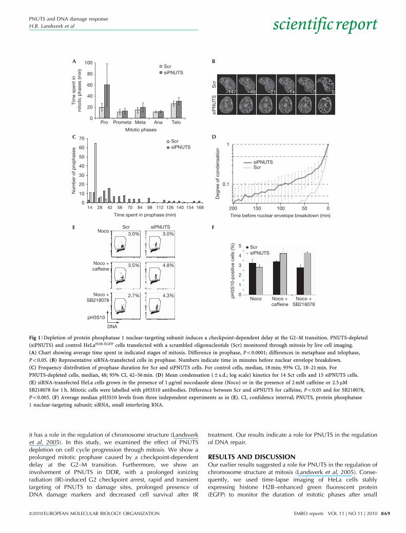
Fig 1 | Depletion of protein phosphatase 1 nuclear-targeting subunit induces a checkpoint-dependent delay at the G2–M transition. PNUTS-depleted
(siPNUTS) and control HeLaH2B-EGFP cells transfected with a scrambled oligonucleotide (Scr) monitored through mitosis by live cell imaging.
(A) Chart showing average time spent in indicated stages of mitosis. Difference in prophase, Po0.0001; differences in metaphase and telophase,
Po0.05. (B) Representative siRNA-transfected cells in prophase. Numbers indicate time in minutes before nuclear envelope breakdown.
(C) Frequency distribution of prophase duration for Scr and siPNUTS cells. For control cells, median, 18 min; 95% CI, 18–21 min. For
PNUTS-depleted cells, median, 48; 95% CI, 42–56 min. (D) Mean condensation (±s.d.; log scale) kinetics for 14 Scr cells and 15 siPNUTS cells.
(E) siRNA-transfected HeLa cells grown in the presence of 1 mg/ml nocodazole alone (Noco) or in the presence of 2 mM caffeine or 2.5mM
SB218078 for 1 h. Mitotic cells were labelled with pH3S10 antibodies. Difference between Scr and siPNUTS for caffeine, Po0.05 and for SB218078,
Po0.005. (F) Average median pH3S10 levels from three independent experiments as in (E). CI, confidence interval; PNUTS, protein phosphatase
1 nuclear-targeting subunit; siRNA, small interfering RNA.
PNUTS and DNA damage response
H.B. Landsverk et al
&2010 EUROPEAN MOLECULAR BIOLOGY ORGANIZATION EMBO reports VOL 11 | NO 11 | 2010
scientificreport
869
interfering RNA (siRNA)-mediated PNUTS depletion. A markedlengthening of prophase—defined morphologically as the stagebetween the first visible signs of chromosome condensationand nuclear envelope breakdown—was observed, comparedwith controls. In PNUTS-depleted compared with control cells,average prophase duration increased threefold (60.1±38.4 and20.1±6.8 min, respectively, Po0.0001; Fig 1A,B; Table 1) andfrequency distribution of prophase duration was much broader(median values of 48 min and 18 min, respectively; Fig 1C). Toquantify chromatin condensation, we randomly selected subsetsof cells for computerized image processing to measure theheterogeneity in the spatial distribution of fluorescently labelledchromatin (supplementary information online). Chromatin inPNUTS-depleted cells showed signs of condensation as early as2.5 h before nuclear envelope breakdown, compared with 0.5 h incontrol cells (Fig 1D).
Prolonged prophase is characteristic of cells arrested at theG2–M transition (Pines & Rieder, 2001). To test this, we monitoredmitotic entry after 1 h incubation with the G2 checkpoint inhibitorcaffeine and the Chk1-inhibitor SB218078 ( Jin et al, 2005).Remarkably, more PNUTS-depleted than control cells enteredmitosis in the presence of caffeine or SB218078 (Fig 1E,F). Similarresults were observed with an independent PNUTS siRNAoligonucleotide (supplementary Fig S1A,D online). Thus, PNUTSdepletion activates a caffeine- and SB218078-sensitive G2checkpoint in HeLa cells.
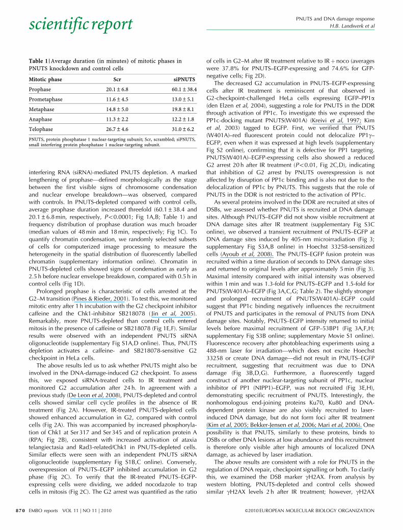
The above results led us to ask whether PNUTS might also beinvolved in the DNA-damage-induced G2 checkpoint. To assessthis, we exposed siRNA-treated cells to IR treatment andmonitored G2 accumulation after 24 h. In agreement with aprevious study (De Leon et al, 2008), PNUTS-depleted and controlcells showed similar cell cycle profiles in the absence of IRtreatment (Fig 2A). However, IR-treated PNUTS-depleted cellsshowed enhanced accumulation in G2, compared with controlcells (Fig 2A). This was accompanied by increased phosphoryla-tion of Chk1 at Ser 317 and Ser 345 and of replication protein A(RPA; Fig 2B), consistent with increased activation of ataxiatelangiectasia and Rad3-related/Chk1 in PNUTS-depleted cells.Similar effects were seen with an independent PNUTS siRNAoligonucleotide (supplementary Fig S1B,C online). Conversely,overexpression of PNUTS–EGFP inhibited accumulation in G2phase (Fig 2C). To verify that the IR-treated PNUTS–EGFP-expressing cells were dividing, we added nocodazole to trapcells in mitosis (Fig 2C). The G2 arrest was quantified as the ratio
of cells in G2–M after IR treatment relative to IRþ noco (averageswere 37.8% for PNUTS–EGFP-expressing and 74.6% for GFP-negative cells; Fig 2D).
The decreased G2 accumulation in PNUTS–EGFP-expressingcells after IR treatment is reminiscent of that observed inG2-checkpoint-challenged HeLa cells expressing EGFP–PP1a(den Elzen et al, 2004), suggesting a role for PNUTS in the DDRthrough activation of PP1c. To investigate this we expressed thePP1c-docking mutant PNUTS(W401A) (Kreivi et al, 1997; Kimet al, 2003) tagged to EGFP. First, we verified that PNUTS(W401A)–red fluorescent protein could not delocalize PP1g–EGFP, even when it was expressed at high levels (supplementaryFig S2 online), confirming that it is defective for PP1 targeting.PNUTS(W401A)–EGFP-expressing cells also showed a reducedG2 arrest 20 h after IR treatment (Po0.01, Fig 2C,D), indicatingthat inhibition of G2 arrest by PNUTS overexpression is notaffected by disruption of PP1c binding and is also not due to thedelocalization of PP1c by PNUTS. This suggests that the role ofPNUTS in the DDR is not restricted to the activation of PP1c.
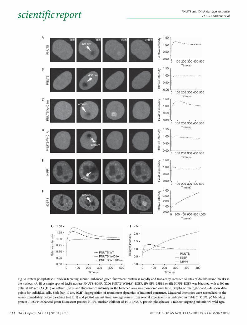
As several proteins involved in the DDR are recruited at sites ofDSBs, we assessed whether PNUTS is recruited at DNA damagesites. Although PNUTS–EGFP did not show visible recruitment atDNA damage sites after IR treatment (supplementary Fig S3Conline), we observed a transient recruitment of PNUTS–EGFP atDNA damage sites induced by 405-nm microirradiation (Fig 3;supplementary Fig S3A,B online) in Hoechst 33258-sensitizedcells (Ayoub et al, 2008). The PNUTS–EGFP fusion protein wasrecruited within a time duration of seconds to DNA damage sitesand returned to original levels after approximately 5 min (Fig 3).Maximal intensity compared with initial intensity was observedwithin 1 min and was 1.3-fold for PNUTS–EGFP and 1.5-fold forPNUTS(W401A)–EGFP (Fig 3A,C,G; Table 2). The slightly strongerand prolonged recruitment of PNUTS(W401A)–EGFP couldsuggest that PP1c binding negatively influences the recruitmentof PNUTS and participates in the removal of PNUTS from DNAdamage sites. Notably, PNUTS–EGFP intensity returned to initiallevels before maximal recruitment of GFP–53BP1 (Fig 3A,F,H;supplementary Fig S3B online; supplementary Movie S1 online).Fluorescence recovery after photobleaching experiments using a488-nm laser for irradiation—which does not excite Hoechst33258 or create DNA damage—did not result in PNUTS–EGFPrecruitment, suggesting that recruitment was due to DNAdamage (Fig 3B,D,G). Furthermore, a fluorescently taggedconstruct of another nuclear-targeting subunit of PP1c, nuclearinhibitor of PP1 (NIPP1)–EGFP, was not recruited (Fig 3E,H),demonstrating specific recruitment of PNUTS. Interestingly, thenonhomologous end-joining proteins Ku70, Ku80 and DNA-dependent protein kinase are also visibly recruited to laser-induced DNA damage, but do not form foci after IR treatment(Kim et al, 2005; Bekker-Jensen et al, 2006; Mari et al, 2006). Onepossibility is that PNUTS, similarly to these proteins, binds toDSBs or other DNA lesions at low abundance and this recruitmentis therefore only visible after high amounts of localized DNAdamage, as achieved by laser irradiation.
The above results are consistent with a role for PNUTS in theregulation of DNA repair, checkpoint signalling or both. To clarifythis, we examined the DSB marker gH2AX. From analysis bywestern blotting, PNUTS-depleted and control cells showedsimilar gH2AX levels 2 h after IR treatment; however, gH2AX
Table 1 | Average duration (in minutes) of mitotic phases inPNUTS knockdown and control cells
Mitotic phase Scr siPNUTS
Prophase 20.1±6.8 60.1±38.4
Prometaphase 11.6±4.5 13.0±5.1
Metaphase 14.8±5.0 19.8±8.1
Anaphase 11.3±2.2 12.2±1.8
Telophase 26.7±4.6 31.0±6.2
PNUTS, protein phosphatase 1 nuclear-targeting subunit; Scr, scrambled; siPNUTS,small interfering protein phosphatase 1 nuclear-targeting subunit.
PNUTS and DNA damage response
H.B. Landsverk et al
EMBO reports VOL 11 | NO 11 | 2010 &2010 EUROPEAN MOLECULAR BIOLOGY ORGANIZATION
scientificreport
870
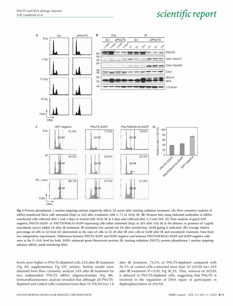
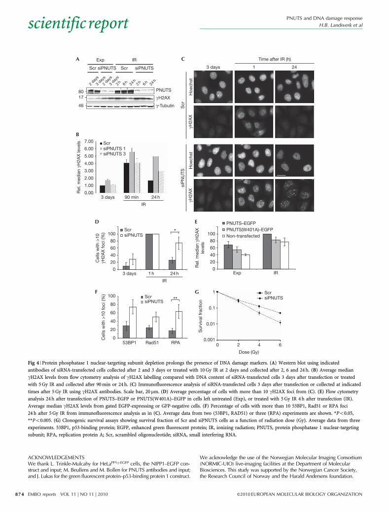
levels were higher in PNUTS-depleted cells 24 h after IR treatment(Fig 4A; supplementary Fig S1C online). Similar results wereobtained from flow cytometry analysis 24 h after IR treatment fortwo independent PNUTS siRNA oligonucleotides (Fig 4B).Immunofluorescence analysis revealed that although all PNUTS-depleted and control cells contained more than 10 gH2AX foci 1 h
after IR treatment, 74.2% of PNUTS-depleted compared with26.1% of control cells contained more than 10 gH2AX foci 24 hafter IR treatment (Po0.05; Fig 4C,D). Thus, removal of gH2AXis delayed in PNUTS-depleted cells, suggesting that PNUTS isinvolved in the regulation of DNA repair or participates indephosphorylation of gH2AX.
Chk1-Ser317
PNUTS
IR
Scr siPNUTS
2 day
s
2h 24h
6h3 day
s
2h 24h
6h
Chk1-Ser345
2 day
s
3 day
s
Scr siPNUTS
Exp
Chk1
γ-Tubulin46
5846
58
5846
80
5 Gy
7.5 Gy
10 Gy
ExpA B
C D
Scr siPNUTS
DNA
Cells
RPARPA-P30
25
Exp
IR
IR + noco
GFP negative PNUTS–EGFP PNUTS(W401A)–EGFP
G2/M G2/M G2/M
G2/MG2/MG2/M
G2/MG2/MG2/M
100
80
60
40
20
0Rel
ativ
e ce
lls in
G2
(%)
DNA
Cells
31.5%
64.3%
89.1%
17.0%
22.6%
57.5%
15.6%
22.3%
51.3%
PNUTS–E
GFP
PNUTS(W
401A
)–EGFP
GFP n
egat
ive
Fig 2 | Protein phosphatase 1 nuclear-targeting subunit negatively affects G2 arrest after ionizing radiation treatment. (A) Flow cytometry analysis of
siRNA-transfected HeLa cells untreated (Exp) or 24 h after irradiation with 5, 7.5 or 10 Gy IR. (B) Western blot using indicated antibodies of siRNA-
transfected cells collected after 2 and 3 days or treated with 10 Gy IR at 2 days and collected after 2, 6 and 24 h. (C) Flow analysis of gated GFP-
negative, PNUTS–EGFP- or PNUTS(W401A)–EGFP-expressing cells either untreated (Exp) or 20 h after 5 Gy IR in the absence or presence of 1mg/ml
nocodazole (noco) added 2 h after IR treatment. IR treatment was carried out 4 h after transfection. G2/M gating is indicated. (D) Average relative
percentage of cells in G2 from (C) determined as the ratio of cells in G2–M after IR over cells in G2/M after IR and nocodazole treatment. Data from
two independent experiments. Differences between PNUTS–EGFP and EGFP-negative and between PNUTS(W401A)–EGFP and EGFP-negative cells
were at the Po0.01 level for both. EGFP, enhanced green fluorescent protein; IR, ionizing radiation; PNUTS, protein phosphatase 1 nuclear-targeting
subunit; siRNA, small interfering RNA.
PNUTS and DNA damage response
H.B. Landsverk et al
&2010 EUROPEAN MOLECULAR BIOLOGY ORGANIZATION EMBO reports VOL 11 | NO 11 | 2010
scientificreport
871
PN
UTS
A
B
D
E
F
G H
C
PN
UTS
PN
UTS
(W40
1A)
PN
UTS
(W40
1A)
1000 200 300 400 500Time (s)
1000 200 300 400 500Time (s)
1000 200 300 400 500Time (s)
53B
P1
2000 400 600 8001,000Time (s)
4.00
3.00
1.00
0.00Rel
ativ
e in
tens
ity
2.00
1.50
1.00
0.50
0.00Rel
ativ
e in
tens
ity
1.50
1.00
0.50
0.00Rel
ativ
e in
tens
ity
1.50
1.00
0.50
0.00Rel
ativ
e in
tens
ity
1000 200 300 400 500Time (s)
1.50
1.00
0.50
0.00Rel
ativ
e in
tens
ity
0 s 69 s 250 s–8 s
NIP
P1
1000 200 300 400 500Time (s)
1.50
1.00
0.50
0.00Rel
ativ
e in
tens
ity
405 nm
405 nm
405 nm
405 nm
488 nm
488 nm
PNUTS WT 488 nm
PNUTS WTPNUTS W401A
0 400200 300100 500
Time (s)
Rel
ativ
e in
tens
ity
0.75
1.00
1.25
1.50
0.00
0.25
0.50
1.5
2.0
2.5
0.0
0.5
1.0
Rel
ativ
e in
tens
ity
NIPP1
PNUTS 53BP1
0 400200 300100 500
Time (s)
Fig 3 | Protein phosphatase 1 nuclear-targeting subunit–enhanced green fluorescent protein is rapidly and transiently recruited to sites of double-strand breaks in
the nucleus. (A–E) A single spot of (A,B) nuclear PNUTS–EGFP, (C,D) PNUTS(W401A)–EGFP, (F) GFP–53BP1 or (E) NIPP1–EGFP was bleached with a 500-ms
pulse at 405 nm (A,C,E,F) or 488 nm (B,D), and fluorescence intensity in the bleached area was monitored over time. Graphs on the right-hand side show data
points for individual cells. Scale bar, 10mm. (G,H) Superposition of recruitment dynamics of indicated constructs. Measured intensities were normalized to the
values immediately before bleaching (set to 1) and plotted against time. Average results from several experiments as indicated in Table 2. 53BP1, p53-binding
protein 1; EGFP, enhanced green fluorescent protein; NIPP1, nuclear inhibitor of PP1; PNUTS, protein phosphatase 1 nuclear-targeting subunit; wt, wild type.
PNUTS and DNA damage response
H.B. Landsverk et al
EMBO reports VOL 11 | NO 11 | 2010 &2010 EUROPEAN MOLECULAR BIOLOGY ORGANIZATION
scientificreport
872
To address whether PNUTS controls dephosphorylation ofgH2AX, we analysed gH2AX levels in exponentially growing andIR-treated cells after overexpression of PNUTS–EGFP. As PNUTSoverexpression did not result in a decrease in gH2AX levels(Fig 4E), the PNUTS–PP1c holoenzyme probably does notdephosphorylate gH2AX in vivo, although PP1c does so in vitro(Siino et al, 2002). We also analysed 53BP1 foci, a differentmarker for DSBs (Schultz et al, 2000). Consistent with delayedrepair, PNUTS-depleted cells showed more 53BP1 foci 24 h afterIR treatment than control cells (Fig 4F). PNUTS-depleted cells alsoshowed increased Rad51 and RPA foci at 5 and 24 h after IRtreatment (Fig 4F; data not shown), consistent with increasedhomologous recombination repair (Sorensen et al, 2005) andpresence of single-strand DNA ( Jackson & Bartek, 2009). Thesedata strongly suggest that PNUTS regulates DNA repair rather thangH2AX dephosphorylation.
Finally, we reasoned that if PNUTS regulates DNA repair,PNUTS-depleted cells should be hypersensitive to IR treatment incell survival assays. The PNUTS siRNA oligonucleotides withPNUTS almost completely downregulated (siPNUTS 1; supple-mentary Fig S1D online) were unsuitable for clonogenic survivalassays, as they showed reduced cloning efficiency, even in theabsence of IR treatment (o10% cloning efficiency compared withcontrol transfected cells). However, an increase in IR-induceddeath could be observed by measuring the percentage of sub-G1cells in DNA histograms 72 h after 5-Gy IR treatment (supple-mentary Fig S4 online). More conclusively, a different PNUTSsiRNA oligonucleotide (siPNUTS 3)—which resulted in onlypartial downregulation of PNUTS—showed decreased clonogenicsurvival after IR treatment in HeLa cells (Fig 4G). Thus, andconsistent with defective DNA repair, PNUTS-depleted cells arehypersensitive to IR-induced cell death.
Specificity is provided to PP1c—a protein phosphatase with abroad range of substrates—through association with regulatory
subunits that target the holoenzyme to specific locations orsubstrates and control its activity (Moorhead et al, 2007; Bollenet al, 2009). Although PP1c has previously been implicated in theDDR (den Elzen et al, 2004; Tang et al, 2008; Hamilton et al,2009), we identify here for the first time, to the best of ourknowledge, that the regulatory subunit of PP1 PNUTS is anintegral component of this pathway. The rapid and transientrecruitment of PNUTS–EGFP to DSBs together with the long-termeffects of PNUTS knockdown on DNA damage markers, Chk1activation, G2 arrest and cell survival after IR treatment suggeststhat PNUTS regulates early events in the DDR, which affectsmajor downstream targets involved in DNA repair.
METHODSCell culture, irradiation and drugs. Human epithelial HeLa,HeLaEGFP–PP1g (Trinkle-Mulcahy et al, 2003) and HeLaH2B–EGFP
(Hirota et al, 2004) were grown in Dulbecco’s Modified EagleMedium containing 5% fetal calf serum (Invitrogen). Cells wereirradiated using a 137Cs source at a dose rate of 4.3 Gy/min or withan X-ray generator (Faxitron CP160, 160 kV, 6.3 mA) at a dose rateof 1 Gy/min. Caffeine and nocodazole were purchased fromSigma-Aldrich and SB218078 from Calbiochem.DNA constructs and cloning. Wild-type PNUTS was cloned fromHeLa complementary DNA into the enhanced green andmonomeric red fluorescent protein vectors (Clontech) withoutthe seven carboxy-terminal amino acids of PNUTS. The PNUTS(W401A)–EGFP mutant was produced by site-directed mutagenesis(Quickchange II, Stratagene). Mouse GFP–53BP1 was obtainedfrom J. Lukas (Bekker-Jensen et al, 2006) and NIPP1–EGFP from L.Trinkle-Mulcahy (Trinkle-Mulcahy et al, 1999).Western blotting, immunofluorescence and flow cytometry. Westernblotting and immunofluorescence were performed as describedpreviously (Landsverk et al, 2005). PNUTS antibody was providedby M. Bollen (Lesage et al, 2004), phospho-Chk1 (Ser 317 andSer 345) and phospho-H3 (pH3S10) were obtained from CellSignaling Technology, phospho-H2AX (gH2AX clone JBW301)from Millipore, RPA (p34) from AH Diagnostics and g-tubulin(GTU-88) from Sigma. Chk1 antibody (DCS310) has been describedpreviously (Sorensen et al, 2005). Flow cytometry with doublelabelling was performed as described previously (Naderi et al,2005) omitting the acid/pepsin and neutralization steps, and triplelabelling is described in the supplementary information online.Live cell imaging and laser irradiation. Procedures are describedin the supplementary information online.Clonogenic survival assay. Twenty-four hours after siRNA trans-fection, between 150 and 1,200 cells (depending on IR dose toyield 50–100 colonies per dish) were seeded on to 6-cm dishes,incubated for about 20 h, and treated with IR (0, 2, 4 and 6 Gy).After 14 days, cells were stained with methylene blue andcolonies containing more than 50 cells were counted as survivors.Survival fractions after each IR dose were calculated as theaverage cloning efficiency ratio (from three parallel dishes in eachexperiment) of irradiated cells to non-irradiated cells.Statistics. Statistical analysis was performed using unpairedStudent’s t-test and all experiments were performed three timesor more, unless otherwise indicated. Error bars represent standarddeviation values.Supplementary information is available at EMBO reports online(http://www.emboreports.org).
Table 2 | FRAP analysis of PNUTS–EGFP, PNUTS(W401A)–EGFP,GFP–53BP1 and NIPP1–EGFP
Microirradiation(nm)
N Timemaximumintensity
Valuemaximumintensity
Intensity2 min
post-bleach
PNUTS–EGFP
405 12 57 s 540 ms 1.296±0.065 1.186±0.059
488 6 ND ND 0.980±0.101
PNUTS(W401A)–EGFP
405 15 60 s 828 ms 1.456±0.135 1.358±0.124
488 32 ND ND 0.977±0.018
EGFP–53BP1
405 12 415 min 2.559±0.284 1.221±0.044
NIPP1–EGFP
405 19 ND ND 0.969±0.039
Each data set was normalized to intensity immediately before bleach; mean valuesshown with given s.e.m. values.53BP1, p53-binding protein 1; EGFP, enhanced green fluorescent protein; FRAP,fluorescence recovery after photobleaching; ND, not determined; NIPP1, nuclearinhibitor of PP1; WT, wild type.
PNUTS and DNA damage response
H.B. Landsverk et al
&2010 EUROPEAN MOLECULAR BIOLOGY ORGANIZATION EMBO reports VOL 11 | NO 11 | 2010
scientificreport
873
ACKNOWLEDGEMENTSWe thank L. Trinkle-Mulcahy for HeLaPP1g-EGFP cells, the NIPP1–EGFP con-struct and input; M. Beullens and M. Bollen for PNUTS antibodies and input;and J. Lukas for the green fluorescent protein–p53-binding protein 1 construct.
We acknowledge the use of the Norwegian Molecular Imaging Consortium(NORMIC-UIO) live-imaging facilities at the Department of MolecularBiosciences. This study was supported by the Norwegian Cancer Society,the Research Council of Norway and the Harald Andersens foundation.
3 days 1 24
Time after IR (h)
γH2A
X
siP
NU
TS
Hoe
chst
γH2A
X
Scr
Hoe
chst
IR
Scr siPNUTS
2 day
s
2h 24h
6h3 day
s
2h 24h
6h2 day
s
3 day
s
Scr siPNUTS
PNUTS
γ-Tubulin
ExpA
B
D E
GF
C
γH2AX8017
46
3 days
7.00 ScrsiPNUTS 1siPNUTS 3
6.00
5.00
4.00
3.00
2.00
1.00
0.0090 min 24 h
IR
Rel
. med
ian
γH2A
X le
vels
IR
3 days 1 h 24 h
ScrsiPNUTS
*
Cel
ls w
ith >
10γH
2AX
foci
(%)
0
20
40
60
80
100
Exp IR
PNUTS–EGFPPNUTS(W401A)–EGFPNon-transfected
Rel
. med
ian
γH2A
Xle
vels
0
20
40
60
80
100
53BP1 Rad51 RPA0
20
40
60
80
100 ScrsiPNUTS
Cel
ls w
ith >
10 fo
ci (%
)
**
Sur
viva
l fra
ctio
n
1
0.1
0.01
0.0010 2 4 6
ScrsiPNUTS
Dose (Gy)
Fig 4 | Protein phosphatase 1 nuclear-targeting subunit depletion prolongs the presence of DNA damage markers. (A) Western blot using indicated
antibodies of siRNA-transfected cells collected after 2 and 3 days or treated with 10 Gy IR at 2 days and collected after 2, 6 and 24 h. (B) Average median
gH2AX levels from flow cytometry analysis of gH2AX labelling compared with DNA content of siRNA-transfected cells 3 days after transfection or treated
with 5 Gy IR and collected after 90 min or 24 h. (C) Immunofluorescence analysis of siRNA-transfected cells 3 days after transfection or collected at indicated
times after 5 Gy IR using gH2AX antibodies. Scale bar, 20mm. (D) Average percentage of cells with more than 10 gH2AX foci from (C). (E) Flow cytometry
analysis 24 h after transfection of PNUTS–EGFP or PNUTS(W401A)–EGFP in cells left untreated (Exp), or treated with 5 Gy IR 4 h after transfection (IR).
Average median gH2AX levels from gated EGFP-expressing or GFP-negative cells. (F) Percentage of cells with more than 10 53BP1, Rad51 or RPA foci
24 h after 5 Gy IR from immunofluorescence analysis as in (C). Average data from two (53BP1, RAD51) or three (RPA) experiments are shown. *Po0.05,
**Po0.005. (G) Clonogenic survival assays showing survival fraction of Scr and siPNUTS cells as a function of radiation dose (Gy). Average data from three
experiments. 53BP1, p53-binding protein; EGFP, enhanced green fluorescent protein; IR, ionizing radiation; PNUTS, protein phosphatase 1 nuclear-targeting
subunit; RPA, replication protein A; Scr, scrambled oligonucleotide; siRNA, small interfering RNA.
PNUTS and DNA damage response
H.B. Landsverk et al
EMBO reports VOL 11 | NO 11 | 2010 &2010 EUROPEAN MOLECULAR BIOLOGY ORGANIZATION
scientificreport
874
CONFLICT OF INTERESTThe authors declare that they have no conflict of interest.
REFERENCESAllen PB, Kwon YG, Nairn AC, Greengard P (1998) Isolation and
characterization of PNUTS, a putative protein phosphatase 1 nucleartargeting subunit. J Biol Chem 273: 4089–4095
Ayoub N, Jeyasekharan AD, Bernal JA, Venkitaraman AR (2008) HP1-betamobilization promotes chromatin changes that initiate the DNA damageresponse. Nature 453: 682–686
Bekker-Jensen S, Lukas C, Kitagawa R, Melander F, Kastan MB, Bartek J, LukasJ (2006) Spatial organization of the mammalian genome surveillancemachinery in response to DNA strand breaks. J Cell Biol 173: 195–206
Bollen M, Gerlich DW, Lesage B (2009) Mitotic phosphatases: from entryguards to exit guides. Trends Cell Biol 19: 531–541
De Leon G, Sherry TC, Krucher NA (2008) Reduced expression of PNUTSleads to activation of Rb-phosphatase- and caspase-mediated apoptosis.Cancer Biol Ther 7: 833–841
den Elzen NR, O’Connell MJ (2004) Recovery from DNA damage checkpointarrest by PP1-mediated inhibition of Chk1. EMBO J 23: 908–918
den Elzen N, Kosoy A, Christopoulos H, O’Connell MJ (2004) Resisting arrest:recovery from checkpoint arrest through dephosphorylation of Chk1 byPP1. Cell Cycle 3: 529–533
Hamilton J, Grawenda AM, Bernhard EJ (2009) Phosphatase inhibition andcell survival after DNA damage induced by radiation. Cancer Biol Ther 8:1577–1586
Hirota T, Gerlich D, Koch B, Ellenberg J, Peters JM (2004) Distinct functionsof condensin I and II in mitotic chromosome assembly. J Cell Sci 117:6435–6445
Jackson SP, Bartek J (2009) The DNA-damage response in human biology anddisease. Nature 461: 1071–1078
Jagiello I, Beullens M, Stalmans W, Bollen M (1995) Subunit structure andregulation of protein phosphatase-1 in rat liver nuclei. J Biol Chem 270:17257–17263
Jin ZH, Kurosu T, Yamaguchi M, Arai A, Miura O (2005) Hematopoieticcytokines enhance Chk1-dependent G2/M checkpoint activation byetoposide through the Akt/GSK3 pathway to inhibit apoptosis. Oncogene24: 1973–1981
Kim H, Lee OH, Xin H, Chen LY, Qin J, Chae HK, Lin SY, Safari A, Liu D,Songyang Z (2009) TRF2 functions as a protein hub and regulatestelomere maintenance by recognizing specific peptide motifs. Nat StructMol Biol 16: 372–379
Kim JS, Krasieva TB, Kurumizaka H, Chen DJ, Taylor AM, Yokomori K (2005)Independent and sequential recruitment of NHEJ and HR factors to DNAdamage sites in mammalian cells. J Cell Biol 170: 341–347
Kim YM, Watanabe T, Allen PB, Kim YM, Lee SJ, Greengard P, Nairn AC,Kwon YG (2003) PNUTS, a protein phosphatase 1 (PP1) nuclear targetingsubunit. Characterization of its PP1- and RNA-binding domains andregulation by phosphorylation. J Biol Chem 278: 13819–13828
Kreivi JP, Trinkle-Mulcahy L, Lyon CE, Morrice NA, Cohen P, Lamond AI(1997) Purification and characterisation of p99, a nuclear modulator ofprotein phosphatase 1 activity. FEBS Lett 420: 57–62
Krucher NA, Rubin E, Tedesco VC, Roberts MH, Sherry TC, De Leon G (2006)Dephosphorylation of Rb (Thr 821) in response to cell stress. Exp Cell Res312: 2757–2763
Landsverk HB, Kirkhus M, Bollen M, Kuntziger T, Collas P (2005) PNUTSenhances in vitro chromosome decondensation in a PP1-dependentmanner. Biochem J 390: 709–717
Lee SJ, Lim CJ, Min JK, Lee JK, Kim YM, Lee JY, Won MH, Kwon YG (2007)Protein phosphatase 1 nuclear targeting subunit is a hypoxia induciblegene: its role in post-translational modification of p53 and MDM2.Cell Death Differ 14: 1106–1116
Lesage B, Beullens M, Nuytten M, Van Eynde A, Keppens S, Himpens B,Bollen M (2004) Interactor-mediated nuclear translocation and retentionof protein phosphatase-1. J Biol Chem 279: 55978–55984
Liu Q et al (2000) Chk1 is an essential kinase that is regulated by Atr andrequired for the G(2)/M DNA damage checkpoint. Genes Dev 14:1448–1459
Mari PO et al (2006) Dynamic assembly of end-joining complexes requiresinteraction between Ku70/80 and XRCC4. Proc Natl Acad Sci USA 103:18597–18602
Moorhead GB, Trinkle-Mulcahy L, Ulke-Lemee A (2007) Emerging roles ofnuclear protein phosphatases. Nat Rev Mol Cell Biol 8: 234–244
Naderi S, Wang JY, Chen TT, Gutzkow KB, Blomhoff HK (2005) cAMP-mediated inhibition of DNA replication and S-phase progression:involvement of Rb, p21Cip1, and PCNA. Mol Biol Cell 16: 1527–1542
Peng A, Lewellyn AL, Schiemann WP, Maller JL (2010) Repo-man controls aprotein phosphatase 1-dependent threshold for DNA damage checkpointactivation. Curr Biol 20: 387–396
Pines J, Rieder CL (2001) Re-staging mitosis: a contemporary view of mitoticprogression. Nat Cell Biol 3: E3–E6
Schultz LB, Chehab NH, Malikzay A, Halazonetis TD (2000) p53-bindingprotein 1 (53BP1) is an early participant in the cellular response to DNAdouble-strand breaks. J Cell Biol 151: 1381–1390
Siino JS, Nazarov IB, Svetlova MP, Solovjeva LV, Adamson RH,Zalenskaya IA, Yau PM, Bradbury EM, Tomilin NV (2002)Photobleaching of GFP-labeled H2AX in chromatin: H2AX has lowdiffusional mobility in the nucleus. Biochem Biophys Res Commun 297:1318–1323
Sorensen CS, Hansen LT, Dziegielewski J, Syljuasen RG, Lundin C,Bartek J, Helleday T (2005) The cell-cycle checkpoint kinase Chk1 isrequired for mammalian homologous recombination repair. Nat Cell Biol7: 195–201
Tang X, Hui ZG, Cui XL, Garg R, Kastan MB, Xu B (2008) A novel ATM-dependent pathway regulates protein phosphatase 1 in response to DNAdamage. Mol Cell Biol 28: 2559–2566
Trinkle-Mulcahy L, Ajuh P, Prescott A, Claverie-Martin F, Cohen S, LamondAI, Cohen P (1999) Nuclear organisation of NIPP1, a regulatory subunitof protein phosphatase 1 that associates with pre-mRNA splicing factors.J Cell Sci 112: 157–168
Trinkle-Mulcahy L, Andrews PD, Wickramasinghe S, Sleeman J, Prescott A,Lam YW, Lyon C, Swedlow JR, Lamond AI (2003) Time-lapse imagingreveals dynamic relocalization of PP1gamma throughout the mammaliancell cycle. Mol Biol Cell 14: 107–117
Trinkle-Mulcahy L, Andersen J, Lam YW, Moorhead G, Mann M, Lamond AI(2006) Repo-Man recruits PP1-gamma to chromatin and is essential forcell viability. J Cell Biol 172: 679–692
Udho E, Tedesco VC, Zygmunt A, Krucher NA (2002) PNUTS (phosphatasenuclear targeting subunit) inhibits retinoblastoma-directed PP1 activity.Biochem Biophys Res Commun 297: 463–467
PNUTS and DNA damage response
H.B. Landsverk et al
&2010 EUROPEAN MOLECULAR BIOLOGY ORGANIZATION EMBO reports VOL 11 | NO 11 | 2010
scientificreport
875
Copyright © 2022 FDOKUMEN