The protective effect of melatonin and S-methylisothiourea treatments in nitrogen mustard induced...
8
environmental toxicology and pharmacology 36 ( 2 0 1 3 ) 1283–1290 Available online at www.sciencedirect.com ScienceDirect journal homepage: www.elsevier.com/locate/etap The protective effect of melatonin and S-methylisothiourea treatments in nitrogen mustard induced lung toxicity in rats Enis Macit a,∗ , Hakan Yaren b , Ibrahim Aydin c , Zeki Ilker Kunak b , Halil Yaman c , Onder Onguru d , Bulent Uysal e , Ahmet Korkmaz e , Samet Turel b , Levent Kenar b a Department of Toxicology, Gulhane Military Medical Academy, Etlik, Ankara, Turkey b Department of CBRN Defense, Gulhane Military Medical Academy, Etlik, Ankara, Turkey c Department of Clinical Biochemistry, Gulhane Military Medical Academy, Etlik, Ankara, Turkey d Department of Pathology, Gulhane Military Medical Academy, Etlik, Ankara, Turkey e Department of Physiology, Gulhane Military Medical Academy, Etlik, Ankara, Turkey article info Article history: Received 8 March 2013 Received in revised form 3 October 2013 Accepted 6 October 2013 Available online 24 October 2013 Keywords: Mechlorethamine Melatonin S-Methylisothioure Inflammation Pro-inflammatory cytokines Lung toxicity abstract Objectives: Mustard is highly toxic to the lung. Its toxic effects are associated with inflamma- tory cell accumulation and increased pro-inflammatory cytokines as well as reactive oxygen and nitrogen species. In this study, we aimed to investigate the efficiency of melatonin (MEL) and S-methylisothiourea (SMT) on mechlorethamine (MEC) induced lung toxicity. Methods: Thirty-six male rats were randomly divided into four groups: control, MEC, MEC + MEL, and MEC + SMT. Control group was given saline only via transdermal route. Other groups were exposured to a single dose of MEC (3.5mg/kg) via transdermal route. MEL (100 mg/kg) was administered intraperitoneally 30 min after the application of MEC, and after the same dose of MEL was given every 12 h for a total of six doses. SMT (50 mg/kg) was also given intraperitoneally 30 min after the application of MEC. Results: MEC injection resulted in alveolar epithelial injury, hemorrhage, inflammation, edema and interalveolar septal thickening in the lung tissues. The tissue TNF-, IL-1, and nitrate/nitrite (NOx) levels were found significantly different for all groups (p < 0.001). TNF- and IL-1 levels increased significantly with MEC exposure, and MEL and SMT amelio- rated these increases in lung tissues. MEC also elevated NOx levels in lung tissue. Melatonin showed meaningful protection against lung injury. But protection of SMT was weaker. Conclusion: Inflammation plays an important role in the MEC induced lung toxicity as well as oxidative and nitrosative stress. Melatonin has also anti-inflammatory properties similar to SMT, as well as anti-oxidant properties. But melatonin treatment was found more efficient than SMT treatment. Crown Copyright © 2013 Published by Elsevier B.V. All rights reserved. ∗ Corresponding author at: Department of Toxicology, Gulhane Military Medical Academy, School of Medicine, Etlik-06018, Ankara, Turkey. Tel.: +90 312 304 4784; fax: +90 312 304 2100. E-mail addresses: [email protected], [email protected] (E. Macit). 1382-6689/$ – see front matter. Crown Copyright © 2013 Published by Elsevier B.V. All rights reserved. http://dx.doi.org/10.1016/j.etap.2013.10.001
-
Upload
independent -
Category
Documents
-
view
3 -
download
0
Transcript of The protective effect of melatonin and S-methylisothiourea treatments in nitrogen mustard induced...

TSm
EHSa
b
c
d
e
a
A
R
R
3
A
A
K
M
M
S
I
P
L
T
1h
e n v i r o n m e n t a l t o x i c o l o g y a n d p h a r m a c o l o g y 3 6 ( 2 0 1 3 ) 1283–1290
Available online at www.sciencedirect.com
ScienceDirect
journa l homepage: www.e lsev ier .com/ locate /e tap
he protective effect of melatonin and-methylisothiourea treatments in nitrogenustard induced lung toxicity in rats
nis Macita,∗, Hakan Yarenb, Ibrahim Aydinc, Zeki Ilker Kunakb,alil Yamanc, Onder Ongurud, Bulent Uysal e, Ahmet Korkmaze,amet Turelb, Levent Kenarb
Department of Toxicology, Gulhane Military Medical Academy, Etlik, Ankara, TurkeyDepartment of CBRN Defense, Gulhane Military Medical Academy, Etlik, Ankara, TurkeyDepartment of Clinical Biochemistry, Gulhane Military Medical Academy, Etlik, Ankara, TurkeyDepartment of Pathology, Gulhane Military Medical Academy, Etlik, Ankara, TurkeyDepartment of Physiology, Gulhane Military Medical Academy, Etlik, Ankara, Turkey
r t i c l e i n f o
rticle history:
eceived 8 March 2013
eceived in revised form
October 2013
ccepted 6 October 2013
vailable online 24 October 2013
eywords:
echlorethamine
elatonin
-Methylisothioure
nflammation
ro-inflammatory cytokines
ung toxicity
a b s t r a c t
Objectives: Mustard is highly toxic to the lung. Its toxic effects are associated with inflamma-
tory cell accumulation and increased pro-inflammatory cytokines as well as reactive oxygen
and nitrogen species. In this study, we aimed to investigate the efficiency of melatonin (MEL)
and S-methylisothiourea (SMT) on mechlorethamine (MEC) induced lung toxicity.
Methods: Thirty-six male rats were randomly divided into four groups: control, MEC,
MEC + MEL, and MEC + SMT. Control group was given saline only via transdermal route.
Other groups were exposured to a single dose of MEC (3.5 mg/kg) via transdermal route.
MEL (100 mg/kg) was administered intraperitoneally 30 min after the application of MEC,
and after the same dose of MEL was given every 12 h for a total of six doses. SMT (50 mg/kg)
was also given intraperitoneally 30 min after the application of MEC.
Results: MEC injection resulted in alveolar epithelial injury, hemorrhage, inflammation,
edema and interalveolar septal thickening in the lung tissues. The tissue TNF-�, IL-1�,
and nitrate/nitrite (NOx) levels were found significantly different for all groups (p < 0.001).
TNF-� and IL-1� levels increased significantly with MEC exposure, and MEL and SMT amelio-
rated these increases in lung tissues. MEC also elevated NOx levels in lung tissue. Melatonin
showed meaningful protection against lung injury. But protection of SMT was weaker.
Conclusion: Inflammation plays an important role in the MEC induced lung toxicity as well as
oxidative and nitrosative stress. Melatonin has also anti-inflammatory properties similar to
SMT, as well as anti-oxidant properties. But melatonin treatment was found more efficient
than SMT treatment.
Crow
∗ Corresponding author at: Department of Toxicology, Gulhane Military Mel.: +90 312 304 4784; fax: +90 312 304 2100.
E-mail addresses: [email protected], [email protected] (E. Macit).382-6689/$ – see front matter. Crown Copyright © 2013 Published by Ettp://dx.doi.org/10.1016/j.etap.2013.10.001
n Copyright © 2013 Published by Elsevier B.V. All rights reserved.
edical Academy, School of Medicine, Etlik-06018, Ankara, Turkey.
lsevier B.V. All rights reserved.

p h a
1284 e n v i r o n m e n t a l t o x i c o l o g y a n d1. Introduction
Sulfur mustard (SM) is one of the most important chemi-cal warfare agents of the 20th century and has been usedin several conflicts including World War I and Iran-Iraq War(Ekstrand-Hammarstrom et al., 2011). SM and nitrogen mus-tard (NM) are potent vesicant, bifunctional alkylating agentsthat create their toxicity reacting readily with DNA and othermajor components of cell (Ekstrand-Hammarstrom et al.,2011; Sunil et al., 2011a). Besides systemic toxicity mustardsaffect lungs, skin and eyes. Accumulating evidences sug-gest that the mechanism of mustard toxicity includes DNAalkylation, glutathione depletion, activation of proteases, pro-duction of free radicals and free radical-mediated oxidativestress, inflammation, activation of tumor necrosis factor (TNF-�) and other pro-inflammatory cytokines, inducible nitricoxide synthase (iNOS) activation, and poly(ADP-ribose) poly-merase (PARP) activation (Kehe et al., 2008; Weinberger et al.,2011). A major target of both chemicals is the respiratory tract.Following exposure of animals to mustards, inflammatory cellaccumulation is observed in the respiratory tract and is associ-ated with increased production of pro-inflammatory cytokinesand oxidative and nitrosative stresses (Sunil et al., 2011b).In mustard induced lung toxicity, oxidative stress is thoughtto be a primary event triggering the inflammatory cascade(Korkmaz et al., 2006).
Mechlorethamine [bis(2-chloroethyl)methylamine, MEC] isa NM that has been synthesized for military purposes (Kunaket al., 2012). MEC induces the secretion of pro-inflammatorycytokines such as TNF-�, interleukin (IL)-1, IL-6, IL-8, andIL-15 from human respiratory epithelial cells (Kunak et al.,2012). Experimental studies have shown that exposure toSM or NM results in increased expression of iNOS in thelung (Malaviya et al., 2010; Ucar et al., 2007; Yaren et al.,2007). This is associated with inflammatory cell infiltrationinto the lung (Sunil et al., 2011a; Yaren et al., 2007). iNOS isalso a unique enzyme that can induce inflammation caus-ing via excessive nitric oxide (NO) and ROS production.iNOS in the macrophages produce extreme amount of NOunder inflammatory state (Ren et al., 2006). Furthermore, ithas reported that the inhibition of NOS decreased diseaseactivity in an experimental study (McCartney-Francis et al.,1993). Urinary nitrite/nitrate (NOx) is a marker for NO pro-duction. It is also an indirect indicator of the activation ofNOS.
Melatonin (N-acetyl-5-methoxytryptamine) is a neuro-hormone which is secreted from the pineal gland of allmammals (Ozkan et al., 2012a). Numerous experimentalstudies have documented that melatonin has antioxidant,anti-apoptotic, anti-carcinogenic, and anti-aging properties.In addition, melatonin has also effects on the immune system(Korkmaz and Manchester, 2011; Ozkan et al., 2012a,b). Afterexposure to NM, treatment of animals with melatonin reducesinjury and inflammation in the lungs (Ucar et al., 2007).
S-Methylisothiourea (SMT) is a selective iNOS inhibitor andhas anti-inflammatory properties (Guo et al., 2011). Following
exposure to various vesicants, the upregulation of iNOS havebeen implicated in the lung (Malaviya et al., 2010; Sunil et al.,2011a,b; Ucar et al., 2007; Yaren et al., 2007).r m a c o l o g y 3 6 ( 2 0 1 3 ) 1283–1290
Antioxidant therapy is significantly promising in inflam-mation modulation. NOS inhibitors are useful in suppressingoxidative stress (Korkmaz et al., 2008; Pohanka et al., 2011).Despite of some many research reports, until today there is nodefinitive therapy to counteract and/or reverse toxic effects ofSM (Kumar et al., 2010). The aim of the study is to evaluate theefficiency of melatonin and SMT in mechlorethamine inducedlung toxicity.
2. Materials and methods
2.1. Animals
A total of thirty-six male Sprague-Dawley rats (Health SciencesInstitute, Gulhane Military Medical Academy, Ankara, Turkey),with body weights of 220–280 g, were divided into four groupsusing the ‘simple random sampling method’ and they weregiven food and water ad libitum. All animals received humanecare in compliance with the institution’s guidelines, as out-lined in the Guide for the Care and Use of Laboratory Animals,published by the National Institutes of Health. The presentstudy was performed in the Research Center of the GulhaneMilitary Medical Academy with the approval of the GulhaneMilitary Medical Academy Animal Ethics Committee, Turkey.
2.2. Experimental design and tissue preparation
Animals were randomly assigned into 4 groups containing 9rats each: control, mechlorethamine (MEC), MEC + melatonin(MEL), and MEC + S-methylisothiourea (SMT). The animalswere given via transdermal route a dose of 3.5 mg/kg MEC,dissolved in 500 �L saline. Control animals were given500 �L saline only via transdermal route. Dose of melatonin(100 mg/kg) was administered intraperitoneally 30 min afterthe application of MEC, additional melatonin were given every12 h for a total of six doses. SMT (50 mg/kg) was also givenintraperitoneally every 12 h for three consecutive days 30 minafter the application of MEC. All surviving animals were sacri-ficed on the 3rd day of experiment via decapitation, the chestwas opened and then lungs were collected for analysis. One ofthem was fixed in 10% buffered formaldehyde for histopatho-logic evaluation and the other one was washed with salineto remove residual blood, placed into tubes, frozen with liq-uid nitrogen and stored at −80 ◦C. The frozen lung tissueswere homogenized in phosphate buffer (pH 7.4) by means of ahomogenizator (Heidolph Diax 900; Heidolph Elektro GmbH,Kelhaim, Germany). The supernatant was stored at −80 ◦Cuntil assays.
2.3. Biochemical analyses
Tissue TNF-� and IL-1� concentrations were determinedby sandwich enzyme-linked immunosorbent assay (ELISA)(Bender MedSystems, Vienna, Austria) and nitrate/nitrite(NOx) concentrations were determined by colorimetric assaykit (Cayman Chemical, Ann Arbor, MI, USA) performed accord-
ing to the manufacturer’s instructions. The protein contentof lung homogenates was measured by the method of Lowryet al. (1951). TNF-� and IL-1� concentrations were expressed as
a r m a c o l o g y 3 6 ( 2 0 1 3 ) 1283–1290 1285
np
2
StsHefltwim3w
2
A(wtUsTae
3
TgsHtF
feMpcaw
(w[w(tMtt
p
Bio
chem
ical
fin
din
gsin
allt
he
exp
erim
enta
lgro
up
s.
ers
Gro
up
spa
(ove
rall
)pb
(Ivs
II)
pb(I
vsII
I)pb
(Ivs
IV)
pb(I
Ivs
III)
pb(I
Ivs
IV)
pb(I
IIvs
IV)
Con
trol
(n=
9)(I
)M
EC(n
=6)
(II)
MEC
+M
EL(n
=9)
(III
)M
EC+
SMT
(n=
7)(I
V)
/gp
rote
in)
4.3
(2.3
–7.8
)53
.7(3
9.7–
63.3
)12
.2(7
.1–1
6.3)
29.6
(23.
6–38
.8)
<0.
001
0.00
6<
0.00
10.
006
0.00
60.
024
0.01
2/g
pro
tein
)0.
7(0
.3–1
.8)
3.8
(2.1
–5.0
)1.
2(0
.8–1
.6)
2.1
(0.4
–3.2
)<
0.00
10.
006
0.60
60.
006
0.00
60.
222
0.05
4g
pro
tein
)6.
3(1
.8–1
0.9)
44.9
(26.
7–54
.9)
9.5
(6.1
–26.
6)27
.9(1
2.5–
42.1
)<
0.00
10.
006
0.10
80.
024
0.00
60.
120
0.08
4
mor
nec
rosi
sfa
ctor
-alp
ha;
NO
x,n
itra
tep
lus
nit
rite
;IL-
1�,i
nte
rleu
kin
e-1
beta
.All
dat
aw
ere
exp
ress
edas
med
ian
(min
imu
m–m
axim
um
).–W
alli
ste
st.
nic
orre
cted
Man
n–W
hit
ney
Ute
st.
e n v i r o n m e n t a l t o x i c o l o g y a n d p h
g/g protein and NOx concentration was expressed as mol/grotein.
.4. Histopathological evaluation
pecimens were fixed in 10% neutral buffered formalin. Afterissue processing, tissues embedded in paraffin and 5-�m cutections were stained with hematoxylin–eosin (H&E) stain.istologic sections were examined by light microscopy andvaluated by a pathologist blinded to the study groups. Theollowing parameters were assessed histologically: alveo-ar epithelial injury, edema, hemorrhage, inflammation, andhickening of interalveolar septa. Histopathologic parametersere evaluated semiquantitatively relative to controls accord-
ng to 4-grade scale as follows: grade 0: no changes, grade 1:inimal to mild change, grade 2: moderate change, and grade
: severe change. The extensity of histopathologic changesas also assessed as focal or diffuse.
.5. Statistical analysis
ll the statistical analyses were performed by using SPSS 15.0SPSS Inc., Chicago, IL, USA) statistical package. Distributionsere evaluated by using one sample Kolmogorov–Smirnov
est. Kruskal–Wallis and Bonferroni Corrected Mann–Whitneytests were used for testing differences between groups. Chi-
quare test was used for comparing the grades of lung injury.he results were expressed as median (minimum–maximum)nd frequencies (%). A probability level of p < 0.05 was consid-red statistically significant for overall comparisons.
. Results
hree and two deaths observed in the MEC and MEC + SMTroups of rats until the end of experiment, respectively. Tis-ue TNF-�, NOx and IL-1� concentrations are shown in Table 1.istopathological findings are shown in Table 2. Representa-
ive histopathological pictures of the study groups are seen inig. 1.
The lung TNF-� levels were found significantly differentor all groups (p < 0.001). Control group had lowest TNF-� lev-ls and these concentrations were significantly different thanEC, MEC + MEL and MEC + SMT groups (p = 0.006, p < 0.001 and= 0.006, respectively). TNF-� levels of MEC group were signifi-antly higher than MEC + MEL and MEC + SMT groups (p = 0.006nd p = 0.024, respectively). In MEC + MEL group, TNF-� levelsere significantly lower than MEC + SMT group (p = 0.012).
The concentrations of NOx in the control group [0.70.3–1.8) mol/g protein] were significantly different compared
ith the MEC [3.8 (2.1–5.0) mol/g protein] and the MEC + SMT2.1 (0.4–3.2) mol/g protein] groups (in both, p = 0.006). But thereas no difference between the control and the MEC + MEL [1.2
0.8–1.6) mol/g protein] groups (p = 0.606). On the other hand, inhe MEC group, NOx levels were significantly higher than theEC + MEL group (p = 0.006). There was no difference between
he MEC and the MEC + SMT groups, and the MEC + MEL andhe MEC + SMT groups (p = 0.222 and p = 0.054, respectively).
The lowest lung IL-1� concentrations [6.3 (1.8–10.9) ng/grotein] were also in the control group. These concentrations
Tabl
e1
–
Para
met
TN
F-�
(ng
NO
x(m
olIL
-1�
(ng/
TN
F-�
,tu
aK
rusk
alb
Bon
ferr
o

1286e
nv
iro
nm
en
ta
lt
ox
ico
lo
gy
an
dp
ha
rm
ac
ol
og
y3
6(2
01
3)
1283–1290
Table 2 – Histopathological findings in all the experimental groups.
Parameters Grade Groups pa (overall) pb (I vs II) pb (I vs III) pb (I vs IV) pb (II vs III) pb (II vs IV) pb (III vs IV)
Control(I), n (%)
MEC (II),n (%)
MEC + MEL(III), n (%)
MEC + SMT(IV), n (%)
Alveolarepithelial injury
0 9 (100) 0 (0) 2 (22.2) 0 (0)
<0.001 0.002 0.016 0.004 0.015 0.195 0.4441 0 (0) 0 (0) 5 (55.6) 3 (42.8)2 0 (0) 2 (33.3) 2 (22.2) 3 (42.8)3 0 (0) 4 (66.7) 0 (0) 1 (14.4)
Edema
0 9 (100) 0 (0) 5 (55.6) 0 (0)
0.001 0.004 0.165 0.003 0.076 1.000 0.0651 0 (0) 4 (66.7) 4 (44.4) 5 (71.5)2 0 (0) 2 (33.3) 0 (0) 2 (28.5)3 0 (0) 0 (0) 0 (0) 0 (0)
Hemorrhage
0 9 (100) 0 (0) 6 (66.7) 1 (14.4)
0.002 0.005 0.391 0.008 0.018 1.000 0.0551 0 (0) 2 (33.3) 3 (33.3) 2 (28.5)2 0 (0) 3 (50) 0 (0) 4 (57.1)3 0 (0) 1 (16.7) 0 (0) 0 (0)
Inflammation
0 9 (100) 0 (0) 0 (0) 0 (0)
<0.001 0.002 0.001 0.005 0.014 0.482 0.9181 0 (0) 0 (0) 6 (66.7) 3 (42.8)2 0 (0) 2 (33.3) 3 (33.3) 2 (28.6)3 0 (0) 4 (66.7) 0 (0) 2 (28.6)
Thickening ofinteralveolarsepta
0 9 (100) 0 (0) 2 (22.2) 0 (0)
<0.001 0.002 0.016 0.002 0.015 0.057 0.5821 0 (0) 0 (0) 5 (55.6) 3 (42.9)2 0 (0) 2 (33.3) 2 (22.2) 4 (57.1)3 0 (0) 4 (66.7) 0 (0) 0 (0)
a Chi-square test.b Corrected pairwise Chi-square test.

e n v i r o n m e n t a l t o x i c o l o g y a n d p h a r m a c o l o g y 3 6 ( 2 0 1 3 ) 1283–1290 1287
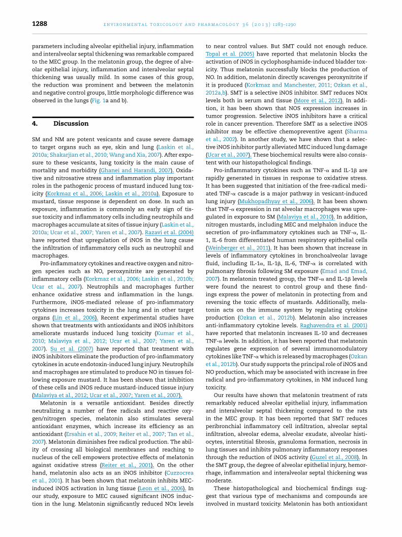
Fig. 1 – (a) Histopathologic examination shows normal lung tissue in the control group (H&E, 40×). (b) In the melatoningroup, most of the alveolar septa are normal (star) in thickness. In some areas, focal minimal thickening in interalveolarsepta with mild inflammatory infiltrate (arrow) is observed (H&E, 100×). (c) A case in the SMT group shows mild to moderatealveolar septal thickening (star). In the interalveolar septa, mild inflammatory infiltrate with minimal hemorrhage (arrow)and congestion is present (H&E, 40×). d. A case in the MEC group demonstrates severely injured alveolar structures (star)with destruction of epithelium and narrowed alveolar spaces with extensively thickened interalveolar septa (arrow)c hage
wpgt(Igcset
isatgc
ontaining prominent inflammatory infiltration and hemorr
ere significantly different than the MEC [44.9 (26.7–54.9) ng/grotein] and the MEC + SMT [27.9 (12.5–42.1) ng/g protein]roups (p = 0.006 and p = 0.024, respectively). There was no sta-istical difference between the control and the MEC + MEL [9.56.1–26.6) ng/g protein] groups (p = 0.108). In the MEC group,L-1� levels were significantly higher than in the MEC + MELroup (p = 0.006). In the MEC + SMT group, the lung IL-1� con-entrations were lower than the MEC group, but it was nottatistically significant (p = 0.120). Additionally, lung IL-1� lev-ls in the MEC + MEL group were not different compared withhe MEC + SMT group (p = 0.084).
Mechlorethamine injection resulted in alveolar epithelialnjury, hemorrhage, inflammation, edema and interalveolareptal thickening in the lung tissues of MEC group (Table 2
nd Fig. 1d). Especially, alveolar epithelial injury, inflamma-ion and interalveolar septal thickening were severe in thisroup. Hemorrhage was moderate to severe in most of theases in MEC group. Hemorrhage was observed generally inin some areas (H&E, x40).
the interalveolar septa as extravasated erythrocytes accom-panied with congestion but not in the alveolar air spaces.Mainly inflammatory cell accumulation, congestion withextravasated erythrocytes contributed to the interalveolarseptal thickening. Inflammation was characterized princi-pally by mononuclear inflammatory cell infiltration which wascomposed of macrophages in majority. However, neutrophilleucocytes were also observed among mononuclear inflam-mation.
SMT administration reduced these morphological changesin the lungs but the level of reduction was not significant(Table 2 and Fig. 1c). In the SMT group, the degree of alveolarepithelial injury, hemorrhage, inflammation and interalveolarseptal thickening was moderate.
Melatonin administration diminished almost all of thepathological findings in the lungs of rats emerged due to thetoxicity caused by nitrogen mustard (Table 2 and Fig. 1b). Inthis group of melatonin, the reduction in the morphologic
p h a
1288 e n v i r o n m e n t a l t o x i c o l o g y a n dparameters including alveolar epithelial injury, inflammationand interalveolar septal thickening was remarkable comparedto the MEC group. In the melatonin group, the degree of alve-olar epithelial injury, inflammation and interalveolar septalthickening was usually mild. In some cases of this group,the reduction was prominent and between the melatoninand negative control groups, little morphologic difference wasobserved in the lungs (Fig. 1a and b).
4. Discussion
SM and NM are potent vesicants and cause severe damageto target organs such as eye, skin and lung (Laskin et al.,2010a; Shakarjian et al., 2010; Wang and Xia, 2007). After expo-sure to these vesicants, lung toxicity is the main cause ofmortality and morbidity (Ghanei and Harandi, 2007). Oxida-tive and nitrosative stress and inflammation play importantroles in the pathogenic process of mustard induced lung tox-icity (Korkmaz et al., 2006; Laskin et al., 2010a). Exposure tomustard, tissue response is dependent on dose. In such anexposure, inflammation is commonly an early sign of tis-sue toxicity and inflammatory cells including neutrophils andmacrophages accumulate at sites of tissue injury (Laskin et al.,2010a; Ucar et al., 2007; Yaren et al., 2007). Razavi et al. (2004)have reported that upregulation of iNOS in the lung causethe infiltration of inflammatory cells such as neutrophil andmacrophages.
Pro-inflammatory cytokines and reactive oxygen and nitro-gen species such as NO, peroxynitrite are generated byinflammatory cells (Korkmaz et al., 2006; Laskin et al., 2010b;Ucar et al., 2007). Neutrophils and macrophages furtherenhance oxidative stress and inflammation in the lungs.Furthermore, iNOS-mediated release of pro-inflammatorycytokines increases toxicity in the lung and in other targetorgans (Lin et al., 2006). Recent experimental studies haveshown that treatments with antioxidants and iNOS inhibitorsameliorate mustards induced lung toxicity (Kumar et al.,2010; Malaviya et al., 2012; Ucar et al., 2007; Yaren et al.,2007). Su et al. (2007) have reported that treatment withiNOS inhibitors eliminate the production of pro-inflammatorycytokines in acute endotoxin-induced lung injury. Neutrophilsand macrophages are stimulated to produce NO in tissues fol-lowing exposure mustard. It has been shown that inhibitionof these cells and iNOS reduce mustard-induced tissue injury(Malaviya et al., 2012; Ucar et al., 2007; Yaren et al., 2007).
Melatonin is a versatile antioxidant. Besides directlyneutralizing a number of free radicals and reactive oxy-gen/nitrogen species, melatonin also stimulates severalantioxidant enzymes, which increase its efficiency as anantioxidant (Ersahin et al., 2009; Reiter et al., 2007; Tan et al.,2007). Melatonin diminishes free radical production. The abil-ity of crossing all biological membranes and reaching tonucleus of the cell empowers protective effects of melatoninagainst oxidative stress (Reiter et al., 2001). On the otherhand, melatonin also acts as an iNOS inhibitor (Cuzzocrea
et al., 2001). It has been shown that melatonin inhibits MEC-induced iNOS activation in lung tissue (Leon et al., 2006). Inour study, exposure to MEC caused significant iNOS induc-tion in the lung. Melatonin significantly reduced NOx levelsr m a c o l o g y 3 6 ( 2 0 1 3 ) 1283–1290
to near control values. But SMT could not enough reduce.Topal et al. (2005) have reported that melatonin blocks theactivation of iNOS in cyclophosphamide-induced bladder tox-icity. Thus melatonin successfully blocks the production ofNO. In addition, melatonin directly scavenges peroxynitrite ifit is produced (Korkmaz and Manchester, 2011; Ozkan et al.,2012a,b). SMT is a selective iNOS inhibitor. SMT reduces NOxlevels both in serum and tissue (More et al., 2012). In addi-tion, it has been shown that NOS expression increases intumor progression. Selective iNOS inhibitors have a criticalrole in cancer prevention. Therefore SMT as a selective iNOSinhibitor may be effective chemopreventive agent (Sharmaet al., 2002). In another study, we have shown that a selec-tive iNOS inhibitor partly alleviated MEC induced lung damage(Ucar et al., 2007). These biochemical results were also consis-tent with our histopathological findings.
Pro-inflammatory cytokines such as TNF-� and IL-1� arerapidly generated in tissues in response to oxidative stress.It has been suggested that initiation of the free-radical medi-ated TNF-� cascade is a major pathway in vesicant-inducedlung injury (Mukhopadhyay et al., 2006). It has been shownthat TNF-� expression in rat alveolar macrophages was upre-gulated in exposure to SM (Malaviya et al., 2010). In addition,nitrogen mustards, including MEC and melphalon induce thesecretion of pro-inflammatory cytokines such as TNF-�, IL-1, IL-6 from differentiated human respiratory epithelial cells(Weinberger et al., 2011). It has been shown that increase inlevels of inflammatory cytokines in bronchoalveolar lavagefluid, including IL-1�, IL-1�, IL-6, TNF-� is correlated withpulmonary fibrosis following SM exposure (Emad and Emad,2007). In melatonin treated group, the TNF-� and IL-1� levelswere found the nearest to control group and these find-ings express the power of melatonin in protecting from andreversing the toxic effects of mustards. Additionally, mela-tonin acts on the immune system by regulating cytokineproduction (Ozkan et al., 2012b). Melatonin also increasesanti-inflammatory cytokine levels. Raghavendra et al. (2001)have reported that melatonin increases IL-10 and decreasesTNF-� levels. In addition, it has been reported that melatoninregulates gene expression of several immunomodulatorycytokines like TNF-� which is released by macrophages (Ozkanet al., 2012b). Our study supports the principal role of iNOS andNO production, which may be associated with increase in freeradical and pro-inflammatory cytokines, in NM induced lungtoxicity.
Our results have shown that melatonin treatment of ratsremarkably reduced alveolar epithelial injury, inflammationand interalveolar septal thickening compared to the ratsin the MEC group. It has been reported that SMT reducesperibronchial inflammatory cell infiltration, alveolar septalinfiltration, alveolar edema, alveolar exudate, alveolar histi-ocytes, interstitial fibrosis, granuloma formation, necrosis inlung tissues and inhibits pulmonary inflammatory responsesthrough the reduction of iNOS activity (Guzel et al., 2008). Inthe SMT group, the degree of alveolar epithelial injury, hemor-rhage, inflammation and interalveolar septal thickening was
moderate.These histopathological and biochemical findings sug-gest that various type of mechanisms and compounds areinvolved in mustard toxicity. Melatonin has both antioxidant

a r m
aiet
isNatcStcspttn
C
T
r
C
E
E
E
G
G
G
K
K
e n v i r o n m e n t a l t o x i c o l o g y a n d p h
nd anti-inflammatory effects. But SMT is only an iNOSnhibitor. This may explain why SMT has minimal protectiveffect on MEC induced lung injury and melatonin is supposedo be more promising against mustard toxicity.
In conclusion, the exposure to NM is associated withnflammatory and oxidative injury in the lungs. Fully under-tanding of the cellular and molecular mechanisms by whichM causes injury will aid to develop effective treatments andgent against NM toxicity. Newer therapeutic approaches forreating mustard induced lung injury have focused on spe-ific pro- and anti-inflammatory mediators. Melatonin andMT played a significant role in regulating inflammation inhe lung through production of NO and pro-inflammatoryytokines such as TNF-� and IL-1�. Melatonin combined withpecific iNOS inhibitors (e.g. SMT, aminoguanidine) could be aotential therapeutic treatment against mustard induced lungoxicity. Further studies need to be done to combine severalherapeutic agents, each blocking one of the toxic mecha-isms induced by mustards.
onflict of interest
he authors declare that there are no conflicts of interest.
e f e r e n c e s
uzzocrea, S., Mazzon, E., Serraino, I., Lepore, V., Terranova, M.L.,Ciccolo, A., Caputi, A.P., 2001. Melatonin reducesdinitrobenzene sulfonic acid-induced colitis. J. Pineal Res. 30,1–12.
kstrand-Hammarstrom, B., Wigenstam, E., Bucht, A., 2011.Inhalation of alkylating mustard causes long-term Tcell-dependent inflammation in airways and growth ofconnective tissue. Toxicology 280, 88–97.
mad, A., Emad, Y., 2007. Levels of cytokine in bronchoalveolarlavage (BAL) fluid in patients with pulmonary fibrosis due tosulfur mustard gas inhalation. J. Interferon Cytokine Res. 27,38–43.
rsahin, M., Toklu, H.Z., Cetinel, S., Yuksel, M., Yegen, B.C., Sener,G., 2009. Melatonin reduces experimental subarachnoidhemorrhage-induced oxidative brain damage andneurological symptoms. J. Pineal Res. 46, 324–332.
hanei, M., Harandi, A.A., 2007. Long term consequences fromexposure to sulfur mustard: a review. Inhal. Toxicol. 19,451–456.
uo, Y.J., Li, W., Li, X.F., Zhao, L., Zhang, S.L., Zhou, Y., Dong, J.H.,Mei, Y.W., 2011. S-Methylisothiourea induces apoptosis ofherpes simplex virus-1-infected microglial cells.Inflammation 34, 388–401.
uzel, A., Basaran, U.N., Aksu, B., Kanter, M., Yalcin, O., Aktas, C.,Karasalihoglu, S., 2008. Protective effects ofS-methylisothiourea sulfate on different aspirationmaterials-induced lung injury in rats. Int. J. Pediatr.Otorhinolaryngol. 72, 1241–1250.
ehe, K., Raithel, K., Kreppel, H., Jochum, M., Worek, F.,Thiermann, H., 2008. Inhibition of poly(ADP-ribose)polymerase (PARP) influences the mode of sulfur mustard(SM)-induced cell death in HaCaT cells. Arch. Toxicol. 82,461–470.
orkmaz, A., Kunak, Z.I., Paredes, S.D., Yaren, H., Tan, D.X., Reiter,R.J., 2008. The use of melatonin to combat mustard toxicity.REVIEW. Neuro Endocrinol. Lett. 29, 614–619.
a c o l o g y 3 6 ( 2 0 1 3 ) 1283–1290 1289
Korkmaz, A., Manchester, L., 2011. Reactive nitrogen species;devastating intracellular players and melatonin as a defender.J. Exp. Integr. Med. 1, 63–65.
Korkmaz, A., Yaren, H., Topal, T., Oter, S., 2006. Molecular targetsagainst mustard toxicity: implication of cell surface receptors,peroxynitrite production, and PARP activation. Arch. Toxicol.80, 662–670.
Kumar, P., Gautam, A., Prakash, C.J., Kumar, A., Ganeshan, K.,Pathak, U., Vijayaraghavan, R., 2010. Ameliorative effect ofDRDE 07 and its analogues on the systemic toxicity of sulphurmustard and nitrogen mustard in rabbit. Hum. Exp. Toxicol.29, 747–755.
Kunak, Z.I., Macit, E., Yaren, H., Yaman, H., Cakir, E., Aydin, I.,Turker, T., Kurt, Y.G., Ozcan, A., Uysal, B., Isbilir, S., Akgul, E.O.,Cayci, T., Korkmaz, A., Kenar, L., 2012. Protective effects ofmelatonin and S-methylisothiourea on mechlorethamineinduced nephrotoxicity. J. Surg. Res. 175,e17–e23.
Laskin, J.D., Black, A.T., Jan, Y.H., Sinko, P.J., Heindel, N.D., Sunil,V., Heck, D.E., Laskin, D.L., 2010a. Oxidants and antioxidantsin sulfur mustard-induced injury. Ann. N. Y. Acad. Sci. 1203,92–100.
Laskin, J., Heck, D., Laskin, D., 2010b. Nitric oxide pathways intoxic responses. In: Ballantine, B., Marrs, T., Syversen, T. (Eds.),General and Applied Toxicology. Wiley-Blackwell, UK, pp.425–438.
Leon, J., Escames, G., Rodriguez, M.I., Lopez, L.C., Tapias, V.,Entrena, A., Camacho, E., Carrion, M.D., Gallo, M.A., Espinosa,A., Tan, D.X., Reiter, R.J., Acuna-Castroviejo, D., 2006.Inhibition of neuronal nitric oxide synthase activity byN-1-acetyl-5-methoxykynuramine, a brain metabolite ofmelatonin. J. Neurochem. 98, 2023–2033.
Lin, N.T., Yang, F.L., Lee, R.P., Peng, T.C., Chen, H.I., 2006. Induciblenitric oxide synthase mediates cytokine release: the timecourse in conscious and septic rats. Life Sci. 78, 1038–1043.
Lowry, O.H., Rosebrough, N.J., Farr, A.L., Randall, R.J., 1951. Proteinmeasurement with the Folin phenol reagent. J. Biol. Chem.193, 265–275.
Malaviya, R., Sunil, V.R., Cervelli, J., Anderson, D.R., Holmes, W.W.,Conti, M.L., Gordon, R.E., Laskin, J.D., Laskin, D.L., 2010.Inflammatory effects of inhaled sulfur mustard in rat lung.Toxicol. Appl. Pharmacol. 248, 89–99.
Malaviya, R., Venosa, A., Hall, L., Gow, A.J., Sinko, P.J., Laskin, J.D.,Laskin, D.L., 2012. Attenuation of acute nitrogenmustard-induced lung injury, inflammation and fibrogenesisby a nitric oxide synthase inhibitor. Toxicol. Appl. Pharmacol.265, 279–291.
McCartney-Francis, N., Allen, J.B., Mizel, D.E., Albina, J.E., Xie,Q.W., Nathan, C.F., Wahl, S.M., 1993. Suppression of arthritisby an inhibitor of nitric oxide synthase. J. Exp. Med. 178,749–754.
More, A.S., Kumari, R.R., Gupta, G., Kathirvel, K., Lonare, M.K.,Dhayagude, R.S., Kumar, D., Kumar, D., Sharma, A.K., Tandan,S.K., 2012. Effect of S-methylisothiourea inacetaminophen-induced hepatotoxicity in rat.Naunyn-Schmiedebergs Arch. Pharmacol. 385, 1127–1139.
Mukhopadhyay, S., Hoidal, J.R., Mukherjee, T.K., 2006. Role ofTNF-alpha in pulmonary pathophysiology. Respir. Res. 7,125.
Ozkan, E., Yaman, H., Cakir, E., Aydin, I., Oztosun, M., Agilli, M.,Kurt, Y., Akgul, E., Cayci, T., Ilhan, N., Ilhan, N., 2012a. Themeasurement of plasma melatonin levels by highperformance liquid chromatography. J. Exp. Integr. Med. 2 (1),85–88.
Ozkan, E., Yaman, H., Cakir, E., Deniz, O., Oztosun, M., Gumus, S.,Akgul, E.O., Agilli, M., Cayci, T., Kurt, Y.G., Aydin, I., Arslan, Y.,
Ilhan, N., Erbil, M.K., 2012b. Plasma melatonin and urinary6-hydroxymelatonin levels in patients with pulmonarytuberculosis. Inflammation 35, 1429–1434.
p h a
1290 e n v i r o n m e n t a l t o x i c o l o g y a n dPohanka, M., Sobotka, J., Stetina, R., 2011. Sulfur mustard inducedoxidative stress and its alteration by epigallocatechin gallate.Toxicol. Lett. 201, 105–109.
Raghavendra, V., Singh, V., Kulkarni, S.K., Agrewala, J.N., 2001.Melatonin enhances Th2 cell mediated immune responses:lack of sensitivity to reversal by naltrexone or benzodiazepinereceptor antagonists. Mol. Cell. Biochem. 221, 57–62.
Razavi, H.M., Wang le, F., Weicker, S., Rohan, M., Law, C.,McCormack, D.G., Mehta, S., 2004. Pulmonary neutrophilinfiltration in murine sepsis: role of inducible nitric oxidesynthase. Am. J. Respir. Crit. Care Med. 170,227–233.
Reiter, R.J., Tan, D.X., Manchester, L.C., Qi, W., 2001. Biochemicalreactivity of melatonin with reactive oxygen and nitrogenspecies: a review of the evidence. Cell Biochem. Biophys. 34,237–256.
Reiter, R.J., Tan, D.X., Manchester, L.C., Tamura, H., 2007.Melatonin defeats neurally-derived free radicals and reducesthe associated neuromorphological and neurobehavioraldamage. J. Physiol. Pharmacol. 58 (Suppl. 6),5–22.
Ren, D., Sun, R., Wang, S., 2006. Role of inducible nitric oxidesynthase expressed by alveolar macrophages in high mobilitygroup box 1 – induced acute lung injury. Inflamm. Res. 55,207–215.
Shakarjian, M.P., Heck, D.E., Gray, J.P., Sinko, P.J., Gordon, M.K.,Casillas, R.P., Heindel, N.D., Gerecke, D.R., Laskin, D.L., Laskin,J.D., 2010. Mechanisms mediating the vesicant actions ofsulfur mustard after cutaneous exposure. Toxicol. Sci. 114,5–19.
Sharma, S., Wilkinson, B.P., Gao, P., Steele, V.E., 2002. Differentialactivity of NO synthase inhibitors as chemopreventive agentsin a primary rat tracheal epithelial cell transformationsystem. Neoplasia 4, 332–336.
r m a c o l o g y 3 6 ( 2 0 1 3 ) 1283–1290
Su, C.F., Yang, F.L., Chen, H.I., 2007. Inhibition of inducible nitricoxide synthase attenuates acute endotoxin-induced lunginjury in rats. Clin. Exp. Pharmacol. Physiol. 34, 339–346.
Sunil, V.R., Patel, K.J., Shen, J., Reimer, D., Gow, A.J., Laskin, J.D.,Laskin, D.L., 2011a. Functional and inflammatory alterationsin the lung following exposure of rats to nitrogen mustard.Toxicol. Appl. Pharmacol. 250, 10–18.
Sunil, V.R., Patel-Vayas, K., Shen, J., Gow, A.J., Laskin, J.D., Laskin,D.L., 2011b. Role of TNFR1 in lung injury and altered lungfunction induced by the model sulfur mustard vesicant,2-chloroethyl ethyl sulfide. Toxicol. Appl. Pharmacol. 250,245–255.
Tan, D.X., Manchester, L.C., Terron, M.P., Flores, L.J., Reiter, R.J.,2007. One molecule, many derivatives: a never-endinginteraction of melatonin with reactive oxygen and nitrogenspecies? J. Pineal Res. 42, 28–42.
Topal, T., Oztas, Y., Korkmaz, A., Sadir, S., Oter, S., Coskun, O.,Bilgic, H., 2005. Melatonin ameliorates bladder damageinduced by cyclophosphamide in rats. J. Pineal Res. 38,272–277.
Ucar, M., Korkmaz, A., Reiter, R.J., Yaren, H., Oter, S., Kurt, B.,Topal, T., 2007. Melatonin alleviates lung damage induced bythe chemical warfare agent nitrogen mustard. Toxicol. Lett.173, 124–131.
Wang, G.Q., Xia, Z.F., 2007. Tissue injury by hot fluid containingnitrogen mustard. Burns 33, 923–926.
Weinberger, B., Laskin, J.D., Sunil, V.R., Sinko, P.J., Heck, D.E.,Laskin, D.L., 2011. Sulfur mustard-induced pulmonary injury:therapeutic approaches to mitigating toxicity. Pulm.Pharmacol. Ther. 24, 92–99.
Yaren, H., Mollaoglu, H., Kurt, B., Korkmaz, A., Oter, S., Topal, T.,Karayilanoglu, T., 2007. Lung toxicity of nitrogen mustard maybe mediated by nitric oxide and peroxynitrite in rats. Res. Vet.Sci. 83, 116–122.