The prepilin peptidase is required for protein secretion by and the virulence of the intracellular...
12
The prepilin peptidase is required for protein secretion by and the virulence of the intracellular pathogen Legionella pneumophila Mark R. Liles, 1 Paul H. Edelstein 2 and Nicholas P. Cianciotto 1 * 1 Department of Microbiology-Immunology, Northwestern University, 303 E. Chicago Ave., Chicago, IL 60611, USA. 2 Departments of Pathology and Laboratory Medicine, The University of Pennsylvania, Philadelphia, PA 19104, USA. Summary Prepilin peptidases cleave, among other substrates, the leader sequences from prepilin-like proteins that are required for type II protein secretion in Gram-nega- tive bacteria. To begin to assess the importance of type II secretion for the virulence of an intracellular pathogen, we examined the effect of inactivating the prepilin peptidase (pilD ) gene of Legionella pneumo- phila . Although the pilD mutant and its parent grew similarly in bacteriological media, they did differ in colony attributes and recoverability from late stationary phase. Moreover, at least three proteins were absent from the mutant’s supernatant, indicating that PilD is necessary for the secretion of Legionella proteins. The absence of both the major secreted protein and a haemolytic activity from the mutant signalled that the L. pneumophila zinc metalloprotease is excreted via type II secretion. Most interestingly, the pilD mutant was greatly impaired in its ability to grow within Hart- mannella vermiformis amoebae and the human macro- phage-like U937 cells. As reintroduction of pilD into the mutant restored infectivity and as a mutant lacking type IV pilin replicated like wild type, these data sug- gested that the intracellular growth of L. pneumophila is promoted by proteins secreted via a type II path- way. Intratracheal inoculation of guinea pigs revealed that the LD 50 for the pilD mutant is at least 100-fold greater than that for its parent, and the culturing of bacteria from infected animals showed a rapid clear- ance of the mutant from the lungs. This is the first study to indicate a role for PilD and type II secretion in intracellular parasitism. Introduction In recent years, much attention has been directed towards understanding protein secretion by bacteria (Pugsley, 1993). This effort has been driven, in large part, by the intriguing ways in which protein secretion pathways promote the viru- lence of animal and plant pathogens (Hueck, 1998). The secretion mechanisms of Gram-negative bacteria have been divided into four types, i.e. types I, II, III and IV (Sal- mond and Reeves, 1993; Finlay and Falkow, 1997). The type I pathway (referred to as type IV in early reviews) is composed of three proteins that form a channel for the transport of proteins directly from the cytoplasm to the exter- ior (Wandersman, 1992; Salmond and Reeves, 1993; Hueck, 1998). Virulence determinants that are handled via this secretion pathway include the haemolysin of Escherichia coli , the adenylate cyclase of Bordetella per- tussis and the alkaline protease of Pseudomonas aeru- ginosa (Finlay and Falkow, 1997). In type II secretion, exoproteins are transported across the inner membrane in sec-dependent fashion, and then the mature proteins cross the outer membrane through the combined action of some 14 proteins (Wandersman, 1992; Pugsley, 1993; Salmond and Reeves, 1993; Pugsley et al ., 1997; Russel, 1998). Virulence factors that depend upon type II secretion include the exotoxin A and phospholipase C of P. aeru- ginosa, the haemolysin and protease of Aeromonas hydro- phila and the enterotoxin and protease of Vibrio cholerae (Bally et al ., 1991; Strom et al ., 1991; Pepe et al ., 1996; Fin- lay and Falkow, 1997). Type III secretion involves approxi- mately 20 proteins that carry exoproteins directly from the cytoplasm to the cell surface, from where they are then injected into an animal or plant cell upon contact (Salmond and Reeves, 1993; Finlay and Falkow, 1997; Hueck, 1998). The Yops of Yersinia enterocolitica and invasins of Shigella flexneri and Salmonella typhimurium are virulence factors secreted via a type III system (Anderson and Schneewind, 1997; Finlay and Falkow, 1997). In the type IV system (referred to as type I in early reviews), sec -dependent transport of proteins across the inner membrane is followed by self-directed passage through the outer membrane (Wandersman, 1992; Finlay and Falkow, 1997; Hueck, 1998). Factors that complete their own export are the vacuolating cytotoxin of Helicobacter pylori , the IgA pro- tease of Neisseria gonorrhoeae and SepA of S. flexneri . Molecular Microbiology (1999) 31(3), 959–970 Q 1999 Blackwell Science Ltd Received 14 August, 1998; revised 22 October, 1998; accepted 30 October, 1998. *For correspondence. E-mail [email protected]; Tel. (þ1) 312 503 0385; Fax (þ1) 312 503 1339.
-
Upload
independent -
Category
Documents
-
view
2 -
download
0
Transcript of The prepilin peptidase is required for protein secretion by and the virulence of the intracellular...
The prepilin peptidase is required for protein secretionby and the virulence of the intracellular pathogenLegionella pneumophila
Mark R. Liles, 1 Paul H. Edelstein 2 and Nicholas P.Cianciotto 1*1Department of Microbiology-Immunology, NorthwesternUniversity, 303 E. Chicago Ave., Chicago, IL 60611, USA.2Departments of Pathology and Laboratory Medicine, TheUniversity of Pennsylvania, Philadelphia, PA 19104, USA.
Summary
Prepilin peptidases cleave, among other substrates,the leader sequences from prepilin-like proteins thatare required for type II protein secretion in Gram-nega-tive bacteria. To begin to assess the importance oftype II secretion for the virulence of an intracellularpathogen, we examined the effect of inactivating theprepilin peptidase ( pilD ) gene of Legionella pneumo-phila . Although the pilD mutant and its parent grewsimilarly in bacteriological media, they did differ incolony attributes and recoverability from late stationaryphase. Moreover, at least three proteins were absentfrom the mutant’s supernatant, indicating that PilDis necessary for the secretion of Legionella proteins.The absence of both the major secreted protein and ahaemolytic activity from the mutant signalled that theL. pneumophila zinc metalloprotease is excreted viatype II secretion. Most interestingly, the pilD mutantwas greatly impaired in its ability to grow within Hart-mannella vermiformis amoebae and the human macro-phage-like U937 cells. As reintroduction of pilD into themutant restored infectivity and as a mutant lackingtype IV pilin replicated like wild type, these data sug-gested that the intracellular growth of L. pneumophilais promoted by proteins secreted via a type II path-way. Intratracheal inoculation of guinea pigs revealedthat the LD 50 for the pilD mutant is at least 100-foldgreater than that for its parent, and the culturing ofbacteria from infected animals showed a rapid clear-ance of the mutant from the lungs. This is the firststudy to indicate a role for PilD and type II secretionin intracellular parasitism.
Introduction
In recent years, much attention has been directed towardsunderstanding protein secretion by bacteria (Pugsley, 1993).This effort has been driven, in large part, by the intriguingways in which protein secretion pathways promote the viru-lence of animal and plant pathogens (Hueck, 1998). Thesecretion mechanisms of Gram-negative bacteria havebeen divided into four types, i.e. types I, II, III and IV (Sal-mond and Reeves, 1993; Finlay and Falkow, 1997). Thetype I pathway (referred to as type IV in early reviews) iscomposed of three proteins that form a channel for thetransport of proteins directly from the cytoplasm to the exter-ior (Wandersman, 1992; Salmond and Reeves, 1993;Hueck, 1998). Virulence determinants that are handledvia this secretion pathway include the haemolysin ofEscherichia coli, the adenylate cyclase of Bordetella per-tussis and the alkaline protease of Pseudomonas aeru-ginosa (Finlay and Falkow, 1997). In type II secretion,exoproteins are transported across the inner membranein sec-dependent fashion, and then the mature proteinscross the outer membrane through the combined actionof some 14 proteins (Wandersman, 1992; Pugsley, 1993;Salmond and Reeves, 1993; Pugsley et al., 1997; Russel,1998). Virulence factors that depend upon type II secretioninclude the exotoxin A and phospholipase C of P. aeru-ginosa, the haemolysin and protease of Aeromonas hydro-phila and the enterotoxin and protease of Vibrio cholerae(Bally et al., 1991; Strom et al., 1991; Pepe et al., 1996; Fin-lay and Falkow, 1997). Type III secretion involves approxi-mately 20 proteins that carry exoproteins directly from thecytoplasm to the cell surface, from where they are theninjected into an animal or plant cell upon contact (Salmondand Reeves, 1993; Finlay and Falkow, 1997; Hueck, 1998).The Yops of Yersinia enterocolitica and invasins of Shigellaflexneri and Salmonella typhimurium are virulence factorssecreted via a type III system (Anderson and Schneewind,1997; Finlay and Falkow, 1997). In the type IV system(referred to as type I in early reviews), sec-dependenttransport of proteins across the inner membrane is followedby self-directed passage through the outer membrane(Wandersman, 1992; Finlay and Falkow, 1997; Hueck,1998). Factors that complete their own export are thevacuolating cytotoxin of Helicobacter pylori, the IgA pro-tease of Neisseria gonorrhoeae and SepA of S. flexneri.
Molecular Microbiology (1999) 31(3), 959–970
Q 1999 Blackwell Science Ltd
Received 14 August, 1998; revised 22 October, 1998; accepted 30October, 1998. *For correspondence. E-mail [email protected];Tel. (þ1) 312 503 0385; Fax (þ1) 312 503 1339.
It has been suggested recently that a fifth secretion sys-tem exists, dictating the conjugal transfer of plasmids inAgrobacterium tumefaciens and the release of pertussistoxin by B. pertussis (Winans et al., 1996; Hueck, 1998).The dot/icm loci of Legionella pneumophila may encode arelated pathway that is required for the infection of macro-phages (Segal et al., 1998; Vogel et al., 1998). Curiously,of these secretion pathways, only type II has not beenidentified previously in an intracellular pathogen. Our recentdiscovery of a locus in L. pneumophila that, in other Gram-negative bacteria, is required for type II secretion sug-gested that this secretory mechanism might promoteintracellular parasitism as well (Liles et al., 1998).
L. pneumophila exists primarily as a parasite of protozoa,replicating within amoebae and ciliates in freshwater habi-tats (Fields, 1996). However, the inhalation of contaminatedwater droplets can lead to a potentially fatal pneumoniaknown as Legionnaires’ disease, an infection characterizedby the proliferation of L. pneumophila within alveolar macro-phages (McDade et al., 1977; Horwitz and Silverstein,1980; Dowling et al., 1992). In both protozoa and macro-phages, L. pneumophila multiplies within a ribosome-studded phagosome that does not fuse with endosomesor lysosomes (Horwitz, 1983; Abu Kwaik, 1996; Fields,1996). The factors that enable L. pneumophila to subvertthe host’s bactericidal mechanisms and to access nutrientshave been sought by numerous investigators (Dowling etal., 1992). Recently, while characterizing a locus involvedin iron acquisition, we discovered an operon (pilBCD )encoding analogues of proteins involved in the biogenesisof pili and type II secretion in other Gram-negative bacteria(Liles et al., 1998). The PilB- and PilC-like proteins arerequired for the translocation of type IV prepilin acrossthe inner membrane, at which point the prepilin peptidase(PilD) cleaves the hydrophobic leader sequence, therebyreleasing the mature pilin monomer into the periplasm(Strom and Lory, 1993). The pilin subunits are then assem-bled into the pilus structure via a dedicated outer membraneapparatus (Bally et al., 1992; Strom and Lory, 1993). PilBand PilC are thought to be strictly involved in type IV pilusbiogenesis (Nunn et al., 1990; Turner et al., 1993). In con-trast, PilD can have as many as five prepilin-like substratesthat are directly required for type II secretion across theouter membrane (Nunn and Lory, 1992; Bleves et al.,1998). Therefore, in many extracellular pathogens, the pre-pilin leader peptidase is required for the assembly of botha type IV pilus and a type II secretory apparatus (Kaufmanet al., 1991; Nunn and Lory, 1992; Freitag et al., 1995;Pepe et al., 1996). Stone and Abu Kwaik (1998) recentlydiscovered the L. pneumophila type IV pilin gene (pilEL)and determined that a pilEL mutation eliminates the pro-duction of type IV pili and modestly reduces adherenceto host cells, but does not impair intracellular growth. Tobegin to determine whether a type II system promotes
intracellular parasitism, we constructed and characterizedan L. pneumophila pilD mutant. This mutant was substan-tially impaired in its ability to replicate within amoebae andhuman macrophages, indicating, for the first time, that fac-tors secreted via a type II apparatus contribute to the viru-lence of an intracellular pathogen.
Results
Construction, piliation and colony morphology of anL. pneumophila pilD mutant
To help to determine whether L. pneumophila expresses atype II secretion system and, if so, what the importance ofthis protein export mechanism is to the virulence of theorganism, a pilD mutant of strain 130b was constructedand characterized. The pilD locus was inactivated by theinsertion of a kanR gene 559 bp past the gene’s transla-tional start. After electroporation of the allelic exchangeplasmid pMRL13 into strain 130b, KanR/SucR colonieswere easily isolated at a frequency of 10¹7. Southern hybrid-ization of HindIII-digested DNA revealed that six out of sixKanR clones had undergone the predicted chromosomalrearrangement, i.e. 1.9 kb and 2.7 kb hybridizing fragmentswere observed for each clone in contrast to the 3.5 kbhybridizing fragment for strain 130b (data not shown).The KanR clone chosen for further analysis was designatedNU243.
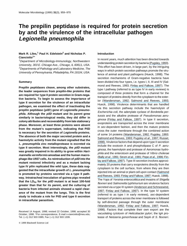
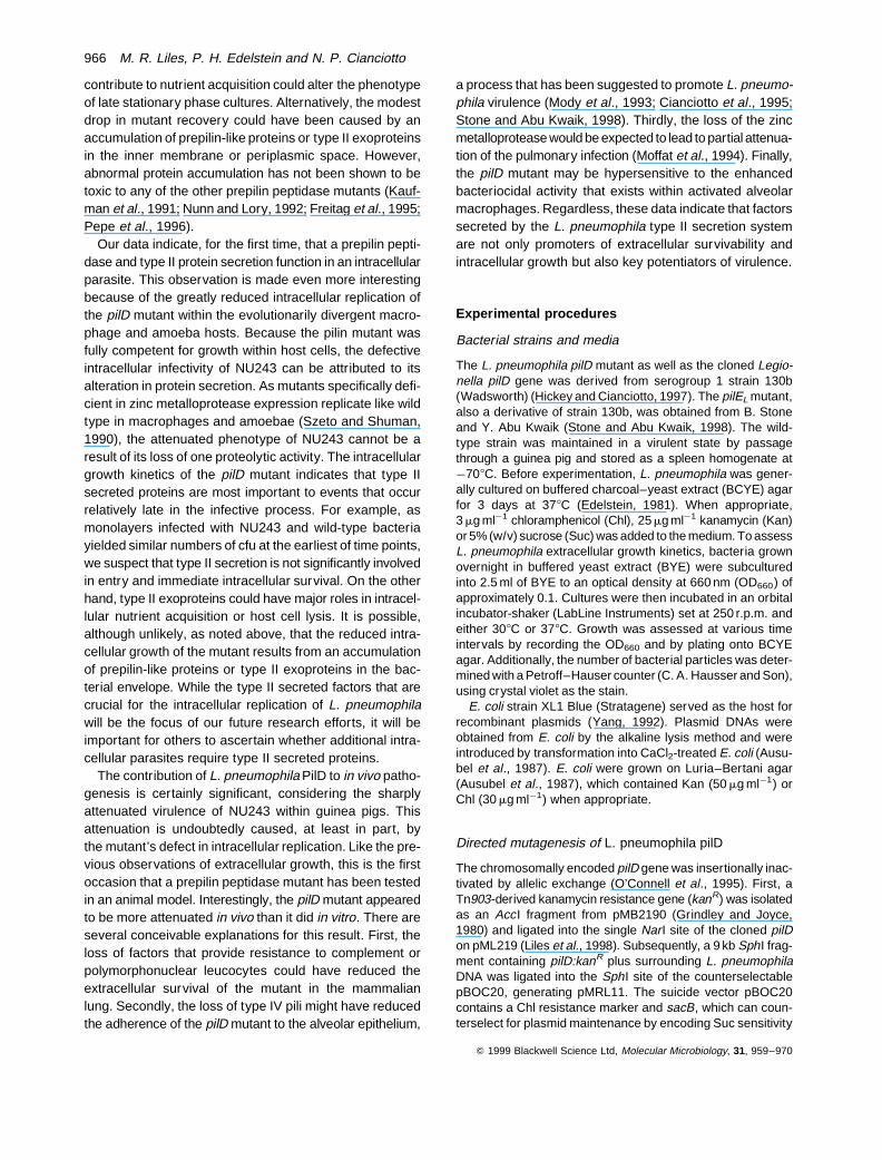
To verify that the pilD mutation abolished type IV pilusproduction, electron microscopy was conducted on 130band NU243 grown on BCYE agar. As greater piliationhad been observed at 308C than at 378C (Liles et al.,1998), we tested the strains for pilus formation at both tem-peratures. This analysis revealed the complete loss ofpiliation for NU243 under all growth conditions, whereas130b exhibited pili as before (Fig. 1). While not unexpected,the observation that NU243 lacked pili was the first experi-mental evidence that L. pneumophila pilD encodes a pro-tein with functional homology to other prepilin peptidases.
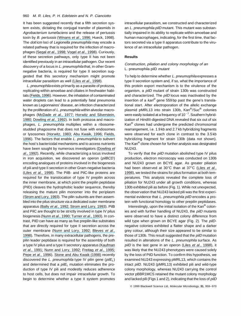
Interestingly, upon the initial isolation of the KanR colon-ies and with further handling of NU243, the pilD mutantswere observed to have a distinct colony difference fromwild type when grown on BCYE agar (Fig. 2). The pilD-negative colonies exhibited a flatter shape and a darkergrey colour, although their size appeared to be similar tothose of 130b. This result suggested that the pilD mutationresulted in alterations of the L. pneumophila surface. AspilD is the last gene in an operon (Liles et al., 1998), itwas likely that the NU243 phenotypes were caused solelyby the loss of PilD function. To confirm this hypothesis, weexamined NU243 expressing pMRL13, which contains theintact pilD. NU243 (pMRL13) exhibited pili and wild-typecolony morphology, whereas NU243 carrying the controlvector pBBR1MCS retained the mutant colony morphologyand lacked pili (Figs 1 and 2), indicating that the loss of pilD
Q 1999 Blackwell Science Ltd, Molecular Microbiology, 31, 959–970
960 M. R. Liles, P. H. Edelstein and N. P. Cianciotto
is responsible for these phenotypical changes. As the lossof type IV pili can induce changes in the colony morph-ology of other Gram-negative bacteria (e.g. pilin mutantsof N. gonorrhoeae are flatter and less reflective) (Freitaget al., 1995), we re-examined the colonies of the L. pneumo-phila pilEL mutant. Supporting the observation of Stone andAbu Kwaik (1998), we did not see any colony differencesresulting from the absence of pilin (data not shown). Thus,the unique colony morphology of the pilD mutant is prob-ably attributable to a deficiency in type II protein secretion.
Extracellular growth of the L. pneumophila pilDmutant
The initial observations of growth on BCYE agar indicatedthat NU243 grew at a rate similar to wild-type L. pneumo-phila. To study the extracellular growth of this mutant in
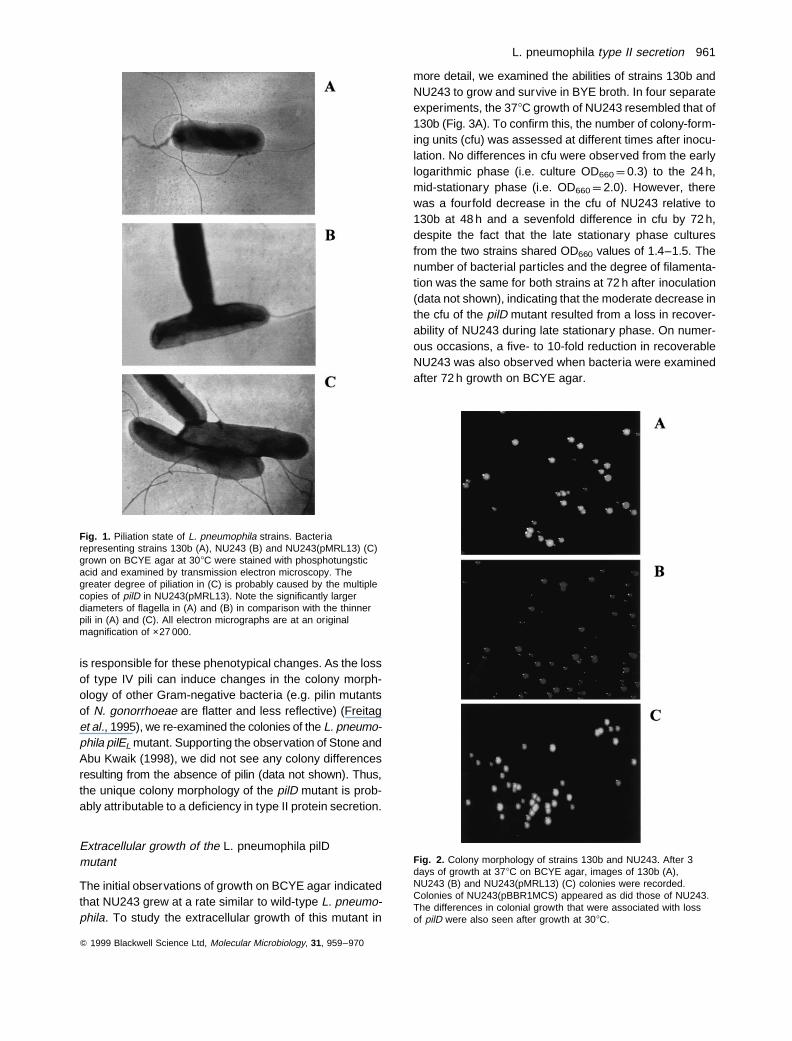
more detail, we examined the abilities of strains 130b andNU243 to grow and survive in BYE broth. In four separateexperiments, the 378C growth of NU243 resembled that of130b (Fig. 3A). To confirm this, the number of colony-form-ing units (cfu) was assessed at different times after inocu-lation. No differences in cfu were observed from the earlylogarithmic phase (i.e. culture OD660 ¼ 0.3) to the 24 h,mid-stationary phase (i.e. OD660 ¼ 2.0). However, therewas a fourfold decrease in the cfu of NU243 relative to130b at 48 h and a sevenfold difference in cfu by 72 h,despite the fact that the late stationary phase culturesfrom the two strains shared OD660 values of 1.4–1.5. Thenumber of bacterial particles and the degree of filamenta-tion was the same for both strains at 72 h after inoculation(data not shown), indicating that the moderate decrease inthe cfu of the pilD mutant resulted from a loss in recover-ability of NU243 during late stationary phase. On numer-ous occasions, a five- to 10-fold reduction in recoverableNU243 was also observed when bacteria were examinedafter 72 h growth on BCYE agar.
Q 1999 Blackwell Science Ltd, Molecular Microbiology, 31, 959–970
Fig. 2. Colony morphology of strains 130b and NU243. After 3days of growth at 378C on BCYE agar, images of 130b (A),NU243 (B) and NU243(pMRL13) (C) colonies were recorded.Colonies of NU243(pBBR1MCS) appeared as did those of NU243.The differences in colonial growth that were associated with lossof pilD were also seen after growth at 308C.
Fig. 1. Piliation state of L. pneumophila strains. Bacteriarepresenting strains 130b (A), NU243 (B) and NU243(pMRL13) (C)grown on BCYE agar at 308C were stained with phosphotungsticacid and examined by transmission electron microscopy. Thegreater degree of piliation in (C) is probably caused by the multiplecopies of pilD in NU243(pMRL13). Note the significantly largerdiameters of flagella in (A) and (B) in comparison with the thinnerpili in (A) and (C). All electron micrographs are at an originalmagnification of ×27 000.
L. pneumophila type II secretion 961
As previous data indicated that transcription of thepilBCD operon increased at 308C relative to 378C (Lileset al., 1998), the extracellular growth of these two strainsin BYE was compared at this lower temperature (Fig. 3B).Although the lag and logarithmic phases of growth weresimilar for strains 130b and NU243 at 308C, a decrease inthe optical density of the NU243 culture was observed dur-ing stationary phase on five separate occasions (Fig. 3B).A reduction in the cfu of NU243 relative to 130b occurredbeginning in the stationary phase, with a fourfold differencein cfu evident at 34 h after inoculation and an eightfold dif-ference in cfu observed at 50 h. Again, no difference wasobserved in numbers of bacterial particles or degree of fila-mentation between strains 130b and NU243 (data notshown).
These experiments indicated that the pilD mutant is fully
competent for replication in an extracellular growth med-ium, yet its recoverability during the late stationary phaseof growth is impaired compared with wild-type L. pneumo-phila. NU243 expressing pMRL13 grew like strain 130b anddid not exhibit the moderate decrease in the numbersof recoverable bacteria at either 308C or 378C (data notshown), indicating that this phenotype results from theloss of PilD. To determine whether the absence of typeIV pili contributes to late stationary phase recoverability,the pilEL mutant was tested for extracellular growth. Thegrowth of the pilEL mutant was the same as strain 130bat 308C and 378C, without a drop in OD660 or cfu ml¹1 dur-ing stationary phase (data not shown). Thus, this evidencesuggests that proteins exported by a type II machinerycontribute to L. pneumophila recoverability during thelate stationary phase of growth in a complex extracellularmedium.
Protein secretion by the L. pneumophila pilD mutant
If L. pneumophila possesses a type II secretion system,then a pilD mutation should result in the absence of someproteins in the bacterial supernatant. To test this hypoth-esis, supernatants from BYE cultures grown at 308C and378C were examined for proteins secreted by strains130b and NU243. The protein profiles of the two strainswere very different in logarithmic and stationary phasegrowth. During early and late log phases, all seven oreight proteins that were apparent in the 130b supernatantswere absent in the NU243 supernatants (Fig. 4, left). Like-wise, several prominent protein bands were lacking in thestationary supernatants of the mutant (Fig. 4, right). Cur-iously, a number of protein species that were present inthe mutant stationary samples were absent from wild-typesamples, suggesting that NU243 might secrete ‘new’ pro-teins in an attempt to compensate for the loss of pilD-dependent exoproteins. Alternately, this observation couldsimply be an early manifestation of the reduced viability ofmutant cultures (see above). Nevertheless, this series ofexperiments indicated that the L. pneumophila pilD mutantis defective for the secretion of a variety of proteins.
The Legionella species are known to secrete several pro-teins during growth in extracellular media at 378C, includingan acid phosphatase, a phospholipase C, a lipase and sev-eral proteases (Baine, 1985; Dowling, 1992). The majorsecretory protein of L. pneumophila is a zinc metallopro-tease, which migrates as a 38 kDa band in SDS–polyacryl-amide gels (Conlan et al., 1988; Keen and Hoffman, 1989;Black et al., 1990; Szeto and Shuman, 1990). Interest-ingly, this protein appeared to be absent from the mutant’ssupernatant (Fig. 4). As this protease confers a b-haemo-lytic phenotype on L. pneumophila (Keen and Hoffman,1989; Quinn and Tompkins, 1989), strains 130b andNU243 were tested for their abilities to lyse red blood
Q 1999 Blackwell Science Ltd, Molecular Microbiology, 31, 959–970
Fig. 3. Growth of wild type and a pilD mutant in BYE broth. Strainswere grown overnight in BYE at 378C and then subcultured intofresh BYE and incubated with shaking at either 378C (A) or 308C(B). The growth of strain 130b (filled circles) and NU243 (opencircles) was quantified by measuring the OD660 at various times.Each time point represents the mean 6 standard error of threereplicate cultures. Error bars are not visible at many time pointsbecause of their small values. The growth observed at 378C wasas seen before with strain 130b and others, but the growth at 308Chas, to our knowledge, not been reported before. The difference inOD between 308C cultures was significant after 45 h (P<0.05;Student’s t-test). The slightly longer lag period of NU243 in (B) wasnot observed in subsequent experiments.
962 M. R. Liles, P. H. Edelstein and N. P. Cianciotto
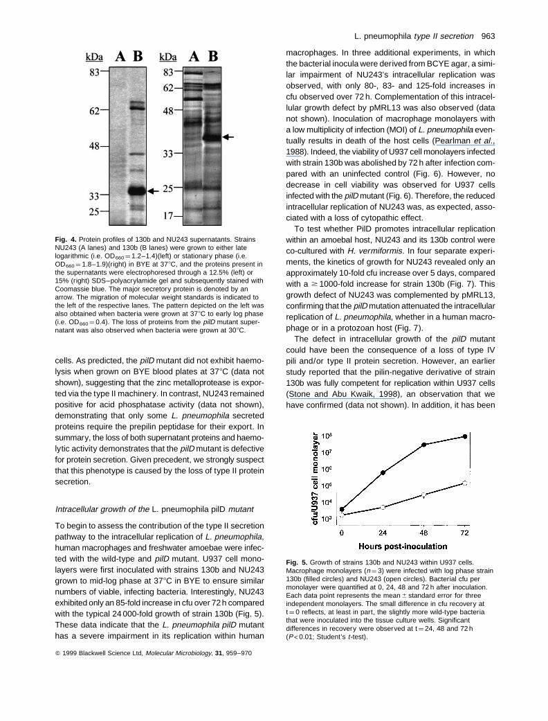
cells. As predicted, the pilD mutant did not exhibit haemo-lysis when grown on BYE blood plates at 378C (data notshown), suggesting that the zinc metalloprotease is expor-ted via the type II machinery. In contrast, NU243 remainedpositive for acid phosphatase activity (data not shown),demonstrating that only some L. pneumophila secretedproteins require the prepilin peptidase for their export. Insummary, the loss of both supernatant proteins and haemo-lytic activity demonstrates that the pilD mutant is defectivefor protein secretion. Given precedent, we strongly suspectthat this phenotype is caused by the loss of type II proteinsecretion.
Intracellular growth of the L. pneumophila pilD mutant
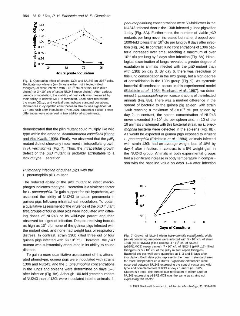
To begin to assess the contribution of the type II secretionpathway to the intracellular replication of L. pneumophila,human macrophages and freshwater amoebae were infec-ted with the wild-type and pilD mutant. U937 cell mono-layers were first inoculated with strains 130b and NU243grown to mid-log phase at 378C in BYE to ensure similarnumbers of viable, infecting bacteria. Interestingly, NU243exhibited only an 85-fold increase in cfu over 72 h comparedwith the typical 24 000-fold growth of strain 130b (Fig. 5).These data indicate that the L. pneumophila pilD mutanthas a severe impairment in its replication within human
macrophages. In three additional experiments, in whichthe bacterial inocula were derived from BCYE agar, a simi-lar impairment of NU243’s intracellular replication wasobserved, with only 80-, 83- and 125-fold increases incfu observed over 72 h. Complementation of this intracel-lular growth defect by pMRL13 was also observed (datanot shown). Inoculation of macrophage monolayers witha low multiplicity of infection (MOI) of L. pneumophila even-tually results in death of the host cells (Pearlman et al.,1988). Indeed, the viability of U937 cell monolayers infectedwith strain 130b was abolished by 72 h after infection com-pared with an uninfected control (Fig. 6). However, nodecrease in cell viability was observed for U937 cellsinfected with the pilD mutant (Fig. 6). Therefore, the reducedintracellular replication of NU243 was, as expected, asso-ciated with a loss of cytopathic effect.
To test whether PilD promotes intracellular replicationwithin an amoebal host, NU243 and its 130b control wereco-cultured with H. vermiformis. In four separate experi-ments, the kinetics of growth for NU243 revealed only anapproximately 10-fold cfu increase over 5 days, comparedwith a $ 1000-fold increase for strain 130b (Fig. 7). Thisgrowth defect of NU243 was complemented by pMRL13,confirming that the pilD mutation attenuated the intracellularreplication of L. pneumophila, whether in a human macro-phage or in a protozoan host (Fig. 7).
The defect in intracellular growth of the pilD mutantcould have been the consequence of a loss of type IVpili and/or type II protein secretion. However, an earlierstudy reported that the pilin-negative derivative of strain130b was fully competent for replication within U937 cells(Stone and Abu Kwaik, 1998), an observation that wehave confirmed (data not shown). In addition, it has been
Q 1999 Blackwell Science Ltd, Molecular Microbiology, 31, 959–970
Fig. 4. Protein profiles of 130b and NU243 supernatants. StrainsNU243 (A lanes) and 130b (B lanes) were grown to either latelogarithmic (i.e. OD660 ¼ 1.2–1.4)(left) or stationary phase (i.e.OD660 ¼ 1.8–1.9)(right) in BYE at 378C, and the proteins present inthe supernatants were electrophoresed through a 12.5% (left) or15% (right) SDS–polyacrylamide gel and subsequently stained withCoomassie blue. The major secretory protein is denoted by anarrow. The migration of molecular weight standards is indicated tothe left of the respective lanes. The pattern depicted on the left wasalso obtained when bacteria were grown at 378C to early log phase(i.e. OD660 ¼ 0.4). The loss of proteins from the pilD mutant super-natant was also observed when bacteria were grown at 308C.
Fig. 5. Growth of strains 130b and NU243 within U937 cells.Macrophage monolayers (n ¼ 3) were infected with log phase strain130b (filled circles) and NU243 (open circles). Bacterial cfu permonolayer were quantified at 0, 24, 48 and 72 h after inoculation.Each data point represents the mean 6 standard error for threeindependent monolayers. The small difference in cfu recovery att ¼ 0 reflects, at least in part, the slightly more wild-type bacteriathat were inoculated into the tissue culture wells. Significantdifferences in recovery were observed at t ¼ 24, 48 and 72 h(P<0.01; Student’s t-test).
L. pneumophila type II secretion 963
demonstrated that the pilin mutant could multiply like wildtype within the amoeba Acanthamoeba castellanii (Stoneand Abu Kwaik, 1998). Finally, we observed that the pilEL
mutant did not show any impairment in intracellular growthin H. vermiformis (Fig. 7). Thus, the intracellular growthdefect of the pilD mutant is probably attributable to alack of type II secretion.
Pulmonary infection of guinea pigs with theL. pneumophila pilD mutant
The reduced ability of the pilD mutant to infect macro-phages indicates that type II secretion is a virulence factorfor L. pneumophila. To gain support for this hypothesis, weassessed the ability of NU243 to cause pneumonia inguinea pigs following intratracheal inoculation. To obtaina qualitative assessment of the virulence of the pilD mutantfirst, groups of four guinea pigs were inoculated with differ-ing doses of NU243 or its wild-type parent and thenobserved for signs of infection. Despite receiving inoculaas high as 108 cfu, none of the guinea pigs infected withthe mutant died, and none had weight loss or respiratorydistress. In contrast, strain 130b killed three out of fourguinea pigs infected with 6 ×10⁶ cfu. Therefore, the pilDmutant was substantially attenuated in its ability to causedisease.
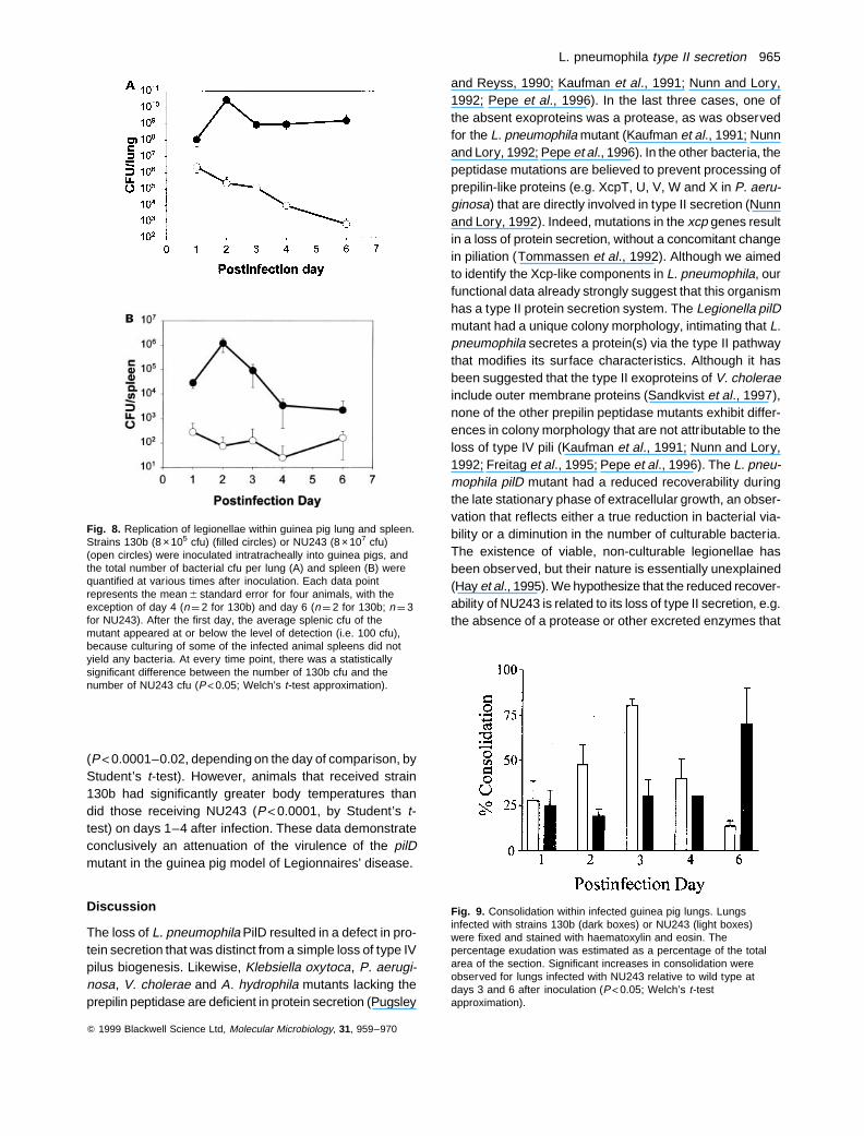
To gain a more quantitative assessment of this attenu-ated phenotype, guinea pigs were inoculated with strains130b and NU243, and the L. pneumophila concentrationsin the lungs and spleens were determined on days 1–6after infection (Fig. 8A). Although 100-fold greater numbersof NU243 than of 130b were inoculated into the animals, L.
pneumophila lung concentrations were 50-fold lower in theNU243-infected than in the 130b-infected guinea pigs after1 day (Fig. 8A). Furthermore, the number of viable pilDmutants per lung never increased but rather dropped over1000-fold to less than 103 cfu per lung by 6 days after infec-tion (Fig. 8A). In contrast, lung concentrations of 130b bac-teria increased over time, reaching a maximum of over1010 cfu per lung by 2 days after infection (Fig. 8A). Histo-logical examination of lungs revealed a greater degree ofexudation in animals infected with the pilD mutant thanwith 130b on day 3. By day 6, there was resolution ofthis lung consolidation in the pilD group, but a high degreeof consolidation in the 130b group (Fig. 9). As systemicbacterial dissemination occurs in this experimental model(Edelstein et al., 1984; Reinhardt et al., 1987), we deter-mined L. pneumophila spleen concentrations of the infectedanimals (Fig. 8B). There was a marked difference in thespread of bacteria to the guinea pig spleen, with strain130b reaching a maximum of 2 ×10⁶ cfu per spleen byday 2. In contrast, the spleen concentration of NU243never exceeded 8 ×10² cfu per spleen and, in 10 of the19 animals challenged with this bacterial strain, no L. pneu-mophila bacteria were detected in the spleens (Fig. 8B).As would be expected in guinea pigs exposed to virulentL. pneumophila (Edelstein et al., 1984), animals infectedwith strain 130b had an average weight loss of 18% byday 4 after infection, in contrast to a 5% weight gain inthe NU243 group. Animals in both experimental groupshad a significant increase in body temperature in compari-son with the baseline value on days 1–4 after infection
Q 1999 Blackwell Science Ltd, Molecular Microbiology, 31, 959–970
Fig. 6. Cytopathic effect of strains 130b and NU243 on U937 cells.Replicate monolayers (n ¼ 6) were either not infected (filledtriangles) or were infected with 8 ×10⁶ cfu of strain 130b (filledcircles) or 3 ×10⁶ cfu of strain NU243 (open circles). After variousperiods of incubation, the viability of host cells was measured bytheir ability to convert MTT to formazan. Each point representsthe mean OD540, and vertical bars indicate standard deviations.Differences in cytopathic effect between strains was significant at72 h and 96 h after inoculation (P< 0.0001, Student’s t-test). Thesedifferences were observed in two additional experiments.
Fig. 7. Growth of NU243 within Hartmannella vermiformis. Wells(n ¼ 4) containing amoebae were infected with 5 ×10⁵ cfu of strain130b (pBBR1MCS) (filled circles), 4 ×10⁵ cfu of NU243(pBBR1MCS) (open circles), 7 ×10⁵ cfu of NU243 (pMRL13) (filledtriangles) or 5 ×10⁵ cfu of the pilEL mutant (open triangles).Bacterial cfu per well were quantified at 1, 3 and 5 days afterinoculation. Each data point represents the mean 6 standard errorfor three independent co-cultures. Significant differences wereobserved between NU243 expressing the control vector and wildtype and complemented NU243 at days 3 and 5 (P<0.05;Student’s t-test). The intracellular replication of either 130b orNU243 expressing pBBR1MCS was the same as strains notexpressing this vector.
964 M. R. Liles, P. H. Edelstein and N. P. Cianciotto
(P<0.0001–0.02, depending on the day of comparison, byStudent’s t-test). However, animals that received strain130b had significantly greater body temperatures thandid those receiving NU243 (P<0.0001, by Student’s t-test) on days 1–4 after infection. These data demonstrateconclusively an attenuation of the virulence of the pilDmutant in the guinea pig model of Legionnaires’ disease.
Discussion
The loss of L. pneumophila PilD resulted in a defect in pro-tein secretion that was distinct from a simple loss of type IVpilus biogenesis. Likewise, Klebsiella oxytoca, P. aerugi-nosa, V. cholerae and A. hydrophila mutants lacking theprepilin peptidase are deficient in protein secretion (Pugsley
and Reyss, 1990; Kaufman et al., 1991; Nunn and Lory,1992; Pepe et al., 1996). In the last three cases, one ofthe absent exoproteins was a protease, as was observedfor the L. pneumophila mutant (Kaufman et al., 1991; Nunnand Lory, 1992; Pepe et al., 1996). In the other bacteria, thepeptidase mutations are believed to prevent processing ofprepilin-like proteins (e.g. XcpT, U, V, W and X in P. aeru-ginosa) that are directly involved in type II secretion (Nunnand Lory, 1992). Indeed, mutations in the xcp genes resultin a loss of protein secretion, without a concomitant changein piliation (Tommassen et al., 1992). Although we aimedto identify the Xcp-like components in L. pneumophila, ourfunctional data already strongly suggest that this organismhas a type II protein secretion system. The Legionella pilDmutant had a unique colony morphology, intimating that L.pneumophila secretes a protein(s) via the type II pathwaythat modifies its surface characteristics. Although it hasbeen suggested that the type II exoproteins of V. choleraeinclude outer membrane proteins (Sandkvist et al., 1997),none of the other prepilin peptidase mutants exhibit differ-ences in colony morphology that are not attributable to theloss of type IV pili (Kaufman et al., 1991; Nunn and Lory,1992; Freitag et al., 1995; Pepe et al., 1996). The L. pneu-mophila pilD mutant had a reduced recoverability duringthe late stationary phase of extracellular growth, an obser-vation that reflects either a true reduction in bacterial via-bility or a diminution in the number of culturable bacteria.The existence of viable, non-culturable legionellae hasbeen observed, but their nature is essentially unexplained(Hay et al., 1995). We hypothesize that the reduced recover-ability of NU243 is related to its loss of type II secretion, e.g.the absence of a protease or other excreted enzymes that
Q 1999 Blackwell Science Ltd, Molecular Microbiology, 31, 959–970
Fig. 8. Replication of legionellae within guinea pig lung and spleen.Strains 130b (8 ×10⁵ cfu) (filled circles) or NU243 (8 ×10⁷ cfu)(open circles) were inoculated intratracheally into guinea pigs, andthe total number of bacterial cfu per lung (A) and spleen (B) werequantified at various times after inoculation. Each data pointrepresents the mean 6 standard error for four animals, with theexception of day 4 (n ¼ 2 for 130b) and day 6 (n ¼ 2 for 130b; n ¼ 3for NU243). After the first day, the average splenic cfu of themutant appeared at or below the level of detection (i.e. 100 cfu),because culturing of some of the infected animal spleens did notyield any bacteria. At every time point, there was a statisticallysignificant difference between the number of 130b cfu and thenumber of NU243 cfu (P<0.05; Welch’s t-test approximation).
Fig. 9. Consolidation within infected guinea pig lungs. Lungsinfected with strains 130b (dark boxes) or NU243 (light boxes)were fixed and stained with haematoxylin and eosin. Thepercentage exudation was estimated as a percentage of the totalarea of the section. Significant increases in consolidation wereobserved for lungs infected with NU243 relative to wild type atdays 3 and 6 after inoculation (P<0.05; Welch’s t-testapproximation).
L. pneumophila type II secretion 965
contribute to nutrient acquisition could alter the phenotypeof late stationary phase cultures. Alternatively, the modestdrop in mutant recovery could have been caused by anaccumulation of prepilin-like proteins or type II exoproteinsin the inner membrane or periplasmic space. However,abnormal protein accumulation has not been shown to betoxic to any of the other prepilin peptidase mutants (Kauf-man et al., 1991; Nunn and Lory, 1992; Freitag et al., 1995;Pepe et al., 1996).
Our data indicate, for the first time, that a prepilin pepti-dase and type II protein secretion function in an intracellularparasite. This observation is made even more interestingbecause of the greatly reduced intracellular replication ofthe pilD mutant within the evolutionarily divergent macro-phage and amoeba hosts. Because the pilin mutant wasfully competent for growth within host cells, the defectiveintracellular infectivity of NU243 can be attributed to itsalteration in protein secretion. As mutants specifically defi-cient in zinc metalloprotease expression replicate like wildtype in macrophages and amoebae (Szeto and Shuman,1990), the attenuated phenotype of NU243 cannot be aresult of its loss of one proteolytic activity. The intracellulargrowth kinetics of the pilD mutant indicates that type IIsecreted proteins are most important to events that occurrelatively late in the infective process. For example, asmonolayers infected with NU243 and wild-type bacteriayielded similar numbers of cfu at the earliest of time points,we suspect that type II secretion is not significantly involvedin entry and immediate intracellular survival. On the otherhand, type II exoproteins could have major roles in intracel-lular nutrient acquisition or host cell lysis. It is possible,although unlikely, as noted above, that the reduced intra-cellular growth of the mutant results from an accumulationof prepilin-like proteins or type II exoproteins in the bac-terial envelope. While the type II secreted factors that arecrucial for the intracellular replication of L. pneumophilawill be the focus of our future research efforts, it will beimportant for others to ascertain whether additional intra-cellular parasites require type II secreted proteins.
The contribution of L. pneumophila PilD to in vivo patho-genesis is certainly significant, considering the sharplyattenuated virulence of NU243 within guinea pigs. Thisattenuation is undoubtedly caused, at least in part, bythe mutant’s defect in intracellular replication. Like the pre-vious observations of extracellular growth, this is the firstoccasion that a prepilin peptidase mutant has been testedin an animal model. Interestingly, the pilD mutant appearedto be more attenuated in vivo than it did in vitro. There areseveral conceivable explanations for this result. First, theloss of factors that provide resistance to complement orpolymorphonuclear leucocytes could have reduced theextracellular survival of the mutant in the mammalianlung. Secondly, the loss of type IV pili might have reducedthe adherence of the pilD mutant to the alveolar epithelium,
a process that has been suggested to promote L. pneumo-phila virulence (Mody et al., 1993; Cianciotto et al., 1995;Stone and Abu Kwaik, 1998). Thirdly, the loss of the zincmetalloprotease would be expected to lead to partial attenua-tion of the pulmonary infection (Moffat et al., 1994). Finally,the pilD mutant may be hypersensitive to the enhancedbacteriocidal activity that exists within activated alveolarmacrophages. Regardless, these data indicate that factorssecreted by the L. pneumophila type II secretion systemare not only promoters of extracellular survivability andintracellular growth but also key potentiators of virulence.
Experimental procedures
Bacterial strains and media
The L. pneumophila pilD mutant as well as the cloned Legio-nella pilD gene was derived from serogroup 1 strain 130b(Wadsworth) (Hickey and Cianciotto, 1997). The pilEL mutant,also a derivative of strain 130b, was obtained from B. Stoneand Y. Abu Kwaik (Stone and Abu Kwaik, 1998). The wild-type strain was maintained in a virulent state by passagethrough a guinea pig and stored as a spleen homogenate at¹708C. Before experimentation, L. pneumophila was gener-ally cultured on buffered charcoal–yeast extract (BCYE) agarfor 3 days at 378C (Edelstein, 1981). When appropriate,3 mg ml¹1 chloramphenicol (Chl), 25 mg ml¹1 kanamycin (Kan)or 5% (w/v) sucrose (Suc) was added to the medium. To assessL. pneumophila extracellular growth kinetics, bacteria grownovernight in buffered yeast extract (BYE) were subculturedinto 2.5 ml of BYE to an optical density at 660 nm (OD660) ofapproximately 0.1. Cultures were then incubated in an orbitalincubator-shaker (LabLine Instruments) set at 250 r.p.m. andeither 308C or 378C. Growth was assessed at various timeintervals by recording the OD660 and by plating onto BCYEagar. Additionally, the number of bacterial particles was deter-mined with a Petroff–Hauser counter (C. A. Hausser and Son),using crystal violet as the stain.
E. coli strain XL1 Blue (Stratagene) served as the host forrecombinant plasmids (Yang, 1992). Plasmid DNAs wereobtained from E. coli by the alkaline lysis method and wereintroduced by transformation into CaCl2-treated E. coli (Ausu-bel et al., 1987). E. coli were grown on Luria–Bertani agar(Ausubel et al., 1987), which contained Kan (50 mg ml¹1) orChl (30 mg ml¹1) when appropriate.
Directed mutagenesis of L. pneumophila pilD
The chromosomally encoded pilD gene was insertionally inac-tivated by allelic exchange (O’Connell et al., 1995). First, aTn903-derived kanamycin resistance gene (kanR) was isolatedas an AccI fragment from pMB2190 (Grindley and Joyce,1980) and ligated into the single NarI site of the cloned pilDon pML219 (Liles et al., 1998). Subsequently, a 9 kb SphI frag-ment containing pilD:kanR plus surrounding L. pneumophilaDNA was ligated into the SphI site of the counterselectablepBOC20, generating pMRL11. The suicide vector pBOC20contains a Chl resistance marker and sacB, which can coun-terselect for plasmid maintenance by encoding Suc sensitivity
Q 1999 Blackwell Science Ltd, Molecular Microbiology, 31, 959–970
966 M. R. Liles, P. H. Edelstein and N. P. Cianciotto
(O’Connell et al., 1995). Next, pMRL11 was electroporatedinto strain 130b, and transformants that may have undergonethe desired double cross-over event were selected on BCYEcontaining Kan and Suc (Hickey and Cianciotto, 1997). Finally,genomic DNA was isolated from doubly resistant strains exhi-biting Chl sensitivity (Hickey and Cianciotto, 1997), and allelicexchange was verified by Southern hybridization analysisusing the Genius system kit (Boehringer Mannheim) and apilD-specific probe. To help to generate the probe, primersF3 (58-GGATTGGTGGTGGGTATG) and R34 (58-CAAGAG-CTAATGGCCAGAAG) were used to polymerase chain reac-tion (PCR) amplify pilD from strain 130b DNA (Liles et al.,1998). The reaction contained 100 ng of genomic DNA, with3 mM MgCl2, 25 pM each primer, 0.2 mM dNTPs and 1 unitof Taq polymerase (Gibco BRL) and was conducted with 45 smelting at 948C, followed by 45 s annealing at 508C and 90 selongation at 728C for 30 cycles.
Trans-complementation of the L. pneumophila pilDmutation
To help to determine whether the mutant’s phenotypes werecaused by the loss of PilD and not a second site mutation,a pilD-containing plasmid was constructed. First, the 1.3 kbpilD-specific PCR product (see above) was cloned into the vec-tor pCR2.1 (Clontech Laboratories). The pilD gene was thenremoved as a KpnI/XbaI fragment and ligated into the broad-host-range vector pBBR1MCS (Kovach et al., 1995), generat-ing pMRL13. The orientation of pilD in pMRL13 was in thesame direction as lacZ transcription, permitting expressionof the gene in the absence of the pilBCD promoter. The pilDmutant was electroporated with pMRL13 or pBBR1MCS asa control, and colonies were selected on BCYE containingChl. A Southern blot of HindIII-digested DNA isolated fromtransformants was hybridized with the pilD probe to verify thepresence of pMRL13 and its mutated pilD. Passage of NU243(pMRL13) on BCYE agar without antibiotic selection resultedin plasmid loss. Thus, NU243 (pMRL13), NU243 (pBBR1MCS)and 130b (pBBR1MCS) were always grown on media contain-ing Chl.
Electron microscopic examination of L. pneumophila
Pili on the surface of L. pneumophila were visualized as before(Liles et al., 1998). Strains were grown on BCYE agar, and thenisolated colonies were suspended in PBS and adhered to For-mvar-coated copper grids (Ladd Industries). Bacteria adherentto the grid were stained using 10 ml of 1% phosphotungsticacid for 2 min and visualized on a Jeol JEM-100 CxII transmis-sion electron microscope at 60 kV.
SDS–PAGE analysis of proteins in L. pneumophilasupernatants
Excreted proteins of L. pneumophila were analysed essen-tially as described previously (Keen and Hoffman, 1989). Bac-teria were grown for up to 18 h at 308C or 378C in 2 l flaskscontaining 250 ml of aerated BYE. Supernatants were iso-lated by centrifugation of the cultures at 12 000 ×g at 48C, fol-lowed by filtration through 0.2 mm membranes. The samples
were concentrated to 10 ml with a 1 kDa (YM1 membrane)ultrafiltration cell (Millipore), and then the concentrates weredialysed against three 1 l volumes of 50 mM Tris buffer con-taining 5 mM EDTA (pH 7.8). The amount of protein in eachsample was determined by the Lowry assay (Ausubel et al.,1987). Samples containing equivalent amounts of protein inSDS–loading buffer were then boiled for 5 min before separa-tion on either a 12.5% or a 15% SDS–polyacrylamide gel runat 25 V for 15 h at 158C (Ausubel et al., 1987). After electro-phoresis, protein bands were stained with Coomassie bluefor visualization and compared with molecular weight stan-dards (New England Biolabs) (Ausubel et al., 1987).
Detection of L. pneumophila enzymatic activities
To observe the haemolytic activity resulting from the zinc metal-loprotease of L. pneumophila (Keen and Hoffman, 1989),strains were grown on BYE agar supplemented with 5% defi-brinated sheep erythrocytes and examined daily for evidenceof b-haemolysis. Acid phosphatase activity was detected bylifting L. pneumophila colonies from BCYE agar on a nitrocel-lulose filter onto an indicator plate containing 0.8% agaroseand 40 mg ml¹1 5-bromo-4-chloro-3-indolyl phosphate (pH 5.0)(Albano et al., 1992), with a positive reaction generating ablue colour. Liquid phosphatase assays were conducted onconcentrated bacterial supernatants using the Sigma 104phosphatase substrate (Albano et al., 1992).
Intracellular infection of U937 cells and amoebae byL. pneumophila
Human U937 cells, which differentiate into adherent, macro-phage-like cells after treatment with phorbol esters, havebeen used by many investigators to study the intracellularreplication of L. pneumophila (Pearlman et al., 1988; Cianciottoet al., 1990; Fields et al., 1990; Hacker et al., 1991; Bergerand Isberg, 1993). U937 cell monolayers were prepared andinfected as described previously (Cianciotto et al., 1990; Pearl-man et al., 1988). The number of viable bacteria added to themacrophage monolayers was determined by plating inoculaon BCYE agar. After inoculation with approximately 106 cfu,replicate monolayers (n ¼ 3) consisting of 106 U937 cellswere incubated for 2 h to permit bacterial uptake and thenwashed to remove unattached bacteria. The infected mono-layers were then incubated at 378C in RPMI-1640 mediumsupplemented with heat-inactivated 5% fetal bovine serum(FBS; Gibco BRL). After incubation for 0–72 h, the monolayerswere lysed in 0.1% saponin (Sigma), an agent that acts uponcholesterol and hence does not alter the viability of bacteria(Leung et al., 1997). Finally, 10-fold serial dilutions of thelysates were plated on BCYE agar. The resulting cfu wereused to calculate the corresponding numbers of bacteria permonolayer. As L. pneumophila does not replicate in the tissueculture medium, all cfu recovered represent bacteria that havereplicated intracellularly. For the assessment of the cytopathiceffect of strains for U937 cells, infected monolayers (n ¼ 6)were treated with the vital stain 3-[4,5-dimethylthiazol-2-yl]-2,5-diphenyltetrazoliumbromide as described previously (Pearl-man et al., 1988). To each well was added 5 ml of 5 mg ml¹1 ofthe stain and, after 4 h of incubation, the formazan product
Q 1999 Blackwell Science Ltd, Molecular Microbiology, 31, 959–970
L. pneumophila type II secretion 967
was dissolved in acid isopropanol. The OD570 of individualwells was then determined.
To assess the ability of legionellae to grow within protozoa,Hartmannella vermiformis was used as a host, as performedby various investigators (Fields et al., 1986; Cianciotto andFields, 1992; Venkataraman et al., 1997). The co-culturewas carried out as described previously (Cianciotto and Fields,1992). Briefly, approximately 105 amoebae were suspendedin 2 ml of axenic medium 1034 without FBS that was diluted1:1 with Puck’s saline and added to replicate wells (n ¼ 4) ina 24-well tissue culture plate. Next, bacteria suspended inassay medium were added at a final concentration of 105 cfuml¹1, and the co-cultures were incubated for 5 days at 358C.Numbers of bacteria in each well were determined at varioustime points by plating dilutions on BCYE agar. As the assaymedium does not support the growth or survival of L. pneumo-phila, the cfu represent bacteria that have replicated withinthe amoebae.
Experimental infection of guinea pigs by L. pneumophila
To assess the virulence of L. pneumophila strains, guinea pigswere infected intratracheally with the bacteria as describedpreviously (Winn et al., 1982; Edelstein et al., 1984; Cianciottoet al., 1990); this mode of infection produces bilateral pneumo-nia similar to that seen in immunocompromised patients withLegionnaires’ disease (Winn et al., 1982; Edelstein et al.,1984). Inocula for infection were prepared by first culturingthe bacteria on BCYE agar, then in BYE broth, at 378C in ashaking incubator (Edelstein et al., 1984). Mid-log phase cul-tures (108 cfu ml¹1) were used. The bacteria were then sus-pended at the desired concentrations in sterile water forinjection. Bacteria were injected into the surgically exposedtracheas in 0.3 ml volumes using a syringe and needle. Thebacterial inoculum delivered was determined by quantitativeplating of the inoculum onto BCYE agar.
Two animal studies were performed, i.e. an infectious dosetitration study and a bacterial clearance study. Both studiesused Hartley male guinea pigs weighing 250–300 g. For thetitration study, animals in randomly assigned groups of foureach were infected with either 1 ×10⁶, 1 ×10⁷ or 1 ×10⁸ cfuof NU243. The animals were observed clinically for 7 days,with daily measurements of weight and temperature for thefirst 5 days. A previous study using the same methods showedthat the parent strain 130b strain killed three out of four animalsgiven 6 × 106 cfu by 4 days after infection, and no animals given1 ×105 cfu (unpublished data). The bacterial clearance studywas performed by infecting 16 guinea pigs with 8 ×10⁵ cfuof 130b and 19 animals with 8 ×10⁷ cfu of NU243. The groupassignments were random. The body weight and rectal tem-perature of each animal were measured daily. Four animalsin each group were sacrificed daily for 3 days, starting 1 dayafter infection. Animals were killed in numerical order toavoid bias resulting from degree of illness. Two animalsreceiving 130b were killed on day 4 and two on day 6; therespective numbers for NU243 were four and three. The rightlower lobe of the lung and the spleen were harvested asepti-cally, and their ground homogenates were cultured quantita-tively on BCYE agar. The lower limit of detection of bacteriain the organs was about 100 cfu per organ. The remaininglungs were fixed in cold (48C) 10% buffered formalin for 1
week before dissection (Edelstein et al., 1984). Each day,one set of animal tissues from NU243 recipients was also cul-tured quantitatively on BCYE agar containing Kan to look forrevertants. Histology was performed on the fixed lungs ofall animals, which were coded to obscure animal group.Three or four of these sections were stained per lung with hae-matoxylin and eosin and examined microscopically at 25×magnification. The average amount of consolidation in the sec-tions was estimated as a percentage of the total area of thesection (Edelstein et al., 1984). The code of treatment groupidentity was broken only after all results had been recorded.
Acknowledgements
The authors thank Yousef Abu Kwaik and Barbara Stone fortheir contribution of the pilin mutant for study, Carmela Ruffolo,Mark Strom, and V. K. Viswanathan for many helpful discus-sions, and Virginia Aragon, Martha Edelstein, Jianjun Ren andDavid Moon for expert technical assistance. This work wassupported by NIH grant AI34937.
References
Abu Kwaik, Y. (1996) The phagosome containing Legionellapneumophila within the protozoan Hartmanella vermiformisis surrounded by the rough endoplasmic reticulum. ApplEnviron Microbiol 62: 2022–2028.
Albano, M.A., Arroyo, J., Eisenstein, B.I., and Engleberg, N.C.(1992) PhoA gene fusions in Legionella pneumophila gener-ated in vivo using a new transposon, MudphoA. Mol Micro-biol 6: 1829–1839.
Anderson, D.M., and Schneewind, O. (1997) A mRNA signalfor the type III secretion of Yop proteins by Yersinia entero-colitica. Science 278: 1140–1143.
Ausubel, F.M., Brent, R.B., Kingston, R.E., Moore, D.D.,Seidman, J.G., Smith, J.A., et al. (eds) (1987) Current Pro-tocols in Molecular Biology. New York: John Wiley & Sons.
Baine, W.B. (1985) Cytolytic and phospholipase C activity inLegionella species. J Gen Microbiol 131: 1383–1391.
Bally, M., Ball, G., Badere, A., and Lazdunski, A. (1991) Pro-tein secretion in Pseudomonas aeruginosa: the xcpA geneencodes an integral inner membrane protein homologousto Klebsiella pneumoniae secretion function protein PulO.J Bacteriol 173: 479–486.
Bally, M., Filloux, A., Akrim, M., Ball, G., Lazdunski, A., andTommassen, J. (1992) Protein secretion in Pseudomonasaeruginosa: characterization of seven xcp genes and pro-cessing of secretory apparatus components by prepilinpeptidase. Mol Microbiol 6: 1121–1131.
Berger, K.H., and Isberg, R.R. (1993) Two distinct defects inintracellular growth complemented by a single geneticlocus in Legionella pneumophila. Mol Microbiol 7: 7–19.
Black, W.J., Frederick, D.Q., and Tompkins, L.S. (1990)Legionella pneumophila zinc metalloprotease is structurallyand functionally homologous to Pseudomonas aeruginosaelastase. J Bacteriol 172: 2608–2613.
Bleves, S., Voulhoux, R., Michel, G., Lazdunskim, A., Tom-massen, J., and Filloux, A. (1998) The secretion apparatusof Pseudomonas aeruginosa: identification of a fifth pseudo-pilin, XcpX (GspK family). Mol Microbiol 27: 31–40.
Q 1999 Blackwell Science Ltd, Molecular Microbiology, 31, 959–970
968 M. R. Liles, P. H. Edelstein and N. P. Cianciotto
Cianciotto, N.P., and Fields, B.S. (1992) Legionella pneumo-phila mip gene potentiates intracellular infection of protozoaand human macrophages. Proc Natl Acad Sci USA 89:5188–5191.
Cianciotto, N.P., Eisenstein, B.I., Mody, C.H., and Engleberg,N.C. (1990) A mutation in the mip gene results in anattenuation of Legionella pneumophila virulence. J InfectDis 162: 121–126.
Cianciotto, N.P., Stamos, J.K., and Kamp, D.W. (1995) Infec-tivity of Legionella pneumophila mip mutant for alveolarepithelial cells. Curr Microbiol 30: 247–250.
Conlan, J.W., Baskerville, A., and Ashworth, L.A.E. (1988)Separation of Legionella pneumophila proteases and puri-fication of a protease which produces lesions like those ofLegionnaires’ disease in guinea pig lung. J Gen Microbiol134: 481–487.
Dowling, J.N., Saha, A.K., and Glew, R.H. (1992) Virulencefactors of the family Legionellaceae. Microbiol Rev 56:32–60.
Edelstein, P.H. (1981) Improved semiselective medium forisolation of Legionella pneumophila from contaminatedclinical and environmental specimens. J Clin Microbiol 14:298–303.
Edelstein, P.H., Calarco, K., and Yasui, V.K. (1984) Anti-microbial therapy of experimentally induced Legionnaires’disease in guinea pigs. Am Rev Resp Dis 130: 849–856.
Fields, B.S. (1996) The molecular ecology of legionellae.Trends Microbiol 4: 286–290.
Fields, B.S., Barbaree, J.M., Shotts, Jr, E.B., Feeley, J.C.,Morrill, W.E., Sanden, G.N., et al. (1986) Comparison ofguinea pig and protozoan models for determining virulenceof Legionella species. Infect Immun 53: 553–559.
Fields, B.S., Barbaree, J.M., Sanden, G.N., and Morrill, W.E.(1990) Virulence of a Legionella anisa strain associatedwith Pontiac fever: an evaluation using protozoan, cell cul-ture, and guinea pig models. Infect Immun 58: 3139–3142.
Finlay, B.B., and Falkow, S. (1997) Common themes inmicrobial pathogenicity revisited. Microbiol Mol Biol Rev61: 136–169.
Freitag, N.E., Seifert, H.S., and Koomey, M. (1995) Charac-terization of the pilF–pilD pilus assembly locus of Neis-seria gonorrhoeae. Mol Microbiol 16: 575–586.
Grindley, N.D., and Joyce, C.M. (1980) Genetic and DNAsequence analysis of the kanamycin resistance transposonTn903. Proc Natl Acad Sci USA 77: 7176–7180.
Hacker, J., Ott, M., Ludwig, B., and Rdest, U. (1991) Intracel-lular survival and expression of virulence determinants ofLegionella pneumophila. Infection 19: S198–S201.
Hay, J., Seal, D.V., Billcliffe, B., and Freer, J.H. (1995) Non-culturable Legionella pneumophila associated with Acantha-moeba castellanii : detection of the bacterium using DNAamplification and hybridization. J Appl Bacteriol 78: 61–65.
Hickey, E.K., and Cianciotto, N.P. (1997) An iron- and Fur-repressed Legionella pneumophila gene that promotesintracellular infection and encodes a protein with similarityto the Escherichia coli aerobactin synthetases. Infect Immun65: 133–143.
Horwitz, M.A. (1983) The Legionnaires’ disease bacterium(Legionella pneumophila) inhibits phagosome–lysosomefusion in human monocytes. J Exp Med 158: 2108–2126.
Horwitz, M.A., and Silverstein, S.C. (1980) The Legionnaires’
disease bacterium (Legionella pneumophila) multipliesintracellularly in human monocytes. J Clin Invest 66: 441–450.
Hueck, C.J. (1998) Type III protein secretion systems in bac-terial pathogens of animals and plants. Microbiol Mol BiolRev 62: 379–433.
Kaufman, M.R., Seyer, J.M., and Taylor, R.K. (1991) Process-ing of TCP pilin by TcpJ typifies a common step intrinsic toa newly recognized pathway of extracellular protein secre-tion by Gram-negative bacteria. Genes Dev 5: 1834–1846.
Keen, M.G., and Hoffman, P.S. (1989) Characterization of aLegionella pneumophila extracellular protease exhibitinghemolytic and cytotoxic activities. Infect Immun 57: 732–738.
Kovach, M.E., Elzer, P.H., Hill, D.S., Robertson, G.T., Farris,M.A., Roop, R.M., et al. (1995) Four new derivatives of thebroad-host-range cloning vector pBBR1MCS, carrying dif-ferent antibiotic-resistance cassettes. Gene 166: 175–176.
Leung, Y.M., Ou, Y.J., Kwan, C.Y., and Loh, T.T. (1997)Specific interaction between tetrandrine and Quillaja sapo-nins in promoting permeabilization of plasma membrane inhuman leukemic HL-60 cells. Biochim Biophys Acta 1325:318–328.
Liles, M.R., Viswanathan, V.K., and Cianciotto, N.P. (1998)Identification and temperature regulation of Legionella pneu-mophila genes involved in type IV pilus biogenesis and typeII secretion. Infect Immun 66: 1776–1782.
McDade, J.E., Shepard, C.C., Fraser, D.W., Tsai, T.R.,Redus, M.A., and Dowdle, W.R. (1977) Legionnaires’disease: isolation of a bacterium and demonstration ofits role in other respiratory disease. N Engl J Med 297:1197–1203.
Mody, C.H., Paine, III, R., Shahrabadi, M.S., Simon, R.H.,Pearlman, E., Eisenstein, B.I. et al. (1993) Legionellapneumophila replicates within rat alveolar epithelial cells.J Infect Dis 167: 1138–1145.
Moffat, J.F., Edelstein, P.H., Regula, Jr, D.P., Cirillo, J.D.,and Tompkins, L.S. (1994) Effects of an isogenic Zn-metal-loprotease-deficient mutant of Legionella pneumophila in aguinea-pig pneumonia model. Mol Microbiol 12: 693–705.
Nunn, D.N., and Lory, S. (1992) Components of the proteinexport-excretion apparatus of Pseudomonas aeruginosaare processed by the type IV prepilin peptidase. ProcNatl Acad Sci USA 89: 47–51.
Nunn, D., Bergman, S., and Lory, S. (1990) Products of threeaccessory genes, pilB, pilC, and pilD, are required for bio-genesis of Pseudomonas aeruginosa pili. J Bacteriol 172:2911–2919.
O’Connell, W.A., Bangsborg, J.M., and Cianciotto, N.P. (1995)Characterization of a Legionella micdadei mip mutant. InfectImmun 63: 2840–2845.
Pearlman, E., Jiwa, A.H., Engleberg, N.C., and Eisenstein, B.I.(1988) Growth of Legionella pneumophila in a humanmacrophage-like (U937) cell line. Microb Pathogen 5: 87–95.
Pepe, C.M., Eklund, M.W., and Strom, M.S. (1996) Cloning ofan Aeromonas hydrophila type IV pilus biogenesis cluster:complementation of pilus assembly functions and charac-terization of a type IV leader peptidase/N-methyltrans-ferase required for extracellular protein secretion. MolMicrobiol 19: 857–869.
Q 1999 Blackwell Science Ltd, Molecular Microbiology, 31, 959–970
L. pneumophila type II secretion 969
Pugsley, A.P. (1993) The complete general secretory path-way in Gram-negative bacteria. Microbiol Rev 57: 50–108.
Pugsley, A.P., and Reyss, I. (1990) Five genes at the 38 endof the Klebsiella pneumoniae pulC operon are required forpullulanase secretion. Mol Microbiol 4: 365–379.
Pugsley, A.P., Francetic, O., Possot, O.M., Sauvonnet, N.,and Hardie, K.R. (1997) Recent progress and future direc-tions in studies of the main terminal branch of the generalsecretory pathway in Gram-negative bacteria – a review.Gene 192: 13–19.
Quinn, F.D., and Tompkins, L.S. (1989) Analysis of a clonedsequence of Legionella pneumophila encoding a 38 kDametalloprotease possessing haemolytic and cytotoxicactivities. Mol Microbiol 3: 797–805.
Reinhardt, J.F., Nakahama, C., and Edelstein, P.H. (1987)Comparison of blood culture methods for recovery ofLegionella pneumophila from the blood of guinea pigs withexperimental infection. J Clin Microbiol 25: 719–721.
Russel, M. (1998) Macromolecular assembly and secretionacross the bacterial cell envelope: type II protein secretionsystems. J Mol Biol 279: 485–499.
Salmond, G.P.C., and Reeves, P.J. (1993) Membrane trafficwardens and protein secretion in gram-negative bacteria.Trends Biochem Sci 18: 7–12.
Sandkvist, M., Michel, L.O., Hough, L.P., Morales, V.M.,Bagdasarian, M., Koomey, M., et al. (1997) General secre-tion pathway (eps) genes required for toxin secretion andouter membrane biogenesis in Vibrio cholerae. J Bacteriol179: 6994–7003.
Segal, G., Purcell, M., and Shuman, H.A. (1998) Host cell kill-ing and bacterial conjugation require overlapping sets ofgenes within a 22 kb region of the Legionella pneumophilagenome. Proc Natl Acad Sci USA 95: 1669–1674.
Stone, B., and Abu Kwaik, Y. (1998) Expression of multiplepili by Legionella pneumophila: identification and character-ization of a type IV pilin gene and its role in adherence tomammalian and protozoan cells. Infect Immun 66: 1768–1775.
Strom, M.S., and Lory, S. (1993) Structure–function andbiogenesis of the type IV pili. Annu Rev Microbiol 47:565–596.
Strom, M.S., Nunn, D., and Lory, S. (1991) Multiple roles ofthe pilus biogenesis protein PilD: involvement of PilD inexcretion of enzymes from Pseudomonas aeruginosa.J Bacteriol 173: 1175–1180.
Szeto, L., and Shuman, H.A. (1990) The Legionella pneumo-phila major secretory protein, a protease, is not required forintracellular growth or cell killing. Infect Immun 58: 2585–2592.
Tommassen, J., Filloux, A., Bally, M., Murgier, M., andLazdunski, A. (1992) Protein secretion in Pseudomonasaeruginosa. FEMS Microbiol Rev 103: 73–90.
Turner, L.R., Lara, J.C., Nunn, D.N., and Lory, S. (1993) Muta-tions in the consensus ATP-binding sites of XcpR and PilBeliminate extracellular protein secretion and pilus biogenesisin Pseudomonas aeruginosa. J Bacteriol 175: 4962–4969.
Venkataraman, C., Haack, B.J., Bondada, S., and AbuKwaik, Y. (1997) Identification of a Gal/GalNAc lectin inthe protozoan Hartmanella vermiformis as a potentialreceptor for attachment and invasion by the Legionnaires’disease bacterium. J Exp Med 186: 537–547.
Vogel, J.P., Andrews, H.L., Wong, S.K., and Isberg, R.R.(1998) Conjugative transfer by the virulence system ofLegionella pneumophila. Science 279: 873–876.
Wandersman, C. (1992) Secretion across the bacterial outermembrane. Trends Genet 8: 317–322.
Winans, S.C., Burns, D.L., and Christie, P.J. (1996) Adapta-tion of a conjugal transfer system for the export of patho-genic macromolecules. Trends Microbiol 4: 64–68.
Winn, Jr, W.C., Davis, G.S., Gump, D.W., Craighead, J.E.,and Beaty, H.N. (1982) Legionnaires’ pneumonia afterintratracheal inoculation of guinea pigs and rats. Lab Invest47: 568–578.
Yang, X.M. (1992) Optimization of a cultivation process forrecombinant protein production by Escherichia coli. J Bio-tech 23: 271–289.
Q 1999 Blackwell Science Ltd, Molecular Microbiology, 31, 959–970
970 M. R. Liles, P. H. Edelstein and N. P. Cianciotto


![[Current Topics in Microbiology and Immunology] || Modulation of the Ubiquitination Machinery by Legionella](https://static.fdokumen.com/doc/165x107/63328412f0080405510482a9/current-topics-in-microbiology-and-immunology-modulation-of-the-ubiquitination.jpg)