The preparation and characterisation of ceils and their ...
313
The preparation and characterisation of ceils and their scaffolds suitable for tissue repair applications Thesis submitted for the degree of Doctor of Philosophy in Biochemical Engineering by Leda Pittarello BS MSc The Advanced Centre for Biochemical Engineering Department of Biochemical Engineering University College London September 2007 1
-
Upload
khangminh22 -
Category
Documents
-
view
0 -
download
0
Transcript of The preparation and characterisation of ceils and their ...

The preparation and characterisation of ceils and their scaffolds suitable for tissue repair applications
Thesis submitted for the degree of Doctor of Philosophy
inBiochemical Engineering
byLeda Pittarello
BS MSc
The Advanced Centre for Biochemical Engineering Department of Biochemical Engineering
University College London
September 2007
1

UMI Number: U592557
All rights reserved
INFORMATION TO ALL USERS The quality of this reproduction is dependent upon the quality of the copy submitted.
In the unlikely event that the author did not send a complete manuscript and there are missing pages, these will be noted. Also, if material had to be removed,
a note will indicate the deletion.
Dissertation Publishing
UMI U592557Published by ProQuest LLC 2013. Copyright in the Dissertation held by the Author.
Microform Edition © ProQuest LLC.All rights reserved. This work is protected against
unauthorized copying under Title 17, United States Code.
ProQuest LLC 789 East Eisenhower Parkway
P.O. Box 1346 Ann Arbor, Ml 48106-1346

To my parents, Maria and Adelmo

Acknowledgments
First of all I would like to thank Prof. Anthony Hollander, who put trust in me and instilled me the idea of a PhD for the very first time.
I would like to thank also my supervisor Frank Baganz for his kind support and help, and my advisor Chris Mason for his encouragement during the writing of this thesis. I certainly could not have brought this hard work to completion without their input and kind help.
I wish also to thank Prof. Nigel Titchener-Hooker and Prof. Gary Lye for their special help in making all of this possible; Alethea Tabor for her comments on my data; Prof. Peter Dunnill for his extraordinary guidance during the first year of my PhD and for taking care of reviewing my data and part of this thesis. Special thanks also to Prof. Mike Hoare for giving me the possibility to work and study in the Department of Biochemical Engineering during the three years of my experimental work.
My thanks also to my colleagues for the great time I had in working with them in the lab: Julia Markusen and Emily Culme-Seymour, for the great collaboration on the alginate derivatisation work, and Spyros Gerontas for being always happy to help and discuss about our works. Many thanks also to my friend Anna Cariboni, who helped me with the staining and the photographs of my samples during the last stages of my experimental work.
I want to express my gratitude also to Prof. Tom Byers, who taught me most of all to “make no little plans”. Thanks to him I will always make of my aspirations true possibilities.
I thank Elena and Joseph as well as Samuela, Marta and Jim, Roberta, Anna, Kuna and Adrian for real friendship, and Duncan for being always so much fun and such a good mentor, anytime I need. I thank also my other best friends, who I have left in Italy a while ago, but always remain by my side: the “Pink Ladies” Dommi, Silvia and Stefania.
I thank my ex-flatmates Fabio Sormani, John Baker and Bruno Garcia for their incredible patience and for cheering me up during the most difficult moments of this journey.
Last but not least, I wish to thank my parents Maria and Adelmo, to whom I dedicate this work, for accepting my choice of doing a PhD and bearing with both my physical and psychological absence during these past years. To them as well as to my sister Sara and my lovely niece Ludovica (and to Brian!) I will always feel close even being far away.
I guess I cannot avoid thanking also the one and only person that was responsible for my moving to London five years ago - with all that much this has meant for both my career and personal life. Despite all, he opened up my view on the world and extended my horizon.
This PhD has been supported by the Department of Biochemical Engineering and the Engineering and Physical Sciences Research Council (EPSRC), and their support is gratefully acknowledged.
3

Abstract
In this research, a tissue engineering approach was investigated for the
fabrication of homogeneous polymer/cells scaffolds suitable for applications in the
clinic, in particular for the treatment of coronary disease and atherosclerosis.
Sodium alginate chemically modified with an RGD-containing peptide promoting
cell attachment was chosen as the natural polymer for the preparation of
matrix/cells constructs. Several conditions were investigated to optimise the
derivatisation of the biomaterial, and no major dissimilarities were identified
among alginates with different guluronic/mannuronic acid compositions and
viscosities during the derivatisation process. Successful incorporation of the
GRGDY-pentapeptide onto the alginate was achieved. Amino acid analysis, which
allowed a reliable quantification of the degree of peptide attached, demonstrated
that the complete pentapeptidic sequence was present in the alginate after
completion of the derivatisation procedure. However, a significant loss of the
peptide’s final tyrosine residue was consistently observed during derivatisation.
Studies were also performed using multipotent fibroblast-like plastic-adherent
cells recovered from frozen bone marrow samples of adult patients, here defined as
human “mesenchymal stem cells” (hMSCs). Prior to the cell immobilisation in the
alginate matrix, the optimal conditions for cell expansion in monolayer culture
were identified. Cell samples were assessed according to their percent viability at
each expansion passage, doubling time, cell growth rate and specific glucose
consumption rate. Overall, the highest cell growth kinetics, highest cell viabilities
and lowest doubling times were observed for the combined use of serum-/bFGF-
supplemented low-glucose DMEM medium and non-coated culture vessels in a
21% dissolved oxygen tension atmosphere. hMSCs expanded for four subsequent
passages under these monolayer conditions proved to retain their undifferentiated
state and their ability to differentiate toward multiple phenotypes.
Finally, alginate/cells beads were prepared and maintained in the optimal
culture conditions identified in the monolayer studies. Constructs prepared with
either hMSCs or human foreskin fibroblasts in either unmodified alginate or
alginate coupled with the GRGDY-pentapeptide were analysed. After
immobilisation, both cell types failed to adhere to the matrix and to acquire their
characteristic elongated, spindle-shaped morphology. Lack of cell proliferation and
4

cell death were also observed over time for both cell types in constructs prepared
with either the unmodified or the derivatised alginates.
Further studies are required to understand whether tyrosine loss is responsible
for the lack of matrix-cells interactions, and to develop strategies to prevent this
phenomenon.
5

Table of contentsA bstract 4Table o f contents 6
List of Figures...................................................................................................................... 11List of T ables....................................................................................................................... 191 Introduction......................................................................................................................231.1 Tissue Engineering ................................................................................................... 231.1.1 Tissue Engineering: a new discipline................................................................231.1.2 Different approaches in Regenerative Medicine.............................................. 241.1.3 The development and the current status of Tissue Engineering.................... 261.1.4 The emerging industry of Tissue Engineering..................................................281.1.5 The challenges and the way forward.................................................................291.1.6 Vascular tissue engineering for the treatment of atherosclerosis and coronary
artery disease............................................................................................................................ 311.1.6.1 The artery wall: anatomy and function..............................................................321.1.6.2 Ideal mechanical and haemodynamic properties of vascular grafts.............. 341.1.6.3 Tissue engineering of arterial vessel substitutes............................................... 351.2 Selection of a cell source for a bio-engineered tissue substitute .................... 381.2.1 Autologous cells....................................................................................................381.2.2 Allogeneic cells.....................................................................................................391.2.3 Xenogeneic cells................................ !................................................................. 391.2.4 Stem cells...............................................................................................................401.2.4.1 Stem cells: an alternative cell source for vascular grafts................................ 401.2.4.2 Stem cells: a brief history....................................................................................421.2.4.3 The unique properties of stem cells................................................................... 421.2.4.4 Human stem cells and cell-based therapies...................................................... 421.2.4.5 Obstacles limiting the promise of stem cell-based therapies..........................441.2.4.6 Embryonic and adult mesenchymal stem cells.................................................451.2.4.7 Bone marrow-derived M SCs.............................................................................. 461.3 Selection of a three-dimensional matrix suitable for the preparation of an
engineered tissue ................................................................................................................ 471.3.1 Synthetic materials................................................................................................481.3.2 Natural polymers...................................................................................................491.3.3 Activation of inert supports................................................................................ 491.3.4 Sodium alginate.................................................................................................... 501.3.4.1 Quality of alginate.................................................................................................521.3.4.2 Alginate’s biomedical and biotechnology applications...................................531.4 The role o f bioactive molecules and oxygen tension in the in vitro environment
...................................................................................................................................... 551.4.1 Growth medium ....................................................................................................551.4.2 Serum................................................ 571.4.3 Growth factors......................................................................................................591.4.4 Insoluble ECM adhesion molecules................................................................... 601.4.5 The role of oxygen in cell culture...................................................................... 611.5 The goals o f the present thesis ........................................................................632. M aterials and M ethods............................................................................................... 642.1 Human Mesenchymal Stem Cell culture in tissue culture vessels ......................642.1.1 Isolation of human mesenchymal stem cells from frozen bone marrow
samples ..................................................................................................................................642.1.2 In \itro culture of human Mesenchymal Stem C ells.......................................65
6

2.1.3 Cell expansion in tissue culture vessels..............................................................662.1.4 Creation of a working cell bank...........................................................................672.1.5 Protocols of hMSCs culture using different basal media, different serum and
fibroblast growth factor concentrations, different coatings and oxygen tensions.......... 682.1.5.1 Basal growth medium.......................................................................................... 682.1.5.2 Serum supplementation...................................................................................... 702.1.5.2.1 Fetal Bovine Serum concentration..................................................................702.1.5.2.2 Serum-free media experiment......................................................................... 702.1.5.2.3 Surface coatings of tissue culture vessels.....................................................722.1.5.2.4 Human recombinant Fibroblast Growth Factor supplementation...............732.1.5.5 5 Dissolved oxygen tension............................................ 742.2 An alternative cell source: cell passaging protocol in tissue .....................74culture vessels for human Fibroblasts ......................................................................... 742.3 Preparation of the alginate scaffold ......................................................................... 752.3.1 Reconstitution of the sodium alginate m atrix.................................................... 772.3.2 Alginate derivatisation with GRGDY pentapeptide......................................... 772.3.2.1 Concentration of reactants................................................................................... 8023.2.2 Dialysis.................................................................................................................. 812.3.2.3 Lyophilisation........................................................................................................822.3.2.4 Alginate concentration with polyethylene Glycol (PEG) solution................ 832.3.2.5 Reconstitution of lyophilised alginate-GRGDY............................................. 832.4 Characterisation of the alginate matrix ............................................................ 842.4.1 Quantification of amino acids in the alginate-GRGDY m atrix.......................842.4.2 Nuclear Magnetic Resonance (*H N M R )...........................................................852.4.3 Mass spectrometry (MS)...................................................................................... 872.5 Preparation of alginate/cells constructs ............... 882.5.1 Preparation of the cell suspension for mixing with the alginate solution 882.5.2 Cross-linking solution............................................................................................892.5.3 Preparation of alginate/cells beads.......................................................................892.5.4 Preparation of alginate/cells d isks .........................................................902.5.5 Release of cells from matrix using trisodium citrate solution......................... 922.6 Methods of cells and matrix/cells constructs characterisation .................. 932.6.1 Light microscopy for visualisation of cell morphology and cell growth 932.6.2 Cell count with haemocytometer......................................................................... 932.6.3 Cell viability with trypan b lu e ............................................................................. 942.6.4 Calculation of cell growth rates.................................................................. 952.6.5 Nutrient and metabolite analysis with Nova Bioprofiler.................................982.6.5.1 Glucose and lactate biosensors in the Nova Biomedical BioProfiler............ 982.6.5.2 The glucose sensor and determination of glucose consumption....................982.6.5.3 Lactate production.............................................................................................. 1002.6.6 Qualitative analysis of cell viability with fluorescence microscopy .......... 1012.6.7 hMSCs multipotentiality with hMSCs Functional Identification K it........... 1022.6.7.1 Lineage-specific differentiation........................................................................ 1032.6.7.2 Cryosectioning of cell pellets for chondrogenesis........................................... 1042.6.7.3 Immunocytochemistry....................................................................................... 1052.6.8 Cell apoptosis: Guava Nexin assay....................................................................1062.6.9 Statistical analysis................................................................................................ 1072.7 MTT (3-(4,5-dimethylthiazol-2-yl)-2,5-diphenyltetrazolium bromide) assay
1082.7.1 Cell standard curve..............................................................................................109
7

2.7.2 Cell standard curve for hMSCs immobilised in three-dimensional alginatebeads ............ I l l
2.7.3 Determination of cell concentration in alginate beads..................... ............. 1122.7.4 MTT protocol........................................................................................ ............. 1122.7.4.1 MTT incubation............ ...................................................................... ............. 1122.7.4.2 Solubilisation of the formazan product............................................. .............1132.7.4.3 Analysis of data................................................................................... .............1142.7.5 Interference of the three-dimensional alginate matrix with MTT absorbance 1152.7.6 Determination of hMSCs proliferation within alginate beads........ .............1162.7.7 Interference of alginate in solution with MTT absorbance............. ............. 116
3. The derivatisation and characterisation of the alginate derived-polymer for tissue engineering applications..................................................................................... 1183.1 Introduction .............................................................................................................. 1183.2 Derivatisation of the alginate with the GRGDY pentapeptide ................... 1193.2.1 Effect o f preservation of alginate-derived polymer with respect to
biochemistry........................................................................................................................... 1233.2.2 The coupling reaction.........................................................................................1233.2.2.1 Alginate formulation............................................................................................. 1243.2.2.2 EDC and sulfo-NHS concentration employed in the coupling reaction.......1263.2.2.3 Use of manufacturer-prepared EDC solution..................................... ..............1263.2.2.4 Peptide source and concentration tested for the alginate derivatisation........ 1283.2.3 Dialysis step as purification method of the alginate-GRGDY....................... 1293.2.4 Concentration of derivatised alginate................................................................1313.2.4.1 Lyophilisation........................................................................................................1313.2.4.2 Polyethylene glycol (PEG) concentration step ..................................................1323.2.5 Freezing/no freezing step.................................................................................... 1343.2.6 Sterility of alginate-GRGDY.............................................................................. 1343.2.7 Reconstitution of the alginate-GRGDY.............................................................1353.2.8 CaCl2 solution as cross-linking agent................................................................ 1353.3 Characterisation of the alginate matrix ........................................................ 1363.3.1 Characterisation of the alginate-GRGDY by amino acid analysis................ 1363.3.2 Characterisation of the alginate-GRGDY by nuclear magnetic resonance . 1423.3.3 Nuclear magnetic resonance (NMR) and mass spectroscopy (MS) for the
analysis o f the GRGDY peptide before use in the derivatisation of the alginate matrix 1493.4 Conclusions and discussion .................................................................................. 153
4. Characterisation of human mesenchymal stem cells (hMSCs) growth kinetics and metabolic activity in monolayer culture.............................................. 1564.1 Growth of human mesenchymal stem cells (hMSCs) in tissue culture plastic
...................................................................................................................................1564.2 Single cell cloning by serial dilution ...................................................................... 1584.3 Influence o f basal growth medium and Mesenchymal Stem Cell Stimulatory
Supplement on hMSCs monolayer culture: preliminary conclusions ..................1594.4 Studies on Fetal Bovine Serum ...................................................................... 1634.4.1 Effects o f Fetal Bovine Serum (FBS) on hMSCs growth and metabolism in
monolayer culture......................................................... .......................................................1644.4.2 Testing hMSCs growth under different serum-free media conditions 1694.5 Influence of Fetal Bovine Serum (FBS) and basic Fibroblast Growth Factor
(bFGF) on hMSCs proliferation and metabolic activity in monolayer culture ..... 1744.6 Growth kinetics and metabolic activity of hMSCs maintained in different
culture vessel coatings under different serum conditions ............................................. 181
8

4.7 Multipotentiality of human adult mesenchymal stem cells expanded in monolayer culture .............................................................................................................. 192
4.8 Dissolved oxygen tension (DOT) as an important parameter for hMSCs in vitro expansion ...........................................................................................................................199
4.8.1 Studies investigating hMSCs’ proliferation and metabolic activity under different dissolved oxygen tensions.................................................................................... 200
4.8.2 Combined effects of dissolved oxygen tension (DOT) and Fetal Bovine Serum (FBS) on hMSCs proliferation and metabolism............................................................... 208
4.8.3 Combined effects of dissolved oxygen tension (DOT) and basic fibroblast growth factor (bFGF) on hMSCs proliferation and metabolism.................................... 216
4.8.4 Preparation of hMSCs working cell banks at 21% and 1% oxygen levels 2224.9 Conclusions on MSCs monolayer culture studies .............................................2245. Characterisation of alginate/cells constructs......................................................... 2275.1 Alginate-GRGDY as scaffold for cell growth .............................................2275.2 Culture and characterisation of cells in alginate constructs ................................2285.3 High and low G-content and high and low viscosity.............................................229alginates for the fabrication of hMSCs-containing beads.............................................2295.4 Fabrication of alginate-GRGDY/hMSCs beads using ................................233alginate modified according to different derivatisation .............................................233procedures...........................................................................................................................2335.5 Alginate-GRGDY/fibroblasts beads .......................................................................2375.6 Additional studies on alginate-GRGDY for the .............................................242immobilisation of fibroblasts ........................................................................ 2425.7 HMSCs immobilised in alginate-GRGDY disks .............................................2445.8 MTT assay: analysis of cell proliferation within alginate matrices ...................2515.8.1 Comparison of cell numbers derived from MTT absorbance readings and from
haemocytometer................................................................................. 2525.8.2 MTT cell proliferation assay on hMSCs expanded in monolayer condition in
the presence of increasing concentrations of alginate in solution................................... 2535.8.3 MTT cell proliferation assay on hMSCs expanded in monolayer culture in
the presence of an increasing number of cell-free alginate beads.................................. 2555.8.3.1 MTT cell proliferation assay on hMSCs immobilised in alginate beads....2575.9 Conclusions and discussion ....................................................................................2586. Conclusions................................................................................................................... 2616.1 The goal of this thesis ....................................................................................2616.2 The alginate biopolymer .................................................... 2616.2.1 Derivatisation of the alginate polymer...................................................................2636.2.2 Alginate-GRGDY characterisation........................................................................ 2646.3 Characterisation of human mesenchymal stem cells expanded in monolayer
culture ................................................................. 2666.3.1 Basal growth medium formulation........................................................................ 2686.3.2 Different coating conditions for the culture vessels............................................2706.3.3 Dissolved oxygen tension (DOT).......................................................................... 2716.3.4 Optimal culture conditions identified for hMSCs growth in monolayer.......... 2726.4 Matrix/cells constructs ....................................................................................2726.5 Conclusions ......................................................................................................... ....2777 Recommendations for future w ork................. *....................................................... 2787.1 Additional parameters to be considered for optimisation of the alginate
derivatisation procedure .................................... 2787.2 human Mesenchymal Stem Cells (hMSCs) characterisation................................280
9

7.3 Strategies to be considered for the fabrication of functional alginate/cellsconstructs ...........................................................................................................................282
8. References......................................................................................................................28S
10

List of Figures
Figure 1.1: UNOS organ transplant statistics for 1990 to 1999 24
Figure 1.2: Different approaches in Tissue Engineering 26
Figure 1.3: Current research projects in tissue engineering [modified from Vacanti,
2001] 28
Figure 1.4: Typical location for a coronary artery bypass graft 32
Figure 1.5: The composition of a medium-size mature muscular artery in side view
and cross section view 33
Figure 1.6: Scheme showing the tissues/organs constituting the potent targets for
tissue regeneration by stem cell-based therapies 43
Figure 1.7: Examples of some materials used in bioengineered scaffolds [adapted from
Sarraf et al., 2002] 48
Figure 1.8: Chemical block structures of alginate 51
Figure 1.9: Cross-linking of alginate G-blocks with calcium 52
Figure 2.1: Schematic representation of the standard alginate derivatisation
procedure for the coupling of the GRGDY pentapeptide to sodium alginate 76
Fig. 2.2 : Overall reaction for the alginate derivatisation with GRGDY pentapeptide
79
Figure 2.3: Schematic flow diagram of amino acid analyser, highlighting the ion-
exchange chromatography and the ninhydrin detection 85
Figure 2.4: Schematic representation of the fabrication of matrix/cells beads 91
11

Figure 2.5: Schematic representation of the fabrication o f matrix/cells disks 92
Fig. 2.6 : Growth phases of cells in culture [Palsson and Bhatia, 2004] 96
Figure 2.7: Schematic representation of the glucose membrane in the Nova
Biomedical Bioprofile 400 Analyzer 99
Figure 2.8: Molecular structures of the MTT dye precursor and the formazan product
formed upon reduction 109
Figure 3.1: Flow diagram representing a schematic of the alginate derivatisation with
the GRGDY peptide 121
Figure 3.2: The mechanism of the reaction of alginate with the GRGDY peptide
127
Figure 3.3: Schematic of the dialysis step for the alginate-GRGDY purification using
Slide-A-Lyzer Dialysis Cassettes with 3.5 kDa MWCO membranes 130
Figure 3.4: The weight of the dialysis cassette as monitored throughout the re
concentration of the alginate-GRGDY with the PEG concentrating solution 133
Figure 3.5: Chemical formula of the GRGDY peptide sequence 137
Fig.3.6 : 1-Dimensional nuclear 1H NMR spectrum for Pronova non-derivatised
SLG100 alginate 144
Figure 3.7: 1-Dimensional nuclear !H NMR spectrum obtained for the derivatised
(9X reactants, IX peptide) SLM100 alginate 146
Fig.3.8: NMR spectrum recorded for Pronova SLG100 alginate coupled with 9X
reactants (EDC and sulfo-NHS) and 10X GRGDY peptide 148
12

Fig. 3.9: 1-Dimensional nuclear !H magnetic resonance spectrum of the GRGDY
peptide (Albachem) 151
Fig. 3.10: Mass spectrum of GRGDY peptide (Albachem) 152
Fig. 4.1: Cell growth for p4 hMSCs cultured in either MesenCult or DMEM media
161
Fig. 4.2 : Effects of Fetal Bovine Serum on cell proliferation 165
Fig. 4.3 : Glucose consumption and lactate production for in vitro expanded hMSCs
maintained under different Fetal Bovine Serum culture conditions 168
Fig. 4.4: Impact of different Fetal Bovine Serum concentrations on hMSCs glucose
consumption rate during in vitro expansion in tissue culture plates 168
Fig. 4.5: Morphology of hMSCs expanded in Knockout optimised D-MEM medium
(Gibco). 173
Fig. 4.6: Morphology of hMSCs expanded in Ex-cell 302 CHO serum-free medium
(SAFC Biosciences). 173
Fig. 4.7 : Effects of Fetal Bovine Serum (FBS) and basic Fibroblast Growth Factor
(bFGF) on hMSCs proliferative capacity 176
Fig. 4.8 Influence of bFGF-supplementation on hMSCs metabolic activity 179
Fig. 4.9: Impact o f bFGF-supplementation on hMSCs glucose consumption rate
179
Fig. 4.10: 10% serum-supplemented hMSCs expanded in non-coated plates,
fibronectin-coated plates or matrigel-coated plates 182
13

Fig. 4.11: 2% serum-supplemented hMSCs cultured in non-coated plates, fibronectin-
coated plates or matrigel-coated plates 183
Fig. 4.12: Serum-free hMSCs cultured in non-coated plates, fibronectin-coated plates
or matrigel-coated plates 183
Fig. 4.13: Morphology of hMSCs expanded in non-coated plates in medium
supplemented with 10% FBS. 186
Fig.4.14: Morphology of hMSCs expanded in fibronectin-coated plates in medium
supplemented with 10% FBS. 186
Fig. 4.15: Morphology of hMSCs expanded in Matrigel-coated plates in 10% FBS-
supplemented medium. 187
Fig. 4.16: Effects of coatings on hMSCs metabolic activity (10% serum condition)
189
Fig. 4.17: Effects of coatings on hMSCs metabolic activity (2% serum condition)
190
Fig. 4.18: Effects of coatings on hMSCs metabolic activity (serum-free condition)
190
Fig. 4.19: Impact of coatings on 10% serum-supplemented hMSCs glucose
consumption rate 191
Fig. 4.20: Effects of coatings on 2% serum-supplemented hMSCs glucose
consumption rate 191
Fig. 4.21: Effects of coatings on serum-free hMSCs glucose consumption rate
192
Fig. 4.22: Adipogenic differentiation of bone marrow-derived hMSCs 194-195
14

Fig. 4.23: Mouse anti-human Osteocalcin staining for hMSCs induced for 21 days
with osteogenic medium 196-197
Fig. 4.24: Goat anti-human Aggrecan staining for hMSCs induced for 2 1 days with
chondrogenic supplement 198-199
Fig. 4.25: Effects of dissolved oxygen tension on hMSCs proliferation 202
Fig. 4.26: Impact of dissolved oxygen tension on hMSCs proliferation 202
Fig. 4.27: Morphology of hMSCs expanded in the traditional 21% Dissolved Oxygen
Tension. 205
Fig. 4.28: Morphology of hMSCs expanded in 2% Dissolved Oxygen Tension
condition. 206
Fig. 4.29: Impact of dissolved oxygen tension on hMSCs metabolic activity
206
Fig. 4.30: Effects of dissolved oxygen tension on hMSCs metabolic activity 207
Fig. 4.31: Effects of dissolved oxygen tension on hMSCs glucose consumption rate
207
Fig. 4.32: Effects of dissolved oxygen tension on hMSCs glucose consumption rate
208
Fig. 4.33: Effects o f dissolved oxygen tension (DOT) on hMSCs proliferation in
serum-free and 10% serum culture conditions 209
Fig. 4.34: Cell metabolic activity measured by means of the MTT Cell Proliferation
assay 211
15

Fig. 4.35: Photographs taken at day 6 of culture for cells maintained at 21% DOT,
with or without serum. The photographs were taken with the phase contrast
microscope (A and B) and with the confocal microscopes (C and D) after treatment
with the calcein AM/ethydium homodimer dyes. 212
Fig. 4.36: Photographs taken at day 6 of culture for cells maintained at 1% DOT, with
or without serum. The photographs were taken with the phase contrast microscope (A
and B) and with the confocal microscopes (C and D) after treatment with the calcein
AM/ethydium homodimer dyes. 213
Fig. 4.37: Effects of dissolved oxygen tension on hMSCs metabolic activity 215
Fig. 4.38: Effects of dissolved oxygen tension on hMSCs metabolic activity 215
Fig. 4.39: Effects of dissolved oxygen tension and FBS-supplementation on hMSCs
glucose consumption rate 216
Fig. 4.40: Growth kinetics curves obtained for hMSCs maintained under different
dissolved oxygen tension (DOT) and bFGF conditions 217
Fig. 4.41: Effects of dissolved oxygen tension and bFGF on hMSCs metabolic
activity 221
Fig. 4.42: Effects of dissolved oxygen tension and bFGF on hMSCs metabolic
activity 221
Fig. 4.43: Effects of dissolved oxygen tension and bFGF-supplementation on hMSCs
glucose consumption rate 222
Fig. 5.1 A and B : hMSCs immobilised within SLG100 alginate-GRGDY beads. The
alginate was derivatised with 9X reactants and IX peptide 231
Fig. 5.2: hMSCs immobilised within SLG100 alginate-GRGDY beads. The alginate
was derivatised with 9X reactants and 10X peptide 236
16

Fig. 5.3: Fluorescence photograph obtained for hMSCs immobilised within SLG100
alginate-GRGDY beads stained with calcein AM/ethidium homodimer. The alginate
was derivatised with 9X reactants and 1 OX peptide prior to the beads preparation
237
Fig. 5.4 A and B: Human foreskin fibroblasts immobilised within SLG100 alginate-
GRGDY beads. The alginate was derivatised with 9X reactants and 10X GRGDY
peptide 241
Fig. 5.5: Human foreskin fibroblasts immobilised in SLG100, 9X reactants, 32X
GRGDY peptide alginate-GRGDY 243
Fig. 5.6 : Fluorescence photograph obtained for human foreskin fibroblasts
immobilised within SLG100 alginate-GRGDY beads stained with calcein
AM/ethidium homodimer. The alginate was derivatised with 9X reactants and 32X
peptide prior to the beads preparation 244
Fig. 5.7 : Schematic of the cross-linking procedures for the preparation of
alginate/cells beads (A) and layers (B) 246
Fig 5.8 A , B and C: hMSCs immobilised within alginate-GRGDY beads. The
alginate polymer was coupled with 9X EDC and sulfo-NHS and 32X GRGDY
peptide 249-250
Fig 5.9 A and B: hMSCs seeded on top of alginate-GRGDY disks. The alginate
polymer was coupled with 9X EDC and sulfo-NHS and 32X GRGDY peptide
250-251
Fig. 5.10: MTT absorbance values at 600 nm detected from human MSCs (p8)
cultured in monolayer 253
Fig. 5.11: MTT absorbance values obtained for hMSCs in microwell after 10 days in
culture in the presence of an increasing concentration of alginate in solution 254
17

Fig. 5.12: MTT absorbance values measured at 600 nm for hMSCs expanded in
monolayer condition in the presence of an increasing number of non-derivatised
alginate beads 256
Fig. 5.13: MTT absorbance readings measured at 4 different time-points for an
increasing number of alginate/hMSCs beads (average 4x10 cells/bead) 257
18

List of Tables
Table 1.1: Major components found in tissue-culture media [Palsson and Bhatia,
2004] 56
Table 1.2: Major constituents of serum [Palsson and Bhatia, 2004] 58
Table 2.1: Serum-free media investigated for the expansion of hMSCs in tissue
culture flasks 71
Table 2.2: Ratios of the reactants concentrations as used by Rowley (1998) and in the
present research for the alginate derivatisation procedure 81
Table 2.3: Serial dilution of the cell suspension for the MTT cell standard curve
110
Table 2.4: Serial dilution of alginate/cells beads for the MTT assay standard curve
111
Table 2.5: Volume of cell suspension, cell concentration, number of beads and
volume of DPBSG during the MTT study 115
Table 2.6: Serial dilution for the testing the interference of the alginate in solution
with MTT absorbance 117
Table 3.1: Derivatisation procedures performed in the present study for the
incorporation o f the GRGDY peptide in the alginate matrix 122
Table 3.2: Products specifications for Pronova SLG100, SLG20, SLM100 and
SLM20 alginates 125
Table 3.3: Amino acid analysis data from alginate-GRGDY samples 138
19

Table 4.1: Population Doubling Level (PDL), Doubling Time (Td) and cell growth
rate (p) values calculated for the hMSCs expanded in either MesenCult medium or
low-glucose DMEM 161
Table 4.2: Population Doubling Level (PDL), Doubling Time (Td) and cell growth
rate (p) values calculated for hMSCs expanded in different serum conditions 166
Table 4.3: Percent cell viability values obtained for hMSCs cultured in four different
serum conditions 167
Table 4.4: Serum-free media investigated for the expansion of hMSCs in tissue
culture flasks 170
Table 4.5: Population Doubling Level (PDL), Doubling Time (Td) and cell growth
rate (p) values calculated for hMSCs expanded in different FBS and bFGF conditions
177
Table 4.6: Percent cell viability values obtained for hMSCs expanded in the absence
of bFGF under different serum conditions 178
Table 4.7: Population Doubling Level (PDL), Doubling Time (Td) and cell growth
rate (p) values calculated for hMSCs expanded in different coating conditions
185
Table 4.8: Percent cell viability values obtained for hMSCs expanded in different
coating conditions 188
Table 4.9: Terminology adopted in this research to define the different oxygen levels
investigated during in vitro expansion of hMSCs in monolayer culture 200
Table 4.10: Population Doubling Level (PDL), Doubling Time (Td) and cell growth
rate (p) values obtained for hMSCs expanded in different dissolved oxygen tension
conditions 203
20

Table 4.11: Population Doubling Level (PDL), Doubling Time (Td) and cell growth
rate (p) values calculated for cells expanded in different oxygen conditions 203
Table 4.12: Percent cell viability values calculated for cells cultured in different
dissolved oxygen tension conditions 204
Table 4.13: Percent cell viability values calculated for hMSCs cultured in different
dissolved oxygen tension conditions 205
Table 4.14: Cell viability values calculated for hMSCs cultured in different oxygen
and serum conditions 210
Table 4.15: Population Doubling Level (PDL), Doubling Time (Td) arid cell growth
rate (p) values calculated for hMSCs expanded in different oxygen and serum
conditions 214
Table 4.16: Percent cell viability values calculated for hMSCs cultured in different
oxygen and bFGF conditions 218
Table 4.17: Percent cell viability values calculated for hMSCs cultured in different
oxygen and bFGF conditions 219
Table 4.18: Influence of dissolved oxygen tension and bFGF-supplementation on
hMSCs viable, early apoptotic and late apoptotic cell populations 220
Table 5.1: Percent cell viability and cell concentration values obtained for hMSCs
immobilised in non-derivatised alginate (control beads) and alginates modified with
the GRGDY peptide. All alginates were coupled with 9X sulfo-NHS and EDC and
with 1QX peptide concentrations 235
Table 5.2: Percent cell viability and cell concentration values obtained for human
foreskin fibroblasts immobilised in non-derivatised alginate (control beads) and in
21

alginate modified with the GRGDY peptide according to the standard derivatisation
procedure. 9X sulfo-NHS and EDC and 10X peptide were used for the coupling
reaction 240
22

1 Introduction
1.1 Tissue Engineering
1.1.1 Tissue Engineering: a new discipline
The loss or failure of an organ or tissue is one of the most frequent, devastating,
and costly problems in healthcare [Langer and Vacanti, 1993]. Until very recently,
most scientists and clinicians believed that damaged or diseased human tissue could
only be replaced by donor transplants or with totally artificial parts. The main
drawback of organ transplantation is that the need for donor organs far exceeds the
supply. The need for substitutes to replace or repair tissues or organs because of
disease, trauma, or congenital problems is indeed overwhelming, and the organ
shortages for transplantation continue to worsen [Nasseri et al., 2001; Tie, 2001]
(figure 1.1) [UNOS, 2001]. This trend persists, as demonstrated by the fact that, in the
United States alone, approximately only 23,500 people received transplants from July
2000 to July 2001, while approximately 80,000 registrations were left on the waiting
list for an organ during the same period [http://ustransplant.org., 2002; UNOS, 2002
and UNOS, 2001].
The necessity of alternative therapies is illustrated by this ever-widening supply
and demand mismatch of organs and tissues for transplantation [2000 Annual Report
of the US Scientific Registry for Transplant Recipients and the Organ Procurement
and Transplantation Network: Transplantation Data: 1990-1999.]. The overwhelming
organ shortage has resulted in new surgical techniques, such as transplanting whole
organs (e.g. kidneys) from living, related donors and splitting adult organs for
transplant (e.g. a part of a liver or a lung from a parent to a child). Despite excellent
results with these transplant techniques, the problem of donor scarcity remains.
Alternative current treatment modalities include surgical reconstruction, use of
mechanical devices, or supplementation of metabolic products. While these strategies
incorporate significant advances in the field of medicine, they have a number of
inherent limitations, replacing the diseased organ or tissue only imperfectly, or
displaying lack of compliance of the artificial prostheses to allow growth and/or
requiring immunosuppressive treatment.
23

A new multidisciplinary research field, tissue engineering, aims to provide vital
tissues with the abilities to function, grow, repair and remodel. Langer and Vacanti
(1993) defined tissue engineering as “an interdisciplinary field that applies the
principles and methods of engineering and the life sciences toward the development
of biological substitutes that restore, maintain, or improve tissue function” [Langer
and Vacanti, 1993]. This new promising solution to tissue loss requires an
interdisciplinary cooperation among developmental and cellular molecular biologists,
engineers, material scientists, and physicians.
The hope is that tissue-engineered products deliver superior treatments, improving
the speed, extent and duration of healing compared to conventional treatments. The
most important expected outcomes are enhanced quality of life of patients and the
ability to overcome organ shortage for transplantation in the long run.
80000 -|
70000 -
60000 -
e.kafi.ib.
50000 -
u* 40000 -E3z
3000© -
:oooo -
100001998 200019941990
Year
Figure 1.1: UNOS organ transplant statistics for 1990 to 1999 documenting the wait-listed patients (O) and transplants ( • ) [www.unos.org.].
1.1.2 Different approaches in Regenerative Medicine
One approach to regenerative medicine is simple in that it just involves injecting a
bioactive substance, such as a growth factor, into the damaged tissue or organ. That
24

substance recruits the patient’s own cells to migrate into the desired area and to
encourage proliferation or differentiation to re-form new healthy tissue.
A second avenue explored in creating new tissues is the re-introduction into the
body of individual cells or clusters to reproduce a specific - often enzymatic -
function. It has been used successfully in pancreatic islet transplantation [White et al.,
2001]. A non-enzymatic example is the transplantation of myoblast cells into the area
of myocardial infarction to limit the area of fibrotic scarring and restore contractile
function [Menasche’ et al., 2001].
Another approach originally described for similar applications is the implantation
of an acellular biomaterial which acts as a template for tissue regeneration and which
is repopulated by native cells from adjacent uninjured tissue.
The more complex and challenging approach involves the patient receiving a
three-dimensional construct that contain all of the following components - cells,
scaffold or support material, and sometimes bioactive substances. The cells could be
obtained from the patient or from a donor. The scaffold could be natural, artificial,
biodegradable or non-biodegradable. The bioactive substances could be growth
factors, hormones, peptide sequences or protein coatings to promote cell attachment.
The entire 3D structure is transplanted into the damaged site where the cells
proliferate and/or the patient’s cells are recruited and organized into new tissue. As
the tissue matures, the scaffold material could degrade, leaving only tissue [Mooney
and Mikos, 1997]. Figure 1.2 illustrates the main different tissue engineering
approaches explored so far.
25

nSTRATEGY 1
B io a ctiv e Agents im p la n te d a lo n e
STRATEGY 2 C ells
im p la n te d a lon e
STRATEGY 4 STRATEGY 3 S ca ffo ld
im p la n te d a lon eC e lls se ed ed o n s c a ffo ld and su p p le m e n te d w it h
b io a c t iv e m o le c u le s
Figure 1.2 Different approaches in Regenerative Medicine. (Picture adapted from biolab - laboratory for bioimages & bioengineering, available at http://www.bio.dist.unige.it).
1.1.3 The development and the current status of Tissue
Engineering
The idea behind tissue regeneration and transplantation is not a new one.
Rudimentary attempts of transplantations were already performed in the Middle
Ages. Even the science itself is not brand new. The first accepted example of tissue
engineering dates back 70 years to when Bisceglie (1933) wrapped mouse tumour
cells in a polymer membrane and successfully transplanted them into the chick
embryo peritoneum [Bisceglie, 1993]. Thirty years ago Chick et al. reported their
results of encapsulated pancreatic islet cells in semipermeable membranes to aid
glucose control in patients with diabetes [Chick et al., 1975]. A few years later,
Yannas et al. [Yannas et al., 1980] designed a collagen, glucosaminoglycan scaffold,
which aided in regenerating dermis for the bum patient. Later, Bell et al. added
26

fibroblasts to a contracted collagen gel to replace skin [Bell et al., 1981]. Recent
advances in cellular and molecular biology have provided the means to more
precisely manipulate, and perhaps duplicate, tissue function. In the 1990s, tissue
engineering evolved tremendously and so far no tissue or organ has been excluded
from active research.
It has been projected that tissue engineering will be one of the most significant
health/science employment fields of the next few centuries. In 2000, Tissue Engineer
has been listed as the number one career and, interestingly, three of the other top five
careers involve technologies that readily overlap with the field of tissue engineering
[TIME Magazine, 2000]. However, this novel biotechnology is still in its infancy:
despite the huge breadth of interest in and immense potential of this rapidly growing
field, to date tissue-engineered products are available only for a few specific clinical
applications. The first tissue-engineered product (cartilage-ACT product Carticel)
was approved for marketing in 1996 in the USA. Since then, progress in tissue
engineering has resulted in several commercialised products for skin substitution,
knee cartilage repair and a few bone repair products from several companies in
Europe and the USA. These products yet have to gain broad acceptance in clinical
practice [JRCEC, 2003].
The table in figure 1.3 gives an overview of current research projects in tissue
engineering [Vacanti, 2001].
There are also new products in the pipeline, addressing diseases for which no
treatment is available today, for example in the cardiovascular area (tissue-engineered
heart valves, vessel grafts and heart muscle tissue) or concerning neurodegenerative
diseases (e.g. Alzheimer’s and Parkinson’s). These applications may also constitute
future business opportunities.
27

Research projects, in vitro, prcchnxral.1 or d mical
In vkro In vivo ClinicalTissue (References) studies studies studies
B ladder X
Blood vessels [10] X
Bone X
Card lage for jo kits X
Cartilage for urethral sphincter X
Ear X
Eye X
Genitals X
Henri muscle X
Heart valves X
Intestine X
Joints X
Kidney X
Liver X
M eniscus X
Oml mucosa X
Nerves (peripheral) X
Pancreas X
Salivary gland X
Skin X
Spinal cord X
Trachea X
Ureter X
Urethra X
;'Pr<rd'mica I slutlkn arc in vivo jnnrv-d sladkx.
Figure 1.3: Current research projects in tissue engineering [modified from Vacanti, 2001].
1.1.4 The emerging industry of Tissue Engineering
In a little over a decade, more than $3.5 billion has been invested in worldwide
research and development in tissue engineering. Over 90% of this financial
investment has been from the private sector [Lysaght and Reyes; 2001]. A recent
study identified a total of 113 companies active in the field of tissue engineering in
Europe, 54 companies belonging to the core tissue-engineering category [JRCEC,
2003]. From this study emerged that the European market is characterised by young,
small, research-based and technology-oriented companies, most of them SMEs with
less than 50 employees. Comparable results have been obtained for the USA market
from a survey made in 2001 [Lysaght and Reyes, 2001]. The United States gave birth
to the field of tissue engineering through pioneering efforts in cell therapy and
biomaterials engineering, aided by the presence of a strong private and
entrepreneurial spirit. Growth in the U.S. biotechnology industry led to establishment
28

of several of the first cellular tissue engineering and cell therapy companies around
mid- to late-1980s [Mclntire et al., 2002]. According to both the European and the
USA surveys, tissue-engineering firms have increased spending at a compound
annual rate of 16% since 1990 [JRCEC, 2003; Lysaght and Reyes, 2001]. About two
dozens of the companies are listed on the stock exchanges (representing 35% of the
workforce employed) [JRCEC, 2003]. However, as stated earlier (previous section)
no profitable tissue-engineered product seems to be yet on the market.
At the end of 2002, ten tissue-engineered products were in various stages of
clinical trials; however, six products had failed or were abandoned [Lysaght and
Hazlehurst, 2004]. The industry leaders, Organogenesis and Advanced Tissue
Sciences, filed for Chapter 11 and reorganised under new organisations (Novartis and
Smith and Nephew, respectively) in late 2002 due to financial challenges of
development, manufacture, testing distribution and poor market acceptance
[www.yahoo.com, 2002]. Both Organogenesis and Advanced Tissue Sciences faced
significant difficulties including slow regulatory approval, competitors entering the
market, and highly laborious and expensive manufacturing processes prone to
introduction of microbial contaminations [Stone, 2003].
1.1.5 The challenges and the way forward
Important scientific and technical problems still need to be solved for the
development of more complex tissues (e.g. comprehensive understanding of cell and
tissue growth and behaviour, large-scale production and storage). The lack of a
specific European regulatory framework for tissue-engineered products is currently
hampering market growth in Europe. Only products that respond to large, long-term
clinical trials testing their quality, safety and efficacy can gain approval from the
regulatory bodies and ensure profits to the manufacturing firms. The small biotech
companies involved in the manufacturing of tissue-engineering products do not have
the resources for such trials to provide information on the cost-effectiveness of their
products compared to conventional alternatives. In the USA, the Food and Drug
Administration has been working toward a comprehensive regulatory scheme for
tissue-engineered products since early 1997, and only very recently it has developed a
29

guideline covering the rules requirements for human cells, tissues, and cellular and
tissue-based products [Federal Register, November 24, 2004].
Certainly tissue engineering is “a new commercial biotechnology sector in
Europe” [“European Commission focuses on human tissue engineering potential”,
Brussels, 22 January 2004, IP/04/85]. Even though still in its infant phase, tissue
engineering is expanding very quickly and, by providing the health sector with new
opportunities and challenges, it is initiating a revolution in health care and biomedical
research, and it is setting the stage for a new era in transplantation.
The next 5 to 10 year will be critical for the maturation of tissue engineering and
its pivotal role in clinical medicine [Mclntire et al., 2002]. Filling the gap areas in
basic science and engineering will be crucial for the development of actual products.
The free movement of tissue-engineered products will be aided by the establishment
of a uniform regulatory framework, and the creation of important alliances as well as
mergers and acquisitions will help in reaching critical mass, rendering the companies
more effective in both the discovery and the commercialisation processes [Pangarkar
et al, 2003]. In addition, a highly automated development of new tissue-engineering
products will permit biotech companies to utilize their existing equipment and GMP
facilities, and this may ultimately decrease time to market, facilitating success of the
resulting products [Mason, 2003a, Mason, 2003b; Naughton, 2002].
Ultimately, the perceived potential of stem cells to impact this area has already
dramatically stimulated research activity in cellular tissue-engineering and cell
therapy. The recent advances in these fields have created a window of opportunity for
the successful application of tissue-engineering products in clinical practice. The use
of autologous stem cells (isolated from the same patient) promise to overcome the
ethical concerns surrounding embryonic stem cells, together with the immunological
and safety considerations encountered when using allogeneic (derived from a donor
of the same species) or xenogeneic (animal-derived) cells.
30

1.1.6 Vascular tissue engineering for the treatment of
atherosclerosis and coronary artery disease
Among all the possible tissue engineering applications, cardiovascular tissue
engineering has two key advantages in that it is both a life-saving or limb-saving
treatment and targets a very large patient population [L’Heureux et al., 2007]. The
need for arterial substitutes in the clinic is unfortunately increasing, due to the ever
growing occurrence of vascular disease and atherosclerosis in recent years.
Atherosclerotic vascular disease, including peripheral vascular and coronary artery
disease, is the major cause of mortality and morbidity in the United States, Europe,
and other western nations [Niklason et al., 1999]. The development of arterial
atherosclerosis occurs when deposits of cholesterol and plaque accumulate at a tear in
the inner lining of an artery. As the deposits harden and occlude the arterial lumen,
blood flow to distant tissues decreases and a clot may become lodged, completely
blocking the artery. Current surgical therapy for diseased vessels less than 6 mm in
diameter involves bypass grafting with autologous blood vessels such as saphenous
vein or, for coronary artery grafting, the internal mammary artery [Niklason et al.,
1999; Darling et al., 1972]. Coronary artery bypass grafting procedures are performed
approximately 600,000 times annually in the United States alone [Edelman, 1999;
www.americanheart.org]. Figure 1.4 illustrates the vessels involved in the coronary
artery bypass grafting surgical procedure. Although common surgical practice,
vascular grafting is usually associated with disfunction or inflammatory responses
and thrombotic complications. Allografts are problematic because of a high rate of
rejection. Synthetic materials are excessively thrombotic when used to by-pass
arteries less than 6 mm in diameter, with thrombosis rates higher than 40% after 6
months [Edelman, 1999]. As a result, the need for a tissue-engineered vessel of small
calibre composed of biological materials and autologous cells has arisen and has been
an area of active investigation for more than 15 years [Edelman, 1999].
31

Aorta
internal 'rm n im -tr,arlerjCholosterci
fcjlld-VD
Saphenous vein bypass
£> Yc
Figure 1.4: Typical location for a coronary artery bypass graft. The most commonly used bypass vessel is the saphenous vein from the leg. Bypass grafting involves sewing the graft vessels to the coronary arteries beyond the narrowing or blockage. The other end of this vein is attached to the aorta. Over the past 10 to 15 years, chest wall arteries, particularly the left internal mammary artery, have been increasingly used as bypass grafts. (The picture was sourced from MedicineNet, available at: http://intensivecare.hsnet.nsw.gov.au).
1.1.6.1 The artery wall: anatomy and function
The wall structure of the blood vessels is composed of three distinct and well-
defined layers: the tunica intima, tunica media, and tunica adventitia (figure 1.5).
The innermost layer or intima, directly bordering the vessel lumen, typically
consists of a monolayer of endothelial cells and an underlying thin (approx. 80 nm)
basal lamina. Endothelial cells are usually flat and elongated in the direction of blood
flow. The internal elastic lamina, which separates the intima and media, is essentially
a fenestrated “sheet” of elastin that allows the transport of H2 O, nutrients, and
electrolytes across the wall. An important role of the endothelium is to act as a non-
thrombogenic lining that separates the wall contents from the flowing blood. It allows
transport of substances to and from the bloodstream, and it must be considered as a
selective barrier.
The media layer contains spindle-shaped smooth muscle cells that are embedded
in an extracellular plexus of elastin and collagen (primarily types I, III, and V), as
32

well as an aequous ground substance matrix containing proteoglycans. The primary
roles of the contraction of vascular smooth muscle are to modify the distensibility of
the large arteries or to regulate the luminal diameter in medium and small arteries
[Milnor, 1990], The connective tissue augments the structural integrity of the wall
and its ability to generate force, and acts as a scaffolding on which the cells can
adhere or move on.
Finally, the adventitia, or outermost layer of the wall, consists primarily of a
dense network of type I collagen fibers with admixed elastin and fibroblasts. The
collagenous adventitia is thought to limit acute overdistension in the vessel and may
serve primarily as a protective sheath, similar to the epicardium of the heart.
Morevoer, the presence of nerves within the adventitia also allows innervation of
smooth muscle in the outer media, while the vasa vasorum, an intramural network of
arterioles, capillaries, and venules allows sufficient transport of O2 , CO2 , nutrients,
and metabolites to the outer portion of the wall in thick arteries.
Endothexal cell (EC)
Smooth muscle ' ’ coil (SMC)
F1U 0U 0M (FB)
Internal elastic lemma (lEL)
External elastic lemma (EEL)
Basement m embrane IBM)
Figure 1.5: The composition of a medium-size mature muscular artery in side view and cross section view. Pictures adapted from Hansen and Jain, 2003.
Endothelium
Basement membrane
A ih en titia
33

1.1.6.2 Ideal mechanical and haemodynamic properties of vascular grafts
Tissue-engineering techniques may employ different types of materials, often
after colonization by the selected cell type, to achieve a live prosthesis with the
capacity to take part in the tissue-repair process. This represents a promising
experimental approach to the in vitro creation of live autologous vascular substitutes
with an ability for growth, repair and remodelling [Bujan et al., 2004].
The physical and mechanical properties of commercially available grafts vary
widely, but a number of characteristics common to all successful vascular prostheses
may be identified. These include [Greenwald and Berry, 2000]:
• biocompatibility;
• lack of chemical reactivity;
• very low thrombogenicity;
• porosity;
• sterility.
Moreover, to guarantee simple insertion and long-term success of a graft, the
following properties are also essential [Greenwald and Berry, 2000]:
• flexibility;
• the ability to resist kinking and squashing;
• the ability to stretch/elasticity;
• availability in a range of sizes to match the dimensions of the native
vessels;
• tensile stiffness sufficient to resist fraying at cut edges and tearing out of
sutures;
• circumferential strength sufficient to withstand arterial pressures.
In general, the physical and mechanical properties of successful vascular grafts
should approximate those of the native vessels to which they are attached.
In the last 40 years, as techniques and materials have improved, the success rate
of vascular prostheses with diameter greater than 6 mm has risen steadily, 5-year
survival rates exceeding 95% in most centres [Nugent and Edelman, 2003]. Modem
materials meet many of the above-listed requirements, permitting high success rates
of the implantation procedure and long-term patency of large grafts. However, with
smaller grafts no comparable improvement has occurred, the majority failing within 5
34

years [Pevec et al., 1992]. This is mainly caused by intimal hyperplasia and,
ultimately, atherosclerosis, developing in and around the downstream anastomosis
[Bos et al., 1998]. Thus, in most cases the long-term success rate of grafts with a
diameter of less than 6 mm is far from satisfactory and falls steadily as the diameter
becomes smaller [Greenwald and Berry, 2000]. Recently, a variety of materials
including Nylon, Teflon, Orion, Dacron, polyurethane and PTFE have been tested,
and other materials are currently being investigated. However, these biomaterials lack
the ability to grow and consequently do not achieve adequate remodelling, giving rise
to post-implant complications such as thrombosis, stenosis or aneurismal dilation.
More research is needed to determine the effectiveness of possible successful
approaches in the treatment of compromised small vessels.
1.1.6.3 Tissue engineering of arterial vessel substitutes
The vascular wall, with its complicated architecture and distinctive mechanical
properties, presents an enormous challenge to tissue engineering. The search for a
perfect model, and perhaps the birth of vascular tissue engineering, can be tracked
back to 1986 when Weinberg and Bell constructed an in vitro tissue-engineered blood
vessel composed of natural materials [Weinberg and Bell, 1986]. They mimicked the
adventitial and medial layers of native artery using culture derived collagen/fibroblast
gel sheets and a similar collagen/smooth muscle cell (SMC) sheet wrapped over each
other to form a tubular construct. These were then lined by an endothelial cell
monolayer. This artificial artery represented a major advance in conduit technology.
The endothelial lining of this blood vessel model functioned like a normal
endothelium in several respects. To maximize the burst strength, Winberg and Bell
optimized several parameters including the collagen concentration, initial cell density,
and time elapsed after casting, and thus they produced a model with a burst strength
of 323 +/- 31 mmHg. However, the tensile strength of such constructs was woefully
inadequate for physiological use and required external support from a Dacron™
framework, which inevitably introduced limitations to the biological adaptation
and/or vasoactivity. Additional limitations of this model were lack of elastin in the
matrix mixture and incorrect orientation and low density of the smooth muscle cells
and collagen fibers.
35

A vascular graft formed without the use of a scaffold support was later developed
by L’Heureux and colleagues [L’Heureux et al., 1998]. Human vascular smooth
muscle cells were used to mimic the media layer in this system, and fibroblasts were
then wrapped around the media to serve as the adventitia. This tissue-engineered
vessel exhibited a well-defined organisation and had a burst strength of more than
2000 mmHg, comparable to human vessels. One week after implantation into canine
femoral arteries these structures remained patent in 50% of cases. However, it was
unclear whether the material exhibited the normal viscoelastic response under
physiological loading, or whether it would experience compliance mismatch on
implantation.
Another interesting approach was followed by Niklason et al., who used a
scaffold made of a biodegradable polyglycolic acid (PGA) mesh and seeded it with
porcine aortic smooth muscle cells [Niklason et al., 1999]. The constructs were
cultured for several weeks under pulsatile radial stresses in a parallel flow system.
Following an 8-week period of in vitro maturation, porcine endothelial cells were
seeded onto the lumen of the constructs to form a confluent monolayer. Culture
medium containing growth factors and supplements (e.g. 20% fetal bovine serum,
ascorbic acid, copper sulphate, and amino acids) was used to ensure that the proper
biochemical signalling was available for cells to produce and cross-link matrix
proteins. The resulting constructs exhibited burst pressures greater than 2000 mmHg
and desiderable histological characteristics. When cultured under pulsatile conditions,
these engineered vessels were also able to contract in response to known vasoactive
substances, such as serotonin and endothelin-1. However, the engineered vessels did
not demonstrate a full vasoactive response when compared with native blood vessels.
More recently, in the first clinical use of a vascular construct produced in vitro,
Shin’oka et al. were able to regenerate a low-pressure pulmonary outflow tract in
pediatric patients with cyanotic congenital defects [Shin’oka et al., 2001; Shin’oka et
al., 2005]. Shin’oka seeded autologous bone marrow cells into tubes of a copolymer
of L-lactide and e-caprolactone reinforced with a polyglycolic acid sleeve.
Unfortunately, following implantation, these grafts were not suitable for the high-
pressure nonpulmonary circulation.
Interestingly, L’Heureux and colleagues have recently developed a completely
autologous approach whereby dermal fibroblasts obtained from skin biopsies were
grown in conditions that promote the production of extracellular matrix proteins
36

[L’Heureux et a l, 2006]. Following 6-10 weeks in culture, sheets of dermal
fibroblasts were detached from the culture substrate and fused into a homogeneous
tissue, which resulted in multilayer vessels with burst pressures in excess of 3000
mmHg. Prior to implantation these vessels were seeded with autologous endothelial
cells taken from the patients. Despite being time-consuming, this approach is showing
encouraging results from the follow-up on ongoing clinical trials. With this approach
graft failure appears unlikely to be life or limb-threatening, and all grafts seem to
demonstrate excellent surgical handling characteristics [L’Heureux et a l, 2007].
In conclusions, over the past twenty years several approaches to creating tissue
engineered blood vessels have been investigated, and research has moved toward
engineering a completely natural vessel composed of smooth muscle cells and
endothelial cells that is grown under biological conditions [Riha et al, 2005].
However, so far only two techniques (i.e. Shin’oka’s and L’Heureux’s) have been
translated to clinical use. Early clinical trials results for both techniques seem
encouraging; however, significant clinical (i.e. long production time) and regulatory
challenges (i.e. lack of a harmonised European regulatory framework for advanced
medicinal therapeutics) remain to be overcome before vascular tissue engineering can
develop satisfactory small arthery prosthesis for clinical use [L’Heureux et al, 2007].
37

1.2 Selection of a cell source for a bio-engineered tissue
substitute
The source of the cells is a key element enabling or prohibiting potential tissue-
engineering application. There are a variety of choices, depending on the clinical
procedure:
1. autologous cells
2. allogeneic cells
3. xenogeneic cells
4. stem cells, either autologous (adult derived) or allogeneic (embryonic,
fetal or adult derived).
The urgency or lack of urgency of the clinical application will determine how the
cells will be selected, obtained, cultivated, and manipulated. For instance, cells
required for blood vessel repair could be isolated from the patient’s own body, if the
cell cultivation procedure is performed in advance. In contrast, nerve repair must
occur within several hours/days from the initial damage in order to be successful, and
therefore in this case the use of pre-cultivated cells and cells that can be expanded in
vitro in sufficient quantity in culture are required. The source of the cells influences
the culture requirements, delivery strategies and many other design parameters
[Young et al, 1997].
1.2.1 Autologous cells
Autologous cells are derived from the specific individual into which they will be
later reimplanted. The use of autologous cells obviates the problem of host’s immune
rejection and, due to the reduced safety and regulatory requirements compared to the
use of allogeneic and xenogeneic cells, they are often seen as the most obvious route
to clinical application of tissue-engineered products. Cultured autologous cells were
in fact the first to be used in clinical application of cellular tissue engineering: Gallico
and colleagues cultured in vitro autologous keratinocytes and employed the resulting
grafts in bum patients [Gallico et al., 1984]. Due to the long time required to harvest
the cells, expand them in culture, and construct the implant, autologous cells are not
38

the best choice for implants to be used in emergency clinical procedures. Moreover,
the use of autologous cells does not necessarily imply minimal manipulation and
maximum safety for the host, as culture protocols and reagents, if not screened and
validated, can introduce adventitious agents and alter cell populations regardless of
their origin.
Tissue availability, inherent variability and limitations on expansion in culture are
the major drawbacks to the use of autologous cells.
1.2.2 Allogeneic cells
Allogeneic cells are derived from an individual of the same species other than the
recipient. Unlike autologus cells, allogeneic cells can be cultured and cryopreserved
in sufficient quantity ready for immediate use. However, the use of allogeneic sources
presents unique immunological and safety considerations. Allograft rejection is
primarily a T cell mediated immune response. Endothelial cells, leukocytes and other
cell populations are strongly allostimulatory. By contrast, there is now substantial
accumulated clinical experience regarding the lack of immunogenicity of other cell
types such as keratinocytes, fibroblasts, and smooth muscle cells [Falanga et al.,
1998; Laning et al., 1999; Young et al, 1997]. The inability of these cells to stimulate
naive T cells presents the possibility of using allogeneic skin constructs containing
only human epidermal keratinocytes and dermal fibroblasts for tissue engineering
applications [Wilkins et al., 1994]. In summary, allogeneic cells may represent a valid
alternative to the use of autologous cells when the degree of immunoreactivity of the
resulting graft is reasonably low.
1.2.3 Xenogeneic cells
Xenogeneic cells are derived from a species different to that of the recipient. The
use of xenogeneic cells obviously requires additional safety assessment in order to
ensure that pathogenic viruses and prions are not introduced into the human
population. Gel encapsulation of cell aggregates, cell microencapsuation,
conformational coating of cell clusters, genetic manipulation and other technologies
39

are currently being developed to block rejection and enable the use of xenogeneic
tissue [Uludag et al., 2000].
1.2.4 Stem cells
Stem cells have the potential to revolutionize cell therapy and tissue engineering.
The peculiar biological properties of these cells make their clinical exploitation both
feasible and attractive. Human embryonic stem cells, which are isolated from a
blastocyst, are pluripotent and can give rise to virtually any cell type in the body
[Department of Health and Human Services, June 2001]. Adult stem cells, existing in
most, if not all, tissues, have also been shown to differentiate into numerous
specialized, functional cells [Pittenger et al., 1999]. For this reason the use of stem
cells has opened the door to the possible generation of almost limitless cell sources
for a variety of tissues, and stem cells are considered an attractive “raw material” for
multiple biotechnological applications. However, stem cells’ potential is limited, to
date, by researchers’ rudimentary ability to control their proliferation and
differentiation. The stability of Mesenchymal Stem Cells’ phenotype remains a
concern and the ability to control or direct cell response is still one of the most
limiting but important aspects of cellular tissue engineering. Aside from scientific and
technical hurdles, the use of embryonic stem cells can provoke strong ethical
reactions. This field is just beginning to be understood and obviously there are several
significant scientific challenges and ethical issues to overcome prior to the feasibility
of use for a repair aid. Stem cells’ properties, their potential applications and the
obstacles hampering their use in clinic are briefly described in the next sections.
1.2.4.1 Stem cells: an alternative cell source for vascular
grafts
The source of cells to be seeded onto or into synthetic grafts for blood vessel
repair is not yet unequivocally established [Nerem, 2004]. With regard to clinical
applications, several vascular human cell sources have been investigated [Schnell et
al., 2001]. However, the proliferative capacity of many adult tissue-specific cells is
very limited, making their expansion in vitro difficult in the preparation for scaffold
40

seeding. Long-term in vitro cultivation also reduces their functional quality. As far as
the engineering of small-calibre arteries for bypass surgery is concerned, smooth
muscle cells -which are the main cellular component of these vessels- divide a finite
number of times before undergoing growth arrest in a state known as senescence.
This is largely due to the progressive erosion of chromosome-capping telomeres with
each cell division, which ultimately leads to a critically short telomere length that
signals senescence [Bierman E.L., 1978]. Smooth muscle cells must proliferate for at
least 45-60 population doublings (PDs) to produce a mechanically robust artery in
vitro. Bovine, porcine and human fetal and neonatal cells have long telomeres, and
correspondingly divide extensively in culture [Kozik et al., 1998]; long enough, in
fact, to form arteries in vitro. However, non-neonatal human smooth muscle cells that
would be used clinically for tissue engineering can proliferate in vitro for only 10-30
PDs before undergoing senescence [Bierman, 1978]. The limited life-span of non-
neonatal smooth muscle cells may therefore be the rate-limiting step in constructing
autologous human arteries in vitro. An important attempt at extending the life-span of
non-neonatal human smooth muscle cells by inducing telomerase expression in these
cells has been recently investigated [McKee et al., 2003].
However, as an alternative to this approach, attention has started shifting from
seeding scaffolds with tissue-specific cells to the use of stem cells or progenitor cells.
Recent in vitro studies conducted by Kadner and colleagues have shown that
constructs seeded with bone marrow stromal progenitors cells develope a matrix
composition similar to that of constructs seeded with vascular cells [Kadner et al.,
2002]. In contrast to vascular cells, these cells can be obtained without the need to
disrupt intact vessels, representing an easy-to-access cell source in a possible routine
clinical scenario. Due to their good proliferation and progenitor potential, these cells
are expected to be an attractive alternative for cardiovascular tissue engineering
application. Moreover, adult bone marrow cells (including mesenchymal stem cells),
endothelial cells, vascular smooth muscle cells and fibroblasts all derive from the
mesodermal germ layer. This close embryonic relationship further justifies studies
focused on guiding differentiation of adult mesenchymal stem cells into vascular cells
[Department of Health and Human Services, June 2001].
41

1.2.4.2 Stem cells: a brief history
Mesenchymal Stem Cells (MSCs) were first identified in the pioneering studies of
Friedenstein, who isolated bone-forming progenitor cells from rat marrow
[Friedenstein et al., 1966]. However, the new stem cell biology era began only in
1998, when James Thomson and his colleagues reported methods for deriving and
maintaining human embryonic stem cells from the inner cell mass of human
blastocysts that were produced through in vitro fertilization (IVF) and donated for
research purposes [Thomson et al., 1995]. Since then, several research teams have
tried to characterize many of the molecular characteristics of these cells and to
improve the methods for culturing them.
1.2.4.3 The unique properties of stem cells
It is nowadays generally accepted that all stem cells -regardless of their source-
exhibit three general and unique properties: they are capable of dividing and renewing
themselves for long periods; they are unspecialized; and they can give rise to
specialized cell types [Department of Health and Human Services, June 2001].
Furthermore, a stem cell can be considered totipotent, pluripotent, or multipotent
[Vats et al., 2002]. A totipotent stem cell can form all cells/tissues that contribute to
the formation of an organism (e.g. the fertilized egg or zygote); a pluripotent stem cell
can form most (but not all) cells/tissues of an organism (e.g. embryonic stem cells and
embryonic germ layers); and a multipotent stem cell can form a small number of
cells/tissues that are usually restricted to a particular germ layer origin (e.g. bone
marrow stromal or mesenchymal stem cells) [Kuo and Tuan, 2003]. Stem cells’ long
term self-renewal and their pluri/multi-potentiality are what make them particularly
attractive for clinical applications in the innovative field of tissue engineering.
1.2.4.4 Human stem cells and cell-based therapies
Perhaps the most important potential application of human stem cells is the
generation of cells and tissues that could be used for cell-based therapies. Today,
donated organs and tissues are often used to replace ailing or destroyed tissue, but the
need for transplantable tissues and organs far outweighs the available supply. Stem
cells, directed to differentiate into specific cell types, offer the possibility of a
42

renewable cell source for the replacement of compromised tissues and organs. Among
the disorders that might benefit from stem cell-based therapy are brain damages and
disorders, acute liver and heart failures, vision disorders, arthritis, skin disorders and
diabetes (see figure 1.8) [Orlic et al., 2001; Soria et al., 2000; Guettier, 2005;
Lindvall et al., 2004].
STEM CELL-BASED THERAPIES
Figure 1.6. Scheme showing the tissues/organs constituting the potent targets for tissue regeneration by stem cell-based therapies. The localization of tissue-specific stem cells and their niches is shown. The tissue-specific degenerating disorders and diseases that might be treated by the transplantation of stem cell progenitors derived from embryonic stem cells, fetal stem cells, umbilical cord blood, and adult tissues/organs, including bone marrow (BM), are also indicated. The possibility of the mobilization of BM stem cells and their progenitors in the bloodstream by using the mobilizing agents is also shown. Abbreviations: bESC, bulge epithelial stem cell; CESC, comeal epithelial stem cell; CNS, central nervous system; CSC, cardiac stem cell; CXCR, CXC-chemokine receptor; eNCSC, epidermal neural crest stem cell; G- CSF, granulocyte colony-stimulating factor; HOC, hepatic oval cell; HSC, hematopoietic stem cell; KSC, keratinocyte stem cell; MPC, mesodermal progenitor cell; MSC, mechenchymal stem cell; NSC, neural stem cell; PSC, pancreatic stem cell; RSC, retinal stem cell; SDF, stromal-derived factor; SKP, skin-derived precursor [Mimeault and Batra, 2006].
43

However, to realize the promise of novel cell-based therapies for such pervasive
and debilitating diseases, it will be essential to find a way to easily and reproducibly
manipulate stem cells so that they possess the necessary characteristics for successful
differentiation, transplantation and engraftment. The following is a list of steps in
successful cell-based treatments that will be necessary to learn to precisely control to
bring such treatments to the clinic.
To be useful for transplant purposes, stem cells must be reproducibly made to:
• Proliferate extensively and generate sufficient quantities of tissue.
• Differentiate into the desired cell type(s).
• Survive in the recipient after transplantation.
• Integrate into the surrounding tissue after transplantation.
• Function appropriately for the duration of the recipient’s life.
• Avoid harming the recipient in any way.
1.2.4.5 Obstacles limiting the promise of stem cell-based
therapies
Despite the recent advances in stem cell research, the clinical utility of these cells
is still limited, because to date, no effective technological methodologies exist to
cultivate these cells in vitro, and scientists are just beginning to direct the
differentiation of the human pluripotent stem cells and to identify the functional
capabilities of the resulting specialized cells. In addition, the defining parameters
required to maintain and expand the stem cell pool in vitro, and the mechanisms
regulating their differentiation are still poorly defined [Zandstra and Nagy, 2001].
Mesenchymal stem cells’ definition itself is still unclear. Mesenchymal stem cell
(MSC) is the designation commonly applied to the plastic-adherent cells isolated
from the bone marrow; however, this heterogeneous population is recognized, among
leading investigators, to be too “crude” to be considered all mesenchymal stem cells,
and there are no convincing data to support the “sternness” of these cells [Horwitz
and Keating, 2000]. In order to address the inconsistency between nomenclature and
biological properties, it has been recently proposed to term multipotent
“mesenchymal stromal cells” the fibroblast-like plastic-adherent cells, regardless of
the tissue from which they are isolated, while reserving the term “mesenchymal stem
44

cells” (for which the acronym MSC remains unchanged) for cells that meet specified
stem cell criteria [Horwitz et al., 2005]. This proposed solution is still the object of
debate among scientists worldwide, and clearly the confusion over stem cells’
terminology is a reflection of the need for thorough characterization of these cells
[Kuo and Tuan, 2003].
In order to avoid this issue, the mixed population of bone marrow derived
progenitor cells used in this research will be referred to as multipotent “mesenchymal
stem cells”, as this was the accepted term at the beginning of this research.
1.2.4.6 Embryonic and adult mesenchymal stem cells
The embryonic stem cell is defined by its origin -that is from one of the earliest
stages of the development of the embryo, called the blastocyst. Human embryonic
stem cells can be cultured by isolating a blastocyst, removing the outer cell wall,
placing into culture with a layer of mouse feeder cells, and adding selected
differentiation factors after colonies of embryonic stem cells have been formed
[Pedersen, 1997]. Pluripotency -that is the ability to give rise to differentiated cell
types that are derived from all three primary germ layers of the embryo, endoderm,
mesoderm, and ectoderm- is what makes ES cells unique. Despite the potential of ES
cells, legal and ethical issues surrounding their use have turned much attention to the
use of adult stem cells. Relative to embryonic stem cells, adult stem cells have also
reduced tumorigenicity and thus are preferable for therapeutic purposes because they
are considered safer for implantation. Besides, the use of autologous adult stem cells
avoids triggering of immune rejection.
Adult stem cells are undifferentiated (unspecialized) cells that reside in small
numbers among differentiated (specialized) cells in many adult mammalian normal
tissues/organs, including bone marrow, heart, kidneys, brain, skin, eyes,
gastrointestinal tract, liver, pancreas, lungs, breast, ovaries, prostate, and testis
[Department of Health and Human Services, June 2001; Muschler et al., 2003;
Muschler et al., 2002]. Bone marrow-derived MSCs for example, represent a very
small fraction, 0.001-0.01%, of the total population of nucleated cells in marrow
[Pittenger et al., 1999]. Numerous studies have revealed that a population of adult
stem cells resides within specific areas designated as niches in most of the above-
mentioned tissues or organs [Heissig et al., 2005]. Adult stem cells appear to persist
45

in their specialized niches where they remain quiescent (resting or not actively
proliferating/dividing) for many years until they are activated by disease or tissue
injury. Although adult stem cells share with embryonic stem cells the ability to renew
themselves and to generate differentiated cell progenitors of different lineages
[Caplan, 1991; Pittenger et al., 1999], they generally show a more restricted
differentiation potential and give rise to a more limited number of distinct cell
progenitors. However, due to the potential advantages listed above, adult stem cells
are the object of intense and active investigations in the emerging fields of cell-based
therapy and tissue engineering.
1.2.4.7 Bone marrow-derived MSCs
Adult bone marrow is one of the most widely used sources of adult stem cells.
Bone marrow contains at least two, and likely more discemable stem cell populations
[Jiang et al., 2002; Mazurier et al., 2003]. Besides the hematopoietic stem cell (HSC),
which renew circulating blood elements, a cell type termed mesenchymal or marrow
stromal (MSC) also exists in marrow [Majumdar et a l, 1998]. This cell provides
support for hematopoietic and other cells within the marrow, and contribute to the
regeneration of mesenchymal tissues such as bone, cartilage, muscle, ligament,
tendon, adipose, and stroma [Kuznetsov et a l 1997; Friedenstein et al., 1987;
Haynesworth et' al., 1992]. Both HSCs and MSCs can be isolated from the
mononuclear fraction of bone marrow aspirates. Unlike hematopoietic stem cells,
which can be identified and isolated on the basis of characteristic surface markers,
there is not currently a well-established, unique profile of surface markers to identify
MSCs [Minguell et al., 2001; Fibbe, 2002]. As a result, mesenchymal stem cells are
most commonly identified by their tendency to adhere to tissue culture plastic. In fact,
one of the most common techniques used to isolate and culture expand in vitro the
multipotent human MSCs is aspiration from bone marrow followed by density
gradient fractionation and direct plating [Pittenger et a l 1999]. Other techniques
utilized for MSCs’ identification are based on their expansion potential and their
capacity to undergo all three differentiation pathways of osteogenesis,
chondrogenesis, and adipogenesis in culture with appropriate combination of growth
factors and supplements [Pittenger et al., 1999].
46

Recent studies have demonstrated the existence of bone marrow-derived
endothelial progenitor cells that contribute to vasculogenesis and angiogenesis
[Takahashi et al., 1999] and experiments have proved endothelialization of artificial
grafts using bone marrow cells (BMCs) [Noishiki et al., 1996]. Bone marrow cells
(BMCs) have also been seeded onto a biodegradable scaffold to establish tissue-
engineered vascular autografts successfully implanted in vivo both in animal and
human models [Matsumura et al., 2003a; Matsumura et al., 2003b]. More recently, a
specific subpopulation of human bone marrow-derived MSCs has demonstrated the
potential to differentiate into endothelial-like cells in vitro [Oswald et al., 2004]
making it an attractive candidate for the development of engineered vessels as well as
for the vascularization of engineered tissues. These findings may support the
development of tissue-engineered vascular grafts based on autologous bone marrow-
derived MSCs, although clinical studies will have to establish whether these cells
have enough potential for expansion to be practically useful and to display significant
positive effects in patients with small vessel diseases.
To summarise, the promise of stem cell therapies is an exciting one, but
significant technical hurdles remain that will only be overcome through years of
intensive research. Like many expanding fields of scientific inquiry, research on stem
cells raises scientific questions as rapidly as it generates new discoveries. However,
stem cells remain one of the most fascinating areas of biology today, and research in
this emerging field of science is showing the first promising results, indicating that
Regenerative or Reparative Medicine, that is treatment of disease through cell-based
therapies, is most likely to become a real possibility in the near future.
1.3 Selection of a three-dimensional matrix suitable for the
preparation of an engineered tissue
Most tissue-derived cells are anchorage dependent and require attachment to a
solid surface for viability and growth. Biomaterials are used to promote cell adhesion,
spreading and migration, to guide the organization, growth and differentiation of cells
in the process of forming functional tissue and provide both physical and chemical
cues.
47

Scaffold materials for tissue engineering applications must at minimum satisfy a
number of design criteria to function appropriately and promote new tissue formation.
These criteria include classical parameters such as structural and mechanical
properties as well as biological performance parameters like biocompatibility and
suitability for cell adhesion [Chen et al., 2002].
Both synthetic and natural materials have been designed to meet nutritional and
biological needs for the specific cell population involved and to guide the
development of new tissues. A list of some materials used as bioengineered scaffolds
is presented in the table in figure 1.7.
Synthetic polymers used os scaffolds in tissue engineering
Poly beta-hydroxybutrate Polyglycolic acid Poly lactic acidPolyglycolic acid poly L lactic acid co polymer Polyethylene Polyethylene oxide
Carrier materials of natural origin used as scaffolds in tissue engineering
Collagen sponges Collagen gel Alginate Fibrin1 lyaluronic acid GelatinCtillagen-glycosaminoglycan matrices
Figure 1.7: Examples of some materials used in bioengineered scaffolds [adapted from Sarraf et al., 2002].
1.3.1 Synthetic materials
A variety of synthetic materials, both degradable and non-degradable, have been
used to fabricate tissue engineering matrices. Two main structural types are fiber-
based and sponge-based scaffolds. Currently, polyglycolic acid and poly-L-lactic
acid, which are biodegradable materials, are mainly used in this field, and the
hydrolytically degradable poly(lactide-co-glycolide) PLG polymers have been
approved by the FDA for use in the body [Thomson et al., 1995; Wong and Mooney,
48

1997]. Such synthetic polymers are well suited for the delivery of a large number of
cells because of their high porosity and extensive surface area. Their porosity also
allows for the vascularization and structural integration of the new tissue with
surrounding native tissue after implantation. Synthetic polymers can be readily
processed into the devices of varying sizes and shapes and have chemical and
mechanical properties that can be accurately controlled and manipulated.
1.3.2 Natural polymers
Natural polymers include both extracellular matrix (ECM) proteins and
derivatives (including collagen, fibrin, gelatin, hyaluronic acid) and materials derived
from plants and seaweed (such as alginate). A variety of naturally derived hydrogel
forming polymers, a class of highly hydrated polymer materials (water content > 30%
by weight) [Park and Lakes, 1992] are being employed as scaffold materials because
they are either components of or have macromolecular properties similar to the
natural ECM. These materials have been shown to interact in a favourable manner in
vivo and thus have been utilized as hydrogel scaffold materials for tissue engineering
[Lee and Mooney, 2001].
1.3.3 Activation of inert supports
In order to promote cell attachment and cell growth, polymers can be modified at
their surface.
Tissue-culture vessels, for example, are usually made of polystyrene, which can
be treated by radioffequency plasma deposition or exposure to sulphuric acid in order
to increase the number of charged groups at its surface. Alternatively, polymer
surfaces can be treated with purified protein solutions (such as fibronectin, laminin,
collagen and other extracellular matrix proteins) that permit cell adhesion. The
optimal surface for cell adhesion has an intermediate wettability and high surface free
energy, which allows cell adhesion proteins to adsorb to the surface.
With the exception of collagen, which is a natural ECM protein, most hydrogel
forming polymers do not normally permit cell adhesion. Furthermore, because of the
hydrophilic nature of hydrogels, ECM proteins such as laminin, fibronectin and
vitronectin typically do not readily absorb to the gel surface [West and Hubbell,
49

1997]. A common approach to design a highly specific adhesive surface requires
covalently bonding synthetic peptides or incorporating small biologically active
functional groups of the ECM to the graft polymer [West and Hubbell, 1997]. These
peptidic sequences and functional groups are based on the receptor-binding domains
of cell adhesion proteins. Most cell types synthesize and express on their surface
specific proteins, known as integrins, which are able to recognize these cell binding
peptidic sequences. The most common peptide used in this approach is the amino acid
sequence arginine-glycine-aspartic acid (RGD) derived from numerous ECM proteins
including fibronectin, laminin, vitronectin and collagen [West and Hubbell, 1997].
The proteins that contain the RGD sequence, together with their specific integrin cell
receptors, constitute a major recognition system for cell adhesion. The RGD peptide,
the major cell binding sequence, is critical for the interaction of the ECM proteins
with the adhesion receptors [Ruoslahti and Pierschbacher, 1987; Ruoslahti, 1996].
Other common peptides used in this approach include arginine-glutamic acid-aspartic
acid-valine (REDV) (from fibronectin), tyrosine-isoleucine-glycine-serine-arginine
(YIGSR) (from laminin) and isoleucine-lysine-valine-alanine-valine (IKVAV) (from
laminin) [West and Hubbell, 1997].
Mooney and colleagues at the University of Michigan, for example, have been
tailoring and modifying alginate with the RGD peptide to promote cellular adhesion
[Alsberg et al., 2001; Rowley et al., 1999], ultimately permitting optimal
performance of the alginate in tissue-engineering applications (see also section
1.4.4.2).
1.3.4 Sodium alginate
Alginate is a naturally derived polysaccharide and one of the most versatile
biopolymers with a wide range of pharmaceutical and biomedical applications.
Alginate is a common term for a family of natural non-branched binary
copolymers of 1 -4 glycosidically linked (3-D-mannuronic acid (M) and a-L-guluronic
acid residues (G).
50

OHHO-
roH
OH
Figure 1.8: Chemical block structures of alginate. Picture adapted from Mulder, 2002.
All current industrial manufacture of alginate is based on extraction of the
polymer from brown algae. The seaweed grows naturally mainly in the temperate
zone, but large amounts are also cultivated in the Far East, on the coast of China and
Japan in particular. The ratio of the M and G subunits and their sequential
arrangement will vary from one species of brown algae to another. Similar variations
are also found in the seaweed during the growth season, and between different parts
of the plant.
The functional properties of alginate mostly utilized are the viscoelastic ones, as a
gelling agent through cross-linking with calcium, and as a viscosifier in aqueous
solutions. Solubility, swellability and film-forming properties are other capabilities
utilized in biomedical and pharmaceutical applications. The gelling properties of
alginate are a function of the M/G composition and the sequential structure of M and
G along the alginate chain. Divalent cations cooperatively bind between the G-blocks
of adjacent alginate chains, creating ionic interchain bridges which cause gelling of
aequous alginate solutions. A solution of calcium chloride is most commonly used to
donate the calcium ions, which compete for the sodium ion position on the hydroxyl
groups of adjacent blocks of G subunits. The gel formation can be reversed by
addition of excess sodium ions [Martinsen et al., 1989] (Figure 1.9).
51

M-ricIi network G-rich network
represents M-firacdons ^ represents cross-linked G-fractions
Figure 1.9: Cross-linking of alginate G-blocks with calcium [Melvik et al., Pronova Biomedical].
Injectable, in vivo gelling forms of alginate [Marler et al., 2000; Alsberg et al.,
2001] have been successfully combined with cells and/or bioactive molecules and
delivered in a minimally invasive manner.
1.3.4.1 Quality of alginate
In order to be used for transplantation, matrix components of a tissue engineered
product must be non-toxic, non-immunogenic and pyrogen free (not contain
components such as proteins, complex carbohydrates, and nucleotides from micro
organisms) [Skjak-Braek et al., 1989]. Natural, non-animal derived matrices are the
most desirable from a regulatory standpoint. Plant derived materials, such as alginate,
are ideal because they do not contain human pathogens. However, potential
contaminants typically found in alginate include hazardous and cytotoxic
components, like endotoxins, heavy metals and polyphenols, which need to be
removed [Zimmermann et al., 2001]. These are present in alginates in various
amounts according to their source and type. The phenolyc compounds can be detected
by fluorescence spectroscopy in concentrations lower than 1 ppm [Skjak-Braek et al.,
1987]. The contaminants can be reduced by selection of the original alginate material
with low initial polyphenols. Alternatively, various purification protocols can be
employed to remove mitogenic and cytotoxic impurities and other contaminants from
the alginate. These include treatment with specific enzymes, or with bleaching agents
52

such as H2O2 or sodium hyper chlorite (NaC1 0 2 ); followed by a dialysis step to
remove low molecular weight constituents; extraction with organic solvents, such as
acetone or ethanol; adsorption chromatography through polyamide beads or
granulated activated carbon; and filtration through a 0.45 pm membrane filter [Skjak-
Braek et al., 1987].
Following increasing popularity of alginate as an immobilization matrix, Pronova
Biomedical (Norway) now commercially manufactures ultra-pure alginates that are
low in pyrogens and highly compatible with mammalian biological systems. These
alginates have been shown to support tissue and vascular in-growth with minimal
inflammatory response and capsule formation in in vivo animal models [Halberstadt
et al., 2002].
1.3.4.2 Alginate’s biomedical and biotechnology
applications
Alginates have been used for decades as helping agents in various human-health
applications, including in traditional wound dressings, in dental impression material,
and in some formulations preventing gastric reflux. However, due to its increasing
popularity as an immobilization matrix in various biotechnological processes,
alginate’s applications now include other and more advanced technical domains in
addition to its traditional uses. Alginate hydrogels are for example currently
extensively used in enzyme and cell encapsulation and in tissue engineering
applications.
Perhaps the most exciting prospect for alginate gel immobilized cells remains
their potential use in cell transplantation. Here, the main purpose of the gel is to act as
a barrier between the transplant and the immune system of the host [Draget et al.,
2005]. Different cells have been suggested for gel immobilization, including
dopamine-producing adrenal chromaffin cells for treatment of Parkinson’s disease
[Aebisher et al, 1993]. However, major interest has been focused on insulin-
producing cells for treatment of Type I diabetes. Alginate/poly-L-lysine capsules
containing pancreatic Langerhans islets have been shown to reverse diabetes in large
animals and currently are being clinically tested in humans [Soon-Shiong et al., 1993;
Soon-Shiong et al., 1994].
53

Entrapment of cells within calcium-alginate spheres has also become the most
widely used technique for the immobilization of living cells [Smidsrod and Skjak-
Braek, 1990].
Mooney and co-workers have studied alginate that contain covalently bound the
adhesion RGD peptide sequence as a substrate for growth of a variety of cell types in
vitro [Rowley and Mooney, 2002]. Mooney’s group also successfully transplanted
calvarial osteoblasts [Alsberg et al., 2001], pre-adipocytes [Halberstadt et al., 2002],
fibroblasts [Marler et al., 2000] and cotransplants of calvarial osteoblasts and
chondrocytes [Alsberg et al., 2002] immobilised within the modified gel in vivo using
modified alginate scaffolds. Other studies reported data retention of cell function and
differentiation using alginate as a matrix. In addition, chondrocytes have been shown
to maintain a stable differentiated phenotype when cultured in alginate as compared
to 2-D cultures [Hauselmann et al., 1994].
Peptide modified alginates have been also employed as potential scaffolds for
skeletal muscle engineering [Rowley and Mooney, 2002]. Alginate hydrogels have
also shown potential as Schwann cell matrices in the area of nerve grafting [Mosahebi
et al., 2001] and as scaffolds to promote hepatocyte function and synthesis of liver
specific proteins [Glicklis et al., 2000]. A recent study [Wang et al., 2003] has
demonstrated that purified, unmodified alginate gels with a high guluronic acid
content are able to support proliferation of rat marrow cells and their differentiation
along the osteoblastic lineage. However, in two separate studies, both rat neonatal
cardiac cells and human MSCs were shown not to proliferate after immobilization in
unmodified alginate scaffolds [Dar et al., 2002; Ma et al., 2003].
There are still several important issues that need to be addressed prior to the
feasibility o f using alginate scaffold materials for tissue engineering applications in a
clinical setting. These include tailoring the alginate’s micro-architecture in a way that
permits vascularization of the newly formed tissue; controlling the alginate’s
decomposition and degradation and its clearance from the body, and ensuring its
biocompatibility through purification techniques that remove immune response
invoking impurities.
However, in many cases, alginate has already demonstrated the potential to act as
an optimal scaffold material for different cell transplantation and tissue-engineering
applications, in particular for structural tissues such as soft tissue, cartilage,
54

myocardium, bone and liver. These encouraging results, together with the lack of
studies demonstrating the suitability of the alginate as scaffold material for the
preparation of tissue engineered vascular grafts, justify and drive interest on studies
investigating alginate in this particular field.
1.4 The role of bioactive molecules and oxygen tension in
the in vitro environment
As already mentioned (see section 1.1.2), tissue engineering principles are based
on the utilization of three primary components: the biomaterial, the cells and the
biomolecules, which serve to integrate and to functionally regulate the behaviour of
the first two. Biomolecules are regulatory molecules that function as mediators of cell
communication and can exert multiple biological functions by interaction with
specific cell surface receptors. Bioactive molecules include agents with a large
diversity of functions that are key to either the assembly and structural integrity of
tissue engineered constructs or to the functional parameters of these constructs.
This chapter describes the roles of the animal serum and the soluble growth
factors that are normally added to the basal medium in which the cells are grown in
routine culture techniques. In addition, a brief description of the role of some
insoluble ECM macromolecules that can be used as additional supplements in the
medium formulation or can be coated to the surface of the culture vessels or added to
the scaffold material in cell culture applications is provided. Finally, the effects of the
oxygen tension and oxidative stress on cell proliferation potential are presented.
1.4.1 Growth medium
All isolated cells cultured in vitro need an artificial environment that allows
growth and proliferation. Cell cultures must be maintained regularly to prevent
overgrowth, accelerated cell death from exhausted medium, and to promote the
growth of the next generation of cells [Palsson and Bhatia, 2004]. The liquid or
“medium” in which cells are grown and expanded in culture provides inorganic salts,
amino acids, carbohydrates, vitamins and other nutrients important for cell
55
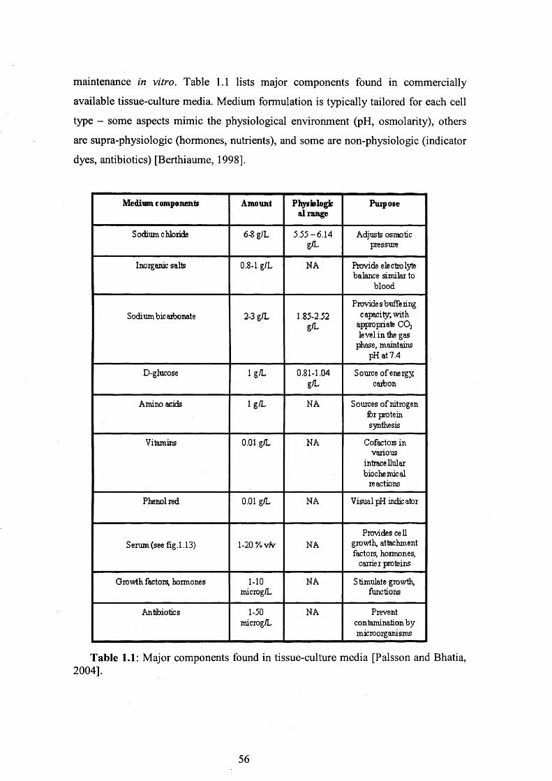
maintenance in vitro. Table 1.1 lists major components found in commercially
available tissue-culture media. Medium formulation is typically tailored for each cell
type - some aspects mimic the physiological environment (pH, osmolarity), others
are supra-physiologic (hormones, nutrients), and some are non-physiologic (indicator
dyes, antibiotics) [Berthiaume, 1998].
Medium c omponents Amount Physiologic al range
Purpose
Sodium chloride 6-8 g/L 5.55-6.14g<L
Adjusts osmotic pressure
Inorganic salts 0.8-1 g/L NA Provide electrolyte balance similar to
blood
Sodium bic atbonate 2-3 g/L 1.85-2.52 g/L
Provide s buffe ring capacity, with
appropriate COj level in the gas
phase, maintains pH at 7.4
D-glucose 1 g/L 0.81-1.04g L
Source of energy carbon
Amino acids 1 g/L NA Sources of nitrogen for protein synthesis
Vitamins 0.01 g/L NA Cofactois in various
intracellular biochemical
reactions
Phenol red 0.01 g/L NA Visual pH indicator
Serum (see fig.1.13) 1-20 Y*vfv NAProvides cell
growth, attachment factory hormones,
carrier proteins
Growth factors, hormones 1-10microg/L
NA Stimulate growth, functions
Antibiotics 1-50microg/L
NA Prevent contamination by microorganisms
Table 1.1: Major components found in tissue-culture media [Palsson and Bhatia, 2004].
56

Typically, a synthetic basal medium is chosen to meet the environmental and
nutritionals requirements of a given cell line. However, basal media formulations
normally will not support cell growth alone, but must be further supplemented with
animal serum.
1.4.2 Serum
Serum is a fraction of whole blood. Plasma is the non-cellular fraction of blood,
whereas serum is the liquid that remains after plasma is allowed to clot. Sera are
typically added to culture medium in a proportion of approximately 1 to 20 % by
volume. Common tissue culture sera are newborn calf (NCS), fetal bovine (FBS),
horse (equine), pig (porcine), and human. Fetal bovine serum (FBS) has been adopted
as the standard supplement because of its rich content of growth factors and its low
gamma globulin content [http://www.cellgro.com Mediatech, Inc. Cell Culture
Reference Guide]. Historically, medium containing fetal bovine serum has been used
for culture of a wide range of cell types.
As seen in table 1.2 [Palsson and Bhatia, 2004], sera are very complex
supplements containing mostly proteins but also important growth factors, adhesion
factors, minerals, lipids, enzymes and hormones that can be key to successful tissue
culture.
57

Constituent Eflectoncelh
Albumin, globulins, transferrin Carriers for fatty acids, iron, etc.
Fibronectin, fetuin Attachment factors
Alfa2-macroglobulin Inhibits proteases
Growth factors and growth hormones Promote cell proliferation
Hormones (e .g. insulin and corticosteroids) Stimulate uptake of glucose and amino acids; alter growth rate
Carbon and nitrogen sources Nutrients
Minerals and trace elements Essential for the activity of me talloenzymes
Inhibitors Inhibit cell proliferation
Table 1.2: Main constituents of serum [Palsson and Bhatia, 2004].
Despite the beneficial effects, the use of sera has some significant disadvantages,
including batch-to-batch variability, the presence of adventitious agents, product
availability, interference with detection of cell products, and potential regulatory
hurdles. Moreover, sera contain a wide range of minor components that may
potentially affect cell growth. These minor undefined components, including nutrients
such as amino acids, nucleosides and sugars, are inherent to sera and may affect
various parameters of particular importance [http://www.cellgro.com Mediatech, Inc.
Cell Culture Reference Guide]. In order to minimize these variables there has been a
move towards the use of serum-free media.
Removal of bovine serum from organ culture medium is necessary also because
of the potential risk of infection. Bovine serum is the usual supplement in the medium
for expanding human cells, however transmission of bovine diseases might be
inevitable when such cells are introduced into the patient for tissue engineering
applications. In addition, bovine serum proteins attach to the surface of the cells and
the biomaterial. It is possible that transferring bovine serum proteins into patients
elicits an unfavourable immune reaction [Yamamoto et al., 2003]. Nowadays there is
both a moral and legal imperative (European Directive 86/609/EEC as amended by
58

Directive 2003/65/EC) for scientists to use alternatives to animals in research
wherever possible. To avoid all the above mentioned unfavourable possibilities,
leading companies are supplying researchers with serum substitutes and serum-free
media, which are used to support the maintenance and expansion of many cell types,
including hematopoietic stem cells and other progenitor cells.
For instance, serum-free culture has been explored for human nasal epithelial cells
(HUNEC) [Mattinger et al., 2002], human corneal endothelial cells [Bednarz et al.,
2001] but, most importantly, for rhesus monkey and human embryonic stem cells
([Pei et al., 2003; Amit et al., 2003] respectively). A culture system for the growth
and assay of human bone marrow stromal precursors under serum-deprived
conditions has also been developed [Gronthos and Simmons, 1995]. As an alternative
to serum-deprived conditions, human chondrocytes have been cultured in a medium
supplemented with autologous serum, and then transplanted into the patient’s
cartilage defects [Brittberg et al., 1994]. Moreover, the effects of human serum on the
in vitro differentiation of human bone marrow fibroblasts into adipogenic and
osteogenic cells have been investigated [Oreffo et al., 1997]. Human bone marrow
derived MSCs have also been cultured in human and rabbit sera [Kuznestov et al.,
2000] or in autologous serum [Yamamoto et al., 2003] to test the hypothesis that
these cells can be successfully expanded ex vivo without losing their potentiality for
osteoblastic differentiation even in an FBS-free culture system.
All these studies represent important prerequisites for future investigations on the
role of serum in the regulation of cell proliferation, differentiation and development.
1.4.3 Growth factors
To enhance cell proliferation, growth factors and other bioactive soluble
macromolecules can be added directly to the growth medium or can be incorporated
into the scaffold material. Growth factors play important regulatory roles in the
control of the immune response, inflammatory reactions, cell proliferation and tumour
growth [Yarden et al., 1991]. They can stimulate or inhibit cell division,
differentiation, migration and apoptosis, and they are involved in the regulation of
several cellular processes such as gene expression, DNA and protein synthesis, and
autocrine and paracrine factor release.
59

Some growth factors have demonstrated the ability to induce division of the
endothelial cells and smooth muscle cells that represent the main cellular constituents
of large blood vessels. These growth factors include:
a) basic fibroblast growth factor (bFGF) - both endothelial and smooth
muscle
b) vascular endothelial cell growth factor (VEGF) - endothelial cells
c) platelet-derived growth factor (PDGF) - smooth muscle cells
d) epidermal growth factor (EGF) - smooth muscle cells
e) endothelin-1 (ET-1) - smooth muscle cells
Each of these growth factors can interact with one another in an additive,
cooperative, synergistic or antagonistic manner, and thus must be studied
individually. For tissue engineering purposes, whether the cells are quiescent or
activated, and whether they are in a two or three-dimensional culture environments,
will influence the production of endogenous growth factor and consequently will also
affect the response to exogenously applied bio-molecules.
1.4.4 Insoluble ECM adhesion molecules
The extracellular matrix (ECM) contains a number of proteins that contribute to
both organizing the matrix and helping cells attach to it. Not surprisingly then, the
composition and geometry of the ECM have both been shown to play a role in cell
function. Due to the important effects of the ECM on cell behaviour, insoluble ECM
molecules, such as fibronectin, laminin, or collagen, have been used to promote tissue
regeneration in tissue engineering applications. These cue materials, together with the
soluble factors such as growth factors or hormones normally added to the medium of
culture, can be utilized to promote cell development and function [Gooch et al.,
1998].
Cell-ECM interactions can be controlled by modification of the solid substrate on
which cells are grown. Polystyrene and other polymers normally employed in the
preparation of tissue culture vessels can be improved just by coating the surface with
ECM molecules such as collagen or fibronectin. Many tissue culture plates are
currently treated with these molecules to improve cell binding.
60

As already mentioned (see section 1.3.3), biomaterial scaffolds can also be treated
with the RGD and other short peptide sequences derived from cell binding regions of
ECM proteins in order to achieve biospecific cell adhesion [Gooch et al., 1998].
Culture surfaces modified with fibronectin are currently investigated for cell
culture by several groups. Fibronectin, a large glycoprotein found in all vertebrates,
was the first non-collagen ECM proteins to be well characterized [Alberts et al.,
1994]. Human fibronectin (HFN) sometimes serves as a general cell adhesion
molecule through its central-binding domain RGD sequence. The apparent function
of fibronectin is to mediate cell attachment by interacting with cell surface receptors
and extracellular matrix components ([Hynes, 1985]; [Rouslahti, 1988]). HFN is not
only used as a substrate to promote cell attachment; it is involved in many other
cellular processes, including cellular migration during wound healing, tissue repair
and development, embryogenesis, blood clotting, and haemostasis/thrombosis.
Also Matrigel, a solubilized basement membrane preparation commercially
available, is currently used for several cell culture applications. According to the
manufacturer’s (Becton-Dickinson Biosciences, Oxford, UK) specifications, ordinary
matrigel contains (as a proportion of protein by weight) 56% laminin, 31% type IV
collagen, 8% entactin, 5% heparan sulfate proteoglycans.
Both fibronectin and matrigel are used as substrates for growth and differentiation
of different cell types.
1.4.5 The role of oxygen in cell culture
Metabolically active cells are also consuming and producing dissolved gases
(oxygen and carbon dioxide). Cell viability and function are in fact dependent upon
both nutrient supply and oxygenation. As a consequence, the in vitro growth
characteristics of cells are subject to dissolved oxygen tension, among many other
culture-related factors, such as the already described media formulation and growth
factors supplementation [Parrinello et al., 2003].
Oxygen consumption can be a critical variable for certain cell types (for example
hepatocytes consume five- to ten-fold more oxygen than other cells). Oxygen delivery
in culture is different than oxygen delivery in vivo because of the lack of red blood
cells (with haemoglobin) as oxygen carriers. Therefore, in culture, oxygen delivery is
61

limited by two factors: 1) the relatively low solubility o f oxygen in media at body
temperature (1.19 pM/mm Hg) and 2) transport of oxygen from gas phase to cell
surface [Palsson and Bhatia, 2004].
Standard culture conditions use dissolved oxygen tension levels (20%-21%) that
are hyper-physiologic for many cell types, including articular chondrocytes and bone-
marrow derived MSCs, which are adapted to relatively low oxygen levels (< 10%) in
vivo [Scott, 1992]. This suggests that excessive oxidative stress may limit the growth
potential of cells under standard conditions. Indeed, it has been shown that MSCs
cultured in low oxygen outgrow their high oxygen counterparts by at least 2-fold
[Moussavi-Harami et al., 2004], suggesting that high oxygen levels are stressful to
MSCs and limit their in vitro growth by inducing senescence. Culture-induced stress
is probably an important factor affecting the in vitro growth of MSCs, and it is
possible that preventing oxidative stress might help in overcoming this barrier.
Lowering incubator oxygen levels may represent an effective and simple means to
that end, but other modifications of standard conditions, such as the addition of
antioxidants to the culture medium, might confer additional protection against
oxidative damage. Moreover, even routine cell culture procedures such as initial cell
isolation and trypsinization are potential sources of oxidative stress. Such occasional
stress exposures might impact growth even in cultures exposed most of the time to
low oxygen conditions.
The attenuation of growth imposed by culture-related stress seriously restricts cell
yields and may have a negative impact on subsequent differentiation of MSCs and
chondrocytes.
Recent studies [Moussavi-Harami et al., 2004] have proved that low oxygen
culture is an effective means to control oxidative stress and to increase the
proliferative potential of MSCs and chondrocytes destined for grafting procedures.
Other investigations have demonstrated that a low oxygen atmosphere significantly
affects rat marrow MSCs’ proliferation, differentiation and mobilization into the
peripheral blood [Lennon et al., 2001; Rochefort et al., 2006;]. Hypoxic conditions
have also proved to be necessary to maintain full pluripotency of human embryonic
cells [Ezashi et al., 2005].
However, additional studies will be needed to determine what are likely to be
diverse phenotypic effects of oxidative damage on human marrow-derived MSCs.
62

1.5 The goals of the present thesis
The driver for this research stems from the ability to create viable three-
dimensional alginate/hMSCs constructs and to define the optimal culture conditions
where the cells display the ability to proliferate while retaining their original
undifferentiated status and multipotentiality.
In order to accomplish this, the investigations of this research have been carried
out at three levels.
Firstly, studies on the alginate matrix itself were conducted with the intention of
optimising the carbodiimide coupling reaction between alginate and peptide, in order
to enhance the efficiency of the derivatisation chemistry and to prepare a three-
dimensional alginate matrix that permits cell adhesion and cell proliferation. In
addition, NMR methods and amino acid analysis were performed to help
characterising the resulting matrix and to quantify the degree of peptide attachment.
Secondly, the effects of some important culture parameters on MSCs’ viability,
proliferation and metabolism were investigated in a two-dimensional culture
environment (tissue culture plates). In particular, attention was focused on changing
medium composition in respect of serum concentration and FGF-supplementation, on
modifying the plastic surface of the vessels through coating with different
biomolecules, and on testing different oxygen tensions.
Finally, the cells were combined with the GRGDY-modified alginate matrix and
the optimal parameters identified in the two-dimensional environment were applied to
the resulting three-dimensional constructs. Investigations were performed on the
constructs in order to evaluate the cells ability to retain their initial viability and
original undifferentiated status, and to determine the degree of cell proliferation.
63
2. Materials and Methods
2.1 Human Mesenchymal Stem Cell culture In tissue
culture vessels
2.1.1 Isolation of human mesenchymal stem cells from frozen
bone marrow samples
The multipotent Mesenchymal Stem Cells (MSCs) used in this study were
isolated from frozen human bone marrow samples received from the Department of
Haematology, UCLH. The use of human clinical samples had been approved by the
University College London Hospital Ethics of Human Research Committee (project
reference number 03/0136). The mononuclear cells were isolated from frozen bone
marrow samples stored in a liquid nitrogen container (Biostore 10) deploying a Ficoll
gradient.
This procedure is described for a total volume of thawed diluted sample of 120
mL (typically the amount of the original sample after thawing was around 125-150
mL, the sample was diluted 1:2 and however only 120 mL of the total diluted sample
was used).
First of all, 120 mL of the original thawed bone marrow sample was filtered
through a 40pm nylon cell strainer (Falcon, Bibby Sterilin, Stone, UK). 100 mL of
the resulting material was transferred in a 250 mL sterile bottle (Nalgene/Fisher) and
was then rinsed with an equal volume (100 mL) of Dulbecco’s phosphate buffered
saline without calcium and magnesium (PBS, Biowhittaker, Walkersville, MD, USA).
6x20 mL aliquots of sterile Ficoll (density 1.077g/mL, Ficoll-Paque Plus, Stem Cell
Technologies Inc., Vancouver BC) was poured into 6x50 mL centrifuge tubes
(Coming, Acton, MA, USA) and 20 mL of the diluted bone marrow sample was then
very slowly added to the edge of each tube such that the sample remained above the
Ficoll surface. During this procedure the centrifuge tube was held at a sharp, slanted
angle in order to increase the surface area on the top of the liquid and to create a clear
phase separation between the Ficoll layer (at the bottom of the tube) and the sample
(at the top of the Ficoll phase), avoiding mixing of the two layers.
64

A 30 minutes centrifugation at 650g, room temperature (20°C), and with no brake
applied (5 81 OR Centrifuge, Eppendorf AG, Hamburg, Germany) was performed in
order to fractionate most of the red blood cells to the bottom of the tube, while
maintaining the mononuclear cells at the interface of the Ficoll liquid. After
centrifugation, the mononuclear cells were carefully removed from the tube without
disturbing the Ficoll interface.
Approximately 5-6mL was collected (maximum of lOmL) from each tube and
transferred into a new 50mL centrifuge tube. The cells were rinsed with
approximately 40mL of PBS per tube and then centrifuged at 650g for 15 minutes, at
room temperature (20°C), using a low brake setting. After discarding the
supernatants, the cell pellets were gently resuspended and pooled together with a total
growth medium volume of 12mL, and the cell suspension was equally divided into 3
vented T150 flasks (Coming, 150cm ) already filled with 26mL complete growth
medium (total volume within each flask was approximately 30mL). The flasks were
then placed at 37°C in a 5% CO2 humidified incubator (HeraCelll50, Kendro
Laboratory Products GmbH, Langenselbold, Germany or Galaxy S, RS Biotech
Laboratory Equipment Ltd., Irvine, Scotland) for 2 days to allow the hMSCs to attach
to the plastic surface. The spent medium containing non-adherent cells was then
removed and replaced with fresh complete medium of culture. While waiting for the
hMSCs to become visible (ca. 7 days) and ready for passaging (ca. 2-3 weeks), fresh
complete growth medium was added every 4-5 days. After reaching approximately
70-80% confluency, the cells were trypsinised, counted with an haemocytometer and
split into the required number of new vented T75 or T 150 flasks in order to have an"X 9initial cell seeding density (ICD) of 5x10 cells/cm (see sections 2.1.3 and 2.6.2).
The flasks were labelled as passage 2 (p2) and incubated at 37°C in an atmosphere of
5% CO2. When the resulting second passage cells were 70-80% confluent, they were
either passaged again at the same ICD and labelled as passage 3 cells, or frozen in
liquid nitrogen.
2.1.2 In vitro culture of human Mesenchymal Stem Cells
The in vitro expansion of hMSCs was performed in sterile, polystyrene T150
tissue culture flasks (150cm2) (Coming Life Science, Coming, New York) with 30mL
65

complete growth medium/ flask (0.2mL/cm2). The flasks were factory treated with a
plasma charge, which introduces a hydrophobic, negatively charged surface. The
cultures were maintained in a 5% CO2, 37°C humidified incubator and the cells were
expanded in culture at 70-80% confluence (as described in section 2.1.3) and plated
in new T150 flasks at the initial cell density of 5x10³ cells/cm2 (7.5x10⁵cells/flask).
The culture medium was removed from the flasks and replaced with fresh and pre
warmed (37°C) medium (30mL/flask=0.2mL/cm2) every 3-4 days of culture to
provide the cells with the necessary nutrients and growth factors and to remove
metabolic products.
Different culture conditions were compared and evaluated in this research, as
described in section 2.1.5.
2.1.3 Cell expansion in tissue culture vessels
Human MSCs were harvested from their T150 tissue culture flasks with a
trypsin/EDTA solution.
After removing the spent medium, each flask was rinsed with 20mL PBS without
calcium and magnesium (O.lmL/cm ) (BioWhittaker) to remove residual serum
proteins that reduce trypsin activity. Each flask was treated with 6mL (0.04mL/cm )
of 0.25% trypsin/0.02% EDTA (Sigma-Aldrich, LTD, Irvine, Ayrshire, UK). The
enzymatic solution was spread over the whole growth surface and was left in action
for 5-7 minutes at room temperature (20°C). The flasks were then gently tapped
against the palm of the hand in order to completely detach the hMSCs from the
surface of the culture vessel. Trypsin activity was then quenched with 12mL
(0.08mL/cm ) of complete cell growth medium per flask. Since trypsin is a non
specific protease that could permeate the cell membrane, the cells were never exposed
to the trypsin solution for periods longer than 10 minutes. The cell suspensions from
all individual flasks were combined together and gently mixed with a sterile pipet to
ensure homogeneity of the final cell suspension. A 0.5mL volume sample was used
for the determination of cell density with haemocytometer and for the cell viability
assay with trypan blue dye (section 2.1.3, 2.6.2 and 2.6.3). The cell suspension was
then centrifuged within a 50mL centrifuge tube at lOOg for 5 minutes. After
discarding the supernatant, the cell pellet was resuspended with the necessary volume
66

of complete cell growth medium. The resulting cell suspension was transferred within
new T150 tissue culture flasks so that the initial seeding density ICD was 5xl03
cells/cm2 (7.5x10⁵ cells/flask). The volume of cell suspension within each flask was
adjusted to a final value of 30mL (0.2mL/cm2) by addition of the necessary amount of
complete growth medium, pre-warmed at 37°C. The flasks were incubated and re-fed
with complete cell growth medium as previously described (section 2.1.2).
2.1.4 Creation of a working cell bank
In order to create a supply of low-passage cells for future experiments, a working
cell bank of fourth passage (p4) hMSCs was created from cells isolated from a frozen
bone marrow sample.
Five vented T150s of 80-90% confluent cells were harvested and trypsinised as
described in section 2.1.3. The cells were treated with 6mL of
0.25%trypsin/0.02%EDTA (Sigma) per flask until the cells had completely detached
from the cell growth surface (approximately 5 minutes). Complete DMEM medium
(12mL/flask) was added to quench trypsin activity and the resulting cell suspensions
were removed from the flasks, pulled together and equally divided into two 50mL
centrifuge tubes. A 0.5mL volume sample was taken from each tube and used for the
determination of the cell density with haemocytometer and for the cell viability with
trypan blue dye, as described in sections 2.6.2 and 2.6.3. The cell suspensions were
centrifuged at 160g for 5 minutes. After discarding the supernatants, the two cell
pellets were combined together and resuspended in 4mL of complete DMEM
medium. Another cell count was performed after centrifugation to confirm the cell
concentration and ensure that no significant cell loss had occurred during the
centrifugation step.
3.6mL of complete DMEM medium were added to the resulting cell suspension.
Finally 1.9mL (20%) of cryopreservation medium containing 75% fetal bovine serum
(FBS, Biowhittaker) and 25% DMSO (Sigma) were slowly added to the cell
suspension (final cell concentration = lxlO6 cells/mL). The total cell suspension
(approximately 9.5mL) was equally aliquoted into 9 cryopreservation vials (Nalgene,
lmL cell suspension/vial), which were then placed overnight in the -80°C freezer
within a controlled rate freezer container (Nalgene TM Cryo 1°C Freezing Container,
67

-l°C/min=rate of cooling) before transfer into liquid nitrogen (-196°C) for long-term
storage.
2.1.5 Protocols of hMSCs culture using different basal media, different serum and fibroblast growth factor concentrations,
different coatings and oxygen tensions
Taking into account the lack of a uniform approach for hMSC isolation and
expansion between laboratories, various culture conditions were compared in this
research with the purpose of first identifying an optimal laboratory protocol for
hMSCs expansion with a view to later large-scale production, while maintaining their
multipotency.
Several culture parameters, including the quality of fetal calf serum, basal
medium, glucose concentration, bone marrow mononuclear cell plating density,
hMSC passaging density, and surface characteristics of the tissue culture vessels are
known to affect the final outcome [Sotiropoulou et al., 2006]. Also the use of basic
fibroblast growth factor (FGF), the most common growth supplement in hMSC
culture media, importantly influences cell proliferation and differentiation potential
[Pitaru et al., 1993; Savion et al. 1996; Majors and Muschler, 1996].
In this study attention was focused on the following parameters:
• Basal cell growth medium
• Fetal bovine serum (FBS) concentration
• Surface coating of tissue culture vessels
• basic Fibroblast Growth Factor (bFGF) supplementation
• Oxygen tension
2.1.5.1 Basal growth medium
Two different growth media were compared for the in vitro expansion of hMSCs.
Complete MesenCult medium, obtained supplementing the basal MesenCult
medium (Stem Cell Technologies Inc., Vancouver, British Columbia, Canada) with
10% (v/v) Mesenchymal Stem Cell Stimulatory Supplement (Stem Cell Technologies
68

Inc.), 100U penicillin/mL, lOOpL streptomycin/mL (both from BioWhittaker,
Walkersville, MD, USA) and Ing/mL recombinant human fibroblast growth factor
(rhFGF) (R&D Systems, Minneapolis Minnesota, USA); and low-glucose (1 g/L)
Dulbecco’s Modified Eagles Medium DMEM (Biowhittaker), which was
supplemented with the same reagents (Mesenchymal Stem Cell Stimulatory
Supplement, penicillin, streptomycin and recombinant human fibroblast growth factor
(rhFGF)) added in the same concentrations as for the MesenCult medium.
Fourth passage (p4) bone marrow-derived hMSCs were expanded in 12-well
tissue culture plates with either complete MesenCult medium or complete DMEM
medium, formulated as described above. In addition, these media were used for the
cell expansion also in their serum-free formulation, or after addition of only 2% (v/v)
serum. The Mesenchymal Stem Cell Stimulatory Supplement from StemCell
Technologies Inc., which was found to consist only of Fetal Bovine Serum
supplemented with 1-glutamine, was used for the serum-supplementation of both the
MesenCult and the DMEM media. The cultures were maintained at 5% CO2, 37°C for
a 12-day period. During the course of the culture, the plates were re-fed every 2-3
days in order to provide the cells with the necessary nutrients and growth factors and
to remove metabolic products. The cells were trypsinised at 4 different time-points
during the culture for the evaluation of the cell density and viability, as measured
with haemocytometer and trypan blue dye. This experiment was repeated twice,
always with p4 MSCs; the results of these studies are discussed in section 4.3.
After a few preliminary studies, the basal low-glucose DMEM medium was
supplemented with 10% (v/v) Fetal Bovine Serum (FBS) screened for hMSCs (Stem
Cell Technologies Inc.), 2mM 1-glutamine, 100U penicillin/mL and lOOpL
streptomycin/mL (both from BioWhittaker) and Ing/mL recombinant human
fibroblast growth factor (rhFGF) (R&D Systems). This was the standard formulation
for the DMEM medium, which was subsequently adopted for the expansion of the
hMSCs used in this research, and it will be referred to as “complete DMEM” for the
remainder of this thesis.
69

2.1.5.2 Serum supplementation
2.1.5.2.1 Fetal Bovine Serum concentration
The standard 10% FBS-supplemented complete DMEM formulation was
compared to DMEM growth medium equally supplemented with 1-glutamine,
penicillin, streptomycin and recombinant human fibroblast growth factor, but
supplemented with different (0%, 2% and 15%) FBS concentrations. The serum used
in this research was fetal bovine serum (FBS) specifically selected by the
manufacturer (Stem Cell Technologies) for optimal hMSCs growth.
The cells were plated in 24-well plates (ICD=5xl03 cells/cm2 or 9.5x10⁴
cells/well) and cultured with the different media formulations. The cells were
harvested at different time-points over the 13-15 days culture period. At each time-
point the cell density and viability were determined with haemocytometer and trypan
blue (see sections 2.6.2 and 2.6.3.) and the cell metabolism was evaluated with the
Nova Biomedical Bioprofile Analyzer (see section 2.6.5). The experiment was
performed twice and the results obtained for the serum studies are presented in
section 4.4.1.
2.1.5.2.2 Serum-free media experiment
A preliminary qualitative experiment was performed where hMSCs were cultured
in different serum-free media, in order to see whether any of these formulations
permitted, promoted or halted cell growth. The following commercially available
serum-free media were tested:
70

No. Medium M anufacturer Cat. no.
1 CD Hybrid0111 a Medium without L-
glutamine
Gibco 11279-023
2 Stem Pro-34 SFM without L-glutamine without antibiotics
Gibco 10640-019
3 Protein free Hybridoma Medium PFHMII (IX)
Gibco 12040-051
4 Hybridoma-SFM serum-free Hybridoma
Medium
BioWliittaker 12045-084
5 Ultraculture medium BioWhittaker BW12-725F
6 UltraDOMA Hybridoma serum-free
growth Medium,defined IX
BioWliittaker BW12-723B
7 Knockout D-MEM optimised D-MEM
Medium for ES cells
Gibco 10829-018
8 Ex-cell 325 PF CHO scrum-free Medium,
protein-free
SAFC
Biosciences
14340C
9 Ex-cell 302 CHO scrum-free Medium
SAFCBiosciences
14324C
10 Ex-cell 620-HSF hybridoma serum-free
Medium
SAFCBioscicnces
14621C
11 Ex-cell CD CHO serum-free Medium, chemically defined
SAFCBiosciences
14361C
Table 2.1: Serum-free media investigated for the expansion of hMSCs in tissue culture flasks. Following the manufacturer’s suggestion, the Knockout D-MEM medium (medium number 7 on the table) was supplemented with 15% Knockout Serum Replacement (Gibco, Cat. No.=10828).
The cells used for this experiment were obtained from a third passage (p3)
working cell bank. Cryopreservation vials of hMSCs were removed from liquid
nitrogen storage and were thawed within a 37°C water-bath for exactly two minutes
and then transferred to T25 tissue culture flaks (Coming, 25cm2). The cells were
expanded at the standard culture conditions (complete DMEM medium, 5% CO2,
71

37°C and 21% oxygen) for two passages before reaching the desired cell number.
[The cell banking and passaging protocols are described in detail in section 2.1.4 and
2.1.3 respectively.] The sequential adaptation protocol here described was then
followed for each one of the 11 serum-free media under investigation and listed in
table 2.1. Actively dividing, high (> 90%) viability cells were used. Sixth passage
(p6) cells were inoculated at double the normal seeding density (10xl03 cells/cm2) in
a 50:50 (v/v) mixture of traditional serum-supplemented DMEM : one of the serum-
free media investigated. The cultures were monitored until the confluency reached
approximately 80%. The cells were then sub-cultured into a 10:90 (v/v) mixture of
serum-supplemented DMEM : serum-free medium. When 80% confluency was
reached again the cells were sub-cultured into 100% serum-free medium. Two T25
control flasks were maintained in the standard complete DMEM medium formulation
throughout the whole experiment. At each expansion passage the cells were split in
1:2 ratio without performing any cell count with haemocytometer, as the present was
purely a preliminary exercise performed to obtain information on cell survival and to
monitor significant cell morphology changes in the different serum-free culture media
tested.
Light microscopy was used for the qualitative assessment of cell growth,
proliferation and morphology. In particular, significant change in cell density and cell
morphology were observed. These results are presented in section 4.4.2.
2.1.5.2.3 Surface coatings of tissue culture vessels
The effects of different surface coatings on the hMSCs proliferation and
expansion potential were evaluated by comparing the culture within 24-well un
coated tissue culture plates (353047, Falcon, Beckton Dickinson Labware Franklin
Lakes, NJ, USA) with the culture of the same cells maintained in 24-well tissue
culture plates coated by the supplier with either human fibronectin or with a thin
Matrigel basement membrane matrix layer (Becton Dickinson Labware, Bedford,
MA). The cells were seeded at an ICD of 5xl03 cells/cm2 (9.5x10⁴ cells/well) and fed
with lmL complete DMEM/well. The three different coating conditions described
were also tested using rhFGF-free or serum-free DMEM formulations, or with
DMEM medium supplemented with different serum concentrations. The cells were
harvested at different time-points over the 13-15 days culture period. At each time-
72

point the cell density and viability were determined with haemocytometer and trypan
blue (see sections 2.6.2 and 2.6.3) and the cell metabolism was evaluated with the
Nova Biomedical Bioprofile Analyzer (see section 2.6.5). The experiment was
repeated twice and the results obtained for the coating studies are presented in chapter
4.
2.1.5.2.4 Human recombinant Fibroblast Growth Factor supplementation
The standard formulation of the complete DMEM culture medium included
supplementation with Ing/mL human recombinant basic Fibroblast Growth Factor
(bFGF).
To investigate the effects of bFGF-supplementation on the growth potential of' i j
human MSCs, p4 hMSCs were plated at the usual ICD (5x10 cells/cm ) in non
coated 24-well tissue culture plates and cultured for a 13-day period. The cells were
expanded in low-glucose (lg/L) DMEM medium in standard culture conditions (10 %
v/v FBS and 1 ng/mL bFGF supplementation). In addition, hMSCs were cultured in
the plain basal (serum-free and bFGF-ffee) low-glucose DMEM medium, and in the
same medium supplemented with increasing serum concentrations (2%, 10% or
15%). For each serum concentration, both the bFGF-free and the bFGF-supplemented
conditions were tested. As usual, the cells were re-fed every 2-3 days over the 13-day
period of culture, thus ensuring the provision of nutrients and growth factors together
with the removal of metabolic products. The cells were harvested as previously
described (section 2.1.3) at day 3, day 7, day 10 and day 13 of culture, i.e. when the
80% cell confluence was reached on the surface of the well plates. At each time-point
the cells were harvested and assessed for cell count and cell viability as previously
described (sections 2.6.2 and 2.6.3 respectively), and for each serum and bFGF
condition a sample of the spent medium was recovered and assessed with the Nova
Biomedical BioProfiler for the analysis of the cells metabolic activity, following the
protocol elucidated in section 2.6.5.
The experiment was performed twice and the results obtained for the bFGF
studies are presented in section 4.5.
73

2.1.5.5 5 Dissolved oxygen tension
Where it is not otherwise specified, the humidified incubator for the culture of
hMSCs was maintained at 5% CO2, 37°C and 21% O2. 21% oxygen levels are
normally used in standard incubators and standard culture conditions, as they permit
optimal growth of fibroblasts. However, MSCs live at relatively low oxygen levels in
vivo [Grant and Smith, 1963; Ishiwaka and Ito, 1998; Harrison et al., 2002; Grigoryan
et al., 2005], and the hyperoxic conditions employed in standard incubators can cause
sufficient oxidative stress to induce premature senescence in MSCs, limiting their in
vitro proliferation potential [Moussavi-Harami et al. 2004]. Modifying the oxygen
tension is thought to have important effects on MSC growth and proliferation. For the
purpose of evaluating the effects of oxygen tension on hMSCs proliferation rate and
metabolism, the above-mentioned standard culture conditions were compared to low-
oxygen (1% or 2%) and intermediate-oxygen (10%) culture conditions. The cells
were cultured in 24-well plates and tested for cell proliferation and cell metabolism at
different time-points in a similar way as for the previous culture conditions examined
(sections 2.1.5.2, 2.1.5.3 and 2.1.5.4). Each experiment was performed twice and the
results obtained for the studies on different oxygen tensions are presented in chapter
4.
2.2. An alternative cell source: cell passaging protocol in tissue
culture vessels for human Fibroblasts
The human foreskin fibroblasts used in this study (Karocell, Stockholm) were
kindly provided by Dr. Stephen Minger, Wolfson Centre for Age-Releated Diseases,
King’s College London. The cell growth medium was Iscove’s modified Dulbecco’s
medium (IMDM) which had been supplemented by the manufacturer (Invitrogen,
Paisley, UK) with 4mM glutamine and 25mM HEPES, and then further supplemented
with 10% heat inactivated foetal bovine serum (Invitrogen, Paisley, UK) and
lOOU/mL penicillin/streptomycin (BioWhittaker, Walkersville, MD).
As for hMSCs, the fibroblasts were cultured in tissue culture flasks specifically
treated by the manufacturer in order to encourage cell attachment to the plastic
surface. The cells were cultured with 30mL medium/T150 flask (0.2mL/cm2) and
74

were usually expanded at a 1:3 split ratio, following the trypsinisation procedure
already described (see section 2; 1.3). Human fibroblasts were maintained in culture at
37°C and 5%CC>2 in a humidified incubator.
2.3 Preparation of the alginate scaffold
The matrix used in this research for the three-dimensional studies with
alginate/cells constructs consisted of sterile GRGDY-derivatised alginate. Alginate
constructs have been extensively used in cell therapy and tissue engineering
applications, mainly with secretory cells (e.g. islet cells) or transformed producer-cell
lines that normally are expanded in suspension cultures [Read et al., 1999; Mullen et
al, 2000; Tobias et al, 2001]. However, mesenchymal stem cells are anchorage-
dependent cells that need to interact with a substrate or biomaterial to survive
[Saltzman, 2000]. Alginate and other hydrogels are unable to specifically interact
with mammalian cells, due to their hydrophilic nature [Smentana, 1993]. It was
therefore necessary to chemically modify the alginate hydrogel by covalently binding
the pentapeptide cell adhesion ligand glycine-arginine-glycine-aspartic acid-tyrosine
(GRGDY) containing the specific RGD sequence, which has shown to promote
cellular anchorage and interaction with hydrogels [Rowley et al., 1999; Chung et al.,
2002].
This section describes the preparation of alginate-GRGDY matrices. The
preparation of alginate constructs including cells is described in section 2.5. A
schematic representation of the standard alginate derivatisation method used in the
present research is shown in figure 2.1.
75

1*« Pionova alginate
-JO n
alginate concenti ation with
PEG sokltion
aliquot into stei lie vials anti lyophilize toi 2 day at -00* C
0.2 0.8 mbar
0.2 |Lmfiheied suVo-IIHS
EOCGRCDY peptide
5-day dialysis cycle in Uinuiai flow cabinet
with 9.2 |i in tiltei ed watei and sterile equipment
20 - 2 2 ° C
s ' " ' " * " ' X
20-hour i e act ion constant mixing
in tire dark20 - 22 °C
alginate U ansfei into T -ii radiated
dialysis cassette20 - 229C
Fig.2.1: Schematic rqiresentation of the standard alginate denvatisahon procedure
for the coupling of the GRGDY pentapeptide to sodium alginate.
2.3.1 Reconstitution of the sodium alginate matrix
Sodium alginate (please see section 1.3.4) was the scaffold material investigated
in the present research.
The alginates used in this research were supplied as sterile freeze-dried products
by NovaMatrix FMC BioPolymer AS, (Drammen, Norway). Different Pronova
alginates were tested; the defining characteristics of these biomaterials, such as the
guluronic acid content and molecular weight, were provided by the manufacturer (see
table 3.2).
Vials of each sterile alginate were reconstituted as required with sterile filtered
normal saline solution or MES/NaCl buffer, and mixed until visibly homogeneous
prior the addition of the sterile filtered reactants for the derivatisation procedure (as
described in section 2.3.2).
The normal saline solution used for the alginate reconstitution consisted of 0.9%
w/v NaCl (Sigma) in cell culture grade water. Alternatively, the alginate was
reconstituted in 0.1 M 2-(N-Morpholino)ethanesulfonic acid (MES) buffer (Lancaster,
Morecambe, UK) with 0.3M NaCl.
Before adding the alginate, the pH of the normal saline and the pH of the
MES/NaCl buffer were respectively adjusted to 7-7.4 and 6.5 with 0.1M NaOH
obtained from 1M NaOH (Sigma) after 10-fold dilution. The alginate was prepared at
a final concentration of 1% weight/volume (w/v), which was isotonic for subsequent
addition of the cell suspension. At this concentration, the water-soluble alginate was
completely dissolved and solubilised after overnight mixing at room temperature
(20°C) on a platform roller (Roller Mixer SRT2, Bibby Stuart Scientific, Stone, UK).
2.3.2 Alginate derivatisation with GRGDY pentapeptide
Alginate hydrogels are able to discourage cell adhesion due to the hydrophilic
nature of the polymer. In previous studies the attachment and proliferation of mouse
skeletal myoblasts has been improved after incorporation of the pentapeptide
GRGDY onto the polysaccharide chain of the alginate [Rowley et al., 1999; Rowley
and Mooney, 2002]. In this research, sodium alginate was covalently modified
77

utilizing aqueous carbodiimide chemistry in order to incorporate the pentapeptidic
GRGDY sequence. The GRGDY pentapeptide was conjugated to the alginate’s
carboxylic acid groups on the uronic acid units of the polymer backbone through the
terminal amine of the peptide, as it is schematically represented in Figure 2.2.
A water-soluble carbodiimide, 1 -ethyl-(dimethylaminopropyl) carbodiimide
(EDC), (Sigma-Aldrich Co., Ltd., Gillingham, Dorset, UK), was used to form amide
linkages between amine containing molecules and the carboxylate moieties on the
alginate polymer backbone. The amine-reactive O-acylisourea intermediate formed is
prone to hydrolysis and thus will regenerate its carboxyl group, unless it encounters
an amine. The co-reactant N-hydroxy-sulfosuccinimide (sulfo-NHS), (Pierce,
Rockford, IL) was therefore added to stabilise the reactive EDC-intermediate against
the competing hydrolysis reaction, raising the efficiency of amide bond formation.
The amine-reactive intermediate was this way converted to a sulfo-NHS (N-
hydroxysulfosuccinimide) ester, which then reacted with the amino terminus of the
peptide. The EDC was converted to urea.
The peptide used in this research was supplied by two different suppliers,
Neosystem Groupe SNPE, Strasbourg, France and Albachem, Edinburgh, and the
whole procedure was performed in totally aseptic conditions, thus optimising the
potential for the final derivatised product being sterile. A total of 12 derivatisation
experiments, investigating different alginate types and alternative derivatisation
procedures, were performed throughout the research.
78

1% sodium alginate solution
EDC
. ^ p
GRGDY
O-C OK
Alginate-GRGDY
•V* GRGDYOMCH"
Sulfo-IIHS ester
Fig. 2.2 : Overall reaction for the alginate derivatisation with GRGDY pentapeptide. Amide bond formation is mediated by the carbodiimide through the carboxyl group on the uronic acid units of the alginate and the N-terminal amine of the pentapeptide. Adapted from Rowley, 1999.
The reaction was carried out in a 50mL sterile tube (Bibby Sterilin Ltd, Stone,
UK) on a platform roller (Roller Mixer SRT2, Bibby Stuart Scientific, Stone, UK) in
the dark, to avoid potential denaturation of the reactants involved in the reaction.
Particular care was taken in maintaining the temperature constant at room
temperature (approximately 20-22°C) throughout the whole alginate derivatisation
procedure. This was necessary because the temperature can significantly affect the
carbodiimide reaction by modifying the reaction rates, the molecular mixing and the
diffusion of molecules across the dialysis membrane [Markusen, 2005]. First, the
sulfo-NHS was added to the alginate solution and mixed for 5 minutes; the EDC
carbodiimide was then added to the solution and allowed to react on the roller for 30
minutes. Finally, the GRGDY pentapeptide was added to the tube and the resulting
solution was mixed for a predetermined time period, typically 20 hours.
itO-acyKsour ea intermediate
| Sulfo-IIHS
79

2.3.2.1 Concentration of reactants
The concentration of reactants involved in the alginate derivatisation with
GRGDY pentapeptide has been determined in previous studies [Rowley et al, 1999]
based upon the efficiency of the peptide incorporation onto the uronic acid units of
the alginate. These concentrations have been estimated for 1 mg of alginate to be
38pg of sulfo-NHS, 67pg of EDC and 1 pig of pentapeptide.
The number of uronic acid units per mg of alginate is expected to vary according
to the type of alginate and the alginate manufacturing and purification procedures
employed. Unfortunately, the number of uronic acid units per mg of alginate has
neither been determined in previous studies nor it is normally provided by alginate
manufacturers. Moreover, in his studies Rowley used a radioactive label for
measuring the amount of peptide incorporated into the alginate, which was not
feasible to implement for this project. As a result, it was necessary to determine the
concentration of reactants needed in this system. A series of range finding
experiments has been previously performed in order to test several reactant
concentrations for the carbodiimide reaction [Markusen, 2005]. In the present
research, attention was focused on the use of highly purified alginate (Pronova
SLG100), with high (approximately 148mPa-s) viscosity. In her studies [Markusen,
2005] Markusen showed that 9X and 12X were optimal reactants concentrations to
achieve successful peptide incorporation onto the Pronova SLG100 alginate polymer.
These concentrations were multiple of those defined by Rowley [Rowley et al,
1999], more specifically 9X and 12X corresponded respectively to nine times and
twelve times the reactants for lmg of alginate (please see table 2.2). The reactants
used for the carbodiimide reaction (sulfo-NHS, EDC and GRGDY peptide) were
individually weighed and dissolved in a small volume (500pL) of water for cell
culture applications (WFI, BioWhittaker) to the desired concentrations. The dissolved
reactants were then filtered through 13mm syringe filters (0.2pm pore size) and
finally added to the 1% w/v alginate solution (typical alginate volumes were 3-12
mL) in the reaction vessel as described in section 2.4.2. The sulfo-NHS and EDC
were filtered using a low binding polytetrafluoroethylene (PTFE) membrane (Millex-
LG, Millipore) whereas a low binding polyvinylidene fluoride (PVDF) membrane
(Durapore, Millex-GV, MilliporeCorp., Bedford, Massachusetts, USA) was used for
the filtration of the GRGDY peptide.
80

Reactants’ratio
EDCconcentration
(P€)
Sulfo-NHSconcentration
(Pg)
GRGDYconcentration
(P€)
IX(Rowley’s)
67 38 1
9X 603 342 9
12X 804 456 12
Table 2.2: Ratios of the reactants concentrations as used by Rowley (1998) and in the present research (9X and 12X) for the alginate derivatisation procedure. The values are expressed per mg of alginate.
2.3.2.2 Dialysis
After completion of the 20-hour reaction, a dialysis cycle was performed in order
to reduce the levels of residual cell-toxic reactants, such as EDC, leftover from the
carbodiimide reaction. The alginate-GRGDY was therefore dialysed for 5 days in
Reverse Osmosis (RO) purified water (MilliQ Synthesis A 10, Millipore, Molshein,
France) filtered through a UV irradiated 0.22 pm filter (Acropak 500 capsule, Pall,
Ann Arbor, Michigan) using gamma-irradiated dialysis cassettes (Slide-A-Lyzer,
Pierce, Rockford, Illinois) with a molecular weight cut-off (MWCO) of 3.5 kDaltons.
The alginate was injected into the pre-wetted dialysis cassettes using a 20mL Leur-
lock syringe with a 21 gauge needle (Becton Dickinson UK Ltd., Cowley, UK). The
dialysis process deployed 5 litres beakers (Azlon, bibby Sterilin Ltd., Stone, UK)
filled with 5 litres of RO purified and filtered water. The water was constantly mixed
at low speed (approximately 80-120 rpm) and was changed every 12 hours, and the
whole procedure was performed avoiding direct light exposure, at 20°-22°C, within a
laminar floor hood (LFH). All aseptic operations were conducted using sterilized
equipment.
81

2.3.2.3 Lyophilisation
During the 5-day dialysis cycle a significant influx of water into the dialysis
cassette was observed. As a result, the final alginate-GRGDY appeared to be over
diluted. It was therefore necessary to increase the resulting alginate concentration to
reach the desired 1% w/v final concentration, in order to allow addition of the cell
suspension. Previous solutions to this problem were investigated [Markusen, 2005]
including the re-suspension of the cells in a very small volume of culture medium
(e.g. lOOpL) before addition to the alginate, and the utilisation of solid polyethylene
glycol (PEG) crystals (Sigma P-2139; Fluka Chemie no.94646) or Sterile Slide-A-
Lyzer® concentrating solution (Pierce, identity of which was proprietary) to
concentrate the alginate-GRGDY inside the dialysis cassette after completion of the
dialysis process. However, these methods had important limitations, leading either to
the formation of hydrogels with low strength and amorphous shape, or requiring
excessive handling and non-aseptic manipulations. For this reason lyophilisation,
which requires reduced aseptic manipulations, was adopted as the method for the
concentration of the alginate-GRGDY, even though a long lyophilisation procedure
(typically 2 days) was required in order to increase the alginate’s concentration to the
desired 1% w/v final concentration required. After the 5-day dialysis procedure, the
alginate-GRGDY was removed from the dialysis cassette using a 50mL Luer-Lok
syringe with a 21 gauge needle (Becton Dickinson), while air was injected into the
cassette to prevent damage to the dialysis membranes. The alginate-GRGDY was
aliquoted into 75x25mm sterilised soda glass vials (Samco, LIG Supplies Ltd.,
March, UK) which were then covered with sterilised aluminium foil, loaded onto a
tube rack and submerged in liquid nitrogen for 10 minutes. Particular care was taken
during the freezing step in order to prevent entering of the nitrogen into the vials. The
vials of frozen alginate were then placed onto the freeze drier chamber (Edwards
Freeze Dryer) pre-cooled at -60°C. The aluminium foil was removed from the vials
and the chamber was sealed with the cover containing a thin layer of vacuum grease
(Dow Coming high vacuum grease, Dow Coming Corp, Midland, Michigan, USA).
In order to prevent an increase in temperature above -60°C within the vials, the
vacuum pump for the chamber was immediately turned on and set up at an initial
vacuum of approximately 0.2-0.4 mbar. The lyophilisation was carried out for two
days, after which the vacuum pressure had decreased to 0.6-0.8 mbar, indicating that
82

the alginate was completely dry. The vacuum was then shut off and the chamber was
slowly vented through a 0.2pm PTFE filter (Midisart, Sartorius AG, Goettingen,
Germany). The vials were then removed from the chamber, covered with autoclaved
aluminium foil, transferred to the laminar flow cabinet and covered with sterilised
caps. The resulting freeze-dried alginate-GRGDY was either stored at -20°C in the
dark until use, or immediately reconstituted and used for the preparation of
cells/alginate-GRGDY three-dimensional constructs.
2.3.2.4 Alginate concentration with polyethylene Glycol (PEG) solution
Concentration of the alginate-GRGDY with PEG Slide-A-Lyzer concentrating
solution (Pierce) was investigated as an alternative method to the lyophilisation
procedure described in the previous section. This alternative concentration procedure
was performed on a small amount of SLG100 alginate-GRGDY (9X reactants 10X
peptide) that was left inside the dialysis cassette after completion of the 5-day dialysis
cycle. The concentration of the resulting alginate-GRGDY solution was calculated by
measuring the total volume inside the cassette and by weighing the cassette
containing the alginate-GRGDY throughout the whole procedure. The cassette was
placed in a sterile plastic bag containing the PEG solution and the weight was
monitored at small intervals, making it possible to estimate when the concentration of
the alginate-GRGDY had reached 1% w/v. The cassette was then removed from the
PEG concentrating solution, and the alginate-GRGDY solution was extracted from
the cassette with a 50 mL syringe fitted with a sterile 21 gauge needle. The resulting
alginate-GRGDY solution was then either immediately used for the preparation of
cells/alginate three-dimensional beads (as described in section 2.5.3), or frozen and
stored at -20°C, covered with aluminium foil, for subsequent use (see flow chart in
chapter 3).
2.3.2.5 Reconstitution of lyophilised alginate-GRGDY
After lyophilisation, the derivatised alginate was typically reconstituted in normal
saline solution (0.9% NaCl in cell culture grade water), in order to achieve a final 1%
w/v alginate concentration, which was isotonic for subsequent addition of the cell
suspension. The extensive dialysis cycle (5 days) caused removal of the majority of
83

the sodium ions and subsequently protonation of the carboxyl groups on the GRGDY-
derivatised alginate polymers, converting the alginate into water-insoluble alginic
acid (Doumeche, 2004). To help the dissolution of the resulting alginic acid, the
necessary amount of 1M NaOH (Sigma) was added to the normal saline solution used
for the alginate reconstitution. Typically 0.75-1.0mL of normal saline supplemented
with 35-50pL of 1M NaOH were used to reconstitute each individual vial of the
lyophilised alginate.
After reconstitution with normal saline solution the alginate-GRGDY solution
was stored in the dark at +4°C until use.
2.4 Characterisation of the alginate matrix
Amino acid analysis and Nuclear Magnetic Resonance (NMR) were employed for
the characterisation of the alginate matrix prepared as described in section 2.3. Mass
Spectrometry and ultraviolet (UV)/visible spectroscopy were performed on the
GRGDY peptide supplied by Albachem Limited in order to measure the amount of
peptide added to the reaction vessel during the alginate derivatisation. These
characterisation studies on the alginate matrix were carried out in collaboration with
Emily Culme-Seymour (Department of Chemistry, UCL) and the results obtained are
presented in chapter 3.
2.4.1 Quantification of amino acids in the alginate-GRGDY
matrix
Amino acid analysis is a useful tool for analysing any amino acid containing
compound, and provides a quantitative result of exactly how much of which amino
acid is present. The diagram in Figure 2.3 describes how the analysis is carried out.
The intact protein is firstly hydrolysed with 6 M HC1 for 1 to 4 days at 110°C,
producing a solution of the component amino acids. These amino acids are then
separated by ion exchange chromatography: the amino acids are applied at pH 3 to a
column made of sulfonated polystirene (a cation exchanger), onto which they all bind,
and subsequently they are eluted off the column in sequence via increasing the pH.
84

First off are the acidic amino acids (e.g. D), then neutral (e.g. G), then aromatic, due
to additional hydrophobic interactions with the aromatic ring (e.g. Y), and finally
basic (e.g. R). After being eluted off the column, each amino acid is visualised by
reaction with ninhydrin: the adducts that are produced in the reaction coil can be
detected by a spectrophotometer (^max = 570 nm), and ultimately quantified by
comparison with a calibration mixture of amino acids.
Buffer pump' p O Sample injection
Ninhydrin pump
Ion-exchangecolumn
\ Separated | amino acids
Strip-chari recorder
r '. f . ;v j NinhydrinReaction coil
Photomultiplier
f t
Figure 2.3: Schematic flow diagram of amino acid analyser, highlighting the ion-exchange chromatography and the ninhydrin detection.(Picture sourced fromhttp://www,altabioscience.bham.ac.uk).
Light source at 440 and 540 nm
Amino acid analysis was employed in this project to evaluate the degree of the
peptide attachment on the alginate polymeric chain after completion of the coupling
reaction described in section 2.3. The analysis was performed by Alta Bioscience at
the University of Birmingham. Unopened vials of sterile alginate-GRGDY from
storage after lyophilisation were submitted for analysis.
2.4.2 Nuclear Magnetic Resonance (1H NMR)
Nuclear Magnetic Resonance (*H NMR) was performed to characterise the
alginate matrix.
NMR is a procedure that, taking advantage of the magnetic properties of nuclei,
allows for the identification of each atom and each functional group in a pure
85

molecule. It shows how many atoms of each type and what atom environments exist
within the sample under analysis.
All the protons surrounding the five carbons on the ring of either the G- or M-
unit in alginate are detectable by NMR spectroscopy. However, the signals from the
protons on four of the carbons overlap largely, due to the similarities between their
respective environments. The only signal that is visible derives from the anomeric
protons on both of the units at position C-l. These are the only protons that are
directly adjacent to two oxygen atoms, electronegative atoms that shift the signal
from the anomeric proton slightly downfields, away from the bulk signal from the
alginate, thus making the signal visible. The size of the shift of the anomeric protons
depends on their environment regarding both the particular unit it is from, as well as
the neighbouring unit. A proton on a G-unit positioned next to a M-unit will be in a
different environment (and so a different shift) than a proton on a G-unit positioned
next to another G-unit.
The block structure and molecular weights of a range of sodium alginates has
previously been analysed via a range of techniques, resulting in information about the
M-unit to G-unit ratio and the distribution of the uronic acid residues [Johnson et al,
1997]. One major problem of the alginate is its high viscosity and large molecular
weight, resulting in a broad NMR signal. Johnson and colleagues (1997) thus adapted
a method to reduce the viscosity of the alginate before NMR analysis from the work
of Haug et a l 1966 and Grasdalen et a l 1979, to produce a visibly more refined
alginate signal.
The protocol utilised in the work presented here was adapted from the method
described by Johnson et a l , 1997. Firstly, acid hydrolysis was performed on the
alginate sample: the alginate was dissolved in distilled water and the pH was adjusted
in two steps to the final value of 3.6 by dropwise addition of 1M HC1, followed by
addition of 0.05M H2SO4. The hydrolysis was performed within an oil bath at 120°C
under reflux. The solution was then cooled to room temperature prior to the freezing
in liquid nitrogen, and the resulting material was lyophilised overnight.
*H NMR was performed on Pronova sodium alginate samples as follows: the
alginate sample (approximately 12 mg) was dissolved in 2 mL of deuterated water
(D2O) and filtered through cotton wool. The alginate solution was brought to 358°K
within a NMR glass tube over a 15 minutes period, and the proton spectrum of the
86

solution was recorded using a Brucker AMX 400 spectrometer (Bruken Spectrospin),
with a 30° pulse sequence and with water suppression.
In order to determine the optimal NMR protocol, giving spectra with clear signals
and allowing to gain accurate integral values of the alginate and tyrosine peaks, a
series of experiments were performed with partially hydrolysed alginate and tyrosine.
The same technique described above was performed on the alginate dissolved in
deuterated water (D2O) after addition of a tyrosine solution in D2O. The 1H NMR
spectrum was recorded as described above.
!H NMR was performed following the same procedure also on the alginate
resulting after derivatisation with the GRGDY pentapeptide. In order to aid
dissolution, deuterated sodium hydroxide (NaOD) was added to the alginate-GRGDY
solution under Argon atmosphere. The material was then sonicated, heated and
filtered through cotton wool and was then ready for the acquisition of the spectrum,
which was recorded as described above.
2.4.3 Mass spectrometry (MS)
Mass Spectrometry (MS) is an analytical tool used for measuring the molecular
mass of a sample. It is most generally used to find the composition of a physical
sample by generating a mass spectrum representing the masses of sample
components. MS can have many different applications, including amino acid
sequencing and analysis of proteins, peptides and oligonucleotides. As such, it can be
employed to obtain sequence confirmation or de novo characterisation of peptides, or
to obtain accurate molecular weight measurement in order to determine the purity of a
sample and to verify amino acid substitutions in the sample.
Structural information can be obtained by analysing the sample’s mass spectrum,
which is measured by a mass spectrometer.
In this research Mass Spectrometry was performed to obtain information on the
chemical structure and amino acid composition of the GRGDY peptide supplied by
Albachem Limited. Mass spectra were obtained through the UCL Mass Spectrometry
Department, and the results obtained are presented in section 3.3.1.1.
87

2.5 Preparation of alginate/cells constructs
This section describes the preparation of the cells/matrix three-dimensional
constructs. Alginate coupled with the GRGDY pentapeptide and prepared as
described in section 2.3 was used as scaffold biomaterial.
The selected cells were harvested from their culture in tissue culture flasks and
counted as described in sections 2.1.3 and 2.6.2 prior to the addition to the alginate
matrix.
Non-derivatised alginate (alginate which had not been reacted with the GRGDY
peptide) was used for the preparation of cells/non-derivatised alginate constructs
serving as controls.
2.5.1 Preparation of the cell suspension for mixing with the
alginate solution
The cells used for the preparation of three-dimensional constructs with the
alginate-GRGDY matrix were harvested from their tissue culture flasks and
trypsinised as described in section 2.1.3.
After trypsinisation, the cell suspensions from individual flasks were combined
together in 50mL centrifuge tubes. A small sample was removed from the cell
suspension and analysed with a haemocytometer for the determination of the cell
density. The cells were then centrifuged at 100-160g for 5 minutes. After removal of
the supernatant, the cell pellet was resuspended with cell growth medium to the
desired concentration. At this stage of the procedure it was necessary to completely
remove the trypsin/EDTA solution from the centrifuge tube, in order to prevent
chelation of divalent ions such as calcium by residual EDTA present in the
trypsinised cell suspension. Such chelation would have caused a reduction of the ratio
of idivalent/monovalent ions, consequently resulting in the undesirable dissolution of
the alginate hydrogel. When particularly small working volumes were used, a second
centrifugation was performed in order to resuspend the cells in a larger volume (e.g.
5-7mL) of culture medium to further dilute the residual trypsin/EDTA solution. After
this second centrifugation step, the supernatant was removed and discarded and the
8 8

cell pellet was finally resuspended in the required small volume of culture medium
prior to being mixed into the matrix solution.
2.5.2 Cross-linking solution
A 1% w/v calcium chloride (Sigma) solution in normal saline solution was used
to cross-link the alginate matrix during the fabrication of three-dimensional
cells/alginate beads or sheets. 0.1M NaOH was used to adjust the pH of the CaCI2
solution to 6.5-7. The resulting solution was sterile-filtered through a 0.22 pm
cellulose acetate membrane (Nalgene) prior to use.
2.5.3 Preparation of alginate/cells beads
The selected cells were combined with the alginate matrix in three-dimensional
spherical-shaped beads with a final diameter of approximately 2-2.5 mm. Typical
combined matrix/cells working volumes were 0.2-1.2mL. The final matrix/cells beads
consisted for 25% v/v of the resuspended cell pellet and the remaining 75% v/v was
the matrix solution.
The fabrication of beads was preferred to the preparation of other three-
dimensional constructs, such as tubes or sheets [Markusen, 2005]. The preparation of
small spherical beads proved to be an easier and more rapid method to produce high
numbers of multiple individual units usable for the evaluation of the cell growth and
viability at micro-scale level. After trypsinisation the cells were homogeneously
resuspended and mixed with the alginate to the final alginate concentration of 1 % w/v
and final cell concentrations ranging from 2xl06 to lxlO7 viable cells/mL. The
alginate/cells mixture was drawn into a lmL syringe and then very slowly syringed
through a sterile 0.1mm diameter blunt needle. During this process small
beads/droplets were dripped into the CaCk cross-linking solution. The beads were left
in the cross-linking solution for 15-20 minutes and were then collected by pouring the
CaCb cross-linking solution into a previously autoclaved metal strainer. The beads
were then rinsed with culture medium within a Petri dish in order to reduce the
residual CaCl2 levels. Finally the beads were carefully transferred into the microwells
89

(1 bead/well) of 24-well ultra-low attachment plates (Costar, Coming Life Sciences,
Coming, New York). The beads were cultured at required conditions (selected
medium of culture, tissue culture plates, temperature and CO2), depending on the
particular cell type under investigation and were re-fed every 3 to 4 days with fresh
growth medium (lmL/well). Each alginate/cells beads experiment was repeated
twice, and the results of these studies are described in chapter 5.
A schematic of the beads fabrication is shown in figure 2.4.
2.5.4 Preparation of alginate/cells disks
The preparation of three-dimensional alginate/hMSCs disks (or layers) was
performed with the purpose of investigating the possible detrimental effects of the
high molar concentration of the cross-linking solution on the cells after instantaneous
immobilisation within the alginate matrix. The fabrication of alginate disks involves a
two-step process, which makes it possible to de-couple the alginate preparation and
the cell seeding. Firstly, the alginate is layered on top of Millipore Millicell
transwells, which are placed in the wells of tissue culture plates pre-loaded with the
CaCh solution. After formation of the alginate disks (approximately 1 hour), the cell
suspension is added on top of the alginate layer and the cross-linking solution is
replaced with growth medium. The preparation of alginate/cells disks is represented
in figure 2.5.
Disks were prepared with either plain Pronova SLG100 alginate or SLG100
alginate derivatised following the standard procedure (see section 2.3.2) with 9X
reactants and 32X peptide (see table 2.1). Two different types of transwells were
used: Millipore Millicell-PCF 3pm membrane, 12mm diameter (small sheets),
cultured in 24-well ultra-low attachment tissue culture plates (Costar, Coming Life
Sciences, Coming, New York), and Millipore Millicell-CM transwells 0.4pm
membrane and 30mm diameter (big sheets), cultured in 6-well ultra-low attachment
tissue culture plates (Costar, Coming).
lOOpL and 250pL of alginate were added to each well respectively. After
trypsinisation, p5 hMSCs were counted with an haemocytometer as previously
described (see section 2.6.2), resuspended in growth medium and seeded on top of the
alginate layers within the transwells. Initial seeding densities were 5x10⁴ cells/cm2
90

(corresponding to 9xl04 cells/transwell) for the small disks, and 3.3x10⁴ cells/cm2
(corresponding to 3.12xl05cells/transwell) for the bigger transwells. The resulting
hMSCs/alginate disks were cultured at 37°C and 5% CO2 with 1 mL and 3 mL of
complete DMEM respectively. The growth medium was replaced with fresh complete
DMEM every 3-4 days. Each alginate/cells disks experiment was repeated twice, and
the results obtained from these studies are presented in chapter 5.
13* c<
1 3Cells
Alginate solution
1 mL sterile syringe with sterile O.lt diameter blunt needle
Beads ptep na tion in cress-linking
solution
Cells/Alginate suspension l* b w/v alginate
2x10*- IxlO7 ceUs/mL 0 2 2 * m 1 tenle-filtered __
1“'C w/v CaClj solution 15-20 min
Cells/Alginate 2 mm diameter
bead
Cells/Alginate 2 nun diameter
bead
Cells/ Alginate 2mm diameter
bead
Cells/Alginate 2mm diameter
bead
lmL medium of culture
1 mL medium efcaltujre
lm l mediumof c u l t u r e
lmL medium o f culture
F i g . 2 . 4 : Schematic representation of the fabrication of matrix/cells beads.
91

alginate sheet alginate sheet alginate sheet alginate sheet\ \ s _________ \
Alginst* solution
\1 ..
1 V
cad Cad. Cad. Cad.
0 2 2 |lm sterilc-filtered pM=65.7
1 <M> wAr CaClj solution 1 hour
I*cel su pension
ceB My«* ceB layer ceB layer cell layer <J.3-5.5xie> <3.J-5.5x1«S (3.3-5.5x1 K4) (3.3-5.5x10⁴)
medium of culture
m etin im o fculture
medium of culture
medium of culture
Fig. 2 .5 : Schematic representation of the fabrication of matrix/cells disks.
2.5.5 Release of cells from matrix using trisodium citrate
solution
After incorporation of the cells in the alginate matrix and preparation of the three-
dimensional constructs, release of the cells from the alginate hydrogel was necessary
in order to be able to evaluate the alginate matrix for cell density and viability.
Trisodium citrate, a chelator of calcium ions, was used to reverse the cross-linking
of the alginate molecules and dissociate the calcium ions from the alginate hydrogel
[Read, 2001]. Previous studies have shown that the cell viability is not affected by
cell exposure to a 0.1M trisodium citrate solution in complete DMEM [Markusen,
2005]. Such a solution was therefore prepared for the dissociation of the cells/alginate
constructs. The samples were incubated with the chelating solution for 10-15 minutes
at room temperature and the resulting cells/alginate suspensions were thoroughly
mixed to ensure homogeneous single cell suspension prior to the assessment of the
cell density and viability with haemocytometer and trypan blue dye.
92

2.6 Methods of cells and matrix/cells constructs characterisation
2.6.1 Light microscopy for visualisation of cell morphology and
cell growth
An inverted light microscope was used at 100-400X magnification for visualising
and monitoring cell growth, both in tissue culture flasks, tissue culture plates and
within three-dimensional alginate constructs. The microscopical observation allowed
the evaluation of the cell morphology, to determine whether the cells were rounded,
or elongated, spindle-shaped, and well networked together. The microscopical
analysis was also used to determine the degree of confluency and to evaluate the
presence of actively dividing cells, indicating cultures in exponential growth phase.
Finally, the microscopic evaluation of the culture samples permitted to detect the
presence of cellular debris, indicating cell death in the medium of culture.
The cells usually grow over time increasing in number and spreading throughout
the plastic surface of the tissue culture vessel. Harvesting and trypsinisation of the
cells was performed at 70-80% confluency in order to avoid formation of cell
aggregates and to facilitate the detachment of single cells from the cell growth
surface.
The colour of the spent medium was observed as an index of the healthy state of
the cultures. The medium of culture appeared orange/red (basic) when healthy
respiring cells released CO2 and metabolic waste products such as ammonium ions.
On the other hand, the medium of culture appeared red/purple when cell growth was
poor and cell density low. Finally, a yellow (acidic) and turbid spent medium
suggested the presence of eventual micro-organisms contaminating the culture.
2.6.2 Cell count with haemocytometer
A cell count with haemocytometer was performed to determine the density of the
cell suspension at each passage. For this purpose, after trypsinization and quenching
with complete medium, a small sample of the cell suspension (typically 0.5mL) was
removed and diluted 1:2 with trypan blue dye (BioWhittaker, Walkersville, MD,
USA). To improve the accuracy of the cell count the diluted cell suspension was
gently pipetted until complete homogeneous mixing. Typically 20pL of sample were
93

placed under a cover slip on the haemocytometer. The cells were counted in the grid
that was etched onto the surface of the haemocytometer. The cell count was
performed three times for each sample and the average value was used for the
calculation. The total number of cells/mL was determined by multiplying the total
cells counted in 10 squares by 1000 times the trypan blue dilution factor (2), as
summarised in the equation:
Average number of cells in 10 squares x 1000 x Dilution factor (2) = Number of
cells/mL
The multiplication factor of 1000 was obtained from the volume of 1mm3
contained in the counting grid (lOsquares x 1mm x 1mm x 0.1mm) and converted to
mL (1mm = 0.00lmL) for the original sample.
The homogeneity of the cell suspension affected the accuracy of the cell count.
Thorough pipetting was performed to ensure homogeneity of the cell suspension prior
cell enumeration. However, the presence of occasional aggregates within the cell
suspension may have led to lower results than actual numbers, as ultimately this
procedure was dependent on the age of the culture, effective trypsinisation and the
operator’s technique. In order to decrease statistical error due to small sample size,
between 100-200 cells were normally counted over the 10 squares of the
haemocytometer grid for each replicate.
2.6.3 Cell viability with trypan blue
The exclusion assay with trypan blue dye was performed for the assessment of the
cell viability. This technique is one of the most common methods used to assess cell
viability [Patterson, 1979]. Trypan blue is a small molecular weight dye that stains
the cells in their cytoplasmic region. At each harvesting step during cell expansion, a
0.5 mL sample of the cell suspension was removed after trypsinisation and quenching
with complete medium, and then diluted 1:2 with trypan blue dye for the
determination of cell viability. When the cell suspension was diluted with the dye, the
live cells with an intact cell membrane excluded the dye (unstained, bright cells),
94

whereas the dead cells with a permeable membrane allowed the dye to enter and
colour the cells (stained, blue cells).
The efficacy of the assay is time-dependent, since after 15 minutes the dye can
permeate live cells together with the dead ones [Veckeneer et al., 2001]. For this
reason the cell count was always performed immediately after addition of the dye to
the cell suspension. The viability of the cell suspension was determined by the
equation:
Percentage of viable cells = (Number of live cells/Total number of cells)xl00
2.6.4 Calculation of cell growth rates
Cell growth typically exhibits a characteristic growth curve, as seen in figure 2.6.
Cells progress through a consistent pattern comprising four main phases, including an
initial lag phase, where the cells become accustomed to the new environment, a
period of logarithmic growth in which the number of cells increases exponentially, a
stationary phase (the plateau phase) in which the rate of cell proliferation slows down,
and a final decline phase, characterised by cell death caused by depletion of essential
nutrients or by the toxic metabolic by-products built-up in the culture medium.
95

A
Statieatary P b n
Phase
Lag Phase
[>Time
Fig. 2.6 : Growth phases of cells in culture [Palsson and Bhatia, 2004].
The cells were trypsinised and counted using a haemocytometer according the
standard protocol described in section 2.6.2 in order to determine the initial and final
cell densities (Xo and X).
The population doubling level (PDL) and the doubling time Td (hours) of the cells
were then calculated as follows:
PDL = logio(X / Xo) / logio(2)
Td = T / PDL,
where T (hours) is the total length of the culture.
For cells maintained in a closed system, such as the tissue culture flasks or plates
used in this research, the cell growth can be described as:
dX / dt = pX
96

which, integrating for the initial concentration X = Xo at t = 0 becomes:
X = Xo exp (pt).
When the cell population increases from Xo to 2Xo in the doubling time Td, the
equation becomes:
2Xo = X 0 exp (pTd)
which can also be written in the following form:
Td = ln(2) / p (Doran, 1995; Butler, 2004).
The cells growth rate p (hours'1) was calculated using the following equation:
p = In (2) / Td (Doran, 1995; Butler, 2004).
which can also be expressed as follows:
p = 2.3* logio (X /X o )/T
The growth rate p, the PDL and the doubling time Td were calculated as described
in this section for the characterisation of the hMSCs cultured in this research in
monolayer, under the different culture conditions described in section 2.1.5. All these
parameters were determined at selected time points over a total culture period of 13-
15 days. In addition, in order to provide a more complete cell characterisation, the
cell metabolism was evaluated from the spent medium recovered from the wells of
the tissue culture plates. The spent medium was analysed with the Nova Biomedical
Profile Analyzer, and the exact volume of the medium recovered at each time point
was used as a correction factor for the calculation of the glucose consumption and
lactate production (as described in section 2.6.5). The results obtained from the
hMSCs growth and metabolic studies are presented in chapter 4.
97

2.6.5 Nutrient and metabolite analysis with Nova Bioprofiler
The growth medium recovered at different time-points during cell culture was
analysed, immediately after removal from the tissue culture plates, with the Nova
Biomedical BioProfile 400 analyzer. Samples of growth medium were taken during
the experiment and the glucose and lactate concentrations were measured afterwards
with the BioProfiler’s sensors and used for the determination of glucose consumption,
lactate production and specific glucose consumption rate. Only one sample per
condition was taken and measured with the BioProfile analyzer for the metabolic
analysis data sets presented in chapter 4.
2.6.5.1 Glucose and lactate biosensors in the Nova Biomedical BioProfiler
Potentiometric-based electrodes measure charged ions and have a sensing
membrane that is selective to the ionic species being measured. They develop a
voltage proportional to the concentration of the measured ionic activity. The basic
amperometric electrode is the oxygen electrode, which consists of an oxygen-
permeable membrane covering a platinum cathode. The Nova Biomedical
BioProfiler’s glucose and lactate biosensors are amperometric electrodes that have
immobilized enzymes in their membranes. They develop a current proportional to the
substrate being measured, and can therefore be used to quantitate the metabolite
concentration.
2.6.5.2 The glucose sensor and determination of glucose consumption
Glucose oxidase is bonded to a polymeric membrane substrate in the BioProfiler’s
glucose sensor. The enzyme catalyses the oxidation of P-D-glucose to gluconic acid
and hydrogen peroxide, as described in the reaction below:
Glucose + O2 + H2O -> gluconic acid + H2O2
glucose oxidase
Glucose diffuses from the sample through the membrane to the immobilized
enzyme layer where oxidation occurs. The hydrogen peroxide produced is then
oxidized at the platinum electrode.
98

The platinum electrode, which is maintained at an applied voltage, detects the
hydrogen peroxide produced:
H20 2^ 0 2 + 2H++ 2e'pt
The current generated is linearly related to the concentration of hydrogen
peroxide [H20 2] and this is related to the concentration of glucose [Glu] in the
sample:
[Glu] = J [H20 2]
where J is a constant set during calibration.
The measured current i is related to the concentration of glucose as follows:
i = KJC
where AT is a constant which includes electrode geometry, overall diffusion
coefficients and reaction rates, and C is the glucose concentration.
M em brane
GluGlu
Glu
Im m obilized Glucose Oxidase
Electrode
Figure 2.7: Schematic representation of the glucose membrane in the Nova Biomedical Bioprofile 400 Analyzer. (Picture sourced from the Nova Biomedical BioProfile 400 instruction manual).
99

Glucose is the primary energy source. The rate at which cells consume glucose
during in vitro expansion can be used as an indication of cell proliferation [Ozturk et
al.9 1997]..
Glucose concentration was measured using the Nova Bioprofiler at selected time-
points just prior to trypsinisation. The specific glucose consumption rate GCR
(g/cell/hour) was then determined using the following equation:
GCV
<-V -V T 2
Where Gc = glucose consumed during the selected culture period (g/L)
V — volume of growth medium analysed (L/cm )'y
Xs = change in cell density for the culture length (hr/cells/cm )
T = total culture time (hours)
Xo = cell density at t = 0
Xt = cell density at the time t
2.6.5.3 Lactate production
Lactate is produced during metabolism of glutamine. Together with glucose
consumption, lactate production can be determined as an indication of the cell
metabolism. The Nova Biomedical Profile Analyzer measures the lactate
concentration based on the level of H2O2 produced during the enzymatic reaction
between lactate and oxygen molecules in the presence of the lactate oxidase enzyme:
Lactate + O2 Pyruvate acid + H2O2
lactate oxidase
At a constant potential of 0.70 volts, electroactive H2O2 is oxidized at the surface
of the platinum anode as follows:
H20 2 -> 2H+ + 0 2 + 2e'pt
G C R =GCV
Xsds
GcV_______
[A exp(kt) - A] t / 2
100

The current generated by the flow of electrons at the surface of the platinum
electrode is proportional to the lactate concentration of the sample.
The lactate concentration (g/L) was measured in the spent growth medium at
selected time-points during culture with the Nova Biomedical Bioprofiler. The lactate
values were then plotted over the desired culture period (expressed in days) and a
linear curve was fit to the data.
2.6.6 Qualitative analysis of cell viability with fluorescence
microscopy
The Live/Dead Viability/Cytotoxicity Assay (Molecular Probes, Inc, Eugene,
Oregon, USA) was used to study and confirm the cell viability data obtained from the
trypan blue dye exclusion assay. This qualitative assay utilises two fluorescent dyes
to discriminate between live and dead cells, measuring recognized parameters of cell
viability, intracellular esterase activity and plasma membrane integrity. Calcein-AM
is a non-fluorescent cell-permeable substance (calcein acetoxy-methyl ester), which is
intracellularly converted to the green fluorescent calcein by esterase activity in viable
cells. The polyanionic calcein is well retained within live cells, producing an intense
uniform green (excitation/emission approximately 495nm/515nm) fluorescence.
Ethidium homodimer-1 (EthD-1), a red DNA dye non-permeable to viable cells,
stains nucleic acids by intercalating between the base pairs. EthD-1 enters cells with
damaged membranes and undergoes a forty times enhancement of its fluorescence on
binding to nucleic acids, thereby producing a bright red (excitation/emission
approximately 495nm/635nm) fluorescence in dead cells. This assay was performed
on both cells cultured in monolayer and on three-dimensional cells/alginate beads.
For the assessment of cell viability in monolayer, the cells were cultured on sterile
13mm diameter glass coverslips (BDH) placed within the transwells of 24-well tissue
culture plates (ICD=5xl03 cells/cm2). The cells were washed with PBS (200
pL/co verslip) prior to the assay, in order to remove or dilute serum esterase activity
generally present in serum-supplemented growth media. Serum esterase activity could
in fact cause some increase in extracellular fluorescence by hydrolyzing calcein AM.
The 2mM EthD-1 and the 4mM calcein-AM stock solutions were diluted with
PBS to a 6.6pM and 3.2pM working solutions respectively. Each coverslip was
101

treated with an equal amount ( 6 6 jliL ) of EthD-1 working solution, calcein-AM
working solution and PBS, so that the final concentrations used were 2.2pM for the
EthD-1 and 1.06pM for the calcein AM. The samples were incubated with the dyes
for 30-40 minutes at 37°C. Using fine-tipped forceps, the coverslips were then
carefully inverted and mounted on glass microscope slides (BDH). To prevent
evaporation, the coverslips were sealed to the glass slides with clear fingernail polish,
and were then visualised on a fluorescence microscope with optimal
excitation/emission wavelengths of approx. 495/515nm for calcein AM and
495/635nm for EthD-1.
To assess the cytotoxicity of the cells immobilised within alginate beads, each
construct was thoroughly washed with PBS (300pL/bead) and then incubated with an
equal amount (165pL) of EthD-1 working solution, calcein-AM working solution and
PBS, so that the final concentrations used were 2.2pM for the EthD-1 and 1.06pM for
the calcein AM. After 30-40 minutes incubation at 37°C, the beads were washed with
PBS (300pL/bead) and carefully transferred on the cavities of microscope glass slides
(Fisher, 1 bead/cavity). In order to prevent movement or loss of the beads, new
untreated coverslips (Chance Propper Ltd, West Mids, England) were mounted and
sealed with clear fingernail polish on top of the glass slides. The resulting samples
were finally observed on the fluorescence microscope with excitation/emission
wavelengths indicated above.
2.6.7 hMSCs multipotentiality with hMSCs Functional Identification Kit
The Human Mesenchymal Stem Cell Functional Identification Kit (R&D
Systems, Inc, Minneapolis, USA) was used to measure the cells’ ability to
differentiate into multiple mesenchymal lineages. hMSCs isolated from frozen bone
marrow samples (as described in section 2.1.1) were cultured for 19-21 days under
conditions that were favourable for adipogenic, chondrogenic, or osteogenic
differentiation (section 2.6.7.1). The cells were then fixed and stained with specific
antibodies included in the kit, normally used to define the mature phenotypes of
102

adipocytes, chondrocytes and osteocytes (section 2.6.13). This experiment was
performed twice, and the results for these studies are presented in chapter 4.
2.6.7.1 Lineage-specific differentiation
Initially, adipogenic/osteogenic basal medium was prepared by supplementing a
Minimum Essential Medium (aMEM) (Gibco) with 10% Fetal Bovine Serum
(Gibco) and 1% 100X Penicillin-Streptomycin-Glutamine (Gibco) [lOOU/mL
Penicillin, 100|Jg/mL Streptomycin, 2mM L-glutamine].
Chondrogenic basal medium was prepared by supplementing DMEM/F12
medium (Gibco) with 1% ITS supplement (R&D Systems) and 1% 100X Penicillin-
Streptomycin-Glutamine (Gibco) [lOOU/mL Penicillin, lOOfJg/mL Streptomycin,
2mM L-glutamine].
The cells were harvested and counted following the standard protocol previously
described. For adipogenic and osteogenic differentiations, fourth passage (p4) the
cells were resuspended in a Minimum Essential Medium basal medium (0.5mL/well)
and cultured until 80-90% confluent (approximately 2-3 days) on top of sterile 13mm
diameter glass coverslips (BDH), within 24-well tissue culture plates, at4 9 ^ 92.1x10 cells/cm and 4.2x10 cells/cm respectively. In order to avoid cell
detachment, which can occur during osteogenic differentiation, the cells undergoing
osteogenic differentiation were seeded on coverslips which had been previously
coated (24-30 hours at 37°C) with lpg/mL human fibronectin (R&D Systems). Once
the cells had reached 80-90% confluence, the basal medium was replaced with
aMEM supplemented respectively with 10X Adipogenic Supplement (1:100 dilution)
and 10X Osteogenic Supplement (1:20 dilution). The cultures were maintained for
19-21 days at 37°C and 5%C02 and re-fed every 3-4 days with aMEM freshly
supplemented with the respective differentiation supplements.
For chondrogenic differentiation, six-eight 2.5x10⁵ fourth passage (p4) cells
aliquots were transferred to six-eight 15mL conical tubes. The tubes were centrifuged
at 200g for 5 minutes at room temperature, the supernatant was removed from each
tube and each pellet was resuspended with lmL of DMEM/F12 basal medium. The
pellets were centrifuged a second time at 200g for 5 minutes at room temperature, the
supernatants were discarded and the pellets were resuspended with 0.5mL of
DMEM/F12 basal medium previously supplemented with 20X chondrogenic
supplement (1:100 dilution). The samples were centrifuged again at 200g for 5
103

minutes at room temperature and the resulting pellets were left in their tubes with the
chondrogenic differentiation medium and incubated at 37°C and 5%CC>2. The caps of
the tubes were left loose throughout the culture to allow gas exchange and the growth
medium was replaced every 3-4 days with freshly supplemented chondrogenic
differentiation medium.
After 19-21 days in lineage-specific culture conditions, both coverslips and pellets
were washed with PBS and fixed with 4% paraformaldeyde (TAAB Laboratories,
England, UK) in PBS (BioWhittaker) for 20 minutes at room temperature. Using a
fine-tipped forceps, the coverslips for adipogenic and osteogenic differentiation were
carefully removed from the tissue culture plates, inverted and mounted on polished
glass microscope slides (BDH) and were then immediately processed for
immunocytochemistry (see section 2.6.7.3). Whereas the pellets for chondrogenic
differentiation were frozen in optimum cutting temperature O.C.T. freezing
compound (BDH) and liquid nitrogen and stored at -80°C until processing (see
section 2.6.7.2).
2.6.7.2 Cryosectioning of cell pellets for chondrogenesis
After fixation with 4% paraformaldeyde, the cell pellets were transferred from
their tubes to 7 mm x 7 mm x 5 mm disposable vinyl plastic base moulds (Fisher) and
embedded in optimum cutting temperature (O.C.T.) freezing compound (BDH). Each
sample was frozen in liquid nitrogen to form a block that could be cryosectioned and
kept at -80°C for long-term storage. The frozen pellets were cut into 5-7pm sections
using standard cryosectioning methods (Cryostat Bright Otf Model, Serial no.
95037/4676, Bright Instruments Co.).
The specimens were collected directly on clean SuperFrost Plus glass slides
(VWR International, Leuven). Usually no more than three sections, each spaced well
apart, were placed per slide. A circle was drawn with a liquid barrier marker pen
(Dako) around each section on the slides in order to prevent reagent mixing between
samples. The sections were left to air-dry at room temperature for 2-3 hours and the
slides were then carefully wrapped in aluminium foil and store at -20°C overnight.
104

2.6.7.3 Immunocytochemistry
Both the coverslips from adipogenesis and osteogenesis culture and the cryo-
sections obtained from the cell pellets maintained in chondrogenic conditions were
placed in a humidified chamber and processed for immunocytochemistry. Firstly, the
samples were permeabilised and blocked with 0.3% Triton X-100, 1% BSA and 10%
normal donkey serum in PBS in order to prevent non-specific binding. The blocking
step was preformed at room temperature for 45 minutes. The samples were then
incubated with the appropriate antibody working solution overnight at 2-8°C. The
coverslips for adipogenic differentiation were treated with lpg/lOOpL goat anti
mouse FABP-4. lpg/lOOpL mouse anti-human osteocalcin was used for coverslips
cultured with osteogenic differentiation medium whereas the cell pellets for
chondrogenic differentiation were incubated with lpg/lOOpL goat anti-human
Aggrecan (all primary antibodies from R&D Systems). After overnight incubation,
the samples were washed three times with PBS containing 1% BSA for 5 minutes.
The samples were then incubated with the appropriate secondary antibody solution
prepared in PBS containing 1% BSA. The incubation was maintained for 60 minutes
at room temperature in the dark. Rhodamine Red-conjugated donkey anti-goat
antibody (Jackson ImmunoResearch Lab. Inc., West Grove, PA, USA;
Fluorophore/Protein absorbance ratio: A550/A280=0.71) was used for the reaction
with adipo-induced coverslips and the chondro-induced pellets, whereas the
coverslips that had been maintained in osteogenic differentiation medium were
treated with Rhodamine Red-conjugated donkey anti-mouse antibody (Jackson
ImmunoResearch Lab. Inc.; Fluorophore/Protein absorbance ratio: A570/A280=1.3).
After incubation with the secondary antibodies, the samples were washed with 1%
BSA in PBS for 5 minutes and then with distilled water. Using a fine-tipped forceps,
the coverslips for adipogenic and osteogenic differentiation were carefully inverted
and mounted on glass microscope slides. New untreated glass coverslips (Chance
Propper Ltd, West Mids, England) were mounted on top of the cell sections obtained
from the pellets for the chondrogenic differentiation. The resulting samples were then
observed on the fluorescence microscope.
Negative controls were assessed in order to test for the specificity of the
antibodies involved: three samples per each condition were tested following exactly
the procedure described above but omitting the primary antibody incubation. These
105
negative controls were instead incubated over-night with PBS containing 1% BSA
and 10% normal donkey serum.
2.6.8 Cell apoptosis: Guava Nexin assay
Apoptosis, or programme cell death, is an important regulatory pathway of cell
growth and proliferation. This phenomenon is characterised by intracellular processes
that result in typical physiological changes, including: extemalisation of phosphatidyl
serine (PS) to the cell surface, cleavage and degradation of specific cellular proteins,
compaction and fragmentation of nuclear chromatin, and, at later stages, loss of
membrane integrity [Kerr et a l , 1972; Wyllie, 1993].
A fluorescence-based assay was performed in order to measure apoptosis based
upon the Guava EasyCyte system, a blue laser 96-well microcapillary flow cytometry
instrument (Guava EasyCyte). This simple assay uses a combination of dyes to
identify and distinguish apoptotic cells from dead cells and debris.
Annexin V is a calcium-dependent phospholipid binding protein with high affinity
for phosphatidyl serine (PS) [van Heerde et al., 1995], a membrane component
normally localised to the internal surface of the cell membrane. In early apoptotic
cells, molecules of PS are translocated to the outer surface of the cell membrane
where Annexin V can readily bind them [Fadok et a l , 1992; van Engeland et al.,
1996]. The Annexin V-PE provided with the Guava PCA-96 Nexin kit (Guava
Technologies Inc. Hayward, CA; cat. no. 4500-0160) detects PS on the external
membrane of apoptotic cells.
A cell-impermeant DNA intercalator, 7-aminoactinomycin (7-AAD), is the
second dye included in the kit. This dye is excluded from live, healthy cells and early
apoptotic cells, and is therefore used to monitor cell membrane permeability changes
commonly observed later in apoptosis as well as in necrotic cells.
Three cell populations can be distinguished simultaneously in this assay: non-
apoptotic Annexin V (-) and 7-AAD (-) cells, early apoptotic Annexin V (+) and 7-
AAD (-) cells and late stage apoptotic and dead cells, which stain positively for both
dyes.
Using both these dyes in a single assay not only allows the monitoring of cellular
events leading to physiological changes typically involved in cell apoptosis, but also
106

enables users to distinguish between cellular processes occurring at different stages of
this phenomenon.
The cells were harvested and counted as previously described (section 2.1.3 and
section 2.6.2). The cell suspension was equally divided in lmL capacity
microcentrifuge tubes (Eppendorf), so that the final cell concentration was between
lxlO4 and 5xl04 cells/tube. The tubes were centrifuged at 360g, +4°C for 5 minutes.
After complete removal of the supernatant, each cell pellet was resuspended in
40pL IX Nexin buffer, obtained by diluting 10X Nexin Buffer with purified
deionised water. 5pL Annexin V-PE and 5pL Nexin 7-AAD were added to each tube
and the staining reactions were then incubated shielded from light for 20 minutes at
+4°C. After incubation, each sample was diluted by addition of 150pL IX Nexin
Buffer, so that both the Annexin V-PE and the Nexin 7-AAD concentrations in the
final 200pL working solution were 2.5%. The resulting samples were transferred into
individual wells of a 96-well microplate (Coming Incorporated, Costar, NY, USA)
and were then immediately assessed on the Guava PCA-96 system using the
ExpressPlus application software within CytoSoft.
The Nexin assay was performed on bFGF-ffee and bFGF-supplemented hMSCs
cultured in different oxygen conditions, in order to evaluate the effects of both bFGF
supplementation and oxygen tension on cell apoptosis. The results are described in
section 4.8.3.
2.6.9 Statistical analysis
For the cell proliferation and cell viability analysis, three replicates for each
condition were performed and assessed. The mean and standard error of the mean
(S.E.) of the three measurements were calculated for each condition, whereby the
standard error was given by the formula:
O'Standard Eiror (S.E.) =
\ n
107

where a is the sample standard deviation (i.e. the sample based estimate of the
standard deviation of the population), and n is the population size (number of
replicates) of the sample.
The statistical significance of results was assessed using the single factor analysis
of variance (one-way ANOVA) program from Microsoft Excel 2002. The single
factor ANOVA analysis tool performed a simple analysis of variance, testing the
hypothesis that the means from two or more samples were equal (drawn from
populations with the same mean). Analysis of the groups of data that produced a p-
value >0.05 indicated that the means were not statistically different (to the 95%
confidence level); whereas a p-value <0.05 indicated one or more of the samples did
not have the same mean.
2.7 MTT (3-(4,5-dimethylthiazol-2-y1)-2,5-
diphenyltetrazolium bromide) assay
Several methods have been developed to study cell viability and proliferation in
cell populations. The most common method for the determination of the growth and
proliferation of anchorage-dependent cells involves harvesting of the cells with a
0.25% trypsin/0.02% EDTA solution, followed by cell staining and cell enumeration
using a haemocytometer and microscope (section 2.6.2 and 2.6.3). However, this cell
count method may be considered time consuming, labour intensive and dependent on
the operator’s technique. For this reason in this research an alternative method, the
MTT assay, was investigated for the cell quantification.
This assay has been demonstrated as a sensitive, precise, rapid and reliable test
method by many studies [Ferrera et al., 1993]. Moreover, results from MTT studies
have shown a good correlation and consistency with direct cell counting and other in
vitro proliferation assays [Zund et al. 1999; Markusen et al., 2006; Ciapetti et al.,
1993].
The MTT (3-(4,5-dimethylthiazol-2-yl)-2,5-diphenyltetrazolium bromide) is a
tetrazolium salt which is widely used to quantitate cell proliferation and cytotoxicity.
The MTT Cell Proliferation assay has been developed based on the metabolic activity
of viable cells. Because the original tetrazolium salt is reduced to a coloured, water-
108

insoluble formazan product only by metabolically active cells, this assay detects
viable cells exclusively. After it is solubilized, the formazan formed can easily and
rapidly be quantitated in a conventional ELISA plate reader at 550-600 nm
(maximum absorbance). Because this technique needs no additional cell washing or
cell harvesting steps, the complete assay from the start of the microculture to data
analysis in an ELISA plate reader can be performed in the same microplate. In
addition, the ELISA plate reader is linked with a computer allowing rapid and
automated data processing. In the present research, the whole assay was performed in
a 96-well microplate format. This miniaturization allowed many samples to be
analysed rapidly and simultaneously.
The MTT assay performed in this study was utilised for the quantification of both
cells grown in adherent cultures in tissue culture vessels and of cells immobilised
within three-dimensional alginate beads. The assay method used was adapted from
Mosmann’s protocol (1983) Markusen’s revised version (2005).
The molecular structures of the MTT salt and its formazan product are shown in
figure 2.8.
MTT Formazan
cm
CM}
Fig. 2.8 : Molecular structures of the dye precursor 3-(4,5-dimethylthiazol-2-yl)- 2,5-diphenyltetrazolium bromide (MTT) and the purple formazan product formed upon reduction. (Picture adapted from biosynth chemistry and biology, available at http://www.biosvnth.com).
2.7.1 Cell standard curve
First of all, a cell standard curve was created from a serial dilution of the cell
suspension. hMSCs were harvested with a 0.25% trypsin/0.02% EDTA solution as
described in section 2.1.3. A small sample (500pL) of the cell suspension was used
for the determination of the cell count using a haemocytometer, as described in
109

section 2.6.2. After centrifugation and removal of the supernatant, the cell pellet was
resuspended with Dulbecco’s phosphate buffered saline with lg/L D-glucose and
36mg/L sodium pyruvate (DPBSG, Biowhittaker, Cambrex, Venders, Belgium) to a
final concentration of 2xl06 viable cells/mL. A small aliquot of this cell suspension
was used for an additional cell count after dilution with DPBSG, in order to confirm
the cell concentration in the pellet sample. The cell suspension was serially diluted
with DPBSG in a 96 well microplate; the final working volume in each well was
lOOpL. The serial dilution was performed in duplicate with cell concentrations
ranging from 0 to 20x104 viable cells/well (table 2.3). The MTT protocol was
performed as described in section 2.7.4.
Volume of cell suspension (pL) Number of
viable
cells/well
Volume of DPBSG
100 20x l04 0
75 15xl04 25
50 lOxlO4 50
25 5xl04 75
12 2.4xl04 88
The cells were diluted 1:12 with
DPBSG and afiirther serial dilution was performed as
follows
50 1.2x l04 50
30 0.72xl04 70
10 0.24xl04 90
control 0 100
Table 2.3: Serial dilution of the cell suspension for the MTT cell standard curve.
110

2.7.2 Cell standard curve for hMSCs immobilised in three-
dimensional alginate beads
Prior to the performance of the MTT assay on the cells/alginate beads, it was
necessary to remove from the beads the residual culture medium containing phenol
red that would interfere with the absorbance reading. For this reason, the beads were
first transferred into a 10cm Petri dish (Sterilin, Bibby Sterilin, Stone, UK) and
washed with DPBSG. After this washing step, the beads were removed from the Petri
dish and placed into a 96-well microplate. In order to assess the linearity of each
MTT assay, a serial dilution of beads was performed, as shown in table 2.4. The serial
dilution was performed in duplicate with number of beads per well ranging from 0
(for a background control) to 3. A cell standard curve was in this way created for
hMSCs immobilised in the beads, since the number of cells/well would be a multiple
of the number of beads. For the determination of the number of cells/well, additional
beads were dissolved and used for the cell enumeration using a haemocytometer, as
described in section 2.7.3. Afterwards, the final working volume in each well was
equalised with DPBSG to a total volume of lOOpL/well, and the MTT assay was
performed as described in section 2.7.4.
Num ber of beads per well
Volume of DPBSG (pL
Total (average) num ber of cells/well
0 100 control
1 100 IX
2 100 2X
3 100 3X
Table 2.4: Serial dilution of alginate/cells beads for the MTT assay standard curve. Additional beads were dissolved and used for the cell count with a haemocytometer, in order to determine the total number of cells/well for each experiment. In order to calculate the remaining total number of cells/well, the average number of cells/beads, denoted as IX, was multiplied by 2 (2X) and by 3 (3X).
I l l
2.7.3 Determination of cell concentration in alginate beads
For the determination of the cell concentration in the beads, 1-3 beads were
dissolved for 15 minutes in a 0.3M trisodium citrate (Sigma) and 10X trypsin/EDTA
in saline (BioWhittaker). Typically lOOpL citrate and 10pL trypsin/EDTA were used
for each bead. The dissolved beads were then mixed and homogeneously resuspended
into a single cell suspension. A small sample of the resulting cell suspension was used
for the determination of the cell count with a haemocytometer (section 2.6.2) and for
the assessment of the cell viability using the exclusion assay with trypan blue dye
(section 2.6.3). The total volume of each sample was carefully monitored with a
graduated pipette and the total cell concentration and the average cell concentration
for each bead were subsequently calculated as follows:
Total cell concentration = (cell concentration/mL) x (total volume)
Average cell concentration/bead = total cell concentration / number of beads
dissolved
The number of beads dissolved in each experiment varied depending upon the
size of the bead and the original cell concentration in the bead.
2.7.4 MTT protocol
2.7.4.1 MTT incubation
A 5mg/mL MTT (Sigma) solution in Dulbeccos’s phosphate buffered saline with
lg/L D-glucose and 36mg/L sodium pyruvate (DPBSG, Biowhittaker, Cambrex,
Verviers, Belgium) was added to each sample well and left in action for 1-3 hours, at
37°C in a humidified 5% CO2 incubator (HeraCelll50, Kendro Laboratory Products
GmbH, Langenselbold, Germany or Galaxy S, RS Biotech Laboratory Equipment
Ltd., Irvine, Scotland). Typically lOOjaL DPBS and lOpL MTT were added to each
well. During incubation with the MTT solution the microplate was covered with
plastic film in order to minimize evaporation. After diffusion into the cells, the water
112

soluble yellow MTT salt was converted by the mitochondrial dehydrogenase enzyme
of viable cells into an insoluble purple formazan product.
In the case of cells/alginate beads, it was necessary to add a 0.3 M trisodium
citrate (Sigma) solution in distilled water (BioWhittaker) in order to dissolve the
alginate within the beads. The citrate solution (40pL/well) was left in action at 37°C
for an additional hour and the microplate was then centrifuged (5 81 OR, Eppendorf
AG, Hamburg, Germany) at lOOg for 5 minutes. The supernatant was carefully
removed from each well in order to eliminate any volume inconsistencies that may
have arisen from the initial DPBSG or alginate beads, and to eliminate the majority of
the alginate solution from the well. Mixing of the cell suspensions resulting from the
dissolution with trisodium citrate was avoided in order to reduce loss of cells
adhering to the inner surface of the pipette. Particular attention was also taken in
removing the supernatant from the microplate wells after centrifugation, in order to
ensure that the cell pellet at the bottom of the well was not disturbed during the
procedure.
The hMSCs cultured in monolayer (without the alginate matrix) were incubated
with the MTT solution following the same procedure, even though in this case the
addition of the trisodium citrate solution was not necessary. An MTT procedure
identical to the one outlined above for the cells/alginate beads was followed in order
to mimic the contents of the residual alginate in the wells containing beads.
2.7.4.2 Solubilisation of the formazan product
After centrifugation and removal of the supernatant, the microplates were either
immediately treated for the dissolution of the formazan product, or wrapped in foil
and frozen at -80°C. The freezing step allowed to perform the procedure more
conveniently in two separate steps. The frozen plates from each time point were
usually thawed and completely processed (e.g. dissolved and read) on the same day.
Microplates containing only cell pellets (and no cells/alginate beads) were treated
immediately after centrifugation (or after the freezing-thawing step) with a solution of
isopropanol (Sigma) containing 0.1N HC1 (Fluka), in order to dissolve the formazan
product after permeabilisation of the cells.
Whilst the cells/alginate beads resulting from centrifugation (or from the freezing-
thawing step) were reconstituted with DPBSG (50pL/bead) supplemented with 10X
113

trypsin/EDTA in saline (20pL/bead), prior the addition of the isopropanol/HCl
solution. Treatment with 10X trypsin/EDTA was necessary to release the single cells
from the alginate-GRGDY matrix and to obtain a homogeneous cell suspension. After
addition of 10X trypsin/EDTA in saline, the microplates were wrapped in plastic film
and incubated at room temperature for approximately 30 minutes. The bead/s in each
well sample was/were then thoroughly mixed with a micropipette to a final single cell
suspension prior addition of the isopropanol/HCl solution.
The microplates were covered with plastic film in order to minimize evaporation
and were incubated at room temperature with the isopropanol/HCl solution
(100pL/well) for 20 minutes. The cell pellet in each well was then thoroughly mixed
to maintain the homogeneity of the cell suspension and the microplates were
subsequently incubated at room temperature for additional 20 minutes. Finally, the
cell suspension in each well was mixed again just before reading the absorbance of
the samples.
2.7.4.3 Analysis of data
After addition of the isopropanol/HCl solution, the 96 well microplates were read
on a spectrophotometer (Packard Bioscience Spectracount™, Packard-Becker BV,
Groningen, The Netherlands) using an absorbance value of 600nm.
In most cases when isopropanol is used to dissolve the formazan final product, the
absorbance is measured at 570nm with background subtraction (reference
wavelength) at 630nm [Mosmann, 1983; Wang, 2003; Yang, 2003]. Sometimes the
data are read at 620nm [Carrier et al, 1999]. In the present research, the Packard
Bioscience microplate spectrophotometer used did not contain a 570nm filter,
therefore the reading was always performed at 600nm which was the closest filter
available in the instrument. As previously demonstrated, the utilisation of a different
wavelength did not affect the results of the MTT assay [Markusen, 2005].
114

2.7.5 Interference of the three-dimensional alginate matrix with
MTT absorbance
In this research the MTT assay was performed on three-dimensional beads
containing only alginate (without cells), in order to evaluate the possible interference
of the residual alginate with the final MTT results. For this purpose, beads consisting
of complete MesenCult medium (25% v/v) and 1% w/v Pronova SLG100 alginate
solution (75% v/v) were prepared as described in section 2.5.3, and then tested with
the MTT assay according to the procedure that follows. For this experiment, a 96 well
microplate was seeded with a constant cell concentration and an increasing number of
“cell-free” alginate beads in each well. Typically 12pL of cell suspension were
seeded in each individual well, corresponding to a cell concentration of 2.05x10⁴
cells/well. Table 2.5 shows the volumes of cell suspensions and of DPBGS and the
number of alginate beads loaded to each well in the present experiment.
Volume of cell
suspension(PL)
Cellconcentratio
n/well
Number of beads/well
Volume of DPBSG (pL
0 (control) 0 0 100
12 2.05xl04 0 88
12 2.05xl04 1 88
12 2.05xl04 2 88
12 2.05xl04 3 88
12 2.05xl04 4 88
Table 2.5: Volume of cell suspension, cell concentration, number of beads and volume of DPBSG loaded into each individual well of a 96 well microplate for the testing of the alginate interference with MTT absorbance. Each well was loaded with the same cell concentration and an increasing number of cell-free alginate beads.
115

The microplates were incubated with the MTT solution and then immediately
treated for the dissolution of the beads with trisodium citrate, according to the
procedure outlined in section 2.7.4. No ffeezing-step was performed for the present
experiment. These results are discussed in chapter 5.
2.7.6 Determination of hMSCs proliferation within alginate
beads
In order to evaluate the hMSCs growth within three-dimensional alginate beads
over a time course, the MTT assay was performed at different time-points over a 14-
day culture period on cells entrapped within alginate beads. Concurrently, a serial
dilution was performed in duplicate, with 0, 2, 4 and 6 beads per well to confirm the
linear correlation between the number of cells/well and the number of beads tested.
The beads were prepared with 25% v/v cell suspension and 75% v/v Pronova
SLG 100 alginate-GRGDY.
In a similar study, the effects of the freezing-step were also evaluated by
comparing the absorbance values obtained for plates frozen after MTT incubation
with those measured for plates immediately processed for the dissolution of the
formazan product. These results are discussed in chapter 5.
2.7.7 Interference of alginate in solution with MTT absorbance
The MTT assay was also performed on both hMSCs and rSMCs cultured in
monolayer in the presence of alginate in solution, in order to evaluate the possible
interference of the alginate in solution with the final MTT absorbance values. For this
purpose, hMSCs or rSMCs were resuspended in complete growth medium and seeded
on 96-well plates at the initial seeding density of 6xl03 cells/cm2. After 24 hours in
culture, the medium was replaced from each well with the alginate solution. A serial
dilution was performed in duplicate with alginate concentrations ranging from 0 to
116

lmL/well. The culture was regularly evaluated microscopically for possible
morphological changes, or eventual cell death, occurring over time.
The MTT assay was performed for both the rSMCs and hMSCs culture after 10
days in culture, according to the procedure already described. These results are
described in chapter 5.
The table below shows the cell concentration, the volumes of growth medium and
of alginate in solution loaded into each individual well of a 96-well plate in the
present experiment.
Cellconcentration
/cm2
Cellconcentration
/well
Volume of medium(jiL)
/well
Volmne of alginate(pL)/
well
(Control)
6X1031.14xl04
1000 0
6x10s 1.14xl04 990 10
6xl03 1.14xl04 980 20
6X10³ 1.14xl04 970 30
6x10³ 1.14xl04 960 40
6X10³ 1.14xl04 950 50
6x 10³ 1.14xl04 940 60
6x 10³ 1.14xl04 930 70
6x 10³ 1.14xl04 920 80
6x 10³ 1.14xl04 910 90
6X10³ 1.14xl04 900 100
6x 10³ 1.14xl04 500 500
6x 10³ 1.14xl04 0 1000
Table 2.6: Serial dilution performed into a 96 well microplate for the testing the interference of the alginate in solution with MTT absorbance. Each well was loaded with the same cell concentration and a solution with increasing concentration of the alginate in solution.
117

3. The derivatisation and characterisation of the alginate derived-polymer for tissue engineering applications
This chapter focuses on the derivatisation of the alginate polymer with the Gly-
Arg-Gly-Asp-Tyr (GRGDY) pentapeptide and on the characterisation of the resulting
alginate-GRGDY, prior to its addition to the cells and its deployment as scaffold for
the fabrication of tissue engineered constructs. The results obtained from the studies
performed for the characterisation of these alginate constructs containing cells are
presented in chapter 5.
3.1 Introduction
The aim of this research was the fabrication of alginate/cells constructs for future
deployment in tissue engineering applications. Such tissue-engineered constructs
could for instance be employed as living blood vessel substitutes for use in cardiac
bypass surgery for coronary artery disease. Current methodologies using cells or
veins or foreign materials for the treatment of these conditions are unsatisfactory
[Edelman, 1999; Darling et al, 1972; Niklason et al, 1999]. As a result, there is a
urgent need for alternatives, and tissue engineering was the approach investigated in
this research.
The focus was on the use of a completely biological scaffold, one that would
allow the fabrication of vascular grafts that are biocompatible and have adequate
mechanical strength, durability and compliance comparable to the existing vessels
[Nerem, 2000; Teebken and Haverich, 2002]. Because of their gentle gelling
properties, alginates are widely used as materials for the encapsulation and
immobilisation of a variety of cells in vitro [Fremond et al., 1993] and in vivo [Chang
et al., 2001] and for several tissue engineering applications [Hauselmann et al., 1996;
Atala et al., 1994]. Cross-linked alginate hydrogels are utilised in various biomedical
applications [Draget et al., 1997] and recent studies suggest that different approaches
can be used to control and manipulate the physical properties of these biomaterials in
a way that permits the development of cell transplantation vehicles desirable for
various biomedical applications [Kong et al., 2004; Kong et al., 2004]. In addition, in
118

previous studies conducted with human cells, cell function and differentiation have
been reported using alginate as a matrix [Rowley and Mooney, 2002; Wang et al,
2003]. Furthermore, the alginate used in these studies (Pronova Biomedical, Oslo,
Norway) has shown to support tissue and vascular ingrowth with minimal
inflammatory response and capsule formation surrounding the implants when injected
in the subcutaneous space of adult sheep [Halberstadt et al., 2002]. Given these
advantages and encouraging results, alginate was the material of choice in this work.
Ultra-pure alginates, which contain only low levels of pyrogens, were used as
scaffold materials for the preparation of alginate/cells constructs. In addition, in order
to reduce potential contaminants (e.g. heavy metals and polyphenols) that could be
present in the commercial biomaterial, dialysis was performed on the original alginate
samples prior to its addition to the cell suspension.
In order to permit and improve cell binding to the biomaterial, the GRGDY
peptide, containing the specific RGD cell adhesion sequence, was immobilised onto
the alginate polymeric chain. The resulting alginate-GRGDY was then used to grow
adherent cells (i.e. human mesenchymal stem cells and human foreskin fibroblasts).
The results obtained from the studies investigating the alginate derivatisation
procedure with the GRGDY pentapeptide are presented in section 3.2, whereas the
data obtained from the characterisation studies on the resulting alginate-GRGDY
matrix are described in section 3.3.
3.2 Derivatisation of the alginate with the GRGDY
pentapeptide
A detailed description of the protocols employed for the preparation of the
derivatised alginate-GRGDY is given in section 2.3.2. The final derivatisation
procedure adopted in the present research was the result of a series of optimisation
studies conducted at the beginning of this research in collaboration with Julia
Markusen [Markusen, 2005].
The first step for the derivatisation of the alginate was the 20-hour reaction
incorporating the GRGDY pentapeptide on the alginate, resulting in the derivatised
119
alginate, which displays the -GRGDY-COOH sequence bound to the carboxylic
groups of the guluronic acid units recurring along the polymeric chain. Different
alginate types (please see section 3.2.2.1) and reactants (EDC and sulfo-NHS)
concentrations (section 3.2.2.2) were tested for the reaction. In addition, different
peptide concentrations were investigated, as described in section 3.2.2.4. EDC, sulfo-
NHS and the GRGDY peptide were all filtered through 0.2 pm filters prior to their
addition to the reaction vessel, thus ensuring all the reagents were free from microbial
contamination.
After completion of the coupling reaction a 5-day dialysis step was performed on
the reaction mixture in order to remove the cell toxic reactants left over from the
carbodiimide reaction (i.e. EDC and sulfo-NHS) and the heavy metals and
polyphenols from the commercial alginate (please see section 3.2.3). The temperature
was carefully monitored and maintained constant at 20-22°C during both the
carbodiimide reaction and the dialysis cycle. Significant temperature changes would
have otherwise affected the reaction, due to changes in reaction rates and molecular
mixing, and the diffusion of molecules across the dialysis membrane. The whole
procedure was conducted in the dark because the reactants are known to be light-
sensitive.
The diagram presented in Figure 3.1 gives an overview of the derivatisation
procedure employed for the attachment of the GRGDY peptide onto the alginate
matrix. The different alginate types and the different reactants and peptide
concentrations used in this work are shown, together with the alternative routes tested
for the aseptic preparation of the final sterile alginate-GRGDY.
120

1X.I0X.32X SLGIOO.SLMIOO.SIU'I.HJHZO « . 32X
c = n wc.vtto-wT)
COUPLING REACTION
PURIFICATIOII STEP
fcquid nitroQwi frening,UMn
r?*COIICEIITRATIOH STEPTM
freezing and storage at -20 C
freezing and storage at-70C
Figure 3.1: Flow diagram representing a schematic of the alginate derivatisation with the GRGDY peptide. The diagram shows the different factors investigated and the alternative methods employed for the preparation of the final alginate-GRGDY product:
1 Performing 2-day lyophilisation followed by freezing step (procedure no.i);
2 Avoiding freezing step after lyophilisation (procedure no. 2);3 Performing polyethylene glycol (PEG) concentration as an alternative to
lyophilisation, followed by freezing step (procedure no. 3);4 Avoiding freezing step after PEG concentration (procedure no. 4).
1 2 1

A summary of the different conditions tested during investigation on the coupling
reaction (i.e., alginate types, l-ethyl-(dimethylaminopropyl) carbodiimide (EDC), N-
hydroxy-sulfosuccinimide (sulfo-NHS) and GRGDY concentrations) is given in table
3.1. The different procedural stages followed for each of these conditions during the
derivatisation of the alginate matrix are also indicated in the table.
Alginate-GRGDYnumber
Alginate type (all supplied sterile by Pronova)
Cone.reactants
(sulfo-NHS,
EDC)*
Peptide cone, in
reaction*
Dialysis Dialysislength
Cone.step
Freeze-storage(-20°C)
1SLG100
9X32X
Y-irradiated
2 12Xcassettes,
0.2pmfiltered
5 days 2 days lyo.
Yes
3 SLG1009X IX
water in LFH
4 SLM100
5 SLM20
6SLG100 9X 10X
Y;irradiated cassettes,
0.2pm filtered water in
LFH
5 days2 days
lyo.
Yes
7 No
8
SLG100 9X 10X
Y-irradiated cassettes,
0.2pm filtered water in
LFH
5 days
PEGconc.
Yes
9(approx. 3 hours) No
10 SLG1009X
liquid32X
Y-irradiated cassettes,
0.2pm filtered water in
LFH
5 days 2 days lyo.
Yes
11 SLG20EDC
Table 3.1: Derivatisation procedures performed in the present study for the incorporation of the GRGDY peptide in the alginate matrix. *1X reactants’ concentrations correspond to lmg of alginate containing 38 pg of sulfo-NHS, 67 pg of EDC and 1 pg of peptide, as defined by Rowley (1999). All reactants and peptide
1 2 2
were sterile filtered through a 0.2 pm filter prior to derivatisation. Alginates-GRGDY no. 1, no.2, no.6 and no. 10 (highlighted in yellow) were also assessed by means of amino acid analysis (data presented in table 3.3).
3.2.1 Effect of preservation of alginate-derived polymer with
respect to biochemistry
The 5-day dialysis step was followed by the alternative concentration procedures
listed below (please see diagram in figure 3.1):
1. Performing 2-day lyophilisation followed by freezing step (procedure no.
i);
2. Avoiding freezing step after lyophilisation (procedure no. 2);
3. Performing polyethylene glycol (PEG) concentration as an alternative to
lyophilisation, followed by freezing step (procedure no. 3);
4. Avoiding freezing step after PEG concentration (procedure no. 4).
The alginates resulting from these four different procedures were compared with
respect to their effect on the gel strength during the preparation of alginate/cells beads
and on cell behaviour. These results are described in chapter 5.
The following sections describe in more details the parameters investigated during
the initial coupling reaction and subsequent dialysis and re-concentration steps of the
derivatisation process.
3.2.2 The coupling reaction
The carbodiimide chemistry for the alginate derivatisation was performed
according to the method reported by Rowley and colleagues [Rowley et al., 1999].
The H2N-GRGDY-COOH pentapeptide was incorporated onto the alginate through
the carboxylic acid groups on the guluronic acid monomers of the polysaccharide
chain, as already described in section 2.3.2 (please see figure 2.2 for the scheme of
the reaction). The reaction involves the use of ethyl-
(dimethylaminopropyl)carbodiimide (EDC) acting as a cross-linking agent and
coupling the carboxyl groups on the polysaccharide to primary amines (through their
N-terminal). The EDC first reacts with the alginate, by removing a proton from the
123

carboxyl acid groups on the uronic acid monomers. The resulting protonated EDC
then reacts with the O' on the carboxylic acid group to form the intermediate (O-
acylisourea), which then reacts with the sulfo-NHS to form a reactive sulfo-NHS
ester. This then reacts with the N-terminal of the GRGDY pentapeptide, eliminating
urea.
The reactants were added in a specific order, which was essential for the right
reaction to result. The sulfo-NHS, followed by the EDC, had to be added to the
alginate first, allowing enough time for them to disperse homogeneously throughout
the alginate solution. This then ensured that the peptide reacted with an EDC
molecule (according to the reaction shown in figure 2.2) immediately after addition to
the reaction vessel. As a result, the probability that the unstable O-acylisourea
intermediate would come straight into contact with a sulfo-NHS molecule was very
high, thus immediately forming the amine-reactive sulfo-NHS ester.
3.2.2.1 Alginate formulation
The aim of this work was to produce a scaffold suitable for addition to the cell
suspension and for the fabrication of alginate/cells constructs where cell viability was
maintained over time. For this purpose, the sterility of the alginate-GRGDY resulting
from the alginate derivatisation with the GRGDY peptide was an essential
requirement.
Preliminary studies investigating various alginate types concluded that the use of
sterile Pronova sodium alginate was the best way to ensure the preparation of a final
sterile product, thus enabling it to be used for cell culture work [Markusen, 2005].
The use of a sterile alginate permits to by-pass any extra sterilisation step that would
otherwise result necessary after the alginate itself has been derivatised. This reduces
handling of the alginate-GRGDY and allows maintaining high sterility in the final
product. As a result, the alginate used in this research was supplied by the
manufacturer as sterile product (NovaMatrix FMC BioPolymer, Norway). For this
reason it was not necessary to perform autoclaving, which would reduce the length of
the alginate chain, the alginate viscosity and molecular weight [Leo et al., 1990;
Vandenbossche and Remon, 1993; Zoro, 2005] and would denaturate the peptide, or
filtration. Both these methods proved to be ineffective sterilisation techniques, hence
the necessity and choice to use alginate supplied as sterile product.
124

In the present study the use of different Pronova ultra-pure sodium alginates was
investigated. Pronova’s alginate is available with either high guluronic acid (SLG) or
high mannuronic acid (SLM) concentration, and with either high viscosity (SLG 100,
SLM100) or low viscosity (SLG20, SLM20). Four alginate types (SLG100, SLG20,
SLM 100 and SLM20) were investigated, and the products specifications for these
alginates were provided by the manufacturer (please see table 3.2).
SLG20 SLG100 SLM20 SLM100
Apparent viscosity (mPA s) 20-100 >100 20-100 >100
Molecular weight (kDa) 75-180 160-250 75-180 160-250
PH 5.5-8.5
Guluronic acid (%) >60 Unknown
Mannuronic acid (%) Unknown >50
Protein (%) <0.3
Endotoxins (EU/gram) <100
Heavy metals (ppm) <40
Pb (ppm) <10
Hg (ppm) <1
Microbiological purity sterile
Table 3.2: Products specifications for Pronova SLG100, SLG20, SLM100 and SLM20 alginates (provided by the manufacturer, NovaMatrix FMC BioPolymer, Norway).
The alginate was dissolved in normal-saline solution (0.9% NaCl in water for cell
culture applications, pH=7-7.4) or MES/NaCl buffer as described in section 2.3.1.
Preliminary investigations had shown this was the best option since when alginate
mixed with water for injection was used for the preparation of alginate/cells
constructs, low cell viability and cell lysis due to the non-physiological conditions
(e.g. low pH and osmolarity) were observed. In addition, reconstitution of the alginate
125

with buffered saline solutions (e.g. PBS phosphate buffered saline without) caused
precipitation of undesirable insoluble calcium phosphate after addition of the CaCl2
cross-linking solution.
3.2.2.2 EDC and sulfo-NHS concentration employed in the
coupling reaction
Different reactants (EDC and sulfo-NHS) concentrations were tested for the
coupling of the GRGDY peptide onto alginate. The range of reactants’ concentrations
used in this procedure (e.g. 9X and 12X) were multiples of the IX reactants’
concentrations described by Rowley (1999), where IX reactants correspond to lmg of
alginate containing 38 pg of sulfo-NHS, 67 pg of EDC and 1 pg of peptide. 9X and
12X were respectively 9 and 12 times the reactants’ concentration, as defined by
Rowley (please see table 2.2). Range finding experiments conducted to optimise the
alginate derivatisation procedure proved that 9X and 12X were the best reactants
(EDC and sulfo-NHS) concentrations for the derivatisation of Pronova ultrapure
SLG 100 alginate, permitting cell attachment and cell elongation after immobilisation
of the cells within the three-dimensional alginate-GRGDY/cells constructs. Higher
reactants concentrations (27X and 50X) led to the dissociation of the alginate beads
within 24 hours (possibly because the higher incorporation of the pentapeptide onto
the alginate prevented the binding of sufficient calcium ions, which is necessary to
hold the beads in shape).
Amino acid analysis was performed to ascertain the degree of derivatisation on
the alginate-GRGDY obtained from the coupling reaction involving either 9X or 12X
reactants concentrations. The results of these studies are described in section 3.3.1.
3.2.2.3 Use of manufacturer-prepared EDC solution
1-Ethyl-3-(3-dimethylaminopropyl)-carbodiimide (EDC) is a water-soluble
derivative of carbodiimide, which catalyzes the formation of amide bonds between
carboxylic acids and amines by activating carboxyl to form an O-urea derivative. This
derivative then reacts readily with nucleophiles. Carbodiimide has been used in this
work for the coupling reaction of the alginate with the GRGDY peptide as the water-
soluble derivative EDC.HC1, as it is represented in figure 3.2.
126

F^C—OHCarboxyl acid (ALGINATE)
HCI"
C H 5 C H 2 N = C = H — <CH2)3—f £ - C H 3
EDC ICH3
Hf
H C fU ♦
CHjCH 2— NH— C = N— (CH2>2—N— CH^
CH3q unstable amine-reactive
intermediate
pjm a Amino group N H ? R ? (PEPTIDE)
O O Hil il I x CHa
RjC— N— R2 4 CHaCHjN— C— N— (C H ^ N
H H " CH3ALGINATE-GRGDY UREA
Figure 3.2: The mechanism of the reaction of alginate with peptide, highlighting the role of the water-soluble 1-ethyl-3-(3-dimethylaminopropyl)-carbodiimide (EDC) as coupling agent. The schematic shows the carboxyl group (-COOH) of the guluronic acid units of the alginate polymer involved in the reaction. The amino group (-NH2) of the glycine (G) residue at the beginning of the GRGDY peptide sequence, which is the peptide’s group involved in the reaction, is also shown in the illustration above. Thus, Ri represents the alginate polymeric chain, whereas R2 represents the peptide GRGDY sequence. EDC catalyses the formation of amide bonds between the alginate’s carboxylic acids and the peptide’s amino groups by activating carboxyl to form an urea derivative. Manufacturer-prepared EDC solution was tested as an alternative coupling agent, under the hypothesis that it would reduce the protonation of the peptide’s amino group thus increasing the efficiency of the reaction. (Picture sourced from Instructions EDC Pierce, available at http://www.piercenet.com).
127
However, a manufacturer-prepared EDC solution was also tested as a possible
better coupling agent for the incorporation of the GRGDY peptide on both SLG 100
and SLG20 alginates. When EDC.HC1 is used in standard coupling reactions, the
hydrochloride salt may protonate the terminal amino groups of the peptidic chain,
thus reducing the efficiency of the coupling reaction. Addition of a base would
therefore be necessary to avoid this protonation. The use of the manufacturer-
prepared EDC solution (i.e. without hydrochloride salt crystals) was tested in order to
see whether it would increase the efficiency of the coupling reaction, by reducing the
protonation of the terminal amino group of the GRGDY peptide. The EDC solution
was used to the same concentration of 9X EDC as in the previous experiments with
solid EDC. It was filtered with the same filters used for the reactant in the solid form,
and added to the reaction at exactly the same time-point in the derivatisation
procedure.
Amino acid analysis was performed on the alginate-GRGDY after completion of
the coupling reaction involving the manufacturer-prepared EDC solution, and these
results are also described in section 3.3.1. The results for the analysis of alginate/cells
beads obtained by using alginate-GRGDY derivatised with liquid EDC are presented
in chapter 5.
3.2.2.4 Peptide source and concentration tested for the alginate
derivatisation
The peptide used in this work was provided by two different suppliers. The
GRGDY peptide supplied by Neosystem Groupe SNPE was used in the first
preliminary experiments; when this was no longer available peptide supplied by
Albachem Limited was used. The amount of the GRGDY peptide that was added to
the alginate was varied between IX, 10X and 32X, where IX was lpg of peptide
added per lmg of alginate [Rowley et al., 1999], and 10X and 32X were respectively
ten and thirty-two times the peptide’s concentration per lmg of alginate. These
investigations were performed to determine which was the lowest peptide
concentration enabling incorporation of the GRGDY peptide on the alginate matrix
prior to its use for the preparation of beads encapsulating cells.
128

The GRGDY peptide sequence of the product originally supplied by the
manufacturer was analysed with nuclear magnetic resonance and mass spectrometry,
in order to obtain information on the chemical structure of the peptide, and to confirm
it was supplied as a complete pentapeptide sequence from the manufacturer (please
see section 3.3.3). After completion of the derivatisation reaction, the alginates
coupled with the different peptide concentrations were mixed with the cell suspension
and used for the preparation of alginate/cells constructs. The results obtained from
these analyses are presented in chapter 5.
3.2.3 Dialysis step as purification method of the alginate-
GRGDY
After completion of the 20-hour reaction it was necessary to remove from the
reaction mixture the excess reagents and un-reacted peptide, which would otherwise
bind to the cells and compete for binding sites with the alginate-GRGDY, since it is
not bonded to any alginate. Removal of by-products toxic to the cells, such as urea,
was also necessary. Gamma-irradiated Slide-A-Lyzer Dialysis Cassettes (Pierce,
Rockford, Illinois) with a molecular weight cut-off (MWCO) of 3.5 kDaltons were
used to ensure purification of the alginate-GRGDY by removal of the unwanted
reactants. These devices effectively remove salts and small molecules less than 3,500
daltons from solutions and are normally used for a wide range of applications,
including low molecular-weight contaminant removal, buffer exchange, desalting,
equilibrium dialysis, and concentration. The cassette membrane is composed of low-
binding regenerated cellulose and features a hermetically sealed sample chamber to
retain the sample. The dialysis cassette was hydrated in autoclaved reverse osmosis
(RO) purified water (MilliQ Synthesis A 10, Millipore, Molshein, France) filtered
through a UV irradiated 0.22 pm filter for 30 minutes prior addition of the alginate
sample. The sample was then introduced into the cassette cavity by penetrating the
self sealing port with a hypodermic needle attached to a syringe (please see schematic
in figure 3.3). After removal of the needle, the gasket immediately resealed, ensuring
that no sample was lost from the port of the cassette during dialysis.
129

Figure 3.3: Schematic of the dialysis step for the alginate-GRGDY purification using Slide-A-Lyzer Dialysis Cassettes with 3.5 kDa MWCO membranes. (Picture sourced from http://www.piercenet.com).
1. The cassette cavity was filled with the alginate-GRGDY solution sample through one of the self sealing ports in the comer of the cassette.
2. The dialysis was carried out for 5 days, within autoclaved filtered RO purified water. Water changes were performed every 12 hours throughout the 5-day dialysis cycle.
3. After competion of the dialysis, the dialysed alginate-GRGDY sample was withdrawn from the cassette with a sterile syringe and needle through the self sealing port.
130

The dialysis was carried on for a minimum of 5 days, since preliminary
investigations conducted at the beginning of this research concluded that 5 days was a
suitable time necessary for completion of dialysis, as opposed, to a 2-day dialysis
cycle. A shorter dialysis led to formation of alginate-GRGDY beads that were too
weak and swelling within 24 hours after formation and addition of the medium of
culture; the outer shell of the beads was still intact however the inner contents of the
beads appeared to spill out due to the influx of water into the beads.
In order to maintain efficient dialysis all the way through the 5-day cycle, the
dialysis water was changed every 12 hours, and was replaced with freshly autoclaved
and filtered RO purified water. The whole procedure was performed within the
laminar flow cabinet using previously autoclaved equipment, in order to ensure there
was no build up of dialysed reactants in the water surrounding the reaction mixture.
As a result, the whole dialysis step was performed in aseptic conditions, with the
intention to remove the excess reactants and toxic by-products, while retaining the
sterility of the alginate-GRGDY polymer, an essential prerequisite for its use in tissue
engineering.
3.2.4 Concentration of derivatised alginate
The dialysis cycle resulted in the alginate-GRGDY being diluted due to influx of
water into the dialysis cassette, i.e. following the concentration gradient. Since the
resulting alginate-GRGDY was too diluted to form a strong scaffold, the material
needed to be re-concentrated. Following the dialysis step, two different concentration
methods, either lyophilisation or the use of a polyethylene glycol (PEG) solution,
were investigated for the concentration of the alginate-GRGDY matrix. These
different procedures, (procedures no. 1 and 2 and no. 3 and 4 respectively in the flow
diagram in Figure 3.1) are described below.
3.2.4.1 Lyophilisation
After completion of dialysis, a lyophilisation step was performed in order to re
concentrate the alginate-GRGDY, which had been diluted by the extensive dialysis.
Particular care was taken during the lyophilisation procedure, in order to avoid phase
131

changes that could modify the pH and moisture content thus damaging the peptides.
In early range-finding investigations performed at the beginning of this research a
reduction of the alginate-GRGDY biological activity was observed when the
lyophilisation was not performed properly, preventing cell elongation within the
alginate-GRGDY/hMSCs three-dimensional constructs. These studies had shown that
in order to maintain the stability of the peptide in the alginate hydrogels and the
biological activity of the resulting derivatised alginate, it was necessary to perform a
minimum 2-day lyophilisation cycle. In the subsequent experiments a 2-day
lyophilisation step was performed on the derivatised alginate in aseptic conditions, in
order to retaine the sterility of the final alginate-GRGDY product. As opposed to
concentration with polyethylene glycol (PEG) solution, lyophilisation proved to be a
more appropriate re-concentration method, requiring the least handling, and thus
further ensuring the sterility of the final product. After completion of the 2-day
lyophilisation cycle, the alginate-GRGDY was either frozen and stored at -20°C
(procedure no.l), or it was immediately mixed with the cell suspension and directly
used for the preparation of beads (procedure no.2 ).
3.2.4.2 Polyethylene glycol (PEG) concentration step
Concentration of the alginate with polyethylene glycol (PEG) was investigated as
an alternative to the lyophilisation step in order to re-concentrate the alginate after
dialysis. Preliminary investigations conducted at the beginning of this research had
shown that freeze-drying the alginate-GRGDY may result in decreased biological
activity, thus preventing cell binding to the matrix or compromising the cells capacity
to elongate within the biomaterial. As a result, concentration of the alginate with the
PEG solution was attempted as an alternative method to lyophilisation.
The use of solid PEG crystals was not considered because of cumbersome
conditions. The use of the liquid Sterile Slide-A-Lyzer concentrating solution, which
is supplied as a sterile product, was preferred. However, this procedure was
problematic and required considerable handling of the sample, thus reducing the
chances of maintaining sterility in the final alginate-GRGDY solution. The method
involved suspending the dialysis cassette containing the dialysed sample solution in
the sterile concentrating solution, and monitoring the weight over time. Frequent
weighting of the alginate, while it was still within the dialysis cassette, was required.
132

A measurement of the alginate weight was in fact taken every 10 minutes, in order to
monitor carefully the changing volume and concentration of the alginate and to avoid
an excessive loss of the alginate solution weight, which would result in the
undesirable over-concentration of the alginate. In addition, it was difficult to perform
the weighting under aseptic conditions, and ultimately the weight measurement for
the alginate samples was not accurate. The graph in figure 3.4 shows the weight
values of the dialysis cassette monitored throughout the re-concentration of the
alginate-GRGDY with the concentrating solution.
Alginate w eight m onitored during re-concen tra tion with PEGsolution
25
24.5 -
24 -
£ 23.5 -
23 -
22.5 -
45
T im e (m inutes)
alginate weight
Figure 3.4: The weight of the dialysis cassette as monitored throughout the reconcentration of the alginate-GRGDY with the PEG concentrating solution.
The graph indicates that the weight of the cassette was rapidly decreasing. Since a
working volume of lmL was normally required for the preparation of the beads, and
the volume of the alginate inside the cassette was visibly decreasing very quickly
over time, the PEG re-concentration step was halted after 40 minutes. The volume of
the resulting alginate solution was then measured with a sterile syringe and needle,
mixed with the required volume of cell suspension and used for the preparation of the
alginate/cells beads. The resulting alginate-GRGDY/cells constructs appeared to be
considerably weaker than the constructs fabricated with lyophilised alginate, and
started dissolving within few hours after formation and after addition of the growth
medium (please see results in chapter 5).
133

Thus, it can be concluded that the PEG concentration step did not prove to be an
ideal method for the re-concentration of the alginate-GRGDY after dialysis.
Lyophilisation was therefore preferred and was used in most of the following
experiments.
3.2.5 Freezing/no freezing step
As shown in the diagram in figure 3.1, after completion of the whole
derivatisation process, the lyophilised alginate-GRGDY was either frozen and stored
at -20°C until use (procedure no.l in the diagram), or immediately reconstituted and
mixed with the cell suspension for the preparation of alginate/cells beads (procedure
no.2). Similarly, long-term storage at -20°C or immediate use for the constructs
preparation and further analysis were investigated for the derivatised alginate
prepared with the alternative procedure involving PEG-concentration step
(procedures no.3 and no.4 respectively). The analysis of the different samples allowed
for the investigation of the effects of freezing, low temperature storage and thawing
on the gel strength and on cell behaviour and performance (i.e. cell adhesion,
elongation, viability and proliferation) after immobilisation within the alginate-
GRGDY matrix. These results are presented in chapter 5.
3.2.6 Sterility of alginate-GRGDY
For any biomaterial to be used as an implantable tissue engineered matrix in
clinical applications, an essential requirement is for it to be non-toxic, biocompatible,
non-immunogenic and pyrogen-free. For this reason Pharmacopea grade reactants
(sulfo-NHS, EDC and GRGDY peptide) were used in the carbodiimide reaction and
all these reagents were sterile filtered through 0.2pm 13mm syringe filters
immediately prior to their use. This step ensured removal of microbes that would
otherwise contaminate the alginate-GRGDY resulting from the derivatisation
procedure.
134

3.2.7 Reconstitution of the alginate-GRGDY
After completion of the derivatisation procedure, the resulting alginate-GRGDY
was reconstituted by dissolution in normal saline solution (0.9% NaCl in water for
injections, pH=7-7.4) supplemented with the necessary amount of 1M sodium
hydroxide (NaOH), as it is described in section 2.3.2.5. The alginate was typically
reconstituted to the desired 1% w/v final concentration, so that the solution was
isotonic for subsequent addition to the cell suspension. The addition of NaOH was
deemed necessary to allow the complete dissolution of the sterile Pronova derivatised
alginate following 5-day dialysis cycle. The extensive dialysis with water had most
probably removed the majority of the sodium ions along the alginate polysaccharide
chain, thus permitting protonation of the carboxylic groups of the biomaterial. As a
result, precipitation of alginic acid crystals was observed in the reaction vessel
[Doumeche et al., 2004]. The addition of a base (i.e. NaOH) was necessary to
solubilise the alginic acid precipitates
[www.fao.org/docrepAV6355E/w6355e04.htm], permitting complete dissolution of
the alginate-GRGDY. The alginate-GRGDY solution was then mixed with the cell
suspension for the preparation of the alginate/cells constructs; these results are also
described in chapter 5.
3.2.8 CaCI2 solution as cross-linking agent
For the fabrication of alginate beads the alginate/cells mixture was syringed
through a sterile 0 .1mm diameter blunt needle and then dripped into a calcium
chloride (CaCh) solution, according to the method described in section 2.5.3. The
resulting beads/droplets were left in the cross-linking solution for 15 minutes at room
temperature and under agitation prior to being transferred into 24-well plates pre-
loaded with the necessary amount of growth medium and to starting the culture.
The calcium chloride solution for the cross linking of the alginate was obtained by
dissolving the calcium chloride (Pharmacopeia grade) in normal saline solution, as
described in section 2.5.2. The dissolution in normal saline was preferred to the usage
of water for cell culture applications or buffered phosphate saline solutions such as
135

PBS, for the same reasons described previously for the alginate dissolution (please
see section 2.3.1).
Typically a 1% w/v CaCh solution, corresponding to a concentration of 90mM,
was used for the cross-linking of the alginate. However, it is recognised that the
physiological CaCb concentration for cells is 1.8mM, considerably lower. The high
molarity (90mM) of the CaCb solution used in this research may therefore be toxic
for the cells, but was found to be necessary to effectively cross-link the alginate
molecules. In fact, previous studies proved that the 1 % CaC^ solution is the lowest
concentration able to reproducibly and effectively work as cross-linking agent
[Markusen, 2005].
3.3 Characterisation of the alginate matrix
3.3.1 Characterisation of the alginate-GRGDY by amino acid
analysis
Amino acid analysis was employed in this project as a method of deducing the
degree of derivatisation on the alginate, since it could be used to directly measure the
amount of peptide present in the alginate sample.
Approximately 10 mg samples of the alginate-GRGDY resulting from the
derivatisation procedure were sent for amino acid analysis (Alta Bioscience,
University of Birmingham) in order to ascertain the degree of peptide attachment onto
the alginate polymeric chain. The analysis was performed according to the procedure
described in section 2.4.1. All the samples analysed were SLG 100, whereas high M-
unit (SLM100) or low viscosity (SLG20 and SLM20) alginates were derivatised and
investigated mainly for the preparation of successful three-dimensional constructs
with the cells (please see data presented in chapter 5).
The GRGDY sequence of the peptide used in the derivatisation of the alginate is
shown in figure 3.5: a glycine (G) starts off the pentapeptide at the N-terminal side,
and a tyrosine, Y (side chain = CH2-C6H4-OH) finishes the pentapeptide at the C-
terminal side.
136

/
GLYCINE«n
f GLYCINEt ( Q
/ TYROSINE
ASPARTIC ACIDARGININE(R)
ASPARTIC ACID(D) CH
Figure 3.5: Chemical formula of the GRGDY peptide sequence.
The amino acid analysis permitted the quantitative assessment of the four
constituent amino acids glycine (G x 2 glycine residues for each of the pentapeptide
sequence), arginine (R x 1 arginine for each of the penptapeptide sequence), aspartic
acid (D x 1 aspartic acid for each of the pentapeptide sequence) and tyrosine (Y x 1
tyrosine for each of the pentapeptide sequence) incorporated into the alginate after
derivatisation. However, it was not possible to perform statistical analysis on the data
obtained because only one measurement was taken from each sample. As a result, the
comparison of the data can be characterised as indicative. Table 3.3 summarises all
the amino acids data obtained from the analyses performed on approximately 10 mg
samples of lyophilised SLG 100 alginate-GRGDY.
137

Exp.no.
alginate-GRGDYnum ber
ALGINATE REACTANTS PEPTIDE ASPARTICACID(D)nmole/mg
GLYCINE(G)
nmole/mg
TYROSINE(Y)
nmole/mg
ARGININE(R)
iLmok/mg
TOT.AA
nmole/mg
Y/D Y/R
- . 0 0 0 0 0 0 01 1-Neosystem SUM 00 9X 32X 17.40 38.40 , 5.47 19.30 80.57 0.314 0.283
2 12X 18.30 41.40 5.45 19.60 84.75 0.298 0.278■5 6 ct r*i nn OY LOX 7.700 16.900 3220 7.700 35.50 0.418 0.418
1-Albachem oULxiUU V A 32X 24.000 48.400 9.340 23.700 105.00 0.389 0.3943 10 SLG100 9 X (liq.EDC) 32X 12.000 21.800 1.470 11.100 46.40 0.122 0.132
Table 3.3: Amino acid analysis data: Hie amino acids resulting from hydrolysis of
the samples were separated by ion exchange chromatography, visualised by reaction
with liiiihydiin and then quantified. Hie values of the four amino acids constituting the GRGDY peptide are expressed in n.mol/mg of alginate. Hie analysis was
performed by Alta Bio science. University o f Birmingham.

A first amino acid analysis was performed on high G alginate derivatised with
either 9X or 12X EDC and sulfo-NHS. The conditions employed for the
derivatisation of these alginates are summarised in table 3.1, where these polymers
are respectively designated as “alginate-GRGDY no .l” and “alginate-GRGDY no. 2”.
The GRGDY peptide, which was supplied by Neosystem in this first experiment, was
added at 32X concentration for the coupling reaction. Plain (non-derivatised) alginate
SLG 100 was tested as negative control. As it is shown in table 3.3, no detectable
amino acids were found in the plain alginate. On the other hand, all four component
amino acids (G, R, D and Y) of the GRGDY pentapeptide were present in the alginate
after derivatisation with either 9X (designated “alginate-GRGDY no. 1 - Neosystem”
in table 3.3) or 12X reactants (designated “alginate-GRGDY no. 2” in table 3.3),
indicating that a successful coupling reaction was achieved in both cases. In addition,
no significant difference was observed in the amino acid content for the alginate-
GRGDY coupled with either 9X or 12X reactants (EDC, sulfo-NHS) concentrations.
9X reactants, 32X peptide were therefore deemed to be sufficient to achieve
derivatisation of the Pronova SLG 100 alginate. As a result, 9X was maintained as the
reactants concentration throughout all the subsequent experiments.
However, the most anomalous result observed in both cases was that the tyrosine
level measured from this analysis was considerably lower than expected, since from
the chemical structure of the GRGDY peptide (figure 3.5) the theoretical value
expected for tyrosine is to be similar to the aspartic acid and arginine residues values
(and therefore the Y/D and Y/R molar ratios are expected to be 1).
A lower concentration for the peptide was tested in a following experiment
(indicated as experiment no. 2 in table 3.3): amino acid analysis was performed on
alginate SLG100 derivatised with 9X reactants (EDC and sulfo-NHS) and 10X
GRGDY peptide concentrations, (designated “alginate-GRGDY no. 6” in both table
3.1 and table 3.3) in order to see whether 10X was a sufficient peptide concentration
to achieve successful coupling reaction. In the same experiment, the alginate SLG 100
was coupled also with 9X reactants and with a higher (32X) GRGDY peptide
concentration, and the amino acids levels were measured in the resulting alginate-
GRGDY (designated “alginate-GRGDY no. 7” in table 3.1 and “alginate-GRGDY no.
1 - Albachem” in table 3.3), so that it was possible to have a direct comparison of the
139

alginate derivatised with either 10X or with 32X of the GRGDY peptide. These data
are also presented in table 3.3 (experiment no. 2).
The peptide used in this second experiment was supplied by a different
manufacturer (Albachem), since the peptide from the previous supplier (Neosystem)
was no longer available. Amino acid analysis data revealed that, regardless of the
peptide concentration used, it was possible to achieve successful incorporation of the
complete pentapetidic GRGDY sequence onto the alginate. As it is shown in table
3.3, the four G, R, D and Y amino acids appeared to be present in all the alginate-
GRGDY samples, indicating that peptide attachment was achieved also when the
GRGDY peptide was used at a lower (10X) concentration. In addition, the amino acid
levels measured for the alginate coupled with 10X peptide were 3 times lower than
those measured for the same alginate derivatised with the higher (32X) peptide
concentration, indicating direct proportionality between the amino acid levels and the
amount of peptide used for the carbodiimide reaction.
Interestingly, the amino acid levels obtained in this experiment from the alginate
coupled with the 32X peptide concentration were higher (average increase of 27.5%
for the D, G and R amino acids) than those obtained for the previous experiment
(experiment number 1 in table 3.3), where the same reactants (9X) and peptide (32X)
concentrations were used. These differences could be attributed to the different
peptide’s manufacturer, since the Neosystem peptide employed for the first
experiment was no longer available, and the peptide supplied by Albachem Limited
was used instead.
It is interesting to note that tyrosine level was considerably lower than the levels
of the other amino acids, indicating that loss of tyrosine, as seen in the previous
experiment, occurred again during the coupling reaction.
Finally, amino acid levels were assessed on SLG 100 alginate derivatised with the
manufacturer-produced EDC in solution. The amino acid levels detected are also
included in table 3.3 (experiment no. 3). 9X reactants and 32X GRGDY peptide
(Albachem Limited) concentrations were used, since from all previous experiments
these proved to be sufficient concentrations for the achievement of the peptide
incorporation onto the alginate polymer. The resulting derivatised alginate is
designated “alginate-GRGDY no. 10” in both table 3.1 and table 3.3.
140
The four constituent amino acids of the pentapeptide sequence were all present,
indicating that the coupling of the peptide was possible also with the use of the
manufacturer-produced EDC in solution. However, lower amino acids levels were
measured in comparison to the amino acid levels detected in the previous
derivatisation experiments, where solid EDC.HC1 was used. A 2-fold decrease was
observed for the amino acids aspartic acid (D), glycine (G) and arginine (R) in this
experiment, indicating that the use of liquid EDC did not improve the efficiency of
the coupling reaction. One reason that the EDC.HC1 worked better could be that it is a
hydrochloride salt, and therefore in this form the EDC is already protonated when
placed into the reaction mixture. This therefore permits the EDC to couple straight
away to an activated carboxylic acid group on the alginate and form the O-
acylisourea intermediate (as shown in the reaction represented in figure 3.2). Of note
is that, as in the previous experiments, tyrosine level was always lower than the levels
of the other amino acids, indicating that the use of liquid EDC did not prevent
tyrosine loss.
From the amino acid analysis data described above, it can be concluded that
successful incorporation of the GRGDY pentapeptide onto the SLG 100 alginate
polysaccharide chain was achieved with 9X sulfo-NHS and solid EDC and with 32X
peptide concentrations. However, in all cases a significant loss of tyrosine was
observed, being 64 % the average tyrosine loss observed in all the derivatisation
experiments performed. The coupling reaction was also achieved with 10X peptide
concentration. Nevertheless, the amino acid levels measured for the 32X peptide
reaction were higher, and it was assumed that a higher peptide concentration would
have allowed better cell survival after immobilisation of the cells in the alginate/cells
constructs. For this reason the 32X peptide reaction was overall preferred for the
derivatisation of the alginate scaffold. Interestingly, the use of EDC.HC1 was
associated with higher amino acids levels, as opposed to the use of the manufacturer-
prepared EDC in solution; thus, EDC.HCL proved to be a better option, and was
preferred in all subsequent derivatisation experiments.
141

3.3.2 Characterisation of the alginate-GRGDY by nuclear magnetic resonance
Nuclear magnetic resonance (NMR) spectroscopy was performed on alginate-
GRGDY samples in order to qualitatively assess the presence of the peptide
incorporated in the derivatised alginate after completion of the coupling reaction. The
amount of peptide attached during the derivatisation procedure can be deduced from
integrating the signals of the alginate and the peptide detected in the final NMR
spectra of an alginate-GRGDY sample solution.
NMR spectroscopy, exploiting the magnetic properties of nuclei and measuring
their radio frequency, allows identification of individual atoms in a pure molecule
(please see section 2.4.2 for a more details description of this technique). Tyrosine
has a very distinctive NMR spectrum, due to the aromatic ring present in its side
chain. The most important signals for these investigations are the signals at 8 6.8 ppm
from the H in the meta position on the aromatic ring on the side chain, and at 8 7.1
ppm from the H in the ortho position on the aromatic ring (slightly further downfield
than the meta-H due to the proximity to the oxygen at the bottom of the aromatic
ring). As a result, these two peaks at 8 6.8 ppm and 8 7.1 ppm should always be
clearly readable in the typical NMR spectrum of the full H2N-GRGDY-COOH
pentapeptide sequence.
In addition to these, in a NMR spectrum, the readable signal from the alginate
manifests in 3 singlets at 8 4.4, 4.7 and 5.1 ppm. These are the signals from the
anomeric protons present on both the M- and G-units repeating throughout alginate;
in the alginate spectrum they tend to be shifted slightly downfield from the rest of the
proton signals due to their proximity to oxygen atoms. Previous work by Penman and
Sanderson (1972) identified the singlet at 8 5.1 ppm corresponding to the anomeric
protons (H-l) on the guluronic acid residues throughout the alginate sample being
analysed, and the singlet at 8 4.8 ppm corresponding to the anomeric protons on the
mannuronic acid residues. Once both peaks are able to be distinguished, the integrals
can be compared, and the G to M ratio in the alginate deduced. At concentrations that
are convenient for !H NMR analysis, the alginate solution is too viscous and causes
linear broadening in the spectrum [Johnson et al., 1997]. For this reason, a partial acid
hydrolysis was carried out on the alginate to reduce its viscosity and thus sharpen the
peaks visible in the spectra. The hydrolysis also makes the peak from the anomeric
142

protons on the mannuronic acid residues clearly visible, thus allowing the G to M
ratio to be deduced. The protocol followed for the alginate hydrolysis and the
subsequent acquisition of the NMR signals is described in section 2.4.2.
A whole series of preliminary NMR investigations was performed, firstly on
different samples of plain (i.e. non-derivatised) high G Pronova sodium alginate, and
secondly on mixtures of differing and known amounts of non-derivatised alginate and
tyrosine. This series of experiments was performed in order to optimise the NMR
protocol, so that it was possible to obtain spectra with clear signals, and ultimately to
gain accurate integral values of the alginate and tyrosine peaks. The best signals were
obtained on partially hydrolysed samples recorded with water suppression, at high
temperature and using a 30° pulse sequence (as described in section 2.4.2). This
method was therefore adopted also for the following NMR investigations on the
alginate-GRGDY samples.
Figure 3.6 shows one of the spectra recorded on partially hydrolysed plain
SLG100 alginate (8.3 mg) in deuterated water (D2O) after addition of 0.01 mg of
tyrosine. The most important NMR signals of the tyrosine residue, at 5 6.8 ppm from
the H in the meta position on the aromatic ring on the side chain, and at 5 7.1 ppm
from the H in the ortho position on the aromatic ring, are evident in this spectrum.
143

* £ S£
Fig.3.6 : 1-Dimensional nuclear NMR spectrum for Pronova non-derivatised SLG100 alginate (8.3mg), after partial hydrolysis in D2O (2mL) and subsequent addition of 0.0lmg tyrosine. The peaks at 7.09 ppm and 6.9 ppm (highlighted by arrows), signalling the presence of the two unique proton chemical shifts on the tyrosine ring, are evident.
144

Nuclear magnetic resonance was also performed on alginate SLM 100, derivatised
with 9X reactants (EDC, sulfo-NHS) and IX GRGDY peptide. The alginate-GRGDY
resulting from the derivatisation procedure was dissolved in a normal saline solution
made up with deuterated water (i.e. deuterated normal saline solution) prior the
registration of the spectrum. The high M alginate-GRGDY dissolved completely in
such a solution, and the three singlets at 8 4.4 ppm, 4.7 ppm and 5.1 ppm originating
from the resulting alginate solution were clearly visible in the final spectrum (please
see figure 3.7). However, no tyrosine peaks at 5 6.9 ppm and 7.1 ppm could be
detected in the spectrum.
145

Figure 3.7: 1-Dimensional nuclear NMR spectrum obtained for the derivatised (9X reactants, IX peptide) SLM 100 alginate after dissolution in deuterated normal- saline solution. The three singlets at 5 = 4.4, 4.7 and 5.1 ppm originating from the alginate solution were clearly visible whereas no tyrosine peaks at 5 = 6.9 ppm and7.1 ppm could be detected.
146

Finally, NMR was performed on SLG 100 alginate-GRGDY samples, obtained
from the coupling reaction with 9X reactants and 1 OX peptide. The high G alginate-
GRGDY failed to dissolve well in either deuterated water (D2O) or in deuterated
sodium hydroxide (NaOD), and the spectra recorded in these cases were both weak,
with only very faint alginate signals visible. In addition, none of the tyrosine peaks at
5 6.9 ppm and 7.1 ppm were present in these spectra. Unlike the SLM 100 alginate-
GRGDY, the high G alginate derivatised with 10X peptide did not dissolve in
deuterated normal-saline solution either, therefore indicating that it is not possible to
employ the same NMR method for the analysis of high M and high G alginates. A
possible reason for this is that the higher G-unit alginate is much harder to dissolve,
due to the stronger conformation between many repeating G-units. NMR was
therefore re-attempted on alginate SLG 100, 9X reactants, 10X peptide after addition
of 60 pL of deuterated sodium hydroxide (NaOD) in a deuterated normal-saline
solution. Complete alginate dissolution was achieved this time, however once again,
no tyrosine peaks were detected (please see spectrum in figure 3.8).
147

Fig.3.8: NMR spectrum recorded for Pronova SLG100 alginate coupled with 9X reactants (EDC and sulfo-NHS) and 10X GRGDY peptide, showing no detectable peaks for the tyrosine amino acid at 7.09 and 6.9 ppm. The spectrum was registered on the alginate-GRGDY after dissolution in deuterated normal-saline solution supplemented with deuterated sodium hydroxide.

In conclusion, no tyrosine peaks were detected when both derivatised SLG 100
and SLM 100 alginates were analysed with nuclear magnetic resonance (NMR)
spectroscopy, either because the peptide concentrations (IX and 10X) used for the
coupling reaction were too low, or because NMR was not sensitive enough to allow
the detection of the tyrosine peaks in the alginate-GRGDY resulting from the
derivatisation procedure. NMR can have a low sensitivity as millimolar
concentrations of substrates can be detected via NMR, whereas it is rather difficult to
detect less than 0.5 pM of a substance using this technique [Syrota and Jehenson,
1991; Wishart, 2006]. Since the tyrosine value measured via amino acid analysis is
considerably low, this implies that NMR might not be a sensitive enough method of
detection for the tyrosine in the alginate-GRGDY samples. Increasing the magnet
strength to 500 MHz was a possibility for enhanced detection, however all spectra
had been previously recorded at 358°K, whereas it is undesirable to reach such high
temperatures when using the Brucker 500. As a result, the amino acid analysis
described above (section 3.3.1) proved to be a more suitable method for the
characterisation of the coupling reaction. It can therefore be concluded that, for the
purposes of this study (i.e. the evaluation of the peptide incorporation in the alginate
matrix), the amino acid analysis is a much more useful and reliable technique,
permitting the quantification of the GRGDY peptide attached to the alginate after
completion of the coupling reaction.
3.3.3 Nuclear magnetic resonance (NMR) and mass
spectroscopy (MS) for the analysis of the GRGDY peptide before
use in the derivatisation of the alginate matrix
As it is described in section 3.3.1, the amino acid analysis provided surprising
results concerning the amount of tyrosine present in the alginate-GRGDY resulting
from the derivatisation procedure. For this reason, in order to obtain information on
the complete chemical structure of the Gly-Arg-Gly-Asp-Tyr (GRGDY) peptide
supplied by the manufacturer, nuclear magnetic resonance (NMR) and mass
spectrometry (MS) were performed on the peptide prior to its use in the alginate
derivatisation procedure.
149
The signals at 5 6.8 ppm and 7.1 ppm, originating from the tyrosine residue, and
therefore detectable in a typical NMR spectrum for the whole GRGDY pentapeptide
sequence, have already been described in section 3.3.2.
As already described in section 2.4.3, mass spectrometry can be employed to
obtain information on the peptidic composition of a given protein or peptide. A
typical mass spectrum is an intensity versus m/z (mass-to-charge ratio) plot
representing the chemical analysis of the sample. Hence, the mass spectrum of a
sample is a pattern representing the distribution of components (atoms or molecules)
by mass (more correctly mass-to-charge ratio) in a sample. The x-axis of a mass
spectrum represents a relationship between the mass of a given ion and the number of
elementary charges that it carries, and is written in m/z, denoting the quantity formed
by dividing the mass of an ion by the unified atomic mass unit and by its charge
number. The y-axis of a mass spectrum represents signal intensity of the ions.
The NMR spectrum performed on the GRGDY pentapeptide (Albachem) (figure
3.9) shows the two distinct peaks at 5 6.8 ppm and 7.1 ppm, generating from the four
aromatic protons on the side chain of tyrosine. Additionally, the mass spectrum
performed on the same Albachem peptide (figure 3.10) showed a peak at m/z 567,
corresponding to the correct molecular weight of the complete GRGDY pentapeptide.
150

CO" c
0m
4>O
Fig. 3.9: 1-Dimensional nuclear magnetic resonance spectrum of the GRGDY peptide (Albachem) before use in the carbodiimide reaction for the alginate derivatisation. The two peaks at 5 = 6.8 ppm and 7.1 ppm (highlighted by arrows), originating from the four aromatic protons on the side chain of tyrosine, indicate that the peptide was a complete pentapeptide sequence.
151

o
Cl»
<4
s
fKin n n rTTXprtnriinT ii ru n m p'in in rw i m im n tn t |rn rp r r r tm TT; i i rri n i ; ; »Tr ; t m r rirB t A a « e < f l e w i a A o ^ o ~ ~ ~
aoucpunqc dApe|9£
Fig.3.10: Mass spectrum of GRGDY peptide (Albachem) before use in the carbodiimide reaction for the alginate derivatisation. The peak at 567 m/z (highlighted by the first arrow), signalling the presence of the complete peptide, is evident. In addition, a signal at 386 m/z (highlighted by the second arrow), corresponding to the weight of the peptide minus the terminal tyrosine residue, is detectable.
152

These data confirmed that the peptide was intact before starting the derivatisation
reaction, and that all the amino acids, including the terminal tyrosine residue, were
present in the original peptide supplied by the manufacturer. However, on the mass
spectrum shown in figure 3.10 a peak at m/z 386, corresponding exactly to the weight
of the peptide minus the end tyrosine, is also detectable. The amount of the shorter
NH2-GRGD-COOH peptide, as indicated by integration of the MS spectrum, was
small but still notable. A possible reason for the presence of this signal, is that the
terminal tyrosine residue on the end of the pentapeptide is unusually reactive and is
lost easily at some stage during the MS process. Alternatively, the presence of the
peak might indicate that some oligopeptide of the shorter sequence NH2-GRGD-
COOH was present in the original peptide sample supplied.
3.4 Conclusions and discussion
The aim of the work described in this chapter was to investigate the carbodiimide
coupling reaction between the alginate and the GRGDY peptide, and to test different
protocols and working conditions with the intention of optimising the derivatisation
chemistry.
Nuclear magnetic resonance and mass spectrometry methods were used to achieve
a better characterisation of the GRGDY peptide supplied by the different
manufacturer, prior to its use in the coupling reaction with the alginate. In addition,
nuclear magnetic resonance spectroscopy and amino acid analysis were used for
measuring the degree of peptide attachment onto the alginate-GRGDY resulting from
the derivatisation procedure.
In each of the derivatisation experiments attempted ultra-pure Pronova sodium
alginate was used; however different viscosities and guluronic (G)-unit to mannuronic
(M)-unit ratio alginates were evaluated. Attention was focused mainly on experiments
with high viscosity and high guluronic acid content (SLG100) alginate, since it was
known from the literature that alginates with a higher guluronic acid monomer
content formed more stable and rigid gels [Wang et al. 2003; Martinsen et al., 1989;
Constantidinis et al., 1999], Nevertheless, both high and low viscosity SLM alginates
153

were selected for investigation alongside high and low viscosity SLG alginates in
some of the studies performed for the optimisation of the derivatisation procedure.
This was done with the intention to see what effect these alginates had on the gel
formed. No differences between the alginate types were visible during the
derivatisation reaction and dialysis, only very slight variation after the alginate-
GRGDY had been lyophilised, with the low viscosity alginates (SLG20 and SLM20)
looking slightly less viscous and weaker than the high molecular weight (SLG 100 and
SLM10) alginates-GRGDY. However, differences were visible when making the
three-dimensional alginate-GRGDY/cells constructs (please see data presented in
chapter 5).
9X EDC and sulfo-NHS concentration, as defined by Rowley, proved to be
sufficient for the incorporation of the GRGDY pentapeptide onto the alginate
polysaccharide chain, as demonstrated by the amino acid analysis data.
Amino acid analysis was performed on SLG100 alginate-GRGDY derivatised
with either solid EDC or with manufacturer-produced EDC in solution. The low
levels of the four amino acids detected in the final SLG 100 derivatised alginate with
EDC in solution indicated that the use of such EDC solution did not improve the
efficiency of the coupling reaction. The amino acids levels measured after
derivatisation with the EDC in solution were in fact half as much as the amino acids
levels obtained from the previous derivatisation experiment with solid EDC, and
similar to the reaction with solid EDC, the tyrosine level was lower than the other
three amino acids levels.
In addition, following the 20-hour reaction, a 5-day dialysis cycle in aseptic
conditions was performed to remove the cell toxic reactants left over from the
carbodiimide reaction (EDC and sulfo-NHS) and of the heavy metals and
polyphenols from the commercial alginate.
Lyophilisation, as opposed to the use of a polyethylene glycol (PEG)
concentrating solution, resulted as the most successful method for re-concentrating
the alginate after completion of the dialysis cycle. In the derivatisation method
investigated in this work a 3-day lyophilisation step, necessitating the least handling,
was carried out in aseptic conditions thus potentially permitting the preparation of a
final sterile product.
The amino acids analysis data presented in this chapter proved that the
carbodiimide coupling reaction between the high G Pronova sodium alginate and the
154

GRGDY peptide was successful. As a result, the protocol adopted in the present
research for the carbodiimide reaction and the whole alginate derivatisation procedure
was successful.
Due to the surprisingly low tyrosine level that was detected through the amino
acid analysis performed on the derivatised alginate, it was decided to conduct nuclear
magnetic resonance and mass spectrometry on the GRGDY peptide prior to its
addition to the reaction with the alginate, in order to verify the amino acidic
composition of the original peptide. Both these techniques permitted to obtain
sufficient information on the peptide sequence, showing that all the four amino acids
of the GRGDY sequence were initially present and that the original product supplied
by the manufacturer was indeed a complete pentapeptide sequence.
The nuclear magnetic resonance investigations into the alginate-GRGDY were
not successful and NMR did not prove to be a suitable method to ascertain the
attachment of the GRGDY peptide on the alginate. The main reason for this was that
the tyrosine signals, which were being investigated in this technique, were too low in
all the alginate-GRGDY samples prepared and tested to be detected in the NMR
spectra.
As opposed to nuclear magnetic resonance, amino acid analysis permitted a more
reliable quantification of the degree of the GRGDY peptide incorporation, showing
that all four constituent amino acids were present in the derivatised alginate.
Even though the data from the amino acid analysis indicated that the GRGDY
peptide had been successfully incorporated in the alginate after completion of the
derivatisation procedure, in all the conditions investigated a significant loss of the
terminal tyrosine residue (on average approximately 60%) from the alginate after
derivatisation was observed. The low value of tyrosine present in all the alginate-
GRGDY correlated with poor outcomes in later cell immobilisation experiments
(please see data presented in chapter 5).
155

4. Characterisation of human mesenchymal stem cells (hMSCs) growth kinetics and metabolic activity in monolayer culture
In order to understand and characterise the growth of cells encapsulated within
alginate hydrogels, it was necessary to establish first their unencapsulated growth
characteristics in tissue flasks. As a result, a whole series of experiments in tissue
culture flasks were conducted with the purpose of investigating the cells growth
kinetics during their in vitro expansion in such a culture environment. The aim of
these studies was to identify the optimal condition of culture, allowing cell viability
and promoting cell proliferation in monolayer. The intention was then to apply the
optimal condition identified to the culture of the alginate/cells constructs obtained
after immobilisation of the cells in the alginate matrix.
This chapter describes the investigations performed on cells cultured in a two-
dimensional environment (tissue culture flasks and microwell plates), prior to the
addition of the alginate matrix and the preparation of alginate/cells constructs. The
studies performed on the constructs resulting after immobilisation of the cells in the
alginate matrix are described in chapter 5.
4.1 Growth of human mesenchymal stem cells (hMSCs) in
tissue culture plastic
The cells used in the monolayer studies described in this chapter were multipotent
fibroblast-like plastic-adherent cells isolated directly from frozen bone marrow
samples of adult patients, according to the density gradient protocol described in
section 2.1.1. It is recognized that these bone marrow-derived cells most likely
consist of an heterogeneous population, including small and rapidly self-renewing
cells that have the highest multipotentiality, together with slowly replicating, large
and more mature progenitor cells at different stages of their differentiation pathway
[Tavassoli and Friedenstein, 1983; Lichtman, 1981; Allen et al., 1990]. However, as
already mentioned (please see section 1.2.4.5), for simplicity they have been here
156

termed “mesenchymal stem cells” (MSCs), this being the designation in use at the
beginning of this study.
Several investigators have previously observed that there is a large number of
variables and parameters that must be considered in expanding hMSCs for
experimental and clinical purposes [Sekiya et al., 2002; Caplan, 2000; Phinney et al.,
1999]. Taken into consideration the lack of a uniform approach for MSCs isolation
and expansion [Sotiropolou et al., 2005], in this research several of these variables
and parameters were examined for the culture of these cells. The intention was to
define an improved and optimal culture protocol for obtaining standardised
preparations of hMSCs, while maintaining both their proliferative and multilineage
capacities.
The cryo-preserved bone marrow samples used for the cells’ isolation were
obtained from the Department of Haematology, University College London Hospital.
The use of human clinical samples had been approved by the University College
London Hospital Ethics of Human Research Committee (project reference number
03/0136). Frozen working cell banks were produced in order to supply experimental
material for this research. In addition, only cells below passage 7 were used, due to
gradual decrease in growth rate and loss of multipotentiality of the hMSCs following
serial passage in culture [Bruder et a l 1997; DiGirolamo et al., 1999].
The cells grown in tissue culture flasks and plates under the different culture
parameters investigated were assessed according to their percent viability at each
passage of culture, their growth kinetics, population doubling level (PDL), doubling
time (Td, hours), specific cell growth rate (p, hours'1) and their specific glucose
consumption rate (GCR, g/cell/hr). For the determination of these measurements, the
cell concentration was obtained using a hemacytometer (please see section 2.6.2),
whereas the exclusion assay with trypan blue dye (please see section 2.6.3) was
performed to assess the cell viability. Finally, for the calculation of the specific
glucose consumption rate, the glucose concentration in the medium of culture was
determined using the Nova Biomedical BioProfile 400 analyzer (please see section
2.6.5).
157

4.2 Single cell cloning by serial dilution
Single cell cloning by serial dilution is widely used for the clonal isolation of
hybridomas and other cell lines that are not attachment-dependent. However, it is also
very useful for cloning attachment-dependent cells when the cell plating efficiency is
very low, unknown or unpredictable. The technique is fast and easy; however, like for
most clonal isolation methods, there is no guarantee that the colonies arise from
single cells. As a result, re-cloning a second time is usually advised to increase the
likelihood that the cells originated from a single cell. The working assumption is that
a single cell gives rise to one visible colony of cells. Clones are normally detectable
by microscopy after 4 to 5 days from plating and they should be ready to score after 7
to 10 days, depending on the growth rate of the cells. The plates are checked for wells
that contain just a single colony. These colonies can then be sub-cultured from the
original well into larger culture vessels.
In this research human MSCs cloning was attempted by limiting dilution. In order
to obtain single cell-derived, clonally expanded human MSCs, seventh passage (p7)
bone marrow-derived cells were suspended in growth medium at a density of
3.78x10⁵cells/mL. Five microliters of the cell suspension were diluted with either
75.6mL or 37.8mL of growth medium. Two hundred microliters of the diluted cell
suspensions were plated in each well of a 96-well, flat-bottomed culture plate, so that
each well was plated with 5 cells/well in the first plate and with 10 cells/well in the
second plate. The following day all wells from the plates seeded with either 5
cells/well or 10 cells/well were carefully evaluated microscopically over a 2-week
period for the detection of single cells. The aim of this study was then to count the
number of colonies that would form from a single cell after an incubation period.
However, in both cases the cells failed to adhere to the plastic surface and to grow in
monolayer. No discernible adherent spindle-shaped cells were observed and no cell
colonies were detected. It was therefore not possible to achieve successful cell
cloning by limiting dilution in expanded hMSCs derived from a frozen bone marrow
sample.
As a result in all subsequent experiments, following the cell separation by
discontinuous density gradient centrifugation with Ficoll, putative hMSCs were
recovered from the mononuclear layer and were identified based on their ability and
tendency to adhere in monolayer condition to the plastic surface of the culture
158

vessels. Adherent cells were then enriched over increasing passage level by removing
the non-adherent cells through medium refreshments. The resulting attachment-
dependent, fibroblast-like cells were grown in tissue culture flasks and plates and
were characterised under different culture conditions according to the methods
described in section 2.6.
Isolation of bone marrow MSCs using the plastic adherent method was first
described by Friedenstein et a l (1968), who noticed that the precursor cells obtained
were growing in plastic cultureware forming colonies capable of giving rise to
chondrocytes and osteoblasts. Since at present no unique phenotype has been
identified that permits the reproducible isolation of MSC precursors, the isolation and
characterization of stromal cell function still relies primarily on their ability to adhere
to plastic and their expansion potential [Fibbe, 2002]. The majority of research groups
continue to use this traditional method for isolating bone marrow MSCs
[Haynesworth et al, 1992; Gronthos & Simmons, 1996; Pittenger et a l 1999;
Prockop et a l 2001; Gronthos et a l 2003]. Thus, this was also the isolation method
preferred in this research for obtaining MSCs preparations.
4.3 Influence of basal growth medium and MesenchymalStem Cell Stimulatory Supplement on hMSCs monolayer
culture: preliminary conclusions
It is known that specific cell lines can be grown in vitro in optimally formulated
culture or nutrient media [Hanss and Moore, 1964; Fahey et al, 1966; Ham and
McKeehan, 1979]. The growth of various mammalian cells in vitro has been achieved
in several chemically defined media supplemented with various sera, preferably fetal
calf or newborn calf serum and other incompletely defined growth factors. One
distinct source of variability is therefore the choice of a culture medium with
appropriate formulation. This is, among the others, an important in vitro condition,
having direct influence on stem and progenitor cells expansion and maintenance in
culture.
159

The study described in this section aimed to expand in vitro MSCs from human
frozen bone marrow by using two different growth media that are commercially
available and widely used for the culture of these cells. Culture media tested were
low-glucose (1 g/L) DMEM medium (BioWhittaker, Walkersville, MD, USA) and
MesenCult medium (StemCell Technologies Inc., Vancouver, BC, Canada). The
influence of these media in the culture system of bone marrow-derived MSCs has
already been investigated in a previous study [Jager et a l 2003], but rather than
analysing cell growth characteristics, this study focused on the impact that these
culture solutions have on the cells’ osteoblastic differentiation.
In this research low-glucose DMEM and MesenCult medium formulations were
used for the cell expansion in either their serum-free formulation, or after addition of
either 2% or 10% (v/v) serum (full description of these media is given in section
2.1.5.1). The Mesenchymal Stem Cell Stimulatory Supplement from StemCell
Technologies Inc., consisting of Fetal Bovine Serum supplemented with L-glutamine,
was used for the serum-supplementation of both media. Thus, this study compared the
effects of the two selected growth media and the Mesenchymal Stem Cell Stimulatory
Supplement on the proliferative capacity and the growth rate of hMSCs expanded in
tissue culture vessels.
Fourth passage (p4) bone marrow-derived hMSCs were plated in non-coated 12
well tissue culture plates (please see section 2.1.5.1). The culture was maintained for
12 days and the cells were assessed for cell count and cell viability at four different
time-points, according to the protocols described in section 2.6.2 and 2.6.3
respectively. The growth rate (p), the population doubling level (PDL) and the
doubling time (Td) were calculated as described in section 2.6.4.
The results obtained from this study are presented in figure 4.1, which shows the
growth kinetics curves for all the conditions investigated, and in table 4.1, which
presents the values of the population doubling level, the doubling time and the cell
growth rate for each of these conditions.
160

E f f e c t s of s e r u m a n d b a sa l m ed ia on M SCs' c u l t u r e
« .06 + 0.3 -
.06+03 ■
2.0e+03 -
10t im e (days)
■+— ucc-o% f »oDMEM - 0 \ F I
Fig. 4.1 : Cell growth for p4 human MSCs cultured in either non-supplemented MesenCult or DMEM media, or in these same media after addition of either 2% or 10% (v/v) serum (Mesenchymal Stem Cell Stimulatory supplement) concentrations. Data are presented as means +/- s.e. of three independent measurements (please see section 2.6.9).
Table 4.1: Population Doubling Level (PDL), Doubling Time (Td) and cell growth rate (ji) values calculated for the hMSCs expanded in 3 different serum conditions for each of the two basal media under examination (MesenCult medium and low-glucose DMEM). The values are calculated from day 6 to day 9 of culture.
Growth conditions^ (h o u rs '1)day6-day9 PDL Td(hours)
0%FBS, MSCCM 0.147 490.3 0.00142%FBS, MSCCM 0.233 309.5 0.002210%FBS, MSCCM 0.554 130.0 0.00530%FBS, CDMEM -0.415 -173.5 -0.00402%FBS, CDMEM 0.655 109.9 0.0063
10%FBS, CDMEM 1 72 0.0096
MesenCult medium seemed to have advantages in MSCs survival when cells were
maintained in serum-free culture, resulting more effective than DMEM medium in
impeding cell death. However, for both media, the serum-free culture was associated
with significant cell death over time, and supplementation with the Mesenchymal
Stem Cells Stimulatory Supplement appeared to be an essential requirement for cell
survival and cell growth. Moreover, after addition of the serum-supplement, the
DMEM medium clearly permitted better cell growth and higher cell density as
compared to the MesenCult medium. After 12 days in 10% serum-supplemented
161

culture cell densities were 12.5xl03 cells/cm2 in low-glucose DMEM and 8.25xl03
cells/cm in MesenCult medium. In addition, for both media, stimulation of the cell
proliferation was further enhanced by increasing the serum-supplement concentration
(please see figure 4.1).
These data correlate well with the doubling time and growth rate values presented
in table 4.1: regardless of the serum concentration, hMSCs expanded in DMEM
medium displayed lower doubling times Td (hours) and higher cell growth rates p
(hours A) than cells cultured in MesenCult medium. For both media, 10% serum-
supplementation was associated with lower doubling times and higher cell growth
rates than 2% serum-supplementation, confirming the dose-dependent positive effect
of serum-supplementation on hMSCs growth and proliferation rate.
Cell viability was calculated with the trypan blue dye exclusion assay over the 12-
day period of culture examined for all the conditions investigated. At the end of the
culture, human MSCs expanded in serum-free condition retained approximately 70%
cell viability for both the MesenCult and DMEM media (72.4% and 71.8%
respectively). However, as expected, cell viability was considerably higher for both
media in the culture samples supplemented with Mesenchymal Stem Cell Stimulatory
Supplement (respectively 92.4% and 92.7% for the 2%-serum MesenCult and DMEM
media, and 96.2% and 98.5% for the 10%-serum media). These data once again
confirmed the need for serum-supplementation in the protocol to be developed for the
culture of hMSCs, indicating that serum remains a critical factor for guaranteeing cell
survival and cell growth.
Finally, cell morphology by means of light microscopy was also evaluated for all
the different medium and serum conditions described. A difference in the number and
density of spindle-shaped, fibroblast-like cells was already noticed after 6 days in
culture, with the low-glucose DMEM medium giving more visible colonies and
greater densities than the MesenCult medium. At the end of the 12-day culture period
colonies of adherent cells with a fibroblast-like morphology appeared significantly
more densely packed on culture plates with the low-glucose DMEM. The plates
where the MesenCult medium was used did not present the same cell growth and cell
density.
It can be concluded from these results that the usage of MesenCult medium did
not lead to any advantage for the proliferation of human mesenchymal stem cells.
Low-glucose DMEM medium, which is less expensive, was therefore chosen and
162

adopted as the standard medium of culture for these cells in all the subsequent
experiments. The formulation for the standard “complete DMEM” medium used in
this research is given in section 2.1.5.1. These data correlate well also with other
studies investigating the isolation and expansion of murine and human marrow-
derived mesenchymal stem cells, where the first adherent fibroblast-like cell colonies
were achieved only after 5 days in low-glucose DMEM culture [Meirelles and Nardi,
2003; Pittenger et al., 1999; Lee et al., 2003]. In addition, this study confirmed the
well documented significant positive effect of serum-supplementation on hMSCs
survival and expansion in tissue culture plates, which is further discussed in the
section below (section 4.4).
In all the subsequent studies, supplementation with the less expensive Fetal
Bovine Serum (FBS) was preferred to the usage of the Mesenchymal Stem Cell
Stimulatory Supplement, which was found to consist only of Fetal Bovine Serum
supplemented with L-glutamine.
The effects of FBS-supplementation in the low-glucose DMEM culture medium
for the in vitro expansion of hMSCs were further investigated in the following
experiments.
4 A Studies on Fetal Bovine Serum
The data presented above demonstrated a significant positive effect of serum
supplementation on hMSCs growth. The use of animal-derived serum for the large-
scale expansion of cells to be used in cellular therapy is an interesting and well
known subject. To this date, MSCs have been expanded in a culture medium
supplemented with Fetal Calf Serum in most of the reported clinical trials
[Sotiropoulou et al., 2006; Meuleman et al., 2006], and all current protocols for in
vitro culture of hMSCs include Fetal Bovine Serum as nutritional supplement
[Shahdadfar et al., 2005]. However, due to its inherent characteristics, serum is an
undesirable additive to cells that are expanded for therapeutic purposes in humans
(please see section 1.4.2). Because of undefined composition, risk of contaminations,
cost factor and also animal welfare considerations concerning the production of sera,
the switch to serum-free alternatives is promoted by regulatory authorities, industry
and the research community in general [Falkner E. et al., 2004;
163

http://www.aahr.asn.au/campaigns/fetal_calf_serum.html; “Australian Code of
Practice for the care and use of animals for scientific purposes” 7th edition 2004 (1.8
page 6); Even et al., 2006]. It follows that a serum-free medium, allowing the
generation of an adequate number of pathogen-free MSCs, is urgently needed for
clinical use. As already mentioned in section 1.4.2, the possibility of growing certain
types of cells in serum-free formulations has been undertaken by several laboratories
[Sato, 1975; Mattinger et al, 2002; Bednarz et al., 2001; Pei et al., 2003; Amit et al.,
2003]. However, in these studies replacement of serum with supplements of better
defined composition was found to be not very successful for the growth of normal
cells in conventional culture media. As a result, a number of fundamental questions
relating to the feasibility of employing serum-free culture systems for the successful
expansion of hMSCs are still unanswered.
Following the preliminary results described in the previous section, other studies
were performed, focusing on investigating further the effects of different Fetal Bovine
Serum (FBS) concentrations on hMSCs proliferation. Both the impacts of serum
alone (section 4.4.1) and in combination with other culture parameters (section 4.5,
4.6 and 4.8.2) on hMSCs behaviour in monolayer condition were investigated. Fetal
Bovine Serum specifically selected by the manufacturer (Stem Cell Technologies
Inc.) for optimal hMSCs growth was used for this purpose.
4.4.1 Effects of Fetal Bovine Serum (FBS) on hMSCs growth
and metabolism in monolayer culture
To compare the effects of different serum concentrations on the proliferative
capability of hMSCs, fourth passage (p4) bone marrow-derived hMSCs were
established in parallel low-glucose DMEM cultures either with no serum or after
supplementation with one of three different concentrations of FBS (2%, 10% and
15% (v/v)). The cells were plated in non-coated 24-well microplates and grown for 13
days. At day 3, 7, 10 and 13, the cells were harvested and enumerated with
haemocytometer (as described in section 2.6.2) and the viable cells were identified
and counted with trypan blue dye (as described in section 2.6.3). In addition, for each
serum condition, the spent medium was recovered from the wells at each harvesting
time-point. The recovered medium was then analysed with the Nova Biomedical
164

BioProfiler for the assessment of the glucose and lactate concentrations (as per
protocol described in section 2.6.5). Cell morphology was evaluated over the 13-day
period of culture by means of light microscopy (as described in section 2.6.1).
The results obtained from this study are presented in figures 4.2, 4.3 and 4.4, and
in tables 4.2 and 4.3.
Influence of Fet.il Bovine Serirn on hMSCs proliferation
60000
50000 ■
< 40000 ■zy> 30000*
Tw 20000-
10000 -
12 140 2 4 6 8 10Culture |)erio<l (days!
0%FBS — 2%FBS 10%FBS 15%FBi~|
Fig. 4.2 : Effects of Fetal Bovine Serum on cell proliferation. Human MSCs were cultured in non-coated plates, with low-glucose DMEM medium supplemented with lng/ml bFGF. The low-glucose DMEM was either used with no serum supplementation or after addition of increasing (2%, 10% and 15%) serum concentrations (v/v). The line represents the cell growth curve for cells cultured in standard control conditions (“complete DMEM”, i.e. medium supplemented with 10% FBS and Ing/mL bFGF). Data are presented as means +/- s.e. of three independent measurements (please see section 2.6.9).
As it is evident from figure 4.2, a significant difference in cell growth between
media with high (10% and 15%), low (2%) serum concentrations and the serum-free
medium condition was observed. 10% FBS- and 15% FBS-supplemented cells
displayed very similar growth curves, the cell proliferation reaching a plateau at
approximately 45x103 cells/cm2 after 7 days in culture in both cases. Also cells
cultured in the presence of 2% serum displayed certain cell growth, and in this
condition linear cell growth was maintained over the 13-day period examined.
165

However, the cells plated in low serum concentration expanded only approximately
4-fold (4.26) in 13 days, whereas the same number of cells plated in both the high
serum concentrations expanded 9-fold (8.9 for 10%-serum cells and 8.72 for 15%-
serum cells) over the same culture period. On the other hand, serum-free culture did
not support cell proliferation, and resulted instead in a 1.5-fold decrease in cell
growth over the 13-day period.
The analysis of the cultures at the inverted light microscope revealed no
discernible morphological differences between cells expanded in high (10% or 15%)
or low (2%) serum concentrations (data not shown). In all conditions, the cells were
well elongated and showed a spindle-shaped, fibroblastoid morphology, indicating
the appearance under the inverted microscope of a representative MSC culture.
The population doubling times and cell growth rates calculated (please see table
4.2) correlated well with the growth kinetic curves described above, with the 10%-
and 15%-serum cells displaying the highest growth rates, and the lower serum cells
having significantly lower rates. There was virtually no difference between the
doubling times Td (hours) and cell growth rates p (hours'1) for cells cultured in either
10% or 15% serum concentrations.
Table 4.2: Population Doubling Level (PDL), Doubling Time (Td) and cell growth rate (p) values calculated for hMSCs expanded in all four different serum conditions tested during the first week of culture.
Growth conditionsp (hours’1)day0-day7 PDL Td(hours)
0%FBS -0.763 -220.33 -0.0032%FBS 1.511 111.22 0.00610%FBS 3.165 53.08 0.01315%FBS 3.235 51.93 0.013
High cell viability (please see table 4.3) was retained over the 13-day culture
period under examination for both cells maintained in serum-free (>92.29%) and cells
grown in serum-supplemented (>94.14%) conditions. However, even though not
statistically significant (p = 0.049, Anova single factor test), a slight decrease in
viability was observed over time for the serum-free cells. As expected, the 10%- and
166

15%-FBS cultures consistently yielded the highest cell viability values throughout the
same period of culture (>96.58% and >95.29% respectively).
Table 4.3: Percent cell viability values obtained for hMSCs cultured in four different serum conditions (0 %, 2 %, 10 % and 15 % v/v). Cell viability was determined by means of the exclusion assay with trypan blue dye at four harvesting time-points during the 13-day period of culture under examination.
Growth conditions0%FBS day viability average (%) std. err. (±%)
3 94.95 4.517 97.22 4.8110 96.49 6.0813 92.29 2.20
2%FBS day viability average std. err. (±%)3 95.59 4.107 96.50 2.4810 94.14 1.0813 96.16 1.14
10%FBS day viability average std. err. (±%)3 97.44 0.967 98.75 0.7710 96.58 0.7813 100.00 0.00
15%FBS day viability average std. err. (±%)3 95.29 1.597 99.28 0.9010 97.04 0.3213 98.42 0.70
As shown by the metabolites analyses performed with the Nova Biomedical
BioProfiler (please see figure 4.3), cells cultured in either serum-free or 2%-serum
conditions were not metabolising any glucose or producing any lactate, and were
therefore metabolically inactive. On the other hand, hMSCs cultured in the presence
of either 10%- or 15%-serum were characterised by a more intense metabolic activity.
These data correlate well with the growth kinetics data presented in figure 4.2 and
described above.
167

Effects of Fetal Bovine Serum on hMSCs glucose metabolism and lactate production
1.4
0.80.6
Time (days)
0%FBS-Gluc0%FBS-Lac
2%FBS-Gluc2%FBS-Lac
10%FBS-Gluc 15%FBS-Gluc 10%FBS-Lac — — 15%FBS-Lac
Fig. 4.3: Glucose consumption and lactate production measured over the 13-day culture period for in vitro expanded hMSCs maintained under different Fetal Bovine Serum culture conditions.
The specific glucose consumption rate (g/cell/hour) was calculated according to
the equation described in section 2.6.5.2. Interestingly, no significant difference in
specific glucose consumption rate was observed for the different serum conditions up
to 10 days in culture. However, 10% FBS-supplemented cells appeared to metabolise
glucose at an increasing rate during the last 3 days of culture (please see figure 4.4).
Effects of FBS on Glucose Consumption Rate
3.00E-08 i _ 2.50E-08 | 2.00E-08 - | 1.50E-08 5 1.00E-08 O 5.00E-09° 0.00E+00 fv T _ , i A
-5.00E-09 3 7 10 13Time (days)
0%FBS - 2%FBS 10%FBS 15%FBS
Fig. 4.4: Impact of different Fetal Bovine Serum concentrations on hMSCs specific glucose consumption rate during in vitro expansion in tissue culture plates.
168

These results demonstrated that MSCs from human bone marrow can be
successfully expanded in vitro in a low-glucose DMEM medium formulation
enriched with 10 % Fetal Bovine Serum. The use of higher serum concentrations did
not lead to evident benefits, whereas lower serum concentrations proved to be not
equally successful and effective in promoting hMSCs proliferation.
4.4.2 Testing hMSCs growth under different serum-free media
conditions
Given the numerous disadvantages associated with the usage of Fetal Bovine
Serum in cell preparations to be used for clinical purposes (please see section 4.4 and
section 1.4.2), another approach to avoid additives of animal origin in our culture
system was investigated. A study was performed to compare the effects of eleven
different serum-free formulations on in vitro expanded hMSCs (please see section
2.1.5.2.2 for a detailed description of the study protocol). The commercially available
serum-free media investigated in this experiment are listed in table 4.4. The
composition of these media is not published, but it is understood that most of them do
not contain animal derived components, some formulations containing only a very
low amount of defined proteins.
169

Table 4.4: Serum-free media investigated for the expansion of hMSCs in tissue culture flasks. Following the manufacturer’s suggestion, the Knockout D-MEM medium (medium number 7) was supplemented with 15% Knockout Serum Replacement (Gibco, Cat. No.=10828).
No. M edium M anufacturer Proteinfree
Phenol red Animalorigin-free
O ptim ised for
1 CD Hybridoma Medium without L-
glutamine
Gibco X no X Human, mouse, rat hybridomas, myelomas.
2 Stem Pro-34 SFM without L-glutamine without antibiotics
Gibco Human hematopoietic progenitor cells
3 Protein free Hybridoma Medium
PFHM II ( IX)
Gibco X X X Human, mouse, rat hybridomas, myelomas
4 Hybridoma-SFM serum-free
Hybridoma Medium
Bio Whittaker no X no Human, mouse, rat hybridomas,
myelomas
5 Ultraculture medium BioWhittaker 3mg/mL no Most cell types
6 UltraDOMA Hybridoma serum-
free growth Medium, defined IX
Bio Whittaker 30pg/mL no Murine, human and chimeric cell lines
7 Knockout D-MEM optimised D-MEM
Medium for ES cells
Gibco Murine and human embryonic stem (ES)
cells
8 Ex-cell 325 PF CHO serum-free Medium,
protein-free
SAFCBiosciences
X X Chinese Hamster Ovary (CHO) cells
9 Ex-cell 302 CHO serum-free Medium
SAFCBiosciences
Transformed CHO cells
10 Ex-cell 620-HSF hybridoma serum-free
Medium
SAFCBiosciences
1 lm g/L Hybridoma and CHO
11 1-cell CD CHO serum- free Medium,
chemically defined
SAFCBiosciences
X CHO cells
These media formulations were compared with the standard low-glucose
complete DMEM medium (formulated as described in section 2.1.5.1) for their use in
serum-free cultivation of hMSCs. Low-glucose DMEM is routinely used for culturing
human mesenchymal progenitor cells [Gotherstrom et al. 2003; Jager et a l, 2003;
Romanov et al., 2003; Pittenger et al., 1999; Lee et al., 2003]. In addition, low
glucose concentration in DMEM-based media has demonstrated to consistently
support human bone marrow-derived MSCs growth [Sotiropolou et al., 2005]. A
DMEM formulation, supplemented with FBS and appropriate additives, is generally
the medium recommended also for expanding in vitro mouse embryonic stem cells
170
[Robertson, 1987; Hogan et a l, 1994]. On the other hand, the media investigated in
this study have been designed for cultivation of several cell types in serum-free
conditions (please see table 4.4). The Knockout-DMEM medium, used without serum
supplementation, for example, is a special basal medium formulated specifically to
improve the morphology and performance of Embryonic Stem Cells. Interestingly,
this medium has already been found to be superior for murine MSCs preparation in
comparison with other commercially available media [de Camargo Bittencourt et al,
2006].
In this experiment hMSC cultures were simply assessed by means of inverted
light microscopy, as this was intended to be exclusively a qualitative and preliminary
exercise, designed to evaluate the survival and growth of MSCs in serum-free
conditions. The objective was to perform an initial rough screening, and to select
among the media formulations investigated those ones, if there were any, allowing
cell survival and proliferation, as compared to the standard 10% serum-supplemented
complete DMEM medium.
Persuading cells to grow in serum-free condition is a process that usually involves
gradual weaning. In this study, for each of the media listed in table 4.4, hMSCs were
sub-cultured using the sequential adaptation protocol described in section 2.1.5.2.2.
The cultures were examined at the inverted light microscope every 2-3 days during
culture for the qualitative evaluation of cell proliferation and cell morphology. The
visual evaluation of the cultures was intended as a simple procedure, providing a
rapid method of assessing and predicting the quality of the cultures. Photographs
using an inverted light microscope and a digital camera were taken at the end of the
culture (data not shown).
During incubation in the 50:50 (v/v) mixture of the traditional serum-
supplemented DMEM - : serum-free medium, a marked reduction in cell proliferation
was observed. Under this condition, for media number 5, number 6 and number 7 the
desired 80% confluence was reached after 17 days of culture, whilst all of the other
serum-free cultures did not permit a similar cell growth. However, the cells were
harvested at the same time even if confluence was not reached in all media with the
purpose to evaluate the more effective serum-free formulation for MSC expansion
and for reduced culture period. Cell proliferation was further hampered during the
following sub-culturing passage in the 10:90 (v/v) serum-supplemented - : serum-free
171

mixtures. This condition was unsuccessful for most of the serum-free media
investigated, as in most cases cell proliferation was not observed. On the contrary,
MSC cultures showed significant morphological abnormalities. The cells varied in
size and shape, with noticeable granules in the cytoplasm, and debris was formed in
the medium. After about 6-7 days in the majority of the cultures the granular cells
began to be vacuolated, rounded and finally detached from the base of the flasks.
However, cell survival and proliferation were noticed in three of the serum-free
media under investigation. Medium number 5 [UltraCulture medium (BioWhittaker)],
medium 6 [UltraDOMA Hybridoma serum-free medium, defined IX (BioWhittaker)]
and medium 7 [Knockout DMEM optimised D-MEM medium for EScells lx, high
glucose, with sodium -pyruvate, without L-glutamine (Gibco) supplemented with
15% Knockout Serum Replacement] were the only media that enabled prolonged
cultivation without serum-supplementation. Even though cell growth was worse than
what observed in control (10% serum-supplemented) cultures, these three serum-free
media did allow cell proliferation, and 70-80% confluence was seen in these
conditions approximately eleven days after plating. Cell morphology evaluation by
inverted light microscopy revealed in these cultures the presence of expanding
adherent spindle-shaped fibroblastoid cells compatible with undifferentiated MSCs.
In these three medium formulations, cell morphology was also very similar to that
observed for the cells maintained in the control flasks (please see figures 4.5 and 4.6
for representative examples of the different cell morphologies observed for hMSCs
expanded in the Knockout D-MEM optimised medium from Gibco and the Ex-cell
302 CHO serum-free medium from SAFC).
172

Fig. 4.5: Morphology of hMSCs expanded in 10:90 (v/v) complete DMEM - : Knockout optimised D-MEM medium (formulated with high glucose, with sodium pyruvate, without L-glutamine, supplemented with 15% Knockout Serum Replacement and supplied by Gibco).
Fig. 4.6: Morphology of hMSCs expanded in 10: 90 (v/v) complete DMEM : Excell 302 CHO serum-free medium, protein free (SAFC Biosciences).
These preliminary results indicated that long-term culture of hMSCs without
serum supplementation is possible in the UltraCulture, the UltraDOMA Hybridoma
and the Knockout DMEM optimised media. This study therefore suggested that these
media might represent a promising alternative for human MSCs culture in serum-free
environment. However, due to time constraint it was not possible to characterise and
173

verify the purity and homogeneity of the resulting MSCs in this study. It would then
be necessary to investigate further the effects of these three media conditions on the
MSCs growth kinetics, multipotentiality and metabolic activity. In future experiments
both the hMSCs isolation from frozen bone marrow samples and their expansion in
tissue flasks could be performed in each one of these three formulations in totally
serum-free conditions. If successful cell isolation and expansion were then achieved
in a reproducible way under any of these conditions, the characterization of the
resulting cell population would confirm whether or not the purity, homogeneity and
“sternness” of the cells are preserved. Growth rate and population time analyses,
differentiation potential assay, flow cytometric analysis of specific surface antigens
(for the determination of MSC markers, osteogenic precursor markers, etc.) could be
performed to ascertain whether these media are truly suitable for the preparation of
pure MSC populations without serum supplementation. If this is the case, deployment
of these media will permit avoidance of serum in the hMSCs culture system, thus
excluding any variability in medium composition and also minimising the risk of
infection resulting from the supplementation with serum.
4.5 Influence of Fetal Bovine Serum (FBS) and basic
Fibroblast Growth Factor (bFGF) on hMSCs
proliferation and metabolic activity in monolayer
culture
In addition to serum requirements, the combination of cytokines delivered to the
cell microenvironment is one more key feature for any stem cell culture system.
Maintenance of the concentrations of these cytokines and their associated receptors
over time is also extremely important [Bianchi et a l, 2001; Locklin et al, 1999;
Deasy. et a l, 2002]. Significant efforts have been made to define cytokine and
growth factors supplementation strategies to control stem cell responses. The
cytokine composition of the medium is particularly challenging to optimise in stem
cell culture because multiple cell types compete for several cytokines that each
influence stem cell fate directly or indirectly [Zandstra and Nagy, 2001].
174

The addition of supplementary factors for the successful isolation and expansion
of MSCs, and in particular the use of bFGF for proliferation enhancement, are
currently objects of thorough studies [van den Bos et ah, 1997; Hori et ah, 2004;
Solchaga et ah, 2005; Bianchi et ah, 2003]. Basic Fibroblast Growth Factor (bFGF) is
undoubtedly the most common growth supplement in MSC culture media. bFGF-
supplementation in MSC culture has already demonstrated to increase cell
proliferation and affect their multilineage differentiation capacity [Sotiropolou et ah,
2005]. Several studies have previously reported that in various culture systems bFGF
acts as a potent mitogen for MSCs, whilst maintaining their differentiation potential
and increasing at the same time their telomere length [Kotobuki et al., 2004; Bianchi
et ah, 2003]. Other investigations suggest that bFGF addition in bone marrow stromal
cells in vitro culture is associated with a higher osteogenic differentiation potential, as
compared to cell samples maintained in the absence of the growth factor. This
indicates that the growth factor might favour their osteogenic potential [Martin et ah,
1997; Reyes and Verfaillie, 1999; Nagai et al, 1999].
Given the significant impact that growth factors addition has shown in stem cell
culture systems, it was of interest to this research to better define the role of basic
Fibroblast Growth Factor (bFGF) on MSCs proliferation potential and viability.
The aim of this experiment was to investigate the combined effects of Fetal
Bovine Serum (FBS) and basic Fibroblast Growth Factor (bFGF) on hMSCs growth
and metabolism in monolayer culture. For this purpose, fourth passage (p4) hMSCs
were expanded in 24-well tissue culture plates in low-glucose DMEM medium
formulated with increasing FBS and bFGF concentrations (see section 2.1.5.4 for a
detailed protocol description). The cells were harvested and evaluated for cell density,
viability and metabolism at different time-points over the 2 -weeks period examined.
The growth kinetics data obtained for all FBS and bFGF conditions are shown in
the graph in figure 4.7.
175

Combined effects of FBS and bFGF on hMSCs proliferation
60000 -I
50000 -
< 40000 -
y 30000 - ifZ 20000 -
10000 -
Culture period (daysi
0%FBS,+FGF
0%FBS,noFGF
10%FBS,+FGF 15%FBS ,+F GF
1Q%FBS,noFGF 15%FBS,noFGF
Fig. 4.7: Effects of Fetal Bovine Serum (FBS) and basic Fibroblast Growth Factor (bFGF) on hMSCs proliferative capacity. Fourth passage (p4) human MSCs were plated in 24-well plates and cultured with growth medium with increasing serum and bFGF concentrations for up to 13 days. Data are presented as means +/- s.e. of three independent measurements (please see section 2.6.9).
It is evident from these data that medium supplementation with bFGF resulted in
a significant positive effect on cell expansion in all the serum concentration
conditions examined. In particular, cell death was observed over time (13-day culture
period) in any case when hMSCs were maintained in serum-free DMEM medium.
However, the presence of bFGF in this serum-free microenvironment significantly
hampered cell death; bFGF-supplemented cultures exhibited only 1.5-fold decrease in
cell proliferation over 13 days, as opposed to a marked 4.7-fold decrease in cell
growth observed over the same time period for serum-free/ bFGF-free cultures.
Addition of bFGF resulted essential particularly in serum-deprivation (2% FBS)
condition, where cell proliferation resulted heavily compromised in the absence of the
growth factor. Over the same time period (13-day culture period) bFGF-withdrawal
resulted in 1.2 fold increase in cell growth, as opposed to a 4.2 fold increase
calculated for bFGF-supplemented cells. Finally, bFGF-supplementation led to higher
cell proliferation also in standard (10% FBS) and 15% FBS culture conditions. The
growth factor had similar positive effect in these two higher serum conditions. After
13 days in bFGF-supplemented medium culture, a 1.9-fold cell growth increase was
measured for standard (i.e. 10%) serum cultures, and a 1.4-fold increase for cells
176

supplemented with 15% serum, as compared to cells expanded in the absence of the
growth factor.
It is interesting to note that there was also a good correlation between the growth
kinetics data described above and the population doubling time and cell growth rate
values determined (please see table 4.5). The lowest doubling times and highest cell
growth rates were measured for 10 % and 15 % serum- and bFGF-supplemented
cultures. The values calculated confirmed the positive influence of the growth factor
on hMSCs proliferation.
Table 4.5: Population Doubling Level (PDL), Doubling Time (Td) and cell growth rate (p) values calculated for hMSCs expanded in all the different FBS and bFGF conditions under investigation during the first week of culture.
Growth conditions day0-day7
0%FBS,+FGF 2%FBS,+FGF 10%FBS,+FGF 15%FBS,+FGF
PDL-0.7631.5113.1653.235
Td(hours)-220.33111.2253.0851.93
^ (hours'1)-0.0030.0060.0130.013
0%FBS,noFGF -1.248 -134.62 -0.0052%FBS,noFGF -0.046 -3629.01 0.00010%FBS,noFGF 2.337 71.89 0.01015%FBS,noFGF 2.337 71.89 0.010
The percent viability values for cells expanded in the absence of the growth factor
are presented in table 4.6. The effects of bFGF on cell viability can be easily deduced
by comparing these values with the viability data obtained for the same cells
maintained in bFGF-supplemented DMEM medium (please see table 4.3).
Supplementation with bFGF appears to have a significant positive effect on hMSCs
viability particularly in serum-free culture. In the presence of serum, the positive
effect of the growth factor on cell viability was much less evident, and was virtually
void for 2 % serum cells.
177

Table 4.6: Percent cell viability values obtained for hMSCs expanded in the absence of bFGF under different serum conditions. Data are shown for each harvesting time-point during the 13-day period o f culture evaluated.
Growth conditions0%FBS,noFGF day viability average (%) std. err. (±%)
3 100.00 07 100.00 010 83.62 1.9613 67.14 15.45
2%FBS,noFGF day viability average std. err. (±%)3 100.00 07 93.17 2.2110 84.39 9.5413 96.63 3.45
10%FBS,noFGF day viability average std. err. (±%)3 100.00 07 95.14 2.6910 95.29 1.7913 95.28 1.73
15%FBS,noFGF day viability average std. err. (±%)3 99.09 1.587 97.69 1.2710 96.13 1.5813 95.26 0.75
The graph in figure 4.8 indicates the glucose depletion and lactate production
measured in the spent medium recovered from the cultures over the 13-day period
under examination. These data are given for hMSCs cultured in 10 % FBS-
supplemented DMEM medium, either with or without Ing/mL bFGF. It is evident
from the graph that in standard ( 1 0 % v/v) serum condition cell metabolism results
favoured after addition of bFGF in the medium of culture.
178

Effects of bFGF on hMSCs glucose metabolism and lactate production (10% FBS)
0.9 -_ 0.8 -
=3, 0.7 o>r o.6-
0.2 -
14
Culture period (days)
+FGF-Gluc no FGF-Gluc +FGF-Lac noFGF-Lac
Fig. 4.8: Influence of bFGF-supplementation on hMSCs metabolic activity. The graph indicates changing in glucose and lactate concentrations measured in the spent medium over time in culture.
As it is shown in figure 4.9, hMSCs maintained in bFGF-supplemented
culture displayed a significant increase in glucose metabolism rate during the
last 3 days o f culture, but there was almost no difference in specific glucose
consumption rate up to 1 0 days in culture.
Effects of FGF on G lucose C onsum ption R ate
3.00E-08
2.50E-08 -
2.00E-08
® 1.50E-08
™ 1.00E-08 ofg 5.00E-09
0.00E+00
-5.00E-09 J
Time (days)
+FGF noFGF
Fig. 4.9: Impact of bFGF-supplementation on hMSCs specific glucose
consumption rate.
179

The results presented in this section suggested that supplementation of MSCs
culture media with 1 ng/mL bFGF clearly affects cell proliferation, resulting in higher
cell proliferation rates and higher cell densities within two weeks of culture. These
data confirm results obtained by numerous other research groups indicating that FGF
has the capacity to stimulate the life span and proliferation rate of human bone
marrow stromal cells [Hankemeier et a l, 2005; Martin et a l, 1997] and MSCs
[Sotiropolou et a l, 2005; Solchaga et al, 2005].
Microscopic investigation of the bFGF-supplemented cell cultures showed
homogeneous, dense, elongated, spindle-shaped cells which were retaining unaltered
their fibroblast-like morphology over subsequent expansion passages. This was in
line with other studies showing that the characteristic fibroblast-like phenotype was
retained for over 4 weeks when bFGF was added in bone marrow stromal cells’
culture systems [Hankemeier et a l, 2005; Martin et a l, 1997]. However, it must be
noticed that other groups [Sotiropolou et al, 2005; Solchaga et al, 2005] have
observed that bFGF-addition critically alters MSCs morphology and phenotype,
resulting in cells that tend to shorten and lose their spindled shape over time, a
phenomenon which was not seen in this research.
Finally, supplementation of bFGF in the DMEM culture medium did not limit the
differentiation capacity of MSCs. Osteogenic, adipogenic and chondrogenic potential
were retained after the cells were expanded over consecutive passages in bFGF-
supplemented medium, as it is described in section 4.7. These results are consistent
with previous investigations, which have also indicated that MSCs expanded in vitro
with the growth factor retain their osteogenic and adipogenic potential throughout
many mitotic division, while at the same time appear to be able to differentiate to
chondrocytes in FGF-supplemented pellet cultures [Tsutsumi et al, 2001; van den
Bos et a l, 1997; Solchaga et al, 2005; Bianchi et a l, 2003].
180

4.6 Growth kinetics and metabolic activity of hMSCs
maintained in different culture vessel coatings under
different serum conditions
The surface of the culture plates and their impact on cell expansion is an
additional parameter investigated in this research. It is known that extracellular matrix
(ECM) components interact with specific cell surface adhesion receptors and control
various cellular functions, including differentiation, proliferation, migration and
apoptosis [Magnusson and Mosher, 1998]. By regulating cell differentiation and
function, cell-ECM interactions are thought to orchestrate tissue organization both
during fetal development and throughout normal adult life [Darribere et al, 2000;
Damsky, 1999]. The use of an ECM as a coating for tissue culture surfaces permits
the development of culture systems mimicking closely in vivo conditions. Such in
vitro models, which incorporate components of the in vivo environment, usually
succeed in supporting normal cell culture and function. It follows from these
considerations that the choice of ECM is an important component to consider when
optimising in vitro systems for cell culture.
Previous studies have indicated that culture surface properties have significant
impact on the behaviours of MSCs. For instance, enhanced cell attachment and
spreading, together with a significant increase in the number o f adherent cells, were
observed when hMSCs were cultured on fibronectin-coated dishes [Ogura et al,
2004]. In other studies hMSCs growth rate and proliferative life span resulted
markedly increased in tissue culture dishes coated with a basement membrane-like
extracellular matrix with similar composition to that o f Matrigel [Matsubara et al,
2004]. Results presented by other groups have suggested that Matrigel coating itself,
used at an appropriate density, provides a favourable substrate that improves the
efficiency o f MSC expansion and their neuronal differentiation [Qian and Saltzman,
2004]. A short description of fibronectin’s properties and matrigel composition are
given in section 1.4.4.
Since MSCs are known to be strongly affected by the properties of culture
substrates [Fried et a l, 1996; Gronthos et a l, 2001], the possibility of improving the
yield o f bone marrow-derived MSCs through culture surface modification was
investigated.
181

To compare the growth potential of hMSCs expanded in monolayer culture in
different culture surfaces, fourth passage (p4) cells were incubated and maintained in
either standard non-coated tissue plates or in plates coated by the supplier with either
fibronectin or matrigel. For each of these coating conditions, DMEM medium with
increasing serum concentrations (0 %, 2 % or 1 0 % v/v) was used (please see section
2.1.5.2.3 for a detailed protocol description). The cultures were maintained for 13
days. Cell proliferation, cell viability and cell growth rates were evaluated at day 3, 7,
10 and 13 as previously described (please see sections 2.6.2, 2.6.3 and 2.6.4
respectively). In addition, morphology of hMSCs was observed by phase-contrast
microscopy, following the protocol presented in section 2.6.1. Finally, for each
condition a sample of spent medium was recovered from the wells at all four time-
points and was then analysed with the Nova Biomedical Bioprofiler for the
measurement of glucose and lactate concentrations (please see section 2.6.5 for a
description of this technique).
The growth curves obtained for all coatings conditions are represented
respectively in figures 4.10 (10 % serum), 4.11 (2 % serum) and 4.12 (serum-free).
Effects of coatings on hMSCs proliferation (10% FB S, +bFGF)
60000
50000 -
< 40000 ■ £% 30000 ■
« 20000
10000 -
10 12 140 2 4 6 8Ciitrve period (days}
I — No Coatirq —<■— Ffcronectin Matrigel I
Fig. 4.10: 10% serum-supplemented hMSCs expanded in non-coated plates (control, ♦ ■' line), fibronectin-coated plates ( " line) or matrigel-coated plates ( line). Data are presented as means +/- s.e. of three independent measurements (please see section 2.6.9).
1 8 2

Effects of coatings on hMSCs proliferation (2%FBS,+I>FGF)
30000 n
29000 ■
2 20000 -
s5 19000 -
1 10000 -
9000 ■
0 2 4 6 8 10 12 14Cilture |>erio<l (days)
—»— No Coding - * Fibronectin Matrigel
Fig. 4.11: 2% serum-supplemented hMSCs cultured in non-coated plates (control, — line), fibronectin-coated plates ( ■■■■» line) or matrigel-coated plates ( line). Data are presented as means +/- s.e. of three independent measurements (please see section 2.6.9).
Effects of Coatings on hMSCs proliferation (0%FBS,+bFGF)
8000 - 7000 - 6000 ■
< 9000 ■ 4000 •V
x 3000 ■ * 2000 ■
1000 -
128 10 140 2 64Ctritire period (days)
No Coating ^ Fibronectin Metriqel
Fig. 4.12: Serum-free hMSCs cultured in non-coated plates (control, • line), fibronectin-coated plates ( line) or matrigel-coated plates ( line). Data are presented as means +/- s.e. of three independent measurements (please see section 2.6.9).
In standard (10 % FBS) serum condition the growth kinetics curves exhibited a
similar pattern for cells expanded in either non-coated or fibronectin-coated plates
183

(please see graph in figure 4.10). In both these culture surfaces cell growth reached a
plateau at approximately 44.5xl03 cells/cm2 after 1 week in culture. On the other
hand, Matrigel coating did not permit a similar cell growth, as cell density measured
at day 7 was only approximately 25.7xl03 cells/cm2 and did not increase significantly
over the second week of culture.
When hMSCs were cultured in DMEM medium supplemented with 2% serum
(figure 4.11), fibronectin-coated plates seemed to permit a slightly better cell
proliferation as compared to non-coated plates. However, the difference was not
statistically significant (p = 0.048, Anova single factor test), and the cells experienced
a similar cell proliferation profile in both conditions (cell growth increase measured
over 13 days in culture was 4.5-fold in fibronectin-coated plates and 4.3-fold in non-
coated vessels). On the other hand, as opposed to fibronectin and non-coated plates,
Matrigel-coated plates significantly limited cell expansion, as in this case only a 2.8-
fold increase in cell growth was measured over the same time period.
Also in serum-free culture (figure 4.12) hMSCs expanded in either non-coated or
fibronectin-coated plates exhibited similar growth kinetics, experiencing over the 13
days of culture respectively a 1.5-fold and 1.9-fold decrease in cell growth. In
agreement with what was seen for the serum-supplemented cultures, the reduction in
cell proliferation was much more evident in the Matrigel-coated samples (5.5-fold
decrease over the 13-day period).
From these data, it can be concluded that, regardless of the serum concentration in
the culture system, there is no significant advantage in the use of fibronectin-coated
plates as compared to the use of standard non-coated plates. Besides, evidence
suggests that Matrigel-coating significantly inhibits cell proliferation in both serum-
free and serum-supplemented samples.
The doubling time and cell growth rate values determined for hMSCs expanded in
the different coating plates are shown in table 4.7. These values are in accordance
with the growth kinetic data described above. Cells expanded in either non-coated or
fibronectin-coated plates were characterised by the lowest doubling times and the
highest cell growth rates, and these values were similar for these two coating
conditions. Higher doubling time and lower cell growth rate values were observed for
the Matrigel-coated samples, confirming the negative impact of Matrigel on hMSCs
proliferation capacity. Well in accordance with the results presented in the previous
184

sections, doubling time values decreased and cell growth rate values increased by
increasing FBS concentration in the DMEM medium formulation.
Table 4.7: Population Doubling Level (PDL), Doubling Time (Td) and cell growth rate (p) values calculated for hMSCs expanded in all the different conditions under investigation from day 0 to day 7 of culture.
Growth conditionsday0-day7 PDL Td(hours) p (hours'1)
10%FBS,NC 3.165 53.08 0.013110%FBS,FN 3.154 53.27 0.0130
10%FBS, Matrigel 2.361 71.16 0.00972%FBS, NC 1.511 111.22 0.00622%FBS, FN 1.646 102.06 0.0068
2%FBS, Matrigel 0.640 262.67 0.00260%FBS,NC -0.763 -220.33 -0.00310%FBS,FN -0.347 -483.50 -0.0014
0%FBS,Matrigel -2.067 -81.26 -0.0085
Microscopic observation of the cultures revealed that in both the non-coating and
fibronectin-coating conditions most of the cells attached to the vessel surface and
exhibited a fibroblast-like spindle shape. Under these conditions the cells proliferated
to form a uniform confluent cell monolayer every 3 to 4 days. hMSC attachment and
spreading over the culture vessels’ surface were not observed in Matrigel-coated
plates. Lower cell numbers were noticed in the samples expanded in Matrigel-coating
as compared to non-coating and fibronectin-coating samples (please see figures 4.13,
4.14 and 4.15).
185
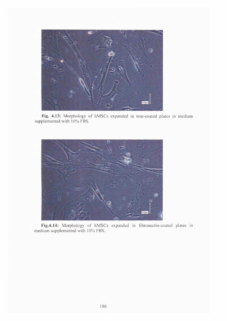
! 0 |iin
Fig. 4.13: Morphology o f hM SCs expanded in non-coated plates in medium supplemented with 10% FBS.
1 0 (U M
Fig.4.14: Morphology o f hM SCs expanded in fibronectin-coated plates in m edium supplemented with 10% FBS.
186

,1 0 |U tt
Fig. 4.15: Morphology of hMSCs expanded in Matrigel-coated plates in 10% FBS- supplemented medium.
Cell viability was assessed at each time-point for all serum and coating
conditions. These data are presented in table 4.8. Regardless of the coating condition,
10 % serum-supplementation permitted the highest cell viability, whilst serum
deprivation significantly affected cell viability, particularly for cells cultured in
fibronectin-coated or matrigel-coated plates. Overall, the culture within Matrigel-
coated plates was associated with the lowest cell viability values. There was no
significant difference between cell viability for non-coating and fibronectin-coating
conditions, both in 10 % FBS- or 2 % FBS- supplemented media. On the other hand,
the viability measured in serum-free culture was higher for cells expanded in non-
coated plates than for cells grown in fibronectin-coated plates.
187
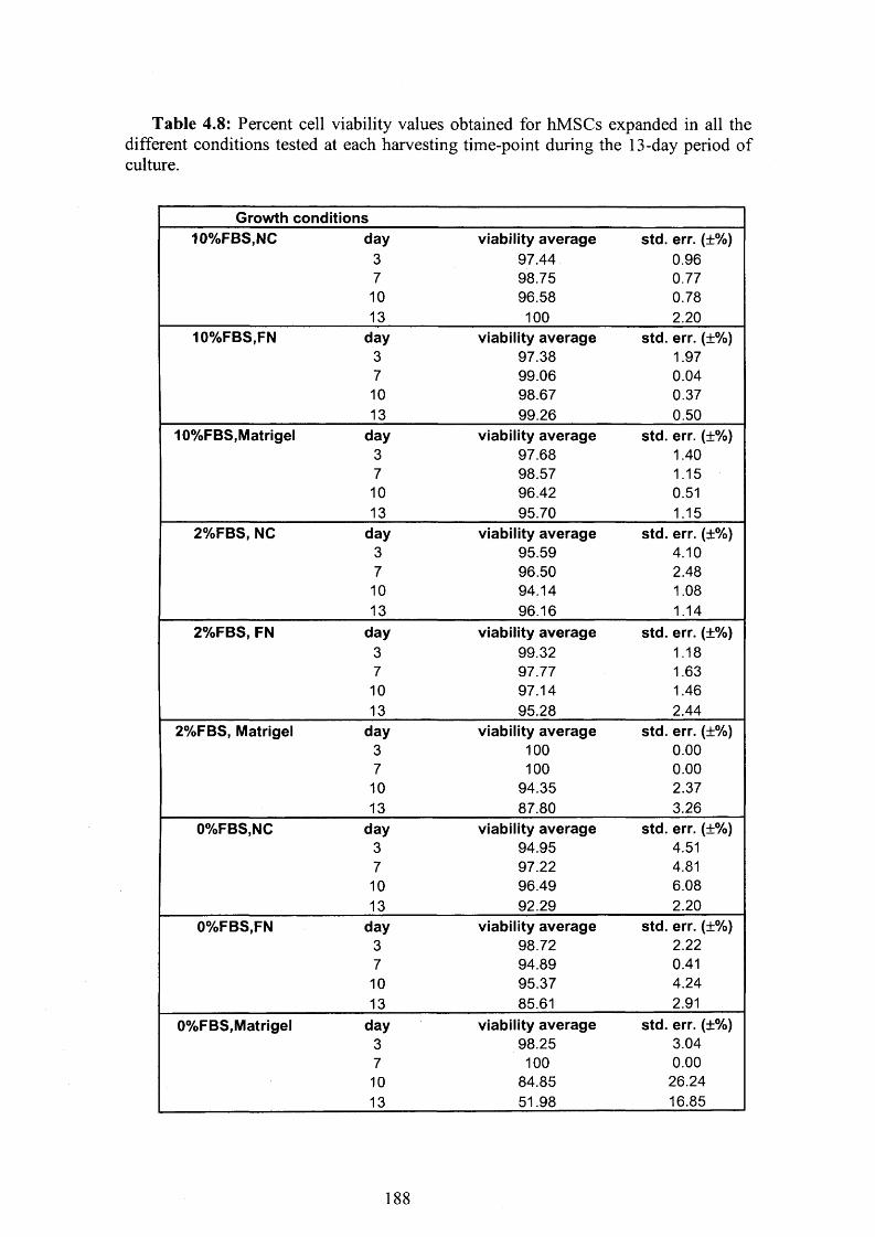
Table 4.8: Percent cell viability values obtained for hMSCs expanded in all the different conditions tested at each harvesting time-point during the 13-day period of culture.
Growth conditions10%FBS,NC day viability average std. err. (±%)
3 97.44 0.967 98.75 0.7710 96.58 0.7813 100 2.20
10%FBS,FN day viability average std. err. (±%)3 97.38 1.977 99.06 0.0410 98.67 0.3713 99.26 0.50
10%FBS,Matrigel day viability average std. err. (±%)3 97.68 1.407 98.57 1.1510 96.42 0.5113 95.70 1.15
2%FBS, NC day viability average std. err. (±%)3 95.59 4.107 96.50 2.4810 94.14 1.0813 96.16 1.14
2%FBS, FN day viability average std. err. (±%)3 99.32 1.187 97.77 1.6310 97.14 1.4613 95.28 2.44
2%FBS, Matrigel day viability average std. err. (±%)3 100 0.007 100 0.0010 94.35 2.3713 87.80 3.26
0%FBS,NC day viability average std. err. (±%)3 94.95 4.517 97.22 4.8110 96.49 6.0813 92.29 2.20
0%FBS,FN day viability average std. err. (±%)3 98.72 2.227 94.89 0.4110 95.37 4.2413 85.61 2.91
0%FBS,Matrigel day viability average std. err. (±%)3 98.25 3.047 100 0.0010 84.85 26.2413 51.98 16.85
1 8 8

The metabolic activity data obtained confirmed the overall effects of the different
culture plates surfaces on MSCs behaviour. These data are presented in figure 4.16
(10 % serum), figure 4.17 (2 % serum) and figure 4.18 (serum-free). In standard (10
%) serum condition, hMSCs expanded in traditional non coated-coated plates
exhibited higher metabolic activity than cells maintained in fibronectin-coated or
matrigel-coated plates. On the other hand, cells grown in serum-free cultures resulted
metabolically inactive, and regardless of the surface conditions examined they
resulted unable to consume any glucose or produce any lactate.
1.2
? 1 B 0.8 -o% 0.6 § 0.4 -0 0.2 -
Effects of coatings on hMSCs glucose metabolism and lactate production (10%FBS,
+bFGF)
0 -\-------------T------------ T---------------------
0 2 41 I - I I I
6 8 10 12 14 Time (days)
—♦— No Coating-Gluc Matrigel-Gluc
. X Fihrnner.tin-I an
— Fibronectin-Gluc No coating-Lac
- • Matrinal-1 an
Fig. 4.16: Effects of coatings on hMSCs metabolic activity. Cells were cultured in low-glucose DMEM medium supplemented with 10% (v/v) Fetal Bovine Serum (FBS) and 1 ng/mL basic Fibroblast Growth Factor (bFGF).
189

? 1 5 1 o>7T 1 - (0" i 0.5 - o3
Effects of coatings on hMSCs glucose metabolism and lactate production (2%FBS,
+bFGF)
r\______ _____ _____ --------------- j y _____ _________
O OHc-------------1----- ®----- 1-------------1----- ------- 1------------ ^ “ — i----- X ----- 1) 2 4 6 8 10 12 14
Time (days)
— No Coating-Gluc — Fibronectin-Gluc Matrigel-GLuc No Coating-Lac x - Fibronectin-Lac • Matrigel-Lac
Fig. 4.17: Effects of coatings on hMSCs metabolism. The cultures were maintained in low-glucose DMEM medium supplemented with 2% FBS and Ing/mL bFGF.
Effects of coatings on hMSCs glucose metabolism and lactate production (0%FBS,
+bFGF)
0 2 4 6 8 10 12 14
Time (days)
♦ No Coating-Gluc — Fibronectin-GLuc Matrigel-Gluc
No Coating-Lac —* — Fibronectin-Lac — Matrigel-Lac
Fig. 4.18: Effects of coatings on hMSCs metabolic activity. Cells were expanded in serum-free low-glucose DMEM medium supplemented with 1 ng/mL bFGF.
The specific glucose consumption rates (g/cell/hour) were calculated for each
serum and coating conditions as it is described in section 2.6.4. The values obtained
are presented in figure 4.19, 4.20 and 4.21. In the 10 % serum-supplemented culture
190

samples a significant increase in GCR was observed during the last 3 days of culture
only for hMSCs expanded in non-coated plates, whereas no significant increase in
GCR was detected for cells expanded in fibronectin- or Matrigel-coated plates.
Results for 2% serum or serum-free cells are more difficult to interpret (figure 4.19
and figure 4.21).
Effects of coating on Glucose Consumption Rate (10°/cFBS)
3.00E- -I
I 2.00E- -<v
S 1.00E- -
S O.OOE+O-
-1.00E- ^
NC — FN Matrigel
Fig. 4.19: Impact of coatings on 10% serum-supplemented hMSCs specific glucose consumption rate.
Effects of coating on Glucose Consumption Rate (2%FBS)
_ 8.00E-10 | 6.00E-10 - § 4.00E-10 - a 2.00E-10 - a 0.00E+00 --
-2.00E-10 f|-4.00E-10 - -6.00E-10 -1
NC FN Matrigel
Fig. 4.20: Effects of coatings on 2% serum-supplemented hMSCs specific glucose consumption rate.
Time (days)
10 12 144 6 8
Time
191

Effects of coating on Glucose Consumption Rate (0%FBS)
~ 3.00E-09 f 2.50E-09 - 8 2.00E-09 - 3 1.50E-09 - cc 1.00E-09 - O 5.00E-10 -
0.00E-K30 -0 2 4 6 8 10 12 14
Time (days)
NC — FN Matrigel
F i g . 4 . 2 1 : Effects of coatings on serum-free hMSCs specific glucose consumption
rate.
In conclusions, evaluation of different culture surfaces (i.e. standard non-coated,
fibronectin-coated and Matrigel-coated plates) for hMSCs, revealed that, regardless
of the serum concentration in the culture system, those based on Matrigel impeded
cell growth, viability and metabolic activity. On the contrary, both standard non-
coated and fibronectin-coated plates consistently supported hMSCs growth and
metabolism.
4.7 Multipotentiality of human adult mesenchymal stem
cells expanded in monolayer culture
For regeneration of human cartilage, bone and even osteochondral defects,
different cell sources are being investigated. In general, stem and progenitor cells are
preferable to final differentiated cells due to their proliferation and differentiation
capacity [Caplan and Goldberg, 1999; Lemare et al., 1998]. In vitro, culture-
expanded bone marrow-derived mesenchymal stem cells have indeed shown the
capacity to differentiate into a number of mesenchymal lineages, including
osteogenic, chondrogenic and adipogenic [Pittenger et al., 1999: Muraglia et al.,
2000; Jiang et a l, 2002]. MSCs’ ability to retain their multilineage differentiation
192

potential for over a significant number of cell doublings in culture is what makes
them so attractive for a broad range of tissue engineering applications [Reyes et al,
2001]. In vivo, several studies in a variety of animal models have shown that hMSCs
may be useful in the repair or regeneration of cartilage [Wakitani et al, 2002],
damaged bone [Livingston et al, 2001; De Kok et al, 2003], tendon [Award, et al,
2003] and meniscus [Murphy et al, 2003]. Other investigations have indicated that
MSCs may result useful also for repair of skeletal muscle [Wakitani et al., 1995],
while there has been also some evidence that hMSCs can also be induced to
differentiate into neurons in vitro [Woodbury et a l, 2000].
The differentiation potential of MSCs into multiple mesenchymal lineages - that
is, bone, cartilage and adipose tissue - is most commonly used as a functional
criterium defining MSC precursor cells [Fibbe, 2002]. In order to assess their
multipotency, fourth passage (p4) bone marrow-derived plastic adherent MSCs
isolated from a sample of adult frozen bone marrow were tested with the “Human
Mesenchymal Stem Cell Functional Identification Kit” (R&D Systems), which
permits the identification of human bone marrow-derived stem cells
(BMSCs)/mesenchymal stem cells (MSCs) based on their ability to differentiate into
mature phenotypes of adipocytes, osteocytes and chondrocytes. Prior to the start of
this study, the cells were expanded in vitro in “complete DMEM” medium,
formulated as described in section 2.1.5.1, until the necessary numbers of cells was
achieved. The typical morphology of the hMSCs expanded for this experiment and
seen prior the switch to the differentiation media is shown in figure 4.28 a).
In order to assess their adipogenic potential, hMSCs were plated at a density of
2 .1x l 0 4 cells/cm2 and were cultured with adipogenic medium, as it is described in
section 2.6.7.1. Morphologic changes in the cells, as well as the formation of neutral
lipid vacuoles, were noticeable 3 weeks after induction and were visualised by goat
anti-mouse FABP-4 immuno-staining (figure 4.22 A, B). Adipogenic differentiation
was not observed 21 days after induction in the negative control samples (not been
treated with the same FABP-4 antibody (figure 4.22 C)).
193

A
194

Fig. 4.22: Adipogenic differentiation of bone marrow-derived hMSCs. Monolayer MSCs were treated with adipogenic medium. After 21 days, the cells were fixed and immuno-stained for goat anti-mouse FABP-4. A y B (positive staining) and C (negative control).
To investigate the osteogenic potential of the bone marrow-derived MSCs, the
same cells were plated at a density of 4.2x10³ cells/cm2 and were incubated under
conditions appropriate for inducing differentiation for osteogenic lineage (please see
section 2.6.7.1). When induced to differentiate under osteogenic conditions, the
spindle shape of the bone marrow-derived cells flattened with increasing time of
induction and formed mineralized matrix, as evidenced by microscopic evaluation.
The osteocytes phenotype was shown after 21 days of induction by
immunofluorescence staining for mouse anti-human osteocalcin antibody (figure 4.23
A, B). Osteogenic differentiation, as determined by positive immunostaining for
osteocalcin antibody, was achieved in all the osteogenic medium-treated samples
tested with the osteocalcin antibody. The immunofluorescence assay performed on
induced cells not treated with human osteocalcin (negative controls) stained negative
for the antibody (figure 4.23 C).
195

A
196
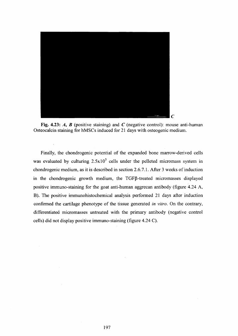
Fig. 4.23: A , B (positive staining) and C (negative control): mouse anti-human Osteocalcin staining for hMSCs induced for 21 days with osteogenic medium.
Finally, the chondrogenic potential of the expanded bone marrow-derived cells
was evaluated by culturing 2.5x10⁵ cells under the pelleted micromass system in
chondrogenic medium, as it is described in section 2.6.7.1. After 3 weeks of induction
in the chondrogenic growth medium, the TGFp-treated micromasses displayed
positive immuno-staining for the goat anti-human aggrecan antibody (figure 4.24 A,
B). The positive immunohistochemical analysis performed 21 days after induction
confirmed the cartilage phenotype of the tissue generated in vitro. On the contrary,
differentiated micromasses untreated with the primary antibody (negative control
cells) did not display positive immuno-staining (figure 4.24 C).
197
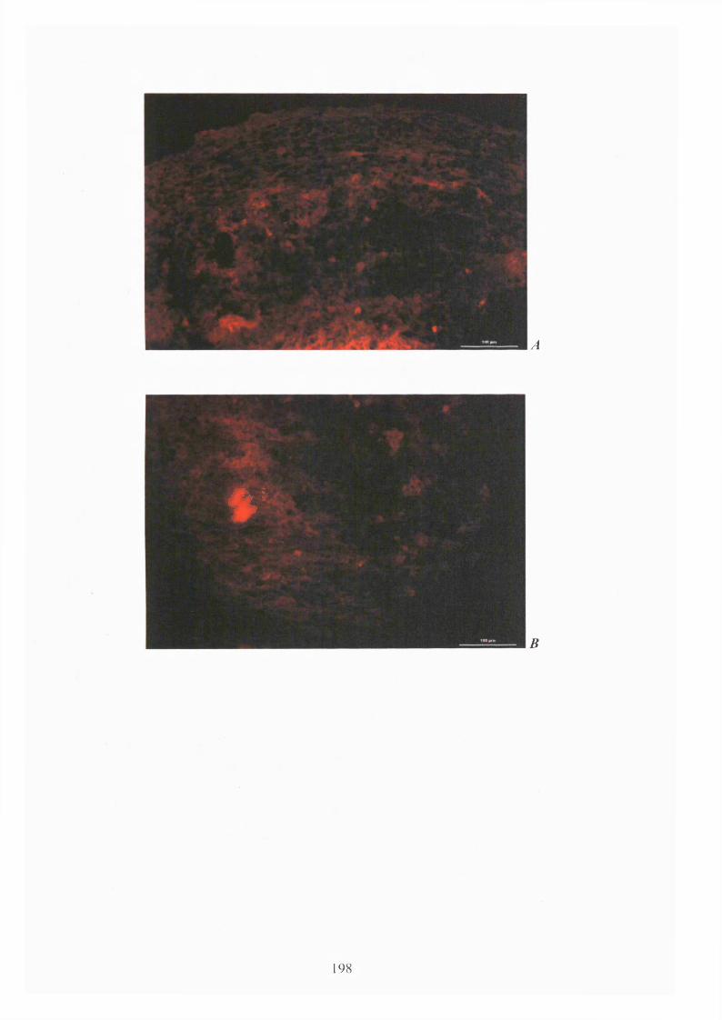
V
A
B
198

Fig. 4.24: A, B (positive staining) and C (negative control): goat anti-human Aggrecan staining for hMSCs induced for 21 days with chondrogenic supplement.
This study confirmed that human bone marrow-derived MSCs, expanded under
appropriate culture conditions, can be induced to differentiate in vitro toward the
adipogenesis, osteogenesis and chondrogenesis pathways. Under these conditions, the
cells showed the ability to successfully differentiate toward the three different
lineages, as demonstrated by the positive staining for the specific antibodies provided
with the “Human Mesenchymal Stem Cell Functional Identification kit”, defining the
mature phenotypes of adipocytes, osteocytes and chondrocytes. As a result, it can be
concluded that hMSCs originally isolated from a human frozen bone marrow sample
retain their multipotentiality after 4 expansion passages in culture.
4.8 Dissolved oxygen tension (DOT) as an important
parameter for hMSCs in vitro expansion
It is recognised that the dissolved oxygen tension (i.e. the exact oxygen level at
the cells/medium interface) is lower than the oxygen level in the humidified gas
mixture within the incubator. This is due to oxygen consumption by living cells.
However, since to our knowledge there is no practical methodology that can be
employed to measure the actual dissolved oxygen tension (DOT), we here make no

formal distinction between the two values, and refer to both simply as to “DOT” or
“oxygen level”.
This parameter has been demonstrated to play an important role on the growth
kinetics and functionality of a broad range of cell types grown in vitro, including rat
marrow-derived MSCs [Lennon et a l, 2001], human articular chondrocytes [Martin
et a l, 2004; Domm et al., 2004], human embryonic stem cells [Ezashi et al, 2005]
and human MSCs [Moussavi-Harami et al, 2004; Rochefort et a l , 2006]. The
importance of the oxygen levels in in vitro settings has already been elucidated in
section 1.5.5. Due to the established significance of this parameter on cell behaviour,
dissolved oxygen tension was thoroughly investigated in this research. Firstly, the
effects of the oxygen level alone on hMSCs proliferation rate and metabolism were
investigated (section 4.8.1). These studies were followed by experiments focusing on
the combined effects of the DOT and FBS-supplementation (section 4.8.2), and the
combined effects of the oxygen level and supplementation with bFGF (section 4.8.3).
To ensure maintenance of the desired gas mixture and DOT, incubators with two air
sensors, one for CO2 and the other O2, were used for all these studies.
4.8.1 Studies investigating hMSCs’ proliferation and metabolic
activity under different dissolved oxygen tensions
This section describes the studies performed on hMSCs cultures maintained in
standard serum (10 %-supplementation), bFGF (1 ng/mL-supplementation) and
coating (traditional non-coated tissue culture plates) conditions, where focus was kept
on varying the DOT value alone. A first experiment was conducted by growing
hMSCs for a 13-day period in one or the other of the following dissolved oxygen
tensions: 21 % (the typical ambient O2 level employed in most culture settings), 10 %
or 2 % (please see table 4.9 for clarification on the terminology adopted in this
research for defining the “ambient” and “hypoxic” conditions).
Table 4.9: Terminology adopted in this research to define the different oxygen levels investigated during in vitro expansion of hMSCs in monolayer culture.
2 0 0

Oxygen level 21 oxygen 6-10% oxygen 1 -2 % oxygen
Terminology
adopted in this
research
Ambient
(hyperoxic)
Normoxic Hypoxic
To understand the effect of relatively long-term, low DOT on hMSCs expansion,
in a second study ambient oxygen (21 %) culture was compared with 6 % and 1 %
DOT samples over a longer (21 days) time period. In order to characterize the growth
kinetics and metabolic activity of hMSCs under the various oxygen regimes, sixth
passage (p6 ) hMSCs were expanded in 24-well tissue culture plates that were
maintained in culture at 37°C, 5% CO2 in air in three different humidified (>95%)
incubators, each one set at one of the three selected DOTs for each experiment. All
the cultures were performed in traditional complete DMEM medium, formulated as
described in section 2.1.5.1. The cells were harvested and evaluated for cell density,
viability and metabolism at different time-points over the culture period under
examination.
In both studies, hMSCs grown at the 21 % standard DOT were characterised by
the highest proliferation potential, as it is evident from the graphs in figure 4.25 and
figure 4.26. On the other hand, cells maintained at lower oxygen regimes (10 %, and
2 % in figure 4.25, 6 % and 1 % in figure 4.26) did not show a similar growth
capacity.
2 0 1

Effects of Oxygen tension on hMSCs proliferation
50000 n
40000 -
< 30000 -
20000 -
10000 -
150 5 10
Culture period (days)
—♦— 21%Oxygen 10%Qxygen 2% Oxygen
Fig. 4.25: Effects of dissolved oxygen tension (DOT) on hMSCs proliferation. hMSCs were expanded in non-coated plates, in low-glucose DMEM medium supplemented with 10% FBS and lng/ml bFGF. The cells were incubated at 5%C02, 37°C and either 21% DOT (standard culture condition, • line), or 10% DOT ( ■ line), or 2% DOT ( line). Data are presented as means +/- s.e. of threeindependent measurements (please see section 2.6.9).
Effects of Oxygen tension on hMSCs proliferation35000
30000 -
25000 -
| 20000 -| 15000 - ©
10000 -
5000 -
20 2510 150 5C ulture period (days)
— 2 1 %Oxyge n - * — 6% Oxygen 1% Oxygen
Fig. 4.26: Impact of dissolved oxygen tension (DOT) on hMSCs proliferation. hMSCs were cultured in non-coated plates, in low-glucose DMEM medium supplemented with 10% v/v FBS and lng/ml bFGF. The cells were incubated at 5%C02, 37°C and either 21 % DOT (standard culture condition, —•— line), or 6 % DOT ( ■ line) or 1 % DOT ( line). Data are presented as means +/- s.e. ofthree independent measurements (please see section 2.6.9).
2 0 2

Even though higher cell proliferation was generally observed in the first
experiment than in the second one for all conditions, in both studies the same rather
weak but still negative effect of I0 W-O2 on cell expansion was noted. In both
experiments a 1.05-fold reduction in cell growth was seen after 2 weeks in culture by
reducing the DOT from ambient level to 10% or 6%, whereas hypoxic cultures were
characterized in both studies by a 1.3-fold decrease in cell proliferation over the same
time period. Overall, the pattern followed by the growth kinetics curves under the
lower oxygen conditions was similar in the two studies, and indicated that reducing
the dissolved oxygen tension in the attempt to approach the physiologic values and
mimic the in vivo environment does not enhance the cells proliferative capacity.
These results correlate well with the population doubling time and the cell growth
rate values obtained for both studies, and shown in table 4.10 and table 4.11.
Table 4.10: Population Doubling Level (PDL), Doubling Time (Td) and cell growth rate (p) values obtained for hMSCs expanded in the three dissolved oxygen tension conditions tested during the first week of culture.
Growth conditionsDay0-day7 PDL Td(hours) u(hours '1)
21%oxygen 3.01 55.79 0.01210%oxygen 2.49 67.42 0.0102%oxygen 2.34 71.89 0.009
Table 4.11: Population Doubling Level (PDL), Doubling Time (Td) and cell growth rate (p) values calculated for the hMSCs expanded in the three different oxygen conditions tested during the first week of culture.
Growth conditionsp (hours1)day0-day7 PDL Td(hours)
21 %oxygen 2.54 66.21 0.0106%oxygen 2.36 71.30 0.0101%oxygen 2.10 80.12 0.009
hMSCs expanded in the 21 % standard in vitro oxygen concentration were
characterized by the lowest doubling time and the highest cell growth rate calculated
during the first week of culture. On the other hand, the same cells expanded at the
lower DOTs required longer times to double and proliferate. Even though this
203

dissimilarity was small, particularly for the second experiment were the difference
was not statistically significant (p value = 0.048, Anova single factor test; table 4.11),
a trend was observed for which by reducing oxygen levels in the culture settings the
doubling times increased and concordantly the cell growth rates decreased.
As it is evident from the data shown in both table 4.12 and table 4.13, cell
viability was not significantly affected by decreasing the oxygen level in the culture
environment. For all three the oxygen levels investigated, high cell viability (> 98.47)
was retained after 13 days of in vitro expansion in the first experiment (please see
table 4.12). In addition, only a slight decrease in cell viability was observed over time
for cells maintained in culture for a longer time period in the second experiment
(please see table 4.13), indicating no O2 effect on cell viability.
Table 4.12: Percent cell viability values calculated for the cells cultured in the three different dissolved oxygen tension conditions under investigation at each harvesting time-point during the 13-day period of culture examined.
Growth conditions21%Oxygen day average viability std. err. (±%)
3 96.93 1.287 98.22 0.8610 99.49 0.2013 98.47 0.63
10%Oxygen day average viability std. err. (±%)3 96.37 0.927 98.06 0.6110 99.03 0.8613 99.06 0.55
2%Oxygen day average viability std. err. (±%)3 98.41 2.757 97.84 0.9610 99.27 0.3213 99.02 0.50
204

Table 4.13: Percent cell viability values calculated for hMSCs cultured in the three different dissolved oxygen tension conditions tested at the beginning (day 3) and at the end (day 2 1 ) of the culture period.
G row th co n d itio n s21% oxygen day av e rag e viability std . err. (±%)
3 96.15 2.0321 90.97 2.94
6% oxygen day av e rag e viability std . . (±%)3 96.83 0.25
21 92.01 1.511% oxygen day av e rag e viability std . err. (±%)
3 96.63 2.9421 90.67 2.71
hMSCs maintained in either ambient or low (1-10%) oxygen were essentially
identical as viewed by phase contrast microscopy. Individual subcultured cells
appearing as adherent, fibroblast-like healthy cells were similar to one another in
morphology and size regardless of the oxygen condition (please see figures 4.27 and
4.28).
Fig. 4.27: Morphology of hMSCs expanded in the traditional 21% Dissolved Oxygen Tension.
205

I Q H t n l
Fig. 4.28: Morphology of hMSCs expanded in 2% Dissolved Oxygen Tension condition.
As it is shown in figure 4.29 and figure 4.30, in line with the positive effect of
high DOT on cell proliferation rate and cell density, hMSCs expanded in the
traditional 21 % oxygen condition were characterised by a more intense metabolic
activity, as compared to cells maintained in lower oxygen conditions. Interestingly, in
both studies the positive effect of DOT on cell metabolism appeared to be
concentration-dependent, the metabolic activity was the lowest for cells maintained in
hypoxic (2 % or 1% oxygen) conditions.
E f fe c t s o f o x y g e n on G l u c o s e m e t a b o l i s m a n d L a c t a t e p r o d u c t i o n
1.2
5» 0.8
0.0
= 0.4
0.2
1010 14Ti m e
2% oxygen- - *-2% oxygan-
21 %oxygtn*21%oxyg«n- 10% oxygen-
Fig. 4.29: Impact of dissolved oxygen tension on hMSCs metabolic activity.
206

Effects of Oxygen on hMSC Glucose metabolism and Lactate production
» 0.8o» 0.6
« 0 .45 0.2
Time (days)
21%Oxygen-GLuc — 6%Oxygen-Gluc 21%Oxygen-Lac H*-6%Oxygen-Lac
1%Oxygen-Gluc1%Oxygen-Lac
Fig. 4.30: Effects of dissolved oxygen tension on hMSCs metabolic activity. The cells were cultured in either 21 % or 6 % or 1 % dissolved oxygen tensions for 21 days.
Specific glucose consumption rate data are presented in figure 4.31 and figure
4.32. Interestingly, cells expanded in the ambient (21%) oxygen condition seemed to
be characterised by a significant increase in GCR (g/cell/hour) during the last 4 days
in culture.
Effects of Oxygen on Glucose ConsumptionRate
1.00E-09 -i
O.OOE-HDO
-2.00E-09 -
-3.00E-09 J
21%Oxygen 10%Oxygen 2%Oxygen
Fig. 4.31: Effects of dissolved oxygen tension on hMSCs specific glucose consumption rate.
207

Effects of oxygen on Glucose ConsumptionRate
6.00E-09
4.00E-09
| 2.00E-09 -
g 0.00E+00 -
3 -2.00E-09 0 10 15 20 25
O -4.00E-09 - O-6.00E-09
-8.00E-09 -
Time (days)
— 2 1 %o x y g e n —■— 6% oxygen 1% oxygen
Fig. 4.32: Effects of dissolved oxygen tension on hMSCs specific glucose consumption rate. The cells were cultured in either 21% or 6% or 1% dissolved oxygen tensions for 21 days.
4.8.2 Combined effects of dissolved oxygen tension (DOT) and
Fetal Bovine Serum (FBS) on hMSCs proliferation and metabolism
In order to evaluate the combined effects of high and low dissolved oxygen
tension and FBS-supplementation during in vitro expansion, hMSCs were cultured in
either complete DMEM or serum-free DMEM for two weeks under a humidified
(>95%) atmosphere of the high (21%) or the low (1%) DOTs (5% CO2 in air). In
addition to cell density, viability and metabolism analyses, the cell samples were
assessed by means of the Live/Dead Viability/Cytotoxicity assay with calcein
AM/ethidium homodymer described in section 2.6.6. This two-colour fluorescence
cell viability technique was used to measure intracellular esterase activity and plasma
membrane integrity as indicators of cell viability.
The growth kinetics curves obtained from this study are presented in figure 4.33.
In line with the results observed in the studies presented in the previous sections
(sections 4.3, 4.4 and 4.6), hMSCs proliferative capacity was significantly enhanced
by culture in FBS-supplemented medium. Regardless of the dissolved O2 tension
maintained over the 14-day period of culture, serum-free culture did not allow cell
growth. In addition, and in accordance with the results shown above (section 4.8.1),
2 0 8

2-week (short-term) exposure of serum-supplemented hMSCs to 1 % oxygen was
associated with a similar diminished proliferation potential, being 1.2-fold the
decrease in cell growth measured as compared with growth of cells expanded in
ambient oxygen level.
Effects of FBS and Oxygen tension on hMSCs proliferation
50000 n
40000 -
9 30000 -
I 20000 -<Af 10000 -
0
-10000 ^ 5 10
Culture period (days)
+FBS, 21% Oxygen
+FBS, 1%OxygennoFBS, 21% Oxygen
noFBS, 1% Oxygen
15
Fig. 4.33: Effects of dissolved oxygen tension (DOT) on hMSCs proliferation in serum-free and 10% serum culture conditions. hMSCs were cultured in non-coated plates, in low-glucose DMEM medium supplemented with lng/ml bFGF. The cells were incubated in 5%C02, 37°C incubators with 21%02 level (10%serum standard culture condition = ■» line; serum-free condition = —■— line) or 1 % 0 2 level (10%serum or serum-free = and lines respectively). Data are presented as means +/- s.e. of three independent measurements (please see section 2.6.9).
These data were confirmed by the cell viability measurements obtained with the
trypan blue dye exclusion assay and presented in table 4.14. High cell viability
(>91%) was maintained over the 2-week period in serum-supplemented cultures,
whereas serum-deprivation resulted in important cell death over time. Interestingly,
this phenomenon was particularly evident and significant for serum-free cells
expanded in hypoxic (1% O2 ) condition, for which cell viability measured 14 days
after plating was reduced to 55%.
209
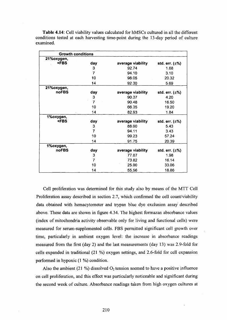
Table 4.14: Cell viability values calculated for hMSCs cultured in all the different conditions tested at each harvesting time-point during the 13-day period of culture examined.
Growth conditions21%oxygen,
+FBS day average viability std. err. (±%)3 92.74 1.687 94.10 3.10
10 98.05 20.3214 92.30 5.69
21%oxygen,noFBS day average viability std. err. (±%)
3 90.37 4.207 90.48 16.50
10 66.35 19.2014 82.93 1.84
1%oxygen,+FBS day average viability std. err. (±%)
3 88.00 5.437 94.11 3.43
10 99.23 57.2414 91.75 20.39
1%oxygen,noFBS day average viability std. err. (±%)
3 77.07 1.987 73.82 16.14
10 25.00 33.0614 55.56 18.86
Cell proliferation was determined for this study also by means of the MTT Cell
Proliferation assay described in section 2.7, which confirmed the cell count/viability
data obtained with hemacytometer and trypan blue dye exclusion assay described
above. These data are shown in figure 4.34. The highest formazan absorbance values
(index of mitochondria activity observable only for living and functional cells) were
measured for serum-supplemented cells. FBS permitted significant cell growth over
time, particularly in ambient oxygen level: the increase in absorbance readings
measured from the first (day 2) and the last measurements (day 13) was 2.9-fold for
cells expanded in traditional (2 1 %) oxygen settings, and 2 .6 -fold for cell expansion
performed in hypoxic (1 %) condition.
Also the ambient (21 %) dissolved O2 tension seemed to have a positive influence
on cell proliferation, and this effect was particularly noticeable and significant during
the second week of culture. Absorbance readings taken from high oxygen cultures at
210
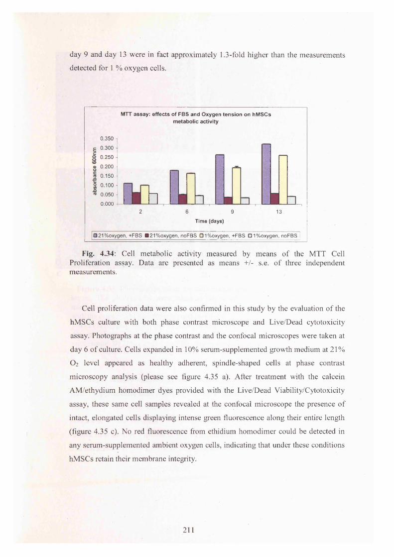
day 9 and day 13 were in fact approximately 1.3-fold higher than the measurements
detected for 1 % oxygen cells.
MTT assay : effec ts of FBS and Oxygen ten sion on hM SCs m etabolic activity
0.350 i
E 0.300
| 0.2500.250
® 0.200 1
5 0.150 -o 0.100 -
« 0.050
0.0002 6 9 13
Time (days)
□ 21%oxygen, +FBS ■21%oxygen, noFBS 01%oxygen, +FBS 01%oxygen, noFBS
Fig. 4.34: Cell metabolic activity measured by means of the MTT Cell Proliferation assay. Data are presented as means +/- s.e. of three independent measurements.
Cell proliferation data were also confirmed in this study by the evaluation of the
hMSCs culture with both phase contrast microscope and Live/Dead cytotoxicity
assay. Photographs at the phase contrast and the confocal microscopes were taken at
day 6 of culture. Cells expanded in 10% serum-supplemented growth medium at 21%
O2 level appeared as healthy adherent, spindle-shaped cells at phase contrast
microscopy analysis (please see figure 4.35 a). After treatment with the calcein
AM/ethydium homodimer dyes provided with the Live/Dead Viability/Cytotoxicity
assay, these same cell samples revealed at the confocal microscope the presence of
intact, elongated cells displaying intense green fluorescence along their entire length
(figure 4.35 c). No red fluorescence from ethidium homodimer could be detected in
any serum-supplemented ambient oxygen cells, indicating that under these conditions
hMSCs retain their membrane integrity.
211

c DFigure 4.35: Photographs taken for cells maintained at 21% DOT, with or without
serum. The photographs were taken at day 6 o f culture with the phase contrast microscope (A and B) and with the confocal microscopes (C and D) after treatment with the calcein AM/ethydium homodimer dyes.
A) 21% oxygen, +FBS, day 6, phase contrast microscope; B) 21% oxygen, no FBS, day 6, phase contrast microscope; C) 21% oxygen, +FBS, day 6, confocal microscope; D) 21% oxygen, no FBS, day 6, confocal microscope.
Similar healthy morphology and intense green fluorescence were observed for
serum-supplemented cells grown in hypoxic (1% oxygen) condition. However, in this
case viable cells were visible in lower numbers, while a weak red fluorescence was
noticeable at the confocal microscopy analysis (please see figure 4.36 c). These
results indicate that hypoxia might be associated with cytotoxicity, a phenomenon
that has already been observed for hepatocytes [Khan and O ’Brien, 1995].
212

■ ■
m
C DFigure 4.36: Photographs taken for cells maintained at 1% DOT, with or without serum. The photographs were taken taken at day 6 o f culture with the phase contrast microscope (A and B) and with the confocal microscopes (C and D) after treatment with the calcein AM/ethydium homodimer dyes.
A) 1% oxygen, +FBS, day 6, phase contrast microscope; B) 1% oxygen, no FBS, day 6, phase contrast microscope; C) 1% oxygen, +FBS, day 6, confocal microscope; D) 1% oxygen, no FBS, day 6, confocal microscope.
In marked contrast, in all serum-free samples examined only very few adherent
and elongated cells were visible at inverted light microscope (figure 4.35 b and figure
4.36 b), m ost o f the cells having been removed with medium refreshments after
detachment from the culture surface. Green calcein fluorescence was absent from all
serum-free samples, confirming very low cell viability, in this culture condition
(please see figure 4.35 d and figure 4.36 d). Diffuse red fluorescence o f low intensity
was noticed in the cultures maintained in serum-free and hypoxic condition (figure
4.36 d), revealing damaged plasma membranes in these cell samples.
213

The doubling time and cell growth rate values determined for hMSCs expanded in
high and low oxygen with or without serum are shown in table 4.15. These values are
in accordance with the growth kinetic data described above and with the results
described in the previous sections. In standard serum-supplemented cultures, 21 %
DOT cells displayed lower doubling time and higher cell growth rate values than cells
cultured in hypoxic condition. The lack of cell growth in serum-free culture was
confirmed by the (negative) doubling time and cell growth rate values (please see
table 4.15).
Table 4.15: Population Doubling Level (PDL), Doubling Time (Td) and cell growth rate (p) values calculated for the cells expanded in all the different conditions tested during the first 1 0 days of culture.
Growth conditionsDay0-day10 PDL Td(hours) p hours '1)
21 %oxygen,+FBS 2.898 82.81 0.008421%oxygen,-FBS -2.017 -118.97 -0.00581%oxygen,+FBS 2.537 94.59 0.00731%oxygen,-FBS -3.892 -61.67 -0.0112
In 10% serum-supplemented samples, high DOT seemed to permit a slightly more
intense metabolic activity as compared to low DOT (figure 4.37). However, in serum-
free condition hMSCs appeared to be metabolically inactive, regardless of the oxygen
level employed during in vitro cell expansion (figure 4.38).
214

Effects of oxygen on glucose metabolism and lactate production (+FBS)
1 -i
I 0 -8 -o 0.6 -I5. 0.4 -j o3 ° ^
8 10 12 14
21% cxygerKjucose 21% cxygerHactate
1% oxygen- ucose 1% oxygerHactste
16
Fig. 4.37: Effects of dissolved oxygen tension on hMSCs metabolic activity. Cells were cultured in either 21 % or 1 % dissolved oxygen tension in FBS-supplemented low-glucose DMEM medium.
Effects of oxygen on glucose metabolism and lactate production (no FBS)
1.2 -
d 1 S 0.8o™ 0.6 -
o 0.4 | 0.2 J
5 -r- ^ i1 0 1 5Time (days)
—♦— 21 %oxygen-glucose 21 %oxygen-lactate1 %oxygen-glucose 1 %oxygen-lactate
Fig. 4.38: Effects of dissolved oxygen tension on hMSCs metabolic activity. Cells were cultured in either 2 1 % or 1% dissolved oxygen tensions in serum-free low- glucose DMEM medium.
According to the data presented in figure 4.39, there appeared to be no significant
difference in specific glucose consumption rate (g/cell/hour) for hMSCs cultured for
up to 10 days in the different FBS and oxygen conditions. However, cells expanded in
serum-supplemented medium at the ambient (2 1 %) oxygen experienced a significant
increase in GCR over the last 4 days in culture, whereas cells maintained in hypoxic
conditions in serum-free DMEM medium appeared to be characterised by a
significant decrease in GCR over the same culture period (dayl 0-day 14).
215

Effects of FBS and oxygen on Glucose consumption rate
2.00E-08
1 50E-08 ■
1 00E-08 ■
5.00E-09 -
0.00E+00 --
-5.00E-09 (I-1 00E-08 -
Time (days)-1 .S0E-08
noFBS, 21%oxygen noFBS .1 %oxyqen
Fig. 4.39: Effects of dissolved oxygen tension and FBS-supplementation on hMSCs specific glucose consumption rate.
4.8.3 Combined effects of dissolved oxygen tension (DOT) and
basic fibroblast growth factor (bFGF) on hMSCs proliferation and
metabolism
Given the positive impact that bFGF had demonstrated on hMSCs proliferative
capacity in the previous investigations (please see results presented in section 4.5),
the combined effects of dissolved oxygen tension and bFGF-supplementation were
also evaluated.
For this purpose, hMSCs were cultured in either complete (bFGF-supplemented)
DMEM or in the same DMEM medium formulated without bFGF. The cultures were
maintained for two weeks in two separate incubators (>95% humidity, 5% CO2 in
air), one equipped with ambient (21%) and the other with low (1%) DOT. Growth
kinetics, cell doubling time and viability were assessed as for the previous
experiments. In addition, in this study mitochondrial activity and cell apoptosis were
determined with the Guava Nexin assay, following the protocol described in section
2.6.8. This technique was performed in order to identify and distinguish in our
samples apoptotic cells from dead cells and debris.
216

Once again, the high DOT maintained during in vitro expansion supported
hMSCs growth more efficiently than the low oxygen, as it is evident from the growth
kinetics curves presented in figure 4.40. This effect was observed for both bFGF-
supplemented and bFGF-free culture conditions, for which increase in cell
proliferation measured 13 days after plating was respectively 1.3 and 1.4-fold.
Interestingly, bFGF-supplementation was associated with significantly increased
cell proliferation over the 13 days of culture for both cells expanded in ambient
oxygen (1.33-fold increase) or in the hypoxic condition (1.4-fold increase). These
data were in accordance with the bFGF results presented previously (section 4.5), and
confirmed the key role played by the growth factor in the hMSCs in vitro
proliferation.
Effec ts o f bFGF a n d O x y g e n t e n s i o n o n h M S C s p ro l i fe ra t ion
50000
^ 40000 -
| 30000 •
| 20000 - * 10000 ■
0 -- 0
— +FGF, 21%Oxygen — noFGF, 21 %Oxygen
+FGF, 1%Oxygen_________noFGF, 1 %Oxygen
Fig. 4.40: Growth kinetics curves obtained for hMSCs maintained for a 13-day period under different dissolved oxygen tension (DOT) conditions, with either bFGF- supplemented or bFGF-free growth medium. hMSCs were cultured in non-coated plates, in low-glucose DMEM medium supplemented with 10 % Fetal Bovine Serum. The cells were maintained in 5% C 02, 37°C incubators with either 21 % 0 2 level (with bFGF, standard culture condition, represented by the • line; bFGF-free condition, represented by the " line) or 1% 0 2 level (with or without bFGF,
line and line respectively). Data are presented as means +/- s.e. of three independent measurements (please see section 2.6.9).
4 6 8 10
Culture period (days)12 14
217

The population doubling and growth rate values obtained from this investigation
confirmed the positive effect of the basic Fibroblast Growth Factor. bFGF-
supplementation allowed the cells to double faster both in high and low oxygen
regimes, as demonstrated by the Td values shown in table 4.16. The effect of the
growth factor was particularly important in ambient oxygen condition. In accordance
with the data described in the previous sections (4.8.1 and 4.8.2), the lowest doubling
time and highest cell growth rate values were measured for cells grown at the
standard 21 % DOT in complete DMEM medium after bFGF-supplementation.
Interestingly, even though doubling times were lower for cells in ambient oxygen,
there appeared not to be a significant difference in cell growth rates for bFGF-
deprived cells grown in either ambient or hypoxic conditions.
Table 4.16: Population Doubling Level (PDL), Doubling Time (Td) and cell growth rate (p) values calculated for hMSCs expanded in all the different conditions tested from day 3 to day 6 of culture.
Growth conditionsDay0-day10 PDL Td(hours) H (hours'1)
21 %oxygen,+FGF 2.799 85.7 0.00821%oxygen,-FGF 1.350 177.8 0.0041 %oxygen,+FGF 2.127 112.8 0.0061%oxygen,-FGF 1.506 159.3 0.004
The combined effects of the DOT and bFGF-supplementation on cell viability
were also analysed. Cell viability was assessed as usual using the trypan blue dye
exclusion assay, according to the protocol described in section 2.6.3. High cell
viability was maintained over the 13-day period of culture for all the three different
oxygen conditions tested (please see table 4.17). As in all the studies described above
(section 4.8.1 and 4.8.2), regardless of DOT, the cells did not experience any
dramatic loss in cell viability over time. In addition, cell viability was not affected by
supplementation of the growth medium with bFGF, as the percentage of viable
hMSCs was over 90.05 % for all culture conditions, regardless of bFGF-
supplementation. These results confirmed that even though bFGF may promote
considerably hMSCs viability in serum-free cultures (please see results described in
tables 4.3 and 4.6), this effect is not so relevant for cells expanded in serum-
supplemented DMEM.
218

Table 4.17: Percent cell viability values calculated for hMSCs cultured in all the different conditions tested at the beginning (day 3) and at the end (day 13) of the culture period.
Growth conditions 21%oxygen,
+FGF day313
average viability89.6792.42
std. err. (±%) 3.45 2.49
21 %oxygen,noFGF day average viability std. err. (±%)
3 91.43 3.0913 94.22 1.01
1%oxygen,+FGF day average viability std. err. (±%)
3 82.92 3.4513 90.05 2.39
1%oxygen,noFGF day average viability std. err. (±%)
3 92.09 8.6013 92.33 2.71
At the end of the 13-day culture period under examination the Nexin assay, which
is based upon the Guava EasyCyte system, was performed in order to investigate the
effects of both DOT and bFGF-supplementation on hMSCs’ apoptosis. The protocol
for the Guava Nexin assay is described in detail in section 2.6.8, and the results from
this analysis are presented in table 4.18. It must be noted that no significant difference
was detected with this technique in percentages of viable cells for all oxygen and
bFGF conditions, and high cell viability (>97%) was observed in all conditions over
the 13-day period of culture examined (please see table 4.18). In the same way,
similar percentages of early apoptotic (0.2-0.4%) and late apoptotic (1.1-1.4%) cells
were measured for all cell samples, regardless of the dissolved oxygen tension in
culture settings or the bFGF presence in the growth medium. These data
demonstrated that, even though hMSCs incubated under hypoxia had decreased rates
of proliferation, there was no increase in apoptosis for these samples. The results
were consistent with previous reports [Salim et al., 2004, Hung et al., 2007] but not
with other publisehd studies [e.g. Fink et al., 2004; Lennon et al. 2001]. The
conflicting results may in part be explained by the variation in the DOT, the duration
of hypoxic culture and the system used in each study to control the oxygen level
[Hung et al., 2007].
219

Table 4.18: Influence of dissolved oxygen tension and bFGF-supplementation on hMSCs viable, early apoptotic and late apoptotic cell populations, as obtained with the Nexin cell apoptosis assay performed on the Guava EasyCyte system. Data are presented for measurements taken at the end of the 13-day culture period.
Growthconditions
Averageviablecells
Standarderror
Averageearly
apoptoticcells
Standarderror
Averagelate
apoptoticcells
Standarderror
21%oxygen,+FGF
21%oxygen,noFG
F1%oxygen,
+FGF1%oxygen,
+FGF
97.00 0.523 0.35 0.159 1.36 0.184
97.11 0.137 0.37 0.053 1.10 0.094
97.98 0.554 0.22 0.106 1.19 0.486
97.78 0.205 0.28 0.059 1.17 0.225
As it is shown in figure 4.42, cells expanded in the absence of bFGF appeared to
be unable to metabolise any glucose or produce any lactate, and this metabolic
inactivity was observed for both cell samples expanded in high or low DOTs. On the
other hand, bFGF-supplementation seemed to permit a slightly higher metabolic
activity, and in this case cells maintained in high DOT seemed to be more
metabolically active than cells expanded in hypoxic conditions (figure 4.41).
220

Effects of oxygen on glucose metabolism and lactate production (+ bFGF)
1? 0.8 o>o 0.6 re“i 0.4oI 02-
8 10 12
■21% oxygen-glucose
1% oxygen-glucose
21% oxygen-lactate
1% oxygen-lactate
14
F ig. 4 .41: Effects of dissolved oxygen tension on hMSCs metabolic activity. The cells were cultured in either 21 % or 1 % dissolved oxygen tensions in bFGF- supplemented low-glucose DMEM medium.
Effects of oxygen on glucose metabolism and lactate production (no bFGF)
1.2 -
1
0.8ore 0.6d 0.43o 0.2
06 8 Time (days)
10 12
21 % oxygen-glucose
1% oxygen-glucose
21% oxygen-lactate
1% oxygen-lactate
14
F ig . 4 .42: Effects of dissolved oxygen tension on hMSCs metabolic activity. Cells were cultured in either 21 % or 1 % dissolved oxygen tensions in bFGF-free low- glucose DMEM medium.
The specific glucose consumption rate data are presented in figure 4.43. These
data are quite difficult to interpret. However, it can be noted that cells expanded in
hypoxic condition, in bFGF-free medium appeared to be characterised by an increase
in GCR from day6 to day 10 in culture, whereas GCR for these cells seemed to
221

decrease during the last 4 days in culture. No significant difference in GCR was
observed instead for the cells maintained in all other culture conditions investigated in
this experiment.
Ef
4.00E-09 -]
£ 2.00E-09 -
fects o f FGF and oxygen on glucose consum ption rate
g u.uut+uu -
S -2.00E-09 - 01O -4.00E-09
-6.00E-09
------------- 9 I ■—r """ * - ----1) 5 10 15
Time (days)
—♦—FGF, 21%oxygen —•—noFGF, 21%oxygen FGF, 1%oxygen noFGF, 1%oxygen
Fig. 4.43: Effects of dissolved oxygen tension and bFGF-supplementation on hMSCs specific glucose consumption rate.
4.8.4 Preparation of hMSCs working cell banks at 21% and 1%
oxygen levels
The aim of this study was to compare the expansion and behaviour of human
mesenchymal stem cells maintained in monolayer culture under two different
dissolved oxygen tensions (i.e. 21 % and 1 %). In order to do so, after cell isolation
from the same bone marrow sample by discontinuous density gradient centrifugation,
two hMSC cultures were performed in parallel in T75 flasks that were maintained
throughout the whole experiment in the two different DOTs selected. The standard
low-glucose “complete DMEM” formulation was used for the cell expansion for both
the oxygen cultures. Once a sufficient number of cells was reached within the T75
flasks, two separate working cell banks were prepared for each of the two different
DOT conditions under investigation. The resulting cells were transferred and
222

maintained in liquid nitrogen (-196°C) for long-term storage, in order to supply
material for future studies investigating further the effects of DOT on MSCs
proliferation and function in culture. The protocols followed for the cell isolation and
the creation of the working cell banks are described in sections 2.1.1 and 2.1.4
respectively.
After cell isolation from the same bone marrow source, successful hMSCs
expansion was achieved in both oxygen levels. In both conditions the cells started to
adhere to the plastic surface of the tissue culture vessels and began to elongate within
48 hours after plating. Cell proliferation occurring in both oxygen conditions was
comparable and equally slow, and the desired 80 % confluence was reached in
parallel in the two culture systems. In both oxygen conditions the primary culture
cells reached the desired confluence 22 days after plating. In both cases, second
passage (p2 ) cells were again approximately 80 % confluent and ready for
trypsinisation after 11 days in culture. 2 1 % oxygen and 1% oxygen-cells displayed
similar cell growth rates (p) values (0 .0 0 2 2 and 0 .0 0 2 1 hours' 1 respectively) and
doubling time (Td) values (318.3 and 329.9 hours respectively). Both oxygen cell
types displayed also similar morphology, as assessed by the microscopic evaluation
of the culture samples. In both cases, the cells appeared to have a spindle-shape
fibroblast-like morphology, characteristic of healthy bone marrow-derived MSCs
(data not shown).
Despite the extremely high doubling time values measured, the microscopic
evaluation of the samples, performed every other day during the whole culture period,
revealed over time the presence of healthy, elongated spindle-like cells adhering to
the plastic surface of the tissue flasks. In addition, successful cell expansion leading
to cell numbers sufficient for the preparation of a working cell bank was achieved in
both oxygen conditions. In conclusion, no discemable differences were noticed in the
cell behaviour during this expansion procedure, indicating that oxygen level does not
affect hMSCs proliferation capability and cell morphology in primary culture.
223

4.9 Conclusions on MSCs monolayer culture studies
The increasing number of clinical protocols using hMSCs underscores the need
for serum supplements other than Fetal Bovine Serum [Shahdadfar et al., 2005]. As a
result, supplementation of the cell culture medium with FBS was one of the principal
parameters investigated in this study. In conclusions, this research substantiated the
significant positive influence of FBS in promoting cell proliferation rate for MSCs
derived from human bone marrow samples. Serum, which delivers important
nutrients, proteins and growth factors to the culture system (please see section 1.5.2),
resulted essential for cell survival and long-term cell expansion in in vitro monolayer
conditions. Importantly, the 10 % v/v serum concentration traditionally employed in
standard culture protocols, resulted the most appropriate for yielding highest cell
densities and allowing hMSCs metabolic activity.
Preliminary studies were also conducted to define the optimal basal growth
medium for hMSCs expansion, and low-glucose DMEM was chosen against the more
expensive MesenCult medium because of its advantages in terms of number of cells'X 9generated after 12 days in serum-supplemented culture (12.5x10 cells/cm versus
"X 98.25x10 cells/cm counted in 10% serum-supplemented cultures).
Several serum-free media have also been investigated in a preliminary qualitative
study whereby media effects were observed purely by means of inverted light
microscopy. Interestingly, among all the formulations examined, UltraCulture
(BioWhittaker), UltraDOMA Hybridoma (BioWhittaker) and Knockout D-MEM
(Gibco) optimised media permitted survival and proliferation of attachment-
dependent fibroblast-like cells resembling pure MSCs. Future studies could be
performed to characterise the cell samples grown in these medium formulations and
to prove whether these cultures retain all the unique and desirable features
characteristic of unadulterated authentic stem cells.
The cytokines released in the MSCs’ microenvironment appears to be an
additional essential factor for proper development. Among all growth factors,
Fibroblast Growth Factor, which is known to be an effective mitogen for a variety of
cells of mesenchymal or neuroectodermal origin in vitro [Basilico and Moscatelli,
1992; Scherping et al, 1997], demonstrated to play a key role in promoting hMSCs
proliferation and activity. bFGF appeared to be able to significantly obstruct cell
death in serum-free cell samples. In addition, in the standard 10% serum-
224

supplemented cultures, after 13 days, bFGF-addition resulted in a 1.9-fold increase in
cell growth as compared to cells expanded in bFGF absence. Interestingly, the most
remarkable positive influence of bFGF was observed in low (2%) serum
concentration samples, for which the growth factor permitted a 4.2-fold cell growth
increase over 13 days, as opposed to the 1.2-fold increase measured over the same
time period for bFGF-free cells.
Also the culture surface properties investigated in this research were demonstrated
to have a significant influence on hMSCs behaviour. Matrigel proved not to be a
favourable coating surface for cell growth, leading to both significant cell death over
time and metabolic inactivity. Its negative effect was noticed regardless of the serum
concentration used for the medium supplementation. Moreover, the use of
fibronectin-coated plates did not reveal any particular advantage as compared to the
traditional non-coated plates frequently employed for tissue culture. Similar growth
kinetics were observed for cells grown in fibronectin-coated or non-coated vessels in
all serum conditions, and a similar increase in cell density (approximately 9-fold
increase for 10% serum condition and 4.5-fold increase for 2% serum samples) was
observed over 13 days in culture for both culture surfaces. Cell morphology and cell
viability were also similar under these two coating conditions.
Finally, in this research several investigations were performed in order to
elucidate the impact of oxygen levels on hMSCs expansion potential and metabolic
activity. It has already been envisaged that among all tissue environmental factors
affecting MSCs functionality and differentiation, perhaps the most important is
oxygen level [Salim, 2004]. However, to date, studies focusing on mesenchymal stem
cells responses to hypoxia have been inconclusive [Lennon et al, 2001; Ma et al,
2001]. Since in the bone marrow environment oxygen level is low, and normally
ranging from 5.7 % to 7.1 % (40-50 mmHg) [Ishikawa and Ito, 1988; Grant and
Smith, 1963; Harrison et al, 2002], it was decided to test whether hypoxia can
enhance proliferation and growth potential of MSCs isolated from human bone
marrow samples. In conclusion, the results of these studies clearly demonstrated that
the exposure of hMSCs to high (21%) dissolved oxygen tension (maintained by
exposure to 5 %CC>2 in air) during in vitro expansion is beneficial for cell growth. On
the other hand, exposure to low DOT during culture did not significantly improve the
cells proliferation capacity. However, no significant treatment effect by DOT was
found in the percentages of viable cells.
225

Overall, the best results, yielding highest cell growth kinetics (average 38.9xl04
cells/cm counted after 13-14 days in culture), highest cell viabilities (average
96.08% +/-3.01% over 2-week culture), and lowest doubling times (average 58.36
hours calculated from day 0 to day 7) were derived from the combined use of the high
DOT and serum-/bFGF-supplemented low-glucose DMEM medium and non-coated
culture vessels. It was also demonstrated that hMSCs expanded for four subsequent
passages in monolayer culture under these conditions appeared to retain their
undifferentiated state and their ability to differentiate toward the phenotypes of fully
mature adipocytes, osteocytes and chondrocytes.
226

5. Characterisation of alginate/cells constructsFollowing the studies establishing the growth kinetics of unencapsulated cells
expanded in tissue flasks and microwell plates (described in chapter 4), interest was
focused on investigating the growth of cells within alginate hydrogels. The optimal
conditions of culture identified in monolayer, allowing cell viability and promoting
cell proliferation, were employed for the growth of the alginate/cells constructs
resulting after immobilisation of the cells in the alginate matrix. This chapter
describes the results obtained from the characterisation of these constructs.
5.1 Alginate-GRGDY as scaffold for cell growth
The ultimate research goal was the fabrication of alginate/hMSCs constructs
suitable for use in the clinic. In order to do so, a cell adhesion site was first
incorporated onto the alginate molecules to create a scaffold able to encourage cell
adhesion and proliferation (please see results presented in chapter 3).
Sodium alginate is often used as a material for the encapsulation and
immobilisation of a variety of cells for different applications, since, subject to
diffusion limitations, cells can maintain viability within cross-linked alginate gels
[Trivedi et al., 2001; Park and Chang, 2000]. However, mammalian cells cannot
interact with unmodified alginate gels [Smentana, 1993]; thus, the alginate surface
chemistry is often modified to improve cellular functions (i.e. cell adhesion,
spreading, migration and differentiation functions) [Saltzman, 2000]. Following
incorporation of the RGD ligand sequence, alginate scaffolds encapsulating calvarial
osteoblasts [Alsberg et al, 2001], pre-adypocytes [Halberstadt et al., 2002] and
fibroblasts [Marler et al., 2000] have shown encouraging results in in vivo studies. In
addition, in vitro studies have shown that alginate covalently modified with the
GRGDY pentapeptide has the potential to promote mouse skeletal myoblasts
attachment and proliferation [Rowley et al., 1999]. More recently it has also been
demonstrated that human MSCs have the ability to adhere, elongate and retain high
viability within alginate-GRGDY beads [Markusen et al., 2006]. However, in these
studies no hMSCs proliferation has been observed within these matrices.
227

Thus, studies were conducted in this research to investigate further the effects of
non-derivatised alginate and alginate-GRGDY on cell behaviour and performance
following the fabrication of constructs incorporating cells.
5.2 Culture and characterisation of cells in alginate
constructs
Several experiments were performed in order to evaluate cell behaviour and
performance after immobilisation within either plain alginate or alginate-GRGDY 2-
2.5 mm diameter beads. In addition to the bone marrow-derived hMSCs employed in
the monolayer studies, human foreskin fibroblasts were also used for the fabrication
of alginate/cells constructs. The fibroblasts (Karocell, Stockholm) were kindly
provided by Dr. Stephen Minger, Wolfson Centre for Age-Releated Diseases, King’s
College London. Following mixing of the alginate matrix with the cell suspension,
gel beads were produced by extrusion of alginate/cells droplets into the CaCl2 cross-
linking solution. The beads were cultured in 24-well non-coated plates in “complete
low-glucose DMEM” (as defined in section 2.1.5.1) in a humidified 37°C incubator at
21% DOT, 5% CO2 (i.e. the optimal culture conditions identified in the monolayer
studies). Following digestion with a trisodium citrate solution (please see section
2.5.5), the cell suspension was analysed at selected time-points with haemocytometer
(please see section 2 .6 .2 ) and trypan blue exclusion dye assay (please see section
2.6.3) for the determination of cell count and cell viability. In some cases cell
viability was confirmed by means of the calcein AM/ethidium homodimer assay
(please see section 2.6.6 for full description of the methodology). hMSCs behaviour
and functionality were also examined after seeding the cells on the surface of alginate
disks (layers), which were maintained under the same culture conditions and were
characterised using the same techniques employed for the beads.
Scaffold-cells interactions were also analysed in order to assess the biological
activity of the alginate matrices. In particular, eventual changes in the cell
morphology, evaluated by way of light microscopy were observed. Cells remaining
rounded and spherical indicated that no interaction with the biomaterial was
occurring, whereas elongated cells would have indicated association with the matrix.
2 2 8

Cell studies were performed also to simultaneously establish the alginate
derivatisation procedure, since different Pronova ultra-pure alginates (i.e. high and
low G-subunits contents and high and low viscosity) were used for the preparation of
the constructs. Alginates resulting from the coupling reaction with 9X reactants
(EDC, sulfo-NHS) and different GRGDY peptide concentrations (1-32X) according
to alternative derivatisation procedures were investigated.
Finally, the MTT assay (described in section 2.7) was employed in few
preliminary studies as an additional method for the evaluation of cell proliferation
within alginate/hMSCs beads.
5.3 High and low G-content and high and low viscosity
alginates for the fabrication of hMSCs-containing beads
During beads fabrication, the gelation of the alginate takes place due to interchain94-chelation of the Ca ions of the cross-linking solution with blocks of guluronic acid
(G) residues between two different polysaccharide chains [Grant et al., 1973;
Smidsrod and Skjak-Braek, 1990; Rowley et a l, 1999]. The divalent Ca2+ cations
cooperatively bind between the G-blocks of adjacent alginate chains, creating ionic
interchain bridges which cause gelling of aqueous solutions. Mannuronic acid (M)
blocks and alternating MG blocks do not participate in gel cross-linking [Smidsrod
and Skjak-Braek, 1990; Draget et al., 1994; Wang et al., 1993]. As a result, the cross-
linking density and thus mechanical strength of the network depend on the ratio of
guluronic/mannuronic acid residues, the length of the guluronic acid residues, the94-molecular weight (MW) of the polymer, and the Ca ion concentration at the time of
gelation [Martinsen et al., 1989; Drury and Mooney, 2003; Kong et al., 2004a; Kong
et a l, 2004b; Wong M., et al, 2001]. High guluronic alginates have long been
advocated for use in encapsulated cell systems [Colton, 1996], due to their strength
and stability. In addition, rat bone marrow cells have shown to be able to adhere and
proliferate on high G-content unmodified alginate hydrogel discs [Wang et al., 2003].
However, other groups have shown that unmodified high G alginates have also the
ability to inhibit normal cell growth and overall metabolic activity [Stabler et al.,
2002]. On the other hand, high-G alginates chemically modified with RGD-
229

containing peptides demonstrated to permit mouse skeletal myoblasts adhesion and to
promote higher proliferation rates than lower G-content substrates [Rowley and
Mooney, 2002].
Moreover, recent studies have indicated that high MW alginates are associated
with low cell viability, due to the high shear forces required to mix cells with these
(high viscosity) solutions [Kong et al., 2003]. However, these polymers are normally
preferred for use in encapsulated cell systems because of their gel-forming ability,
while low MW alginates tend to form brittle hydrogels [Colton, 1996; Weber et al.,
2002]. Interestingly, alginates that maintain the required mechanical rigidity and
allow at the same time higher cell viability have been recently created by combining
low MW alginate molecules with a small fraction of high MW alginate [Kong et al.,
2003].
Given these interesting and sometimes contradictory findings, it was of interest to
see whether the alginate composition and viscosity, together with the GRGDY
peptide incorporation procedure, had any different effect in terms of both alginate-cell
interactions (i.e. alginate biological activity) and bead strength and stability.
As a result, initial studies with hMSCs were performed using high G-subunits
high viscosity (SLG100) alginate and high M-subunits high and low viscosities
(SLM100 and SLM20) alginates. Each of these biomaterials was used either in its
plain (i.e. non derivatised) form or after completion of the standard derivatisation
method described in section 2.3. In particular, for the derivatisation procedure these
alginates were coupled with 9X EDC and sulfo-NHS and IX GRGDY peptide (i.e.
alginate 3, 4 and 5 in table 3.1). Alginate beads were made with 25% v/v fourth
passage (p4) hMSCs resuspended in “complete DMEM” medium to a final
concentration of 1x10 cells/mL in each of the alginate solutions under investigation.
Interestingly, the high mannuronic acid content (M) alginates did not prove to be
ideal biomaterials for cell immobilisation, as both the SLM100 and SLM20 beads
completely dissolved within 24 hours from cross-linking. This is in line with the well-
documented structural integrity of high guluronic acid (G) content alginates, which
develop stiffer, more porous gels that maintain their integrity for longer periods of
time, as opposed to alginates rich in mannuronic acid (M) [Stabler et al., 2002]. Thus,
following complete dissolution, the SLM100 and SLM20 beads were discarded, while
the culture of the SLG100 alginate beads was carried on over a 2-week period. The
scaffold was evaluated immediately after cross-linking (day 0 ) and at two additional
230

time-points during culture (i.e. day 4 and day 7) for cell density and viability, using a
haemocytometer and trypan blue. In addition, the culture of the beads was monitored
and evaluated by means of light microscopy, and photographs were taken four days
after beads preparation (please see figure 5.1 A and B). No cell adhesion was
observed on both control alginate or alginate-GRGDY beads. In both alginate types
the cells remained rounded and failed to elongate and acquire their typical spindle-
shaped morphology.
Fig. 5.1 A and B: hMSCs immobilised within SLG100 alginate-GRGDY beads. The alginate was derivatised with 9X reactants and IX peptide, and after lyophilisation it was frozen at -20°C. The alginate-GRGDY was then thawed and mixed with the cell suspension for beads preparation (i.e. standard derivatisation
231
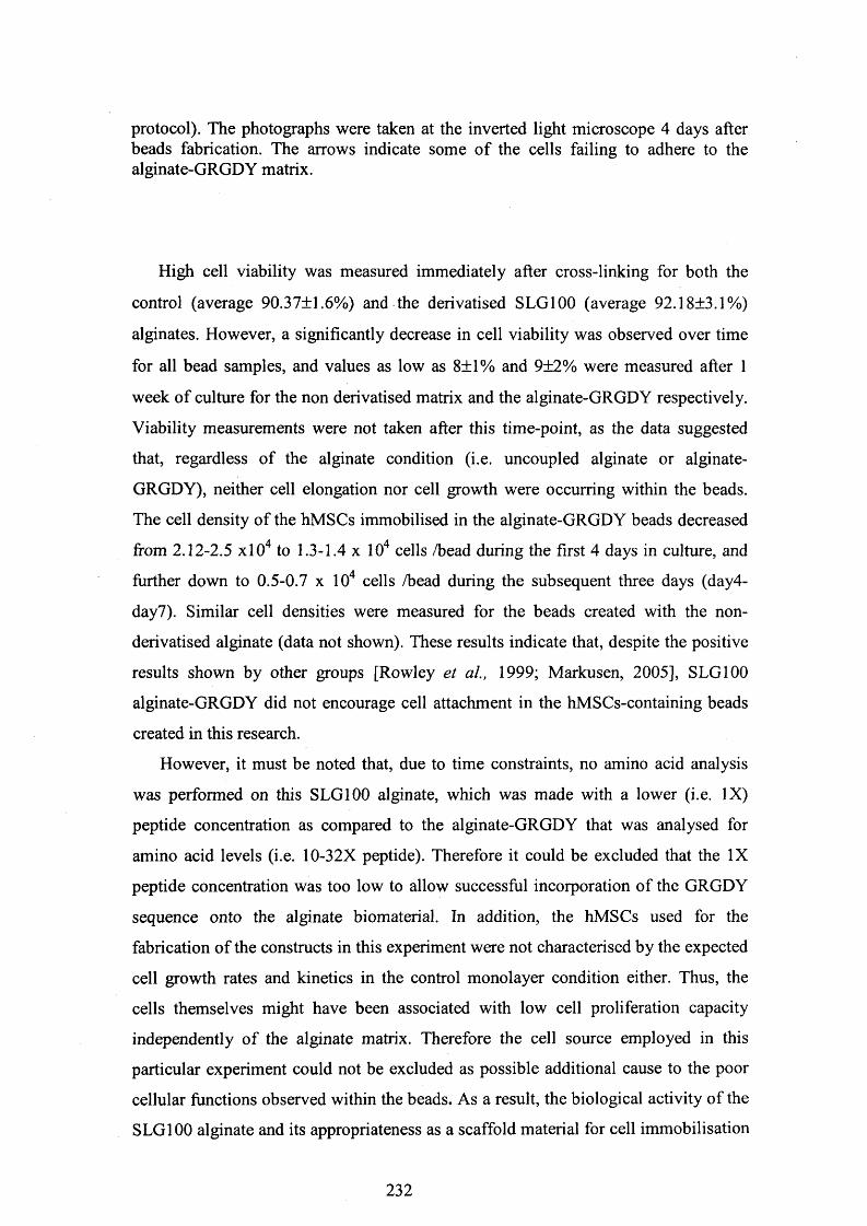
protocol). The photographs were taken at the inverted light microscope 4 days after beads fabrication. The arrows indicate some of the cells failing to adhere to the alginate-GRGDY matrix.
High cell viability was measured immediately after cross-linking for both the
control (average 90.37±1.6%) and the derivatised SLG100 (average 92.18±3.1%)
alginates. However, a significantly decrease in cell viability was observed over time
for all bead samples, and values as low as 8±1% and 9±2% were measured after 1
week of culture for the non derivatised matrix and the alginate-GRGDY respectively.
Viability measurements were not taken after this time-point, as the data suggested
that, regardless of the alginate condition (i.e. uncoupled alginate or alginate-
GRGDY), neither cell elongation nor cell growth were occurring within the beads.
The cell density of the hMSCs immobilised in the alginate-GRGDY beads decreased
from 2.12-2.5 xlO4 to 1.3-1.4 x 10⁴ cells /bead during the first 4 days in culture, and
further down to 0.5-0.7 x 10⁴ cells /bead during the subsequent three days (day4-
day7). Similar cell densities were measured for the beads created with the non-
derivatised alginate (data not shown). These results indicate that, despite the positive
results shown by other groups [Rowley et al., 1999; Markusen, 2005], SLG100
alginate-GRGDY did not encourage cell attachment in the hMSCs-containing beads
created in this research.
However, it must be noted that, due to time constraints, no amino acid analysis
was performed on this SLG100 alginate, which was made with a lower (i.e. IX)
peptide concentration as compared to the alginate-GRGDY that was analysed for
amino acid levels (i.e. 10-32X peptide). Therefore it could be excluded that the IX
peptide concentration was too low to allow successful incorporation of the GRGDY
sequence onto the alginate biomaterial. In addition, the hMSCs used for the
fabrication of the constructs in this experiment were not characterised by the expected
cell growth rates and kinetics in the control monolayer condition either. Thus, the
cells themselves might have been associated with low cell proliferation capacity
independently of the alginate matrix. Therefore the cell source employed in this
particular experiment could not be excluded as possible additional cause to the poor
cellular functions observed within the beads. As a result, the biological activity of the
SLG100 alginate and its appropriateness as a scaffold material for cell immobilisation
232

was investigated further in additional studies, which are described in the following
sections.
Nevertheless, in accordance with other groups findings [Stabler et al, 2002], this
study confirmed that the chemical composition of the alginate has a significant effect
on its physical properties and suggested that high-G content alginates should be
preferred over the high-M content polymers for their gelling properties and their
strength and stability. As a result, SLG100, which permitted the fabrication of
stronger and more stable beads, was chosen and adopted as the preferred alginate
biomaterial for all subsequent matrix/cells constructs studies.
5.4 Fabrication of alginate-GRGDY/hMSCs beads using
alginate modified according to different derivatisation
procedures
The aim of this study was to test the effects of SLG100 alginates prepared
according to different derivatisation procedures (alginates 6 , 7, 8 , 9 in table 3.1) on
both the cell behaviour after immobilisation and the mechanical properties of the
resulting constructs. The coupling reaction for all of these alginates was performed
using exactly the same reactants concentrations (i.e. 9X reactants and 10X peptide)
under the same working conditions (20-hour reaction at T=20-22°C, under constant
mixing on a platform roller, in the dark). However, after completion of the
carbodiimide reaction and the 5-day dialysis cycle, alternative methods (i.e.
lyophilisation with or without freezing step, PEG concentration with or without
freezing step) were used for the purification and the concentration steps, as described
in section 3.2 (please see figure 3.1).
Fourth passage (p4) hMSCs (25% v/v) were mixed with each of the four alginaten
solutions to a final concentration of 1x10 cells/mL, and the resulting alginate/cell
mixtures were treated with CaCl2 for gel cross-linking and beads fabrication.
Particular care was taken on the cell choice, with respect of their proliferation
potential in the control monolayer culture, and only hMSCs associated with high
growth rate (0.012 hours-1) and low doubling time (55.79 hours) were used for the
fabrication of the beads. This permitted a more accurate comparison of the cell
233

performance in monolayer condition (tissue flasks) and within alginate-GRGDY
beads.
No significant differences were noted among all bead samples in terms of gel
stiffness and mechanical behaviour. Stable and rigid constructs were obtained for all
alginate conditions, and all beads held regular structure and shape over the 1 0 -day
period examined. However, despite the structural integrity and stability, no cell
attachment and elongation within the alginate polymers were observed (please see
photograph in figure 5.2). Regardless of the derivatisation procedure employed for
the fabrication of the alginate matrix, hMSCs demonstrated to be unable to adhere to
and elongate within the alginate-GRGDY biomaterial.
In addition, a significant drop in cell viability, comparable to that observed for the
control constructs (i.e. beads prepared with non derivatised-alginate) was measured
over time (please see table 5.1 and photograph in figure 5.3). The cell density values
measured for hMSCs immobilised in the alginate-GRGDY beads also decreased
consistently over time, similarly to what was seen for cells immobilised in the control
(i.e. non derivatised) alginate (please see table 5.1).
Thus, no discemable differences were observed between the non derivatised
alginate and all the alginate-GRGDY types investigated.
234

Beads conditions
day Viabilityaverage
Cells/beads
Control(non derivatised) alginate
0 91.4+0.9% 2.70-2.91 x10⁴
4 55.9+2.6% 1.67-1.69x10⁴
7 8.5+1.2% 0.82-0.91 x10⁴
Standardprotocol(no.1)
0 92.18+2.9% 2.95-3.09x10⁴
4 62.3+1.3% 1.70-1.2x10⁴
7 7.2+0% 0.90-0.97x10⁴
Protocol no.2 0 89.7+1.6% 2.7-2.93x10⁴
4 57.1+2.8% 41.50-1 9x10⁴
7 7.9+1.3% 0.60-0.75x10⁴
Protocolno.3
0 90.2+0% 3.0-1:3x10⁴
4 52.8+5.7% 1.6-0.9x10⁴
7 9.1+3.1% 0.8-0.95x10⁴
Protocolno.4
0 93.2+0.3% 2.80-2.93x10⁴
4 64.2+3.2% 1.57-1.81 x10⁴
7 6.9+3.4% 0.63-0.71 x10⁴
Table 5.1: Percent cell viability and cell concentration values obtained for hMSCs immobilised in non-derivatised alginate (control beads) and alginates modified with the GRGDY peptide according to different derivatisation protocols. All alginates were coupled with 9X sulfo-NHS and EDC and with 10X peptide concentrations (the resulting derivatised alginates are designated as “alginate- GRGDY number 6 ”, “number 7”, “number 8 ” and “number 9” respectively in table 3.1) Data are shown for each harvesting time-point during the first week of culture.
Interestingly, amino acid analysis data revealed that the GRGDY peptide was
successfully incorporated onto the alginate after completion of the derivatisation
procedure according to the standard protocol (procedure no. 1 in figure 3.1). This
alginate (final alginate-GRGDY number 6 in table 3.1) contained all the four
constituent amino acids of the GRGDY sequence, as it appears from the results
shown in table 3.3 (final alginate-GRGDY number 6 ). However, as already
mentioned in section 3.3.1, a significant tyrosine loss was also detected in these
samples. Ultimately, it can be hypothesised that this phenomenon was responsible for
the low biological activity of the alginate-GRGDY, resulting in lack of matrix-cells
235
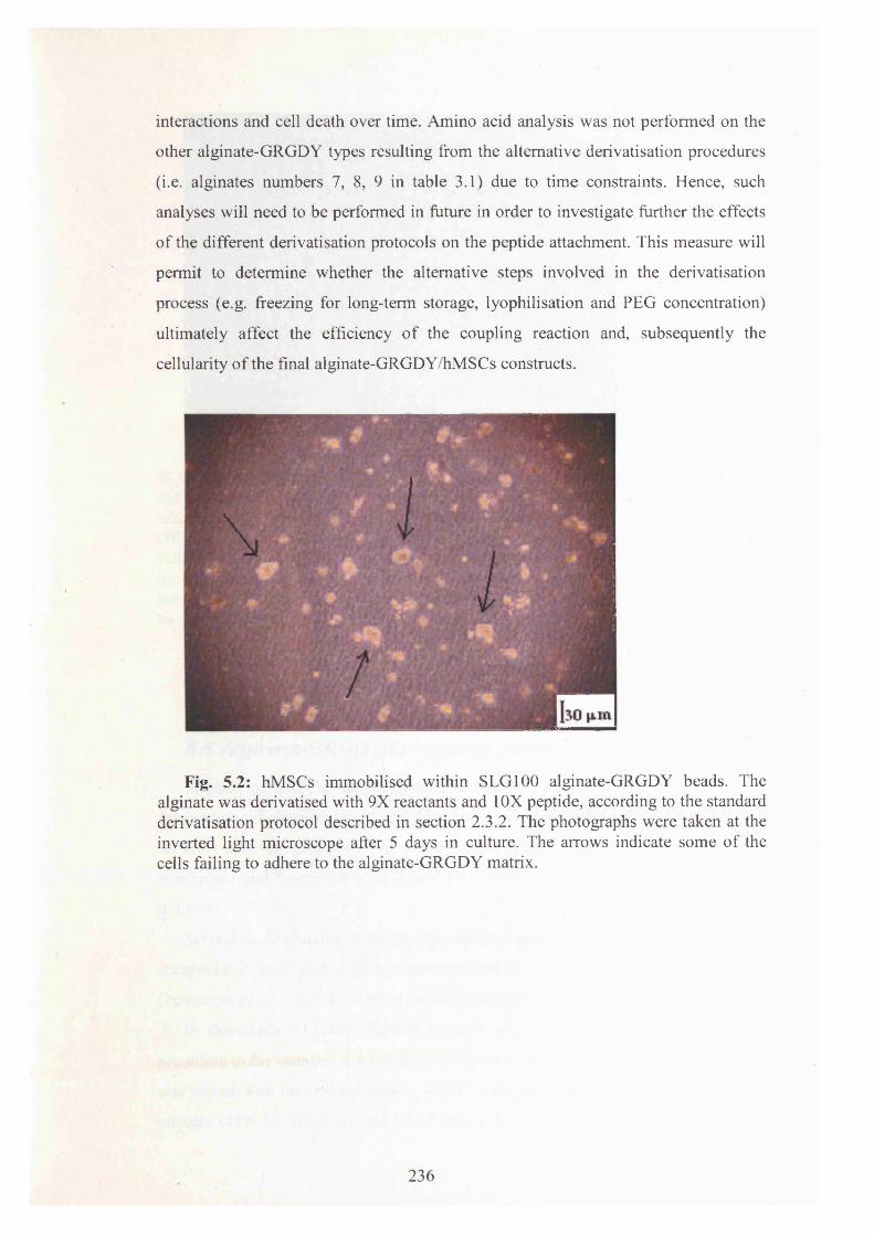
interactions and cell death over time. Amino acid analysis was not performed on the
other alginate-GRGDY types resulting from the alternative derivatisation procedures
(i.e. alginates numbers 7, 8 , 9 in table 3.1) due to time constraints. Hence, such
analyses will need to be performed in future in order to investigate further the effects
of the different derivatisation protocols on the peptide attachment. This measure will
permit to determine whether the alternative steps involved in the derivatisation
process (e.g. freezing for long-term storage, lyophilisation and PEG concentration)
ultimately affect the efficiency of the coupling reaction and, subsequently the
cellularity of the final alginate-GRGDY/hMSCs constructs.
Fig. 5.2: hMSCs immobilised within SLG100 alginate-GRGDY beads. The alginate was derivatised with 9X reactants and 10X peptide, according to the standard derivatisation protocol described in section 2.3.2. The photographs were taken at the inverted light microscope after 5 days in culture. The arrows indicate some of the cells failing to adhere to the alginate-GRGDY matrix.
236

Fig. 5.3: Fluorescence photograph obtained for hMSCs immobilised within SLG100 alginate-GRGDY beads stained with calcein AM/ethidium homodimer for the qualitative evaluation of cell viability. The alginate was derivatised with 9X reactants and 10X peptide prior to the beads preparation. The resulting alginate- GRGDY was mixed with the cell suspension and used for beads preparation following the lyophilisation and freezing steps (i.e. standard derivatisation procedure). The photograph was taken at the confocal microscope 5 days after beads fabrication. The intense red fluorescence originates from the Ethidium homodimer-1 bound to the cellular DNA after permeation of the damaged cell membranes.
5.5 Alginate-GRGDY/fibroblasts beads
In place of hMSCs, human foreskin fibroblasts were used in this study for the
preparation of alginate beads, in order to see whether different cell types exhibit
different adhesion properties, and whether the GRGDY ligand may favour fibroblast
attachment and functionality as opposed to what was observed in this research for
hMSCs.
Several in vitro studies have already indicated that fibroblasts can be successfully
encapsulated in alginate beads to be employed in different therapeutic applications
[Peirone et al., 1998; Schwinger et al, 2002; Keshaw et al, 2004; Ueng et al, 2000].
In this study SLG100 alginate coupled with 9X reactants and 10X peptide
according to the standard derivatisation protocol (alginate-GRGDY no. 6 in table 3.1)
was mixed with the cell suspension (lxlO 7 cells/mL) and the resulting matrix-cells
mixture (25% v/v of cells) was gelled into 2-2.5 mm diameter beads following the
237

previously described (please see section 2.5.3) cross-linking treatment in 90 mM
CaCl2. Sixth passage (p6 ) fibroblasts were used in this study, and a culture in T25
tissue flasks was run in parallel in order to have a direct comparison with cell
performance in the monolayer condition. Control beads were simultaneously
fabricated with non-derivatised SLG100 alginate and were assessed in parallel with
the alginate-GRGDY constructs. As in the previous experiments, cell morphology
and alginate biological activity were evaluated by means of light microscopy, and cell
densities and viabilities were assessed with haemocytometer and trypan blue dye
exclusion assay respectively following cell release with trisodium citrate solution.
Both control (i.e. non derivatised) alginate and alginate coupled with the GRGDY
peptide formed stable gel beads, which retained a defined regular shape over the 1 0 -
day period tested. However, similarly to what was observed in the previous
experiment for the hMSCs, the fibroblasts failed to interact with both the unmodified
polymer and the alginate-GRGDY, and could not elongate and acquire their
characteristic spindle-shaped morphology (please see photographs in figure 5.4 A and
B). Moreover, while the cells were proliferating extensively in tissue flasks (growth
rate = 0.014 hours-1, doubling time = 49.8 hours), no cell proliferation was observed
within the beads. Cell viability data indicated significant cell death over time, both for
the control beads and the alginate-GRGDY constructs (please see table 5.2). The
cells, having failed to adhere to the alginate scaffold, completely dissociated from the
matrix as small spherical entities floating in the growth medium within 48 hours from
cross-linking.
Light microscopy also revealed that in these constructs the cells tended to position
in close proximity to the outer surface of the matrix/cells beads. This inhomogeneous
cell distribution may have been caused by the particular method employed for the
beads fabrication in this research. It has already been reported that the diffusion-
setting method for the gelation of the alginate with CaCl2 results in an
inhomogeneous alginate distribution within the gels [Skjak-Braek et al, 1989]. In
general, it is well documented that a very rapid and irreversible binding reaction of
multivalent cations is typical for alginates; a direct mixing of these two components
therefore rarely produces homogeneous gels [Draget et al., 2005] (please see section
6.4 for a more detailed description of this phenomenon).
Ultimately, a similar poor cellular behaviour within the alginate-GRGDY beads
was therefore observed for the hMSCs and the fibroblasts, as both cell types appeared
238
to be unable to interact with the biomaterial and to spread out as spindle-shaped
elongated healthy cells within the alginate beads.
As mentioned in the previous section, amino acid analysis was performed on the
alginate used here for the preparation of the beads (alginate number 6 in table 3.1),
revealing the presence of a complete GRGDY sequence attached to the polymer, yet
with a very low tyrosine level (please see alginate-GRGDY number 6 in table 3.3 for
the amino acid content data). Thus, the most probable cause for the lack of alginate-
cells interactions observed in these studies was the significant loss of the final
tyrosine residue of the GRGDY sequence, which, according to the amino acid
analysis data, was being cleaved off (at some stage) during the derivatisation
procedure. This event probably determined the poor results in terms of cellularity
described in this research for the studies performed on the alginate/cells beads.
However, despite the lack of cell adhesion and proliferation, it must be noted that
constructs fabricated with SLG100 alginate retained good mechanical properties (i.e.
considerable stiffness, structural integrity and stability) over the 10 -day week period
of culture under examination.
239

Beads conditions
Day Viabilityaverage
Cells/bead
Control(non
derivatised)alginate
0 94.4+1.3%
2.46-2.78x10⁴.
4 65.7+1.6%
1.52-1.61x10⁴
7 9+2.1% 0.92-0.98x10⁴
SLG100alginate-GRGDY
0 92.67+3.4%
2.65-2.91 x10⁴
4 67.0+0.3%
1.59-1.64x10⁴
7 11.3+1% 0.92-0.99x10⁴
Table 5.2: Percent cell viability and cell concentration values obtained for human foreskin fibroblasts immobilised in non-derivatised alginate (control beads) and in alginate modified with the GRGDY peptide according to the standard derivatisation procedure. 9X sulfo-NHS and EDC and 10X peptide were used for the coupling reaction (the resulting derivatised alginate is designated as “alginate-GRGDY no. 6 ” in table 3.1). Data are shown for each harvesting time-point during the first week of culture.
240

Fig. 5.4 A and B : Human foreskin fibroblasts immobilised within SLG100 alginate-GRGDY beads. The alginate was derivatised with 9X reactants and 10X GRGDY peptide according to the standard derivatisation procedure described in section 2.3.2. The photograph was taken at the inverted light microscope 6 days after beads fabrication. The small spherical morphology indicates that the cells were unable to adhere to the alginate-GRGDY matrix within the beads.
241

5.6 Additional studies on alginate-GRGDY for the
immobilisation of fibroblasts
Additional studies were performed in order to investigate further the possibility
for the alginate-GRGDY (fabricated according to the standard derivatisation
procedure described in this research) to act as a suitable substrate for cell viability and
growth.
Thus, SLG100 alginate coupled with the same (i.e. 9X) EDC and sulfo-NHS
concentrations as in the previous experiments, but with a higher (i.e. 32X) peptide
concentration, was used for the fabrication of the constructs (alginate no. 1 in table
3.1).
Frozen vials of this alginate-GRGDY were thawed and reconstituted with normal
saline solution as described in section 2.3.2.5. The resulting alginate solution was
mixed with fifth passage (p5) human foreskin fibroblasts in suspension (lxlO 7
cells/mL) and the alginate-GRGDY/fibroblast mixture (25% v/v of cells) was then
used for the fabrication of the constructs. Fibroblasts were also simultaneously
expanded in monolayer culture, and were also grown within non-derivatised alginate
beads acting as control.
The expectation was that the higher peptide concentration (32X) used in this
experiment, which had already led to the incorporation of higher amino acid levels
onto the alginate chain (please see data for alginate designated as “alginate-GRGDY
no. 1-Albachem” in table 3.3), would have resulted in cell adhesion and cell
proliferation within the constructs. However, once again neither cell attachment nor
cell elongation were observed in the alginate-GRGDY beads (please see photograph
in figure 5.5). As for all the experiments described above (sections 5.3, 5.4 and 5.5),
significant loss of cell viability was observed over time (please see photograph in
figure 5.6). The percentage of detectable viable cells at day 7 of culture was in fact
only 10.4±0.9% and 9.07+3.4% for the control beads and the derivatised alginate
constructs respectively. Concurrently, cell numbers calculated after dissolution of
both bead types with trisodium citrate decreased considerably over time, dropping
from initial values in the 2.15-2.79xl04 cells/bead range (day 0) to merely 0.49-
0.76x10⁴ cells/bead at day 7.
Thus, the use of alginate coupled with higher peptide concentrations did not
improve the biological activity of the resulting polymer hydrogels. According to the
242
amino acid analysis data (please see section 3.3.1), also in this case a significant loss
of tyrosine was occurring during the derivatisation process. Hence, the negative
results in terms of cell performance consistently observed after cell immobilisation
corroborate the hypothesis of the correlation between tyrosine loss and lack of cell
adhesion, and ultimately lack of cell survival, within the alginate-GRGDY beads.
Fig. 5.5 : Human foreskin fibroblasts immobilised in SLG100, 9X reactants, 32X GRGDY peptide alginate-GRGDY. The photograph was taken at the inverted light microscope 5 days after beads fabrication. Small, spherical cells failing to adhere to the scaffold and to elongate are clearly visible within the bead.
243

Fig. 5.6: Fluorescence photograph obtained for human foreskin fibroblasts immobilised within SLG100 alginate-GRGDY beads stained with calcein AM/ethidium homodimer for the qualitative evaluation of cell viability. The alginate was derivatised with 9X reactants and 32X peptide prior to the beads preparation. The resulting alginate-GRGDY was mixed with the cell suspension and used for beads preparation following the lyophilisation and freezing steps (i.e. standard derivatisation procedure). The photograph was taken at the confocal microscope 5 days after beads fabrication. The intense red fluorescence originates from the Ethidium homodimer-1 bound to the cellular DNA after permeation of the damaged cell membranes.
5.7 HMSCs immobilised in alginate-GRGDY disks
Given the lack of cell attachment observed for both hMSCs and human foreskin
fibroblasts after immobilisation within alginate-GRGDY beads, it was decided to
investigate hMSCs performance and functions after seeding the cells on top of
alginate-GRGDY disks (or layers).
Despite the project’s aim to immobilise the cells within the alginate hydrogels in a
one-step process, alginate disks were fabricated to de-couple the cell seeding step
from the alginate scaffold formation, in order to investigate the possible detrimental
effects of high molar concentration of the cross-linking solution. During the beads
preparation procedure the cells were immediately mixed with the alginate and the
resulting suspension was then syringed into the CaCI2 cross-linking solution (please
244

see schematic representation in figure 5.7). Thus, the final alginate/cells beads were
instantaneously formed within the cross-linking solution.
However, the 1 % CaC^ solution used (corresponding to 90mM) was hypertonic,
being 1.8mM the physiological CaC^ concentration for cell maintenance and growth,
i.e. level in DMEM medium (BioWhittaker Product Catalog, 2001). During cell
immobilisation in alginate beads the cells are exposed to the calcium chloride solution
for prolonged time periods. This procedure can, in principle, cause unwanted and
uncontrolled physiological effects. In addition, only recently attachment-dependent
cells have been reported to be successfully immobilised within alginate beads, whilst
alginate encapsulation systems are in most cases employed only for cell therapy
applications involving the use of suspension adapted cells. Even Rowley’s and
Mooney’s studies had been performed by constructing alginate layers and then
seeding on the surface with cells [Rowley, 1999].
Given the potential harmful effect of the cross-linking solution on the overall cell
performance, and given that no cell adhesion was observed from the cell
immobilisation studies with alginate-GRGDY beads (described in the sections 5.3
and 5.6 above), experiments were conducted by pre-forming the alginate-GRGDY
matrices as layers in the bottom of a microwell, and then seeding the cells separately
onto the surface of the alginate (please see section 2.5.4 for a full description of the
method employed for disks preparation). This procedure made it possible to de
couple the alginate preparation and the cell seeding steps, and therefore to test the
detrimental effects of the cross-linking solution on cell behaviour. A schematic
representing the different procedures for beads and disks fabrication is given in figure
5.7.
245

Oh* STEP PROCEDURE
alginate solution <tH suspension
atginateJcells mixture
Cod
inafraiHariaou* beads fabrication
TWO-STFP PftOf fIXJRf
□aiginat* solutionX
— f—CaO.
siginate disk t hour fabrication
step
+ cell suspension
<«Ss seeded on top ’ of alginate disk
alginate layer or disk
culture
Fig. 5.7 : Schematic of the cross-linking procedures for the preparation of alginate/cells beads (A) and layers (B). Figure A adapted from Draget et al. 2005.
246

Sixth passage (p6 ) human MSCs and SLG100 Pronova alginate derivatised
following the standard derivatisation procedure with 9X EDC and sulfo-NHS and
32X peptide (designated as “alginate-GRGDY number 1” in table 3.1) were used in
this experiments for the disks preparation (3.3x10⁴cells/cm2- 5xl04cells/cm2). The
cells were also immobilised directly into alginate-GRGDY beads (average 2.52x10⁴
cells/bead) for a direct comparison. Alginate-GRGDY disks and beads were
constructed as described in sections 2.5.4 and section 2.5.3 respectively. After
formation, the alginate layers were rinsed thoroughly of excess CaCl2 prior to the
addition of the cells. A monolayer culture in 24-well tissue culture plates was
performed in parallel, and alginate/cells layers and beads were created also with non-
derivatised SLG100 alginate for control. Light microscopy was used to evaluate
matrix-cells interactions, cell attachment to the surface of the alginate disks and cell
morphology. The cells were released from the matrix with trisodium citrate solution
(please see section 2.5.5) and were evaluated for cell density and viability with
haemocytometer and trypan blue (please see sections 2.6.2 and 2.6.3). While the cells
in monolayer culture were characterised by high proliferation potential (cell growth
rate = 0.014 hours-1) and low doubling time (50.86 hours), cell growth was not
observed after seeding on the surface of the alginate-GRGDY disks. The cells did not
attach to the alginate-GRGDY layers and could not spread out to acquire their typical
fibroblast-like appearance. Virtually no difference was observed among cellularity
within the beads and cell behaviour on the surface of the hydrogel disks, and in both
construct types the alginate-GRGDY matrix did not support cell adhesion and
elongation (please see photographs in figures 5.8 and 5.9).
Thus, it can be concluded that the lack of cell response that was seen for both the
alginate/hMSCs beads and disks was most likely due to the alginate-GRGDY
biological inactivity, rather than to the harmful treatment with the high molarity
cross-linking agent during the beads fabrication procedure. Since the alginate-
GRGDY was characterised by a considerable loss of the tyrosine residue, it cannot be
excluded that this phenomenon was the major cause responsible for the lack of cell
attachment, cell elongation and, ultimately, cell proliferation observed for both the
alginate beads and disks. According to this hypothesis, tyrosine loss probably altered
the chemical conformation of the GRGDY sequence, ultimately preventing cell
attachment. Thus, the hMSCs, which are strongly anchorage dependent, could not
attach to the alginate matrix of both beads and disks simply because the polymer,
247

having lost the tyrosine residue, could not provide cell adhesion sites. However, other
possibilities cannot be excluded, since there are several other parameters that may
affect the beads fabrication procedure, and thus the cell adhesion properties of the
resulting scaffold. It is known that the gelation process, which occurs when divalent
cations take part in the interchain binding between G-blocks, depends on both the
alginate and gelling ion concentrations [Martinsen et al., 1989]. Thus, future studies
could be performed investigating cell immobilisation within alginate beads made with04-different Ca concentrations, or by using different length exposures to the cross-
linking solution. Furthermore, it is true that for the immobilisation of living cells
calcium has become the primary choice as a gel-inducing agent. However, alginate04-forms gels with most di- and multivalent cations. While monovalent cations and Mg
04- 0 Iions do not induce gelation [Sutherland, 1991], ions like Ba and Sr produce04- 0 Istronger alginate gels than Ca [Clark and Ross-Murphy, 1987]. Like Ca , these
ions are considered to be sufficiently biocompatible for applications involving
immobilisation of living cells [Smidsrod and Skjak-Braek, 1990], Moreover, as
compared to Ca2+, they have a higher affinity for alginate [Smidsrod, 1974], and
consequently are associated with higher gel stability [Clark and Ross-Murphy, 1987].
As a result, the use of alternative gelling cations, such as Ba or Sr , could be
investigated. Ultimately, further studies are needed to elucidate whether the lack of
cell adhesion to the alginate beads and disks was caused in both matrices by the
tyrosine loss and subsequent biological inactivity of the alginate-GRGDY matrix, or
by other mechanisms.
248

249

Fig 5.8 A, B and C: hMSCs immobilised within alginate-GRGDY beads. The alginate polymer was coupled with 9X EDC and sulfo-NHS and 32X GRGDY peptide, and was derivatised according to the standard protocol described in section2.3.2. The photographs were taken at the inverted light microscope 4 days after the fabrication of the constructs. It is evident from the photographs that the cells were unable to adhere to the alginate-GRGDY and to elongate within the beads. The arrows indicate some of these cells.
250

Fig 5.9 A and B: hMSCs seeded on top of alginate-GRGDY disks. The alginate polymer was coupled with 9X EDC and sulfo-NHS and 32X GRGDY peptide, and was derivatised according to the standard protocol described in section 2.3.2. The photographs were taken at the inverted light microscope 4 days after the fabrication of the constructs. The arrows indicate big cells failing to attach to the alginate- GRGDY layers.
5.8 MTT assay: analysis of cell proliferation within
alginate matrices
Experiments were performed using the MTT (3-(4,5-dimethylthiazol-2-yl)-2,5-
diphenyltetrazolium bromide) assay for the determination of viable cell numbers
within the alginate matrix. The MTT Cell Proliferation assay is a rapid colorimetric
method, which is commonly used for the quantitative assessment of cellular survival
and proliferation, based on the metabolic activity of viable cells (please see section
2.7 for a more detailed description of the MTT technique). Only active mitochondria
of live cells can cleave the tetrazolium ring of the MTT compound. The absorbance
subsequently measured for the blue formazan product generated from the MTT dye is
linearly related to the quantity of viable cells added per well. Thus, the assay can be
used to detect the cell proliferation rate and conversely, when metabolic events lead
to apoptosis or necrosis, the reduction in cell viability [Mosmann, 1983; Ferrera et al.,
1993; Ciapetti et al., 1992].
251

5.8.1 Comparison of cell numbers derived from MTT
absorbance readings and from haemocytometer
In order to confirm the linearity of the MTT assay, a series of cell standard curves
were created from individual cell suspensions using T150 (150 cm2) flasks. Seventh
passage (p7) human MSCs were harvested from tissue flasks and brought in cell
suspension in Dulbecco’s phosphate buffered saline with lg/L D-glucose and 36mg/L
sodium pyruvate (DPBSG, Bio Whittaker), following the method described in section
2.1.3. The resulting cell suspension was serially diluted with DPBSG and seeded in a
96-well tissue culture plate as it is described in section 2.7.1. The MTT assay was
then performed on the resulting samples as described in section 2.7, and the
absorbance was measured at 600nm on a spectrophotometer (please see section
2.7.4.3).
The cell standard curves obtained for the T150 flasks were plotted according to
the cell density (cell number/mL) measured simultaneously by haemocytometer
count. Figure 5.10 shows one of the typical standard curves obtained from this
analysis. The curve indicates that the MTT absorbance readings detected with the
spectrophotometer correlated very closely to the cell concentration measured with the
haemocytometer. This study confirmed that by following the MTT assay protocol
described in section 2.7 it was possible to establish a linear relationship between the
formazan absorbance readings and the cell concentration within the samples. This
relationship was established for a cell suspension of hMSCs from tissue flask cultures
harvested at day 4 of culture.
252

MTT assay on hMSCs in monolayery = 0.0067x R2 = 0.8625
- p 1 .2 -
0.8
M
§ Q 0.0- 0.2 b 1 0 0
No. of cells (x103)/mL
150 200
Fig.5.10: MTT absorbance values at 600 nm detected from human MSCs (p7) cultured in monolayer.
In this research the MTT assay was used to confirm the decrease of cell viability
observed for cells encapsulated within the alginate beads.
Indeed, the MTT assay has already been used in previous studies for the
determination of the status of several cell types in different alginate matrices [Barralet
et a l, 2005; Lawson et a l, 2004; Markusen et a l, 2006; Rokstad et al, 2005; Mahler
et a l, 2003; Khattak et al, 2006; Khattak et a l, 2006; Zhang et al, 2005]. In this
research, the technique was used at a preliminary stage, hence, prior to perform the
assay on the matrix/cells beads, experiments were performed to confirm that the
alginate biomaterial does not interfere with the absorbance readings measured and
does not affect the validity of the results. In addition, only beads fabricated with non-
derivatised alginate were tested for cell viability in the MTT experiments.
5.8.2 MTT cell proliferation assay on hMSCs expanded in
monolayer condition in the presence of increasing concentrations
of alginate in solution
The aim of this study was to test any potential interference of the alginate
biomaterial on the MTT absorbance readings measured during determination of cell
viability for hMSCs expanded as a monolayer.
253

ProNova non-derivatised SLG100 alginate was used for this purpose. Sixth
passage (p6 ) hMSCs cells were resuspended in complete DMEM, and seeded onto
24-well microplates in monolayer condition (6xl03cells/cm2) according to the usual
plating protocol (please see section 2.1.2). 24 hours after plating, the medium of
culture was removed from the wells and replaced with alginate in solution (1% w/v),
which was serially diluted in complete DMEM medium. The cells were maintained in
culture in the resulting alginate/DMEM solutions (alginate final concentration
ranging from 0 to 100% v/v) for 10 days, with alginate/DMEM refreshments
performed every 2-3 days. The MTT assay was performed on the resulting cell
samples at the end of the culture period (day 1 0 ), following the protocol described in
section 2.7.8.
The absorbance readings obtained for the microwell samples were plotted
according to the alginate solution concentration in the DMEM medium. The data
obtained from this analysis are shown in figure 5.11.
2 0.35 -i & 0.3 --wS 025 a
0.2 H
MTT absorbance values for hMSCs expanded in monolayer with increasing concentration of
alginate in solution
(0 o5 . 0.15 oS 0 1 | 0.05i—O nm u n < 20 40 60 80
Alginate concentration (v/v % ]
100 120
absorbance readings
Fig. 5.11: MTT absorbance values obtained for hMSCs in microwell (6xl03cells/cm2) after 1 0 days in culture in the presence of an increasing concentration of alginate in solution (0, 1, 2, 3, 4, 5, 6 , 7, 8 , 9, 10, 50 and 100 v/v %).
254

As it is evident in the graph, the alginate solution did not affect the MTT
absorbance readings detected with the spectrophotometer up to a 50% v/v
concentration. Whilst no significant difference was observed between the absorbance
readings measured for cells expanded in the alginate solutions with concentration
ranging from 0% to 50% v/v, a significant decrease in the absorbance value was
observed for hMSCs expanded in the complete (100% v/v) alginate solution. These
results indicate that the cells experienced a dramatic drop in cell viability following a
10-day culture in the alginate solution. As it is shown in the graph, cells that had been
completely deprived of the DMEM medium of culture over the 10-day period, and
had been grown instead in the complete alginate solution, retained no cell viability.
The absorbance readings measured for these DMEM-deprived cells were in fact
comparable to those measured for the blank (i.e. complete DMEM medium only)
microwells.
According to these data, hMSCs can be cultured in monolayer condition in the
presence of alginate solution with a concentration up to 50% v/v, without
experiencing cell death due to the presence of the alginate polymer.
5.8.3 MTT cell proliferation assay on hMSCs expanded in
monolayer culture in the presence of an increasing number of cell-
free alginate beads
Experiments were also performed using cell-free alginate beads to test whether
the presence of the alginate polymer, after cross-linking treatment and gelling into
hydrogel beads, had any effect on the absorbance values measured for samples of
hMSCs expanded in monolayer culture.
Pronova non-derivatised SLG100 alginate and sixth passage (p6 ) hMSCs were
used in these studies. The cells were harvested from T150 flasks and resuspended in
Dulbecco’s phosphate buffered saline with 1 g/L D-glucose and 36 mg/L sodium
pyruvate (DPBSG, Bio Whittaker). The cell suspension was seeded into two 96-well
plates (2.05x10⁴ cells/well) as it is described in section 2.7.6 and an increasing
number o f cell-free alginate beads (0, 1, 2, 3 and 4) were added to the microwells in
culture. The culture was performed under the optimal conditions identified in the
monolayer studies (please see results from chapter 4) and was maintained for a 10-
day period. The MTT cell proliferation assay was performed at the end of the culture
255

period (day 10), following the protocol described in section 2.7. The MTT solution
was left in incubation at 37°C for one 1 hour and the absorbance values were read on
the spectrophotometer at 600nm within 1 hour from isopropanol/HCl addition,
following the standard MTT assay protocol, as described in section 2.7.4.3.
The results are presented in figure 5.12.
MTT absorbance values for hMSCs expanded in monolayer condition in the presence of cell-free alginate
beads
a>oc(0Ak .O(0A<
„ 0.4 ^0.35 - w A3 S(g25 ^
75 3 '2
Q. 0.12-0.05
T i
1 2 3
No. of cell-free alg inate b ead s
Fig. 5.12: MTT absorbance values measured at 600 nm for hMSCs expanded in monolayer condition (2.05x10⁴ cells/well) in the presence of an increasing number of non-derivatised alginate beads. The assay was performed 10 days after plating.
It is apparent from this graph that there was no significant difference in the
absorbance values detected for hMSCs expanded in monolayer in the presence of an
increasing number of cell-free alginate beads. Thus, the presence of the alginate
biomaterial in the assay does not interfere with the absorbance readings for the period
examined. As a result, it can be concluded that short-term culture of hMSCs
microwell samples in the presence of alginate gel beads is possible without
experiencing a subsequent change in absorbance readings and in the cell proliferation
potential.
256

5.8.3.1 MTT cell proliferation assay on hMSCs immobilised in
alginate beads
The MTT assay was then used to measure cell proliferation potential for hMSCs
immobilised in Pronova SLG100 alginate beads (average 4.47x10³ cells/bead). The
constructs were fabricated according to the usual protocol (please see section 2.5.3)
and were maintained in culture over a 14-day period. The microwells were seeded
with an increasing number of beads (0, 2, 4 and 6) and were cultured under the
optimal culture conditions identified from the monolayer studies described in chapter
4. At selected time-points (dayl, day 4, day 7 and day 14) cell proliferation within the
beads was assessed by means of the MTT assay. These data are presented in figure
5.13.
MTT absorbance values for hMSCs encapsulated in non- derivatised alginate beads
ctoA
0 .35
0 .3
0 .25E 0.2
_ g 0 .15S § o . i
0.05
\
aO
-0 .05
Time (days)
0 b e a d s 2 b e a d s 4 b e a d s 6 b e a d s
Fig. 5.13: MTT absorbance readings measured at 4 different time-points (day 1, day 4, day 8, day 14) for an increasing number of alginate/hMSCs beads (average 4xl03 cells/bead). The constructs were maintained in the culture conditions that in the extensive monolayer studies presented in chapter 4 were associated with the highest growth kinetics and cell viability data.
257

The graph in figure 5.13 indicates that there was a good linear relationship
between the number of beads evaluated and the formazan absorbance readings
measured with the spectrophotometer. This relationship was established for constructs
cultured over a 4-day period. The absorbance measurements taken at the additional
time-points (day 8 and day 14) indicate that the cells were characterised by a
significant decrease in cell viability after immobilisation within the alginate bead
matrices. In conclusions, the MTT assay data confirmed the lack of cell proliferation
experienced by hMSCs immobilised within non-derivatised alginate beads.
Ultimately, the absorbance readings measured with the MTT assay appeared to be
consistent with the cell density and cell viability measurements obtained with the
haemocytometer and the trypan blue exclusion dye assay. In principle, this proves
that the MTT cell proliferation assay can be used in future studies as a reliable
method for the quantitative assessment of the proliferation and mitochondrial
metabolic activity of viable cells encapsulated in alginate-GRGDY constructs.
5.9 Conclusions and discussion
The advantages of using the RGD containing alginate matrices as scaffold
materials to promote cell attachment and growth have already been shown by
different authors [Rowley et al., 1999; Markusen et al., 2006; Halberstadt et al.,
2002; Alsberg et al., 2001]. Thus, several experiments were performed in order to
investigate cell behaviour and performance after immobilisation within alginate-
GRGDY matrices. Both hMSCs and fibroblasts were analysed for this purpose.
It is known that the mechanical strength and compression modulus of alginate
hydrogels are strictly related to the ratios of G to M subunits, as well as to the lengths
of G blocks [Smidsor and Skjak-Braek, 1990]. In addition, recent evidence suggests
that the alginate viscosity may have significant impact on the viability of
encapsulated cells [Kong et al., 2003]. As a result, it was interest of this research to
evaluate the effects of alginates with different chemical compositions and viscosities
on cell behaviour and mechanical integrity during the fabrication of alginate-cells
beads. High mannuronic acid (M) content alginates (i.e. alginate SLM100 and
SLM20) proved not to be ideal biomaterials for cell immobilisation and beads
preparation. The constructs created with these alginates appeared to be weak and
258

fragile, and quickly dissolved after treatment with the cross-linking solution. On the
other hand, the use of high guluronic acid (G) content alginates permitted the
fabrication of stronger beads, which retained their structural integrity over time.
These results are well in accordance with previous studies indicating that alginate
containing the highest percent of guluronic acid is the most mechanically stable
[Wong et a l, 2001] and that the strength of the network depends on the overall
fraction of G units [Smidsor and Skjak-Braek, 1990; Martinsen et al., 1989; Stabler et
al, 2 0 0 2 ].
Moreover, regardless of the peptide concentration used for the coupling reaction
(i.e. 10X or 32X), both hMSCs and fibroblasts in SLG100 alginate beads did not
interact with the biomaterial and did not elongate to acquire their characteristic
spindle-shaped morphology. The initial cell viability within the constructs was high
(>92.18%±2.9%) but no cell proliferation was observed over time. A similar lack of
cell adhesion and cell growth was also seen after seeding the cells on the surface of
alginate-GRGDY disks. Ultimately, additional studies are needed to clarify the
mechanisms responsible for the lack of cell attachment in the modified alginate
matrices.
Finally, preliminary studies proved that the MTT assay can be used as a reliable
method for the quantification of metabolically active cells within the alginate matrix.
The assay proved not to be adversely affected by the presence of the alginate polymer
itself within the samples, and MTT data revealed lack of cell proliferation after
immobilisation within non-derivatised alginate constructs. These results are in
accordance with the cell proliferation data obtained with the haemocytometer
measurements presented in sections 5.3-5.6 . However, further studies including the
assessment of positive control samples (i.e. alginate/cells beads characterised by good
proliferation kinetics from haemocytometer data) will need to be conducted to
confirm the consistency and thus the validity of the MTT data presented in this
research.
In conclusions, in this research it was not possible to obtain immobilised cells,
which were associating with the matrix once the GRGDY peptide motif was
incorporated into the alginate polymer. These findings are inconsistent with previous
studies indicating that, after incorporation of RGD-containing sequences, alginate
matrices are able to promote adhesion, proliferation and differentiation of different
cell types, including mouse skeletal myoblasts [Rowley et al, 1999; Rowley et al,
259

2002]. In particular, previous studies have shown that also adult hMSCs can be
encapsulated in alginate beads with a substantially retained viability (>80%), and that
the GRGDY peptide encourages hMSCs attachment and elongation within the three-
dimensional matrix [Markusen et a l 2006].
The lack of cell adhesion and functions within alginate-GRGDY matrices
observed in this research may have been caused by the loss of the final tyrosine
residue from the GRGDY peptide sequence bound to the alginate polymeric chain
(please see amino acid analysis data presented in chapter 3). The detachment of the
tyrosine residue may have caused a significant stereochemical change in the overall
three-dimensional configuration of the resulting alginate-GRGDY. This may have
caused a rearrangement of the charge distribution along the polymeric chain of the
modified matrix. Since the polyelectrolyte nature of the alginate molecule is also an
important parameter for its function [Draget et a l, 2005], the altered stereochemistry
and conformational properties of the resulting scaffold may ultimately have been
responsible for obstructing and preventing cell adhesion to the biomaterial. Thus, the
chemistry and structural configuration of the adhesion ligand chosen for the covalent
coupling to alginate hydrogels seems to play a critical role in the specific adhesion
properties of the resulting matrix.
Overall, further optimisation of the alginate derivatisation process is required to
achieve the appropriate adhesion properties for optimal cell binding, and more work
is required to design a practical and reproducible system for hMSCs immobilisation.
260

6. Conclusions
6.1 The goal of this thesis
The final goal of this research was the fabrication of functional tissue-engineered
constructs suitable for clinical use. For this purpose, selected cell types were mixed
with a natural water-soluble polymer and, following gelling of the polymer by addition
of a cross-linking agent, the resulting polymer/cells mixture was processed into
homogeneous gel beads.
This approach may for example be applicable for the fabrication of blood vessel
substitutes that can potentially be employed for the treatment of blood vessels diseases
(e.g. coronary artery disease and atherosclerosis), thus addressing a noteworthy
clinical need (please see section 1 .1 .6 ).
Essential requirements for the fabrication of any functional tissue-engineered
substitute are structural uniformity and homogeneous cell distribution. In the specific
case of living blood vessels, in order to resemble the cell elements that compose the
media and adventitia layers (i.e. smooth muscle cells and fibroblasts respectively),
the cells must be able to elongate and spread out within the scaffold, thus giving rise
to flattened cell layers lining the surface of the graft, so that it can ultimately acquire
a normal vessel-like appearance.
In particular, this research project was focused on investigating both alginate
hydrogels and hMSCs respectively as the matrix and the cell source to be used for
the fabrication of functional matrix/cells constructs.
6.2 The alginate biopolymer
In first instance, it was intended to devise a process for producing a scaffold
suitable for addition to the cell suspension and, ultimately, for the fabrication of
261

functional scaffold/cells constructs where cell adhesion was encouraged and cell
viability maintained over time.
Sodium alginate, which has a major use in tissue engineering applications [Atala
et a l, 1994; Hauselmann et al, 1996], was the natural polymer selected among all
other biomaterials for the fabrication of the constructs. Alginate is relatively
biocompatible and approved by the Food and Drug Administration (FDA) for human
use as wound dressing material [Atala, 2004]. Because of their gentle gelling
properties, low toxicity, abundance in source and low price alginates are widely used
for cell encapsulation in vitro and in vivo and for several tissue engineering
applications [Drury and Mooney, 2003; Rowley and Mooney, 2002; Kuo and Ma,
2001]. However, it must be noted that the hydrophilic nature of the alginate normally
discourages protein adhesion and cell attachment [Smentana, 1993; West et al,
1997]. As a result, in applications that require some anchorage-dependent cell
cultures (the majority of mammalian cell types, e.g. mesenchymal stem cells and
smooth muscle cells), alginate hydrogels have been chemically modified to
covalently attach cell adhesion molecules [Rowley et a l , 1999; Alsberg et al, 2001;
Markusen et a l, 2006]. In this research, studies were performed on the biomaterial
itself, whereby the alginate was chemically modified using aqueous carbodiimide
chemistry with a protein containing the Arg-Gly-Asp (RGD) attachment site, a
sequence known to promote cell attachment [Ruoslahti, 1996]. The alginate
biomaterial modified by addition of RGD-containing peptides has already
demonstrated to permit and support adhesion and proliferation for several cell types,
including endothelial cells, preadipocytes, fibroblasts and skeletal myoblasts
[Rowley et al, 1999; Greene et al, 2000; Loebsack et a l, 2001]. In this research the
alginate was covalently modified to attach the RGD adhesion ligand according to the
method described by Rowley and colleagues (1999), which was modified with
respect to the reactants (i.e. EDC and sulfo-NHS) concentrations. The GRGDY
pentapeptide was chosen as the RGD-containing sequence to mimic cell adhesion
proteins and promote cell adhesion on the alginate scaffold.
262

6.2.1 Derivatisation of the alginate polymer
Several conditions were investigated in order to optimise the derivatisation of the
alginate, and the degree of peptide attachment in the resulting biomaterial was
examined and assessed by means of nuclear magnetic resonance (NMR) and amino
acid analysis. The material employed in this research was supplied in sterile form
with negligible endotoxin level (< 100 EU/gram) [www.novamatrix.biz]. Thus, the
alginate did not require autoclaving or filtration, which in previous studies
[Markusen, 2005] proved to be ineffective techniques for the sterilisation of the
alginate biomaterial. Autoclaving would have been particularly undesirable because
it would reduce the alginate viscosity and molecular weight [Leo, 1990;
Vandenbossche and Remon, 1993] and would denaturate the GRGDY peptide.
High (> 100 mPa-s) and low (20-100 mPa-s) viscosities alginates, with different
guluronic and mannuronic acid compositions (i.e. either high G alginates with > 60
% guluronic acid content, or high M alginates with > 50 % mannuronic acid content)
were investigated. No major dissimilarities were identified among these biomaterials
during the derivatisation reaction and the subsequent dialysis step. However, after
reconstitution and derivatisation with the GRGDY peptide, low viscosity (SLG20
and SLM20) alginates appeared to be slightly more heterogeneous and less viscous
than the high molecular weight (SLG100 and SLM100) products.
Based on the results from previous range-findings studies [Markusen, 2005], it
was decided to perform investigations focusing on the use of 9X-12X reactants
(EDC and sulfo-NHS) and 1X-32X GRGDY peptide concentrations. These
concentrations were multiples of the IX reactants concentrations described by
Rowley (1999), where IX reactants correspond to lm g of alginate containing 38 pg
of sulfo-NHS, 67 pg of EDC and 1 pg of peptide. 9X, 12X and 32X were
respectively 9, 12 and 32 times the reactants concentration, as defined by Rowley.
9X EDC and sulfo-NHS and 32X peptide proved to be sufficient concentrations for
the incorporation of the pentapeptide onto the alginate polysaccharide chain. Amino
acid analysis data revealed that alginate-GRGDY samples prepared in this condition
did contain all the constituent amino acid residues of the pentapeptide sequence (i.e.
G, R, D and Y). It follows that higher level of reactants were required in this
research for the polymer-GRGDY covalent binding, as compared to what was
263

observed by Rowley in his work. This discrepancy may be explained by the
variation in the reactants that have been used in each study. Rowley tested alginates
supplied by different manufacturers in his investigations, but he also used the same
ProNova Biopolymers high G content sodium alginate that was used in this research.
It must be noted that ProNova polymers are manufactured in compliance with US
FDA (Food and Drug Administration) guidelines for current GMP (Good
Manufacturing Practice) practice (Code of Federal Regulations, Title 21, Part 210
and 211), and adhere to the most stringent standards [www.novamatrix.biz]. These
products are developed through use of validated analytical methods and automated
manufacturing processes. Under these circumstances variability of results should be
an exception. However, the GRGDY peptide and the other reactants used in this
research have been provided by different manufacturers, as compared to the products
used by Rowley and colleagues, and also by Markusen in most of her studies. These
variations, together with batch to batch variability, cannot be excluded as possible
cause to the different reactants concentrations required to achieve successful
polymer-GRGDY binding through the carbodiimide chemistry reaction described.
Following the dialysis cycle, it was necessary to re-concentrate the alginate-
GRGDY, which had been extensively diluted due to the influx of water into the
dialysis cassette. The results presented in this research revealed that lyophilisation,
as opposed to the use of a polyethylene glycol (PEG) solution, is more appropriate
for re-concentrating the alginate after the dialysis cycle. Previous studies [Markusen,
2005] had shown that a 2 -day lyophilisation cycle was sufficient for re-concentrating
the alginate, whilst retaining the stability of the peptide in the alginate hydrogels and
the biological activity of the resulting alginate-GRGDY. These results were
confirmed by the amino acid analysis data presented in this thesis, which showed
successful incorporation of the peptide sequence onto the alginate following a 2 -day
lyophilisation cycle.
6.2.2 Alginate-GRGDY characterisation
Nuclear magnetic resonance (NMR) was performed on the alginate-GRGDY
samples as a qualitative measure to assess the incorporation of the GRGDY peptide
264
in the alginate biomaterial. However, this technique did not prove to be a suitable
method to ascertain the attachment of the peptide onto the alginate. The signals at 5
6 .8 ppm and 7.1 ppm, typically originating from a tyrosine residue, were not
detected when both derivatised SLG100 (coupled with 10X peptide) and SLM100
(coupled with IX peptide) alginates were analysed with NMR spectroscopy. It can
therefore be concluded that this technique is probably not sensitive enough to allow
the detection of the tyrosine peaks in the alginate-GRGDY samples. NMR has in
fact very low sensitivity and only millimolar concentrations of substrates (lower
limit of detection = 1-5 pM) can be detected with this methodology [Syrota and
Jehenson, 1991; Wishart, 2006], while amino acid analysis data revealed that the
level of tyrosine was indeed very low (i.e. < 9.3n.mole/mg) in the alginate samples
resulting from the derivatisation procedure. This might explain why NMR failed to
detect the tyrosine residue and, ultimately, to measure the degree of peptide attached
to the alginate polymer.
As opposed to NMR, amino acid analysis allowed for a more reliable
quantification of the degree of peptide incorporated in the polysaccharide chain of
the SLG100 alginate. Amino acid analysis data demonstrated that all four
component amino acids (G, R, D and Y) of the GRGDY pentapeptide were present
in the alginate coupled with either 9X or 12X EDC and sulfo-NHS and either 32X or
10X peptide. These data indicated that a successful polymer-GRGDY coupling
reaction was achieved under these conditions. No significant difference was
observed in the amino acid content for the alginate-GRGDY coupled with either 9X
or 12X EDC and sulfo-NHS concentrations. As a result, 9X was the concentration
maintained for these reactants throughout all the subsequent experiments. It is also
interesting to note that there seemed to be a direct proportionality between the
peptide concentration used in the carbodiimide chemistry reaction and the amino
acid levels measured following the derivatisation procedure. However, even though
successful incorporation of the GRGDY peptide was achieved, the amino acid
analysis also revealed that a significant (approximately 60 %) loss of the final
tyrosine residue was occurring during derivatisation in all the SLG100 alginate
samples. This phenomenon will require future investigations.
265

Markusen [Markusen, 2005 and 2006] did not measure the amino acid levels in
her final alginate-GRGDY, thus one can only assume that her alginate-GRGDY
contained an intact sequence (complete of tyrosine) from the positive results she
obtained from her cell adhesion studies. On the other hand Rowley did quantify the
GRGDY ligand incorporated onto the alginate by labelling it with a radioactive tag
( I), and saw no tyrosine loss. It was rather unexpected to observe in this research
a consistent tyrosine loss from the alginate-GRGDY samples. This was indeed
surprising, especially considering that the same derivatisation procedure described
by both Rowley and Markusen was followed, but neither Rowley nor Markusen
did observe a similar phenomenon in their studies. Possible causes for these
discrepancies are difficult to speculate. As already mentioned, the divergent results
may perhaps be attributed to different manufacturers and batch to batch
variabilities. In particular, the amount of EDC present in the reaction is known to
have a dramatic effect on peptide incorporation efficiency [Rowley et al, 1999];
thus, even a small difference in the concentration of EDC appearing in the reaction
vessel may have been responsible for such different outcomes. In addition, EDC-
mediated cross-linking of the alginate with diamines is known to be affected by
both the pH and the NaCl concentration of the reaction medium [Rowley et al,
1999], therefore the divergent results may also be attributed to possible changes of
these parameters, which in this research have not been monitored throughout the
whole derivatisation procedure.
6.3 Characterisation of human mesenchymal stem cells
expanded in monolayer culture
Primarily studies using multipotent fibroblast-like plastic-adherent cells isolated
from frozen bone marrow samples of adult patients were performed. These cells
were recovered from the bone marrow according to a well-established density
gradient protocol [Friedenstein et al, 1968]. It is acknowledged that these cells
represent a heterogeneous population, which includes a variety of progenitor cells at
different stages of their differentiation pathways [Tavassoli et al. 1983; Lichtman,
1981; Allen et a l, 1990; Bianco et al, 2001]. As a result, Horwitz et a l (2005) have
266
recently proposed to term this cell population as “mesenchymal stromal cells”,
which is a more general and, thus, more appropriate definition. However, these cells
are here defined as “mesenchymal stem cells” (MSCs), as this was the accepted and
popularised term at the beginning of this research, when stem cells were more
generally defined as cells having the ability to self-renew and, under proper culture
conditions, the potential to differentiate into a variety of cell and tissue types (i.e.
including tissues other than those originating in the mesodermal germ layer) [Quarto
et a l, 1998; Pereira et al., 1995; Caplan, 1991; Lalan et al, 2001; Stock and
Vacanti, 2001].
Prior to the cell immobilisation in the alginate matrix, studies were performed to
identify the optimal conditions for cell expansion in monolayer culture.
Despite the need for vast number of hMSCs as therapeutic means, there is only
limited information on the optimisation of culture conditions required for their large-
scale production for clinical applications [Bianco et a l, 2001; Tuan et al, 2002;
Sotiropolou et al, 2005]. Nevertheless, Athersys has recently licensed a noteworthy
invention for providing a substantially homogeneous stem cell population of non-
embryonic origin that can be maintained in culture in the undifferentiated state or
can be differentiated to form cells of multiple tissue types. In their patent, the
methods of isolation and culture, as well as therapeutic uses for the isolated cells are
described [Furcht et al, 2005]. However, one must remember that the scale-up of a
MSCs reservoir that can be available for “off-the-shelf’ use implies the utilisation of
allogeneic cells. In this case, significant issues of host rejection, disease transmission
and ethical disquiet need to be overcome.
Although the long-term goal remains the production of a sufficient number of
safe cells that can be ready in advance of need, a more realistic short-term goal is to
develop an optimised protocol for the MSCs in vitro expansion. As a result, several
variables were investigated in this research to improve the hMSCs expansion
protocol in monolayer condition. In particular, attention was focused on changing
medium composition in respect of serum concentration and FGF-supplementation,
on modifying the plastic surface of the culture vessels through coating with different
biomolecules, and on testing different oxygen tensions. Following measurements
taken with a haemocytometer and the Nova Biomedical BioProfile 400 analyser, the
monolayer cell samples were assessed according to their growth kinetics, their
267

doubling time, cell growth rate and their specific glucose consumption rate. In
addition, cell percent viability was assessed at each expansion passage by means of
the trypan blue dye exclusion assay.
6.3.1 Basal growth medium formulation
The choice of an appropriate medium formulation is an important in vitro
condition, having direct influence on stem and progenitor cells expansion and
maintenance in culture [Sotiropolou et al, 2005; Robertson, 1987; Hogan et al,
1994]. Preliminary studies were conducted in order to evaluate two different
commercially available basal medium formulations toward their proliferation
promoting capacity on hMSCs in vitro. Following direct comparison with the more
expensive MesenCult medium (StemCell Technologies Inc.), low-glucose DMEM
medium (BioWhittaker) demonstrated to permit better cell growth and higher cell
densities, and was therefore preferred in all subsequent experiments for hMSCs
expansion.
Most basal cell media cannot support the growth of mammalian cells by
themselves. As a result, it is a common practice to supplement cell culture media
with animal sera [Shah, 1995]. Serum additives are typically provided by fetal
bovine serum (FBS), which is capable of supporting the growth of a variety of cell
types. The availability and ease of storage of FBS [Barnes and Sato, 1980], together
with its rich content of growth factors and nutrients for proliferation and
differentiation [Lambert and Birch, 1985; Palsson and Bhatia, 2004], have led to its
adoption as the standard medium supplement. However, the use of animal-derived
sera in tissue culture systems is a practice that raises ethical, scientific and safety
concerns [Even et al, 2006]. Serum is an ill-defined mixture, containing by-products
like bacterial endotoxins and contaminants such as viruses, prions and mycoplasma
[Merten, 1999; Falkner, 2004], which ultimately impair the use of culture-derived
products. Given its undefined composition, batch to batch variability, which may
seriously affect experimental data, is also normally associated to serum use [Morris
and Warburton, 1975]. In addition, FBS is harvested from bovine foetuses taken
from pregnant cows, and it is estimated that between 44 and 144 heads of cattle must
be slaughtered to obtain only one litre of FBS [Hodgson, 1991]. This consideration
268

has obviously captured the attention of several animal welfare associations, which
believe that researchers have a moral and legal obligation to replace animals with
alternatives when they are available [AAIIR
http://www.aahr.asn.au/camnaigns/fetal calf serum.html; “Australian Code of
Practice for the care and use of animals for scientific purposes” 2004]. Given the
recognized controversy that is currently surrounding the use of animal-derived
serum for proliferation enhancement during in vitro cell culture [Falkner et al., 2004;
Even et a l, 2006; www.focusonaltematives.org.uk], interest of this work was to
investigate the possibility for hMSCs expansion in serum-free culture settings.
Extensive studies were performed to investigate the effects of fetal bovine serum on
hMSCs proliferative capacity and metabolic activity. The data obtained from these
investigations revealed that 10 % serum-supplementation resulted necessary to
achieve good cell growth, high cell viability and to retain cellular metabolic activity.
However, three commercially available serum-free formulations (i.e. UltraCulture
and UltraDOMA Hybridoma optimised media, both from BioWhittaker, and the
Knockout DMEM medium from Gibco) also showed interesting and encouraging
preliminary data, resulting capable of permitting long-term culture of hMSCs in
serum-free environment.
In addition to serum requirements, bFGF has also demonstrated to have a
significant impact in stem cell culture [Kotobuki et a l, 2004; Bianchi et al, 2003;
Martin et a l, 1997; Reyes and Verfaillie, 1999; Nagai et al, 1999]. As a result, the
effects of bFGF-supplementation on hMSCs proliferation potential and viability
were also investigated. In line with previous data [Sotiropolou et a l, 2005; Kotobuki
et a l, 2004; Bianchi et al, 2003] the data presented in this research revealed that
bFGF-supplementation significantly improves and favours cell expansion and
proliferation. Addition of 1 ng/mL bFGF was associated with higher cell
proliferation rates, higher cell densities and lower doubling times, as compared to
the values observed for cells maintained in bFGF-free growth medium. What is
particularly interesting is that the growth factor seemed to favour considerably
hMSCs expansion also in serum-deprivation (2% FBS) condition, where cell
proliferation resulted heavily compromised in the absence of the growth factor.
Moreover, the presence of bFGF in serum-free microenvironment also significantly
hampered cell death. These findings suggest that bFGF-supplementation may well
269

represent an essential additive for those culture systems that are being developed to
avoid FBS use for cell in vitro expansion.
6.3.2 Different coating conditions for the culture vessels
Previous studies have shown that the behaviour of mesenchymal stem cells is
also significantly affected by the culture surface properties employed in their in vitro
settings [Ogura et al., 2004; Matsubara et al., 2004; Qian and Saltzman, 2004]. In
line with these considerations, the culture surface of the vessels employed in this
study demonstrated to have a considerable impact on hMSCs expansion. Among the
different coatings tested, Matrigel seemed to significantly hamper cell growth, cell
viability and metabolic activity. On the other hand, the use of fibronectin-coated
plates did not have such a negative effect, but did not lead to any advantage as
opposed to the use of the traditional non-coated vessels either. Both non-coated and
fibronectin-coated plates consistently supported hMSCs proliferation and metabolic
activity, and these coatings effects were observed regardless of the serum
concentration used for the medium supplementation.
These findings are rather unexpected, given that fibronectin contains the RGD
cell adhesive site [Ruoslahti and Pierschbacher, 1986; Magnusson and Mosher,
1998], a peptide sequence with well-documented cell adhesion properties. Indeed
fibronectin has been shown to support attachment of different cell types both in vitro
[Prajapati et al, 1996; Ejim et al., 1993] and in vivo [Whitworth et al., 1995; Sethi et
al., 2002], In addition, enhanced cell adhesion and spreading have been previously
observed for hMSCs expanded in fibronectin-coated dishes [Ogura et al., 2004].
However, other investigators have also seen no difference in MSCs growth on
surfaces coated with either fibronectin or the polystyrene control [Qian and
Saltzman, 2004]. Similarly to what Qian observed in his studies, this research also
revealed that the typical morphology of undifferentiated MSCs was retained on both
fibronectin-coated and non-coated surfaces. The inconsistency of the data presented
by different groups is not easy to explain. Indeed the expectation would have been
that the RGD sequence contained in the fibronectin coating would have favoured
cell attachment, whereas this research (and Qian’s group) shows that hMSCs
adhesion occurred at the same extent in the fibronectin-coated and the traditional
non-coated plates. However, given their complexity, cell-cell and cell-matrix
270

interactions are still unclear, and virtually nothing is known of the interactions
between stromal precursor cells and the extracellular matrix in vivo [Gronthos et al,
2001]. That stromal precursors possess a high binding affinity to the extracellular
matrix components commonly found in the reticular marrow (e.g. fibronectin,
collagen I, collagen III) is a reality. However, it is also true that the attachment of
cells to various glycoproteins may depend on their original location and their stage
of cellular differentiation [Gronthos et al, 2001]. Because the cell population
investigated in this research most likely comprised mesenchymal stromal cells at
different stages of their differentiation pathway, and because the exact location of
these cells in the marrow spaces is still a matter of conjecture [Gronthos et al,
2 0 0 1 ], this may perhaps justify more easily the differential binding affinity to
fibronectin that is observed by different investigators.
6.3.3 Dissolved oxygen tension (DOT)
Dissolved oxygen tension (DOT), which is known to have a profound influence
on the growth potential and functionality of different cell lines [Lennon et a l, 2001;
Martin et a l, 2004; Domm et a l, 2004; Ezashi et a l, 2005; Moussavi-Harami et al,
2004; Rochefort et al., 2006], was an additional parameter investigated in this
research for hMSCs in vitro expansion. The ambient 21 % O2 traditionally employed
in most culture environments was compared with lower oxygen levels, ranging from
1 % to 10 % O2. The results presented in this thesis revealed a positive effect of the
high (21 %) DOT on cell proliferation rate and cell density. hMSCs grown at the 21
% standard oxygen DOT were characterised by the highest proliferation potential
and the lowest doubling time; on the other hand, cells maintained at lower oxygen
regimes ( 1 0 %, 6 %, 2 % and 1 %) did not demonstrate a similar growth capacity.
However, it must be noted that no oxygen effect on cell viability was observed in
this research. In addition, no evident changes in cell morphology were seen among
cells expanded in all the different oxygen regimes investigated (i.e. 1%, 2 %, 6 %,
10% and 21%). One hypothesis that can be drawn as to why no oxygen effect on cell
viability was seen is that the cells might have been able to quickly adapt to the
change of environment and the different oxygen levels applied to the incubators
settings. The cells were in fact transferred to the lower oxygen levels after
incubation in the ambient (i.e. 2 1 %) O2 condition, and were maintained under the
271

different low oxygen regimes selected only for the limited culture period under
investigation (i.e. 2 weeks). This might explain why a significant oxygen effect was
seen mostly on cell proliferation capacity and growth rate, while no significant
changes in cell viability and morphology were observed as a result of the short-term
hypoxic treatments. To date, studies focusing on the MSC responses to hypoxia have
been inconclusive [Lennon et al, 2001; Mackay et a l, 1998; Wang et al, 2005], and
the mixed results reported for MSCs proliferation and differentiation under hypoxia
highlight the need for further studies elucidating the pathways involved in hypoxic
processes for these cells [Ma et al, 2001; Malladi et al, 2006].
6.3.4 Optimal culture conditions identified for hMSCs growth in
monolayer
The extensive monolayer studies performed in this research permitted to identify
the optimal culture conditions for hMSCs, allowing cell viability and promoting cell
proliferation during in vitro expansion in tissue culture microwell plates. Overall, the
combined use of 10 % (v/v) serum- / 1 ng/mL bFGF-supplemented low-glucose
DMEM medium and non-coated culture vessels in high (21 %) oxygen tension was
associated with the highest cell growth kinetics (average 38.9x10⁴ cells/cm2 counted
after 13-14 days in culture), highest cell viabilities (average 96.08% +/-3.01% over
2-week culture), and lowest doubling times (average 58.36 hours calculated from
day 0 to day 7). In addition, hMSCs expanded for four subsequent passages in
monolayer culture under these conditions proved to retain their undifferentiated state
and their differentiation potential. Fourth passage (p4) hMSCs induced for 21 days
with adipogenic, osteogenic and chondrogenic differentiation media demonstrated
the ability to respectively differentiate to the phenotypes of adipocytes, osteocytes
and chondrocytes.
6.4 Matrix/cells constructs
The overall objective of this research was the fabrication of functional tissue-
engineered constructs with the potential to be employed in therapeutic applications.
Following studies focusing first on the alginate matrix itself and then on the cell
272

expansion in monolayer culture, investigations were therefore performed on the
fabrication of alginate/cells beads and disks. By utilising the capability of the
alginate to form a gel-like structure, alginate/cells constructs were prepared, and the
optimal parameters identified in monolayer condition were applied to the culture of
these constructs.
In order to ensure the functionality of a tissue engineered construct whereby
attachment-dependent cells are employed, the cell elements must in the first place
effectively interact with the polymer scaffold, which must therefore possess good
cell adhesion properties. Cell elongation and alignment are also essential
requirements in the specific case of smooth muscle cells and fibroblasts immobilised
within constructs intended as human blood vessel replacements. Thus, the
preparation of a natural matrix covalently modified with the GRGDY peptide
(containing the well-known RGD adhesive site) has been investigated in this
research ultimately to create a guidance scaffold, one that would have encouraged
cell attachment and elongation after processing of the matrix/cells suspension into
gel beads. However, the studies performed on the constructs demonstrated that, after
immobilisation within the alginate matrix, both hMSCs and human foreskin
fibroblasts failed to adhere to the alginate-GRGDY polymer. Light microscopy
revealed that the cells appeared to be small and rounded, and cell attachment was not
observed even several days after plating. Histological staining confirmed that the
cells resulted unable to spread and elongate within the biomaterial and could not
acquire their characteristic spindle-shaped morphology. Lack of cell proliferation
and significant cell death were also observed over time for all cell types within
constructs prepared with either the unmodified or the derivatised alginates.
In several cases light microscopy also revealed that the cells tended to position in
close proximity to the outer surface of the matrix/cells beads. This inhomogeneous
cell distribution may have been caused by the intrinsic structural inhomogeneity of
the alginate beads, which is a result of the rapid and virtually irreversible gelling
mechanism taking place during the process of beads fabrication. It has in fact been
reported that gels made by the diffusion-setting method utilised also in this research
(please see section 2.5.3 for the full description of the method) often exhibit a
polymer concentration gradient within the gel bead, due to the fast gelation rate with
CaCl2 [Skjak-Braek et al., 1989; Draget et al., 2005; Thu et al., 2000; Melvik and
Domish, 2004]. The rapid gelling kinetics of this methodology cause an
273

inhomogeneous distribution of the alginate, the highest concentration being at the
surface and gradually decreasing towards the centre of the gel [Draget et al., 2005].
This result has been explained by the fact that the diffusion of gelling ions create a
sharp gelling zone that moves from the surface toward the centre of the gel. The
activity of the alginate (and of the gelling ion) equals zero in this zone, and alginate
molecules diffuse from the internal, non-gelled part of the gelling body toward the
zero-activity region [Skjak-Braek et al., 1989]. It is important to note that structural
uniformity in scaffolds to be used for tissue engineering applications is necessary to
ensure uniform porosity in all areas of the gel, thus permitting homogeneous cell
distribution and, ultimately, uniform tissue formation [Kuo and Ma, 2001]. Thus, the
particular gelling mechanism described above may explain why heterogeneous cell
distribution was observed in the alginate beads fabricated in this research.
Importantly, for tissue engineered scaffolds a homogeneous structure is desirable not
only for uniform cell distribution, but also for well-controlled material properties.
This is because the mechanical properties of the matrix result more consistent
throughout the gel and between samples if the structure is homogeneous [Kuo and
Ma, 2001].
Despite the lack of cell adhesion and cell proliferation within the alginate-
GRGDY constructs, these studies also demonstrated that, among all the alginate types
evaluated, high G high viscosity (SLG100) alginate was the most appropriate for the
fabrication of rigid and stable gel beads. Alginate polymers with many repeating G-
units are in fact normally more rigid and inflexible, and tend to form gels at rather
specific concentrations of cations, as compared to alginate chains of repeating M-
units, which form a softer gel over a wider range of conditions
[http://www.cybercolloids.net/library/alginate/structure.php]. It has in fact been
shown that a polymer of repeating G-units forms stiffer intra-molecular H-bonding
[http://www.lsbu.ac.uk/water/hyalg.html]. Thus, the difference in strength of bonding
between two subsequent uronic units results in the desirability of higher G-unit-
containing alginates for use when culturing with cells. High purity and high G-unit
alginate has in fact shown to retain 27% of its initial strength after 12 days in culture,
compared to the high M-unit alginate, which loses its strength quicker [Wang et al.,
2003].
Ultimately, in this research alginate hydrogels covalently modified with the
GRGDY peptide did not encourage cell attachment and elongation. This data are not
274

in line with previous studies where cell attachment was observed in constructs
prepared with alginates chemically modified with RGD-containing sequences
[Rowley et a l, 1999; Alsberg et a l , 2001; Halberstadt et a l, 2002; Marler et al,
2000; Alsberg et a l, 2002; Markusen et al, 2006]. In addition, importantly, the
method described by Rowley and Mooney for the preparation of alginates modified
through the covalent bonding of synthetic peptides containing the well-known RGD
attachment ligand is now also protected by a US patent [Mooney et al, 2003].
It can be hypothesised that the tyrosine loss consistently observed in this work
was responsible for the lack of cell adhesion and ultimately cell survival and
proliferation within the alginate scaffold. The missing tyrosine may have changed
the structural conformation of the remaining peptide, thus affecting cell adhesion
and resulting in the cells not binding to the normally well-recognised RGD
attachment site. According to this hypothesis, the low value of tyrosine present in all
the alginate-GRGDY samples analysed does account for as to why none of the cells
successfully attached, after immobilisation within the derivatised alginate. If this is
true, it will be particularly interesting to understand why tyrosine is lost during the
derivatisation procedure, and at which stage of the process this loss occurs.
Different hypothesis can be drawn as to why this phenomenon was observed in
all the alginate samples. Firstly, it must be noted that the significant tyrosine loss
observed cannot be accounted for by the acid hydrolysis reaction during the amino
acid analysis since, unlike other amino acids, tyrosine is not affected by the acid
hydrolysis [www.altabioscience.bham.ac.uk/services/amacid/].
A second possibility could have been that an endopeptidase was present in the
alginate, which could be cleaving the tyrosine off during the 20-hour reaction. If
this were the case, the tyrosine would then be lost during the dialysis and therefore
would not be present in the final alginate-GRGDY sample, thus confirming the
results seen in the amino acid analysis. However, the ProNova alginate used in this
research was supplied by the manufacturer as a sterile product, and the level of
endotoxins in each vial of the original product is stated to be less than 1 0 0
EU/gram [www.novamatrix.biz], corresponding to a quality for the purified
polymer that exceeds FDA specifications for implantable polymers [Prokop and
Wang, 1997]. Thus, one can conclude that also this second possibility is quite
unlikely.
275

A reaction occurring between the tyrosine side chain and the coupling reactants
could have been another possibility. The activation of tyrosine on the phenol ring
(HO-C6H5) for example could cause subsequent cleavage from the rest of the
peptide. However, due to the high stability of the amide bond, this possibility is
also considered to be unlikely.
In conclusion, neither of the above causes is very likely, and further work is
needed to understand the mechanisms leading to tyrosine loss. This will probably
permit in future to reproduce a derivatisation procedure that allows incorporation
of normal tyrosine levels onto the alginate, thus ultimately permitting the
fabrication of functional alginate-GRGDY scaffolds able to encourage cell
attachment.
Interestingly, NovaMatrix has recently developed a new high quality, ultrapure
peptide-coupled alginate, which is protected by a US patent licensed from the
University of Michigan. The Novatach GRGDSP-coupled alginate is a trial
product that could result particularly useful in cell therapy and tissue engineering
applications where cell-to-matrix interaction is beneficial [www.novamatrix.biz].
Thus, to use this product for the development of an alginate scaffold with optimal
cell adhesion properties is an interesting alternative that can be investigated in
future studies.
276

6.5 Conclusions
The investigations carried out in this research revealed some interesting
findings in relation to the in vitro expansion of adult mesenchymal stem cells and
their association with an alginate scaffold for tissue engineering applications.
• A procedure already described and employed for the derivatisation of
the alginate matrix with the GRGDY cell adhesion sequence has been here
reproduced, and some of the parameters involved have been further investigated in
the attempt to optimise the process. Successful incorporation of the peptide onto
the alginate polyaccharide chain was achieved under the conditions employed.
However, some additional complexities, mainly the loss of the final tyrosine
residue from the GRGDY sequence bound to the alginate, have been identified as
critical in this process, and will need to be explored in future studies.
• Amino acid analysis has proven to be a useful and appropriate
technique to ascertain the amount of peptide incorporated in the alginate
biomaterial, and may be employed in future to elucidate the mechanisms
underlying the tyrosine loss phenomenon.
• The effects of changes in several culture parameters on the in vitro
hMSCs expansion as a monolayer have been used to establish their optimal growth
conditions, associated with the highest cell growth rates and viabilities, the lowest
doubling times, and maintenance of undifferentiated state and multipotentiality
over time. These conditions can be applied to the culture of alginate/hMSCs
constructs to promote cell proliferation once cell adhesion to the matrix will be
achieved.
• Despite not promoting cell attachment after derivatisation with the
GRGDY cell adhesion sequence, high G high viscosity alginate proved to be an
appropriate biomaterial for the fabrication of rigid and stable gel constructs.
In conclusions, there are many questions that remain to be answered and that
will require more work in future. Nevertheless, the findings observed in this
research are of interest to the field of tissue engineering, as they highlight some of
the fundamental complexities encountered when attempting the long-term in vitro
expansion of hMSCs and the design of a practical cell immobilisation system to be
implanted in vivo.
277

7 Recommendations for future work
7.1 Additional parameters to be considered for
optimisation of the alginate derivatisation procedure
The work conducted in this research allowed for the optimisation of the
procedure developed to introduce the cell attachment GRGDY peptide sequence
onto the alginate polysaccharide chain. However, in order to further improve the
derivatisation protocol, other conditions affecting the outcome of the coupling
reaction will need to be investigated in future studies. These include the pH and
osmolality of the alginate-GRGDY, which should be measured at every stage of
the reaction to ensure that they are maintained at a physiological level relative to
the extracellular matrix. In particular, it is well known that degradation of the
alginate is a function of pH [Draget et al., 2005]. In addition, the pH of the
reaction medium has also been reported to affect cross-linking of alginate
mediated by the EDC with diamines [Rowley et al., 1999], and it has been proved
that for optimal peptide incorporation in this system a neutral pH is indeed
necessary [Rowley, 1999]. Thus, this parameter should be monitored carefully
during the coupling reaction.
The light exposure of the reactants is an additional parameter that can
potentially affect the efficiency of the derivatisation procedure. As a result, light
exposure could also be carefully controlled and reduced to a minimum throughout
the whole derivatisation process, in order to ensure maintenance of the stability of
all the reactants and to avoid the peptide denaturation.
Future studies could also be performed to verify and adjust the viscosity of the
reconstituted alginate-GRGDY, which is directly related to the molecular weight
o f the alginate [Smidsrod and Haug, 1972; Mackie et a l, 1980], and ultimately
reflects the polymer chain stiffness and its gelling properties [Draget et al., 2005].
It is also necessary to consider that any biomaterial to be implanted as a
scaffold in tissue engineering applications should be biocompatible, non-toxic, and
pyrogen-free. The derivatisation procedure employed in this research for the
preparation of the alginate-GRGDY matrix was conducted in aseptic conditions.
Sterile reagents and autoclaved equipment were used for both the initial coupling
278

reaction and the subsequent dialysis and lyophilisation cycles. However, this
process needs to be further improved with respect to the sterility conditions, and a
method involving the least handling of both the reagents and materials used should
be developed. The alginate and all o f the reactants could be tested for sterility at
each stage of the process so that every component and every process step could be
tested to see if contamination is introduced. Endotoxins tests, together with
polymerase chain reaction (PCR) for mycoplasma screening and detection could
be performed for this purpose. This would ensure the preparation of a final
alginate-GRGDY product that is sterile and therefore suitable for in vivo
applications in the clinic.
One of the main drawbacks observed for all alginate-GRGDY samples after
completion of the coupling reaction was a significant loss of the tyrosine residue at
the end the GRGDY sequence. Furthermore, results obtained from the studies
performed on the alginate/cells constructs revealed that both hMSCs and human
foreskin fibroblasts failed to adhere to the alginate-GRGDY matrix.
As already mentioned, tyrosine loss might have been responsible for the lack of
alginate-cells interactions. In particular, the missing tyrosine may have caused a
conformational change in the structure of the remaining peptide associated to the
alginate, and this may have ultimately impaired cell attachment. Thus, the loss of
tyrosine may be associated with biological inactivity o f the alginate-GRGDY matrix.
In order to better elucidate the causes of tyrosine loss more amino acid analysis
will need to be performed on alginate samples harvested at different time points
during the coupling reaction. This will make it possible to understand why, and at
what stage of the reaction the tyrosine loss occurs. Alternatively, two-dimensional
or three-dimensional liquid chromatography techniques, using ion exchange and
size exclusion chromatography as initial separation techniques and coupled to
electrospray ionisation mass spectrometry, could be used. These methods have
been previously used to analyse complex peptide mixtures [Langrock et al, 2006].
These techniques would also allow the effective direct measurement of the amino
acid levels eliminating the need for the spectrophotometric detection.
Another strategy that could be considered in the future is the utilisation of a
different adhesion peptide sequence for the covalent modification of the alginate
279
polymer, one with a chemical conformation that perhaps could permit better
recognition of the RGD ligand and would therefore facilitate the matrix-cells
interactions. For instance, for improved cell functionality and viability,
NovaMatrix has recently introduced a new peptide-coupled alginate, the Novatach
GRGDSP-coupled alginate. This product is particularly indicated to facilitate and
promote the scaffold/cells interaction and has therefore major utility in
formulations where cell-to-matrix interaction is beneficial [www.novamatrix.biz].
Ultimately, understanding the reason why tyrosine loss was observed during
the derivatisation procedure described in this research will probably permit to
avoid the occurrence of this phenomenon in future.
7.2 human Mesenchymal Stem Cells (hMSCs)
characterisation
One of the great challenges facing mesenchymal stem biology is the
development of a simple and reproducible method to identify and recover from
bone marrow samples a pure hMSC population [Horwitz, 2002; Sotiropoulou et
al., 2005]. Given the heterogeneous composition of the bone marrow stromal
microenvironment, it is necessary to remove from the unfractionated bone marrow
cell population those accessory cells that do not meet clearly defined “stem
criteria” (i.e. long-term self-renewal capacity and the ability to differentiate in vivo
into specific, multiple cell types) [Wang et al, 1990; McIntyre and Bjomson,
1986]. However, since there is no universal antigenic definition of MSCs and there
is no universal assay, the approach to isolation and purification still varies widely
from different research groups and investigators [Horwitz, 2002].
The progenitor cells described and investigated in this research have been
recovered from bone marrow samples by isolation of the mononuclear fraction of
marrow cells through density gradient centrifugation, followed by their tendency
to adhere to the plastic surface of the culture vessels, according to the protocol
described by Friedenstein and colleagues [Friedenstein et al., 1968]. The resulting
heterogeneous cell population comprises several progenitors at different stages of
their differentiation pathways. In line with these considerations, it is acknowledged
280

that future studies are needed to verily and confirm the purity and “sternness” (i.e.
self-renewal capacity and multilineage differentiation) of these cells. To this end,
several approaches can be followed.
The colony-forming-unit-fibroblast (CFU-F) assay has been used by several
groups as indicative of the numbers of marrow-derived stromal cells in test
populations [Owen et al, 1987; Perkins and Fleischman, 1990]. However, it is
clear that this assay does not, on its own, directly detect self-renewal potential or
multilineage capacity [Zandstra and Nagy, 2001].
Although there are no universally accepted antigenic determinants for
mesenchymal stem cells [Horwitz et al, 2002] and opinions seem still to differ
[De Bari et a l, 2006], a series of markers defining the phenotype of mesenchymal
stem and progenitor cells have been recently agreed upon [Tyndall et al, 2007].
Mesenchymal stem cells are known to express the antigens CD 105 and CD73
reactive respectively with the monoclonal antibodies SH2 and SH3-SH4. These
CD antigens have been explored as important antigenic determinants in the
identification of these cells [Haynesworth, 1992; Barry et al, 2001]. Pittenger has
reported that CD29, CD44 and CD90 are also important determinant [Pittenger et
al, 1999], while Gronthos and Simmons described STRO-1 antibody that also
seems to identify an immature population of mesenchymal cells [Gronthos et al,
1998; Simmons et al, 1991]. Thus, flow cytometry could be performed for the
characterisation of hMSCs by immunophenotyping. As the panel of antibody
markers for monitoring the hMSCs profile of antigen expression grows and
develops, this technique will result more and more useful for the identification of
these cells.
In addition, karyotype analysis, performed to monitor the genetic stability of a
cell culture [Rutzky et al, 1980], can be employed to analyse the physical
structure of hMSCs genome and visualise their chromosomes. The resulting
chromosome mapping would permit to understand whether the cells show
significant chromosome abnormalities or, on the contrary, retain a normal
karyotype.
Furthermore, differentiation potential assays similar to the one employed in
this research should be performed to demonstrate that the cells retain the
multilineage potential that is a key attribute of mesenchymal stem cells
functionality.
281

Ultimately, what is probably required is to combine these (and possibly others)
assays to better determine the biological properties of hMSCs and to confirm their
identity. This measure may facilitate all current efforts to optimise the production
of authentic marrow-derived progenitor stem cells that possess the “stem criteria”
defined above, and that may therefore demonstrate a real potential for a variety of
clinical applications.
7.3 Strategies to be considered for the fabrication of
functional alginate/cells constructs
The results presented in this research showed that both hMSCs and human
foreskin fibroblasts were not able to adhere to and elongate within the alginate
matrix. The expectation would have been that the cells would have elongated
along the pores of the alginate hydrogel containing the GRGDY peptide. It is
possible that the pore size of the alginate-GRGDY matrix may have been too small
or restrictive. It has in fact been previously suggested that alginate used in the
physical form of a hydrogel contains small pore size that does not allow cell
movement [Eiselt, 2000].
Zmora and colleagues found that the morphology of rat hepatocytes seeded in
macroporous high G alginate scaffolds (pore diameter ranging from 110±30pm to
210±80pm) was either elongated or spherical based upon the pore microstructure
[Zmora, 2002]. The different pore microstructures were created by freezing the
alginate matrix and examining the structure using a scanning electron microscope.
The freezing regimen can in fact have a critical effect on the polymer pore
microstructure [Lin and Yeh, 2004; Zmora et al., 2002], and the effects of freezing
rate on the morphology of scaffolds for tissue engineering applications have been
reported in many works [Mohan and Nair, 2005; Kang et al, 1999]. Hydrogels
that are used for tissue engineering should have pores within the inter-molecular
network large enough to provide sufficient space for cell growth and extracellular
matrix production [Kang et al, 1999]. Uniform pore size and distribution are
necessary also to permit diffusion of growth factors and nutrients to the
surrounding cells and to ensure removal of metabolic wastes from the system [Kuo
282
and Ma, 2001; Kang et a l, 1999]. Thus, the porosity, pore structure and pore size
distribution, which are directly related to the scaffold diffusion properties, are
particularly important for cell growth and nutrient supply of cells [Melvik and
Domish, 2004; Mohan and Nair, 2005]. Since this issue has not been addressed in
this research, the alginate pore size and microstructure could be examined in future
studies. Numerous methods and materials have been continuously developed for
preparing porous materials for cell scaffolding [Laurencin et al., 1996; Liu and
Kodama, 1992; Whang and Healy, 2001]. Interconnecting macroporous scaffolds
with >90% porosity can typically be processed from alginate gels under mild
conditions using lyophilisation (freeze-drying) [Klock et al, 1997] or carbon-
dioxide foaming [Eiselt et a l, 2000] techniques [Velema and Kaplan, 2006].
Macroporous alginate beads have for instance been fabricated by incorporating gas
pockets within the alginate beads, stabilising the gas bubbles with surfactants such
as BSA or Pluronic (F68 or F I08), and subsequently removing the gas [Eiselt et
al, 2000].
This work also revealed that, after immobilisation within the alginate
biopolymer, in several samples the majority of the cells tended to locate on or near
the outer surface of the matrix/cells beads. This inhomogeneity was probably
caused by the particular gelation process taking place during beads formation. It
has in fact been reported that alginate gels formed by diffusion of cross-linking
ions into the alginate solution result in a polymer concentration gradient within the
gel beads [Melvik and Domish, 2004; Skjak-Braek et al, 1989; Thu et al, 2000].
However, the gel formation process may be carefully controlled. In particular,
bead homogeneity results to be dependent upon the way in which the gelling ions
are added in the system. For instance, it has been shown that slower gelation
systems generate more uniform and mechanically stronger gels than faster gelation
systems [Martinsen et al, 1989; Skjak-Braek et al, 1989] and that slowing the
gelation rate can lead to more uniform and mechanically stronger hydrogels [Kuo
and Ma, 2001]. Thus, the use of slow-gelling ions systems can be investigated in
future studies in order to produce more homogenous alginate/cells beads. A similar
approach has already been followed for example by Kuo and colleagues, who
reported the fabrication of structurally uniform gels for tissue engineering
283

applications with the use of calcium carbonate-GDL (D-glucono-5-lattone) and
calcium carbonate-GDL-calcium sulfate systems [Kuo and Ma, 2001].
Finally, the beads size is an additional parameter that can be taken into
consideration when preparing alginate beads in future investigations. The
fabrication of smaller diameter constructs offers several advantages, including
better transportation of nutrient and oxygen [Chicheportiche and Reach, 1988], and
higher mechanical strength [Sugiura S., et a l, 2005]. This approach has already
been successfully followed in several microencapsulating alginate systems, for
which the use of 185 pm diameter microcapsules has also given much less severe
pericapsular reactions than those induced by standard size microcapsules [Halle’ et
al, 1994; Robitaille et al, 1999]. A critical diameter of approximately 400-500
pm is indeed the preferred device geometry for cell immobilisation from a
biocompatibility, functional and technological point of view [Hunkeler, 2001;
Orive et al, 2003]. Thus, to ensure both a better nutrient and oxygen supply to
encapsulated cells and the fabrication of biocompatible constructs, future studies
could investigate novel approaches for the preparation of smaller diameter beads.
Alginate constructs as small as 300pm diameter beads for example could be
fabricated by use of an electrostatic potential droplet generator. In such a system
the beads size can be regulated by the following parameters: the droplet generator
needle diameter, the electrostatic potential, the flow-rate of the cell-gel suspension
and the distance between the needle and the gelling bath [Orive et a l, 2003; Klokk
and Melvik, 2002]. Thus, by adjusting these parameters in a droplet generator
system alginate/cells beads with appropriate size can be fabricated.
284
8. References
AAIIR -Australian Association for human Research Inc. “Use of fetal calf serum”; http://www.aahr.asn.au/campaigns/fetal calf serum.html.
Aebischer P., Goddarg M., Tresco P.A. (1993) “Cell encapsulation for the nervous system”, in: “Fundamentals of Animal Cell Encapsulation andImmobilization” (Goosen M.F.A., Ed.), Boca Raton, FL: CRC Press, 197-224.
Alberts B et al., Molecular Biology of the Cell (Third Edition), Garland Publishing, NY, 1994.
Allen T.D., Dexter T.M., Simmons P.J. “Marrow biology and stem cells” Immunol Ser 1990, 49: 1-38.
Al-Rubeai M. and Spier R. “Quantitative cytochemical analysis of immobilised hybridoma cells” Applied Microbiology and Biotechnology, 1989, 31(4): 430-433.
Alsberg E., Anderson K.W., Albeiruti A. Franceschi R.T., Mooney D.J. “Cell- interactive alginate hydrogels for bone tissue engineering” J. Dent. Res. 2001; 80: 2025-9.
Alsberg E., Anderson K.W., Albeiruti A., Rowley .J.A., Mooney D.J. “Engineering growing tissues” Proc. Nat. Acad. Sci USA 2002; 99(19): 12025-12030.
Amit M, Shariki C, Margulets V, Itskovitz-Eldor “Feeder layer- and serum-free culture of human embryonic stem cells” Biol. Reprod., 2003, 70 (3): 837-845.
Atala A., Kim W., Paige K.T., 'Vacanti C.A., Retik A.B. “Endoscopic treatment of vescicoureteral reflux with a chondrocyte-alginate suspension” J Urol, 1994, 152:641-643.
Atala A. “Tissue engineering and regenerative medicine: concepts for clinical application” Rejuvenation Research, 2004, 7 (1): 15-31.
“Australian Code of Practice for the care and use of animals for scientific purposes” 7th edition 2004 (1.8 page 6).
Award H.A., Boivin G.P., Dressier M.R. et al., “Repair of patellar tendon injuries using a cell-collagen composite” J Orthop Res 2003; 21: 420-431.
Basilico C. and Moscatelli D. “The FGF family of growth factors and oncogenese” Adv Cancer Res 59, 115, 1992.
Barnes D. and Sato G. “Serum-free cell culture: a unifying approach” Cell, 1980, 22: 649-655.
285

Barralet J.E., Wang L., Lawson M., Triffitt J.T., Cooper P.R., Shelton R.M. “Comparison of bone marrow cellgrowth on 2D and 3D alginate hydrogels” Journal o f Materials Science: Materials in Medicine, 2005, 16: 515-519.
Barry F., Boynton R., Murphy M., Haynesworth S., Zaia J. “The SH-3 and SH- 4 antibodies recognize disctinct epitopes on CD73 from human mesenchymal stem cells” Biochem Biophys Res Commun, 2001, 289: 519-524.
Bednarz J., Doubilei V., Wollnik P.C.M., Engelmann K. “Effect of three different media on serum free culture of donor corneas and isolated human corneal endothelial cells” Br. J. Ophthalmol 2001, 85: 1416-1420.
Bell E, Ehrlich HP, Buttle DJ, et al. “Living tissue formed in vitro and accepted as skin-equivalent tissue of full thickness” Science, 1981; 211: 1052-4.
Berthiaume F. “Laboratory in molecular and cellular sciences” Boston: Center for Engineering in Medicine, 1998.
Bianchi G., Muraglia A., Daga A., Corte G., Cancedda R., Quarto R. “Microenvironment and stem properties of bone marrow-derived mesenchymal stem cells” Wound repair regen 9, 460, 2001.
Bianchi G., Banfi A., Mastrogiacomo G. et al, “Ex vivo enrichment of mesenchymal cell progenitors by fibroblast growth factor 2” Exp Cell Res, 2003; 28: 98-105.
Bianchi G., Banfi A., Mastrogiacomo G. et a l, “Ex vivo enrichment of mesenchymal cell progenitors by fibroblast growth factor 2” Exp Cell Res, 2003; 28: 98-105.
Bianco P., Riminucci M., Gronthos S., Robey P.G. “Bone marrow stromal stem cells: nature, biology, and potential applications” Stem Cells, 2001, 19: 180-192.
Bierman EL, “The effect of donor age on the in vitro life span of cultured human arterial smooth-muscle cells” In vitro, 1978; 14: 951-955.
Bisceglie V. “Uber die antineoplastiche Immunitat; heterologe Einpflanzung von Tumoten in Huhner-Embryonen” Ztschr Krebsforsch, 1933; 40: 122-40.
Bos GW, Poot AA, Geugeling T, van Aken WG, Feijen J “Small-diameter vascular graft prostheses: current status” Arch. Physiol Biochem. 1998; 106: 1 GO- 115.
Brittberg M, Lindahl A, Nilsson A, Ohlsson C, Isaksson O and Peterson L “Treatment of deep cartilage defects in the knee with autologous chondrocyte transplantation” A. Engl. J. Med. 1994, 331: 889-95.
Bruder S.P., Jaiswal N. and Haynesworth S.E. “Growth kinetics, self-renewal, and the osteogenic potential of purified human mesenchymal stem cells during extensive
286
subcultivation and following cryopreservation” Journal o f Cellular Biochemistry 64: 278-294(1997).
Bujan J., Garcia-Honduvilla N., Manuel Bellon J.M. “Engineering conduits to resemble natural vascular tissue” Biotechnol. Appl. Biochem., 2004; 39: 17-27.
Butler M., “Animal Cell Culture & Technology” Abingdon, UK: Garland Science/BIOS Scientific Publishers, 2004.
Caplan A.I. and Goldberg V.M. “Principles of tissue engineered regeneration of skeletal tissues” Clin. Orthop., 1999, 367(Suppl.): 12-6.
Caplan A.I. “Mesenchymal stem cells and gene therapy” Clin Orthop 2000; (379 suppl) S67-S70.
Caplan A.I. “Mesenchymal stem cells” J Orthop Res., 1991, 9: 641-450.
Carrier R.L., Papadaki M., Rupick M., Schoen M., Bursae F.J., Langer R, Freed L.E., Vunjak-Novakovic G. “Cardiac tissue engineering: cell seeding, cultivation parameters and tissue construct characterization” Biotechnology and Bioengineering, 1999;64:580-589.
Chang S.C.N., Rowley J.A., Tobias G., Genes N.G., Roy A.K., Mooney D.J., Vacanti C.A., Bonassar L.J. “Injection molding of chondrocytes/alginate constructs in the shape of facial implants” J Biomed Mater Res, 2001, 55: 503-511.
Chen G., Ushida T., Tateishi T. “Scaffold Design for Tissue Engineering” Macromolecular Bioscience 2002; 2 (2): 61-11.
Chicheportiche D. and Reach G. “/« vitro kinetics of insulin release by microencapsulated rat islets effect of the size of the microcapsules” Diabetologia, 1988,31:54-57.
Chick WL, Like AA, Lauris V “Beta cell culture on synthetic capillaries: an artificial endocrine pancreas” Science, 1975; 187: 847-9.
Chung T.W., Lu Y.F., Wang S.S., Lin Y.S., Chu S.H. “Growth of human endothelial cells on photochemically grafted Gly-Arg-Gly-Asp (GRGD) chitosans” Biomaterials, 2002, 23: 4803-4809.
Ciapetti G., Cenni E., Pratelli L. and Pizzoferrato A. “In vitro evaluation of cell/biomaterial interaction by MTT assay” Biomaterials 1993; 14(5): 359-364.
Clark A.H. and Ross-Murphy S.B. “Structural and mechanical properties of biopolymer gels” Adv. Polymer Sci., 1987, 83: 57-192.
Colton C.K. “Engineering challenges in cell-encapsulation technology” Trends Biotechnol., 1996, 14: 158-162.
287

Constantidinis I., Rask I., Long R.C., Sambanis A. “Effects of alginate composition on the metabolic, secretory and growth characteristics of entrapped pTC3 mouse insulinoma cells” Biomaterials 20 (1999) 2019-2027.
Damsky C.H., “Extracellular matrix-integrin interactions in osteoblast function and tissue remodelling” Bone, 1999; 25: 95-96.
Dar A., Shachar M., Leor J. Cohen S., “Optimization of cardiac cell seeding and distribution in 3D porous alginate scaffolds” Biotech. Bioeng. 80(3) (2002)305-312.
Darling R.C., Linton RR “Durability of femoropopliteal reconstructions. Endarterectomy versus vein bypass grafts” Am. J. Surg., 1972; 123: 472-479.
Darribere T., Skalski M., Cousin H.L., Gaultier A., Montmory C. and Alfandari D. “Integrins: regulators of embryogenesis” Biol Cell, 2000; 92: 5-25.
Deasy B.M., Qu-Peterson Z., Greenberger J.S., Huard J. “Mechansims of muscle stem cell expansion with cytokines” Stem Cells 20, 50, 2002.
De Bari C., Dell’Accio F., Vanlauwe J., Eyckmans J., Khan I.M., Archer C.W., Jones E.A., McGonagle D., Mitsiadis T.A., Pitzalis C. et al., “Mesenchymal multipotency of adult human periosteal cells demonstrated by single-cell lineage analysis” Arthritis Rheum, 2006, 54(1): 1209-1221.
de Camargo Bittencourt R.A., Pereira H.R., Felisbino S.L., Murador P., de Oliveira A.P.E. and Deffune E. “Isolation of bone marrow mesenchymal stem cells” Acta Ortop. Bras. 2006; 14 (1): 22-24.
De Kok I., Peter S.J., Archambault M. et al, “Investigation of allogeneic mesenchymal stem cell-based alveolar bone formation: preliminary findings” Clin Oral Implants Res 2003; 14: 481-489.
Department of Health and Human Services. Available: http://www.nih.gov/news/stemcell/scireport.htm, June 2001.
DiGirolamo C.M., Stokes D., Colter D. et al., “Propagation and senescence of human marrow stromal cells in culture: a simple colony-forming assay identifies samples with the greatest potential to propagate and differentiate” Br J Haematol, 1999; 107:275-281.
Domm C., Schunke M., Steinhagen J., Freitag S. and Kurz B. “Influence of various alginate brands on the redifferentiation of dedifferentiated bovine articular chondrocytes in alginate bead culture under high and low oxygen tension” Tissue Engineering, 2004, 10(11/12): 1796-1805.
Doran P.M., “Bioprocess Engineering Principles, Academic Press Ltd. (1995) 262-265, 277-279.
Doumeche, B., Kuppers M., Stapf S., Blumich B., Hartmeier W., Ansorge- Schumacher M.B. “New approaches to the visualization, quantification and
288

explanation of acid-induced water loss from Ca-alginate hydrogel beads” Journal o f microencapsulation 21: 5 (2004) 565-73.
Draget K.I., Brae G.S., Smidsrod O. “Alginic acid gels - the effects of alginate chemical-composition and molecular weight” Carohydr Polym, 1994, 25: 31-38.
Draget K.I., Skjak-Braek G., Smidsor O. Int. J. Biol. Macromol., 1997, 21, 47.
Draget K.I., Smidsor O. and Skjak-Braek G. “Alginates from algae” Polysaccharides and Polyamides in the Food Industry. Properties, Production and Patents. Edited by A. Steinbuchel and S.K. Rhee, 2005, Wiley VCH Verlag GmbH & Co. KGaA, Weinheim, ISBN: 3-527-31345-1.
Drury J.L. and Mooney D.J. “Hydrogels for tissue engineering: scaffold desing variables and applications” Biomaterials, 2003, 24: 4337-3451.
Edelman E.R. “Vascular tissue engineering: designer arteries”. Circ. Res. 1999; 85:1115-7.
Eiselt P., Yeh J., Latvala R.K., Shea L.D., Mooney D.J. “Porous carriers for biomedical applications based on alginate hydrogels” Biomaterials, 2000, 21: 1921-1927.
Ejim O.S., Blunn G.W., Brown R.A. “Production of artificial-orientated mats and strands from plasma fibronectin: a morphological study” Biomaterials, 1993, 14(10): 743-748.
“European Commission focuses on human tissue engineering potential”, Brussels, 22 January 2004, IP/04/85.
Even M.S., Sandusky C.B., Barnard N.D. “Serum-free hybridoma culture: ethical, scientific and safety considerations” Trends in Biotechnology, 2006, 24(3): 105-108.
Ezashi T., Das P. and Roberts M. “Low oxygen tensions and the prevention of differentiation of human Embryonic Stem cells” Developmental Biology, 2005; 102(13): 4783-4788.
Fadok V.A. et al., “Exposure of phosphatidylserine on the surface of apoptotic lymphocytes triggers specific recognition and removal by macrophages” J. Immunol., 1992; 148:2207-2216.
Fahey J.L., Finegold I., Rabson A.S., Manaker R.A. “Immunoglobulin Synthesis in vitro by Established Human Cell Lines” 1966; Science 152: 1259-1261.
Falanga V., D. Margolis, O. Alvarez, M. Auletta, F. Maggiacomo, M. Altman, J. Jensen, M. Sabolinsky, and J. Hardin-Young 1998, “Rapid healing of venous ulcers and lack of clinical rejection with an allogeneic cultured human skin equivalent” Human Skin Equivalent Investigators Group. Arch. Dermatol. 134 (3): 293-300.
289

Falkner E., Appl H., Eder C., M acfelda K., Losert U., Schoffl H., Pfaller W. “Serum free cell culture media. Updated Product Guide 1/2004”, M edical U niversity o f Vienna.
Federal Register, Novem ber 24, 2004, CFR Parts 16, 1270 and 1271 “Current Good Tissue Practice for Human Cell, Tissue and Cellular and Tissue-Based Product Establishment; Inspection and Enforcement; Final R ule” .
Ferrera R., Larese A., Berthod F., Guidollet J., Rodriguez C., Dureau G. and Dittm ar A. “Quantitative reduction o f M TT by hearts biopsies in vitro is an index o f viability” Journal o f Molecular and Cellular Cardiology 25: 9 (1993), 1091-1099.
Fibbe W .E. “M esenchymal stem cells. A potential source for skeletal repair” Ann. Rheum. Dis. 61, (Suppl.II) ii29-ii31, 2002.
Fink T., Abildtrup L., Fogd K., Abdallah B.M., Kassem M. et al., “Induction o f adipocyte-like phenotype in hum an mesenchymal stem cells in hypoxia” Stem Cells 2004, 22: 1346-1355.
Frem ond B., M alandain C., Guyomard C., Chesne C., Guillouzo A., Cam pion J.- P. “Correction o f bilirubin conjugation in the gunn rat using hepatocytes im mobilized in alginate gel beads as an extracorporeal bioartificial liver” Cell Transplant, 1993, 2: 453-460.
Fried A., Shamay A., W ientroub S., Benayahu D. “Phenotypic expression o f m arrow cells when grown on various substrata” J Cell Biochem, 1996; 61 (2): 246- 54.
Friedenstein A.J.P.S. and Petrakova KV.. “Osteogenesis in transplants o f bone m arrow cells’’ / Embryol. Explor. Morph. 1966, 16: 381-390.
Friedenstein A.J., Chailakhyan R.K., G erasim ov U.V., Cell Tissue Kinet., 1987, 20: 263.
Friedenstein A.J., Petrakova K.V., K urolesova A.I. & Frolova G.P. “Heterotopic o f bone marrow. Analysis o f precursor cells for osteogenic and hem atopoietic tissues” Transplantation, 1968; 6: 230-247.
Furcht L.T., Verfaillie C.M. and Reyes M. “M ultipotent adult stem cells and m ethods for isolation” United States Patent, August 2005. United States Athersys,Inc. 20050181502. Available at: http://ww w .ffeepatentsonline.com /20060030041.htm l.
Gallico G.G., O ’Connor N.E., Compton C.C., Kehinde O., Green H. “Perm anent coverage o f large bum wounds with autologous cultured hum an epithelium ” N. Engl. J. Med. 3 1 1 :7 (1984) 448-451.
G licklis R., Shapiro L., Agbaria R., M erchuk J.C., Cohen S. “Hepatocyte behavior w ithin three-dimensional porous alginate scaffolds” Biotech. Bioeng. 2000; 67: 344-53.
290

Gooch KJ, Blunk T, Vunjak-Novakovic G, Langer R, Freed LE “M echanical forces and growth factors utilized in tissue engineering” Frontiers in tissue engineering, Edited by Patrick CW, M ikos AG, and M clntire LV, 1998; 68-75.
Gotherstrom C., Ringfen O., W estgren M., Tam m ik C. and Le Blanc K “Imm unom odulatory effects o f hum an foetal liver-derived mesenchymal stem cells” Bone Marrow Transplantation 2003; 32: 265-272.
Grant J.L. and Smith B., “Bone m arrow gas tensions, bone marrow blood flow, and erythropoiesis in m an” Ann Intern Med 1963; 58: 801-9.
Grant G.T. et a l, “Biological interactions betw een polysaccharides and divalent cations: the egg-box m odel” FEBSLett., 1973,32: 195-198.
Grasdalen H. B., Larsen B., O. Smidsrod « A p.m.r. study o f the com position and sequence o f uronate residues in alginates » Carbohydr. Res. 1979 ; 68 : 23-31.
Greene K.G., Burg K.J.L., Austin C.E., Culberson C.R., Halberstadt C.R., Holder W .D., M orton D.S., Loesback A.B. “The developm ent o f a seeding m ethod for a porous hydrogel construct” Proceedings o f the Sixth W orld Biomaterials Congress, 2000.
Greenwald SE, Berry CL “Improving vascular grafts: the im portance o f m echanical and haem odynam ic properties” J. Pathol, 2000; 190: 292-299.
Grigoryan R., Keshelava N., Anderson C., Reynolds P. “/« vitro testing o f chem osensitivity in physiological hypoxia” from M ethods in M olecular Medicine, 2005, Vol. 110, Chemosensitivity, Vol. 1: In vitro assays, edited by R.D. Blumenthal H um ana Press Inc, Totowa, NJ.
Gronthos S. and Simmons P.J. “The growth factor requirements o f STRO-1- positive hum an bone marrow stromal precursors under serum-deprived conditions in vitro” Blood, 1995, vol.85, no.4, pp.929-940.
Gronthos S. and Simmons P.J. “The biology and application o f hum an bone m arrow stromal cell precursors” / . Hematother. 1996; 5: 15-23.
G ronthos S., Simmons P.J., Graves S.E., Robey P.G. “Integrin-mediated interactions between human bone marrow stromal precursor cells and the extracellular m atrix” Bone, 2001; 28 (2): 14-81.
G ronthos S., Zannettino A.C., Hay S.J. et a l “M olecular and cellular characterization o f highly purified stromal stem cells derived from hum na bone m arrow ” / CellSci. 2003; 116: 1827-1835.
Gronthos S., Graves S.E., Simmons P.J. “Isolation, purification and in vitro m anipulation o f hum an bone marrow stromal precursor cells” In: Beresford, J N. and Owen M .E., Eds. M arrow stromal cell culture. UK: Cam bridge U niversity Press, 1998, 26-42.
291
G uettier C. “W hich stem cells for asdult liver?” Ann. Pathol. 2005; 25: 33-34.
Halberstadt C., Austin C., Rowley J., Culberson C., Loebsack A., W yatt S., Coleman S., Blacksten L., Burg K., M ooney D., H older W. “A hydrogel material for plastic and reconstructive applications injected into the subcutaneous space o f a sheep” Tissue Eng., 2002, 8(2): 309-319.
H alle’ J.P., Leblond F.A., Pariseau J.F., Jutras P., Brabant M .J., Lepage Y. “Studies on small (<300 microns) microcapsules: II param eters governing the production o f alginate beads by high voltage electrostatic pulses” Cell Transplant, 1994,3: 365-372.
Ham R.G. and M cKeehan W.L. “M edia and growth requirem ents” Methods in Enzymol. 1979; 58: 44-93.
Hankem eier S., Keus M., Zeichen J., Jagodzinski M., Barkhausen T., Bosch U., Krettek C. and Van Griensven M. “M odulation o f proliferation and differentiation o f hum an bone marrow stromal cells by fibroblast growth factor 2: potential im plications for tissue engineering o f tendons and ligam ents” Tissue Engineering 2005, 11(1/2): 41.
Hansen J.T. ’N etter’s Atlas o f Human Physiology’ and Jain R.K. ’M olecular regulation o f vessel m aturation’ (Nature M edicine, 2003).
Hanss J. and M oore G.E. “Studies o f culture m edia for the growth o f human tum our cells” Exp. Cell Res. 1964, 24: 243-256.
Harrison J.S., Ram eshwar P., Chang V., Bandari P., 2002 “Oxygen saturation in the bone m arrow o f healthy volunteers” Blood 99:394.
Haug A., Larsen B., Smidsrod O. « A study o f the constitution o f alginic acid by partial acid hydrolysis »Acta. Chem. Scand., 1966 ; 20 : 183-190.
Hauselmann H.J., Fernandes R.J., M ok S.S., Schm id T.M ., Block J.A., Aydelotte M .B., Kuettner K.E., Thonar E.J.M.A. “Phenotypic stability o f bovine articular chondrocytes after long-term culture in alginate beads” Journal o f Cell Science 107 (1994) 17-27.
Hauselm ann H.J., Masumd K., H unziker E.B., N eidhart M., Mok S.S., MichelB.A., Thonar E.J.-M.A. “Adult hum an chondrocytes cultured in alginate form a m atrix sim ilar to native human articular cartilage” Am J Physiol, 1996, 271: C742- C752.
Haynesworth S.E., Baber, M.A., Caplan A.I. “Cell surface antigens on human m arrow-derived mesenchymal stem cells are detected by monoclonal antibodies” Bone, 1992, 13: 69-80.
Haynesworth S.E., Goshima J., Goldberg V.M ., & Caplan A.I. “Characterization o f cells w ith osteogenic potential from human m arrow ” 1992; Bone, 13: 81-88.
292

Heissig B., Ohki Y., Sato Y. et al “A role for niches in hem atopoietic cell developm ent” Hematology, 2005; 10: 247-253.
Hodgson J. Bio/Technology 1991, 9: 1320-1324.
Hogan, B., Beddington R., Costantini F., Lacy E. “M anipulating the mouse embryo: A laboratory m anual” (second edition). 1994: Cold Spring H arbor Laboratory Press.
Hori Y., Inoue S., Hirani Y. et a l, “Effect o f culture substrates and fibroblast growth factor addition on the proliferation and differentiation o f rat bone marrow stromal cells” Tissue Eng., 2004; 10: 995-1005.
Hortelano G., Stockley T. “Implantable m icrocapsules for gene therapy for hem ophilia” In: Kuhtreiber W.M., Lanza R.P., Chick W.L. editors. “Cell encapsulation technology and therapeutics” Boston, MA: Birkhauser, 1999, p. 321-9.
Horwitz EM, Keating A “N onhem atopoietic m esenchym al stem cells: what are they?” Cytotherapy 2000; 2: 387-8.
Horwitz et al “Clarification o f the nom enclature for MSC: the international society for cellular therapy position statement” Cytotherapy (2005) V ol.7, N o.5, 393- 395.
Horwitz E.M. “M esenchymal Cells Information Summary; M esenchymal Cells: A Basic Review” ISCT Nonhem atopoietic and M esenchym al Stem Cell Committee, M ay 2002.
Hung S.-C., Pocham pally R.R., Hsu S.-C., Sanchez X., Chen S.-C., Spees J. and Prockop D.J. “Short-term exposure o f m ultipotent stromal cells to low oxygen increases their expression o f CX3CR1 and CX CR4 and their engraftment in vivo” 2007, doi: 10.137l/joum al.pone.0000416.g002.
Hunkeler D. “Alio transplant xeno: as bioartificial organs move to the clinic”In: Bioartificial organs III: Tissue sourcing, im m unoisolation and clinical trials” Annals o f New York Academy o f Sciences, 2001, p. 1-6.
Huveneers-Oorsprong M .B.M ., H oogenboom L.A.P., Kuiper H.A., “The use o f the M TT test for determining the ctotoxicity o f veterinary drugs in pig hepatocytes” Toxicology in Vitro 1997; 11: 385-392.
Hynes RO, Ann. Rev. Cell. Biol. 1, 67, 1985.
http://ustransplant.org., 2002.
http://www.aahr.asn.au/cam paigns/fetal calf serum.html
http://w w w .cellgro.com Mediatech, Inc. Cell Culture Reference Guide.
293

Jager M. et a l “ Influence o f different culture solutions on osteoblastic differentiation in cord blood and bone marrow derived progenitor cells” Biomed Tech (Berl) 2003; 48 (9): 241-244.
Jiang Y., Jahagirdar B.N., Reinhardt R.L. et al, “Pluripotency o f m esenchym al stem cells” Science 1999; 284: 143-147.
Jiang Y., Jahagirdar B.N., Reinhardt R.L., Schwartz R.E., Keene C.D., Ortiz- Gonzalez X.R., Reyes M., Lenvik T., Lund T., Blackstad M., Du J., A ldrich S., Lisberg A., Low W .C., Largaespada D.A. and Verfaillie CM “Pluripotency o f mesenchymal stem cells derived from adult m arrow ” Nature, 2002; 418: 41-49.
Johnson F.A., Craig D.Q.M., M ercer A.D. “Characterisation o f the block structure and m olecular w eight o f sodium alginates” J. Pharm. Pharmacol. 1997; 49: 639-643.
JRCE - Joint Research Centre European Com m ission “Human tissue-engineered products -T o d ay ’s markets and future prospects”, O ctober 2003.
Kadner A, Hoerstrup S, Zund G, Eit K, M aurus C, M elnitchouk S, Grunenfelder J, Turina M “A new source for cardiovascular tissue engineering: hum an bone m arrow stromal cells” Eur. J. Cardio-thorac. Surg. 21(6): 1055-1060, 2002.
K ang H.-W ., Tabata Y. and Ikada Y. “Fabrication o f porous gelatin scaffolds for tissue engineering” Biomaterials, 1999,20: 1339-1344.
Kerr J.F.R. et a l “Apoptosis: a basic biological phenom enon with wide-ranging im plications in tissue kinetics” Br. J. Cancer, 1972; 26: 139-157.
Keshaw H., Forbes A., Day R.M. “Release o f angiogenic growth factors from cells encapsulated in alginate beads with bioactive glass” Biomaterials, 2004, 26(19): 4171-4179.
Khan S., O ’Brien P.J. “M odulating hypoxia-induced hepatocyte injury by affecting intracellular redox state” Biochim Biophys Acta, 1995, 1269(2): 153-61.
Khattak S., Chin K.-S., Bhatia S., Roberts S.C. “Enhancing oxygen tension and cellular function in alginate cell encapsulation devices through the use o f perfluorocarbons” Biotechnology and Bioengineering, 2006, 96(1): 156-166.
K hattak S., Spatara M., Roberts L., Roberts S. “Application o f colorimetric assays to assess viability, growth and metabolism o f hydrogel-encapsulated cells” Biotechnology Letters, 2006, 28(17): 1361-1370(10).
Klock G., Pfeffermann A., Ryser C., Grohn P., Kuttler B., Hahn H.-J., Zimmerm ann U. “Biocom patibility o f m annuronic acid-rich alginate” Biomaterials, 1997, 18: 707-713.
Klokk T.I. and M elvik J.E. “Controlling the size o f alginate gel beads by use o f a high electrostatic potential” Journal o f Micro encapsulation, 2002, 19(4): 415- 424.
294

Kong H.J., Smith M., M ooney D.J. “D esigning alginate hydrogels to m aintain viability o f im m obilized cells” Biomaterials, 2003, 24(22): 4023-9.
Kong H.J., Alsberg E., Kaigler D., Lee K.Y. and M ooney D.J. “Controlling degradation o f hydrogels via the size o f cross-linked junctions” Advanced Materials, 2004a, 16(21): 1917-1921.
Kong H.J., Kaigler D., Kim K., M ooney D.J. “Controlling rigidity and degradation o f alginate hydrogels via m olecular w eight distribution” Biomacromolecules, 2004b, 5: 1720-1727.
Kotobuki N., Hirose M., Takakura Y et al., “Cultured anutologous hum an cells for hard tissue regeneration: preparation and characterization o f mesenchymal stem cells from bone marrow ” Artif Organs, 2004; 28: 33-39.
Kozik A, Bradbury EM, Zalensky A, “Increased telom ere size in sperm cells o f mammals w ith long terminal (TTAGGG) in arrays” . M ol Reprod. Dev. 1998; 51, 98- 104.
Kuo C.K. and M a P.X. “Ionically cross-linked alginate hydrogels as scaffolds for tissue engineering: Part 1. Structure, gelation rate and m echanical properties” Biomaterials, 2001, 22: 511-521.
Kuo C. and Tuan R.S. “Tissue engineering w ith m esenchym al stem cells” Engineering in m edicine and biology m agazine IEEE, 2003; 22 (5): 51-56.
Kuznetsov SA et al. J. Bone Miner. Res. 12, 1335 (1997).
Kuznestov SA, M ahesh H, Robey PG “Effect o f serum on human bone marrow stromal cells: ex vivo expansion and in vivo bone form ation” Transplantation 2000, vol.70, no. 12: 1780-1787.
Ishiwaka Y. and Ito T., “Kinetics o f hem opoietic stem cells in a hypoxic culture” Eur. J. Haematol.. 1998; 40(2): 126-9.
Lalan S., Pomerantseva I., Vanati J.P. “T issue engineering and its potential im pact on surgery” World J. Surg., 2001, 25: 1458-1466.
Lam bert K.J. and Birch J.R. “Animal Cell Biotechnology” 1: 85, 1985.
Langer R, Vacanti JP “Tissue engineering” Science, 1993; 260: 920-26
Langrock T., Czihal P., H offm ann R. “Amino acid analysis by hydrophilic interaction chrom atography coupled on-line to electrospray ionisation mass spectrom etry”, Amino Acids, 2006, 30: 291-297.
Laning J.C., J.E. De Luca and J. Hardin-Young, 1999. “Effects o f im m unoregulatory cytokines on the im m unogenic potential o f the cellular com ponents o f a bilayered living skin equivalent” Tissue Engineering 5 (2): 171-181.
295

Laurencin C.T., El Amin S.F., Ibim S.E., W illoughby D.A., A ttaw ia M., Allcock H.R., Am rbosio A.A. “A highly porous three-dim ensional polyphosphazene polym er m atrix for skeletal tissue regeneration” JBiomed Mater Res, 1996, 30(2): 133-8.
Lawson M .A., Barralet J.E., W ang L., Shelton R.M., Triffitt J.T. “A dhesion and growth o f bone marrow stromal cells on m odified alginate hydrogels” Tissue Engineering, 2004, 10(9-10): 1480-91.
Lee G.M ., Han B.K., Kim J.H., Palsson B.O. “Effect o f calcium chloride treatm ent on hybridom a cell viability and growth” Biotechnology Letters, 1992, 14(10): 891-896.
LeeG.M., Kim S.G. and Palsson B.O., “Enhanced specific antibody productivity o f calcium alginate-entrapped hybridom a is cell line-specific” Cytotechnology, 1994, 16(1): 1-15.
Lee H .S., Huang G.T., Chiang H., Chiou L.L., Chen M .H., Hsieh C.H., Jiang C.C. “M ultipotential mesenchymal stem cells from femoral bone m arrow near the site o f osteonecrosis” Stem Cells 2003; 21: 190-9.
Lee K.Y. and M ooney D.J. “Hydrogels for tissue engineering” Chem. Rev. 2001; 101: 1860-77.
Lemare F., Steinberg N., LeGriel C., Demignot S., A dolphe M. “Dedifferentiated chondrocytes cultured in alginate beads: restoration o f the differentiated phenotype and o f the m etabolic responses to interleukin- i p ” ./. Cell. Physiol., 1998, 176: 303-13.
Lennon D.P., Edm ison J. M. and Caplan A. I. “Cultivation o f rat marrow-derived mesenchymal stem cells in reduced oxygen tension: effects on in vitro and in vivo chondrogenesis” Journal o f Cellular Physiology 187: 345-355 (2001).
Leo W .J., M cLoughlin A.J., M alone D.M. “Effects o f sterilization treatments on some properties o f alginate solutions and gels” Biotechnology Progress, 1990, 6(1): 51-53.
L ’Heureux N, Paquet S, Labbe R, Germ ain L, Auger FA “A com pletely biological tissue-engineered human blood vessel” FASEB J 1998; 12: 47-56.
L ’H eureux N. et al., “Human tissue-engineered blood vessels for adult arterial revascularization” Nat Med, 2006, 12: 361-365.
L ’Heureux N., Dusserre N., M arini A., Garrido S., de la Fuente L. and M cAllister T. “Technology insight: the evolution o f tissue-engineered vascular grafts - from research to clinical practice” Nature Clinical Practice, Cardiovascular Medicine, 2007, 4 (7): 389-395.
Lichtm an M.A. “The ultrastructure o f the hem atopoietic environm ent o f the marrow: a review ” Exp Hemato11981, 9: 391-410.
296

Lin H.-R. and Yeh Y.-J. “Porous alginate/hydroxyapatite com posite scaffolds for bone tissue engineering: Preparation, characterization, and in vitro studies” Journal o f Biomedical Materials Research Part B: Applied Biomaterials, 2004, 71B (1): 52-65.
Lindvall O., Kokaia Z., M artinez-Serrano A. “Stem cell therapy for hum an neurodegenerative disorders - how to make it w ork” Nat. Med. 2004; 10: S42-S50.
Liu S.Q., K odam a M. “Porous polyurethane vascular prostheses w ith variable com pliances” JBiom ed Mater Res, 1992,26: 1489-502.
Livingston T., Kadiyali S., Elkalay M. et al., “Repair o f canine segmental bone defects using allogeneic mesenchymal stem cells” Transactions o f the 47th Annual ORS m eeting 2001; 26-49
Locklin R.M., Oreffo R.O., Triffitt J.T. “Efects o f TGF0 and bFGF on the differentiation o f human bone marrow stromal fibroblasts” Cell Biol Int 23, 185, 1999.
Loebsack, A., Greene, K., W yatt S., Culberson C., Austin C., Beiler R., Roland W., Eiselt P., Rowley J., Burg K., M ooney D., H older W., Halberstadt C. “In vivo characterisation o f a porous hydrogel material for use as a tissue bulking agent” J Biomed Mater Res, 2001, 57 (4): 575-581.
Lysaght MJ and Reyes J “The Growth o f Tissue Engineering” Tissue Engineering; 2001; 7(5), 485-493.
Lysaght M.J. and Hazlehurst A.L., “Tissue Engineering: The End o f the Beginning” Tissue Engineering, 2004; 10(1/2), 309-320.
M a H.L., Hung S.C., Lin S.Y., Chen Y.L., Lo W .H. “Chondrogenesis o f human m esenchymal stem cells encapsulated in alginate beads” Journal o f Biomedical Materials Research, 64A (2003) 273-281.
M a T., Yang S.T. and Kniss D.A. “Oxygen tension influences proliferation and differentiation in a tissue-engineered model o f placenta 1 trophoblast-like cells” Tissue Eng, 7: 495-506, 2001.
M ackay A.M ., Beck S.C., M urphy J.M., Barry F.P., Chichester C.O., Pittenger M.F. “Chondrogenic differentiation o f cultured human mesenchymal stem cells from m arrow ” Tissue Eng., 1998, 4: 415-428.
M ackie W . Perez S., Rizzo R. Taravel F., V ignon M. “Aspects o f the conform ation o f polyguluronate in the solid state and in solution” Int. J. Biol. Macromol., 1980 ,5 :329-341 .
M agnusson M.K. and M osher D.F. “Fibronectin. Strucure, assem bly and cardiovascular im plications” Arterioscler Thromb Vase Bio. 1998; 18: 1363-1370.
297

M ahler S., Desille M., Fremond B., Chesne C., Guillouzo A., Cam pion J.-P., Clement B. “Hypothermic storage and cryopreservation o f hepatocytes: the protective effect o f alginate gel against cell dam ages” Cell Transplantation, 2003, 12(6): 579- 592(14).
M ajors A.E., M uschler G.F. “Basic FGF enhances proliferation and reversively inhibits differentiation o f osteoblastic progenitors” Trans. Orth. Res. Soc. 1996, 21:113.
M ajum dar MK, Thiede MA, M osca JD et al “Phenotypic and functional comparison o f cultures o f marrow-derived m esenchym al stem cells (M SC) and stromal cells”, / . Cell Physiol., 1998; 176: 57-66.
M alladi P., Xu Y., Chiou M., Giaccia A.J., Longaker M.T. “Effect o f reduced oxygen tension on chondrogenesis and osteogenesis in adipose-derived mesenchymal cells’M m J Physiol Cell Physiol, 2005, 290: C l 139-C1146.
M arler J.J., Guha A., Rowley J., Koka R., M ooney D., U pton J., Vacanti J.P. “Soft-tissue augm entation with injectable alginate and synergetic fibroblasts” Plast Reconstr Surg, 2000, 105: 2049-58.
M arkusen J.F., « Growth and characterization o f cells used to design a tissue engineered blood vessel » Thesis submitted for the degree o f D octor o f Phiolosophy in Biochemical Enginering, The Advanced Centre for Biochemical Engineering, Departm ent o f Biochemical Engineering, University College London, June 2005.
M arkusen J.F., M ason C., Hull D. A., Town M .A., Tabor A.B., Clements M., B oshoff C.H. and Dunnill P. ’’Behavoir o f adult hum an mesenchymal stem cells entrapped in alginate-GRGDY beads” Tissue Engineering 2006; 12 (4): 821-830.
M arler J.J., Guha A., Rowley J., Koka R., M ooney D., Upton J., Vacanti J.P. “Soft-tissue augmentation with injectable alginate and syngeneic fibroblasts” Plast. Reconst. Surg. 2000; 105: 2049-58.
M artin J.A., Klingelhutz A.J., M oussavi-Haram i F. and Buckwalter J.A. “Effects o f oxidative damage and telomerase activity on human articular cartilage chondrocyte senescence” Journal o f Gerontology: Biological Sciences 59A: 324-337 (2004).
M artin I., Muraglia A., Cam panile G. et al., “Fibroblast growth factor-2 supports ex vivo expansion and m aintenance o f osteogenic precursors from hum an bone m arrow "Endocrinology, 1997;138:4456-4462.
M artin J.A ., K lingelhutz A.J., M oussavi-Harami F. and Buckwalter J.A. “Effects o f oxidative dam age and telom erase activity on hum an articular cartilage chondrocyte senescence” Journal o f Gerontology: Biological Sciences 59 A: 324-337 (2004).
M artinsen A, Skjak-Braek G, Smidsrod O “Alginate as im mobilization material: correlation between chemical and physical properties o f alginate gel beads” Biotech. Bioeng. 1989, 33 (1): 79-89.
298

M ason C. “Tissue Engineering” Biotechnology Investment Today, 2003a; 2(1), 20-27.
M ason C. “Automated Tissue Engineering: A M ajor Paradigm Shift in Health Care” Medical Device Technology, 2003b; 14, 16-18.
M atsubara T., Tstutsumi S., Pan H., H iraoka H., Oda R., N ishim ura M., Kawaguchi H., N akam ura K. and Kato Y. “A new technique to expand hum an m esenchym al stem cells using basem ent m em brane extracellular m atrix” Biocehmical and Biophysical Research Communications 2004, 313: 503-508.
M atsum ura G, M iyagawa-Tomita S, Shin’oka T, Ikada Y, K urosawa H “First evidence that bone m arrow cells contribute to the construction o f tissue-engineered vascular autografts in v/vo”, Circulation, 2003a; 108: 1729-1734.
M atsum ura G, Hibino N, Ikada Y, Kurosawa H, Shinoka T “Successful application o f tissue engineered vascular autografts: clinical experience”, Biomaterials, 2003b; 24: 2303-2308.
M attinger C, Nyugen T, Schafer D, Hormann K “Evaluation o f serum-free culture conditions for primary hum an nasal epithelial cells” International Journal o f Hygiene and Environmental Health, 2002, vol.205, no.3, pp. 235-238.
M azurier F, Doedens M, Gan OI and Dick JE “Rapid myeloerythroidrepopulation after intrafemoral transplantation o f N OD -SCID m ice reveals a new class o f hum an stem cells” Nature Medicine, 2003; 9: 959-963.
M cKee J.A., Banik S.S.R., Boyer M.J., Hamad N .M ., Lawson J.H., Niklason L.E. and Counter C.M. “Human arteries engineered in vitro” European M olecular Biology Organization (EM BO) Reports, 2003, 4(6): 633-638.
M clntire L.V., Greisler H.P., Griffith L, Johnson P.C., M ooney D.J., M rKsich M., Parentau N.L., and Smith D. “W orld Technology-W TEC Panel Report on Tissue Engineering”, January 2002.
M cIntyre A.P. and Bjom son B.H. “H um an bone marrow cell colonies: response to hydrocortisone and dependence on platelet-derived growth factor” Exp Hematol 14: 833, 1986.
M eirelles L.S., Nardi N.B. “M urine marrow-derived mesenchymal stem cell: isolation, in vitro expansion, and characterization” BR J Hematol 2003; 123: 702-11.
M elvik J.E., Kristensen A.H., Dom ish M. and Skaugrud O. “K ey characterization param eters o f alginate for use in biomedical and pharm aceutical applications” Prom ova Biom edical,Gaustadalleen 21, 0371 Oslo, Norway.
M elvik J.E. and D om ish M. “Alginate as a carrier for cell im m obilisation” Book Chapter: “Fundam entals o f cell im mobilisation Biotechnology”, 2004, ISBN 1 -4020- 1887-8, 33-51; M ohan and Nair, 2005.
299

M enasche P, Scorsin M, Hagege A et al. “M yoblast transplantation for heart failure” Lancet 2001; 357: 279-80.
M erten O.W. “Safety issues o f animal products used in serum free media. In: Animal Sera, Animal Sera Derivatives and Substitutes Used in the M anufacture o f Pharmacueticals: Viral Safety and Regulatory A spects (1999) Editors: Brown F., Cartwright T., Horaiid F., Spieser J.M., D evelopm ents in Biological Standardization 99, Karger.
M eulem an N., Tondreau T., Delforge A., Dejeneffe M., M assy M., Libertalis M., Bron D., Lagneaux L. “Human marrow m esenchym al stem cell culture: serum-free medium allows better expansion than classical a-M E M m edium ” Eur J Haematol. 2006;76 :309-316 .
M ilnor W .R. (1990) Cardiovascular Physiology, Oxford University Press, Oxford, UK.
M im eault M. and Batra S.K. “Recent advances on the significance o f stem cells in tissue regeneration and cancer therapies” Stem Cells, 2006: DOI:10.1634/stemcell.2006-006.
M inguell J.J., Erices A. And Conget P. “M inireview: mesenchymal stem cells” Exp. Biol. Med. 226 (6): 507-520, 2001.
M ohan N. and N air P.D. “Novel porous, polysaccharide scaffolds for tissue engineering applications” Trends Biomater. Artif. Organs, 2005, 28(2): 219-224.
M ooney D.J. and M ikos A.G. “Growing new organs” Scientific American, 1997; 10-15.
M ooney D.J., Bouhadir K.H., W ong W .K. and Row ley J.A. “Polymers containing polysaccharides such as alginates or modified alginates” United States Patent, November 4, 2003. Available at: http://www .ffeepatentsonline.com /6642363.htm l.
M orris C.B. and W arburton S. “Serum -screening and selection” Cell & Tissue Culture: Laboratory Procedures (Doyle A., Griffiths J.B., Newell D.G., eds) pp.2B: 1.1-1.5, Wiley; Honn et al., 1975.
M osahebi A., Simon M., W iberg M., Terenghi G. “A novel use o f alginate hydrogel as Schwann cell m atrix” Tissue Eng. 2001; 7: 525-34.
M osm ann T. “Rapid colorimetric assay for cellular growth and survival: application to proliferation and cytotoxicity assays” Journal o f Immunological Methods 1983; 65: 55-63.
M oussavi-Harami F, Duwayri Y, Martin JA, M oussavi-Harami F, Buckw alter J “Oxygen effects on senescence in chondorcytes and mesenchymal stem cells: consequences for tissue engineering” The Iowa Orthopaedic Journal 2004; 24: 15-20.
300
M ulder L. “Cell adhesion on alginate scaffolds for tissue engineering o f an aortic valve - a review” Report BM TE 02.40, October 2002.
M ullen Y., M aruyam a M., Smith C.V. “Current progess and perspectives in islet transplantation” Journal o f Hepato-Biliary-Pancreatic Surgery, 2000, 7: 347-357.
M uraglia A., Cancedda R., Quarto R. “Clonal m esenchym al progenitors from human bone marrow differentiate in vitro according to a hieararchical m odel” J. Cell Sci. 2000; 113: 1161-1166.
M urphy J.M ., Fink D.J., Hunziker E.B. et al., “Stem cell therapy in a caprine model o f osteoarthritis” Arthritis Rheum 2003; 48: 3464-3474.
M uschler GF, M idura RJ, and Nakamoto C. “Practical M odeling Concepts for Connective Tissue Stem Cell and Progenitor Com partm ent K inetics”, J. Biomed. Biotech., 2003 ,3 : 170-193.
M uschler G.F., M idura R.J. “Connective tissue progenitors: practical concepts for clinical applications” Clin. Orthop. 2002, 395: 66-80.
N agai H., Tsukuda R. Yamasaki H. et al., “Systemic injection o f FGF-2 stim ulates endocortical bone modelling in SAM P6, a m urine model o f low turnover osteopenia” J Vet Med Sci 1999, 61: 869-875.
N asseri B.A., Ogawa K. Vacanti J.P. “Tissue engineering: an evolving 21st century science to provide biologic replacem ent for reconstruction and transplantation” Surgery, 2001; 130(5), 781-784.
Naughton G. “From Lab Bench To M arket. Critical Issues in Tissue Engineering” Annals o f the New York Academy o f Sciences, 2002; 961, 372-385.
N erem R.M. “Tissue engineering a blood vessel substitute: the role o f biom echanics” Yonsei Med. J. 41: 6 (2000) 735-739.
N erem R.M. “Critical issues in vascular tissue engineering” International Congress Series 1262 (2004), 122-125.
N iklason L.E. “Replacem ent arteries m ade to order” Science, 1999, 286: 1493- 1494.
N iklason LE, Gao J, Abbott WM, et al. “Functional arteries grown in vitro” Science 1999; 284: 489-93.
N oishiki Y, Tom izaw a Y, Yamane Y. et al “Autocrine angiogenic vascular prosthesis w ith bone m arrow transplantation” Nat. Med. 1996; 2: 90-93.
N ugent H.M. and Edelman E. R. “Tissue engineering therapy for cardiovascular disease” Circulation Research, 2003; 92: 1068-1078.
301

O gura N., Kawada M., Chang W .-J., Lee S.-Y., Kondoh T., Abiko Y. “Differentiation o f the human mesenchymal stem cells derived from bone m arrow and enhancem ent o f cell attachment by fibronectin” Journal o f Oral Science, 2004; 46 (4): 207-213.
Oreffo RO, Virdi AS, Triffitt JT “M odulation o f osteogenesis and adipogenesis by hum an serum in human bone marrow cultures” Eur. J. Cell Biol. 1997, 74 (3): 251-61.
Orlic D, Kajstura J, Chimenti S, Jakoniuk I, A ndersen SM, Li B, Pickel J, M cKay R, N adal-Ginard B, Bodine DM, Leri A, A nversa P “Bone marrow cells regenerate infracted m yocardium ” Nature, 2001; 410: 701-705.
Orive G., Hernandez R.M., Gascon A.R., Igartua M., Pedraz J.L. “Survival o f different cell lines in alginate-agarose m icrocapsules” Eur J Pharm Sci, 2003, 18, p.23-30.
Oswald J, Boxberger S, Jorgensen B, Feldm ann S, Ehninger G, Bom hauser M, W erner C. “M esenchymal stem cells can be differentiated into endothelial cells in vitro”, Stem Cells, 2004; 22: 377-384.
Owen M .E., Cave J., Joyner C.J. “Clonal analysis in vitro o f osteogenetic differentiation o f marrow CFU-F” J Cell Science, 87: 731, 1987.
Ozturk S.S., Thrift J.C., Blackie I.D., N aveh D. “Real-tim e m onitoring and control o f glucose and lactate concentration in a m am m alian cell perfusion reactor” Biotech. Bioeng., 1997, 53 (4): 372-378.
Palsson B. and Bhatia S., “Tissue Engineering”, 2004; Upper Saddle River, New Jersey 07458.
Pangarkar N., Hutmacher D.W., and H utm acher D.W. “Invention and Business Perform ance in the Tissue Engineering Industry” Tissue Engineering, 2003; 9(6), 1313-1322.
Park J.B. and Lakes R.S. “Biomaterials: and introduction, 2nd ed. New York; Plenum Press; 1992.
Park J.K., Chang H.N. “M icroencapsulation o f microbial cells” Biotechnol. Adv., 2000, 18:303-19.
Parrinello S, Samper E, K rtolica A, Goldstein J, M elov S, Campisi J “Oxygen sensitivity severely limits the replicative lifespan o f m urine fibroblasts” Nat. Cell. Biol. 2003 Aug; 5 (8): 741-747.
Patterson M.K. « M easurem ent o f growth and viability o f cells in culture » Methods Enzymol., 1979, 58: 141-152.
Pedersen RA, “Em bryonic stem cells for m edicine” Sci. Am., 1997; 18-23.
302

Pei Y, M a j, Zhang X, Ji W “Serum-free culture o f rhesus m onkey embryonic stem cells” Archives o f Andrology, 2003, vol.49, no.5, pp. 331-342.
Peirone M., Ross C.J.D., Hortelano G., Brash J.L., Chang P.L. “Encapsulation o f various recom binant mammalian cell types in different alginate m icrocapsules” Journal o f Biomedical Materials Research, 1998, 42(4): 587-596.
Penm an A., Sanderson G.R. “A method for the determ ination o f uronic acid sequence in alginates” Carbohydr. Res., 1972; 25: 273-282.
Pereira R.F., Halford K.W ., O ’Hara M.D. et al., “Cultured adherent cells from marrow can serve as long-lasting precursor cells for bone, cartilage, and lung in irradiated m ice " Proc. N atl Acad. Sci. USA, 1995, 92: 4857-4861.
Perkins S., Fleischman R.A. “Stromal cell progeny o f m urine bone marrow firboblast colony-form ing units are clonal endothelial-like cells that express collagen IV and lam inin” Blood, 75: 620, 1990.
Pevec W C, Darling RC, L ’ltalien GJ, Abbott W M , “Femoropopliteal reconstruction with knitted, nonvelour Dacron versus expanded polytetrafluoroethylene” J. Vase. Surg. 1992; 16: 60-65.
Phinney D.G., Kopen G., Righter W. et al. “Donor variation in the growth properties and osteogenic potential o f human m arrow stromal cells " J Cell Biochem 1999; 75:424-436.
Pitaru S., Kotev-Em eth S., N off D., Kaffuler S., Savion N. “Effect o f basic fibroblast growth factor on the growth and differentiation o f adult stromal bone marrow cells: Enhanced development o f m ineralized bone-like tissue in culture” J. Bone Miner. Res. 1993, 8: 919929.
Pittenger M.F., M ackay A.M., Beck S.C., Jaiswal R.K., Douglas R., M osca J.D., M oorman M.A., Simoneti D.W., Craig S and M arshak D.R. “M ultilineage potential o f adult hum an mesenchymal stem cells” Science, 1999; 284: 143-7.
Prajapati R.T., Al-Ani S., Smith P.J., Brown R.A. “Fibronectin mats assessed in an in vitro model for keratinocyte grafting” Cellular Engineering, 1996, 1: 143- 149.
Prokop A. and W ang T.G. “Purification o f polym ers used for fabrication o f an im m unoisolation barrier” Annals New York Academ y o f Sciences, 1997* 223-231.
Prockop D.J., Sekiya I & Colter D.C. “Isolation and characterization o f rapidly self-renewing stem cells from cultures o f human m arrow stromal cells” Cytotherapy 2001 ;3 :393-396 .
Qian L. and Saltzman W.M. “Improving the expansion and neuronal differentiation o f mesenchymal stem cells through culture surface m odification” Biomaterials, 2004; 25: 1331-1337.
303
Quarto R., M uraglia A., Corsi A. et al., “Cultured bone m arrow stromal cells: from bench to the clinic” Tissue Eng., 1998, 4: 473.
Read T.A., Stensvaag V., Vindenes H., U lvestad E., Bjerkvig R., Thorsen F. “Cells encapsulated in alginate: a potential system for delivery o f recom binant proteins to malignant brain tum ors” International Journal o f Developmental Neuroscience, 1999, 6: 653-663.
Read T.A., Sorensen D.R., M ahesparan R., Enger P., Timpl R., O lsen B.R., Hjelstuen M.H.B. Haraldseth O., B jerkvig P. “Local endostatin Treatm ent o f gliomas adm inistered by m icro-encapsulated producer cells” Nature Biotechnology, 2001, 19: 29-39.
Reyes M., and Verfaillie C.M. “Turning m arrow into brain: generation o f glial and neuronal cells from adult bone marrow m esenchym al stem cells” Blood, 1999; 94 (suppl 1): 3a.
Reyes M., Lund T., Lenvik T. et al., “Purification and ex vivo expansion o f postnatal hum an marrow mesodermal progenito cells” Blood, 2001; 98: 2615-25.
Riha et al, “Applications o f stem cells for vascular tissue engineering” Tissue Engineering, Volume 11, Number 9/10, 1535-1552, 2005.
Riley M.R., M uzzio F.J., Reyes S.C., “Experim ental and m odeling studies o f diffusion in im m obilized cell systems” Applied Biochemistry and Biotechnology, 1999, 80(2): 151-188.
Robertson E.J., “Teratocarcinomas and em bryonic stem cells: a practical approach” ed. IRL Press at Oxford University Press, 1987.
Robitaille R., Pariseau J.F., Leblond F.A., Lam oreux M., Lepage Y., H alle’ J.P. “Studies on small (<350 micron) alginate-poly-l-lysine microcapsules. Ill Biocom patibility o f smaller versus standard m icrocapsules” J Biomed Mater Res, 1999, 44: 116-120.
Rochefort G.Y., D elorme B., Lopez A., Herault O., Bonnet P., Charbord P., Eder V. and Dom enech J. “M ultipotential mesenchymal stem cells are mobilized into peripheral blood by hypoxia” Stem Cells, 2006, doi:10:1634/stemcells.2006-0164.
Rokstad A.M ., Donati I., Borgnogna M., O berholzer J., Strand B.L., Espevik T., Skiak-Braek G. “Cell-compatible covalently reinforced beads obtained from a chem oenzym atically engineered alginate” 2005, Biomaterials, 27(27): 4726-4737.
Rom anov Y.A., Svintsitskaya V.A., Smirnov V.N. “Searching for alternative sources o f postnatal hum an mesenchymal stem cells: candidate M SC-like cells from umbilical cord” Stem Cells, 2003; 21: 105-10.
Row ley J. A., M adlambayan G., M ooney D.J. “Alginate hydrogels as synthetic extracellular matrix m aterials” Biomaterials 1999; 20: 45-53.
304

Row ley J.A. and M ooney D.J. “Alginate type and RGD density control myoblast phenotype” J. Biomed. Mater. Res. 2002; 60: 217-223.
Ruoslahti E. and Pierschbacher, M .D., Cell, 1986, 44, 517.
Rouslahti E, “Fibronectin and its receptors” Ann. Rev. Biochem. 57, 375, 1988.
Ruoslahti E. and Pierschbacher M.D. “New perspectives in cell adhesion: RGD and integrins” Science, 1987, vol. 238, 491-497.
Ruoslahti E. “RGD and other recognition sequences for integrins” Annu. Rev. Cell Dev. Biol., 12: 697-715, 1996.
Rutzky C.P., Kaye C.J., Siciliano C., Kahan B.D. “Longitudinal karyotype and genetic signature analysis o f cultures hum an colon adenocarcinom a cell lines L S I80 and LS174T” Can. Res., 1980, 40: 1443-1448.
Salim A., Nacamuli R.P., Morgan E.F., G iaccia A.J. and Longaker M.T. “Transient changes in oxygen tension inhibit osteogenic differentiation and Runx2 expression in osteoblasts” J Biol Chem 2004, 279: 40007-40016.
Saltzman W.M. “Cell interactions with polym ers” In: Principles o f Tissue Engineering, Second Edition, Lanza R., Langer R., Vacanti J. editors, Academic Press (2000) 221-235.
Sarraf, Harris, M cCulloch and Eastwood “Tissue engineering o f biological cardiovascular system surrogates” , Heart Lung Circ. 2002; 11: 142-150.
Sato, (1975) in Biochemical Actions o f .Horm ones, G. Litwack (ed.) Academic Press, New York, Vol.3, pp.391-396.
Savion N., Pri-Chen S., Pitaru S. “bFGF enhances the growth and the expression o f osteogenic phenotype o f dexam ethasone treated hum an bone marrow derived bone-like cells in culture” J. Bone Miner. Res. 1996, 11 :S347.
Scherping S.C. Jr., Schmidt C.C., Georgescu H.I., Kwoh C.K., Evans C.H., Woo S.L. “Effect o f growth factors on the proliferation o f ligament fibroblasts from skeletally m ature rabbits” Connect Tissue Res 36, 1, 1997.
Schnell A.M ., Hoerstrup S.P., Zund G., Kolb S., Sodian R., V isjager J.F., G runenfelder J., Suter A. and Turina M. “Optimal cell source for tissue engineering: venous vs aortic hum an myofibroblasts” Thoracic and Cardiovascular Surgeon, 2 001 ,49 ,221 -225 .
Schwinger C.; Koch S.; Jahnz U.; Wittlich P.; Rainov N. G.; Kressler J. “High throughput encapsulation of murine fibroblasts in alginate using the JetCutter technology” Journal o f Microencapsulation, 2002, 19(3): 273-280(8).
Scott J.E. “Oxygen and the connective tissues trends” Biochem. Sci. 1992; 17: 340-343.
305
Sekiya I., Larson B.L., Smith J.R., Pocham pally R., Cui J.-G., Prockop D.J. “Expansion o f Human Adult Stem Cells from Bone M arrow Stroma: Conditions that M axim ize the Yields o f Early Progenitors and Evaluate Their Quality” Stem Cells 2002;20:530-541.
Sethi K.K.,; Yannas I.V., M udera V., Eastwood M., M cFarland C., Brown R.A. “Evidence for sequential utilization o f fibronectin, vitronectin, and collagen during fibroblast-mediated collagen contraction” Wound Repair and Regeneration, 2002, 10(6): 397-408.
Shah, G. “’’W hy do we still use serum in the production o f biopharm aceuticals?” Dev. Biol. Stand., 1995, 99: 17-22.
Shahdadfar A., Fronsdal K., Haug T., Reinholt F.P., Brinchm ann J.E. “In vitro expansion o f hum an mesenchymal stem cells: choice o f serum is a determ inant o f cell proliferation, differentiation, gene expression and transcriptom e stability” Stem Cells 2005; 23: 1357-1366.
Shin’oka T. et al., “Transplanation o f a tissue-engineered pulm onary artery” N Engl J Med, 2001, 344: 532-533.
Shin’oka T. et a l, “M idterm clinical result o f tissue-engineered vascular autografts seeded with autologous bone marrow cells” J Thorac Cardiovasc Surg, 2005, 129: 1330-1338.
Simmons P.J., and Torok-Storb B. “Identification o f strom al cell precursors in hum an bone marrow by a novel monoclonal antibody, STRO -1”, Blood, 1991, 78: 55-62.
Skjak-Braek, Murano E. and Paoletti S. “A lginate as im m obilization material. II: Determ ination o f polyphenol contaminants by fluorescence spectroscopy, and evaluation o f methods for their rem oval” Biotech. B io e n g 1987, 33 (1): 90-94.
Skjak-Braek G., Grasdalen H. and Sm idsrod O. “Inhomogeneous polysaccharide ionic gels” Carbohydr. Res. 1989, 10(1): 31-54.
Skjak-Braek G., Grasdalen H., D raget K.I. and Smidsord O. “Inhomogeneous calcium alginate beads” In: Biom edical and Biotechnological Advances in Industrial Polysaccharides (Crescenzi V., D ea I.C.M ., Paoletti S., Stivala S.S., Sutherland I.W. Eds.), New York: Gordon and Breach, 1989, 345-363.
Sm entana K., “Cell biology o f hydrogels” Biomaterials 1993; 14: 1046-50.
Sm idsrod O. and Haug A., “Properties o f poly(l,4hexuronates) in the gel state II. Com parison o f gels o f different chemical com position” Acta Chemica Scandinavica, 1972, 26: 79-88.
Smidsrod O. “M olecular basis for some physical properties o f alginates in the gel state” Faraday discussions on the Chemical Society, 1974, 57: 263-274.
306

Smidsrod O and Skjak-Braek G. “Alginate as im m obilization matrix for cells” Trends Biotech. 1990; 8: 71-8.
Solchaga L.A., Penick K., Porter J.D. et al., “FGF-2 enhances the m itotic and chondrogenic potentials o f human adult bone m arrow -derived m esenchym al stem cells” J Cell Physiol, 2005; 203: 398-409.
Soon-Shiong P., Fedelman E., Nelson R., Heints R., Yao Q., Yao T., Zheng N., M erideth G., Skjak-Braek G., Espevik T., Smidsrod O., Sandford P. (1993) “Longterm reversal o f diabetes by the injection o f im munoprotected islets” Proc. Natl. Acad. Sci. USA 90, 5843-5847.
Soon-Shiong P., Heintz R.E., M erideth N, Yao Q.X., Yao Z.W ., Zheng T.L., M urphy M., M oloney M.K., Schmehl M., Harris M., M endez R., M endez R., Sandford P.A. (1994) “Insulin independence in a type 1 diabetic patient after encapsulated islet transplantation” Lancet 343, 950-951.
Soria B, Roche E, Bem a G, Leon-Quinto T, Reig JA, M artin F “Insulin-secreting cells derived from embryonic stem cells norm alize glycem ia in streptozotocin induced diabetic m ice” Diabetes 2000; 49: 157-162.
Sotiropoulou P., Perez S., Salagianni M., Baxevanis C., Papam ichail M. “Characterization o f the optimal culture conditions for clinical scale production o f hum an mesenchymal stem cells”, Stem Cells, 2006, 24: 462-471.
Sotiropoulou P.A., Perez S.A., Salagianni M., Baxevanis C.N., Papamichail M. “Cell culture m edium com position and translational adult bone m arrow-derived stem cell research” Stem Cells Express, 2006, doi:10.1634/stem cells.2005-0654.
Spiekermann P., Vorlop K.D., Klein J., Proc. 4th European Congress on Biotechnology, 1987, 3: 590-593.
Stabler C.L., Sambanis A., Constantidinis I. “Effects o f alginate composition on the grow th and overall m etabolic activity o f 0TC3 cells” Annals o f the New York Academy o f Sciences, 2002, 961: 103-133.
Stock U.A. and Vacanti J.P. “Tissue engineering: current state and prospects” Annu. Rev. Med., 2001, 52: 443-451.
Stone A. “FDA Issues Felled Advanced Tissue Sciences” Genetic Engineering News, 2003; 23(11), 25-28.
Sugiura S., Oda T., Izumida Y., Aoyagi Y., Satake M., Ochiai A., Ohkohchi N. and N akajim a M. “Size control o f calcium alginate beads containing living cells using m icro-nozzle array” Biomaterials, 2005, 26(16): 3327-3331.
Sutherland I.W. “Alginates” In: Byrom. D. (Ed) Biomaterials; Novel materials from biological sources” Macmillan, New York, 1991, Chapter 7, 309-331.
307
Syrota A. and Jehenson P. “com plem entarity o f m agnetic resonance spectroscopy, positron emission tom ography and single photon em ission tom ography for the in vivo investigation o f hum an cardiac m etabolism and neurotransm ission” EurJNuclM ed, 1991, 18(11): 897-923.
Takahashi T, K alka C, M asuda H. et al “Ischem ia- and cytokine-induced m obilization o f bone marrow-derived endothelial progenitor cells for neovascularization” Nat. Med. 1999; 5: 434-438.
Tavassoli M ., Friedenstein A. “Hematopoietic stromal m icroenvironm ent” Am J Hematol 1983, 15: 195-203.
Teebken O.E. and Haverich A. “Tissue engineering o f small diam eter vascular grafts” European Journal o f Vascular and Endovascular Surgery 23 (2002) 475-485.
Tie L. “As organ shortage grows, hospitals spar and people die w aiting” , Globe Staff, 3/13/2001, The Boston Globe; Health/Science.
TIM E M agazine, M ay 22, 2000.
Thom son A.W. 1991, The Cytokine Handbook, A cadem ic Press, London.
Thomson, J.A., Kalishman, J., Golos, T.G., D um ing, M., Harris, C.P., Becker, R.A., and Hearn, J.P. “Isolation o f a prim ate em bryonic stem cell line” Proc. Natl. Acad. Sci. 1995; U. S. A. 92, 7844-7848.
Thom son R.C., W ake M .C., Y aszem ski M. J., Mikos A.G. “Biodegradable polym er scaffolds to regenerate organs” Adv. Polym. Sci. 1995; 122: 245-74.
Thu B., Gaserod O., Paus D., M ikkelsen A., Skjak-Braek G., Toffanin R., V ittur F. and Rizzo R. “Inhomogeneous alginate gel spheres: an assessm ent o f the polym er gradients by synchroton radiation-induced X-ray emission, m agnetic resonance m icroim aging, and mathematical m odeling” Biopolymers, 2000, 53: 60-71.
Tobias C.A., Dhoot N.O., W heatley M.A., Tessler A., M urray M., Fischer I. “G rafting o f encapsulated BDNF-producing fibroblasts into the injured spinal cord w ithout im m une suppression in adult rats” Journal o f Neurotrauma, 2001, 18(3): 287-301.
Trivedi N., Keegan M., Steil G.M., Hollister-Lock J., Hasenkamp W .M ., Colton C.K., Bonner-W eir S., W eir G.C. “Islets in alginate m acrobeads reverse diabetes despite m inim al acute insulin secretory responses” Transplantation, 2001, 71: 203-1.
Tsutsum i S., Shimazu A., M iyazaki K., Pan H., Koike C., Y oshida E., Takagishi K. and Kato Y. “Retention o f multilineage differentiation potential o f m esenchym al stem cells during proliferation in response to FGF” Biochem Biophys Res Com m un, 2001,288 (2): 413-9.
Tuan R.S., Boland G. and Tuli R. “Adult m esenchym al stem cells and cell-based tissue engineering” Arthritis Research and Therapy, 2002, 5 (1): 32-45.
308

Tyndall A., W alker U.A., Cope A., Dazzi F., D e Bari C., Fibbe W ., Guiducci S., Jones S., Jorgensen C., Le Blanc K., Luyten F., M cG onagle D., M artin I., Bocelli-Tyndall C., Pennesi G., Pistoia V., Pitzalis C., U ccelli A., W ulffraat N., Feldm ann M. “Imm unomodulatory properties o f m esenchym al stem cells: a review based on an interdisciplinary m eeting held at the K ennedy Institute o f Rheum atology Division, London, UK, 31 October 2005” M eeting Report, Arthritis Research & Therapy, 2007, 9:301.
Ueng, S.W .N.; Lee S.-S.; Lin S.-S.; Chan E.-C.; H su B.R.-S.; Chen K.-T. “Biodegradable Alginate Antibiotic Beads” Clinical Orthopaedics & Related Research, 2000, 380:250-259.
U ludag H., P. De Vos, and P.A. Tresco, 2000. “Technology o f m am m alian cell encapsulation” Advanced Drug Delivery Reviews 42 (1-2): 29-64.
UNOS, 2001: “Critical Data: U.S. Facts about Organ Transplantation” UNOS. Online. Internet. 2 M ay 2001.
UNOS, 2002, Annual Report US Scientific Registry o f Transplant Recipients and the Organ Procurement and Transplantation Network: Tranplant data 1992-2001 Rockville, M D and Richmond, VA: HHS/HRSA/OSP/DOT.
Vacanti J. “Tissue Engineering: current state and prospects” Annu. Rev. Med. 2 001 ,52 :443-51 .
van den Bos C., M osca J.D., W inkles J. et al., “Human mesenchymal stem cells respond to fibroblast growth factors ” Hum. Cell, 1997; 10: 45-50.
Vandenbossche G.M.R. and Rem on J.P. “Influence o f the sterilization process on alginate dispersions” The Journal o f Pharmacy and Pharmacology, 1993, 45(5): 484-486.
van Engeland M. et al. “A novel assay to m easure loss o f plasm a m em brane asym m etry during apoptosis o f adherent cells in culture” Cytometry, 1996; 24: 131- 139.
van H eerde W.L. et al. “The complexity o f the phospholipid binding protein A nnexin V ” Thrombosis and Haemostasis, 1995; 73: 172-179.
V ats A, Tolley NS, Polka JM, Buttery LDK “Stem cells: sources and applications” Clin. Otolaryngol. Vol 27, pp. 227-232, 2002.
V eckeneer M ., van Overdam K., M onzer J., Kobuch K., van M arie W ., Spekreijse H. and van M eurs J. “Ocular toxicity study o f trypan blue injected into the vitreous cavity o f rabbit eyes” Graefes. Arch. Clin. Exp. Ophtalmol. 2001, 239: 698-704.
V elem a J. and Kaplan D. “Biopolymer-based biom aterials as scaffolds for tissue engineering’ Biochem Engin/Biotechnol, 2006, 102: 187-238.
309

W akitani S., Saito T., Caplan A.I. “M yogenic cells derived from rat bone marrow mesenchymal stem cells exposed to 5-azacytidine “ Muscle Nerve 1995; 18: 1417- 1426.
W akitani S., Yamamoto T. “Response o f the donor and recipient cells in mesenchymal cell transplantation to cartilage defect” Microsc Res Tech 2002; 58: 14- 18.
W ang L., Shelton R.M., Cooper P.R., Lawson M., Triffitt J.T., Barralet J.E. “Evaluation o f sodium alginate for bone marrow cell tissue engineering”, Biomaterials, 2003, 24: 3475-3481.
W ang Z.-Y., Zhang Q.Z., Konno M. and Saito S. “Sol-gel transition o f alginate • 1
solution by the addition o f various divalent cations: C-NM R spectroscopy study”Biopolymers, 1993, 33: 703-711.
W ang Q.-R., Y an Z.-J. and W olf N.S. “D issecting the hem atopoietic m icroenvironm ent” VI. The effects o f several growth factors on the in vitro growth o f m urine bone marrow CFU-F” Exp Hematol, 1990, 18: 341.
W ang L., Shelton R.M., Cooper P.R., Lawson M., Triffilt J.T., Barralet J.E. “Evaluation o f sodium alginate for bone marrow cell tissue engineering” , Biomaterials, 2003, 24: 3475-3481.
W ang D., W illiam s C.G., Li Q., Sharma B., E lisseeff J.H. “Synthesis and characterisation o f a novel degradable phosphate-containing hydrogel” Biomaterials, 2003, 24: 3969-3980.
W ang D.W., Fermor B., Gimble J.M., Awad H.A., Guilak F. “Influence o f oxygen on the proliferation and metabolism o f adipose derived adult stem cells” J Cell Physiol., 2005, 204: 184-191.
W eber M., Steinert A., Jork A., Dimmler A., Thurm er F., Schutze N., HendrichC., Zim m erm ann U. “Formation o f cartilage m atrix proteins by BM P-transfected murine m esenchymal stem cells encapsulated in a novel class o f alginates” Biomaterials, 2002, 23: 2003-2013.
W einberg CB, Bell E “A blood vessel model constructed from collagen and cultured vascular cells” Science 1986; 231: 397-400.
W est J.L. and Hubbell J.A. “Bioactive polym ers” In: Atala A, M ooney D.J., editors: Langer R., Vacanti, J.P., associate editors, Synthetic biodegradable polym er scaffolds. Boston: Birkhauser: 1997. p .83-95.
W hang K. and H ealy K.E. “Processing o f polym er scaffolds: freeze-drying” In: M ethods o f tissue engineering (Ed. A. Atala & R.P. Lanza), 2001, pp. 697-704, San Diego, CA: Academ ic Press.
W hite S.A., James R.F.L., Swift S.M., Kim ber R.M., Nicholson M.L. “Human islet cell transplantation: future prospects” Diabet. Med. 2001; 18: 78-103.
310

W hitw orth I.H., Brown R.A., Dore C., Green C.J. Terenghi G. “Orientated m ats o f fibronectin as a conduit material for use in peripheral nerve repair” J Hand Surg (British and European Vol.), 1995, 20B, 4: 429-436.
W idersoe H. and Danielsen S. “Evaluation o f the use o f Sr2+ in alginate im m obilisation o f cells” Naturwissenschaften, 2001, 88: 224-228.
W ilkins LM, W atson SR, Prosky SJ, M eunier SF and Parenteau NL “Developm ent o f a bilayering skin construct for clinical applications” Biotechnol. Bioeng. 1994; 43: 747-756.
W ishart D.S. “M etabolomics in Humans an O ther M am m als” in M etabolomics: Quantitative and Qualitative Analysis o f M etabolites (Editors: Silas Vilas-Boas) John W iley & Sons, New York, 2006.
W ohlpart D., Gainer J., Kirwan D. 1991, “O xygen uptake by entrapped hybridom a cells” , Biotechnology Bioengineering, 37(11): 1050-1053.
W ong W .H. and M ooney D.J. “synthesis and properties o f biodegradable polym ers used as synthetic matrices for tissue engineering” In: A tala A, M ooney D.J., editors; Langer R., Vacanti J.P., associate editors. “Synthetic biodegradable polym er scaffolds” Boston: Birkhauser: 1997, p.51-82.
W ong M., Siegrist M., W ang X. and Hunziker E. “D evelopm ent o f m echanically stable alginate/chondrocyte constructs: effects o f guluronic acid content and matrix synthesis” Journal o f Orthopaedic Research, 2001, 19: 493-499.
W oodbury D., Schwarz E.J., Prockop D.J. et a l, “A dult rat and hum an bone m arrow stromal cells differentiate into neurons” J Neurosci Res 2000; 61: 364-370.
www. altabioscience.bham. ac.uk/services/am acid/
www.am ericanheart.org N otew orthy numbers from the 2002 statistical update.
w ww .cvbercolloids.net/librarv/alginate/structure.php
www. fao .org/docrep/W 6355E/w63 55e04 .htm
w ww .focusonaltem atives.org.uk
w w w .lsbu.ac.uk/w ater/hvalg.htm l
w ww .novam atrix.biz
w w w .unos.org. 2000 Annual Report o f the US Scientific Registry for Transplant Recipients and the Organ Procurement and Transplantation Network: Transplantation Data: 1990-1999. US Departm ent o f Health and Human Services, Health Resources and Services Adm inistration, Office o f Special Programs, D ivision o f Tranplantation, Rockville, M D; United Network for Organ Sharing, Richmond, VA.
311

w w w .vahoo.com . October 10, 2002, A dvanced T issue Sciences files for Chapter11.
W yllie A.H. “Apoptosis” Br. J. Cancer, 1993; 67: 205-208.
Yamam oto N, Isobe M, Negishi A, Y oshim asu H, Shim okawa H, Ohya K, A m agasa T, Kasugai S “Effects o f autologous serum on osteoblastic differentiation in hum an bone m arrow z q \ \ s ” J Med. Dent. Sci. 2003, 50: 63-69.
Y ang, L., Korom S., W elti M., Hoerstrup S.P., Zund G., Jung F.J., N euenschw ander P., W eder W. “Tissue engineered cartilage generated from hum an trachea using DegraPol scaffold” European Journal o f Cardio-Thoracic Surgery, 2003 ,24 :201-207 .
Yannas I.V., Burke J.F., Gordon P.L., et al. “D esign o f an artificial skin. II. Control o f chemical com position” J. Biomed. Mater. Res., 1980; 14: 107-32.
Yarden Y. and U lrich A. 1988 Annu. Rev. Biochem. 57; 443-478.
Young J.H., Teum er J., Kem p P.D., Parentau N .L., “Approaches to transplanting engineered cells and tissues” Principals o f Tissue Engineering Lanza R.P., Langer R., and Chick W .L., editors R.G. Lander Company, Austin TX (1997) 301-304.
Zandstra P.W . and N agy A. “ Stem cell bioengineering” Annu. Rev. Biomed. Eng. 2001 ;3 :275-305 .
Zhang X., W ang W., X ie Y ., Zhang Y. “Proliferation, viability and metabolism o f hum an tum or and norm al cells cultured in m icrocapsule” Applied Biochemistry and Biotechnology, 2006, 134(1): 61-76.
Zim m erm ann U., Thurm er F., Jork A., W eber M ., M im ietz S., H illgartner M., Brunnenm eier F., Zim m erm ann H., W estphal I., Fuhr G., N oth U., Haase A., Steinert A., H endrich C., ”A novel class o f amitogenic alginate m icorcapsules for long-term im munoisolated transplantation” Annals o f the New York Academy o f Sciences 944 (2001) 199-215.
Zm ora S., Glicklis R., Cohen S. “Tailoring the pore architecture in 3D alginate scaffolds by controlling the freezing regim e during fabrication” Biomaterials, 2002 ,23 :4087-4094 .
Zund G., Y e Q., H oerstrup S.P., Schoeberlein A., Schmid A.C., Grunenfelder J., Vogt P. and Turina M. “Tissue engineering in cadiovascular surgery: MTT, a rapid and reliable quantitative m ethod to assess the optimal human cell seeding on polym eric m eshes” European Journal o f Cardio-thoracic Surgery 1999; 15: 519-524.
Zoro B. “A utom ated biorpocessing o f m am m alian cells for tissue engineering and cell therapies” Thesis submitted for the degree o f D octor o f Engineering, The A dvanced Centre for Biochemical Engineering, Departm ent o f Biochemical Engineering, U niversity College London, 2005.
312