The PERK pathway independently triggers apoptosis and a Rac1/Slpr/JNK/Dilp8 signaling favoring...
10
OPEN The PERK pathway independently triggers apoptosis and a Rac1/Slpr/JNK/Dilp8 signaling favoring tissue homeostasis in a chronic ER stress Drosophila model Y Demay 1 , J Perochon 1 , S Szuplewski 1 , B Mignotte 1 and S Gaumer* ,1 The endoplasmic reticulum (ER) has a major role in protein folding. The accumulation of unfolded proteins in the ER induces a stress, which can be resolved by the unfolded protein response (UPR). Chronicity of ER stress leads to UPR-induced apoptosis and in turn to an unbalance of tissue homeostasis. Although ER stress-dependent apoptosis is observed in a great number of devastating human diseases, how cells activate apoptosis and promote tissue homeostasis after chronic ER stress remains poorly understood. Here, using the Drosophila wing imaginal disc as a model system, we validated that Presenilin overexpression induces chronic ER stress in vivo. We observed, in this novel model of chronic ER-stress, a PERK/ATF4- dependent apoptosis requiring downregulation of the antiapoptotic diap1 gene. PERK/ATF4 also activated the JNK pathway through Rac1 and Slpr activation in apoptotic cells, leading to the expression of Dilp8. This insulin-like peptide caused a developmental delay, which partially allowed the replacement of apoptotic cells. Thanks to a novel chronic ER stress model, these results establish a new pathway that both participates in tissue homeostasis and triggers apoptosis through an original regulation. Cell Death and Disease (2014) 5, e1452; doi:10.1038/cddis.2014.403; published online 9 October 2014 Protein folding is a major role of the endoplasmic reticulum (ER) that can be challenged by modification of calcium homeostasis, elevated protein synthesis, glucose deprivation, hypoxia and altered protein glycosylation, leading to unfolded or misfolded protein accumulation in the ER. If this accumula- tion exceeds the folding capacity of chaperones, ER-stress is induced, triggering an adaptive response known as the unfolded protein response (UPR) to resolve the stress or eliminate the cell. 1,2 Unresolved ER stress-induced apoptosis is observed in a great number of devastating diseases including neurodegenerative and renal diseases, diabetes and atherosclerosis. 3 Three principal arms of the UPR have been identified and are particularly well characterized in mammals. 4 Each branch is regulated by a different sensor transmembrane protein, that is, IRE1 (inositol-requiring enzyme 1), PERK (double-stranded RNA-activated Protein kinase (PKR) – like ER Kinase) or ATF6 (Activating Transcrip- tion Factor 6) – which senses unfolded protein accumulation and leads to the transcriptional activation of genes involved in the UPR. All three sensors of the UPR are conserved, though ATF6 has not yet been linked to the UPR in Drosophila. 5–7 How cells activate apoptosis after a chronic ER stress remains poorly understood. This accidental programmed cell death can lead to an unbalance of tissue homeostasis. Surprisingly, the UPR has never been shown to play a role in tissue homeostasis after ER stress-mediated cell death. Well-known strategies that allow tissue homeostasis in Drosophila wing imaginal discs are compensatory proliferation and apoptosis-induced proliferation. 8 Tissue homeostasis depends on the c-Jun N-terminal kinase (JNK) pathway that is activated either in apoptotic or in proliferating cells according to the apoptotic stimuli. 9–11 Both the mechanism of activation and the role of this pathway remain unclear. Indeed, the JNK signaling has been reported to be controlled by either Drosophila inhibitor of apoptosis (DIAP1) or the initiator caspase Dronc. 12–14 Nevertheless, the JNK pathway compo- nents that would be targets of DIAP1 or Dronc remain unidentified. In Drosophila as in mammals, the transcription factor that represents the last component of this pathway, dAP-1, is a dimer of Jra and Kay, which are homologous to Jun and Fos. 15 It is activated through a cascade of phosphoryla- tions. Although three JNKs exist in mammals, Bsk is the only one in Drosophila. Similarly, the complexity of the pathway is lower in dipterans than in mammals with two JNKKs, six JNKKKs and many regulators of the JNKKKs described in Drosophila. 16,17 The literature supports the idea that signaling specificity could be driven by the JNKKK and by the combination of activated kinases. 18 1 Laboratoire de Génétique et Biologie Cellulaire, EA4589, Université Versailles-St-Quentin-en-Yvelines, Ecole Pratique des Hautes Etudes, 2 avenue de la Source de la Bièvre, Montigny-le-Bretonneux, France *Corresponding author: S Gaumer, Laboratoire de Génétique et Biologie Cellulaire, Université Versailles-St-Quentin-en-Yvelines, Ecole Pratique des Hautes Etudes, 2 avenue de la source de la Biè vre, 78180 Montigny-le-Bretonneux, France. Tel: +33 1 70 42 94 17; Fax: +33 1 70 42 95 03, E-mail: [email protected] Received 24.1.14; revised 15.8.14; accepted 18.8.14; Edited by E Baehrecke Abbreviations: 20E, 20-Hydroxyecdysone; ADRP, autosomal dominant retinitis pigmentosa; AED, after egg deposition; ATF6, activating transcription factor 6; Bsk, Basket; DIAP1, Drosophila inhibitor of apoptosis 1; Dilp8, Drosophila insulin-like peptide 8; EGFP, enhanced green fluorescent protein; GADD34, growth arrest and DNA damage-inducible 34; ER, endoplasmic reticulum; Hep, Hemipterous; IRE1, inositol-requiring enzyme 1; JNKKK, c-Jun N-terminal kinase kinase kinase; MMP1, matrix metalloproteinase; Msn, Misshapen; PERK, double-stranded RNA-activated protein kinase (PKR)-like ER kinase; PH3, phospho-histone H3; PSN, presenilin; Puc, Puckered; TUNEL, terminal deoxynucleotidyl transferase dUTP nick end labeling; UPR, unfolded protein response; vg, vestigial; XBP1, X-box binding protein 1 Citation: Cell Death and Disease (2014) 5, e1452; doi:10.1038/cddis.2014.403 & 2014 Macmillan Publishers Limited All rights reserved 2041-4889/14 www.nature.com/cddis
Transcript of The PERK pathway independently triggers apoptosis and a Rac1/Slpr/JNK/Dilp8 signaling favoring...

OPEN
The PERK pathway independently triggers apoptosisand a Rac1/Slpr/JNK/Dilp8 signaling favoring tissuehomeostasis in a chronic ER stress Drosophila model
Y Demay1, J Perochon1, S Szuplewski1, B Mignotte1 and S Gaumer*,1
The endoplasmic reticulum (ER) has a major role in protein folding. The accumulation of unfolded proteins in the ER induces astress, which can be resolved by the unfolded protein response (UPR). Chronicity of ER stress leads to UPR-inducedapoptosis and in turn to an unbalance of tissue homeostasis. Although ER stress-dependent apoptosis is observed in a greatnumber of devastating human diseases, how cells activate apoptosis and promote tissue homeostasis after chronic ER stressremains poorly understood. Here, using the Drosophila wing imaginal disc as a model system, we validated that Presenilinoverexpression induces chronic ER stress in vivo. We observed, in this novel model of chronic ER-stress, a PERK/ATF4-dependent apoptosis requiring downregulation of the antiapoptotic diap1 gene. PERK/ATF4 also activated the JNK pathwaythrough Rac1 and Slpr activation in apoptotic cells, leading to the expression of Dilp8. This insulin-like peptide caused adevelopmental delay, which partially allowed the replacement of apoptotic cells. Thanks to a novel chronic ER stress model,these results establish a new pathway that both participates in tissue homeostasis and triggers apoptosis through an originalregulation.Cell Death and Disease (2014) 5, e1452; doi:10.1038/cddis.2014.403; published online 9 October 2014
Protein folding is a major role of the endoplasmic reticulum(ER) that can be challenged by modification of calciumhomeostasis, elevated protein synthesis, glucose deprivation,hypoxia and altered protein glycosylation, leading to unfoldedor misfolded protein accumulation in the ER. If this accumula-tion exceeds the folding capacity of chaperones, ER-stress isinduced, triggering an adaptive response known as theunfolded protein response (UPR) to resolve the stress oreliminate the cell.1,2 Unresolved ER stress-induced apoptosisis observed in a great number of devastating diseasesincluding neurodegenerative and renal diseases, diabetesand atherosclerosis.3 Three principal arms of the UPR havebeen identified and are particularly well characterized inmammals.4 Each branch is regulated by a different sensortransmembrane protein, that is, IRE1 (inositol-requiringenzyme 1), PERK (double-stranded RNA-activated Proteinkinase (PKR) – like ER Kinase) or ATF6 (Activating Transcrip-tion Factor 6) – which senses unfolded protein accumulationand leads to the transcriptional activation of genes involved inthe UPR. All three sensors of the UPR are conserved, thoughATF6 has not yet been linked to the UPR in Drosophila.5–7
How cells activate apoptosis after a chronic ER stress remainspoorly understood. This accidental programmed cell deathcan lead to an unbalance of tissue homeostasis. Surprisingly,
the UPR has never been shown to play a role in tissuehomeostasis after ER stress-mediated cell death.Well-known strategies that allow tissue homeostasis in
Drosophilawing imaginal discs are compensatory proliferationand apoptosis-induced proliferation.8 Tissue homeostasisdepends on the c-Jun N-terminal kinase (JNK) pathway thatis activated either in apoptotic or in proliferating cells accordingto the apoptotic stimuli.9–11 Both the mechanism of activationand the role of this pathway remain unclear. Indeed, the JNKsignaling has been reported to be controlled by eitherDrosophila inhibitor of apoptosis (DIAP1) or the initiatorcaspase Dronc.12–14 Nevertheless, the JNK pathway compo-nents that would be targets of DIAP1 or Dronc remainunidentified. In Drosophila as in mammals, the transcriptionfactor that represents the last component of this pathway,dAP-1, is a dimer of Jra and Kay, which are homologous to Junand Fos.15 It is activated through a cascade of phosphoryla-tions. Although three JNKs exist in mammals, Bsk is the onlyone in Drosophila. Similarly, the complexity of the pathway islower in dipterans than in mammals with two JNKKs, sixJNKKKs and many regulators of the JNKKKs described inDrosophila.16,17 The literature supports the idea that signalingspecificity could be driven by the JNKKK and by thecombination of activated kinases.18
1Laboratoire de Génétique et Biologie Cellulaire, EA4589, Université Versailles-St-Quentin-en-Yvelines, Ecole Pratique des Hautes Etudes, 2 avenue de la Source de laBièvre, Montigny-le-Bretonneux, France*Corresponding author: S Gaumer, Laboratoire de Génétique et Biologie Cellulaire, Université Versailles-St-Quentin-en-Yvelines, Ecole Pratique des Hautes Etudes,2 avenue de la source de la Biev̀re, 78180 Montigny-le-Bretonneux, France. Tel: +33 1 70 42 94 17; Fax: +33 1 70 42 95 03, E-mail: [email protected]
Received 24.1.14; revised 15.8.14; accepted 18.8.14; Edited by E Baehrecke
Abbreviations: 20E, 20-Hydroxyecdysone; ADRP, autosomal dominant retinitis pigmentosa; AED, after egg deposition; ATF6, activating transcription factor 6; Bsk,Basket; DIAP1, Drosophila inhibitor of apoptosis 1; Dilp8, Drosophila insulin-like peptide 8; EGFP, enhanced green fluorescent protein; GADD34, growth arrest and DNAdamage-inducible 34; ER, endoplasmic reticulum; Hep, Hemipterous; IRE1, inositol-requiring enzyme 1; JNKKK, c-Jun N-terminal kinase kinase kinase; MMP1, matrixmetalloproteinase; Msn, Misshapen; PERK, double-stranded RNA-activated protein kinase (PKR)-like ER kinase; PH3, phospho-histone H3; PSN, presenilin; Puc,Puckered; TUNEL, terminal deoxynucleotidyl transferase dUTP nick end labeling; UPR, unfolded protein response; vg, vestigial; XBP1, X-box binding protein 1
Citation: Cell Death and Disease (2014) 5, e1452; doi:10.1038/cddis.2014.403& 2014 Macmillan Publishers Limited All rights reserved 2041-4889/14
www.nature.com/cddis

The JNK pathway has also been identified as mediatingdevelopmental delay by controlling Drosophila insulin-likepeptide 8 (Dilp8) expression.19 Previous studies have demon-strated that damaged imaginal discs delay the onset ofmetamorphosis, thus permitting tissue regeneration.20–22
This developmental delay is the result of a depletion of theProthoracicotropic hormone (Ptth), a neuropeptide whichpromotes the release of the steroid hormone ecdysone thatregulates developmental transitions in ecdysosoans such asDrosophila. Dilp8 and retinoids have been identified assecreted signals produced by damaged discs to promote ptthtranscription inhibition.19,22,23
Although numerous studies have shown that ER stress canbe induced by various stimuli, ER stress-induced cell deathhas only been studied once in Drosophila. This recent workshowed that a strong chronic ER stress induces apoptosis inthe eye imaginal discs of Drosophila thanks to a CDK5/MEKK1/JNK signaling pathway.5,24 This model of ER-inducedapoptosis relies on a rhodopsin-1 mutant allele mimickingautosomal dominant retinitis pigmentosa (ADRP). The Pre-senilin gene encodes an eight to nine-pass transmembraneprotein best described to function as the catalytic subunit ofthe γ-secretase multiprotein complex after an endoproteolyticprocessing, but also as a regulator of calcium flux in the ER.It has been previously shown that Presenilin overexpressioncould modify calcium homeostasis in Drosophila wing discs25
and in cultured mammalian cells.26 This expression inducesER stress in mammalian cells. Full-length Presenilin isprimarily located on the ER membrane.27,28 The overexpres-sion of mammalian Presenilin in cultured cells induced theaccumulation in the ER membrane of Psn with unmodifiedcatalytic activity.29,30 Therefore, we hypothesized that theoverexpression of the Drosophila Presenilin gene (Psn) couldbe an appropriate model to induce ER stress in thismulticellular organism.In this study, we validate that Psn overexpression in wing
imaginal discs induces an ER stress and we show that thisER stress triggers both cell death and a regulation of tissuehomeostasis. Indeed, a consequence of this overexpressionis the induction of a PERK-dependent apoptosis through theactivation of the transcription factor ATF4 that downregulatesthe antiapoptotic diap1 gene. This ER stress-induced celldeath does not induce apoptosis-induced proliferation. ATF4also activates the JNK pathway through Rac1 and Slpractivation in apoptotic cells. Interestingly, the JNK pathwaydoes not control cell death in this model, but upregulatesDilp8, which participates in tissue homeostasis maintenanceby controlling the developmental clock. In summary, wepresent a novel in vivo model of chronic ER stress thatgreatly differs from the previously established ER stressmodel and we show that the PERK/ATF4 branch of the UPRcan trigger a developmental delay favoring tissue regenera-tion through the Rac1/Slpr/JNK pathway in Drosophila wingimaginal discs.
Results
Overexpression of Presenilin leads to both ER stressand PERK/ATF4-dependent cell death. To test if the
overexpression of Psn could be an appropriate model toinduce ER stress in Drosophila, this gene was overexpressedin part of the vestigial (vg)-expression domain, which coversthe wing-pouch dorsoventral boundary (i.e., the cells corre-sponding to the adult wing margin) and a part of the hingeand notum, thanks to the GAL4–UAS system (Figure 1a, toppanel).31 Psn overexpression in this domain led to a notched-wing phenotype of variable expressivity (SupplementaryFigure S1A). As Psn is involved in the Notch pathway, thisadult phenotype could suggest that Psn overexpressioninhibits the Notch pathway. However, consistent with thedata indicating that Psn overexpression does not affect Psncatalytic activity,29,30 Psn overexpression and Notch deple-tion induced different phenotypes in third-instar wing imaginaldiscs. Notch depletion by using an RNAi transgene induced acomplete extinction of Wg expression on the dorsoventralboundary that was not observed in discs overexpressing Psn.Conversely, Psn overexpression induced apoptosis, whichwas not observed upon Notch depletion (SupplementaryFigure S1B).Overexpression of Psn triggered ER stress and activated
the IRE1 branch of the UPR (Figure 1b), as reported by anxbp1::EGFP (enhanced green fluorescent protein) reporter,which allows EGFP protein synthesis only when ERstress stimulates IRE1-dependent XBP1 mRNA splicing.32
Cells expressing xbp1::EGFP were dying as assayed byTUNEL (terminal deoxynucleotidyl transferase dUTP nickend labeling; Figure 1a) and an anti-activated caspase-3labeling that reveals Dronc activity33 (Figure 1b). Moreover,expression of the caspase inhibitor baculovirus protein p35and dronc loss-of-function inhibited ER stress-induced celldeath (Figures 1a and c). These observations show thatPsn overexpression induces both ER stress- and Dronc-dependent cell death.To determine if this apoptosis depends on ER stress, we
tested whether affecting the IRE1 and PERK branches of UPRcould modulate it. Depleting components of the IRE1 branch(XBP1 or IRE1) by using efficacious RNAi transgenes did notmodify TUNEL staining (data not shown and SupplementaryFigure S2A), suggesting that this branch is not the maininducer of the apoptosis. Depletion of GADD34 (Growth Arrestand DNA Damage–inducible 34), the phosphatase thatnegatively regulates PERK activity,34 increased the cell deathlevel specifically in Psn-overexpressing discs (SupplementaryFigure S2B and data not shown). Conversely, TUNELstaining was strongly decreased upon depletion of eitherPERK (Supplementary Figure S2B) or its effector, ATF4(Figure 1d). These results seemed specific and were not dueto GAL4 titration as numerous transgenes inserted at thesame locus revealed no effect or opposite effects to PERK andATF4 depleting transgenes. Therefore, Psn overexpressiontriggers an ER stress-induced cell death that is mainly ATF4dependent.To better characterize the ER stress-induced cell death
pathway, we focused on DIAP1, the major regulator ofcaspases in Drosophila. The DIAP1 protein level appearedto be decreased in the vg domain labeled by GFP (Figure 1d).Depletion of ATF4 was sufficient to re-establish a normal levelof DIAP1 (Figure 1d). To rule out that this DIAP1 recovery wasdue to the reduction of the apoptosis observed in ATF4
DPERK controls apoptosis and tissue homeostasisY Demay et al
2
Cell Death and Disease

depletion condition, we inhibited ER stress-induced apoptosisby expressing p35. The decrease of DIAP1 level was stillobserved (Figure 1e). A GAL4 titration effect could also beruled out as the number of UAS transgenes did not affect theresults. These data indicate a control of DIAP1 by the PERK/ATF4 branch of the UPR.
To test the existence of a diap1 transcriptional regulation, weused a PlacZ enhancer-trap insertion that monitors diap1transcription.35 We observed a decrease of anti-β-Galactosi-dase staining in the ER-stressed domain, which was restoredby ATF4 depletion (Figure 1f), demonstrating that ATF4negatively regulates diap1 transcription after ER stress.
Figure 1 The PERK/ATF4 pathway is responsible for Psn overexpression-induced loss of DIAP1 and apoptosis. (a) Third-instar imaginal wing discs with the Psn-expressingdomain detected thanks to GFP (green) and TUNEL staining (red) to detect apoptosis. Genotypes are vg-GAL4/+; UAS-EGFP/+ (top), vg-GAL4, UAS-Psn, UAS-Psn/+; UAS-EGFP/+ (middle) and UAS-p35/+; vg-GAL4, UAS-Psn, UAS-Psn/+; UAS-EGFP/+ (bottom). (b) Third-instar imaginal wing discs carrying an xbp1::EGFP reporter transgene(green) to detect ER stress and immunostained with an anti-activated Caspase 3 staining (red) to detect apoptosis. Genotypes are vg-GAL4/+; xbp1::EGFP/+ (top row) andvg-GAL4, UAS-Psn, UAS-Psn/+; xbp1::EGFP/+ (middle row). Bottom row images are enlargement of boxed regions. Colocalization between anti-activated Caspase 3 stainingand Ire1 activity reporter is highlighted in white thanks to the ImageJ Colocalization plugin in the merge enlargement. Note that developmental apoptosis is only observed in thenotum (top). (c) TUNEL staining of Psn-expressing third-instar imaginal wing discs in the absence or presence of droncmutations. Genotypes are vg-GAL4, UAS-Psn, UAS-Psn/+ (left) and vg-GAL4, UAS-Psn, UAS-Psn/+; droncI24/ droncI29(right). (d, e) Third-instar imaginal wing discs with the Psn-expressing domain detected thanks to GFP (green),TUNEL stained (red) to detect apoptosis and immunostained with an anti-DIAP1 antibody (blue). (d) Genotypes are vg-GAL4/+; UAS-EGFP/+ (top), vg-GAL4, UAS-Psn, UAS-Psn/+; UAS-EGFP/+ (middle) and vg-GAL4, UAS-Psn, UAS-Psn/+; UAS-EGFP/UAS-atf4-RNAi (bottom). (e) Genotypes are vg-GAL4, UAS-Psn, UAS-Psn /UAS-nGFP; UAS-EGFP/+ (top), vg-GAL4, UAS-Psn, UAS-Psn/UAS-nGFP; UAS-p35/+ (bottom) (f) Anti-β-Galactosidase staining to detect diap1J5C8 reporter expression in vg-GAL4/+;diap1J5C8/+ (left column) vg-GAL4, UAS-Psn, UAS-Psn/+; diap1J5C8/+ (center); and vg-GAL4, UAS-Psn, UAS-Psn/+; diap1J5C8/UAS-atf4-RNAi (right column) third-instar wingimaginal discs. The black lines correspond to the projection of the corresponding enlarged transverse XZ sections shown under disk images. Enlarge areas are oriented with theirbasal side to the left. The asterisk indicates the diap1 expression reduction (center)
DPERK controls apoptosis and tissue homeostasisY Demay et al
3
Cell Death and Disease

ER stress-induced cell death does not induce prolifera-tion but causes a general Dilp8-dependent developmen-tal delay. Previous studies have demonstrated thatcompensatory or apoptosis-induced proliferation could beactivated in damaged imaginal discs to maintain tissuehomeostasis.10,11,36–39 We did not observe any difference inthe number of mitotic cells labeled by an anti-Phospho-Histone H3 (PH3) immunostaining when compared withcontrol discs (Supplementary Figure S3). Thus, ER stress-induced apoptosis does not seem to increase the cellproliferation rate in the surrounding tissue.Damages to an imaginal disc can extend the larval
development to allow coordination of tissue growth and
regeneration within the organism.22 Indeed, Psn over-expression in the vg domain exhibited a 20-h delay beforepupariation and adult eclosion (Figure 2a). To determine if thisdevelopmental delay participates to tissue homeostasis, weshortened the third larval stage by transferring larvae at 92 hafter egg deposition (AED) to food supplemented with 20-Hydroxyecdysone (20E), which is the active form of the steroidhormone that promotes the 120 h AED transition from thelarval to the pupal stage.40 This treatment suppressed the ERstress-induced delay of adult eclosion (Figure 2b) andaggravated the notched-wing phenotypes that result fromthe ER stress (Figure 2c), showing that the developmentaldelay participates to tissue homeostasis. Therefore, the
Figure 2 Dilp8-induced developmental delay favors tissue homeostasis after ER stress. (a) Effects of Psn overexpression on pupariation and adult eclosion timing. Hatchedbar (vg-GAL4/+) and open bar (vg-GAL4, UAS-Psn, UAS-Psn/+) represent mean time in hours AED of pupariation (left) or adult eclosion (right). Error bars represent the S.E. ofthe mean. Asterisks indicate a significant difference with the control (Left: n= 4, Po10− 5, ANOVA), (Right: n= 6, Po10− 7, ANOVA). (b) Effects of 20E food supplementation of92 h AED old vg-GAL4, UAS-Psn, UAS-Psn/+ larvae on adult eclosion. Error bars represent the S.E.M. of three independent experiments (asterisk: Po5%, ANOVA).(c) Distribution of notched-wing phenotypes after 20E supplementation (gray bars) or not (black bars) according to their strength in a representative experiment (n= 3, Po5%,ANOVA). (d, e) Third-instar wing imaginal discs carrying Dilp8::EGFP (green) and immunostained with an antiactivated caspase 3 antibody (red). Genotypes are vg-GAL4/+;dilp8MI00727/+ (d) and vg-GAL4, UAS-Psn, UAS-Psn/+; dilp8MI00727/+ (e). The enlargement of the boxed area in e, shows signal colocalization highlighted in white thanks to theImageJ Colocalization plugin. (f, g) dilp8 RNA levels measured by quantitative reverse PCR. Data represent mean ± S.E.M. of three independent experiments. RNA wasextracted from control (vg-GAL4/+, hatched bar, (f) or ER stressed (vg-GAL4, UAS-Psn, UAS-Psn/+, open bar, f and g) or ER-stressed dronc mutant (vg-GAL4, UAS-Psn,UAS-Psn/+; droncI24/ droncI29, black bar, g) wing imaginal discs. The asterisk indicates a significant difference with the control (Po10− 4, Student’s t-test). (h). Effects of dilp8homozygous mutation (dilp8MI00727/dilp8MI00727, gray bars) compared with dilp8 heterozygosity (dilp8MI00727/rheaMI00296, white bars) on adult eclosion of vg-GAL4, UAS-Psn,UAS-Psn/+ (plain bars) and vg-GAL4/+ (hatched bars) on mean time of adult eclosion. The rheaMI00296 strain is commonly used as the control for dilp8MI00727 geneticbackground.23 Asterisks indicate significant difference from the corresponding control that does not express Psn (Po10− 4, ANOVA). Error bars represent the S.E.M. of sixindependent experiments. (i) Distribution of notched-wing phenotypes of vg-GAL4, UAS-Psn, UAS-Psn/+; dilp8MI00727/dilp8MI00727flies (gray bars) compared with vg-GAL4,UAS-Psn, UAS-Psn/+; dilp8MI00727/rheaMI00296 flies (open bars) (n= 3, Po10− 6, ANOVA)
DPERK controls apoptosis and tissue homeostasisY Demay et al
4
Cell Death and Disease
developmental delay favors regeneration after an ER stress inthe wing imaginal disc.Recently, dilp8 expression has been reported to induce
developmental delay in challenged imaginal discs.19,23
Indeed, accumulation of EGFP, reporting dilp8 expressionthanks to dilp8MI00727,23 was observed in cells in whichcaspases were activated (Figures 2d and e) and dilp8 mRNAwas increased by 230-fold in ER stressed wing imaginal discswhen compared with unstressed wing discs (Figure 2f). Thisincrease was not significantly modified in dronc mutant wingimaginal discs (Figure 2g, P=27%, paired t -test), suggestingthat the caspase activation is not the major control of dilp8expression. A significant decrease of the adult eclosion delay(Figure 2h) as well as a shift toward stronger notched-wingphenotypes (Figure 2i) were observed in dilp8MI00727homozy-gous mutants in which dilp8 mRNA levels are stronglyreduced.23 Altogether, these results indicate that Dilp8 is amajor contributor to both developmental delay and tissuehomeostasis after ER stress that is not directly controlled bycaspase activation.
The JNK pathway activation in apoptotic cells dependson PERK/ATF4 and controls a developmental delay. TheJNK signaling has been previously reported to regulate dilp8expression but also to be activated by ER stress-induced celldeath in the Drosophilamodel of ADRP.5,19 We also observeda JNK pathway activation after ER stress induction, thanks tothe accumulation of the matrix metalloproteinase 1 (MMP1;
Figures 3a and a’), as well as the expression of PlacZenhancer traps that report the expression of downstreameffectors of the JNK pathway, that is, misshapen (msn;Figures 3b and b’) and puckered (puc; Figures 3c and c’).41
puc-lacZ colocalized with TUNEL (Figure 3d) labeling but notwith an anti-PH3 staining (Figure 3d'). Similar results werefound with msn-lacZ (Supplementary Figure S4). Therefore,the JNK signaling is not activated in proliferating cells inresponse to the ER stress but in apoptotic cells.We thus asked whether the ER stress-induced apoptosis
was JNK dependent. To address this question, we checkedwhether the inhibition of the JNK signaling was able to modifyER stress-induced cell death. Neither the expression of adominant negative form of the JNK Basket (BskDN) nor theRNAi-mediated depletion of the Drosophila JNKK MMK7homolog, Hemipterous (Hep), had any obvious effect onapoptosis (Figures 3e and e’’). The interfering RNA wasverified for its efficacy on Eiger-induced cell death and wasvalidated byMarchal et al.42 This could suggest that our modelof ER stress differs from the Retinitis Pigmentosa model, inwhich apoptosis induction involves a CDK5-dependentactivation of JNK signaling.5 Confirming our hypothesis,CDK5 depletion did not modify the ER stress-induced celldeath triggered by Psn overexpression (Figure 3e’’’). Theseresults indicate that, although activated, the JNK pathwaydoes not regulate ER stress-induced apoptosis in our model ofER stress.
Figure 3 Psn overexpression induces a JNK pathway-independent apoptosis. (a, aʹ) Anti-MMP1 staining in vg-GAL4/+ (a) and vg-GAL4, UAS-Psn, UAS-Psn/+ (aʹ)third-instar wing imaginal discs. (b–cʹ) Anti-β-Galactosidase staining to detect the expression of msn- (b) or puc-lacZ (c, d) reporters in vg-GAL4/+; msn06946/+ (b), vg-GAL4,UAS-Psn, UAS-Psn/+; msn06946/+ (bʹ), vg-GAL4/+; pucE69/+ (c) and vg-GAL4, UAS-Psn, UAS-Psn/+; pucE69/+ (cʹ) third-instar wing imaginal discs. Note MMP1 staining (a) andmsn-lacZ expression (b) in the notum (white arrowheads) are constitutive and independent from Psn overexpression. Note that puc-lacZ expression at the tip of the notum reflectsthe JNK pathway activation required for thorax closure (c, white arrow). (d, dʹ) Colocalization (white) between anti-β-Galactosidase staining (green) to detect puc expression andeither TUNEL labeling (d, red) to detect apoptosis or anti-PH3 (dʹ, red) to detect mitotic cells in vg-GAL4, UAS-Psn, UAS-Psn/+; pucE69/+ third-instar larvae wing imaginal discs.(e–eʹʹʹ) TUNEL staining in vg-GAL4, UAS-Psn, UAS-Psn/+ (e), UAS-bskDN/+ vg-GAL4, UAS-Psn, UAS-Psn/+ (eʹ), vg-GAL4, UAS-Psn, UAS-Psn/+; UAS-hep-RNAi/+ (eʹ) andin vg-GAL4, UAS-Psn, UAS-Psn/UAS-cdk5-RNAi (eʹʹʹ) third-instar wing imaginal discs
DPERK controls apoptosis and tissue homeostasisY Demay et al
5
Cell Death and Disease

Next, we tested whether the JNK pathway could modulateER stress-induced dilp8 expression and therefore develop-mental timing. Reducing the JNK activity by overexpressingpuc, which encodes a phosphatase negatively regulating JNKsignaling,43,44 significantly rescued the developmental delay(Figure 4a), led to an aggravation of the wing phenotype(Figure 4b) and suppressed dilp8 expression (Figures 4c and d).Opposite effects on developmental delay, wing phenotype anddilp8 expression were observed in flies heterozygous forthe pucE69 loss-of-function mutant allele (Figures 4a–d).Altogether, these results show that JNK signaling is involvedin maintaining tissue homeostasis after ER-stress, by stimu-lating dilp8 expression.To address the question of the mechanism of activation of
the JNK pathway, we tested whether the JNK signalingdepends on DIAP1/Dronc. Given that the ER stress involved
Dronc activation (Figures 1a and c) and DIAP1 downregula-tion (Figures 1d and f), we overexpressed both Psn and diap1to counteract DIAP1 decrease and block Dronc activation.Under these conditions, caspase activation was stronglysuppressed but no significant modification of the JNKactivation was revealed by anti-MMP1 staining or by detectingdilp8 expression (Figures 5a and b). Altogether, these resultssuggest that JNK signaling is not mainly controlled by DIAP1/Dronc.We next checked whether the components of the PERK
branch of the UPR could modulate the JNK pathwayactivation. Depletion of ATF4 strongly reduced the JNKactivation that was directly assayed by an anti-MMP1 stainingor indirectly by detecting dilp8 expression (Figures 5c and d).Similar results were observed by reducing the dosage of theATF4-encoding gene, crc, thanks to crc1 and crcBG00047
hypomorphic mutant alleles (data not shown). In agreementwith dilp8 expression reduction, ATF4 depletion also sup-pressed the ER stress-induced developmental delay(Figure 5e). Similar results were found by depleting PERKand converse effects were observed when GADD34 wasdepleted (Supplementary Figure S5), confirming that thePERK/ATF4 branch of the UPR activates JNK signaling.Therefore, tissue homeostasis is maintained when ER stressinduces cell death thanks to a developmental delay controlledby the JNK pathway in an ATF4-dependent manner.
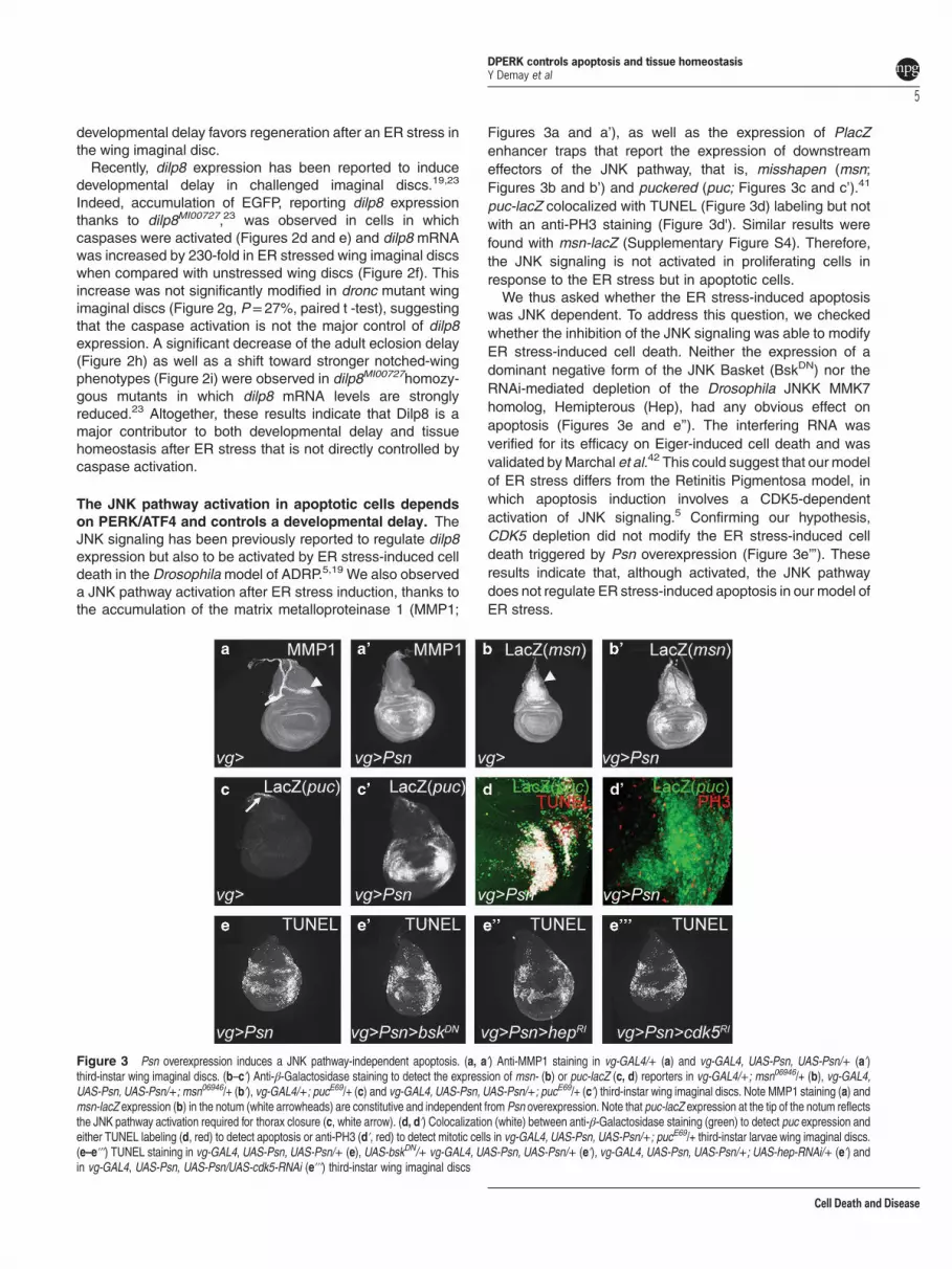
A Rac1/Slpr-dependent JNK pathway regulates develop-mental delay after ER-stress. The core of the JNK pathwayis composed by Jra and Kay, which dimerize to form thedAP-1 transcription factor, and the unique JNK, Bsk. Weobserved that Jra or kay depletion, which had no effect in theabsence of ER stress, aggravated the wing phenotype(Supplementary Table S1), showing that the JNK pathwaycore is involved in tissue homeostasis after an ER stress.We decided to identify the upstream JNK pathway
components that promote ER stress-induced developmentaldelay. A shift toward stronger wing phenotypes was observedwhen the JNKKs dMKK4 and Hep (Supplementary Table S1)or the JNKKK Slpr were depleted by RNAi in the presence ofER stress-induced cell death (Figure 6a,Supplementary TableS1). These depletions did not have any detectable effect bythemselves (data not shown). Slpr depletion also decreaseddilp8 expression and the ER stress-induced developmentaldelay (Figures 6d, g and j). Slpr can be activated by directinteraction with either Msn or the Rac1 GTPase.45 We thusexamined if these interactors participate to ER stress-associated JNK signaling. Surprisingly,msn depletion induceda distribution shift toward weaker phenotypes (Figure 6b).It also increased both dilp8 expression (Figures 6e and h) anddevelopmental delay (Figure 6k) in response to ER stress.These results suggest that Msn may antagonize JNKsignaling, which, to our knowledge, has only been observedonce.46 As ER stress inducesmsn expression (Figure 3b), wethus asked whether this was in a JNK-dependent manner.Indeed, the expression of a Bsk dominant negative formstrongly reduced msn expression (data not shown), suggest-ing that Msn could belong to a JNK pathway negative feedbackinduced after ER stress.
Figure 4 The JNK pathway regulates a Dilp8-dependent developmental delay.(a) Effects of puc overexpression (UAS-puc) (gray) and puc mutant heterozygosity(pucE69/+) (black) compared with control (white) in a vg-GAL4, UAS-Psn, UAS-Psn/+(plain bars) or vg-GAL4/+ (hatched bars) background on the mean time of adulteclosion. Error bars represent the S.E.M. (n= 4). Asterisks indicate significantdifference (Po10− 4, ANOVA). (b) Distribution of notched-wing phenotypes of vg-GAL4, UAS-Psn, UAS-Psn/+; UAS-puc/+ (gray bars) and vg-GAL4, UAS-Psn, UAS-Psn/+; pucE69/+ (black bars) flies compared with vg-GAL4, UAS-Psn, UAS-Psn/+flies (open bars). (c, d) Intensity of GFP reflecting dilp8 expression in vg-GAL4, UAS-Psn, UAS-Psn/+; dilp8MI00727/+ (c left, d white bars), vg-GAL4, UAS-Psn, UAS-Psn/+;dilp8MI00727/UAS-puc (c center, d gray bar), and vg-GAL4, UAS-Psn, UAS-Psn/+;dilp8MI00727/pucE69(c right, d black bar) third-instar wing imaginal discs. Error barsrepresent the S.E.M. of at least eight independent experiments. Asterisks indicatesignificant difference (Po10− 3, ANOVA)
DPERK controls apoptosis and tissue homeostasisY Demay et al
6
Cell Death and Disease

Finally, depletion of rac1 induced a shift toward strongerwing phenotypes (Figure 6c) and a decrease of dilp8expression (Figures 6f and i) resulting in a developmentaldelay suppression (Figure 6l). Therefore, Rac1 seems to bethe JNK pathway activator that triggers ER stress-induceddevelopmental delay. In conclusion, the developmental delayseems to be dependent of a Rac1/Slpr/JNK pathway.
Discussion
As previously reported in mammalian cells, we have validatedthat Psn overexpression can provoke chronic ER stress inDrosophila.26 In mammalian models, the UPR branchescan display opposite roles depending on the model. Forexample, Perk can be either anti or proapoptotic.47–49 Thanksto a newmodel of chronic ER stress, we have demonstrated inthis study that the PERK/ATF4 pathway has a fundamentalrole in Drosophila tissue homeostasis (Figure 7). So far, theADRP model was the only model of strong chronic ER stressreported in Drosophila.5,24 We have validated that Psnoverexpression can also provoke a chronic ER stressin Drosophila, as previously reported in mammalian cells.26
In both Drosophila models, apoptosis is induced by UPR inresponse to ER stress. Nevertheless, this induction involvestotally different pathways. In our chronic ER stress model, celldeath induction is PERK/ATF4 dependent and JNK indepen-dent, contrarily to the ADRP model in which CDK5 activatesJNK signaling that triggers apoptosis.5 These differencesshow that the complexity of ER stress-induced signaling foundin mammals is conserved in Drosophila, thus highlighting theusefulness of ER stress models plurality.We have shown that the PERK/ATF4 pathway induces a
caspase-dependent apoptosis by repressing diap1 transcrip-tion. However, PERK has been described to exert some
antiapoptotic activity by inducing IAP gene expression inmammals.47 This effect does not seem to rely on direct targetsof PERK, ATF4 and CHOP.50 Similarly, we did not find anyATF4 consensus binding sequence (5'-RTTRCRTCA-3') in thediap1 promoter region and no CHOP homolog has been foundin Drosophila. Therefore, the mechanisms involved in PERKregulation of IAPs remain to be clarified.In our chronic ER stress model, the JNK pathway is
activated in apoptotic cells to favor tissue homeostasis withoutstimulating cell proliferation. This is in contrast to a JNKactivation in cells neighboring apoptotic cells, which results inan increase of the proliferation rate.11 Similar to our observa-tion, JNK activation in apoptotic cells has been observed in‘undead cell’models.9,12–14 In these models, the JNK pathwaycould be activated by DIAP1 or DRONC, whereas JNKsignaling seems to be primarily independent from DIAP1/DRONC in our model. In a mammalian model of chronic ERstress, the IRE1 branch of the UPR activated the JNK pathwayto trigger apoptosis thanks to TRAF2/ASK1.51 In our ER stressmodel, depletion of traf2 or ask1 had no effect (SupplementaryTable S1 and data not shown). Instead, we have shown for thefirst time that JNK pathway activation mainly depends on thePERK/ATF4 pathway. Interestingly, this particular JNK path-way is not mainly activated by apoptosis and does notmodulate cell death or proliferation.We have also shown that PERK/ATF4 regulates an ER
stress-induced developmental delay. As previously reported,we observed that Dilp8 is amajor contributor to developmentaldelay.19,23 An obvious candidate for a Dilp8-independentdevelopmental delay regulation was the retinoic acid signalingthat has been reported to modulate an irradiation-induceddevelopmental delay.22 We have tested if this pathway couldalso regulate the developmental delay caused by Psn
Figure 5 ATF4 activates the JNK pathway. (a, left) Anti-activated caspase 3 (green) and anti-MMP1 (red) staining in vg-GAL4, UAS-Psn, UAS-Psn/+ (top) and vg-GAL4,UAS-Psn, UAS-Psn/+; UAS-diap1/+ (bottom) third-instar wing imaginal discs. (a, right) Intensity of GFP in vg-GAL4, UAS-Psn, UAS-Psn/+; dilp8MI00727/+ (top) and vg-GAL4,UAS-Psn, UAS-Psn/+; dilp8MI00727/UAS-diap1 (bottom) third-instar wing imaginal discs. (b) Quantification of relative dilp8::EGFP expression. Error bars represent the S.E.M. ofat least nine independent experiments. The asterisk indicates significant difference between the indicated genotype and the control (Po10− 5, ANOVA). (c) Anti-MMP1 staining(left) and intensity of Dilp8::EGFP detection (right) in vg-GAL4, UAS-Psn, UAS-Psn/+ (top) and vg-GAL4, UAS-Psn, UAS-Psn/+; UAS-atf4-RNAi (bottom) third-instar wingimaginal discs. (d) Quantification of relative dilp8 expression. Error bars represent the S.E.M. of at least four independent experiments. The asterisk indicates significant differencebetween the indicated genotype and the control (Po10− 4, ANOVA). (e) Effects of ATF4 depletion (gray bars) compared with control (white bars) on vg-GAL4, UAS-Psn, UAS-Psn/+ (solid bars) and vg-GAL4/+ (streaked bars) on the mean time of adult eclosion. Error bars represent the S.E.M. (n= 3). The asterisk corresponds to a statistical difference(Po10− 3, ANOVA)
DPERK controls apoptosis and tissue homeostasisY Demay et al
7
Cell Death and Disease
overexpression. No wing phenotype modification wasdetected upon the downregulation of this pathway (data notshown).We have characterized the components of the JNK
signaling that is activated in response to chronic ERstress in Drosophila wing imaginal discs (Figure 7). Thesmall GTPase Rac1 would activate the JNKKK Slpr, which inturn would activate JNK signaling core to regulate dilp8expression and ultimately favor development delay andtissue homeostasis maintenance. How the ATF4/PERKbranch activates Rac1 remains to be elucidated. Our resultsalso suggest the existence of a negative feedback loopregulating the JNK pathway, which would involve theJNKKKK, Msn. This is in agreement with a genetic andphosphoproteomic study showing that Msn is able to inhibitthe phosphorylation of Jun.46 Considering that the JNKpathway induces dilp8 expression in abnormally growingimaginal discs in other stress models,19 one may wonderwhether the same JNK pathway is implicated in thesemodels. Moreover, one may wonder whether dilp8 controlduring tissue homeostasis-associated developmental delay
is always JNK-dependent and relies on a Rac1/Slprpathway.To summarize, we have shown in this study that in response
to an ER stress induced by Psn overexpression, the PERKpathway is activated resulting in a Janus-faced ATF4 role. Onone hand, ATF4 induces caspase-dependent apoptosis byrepressing diap1 expression and on the other hand, it favorstissue homeostasis maintenance through the induction of aRac1/Slpr/JNK pathway and the resulting dilp8 expression.More investigations on this new Drosophila chronic ER-stressmodel should allow the identification of novel regulators ofUPR-dependent tissue and organism homeostasis that maybe conserved in mammals.
Materials and MethodsDrosophila crosses and strains. Flies were raised on standard corn-agarmedium. All crosses were performed at 25 °C. The Drosophila strains usedcarried vg-GAL4,52 pucE69,41 UAS-puc,43 UAS-Psn (Psn+14),53 UAS-xbp1::EGFPreporter,32 UAS-diap1,54 UAS-EGFP9.2,55 UAS-p35 (Bl#6298), msn06946(Bl#11707),thj5C8 (Bl#12093),35 UAS-bskDN (Bl#108773),56 dilp8MI00727 (Bl#33079). The UAS-cdk5-RNAi (Bl#35287), UAS-atf4-RNAi (Bl#25985), UAS-Rac1-RNAi (Bl#28985)strains from the Transgenic RNAi Project (TRIP) were provided by the Bloomington
Figure 6 The Rac1/Slpr JNK pathway triggers an ER stress-induced developmental delay negatively controlled by Msn. (a–c) Distribution of notched-wing phenotypes ofvg-GAL4, UAS-Psn, UAS-Psn/+; UAS-slpr-RNAi/+ (a, red), vg-GAL4, UAS-Psn, UAS-Psn/+; UAS-msn-RNAi/+ (b, blue) and vg-GAL4, UAS-Psn, UAS-Psn/+; UAS-Rac1-RNAi/+ (c, yellow) flies compared with vg-GAL4, UAS-Psn, UAS-Psn/+ flies (a–c, white). (d–i) Intensity of GFP emitted by the dilp8::EGFP reporter in vg-GAL4, UAS-Psn, UAS-Psn/+(d–i), vg-GAL4, UAS-Psn, UAS-Psn/+; UAS-slpr-RNAi/+ (d, g), vg-GAL4, UAS-Psn, UAS-Psn/+; UAS-msn-RNAi/+ (e, h) and vg-GAL4, UAS-Psn, UAS-Psn/+; UAS-Rac1-RNAi/+ (f, i) third-instar wing imaginal discs. Error bars represent the S.E.M. of at least six independent experiments. Asterisks indicate significant difference between theindicated genotype and its control (Po10− 4). (j–l) Effects of Slpr (j, red), Msn (k, blue) and Rac1 (l, yellow) depletion compared with the control (white) in a vg-GAL4, UAS-Psn,UAS-Psn/+ (plain bars) or vg-GAL4/+ (hatched bars) background on mean time of adult eclosion. Error bars represent the S.E.M. of seven independent experiments. Asteriskscorrespond to significant difference between controls without Psn (hatched bars) and the indicated genotype (Po10− 4, ANOVA)
DPERK controls apoptosis and tissue homeostasisY Demay et al
8
Cell Death and Disease

Drosophila Stock Center (BDRC, Bloomington, IN, USA). The UAS-slpr-RNAi (ID106449) and UAS-msn-RNAi (ID 101517) strains were obtained from the ViennaDrosophila RNAi Center (VDRC, Vienna, Austria), while the UAS-hep-RNAi(4353R2) strain was provided by the National Institute of Genetics stock center(NIG-Fly, Kyoto, Japan). Genetic background control strains were adapted to thedifferent transgenic lines we used. The rheaMI00296 (Bl#30955) strain was used asthe control for dilp8MI00727 (Bl#33079) genetic background, the y,w[1118];P{attP,y[+],w[3]} (ID 60100) line was the control of the KK library hosted by the VDRC, the y1
v1; P{UAS-GFP.VALIUM10}attP2 (Bl#35786) was used as a control for the TRIPV10 library and y1 sc1 v1 P{nos-phiC31\int.NLS}X; P{CaryP}attP2 (Bl#25710) for theTRIP V20 library. Canton S flies were used as reference for all the other strains.
Immunostaining, TUNEL assay and microscopy. Wing discs of third-instar male larvae were dissected in PBS and fixed in 3.7% formaldehyde for 20 minat room temperature. Samples were then washed thrice in PBT 0.3%. TUNELstaining was performed following manufacturer’s instructions (ApopTag Red in situapoptosis detection kit, Millipore, Temecula, CA, USA). Immunostaining was alwaysperformed, according to standard protocols, before the TUNEL assay. The followingprimary antibodies were used: rabbit anti-Caspase-3 (Asp175, Cell Signaling,Danvers, MA, USA, 1 : 20), mouse anti-β-galactosidase (40-1a, DSHB, University ofIowa, IA, USA, 1 : 200), mouse anti-MMP1 (5H7B11, DSHB, University of Iowa, IA,USA, 1 : 30), mouse anti-DIAP1 (kind gift from B Hay, 1 : 200). Secondaryantibodies conjugated to Alexa Fluor 488, 568 or 647 were purchased fromMolecular Probes (Eugen, OR, USA 1 : 400). Discs were mounted in Citifluor(Biovalley, Marne-La-Vallée, France). Images were captured using a Leica SPEconfocal laser-scanning microscope (Leica, Wetzlar, Germany). Images wereprocessed and treated with ImageJ and Adobe Photoshop 8.0. We used theColocalization plugin (Pierre Bourdoncle, 2003), which considers that two signalscolocalize if their respective intensities are strictly higher than the threshold of theirchannels and if their ratio of intensity is strictly higher than the ratio setting value(here, 75%). Transverse sections were computationally generated after reslicingconfocal stacks using ImageJ. GFP mean intensity of the Dilp8MI00727-encodedprotein trap was evaluated using the 'measure' function of ImageJ.
Developmental timing and 20E treatment. Eighty females were crossedto 40 males and transferred to fresh food after 2 days for 1 h to deposit eggs onculture medium. Flies reaching adulthood or pupariating were then counted every3 h. Addition of ecdysteroid (Sigma Aldrich, Saint Louis, MO, USA) was performedaccordingly to standard protocols.22 Each experiment was realized at least thriceindependently and data were statistically analyzed by ANOVA.
Genetic interaction test. Genetic interactions were tested based on themodulation of the severity of Psn overexpression-dependent notched-wing phenotype.Wings were categorized according to the number and size of notches observed intheir margin (Supplementary Figure S1A). To control the genetic background, mutantstrains and control strains were first crossed with males containing referencechromosomes carrying markers, that is, BI for chromosome II and Dr for chromosomeIII. Male progeny carrying Bl, Dr and the mutation to be tested were crossed withvg-GAL4, UAS-Psn, UAS-Psn flies. To rule out any effect by itself of the tested strains,control crosses with vg-GAL4 females were performed for each strain. Only the resultsobtained with males are depicted in figures although all data were statisticallyanalyzed by ANOVA, which allows to determine the effect of the chromosome ofinterest compared with the same chromosome in the control genetic background.
RNA extraction and RT-qPCR. Fifty wing imaginal discs were dissected inthe RNA XS NucleoSpin kit’s lysis buffer on ice for each genotype. Total RNAs wereextracted using this kit (Macherey-Nagel, Düren, Germany). RT was performedusing 4.8 μg of RNA incubated with random primer oligonucleotides andRecombinant Taq DNA Polymerase (Invitrogen, Life Technologies, Carlsbad,USA). Real-time PCR was performed using the ABI Prism 7700 HT (AppliedBiosystems, Life Technologies, Carlsbad, USA) with SYBRGreen mastermix(Abgene, Thermo Fisher Scientific, Waltham, MA, USA) and 11 ng of cDNA. Thereal-time PCR primers (Invitrogen, Life Technologies) were designed as in Garelliet al.23 Three independent RT experiments were performed and data werenormalized against rp49 mRNA levels.
Conflict of InterestThe authors declare no conflict of interest.
Acknowledgements. Confocal image acquisition and analysis were performedon CYMAGES imaging facility. We thank S Netter, B Limbourg-Bouchon, B Hay,J-A Lepesant and ME Fortini for providing fly stocks and antibodies. We are verygrateful to S Netter and I Guenal for suggestions on the manuscript and helpfuldiscussions throughout this work. We thank C Pirou, P Gandille, C Wintz and ourteam for their contribution to this project. This article is dedicated to the memory ofDidier Contamine who succumbed to a long illness after initiating this project. Weacknowledge the NIG (National Institute of Genetics, Kyoto, Japan), ViennaDrosophila RNAi Center (VDRC, Vienna, Austria) and Bloomington Stock center(Bloomington, IN, USA) for providing fly stocks.
Figure 7 Model of tissue homeostasis maintenance after an ER stress. ChronicER stress activates the PERK pathway, which results in ATF4 expression. ATF4 hastwo antagonistic functions. On one hand, ATF4 induces a caspase-dependentapoptosis by repressing diap1 expression. On the other hand, it favors tissuehomeostasis through the induction of dilp8 expression by Rac1/Slpr/JNK pathwayactivation. The role of Msn in the control of the observed developmental delayremains to be elucidated. It could either negatively regulate the JNK pathway or havea JNK pathway-independent mechanism of action
DPERK controls apoptosis and tissue homeostasisY Demay et al
9
Cell Death and Disease
1. Patil C, Walter P. Intracellular signaling from the endoplasmic reticulum to the nucleus: theunfolded protein response in yeast and mammals. Curr Opin Cell Biol 2001; 13: 349–355.
2. Harding HP, Calfon M, Urano F, Novoa I, Ron D. Transcriptional and translational control inthe Mammalian unfolded protein response. Annu Rev Cell Dev Biol 2002; 18: 575–599.
3. Kaufman RJ. Orchestrating the unfolded protein response in health and disease. J ClinInvest 2002; 110: 1389–1398.
4. Walter P, Ron D. The unfolded protein response: from stress pathway to homeostaticregulation. Science 2011; 334: 1081–1086.
5. Kang MJ, Chung J, Ryoo HD. CDK5 and MEKK1 mediate pro-apoptotic signalling followingendoplasmic reticulum stress in an autosomal dominant retinitis pigmentosa model. Nat CellBiol 2012; 14: 409–415.
6. Rasheva VI, Domingos PM. Cellular responses to endoplasmic reticulum stress andapoptosis. Apoptosis 2009; 14: 996–1007.
7. Ryoo HD, Steller H. Unfolded protein response in Drosophila: why another model can make itfly. Cell Cycle 2007; 6: 830–835.
8. Mollereau B, Perez-Garijo A, Bergmann A, Miura M, Gerlitz O et al. Compensatoryproliferation and apoptosis-induced proliferation: a need for clarification. Cell Death Differ2013; 20: 181.
9. Ryoo HD, Gorenc T, Steller H. Apoptotic cells can induce compensatory cell proliferationthrough the JNK and the Wingless signaling pathways. Dev Cell 2004; 7: 491–501.
10. Perez-Garijo A, Shlevkov E, Morata G. The role of Dpp and Wg in compensatory proliferationand in the formation of hyperplastic overgrowths caused by apoptotic cells in the Drosophilawing disc. Development 2009; 136: 1169–1177.
11. Bergantinos C, Corominas M, Serras F. Cell death-induced regeneration in wing imaginaldiscs requires JNK signalling. Development 2010; 137: 1169–1179.
12. Huh JR, Guo M, Hay BA. Compensatory proliferation induced by cell death in the Drosophilawing disc requires activity of the apical cell death caspase Dronc in a nonapoptotic role. CurrBiol 2004; 14: 1262–1266.
13. Wells BS, Yoshida E, Johnston A. Compensatory proliferation in Drosophila imaginal discsrequires Dronc-dependent p53 activity. Curr Biol 2006; 16: 1606–1615.
14. Kondo S, Senoo-Matsuda N, Hiromi YMiura M. DRONC coordinates cell death andcompensatory proliferation. Mol Cell Biol 2006; 26: 7258–7268.
15. Perkins KK, Dailey GM, Tjian R. Novel Jun- and Fos-related proteins in Drosophila arefunctionally homologous to enhancer factor AP-1. EMBO J 1988; 7: 4265–4273.
16. ChenW, White MA, Cobb MH. Stimulus-specific requirements for MAP3 kinases in activatingthe JNK pathway. J Biol Chem 2002; 277: 49105–49110.
17. Stronach B, Perrimon N. Activation of the JNK pathway during dorsal closure in Drosophilarequires the mixed lineage kinase, slipper. Genes Dev 2002; 16: 377–387.
18. Stronach B. Dissecting JNK signaling, one KKKinase at a time. Dev Dyn 2005; 232: 575–584.19. Colombani J, Andersen DS, Leopold P. Secreted peptide Dilp8 coordinates Drosophila
tissue growth with developmental timing. Science 2012; 336: 582–585.20. Hussey RG, Thompson WR, Calhoun ET. The Influence of X-Rays on the Development of
Drosophila Larvae. Science 1927; 66: 65–66.21. Simpson P, Berreur P, Berreur-Bonnenfant J. The initiation of pupariation in Drosophila:
dependence on growth of the imaginal discs. J Embryol Exp Morphol 1980; 57: 155–165.22. Halme A, Cheng M, Hariharan IK. Retinoids regulate a developmental checkpoint for tissue
regeneration in Drosophila. Curr Biol 2010; 20: 458–463.23. Garelli A, Gontijo AM, Miguela V, Caparros E, Dominguez M. Imaginal discs secrete insulin-
like peptide 8 to mediate plasticity of growth and maturation. Science 2012; 336: 579–582.24. Ryoo HD, Domingos PM, Kang MJ, Steller H. Unfolded protein response in a Drosophila
model for retinal degeneration. EMBO J 2007; 26: 242–252.25. Michno K, Knight D, Campusano JM, van de Hoef D, Boulianne GL. Intracellular calcium
deficits in Drosophila cholinergic neurons expressing wild type or FAD-mutant presenilin.PLoS One 2009; 4: e6904.
26. Honarnejad K, Jung CK, Lammich S, Arzberger T, Kretzschmar H, Herms J. Involvement ofpresenilin holoprotein upregulation in calcium dyshomeostasis of Alzheimer's disease. J CellMol Med 2013; 17: 293–302.
27. Cook DG, Sung JC, Golde TE, Felsenstein KM, Wojczyk BS, Tanzi RE et al. Expression andanalysis of presenilin 1 in a human neuronal system: localization in cell bodies and dendrites.Proc Natl Acad Sci USA 1996; 93: 9223–9228.
28. Annaert WG, Levesque L, Craessaerts K, Dierinck I, Snellings G, Westaway D et al.Presenilin 1 controls gamma-secretase processing of amyloid precursor protein in pre-golgicompartments of hippocampal neurons. J Cell Biol 1999; 147: 277–294.
29. Levitan D, Lee J, Song L, Manning R, Wong G, Parker E et al. PS1 N- and C-terminalfragments form a complex that functions in APP processing and Notch signaling. Proc NatlAcad Sci USA 2001; 98: 12186–12190.
30. Kim J, Kleizen B, Choy R, Thinakaran G, Sisodia SS, Schekman RW. Biogenesis of gamma-secretase early in the secretory pathway. J Cell Biol 2007; 179: 951–963.
31. Brand AH, Perrimon N. Targeted gene expression as a means of altering cell fates andgenerating dominant phenotypes. Development 1993; 118: 401–415.
32. Souid S, Lepesant JA, Yanicostas C. The xbp-1 gene is essential for development inDrosophila. Dev Genes Evol 2007; 217: 159–167.
33. Fan Y, Bergmann A. The cleaved-caspase-3 antibody is a marker of caspase-9-like DRONCactivity in Drosophila. Cell Death Differ 2010; 17: 534–539.
34. Novoa I, Zeng H, Harding HP, Ron D. Feedback inhibition of the unfolded proteinresponse by GADD34-mediated dephosphorylation of eIF2alpha. J Cell Biol 2001; 153:1011–1022.
35. Ryoo HD, Bergmann A, Gonen H, Ciechanover A, Steller H. Regulation of Drosophila IAP1degradation and apoptosis by reaper and ubcD1. Nat Cell Biol 2002; 4: 432–438.
36. Smith-Bolton RK, Worley MI, Kanda H, Hariharan IK. Regenerative growth in Drosophilaimaginal discs is regulated by wingless and Myc. Dev Cell 2009; 16: 797–809.
37. Herrera SC, Martin R, Morata G. Tissue homeostasis in the wing disc of Drosophilamelanogaster: immediate response to massive damage during development. PLoS Genet2013; 9: e1003446.
38. Bryant PJ, Simpson P. Intrinsic and extrinsic control of growth in developing organs. Q RevBiol 1984; 59: 387–415.
39. Haynie JL, Bryant PJ. Intercalary regeneration in imaginal wing disk of Drosophilamelanogaster. Nature 1976; 259: 659–662.
40. Colombani J, Bianchini L, Layalle S, Pondeville E, Dauphin-Villemant C, Antoniewski C et al.Antagonistic actions of ecdysone and insulins determine final size in Drosophila. Science2005; 310: 667–670.
41. Ring JM, Martinez Arias A. Puckered, a gene involved in position-specific cell differentiationin the dorsal epidermis of the Drosophila larva. Dev Suppl 1993; 121: 251–259.
42. Marchal C, Vinatier G, Sanial M, Plessis A, Pret AM, Limbourg-Bouchon B et al. The HIV-1Vpu protein induces apoptosis in Drosophila via activation of JNK signaling. PLoS One 2012;7: e34310.
43. Martin-Blanco E, Gampel A, Ring J, Virdee K, Kirov N, Tolkovsky AM et al. puckeredencodes a phosphatase that mediates a feedback loop regulating JNK activity during dorsalclosure in Drosophila. Genes Dev 1998; 12: 557–570.
44. McEwen DG, Peifer M. Puckered, a Drosophila MAPK phosphatase, ensures cell viability byantagonizing JNK-induced apoptosis. Development 2005; 132: 3935–3946.
45. Garlena RA, Gonda RL, Green AB, Pileggi RM, Stronach B. Regulation of mixed-lineagekinase activation in JNK-dependent morphogenesis. J Cell Sci 2010; 123: 3177–3188.
46. Bakal C, Linding R, Llense F, Heffern E, Martin-Blanco E, Pawson T et al. Phosphorylationnetworks regulating JNK activity in diverse genetic backgrounds. Science 2008; 322:453–456.
47. Hamanaka RB, Bobrovnikova-Marjon E, Ji X, Liebhaber SA, Diehl JA. PERK-dependentregulation of IAP translation during ER stress. Oncogene 2009; 28: 910–920.
48. Verfaillie T, van Vliet A, Garg AD, Dewaele M, Rubio N, Gupta S et al. Pro-apoptotic signalinginduced by photo-oxidative ER stress is amplified by Noxa, not Bim. Biochem Biophys ResCommun 2013; 438: 500–506.
49. Oommen D, Prise KM. Down-regulation of PERK enhances resistance to ionizing radiation.Biochem Biophys Res Commun 2013; 441: 31–35.
50. Han J, Back SH, Hur J, Lin YH, Gildersleeve R, Shan J et al. ER-stress-inducedtranscriptional regulation increases protein synthesis leading to cell death. Nat Cell Biol2013; 15: 481–490.
51. Nishitoh H, Matsuzawa A, Tobiume K, Saegusa K, Takeda K, Inoue K et al. ASK1 is essentialfor endoplasmic reticulum stress-induced neuronal cell death triggered by expandedpolyglutamine repeats. Genes Dev 2002; 16: 1345–1355.
52. Simmonds AJ, Brook WJ, Cohen SM, Bell JB. Distinguishable functions for engrailed andinvected in anterior-posterior patterning in the Drosophila wing. Nature 1995; 376: 424–427.
53. Ye Y, Fortini ME. Characterization of Drosophila Presenilin and its colocalization with Notchduring development. Mech Dev 1998; 79: 199–211.
54. Hay BA, Wassarman DA, Rubin GM. Drosophila homologs of baculovirus inhibitor ofapoptosis proteins function to block cell death. Cell 1995; 83: 1253–1262.
55. Netter S, Faucheux M, Theodore L. Developmental dynamics of a polyhomeotic-EGFPfusion in vivo. DNA Cell Biol 2001; 20: 483–492.
56. Adachi-Yamada T, Nakamura M, Irie K, Tomoyasu Y, Sano Y, Mori E et al. p38 mitogen-activated protein kinase can be involved in transforming growth factor beta superfamily signaltransduction in Drosophila wing morphogenesis. Mol Cell Biol 1999; 19: 2322–2329.
Cell Death and Disease is an open-access journalpublished by Nature Publishing Group. This work is
licensed under a Creative Commons Attribution-NonCommercial-ShareAlike 3.0 Unported License. The images or other third partymaterial in this article are included in the article’s Creative Commonslicense, unless indicated otherwise in the credit line; if the materialis not included under the Creative Commons license, users will needto obtain permission from the license holder to reproduce thematerial. To view a copy of this license, visit http://creativecommons.org/licenses/by-nc-sa/3.0/
Supplementary Information accompanies this paper on Cell Death and Disease website (http://www.nature.com/cddis)
DPERK controls apoptosis and tissue homeostasisY Demay et al
10
Cell Death and Disease


![Arf nucleotide binding site opener [ARNO] promotes sequential activation of Arf6, Cdc42 and Rac1 and insulin secretion in INS 832/13 β-cells and rat islets](https://static.fdokumen.com/doc/165x107/6316194efc260b7102104d00/arf-nucleotide-binding-site-opener-arno-promotes-sequential-activation-of-arf6.jpg)