Activated R-Ras, Rac1, PI 3-Kinase and PKCepsilon Can Each Restore Cell Spreading Inhibited by...
12
The Rockefeller University Press, 0021-9525/2000/12/1549/12 $5.00 The Journal of Cell Biology, Volume 151, Number 7, December 25, 2000 1549–1560 http://www.jcb.org/cgi/content/full/151/7/1549 1549 Activated R-Ras, Rac1, PI 3-Kinase and PKCe Can Each Restore Cell Spreading Inhibited by Isolated Integrin b1 Cytoplasmic Domains Allison L. Berrier,* Anthony M. Mastrangelo,* Julian Downward, ‡ Mark Ginsberg, § and Susan E. LaFlamme* *Center for Cell Biology and Cancer Research, Albany Medical College, Albany, New York 12208; ‡ Imperial Cancer Research Fund, London WC2A 3PX, United Kingdom; and § Department of Vascular Biology, The Scripps Research Institute, La Jolla, California 92037 Abstract. Attachment of many cell types to extracellu- lar matrix proteins triggers cell spreading, a process that strengthens cell adhesion and is a prerequisite for many adhesion-dependent processes including cell migration, survival, and proliferation. Cell spreading requires integrins with intact b cytoplasmic domains, presumably to connect integrins with the actin cytoskel- eton and to activate signaling pathways that promote cell spreading. Several signaling proteins are known to regulate cell spreading, including R-Ras, PI 3-kinase, PKCe and Rac1; however, it is not known whether they do so through a mechanism involving integrin b cyto- plasmic domains. To study the mechanisms whereby cell spreading is regulated by integrin b cytoplasmic do- mains, we inhibited cell spreading on collagen I or fi- brinogen by expressing tac-b1, a dominant-negative in- hibitor of integrin function, and examined whether cell spreading could be restored by the coexpression of ei- ther V38R-Ras, p110a-CAAX, myr-PKCe, or L61Rac1. Each of these activated signaling proteins was able to restore cell spreading as assayed by an increase in the area of cells expressing tac-b1. R-Ras and Rac1 rescued cell spreading in a GTP-dependent manner, whereas PKCe required an intact kinase domain. Importantly, each of these signaling proteins required intact b cyto- plasmic domains on the integrins mediating adhesion in order to restore cell spreading. In addition, the res- cue of cell spreading by V38R-Ras was inhibited by LY294002, suggesting that PI 3-kinase activity is re- quired for V38R-Ras to restore cell spreading. In con- trast, L61Rac1 and myr-PKCe each increased cell spreading independent of PI 3-kinase activity. Ad- ditionally, the dominant-negative mutant of Rac1, N17Rac1, abrogated cell spreading and inhibited the ability of p110a-CAAX and myr-PKCe to increase cell spreading. These studies suggest that R-Ras, PI 3-kinase, Rac1 and PKCe require the function of inte- grin b cytoplasmic domains to regulate cell spreading and that Rac1 is downstream of PI 3-kinase and PKCe in a pathway involving integrin b cytoplasmic domain function in cell spreading. Key words: cell spreading • integrin • signaling • Rac1 • R-Ras Introduction Integrins are a family of a/b heterodimeric transmem- brane receptors used by cells to interact with their extra- cellular matrix (Hynes, 1992). Integrins form a structural link between the extracellular matrix and the cell’s cyto- skeletal and signal transduction apparatus. Integrin engage- ment triggers a myriad of intracellular signals resulting in changes in intracellular pH and [Ca 21 ], as well as the acti- vation of protein kinases, phosphoinositide kinases, phos- pholipases, and small GTP binding proteins (Schwartz et al., 1995; Yamada and Miyamoto, 1995; Burridge and Chr- zanowska-Wodnicka, 1996; LaFlamme et al., 1997). Many of the signals activated by integrin engagement regulate the adhesion process itself (Schwartz et al., 1995; Yamada and Miyamoto, 1995; Burridge and Chrzanowska-Wod- nicka, 1996; LaFlamme et al., 1997). Cell spreading requires actin polymerization and reorga- nization, the extension of lamellipodia or filopodia, and the formation of new integrin-substrate adhesions (Small et al., 1999). The initial interaction of integrins with their ligand in cell attachment is believed to trigger signaling pathways that regulate the process of cell spreading. PI 3-kinase has been shown to be activated by integrin-medi- ated cell adhesion, and in some cell types PI 3-kinase activ- ity is required for cell spreading (Khwaja et al., 1997; King et al., 1997). The expression of an activated form of R-Ras Address correspondence to Susan E. LaFlamme, Center for Cell Biology & Cancer Research, Mail Code 165, Albany Medical College, 47 New Scotland Avenue, Albany, NY 12208. Tel.: (518) 262-6256. Fax: (518) 262- 5669. E-mail: [email protected]
-
Upload
independent -
Category
Documents
-
view
1 -
download
0
Transcript of Activated R-Ras, Rac1, PI 3-Kinase and PKCepsilon Can Each Restore Cell Spreading Inhibited by...

The Rockefeller University Press, 0021-9525/2000/12/1549/12 $5.00The Journal of Cell Biology, Volume 151, Number 7, December 25, 2000 1549–1560http://www.jcb.org/cgi/content/full/151/7/1549 1549
Activated R-Ras, Rac1, PI 3-Kinase and PKC
e
Can Each Restore Cell Spreading Inhibited by Isolated Integrin
b
1 Cytoplasmic Domains
Allison L. Berrier,* Anthony M. Mastrangelo,* Julian Downward,
‡
Mark Ginsberg,
§
and Susan E. LaFlamme*
*Center for Cell Biology and Cancer Research, Albany Medical College, Albany, New York 12208;
‡
Imperial Cancer Research Fund, London WC2A 3PX, United Kingdom; and
§
Department of Vascular Biology, The Scripps Research Institute, La Jolla, California 92037
Abstract.
Attachment of many cell types to extracellu-lar matrix proteins triggers cell spreading, a processthat strengthens cell adhesion and is a prerequisite for
many adhesion-dependent processes including cellmigration, survival, and proliferation. Cell spreading
requires integrins with intact
b
cytoplasmic domains,presumably to connect integrins with the actin cytoskel-eton and to activate signaling pathways that promotecell spreading. Several signaling proteins are known toregulate cell spreading, including R-Ras, PI 3-kinase,PKC
e
and Rac1; however, it is not known whether theydo so through a mechanism involving integrin
b
cyto-plasmic domains. To study the mechanisms wherebycell spreading is regulated by integrin
b
cytoplasmic do-mains, we inhibited cell spreading on collagen I or fi-brinogen by expressing tac-
b
1, a dominant-negative in-hibitor of integrin function, and examined whether cellspreading could be restored by the coexpression of ei-ther V38R-Ras, p110
a
-CAAX, myr-PKC
e
, or L61Rac1.Each of these activated signaling proteins was able torestore cell spreading as assayed by an increase in thearea of cells expressing tac-
b
1. R-Ras and Rac1 rescued
cell spreading in a GTP-dependent manner, whereasPKC
e
required an intact kinase domain. Importantly,each of these signaling proteins required intact
b
cyto-
plasmic domains on the integrins mediating adhesionin order to restore cell spreading. In addition, the res-cue of cell spreading by V38R-Ras was inhibited byLY294002, suggesting that PI 3-kinase activity is re-quired for V38R-Ras to restore cell spreading. In con-trast, L61Rac1 and myr-PKC
e
each increased cellspreading independent of PI 3-kinase activity. Ad-ditionally, the dominant-negative mutant of Rac1,N17Rac1, abrogated cell spreading and inhibited the
ability of p110
a
-CAAX and myr-PKC
e
to increase
cell spreading. These studies suggest that R-Ras, PI3-kinase, Rac1 and PKC
e
require the function of inte-grin
b
cytoplasmic domains to regulate cell spreadingand that Rac1 is downstream of PI 3-kinase and PKC
e
in a pathway involving integrin
b
cytoplasmic domainfunction in cell spreading.
Key words: cell spreading • integrin • signaling •Rac1 • R-Ras
Introduction
Integrins are a family of
a
/
b
heterodimeric transmem-brane receptors used by cells to interact with their extra-cellular matrix (Hynes, 1992). Integrins form a structurallink between the extracellular matrix and the cell’s cyto-skeletal and signal transduction apparatus. Integrin engage-ment triggers a myriad of intracellular signals resulting in
changes in intracellular pH and [Ca
2
1
], as well as the acti-vation of protein kinases, phosphoinositide kinases, phos-pholipases, and small GTP binding proteins (Schwartz etal., 1995; Yamada and Miyamoto, 1995; Burridge and Chr-
zanowska-Wodnicka, 1996; LaFlamme et al., 1997). Manyof the signals activated by integrin engagement regulatethe adhesion process itself (Schwartz et al., 1995; Yamadaand Miyamoto, 1995; Burridge and Chrzanowska-Wod-nicka, 1996; LaFlamme et al., 1997).
Cell spreading requires actin polymerization and reorga-nization, the extension of lamellipodia or filopodia, andthe formation of new integrin-substrate adhesions (Smallet al., 1999). The initial interaction of integrins with theirligand in cell attachment is believed to trigger signaling
pathways that regulate the process of cell spreading. PI3-kinase has been shown to be activated by integrin-medi-ated cell adhesion, and in some cell types PI 3-kinase activ-ity is required for cell spreading (Khwaja et al., 1997; Kinget al., 1997). The expression of an activated form of R-Ras
Address correspondence to Susan E. LaFlamme, Center for Cell Biology& Cancer Research, Mail Code 165, Albany Medical College, 47 NewScotland Avenue, Albany, NY 12208. Tel.: (518) 262-6256. Fax: (518) 262-5669. E-mail: [email protected]

The Journal of Cell Biology, Volume 151, 2000 1550
(V38R-Ras) has also been shown to positively regulate cellspreading, whereas the expression of dominant negativeR-Ras (N43R-Ras) can inhibit cell spreading in CHO cells(Zhang et al., 1996). In addition, both PKC
a
and PKC
e
ap-pear to be positive regulators of cell adhesion (Vuori andRuoslahti, 1993; Chun et al., 1996). PKC
e
has been shownto become activated upon cell attachment before spread-ing and pharmacological inhibitors of PKC have beenshown to inhibit the process of spreading (Vuori and Ruo-slahti, 1993; Chun et al., 1996). The small GTP binding pro-teins Rac1 and Cdc42 are also regulators of cell spreading.Integrin-mediated cell adhesion can trigger Rac1 activa-tion (Del Pozo et al., 2000), and expression of dominantnegative mutants of Rac1 and Cdc42 have been shown toinhibit cell spreading (Clark et al., 1998; Price et al., 1998;Van Leeuwen et al., 1999). In addition to the requirementfor the activities of several signaling proteins, the processof cell spreading is dependent upon intact
b
subunit cyto-plasmic domains on the integrins mediating adhesion(Ylanne et al., 1993); however, the molecular mechanismsby which integrins and various signaling proteins regulatecell spreading are not fully defined.
We and others have previously shown that expression ofisolated
b
cytoplasmic domains connected to the extracel-lular and transmembrane domains of the small nonsignal-ing (tac) subunit of the interleukin-2 receptor or otherextracellular reporters can inhibit endogenous integrinfunction in a variety of cellular processes, including cellspreading (Chen et al., 1994; LaFlamme et al., 1994; Luka-shev et al., 1994; Smilenov et al., 1994). Recently, we haveshown that high levels of expression of the tac-
b
1 chimeracan inhibit both cell attachment to fibronectin and
b
1 inte-grin conformations that are favorable to
b
1 integrin–ligand interactions (Mastrangelo et al., 1999a). Since cellsexpressing moderate levels of tac-
b
1 can attach, but are in-hibited in cell spreading (LaFlamme et al., 1994; Mas-trangelo et al., 1999a), we have used this system to studythe molecular mechanisms that regulate the process of cellspreading. In this study, we asked whether activated formsof signaling proteins known to positively regulate cellspreading could restore cell spreading inhibited by the ex-pression of tac-
b
1. We found that activated forms of PI3-kinase, R-Ras, Rac1, and PKC
e
could all restore cellspreading inhibited by tac-
b
1 in both normal human fibro-blasts adherent to collagen I and CHO cells expressing re-combinant
a
IIb
b
3 adherent to fibrinogen. Using CHOcells expressing either wild-type recombinant
a
IIb
b
3 or
a
IIb
b
3 with a
b
3 cytoplasmic domain truncation (
a
IIb
b
3
D
728), we show that the ability of V38R-Ras, L61Rac1,p110
a
-CAAX, and myr-PKC
e
to increase cell spreading intac-
b
1–expressing cells is dependent upon the presence ofintact
b
cytoplasmic domains on the integrins mediatingadhesion.
Materials and Methods
Antibodies and Matrix Proteins
Tac expression was monitored with FITC-conjugated antibodies to CD25from Accurate Chemicals and Scientific Corp. and Becton Dickinson. Rab-bit and goat polyclonal antibodies to the Myc (9E10) epitope, Flag epitope,and PKC
e
were purchased from Santa Cruz Biotechnology, Inc.Rhodamine-conjugated goat antibodies to rabbit IgG were purchased from
Roche Molecular Biochemicals and Pierce. Rhodamine-conjugated donkeyantibodies to goat IgG were provided by Santa Cruz Biotechnology, Inc.The
b
1 integrin function blocking P4C10 ascites was purchased fromGIBCO-BRL and the control ascites from Sigma-Aldrich. The matrix pro-teins collagen I (Vitrogen 100) and human fibrinogen were purchased fromCollagen Biomaterials and Enzyme Research Laboratories, respectively.
cDNAs, Cells, Transfections, and Spreading Assays
The eukaryotic expression vectors have been described previously: V38R-Ras and N43R-Ras by Marte et al. (1996), p110
a
-CAAX by Wennstromand Downward (1999), and tac and tac-
b
1 by LaFlamme et al. (1992). Eu-karyotic expression vectors for L61Rac1 and N17Rac1 were generouslyprovided by Dr. Alan Hall (University College, London, UK) and weredescribed previously by Lamarche et al. (1996). Myr-PKC
e
, PKC
e
, andW437PKC
e
were generous gifts from Dr. Alex Toker (Deaconess MedicalCenter, Boston, MA) and were constructed similarly to the PKC
z
con-structs as previously described (Chou et al., 1998).
Primary human foreskin fibroblasts (Vec Technologies) were maintainedin DMEM supplemented with 10% fetal bovine serum and 2 mM
L
-glu-tamine. The stable CHO cell lines A5 (
a
IIb
b
3) and ETC12 (
a
IIb
b
3
D
728)(Ylanne et al., 1995) were maintained in DMEM supplemented with 10%fetal bovine serum, 2 mM
L
-glutamine, and MEM nonessential amino acids.Cells were transfected by electroporation (Mastrangelo et al., 1999b) with20
m
g of tac or tac-
b
1 expression vectors together with 40
m
g of expressionvector for the various signaling proteins. For the kinase-dead PKC
e
experi-ment, comparable expression of the PKC
e
proteins was observed by immu-nofluorescence when 30
m
g of PKC
e
and 60
m
g of kinase-dead W437PKC
e
expression vectors were transfected. For spreading assays, human fibro-blasts were harvested by trypsinization with cold PBS
z
16 h after elec-troporation and were allowed to recover for 40 min at 37
8
C in suspension.Approximately 4
3
10
5
cells were plated onto glass coverslips coated with20
m
g/ml collagen I and were allowed to spread for 1 h at 37
8
C in serum-freemedium. Spreading assays using A5 and ETC12 CHO cell lines were per-formed similarly; however, cells were allowed to recover for 1 h before plat-ing onto glass coverslips coated with 15
m
g/ml fibrinogen, and spreading wasassayed after 30 min (Ylanne et al., 1995). To determine whether cellspreading on collagen I was dependent on endogenous
b
1 integrins, the
b
1function blocking P4C10 ascites (1:1,000 dilution) or control ascites (1:1,000dilution) was added 10 min after plating, at which time
.
90% of the cellshad attached, but not spread. To determine the requirement for PI 3-kinaseactivity, transiently transfected cells were incubated for 40 min in suspen-sion with LY294002 (Calbiochem) at a final concentration of 50
m
M, andsubsequently analyzed for spreading as described above.
Immunofluorescence and Quantification of Cell Area and Fluorescence Intensity
Adherent cells were fixed and processed for immunofluorescence as pre-viously described (LaFlamme et al., 1992). Cells were stained for cell sur-face expression of the tac epitope with FITC-conjugated antibodies toCD25 before permeabilization. Cells cotransfected with R-Ras, Rac1, andmyr-PKC
e
signaling proteins were subsequently permeabilized with PBScontaining 0.4% Triton X-100 for 5 min at room temperature, incubatedwith rabbit polyclonal antibodies to the epitope tag of the signaling pro-teins, and then with rhodamine-conjugated secondary antibodies. Cellscotransfected with wild-type PKC
e
and kinase-dead W437PKC
e
were per-meabilized with PBS containing 0.1% Triton X-100 and 3.7% formalde-hyde for 15 min at room temperature and stained as described previously.Microscopy was performed using an Olympus BX60 microscope equippedwith phase contrast and epifluorescence and attached to a Spot low lightcamera interfaced with a PC computer (Diagnostic Instruments). Spotsoftware was used to acquire the fluorescence and interference reflectionmicroscopy (Izzard and Lochner, 1976) images of transfected cells. Cellarea and fluorescence intensity were measured using Image Pro-Plus soft-ware (Media Cybernetics). Image Pro-Plus software was calibrated withan Applied Micro Stage micrometer (Applied Image, Inc.). The accuracyof the area measurements was confirmed with a TSM measurement slidethat contains etched squares of known dimensions. Preliminary studies in-dicated that cells that are round and have not begun to spread have cell ar-eas
,
560
m
m
2
. Hence, we define those cells with areas
.
560
m
m
2
asspread and cells with areas
,
560
m
m
2
as not spread. The conditions usedto acquire the fluorescence images minimized the influence of cell shape;however, for very small cells (areas
,
320
m
m
2
), our analysis may underes-timate the expression level of recombinant proteins.

Berrier et al.
Rescue of Cell Spreading Inhibited by Tac-
b
1
1551
Results
Activated V38R-Ras, myr-PKC
e
, L61Rac1, orp110
a
-CAAX Increase the Spreading of Cells Expressing Tac-
b
1
Previous studies have shown that the expression of tac-
b
1can inhibit cell spreading on a variety of extracellular ma-trix proteins (LaFlamme et al., 1994). In the present study,we tested whether the expression of activated forms of sig-naling proteins known to positively regulate cell spreadingcould restore tac-
b
1–inhibited spreading. Normal fibro-blasts were transiently transfected with tac-
b
1 or the con-trol tac receptor alone, or cotransfected with tac-
b
1 and ei-ther activated R-Ras, PKC
e
, Rac1, or PI 3-kinase.Transfected cells were plated onto collagen I and allowedto spread for 1 h. The cells adherent to collagen I were im-munostained for tac expression and the expression of thecotransfected signaling protein where appropriate. Cellarea and fluorescence intensity were quantified for posi-tive and double positive cells using Image Pro-Plus soft-ware as described in Materials and Methods.
In agreement with previous studies (LaFlamme et al.,1994), we found that the majority of the tac-
b
1 positivecells had cell areas
,
560
m
m
2
, indicating that they were in-hibited in cell spreading (Fig. 1 B). In contrast, the major-ity of cells expressing the control tac receptor had cell ar-eas
.
560
m
m2, indicating that they were not inhibited inspreading (Fig. 1 B). When we tested the ability of acti-vated R-Ras (V38R-Ras) and membrane-anchored PKCe(myr-PKCe) to restore tac-b1–inhibited cell spreading, wefound that the coexpression of tac-b1 with either V38R-Ras or myr-PKCe resulted in an increase in cell area ascompared with cells expressing tac-b1 alone (Fig. 1 B).Quantitation of the spreading assay revealed that 35% ofcells coexpressing myr-PKCe and 52% of cells coexpress-ing V38R-Ras were spread, compared with 12% of cellsexpressing tac-b1 alone. Coexpression of constitutively ac-tive Rac1 (L61Rac1) also increased spreading of tac-b1–expressing cells (Fig. 1 C). Of the cells coexpressing tac-b1and L61Rac1, 86% were spread, compared with 13% ex-pressing tac-b1 alone. Cotransfection of the membrane-anchored catalytic subunit of PI 3-kinase also increasedthe area of tac-b1–expressing cells (Fig. 1 D). When wequantitated the spreading of cells cotransfected with tac-b1 and p110a-CAAX as a function of tac epitope expres-
expression. The x axis is a linear scale of cell area from 0 to 2,400mm2 and the y axis is a linear scale of FITC fluorescence (tac epitopeexpression) from 0 to 3.2 3 104 (a) or 0 to 1.6 3 104 (b and c). (B–D)The vertical line positioned at a cell area of 560 mm2 indicates theseparation of spread (right) and not spread (left) cells. It is impor-tant to note that our spreading assays primarily analyze cells ex-pressing moderate to low levels of tac-b1, since cell attachment tocollagen I is inhibited by the expression of high levels of tac-b1 (datanot shown). This observation is consistent with our recent studiesshowing that high levels of tac-b1 inhibit cell attachment to fibronec-tin (Mastrangelo et al., 1999a). The range of FITC fluorescence rep-resents the range of tac-b1 expression detected in the adherenttransfected cells. These experiments were performed three timesand similar results were obtained.
Figure 1. Coexpression of either myr-PKCe, V38R-Ras, L61Rac1,or p110a-CAAX with tac-b1 can restore cell spreading on collagenI. (A) Diagram of the control tac receptor containing the extracellu-lar and transmembrane domains of the small (tac) subunit of the hu-man interleukin-2 receptor and the tac-b1 chimera containing thesame domains of the interleukin-2 receptor fused to the human inte-grin b1A cytoplasmic domain. (B) Fibroblasts were transfected withtac (a) or tac-b1 (b), or cotransfected with tac-b1 and myr-PKCe-Flag (c and e) or tac-b1 and myc-V38R-Ras (d and f). Cell area for100 randomly sampled positively transfected cells is plotted as afunction of either tac epitope expression (a–d), myr-PKCe-Flag ex-pression (e) for the same cells shown in c, or myc-V38R-Ras expres-sion (f) for the same cells shown in d. The x axis is a linear scale ofcell area from 0 to 1,600 mm2, the y axis is a linear scale of eitherFITC fluorescence (tac epitope expression) units defined by ImagePro-Plus from 0 to 2.4 3 104 (a–d) or rhodamine fluorescence (Flagor myc epitope expression) from 0 to 1.2 3 105 (e and f). (C) Fibro-blasts were transfected with tac (a) or tac-b1 (b), or cotransfectedwith tac-b1 and myc-L61Rac1 (c and d). Cell area for 90 randomlysampled cells expressing tac, tac-b1, or coexpressing tac-b1 and myc-L61Rac1 is plotted as a function of tac epitope expression (a–c) ormyc epitope expression (d) for the cells shown in c. The x axis is alinear scale of cell area from 0 to 2,400 mm2, the y axis is a linearscale of FITC fluorescence (tac epitope expression) from 0 to 2.4 3104 (a–c) or rhodamine fluorescence (myc epitope expression) from0 to 1.2 3 105 (d). (D) Fibroblasts transfected with the control tac re-ceptor (a) or tac-b1 (b) or tac-b1 and p110a-CAAX (c) were ana-lyzed for cell-surface expression of the tac epitope and cell area, asdescribed in Materials and Methods. Cell area for 98 randomly sam-pled positively transfected cells is plotted as a function of tac epitope

The Journal of Cell Biology, Volume 151, 2000 1552
sion only (Fig. 1 D), we found that 59% of the tac-b1 andp110a-CAAX cotransfected cells were spread comparedwith only 9% of the cells transfected with tac-b1 alone. Inthis instance, we were unable to quantitate p110a-CAAXexpression by immunofluorescence. However, we havefound in our assays that z80% of transfected cells coex-press cotransfected plasmids (data not shown). Thus, as an
additional control, we analyzed the percentage of trans-fected cells that spread as a function of tac epitope expres-sion in four separate experiments and found that 57 6 9%of the cells cotransfected with tac-b1 and p110a-CAAXwere spread compared with 12 6 5% of the cells trans-fected with tac-b1 alone (quantitation is the mean 6 SD).
We also demonstrated that the rescue of cell spreading byR-Ras, PI 3-kinase, PKCe, and Rac1 was dependent upontheir activation state or kinase activity. The rescue of cellspreading by R-Ras and Rac1 was a GTP-dependent pro-cess, since the coexpression of dominant-negative R-Ras(N43R-Ras) or dominant-negative Rac1 (N17Rac1) failedto increase the area of tac-b1–expressing cells (compare Fig.2, g, i, and k). In addition, the kinase activities of PKCe andPI 3-kinase were required to increase the cell area of tac-b1–expressing cells, since the coexpression of kinase-dead
Figure 2. Coexpression of either N43R-Ras or N17Rac1 with tac-b1 does not restore cell spreading on collagen I. Transientlytransfected cells were plated on collagen I and analyzed for cellarea (x axis) and expression of recombinant proteins (y axis):cells transfected with tac (a) or tac-b1 alone (b) were analyzedfor cell area and tac epitope expression; cells cotransfected withtac and myc-N43R-Ras were analyzed for cell area and tac (c) ormyc (d) epitope expression; cells cotransfected with tac and myc-N17Rac1 were analyzed for cell area and tac (e) or myc (f)epitope expression; cells cotransfected with tac-b1 and myc-V38R-Ras were analyzed for cell area and tac (g) or myc (h)epitope expression; cells cotransfected with tac-b1 and myc-N43R-Ras were analyzed for cell area and tac epitope (i) or myc(j) epitope expression; and cells cotransfected with tac-b1 andmyc-N17Rac1 were analyzed for cell area and tac (k) or myc (l)epitope expression. In each instance, cell area and epitope ex-pression was analyzed for 100 randomly sampled cells. The x axisis a linear scale of cell area from 0 to 2.5 3 103 mm2. The y axis isa linear scale of FITC fluorescence (tac epitope expression) from0 to 105 (a, c, and e) and from 0 to 2.5 3 104 (b, g, i, and k), orrhodamine fluorescence (myc epitope expression) from 0 to8.0 3 104 (d, f, h, j, and l). The vertical line positioned at a cellarea of 560 mm2 indicates the separation of spread (right) and notspread (left) cells. This experiment was performed twice and sim-ilar results were observed.
Figure 3. The kinase activity of PKCe is required to increase thespreading of cells coexpressing tac-b1. Transiently transfectedcells were plated on collagen I and analyzed for cell area (x axis)and expression of recombinant proteins (y axis): cells expressingtac (a) or tac-b1 (b) were analyzed for cell area and tac epitopeexpression; cells coexpressing tac and PKCe were analyzed forcell area and tac (c) or PKCe (d) expression; cells coexpressingtac and W437PKCe were analyzed for cell area and tac (e) orPKCe (f) expression; cells coexpressing tac-b1 and PKCe wereanalyzed for cell area and tac-b1 (g) or PKCe (h) expression; andcells coexpressing tac-b1 and W437PKCe were analyzed for cellarea and tac-b1 (i) or PKCe (j) expression. In each instance, 100randomly sampled positively transfected cells were analyzed. Thex axis is a linear scale of cell area from 0 to 4.0 3 103 mm2. The yaxis is a linear scale of FITC fluorescence (tac epitope expres-sion) from 0 to 5.0 3 104 (a–c, e, g, and i), or rhodamine fluores-cence (PKCe expression) from 0 to 3.0 3 105 (d, f, h, and j). Thevertical line positioned at a cell area of 560 mm2 indicates the sep-aration of spread (right) and not spread (left) cells. This experi-ment was performed twice and similar results were observed.

Berrier et al. Rescue of Cell Spreading Inhibited by Tac-b1 1553
PKCe (W437PKCe) failed to rescue cell spreading andLY294002 inhibited the ability of p110a-CAAX to restorecell spreading (Fig. 3, g and i, and see Fig. 8 A).
We next examined the morphology of cells coexpressingtac-b1 and each of the signaling proteins. As expected,cells expressing tac had a normal fibroblast morphology,whereas cells expressing tac-b1 were not spread (Fig. 4 A).Cells expressing tac-b1 and either p110a-CAAX or myr-PKCe were spread and had many membrane projections(Fig. 4 A). In some instances, tac-b1 was observed in focaladhesions of myr-PKCe–expressing cells (Fig. 4 B). Themorphology of cells coexpressing tac-b1 and either p110a-CAAX or myr-PKCe was similar to many of the untrans-fected cells or the cells expressing tac alone, indicating thatthis is a common morphology for fibroblasts in our assay.Many of the cells coexpressing tac-b1 and V38R-Ras ex-hibited large polarized lamellipodia (Fig. 4 A) with promi-nent focal adhesion staining of tac-b1 (Fig. 4 B). Cells co-expressing tac-b1 and L61Rac1 were symmetrically spreadand exhibited membrane ruffling (Fig. 4 A) and tac-b1 lo-calization in focal adhesions at the cell periphery (Fig. 4B). Thus, expression of activated forms of PI 3-kinase,PKCe, R-Ras, and Rac1 increased cell spreading throughdistinct cell morphologies. Interestingly, cells expressingL61Rac1 alone or V38R-Ras alone had cell morphologiesvery similar to cells coexpressing tac-b1, which tended tohave smaller cell areas (Figs. 4 A and 5). Cells expressingeither myr-PKCe or p110a-CAAX alone had cell mor-phologies similar to untransfected cells or cells transfectedwith tac alone (Fig. 5).
We also examined the morphology of cells expressingdominant-negative forms of Rac1 (N17Rac1) and R-Ras(N43R-Ras). Expression of N17Rac1 resulted in cells that
were round and inhibited in cell spreading (Fig. 5). This isin agreement with previously published observations byothers in different cell types (Clark et al., 1998; Price et al.,1998). In contrast to N17Rac1, the expression of N43R-Ras did not significantly affect cell morphology in our as-say (Fig. 5). This observation differs from a previouslypublished report that N43R-Ras inhibits CHO cell spread-ing (Zhang et al., 1996). Quantitation of the effect of ex-pressing the kinase-dead or dominant-negative signalingproteins on the extent of cell spreading confirmed ourmorphological observations and indicated that the expres-sion of kinase-dead PKCe did not significantly alter the ex-tent of cell spreading (Figs. 2 and 3).
Although Cdc42 has also been suggested as a potentialregulator of cell spreading (Clark et al., 1998; Price et al.,1998), we did not test the ability of activated Cdc42(L61Cdc42) to rescue tac-b1–inhibited cell spreading. Ex-pression of L61Cdc42 in normal human fibroblasts resultedin cells with small areas, but which had prominent focal ad-hesions and stress fibers (data not shown). Thus, scoring arescue of cell spreading by L61Cdc42 based on cell areawould have been difficult. However, expression of domi-nant-negative Cdc42 (N17Cdc42) inhibited cell spreadingsimilar to the expression of N17Rac1 (data not shown).
Endogenous Integrin Extracellular and Cytoplasmic Domain Function Is Required for p110a-CAAX,myr-PKCe, V38R-Ras, and L61Rac1 to RestoreCell Spreading
To determine whether the rescue of cell spreading on col-lagen I induced by the coexpression of activated signalingproteins was a b1 integrin-dependent process, b1 functionblocking antibodies were added to the spreading assay af-
Figure 4. The morphology of cotransfected cells adherent to collagen I. (A) The morphology of cells expressing tac or tac-b1 alone orcells coexpressing tac-b1 and either V38R-Ras, L61Rac1, or myr-PKCe. Shown is tac epitope expression (FITC fluorescence) for repre-sentative cells from the quantitative experiment shown in Fig. 1. Also included is tac-b1 expression of a representative cell from thecotransfection of tac-b1 and p110a-CAAX. Fluorescence images were obtained with Spot software and composites were generated inadobe photoshop. Scale bar: 10 mm. (B) Coexpression of either V38R-Ras, L61Rac1, or myr-PKCe in addition to rescuing cell spread-ing also restores the localization of tac-b1 to the focal contact. Fibroblasts were cotransfected with tac-b1 and either V38R-Ras,L61Rac1, or myr-PKCe, and cells adherent to collagen were costained for tac and signaling protein expression as described previously.The tac-FITC staining (right) and corresponding interference reflection pattern (left) of coexpressing cells is shown. Scale bar: 10 mm.

The Journal of Cell Biology, Volume 151, 2000 1554
ter cell attachment had occurred, but before cell spread-ing. Comparison of cell spreading of control and anti–b1-treated cells indicated that fibroblast spreading on col-lagen I and the rescue of spreading by these signalingproteins were inhibited by the b1 blocking antibodies (Fig.6). Thus, the rescue of cell spreading by V38R-Ras, myr-PKCe, p110a-CAAX, and L61Rac1 is dependent upon theextracellular function of endogenous b1 integrins. In thecase of cells coexpressing either R-Ras or Rac1, we alsoexamined the cell-surface expression of tac-b1 and the en-dogenous b1 subunit. We found that the rescue of cellspreading by constitutively active R-Ras and Rac1 did notinvolve changes in the cell-surface expression of either tac-b1 or the endogenous b1 subunit (data not shown).
We subsequently tested whether the ability of these sig-naling proteins to restore cell spreading required intact in-tegrin b cytoplasmic domains on the integrins mediatingcell adhesion. For these studies, we used CHO cells stablyexpressing either wild-type recombinant aIIbb3 (A5 cells)or aIIbb3D728 containing a b3 cytoplasmic domain trun-cation (ETC12 cells) (Ylanne et al., 1993). Previous stud-ies had demonstrated that A5 cells expressing wild-typeaIIbb3 spread on fibrinogen, whereas ETC12 cells ex-pressing the b cytoplasmic domain truncation are inhib-ited in cell spreading (Ylanne et al., 1993). Thus, if the ex-pression of an activated signaling protein can induce cellspreading independent of integrin b cytoplasmic domainfunction, then this signaling protein should do so when ex-pressed in ETC12 cells plated on fibrinogen.
A5 and ETC12 cells expressing the control tac receptor ex-hibited differences in spreading on fibrinogen consistent withthe requirement for the b cytoplasmic domain in cell spread-ing. The majority of A5 cells expressing the control tac recep-tor had spread after 30 min, whereas the majority of the tacexpressing ETC12 cells had cell areas ,560 mm2 and were
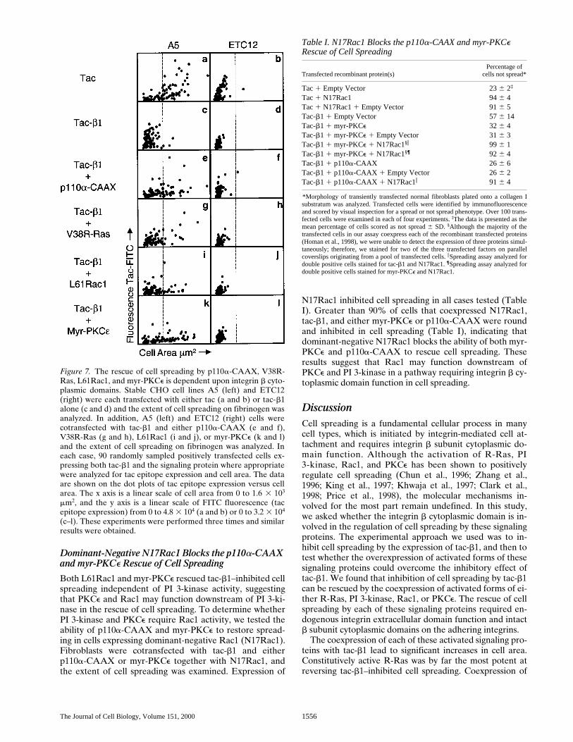
not spread (Fig. 7, a and b). Both A5 and ETC12 cells ex-pressing tac-b1 were inhibited in spreading with .97% of thecells having cell areas ,560 mm2 (Fig. 7, c and d). Thus, ex-pression of tac-b1 also inhibited CHO cell spreading on fi-brinogen mediated by aIIbb3. V38R-Ras, p110a-CAAX,myr-PKCe, and L61Rac1 were each able to restore CHO cellspreading inhibited by tac-b1; however, this increase in cellarea was restricted to the A5 cells and was not observed inETC12 cells (Fig. 7). More than 30% of the A5 cells trans-fected with tac-b1 and p110a-CAAX spread on fibrinogen,whereas .98% of the ETC12 cells transfected with tac-b1and p110a-CAAX were not spread (Fig. 7, e and f). Simi-larly, comparison of the spreading of A5 and ETC12 cells ex-pressing both tac-b1 and L61Rac1 revealed that none of theETC12 cells spread, whereas .15% of the A5 cells spread onfibrinogen (Fig. 7, i and j). Finally, .40% of the A5 cellstransfected with tac-b1 and myr-PKCe spread on fibrinogen.In contrast, .98% of the ETC12 cells transfected with tac-b1and myr-PKCe were not spread and exhibited a round cellmorphology (Fig. 7, k and l). Thus, the enhancement of cellspreading by V38R-Ras, p110a-CAAX, myr-PKCe, andL61Rac1 requires intact b cytoplasmic domains on the inte-grins mediating cell adhesion.
To ensure that the inability of signaling proteins to res-cue ETC12 cell spreading on fibrinogen was due to thelack of intact b3 cytoplasmic domains on the fibrinogenbinding integrins, and not to some peculiarity of theETC12 clone, we tested the ability of L61Rac1 and myr-PKCe to restore tac-b1–inhibited ETC12 cell spreading onfibronectin. Although ETC12 cells are not capable ofspreading on fibrinogen, they are able to spread on fi-bronectin (Ylanne et al., 1995). We found that ETC12 cellsspread on fibronectin, and this spreading was inhibited bytac-b1 and restored by the coexpression of either L61Rac1or myr-PKCe (data not shown).
Figure 5. The morphology ofcells expressing either p110a-CAAX, L61Rac1, N17Rac1,V38R-Ras, N43R-Ras, or myr-PKCe. Normal fibroblasts weretransfected with tac alone oreach of the signaling proteins,and subsequently replated ontocollagen I as described in Materi-als and Methods. Adherent cellswere immunostained for expres-sion of the tac epitope in cellstransfected with the control tacreceptor alone or cotransfectedwith the control tac receptor andp110a-CAAX. Cells were immu-nostained for expression of theflag epitope in cells transfectedwith myr-PKCe-Flag, or the mycepitope in the case of myc-L61Rac1, myc-N17Rac1, myc-V38R-Ras, and myc-N43R-Ras.Staining was visualized usingrhodamine-conjugated second-ary antibodies. Fluorescence im-ages were obtained as describedin Fig. 4. Scale bar: 10 mm.

Berrier et al. Rescue of Cell Spreading Inhibited by Tac-b1 1555
V38R-Ras Requires PI 3-Kinase Activity to Rescue Cell Spreading, whereas L61Rac1 and myr-PKCe Do Not
R-Ras has been shown to activate PI 3-kinase (Marte etal., 1996), suggesting that PI 3-kinase may be down-stream of R-Ras in regulating cell spreading. In addi-tion, PIP3, a product of PI 3-kinase, has been shown tobe involved in the activation of PKCe (Toker et al.,1994; Moriya et al., 1996), suggesting that PI 3-kinasemay function upstream of PKCe activation during cellspreading. PI 3-kinase activity has also been reported insignaling pathways containing Rac1. Since PI 3-kinasehas been positioned both upstream and downstream ofRac1 activation (Hawkins et al., 1995; Nobes et al.,1995; Parker, 1995; Keely et al., 1997; Missy et al.,1998), the role of PI 3-kinase in Rac1 signaling duringcell spreading is not certain.
To determine the requirement for PI 3-kinase activity inthe rescue of cell spreading by V38R-Ras, myr-PKCe, andL61Rac1, the ability of cotransfected cells to spread oncollagen I was assayed in the presence and absence of thePI 3-kinase inhibitor LY294002 (Fig. 8). Initial experi-ments demonstrated a role for PI 3-kinase in normal fibro-blast cell spreading. When fibroblasts transiently express-ing the control tac receptor were assayed for their abilityto spread on collagen I in the presence of the PI 3-kinaseinhibitor (LY294002), .50% of the tac-expressing cellswere inhibited in spreading, compared with ,20% in the
presence of DMSO alone (Fig. 8, A, a and e, and B, a andc). Thus, normal human fibroblasts require PI 3-kinase ac-tivity for cell spreading on collagen I.
We then tested the ability of LY294002 to impair cellspreading in cells cotransfected with tac-b1 and the vari-ous constitutively activated signaling proteins. Greaterthan 60% of cells cotransfected with tac-b1 and p110a-CAAX spread in the presence of DMSO, whereas, in thepresence of LY294002, ,15% of these transfected cellshad areas .560 mm2, suggesting that p110a-CAAX re-quires PI 3-kinase activity to enhance cell spreading (Fig.8 A, b and f). The V38R-Ras–triggered rescue was alsofound to require PI 3-kinase activity: .80% of thecotransfected cells treated with LY294002 were notspread (Fig. 8 B, b and d), compared with 44% ofcotransfected cells treated with DMSO. In contrast, cellscoexpressing tac-b1 and L61Rac1 were spread whentreated with either LY294002 or DMSO alone. Of thecells coexpressing tac-b1 and L61Rac1, 78% were spreadwhen treated with DMSO and 63% were spread whentreated with LY294002 (Fig. 8 A, c and g). This was alsotrue for cells coexpressing tac-b1 and myr-PKCe. In cellscoexpressing tac-b1 and myr-PKCe, 60% of the DMSO-treated cells and 45% of the LY294002-treated cells werespread (Fig. 8 A, d and h). These results suggest that en-dogenous PI 3-kinase activity is required for V38R-Ras,but not for L61Rac1 or myr-PKCe, to restore cell spread-ing in tac-b1–expressing cells.
Figure 6. The rescue of tac-b1–inhibited cell spreading on col-lagen I is a b1 integrin–depen-dent process. Human fibroblaststransfected with the control tacreceptor alone (A, a and e, andB, a and c) or cotransfected withtac-b1 and either p110a-CAAX(A, b and f), myr-PKCe (A, c andg), V38R-Ras (A, d and h), orL61Rac1 (B, b and d) were ana-lyzed for spreading on collagen Iin the presence of control ascites(A, a–d, and B, a and b) or b1function blocking P4C10 ascites(A, e–h, and B, c and d) as de-scribed in Materials and Meth-ods. The extent of cell spreadingand the expression levels of thetac epitope were quantified for100 (A) or 80 (B) randomly sam-pled positively transfected cells,and the results are shown by dotplot. The x axis is a linear scale ofcell area from 0 to 3.2 3 103 mm2
(A and B). The y axis is a linearscale of FITC fluorescence (tacepitope expression) from 0 to2.4 3 104 (A and B). The verticalline positioned at a cell area of560 mm2 indicates the separationof spread (right) and not spread(left) cells. These experimentswere performed twice and simi-lar results were obtained.
The Journal of Cell Biology, Volume 151, 2000 1556
Dominant-Negative N17Rac1 Blocks the p110a-CAAX and myr-PKCe Rescue of Cell Spreading
Both L61Rac1 and myr-PKCe rescued tac-b1–inhibited cellspreading independent of PI 3-kinase activity, suggestingthat PKCe and Rac1 may function downstream of PI 3-ki-nase in the rescue of cell spreading. To determine whetherPI 3-kinase and PKCe require Rac1 activity, we tested theability of p110a-CAAX and myr-PKCe to restore spread-ing in cells expressing dominant-negative Rac1 (N17Rac1).Fibroblasts were cotransfected with tac-b1 and eitherp110a-CAAX or myr-PKCe together with N17Rac1, andthe extent of cell spreading was examined. Expression of
N17Rac1 inhibited cell spreading in all cases tested (TableI). Greater than 90% of cells that coexpressed N17Rac1,tac-b1, and either myr-PKCe or p110a-CAAX were roundand inhibited in cell spreading (Table I), indicating thatdominant-negative N17Rac1 blocks the ability of both myr-PKCe and p110a-CAAX to rescue cell spreading. Theseresults suggest that Rac1 may function downstream ofPKCe and PI 3-kinase in a pathway requiring integrin b cy-toplasmic domain function in cell spreading.
DiscussionCell spreading is a fundamental cellular process in manycell types, which is initiated by integrin-mediated cell at-tachment and requires integrin b subunit cytoplasmic do-main function. Although the activation of R-Ras, PI3-kinase, Rac1, and PKCe has been shown to positivelyregulate cell spreading (Chun et al., 1996; Zhang et al.,1996; King et al., 1997; Khwaja et al., 1997; Clark et al.,1998; Price et al., 1998), the molecular mechanisms in-volved for the most part remain undefined. In this study,we asked whether the integrin b cytoplasmic domain is in-volved in the regulation of cell spreading by these signalingproteins. The experimental approach we used was to in-hibit cell spreading by the expression of tac-b1, and then totest whether the overexpression of activated forms of thesesignaling proteins could overcome the inhibitory effect oftac-b1. We found that inhibition of cell spreading by tac-b1can be rescued by the coexpression of activated forms of ei-ther R-Ras, PI 3-kinase, Rac1, or PKCe. The rescue of cellspreading by each of these signaling proteins required en-dogenous integrin extracellular domain function and intactb subunit cytoplasmic domains on the adhering integrins.
The coexpression of each of these activated signaling pro-teins with tac-b1 lead to significant increases in cell area.Constitutively active R-Ras was by far the most potent atreversing tac-b1–inhibited cell spreading. Coexpression of
Figure 7. The rescue of cell spreading by p110a-CAAX, V38R-Ras, L61Rac1, and myr-PKCe is dependent upon integrin b cyto-plasmic domains. Stable CHO cell lines A5 (left) and ETC12(right) were each transfected with either tac (a and b) or tac-b1alone (c and d) and the extent of cell spreading on fibrinogen wasanalyzed. In addition, A5 (left) and ETC12 (right) cells werecotransfected with tac-b1 and either p110a-CAAX (e and f),V38R-Ras (g and h), L61Rac1 (i and j), or myr-PKCe (k and l)and the extent of cell spreading on fibrinogen was analyzed. Ineach case, 90 randomly sampled positively transfected cells ex-pressing both tac-b1 and the signaling protein where appropriatewere analyzed for tac epitope expression and cell area. The dataare shown on the dot plots of tac epitope expression versus cellarea. The x axis is a linear scale of cell area from 0 to 1.6 3 103
mm2, and the y axis is a linear scale of FITC fluorescence (tacepitope expression) from 0 to 4.8 3 104 (a and b) or 0 to 3.2 3 104
(c–l). These experiments were performed three times and similarresults were obtained.
Table I. N17Rac1 Blocks the p110a-CAAX and myr-PKCe Rescue of Cell Spreading
Transfected recombinant protein(s)Percentage of
cells not spread*
Tac 1 Empty Vector 23 6 2‡
Tac 1 N17Rac1 94 6 4Tac 1 N17Rac1 1 Empty Vector 91 6 5Tac-b1 1 Empty Vector 57 6 14Tac-b1 1 myr-PKCe 32 6 4Tac-b1 1 myr-PKCe 1 Empty Vector 31 6 3Tac-b1 1 myr-PKCe 1 N17Rac1§i 99 6 1Tac-b1 1 myr-PKCe 1 N17Rac1§¶ 92 6 4Tac-b1 1 p110a-CAAX 26 6 6Tac-b1 1 p110a-CAAX 1 Empty Vector 26 6 2Tac-b1 1 p110a-CAAX 1 N17Rac1i 91 6 4
*Morphology of transiently transfected normal fibroblasts plated onto a collagen Isubstratum was analyzed. Transfected cells were identified by immunofluorescenceand scored by visual inspection for a spread or not spread phenotype. Over 100 trans-fected cells were examined in each of four experiments. ‡The data is presented as themean percentage of cells scored as not spread 6 SD. §Although the majority of thetransfected cells in our assay coexpress each of the recombinant transfected proteins(Homan et al., 1998), we were unable to detect the expression of three proteins simul-taneously; therefore, we stained for two of the three transfected factors on parallelcoverslips originating from a pool of transfected cells. i Spreading assay analyzed fordouble positive cells stained for tac-b1 and N17Rac1. ¶Spreading assay analyzed fordouble positive cells stained for myr-PKCe and N17Rac1.

Berrier et al. Rescue of Cell Spreading Inhibited by Tac-b1 1557
V38R-Ras resulted in cells with similar sizes and morpholo-gies to untransfected or control tac-transfected cells. Theability of V38R-Ras to rescue cell spreading was dependentupon PI 3-kinase activity, which is consistent with previousreports indicating that activated R-Ras can associate withand activate PI 3-kinase (Marte et al., 1996). Thus, V38R-Ras may rescue cell spreading by triggering pathways nor-mally activated by integrins such as PI 3-kinase.
Interestingly, activated V38R-Ras has also been shownto rescue high affinity ligand binding of aIIbb3 inhibitedby the constitutive activation of the ERK-MAPK cascade;however, in this instance, R-Ras regulated integrin activityby a pathway independent of PI 3-kinase (Sethi et al.,1999). Thus, R-Ras may modulate integrin function viamultiple downstream effectors. Additionally, stable ex-pression of activated R-Ras has been shown to activatea2b1-dependent cell migration and invasion of carcinomacells by a mechanism requiring the a2 cytoplasmic domain,suggesting that R-Ras might also specifically target indi-vidual integrin heterodimers by regulating protein interac-tions with their a subunit cytoplasmic domains (Keely etal., 1999). However, the role of individual a subunit cyto-plasmic domains in the rescue of cell spreading by V38R-Ras is not known at present.
Little data has been published regarding how R-Ras isactivated under physiological conditions and there is cur-rently no evidence linking integrin engagement to R-Rasactivation. Our result that expression of dominant-nega-
tive N43R-Ras does not inhibit cell spreading suggests thatintegrin-triggered activation of R-Ras is not required forspreading, at least in normal human fibroblasts. This resultcontradicts earlier studies demonstrating that N43R-Rascan inhibit CHO cell spreading (Zhang et al., 1996); how-ever, the reason for these differences is not clear at thistime. Nonetheless, the ability of V38R-Ras to regulate cellspreading in our system suggests that the activation of en-dogenous R-Ras by signals initiated by other cell surfacereceptors may regulate the spreading of some cell types.
Interestingly, although PI 3-kinase activity is required forV38R-Ras to rescue tac-b1–inhibited cell spreading, themembrane targeted form of PI 3-kinase, p110a-CAAX,only partially restored cell spreading in tac-b1–expressingcells compared with V38R-Ras. One possible explanation isthat V38R-Ras may lead to more robust PI 3-kinase signal-ing compared with the expression of p110a-CAAX. Alter-natively, V38R-Ras may activate additional pathways thatenhance cell spreading in conjunction with PI 3-kinase.
PI 3-kinase is known to be activated by integrin engage-ment and was found to be important in normal fibroblastcell spreading in the present study. The pathways linkingintegrins to activation of PI 3-kinase are not yet fully de-fined. The ability of integrins to activate FAK and H-Rasprovides two potential pathways, since both have beenshown to interact with and activate PI 3-kinase (Chen andGuan, 1994; Kodaki et al., 1994; Rodriguez-Viciana et al.,1994, 1996; Guan, 1997; Giancotti and Ruoslahti, 1999).
Figure 8. PI 3-kinase activity is required for V38R-Ras, but not forL61Rac1 or myr-PKCe, to rescue cell spreading. Normal fibroblastswere transfected with either tac alone (A, a and e, and B, a and c) orcotransfected with tac-b1 and either V38R-Ras (B, b and d), p110a-CAAX (A, b and f), L61Rac1 (A, c and g), or myr-PKCe (A, d and h),and the morphology of cells adherent to collagen was analyzed in thepresence of the PI 3-kinase inhibitor, LY294002 (A, e–h, and B, c andd), or DMSO (A, a–d, and B, c and d) as described in Materials andMethods. In each case, 100 (A) or 95 (B) randomly sampled transfectedcells were analyzed for tac epitope expression and cell area as describedpreviously. The x axis is a linear scale of cell area from 0 to 2.4 3 103
mm2 (A) or 0 to 3.2 3 103 mm2 (B), and the y axis is a linear scale ofFITC fluorescence (tac epitope expression) from 0 to 4.8 3 104 (A, aand e) or 0 to 2.4 3 104 (A, b–d and f–h, and B, b, d) or 0 to 8.0 3 104
(B, a and c). The vertical line positioned at 560 mm2 indicates the sepa-ration of spread (right) and not spread (left) cells. These experimentswere performed three times and similar results were obtained.

The Journal of Cell Biology, Volume 151, 2000 1558
However, the relative contribution of FAK and H-Ras tointegrin activation of PI 3-kinase is currently unknown.Potential downstream effectors of PI 3-kinase in the regu-lation of cell spreading are PKCe and Rac1 (Toker et al.,1994; Moriya et al., 1996; Rodriguez-Viciana et al., 1997;Missy et al., 1998). It is known that growth factor activa-tion of PI 3-kinase can trigger membrane ruffling througha Rac1-dependent pathway and roles for PI 3-kinase in theactivation of Rac1 have been described (Hawkins et al.,1995; Nobes et al., 1995; Parker et al., 1995). Our observa-tions that N17Rac1 prevents the ability of p110a-CAAXto increase cell spreading and that L61Rac1 can restorecell spreading independent of PI 3-kinase are consistentwith PI 3-kinase activation occurring either upstream orindependent of Rac1 in the regulation of cell spreading.The rescue of cell spreading by myr-PKCe is also in-dependent of PI 3-kinase, but inhibited by N17Rac1, simi-larly suggesting that PKCe may reside downstream of PI3-kinase and upstream of Rac1 in a pathway regulatingcell spreading.
The mechanism by which Rac1 regulates integrin-medi-ated cell spreading is not completely understood. It is as-sumed to do so by triggering actin polymerization andmembrane extension (lamellipodia) required for the in-
crease in cell area that accompanies cell spreading. Wehave not yet identified signaling proteins that functiondownstream of Rac1 to regulate cell spreading. PAK1,which can be activated by Rac1, is a potential candidate(Sells and Chernoff, 1997). Activated PAK1 mutants havebeen shown to induce lamellipodia (Sells et al., 1997), anddominant-negative mutants of PAK1 have been found toinhibit cell spreading (Price et al., 1998). However,whether PAK1 functions downstream of Rac1 in the in-duction of lamellipodia is unclear since Rac1-inducedlamellipodia can occur independent of Rac1-PAK1 inter-actions (Joneson et al., 1996; Lamarche et al., 1996;Westwick et al., 1997).
POR1 is another potential downstream effector of Rac1that may regulate cell spreading (Van Aelst et al., 1996).POR1 has been shown to localize to lamellipodia and dom-inant-negative forms of POR1 have been shown to inhibitRac1-induced lamellipodia (Van Aelst et al., 1996). In ad-dition, mutations in Rac1 that inhibit Rac1-POR1 inter-actions also inhibit lamellipodia formation (Joneson etal., 1996), suggesting that POR1 is a good candidate.P160Rock may also be a good candidate. P160Rock inter-acts with Rac1 in a GTP-dependent manner (Lamarche etal., 1996) and Rac1 mutations that inhibit p160Rock-Rac1
Figure 9. Models for the rescueof tac-b1–inhibited cell spread-ing. (a, Model I) Expression oftac-b1 results in the titration ofcellular factors from the endoge-nous b cytoplasmic domains thattrigger the activation of signal-ing proteins required for cellspreading. The coexpression ofactivated recombinant signalingproteins with tac-b1 may bypassthis block in integrin signalingand restore cell spreading. Tac-b1 does not inhibit integrin b cy-toplasmic domain function thatis required downstream of thesesignaling proteins. (b, Model II)Integrin-mediated cell attach-ment activates signaling proteinsthat regulate the binding of pro-teins to endogenous b cytoplas-mic domains that are requiredfor cell spreading. Expression oftac-b1 titrates these adhesion-induced protein interactionswith the integrin b cytoplasmicdomains. Coexpression of acti-vated signaling proteins withtac-b1 increases the pool of pro-teins that bind the integrin b cy-toplasmic domain and restoresendogenous integrin function incell spreading. (Model III). Tac-b1 inhibits the adhesion-inducedactivation of these signaling pro-teins and additionally inhibitsprotein interactions with the bcytoplasmic domain that aretriggered by the signaling pro-teins (not shown).

Berrier et al. Rescue of Cell Spreading Inhibited by Tac-b1 1559
interactions also inhibit lamellipodia formation (Lamarcheet al., 1996). Our future studies will determine whetherthese effectors regulate integrin function in cell spreading.
We propose three models for the inhibition and rescueof cell spreading. Our first model is that expression of tac-b1 may inhibit cell spreading by titrating a b cytoplasmicdomain binding protein from endogenous integrin b cyto-plasmic domains that may be required for the adhesion-triggered activation of Rac1 (Fig. 9 a). Expression of acti-vated Rac1 may bypass the block in integrin-triggeredRac1 activation. However, even if this is the case, b cyto-plasmic domain function is also required downstreamof Rac1 activation, since L61Rac1 could not rescuethe spreading of CHO cells attached to fibrinogen viaaIIbb3D728. So, in our first model, expression of tac-b1 in-hibits the function of the b cytoplasmic domain upstreambut not downstream of Rac1 activation.
In our second model, we propose that Rac1 signalingmay regulate protein interactions with integrin b cytoplas-mic domains that are required for cell spreading (Fig. 9 b).In this model, integrin-induced Rac1 activation is not in-hibited by the expression of tac-b1, instead tac-b1 inhibitscell spreading by titrating these Rac1-induced integrin-binding proteins from the endogenous integrin b cytoplas-mic domains. Expression of L61Rac1 may rescue cellspreading by increasing the cytoplasmic pool of proteinsthat can bind to integrin b cytoplasmic domains. These in-teractions may be required for Rac1 to initiate actin poly-merization and lamellipodia formation. A third model isalso possible in which tac-b1 inhibits integrin activation ofRac1 as well as the ability of Rac1 signaling to regulate in-tegrin b cytoplasmic domain function. Future studies willexplore these possibilities.
We thank Drs. J. Sottile and M. Dipersio for critically reading this manu-script, Dr. J. Depasquale for valuable advice in using the Image Pro-Plussoftware to accurately measure cell area and flourescence intensity, andR. Martinez for skillful technical assistance. We also thank Drs. Hall andToker for providing us with important reagents used in these studies.
This work was supported by National Institutes of Health grantsGM51540, T32HL07529, and T32GM07033, and the American Heart As-sociation grant 0020180T.
Submitted: 21 June 2000Revised: 19 October 2000Accepted: 6 November 2000
References
Burridge, K., and M. Chrzanowska-Wodnicka. 1996. Focal adhesions, contrac-tility, and signaling. Annu. Rev. Cell Dev. Biol. 12:463–519.
Chen, H.-C., and J.-L. Guan. 1994. Association of focal adhesion kinase with itspotential substrate phosphatidylinositol 3-kinase. Proc. Natl. Acad. Sci.USA. 91:10148–10152.
Chen, Y.-P., T.E. O’Toole, T. Shipley, J. Forsyth, S.E. LaFlamme, K.M. Ya-mada, S.J. Shattil, and M.H. Ginsberg. 1994. “Inside-out” signal transduc-tion inhibited by isolated integrin cytoplasmic domains. J. Biol. Chem. 269:18307–18310.
Chou, M.M., W. Hou, J. Johnson, L.K. Graham, M.H. Lee, C.-S. Chen, A.C.Newton, B.S. Schaffhausen, and A. Toker. 1998. Regulation of protein ki-nase C z by PI 3-kinase and PDK-1. Curr. Biol. 8:1069–1077.
Chun, J.-S., M.-J. Ha, and B.S. Jacobson. 1996. Differential translocation ofprotein kinase C e during HeLa cell adhesion to a gelatin substratum. J. Biol.Chem. 271:13008–13012.
Clark, E.A., W.G. King, J.S. Brugge, M. Symons, and R.O. Hynes. 1998. Inte-grin-mediated signals regulated by members of the Rho Family of GTPases.J. Cell Biol. 142:573–586.
Del Pozo, M.A., L.S. Price, N.B. Alderson, X.-D. Ren, and M.A. Schwartz.2000. Adhesion to the extracellular matrix regulates the coupling of the
small GTPase Rac to its effector PAK. EMBO (Eur. Mol. Biol. Organ.) J.19:2008–2014.
Giancotti, F.G., and E. Ruoslahti. 1999. Integrin Signaling. Science. 285:1028–1032.
Guan, J.-L. 1997. Focal adhesion kinase in integrin signaling. Matrix Biol. 16:195–200.
Hawkins, P.T., A. Eguinoa, R.-G. Qiu, D. Stokoe, F.T. Cooke, R. Walters, S.Wennstrom, L. Claesson-Welsh, T. Evans, M. Symons, and L. Stephens.1995. PDGF stimulates an increase in GTP-Rac via activation of phospho-inositide 3-kinase. Curr. Biol. 5:393–403.
Homan, S.M., A.M. Mercurio, and S.E. LaFlamme. 1998. Endothelial cells as-semble two distinct a6b4-containing vimentin-associated structures: rolesfor ligand binding and the b4 cytoplasmic domain. J. Cell Sci. 111:2717–2728.
Hynes, R.O. 1992. Integrins: versatility, modulation, and signaling in cell adhe-sion. Cell. 69:11–25.
Izzard, C.S., and L.R. Lochner. 1976. Cell-to-substrate contacts in living fibro-blasts: and interference-reflexion study with an evaluation of the technique.J. Cell Sci. 21:129–159.
Joneson, T., M. McDonough, D. Bar-Sagi, and L. Van Aelst. 1996. Rac regula-tion of actin polymerization and proliferation by a pathway distinct from Junkinase. Science. 274:1374–1376.
Keely, P.J., E.V. Rusyn, A.D. Cox, and L.V. Parise. 1999. R-Ras signalsthrough specific integrin a cytoplasmic domains to promote migration andinvasion of breast epithelial cells. J. Cell Biol. 145:1077–1088.
Keely, P.J., J.K. Westwick, I.P. Whitehead, C.J. Der, and L.V. Parise. 1997.Cdc42 and Rac1 induce integrin-mediated cell motility and invasivenessthrough PI(3)K. Science. 390:632–636.
Khwaja, A., P. Rodriguez-Viciana, S. Wennstrom, P.H. Warne, and J. Down-ward. 1997. Matrix adhesion and Ras transformation both activate a phos-phoinositide 3-OH kinase and protein kinase B/AKT cellular survival path-way. EMBO (Eur. Mol. Biol. Organ.) J. 16:2783–2793.
King, W.G., M.D. Mattaliano, T.O. Chan, P.N. Tsichlis, and J.S. Brugge. 1997.Phosphatidylinositol 3-kinase is required for integrin-stimulated AKT andraf-1/mitogen-activated protein kinase pathway activation. Mol. Cell. Biol.17:4406–4418.
Kodaki, T., R. Woscholski, B. Hallberg, P. Rodriguez-Viciana, J. Downward,and P.J. Parker. 1994. The activation of phosphatidylinositol 3-kinase byRas. Curr. Biol. 4:798–806.
LaFlamme, S.E., S.K. Akiyama, and K.M. Yamada. 1992. Regulation of fi-bronectin receptor distribution. J. Cell Biol. 117:437–447.
LaFlamme, S.E., S.M. Homan, A.L. Bodeau, and A.M. Mastrangelo. 1997. In-tegrin cytoplasmic domains as connectors to the cell’s signal transduction ap-paratus. Matrix Biol. 16:153–163.
LaFlamme, S.E., L.A. Thomas, S.S. Yamada, and K.M. Yamada. 1994. Singlesubunit chimeric integrins as mimics and inhibitors of endogenous integrinfunctions in receptor localization, cell spreading and migration, and matrixassembly. J. Cell Biol. 126:1287–1298.
Lamarche, N., N. Tapon, L. Stowers, P.D. Burbelo, P. Aspenstrom, T. Bridges,J. Chant, and A. Hall. 1996. Rac and Cdc42 induce actin polymerization andG1 cell cycle progression independently of p65PAK and the JNK/SAPKMAP kinase cascade. Cell. 87:519–529.
Lukashev, M.E., D. Sheppard, and R. Pytela. 1994. Disruption of integrin func-tion and induction of tyrosine phosphorylation by the autonomously ex-pressed b1 integrin cytoplasmic domain. J. Biol. Chem. 269:18311–18314.
Marte, B.M., P. Rodriguez-Viciana, S. Wennstrom, P.H. Warne, and J. Down-ward. 1996. R-Ras can activate the phosphoinositide 3-kinase but not theMAP kinase arm of the Ras effector pathways. Curr. Biol. 7:63–70.
Mastrangelo, A.M., S.M. Homan, M.J. Humphries, and S.E. LaFlamme. 1999a.Amino acid motifs required for isolated b cytoplasmic domains to regulate‘in trans’ b1 integrin conformation and function in cell attachment. J. CellSci. 112:217–229.
Mastrangelo, A.M., A.L. Bodeau, S.M. Homan, A.L. Berrier, and S.E.LaFlamme. 1999b. The use of chimeric receptors in the study of integrin sig-naling. In Signaling Through Cell Adhesion Molecules. J.-L. Guan, editor.CRC Press, New York, NY. 3–17.
Missy, K., V. Van Poucke, P. Raynal, C. Viala, G. Mauco, M. Plantavid, H.Chap, and B. Payrastre. 1998. Lipid products of phosphoinositide 3-kinaseinteract with Rac1 GTPase and stimulate GDP dissociation. J. Biol. Chem.273:30279–30286.
Moriya, S., A. Kazlauskas, K. Akimoto, S.-I. Hirai, K. Mizuno, T. Takenawa, Y.Fukui, Y. Watanabe, S. Ozaki, and S. Ohno. 1996. Platelet-derived growthfactor activates protein kinase Ce through redundant and independentsignaling pathways involving phospholipase Cg or phosphatidylinositol3-kinase. Proc. Natl. Acad. Sci. USA. 93:151–155.
Nobes, C.D., P. Hawkins, L. Stephens, and A. Hall. 1995. Activation of thesmall GTP-binding proteins Rho and Rac by growth factor receptors. J. CellSci. 108:225–233.
Parker, P.J. 1995. PI 3-kinase puts GTP on the Rac. Curr. Biol. 5:577–579.Price, L.S., J. Leng, M.A. Schwartz, and G.M. Bokoch. 1998. Activation of Rac
and Cdc42 by integrins mediates cell spreading. Mol. Biol. Cell. 9:1863–1871.Rodriguez-Viciana, P., P.H. Warne, R. Dhand, B. Vanhaesebroeck, I. Gout,
M.J. Fry, M.D. Waterfield, and J. Downward. 1994. Phosphatidylinositol-3-OH kinase as a direct target of Ras. Nature. 370:527–532.
Rodriguez-Viciana, P., P.H. Warne, A. Khwaja, B.M. Marte, D. Pappin, P. Das,M.D. Waterfield, A. Ridley, and J. Downward. 1997. Role of phosphoinosi-

The Journal of Cell Biology, Volume 151, 2000 1560
tide 3-OH kinase in cell transformation and control of the actin cytoskeletonby Ras. Cell. 89:457–467.
Rodriguez-Viciana, P., P.H. Warne, B. Vanhaesebroeck, M.D. Waterfield, andJ. Downward. 1996. Activation of phosphoinositide 3-kinase by interactionwith Ras and by point mutation. EMBO (Eur. Mol. Biol. Organ.) J. 15:2442–2451.
Schwartz, M.A., M.D. Schaller, and M.H. Ginsberg. 1995. Integrins: emergingparadigms of signal transduction. Annu. Rev. Cell Dev. Biol. 11:549–599.
Sells, M.A., and J. Chernoff. 1997. Emerging from the Pak: the p21-activatedprotein kinase family. Trends Cell Biol. 7:162–167.
Sells, M.A., U.G. Knaus, S. Bagrodia, D.M. Ambrose, G.M. Bokoch, and J.Chernoff. 1997. Human p21-activated kinase (Pak1) regulates actin organi-zation in mammalian cells. Curr. Biol. 7:202–210.
Sethi, T., M.H. Ginsberg, J. Downward, and P.E. Hughes. 1999. The smallGTP-binding protein R-Ras can influence integrin activation by antagoniz-ing a Ras/Raf-initiated integrin suppression pathway. Mol. Biol. Cell. 10:1799–1809.
Small, J.V., K. Rottner, and I. Kaverina. 1999. Functional design in the actin cy-toskeleton. Curr. Opin. Cell Biol. 11:54–60.
Smilenov, L., R. Briesewitz, and E.E. Marcantonio. 1994. Integrin b1 cytoplas-mic domain dominant negative effects revealed by lysophosphatidic acidtreatment. Mol. Biol. Cell. 5:1215–1223.
Toker, A., M. Meyer, K.K. Reddy, J.R. Falck, R. Aneja, S. Aneja, A. Parra,D.J. Burns, L.M. Ballas, and L.C. Cantley. 1994. Activation of protein kinaseC family members by the novel polyphosphoinositides PtdIns-3,4-P2 andPtdIns-3,4,5-P3. J. Biol. Chem. 269:32358–32367.
Van Aelst, L., T. Joneson, and D. Bar-Sagi. 1996. Identification of a novel
Rac1-interacting protein involved in membrane ruffling. EMBO (Eur. Mol.Biol. Organ.) J. 15:3778–3786.
Van Leeuwen, F.N., S. Van Delft, H.E. Kain, R.A. Van der Kammen, and J.G.Collard. 1999. Rac regulates phosphorylation of the myosin-II heavy chain,actinomyosin disassembly and cell spreading. Nat. Cell Biol. 1:242–248.
Vuori, K., and E. Ruoslahti. 1993. Activation of protein kinase C precedesa5b1 integrin-mediated cell spreading on fibronectin. J. Biol. Chem. 268:21459–21462.
Wennstrom, S., and J. Downward. 1999. Role of phosphoinositide 3-kinase inactivation of ras and mitogen-activated protein kinase by epidermal growthfactor. Mol. Cell. Biol. 19:4279–4288.
Westwick, J.K., Q.T. Lambert, G.J. Clark, M. Symons, L. Van Aelst, R.G. Pes-tell, and C.J. Der. 1997. Rac regulation of transformation, gene expression,and actin organization by multiple, PAK-independent pathways. Mol. Cell.Biol. 17:1324–1335.
Yamada, K.M., and S. Miyamoto. 1995. Integrin transmembrane signaling andcytoskeletal control. Curr. Opin. Cell Biol. 7:681–689.
Ylanne, J., Y. Chen, T.E. O’Toole, J.C. Loftus, Y. Takada, and M.H. Ginsberg.1993. Distinct functions of integrin a and b subunit cytoplasmic domains incell spreading and formation of focal adhesions. J. Cell Biol. 122:223–233.
Ylanne, J., J. Huuskonen, T.E. O’Toole, M.H. Ginsberg, I. Virtanen, and C.G.Gahmberg. 1995. Mutation of the cytoplasmic domain of the integrin b3 sub-unit. Differential effects on cell spreading, recruitment to adhesion plaques,endocytosis, and phagocytosis. J. Biol. Chem. 270:9550–9557.
Zhang, Z., K. Vuori, H.-G. Wang, J.C. Reed and E. Ruoslahti. 1996. Integrinactivation by R-Ras. Cell. 85:61–69.