world congress of endourology & swl program book - The Perk ...
540
SCIENTIFIC PROGRAM FOR 31 ST WORLD CONGRESS OF ENDOUROLOGY & SWL PROGRAM BOOK Tuesday, October 22, 2013 Moderated Poster Session MP1A 1:00 pm–3:00 pm BASIC RESEARCH: UPPER TRACT PHYSIOLOGY Room Nottoway @ Sheraton New Orleans Chair: John D. Denstedt Faculty: Yoshinari Ono and Gunter Janetschek *Presenting author MP1A-01 EVALUATION OF POTENTIAL PHARMACEU- TICAL AGENTS FOR A DRUG ELUTING URETERIC STENT USING EX-VIVO IN- VESTIGATION INTO THE UROTHELIAL PERMEABILITY Nicholas Williams, Chris Allender, Jenna Bowen, Marc Gumbleton, Cardiff, United Kingdom, Tim Harrah, Jamie Li, Boston, MA, Hrishi Joshi*, Cardiff, United Kingdom MP1A-02 UNDERSTANDING STENT-INDUCED URETERAL APERISTALSIS Claudia Janssen*, Wolfgang Jaeger, Dennis Solomon, Ladan Fazli, Ralph Buttyan, Ben H. Chew, Chun Y. Seow, Dirk Lange, Vancouver, Canada MP1A-03 ACOUSTIC BUBBLE REMOVAL TO ENHANCE THE EFFICACY OF SHOCK WAVE LITHOTRIPSY: AN IN-VITRO STUDY Alexander P. Duryea*, William W. Roberts, Charles A. Cain, Hedieh A. Tamaddoni, Timothy L. Hall, Ann Arbor, MI MP1A-04 COMPARATIVE IN-VITRO STUDY OF THE EFFECTIVENESS OF NANOSECOND ELEC- TRICAL PULSE AND LASER LITHOTRIPTERS Alexey Martov, Dimitry Ergakov*, Moscow, Russian Federation, Valery Diamant, Katsrin, Israel, Artem Borisik, Andrey Andronov, Moscow, Russian Federa- tion, Vladimir Chernenko, Tomsk, Russian Federation MP1A-05 DETECTING MICRO-STONES IN URINE WITH QUANTITATIVE RAMAN SPECTROSCOPY AFTER ESWL Yichun Chiu*, Po-An Chen, Huihua Chiang, Thomas Hsueh, Shing-Hwa Lu, Allen Chiu, Taipei, Taiwan MP1A-06 URINE AQUAPORIN-1 AND PERILIPIN-2: CAN THESE MARKERS ASSIST IN THE EVALUA- TION OF SMALL RENAL MASSES? Jonathan Mobley*, Jeremiah Morrissey, Sam Bhayani, Joseph Song, Joel Vetter, Evan Kharasch, Robert Figenshau, St. Louis, MO MP1A-07 INTRA AND EXTRA-RENAL AUTONOMIC NERVOUS SYSTEM REDEFINED Achim Lusch*, Emon Heidari, Ryan Leary, Zhamshid Okhunov, Jamie Wikenheiser, Jaime Landman, Orange, CA MP1A-08 SECRETED FACTORS FROM PERITUMOR ADIPOSE TISSUES OF CLEAR CELL RENAL CELL CARCINOMA INCREASED THE MOTILITY OF HUMAN CCRCC CELL LINE CAKI-2 VIA ENHANCEMENT OF WNT SIGNALING Achim Lusch*, Christopher Blair, Molly Baker, Zhamshid Okhunov, Victor Huynh, Xiaolin Zi, Jaime Landman, Orange, CA MP1A-09 CAN REMOTE ISCHEMIC PRECONDITIONING CONFER PROTECTION AGAINST REPERFU- SION INJURY FOLLOWING WARM ISCHEMIA IN A PORCINE SOLITARY-KIDNEY MODEL? Jeffrey Gahan*, Jodi Antonelli, Bedir Selahattin, Yunbo Ma, Steve Faddegon, Payal Kapur, Jeffrey Cadeddu, Dallas, TX MP1A-10 URINARY CYSTATIN C AND NGAL AS EARLY BIOMARKERS FOR ASSESSMENT OF RENAL ISCHEMIA- REPERFUSION INJURY: A SERUM MARKER TO REPLACE CREATININE? Ben Woodson*, Liang Wang, Sree Mandava, Benjamin Lee, New Orleans, LA MP1A-11 MULTIPHOTON MICROSCOPIC CHARACTER- IZATION OF RENAL CELL CARCINOMA Sara Best*, E. Jason Abel, Matthew Houlihan, Kevin Eliceiri, Madison, WI MP1A-12 ADDITION OF SODIUM BICARBONATE TO IRRIGATION SOLUTION MAY ASSIST IN DISSOLUTION OF URIC ACID FRAGMENTS DURING URETEROSCOPY Jessica E Paonessa*, Naeem Bhojani, James C Williams, Jr., James E Lingeman, Indianapolis, IN MP1A-13 DIETARY HYDROXYPROLINE INDUCED CAL- CIUM OXALATE LITHIASIS AND ASSOCIATED RENAL INJURY IN THE PORCINE MODEL Sri Sivalingam*, Stephen Nakada, Priyanka Sehgal, Tom Crenshaw, Kristina Penniston, Madison, WI JOURNAL OF ENDOUROLOGY Volume 27, Supplement 1, September 2013 ª Mary Ann Liebert, Inc. DOI: 10.1089/end.2013.2001 -P1-
-
Upload
khangminh22 -
Category
Documents
-
view
0 -
download
0
Transcript of world congress of endourology & swl program book - The Perk ...

SCIENTIFIC PROGRAM FOR 31ST WORLD CONGRESSOF ENDOUROLOGY & SWL PROGRAM BOOK
Tuesday, October 22, 2013 Moderated Poster Session MP1A 1:00 pm–3:00 pm
BASIC RESEARCH: UPPER TRACT PHYSIOLOGY
Room Nottoway @ Sheraton New OrleansChair: John D. Denstedt
Faculty: Yoshinari Ono and Gunter Janetschek
*Presenting author
MP1A-01 EVALUATION OF POTENTIAL PHARMACEU-
TICAL AGENTS FOR A DRUG ELUTING
URETERIC STENT USING EX-VIVO IN-
VESTIGATION INTO THE UROTHELIAL
PERMEABILITY
Nicholas Williams, Chris Allender, Jenna Bowen,Marc Gumbleton, Cardiff, United Kingdom,Tim Harrah, Jamie Li, Boston, MA, Hrishi Joshi*,Cardiff, United Kingdom
MP1A-02 UNDERSTANDING STENT-INDUCED
URETERAL APERISTALSIS
Claudia Janssen*, Wolfgang Jaeger, Dennis Solomon,Ladan Fazli, Ralph Buttyan, Ben H. Chew,Chun Y. Seow, Dirk Lange, Vancouver, Canada
MP1A-03 ACOUSTIC BUBBLE REMOVAL TO ENHANCE
THE EFFICACY OF SHOCK WAVE
LITHOTRIPSY: AN IN-VITRO STUDY
Alexander P. Duryea*, William W. Roberts,Charles A. Cain, Hedieh A. Tamaddoni,Timothy L. Hall, Ann Arbor, MI
MP1A-04 COMPARATIVE IN-VITRO STUDY OF THE
EFFECTIVENESS OF NANOSECOND ELEC-
TRICAL PULSE AND LASER LITHOTRIPTERS
Alexey Martov, Dimitry Ergakov*, Moscow, RussianFederation, Valery Diamant, Katsrin, Israel, ArtemBorisik, Andrey Andronov, Moscow, Russian Federa-tion, Vladimir Chernenko, Tomsk, Russian Federation
MP1A-05 DETECTING MICRO-STONES IN URINE WITH
QUANTITATIVE RAMAN SPECTROSCOPY
AFTER ESWL
Yichun Chiu*, Po-An Chen, Huihua Chiang,Thomas Hsueh, Shing-Hwa Lu, Allen Chiu,Taipei, Taiwan
MP1A-06 URINE AQUAPORIN-1 AND PERILIPIN-2: CAN
THESE MARKERS ASSIST IN THE EVALUA-
TION OF SMALL RENAL MASSES?
Jonathan Mobley*, Jeremiah Morrissey, Sam Bhayani,Joseph Song, Joel Vetter, Evan Kharasch,Robert Figenshau, St. Louis, MO
MP1A-07 INTRA AND EXTRA-RENAL AUTONOMIC
NERVOUS SYSTEM REDEFINED
Achim Lusch*, Emon Heidari, Ryan Leary,Zhamshid Okhunov, Jamie Wikenheiser,Jaime Landman, Orange, CA
MP1A-08 SECRETED FACTORS FROM PERITUMOR
ADIPOSE TISSUES OF CLEAR CELL RENAL
CELL CARCINOMA INCREASED THE
MOTILITY OF HUMAN CCRCC CELL LINE
CAKI-2 VIA ENHANCEMENT OF WNT
SIGNALING
Achim Lusch*, Christopher Blair, Molly Baker,Zhamshid Okhunov, Victor Huynh, Xiaolin Zi,Jaime Landman, Orange, CA
MP1A-09 CAN REMOTE ISCHEMIC PRECONDITIONING
CONFER PROTECTION AGAINST REPERFU-
SION INJURY FOLLOWING WARM ISCHEMIA
IN A PORCINE SOLITARY-KIDNEY MODEL?
Jeffrey Gahan*, Jodi Antonelli, Bedir Selahattin,Yunbo Ma, Steve Faddegon, Payal Kapur,Jeffrey Cadeddu, Dallas, TX
MP1A-10 URINARY CYSTATIN C AND NGAL AS EARLY
BIOMARKERS FOR ASSESSMENT OF RENAL
ISCHEMIA- REPERFUSION INJURY: A SERUM
MARKER TO REPLACE CREATININE?
Ben Woodson*, Liang Wang, Sree Mandava,Benjamin Lee, New Orleans, LA
MP1A-11 MULTIPHOTON MICROSCOPIC CHARACTER-
IZATION OF RENAL CELL CARCINOMA
Sara Best*, E. Jason Abel, Matthew Houlihan,Kevin Eliceiri, Madison, WI
MP1A-12 ADDITION OF SODIUM BICARBONATE TO
IRRIGATION SOLUTION MAY ASSIST IN
DISSOLUTION OF URIC ACID FRAGMENTS
DURING URETEROSCOPY
Jessica E Paonessa*, Naeem Bhojani, James C Williams,Jr., James E Lingeman, Indianapolis, IN
MP1A-13 DIETARY HYDROXYPROLINE INDUCED CAL-
CIUM OXALATE LITHIASIS AND ASSOCIATED
RENAL INJURY IN THE PORCINE MODEL
Sri Sivalingam*, Stephen Nakada, Priyanka Sehgal,Tom Crenshaw, Kristina Penniston, Madison, WI
JOURNAL OF ENDOUROLOGYVolume 27, Supplement 1, September 2013ª Mary Ann Liebert, Inc.DOI: 10.1089/end.2013.2001
-P1-

MP1A-14 THE RECOVERY OF URETERAL PERISTALSIS
AFTER TRANSIENT OBSTRUCTION IN A NO-
VEL MURINE MODEL
Claudia Janssen*, Wolfgang Jaeger, Igor Moskalev,Ben H. Chew, Dirk Lange, Vancouver, Canada
MP1A-15 RAMAN SPECTROSCOPIC COMPOSITION
ANALYSIS OF URINARY CALCULI
Matthias Eder*, Elena Foditsch,Reinhold Zimmermann, Maurizio Musso,Guenther Redhammer, Paolo Sereni, Salzburg,Austria
MP1A-16 KIDNEY STONE PREVENTION: CAN OBESE
PATIENTS FOLLOW A DIET?
Fabio Torricelli*, Shubha De, Ina Tien, Manoj Monga,Cleveland, OH
MP1A-17 HYDROXYPROLINE METABOLISM TO
URINARY OXALATE AND GLYCOLATE
EXCRETION IN NORMAL SUBJECTS
Dean Assimos*, Ross Holmes, John Knight,Birmingham, AL
MP1A-18 STONE DEPICTION WITH THE URO DYNA-CT:
STONES OF DIFFERENT COMPOSITIONS
Benjamin Meister*, Marie-Claire Rassweiler,Maurice-Stephan Michel, Manuel Ritter,Mannheim, Germany
MP1A-19 IN VITRO STONE DISINTEGRATION OF
STANDARDIZED BON(N) STONES TO
COMPARE THE DIODE LASER 1.318NM WITH
THE HOLMIUM LASER 2.100 NM AND ESTAB-
LISHING AN EX VIVO ANIMAL MODEL FOR
IN SITU DIODE LASER EVALUATION
Reinhold Zimmermann*, Markus Wallner, Salzburg,Austria, Roswitha Siener, Bonn, Germany,Gunter Janetschek, Esra Foditsch, Salzburg, Austria
MP1A-20 THE TENSION TEST AND IN VITRO DE-
GRADATION OF POLY(LACTIC ACID -
GLYCOLIC ACID)/POLYCAPROLACTONE
BIODEGRADABLE NANO-STRUCTURE
URETERAL STENT FABRICATED BY
ELECTROSPINNING
Xiao Qing Wang, Hong Li Shan, Zhi Hua Lu,Yu Chuan Hou, Chun Xi Wang*, Changchun,China, People’s Republic of
MP1A-21 HOW TO DETECT ACUTE POST-OPERATIVE
KIDNEY INJURY AFTER CLAMPLESS
LAPAROSCOPIC PARTIAL NEPHRECTOMY?
A PILOT STUDY
Francesco Porpiglia*, Daniele Amparore,Riccardo Bertolo, Fabrizio Mele, Diletta Garrou,Giovanni Cattaneo, Matteo Manfredi, Cristian Fiori,Orbassano (Torino), Italy
MP1A-22 BIOMECHANICAL PROPERTIES OF THE
URETER: PRELIMINARY RESULTS
Yaniv Shilo*, Joseph E. Pichamuthu,Timothy D. Averch, Stephen V. Jackman,David A. Vorp, Pittsburgh, PA
MP1A-23 SPECTROSCOPIC TISSUE ANALYSIS OF
RENAL ISCHEMIA AND RECOVERY DURING
SEGMENTAL RENAL ARTERY VS MAIN
RENAL ARTERY CLAMPING
Philip Dorsey*, Quincy Brown, New Orleans, LA,Janet Colli, Memphis, TN, Kate Elfer, Theodore Saitz,Ross McCaslin, Benjamin Lee, New Orleans, LA
MP1A-24 NOVEL BIOMARKERS TO MEASURE CHAN-
GES IN KIDNEY FUNCTION IN THE ADULT
HUMAN
Sree Harsha Mandava*, Benjamin Woodson,Liang Wang, Benjamin Lee, New Orleans, LA
MP1A-25 INCIDENCE OF HYBRID TUMORS FOUND IN
EXCISED RENAL MASSES: A MULTI-INSTITU-
TIONAL ANALYSIS
David Fumo*, Toledo, OH, Pengbo Jiang,Hackensack, NJ, Khaled Shahrour, Toledo, OH,Ravi Munver, Hackensack, NJ, Samay Jain,Toledo, OH
Tuesday, October 22, 2013 Moderated Poster Session MP1B 1:00 pm–3:00 pm
BASIC RESEARCH: UROLITHIASIS
Room Bayside A-C @ Sheraton New OrleansChair: Dean Assimos
Faculty: Mahesh Desai and Mantu Gupta
*Presenting author
MP1B-01 A URETEROSCOPIC LITHOTRITE: IN VITRO
ASSESSMENT OF A NOVEL FLEXIBLE PROBE
ULTRASONIC INTRACORPOREAL DEVICE
Jessica E Paonessa*, Naeem Bhojani, James A McAteer,James C Williams, Jr., James E Lingeman,Indianapolis, IN
MP1B-02 ACTIVITY LEVELS AND STONE DISEASE: A
POPULATION BASED ANALYSIS USING THE
NATIONAL HEALTH AND NUTRITION EX-
AMINATION DATABASE
Shubha De*, Jiangbo Li, Fabio Torricelli,Manoj Monga, Cleveland, OH
MP1B-03 THE VALUE OF REPEATING A TEST: META-
BOLIC PROFILES
Ranan Dasgupta*, Saskia Verhagen, Jeremy Cox,London, United Kingdom
MP1B-04 CHANGING TRENDS IN AMERICAN DIET AND
THE RISING PREVALENCE OF KIDNEY STONES
Fabio Torricelli*, Shubha De, XIaobo Liu,Manoj Monga, Cleveland, OH
MP1B-05 HOW MUCH FORCE TO DISLODGE A STONE
FROM RANDALL’S PLAQUE?
INTRAOPERATIVE DISTRACTION FORCES
-P2- PROGRAM WITH ABSTRACT LISTING

Shubha De, Robert Brown*, Carl Sarkissian,Manoj Monga, Cleveland, OH
MP1B-06 UPPER CALYCEAL PERCUTANEOUS
NEPHROLITHOTOMY (PCNL) UNDER SPINAL
ANESTHESIA: A PROSPECTIVE STUDY
Haresh Thummar*, R Ganatra, Rajkot, India
MP1B-07 DEVELOPMENT OF A NOVEL ACCESS SHEATH
THAT ALLOWS SIMULTANEOUS SHEATH
PLACEMENT AND SAFETY WIRE ACCESS
Yung Tan*, Doh Cha, Edan Shapiro, Ciara Marley,Mantu Gupta, New York, NY
MP1B-08 DISSOLUTION KINETICS OF URIC ACID
STONES IN RELATION TO URIC ACID SOLU-
TION CONCENTRATION. AN IN VITRO STUDY
Itay Sagy*, Bezalel Sivan, Petach Tikva, Israel,Ruth Frid, Ytzhak Mastai, Ramat-Gan, Israel,Pinchas M. Livne, David Lifshitz, Petach Tikva, Israel
MP1B-09 EFFECT OF HYDROPHILIC EXTRACT OF
ALHAGI MAURORUM ON ETHYLENE GLYCOL-
INDUCED RENAL STONE IN MALE WISTAR
RATS
Sadrollah Mehrabi*, Yasuj, Iran, Farhad Mehrabi,Shiraz, Iran
MP1B-10 COMPARISON OF THE IMPACT OF NANOSE-
COND ELECTROPULSE AND ELECTRO-
HYDRAULIC LITHOTRIPTERS ON URINARY
TRACT TISSUE
Alexander Gudkov, Dimitry Ergakov*, Tomsk,Russian Federation, Valery Diamant, Katsrin, Israel,Maxim Lozovsky, Tomsk, Russian Federation,Gennady Chepovetsky, Katsrin, Israel, Marat Lerner,Tomsk, Russian Federation
MP1B-11 STUDY OF THE DIFFERENCES BETWEEN
NANOSECOND ELECTROPULSE AND ELEC-
TROHYDRAULIC METHODS OF LITHOTRIPSY
Alexey Martov, Moscow, Russian Federation,Alexander Gudkov, Dimitry Ergakov*, Tomsk,Russian Federation, Valery Diamant,Gennady Chepovetsky, Katsrin, Israel,Marat Lerner, Tomsk, Russian Federation
MP1B-12 COMBINATION OF RIGID URETEROSCOPY
WITH FLEXIBLE URETEROSCOPY DOES NOT
HAVE ANY NEGATIVE IMPACT ON THE
OUTCOMES
Erdal Alkan, Mirac Turan, Oguz Ozkanli,Egemen Avcý, Mehmet Murad Basar,Yusuf Oguz Acar, Derya Balbay*, Istanbul, Turkey
MP1B-13 SIZE-MEASUREMENT OF RENAL STONES
WITH THE URO DYNA-CT
Benjamin Meister*, Marie-Claire Rassweiler,Christel Weiß, Maurice-Stephan Michel, Axel Haecker,Manuel Ritter, Mannheim, Germany
MP1B-14 IS LYMPHOCYTOPENIA A NEW MARKER FOR
PYONEPHROTIC OBSTRUCTED KIDNEYS
(POK) SECONDARY TO URINARY STONE
DISEASE?
Ahmed Ali*, Liam Farrell, Bhaskar Somani,Southampton, United Kingdom
MP1B-15 AN IN VITRO COMPARISON OF THE USE OF
ABDOMINAL AND BONE WINDOWS ON
COMPUTED TOMOGRAPHY MEASUREMENTS
OF HOUNSFIELD UNITS AND SIZE OF STONES
Paul Erotocritou*, Miles Walkden, Daron Smith,London, United Kingdom
MP1B-16 EFFECTIVENESS OF FLEXIBLE URETEROR-
ENOSCOPY FOR MULTIPLE UNILATERAL
INTRARENAL STONES SMALLER THAN 2 CM
Erdal Alkan, Oguz Ozkanli, Egemen Avcý,Mirac Turan, Mehmet Murad Basar,Yusuf Oguz Acar, Derya Balbay*, Istanbul, Turkey
MP1B-17 EXTERNAL VALIDATION OF AN
INDIVIDUALIZED WEIGHT-BASED GOAL
URINE VOLUME (WGUV) MODEL INTENDED
TO IMPROVE EXPECTED CALIUM URINE
CONCENTRATIONS
Jack Lambert*, Norfolk, VA, Nicole Miller, Nashville,TN, Justin Watson, Michael Fabrizio, Mark Sawyer,Norfolk, VA
MP1B-18 THE ANIMAL MODEL IN STONE DISEASE:
A TRIBUTE
Michael Moran*, Tucson, AZ
MP1B-19 EXTRACORPOREAL SHOCKWAVE LITHO-
TRIPSY (ESWL) FOR RENAL AND URETERAL
STONES IN SOETOMO HOSPITAL FROM
2011 TO 2012
Muhammad Ridha, M Ayodhia Soebadi*,Doddy M Soebadi, Surabaya, Indonesia
MP1B-20 FACILITY SITUATION OF STONE TREATMENT
2013 IN GERMANY: RESULTS FROM A NATION-
WIDE HOSPITAL SURVEY
Wolfgang Brummeisl*, Christian Chaussy,Regensburg, Germany, Jens Rassweiler, Heilbronn,Germany, Thomas Knoll, Sindelfingen, Germany,Andreas Gross, Hamburg, Germany,Kai Koehrmann, Mannheim, Germany,Wolf Wieland, Hans-Martin Fritsche, Regensburg,Germany
MP1B-21 STONE RADIODENSITY ON NON-CONTRAST
COMPUTED TOMOGRAPHY IS A PARAMETER
FOR PREDICTING OUTCOME OF
EXTRACORPOREAL SHOCKWAVE
LITHOTRIPSY FOR RENAL STONE
Heshmatollah Sofi Majidpour*,Hooshmand Sofi Majidpour, Sanandaj, Iran
MP1B-22 PROSPECTIVE COMPARISON BETWEEN
TAMSULOSIN AND RENALIT COMBI COLIC
AS A TREATMENT OF RENO-URETERAL
CALCULI EXPULSION AFTER EXTRA-
CORPOREAL SHOCK WAVE LITHOTRIPSY
(ESWL)
Alessio Zordani, Marco Rosa*, Alessandro Mofferdin,Maria Chiara Sighinolfi, Eugenio Martorana,Salvatore MIcali, Giampaolo Bianchi, Modena, Italy
MP1B-23 RADIATION EXPOSURE IS 2.6 FOLD HIGHER
AT ENDOSCOPIC LITHOTRIPSY VERSUS SWL
Yoram I. Siegel, Shmuel Roizman*,Sigalit Haruz-Waschitz, Zerifin, Israel,Avi Ben-Shlomo, yAvne, Israel, David Yudelevich,
PROGRAM WITH ABSTRACT LISTING -P3-

Amir Cooper, Yaniv Shilo, Amnon Zisman, Zerifin,Israel
MP1B-24 AGE PREDICTS SUCCESS OF ELECTRO-
MAGNETIC SHOCK WAVE LITHOTRIPSY FOR
URETERAL STONES
Sameer Deshmukh*, Brian Eisner, Boston, MA
MP1B-25 EXTRACORPOREAL SHOCK WAVE LITHO-
TRIPSY FOR RENAL CALCULI: A REVIEW OF 4041
CONSECUTIVE CASES DONE OVER 20 YEARS
Jitendra Jagtap*, Raguram Ganesamoni, Jigish Vyas,Shashikant Mishra, Arvind Ganpule, Ravindra Sabnis,Mahesh Desai, Nadiad, India
Tuesday, October 22, 2013 Moderated Poster Session MP2A 3:00 pm–5:00 pm
BASIC RESEARCH: LOWER TRACT PHYSIOLOGY
Room Nottoway @ Sheraton New OrleansChair: Glenn Preminger
Faculty: Jean de la Rosette and Isaac Y. Kim
*Presenting author
MP2A-01 ALDH1 EXPRESSION AS TUMOR STEM CELLS
MARKER AND ITS RELATIONSHIP TO CLIN-
ICOPATHOLOGIC PARAMETERS AND PROG-
NOSIS IN INVASIVE BLADDER CANCER
Hai Ming Wang, Hai Tao Zhang, Ning Xu*,Changchun, China, People’s Republic of
MP2A-02 THE CLINICAL SIGNIFICANCE OF SERUM
AMYLOID PROTEIN A IN PROSTATE CANCER
PATIENTS
Zhong Shuai Cao, Hong Li Shan*, Changchun,China, People’s Republic of
MP2A-03 ALDH1 EXPRESSION AS TUMOR STEM CELLS
MARKER AND CLINICAL SIGNIFICANCE IN
NONINVASIVE BLADDER CANCER
Hai Ming Wang, Ming Ming Shao, Ning Xu*,Changchun, China, People’s Republic of
MP2A-04 EFFECTIVENESS OF TAMSULOSIN IN
PREVENTION OF POST-OPERATIVE URINARY
RETENTION: A RANDOMIZED DOUBLE-BLIND
PLACEBO-CONTROLLED STUDY
Ali Hamidi Madani*, Hamidreza Baghani Aval,Gholamreza Mokhtari, Hamidreza Nasseh,Samaneh Esmaeili, Rasht, Iran, Maryam Shakiba,Tehran, Iran, Reza Shahrokhi Damavand,Seyed Mohamad Seyed Saadat, Rasht, Iran
MP2A-05 NON OBSTRUCTIVE URINARY TRACT
DILATATION: LONG TERM FOLLOW UP OF A
SINGLE CASE
Aditi Kumar*, Aniruddha Chakravarti, Anthony D’Sa,Birmingham, United Kingdom
MP2A-06 URINE CONCENTRATIONS OF AQUAPORIN-1
AND PERILIPIN-2 NORMALIZE IN PATIENTS
WITH RENAL CELL CARCINOMA FOLLOWING
EXCISION OF TUMORS
Jonathan Mobley*, Jeremiah Morrissey, Sam Bhayani,Joseph Song, Joel Vetter, Evan Kharasch,Robert Figenshau, St. Louis, MO
MP2A-07 THE BIOCOMPATIBILITY OF POLY(LACTIC
ACID - GLYCOLIC ACID)/POLY-
CAPROLACTONE BIODEGRADABLE NANO-
STRUCTURE URETERAL STENT FABRICATED
BY ELECTROSPINNING
Xiao Qing Wang, Hong Li Shan, Yu Chuan Hou,Jing Hai Hu, Yuan Yuan Hao, Chun Xi Wang*,Changchun, China, People’s Republic of
MP2A-08 ASSOCIATION OF HYPERTENSION WITH
SYMPTOMS OF BENIGN PROSTATIC
HYPERPLASIA
Xin Jiang, Shi Ying Li*, Jilin, China,People’s Republic of
MP2A-09 IMPACT OF RHO-KINASE INHIBITOR HY-
DROXYFUSIDIL IN PROTAMINE SULPHATE
INDUCED CYSTITIS IN RATS
Yigit Akin*, Aliseydi Bozkurt, Erzincan, Turkey,Huseyin Serkan Erol, Mesut Halici, Fikret Celebi,Kubra Asena Kapakin Terim, Erzurum, Turkey,Hakan Gulmez, Ankara, Turkey, Mutlu Ates,Afyonkarahisar, Turkey, Taha Abdulkadir Coban,Baris Nuhoglu, Erzincan, Turkey
MP2A-10 THE RECLASSIFICATION OF PREOPERATIVE
HIGH RISK PROSTATE CANCER PATIENTS
AFTER ROBOTIC ASSISTED LAPAROSCOPIC
PROSTATECTOMY
Vladimir Mouraviev*, Matt Kardjian, Po Lam,Angelo Rosalio, Elan Salzhauer, Harvey Sauer,Syracuse, NY, Matvey Tsivian, Durham, NC,Christopher Pieczonka, Jeffrey Sekula, Ilija Aleksic,David Albala, Syracuse, NY
MP2A-11 ANASTOMOTIC STRICTURES IN A LARGE
COHORT OF 2800 PATIENTS AFTER LAPARO-
SCOPIC AND ROBOTIC-ASSISTED LAPARO-
SCOPIC RADICAL PROSTATECTOMY
Marcel Hruza*, Ali Goezen, Heilbronn, Germany,Justo Lorenzo Bermejo, Heidelberg, Germany,Michael Schulze, Jan Klein, Jens Rassweiler,Heilbronn, Germany
MP2A-12 ROBOT ASSISTED RADICAL PROSTA-
TECTOMY (RALP): ‘‘TRIFECTA’’ RESULTS IN
MORE THAN FIVE YEARS OF ACTIVITY
Giampaolo Bianchi, Modena, Italy, Ahmed Ghaith*,Tanta, Egypt, Cosimo De Carne, Francesco Fidanza,Stefano Puliatti, Eugenio Martorana, Salvatore MIcali,Modena, Italy
MP2A-13 PERIOPERATIVE OUTCOMES OF OPEN
RADICAL PROSTATECTOMY VERSUS ROBOT
ASSISTED LAPAROSCOPIC PROSTATECTOMY
IN FILIPINO MEN: EXPERIENCE IN A SINGLE
INSTITUTION
Joel Estanislao*, Jose Benito Abraham, Josefino Castillo,Michael Chua, Quezon, Philippines
-P4- PROGRAM WITH ABSTRACT LISTING

MP2A-14 UROLOGIC LAPAROSCOPE SURGERIES IN
ELDERLY: ANALYSIS OF PRE-OPERATIVE
RISK FACTORS AND POSTOPERATIVE
COMPLICATIONS
Sompol Permpongkosol*, Bangkok, Thailand
MP2A-15 POSITIVE SURGICAL MARGINS LOCATION IN
ROBOTIC PROSTATECTOMY: ARE TRENDS
CHANGING IN THE PT3 POPULATION?
Yu-Kai Su*, Shailen Sehgal, Ziho Lee, Philadelphia,PA, Yu-Chen Su, Los Angeles, CA, David Lee,Philadelphia, PA
MP2A-16 REMOVAL OF PELVIC SCHWANNOMA USING
A HAND-ASSISTED TRANSPERITONEO-
SCOPIC APPROACH: DESCRIPTION OF AN
EFFECTIVE NOVEL TECHNIQUE
Thomas Y. Hsueh*, Allen W. Chiu, Taipei, Taiwan
MP2A-17 PREDICTING MIDDLE-TERM SURVIVAL IN
INTERMEDIATE RISK PROSTATE CANCER IN
PATIENTS SUBMITTED TO ROBOTIC
ASSISTED RADICAL PROSTATECTOMY
(RARP) AND LAPAROSCOPIC RADICAL PROS-
TATECTOMY (LRP) WITH AND WITHOUT
Guilherme de Almeida Prado Costa*,Ana Maria Autran-Gomez, Francois Audenet,Rafael Sanchez-Salas, Dominique Prapotnich,Eric Barret, Francois Rozet, Marc Galiano,Annick Mombet, Nathalie Cathala, Xavier Cathelineau,Paris, France
MP2A-18 STAGE MIGRATION OF PROSTATE CANCER
FOLLOWING A NATIONAL DISASTER – ANA-
LYSIS OF THE SURVEILLANCE EPIDEMIOL-
OGY END RESULTS DATABASE
Sree Harsha Mandava*, Greg Mitchell, Larry Webber,Oliver Sartor, Raju Thomas, Benjmin Lee,New Orleans, LA
MP2A-19 LONG TERM EVALUATION OF ONCOLOGIC
AND FUNCTIONAL OUTCOMES AFTER LA-
PAROSCOPIC OPEN-ASSISTED RADICAL CY-
STECTOMY: A MATCHED PAIR ANALYSIS
Simone Albisinni*, Ksenija Limeni Limeni, Lisa Ingels,Felix Kwizera, Renaud Bollens, Thierry Quackels,Marc Vanden Bossche, Alexandre Peltier, Eric Ha-waux, Thierry Roumeguere, Roland Van Velthoven,Bruxelles, Belgium
MP2A-20 PERIOPERATIVE, PATHOLOGIC AND LONG
TERM ONCOLOGIC RESULTS OF LAPARO-
SCOPIC RADICAL CYSTECTOMY IN EIGHT
EXCELLENCE CENTERS ACROSS EUROPE:
A MULTICENTER PROSPECTIVE EUROPEAN
COHORT
Simone Albisinni*, Renaud Bollens, Bruxelles,Belgium, Jens Rassweiler, Dogu Teber, Heilbronn,Germany, Jens-Uwe Stolzenburg, Leipzig, Germany,Piotr Chlosta, Krakow, Poland, Franco Gaboardi,Milan, Belgium, Claude Abbou, Alexandre De la taille,Creteil, France, Peter Rimington, Eastbourne,United Kingdom, Roland Van Velthoven, Bruxelles,Belgium
MP2A-21 ROBOTIC-ASSISTED LAPAROSCOPIC REPAIR
OF COMPLEX VESICOVAGINAL FISTULAS AT
A SINGLE ACADEMIC INSTITUTION
Thomas Tieu*, Sohail Siddique, Alex Gorbonos,Springfield, IL
MP2A-22 PURE-LAPAROSCOPIC ORTHOTOPIC ILEAL
NEOBLADDER AND ILEAL CONDUIT DURING
RADICAL CYSTECTOMY
Changjun Yin*, Pengfei Shao, Chao Qin, Nanjing,China, People’s Republic of
MP2A-23 LAPAROSCOPIC EXTENDED PELVIC LYMPH
NODE DISSECTION DURING RADICAL
CYSTECTOMY
Pengfei Shao*, Changjun Yin, Chao Qin, Nanjing,China, People’s Republic of
MP2A-24 COMPARISON OF OPEN, LAPAROSCOPIC AND
ROBOTIC-ASSISTED RADICAL CYSTECTOMY:
A SINGLE-TEAM EXPERIENCE
Yang Cheng-Kuang*, Ou Yen-Chuan, Taichung,Taiwan
MP2A-25 SINGLE SURGEON EXPERIENCE OF ROBOTIC
VERSUS OPEN RADICAL CYSTECTOMY:
COMPARISON OF QUALITY OF LYMPH NODE
DISSECTION AND PERIOPERATIVE
COMPLICATIONS
Hoon Ah Jang*, Sung Gu Kang, Seok Ho Kang,Jun Choen, Jae Hyun Bae, Seoul, Korea, Republic of
Tuesday, October 22, 2013 Moderated Poster Session MP2B 3:00 pm–5:00 pm
BASIC RESEARCH: LAPAROSCOPY/ROBOTICS/LESS
Room Bayside A-C @ Sheraton New OrleansChair: William Roberts
Faculty: Misop Han and Olivier Traxer
*Presenting author
MP2B-01 EVALUATION OF THE IMPACT OF THREE-
DIMENSIONAL VISION ON LAPAROSCOPIC
PERFORMANCE
Philip Bucur*, Achim Lusch, Ashleigh Menhadji,Michael A Liss, Zhamshid Okhunov, Jaime Landman,Orange, CA
MP2B-02 LAPAROSCOPIC WIRELESS PALPATION DEVICE:
PRELIMINARY ASSESSMENT OF TISSUE STIFF-
NESS MEASUREMENTS ON ELASTIC MODULI
Aaron Benson*, Marco Beccani, Christian Di Natali,Ryan Pickens, Pietro Valdastri, S. Duke Herrell,Nashville, TN
PROGRAM WITH ABSTRACT LISTING -P5-

MP2B-03 LAPAROSCOPIC WIRELESS PALPATION
DEVICE: PRELIMINARY ASSESSMENT OF
SIMULATED TUMOR DETECTION IN AN
ELASTIC MODULUS
Aaron Benson*, Christian Di Natali, Marco Beccani,Ryan Pickens, Pietro Valdastri, S. Duke Herrell,Nashville, TN
MP2B-04 OFF-CLAMP LAPAROSCOPIC PARTIAL
NEPHRECTOMY FOR HILAR TUMORS: ONCO-
LOGIC AND RENAL FUNCTIONAL OUTCOMES
Simpa Salami*, Arvin George, Nikhil Waingankar,Louis Kavoussi, New Hyde Park, NY
MP2B-05 COMPARISON OF RENAL PARENCHYMAL
CLOSING PRESSURE DURING OPEN,
LAPAROSCOPIC AND ROBOTIC-ASSISTED
RENAL RECONSTRUCTION
Ramtin Khanipour*, Michael del Junco, Achim Lusch,Renai Yoon, Philip Bucur, Zhamshid Okhunov,Jaime Landman, Orange, CA
MP2B-06 MINIMALLY-INVASIVE ESTABLISHMENT OF
UROLOGIC CANCER XENOGRAFTS:
A HIGH-PRECISION APPROACH BY
ULTRASOUND-GUIDANCE
Wolfgang Jaeger*, Igor Moskalev, Claudia Janssen,Tetsutaro Hayashi, Dirk Lange, Peter Black,Vancouver, Canada
MP2B-07 EARLY UNCLAMPING IN LAPAROSCOPIC
PARTIAL NEPHRECTOMY IMPROVES EARLY
ESTIMATED GLOMERULAR FILTRATION
REDUCTION RATE
Susumu Akihama*, Kazuyuki Numakura,Hiroshi Tsuruta, Mitsuru Saito, Takamitsu Inoue,Shintaro Narita, Norihiko Tsuchiya, Shigeru Satoh,Tomonori Habuchi, Akita, Japan
MP2B-08 COMPARISON OF TISSUE DAMAGE AFTER
USE OF BIPOLAR SEALING DEVICES IN AN
ANIMAL MODEL
Toshiro Suzuki*, Nagoya-shi, Japan,Teruyuki Ogawa, Matsumoto-shi, Japan,Ryohei Hattori, Toyonori Tsuzuki, Takashi Fujita,Naoto Sassa, Yoshihisa Matsukawa, Masashi Kato,Nagoya-shi, Japan, Kazuo Mizutani, Tokyo, Japan,Yasushi Yoshino, Tokunori Yamamoto,Momokazu Gotoh, Nagoya-shi, Japan
MP2B-09 COMPARISON OF THE ITRAINER AND
STANDARD LAPAROSCOPIC TRAINER FOR
BASIC LAPAROSCOPIC TASKS
Renai Yoon*, Adam Kaplan, Philip Bucur, MartinHofmann, Michael del Junco, Reza Alipanah, Orange,CA, Elspeth M. McDougall, Vancouver, Canada,Jaime Landman, Orange, CA
MP2B-10 LAPAROSCOPIC ELECTRODE PLACEMENT ON
THE PUDENDAL NERVE IN PIGS
Esra Foditsch*, Salzburg, Austria, Bogdan Hoinoiu,Cosmin Glameanu, Timisoara, Romania, GunterJanetschek, Reinhold Zimmermann, Salzburg, Austria
MP2B-11 LONG-TERM FOLLOW UP OF ROBOTIC PYE-
LOPLASTY IN THE PEDIATRIC POPULATION
Mathew Oommen, Janet Colli, Aaron Boonjindasup*,Christopher Keel, Philip Dorsey, Raju Thomas,New Orleans, LA
MP2B-12 ARE LAPAROSCOPIC ADRENALECTOMIES
FEASIBLE FOR LARGER ADRENAL MASSES?
Aditi Kumar*, Janica Chavda, Tamer El-Husseiny,Nuwan Premachandra, Birmingham,United Kingdom, Sashi Kommu, London,United Kingdom, Aniruddha Chakravarti,birmingham, United Kingdom
MP2B-13 HYBRID ROBOTIC TRANSRECTAL NATURAL
ORIFICE TRANSLUMINAL ENDOSCOPIC SUR-
GERY (NOTES) PARTIAL NEPHRECTOMY IN
THE PORCINE MODEL
Hossein Mirheydar*, Michael Liss, Ryan Kopp,Jason Woo, La Jolla, CA, James Masterson,San Diego, CA, Ramzi Jabaji, Kerrin Palazzi, Hak Lee,La Jolla, CA, Sean Stroup, San Diego, CA,Christopher Kane, Ithaar Derweesh, La Jolla, CA
MP2B-14 INITIAL EXPERIENCE WITH A NOVEL FASCIAL
CLOSURE DEVICE: OPTION 3 TM
Ashish Parekh*, Kirk Tamaddon, Apurba Pathak,Los Angeles, CA
MP2B-15 INTRACORPOREAL RENAL SHRINKING WITH
HYPERTONIC SALINE SOLUTION FOR SINGLE-
SITE-NEPHRECTOMY: ASSESSMENT OF FEA-
SIBILITY AND IMPACT ON THE INCISION FOR
ORGAN REMOVAL
Homar Elias*, Hugo Quevedo, Cassio Andreoni,São Paulo, Brazil
MP2B-16 TRANSCOLONIC EXTRACTION OF THE
KIDNEY FOLLOWING LAPAROSCOPIC
NEPHRECTOMY: A SURVIVAL STUDY IN PIGS
TO MINIMIZE FECAL CONTAMINATION
Richard Knight*, San Antonio, TX, Kyle Weld,Fort Sam Houston, TX
MP2B-17 DIFFERENCES IN PERFORMANCE ACROSS
DIFFERENT SURGICAL SUB-SPECIALTIES,
UTILIZING LAPAROSCOPIC AND ROBOTIC
SIMULATORS
Ahmed Ghazi*, Jorge Carrillo, Anees Fazili,Emelian Scosyrev, Jean Joseph, Rochester, NY
MP2B-18 LAPAROSCOPIC PARTIAL NEPHRECTOMY
WITHOUT ISCHEMIA USING A NEW 1,318 NM
DIODE LASER IN A PORCINE SURVIVAL
MODEL: FEASIBILITY AND HISTOLOGICAL
RESULTS AFTER 4 WEEKS
Reinhold Zimmermann*, Lukas Lusuardi,Martina Hager, Salzburg, Austria, Bogdan Hoinoiu,Timisoara, Romania, Esra Foditsch,Gunter Janetschek, Salzburg, Austria
MP2B-19 A CLINICAL COMPARISON OF A NOVEL
COMMERCIAL SINGLE PORT AND A
HOMEMADE SINGLE PORT IN SINGLE PORT
ENDOSCOPIC TOTAL EXTRAPERITONEAL
REPAIR OF GROIN HERNIAS
Yao-Chou Tsai*, New Taipei City, Taiwan
MP2B-20 DOES PLAYING VIDEO GAMES IMPROVE
YOUR LAPAROSCOPIC SKILLS? RESULTS FROM
A SYSTEMATIC REVIEW OF LITERATURE
Hiro Ishii*, Southampton, United Kingdom,Chandra Shekhar Biyani, Yorkshire, United Kingdom,Jon Dyer, Bhaskar Somani, Southampton,United Kingdom
-P6- PROGRAM WITH ABSTRACT LISTING

MP2B-21 COMPARATIVE OUTCOMES OF OPEN VERSUS
LAPAROSCOPIC VERSUS ROBOTIC
PYELOPLASTY FOR URETEROPELVIC
JUNCTION OBSTRUCTION
Ugur Boylu*, Cem Basatac, Guven Turan,Fikret Fatih Onol, Eyup Gumus, Istanbul, Turkey
MP2B-22 IS LAPARASCOPIC RENAL SURGERY SAFE IN A
DISTRICT GENERAL HOSPITAL?
Palaniappa Shanmugaraju*, Tsong Kwong, Croydon,United Kingdom
MP2B-23 MINIMALLY INVASIVE EXTIRPATIVE SUR-
GERY FOR UPPER TRACT UROTHELIAL CAR-
CINOMA: 10 YEAR SINGLE-INSTITUTION
EXPERIENCE OF LAPAROSCOPIC AND ROBOT-
ASSISTED NEPHROURETERECTOMY
Khushabu Kasabwala*, Andrew Tracey, Lisa Wolkin,Jennifer Yates, Ravi Munver, Hackensack, NJ
MP2B-24 IMPROVING POSTOPERATIVE PAIN
FOLLOWING ROBOTIC-ASSISTED AND
LAPAROSCOPIC UROLOGIC SURGERIES: A
COMPARISON OF LIPOSOME BUPIVACAINE
TO ROPIVACAINE DELIVERED BY THE ON-Q
PAIN RELIEF SYSTEM
Paul W. Walker*, Michael A. White, Edwin E. Morales,San Antonio, TX, Uzoamaka O. Nwoye,Fort Sam Houston, TX, William J. Harmon,San Antonio, TX
MP2B-25 OFF-CLAMP ROBOTIC PARTIAL NE-
PHRECTOMY OUTCOMES
AbdulRaouf Lamoshi, Mohamad Salkini*,Morgantown, WV
Wednesday, October 23, 2013 Moderated Poster Session MP01 2:30 pm–4:15 pm
ROBOTICS/LAPAROSCOPY: UPPER TRACT I
Room Rhythms 1 @ Sheraton New OrleansChair: Gunter Janetschek
Faculty: Matthew Gettman and Ashok Hemal
*Presenting author
MP01-01 IS THE ANATOMIC LOCATION OF RENAL
MASS THE CONTRIBUTING FACTOR FOR POST
PARTIAL NEPHRECTOMY RENAL
FUNCTIONAL CHANGE?
Kang Sup Kim*, Yong Sun Choi, Yong Hyun Park,Seoul, Korea, Republic of, Yong-June Kim,cheongju, Korea, Republic of, Seok Ho Kang,Seoul, Korea, Republic of, Seok-Soo Byun,Seongnam, Korea, Republic of, Sung-Hoo Hong,Seoul, Korea, Republic of
MP01-02 INTERMEDIATE TERM ONCOLOGIC OUT-
COMES OF RENAL CRYOABLATION: AN
INTERNATIONAL MULTI-INSTITUTION
ANALYSIS
Achim Lusch*, Philip Bucur, Zhamshid Okhunov,Orange, CA, Ithaar Derweesh, Michael A Liss,San Diego, CA, Louis R Kavoussi, New Hyde Park,NY, M Pilar Laguna, Jean J De La Rosette,Amsterdam, Netherlands, Matvey Tsivian,Thomas J Polascik, Durham, NC,H Christoph Klingler, Tobias Klatte, Vienna, Austria,Jaime Landman, Orange, CA
MP01-03 EARLY EXPERIENCE WITH ROBOTIC
ANATROPHIC NEPHROLITHOTOMY FOR
MANAGEMENT OF STAGHORN CALCULI
Sherita King*, Zachary Klaassen, Rabii Madi,William Shingleton, Augusta, GA
MP01-04 RENAL FUNCTION OUTCOMES FOLLOWING
SELECTIVE ANGIOEMBOLIZATION FOR
IATROGENIC VASCULAR LESIONS AFTER
PARTIAL NEPHRECTOMY
Jeffrey Gahan, Mansi Gaitonde, Monic Morgan*,Jeffrey Cadeddu, Clayton Trimmer, Dallas, TX
MP01-05 A MODIFIED SUTURE TECHNIQUE USING A
BARBED SUTURE IN RETRO-
PERITONEOSCOPIC PARTIAL NEPHRECTOMY
– A SINGLE SURGEON EXPERIENCE OF 150
CASES
Christian Wülfing*, Niclas Flechtenmacher,Serkan Filiz, Johannes Göckschu, David Marghawal,Hamburg, Germany
MP01-06 USE OF INDOCYANINE GREEN DYE WITH
NEAR-INFRARED LIGHT FOR VASCULAR
IDENTIFICATION AND CLAMPING DURING
MINIMALLY INVASIVE PARTIAL
NEPHRECTOMY: CASE-CONTROL STUDY
Luca Lunelli*, Eric Barret, Rafael Sanchez-Salas,Francois Rozet, Youness Ahallal, Petr Macek,Dominique Prapotnich, Marc Galiano,Annick Mombet, Nathalie Cathala, Xavier Cathelineau,Paris, France
MP01-07 HYBRID TRANSVAGINAL NEPHRECTOMY-
THREE CENTERS’ EXPANDING EXPERIENCE
Ioannis Georgiopoulos*, Iason Kyriazis,Panagiotis Kallidonis, Stavros Kontogiannis,Patras, Greece, Jens-Uwe Stolzenburg,Leipzig, Germany, Christian Schwentner,Tuebingen, Germany, Evangelos Liatsikos,Patras, Greece
MP01-08 856 ROBOTIC, LAPAROSCOPIC AND OPEN
PARTIAL NEPHRECTOMIES FOR T1A RENAL
MASSES: COMPARISON OF SURGICAL
OUTCOMES AT A SINGLE INSTITUTION
Humberto Laydner*, Ahmad Kassab, Ali Khalifeh,Riccardo Autorino, Robert Stein, Amr Fergany,Jihad Kaouk, Cleveland, OH
PROGRAM WITH ABSTRACT LISTING -P7-

MP01-09 ROBOT-ASSISTED PARTIAL NEPHRECTOMY
IN 25001 CONSECUTIVE CASES: A FIVE YEAR
MULTI-INSTITUTIONAL EXPERIENCE FROM
THE ROBOT-ASSISTED PARTIAL
NEPHRECTOMY INTEGRATED DATABASE
(RAPID) STUDY GROUP
Pengbo Jiang*, RAPID (Robot-Assisted PartialNephrectomy Integrated Database Study Group),Hackensack, NJ
MP01-10 LAPAROSCOPIC CRYOABLATION FOR
CLINICAL STAGE T1 RENAL MASSES:
LONG-TERM ONCOLOGICAL AND
FUNCTIONAL OUTCOMES AT THE MEDICAL
COLLEGE OF WISCONSIN
Scott Johnson*, Khanh Pham, Milwaukee, WI,Frank Begun, Columbus, OH, Peter Langenstroer,Milwaukee, WI
MP01-11 DOES RENAL ARTERY AND VEIN CLAMPING
IMPAIR SHORT OR LONG TERM RENAL
FUNCTION AS COMPARED TO ARTERY ONLY
CLAMPING? A NON-RANDOMIZED
COMPARATIVE STUDY
Louis Krane*, Victor Romero, Ashok Hemal,Winston-Salem, NC
MP01-12 RENAL CELL CARCINOMA RECURRENCE
AFTER LAPAROSCOPIC PARTIAL NE-
PHRECTOMY
Arvin George*, Paras Shah, Jessica Kreshover,Sammy El-Samra, Simpa Salami, Louis Kavoussi,New Hyde Park, NY
MP01-13 "MINI LAPAROSCOPIC" VERSUS "ROBOTIC
ASSISTED LAPAROSCOPIC SINGLE SITE"
PYELOPLASTY: PERIOPERATIVE AND
COSMETIC RESULTS
Cristian Fiori, Riccardo Bertolo, Matteo Manfredi,Fabrizio Mele, Giovanni Cattaneo, DIletta Garrou,Daniele Amparore, Roberta Aimar, Enrico Checcucci,Francesco Porpiglia*, Orbassano Torino, Italy
MP01-14 COMPARISON OF TRADITIONAL AND MICRO-
LAPAROSCOPIC PYELOPLASTY: A SINGLE IN-
STITUTION EXPERIENCE
Sapan Ambani*, J. Stuart Wolf Jr., Ann Arbor, MI
MP01-15 TRANSPERITONEAL PYELOPLASTY: MICRO-
LAPAROSCOPY VERSUS CONVENTIONAL
LAPAROSCOPY
Aaron Benson*, Trisha Juliano, Nashville, TN,Davis Viprakasit, Chapel Hill, NC, Ryan Pickens,S. Duke Herrell, Nashville, TN
MP01-16 SALVAGE ROBOTIC PARTIAL NEPHRECTOMY:
A VIABLE APPROACH FOR MANAGEMENT OF
LOCAL TUMOR RECURRENCE FOLLOWING
FAILED NEPHRON SPARING SURGERY
Zachary Klaassen*, Junjian Huang, Sherita A. King,Qiang Li, W. Bruce Shingleton, Kelvin A. Moses,Martha K. Terris, Rabii Madi, Augusta, GA
MP01-17 MINIMALLY INVASIVE RENAL SURGERY IS
ASSOCIATED WITH DECREASED HOSPITAL
CHARGE
Mark Ball*, Hiten Patel, Jeffrey Mullins,Brian Matlaga, Mohamad Allaf, Baltimore, MD
MP01-18 EARLY UNCLAMPING SURGICAL TECHNIQUE
FOR ROBOT-ASSISTED PARTIAL NE-
PHRECTOMY: A MULTICENTER PROSPECTIVE
EXPERIENCE
Andrew Wagner, Boston, MA, Alireza Moinzadeh,Burlington, MA, Peter Chang, Andrew Percy,Boston, MA, Diana Mehedint, Terrance Creighton,Buffalo, NY, Christopher Lebeis, Burlington, MA,Thomas Schwaab*, Buffalo, NY
MP01-19 LAPAROSCOPIC AND ROBOTIC PARTIAL
NEPHRECTOMY: COST ANALYSIS OF PERI-
OPERATIVE AND POST-OPERATIVE
OUTCOMES AT A SINGLE INSTITUTION
Aaron Boonjindasup*, Sree Mandava,Benjamin Woodson, Raju Thomas, Benjamin Lee,New Orleans, LA
MP01-20 IS CYSTATIN C USEFUL AS A BIOMARKER FOR
ASSESSING AND STRATIFYING RENAL INJURY
AFTER WARM ISCHEMIA FOLLOWING RO-
BOTIC PARTIAL NEPHRECTOMY
Ben Woodson*, Liang Wang, Sree Mandava,Benjamin Lee, New Orleans, LA
MP01-21 LAPAROSCOPIC PARTIAL NEPHRECTOMY
VERSUS ROBOT-ASSISTED PARTIAL NE-
PHRECTOMY FOR RENAL CELL CARCINOMA:
A MULTICENTER ANALYSIS OF FUNCTIONAL
OUTCOMES
Kang Sup Kim*, Yong Sun Choi, Yong Hyun Park,Seoul, Korea, Republic of, Yong-June Kim,Cheongju, Korea, Republic of, Seok Ho Kang, Seoul,Korea, Republic of, Seok-Soo Byun, Seongnam,Korea, Republic of, Sung-Hoo Hong, Seoul, Korea,Republic of, Seung Hyun Jeon, ,
MP01-22 MULTICENTER COMPARISON OF PERIOPERA-
TIVE TRENDS FOR ROBOTIC AND LAPARO-
SCOPIC PARTIAL NEPHRECTOMY
Michael Liss*, Kerrin Palazzi, La Jolla, CA,James Masterson, San Diego, CA, Reza Mehrazin,Memphis, TN, Sean Stroup, San Diego, CA,Ramzi Jabaji, Ryan Kopp, Hossein Mirheydar, Hak Lee,Christopher Kane, La Jolla, CA, James L’Esperance,San Diego, CA, Ithaar Derweesh, La Jolla, CA
MP01-23 NATIONAL TRENDS IN FOLLOW-UP ANA-
TOMIC AND FUNCTIONAL IMAGING AFTER
PYELOPLASTY: IS SUCCESS OVERESTIMATED?
Ryan Hsi*, Sarah Holt, John Gore, Jonathan Harper,Seattle, WA
MP01-24 UPPER QUADRANT PORT PLACEMENT FOR
ROBOTIC-ASSISTED RENAL SURGERY: IM-
PLEMENTATION OF THE FLOATING ARM AND
THE XL PROTYPE
Samer Totonchi*, Robert Elgin, Michael Monahan,Farmington Hills, MI, William Johnston III,Novi, MI
MP01-25 COMPARING RENAL FUNCTION AFTER OPEN
AND ROBOTIC PARTIAL NEPHRECTOMY
Clinton Bahler*, Jason Sea, Rudy Bowens,Jagan Kansal, Christian Tabib, Chandru Sundaram,Indianapolis, IN
-P8- PROGRAM WITH ABSTRACT LISTING

Wednesday, October 23, 2013 Moderated Poster Session MP02 2:30 pm–4:15 pm
EDUCATION & SIMULATORS
Room Rhythms 2 @ Sheraton New OrleansFaculty: Elspeth McDougall and John Davis
*Presenting author
MP02-01 PRELIMINARY STRATIFICATION OF EXPERT
VS NOVICE LAPAROSCOPISTS USING THE
BASIC LAPAROSCOPIC UROLOGIC SURGERY
(BLUS) CURRICULUM
Sree Harsha Mandava*, Benjamin Woodson,Philip Dorsey, Raju Thomas, Benjamin Lee,New Orleans, LA
MP02-02 CITATION ANALYSIS: DOES INSTITUTIONAL
H-INDEX CORRELATE WITH A PROGRAM’S
RANK IN UROLOGY?
Michael Johnson*, Jonathan Mobley, Sam Bhayani,Joel Vetter, Brian Benway, Saint Louis, MO
MP02-03 CONTENT AND FACE VALIDATION OF A
CURRICULUM FOR ULTRASONIC
PROPULSION OF RENAL CALCULI IN A
HUMAN PHANTOM
Ryan Hsi*, Barbrina Dunmire, Bryan Cunitz,Xuemei He, Mathew Sorensen, Jonathan Harper,Michael Bailey, Thomas Lendvay, Seattle, WA
MP02-04 PERCEPTION OF UROLOGISTS PERFORMING
LIVE CASE DEMONSTRATION (LCD) - TO BE OR
NOT TO BE?
Simpa Salami*, Sammy Elsamra, Justin Friedlander,Arvin George, Brian Duty, Zeph Okeke, Arthur Smith,New Hyde Park, NY
MP02-05 UTILIZATION OF LEARNING RESOURCES
AMONG UROLOGY RESIDENCY APPLICANTS
Kelly A. Healy*, Sanjay S. Kasturi,Demetrius H. Bagley, Philadelphia, PA
MP02-06 EXPERIENCE IN 3D LAPAROSCOPIC
NEPHRECTOMY IN PORCINE MODEL
Alberto Jorge Camacho Castro*, México,Distrito Federal, Mexico, Victor Osornio,Mauricio Cantellano, Carlos Martı́nez,Gustavo Morales, Carlos Pacheco, Mexico,Distrito Federal, Mexico
MP02-07 EVALUATION OF THE LEARNING CURVE FOR
THE AMS GREENLIGHT� SIM AND DEVELOP-
MENT OF A VIRTUAL REALITY TRAINING
CURRICULUM FOR GREEN LIGHT LASER
PROSTATECTOMY
Abdullatif Aydina*, Gordon Muir,Mohammed Shamim Khan, Prokar Dasgupta,Kamran Ahmed, London, United Kingdom
MP02-08 CAN AT-HOME TRAINING RIVAL IN-LAB
TRAINING IN THE ACQUISITION
OF LAPAROSCOPIC SKILLS?
Ali Bahsoun*, Michael Michael, Saied Froghi,Kamran Ahmed, Prokar Dasgupta, London,United Kingdom
MP02-09 A DA VINCI S TO SI CURRICULUM ON A 3D VR
ROBOTIC SURGICAL SIMULATOR MAY BE EF-
FICIENTLY EMPLOYED TO FACILITATE SUR-
GEON TRANSITION
Ryan Speir*, Lacey, WA, Timothy Brand, Tacoma,WA
MP02-10 EFFECT OF EXPERT MENTORING ON THE
ACQUISITION OF ROBOTIC SURGICAL SKILLS
- A RANDOMISED CONTROLLED TRIAL
Daniel Hay*, Kamran Ahmed, Prokar Dasgupta,Ben Challacombe, London, United Kingdom
MP02-11 ASSESSMENT OF ROBOTIC SIMULATION
PERFORMANCE BY UROLOGY TRAINEES IN
RESIDENCY PROGRAMS
Abby Taylor*, Jacksonville, FL, Raaj Ruparel,Rochester, MN, Janil Patel, Jacksonville, FL,Vipul Patel, Orlando, FL, Todd Larson, Celebration,FL, Amy Lannen, Jacksonville, FL,Raymond Leveillee, Miami, FL, David Thiel,Jacksonville, FL
MP02-12 PRELIMINARY EXPERIENCE WITH THE USE OF
THE DA VINCI SI ROBOTIC SURGERY SYSTEM
IN PANAMA. RESULTS OF THE IMPLEMENTA-
TION OF SURGERY CLINICAL PATHWAY FOR
TRAINING
Marcos Young*, Leticia Ruiz, Alejandro Manduley,Elias Bodden, Panama, Panama, Octavio Castillo,Santiago, Chile, Brian Matlaga, Baltimore, MD
MP02-13 AN EFFECTIVE REPETITIVE TRAINING
SCHEDULE TO ACHIEVE SKILL ACQUISITION
IN NOVEL ROBOTIC VIRTUAL REALITY
SIMULATOR
Seok Cho*, Sung Gu Kang, Kyung Sook Yang,Byung-Ju Ryu, Hoon Ah Jang, Seok Ho Kang,Jeong Gu Lee, Je Jong Kim, Jun Cheon, Koo Han Yoo,Seoul, Korea, Republic of, ,
MP02-14 DOES RESIDENT AND FELLOWSHIP TRAINING
AFFECT OPERATIVE AND SHORT-TERM
ONCOLOGIC AND FUNCTIONAL OUTCOMES
IN PATIENTS UNDERGOING ROBOT-
ASSISTED RADICAL PROSTATECTOMY
(RARP)?
Ziho Lee*, Shailen Sehgal, Reid Graves, Yu-Kai Su,Elton Llukani, Kelly Monahan, Alice Mcgill,Phillip Mucksavage, David Lee, Philadelphia, PA
MP02-15 CONSTRUCT, CONTENT AND FACE VALIDITY
OF THE DA VINCI SURGICAL SIMULATOR
Adam C Calaway*, Jason C Sea, Chandru P Sundaram,Indianapolis, IN
PROGRAM WITH ABSTRACT LISTING -P9-

MP02-16 OUTCOMES OF INDIANA UNIVERSITY
UROLOGY WEEKEND ROBOTICS TRAINING
PROGRAM
Jason C Sea*, Adam Calaway, Clinton D Bahler,Christopher Southwood, Chandru P Sundaram,Indianapolis, IN
MP02-17 COMPARISON OF COMPUTER GENERATED
PERFORMANCE METRICS IN THE DAVINCI
SKILLS SIMULATOR- WHICH DEMONSTRATE
THE MOST CONSTRUCT VALIDITY?
Ryan Dorin*, Kyle Finnegan, Halil Kiziloz,Steven Shichman, Hartford, CT
MP02-18 ASSESSMENT OF ROBOTIC SIMULATION USE
IN RESIDENCY PROGRAMS OF THE SOUTH-
EASTERN SECTION OF THE AMERICAN UR-
OLOGIC ASSOCIATION
Abby Taylor*, David Thiel, Jacksonville, FL,Vipul Patel, Celebration, FL, Todd Larson, Seattle,WA, Amy Lannen, Jacksonville, FL,Raymond Leveilee, Coral Gables, FL
MP02-19 PERCEIVING A LIVE CASE DEMONSTRATION:
PERCEPTION OF BENEFIT
Sammy Elsamra*, Hector Motato, Justin Friedlander,Daniel Moreira, Arvin George, Brian Duty,Arthur Smith, Zeph Okeke, New Hyde Park, NY
MP02-20 ELECTRONIC DATA COLLECTION FOR
PATIENT-REPORTED OUTCOMES IN MEN
WITH PROSTATE CANCER: ASSESSING EASE
OF USE AND PATIENT SATISFACTION
Brian Benway*, Leslie McIntosh, Linda Black,Joanne Morley, Sheri Long, Patricia Carter,Elizabeth Jones, Alethea Paradis, Arnold Bullock,Gerald Andriole, Saint Louis, MO
MP02-21 IMPACT OF PAST LAPAROSCOPIC EXPERI-
ENCE ON ROBOTIC PERFORMANCE
Roger Smith, Haidar Abdul-Muhsin*, Vipul Patel,Celebration, FL
MP02-22 UTILIZATION OF ROBOTIC SIMULATORS FOR
UROLOGIC TRAINING
Edan Shapiro*, Ari Bergman, Rus Korets,Trushar Patel, Ketan Badani, New York, NY
MP02-23 A SYSTEMATIC REVIEW OF TECHNOLOGY-DRI-
VEN SIMULATORS FOR MEDICAL STUDENTS
Michael Michael, Stevenage, United Kingdom,Hamid Abboudi, Brighton, United Kingdom,Jean Ker, Dundee, United Kingdom,Kamran Ahmed, Prokar Dasgupta, London,United Kingdom, Ali Bahsoun*,
MP02-24 LAPAROSCOPIC RENAL RESECTION
TRAINING IN CADAVERS EMBALMED USING
THIEL’S METHOD: DEVELOPMENT AND
EVALUATION OF SKILLS LEARNING
Sarvpreet Singh Ubee*, Wolverhampton,United Kingdom, Benjie Tang, Roos Eisma,Dundee, United Kingdom, ChandrashekharBiyani, Wakefield, United Kingdom, Ghulam Nabi,Dundee, United Kingdom
MP02-25 CONSTRUCTION AND ASSESSMENT OF A
NOVEL INDIGENOUS PCNL SIMULATOR:
AN INNOVATIVE APPROACH TO TRAINING
Ashish Rawandale*, Lokesh Patni, Atul Mulay,Preeti Patil, Dhule, India
MP02-26 EFFECTS OF RESIDENCY TRAINING ON
OPERATING ROOM : A REVIEW OF
PROSTATE BIOPSIES IN A RESIDENT-RUN
CLINIC AT A CITY HOSPITAL
Allison Polland*, New York, NY, Alfred Winkler,Elmhurst, NY
Wednesday, October 23, 2013 Moderated Poster Session MP03 2:30 pm–4:15 pm
BPH/LUTS I
Room Rhythms 3 @ Sheraton New OrleansChair: Brian Eisner
Faculty: Stephan Hruby and Leslie A. Dean
*Presenting author
MP03-01 UROBEAM� DIODE LASER VAPORIZATION
OF THE PROSTATE: MID-TERM OUTCOMES
Joao Padua Manzano*, Frederico Teixeira Barbosa,José Ricardo Cruz Silvino Jr, Luciano Salles Lage,Adalberto Andriolo Jr, Roberto Soler,Joaquim Francisco De Almeida Claro,Sao Paulo, Brazil
MP03-02 IS COMBINED PROSTATE RESECTION WITH
CYSTOLITHOTRIPSY BENEFICIAL FOR BPH
PATIENTS WITH BLADDER CALCULI? -A
NATIONWIDE POPULATION-BASED STUDY
Eric Yi-Hsiu Huang*, Tzu-Ting Kuo,Hsiao-Jen Chung, Chih-Chieh Lin, Alex TL Lin,Kuang-Kuo Chen, Taipei, Taiwan
MP03-03 BASELINE CHARACTERISTICS PREDICT RISK
OF PROGRESSION AND RESPONSE TO
COMBINATION MEDICAL THERAPY
FOR BENIGN PROSTATIC HYPERPLASIA
Michael Kozminski*, John Wei, Ann Arbor, MI,Jason Nelson, David Kent, Boston, MA
MP03-04 TWO-YEAR PROSPECTIVE, RANDOMIZED
COMPARISON BETWEEN THE BIPOLAR
PLASMA ENUCLEATION OF THE PROSTATE
AND OPEN PROSTATECTOMY IN BPH CASES
OVER 80 ML
Bogdan Geavlete*, Florin Stanescu,Cristian Moldoveanu, Marian Jecu, Leon Adou,Petrisor Geavlete, Bucharest, Romania
-P10- PROGRAM WITH ABSTRACT LISTING

MP03-05 LEARNING CURVE OF MORCELLATION DUR-
ING HOLMIUM LASER ENUCLEATION OF THE
PROSTATE (HOLEP)
Jin Kyu Oh, Incheon, Korea, Republic of,Hahn-Ey Lee*, Jae Hyun Jung, Chang Wook Jeong,Jae-Seung Paick, Seung-June Oh, Seoul, Korea,Republic of
MP03-06 HOLMIUM LASER ENUCLEATION OF THE
PROSTATE (HOLEP) OUTCOMES IN PATIENTS
WITH PRIOR BENIGN PROSTATIC HYPER-
TROPHY (BPH) SURGERY
Ryan Pickens*, Nashville, TN, Amy Krambeck,Rochester, MN, Mitchell Humphreys, Scottsdale,AZ, Nicole Miller, Nashville, TN
MP03-07 LONG TERM IMPACT OF ROBOTIC ASSISTED
RADICAL PROSTATECTOMY ON CONTINENCE
AND LOWER URINARY TRACT SYMPTOMS
(LUTS)
Adam Gordon, Harleen Dhaliwal, Douglas Skarecky,Kathyrn Osann, Blanca Morales, Thomas Ahlering*,Orange, CA
MP03-08 SERIAL MRI AND 3D RENDERING FOLLOWING
TREATMENT OF BPH USING HIGH ENERGY
WATER VAPOR THERAPY AND THE REZUM�SYSTEM ; INITIAL RESULTS FROM THE FIRST-
IN-MAN AND REZUM� 1 CLINICAL TRIALS
Christopher Dixon*, NY, NY, Edwin Rijo-Cedano,La Romana, Dominican Republic, Dalibor Pacik,Vitislav Vit, Gabriele Varga, Brno, Czech Republic,Lance Mynderse, Dennis Hanson, Rochester, MN,Thayne Larson, Scottsdale, AZ
MP03-09 TRENDS IN RESIDENT INVOLVEMENT IN BPH
PROCEDURES
Mark Ball*, Max Kates, Brian Matlaga, Baltimore,MD
MP03-10 HOLMIUM LASER ENUCLEATION/ABLATION
OF THE PROSTATE IN PATIENTS PREVIOUSLY
TREATED WITH RADIATION THERAPY FOR
PROSTATE CANCER
Jessica E Paonessa*, Naeem Bhojani,James E Lingeman, Indianapolis, IN
MP03-11 A COMPARATIVE STUDY BETWEEN HOLMIUM
LASER ENUCLEATION OF THE PROSTATE AND
TRANSURETHRAL RESECTION OF THE PROS-
TATE: 12 MONTH FOLLOW UP
Mohamed Etafy*, Gamal Morsi, Atef Hammouda,M Hammouda, Assiut, Egypt, Enmar Habib, Cairo,Egypt
MP03-12 REGIONAL DIFFERENCE IN IPSS, PV, AND PSA
IN KOREAN MALE PATIENTS WITH LUTS
Sung Chul Kam*, Jae Hwi Choi, Seong Uk Jeh,Jeong Seok Hwa, Jae Seog Hyun, Jinju, Korea,Republic of
MP03-13 THE ROLE OF TRANSURETHRAL RESECTION
OF PROSTATE IN TREATING PATIENTS WITH
BENIGN PROSTATE HYPERPLASIA AND
ELEVATED PROSTATE-SPECIFIC ANTIGEN
Jeong Man Cho*, Sun Choel Shin, Hee Ju Cho,Jung Yoon Kang, Tag Keun Yoo, Seoul, Korea,Republic of
MP03-14 LASER PROSTATECTOMY OF LARGE
PROSTATES USING A NEW 1.9lM THULIUM
LASER: RESULTS AFTER 1 YEAR FOLLOW-UP
David Zimmermann*, Patrick Honeck, Thomas Knoll,Gunnar Wendt-Nordahl, Sindelfingen, Germany
MP03-15 THREE-YEAR PROSPECTIVE, RANDOMIZED
COMPARISON OF THE BIPOLAR PLASMA
VAPORIZATION OF THE PROSTATE,
MONOPOLAR AND BIPOLAR RESECTION IN
MEDIUM SIZE BPH PATIENTS
Bogdan Geavlete*, Razvan Multescu, Dragos Georgescu,Florin Stanescu, Marian Jecu, Cristian Moldoveanu,Petrisor Geavlete, Bucharest, Romania
MP03-16 DETRUSOR WALL THICKNESS AS A POSSIBLE
PREDICTOR OF PERSISTENT URINARY
URGENCY AFTER TRANSURETHRAL
RESECTION OF PROSTATE
Dong Soo Park*, Seung Ryeol Lee, Seongnam-si,Korea, Republic of
MP03-17 GREEN LASER PROSTATIC LASER VAPORIZA-
TION IN PATIENTS OLDER THAN 80 YEARS.
IS IT SAFE?
Pablo Contreras*, Buenos Aires, Argentina,Francisco Lopez, Ramiro Castilla, Carlos Ameri,Gonzalo Vitagliano, Osvaldo Mazza,Ciudad Autónoma de Buenos Aires, Argentina
MP03-18 DIODE LASER VAPORIZATION OF THE PROS-
TATE FOR BENIGN PROSTATIC HYPERPLASIA
– COMPARING VAPORIZATION ALONE WITH
VAPORIZATION PLUS SUBSEQUENT BIPOLAR
TRANS-URETHRAL RESECTION AT 12
MONTHS FOLLOW-UP
Ferdinando De Marco*, Grottaferrata, Italy,Markus Rheinwald, Wessling, Germany,Thomas Bayer, Kempten, Germany
MP03-19 A PROSPECTIVE MULTICENTER RANDO-
MIZED STUDY COMPARING GREENLIGHT
XPS� LASER AND TRANSURETHRAL RESEC-
TION OF THE PROSTATE FOR THE TREAT-
MENT OF BENIGN PROSTATIC HYPERPLASIA
Alexander Bachmann*, Basel, Switzerland, AndreaTubaro, Rome, Italy, Neil Barber, Camberley Surrey,United Kingdom, Frank d’Ancona, Nijmegen,Netherlands, Gordon Muir, London,United Kingdom, Ulrich Witzsch,Frankfurt, Germany,Marc-Oliver Grimm, Jena, Germany, Joan Benejam,Manacor, Spain, Jens-Uwe Stolzenburg,Leipzig, Germany, Anthony Riddick, Edinburgh,United Kingdom, Sascha Pahernik, Heidelberg,Germany, Johannes Hermanus Roelink, Almelo/Hengelo, Netherlands, Filip Ameye, Gent, Belgium,Christian Saussine, Strasbourg, France, FrankBruyere, Tours, France, Wolfgang Loidl, Linz,Austria, Timothy Larner, Brighton, United Kingdom,Nirjan Gogoi, Dewsbury, United Kingdom,Richard Hindley, Hampshire, United Kingdom,Rolf Muschter, Rotenburg, Germany, Andrew Thorpe,Newcastle upon Tyne, United Kingdom,Nitin Shrotri, Kent, United Kingdom, Stuart Graham,London, United Kingdom, Moritz Franz Hamann,Kiel, Germany, Kurt Miller, Berlin, Germany, MartinSchostak, Magdeburg, Germany, Carlos Capitan,
PROGRAM WITH ABSTRACT LISTING -P11-

Madrid, Spain, Helmut Knispel, Berlin, Germany,James Andrew Thomas, Wales, United Kingdom
MP03-20 TRANSURETHRAL ENUCLEATION WITH
BIPOLAR (TUEB) FOR PROSTATES OVER 50 ML :
A SINGLE CENTER EXPERIENCE
Hayato Takeda*, Hiroyuki Shimizu, Yasutomo Suzuki,Mamoru Oki, Jun Hasegawa, Yukihiro Kondo, Tokyo,Japan
MP03-21 TISSUE EFFECTS RESULTING FROM ERASER
LASER ENUCLEATION OF THE PROSTATE: IN
VIVO INVESTIGATION
Lukas Lusuardi*, Martina Hager, Manuela Sieberer,Stephan Hruby, Birgit Kloss, Günter Janetschek,Salzburg, Austria
MP03-22 FACTORS PREDICTING OCCURRENCE OF
TRANSIENT URINARY INCONTINENCE AFTER
HOLMIUM LASER ENUCLEATION OF THE
PROSTATE
Dong Gil Shin*, Jeong Zoo Lee, Tae Gyeong Jeon,Tae Nam Kim, Busan, Korea, Republic of,Moon Kee Chung, Yangsan, Korea, Republic of,Chang Yell Lee, Busan, Korea, Republic of
MP03-23 EVALUATION OF SHORT TERM EFFICACY OF
DIFFERENT ALFA ADRENERGIC BLOCKERS ON
LUTS IN PATIENTS WITH SYMPTOMATIC BPH
Arup Mandal*, Sudheer Devana, Shrawan Singh,Ravimohan Mavuduru, Chandigarh, India
MP03-24 SAFETY AND EFFICACY OF REPEAT GREEN-
LIGHT LASER THERAPY WITH THE XPS
SYSTEM IN PATIENTS PREVIOUSLY TREATED
WITH THE GREENLIGHT PV OR HPS SYSTEMS
Andrew Tracey*, Chris Wright, Shailja Mehta, RaviMunver, Hackensack, NJ
MP03-25 INFLUENCE OF BODY MASS INDEX ON
BENIGN PROSTATIC HYPERPLASIA-RELATED
COMPLICATIONS IN PATIENTS UNDERGOING
PROSTATECTOMY
Hisham Mosli*, Jeddah, Saudi Arabia
MP03-26 ROLE OF GREENLIGHT LASER IN THE SURGI-
CAL MANAGEMENT OF BENIGN PROSTATIC
ENLARGEMENT
Mahmood Hai*, Westland, MI
Wednesday, October 23, 2013 Moderated Poster Session MP04 2:30 pm–4:15 pm
PERCUTANEOUS NEPHROLITHOTOMY I
Room Grand Ballroom D @ Sheraton New OrleansChair: Peter Alken
Faculty: James Lingeman and Timothy Averch
*Presenting author
MP04-01 WITHOUT STONE CULTURE, INFECTIOUS
KIDNEY ORGANISMS ARE MISIDENTIFIED IN
ALMOST 1/4 OF PATIENTS UNDERGOING
PERCUTANEOUS NEPHROLITHOTOMY
Jessica E Paonessa*, Naeem Bhojani, James C Williams,Jr., Indianapolis, IN, Jessica A Mandeville, Reading,MA, James E Lingeman, Indianapolis, IN
MP04-02 MINI-PERC USING A 16 F PEEL-AWAY SHEATH
James Borin*, Jared Cohen, Janae Preece, Baltimore, MD
MP04-03 PERCUTANEOUS NEPHROLITHOTOMY
ACCESS TRACT DILATION USING THE
"VISUAL DILATOR SYSTEM" : AN INITIAL
CLINICAL REPORT
Arvind K. Shah*, Kewei Xu, Jian Huang, Tianxin Lin,Hao Liu, Hai Huang, Chun Jiang, Guangzhou,China, People’s Republic of
MP04-04 CURRENT PRACTICES IN PERCUTANEOUS
NEPHROLITHOTOMY AMONG EN-
DOUROLOGISTS
Sri Sivalingam*, Shannon Cannon, Stephen Nakada,Madison, WI
MP04-05 DOES IV ACETAMINOPHEN GIVEN DURING
PERCUTANEOUS NEPHROLITHOTOMY
REDUCE POST-OPERATIVE PAIN?
Brian T. Kadow*, Yaniv Shilo, Julie M. Riley,Stephen V. Jackman, Timothy D. Averch,Pittsburgh, PA
MP04-06 FACTORS EFFECTING DEVELOPMENT OF
SYSTEMIC INFLAMMATORY RESPONSE
SYNDROME AFTER PERCUTANEOUS
NEPHROLITHOTOMY
Emrah Yuruk, Murat Binbay*, Istanbul, Turkey,Mahir Seyrek, Canakkale, Turkey, Tolga Akman,Yalcin Berberoglu, Ahmet Muslumanoglu, Istanbul,Turkey
MP04-07 INTERVENTIONAL RADIOLOGIST-DIRECTED
PERCUTANEOUS RENAL ACCESS PERFORMED
AT AN OUTSIDE INSTITUTION IS RARELY
ACCEPTABLE FOR PERCUTANEOUS
NEPHROLITHOTOMY
Andrew Callen*, Thomas Chi, Joe Miller,Marshall Stoller, San Francisco, CA
MP04-08 LOW TRANSFUSION RATE ASSOCIATED WITH
PERCUTANEOUS NEPHROLITHOTOMY
Andrew Callen*, Thomas Chi, Joe Miller,Marshall Stoller, San Francisco, CA
MP04-09 ANALYSIS OF THE UTILITY OF STONE GRAM
STAIN IN INFECTED UROLITHIASIS TREATED
WITH PERCUTANEOUS NEPHROLITHOTOMY
Patrick Cockerill*, Marcelino Rivera, Amy Krambeck,Rochester, MN
-P12- PROGRAM WITH ABSTRACT LISTING

MP04-10 ENDOSCOPIC-GUIDED PERCUTANEOUS
NEPHROLITHOTOMY: A TECHNIQUE TO
REDUCE RADIATION DOSE
Andrea G. Lantz*, Padraic O’Malley, Michael Ordon,Jason Y. Lee, Toronto, Canada
MP04-11 COMPLICATIONS AND ANALGESIC USE
FOLLOWING UPPER POLE ACCESS FOR
PERCUTANEOUS NEPHROLITHOTOMY
Caleb C Ng, Caroline L Wallner, Gene O Huang,Steven R Engebretsen, Roger Li, Michelle A Lightfoot*,Don C Arnold II, Gaudencio Olgin,Muhannad M Alsyouf, Javier L Arenas,D Duane Baldwin, Loma Linda, CA
MP04-12 PIONEERING OUTPATIENT PCNL:
THE QUEEN’S/MCGILL EXPERIENCE
Darren Beiko*, Andrea Kokorovic, Gregory Roberts,Kingston, Canada, Mohamed Elkoushy,Sero Andonian, Montreal, Canada
MP04-13 ANTIMICROBIAL USAGE IN PERCUTANEOUS
NEPHROLITHOTOMY: INFECTIOUS AND AN-
TIBIOTIC RELATED COMPLICATIONS
Boyd Viers*, Amy Krambeck, Rochester, MN
MP04-14 DESCRIPTION AND EVALUATION OF A NOVEL
LASER-GUIDED PERCUTANEOUS ACCESS
TECHNIQUE IN A BENCHTOP MODEL
Jacob A Martin, Michael J Lee, Janna M Vassantachart,Gaudencio Olgin, Steven R Engebretsen,Gene O Huang, Michelle A Lightfoot*,Don C Arnold II, Jason C Smith, D Duane Baldwin,Loma Linda, CA
MP04-15 MULTICENTER VALIDATION OF S.T.O.N.E.
NEPHROLITHOMETRY
Zhamshid Okhunov*, Orange, CA, Daniel Moreira,Arvin George, Sammy Elsamra, New Hyde Park, NY,Brian Duty, Portland, OR, Hector Motato,New Hyde Park, NY, Edan Shapiro, NY, NY,Achim Lusch, Fotima Asqarova, Orange, CA,Chad Tracy, Iowa city, IA, Mantu Gupta, NY, NY,Vincent Bird, Gainsville, FL, Jorge Moreno, Mexico,Mexico, Kevan Sternberg, Vermont, VT, ArthurSmith, New Hyde Park, NY, Jaime Landman,Orange, CA, Zeph Okeke, New Hyde Park, NY
MP04-16 GUY’S STONE SCORE: PREDICTING
OUTCOMES AND COMPLICATIONS OF
PERCUTANEOUS NEPHROLITHOTOMY
Fabio C. Vicentini, Giovanni S. Marchini*,Eduardo Mazzucchi, Joaquim F. A. Claro,Miguel Srougi, São Paulo, Brazil
MP04-17 PREVALENCE OF MULTIDRUG RESISTANT
BACTERURIA IN PATIENTS UNDERGOING
PERCUTANEOUS NEPHROLITHOTOMY
Omer Raheem*, San Diego, CA, William Shi,San Diego , CA, Craig Schallhorn, Lindsay Kiyawa,David Wenzler, Charles Lakin, Roger Sur,San Diego, CA
MP04-18 IMPACT OF PERIOPERATIVE
ANTICOAGULATION ON INCIDENCE OF
BLEEDING COMPLICATIONS IN PATIENTS
UNDERGOING PERCUTANEOUS NE-
PHROLITHOTOMY
Elizabeth Johnson, Lebanon, NH, Seth Bechis*,Sameer Deshmukh, Boston, MA,Paholo Barboglio-Romo, Lebanon, NH, Brian Eisner,Boston, MA, Vernon Pais, Lebanon, NH
MP04-19 SEOUL NATIONAL UNIVERSITY RENAL STONE
COMPLEXITY SCORE FOR PREDICTING
STONE-FREE RATE AFTER PERCUTANEOUS
NEPHROLITHOTOMY
Min Soo Choo*, Chang Wook Jeong, Seoul, Korea,Republic of, Jin-Woo Jung, Byung Ki Lee,Yong Hyun Park, Sangchul Lee, Seong Jin Jeong,Seok-Soo Byun, Sang Eun Lee, Seongnam, Korea,Republic of
MP04-20 STONE FREE AT THE END OF PCNL:
SURGEON’S ESTIMATION VS.
POSTOPERATIVE IMAGING
Itay Sagy*, McKalba-Batarin Kaltungo, Marc Lubin,Einav Cohen, Ronen Holland, Pinchas M. Livne,David Lifshitz, Petach Tikva, Israel
MP04-21 MULTIMODAL STRATEGY FOR THE
PREVENTION OF INFECTIOUS
COMPLICATIONS OF PERCUTANEOUS
SURGERY: OUR EXPERIENCE
Cesare Marco Scoffone*, Cecilia Maria Cracco,Torino, Italy
MP04-22 RETROSPECTIVE REVIEW OF THORACIC
COMPLICATIONS FOLLOWING PERCUTA-
NEOUS NEPHROLITHOTOMY PROCEDURES
Deirdre Connolly*, Joseph Caputo, Justina Tam,Crista Cerrone, Jonathan Melquist, Kevin Gioia,David Schulsinger, Stony Brook, NY
MP04-23 EXPERIENCE WITH ‘ULTRA-MINI’ PCNL
Madhu Agrawal*, Agra, India
MP04-24 EFFECTS OF SEMI-FLANK POSITION PERCU-
TANEOUS NEPHROLITHOTOMY: COMPAR-
ISON WITH PRONE POSITION PROCEDURE
Jae Young Choi*, Bum Soo Kim, Jun Nyung Lee,Se Yun Kwon, Hyun Tae Kim, Tae-Hwan Kim,Eun Sang Yoo, Tae Gyun Kwon, Sung Kwang Chung,Bup Wan Kim, Yoon Kyu Park, Jae Soo Kim,Daegu, Korea, Republic of
MP04-25 A LOW-CALIBER PERCUTANEOUS
NEPHROLITHOTOMY SYSTEM (MICROPERC)
FOR THE TREATMENT OF KIDNEY STONES
Ugur Boylu*, Cem Basatac, Abdurrahman Inkaya,Fikret Fatih Onol, Eyup Gumus, Istanbul, Turkey
MP04-26 COMPLICATIONS OF PCNL ACCORDING TO
MODIFIED CLAVIEN-DINDO SYSTEM
Guido Giusti*, Silvia Proietti, Roberto Peschechera,Davide Giraudo, Gianluigi Taverna,Pierpaolo Graziotti, Rozzano (MI), Italy
PROGRAM WITH ABSTRACT LISTING -P13-

Wednesday, October 23, 2013 Moderated Poster Session MP05 2:30 pm–4:15 pm
IMAGING & NEW TECHNIQUES I
Room Grand Chenier @ Sheraton New OrleansChair: Mordechai Duvdevani
Faculty: Lee Richstone and Oscar Fugita
*Presenting author
MP05-01 INDOCYANINE GREEN SENTINEL LYMPHA-
DENECTOMY DURING ROBOTIC PROSTA-
TECTOMY: RESULTS IN 50 PATIENTS
Ted Manny*, Ashok Hemal, Winston-Salem, NC
MP05-02 A NOVEL APPROACH FOR THE TREATMENT
OF RADIATION-INDUCED HEMORRHAGIC
CYSTITIS WITH THE GREENLIGHT� XCELER-
ATED PERFORMANCE SYSTEM (XPS) LASER:
A LITERATURE REVIEW AND CASE SERIES
Daniel Martinez*, Cesar Ercole, Justin Parker,Bryan Allen, Mary K. Hall, Tampa, FL
MP05-03 NATURAL HISTORY OF SMALL INDEX
LESIONS IDENTIFIED ON MULTIPARAMETRIC
PROSTATE MRI: RECOMMENDATIONS FOR
INTERVAL IMAGING FOLLOW-UP
Soroush Rais-Bahrami*, Baris Turkbey,Ardeshir Rastinehad, Annerleim Walton-Diaz,Anthony Hoang, M. Minhaj Siddiqui,Lambros Stamatakis, Hong Truong, Jeffrey Nix,Srinivas Vourganti, Kinzya Grant, Maria Merino,Bradford Wood, Peter Choyke, Peter Pinto,Bethesda, MD
MP05-04 A NOVEL USE OF NEAR INFRARED FLUORES-
CENCE IMAGING DURING ROBOTIC SURGERY
TO IDENTIFY AREAS OF INTEREST MARKED
BY WHITE LIGHT OF ENDOSCOPIC
INSTRUMENTS
Mark Hockenberry*, Zachary Smith, Reid Graves,Abdo Kabarriti, Phillip Mucksavage, Philadelphia,PA
MP05-05 LONG-TERM OUTCOMES FOR PARALLEL
URETERAL STENTS IN PATIENTS WITH
URETERAL OBSTRUCTION SECONDARY TO
PELVIC MALIGNANCY
Bailey Zampella*, Yungkhan Tan, Natasha Leigh,Crystal Castaneda, Mantu Gupta, New York, NY
MP05-06 IMPROVING THE EFFICACY OF LASER
FLEXIBLE CYSTOSCOPY LISTS FOR
NON-MUSCLE INVASIVE BLADDER CANCER
Tharsika Karunakaran, Samir Mehta*, Jacques Roux,Stuart Graham, London, United Kingdom
MP05-07 LOW AND CONVENTIONAL-DOSE COMPUTED
TOMOGRAPHY FOR THE DETECTION OF URIC
ACID STONES
Gaudencio Olgin*, Jason C Smith, Caroline L Wallner,Steven R Engebretsen, Gene O Huang,David J Culpepper, Andrew T Mai, Caleb C Ng,Jonathan D Creech, Christopher S Chung,Don C Arnold II, D Duane Baldwin, Loma Linda, CA
MP05-08 ANATOMIC KIDNEY SEGMENTATION
SYSTEM TO DESCRIBE TUMOUR LOCATION
FOR NEPHRON-SPARING SURGERY
Rocco Papalia, Rome, Italy, Andre Luis de CastroAbreu*, Los Angeles, CA, Valeria Panebianco, Rome,Italy, Andre K. Berger, Vinay Duddalwar,Los Angeles, CA, Giuseppe Simone, Rome, Italy,Scott Leslie, Los Angeles, CA, Salvatore Guaglianone,Rome, Italy, Tapas Tejura, Los Angeles, CA,Mariaconsiglia Ferriero, Carlo Catalano, Rome, Italy,Mihir Desai, Los Angeles, CA, Michele Gallucci,Rome, Italy, Inderbir Gill, Los Angeles, CA
MP05-09 TRANSRECTAL ULTRASOUND VERSUS CT
SCAN IN THE ESTIMATION OF PROSTATIC
VOLUME
Igor Sorokin*, Robert Tartaglione, Allen Herr,Paul Feustel, Laura Chang-Kit, Albany, NY
MP05-10 THE PREDICTION OF POSTOPERATIVE TOTAL
RENAL FUNCTION BY RENAL CORTEX
VOLUMETRY WITH MULTIDETECTOR
COMPUTED TOMOGRAPHY (MDCT) AFTER
NEPHRECTOMY
Shuji Isotani*, Masahiro Inoue, Itabashi, Tokyo,Japan, Hiroshi Shimoyama, Yasuhiro Noma, Bunkyo,Tokyo, Japan, Shino Tokiwa, Keisuke Saito,Takashi Yoshi, Hisamitsu Ide, Satoru Muto,Koji Takeshita, Itabashi, Tokyo, Japan, Shigeo Horie,Bunkyo, Tokyo, Japan, Raizo Yamaguchi,Itabashi, Tokyo, Japan
MP05-11 RADIATION EXPOSURE DURING IMAGE-
GUIDED ABLATION OF SMALL RENAL
MASSES: A MULTI-CENTER
CHARACTERIZATION OF RISK FACTORS
Chad R. Tracy*, Amit Gupta, Iowa City, IA,Jeffrey Gahan, Dallas, TX, Zhamshid Okhunov,South Orange, CA, Sammy E Elsamra,Nithin Theckumparampil, New Hyde Park, NY,Maurilio Garcia-Gil, Dallas, TX, Shiliang Sun,Iowa City, IA, Igor Lubko, New Hyde Park, NY,Sandy Lall, South Orange, CA, Louis R. Kavoussi,New Hyde Park, NY, Jaime Landman,South Orange, CA, Jeffrey A. Cadeddu, Dallas, TX
MP05-12 NOVEL USE OF INDOCYANINE GREEN FOR
IDENTIFICATION OF SENTINEL LYMPH
NODES AND MESENTERIC ANGIOGRAPHY TO
ASSESS BOWEL VASCULARITY DURING
ROBOTIC RADICAL CYSTECTOMY WITH
INTRACORPOREAL URINARY DIVERSION
Ted Manny*, Ashok Hemal, Winston-Salem, NC
-P14- PROGRAM WITH ABSTRACT LISTING

MP05-13 DEFINING THE ROLE OF INTRAOPERATIVE
TRANSESOPHAGEAL ECHOCARDIOGRAPHY
DURING INFERIOR VENA CAVAL TUMOR
THROMBECTOMY IN RENAL CELL
CARCINOMA
Mark Ball*, Vivek Arora, Mary Beth Brady, AshishShah, James Black, Mohamad Allaf, Baltimore, MD
MP05-14 ONCOLOGIC RESULTS OF PERCUTANEOUS
RENAL CRYOABLATION AT A MEDIAN
FOLLOW-UP OF 24 MONTHS
Anees Fazili*, Tiffany Lee, Sriram Venigalla, LouisEichel, Rochester, NY
MP05-15 REPEAT PERCUTANEOUS CT-GUIDED
CRYOABLATION FOR LOCALLY RECURRENT
RENAL CELL CARCINOMA
Zhamshid Okhunov*, Samuel Juncal, Arvin George,Orange, CA, Sammy Elsamra, New Hyde Park, CA,Daniel Moreira, New Hyde Park, NY,Nithin Theckumparampil, Orange, CA,Martin Hofmann, , , Fotima Asqarova, Puja Patel,Orange, CA, Louis Kavoussi, Igor Lobko,New Hyde Park, NY, Jaime Landman, Orange, CA
MP05-16 DEVELOPMENT OF A TARGETTED PHOTO-
IMMUNOTHERAPY PLATFORM IN THE
MANAGEMENT OF BLADDER CANCER
Srinivas Vourganti*, Michael Weintraub, Quentin Li,Piyush Agarwal, Bethesda, MD
MP05-17 INTEROBSERVER RELIABILITY AND RE-
PRODUCIBILITY OF S.T.O.N.E. NE-
PHROLITHOMETRY FOR RENAL CALCULI
Zhamshid Okhunov*, Alberto Perez-Lanzac,Mohammad Helmy, Ashleigh Menhadji,Philip Bucur, Surendra Kolla, Jane Cho, Kathy Osann,Achim Lusch, Jaime Landman, Orange, CA
MP05-18 ENDOVASCULAR COIL OCCLUSION (ECO) -
THE REAL ALTERNATIVE METHOD FOR VE-
NOUS LEAK CORRECTION
Dmitriy Kurbatov*, Alexandr Lepetukhin, Ivan Sitkin,Sergey Dubsky, Moscow, Russian Federation
MP05-19 MR FUSION PROSTATE BIOPSIES ARE FEA-
SIBLE AND USEFUL IN A BUSY UROLOGIC
PRACTICE
David Hatcher*, Joshua Cohn, Chicago, IL,Robert Silvers, Michael McGuire, Evanston, IL
MP05-20 COMPARISON OF RELIABILITY OF THE RENAL
NEPHROMETRY SCORE BETWEEN RADI-
OLOGISTS AND UROLOGISTS
Samay Jain*, Khaled Shahrour, Toledo, OH
MP05-21 COMBINED PATIENT AND STONE
MORPHOMETRY ENHANCE PREDICTION
OF STONE COMPOSITION
Kara L Watts*, Tian C Zhou, Joseph Divito,David M Hoenig, Bronx, NY
MP05-22 PREOPERATIVE PLANNING WITH NON-
CONTRAST COMPUTED TOMOGRAPHY IN
THE PRONE AND SUPINE POSITION FOR
PERCUTANEOUS NEPHROLITHOTOMY:
A PRACTICAL OVERVIEW
Giovanni S. Marchini*, Fernanda Berto,Fabio C. Vicentini, Eduardo Mazzucchi,Miguel Srougi, São Paulo, Brazil
MP05-23 THE FIRST UNITED STATES SERIES USING THE
TRANSURETHRAL SUPRAPUBIC ENDO-
CYSTOSTOMY DEVICE FOR SUPRAPUBIC
CATHETER INSERTION
Robert Larke*, Vassilis Siomos, Brian Flynn,Aurora, CO
MP05-24 RENAL CALCULI AND PLAIN IMAGING-
RADIOLUCENT OR RADIO OPAQUE?
Paul Healy*, Dublin, Ireland,Saraswathy Suresh Babu, Leicester, United Kingdom,Priya Kumar, Preston, United Kingdom,Masood Khan, Leicester, United Kingdom
MP05-25 PERCUTANEOUS NEPHROSTOMY MADE EASY:
ELECTROMAGNETIC NEEDLE GUIDANCE
WITH TRACKED ULTRASOUND SNAPSHOTS
IN A SIMULATION MODEL
Michael Fuoco*, Tamas Ungi, Rob Siemens,Gabor Fichtinger, Darren Beiko, Kingston, Canada
MP05-26 INCIDENCE AND DIAGNOSIS OF RENAL
ARTERY PSEUDOANEURYSM FOLLOWING
LAPAROSCOPIC AND ROBOT-ASSISTED
PARTIAL NEPHRECTOMY: A SYSTEMATIC
REVIEW OF THE LITERATURE
Samay Jain, Toledo, OH, Andrew Tracey*,Nina Harkhani, Jennifer Yates, Ravi Munver,Hackensack, NJ
Wednesday, October 23, 2013 Video Session V01 2:30 pm–4:30 pm
ROBOTICS: UPPERTRACT I
Room Armstrong @ Sheraton New OrleansFaculty: Michael Conlin and Michael Stiflelman
*Presenting author
V01-01 APPLICATION OF NEAR-INFRARED FLUORES-
CENCE IMAGING IN ROBOT-ASSISTED SURGERY
Melanie Gan*, Aalst, Belgium, Alessandro Volpe,Novara, Italy, Vincenzo Ficarra, Padua, Italy,Geert De Naeyer, Aalst, Belgium,Michael Stifelman, New York, NY,Alexandre Mottrie, Aalst, Belgium
V01-02 RERESECTION OF A GROSS POSITIVE SURGICAL
MARGIN DURING ROBOTIC PARTIAL NE-
PHRECTOMY: LESSONS LEARNT
Jitendra Jagtap*, Raguram Ganesamoni, Jigish Vyas,Shashikant Mishra, Arvind Ganpule, Nadiad, India,Mihir Desai, Los Angeles, CA, Ravindra Sabnis,Mahesh Desai, Nadiad, India
PROGRAM WITH ABSTRACT LISTING -P15-

V01-03 "ZERO ISCHEMIA" ROBOTIC ASSISTED PARTIAL
NEPHRECTOMY FOR TUMORS WITH HIGH
NEPHROMETRY SCORE
Giuseppe Simone*, Rocco Papalia, Mariaconsiglia Ferriero,Salvatore Guaglianone, Michele Gallucci, Rome, Italy
V01-04 ROBOT ASSISTED LAPAROSCOPIC
TRANSMESOCOLIC PYELOPLASTY
Bilal Firat Alp, Seref Basal*, Ankara, Turkey,Zafer Demirer, Eskisehir, Turkey, Ali Guragac,Ankara, Turkey, Sami Uguz, Agri, Turkey,Ercan Malkoc, Corlu, Turkey, Ibrahim Yidirim,Ankara, Turkey
V01-05 MANAGEMENT OF DOUBLE COLLECTING
SYSTEM WITH BOTH UPJ OBSTRUCTION AND
URETERAL STONE
Ilter Tufek*, Omer Burak Argun, Selcuk Keskin,Ahmet Sahin, Ali Riza Kural, Istanbul, Turkey
V01-06 ROBOT-ASSISTED LAPAROSCOPIC HEMINE-
PHRECTOMY FOR A NON-FUNCTIONING UPPER
MOIETY: LESSONS LEARNT FROM 3 CASES
Paul Sturch*, Matt Bultitude, Declan Cahill,Prokar Dasgupta, Ben Challacombe, London,United Kingdom
V01-07 ROBOT-ASSISTED PARTIAL CYSTECTOMY IN
TREATMENT OF A BLADDER PARAGANGLIOMA
OF THE URINARY BLADDER
MichaelWeintraub*,Minhaj Siddiqui,Srinivas Vourganti,Brian Shuch, Jeffrey Nix, Chris Ricketts, W. MarstonLinehan, Piyush K. Agarwal, Bethesda, MD
V01-08 ROBOT-ASSISTED LAPAROSCOPIC URETER-
OURETEROSTOMY FOR RETROCAVAL URETER
Scott Tobis, Anees Fazili*, Guan Wu, Jean Joseph,Rochester, NY
V01-09 ROBOTIC PARTIAL NEPHRECTOMY IN A PELVIC
KIDNEY MASS: SURMOUNTING ANATOMIC
CHALLENGES
Vikram Narayan*, Joseph Ellen, Christopher Nelsen,Li-Ming Su, Gainesville, FL
V01-10 ROBOTIC RIGHT ADRENALECTOMY FOR
LARGE PHEOCHROMOCYTOMA
Gautam Jayram*, Petra Szima-Cotter, Mohamad Allaf,Misop Han, Baltimore, MD
V01-11 COMPARATIVE TECHNIQUES FOR MAINTAIN-
ING HEMOSTASIS DURING LAPAROSCOPIC/
ROBOTIC ASSISTED PARTIAL NEPHRECTOMY
Jagan Kansal*, Jason C Sea, Clinton D Bahler,Chandru P Sundaram, Indianapolis, IN
V01-12 ROBOTIC RIGHT NEPHRECTOMY AND IN-
FERIOR VENA CAVA TUMOR THROMBECTOMY
WITH CAVAL PATCH GRAFT RECONSTRUC-
TION
Ziho Lee*, Christopher Reilly, Blake Moore,Daniel Parker, Linsey Parkes, Eric Choi, Jack Mydlo,Daniel Eun, Philadelphia, PA
Wednesday, October 23, 2013 Video Session V02 2:30 pm–4:30 pm
PERCUTANEOUS SURGERY
Room Grand Ballroom E @ Sheraton New OrleansChair: Guido Kamphuis
Faculty: Mark Cynk and Ningchen Li
*Presenting author
V02-01 TUBELESS, PRONE-FLEXED PERCUTANEOUS
NEPHROLITHOTOMY: TECHNIQUE FOR
TETHERED DOUBLE-J URETERAL STENT
INSERTION
Kirsten Foell*, R. John D’A. Honey, Toronto, Canada
V02-02 ONE STAGE PCNL FOR STAGHORN CALCULI BY
3 ACCESSES
Zhang Shudong*, Beijing, China, People’sRepublic of
V02-03 TIPS AND TRICKS FOR PERCUTANEOUS NE-
PHROLITHOTRIPSY
Ioannis Georgiopoulos*, Iason Kyriazis,Panagiotis Kallidonis, Stavros Kontogiannis,Evangelos Liatsikos, Patras, Greece
V02-04 LASER ENDOPYELOTOMY
Prem Kumar*, Mohan Keshavamurthy, Shakir Tabrez,Uday Bhaskar, Mohan Balaiah Ashwathaiah,Bangalore, India
V02-05 RETROGRADE ACCESS VERSUS CLASSIC
PERCUTANEOUS CYSTOLITHOLAPAXY IN
BLADDER STONE MANAGEMENT
Shahrokh Sakhaei, Kermanshah, Iran,Babak Kazemzadehazad*, Tehran, Iran
V02-06 TECHNIQUE OF PERCUTANEOUS TREATMENT
IN THE SUPINE POSITION OF A RENAL
DIVERTICULUM
Ioannis Kartalas Goumas*, Emanuele Itri,Francesco Dell’Aglio, Fabrizio Pozzoni,Lorenzo Innocenti, Gianpaolo Zanetti,Vimercate, Italy
V02-07 MICROPERCUTANEOUS NEPHROLITHOTOMY
GUIDED BY RETROGRADE FLEXIBLE URETERO-
SCOPY: PRELIMINARY EXPERIENCE
Cesare Marco Scoffone*, Fabiola Liberale,Cecilia Maria Cracco, Torino, Italy
-P16- PROGRAM WITH ABSTRACT LISTING

V02-08 MICROPERCUTANEOUS NEPHROLITHOTOMY
(MICROPERC): THE FIRST ITALIAN
EXPERIENCE
Giampaolo Bianchi*, Alessio Zordani, Marco Rosa,Riccardo Galli, Modena, Italy, Ahmed Ghaith,Tanta, Egypt, Corradino Di Pietro, 41124, Italy,Salvatore MIcali, Modena, Italy
V02-09 PERCUTANEOUS PLACEMENT OF A SAFETY
GUIDEWIRE AT NO COST
Mohammed Lezrek*, Khalil Bazine,Ahmed Fethi,Hicham Tazi, Mohammed Alami,Meknes, Morocco
V02-10 A SECOND SIMULTANEOUS PERCUTANEOUS
RENAL TRACT WITH THE RIGID
URETEROSCOPE
Mohammed Lezrek*, Khalil Bazine,Adil Slimani Alaoui, Hicham Tazi,Mohammed Alami, Meknes, Morocco
V02-11 THORACIC LITHIASIS: AN UNUSUAL COMPLI-
CATION OF PERCUTANEOUS RENAL SURGERY
Mohammed Lezrek*, Hicham Tazi,Adil Slimani Alaoui, Khalil Bazine,Mohammed Alami, Meknes, Morocco
V02-12 A NEW TECHNIQUE OF PERCUTANEOUS
ENDOSCOPIC NEPHROPEXY
Mohammed Lezrek*, Khalil Bazine, Hicham Tazi,Adil Slimani Alaoui, Mohammed Alami,Meknes, Morocco
Wednesday, October 23, 2013 Video Session V03 2:30 pm–4:30 pm
LAPAROSCOPIC EDUCATION, SIMULATORS, FEMALE UROLOGY
Room Nottoway @ Sheraton New OrleansChair: Sri Sivalingam
Faculty: Howard N. Winfield and Robert Figenshau
*Presenting author
V03-01 PLACEMENT OF METALLIC URETERAL
STENTS - ANTEGRADE, RETROGRADE AND
URINARY DIVERSION APPROACHES
Ioannis Georgiopoulos*, Iason Kyriazis,Panagiotis Kallidonis, Patras, Greece, Jens-UweStolzenburg, Leipzig, Germany,Evangelos Liatsikos, Patras, Greece
V03-02 A PRACTICAL TRAINING SYSTEM OF
LAPAROSCOPIC SURGERY
Hideo Yuki*, Miki Fuse, Tomoya Mizuno,Akinori Masuda, Hironori Betsunoh, Hideyuki Abe,Masahiro Yashi, Yoshitatsu Fukabori,Tomonori Yamanishi, Takao Kamai,Mibu-machi, Japan
V03-03 A GLOVE MODEL FOR ACQUIRING SKILLS OF
ENDOUROLOGIC STONE MANIPULATION
Mohammed Lezrek*, Hicham Tazi,Adil Slimani Alaoui, Khalil Bazine, Mohammed Alami,Meknes, Morocco
V03-04 PERCUTANEOUS CALYX PUNCTURE
SIMULATION IN A GLOVE MODEL
Mohammed Lezrek*, Hicham Tazi,Adil Slimani Alaoui, Khalil Bazine,Mohammed Alami, Meknes, Morocco
V03-05 MANAGEMENT OF STRESS URINARY INCON-
TINENCE AND VAGINAL PROLAPSE USING A
SELF-TAILORED POLYPROPYLENE MESH
Mohammed Lezrek*, Omar Laghzaoui Boukaidi,Adil Slimani Alaoui, Khalil Bazine, Mohammed Alami,Meknes, Morocco
V03-06 FOUR PORT ROBOTIC SACROCOLPOPEXY: DE-
MONSTRATION OF A NOVEL TECHNIQUE AND
FEASIBILITY
Christopher Tenggardjaja*, Nitya Abraham, GeorgesHaber, Raymond Rackley, Cleveland, OH
V03-07 ROBOTIC ASSISTED VESICOVAGINAL FISTULA
REPAIR WITH EXCISION OF MESH AND
SIMULTANEOUS INTRAVESICAL URETERAL
REIMPLANT
Jeffrey Marotte*, Conway, AR, Wilson Alobuia,Little Rock, AR
V03-08 THE MANAGEMENT OF COMPLEX RENAL
MASSES BY EX - VIVO PARTIAL NEPHRECTOMY
AND AUTO - TRANSPLANTATION: CASE SERIES
AND VIDEO PRESENTATION
Jasmir Nayak*, Joshua Koulack, Thomas McGregor,Winnipeg, Canada
V03-09 LAPAROSCOPIC REPAIR OF POST RADICAL
CYSTECTOMY PARASTOMAL HERNIA
Manickam Ramalingam*, Kallappan Senthil,Anandan Murugesan, Mizar Ganapathy Pai,Coimbatore, India
V03-10 COMPILATION OF TWO CASES OF INVERTED
PAPILLOMA THAT MIMICS TRANSITIONAL
CELL NEOPLASIA IN YOUNG MEN PATIENTS;
OUR TUR-BT EXPERIENCE
Serdar Yalcin*, Bilal Firat Alp, Sercan Yilmaz,Ibrahim Yildirim, Ankara, Turkey
V03-11 ROBOTIC ASSISTED MICROSURGICAL REPAIR
OF TESTICULAR ARTERIAL INJURY
Jamin Brahmbhatt, Ahmet Gudeloglu*, Sijo Parekattil,Winter Haven, FL
PROGRAM WITH ABSTRACT LISTING -P17-

Wednesday, October 23, 2013 Moderated Poster Session MP06 4:30 pm–6:15 pm
ROBOTICS/LAPAROSCOPY: PROSTATE, LOWER TRACT I
Room Rhythms 1 @ Sheraton New OrleansChair: Inderbir Gill
Faculty: David Albala and Changjun Yin
*Presenting author
MP06-01 OPEN OR ROBOT-ASSISTED RADICAL
PROSTATECTOMY AS THE PRIMARY TREAT-
MENT OF HIGH-RISK PROSTATE CANCER:
ONCOLOGIC OUTCOMES AND INCIDENCE OF
SUBSEQUENT THERAPIES
Mary Achim*, Brian Chapin, Surena Matin,John Davis, Houston, TX
MP06-02 DOES LIGASURE VESSEL SEALING SYSTEM
PROVIDES SAFE AND EFFECTIVE SOLUTION
ON SECURING DORSAL VEIN IN LAPARO-
SCOPIC RADICAL PROSTATECTOMY?
Onur Kaygisiz, Yakup Kordan, Bursa, Turkey,Cabir Alan, Cxanakkale, Turkey, Burhan Coskun,Ömür Günseren, Bursa, Turkey, Ali Erhan Eren,Cxanakkale, Turkey, Berna Aytaç, Hakan Vuru5kan*,Bursa, Turkey
MP06-03 OUTPATIENT ROBOTIC RADICAL
PROSTATECTOMY: THE USC EXPERIENCE
Andre Berger*, Andre Luis de Castro Abreu,Arnaud Marien, Dennis J. Lee, Sheaumei Tsai,Scott Leslie, Raed Azhar, Sumeet Syan,Mihir M. Desai, Monish Aron, Inderbir S. Gill,Los Angeles, CA
MP06-04 THE IMPACT OF OBESITY ON THE COMPLI-
CATION AND SUCCESS RATES IN LAPARO-
SCOPIC RADICAL PROSTATECTOMY
Onur Kaygisiz, Yakup Kordan, Hakan Vuruskan*,Ömür Günseren, Burhan Coskun, Hakan Kilicarslan,Berna Aytaç, Ismet Yavascaoglu, Bursa, Turkey
MP06-05 PELVIC LYMPHADENECTOMY IN INTER-
MEDIATE TO HIGH-RISK PROSTATE CANCER
PATIENTS: A COMPARISON OF ROBOTIC AND
OPEN APPROACHES
Andrew Michigan*, Don T. Bui, Fray F. Marshall,John G. Pattaras, Atlanta, GA
MP06-06 GLEASON SCORE 6 PROSTATE CANCER AND
PRESENCE OF EXTRAPROSTATIC EXTENSION
Aria A. Razmaria*, Chciago, IL, Edris Negron,Gladell P. Paner, Chicago, IL, Michael McGuire,Evanston, IL, Gregory P. Zagaja, Arieh L. Shalhav,Scott E. Eggener, Chicago, IL
MP06-07 VIDEO ASSISTED DOCKING OF THE DA VINCI
SURGICAL SYSTEM PATIENT CART
Saum Ghodoussipour, Kristen Coffey,Tamim Khaddash, John Gaughan, Zachary Smith*,Philadelphia, PA, Michael Louie, Chino, CA,Phillip Mucksavage, Philadelphia, PA,Aaron Bernie, ,
MP06-08 ROBOTIC ASSISTED RADICAL PROSTA-
TECTOMY IN BIOPSY PROVEN HIGH-GRADE
PROSTATE CANCER: EXPERIENCE FROM TWO
TERTIARY CENTERS WITH GLEASON
DOWNGRADING AT FINAL PATHOLOGY
ASSESSMENT
Naif Alhathal, Assaad El-Hakim*, Montreal, Canada,Vladimir Mouraviev, David M Albala, Matt Kardjian,Syracuse, NY, Pierre-Alain Hueber, Kevin C Zorn,MONTREAL, Canada
MP06-09 POTENCY OUTCOMES OF ANATOMICAL
GRADING OF NERVE SPARING (NS) DURING
ROBOT ASSISTED RADICAL PROSTA-
TECTOMY (RARP)
Oscar Schatloff, Zerifin, Israel, Haidar Abdul-Muhsin,Srinivas Samavedi*, Celebration, FL, Rafael Coelho,Sao Paulo, Brazil, Bernardo Rocco, Milan, Italy,Kenneth Palmer, Vipul Patel, Celebration, FL
MP06-10 OBJECTIVE PERIOPERATIVE PARAMETERS
PREDICT LENGTH OF STAY FOR ROBOTIC-
ASSISTED RADICAL PROSTATECTOMIES
Ariel Schulman*, Robert Lacivita, Peter Homel,Piyush Gupta, David Silver, Brooklyn, NY
MP06-11 SINGLE INSTITUTION OUTCOMES OF AFRI-
CAN AMERICANS FOLLOWING ROBOTIC AS-
SISTED LAPAROSCOPIC PROSTATECTOMY
Samuel Ohlander*, Victor Gappmaier, Leslie Deane,Chicago, IL
MP06-12 COMBINED OPERATION OF ROBOT-ASSISTED
RADICAL PROSTATECTOMY AND LAPARO-
SCOPIC NEPHROURETERECTOMY
Yoko Kyono*, Kazunori Hattori, Kohsuke Hishiki,Kazuhito Matsushita, Masaki Shimbo, Fumiyasu Endo,Kenichi Tobisu, Osamu Muraishi, Tokyo, Japan
MP06-13 THE SURGICAL LEARNING CURVE FOR
PROSTATE CANCER CONTROL AFTER RO-
BOTIC-ASSISTED LAPAROSCOPIC RADICAL
PROSTATECTOMY: EXPERIENCE OF A SINGLE
SURGEON OF 500 CASES IN TAIWAN
Yen-chuan Ou*, CK Yang, Taichung, Taiwan
MP06-14 LONG TERM STABILITY OF PEAK FLOW RATES
ACROSS AGES OR IN MEN WITH BASELINE
PFR<10 FOLLOWING ROBOTIC ASSISTED RA-
DICAL PROSTATECTOMY (RARP)
Harleen Dhaliwal, Adam Gordon, Douglas Skarecky,Kathyrn Osann, Blanca Morales, Thomas Ahlering*,Orange, CA
-P18- PROGRAM WITH ABSTRACT LISTING

MP06-15 GRADED ANATOMIC NERVE SPARING DUR-
ING ROBOT-ASSISTED RADICAL PROSTA-
TECTOMY (RARP)
Ziho Lee*, Shailen Sehgal, Reid Graves, Yu-Kai Su,Elton Llukani, Kelly Monahan, Alice Mcgill,David Lee, Philadelphia, PA
MP06-16 COMPARISON OF ONCOLOGICAL OUTCOMES
BETWEEN RETROPUBIC RADICAL
PROSTATECTOMY AND ROBOT-ASSISTED
RADICAL PROSTATECTOMY: AN ANALYSIS
STRATIFIED BY SURGICAL EXPERIENCE
Jinsung Park*, Dae-Seon Yoo, Daejeon, Korea,Republic of, Seong Cheol Kim, Busan, Korea,Republic of, Sejun Park, Hanjong Ahn, Seoul, Korea,Republic of
MP06-17 SURGICAL TEAM ASSESSMENT OF THE OF
THE 3D VIDEO SYSTEM AS USED IN LAPARO-
SCOPIC RADICAL PROSTATECTOMIES
Bogdan Petrut*, Hogea Maximilian,Vlad Schițcu, Andrei Kozan,Tiberiu Calistru, Alb Alexandra, Vasile Buda,Cluj Napoca, Romania
MP06-18 10 YEARS OF ROBOTIC UROLOGIC SURGERY
AT AN ACADEMIC MEDICAL CENTER:
A RETROSPECTIVE ANALYSIS OF CASE
TRENDS AND PATTERNS
Gregory Mitchell*, Christopher Keel, Philip Dorsey,New Orleans, LA, Erik Castle, Scottsdale, AZ,Rodney Davis, Little Rock, AR, Raju Thomas,Benjamin Lee, New Orleans, LA
MP06-19 USE OF TRANEXAMIC ACID IN ROBOTIC
ASSISTED LAPAROSCOPIC RADICAL
PROSTATECTOMY – OUTCOMES AND
COMPLICATIONS
Sarvpreet Ubee*, Masilamani Selvan, Phillip El-Dalil,Jane Boddy, Bhagyashree Netke,Rangaswamy Chandrashekar, Peter Cooke,Wolverhampton, United Kingdom
MP06-20 LEARNING CURVE AND PERIOPERATIVE
OUTCOMES OF ROBOT-ASSISTED
LAPAROSCOPIC RADICAL PROSTATECTOMY
IN 200 INITIAL JAPANESE CASES BY A
SINGLE SURGEON
Takeshi Hashimoto*, Kunihiko Yoshioka,Jun Nakashima, Kazunori Namiki, Yutaka Horiguchi,Choichiro Ozu, Masaaki Tachibana, Tokyo, Japan
MP06-21 PELVIC LYMPH NODE YIELD IN ROBOTIC
PROSTATECTOMY: EXTRAPERITONEAL VS.
TRANSPERITONEAL APPROACH
Ashley Brandon*, Boston, MA, Abdul Babaeer,Ingolf Tuerk, Brighton, MA
MP06-22 NOVEL TECHNIQUE PREVENTS LYMPHO-
CELES FOLLOWING TRANSPERITONEAL
ROBOTIC ASSISTED PELVIC LYMPH NODE
DISSECTION: PERITONEAL FLAP
INTERPOSITION
Christopher Lebeis*, Andrea Sorcini,David Canes, Alireza Moinzadeh,Burlington, MA
MP06-23 IDENTIFICATION OF FAVORABLE RISK
FACTORS IN PATIENTS WITH POSITIVE
SURGICAL MARGIN AFTER ROBOT-ASSISTED
RADICAL PROSTATECTOMY
Yun-Sok Ha*, North Brunswick, NJ, Dong Il Kang,Busan, Korea, Republic of, Jeong Hyun Kim,Chuncheon, Korea, Republic of, Jae Young Joung,Goyang, Korea, Republic of, Ji Hyeong Yu, Seoul,Korea, Republic of, Wun-Jae Kim, Cheongju, Korea,Republic of, Isaac Kim, New Brunswick, NJ
MP06-24 RISK FACTORS OF POSITIVE SURGICAL
MARGINS ON THE APEX OF PROSTATE ON
ROBOT-ASSISTED LAPAROSCOPIC
PROSTATECTOMY
Risa Muneishi*, Ryuta Tanimoto, Yasuyuki Kobayashi,Motoo Araki, Shin Ebara, Toyohiko Watanabe,Yasutomo Nasu, Hiromi Kumon, Okayama, Japan
MP06-25 DOES SURGEON SUBJECTIVE NERVE SPARING
SCORE PREDICT RECOVERY OF ERECTILE
FUNCTION FOLLOWING ROBOT-ASSISTED
RADICAL PROSTATECTOMY?
Sung Gu Kang*, Seoul, Korea, Republic of,Abdul Haidar, Orlando, FL, Srinivas Samavedi,Kenneth Palmer, Celebration, FL, Jun Cheon, Seoul,Korea, Republic of, Vipul Patel, Celebration, FL
MP06-26 IMPACT OF LENGTH OF POSITIVE SURGICAL
MARGINS (PSM) ON BIOCHEMICAL RECUR-
RENCE (BCR) AFTER ROBOTIC-ASSISTED
RADICAL PROSTATECTOMY (RARP), IN A
SINGLE CENTER SERIES WITH FOLLOW-UP OF
AT LEAST FIVE YEARS
Prasanna Sooriakumaran*, Achilles Ploumidis,Leif Haendler, Tommy Nyberg, Mats Olsson,Stefan Carlsson, Gunnar Steineck, Peter Wiklund,Worcester Park, United Kingdom
PROGRAM WITH ABSTRACT LISTING -P19-

Wednesday, October 23, 2013 Moderated Poster Session MP07 4:30 pm–6:15 pm
ESWL/UROLITHIASIS
Room Rhythms 2 @ Sheraton New OrleansChair: Christian Chaussey
Faculty: Brian Matlaga and Achim Loske
*Presenting author
MP07-01 ESWL TREATMENT IN GERMANY IN 2013: RE-
SULTS FROM A NATION-WIDE HOSPITAL
SURVEY
Wolfgang Brummeisl*, Christian Chaussy,Regensburg, Germany, Jens Rassweiler, Heilbronn,Germany, Thomas Knoll, Sindelfingen, Germany,Andreas Gross, Hamburg, Germany,Kai Koehrmann, Mannheim, Germany,Wolf Wieland, Hans-Martin Fritsche,Regensburg, Germany
MP07-02 TRIPLE D SCORE IS A READILY INTERPRETED
SINGLE SCORE THAT PREDICTS SHOCKWAVE
LITHOTRIPSY STONE FREE RATES FOR RENAL
CALCULI
Timothy Tran*, Kathryn McGillen, Eugene Cone,Shadi Al Ekish, Damian Dupuy, Gyan Pareek,Providence, RI
MP07-03 A NON-INFERIORITY TRIAL COMPARING THE
COMBINATION OF ETORICOXIB AND DICLO-
FENAC AGAINST PETHIDINE AS ANALGESIA
DURING EXTRACORPOREAL SHOCKWAVE LI-
THOTRIPSY
Cheuk Fan Shum*, Amit Mukherjee, Chin Hu Ong,Tow Poh Lim, Chang Peng Colin Teo, Singapore,Singapore
MP07-04 TO ASSESS THE EFFECTS OF DIFFERENT
TREATMENT PROTOCOLS ON THE DEGREE OF
RENAL INJURY IN PATIENTS WITH RENAL
STONES RECEIVING SHOCKWAVE
LITHOTRIPSY
Ye Lu*, Wai Man Yuen, Danny Gohel, Chi Fai Ng,Hong Kong, Hong Kong
MP07-05 IN VIVO STONE COMMINUTION PRODUCED
BY A MODIFIED ACOUSTIC LENS FOR ELEC-
TROMAGNETIC LITHOTRIPTERS
Andreas Neisius*, Nathan Smith, Nicholas Kuntz,Tim Schykowski, Gaston Astroza, Richard Shin,Ramy Youssef, Muhammad Iqbal, Michael Ferrandino,Michael Lipkin, W. Neal Simmons, Glenn Preminger,Pei Zhong, Durham, NC
MP07-06 ENERGY AND PULSE REPETITION FREQUENCY
DEPENDENT TISSUE INJURY PRODUCED BY A
MODIFIED ACOUSTIC LENS FOR ELECTRO-
MAGNETIC LITHOTRIPTERS
Andreas Neisius*, Nathan Smith, Nicholas Kuntz,Tim Schykowski, Gaston Astroza, Richard Shin,Ramy Youssef, Muhammad Iqbal, Michael Ferrandino,Michael Lipkin, W. Neal Simmons, Glenn Preminger,Pei Zhong, Durham, NC
MP07-07 A NOVEL METHOD OF EXTRACORPOREAL LI-
THOTRIPSY WITHOUT SHOCK WAVES: IN VI-
TRO FRAGMENTATION OF ARTIFICIAL AND
HUMAN CALCULI
Adam Maxwell*, Bryan Cunitz, Wayne Kreider,Oleg Sapozhnikov, Ryan Hsi, Mathew Sorensen,Jonathan Harper, Michael Bailey, Seattle, WA
MP07-08 HYPOTHERMIA (ICE PACK) VERSUS CY-
CLOOXYGENASE 2 SELECTIVE INHIBITOR AS
PAIN CONTROL FOR POST-EXTRACORPOREAL
SHOCK WAVE LITHOTRIPSY PATIENTS
Ernesto Manuel Jr. Romero M.D.*, Omar Cortes,M.D., FPUA, FPCS, Michael Eufemio Jr. Macalalag,M.D., FPUA, FPCS, FPSO, Quezon City,Philippines
MP07-09 EXTRACORPOREAL SHOCK WAVE LITHO-
TRIPSY WITH DORNIER LITHOTRIPTER S:
LONG TERM SIDE EFFECTS ASSESSMENT
AFTER 10 YEARS OF FOLLOW UP
Marco Rosa*, Alessio Zordani, Maria Chiara Sighinolfi,Alessandro Mofferdin, Riccardo Galli,Salvatore MIcali, Giampaolo Bianchi, Modena, Italy
MP07-10 EFFECT OF ROWATINEX ON URINARY
CALCULUS CLEARANCE AFTER EXTRA-
CORPOREAL SHOCK WAVE LITHOTRIPSY
Won Sik Jeong*, Won Sik Ham, Hong Sang Moon,Tchun Yong Lee, Sung Yul Park, Kyung Hyun Moon,Seoul, Korea, Republic of
MP07-11 THE EFFECT OF PREOPERATIVE COUNSELING
ON PATIENT PREFERENCE IN MANAGEMENT
OF ASYMPTOMATIC RENAL CALCUL
Gregory Lieser*, Giovanni Marchini, Carl Sarkissian,Julie Cheng, Manoj Monga, Cleveland, OH
MP07-12 VALIDATION OF A CLINICAL NOMOGRAM TO
PREDICT SUCCESSFUL SINGLE TREATMENT
SHOCKWAVE LITHOTRIPSY OF RENAL AND
URETERAL CALCULI
Andrea G. Lantz*, Daniela Ghiculete, Kirsten Foell,Tarek Alzahrani, R. John Honey, Kenneth T. Pace,Toronto, Canada
MP07-13 OPTICAL COUPLING CONTROL IN EXTRA-
CORPOREAL SHOCK WAVE LITHOTRIPSY:
FIRST CLINICAL EXPERIENCE
Geert Tally*, Kapellen, Belgium
MP07-14 IS SCREENING PATIENTS FOR URINARY
TRACT INFECTIONS BEFORE EXTRA-
CORPOREAL SHOCK WAVE LITHOTRIPSY
USING URINE DIPSTICK NECESSARY?
Ben Tschobotko, Milad Hanna*, Ranan DasGupta,London, United Kingdom
-P20- PROGRAM WITH ABSTRACT LISTING

MP07-15 EARLY OUTCOMES AND PREDICTORS OF
TREATMENT SUCCESS AFTER SINGLE SES-
SION EXTRA-CORPORAL SHOCK WAVE LI-
THOTRIPSY (ESWL) FOR UROLITHIASIS
Mano Roy, Danjuma U Kalba*, Nir Polak,Ronen Holland, Pinhas M Livne, David Lifshitz,Tel Aviv, Israel
MP07-16 INDUCIBLE NITRIC OXIDE SYNTHASE IN-
HIBITORS FOR PROTECTION FROM RENAL
DAMAGE DUE TO EXTRACORPOREAL
SHOCKWAVE THERAPY
Bilal Firat Alp*, Ankara, Turkey, Ercan Malkoc,Tekirdag, Turkey, Zafer Demirer, Eskisehir, Turkey,Ali Guragac, Ahmet Guven, Ayhan Ozcan,Ibrahim Yildirim, Ankara, Turkey
MP07-17 DOES OZONE THERAPY HAVE PROTECTIVE
EFFECT ON RENAL DAMAGE CAUSED BY EX-
TRACORPOREAL SHOCKWAVE THERAPY?
Ercan Malkoc*, Tekirdag, Turkey, Bilal Firat Alp,Ankara, Turkey, Sami Uguz, Agri, Turkey,Ali Guragac, Ankara, Turkey, Ferhat Ates,Istanbul, Turkey, Bulent Uysal, Yasemin Gulcan Kurt,Ayhan Ozcan, Ibrahim Yildirim, Ankara, Turkey,Kenan Karademir, Istanbul, Turkey
MP07-18 VALPROIC ACID TREATMENT IN RENAL DA-
MAGE DUE TO EXTRACORPOREAL SHOCK-
WAVE THERAPY
Zafer Demirer*, Eskisehir, Turkey, Ercan Malkoc,Tekirdag, Turkey, Bilal Firat Alp, Ankara, Turkey,Sami Uguz, Agri, Turkey, Yavuz Poyrazoglu,Ankara, Turkey, Hasan Kocoglu, Kocaeli, Turkey,Emin Oztas, Yasemin Gulcan Kurt, Ayhan Ozcan,Seref Basal, Ibrahim Yildirim, Ankara, Turkey
MP07-19 CHANGE OF OXIDATIVE STRESS BEFORE AND
AFTER ESWL FOR PATIENTS WITH RENAL
STONE
Saint Shiou-Sheng Chen*, Allen W. Chiu, Taipei,Taiwan
MP07-20 COMPARISON EARLY WITH DELAYED LITHO-
TRIPSY IN COLIC PATIENTS WITH SOLITARY
URETER CALCULI
Han Jung*, Sang-Jin Yoon, Khae-Hawn Kim,Tae-Beom Kim, Jin-Kyu Oh, Kyung Jin Chung,Kwang Taek Kim, Incheon, Korea, Republic of
MP07-21 NEUTROPHIL GELATINASE-ASSOCIATED LY-
POCALIN (NGAL) VALUES CHANGE BEFORE
AND AFTER SHOCK WAVE LITHOTRIPSY
Alessandro D’addessi*, Matteo Vittori, Silvia Baroni,Chiara De Waure, Matteo Raponi, Aniello Primiano,Francesco Sessa, Giuseppe Palermo,Pierfrancesco Bassi, Rome, Italy
MP07-22 SELECTIVE LUNG INTUBATION MAY
IMPROVE STONE TARGETING DURING
SECONDARY SWL
Vitaly Sherman, Shmuel Roizman*, Amnon Zisman,Amir Cooper, Zoya Haitov, Yoram I. Siegel, Zerifin,Israel
MP07-23 EXTRACORPOREAL SHOCK WAVE
LITHOTRIPSY(ESWL) FOR THE TREATMENT OF
RENAL COLIC INDUCED BY URETERAL
STONES
Huang Donglong*, Shenzhen, China, People’sRepublic of
MP07-24 FIRST YEAR’S EXPERIENCE WITH A DORNIER
GEMINI MULTIFUNCTIONAL LITHOTRIPTOR
Geert Tally*, Kapellen, Belgium
MP07-25 WITH THE ERA OF ADVANCED ENDOSCOPIC
TECHNOLOGY, IS ESWL STILL NEEDED?
Hamed El-Darawany*, Alkhobar, Saudi Arabia
MP07-26 REGIONAL VARIATION IN THE SURGICAL
MANAGEMENT OF KIDNEY STONE DISEASE
IN THE PROVINCE OF ONTARIO
Michael Ordon*, R. John D’A. Honey, Kenneth T. Pace,Toronto, Canada
Wednesday, October 23, 2013 Moderated Poster Session MP08 4:30 pm–6:15 pm
ROBOTICS/LAPAROSCOPY: PROSTATE/BLADDER I
Room Rhythms 3 @ Sheraton New OrleansChair: Allen Chiu
Faculty: James Borin and Andres Hernandez Porras
*Presenting author
MP08-01 ROBOTIC RADICAL CYSTECTOMY WITH TO-
TALLY INTRACORPOREAL URINARY DIVER-
SION: PRELIMINARY RESULTS
Mariaconsiglia Ferriero*, Giuseppe Simone,Rocco Papalia, Salvatore Guaglianone, Rome, Italy,Mihir Desai, Inderbir Gill, Los Angeles, CA,Michele Gallucci, Rome, Italy
MP08-02 THE FIRST NATIONAL EXPERIENCE OF IN-
TRAVESICAL INJECTION OF THE TRACEIT�TISSUE MARKER UNDER A LOCAL ANESTHE-
SIA FOR IMAGING VISUALIZATION OF MUS-
CLE-INVASIVE BLADDER CANCER FOR THE
TARGETED IMRT
Joel Bass*, Po Lam, Christopher Pieczonka, Syracuse,NY, Patrick Campbell, Waltham, MA, David Albala,Howard Williams, Vladimir Mouraviev,Neil Mariados, Syracuse, NY
PROGRAM WITH ABSTRACT LISTING -P21-

MP08-03 ROBOTIC RADICAL CYSTECTOMY AND COM-
PLETELY INTRACORPOREAL URINARY DI-
VERSION: THE USC EXPERIENCE
Andre Berger*, Andre Luis de Castro Abreu,Adrian Fairey, Sheaumei Tsai, Mehrdad Alemozaffar,Alvin Goh, Hamed Hamadi, Dennis J. Lee, Scott Leslie,Raed Azhar, Sumeet Syan, Monish Aron,Inderbir S. Gill, Mihir M. Desai, Los Angeles, CA
MP08-04 COMPARISON OF OUTCOMES BETWEEN IN-
TRACORPOREAL AND EXTRACORPOREAL
ILEAL CONDUITS IN 164 CONSECUTIVE RO-
BOT-ASSISTED RADICAL CYSTECTOMIES
Anees Fazili*, Helen Levey, Justin Houman,Changyong Feng, Hani Rashid, Guan Wu,Rochester, NY
MP08-05 THE EFFECT OF SURGEON VOLUME ON THE
MORBIDITY OF RADICAL CYSTECTOMY IN
THE UNITED STATES: A CONTEMPORARY PO-
PULATION-BASED ANALYSIS
Jeffrey Leow*, Wei Jiang, Stephen Reese, Stuart Lipsitz,Boston, MA, Benjamin Chung, Stanford, CA,Steven Chang, Boston, MA
MP08-06 EFFECTS OF OPIOID-BASED ANALGESICS AND
THE UTILIZATION OF THE ROBOTIC SYSTEM
ON THE DURATION OF POSTOPERATIVE
ILEUS FOLLOWING RADICAL CYSTECTOMY
WITH ILEAL CONDUIT
Kyo Chul Koo*, Sey Kiat Lim, Tae Young Shin,Young Eun Yoon, Sang Woon Kim, Koon Ho Rha,Seoul, Korea, Republic of
MP08-07 PATIENT SURVIVAL COMPARISON BETWEEN
CONVENTIONAL VS. OTHER VARIANT SUB-
TYPES OF BLADDER UROTHELIAL CARCINO-
MA
Ahmed Abd El Latif*, Beni Suef, Egypt,Ranko Miocinovic, Detroit, MI, Adrian Hernandez,Ryan Berglund, Cleveland, OH
MP08-08 PRE-PERITONEAL LAPAROSCOPIC PARTIAL
CYSTECTOMY OF THE BLADDER PHEOCHRO-
MOCYTOMA (WITH VIDEO)
Xiaojun Tian*, Lulin Ma, Yi Huang, Beijing, China,People’s Republic of
MP08-09 LAPAROSCOPIC RADICAL CYSTECTOMY FOR
BLADDER CANCER: FOLLOW-UP OF 53 CASES
IN SINGLE CENTER
Jianlin Huang*, Yong Liao, Mingxing Qiu, Chengdu,China, People’s Republic of
MP08-10 MATCHED-COMPARISON OF ROBOTIC AS-
SISTED AND OPEN RADICAL CYSTECTOMY:
EXPERIENCE FROM A SINGLE INSTITUTION
Sailaja Pisipati*, Datesh Daneshwar, Elizabeth Waine,Christian Bach, David Gillatt, Anthony Koupparis,Edward Rowe, Bristol, United Kingdom
MP08-11 ROBOTIC VERSUS LAPAROSCOPIC
INTRACORPOREAL URINARY DIVERSION
AFTER RADICAL CYSTECTOMY
Idir Ouzaid*, Riccardo Autorino, Emad Rizkala,Dinesh Samarasekera, Vishnuvardhan Ganesan,Robert Stein, Jihad Kaouk, Georges-Pascal Haber,Cleveland, OH
MP08-12 CONTEMPORARY OUTOMES OF ROBOT AS-
SISTED RADICAL CYSTECTOMY WITH EX-
TENDED PELVIC LYMPH NODE DISSECTION
Ryan Dorin*, Halili Kiziloz, Kyle Finnegan,Joseph Wagner, Anoop Meraney, Hartford, CT
MP08-13 LAPAROSCOPIC RADICAL CYSTECTOMY
WITH ILEAL CONDUIT: THE FIRST EXPERI-
ENCE FROM SOUTH AFRICA
Sunil Sinha*, Cape Town, South Africa
MP08-14 COMPARITIVE OUTCOMES OF STUDER ILEAL
NEOBLADDER AND BRICKER ILEAL CONDUIT
URINARY DIVERSION AFTER LAPAROSCOPIC
RADICAL CYSTECTOMY: EIGHT-YEAR FOL-
LOW-UP OF 77 CASES
Jianfei Ye*, Jianfei Ye, Lulin Ma, Beijing, China,People’s Republic of
MP08-15 A COMPARISON OF ROBOTIC AND OPEN
RADICAL CYSTECTOMY FOR UROTHELIAL
CARCINOMA IN OCTOGENARIANS
Amar Singh*, Sarah Hunt, Argil Wheelock, Norman Galen,Colin Goudelocke, Juan Class, Chattanooga, TN
MP08-16 MINIMALLY-INVASIVE URETERAL RE-
IMPLANTATION: A SINGLE INSTITUTION
FIVE-YEAR EXPERIENCE
Samuel Kaffenberger*, Aaron Benson, Ryan Pickens,S. Duke Herrell, Nashville, TN
MP08-17 LAPAROSCOPIC RADICAL CYSTECTOMY
WITH INTRACORPOREAL CONSTRUCTION OF
ORTHOTOPIC SIGMOID NEOBLADDER:
TECHNIQUES AND RESULTS
Hao Liu, Tianxin Lin, Kewei Xu, Chun Jiang, Jinli Han,Hai Huang, Wen Dong, Xinxiang Fan, Hao Yu,Jian Huang*, Guangzhou, China, People’s Republic of
MP08-18 LAPAROSCOPIC ILEOCYSTOPLASTY IN NEU-
ROGENIC BLADDERS - IDEAL ILEAL LENGTH
REDEFINED
Manickam Ramalingam*, Anandan Murugesan,Kallappan Senthil, Mizar Ganapathy Pai,Coimbatore, India
MP08-19 CLINICAL SIGNIFICANCE OF RADICAL CY-
STECTOMY WITH EXTENDED LYMPHADE-
NECTOMY AND INFLUENCING FACTORS
ASSOCIATED WITH RECURRENCE OF BLAD-
DER CANCER
Gongxian Wang*, NanChang, China,People’s Republic of
MP08-20 80 IS THE NEW 60: ROBOTIC CYSTECTOMY IN
OCTOGENARIANS AFFORDS PATIENTS
STANDARD OF CARE WITH LIMITED COM-
PLICATIONS
Elizabeth Phillips, Vik Uberoi, Boston, MA,Audley Osbourne, Lewiston, ME, Ingolf Tuerk,Brighton, MA, Chris Hoover*, ,
MP08-21 REPAIR OF IATROGENIC URINARY TRACT IN-
JURY USING THE SAME MINIMALLY-IN-
VASIVE TECHNIQUE AS THE ORIGINAL
PROCEDURE
Dana Kivlin*, Michael Hanzly, Jamison Jaffe,Justin Harmon, Philadelphia, PA
-P22- PROGRAM WITH ABSTRACT LISTING

MP08-22 INITIAL EXPERIENCE OF ROBOTIC-ASSISTED
RADICAL CYSTECTOMY WITH INTRA-COR-
POREAL URINARY DIVERSION: A COMPAR-
ISON WITH EXTRA-CORPOREAL METHOD
Sailaja Pisipati*, Datesh Daneshwar, Christian Bach,Elizabeth Waine, David Gillatt, Edward Rowe,Anthony Koupparis, Bristol, United Kingdom
MP08-23 NOVEL LAPAROSCOPE DEFOGGING AND
CLEANING DEVICE FOR ROBOT-ASSISTED
LAPAROSCOPIC PROSTATECTOMY (RALP)
Carson Wong*, Middleburg Heights, OH, Xiao Gu,Yangzhou, China, People’s Republic of,Motoo Araki, Okayama, Japan, Sara Heider,Middleburg Heights, OH
MP08-24 INCIDENCE AND SEVERITY OF SURGICAL
COMPLICATIONS IN MINIMALLY INVASIVE
RADICAL PROSTATECTOMY
Ana Maria Autran-Gomez*, Rafael Sanchez-Salas,Dominique Prapotnich, Paris, France, Fernando Secin,
Buenos Aires, Argentina, Eric Barret, Francois Rozet,Marc Galiano, Annick Mombet, Nathalie Cathala,Xavier Cathelineau, Paris, France
MP08-25 BLADDER ASPECT RATIO AND VESICO-URE-
THRAL ANASTOMOSIS POSITION ON POST-
OPERATIVE CYSTOGRAPHY AS THE
PREDICTIVE FACTORS OF URINARY CON-
TINENCE RECOVERY AFTER LAPAROSCOPIC
RADICAL PROSTATECTOMY
Nobuyuki Nakajima*, Yuuki Shimizu, Taro Higure,Mamoru Fukuda, Masayoshi Kawakami,Masahiro Nitta, Kazuya Hanai, Akio Hoshi,Takeshi Nomoto, Toshiro Terachi, Isehara, Japan
MP08-26 REAL- VISUAL TISSUE STIFFNESS FEEDBACK
IN MINIMALLY INVASIVE SURGERY
Gautam Mehra*, Min Li, Geoffrey Lane,Kaspar Althoefer, Prokar Dasgupta, London,United Kingdom
Wednesday, October 23, 2013 Moderated Poster Session MP09 4:30 pm–6:15 pm
TUR SURGERY
Room Grand Ballroom D @ Sheraton New OrleansFaculty: John Honey and Darren Bieko
*Presenting author
MP09-01 HISTOTRIPSY EFFECTS ON THE BLADDER
TRIGONE: FUNCTIONAL AND HISTOLOGIC
CONSEQUENCES IN THE CANINE MODEL
Christopher Allam, J. Erby Wilkinson, Xu Cheng,Kimberly Ives, Timothy Hall, William Roberts*,Ann Arbor, MI
MP09-02 EN BLOC TURB WITH PLASMAKINETIC
BUTTON TURIS: IS A BETTER TREATMENT
OPTION?
Barbara Cristina Gentile*, Roberto Giulianelli,Luca Albanesi, Francesco Attisani, Gabriella Mirabile,Francesco Pisanti, Manlio Schettini, David Granata,Rome, Italy
MP09-03 FOUR-YEAR NON-MUSCLE INVASIVE BLAD-
DER CANCER RECURRENCE RATES –
A PROSPECTIVE, RANDOMIZED COMPAR-
ISON BETWEEN HEXAMINOLEVULINATE BLUE
LIGHT AND STANDARD WHITE LIGHT
CYSTOSCOPY
Bogdan Geavlete*, Razvan Multescu,Dragos Georgescu, Marian Jecu, Florin Stanescu,Cristian Moldoveanu, Petrisor Geavlete,Bucharest, Romania
MP09-04 CLASSIFICATION AND ENDOSCOPIC REPAIR
OF INFRAVESICAL OBSTRUCTION AFTER
HIFU
Stefan Thueroff *, Munich, Germany,Christian Chaussy, Regensburg, Germany
MP09-05 HIGH INTENSITY FOCUSED ULTRASOUND IN
INCIDENTAL PROSTATE CANCER - A NON IN-
VASIVE CURATIVE THERAPY-
Stefan Thueroff*, Derya Tilki, Munich, Germany,Christian Chaussy, Regensburg, Germany
MP09-06 A RANDOMIZED COMPARATIVE EFFECTIVE-
NESS TRIAL ON TRANSURETHRAL ENUCLEA-
TION WITH OLYMPUS BUTTON ELECTRODE
(B-TUEP)VERSUS ‘‘TRADITIONAL LOOP’’
GYRUS PK FOR THE TREATMENT OF CLINICAL
OBSTRUCTING BENIGN PROSTATE HYPER-
PLASIA. RUA’S EXPERIENCEA RANDOMIZED
COMPA
Barbara Cristina Gentile*, Roberto Giulianelli,Luca Albanesi, Francesco Attisani, Gabriella Mirabile,Francesco Pisanti, Manlio Schettini, Rome, Italy
MP09-07 TRANSURETHRAL ENUCLEATION AND
RESECTION FOR PROSTATIC HYPERPLASIA
BEYOND 80 CC
Kenji Kawamura*, Ishikawa, Japan
MP09-08 2-MICRON LASER TRANSURETHRAL
RESECTION OF REPEATED RECURRENCE OF
MULTIPLE NON-MUSCLE INVASIVE BLADDER
TUMORS: SINGLE-CENTER EXPERIENCE
Yanbo Wang, Jingjing Guan, Min Liu, Haifeng Zhang,Chunxi Wang*, Changchun, China, People’sRepublic of
PROGRAM WITH ABSTRACT LISTING -P23-

MP09-09 HOLMIUM LASER ENUCLEATION OF THE
PROSTATE VERSUS PHOTOSELECTIVE
VAPORIZATION OF THE PROSTATE FOR PA-
TIENTS WITH BENIGN PROSTATIC HYPER-
PLASIA AND CHRONIC URINARY RETENTION
Christopher Jaeger*, Christopher Mitchell,Lance Mynderse, Amy Krambeck, Rochester, MN
MP09-10 FACTORS AFFECTING DE NOVO URINARY
RETENTION AFTER HOLMIUM LASER EN-
UCLEATION OF THE PROSTATE
Sung Han KIM, Kyanggi, Korea, Republic of,Minsoo Choo, Jae-Seung Paick, Hahn-Ey LEE*,Seung-June OH, Seoul, Korea, Republic of
MP09-11 MEDIUM TERM OUTCOME OF BIPOLAR PLAS-
MA VAPORIZATION IN PROSTATE CANCER
PATIENTS – A PALLIATIVE MODALITY OF
PRESERVING SPONTANEOUS VOIDING
Bogdan Geavlete, Cristian Moldoveanu,Gheorghe Nita, Florin Stanescu, Marian Jecu,Petrisor Geavlete*, Bucharest, Romania
MP09-12 A PROSPECTIVE COMPARISON BETWEEN NBI
AND STANDARD WHITE LIGHT CYSTOSCOPY
IN CASES OF NON-MUSCLE INVASIVE BLAD-
DER CANCER
Bogdan Geavlete, Marian Jecu, Florin Stanescu,Cristian Moldoveanu, Leon Adou, Petrisor Geavlete*,Bucharest, Romania
MP09-13 SALVAGE HOLMIUM LASER ENUCLEATION
OF PROSTATE TO TREAT RECURRING BENIGN
PROSTATIC HYPERPLASIA
Jin Kyu Oh*, Incheon, Korea, Republic of,Hahn Ey Lee, Seoul, Korea, Republic of,Jungbum Bae, Goyang, Korea, Republic of,Chang Wook Jeong, Jae-Seung Paick, Seung-June Oh,Seoul, Korea, Republic of
MP09-14 THREE-YEAR IMPACT OF COMBINED
NARROW BAND IMAGING CYSTOSCOPY AND
BIPOLAR PLASMA VAPORIZATION IN LARGE
NON-MUSCLE INVASIVE BLADDER TUMORS’
CASES – A PROSPECTIVE, RANDOMIZED
COMPARISON TO THE STANDARD AP-
PROACH
Bogdan Geavlete*, Razvan Multescu, Dragos Georgescu,Florin Stanescu, Marian Jecu, Cristian Moldoveanu,Petrisor Geavlete, Bucharest, Romania
MP09-15 EVALUATION AND PREDICTION OF
POSTOPERATIVE COMPLICATIONS AFTER
PRIMARY TURB FOR NON-MUSCLE INVASIVE-
BLADDER CANCER (NMIBC)
Rafael Sanchez-Salas*, Ana Maria Autran-Gomez,Dominique Prapotnich, Paris, France, Fernando Secin,Buenos Aires, Argentina, Eric Barret, Francois Rozet,Marc Galiano, Annick Mombet, Nathalie Cathala,Xavier Cathelineau, Paris, France
MP09-16 VISUAL INTERNAL URETHROTOMY FOR
URETHRAL STRICTURE RECURRENCE AFTER
PERINEAL BULBOPROSTATIC ANASTOMOSIS:
WHO WILL BE SUITABLE?
Dong Soo Park*, Seung Ryeol Lee, Seongnam-si,Korea, Republic of
MP09-17 TWO YEARS ENDOSCOPIC RESECTION OF
BLADDER WITH PHOTODYNAMIC BLUE
LIGHT: PRELIMINARY RESULTS
Tommaso Brancato*, Roma, Italy, Francesca Suriano,Roberto D’Ascenzo, Pietro Nupieri, Gianni Paulis,Giuseppe Orsolini, Albano Laziale Roma, Italy,Rosaria Alvaro, Roma, Italy
MP09-18 SHOULD WE STOP ASPIRIN BEFORE TURP?
Alireza Farshi Haghro*, Tabriz, Iran
MP09-19 THE ROLE OF URODYNAMICS PRIOR TO
HOLMIUM LASER ENUCLEATION OF THE
PROSTATE IN MEN WITH BPH: A SERIES OF
OVER 300 PATIENTS
Chinedu Mmeje*, Rafael Nunez-Nateras,Meng-Ru Cheng, Yu-Hui Chang,Mitchell Humphreys, Phoenix, AZ
MP09-20 TRANSURETHRAL LOW ENERGY HOLMIUM
LASER ENUCLEATION OF THE PROSTATE
(HOLEP) – A COMPARISON OF SINGLE SUR-
GEON PERFORMANCE IN THE BEGINNING
AND IN THE PRESENT
Philip Emanuel Rieker*, Jan T. Klein, Michael Schulze,Marcel Hruza, Koray Genisoglu, Jens J. Rassweiler,Heilbronn, Germany
MP09-21 OVERCOMING THE LEARNING CURVE:
A PERFORMANCE IMPROVEMENT ANALYSIS
OF 210 CONSECUTIVE ANTEROPOSTERIOR
DISSECTION HOLEP
Fumiyasu Endo*, Masaki Shimbo,Kazuhito Matsushita, Kohsuke Hishiki, Yoko Kyono,Akiko Fujisaki, Takayuki Sugimura, Kazunori Hattori,Kenichi Tobisu, Osamu Muraishi, Tokyo, Japan
MP09-22 OUT-PATIENT TRANSURETHRAL LASER
ABLATION (TULA) OF UROTHELIAL TUMOURS
USING THE 1470 NM DIODE LASER
Sachin Agrawal*, Uwais Mufti, Rosemarie Richards,David Hrouda, Altaf Shamsuddin, London,United Kingdom
MP09-23 HARD NODULE RESISTANT TO
MORCELLATION DURING HOLMIUM LASER
ENUCLEATION OF PROSTATE
Jungbum Bae*, Goyang, Korea, Republic of,Hahn-Ey Lee, Kyung Chul Moon, Seung-June Oh,Seoul, Korea, Republic of
MP09-24 OUR EXPERIENCE OF PHOTOSELECTIVE
VAPORISATION OF THE PROSTATE (PVP):
A SINGLE INSTITUTIONAL STUDY OF 168
PATIENTS
Salil Umranikar*, Solihull, United Kingdom,Salahuddin Ghiblawi, Shrewsbury,United Kingdom
MP09-25 ENDOUROLOGY AND FOREIGN BODIES IN
THE BLADDER: HISTORICAL REVIEW
Michael Moran*, Tucson, AZ
-P24- PROGRAM WITH ABSTRACT LISTING

Wednesday, October 23, 2013 Moderated Poster Session MP10 4:30 pm–6:15 pm
IMAGING & NEW TECHNIQUES II
Room Grand Chenier @ Sheraton New OrleansChair: David Hoenig
Faculty: Madhu S Agrawal and Art Rastinhad
*Presenting author
MP10-01 IMPACT OF RENAL NEPHROMETRY SCORE ON
RENAL BIOPSY UTILITY: ANALYSIS OF THE
TRACE REGISTRY DATA
Jason Bylund, Lexington, KY, Jaime Landman, Irvine,CA, Stephen Savage, Charleston, SC, Peter Clark,Stanley Herrell, Nashville, TN, Chad LaGrange,Omaha, NE, David Schulsinger, Stony Brook, NY,Stephen Strup*, Lexington, KY
MP10-02 PROSTATE HISTOSCANNING AS A TOOL FOR
DECISION MAKING IN PROSTATE CANCER
THERAPEUTICS
Petr Macek*, Eric Barret, Rafael Sanchez-Salas,Marc Galiano, Luca Lunelli, Youness Ahallal,Laurent Mascle, Camilo Giedelman, Josep M Gaya,Dominique Prapotnich, Francois Rozet,Xavier Cathelineau, Paris, France
MP10-03 LABEL-FREE DETECTION OF POSITIVE SURGI-
CAL MARGINS DURING RADICAL PROSTA-
TECTOMY USING COHERENT ANTI-STOKES
RAMAN SCATTERING MICROSCOPY
Alvin Goh*, Xiaoyun Xu, Houston, TX, Liang Gao,Rockingham, VT, Haijun Zhou, Michael Thrall, Xi Wang,Xu Chen, Zhengfan Liu, Houston, TX, Ganesh Palapattu,Ann Arbor, MI, Stephen Wong, Houston, TX
MP10-04 OFFICE-BASED IMAGED-GUIDED PERCUTA-
NEOUS CRYOABLATION OF CLINICAL T1A
RENAL MASSES: EXPERIENCE OF A SINGLE
PRIVATE-PRACTICE UROLOGIST IN THE
COMMUNITY SETTING
James Siegert*, Cameron Jirschele, Thai Nguyen, Joliet, IL
MP10-05 FINE TILT TUNING OF A LAPAROSCOPIC
CAMERA BY LOCAL MAGNETIC ACTUATION:
TWO-PORT LAPAROSCOPIC NEPHRECTOMY
EXPERIENCE ON HUMAN CADAVERS
Ryan Pickens*, Knoxville, TN, Massimliano Simi,Duke Herrell, Pietro Valdastri, Nashville, TN
MP10-06 POST-TREATMENT IMAGING CHARACTER-
ISTICS OF RENAL MASSES AFTER CRYOABLA-
TION: RESULTS FROM A MULTI-
INSTITUTIONAL DATABASE
James Rosoff, charleston, SC, Jaime Landman, Irvine,CA, S. Duke Herrell, Peter Clark, Nashville, TN,Stephen Strup, Lexington, KY, Chad LaGrange,Albequerque, NM, David Schulsinger,Stony Brook, NY, Stephen Savage*, Charleston, SC
MP10-07 A NEW METHOD TO ESTIMATE URINARY
STONE SIZE: SEGMENTATION
ALGORITHM-BASED CT
Xiaobo Ding*, Liang Chen, Jiping Wang, Changchun,China, People’s Republic of, Gang Jin, Herbin,China, People’s Republic of
MP10-08 PROSPECTIVE COMPARISON OF FLEXIBLE FI-
BEROPTIC CO2 LASER AND STANDARD
MONOPOLAR CAUTERY FOR ROBOTIC MI-
CROSURGICAL DENERVATION OF THE SPER-
MATIC CORD PROCEDURE
Landon Trost, Ahmet Gudeloglu*, Jamin Brahmbhatt,Sijo Parekattil, Winter Haven, FL
MP10-09 LASER ACTIVATED NANOPARTICLE ABLA-
TION OF PROSTATE CANCER: A PILOT STUDY
IN HUMANS
Joshua Stern*, Larchmont, NY, Elena Elena Sazykina,Mexico City, Mexico, Jon Schwartz, Houston, TX
MP10-10 DIAGNOSTIC EFFICACY OF CONTRAST-EN-
HANCED ULTRASOUND FOR SOLID RENAL
TUMOR
Ill Young Seo*, Tae Hoon Oh, Jae Whan Lee, Iksan,Korea, Republic of
MP10-11 CONTEMPORARY UROLOGIC MINI-LAPARO-
SCOPY: INDICATIONS, TECHNIQUES AND
SURGICAL OUTCOMES IN A MULTI-INSTITU-
TIONAL EUROPEAN COHORT
Riccardo Autorino*, Cleveland, OH,Francesco Porpiglia, Orbassano, Italy,Vincenzo Pagliarulo, Bari, Italy, Alessandro Volpe,Novara, Italy, Mario Falsaperla, Catania, Italy,Antonio Celia, Bassano del Grappa, Italy,Alberto Breda, Barcelona, Italy, Francesco Greco,Halle/Salle, Germany, Antonio Cicione, Braga,Portugal, Marco De Sio, Napoli, Italy, Alberto Saita,Catania, Italy, Angelo Venneri Becci, Bari, Italy,Monica Zacchero, Novara, Italy, Riccardo Bertolo,Orbassano, Italy, Paolo Fornara, Halle/Salle,Germany, Christian Fiori, Orbassano, Italy,Carlo Terrone, Novara, Italy, Estevao Lima, Braga,Portugal, Jens Rassweiler, Heilbronn, Germany
MP10-12 COMPARISON OF LAPAROSCOPIC AND PER-
CUTANEOUS APPROACHES FOR CRYOABLA-
TION OF SMALL RENAL CORTICAL
NEOPLASM
Zhamshid Okhunov*, Jaime Landman, Orange, CA,Peter Clark, Duke Herrell, Nashville, TN,Stephen Savage, Charlston, SC, Stephen Strup,Lexington, KY, David Schulsinger, NY, NY,Chad LaGrange, Omaha, NE
MP10-13 THREE-DIMENSIONAL VERSUS STANDARD
LAPAROSCOPY: COMPARATIVE ASSESSMENT
USING A VALIDATED PROGRAM FOR
LAPAROSCOPIC UROLOGIC SKILLS
Riccardo Autorino*, Cleveland, OH, Antonio Cicione,Braga, Portugal, Alberto Breda, Barcelona, Spain,Marco De Sio, Napoli, Italy, Rocco Damiano,Catanzaro, Italy, Francesco Greco, Halle/Salle,
PROGRAM WITH ABSTRACT LISTING -P25-

Germany, Ferdinando Fusco, Napoli, Italy,Emanuel Carvalho-Dias, Paulo Mota, Crisitina Noguiera,Pedro Pinho, Braga, Portugal, Vincenzo Mirone,Napoli, Italy, Jens Rassweiler, Heilbronn, Germany,Estevao Lima, Braga, Portugal
MP10-14 COMPLETE HIFU IN LOCALIZED PROSTATE
CANCER
Christian Chaussy*, Regensburg, Germany,Derya Tilki, Stefan Thueroff, Muenchen, Germany
MP10-15 SUCCESS OF PERCUTANEOUS ABLATION FOR
RENAL CELL CARCINOMA IS DEPENDENT ON
TUMOR SIZE: A MULTI-INSTITUTIONAL
STUDY
Sara Best*, E. Jason Abel, Madison, WI, Ali Khalifeh,Cleveland, OH, Meghan Lubner, Sutchin Patel,Stephen Nakada, Madison, WI, Jihad Kaouk,Cleveland, OH
MP10-16 DOES STRENGTH OF MRI SCANNER AND BE-
TWEEN BIOPSY AND EMRI SIGNIFICANTLY
INFLUENCE THE ABILITY OF ENDORECTAL
MAGNETIC RESONANCE IMAGING (EMRI) TO
PREDICT EXTRACAPSULAR EXTENSION (ECE)
AND SEMINAL VESICLE INVASION (SVI) IN
PATIENTS WITH CLINICALLY LOCAL
Ziho Lee*, Shailen Sehgal, Reid Graves, Yu-Kai Su,Elton Llukani, Kelly Monahan, Alice Mcgill,David Lee, Philadelphia, PA
MP10-17 HISTOPATHOLOGICAL DIFFERENCES BE-
TWEEN PROSTATE CANCER FOCI THAT ARE
DETECTED AND MISSED USING MULTI-
PARAMETRIC MAGNETIC RESONANCE IMA-
GING IN KOREAN PATIENTS
Jae Dong Chung*, Seung Hyun Ahn, Jong Kyou Kwon,Tae-Hyoung Kim, Soon Chul Myung,Young Tae Moon, Kyung Do Kim, In Ho Chang,Seoul, Korea, Republic of
MP10-18 INTRAOPERATIVE OPTICAL BIOPSY DURING
ROBOTIC-ASSISTED RADICAL PROSTA-
TECTOMY USING CONFOCAL LASER EN-
DOMICROSCOPY: INITIAL FEASIBILITY
STUDY
Joseph Liao*, Daniel Bui, Jen-Jane Liu, Kathleen Mach,Stanford, CA, Theodore Harris, Palo Alto, CA,John Leppert, Stanford, CA
MP10-19 FOCAL THERAPY OF LOCALIZED RENAL CELL
CARCINOMA. INTERSTITIAL LASER COAGU-
LATION
Oleg Teodorovich, Stanislav Naryshkin*,Gennady Borisenko, Elena Rasshchupkina,Alina Ivannikova, Andrey Ryazantsew, David Kochiev,Moscow, Russian Federation
MP10-20 THE DIAGNOSTIC ACCURACY OF MULTI-
DETECTOR COMPUTED TOMOGRAPHY WITH
MULTIPLANAR REFORMATTED IMAGING
AND VIRTUAL CYSTOSCOPY IN THE EARLY
DETECTION AND EVALUATION OF BLADDER
CARCINOMA: COMPARISON WITH CONVEN-
TIONAL CYSTOSCOPY
Amr Abdel Hamid*, El Minia, Egypt
MP10-21 WHITE LIGHT AND NARROW BAND IMAGE OF
BLADDER UROTHELIAL CARCINOMA: COLOR
COMPONENT ANALYSIS
Yi-Chia Lin*, Chen-Kun Liaw, Te-Fu Tsai,Kuang-Yu Chou, Hung-En Chen, Thomas I.S. Hwang,Taipei, Taiwan
MP10-22 RADIATION EXPOSURE ASSOCIATED WITH
DEDICATED RENAL MASS COMPUTER TO-
MOGRAPHY PROTOCOL: IMPACT OF PATIENT
CHARACTERISTICS
Matvey Tsivian, Michael Abern, Peter Qi, John Yoo,Paul Evans, Charles Kim, Michael Lipkin,Thomas Polascik, Michael Ferrandino*, Durham, NC
MP10-23 COMPARISON OF ONCOLOGIC OUTCOMES
BETWEEN SUBJECTS UNDERGOING ON AND
OFF CLAMP LAPAROSCOPIC PARTIAL NE-
PHRECTOMY
Paras Shah*, Arvin George, Louis Kavoussi,Manaf Alom, Sammy Elsamra, Soroush Rais-Bahrami,Nikhil Waingankar, New Hyde Park, NY
MP10-24 IN VITRO COMPARISON OF A NOVEL FA-
CILITATED ULTRASOUND TECHNOLOGY
VERSUS STANDARD TECHNIQUE APPROACH
FOR PERCUTANEOUS RENAL BIOPSY
Ashleigh Menhadji*, Vien Nguyen, Jane Cho,Ringo Chu, Kathyrn Osann, Philip Bucur, Puja Patel,Achim Lusch, Orange, CA, Elspeth McDougall,Vancouver, Canada, Jaime Landman, Orange, CA
MP10-25 IN VITRO COMPARISON OF A STANDARD AND
NOVEL ECHOGENIC NEEDLE FOR ULTRA-
SOUND-GUIDED PERCUTANEOUS RENAL
BIOPSY
Ashleigh Menhadji, Vien Nguyen*, Jane Cho,Ringo Chu, Kathyrn Osann, Philip Bucur, Puja Patel,Achim Lusch, Orange, CA, Elspeth McDougall,Vancouver, Canada, Jaime Landman, Orange, CA
MP10-26 VIDEO ASSISTED DOCKING OF THE DA VINCI
SURGICAL SYSTEM PATIENT CART
Saum Ghodoussipour*, Kristen Coffey,Tamim Khaddash, John Gaughan, Philadelphia, PA,Michael Louie, Irvine, CA, Phillip Mucksavage,Philadelphia, PA
-P26- PROGRAM WITH ABSTRACT LISTING

Wednesday, October 23, 2013 Video Session V04 4:30 pm–6:00 pm
BPH & ROBOTICS: LOWER TRACT
Room Armstrong @ Sheraton New OrleansChair: Stavros Gravas
Faculty: Li-Ping Xie and Andrew Fuller
*Presenting author
V04-01 TREATMENT OF LARGE VOLUME BPH WITH
‘‘SANDWICH’’ TECHNIQUE: DIODE LASER VA-
PORIZATION COMBINED WITH BIPOLAR PLAS-
MA KINETIC TRANSURETHRAL RESECTION
Joao Padua Manzano*, Frederico Teixeira Barbosa,Gabriel Moulin Gouvea, Adalberto Andriolo Jr,Roberto Soler, Joaquim Francisco De Almeida Claro,Sao Paulo, Brazil
V04-02 THE USE OF THE EVOLVE� 300 DUAL DIODE
LASER FOR THE MINIMALLY INVASIVE TREAT-
MENT OF BENIGN PROSTATIC ENLARGEMENT:
TECHNIQUE AND PRELIMINARY RESULTS
Ioannis Kartalas Goumas*, Emanuele Itri,Francesco Dell’Aglio, Fabrizio Pozzoni,Lorenzo Innocenti, Gianpaolo Zanetti, Vimercate, Italy
V04-03 HOLEP: POINT OF TECHNIQUE, TIPS AND
TRICKS
Ivano Vavassori*, Yasser Hussein, Giovanni Giliberto,Luca Cau, Francesca Ceresoli, Treviglio, Italy
V04-04 B-TUEP: PLASMA ENUCLEATION USING OLYM-
PUS BUTTON ELECTRODE IN BPH
Francesco Pisanti*, Luca Albanesi, Francesco Attisani,Barbara Cristina Gentile, Luca Mavilla,Gabriella Mirabile, Manlio Schettini,Roberto Giulianelli, Rome, Italy
V04-05 TRANSURETHRAL "ONE-PIECE" ENUCLEATION
WITH BIPOLAR; SIMPLE AND RATIONAL SUR-
GICAL PROCEDURE FOR BENIGN PROSTATIC
HYPERPLASIA
Takao Mishima*, Tadashi Matsuda, Hirakata, Japan,Jiro Harada, Kenta Takayasu, Gen Kawa, Osaka,Japan, Shigenori Kurumada, Atsushi Yoshimizu,Niigata, Japan
V04-06 SAFETY IS OUR TOP PRIORITY: TIPS AND
TRICKS FOR SAFE MORCELLATION DURING
HOLEP
Hahn-Ey Lee*, Myong Kim, Jae-Seung Paick,Seung-June Oh, Seoul, Korea, Republic of
V04-07 TRANSURETHRAL RESECTION OF THE PROS-
TATE WITHOUT POSTOPERATIVE IRRIGATION
Mohammed Lezrek*, Khalil Bazine,Adil Slimani Alaoui, Hicham Tazi, Mohammed Alami,Meknes, Morocco
V04-08 ROBOTIC SUPRAPUBIC PROSTATECTOMY: AN
INTRAVESICAL APPROACH
Marc Bjurlin*, Arielle Miller, Suzannah Sorin,Benjamin Brucker, Michael Stifelman, New York, NY
V04-09 ROBOTIC ASSISTED LAPAROSCOPIC PARTIAL
CYSTECTOMY WITH CYSTOSCOPIC GUIDANCE
FOR URACHAL CARCINOMA
Jacob Khurgin*, Ronak Gor, Justin Harmon,Philadelphia, PA
V04-10 A SERIES BEGINNING: ROBOTIC CYSTO-
LITHOTOMY AND SIMPLE PROSTATECTOMY
Peter Chang*, Daniel Welchons, Peter Steinberg,Andrew Wagner, Boston, MA
V04-11 COMBINED ROBOTIC AND LAPAROSCOPIC
REPAIR OF COLO-VESICAL FISTULA DUE TO
SIGMOID DIVERTICULITIS
Haluk Akpinar*, Fatih Altunrende,Mehmet Hakan Tekelioglu, Erdal Ergin, Istanbul, Turkey
V04-12 THULIUM LASER ENUCLEATION AS CON-
SERVATIVE RESECTION OF PROSTATIC URE-
THRA UROTHELIAL CANCER NEAR SPHINCTER
Shih-Che Tseng*, Wei-Ting Kuo, Jentai Lin,Kaohsiung, Taiwan
Wednesday, October 23, 2013 Video Session V05 4:30 pm–6:00 pm
LAPAROSCOPY/ROBOTICS: PROSTATE, BLADDER & LOWER TRACT I
Room Grand Ballroom E @ Sheraton New OrleansFaculty: Jorge Gutierrez-Aceves and Bannakij Lojanapiwat
*Presenting author
V05-01 APPLICATION OF SINGLE NEEDLE RUNNING
SUTURE BY TWO HANDS IN LAPAROSCOPIC
RADICAL PROSTATECTOMY
Ma Lulin*, Zhang Shudong, Qiu Min, Beijing, China,People’s Republic of
V05-02 IATROGENIC DIRECT RECTAL INJURY: AN
UNUSUAL COMPLICATION DURING SUPRA-
PUBIC CYSTOSTOMY INSERTION AND ITS LA-
PAROSCOPIC MANAGEMENT
Theodoros Tokas*, Ali Serdar Gözen, Rakesh Rajmohan,Bernardo Aguilar-Davidov, Jens Rassweiler,Heilbronn, Germany
PROGRAM WITH ABSTRACT LISTING -P27-

V05-03 PELVIC LYMPH NODE DISSECTION IN LAPARO-
SCOPIC RADICAL CYSTECTOMY - THREE-YEAR
RESULTS
Wing Hang Au*, Chi-Fai Kan, Ho-Yin Ngai, Lap-Yin Ho,Steve Wai-Hee Chan, Kowloon, Hong Kong
V05-04 EXTRAPERITONEAL VERUS TRANSPERITONEAL
PELVIC LYMPH NODE DISSECTION IN LAPARO-
SCOPIC RADICAL PROSTATECTOMY
Trevor Churk Fai Li*, Wai Hee Steve Chan, kowloon,Hong Kong
V05-05 LAPAROSCOPIC CLOSURE OF FAILED MITROFAN-
OFF CATHETERISABLE STOMA - IS IT FEASIBLE
Manickam Ramalingam*, Kallappan Senthil,Anandan Murugesan, Mizar Ganapathy Pai,Coimbatore, India
V05-06 LAPAROSCOPIC URETEROCYSTOPLASTY WITH
FASHIONING OF URETER
Manickam Ramalingam*, Kallappan Senthil,Anandan Murugesan, Mizar Ganapathy Pai,Coimbatore, India
V05-07 LAPAROSCOPIC EXCISION OF A BLADDER
CARCINOID TUMOR
Abraham Kurien*, Nagarajan Palaniappan,Ranjit Vijayan, Ashish Verma, Chennai, India
V05-08 ROBOTIC-ASSISTED INGUINAL LYMPHADE-
NECTOMY FOR URETHRAL CARCINOMA
Thomas Frye*, Alex Gorbonos, Springfield, IL
V05-09 ROBOTIC RADICAL PROSTATECTOMY FOL-
LOWING SLEEVE GASTRECTOMY WITH CON-
COMITANT ABDOMINOPLASTY CASE REPORT
René Sotelo*, Juan Castro, Ruben Contreras,Oswaldo Carmona, Javier Manrique,Robert De Andrade, Golena Fernandez, Roberto Garza,Roberto Cisneros, Caracas, Venezuela, Mihir Desai,Los Angeles, CA
V05-10 ROBOTIC ASSISTED LAPAROSCOPIC REPAIR OF
IATROGENIC BLADDER INJURY
Ahmet Gudeloglu*, Jamin Brahmbhatt, Sijo Parekattil,Winter Haven, FL
V05-11 ROBOTIC POST-CHEMOTHERAPY RETRO-
PERITONEAL LYMPH NODE DISSECTION WITH
ASSOCIATED DUPLICATED INFERIOR VENA
CAVA
Christopher Keel*, Erik Castle, Benjamin Woodson,Raju Thomas, New Orleans, LA
V05-12 ROBOTIC BLADDER BOARI FLAP RE-
CONSTRUCTION FOR REPLACEMENT OF DIS-
TAL URETER
Mathew Oommen, Janet Colli, Ashley Bowen,Kush Patel, Philip Dorsey*, Andrew Pridjian,Raju Thomas, New Orleans, LA
Wednesday, October 23, 2013 Video Session V06 4:30 pm–6:00 pm
IMAGING & NEW TECHNIQUES
Room Nottoway @ Sheraton New OrleansChair: Brian R. Matlaga
Faculty: Michael E. Lipkin and Mordechai Duvdevani
*Presenting author
V06-01 EN-BLOC RESECTION OF NON-MUSCLE IN-
VASIVE BLADDER TUMORS WITH A NOVEL
HYBRID INSTRUMENT (HYBRIDKNIFE�)
Christian Schwentner*, Tuebingen, Germany
V06-02 ‘‘5-PANG SYSTEM’’-VERSION 2: A MULTIUTILITY
SYSTEM FOR PERCUTANEOUS RENAL ACCESS
Ashish Rawandale*, Lokesh Patni, Preeti Patil,Dhule, India
V06-03 A PERITONEAL WALL ANCHOR TECHNIQUE
USING A NEW DEVICE FOR LAPAROSCOPY-
ASSISTED CATHETER PLACEMENT IN PERITO-
NEAL DIALYSIS
Yoshiyuki Miyaji*, Keita Hirata, Hiroyasu Takasaki,Seitetsu Kin, Aya Takahara, Kazuhiko Fukumoto,Mikako Kaifu, Tomohiro Fujii, Yoshimasa Jo,Teruhiko Yokoyama, Atsushi Nagai, Kurashiki, Japan
V06-04 URETERIC AND BLADDER ENDOSCOPIC SU-
TURING IN NEPHROURETERECTOMY WITH
ENDOSCOPIC URETERAL DETACHMENT:
INITIAL EXPERIENCE
Mohammed Lezrek*, Hicham Tazi,Adil Slimani Alaoui, Khalil Bazine, Mohammed Alami,Meknes, Morocco
V06-05 INSERTION OF TWO MEMOKATH 051 TO STENT
THE WHOLE URETER
Andreas Bourdoumis, Stefanos Kachrilas*, Junaid Masood,Noor Buchholz, London, United Kingdom
V06-06 INSERTION OF URETERIC ALLIUM STENT
Stefanos Kachrilas*, Andreas Bourdoumis, Junaid Masood,Noor Buchholz, London, United Kingdom
V06-07 FOCAL SALVAGE CRYOSURGERY FOR
PROSTATE CANCER IN A POST RADIATION
THERAPY PATIENT: TECHNIQUE
Aaron Katz*, Ajayram Ullal, Anne Darves-Bornoz,Maureen Regan, Scott Quarrier, Mineola, NY
V06-08 ROBOTIC RETROPERITONEAL LYMPH NODE
DISSECTION FOR CLINICAL STAGE I TESTIS
CANCER
Gautam Jayram*, Mark Ball, Trinity Bivalacqua,Mohamad Allaf, Baltimore, MD
V06-09 FEASIBILITY OF INTERNAL ORGAN RE-
TRACTOR SYSTEM FOR UROLOGIC LAPAR-
OENDOSCOPIC SINGLE-SITE SURGERY: INITIAL
EXPERIENCES IN 17 PATIENTS
Wonho Jung*, Seol Ho Choo, Seong Soo Jeon,Hyung Keun Park, Deok Hyun Han, Seoul, Korea,Republic of
-P28- PROGRAM WITH ABSTRACT LISTING
V06-10 SINGLE PORT LAPAROSCOPIC DONOR NE-
PHRECTOMY: FIRST CASE REPORT IN THAILAND
Wisoot Kongchsreonsombat*, Bangkok, Thailand
V06-11 ROBOTIC-ASSISTED LAPAROSCOPIC TRANS-
ABDOMINAL VASOVASOSTOMY
Dinesh Samarasekera*, Riccardo Autorino,Christopher Starks, Edmund S Sabanegh,Jihad H Kaouk, Cleveland, OH
Thursday, October 24, 2013 Moderated Poster Session MP11 2:30 pm–4:15 pm
ROBOTICS/LAPAROSCOPY: UPPER TRACT II
Room Rhythms 1 @ Sheraton New OrleansChair: Tadashi Matsuda
Faculty: Michelle Gallucci and J. Stuart Wolf
*Presenting author
MP11-01 REVIEW OF MANAGEMENT OF BILATERAL
RENAL MASSES: ONCOLOGIC AND FUNC-
TIONAL OUTCOMES
Ben Woodson*, Sree Mandava, Phil Dorsey,Benjamin Lee, New Orleans, LA
MP11-02 ROBOTIC PARTIAL NEPHRECTOMY: INTER-
MEDIATE ONCOLOGIC & FUNCTIONAL OUT-
COMES AT 58 MONTHS
Ben Woodson*, Sree Mandava, Michael Maddox,Alex Beazer, Benjamin Lee, New Orleans, LA
MP11-03 RETROPERITONEAL LAPAROSCOPIC SUR-
GERY FOR THE PARAPELVIC CYST£�A SINGLE
CENTER¡�S EXPERIENCE
Qi Hui Chen, Zhi Hua Lu, Xiao Qing Wang*,Changchun, China, People’s Republic of
MP11-04 PRACTICAL AND INTUITIVE SURGICAL AP-
PROACH RENAL RANKING (SARR) TO PRE-
DICT OUTCOMES IN THE TREATMENT OF
RENAL TUMORS: A NOVEL SCORE TOOL
Matheus Tannus, Salvador, Brazil, Suzan Goldman,Cassio Andreoni*, Sao Paulo, Brazil
MP11-05 THE INTRAOPERATIVE TRENDS FOR RADICAL
AND PARTIAL NEPHRECTOMIES IN LOCA-
LIZED CANCER IN LARGE UROLOGY GROUP
David Albala*, Colin O’Keefe, Po Lam,Andres Madissoo, Angelo DeRosalia, Elan Salzhauer,Harvey Sauer, Ilija Aleksic, Vladimir Mouraviev,Syracuse, NY
MP11-06 ROBOTIC PARTIAL NEPHRECTOMY IN RENAL
UNITS WITH MULTIPLE RENAL ARTERIES
Kyrollis Attalla*, Emad Rizkala, Riccardo Autorino,Ali Khalifeh, Dinesh Samarasekera, Humberto Laydner,Georges Pascal-Haber, Robert Stein, Jihad Kaouk,Cleveland, OH
MP11-07 FIRST CLINICAL USE OF THE 3 DIMENSION
(3D) HIGH DEFINITION (HD) ENDOEYE FLEX
LAPAROSCOPIC TECHNOLOGY AND EVA-
LUATION OF IMPAIRING SYMPTOMS USING A
VALIDATED SIMULATOR SICKNESS QUES-
TIONNAIRE (SSQ) QUESTIONNAIRE DURING
LAPAROSCOPIC PYELOPLASTY
Fernando Kim, David Sehrt, Wilson Molina*,Kyle Rove, Jason Phillips, Aurora, CO
MP11-08 POSTERIOR RETROPERITONEOSCOPIC ADRE-
NALECTOMY FOR ADRENAL TUMORS
Zhong Wu*, Hao Wen Jiang, Chen-Chen Feng,Peng Gao, Lu Jia Wang, Qiang Ding, Shanghai,China, People’s Republic of
MP11-09 HAND ASSISTED RETROPERITONEOSCOPIC
NEPHROURETERECTOMY WITH BLADDER
CUFFING AFTER BOTH PRE- AND RETRO-
PERITONEAL PERIVESICAL BALLOONING
Kwang Taek Kim*, Chang Hee Kim, Khae Hawn Kim,Sang Jin Yoon, Incheon, Korea, Republic of
MP11-10 RENAL RECURRENCE AFTER ZERO ISCHEMIA
PARTIAL NEPHRECTOMY
Rocco Papalia*, Giuseppe Simone,Mariaconsiglia Ferriero, Salvatore Guaglianone,Manuela Costantini, Michele Gallucci, Rome, Italy
MP11-11 UROLOGIC RECONSTRUCTIVE SURGERY OF
THE UPPER TRACT: IMPACT OF THE ROBOT
Aaron Boonjindasup*, Daniel Rittenberg, Eric Shaw,Phillip Dorsey, Raju Thomas, New Orleans, LA
MP11-12 MINIMALLY INVASIVE PARTIAL NE-
PHRECTOMY WITHOUT HILAR CLAMPING IN
PATIENS WITH SOLITARY KIDNEY
Papalia Rocco*, Giuseppe Simone,Mariaconsiglia Ferriero, Salvatore Guaglianone,Manuela Costantini, Michele Gallucci, Rome, Italy
MP11-13 SAFETY AND FEASIBILITY OF A NEW AB-
SORBABLE CLIP DURING SLIDING CLIP RE-
NORRHAPHY IN ROBOTIC PARTIAL
NEPHRECTOMY
Christopher Reilly*, Elton Llukani, Blake Moore,Ziho Lee, Jack Mydlo, Daniel Eun, Philadelphia, PA
MP11-14 PNEUMOVESICUM APPROACH TO EN-BLOC
LAPAROSCOPIC NEPHROURETERECTOMY
WITH BLADDER CUFF EXCISION FOR UPPER
TRACT UROTHELIAL CANCER: INITIAL EX-
PERIENCES OF 7 CASES
Mohammed Bhuiyan*, MF Islam, ZH Bhuiyan,KMH Tawhid, NIU Ahmed, K Salahuddin, Dhaka,Bangladesh
PROGRAM WITH ABSTRACT LISTING -P29-

MP11-15 ROBOTIC UROLOGIC SURGERY IN THE EL-
DERLY: IS THERE AN INCREASED RISK OF
COMPLICATIONS?
Kristen Scarpato*, Farmington, CT, Halil Kiziloz,Kyle Finnegan, Ryan Dorin, Steven Shichman,Anoop Meraney, Joseph Wagner, Stuart Kesler,Hartford, CT
MP11-16 RENAL NEPHROMETRY SCORE PREDICTS RE-
NAL PARENCHYMAL ATROPHY AFTER RO-
BOTIC PARTIAL NEPHRECTOMY
Young Eun Yoon*, Kyung Hwa Choi, Kyo Chul Koo,Joo Yong Lee, Won Sik Ham, Koon Ho Rha,Young Deuk Choi, Woong Kyu Han, Seoul, Korea,Republic of
MP11-17 COMPLICATIONS IN LAPAROSCOPIC UROL-
OGY: 8 YEARS’ EXPERIENCE WITH 793 CASES
Oner Sanli, Tzevat Tefik*, Tayfun Oktar,Mazhar Ortac, Selcuk Erdem, Emre Salabas,Mohammad Khodr, Serkan Karakus, Asif Sadiqli,Feyyaz Ural, Baris Yucel, Faruk Ozcan, Ismet Nane,Murat Tunc, Istanbul, Turkey
MP11-18 ROUTINE EN BLOCK STAPLING OF THE RENAL
HILUM DURING LAPAROSCOPIC NE-
PHRECTOMY IS SAFE AND EFFECTIVE
David Albala*, Harvey Sauer, Po Lam, Elan Salzhauer,Angelo Rosalio, Jeffrey Sekula, Benjamin McHone,Vladimir Mouraviev, Syracuse, NY
MP11-19 MINIMAL-MARGIN ROBOTIC PARTIAL NE-
PHRECTOMY: ZERO-ISCHEMIA TECHNIQUE
Andre Berger*, Andre Luis de Castro Abreu,Dennis J. Lee, Sheaumei Tsai, Scott Leslie, Raed Azhar,Sumeet Syan, Mihir M. Desai, Monish Aron,Inderbir S. Gill, Los Angeles, CA
MP11-20 RETROPERITONEAL LAPAROSCOPIC PARTIAL
NEPHRECTOMY FOR ¡�R.E.N.A.L NE-
PHROMETRY SCORE 7 TO 9¡6 HILAR TUMORS
Ben Xu*, Qian Zhang, Jie Jin, Beijing, China,People’s Republic of
MP11-21 LAPAROSCOPIC VERSUS OPEN SURGERY FOR
RENAL MASSES WITH LEVEL I TO II TUMOR
THROMBUS:THE LARGEST SERIES OF RETRO-
PERITONEAL EXPERIENCE FROM UR-
OLOGICAL CANCER CENTER OF CHINA
Ben Xu*, Qian Zhang, Jie Jin, Beijing, China,People’s Republic of
MP11-22 LAPAROSCOPIC ANATROPHIC NE-
PHROLITHOTOMY FOR MANAGEMENT OF
COMPLETE STAGHORN RENAL STONE: CLIN-
ICAL EFFICACY AND INTERMEDIATE-TERM
FUNCTIONAL OUTCOME
Alireza Aminsharifi, Firoozeh Afsar*, Shiraz, Iran
MP11-23 A COMPARISON OF LAPAROSCOPIC VERSUS
ROBOTIC PARTIAL NEPHRECTOMY PERIO-
PERATIVE OUTCOMES: A SINGLE SURGEON
ANALYSIS
Junjian Huang*, Zachary Klaassen, Qiang Li,W. Bruce Shingleton, Kelvin A. Moses,Martha K. Terris, Rabii Madi, Augusta, GA
MP11-24 COMPARISON BETWEEN RETROPERITONEAL
LAPAROSCOPIC AND OPEN DISMEMBERED
PYELOPLASTY(WITH 63 CASES)
Min Qiu*, Beijing, China, People’s Republic of
MP11-25 ROBOTIC PARTIAL NEPHRECTOMY FOR
ANTERIOR VERSUS POSTERIOR TUMORS:
ANALYSIS OF PERIOPERATIVE OUTCOMES
Gordon Fifer*, Michael Woods, Matthew Nielsen,Eric Wallen, Mathew Raynor, Chapel Hill, NC
MP11-26 NORMOTENSIVE LAPAROSCOPIC ZERO
ISCHEMIA PARTIAL NEPHRECTOMY WITH-
OUT HILAR PREPARATION: INITIAL EXPERI-
ENCE OF ONE SINGLE SURGEON IN LOW
VOLUME FACILITY
Hitoshi Yanaihara*, Fuminari Hanashima,Hiroshi Kaguyama, Koichirou Ogihara,Hirofumi Sakamoto, Kayo Aonuma, Kaori Matsuda,Yoko Nakahira, Hirotaka Asakura, Saitama, Japan
Thursday, October 24, 2013 Moderated Poster Session MP12 2:30 pm–4:15 pm
URETEROSCOPY I
Room Rhythms 2 @ Sheraton New OrleansChair: Marius Conradie
Faculty: Petrisor Geavlete and Gerhard Fuchs
*Presenting author
MP12-01 IN VIVO AND EX VIVO COMPARISON OF OP-
TICS AND PERFORMANCE OF A NOVEL DUAL
CHANNEL FIBEROPTIC URETEROSCOPE
Achim Lusch*, Zhamshid Okhunov, Renai Yoon,Michael A Del Junco, Ramtin Khanipour,Ashleigh Menhadji, Jaime Landman, Orange, CA
MP12-02 DOES PREOPERATIVE STENTING, ANESTHE-
SIA AND STONE SIZE IMPACT IN-SITU
TREATMENT OF LOWER POLE STONES?
Sri Sivalingam*, Priyanka Sehgal, Stephen Nakada,Madison, WI
MP12-03 URETEROSCOPY WITH LASER LITHOTRIPSY
IN PATIENTS WITH NEUROLOGIC CONDI-
TIONS AND NEUROGENIC BLADDER: SAFETY
AND EFFICACY
Didi Pathak*, Mark Katz, Richard Babayan,David Wang, Boston, MA
MP12-04 A PROSPECTIVE, MULTI-INSTITUTIONAL
STUDY OF FLEXIBLE URETEROSCOPY FOR
PROXIMAL URETERAL STONES <2 CM
Elias Hyams, Lebanon, NH, Manoj Monga,Cleveland, OH, Margaret Pearle, Dallas, TX,
-P30- PROGRAM WITH ABSTRACT LISTING

Vernon Pais, Lebanon, NH, Glenn Preminger,Michael Lipkin, Durham, NC, James Lingeman,Indianapolis, IN, Brian Eisner, Boston, MA,Ojas Shah, New York, NY, Roger Sur, San Diego,CA, Michelle Semins, Pittsburgh, PA, Dean Assimos,Birmingham, AL, Brian Matlaga*, Baltimore, MD
MP12-05 CONTEMPORARY PRACTICE PATTERNS IN
THE MANAGEMENT OF ACUTE OBSTRUCTING
URETERAL STONES: A SURVEY OF EN-
DOUROLOGISTS
Sri Sivalingam*, Ian Stormont, Stephen Nakada,Madison, WI
MP12-06 IN- VIVO EVALUATION OF A REVERSE THER-
MOSENSATIVE POLYMER FOR URETERO-
SCOPY WITH LASER LIHTOTRIPSY: PORCINE
MODEL
Jonathan Mobley*, Goutham Vemana,Robert Figenshau, Gerald Andriole, Marshall Strother,Joel Vetter, Brian Benway, St. Louis, MO
MP12-07 FLEXIBLE URETEROSCOPY WITH A URETERAL
ACCESS SHEATH: WHEN TO STENT?
Bryan Hinck*, Fabio Torricelli, Shubha De,Mark Noble, Manoj Monga, Cleveland, OH
MP12-08 DOES TUMOR SIZE INFLUENCE THE ACCU-
RACY OF URETEROSCOPIC BIOPSY FOR UPPER
TRACT UROTHELIAL CARCINOMA?
Sima Porten, Andrew Park, Charles Guo,Ashish Kamat, Lianchun Xiao, Surena Matin*,Houston, TX
MP12-09 A CRITICAL EVALUATION OF THE THERMEDX
FLUID MANAGEMENT SYSTEM (TFMS) IN A
URETEROSCOPIC (URS) MODEL
Shubha De, Fabio Torricelli, Ganesh Kartha*,Carl Sarkissian, Manoj Monga, Cleveland, OH
MP12-10 URETEROSCOPIC MANAGEMENT OF LARGE
(>2 CM) UPPER TRACT UROTHELIAL
CARCINOMA
Nir Kleinmann*, Scott G Hubosky, Kelly A Healy,Marluce Bibbo, Demetrius H Bagley,Philadelphia, PA
MP12-11 ENDOSCOPIC FORCEPS FOR URETEROSCOPY:
A COMPARATIVE IN VITRO ANALYSIS
Giovanni Marchini, Raman Unnikrishnan*,Shubha De, Carl Sarkissian, Manoj Monga,Cleveland, OH
MP12-12 A COMPARISON OF FLUOROLESS AND
CONVENTIONAL URETEROSCOPY
Gaudencio Olgin*, Gene O Huang,Steven R Engebretsen, Don C Arnold II,D Duane Baldwin, Loma Linda, CA
MP12-13 UPPER TRACT URETEROSCOPIC LASER
LITHOTRIPSY: A CASE MATCHED
COMPARISON BETWEEN CALCIUM
OXALATE MONOHYDRATE VERSUS
CALCIUM OXALATE DIHYDRATE STONES
Priyanka Sehgal*, Sri Sivalingam, Kristina Penniston,Stephen Nakada, Madison, WI
MP12-14 NEEDLEOSCOPY: THE INCORPORATION OF A
NEW TECHNOLOGY FOR THE MANAGEMENT
OF PEDIATRIC URETERAL CALCULI
Daniel Martinez*, Tampa, FL, Hubert Swana,Mark Rich, Orlando, FL
MP12-15 ERGONOMICALLY ENHANCED FLEXIBLE
ENDOSCOPY SYSTEM: DESIGN, CONTROL
AND PHANTOM TESTING
Linan Zhang, Tianjin, China, People’s Republic of,Rahul Khare, Aaron Martin, Kyle Wu, Kevin Cleary,Craig Peters*, Washington, DC
MP12-16 NEPHRON SPARING ENDOSCOPIC APPROACH
FOR UPPER TRACT LOW GRADE TRANSI-
TIONAL CELL CARCINOMA COMPARED TO
NEPHROURETERECTOMY: MID TERM
FOLLOW-UP RESULTS
Azik Hoffman, Danjuma U Kalba*, Ofer Yossepowitch,Dov Lask, Pinhas M Livne, Ronen Holland,David Lifshitz, Tel Aviv, Israel
MP12-17 ENDOSCOPIC MANAGEMENT OF GENITOUR-
INARY FOREIGN BODIES: WASHINGTON
UNIVERSITY CASE SERIES
Joseph Song*, St Louis, MO, Youssef Tanagho,Saint Louis, MO, Mohammed Haseebuddin,Brian Benway, St Louis, MO, Alana Desai,Sam Bhayani, Robert Figenshau, Saint Louis, MO
MP12-18 URETEROSCOPY VIA AN ORTHOTOPIC NEO-
BLADDER: SURGICAL TECHNIQUES AND
COMMON PITFALLS
Marcelino Rivera*, Amy Krambeck, Rochester, MN
MP12-19 FLEXIBLE URETERORENOSCOPY FOR STONES:
SAFE AND EFFECTIVE EVEN DURING THE
INITIAL LEARNING CURVE
Francesco Berardinelli, Luca Cindolo,Petros Sountoulides*, Fabrizio Pellegrini, Fabio Neri,Fabiola Tanburro, Luigi Schips, Vasto, Italy
MP12-20 COMPLICATIONS OF FLEXIBLE URETEROR-
ENOSCOPY CLASSIFIED BY THE MODIFIED
CLAVIEN GRADING SYSTEM (A SINGLE
CENTER’S EXPERIENCE OVER 10 YEARS)
Saeed M. Al-Qahtani*, Julien Letendre,Sabrina Benbouzid, Gauthier Raynal,Achilles Ploumidis, Sixtina De medina,Mohamed Tligui, Olivier Traxer, Paris, France
MP12-21 URETEROSCOPIC BIOPSY OF UPPER TRACT
UROTHELIAL CARCINOMA: COMPARISON
OF BASKET AND FORCEPS
Nir Kleinmann*, Kelly Healy, Scott Hubosky,Philadelphia, PA, David Margel, Toronto, Canada,Marluce Bibbo, Demetrius Bagley, Philadelphia, PA
MP12-22 RIGID, FLEXIBLE? WHY NOT
SIMULTANEOUSLY BOTH?
Jorge Campos Castellanos*, Huixquilucan, Mexico
MP12-23 VARIABLES AFFECTING RADIATION
EXPOSURE IN PATIENTS UNDER GOING
ENDO-UROLOGICAL PROCEDURES
Samuel Parker*, Sashi Kommu, Munir Ahmed,London, United Kingdom
PROGRAM WITH ABSTRACT LISTING -P31-

MP12-24 DOES URETERAL ACCESS SHEATH USE
CONTRIBUTE TO POOR OUTCOME DURING
URETEROPYELOSCOPY? WHAT SIZE OF STONE
BURDEN INCREASES RISK OF POOR OUT-
COME? THE UNIVERSITY OF NEW MEXICO
HEALTH SCIENCES CENTER EXPERIENCE
Maxx Gallegos*, Maire Brennen, Michael Yap,Stephanie Tran, Antonia Harford, Susan Paine,Michael Davis, Albuquerque, NM
MP12-25 ENDOSCOPIC MANAGEMENT OF UPPER UR-
INARY TRACT TCC USING THE 1470 NM DIODE
LASER
Sachin Agrawal*, Taufiq Shaikh, David Hrouda,Altaf Shamsuddin, london, United Kingdom
MP12-26 A QUALITY AUDIT OF UPPER RENAL TRACT
UROTHELIAL BIOPSY: OUR INITIAL EXPERI-
ENCE OF BIGOPSY FORCEPS
Basharat Hussain*, Wai Man Chow, Manchester,United Kingdom, Khalid Ahmed, Oldham,United Kingdom, John Calleary, Manchester,United Kingdom, Mukesh Gupta, Oldham,United Kingdom, Jacob Cherian, Manchester,United Kingdom, Zahid Hussain, Arun Jain,Ramesh Vennam, Raveendra Surange, Oldham,United Kingdom
Thursday, October 24, 2013 Moderated Poster Session MP13 2:30 pm–4:15 pm
ENDOUROLOGY & NEW TECHNOLOGIES I
Room Rhythms 3 @ Sheraton New OrleansChair: Muhammad Shamim KhanFaculty: Eric Wallen and Wei Xue
*Presenting author
MP13-01 A MODIFIED RENAL NEPHROMETRY SCORE
PREDICTS ONCOLOGIC OUTCOMES FOR
RADIO FREQUENCY ABLATION
Jeffrey Gahan*, Michael Richter, Casey Seideman,Danny Chan, Mathew Weaver, Dallas, TX,Ephrem Olweny, New Bruswick, NJ, Jeffrey Cadeddu,Dallas, TX
MP13-02 LONG-TERM OUTCOMES IN HEALTHY
ADULTS FOLLOWING RADIOFREQUENCY
ABLATION OF T1A RENAL TUMORS
Yunbo Ma, Selhattin Bedir, Monica Morgan*,Jeffrey Cadeddu, Jeffrey Gahan, Dallas, TX
MP13-03 POST URETERAL STENT REMOVAL SYMP-
TOMS
Nithin Theckumparampil*, Sammy Elsamra,Hector Motato, Arvin George, New Hyde Park, NY,Justin Friedlander, Ne, NY, Jay Motola, Arthur Smith,Zeph Okeke, New Hyde Park, NY
MP13-04 EFFECTIVENESS OF A SEGMENTAL
THERMO-EXPANDABLE METAL ALLOY SPIRAL
STENT (MEMOKATH 051TM) AND SELF-
EXPANDABLE COVERED METALLIC STENT
(UVENTATM) IN THE MANAGEMENT
OF URETERAL OBSTRUCTION
Kang Sup Kim*, Yong Sun Choi, Sung-Hoo Hong,Hyuk Jin Cho, Tae Kon Hwang, Seoul, Korea,Republic of
MP13-05 COMPARISON OF OUTCOMES IN PATIENTS
UNDERGOING PERCUTANEOUS RENAL
CRYOABLATION WITH SEDATION VERSUS
GENERAL ANESTHESIA
Samuel Juncal*, Zhamshid Okhunov, Michael Ordon,Achim Lusch, Philip Bucur, Orange, CA,Arvin George, Fatima Askarova, Louis Kavoussi,New York, NY, Cassio Andreoni, São Paulo,Brazil, Jaime Landman, Orange, CA
MP13-06 EVALUATION OF CLINICAL OUTCOMES FOR
PATIENTS WITH NONDIAGNOSTIC BIOPSY
DURING CRYOABLATION OF RENAL
CORTICAL NEOPLASMS
Samuel Juncal*, Michael Ordon, Zhamshid Okhunov,Achim Lusch, Orange, CA, Cassio Andreoni,São Paulo, Brazil, Jaime Landman, Orange, CA
MP13-07 HYDROPHILIC GUIDEWIRES: EVALUATION
AND COMPARISON OF THEIR PROPERTIES
AND SAFETY
Fabio Torricelli, Shubha De*, Carl Sarkissian,Manoj Monga, Cleveland, OH
MP13-08 DOES DIAZEPAM ALTER PAIN PERCEPTION
DURING PROSTATE BIOPSY?
Jonathan D Creech, David J Culpepper, Gene O Huang,Caroline L Wallner, Steven R Engebretsen,Gaudencio Olgin, Don C Arnold II, Roger Li*,Michelle A Lightfoot, Herbert C Ruckle,D Duane Baldwin, Loma Linda, CA
MP13-09 INTRAOPERATIVE FROZEN SECTION ANALY-
SIS: PRACTICE PATTERNS OF HIGH-VOLUME
SURGEONS PERFORMING ROBOT-ASSISTED
LAPAROSCOPIC PARTIAL NEPHRECTOMY
Nina Harkani*, Khushabu Kasabwala, Pengbo Jiang,Jennifer Yates, Ravi Munver, Hackensack, NJ
MP13-10 SHIM VS. SHAM SCORE: PREOPERATIVE
ERECTILE FUNCTION IS IN THE EYE OF THE
BEHOLDER
Peter Sunaryo*, Nina Harkhani, Pengbo Jiang,Jennifer Yates, Ravi Munver, Hackensack, NJ
MP13-11 CURRENT PATTERNS OF PRESENTATION AND
TREATMENT OF RENAL MASSES: A CROES
PROSPECTIVE STUDY
M. Pilar Laguna*, Amsterdam, Netherlands,Ferran Algaba, Barcelona, Spain, Jeffrey Cadeddu,Dallas, TX, Ralph Clayman, Irvine, CA, Inderbir Gill,
-P32- PROGRAM WITH ABSTRACT LISTING

Los Angeles, CA, Guillermo Gueglio, Buenos Aires,Argentina, Markus Hohenfellner, Heidelberg,Germany, Adrian Joyce, Leeds, United Kingdom,Jaime Landman, Irvine, CA, Benjamin Lee,New Orleans, LA, Hein Van Poppel, Leuven,Belgium
MP13-12 START OF SALVAGE TREATMENT FOR PER-
SISTENT POSITIVE BIOPSIES AFTER HIFU
Rafael Sanchez-Salas*, Dominique Prapotnich, Paris,France, Fernando Secin, Buenos Aires, Argentina,Eric Barret, Francois Rozet, Marc Galiano,Annick Mombet, Nathalie Cathala, Xavier Cathelineau,Paris, France
MP13-13 THE SECOND ‘‘-OUT’’: A SURGICAL SAFETY
CHECKLIST FOR LENGTHY ROBOTIC
SURGERIES
Joseph Song*, Goutham Vemana, Jonathan Mobley,Sam Bhayani, St Louis, MO
MP13-14 TANDEM URETERAL STENTS FOR THE
DECOMPRESSION OF MALIGNANT
AND BENIGN OBSTRUCTIVE UROPATHY
Sammy Elsamra*, Hector Motato, Arthur Smith,Zeph Okeke, New Hyde Park, NY
MP13-15 AUTO TRANSPLANTATION – AN EXCELLENT
TECHNIQUE FOR COMPLEX URETERAL AND
RENAL VASCULAR PATHOLOGY
Richard Johnston*, Natalya Lopushnyan,Thomas Hefty, Paul Kozlowski, Seattle, WA
MP13-16 RELIABILITY OF THE CT-SCAN PERCUTA-
NEOUS BIOPSY FOR SMALL RENAL MASSES
Alexandre Ingels*, Eric Barret, Francois Audenet,Luca Lunelli, Guilherme Prada Costa, Francois Rozet,Marc Galiano, Rafael Sanchez-Salas, Stephane Lenoir,Pierre Validire, Xavier Cathelineau, Paris, France
MP13-17 THE USE OF A THERMOEXPANDABLE METAL
ALLOY STENT IN THE MINIMALLY INVASIVE
TREATMENT OF RETRO PERITONEAL
FIBROSIS - A SINGLE CENTRE EXPERIENCE
Andreas Bourdoumis, Stefanos Kachrilas*,Sona Kapoor, Faruquz Zaman, Shafi Wardak,Athanasios Papatsoris, Noor Buchholz, Junaid Masood,London, United Kingdom
MP13-18 ESTABLISHMENT AND QUANTITATIVE
ANALYSIS OF AN ORTHOTOPIC MURINE
BLADDER CANCER MODEL
Lingbo Yang, Yuchuan Hou*, Changchun, China,People’s Republic of
MP13-19 ACTIVE SURVEILLANCE OF RENAL MASSES:
AN ANALYSIS OF GROWTH KINETICS AND
CLINICAL OUTCOMES STRATIFIED BY
RADIOLOGICAL CHARACTERISTICS
AT DIAGNOSIS
Ryan Dorin*, Max Jackson, Halil Kiziloz,Kyle Finnegan, Stuart Kesler, Anoop Meraney,Steven Shichman, Hartford, CT
MP13-21 PERCUTANEOUS RADIOFREQUENCY
ABLATION VERSUS PERCUTANEOUS
CRYOABLATION: A 10 YEAR SINGLE CENTER
EXPERIENCE AFTER ABLATION FOR RENAL
CELL CARCINOMA
Dinesh Samarasekera*, Ali Khalifeh,Riccardo Autorino, Jihad Kaouk, Cleveland, OH
MP13-23 ANALYZING COSTS OF KIDNEY CANCER
SURGERY: IN-DEPTH ANALYSIS OF PROVI-
DERS WITHIN A SINGLE INSTITUTION
Blake Hamilton*, Salt Lake City, UT
MP13-24 CLINICOPATHOLOGIC CHARACTERISTICS OF
PROSTATE CANCER IN KOREAN MEN: IMPACT
OF PSA SCREENING
Wansuk Kim*, Busan, Korea, Republic of,Cheryn Song, Hanjong Ahn, Seoul, Korea,Republic of
MP13-25 PSA BOUNCE REDUCED THE RISK OF
BIOCHEMICAL FAILURE AFTER IODINE-125
BRACHYTHERAPY
Takuya Sadahira*, Ryuta Tanimoto,Yasuyuki Kobayashi, Shin Ebara, Yasutomo Nasu,Hiromi Kumon, Okayama, Japan
MP13-26 EFFECTS OF NATIVE NEPHRECTOMY ON
BLOOD PRESSURE IN PATIENTS WITH ADPKD
AFTER RENAL TRANSPLANTATION
Leah Chiles*, Patrick Lowry, Mohanram Narayanan,Daniel Jupiter, Temple, TX
Thursday, October 24, 2013 Moderated Poster Session MP14 2:30 pm–4:15 pm
UROLITHIASIS I
Room Grand Ballroom D @ Sheraton New OrleansChair: Hyung Keun Park
Faculty: Robert Marcovich and Joel Aldana
*Presenting author
MP14-01 RADIATION PRACTICE PATTERNS AND
EXPOSURE IN THE HIGH-VOLUME
ENDOUROLOGIST
Anne Dudley*, Michelle Semins, Pittsburgh, PA
MP14-02 TURKISH ROBOT ROBOFLEXAVICENNA FOR
FLEXIBLE URETERORENOSCOPIC SURGERY:
INITIAL MULTI-CENTRIC ANALYSIS
Remzi Saglam, Ahmet Y. Muslumanoglu*,Emrah Yuruk, Zafer Tokatli, Ahmet Sinan Kabakci,Mut Safak, Ankara, Turkey, Abdullah Armagan,
PROGRAM WITH ABSTRACT LISTING -P33-

Istanbul, Turkey, Atilla Semercioz, Murat Binbay,Ankara, Turkey
MP14-03 EXPULSIVE THERAPY VERSUS EARLY ENDO-
SCOPIC STONE REMOVAL IN ACUTE RENAL
COLIC: A COMPARISON OF INDIRECT COSTS
Casey A. Dauw, M.D.*, Brent K. Hollenbeck, M.D.,M.S., Samuel R. Kaufman, M.A.,Jeffrey S. Montgomery, M.D., M.H.S.A.,Alon Z. Weizer, M.D., William W. Roberts, M.D.,Gary J. Faerber, M.D., J. Stuart Wolf, Jr., M.D.,John M. Hollingsworth III, M.D., M.S.,Ann Arbor, MI
MP14-04 VALIDITY OF ADMINISTRATIVE CODING FOR
PREGNANT PATIENTS WITH UROLITHIASIS
Anne Dudley*, Julie Riley, Michelle Semins,Pittsburgh, PA
MP14-05 ENDOSCOPIC, PATHOLOGIC AND RADIO-
GRAPHIC CHARACTERISTICS OF URIC ACID
STONE FORMERS
Boyd Viers*, Xiangling Wang, Xujian Li,Terri Vrtiska, John Lieske, Amy Krambeck,Rochester, MN
MP14-06 STONE DISEASE IN LIVING - RELATED RENAL
DONORS: LONG - TERM OUTCOMES FOR
TRANSPLANT DONORS AND RECIPIENTS
Sarah Coleman*, Emad Rizkala, Christine Tran,Isac Wahib, David Goldfarb, Stuart Flechner,Manoj Monga, Cleveland, OH
MP14-07 COST EFFECTIVENESS COMPARISON OF RE-
NAL CALCULI TREATED WITH URETERO-
SCOPY WITH LASER LITHOTRIPSY VERSUS
SHOCK WAVE LITHOTRIPSY
Eugene Cone, Durham, NC, Brian Eisner,Michal Ursiny, Boston, MA, Gyan Pareek*,Providence, RI
MP14-08 PATIENT EXPERIENCES AND PREFERENCES
WITH DIFFERENT URETERAL STENT RE-
MOVAL TECHNIQUES
Roger Low, Sacramento, CA, Mike Nguyen,Los Angeles, CA, Manoj Monga*, Cleveland, OH
MP14-09 CONCURRENT VALIDITY OF THE WISCONSIN
STONE QUALITY OF LIFE SURVEY
Kristina Penniston, Rachel Bell, Stephen Nakada*,Madison, WI
MP14-10 THE GEOMETRIC COMPLEXITY INDEX: A NEW
MEASURE OF STONE BURDEN
Michael Conlin*, Brian Duty, Gregory Adams,Matthias Schabel, Eugene Fuchs, Portland, OR
MP14-11 EFFICACY OF TAMSULOSIN IN THE MANAGE-
MENT OF LOWER URETERAL STONES:
A RANDOMIZED DOUBLE-BLIND PLACEBO-
CONTROLLED STUDY OF 100 PATIENTS
Abdulla Al-Ansari*, Abdulla Al-Naimi,Abdulkader Alobaidy, Khalid Assadiq, Mohamed Azmi,Doha, Qatar, Ahmed Shokeir, Almansoura, Egypt
MP14-12 EFFECT OF TAMSULOSIN ON STONE EXPUL-
SION IN PROXIMAL URETERAL CALCULI: AN
OPEN-LABEL RANDOMIZED CONTROLLED
TRIAL
Jinsung Park*, Seung Hyo Woo, Daejeon, Korea,Republic of, Seung Wook Lee, Guri, Korea,Republic of
MP14-13 NO DIFFERENCE IN 24-HOUR URINE PARA-
METERS BETWEEN PATIENTS WITH OB-
STRUCTING AND NON-OBSTRUCTING
UROLITHIASIS PRESENTING TO A TERTIARY
REFERRAL CENTER
Tarek Alzahrani*, Daniela Ghiculete, Andrea G Lantz,Kenneth T Pace, Jason Y Lee, R. John Honey, Toronto,Canada
MP14-14 PATIENT EDUCATION AND THE IMPACT ON
URETEROSCOPY EXPERIENCE
Nathan Grunewald, Alison Marciniak, Carley Davis*,Milwaukee, WI
MP14-15 A MULTICENTER, RANDOMIZED, DOUBLE-
BLIND, PLACEBO-CONTROLLED STUDY OF SI-
LODOSIN TO FACILITATE MEDICAL EXPUL-
SION OF URETERAL CALCULI
Roger Sur*, San Diego, CA, Scott Olsen,Salt Lake City, UT, Ojas Shah, New York, NY
MP14-16 MODIFIED SEOUL NATIONAL UNIVERSITY
RENAL STONE COMPLEXITY SCORE FOR RET-
ROGRADE INTRARENAL SURGERY: EVALUA-
TION WITH SINGLE SURGEON¢S COHORT
Byung Ki Lee*, Jin-Woo Jung, Yong Hyun Park,Sangchul Lee, Seok-Soo Byun, Sang Eun Lee,Seongnam, Korea, Republic of, Chang Wook Jeong,Seoul, Korea, Republic of
MP14-17 URETEROSCOPY FOR THE MANAGEMENT OF
UPPER URETERAL CALCULI; WHETHER MU-
COSAL IMPACTED STONES OR NOT, DOES IT
MATTER?
Dong-Hoon Lim, MIn-Seok Kim*, Joon Roh,Chul-Sung Kim, Dong-Hoon Yoo, Ju-Sung Kim,Sang-joon Jun, Gwangju, Korea, Republic of
MP14-18 MEDICAL EXPULSION THERAPY IS UNDER-
UTILIZED IN WOMEN PRESENTING TO AN
EMERGENCY DEPARTMENT WITH ACUTE UR-
INARY STONE DISEASE
Herman Bagga*, Ayesha Appa, Ralph Wang,Thomas Chi, Joe Miller, Jersey Neilson,Rebecca Smith-Bindman, Marshall Stoller,San Francisco, CA
MP14-19 IN VITRO EVALUATION OF THE URONET�STONE FRAGMENT RETRIEVER FOR PERC-
TUANEOUS NEPHROLITHOTOMY
Kelly A. Healy*, Ryan C. Cleary, Demetrius H. Bagley,Philadelphia, PA
MP14-20 THE IMPACT OF FISH OIL SUPPLEMENTATION
ON ENDOGENOUS OXALATE SYNTHESIS AND
URINARY OXALATE EXCRETION
Jessica Lange*, Winston-Salem, NC, Patrick Mufarrij,Washington, D.C., DC, Linda Easter,Winston-Salem, NC, John Knight, Ross Holmes,Dean Assimos, Birmingham, AL
-P34- PROGRAM WITH ABSTRACT LISTING

MP14-21 HOW LONG IT TAKES TO LEARN TO USE RO-
BOFLEXAVICENA FOR FLEXIBLE URETEROR-
ENOSCOPY, COMPARISON OF AN
EXPERIENCED FLEXIBLE URS USERS AND
TRAINEES
Zafer Tokatli*, Ankara, Turkey,Ahmet Yaser Muslumanoglu, Murat Binbay,Abdülkadir Tepeler, Istanbul, Turkey, Evren Süer,Remzi Saglam, Ankara, Turkey
MP14-22 TRENDS AND DEMOGRAPHICS IN STONE
COMPOSITION - TRANSITION FROM NON-
URIC ACID TO URIC ACID STONES
Kara L Watts*, Tian C Zhou, Joseph Divito,David M Hoenig, Bronx, NY
MP14-23 TOTAL CHOLESTEROL AS A SERUM BIO-
MARKER OF HYPERCALCIURIA IN PATIENTS
WITH KIDNEY STONES
Raman Unnikrishnan*, Fabio Torricelli, Shubha De,Nima Almassi, Carl Sarkissian, Ina Li, Manoj Monga,Cleveland, OH
MP14-24 DEFINING VARIATION IN URINARY OXALATE
IN HYPEROXALURIC STONE-FORMERS
Jodi Antonelli, Justin Friedlander*, Dallas, TX,Craig Langman, Chicago, IL, John Poindexter,Beverley Huet, Margaret Pearle, Dallas, TX
MP14-25 GOUT, STONE COMPOSITION AND URINARY
STONE RISK: A CASE MATCHED COMPARA-
TIVE STUDY
Ryan Mori*, Shubha De, Giovanni Marchini,Carl Sarkissian, Devin Tian, Manoj Monga,Cleveland, OH
MP14-26 RETROGRADE INTRARENAL SURGERY (RIRS)
VS MINIMALLY INVASIVE PERCUTANEOUS
NEPHROLITHOTRIPSY (MPNL) FOR RENAL
STONES BETWEEN 1 TO 2 CM: A PROSPECTIVE
COMPARATIVE STUDY
Rajesh Kukreja*, Indore, India
Thursday, October 24, 2013 Moderated Poster Session MP15 2:30 pm–4:15 pm
ROBOTICS/LAPAROSCOPY: PROSTATE, LOWER TRACT II
Room Grand Chenier @ Sheraton New OrleansChair: Ben Challacombe
Faculty: Thomas Ahlering and Duane Baldwin
*Presenting author
MP15-01 TIPS TO AVOID POSITIONING INJURY TO THE
PATIENT AND ASSISTANT DURING ROBOTIC
SURGERY
James Borin*, Jeff McDaniel, Baltimore, MD
MP15-02 FUNCTIONAL AND ONCOLOGICAL OUT-
COMES OF ROBOTIC ASSISTED RADICAL
PROSTATECTOMY (RARP) LEARNING CURVE
ANALYSIS OF 418 CONSECUTIVE PATIENTS
WITH UP TO 7 YEAR FOLLOW-UP
Philippe Violette*, Stephen Pautler, London, Canada
MP15-03 ROBOTIC ASSISTED RADICAL PROSTA-
TECTOMY FOR HIGH RISK PROSTATE CAN-
CER: QUALITY OF LIFE OUTCOMES IN MEN
WHO ELECT SURGERY AS PRIMARY THERAPY
Hoyt Doak*, Farmington, CT, Ryan Dorin,Halil Kiziloz, Joseph Wagner, Hartford, CT
MP15-04 ONCOLOGIC OUTCOMES FOLLOWING ROBOT
ASSISTED LAPAROSCOPIC RADICAL PROS-
TATECTOMY IN OBESE PATIENTS WITH NON-
ORGAN CONFINED PROSTATE CANCER
Edris Negron*, Kyle Richards, Joshua Cohn,Scott Eggener, Gregory Zagaja, Arieh Shalhav,Chicago, IL
MP15-05 DOES SIMULTANEOUS EXTRAPERITONEAL
LAPAROSCOPIC HERNIA REPAIR INFLUENCE
MORBIDITY AND PAIN LEVELS AFTER EX-
TRAPERITONEAL LAPAROSCOPIC RADICAL
PROSTATECTOMY?
Ali Gözen*, Theodoros Tokas, Heilbronn, Germany,Gökhan Atis, istanbul, Turkey, Marcel Hruza,Jens Rassweiler, Heilbronn, Germany
MP15-06 POPULATION-BASED DETERMINANTS OF RA-
DICAL PROSTATECTOMY OPERATIVE
Stacey Carter*, Los Angeles, CA, Stuart Lipsitz,Boston, MA, Ya-Chen Shih, Chicago, IL,Paul Nguyen, Quoc-Dien Trinh, Boston, MA, Jim Hu,Los Angeles, CA
MP15-07 DRAINS ARE NOT NECESSARY IN THE MA-
JORITY OF ROBOTIC UROLOGIC PROCEDURES
Daniel Gilbert*, Jordan Angell, Ronney Abaza,Dublin, OH
MP15-08 IS THE SURGICAL, ONCOLOGIC AND FUNC-
TIONAL OUTCOMES OF RALP AFFECTED BY
THE PERIOD OF AFTER TURP?
Sang Woon Kim*, Kyo Chul Koo, Young Eun Yoon,Yong Seung Lee, Joo Yong Lee, Woong Kyu Han,Byung Ha Chung, Koon Ho Rha, Seoul, Korea,Republic of
MP15-09 A COMPARATIVE STUDY OF LAPAROSCOPIC
RADICAL PROSTATECTOMY BY EXTRA-
PERITONEAL VS SINGLE-PORT TRANSVESI-
CAL APPROACH FOR LOW-RISK
Xin Gao*, Ke Li, Dongen Jiang, Jelin Li, Li Lu,Guangzhou, China, People’s Republic of
PROGRAM WITH ABSTRACT LISTING -P35-

MP15-10 BLADDER NECK CONTRACTURE – INCIDENCE
AND MANAGEMENT FOLLOWING CON-
TEMPORARY ROBOT ASSISTED LAPARO-
SCOPIC PROSTATECTOMY TECHNIQUES
Jaspreet Parihar*, Isaac Kim, New Brunswick, NJ
MP15-11 D’AMICO RISK STRATIFICATION CORRE-
LATES WITH PERIOPERATIVE COMPLICATION
RATES FOLLOWING ROBOTIC PROSTA-
TECTOMY
Katherine Rotker*, Michael Maddox, Shadi Al Ekish,Gyan Pareek, Dragan Golijanin, Joseph Renzulli,Providence, RI
MP15-12 INCIDENCE OF CORNEAL ABRASION FOL-
LOWING ROBOTIC-ASSISTED LAPAROSCOPIC
PROSTATECTOMY AT A SINGLE HIGH-VO-
LUME INSTITUTION
Michael Large, Edris Negron*, Joshua A. Cohn,Gregory Zagaja, Scott Eggener, Arieh Shalhav,Chicago, IL
MP15-13 PREDICTIVE FACTORS OF BIOCHEMICAL RE-
CURRENCE IN MEN WITH SEMINAL VESICLE
INVASION
Edan Shapiro*, Matthew Pagano, Kyle Scarberry,Trushar Patel, Ari Bergman, Arindam RoyChoudhury,James McKiernan, Mitchell Benson, Ketan Badani,New York, NY
MP15-14 NOVEL UROFLOW STOP TEST AT OF CATHE-
TER REMOVAL IS A STRONG PREDICTOR OF
EARLY URINARY CONTINENCE RECOVERY
FOLLOWING ROBOTIC-ASSISTED RADICAL
PROSTATECTOMY
Assaad El-Hakim*, Naif Alhathal, Talal Al-Qaoud,Ginette Gagné, Suzanne Larocque, Ronald Denis,Kevin C Zorn, Montreal, Canada
MP15-15 ROBOTIC ASSISTED LAPAROSCOPIC RADI-
CAL PROSTATECTOMY: INITIAL EXPERIENCE
AND THE LEARNING CURVE
Kau Han Lee*, Chien-Liang Liu, Steven K Huan,Tainan, Taiwan
MP15-16 ROBOT-ASSISTED SALVAGE RADICAL PROS-
TATECTOMY AFTER RADIATION FAILURE:
INITIAL RESULTS AND SHORT-TERM
FUNCTIONAL OUTCOMES
Wei-Chun Weng*, Chun-Kuang Yang,Yen-Chuan Ou, Taichung, Taiwan
MP15-17 LONG-TERM ONCOLOGICAL RESULTS OF RA-
DICAL PROSTATECTOMY FOR HIGH-RISK
PROSTATE CANCER: A SINGLE-CENTRE
EXPERIENCE
Youness Ahallal*, Rafael Sanchez-Salas,Laurent Mascle, Luca Lunelli, Petr Macek,Camilo Giedelman, Dominique Prapotnich, Eric Barret,Francois Rozet, Marc Galiano, Annick Mombet,Nathalie Cathala, Xavier Cathelineau, Paris, France
MP15-18 ROBOTIC SIMPLE PROSTATECTOMY FOR SE-
VERE BENIGN PROSTATIC HYPERPLASIA
Wesley White, Knoxville, TN, David Thiel,Jacksonville, FL, James Bienvenu*, Frederick Klein,Bedford Waters, Knoxville, TN
MP15-19 ROBOTIC-ASSISTED LAPAROSCOPIC VERSUS
OPEN URETERAL REIMPLANTATION: A SIN-
GLE INSTITUTION MATCHED COHORT RE-
VIEW
David Duchene*, Daniel Zainfeld, Kirk Redger,Andrew Windsperger, Kansas City, KS
MP15-20 ANALYSIS OF FACTORS PREDICTING RE-
COVERY OF ERECTILE FUNCTION AFTER LA-
PAROSCOPIC RADICAL PROSTATECTOMY
Bi Hai*, Ma Lu-lin, Hou Xiao-fei, Beijing, China,People’s Republic of
MP15-21 ANALYSIS OF ROBOT ASSISTED RADICAL
PROSTATECTOMY COMPLICATIONS AC-
CORDING TO DURATION OF FOLLOW-UP
Ahmed Ghazi*, Nandini Venkateswaran,Vineet Agrawal, Emelian Scosyrev, Jean Joseph,Rochester, NY
MP15-22 EFFECT OF PUBOPELVIC ANGLE ON SURGICAL
OUTCOMES IN RETROPUBIC AND ROBOT-AS-
SISTED RADICAL PROSTATECTOMY
Kyungtae Ko*, Woo Suk Choi, Ja Hyeon Ku,Hyeon Hoe Kim, Cheol Kwak, Seoul, Korea,Republic of
MP15-23 CONTEMPORARY SERIES OF ROBOTIC AS-
SISTED DISTAL URETERAL RECONSTRUC-
TION UTILIZING SIDE DOCKING POSITION
Julie Riley*, Albuquerque, NM, Yaniv Shilo,Michael Ost, Pittsburgh, PA
MP15-24 MULTI-INSTITUTIONAL OUTCOMES OF RO-
BOTIC-ASSISTED LAPAROSCOPIC DISTAL UR-
ETERAL REIMPLANTATION
Tracy Marien, New york, NY, Darko Kröpfl,Lukas Hohenhorst, Essen, Germany,Matthew Bilbily*, New york, NY, Gaurav Rao,Buffalo, NY, Ojas Shah, Michael Stifelman,New york, NY
MP15-25 IS R.E.N.A.L NEPHROMETRY SCORING SYS-
TEM ASSOCIATED WITH THE OUTCOME OF
LPN?
Masahiro Sugiura*, Kazuhiro Araki, Kyokusin Ho,Masahiko Inahara, Ichihara, Japan, Syuko Cho,Nobuyuki Sekita, Chiba, Japan, Hiroshi Masuda,Yukio Naya, Ichihara, Japan
MP15-26 RENAL HILAR CLEARANCE (DENERVATION)
AS A TREATMENT FOR INTRACTABLE/ RE-
FRACTORY HYPERTENSION
Shakir Tabrez, Mohan keshavmurthy, Prem kumar*,Uday Bhaskar, Mohan Balaiah Ashwathaiah,Bangalore, India
-P36- PROGRAM WITH ABSTRACT LISTING

Thursday, October 24, 2013 Video Session V07 2:30 pm–4:30 pm
LAPAROSCOPY: UPPER TRACT I
Room Armstrong @ Sheraton New OrleansChair: Jaime Landman
Faculty: Byong Chang Jeong and Joseph Liao
*Presenting author
V07-01 RETROPERITONEAL LAPAROSCOPIC PARTIAL
NEPHRECTOMY FOR COMPLETELY
INTRARENAL TUMOR
Ma Lulin*, Zhang Shudong, Beijing, China,People’s Republic of
V07-02 LAPAROSCOPIC RESOLUTION OF VASCULAR
COMPLICATIONS IN RETROPERITONEAL
LAPAROSCOPIC NEPHRECTOMY
Zhang Shudong*, Ma Lulin, Beijing, China,People’s Republic of
V07-03 LAPAROSCOPIC DONOR NEPHRECTOMY:
MANAGING COMPLEX VENOUS ANOMALIES
Ahmed Mansour*, Ibrahim Eraky, Ahmed R. El-Nahas,Bedeir Ali-El-dein, Ahmed Shokeir, Mansoura, Egypt
V07-04 A STANDARDIZED SUTURE TECHNIQUE USING
A BARBED SUTURE IMPROVES EFFICACY AND
SAFETY IN RETROPERITONEOSCOPIC PARTIAL
NEPHRECTOMY
Christian Wülfing*, Niclas Flechtenmacher,Serkan Filiz, Johannes Göckschu, Jörn Bode,David Marghawal, Hamburg, Germany
V07-05 CLAMPLESS LAPAROSCOPIC PARTIAL
NEPHRECTOMY: OUR TECHNIQUE
Ioannis Georgiopoulos*, Iason Kyriazis,Panagiotis Kallidonis, Stavros Kontogiannis,Evangelos Liatsikos, Patras, Greece
V07-06 RETROPERITONEOSCOPIC DISMEMBERED
PYELOPLASTY IN THE TREATMENT OF PUJ OB-
STRUCTION- OUR EXPERIENCE WITH 60 CASES
Venugopal Ganapathy*, Haris C H,Thiruvananthapuram, India, Sudin S R, Kollam,India, Navin C Angus, Ginson V George,Sandeep Krishnan, Thiruvananthapuram, India,
Suchindra P Unni, Kochi, India, Jibu K Pillai,Thiruvananthapuram, India, Jithunath M R,Thrissur, India
V07-07 LAPAROSCOPIC PYELOLITHOTOMY IN A
HORSE SHOE KIDNEY IN THE ERA OF PCNL
AND RIRS
Manickam Ramalingam*, Kallappan Senthil,Anandan Murugesan, Mizar Ganapathy Pai,Coimbatore, India
V07-08 LAPAROSCOPIC URETEROCALICOSTOMY:–
PELVICURETERIC JUNCTION OBSTRUCTION
IN INTRARENAL PELVIS WITH MULTIPLE
SECONDARY CALCULI
Vikas Agarwal*, Atul Goswami, Delhi, India
V07-09 LAPAROSCOPIC CYTOREDUCTIVE
NEPHRECTOMY: MPUH EXPERIENCE
Amit Bhattu*, Nadiad, Gujarat, India,Shashikant Mishra, Arvind Ganpule, Jitendra Jagtap,Raguram Ganesamoni, Jigish vyas, Ravindra Sabnis,Mahesh Desai, Nadiad, India
V07-10 NEPHRON-SPARING SURGERY FOR RENAL
TUMORS USING PARENCHYMAL CLAMPING
Mohammed Lezrek*, Khalil Bazine, Hicham Tazi,Adil Slimani Alaoui, Mohammed Alami, Meknes,Morocco
V07-11 ROBOTIC ASSISTED LAPAROSCOPIC RETRO-
PERITONEAL LYMPH NODE DISSECTION FOR
STAGE I NONSEMINOMATOUS GERM CELL TU-
MOR: PORT PLACEMENT AND SURGICAL
TECHNIQUE
Dinesh Samarasekera*, Maria C Mir,Riccardo Autorino, Joseph Klink, Andrew J Stephenson,Jihad H Kaouk, Cleveland, OH
Thursday, October 24, 2013 Video Session V08 2:30 pm–4:30 pm
LAPAROSCOPY/ROBOTICS: PROSTATE, BLADDER & LOWER TRACT II
Room Grand Ballroom E @ Sheraton New OrleansChair: Mathew C. Raynor
Faculty: Sri Sivalingham and Jonathan Silverstein
*Presenting author
V08-01 LAPAROSCOPIC URETEROURETEROSTOMY
FOR THE MANAGEMENT OF OBSTRUCTIVE
UROPATHY CAUSED BY CONGENITAL URE-
TERIC ENTRAPMENT IN THE ILIAC BONE
Alireza Aminsharifi*, Shiraz, Iran
V08-02 LAPAROSCOPIC REPAIR OF VESICOUTERINE
FISTULA USING TACHOSILTM SPONGE
Roberto Sanseverino*, Tommaso Realfonso,Oliver Intilla, Umberto Di Mauro, Giovanni Molisso,Giorgio Napodano, Nocera Inferiore, Italy
PROGRAM WITH ABSTRACT LISTING -P37-

V08-03 LAPAROSCOPIC TREATMENT OF BLADDER
DIVERTICULA: THE PROCEDURE STEP BY STEP
Filippo Nigro, Paolo Ferrarese, Enrico Scremin,Giuseppe Benedetto, Ciro Ammendola, Andrea Tasca*,Vicenza, Italy
V08-04 CYSTOSCOPY-ASSISTED LAPAROSCOPIC
PARTIAL CYSTECTOMY FOR TREATMENT OF
BLADDER ENDOMETRIOSIS
João Cabral*, Isaac Braga, Nuno Azevedo,Avelino Fraga, Frederico Branco, Luı́s Osório,Porto, Portugal
V08-05 LAPAROSCOPIC VESICULECTOMY – A FEASIBLE
TECHNIQUE TO MANAGE A RARE PATHOLOGY
Daniel Oliveira-Reis*, Isaac Braga, Nuno Azevedo,Avelino Fraga, Severino Ribeiro, José Soares,Luı́s Osório, Porto, Portugal
V08-06 LAPAROSCOPIC PYELOURETEROSTOMY WITH
RECIPIENTS URETER IN A TRANSPLANTED
KIDNEY WITH URETERAL STRICTURE
Dmitry Perlin*, Ilya Alexandrov, Alexey Nikolaev,Olga Shevchenko, Vladislav Efremov, Voljsky,Russian Federation
V08-07 ROBOT ASSISTED MANAGEMENT OF
OBSTRUCTING PROSTATE GLANDS GREATER
THAN 150 GM
Mathew Oommen, Kush Patel, Philip Dorsey*,Janet Colli, Michael Pinsky, Benjamin Lee,Raju Thomas, New Orleans, LA
V08-08 TRANSVESICAL LAPAROSCOPIC CLOSURE OF
URETHROVESICAL FISTULAE
Gamal Ghoniem, Zhamshid Okhunov*, Jordan Siegel,Mina Samaan, Orange, CA, Hanul Choi,
V08-09 ODDITIES OF THE PELVIS: MANAGEMENT OF
ATYPICAL MASSES IN THE PELVIS
Jonathan Melquist*, Rosa Park, Stony Brook, NY,Rahuldev Bhalla, New Bruinswick, NJ
V08-10 ROBOT-ASSISTED LAPAROSCOPIC EXTRA-
VESICAL BLADDER DIVERTICULECTOMY
WITH URETERAL RECONSTRUCTION
Samay Jain*, Khaled Shahrour, Toledo, OH
V08-11 ROBOTIC URETERONEOCYSTOSTOMY WITH
PSOAS HITCH FOR DISTAL URETERAL
THERMAL INJURY
Omer Burak Argun*, Ilter Tufek, Selcuk Keskin,Hakan Ozveri, Ahmet Sahin, Ali Riza Kural,Istanbul, Turkey
V08-12 THE USE OF 30-DEGREE CAMERA IN UP
POSITION FOR DORSAL VEIN CONTROL
DURING ROBOT ASSISTED RADICAL
PROSTATECTOMY (RARP)
Omer Burak Argun*, Ilter Tufek, Selcuk Keskin,Ahmet Sahin, Ali Riza Kural, Istanbul, Turkey
Thursday, October 24, 2013 Video Session V09 2:30 pm–4:30 pm
URETEROSCOPY
Room Nottoway @ Sheraton New OrleansChair: Paul Van Cangh
Faculty: Yeh Hong Tan and Brian Duty
*Presenting author
V09-01 EXTRACORPOREAL LITHOTRIPSY
ENDOSCOPICALLY CONTROLLED BY
URETERORENOSCOPY (LECURS) USING
DIGITAL URETERORENOSCOPE FOR THE
TREATMENT OF KIDNEY STONES.
A CASE SERIES
Julien Letendre*, Olivier Traxer, Paris, France
V09-02 LOWER- MIDDLE CALYX PATHOLOGY AND A
NOVEL ENDOSCOPE CONCEPT
Jorge Campos Castellanos*, Huixquilucan, Mexico
V09-03 A NOVEL CONCEPT FOR ENDOUROLOGY
INSTRUMENTATION
Jorge Campos Castellanos*, Huixquilucan, Mexico
V09-04 A PRECLINICAL FEASIBILITY STUDY
OF RETROGRADE ENDOPYELOPLASTY
Mohammed Lezrek*, Hicham Tazi,Adil Slimani Alaoui, Khalil Bazine, Mohammed Alami,Meknes, Morocco
V09-05 URETERAL STONE RETROPULSION PREVEN-
TION DURING AN URETEROSCOPIC LITHO-
TRIPSY USING A NOVEL REVERSE THERMO-
SENSITIVE WATER-SOLUBLE POLYMER
Alberto Perez-Lanzac de Lorca*,Jesus Rosety Rodriguez, Angela Conde Giles,Cristina Leon Delgado, Javier Amores, Javier Cañete,Rodrigo Garcia-Baquero, Maria Jose Ledo Cepero,Juan Solo Villalva, Blanca Madurga Patuel,Jose Luis Alvarez-Ossorio Fernandez, cadiz, Spain
V09-06 ENDOSCOPIC REMOVAL OF GIANT URETERAL
POLYP
Oktay Demirkesen*, Cetin Demirdag, Cagatay Dogan,Bulent Alici, Istanbul, Turkey
V09-07 TURKISH ROBOT FOR COMMERCIALLY AVAIL-
ABLE FLEXIBLE URETERORENOSCOPES FOR THE
RETROGRADE INTRARENAL SURGERY (RIRS)
Remzi Saglam*, Ankara, Turkey, Jens J. Rassweiler,Heilbronn, Germany, Ahmet Yaser Muslumanoglu,Istanbul, Turkey, Ahmet Sinan Kabakci, Ankara,Turkey
-P38- PROGRAM WITH ABSTRACT LISTING

V09-08 MODIFIED TECHNIQUE OF RETROGRADE IN-
TRA RENAL SURGERY
Nilkamal Joshi*, Al Ain, United Arab Emirates
V09-09 EXTRA-ANATOMIC STENTS: CHANGE IN RE-
FRACTORY URETERIC OBSTRUCTION
Samuel Grimsley*, Kesapavilla Subramonian,Birmingham, United Kingdom
V09-10 THE TWO-WIRE TECHNIQUE: SHEATHLESS
RETROGRADE INTRODUCTION OF FIBREOPTIC
URETERORENOSCOPE IN INTRARENAL ENDO-
SCOPIC LASER LITHOTRIPSY
Basharat Hussain*, Wai Man Chow, Manchester,United Kingdom
V09-11 UPPER URINARY TRACT AMYLOIDOSIS: CASE
PRESENTATION AND ENDOSCOPIC VIDEO
Ariel Schulman*, William Hilton, Antoun Toubaji,Hikmat Al-Ahmadie, Jonathan Coleman, New York,NY
V09-12 FLUOROLESS URETEROSCOPY
Min S Jun, Gene O Huang, Roger Li*,D Duane Baldwin, Loma Linda, CA
Thursday, October 24, 2013 Moderated Poster Session MP16 4:30 pm–6:15 pm
UROLITHIASIS II
Room Rhythms 1 @ Sheraton New OrleansFaculty: Roger Sur and Richard Babayan
*Presenting author
MP16-01 ENDOCKSCOPE: USING MOBILE TECHNOL-
OGY TO CREATE GLOBAL POINT OF SERVICE
ENDOSCOPY
Renai Yoon*, Samir Shreim, Atreya Dash,Ralph Clayman, William Sohn, Hak J. Lee, Irvine, CA
MP16-02 ENDOSCOPIC MANAGEMENT OF UR-
OLITHIASIS IN THE HORSESHOE KIDNEY
Marcelino Rivera*, Amy Krambeck, Rochester, MN
MP16-03 SINGLE ENDOUROLOGY CENTRE EXPERIENCE
IN PERCUTANEOUS CHEMOLYSIS OVER THE
LAST TEN YEARS
Stefanos Kachrilas*, Andreas Bourdoumis,Faruquz Zaman, Christian Bach, Anuj Goyal,Athanasios Papatsoris, Junaid Masood, Noor Buchholz,London, United Kingdom
MP16-04 PREVALENCE OF 24-HOUR URINE COLLEC-
TION AMONG HIGH-RISK STONE FORMERS
Jaclyn Milose*, Brent Hollenbeck, Samuel Kaufman,Gary Faerber, J. Stuart Wolf, Jeffrey Montgomery,Alon Weizer, John Hollingsworth, Ann Arbor, MI
MP16-05 MANIFOLD PURE URIC ACID BLADDER
STONES: PRESENTATION AND ENDOSCOPIC
MANAGEMENT
Shih-Chieh Jeff Chueh*, Fabio C. M. Torricelli,Shujane Shen, Manoj Monga, Cleveland, OH
MP16-06 IS THERE A DIFFERENCE IN 24-HOUR URINE
COMPOSITION BETWEEN STONE PATIENTS
WITH AND WITHOUT DIABETES MELLITUS?
Christopher Hartman*, Justin Friedlander,Daniel Moreira, Sammy Elsamra, Arthur Smith,Zeph Okeke, New Hyde Park, NY
MP16-07 EFFECTIVENESS OF FLEXIBLE URETERO-
SCOPIC STONE REMOVAL FOR TREATING
URETERAL AND IPSILATERAL RENAL STONES:
A SINGLE CENTER EXPERIENCE
Jae Dong Chung*, Seung Hyun Ahn, Jong Kyou Kwon,Tae-Hyoung Kim, Soon Chul Myung,
Young Tae Moon, Kyung Do Kim, In Ho Chang,Seoul, Korea, Republic of
MP16-08 AN EVALUATION OF THE IMPACT OF CREA-
TININE TO WEIGHT RATIO BY GENDER IN 24-
HOUR URINE STUDIES FOR RENAL STONE
PREVENTION
Justin M. Watson*, Shaun Wason, Michael D. Fabrizio,Norfolk, VA, Nicole L. Miller, Nashville, TN,Mark D. Sawyer, Norfolk, VA
MP16-09 INFECTION, SIRS AND SEPSIS IN PATIENTS
WITH OBSTRUCTIVE PYELONEPHRITIS DUE
TO URETERAL STONES
Jodi Antonelli*, Daniel Mollengarden, Jeffrey Shoss,Yair Lotan, Margaret Pearle, Dallas, TX
MP16-10 MULTIPLE SCLEROSIS AND KIDNEY STONE
DISEASE
Shubha De*, Giovanni Marchini, Benjamin Larson,Manoj Monga, Cleveland, OH
MP16-11 BILATERAL TUBELESS AND JJ STENTLESS
PCNL EXPERIENCE OF 37 CASES
Mohammed Bhuiyan*, MF Islam, ZH Bhuiyan,KMH Tawhid, NIU Ahmed, K Salahuddin, Dhaka,Bangladesh
MP16-12 NON-LINEAR LOGISTIC REGRESSION MODEL
FOR OUTCOMES AFTER ENDOUROLOGIC
PROCEDURES: A NOVEL PREDICTOR
Adam Kadlec*, Maywood, IL, Samuel Ohlander,James Hotaling, Chicago, IL, Jessica Hannick,Maywood, IL, Craig Niederberger, Chicago, IL,Thomas Turk, Maywood, IL
MP16-13 RANDALL’S PLAQUE AND INTRADUCTAL
PLUGGING: ASSOCIATION WITH STONE
EVENTS AND COMPOSITION
Mitra de Cogain*, Amy Krambeck, Eric Bergstralh,Xujian Li, John Lieske, Rochester, MN
PROGRAM WITH ABSTRACT LISTING -P39-

MP16-14 DIABETES MELLITUS AND UROLITHIASIS:
DOES RANDALL’S PLAQUE ACCOUNT FOR
INCREASED STONE RISK?
Mitra de Cogain*, Amy Krambeck, Eric Bergstralh,Xujian Li, John Lieske, Rochester, MN
MP16-15 NEW DESIGNED TIP-FLEXIBLE SEMI-RIGID
URETEROSCOPY IN THE MANAGEMENT OF
RENAL STONES: INITIAL EXPERIENCE
Yinghao Sun*, Xiaofeng Gao, Shanghai,China,China, People’s Republic of, Ling Li, Shanghai,China, People’s Republic of, Yonghan Peng,Shanghai, China, China, People’s Republic of
MP16-16 EVALUATION OF HEAVY METALS AND TRACE
ELEMENTS CONTENTS IN URINARY CALCULI:
MICROANALYTICAL STUDY
Mahmoud Elnagar*, Naimat Alsayegh, Alain,United Arab Emirates, Bedier Ali-El-Dein,Mansoura, Egypt, Saeed Alqahtani, Paris, France,Sanjeev Mehta, Ahmadabad, India, Nilkamal Joshi,Alain, United Arab Emirates, Emad Elsobky,Abu Dhabi, United Arab Emirates
MP16-17 EVALUATION OF MICRO CT ANALYSIS OF
URINARY STONE COMPOSITION
James Williams*, Indianapolis, IN, Kalba Usman,Ruth Tor, Petach Tikva, Israel, James McAteer,Indianapolis, IN, David Lifshitz, Petach Tikva,Israel
MP16-18 SIMULTANEOUS DIGITAL FLEXIBLE URETER-
ORENOSCOPY AND EXTRACORPORAL SHOCK
WAVE LITHOTRIPSY: A NEW APPROACH FOR
RENAL STONE TREATMENT?
Thomas Knoll*, Patrick Honeck, Sindelfingen,Germany, Ulf Balsmeyer, Dirk Fahlenkamp,Chemnitz, Germany, Gunnar Wendt-Nordahl,Sindelfingen, Germany
MP16-19 HIGHER HEMOGLOBIN A1C IS ASSOCIATED
WITH A GREATER LIKELIHOOD OF URIC ACID
STONE FORMATION: IS CONTROL OF DIA-
BETES IMPORTANT FOR STONE PREVENTION?
Sara Best, Jonathan Shiau*, Rachel Bell,Kristina Penniston, Madison, WI
MP16-20 URETEROSCOPY VERSUS SHOCK WAVE LI-
THOTRIPSY – FACTORS INFLUENCING PA-
TIENT TREATMENT PREFERENCES
Adam Shrewsberry*, Steven Gerhard, Tania Solomon,Dana Nickleach, Yuan Liu, John Pattaras,Kenneth Ogan, Atlanta, GA
MP16-21 HYPERLIPIDEMIA IS ASSOCIATED WITH AN
INCREASED RISK OF NEPHROLITHIASIS
LCDR James H Masterson, Jason R Woo,David C Chang, CDR James O L’Esperance,San Diego, CA, Marshall L Stoller, San Francisco,CA, Roger L Sur*, San Diego, CA
MP16-22 GENDER INEQUALITIES IN URINARY META-
BOLIC EVALUATION OF STONE FORMERS: A 10
YEAR EXPERIENCE FROM A WESTERN INDIAN
STONE BELT REGION
Jitendra Jagtap*, Raguram Ganesamoni, Jigish Vyas,Shashikant Mishra, Arvind Ganpule, Ravindra Sabnis,Mahesh Desai, Nadiad, India
MP16-23 MONTHLY VARIATIONS IN UROLITHIASIS
PRESENTATIONS AND THEIR ASSOCIATION
WITH METEOROLOGICAL FACTORS IN NEW
YORK CITY
Benjamin Katz*, Mohit Sirohi, New York, NY,Daniel Moreira, New Hyde Park, NY, Caner Dinlenc,New York, NY
MP16-24 URINARY TRACT STONE DEVELOPMENT IN
PATIENTS WITH MYELODYSPLASIA SUB-
JECTED TO AUGMENTATION CYSTOPLASTY
Courtney Shepard*, David Joseph, Dean Assimos,Betsy Hopson, Ralee Bishop, Birmingham, AL
MP16-25 USING 24-HOUR URINALYSIS TO PREDICT
STONE TYPE
Justin Friedlander*, Daniel Moreira, Chris Hartman,Sammy Elsamra, Arthur Smith, Zeph Okeke,New Hyde Park, NY
MP16-26 THE PREDICTION FACTORS OF FLEXIBL UR-
ETERORENOSCOPY REQUIREMENT AFTER RI-
JID URETERORENOSCOPY IN RENAL PELVIS
STONES
Evren Suer, Omer Gulpinar, Cihat Ozcan*,Cagatay Gogus, Seymur Kerimov, Siddik Mut Safak,Ankara, Turkey
Thursday, October 24, 2013 Moderated Poster Session MP17 4:30 pm–6:15 pm
FEMALE UROLOGY/ADRENAL/TESTIS
Room Rhythms 2 @ Sheraton New OrleansFaculty: Gopal Badlani and Ramakrishna Venkatesh
*Presenting author
MP17-01 SINGLE PORT INTRAVESICAL
LAPAROSCOPIC REPAIR
OF VESICOVAGINAL FISTULA
Ahmed Moharram*, Giza, Egypt
MP17-02 BILATERAL LAPAROSCOPIC ADRENA-
LECTOMY IN A PREGNANT WOMAN WITH
CUSHING’S SYNDROME
Mohammad Aslzare*, Amirabbas Asadpour,Mohammad Alipour, Mohammadreza Darabi Mahboub,Morteza Taghavi, Alireza Ghoreifi, Mashhad, Iran
-P40- PROGRAM WITH ABSTRACT LISTING

MP17-03 LAPAROSCOPIC VARICOCELECTOMY FOR
CHRONIC SCROTAL PAIN. A SINGLE CENTRE
EXPERIENCE
Stefanos Kachrilas*, Elenko Popov,Andreas Bourdoumis, Faruquz Zaman, Christian Bach,Anuj Goyal, Athanasios Papatsoris, Junaid Masood,Noor Buchholz, London, United Kingdom
MP17-04 EXTRAPERITONEAL LAPAROSCOPIC
RETROPERITONEAL LYMPH NODE
DISSECTION FOR TESTICULAR NON-
SEMINOMATOUS GERM CELL TUMORS
Changjun Yin*, Nanjing, China, People’sRepublic of
MP17-05 LAPAROSCOPIC PARTIAL VERSUS TOTAL
ADRENALECTOMY FOR ADRENAL MASS
Young Gon Kim, Hyung Jin Kim, Young Beom Jeong*,Jeonju, Korea, Republic of
MP17-06 LAPARASCOPIC SACROPEXY USING
A TITANIZED POLYPROPYLENE MESH FOR
PELVIC ORGAN PROLAPSE: FUNCTIONAL AND
ANATOMICAL OUTCOME AFTER 2.5 YEARS
Jan-Peter Jessen, Gunnar Wendt-Nordahl,Thomas Knoll, Patrick Honeck*, Sindelfingen, Germany
MP17-07 OUTCOMES OF ROBOT-ASSISTED URETER-
ONEOCYSTOSTOMY IN THE MANAGEMENT
OF GYNECOLOGICAL SURGERY ASSOCIATED
URETERIC COMPLICATIONS
Amar Singh*, Sarah Hunt, Argil Wheelock,Norman Galen, Colin Goudelocke, Juan Class,Chattanooga, TN
MP17-08 COMPARISON OF POSTOPERATIVE PAIN
AFTER TRANSOBTURATOR OR RETROPUBIC
SUBURETHRAL SLING FOR FEMALE STRESS
URINARY INCONTINENCE: A PROSPECTIVE
RANDOMIZED STUDY
Jin Ho Choe*, Hui Dai Lee, Se Hwan Park, Dae Gi Jo,Hyo Serk Lee, Joong Shik Lee, Ju Tae Seo, Seoul, Korea,Republic of
MP17-09 A PROSPECTIVE RANDOMIZED CONTROLLED
TRIAL OF THE EFFICACY OF TAMSULOSIN
AFTER EXTRACORPOREAL SHOCK WAVE LI-
THOTRIPSY FOR A SINGLE PROXIMAL UR-
ETERAL STONE
Yong Hyun Park*, Gyeonggi-do, Korea,Republic of, Jae Young Park, Ansan, Korea,Republic of, Hyunsoo Ahn, Hyeon Hoe Kim,Seoul, Korea, Republic of
MP17-10 ROBOTIC RETROPERITONEAL LYMPH NODE
DISSECTION FOR CLINICAL STAGE I TESTIS
CANCER: INITIAL SERIES AND FEASIBILITY
STUDY
Gautam Jayram*, Mark Ball, Trinity Bivalacqua,Mohamad Allaf, Baltimore, MD
MP17-11 CORRELATION OF ULTRASOUND WITH SUR-
GERY IN PATIENTS WITH UNILATERAL NON-
PALPABLE TESTICLES: IMPACT OF WEIGHT
AND CONTRALATERAL TESTICULAR SIZE
Wissem Hmida, Ghassen Tlili*, Faouzi Mallat,Sidiya Chavey, Nadia Mama, Mahdi Jaidane,Faouzi Mosbah, Sousse, Tunisia
MP17-12 COMPARATIVE ANALYSIS OF ROBOTIC VS.
OPEN RETROPERITONEAL LYMPH NODE DIS-
SECTION FOR TESTICULAR CANCER STAGING
Sean Stroup*, San Diego, CA, Michael Liss,Kerrin Palazzi, La Jolla, CA, James Masterson,San Diego, CA, Ithaar Derweesh, La Jolla, CA,James L’Esperance, San Diego, CA
MP17-13 POST-CHEMOTHERAPY LAPAROSCOPIC RET-
ROPERITONEAL LYMPH NODE DISSECTION IN
LOW VOLUME RESIDUAL GERM CELL CANCER
Christian Schwentner*, Tilman Todenhöfer,Georgios Gakis, Stefan Aufderklamm, Tuebingen,Germany
MP17-14 ROBOTIC VERSUS LAPAROSCOPIC ADRENA-
LECTOMY: A COMPARISON OF TECHNIQUES
Brett Parker*, Mohammad Malik, Jennifer Yates,Ravi Munver, Hackensack, NJ
MP17-15 COEXISTENCE OF LOWER EXTREMITY
VARICES AND STRESS URINARY INCONTINCE
IN WOMEN
Alper Otunctemur, Emin Ozbek*, Murat Dursun,Mustafa Erkoc, Eyyup Danis, Recep Bayraktarli,Tahsin Yasar, Istanbul, Turkey
MP17-16 MINIMALLY INVASIVE TREATMENT
OF URETEROVAGINAL FISTULA: A REVIEW
AND REPORT OF A NEW TECHNIQUE
Khalid Al Otaibi, Reem Al Damanhori*, Alkhobar,Eastern Province, Saudi Arabia
MP17-17 INTRAVESICAL MIGRATION OF IUCD
(INTRAUTERINE DEVICE) WITH CALCULUS
FORMATION
Yoheeswaran Nallaswami*, Sangeetha Karunanidhi,Erode, India
MP17-18 FERTILITY OF PATIENTS WITH A HISTORY
OF BILATERAL CRYPTORCHIDISM TREATED
Wissem Hmida, Ghassen Tlili*, Faouzi Mallat,Sidiya Chavey, Nadia Mama, Mahdi Jaidane,Faouzi Mosbah, Sousse, Tunisia
MP17-19 ROBOT-ASSISTED LYMPH NODE DISSECTION
IN TESTIS CANCER. EXPERIENCE IN 12
CONSECUTIVE CASES
Octavio Castillo, Ivar Vidal-Mora, Daniel Revello*,Matias Poblete, Andres Silva, Jorge Lopez-Vallejo,Santiago, Chile
MP17-20 DIAGNOSIS AND TREATMENT OF ELEVEN
CASES OF ADRENOCORTICAL ONCOCYTIC
ADENOMA:REPORT OF 11 CASES
Xiao Qing Wang, Yan Wang, Hai Feng Zhang,Hai Ming Wang, Chun Xi Wang*, Changchun,China, People’s Republic of
MP17-21 IS THERE ANY INDICATION OF PERCUTA-
NEOUS BIOPSY OF A LARGE ADRENAL
MASS BEFORE CONSIDERING SURGERY?
Janica Chavda*, Aditi Kumar, Tamer El-Husseiny,Nuwan Premachandra, Birmingham,United Kingdom, Sashi Kommu, London,United Kingdom, Aniruddha Chakravarti,Birmingham, United Kingdom
PROGRAM WITH ABSTRACT LISTING -P41-

MP17-22 INVESTIGATION AND MANAGEMENT OF IN-
CIDENTAL ADRENAL MASSES
Jorge Clavijo-Eisele*, Mark Rogers, Shahzad Laghari,Lucy Nicholson, Grimsby, United Kingdom,Diego Abreu, Montevideo, Uruguay
MP17-23 CHANGING TRENDS IN ACUTE SCROTUM
PRESENTATION: UK EXPERIENCE
Syed Ali Shahzad*, Ugo Otite, Birmingham,United Kingdom
MP17-24 THE UTILITY OF NOVEL REUSABLE DOPPLER
PROBE INTEGRATED WITH GRASP FORCEPS
Junichi Inokuchi*, Takeshi Ohdaira, Keijiro Kiyoshima,Katsunori Tatsugami, Akira Yokomizo,Makoto Hashizume, Seiji Naito, Fukuoka, Japan
MP17-25 TRANSURETHRAL SEMINAL VESICULOSCOPY
FOR INTRACTABLE SEMINAL VESICULITIS
Bianjiang Liu, Jie Li, Jiexiu Zhang, Pengchao Li,Zengjun Wang*, Nanjing, China, People’sRepublic of
Thursday, October 24, 2013 Moderated Poster Session MP18 4:30 pm–6:15 pm
ROBOTICS/LAPAROSCOPY: UPPER TRACT III
Room Rhythms 3 @ Sheraton New OrleansChair: Gunter Janetschek
Faculty: Mohammed Allaf and Jay Bishoff
*Presenting author
MP18-01 ROBOT-ASSISTED LAPAROSCOPIC PARTIAL
NEPHRECTOMY FOR CLINICAL T1B RENAL
MASSES
Yong Hyun Park*, Gyeonggi-do, Korea,Republic of, Hyunsoo Ahn, Seoul, Korea,Republic of, Jung Keun Lee, Byung Ki Lee,Jin-Woo Jeong, Chang Wook Jeong, Seok-Soo Byun,Gyeonggi-do, Korea, Republic of
MP18-02 ROBOTIC ASSISTED LAPAROSCOPIC PYELO-
PLASTY: LESSONS LEARNT AFTER 9 YEARS
Nicholas Faure Walker*, Oussama Elhage,Corinna Slawinski, Ben Challacombe,Prokar Dasgupta, London, United Kingdom
MP18-03 THE LEARNING CURVE FOR ROBOTIC PAR-
TIAL NEPHRECTOMY IN A HIGH VOLUME
ROBOTIC SURGERY CENTRE
Osayuki Nehikhare*, Ahalya Kadirvelarasan,Paul Sturch, Amit Patel, Matthew Brown, Nikita Joji,Gordan Kooiman, Tim O’Brien, Ben Challacombe,London, United Kingdom
MP18-04 COMPARISON OF SELECTIVE PARENCHYMAL
CLAMPING TO HILAR CLAMPING DURING
ROBOTIC-ASSISTED LAPAROSCOPIC PARTIAL
NEPHRECTOMY
Ryan Hsi*, Liam Macleod, John Gore, Jonathan Wright,Jonathan Harper, Seattle, WA
MP18-05 RETROPERITONEAL VERSUS TRANSPER-
ITONEAL ROBOTIC PARTIAL NEPHRECTOMY
FOR POSTERIOR RENAL TUMORS: RESULTS OF
MATCHED-PAIR COMPARISON FROM TWO
MAJOR REFERRAL CENTERS
Marc Bjurlin*, New York, NY, Hamed Ahmadi,Los Angeles, CA, Tyler McClintock, James Wysock,New York, NY, Mihir Desai, Monish Aron,Inderber Gill, Los Angeles, CA, William Huang,Samir Taneja, Michael Stifelman, New York, NY
MP18-06 EVALUATION OF PERIOPERATIVE OUTCOMES
IN ELDERLY PATIENTS AND COMPARISON
WITH YOUNGER PATIENTS UNDERGOING
ROBOTIC ASSISTED PARTIAL NEPHRECTOMY
Louis Krane*, Victor Romero, Ashok Hemal,Winston-Salem, NC
MP18-07 COMPARING MINIMALLY INVASIVE SURGI-
CAL APROACHES TO RADICAL NE-
PHRECTOMY
Priayanka Arshanapalli, Tim Large*, Jason Sea,Clint Bahler, Thomas Gardner, Chandru Sundaram,Indianapolis, IN
MP18-08 OPEN AND ROBOTIC NEPHRON SPARING
SURGERY FOR T1B OR GREATER RENAL CELL
CARCINOMA: A SINGLE CENTER EXPERIENCE
Amar Singh*, Sarah Hunt, Argil Wheelock,Norman Galen, Colin Goudelocke, Juan Class,Chattanooga, TN
MP18-09 ROBOT-ASSISTED MANAGEMENT OF BENIGN
DISTAL URETERAL STRICTURES IN
REOPERATIVE FIELD: SINGLE INSTITUTION
EXPERIENCE
Alonso Carrasco*, Matthew Gettman, George Chow,Matthew Tollefson, Rochester, MN
MP18-10 PERINEPHRIC FAT THICKNESS INCREASES
OPERATIVE COMPLEXITY INDEPENDENT OF
NEPHROMETRY SCORE DURING ROBOTIC
PARTIAL NEPHRECTOMY
Liam C. Macleod*, Ryan S. Hsi, John L. Gore,Jonathan L. Wright, Jonathan D. Harper, Seattle, WA
MP18-11 ROBOTIC NEPHRECTOMY IS NOT COSTLIER
THAN STANDARD LAPAROSCOPY WHEN RO-
BOT AVAILABLE
Daniel Gilbert*, Dublin, OH, Iahn Gonsenhauser,Columbus, OH, Ronney Abaza, Dublin, OH
-P42- PROGRAM WITH ABSTRACT LISTING

MP18-12 THE IMPACT OF BODY MASS INDEX ON RENAL
FUNCTIONAL OUTCOMES FOLLOWING MINI-
MALLY INVASIVE PARTIAL NEPHRECTOMY
Edris Negron*, Kyle Richards, Joshua Cohn,Zoe Steinberg, Scott Eggener, Arieh Shalhav,Chicago, IL
MP18-13 COST AND SAFETY ANALYSIS OF LAPARO-
SCOPIC CRYOABLATION AND ROBOTIC
PARTIAL NEPHRECTOMY FOR MANAGEMENT
OF RENAL MASSES
Qiang Li*, Zachary Klaassen, Bruce Shingleton,Rabii Madi, Augusta, GA
MP18-14 PREOPERATIVE, POSTOPERATIVE RENAL
FUNCTION TEST USING TC 99 M-DTPA
FOLLOWING LAPAROSCOPIC AND ROBOT-
ASSISTED PARTIAL NEPHRECTOMY AT
SHORT WARM ISCHEMIC
Kang Sup Kim*, Yong Sun Choi, Hyuk Jin Cho,Sung-Hoo Hong, Seoul, Korea, Republic of
MP18-15 IMPACT OF EARLY ARTERIAL UNCLAMPING
DURING ROBOTIC PARTIAL NEPHRECTOMY
Christopher Reilly*, Elton Llukani, Blake Moore,Ziho Lee, David Lee, Jack Mydlo, Daniel Eun,Philadelphia, PA
MP18-16 OUTCOMES OF PARTIAL NEPHRECTOMY
IN PATIENT WHO MEET PERCUTANEOUS
ABLATION CRITERIA
Mark Ball*, Michael Gorin, Gautam Jayram,Mohamad Allaf, Baltimore, MD
MP18-17 ROBOT-ASSISTED VERSUS PURE
LAPAROSCOPIC RADICAL NEPHRECTOMY:
ARE ADDITIONAL COSTS OFFSET
BY TECHNICAL GAINS?
Scott Tobis, Helen Levey*, Anees Fazili,Justin Houman, Deep Trivedi, Bryce Allio,Tamara John, Emelian Scosyrev, Jean Joseph,Hani Rashid, Guan Wu, Rochester, NY
MP18-18 THE ‘‘EUNORRHAPHY’’: A MODIFIED SLIDING
CLIP RENORRHAPHY USING A DOUBLE-
ARMED BARBED SUTURE FOR ROBOTIC
PARTIAL NEPHRECTOMY
Christopher Reilly*, Elton Llukani, Blake Moore,Ziho Lee, David Lee, Jack Mydlo, Daniel Eun,Philadelphia, PA
MP18-19 MINIMALLY INVASIVE PARTIAL
NEPHRECTOMY VERSUS RENAL
CRYOABLATION FOR SMALL
RENAL MASSES
Samuel Juncal*, Michael Ordon, Achim Lusch,Zhamshid Okhunov, Orange, CA, Cassio Andreoni,São Paulo, Brazil, Jaime Landman, Orange, CA
MP18-20 ROBOTIC PARTIAL NEPHRECTOMY IN
PT1B AND LARGER RENAL MASSES:
SINGLE INSTITUTION ANALYSIS WITH
3 YEAR FOLLOW UP
Louis Krane*, Victor Romero, Ashok Hemal,Winston-Salem, NC
MP18-21 EQUIVOCAL OVERALL SURVIVAL IN
ROBOTIC, LAPAROSCOPIC, AND OPEN
NEPHROURETERECTOMY AT INTERMEDIATE
FOLLOW UP
David Koslov*, Louis Spencer Krane,Winston Salem, NC, Theodore Manny,Ashok Hemal, Winston-Salem, NC
MP18-22 DEFINING THE LEARNING CURVE
OF ROBOTIC PARTIAL NEPHRECTOMY:
ANALYSIS OF ISCHEMIA S
Philip Dorsey*, Eric Shaw, Sree Mandava,Benjamin Lee, New Orleans, LA
MP18-23 A COMPARATIVE STUDY OF OPEN,
LAPAROSCOPIC AND ROBOTIC PARTIAL
NEPHRECTOMY IN OBESE PATIENTS
Clairese Webb*, Mohamed Kamel, Ehab Eltahawy,Mohammed Elfaramawi, Rodney Davis,Nabil Bissada, Little Rock, AR
MP18-24 DO WE NEED TO CLAMP THE RENAL HILUM
LIBERALLY DURING THE INITIAL PHASES OF
THE LEARNING CURVE FOR ROBOT-ASSISTED
NEPHRON-SPARING SURGERY?
Tarik Esen, Omer Acar*, Ahmet Musaoglu,Metin Vural, Istanbul, Turkey
MP18-25 LAPAROSCOPIC NEPHROURETERECTOMY
IS A SAFE AND ADHERENT MODALITY IN
PATIENTS WITH UPPER TRACT UROTHELIAL
CARCINOMA IRRESPECTIVE OF BODY MASS
INDEX
Takahiro Hirayama*, Kazumasa Matsumoto,Morihiro Nishi, Kenichi Tabata, Daisuke Ishii,Tetsuo Fujita, Kazunari Yoshida, Masatugu Iwamura,Kanagawa, Japan
MP18-26 LAPAROSCOPIC PARTIAL NEPHRECTOMY
WITH SEGMENTAL RENAL ARTERY
CLAMPING
Changjun Yin*, Pengfei Shao, Chao Qin, Nanjing,China, People’s Republic of
PROGRAM WITH ABSTRACT LISTING -P43-

Thursday, October 24, 2013 Moderated Poster Session MP19 4:30 pm–6:15 pm
IMAGING, UROLITHIASIS & NEW TECHNIQUES
Room Grand Ballroom D @ Sheraton New OrleansChair: David Tolley
Faculty: Salvatore Micali and Gianpaolo Bianchi
*Presenting author
MP19-01 EMERGENCY SEMIRIGID URETROSCOPY FOR
MANAGEMENT OF DIFFERENT URETERAL
DISEASES
Mohamed Mohamed*, Mostafa Shamaa,Hassan A.elwahab, Gamal Elatrash, Ismailia, Egypt
MP19-02 RETROGRADE ENDOSCOPIC URETERAL
STENT PLACEMENT IN TRANSPLANT KID-
NEYS: TECHNIQUE AND OUTCOMES OF A
CONTEMPORARY SERIES
Shailja Mehta*, Chris Wright, Khushabu Kasabwala,Jennifer Yates, Michael Shapiro, Nicole Ali,Ravi Munver, Hackensack, NJ
MP19-03 SPECTROSCOPIC HIGH-INTENSITY FOCUSED
ULTRASOUND (HIFU) MONITORING
IN KIDNEY
Mei Wang*, Kate Elfer, Nguyen Hoang,Damir Khismatullin, J. Quincy Brown,New Orleans, LA
MP19-04 FLUORESCENCE STAINING STRATEGIES FOR
HISTOLOGICAL ASSESSMENT OF PROSTATE
BIOPSY
Kate Elfer, New Orleans, LA, Andrew Moore*,Hillary Kimbrell, New Orelans, LA, J. Quincy Brown,New Orleans, LA
MP19-05 IN VIVO OPTICAL COHERENCE TOMO-
GRAPHY FOR THE EVALUATION OF UPPER
URINARY TRACT UROTHELIAL CARCINOMA:
INITIAL RESULTS FROM A PILOT STUDY
Mieke Bus*, Berrend Muller, Daniel de Bruin,Dirk Faber, Maria Laguna Pes, Ton van Leeuwen,Theo de Reijke, Jean de la Rosette, Amsterdam,Netherlands
MP19-06 ENDOSCOPIC MANAGEMENT OF URINE
LEAKS AFTER PARTIAL NEPHRECTOMY WITH
URETEROSCOPIC RETROGRADE FIBRIN
SEALANT
Aaron D Weiss*, John G Pattaras, Atlanta, GA
MP19-07 THE CLINICAL UTILITY OF MRI IN
CHARACTERIZING THE DYNAMIC EFFECTS OF
5-ALPHA REDUCTASE INHIBITORS
ON PROSTATE ZONAL VOLUMES
Hong Truong*, Jennifer Logan, Baris Turkbey,M. Minhaj Siddiqui, Soroush Rais-Bahrami,Anthony Hoang, Chad Pusateri, Brian Shuch,Annerleim Walton-Diaz, Srinivas Vourganti,Jeffrey Nix, Lambros Stamatakis, Peter L. Choyke,Bradford J. Wood, Peter A. Pinto, Bethesda, MD
MP19-08 HIGH INCIDENCE OF DONOR GIFTED
LITHIASIS FROM CADAVERIC GRAFTS
SCREENED USING NON CONTRASTED
COMPUTERIZED AXIAL TOMOGRAPHY
INITIAL RESULTS FROM A TERTIARY
CENTRE IN MEXICO CITY
Jorge David Magaña Rodriguez*,Christian Isaac Villeda, Carolina Culebro Garcia,Ricarlo Alonso Castillejos Molina,Bernardo Gabilondo Pliego, Jorge Vazquez Lamadrid,Carlos E Mendez Probst, Mexico, Mexico
MP19-09 REDUCTION OF RADIATION EXPOSURE TO
ENDOUROLOGIC SURGEONS WITH THE URO
DYNA-CT
Manuel Ritter*, Marie-Claire Rassweiler,Carolin Hörmann, Mannheim, Germany,Alexandre E. Pelzer, Ingolstadt, Germany,Axel Häcker, Maurice-Stephan Michel, Mannheim,Germany
MP19-10 PRELIMINARY INVESTIGATION OF MICRO-
WAVE ABLATIONS AT 915 MHZ IN EX-VIVO
AND IN-VIVO PORCINE KIDNEYS
Karli Pease*, Gideon Lorber, Raymond Leveillee,Nelson Salas, Miami, FL
MP19-11 REDUCED RADIATION EXPOSURE OF EN-
DOUROLOGICAL PATIENTS BY NEW PROTO-
COLS IN THE URO DYNA-CT
Marie-Claire Rassweiler*, Mannheim, Germany,Rosemarie Banckwitz, Christoph Koehler,Bernd Mueller-Allisaat, Forchheim, Germany,Maurice-Stephan Michel, Axel Haecker,Manuel Ritter, Mannheim, Germany
MP19-12 EFFICACY OF A NOVEL TECHNIQUE FOR PER-
SISTENT AND RECURRENT HEMOSPERMIA BY
TRANSURETHRAL SEMINAL VESICULOSCOPY
IN A SINGLE-CENTER OF SOUTH CHINA:
LONG-TERM FOLLOW-UP RESULTS
Heng-Jun Xiao*, De-Juan Wang, Jun Chen, Jun Pang,Yun Luo, Xiao-Peng Liu, Yan Zhang, Jin-Ming Di,Jian-Guang Qiu, Xiang-Fu Zhou, Xin Gao,Guangzhou, China, People’s Republic of
MP19-13 CAN RENAL MASS FEATURES ON CT PREDICT
POSITIVE MARGINS?
Brian Duty*, Nick Tadros, Michael Conlin, Portland,OR
MP19-14 INITIAL INVESTIGATION OF A 2450 MHZ
MICROWAVE SYSTEM FOR ABLATION
OF RENAL TISSUES
Karli Pease*, Arturo Castro, Raymond Leveillee,Nelson Salas, Miami, FL
-P44- PROGRAM WITH ABSTRACT LISTING

MP19-15 EVIDENCE FOR REVERSE STAGE MIGRATION
AFTER ROBOT-ASSISTED LAPAROSCOPIC
RADICAL PROSTATECTOMY
Aaron Bernie*, Ranjith Ramasamy, Adnan Ali,Ashutosh Tewari, New York, NY, Zachary Smith,
MP19-16 IMPACT OF URETERAL JET FLOW ON URINARY
TRACT STONE FORMATION
Serdar Celik*, Canan Altay, Sakir Ongun, Gorkem Uz,Ozan Bozkurt, Omer Demir, Mustafa Secil,Guven Aslan, Izmir, Turkey
MP19-17 EVALUATION OF A DISPOSABLE CYSTOSCOPE
IN CONJUNCTION WITH PHOTOSELECTIVE
LASER VAPORIZATION OF THE PROSTATE
(PVP)
Joseph V. DiTrolio, M.D.*, Roseland, NJ,Nina N. Harkhani, Newark, NJ, Rahuldev Bhalla,M.D., Millburn, NJ
MP19-18 STONE DETECTION FOR RENAL COLIC:
COMPARATIVE RESULTS BETWEEN IVU
AND NCCT
Saher Anwer*, Tim Bryant, Iain Wilson,Bhaskar Somani, Southampton, United Kingdom
MP19-19 A NOVEL TECHNIQUES OF RETRO-
PERITONEAL LAPAROSCOPIC NEPHRON-
SPARING SURGERY WITHOUT SUTURE
(WITH VIDEO )
Lulin Ma*, Xiaojun Tian, Yi Huang, Beijing, China,People’s Republic of
MP19-20 THE IMPACT OF MUSIC ON PAIN AND
ANXIETY EXPERIENCED IN PATIENTS
UNDERGOING FLEXIBLE CYSTOSCOPY:
A PROSPECTIVE RANDOMIZED CONTROLLED
TRIAL
Luke Dixon*, Alberto Coscione, Mathias Winkler,London, United Kingdom
MP19-21 ARE PSA, FREE TO TOTAL PSA RATIO, & P2PSA
LEVELS HELPFUL TO MONITOR PATHOLOGI-
CAL GRADES OF PROSTATE CANCER
PATIENTS ON ACTIVE SURVEILLANCE?
Waseem Akhter*, Sally Benton, Frank Chinegwundoh,London, United Kingdom
MP19-22 ULTROSOUND-GUIDED CRYOABLATION OF
SMALL RENAL TUMORS
Jian Bo Lu, Xiao Qing Wang*, Changchun, China,People’s Republic of
MP19-23 HOW TO DO PROSTATE BIOPSY IN PATIENTS
WITH NO ANUS
Andy Huang*, Allen Chiu, Thomas Hsueh,Taipei CIty, Taiwan
MP19-24 OUTCOME OF SEMIRIGID URETROSCOPY FOR
BILATERAL URETERAL DISEASES
Mohamed Bakr*, Hassan Abd Elwahab,Gamal Elatrash, Mostafa Shamaa, Ismailia, Egypt
MP19-25 LARGE OBSTRUCTED UPPER URETERIC
CALCULIAIM5TO COMPARE THE EFFICACY
BETWEEN LAPAROSCOPY & URS IN THE
TREATMENT OF LARGE UPPER URETERIC
CALCULI (>2.5 CM)
Prashant Pattnaik, Mumbai, India,Prashant Pattnaik, Prashant Pattnaik*, Mumbai, India
MP19-26 SINGLE-SESSION INTRACORPOREAL
PNEUMATIC LITHOTRIPSY FOR NUMEROUS
URETERIC CALCULI IN ONE URETER OF A
COMPLETE PELVI-CALYCEAL AND URETERIC
DUPLICATION: A CASE REPORT
Arif Hamid*, Srinagar, India
MP19-27 ENDOUROLOGY AND INFECTION CONTROL
Nilkamal Joshi*, Al Ain, United Arab Emirates
MP19-28 COMPLICATIONS IN UPPER URETEROSCOPY
AND LITHOTRIPSY
Andrius Gaizauskas*, Sergejus Gaizauskas, Vilnius,Lithuania
Thursday, October 24, 2013 Moderated Poster Session MP20 4:30 pm–6:15 pm
ENDOUROLOGY & NEW TECHNOLOGIES II
Room Grand Chenier @ Sheraton New OrleansFaculty: Matthew Raynor and Murali Ankem and John Dushinski
*Presenting author
MP20-01 UROLOGY AND INTERVENTIONAL
RADIOLOGY: MOVING TOWARDS
AN INTERGRATED CARE MODEL
Alberto Coscione*, Luke Dixon, Lisa Henderson,Rebecca Powell, Sachin Agrawal, Altaf Shamsuddin,London, United Kingdom
MP20-02 MAPPING OF THE USSQ SCORES TO EUROQOL
VALUES TO PERFORM ECONOMIC AND
UTILITY IMPACT OF URETERAL STENTS
Ana Carvalho, Prof. S. Salek, Hrishi Joshi*, Cardiff,United Kingdom
MP20-03 ANTEGRADE AND RETROGRADE
ENDOPYELOTOMY AS TREATMENT FOR
PRIMARY AND SECONDARY URETEROPELVIC
OBSTRUCTION (PUJO)
Stefanos Kachrilas*, Andreas Bourdoumis,Faruquz Zaman, Christian Bach, Anuj Goyal,Athanasios Papatsoris, Noor Buchholz, Junaid Masood,London, United Kingdom
PROGRAM WITH ABSTRACT LISTING -P45-
MP20-04 RESCUE FROM PCN DRAINAGE AFTER FAIL-
URE OF INTERNAL URETERAL STENT PLACE-
MENT FOR EXTRINSIC URETERAL
OBSTRUCTION WITH A WIRE-REINFORCED
URETERAL STENT
Casey A. Dauw, M.D.*, Gary J. Faerber, M.D.,John M. Hollingsworth III, M.D., M.S,William W. Roberts, M.D., Ann Arbor, MI,J. Stuart Wolf Jr., M.D., 48109, MI
MP20-05 CONSERVATIVE RESECTION AND BCG
INSTILLATION FOR UPPER URINARY TRACT
UROTHELIAL CARCINOMA – PRELIMINARY
DATA FROM A SINGLE INSTITUTE IN TAIWAN
Jentai Lin*, Chia-Cheng Yu, Tony Wu, Kaohsiung,Taiwan
MP20-06 LONG-TERM OUTCOMES OF RESONANCE
METALLIC STENTS FOR MALIGNANT AND
CHRONIC-BENIGN URETERAL OBSTRUCTION
Crystal Castaneda*, Yungkhan Tan, Edan Shapiro,Natasha Leigh, Mantu Gupta, New York, NY
MP20-07 BASELINE SERUM 25-HYDROXYVITAMIN
D LEVELS IN MEN UNDERGOING RADICAL
PROSTATECTOMY: IS THERE AN
ASSOCIATION WITH ADVERSE PATHOLOGIC
FEATURES?
Edan Shapiro*, William Berg, Ari Bergman,Kyle Scarberry, Trushar Patel, Ketan Badani,New York, NY
MP20-08 ANTERIOR URETHRAL VALVE- A STUDY OF 8
PATIENTS
Dipak Rajyaguru*, Mehsana, India, Jitendra Amlani,Rajkot, India, Ashwin Gami, Palanpur, India
MP20-09 EVOLUTION OF TECHNOLOGY: SHORT TERM
OUTCOMES WITH INTEGRATED IMAGING
ABLATHERM-HIFU
Rafael Sanchez-Salas*, Dominique Prapotnich, Paris,France, Fernando Secin, Buenos Aires, Argentina,Eric Barret, Francois Rozet, Marc Galiano,Annick Mombet, Nathalie Cathala, Xavier Cathelineau,Paris, France
MP20-10 ENDOSCOPIC INGUINOFEMORAL LYMPH
NODE DISSECTION FOR MALIGANCIES OF
THE EXTERNAL GENITALIA
Christian Schwentner*, Tilman Todenhöfer,Stefan Aufderklamm, Georgios Gakis, Tuebingen,Germany
MP20-11 UPPER URINARY TRACT RECURRENCE FOL-
LOWING RADICAL CYSTECTOMY FOR BLAD-
DER CANCER WITH ITS HISTOLOGY: AN
ANALYSIS OF INCIDENCE AND RISK FACTORS
JungHoon Lee*, Jae Hyun Jung, Sung Han KIM,EunSik Lee, EunSik Lee, Seoul, Korea, Republic of
MP20-13 LONG-TERM ONCOLOGIC OUTCOME AND
COMPLICATIONS OF LAPAROSCOPIC RADI-
CAL NEPHRECTOMY IN 286 CONSECUTIVE
PATIENTS IN AKITA-JAPAN: RELATIVELY
HIGH RECURRENCE RATE IN CT1 TUMORS
Kazuyuki Numakura*, Hiroshi Tsuruta,Susumu Akihama, Mitsuru Saito, Takamitsu Inoue,
Shintaro Narita, Norihiko Tsuchiya, Shigeru Satoh,Tomonori Habuchi, Akita, Japan
MP20-14 5-YEAR EXPERIENCE OF METALLIC MESH
STENT (UVENTA) IN MALIGNANT URETERAL
OBSTRUCTION
Wonho Jung*, Seol Ho Choo, Seong Soo Jeon,Hyung Keun Park, Deok Hyun Han, Seoul, Korea,Republic of
MP20-15 AN OVERVIEW OF THE USE OF CHECKLISTS IN
SURGICAL SPECIALITIES - A SYSTEMATIC RE-
VIEW
Janki Patel*, Kamran Ahmed, London,United Kingdom, Khurshid Guru, Buffalo, NY,Howard Marsh, Kent, United Kingdom,Mohammed Shamim Khan, Prokar Dasgupta, London,United Kingdom
MP20-16 LONG-TERM RESULTS FOR SUBCUTANEOUS
DETOUR� PROSTHESIS FOR URETERAL
OBSTRUCTION: EXPERIENCES OF IMPLANTA-
TION, AFTERCARE AND MANAGEMENT OF
COMPLICATIONS
Andreas Janitzky, Markus Porsch, Uwe-Bernd Liehr,Martin Schostak*, Magdeburg, Germany
MP20-17 DOES SOCIOECONOMIC DEPRIVATION, BODY
MASS INDEX AND SMOKING STATUS
INFLUENCE GRADE, STAGE, MULTIFOCALITY
AND RECURRENCES IN UPPER TRACT
TRANSITIONAL CELL CARCINOMA?
Angela Gillan*, Paimaun Zakikhani, Bhavan Rai,Campbell Tait, Ghulam Nabi, Dundee,United Kingdom
MP20-18 TRENDS IN THE MANAGEMENT PATTERNS OF
RENAL ANGIOMYOLIPOMAS: A CASE SERIES
OF 392 PATIENTS
Idir Ouzaid*, Riccardo Autorino, Richard Fatica,Vishnuvardhan Ganesan, Robert Stein, Jihad Kaouk,Georges-Pascal Haber, Cleveland, OH
MP20-19 KIDNEY SIZE AND CANCER SPECIFIC
SURVIVAL FOR PATIENTS UNDERGOING
NEPHRECTOMY TO TREAT PT1 CLEAR CELL
RENAL CELL CARCINOMA
Jacob Jorns*, David Thiel, Jacksonville, FL,Christine Lohse, Rochester, MN, Adrienne Williams,Michelle Arnold, Jacksonville, FL, John Cheville,Bradley Leibovich, Rochester, MN,Alexander Parker, Jacksonville, FL
MP20-20 RADIOFREQUENCY ABLATION OF RENAL
TUMORS:4-YEAR FOLLOW-UP RESULTS
IN 47 PATIENTS
Soo-dong Kim*, Seong Guk Yoon, Gyung Tak Sung,Busan, Korea, Republic of
MP20-21 ISOLATION AND BIOLOGICAL BEHAVIOR
STUDY OF CD133 POSITIVE CELLS FROM
BLADDER CANCER CELL LINE 5637
Shuaiqi Chen, Tao Yang, Yuchuan Hou*, Changchun,China, People’s Republic of
-P46- PROGRAM WITH ABSTRACT LISTING

MP20-22 A NOVEL PNEUMATIC ELECTROCAUTERY
DEVICE
Herman Bagga*, Joe Miller, Thomas Chi,San Francisco, CA, Michael Blomeyer,Walnut Creek, CA, Marshall Stoller, Maxwell Meng,San Francisco, CA
MP20-23 THE IMPACT OF OUTPATIENT ENDOSCOPY
ON UROLOGY SERVICES IN A TERTIARY
HOSPITAL IN A LOW RESOURCE
ENVIRONMENT
Augustine Takure, Sikiru Adebayo, Olayiwola Shittu,Linus Okeke, Oluwabunmi Olapade-Olaopa,Augustine Takure*, Ibadan, Nigeria
MP20-24 ‘‘CLOSED ACCESS’’ STERILE DRAINAGE: IT
WORKS FOR PERITONEAL DIALYSIS—IT WILL
WORK FOR THE URINARY BLADDER, ALSO
Chris Smith*, Don Griffith, Houston, TX
MP20-25 RETROPERITONEAL AND EXTRAPERITONEAL
LAPAROSCOPIC UROLOGICAL SURGERY
USING SPACEMAKERTM, DISSECTION
BALLOON (COVIDIEN, USA): SINGLE
SURGEON¡�S EXPERIENCE OF THE FIRST
27 CASES
Cheol Kyu Oh*, Seok San Park, Sang Hyun Park,Jae Seung Chung, Seong Cheol Kim, Busan, Korea,Republic of
MP20-26 A DECADE OF NATIONAL AND INTERNA-
TIONAL TRENDS IN BASIC SCIENCE UR-
OLOGIC RESEARCH: 2002–2011
Christopher Miller, David Kurz, Philadelphia, PA,Saum Ghodoussipour, Los Angeles, CA,John Graham Jr., Brooklyn, NY, Phillip Mucksavage*,Philadelphia, PA
Thursday, October 24, 2013 Video Session V10 4:30 pm–6:00 pm
UROLITHIASIS
Room Armstrong @ Sheraton New OrleansChair: Fernando Kim
Faculty: Saint Siou Chen, Ryan C. Hedgepeth and Alex Martov
*Presenting author
V10-01 RETROGRADE RENAL ACCESS IN PATIENTS
WITH URINARY DIVERSION
Aryeh Keehn*, David Hoenig, Bronx, NY
V10-02 MYTHS, DOUBTS AND FACTS ABOUT LASER
LITHOTRIPSY
Peter Kronenberg*, Amadora, Portugal,Olivier Traxer, Paris, France
V10-03 PERCSAC: A NOVEL DEVICE TO PREVENT
STONE FRAGMENT MIGRATION DURING
PERCUTANEOUS LITHOTRIPSY
Jodi Antonelli*, Jeffrey Gahan, Justin Friedlander,Heather Beardsley, Margaret Pearle, Jeffrey Cadeddu,Dallas, TX
V10-04 COMBINED ROBOTIC CYSTOLITHOTOMY, UR-
ETEROLITHOTOMY, AND PYELOLITHOTOMY IN
A MORBIDLY OBESE PATIENT
Mathew Oommen*, Ahmet Gudeloglu, Kevin Lee,Jamin Brahmbhatt, Sijo Parekattil, Winter Haven, FL
V10-05 TRANSPERITONEAL FLEXIBLE ENDOSCOPY
FACILITATES STONE REMOVAL DURING
MINIMALLY INVASIVE PYELOLITHOTOMY
James Borin*, Jeff McDaniel, Sarah Chan,Baltimore, MD
V10-06 IATROGENIC COLON INJURY DURING PERCU-
TANEOUS STONE EXTRACTION
Sammy Elsamra*, Nithin Theckumparampil,Justin Friedlander, Hector Motato, Arthur Smith,Zeph Okeke, New Hyde Park, NY
V10-07 RETROGRADE ENDOSCOPIC MANAGEMENT
OF CALYCEAL DIVERTICULUM WITH
CONCOMITANT RENAL CALCULI
Mathew Oommen, Kush Patel, Arthur Caire,Philip Dorsey*, Ben Woodson, Raju Thomas,New Orleans, LA
V10-08 ANTEROGRADE URETERORENOSCOPY – PER-
CUTANEOUS LITHOTRIPSY AND INTESTINAL
DILATION OF URETEROSIGMOIDOSTOMY STE-
NOSIS AND ASSOCIATED LITHIASIS
Sandro Gaspar*, José Dias, Tito Leitão, Ricardo Silva,Joao Lopes, Tomé Lopes, Lisboa, Portugal
V10-09 RETROGRADE PERCUTANEOUS EXTRAPELVIC
LASER ENDOPYELOTOMY
Khalid Alotaibi*, khobar, Saudi Arabia
V10-10 TREATMENT OF PEDIATRIC RENAL MATRIX
STONE WITH PERC NCIRCLE�IN MINIMALLY
INVASIVE PERCUTANEOUS NE-
PHROLITHOTOMY
Hakan Kilicarslan*, Onur Kaygisiz, Hakan Vuruskan,Yakup Kordan, Sinan Celen, Burhan Coskun, Bursa,Turkey
V10-11 THE WORLD’S FIRST SUPINE ULTRA-MINI
PERCUTANEOUS NEPHROLITHOLAPAXY (UMP)
Andreas Bourdoumis, Stefanos Kachrilas*,Junaid Masood, Noor Buchholz, London,United Kingdom
V10-12 HEMOSTATIC PLUG: NOVEL TECHNIQUE FOR
CLOSURE OF PERCUTANEOUS NEPHROSTOMY
TRACTS
Joel E Abbott*, Roger W Jump III, Detroit, MI,Arman Cicic, Des Moines, IA, Julio G Davalos,Baltimore, MD
PROGRAM WITH ABSTRACT LISTING -P47-

Thursday, October 24, 2013 Video Session V11 4:30 pm–6:00 pm
LAPAROSCOPY/ROBOTICS: PROSTATE, BLADDER & LOWER TRACT III
Room Grand Ballroom E @ Sheraton New OrleansChair: Evangelos N. Liatsikos
Faculty: Robert Figenshau and S. Duke Herrell
*Presenting author
V11-01 ROBOT – ASSISTED RADICAL CYSTECTOMY
FOR FEMALE PATIENTS. THE O.L.V. VATTIKUTI
ROBOTIC SURGERY INSTITUTE TECHNIQUE
Achilles Ploumidis*, Melanie Gan, Geert De Naeyer,Peter Schatteman, Aalst, Belgium, Alessandro Volpe,Novara, Italy, Alexandre Mottrie, Aalst, Belgium
V11-02 ROBOT–ASSISTED SACROCOLPOPEXY FOR
PELVIC ORGAN PROLAPSE (POP). THE O.L.V.
TECHNIQUE
Anne-Françoise Spinoit*, Achilles Ploumidis,Melanie Gan, Geert De Naeyer, Peter Schatteman,Alexandre Mottrie, Aalst, Belgium, Alessandro Volpe,Novara, Italy
V11-03 ROBOTIC RADICAL CYSTECTOMY. TIPS AND
TRICKS IN PELVIC LYMPHADENECTOMY AND
NEUROVASCULAR BUNDLES PRESERVATION
Camilo Giedelman*, Bogota, Colombia,Francois Rozet, Rafael Sanchez-salas, Eric Barret,Marc Galiano, Luca Lunelli, Youness Ahallal, Paris,France, Petr Macek, Prague, Czech Republic,Laurent Mascle, Xavier Cathelineau, Paris, France
V11-04 EXPLORING THE MARCILLE TRIANGLE DURING
ROBOT-ASSISTED PELVIC LYMPHADE-
NECTOMY FOR BLADDER CANCER: REPLICAT-
ING THE OPEN SURGICAL TECHNIQUE
Ahmed M. Mansour*, Hassan Abol-Enein, Mansoura,Egypt, Murugesan Manoharan, Miami, FL
V11-05 ROBOTICALLY ASSISTED BILATERAL VASOVA-
SOSTOMY, AFTER A SUCCESSFUL BILATERAL
VASECTOMY
Theodoros Tokas*, Ali Serdar Gözen, Jens Rassweiler,Heilbronn, Germany
V11-06 ROBOT-ASSISTED LAPAROSCOPIC REPAIR OF
RECTO-VESICAL FISTULA FOLLOWING OPEN
RADICAL PROSTATECTOMY
Raguram Ganesamoni*, Shashikant Mishra,Arvind Ganpule, Jigish Vyas, Jitendra Jagtap,Amit Bhattu, Ravindra Sabnis, Mahesh Desai,Nadiad, India
V11-07 NERVE-SPARING ROBOTIC ASSISTED LAPARO-
SCOPIC RADICAL CYSTECTOMY AND URINARY
DIVERSION
Amit Bhattu*, Nadiad, Gujarat, India,Shashikant Mishra, Jitendra Jagtap,Raguram Ganesamoni, Jigish vyas, Arvind Ganpule,Ravindra Sabnis, Nadiad, India, Mihir Desai,Los Angeles, CA, Mahesh Desai, Nadiad, India
V11-08 ROBOTIC ASSISTED TOTALLY
INTRACORPOREAL STAPLED ILEAL
NEOBLADDER IN FEMALE
Giuseppe Simone*, Rocco Papalia,Mariaconsiglia Ferriero, Salvatore Guaglianone,Michele Gallucci, Rome, Italy
V11-09 ROBOT ASSISTED LAPAROSCOPIC URETER-
ONEOCYSTOSTOMY FOR VESICOURETERAL
REFLUX AFTER URETEROCELE INCISION FOR
HUGE URETEROCELE WITH MULTIPLE STONES
Won Sik Jeong*, Won Sik Ham, Hong Sang Moon,Tchun Yong Lee, Sung Yul Park, Kyung Hyun MoonSeoul, Korea, Republic of
V11-10 ROBOTIC INTRACORPOREAL STUDER POUCH
CONSTRUCTION
Erdal Alkan, Oguz Ozkanli, Merve Yilmaz,Derya Balbay*, Istanbul, Turkey
V11-11 RE-DO ROBOTIC-ASSISTED RADICAL
PROSTATECTOMY AT 48 HOURS: SUCCESSFUL
RE-RESECTION FOR POSITIVE MARGINS
Paul Sturch*, Declan Cahill, Prokar Dasgupta,Rikki Vendivil, Ashish Chandra, Ben Challacombe,London, United Kingdom
V11-12 TECHNICAL MODIFICATIONS TO ROBOTIC-
ASSISTED LAPAROSCOPIC PROSTATECTOMY
FOR PATIENTS WITH HIGH-RISK PROSTATE
CANCER AS DEFINED BY NATIONAL COMPRE-
HENSIVE CANCER NETWORK CRITERIA
Wassim Bazzi*, New York, NY, Jonathan Silberstein,New Orleans, LA, Vincent Laudone, New York, NY
-P48- PROGRAM WITH ABSTRACT LISTING

Thursday, October 24, 2013 Video Session V12 4:30 pm–6:00 pm
ADRENAL, IMAGING & NEW TECHNIQUES
Room Nottoway @ Sheraton New OrleansChair: Marshall L. Stoller
Faculty: Michael Maddox and Steven J. Schichman
*Presenting author
V12-01 LAPAROSCOPIC RETROPERITONEAL LYMPH
NODE DISSECTION (L-RPLND): CHALLENGING
YET TECHNICALLY FEASIBLE WITHOUT
CHANGE IN PATIENT POSITION
Arup Mandal*, Ravimohan Mavuduru,Shrawan Singh, Sudheer Devana, Chandigarh, India
V12-02 RETROPERITONEAL LAPAROSCOPIC
ADRENALECTOMY
Roberto Sanseverino*, Giorgio Napodano,Oliver Intilla, Umberto Di Mauro, Carmine Cicalese,Tommaso Realfonso, Nocera Inferiore, Italy
V12-03 LAPAROSCOPIC REMOVAL OF A
PARAGANGLIOMA SURROUNDED WITH THE
INFERIOR VENA CAVA AND THE RENAL VEIN
AND ARTERY AT THE RIGHT RENAL HILUM:
A CASE REPORT
Mamoru Fukuda*, Masayoshi Kawakami,Nobuyuki Nakajima, Toshiro Terachi,Isehara City, Japan
V12-04 POST CHEMOTHERAPY BILATERAL ROBOTIC
RETROPERITONEAL LYMPH NODE DISSECTION
(PC-RRPLND) FOR TESTICULAR MIXED GERM
CELL TUMOR (MGCT): THREE ARMS APPROACH
Mohamed Kamel*, Rodney Davis, C Mark Jackson,J Taylor Moore, Little Rock, AR
V12-05 AN INNOVATIVE INSIDE-OUT APPROACH TO
SUPRAPUBIC CATHETER INSERTION IN THE
OBESE PATIENT WITH A NEUROGENIC
BLADDER: THE TRANSURETHRAL SUPRAPUBIC
ENDO-CYSTOSTOMY DEVICE
Vassilis Siomos*, Thomas Pshak, Brian Flynn,Aurora, CO
V12-06 NOVEL LAPAROSCOPIC APPROACH TO PENILE
PROSTHESIS RESERVOIR ABSCESS
Erin M. Burns*, Jonathan Picard, Rana C. Pullatt,Charleston, SC
V12-08 ENDOCKSCOPE: BRIDGING ENDOSCOPY WITH
MOBILE TECHNOLOGY
Philip Bucur, B.S.*, Adam Spjute, B.A.,Renai Yoon, B.S., Ashleigh Menhadji, B.S.,Samir Shreim, Ph.D., Victor Huynh, B.S.,Atreya Dash, M.D., Ralph Clayman, M.D.,Hak Lee, M.D., William Sohn, M.D., Orange, CA
V12-09 THE FIRST ASSISTANT SPARING TECHNIQUE
(F.A.S.T) OF ROBOTIC PARTIAL NEPHRECTOMY
USING IMMUNOFLUORESCENCE AIDED
SELECTIVE ARTERIAL CLAMPING
Ketan Badani, Ari Bergman*, Edan Shapiro,Trushar Patel, New York, NY
V12-10 FLEXIBLE CO2 LASER FOR ROBOTIC TARGETED
MICROSURGICAL DENERVATION OF THE
SPERMATIC CORD
Jamin Brahmbhatt, Ahmet Gudeloglu*, Sijo Parekattil,Winter Haven, FL
V12-11 DEVELOPMENT OF DEDICATED STONE DETEC-
TION PROTOCOLS USING A RESEARCH-BASED
ULTRASOUND IMAGER
Ryan Hsi*, Bryan Cunitz, Barbrina Dunmire,Marla Paun, Jonathan Harper, Michael Bailey,Mathew Sorensen, Seattle, WA
V12-12 ROBOTIC RETROPERITONEAL LYMPH NODE
DISSECTION FOR STAGE 1 TESTICULAR CAN-
CER USING THREE ROBOTIC ARMS
Mohamed Kamel*, Rodney Davis, C Mark Jackson,Samy M Heshmat, Little Rock, AR
PROGRAM WITH ABSTRACT LISTING -P49-

Friday, October 25, 2013 Moderated Poster Session MP21 2:30 pm–4:15 pm
ROBOTICS/LAPAROSCOPY: UPPER TRACT IV
Room Rhythms 1 @ Sheraton New OrleansChair: Ali Kural
Faculty: Chang-Jun Yin and Kun-Yuan Chiu
*Presenting author
MP21-01 ROBOTIC VERSUS LAPAROSCOPIC ADRENA-
LECTOMY: A META-ANALYSIS OF SURGICAL
OUTCOMES
Riccardo Autorino*, Georges-Pascal Haber,Humberto Laydner, Dinesh Samarasekera, Ali Khalifeh,Idir Ouzaid, Luis Felipe Brandao, Cleveland, OH,Marco De Sio, Napoli, Italy, Francesco Porpiglia,Orbassano, Italy, Robert J. Stein, Jihad Kaouk,Cleveland, OH
MP21-02 MEDIUM TERM RESULTS OF MINI LAPARO-
SCOPIC PYELOPLASTY USING THE SMALL-
INCISION ACCESS RETROPERITONEOSCOPIC
TECHNIQUE IN ADULTS
Murad Al Nasser*, Ali Goezen, Theodore Tokas,Giovannalberto Pini, Marcel Hruza, Jens Rassweiler,Heilbronn, Germany
MP21-03 SURGICAL TEAM ASSESSMENT OF THE
OF THE 3D VIDEO SYSTEM AS USED IN
LAPAROSCOPIC PARTIAL NEPHRECTOMY
Bogdan Petrut*, Hogea Maximilian,Vlad Schițcu, Andrei Kozan, Tiberiu Calistru,Alb Alexandra, Vasile Buda, Cluj Napoca, Romania
MP21-04 RETROPERITONEAL LAPAROSCOPIC PARTIAL
NEPHRECTOMY FOR PT1B AND ABOVE
LESIONS
Eric Moskowitz*, Derek Prabharasuth,Andrew Fishman, Michael Grasso, Valhalla, NY
MP21-05 REVIEW OF COMPLICATIONS FOR OPEN
VERSUS ROBOTIC PARTIAL NEPHRECTOMY
Rudolph Bowens*, J Sea, C Bahler, C Sundaram,Indianapolis, IN
MP21-06 DOES THE AMOUNT OF PERI-RENAL FAT
AFFECT TOTAL OPERATIVE DURING ROBOTIC
PARTIAL NEPHRECTOMY?
Piruz Motamedinia*, Trushar Patel, Yungkhan Tan,Vijay Goru, Edan Shapiro, Ari Bergman, Ketan Badani,New York, NY
MP21-07 OUTCOMES OF AN AGED AND COMPLEXITY
MATCHED COMPARISON BETWEEN OPEN
AND ROBOTIC-ASSISTED PARTIAL
NEPHRECTOMY
Osayuki Nehikhare*, Ahalya Kadirvelarasan,Paul Sturch, Amit Patel, Matthew Brown,Gordan Kooiman, Tim O’Brien, Ben Challacombe,London, United Kingdom
MP21-08 SINGLE-CENTER EXPERIENCE OF RETRO-
PERITONEOSCOPIC ADRENALECTOMY FOR
ADRENAL TUMORS LARGER THAN 6 CM
Jianfei Ye*, Lulin Ma, Beijing, China,People’s Republic of
MP21-10 RETROPERITONEAL LAPAROSCOPIC
RADICAL NEPHROURETERECTOMY WITH 3
FORMS OF BLADDER CUFF CONTROL
Jianfei Ye*, Lulin Ma, Beijing, China,People’s Republic of
MP21-11 NEW TECHNOLOGIES OF IDENTIFICATION
OF RENAL ARTERY IN RETROPERITONEAL
LAPAROSCOPIC RENAL SURGERY
Yichang Hao*, Lulin Ma, Beijing, China,People’s Republic of
MP21-12 OUTCOMES OF LAPAROSCOPIC PARTIAL
NEPHRECTOMY IN PATIENTS WITH CHRONIC
RENAL DISEASE
Ahmed Alasker, Steve Williams*, Reza Ghavamian,Bronx, NY
MP21-13 SUPER SELECTIVE RENAL ARTERIAL CLAMP-
ING WITHOUT INTRA-OPERATIVE IMAGIN-
ING IN ROBOT-ASSISTED LAPAROSCOPIC
PARTIAL NEPHRECTOMY USING PRE-
OPERATIVE CT ANGIOGRAPHY AND IN-
TRAOPERATIVE PERFUSION VISUALIZATION
Shadi Al Ekish, Michael Maddox*, Andrew Leone,Gyan Pareek, Dragan Golijanin, Providence, RI
MP21-14 LUMBOSCOPIC SURGERY FOR THE LITHIASIS
OF LUMBAR URETER
Tania González, Marı́a Elena Suárez,Mayuri Machado, David Perdomo*, Havana, Cuba
MP21-15 ASSOCIATION OF R.E.N.A.L. SCORE, PADUA
SCORE AND CENTRALITY INDEX SCORE
WITH PERIOPERATIVE OUTCOMES AND
POSTOPERATIVE RENAL FUNCTION
Katsunori Tatsugami*, Junichi Inokuchi,Takumi Adachi, Seiji Naito, Fukuoka, Japan
MP21-16 RETROPERITONEAL LAPAROSCOPIC
NEPHRECTOMY FOR NONFUNCTIONING
TUBERCULOUS KIDNEY: A SINGLE-CENTER
EXPERIENCE WITHOUT LARGE VOLUME
OF PATIENTS
Bi Hai*, Hou Xiao-fei, Ma Lu-lin, Beijing, China,People’s Republic of
MP21-17 SELECTIVE RENAL PARENCHYMAL
CLAMPING IN LAPAROSCOPIC PARTIAL
NEPHRECTOMY
Noriaki Utsunomiya*, Yuka Kono, Daisaku Nishihara,Keiyu Matsumoto, Takashi Matsuoka, Toshifumi Yano,Hiroyuki Tsunemori, Takuya Okada, Takehiko Segawa,Koei Muguruma, Mutsushi Kawakita, Kobe, Japan
-P50- PROGRAM WITH ABSTRACT LISTING

MP21-18 A MATCHED COMPARISON OF PERIOPERA-
TIVE OUTCOMES OF A SINGLE LAPARO-
SCOPIC SURGEON VERSUS A MULTISURGEON
ROBOT-ASSISTED COHORT FOR PARTIAL
NEPHRECTOMY
Ivar Vidal-Mora, Octavio Castillo, Daniel Revello*,Matias Poblete, Santiago, Chile
MP21-19 METACHRONOUS RENAL CRYOABLATION
FOR BILATERAL RENAL MASSES
Zachary Klaassen*, Qiang Li, Rabii Madi,W. Bruce Shingleton, Augusta, GA
MP21-20 HYBRID COMBINATION OF LAPAROSCOPE
AND VIDEO-ASSISTED THORACOSCOPIC
SURGERY FOR RENAL CANCER WITH LEVEL IV
INFERIOR VENA CAVAL THROMBUS
Changjun Yin*, Pengfei Shao, Chao Qin, Nanjing,China, People’s Republic of
MP21-21 LEARNING CURVE ANALYSIS FOR TRANSUM-
BILICAL LAPAROENDOSCOPIC SINGLE-SITE
SURGERY (LESS) ADRENALECTOMY
Kyungtae Ko*, Jeong Woo Lee, Woo Suk Choi,Yong Hyun Park, Hyeon Hoe Kim, Seoul, Korea,Republic of
MP21-22 RETROPERITONEOSCOPIC HAND-ASSISTED
NEPHROURETERECTOMY USING A HOME-
MADE DEVICE
Chien-Hui Ou*, Wen-Horng Yang, Tainan, Taiwan
MP21-23 SIMPLE, EASY AND EFFECTIVE TECHNIQUE
FOR RENORRHAPHY DURING LAPAROSCOPIC
PARTIAL NEPHRECTOMY: EXTRACORPOREAL
SLIDING KNOT SUTURE
Joon Woo Kim*, Yoon Hyoung Lee, Joon Beom Kwon,Daegu, Korea, Republic of, Kang Soo Sim, Andong,Korea, Republic of, Jae Soo Kim, Daegu, Korea,Republic of
MP21-24 RETROPERITONEAL LAPAROSCOPIC LIVING-
DONOR NEPHRECTOMY AND RECIPIENT
OUTCOME
Jhih Cheng Wang*, Yu-Feng Tian, Steven K. Huan,Ling-Hsien Lee, Tainan City, Taiwan, Allen W. Chiu,Taipei, Taiwan
MP21-25 ROBOTIC ASSISTED PARTIAL NEPHRECTOMY
FOR SMALL RENAL TUMOR: ONE SURGEON¢SINITIAL EXPERIENCE
Kazunori Namiki*, Yohei Sawada, Hidenori Okubo,Ryo Iseki, Takeshi Hashimoto, Naoya Satake,Yoshihiro Nakagami, Yutaka Horiguchi,Kunihiko Yoshioka, Masaaki Tachibana, Tokyo, Japan
MP21-26 THE EXPERIENCE OF RETROPERITONEAL
LAPAROSCOPIC RENAL CAPSULECTOMY
FOR PATIENTS WITH IDIOPATHIC RENAL
SUBCAPSULAR FLUID COLLECTION
Dapeng Wu*, Guodong Zhu, Wenbin Song,Linlin Zhang, Zhishang Yang, Dalin He, Xi’an,China, People’s Republic of
Friday, October 25, 2013 Moderated Poster Session MP22 2:30 pm–4:15 pm
PERCUTANEOUS NEPHROLITHTOMY II
Room Rhythms 2 @ Sheraton New OrleansChair: Manoj Monga
Faculty: Michael Wong and Norberto Bernardo
*Presenting author
MP22-01 DOES BODY MASS INDEX IMPACT THE
OUTCOMES OF TUBELESS PERCUTANEOUS
NEPHROLITHOTOMY?
Nicholas J Kuntz, Andreas Neisius, Durham,NC, Gastón M Astroza, Santiago, Chile,Matvey Tsivian, Richard H Shin*,Muhammad W Iqbal, Ramy Youssef,Michael N Ferrandino, Glenn M Preminger,Michael E Lipkin, Durham, NC
MP22-02 THE EFFECT OF OBESITY ON THE
OUTCOMES OF THE PATIENTS
UNDERGOING PERCUTANEOUS
NEPHROLITHOTOMY
Alper Otunctemur, Emin Ozbek*, Huseyin Besiro+lu,Murat Dursun, Suleyman Sahin, Ismail Koklu,Mustafa Erkoc, Eyyup Danis, Muammer Bozkurt,Galip Dedekarginoglu, Istanbul, Turkey
MP22-03 OUR EXPERIENCES OF PERCUTANEOUS
NEPHROLITHOTOMY FOR TEN YEARS
Alper Otunctemur, Emin Ozbek*, Huseyin Besiro+lu,Murat Dursun, Suleyman Sahin, Ismail Koklu,Mustafa Erkoc, Eyyup Danis, Muammer Bozkurt,Galip Dedekarginoglu, Istanbul, Turkey
MP22-04 TUBELESS PERCUTANEOUS NE-
PHROLITHOTOMY WITHOUT TRACT
SEALANT: FEASIBILITY AND OUTCOMES
Gautam Jayram*, Jason Michaud, Brian Matlaga,Baltimore, MD
MP22-05 MICROPERC: MULJIBHAI PATEL UROLOGICAL
HOSPITAL EXPERIENCE
Ravindra Sabnis*, Raguram Ganesamoni,Arvind Ganpule, Shashikant Mishra, Jigish Vyas,Jitendra Jagtap, Amit Bhattu, Mahesh Desai,Nadiad, India
PROGRAM WITH ABSTRACT LISTING -P51-

MP22-06 PERCUTANEOUS STONE EXTRACTION FOR
CALYCEAL DIVERTICULAR STONES: HOW
DOES IT COMPARE?
Sammy Elsamra*, Hector Motato, Zhamshid Okhunov,Arvin George, Nikhil Waingankar, Arthur Smith,Zeph Okeke, New Hyde Park, NY
MP22-07 PERCUTANEOUS NEPHROLITHOTOMY IN THE
EXTREMELY OBESE (BMI>40), IS IT STILL SAFE?
Benjamin Larson*, Shubha De, Fabio Torricelli,Manoj Monga, Mark Noble, Cleveland, OH
MP22-08 COMPLIANCE WITH GUIDELINES FOR
ANTIBIOTICS AFTER PERCUTANEOUS
NEPHROLITHOTOMY DOES NOT INCREASE
INFECTIOUS COMPLICATIONS
Sameer Deshmukh*, Seth Bechis, Brian Eisner,Boston, MA
MP22-09 THE EFFECT OF PRONE-FLEXED POSITIONING
(PFP) ON AIRWAY PRESSURES DURING
PERCUTANEOUS NEPHROLITHOTOMY (PCNL)
Kirsten Foell*, Michael Ordon, Andrea G. Lantz,Kenneth T. Pace, R. John D’A. Honey, Toronto,Canada
MP22-10 INFECTIOUS OUTCOMES IN NEPHROSTOMY
DRAINAGE PRIOR TO PERCUTANEOUS
NEPHROLITHOTOMY COMPARED TO
CONCURRENT ACCESS
Aaron Benson*, Trisha Juliano, Ryan Pickens,Nicole Miller, Nashville, TN
MP22-11 EFFECT OF ADJUVANT INTRACAVITARY
THERAPY ON UPPER TRACT UROTHELIAL
CARCINOMA
Amin Herati*, Sammy Elsamra, Hector Motato,Daniel Moreira, Arvin George, Manaf Alom,Arthur Smith, Zeph Okeke, New Hyde Park, NY
MP22-12 COMPARISON OF RETROGRADE IN-
TRARENAL SURGERY VERSUS SINGLE-TRACT
PERCUTANEOUS NEPHROLITHOTOMY FOR
LOWER POLE STONES WITH A DIAMETER OF
10 TO 20 MM: A PROPENSITY SCORE-MATCH-
ING STUDY
Sung Yong Cho, Jae Hyun Jung*, Min Soo Choo,Seoul, Korea, Republic of, Chang Wook Jeong,Seongnam, Korea, Republic of,Islah Munjih Ab Rashid, Kyung Tae Ko, Seung Bae Lee,Hwancheol Son, Hyeon Jeong, Hyeon Hoe Kim,Seung-june Oh, Seoul, Korea, Republic of
MP22-13 RANDOMIZED CONTROLLED TRIAL COM-
PARING TOTALLY TUBELESS, TUBELESS AND
EARLY TUBE REMOVAL IN MINIPERC
Raguram Ganesamoni*, Ravindra Sabnis,Shashikant Mishra, Arvind Ganpule, Jigish Vyas,Jitendra Jagtap, Amit Bhattu, Mahesh Desai, Nadiad,India
MP22-14 STANDARDIZATION OF SURGICAL OUT-
COMES REPORTING IN PERCUTANEOUS
NEPHROLITHOTOMY
Dedan Opondo, Amsterdam, Netherlands,Stavros Gravas*, Larissa, Greece, Adrian Joyce,Leeds, United Kingdom, Margaret Pearle, Dallas,TX, Tadashi Matsuda, Osaka, Japan, Ying Hao Sun,
Shanghai, China, People’s Republic of,Dean Assimos, Birmingham, AL, John Denstedt,London, Canada, Jean de la Rosette, Amsterdam,Netherlands
MP22-15 A COMPARISON OF PERIOPERATIVE ANTI-
BIOTIC REGIMENS FOR PERCUTANEOUS
NEPHROLITHOTOMY
Matthew Ferroni*, Pittsburgh, PA, Julie Riley,Albuquerque, NM, Timothy Averch,Stephen Jackman, Michael Ost, Pittsburgh, PA
MP22-16 SUPINE PERCUTANEOUS NE-
PHROLITHOTOMY (SPCNL): UPPER POLE
ACCESS IS POSSIBLE WITHOUT INCREASED
RISK OF COMPLICATIONS
Paras Singh*, Ramin Mandegaran, Gideon Ellis,Anthie Papadopoulou, Neil Davies, Antony Goode,Nick Woodward, Dominic Yu, Darrell Allen,Leye Ajayi, London, United Kingdom
MP22-17 EFFECT OF STONE COMPOSITION ON
OPERATIVE AND SPEED OF STONE
CLEARANCE DURING PERCUTANEOUS
NEPHROLITHOTOMY
Johann Ingimarsson*, Lebanon, NH, Seth Bechis,Brian Eisner, Boston, MA, Vernon Pais Jr.,Lebanon, NH
MP22-18 EXTERNAL VALIDATY OF TWO PREDICTOR
SCORES FOR RENAL CALCULI STONE FREE
RATE AFTER PERCUTANEOUS NE-
PHROLITHOTOMY IN A TERTIARY REFERRAL
CENTER IN MEXICO
Christian Villeda Sandoval*, Carlos Mendez Probst,Mexico City, Mexico
MP22-19 PRONE POSITION OR COMPLETELY SUPINE
POSITION IN PERCUTANEOUS
NEPHROLITHOTOMY FOR STAGHORN STONES
IN PATIENTS WITH SOLITARY KIDNEY
Chunping Li, Yanbo Wang, Fengming Jiang*,Changchun, China, People’s Republic of
MP22-20 CLINICAL OUTCOMES OF 76 CASES OF
PERCUTANEOUS NEPHROLITHOTOMY OVER
A 10-YEAR PERIOD
Go Anan*, Yutaka Chiba, Ikuo Maehara, Sendai,Japan
MP22-21 HIGH-RISK PATIENTS FOR PERCUTANEOUS
NEPHROLITHOTOMY: A SINGLE-CENTER
EXPERIENCE
Yan Wang*, Yanbo Wang, Yuchuan Hou,Changchun, China, People’s Republic of
MP22-22 MINIPERC: MPUH EXPERIENCE
Amit Bhattu*, Nadiad, Gujarat, India,Raguram Ganesamoni, Shashikant Mishra, Nadiad,India, Narendra Parekh, Nadiad, Gujarat, India,Jitendra Jagtap, Arvind Ganpule, Jigish vyas,Ravindra Sabnis, Mahesh Desai, Nadiad, India
MP22-23 OUTCOMES OF PERCUTANEOUS
NEPHROLITHOTOMY: COMPARISON
OF ELDERLY AND YOUNGER PATIENTS
Bannakij Lojanapiwat*, Thongchai Nakamon,Pruit Kittirattrakarn, Chiangmai, Thailand
-P52- PROGRAM WITH ABSTRACT LISTING

MP22-24 A NOVEL 7 F MINI-NEPHROSCOPE THROUGH
A 10 F SHEATH FOR A NOVEL TECHNIQUE OF
SUPER-MINI-PERCUTANEOUS
NEPHROLITHOTOMY (SMP): A PRELIMINARY
REPORT
Guohua Zeng*, Zhijian Zhao, Wenqi Wu,Guanghzou, China, People’s Republic of
MP22-25 RETROGRADE INTRARENAL SURGERY VER-
SUS PERCUTANEOUS NEPHROLITHOTOMY
FOR THE MANAGEMENT OF LARGER THAN
3 CM STONES IN SOLITARY KIDNEYS:
A MATCHED CASE COMPARATIVE STUDY
Guohua Zeng*, Zhijian Zhao, Wenqi Wu,Guanghzou, China, People’s Republic of
Friday, October 25, 2013 Moderated Poster Session MP23 2:30 pm–4:15 pm
BPH/LUTS II
Room Rhythms 3 @ Sheraton New OrleansChair: Andreas Gross
Faculty: Neville Perera and Richard Vanlangendonck
*Presenting author
MP23-01 THE VERY LARGE PROSTATE: DO ABLATIVE
TECHNIQUES HAVE A ROLE? EXPERIENCE
WITH PHOTOSELECTIVE VAPORIZATION OF
THE PROSTATE USING THE 180 W LITHIUM
TRIBORATE LASER
Helen Nicholson*, Henry Woo, Sydney, Australia
MP23-02 THULIUM VAPOENUCLEATION OF THE PROS-
TATE: FOUR-YEAR OUTCOME
Christopher Netsch, Alexander Engbert,Thorsten Bach, Sophie Knipper*, Andreas Gross,Hamburg, Germany
MP23-03 THULIUM LASER IN PATIENTS WITH BENIGN
PROSTATIC HYPERPLASIA ON ANTIC-
OAGULANT AND ANTIPLATELET DRUGS
Stefano Mattioli*, Alessandro Picinotti,Andreina Burgio, Arezzo, Italy
MP23-04 PHOTOSELECTIVE VAPORIZATION VERSUS
TRANSURETHRAL RESECTION FOR BENIGN
PROSTATIC HYPERPLASIA: A PROSPECTIVE
RANDOMIZED STUDY
Waleed Hasan*, Hussain Al Durazi, Noor Al-Nooh,Zainab Ebrahim, Manama, Bahrain
MP23-05 T1470 LASER FOR ENUCLEATION AND
ABALATION OF PROSTATE AS OUTPATIENT
SURGERY
Binod Sinha*, Edison, NJ
MP23-06 HOLMIUM LASER ENUCLEATION OF THE
PROSTATE (HOLEP) AND PERIOPERATIVE
DIAGNOSIS OF PROSTATE CANCER:
PREDICTORS OF HIGH RISK DISEASE
Marcelino Rivera*, Igor Frank, Amy Krambeck,Rochester, MN
MP23-07 THE UNDERSTANDING OF THE
ANTEROPOSTERIOR DISSECTION HOLEP CAN
HELP TO BE AN INDEPENDENT SURGEON
WITH SHORTER LEARNING CURVE
Masaki Shimbo*, Fumiyasu Endo,Kazuhito Matsushita, Yoko Kyono, Kazunori Hattori,Osamu Muraishi, Kosuke Hishiki, Tokyo, Japan
MP23-08 TREATMENT OF HIGH-RISK PATIENTS WITH
BENIGN PROSTATIC HYPERPLASIA BY
2-MICRON LASER VAPORESECTION
Fengming Jiang*, Yanbo Wang, Chunping Li,Changchun, China, People’s Republic of
MP23-09 HOLMIUM LASER ENUCLEATION OF THE
PROSTATE (HOLEP) IN PATIENTS WITH
PRE-EXISTING DIAGNOSIS OF PROSTATE
CANCER: AN OUTCOME ANALYSIS
Marcelino Rivera*, Igor Frank, Amy Krambeck,Rochester, MN
MP23-10 PEEZY AT EASE: OUR INITIAL 106 PATIENTS
EXPERIENCE ON AN INNOVATIVE DEVICE FOR
COLLECTION OF MID-STREAM URINE (MSU)
SAMPLES
Wai Man Chow*, Basharat Hussain, Manchester,United Kingdom
MP23-11 CONTINUOUS VERSUS CONVENTIONAL
BIPOLAR PLASMA VAPORIZATION OF THE
PROSTATE AND STANDARD MONOPOLAR
RESECTION – A RANDOMIZED COMPARISON
OF A NEW TECHNOLOGICAL ADVANCEMENT
Bogdan Geavlete*, Florin Stanescu,Cristian Moldoveanu, Marian Jecu, Leon Adou,Petrisor Geavlete, Bucharest, Romania
MP23-12 THE SAFETY AND FEASIBILITY OF
CONCOMITANT SURGERY DURING
HOLMIUM LASER ENUCLEATION OF THE
PROSTATE (HOLEP)
Amar Patel*, Rafael Nunez, M.D., Chinedu Mmeje,M.D., Mitchell Humphreys, M.D., Phoenix, AZ
MP23-13 TRANSURETHRAL HIGH ENERGY WATER
VAPOR THERAPY FOR BPH; INITIAL CLINICAL
RESULTS OF THE FIRST-IN-MAN AND REZUM 1
CLINICAL TRIALS USING THE REZUM SYSTEM
Christopher Dixon*, NY, NY, Edwin Rijo-Cedano,La Romana, Dominican Republic, Dalibor Pacik,Vitislav Vit, Gabriele Varga, Brno, Czech Republic,Lance Mynderse, Dennis Hanson, Rochester, MN,Thayne Larson, Scottsdale, AZ
PROGRAM WITH ABSTRACT LISTING -P53-

MP23-14 HOLMIUM LASER ENUCLEATION OF THE AB-
LATED PROSTATE (HOLEAP): AN INNOVATIVE
SURGICAL TECHNIQUE FOR BENIGN
PROSTATIC HYPERPLASIA
Sanjay Razdan*, Alaa Hamada, Yubiry Morales,Miami, FL
MP23-15 LOW-POWERED HOLEP FOR THE TREATMENT
OF BENIGN PROSTATIC HYPERPLASIA
Peter Gilling*, Rana Reuther, Mark Fraundorfer,Tauranga, New Zealand
MP23-16 THE CLINICAL RESEARCH OFFICE OF THE
ENDOUROLOGICAL SOCIETY (CROES) GREEN
LIGHT LASER GLOBAL STUDY: BASELINE
CHARACTERISTICS IN A MULTINATIONAL
CONTEMPORARY REGISTRY OF 713 PATIENTS
Jean de la Rosette*, Amsterdam, Netherlands,Seiji Naito, Fukuoka, Japan, Hassan Razvi, London,Canada, Alexander Bachmann, Basel, Switzerland,Gerasimos Alivizatos, Athens, Greece, Gopal Badlani,Winston-Salem, NC, Carson Wong,Middleburg Heights, OH, Alexis Te, New York,NY, Carl-Jørgen Arum, Trondheim, Norway
MP23-17 A PROSPECTIVE-RANDOMIZED COMPARISON
OF A 1.9 lM AND 2 lM THULIUM:YTTRIUM-
ALUMINIUM-GARNET LASER DEVICE FOR
THULIUM VAPOENUCLEATION OF THE
PROSTATE: FIRST RESULTS
Andreas Gross*, Christian Tiburtius, Sophie Knipper,Christopher Netsch, Hamburg, Germany
MP23-18 IS THERE A POTENTIAL ROLE FOR HOLMIUM
LASER ENUCLEATION OF THE PROSTATE IN
PATIENTS WITH PROSTATE CANCER, VERY
LARGE PROSTATES, AND OBSTRUCTION?
Jessica E Paonessa*, Naeem Bhojani,James E Lingeman, Indianapolis, IN
MP23-19 COMPLICATIONS FOLLOWING PROSTATE
BIOPSY: A RETROSPECTIVE SINGLE-
INSTITUTIONAL STUDY
Sung Chul Kam*, Jae Hwi Choi, Seong Uk Jeh,Jeong Seok Hwa, Jae Seog Hyun, Jinju, Korea,Republic of
MP23-20 PREDICTING ACUTE URINARY RETENTION IN
PATIENTS WITH ELEVATED POST VOID RE-
SIDUALS
David Cahn, Curtis Ross*, Michelle Persun,Richard Harkaway, Philip Ginsberg, Philadelphia, PA
MP23-21 BIPOLAR PLASMA VAPORIZATION VERSUS
STANDARD TUR IN CASES OF SECONDARY
BLADDER NECK SCLEROSIS – A PROSPECTIVE,
MEDIUM TERM, RANDOMIZED COMPARISON
Bogdan Geavlete, Cristian Moldoveanu,Florin Stanescu, Marian Jecu, Leon Adou,Petrisor Geavlete*, Bucharest, Romania
MP23-22 DIABETES AND BENIGN PROSTATE
HYPERPLASIA: CLINICAL, BIOCHEMICAL
AND RADIOLOGICAL EVALUATION
Alper Otunctemur, Emin Ozbek*, Huseyin Besiro+lu,Istanbul, Turkey, Emre Can Polat, Sanliurfa,Turkey, murat dursun, Mustafa Erkoc,Recep Bayraktarli, Mine Adas, Istanbul, Turkey
MP23-23 THE EVALUATION OF THE EFFECTS OF OB-
TURATOR NERVE BLOCKAGE IN ENDOSCOPIC
RESECTION OF BLADDER TUMOR OPERATION
IN PATIENTS WITH BLADDER SIDE WALL
TUMOURS >3 CM
Mehmet Bilgehan Yüksel*, Manisa, Turkey,Ayhan Karaköse, Sacit Nuri Görgel, Izmir, Turkey,Necip Pirinççi, Van, Turkey, Yusuf Ziya Ate5çi,Izmir, Turkey, Bilal Gümü5, Manisa, Turkey
MP23-24 NO EFFECT OF HOLMIUM LASER
ENUCLEATION OF PROSTATE (HOLEP) ON
SEXUAL FUNCTION IN BENIGN PROSTATIC
HYPERPLASIA PATIENTS: A PROSPECTIVE
SHORT-TERM STUDY USING MALE SEXUAL
HEALTH QUESTIONNAIRE
Sung Han KIM*, Kyanggi, Korea, Republic of,Hahn-Ey LEE, Jae-Seung Paick, Seung-June OH,Seoul, Korea, Republic of
MP23-25 VISCERAL FAT AS A MARKER IN KIDNEY
AND TUMOR PHYSIOLOGY
Aryeh Keehn*, Reza Ghavamian, Joseph Divito,Abhishek Srivastava, Joshua Stern, Bronx, NY
MP23-26 TOO FRAIL FOR SURGERY? PROSPECTIVE
STUDY OF FRAILTY AS A PREDICTOR OF POST-
OPERATIVE COMPLICATIONS IN MINIMALLY
INVASIVE SURGERY
Louis Revenig*, Daniel Canter, Viraj Master,John Pattaras, Joshua Preiss, Maxwell Taylor,Kenneth Ogan, Atlanta, GA
-P54- PROGRAM WITH ABSTRACT LISTING

Friday, October 25, 2013 Moderated Poster Session MP24 2:30 pm–4:15 pm
LESS/NOTES
Room Grand Ballroom D @ Sheraton New OrleansChair: Abhay Rane
Faculty: Davis Viprikasit and Faith Atug
*Presenting author
MP24-01 SUPRAPUBIC-ASSISTED LAPAROENDO-
SCOPIC SINGLE-SITE SURGERY IN UROLOGY
WITH REPORT OF 324 CASES
Xiaofeng Zou*, Guoxi Zhang, Gengqing Wu,Yuanhu Yuan, Rihai Xiao, Yijun Xue, Xiaoning Wang,Dazhi Long, Yuting Wu, Min LIu, Folin Liu, Hui Xu,Ganzhou, China, People’s Republic of
MP24-02 A COMPARISON AMONG SUPRAPUBIC-
ASSISTED LAPAROENDOSCOPIC SINGLE-
SITE, UMBILICAL LAPAROENDOSCOPIC
SINGLE-SITE AND RETROPERITONEAL
LAPAROSCOPIC URETEROLITHOTOMY
Xiaofeng Zou*, Guoxi Zhang, Xiaoning Wang,Yuanhu Yuan, Rihai Xiao, Gengqing Wu, Yijun Xue,Dazhi Long, Min Liu, Hui Xu, Gang Xu, Ganzhou,China, People’s Republic of
MP24-03 SUPRAPUBIC-ASSISTED LAPAROENDO-
SCOPIC SINGLE-SITE SURGERY (SA-LESS)
HEMINEPHRECTOMY FOR DUPLEX KIDNEYS
IN CHILDREN WITH REPORT OF 6 CASES
Xiaofeng Zou*, Guoxi Zhang, Yuting Wu,Yuanhu Yuan, Rihai Xiao, Gengqing Wu,Xiaoning Wang, Yijun Xue, Dazhi Long, Hui Xu,Yunfeng Liao, Gang Xu, Ganzhou, China,People’s Republic of
MP24-04 DOUBLE-CROSSING TECHNIQUE FACIL-
ITATES EXPOSURE AND DISSECTION OF
ADRENAL TUMORS AND/OR UPPER KIDNEYS
DURING PURE LAPAROENDOSCOPIC SINGLE
SITE SURGERY (LESS)
Shih-Chieh Chueh*, Cleveland, OH, Bashir Sankari,Abu Dhabi, United Arab Emirates,Anthony Avallone, Cleveland, OH
MP24-05 COMPARISON OF RETROPERITONEAL LA-
PAROENDOSCOPIC SINGLE-SITE (R-LESS)
AND MULTIPORT RETROPERITONEAL LA-
PAROSCOPIC RADICAL NEPHRECTOMY
Michael Liss*, La Jolla, CA, Sean Stroup, San Diego,CA, Ryan Kopp, Jason Woo, Hak Lee, Ithaar Derweesh,La Jolla, CA
MP24-06 LESS RADICAL NEPHRECTOMY FOR RENAL
CELL CANCER IN HIGH- RISK PATIENTS:
RESULTS OF AN INITIAL PROSPECTIVE
SINGLE-SURGEON STUDY AND ANALYSIS
OF PERIOPERATIVE AND SHORT-TERM
OUTCOMES
Francesco Greco*, Halle Saale, Germany,Christopher Springer, Vienna, Austria, Paolo Fornara,Paolo Fornara, Halle Saale, Germany
MP24-07 A STEP-BY-STEP STRATEGY TO INTRODUCE
LAPAROENDOSCOPIC SINGLE-SITE (LESS)
DONOR NEPHRECTOMY USING GELPOINT�WITHOUT ARTICULATING INSTRUMENTS
Takamitsu Inoue*, Shintaro Narita, Norihiko Tsuchiya,Kazuyuki Numakura, Mitsuru Saito, Shigeru Satoh,Tomonori Habuchi, Akita, Japan
MP24-08 PURE LAPAROENDOSCOPIC SINGLE-SITE
RADICAL PROSTATECTOMY: TECHNIQUE
AND INITIAL OUTCOMES
Gang Zhu*, Yan qun Zhang, Pengjie Wu, Shengjie Liu,Bin Jin, Ben Wan, Jianye Wang, Beijing, China,People’s Republic of
MP24-09 TRANSVAGINAL NATURAL ORIFICE TRANS-
LUMENAL ENDOSCOPIC SURGERY (NOTES)-
ASSISTED VERSUS CONVENTIONAL LAPARO-
SCOPIC NEPHRECTOMY: A COMPARISON OF
PERIOPERATIVE OUTCOMES AND SHORT-
TERM MEASURES OF CONVALESCENCE
Xiaofeng Zou*, Guoxi Zhang, Yijun Xue,Yuanhu Yuan, Rihai Xiao, Gengqing Wu,Xiaoning Wang, Dazhi Long, Yuting Wu, Min Liu,Jun Yang, Zhong Xin, Ganzhou, China,People’s Republic of
MP24-10 SUPRAPUBIC-ASSISTED LAPAROENDO-
SCOPIC SINGLE-SITE VERSUS CONVEN-
TIONAL LAPAROSCOPIC NEPHRECTOMY
Guoxi Zhang, Xiaofeng Zou*, Xiaoning Wang,Yuanhu Yuan, Rihai Xiao, Gengqing Wu, Yijun Xue,Dazhi Long, Yuting Wu, Min Liu, Ruiquan Xu,Yuhua Zou, Ganzhou, China, People’s Republic of
MP24-11 PURE TRANSURETHRAL NATURAL ORIFICE
TRANSLUMENAL ENDOSCOPIC SURGERY
(NOTES) FOR FENESTRATION AND DRAINAGE
TREATMENT OF RENAL CYST: REPORT OF
THREE CASES
Guoxi Zhang, Hui Xu, Xiaofeng Zou*, Min Liu,Yuanhu Yuan, Rihai Xiao, Gengqing Wu,Xiaoning Wang, Yijun Xue, Dazhi Long, Bo Jiang,Ruiquan Xu, Ganzhou, China, People’s Republic of
MP24-12 COMPARISON OF POSTOPERATIVE STRESS IN
PATIENTS UNDERGOING TRANSVAGINAL
NOTES-ASSITED LAPAROSCOPIC AND STAN-
DARD LAPAROSCOPIC NEPHRECTOMY
Li Chen, Guoxi Zhang, Xiaofeng Zou*, Yuanhu Yuan,Rihai Xiao, Gengqing Wu, Xiaoning Wang, Yijun Xue,Dazhi Long, Yuting Wu, Hui Xu, Ju Yang, Min Liu,Ganzhou, China, People’s Republic of
PROGRAM WITH ABSTRACT LISTING -P55-

MP24-13 LAPAROENDOSCOPIC SINGLE-SITE RADICAL
NEPHRECTOMY BY SINGLE-CUP OR SINGLE-
RING GLOVE TECHNIQUE
Zhang Shudong*, Ma Lulin, Qiu Min, Bi Hai, beijing,China, People’s Republic of
MP24-14 SIMULTANEOUS LAPAROENDOSCOPIC
SINGLE-SITE SURGERY FOR SURGICAL
TREATMENT OF INTRA-ABDOMINAL
PATHOLOGIES IN TWO DIFFERENT ORGANS
Kwang Taek Kim*, Incheon, Korea, Republic of,Yong Hyun Park, Seung-Yong Jeong, Seoul, Korea,Republic of, Sang Jin Yoon, Chang Hee Kim, Incheon,Korea, Republic of, Hyeon Hoe Kim, Seoul, Korea,Republic of
MP24-15 LAPAROENDOSCOPIC SINGLE-SITE SURGERY
(LESS) FOR TREATMENT OF DIFFERENT
UROLOGIC PATHOLOGIES IN PEDIATRICS:
SINGLE-CENTER EXPERIENCE
Aly Abdel-Karim*, Ashraf Saad, Moussa Ahmed,Ahmed Abolfotof, Mostafa Elmissery,Haytham Badawy, Salah Elsalmy, Alexandria, Egypt
MP24-16 STEP-WISE ‘EXIT STRATEGIES’ DURING LA-
PAROENDOSCOPIC SINGLE SITE LIVE DONOR
NEPHRECTOMY— EXPERIENCE SHARING
Shih-Chieh Chueh*, Cleveland, OH, Bashir Sankari,Abu Dhabi, United Arab Emirates, J Stephen Jones,Cleveland, OH
MP24-17 SUBJECTIVE SCAR APPEARANCE AND SEXUAL
SATISFACTION IN TRANSVAGINAL NOTES
ASSISTED LAPAROSCOPIC RADICAL
CYSTECTOMY
Giovannalberto Pini*, Nasreldin Mohammed,Francesco Greco, Vincenzo Maria Altieri,Paolo Fornara, Halle, Germany
MP24-18 IS THERE INCONGRUENCE BETWEEN THE
GOLD STANDARD APPROACH FOR CT1
RENAL MASSES - NEPHRON SPARING
SURGERY - AND DIAGNOSIS RELATED
GROUPS (DRG) REIMBURSEMENT? ANALYSIS
OF INTRAOPERATIVE COSTS FOR
LAPAROENDOSCOPIC SINGLE-SITE (LESS),
LAPAROSCOPIC AND OPEN APP
Giovannalberto Pini*, Ascalone Luigi, Francesco Greco,Paolo Fornara, Halle, Germany
MP24-19 PERIOPERATIVE NURSING OF PATIENTS
UNDERGOING PURE NATURAL ORIFICE
TRANSLUMENAL ENDOSCOPIC SURGERY
(NOTES) TRANSVAGINAL NEPHRECTOMY
Lanying Liu, Meiying Meng, Lili Li, Xiaoying Zeng,Fangzhen Xiao, Yulan Huang, Guoxi Zhang,Xiaofeng Zou*, Ganzhou, China,People’s Republic of
MP24-20 CLINICAL EXPERIENCE OF LAPAROENDO-
SCOPIC SINGLE-SITE BIOPSY FOR DIAGNOSIS
OF RETROPERITONEAL TUMOR OF
UNKNOWN ORIGIN
Tetsuo Nozaki*, Akihiro Morii, Kenji Yasuda,Hideki Fuse, Toyama, Japan
MP24-21 PRELIMINARY EXPERIENCE WITH TRANSPER-
ITONEAL LAPAROENDOSCOPIC SINGLE-SITE
RADICAL NEPHRECTOMY USING A HOME-
MADE SINGLE-PORT DEVICE IN CHINA
Bi Hai*, Ma Lu-lin, Hou Xiao-fei, Beijing, China,People’s Republic of
MP24-22 PURE LAPAROENDOSCOPIC SINGLE-SITE RET-
ROPERITONEOSCOPIC ADRENALECTOMY:
TECHNIQUE AND EARLY OUTCOMES
Gang Zhu*, Shengjie Liu, Ya Qun Zhang, Pengjie Wu,Jianlong Wang, Hong Ma, Ben Wan, Jianye Wang,Beijing, China, People’s Republic of
MP24-23 OPTIMAL INSTRUMENT LENGTH FOR
TRANSUMBILICAL LAPARO-ENDOSCOPIC
SINGLE SITE (LESS) SURGERY
Justin Houman*, Scott Tobis, Changyong Feng,Vikram Dogra, Guan Wu, Rochester, NY
MP24-24 LAPAROENDOSCOPIC SIMULTANEOUS DRAI-
NAGE OF INTRAABDOMINAL ABSCESS AND
PERITONEAL DIALYSIS CATHETER INSERTION
Thomas Y. Hsueh*, Yi-Shen Lin, Allen W. Chiu,Taipei, Taiwan
MP24-25 LONGTERM RESULTS AFTER TWELVE YEARS
OF RETROPERITONEOSCOPIC PYELOPLASTY
FOR THE TREATMENT OF URETEROPELVIC
JUNCTION OBSTRUCTION
Svetozar Subotic, Armin Halla*, Antje Feicke,Georg Müller, Basel, Switzerland, Thomas Gasser,Liestal, Switzerland, Stephen Wyler,Alexander Bachmann, Basel, Switzerland
Friday, October 25, 2013 Moderated Poster Session MP25 2:30 pm–4:15 pm
PEDIATRICS
Room Grand Chenier @ Sheraton New OrleansChair: Michael Ost
Faculty: Brian VanderBrink and Lars Cisek
*Presenting author
MP25-01 UTILIZATION AND OUTCOMES OF PEDIATRIC
PYELOPLASTY
Stacey Carter*, Lorna Kwon, Jim Hu, Los Angeles, CA
MP25-02 MINI PCNL IN PAEDIATRIC UROLITHIASIS
Jitendra Amlani*, Deepak Rajyaguru, Aswin Gami,Rajkot, India
MP25-03 NATURAL HISTORY OF HYDRONEPHROSIS
AFTER ROBOTIC EXTRAVESICAL URETERAL
REIMPLANTATION IN CHILDREN
Dennis Lee, Leo Dalag, Mukil Patil, Roger De Filippo,Andy Chang, Los Angeles, CA, Chester Koh*,Houston, TX
-P56- PROGRAM WITH ABSTRACT LISTING

MP25-04 A LEVEL ONE TRAUMA CENTER REVIEW OF
PEDIATRIC RENAL TRAUMA
Katherine Rotker*, Liza Aguiar, Kennon Miller,Pamela Ellsworth, Jeremy Aidlen,Anthony Caldamone, Providence, RI
MP25-05 EXPERIENCE WITH ENDOSCOPIC INJECTION
OF NONANIMAL DEXTRANOMER/HYA-
LURONIC ACID WITH SMALLER DIAMETER
MICROSPHERES (80–120 MICRON)IN THE
TREATMENT OF PRIMARY VUR AND A MULTI-
VARIATE ANALYSIS OF FACTORS FOR FAILURE
Yigit Akin*, Erzincan, Turkey, Erol Guntekin,Mehmet Baykara, Selcuk Yucel, Antalya, Turkey
MP25-06 OUR EXPERIENCE IN HOLMIUM LASER
CYSTOLITHOTRIPSY IN CHILDREN
Waseem Aboul Ela*, Hani Morsi, Ahmed Shoman,Mohamed El Shiemy, Ahmed Shokry, Mohamed Eissa,Cairo, Egypt
MP25-07 HIDES TECHNIQUE TROCAR PLACEMENT FOR
PEDIATRIC ROBOTIC PYELOPLASTY:
COMPARISON TO CONVENTIONAL TROCAR
PLACEMENT
Danesh Bansal, Nicholas G. Cost, Christopher M. Bean,W. Robert DeFoor, Jr., Pramod P. Reddy,Eugene A. Minevich, Brian A. VanderBrink,Paul H. Noh*, Cincinnati, OH
MP25-08 TREATMENT OF RENAL STONES WITH
FLEXIBLE URETEROSCOPY IN PRE-SCHOOL
AGE CHILDREN
Bülent Erkurt, Turhan Caskurlu, Gokhan Atis*,Cenk Gurbuz, Sabri Pelit, Ozgur Arikan, Bulent Altay,Istanbul, Turkey
MP25-09 CAVITATION-BASED FOCUSED ULTRASOUND
FOR NONINVASIVE PUNCTURE OF URETER-
OCELES: IN VITRO RESULTS
Adam Maxwell*, Ryan Hsi, Michael Bailey, Seattle,WA, Pasquale Casale, New York, NY,Thomas Lendvay, Seattle, WA
MP25-10 SMALL INCOMPLETE POSTERIOR URETHRAL
VALVES
Amlesh Seth*, Rajan Gupta, Ashish Saini,Prabhjot Singh, PN Dogra, New Delhi, India
MP25-11 TRANSURETHRAL NEOURETERAL ORIFICE
CREATION: A NEW TECHNIQUE FOR
MANAGEMENT OF UPPER POLE HYDRO-
URETERONEPHROSIS IN INFANTS:
INITIAL EXPERIENCE
Hubert Swana*, Orlando, FL, Tariq Hakky, Tampa,FL, Mark Rich, Orlando, FL
MP25-12 MANAGEMENT OF DISLODGED URETERAL
STENT THROUGH A NEFROSCOPIC AP-
PROACH IN A INFANT CHILD – TWO CASE
REPORT AND REVIEW OF THE LITERATURE
Marino Cabrera*, Camilo Orjuela, Yair Cadena,Adolfo Serrano, Bogota, Colombia
MP25-13 LAPAROSCOPIC MODIFIED BYPASS PYELO-
PLASTY: INITIAL EXPERIENCES
Nobuhiro Haga*, Yuichi Sato, Tomoyuki Koguchi,Junya Hata, Tomomichi Yabe, Hidenori Akaihata,
Norio Takahashi, Tomohiko Yanagida,Yoshiyuki Kojima, Fukushima, Japan
MP25-14 INFANT ROBOTIC PYELOPLASTY: INITIAL
EXPERIENCE AND COMPARISON WITH AN
OPEN COHORT
Danesh Bansal, Nicholas G. Cost, Christopher M. Bean,Edward Riachy, Brian A. VanderBrink, Shumyle Alam,W. Robert DeFoor, Jr., Pramod P. Reddy,Eugene A. Minevich, Curtis A. Sheldon, Paul H. Noh*,Cincinnati, OH
MP25-15 INFANT ROBOTIC ASSISTED LAPAROSCOPIC
PYELOPLASTY: COMPARISON WITH NON-
INFANT COHORT
Danesh Bansal, Nicholas G. Cost, Christopher M. Bean,Brian A. VanderBrink, W. Robert DeFoor, Jr.,Pramod P. Reddy, Shumyle Alam,Eugene A. Minevich, Paul H. Noh*, Cincinnati, OH
MP25-16 LEARNING CURVES FOR PEDIATRIC ROBOTIC
ASSISTED LAPAROSCOPIC PYELOPLASTY:
A SINGLE PEDIATRIC INSTITUTION
EXPERIENCE
Danesh Bansal, Nicholas G. Cost, Christopher M. Bean,W. Robert DeFoor, Jr., Pramod P. Reddy, Paul H. Noh*,Cincinnati, OH
MP25-17 EFFECTS OF ACETYLSALICYLIC ACID ON
SYMPTOMS RELATED TO THE SYNDROME
NUTCRACKER
Sidiya Chavey*, Sousse, Tunisia, Wissem Hmida,Faouzi Mallat, Sana Mosbahi, Mehdi Jaidane,Faouzi Mosbah, Sahloul, Tunisia
MP25-18 COMPARISON OF CHILDREN VERSUS ADULTS
UNDERGOING MINI-PERCUTANEOUS
NEPHROLITHOTOMY: LARGE-SCALE
ANALYSIS OF A SINGLE INSTITUTION
Guohua Zeng*, Zhijian Zhao, Wenqi Wu,Guanghzou, China, People’s Republic of
MP25-19 EFFECTIVENESS OF PENILE BLOCK WITH
BUPIVACAINE DURING CIRCUMCISION IN
CHILDREN: A COMPARATIVE STUDY
Faouzi Mallat*, Wissem Hmida, Nawel Bjaoui,Faouzi Mosbah, Sahloul, Tunisia
MP25-20 HOLMIUM LASER ENUCLEATION OF THE
PROSTATE (HOLEP) BEFORE HIGH-INTENSITY
FOCUSED ULTRASOUND (HIFU) FOR THE
PROSTATE CANCER TREATMENT: OUR
EXPERIENCE
Antonio Salvaggio*, Angelo Cafarelli, Donato Dente,Abano Terme (Padua), Italy, Emanuele Cappa,Chieti, Italy, Angelo Porreca, Abano Terme (Padua),Italy
MP25-21 SIMPLE TECHNIQUE FOR NON-INVASIVE
REMOVAL OF URETERAL STENT IN RENAL
TRANSPLANT RECIPIENTS; COMPARISON
WITH CONVENTIONAL CYSTOSCOPIC
REMOVAL
Taek Sang Kim*, Jung Hyun Oh, Hyun Yul Rhew,Busan, Korea, Republic of
PROGRAM WITH ABSTRACT LISTING -P57-

MP25-22 SURGEONS’ PREFERENCES AND PRACTICE
PATTERNS REGARDING INTRA-OPERATIVE
FROZEN SECTION DURING PARTIAL
NEPHRECTOMY
Abhinav Sidana*, James F Donovan,Krishnanath Gaitonde, Cincinnati, OH
MP25-23 ALL OUT: THE INGLORIOUS END OF ‘‘BUCKY-
BALLS’’
Lawrence Wyner*, Huntington, WV
MP25-24 INNER PREPUTIAL INLAY GRAFT
URETHROPLASTY IN HYPOSPADIAS REPAIR:
WORTH DOING?
Mamdouh Ahmed*, Abdulnaser Al Said, Kuwait,Kuwait
MP25-25 NEW THERAPEUTIC APPROACH TO TREAT
SLOWLY RISING PROSTATIC SPECIFIC ANTI-
GEN FOLLOWING ROBOTIC ASSISTED
LAPAROSCOPIC PROSTATECTOMY
Alaa Hamada*, Sanjay Razdan, Jolly Varki,Yubiry Morales, Miami, FL
Friday, October 25, 2013 Video Session V13 2:30 pm–4:30 pm
LESS/NOTES
Room Armstrong @ Sheraton New OrleansFaculty: Matthew J. Maurice and Steven Lucas
*Presenting author
V13-01 TRANSVAGINAL NATURAL ORIFICE TRANSLU-
MENAL ENDOSCOPIC SURGERY (NOTES)-
ASSISTED LAPAROSCOPIC NEPHRECTOMY
Xiaofeng Zou*, Guoxi Zhang, Yuanhu Yuan,Rihai Xiao, Gengqing Wu, Xiaoning Wang, Yijun Xue,Dazhi Long, Yuting Wu, Folin Liu, Hui Xu, Ganzhou,China, People’s Republic of
V13-02 SUPRAPUBIC-ASSISTED LAPAROENDOSCOPIC
SINGLE-SITE SURGERY (SA-LESS) FOR
NEPHROURETERECTOMY
Xiaofeng Zou*, Guoxi Zhang, Yunfeng Liao,Yuanhu Yuan, Rihai Xiao, Gengqing Wu,Xiaoning Wang, Yijun Xue, Dazhi Long, Min Liu,Hui Xu, Gang Xu, Ganzhou, China, People’sRepublic of
V13-03 PURE TRANSVAGINAL NATURAL ORIFICE
TRANSLUMENAL ENDOSCOPIC SURGERY
(NOTES) FOR NEPHRECTOMY:REPORT OF
15 CASES
Xiaofeng Zou*, Guoxi Zhang, Rihai Xiao,Yuanhu Yuan, Yijun Xue, Gengqing Wu,Xiaoning Wang, Dazhi Long, Yuting Wu, Folin Liu,Hui Xu, Min Liu, Ganzhou, China, People’sRepublic of
V13-04 COST-EFFECTIVE GLOVE-URETERIC CATHETER
SINGLE PORT FOR LAPARO-ENDOSCOPIC
SINGLE SITE NEPHRECTOMY
Raguram Ganesamoni*, Shashikant Mishra,Arvind Ganpule, Jigish Vyas, Jitendra Jagtap,Amit Bhattu, Ravindra Sabnis, Mahesh Desai,Nadiad, India
V13-05 LESS TRANSVESICAL DIVERTICULECTOMY
Lokesh Sinha, Rajendra Godbole, Dombivli, India,Prashanth Rao, Pradeep Rao*, Mumbai, India
V13-06 BILATERAL SPERMATIC CORD DISSECTION E
LIGATION BY LAPAROENDOSCOPIC SINGLE
SITE SURGERY: OUR TECHNIQUE ‘‘STEP BY
STEP’’
Salvatore MIcali*, Eugenio Martorana,Alessio Zordani, Marco Rosa, Modena, Italy,Ahmed Ghaith, Tanta, Egypt, Francesco Fidanza,Giampaolo Bianchi, Modena, Italy
V13-07 RETROPERITONEAL LAPAROENDOSCOPIC
SINGLE-SITE SURGERY: OUR TECHNIQUE IN
RENAL CYST DECORTICATION
Salvatore MIcali*, Eugenio Martorana,Alessio Zordani, Riccardo Galli, Modena, Italy,Ahmed Ghaith, Tanta, Egypt, Maria Chiara Sighinolfi,Giampaolo Bianchi, Modena, Italy
V13-08 LAPAROENDOSCOPIC SINGLE-SITE (LESS)
DONOR NEPHRECTOMY USING GELPOINT�WITHOUT ARTICULATING INSTRUMENTS
Takamitsu Inoue*, Shintaro Narita, Mitsuru Saito,Norihiko Tsuchiya, Shigeru Satoh, Tomonori Habuchi,Akita, Japan
V13-09 A NEW INTERNAL RETRACTOR SYSTEM USEFUL
FOR LAPARO-ENDOSCOPIC SINGLE-SITE
LIVING DONOR NEPHRECTOMY
Kazunori Iwasaki*, Toshitaka Shin,Mutsushi Yamasaki, Yasuhiro Sumino, Takeo Nomura,Fuminori Sato, Hiromitsu Mimata, Oita, Japan
V13-10 INNOVATIVE ARTICULATING INSTRUMENTS
FOR LAPAROENDOSCOPIC SINGLE-SITE SUR-
GERY: PRECLINICAL EVALUATION OF THE
PROTOTYPE
Chang Wook Jeong*, Seoul, Korea, Republic of,Jin-Woo Jung, Byung Ki Lee, Sangchul Lee,Seongnam, Korea, Republic of, Seung Bae Lee,Seoul, Korea, Republic of, Sang Eun Lee,Seongnam, Korea, Republic of
-P58- PROGRAM WITH ABSTRACT LISTING

V13-11 PERCUTANEOUS ACCESS UNDER DIRECT
ENDOSCOPIC VISUALIZATION: A MODIFIED
APPROACH FOR PERCUTANEOUS NE-
PHROLITHOTOMY
Shubha De, Robert Brown*, Giovanni Marchini,Carl Sarkissian, Manoj Monga, Cleveland, OH
V13-12 ROBOTIC INGUINAL LYMPHADENECTOMY FOR
PENILE CANCER
René Sotelo*, Oswaldo Carmona, Robert De Andrade,Golena Fernández, Marino Cabrera, Oscar Martin,Juan Arriaga, Caracas, Venezuela, David Canes,Burlington, MA
Friday, October 25, 2013 Video Session V14 2:30 pm–4:30 pm
LAPAROSCOPY: UPPER TRACT II
Room Grand Ballroom E @ Sheraton New OrleansChair: Alejandro Rodriguez
Faculty: Ashley Bowen and Alex Lin
*Presenting author
V14-01 LAPAROSCOPIC PARTIAL NEPHRECTOMY WITH
‘‘KIDNEY INVERSION’’
Roberto Sanseverino*, Oliver Intilla,Umberto Di Mauro, Giovanni Molisso,Carmine Cicalese, Giorgio Napodano,Nocera Inferiore, Italy
V14-02 ZERO ISCHEMIA LAPAROSCOPIC PARTIAL
NEPHRECTOMY
Roberto Sanseverino*, Oliver Intilla,Umberto Di Mauro, Giovanni Molisso,Carmine Cicalese, Giuseppe Lubrano,Giorgio Napodano, Nocera Inferiore, Italy
V14-03 ALTERNATIVE TECHNIQUES TO PERFORM
SUPERSELECTIVE MICRODISSECTION DURING
UNCLAMPED LAPAROSCOPIC PARTIAL
NEPHRECTOMY
Giuseppe Simone*, Rocco Papalia,Mariaconsiglia Ferriero, Salvatore Guaglianone,Michele Gallucci, Rome, Italy
V14-05 LAPAROSCOPIC URETEROVESICOSTOMY IS A
SAFE AND EFFECTIVE PROCEDURE USING
INTRAVESICAL OR EXTRAVESICAL APPROACH
IN PATIENTS WITH MEGAURETER DUE TO
URETEROVESICAL JUNCTION OBSTRUCTION
Dai Kouguchi*, Morihiro Nishi,Kazumasa Matsumoto, Takahiro Hirayama,Teppei Oyama, Tetsuo Fujita, Kazunari Yoshida,Masatsugu Iwamura, Sagamihara, Japan
V14-06 J-TUBE TECHNIQUE FOR DOUBLE-J STENT
INSERTION DURING LAPAROSCOPIC UPPER
URINARY TRACT SURGERY
Byung Ki Lee*, Seongnam, Korea, Republic of,Chang Wook Jeong, Seoul, Korea, Republic of,Jin-Woo Jung, Jung Keun Lee, Yong Hyun Park,Seok-Soo Byun, Sang Eun Lee, Seongnam, Korea,Republic of
V14-07 RETROPERITONEAL ADRENALECTOMY USING
LIGASURE
Redouane Rabii*, Mohamed El Mrini,Youssef Elkattani, Rachid Aboutaieb, Fathi Meziane,Casablanca, Morocco
V14-08 LAPAROSCOPIC PYELOPLASTY WITH
INTRACORPOREAL FLEXIBLE URETEROSCOPY
FOR MANAGEMENT OF PELVIURETERIC JUNC-
TION OBSTRUCTION AND MULTIPLE CALICEAL
CALCULI: PRELIMINARY EXPREIENCE
Krishanu Das*, Muscat, Oman
V14-09 SINGLE SESSION LAPAROSCOPIC PARTIAL
NEPHRECTOMY AND CONTRALATERAL TOTAL
NEPHRECTOMY: A FEASIBLE PROCEDURE
THROUGH THE SAME PORTS
Isaac Braga*, João Cabral, Nuno Louro, Avelino Fraga,José Soares, Luı́s Osório, Porto, Portugal
V14-10 LAPAROSCOPIC PARTIAL NEPHRECTOMY:
INITIAL EXPERIENCE AND TECHNIQUE ILLUS-
TRATION
Diogo Gil Sousa*, Filipe Coutinho, Avelino Fraga,José Soares, Luı́s Osório, Porto, Portugal
PROGRAM WITH ABSTRACT LISTING -P59-

Friday, October 25, 2013 Video Session V15 2:30 pm–4:30 pm
ROBOTICS: UPPER TRACT II
Room Nottoway @ Sheraton New OrleansChair: Richard E. Link
Faculty: Andres Hernandez Porras and Shuo Wang
*Presenting author
V15-01 OFF-CLAMP RIGHT ROBOTIC PARTIAL
NEPHRECTOMY USING INTUITIVE
ENDOWRIST� ONE� VESSEL SEALER
Derek Prabharasuth, Valhalla, NY,Farshid Hajimirzaee*, Gregory Lovallo,Mutahar Ahmed, Hackensack, NJ
V15-02 ROBOTIC-ASSISTED LAPAROSCOPIC DISTAL
URETERAL REIMPLANTATION TECHNIQUES
Tracy Marien*, New York, NY, Darko Kropfl,Michael Musch, Lukas Hohenhorst, Essen, Germany,Matthew Bilbily, Suzannah Sorin, New York, NY,Gaurav Rao, Buffalo, NY, Ojas Shah,Michael Stifelman, New York, NY
V15-03 A NOVEL USE OF RUMMEL TOURNIQUET FOR
RENAL ARTERY OCCLUSION DURING ROBOTIC
PARTIAL NEPHRECTOMY
Gordon Fifer*, Matthew Raynor, Eric Wallen,Michael Woods, Matthew Nielsen, Chapel Hill, NC
V15-04 ROBOTIC MICROSURGICAL MANAGEMENT OF
LARGE RENAL ARTERY ANEURYSM
Kush Patel*, Mathew Oommen, Janet Colli,Philip Dorsey, Arthur Caire, Christopher Keel,Albert Sam, Anil Paramesh, Raju Thomas,New Orleans, LA
V15-05 ROBOTIC-ASSISTED LAPAROSCOPIC PARTIAL
NEPHRECTOMY: RESECTION OF MULTIPLE RE-
NAL MASSES AND DEMONSTRATION OF THE
SEQUENTIAL PRE-PLACED SUTURETECHNIQUE
Dinesh Samarasekera*, Emad Rizkala,Riccardo Autorino, Jihad Kaouk, Cleveland, OH
V15-06 ROBOTIC-ASSISTED LAPAROSCOPIC PARTIAL
NEPHRECTOMY FOR A COMPLEX CYSTIC TU-
MOR: TIPS, TRICKS, AND TROUBLESHOOTING
Dinesh Samarasekera*, Riccardo Autorino,Ali Khalifeh, Jihad Kaouk, Cleveland, OH
V15-07 ROBOTIC-ASSISTED LAPAROSCOPIC PARTIAL
NEPHRECTOMY FOR A COMPLETELY
ENDOPHYTIC HILAR RENAL MASS
Dinesh Samarasekera*, Humberto Laydner,Ali Khalifeh, Luis Felipe Brandao,Riccardo Autorino, Jihad Kaouk, Cleveland, OH
V15-08 OFF-CLAMP ROBOTIC-ASSISTED LAPARO-
SCOPIC RETROPERITONEAL PARTIAL
NEPHRECTOMY
Michael Johnson*, Jonathan Mobley,R. Sherburne Figenshau, Saint Louis, MO
V15-09 ROBOTIC-ASSISTED LAPAROSCOPIC CALICEAL
DIVERTICULECTOMY
Richard Link*, Dominic Lee, Houston, TX
V15-10 ROBOT-ASSISTED ADRENAL METASTA-
SECTOMY IN LARGER-SIZED TUMORS, REPORT
OF TWO CASES
René Sotelo*, Oswaldo Carmona, Robert De Andrade,Marino Cabrera, Golena Fernández, Eric Saenz,Caracas, Venezuela, David Canes, Burlington, MA
V15-11 FEMALE ORGAN-SPARING ROBOTIC CYSTECT-
OMY: A STEP-BY-STEP ANATOMIC APPROACH
Alvin Goh*, Houston, TX, Andre Abreu,Los Angeles, CA, Miguel Mercado, Houston, TX,Rene Sotelo, Golena Fernandez, Caracas, Venezuela,Monish Aron, Inderbir Gill, Mihir Desai,Los Angeles, CA
V15-12 ROBOT-ASSISTED INTRACORPOREAL ILEAL
NEOBLADDER: SIMPLIFIED STEP-BY-STEP
TECHNIQUE AND SURGICAL OUTCOMES
Idir Ouzaid*, Riccardo Autorino, Emad Rizkala,Dinesh Samarasekera, Vishnuvardhan Ganesan,Robert Stein, Jihad Kaouk, Georges-Pascal Haber,Cleveland, OH
Friday, October 25, 2013 Moderated Poster Session MP26 4:30 pm–6:15 pm
ROBOTICS/LAPAROSCOPY: UPPER TRACT V
Room Rhythms 1 @ Sheraton New OrleansChair: Pilar Laguna
Faculty: Peter Remington and Tomonori Habuchi
*Presenting author
MP26-01 TRANSPERITONEAL ROBOTIC PARTIAL NE-
PHRECTOMY FOR POSTERIOR RENAL MASSES
Mark Ball*, Michael Gorin, Phillip Pierorazio,Gautam Jayram, Mohamad Allaf, Baltimore, MD
MP26-02 LONG-TERM FOLLOW-UP RESULTS
OF LAPAROSCOPIC PYELOPLASTY
Ill Young Seo*, Tae Hoon Oh, Jea Whan Lee,Iksan/Jeonbuk, Korea, Republic of
-P60- PROGRAM WITH ABSTRACT LISTING

MP26-03 ROBOTIC PARTIAL NEPHRECTOMY: WHERE
DO WE STAND IN TERMS OF ONCOLOGIC
OUTCOMES?
Brandy Hood*, Spencer Krane, Theodore Manny,Ashok Hemal, Winston-Salem, NC
MP26-04 LAPAROSCOPIC PARTIAL NEPHRECTOMY
FOR HILAR TUMORS: ONCOLOGIC AND RE-
NAL FUNCTIONAL OUTCOMES
Arvin George*, Amin Herati, Soroush Rais-Bahrami,Nikhil Waingankar, Zhamshid Okhunov, Louis Ka-voussi, Lee Richstone, New Hyde Park, NY
MP26-05 OFF-CLAMP LAPAROSCOPIC PARTIAL NE-
PHRECTOMY: LONG TERM RENAL FUNC-
TIONAL OUTCOMES
Arvin George*, Paras Shah, Soroush Rais-Bahrami,Zhamshid Okhunov, Sammy El-Samra,Louis Kavoussi, New Hyde Park, NY
MP26-06 ROBOTIC VS. PURE LAPAROSCOPIC PARTIAL
NEPHRECTOMY: COMPARISON OF OUT-
COMES AND EVALUATION OF LEARNING
CURVE
Steve K Williams*, Jared Winoker,Abhishek Srivastava, Reza Ghavamian, Bronx, NY
MP26-07 FACTORS PREDICTIVE OF SYMPTOMATIC
PRESENTATION IN RENAL CELL CARCINOMA
Joseph Song*, Youssef Tanagho, Saint Louis, MO,Sam Bhayani, St Louis, MO, Robert Figenshau,Saint Louis, MO
MP26-08 MULTI-CENTER, LONGITUDINAL OUTCOMES
OF ROBOTIC PARTIAL NEPHRECTOMY FOR
MODERATE TO HIGHLY COMPLEX RENAL
MASSES: COMPARATIVE OUTCOMES BASED
ON R.E.N.A.L NEHPROMETRY SCORE
James Bienvenu*, Brent Hardin, Eric Heidel,Frederick Klein, Knoxville, TN, David Thiel,Jacksonville, FL, Wesley White, Knoxville, TN
MP26-09 POSTERIOR ABDOMINAL AND LATERAL AB-
DOMINAL WALL FAT IS CORRELATED WITH
OPERATIVE IN ROBOTIC ASSISTED LAPARO-
SCOPIC PARTIAL NEPHRECTOMIES
Andrew Leone*, William Loverme, Providence, RI,Sammy Elsamra, New Hyde Park, NY,Michael Maddox, Shadi Al-Ekish, Damian Dupuy,Gyan Pareek, Dragan Golijanin, Providence, RI
MP26-10 COMPARISON OF PERIOPERATIVE OUT-
COMES OF RETROPERITONEAL AND TRANS-
PERITONEAL LAPAROSCOPIC PARTIAL
NEPHRECTOMY
Greg Gin*, Alexandra Maschino,Massimiliano Spaliviero, Melanie Bernstein,Jonathan Coleman, New York, NY
MP26-11 LAPAROSCOPIC NEPHRECTOMY: SAFETY AND
EFFICACY IN PATIENTS ON ANTICOAGULANT
THERAPY
Takaharu Ichikawa*, Kurashiki, Japan,Yasuhiro Nishiyama, Susumu Yamane, Shunji Hayata,Tottori, Japan, Tomoko Sako, Youichi Shiotsuka,Yasuo Yamamoto, Noritaka Ishito, Hitoshi Takamoto,Kurashiki, Japan
MP26-12 UTILIZATION OF THE ROBOTIC SURGICAL
PLATFORM FOR RADICAL NEPHRECTOMY: A
NATIONAL COMPARISON OF TRENDS FOR
OPEN, LAPAROSCOPIC, AND ROBOTIC AP-
PROACHES
Aaron Weinberg*, Christopher Deibert, Bronx, NY,Tim Wen, Mitchell Benson, Ketan Badani,New York, NY
MP26-13 MID-TERM OUTCOMES OF LAPAROSCOPIC
RADICAL NEPHRECTOMY IN PATIENTS WITH
LOCALIZED RENAL TUMOR
Luca Lunelli*, Eric Barret, Rafael Sanchez-Salas,Youness Ahallal, Laurent Mascle, Petr Macek,Camilo Giedelman, Dominique Prapotnich,Francois Rozet, Marc Galiano, Annick Mombet,Nathalie Cathala, Xavier Cathelineau, Paris, France
MP26-14 MULTI-INSTITUTIONAL ANALYSIS OF RENAL
FUNCTION FOLLOWING CRYOABLATION OF
SMALL RENAL MASSES IN SOLITARY KIDNEYS
Fahd Khan*, Wasim Mahmalji, Sanjeev Madaan,Ian K. Dickinson, Kent, United Kingdom,Frank X. Keeley, Bristol, United Kingdom,Brunolf Lagerveld, Amsterdam, Netherlands,Seshadri Sriprasad, Kent, United Kingdom
MP26-15 LAPAROSCOPIC NEPHRECTOMY FOR XAN-
THOGRANULOMATOUS PYELONEPHRITIS
Oscar Fugita*, Sao Paulo, Brazil, Paulo Kawano,Hamilto Yamamoto, Joao Amaro, Rodrigo Guerra,Botucatu, Brazil, Marcos Nogueira, Sao Paulo,Brazil, Horacio Consolmagno, São Paulo, Brazil,Americo Sakai, Sao Paulo, Brazil
MP26-16 ASSESSING THE USEFULNESS OF DIAMETER-
AXIAL-POLAR NEPHROMETRY SCORE FOR
PREDICTION OF OPERATIVE PARAMETERS IN
ROBOTIC PARTIAL NEPHRECTOMY
Young Eun Yoon, Kwang Suk Lee*, Kyung Hwa Choi,Sang Woon Kim, Ji Yong Ha, Won Sik Ham,Koon Ho Rha, Young Deuk Choi, Woong Kyu Han,Seoul, Korea, Republic of
MP26-17 RETROPERITONEAL LAPAROSCOPIC AP-
PROACH IN UROLGY DEPARTEMENT OF IBN
ROCHD HOSPITAL UNIVERSITY: A REPORT OF
133 CASES
Redouane Rabii*, Mohamed El Mrini,Youssef Elkattani, Rachid Aboutaieb, Fathi Meziane,casablanca, Morocco
MP26-18 LAPAROENDOSCOPIC SINGLE SITE PYELO-
PLASTY AND NEPHROLITHOTOMY PER-
FORMED SIMULTANEOUSLY
Yasuyuki Naitoh*, Akihiro Kawauchi, Fumiya Hongo,Jintetsu Soh, Kazumi Kamoi, Tsuneharu Miki,Kyoto, Japan
MP26-19 INDUCTION OF COLD ISCHEMIA IN PATIENTS
WITH SOLITARY KIDNEY USING RETRO-
GRADE INTRARENAL COOLING: UPDATE AND
3-YEAR FUNCTIONAL OUTCOMES
Philip Dorsey*, New Orleans, LA, Janet Colli,Memphis, TN, Benjamin R Lee, New Orleans, LA
MP26-20 LAPAROSCOPIC DISMEMBERED PYELOPLASTY
Boris Komyakov, Bakhman Guliev*, Ruslan Aliev,Saint-Petersburg, Russian Federation
PROGRAM WITH ABSTRACT LISTING -P61-

MP26-21 RETROPERITONEAL LAPAROSCOPIC PARTIAL
NEPHRECTOMY: 10 YEAR SURGICAL EXPERI-
ENCE AND OUTCOMES
Derek Prabharasuth*, Eric Moskowitz,Andrew Fishman, Michael Grasso, Valhalla, NY
MP26-22 LAPAROSCOPIC NEPHROURETEREKTOMY IN
A PATIENT WITH UNUSUAL STONE LOCALI-
SATION
Bilal Firat Alp, Ankara, Turkey, Zafer Demirer,Eskisehir, Turkey, Seref Basal*, Ali Guragac,Ankara, Turkey, Ercan Malkoc, Corlu, Turkey,Hasan Cem Irkilata, Ibrahim Yidirim, Ankara, Turkey
MP26-23 INITIAL OPERATIVE EXPERIENCE WITH RO-
BOTIC URETERAL RECONSTRUCTION DISTAL
TO THE URETEROPELVIC JUNCTION
Gordon Fifer*, Matthew Raynor, Michael Woods,Eric Wallen, Raj Pruthi, Chapel Hill, NC
MP26-24 ROBOTIC PARTIAL NEPHRECTOMY: THE IM-
PACT OF WARM ISCHEMIA
Mohamad Salkini*, AbdulRaouf Lamoshi,Morgantown, WV
MP26-25 A SINGLE SURGEON, SINGLE CENTRE, 5 YEARS
EXPERIENCE IN RETROPERITONEOSCOPY- AN
INDIAN SCENARIO
Venugopal Ganapathy, Haris C H*,Thiruvananthapuram, India, Sudin S R, Kollam,India, Navin C Angus, Ginson V George,Sandeep Krishnan, Thiruvananthapuram, India,Suchindra P Unni, Kochi, India, Jibu K Pillai,Thiruvananthapuram, India, Jithunath M R,Thrissur, India
Friday, October 25, 2013 Moderated Poster Session MP27 4:30 pm–6:15 pm
ROBOTICS/LAPAROSCOPY: UPPER TRACT VI
Room Rhythms 2 @ Sheraton New OrleansFaculty: Rene Sotelo and Hiromitsu Mimata
*Presenting author
MP27-01 LAPAROSCOPIC RADICAL NEPHRECTOMY FOR
CLINICAL T1B AND T2A RENAL MASSES. DOES
THE APPROACH MAKE A DIFFERENCE? COM-
PARISON OF LAPARO-ENDOSCOPIC SINGLE-SITE
AND MULTIPORT LAPAROSCOPIC SURGERY
Omer Raheem, Michael Liss, La Jolla, CA,Reza Mehrazin, Memphis, TN, Jason Woo,Sean Stroup, Ithaar Derweesh*, La Jolla, CA
MP27-02 THE PRESENCE OF GLOMERULOSCLEROSIS IS
NOT ASSOCIATED WITH WORSENED DECLINE
IN RENAL FUNCTION FOLLOWING ROBOTIC
ASSISTED PARTIAL NEPHRECTOMY
Michael Johnson*, Jonathan Mobley, Joel Vetter,R. Sherburne Figenshau, Sam Bhayani,Saint Louis, MO
MP27-03 HAND-ASSISTED LAPAROSCOPIC LIVING
DONOR NEPHRECTOMY (HALDN): A 10-YEAR
SINGLE-CENTER EXPERIENCE IN ORIENTAL
POPULATION
Yoshihide Higuchi*, Akihiro Kanematsu,Kimihiro Shimatani, Takeshi Hanasaki, Toeki Yo,Yukako Nakanishi, Yoshikazu Togo, Shuken Go,Toru Suzuki, Michio Nojima, Shingo Yamamoto,Nishinomiya, Japan
MP27-04 R.E.N.A.L. NEPHROMETRY SCORE DOES NOT
PREDICT SURGICAL COMPLEXITY
Nikhil Waingankar*, Arvin George, Mostafa Sadek,Louis Kavoussi, New Hyde Park, NY
MP27-05 THE INCREASED POSTOPERATIVE RENAL
FUNCTION LOSS AFTER LAPAROSCOPIC PAR-
TIAL NEPHRECTOMY WITH GREATER WARM
ISCHEMIA TENDS TO BE RECOVERED IN MID
TERM FOLLOW-UP
Selcuk Erdem*, Abubekir Boyuk, Tzevat Tefik,Feyyaz Ural, Halim Issever, Ismet Nane, Oner Sanli,Istanbul, Turkey
MP27-06 PORT-SITE METASTASIS AFTER SURGERY FOR
RENAL CELL CARCINOMA: A HARBINGER OF
FUTURE METASTASIS
Joseph Song*, Eric Kim, Jonathan Mobley,Goutham Vemana, Youssef Tanagho, Joel Vetter,Sam Bhayani, Robert Figenshau, St Louis, MO
MP27-07 ROBOT-ASSISTED URETERECTOMY AND UR-
ETERAL REIMPLANT FOR MANAGEMENT OF
DISTAL URETERAL MALIGNANCIES: SINGLE
INSTITUTION EXPERIENCE WITH LONG FOL-
LOW-UP
Alonso Carrasco*, Matthew Gettman, George Chow,Matthew Tollefson, Rochester, MN
MP27-08 COMPARATIVE ANALYSIS OF VASCULAR
BULLDOG CLAMPS USED IN ROBOTIC PAR-
TIAL NEPHRECTOMY
Brian Le, Richard Matulewicz, Samuel Eaton*,Kent Perry, Robert Nadler, Chicago, IL
MP27-09 STATIN THERAPY MAY BE ASSOCIATED WITH
INCREASED RISK OF LONG TERM RENAL
DYSFUNCTION FOLLOWING HILAR CLAMP-
ING IN ROBOTIC PARTIAL NEPHRECTOMY
Louis Krane*, Victor Romero, Ashok Hemal,Winston-Salem, NC
MP27-10 ROBOT-ASSISTED CYSTECTOMY WITH SI-
MULTANEOUS NEPHROURETERECTOMY:
CASE SERIES FROM A SINGLE INSTITUTION
Helen Levey*, Justin Houman, Anees Fazili,Hani Rashid, Guan Wu, Rochester, NY
-P62- PROGRAM WITH ABSTRACT LISTING

MP27-11 OUTCOMES IN PARTIAL NEPHRECTOMY AT
UNIVERSITY HOSPITAL COVENTRY AND
WARWICKSHIRE, UK
Claire Webster*, Coventry, United Kingdom,Salil Umranikar, Solihull, United Kingdom,Husam Ibrahim, Anthony Blacker, Michael Wills,Coventry, United Kingdom
MP27-12 RETROPERITONEAL LAPAROSCOPIC LIVING
DONOR NEPHRECTOMY: REPORT OF 151
CASES
Hongxian Zhang*, Xian Zhang, Lei Zhao, Lulin Ma,Beijing, China, People’s Republic of
MP27-13 PRONE VERSUS SUPINE LASIX RENAL SCAN
TO ASSESS SURGICAL SUCCESS FOLLOWING
LAPAROSCOPIC AND ROBOTIC ASSISTED
PYELOPLASTY
Andrea G. Lantz*, Michael Ordon, Kenneth T. Pace,R. John Honey, Toronto, Canada
MP27-14 PURE RETROPERITONEAL LAPAROSCOPIC
COMPLETE NEPHROURETERECTOMY AND
BLADDER CUFF RESECTION FOR UPPER UR-
INARY TRACT TRANSITIONAL CELL CARCI-
NOMA
Fang Zhenqiang*, Ye Gang, He Fan, Shen Chongxing,Wang Xiangwei, Yi Shanhong, Jia Weisheng,Chongqing, China, People’s Republic of
MP27-15 COMPARATIVE ASSESSMENT OF PERIOPERA-
TIVE OUTCOMES FOR TRANSPERITONEAL
LAPAROSCOPIC RADICAL NEPHRECTOMIES
PERFORMED ON VERY LARGE TUMOURS
(>10 CM). DOES SIZE MATTER ?
Simon Ouellet*, Michel Carmel, Arold Martel,Robert Sabbagh, Sherbrooke, Canada
MP27-16 CAN THE R.E.N.A.L. NEPHROMETRY SCORE BE
OPTIMIZED FOR PATIENTS UNDERGOING
CRYOABLATION?
Peter Clark*, S. Duke Herrell, Nashville, TN,Jaime Landman, Irvine, CA, Stephen Savage,Charleston, SC, Stephen Strup, Lexington, KY,Chad LaGrange, Omaha, NE, David Schulsinger,Stony Brook, NY
MP27-17 THE EFFECT OF THE SURGICAL ROBOT ON
THE HOSPITAL-LEVEL UTILIZATION OF PAR-
TIAL NEPHRECTOMY
Ganesh Sivarajan*, Glen Taksler, Dawn Walter,Marc Bjurlin, New York, NY, Cary Gross,
New Haven, CT, R Ernest Sosa, Danil Makarov,New York, NY
MP27-18 ROBOTIC ASSISTED RETROPERTONEOSCOPIC
PARTIAL NEPHRECTOMY FOR SMALL RENAL
MASS: EXPERIENCE OF CHI MEI MEDICAL
CENTER
Chien-Liang Liu*, Steven K Huan, Tainan, Taiwan,Allen W Chiu, Taipei, Taiwan
MP27-19 RENAL PARENCHYMAL NON-SUTURING
TECHNIQUE USING TACHOSIL TISSUE SEAL-
ING SHEET IN LAPAROSCOPIC PARTIAL NE-
PHRECTOMY
Hidehumi Kinoshita, Motohiko Sugi*,Tadashi Matsuda, Hirakata, Japan
MP27-20 ROBOTIC PARTIAL NEPHRECTOMY FOR
COMPLETELY ENDOPHYTIC RENAL MASSES: A
SINGLE INSTITUTION EXPERIENCE
Riccardo Autorino*, Ali Khalifeh, Humberto Laydner,Dinesh Samarasekera, Idir Ouzaid,Luis Felipe Brandao, Robert J. Stein,Georges-Pascal Haber, Jihad Kaouk, Cleveland, OH
MP27-21 RENAL CRYOABLATION VERSUS ROBOT-AS-
SISTED PARTIAL NEPHRECTOMY: SINGLE-
CENTER EXPERIENCE
Youssef Tanagho, Eric Kim*, Sam Bhayani,Robert Figenshau, St. Louis, MO
MP27-22 DOWNWARD NEPHROPEXY IS A USEFUL AD-
JUNCTIVE MANEUVER DURING ROBOT-AS-
SISTED URETEROURETEROSTOMY
Ziho Lee*, Daniel Parker, Elton Llukani,Christopher Reilly, Daniel Eun, Philadelphia, PA
MP27-24 MINIMALLY INVASIVE HEMINEPHRECTOMY:
A SPECIFIC DEFINITION
Joseph Klink*, Ali Khalifeh, Dinesh Samarasekera,Kamol Panumatrassamee, Jihad Kaouk,Cleveland, OH
MP27-25 ANALYSIS OF OPERATIVE METHODS AND
CENTER-SPECIFIC PROPORTION IN MAJOR
UROLOGICAL ONCOLOGY SURGERIES IN
KOREA, BASED ON THE 2010 ANNUAL REPORT
OF KOREAN UROLOGICAL ASSOCIATION
Sunghyun paick*, Sangrak Bae, Hyoungkeun Park,Yongsoo Lho, Hyeonggon Kim, Seoul, Korea,Republic of
PROGRAM WITH ABSTRACT LISTING -P63-

Friday, October 25, 2013 Moderated Poster Session MP28 4:30 pm–6:15 pm
URETEROSCOPY II
Room Rhythms 3 @ Sheraton New OrleansChair: Manoj Monga
Faculty: Lorenzo Defidio and Ugur Boylu
*Presenting author
MP28-01 URETEROSCOPY FOR BENIGN ESSENTIAL HE-
MATURIA: A SYSTEMATIC REVIEW
Nicholas Tadros*, Michael Conlin, Portland, OR
MP28-02 HIGH DEFINITION FLEXIBLE CYSTOSCOPY: IS
IT WORTH THE COST?
Nicholas J Kuntz, Andreas Neisius, W. Neal Simmons,Muhammad W Iqbal, Richard H Shin*, Ramy Youssef,Michael N Ferrandino, Glenn M Preminger,Michael E Lipkin, Durham, NC
MP28-03 BALLOON DILATION OF THE URETER: A
CONTEMPORARY REVIEW OF OUTCOMES
AND COMPLICATIONS
Nicholas Kuntz, Andreas Neisius, Matvey Tsivian,Momin Ghaffar, Richard Shin*, Muhammad W Iqbal,Ramy Youssef, Durham, NC, Nishant Patel,Roger Sur, San Diego, CA, Michael Ferrandino,Glenn Preminger, Michael Lipkin, Durham, NC
MP28-04 A NOVEL STONE MANAGEMENT DEVICE
DURING ENDOLITHOTRIPSY FOR URETERAL
STONES: FIRST MULTI-INSTITUTIONAL EX-
PERIENCE
Nicola Macchione, Milan, Italy,Francesco Sanguedolce*, London , United Kingdom,Stephan Hruby, Salzburg, Austria, Fabrizio Longo,Milan, Italy, Stefanos Kachrilas, Junaid Masood,Noor Buchholz, London, United Kingdom,Emanuele Montanari, Milan, Italy
MP28-05 URETEROSCOPY AND LASER LITHOTRIPSY OF
LARGE RENAL STONE BURDEN >2.0 CM
Jessica M. Yih*, Cleveland Heights, OH,Robert M. Kohut, Edward E. Cherullo, Cleveland, OH
MP28-06 A CRITICAL COMPARISON OF THE PERFOR-
MANCES AND LIMITATIONS OF THREE OF
THE LATEST MODELS OF FLEXIBLE URETERO-
SCOPES
Razvan Multescu*, Bogdan Geavlete, Petrisor Geavlete,Bucharest, Romania
MP28-07 COMPARATIVE STUDY OF THE TREATMENT
OF RENAL STONES WITH FLEXIBLE URETER-
ORENOSCOPY IN OBESE, MORBIDLY OBESE
AND NORMAL WEIGHT PATIENTS
Steeve Doizi*, Sixtina Gil Diez De Medina,Olivier Traxer, Paris, France
MP28-08 EXTENDED OPERATIVE FOR URETEROSCOPIC
STONE REMOVAL: DOES IT IMPACT MORBID-
ITY?
Patrick Mufarrij*, Washintong, DC, Jessica Lange,L. Spencer Krane, Kyle Wood, Winston-Salem, NC,Dean Assimos, Birmingham, AL
MP28-09 HOW ACCURATE IS ENDOSCOPIC INTRA-OP-
ERATIVE ASSESSMENT OF KIDNEY STONE
SIZE?
Nishant Patel*, San Diego, CA, Ben Chew,Vancouver, Canada, Bodo Knudsen, Columbus,OH, Michael Lipkin, Durham, NC, Roger Sur,San Diego, CA
MP28-10 INITIAL CLINICAL EXPERIENCE WITH A BALL
TIPPED HOLMIUM:YAG OPTICAL FIBER FOR
FLEXIBLE URETEROSCOPY
Bodo Knudsen*, Columbus, OH, Shubha De,Cleveland, OH, Manog Monga, Celveland, OH
MP28-11 BEST STENT LENGTH PREDICTED BY SIMPLE
CT MEASUREMENT, RATHER THAN PATIENT
HEIGHT
Kirsten Foell*, Andrea G. Lantz, Michael Ordon,Jason Y. Lee, Kenneth T. Pace, R. John D’A. Honey,Toronto, Canada
MP28-12 USE OF PERCSYS ACCORDIONTM TO AVOID
URETERAL STONE FRAGMENT MIGRATION
DURING LASER ENDOLITHOTRIPSY: INITIAL
RESULTS OF AN OPEN RANDOMIZED CON-
TROLLED TRIAL
Christian Villeda Sandoval*, Daniel Olvera Posada,Mario Ramirez Bonilla,Francisco Rodriguez Covarrubias,Carlos Mendez Probst, Mexico City, Mexico
MP28-13 RECURRENT SYMPTOMS FOLLOWING SURGI-
CAL MANAGEMENT OF URETEROPELVIC
JUNCTION OBSTRUCTION WITH A NORMAL
ENDOSCOPIC EVALUATION: ASSESSMENT
AND OUTCOMES
Dominic Lee*, Richard E. Link, Houston, TX
MP28-14 IS PROLONGED OPERATION A PREDICTOR
FOR THE OCCURRENCE OF COMPLICATIONS?
Sophie Knipper*, Christian Tiburtius,Christopher Netsch, Andreas Gross, Hamburg,Germany
MP28-15 THE CLINICAL RESEARCH OFFICE OF THE EN-
DOUROLOGICAL SOCIETY URETEROSCOPY
GLOBAL STUDY: INDICATIONS, COMPLICA-
TIONS, AND OUTCOMES IN 11885 PATIENTS
Jean de la Rosette*, Amsterdam, Netherlands,John Denstedt, London, Canada, Petrisor Geavlete,Bucharest, Romania, Francis Keeley, Bristol,United Kingdom, Tadashi Matsuda, Osaka, Japan,Margaret Pearle, Dallas, TX, Glenn Preminger,Durham, NC, Olivier Traxer, Paris, France
-P64- PROGRAM WITH ABSTRACT LISTING

MP28-16 A NEW STONE OCCLUSION DEVICE (IN-
NOVEX) PARALLEL TO NTRAP IN URETERO-
SCOPIC HO:YAG LITHOTRIPSY FOR UPPER
URETERAL CALCULI
Zhong Wu*, Chen-chen Feng, Peng Gao, Lu jia Wang,Qiang Ding, Shanghai, China, People’s Republic of
MP28-17 COMPLETELY STANDARDIZED FLEXIBLE UR-
ETERORENOSCOPY FOR TREATING RENAL
CALCULI: A SINGLE-CENTER EXPERIENCE
Nikolaos Karpathakis, Konstantinos Fasoulakis,Ioannis Georgopoulos, Ioannis Bolonakis,Fragiskos Sofras, Charalampos Mamoulakis*,Heraklion, Greece
MP28-18 URETERIC STENT INSERTION AND DOC-
UMENTATION OF REMOVAL; IS IT CON-
TRIBUTING TO ‘FORGOTTEN’ STENTS
Carolyn Thomas*, Zubair Al-Qassim, Zeb Khan,Kettering, United Kingdom
MP28-19 USE OF A 1.3 F NITINOL STONE RETRIEVAL
BASKET FACILITATES EXTRACTION OF RENAL
AND URETERAL CALCULI TO RENDER PA-
TIENTS STONE FREE
Mohammad Malik*, Chris Wright, Jennifer Yates,Ravi Munver, Hackensack, NJ
MP28-20 ASSESSING DIFFICULTY OF URETEROSCOPIC
STONE EXTRACTION USING PREOPERATIVE
CT IMAGING
Saman Moazami*, Mira Herman, Abhishek Srivastava,David Hoenig, Joshua Stern, Bronx, NY
MP28-21 NEW URETERAL ACCESS SHEATHS: A DOU-
BLE-STANDARD
Shubha De, Fabio Torricelli, Carl Sarkissian*,Manoj Monga, Cleveland, OH
MP28-22 MODULAR FLEXIBLE URETEROSCOPY AND
HOLMIUM LASER LITHOTRIPSY FOR TREAT-
MENT OF UPPER URINARY TRACT CALCULI: A
SINGLE-SURGEON EXPERIENCE OF 382 CASES
Cheng Yue*, Yan Zejun, Xie Guohai, Yuan Hesheng,Ningbo, China, People’s Republic of
MP28-23 RISK FACTORS FOR URETERIC STRICTURES
FOLLOWING SEMI-RIGID URETEROSCOPIC
LASERTRIPSY: RESULTS FROM MULTICENTRE
STUDY
Syed Ali Shahzad*, Phil Polson, Birmingham,United Kingdom, Fahad Khan, Howard Marsh,Sri Sriprasad, Kent, United Kingdom,Raghu Devarajan, Birmingham, United Kingdom
MP28-24 RETROGRADE INTRARENAL SURGERY (RIRS)
IN THE TREATMENT OF RENAL STONES IN
SOLITARY KIDNEY
Guido Giusti*, Rozzano (MI), Italy, Silvia Proietti,Rozzano, Italy, Roberto Peschechera, Davide Giraudo,Gianluigi Taverna, Pierpaolo Graziotti,Rozzano (MI), Italy
MP28-25 URETEROSCOPIC TREAMENT OF LARGE,
COMPLEX, NON-INFECTIOUS STONE BUR-
DENS GREATER THAN 2.0 CM
Andrew I. Fishman*, Jacob Cohen, Michael Grasso,Valhalla, NY
MP28-26 PREVENTING FLEXIBLE URETERORENOSCOPE
DAMAGE: HOW WE DID IT
Hiro Ishii*, Tamsin Drake, Bhaskar Somani,Southampton, United Kingdom
Friday, October 25, 2013 Moderated Poster Session MP29 4:30 pm–6:15 pm
PERCUTANEOUS NEPHROLITHOTOMY & URETEROSCOPY
Room Grand Ballroom D @ Sheraton New OrleansFaculty: Ranan DasGupta and Evangelos Liatsikos
*Presenting author
MP29-01 COMPARISON OF USING SPLITTED SIDE AND
CONVENTIONAL AMPLATZ SHEATH IN PA-
TIENTS UNDERGOING PERCOTANEOUS NE-
PHROLITHOTOMY
Nasim Irani*, Daryush Irani, Shiraz, Iran
MP29-02 STAGHORN CALCULI: COMPOSITION, BAC-
TERIOLOGY AND MANAGEMENT: AT MONK-
LANDS HOSPITAL, WEST OF SCOTLAND, UK
Edward Mains*, Imran Ahmad, Sarath Nalagatla,Airdrie, United Kingdom
MP29-03 SUPINE PCNL: AN EFFECTIVE AND SAFE AP-
PROACH FOR RENAL STONES MANAGEMENT
– AN EARLY EXPERIENCE
Tarek El-Leithy*, Hany Nour, Mohandessin City,Egypt
MP29-04 PERCUTANEOUS NE-
PHROLITHOTOMY:CRITICAL ANALYSIS OF
UNFAVORABLE RESULTS
Abdulqadir Alobaidy*, doha, Qatar,Abdulla Al-Naimi, Doha,, Qatar, Khalid Assadiq,Haidar Alkhafaji, Abdulla Al-Ansari, doha, Qatar,Ahmed Shokeir, Almansoura, Egypt
MP29-05 PCNL RENAL TRACT ACCESS BY UROLOGISTS-
OUR EXPERIENCE
Alexandra Zachou*, Calvin Nathaniel, Maneesh Ghei,Barry H. Maraj, London, United Kingdom
MP29-06 PERCUTANEOUS NEPHROLITHOTOMY (PCNL)
IN AN ELDERLY POPULATION. IS IT SAFE?
Ross Vint*, Sarath Nalagatla, Airdrie,United Kingdom
PROGRAM WITH ABSTRACT LISTING -P65-

MP29-07 STANDARD-TRACT COMBINED WITH MINI-
TRACT IN PERCUTANEOUS NE-
PHROLITHOTOMY FOR RENAL STAGHORN
CALCULI
Yanbo Wang*, Ning Xu, Min Liu, Chunxi Wang,Changchun, China, People’s Republic of
MP29-08 RENAL ACCESS BY SONOGRAPHER VERSUS
UROLOGIST DURING PERCUTANEOUS NE-
PHROLITHOTOMY
Yanbo Wang*, Zhihua Lu, Jinghai Hu, Haifeng Zhang,Chunxi Wang, Changchun, China,People’s Republic of
MP29-09 PERCUTANEOUS NEPHROLITHOTRIPSY
(PCNL) IN CHILDREN
Mohammad Reza Darabi Mahboub*, Mashhad , Iran,Mohammad Aslzare, Alireza Ghoreifi, Mashhad, Iran
MP29-10 MASSIVE HEMORRHAGE AFTER PERCUTA-
NEOUS NEPHROLITHOTOMY: HOW TO SAVE
THE KIDNEY WHEN ANGIOEMBOLIZATION
HAS FAILED OR IS UNAVAILABLE?
Alireza Aminsharifi*, Shiraz, Iran
MP29-11 COMPARISON BETWEEN SUPINE AND PRONE
PCNL IN THE THERAPY OF STAGHORN
STONES
Viorel Bucuras*, Razvan Bardan, Adrian Muresan,Alin Cumpanas, Cristina Balarie, Ciprian Secasan,Timisoara, Romania
MP29-12 EVALUATION OF SAFETY AND EFFICACY PER-
CUTANEOUS NEPHROLITHOTRIPSY IN
TREATMENT OF ANTERIOR DIVERTICULAR
RENAL STONES
Daryush Irani, Nasim Irani*, Reza Haghpanah,Shiraz, Iran
MP29-13 RESULTS AND COMPLICATIONS OF SUPINE
POSITION FOR PERCUTANEOUS NE-
PHROLITHOTOMY (PCNL)
Sadrollah Mehrabi Sisakht*, Yasuj, Iran
MP29-14 THE EVALUATION OF SAFETY AND EFFICACY
OF MULTIPLE TRACKS PERCUTANEOUS NE-
PHROLITHOTOMY UNDER SPINAL ANESTHESIA
Seyed Habibollah Mousavi-Bahar*,abdolmajid iloon kashkouli, Babak Borzouei,hamadan, Iran
MP29-15 COMPLICATIONS OF PERCUTANEOUS NE-
PHROLITHOTOMY: A SINGLE SURGEON’S EX-
PERIENCE
Nathaly Francois*, Springfield, IL
MP29-16 COMPARATIVE STUDY OF RESULTS AND
COMPLICATIONS OF PERCUTANEOUS NE-
PHROLITHOTOMY (PNL) WITH NE-
PHROSTOMY AND WITHOUT NEPHROSTOMY
(TUBELES)
Mohmed eTafy*, miami, FL, gamal hasan, Ayut,Egypt, Hazem Deef, Asyut, Egypt, magdy badr,Ayut, Egypt, Sayeda Ali, Mamdouh Fareed,Sayed Mousa, cairo, Egypt, Emad Ali, Gamal Morsi,Ayut, Egypt
MP29-17 THE HISTORY OF PERCUTANEOUS NEPHRO-
LITHOTOMY
Katherine Hall*, Priyadarshi Kumar, Moeketsi Mokete,Preston, United Kingdom
MP29-18 EXPERIENCE OF MINI-PERCUTANEOUS NE-
PHROLITHOTOMY FOR PROXIMAL URETERAL
STONES OF PATIENTS WITH REPEATED EX-
TRACORPOREAL SHOCK WAVE LITHOTRIPSY
FAILURE
Jingjing Guan*, Yanbo Wang, Xuesong Li,Yangyang Li, Zhengyi Lv, Xiaoming Yang,Qianqian Zhao, Changchun, China,People’s Republic of
MP29-19 LOCKED DEFLECTION DURING FLEXIBLE UR-
ETEROSCOPY: AN UNDERREPORTED COMPLI-
CATION
Scott Hubosky*, Nir Kleinmann, Demetrius Bagley,Philadelphia, PA
MP29-20 PREOPERATIVE STENTING ENABLE US TO RE-
MOVE RENAL STONES WITH FLEXIBLE UR-
ETERORENOSCOPE AT FIRST ATTEMPT
Shohei Ishida*, Toru Kimura, Takashi Kato,Yuta Sano, Fumitoshi Sakamoto, Tomonori Komatsu,Yoshikazu Tsuji, Tsuneo Kinukawa, NAGOYA, Japan
MP29-21 THE WALLSTENT�: 23 YEARS OF EXPERIENCE
IN THE TREATMENT OF BENIGN URETER-
OENTERIC ANASTOMOTIC STRICTURES
AFTER BRICKER DEVIATION
Thijs Campschroer*, Tycho Lock,Utrecht, Netherlands
MP29-22 TUL-ASSISTED PCNL (TAP): IMPACT OF UR-
ETEROSCOPY IN THE MODIFIED SUPINE PO-
SITION FOR MANAGEMENT OF UPPER
URINARY TRACT STONE DISEASE
Kae Ante-Sugihara*, Noritaka Ishito,Yumiko Yamashita, Tomoko Sako, Shohei Yokoyama,Yoichi Shiotsuka, Takaharu Ichikawa, Yasuo Yamamoto,Hitoshi Takamoto, Kurashiki, Japan
MP29-23 COMPARISON OF OPTICAL RESOLUTION
AMONG 2 DIGITAL AND STANDARD FIBER-
OPTIC URETERORENOSCOPES USING AN IN
VITRO MODEL
Kikuo Nutahara*, Mitaka, Japan, Junichi Matsuzaki,Yokohama, Japan, Soichi Mugiya, Hamamatsu,Japan, Akito Yamaguchi, Hukuoka, Japan
MP29-24 APPLICATION OF FLEXIBLE URETEROSCOPY
IN UPPER URINARY DISEASES
Yan Zejun*, Cheng Yue, Xie Guohai, Ningbo, China,People’s Republic of
MP29-25 OUR THREE CENTRE TREATMENT OUTCOME
OF RETROGRADE INTRARENAL SURGERY FOR
RENAL STONES AND PREDICTIVE FACTORS
OF STONE FREE
Ashvinkumar Gami*, Palanpur, India,Dipak Rajyaguru, Mahesana, India, Jitendra Amlani,Rajkot, India
MP29-26 ACUCISE� ENDOURETEROTOMY: A FOR-
GOTTEN TREATMENT OPTION IN URETERAL
STRICTURES
Thijs Campschroer*, Tycho Lock, Utrecht,Netherlands
-P66- PROGRAM WITH ABSTRACT LISTING

Friday, October 25, 2013 Moderated Poster Session MP30 4:30 pm–6:15 pm
UROLITHIASIS III
Room Grand Chenier @ Sheraton New OrleansChair: Viorel Bucaras
Faculty: Mohamad Alomar and Mordechai Duvdevani
*Presenting author
MP30-01 EXTRACORPOREAL LITHOTRIPSY USING IN
LINE AND ON LINE FLUOROSCOPY WITH THE
INTEGRA LITHOTRIPTER
Dr. Ruben Munoz Montastruc*,Dr. Ricard Recasens Guijuan, Dr. C. Berbegal Colas,Dr. D. Garcia Belmonte, Dra. Merce Pascual Queralt,Lleida, Spain
MP30-02 UNDERUTILIZATION OF 24-HOUR URINE
TESTING IN KIDNEY STONE PATIENTS
E. Will Kirby, David C. Johnson, James E. Ferguson,D. Kim Chantala, Davis P. Viprakasit*, Chapel Hill, NC
MP30-03 MANAGEMENT OF STRUVITE STONES USING
COMBINED ENDOUROLOGICAL AND MEDI-
CAL TREATMENT: PREDICTORS OF UN-
FAVORABLE CLINICAL OUTCOME
Muhammad Iqbal, Ramy Youssef*, Andreas Neisius,Nicholas Kuntz, Richard Shin, Jonathan Hanna,Michael Ferrandino, Glenn Preminger, Michael Lipkin,Durham, NC
MP30-04 CLINICO-METABOLIC CHARACTERISTICS OF
OLDER PATIENTS WITH URINARY STONE
DISEASE
Won Tae Kim*, Sang-Cheol Lee, Cheongju, Korea,Republic of, Hoon Jang, Daejeon, Korea,Republic of, Yong-June Kim, Seok-Joong Yun,Wun-Jae Kim, Cheongju, Korea, Republic of
MP30-05 PREDICTION OF URIC ACID STONE COMPO-
SITION BY HOUNSFIELD UNITS: TECHNIQUE
AND STONE SIZE IMPACTS PREDICTIVE VA-
LUES
Sangtae Park*, Glencoe, IL
MP30-06 SURVEY OF PRACTICE PATTERNS FOR THE
TREATMENT OF UPPER URINARY TRACT
STONES IN NEW ENGLAND
Vik Uberoi*, David Wang, Boston, MA
MP30-07 EFFECT OF POMEGRANATE SUPPLEMENTA-
TION ON SERUM AND URINE MARKERS OF
INFLAMMATION, OXIDATIVE STRESS AND 24
HOUR URINE IN PATIENTS WITH RECURRENT
NEPHROLITHIASIS
Jonathan Henning*, Lincoln, NE, Mark Newton,Miriam Zimmerman, Chad Tracy, Iowa City, IA
MP30-08 A GLOBAL UROLITHIASIS MANAGEMENT
SURVEY BY THE CLINICAL RESEARCH OFFICE
OF THE ENDOUROLOGICAL SOCIETY (CROES):
DO UROLOGISTS FOLLOW THE ‘‘GOLDEN
RULE’’?
Gregory Roberts*, Darren Beiko, Kingston, Canada,Dedan Opondo, Jean de la Rosette, Amsterdam,Netherlands, Hassan Razvi, London, Canada
MP30-09 COMPARISON OF STONE COMPOSITION AND
URINARY RISK FACTORS BETWEEN ELDERLY
AND YOUNGER COHORT WITH NE-
PHROLITHIASIS
Yaniv Shilo*, Pittsburgh, PA, Ilia Beberashvili,Zerifin, Israel, Julie M. Riley, Jocelyn Kim,Stephen V. Jackman, Timothy D. Averch,Pittsburgh, PA
MP30-10 STONEDIAGNOSTIC IN THE URO DYNA-CT
COMPARED TO STANDARD NON-CONTRAST
CT
Marie-Claire Rassweiler*, Stefan Haneder,Joachim Brade, Benjamin Meister, Maurice-StephanMichel, Axel Haecker, Manuel Ritter, Mannheim,Germany
MP30-11 CORELATION BETWEEN GUY’S STONE CLAS-
SIFICATION AND PERCUTANEOUS LITHO-
TRIPSY OUTCOMES
Norberto Bernardo*, Horacio Sangunetti,Buenos Aires, Argentina, Maximiliano Lopez Silva,Javier Garcia Villalba, bu, Argentina, Osvaldo Mazza,Buenos Aires, Argentina
MP30-12 THE CONCORDANCE OF ULTRASOUND AND
CT SCAN IMAGES FOR KIDNEY STONE DISEASE
Dan Greene*, Shubha De, Carl Sarkissian,Manoj Monga, Cleveland, OH
MP30-13 UPPER URINARY TRACT STONES PRESENTING
TO THE EMERGENCY DEPARTMENT IN THE
UNITED STATES: TRENDS IN HOSPITALIZA-
TION AND CHARGES
Khurshid Ghani*, Detroit, MI, Florian Roghman,Montreal, Canada, Jesse Sammon, Detroit, MI,Vincent Trudeau, Montreal, Canada, Ramesh Kumar,Shyam Sukumar, James Peabody, Mani Menon,Detroit, MI, Quoc-Dien Trinh, Montreal, Canada
MP30-14 RADIATION EXPOSURE OF THE PATIENT
SUBMITTED TO URETEROSCOPY FOR UR-
ETERAL CALCULI
Filippo Nigro, Paolo Ferrarese, Ciro Ammendola,Enrico Scremin, Giuseppe Benedetto, Emiliano Bratti,Andrea Tasca*, Vicenza, Italy
MP30-15 GIANT URETERIC AND STAGHORN CALCULI
IN YOUNG-ADULTS
Wissem Hmida*, Sousse, Tunisia, Faouzi Mallat,Mouna Ben Othmen, Ghassen Tlili, Mehdi Jaidane,Faouzi Mosbah, Sahloul, Tunisia
MP30-16 RESULTS OF TRANSURETHRAL URETER-
OLITHOTRIPSY (TUL) AND CLINICAL CHAR-
ACTERISTICS OF ACUTE PYELONEPHRITIS
FOLLWING TUL
PROGRAM WITH ABSTRACT LISTING -P67-

Yasunobu Hashimoto*, Kita-Aoyama, Minato-ku,Tokyo, Japan, Takafumi Yagisaway, Yukiko Kouchi,Kawada-cho, Shinjyuku-ku, Tokyo, Japan,Yoshiko Maeda, Kita-Aoyama, Minato-ku, Tokyo,Japan, Tomokazu Shimizu, Kazunari Tanabe,Kawada-cho, Shinyuku-ku, Tokyo, Japan
MP30-17 IS URINARY LITHIASIS RELATED TO COR-
ONARY ATHEROESCLEROSIS?
Hector Sandoval Barba*, Christian Villeda Sandoval,Ricardo Castillejos Molina,Monica Chapa Ibarguengoitia, Carlos Mendez Probst,Mexico City, Mexico
MP30-18 DIFFERENT ANALGESIC METHODS FOR
SHOCKWAVE LITHOTRIPSY
Farkhod Abdurakhmonov*, Samarkand, Uzbekistan
MP30-19 OUTCOMES AND SAFETY OF RETROGRADE
INTRA-RENAL SURGERY(RIRS) FOR RENAL
STONES :–OUR EXPERIENCE
Vikas Agarwal*, Atul Goswami, Delhi, India
MP30-20 CLINICAL EVALUATION OF DIGITAL FLEXIBLE
URETEROSCOPY WITH HOLMIUM LASER LI-
THOTRIPSY FOR COMPLICATED RENAL CALI-
CULI
Cheng Yue, Yan Zejun*, Xie Guohai, Ningbo, China,People’s Republic of
MP30-21 DIABETES IN ASIAN AND CAUCASIAN PA-
TIENTS WITH NEPHROLITHIASIS
Raghuram Devarajan*, Solihull, United Kingdom
MP30-22 THE CORRELATIONS BETWEEN THE HOUNS-
FIELD UNIT (HU) ON NON-CONTRAST COM-
PUTERIZED TOMOGRAPHY SCANS AND THE
METABOLIC TEST RESULTS OF URINARY
STONE PATIENTS
Hoon Jang*, Daejeon, Korea, Republic of,Ho wun Kang, Dong Wook Kim, Won Tae Kim,Seok Joong Yoon, Sang Cheol Lee, Cheongju, Korea,Republic of
MP30-23 IS IT POSSIBLE TO REDUCE THE COST OF
FLEXIBLE URETEROSCOPIC LITHOTRIPSY
Cenk Gurbuz*, Gokhan Atý5, Ozgur Arikan,Ozgur Efilioglu, Onur Danacýoglu, Turhan Caskurlu,Istanbul, Turkey
MP30-24 PREVENTING RETROGRADE STONE DIS-
PLACEMENT DURING PNEUMATIC LITHO-
TRIPSY FOR URETERAL CALCULI USING
LIDOCAINE JELLY
Mohammad Reza Darabi Mahboub*, Mashhad , Iran,Rahim Taghavi, Mohammad Aslzare,Behtash Pedramrad, Mashhad, Iran
MP30-25 THE EFFECT OF STONE LOCALIZATION ON
SUCCESS RATES OF RETROGRADE IN-
TRARENAL SURGERY
Hakan Kilicarslan*, Burhan Coskun, Yakup Kordan,Hakan Vuruskan, Onur Kaygisiz, Omur Gunseren,Bursa, Turkey
MP30-26 SHORT TERM RESULTS OF URETEROSCOPIC
LASER LITHOTRIPSY AT AKDENIZ UNI-
VERSITY
Mehmet Baykara*, Antalya, Turkey, Yigit Akin,Erzincan, Turkey, Selcuk Yucel, Arif Kol,Ilker Fatih Sahiner, Antalya, Turkey
Friday, October 25, 2013 Video Session V16 4:30 pm–6:00 pm
LAPAROSCOPY: UPPER TRACT III
Room Armstrong @ Sheraton New OrleansChair: Yew-Lam Chong
Faculty: Benjamin W. Woodson and Ramakrishna Venkatesh
*Presenting author
V16-01 LAPAROSCOPIC REPAIR OF IATROGENIC A RE-
NAL VEIN INJURY DURING LAPAROSCOPIC
PARTIAL NEPHRECTOMY
Bogdan Petrut*, Hogea Maximilian,Vlad Schitcu, Andrei Kozan, Tiberiu Calistru, AlbAlexandra, Vasile Buda, Cluj Napoca, Romania
V16-02 VARIOUS TECHNIQUES OF STENT PLACEMENT
DURING LAPAROSCOPIC URETERAL RE-
CONSTRUCTIVE SURGERY
Seok Kwan Hong*, Yee Mun Lee, Yew Lam Chong,Keng Siang Png, Singapore, Singapore
V16-03 LAPAROSCOPIC RADICAL NEPHRECTOMY
WITH LEFT RENAL VEIN THROMBECTOMY
Worapat Attawettayanon, sompol permpongkosol*,Bangkok, Thailand
V16-04 LAPAROSCOPIC URETEROLITHOTOMY WITH
FIBRIN SEALANT IN THE TREATMENT OF
LARGE IMPACTED PROXIMAL URETERAL
STONE
Ugur Boylu*, Cem Basatac, Ahmet Tahra,Fikret Fatih Onol, Eyup Gumus, Istanbul, Turkey
V16-05 OPEN SURGICAL ANASTOMOSIS WITH MUSCLE
SPLITTING TECHNIQUE DURING LAPARO-
SCOPIC PYELOPLASTY IN ADULTS
Seref Basal*, Bilal Firat Alp, Ankara, Turkey,Zafer Demirer, Eskisehir, Turkey, Ali Guragac,Ankara, Turkey, Ercan Malkoc, Corlu, Turkey,Emin Aydur, Ibrahim Yidirim, Ankara, Turkey
V16-06 LAPAROSCOPIC PYELOLITHOTOMY USING A
FLEXIBLE URETEROSCOPE ANTEGRADLLY
-P68- PROGRAM WITH ABSTRACT LISTING

Yakup Kordan, Gokhun Ozmerdiven, Burhan Coskun,Onur Kaygisiz, Hakan Vuruskan, Hakan Kilicarslan*,Ismet Yavascaoglu, Bursa, Turkey
V16-07 NYU TECHNIQUE FOR ROBOTIC-ASSISTED
BOARI FLAP
Arielle Miller*, Tracey Marien, Suzzane Sorin,Marc Bjurlin, Michael Stifelman, New York, NY
V16-08 LAPAROSCOPIC PYELOLITHOTOMY IN AN EC-
TOPIC PELVIC KIDNEY
Christopher Keel*, Arthur Caire, Benjamin Woodson,Benjamin Lee, New Orleans, LA
V16-09 RIGHT LAPAROSCOPIC RADICAL NE-
PHRECTOMY: STEP-BY-STEP
René Sotelo*, Oswaldo Carmona, Robert De Andrade,Frederic Birkhauser, Caracas, Venezuela,Flavio Santinelli, Buenos Aires, Argentina,Arvind Gampule, Nadiad, India, Martin Piana,Santa Fe, Argentina, Roberto Garza,Golena Fernandez, Juan Castro, Cesar Ignacio,Roberto Cisneros, Caracas, Venezuela, David Canes,Burlington, MA, Ralph Clayman, Irvine, CA
V16-10 LEFT LAPAROSCOPIC RADICAL NE-
PHRECTOMY: STEP-BY-STEP
René Sotelo*, Oswaldo Carmona, Robert De Andrade,Caracas, Venezuela, Flavio Santinelli, Buenos Aires,Argentina, David Subira, Madrid, Spain,Cesar Ignacio, Golena Fernandez, Roberto Garza,Juan Castro, Frederic Birkhauser, Roberto Cisneros,Caracas, Venezuela, Ralph Clayman, Irvine, CA
V16-11 ROBOTIC ASSISTED LAPAROSCOPIC PROSTA-
TECTOMY IN KIDNEY TRANSPLANT RECIPIENT
Mohammed Zaher, Downer’s Grove, IL,Samuel Ohlander*, Neha Malhotra, Wendy Padilla,Christopher Yang, Leslie Deane, Chicago, IL
V16-12 EARLY RETURN OF CONTINENCE IN PATIENTS
UNDERGOING ROBOTIC ASSISTED LAPARO-
SCOPIC PROSTATECTOMY USING MODIFIED
MAXIMAL URETHRAL LENGTH PRESERVATION
TECHNIQUE
Sanjay Razdan*, Alaa Hamada, Miami, FL,Shirin Razdan, Doral, FL, Mohamed Etafy, Miami, FL
Friday, October 25, 2013 Video Session V17 4:30 pm–6:00 pm
ROBOTICS: UPPER TRACT IV
Room Grand Ballroom E @ Sheraton New OrleansChair: Peter Wiklund
Faculty: Justin Friedlander and Sero Andonian
*Presenting author
V17-01 IMMEDIATE ROBOTIC ASSISTED DISTAL UR-
ETEROURETEROSTOMY WITH OMENTAL IN-
TERPOSITION GRAFT AFTER IATROGENIC
INJURY
Blake W Moore*, Ziho Lee, Adam Reese, Jack H Mydlo,Daniel D Eun, Philadelphia, PA
V17-02 ZERO ISCHEMIA, SELECTIVE ISCHEMIA, AND
TOTAL ISCHEMIA FOR THREE MASSES IN A
SOLITARY KIDNEY: THE VERSATILITY OF RO-
BOTIC PARTIAL NEPHRECTOMY
Richard Knight*, Michael White, San Antonio, TX
V17-03 ROBOTIC ANATROPHIC NEPHROLITHOTOMY:
INITIAL CASE
Sherita King*, Zachary Klaassen, Ray King,Rabii Madi, Augusta, GA
V17-04 ROBOTIC ANATROPHIC NEPHROLITHOTOMY
WITH RENAL HYPOTHERMIA FOR TREATING
STAGHORN CALCULI
Khurshid Ghani*, Craig Rogers, Akshay Sood,Ramesh Kumar, Michael Ehlert, Wooju Jeong, Detroit,MI, Arvind Ganpule, Nadiad, India,Mahendra Bhandari, Detroit, MI, Mahesh Desai,Nadiad, India, Mani Menon, Detroit, MI
V17-05 ROBOTIC-ASSISTED RADICAL NEPHRECTOMY
WITH INFERIOR VENA CAVA TUMOR THROM-
BECTOMY
Mark Ball*, Gautam Jayram, Mohamad Allaf,Baltimore, MD
V17-06 ROBOTIC ASSISTED PARTIAL NEPHRECTOMY
IN A PELVIC KIDNEY
Aryeh Keehn*, Reza Ghavamian, Bronx, NY
V17-07 COMPLEX PARTIAL NEPHRECTOMY FOLLOW-
ING TUMOR DOWNSIZING WITH NEOADJU-
VANT THERAPY
Luis Felipe Brandao*, Humberto Laydner,Riccardo Autorino, Ali Khalifeh, Dinesh Samarasekera,Emad Rizkala, Oliver Ko, Georges-Pascal Haber,Jihad Kaouk, Robert Stein, Cleveland, OH
V17-08 ROBOTIC CALYCEAL DIVERTICULECTOMY
AND NEPHROLITHOTOMY
Christopher Keel*, Kush Patel, Benjamin Woodson,Benjamin Lee, New Orleans, LA
V17-09 ROBOTIC RADICAL NEPHRECTOMY AND IN-
FRA-HEPATIC INFERIOR VENA CAVA THROM-
BECTOMY
Andre Berger*, Andre Luis de Castro Abreu,Dennis J. Lee, Sheaumei Tsai, Scott Leslie,Mihir M. Desai, Monish Aron, Inderbir S. Gill,Los Angeles, CA
V17-10 COMPLETELY INTRACORPOREAL ROBOTIC RE-
NAL AUTOTRANSPLANTATION
Daniel Gilbert*, Jordan Angel, Ronney Abaza,Dublin, OH
PROGRAM WITH ABSTRACT LISTING -P69-

V17-11 ILEAL CONDUIT REVISION AND URETERAL
STENOSIS REPAIR: ROBOT ASSISTED LAPARO-
SCOPIC TECHNIQUE
Idir Ouzaid*, Edward Diaz, Riccardo Autorino,Dinesh Samarasekera, Vishnuvardhan Ganesan,Robert Stein, Jihad Kaouk, Georges-Pascal Haber,Cleveland, OH
V17-12 ROBOTIC ANATROPHIC INCISION FOR NE-
PHRON SPARING SURGERY FOR COMPLETE IN-
TRARENAL TUMOR IN THE RENAL SINUS
René Sotelo*, Oswaldo Carmona, Robert De Andrade,Carmen Rodriguez, Golena Fernandez, Rafael Clavijo,Roberto Garza, Caracas, Venezuela
Friday, October 25, 2013 Video Session V18 4:30 pm–6:00 pm
PEDIATRICS & OBSTRUCTION
Room Nottoway @ Sheraton New OrleansChair: Michael C. Ost
Faculty: Blake Palmer and Bulent Onal
*Presenting author
V18-01 ROBOTIC-ASSISTED LAPAROSCOPIC LEFT RET-
ROPERITONEAL LYMPH NODE DISSECTION
Jacob Jorns*, Michael Erhard, Jacksonville, FL
V18-02 ENTIRELY MINIMALLY-INVASIVE MANAGE-
MENT OF AN INFECTED URACHAL CYST IN THE
PEDIATRIC PATIENT
Christopher Jaeger*, Candace Granberg,Rochester, MN
V18-03 ROBOTIC- ASSISTED, LAPAROSCOPIC EXCISION
OF BILATERAL HUTCH DIVERTICULI WITH BI-
LATERAL EXTRAVESICAL URETERAL RE-
IMPLANTATION
Carlos Villanueva*, Michael Belsante,Patricio Gargollo, Dallas, TX
V18-04 PEDIATRIC ROBOTIC PYELOPLASTY IN HORSE-
SHOE KIDNEY
Candace Granberg, Rochester, MN, Daniel DaJusta,Louisville, KY, Patricio C. Gargollo*, Dallas, TX
V18-05 LAPAROSCOPIC EXCISION OF A LARGE PRO-
STATIC UTRICLE IN A SYMPTOMATIC 1 YEAR-
OLD
Mark Currin*, Todd Purves, Andrew Stec,Charleston, SC
V18-06 MINILAPAROENDOSCOPIC SINGLE-SITE
(MILESS) DISMEMBERED PYELOPLASTY: TECH-
NIQUE AND SURGICAL OUTCOMES
Francesco Greco*, Giovannalberto Pini, Paolo Fornara,Halle, Germany
V18-07 ROBOTIC SINGLE SITE PYELOPLASTY USING
CROSSED ARTICULATING INSTRUMENTS AND
5 MM FLARED CANNULAS
Maurilio Garcia-Gill, Jeffrey Gahan*, Jeffrey Caddedu,Dallas, TX
V18-08 LAPAROENDOSCOPIC SINGLE SITE (LESS) SUR-
GERY FOR URACHAL REMNANT IN ADULT-
HOOD
Kyohei Kuorse*, Takamatsu, Japan
V18-09 LAPAROSCOPIC URETEROLYSIS IN RETRO-
PERITONEAL FIBROSIS
Burhan Coskun, Onur Kaygisiz, Berna Aytac,Hakan Kilicarslan, Yakup Kordan, Hakan Vuruskan*,Bursa, Turkey
V18-10 ROBOT ASSISTED RADICAL PROSTATECTOMY
IN A PATIENT WITH EPISPADIAS
Samuel Eaton*, Robert Nadler, Kent Perry,Chicago, IL
V18-11 ROBOTIC URETERAL REIMPLANTATION UTI-
LIZING A BOARI FLAP
John Gannon*, Jeffery Redshaw, Salt Lake City, UT,Jeff Piacitelli, Jay Bishoff, Murray, UT
V18-12 ROBOTIC ASSISTED EXCISION OF A GIANT
SEMINAL VESICLE CYST AND ATRETIC URETER
Ramgopal Satyanarayana*, Devendar Katkoori,Ezekiel Young, Murugesan Manoharan, Miami, FL
-P70- PROGRAM WITH ABSTRACT LISTING

MP1A BASIC RESEARCH: UPPER TRACT PHYSIOLOGY
MP1A-01 EVALUATION OF POTENTIAL PHARMA-
CEUTICAL AGENTS FOR A DRUG ELUTING URETERIC
STENT USING EX-VIVO INVESTIGATION INTO THE URO-
THELIAL PERMEABILITY
Nicholas Williams, Chris Allender, Jenna Bowen,Marc Gumbleton, Cardiff, United Kingdom, Tim Harrah, Jamie Li,Boston, MA, Hrishi Joshi*, Cardiff, United Kingdom
INTRODUCTION AND OBJECTIVES: Oral administartion ofagents such as anticholinergics, NSAIDs and alpha blockers areused to address side effects of uretric stents. The symptoms, donot respond adequately to oral therapy and future managementmay involve incoporation of suitable drug/s into the stent itself.Such a stent would allow continuous exposure of high concen-trations of drug locally. Selection of a suitable drug must take intoaccount the permeability of drug across the urothelium. Wereport the findings of an ex-vivo study investigating the trans-urothelial transport of oxybutinin, ketorolac and tamsulosin,potential candidates from different groups.METHODS: Porcine bladder was mounted in Franz-type diffu-sion cells and drug applied to the urothelium. At pre-defined timepoints, tissue samples were excised and drug extracted from tis-sue prior to HPLC analysis. Transurothelial permeability coeffi-cients (Kp) were calculated.RESULTS: Using a robust ex-vivo porcine bladder model, thepermeability of oxybutinin, ketorolac and tamsulosin has beendetermined. All drugs permeated the urothelium, however, theydemonstrated different rates of transport. Kp values were calcu-lated a 3.1 · 10–06, 2.54 · 10–06 and 8.4 · 10–07 cm s-1 for oxy-butinin, ketorolac and tamsulosin respectively.CONCLUSIONS: These studies reveal crucial information aboutthe urothelium’s permeability to oxybutinin, ketorolac and tam-sulosin. Calculated Kp values suggest oxybutinin and ketorolacto be better candidates for incorporation into a drug eluting stent.Our ex-vivo model and investigations into the urolthelial per-meability of drugs can provide valuable information whenselcting a suitable candidate for incorporation into a drug elutingstent.
Source of Funding: Cardiff university and Boston Scientificcorporation
MP1A-02 UNDERSTANDING STENT-INDUCED URET-
ERAL APERISTALSIS
Claudia Janssen*, Wolfgang Jaeger, Dennis Solomon,Ladan Fazli, Ralph Buttyan, Ben H. Chew, Chun Y. Seow,Dirk Lange, Vancouver, Canada
INTRODUCTION AND OBJECTIVES: Indwelling double J(DJ) stents are known to interfere with coordinated ureteralperistalsis. The exact mechanisms for this are not well under-stood. During development, the Sonic Hedgehog signalingpathway (Shh) regulates the development of pacemaker cellsthroughout the urinary tract required for coordinated ureteral
peristalsis. Defects in Shh signaling result in hydronephrosis andhydroureter due to a loss of ureteral peristalsis. In addition, theseprocesses are activated during healing and the regeneration ofepithelial layers in adults. Since stent movement in the bladder,ureter, and kidney results in urothelial irritation and inflamma-tion, we hypothesize that stent-induced disruption of ureteralperistalsis may be the result of changes in Shh signaling in theureteral tissue.METHODS: To investigate effects of ureteral stenting the ex-pression of Shh and its Gli effectors was analyzed via immuno-histochemistry in upper ureteral segments of unilaterally stentedYorkshire pigs at 48 hrs, 1, 2, 4, 7 and 10 wks post stent-insertion(n = 4/time point) and compared to the contralateral non-stentedsides. Four un-stented animals served as controls. Furthermore,ureteral peristalsis was assessed macroscopically in vivo at 48 h(group 1) or 7 days (group 2) post stent placement. In addition, ateach time point ureters were harvested bilaterally in each animaland the effects of stenting on smooth muscle (SM) contractilitywere assessed ex vivo using tissue baths.RESULTS: The expression of the transcriptional activator Gli 1was significantly decreased in ureteral SM 1 wk post stent inser-tion, which coincided with maximal inflammation. Interestingly,this decrease was paralleled by an increase in Gli 1 expression inthe SM of the contralateral non-stented ureters. Following stentremoval at 7 wks and decreased inflammation, Gli 1 expressionincreased back towards normal levels.
Aside from the effects on the Shh pathway, ureteral dilationdue to stenting completely abolished peristalsis in vivo and sig-nificantly decreased ureteral SM contractility ex vivo compared toun-stented controls. No spontaneous or stimulated peristalsiswas observed within 30 minutes following stent removal.CONCLUSIONS: DJ stents induce significant ureteral dilation(as early as 48 hrs post-insertion) resulting in SM overexpansion,reduced contractility and overall aperistalsis. Furthermore,chronic ureteral inflammation 1 wk post stent insertion results inthe loss of Gli 1 expression in the ureteral SM, potentially delayinghealing and recovery of peristalsis.
Source of Funding: Canadian Urological Association-AstellasResearch Grant, Urologic Care Foundation, EndourologicalSociety, Friend’s of Joe
MP1A-03 ACOUSTIC BUBBLE REMOVAL TO EN-
HANCE THE EFFICACY OF SHOCK WAVE LITHOTRIPSY:
AN IN-VITRO STUDY
Alexander P. Duryea*, William W. Roberts, Charles A. Cain,Hedieh A. Tamaddoni, Timothy L. Hall, Ann Arbor, MI
INTRODUCTION AND OBJECTIVES: Rate dependent efficacyhas been extensively documented in shock wave lithotripsy(SWL) stone comminution, with shock waves (SWs) delivered atlow rate producing more efficient fragmentation in comparison tothose delivered at high rates. Cavitation is postulated to be theprimary source underlying this rate phenomenon. Residual
-A1-

bubble nuclei that persist along the axis of SW propagation candrastically attenuate the waveform’s negative phase, reducing theenergy that is ultimately delivered to the stone and compromisingcomminution. The effect is more pronounced at high shock ratesas bubbles have less time to passively dissolve between succes-sive SWs. Here, we explore a strategy for the active removal ofthese residual bubbles using low-intensity acoustic pulses de-signed to stimulate their coalescence.METHODS: Cystine-mimicking model stones were held within avinyl finger cot for treatment. SWL was delivered using an elec-trohydraulic research system designed to simulate the acousticoutput of the Dornier HM3. Bubble removal pulses were gener-ated using a separate unfocused piezoelectric transducer drivenat 370 kHz. A 100 ms burst at an amplitude of 500 kPa was used tosonicate remnant bubbles following a SW. Stones were treated at30, 60, or 120 SW/min with or without bubble removal. Fivestones were treated with each respective scheme, and the numberof SWs required to achieve complete comminution (all debris< 2 mm) was recorded.RESULTS: Consistent with previous studies the number of SWsrequired to achieve complete stone comminution in conventionalSWL was observed to decrease with decreasing shock rate(P < 0.0001), indicating that the ’per-shock’ efficiency is higherwhen a lower rate is used. The introduction of bubble removalpulses resulted in a significant reduction in the number of SWsrequired to achieve complete comminution at both 120 and60 SW/min (P £ 0.003), while no difference was detected at thelower rate of 30 SW/min (P = 0.88).CONCLUSIONS: Active removal of residual bubble nuclei fol-lowing a SW can dramatically improve the in-vitro comminutionefficacy of SWL at higher rates (120 and 60 SW/min). Maintainingthis efficiency at high shock rate has the potential to allow forfaster and more complete SWL therapy.
Source of Funding: Research reported in this abstract was sup-ported by The National Institute of Diabetes and Digestive andKidney Diseases of the National Institutes of Health under awardnumber R01DK091267. The content is solely the responsibility ofthe authors and does not necessarily represent the official viewsof the National Institutes of Health.
MP1A-04 COMPARATIVE IN-VITRO STUDY OF THE
EFFECTIVENESS OF NANOSECOND ELECTRICAL PULSE
AND LASER LITHOTRIPTERS
Alexey Martov*, Moscow, Russian Federation, Valery Diamant,Katsrin, Israel, Artem Borisik, Andrey Andronov, Moscow,Russian Federation, Vladimir Chernenko, Tomsk,Russian Federation
INTRODUCTION AND OBJECTIVES: The use of various en-dourological techniques and lithotripters is increasing for thetreatment of nephrolithiasis and ureterolithiasis. A novel litho-tripsy method has been developed that differs in its principle ofapplication from other methods in that it employs high voltageelectrical pulses with nanosecond front and duration.
The purpose of this study is to evaluate the effectiveness ofnanosecond electropulse lithotripsy (NEPL) in comparison withestablished Ho: Yag laser lithotripsy (LL).METHODS: Four sizes of BegoStone phantoms of various den-sities were fabricated. The stones were placed on a grid with2 · 2 mm cells immersed in the water. NEPL and LL devices wereoperated at comparable settings using varying probe/fiber sizesin order to achieve fragments less than 2 mm. The LL device wasalso tested at higher power settings in order to identify a modewhere its efficiency can be comparable with NEPL. The number ofpulses, cumulative energy and operation time were recorded.Cumulative energy was chosen as the criterion for comparing theeffectiveness of the devices. A statistical analysis of the resultswas conducted.RESULTS: Stone clearance was achieved in 100% for both litho-tripters at all settings and probe/fiber sizes. The results demon-strated that NEPL in all cases requires less cumulative energy todestroy stones than LL and that the difference between themreaches nearly one order of magnitude. The specific volumetricenergy needed for stone fragmentation was also evaluated. Theresults obtained indicate that different correlations exist betweenthe lithotripters in the specific volumetric energy required to de-stroy the phantoms relative to probe diameter and stone density.CONCLUSIONS: The NEPL device is more effective than the LLdevice because, in all cases, it needs significantly less energy forstone disintegration. The two compared lithotripters differ ac-cording to the mechanisms at work, which explains the differentresults that were obtained. The results for NEPL confirmed thecorrelation between pulse energy used for fragmentation and theproperties of stones, to wit: the device always required less en-ergy for the destruction of soft stones than hard stones while LLoften required the same or even higher energy on soft stones thanon hard stones.
At the same time, no correlation among factors of specificvolumetric energy, stone density and fiber diameter was ob-served for LL. In the case of NEPL, these parameters were clearlycorrelated.
Source of Funding: None
MP1A-05 DETECTING MICRO-STONES IN URINE
WITH QUANTITATIVE RAMAN SPECTROSCOPY AFTER
ESWL
Yichun Chiu*, Po-An Chen, Huihua Chiang, Thomas Hsueh,Shing-Hwa Lu, Allen Chiu, Taipei, Taiwan
INTRODUCTION AND OBJECTIVES: With the improvementof the stone management, less stone fragments could be collectedafter treatment especially extracoporeal shock wave lithotripys(ESWL). But stone analysis still highly involved the further met-abolic study and stone management policy. We tried to developthe quantitative micro-raman spectroscopy (MRS) to detect theminute/micro stone in the urine after the ESWL.METHODS: This study used calibration curves of known mix-tures of synthetically prepared calcium oxalate monohydrate(COM), hydroxyapatite (HAP), calcium oxalate dehydrate(COD), dicalcium phosphate dehydrate (DCPD), and uric acid. Avariety of samples of binary and ternary mixtures including
-A2- MP1A BASIC RESEARCH: UPPER TRACT PHYSIOLOGY

COM/HAP, COM/COD, COD/HAP, COM/uric acid, COD/uric acid, HAP/uric acid, HAP/DCPD, and COM/COD/HAPwere prepared in various concentration ratios for use as the basisof the quantitative analysis. Intensities of the characteristic bandsat 961 cm-1 (IHAP), 986 cm-1 (IDCPD), 1402 cm-1 (IUric acid),1462 cm-1 (ICOM and COD), and 1477 cm-1 (ICOD and COM)were used for the calculation. We also derived a set of quantita-tive analysis equations for the ternary composition COD/COM/HAP group by combining two binary equations from the groupsCOM/COD and the HAP/COM. Urine from 30 patients afterESWL were collected as one shot (within 1 hour) and 24 hoursurine. Then we compared the result to the stone analysis from thesubsequent expelled stones by FTIR.RESULTS: This study extracted and measured 30 post-ESWLurine samples, including 12 single composition samples, 15 bi-nary composition samples, and 3 ternary composition samples.Results with MRS disclosed 15 binary compositions and the in-tensity Raman spectra was used for quantitative analysis. Thereare six COM/COD samples, five HAP/COM samples, twoHAP/DCPD samples, one HAP/COD samples and one Uricacid/COM samples. The figure revealed the raman spectra of thepatiens with 3 compositions (HAP/COM/COD) and percentcomposition could also be calculated by quantitative MRS-basedmethod. The MRS based method detected more precise compo-sition than the stone analysis by FTIR and also could do thequantitative analysis in minute/micro stones in urine. And theresult showed the same between one shot urine and 24 hoursurine.CONCLUSIONS: This research successfully applied the quanti-tative MRS-based analysis technique from bench to bedside tomeasure the minute/micro stone components in urine afterESWL.
Source of Funding: none
MP1A-06 URINE AQUAPORIN-1 AND PERILIPIN-2:
CAN THESE MARKERS ASSIST IN THE EVALUATION OF
SMALL RENAL MASSES?
Jonathan Mobley*, Jeremiah Morrissey, Sam Bhayani,Joseph Song, Joel Vetter, Evan Kharasch, Robert Figenshau,St. Louis, MO
INTRODUCTION AND OBJECTIVES: The use of abdominalimaging has resulted in the increased detection of small renalmasses (SRMs). As the size of a renal mass decreases, the prob-ability of a benign pathology increases. Currently, renal massbiopsy is one of the few techniques used to distinguish renal cellcarcinoma (RCC) from benign lesions prior to surgical interven-tion. We retrospectively evaluate the ability of pre-operativeurine Aquaporin-1 (AQP1) and Perilipin-2 (PLIN2) to distinguishbetween benign and malignant pathology in SRMs.METHODS: Fifty-seven patients with renal masses less than 4 cmin size underwent preoperative urine AQP1 and PLIN2 prior totumor excision. Urine concentrations of AQP1 and PLIN2 weremeasured using Western blot procedures normalized to the uri-nary creatinine excretion. We retrospectively reviewed the urineconcentrations for various tumor pathologies.RESULTS: The final pathology of the resected SRMs included 34clear cell RCCs, 8 papillary RCCs, 2 chromophobe RCCs, 7 on-cocytomas, and 6 angiomyolipomas (AML). The median AQP1for clear cell RCC was 26.5, for papillary RCC was 53.0, forchromophobe RCC was 3.5, for oncocytoma was 2.0, and forAML was 3.5 relative absorbance units/mg creatinine. The me-dian PLIN2 for clear cell RCC was 23.0, for papillary RCC was
53.5, for chromophobe RCC was 6.5, for oncocytoma was 3.0, andfor AML was 10.5 relative absorbance units/mg creatinine (Fig-ure 1). The median urine AQP1 and PLIN2 was significantly el-evated in clear cell and papillary RCC when compared tochromophobe RCC, oncocytoma, and AML.CONCLUSIONS: In this small retrospective study, urine AQP1and PLIN2 were significantly elevated in SRMs with clear cell andpapillary RCC. These urine markers have the potential to dis-tinguish benign lesions from clear cell and papillary RCC at thecost of false negative results for patients with chromophobe RCC.Use of these urinary markers may provide a noninvasive meansto characterize SRMs without the need for a biopsy, and mayreduce unnecessary excision of benign SRMs.
Source of Funding: Funding for the study comes fromtwo grants: R01CA141521 and the Barnes Jewish HospitalFoundation.
MP1A-07 INTRA AND EXTRA-RENAL AUTONOMIC
NERVOUS SYSTEM REDEFINED
Achim Lusch*, Emon Heidari, Ryan Leary, Zhamshid Okhunov,Jamie Wikenheiser, Jaime Landman, Orange, CA
INTRODUCTION AND OBJECTIVES: The role of the auto-nomic nervous supply to the kidneys is known to be involved inthe development of several diseases, including hypertension. Theneural distribution at the segmental vessels and intra-renal vas-culature has not been well characterized. Herein, we evaluatedthe autonomic nerve distribution from the great vessels to therenal cortex in a cadaveric model.METHODS: We performed a detailed anatomic nerve dissectionfrom the inferior mesenteric artery to the renal operculum in twohuman cadaveric torsos. Autonomic nerve fibers were verified bydissecting the greater splanchnic, sympathetic trunk and ganglia.We then systematically cross-sectioned the kidneys in twelve1 mm slices across a total 3.6 cm, and stained the slices for histo-pathologic analysis of neural tissue in relation to segmentalarteries and other anatomic landmarks. An advanced recon-structive software (SolidWorks; Waltham, MA, USA) was used tocreate a three dimensional computer image of the nerves in re-lation to arteries, veins, and the collecting system.RESULTS: On the main renal arteries and segmental arteriesautonomic nerve fibers are located almost exclusively anteriorlywith only rare fibers wrapping posteriorly. No nerve fibers ran
MP1A BASIC RESEARCH: UPPER TRACT PHYSIOLOGY -A3-
with veins. Once within the parenchyma, histopathology re-vealed that the intra-renal nerves continue to track with the ar-teries but were more circumferentially distributed around thearteries. As slides proceeded from medial to lateral, the distancefrom the nerves to the artery wall diminished. Specifically, thenerve to the lumen intima distance of the arteries decreased from0.91 mm to 0.36 mm from the medial to lateral slides. While thenerve tissue did not run with the collecting system, the proximityof the collecting system put most nerves within a few millimeters.The mean distance of the nerves to the renal collecting systemcomponents was 7 mm [range 0.69–21.4 mm].CONCLUSIONS: The autonomic nerves supplying the kidneymaintain their distribution almost exclusively along the anteriorsurface of arteries as they pass from the aorta to the segmentalarteries. Once inside the renal parenchyma, the nerves are cir-cumferentially distributed around the renal arteries and are inclose proximity to the renal collecting system.
Source of Funding: None
MP1A-08 SECRETED FACTORS FROM PERITUMOR
ADIPOSE TISSUES OF CLEAR CELL RENAL CELL CAR-
CINOMA INCREASED THE MOTILITY OF HUMAN ccRCC
CELL LINE CAKI-2 VIA ENHANCEMENT OF WNT
SIGNALING
Achim Lusch*, Christopher Blair, Molly Baker,Zhamshid Okhunov, Victor Huynh, Xiaolin Zi,Jaime Landman, Orange, CA
INTRODUCTION AND OBJECTIVES: Perinephric adiposetissue (PAT) is metabolically active and manifests endocrinefunction. We evaluated the interaction between PAT and clear cellrenal cell cancer (ccRRC) cells.METHODS: PAT was collected from 49 patients (27 clear cell, 7chromphobe, 1 papillary, 3 uncharacterized RCCs, and 11 benignpathology). PAT was harvested from patients undergoing renalsurgery and cultured for 24 hours to generate conditioned media(CM). The effect of PAT CM on the proliferation and migration ofccRCC cell line Caki-2 was measured by MTT assay and Boydenchamber cell migration assay, respectively. In addition, Wnt/-Catenin activity in PAT CM was examined as inducedlevels of -Catenin. The proliferative and migratory re-sponses are expressed as a stimulation index (SI) calculated bydividing the mean number of proliferative or migratory cells ofPAT CM-stimulated wells by those of non-stimulated wells. Wntresponse is similarly expressed as a ratio of mean Wnt activity ofPAT CM-stimulated cells divided by that of non-stimulated cells.RESULTS: PAT CMs from pT3 ccRCC patients resulted in asignificant increase of CaKi-2 cell migration (mean SI and stan-dard deviation, 1.80 – 0.52, P < 0.05) compared to those at pT1ccRCC (1.36 – 0.71, P > 0.05) and pT2 (1.60 – 1.22, P > 0.05), re-spectively. Conversely, the mean proliferative SIs of PAT CMsfrom ccRCC patients decreased as clinical stage advanced (pT1versus pT3, 1.28 – 0.37 vs. 0.83 – 0.08). PAT CMs from patientswith benign pathology has no significant effect on proliferationand migration. PAT CMs from chromophobe RCC also signifi-cantly increased the migration of Caki-2 (migratory SI, 1.96 – 1.01)with no effect on proliferation (SI, 1.02 – 0.50). High FuhrmanGrade is associated with increased migration by PAT CMs.Increased tumor sizes were inversely associated with prolifera-tive SIs (correlation coefficient, - 0.45). The increased migrationof Caki-2 cells is associated with enhanced Wnt activity inPAT CMs.
CONCLUSIONS: We demonstrate a novel interaction betweenPAT and RCC which may facilitate understanding of cancer cellmigration and metastasis in RCC patients. Further analysis is inprogress.
Source of Funding: None
MP1A-09 CAN REMOTE ISCHEMIC PRECONDITION-
ING CONFER PROTECTION AGAINST REPERFUSION IN-
JURY FOLLOWING WARM ISCHEMIA IN A PORCINE
SOLITARY-KIDNEY MODEL?
Jeffrey Gahan*, Jodi Antonelli, Bedir Selahattin, Yunbo Ma,Steve Faddegon, Payal Kapur, Jeffrey Cadeddu, Dallas, TX
INTRODUCTION AND OBJECTIVES: Ischemic precondition-ing (IPC) has been proposed as a method to confer protectiveeffects for renal tissue subjected to warm ischemia; however, thebenefit of IPC remains controversial. Studies in small animalmodels have yielded promising results, whereas studies in largeanimal have demonstrated conflicting results and have focusedon local IPC (L-IPC). Our study sought to demonstrate for the firsttime the effects of remote IPC (R-IPC) on renal function in a largeanimal (porcine) solitary-kidney model.METHODS: Twelve pigs were assigned evenly to two groups:each underwent a right nephrectomy followed by a week of re-covery. Group 1 underwent left renal hilar dissection followed byhilar clamping for 90 minutes. Group 2 underwent right and leftiliac artery dissection and clamping for 10 minutes each (R-IPC),followed by a reperfusion period of 30 min and then left hilarclamping for 90 minutes. Serum creatinine was drawn for bothgroups prior to the second surgery (pre-op), then at 24 hours, 3days and 7 days after. Kidneys were harvested for histologicanalysis at 24 hours or 7 days. A pathologist, blinded to studydesign, quantified ischemic and inflammatory damage on a 1–5scale for the two groups.RESULTS: Paired serum creatinine values were analyzed be-tween time points. For group 1, paired differences existed at 24hours (p = 0.044) but not at 3 days or 7 days. For group 2, paireddifferences existed at 24 hours (p = 0.043) but not at 3 days or at 7days. Mean serum creatinine was similar for both groups at pre-op (p > 0.05). Mean change in serum creatinine from pre-op valueswas compared between Groups 1 and 2 at each time point andfound not to be significantly different. Histologic analysis re-vealed rare evidence of ATN at 24 hours in the R-IPC group. At 7days, neither group showed ATN and both showed 1–2 + evi-dence of lymphocytic infiltrates and lymphnode aggregates.CONCLUSIONS: Remote ischemic pre-conditioning in asolitary-kidney porcine model does not confer renal protectionafter warm ischemia either on serum or histological analysis. Theporcine kidney seems resilient to even prolonged warm ischemiatimes with only minor inflammatory damage or ATN detected atany point. Though a longer period of R-IPC may have an impact,this would likely become clinically impractical.
Source of Funding: None
MP1A-10 URINARY CYSTATIN C AND NGAL AS
EARLY BIOMARKERS FOR ASSESSMENT OF RENAL
ISCHEMIA- REPERFUSION INJURY: A SERUM MARKER
TO REPLACE CREATININE?
Ben Woodson*, Liang Wang, Sree Mandava, Benjamin Lee,New Orleans, LA
-A4- MP1A BASIC RESEARCH: UPPER TRACT PHYSIOLOGY

INTRODUCTION AND OBJECTIVES: Cystatin C is a non-glycosylated 13 kDa basic protein that is a member of the cystatinsuperfamily of cysteine protease inhibitors and is produced by allnucleated cells. Cystatin C is filtered by the glomerulus, thenundergoes essentially complete tubular reabsorption and cata-bolism. Clinically, it may be measured in the urine or the serumwhen attempting to make determinations of acute kidney injury.Our objective was to assess use of cystatin C and NGAL as bio-markers for renal injury after renal hilar clamping using a rodentanimal model. We also evaluated the use of Allopurinol as arenoprotective agent applying the same model.METHODS: In 58 Sprague-Dawley rats, urinary cystatin C(n = 26) and NGAL (n = 32) was measured as a biomarker for renalischemia injury. Half of the rats were pretreated with allopurinol,and the other cohort was used as control. The right renal hilumand ureter were ligated in all animals, thereby creating a solitarykidney model. After a 30 minute stabilization period, the left hilarvessels were then clamped for time periods of 15, 30, and 60minutes. Urinary levels of cystatin C and NGAL were thenmeasured at the following time points: preclamp (after the 30minute stabilization period) and post clamp (30, 45, and 60minute time periods after unclamping had occurred).RESULTS: With a 15 minute clamp time, Cystatin C values in-creased 11.7 fold compared to controls. With a 30 minute clamptime, the ratio of Cystatin C increased 14.5, and appeared to peakat this length of warm ischemia. At a prolonged 60 minute clamptime, the Cystatin C levels dropped to a 10.5 ratio. The 30 minuteperiod of ischemia consistently demonstrated the greatest in-crease compared to controls.
Findings with NGAL also demonstrated a peak level of dam-age at 30 minutes, with a 4.13 fold increase over controls. Allo-purinol did appear to serve a renoprotective function in thoseanimals undergoing 30 minute clamp times.CONCLUSIONS: Cystatin C and NGAL both appear to be usefulbiomarkers of renal injury after surgically induced ischemia, withreproducible maximal levels of damage at 30 minutes. However,studies with larger numbers are still needed. Also, allopurinoldoes exhibit renoprotective effects against renal ischemia andreperfusion.
Source of Funding: Departmental
MP1A-11 MULTIPHOTON MICROSCOPIC CHAR-
ACTERIZATION OF RENAL CELL CARCINOMA
Sara Best*, E. Jason Abel, Matthew Houlihan, Kevin Eliceiri,Madison, WI
INTRODUCTION AND OBJECTIVES: Optical biomarkers as-sessed via multiphoton microscopy (MPM) and Second Harmo-nic Generation Microscopy (SHG) have been linked to prognosisand outcomes in ovarian and breast cancer. These optical ap-proaches can assay both H&E stained and non-stained samples tolook at both labeled elements and intrinsic signals such as auto-fluorescence and SHG from collagen. We sought to characterizerenal cell carcinoma (RCC) with MPM to assess for any changes inthese intrinsic signals that correlated with pathologic assessment.METHODS: An unstained tissue microarray was constructedusing 0.6 mm cores of renal tissue specimens. Samples from 100patients were analyzed with MPM, including RCCs grades 1–4and benign tissue. A multiphoton workstation equipped witha Ti: Sapphire laser set to an excitation wavelength of 890 nmwas used to analyze each specimen. This technique generatedboth second harmonic generation (SHG) and autofluorescence
images that are separable by optical filters. The images were thencompared.RESULTS: Unstained renal tissue, both benign and malignant,generates excellent intrinsic signals, both SHG and auto-fluorescent, that were detected with MPM. Initial efforts tocharacterize differences among Fuhrman grades of RCC identi-fied detectable differences in collagen patterning (Figure 1). Thesedifferences are also apparent on the small samples used in themicroarray, which are similar to those that might be obtainedfrom a renal tumor biopsy.CONCLUSIONS: Multiphoton microscopy provides a flexibleplatform with which to analyze renal tumors and may provideadditional information to characterize both whole specimens andbiopsy cores. Further studies will aim to quantify renal canceroptical biomarkers such as collagen alignment and associate themwith patient outcomes.
Source of Funding: none
MP1A-12 ADDITION OF SODIUM BICARBONATE TO
IRRIGATION SOLUTION MAY ASSIST IN DISSOLUTION OF
URIC ACID FRAGMENTS DURING URETEROSCOPY
Jessica E Paonessa*, Naeem Bhojani, James C Williams, Jr.,James E Lingeman, Indianapolis, IN
INTRODUCTION AND OBJECTIVES: We hypothesized thataddition of sodium bicarbonate (bicarb) to normal saline (NS)irrigation during ureteroscopy, in patients with uric acid (UA)nephrolithiasis, may assist in dissolving small stone fragmentsproduced during laser lithotripsy. In vitro testing was performedto determine if dissolution of UA fragments could be accom-plished in one hour or less.METHODS: 100% UA renal calculi were fragmented, filtered andseparated by size. Group 1 stones were < 0.5 mm. Group 2 stoneswere 0.5–1 mm. Similar amounts of stone material(0.0121̈C0.0183 g) were agitated in 200 ml of solution. Four solu-tions were tested (NS, NS + 1 ampule bicarb/liter, NS + 2, NS + 3).Both groups were filtered to remove solutions after fixed periods(¡Ü1 hr). Filtered specimens were dried and weighed. Stone dis-solution rates were calculated as percent removed per hour.RESULTS: For group 1, the NS + 1 solution was not significantlydifferent from any other solution. However, mean percent re-moved in NS (40.14%) was significantly lower than in NS + 2(80.65%) and NS + 3 (85.56%)(p < 0.02). For group 2, mean percentremoved in NS (4.5%) was significantly lower than all NS + bicarbsolutions (NS + 1 = 16.6%, NS + 2 = 21.45%, NS + 3 = 23.34%)(p < 0.05 for each).CONCLUSIONS: Addition of bicarb to saline more than doublesthe dissolution rate of UA fragments, and fragments < 0.5 mmcan be completely dissolved in ¡Ü1 hr. Addition of bicarb to NSmay assist in dissolution of UA fragments during ureteroscopy,which may shorten operative times and reduce residual stonematerial in the kidney post-procedure.
Source of Funding: None
MP1A BASIC RESEARCH: UPPER TRACT PHYSIOLOGY -A5-

MP1A-13 DIETARY HYDROXYPROLINE INDUCED
CALCIUM OXALATE LITHIASIS AND ASSOCIATED RENAL
INJURY IN THE PORCINE MODEL
Sri Sivalingam*, Stephen Nakada, Priyanka Sehgal,Tom Crenshaw, Kristina Penniston, Madison, WI
INTRODUCTION AND OBJECTIVES: We previously reportedhyperoxaluria and calcium oxalate calculi in adult pigs (sows) fedhydroxyproline (HP). The purpose of this study was to grossly andhistopathologically characterize intra-renal effects in this model.METHODS: In the swine facility at our campus, we maintained 21gestating sows, of which 15 received daily treatment (10% HPmixed with dry feed) and 6 received no treatment (controls). Ninewere sacrificed at 21 d (3 control, 6 HP). All kidneys were extractedand examined grossly and for radiographic evidence of stones (GECT scanner, 80 kV, 400 MA, 1 sec rotation, 0.625 mm slices). Pa-pillary and cortical samples were processed for histologic analysis.RESULTS: Kidneys from treated sows showed significant calculidistributed within renal papilla on CT, appeared mottled in therenal cortex and papillary areas, and had less distinct cortico-medullary borders. Tiny crystals and mucinous debris lined thepapillary tips, calyces, and pelvis in kidneys from 4 of 6 treatedsows, and multiple stones were noted at the papillary tips. H&Erevealed crystals in collecting tubules and papillary tips in treatedkidneys and none in controls (Fig 1). Yasue staining confirmedcrystals in proximal peri-glomerular tubules of treated but notcontrol animals. Tubular dilation, and inflammatory/fibroticchanges were identified in kidneys from treated animals (fig 2);none of these changes were evident in control kidneys.CONCLUSIONS: We report renal damage as a result of dietary-induced hyperoxaluria in adult sows. Specifically, we foundcrystalluria in proximal peri-glomerular tubules and collectingducts, with tubular damage at all segments.
Source of Funding: none
MP1A-14 THE RECOVERY OF URETERAL PERI-
STALSIS AFTER TRANSIENT OBSTRUCTION IN A NOVEL
MURINE MODEL
Claudia Janssen*, Wolfgang Jaeger, Igor Moskalev,Ben H. Chew, Dirk Lange, Vancouver, Canada
INTRODUCTION AND OBJECTIVES: Ureteral obstruction instone disease is the most common cause for disrupted ureteralperistalsis. While the impact of unilateral ureteral obstruction(UUO) on renal function has been addressed, information re-garding how UUO affects ureteral function is limited.
Furthermore, erythropoeitin (EPO), an endogenous hormoneof the kidney, has been suggested to play protective roles againstinjury and damage in various organs. As such it may also beprotective against the negative effects induced by UUO.
This study introduces a novel murine model of transient UUOthat allows for the investigation of how the duration of UUO im-pacts the recovery of peristalsis, as well as whether EPO has pro-tective effects on ureteral patency and function following UUO.METHODS: Abdominal laparotomy and UUO was performed inCD-1 mice for 24 h (group 1; n = 6), 48 h (group 2; n = 6) and 72 h(group 3; n = 6) using a non-traumatic micro clamp. To test theprotective effects of EPO as has been reported in the literature,n = 5 animals at each time point were given 200 IU EPO intraperitoneally on four consecutive days prior to obstruction. Ur-eteral peristalsis was tracked bilaterally via video microscopyprior- and post-removal of the obstruction. The degree of hy-dronephrosis was assessed daily using ultrasound imaging.RESULTS: The model’s technical success rate was 94%. Thenatural variation in bilateral peristaltic activity in each animalprior to UUO was not significant (15%, P = 0.19) to affect overallresults. Hydronephrosis and aperistalsis were observed in allanimals 24 h post-UUO. Following resolution of hydronephrosis,ureteral peristalsis was restored at 2 d (group 1), 6 d (group 2) and8 d (group 3) post-obstruction removal. Despite the return ofperistalsis, the activity in the previously obstructed ureters wassignificantly decreased in all groups. Overall, the period to res-olution of hydronephrosis and recovery of ureteral peristalsisfollowing UUO in all groups was significantly shorter in the EPOtreated animals compared to un-treated groups.CONCLUSIONS: Transient UUO significantly disrupts ureteralperistalsis, with the duration of obstruction being the major de-termining factor for the length to full recovery. EPO, when ad-ministered prior to UUO has protective effects on the ureters andsignificantly shortens the recovery period. The next steps willinvolve testing the protective functions of EPO when adminis-tered during and following UUO to determine whether this typeof intervention could be used therapeutically.
Source of Funding: None
MP1A-15 RAMAN SPECTROSCOPIC COMPOSITION
ANALYSIS OF URINARY CALCULI
Matthias Eder*, Elena Foditsch, Reinhold Zimmermann,Maurizio Musso, Guenther Redhammer, Paolo Sereni,Salzburg, Austria
INTRODUCTION AND OBJECTIVES: With accurate knowl-edge of urinary calculi composition followed by appropriatemetaphylaxis, the recurrence rate can be reduced from 50 to 15 %.Therefore, various guidelines routinely recommend calculi com-position analysis by infrared spectroscopic or X-ray diffractionanalysis after sample pulverization. Only few studies dealt with
FIG. 1. papillary tipsat 21 days, 4X magnifi-cation. H&E stain: (A)no crystals in CD kid-ney, and (B) crystals inTD kidney (arrows);Yasue stain: (C) nocrystals in CD kidney &(D) crystals in TD kid-ney (arrows)
FIG. 2. Cortical sec-tion, 21 days, H&E 20x.(A) normal glomerulusin control kidney; (B)peri-glomerular inflam-mation and fibroticchanges (white arrow),with crystal in proximaltubule (black arrow),(C) tubules surroundedby multinucleated giant
cells (arrows), (D) 10X magnification, radiating regions in renalcortex with widening of interstitium with extracellar matrix(arrows) interpreted as fibrotic changes.
-A6- MP1A BASIC RESEARCH: UPPER TRACT PHYSIOLOGY

non-destructive analysis of calculi by means of Raman spectros-copy, a technique complementary to infrared spectroscopy. Aimof this study is to evaluate the accordance accuracy of Ramanspectroscopic methods with standard methods for urinary calculicomposition analysis.METHODS: Raman spectra of urinary calculi of 30 patient (stonesize 2 mm–3 cm, removed from ureter and bladder in one case) wererecorded using a Bruker FT-Raman spectrometer. Laser excitation at1064 nm ensured low fluorescence disturbance for Raman spectra.A BioRad Raman database and own reference spectra were used toidentify calculi composition. Raman spectroscopy requires no spe-cific sample preparation, delivering spectral information fromwithin the laser spot (0.1 mm diameter); infrared spectroscopy, onthe other side, usually requires sample pre-treatment by pulveri-zation, averaging information from the whole sample.RESULTS: The results obtained by Raman spectroscopic com-position analysis of the 30 cases were compared with those in-dependently obtained by infrared spectroscopy, and in 5 caseswith those additionally obtained by x-ray diffraction. Overallexcellent accordance was obtained: neat calcium oxalate in 20cases = 66%, neat uric acid in 2 cases = 6% and neat hydroxylap-atite in 1 case = 3%. 6 of the remaining 7 cases dealt with mixturesof two or three components, some secondary calculi componentsnot being detected by Raman spectroscopy. The 5 x-ray diffrac-tion results are in very good accordance with the Raman results, 4of them with corresponding infrared results, a disagreementbeing found for a third component of a mixture.CONCLUSIONS: Raman spectroscopy proves to be a reliablemethod to identify compositions of urinary calculi. In contrast toestablished calculi analysis by infrared spectroscopy or x-ray dif-fraction, Raman spectroscopy doesn’t require specific samplepreparation, but implies the risk of missing secondary components,if averaging over the whole sample is not performed. Future studieson whole calculi and their powders by Raman spectroscopic mix-ture analysis might provide information on the heterogeneitieswithin given samples, and improve the metaphylaxis of urolithiasis.
Source of Funding: None
MP1A-16 KIDNEY STONE PREVENTION: CAN OBESE
PATIENTS FOLLOW A DIET?
Fabio Torricelli*, Shubha De, Ina Tien, Manoj Monga, Cleveland, OH
INTRODUCTION AND OBJECTIVES: Obese patients have anincreased risk of kidney stones, and several studies have identi-fied specific urinary derangements on 24 hour collections. Theobjective of this study was to whether life-style and dietarychanges are effective in obese patients for stone prevention.METHODS: After IRB approval a retrospective analysis wascarried out. All stone patients with a BMI >30, who performed a24 hour urine collection before and after dietary counseling for
stone disease (between Jan’03 to Mar’13) were included. Demo-graphic data, medications, and 24 hour urine components sug-gesting dietary compliance (sodium, calcium, oxalate, uric acidand citrate) were assessed. Initial 24-hour urine results werecompared to follow up results using Mann-Whitney U tests, withan alpha value < 0.05 (SAS 9, Cary NC).RESULTS: Eighty-seven obese stone patients (BMI 36.6 kg/m2)were identified and 66.7% were male. The mean time duration offollow-up with repeat 24-hour collections was 9.3 months. Obesestone patients significantly increased their urinary volume (1410to 1746 mL, p = 0.001) and citrate (159 to 704 mg/24 hrs p < 0.001),while decreasing their sodium (279 to 240 mg/24 hrs p = 0.007),oxalate (81 to 58 mg/24 hrs, p = 0.01) and uric acid levels (958 to732 mg/24 hrs, p = 0.001).CONCLUSIONS: Obese patients can follow a diet. Dietary rec-ommendations can be an effective strategy in counseling obesepatients in regards to their stone disease.
Source of Funding: none.
MP1A-17 HYDROXYPROLINE METABOLISM TO
URINARY OXALATE AND GLYCOLATE EXCRETION
IN NORMAL SUBJECTS
Dean Assimos*, Ross Holmes, John Knight, Birmingham, AL
INTRODUCTION AND OBJECTIVES: The metabolism thatresults in the formation of oxalate, an important component ofmost kidney stones, has not yet been resolved. Glyoxylate is themain precursor of oxalate. Hydroxyproline (Hyp) is a major sourceof glyoxylate as its breakdown produces an equimolar amount ofglyoxylate which may be converted to glycolate, glycine or oxalate.This study was designed to examine the metabolism of Hyp usinga primed, intravenous infusion of (13C515N)-Hyp.METHODS: Five normal subjects were infused with 750 nmolesof (13C515N)-Hyp/kg/hr for 6 hrs in the fasted state after apriming dose equivalent to the amount to be infused in 2 hrs.Blood samples were obtained every 30 min and urine samplesevery hour. (13C515N)-Hyp was measured in plasma by GC/MS,13C-glycolate in plasma by IC/MS, and 13C2-oxalate and 13C2-glycolate in urine by IC/MS.RESULTS: Enrichment of plasma with (13C515N)-Hyp was con-stant from 4–6 hrs when it reached 19.0 – 4.3%. Urinary oxalate wasenriched by 2.4 – 1.2% and glycolate by 11.9 – 1.6%, indicating thatthe mean contribution of Hyp metabolism to total urinary oxalateexcretion was 13.0 – 5.5% and to urinary glycolate excretion was63.4 – 3.7%. There was an 18.5 – 6.8% increase in the enrichment ofurinary glycolate compared to the enrichment in plasma glycolate,suggesting that renal metabolism of Hyp contributed directly to theurinary glycolate pool. The total body flux of Hyp was calculated tobe 3.93 – 0.08 lmoles/kg/hr or 840 mg/day for a 70 kg individual.CONCLUSIONS: These results suggest that Hyp metabolism is amajor source of urinary glycolate and a minor source of urinaryoxalate in normal subjects. As much as half of the urinary oxalatemay have been obtained from the diet, Hyp metabolism maycontribute at least 25% to the urinary oxalate derived from en-dogenous synthesis. The inability of subjects with Primary Hy-peroxaluria to efficiently metabolize glyoxylate produced fromHyp breakdown may be a major source of endogenous oxalateproduction in this disease. Assuming that collagen is 14% Hypand all of the body flux of Hyp is derived from the breakdown ofendogenous collagen, total body flux of collagen is approximatelytwice that previously estimated.
Source of Funding: This research was supported by NIH grantsR01DK83527 and U54DK83908
FIG. 1. Changes in24h urine collectionsbefore (blue) and after(red) dietary counseling
MP1A BASIC RESEARCH: UPPER TRACT PHYSIOLOGY -A7-
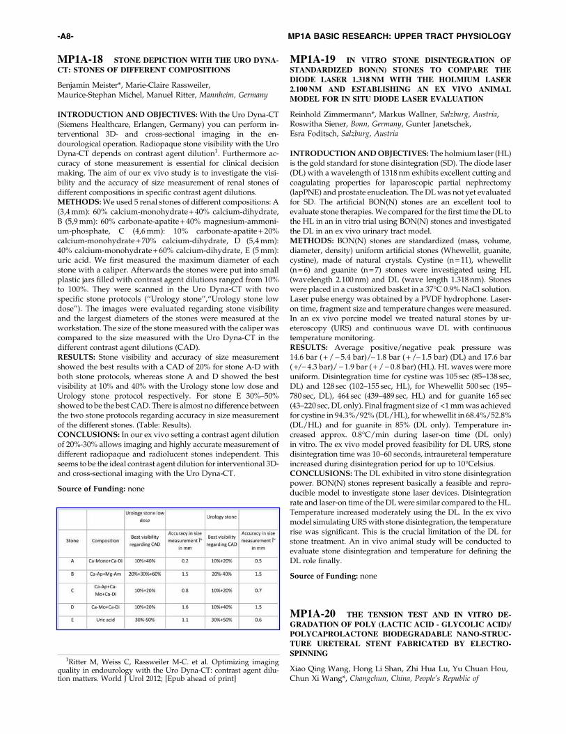
MP1A-18 STONE DEPICTION WITH THE URO DYNA-
CT: STONES OF DIFFERENT COMPOSITIONS
Benjamin Meister*, Marie-Claire Rassweiler,Maurice-Stephan Michel, Manuel Ritter, Mannheim, Germany
INTRODUCTION AND OBJECTIVES: With the Uro Dyna-CT(Siemens Healthcare, Erlangen, Germany) you can perform in-terventional 3D- and cross-sectional imaging in the en-dourological operation. Radiopaque stone visibility with the UroDyna-CT depends on contrast agent dilution1. Furthermore ac-curacy of stone measurement is essential for clinical decisionmaking. The aim of our ex vivo study is to investigate the visi-bility and the accuracy of size measurement of renal stones ofdifferent compositions in specific contrast agent dilutions.METHODS: We used 5 renal stones of different compositions: A(3,4 mm): 60% calcium-monohydrate + 40% calcium-dihydrate,B (5,9 mm): 60% carbonate-apatite + 40% magnesium-ammoni-um-phosphate, C (4,6 mm): 10% carbonate-apatite + 20%calcium-monohydrate + 70% calcium-dihydrate, D (5,4 mm):40% calcium-monohydrate + 60% calcium-dihydrate, E (5 mm):uric acid. We first measured the maximum diameter of eachstone with a caliper. Afterwards the stones were put into smallplastic jars filled with contrast agent dilutions ranged from 10%to 100%. They were scanned in the Uro Dyna-CT with twospecific stone protocols (‘‘Urology stone’’,‘‘Urology stone lowdose’’). The images were evaluated regarding stone visibilityand the largest diameters of the stones were measured at theworkstation. The size of the stone measured with the caliper wascompared to the size measured with the Uro Dyna-CT in thedifferent contrast agent dilutions (CAD).RESULTS: Stone visibility and accuracy of size measurementshowed the best results with a CAD of 20% for stone A-D withboth stone protocols, whereas stone A and D showed the bestvisibility at 10% and 40% with the Urology stone low dose andUrology stone protocol respectively. For stone E 30%–50%showed to be the best CAD. There is almost no difference betweenthe two stone protocols regarding accuracy in size measurementof the different stones. (Table: Results).CONCLUSIONS: In our ex vivo setting a contrast agent dilutionof 20%-30% allows imaging and highly accurate measurement ofdifferent radiopaque and radiolucent stones independent. Thisseems to be the ideal contrast agent dilution for interventional 3D-and cross-sectional imaging with the Uro Dyna-CT.
Source of Funding: none
MP1A-19 IN VITRO STONE DISINTEGRATION OF
STANDARDIZED BON(N) STONES TO COMPARE THE
DIODE LASER 1.318 NM WITH THE HOLMIUM LASER
2.100 NM AND ESTABLISHING AN EX VIVO ANIMAL
MODEL FOR IN SITU DIODE LASER EVALUATION
Reinhold Zimmermann*, Markus Wallner, Salzburg, Austria,Roswitha Siener, Bonn, Germany, Gunter Janetschek,Esra Foditsch, Salzburg, Austria
INTRODUCTION AND OBJECTIVES: The holmium laser (HL)is the gold standard for stone disintegration (SD). The diode laser(DL) with a wavelength of 1318 nm exhibits excellent cutting andcoagulating properties for laparoscopic partial nephrectomy(lapPNE) and prostate enucleation. The DL was not yet evaluatedfor SD. The artificial BON(N) stones are an excellent tool toevaluate stone therapies. We compared for the first time the DL tothe HL in an in vitro trial using BON(N) stones and investigatedthe DL in an ex vivo urinary tract model.METHODS: BON(N) stones are standardized (mass, volume,diameter, density) uniform artificial stones (Whewellit, guanite,cystine), made of natural crystals. Cystine (n = 11), whewellit(n = 6) and guanite (n = 7) stones were investigated using HL(wavelength 2.100 nm) and DL (wave length 1.318 nm). Stoneswere placed in a customized basket in a 37�C 0.9% NaCl solution.Laser pulse energy was obtained by a PVDF hydrophone. Laser-on time, fragment size and temperature changes were measured.In an ex vivo porcine model we treated natural stones by ur-eteroscopy (URS) and continuous wave DL with continuoustemperature monitoring.RESULTS: Average positive/negative peak pressure was14.6 bar ( + / - 5.4 bar)/- 1.8 bar ( + /- 1.5 bar) (DL) and 17.6 bar( +/- 4.3 bar)/ - 1.9 bar ( + / - 0.8 bar) (HL). HL waves were moreuniform. Disintegration time for cystine was 105 sec (85–138 sec,DL) and 128 sec (102–155 sec, HL), for Whewellit 500 sec (195–780 sec, DL), 464 sec (439–489 sec, HL) and for guanite 165 sec(43–220 sec, DL only). Final fragment size of <1 mm was achievedfor cystine in 94.3%/92% (DL/HL), for whewellit in 68.4%/52.8%(DL/HL) and for guanite in 85% (DL only). Temperature in-creased approx. 0.8�C/min during laser-on time (DL only)in vitro. The ex vivo model proved feasibility for DL URS, stonedisintegration time was 10–60 seconds, intraureteral temperatureincreased during disintegration period for up to 10�Celsius.CONCLUSIONS: The DL exhibited in vitro stone disintegrationpower. BON(N) stones represent basically a feasible and repro-ducible model to investigate stone laser devices. Disintegrationrate and laser-on time of the DL were similar compared to the HL.Temperature increased moderately using the DL. In the ex vivomodel simulating URS with stone disintegration, the temperaturerise was significant. This is the crucial limitation of the DL forstone treatment. An in vivo animal study will be conducted toevaluate stone disintegration and temperature for defining theDL role finally.
Source of Funding: none
MP1A-20 THE TENSION TEST AND IN VITRO DE-
GRADATION OF POLY (LACTIC ACID - GLYCOLIC ACID)/
POLYCAPROLACTONE BIODEGRADABLE NANO-STRUC-
TURE URETERAL STENT FABRICATED BY ELECTRO-
SPINNING
Xiao Qing Wang, Hong Li Shan, Zhi Hua Lu, Yu Chuan Hou,Chun Xi Wang*, Changchun, China, People’s Republic of
1Ritter M, Weiss C, Rassweiler M-C. et al. Optimizing imagingquality in endourology with the Uro Dyna-CT: contrast agent dilu-tion matters. World J Urol 2012; [Epub ahead of print]
-A8- MP1A BASIC RESEARCH: UPPER TRACT PHYSIOLOGY

INTRODUCTION AND OBJECTIVES: To investigate the me-chanical property and In vitro Degradation of a polycaprolactone(PCL)/poly (lactic acid - glycolic acid)(PLGA) degradable ure-teral stent.METHODS: The PCL/PLGA ureteral stent was fabricated byelectrospinning. The structure and properties of the stents wereinvestigated by the mechanical property testing, scanning elec-tron microscopy (SEM), degradability test in vitro.RESULTS: The inner diameter of the tubular scaffold in our studywas 1.0 mm and the outer diameter was 2.5 mm. The SEM resultsshow that the scaffold has the nano-structure. The diameters ofthe fibers of different ratio of PCL/PLGA stents have no differ-ences. The tension test showed that the mechanical property wasenhanced with the increase in the proportion of the PCL. Thedegradation curves of different ratio of PCL/PLGA stent wereclose to a straight line. The 5% PCL/PLGA stent could collapsewithin 28 d, and the 15% PCL/PLGA and 25% PCL/PLGA couldcollapse within 42 d and 56 d, respectively.CONCLUSIONS: The PCL/PLGA biodegradable ureteral stenthas a good mechanical property and the degradation time canfully satisfied the demand of a ureteral stent.
Source of Funding: Supported by the National Natural ScienceFoundation of China (no. 50973043).
MP1A-21 HOW TO DETECT ACUTE POST-OPERA-
TIVE KIDNEY INJURY AFTER CLAMPLESS LAPARO-
SCOPIC PARTIAL NEPHRECTOMY? A PILOT STUDY
Francesco Porpiglia*, Daniele Amparore, Riccardo Bertolo,Fabrizio Mele, Diletta Garrou, Giovanni Cattaneo,Matteo Manfredi, Cristian Fiori, Orbassano (Torino), Italy
INTRODUCTION AND OBJECTIVES: NGAL (Neutrophilgelatinase-associated lipocain), is a 25 KDa protein over-expressed in case of renal injury. NGAL measured in bothplasma and urine is being discussing as a precocious biomarkerof acute kidney injury (AKI). A few studies in Literature ana-lysed the role of NGAL in the evaluation of renal injury inpostpartial setting. The aim of this study was the evaluation ofrole of urinary-NGAL as marker to detect and quantify renaldamage in patient who underwent clampless-LaparoscopicPartial Nephrectomy (c-LPN).METHODS: Twenty-four consecutive patients who underwentc-LPN between March and July 2012 were involved in this pro-spective study. Demographic, peri-operative and pathologicaldata were collected. Concerning functional data, NGAL urinaryvalues were dosed on urine samples collected preoperatively, at24 hours, 5 days and 3 months after surgery by ELISA in allpatients. Serum Creatinine (SCr) and GFR (MDRD formula) weredetermined at the same time points. Data were analyzed withFriedman ANOVA and Kendall’s Concordance test (p < 0.05considered significant).RESULTS: Mean age and BMI were 65.6 years and 25.54, re-spectively. Hypertension was recorded in 16 patients (66.6%);mean Charlson’s index, CT-scan size and PADUA score were3.25, 4.51 cm and 8.08, respectively. Mean operative time andestimated blood losses were 115.2 min and 341.6 ml. Meancomplications’ rate and hospital stay were 8.3% (2/24) and5.4 days, respectively. Mean thickness of healthy parenchymaexcised was 0.7 mm. No positive surgical margins wererecorded. Average SCr and GFR values were almost stable atevery time point; average NGAL values were 21.3 + 21.7 ng/ml,37.0 + 37.8 ng/ml, 56.5 + 42.7 ng/ml, 23.7 + 37.1 ng/ml, bothtested respectively before surgery, 24 hours, 5 days and
3 months after surgery (p = 0.042). No differences were foundwhen specifically testing NGAL baseline versus 3rd month va-lue (p = 0.).CONCLUSIONS: Notwithstanding the small sample size, weobserved a progressive and proportional increase of urinaryNGAL values from baseline until the fifth post-operative day; areturn to basal values was recorded at 3rd month. Conversely,SCr and GFR values remained stable in all measurements. Ourdata suggest that NGAL could be a useful molecular marker todetect early onset renal damage, regardless of ischaemia. In ourcase study it showed able to detect even minimal damages and itwas used to evaluate the trend acute kidney damage following c-LPN. Further studies are required.
Source of Funding: none
MP1A-22 BIOMECHANICAL PROPERTIES OF THE
URETER: PRELIMINARY RESULTS
Yaniv Shilo*, Joseph E. Pichamuthu, Timothy D. Averch,Stephen V. Jackman, David A. Vorp, Pittsburgh, PA
INTRODUCTION AND OBJECTIVES: Ureteral injuries such asperforation and avulsion are directly related to mechanicaldamage of the ureter. Understanding the biomechanical proper-ties of this tissue may assist in engineering efficient devices forurological applications, a better comprehension of pathologicalevents in the ureter and prevention of iatrogenic injuries. Whilefew studies looking on the mechanical properties of the animalureter were reported, specific data on the tensile strength andelastic properties of the human ureter is unavailable. The purposeof this work was to study the biomechanical properties of thehuman ureter.METHODS: After obtaining IRB approval, we harvested 6human proximal ureters (4 male and 2 female) from patients whohad nephrectomy for either renal cell carcinoma or nonfunc-tioning kidney from August, 2012 to February, 2013. The ureterswere immediately placed in saline at 4�C and tested within 48 hrs.The ureter was then cut into multiple circumferentially- andlongitudinally-oriented tissue specimens for tensile testing. Initialdimensions of each tissue specimen were measured and thenmounted and stretched to failure after ten cycles of pre-conditioning in a uniaxial tensile testing machine. The corre-sponding force and displacement was recorded and stress-straincurve was plotted. Finally, stress at failure was noted as thetensile strength of the sample. Circumferential tensile strengthwas also compared in the proximal and distal regions of thespecimens.RESULTS: The age of the patients studied was 66.5 – 10.8(mean – SEM) years and BMI was 24.5 – 1.8. The tensile strengthof the ureter in circumferential and longitudinal orientations werefound to be 538 – 50 MPa and 1059 – 197 MPa, respectively(p < 0.03). The circumferential strength in the proximal portion ofthe ureter was 501 – 41 MPa in comparison to 575 – 94 MPa in thedistal portion (p = 0.15).CONCLUSIONS: The circumferential tensile strength of theureter was found to be significantly lower than the longitudinal.Additionally, the circumferential tensile strength of proximalsegments of the ureter is lower than its distal segments, likelyindicating the proximal portion of ureter is more prone to bedamaged than the distal portion.
Source of Funding: none
MP1A BASIC RESEARCH: UPPER TRACT PHYSIOLOGY -A9-
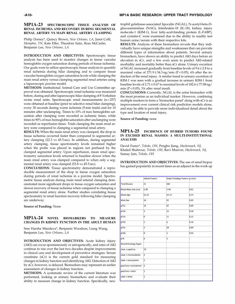
MP1A-23 SPECTROSCOPIC TISSUE ANALYSIS OF
RENAL ISCHEMIA AND RECOVERY DURING SEGMENTAL
RENAL ARTERY VS MAIN RENAL ARTERY CLAMPING
Philip Dorsey*, Quincy Brown, New Orleans, LA, Janet Colli,Memphis, TN, Kate Elfer, Theodore Saitz, Ross McCaslin,Benjamin Lee, New Orleans, LA
INTRODUCTION AND OBJECTIVES: Spectroscopic tissueanalysis has been used to monitor changes in tissue vascularhemoglobin oxygen saturation during periods of tissue ischemia.Our goals were to utilize spectroscopic tissue analysis to quantifyrenal ischemia during hilar clamping and to compare tissuevascular hemoglobin oxygen saturation levels while clamping themain renal artery versus clamping segmental renal arteries usinga laparoscopic porcine model.METHODS: Institutional Animal Care and Use Committee ap-proval was obtained. Spectroscopic renal ischemia was measuredbefore, during and after laparoscopic hilar clamping of 8 Yorkshireswine renal units. Spectroscopic renal measurements (n = 257)were obtained at baseline (prior to selective renal hilar clamping),every 30 seconds during warm ischemia (5 min trials) and for 10minutes after unclamping. Times to 10% of max hemoglobin sat-uration after clamping were recorded as ischemic times, whiletimes to 90% of max hemoglobin saturation after unclamping wererecorded as reperfusion times. Trials clamping the main renal ar-tery were compared to clamping a segmental renal artery.RESULTS: When the main renal artery was clamped, the drop intissue ischemia occurred faster than compared to segmental ar-tery clamping (22.3 vs 45.5 sec). In addition, during segmentalartery clamping, tissue spectrometry levels remained higherwhen the probe was placed in regions not perfused by theclamped segmental artery. Upon reperfusion, mean renal spec-trometry saturation levels returned to baseline slower when themain renal artery was clamped compared to when only a seg-mental renal artery was clamped (53.8 vs 45.5 sec).CONCLUSIONS: Tissue spectrometry demonstrated a repro-ducible measurement of the drop in tissue oxygen saturationduring periods of renal ischemia in a porcine model. Spectro-metric tissue analysis during main renal arterial clamping dem-onstrated more significant drops in tissue oxygen saturation andslower recovery of tissue ischemia when compared to clamping asegmental renal artery alone. Further studies correlating tissuespectrometry to renal function recovery following hilar clampingare underway.
Source of Funding: None
MP1A-24 NOVEL BIOMARKERS TO MEASURE
CHANGES IN KIDNEY FUNCTION IN THE ADULT HUMAN
Sree Harsha Mandava*, Benjamin Woodson, Liang Wang,Benjamin Lee, New Orleans, LA
INTRODUCTION AND OBJECTIVES: Acute kidney injury(AKI) can occur spontaneously or iatrogenically, and rates of AKIcontinue to rise over the last two decades despite improvementsin clinical care and development of preventive strategies. Serumcreatinine (sCr) is the current gold standard for measuringchanges in kidney function and identifying AKI. Detection of AKIby sCr, however, is delayed. Biomarkers may represent an earlierassessment of changes in kidney function.METHODS: A systematic review of the current literature wasperformed, looking at urinary biomarkers and evaluate theirability to measure change in kidney function. Specifically, neu-
trophil gelatinase-associated lipocalin (NGAL), N-acetyl-beta-D-glucosaminidase (NAG), Interleukin-18 (IL-18), kidney injurymolecule-1 (KIM-1), liver fatty-acid-binding protein (L-FABP),and cystatin-C were examined due to the ability to readily testhuman urine/serum with their respective kits.RESULTS: Analysis of these biomarkers reveals that they indi-vidually have unique strengths and weaknesses that can providedifferent types of information about patients. Several urinarybiomarkers, have shown an ability to predict AKI days before anelevation in sCr, and a few even seem to predict AKI-relatedmorbidity and mortality better than sCr alone. Urinary excretionof NGAL increased gradually from baseline levels of 9.6 – 2.8 to amaximal value of 271.9 – 34.3 ng/min (P < 0.05), 4 h after the in-duction of the renal injury. A similar trend in urinary excretion ofKIM-1 was seen with a gradual increase in urinary KIM-1 frombaseline levels of 2.71 – 0.67 to maximal levels of 182.6 – 77.08 ng/min (P < 0.05), 3 h after renal insult.CONCLUSIONS: Currently, NGAL is the urine biomarker withthe most promise as an individual marker. However, combiningmultiple markers to form a ’biomarker panel’ along with sCr is animprovement over current clinical risk prediction models alone,and may be able to provide more individualized detail about thetype and location of renal injury.
Source of Funding: none
MP1A-25 INCIDENCE OF HYBRID TUMORS FOUND
IN EXCISED RENAL MASSES: A MULTI-INSTITUTIONAL
ANALYSIS
David Fumo*, Toledo, OH, Pengbo Jiang, Hackensack, NJ,Khaled Shahrour, Toledo, OH, Ravi Munver, Hackensack, NJ,Samay Jain, Toledo, OH
INTRODUCTION AND OBJECTIVES: The use of renal biopsyhas gained popularity in recent times as an adjunct in the work up
-A10- MP1A BASIC RESEARCH: UPPER TRACT PHYSIOLOGY

of renal masses, particularly small renal masses ( < 4 cm) suspi-cious for renal cell carcinoma (RCC). Our present study attemptsto quantify the prevalence of tumors that contain more than onehistology as a means of determining the potential accuracy ofrenal biopsy.METHODS: In this IRB-approved, multi-institutional study, aretrospective review of renal masses excised between the years of1999–2012 was performed. Patients and masses were included inthe study if information pertaining to demographics and patho-logic stage, mass size, and histology were present, respectively.Tumors were considered to be hybrid tumors if more than onedistinct histology was reported in the final pathology. Granularcell RCC was considered a variant of clear cell RCC and, there-fore, these masses were considered non-hybrid tumors. Statisticalanalyses were completed with paired T-tests and Chi Squaredanalysis, where appropriate. All analyses were completed usingMicrosoft Excel.RESULTS: A total of 203 tumors were included in the analysis.Table 1 summarizes the patient demographics and results of thestudy. Of the entire cohort, 21 tumors (10%) displayed more than
one distinct histology. The differences in mass size and age ofpatients between the two sub-groups were not statistically sig-nificant. There was a greater proportion of pT1a lesions in thehybrid tumor group compared to the single histology group(p = 0.03).
The breakdown of each hybrid tumor with its described his-tology is listed in table 1. A majority of the hybrid tumors werepT1a and were a combination of clear cell and papillary cell RCC.Thirteen of the hybrid tumors were smaller than 4 cm in size andthree of these showed a combination of a malignant and benignhistology.CONCLUSIONS: A majority (62%) of the hybrid tumors re-viewed in this analysis were pT1a. Approximately 30% of thesetumors contained a benign component mixed with a malignantcomponent. This finding should be taken into consideration wheninterpreting a benign renal biopsy for a small renal mass. A largeranalysis with more masses needs to be completed to validate theabove findings.
Source of Funding: none
MP1B BASIC RESEARCH: UROLITHIASIS
MP1B-01 A URETEROSCOPIC LITHOTRITE: IN
VITRO ASSESSMENT OF A NOVEL FLEXIBLE PROBE
ULTRASONIC INTRACORPOREAL DEVICE
Jessica E Paonessa*, Naeem Bhojani, James A McAteer,James C Williams, Jr., James E Lingeman, Indianapolis, IN
INTRODUCTION AND OBJECTIVES: Recently, a flexibleultrasonic lithotrite probe has been developed (UreTron, Med-Sonics Corp., Erie, PA) for use with a flexible ureteroscope. Thisnovel device has potential application in the treatment of stoneswhere probe deflection may be an advantage. We conductedin vitro testing of the UreTron flexible probe to assess the effect ofureteroscope deflection on stone comminution.METHODS: A hands-free in vitro test system was designed toobjectively assess the efficiency of stone penetration by the Ure-Tron US-flex-probe. The US-flex-probe was passed through theworking channel of a Wolf Cobra flexible ureteroscope. TheCobra was mounted upright with the probe extending into awater-filled, optically clear test chamber. Deflection angle was setat 0, 15 or 30 degrees. The US-flex-probe was advanced 5 mm pastthe end of the Cobra and a gypsum artificial stone was centeredon the probe tip with a counter weight atop the stone to provideconstant force. Irrigant flow through the US-flex-probe was set at15 ml/min. The UreTron was operated on the hard-stone settingand time for stone penetration was measured.RESULTS: All six UreTron probes tested were able to penetratethe test stone material at an average rate of 2.25 mm/min.However, the probes did differ significantly in penetrationrate, with average times ranging from a slowest value of2.02 + / - 0.14 mm/min to the fastest of 2.50 + / - 0.16 mm/min(P < 0.0001 by multi-factor ANOVA). Deflecting the tip of theureteroscope by 30 degrees reduced the stone penetration rate byan average of 3.4% compared with no deflection (P = 0.02). Theprobes performed well long after the manufacturer’s stated life-
time of 7 minutes, with a fall-off of penetration rate of only 5.3%on average after 30 minutes of use.CONCLUSIONS: The ability of the UreTron flexible ultrasonicprobe to fragment stones is quite novel. Assessment using ourhands-free in vitro test system, in which stone penetration time isnot affected by operator bias, shows that increased angle of de-flection affects the performance of the probe. Further in vitro andin vivo testing is warranted.
Source of Funding: Supported by NIH DK43881
MP1B-02 ACTIVITY LEVELS AND STONE DISEASE: A
POPULATION BASED ANALYSIS USING THE NATIONAL
HEALTH AND NUTRITION EXAMINATION DATABASE
Shubha De*, Jiangbo Li, Fabio Torricelli, Manoj Monga,Cleveland, OH
INTRODUCTION AND OBJECTIVES: A recent analysis of theWomen’s Health Initiative Study found an inverse relationshipbetween kidney stone risk and the amount of activity in post-menopausal women. Our objective was to see if this associationheld true when using representative national survey data(NHANES 2009–2010).METHODS: National Health and Nutrition Examination Surveydata was used from 2009–2010. Data from patients answering thequestion, ‘‘Have you ever had a kidney stone?’’ were analyzed.We then compared the responses from the Physical ActivitySurvey, including 5 questions on recreational and work-relatedmeasures of moderate to vigorous activity. We then comparedstone formers and non-stone formers in relation to HDL levels.Chi-square and student’s t-tests were used using SASv9 (signifi-cance level p < 0.05).
MP1B BASIC RESEARCH: UROLITHIASIS -A11-

RESULTS: When asked ‘Have you ever had a kidney stone?’ 642subjects answered yes (292 male, 250 female), and 5659 answeredno (between 20–80 y/o). The relative risks in respect to kidneystones, favored those who; participate in moderate activity for atleast 10 minutes continuously (RR = 0.889, p < 0.001); participatein vigorous physical activity (RR 0.624, p < 0.001); walk or bicycleas a form of transportation (RR = 0.661, p < 0.001). Those whowere involved with moderate activity at work, showed no dif-ference in stone risk (p = 0.341).
HDL levels were also found to be significantly lower in thosewith stones, as compared to those without (49.4 vs. 52.9,p < 0.001).CONCLUSIONS: Using the NHANES database, subjects whowere more active, had lower RR in respect to kidney stones. UsingHDL as a surrogate for activity levels also illustrated non-stoneformers have more favorable profiles.
Source of Funding: none.
MP1B-03 THE VALUE OF REPEATING A TEST:
METABOLIC PROFILES
ranan dasgupta*, saskia verhagen, Jeremy Cox,London, United Kingdom
INTRODUCTION AND OBJECTIVES: Most dedicated stoneunits will use 24 hour urine collection as part of their metabolicprofile for stone formers, and this may be used as the basis forrecommending dietary changes or commencing medications suchas diuretic therapy. However, the value of such a ’snapshot’ as-sessment is unclear, and we aim to review the variability of theseassessments.METHODS: We reviewed all patients that underwent a 24 urinecollection in our metabolic unit, for those with a history of re-current stone formation. We compared the serum and urinarybiochemistry, and aimed to review the variability of diagnosis ofaltered urinary calcium, oxalate, urate and phosphate in suchpatients, with the test repeated at different intervals.RESULTS: From a total of 140 patients, there were 46% whounderwent repeated 24 hour urine profiles at some stage. Of these23 patients had 3 or more 24 hour urine collections performed,without any change in medication between studies. There waspersistent hypercalciuria in 3, hyperoxaluria in 1 patient, andnone with persistent hyperuricosuria in repeat samples. The pick-up rate of abnormalities showed little difference between samples2 and 3.CONCLUSIONS: We found no significant difference in vari-ability of 24 urine collections after 2 samples at discrete times,suggesting that while there may be benefit in repeating thesample, there may be little benefit in undertaking this more thantwice before instuting medical therapy; nevertheless, there was adegree of variability after the first test to warrant a repeat sample.
Source of Funding: None
MP1B-04 CHANGING TRENDS IN AMERICAN DIET
AND THE RISING PREVALENCE OF KIDNEY STONES
Fabio Torricelli*, Shubha De, XIaobo Liu, Manoj Monga,Cleveland, OH
INTRODUCTION AND OBJECTIVES: It has been postulatedthat changing dietary habits may play a role in the prevalence ofnephrolithiasis. The United States Department of Agriculture(USDA) annually accounts for all food available for human con-
sumption (adjusting for export, spoilage, waste, etc). Our objec-tive was to evaluate changing trends in the American diet from1974–2010 and to identify whether caloric intake and the con-sumption of lithogenic foods has been rising.METHODS: Prevalence values from NHANES survey data, werecompared to USDA data on food distribution during the sameperiod (1974–2010). Three data points for prevalence spanning 38years were used from the literature to create two linear models,interpolating the yearly increase in stone prevalence. Changes inlithogenic foods were then correlated with increasing stoneprevalence.
Spearman correlations were performed (p < 0.05), using SAS9.2.RESULTS: Increased total daily calories (Rho 0.96, CI 0.86–1.00,p < 0.001), fat grams (Rho 0.79, CI 0.55–1.00, p < 0.001), proteinounces (Rho 0.85, CI 0.67–1.00, p < 0.001), and fruit/vegetable(Rho 0.73, CI 0.49–0.97, p < 0.001) correlated strongly with theannual increase in stone prevalence. Specific foods also showedstrong correlations with stone prevalence, including dark greenvegetables, flour/cereal products, fish/shellfish, corn prod-ucts (including high fructose corn syrup) and added sugars.Citrus juice was negatively correlated to stone disease (Rho- 0.18, CI - 0.52–0.17, p = 0.31) though not significant.CONCLUSIONS: Several lithogenic components correlate tem-porally with the simultaneous increase in stones. Whether thesecorrelations are incidental or causal is difficult to determine. TheAmerican diet has clearly changed over the last four decades andfurther investigation is required to better evaluate how this mayimpact the national prevalence of nephrolithiasis.
Source of Funding: none
MP1B-05 HOW MUCH FORCE TO DISLODGE A
STONE FROM RANDALL’S PLAQUE? INTRAOPERATIVE
DISTRACTION FORCES
Shubha De, Robert Brown*, Carl Sarkissian, Manoj Monga,Cleveland, OH
INTRODUCTION AND OBJECTIVES: Attachment forces ofadherent stones have implications for defining the physiologic orapplied forces required for stone detachment. As such, in situdistraction forces were measured to characterize stone distractionforces.METHODS: With IRB approval patients were consented. AFlexiforce hand-held digital force transducer was modified andaffixed to a Halo endoscopic basket (Sacred Heart Medical, MN)to measure the maximal distraction force at which a stone wasdetached for the renal papilla.RESULTS: One patient (33 yo female, BMI 23) undergoing flexi-ble ureteroscopy was found to have 9 stones (3–5 mm) adherent to
-A12- MP1B BASIC RESEARCH: UROLITHIASIS

the papillae. Stone composition was mixed (CHP 40%, CaP40%,MAP20%. Distraction forces ranged from 0.06–0.318lb, with anaverage force 0.16 + 0.099lb.CONCLUSIONS: The clinical importance of distraction forces isapplicable to ureteroscopy and novel non-invasive techniques(i.e. ultrasound repositioning, magnetizing microparticles, etc.)for stone manipulation. Defining the distraction forces will helpin the design and implementation of these new modalities. Fur-ther study is needed to define the impact of stone specific char-acteristics (size, composition, location) on distraction forces.
Source of Funding: none
MP1B-06 UPPER CALYCEAL PERCUTANEOUS NE-
PHROLITHOTOMY (PCNL) UNDER SPINAL ANESTHESIA:
A PROSPECTIVE STUDY
Haresh Thummar*, R Ganatra, Rajkot, India
INTRODUCTION AND OBJECTIVES: Spinal anaesthesia hasbeen used for lower limb surgery with a stable cardiovascularstate and a short recovery unit stay. We sought to test the suit-ability of low-dose bupivacaine spinal anaesthesia for percuta-neous nephrolithotomy, a procedure hitherto performed undergeneral anaesthesia. Aim of our study is to compare Spinal An-esthesia with General anesthesia in patients undergoing PCNLfor upper calyceal puncture.METHODS: We performed prospective study of 32 patients ineach group of patients undergoing PCNL at our institute GroupA; PCNL under spinal anesthesia and grouop B under Generalanesthesia and studied various parameters. We use spinal anes-thesia drug bupivacaine 5 mg/ml alone or with the addition of10 lg of fentanyl. Drugs were given at the L_2–L_3 interspacewith the patient in the sitting position. The Patients remained inthis position for 10 min, after which the sensory and motor blockswere assessed.RESULTS: The sensory and motor blocks after intrathecal bupi-vacaine and bupivacaine–fentanyl were similar. Sensory block,reached the fifth derrmatomes, deep motor block occurred allpatients. The patients in the bupivacaine-Spinal anesthesia grouprequired less intra-operative and post-operative analgesics, andboth patients and endoscopists were better satisfied. Both groupswere matched in age, stone burden, gender. There was no sig-nificant difference in both the groups for parameters like opera-tive time, intraoperative complications, pelvicalyceal aceess,bleeding, stone free rate or post operative complications. But costwas significantly less (10 time s at our hospital, 150 Rs. VS 1600Rs.) with group A apart from quick recovery from anesthesia, safein high risk patients. Two patients required supplementationwith sedation or ketamine as these patient required little longertime for stone free. Post operative pain was significantly less ingroup A. All patients were comfortable except mild pain in chestduring PCNL when they were post operative period. Post oper-ative analgesic effect and respiration was significantly better inspinal group. PCNL underSpinal anesthesia demand more tech-nical skill for access.CONCLUSIONS: This study demonstrates Spinal anesthesia issafe, reliable and as effectiveas GA but superior analgesia, at verylow cost without affecting outcome for patients subjected topercutaneous nephrolithotomy, with stable haemodynamics,good post-operative analgesia and acceptable to patient and en-doscopist satisfaction.
Source of Funding: none
MP1B-07 DEVELOPMENT OF A NOVEL ACCESS
SHEATH THAT ALLOWS SIMULTANEOUS SHEATH PLA-
CEMENT AND SAFETY WIRE ACCESS
Yung tan*, Doh Cha, Edan Shapiro, Ciara Marley, Mantu Gupta,New York, NY
INTRODUCTION AND OBJECTIVES: Ureteral access sheaths(UAS) have been shown to facilitate repeated access to the ureterand intrarenal collecting system, reduce intrarenal pressures,decrease operative times, and improve visibility. It remains ageneral dictum to have a safety guidewire when using accesssheaths. In order to harness the advantages of both UAS andsafety guidewire access, we are helping with the development ofa novel prototype with Cook Medical (Bloomington, IN) thatestablishes safety wire access and sheath placement simulta-neously.METHODS: Three possible Flexor Parallel Ureteral AccessSheath (FPUAS) (Cook Medical, Bloomington, IN) prototypes; anarrow slit, a wide slit and a spiral slit, were evaluated with 3types of 0.035’’ access wires; a standard guidewire, a super-stiffguidewire, and a glidewire, in a porcine kidney/ureter/bladdermodel (Sierra Medical Sciences Inc, Whittier, CA). Both FPUASand control sheath were engaged and advanced to target in a co-axial manner, the force required to advance was quantified usinga digital pressure sensor (Percsys, Palo Alto, CA). Ability of theprototype to stay on the access wire, ease of deployment andureteroscopy of collecting system were assessed.RESULTS: Altogether 20 trials were performed. It was found thatonly the narrow slit with a super stiff wire was 100% successful inall attempts at insertion of the flexor parallel access sheath. Therewere no cases of wire slippage when this configuration was used.The spiral prototype was the most challenging as it would catchthe wire and drag it along in the ureter leading to failure of sheathadvancement. The wide slit had the lowest advancement force(0.71 lbs) but any advantage was negated by failed insertions asthe access sheath slipped off the wire in 100% of the trials. Ad-vancement forces for the narrow slit prototype were found to beabout 5 times higher than with a standard access sheath but notclinically significant (2.59 lbs vs 0.51 lbs). Flexible ureteroscopy inall cases did not reveal any significant trauma to the porcineureters.
MP1B BASIC RESEARCH: UROLITHIASIS -A13-

CONCLUSIONS: The narrow slit prototype was the most suc-cessful prototype. FPUAS facilitates safe simultaneous placementof ureteral access sheath and a safety wire. Further evaluationswill have to be carried out to determine its effectiveness inhumans.
Source of Funding: none
MP1B-08 DISSOLUTION KINETICS OF URIC ACID
STONES IN RELATION TO URIC ACID SOLUTION CON-
CENTRATION. AN IN VITRO STUDY
Itay Sagy*, Bezalel Sivan, Petach Tikva, Israel, Ruth Frid,Ytzhak Mastai, Ramat-Gan, Israel, Pinchas M. Livne,David Lifshitz, Petach Tikva, Israel
INTRODUCTION AND OBJECTIVES: Dissolution by oralmedications is the treatment of choice for non-obstructing renaluric acid (UA) stones. The major goal is urine alkalization, pref-erably to pH 6.5–7.0. Data on the effect of lowering the urine uricacid concentration on the chemolitholysis rate of uric acid stonesare sparse. According to current practice, allopurinol is indicatedfor hyperuricosuria and its impact in the common uric acid stonesformer, who have a normal-range urine uric acid concentration, isunknown. The aim of this in vitro study was to evaluate thedissolution kinetics of pure uric acid stones in several alkalinesolutions (pH 6.8) with various concentrations of uric acid.METHODS: Whole and fragmented stones were obtained duringpercutaneous nephrolithotomy – PCNL and examined for crys-talline structure by X-ray diffraction. Time-Resolved UV-Visiblespectroscopy (wavelength 290 nm) was used to measure thedissolution kinetics of the uric acid stones, which are proportionalto the rate of change in uric acid concentration in the solution.Solutions containing zero, normal-physiologic, and high con-centrations of uric acid were tested (0, 50, 80, and 130 mg/dl).RESULTS: Figure 1 shows that as the concentration of uric aciddecreases, the dissolution rate of the uric acid stones increases.This effect was more significant for the lower range of concen-trations. The most dramatic change was observed at uric acidconcentrations of 0 to 50 mg/dL, which were associated with atripling of the dissolution rate.CONCLUSIONS: Lowering the urine uric acid concentration,even within the normal range (31–91 mg/dL), greatly enhancesuric acid stone dissolution in vitro. This finding may suggest abenefit for allopurinol also in patients with normal uric acidlevels. Further clinical trials are needed to confirm these findings.
Source of Funding: none
MP1B-09 EFFECT OF HYDROPHILIC EXTRACT OF
ALHAGI MAURORUM ON ETHYLENE GLYCOL-INDUCED
RENAL STONE IN MALE WISTAR RATS
Sadrollah Mehrabi*, Yasuj, Iran, Farhad Mehrabi, Shiraz, Iran
INTRODUCTION AND OBJECTIVES: Alhagi maurorum isone of the herbal medicine that routinely use for passage of uri-nary stones. Aim of this study is defining the effect of hydrophilicextract of Alhagi maurorum on ethylene glycol-induced renalstone in male wistar rats.METHODS: In this experimental study 40 male Wistar rat ran-domly allocated in four groups and studied during 30 days. Twogroups of healthy and prophylactic control received usual waterand 1% ethylene glycol in water respectively during study. Pro-phylactic groups of low dose and high dose extract in addition to1% ethylene glycol in water daily gavaged with 500 mg/kg and1000 mg/kg extract respectively. All rats was hold in metaboliccages in days 0,15 and 30 and 24 hours urine sample was taken.After 30th day rats anesthetized with ether and after taking serumsample from them for measurement of calcium, uric acid andcreatinine they killed and their kidneys removed and send forpathology and were examined for presence and volume of cal-cium oxalate crystals.RESULTS: There was no significant difference between com-plexes of oxalate calcium crystals between studied groups. alsocomparison of urinary oxalate in 30th day was showed that uri-nary oxalate concentration in preventive and negative controlgroups were significantly more than positive control group (p < /05). urinary oxalate concentration in preventive groups were lessthan negative control that was significant (p < 05).measurementof biochemical factors in 30th day was showed significant risingof serum creatinine in negative control group in comparison topositive control (p < 05), but there was no significant differencebetween preventives groups with positive control.CONCLUSIONS: Hydrophilic extract of Alhagi maurorum withregard to reduction in urinary oxalate concentration may be ef-fective in prevention of urinary calcium oxalate stones in rat as asuitable model for human stones, although it has no significanteffect in rate of calcium oxalate crystal formation.
Source of Funding: Medicinal Plant Research Center, YasujUniversity of Medical Sciences
MP1B-10 COMPARISON OF THE IMPACT OF NANO-
SECOND ELECTROPULSE AND ELECTROHYDRAULIC
LITHOTRIPTERS ON URINARY TRACT TISSUE
Alexander Gudkov*, Tomsk, Russian Federation, Valery Diamant,Katsrin, Israel, Maxim Lozovsky, Tomsk, Russian Federation,Gennady Chepovetsky, Katsrin, Israel, Marat Lerner, Tomsk,Russian Federation
INTRODUCTION AND OBJECTIVES: Endoscopic techniquesand lithotripsy procedures have become the leading methods fornephrolithiasis and ureterolithiasis treatment in recent years. Thisstudy aims to compare the safety of two principal electrical
Histologic view of Cal-cium oxalate crystal inrat renal tubules:H · Estain Zoom 10 · 40
-A14- MP1B BASIC RESEARCH: UROLITHIASIS

discharge lithotripsy methods – electrohydraulic lithotripsy(EHL) and novel nanosecond electropulse lithotripsy (NEPL).While they appear similar, they employ different principles ofstone fragmentation that affect their safety and efficacy.METHODS: Safety tests were conducted in this study on humanex-vivo tissue samples harvested after nephrectomy, ureter-ectomy and cystectomy procedures. A fragment of the sampledmaterial was first placed in a tray filled with saline solution and alithotripter discharge was then applied. The time between takingof the samples and application of pulse energy did not exceed2 min. The closest energy settings and probe sizes were used foreach pair of probes in the comparison. Representative samplesfrom the urinary tract (renal calyx, pelvis, ureter and bladder)were selected. Probe positions were chosen as follows: distancefrom sample – 0, 1 and 3 mm; angle between probe and samplesurface – 0�, 45� and 90�. The criterion used for terminating theexperiment was observed tissue perforation. Histological analy-ses of the samples were also performed.RESULTS: Perforations and damages to kidney parenchymawere observed following EHL treatment of calyx paries underminimal energy parameters. In contrast, no visible damage wasobserved with NEPL. An increase of EHL pulse energy to maxi-mum level, 45� angle and contact distance caused a completerupture of the pelvis walls while minor visual color change of themucous was observed for NEPL only when in direct contact withtissue and at probe angles of 45� and 90�. Similar wall perforationwas observed using EHL in the ureter and bladder. Under thesame parameters NEPL produces only slight damage to mucosaltissue.CONCLUSIONS: From these results, we can conclude that undertest conditions EHL causes significant tissue damage. On theother hand, while NEPL may sometimes cause damage at themicroscopic level, such damage does not have irreversible con-sequences. These findings demonstrate that NEPL is safer andbehaves quite differently from EHL. These results can be ex-plained by the different energy transfer mechanisms in the com-pared lithotripters: while energy in NEPL is transferred to thestone by direct discharge into a solid, the energy of the EHL istransferred through liquid medium creating a damaging shockwave.
Source of Funding: None
MP1B-11 STUDY OF THE DIFFERENCES BETWEEN
NANOSECOND ELECTROPULSE AND ELECTROHYDRA-
ULIC METHODS OF LITHOTRIPSY
Alexey Martov, Moscow, Russian Federation, Alexander Gudkov*,Tomsk, Russian Federation, Valery Diamant,Gennady Chepovetsky, Katsrin, Israel, Marat Lerner,Tomsk, Russian Federation
INTRODUCTION AND OBJECTIVES: Endoscopic fragmen-tation of calculus can be achieved with various lithotripsymethods. To overcome the shortcomings of lithotripters now inuse, a novel nanosecond electropulse lithotripter has been de-veloped that employs electric pulses with nanosecond rise timeand duration.
The purpose of this study is to compare nanosecond electro-pulse lithotripsy (NEPL) and electrohydraulic lithotripsy (EHL)in order to demonstrate the differences between these stonefragmentation methods. While apparently similar since they areboth based on electrical discharge, these methods differ accordingto the principles of fragmentation that they employ.
METHODS: Two types of BegoStone phantom were used in or-der to simulate hard and soft uroliths. The stones were placed ona grid with 2 · 2 mm cells immersed in saline. The experiment wasterminated when the objective of clearing the fragmented stonethrough the grid mesh was achieved. The number of pulses re-quired to fragment the stones was recorded per probe size andenergy setting. Comparable energy settings and probe sizes wereused in each method. The results were statistically evaluated.RESULTS: In the testing, NEPL cleared 100% of the stones whileEHL had a success rate of 92%. The criterion chosen for com-paring the efficiency of each method was cumulative energy.Cumulative energy and the number of pulses required for frag-mentation of stones were found to increase with stone densityand pulse energy. The data demonstrated that NEPL requiresmuch less energy to clear stones than EHL. The specific volu-metric energy needed by the devices for fragmenting stonesdemonstrated various dependent relationships between the en-ergy for clearing phantoms and both stones density and probesdiameter. The average total specific energy required for stonesfragmentation using NEPL was 4 times lower than that for EHL.CONCLUSIONS: Various dependences of the received results asfunctions from stones density and probe sizes have been ascer-tained. The findings demonstrate that NEPL is more effective andrequires fewer pulses and correspondingly less time for stonesfragmentation than EHL. These results are explained by the dif-ferent energy transfer mechanisms at work in stone destruction inthe compared lithotripters: while energy in NEPL is transferred tothe stone due by direct discharge penetration into a solid state,energy in the electrohydraulic mechanism is transferred throughthe liquid medium, which also creates a damaging shock wave.
Source of Funding: None
MP1B-12 COMBINATION OF RIGID URETEROSCOPY
WITH FLEXIBLE URETEROSCOPY DOES NOT HAVE ANY
NEGATIVE IMPACT ON THE OUTCOMES
Erdal Alkan, Mirac Turan, Oguz Ozkanli, Egemen Avcý,Mehmet Murad Basar, Yusuf Oguz Acar, Derya Balbay*,Istanbul, Turkey
INTRODUCTION AND OBJECTIVES: To compare the effec-tiveness and complication rates of rigid ureteroscopy combinedwith flexible ureteroscopy to rigid ureteroscopy alone.METHODS: A comparison between patients underwent RIRSfollowing rigid ureteroscopy (group I, n = 35) simultaneously anda matched control group of patients undergoing rigid uretero-scopy alone (group II, n = 35) was done. Patient with symptomaticrenal stones were excluded. Stone free rates (SFRs) were definedas the absence of stone fragments or asymptomatic insignificantresidual fragments < = 4 mm. In cases of multiple stones, the totalstone burden was calculated as the sum of each stone size.RESULTS: Mean ureteral stone size was 7.4 – 0.44 mm (3–16) and10.5 – 1.03 mm (4–21) in groups I and II, respectively (p = 0.007).Mean renal stone size was 13.3 – 1.45 mm (2–15), and SFR forintrarenal stones was 94.3%. Residual stone fragments larger than5 mm remained in 2 patients who had multiple (4 stones) andheavy stone burden (total stones dimension was 25 mm). Meanoperative time was 55.4 – 3.28 minutes (30–95) and 28.4 – 2.66minutes (10–90) in groups I and II, respectively (p < 0.001). RIRSfor asymptomatic renal stones increased the mean operative timefor 27.0 – 0.62 minutes. DJSs were left in 30 and 17 patients ingroups I and II, respectively (p = 0.001). Hospital stay and com-plications rates were similar in both groups.
MP1B BASIC RESEARCH: UROLITHIASIS -A15-

CONCLUSIONS: Simultaneous endoscopic treatment of theureteral and ipsilateral asymptomatic renal stones significantlyprolongs duration of surgery, increases use of DJS, and elevatesSFRs. It does not affect hospitalization and complication rates.
Source of Funding: none
MP1B-13 SIZE-MEASUREMENT OF RENAL STONES
WITH THE URO DYNA-CT
Benjamin Meister*, Marie-Claire Rassweiler, Christel Weiß,Maurice-Stephan Michel, Axel Haecker, Manuel Ritter,Mannheim, Germany
INTRODUCTION AND OBJECTIVES: Residual stone frag-ments after percutaneous stone removal (PCNL) are a commonissue. The size of the residual fragments is the main criterium iffurther interventions such as URS, ESWL or second look PCNLneed to be done or not. Respecting recent literature clinical sig-nificant stone fragments are ‡ 4 mm. The most sensitive tool forstone detection, especially of small size is computed tomography.Since the implementation of the Uro Dyna-CT in the en-dourological operation room in Mannheim (Germany) we havethe possibility of 3D- and cross-sectional imaging already duringthe intervention with excellent image quality and low radiationdose especially for hard contrast depiction. In our study we in-vestigated the accuracy of imaging and measurement of stonefragments with the Uro Dyna-CT.METHODS: We measured 27 artificial stones (plaster of Paris)using a digital caliper. Afterwards the stones were embedded insmall plastic jars filled with plasticine and scanned with the UroDyna-CT in a randomized order. Cross-sectional and 3D-imageswere reconstructed. The major diameter of each stone was mea-sured blinded at the workstation. The difference between themeasurements was statistically evaluated using the paired t-test,calculating a 95% reference area for the differences between themeasurements, calculating an intraclass correlation coefficient (inorder to see if there is a relation between the two measuringmethods) and doing a Bland-Altman-Analysis to check if theabbreviations depend on the measuring area.RESULTS: Range of stone sizes was 3 to 5 mm. Mean size of thecalculi measured with Uro Dyna-CT was 4,21 mm [ – 0, 64], meansize of the calculi measured with caliper 4,10 mm [ – 0, 61]. Meandifference in the statistical analysis of the diameter differencesbetween caliper and Uro Dyna-CT was 0,12 mm – [0, 66]. In-traclass correlation coefficient was 0,44647 (p = 0,0088), whichshows that there is a significant correlation between the twomeasuring methods. Furthermore there were no significant dif-ferences in the size of the stones measured in the Uro Dyna-CTand measured with caliper (p = 0, 3597).CONCLUSIONS: Radiopaque artificial stone fragments can bemeasured highly accurate with the Uro Dyna-CT. In the futurefurther studies need to proof, how residual stones can be detectedand measured with the Uro Dyna-CT in vivo.
Source of Funding: none
MP1B-14 IS LYMPHOCYTOPENIA A NEW MARKER
FOR PYONEPHROTIC OBSTRUCTED KIDNEYS (POK)
SECONDARY TO URINARY STONE DISEASE?
Ahmed Ali*, Liam Farrell, Bhaskar Somani, Southampton,United Kingdom
INTRODUCTION AND OBJECTIVES: Leucocyte abnormali-ties are seen in severe urinary infections. Lymphocytopenia hasbeen used as a marker for infections and trauma. We wanted tosee if it has significance in Pyonephrotic obstructed kidneys(POK) secondary to urinary stone disease.METHODS: Between Aug 2009-Aug 2012, patients with POKrequiring percutaneous nephrostomy (PCN) were reviewed.Clinical presentation, radiological findings, microbiological cul-ture, renal function and final treatment outcome were recorded.Leukocyte, neutrophil, lymphocyte counts and CRP were ex-tracted from the daily complete blood count (CBC). Lymphocy-topenia was defined as lymphocyte count < 1.5.RESULTS: Thirty-nine patients with a mean age of 55 years(range:22–92) had PCN for POK. Lymphocytopenia was seen in25/39 (64%) patients with a mean lymphocyte count of 1.3 (range:0.1–3.8). Following PCN, it improved in majority of cases (87%)with severe lymphocytopenia (count < 1) associated with a sig-nificant rise in CRP in all cases. Lymphocytopenia was also as-sociated with positive urine/blood culture in nearly two-third ofpatients. The mean leucocyte count, neutrophil count and CRPwere 14.3 (1.8–34.6), 11.6 (1.4–31.6) and 205 (4–483) respectively.Renal dysfunction was seen in 15 patients (60%) with lympho-cytopenia and 6 patients (42%) with normal lymphocyte count.The mean stone size was 14 mm (4–50 mm) with half stones in thePUJ (n = 13) or upper ureter (n = 7).CONCLUSIONS: Lymphocytopenia seems to be associated withthe severity of infection and could be a surrogate marker for initialdiagnosis and monitoring response to treatment in patients withpyonephrotic obstructed kidneys.
Source of Funding: Nil
MP1B-15 AN IN VITRO COMPARISON OF THE USE
OF ABDOMINAL AND BONE WINDOWS ON COMPUTED
TOMOGRAPHY MEASUREMENTS OF HOUNSFIELD UNITS
AND SIZE OF STONES
Paul Erotocritou*, Miles Walkden, Daron Smith, London,United Kingdom
INTRODUCTION AND OBJECTIVES: Unenhanced computedtomography (CT) is the investigation of choice in stone
Table. Inflammatory markers and stone details (Pre
and Post Nephrostomy (N)
-A16- MP1B BASIC RESEARCH: UROLITHIASIS

management, with CT providing the ability to size stones andplan subsequent management. More recently the measurement ofthe Hounsfield Unit (HU) of a stone has been eluded to giveindirect evidence to stone composition/hardness which in turnadds another preoperative factor to consider in stone manage-ment. Furthermore, the accuracy to which stones may be sizedhas been suggested to vary according to the windows used on CTto analyse the images.
The objective of this study was to determine in vitro the effectson size and HU readings depending on the window used foranalysis.METHODS: Fragments of stone retrieved during percutaneousnephrolithotomy (PCNL) were suspended in gelatin within aspecimen pot. These set stone samples were then scanned usingstandard low dose CT, using a 16 row CT scanner, data wasreconstructed into sequential 3 mm axial and 2 mm coronal sliceswith 0.5 mm overlap. Images obtained were analysed usingOsiriX with each slice of the CT containing stone been selected asa region of interest (ROI) to assign a mean HU value and a cubicstone size, this was done using both abdominal and bone win-dows. Once images were gained the stone fragments were sent forbiochemical assessment for their composition.RESULTS: In total 31 stone fragments were analysed, of these 13had a crystalline structure of greater then 95% of a single com-ponent (6 calcium phosphate, 3 calcium oxalate, 3 urate, 1 cys-tine). The remaining 18 stones were of mixed composition withmore than 50% of the composition been calcium based in 12stones, magnesium ammonium phosphate (MAP) in 3 stones andurate in 3 stones. HU values given on analysis were in keepingwith what is published in the literature, but with HU value beengreater for a stone when analysed with bone windows. For thestones of pure composition the values were: urate 284.54 on ab-dominal windows (AW), 321.206 on bone windows (BW), cal-cium phosphate 871.122 AW, 894.867 BW, calcium oxalate 607.23AW, 895.21 BW and cystine 473.25 AW, 573.56 BW. Furthermorewhen size of stone was analysed, the mean volume on abdominalwindows was greater than that calculated on bone windows.CONCLUSIONS: CT has a central role in stone managementbeen able to elude to stone composition in both pure componentstones and mixed component stones. Furthermore, the use ofbone windows may enhance the accuracy with which stonecomposition can be suggested in addition to more accurate sizeestimation.
Source of Funding: NONE
MP1B-16 EFFECTIVENESS OF FLEXIBLE URETER-
ORENOSCOPY FOR MULTIPLE UNILATERAL INTRA-
RENAL STONES SMALLER THAN 2 CM
Erdal Alkan, Oguz Ozkanli, Egemen Avcý, Mirac Turan,Mehmet Murad Basar, Yusuf Oguz Acar, Derya Balbay*,Istanbul, Turkey
INTRODUCTION AND OBJECTIVES: The objective of thisstudy was to evaluate the safety and efficacy of RIRS for thetreatment of multiple unilateral intrarenal stones smaller than20 mm.METHODS: Between March 2007 and April 2013, patients withmultiple intrarenal stones smaller than 20 mm were treated withRIRS and evaluated retrospectively. SFRs were defined as acomplete absence or residual stones < 4 mm in the kidney. Eachpatient was evaluated for stone number, stone burden (cumula-tive stone length), operative time, SFRs, and complications.
RESULTS: 173 intrarenal stones in 48 patients were included.Mean age, mean number of stones per patient, mean stone bur-den, and mean operative time were 40.2 – 10.88 years (23–63),3.6 – 2.96 (2–18), 22.2 – 8.44 mm (12–45), and 60.3 – 21.99 minutes(30–130), respectively. The overall SFR was 91.7%. SFRs for pa-tients with a stone burden less and greater than 20 mm were 100%(23/23) and 84% (21/25), respectively (p < 0.001). Complicationsoccurred in six (12.5% - 6/48) patients, including urinary tractinfection or high grade fever > 38.5 C in three, prolonged hema-turia in two, and ureteral perforation in one, all of whom weretreated conservatively. No major complications occurred.CONCLUSIONS: RIRS is an effective treatment option in pa-tients with multiple unilateral intrarenal stones especially whenthe total stone burden is less than 20 mm. Therefore RIRS shouldbe considered as the first line treatment for multiple small in-trarenal stones.
Source of Funding: none
MP1B-17 EXTERNAL VALIDATION OF AN IN-
DIVIDUALIZED WEIGHT-BASED GOAL URINE VOLUME
(WGUV) MODEL INTENDED TO IMPROVE EXPECTED
CALIUM URINE CONCENTRATIONS
Jack Lambert*, Norfolk, VA, Nicole Miller, Nashville, TN,Justin Watson, Michael Fabrizio, Mark Sawyer, Norfolk, VA
INTRODUCTION AND OBJECTIVES: Current recommenda-tions for urine volumes for stone prevention do not account forbody size differences. A weight based goal urine volume(WGUV) model has been proposed as an alternative to a 2 L goal:WGUV = [0.466 + 0.0678*weight (kg)]/2.5 mM. This volume isintended to achieve a mean calcium concentration of 100 mg/L(2.5 mM) regardless of body weight. However, it remains to beexternally validated in an adult patient population. The intent ofthis study was to assess the validity of the model in a largerexternal cohort.METHODS: A retrospective database of 24-hour urine collec-tions (Litholink) at our institution (EVMS) from 1/2006–8/2012was established. Only the first urine volume for each patient wasincluded. Excluded were patients with Cr/kg ratio outside thegender appropriate reference range and cystinurics. For eachpatient was then calculated actual calcium concentration andexpected calcium concentrations using a goal volume of 2 L orWGUV. Actual and expected concentrations were compared.RESULTS: Of 2956 stone forming patients, 1444 patients metstudy inclusion (M = 831; F = 613). Median age was 51 (18–91).Median weight was 83.9 kg (40.8–188.7 kg) with a median BMI of28.0 (15.0–56.4). Mean actual urinary [Ca] was 142.9 ( – 77.7) mg/L, which would be expected to improve to 114.6 ( – 62.0) mg/Lwith a 2 L goal and to 92.8 ( – 46.4) mg/L with WGUV (Figure).Thus, the expected mean calcium concentration was 7.2% lowerthan the intended goal calcium concentration for WGUV in our
MP1B BASIC RESEARCH: UROLITHIASIS -A17-

cohort. Expected urinary [Ca] was also significantly lower usingthe WGUV as compared to a 2 L goal (p < 0.0001). Median mea-sured 24-hour urine volume was 1.66 L (0.356–5.05 L) whereasmedian WGUV was 2.46 L (1.29–5.30).CONCLUSIONS: Evaluation of the WGUV model in a largerexternal data set demonstrates a slightly larger expected im-provement in mean calcium concentration for WGUV relative to a2 L goal urine volume than in the original cohort. This studyappears to support the validity of the original model as well as theconcept of adjusting goal urine volume for weight.
Source of Funding: none
MP1B-18 THE ANIMAL MODEL IN STONE DISEASE:
A TRIBUTE
Michael Moran*, Tucson, AZ
INTRODUCTION AND OBJECTIVES: A ‘‘model’’ is an imita-tion or stand-in for something else; hence, an animal modelremoves the human as the research source. Claude Bernard(1813–1878) is considered the father of experimental medicineand physiology. Stone disease was already known to be a com-plex condition even prior to his monumental accomplishments.This represents a review of the animal models and physiologistswho contributed to our modern understanding of stone disease.METHODS: This is a comprehensive historical overview of theanimal models that resulted in the modern understanding orurolithiasis. Animal models that resulted in the formation ofurolithiasis were specifically targeted for this historical review.RESULTS: M. Morand was the first to note that the Black ratformed kidney stones. ‘‘He informs us that it is much more fre-quent in males than females; and that almost all Rats, when theybecome old, have stones in their urinary passages.’’ Oskar Min-kowski developed an animal model to induce uric acid stone byfeeding large doses of adenine to dogs in 1898. Wilhelm Ebsteinpublished over 237 papers in his prolific career including a stronginterest in urolithiasis developing several animal models includ-ing calcium oxalate stone formation. Animal research and uro-lithiasis came to the New World and Helen Baldwin wrote anarticle in Journal of Experimental Medicine on animal models andstones in 1900.CONCLUSIONS: Fuller Albright, Alexander Randall, CharlesHiggins, and Linwood Keyser all represented the future of stonephysiology in America at the beginning of the 20th century. MileyWesson was President of the AUA in 1935 stated ‘‘We have greathopes of the answer to our problem [urolithiasis] coming from thework being done at four centers of research.’’ The spirit of ac-complishment had reached new heights by the beginning of theWorld War era in the world and a creeping optimism that thecomplexities of stone formation were finally being solved haddeveloped thanks to the animal models of stone formation. ‘‘Menof science learn every day from experience; by experience theyconstantly correct their scientific ideas, their theories; rectifythem, bring them into harmony with more and more facts, and socome nearer and nearer to the truth.’’
Source of Funding: None
MP1B-19 EXTRACORPOREAL SHOCKWAVE LITHO-
TRIPSY (ESWL) FOR RENAL AND URETERAL STONES IN
SOETOMO HOSPITAL FROM 2011 TO 2012
Muhammad Ridha, M Ayodhia Soebadi*, Doddy M Soebadi,Surabaya, Indonesia
INTRODUCTION AND OBJECTIVES: To report patient char-acteristics and results of Huikang HK-V extracorporeal shock-wave lithotripsy (ESWL) machine for renal and ureteral stones.METHODS: We retrospectively analyzed medical records of allpatients treated with ESWL using HK-V device at Department ofUrology Soetomo Hospital from May 2011 to February 2012.RESULTS: There were 241 patients, consisted of 148 men (61.4%)and 93 women (38.6%). Mean age was 47.8 years (range 7 to 87).ESWL was conducted for 130 single stones and 111 multiplestones, using fluoroscopic guidance for 166 patients, ultrasoundonly for 31 patients (12.9%) and both for 44 (18.3%) patients. Stonelocation was 109 (41%) in lower pole, 69 (26%) in middle pole, 28(11%) in upper pole, 41 (16%) in proximal ureter, 14 (5 %) at renalpelvis, 1 (0.3%) at distal ureter and 2 (0.7%) staghorn stones. Therewere 130 patients with stones less than 10 mm (53.9%), 102(42.3%) with stone 10 to 20 mm, and 9 (3.7%) more than 20 mm.There were slightly more left-sided treatments (141 patients/58.5%). Of 181 (75%) patients with hydronephrosis, 104 (43.2%)had DJ stent inserted. Overall, we found 158 (65.5%) patientswere stone free after treatment, 49 (20.3%) patients with residualstones less than 4 mm and 34 (14,1%) patients had more than4 mm residuals.CONCLUSIONS: ESWL is the treatment of choice for simplerenal and ureteral stones less than 20 mm (opaque and nonopaque).
Source of Funding: none
MP1B-20 FACILITY SITUATION OF STONE TREAT-
MENT 2013 IN GERMANY: RESULTS FROM A NATION-
WIDE HOSPITAL SURVEY
Wolfgang Brummeisl*, Christian Chaussy, Regensburg, Germany,Jens Rassweiler, Heilbronn, Germany, Thomas Knoll, Sindelfingen,Germany, Andreas Gross, Hamburg, Germany, Kai Koehrmann,Mannheim, Germany, Wolf Wieland, Hans-Martin Fritsche,Regensburg, Germany
INTRODUCTION AND OBJECTIVES: 33 years ago, in 1980,German doctors and engineers established the non-invasivemethod of stone-lithotripsy. The aim of the present survey was toacquire the facility situation of stone treatment within Germanurology departments, with particular focus on extracorporealshock wave lithotripsy (ESWL), and further, to postulate thetrend for the future role of ESWL in stone therapy.METHODS: A survey was sent to 306 urologic departmentsin Germany under the auspices of the German Society for ShockWave Lithotripsy (DGSWL). The questionnaire contained 25questions on the type, size, and technical facilities of stone-therapy.RESULTS: When the study was concluded, 99 centers (33%) hadanswered the questionnaire, including 25 university hospitals.
On average, about 600 patients with urolithiasis are treated peryear, ranging from 80 to 2100 cases. 27% of the patients are treatedconservatively. ESWL is the preferred method for stone treatmentfor only 22% of the departments (vs. endourologic measures),however, only 2% do not perform ESWL.
Sorted by manufacturer, the following devices are used: 31%Siemens, 20% Dornier, 17% Wolf, 11% Storz, Phillips 8%, EDAP5%, AST 3%, 5% others. The average age of the lithotriptsy de-vices is 9 years.
68% belive that ESWL will retain its importance in stone-ther-apy, 27% believe that it will diminish in relevance, while 5% ex-pect that ESWL will gain importance for stone treatment.CONCLUSIONS: The present survey shows the current stateand equipment of German urology departments with regards to
-A18- MP1B BASIC RESEARCH: UROLITHIASIS

the treatment of urolithiasis. 33 years after its invention, ESWL isavailable in nearly all urologic departments in Germany. ESWL isan important option in the current armamentarium for stonetreatment. Based on the present results, we conclude ESWL willretain its important status in years to come, despite evolvingendourologic measures.
Source of Funding: A survey was sent to 306 urologic depart-ments in Germany under the auspices of the German Society forShock Wave Lithotripsy (DGSWL).
MP1B-21 STONE RADIODENSITY ON NON-CONTRAST
COMPUTED TOMOGRAPHY IS A PARAMETER FOR PRE-
DICTING OUTCOME OF EXTRACORPOREAL SHOCKWAVE
LITHOTRIPSY FOR RENAL STONE
Heshmatollah sofi majidpour*, hooshmand sofi majidpour,sanandaj, Iran
INTRODUCTION AND OBJECTIVES: We evaluated the use-fulness of measuring of renal calculi attenuation values on un-enhanced computerized tomography images as a predictor of theoutcome of treatment by extracorporeal shockwave lithotripsy.
The success rate of ESWL treatment depends on multiplefactors, Unfavorable stone composition is considered a majorcause of failure.METHODS: 151 patients with renal stone 7–21 mm in size un-derwent non-contrast computerized tomography, then ESWL.
patients were excluded due to elevated creatinine levels (morethan 2 mg/dL), single kidney or obstructed kidney.
Successful treatment of renal stones was defined as thosepatients who were stone free or had asymptomatic, clinically in-significant residual fragments < or = 4 mm in diameter, as mea-sured by KUB X-ray and ultrasonograhy, 3 months after a ESWLtreatment.
The patients were further analyzed by dividing them into 6groups according to stone density, stone size.
Group 1: 42 patients with attenuation value 500–700 HU, size7–14 mm.
Group 2: 21 patients with attenuation value 700–900 HU, size7–14 mm.
Group 3: 19 patients with attenuation value 900–1200 HU, size7–14 mm.
Group 4: 37 patients with attenuation value 500–700 HU, size15–21 mm.
Group 5: 21 patients with attenuation value 700–900 HU, size15–21 mm.
Group 6: 11 patients with attenuation value 900–1200 HU, size15–21 mm.
The relationship among the HU, size and the characteristics ofthe patients were analyzed
All ESWLs were undertaken by a STORZ SLK Lithotripter withfragmentation performed under fluoroscopic guidance.RESULTS: The patients were further analyzed by dividing theminto 6 groups according to stone density and stone size. The‘‘low density group’’ comprised all patients with stone densitiesof less than 700 HU, the ‘‘medium density group’’ comprised allpatients with stone densities of 700–900, while, the ‘‘high densitygroup’’ comprised all patients with stone densities of more than900.
The ‘‘low diameter group’’: stone diameters of 7–14 mms,while, the ‘‘high diameter group’’:15–21 mms.
ESWL treatment outcomes:
Group 1:95.2%(40 from 42)Group 2:90.4%(19 from 21)Group 3:84.2%(16 from 19)Group 4:94.5%(35 from 37)Group 5:85.7%(18 from 21)Group 6:72.72%(8 from 11)
Analysis showing higher success rate in low density group1&4.CONCLUSIONS: The knowledge of stone density on pretreat-ment non-contrast computerized tomography can predict stonefragility and stone free rate.
These findings suggest that this information may be beneficialfor selecting the preferred treatment option for patients withurinary calculi.
Source of Funding: none
MP1B-22 PROSPECTIVE COMPARISON BETWEEN
TAMSULOSIN AND RENALIT COMBI COLIC AS A
TREATMENT OF RENO-URETERAL CALCULI EXPULSION
AFTER EXTRACORPOREAL SHOCK WAVE LITHOTRIPSY
(ESWL)
Alessio Zordani, Marco Rosa*, Alessandro Mofferdin,Maria Chiara Sighinolfi, Eugenio Martorana, Salvatore Micali,Giampaolo Bianchi, Modena, Italy
INTRODUCTION AND OBJECTIVES: In this preliminaryprospective study we evaluate the effectiveness of colic expulsivetherapy with tamsulosin 0.4 mg compared to an association ofDimettosi-Dihydroxy-Aporfina 250 mcg, 60 mg Arbutin andumbelliferone after ESWL treatment. Aporfina is already knownfor its alpha-lytic action. Recently it has been combined with theArbutin (anti-inflammatory due to the inhibition of phospholi-pase A2), Asparagine (diuretic) and Herniaria (anti-adhesiveaction).METHODS: From January 2013 we prospectively followed 51patients who underwent ESWL treatment for reno-ureteral cal-culi (mean age: 54 +/- 14.8; 35 men, 16 women). ESWL wasconducted by Dornier Lithotripter S XXP, using the pointing bothultrasound and fluoroscopy. Right after the ESWL treatment,patients were randomized to receive expulsive therapy using: a)Aporfina (Renalit Colic Combi) for 12 days b) Tamsulosin 0.4 mgfor 20 days (control group). Follow-up, 30 days after ESWLtreatment, was done by ultrasonography and, in selected cases,abdomen x-ray. We considered the following variables: size andlocation of the calculi; apparent fragmentation of the calculiduring ESWL treatment; expulsion of fragments or size reductionof the calculi. The data obtained were inserted into a specificdatabase and analyzed using SPSS for Windows 12, through theuse of parametric and non-parametric test (chi square).RESULTS: After ESWL, 24 patients have undergone therapywith Aporfina (A) and 27 patients with Tamsulosin (B). The av-erage size of the stones in the two groups was equal to 8.3 mm and9.5 mm, respectively (p = 0.058). The location was pyelic andureteral in both groups (21/24 patients in group A, 23/27 patientsin group B, p = 0.8). Six patients were excluded from the analysisdue to lack of significant fragmentation in the course of treatment.Follow-up after 30 days showed expulsion of fragments or amarked reduction in the size of the stone in 90.4% (19/21) ofpatients treated with Aporfina and 91.6% (22/24) of the patientstreated with Tamsulosin (p = 0.64). There was no evidence offragments expulsion in the remaining patients. Side effects afterESWL or medical therapy were not present in both groups.
MP1B BASIC RESEARCH: UROLITHIASIS -A19-

CONCLUSIONS: In terms of expulsion of the fragments, the useof Renalit Combi Colic in this preliminary experience seems toshow comparable efficacy to the traditional Tamsulosin’s ther-apy. This formula also helps overcome the difficulties that areoften encountered in prescribing an off label drug in certain ca-tegories of patients, such as women and children.
Source of Funding: none
MP1B-23 RADIATION EXPOSURE IS 2.6 FOLD HIGHER
AT ENDOSCOPIC LITHOTRIPSY VERSUS SWL
Yoram I. Siegel, Shmuel Roizman*, sigalit Haruz-Waschitz,Zerifin, Israel, Avi Ben-Shlomo, yavne, Israel, David Yudelevich,Amir Cooper, Yaniv Shilo, Amnon Zisman, Zerifin, Israel
INTRODUCTION AND OBJECTIVES: All common stonetherapies are performed under fluoroscopy guidance. On onehand there is a trend towards radiation-less endoscopic litho-tripsy and on the other a call to increase SWL effectiveness byincrease the frequency of focus repositioning during the proce-dure. The purpose of this study is to asses and compares theextent of radiation exposure of patients during endoscopic lith-otripsy and SWL.METHODS: Measurements of dose-area product (DAP) wereperformed in 47 (44%) patient who underwent endoscopic lith-otripsy and 59 (56%) who underwent SWL. For SWL an exteriorDAP-meter was assembled to the C-arm whereas for endoscopiclithotripsy the C-arm includes a built in DAP meter. A baselineCoefficient factor was determined for devices coordination. De-mographic and clinical data were collected including age, sex,body mass index (BMI) as well as size and location of stone.RESULTS: (see table)CONCLUSIONS: The crude average radiation exposure is sig-nificantly higher during one session of endoscopic lithotripsycompared with SWL. Further effectiveness studies are warrantedtaking into consideration additional factors such as success rateand frequency of redo and ancillary procedures. More accurateinformation on the risk of radiation exposure may gain access toclinical decision making for these particular procedures.
Source of Funding: none
MP1B-24 AGE PREDICTS SUCCESS OF ELECTRO-
MAGNETIC SHOCK WAVE LITHOTRIPSY FOR URETERAL
STONES
Sameer Deshmukh*, Brian Eisner, Boston, MA
INTRODUCTION AND OBJECTIVES: To evaluate the efficacyof shock wave litotripsy (SWL) using an electromagnetic litho-tripter for the treatment of ureteral stones.METHODS: A retrospective review was performed of patientswho underwent shock wave lithotripsy for the treatment of ureteralstones. Procedures were performed on the Dornier Compact DeltaII, an electromagnetic lithotripter (Dorner MedTech). All patients
were treated on the following protocol: total of 2500 shocks; rate of80/minute for the first 300 shocks and 100 per minute for the re-maining 2200 shocks; power setting of 2 for the first 300 shocks,power setting of 3 for shocks 300–800, and power setting of 4 for theremaining 1700 shocks. F/u imaging was obtained by plain ab-dominal radiography if stones were visible on plain radiographyprior to SWL or computed tomography (CT) if they were not.RESULTS: Forty-eight (48) patients were included in the study,with 40 patients being male gender (83%). Mean age was 52.3years (SD 16.1) and mean BMI was 26.7 kg/m2 (SD 3.7). Meanstone size was 6.2 mm (SD 2.4) and stone composition wasavailable for 20 stones, 18 of which were > 50% calcium oxalate.Stone location was proximal ureter for 19 patients (39.5%) anddistal ureter for 29 patients (60.5%). Stone free rate was 39.6%after a single SWL session (19/29 patients). Five patients under-went a second treatment of which 1 was rendered stone free.When evaluating SWL success, those who were stone-free weresignificantly younger than those who were not (45.6 years versus56.7 years, p = 0.02). There were no differences in stone-free ver-sus not stone-free based on BMI (26.6 versus 26.8), stone size(5.6 mm versus 6.6 mm), anterior stone to skin distance (115.0 mmversus 118.8 mm), posterior stone-to-skin distance (109 mm ver-sus 119.6 mm), gender (42.9% females stone-free versus 40.0% ofmales stone free), or ureteral location (38.9% stone free proximalureter versus 41.4% stone-free distal ureter).CONCLUSIONS: SWL of ureteral stones using electromagneticlithotripsy has success rates of nearly 40% independent of ureterallocation. It appears that success rates may be improved inyounger patients.
Source of Funding: none
MP1B-25 EXTRACORPOREAL SHOCK WAVE LI-
THOTRIPSY FOR RENAL CALCULI: A REVIEW OF 4041
CONSECUTIVE CASES DONE OVER 20 YEARS
Jitendra Jagtap*, Raguram Ganesamoni, Jigish Vyas,Shashikant Mishra, Arvind Ganpule, Ravindra Sabnis,Mahesh Desai, Nadiad, India
INTRODUCTION AND OBJECTIVES: Extracorporeal shock-wave lithotripsy (ESWL) remains a commonly utilized treatmentmodality for renal calculi. We reviewed the data of 4041 consec-utive patients undergoing ESWL for renal calculi at our centre.METHODS: We retrospectively reviewed clinical data for pa-tients who had undergone ESWL for treatment of renal calculifrom 1989 to 2012 at our tertiary referral centre in Western India.We analysed patient age, gender, stone size, location, co-morbidities, postoperative complications, additional interven-tions and anatomical parameters of these patients.RESULTS: The average age was 39.40 – 14.32 years. The Male:Female ratio was 2.64:1. Average Stone size was 1.34 – 0.55 cm
Stone Clearance by Size
-A20- MP1B BASIC RESEARCH: UROLITHIASIS

with the left:right side ratio being 0.96:1.00. The averageHounsfield unit on CT scan was 1159.65 – 315.90 HU. 11.71%patients had comorbidities. The average number of shocks re-ceived were 2537 and the mean fluoroscopy time in our study was240.86 – 146.10 sec. A total of 136 patients (3.36%) had complica-tions ranging from fever to perinephric hematoma. 165 patients(4.0%) required an auxillary procedure. The overall success ratewas 88.0 %.
CONCLUSIONS: Our long-term review of ESWL for renal cal-culi demonstrates that it is a safe, feasible treatment with goodsuccess rates in appropriately selected patients.
Source of Funding: none
MP2A BASIC RESEARCH: LOWER TRACT PHYSIOLOGY
MP2A-01 ALDH1 EXPRESSION AS TUMOR STEM
CELLS MARKER AND ITS RELATIONSHIP TO CLIN-
ICOPATHOLOGIC PARAMETERS AND PROGNOSIS IN IN-
VASIVE BLADDER CANCER
Hai Ming Wang, Hai Tao Zhang, Ning Xu*, Changchun, China,People’s Republic of
INTRODUCTION AND OBJECTIVES: The purpose of our ex-periments is to investigate the effect of aldehyde dehydrogenase1(ALDH1) as tumor stem cells marker on the biological behaviorof invasive bladder cancer.METHODS: 109 cases of primary invasive carcinomas speci-mens and 26 cases of 37 cases pelvic lymph node dissection wereassembled as experimental group while 20 cases of normalbladder tissue surrounding cancer were collected as controlgroup. Analysis: The expression of ALDH1 protein were de-tected by immunohistochemistry in experimental group andcontrol group.RESULTS: In 109 cases of invasive bladder cancer, the rate ofALDH1 positive expression was 33.94%. In 20 cases of adjacentnormal bladder tissue, the rate of ALDH1 positive expressionwas 5.00%. The expression was significantly different betweenthe two groups (P < 0.01). There were 37 cases of local lymphnodes of invasive bladder cancer. In 6 metastatic lymph nodes,the rate of ALDH1 positive expression was 50.00%. No ex-pression in 20 cases of normal local lymph nodes. The expres-sion was significantly different between the two groups(P < 0.01). The positive rate of ALDH1 expression in low-gradegroup and high-grade group bladder cancer was 13.04% (3/23)and 39.53% (34/86) respectively, and the difference was statis-tically significant (P < 0.05). The positive rate of ALDH1 ex-pression in non-muscle invasive group and muscle invasivegroup bladder cancer was 19.05% and 43.28%, respectively. The
difference was statistically significant (P < 0.05). There were 6cases of lymphatic metastasis in 37 cases of pelvic removal ofregional lymph nodes. The positive rate of ALDH1 expression inbladder cancer of lymphatic metastasis group and withoutlymphatic metastasis group was 50.00% and 12.90%, respec-tively. The difference was statistically significant (P < 0.05).Statistics show that ALDH1 expression in bladder cancer is notcorrelated with patients gender and age and tumor size andtumor maiden or recurrence.CONCLUSIONS: ALDH1 may be correlated with the occurrenceof bladder cancer. It may be used as an index of grade, stage andprognosis of bladder cancer.
Source of Funding: none
MP2A-02 THE CLINICAL SIGNIFICANCE OF SERUM
AMYLOID PROTEIN A IN PROSTATE CANCER PATIENTS
Zhong Shuai Cao, Hong Li Shan*, Changchun, China,People’s Republic of
INTRODUCTION AND OBJECTIVES: To screen and identifyserum amyloid A (SAA) in patients with prostate cancer withmass spectrum technique.METHODS: The serum samples of 50 prostate cancer patientsand 30 healthy volnnteers were tested. SELDI technology wasused to detect the changes in protein expression. SAA wasscreened and separated and then identified by peptide massfingerprint (PMF) based on matrix-assisted laser desorption/ionization time of flight mass spectrometry (MALDI-TOF-MS)and database searching.RESULTS: The analysis showed that a mass cluster in the rangesof 11.1–11.9 KD in M/Z value in the serum of prostate cancer
Number of ESWL Sittings
MP2A BASIC RESEARCH: LOWER TRACT PHYSIOLOGY -A21-

patients was much higher than that in the control group. Mean-while, this protein peak was closely correlated with clinical stagesof prostate cancer. The level of the protein peak was increased asthe illness got serious. Through MALDI technology combinedwith HPLC, the mass cluster in the range of 11.1–11.9 KD in M/Zvalue on the chip was identified as SAA. And it was also verifiedthrough ELISA method.CONCLUSIONS: Mass spectrum technology is an effectivemethod to detect the biological markers in prostate cancerpa-tients. This method is convenient, highly sensitive and with goodreproducibility. The SAA can be used as a marker in the diagnosisof prostate cancer. These indices are also meaningful in screeningand identifying signal proteins from the serum of prostate cancerpatients.
Source of Funding: none
MP2A-03 ALDH1 EXPRESSION AS TUMOR STEM
CELLS MARKER AND CLINICAL SIGNIFICANCE IN NON-
INVASIVE BLADDER CANCER
Hai Ming Wang, Ming Ming Shao, Ning Xu*, Changchun, China,People’s Republic of
INTRODUCTION AND OBJECTIVES: The purpose of our ex-periments is to evaluate ALDH1 expression in noninvasivebladder cancer and to analyze the relations between ALDH1 andtumor clinicopathologic features and prognosis.METHODS: 118 cases of noninvasive bladder carcinomas spec-imens and 30 cases of normal bladder tissue surrounding cancerwere detected. The expression of Aldehyde dehydrogenase1(ALDH1) protein were detected by immunohistochemistry.RESULTS: In 118 cases of noninvasive bladder cancer, the rate ofALDH1 positive expression was 25.58%. In 30 cases of adjacentnormal bladder tissue, the rate of ALDH1 positive expression was6.67%. The expression was significantly different between the twogroups (P < 0.05). The positive rate of ALDH1 expression in low-grade group and high-grade group noninvasive bladder cancerwas 18.18% and 36.59% respectively, The difference was statis-tically significant (P < 0.05). The positive rate of ALDH1 expres-sion in Ta group and T1 group noninvasive bladder cancer was16.92% and 33.96% respectively. The difference was statisticallysignificant (P < 0.05). The positive rate of ALDH1 expression inprimary group and recurrence group noninvasive bladder cancerwas 19.77% and 37.50% respectively. The difference was statis-tically significant (P < 0.05). Statistics show that ALDH1 expres-sion in noninvasive bladder cancer is not correlated with patientsgender and age and tumor size.followed up for 4–52 months, thecumulative disease-free survival rate of ALDH1-positive groupand ALDH1-negative group are 55.17% and 61.80%, respectively.Kaplan-Meier analysis and the log-rank test showed that the cu-mulative disease-free survival rate was statistically different be-tween the ALDH1-positive group and the ALDH1-negativegroup (P < 0.05).CONCLUSIONS: The ALDH1 may be important for the earlydiagnosis of noninvasive bladder cancer. ALDH1 expression innoninvasive bladder cancer is not correlated with gender, age andtumor size, and was positive correlated with primary tumors andrecurrence. The cumulative disease-free survival rate of ALDH1-positive group is lower than of ALDH1-negative group. ALDH1may be one of the useful indexes of noninvasive bladder cancerprognosis.
Source of Funding: none
MP2A-04 EFFECTIVENESS OF TAMSULOSIN IN
PREVENTION OF POST-OPERATIVE URINARY RETEN-
TION: A RANDOMIZED DOUBLE-BLIND PLACEBO-
CONTROLLED STUDY
Ali Hamidi Madani*, Hamidreza Baghani Aval,Gholamreza Mokhtari, Hamidreza Nasseh, Samaneh Esmaeili,Rasht, Iran, Maryam Shakiba, Tehran, Iran,Reza Shahrokhi Damavand,Seyed Mohamad Seyed Saadat, Rasht, Iran
INTRODUCTION AND OBJECTIVES: Urinary retention is oneof the most common complications contributing to surgical pro-cedures. Recent studies have shown the benefits of alpha-adren-ergic blockers in preventing post-operative urinary retention(POUR). The aim of this prospective study was to compare theprophylactic effect of tamsulosin with placebo on post-operativeurinary retention.METHODS: In this randomized placebo controlled, clinical trial,232 male patients aged 18 to 50 years old admitted to Razi uni-versity Hospital for varicocelectomy, inguinal herniorrhaphy, andscrotal surgery were randomly assigned to receive either threedoses of 0.4 mg tamsulosin (n = 118) or placebo (n = 114), 14 and 2hours before, and 10 hours after surgery. Patients were closelymonitored for the development of urinary retention 24 hours aftersurgical intervention. The primary endpoint was to investigate theeffect of tamsulosin in prevention of post-operative urinary re-tention during the first 24 hours after surgical intervention.RESULTS: One hundred and eighteen patients were included intamsulosin arm and 114 in placebo arm. POUR in patients whoreceived tamsulosin was significantly lower than placebo, as 5.1%of the patients treated with tamsulosin and 21.1% placebo group,reported urinary retention following surgery. No serious adverseeffects were seen in two groups.CONCLUSIONS: This study suggests that short perioperativetreatment with tamsulosin can reduce the incidence of urinaryretention and the need for catheterization after varicocelectomy,inguinal herniorrhaphy, and scrotal surgery.
Source of Funding: none
MP2A-05 NON OBSTRUCTIVE URINARY TRACT DI-
LATATION: LONG TERM FOLLOW UP OF A SINGLE CASE
Aditi Kumar*, Aniruddha Chakravarti, Anthony D’Sa,Birmingham, United Kingdom
INTRODUCTION AND OBJECTIVES: Following ultrasoundscanning for minor renal trauma, a 13 year old boy, known tohave congenital nephrogenic diabetes insipidus, was found tohave bilateral hydroureteronephrosis, with marked cortical lossand a urinoma. We subsequently documented the patients entirerenal history to see how this patient had been managed.METHODS: The patient’s entire urological/nephrological his-tory was documented. All sources were reviewed includingclinical notes, blood tests, imaging and urodynamics.RESULTS: Diabetes insipidus had originally been diagnosedwhen the patient was 9 months. Having been lost to follow up,initial work up showed the patient had a residual bladder volumeof 200–400 mls, cystoscopy showed a trabeculated bladder, and aMAG 3 showed unequal reduction of kidney function. eGFRslowly declined over a 4 year period from 90 to 82. We review thedifferent complications these patients are prone to such as UTI,valve bladder syndrome, bladder dysfunction and hydrone-phrosis, Different management options are discussed including
-A22- MP2A BASIC RESEARCH: LOWER TRACT PHYSIOLOGY

dDVAP, intermittent catheterisation, nocturnal bladder drainage,button cystostomy, urinary diversion, dialysis and transplanta-tion following renal failure.CONCLUSIONS: To date there are no sets of data that agree onhow diabetes insipidus should be managed. By reviewing theliterature and the above we have proposed a management strat-egy involving principally urologists and nephrologists, for thefollow up of these patients.
Source of Funding: None
MP2A-06 URINE CONCENTRATIONS OF AQUA-
PORIN-1 AND PERILIPIN-2 NORMALIZE IN PATIENTS
WITH RENAL CELL CARCINOMA FOLLOWING EXCISION
OF TUMORS
Jonathan Mobley*, Jeremiah Morrissey, Sam Bhayani,Joseph Song, Joel Vetter, Evan Kharasch, Robert Figenshau,St. Louis, MO
INTRODUCTION AND OBJECTIVES: The purpose of this in-vestigation is to evaluate the screening properties of Aquaporin-1(AQP1) and Perilipin-2 (PLIN2) in patients with clear cell andpapillary renal cell carcinoma (RCC).METHODS: Western blot procedures determined the pre-operative urine AQP1 and PLIN2 levels of 61 patients undergoingeither a partial or a radical nephrectomy for clear cell or papillaryRCC. Concentrations of these markers were compared with 43controls of equivalent age, sex, weight, smoking history, and esti-mated glomerular filtration rate. Post-operative urine concentrationof both markers were obtained from RCC patients following tumorexcision and compared to pre-operative samples and controls.RESULTS: The median urine AQP1 for RCC and controls was 53and 1.5 absorbance units/mg creatinine (p < 0.001). The medianurine PLIN2 for RCC and controls was 59 and 6 absorbanceunits/mg creatinine (p < 0.001). The sensitivity and specificity forAQP1 was both 100%, and for PLIN2 was 92% and 100%. Figure 1shows the trends in AQP1 and PLIN2 following either partial orradical nephrectomy. Post-operative AQP1 and PLIN2 levels inthe RCC group was similar to the pre-operative concentrations inthe controls.CONCLUSIONS: Urine AQP1 and PLIN2 displayed both ex-cellent sensitivity and specificity to distinguish RCC patientsfrom controls in this retrospective, cohort study. The normaliza-tion of marker concentrations following tumor excision furthersupports the concept that RCC causes elevations in urine AQP1and PLIN2.
Source of Funding: Funding is from two grants R01CA141521and the Barnes Jewish Hospital Foundation.
MP2A-07 THE BIOCOMPATIBILITY OF POLY (LACTIC
ACID - GLYCOLIC ACID)/POLYCAPROLACTONE BIODE-
GRADABLE NANO-STRUCTURE URETERAL STENT FABRI-
CATED BY ELECTROSPINNING
Xiao Qing Wang, Hong Li Shan, Yu Chuan Hou, Jing Hai Hu,Yuan Yuan Hao, Chun Xi Wang*, Changchun, China, People’sRepublic of
INTRODUCTION AND OBJECTIVES: To investigate the bio-compatibility of a poly (lactic acid - glycolic acid)/poly-caprolactone degradable ureteral stent.METHODS: The Hela cell and MTT method were used to eval-uate the cytotoxicity in vitro. The scaffold was cut into smallpieces and embedded in the back muscle of rabbits to evaluate thehistocompatibility. The rabbits were sacrificed after 1 week, 4weeks and 12 weeks.RESULTS: The OD492 of negative control group and each con-centration of extracts were more thean 0.5, while the positivecontrol of phenol was less than 0.1. The differences betweennegative control group and various concentrations of extractswas not significant (P > 0.05), but all of them were significantlyhigher than the positive control group (P < 0.05). All of the con-centrations of extracts can be considered non-toxic. The lightmicroscopy examination showed that there are a large number ofinflammatory cells at the junction of materials and muscles after 1weeks, and the inflammatory cells were significantly reducedafter 4 weeks. The fiberosis appeared in the same time. The in-flammatory cells were almost disappeared after 12 weeks, and thethickness of fiber did not increase compared to 4 weeks. In thecontrol group, the histological features after 1 week and 4 weekswere similar to the experimental group, but the thickness of fiberat 12 weeks was higher than that of experimental group.CONCLUSIONS: The poly (lactic acid - glycolic acid)/poly-caprolactone biodegradable urethral stent has no cytotoxicity andhas a good histocompatibility. It can fully satisfied the demand ofa ureteral stent.
Source of Funding: Supported by the National Natural ScienceFoundation of China (no. 50973043).
MP2A-08 ASSOCIATION OF HYPERTENSION WITH
SYMPTOMS OF BENIGN PROSTATIC HYPERPLASIA
Xin Jiang, Shi Ying Li*, Jilin, China, People’s Republic of
INTRODUCTION AND OBJECTIVES: This study was designto determine whether the benign prostatic hyperplasia (BPH)symptoms has the relationship with hypertension.METHODS: The clinical baseline data of a large sample of menseeking treatment for BPH symptoms were analyzed retrospec-tively. BPH symptom intensity (assessed by the InternationalProstatic Symptom Score [I-PSS] or urinary flow rate [Qmax]) andblood pressure were determined in 1,238 patients with BPH.Normotension was defined as a diastolic blood pressure of 90 mmHg or less, the absence of the diagnosis hypertension and the lackof antihypertensive medication. Hypertension was defined as adiastolic blood pressure of greater than 90 mm Hg, being diag-nosed with hypertension or the current prescription of anti-hypertensive drugs.RESULTS: When age and presence of hypertension were used asthe independent explanatory variables, each year of age con-tributed 0.19 points and measured hypertension 1.70 points to I-PSS as the dependent response variable. Similar results wereobtained with Qmax as the dependent response variable. In a
FIG. 1. Pre- and post-nephrectomy urine lev-els of AQP1 and PLIN2in 47 patients with clearcell or papillary renalcell carcinoma.
MP2A BASIC RESEARCH: LOWER TRACT PHYSIOLOGY -A23-

logistic regression procedure using age, I-PSS and Qmax as theindependent explanatory variables, each year of age and each I-PSS point significantly increased the risk of being hypertensive by4.4% and 5.2%, respectively, while Qmax did not had a statisti-cally significant contribution to that risk.CONCLUSIONS: We conclude that a significant, age indepen-dent association exists between BPH symptoms and hypertension.
Source of Funding: none
MP2A-09 IMPACT OF RHO-KINASE INHIBITOR HY-
DROXYFUSIDIL IN PROTAMINE SULPHATE INDUCED
CYSTITIS IN RATS
Yigit Akin*, Aliseydi Bozkurt, Erzincan, Turkey,Huseyin Serkan Erol, Mesut Halici, Fikret Celebi,Kubra Asena Kapakin Terim, Erzurum, Turkey, Hakan Gulmez,Ankara, Turkey, Mutlu Ates, Afyonkarahisar, Turkey,Taha Abdulkadir Coban, Baris Nuhoglu, Erzincan, Turkey
INTRODUCTION AND OBJECTIVES: Interstitial cystitis (IC)is defined as unpleasant sensations, such as pain, pressure anddiscomfort, perceived in relation to the bladder and lower urinarytract, with symptoms lasting for six weeks or longer in the ab-sence of infection or any other confirmed causes. The aim of thisstudy was to investigate the prevention of detrusor overactivityand also bladder tissue damage with hydroxyfasudil in prot-amine sulfate (PS) induced cystitis rat model in vivo and in vitro.METHODS: Animals were divided into four groups. IC modelwas created by administrating PS intravesical for three days. Atthe end of the third day, Rhokinase (ROCK) inhibitor hydro-xyfasudil or a corresponding volume of saline was administratedintraperitoneally. There were eight animals in each group. Group1: PS and hydroxyfasudil were administrated. Group 2: PS andsaline were administrated. Group 3: hydroxyfasudil was ad-ministrated intraperitoneal. Group 4 was the control group, PSand hydroxyfasudil were not administered. Invivo micturitionbehaviors were recorded. Invitro contractions of bladder tissuestrips were measured in tissue-bath by administering acetylcho-line (Ach) and potassium chloride (KCl). Concentration responsecurves were obtained. Biochemical analyses were performed foroxidative stress and pathological evaluations were investigated.RESULTS: The mean weight of animal experiments was 280.2 g( – 30,1). There was significantly higher frequency of micturition,lower volume and total urine output after PS administration ingroups 1 and 2. Invitro contraction responses of bladder strips toKCl and Ach were statistically higher in Group 2 than other groups.Additionally, lower contraction response to KCl that was statisti-cally significant was obtained in Group 3. Moreover, pathologicaland biochemical analyses supported our findings (figüre).CONCLUSIONS: Hydroxyfasudil decreased invitro responsesto contractions of bladder smooth muscle strips. Moreover, sig-
nificant reduction of inflammation by affecting the anti-oxidantdefence systems was provided with hydroxyfasudil whichcan be a potential new therapeutic option for OAB and inflam-mation in IC.
Source of Funding: None
MP2A-10 THE RECLASSIFICATION OF PRE-
OPERATIVE HIGH RISK PROSTATE CANCER PATIENTS
AFTER ROBOTIC ASSISTED LAPAROSCOPIC PROSTA-
TECTOMY
Vladimir Mouraviev*, Matt Kardjian, Po Lam, Angelo Rosalio,Elan Salzhauer, Harvey Sauer, Syracuse, NY, Matvey Tsivian,Durham, NC, Christopher Pieczonka, Jeffrey Sekula, Ilija Aleksic,David Albala, Syracuse, NY
INTRODUCTION AND OBJECTIVES: The most commonlyused definition criteria for prostate cancer risk-stratification(D’Amico and Epstein) do not necessarily reflect the complexityand biological behavior of this disease. They include tissue di-agnosis data (biopsy results) along with clinical (clinical stage)and biochemical parameters (PSA level). The aim of this studywas to compare histologic high risk features preoperatively withfinal pathology results.METHODS: 30 (5.5%) patients from 550 consecutive RALP casesperformed from 2009 to 2011 had a high-risk features based onD’Amico-Epstein definition based mostly on biopsy histologydata (Gleason Score ‡ 8, pT3 or positive lymphatic nodes). Thesepathological results were compared with final pathological re-sults after RALP.RESULTS: Of 30 patients who had high Gleason grade on biopsy( ‡ 8), 15 (50%) were reclassified to lower risk based on final pa-thology (Gleason < 8, organ-confined disease and negative lymphnodes). Patients with lower biopsy Gleason score were morelikely to be down-staged (67% for Gleason 8, 17% in Gleason 9and 0 in Gleason 10, p < 0.05) (Table 1). Similarly, lower preop-erative PSA levels and fewer positive cores were associated withincreased likelihood of reclassification (p < 0.05) (Table 1).CONCLUSIONS: 50% of men with preoperatively high-riskprostate cancer were reclassified to a lower risk category uponfinal pathological results. Histopathologic features alone mayguide clinicians away from definitive treatment such RALP. Inthe future, we believe more comprehensive pre-procedurual
-A24- MP2A BASIC RESEARCH: LOWER TRACT PHYSIOLOGY

staging including advanced imaging as well as molecular andgenetic testing to better correlate risk of recurrence.
Source of Funding: none
MP2A-11 ANASTOMOTIC STRICTURES IN A LARGE
COHORT OF 2800 PATIENTS AFTER LAPAROSCOPIC AND
ROBOTIC-ASSISTED LAPAROSCOPIC RADICAL PROSTA-
TECTOMY
Marcel Hruza*, Ali Goezen, Heilbronn, Germany,Justo Lorenzo Bermejo, Heidelberg, Germany, Michael Schulze,Jan Klein, Jens Rassweiler, Heilbronn, Germany
INTRODUCTION AND OBJECTIVES: The formation of stric-tures of the vesicourethral anastomosis is described in most seriesof laparoscopic and robotic assisted laparoscopic radical prosta-tectomy (LRP and RALP). The aim of this study is to analyzewhen anastomotic strictures appear within the long-term follow-up and to identify parameters with significant influence onstricture formation.METHODS: The first 2800 consecutive patients operated at ourinstitution between 1999 and 2011 were included in this study.The median follow-up time was 92 months (interquartile range62–121 months, range 15–171 months). 2521 patients (90%) un-derwent LRP, 279 (10%) underwent RALP. Univariate and mul-tiple logistic regression models were used to investigate therelationship between patient and clinical characteristics andstricture formation.RESULTS: Anastomotic strictures occurred in 117 of 2800 pa-tients (4.2%). The incidence of strictures declined from 7.7%within the first 700 cases to 0.9% within the last 700. 52% of allstrictures were seen within the first 6 months after surgery, 75%appeared within the first 15 months, 92% within the first 36months. However, 4.3% of the strictures arose later than 5 yearsafter LRP/RALP. All anastomotic strictures were treated withurethrotomy using the Holmium-YAG-laser. Recurrent strictureswere seen in 32 of the 117 cases (27.4%): 12 of those patients(10.3%) needed more 2 urethrotomies (3–5 procedures). None ofthe patients had to undergo other surgical treatments for theanastomotic stricture. A multivariate analysis showed body massindex, pathological tumor stage, degree of incontinence earlyafter removal of the foley catheter, surgeon and use of the robot asindependent predictors of stricture formation.CONCLUSIONS: Most strictures of the vesicourethral anasto-mosis occur early after LRP, the rate of late strictures ( > 5 yearsafter LRP) is low. We could demonstrate a significant decrease inthe rates of anastomotic strictures after LRP/RALP within ourseries. The experience of the surgeon and the use of the robotshowed significant influence on stricture formation in multivar-iate analysis, whereas other operative parameters as the tech-nique of suturing the vesicourethral anastomosis, bladder necksparing, nerve sparing, or the Rocco stitch did not reach signifi-cance in multivariate analysis.
Source of Funding: none
MP2A-12 ROBOT ASSISTED RADICAL PROSTA-
TECTOMY (RALP): ‘‘TRIFECTA’’ RESULTS IN MORE
THAN FIVE YEARS OF ACTIVITY
Giampaolo Bianchi, Modena, Italy, Ahmed Ghaith*, Tanta, Egypt,Cosimo De Carne, Francesco Fidanza, Stefano Puliatti,Eugenio Martorana, Salvatore Micali, Modena, Italy
INTRODUCTION AND OBJECTIVES: This research shows the‘‘trifecta’’ results, obtained in the Urology Department of Poli-clinico of Modena. ‘‘Trifecta’’ comes from the horse racing field: itstands for the right choice of the three horses that gain the first, thesecond and the third place of the arrival ranking, in order to winthe bet. This word has been used for the cumulative evaluation ofthe radical prostatectomy and it is relation to the concurrentpresence of: tumour eradication, urinary continence and sexualpotency.METHODS: Data concerning 319 patients operated with therobot from 2007. Data have been collected and ordered in a per-spective study in order to obtain complete information about:Personal Data and Pre-Surgical Staging, Recovery, FunctionalResults of Urinary Continence, Functional Results of Potency,Oncologic Results.RESULTS: The total rate of positive surgical margins is up to16,3%. Within stage pT2, the 11,1% has positive margins and thepercentage goes up to 41,8% if the pT3 have to be considered. Asfar as the oncologic follow-up is concerned, 263 patients, amongthose having undergone the operation at least one year earlier,have been included. PSA had been reset in 251 patients, the 95,4%.43 out of the 263 patients had positive margins. Among them, twohad been treated with hormone therapy, which has then beenstopped 24 months later, 31 had undergone radiotherapy, while12 had been strictly monitored, showing a constant PSA. By now,all the patients treated with radiotherapy and hormon therapyhave seen their PSA reset.
12 months after surgery, the 94,7% of the patients had recov-ered Continence.
Sexuality has been evaluated in the patients with IIEF 1–5 and15 higher than 16 in the pre-surgical evaluation, not having re-ceived adiuvant therapy and that have done the intervention bymore that one year. The sum of 51.7% has recovered sexual po-tency, either through phD-Inibithors or spontaneously. The 14,5%has recovered it using PGE.
Of the above mentioned 263 patients that have undergone theoperation at least one year earlier, 172 are the ones with IIEF 1–5;15 > 16. Considering these 172 patients the triple result of the PSAreset, continence and potency has been obtained in 81 of the 172patients, the 47,09%.CONCLUSIONS: Data deriving from our experience confirmwhat shown by literature about the use of RALP, which offersgreat functional and oncologic results. The spread of this surgicalmethod is limited due to the high costs and the lack of random-ized studies. Nevertheless, the robotic will be the future of thesurgery.
Source of Funding: none
MP2A-13 PERIOPERATIVE OUTCOMES OF OPEN
RADICAL PROSTATECTOMY VERSUS ROBOT ASSISTED
LAPAROSCOPIC PROSTATECTOMY IN FILIPINO MEN:
EXPERIENCE IN A SINGLE INSTITUTION
Joel Estanislao*, Jose Benito Abraham, Josefino Castillo,Michael Chua, Quezon, Philippines
INTRODUCTION AND OBJECTIVES: To compare our expe-rience of open radical retropubic prostatectomy (ORRP) fromrobot assisted laparoscopic prostatectomy (RALP) with regard tointraoperative and short-term postoperative outcomes.METHODS: A retrospective analysis was performed on patientswho underwent ORRP from January 2006 to September 2010 andLARP from November 2010 to September 2012. Intraoperativeoutcomes included operative time, estimated blood loss (EBL)
MP2A BASIC RESEARCH: LOWER TRACT PHYSIOLOGY -A25-

and tranfusion rate. Postoperative outcomes included length ofhospital stay (LOS) and complications. Independent T test wasused for comparison of means for operative time, blood loss andhospital stay. Fisher’s Exact test was used for comparison ofmorbidities and positive surgical margin.RESULTS: A total of 48 patients were included in the ORRP and45 patients for the RALP. The mean age was 626.50 years
(43–75) for ORRP and 62.80 7.30 years (43–76) for RALPand were not significantly different. Mean operative time was26956.36 (180–520) minutes for ORRP versus 288.82120.63 (127–720) and were not significantly different.EBL was 647295.18 mL (300–1500) versus 154.88118.92 [50–700] and LOS was 6.72.16 days (4–17) versus 4.02 1.35 (2–9) between ORRP and LARP,respectively which were significantly different. Positive marginsoccurred in 9 (18%) specimens for the ORRP and 7 (15%) forRALP which was not significantly different.CONCLUSIONS: Our experience showed that ORRP and LARPis safe and effective for localized prostate cancer. LARP has theadvantage of shorter hospital stay and lower blood loss andtransfusion rate. A longer follow-up is needed to determine du-rable oncological results.
Source of Funding: None
MP2A-14 UROLOGIC LAPAROSCOPE SURGERIES IN
ELDERLY: ANALYSIS OF PRE-OPERATIVE RISK FACTORS
AND POSTOPERATIVE COMPLICATIONS
Sompol Permpongkosol*, Bangkok, Thailand
INTRODUCTION AND OBJECTIVES: The aging of the pop-ulation leads to increases in the prevalence of symptomatic uro-logic diseases. The aim of this study is the analysis of pre-operative risk factors and postoperative complications in patientsover the age of 60 years undergoing elective laparoscopic urologicsurgery.METHODS: A retrospective study was conducted of 113 patients60 years of age or older who underwent urologic laparoscopicsurgery by a single surgeon. The pre-operative physical statusand systemic complications, operation time, postoperative com-plications, postoperative hospital stay and other clinical featuresof the patients were reviewed. Complications were classified ac-cording to the recently revised Clavien classification system.Statistical analysis was done using the Fisher Exact test and Re-lative risk.RESULTS: Laparoscopic urologic surgery was performed on 113patients 60 years-old and over, with an average age of 69.6 years.Associated diseases were found in 92% of them. Pelvic surgery(65, 57.5%) was the main reason for surgery. There were 5 (4.4%)conversions to open surgery and 0% mortality. The overallcomplication rate was 10 patients (8.8%). Among 9 (7.96%) pa-tients with post-operative complications; Grade I, II, IIIa, IIIb andV complications were observed in 1.77%, 12.8%, 3.53%, 0.88% and0.88% of cases, respectively. Complications correlated with male,operative time ¡Ý 250 and cancer with high risk ratio (2.76, 2.11and 3.02, respectively); however the correlations of all of preop-erative risk factors and postoperative complications showed nostatistically significant differences.CONCLUSIONS: Laparoscopic surgical treatment of urologicdisease in elderly patients performed is feasible and well toler-ated, with low perioperative morbidity and a good overall sur-vival rate.
Source of Funding: none
MP2A-15 POSITIVE SURGICAL MARGINS LOCATION
IN ROBOTIC PROSTATECTOMY: ARE TRENDS CHANGING
IN THE PT3 POPULATION?
Yu-Kai Su*, Shailen Sehgal, Ziho Lee, Philadelphia, PA,Yu-Chen Su, Los Angeles, CA, David Lee, Philadelphia, PA
Table 1. Summary of Patient Characteristics
Table 2. Incidence of complications
Table 3. Summary of histopathological data
-A26- MP2A BASIC RESEARCH: LOWER TRACT PHYSIOLOGY

INTRODUCTION AND OBJECTIVES: The most common siteof a positive surgical margin (PSM) during open or roboticprostatectomy is the apex. As such, surgeons are cognizant ofimproving apical positive surgical margin rates, however, it hasbeen suggested that other PSM locations are more likely to resultin biochemical recurrence (BCR). We analyzed the trends of PSMlocations from 2005 to 2011 in 587 subjects that had T3 patho-logical stage disease at RARP. We aimed to describe our changingincidence of positive surgical margins at the apex and elsewherein the only series to date.METHODS: A single-institution, single-surgeon retrospectivereview was done of 587 patients who underwent RARP withT3 pathological stage disease from 2005 to 2011. The PSM in-cidence and rates were stratified by location. PSM locationcategories were: Posterior, Anterior, Lateral, Base, Apex,Bladder Neck, and Seminal Vesicles. The PSM rates per loca-tion were trended.RESULTS: From 2005 to 2011, the overall PSM rate for our studypopulation of T3 patients has been decreasing at 4.23% per year,with the most recent overall PSM rate at 32.99%. However, duringthis time, the posterior PSM rates in T3 patients increased by4.05% per year. The posterior PSM rate peaked at 56.25% in themost recent year, compared to a 26.32% PSM rate in 2005. ApicalPSM decreased 5.37% per year, with the lowest rate at 10.42%most recently. Bladder neck margin rates have increased at 3.08%per year with the highest PSM rate in the most recent year at16.67%. There were no major changes in other PSM locations inthe T3 population over the study period.CONCLUSIONS: Many methodological studies have helpedsurgeons achieve a safe and successful apical dissection duringRARP, but has this come at the expense of increasing PSM else-where? This study is the first to examine this question in the T3RARP population. Our data describes an increased posterior anddecreased apical PSM in this group of patients. It is important forsurgeons to be aware of their trends in PSM location, especiallywhen considering that PSM locations other than the apex may bemore correlated with BCR.
Source of Funding: none
MP2A-16 REMOVAL OF PELVIC SCHWANNOMA
USING A HAND-ASSISTED TRANSPERITONEOSCOPIC AP-
PROACH: DESCRIPTION OF AN EFFECTIVE NOVEL
TECHNIQUE
Thomas Y. Hsueh*, Allen W. Chiu, Taipei, Taiwan
INTRODUCTION AND OBJECTIVES: A 67 years old femalepresented to our clinic with the chief complaint of right low ab-
dominal discomfort with dull pain for more than 3 months.Physical examination revealed one palpable mass at right lowerquadrant of abdomen. Serum biochemistry and blood routineshowed negative results. Abdominal computed tomographyshowed one 8 · 10 cm heterogenous reteroperitoneal mass sur-rounded by external iliac and internal iliac artery.METHODS: The patient was put in supine position. One 7 cmincision was made at periumbilical area for hand-assisted deviceapplication. Two trocars measuring 10 mm and 12 mm were in-serted under laparoscopic guidance. The tumor was surroundedby external iliac artery and vein, and was excised with the aid ofbipolar electrocautery.RESULTS: Total operative time was 75 minutes and the esti-mated blood loss was 50 ml. Postoperative analgesics use was15 mg morphine sulfate equivalents. The pathology reportshowed retroperitoneal schwannoma. Convalescence was un-eventful and she was discharged 7 days after operation. Therewas no tumor recurrence at 9-month followup.CONCLUSIONS: Retroperitoneal schwannoma was a raredisease accounting for 1–3% of all schwannoma. Preoperativediagnosis is very difficult to make due to lack of typical symp-toms and typical image features. Surgical excision is the treat-ment of choice for retroperitoneal schwannoma and thepossibility of local recurrence and malignant change was everreported in previous literatures. There were only a few reportsevaluating the availability of laparoscopic resection of retro-peritoneal schwannoma. It was reported the average size of thetumor and the mean operative time were 45 mm and 174 min-utes, respectively. In this patient, we use hand-assisted laparo-scopic approach for tumor removal. This modified techniquewarrants efficient minimal-invasive operation and rapid post-operative recovery.
Source of Funding: none
MP2A-17 PREDICTING MIDDLE-TERM SURVIVAL IN
INTERMEDIATE RISK PROSTATE CANCER IN PATIENTS
SUBMITTED TO ROBOTIC ASSISTED RADICAL PROSTA-
TECTOMY (RARP) AND LAPAROSCOPIC RADICAL PROS-
TATECTOMY (LRP) WITH AND WITHOUT
Guilherme de Almeida Prado Costa*, Ana Maria Autran-Gomez,Francois Audenet, Rafael Sanchez-Salas, Dominique Prapotnich,Eric Barret, Francois Rozet, Marc Galiano, Annick Mombet,Nathalie Cathala, Xavier Cathelineau, Paris, France
INTRODUCTION AND OBJECTIVES: Over the last decades,the role of pelvic lymph node dissection (PLND) in the treatmentof prostate cancer (PCa) has been a highly debated topic, evenmore in intermediate risk patients.
Objectives. The objective of the present study was to determinethe potential independent predictive factors for biochemical re-currence [BCR]-free-survival in patients with intermediate riskPCa submitted to RARP and LRP with and without PLND.METHODS: A prospectively maintained data base was used toevaluate 2293 consecutive patients at intermediate risk (D’AmicoClassification) who were treated with RARP or LRP at our insti-tution between 1998 and 2012. The risk factors for BCR-fee sur-vival, were explored using Cox regression models.RESULTS: Table 1 shows the clinical and pathological charac-teristics of study population. A total of 630 (27%) patients hadPLND, where 173 (27%) were submitted to RARP and 457 (73%)to LRP. The median lymph node yield was 10 (range 1–24] inRARP and 7 (1–16) in LRP (p = 0.004]. 6 (3%) of RARP patients and15 (3%) in LRP reported pN + . 1663 (73%) not received PLND, 505
Trends of PSM Locations From 2005 to 2011in pT3 Patients
MP2A BASIC RESEARCH: LOWER TRACT PHYSIOLOGY -A27-

(30%] RARP and LRP (70%). The BCR-free survival at 5 yrs was79% in PLND and 69% in not PLND group (p = 0.108) At multi-variable analyses the risk factors for BCR-free survival were, the% of positive cores, PSA and tumor volume.CONCLUSIONS: The benefits of PLND in patients at interme-diate risk PCa, remains unclear. Long term follow-up in RARPand LRP are need to evaluate the therapeutic benefit of PLND.
Source of Funding: None
MP2A-18 STAGE MIGRATION OF PROSTATE CAN-
CER FOLLOWING A NATIONAL DISASTER – ANALYSIS OF
THE SURVEILLANCE EPIDEMIOLOGY END RESULTS
DATABASE
Sree Harsha Mandava*, Greg Mitchell, Larry Webber,Oliver Sartor, Raju Thomas, Benjmin Lee, New Orleans, LA
INTRODUCTION AND OBJECTIVES: Stage and grade mi-gration of prostate cancer following Hurricane Katrina was as-sessed at an academic medical center in New Orleans andcompared to the national Surveillance Epidemiology End Results(SEER) database. We hypothesized a potential increase in gradeand stage as a result of decreased access to the healthcare infra-structure after the disaster. We then examined the associationbetween physician density and prostate cancer demographics.METHODS: Between 1999 and 2009, 703 patients were treatedfor prostate cancer at a single institution. From records whichwere available for analysis, Pre-Hurricane Katrina (PreK), pa-tients who underwent radical prostatectomy (RP) were comparedto patients who underwent prostate cancer surgery post-Hurri-cane Katrina (PostK). In addition, 14,370 patients with Prostatecancer diagnosed between 1973–2007 from the SEER databasewhich represented the national averages, were compared to thiscohort. The population of New Orleans and the number of urol-ogists practicing within the city during the period were calcu-lated. Multi-variable analysis was performed on age, race andpathologic staging. Influence of available variables on prostatecancer progression was analyzed with logistic regression.RESULTS: The PreK and PostK groups had similar demo-graphics characteristics. Patients Prek and PostK had a higherincidence of cancer being T2c (T1c: 0.0%, T2c: 62.5%) whencompared to the SEER database where T1C was the predominantstage (T1c: 40.1%, T2c: 15.6%). Pathologic staging also showed an
increase in pT2c, pT3a, pT3b from PreK to PostK. The mean ageand standard deviation (SD) PreK was 59.5 and PostK 60.0 yearsof age compared to SEER age of 77.4 years of age.CONCLUSIONS: Hurricane Katrina brought significant dis-ruption to the residents and healthcare system of the Gulf Coast.TNM staging revealed a significant disparity between Louisianaresidents compared to the national registry which revealed fur-ther upward migration PostK. These results possibly reflect theeffects of lack of access to healthcare following a natural disaster.
Source of Funding: Departmental funds
MP2A-19 LONG TERM EVALUATION OF ONCOLO-
GIC AND FUNCTIONAL OUTCOMES AFTER LAPARO-
SCOPIC OPEN-ASSISTED RADICAL CYSTECTOMY: A
MATCHED PAIR ANALYSIS
Simone Albisinni*, Ksenija Limeni Limeni, Lisa Ingels,Felix Kwizera, Renaud Bollens, Thierry Quackels,Marc Vanden Bossche, Alexandre Peltier, Eric Hawaux,Thierry Roumeguere, Roland Van Velthoven, Bruxelles, Belgium
INTRODUCTION AND OBJECTIVES: Enthusiasm for the ap-plication of a minimally invasive approach to radical cystectomy(RC) is increasing; however, long term oncologic data is still in-sufficient. Aim of the study was to evaluate morbidity and longterm oncologic and functional results of our laparoscopic open-assisted RC technique, comparing it with our open approach.METHODS: Patients undergoing laparoscopic RC for urothelialcancer between 2000 and 2010 were prospectively enrolled. Alaparoscopic approach was used for the extirpative part of theprocedure; then a 9 cm incision was performed for specimen ex-traction and urinary diversion. Patients were matched 1:1 withothers undergoing open RC in the same years by the same sur-gical team. Peri and postoperative variables were compared byWilcoxon Rank sum or v2-test, as appropriate. Kaplan Meiercurves and univariate Cox regressions were performed to analyzethe influence of the surgical approach on recurrence-free survival.Continence was assessed during follow up in patients with or-thotopic neobladder.RESULTS: 54 patients undergoing laparoscopic RC were en-rolled and matched. No statistically significant difference in age,sex, BMI, smoking status, neoadjuvant therapy, pT, pN andlymphovascular invasion was present between the two groups.EBL was significantly reduced in the laparoscopic group (750 vs1500 cc, p < 0.0001). Clavien I complications, particularly post-operative ileus, was significantly more frequent in the open group(30 vs 13%, p = 0.03). No significant difference was observed forcomplications Clavien > II. 81% (25/31) of patients in the lapa-roscopic group with orthotopic neobladder were continent at 1year. Mean oncologic follow up was 47 – 34 mo in the laparo-scopic and 21 – 18 mo in the open cohort. No statistically
Table 1. Clinical and pathological characteristics
of patients with and without PLND
FIG. 1. Recurrencefree survival accordingto surgical approach
-A28- MP2A BASIC RESEARCH: LOWER TRACT PHYSIOLOGY

significant difference in recurrence free survival was found acrossthe two groups (p = 0.677) (fig 1). The univariate cox regressionconfirmed the non-significant association (HR for open surgery:1.12; 0.58–2.16 p = 0.74).CONCLUSIONS: Laparoscopic RC may determine lower EBLand less ileus. Our results suggest that laparoscopy is safe andmay determine non-inferior cancer control compared to opensurgery on the long term, with excellent functional outcomes.However, these results must be validated by larger studies.
Source of Funding: none
MP2A-20 PERIOPERATIVE, PATHOLOGIC AND
LONG TERM ONCOLOGIC RESULTS OF LAPAROSCOPIC
RADICAL CYSTECTOMY IN EIGHT EXCELLENCE CEN-
TERS ACROSS EUROPE: A MULTICENTER PROSPEC-
TIVE EUROPEAN COHORT
Simone Albisinni*, Renaud Bollens, Bruxelles, Belgium,Jens Rassweiler, Dogu Teber, Heilbronn, Germany,Jens-Uwe Stolzenburg, Leipzig, Germany, Piotr Chlosta, Krakow,Poland, Franco Gaboardi, Milan, Belgium, Claude Abbou,Alexandre De la taille, Creteil, France, Peter Rimington,Eastbourne, United Kingdom, Roland Van Velthoven,Bruxelles, Belgium
INTRODUCTION AND OBJECTIVES: Laparoscopic radicalcystectomy (LRC) is gaining acceptance in many European cen-
ters. However, to date its results have been extrapolated mainlyfrom small series, lacking long term follow up. Aim of this studyis to explore perioperative, pathologic and long term oncologicoutcomes in a large cohort of patients treated by LRC in 8 centersacross Europe.METHODS: Patients undergoing LRC were prospectively en-rolled between 2000 and 2013. In all centers the extirpative part ofthe procedure was performed via a laparoscopic approach, whilefor urinary diversion either a mini laparotomy or an in-tracorporeal technique was used. OR time, estimated blood loss(EBL), transfusion rate, lymph node (LN) retrieval, margin status,length of stay and complications were recorded. Local, urethraland distant recurrences were reported. Subanalyses were per-formed to describe variables not available for the entire cohort.Kaplan Meier curves and cox regressions were constructed toexplore predictors of recurrence and recurrence free survival rates.RESULTS: 433 patients undergoing LRC were enrolled. Demo-graphic, pathologic, peri- and postoperative outcomes are shownin table 1. Of note, median EBL was 510 ml (300–1000) and LNretrieval was 14 (9–19). Overall positive margin rate was 11%,decreasing to 6% when excluding patients with pT4 cancers.Follow up was available for 359 patients, with a mean follow upof 30 + 29 mo: of these 99 experienced recurrences. On multivari-ate analysis, pT and pN were significant predictors of recurrence(HR for pT: 1.70;1.33–2.19 p < 0.0001; HR for pN: 1.59;1.19–2.12p = 0.002). Overall recurrence free survival rates were 70% at 1year, 60% at 2 and 49% at 5 year follow-up (figure 1).CONCLUSIONS: In experienced hands LRC is safe and its longterm outcomes are comparable to those of the open procedure,with excellent perioperative results. Further long term follow updata is needed before elevating LRC to the same position of theopen technique.
Source of Funding: none
MP2A-21 ROBOTIC-ASSISTED LAPAROSCOPIC RE-
PAIR OF COMPLEX VESICOVAGINAL FISTULAS AT A
SINGLE ACADEMIC INSTITUTION
Thomas Tieu*, Sohail Siddique, Alex Gorbonos, Springfield, IL
INTRODUCTION AND OBJECTIVES: We report on our seriesof complicated robotic-assisted laparoscopic vesicovaginal fistula(VVF) repairs and the results.
FIG. 1. Recurrence free survival
Table 1. Perioperative, pathologic
and postoperative outcomes
MP2A BASIC RESEARCH: LOWER TRACT PHYSIOLOGY -A29-

METHODS: A total of 4 patients were treated at a single aca-demic institution at two different hospitals from July, 2011 to July,2012 by two surgeons. Etiology of the fistula was prior hyster-ectomy in all of the patients. The first patient failed two openfistula repair attempts. The second patient failed one open fistulaattempt prior to being referred to our center. The third patientrequired ureteral reimplantation at the time of fistula repair dueto a presence of concomitant ureteral injury. Cystoscopy andcystogram were used to evaluate success of the procedure onsubsequent follow up visits.RESULTS: To date, all cases have resulted in successful repair ofthe VVF. Mean operative time was 408 minutes. Average esti-mated blood loss was 137.5 mL. Hospital stay averaged 3.5 days.No intraoperative complications occurred. One of the patientsdeveloped hydronephrosis due to sutures being placed too closeto the intramural ureter. This was managed with a ureteral stentand the hydronephrosis did not recur after stent removal. Foleycatheter removal ranged from 27 days to 63 days with an averageof 43 days. Follow-up periods ranged from 41 days to 158 dayswith an average of 108 days. One patient still has incontinenceunrelated to her VVF repair.CONCLUSIONS: Robotic-assisted approach appears to have arole in repair of complex cases of recurrent vesicovaginal fistulas.Concomitant fistula repair and reimplantation can be performedrobotically. Prior failed open attempts do not preclude a robotic-assisted laparoscopic approach as a treatment platform. Largerseries would be required in order to determine efficacy of thistreatment modality.
Source of Funding: none
MP2A-22 PURE-LAPAROSCOPIC ORTHOTOPIC ILEAL
NEOBLADDER AND ILEAL CONDUIT DURING RADICAL
CYSTECTOMY
Changjun Yin*, Pengfei Shao, Chao Qin, Nanjing, China,People’s Republic of
INTRODUCTION AND OBJECTIVES: Most urinary diversionduring laparoscopic radical cystectomy are performed extracor-poreal. With development of laparoscopic techniques, in-tracorporeal urinary reconstruction may provide smaller incisionand comparable surgical outcomes. The present study was toinvestigate the feasibility and efficiency of pure-laparoscopic or-thotopic ileal neobladder and ileal conduit (Bricker operation)during radical cystectomy.METHODS: From March 2011 to March 2013, 70 patients (56male and 14 female) with bladder cancer received laparoscopicradical cystectomy with intracorporeal orthotopic ileal neo-bladder or ileal conduit. The pelvic lymphadectomy was firstlydone followed by total cystectomy. In the orthotopic ileal neo-bladder group, a length of 40 cm ileum was chosen for urinaryreservoir. Anastomosis staplers was applied for harvesting ilealsegment and recovery of ileal continuity. The lowest position ofileal segment was anastomosed with posterior urethra. Thenantimesenteric ileal wall was incised with proximal 10 cm ileumsaved for anastomosis with bilateral ureter. Small bowel loopswas rearranged in a U shape and ileal plate was sutured toformed reservoir. In the ileal conduit group, a length of 15 cmileum was chosen, anastomosis staplers was applied for har-vesting ileal segment and recovery of ileal continuity. Then iso-lated ileal segment was anastomosed with bilateral ureters. Thedistal end was sutured to the abdominal stoma.RESULTS: All procedures were performed successfully withoutopen conversion and major intra/post-operative complications.
Mean total operative time was 290 min. Mean intraoperativeblood loss was 450 ml and 11 case received blood transfusion.Mean bowl movement occurred on 3 day and all patients havefluid diet on 5 day. In orthotopic ileal neobladder group, uretericstent was kept for 14 days and catheter was removed on the 21day and all patients got continence on discharge.CONCLUSIONS: Pure-laparoscopic orthotopic ileal neobladderand ileal conduit during radical cystectomy is safe and feasiblewith the premise of laparoscopic skills.
Source of Funding: none
MP2A-23 LAPAROSCOPIC EXTENDED PELVIC LYMPH
NODE DISSECTION DURING RADICAL CYSTECTOMY
Pengfei Shao*, Changjun Yin, Chao Qin, Nanjing, China,People’s Republic of
INTRODUCTION AND OBJECTIVES: To study the surgicaltechniques and clinical results of laparoscopic extended pelviclymph node dissection during radical cystectomy.METHODS: From July 2007 to February 2013, 150 patients withbladder carcinoma received laparoscopic radical cystectomy withextended pelvic lymphadenectomy and urinary diversion. Pelviclymph node dissection (PLND) was first performed within ex-tended template. The lower part of aorta and vena cava wereisolated from the bifurcation of common iliac artery to the level ofthe inferior mesenteric artery. The standard template PLND wascontinued along the external iliac vessels, internal iliac vesselsand obturator nerve. The bladder was then removed lapar-oscopically and urinary diversion was performed.RESULTS: All procedures were performed successfully and noopen conversion occurred. The duration of the procedure for ex-tended PLND was 50�C185 min (mean 85 min) and total durationwas 190�C 470 min (mean 256 min). Intra-operative blood loss was150�C1500 mL (mean 315 mL). The pathologic analysis revealed 143urothelial carcinoma, 5 adenocarcinoma, 2 squamous cell carci-noma. A range of 15�C 53 lymph nodes were dissected in the pa-tients with a mean of 28.3. Positive nodes were confirmed in 26 cases.Postoperative complications included 4 cases of bowel obstruction,2 cases of mild urine leakage and 24 cases of lymphatic leakage.CONCLUSIONS: Laparoscopic radical cystectomy with ex-tended pelvic lymphadenectomy is indicated in selected patientswith bladder cancer. It is safe, minimally invasive and morelymph nodes can be retrieved with a higher success rate by ex-tended pelvic lymphadenectomy.
Source of Funding: None
MP2A-24 COMPARISON OF OPEN, LAPAROSCOPIC
AND ROBOTIC-ASSISTED RADICAL CYSTECTOMY: A
SINGLE-TEAM EXPERIENCE
Yang Cheng-Kuang*, Ou Yen-Chuan, Taichung, Taiwan
INTRODUCTION AND OBJECTIVES: The gold standard fornon-metastatic muscle invasive bladder cancer remains openradical cystectomy with bilateral pelvic lymph node dissection.Laparoscopic and robotic-assisted radical cystectomy has beendemonstrated the technical feasibility and safety of this proce-dure. We compared open, laparoscopic, and robotic-assistedradical cystectomy in our experience.METHODS: Retrospective comparison of 31 patients from 2006to August 2012 undergoing open radical cystectomy (ORC)(n = 8), laparoscopic radical cystectomy (LRC) (n = 15), and
-A30- MP2A BASIC RESEARCH: LOWER TRACT PHYSIOLOGY

robotic-assisted radical cystectomy (RARC) (n = 8) performed bya team of two surgeons in Taichung Veterans General Hospital.We report perioperative data, complication rates, length of hos-pital stay, and initial oncological outcomes of our series.RESULTS: The mean operative time was 415.6, 306.3, and 281.6minutes in ORC, LRC and RARC groups. The mean blood loss was878.3, 223.3, and 496.3 ml in ORC, LRC and RARC groups. Trans-fusion rate was 87.5%, 20%, 37.5%. No major complications occurredduring or after the operation. The mean dissected lymph nodenumber was 29.1, 27.6, and 28 in ORC, LRC and RARC groups.CONCLUSIONS: Our experience showed that the laparoscopicand robotic radical cystectomy is a safe and feasible surgicaltechnique. The blood loss and hospital days seem lower thanopen radical cystectomy. The total number of dissected lymphnodes is adequate by laparoscopic approach or robotic appoach.Long-time follow-up and large case number are necessary todetermine the oncologic outcome.
Source of Funding: none
MP2A-25 SINGLE SURGEON EXPERIENCE OF RO-
BOTIC VERSUS OPEN RADICAL CYSTECTOMY: COM-
PARISON OF QUALITY OF LYMPH NODE DISSECTION
AND PERIOPERATIVE COMPLICATIONS
Hoon Ah Jang*, Sung Gu Kang, Seok Ho Kang, Jun Choen,Seoul, Korea, Republic of
INTRODUCTION AND OBJECTIVES: To compare the LNDquality and complications of robot-assisted radical cystectomy(RARC) versus open radical cystectomy (ORC).METHODS: LND and 90-day complications using the Clavienclassification system were compared in 44 RARC and 21 ORCwhich were performed from August 2007 to December 2012 bysingle surgeon.RESULTS: Characteristics of patients were reported at table 1.Estimated blood loss was significantly decreased in RARCcompared with ORC (p = 0.00), while total operation time,hospital stay, time to flatus or oral intake were similar be-tween two groups. Ratio of standard LND/extended LNDwas similar between two groups. The mean number of LNsharvested was 24 in RARC and 18 in ORC (p = 0.01). Overall,minor and major complication rates were similar between twogroups, albeit decreasing numerical tendency in RARC. Po-tential study limitations include inclusion of initial experienceof RARC during learning curve and potential selection bias ofpatients.CONCLUSIONS: RARC showed high quality of LND andcomparable complication rate when compared with ORC.
Source of Funding: none
Table 1. Patient Characteristics and Perioperative
Outcomes
Median (range)ASA; American Society of Anesthesiologists classification,
Table 2. Pathologic outcomes
and Lymph Node Dissection
MP2A BASIC RESEARCH: LOWER TRACT PHYSIOLOGY -A31-

MP2B BASIC RESEARCH: LAPAROSCOPY/ROBOTICS/LESS
MP2B-01 EVALUATION OF THE IMPACT OF THREE-
DIMENSIONAL VISION ON LAPAROSCOPIC PERFOR-
MANCE
Philip Bucur*, Achim Lusch, Ashleigh Menhadji, Michael A Liss,Zhamshid Okhunov, Jaime Landman, Orange, CA
INTRODUCTION AND OBJECTIVES: Recent technologicaladvancements have led to the introduction of new 3-dimensional(3D) cameras in laparoscopic surgery. These novel optical sys-tems may yield improved depth perception, spatial location, andprecision when compared to conventional 2-dimensional (2D)laparoscopic equipment. We compared 3D vs. 2D performanceusing six standardized surgical tasks and stratified by level oflaparoscopic experience.METHODS: We performed a prospective, randomized educa-tional study comparing a 0� 3D camera with a conventional 0� 2Dcamera using a high definition monitor (Karl Storz, Tuttlingen,Germany). All participants completed six standardized basicskills tasks utilizing laparoscopic tools. Quality testing scoreswere measured by dropped equipment, grasping attempts, andprecision of needle entry and exiting. Additionally resolution,color distribution, depth of field and distortion were measuredusing optical test targets. After adjusting for level of training, 2Dvs. 3D measurements were statistically analyzed using SPSS 10(SPSS, IBM Corporation, Armonk, New York) and a p-value< 0.05 was considered as statistically significant.RESULTS: 10 medical students, 7 residents and 7 expert surgeonswere evaluated. There was a significant difference between levelsof training and performance on all six-skill tasks and qualityscores, except for cut the line quality score and peg transferquality score. After adjusting for training level, there was a sig-nificant difference in performance between 2D vs. 3D environ-ment in the number of rings left after completion of the ringtransfer (p = 0.041), number of rings threaded (p = 0.0004), pegtransfer completion time (p = 0.047), and number of pegs left aftercompletion of peg transfer (p = 0.012). It also showed a significantdifference for ring transfer quality score (p = 0.046), thread therings quality score (p = 0.0002), knot tying quality score(p = 0.004), and for peg transfer quality score (p = 0.001). The threedimensional system also showed less distorsion (p = 0.0008), ahigher depth of field (p = 0.0004).CONCLUSIONS: 3D laparoscopic equipment results in a sig-nificant improvement in depth perception, spatial location, andprecision compared to the conventional 2D equipment. With thisimproved quality of vision even expert laparoscopic surgeonsmay benefit from 3D imaging. Clinical correlation is in progress.
Source of Funding: None
MP2B-02 LAPAROSCOPIC WIRELESS PALPATION
DEVICE: PRELIMINARY ASSESSMENT OF TISSUE STIFF-
NESS MEASUREMENTS ON ELASTIC MODULI
Aaron Benson*, Marco Beccani, Christian Di Natali,Ryan Pickens, Pietro Valdastri, S. Duke Herrell, Nashville, TN
INTRODUCTION AND OBJECTIVES: Renal ultrasonographyis the only intraoperative imaging modality available for locali-zation in minimally-invasive partial nephrectomy. Ultrasono-
graphy is limited by lack of multiple dimensions, loss of real-timeimages without the probe, need for port space and cabling toexternal monitors for use, and lack of organ surface mapping.Considering these disadvantages, we have developed a wirelesspalpation device (WPD) to measure differences in sub-surfacetissue density (similar to digital palpation).METHODS: The WPD, which utilizes an external magnetic fieldsource, can be used to palpate the target with a laparoscopicgrasper. The WPD is cylindrical with a 12.7 mm diameter and a60 mm length, (compatible with a 12 mm laparoscopic port). In-dentation pressure is obtained by a tactile sensor embedded in thetip. WPD relative position is obtained in real-time by magneticfield sensors. Tissue indentation depth is obtained, integrating theWPD position as indentation pressure is detected. A wirelessmicrocontroller transmits the measurements acquired by themagnetic field sensors and the tactile element. The WPD wasassessed against a standard indenter to measure the stiffness ofdifferent PVC samples (Liquid Plastic, M-F Manuf.). The WPDwas used to palpate the samples in a laparoscopic trainer modelusing a standard 5-mm grasper. The same samples were thenindented with a standard material characterization system forcomparison.RESULTS: Elastic moduli stiffness obtained by interpolating thestress-strain data with the least square fitting for the standardindenter and for the WPD were respectively E (STANDARD) =219.97 kPa (R2 = 0.993) and E(WPD) = 218.70 kPa (R2 = 0.995) forthe stiffer sample, and E(STANDARD) = 45.17 kPa (R2 = 0.993)and E(WPD) = 41.48 (R2 = 0.995) for the softer sample. The aver-age relative error was 4.37%. Indentation depth reached 1.22 mmwith an indentation force equal to 1.2 N, which shows how theWPD is able to detect the stiffness of a soft material.CONCLUSIONS: The WPD dimensions will allow completelyintracorporeal use, eliminating the need for laparoscopic portspace. The WPD was able to detect variations in tissue stiffnesswith accurate and repeatable measurements. From a urologyperspective, detecting and mapping changes in tissue stiffnesscould further improve the accuracy of renal mass excision, es-pecially the primarily endophytic masses. From a practicalstandpoint, technology that was limited by size and wired con-nections has been re-invented in a small, wireless form suited forlaparoscopy.
Source of Funding: None.
MP2B-03 LAPAROSCOPIC WIRELESS PALPATION
DEVICE: PRELIMINARY ASSESSMENT OF SIMULATED
TUMOR DETECTION IN AN ELASTIC MODULUS
Aaron Benson*, Christian Di Natali, Marco Beccani,Ryan Pickens, Pietro Valdastri, S. Duke Herrell, Nashville, TN
INTRODUCTION AND OBJECTIVES: Renal ultrasonography(RUS) in minimally-invasive partial nephrectomy is limited bylack of multiple dimensions, loss of real-time images once theprobe is removed, need for laparoscopic port space and cabling toexternal monitors for use, and lack of organ surface mapping.Considering these disadvantages, we have developed a laparo-scopic wireless palpation device (WPD) to measure differences insub-surface tissue densities (similar to digital palpation) andprovide a 3-dimensional map for localization.
-A32- MP2B BASIC RESEARCH: LAPAROSCOPY/ROBOTICS/LESS

METHODS: The WPD, which utilizes an external magnetic fieldsource, can be used to palpate the target with a laparoscopicgrasper. The WPD is cylindrical (12.7 mm diameter and a 60 mmlength) and compatible with a 12 mm laparoscopic port. In-dentation pressure is obtained by a tactile sensor embedded in thetip. WPD relative position is obtained in real-time by magneticfield sensors. A wireless microcontroller transmits the measure-ments acquired by the magnetic field sensors and the tactile ele-ment. The WPD was used to palpate a elastic modulus withsimulated tumors embedded at a 2 mm depth. Using a real-time3-dimensional mapping interface, the tumors and relative loca-tions were assessed on the map.RESULTS: Tumor locations were detected with an error in x, yand z components not greater than 5 mm ( – 4.5 mm).
The pseudo-stiffness achieved for the simulated tumors is4.1 kPa/mm ( – 0.6 kPa/mm), and 1.2 kPa/mm ( – 0.5 kPa/mm)for the surrounding softer tissue sample. The percentage varia-tion in the tumor pseudo-stiffness compared to the surroundingtissue was 342%. The real time 3-dimensional stiffness map ob-tained displays the three simulated tumors position (see Figure1(A)) and associated pseudo-stiffness measurements (see Figure1(B)).CONCLUSIONS: The WPD measured the difference betweensoft tissue stiffness and tumor pseudo-stiffness, and relative po-sitions, which are represented on 3-dimensional mapping. Incombination with general localization using RUS, sub-surfacetissue density mapping could further improve the accuracy ofrenal mass excision, especially primarily endophytic masses.Thus, technology that was once limited by size and wired con-nections has been re-invented in a small, wireless form suited forminimally invasive surgery.
Source of Funding: None.
MP2B-04 OFF-CLAMP LAPAROSCOPIC PARTIAL
NEPHRECTOMY FOR HILAR TUMORS: ONCOLOGIC AND
RENAL FUNCTIONAL OUTCOMES
Simpa Salami*, Arvin George, Nikhil Waingankar,Louis Kavoussi, New Hyde Park, NY
INTRODUCTION AND OBJECTIVES: Nephron preservationremains a key goal of laparoscopic partial nephrectomy (LPN). Inthis study, we present our experience demonstrating the feasi-bility of off-clamp laparoscopic partial nephrectomy (LPN) forhilar tumors and evaluate intermediate oncologic and renalfunctional outcomes.METHODS: A retrospective review of LPN cases in 9 patientswas performed. Hilar lesions were defined as renal cortical tu-mors in direct physical contact with the renal artery, vein, or bothas identified on preoperative imaging and confirmed in-traoperatively. Although the renal hilum vasculature was iso-lated for possible clamping if needed, tumor extirpation wasperformed off-clamp, eliminating WIT. The clinicopathologic
parameters, perioperative course, complications, and long-termoncologic and renal functional outcomes were analyzed.RESULTS: Nine patients underwent off-clamp LPN for complexhilar tumors. The mean age was 60.5 years, mean ASA score was2, and mean BMI was 27.7 kg/m2. The mean tumor size andvolume were 3.2 cm and 35.8 cm3 respectively. A greater pro-portion of the tumors (55.6%) were endophytic and mostly ofclear cell histology (78%). The mean operative time was 131minutes, estimated blood loss 250 cc, need for transfusion 0%, andcomplication rates 22%. No positive margins were present in-traoperatively or on final pathology. After a median follow-up of42.8 months, there was no evidence of clinical or radiographicrecurrence in any patient. There was no change in the mean es-timated glomerular filtration rate preoperatively and at greaterthan 6 months follow up (97.2 ml/min vs. 81.3 ml/min; p = 0.53).CONCLUSIONS: Complex renal hilar tumors can be success-fully managed with laparoscopic nephron-sparing surgery withelimination of warm ischemia time with preservation of perio-perative, renal functional, and oncologic outcomes.
Source of Funding: none
MP2B-05 COMPARISON OF RENAL PARENCHYMAL
CLOSING PRESSURE DURING OPEN, LAPAROSCOPIC AND
ROBOTIC-ASSISTED RENAL RECONSTRUCTION
Ramtin Khanipour*, Michael Junco, del Achim Lusch,Renai Yoon, Philip Bucur, Zhamshid Okhunov, Jaime Landman,Orange, CA
INTRODUCTION AND OBJECTIVES: Contemporary optionsfor extirpative renal surgery include open (OPEN), laparoscopic(LAP), and robotic-assisted (RAT) techniques. Renal reconstruc-tion is a challenge, and patient outcomes are related to the qualityof the parenchymal closure. In this in vitro study, we measuredand compared renal parenchymal closing pressure with open,laparoscopic, and robotic-assisted technique.METHODS: Using an ex vivo porcine kidney model, we createda standardized 4 cm simulated parenchymal partial nephrec-tomy defect. Participants were stratified into three groups bylevel of training and expertise. Participants were instructedto close the defect by placing a surgical bolster and using a2-0 vicryl suture with a Hem-o-lock� and Lapra-Ty� clips.Parenchymal closure was performed via OPEN, LAP andRAT techniques. During the reconstructive process a Force-transducer which converted mechanical force into an electricalsignal was utilized to measure the amount of force applied witheach technique. Participants were asked to effectively close therenal defect over the bolster containing the force transducer. Werecorded time, pulling force (PF) (force put on suture whileclosing the renal defect), closing force (CF) (force between edgesof the renal defect after application of 2nd Hem-o-lock� andLapra-Ty�) and on purpose performed tearing force (TF)(maximum pulling force applied on the needle end of the sutureuntil the parenchymal tissue begins to tear) in Newtons. Eachsurgical technique was repeated in triplicate.RESULTS: A total of 23 participants were included in the study (8medical students, 8 residents and 7 experts). There was no dif-ference for PF between the various training levels or techniques(p = 0.121, p = 0.545 respectively). Regardless of training level, CFfor RAT was significantly higher compared to LAP and OPEN(p = 0.013). For TF, LAP shows a significant higher value com-pared to OPEN and RAT (p = 0.012). There was a significant dif-ference in the total time completing all tasks across all differenttechniques (p < 0.001), OPEN vs. RAT vs. LAP (8.67 vs. 13.53 vs.
MP2B BASIC RESEARCH: LAPAROSCOPY/ROBOTICS/LESS -A33-

23.36, min respectively). Participants with greatest level of ex-pertise, demonstrated shorter times to complete robotic andlaparoscopic tasks (p = 0.005).CONCLUSIONS: In this in vitro evaluation, all surgeons, despitetheir level of expertise, placed significantly higher closing force(CF) during the RAT parenchymal reconstruction. Participantswith the greatest level of expertise demonstrated significantshorter times to complete all tasks.
Source of Funding: None
MP2B-06 MINIMALLY-INVASIVE ESTABLISHMENT
OF UROLOGIC CANCER XENOGRAFTS: A HIGH-PRECI-
SION APPROACH BY ULTRASOUND-GUIDANCE
Wolfgang Jaeger*, Igor Moskalev, Claudia Janssen,Tetsutaro Hayashi, Dirk Lange, Peter Black, Vancouver, Canada
INTRODUCTION AND OBJECTIVES: Current orthotopicbladder cancer (BCA) xenografts rely on tumor cell inoculation byeither intravesical instillation (superficial model) or direct injec-tion into the bladder wall (primary invasive model). Intravesicalinstillation is limited by the lack of cell lines that are tumorigenicwhen delivered in this manner. The invasive model inflictsmorbidity by the need for laparotomy. Similar limitations applywhen establishing orthotopic renal cancer (RCC) xenografts. Weaddressed these obvious limitations of existing animal models inuro-oncology and developed novel techniques for inoculatingcancer cells orthotopically into urologic organs by ultrasound-guided percutaneous injection.METHODS: For the establishment of primary invasive BCAxenografts human cancer cell lines (UM-UC1, UM-UC3 and UM-UC13) were inoculated into 50 athymic nude mice by percutane-ous injection. PBS was injected between the muscularis and themucosa and subsequently tumor cells were injected between theseseparated layers. Tumor growth was monitored via biolumines-cence and ultrasound. A similar approach (successive injection ofPBS and cells (Caki1)) was applied in order to establish orthotopicRCC xenograft tumors (n = 6). Furthermore superficial BCA xe-nograft tumors (n = 7) cells (UM-UC13) were suspended in Ma-trigel and intravesically delivered strictly at the posterior wall.RESULTS: Technical success rate of the ultrasound-guided per-cutaneous intramural injections was 100%, mortality was zero.Ultrasound and bioluminescence imaging confirmed presence oftumor in the anterior bladder wall in all animals on day 3 afterinoculation. The average tumor volumes increased to 288.7 ml(+/- 60.1), 78.3 ml (+/- 13.4) and 394.4 ml (+/- 72.4) for UM-UC3, UM-UC13 and UM-UC1, respectively. The tumor uptakerate for orthotopic RCC xenografts was 100%, for superficial BCAxenografts 85%.CONCLUSIONS: The minimal invasive and time efficient modelfor establishing primary invasive orthotopic bladder cancer xe-nografts has in our hands replaced the traditional approach. Theultrasound guided inoculation of superficial orthotopic bladderand orthotopic renal cancer xenografts is also feasible and ourpreliminary results will soon be verified in larger animal trials.
Source of Funding: none
MP2B-07 EARLY UNCLAMPING IN LAPAROSCOPIC
PARTIAL NEPHRECTOMY IMPROVES EARLY ESTI-
MATED GLOMERULAR FILTRATION REDUCTION RATE
Susumu Akihama*, Kazuyuki Numakura, Hiroshi Tsuruta,Mitsuru Saito, Takamitsu Inoue, Shintaro Narita,Norihiko Tsuchiya, Shigeru Satoh, Tomonori Habuchi, akita, Japan
INTRODUCTION AND OBJECTIVES: Laparoscopic partialnephrectomy (LPN) with or without robotic assistance is gain-ing popularity as a minimally invasive nephron-sparing optionfor treating selected renal tumors. In performing LPN, in addi-tion to preoperative renal function and volume of renal masspreserved, ischemic time is one of the most important factorsaffecting renal function postoperatively. The technique of earlyunclamping in LPN emerges as an alternative to conventionaldefinitive renal artery clamping for renal hilar control. In thisstudy, we evaluated early maintenance of renal function asmeasured by eGFR in patients who underwent LPN with earlyunclamping and those with conventional definitive hilarclamping.METHODS: A total of 58 patients underwent LPN from Sep-tember 2003 to April 2013. All patients had T1a or T1b tumor inone kidney and a normal contralateral kidney. Forty-three pa-tients underwent LPN with conventional definitive main renalartery clamping, and 15 underwent LPN with early artery un-clamping. In the early unclamping group, renal arterial clamp-ing was released after central hemostatic running sutures. Allprocedures were performed by the two surgeons (TH and NT).RESULTS: All 58 LPNs were completed without conversion toopen surgery. The early unclamping technique significantly re-duced the mean ischemic time (43 min in conventional LPN, vs17 min in early unclamping LPN, p < 0.01). In addition, the earlyunclamping provided significantly better early postoperative (1month postoperatively) renal function restorationcompared withthe conventional technique. Postoperative eGFR/preoprativeeGFR (%) was 82.9% and 92.7% in the conventional and earlyunclamping group, respectively (p < 0.01). There was no signifi-cant difference in the severe complication rate.CONCLUSIONS: LPN with unclamping clamping is safe andfeasible in clinical practice. It minimizes the intraoperative WIinjury and improves early postoperative total renal functioncompared with conventional definitive renal artery clamping.Long-term postoperative renal function is still awaited.
Source of Funding: none
MP2B-08 COMPARISON OF TISSUE DAMAGE AFTER
USE OF BIPOLAR SEALING DEVICES IN AN ANIMAL
MODEL
Toshiro Suzuki*, Nagoya-shi, Japan, Teruyuki Ogawa,Matsumoto-shi, Japan, Ryohei Hattori, Toyonori Tsuzuki,Takashi Fujita, Naoto Sassa, Yoshihisa Matsukawa,Masashi Kato, Nagoya-shi, Japan, Kazuo Mizutani,Tokyo, Japan, Yasushi Yoshino, Tokunori Yamamoto,Momokazu Gotoh, Nagoya-shi, Japan
INTRODUCTION AND OBJECTIVES: Bipolar sealing devicesare thought to perform effective safe cutting and hemostasis.However, some papers reported that the use of bipolar sealingdevices have some risk of thermal damage on the tissue touchedto the blade. In this study we examine tip temperature and his-tological changes after cutting tissue with 4 devices: EnSealTrioTM, EnSeal RoundTM, LigaSureTM and BiClampTM.METHODS: Chicken muscle, pig greater omentum and intestinewere used in this study. Firstly, maximum blade temperature of 3devices was recorded at 5 s, 15 s, 30 s and 45 s applicaion. Histo-logical changes in pig intestine touched to the blade were com-pared after 5 s, 15 s, 30 s and 45 s application. Secondly, in the 4devices tip temperatures were recorded continuously after theapplication for dividing tissue both extracorporeally and in-tracorporeally (laparoscopic). The width of lateral thermal
-A34- MP2B BASIC RESEARCH: LAPAROSCOPY/ROBOTICS/LESS

damage from the blade and difference in thermal damage be-tween vessels were also evaluated histologically.RESULTS: Tip temperature increases in proportion to the ap-plication time. EnSeal produced the lowest tip temperature(53 degrees Celcius/68 degrees Celcius = 5 s/45 s application).BiClamp produced the highest temperature (76.7 degrees Cel-cius/89.0 degrees Celcius = 5 s/45 s application). Histologicalthermal damage was also seen in proportion to the applicationtime. Severe damage was observed after 15 seconds in all de-vices, and BiClamp showed the severest damage to the mucosallayer among three devices. At one application for cutting tissue,blade of Biclamp produced the highest temperature among alldevices in both intracorporeal and extracorporeal conditions.Temperature was much increased and sustains high tempera-ture longer in an intracorporeal setting than that in an extra-corporeal setting. The width of thermal damage of EnSeal,Ligasure and BiClamp were 1.54 mm, 1.78 mm and 3.52 mm,respectively. and the difference was statistically significant.Vessels touched to the blade showed histological changes ininner layer by one application, and BiClamp produced the deeplayer of the inner membrane, which might cause the irreversiblechange.CONCLUSIONS: A longer sustained period of application bybipolar sealing devices causes greater tissue thermal damagewhile short application of EnSeal and Ligasure (within 5 seconds)does not. The risk of thermal damage was affected by the device,intracorporeal situation and tissue components. Surgeons shouldpay close attention to neighboring tissue and organs around theblade during application.
Source of Funding: none
MP2B-09 COMPARISON OF THE ITRAINER AND
STANDARD LAPAROSCOPIC TRAINER FOR BASIC LA-
PAROSCOPIC TASKS
Renai Yoon*, Adam Kaplan, Philip Bucur, Martin Hofmann,Michael del Junco, Reza Alipanah, Orange, CA,Elspeth M. McDougall, Vancouver, Canada,Jaime Landman, Orange, CA
INTRODUCTION AND OBJECTIVES: Laparoscopic trainershave well documented value in surgical education. However,trainers are often expensive and may not be routinely accessibleto trainees. The iTrainer (iT) was developed as an inexpensive,portable laparoscopic trainer, based on the iPad imaging plat-form. The iT consists of both a hardware and software component(under development). In this preliminary study, we compare theiT and a standard pelvic trainer (SPT), using the thread-the-loopstask, in assessing surgical skills and for image quality, resolution,brightness, comfort, and overall performance.METHODS: We built and designed the iT to engage the iPad 3optical system and standard laparoscopic instruments. A total of45 subjects including undergraduates (n = 10), medical students(n = 10), residents (n = 10), and experts (n = 15) participated. Eachparticipant was assigned the thread-the-loops task and was ran-domized to start on either the iT or SPT. Each participant received
a 2 minute warm up followed by a 2 minute testing period. Uponcompletion, skill assessment was scored using the product of skillquality (0–4 scale) and the quantity of loops threaded (0–10 scale).Skill quality was determined by the number of inadvertentpullbacks and missed loops on first try. All participants were thenasked to evaluate each trainer on image quality, resolution,brightness, comfort, and overall performance on a 1–5 Likertscale. Paired T-tests were performed to determine statistical sig-nificance.RESULTS: There was no significant difference between skill as-sessment scores for the thread-the-loops task assigned on the iTcompared to the SPT for all groups tested (p > 0.05) with the ex-ception of the medical student group who performed better onthe SPT (p = 0.03). Participant evaluations showed superior imagequality and resolution for the iT (p < 0.05). The SPT was superiorin brightness, comfort, and overall performance (p = 0.00017,0.04869, 0.00969).CONCLUSIONS: We have demonstrated face, content, and cri-terion validity for the thread-the-loops task on the iTrainer. The iTprovides a unique advantage to trainees due to the increasedavailability and affordability of training tools. Solutions such asthe iT affirm that effective, low-cost alternatives to highly pricedsurgical simulators are possible.
Source of Funding: Society for Urological Chairpersons andProgram Directors
MP2B-10 LAPAROSCOPIC ELECTRODE PLACE-
MENT ON THE PUDENDAL NERVE IN PIGS
Esra Foditsch*, Salzburg, Austria, Bogdan Hoinoiu,Cosmin Glameanu, Timisoara, Romania, Gunter Janetschek,Reinhold Zimmermann, Salzburg, Austria
INTRODUCTION AND OBJECTIVES: Neuromodulation(NM) by transforaminal access to the sacral nerve roots is anestablished treatment option for various pathologies in the smallpelvis, mainly bladder disorders and pain. However, the targetnerves can sometimes not be reached by the conventional route.Furthermore, others beside the sacral nerves, mainly the pu-dendal nerve, might become attractive future targets for NM.The access to these structures has to be defined. Laparoscopy(Lap) is popular in Urology. Accordingly, aim of the study wasto investigate the feasibility of pudendal NM by Lap in an ani-mal model and to establish a standard procedure for futuretrials.METHODS: Nine adolescent farm pigs (25–30 kg) were oper-ated under general anaesthesia. The optic trocar was placedinfraumbilically in the midline and two 12 mm trocars were in-troduced caudal and lateral to the midline in a triangular posi-tion. In a 30� contralateral position, anatomical landmarks weredefined. Urinary bladder was empty due to continuous catheterdrainage. Lateral, the bifurcation of the internal iliac artery andvein, the superficially located obturator nerve and the dorsallysituated sciatic nerve were established. The broad ligament ofthe pelvis was encountered. After careful incision and bluntopening, the pudendal nerve was localised straight subjacentand detached from surrounding tissue at a length of 2–3 cm. Astandard electrode was placed bilaterally alongside each nerveand the procedure was carefully documented. For structuralanalyses, both pudendal nerves were dissected and chemicallyfixed.
MP2B BASIC RESEARCH: LAPAROSCOPY/ROBOTICS/LESS -A35-

RESULTS: The bilateral access to the pudendal nerve was suc-cessfully established, reproduced in eight pigs and therebystandardized. One pig died due to non-surgical complications.Electrode could generally be placed properly for durable place-ment and chronic stimulation. The tined lead electrode was in-appropriate as the leads generally prohibit precise Lapmanoeuvres and could inhibit proper permanent placement. Themaximum operating time for bilateral nerve identification andelectrode implantation was 60 minutes.CONCLUSIONS: Feasibility of pudendal nerve localisation andelectrode placement was shown. Thus, less invasive standardaccess for NM by Lap was established. The best electrode must bedefined but this is only marginal due to the variety of availableelectrodes or the option to customize. Research will be extendedto target NM for several bladder disorders. Functional bladderassessments in animal models will be performed next. Transla-tional value to the human is reasonably high.
Source of Funding: none
MP2B-11 LONG-TERM FOLLOW UP OF ROBOTIC
PYELOPLASTY IN THE PEDIATRIC POPULATION
Mathew Oommen, Janet Colli, Aaron Boonjindasup*,Christopher Keel, Philip Dorsey, Raju Thomas, New Orleans, LA
INTRODUCTION AND OBJECTIVES: Dismembered pyelo-plasty has evolved as the standard of care for ureteropelvicjunction obstruction. Laparoscopic and robotic procedures areavailable to both the pediatric and adult population. We presentour experience with long term follow up of robotic pyeloplasty inchildren.METHODS: Retrospective review of 35 consecutive patients whounderwent robotic pyeloplasty at our institution between May 1,2003 and June 30, 2012 was performed. Subjective (presence ofpain) and objective (radiographic imaging) postoperative datawere analyzed in patients age 18 years and younger.RESULTS: A total of 35 patients age 18 years or younger wereincluded in the study. There were 16 girls and 19 boys with amean age of 14.03 years (range: 5–18). All patients underwentrobotic dismembered pyeloplasty including 15 (43%) left-sidedand 20 (57%) right-sided repairs. Of these, 4 (11.8%) were sec-ondary repairs. An anterior crossing vessel was encountered in 13patients (38%), of which 4 (30.7%) were transposed posterior tothe anastamosis. No patients had a solitary kidney. Two patients(5.9%) had concomitant stone removal for nephrolithiasis andtwo patients (5.9%) had a postoperative complication (pyelone-phritis requiring admission on postoperative day 14 and acuteurinary retention secondary to catheter compression). A total of10 patients (33.3%) were lost to follow up following HurricaneKatrina. Mean objective follow up time was 13.5 months (range:3–72) with success defined as stable or improved radiographicimaging or diuretic renography without need for any furtherintervention. Mean subjective follow up time was 27.5 months(range: 0.5–77) with a 94.4% success rate defined as absence ofclinical symptoms. No patients required repeat intervention.CONCLUSIONS: Robotic pyeloplasty is a safe and effectivetreatment for primary and secondary ureteropelvic junction ob-struction in the pediatric population with excellent long termresults.
Source of Funding: None
MP2B-12 ARE LAPAROSCOPIC ADRENALECTOMIES
FEASIBLE FOR LARGER ADRENAL MASSES?
Aditi Kumar*, Janica Chavda, Tamer El-Husseiny,Nuwan Premachandra, Birmingham, United Kingdom,Sashi Kommu, London, United Kingdom, Aniruddha Chakravarti,birmingham, United Kingdom
INTRODUCTION AND OBJECTIVES: Laparoscopic adrenal-ectomy (LA) is the current gold standard treatment for removal ofadrenal masses. Compared to open adrenalectomy (OA) proce-dures, LA has shown reduced narcotic requirements, shorterhospital stay and fewer postoperative complications. LA in largeadrenal tumours of > 10 cm is generally not recommended due tothe size and risk of malignancy and difficulty in obtaining com-plete clearance. We aim to assess the feasibility of conducting LAfor adrenal masses > 10 cm.METHODS: Over a period of the last five months, we conductedthree LA with masses > 9 cm. Patient notes were used to obtaininformation on pre-operative assessments, analgesia require-ments during inpatient admission, post-operative complicationsand length of hospital stay.RESULTS: Three patients ranging from 51–88 years old wereinvestigated, all with right adrenal masses of 15 cm, 9 cm and12 cm. Their American Society of Anaesthesiologists (ASA) clas-sifications were II indicating they were fit for surgery. All un-derwent right LA. Histological margins of the specimen wereclear on all three cases, and no other organ injury happened peri-operatively. In one case, the tumour was densely adherent to theinferior vena cava and elective excision of cuff of vena cava andrepair was done. Post-operatively, two patients had patientcontrolled analgesia (PCA) for one day with minimal analgesicrequirements thereafter. The mean length of hospital stay was 9days. Two patients had post-operative complications of haema-toma in the adrenal bed, which resolved with conservativemanagement.CONCLUSIONS: Although previous studies identify adrenalmasses > 10 cm as a relative contraindication for LA, our limitedexperience demonstrates it to be a feasible option producing op-timal patient outcome in terms of minimal post operative painand early convalescence. Minimally invasive surgery still remainsa viable option for large adrenal masses therefore proving ad-vantageous over OA.
Source of Funding: None
MP2B-13 HYBRID ROBOTIC TRANSRECTAL NAT-
URAL ORIFICE TRANSLUMINAL ENDOSCOPIC SURGERY
(NOTES) PARTIAL NEPHRECTOMY IN THE PORCINE
MODEL
Hossein Mirheydar*, Michael Liss, Ryan Kopp, Jason Woo,La Jolla, CA, James Masterson, San Diego, CA, Ramzi Jabaji,Kerrin Palazzi, Hak Lee, La Jolla, CA, Sean Stroup, San Diego, CA,Christopher Kane, Ithaar Derweesh, La Jolla, CA
INTRODUCTION AND OBJECTIVES: To evaluate feasibilityof hybrid robotic transrectal natural orifice transluminal endo-scopic surgery partial nephrectomy (RTR-NOTES-PN) in theporcine model.METHODS: 4 female pigs (mean weight 47.8 kg) underwentRTR-NOTES-PN (2 left/2 right). An abdominal incision wasmade and GelPoint access (Applied Surgical, Rancho Santa
-A36- MP2B BASIC RESEARCH: LAPAROSCOPY/ROBOTICS/LESS

Margarita, CA) was placed, and 3 trocars (12 mm for roboticcamera, 8 mm robotic working arm, and 12 mm assistant trocar)were placed. Transrectal access was obtained by a submucosalincision (2 cm above the dentate line), through which a gastro-scope was advanced to obtain access into the abdomen. Thegastroscope was exchanged for an 8 mm robotic trocar, placedthrough the transrectal tunnel for a robotic arm, and da Vinci(S and SI, Intuitive, Sunnyvale, CA) robotic system was thendocked (Figure 1a); for left side (transrectal arm-right, andtransabdominal arm left), and right side (transrectal arm left andtransabdominal arm right). Monopolar scissors and prograspinstruments were then advanced through transabdominal andtransrectal trocars. After renal mobilization and hilar dissection,lower pole PN was performed. LAPRA-TY (Ethicon Endo-Surgery, Cincinnati, OH) secured sutured renorrhaphy was per-formed (Figure 1b). After completion nephrectomy, specimenextraction was carried out transrectally utilizing an EndoCatchbag (Covidien, Mansfield, MA). Rectotomy closure was accom-plished with absorbable sutures.RESULTS: RTR-NOTES-PN was successfully performed in all 4pigs. Mean operative time was 113.5 – 8.3 minutes. Mean trans-rectal access time was 15.8 – 4.4 minutes. Mean resection and re-norrhaphy time was 24 – 1.5 minutes, and mean estimated bloodloss was 30 – 13.5 mL. The mean volume of kidney resected was5.3 – 1.5 cm3. Necropsy did not demonstrate abdominal injuries.CONCLUSIONS: We demonstrate proof-of-principle for RTR-NOTES-PN in swine, with acceptable perioperative parameters.Preclinical survival studies are requisite to assess potential ofRTR-NOTES-PN.
Source of Funding: Stephen Weissman Kidney Cancer ResearchFund
MP2B-14 INITIAL EXPERIENCE WITH A NOVEL
FASCIAL CLOSURE DEVICE: OPTION3TM
Ashish Parekh*, Kirk Tamaddon, Apurba Pathak,Los Angeles, CA
INTRODUCTION AND OBJECTIVES: In the practice of lapa-roscopic surgery (single port, robot-assisted, etc) various portsizes are utilized, up to 15 mm in size. At our institution, we closeall ports greater than 10 mm. We were recently presented theopportunity to use a new closure device, Option 3TM (FDA ap-proved). Our objective is to test the efficacy and utility of this newfascial-closure device.METHODS: The fascial closure device was first tested in vitrowith a lap trainer. Once comfortable, 35 consecutive patientsundergoing laparoscopic urologic procedures were identified forfascial closure. Demographic data was collected, along withperioperative data (distance between skin to fascia [S-F] and skinto peritoneum [S-P], and failure to close fascia).RESULTS: There were no complications in using the fascial clo-sure device. The device was successful in all cases. Port sizes wereeither 11 mm or 12 mm. Patients were predominantly male (30/
35) undergoing robotic prostatectomy; other cases were total/partial nephrectomy and pyeloplasty. The average BMI was 29.The average S-F distance was 1.9 cm, and the average S-P distancewas 2.9 cm. 33/35 attempts at closure were completed with onepass; there were no device failures.CONCLUSIONS: The Option 3TM fascial closure device per-formed as well as the existing devices on the market. It providedprecise closure (1 cm off either cut edge of fascia). The phalangesof the device allowed for pulling the abdominal wall up duringpassage of the needle, improving safety and visualization. In ourexperience it works exceptionally well with the higher BMI pa-tients.
Source of Funding: none
MP2B-15 INTRACORPOREAL RENAL SHRINKING
WITH HYPERTONIC SALINE SOLUTION FOR SINGLE-
SITE-NEPHRECTOMY: ASSESSMENT OF FEASIBILITY
AND IMPACT ON THE INCISION FOR ORGAN REMOVAL
Homar Elias*, Hugo Quevedo, Cassio Andreoni, São Paulo, Brazil
INTRODUCTION AND OBJECTIVES: Laparoscopic nephrec-tomy is a standard procedure, but specimen removal still requiresan incision. Previous studies have shown 25% renal shrinkageand 45% incision length decrease after renal perfusion with hy-pertonic solution. Recently, the single site variation of laparos-copy (LESS: laparo-endoscopic-single-site surgery) was describedas a less invasive technique, however, the benefits have not beendemonstrated very well yet, eventually because an incision up to10 cm is still needed for the specimen removal. Thereby, the ob-jective is to evaluate the feasibility of performing intracorporealarterial perfusion of 5% saline and compare the length of theincision needed for renal extraction after LESS nephrectomy inpigs that do not undergo hypertonic solution.METHODS: Twenty pigs underwent bilateral LESS nephrec-tomy. Randomly, one side was performed intracorporeal perfu-sion technique with 5% saline solution (SS) and the otherunderwent regular LESS nephrectomy. It was used a permanentsingle site device, a flexible scope and curved laparoscopic in-struments to access the kidney. In one side, the renal artery wascatheterized to infuse a 5% SS for about 8 minutes until we had aclear liquid coming out of the renal vein. The renal volume of eachside was mensured as well as the incision length required forcomplete specimen removal.RESULTS: The procedures were successfully performed. Com-plete renal perfusion was not possible in 8 cases, because of earlyarterial bifurcation and selective catheterization and they wereremoved of the comparison evaluation. The mean operative timefor the hypertonic perfusion group and non-perfusion groupwere 66.8 min and 30.2 min (p < 0.01), respectively. The mean re-nal volume for the hypertonic perfusion group and non-perfusiongroup were 58 cc and 72.5 cc (p = 0.46), respectively; relative renalvolume reduction was 20%. The mean incision length for speci-men removal in the hypertonic perfusion group and non-perfu-sion group were 33.6 mm and 48.2 mm (p = 0.49), respectively;relative incision length reduction was 30.1%.CONCLUSIONS: Intracoroporeal renal shrinking with hyper-tonic saline for LESS nephrectomy is feasible, however requireslonger operative time and provided a 30% reduction in the inci-sion length for specimen removal in pigs. The meaning of thiseventual benefit is yet to tested in humans, when it may revealcosmetic and convalescence advantages.
Source of Funding: None.
MP2B BASIC RESEARCH: LAPAROSCOPY/ROBOTICS/LESS -A37-

MP2B-16 TRANSCOLONIC EXTRACTION OF THE
KIDNEY FOLLOWING LAPAROSCOPIC NEPHRECTOMY:
A SURVIVAL STUDY IN PIGS TO MINIMIZE FECAL CON-
TAMINATION
Richard Knight*, San Antonio, TX, Kyle Weld,Fort Sam Houston, TX
INTRODUCTION AND OBJECTIVES: To develop a techniquefor transcolonic extraction of a kidney following laparoscopicnephrectomy while maintaining sterility of the peritoneal cavityand demonstrate survival to 4 weeks in a pig model.METHODS: After confirming the presence of bilateral kidneyson ultrasound, a laparoscopic left nephrectomy was performed infive pigs weighing 70–90 kilograms. A single dose of ceftriaxone1 gm was given intravenously prior to the procedure. A tap waterenema was performed until all fecal material was removed in thedistal 50 cm of the colon as confirmed with colonoscopy. The colonwas flushed with 60 ml of 10% povidone-iodine. A standard lap-aroscopic specimen pouch was then stapled to the wall of the colonusing a laparoscopic stapler. The pouch was then laparoscopicallycut open and through this opening, a colotomy was made withlaparoscopic scissors. The kidney was removed through the co-lotomy, using the pouch as a shield to avoid peritoneal contami-nation. The colotomy was then closed with a laparoscopic linearstapler. The pouch was then removed through one of the laparo-scopic ports. Swabs for culture of the peritoneal cavity were ob-tained at the beginning and end of the procedure. Necropsy wasperformed on all animals after a four week survival period.RESULTS: All five animals survived to four weeks followingsurgery. Sterility of the peritoneal cavity can be maintained usingthis technique. Our complications consisted of three superficialwound infections that were self-contained. Necropsy demon-strated minimal peritoneal adhesions and excellent healing of thecolotomy site.CONCLUSIONS: Transcolonic extraction of the kidney may beperformed using standard commercially available equipment. Fur-ther refinements of technique are necessary to reduce the likelihoodof peritoneal contamination during transcolonic procedures.
Source of Funding: United States Air Force
MP2B-17 DIFFERENCES IN PERFORMANCE ACROSS
DIFFERENT SURGICAL SUB-SPECIALTIES, UTILIZING
LAPAROSCOPIC AND ROBOTIC SIMULATORS
Ahmed Ghazi*, Jorge Carrillo, Anees Fazili, Emelian Scosyrev,Jean Joseph, Rochester, NY
INTRODUCTION AND OBJECTIVES: Robot assisted surgeryhas gained tremendous momentum over conventional laparos-copy in many surgical specialties. In urology it has almost replacedlaparoscopic surgery, owing to the short learning curve and in-creased availability of robotic systems. In other surgical specialties(gynecology and colorectal) there still remains a need for conven-tional laparoscopic training. Robot assisted and Conventionallaparoscopic simulation is based on the same principles, howeverthe latter remains cheaper, readily available and easier to access.Our objective to assess the differences performance of urology andgynecology trainees with varying experience during both dry ‘box’laparoscopic and robotic simulator tasks. To assess if clinical ex-perience correlates with simulator performance.METHODS: 5 OB/GYN,7 Urology residents, at the University ofRochester completed a thirty question survey sent through anonline survey website. A conventional laparoscopic box trainer
approved for FLS training and the DaVinci surgical skills simulator(DSSS) were utilized. All completed scored laparoscopic simula-tion exercises: robotic camera control, endowrist manipulation andsuturing; conventional peg transfer and intra-corporeal suturing.Scoring of robotic simulation tasks was done with DSSS softwaremetrics and for conventional tasks with FLS metrics.RESULTS: 67% of residents were proficient at all three robotictasks. No resident was proficient at any conventional task. Therewas no significant difference in conventional or robotic taskperformance by specialty (p = 0.64, p = 0.12), perceived profi-ciency (p = 0.92, p = 0.76), or number of conventional laparoscopiccases/yr performed (p = 0.66, p = 0.26). Senior residents per-formed significantly better on robotic tasks (p = 0.0005), but notconventional tasks (p = 0.16). Residents with > 10 robotic cases/yras primary surgeon performed significantly better on conven-tional (p = 0.007) and robotic tasks (p = 0.02).CONCLUSIONS: The majority of residents were proficient atrobotic simulation, with senior residents out-performing juniors;no residents were proficient at conventional simulation. Roboticsimulation performance appears to correlate with clinical expe-rience whereas conventional simulation performance does not.This suggests that robotic simulation could be a useful tool forresident assessment while conventional simulation may havemore limitations.
Source of Funding: None
MP2B-18 LAPAROSCOPIC PARTIAL NEPHRECTOMY
WITHOUT ISCHEMIA USING A NEW 1,318 NM DIODE LA-
SER IN A PORCINE SURVIVAL MODEL: FEASIBILITY AND
HISTOLOGICAL RESULTS AFTER 4 WEEKS
Reinhold Zimmermann*, Lukas Lusuardi, Martina Hager,Salzburg, Austria, Bogdan Hoinoiu, Timisoara, Romania,Esra Foditsch, Gunter Janetschek, Salzburg, Austria
INTRODUCTION AND OBJECTIVES: Laparoscopy is estab-lished for complete nephrectomy, but not yet for partial ne-phrectomy, and the 1,318 nm diode laser holds excellent cuttingand coagulation properties justifying its use for non-ischemicpartial nephrectomy. To reconcile these two approaches, an ex-perimental study was conducted in a pig survival model to de-termine the tissue trauma caused by implementation of the diodelaser for zero ischemia laparoscopic partial nephrectomy.METHODS: 10 healthy pigs underwent zero ischemia, laparo-scopic partial nephrectomy with tissue excision and coagulationbeing conducted solely by a 1,3181 nm diode laser. During sur-gery, laser data was collected and kidney specimens were ob-tained intraoperatively. After four weeks survival time, pigs weresacrificed and both kidneys were dissected for a comparativehistopathological analysis of primarily dissected specimens andcorresponding kidneys, respectively. Analysis focused on thedefinition of the range of necrosis and reparation area as well asdiameter of coagulated blood vessels.RESULTS: No intraoperative or post-surgical complications oc-curred and all animals survived the defined survival period.In specimens and kidneys, necrosis areas were small after appli-cation of the diode laser. In specimens, necrosis areas were0.868 +/ - 0.629 mm, while in the kidneys, dimensions weremeasured to 1.672 + / - 0.628 mm. Reparation region in kidneyswas shown to be 1.938 + / - 0.378 mm and the diameters of co-agulated vessels were 0.479 + / - 0.346 mm.CONCLUSIONS: Tissue trauma provoked by the diode laser islow compared to other laser types, making the diode laser a re-liable tool for laparoscopic partial nephrectomy by minimising
-A38- MP2B BASIC RESEARCH: LAPAROSCOPY/ROBOTICS/LESS

kidney harm and conserving renal function in combination withthe zero ischemia strategy.
Source of Funding: none
MP2B-19 A CLINICAL COMPARISON OF A NOVEL
COMMERCIAL SINGLE PORT AND A HOMEMADE SINGLE
PORT IN SINGLE PORT ENDOSCOPIC TOTAL EXTRA-
PERITONEAL REPAIR OF GROIN HERNIAS
Yao-chou Tsai*, New Taipei City, Taiwan
INTRODUCTION AND OBJECTIVES: Introduction and Ob-jective: Although, early experiences in laparoendoscopic single-site (LESS) Total extraperitoneal (TEP) hernia repair have dem-onstrated a comparable result with multiport TEP repair, LESSTEP was associated with lower procedural efficiency due to singleport creation or instrument clashing. So far, none of the com-mercial available single port have been compared in a clinicalsetting. Here, we designed a prospective study to compare theperi-operative parameters regarding LESS TEP repair with eithera homemade single port or a novel commercialized LagiPortTM.METHODS: Methods: Sixty consecutive patients undergoingLESS TEP repair were enrolled in this trial with 31 in the com-mercial single port group and 29 in the homemade port group.Preoperative, intraoperative, and postoperative factors were re-corded. The patients were interviewed at outpatient clinicspostoperatively.RESULTS: Results: The demographic data were comparable be-tween the two groups. (table 1) The median operative time waslonger in the homemade port group than in the commercial portgroup (59.4 versus 51.4 minutes, respectively, p = 0.04). (table 2)The homemade port group was significantly associated withmore port related malfunction than the commercial port group
(19% versus 0, respectively, p = 0.02). The postoperative resultswere comparable between the groups regarding pain scores, an-algesic requirements, complications, and post-operative conva-lescence.CONCLUSIONS: Conclusions: This novel commercial singleport is more reliable and efficient than the homemade one in theLESS TEP groin hernia repair.
Source of Funding: None
MP2B-20 DOES PLAYING VIDEO GAMES IMPROVE
YOUR LAPAROSCOPIC SKILLS? RESULTS FROM A SYS-
TEMATIC REVIEW OF LITERATURE
Hiro Ishii*, Southampton, United Kingdom,Chandra Shekhar Biyani, Yorkshire, United Kingdom, Jon Dyer,Bhaskar Somani, Southampton, United Kingdom
INTRODUCTION AND OBJECTIVES: The advances in bothvideo games and minimally invasive surgery have allowed manyto consider the potential positive relationship between the two.Video games have now evolved to the extent that they require agreat deal of hand eye coordination and a keener sense of visuo-spatial awareness, whilst some platforms also provide a form oftactile feedback. This review looks at the outcomes of studies thatinvestigated the correlation between skills in video gaming andminimally invasive surgery and whether or not video games areof any benefit to improve surgical skills.METHODS: For this review, a search was conducted on PubMedand EmBase databases for the MeSH terms ‘‘video games andlaparoscopy’’, ‘‘computer games and laparoscopy’’, ‘‘Xbox andlaparoscopy’’, ‘‘Nintendo Wii and laparoscopy’’ and ‘‘Playstationand laparoscopy’’ for all journal articles prior to March 2013. Theinclusion criteria consisted of journals looking at the performanceof laparoscopic/endoscopic/robotic skills before and aftervideo/computer game usage in adult participants and where thejournal was written in the English language. The performancedata was then extracted from the relevant journals. Journals
MP2B BASIC RESEARCH: LAPAROSCOPY/ROBOTICS/LESS -A39-

where only a questionnaire was used to gauge video/computergame usage were excluded.RESULTS: A total of forty eight studies were identified using thesearch terms above. Upon scrutiny of the studies, 8 (17%) wereaccepted based upon the inclusion criteria. Out of these, fourwere randomised (intervention group versus a control group)and the other four studies were not randomised. Tasks includedwere object transfer, tissue manipulation, suturing, knot tying,tissue cutting and specialised procedural tasks. The outcomeswere time to completion, errors, hand-eye coordination, effi-ciency and others.
All the non-randomised studies and one of the randomisedstudies showed improved efficiency and performance comparedto controls. Of the other randomised studies fewer errors werenoted in tissue handling in two studies compared to controls.Only one of the eight studies showed no difference in outcomes.CONCLUSIONS: Video games experience seem to help in thetraining of tasks associated with laparoscopy.
Source of Funding: Nil
MP2B-21 COMPARATIVE OUTCOMES OF OPEN
VERSUS LAPAROSCOPIC VERSUS ROBOTIC PYELO-
PLASTY FOR URETEROPELVIC JUNCTION OBSTRUC-
TION
Ugur Boylu*, Cem Basatac, Guven Turan, Fikret Fatih Onol,Eyup Gumus, Istanbul, Turkey
INTRODUCTION AND OBJECTIVES: To compare surgicaland functional outcomes of open, laparoscopic and robotic dis-membered pyeloplasty for ureteropelvic junction obstruction.METHODS: Between 2010 and 2012, a total 22 patients under-went conventional open pyeloplasty, 16 patients underwentlaparoscopic pyeloplasty and 15 patients underwent robotic
pyeloplasty. Mean operative time, estimated blood loss, analgesicrequirements, length of hospital stay and functional outcomeswere compared among groups.RESULTS: The mean age was 30 years in Group 1, 34.3 years ingroup 2 and 32.9 years in group 3. The mean operative time was127, 130 and 114 min (p = 0.32) in group 1, 2, and 3, respectively.The mean estimated blood loss was 105, 31 and, 28 ml in group 1,2, and 3, respectively (p = 0.001). The mean hospital stay was 4,14( – 1,8), 2,8 ( – 0,75) and 2 ( – 1) days in group 1, 2, and 3, respec-tively (p < 0,001). Analgesic requirement was significantly higherin group 1 when compared with group 2 and 3 (p = 0,02). Theradiographic and symptomatic success rate was %95.5 in group 1,%93.8 in group 2, and %93.3 in group 3.CONCLUSIONS: Open, laparoscopic and robotic pyeloplastyhas similar success rates. Open repair has incisional morbidity,slower recovery, and longer hospital stay compared to laparo-scopic and robotic pyeloplasty.
Source of Funding: None
MP2B-22 IS LAPARASCOPIC RENAL SURGERY SAFE
IN A DISTRICT GENERAL HOSPITAL?
palaniappa shanmugaraju*, Croydon, United Kingdom
INTRODUCTION AND OBJECTIVES: Joseph Lister (1827–1912) stated, ‘‘these are exciting times to be surgeon’’ a centuryago when he discovered carbolic acid. The same was reflected byRalph Clayman when he demonstrated a safe laparoscopic ne-phrectomy in 1991.
While laparacopic renal surgery was getting momentum in theUnited Kingdom since 2001, it was argued that its safety could beachieved only in the tertiary surgical centers. A few Urologistslike me put a brave face and developed it in the Secondary Centerand audited the results.METHODS: The left hand of the right handed surgeon was putinto action since January 2003. The standard dry and wet labo-ratory training in France and Germany was undertaken;
Three other Urology Colleagues in the United Kingdom whowere already performing laparocopic renal surgery were identi-fied as Mentors and they were given Honorary contract.
The whole team including the urologist, Consultant Anesthe-siologist, Urology Trainee and three operating room nurses fromCroydon hospital were taken to Mentor’s hospitals for initialexposure.
Funding to buy laparascopic instruments were agreed and thefirst Nephrectomy was done in October 2004. First 22 of 84 caseswere mentored.
A retrospective audit of the single surgeon’s experience wascarried out.
Period: October 2004 till October 2011.RESULTS: Total number of cases were 84. 16 cases such as lap-aroscopic division of adhesions, Laparascopic ureterolithotomyand Laparascopic orchidectomy were excluded.
Remaining 68 were analysed30 males and 38 females were notedAverage age was 57 (range 19–84)Of 68, 48 patients had nephrectomy for Renal Cell carcinoma
(T1 or T2 category except one T3); 8 had nephro ureterectomyfor Transitional cell carcinoma; 8 patients had simple ne-phrectomy for non functiong and 4 had deroofing of cyst torelieve pain
Average hospital stay was 5 (1–30 days)Average Operating time was 180 minutes (60 minutes to 210
minutes)
-A40- MP2B BASIC RESEARCH: LAPAROSCOPY/ROBOTICS/LESS

Conversion was in 6 patients (8.8%)Blood transfusion was in 4 patients (5.8%)Mortality: NilNeuropraxia: NilFollow up ranged from 12 months to 8 years.T3 Renal Cell carcinoma patient developed recurrence and died
CONCLUSIONS: Laparascopic Renal surgery involves a greatdeal of time and effort; needs motivated team.
Lot of time spent by mentors in travelling back and from thebase hospital
One has to withstand the long learning curveMorbidity: Conversion rate was 8.8% and the overall compli-
cation rate was 15% which were comparable to the National auditin England (Kim Davenport National Audit 2006 and 2008 & BJUI)
In conclusion, Laparascopic Renal surgery can be safely per-formed in a District General Hospital
Source of Funding: National Health Service, England
MP2B-23 MINIMALLY INVASIVE EXTIRPATIVE
SURGERY FOR UPPER TRACT UROTHELIAL CARCINOMA:
10 YEAR SINGLE-INSTITUTION EXPERIENCE OF LAPARO-
SCOPIC AND ROBOT-ASSISTED NEPHROURETERECTOMY
Khushabu Kasabwala*, Andrew Tracey, Lisa Wolkin,Jennifer Yates, Ravi Munver, Hackensack, NJ
INTRODUCTION AND OBJECTIVES: Laparoscopic ne-phroureterectomy (LNU) has been associated with decreasedperioperative blood loss and shorter hospital stay as compared toopen nephroureterectomy. Robot-assisted laparoscopic ne-phroureterectomy (RALNU), similarly offers the advantages ofdecreased postoperative pain and improved cosmesis as com-pared to the open technique. The use of robotic assistance aidswith distal ureteral dissection and intracorporeal excision of thebladder cuff, with suture repair. As such, there is increasing ac-ceptance of RALNU. We report on our intermediate outcomeswith LNU and RALNU.METHODS: A retrospective review of all patients undergoingminimally invasive nephroureterectomy from 2003–2013 wasperformed. Demographic and perioperative variables were re-corded. Patients with complete data and follow-up in the out-patient setting were included in the data analysis of perioperativevariables and postoperative outcomes.RESULTS: A total of 57 patients underwent minimally invasivenephroureterectomy, of which 46 met inclusion criteria. Openbladder cuff incision was performed in LNU cases, while extra-vescial bladder cuff excision was performed during RALNU.Table 1 highlights relevant perioperative details. Table 2 lists theoutcome variables. One patient in the RALNU group underwentthe procedure for a nonfunctioning kidney and one individualhad chronic pyelonephritis. The median and mean follow-up was4 years and 4.7 years respectively.
CONCLUSIONS: Robotic nephroureterectomy has replaced theuse of open, hand-assisted, and laparoscopic nephroureterec-tomy at our institution. While oncologic outcomes and survivalare dependent on the disease stage, the robotic approach obviatesthe need for a separate incision for bladder cuff excision. La-paroscopic nephroureterectomy is an acceptable alternative toopen surgery. Robotic technology offers a promising advance-ment in the minimally invasive management of upper urinarytract urothelial carcinoma.
Source of Funding: None
MP2B-24 IMPROVING POSTOPERATIVE PAIN FOL-
LOWING ROBOTIC-ASSISTED AND LAPAROSCOPIC UR-
OLOGIC SURGERIES: A COMPARISON OF LIPOSOME
BUPIVACAINE TO ROPIVACAINE DELIVERED BY THE
ON-Q PAIN RELIEF SYSTEM
Paul W. Walker*, Michael A. White, Edwin E. Morales,San Antonio, TX, Uzoamaka O. Nwoye, Fort Sam Houston, TX,William J. Harmon, San Antonio, TX
INTRODUCTION AND OBJECTIVES: Opioids often have sideeffects such as nausea, constipation, sedation and urinary reten-tion. Therefore, an approach to minimize the use of narcotics aftersurgery is desired. This single institution, single surgeon, retro-spective study compares patients treated with two long actinglocal anesthetic regimens at the time of robotic-assisted and lap-aroscopic urologic surgeries in an effort to determine whichmethod reduced postoperative opioid use the most.METHODS: Between September 2011 and October 2012, 108robot-assisted or laparoscopic urologic surgeries were completed.Fifty-four consecutive patients were treated with 0.5% ropiva-caine delivered by the On-Q pain relief system though twocatheters placed under the fascia at the wound sites at the end ofthe surgery. They were compared with the next 54 consecutivepatients who were injected with liposome bupivacaine subcuta-neously into the incision sites and circumferentially along thetrocars above and below the fascia down to the peritoneum. All ofthe incisions were 3 cm or less. The primary end point of mor-phine equivalent dose (MED) during a patient’s hospital stay andthe secondary endpoints of time to first opioid use, number ofpatients free of opioids while in the hospital and length of hospitalstay were examined.RESULTS: The mean MED was less in the liposome bupivacainegroup versus the On-Q group (23.8 vs. 65.9; p < 0.0001). The meantime to first opioid use was delayed in favor of liposome
Table 1. Perioperative Data
Table 2. Pathology and Outcomes
MP2B BASIC RESEARCH: LAPAROSCOPY/ROBOTICS/LESS -A41-

bupivacaine (186 vs. 63.9 minutes; p = 0.0043). Five patients werefree of opioid use during their hospitalization with liposome bu-pivacaine versus one On-Q patient (p = 0.0929). The mean lengthof hospital stay was similar between the liposome bupivacainegroup versus the On-Q group (1.6 vs. 1.8 days; p = 0.6414).CONCLUSIONS: Liposome bupivacaine significantly reducedand delayed opioid use compared to ropivacaine delivered by theOn-Q pain relief system in patients after robotic-assisted andlaparoscopic urologic surgeries. In an effort to control a patient’spain after minimally invasive surgery while simultaneously re-ducing narcotic use, liposome bupivacaine should be consideredan excellent option.
Source of Funding: None.
MP2B-25 OFF-CLAMP ROBOTIC PARTIAL NEPHREC-
TOMY OUTCOMES
AbdulRaouf Lamoshi, Mohamad Salkini*, Morgantown, WV
INTRODUCTION AND OBJECTIVES: Robotic nephron-sparing surgery is becoming more popular as it demonstratedgood long-term renal functional and oncologic outcomes. Roboticpartial nephrectomy (RPN) is a technically challenging proce-dure, as it requires advanced skills to accomplish tumor resection,hemostasis, and renorrhaphy within short ischemia time. Offclamp partial nephrectomy may decrease the risk of ischemicinjury, however it might increase the risk of bleeding and otherrelated complications. We reviewed our partial nephrectomy
cases to evaluate the outcome of off clamp technique utilized incertain circumstances.METHODS: A total of 76 patients underwent robotic partialnephrectomy between March 2009 and May 2013 for solid renalmasses. Robotic partial nephrectomy was offered routinely to allpatients with T1a tumors and to certain patients with T1b tumors.We identified two groups in our series, the first underwent clas-sical partial nephrectomy with variable warm ischemia time. Thepatients in the second group had off clamp robotic partial ne-phrectomy with zero ischemia time. The criteria utilized to per-form the cases off clamp was exophytic, non hilar tumors thathave a base of 2 cm or less. We utilized pre-placement of hemo-static stitchs into the tumor bed before the resection of the tumor.RESULTS: Patients’ demographics were similar between the twogroups. 43 patients (56%) underwent warm ischemia with anaverage warm ischemia time of 20.26 (range: 11–34 min). 33 pa-tients were done without warm ischemia (44%). Patients withwarm ischemia tend have more blood loss 299.28 ml when com-pared to those who had RPN without ischemia 96.29 ml (P = .023).RPN with warm ischemia was accompanied with longer hospitalstay of 2.43 days compared to off clamp RPN 1.56 days (P = .0002).Warm ischemia RPN resulted in a sharper increase in the im-mediate post operative creatinine of 0.67 mg/dl compared to theoff clamp RPN 0.31 mg/dl (P = .01).CONCLUSIONS: Off-clamp RPN is a safe and feasible approachfor solid small renal masses. Patients tend to have shorter hospitalstay, smaller change in their kidney function, and less blood loss.
Source of Funding: None
MP01 ROBOTICS/LAPAROSCOPY: UPPER TRACT I
MP01-01 IS THE ANATOMIC LOCATION OF RENAL
MASS THE CONTRIBUTING FACTOR FOR POST PARTIAL
NEPHRECTOMY RENAL FUNCTIONAL CHANGE?
Kang Sup Kim*, Yong Sun Choi, Yong Hyun Park, Seoul, Korea,Republic of, Yong-June Kim, cheongju, Korea, Republic of,Seok Ho Kang, Seoul, Korea, Republic of, Seok-Soo Byun,seongnam, Korea, Republic of, Sung-Hoo Hong, Seoul, Korea,Republic of
INTRODUCTION AND OBJECTIVES: Nephron sparing sur-gery is considered the standard of care for most patients withrenal mass especially small and solitary. Anatomic location de-termining renal mass complexity was thought to be the predictivefactors for feasibility of partial nephrectomy. In this study, weevaluated the relation with mass location and peri-, postoperative
results including functional outcomes of partial nephrectomywith 5 multicenter results.METHODS: Data on patients who underwent partial nephrec-tomy for renal cell carcinoma between 2003 and 2011 at multipleinstitutions were collected. According to mass location, the pa-tients were divided to 2 groups; endophytic/hilar and exophytic/peripheral region. Perioperative results with tumor characteris-tics were evaluated. Renal functional change using estimatedglomerular filtration rate (EGFR) were compared either. Student-ttest and chi-square test were used to compare differences be-tween different surgical procedures.RESULTS: A total of 1061 patients were included for evaluation.Patients with exophytic/peripheral regional mass were 597, andendophytic/hilar regional mass were 464 patients. The meanfollow-up period was 31.72 – 25.8 months. Mean tumor size was2.64 cm in exophytic/peripheral region and 2.34 cm in endo-phytic/hilar region (p < 0.001). The operative time (163 vs.162 min, p = 0.944), warm ischemic time (25.2 vs. 25.1 min,p = 0.978) and estimated blood loss (293 vs. 282 cc, p = 0.529) be-tween two groups were similar. The safety margin in exophyticmass; 3.22 mm, endophytic mass; 2.57 mm showed differencebetween groups. (p < 0.001) Renal functional changes after partialnephrectomy in exophytic/peripheral group 1.37 ml/min/1.73 m2, endophytic/hilar group 2.00 ml/min/1.73 m2 (p < 0.001)
-A42- MP01 ROBOTICS/LAPAROSCOPY: UPPER TRACT I

revealed exophytic/peripheral renal mass was more feasible torenal functional preservation.CONCLUSIONS: The peri-post operative result including func-tional outcomes affect the anatomic location of renal mass.Especially, exophytic/peripheral regional mass are to be largerthan endophytic/hilar regional mass. In spite of tumor size, renalfunction is tending to be more reserved in exophytic/peripheralregional mass than others.
Source of Funding: none
MP01-02 INTERMEDIATE TERM ONCOLOGIC OUT-
COMES OF RENAL CRYOABLATION: AN INTERNATIONAL
MULTI-INSTITUTION ANALYSIS
Achim Lusch*, Philip Bucur, Zhamshid Okhunov, Orange, CA,Ithaar Derweesh, Michael A Liss, San Diego, CA,Louis R Kavoussi, New Hyde Park, NY, M Pilar Laguna,Jean J De La Rosette, Amsterdam, Netherlands, Matvey Tsivian,Thomas J Polascik, Durham, NC, H Christoph Klingler,Tobias Klatte, Vienna, Austria, Jaime Landman, Orange, CA
INTRODUCTION AND OBJECTIVES: The increased applica-tion of imaging technologies has resulted in an increased incidence ofsmall renal masses and a stage migration in renal cancer. Cryoabla-tion has emerged as a treatment option, but there remains a scarcity ofintermediate and long-term follow-up results. We present the 5-yearfollow up data of an international multi-institutional experience.METHODS: After institutional board approval we reviewed datafor patients who underwent laparoscopic or percutaneous cryoa-blation at 8 academic institutions internationally from 2004–2007with a focus on patients with a minimum of 5 years follow-up.Patient demographics, perioperative outcomes, and oncologicoutcomes were retrospectively recorded. Statistical analysis wasperformed with SPSS v10 (SPSS, IBM Corporation, Armonk, NewYork) and a p-value < 0.05 was considered as statistical significant.RESULTS: A total of 256 patients were available for evaluation,149 patients had a minimum of 5 year follow up [mean 69.4months, range 60–106 month]. The mean age was 66.9 years witha mean body mass index (BMI) of 27.8 and a median AmericanSociety of Anesthesiologists score (ASA) of 3. Percutaneouscryoablation was performed on 31.5% (47/149) while 68.5% (102/149) underwent laparoscopic cryoablation. The mean tumor sizewas 2.6 cm [range 0.9–6 cm], median number of treated lesionswas 1 [range 1–3]. A median number of 3 cryoablation needles[range 1–6] were used for the procedure and an intraoperativekidney biopsy was performed in 94.6% (141/149) of the patients.Pathology revealed 68.5% malignant, 16.8% benign, 14.8% inde-terminate results. The median number of freeze/thaw cycles was2 with mean duration of 9.4 min for the first and 8.5 min for thesecond freeze cycle.
The median follow-up for percutaneous and laparoscopiccryoablation patients was 65.5 month and 70.5 month, with arecurrence rate of 12.1% and 16.4%, respectively. The mean timeto recurrence was 15.8 and 15.2 month, respectively. Eight pa-tients died of cancer, 24 had locoregional recurrence and 2 pa-tients had distant metastasis. 5 year overall, disease specific anddisease free survival rates were 87.7%, 96.1% and 86.8%.CONCLUSIONS: In this largest multi-institutional series oflaparoscopic and percutaneous renal cryoablation for SRM wedemonstrate excellent oncological outcomes in selected patientswith a disease free survival rate of 86.8%.
Source of Funding: None
MP01-03 EARLY EXPERIENCE WITH ROBOTIC
ANATROPHIC NEPHROLITHOTOMY FOR MANAGEMENT
OF STAGHORN CALCULI
Sherita King*, Zachary Klaassen, Rabii Madi,William Shingleton, Augusta, GA
INTRODUCTION AND OBJECTIVES: We are presenting thefirst reported experience of robotic anatrophic nephrolithotomy(RAN) for management of complex staghorn kidney stones.METHODS: From October 2012 and April 2013, six patients withstaghorn calculi underwent transperitoneal RAN by a singlesurgeon (RM). All but one patient had a full staghorn stone oc-cupying the entire kidney. Patients and stones characteristics arepresented in table 1. Briefly, the hilum was controlled with vas-cular clamps. The renal parenchyma and collecting system wereincised vertically along Brodel’s line with cold monopolar scis-sors. The stones were extracted using robotic forceps. The col-lecting system was closed with a running 3-0 polyglactin suture.The parenchyma was closed with a running horizontal mattress2-0 barbed polyglycolide suture with hemostatic clips instead ofknot tying.
Table 1: Patient Demographics
and Stone Characteristics
Table 2: Surgical Outcomes
MP01 ROBOTICS/LAPAROSCOPY: UPPER TRACT I -A43-

RESULTS: There were no intra-operative complications. Meanwarm ischemia time was 37 minutes with mean robotic andtotal surgery times of 171 minutes and 234 minutes, respec-tively. No stent or drain was kept in the patients. On post-operative abdominal x-ray, two patients were renderedtotally stone free, one had 5% of residual stone, and three had20–40% of residual stone. Table 2 shows the peri-operativeoutcomes.CONCLUSIONS: In our initial experience, RAN provided en-couraging outcomes as a minimally invasive modality of treat-ment for patients with extensive stone burden. Utilizingintraoperative imaging techniques (laparoscopic ultrasound),appropriate patient selection and continued experience with thisprocedure will continue to improve warm ischemia, roboticconsole time and ultimately stone free rates.
Source of Funding: none
MP01-04 RENAL FUNCTION OUTCOMES FOLLOW-
ING SELECTIVE ANGIOEMBOLIZATION FOR IATROGENIC
VASCULAR LESIONS AFTER PARTIAL NEPHRECTOMY
Jeffrey Gahan, Mansi Gaitonde, Monic Morgan*,Jeffrey Cadeddu, Clayton Trimmer, Dallas, TX
INTRODUCTION AND OBJECTIVES: Iatrogenic vascular le-sions (renal artery pseudoaneurysm, arteriovenous fistula) occurin approximately 2% of patients following partial nephrectomy(PN). Angioembolization (AE) is the treatment of choice for theselesions, but the additional renal injury conferred by this treatmenthas not been well described. We sought to report clinical andrenal function outcomes in one of the largest series of treatediatrogenic vascular lesions (IVL) following partial nephrectomy(PN).METHODS: All Patients that underwent open (OPN), laparo-scopic (LPN) or robotic partial nephrectomy (RPN) from 2002–2012 were identified and those who underwent AE were selected.Patients’ charts were reviewed and renal function was analyzedusing estimated GFR (eGFR) and progression of chronic kidneydisease (CKD) classification before and after PN and AE. Pairedstatistics were used to compare these outcome measures beforeand after PN and before and after AE.RESULTS: 849 patients underwent PN during the study periodand 28 (3.3%) developed IVL. 20 (71%) patients had gross he-maturia, 10 (36%) had flank pain and 10 (36%) had symptomaticanemia (hypotension, tachycardia or dizziness) at presentation.Mean time to presentation was 10.2 – 7.7 days after PN. 8 patients(28%) required transfusion. All patients had identifiable IVL atthe time of selective AE and technical success was achieved in 24(86%) patients with 4 requiring subsequent AE. The paired de-crease in eGFR after PN ( - 16.4 mL/min/1.73 m2) was significant(p < 0.01), while the paired change in eGFR after AE ( + 1.7 mL/min/1.73 m2) was not either with short-term (2.8 days) or inter-mediate-term (362 days) follow-up. A net total of 4 patients ex-perienced worsening in CKD classification after PN, as opposedto 1 after AE.CONCLUSIONS: A high index of suspicion for IVL should bemaintained in any patient presenting with hematuria, pain orsymptoms of anemia in the post-operative period after PN. Se-lective angioembolization for these lesions is safe, efficacious andcan be performed without the worry of significant loss of renalfunction.
Source of Funding: None
MP01-05 A MODIFIED SUTURE TECHNIQUE USING A
BARBED SUTURE IN RETROPERITONEOSCOPIC PARTIAL
NEPHRECTOMY – A SINGLE SURGEON EXPERIENCE OF
150 CASES
Christian Wülfing*, Niclas Flechtenmacher, Serkan Filiz,Johannes Göckschu, David Marghawal, Hamburg, Germany
INTRODUCTION AND OBJECTIVES: Recently, a paradigmshift preferring partial nephrectomy over radical nephrectomyhas been encountered. In minimally-invasive partial nephrec-tomy, warm ischemia time (WIT) can – in most cases - not beavoided. The main influencing factors for WIT are tumor excisionand intracorporal suturing which can be quite time-consuming.METHODS: We analzyed a modified suture technique using twobarbed sutures (V-Locª, Covidien Inc.) which was used in 150consecutive patients undergoing retroperitoneoscopic partialnephrectomy by one surgeon (C.W.) in our department. In brief,after exposing the tumor and placing an endo-bulldog clip, thetumor was excised. In the first step, the deep tumorbed was su-tured with the first V-locª suture applying a running suturewithout clips. In the second step, the same suture, but with abigger needle was used for a running transparenchymal suturewhich was combined with placement of Surgicelª.RESULTS: 150 patients with 155 renal tumors (median tumorsize = 2.8 cm) were included in the study. With the techniquepresented favorable results could be demonstrated: the medianoperating time was 88 (32–241 min). Blood loss was minor (132 ml(0–1600 ml)) with a transfusion rate of 2.6%. The mean warmischemia time was 14 (0–31) min.CONCLUSIONS: WIT is a critical aspect in minimally-invasivepartial nephrectomy and should not exceed 20 minutes. Themodified suture technique presented in this study leads to fa-vorable and reliable results concerning operating time, blood lossand WIT. In our view this technique combines elegantly withthe advantages of the retroperitoneal access in minimally-invasive partial nephrectomy, which is the standard access at ourinstitution.
Source of Funding: none
MP01-06 USE OF INDOCYANINE GREEN DYE WITH
NEAR-INFRARED LIGHT FOR VASCULAR IDENTIFICA-
TION AND CLAMPING DURING MINIMALLY INVASIVE
PARTIAL NEPHRECTOMY: CASE-CONTROL STUDY
Luca Lunelli*, Eric Barret, Rafael Sanchez-Salas, Francois Rozet,Youness Ahallal, Petr Macek, Dominique Prapotnich,Marc Galiano, Annick Mombet, Nathalie Cathala,Xavier Cathelineau, Paris, France
INTRODUCTION AND OBJECTIVES: Small ( £ 4 cm) renaltumors account for 60–70% of diagnosed renal masses. Partialnephrectomy (PN) compared with radical, showed significantlybetter long term preservation of renal function. Warm IschemiaTime (WIT) is a key variable of the procedure.
Indocyanine Green (ICG) is a fluorescent intravascular dye thatabsorbs and emits near-infrared light. Objective of the study is toevaluate vasculature under Near Infrared Fluorescence (NIRF)compared to the standard white light during PN.METHODS: Between February 2012 and February 2013 weperformed a prospective non randomised study, comparing 20minimally invasive PNs with ICG and NIRF imaging (ICGGroup) and 24 minimally invasive PNs without ICG (Non-ICGGroup). ICG Group received a IV bolus of 5 ml/12.5 mg of ICG
-A44- MP01 ROBOTICS/LAPAROSCOPY: UPPER TRACT I

intraoperatively before clamping. Optical system was a prototypeof Olympus Visera Pro with Near Infrared light and camera.Preoperative parameters for ICG and Non-ICG Group respec-tively, were: males 14 and 17 (n), age 55.5 and 60.3 years; BMI 25.6and 25.1; right side 10 and 9 (n); mean tumor size 36.3 and37.2 cm, RENAL score 7.3 and 7.0, preoperative creatinine 79.2and 76.5 lmol/L.
Outcomes were feasibility of selective/zero clamping, warmischemia time (WIT), operative time (OT), renal function (creati-nine) at postoperative day 3 and aspect of the lesion under NIRF.RESULTS: In ICG Group, 9 laparoscopic, 8 robotic and 3 LESSPN were performed. In non-ICG Group, 14 laparoscopic and 10robotic PN were performed.
In ICG Group, 9 (45%) selective (n = 6) and zero clamping (n = 3)procedures were obtained. In Non-ICG Group, 5 (20.8%) selective(n = 2) and zero clamping (n = 3) PN were obtained (p = 0.1). MeanWIT was shorter for ICG Group (18.1 vs 21.4 min) (p = 0.22). OTwas longer for ICG Group (140 and 127 min), (p = 0.27). Sig-nificant (p = 0,01) elevation of serum creatinine compared withpreoperative was observed in Non-ICG Group. In terms of lesionaspect under NIRF, 55% were hypofluorescent, 15% iso-fluorescent and 30% were not assessed, due to overlying fat tis-sue. No relation between histology and degree of fluorescence canbe inferred, since malignant and benign tumors showed similarfluorescence patterns.CONCLUSIONS: The use of ICG under NIRF for minimally in-vasive partial nephrectomy is feasible and safe.
Laparoscopic optical system can be used for LESS and Roboticsettings.
ICG and NIRF contribute in evaluation of renal vasculature.When clamping is mandatory, a selective clamping can be facil-itated.
Source of Funding: None
MP01-07 HYBRID TRANSVAGINAL NEPHRECTOMY-
THREE CENTERS’ EXPANDING EXPERIENCE
Ioannis Georgiopoulos*, Iason Kyriazis, Panagiotis Kallidonis,Stavros Kontogiannis, Patras, Greece, Jens-Uwe Stolzenburg,Leipzig, Germany, Christian Schwentner, Tuebingen, Germany,Evangelos Liatsikos, Patras, Greece
INTRODUCTION AND OBJECTIVES: Nephrectomy tech-niques are rapidly evolving, and hybrid transvaginal nephrec-tomy may be an important addition to the available techniques.We present the expanding experience of three European aca-demic centers.METHODS: 25 female patients underwent hybrid transvaginalnephrectomy for tumor (n = 16) and non-functioning renal unit(n = 9) in two academic medical centers. The patients were in-formed for the procedure, the type of accesses (both transumbi-lical and transvaginal) and its innovate nature. The potentialconversion to conventional laparoscopic surgery was also ex-plained. Informed consent was obtained from all patients. Bodymass index (BMI) was not an exclusion criterion. Prospective datarecording took place and information regarding patient demo-graphics, intra- and post-operative course of the patients, pa-thology results was obtained.RESULTS: Average age and BMI for patients was 56 years oldand 23.9 kg/m2 respectively. Average operative time was 115minutes, and the estimated blood loss was 83.3 ml. Patient’sstayed on average for 3.3 days post-operatively in hospital, andhad few complications overall. One case of bladder perforation
requiring suture (Clavien Dindo IIIb) and one case of hemorrhagerequiring transfusions were reported (Clavien Dindo II). Finalpathology results for tumor cases were three T1a, nine T1b andfour T2.CONCLUSIONS: Transvaginal hybrid nephrectomy is anevolving technique, and the expanding experience of centersworldwide suggests that it is a safe and effective minimally in-vasive surgical technique.
Source of Funding: None
MP01-08 856 ROBOTIC, LAPAROSCOPIC AND OPEN
PARTIAL NEPHRECTOMIES FOR T1A RENAL MASSES:
COMPARISON OF SURGICAL OUTCOMES AT A SINGLE
INSTITUTION
Humberto Laydner*, Ahmad Kassab, Ali Khalifeh,Riccardo Autorino, Robert Stein, Amr Fergany, Jihad Kaouk,Cleveland, OH
INTRODUCTION AND OBJECTIVES: In 2004, robotic partialnephrectomy (RPN) was first reported. Large comparativestudies between RPN and older techniques, such as open (OPN)and laparoscopic partial nephrectomy (LPN) are needed tobetter define the role of RPN. Here, we compare surgical out-comes of RPN, LPN and OPN approaches for T1a renal masses( £ 4 cm).METHODS: We retrospectively reviewed an IRB-approvedprospectively-maintained database for 856 patients with T1a re-nal masses who underwent partial nephrectomy at our institutionfrom March 2002 to April 2012. Estimated glomerular filtrationrate (eGFR) was used to assess renal function. Tumor complexitywas evaluated by the R.E.N.A.L-nephrometry score (RNS).Kruskal-Wallis and Wilcoxon tests were used to analyze contin-uous variables, Chi-square and Fisher’s exact test were used toanalyze categorical variables.RESULTS: We identified 443 RPN, 296 LPN, and 117 OPN. Dataare summarized in Table 1. RPN and OPN had more complextumors (median RNS 7) than LPN (5;p < 0.001). OPN had lowerpreoperative median eGFR (77 ml/min/1.73 m2) than LPN(88;p < 0.001) and RPN (85;p = 0.02). OPN had more blood loss(200 mL) than RPN and LPN (150;p < 0.001), however bloodtransfusion was significantly lower in the OPN group(p < 0.001). Conversion rate to OPN was significantly higher forLPN than RPN (2.7%vs.0;p < 0.001). Conversion to radical ne-phrectomy was higher for LPN than RPN and OPN (p < 0.001).Median WIT was similar for RPN and OPN groups (19 and20 min; p = 0.15), and both were significantly shorter than LPN(26 min; p < 0.001). Complications were comparable in allgroups (23% in RPN, 28% in LPN, and 21% in OPN; p = 0.92).OPN had longer hospital stay (p < 0.001). RPN had lower posi-tive margin rate (0.45%) than LPN (3%; p < 0.001) and OPN(4.3%; p < 0.001) for malignant tumors. LPN group had longermedian follow up (29.5 months vs. 6.3 for RPN and 9.1 for OPN;p < 0.001). There were no significant differences in operativetime (p = 0.4) and changes in renal function (p = 0.34). The mainlimitations are the retrospective nature of our study and shorterfollow up of the RPN and OPN groups.CONCLUSIONS: LPN had a higher conversion rate to OPN andradical nephrectomy. RPN and OPN had lower WIT than LPN.Complication rate was similar in all groups. RPN afforded leastpositive margins and shortest hospital stay.
Source of Funding: none
MP01 ROBOTICS/LAPAROSCOPY: UPPER TRACT I -A45-

MP01-09 ROBOT-ASSISTED PARTIAL NEPHREC-
TOMY IN 25001 CONSECUTIVE CASES: A FIVE YEAR
MULTI-INSTITUTIONAL EXPERIENCE FROM THE ROBOT-
ASSISTED PARTIAL NEPHRECTOMY INTEGRATED
DATABASE (RAPID) STUDY GROUP
Pengbo Jiang*, RAPID (Robot-Assisted Partial NephrectomyIntegrated Database Study Group), Hackensack, NJ
INTRODUCTION AND OBJECTIVES: The transition fromopen to robot-assisted partial nephrectomy (RAPN) has beengaining momentum in recent years. Despite its more establishedposition for prostate cancer surgery, the role for the da Vinci�Surgical System in laparoscopic renal surgery continues toevolve. Advances in instrumentation and technology has resultedin its increasing application for nephron-sparing surgery. Wereport on the largest collaborative multi-institutional experiencewith robot-assisted partial nephrectomy (RAPN) to date, at a totalof 27 high volume academic institutions.METHODS: A retrospective review of all consecutive robot-assisted partial nephrectomies was performed at 27 institutionsbetween 2007–2012. Of 33 primary surgeons, approximately80% had received advanced training in minimally invasive/endourology (76%) or urologic oncology (24%) fellowships. Thesurgeons had a collective experience of more than 3000 pure orhand-assisted laparoscopic partial nephrectomies. Demographicand perioperative data were prospectively collected and analyzed.RESULTS: A total of 2507 patients that underwent RAPN wereevaluated. The mean patient age was 58.0 years (range 19–89) andincluded 1490 men (59%) and 1017 women (41%). Procedureswere performed for tumors in the left in 1251 patients (49.9%) orright in 1256 patients (51.1%) kidney. The mean operative timewas 199.6 min (range 52–447 min) and warm ischemia time was22.5 min (range 5–77 min). Estimated blood loss was 204 mL(range 5–2800 mL) and the mean hospital stay was 2.6 days (range1–89 days). The mean tumor size was 2.9 cm (range 0.5–15 cm)and 95.7% of the specimens had negative surgical margins. Themean and median R.E.N.A.L. scores were 6.54 (range 4–12) and 7(median). The overall postoperative complication rate was 16.0%(n = 402) of which 59% were Clavien I and II complications.CONCLUSIONS: Robot-assisted laparoscopic partial nephrec-tomy is gaining popularity among surgeons with various levels ofexpertise in laparoscopic and robotic surgery. The benefits ofrobotic-assistance for experienced laparoscopic renal surgeonsare beginning to become evident. This 5 year study across severalmajor institutions is a truly unbiased multiinstitutional experi-ence that has generated significant data demonstrating the ad-vantages and limitations of robotic technology. We report on ourintermediate experience with RAPN and will continue to reportour ongoing evaluation with robot-assisted partial nephrectomy.
Source of Funding: None.
MP01-10 LAPAROSCOPIC CRYOABLATION FOR
CLINICAL STAGE T1 RENAL MASSES: LONG-TERM ON-
COLOGICAL AND FUNCTIONAL OUTCOMES AT THE
MEDICAL COLLEGE OF WISCONSIN
Scott Johnson*, Khanh Pham, Milwaukee, WI, Frank Begun,Columbus, OH, Peter Langenstroer, Milwaukee, WI
INTRODUCTION AND OBJECTIVES: Ablative therapy is anevolving option for the management of small renal masses. Ro-bust long-term data on oncological and functional outcomes are
Table 1: Demographics, Outcomes, and Pathology
BMI: body mass index, ASA: American Society of Anesthesiolo-gists, RNS: RENAL nephrometry score, eGFR: estimated glomerularfiltration rate, EBL: estimated blood loss, WIT: warm ischemia time,OPN: open partial nephrectomy, RN: radical nephrectomy, RCC:renal cell carcinoma, N/A: not applicable, Kruskal-Wallis andMannâe‘‘Whitney U-test for continuous variables, and Chi-squareand Fisherâe�s exact test for categorical variables. Continuous andcategorical variables expressed as median (range) and n (%),respectively.
-A46- MP01 ROBOTICS/LAPAROSCOPY: UPPER TRACT I

lacking. We report outcomes of laparoscopic cryoablation forclinical stage T1 renal masses at our institution.METHODS: An IRB approved retrospective chart review wasperformed evaluating 144 patients who underwent laparoscopiccryoablation of 171 renal masses at the Medical College of Wis-consin between February 2000 and October 2009. Patients withclinical follow-up of less than 5 years and renal masses with be-nign pathology were excluded.RESULTS: Eighty-four patients met inclusion criteria. A total of104 renal masses were treated. Mean patient age was 58.8 years(SD 13.2). Mean tumor size was 2.3 cm (SD 0.79). Mean follow-upwas 97.2 months (SD 24.6). Progression-free survival (PFS) was91.7%; cancer-specific survival (CSS) was 98.7%; and overallsurvival (OS) was 90.5%. Mean preoperative serum creatininewas 1.09 (SD, 0.78) compared to 1.32 (SD, 0.99) postoperatively atlast follow-up (p = 0.11).CONCLUSIONS: Laparoscopic cryoablation is a viable treat-ment option for clinical stage T1 renal masses with excellent long-term oncological and functional outcomes that are comparable toalternative nephron-sparing modalities, such as laparoscopic oropen partial nephrectomy.
Source of Funding: None
MP01-11 DOES RENAL ARTERY AND VEIN CLAMP-
ING IMPAIR SHORT OR LONG TERM RENAL FUNCTION
AS COMPARED TO ARTERY ONLY CLAMPING? A NON-
RANDOMIZED COMPARATIVE STUDY
Louis Krane*, Victor Romero, Ashok Hemal, Winston-Salem, NC
INTRODUCTION AND OBJECTIVES: The advent of surgicaltechniques to improved renal dysfunction following hilarclamping for partial nephrectomy has been a subject of greatdebate. Renal artery clamping alone as compared to clamping ofthe artery and vein has been shown in animal models as poten-tially superior. We evaluated this in a non-randomized fashion atour institution to evaluate if the occlusion of the renal vein canimpact long term renal function.METHODS: 249 consecutive patient undergoing robotic partialnephrectomy who had hilar clamping at a single institution wereinclude in this analysis. Decision to clamp artery and vein (AV) orartery alone (AO) was at the discretion of the attending physicianat the time of the procedure. Chronic kidney disease stage pro-gression was defined as increasing from stage I or II (GFR > 60) tostage III (GFR < 60) or higher. Patients who progressed from StageIII to Stage IV or higher were also included. GFR was calculatedusing the MDRD equation preoperatively, perioperatively(highest serum Cr at time of hospitalization) and at last follow-up.Perioperative acute kidney injury (AKI) was defined according tothe RIFLE criteria of a 25% decrease in estimated GFR.RESULTS: One hundred and three (41%) had AO clamping.Body mass index (p = 0.75), nephrometry score distribution(p = 0.48) and preop estimated GFR (78 vs 76, p = 0.43) weresimilar between the two groups. Tumor size was slightly larger(median 3.1 cm vs 2.9 cm, p = 0.045) in the AV cohort. The inci-dence of perioperative AKI was significantly higher in the AV(26%) versus the AO (12%) cohorts, (p = 0.01). Median follow upwas 12 months (IQR 3–20) and similar between groups (p = 0.12).Artery and vein clamping was associated with a 6 minute in-creased clamp time (22 vs 16 min, p = 0.001) Cox proportionhazards evaluating risk of CKD progression including tumor size,ischemia time, age (as a continuous variable) and clampingtechnique demonstrated that each of these were associated withlong term CKD progression (Table 1).
CONCLUSIONS: Renal hilar occlusion of both the artery andvein may play a role in increased risk of both short term AKI andlong term chronic kidney disease progression. Future studies arewarranted to elucidate the exact effect of renal vein clamping atthe time of robotic partial nephrectomy.
Source of Funding: None
MP01-12 RENAL CELL CARCINOMA RECURRENCE
AFTER LAPAROSCOPIC PARTIAL NEPHRECTOMY
Arvin George*, Paras Shah, Jessica Kreshover, Sammy El-Samra,Simpa Salami, Louis Kavoussi, New Hyde Park, NY
INTRODUCTION AND OBJECTIVES: The objective of thisstudy was to delineate characteristics of patients developing re-currence after laparoscopic partial nephrectomy in a contempo-rary cohort.
Table. 1. Cox Proportion Hazards of Progression of
Chronic Kidney Disease Stage
Table. 1. Patient Characteristics (N = 378)
MP01 ROBOTICS/LAPAROSCOPY: UPPER TRACT I -A47-

METHODS: A retrospective review was performed of 631 pa-tients undergoing laparoscopic partial nephrectomy. Periperativepatient and tumor characteristics were recorded including age,gender, ASA, BMI, nephrometry score and pathologic data.RESULTS: A total of 16 recurrences were detected for a recurrencerate of 2.5%. The mean time to recurrence was 40.3 months. Themean tumor size of the initial resection was 4.01 cm with a meannephrometry score of 8.5. The majority (94%) of recurrences werefor conventional clear cell carcinoma, with the remaining second-ary to papillary rcc. Pathologic characteristics are noted in Figure 1.Only 1 patient had a positive margin at the time of the initialresection. Only 4 patients were found to have recurrence within thefirst 2 years postoperatively, with 4 patients having recurrencedetected at > 60 months followup, all of which were for T1a lesions.CONCLUSIONS: The recurrence rate of rcc following laparo-scopic partial nephrectomy was 2.5% with > 25% of these oc-curring at greater that 5 years follow-up. Identifying patients withrisk factors for recurrence may help identify those patients whomay benefit from extended surveillance.
Source of Funding: none
MP01-13 ‘‘MINI LAPAROSCOPIC’’ VERSUS ‘‘RO-
BOTIC ASSISTED LAPAROSCOPIC SINGLE SITE’’ PYELO-
PLASTY: PERIOPERATIVE AND COSMETIC RESULTS
Cristian Fiori, Riccardo Bertolo, Matteo Manfredi, Fabrizio Mele,Giovanni Cattaneo, DIletta Garrou, Daniele Amparore,Roberta Aimar, Enrico Checcucci, Francesco Porpiglia*,Orbassano Torino, Italy
INTRODUCTION AND OBJECTIVES: Laparoscopic pyelo-plasty has become the gold standard in the treatment of pelvi-ureteric junction (PUJ) obstruction (PUJO). New approaches havebeen developed to further reduce the invasiveness of the proce-dure. The aim of this study was to compare perioperative andcosmetic results after pure mini-laparoscopic pyeloplasty (mLP)and robotic assisted laparoscopic single site pyeloplasty (rLESS-P).METHODS: From April 2009 to June 2010, 12 patients (11 F and1 M) with primary PUJO, age > 18 years, BMI < 25 with no pre-vious abdominal surgery were enrolled in this prospective studyand underwent mLP by using only 3 mm instruments. With thesame indications from February 2012 to January 2013, 12 patients(8M and 4 F) underwent rLESS-P by using silicon ‘‘single site’’port (Intuitive Surgery�) and 5 mm instruments (Da Vinci HD sirobotic system, Intuitive Surgery�). Both mLP and rLESS-P wereperformed according Anderson-Hynes technique and transper-itoneal approach. Demographic, perioperative results were re-corded and compared, cosmetic results were assessed using aPatient Scar Assessment Questionnaire (PSAQ). Groups werecompared using chi-squared and Wilcoxon-Mann-Whitney tests(P < 0.05 was considered significant).RESULTS: Baseline characteristics were similar between thegroups. There were no differences in blood loss. Operative timeswere 134’ in mLP group and 192’ in rLESS-P group (p < 0.05).Two/12 (16%) cases of mLP group required conversion to sLPdue to minor bleeding while in 2/12 (16%) cases during rLESS-Pan adjunctive 5 mm port was needed to complete the interven-tion. In 1/12 (8%) cases a conversion to standard robotic P wasneeded. In 4/12 (33%) cases of rLESS-P group a renal stone wasremoved by using a flexible nephrocystoscope inserted throughthe ‘‘single site’’ port. Analgesic consumption, pain visual ana-logue scale scores, complication rate, postoperative hospital stayand cosmetic results (PSAQ scores) were not significantly dif-ferent between the mLP and rLESS-P cohorts.
CONCLUSIONS: mLP and rLESS-P appear to be feasible andsafe in the treatment of PUJOs. Cosmetically, no differences werenoted. In our opinion both techniques represent an important steptoward a (virtually) scarless surgery.
Source of Funding: none
MP01-14 COMPARISON OF TRADITIONAL AND
MICRO-LAPAROSCOPIC PYELOPLASTY: A SINGLE
INSTITUTION EXPERIENCE
Sapan Ambani*, J. Stuart Wolf Jr., Ann Arbor, MI
INTRODUCTION AND OBJECTIVES: The introduction oflaparoscopic instruments £ 3 mm is hoped to improve cosmesiswithout sacrificing other outcomes. This approach is appealingespecially in the young and healthy UPJ obstruction populationrequiring a laparoscopic pyeloplasty (LP). We compared a singlesurgeon’s initial micro-laparoscopic pyeloplasty (MLP) experi-ence with a well-established practice of performing standardlaparoscopic pyeloplasty (SLP) to treat UPJ obstruction.METHODS: We retrospectively reviewed our institutional lap-aroscopy database to identify patients who underwent SLP(n = 39) or MLP (n = 17) from 3/2010 to 10/2012.RESULTS: Compared to SLP patients, MLP patients had signifi-cantly lower BMI (mean, 24 v. 29, p = 0.001) and ASA scores (ASA 1in 41% compared to 13%, p = 0.008). There was no significant dif-ferences in operative time (mean, 201 v. 207 minutes, p = 0.74) ormedian estimated blood loss (25 v. 50 cc, p = 0.11) for MLP v. SLP,with no need for transfusion in either group. No postoperativecomplications were seen in the MLP group, while 9 patients ex-perienced a postoperative complication in the SLP group (p = 0.05).Median follow up time was similar (p = 0.49). Each group experi-enced 3 failures of surgery characterized by re-obstruction. Al-though no quantitative measurements were performed at followup, subjectively the cosmetic appearance was improved with MLP.CONCLUSIONS: MLP was performed in a more favorable groupwith a lower rate of postoperative complications. No differencewas seen in estimated blood loss or operative time, suggesting thatusing smaller ports does not increase the difficulty in performingan LP. We did not see any change in the rate of failure with MLP.Our initial experience with MLP is encouraging and suggests thatimproved cosmesis is possible without sacrificing other outcomes.
Source of Funding: none
MP01-15 TRANSPERITONEAL PYELOPLASTY: MI-
CROLAPAROSCOPY VERSUS CONVENTIONAL LAPARO-
SCOPY
Aaron Benson*, Trisha Juliano, Nashville, TN, Davis Viprakasit,Chapel Hill, NC, Ryan Pickens, S. Duke Herrell, Nashville, TN
INTRODUCTION AND OBJECTIVES: Microlaparoscopypossesses inherent mechanical and cosmetic benefits over con-ventional laparoscopy in certain populations (e.g., pediatrics andyoung adults), but is potentially overshadowed by technologicaladvances, such as robotic-assistance. Considering the small in-strument size, microlaparoscopy may be ideally suited for pro-cedures without specimen extraction incisions, such aslaparoscopic pyeloplasty. We present our experience with mi-crolaparoscopy compared to conventional laparoscopy fortransperitoneal pyeloplasty.METHODS: The charts of patients who underwent laparoscopictransperitoneal pyeloplasty for ureteropelvic junction obstruction
-A48- MP01 ROBOTICS/LAPAROSCOPY: UPPER TRACT I

were reviewed. Data collected included: age, sex, body mass in-dex (BMI), American Society of Anesthesiology (ASA) level, la-terality, prior treatment, laparoscopic type, trocar size, operatingroom (OR) time, estimated blood loss (EBL), pain scores, length ofstay (LOS) success rates, and complication rates. The data formicrolaparoscopic pyeloplasty (MLP) and conventional laparo-scopic pyeloplasty (CLP) were compared using Student’s t–testand Pearson’s chi–squared test.RESULTS: Between January 2009 and May 2013, there were 21MLP and 34 CLP procedures performed. The MLP group meanage was younger than the CLP group (33 vs. 47; p = 0.0007). Priortreatment rate was higher in the CLP group (65.4% vs. 28.6%;p = 0.029). Mean BMI was higher in the CLP group (28 vs. 24;p = 0.035). Sex, ASA, OR time, LOS, pain scores, ureteral stentduration, and complication rates were not different between theMLP and CLP groups. The MLP trocar size range was 1.9–3 mm(5 mm HD camera was used through a hidden umbilical fold),while the CLP trocar size range was 5–12 mm. Strict success rates(indicated by renal scan T½ < 20 minutes) were similar betweenMLP and CLP groups (93.8% vs. 87.5%; p = 0.44); however, renalscan results were available for more MLP than CLP patients(88.9% vs. 80.8%; p = 0.47). No MLP procedures were converted toCLP or open approaches.CONCLUSIONS: From technical, perioperative, and outcomeperspectives, microlaparoscopy for transperitoneal pyeloplastyappears to be comparable to conventional laparoscopy. This seriesrepresents the first MLP cohort at our institution and primarily hasbeen utilized in a younger population, which may benefit cos-metically from the smaller trocars and instruments. Our series alsodemonstrates that even complex laparoscopic tasks, such as ure-teral suturing, are feasible with microlaparoscopic instruments.
Source of Funding: None.
MP01-16 SALVAGE ROBOTIC PARTIAL NEPHREC-
TOMY: A VIABLE APPROACH FOR MANAGEMENT
OF LOCAL TUMOR RECURRENCE FOLLOWING FAILED
NEPHRON SPARING SURGERY
Zachary Klaassen*, Junjian Huang, Sherita A. King, Qiang Li,W. Bruce Shingleton, Kelvin A. Moses, Martha K. Terris,Rabii Madi, Augusta, GA
INTRODUCTION AND OBJECTIVES: Failure of nephronsparing surgery (NSS) for renal cell carcinoma (RCC) is usuallymanaged by radical nephrectomy. Alternatively, we report suc-cessful salvage robotic partial nephrectomy for recurrent RCC.METHODS: Between August 2006 and April 2013, 138 consec-utive patients underwent partial nephrectomy for a suspiciouskidney tumor by a single surgeon (RM). Four surgeries wereperformed in a salvage setting for recurrent enhancing kidneytumors following failed NSS. One patient had previous cryo-therapy, one patient had previous hand-assisted laparoscopicpartial nephrectomy, one patient had previous robotic partialnephrectomy and one patient had previous radiofrequency ab-lation. Diagnosis of recurrence was based on imaging except forthe cryotherapy patient who had biopsy proven recurrence.RESULTS: All surgeries were performed via the robotic approachwithout intra-operative complications. The mean robotic time was165 – 85 min. Other peri-operative parameters are listed in Table 1.The post-operative course was uneventful and all patients weredischarge home on the second postoperative day. Final pathologyshowed viable malignant tumor in all but one specimen. There wasno evidence of tumor recurrence upon follow up.
CONCLUSIONS: Robotic salvage partial nephrectomy is a feasi-ble and safe approach for recurrent kidney tumors initially man-aged by minimally invasive NSS or ablation. Pre-operative biopsymay be necessary to minimize the incidence of false positive results.
Source of Funding: None
MP01-17 MINIMALLY INVASIVE RENAL SURGERY IS
ASSOCIATED WITH DECREASED HOSPITAL CHARGE
Mark Ball*, Hiten Patel, Jeffrey Mullins, Brian Matlaga,Mohamad Allaf, Baltimore, MD
INTRODUCTION AND OBJECTIVES: Single institution serieshave previously demonstrated decreased cost of laparoscopicradical nephrectomy compared to open radical nephrectomy(ORN). However, the impact of open versus minimally invasivesurgery (MIS) for radical nephrectomy (RN) and partial ne-phrectomy (PN) has not been assessed at the population level.Here we define the impact of surgical treatment approaches onhospital charge in a large state-wide population-based cohort.METHODS: The Maryland Health Services Cost Review Com-mission (HSCRC) database documents all acute care hospitaldischarge data. This database was used to identify patients whounderwent RN and PN from 2000–2011. These cases were sub-divided by open and MIS approach. Patient demographics,length of stay (LOS), ICU days, and hospital charge data wereanalyzed. All charge data were adjusted for inflation and re-ported in year 2011 equivalents. MIS refers to both laparoscopicand robotic-assisted approaches.
Table 1: Perioperative Variable for Patients
undergoing Salvage Robotic Partial Nephrectomy
MP01 ROBOTICS/LAPAROSCOPY: UPPER TRACT I -A49-

RESULTS: Overall, 14239 patients underwent renal surgery forRCC. Open radical nephrectomy (ORN) was performed in 9189cases, MIS RN in 2371, open partial nephrectomy (OPN) in 1862cases and MIS PN in 817. Total hospital charge for ORN washigher than MIS RN (P < 0.001) and OPN charge was more thanMIS PN (P < 0.001). Length of stay (LOS) was significantly longerfor both ORN and OPN as compared to the MIS counterparts. ORcharge was higher for ORN compared to MIS radical nephrec-tomy, while OR charge for MIS partial nephrectomy was higherthan OPN. Room and board charges were higher for open sur-geries versus MIS counterparts.CONCLUSIONS: MIS surgery is associated with decreasedhospital charge both in RN and PN. The difference in charge isdriven by decreased LOS and correspondingly decreased roomand board, drug and laboratory charges. Despite a perception ofhigher costs related to MIS that depend on expensive devices, thisstudy reveals that compared with open surgery MIS is less costlyin the state of Maryland.
Source of Funding: none
MP01-18 EARLY UNCLAMPING SURGICAL TECHNI-
QUE FOR ROBOT-ASSISTED PARTIAL NEPHRECTOMY:
A MULTICENTER PROSPECTIVE EXPERIENCE
Andrew Wagner, Boston, MA, Alireza Moinzadeh, Burlington,MA, Peter Chang, Andrew Percy, Boston, MA, Diana Mehedint,Terrance Creighton, Buffalo, NY, Christopher Lebeis,Burlington, MA, Thomas Schwaab*, Buffalo, NY
Mean Charge by Surgical Approach
Demographics Characteristics
and Perioperative Outcomes
ASA American Society of Anesthesia eGFR estimated glomerularfiltration rate, EBL estimated blood loss, WIT warm ischemia time,LOS length of stay, PSM positive surgical margin
-A50- MP01 ROBOTICS/LAPAROSCOPY: UPPER TRACT I

INTRODUCTION AND OBJECTIVES: Warm ischemia time(WIT) during partial nephrectomy (PN) is the strongest modifi-able surgical risk factor for decreased renal function. Zeroischemia techniques during PN, including unclamped PN andsuper-selective segmental clamping, have been reported but areexceedingly difficult. Minimally invasive PN using an early un-clamping (EU) technique consists of renal artery bulldog clamp-ing, tumor resection, and placement of one layer of runningsuture at the base of the resection defect. The bulldog clamp isthen removed, and completion of all other reconstruction andrenorrhaphy is done ‘‘off clamp’’. Our objective was to report aprospective multi-institutional experience using an EU techniqueduring robot-assisted partial nephrectomy (RAPN).METHODS: Prospectively maintained databases from threeacademic kidney cancer centers were combined and analyzed. Wereport the demographic, perioperative, and postoperative data.RESULTS: Between 2008–2013, 296 patients underwent RAPNusing an EU technique (Table 1). The majority of tumors wereclinical stage T1 (mean tumor size 3.1 cm) and of moderate to highcomplexity (nephrometry scores ‡ 7 in 62% of tumors). Mean WITwas 15.2 minutes (range 5–36 minutes), and mean estimated bloodloss was 256 mL. 7 (2.4%) patients required a blood transfusion andpositive surgical margin rate was 5.8%. 67 patients (22.6%) had apostoperative complication, of which 34 (51%) were Clavien gradeI and 28 (42%) were grade II. Median length of stay was 2 days.CONCLUSIONS: RAPN with EU is safe and reproducible, al-lowing excellent visualization during tumor excision and preciseclosure of collecting system and end arteries. The techniqueprovides for a reliably short WIT with acceptable perioperativecomplication risks. In our experience, EU may be a morestraightforward method of limiting WIT than unclamped andsegmental occlusion techniques.
Source of Funding: None
MP01-19 LAPAROSCOPIC AND ROBOTIC PARTIAL
NEPHRECTOMY: COST ANALYSIS OF PERI-OPERATIVE
AND POST-OPERATIVE OUTCOMES AT A SINGLE IN-
STITUTION
Aaron Boonjindasup*, Sree Mandava, Benjamin Woodson,Raju Thomas, Benjamin Lee, New Orleans, LA
INTRODUCTION AND OBJECTIVES: Partial nephrectomy isa nephron-sparing technique that is the standard of care for StageT1a renal masses. Laparoscopic partial nephrectomy (LPN) pro-duces good oncological outcomes with improved convalescenceover open surgery. Questions concerning LPN include long is-chemic times due to complicated suturing and risk of complica-tions. Reduced morbidity from robotic partial nephrectomy(RPN) may determine the final direct cost. We compare directcosts of both laparoscopic and robotic-assisted laparoscopicpartial nephrectomy at a single institution.METHODS: Data was collected from an IRB approved retro-spective database of partial nephrectomy patients between 2003and 2013 for laparoscopic and robotic partial nephrectomies. MeanOR time, length of stay (LOS), estimated blood loss, ischemia timeand direct costs were evaluated between groups. Data were ana-lyzed by Fisher’s exact T-test and Mantel-Haenszel chi-square oddsratio analysis. A p-value of < 0.05 was considered significant.RESULTS: 190 LPN and 221 RPN procedures were identified inour database. Mean OR time was 168.7 minutes for LPN and 167.3minutes for RPN (p = 0.678). Mean LOS was 3.7 and 2.52 days(p < 0.05) respectively for LPN and RPN. LPN was more a morecost-effective approach at a mean cost of $10,546.39 compared to
$11,619.45 (p = 0.0023), when factoring in amortized upfront ro-bot cost. RPN was also shown to be as sufficient or better thanLPN in extirpation of larger tumors (2.7 cm for LPN vs. 3.2 cm forRPN, p = 0.018). Intra-operative morbidity such as estimatedblood loss (326.6 for LPN vs. 235.4 for RPN, p = 0.155) and is-chemia time (35.5 for LPN vs. 19.66 for RPN, p < 0.05) showedequal or better results for RPN.CONCLUSIONS: LPN is more cost effective than RPN whenfactoring in robotic costs. RPN has a higher cost due to upfrontcosts and intra-operative instrumentation costs than LPN, but ispartially compensated by a shorter LOS. Intra-operative predic-tors of patient morbidity are shown to be the same or better forRPN. RPN may be superior and cost-efficient for complex tumorsand cases where ischemia times are crucial (i.e. solitary kidney).
Source of Funding: none
MP01-20 IS CYSTATIN C USEFUL AS A BIOMARKER
FOR ASSESSING AND STRATIFYING RENAL INJURY
AFTER WARM ISCHEMIA FOLLOWING ROBOTIC PAR-
TIAL NEPHRECTOMY
Ben Woodson*, Liang Wang, Sree Mandava, Benjamin Lee,New Orleans, LA
INTRODUCTION AND OBJECTIVES: Currently predominatemethods of assessing global renal function include serum creati-nine and estimated glomular filtration rate. In a patient with anormal contralateral kidney, the serum creatinine measurementmay appear normal due to systemic measurements. Cystatin C is anonglycosylated 13 kDa basic protein that is a member of the cy-statin superfamily of cysteine protease inhibitors and is producedby all nucleated cells. Cystatin C is filtered by the glomerulus, thenundergoes essentially complete tubular reabsorption and catabo-lism. Our objective was to assess use of cystatin C as a biomarker tobetter determine the effects of renal ischemia in a series of clinicalpatients undergoing robotic partial nephrectomy.METHODS: Serum cystatin C levels (n = 27) were obtained im-mediately pre-operatively at the induction of anesthesia beforerobotic partial nephrectomy, immediately post-operatively, andon post-operative day 1 and 2. Three of these patients had theirtumors excised off clamp with zero ischemia, and served ascontrols. We then estimated the GFR by using the Creatinine-Cystatin C Equation.RESULTS: For our measurements of cystatin C in our surgicalpatients undergoing partial nephrectomy, we elected to use cysta-tin C into the CKD-EPI 2012 equation in an effort to obtain mean-ingful data on patients GFR post-operatively. This newlyformulated GFR was then compared to GFR estimates based oncreatinine alone. The overall trend demonstrated an increase in theGFR’s calculated with the combined use of cystatin C and creati-nine, compared with those calculations made from creatinine alone.
Using the CKD-EPI 2012 equation with incorporation of cystatinC values into the calculations, we observed a 12.4% increase in theGFR on post-operative day 2 compared with the GFR calculatedby the Modification of Diet in Renal Disease (MDRD) equation.Serum cystatin C levels did increase at each post operative timepoint, but maintained baseline levels in the control group.
CONCLUSIONS: Combining Cystatin C with Creatinine toestimate GFR can more accurately predict actual GFR, therebyallowing more accurate dosing of medications and enablingbetter allocation of resources by avoiding unnecessary referrals tonephrologists in those nearing Stage 3 Chronic Kidney Disease.
Source of Funding: Departmental
MP01 ROBOTICS/LAPAROSCOPY: UPPER TRACT I -A51-

MP01-21 LAPAROSCOPIC PARTIAL NEPHRECTOMY
VERSUS ROBOT-ASSISTED PARTIAL NEPHRECTOMY FOR
RENAL CELL CARCINOMA: A MULTICENTER ANALYSIS
OF FUNCTIONAL OUTCOMES
Kang Sup Kim*, Yong Sun Choi, Seoul, Korea, Republic of,Yong Hyun Park, Seoul, Korea, Republic of, Yong-June Kim,Cheongju, Korea, Republic of, Seok Ho Kang, Seoul, Korea,Republic of, Seok-Soo Byun, Seongnam, Korea, Republic of,Sung-Hoo Hong, Seoul, Korea, Republic of, Seung Hyun Jeon,Please choose an option below
INTRODUCTION AND OBJECTIVES: Robot-assisted partialnephrectomy (RAPN) is an emerging technique as an alternativemodality for a small renal mass. The oncologic outcomes ofRAPN were comparable with laparoscopic partial nephrectomy(LPN). Still, functional outcomes were not evaluated yet. Wepresent the recent comparison of functional outcomes betweenRAPN and LPN with 5 multicenter results.METHODS: Data on patients who underwent partial nephrectomyfor renal cell carcinoma between 2003 and 2011 at multiple institu-tions were collected. Patient characteristics including preoperativeestimated glomerular filtration rate (EGFR) were investigated, andpostoperative results were evaluated. Functional outcome changewas evaluated with variations in pre- and post-operative EGFR.RESULTS: A total of 1061 patients were included for evaluation;223 patients underwent LPN, while 838 patients underwentRAPN. The mean follow-up period was 31.72 – 25.8 months (range,12–120 months). Preoperative patient¢s age, body mass index,gender distribution between RAPN and LPN were equivalent.
Preoperative EGFR in LPN was higher than RAPN (p < 0.001).Operative results with operative time (214 vs. 152 min, p < 0.001),ischemic time (34 vs. 23 min, p < 0.001) were significantly shorter inRAPN. Tumor size between two groups (2.26 vs. 2.56 cm, p = 0.005)was significantly different. Subset analysis based on tumor locationrevealed that RAPN was done in technically difficult cases. But,estimated blood loss (EBL) data between two groups (358 vs.277 cc, p < 0.001) revealed RAPN is technically more feasible.
Surgical margin rate was similar between LPN (1%) and RAPN(1.6%) (p = 0.573). Postoperative renal functional change was re-markable in LPN (15.46 ml/min/1.73 m2) than RAPN (13.41 ml/min/1.73 m2) (p = 0.014).CONCLUSIONS: RAPN is equivalent in oncologic outcomes.And the functional outcome in RAPN is superior to LPN. The renalfunction can be preserved even the tumor is technically difficult. Inaddition, operative parameters for RAPN appear to be less affectedby tumor complexity compared to LPN. But, RAPN has few dis-advantages such as cost-effectiveness, and assistant control of renalhilum. Additional randomized studies are needed.
Source of Funding: none
MP01-22 MULTICENTER COMPARISON OF PERI-
OPERATIVE TRENDS FOR ROBOTIC AND LAPAROSCOPIC
PARTIAL NEPHRECTOMY
Michael Liss*, Kerrin Palazzi, La Jolla, CA, James Masterson,San Diego, CA, Reza Mehrazin, Memphis, TN, Sean Stroup,San Diego, CA, Ramzi Jabaji, Ryan Kopp, Hossein Mirheydar,Hak Lee, Christopher Kane, La Jolla, CA, James L’Esperance,San Diego, CA, Ithaar Derweesh, La Jolla, CA
INTRODUCTION AND OBJECTIVES: Laparoscopic partialnephrectomy (LPN) has emerged as an acceptable alternative tonephron sparing surgery for select small renal masses; however,concerns regarding potential shortcomings of the laparoscopicplatform have continued. Robot-assisted LPN (RALPN) hasemerged as a platform that may increase adoption of partial ne-phrectomy with wristed instruments improving reconstructiveeffort for renorrophy. Herein we have compared intraoperativeindicators and complications of LPN and RALPN along thetransition to the robotic approach.METHODS: Retrospective multicenter analysis of 205 consecu-tive patients who underwent LPN (n = 118) or RALPN (n = 87)from 2004–2012. Demographics, tumor characteristics includingRENAL nephrometry score, intraoperative variables, and perio-perative outcomes were compared. Primary outcomes were es-timated blood loss (EBL) and complications. Trend analysis wasperformed for operative times and EBL, and multivariable anal-ysis (MVA) was performed for factors associated with increasingblood loss.RESULTS: In comparing LPN vs. RALPN, there were no signifi-cant differences in median tumor size (2.3 vs. 2.2 cm, p = 0.9), meanRENAL score (6.3 vs. 6.5, p = 0.5), ischemia time (29 vs. 34 min.,p = 0.259), positive margin status (4.6% vs. 2.4%, p = 0.468), retro-peritoneal approach (both 15%, p = 1.0), or complications (19.5 vs.20.7, p = 0.861), respectively. Significantly more posterior (28 vs.46%, p = 0.03) and interpolar (44 vs. 56%, p = 0.03) tumors werenoted in the robotic cohort. Median EBL was significantly less inRALPN (100 mL vs. LPN 200 mL, p = 0.005), though there was nodifference in transfusion rate (p = 0.077). There was a significantdecrease in median EBL over time for RALPN but not for LPN(p = 0.026 and p = 0.128, respectively). Moreover, there was a sig-nificant decrease in operative time over the study period in theRALPN cohort compared to LPN (p = 0.045 and p = 0.275, respec-tively). MVA revealed that LPN was independently associatedwith increased EBL (OR 2.52, p = 0.007).CONCLUSIONS: Over time, RALPN provides faster operativetimes and less intraoperative blood loss despite similar tumorcharacteristics to LPN. Transitioning to robotic surgery did notseem to comprise intraoperative outcomes and can become moreefficient with experience. Long-term comparisons are needed toprovide important data comparing the oncologic efficacy of thetwo groups.
Source of Funding: None
MP01-23 NATIONAL TRENDS IN FOLLOW-UP ANA-
TOMIC AND FUNCTIONAL IMAGING AFTER PYELO-
PLASTY: IS SUCCESS OVERESTIMATED?
Ryan Hsi*, Sarah Holt, John Gore, Jonathan Harper, Seattle, WA
INTRODUCTION AND OBJECTIVES: There is no standard ofcare imaging algorithm for patients with ureteropelvic junctionobstruction (UPJO) after undergoing pyeloplasty. High successrates have been reported albeit with limited follow-up, which
-A52- MP01 ROBOTICS/LAPAROSCOPY: UPPER TRACT I

may underestimate failures. Success rates could also differ de-pending on whether postoperative imaging is anatomic versusfunctional. The purpose of this study was to characterize post-operative care after adult pyeloplasty in order to identify trendsin imaging use, duration of follow-up, and determine whetherthese factors confound estimates of pyeloplasty success.METHODS: The MarketScan Database contains insurance claimsdata from US employer-based commercial health plans includingclaims from inpatient admissions and outpatient visits. Patientsaged 17–65 years undergoing pyeloplasty from 2007–2011 wereidentified. Follow-up imaging was classified as functional (di-uretic renogram, IVP) and anatomic (ultrasound, CT, MRI). Thepostoperative period was divided into £ 6 months, 6–12 months,12–24 months, 24–36 months and > 36 months from the date ofsurgery. Patients without enrollment in the database at least12 months after surgery were excluded (n = 937). Chi-square testand logistic regression were performed to determine associationsbetween demographic variables and whether follow-up imagingwas performed at least annually in the first 24 months afterpyeloplasty.RESULTS: Of 1,340 patients who underwent pyeloplasty for thecorrection of UPJO, 68% underwent minimally invasive pyelo-plasty. At £ 6 months, 983 (73%) had at least one imaging study(53% had functional imaging, 37% had anatomic imaging). Be-tween 0–12 months, 81% had at least one imaging study (60% hadfunctional imaging, 47% had anatomic imaging). After the firstyear, only 34% of patients received follow-up imaging, of whichmost studies were anatomic imaging. Overall, 16% of pyeloplastypatients had no postoperative imaging during the observedfollow-up period. At least annual follow-up imaging was signif-icantly associated with older age, female gender, and longerhospital stay. At least one secondary procedure was required in97 patients (7.2%).CONCLUSIONS: Following adult pyeloplasty, the majority ofpatients receive a functional imaging study within 6 months ofsurgery. However, after 1 year, only one-third of patients receivefollow-up imaging, which is most commonly anatomic. Weidentified an underuse of imaging beyond 1 year, which mayindicate that current estimates of pyeloplasty success are con-founded by insufficient follow-up.
Source of Funding: None.
MP01-24 UPPER QUADRANT PORT PLACEMENT FOR
ROBOTIC-ASSISTED RENAL SURGERY: IMPLEMENTA-
TION OF THE FLOATING ARM AND THE XL PROTYPE
Samer Totonchi*, Robert Elgin, Michael Monahan, FarmingtonHills, MI, William Johnston III, Novi, MI
INTRODUCTION AND OBJECTIVES: A robot-assisted lapa-roscopic technique has become an increasingly utilized approachfor renal surgery. Placement of the 4th arm in the lower quadrant(LQ) is the most common position, but has anatomical movementrestrictions and limited ergonomics. An alternative, upperquadrant (UQ) location is desirable, but patient habitus andspacing may interfere with arm attachment. We sought to betterunderstand the current trends in 4th arm port placement andpropose an alternative method at attaching the robotic 4th arm-the ‘‘Floating Arm’’.METHODS: Robotic surgeons from the Endourology Societywere surveyed. We engineered a 10 cm extra long (XL Protype)DaVinci Instrument in our laboratory and investigated using a‘‘Floating Arm’’ technique-a robotic arm attached to a port, butnot physically touching the patient. A dry lab allowed quantita-
tive comparison of spacing and ranges of motion for standardDaVinci ports (dVP), bariatric dVP, telescoping dVP, and the‘‘Floating Arm’’ technique.RESULTS: 108 respondents participated. Half of respondentsroutinely use the 4th arm, but the remaining avoids the 4th arm(lack of need (30%), or due to instrument/camera/patient inter-ference (20%)). The majority (90%) typically positions the 4th armin the LQ, and the majority reported significant limitations in thislocation from robot arm clashing (76%), limited space to placeport (63%), and problems with prominent hip bones (54%). Fewplace 4th arm in the UQ (5%), while most have never heard ofplacement superiorly (73%). Dry lab measurements revealed thatthe bariatric dVP and telescoping dVP techniques increasedshoulder height clearance, but inversely shortened the workinglength of the instrument intra-corporeally. Alternatively, use ofthe XL Protype significantly increased the shoulder length andmaintained available working distances intra-corporeally. Ad-jacent arm interference angle was essentially identical (27 de-grees) for all ports, although the XL Protype had a greater rangeof movement (35 degrees).CONCLUSIONS: Few surgeons are using an UQ positioning ofthe 4th arm or use techniques to improve 4th arm attachmentsuch as telescoping. Our studies indicate the greatest freedommay be obtained by implementing the Free Floating arm, but thisnecessitates production of a longer instrument similar to our XLProtype.
Source of Funding: None
MP01-25 COMPARING RENAL FUNCTION AFTER
OPEN AND ROBOTIC PARTIAL NEPHRECTOMY
Clinton Bahler*, Jason Sea, Rudy Bowens, Jagan Kansal,Christian Tabib, Chandru Sundaram, Indianapolis, IN
INTRODUCTION AND OBJECTIVES: To understand the dif-ferences in glomerular filtration rate (GFR) following open androbotic partial nephrectomy and factors associated with thesedifferences.METHODS: We studied the GFR retrospectively on 121 con-secutive partial nephrectomies (52 open and 69 robotic) per-formed between 2010 and 2012 at Indiana University.Correlation and multiple regression analysis were done to testthe model.RESULTS: The open group had a larger tumor size (3.4 vs. 2.8 cm,p < 0.01), lower percent male gender (48 vs. 64%, p < 0.01), shorteroperative time (152 vs. 220 minutes, p < 0.01), higher transfusionrate (19.2 vs. 2.9%, p < 0.01), and longer length of stay (4.6 vs. 2.7days, p < 0.01). Only 2 urine leaks and 3 pseudo-aneurysms werereported for the entire cohort. The median preoperative, nadir,and late GFR for the entire cohort was 79, 61, and 75 mL perminute/1.73 m2, respectively. The open group had a more fa-vorable change in GFR on last follow-up than the robotic (0.8 vs.- 6.7, p = 0.007). Ischemia time < 19 minutes ( - 0.5 vs. - 6.6,p = 0.028) and the absence of a renorrhaphy suture closure of thecapsule (8.7 vs. - 7.3, p = 000001) were also favorably associatedwith a change in GFR. When controlling for renorrhaphy, surgerytype and ischemia time were no longer associated with GFR.Renorrhaphy was the only significantly associated factor onmultivariate regression analysis.CONCLUSIONS: The renorrhaphy suture closure of the renalcapsule following partial nephrectomy adversely affects renalfunction.
Source of Funding: none
MP01 ROBOTICS/LAPAROSCOPY: UPPER TRACT I -A53-

MP02 EDUCATION & SIMULATORS
MP02-01 PRELIMINARY STRATIFICATION OF EX-
PERT VS NOVICE LAPAROSCOPISTS USING THE BASIC
LAPAROSCOPIC UROLOGIC SURGERY (BLUS) CURRICU-
LUM
Sree Harsha Mandava*, Benjamin Woodson, Philip Dorsey,Raju Thomas, Benjamin Lee, New Orleans, LA
INTRODUCTION AND OBJECTIVES: To assess determinantsof performance with the tasks of the Basic Laparoscopic UrologicSurgery (BLUS) skills curriculum administered at a single insti-tution.METHODS: After obtaining Institutional Review Board ap-proval; 4th year medical students, urology residents (PGY-1 toPGY-5), and attendings from one academic institution were re-cruited for the study. Participants were grouped by level of ex-perience and evaluated on four different BLUS modules testingfundamental laparoscopic techniques from September 2012 toSeptember 2013. Task Completion Time (TCTs) were comparedand correlated with previous laparoscopic experience, amount ofendourologicaltraining and from scores obtained through theSimuLab EDGE E simulator.RESULTS: TCTs among the participants were slower and withgreater incompletes in the Novice (n = 9) level group than com-pared to the Intermediate (n = 5) and Expert (n = 6) level groups;212.6 – 53.9, 116.4 – 42.0 and 106.0 – 18.2 seconds respectively;p = 0.006. When past laparoscopic numbers of participants werecompared with TCPs, a positive correlation at R2 = 0.38, signify-
ing a relationship between number of laparoscopic cases and TCPoutcomes.CONCLUSIONS: Performance on the BLUSª simulator duringthe various modules significantly correlated with the amount oflaparoscopic experience. Increase in the years of experience andnumber of laparoscopic cases lead to faster and more accurate TCPs.
Source of Funding: none
MP02-02 CITATION ANALYSIS: DOES INSTITU-
TIONAL H-INDEX CORRELATE WITH A PROGRAM’S
RANK IN UROLOGY?
Michael Johnson*, Jonathan Mobley, Sam Bhayani, Joel Vetter,Brian Benway, Saint Louis, MO
INTRODUCTION AND OBJECTIVES: The h-index was intro-duced in 2005 as a metric to measure the impact of a researcher’spublications within his or her field. Defined as the number ofpublications, h which have at least h citations in the literature, theh-index has been validated as one of a number of metrics wherebythe quality and consistency of publications may be judged. Wehypothesize that the h-index of researchers may correlate withtheir institution’s esteem. Therefore, we sought to compare the h-index of faculty members at the top urology programs in theUnited States as defined by the U.S. News and World Report(USNWR) 2013 rankings.METHODS: An internet search was performed for all urologydepartments listed in the top 20 USNWR, ‘‘Best Hospitals 2013’’.Institutions were excluded from the analysis and replaced by thenext ranked institution if the faculty members’ academic rankswere not published on their website. The h-index for each facultymember was obtained through the Scopus database. Emeritus,adjunct professors, and clinical instructors were excluded.Spearman’s Correlation Coefficients were calculated comparingUSNWR rankings and mean h-index per institution. Furtheranalysis was performed based on the h-indices of professor, as-sociate professor, and assistant professor subgroups.RESULTS: There was a statistically significant correlationbetween the mean overall institutional and associate professorh-index and USNWR hospital rank ( - 0.5895, p = 0.0062 and -0.4496, p = 0.0467, respectively). There was no a significant cor-relation between mean professor or assistant professor h-indicesand program rank.CONCLUSIONS: Institutional h-index appears to correlate withUSNWR ranking, suggesting that the institutional impact uponthe literature is tied closely with a program’s esteem, as evaluatedthrough other metrics in the USNWR algorithm. Specifically, theh-indices of associate professors appear to have a significantimpact upon program rank, which may reflect their upwardmobility within the field of urology. Alternatively, in the case ofprofessors, their impact upon rank may be skewed by the fact thatthe Scopus database only indexes articles published after 1994.For assistant professors, many may not have been active for asufficient period of time for the impact of their publications to beregistered. Nevertheless, the strong correlation between h-indexand program rank may suggest that bibliometric analyses couldbe of further value in judging a program’s esteem.
Source of Funding: none
-A54- MP02 EDUCATION & SIMULATORS

MP02-03 CONTENT AND FACE VALIDATION OF A
CURRICULUM FOR ULTRASONIC PROPULSION OF RE-
NAL CALCULI IN A HUMAN PHANTOM
Ryan Hsi*, Barbrina Dunmire, Bryan Cunitz, Xuemei He,Mathew Sorensen, Jonathan Harper, Michael Bailey,Thomas Lendvay, Seattle, WA
INTRODUCTION AND OBJECTIVES: Ultrasonic propulsion toreposition urinary tract calculi has recently been developed andrequires knowledge about ultrasound image capture, interpreta-tion, and manipulation of the hardware. The purpose of this studywas to validate a curriculum to teach urologists how to use ultra-sonic propulsion to reposition kidney stones in a tissue phantom.METHODS: With institutional review board study approval, 10board-certified urologists recruited from a single-institution un-derwent a didactic session on renal ultrasound imaging. All sub-jects then completed three technical skills modules in tissuephantoms, including renal and kidney stone imaging, pushing astone through a translucent maze, and repositioning a lower polecalyceal stone to the renal pelvis in a kidney phantom. Objectivemetrics included cognitive skills assessment based on the didacticsession, and technical skills assessment including task time and %effective pushes that resulted in calculus movement. Each subjectthen completed a questionnaire ascertain face and content validity.RESULTS: Eight urologists (80%) had never attended a prior ul-trasound course, and nine (90%) performed renal ultrasound ex-ams less frequently than once per 6 months. All training sessionswere completed within 1.5 hours. Mean cognitive skills scoresimproved from 55% to 91% (paired t-test, p < 0.0001). In the kidneyphantom, 10 (100%) repositioned the lower pole calyceal stone to atleast the lower pole infundibulum, and 9 of 10 (90%) successfullyrepositioned the stone to the renal pelvis (the phantom is con-structed with a blind-ending ureteropelvic jucntion). A mean totalof 15.7 – 13.3 pushes (mean 80.1% effective pushes) was required tocomplete the task over a mean time of 4.6 – 2.2 minutes. Urologistsrated the curriculum’s effectiveness and realism as a training tool atmean scores of 4.6/5 and 4.1/5, respectively (Figure).CONCLUSIONS: The curriculum is effective and useful fortraining stone propulsion technique and technology to urologistsof varying degrees of ultrasound proficiency. Further studies inanimate and human models will be required to assess predictivevalidity.
Source of Funding: Work supported by NIH DK43881,DK092197 and NSBRI through NASA NCC 9–58.
MP02-04 PERCEPTION OF UROLOGISTS PERFORM-
ING LIVE CASE DEMONSTRATION (LCD) - TO BE OR NOT
TO BE?
Simpa Salami*, Sammy Elsamra, Justin Friedlander, Arvin George,Brian Duty, Zeph Okeke, Arthur Smith, New Hyde Park, NY
INTRODUCTION AND OBJECTIVES: To evaluate the per-ception of urologists around the world regarding live surgerydemonstrations: safety, educational value/benefits and ethics.METHODS: A 19-question anonymous survey was sent out tourologists who had participated in the performance of LCD in thepast of which 86 responded. Of those who participated, at least87% responded to each of the 19 questions.RESULTS: Of the 86 participants who responded to the survey,77% practice full time in an academic setting and most were fromEurope and North America. Only 53.0% of respondents were fel-lowship trained (8.4% have neither completed fellowship trainingor any formal course, nor performed the demonstrated case at ahigh volume). Sixty-one percent (61%) have performed more than5 LCD at their various home institutions. Considering the technicalcomplexity of the procedure performed, 74.0% of respondentsdeemed it to be appropriate for a LCD. Performing LCD at an awayinstitution was associated with higher level of anxiety whencompared with performing LCD at a home institution. Consideredto be distracting by the responders were: film equipment and crew(30.6%); Audience and moderator discussions (45.2%); Unfamiliarinstruments and tools (64.9%); unfamiliar team (67.6%); having tonarrate the procedure (44.0%). Language barrier was reported tohave negatively impacted LCDs by 20.5% of respondents.
Eighty-six percent (86%) reported that LCD is an ethical prac-tice, but 70.5% will allow themselves or a family member to be apatient for a LCD; 59.2 % reported that the patients’ operative carewas not delayed as a result of the LCD. Although 40.8 % reportedthat the patient’s operative care was delayed, only in 2.6% of casesdoes the delay negatively impact the patient’s care.
Ninety-one (91%) of respondents felt that LCD are beneficial,that they are a great way to educate. Although 35.9% reportedthat their benefits are limited to increasing attendance at meet-ings, 62.8% felt that LCDs would not be replaced by videos be-cause the interaction afforded by LCD cannot be mimicked byvideos. Considering the various factors and conditions associatedwith LCD, 84.6% would agree to participate in another LCDand 11.5% will participate in another LCD only at their homeinstitution.CONCLUSIONS: Live Case Demonstrations are a great way ofeducation of surgeons. However, standard guidelines and poli-cies are needed for the selection of patient, surgeon and his team,and equipment and facility. Studies are needed to evaluate theimpact of this education process.
Source of Funding: none
MP02-05 UTILIZATION OF LEARNING RESOURCES
AMONG UROLOGY RESIDENCY APPLICANTS
Kelly A. Healy*, Sanjay S. Kasturi, Demetrius H. Bagley,Philadelphia, PA
INTRODUCTION AND OBJECTIVES: Urology continues to bean attractive field for medical student applicants. However, in-formation about the resources utilized by medical students toprepare for urology rotations is limited. As technology continuesto evolve, smartphones, tablets, and other electronic platformsare playing an increasing role in medical training and patient
MP02 EDUCATION & SIMULATORS -A55-

care. This study sought to evaluate which printed and digitalmedia are employed by medical students applying for urologyresidency. We hypothesized that applicants tended to use moreelectronic and web-based resources than printed material.METHODS: A 41-item questionnaire was developed to assesswhich textbooks, surgical atlases, pocket guides, question books,journals, websites, mobile applications, and American UrologicAssociation (AUA) resources were commonly used by applicants.In addition, we also inquired about what electronic hardware de-vices were used including smart phones, tablets, laptops, anddesktop computers. A total of 44 urology residency candidates in-terviewing at our institution were invited to participate. Summarydata were then tabulated to evaluate trends in resource utilization.RESULTS: Twenty-nine (66%) applicants completed the survey.The most popular textbooks included Campbell’s Urology(n = 22), Smith’s Urology (21), Netter’s Atlas (18), and Hinman’sAtlas (8). Weider’s Pocket Guide of Urology (27) was the mostutilized handbook. Only the minority of applicants used a ques-tion bank or question book (21). Applicants mainly used reviewarticles in the Journal of Urology (21), Urology (17), BJU Inter-national (15), and European Urology (14). AUA resources utilizedincluded Update Series (13) and Clinical Guidelines (13). Themajority of applicants preferred these resources in their electronicformat when available (25). The most common websites were theAUA (22), Up to Date (21), and Urology Match (19). Surgicalvideos were primarily viewed on You Tube (25). Most applicantsvisited the AUA website, but less than half used the availableAUA resources. Smartphones (28) were the most popular hard-ware device. Of all hardware formats, smartphones were themost commonly used to prepare for ward rounds, clinic hours,and operative cases.CONCLUSIONS: Urology residency applicants prefer to useelectronic and web-based applications in medical training. Futurestudies are needed to evaluate how such digital media affectslearning among trainees as well as clinical outcomes.
Source of Funding: None.
MP02-06 EXPERIENCE IN 3D LAPAROSCOPIC NE-
PHRECTOMY IN PORCINE MODEL
Alberto Jorge Camacho Castro*, México, Distrito Federal, Mexico,Victor Osornio, Mauricio Cantellano, Carlos Martı́nez,Gustavo Morales, Carlos Pacheco, Mexico, Distrito Federal, Mexico
INTRODUCTION AND OBJECTIVES: The improving in lap-aroscopic imaging, instrumentation and new surgical techniqueswill offer advantages for patients and urologists of the future.From early 90’s some three-dimensional (3D) systems have beendeveloped to overcome the lack of stereoscopic vision in laparo-scopic surgery. The objective of this study is define the learningcurve of laparoscopic nephrectomy using a 3D imaging system,and find if there is any improvement in the performance of theresidents.METHODS: From January 2012 to May 2013 a transversal, am-bispective, descriptive study was performed in which 6 Urologyresidents were involved, 3 of fourth year an 3 of third year, weperformed 96 3D laparoscopic nephrectomies using a Viking 3Dimaging system in porcine models at the facilities of our institution,this procedures were performed once a week, the same day bilat-eral nephrectomy was performed, residents were divide in 3groups compound of 2 residents each, 1 of fourth and 1 of thirdyear, and they rotate the surgeon and camera man position eachweek, several factors were measured such as total surgery time,
hilium dissection time, operatory bleeding, renal parenchyma in-juries, renal vessels injuries, solid organ injuries, and other injuries.RESULTS: Each surgical group operated on 32 kidneys, with thesame surgeon operating on both kidneys of each pig. Mean kid-ney and ureter section was 40 minutes with a 20 to 84 minutesrange. Approximation to the mean occurred after 10 proceduresto group 1, 15 procedures to group 2, and after 13 procedures togroup 3. There were 28 renal parenchyma injuries, 22 renal ves-sels injuries, 4 solid organ injuries (3 hepatic and 1 splenic), 1colon, 1 gastric and 1 small intestine injuries.CONCLUSIONS: This study shows us the usefulness of practicinglaparoscopic skills in animal models for residents, and the 3D imageprovided for the Viking system helps giving residents stereoscopicview and the perception of deepness, which allows a quicker per-form of the tasks needed for a laparoscopic nephrectomy.
Source of Funding: none
MP02-07 EVALUATION OF THE LEARNING CURVE
FOR THE AMS GREENLIGHTTM SIM AND DEVELOPMENT
OF A VIRTUAL REALITY TRAINING CURRICULUM FOR
GREEN LIGHT LASER PROSTATECTOMY
Abdullatif Aydin*, Gordon Muir, Mohammed Shamim Khan,Prokar Dasgupta, Kamran Ahmed, London, United Kingdom
INTRODUCTION AND OBJECTIVES: The majority of learningcurve studies have concentrated on the latest surgical techniques,whilst there is little data pertaining to basic urological proceduressuch as TURP and related laser therapies. This prospective studyaims to establish learning curves for the AMS GreenLight Simu-lator, the first virtual reality simulator for GreenLight LaserPhotoselective Vaporisation of the Prostate (PVP), and developan evidence-based training curriculum to assist trainees intransferring the skills learnt to the operative setting.METHODS: This prospective, observational and comparativestudy, recruited 25 novices from the United Kingdom. A group ofthe novices (n = 12) performed 10 sessions of the subtask trainingmodules and a second group (n = 13) performed five sessions of agiven common case. No more than two sessions were completedper day, each one hour apart. Assistance during sessions wasn’tprovided. The outcome measures for evaluating the learningcurves were time to task, coagulation time, vaporization time,average sweep speed, average laser distance, blood loss, opera-tive errors, and instrument cost.RESULTS: This study applied a stepwise process to the trainingmodules and case metrics of the AMS GreenLight SIM. A learning
-A56- MP02 EDUCATION & SIMULATORS

curve was observed in all five of the training modules in thesimulator (p < 0.001) and a significant reduction in the caseoperative time (p < 0.001) and error (p = 0.017) were seen. Anevidence-based training curriculum, to help trainees acquiretransferable skills, was produced using the results.CONCLUSIONS: This study has demonstrated the AMSGreenLight SIM to be a useful training tool for GreenLight laserPVP. It is hoped that by using the training curriculum for theGreenLight SIM, novice trainees can acquire skills and knowl-edge to a predetermined level of proficiency.
Source of Funding: None
MP02-08 CAN AT-HOME TRAINING RIVAL IN-LAB
TRAINING IN THE ACQUISITION OF LAPAROSCOPIC
SKILLS?
Ali Bahsoun*, Michael Michael, Saied Froghi, Kamran Ahmed,Prokar Dasgupta, London, United Kingdom
INTRODUCTION AND OBJECTIVES: Opportunities to ac-quire laparoscopic skills can be difficult and costly to access. At-home training may prove to be an innovative solution for ourtrainees gain access to training. Our objective was to directlycompare the skill acquisition between an ‘At-Home’ laparoscopictraining device, an iPad tablet trainer (TT), and an ‘In-Lab’ lap-aroscopic box trainer (BT).METHODS: Participants were required to draw cards from a boxto randomise them to either BT group or the TT group. All par-ticipants were medical students who have had no experience withusing laparoscopic instruments. The participants were requiredto complete three tasks (object manipulation, glove dissection andsuturing) at their designated environments. A member of theteam accompanied the home training group to ensure that ad-ditional practice was not undertaken and that all attempts hadbeen recorded. The tasks performed were recorded for bothgroups, anonymised and send to two examiners. The examinersassessed the scores of the participants using OSATS (a validatedassessment tool). The scores were accumulated and processedusing Statistical Package for the Social Sciences (SPSS).RESULTS: Over the repetitions of each task the OSATS improvedsignificantly for Group A (p < 0.05). Similarly, over the repetitionsof each task the OSATS improved significantly for Group B(p < 0.05) except in task 2 (p = 0.322). For task 1, both groupsdemonstrated a Significant improvement in time taken(p < 0.002), but in task 2 (p = 0.096) and task 3 (p = 0.47) there wasno significant improvement in both groups.CONCLUSIONS: Both simulators proved useful at teachingbasic laparoscopic skills to novices. Additionally, the TT hassimilar acquisition of basic laparoscopic skills to the BT. This isimportant as the TT is more portable and cost effective than theBT. Therefore, ‘shifting’ laparoscopic training from ‘In-Lab’ to‘At-Home’ is a convenient way of training.
‘At-Home’ laparoscopic training with its added benefits ofconvenience for both the trainees and the trainers is more ideal forlearning accessible tasks due to it being equally as useful as ‘in-Lab’ training. With the adoption of home training lab time canbecome more focused on assessment and procedural skills mak-ing lab simulation time more productive for both trainees andtrainers. The next few steps will be to design a home curriculumand prepare a library of tools and tasks.
Source of Funding: none
MP02-09 A DA VINCI S TO SI CURRICULUM ON A 3D
VR ROBOTIC SURGICAL SIMULATOR MAY BE EFFI-
CIENTLY EMPLOYED TO FACILITATE SURGEON TRAN-
SITION
Ryan Speir*, Lacey, WA, Timothy Brand, Tacoma, WA
INTRODUCTION AND OBJECTIVES: As robotic surgicaltechnology evolves to new hardware, it is imperative that sur-geons make the transition as well. Ideally, this transition occurs ina simulation setting. With the introduction of the da Vinci Sisystem (Intuitive Surgical, Sunnyvale, CA), surgeons were in-troduced to new finger clutching and new pedal configuration.We designed a study to evaluate a novel curriculum for thetransition of use from the da Vinci S system to the da Vinci Sisystem by means of the dV Trainer 3D VR robotic simulator(Mimic Technologies, Seattle, WA). The aim of this study is tocreate and validate an efficient transition curriculum from the S tothe Si platform.METHODS: We had 10 subjects complete the Ring Walk Level 3task using the Mimic dv trainer S platform 5 times for bench-marking purposes. Our curriculum began with the utilization ofthe commercially available Si orientation training module availableon the dV Trainer software, followed immediately by completionof Ring Walk level 3 using the Si platform 5 times. The task, RingWalk 3, relies heavily on expertise with movement of the camera,instrument clutching and use of the fourth arm, all of which arefeatures which differ between the S and Si platforms. Data collec-tion included recorded metrics of performance to include time tocompletion, economy of motion, time instruments are out of view,instrument collisions and master workspace range.RESULTS: Our benchmark time of 133 seconds (SD 33.04 sec-onds) for Ring Walk 3 was set using mean averages of the 3,4,5trials on the S platform. Our subjects took an average of 2 trialsusing Si platform to achieve the set benchmark (138 seconds, SD41.69). When the junior and senior level groups were compared,no significant differences were found. The average time to com-pletion of the entire curriculum from the orientation module tocompletion was 20 minutes.CONCLUSIONS: In our curriculum an instructional module anda single complex task was used in order to guide a surgeonthrough the new features of the Si surgical platform. We dem-onstrated predictive validity in our curriculum in that aftercompleting the tutorial, it takes a minimum of 2 practice runs toreach the same level of proficiency when compared to the Splatform. We feel this curriculum could be efficiently utilized atfacilities where the new robot is introduced.
Source of Funding: none
MP02 EDUCATION & SIMULATORS -A57-

MP02-10 EFFECT OF EXPERT MENTORING ON THE
ACQUISITION OF ROBOTIC SURGICAL SKILLS - A RAN-
DOMISED CONTROLLED TRIAL.
Daniel Hay*, Kamran Ahmed, Prokar Dasgupta,Ben Challacombe, London, United Kingdom
INTRODUCTION AND OBJECTIVES: To analyse the impactof expert mentoring on the performance of specific tasks using theda Vinci Skills Simulator (dVSSim). To establish whether a cor-relation exists between students’ experience with video-gamesand their innate ability with the dVSSim.METHODS: Twenty robotically naive medical students wereblinded and randomised to two cohorts; the control cohort whoreceived no mentoring (CC, n = 10) and the intervention cohortwho received individual expert mentoring (IC, n = 10). Each stu-dent’s performance on the dVSSim was initially measured andthen following an independent practice (CC) or mentor guidedpractice session (IC) was reassessed to look for any improvement.In the mentored session students were given advice on theirtechnique for controlling the dVSSim, watched the expert to seethe optimal approach for each of the tasks and were then ob-served and given feedback. The outcomes of interest were overallscore, time to completion and economy of motion. For the pur-pose of evaluation of video-games correlation, quantitative(questionnaire) data was collected. Comparisons between cohortswere made using the independent t-test.RESULTS: On average, the CC improved by 16.8% compared to43.3% for the IC (p < 0.05). Time to completion improved by 26.2%(CC) vs. 40% (IC) and economy of motion improved by 16.5% vs.25%, but neither of these findings were statistically significant.Students with video-game experience had initial overall scoresthat were on average 30.9% better than those with little or noexperience (p < 0.05). This correlation was also seen for time tocompletion (15% better) and economy of motion (20.1% better),but neither were statistically significant.CONCLUSIONS: Mentorship based simulation learning is ef-fective. Expert mentoring leads to a significantly greater im-provement in performance with the virtual reality simulator thanindependent practice alone. Prior experience with video-gamesimproves innate ability with the dVSSim.
Source of Funding: None
MP02-11 ASSESSMENT OF ROBOTIC SIMULATION
PERFORMANCE BY UROLOGY TRAINEES IN RESIDENCY
PROGRAMS
Abby Taylor*, Jacksonville, FL, Raaj Ruparel, Rochester, MN,Janil Patel, Jacksonville, FL, Vipul Patel, Orlando, FL, Todd Larson,Celebration, FL, Amy Lannen, Jacksonville, FL,Raymond Leveillee, Miami, FL, David Thiel, Jacksonville, FL
INTRODUCTION AND OBJECTIVES: Resident performanceon the Mimic dV-trainerTM (Mimic Technologies, Inc., Seattle,WA) was assessed for correlation with resident trainee level,console experience, and simulator exposure in their home train-ing program.METHODS: 27 residents from 14 programs of the SoutheasternSection of the American Urologic Association were invited toparticipate in a two-day robotic training course consisting of di-dactic training and console simulation training. Each resident wasscored regarding four different simulator tasks (peg board,camera targeting, energy dissection, and needle targeting) withthree different outcomes (final score, economy of motion score,and time to complete exercise) measured for each task. Thesescores were correlated with resident console experience, traineelevel, and simulator exposure in their home training program. Weconsidered year of Urology residency as a dichotomous cate-gorical variable (Year 1 or 2 versus Year 3 or 4) as well as anordinal variable in association analysis. P-values £ 0.05 wereconsidered statistically significant.RESULTS: 13 trainees had never been on the robotic console foran actual case. Time to complete exercise for the task of energydissection was significantly shorter for residents who had used arobotic console for an actual case compared to those who had not(P = 0.003). Additionally, although not quite significant, time tocomplete exercise for the task of peg board was shorter for resi-dents who had used a robotic console for an actual case(P = 0.076). There were no other associations with use of a roboticconsole for an actual case that approached significance (allP ‡ 0.21). The only measure that was significantly associated withyear of Urology residency was time to complete exercise for thetask of energy dissection, where year 3 and 4 residents had ashorter time than year 1 and 2 residents (P = 0.009), and time wasshorter as year of Urology residency increased in general(P = 0.005). No associations with previous access to a roboticsimulator were statistically significant, although there was a non-significant trend toward a better economy of motion score for thetask of energy dissection for residents who did not have previousaccess to a robotic simulator (P = 0.085).CONCLUSIONS: The energy dissection exercise on the MimicdV-trainerTM is most associated with real life console experienceand resident trainee level compared to other exercises. Exposureto the Mimic dV-trainerTM in training programs does not appearto alter performance scores compared to trainees that do not haveaccess to the simulator.
Source of Funding: none
MP02-12 PRELIMINARY EXPERIENCE WITH THE
USE OF THE DA VINCI SI ROBOTIC SURGERY SYSTEM IN
PANAMA. RESULTS OF THE IMPLEMENTATION OF SUR-
GERY CLINICAL PATHWAY FOR TRAINING
Marcos Young*, Leticia Ruiz, Alejandro Manduley,Elias Bodden, Panama, Panama, Octavio Castillo, Santiago, Chile,Brian Matlaga, Baltimore, MD
INTRODUCTION AND OBJECTIVES: Robotic surgery forurologic disease is becoming increasingly widespread. However,there is a known learning curve for this technology that can in-hibit its adoption; this concern may be particularly important inan international, non-academic setting. Our institution intro-duced the first robotic system in our geographic region (CentralAmerica), and we reviewed our experience in implementing atraining pathway for robotic surgery, to define lessons learnedfrom these efforts.
-A58- MP02 EDUCATION & SIMULATORS

METHODS: In March 2012 a daVinci SI robotic system was in-stalled at a private hospital in Panama; this was the first systeminstalled in Central America. Our implementation pathway wascomprised of an online virtual course, video reviews, on-sitesurgical system training, off-site Surgical skills training usinganimate and inanimate models, off site Live Procedure Ob-servation, and on site surgery with an experienced preceptor. Weprospectively tracked intra-operative parameters: time for patientpreparation, docking of the robot, and console time. We also re-viewed early patient results for all procedures.RESULTS: During an 11 month period, 96 robotic-assisted lap-aroscopic surgeries were performed: 45 urologic, 9 gynecologicand 42 general surgery cases. The 45 urology cases included: 24radical prostatectomies, 10 partial nephrectomies, 5 radical ne-phrectomies, 1 donor nephrectomy, 4 pyeloplasties and 1 partialcystectomy. Mean console time for the first 8 cases, withpreceptor, was 251 minutes and for the following 31 cases 110minutes. Regarding robotic-assisted laparoscopic radical prosta-tectomies: patient preparation was 37 min, docking time 5.6 min,console time 226 min. Estimated blood loss was 344 ml. No con-versions were made. Complications included: Ureteral meatusedema (1), bleeding (2), shoulder pain (1), skin haematoma (1).Positive Surgical margin rate for pT2 was 0%. Continence (0 to 1pad daily): 88%. Erectile function was present in 69% of patients(follow-up from 8 to 2 months).CONCLUSIONS: Our structured clinical training program as-sisted in the rapid development of our robotic surgical program,and we believe was responsible for our safe and effective expe-rience. As robotic surgery continues to expand to diverse inter-national regions and non-academic institutions, detailedprotocols such as ours can aid in its successful adoption else-where, as well.
Source of Funding: None
MP02-13 AN EFFECTIVE REPETITIVE TRAINING
SCHEDULE TO ACHIEVE SKILL ACQUISITION IN NOVEL
ROBOTIC VIRTUAL REALITY SIMULATOR
Seok Cho*, Sung Gu Kang, Kyung Sook Yang, Byung-Ju Ryu,Hoon Ah Jang, Seok Ho Kang, Jeong Gu Lee, Je Jong Kim,Jun Cheon, Seoul, Korea, Republic of, Koo Han Yoo, Please choosean option below
INTRODUCTION AND OBJECTIVES: Recent studies relatedwith robotic simulator (Ed note: should this be in plural?) showedexcellent face, content and contruction validity, but there has beenno report about an effective training schedule to train the trainee.Excessive training could induce fatigue and too long an intervalbetween training could not make the consolidation of training.Herein, we investigate (Ed note: present tense for purpose state-ments) which training schedule would be the most effective, andwhether the simulator training could lead to improved perfor-mance in drylab using real da Vinci Robot.METHODS: A total of 15 candidates were enrolled after an ap-proval from the local institutional review board and ¡�Tube2¡ – task, which is known to show proficiency within four hours,were performed. According to the training schedule, participantswere divided into three groups. Five (33%) novics (Ed note:confirm word; ¡�novices¡�) in Group 1 performed the task on daily1 hour for 4 days. Five (33%) in Group II performed the task onweekly 1 hour for 4 days. Five (33%) in Group III performed thetask on a whole day for four hours. The effect of the training wasinvestigated through the repeat trial number at the plateau of alearning curve and the time to complete ¡�Tube 2¡ – at the plateau.
The point of stable score was evaluated through the CUSUMgraph. Six participants who have different simulator scores per-formed drylab for 5 times, using the da Vinci robot with thesynthetic material similar with the material shown in the ¡�Tube2¡ – task. Correlation was evaluated through Spearman¡�s corre-lation coefficient between the mean time of the last five trialsusing the simulator and the average time of five trials using realda Vinch Robot.RESULTS: The average time to complete the task in the plateauwas 150. 3 seconds in group I, 171.9 seconds in group II and 188.5seconds in group III. The repeat count for the plateau in a learningcurve was 45 times in group I, 36 times in group II and 39 times ingroup III; therefore, there was a continuous improvement after 40times in the score only in group I. There was a significant corre-lation between improvement in each trial interval and attempt,the correlation coefficient (0.924) in group I was bigger than 0.899in group II and 0.838 in group III. On the other hand, each meantime to complete the ¡�Tube¡ – in 6 participants had a significantcorrelation with average time to complete the task in dVSS.CONCLUSIONS: The most effective training results was shownin daily 1 hour group. This study can be used for the useful in-formation for simulator training curriculum of robotic surgery.
Source of Funding: None
MP02-14 DOES RESIDENT AND FELLOWSHIP
TRAINING AFFECT OPERATIVE AND SHORT-TERM ON-
COLOGIC AND FUNCTIONAL OUTCOMES IN PATIENTS
UNDERGOING ROBOT-ASSISTED RADICAL PROSTA-
TECTOMY (RARP)?
Ziho Lee*, Shailen Sehgal, Reid Graves, Yu-Kai Su,Elton Llukani, Kelly Monahan, Alice Mcgill,Phillip Mucksavage, David Lee, Philadelphia, PA
INTRODUCTION AND OBJECTIVES: To determine whetherfellow and chief resident participation during RARP influencesoperative and postoperative outcomes.METHODS: Between August 2011 and June 2012, 388 patientsunderwent RARP by a single primary surgeon (DIL) at our in-stitution. Statistical comparisons of patient characteristics, andoperative and functional outcomes were conducted after group-ing the cohort according to the surgeon(s) operating the roboticconsole: attending only (n = 91), attending and fellow (n = 152),and attending and chief-resident (n = 145). Postoperative urinarycontinence was defined as using 0 pads per day, and postopera-tive erectile function was assessed by Sexual Health Inventory forMen (SHIM) score. Approximately normal variables were com-pared utilizing one-way analysis of variance (ANOVA) test, andcategorical variables were compared utilizing two-tailed chi-square (v2) test; p < 0.05 was considered statistically significant.RESULTS: There were no differences in mean age (p = 0.590),body mass index (p = 0.339), preoperative SHIM score (p = 0.084),preoperative American Urology Association Symptom Score(p = 0.086), preoperative prostate-specific antigen (p = 0.258),preoperative and postoperative Gleason score (p = 0.349 andp = 0.808, respectively), clinical stage (p = 0.242), and pathologicalstage (p = 0.270). With regards to operative outcomes, there wasno difference in mean estimated blood loss (p = 0.807), %NSperformed (p = 0.130), length of stay (p = 0.494), and rate of posi-tive surgical margins (p = 0.058). There was, however a differencein mean operative time (p < .001; attending only = 89.3 min, at-tending and fellow 125.4 min, and attending and chief-resident126.9 min). With regards to functional outcomes at 3 months
MP02 EDUCATION & SIMULATORS -A59-
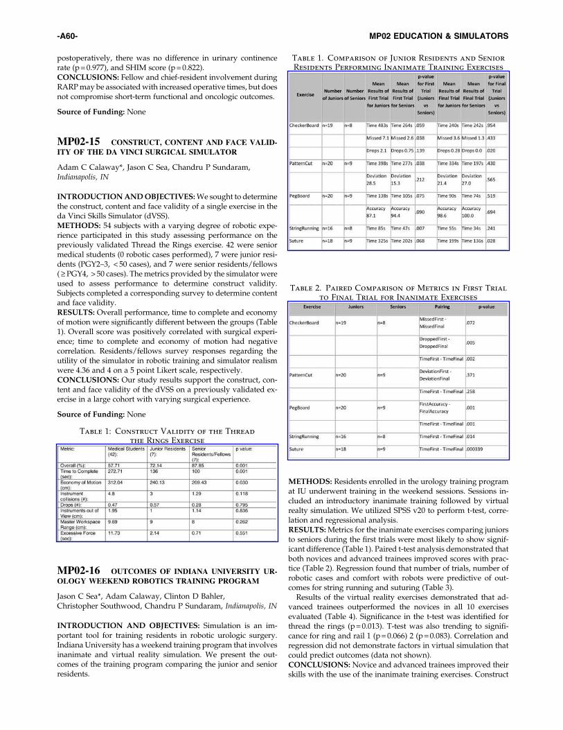
postoperatively, there was no difference in urinary continencerate (p = 0.977), and SHIM score (p = 0.822).CONCLUSIONS: Fellow and chief-resident involvement duringRARP may be associated with increased operative times, but doesnot compromise short-term functional and oncologic outcomes.
Source of Funding: None
MP02-15 CONSTRUCT, CONTENT AND FACE VALID-
ITY OF THE DA VINCI SURGICAL SIMULATOR
Adam C Calaway*, Jason C Sea, Chandru P Sundaram,Indianapolis, IN
INTRODUCTION AND OBJECTIVES: We sought to determinethe construct, content and face validity of a single exercise in theda Vinci Skills Simulator (dVSS).METHODS: 54 subjects with a varying degree of robotic expe-rience participated in this study assessing performance on thepreviously validated Thread the Rings exercise. 42 were seniormedical students (0 robotic cases performed), 7 were junior resi-dents (PGY2–3, < 50 cases), and 7 were senior residents/fellows( ‡ PGY4, > 50 cases). The metrics provided by the simulator wereused to assess performance to determine construct validity.Subjects completed a corresponding survey to determine contentand face validity.RESULTS: Overall performance, time to complete and economyof motion were significantly different between the groups (Table1). Overall score was positively correlated with surgical experi-ence; time to complete and economy of motion had negativecorrelation. Residents/fellows survey responses regarding theutility of the simulator in robotic training and simulator realismwere 4.36 and 4 on a 5 point Likert scale, respectively.CONCLUSIONS: Our study results support the construct, con-tent and face validity of the dVSS on a previously validated ex-ercise in a large cohort with varying surgical experience.
Source of Funding: None
MP02-16 OUTCOMES OF INDIANA UNIVERSITY UR-
OLOGY WEEKEND ROBOTICS TRAINING PROGRAM
Jason C Sea*, Adam Calaway, Clinton D Bahler,Christopher Southwood, Chandru P Sundaram, Indianapolis, IN
INTRODUCTION AND OBJECTIVES: Simulation is an im-portant tool for training residents in robotic urologic surgery.Indiana University has a weekend training program that involvesinanimate and virtual reality simulation. We present the out-comes of the training program comparing the junior and seniorresidents.
METHODS: Residents enrolled in the urology training programat IU underwent training in the weekend sessions. Sessions in-cluded an introductory inanimate training followed by virtualrealty simulation. We utilized SPSS v20 to perform t-test, corre-lation and regressional analysis.RESULTS: Metrics for the inanimate exercises comparing juniorsto seniors during the first trials were most likely to show signif-icant difference (Table 1). Paired t-test analysis demonstrated thatboth novices and advanced trainees improved scores with prac-tice (Table 2). Regression found that number of trials, number ofrobotic cases and comfort with robots were predictive of out-comes for string running and suturing (Table 3).
Results of the virtual reality exercises demonstrated that ad-vanced trainees outperformed the novices in all 10 exercisesevaluated (Table 4). Significance in the t-test was identified forthread the rings (p = 0.013). T-test was also trending to signifi-cance for ring and rail 1 (p = 0.066) 2 (p = 0.083). Correlation andregression did not demonstrate factors in virtual simulation thatcould predict outcomes (data not shown).CONCLUSIONS: Novice and advanced trainees improved theirskills with the use of the inanimate training exercises. Construct
Table 1: Construct Validity of the Thread
the Rings Exercise
Table 1. Comparison of Junior Residents and Senior
Residents Performing Inanimate Training Exercises
Table 2. Paired Comparison of Metrics in First Trial
to Final Trial for Inanimate Exercises
-A60- MP02 EDUCATION & SIMULATORS

validity was present as the advanced trainees performed betterthan the novices.
Source of Funding: none
MP02-17 COMPARISON OF COMPUTER GENERATED
PERFORMANCE METRICS IN THE DAVINCI SKILLS
SIMULATOR- WHICH DEMONSTRATE THE MOST CON-
STRUCT VALIDITY?
Ryan Dorin*, Kyle Finnegan, Halil Kiziloz, Steven Shichman,Hartford, CT
INTRODUCTION AND OBJECTIVES: Previous studies havedemonstrated construct validity of the daVinci Skills Simulator(Intuitive Surgical, Sunnyvale, CA) for differentiating novice,intermediate, and advanced robotic surgeons in overall score andtime to completion of simulator tasks. Our purpose is to deter-mine the relative construct validity of specific performance met-rics within the Skills Simulator scoring system, therebyelucidating which metrics may be particularly useful to incor-porate in a robotic surgery training curriculum.METHODS: Thirty-nine surgeons (18 group I [0–20 roboticcases]; 8 group II [21–150 robotic cases]; and 13 group III [ > 150robotic cases]) were enrolled from September 2010 to December2010. Participants completed 24 virtual-reality exercises on the daVinci Skills Simulator. Data on 12 performance metrics was col-lected by the software. The ability of each of the 12 metrics todifferentiate between surgeon experience groups was analyzedusing univariate general linear model and analysis of variancetests.RESULTS: Of the 12 metrics, 7 were applicable to all 24 exercises,with 5 metrics (i.e. misapplied energy time) only being applicableto select exercises. Of the 7 universally applied metrics, scores on‘Time to Complete,’ ‘Economy of Motion,’ and ‘Work SpaceRange’ were significantly different between Group I and Group IIsurgeons across the greatest number of exercises. Only ‘Time toComplete’ demonstrated significant differentiation betweenGroup II and Group III surgeons. Five of the seven simulatormetrics demonstrated significant construct validity in differenti-ating between all three subgroups.CONCLUSIONS: Within the daVinci Simulator exercise library,metrics relating to efficient use of the console workspace weremost effective in differentiating novice surgeons from interme-diate surgeons, while the speed metric was most effective indifferentiating intermediate from expert surgeons. A simulatorcurriculum focusing on economy of motion and efficient clutch-ing may be especially effective in training the novice roboticsurgeon.
Source of Funding: Intuitive Surgical- Sunnyvale, CA
MP02-18 ASSESSMENT OF ROBOTIC SIMULATION
USE IN RESIDENCY PROGRAMS OF THE SOUTHEASTERN
SECTION OF THE AMERICAN UROLOGIC ASSOCIATION
Abby Taylor*, David Thiel, Jacksonville, FL, Vipul Patel,Celebration, FL, Todd Larson, Seattle, WA, Amy Lannen,Jacksonville, FL, Raymond Leveilee, Coral Gables, FL
INTRODUCTION AND OBJECTIVES: To assess SoutheastSection of the American Urological Association (SESAUA) traineeexposure to and thoughts on robotic simulation.METHODS: 3 trainees from each SESAUA residency trainingprograms were invited to Orlando, Fl. for a formal 2-day robotictraining course. Day 1 was a 3 component didactic session. Day 2involved faculty direction to trainees in set tasks for 4 hours on alive porcine model and 4 hours on the Mimic dV-trainerTM(Mimic Technologies, Inc., Seattle, WA). 32 trainees from 14programs in the SESAUA participated in the course and filled outa 1 page, 8 item questionnaire following their simulator exposure.RESULTS: 53.1% (17/32) had never had robotic console timeduring their residency training. 25% (8/32) of trainees rated theirrobotic training during residency as excellent, of which 4 hadaccess to a simulator, while 21.9% (7/32) rated their training aspoor, of which 4 had access to a simulator. 65.6% (21/32) oftrainees had access to the Mimic dV trainer or Mimic ‘‘backpack’’,while 31.3% (10/32) of the trainees had no prior exposure to
Table 3. Regressional Analysis of Factors Predicting
Outcomes in Inanimate Training
Table 4. Comparison of Junior and Senior Residents
Undergoing Virtual Simulator Exercises
MP02 EDUCATION & SIMULATORS -A61-

robotic simulation. 84.4% (27/32) of the trainees felt the simulatorreplicated real life robotic console surgery and 90.6% (29/32) feltthe simulator was, or would be, helpful for training in theirprogram. Trainees felt the ‘‘tubes 2’’ drill, which mimics a vesico-urethral anastomosis, was the most difficult drill to perform.CONCLUSIONS: A majority of trainees in the SESAUA havehad none to limited robotic console time during their urologyresidency program. A high number of trainees in the SESAUAhave exposure to virtual reality robotic simulation. Trainees be-lieve that the simulator replicates real life robotic console move-ments and almost all believe they would benefit from havingaccess to robotic simulation.
Source of Funding: None
MP02-19 PERCEIVING A LIVE CASE DEMONSTRA-
TION: PERCEPTION OF BENEFIT
Sammy Elsamra*, Hector Motato, Justin Friedlander,Daniel Moreira, Arvin George, Brian Duty, Arthur Smith,Zeph Okeke, New Hyde Park, NY
INTRODUCTION AND OBJECTIVES: Many societies utilizelive case demonstrations (LCD) at meetings. The educationalbenefit LCDs is unclear. Therefore we sought to evaluate per-spectives of urologists viewing live and taped case demonstra-tions (TCD).METHODS: A 15-question anonymous survey was distributedto attendees of the live surgery session at the AUA 2012 nationalmeeting (Atlanta) and the 3rd International Challenges in En-dourology meeting (Paris).RESULTS: Of 1000 surveys distributed, 253 were returnedcompleted. Nearly half of respondents were in academic practicesetting and nearly 75% were beyond training. Just over 30% hadperformed a LCD previously. The perceived benefit of an LCDwas greater than unedited and edited videos (chi-squaredp = 0.0138 and p = 0.0001, respectively). Nearly no one selected‘‘not helpful’’ and few selected minimally helpful for any of thethree modalities. The majority of respondents identified that op-portunity to ask questions (61%) and having access to the fullunedited version (72%), two features inherent to LCD, improvedupon the educational benefit of edited videos. The majority (78%)identified LCD as ethical. However, those that did not perceivedlower educational benefit from LCD (p = 0.019). A slim majority(58%) would allow themselves or a family member to be a patientof a LCD. The vast majority (86%) plan to transfer knowledgegained at the LCD session into their practice.CONCLUSIONS: Urologists who attended these LCD sessionsidentified LCDs as beneficial and applicable to their practice.LCDs are preferred over videos. The large majority considersLCD ethical though not as many would volunteer themselves forLCD. Further studies are necessary to determine if there is actualbenefit from LCD over TCD to patient care.
Source of Funding: None
MP02-20 ELECTRONIC DATA COLLECTION FOR
PATIENT-REPORTED OUTCOMES IN MEN WITH PROS-
TATE CANCER: ASSESSING EASE OF USE AND PATIENT
SATISFACTION
Brian Benway*, Leslie McIntosh, Linda Black, Joanne Morley,Sheri Long, Patricia Carter, Elizabeth Jones, Alethea Paradis,Arnold Bullock, Gerald Andriole, Saint Louis, MO
INTRODUCTION AND OBJECTIVES: The Expanded ProstateCancer Index – Composite for Clinical Practice (EPIC-CP) is avalidated questionnaire which assesses patient-reported quality-of-life (QOL) measures related to prostate cancer and its man-agement. We have previously reported equivalence betweentraditional paper surveys and electronic data collection via iPadwith regards to survey completion time, as well as a two-foldincrease in accuracy of data collection using the electronic format.With this in mind, we aimed to evaluate the ease of use andoverall patient satisfaction associated with an electronic formatfor questionnaire completion, compared to traditional paperquestionnaires.METHODS: 226 men with prostate cancer were asked to com-plete either a traditional paper EPIC-CP survey, or an electronicsurvey administered on an iPad. After completing the surveys,each was assessed for completion, and patients were asked to fillout a questionnaire regarding their experience, focusing uponsatisfaction and comfort with the method of collection, in addi-tion to a self-assessment of computer literacy. Scores were ratedon a 1–10 scale, with 10 indicating high satisfaction or proficiency.RESULTS: 113 men completed the paper version of the EPIC-CPquestionnaire, and 113 completed the electronic version. Patientsfound both the iPad and paper formats easy to use (mean ease-of-use scores of 9.14 vs. 8.80, respectively, p = 0.19), although meansatisfaction was significantly higher in patients who completedthe iPad version of the survey (8.50 vs. 7.75, p = 0.04). Patientswho completed the iPad version rated themselves as more com-puter-savvy (7.56 vs. 4.88, p < 0.001); however, when a subsetanalysis was performed of iPad respondents, comparing patientswho rated their computer proficiency as novice, intermediate, orexpert, no difference in mean satisfaction (6.89 vs. 8.51 vs. 8.61,respectively, p = 0.19) or ease-of-use (8.56 vs. 9.10 vs. 9.27,p = 0.54) was noted, indicating that novices were just as adept asexperts at completing the electronic questionnaire.CONCLUSIONS: In addition to providing a highly-accuratemethod for data collection, the use of an electronic format forcompletion of the EPIC-CP questionnaire is associated withgreater patient satisfaction, even for patients with minimal com-puter literacy.
Source of Funding: Midwest Stone Institute Research Fund
MP02-21 IMPACT OF PAST LAPAROSCOPIC EX-
PERIENCE ON ROBOTIC PERFORMANCE
Roger Smith, Haidar Abdul-Muhsin*, Vipul Patel, Celebration, FL
INTRODUCTION AND OBJECTIVES: Objective: To determinethe degree to which a surgeon’s years of laparoscopic experiencecontribute to the acquisition of robotic surgical skills and theachievement of proficiency.METHODS: Surgeons were tested in their ability to perform fourdifferent simulated robotic surgical skills using the dV-Trainersimulator (Mimic Technologies, Inc., Seattle, WA) of the da VinciSurgical System (Intuitive Surgical Inc., Sunnyvale, CA). Thesubjects completed a pre-test questionnaire to provide demo-graphic and experience data, which included the number of yearsof practice in both laparoscopic and robotic surgery. Each subjectperformed four exercises using the simulator: ring board, cameratargeting, thread the rings, and energy dissection. The simulatorcollected multiple performance metrics during each of the exer-cises. A Pearson’s correlation was calculated on the relationshipbetween the number of years of laparoscopic and robotic expe-rience (independent variables) with their overall proficiency scoreon the robotic simulator (dependent variable).
-A62- MP02 EDUCATION & SIMULATORS

RESULTS: A total of 54 subjects participated in the experimentand 42 completed all four tasks in the robotic simulator. Thesubjects reported a range of experience in laparoscopic surgerybetween 4 and 34 years, and in robotic surgery between 0 and 11years. Subjects indicating zero years of robotic experience wereexcluded from the study, reducing the sample size to 30 surgeons.Using the Pearson Product Moment Correlation with 28 degreesof freedom and a = 0.05, a significant correlation threshold of0.349 was established. There was a statistically significant nega-tive correlation between years of laparoscopic experience and theoverall proficiency score in two of the four robotic surgery exer-cises (ring board = - 0.361; thread the rings = - 0.454) and a neg-ative correlation which did not achieve statistical significance inthe two remaining exercises (camera targeting = - 0.152; energydissection = - 0.228).CONCLUSIONS: Using a robotic simulator to measure the pro-ficiency of surgeons with both laparoscopic and robotic surgicalexperience we found a statistically significant negative correlationbetween the number of years of laparoscopic experience andproficiency in two of four exercises and a trend toward a negativecorrelation in the other two exercises. This analysis suggests thatextensive laparoscopic experience may have a negative impact onthe learning curve associated with robotic surgery.
Source of Funding: Department of Defense
MP02-22 UTILIZATION OF ROBOTIC SIMULATORS
FOR UROLOGIC TRAINING
Edan Shapiro*, Ari Bergman, Rus Korets, Trushar Patel,Ketan Badani, New York, NY
INTRODUCTION AND OBJECTIVES: Computer-based sim-ulators are being increasingly utilized in surgical training tosupplement actual surgical experience. However, with work hourrestrictions and other demands, it is challenging for residents tooptimize their simulator experience during training. We soughtto determine the utilization patterns of two robotic simulationtraining devices among urology residents.METHODS: We enrolled all Urology residents in their PGY2-PGY5 years of training. Participants underwent an orientationsession, completed a baseline survey, and a baseline skill levelassessment (mScore� on Needle Control task). The participantswere then instructed to complete a total of 4 hours of training over2 months, which could be completed on either the dV-Trainer(Mimic Technologies) or on the daVinci robotic console (IntuitiveSurgical); the choice of which training platform to use was at theparticipant’s discretion. Following the 2 month training period,participants were asked to complete an online survey describingtheir experience and delineate the motivating factors in choosingthe time and place of the training. In addition, the skill assessmentwas repeated and participants completed an exit questionnaire.Utilization patterns were analyzed to determine which factorswere associated with skill acquisition.RESULTS: Thirteen residents were enrolled. Overall, the daVinciplatform was utilized for 75% of the total training time vs. 25% forthe dv-Trainer. Of the subjects that used the daVinci platform totrain, 100% reported that it was their first choice, compared to only28% of those that used the dv-Trainer. Primary indications forchoosing the daVinci included fidelity, comfort, and ease of use;availability was the primary indication for using the dv-Trainer.Baseline skill mScore� was 72.5 (24–93), which increased to 78.6(50–95) following the 2 month training period. When stratified bylevel of training, the increase among junior residents (65.5/75.5;15% change) was more pronounced that that for senior residents
(78.4/82.4; 5% increase). Overall, 85% of participants viewed theexperience favorably, 100% thought it helped performance, and82% felt simulation should be mandated in training. Of note, 100%of residents preferred the daVinci console even though 55% felt thedV-Trainer was more readily available.CONCLUSIONS: A standardized simulator training programcan help improve robotic skill, especially for junior residents withminimal experience. The majority of residents preferred the da-Vinci console to the dv-Trainer even though the dv-Trainer wasmore readily available.
Source of Funding: None
MP02-23 A SYSTEMATIC REVIEW OF TECHNOLOGY-
DRIVEN SIMULATORS FOR MEDICAL STUDENTS
Michael Michael, Stevenage, United Kingdom, Hamid Abboudi,Brighton, United Kingdom, Jean Ker, Dundee, United Kingdom,Kamran Ahmed, Prokar Dasgupta, London, United Kingdom,Ali Bahsoun*, Please choose an option below
INTRODUCTION AND OBJECTIVES: Simulation based edu-cation has evolved as a key training tool in high-risk industriessuch as aviation and the military. In parallel with these industries,the benefits of incorporating specialty-oriented simulation train-ing within medical schools are vast. Adoption of simulators intomedical school education programs has shown great promise andis attracting much interest among medical educators. We ana-lysed randomised control studies in order to identify various‘Technology-Driven’ simulators used in undergraduate medicaleducation. A validity framework incorporating certain aspectsfrom the ‘Framework For Technology Enhanced Learning’ reportby the Department of Health was used to evaluate the capabilitiesof each technology-driven simulator.METHODS: An English literature search was carried out usingMEDLINE, EMBASE and psychINFO databases. Information wascollected regarding the simulator type, characteristics and brandname. Where possible, we extracted information from the studies onthe simulators’ performance with respect to validity status, reliabil-ity, feasibility, education impact, acceptability and cost effectiveness.RESULTS: We identified 19 studies, analysing simulators formedical students across a variety of procedure-based specialitiesincluding; cardiovascular (n-2), endoscopy (n-3), laparoscopicsurgery (n-8), vascular access (n-2), ophthalmology (n-1), obstet-rics and gynaecology (n-1), anaesthesia (n-1) and paediatrics (n-1). Incorporation of simulators has so far been on an institutionallevel; no national or international trends have yet emerged.CONCLUSIONS: Simulators are capable of providing a highlyeducational and realistic experience for the medical studentswithin a variety of speciality-oriented teaching sessions. Furtherresearch is needed to establish how best to incorporate simulatorsinto a more primary stage of medical education; pre-clinical andclinical undergraduate medicine.
Source of Funding: none
MP02-24 LAPAROSCOPIC RENAL RESECTION
TRAINING IN CADAVERS EMBALMED USING THIEL’S
METHOD: DEVELOPMENT AND EVALUATION OF SKILLS
LEARNING
Sarvpreet Singh Ubee*, Wolverhampton, United Kingdom,Benjie Tang, Roos Eisma, Dundee, United Kingdom,Chandrashekhar Biyani, Wakefield, United Kingdom,Ghulam Nabi, Dundee, United Kingdom
MP02 EDUCATION & SIMULATORS -A63-

INTRODUCTION AND OBJECTIVES: Thiel’s embalmingmethod is less known, however it preserves natural colours,texture and flexibility; thus overcoming the limitations of for-malin embalming. These qualities place this close to living tissuetherefore an ideal training model for minimally invasive surgical(MAS) training. The teaching model was implemented as part ofBAUS endorsed laparoscopic training courses for the past 2 years.We report assessment of surgical anatomy, tissue planes, con-sistency, smell and satisfaction with performance of procedure byparticipants and course faculty.METHODS: In a prospective descriptive study, 14 participantsand 4 experts carried out transperitoneal or retroperitoneal lap-aroscopic left or right nephrectomy. Questionnaires previouslyvalidated for other surgical courses were used.1,2 Responseswere recorded using five-point Likert satisfaction scale alongwithBAUS feedback forms. Mean scores for each step were comparedbetween delegates and faculty.RESULTS: Faculty members were laparoscopic surgeons withteaching and training experience of more than three years. De-legates were in the advanced stage of their training with experi-ence in basic laparoscopy. Table-1 summarises the results from
questionnaire responses. The anatomy, tissue consistency, ana-tomical planes were very well preserved with mean score of4.25 + / - 0.25. All participants were highly satisfied with surgi-cal quality of the tissues. Similar scores were reported by facultyon the course.CONCLUSIONS: Thiel’s embalming method preserves manyfeatures of human anatomy and the present study showed ex-cellent preservation of anatomical planes, tissue consistency andlandmarks as consistently reported by participants and faculty.This resource could be used to address the challenges of surgicaltraining, particularly MAS in the UK.
References:1) Eisma R, et al. A comparison of Thiel and formalin embalmed
cadavers for thyroid surgery training. Surgeon 2011, 9:1426.2) Rai BP, et al. A Qualitative Assessment of Human Cadavers
Embalmed by Thiel’s Method Used in Laparoscopic Trainingfor Renal Resection. Anat Sci Educ 2012, 5: 182–6.
Source of Funding: University of Dundee
MP02-25 CONSTRUCTION AND ASSESSMENT OF A
NOVEL INDIGENOUS PCNL SIMULATOR: AN IN-
NOVATIVE APPROACH TO TRAINING
Ashish Rawandale*, Lokesh Patni, Atul Mulay, Preeti Patil,Dhule, India
INTRODUCTION AND OBJECTIVES: PCNL has a significantlearning curve. Commercial simulators have prohibitive costs/pitfalls. Hence we patented, constructed and evaluated our in-digenous PCNL simulator.METHODS: Our portable fluoroscopy compatible simulator (fig 1)was designed using CAD software;patented and constructed.It uses the usual PCNL instruments, replicates natural tissue-haptics, has various error alarms, simulates respiratory movementsand uses regular uroendoscope to confirm successful puncture.
Simulator evaluation:‘‘Simulator orientation and puncture demonstration’’ was
performed by PCNL expert (control).13 trainees, underwent a 3-step test in the operating room.
Step 1: 3 successful punctures were performed and ‘‘Endpoints’’ measured
Step 2. Two practice sessions (30 mins each) givenStep 3. Repeat test as in step 1
Pre and post test subjective performance was accessed with aLikert 5 point GRS scaleTrainee feedback form analysed
RESULTS: Table 1Trainees demonstrated statistically significant improvement
(SPSS analysis) in the GRS scores, total time, fluoroscopic time
Table - 1. Summary of the evaluation scores
-A64- MP02 EDUCATION & SIMULATORS

and attempted needle punctures after training (p 0.001). Para-meters showed a rightward shift. Data helped quantify the indi-vidual training hours mandatory to reach the desired expertise.Subjective simulator assessment indicated a high degree of sat-isfaction on effectiveness of simulator.CONCLUSIONS: Our PCNL simulator is an efficient means ofskill acquisition. It provides an opportunity for supervised, re-petitive performance of essential technical skills in a controlled,low stress, OR environment. It allows trainee evaluation, provi-des tailored training sessions and has the potential to decrease thelearning curve for skill acquisition in order to maximise traineeperformance. It has a low initial and maintenance cost. Devel-oping such simulators may open up further new avenues inurology trainee education.
Source of Funding: none
MP02-26 EFFECTS OF RESIDENCY TRAINING ON OP-
ERATING ROOM TIME: A REVIEW OF PROSTATE BIOPSIES
IN A RESIDENT-RUN CLINIC AT A CITY HOSPITAL
Allison Polland*, New York, NY, Alfred Winkler, Elmhurst, NY
INTRODUCTION AND OBJECTIVES: Residents are vital tothe clinical workforce in academic hospitals; residency programs
aim to train residents through rotations in which, under super-vision, residents provide the majority of clinical care. ElmhurstHospital Center is a city hospital affiliated with the Icahn Schoolof Medicine at Mount Sinai and one of three primary sites forresident urological training. Prostate biopsies are performed in aminor operating room (OR) setting by first-year urology residentswith chief resident and attending supervision. Prior to July of thefirst year, residents have minimal experience with ultrasound andno simulator training with transrectal US. The purpose of thisstudy was to examine the effects of time of year, a proxy forresidency training, on OR procedure time for transrectal ultra-sound-guided prostate biopsy.METHODS: The Elmhurst Hospital Center Surgical Databasewas queried for transrectal ultrasound-guided prostate biopsyprocedures from January of 2010 to December of 2012. Case du-ration was recorded in minutes. A student’s t-test was performedto determine if OR procedure time early in the academic year wassignificantly different from OR procedure time late in the aca-demic year.RESULTS: A total of 222 transrectal ultrasound-guided prostatebiopsies were performed. The minimum procedure time was 5minutes and the maximum procedure time was 85 minutes. Therewas a significant difference in length of procedure between July,the month in which residents had least training and June, themonth in which residents had most training (p = 0.027) on two-tailed t-test. Median procedure time decreased by almost fiftypercent, from 20 minutes in July to 11 minutes in June.CONCLUSIONS: Resident experience is significantly associatedwith length of procedure in transrectal ultrasound-guided pros-tate biopsies. This may be related to time required for teachingand learning curve. In addition to potential patient risk related tolonger procedure time, there are financial implications of in-creased OR time and need for staffing. Although transrectal ul-trasound simulators exist, they are not widely used or required inresidency training. Additional resident simulation training, spe-cifically for procedures which utilize transrectal imaging tech-nology, may mitigate the effects of early training on ORprocedure time.
Source of Funding: None
MP03 BPH/LUTS I
MP03-01 UROBEAM� DIODE LASER VAPORIZATION
OF THE PROSTATE: MID-TERM OUTCOMES
Joao Padua Manzano*, Frederico Teixeira Barbosa,José Ricardo Cruz Silvino Jr, Luciano Salles Lage,Adalberto Andriolo Jr, Roberto Soler,Joaquim Francisco De Almeida Claro, Sao Paulo, Brazil
INTRODUCTION AND OBJECTIVES: Evaluate the short-termoutcome and prevalence of perioperative complications of the newsurgical alternative for men with LUTS due to benign prostaticenlargement (BPE), the vaporization of the prostate (VP) withMedilas D UroBeam high power 940 nm Diode laser (UroBeam).METHODS: Prospective data were collected from 114 consecu-tive patients undergoing UroBeam VP at our institution fromJanuary 2011 to January 2013. Perioperative data and surgery-related complications, and 30-day efficacy and safety assessments
were documented. All the surgeries were performed by the sameexperienced surgeon. Comparative analysis between pre andpost operative data was conducted using paired Student’s t test.RESULTS: UroBeam laser VP was successfully performed in allpatients with no intraoperative complications. The mean – SDprostate volume was 68.3 – 29.9 mL (range 25–149 g). Mean op-erative time, proportion of patients needing bladder irrigationand catheterization time was 65.9 – 23.7 minutes (40–130 min),30.7% (35 patients) and 22.1 – 18.8 hours (6–120 h), respectively.Median applied laser energy was 334.7 kJ (85–827 kJ). Hospitali-zation time was 20.8 hours (range 10–96 hours). 30-day rate ofrehospitalization was 2.63% (3 patients), of retreatment was1.75% (2 patient) and of emergency room visits was 10.5% (12patients). Urethral stricture developed in four patients (3.5%), andbladder neck stricture in five patient (4.38%). Temporary urinaryincontinence occurred in 6 patients (5.26%), stress incontinenceand urge-incontinence occurred in 4 patients (3.5%). Nineteen
MP03 BPH/LUTS I -A65-

patients had early urinary retention (16.6%) and two had lateurinary retention (1.75%). Irritative symptoms seemed not tohave impacted quality of life. No patients required blood trans-fusion or had TUR syndrome. Statistically significant improve-ments were noted in all key parameters postoperatively: Qmax(5.1 – 2.8 vs 16 – 5.3 mL/s; p < 0.0001), IPSS (25 – 4.5 vs 6.6 – 2.2;p < 0.0001) IPSS-QoL (4.5 – 1.0 vs 1.6 – 0.67; p < 0.0001), PVR(170 – 180 vs 19 – 8.8 mL; p = 0.004) and PSA (3.8 – 5.5 vs1.0 – 0.58 ng/mL; p < 0.0001).CONCLUSIONS: The UroBeam high power diode laser is aneffective and safe new treatment option for patients with LUTS-BPE. Complications rates seem to be similar to those reportedwith TURP. Further advantages seem to be the short hospital stayand bladder irrigation and catheterization times. Further ran-domized controlled studies are needed to confirm these data.
Source of Funding: none
MP03-02 IS COMBINED PROSTATE RESECTION
WITH CYSTOLITHOTRIPSY BENEFICIAL FOR BPH PA-
TIENTS WITH BLADDER CALCULI? -A NATIONWIDE PO-
PULATION-BASED STUDY
Eric Yi-Hsiu Huang*, Tzu-Ting Kuo, Hsiao-Jen Chung,Chih-Chieh Lin, Alex TL Lin, Kuang-Kuo Chen, Taipei, Taiwan
INTRODUCTION AND OBJECTIVES: Benign prostatic hy-perplasia (BPH) related urinary stasis has been regarded as theprecipitating factor of bladder calculi formation in male pa-tients. As such, transurethral resection of prostate (TURP) isusually advocated to be performed with cystolithotripsy si-multaneously in BPH patients with bladder calculi. We inves-tigated the recurrence rate and recurrence interval of bladdercalculi in this patient cohort by analyzing a nationwide popu-lation-based database.METHODS: We obtained a cohort dataset from ¡ULongitudinalHealth Insurance Database 2005¡f (LHID 2005) of Taiwan, whichcontains 1,000,000 individuals randomly sampled from 25.68million Beneficiaries in Taiwan National Health Insurance Pro-gram. There was no significant difference in the gender and agedistribution between the patients in the LHID 2005 and theoriginal database. The patients underwent cystolithotripsy with/without TURP were identified from the claim data (n = 1,737). Thepatients having the diagnosis of ureteral stone within 3 monthsprior to TURP, no definite diagnosis of BPH (ICD-9-CM code600.0), or accepted repeated TURP during post-operative follow-up were excluded. The recurrence of bladder calcluli was definedby the occurrence of second time cystolithotripsy.RESULTS: There were 523 patients identified, 221 received cy-stolithotripsy alone and 302 received concurrent TURP. The re-currence rates of bladder calculi were 12.2% and 11.9%,respectively (p = 0.918). The median interval of stone recurrencewere 3 and 4 years for each (p = 0.085). The patients with smallresection weight of prostate ( £ 15 g) have similar stone recurrencerate as compared to patients receiving cystolithotripsy alone(13.7% vs 12.2%, p = 0.626), whereas others with large resectionweight of prostate ( > 15 g) have significantly lower recurrencerate (2.1% vs 12.2%, p = 0.037).CONCLUSIONS: For the patients with small resection weight ofprostate, concurrent TURP and cystolithotripsy may not be ben-eficial as compared to the patients accepted cystolithotripsy alonein terms of reducing the recurrence of bladder calculi andprolonging recurrence interval.
Source of Funding: Taipei Veterans General Hospital
MP03-03 BASELINE CHARACTERISTICS PREDICT
RISK OF PROGRESSION AND RESPONSE TO COMBINA-
TION MEDICAL THERAPY FOR BENIGN PROSTATIC HY-
PERPLASIA
Michael Kozminski*, John Wei, Ann Arbor, MI, Jason Nelson,David Kent, Boston, MA
INTRODUCTION AND OBJECTIVES: Multiple risk factorshave been identified for the progression of benign prostatic hy-perplasia (BPH). Current practice patterns and national guide-lines fail to account for the evidence behind these factors andarguably result in overtreatment. We sought to derive a nomo-gram based on data from the Medical Therapy of ProstaticSymptoms (MTOPS) trial to help optimize decision making formen with moderate to severe lower urinary tract symptoms(LUTS) secondary to BPH.METHODS: We conducted secondary analysis of MTOPS data.In short, MTOPS was a randomized trial evaluating doxazosin,finasteride, or the combination of these medications for risk ofBPH progression in men ‡ 50 years with AUA-SI scores of 8–30and Qmax of 4–15 mL/sec. Using bivariate tests, we identifiedsignificant factors that would predict BPH progression, as de-fined by MTOPS. We then utilized these factors in Cox
Table 1. Cox Proportional Hazards Model for Risk
of BPH Progression, Divided by Quartile
-A66- MP03 BPH/LUTS I

Proportional Hazard models to 1) more comprehensively riskstratify the study population based on pre-treatment parametersand 2) to establish a nomogram for clinical use.RESULTS: 3047 men were followed in MTOPS for a mean of 4.5years. Baseline post-void residual (per 10 mL, HR 1.015, 95% CI1.004-1.026), PSA (HR 1.082, 95% CI 1.033–1.133), age (per 10 yrs,HR 1.320, 95% CI 1.140–1.529), AUA-SI score (HR 0.974, 95% CI0.956–0.993), and Qmax (per 5 mL/sec, HR 0.752, 95% CI 0.613-0.924) were found to be significantly correlated with overall BPHprogression in multivariable analysis. This predictive model ofrisk for BPH progression (AUC = 0.62) demonstrated varyingrisks of progression across quartiles (see Table). The highest-riskquartile had a risk of progression approximately three times thatof the lowest within all trial arms.CONCLUSIONS: MTOPS data suggests that the benefits ofmedical therapy for BPH are unevenly distributed with men whowere in the highest risk strata accounting for the greatest clinicalbenefit. Importantly, utilizing commonly available, baseline riskfactors permits estimation of individual patient risk for clinicalprogression. A novel nomogram based on these analyses willallow clinicians to better select those most likely to benefit frommedical therapy for BPH. Potential treatment effect can then beweighed against possible costs and risks of adverse effects for agiven patient.
Source of Funding: PCORIThe MTOPS study was conducted by the MTOPS Investigators
and supported by the National Institute of Diabetes and Digestiveand Kidney Diseases (NIDDK). The data and samples fromMTOPS reported here were supplied by the NIDDK Central Re-positories. This manuscript was not prepared in collaborationwith Investigators of the MTOPS study and does not necessarilyreflect the opinions or views of the MTOPS study, the NIDDKCentral Repositories, or the NIDDK.
MP03-04 TWO-YEAR PROSPECTIVE, RANDOMIZED
COMPARISON BETWEEN THE BIPOLAR PLASMA EN-
UCLEATION OF THE PROSTATE AND OPEN PROSTA-
TECTOMY IN BPH CASES OVER 80 ML
Bogdan Geavlete*, Florin Stanescu, Cristian Moldoveanu,Marian Jecu, Leon Adou, Petrisor Geavlete, Bucharest, Romania
INTRODUCTION AND OBJECTIVES: This long term, pro-spective, randomized trial aimed to evaluate the viability of thebipolar plasma enucleation of the prostate (BPEP) by comparisonto open transvesical prostatectomy (OP) in cases of large prostatesconcerning surgical efficacy, perioperative morbidity and follow-up parameters.METHODS: A total of 140 benign prostatic hyperplasia (BPH)patients with prostate volume over 80 mL, maximum flow rate(Qmax) below 10 mL/s and International Prostate SymptomScore (IPSS) over 19 were randomized in the 2 study arms (70cases each). All patients were assessed preoperatively and every 6months after surgery for a period of 2 years by IPSS, Qmax,quality of life score (QoL), post-voiding residual urinary volume(PVR), postoperative prostate volume and PSA level.RESULTS: The two series were characterized by similar pre-operative parameters, including the prostate volume (132.6versus 129.7 mL). The BPEP and OP techniques emphasizedsimilar mean operating times (91.4 versus 87.5 minutes) andresected tissue weights (108.3 versus 115.4 grams). The post-operative hematuria rate (2.9% versus 12.9%) as well as themean hemoglobin drop (1.7 versus 3.1 g/dL), catheterizationperiod (1.5 versus 5.8 days) and hospital stay (2.1 versus 6.9
days) were significantly improved for BPEP. The re-catheteri-zation for acute urinary retention was more frequent in the OPgroup (8.6% versus 1.4%), while the rates of early irritativesymptoms were similar for BPEP and OP (11.4% versus 7.1%).During the 2 year’ follow-up period, no statistically significantdifference was determined in terms of IPSS, Qmax, QoL, PVR,PSA level and postoperative prostate volume between the twoseries. Consequently, the calculated prostate volume decrease(83.0–84.7% versus 82.1–83.9%) and PSA level reduction (90.6–92.5% versus 90.1–92.6%) by comparison to the preoperativemeasurements were statistically equivalent for the BPEP and OPgroups.CONCLUSIONS: BPEP represents a promising endoscopic ap-proach in large BPH cases, characterized by good surgical effi-ciency and similar BPH tissue removal capabilities whencompared to standard open prostatectomy. Plasma enucleationpatients benefited from significantly reduced complications,shorter convalescence period and satisfactory long term follow-up symptom scores and voiding parameters.
Source of Funding: None
MP03-05 LEARNING CURVE OF MORCELLATION
DURING HOLMIUM LASER ENUCLEATION OF THE
PROSTATE (HOLEP)
Jin Kyu Oh, Incheon, Korea, Republic of, Hahn-Ey Lee*,Jae Hyun Jung, Chang Wook Jeong, Jae-Seung Paick,Seung-June Oh, Seoul, Korea, Republic of
INTRODUCTION AND OBJECTIVES: Morcellation of theenucleated adenoma is an essential step of holmium laser enu-cleation of the prostate (HoLEP). However, few results have beenreported about the learning curve of the morcellation procedure.We aimed to analyze our results and assess the learning curve ofmorcellation of the enucleated adenoma.METHODS: We retrospectively reviewed the data of patientswho underwent HoLEPs performed by two surgeons (SJO & JSP)between July 2008 and August 2010. We assessed the cut-off pointof the learning curve of morcellation. We analyzed preoperativeand intraoperative parameters and the incidence of in-traoperative complications related to morcellation.RESULTS: A total of 339 patients whose mean age was 68.3( – 6.5, SD) years and prostate volume was 55.6 ( – 23.6) ml wereincluded. The mean total operation time, morcellation time andweight of the enucleated adenoma were 75.3 ( – 37.4) minutes,11.3 ( – 9.5) minutes and 20.8 ( – 16.9) g, respectively. A signif-icant decrease in morcellation index (morcellation time perweight of the enucleated adenoma) was shown as cases accu-mulated (figure: Trend of morcellation index). There weresignificant differences in morcellation time and morcellationindex before and after 50 cases (25 cases for each surgeon)
FIG. 1. Trend of Morcellation Index
MP03 BPH/LUTS I -A67-
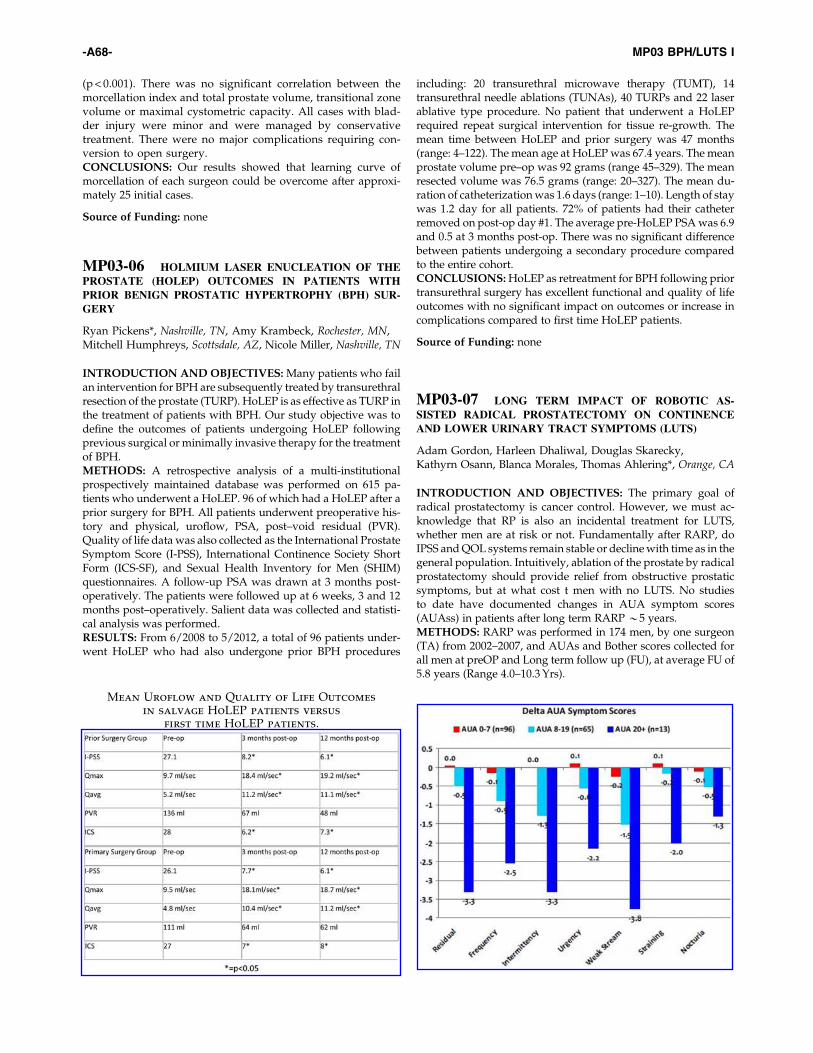
(p < 0.001). There was no significant correlation between themorcellation index and total prostate volume, transitional zonevolume or maximal cystometric capacity. All cases with blad-der injury were minor and were managed by conservativetreatment. There were no major complications requiring con-version to open surgery.CONCLUSIONS: Our results showed that learning curve ofmorcellation of each surgeon could be overcome after approxi-mately 25 initial cases.
Source of Funding: none
MP03-06 HOLMIUM LASER ENUCLEATION OF THE
PROSTATE (HOLEP) OUTCOMES IN PATIENTS WITH
PRIOR BENIGN PROSTATIC HYPERTROPHY (BPH) SUR-
GERY
Ryan Pickens*, Nashville, TN, Amy Krambeck, Rochester, MN,Mitchell Humphreys, Scottsdale, AZ, Nicole Miller, Nashville, TN
INTRODUCTION AND OBJECTIVES: Many patients who failan intervention for BPH are subsequently treated by transurethralresection of the prostate (TURP). HoLEP is as effective as TURP inthe treatment of patients with BPH. Our study objective was todefine the outcomes of patients undergoing HoLEP followingprevious surgical or minimally invasive therapy for the treatmentof BPH.METHODS: A retrospective analysis of a multi-institutionalprospectively maintained database was performed on 615 pa-tients who underwent a HoLEP. 96 of which had a HoLEP after aprior surgery for BPH. All patients underwent preoperative his-tory and physical, uroflow, PSA, post–void residual (PVR).Quality of life data was also collected as the International ProstateSymptom Score (I-PSS), International Continence Society ShortForm (ICS-SF), and Sexual Health Inventory for Men (SHIM)questionnaires. A follow-up PSA was drawn at 3 months post-operatively. The patients were followed up at 6 weeks, 3 and 12months post–operatively. Salient data was collected and statisti-cal analysis was performed.RESULTS: From 6/2008 to 5/2012, a total of 96 patients under-went HoLEP who had also undergone prior BPH procedures
including: 20 transurethral microwave therapy (TUMT), 14transurethral needle ablations (TUNAs), 40 TURPs and 22 laserablative type procedure. No patient that underwent a HoLEPrequired repeat surgical intervention for tissue re-growth. Themean time between HoLEP and prior surgery was 47 months(range: 4–122). The mean age at HoLEP was 67.4 years. The meanprostate volume pre–op was 92 grams (range 45–329). The meanresected volume was 76.5 grams (range: 20–327). The mean du-ration of catheterization was 1.6 days (range: 1–10). Length of staywas 1.2 day for all patients. 72% of patients had their catheterremoved on post-op day #1. The average pre-HoLEP PSA was 6.9and 0.5 at 3 months post-op. There was no significant differencebetween patients undergoing a secondary procedure comparedto the entire cohort.CONCLUSIONS: HoLEP as retreatment for BPH following priortransurethral surgery has excellent functional and quality of lifeoutcomes with no significant impact on outcomes or increase incomplications compared to first time HoLEP patients.
Source of Funding: none
MP03-07 LONG TERM IMPACT OF ROBOTIC AS-
SISTED RADICAL PROSTATECTOMY ON CONTINENCE
AND LOWER URINARY TRACT SYMPTOMS (LUTS)
Adam Gordon, Harleen Dhaliwal, Douglas Skarecky,Kathyrn Osann, Blanca Morales, Thomas Ahlering*, Orange, CA
INTRODUCTION AND OBJECTIVES: The primary goal ofradical prostatectomy is cancer control. However, we must ac-knowledge that RP is also an incidental treatment for LUTS,whether men are at risk or not. Fundamentally after RARP, doIPSS and QOL systems remain stable or decline with time as in thegeneral population. Intuitively, ablation of the prostate by radicalprostatectomy should provide relief from obstructive prostaticsymptoms, but at what cost t men with no LUTS. No studiesto date have documented changes in AUA symptom scores(AUAss) in patients after long term RARP *5 years.METHODS: RARP was performed in 174 men, by one surgeon(TA) from 2002–2007, and AUAs and Bother scores collected forall men at preOP and Long term follow up (FU), at average FU of5.8 years (Range 4.0–10.3 Yrs).
Mean Uroflow and Quality of Life Outcomes
in salvage HoLEP patients versus
first time HoLEP patients.
-A68- MP03 BPH/LUTS I

RESULTS: Total AUAs for all men declined from baseline to 5Years by 3.7 (8.5 to 4.8) while the bother score decreased by 0.5(1.7 to 1.2), all p < .05. Average age was 61.8 (Range 43–80), av-erage follow up was 5.8 years and Pad free continence at 5 yearswas 88%. Men with mild LUTS remained statistically and clin-ically unchanged at 5 years in respect to preoperative AUAscores, except for a small increase in urgency. In contrast,Individuals with moderate/severe preop LUTS markedly de-clined in AUA and QOL Bother scores, all p ¡Ü 0.05, excepturgency.
Figure 1 demonstrates the decease in individual AUA scores(improvement) for each of the 3 LUTS groups from preoperativeAUA to 5 Year followup. Figure 2 shows the overall decrease inAUA scores between the 3 LUTSgroups across the 5 year post-operative period.CONCLUSIONS: Men with mild LUTS tend to maintain theirbaseline IPSS Scores at 5 years. For men with preoperativemoderate/severe LUTS, 61% had significant QOL improvements,and 67% reduced their AUA scores to mild, persisting years afterRARP. Men should be counseled that RARP confers a very sig-nificant long term benefit in men with mod/severe AUAs, im-proving LUTS QOL.
Source of Funding: none
MP03-08 SERIAL MRI AND 3D RENDERING FOL-
LOWING TREATMENT OF BPH USING HIGH ENERGY
WATER VAPOR THERAPY AND THE REZUMTM SYSTEM;
INITIAL RESULTS FROM THE FIRST-IN-MAN AND RE-
ZUMTM 1 CLINICAL TRIALS
Christopher Dixon*, NY, NY, Edwin Rijo-Cedano, La Romana,Dominican Republic, Dalibor Pacik, Vitislav Vit, Gabriele Varga,Brno, Czech Republic, Lance Mynderse, Dennis Hanson, Rochester,MN, Thayne Larson, Scottsdale, AZ
INTRODUCTION AND OBJECTIVES: The Rezum System� isa novel transurethral therapy based on phase-change, convectiveheat transfer using high energy water vapor (steam). It is beingdeveloped as a very rapid, outpatient or office-based treatmentfor clinical BPH.
The objectives were to assess the serial effects on prostate vol-ume, thermal lesion characteristics and to validate the principle of
phase-change convective heating using high energy water vaporin the human prostate.METHODS: IRB approval from 2 centers and informed consentwas obtained in 30 men. (15 in the FIM trial and 15 in the Rezum 1trial.) Both trials used the same standard entry criteria. The FIMtrial used energy ranging from 190 to 289 calories per injectionwhereas, the Rezum 1 trial used a standardized dose of 208 cal-ories per injection. One to 3 injections per lobe, including themedian lobe, were delivered. Gadolinium-enhanced MRI’s wereconducted at 1 week, 1, 3 and 6 months to assess the location ofthermal lesions and perform volume measurements for totalprostate, transition zone and thermal lesions. MRI’s were ana-lyzed and calculated via T-1 and T-2 axial acquisitions using theAnalyze� Image Acquisition System.RESULTS: Thermal lesions were limited to the transition zonevalidating the principles of convective heat transfer in the humanprostate.
Mean thermal lesion volume at 1 week (n = 30) was 10 cc (range7–35 cc) and decreased by 93% at 3 months (n = 28) and 97% at6 months (n = 28).
Mean total prostate volume at 1 week (n = 30) was 75 cc (range29–169 cc) and decreased by 26% at 3 months (n = 28) and 33% at6 months (n = 28).CONCLUSIONS: MRI studies confirmed 1.) a significant volumeof treated tissue, 2.) treatment within the transition zone vali-dating the principle of convective heating, 3) rapid, near completeresolution by 3 months. and 4) reduction in total prostate volumesupporting probable long term clinical benefit.
Source of Funding: NxThera Inc. Maple Grove, MN
MP03-09 TRENDS IN RESIDENT INVOLVEMENT IN
BPH PROCEDURES
Mark Ball*, Max Kates, Brian Matlaga, Baltimore, MD
INTRODUCTION AND OBJECTIVES: Transurethral resectionof the prostate (TURP) is the historic gold standard surgicaltreatment for men suffering from symptomatic benign prostatichyperplasia (BPH). In recent years, though, novel BPH therapiessuch as photoselective vaporization of the prostate (PVP) havebecome increasingly adopted. Indeed, epidemiologic evidencesuggests that the utilization of TURP is declining and laser va-porization has become the fastest growing treatment modality.Consequently, there is concern that urology residents are notbeing trained in TURP techniques to the extent they once were –an issue of particular importance given the well describedlearning curve associated with TURP. We performed a study tocharacterize resident participation in TURP and PVP proce-dures.METHODS: The American College of Surgeons’ National Sur-gical Quality Improvement Database (NSQIP) is a prospectivelymaintained, validated database of pre-operative to 30-daypostoperative surgical outcomes that was designed to improvesurgical care. We queried NSQIP for the years 2007–2011, usingCPT codes 52601 (TURP) and 52648 (PVP) to identify the studycohort. Resident involvement was assessed and compared, bothoverall and then stratified by year as a proportion of total BPHcases.RESULTS: A total of 6,077 BPH procedures were captured inNSQIP during the study period. Residents were involved in 1,378procedures (22.7%). TURP was performed 3,764 times, with res-idents involved in 826 cases (21.9%). PVP was performed 2,313times, with residents involved in 552 (23.8%). When stratified byyear, both the total number of cases and the proportion of cases
MP03 BPH/LUTS I -A69-

with resident involvement were higher for TURP than for PVP ineach year (Figure 1). Pairwise comparison for each year were allstatistically significant (p values 0.03 or less).CONCLUSIONS: TURP is more commonly performed than PVPamong institutions participating in NSQIP. With regards to sur-gical training, residents are more likely to participate in a TURPprocedure than a PVP procedure.
Source of Funding: none
MP03-10 HOLMIUM LASER ENUCLEATION/ABLA-
TION OF THE PROSTATE IN PATIENTS PREVIOUSLY
TREATED WITH RADIATION THERAPY FOR PROSTATE
CANCER
Jessica E Paonessa*, Naeem Bhojani, James E Lingeman,Indianapolis, IN
INTRODUCTION AND OBJECTIVES: A subset of patientswho are treated with radiation therapy (RT) for prostate cancer(PCa) will later develop bladder outlet obstruction (BOO). Man-agement of these patients represents a challenge to the urologist.Previous reports have demonstrated that patients who undergotransurethral resection of the prostate (TURP) after RT for PCahave a higher incidence of incontinence and voiding symptoms.We present our experience using HoLEP/HoLAP for patientswith BOO and prior RT for PCa.METHODS: An Institutional Review Board approved prospec-tive database has been maintained at our institution since January1999 for all HoLEP procedures. The database was retrospectivelyreviewed for patients who underwent HoLEP for BOO and hadpreviously been treated with RT (external beam and bra-chytherapy) for PCa.RESULTS: From January 2000 to September 2010, we identified 13patients treated with RT for PCa who subsequently underwentHoLEP for BOO. 9 patients were treated with external beam, 2 withbrachytherapy and one with combined external beam and bra-chytherapy. Preoperative retention was present in 7 patients, 5 hadrecurrent gross hematuria and 6 reported some degree of inconti-nence. Preop mean PSA was 2.37 ng/mL and mean AmericanUrological Association Symptom Index (AUA SI) was 20.8. Meanenucleation and morcellation times were 63.9 and 16.5 minutes, re-spectively. Mean enucleated tissue weight was 38.5 grams. 6 patientshad standard HoLEP procedures, 3 had ablation only (HoLAP), 2had resection of the median lobe only and 2 had a channel HoLEP. Inthe standard HoLEP group, tissue planes were maintained in all but
one patient. Mean hospital stay was 25.7 hours and mean postopcatheterization time was 36.9 hours. All patients were able to voidafter catheter removal. Pathology results revealed PCa in 2 patients(1 with Gleason 3 + 4 = 7 in < 10%, 1 with Gleason 5 + 4 = 9 in 80%) At6 month follow up, mean AUA SI was 17.8. The one patient with highgrade, high volume PCa had a PSA of 39.63 ng/ml. The remainingpatients had a mean PSA of 1.67 ng/ml. At 12 month follow up,mean AUA SI was 14 and 7 patients reported incontinence.CONCLUSIONS: A subset of patients who are treated with RTfor PCa will later develop BOO. This patient population is oftendifficult to manage. As with HoLEP for treatment of BPH, HoLEPafter RT produces excellent relief of obstruction with low perio-perative morbidity. However, AUA SI remain elevated up to oneyear following surgery. Significant voiding symptoms and in-continence are common postoperatively.
Source of Funding: None
MP03-11 A COMPARATIVE STUDY BETWEEN HOL-
MIUM LASER ENUCLEATION OF THE PROSTATE AND
TRANSURETHRAL RESECTION OF THE PROSTATE: 12
MONTH FOLLOW UP
Mohamed Etafy*, Gamal Morsi, Atef Hammouda,M Hammouda, Assiut, Egypt, Enmar Habib, Cairo, Egypt
INTRODUCTION AND OBJECTIVES: This was a prospectiverandomized clinical trial to compare the safety and efficacy ofholmium laser enucleation of prostate and transurethral resectionof the prostate for surgical treatment of patients with bladderoutlet obstruction due to benign prostatic hyperplasia.METHODS: From December 2009 to October 2010, 60 consecu-tive patients with lower urinary tract obstruction (LUTs) due toBPH were randomized to either surgical treatment with HoLEP(group 1, n = 30) or standard TURP (group 2, N = 30). Preoperativeassessments included American Urological Association (AUA)symptom score. Serum prostate-specific antigen, (PSA), post voidresidual urine volume (PVR), transrectal ultrasound (TRUS) anduroflowmetry. Perioperative parameters included total operatingtime, resected tissue weight, hemoglobin loss, presence or ab-sence of blood transfusion, time of catheter removal, duration ofhospital stay. Postoperative evaluations were conducted at 1,3,6,and 12 months.RESULTS: Patients in the HOLEP group had shorter catheteri-zation time, and hospital stay, but longer operating time. Meanhemoglobin loss was lower in the HOLEP group (0.900 – 0.419 g/dl versus 1.157 – 0.918 g/dl). The follow up results up to 12 monthregarding AUA symptom score, PVR urine volume and Q-maxshowed that both group were comparable. Complications weresimilar with 2 procedure with no sign fiancé difference.CONCLUSIONS: Conclusion: HOLEP proved to be safe andhighly effective technique for surgical treatment of bladder outletobstruction due to BPH.
Source of Funding: NONE
MP03-12 REGIONAL DIFFERENCE IN IPSS, PV, AND
PSA IN KOREAN MALE PATIENTS WITH LUTS
Sung Chul Kam*, Jinju, Korea, Republic of, Jae Hwi Choi,Seong Uk Jeh, Jeong Seok Hwa, Jae Seog Hyun,Jinju, Korea, Republic of
INTRODUCTION AND OBJECTIVES: Lower urinary tractsymptom (LUTS) is a common symptom in middle-aged men.
FIG 1. Resident involvement in TURP and PVP as percentageof total BPH procedures
-A70- MP03 BPH/LUTS I

Prostate volume (PV) is an important predictor of benign pros-tatic hyperplasia (BPH) progression. Prostate specific antigen(PSA) is a reliable factor predicting PV in men suffering fromLUTS. This study aimed to investigate regional difference in in-ternational prostate symptom score (IPSS), PV, and PSA in menwith LUTS.METHODS: The study was conducted on 36,632 male patientsaged 40*99 years who were suffering from LUTS from 2001 to2010. The baseline PV and PSA were defined using standarddescription. In addition, the IPSS and quality of life (QoL) werecollected via an interview. Data used in this study were collectedfrom patients with PV £ 200 ml and baseline PSA = 0–10 ng/ml.Previous history of prostatic surgery and status other than pros-tate cancer and BPH were excluded from the baseline data. As fordiscrimination of urban and rural areas, city or higher adminis-trative areas were assigned to the urban group, whereas Eup,Myeon, and Ri were assigned to the rural group based on pa-tient¡�s address.RESULTS: A total of 35,797 patients were analyzed. The patientshad mean age 69.7 – 8.3 years, mean baseline PV 29.9 – 13.8 ml,mean PSA 1.47 – 1.5 ng/ml, and mean IPSS 15.4 – 8.4. The pa-tients consisted of 25,967 patients living in the urban areas and9,830 patients living in the rural areas. The mean age of the urbanand rural groups were shown to be 69.12 – 8.8 years and70.42 – 9.8 years, respectively, which showed a significant dif-ference (p < 0.05). The PSA was shown to be 2.62 – 10.2 ng/ml inthe urban group and 2.11 – 9.06 ng/ml in the rural group, whichshowed a significant difference (p < 0.05). The PV was shown tobe 29.86 – 14.6 ml and 30.35 – 15.9 ml in the urban and ruralgroups, respectively, which showed a significant difference(p < 0.05). The IPSS and QOL were significantly higher in the ruralgroup than in the urban group (p < 0.05). When the body massindex (BMI) was measured in the 931 patients of the urban groupand 5,235 patients of the rural group, it was shown to be 31.32 and24.41 in the urban and rural groups, respectively, which showedthat it was significantly higher in the urban group than in therural group.CONCLUSIONS: Compared to the rural group, the urban grouphad the increased PSA but decreased PV. This result is likely to beattributable to the fact that the PSA increased rather than prostaticsize by obesity. Thus, it is recommended to treat LUTS in a clinicalpractice, considering the results of this study.
Source of Funding: none
MP03-13 THE ROLE OF TRANSURETHRAL RESEC-
TION OF PROSTATE IN TREATING PATIENTS WITH BE-
NIGN PROSTATE HYPERPLASIA AND ELEVATED
PROSTATE-SPECIFIC ANTIGEN
Jeong Man Cho*, Sun Choel Shin, Hee Ju Cho, Jung Yoon Kang,Tag Keun Yoo, Seoul, Korea, Republic of
INTRODUCTION AND OBJECTIVES: To define the role andclinical significance of transurethral resection of prostate intreating patient with benign prostate hyperplasia (BPH) and el-evated prostate-specific antigen (PSA).METHODS: We retrospectively evaluate patients with BPH,lower urinary tract symptom (International Prostate SymptomScore ‡ 8), elevated serum PSA ( ‡ 4 ng/ml) and negative onprevious TRUS biopsy. Short term surgical outcome in regards oftumor detection rate on resected prostate tissue, IPSS, maximumuroflow rate (Qmax), postvoiding residual urine (PVR) was an-alyzed. And the changes of PSA after TURP were monitored.
RESULTS: A total 186 patients were enrolled. Preoperativeclinical variables are described in Table 1. Histologic examinationon resected tissue by TURP revealed prostate cancer in 12/186(6.5%). The mean PSA of 118 patients at POD 3 months was1.34 – 0.13 ng/ml and then maintained as following:1.28 – 0.14 ng/ml in 47 patients at POD 1 year, 1.17 – 0.14 in 34patients at POD 2 years and 1.34 – 0.24 ng/ml in 36 patients atPOD 3 years. When compared serum PSA to POD 3 months,19/40 (47.5%) patients increased PSA with average 0.33(0.01*1.07) ng/ml at POD 1 years, in 9/28 (32.1%) patients withaverage 0.32 (0.03*1.13) ng/ml at POD 2 years and in 14/31(45.2%) patients with average 0.68 (0.01*2.69) ng/ml at POD 3years. Improvements of IPSS, QOL, Qmax and PVR were 9.7 – 1.1,2.6 – 0.18 points, 7.2 – 0.72 ml/s and 100.5 – 27.2 ml at POD3 months. No prostate cancer was detected during follow-upperiod.CONCLUSIONS: TURP reduced serum PSA dramatically andcan be helpful in monitoring PSA without anxiety about malig-nancy. In addition, TURP improves IPSS, Qmax and PVR in pa-tients with BPH, moderate LUTS and elevated PSA.
Source of Funding: none
MP03-14 LASER PROSTATECTOMY OF LARGE
PROSTATES USING A NEW 1.9lM THULIUM LASER: RE-
SULTS AFTER 1 YEAR FOLLOW-UP
David Zimmermann*, Patrick Honeck, Thomas Knoll,Gunnar Wendt-Nordahl, Sindelfingen, Germany
INTRODUCTION AND OBJECTIVES: The aim of the studywas to evaluate the treatment results including subjective andobjective parameters one year after laser prostatectomy due tobenign prostatic enlargement using a new 1.9lm thulium laserdevice.METHODS: 222 patients were treated with the 1.9lm thuliumlaser device (Vela XL, StarMedTec, Germany) between 5/2010and 05/2013. 70 of these patients (mean age 71 – 7.4 years) havecompleted a one year follow-up. In 62 patients a laser enucleationwas performed, whereas in 8 patients the laser was used to va-poresect the tissue. The patients were the first treated with thedevice in our department and therefore include the learningcurve. Objective and subjective parameters were obtained pro-spectively and compared to preoperative values.RESULTS: Mean operation time was 107 – 44 min (includingmorcellation) and the mean resection volume was 45.3 – 31.4 g.Mean prostatic volume was reduced from 85.7 – 41.9 cc to
Table 1. Preoperative clinical variables
MP03 BPH/LUTS I -A71-

28.7 – 13.9 cc (68%, p = 0.0001) and mean PSA from 6.33 – 4.86 ng/ml to 1.94 – 1.97 ng/ml (69%, p = 0.0001).
Mean IPSS dropped from 19.4 – 8.7 to 5.7 – 4.6 (71%, p = 0.0001),mean QL improved from 4.0 – 1.7 to 1.1 – 1.3 (73%, p = 0.0001).Maximal flow rate increased from 8.3 – 3.6 ml/s to 18.0 – 8.0 ml/s (117%, p = 0.0001), while mean post void residual volumewas reduced by 80% from 128.7 – 137.5 ml to 25.4 – 36.0 ml(p = 0.0001). Of the 26 patients in urinary retention preopera-tively only 1 required a suprapubic catheter at the time of thefollow-up due to urodynamically proven neurogenic insuffi-ciency of the detrusor muscle. 3 patients reported a delayedgross hematuria within two months after surgery. 4 patientsrequired re-intervention during the follow-up period due tourethral or bladder neck stricture (3 instrumental dilatation, 1resection). Whereas no higher grade incontinence was observed,6 patients (9%) reported mild incontinence (5 patients stressincontinence I�, 1 patient urge incontinence). 2 patients reportedurge symptoms without incontinence that are treated with an-ticholinergic drugs.CONCLUSIONS: One year after thulium laser prostatectomyboth subjective and objective parameters showed highly signifi-cant improvements. Therefore, the 1.9lm thulium laser consti-tutes an effective and safe device for laser prostatectomy due tobenign prostatic enlargement.
Source of Funding: none
MP03-15 THREE-YEAR PROSPECTIVE, RANDO-
MIZED COMPARISON OF THE BIPOLAR PLASMA VA-
PORIZATION OF THE PROSTATE, MONOPOLAR AND
BIPOLAR RESECTION IN MEDIUM SIZE BPH PATIENTS
Bogdan Geavlete*, Razvan Multescu, Dragos Georgescu,Florin Stanescu, Marian Jecu, Cristian Moldoveanu,Petrisor Geavlete, Bucharest, Romania
INTRODUCTION AND OBJECTIVES: A prospective, longterm, randomized trial was performed aiming to compare thebipolar plasma vaporization of the prostate (BPVP), the bipolartransurethral resection in saline (TURis) and the monopolartransurethral resection of the prostate (TURP) concerning thesurgical efficacy, complication rate and follow-up results.METHODS: A total of 510 benign prostatic hyperplasia (BPH)patients with prostate volume between 30 and 80 mL, maximumflow rate (Qmax) below 10 mL/s and International ProstateSymptom Score (IPSS) over 19 were included in the trial. All caseswere evaluated preoperatively as well as at 6 months’ intervalsafter surgery by IPSS, Qmax, quality of life score (QoL), post-voiding residual urinary volume (PVR), postoperative prostatevolume and PSA level.RESULTS: The mean operation time (39.7 versus 52.1 and 55.6minutes), catheterization period (23.5 versus 46.3 and 72.8 hours)and hospital stay (1.9 versus 3.1 and 4.2 days) were significantlyshorter for BPVP patients. The mean hemoglobin drop (0.5 versus1.2 and 1.6 g/dL), intraoperative bleeding (1.8% versus 8.2% and13.5%) and capsular perforation (1.2% versus 7.1% and 9.4%)rates were significantly reduced in the BPVP series. The rate ofearly irritative symptoms was statistically similar in the 3 studyarms (12.4% versus 11.2% and 10.6%, respectively). In the BPVPand TURis groups, significantly lower postoperative hematuria(2.9% and 4.7% versus 15.3%) and blood transfusion (1.2% and1.8% versus 6.5%) rates were determined. During the long termfollow-up, BPVP patients emphasized significantly superior pa-rameters concerning IPSS and Qmax. On the other hand, thethree-year evaluation protocol described statistically similar QoL
score, PVR values, PSA level and the postoperative prostatevolume in the 3 series of patients.CONCLUSIONS: BPVP displayed superior surgical efficacy,reduced perioperative morbidity and faster postoperative re-covery when compared to TURis and TURP. During the longterm follow-up, the method provided significantly improvedparameters regarding the symptom scores and voiding charac-teristics and similar BPH tissue removal capabilities.
Source of Funding: None
MP03-16 DETRUSOR WALL THICKNESS AS A POSSI-
BLE PREDICTOR OF PERSISTENT URINARY URGENCY
AFTER TRANSURETHRAL RESECTION OF PROSTATE
Dong Soo Park*, Seung Ryeol Lee, Seongnam-si, Korea, Republic of
INTRODUCTION AND OBJECTIVES: We evaluated thevoiding difficulty according to the detrusor wall thickness (DWT)before the transurethral resection of prostate (TURP) and thedifferences of DWT between the patients with and without per-sistent urinary urgency after TURP.METHODS: We retrospectively reviewed the records of 79 pa-tients who underwent TURP for symptomatic benign prostatehyperplasia between December 2010 and July 2012. Of thesepatients, 52 patients without a neurogenic cause underlyingurinary urgency were selected. The patients completed the In-ternational Prostate Symptom Score (IPSS), uroflowmetry, andtransrectal ultrasonography (TRUS). DWT was measured byperforming TRUS at the anterior bladder wall in midsagittalimage. Patients were divided in two groups according to DWT(group 1: patients with DWT ‡ 2 mm, group 2: patients withDWT < 2 mm). After 6 months of TURP, patients were re-evaluated using the IPSS and uroflowmetry, and divided intotwo groups according to the urgency (group 3: patient withpersistent urgency, group 4: patients without urgency). Urgencywas defined when the score for question 4 of the IPSS was morethan 3 points. Voiding symptom score was defined as the sum ofscores for questions 1, 3, 5, and 6 of the IPSS. Bladder storagesymptom score was defined as the sum of scores for questions 2,4, and 7 of the IPSS.RESULTS: Of 52 patients, there were 43 patients in group 1 and9 patients in group 2. Peak urinary flow rate was signifi-cantly lower in group 1 (7.4 – 3.5 ml/s) than group 2 (11.1 – 4.0,p = 0.026). There were no significant differences in residual urinevolume, voiding symptom score, and bladder storage symptomscore between group 1 and 2. Before TURP, 21 patients (40.4%)had urgency and the urgency disappeared after TURP in 14patients. Seven patients (13.5%) had the persistent urinary ur-gency after TURP. There were no significant differences in age,body mass index, prostate volume, preoperative peak urinaryflow rate, and residual urine volume between group 3 (n = 7) and4 (n = 45). However, DWT was significantly thicker in group 3(2.4 – 0.3 mm; range, 2.3–2.7) than group 4 (2.1 – 0.4 mm; range,1.2–2.7, p = 0.021).CONCLUSIONS: Peak urinary flow rate was significantly lowerin patients with DWT ‡ 2 mm and this demonstrates the possi-bility of bladder outlet obstruction in these patients. DWT wassignificantly thicker in patients with persistent urinary urgencyafter TURP. DWT may help to predict patients with persistenturinary urgency after TURP.
Source of Funding: none
-A72- MP03 BPH/LUTS I

MP03-17 GREEN LASER PROSTATIC LASER VAPOR-
IZATION IN PATIENTS OLDER THAN 80 YEARS. IS IT
SAFE?
Pablo Contreras*, Buenos Aires, Argentina, Francisco Lopez,Ramiro Castilla, Carlos Ameri, Gonzalo Vitagliano,Osvaldo Mazza, Ciudad Autónoma de Buenos Aires, Argentina
INTRODUCTION AND OBJECTIVES: Benign Prostatic Hi-perplasia is a prevalent condition among elderly men. Thegrowing presence of age related comorbidities increases the sur-gical risk in this group of patients. The aim of the study was toevaluate the ssfety of Green Laser Prostatic Vaporization in pa-tients older than 80 years.METHODS: Between June 2007 and August 2012, 450 Greenlaser prostatic vaporization were made at our institution, 30 ofthem were older than 80 years. Intraoperative and perioperativecomplications were analized.RESULTS: Medium patient’s age was 83,5 years (80–90 years).Average hospitalization was 1,4 days. Four patients had perma-nent bladder catheter at the time of surgery. Average prostate sizewas 48,7 cc (18–100 cc). Three patiens were anticoagulated. Therewere no intra or perioperative deaths. One patient required alonger hospitalization (three days) because of COPD reagudiza-tion and three patients due a concomitant procedure inm thebladder (Holmium lithotripsy). There were no readmissions orreoperations. Two patients (6,6%) presented an acute urinaryretention after catheter removal, but after seven days of cathe-terisation achieved spontaneous micturition. Four patients pre-sented advanced Prostate Cancer (13%), three (10%) bladdercancer and five (16,6%) colorectal cancer.CONCLUSIONS: From our experience, Green Laser ProstaticVaporization was a safe procedute for patients of 80 years andolder with zero mortality and a low rate of minor complications.
Source of Funding: None
MP03-18 DIODE LASER VAPORIZATION OF THE
PROSTATE FOR BENIGN PROSTATIC HYPERPLASIA –
COMPARING VAPORIZATION ALONE WITH VAPORIZA-
TION PLUS SUBSEQUENT BIPOLAR TRANS-URETHRAL
RESECTION AT 12 MONTHS FOLLOW-UP
Ferdinando De Marco*, Grottaferrata, Italy, Markus Rheinwald,Wessling, Germany, Thomas Bayer, Kempten, Germany
INTRODUCTION AND OBJECTIVES: Using diode lasers thatemit light in the near infrared for vaporization of benign prostatichyperplasia (BPH) leads to good hemostasis. Recent literaturealso proposes to add bipolar trans-urethral resection (b-TURP)after diode laser vaporization of the prostate for patients withlarger prostates. The objective of the study was to compare thefunctional outcomes in patients with prostates larger than 70grams between patients treated with a high power diode laseralone and patients treated with high power diode laser andsubsequent b-TURP.METHODS: Vaporization of prostatic tissue in BPH patients wasperformed using a high power diode laser operating at 940nanometers and emitting a maximum power of 250 Watts incontinuous wave mode. In some patients with prostates largerthan 70 grams, particularly very large prostates, b-TURP wasapplied after laser vaporization. Before and for follow-up on 1, 3,6, and 12 months after the operation, patients were assessed fortheir residual urine volume, maximum urine flow rate (Qmax)and International Prostate Symptom Score (IPSS).
RESULTS: 34 patients treated with vaporization alone (group 1)and 28 patients treated with vaporization and subsequent b-TURP (group 2) have reached 12 months of follow-up so far. At 12months, compared to pre-operatively, the mean maximum flowrate increased from 10,0 ml/s to 21,3 ml/s in group 1 and from8,0 ml/s to 23,9 ml/s in group 2; mean IPSS dropped from 19,0 to8,0 in group 1 and from 19,6 to 7,1 in group 2; and mean residualurine volume dropped from 127,3 ml to 20,6 ml in group 1 andfrom 130,5 ml to 11,1 ml in group 2.CONCLUSIONS: The treatment of BPH with the high powerdiode laser at 250 W, with laser vaporization alone or followed byTURP for large prostates, provides good clinical outcome re-garding peri-operative bleeding and post-operative IPSS, maxi-mal flow rate and residual urine in the follow-up of 12 months.Adding b-TURP subsequent to diode laser vaporization tends tofurther improve functional results during follow-up. Furtherstudies with extended follow-up and patient numbers are neededto confirm these data.
Source of Funding: Laser and fibers: Dornier MedTech Laser
MP03-19 A PROSPECTIVE MULTICENTER RANDO-
MIZED STUDY COMPARING GREENLIGHT XPSTM LASER
AND TRANSURETHRAL RESECTION OF THE PROSTATE
FOR THE TREATMENT OF BENIGN PROSTATIC HYPER-
PLASIA
Alexander Bachmann*, Basel, Switzerland, Andrea Tubaro, Rome,Italy, Neil Barber, Camberley Surrey, United Kingdom,Frank d’Ancona, Nijmegen, Netherlands, Gordon Muir, London,United Kingdom, Ulrich Witzsch, Frankfurt, Germany,Marc-Oliver Grimm, Jena, Germany, Joan Benejam, Manacor, Spain,Jens-Uwe Stolzenburg, Leipzig, Germany, Anthony Riddick,Edinburgh, United Kingdom, Sascha Pahernik, Heidelberg,Germany, Johannes Hermanus Roelink, Almelo/Hengelo,Netherlands, Filip Ameye, Gent, Belgium, Christian Saussine,Strasbourg, France, Frank Bruyere, Tours, France, Wolfgang Loidl,Linz, Austria, Timothy Larner, Brighton, United Kingdom,Nirjan Gogoi, Dewsbury, United Kingdom, Richard Hindley,Hampshire, United Kingdom, Rolf Muschter, Rotenburg, Germany,Andrew Thorpe, Newcastle upon Tyne, United Kingdom,Nitin Shrotri, Kent, United Kingdom, Stuart Graham, London,United Kingdom, Moritz Franz Hamann, Kiel, Germany,Kurt Miller, Berlin, Germany, Martin Schostak, Magdeburg,Germany, Carlos Capitan, Madrid, Spain, Helmut Knispel, Berlin,Germany, James Andrew Thomas, Wales, United Kingdom
INTRODUCTION AND OBJECTIVES: Minimally invasivetherapies are increasingly being used to treat benign prostatichyperplasia (BPH) but there have been few prospective ran-domized trials that compare such procedures to trans urethralresection of the prostate (TURP). We conducted a trial to evaluatethe non-inferiority of photo-selective vaporization (PVP) with theGreenLight XPS system and MoXy fiber compared to TURP.METHODS: 291 patients were recruited at 29 European centers.Patients were randomized to XPS or TURP. The study was de-signed to assess non-inferiority for the primary end point of IPSSat 6 months. The study was also powered to show non-inferiorityfor Qmax and proportion of patients free of complicationsthrough 180 days post surgery. Demographics showed that thearms of the study were well balanced at baseline. Key inclusion/exclusion criteria included age between 40–80 years, IPSS > 12,Qmax < 15 cc/sec, prostate size < 100 mg, and no previous lowerurinary tract surgery.
MP03 BPH/LUTS I -A73-

RESULTS: There were 136 patients treated in the XPS arm and133 in the TURP arm. It has been shown that on the IPSS a dif-ference of 3 points is the smallest perceptible difference; thus, 3was chosen as the margin for non-inferiority. At six months, meanIPSS scores were 6.8 – 5.2 for the XPS and 5.6 – 4.9 for the TURParm and met the criterion for non-inferiority (p = 0.002). Similarly,there was no statistical difference in Qmax at 6 months with23.3 – 10.1 for XPS and 24.2 – 11.4 for TURP, meeting the non-inferiority criterion for the pre-specified margin of 5 (p = 0.002).XPS was also non-inferior with respect to the proportion of pa-tients that experienced a complication by 6 months. Mean lengthof catheterization (40.8 hours vs 59.5 hours), hospitalization (65.5hours vs 96.9 hours) and time to stable health status (37.3 hours vs63.5 hours) were all significantly shorter for XPS compared toTURP (p < 0.001 for each assessment). There were more XPS pa-tients with UTI’s (22 vs 12). Grade III and IV bleeding events weremore common in TURP patients (9 vs 4). Invasive or surgical re-interventions within 30 days were more common in TURP pa-tients (13 vs 4 in XPS).CONCLUSIONS: In this prospective randomized trial, Green-Light XPS PVP showed similar efficacy and safety to TURP withstatistically significant improvements in catheterization time,length of hospitalization, and time to stable health status. More-over, there were fewer serious bleeding complications and inva-sive or surgical re-interventions in the PVP arm compared to theTURP arm.
Source of Funding: American Medical Systems, Inc.
MP03-20 TRANSURETHRAL ENUCLEATION WITH
BIPOLAR (TUEB) FOR PROSTATES OVER 50 ML: A SINGLE
CENTER EXPERIENCE
Hayato Takeda*, Hiroyuki Shimizu, Yasutomo Suzuki,Mamoru Oki, Jun Hasegawa, Yukihiro Kondo, Tokyo, Japan
INTRODUCTION AND OBJECTIVES: Transurethral Enuclea-tion with Bipolar (TUEB) is an established and durable treatmentmodality for men with lower urinary tract symptoms (LUTS) dueto benign prostate hyperplasia (BPH). Clinical outcomes of pa-tients treated with TUEB with a prostate volume over 50 ml wereevaluated.METHODS: 126 consecutive patients who underwent TUEBbetween March 2008 and September 2012 were retrospectivelyreviewed, and patients with a prostate volume over 50 ml mea-sured by suprapubic ultrasonography were identified. Using aconventional loop, incising into the mucosa at 0, 5, 7 o’clock frombelow the bladder neck to the urethral caruncle, the prostate isdivided into 3 blocks. With the spatula, the prostate is detachedand resected into small pieces with the conventional loop, with-out using the morecellator for bipolar transurethral vaporization.RESULTS: Out of 126 patients, 83 patients were identified.Median age of the patients was 76.1 years old (range 59–81 yearsold). Postoperative median measured prostate volume was76.6 ml (range 50–158 ml), mean resected prostate volume 42.1 g(range 12–129.3 g), and mean operation time was 116 min (range60–235 min). The mean hemoglobin drop was 1.0 g/dl, with nocases of blood transfusion. The median postoperative catheteri-zation period was 2.7 days. Mean preoperative and postoperativeIPSS was 21.7 vs 4.7, emptying symptoms 9.1 vs 0.5, storagesymptoms 9.1 vs 3.9, QOL 5.1 vs 1.4, Qmax 6.5 ml/s vs 19.1 ml/s.The PSA decreasing rate was 82.2%. Complications and dysuriaafter catheter removal were, postoperative bleeding in 2 patients,urinary tract infection in 2 patients, urethral stricture in 1 patient,
and 1 patient had abdominal pressure induced incontinence re-quiring 3 months for pad free.CONCLUSIONS: Even for large prostatic volumes, reasonableoutcomes can be achieved by TUEB for BPH, with significantimprovement in emptying symptoms. Severe complications werenot found.
Source of Funding: none
MP03-21 TISSUE EFFECTS RESULTING FROM ERA-
SER LASER ENUCLEATION OF THE PROSTATE: IN VIVO
INVESTIGATION
Lukas Lusuardi*, Martina Hager, Manuela Sieberer,Stephan Hruby, Birgit Kloss, Günter Janetschek, Salzburg, Austria
INTRODUCTION AND OBJECTIVES: To describe the depth ofthe laser coagulation zone in vivo based on histological exami-nations and the functional outcome of a 1,318-nm diode laser fortransurethral enucleation in benign prostatic enlargement (BPE).METHODS: A total of 20 patients with BPE were treated by laserEraser� enucleation of the prostate (ELEP). Prostatic tissuewedges were evaluated to assess the depth of the ELEP coagu-lation zones. Additionaly patients were assessed pre-operativelyand 12 month postoperatively.RESULTS: The coagulation zones were 0.36 – 0.17 mm in epi-thelial tissue, 0.28 – 0.15 mm in stromal tissue, and 0.25 – 0.12 mm
Table 1. Baseline Characteristics of the 20 Patients
Table 2. Peri-operative Data for the 20 Patients
-A74- MP03 BPH/LUTS I

in mixed tissue of the diode laser-treated prostate tissue. Thecoagulation area at the cutting edge completely sealed capillaryvessels, reaching a depth of 0.35 – 0.15 mm. The diameter of thecoagulated vessels measured 1.75 – 0.83 mm. The mean blood losswas 115.54 – 93.12 ml, catheter time 1.35 – 0.33 days, and hospitalstay 1.89 – 0.52 days. The International Prostate Symptom Score,maximal flow rate, and quality of life significantly improved 12months after the procedure.CONCLUSIONS: ELEP is safe and effective for BPE treatmentand yields good results at a follow-up of one year. Because of thelimited penetration depth, damage to the urinary sphincter is notexpected.
Source of Funding: none
MP03-22 FACTORS PREDICTING OCCURRENCE OF
TRANSIENT URINARY INCONTINENCE AFTER HOLMIUM
LASER ENUCLEATION OF THE PROSTATE
Dong Gil Shin*, Jeong Zoo Lee, Tae Gyeong Jeon, Tae Nam Kim,Busan, Korea, Republic of, Moon Kee Chung, Yangsan, Korea,Republic of, Chang Yell Lee, Busan, Korea, Republic of
INTRODUCTION AND OBJECTIVES: Holmium laser enucle-ation of the prostate (HoLEP) is a recent step in enabling a trueanatomical enucleation of prostatic tissue. The aims of this studywas to identify the factors associated with occurrence of transienturinary incontinence (TI) after HoLEP for the surgical treatmentof benign prostatic hyperplasia (BPH).
METHODS: The data from the 624 consecutive patients whounderwent HoLEP for the symptomatic BPH between January2010 and December 2011 were evaluated retrospectively. Datawere analyzed to obtain prostate volume by transrectal ultra-sound, serum prostate specific antigen (PSA), hospitalizationtime, time to catheter removal, enucleation and morcellation time,and weight of resected prostate. The follow up included the In-ternational Prostate Symptom Score (IPSS), quality of life (QoL)score, maximum urinary flow rate (Qmax), and postvoiding re-sidual urine volume (PVR). Urinary incontinence was definedaccording to the International Continence Society and by thequestion ¢Do you have involuntary loss of urine?¢RESULTS: The patients¢ mean age was 68.1 – 7.6 years and meanprostate volume was 57.2 – 30.3 g. Mean enucleation time was56.6 – 42.3 minutes, mean morcellation time was 12.3 – 18.9 min-utes, and the mean resected weight of the prostate was21.4 – 24.5 g. The mean catheter indwelling period was 36.3 – 35.4hours and mean hospital stay was 3.2 – 2.5 days. TI occurred in 59patients (9.5%) after HoLEP. The patient¢s age, morecllation timeand resected volume were associated with the occurrence ofpostoperative TI.CONCLUSIONS: After HoLEP, some patients experienced TIand most of them showed recovery within 6 months. HoLEP is asafe and effective modality for treating symptomatic BPH despiteof TI.
Source of Funding: none
MP03-23 EVALUATION OF SHORT TERM EFFICACY
OF DIFFERENT ALFA ADRENERGIC BLOCKERS ON LUTS
IN PATIENTS WITH SYMPTOMATIC BPH
Arup Mandal*, Chandigarh, India, Sudheer Devana,Shrawan Singh, Ravimohan Mavuduru, Chandigarh, India
INTRODUCTION AND OBJECTIVES: An early improvementof lower urinary tract symptoms in patients with symptomaticBPH is desirable. Silodosin has been claimed to have rapid onsetof action and hence improvement in LUTS. We evaluated theshort term efficacy of different alfa adrenergic blockers on LUTSin patients with symptomatic BPH.METHODS: A total of 75 cases of symptomatic BPH aged 50–80 yrs with IPSS ‡ 8 or QOL score ‡ 2 were enrolled in this openlabeled study. Patients with other causes of bladder outlet ob-struction, prostate and bladder malignancy, renal failure, hepaticfailure and prior history of prostatectomy were excluded. Theywere randomized into three groups. Group T received tamsulosin0.4 mg/day, Group A received alfuzosin 10 mg/day and Group Sreceived silodosin 8 mg/day. Patients were evaluated at baseline,6 hrs, 24 hrs, 4th day, 7th day and 1 month of initiation of treat-ment for IPSS, QOL scores, Q max, Q avg and PVR. Side effects oftreatment were recorded after 1 week and one month of initiationof treatment.RESULTS: The baseline characteristics of all three groups interms of age, BMI, symptom duration, prostate volume, IPSS,QOL score, Q max, Q avg and PVR were comparable. Significantimprovement in IPSS and QOL scores were noted after 3rd dosein all three groups (p value 0.001). In group T and group A im-provement in IPSS score ‡ 3 was observed in 24% of patients at6 hrs and in 40% of patients at 24 hrs whereas in group S suchimprovement was observed in 12% at 6 hrs and 16% at 24 hrs. Onintergroup comparison no significant difference was noted inIPSS, QOL scores, Qmax, Qavg and PVR at different time inter-vals following initiation of treatment. Dizziness was reported in4% of group T and group A but none discontinued therapy. In
Table 3. Patient Characteristics over 12 Months
of Follow-up
Preoperative and Postoperative Parameters
MP03 BPH/LUTS I -A75-

group S 8% of patients experienced ejaculatory dysfunction butnone complained of dizziness.CONCLUSIONS: All the three alfa adrenergic blockers (Tam-sulosin, Alfuzosin and Silodosin) improve LUTS significantly inpatients with symptomatic BPH. This improvement achievessignificance after 3rd dose of treatment. The improvement insymptoms is not rapid with silodosin as compared to tamsulosinand alfuzosin.
Source of Funding: None
MP03-24 SAFETY AND EFFICACY OF REPEAT
GREENLIGHT LASER THERAPY WITH THE XPS SYSTEM
IN PATIENTS PREVIOUSLY TREATED WITH THE
GREENLIGHT PV OR HPS SYSTEMS
Andrew Tracey*, Chris Wright, Shailja Mehta, Ravi Munver,Hackensack, NJ
INTRODUCTION AND OBJECTIVES: GreenLight laser ther-apy for BPH is gradually replacing transurethral resection of theprostate (TURP) due to decreased blood loss, shorter hospitalstay, fewer side effects, and decreased complications, whilemaintaining excellent urinary outcomes. As more patients aretreated with laser and other transurethral therapies, an increasingnumber will likely require additional therapy at a later date. Weevaluated our experience in patients previously successfullytreated with GreenLight laser therapy in which a second proce-dure was required due to obstructive urinary symptoms.METHODS: A retrospective review was performed of all pa-tients that underwent GreenLight laser therapy at our institutionsince 2003. Patients that were previously treated with GreenLightlaser therapy met inclusion criteria for this study. Perioperativedata for patients that underwent a second GreenLight procedurewere analyzed.RESULTS: A total of 9 patients were identified has having hadprevious treatment with GreenLight laser therapy at other insti-
tutions. The mean age was 69.5 (range 58–78 years) and bodymass index of 26 (range 23–31). Patients were previously treatedwith the PV (n = 5) or HPS (n = 4) systems. The median time toretreatment was 6 years (range 2–10 years). Two patients pre-sented with urinary retention at the time of the secondary treat-ment. Patients were treated on an outpatient basis (n = 7) or weredischarged on the first postoperative day (n = 2). Seven patientshad the foley catheter removed on POD #1, one on POD #5, andone on POD #14. There were no intraoperative or postoperativecomplications at a median follow-up of 10 months (range 2–24).CONCLUSIONS: This is among the first reports of a series ofpatients that underwent a secondary GreenLight laser therapyprocedure with the XPS system after having been initially treatedwith the PV or HPS systems. In the short term, a secondaryGreenLight procedure appears safe and effective in patients thatdevelop regrowth of prostate tissue that either fail medical ther-apy or elect for surgical therapy. As more patients are retreatedwith GreenLight laser therapy additional data will be collectedand can be compared to the results of this study.
Source of Funding: None
MP03-25 INFLUENCE OF BODY MASS INDEX ON BE-
NIGN PROSTATIC HYPERPLASIA-RELATED COMPLICA-
TIONS IN PATIENTS UNDERGOING PROSTATECTOMY
Hisham Mosli*, Jeddah, Saudi Arabia
INTRODUCTION AND OBJECTIVES: Recent clinical evidencesuggests a strong link between obesity and benign prostatic hy-perplasia (BPH) related complications. This study aimed at ex-amining the link between increased body mass index and benignprostatic hyperplasia (BPH) related complications, namely; acuteurinary retention (AUR), bladder stones and bladder diverticula.METHODS: Medical records of BPH patients who underwentprostatectomy during the period 2010–2012 were reviewed. Pa-tients who underwent transurethral resection of prostate (TURP)or photo-sensitive vaporization of prostate (PVP), represented the2 treatment groups within the study. This retrospective data re-view compared clinical parameters like age, prostate specificantigen (PSA) and prostate volume (PV) as measured by ultra-sonography, patients’ weight and height; BPH related compli-cations mainly AUR, bladder stones and bladder diverticulum;among obese and non obese patients in the 2 treatment groups.Patients were classified as obese or non-obese based upon theWorld Health Organization (WHO) classification of obesity.
Chi-square test was used for comparison between groupswhere p-value was considered significant if < 0.05 and ANOVAwas used for comparison between multiple variables.RESULTS: Of the 197 patients included in this study, 95 (48%)underwent TURP and 102 (52%) underwent PVP. The 2 groupswere found to be similar in mean age and PSA and significantlydifferent in prostate volume and operating room (O.R.) time. Nomajor differences were observed in the frequency of BPH relatedcomplications (AUR, bladder stones and bladder diverticulum).CONCLUSIONS: Evidence elicited through the current studydid not demonstrate an increase in the risk of developing BPH-related complications with an increased BMI. No significant dif-ferences were noted in the development of AUR, bladder stoneand diverticulum formation; between patients with increasedBMI (overweight and obese) and those with normal BMI. Furtherstudies are recommended to explore the influence of increasedBMI on BPH-related complications.
Source of Funding: None
-A76- MP03 BPH/LUTS I

MP03-26 ROLE OF GREENLIGHT LASER IN THE
SURGICAL MANAGEMENT OF BENIGN PROSTATIC EN-
LARGEMENT
Mahmood Hai*, Westland, MI
INTRODUCTION AND OBJECTIVES: The new AUA guide-lines for the treatment of Benign Prostatic Enlargement (BPE) haschanged and more clearly defines the use of lasers in the surgicalmanagement of obstructive disease of the prostate. Althoughmedical therapy has been the main stay, the new guidelines statesthat surgical therapy can be used as a primary treatment in pa-tients with moderate to severe bladder outlet obstruction (BOO).Photoselective Vaporization of the prostate (PVP) with theGreenLight laser has been successfully used to treat BOO since1999 and a large number of studies have confirmed its efficacyand durability. The recent introduction of the XPS system deliv-ered through the MoXy laser fiber has markedly improved theremoval of prostatic tissue efficiently and quantitatively.METHODS: The new AUA guidelines (2010) for the treatment ofBPE was analyzed especially in respect to the modalities of lasersurgeries recommended.
All patients with moderate to severe BOO secondary to BPE whopresented at our center between August 2010 and May 2013 wereclinically evaluated with a complete history and physical, priormedications for BPE, AUA symptom score, PSA, transrectal ul-trasound (TRUS), uroflow with post void residual (PVR) and Cy-
stoscopy. In those who opted for the PVP procedure, intra-operative parameters including lazing time and total energy usedwas noted. Patients were followed up post operatively on a peri-odic basis for any complications, PSA, TRUS, uroflow with PVRover the study period. Data was analyzed to assess the clinicaloutcomes subjectively and objectively including any complications.RESULTS: When the options of treatment were explained to thepatient, based on the new guidelines, more patients opted for thePVP procedure because of its efficacy, durability and overall cost.
Of all the patients evaluated, 495 patients had follow up dataup to three years. Their pre-operative evaluation, operative timeand energy used in joules and post-operative outcomes werecompared.
Gland size ranged between 80 to 526 cc.Pt. showed excellent improvement both subjectively and ob-
jectively which was confirmed by marked improvement in theirAUA symptom score, decrease in PSA and TRUS volume andincrease in the uroflow with low PVR.
Overal complications were minimal with very few patientsneeding any further treatment.CONCLUSIONS: The new AUA guidelines provides moreflexibility for the surgical treatment with lasers. The overall out-comes of PVP performed by the XPS laser and MoXy fibershowed excellent outcomes with low morbidity and good dura-bility. Long term follow ups are still needed.
Source of Funding: None
MP04 PERCUTANEOUS NEPHROLITHOTOMY I
MP04-01 WITHOUT STONE CULTURE, INFECTIOUS
KIDNEY ORGANISMS ARE MISIDENTIFIED IN ALMOST 1/4
OF PATIENTS UNDERGOING PERCUTANEOUS NE-
PHROLITHOTOMY
Jessica E Paonessa*, Naeem Bhojani, James C Williams, Jr.,Indianapolis, IN, Jessica A Mandeville, Reading, MA,James E Lingeman, Indianapolis, IN
INTRODUCTION AND OBJECTIVES: One of the most sig-nificant complications of percutaneous nephrolithotomy (PCNL)is sepsis. In order to avoid this complication, preoperative urinecultures (UCx) are used to adequately treat patients before per-forming PCNL. Moreover, patients who are known to harborstruvite stones are carefully scrutinized when preparing for
PCNL. However, it has been shown that non-struvite stone for-mers may harbor equally lethal bacteria. As well, peri-operativestone cultures (SCx) can harbor different bacteria than preopUCx. The objective of this study is to demonstrate the relationshipbetween stone type, SCx and UCx in order to enable one to ad-equately treat and/or prevent sepsis post-PCNL.METHODS: We performed a retrospective data analysis ofPCNL patients treated at one institution from 1999 to 2009. In-clusion criteria for this study comprised patients who had resultsfor both stone and urine cultures as well as stone mineral content.RESULTS: The overall agreement between UCx and SCx oc-curred in 361 cases (72.7%) and the rate of discordance wassimilar in both the struvite and non-struvite stone forming groups(26.2% and 27.4% respectively). A positive SCx in the presence ofsterile urine occurred in 10.5% of patients overall and this oc-curred most frequently in non-struvite stone formers (n = 47,10.8%). Of patients presenting with both a positive UCx and SCx(n = 151), 67 or 44.4% (13.5% overall) were found to have differentinfectious organisms between the urine and stone cultures.Therefore, 24% of patients will present with a positive SCx in thepresence of sterile urine (10.5%) or a different organism culturedfrom stone than from urine (13.5%).CONCLUSIONS: The simplification of the infection stone beingsynonymous with struvite stone may have negative conse-quences in the clinical treatment and management of stone dis-ease. As well, the utilization of UCx alone will often lead to
Table 1: Culture Results of PCNL Patients Grouped
by Stone Mineral Content
MP04 PERCUTANEOUS NEPHROLITHOTOMY I -A77-

misidentification of the actual infectious organism present in thekidney. Therefore, it is our belief that both the UCx, as well as theSCx, are useful in identifying and managing infectious risk as-sociated with PCNL.
Source of Funding: None
MP04-02 MINI-PERC USING A 16 F PEEL-AWAY
SHEATH
James Borin*, Jared Cohen, Janae Preece, Baltimore, MD
INTRODUCTION AND OBJECTIVES: Standard percutaneousnephrolithotomy (PCNL) requires tract dilation to 30 F which canresult in bleeding and postoperative pain. In some cases, a largetract may be unnecessary, for example in patients with an ob-structing ureteral calculus previously decompressed with a ne-phrostomy tube, during a second look PCNL for small volumeresidual stones, or in patients with significant medical co-morbidities, bleeding diathesis, or other upper tract pathologysuch as urothelial carcinoma or ureteral stricture. In these pa-tients, a mini-perc may provide adequate renal and ureteral ac-cess with less morbidity.METHODS: Three patients underwent 5 mini-perc proceduresusing a 16 F · 12 cm peel-away sheath. A 15 F flexible cystoscopewas used to perform antegrade endoscopy with laser lithotripsyor biopsy of urothelial tumors (see figure). All patients had pre-existing nephrostomy tubes prior to mini-perc.RESULTS: Two patients initially presented with an obstructingproximal ureteral stone (2.1 · 1.8 cm, 2.0 · 1.5 cm) requiring emer-gent decompression with a nephrostomy tube. Both were subse-quently rendered stone free after 1 and 2 procedures, respectively.Stones were composed of calcium oxalate/phosphate mixture.Mean OR time was 134 minutes and there were no complications.One patient with urothelial carcinoma in a solitary, obstructedkidney underwent antegrade renal endoscopy with biopsy andfulguration of tumors on 2 occasions. Mean OR time was 86minutes with no complications. 4 procedures were performed inGaldakao-modified supine Valdivia position, one in prone posi-tion. All patients were discharged home within 23 hours.CONCLUSIONS: Mini-perc with a 16 F peel-away sheath facili-tates treatment of ureteral stones and other renal pathologies whichmay not require and would not benefit from a 30 F sheath. Thismethod is particularly useful for patients with pre-existing ne-phrostomy tubes. Visualization is excellent using a 15 F flexiblecystoscope. The smaller sheath results in minimal pain andbleeding so patients may be discharged home the same day orwithin 23 hours.
Source of Funding: none
MP04-03 PERCUTANEOUS NEPHROLITHOTOMY
ACCESS TRACT DILATION USING THE ‘‘VISUAL DILATOR
SYSTEM’’: AN INITIAL CLINICAL REPORT
Arvind K. Shah*, Kewei Xu, Jian Huang, Tianxin Lin, Hao Liu,Hai Huang, Chun Jiang, Guangzhou, China, People’s Republic of
INTRODUCTION AND OBJECTIVES: Ultrasound-guidanceduring percutaneous nephrolithotomy has emerged as a reason-able alternative to the fluoroscopy-guidance due to the potentialradiation hazards. However, due to poor ecogenicity of dilatorand amplatz sheath, the percutaneous tract dilation under soloultrasound-guidance can be especially challenging. It can bedifficult to prevent over-dilation causing collecting system per-foration and vascular injury or under-dilation making the estab-lishment of access tract in a single attempt strenuous. Here, wepresent our initial clinical experience in using the novel techniqueof visual dilator system to obtain visual confirmation of accuracyduring percutaneous tract dilation.METHODS: Between February 2012 and July 2012, the visualdilator system was used during percutaneous tract dilation in 33PCNL cases. In initial 9 cases, dilation using this technique wascontrolled under fluoroscopy to establish its feasibility and ac-curacy.RESULTS: All tracts were successfully dilated in a single attempt.The intervening tissue layers, approach into target calyx as well asthe access sheath placement could be visually monitored to con-firm accuracy in dilation. Mean dilation time was 5.2 – 1.2 mins,decrease in hemoglobin was 1.2 – 0.3 g/dL, primary stone-freerate and that after auxiliary treatment were 84.8% and 93.9% re-spectively. Floroscopy during dilation with the visual dilatorsystem in the initial 9 clinical cases confirmed the dilator locationas it progressed from skin into the renal collecting system. Nocomplications like collecting system perforation, loss of access,transfusion and surrounding organ injury was experienced.Limited case number and the lack of comparison with other di-lator modalities were major drawbacks of the study.
FIG. 2. Intraoperativeview of the dilationthrough the visual dila-tor system. (a) dilationthrough perirenal fatlayer, (b) prior to theentry into the collectingsystem, (c) view insidethe target calyx withcalculi
FIG. 1. Diagramaticrepresentation of dila-tion steps using visualdilator system. (a) initialdilation of subcutane-ous tissue and musclelayerswith fascial dila-tor, (b) dilation into thetarget calyx, (c) workingsheath positioned intothe target calyx
-A78- MP04 PERCUTANEOUS NEPHROLITHOTOMY I

CONCLUSIONS: PCNL access tract dilation using the ‘‘visualdilator system’’,is a clinically feasible technique. It provides areal-time visual monitoring and confirmation of accuracy in di-lation. It may improve the overall safety and efficacy of the PCNLprocedure.
Source of Funding: none
MP04-04 CURRENT PRACTICES IN PERCUTANEOUS
NEPHROLITHOTOMY AMONG ENDOUROLOGISTS
Sri Sivalingam*, Shannon Cannon, Stephen Nakada,Madison, WI
INTRODUCTION AND OBJECTIVES: Our objective was tocharacterize current practices among endourologists to quantifyvariations in procedural techniques in relation to practice setting,experience and fellowship training.METHODS: A web-based survey was administered to activeendourological society members. Responses were grouped basedon demographic information pertaining to setting of practice,number of years practiced and fellowship training in en-dourology. PCNL technique details were evaluated and com-pared by each group. Statistical analysis was performed usingSPSS.RESULTS: Two hundred ninety three completed responses of2000 were received. There was a significant difference in the ex-perience level among respondents (P < 0.001), with a relativelygreater proportion being 11–20 years in practice. The majority ofrespondents were academic urologists (74%), with 18% beingwithin a group-based private practice. 77% of respondents ob-tained their own access while 19% had access by interventionalradiologists; this was similar across all practice settings and ex-perience levels. 62% were endourology fellowship trained, andfellows were significantly more likely to obtain their own access(82% versus 71%, p = 0.022). 86% used the prone position to obtainaccess, 10% used supine and 4% used lateral decubitus. Anantegrade approach was preferred by 68%, while 18.5% used aretrograde, and 12% used a combined approach. These trendsheld true across all demographic sub-groups. Overall, 76% placeda nephrostomy tube for post-operative drainage; a ureteral stentor catheter was left in place by 28% or 11% of respondents, re-spectively, and only 6 respondents (2%) performed a ‘‘tubeless’’procedure without any drainage. Urologists in practice > 20 yearswere less inclined to use ureteral catheters compared to thosein practice < 20 years (5% vs 13%, p = 0.007) for post operativedrainage.CONCLUSIONS: The majority of urologists performing PCNLswho responded obtain their own access, and there is an evenhigher proportion of self-obtained access in endourology fel-lowship trained urologists. Prone positioning is predominant,tubeless approaches are rare, and more than 75% of respondentsleave a nephrostomy tube post-operatively.
Source of Funding: none
MP04-05 DOES IV ACETAMINOPHEN GIVEN DURING
PERCUTANEOUS NEPHROLITHOTOMY REDUCE POST-
OPERATIVE PAIN?
Brian T. Kadow*, Yaniv Shilo, Julie M. Riley,Stephen V. Jackman, Timothy D. Averch, Pittsburgh, PA
INTRODUCTION AND OBJECTIVES: Adequate post-opera-tive pain control is an important goal in the treatment of patients
undergoing percutaneous nephrolithotomy (PCNL). Althoughmany different analgesic combinations are used to alleviate post-PCNL pain, no specific combination has been shown to be su-perior. Recently, IV Acetaminophen has been increasingly used inthe anesthesia armamentarium to reduce post-operative pain. Wehave sought to determine the efficacy of IV acetaminophen tocontrol pain in the post-operative period of patients undergoingPCNL.METHODS: We retrospectively reviewed the medical records of66 patients who underwent PCNL at a single institution between2011 and 2012. Group 1 (28 patients) received a single dose of1000 mg of IV acetaminophen at the completion of the procedure,whereas group 2 did not (38 patients). The patients’ subjectivemaximal pain scores and analgesic use on the day of surgery werecompared. Other parameters that were reviewed included med-ical comorbidities, stone characteristics, presence of nephrostomytubes and ureteral stents and post-operative complications.Overall narcotic requirement was assessed by calculating the IVhydromorphone equivalence.RESULTS: There were no differences between the groups in re-gard to age, medical comorbidities, stone burden, procedurelength, nephrostomy and ureteral stent presence, and post-operative complications. There was no statistical difference in themaximal pain score and the overall opioid use between groups 1and 2 (5.7 vs. 5.8 out of 10 and 6.4 mg vs. 3.8 mg hydromorphoneequivalence, respectively, p = 0.89). Maximal systolic and dia-stolic blood pressure, and heart rate were significantly higher ingroup 2 (144 vs. 133 mmHg, 83 vs. 71 mmHg and 98 vs. 87 BPM,respectively, p < 0.05).CONCLUSIONS: Our data suggest that the use of a singledose of perioperative IV acetaminophen does not improve post-operative pain levels following PCNL, nor does it decreaseoverall narcotic usage. A prospective randomized trial in a largercohort is imperative to further evaluate the efficacy of IV acet-aminophen in these patients.
Source of Funding: None
MP04-06 FACTORS EFFECTING DEVELOPMENT OF
SYSTEMIC INFLAMMATORY RESPONSE SYNDROME
AFTER PERCUTANEOUS NEPHROLITHOTOMY
Emrah Yuruk, Murat Binbay*, Istanbul, Turkey,Mahir Seyrek, Canakkale, Turkey, Tolga Akman,Yalcin Berberoglu, Ahmet Muslumanoglu, Istanbul, Turkey
INTRODUCTION AND OBJECTIVES: Percutaneous ne-phrolithotomy (PNL) is considered a safe and effective procedurefor treatment of kidney stones and recommended as first linetreatment option in EAU guidelines. Infections and fever in anyform are one of the most frequent complications and needs closeobservation and prompt treatment. Systemic inflammatory re-sponse syndrome (SIRS) criteria provide objective and consistentassessment of inflammation. We herein performed this prospec-tive study to demonstrate factors affecting development of SIRSafter PNL and predict in which patients an infectious complica-tion will develop.METHODS: Between February 2010 and June 2011, 241 patientseligible to inclusion criteria of study underwent PNL for kidneystones. Patients with prulent urine obtained from calyceal accesswere also excluded. Antibiotic prophylaxis was performed eitherwith cefuroxime or ampicillin-sulbactam. Operative data as wellas postoperative findings were noted. Patients were assessedaccordýng to the SIRS criteria (body temperature < 36�C or
MP04 PERCUTANEOUS NEPHROLITHOTOMY I -A79-

> 38�C, heart rate > 90 bpm, respiratory rate > 20 /min, WBCcount < 4000 or > 12000 IU).RESULTS: Mean patient age was 44.4 – 14.4 years and there were106 women. Overall 42 patients developed SIRS (presence of 2 ormore SIRS criteria). Presence of previous open stone surgery (49patients, p: 0.29) and SWL (66 patients, p: 0.55) history did notincrease risk of SIRS. Although stone localization did not alterSIRS risk (91 simple, single calyx stone, p: 011), access number (37patients had multiple access, p: 0.01) and presence of intercostalaccess (in 25 patients, 16 (64%) developed SIRS, p < 0.0001) in-creased development of SIRS. Both presence of bleeding duringoperation (in 25 patients, 16 (64.0%) developed SIRS, p < 0.0001)and need for transfusion (in 26 patients, 17 (65.4%) developedSIRS, p < 0.0001) increased development of SIRS. Presence ofsolitary kidney (7 patients) or collecting system perforation (9patients) did not increase risk of SIRS.CONCLUSIONS: After all preventive measurements SIRS maystill develop after PNL especially in patients who need multiple orintercostal access to collecting system and have bleeding duringoperation or need blood transfusion. Therefore caution must begiven in this high risk group and patients should be informedabout possible complications.
Source of Funding: none
MP04-07 INTERVENTIONAL RADIOLOGIST-DIRECTED
PERCUTANEOUS RENAL ACCESS PERFORMED AT AN
OUTSIDE INSTITUTION IS RARELY ACCEPTABLE FOR
PERCUTANEOUS NEPHROLITHOTOMY
Andrew Callen*, Thomas Chi, Joe Miller, Marshall Stoller,San Francisco, CA
INTRODUCTION AND OBJECTIVES: While most patientsundergoing percutaneous nephrolithotomy (PNL) have renalaccess performed at the time of their intervention, some requirenephrostomy tube placement prior to definitive surgery. The goalof this study was to identify predictors for which of these per-cutaneous renal access tracts was appropriate for PNL.METHODS: PNL cases consecutively performed at our institu-tion between 2005 and 2012 were identified. Patients who re-quired nephrostomy tube placement at an outside institutionprior to surgery were identified. Patient clinical data was exam-ined including what type of physician (interventional radiologistversus urologist) had placed the percutaneous drainage tube andthis tract was appropriate and used for renal access. Primaryoutcome measures included change in hematocrit, GFR, length ofstay, fever, and complication rates.RESULTS: 68 patients (18% of total) were identified who hadrequired percutaneous drainage prior to surgery. Patients with apreviously placed percutaneous nephrostomy tube had a signif-icantly smaller drop in hematocrit 24 hours after their surgery( - 4.1 vs - 6.0 points, p < .05) but no difference in other primaryoutcomes. Of those previously placed nephrostomy tubes, 33 hadtheir existing tract dilated during PNL, and 35 were deemed to beinappropriate and not used. For these 35 cases requiring a newpercutaneous access, a higher rate of post-operative fever (23% vs6% p < .05) and a greater drop in GFR 24 hours post-procedure( - 8.61 vs 1.28, p > .05) were seen. In patients with preexistingdrainage tubes in place, the majority (75%) of their tubes had beenplaced by interventional radiologists, while the remaining 25% oftubes were placed by urologists. More than half of the tubes (55%)placed by interventional radiology were inappropriate and un-usable for percutaneous renal access and patients underwent anew percutaneous access to perform PNL at the time of their
surgery. In contrast, 59% of percutaneous access obtained byprior urologists were appropriate for dilation and renal access(p < 0.05).CONCLUSIONS: While patients with pre-existing nephrostomytubes prior to PNL may have lower rates of bleeding compared tothose patients requiring a new puncture, renal access tracts mustbe carefully evaluated and selected for dilation at the time of PNL.Interventional radiologist-directed renal access results in lowerrates of appropriately placed nephrostomy tubes useable for PNLwhen compared to urologist-placed tubes.
Source of Funding: None
MP04-08 LOW TRANSFUSION RATE ASSOCIATED
WITH PERCUTANEOUS NEPHROLITHOTOMY
Andrew Callen*, Thomas Chi, Joe Miller, Marshall Stoller,San Francisco, CA
INTRODUCTION AND OBJECTIVES: Percutaneous ne-phrolithotomy (PNL) is generally accepted as definitive treatmentfor renal stone disease. Patients and endourologists are concernedwith potential for blood loss and complication rates related to theprocedure compared to less invasive procedures. Previous stud-ies have reported an increased rate of postoperative complica-tions in patients with a higher Charlson Comorbidity Index score,larger stone size or the use of multiple access tracts. The aim ofthis study was to provide a contemporary update on the com-plication rates of percutaneous nephrolithotomy, and to elucidatefactors contributing to blood loss.METHODS: We identified 507 consecutively performed PNL’s atour institution by a single surgeon between 2005–2012. Afterexcluding patients with incomplete clinical data, we examinedclinical characteristics for 384 patients in this cohort. Our primaryoutcome was a bleeding complication as measured by both de-crease in hematocrit and frequency of blood transfusion. Sec-ondary outcomes including change in creatinine, occurrence ofpost-operative fever, and length of stay were also included in theanalysis.RESULTS: The mean patient age was 52.2 – 14.7 years. 48% weremale and the mean BMI of the patient population was 31.4. 51% ofpatients had undergone a previous intervention for ne-phrolithiasis. The mean age-adjusted Charlson Comorbidity In-dex was 2.58 – 2.04. The mean maximum stone diameter was3.1 – 2.1 cm. 46% of cases were performed on the right kidney,53% on the left, and 1% bilaterally. All percutaneous renal accesswas performed by urologists. The majority of punctures weredirected into the posterior lower pole (71%) followed by upperpole calyces (21%). The average hematocrit decrease was 5.6 – 3.7points. Eight patients (2%) required blood transfusion postoper-atively. The median length of stay was 2 days. Sixty-one patients(16%) experienced a transient fever over 38.5�C postoperatively.Seventeen patients (4%) experienced an intra- or post-operativecomplication, the most common being sepsis (five patients, whorequired intensive care unit monitoring).CONCLUSIONS: Percutaneous nephrolithotomy can be per-formed with minimal need for blood transfusion ( < 2%) at a ter-tiary referral center. As such, we do not routinely type and crosspatients for this procedure unless a special hematologic consid-eration (ie bleeding diatheses) is present. Practitioners should beaware of contemporary rates of transfusion and complicationsrelated to this procedure to counsel patients effectively.
Source of Funding: None
-A80- MP04 PERCUTANEOUS NEPHROLITHOTOMY I

MP04-09 ANALYSIS OF THE UTILITY OF STONE
GRAM STAIN IN INFECTED UROLITHIASIS TREATED
WITH PERCUTANEOUS NEPHROLITHOTOMY
Patrick Cockerill*, Marcelino Rivera, Amy Krambeck,Rochester, MN
INTRODUCTION AND OBJECTIVES: Positive stone cultureobtained at percutaneous nephrolithotomy has proven to be apredictor of the systemic inflammatory response syndrome andsepsis post operatively. Stone cultures may take one week orlonger to incubate, so early prediction of culture positive stones isclinically useful to identify high risk patients. The predictive va-lue of stone gram stain on final stone culture results has not beenreported in the literature. We sought to define the sensitivity andspecificity of stone gram stain for infected urolithiasis.METHODS: Percutaneous nephrolithotomies performed by asingle surgeon at our institution were analyzed between January2009 and May 2013. Stone fragments obtained through the per-cutaneous tract at the time of the procedure using a no touchtechnique were sent for anaerobic, aerobic, and fungal cultures. Agram stain and fungal smear were performed on the stones andreported within 24 hours of collection, before stone culture resultswere finalized.RESULTS: A total of 228 patients, 127 female and 101 male, un-derwent 248 percutaneous nephrolithotomies from January 2009to May 2013. Median age at surgery was 58. Of 248 stones, 80(32%) had a positive stone culture. Stone gram stain was positivein 29 cases and negative in 51. There were 168 negative stonecultures, and in these cases, gram stain was positive in six andnegative in 162. The calculated sensitivity and specificity of stonegram stain were 36% and 96%. The positive predictive and neg-ative predictive values were 83% and 76%. The false positive rateof stone gram stain was 4% and the false negative rate was 64%. Inthe subset of 16 patients with positive stone fungal cultures,fungal smear was performed in 12, and was positive in 4, givingfungal smear a sensitivity of 33%.CONCLUSIONS: The results of this study suggest that stonegram stain cannot be relied upon to detect a positive stone cul-ture, and may fail to detect up to 64% of infected stones. However,when positive, gram stain accurately predicts a positive stoneculture in 83% of cases.
Source of Funding: none
MP04-10 ENDOSCOPIC-GUIDED PERCUTANEOUS
NEPHROLITHOTOMY: A TECHNIQUE TO REDUCE RA-
DIATION DOSE
Andrea G. Lantz*, Padraic O’Malley, Michael Ordon,Jason Y. Lee, Toronto, Canada
INTRODUCTION AND OBJECTIVES: Percutaneous ne-phrolithotomy (PCNL) is associated with ionizing radiationexposure. Endoscopic-guided PCNL (ePCNL), a relatively un-common approach to PCNL, may result in less radiation expo-sure than standard PCNL techniques. This study determines theradiation exposure (fluoroscopy time [FT], effective dose ofradiation [EDR]) associated with the ePCNL technique and pro-vides a comparison to published literature for standard fluoro-scopic-guided PCNL.METHODS: A retrospective review of all ePCNL proceduresperformed at our institution from Nov 2011 to Mar 2013 wasconducted. Patient demographics, stone characteristics, operativedetails and total FT were recorded; radiation dosage (EDR) was
calculated using standard conversions. Pearson and Spearmancorrelation were used to assess for variables correlated with in-creased radiation exposure.RESULTS: A total of 37 ePCNL cases were included in the re-view. Mean age was 59 – 17 yrs, mean BMI was 30.2 – 6.9 kg/m2
and mean stone size was 3.2 · 2.0 cm. Four cases (11%) involvedcomplete staghorn stones, and 51% involved upper pole punc-tures. Mean FT was 3.77 – 2.2 min, mean EDR was 2.63 – 2.0 mSv.Treatment success rate was 83% at 1-week postoperatively; 8.6%required ancillary procedures. Overall complication rate was 33%but most (10/12) were minor. Longer FT correlated with in-creased stone size (p < 0.01), longer operative time (p = 0.02), andneed for ancillary procedures (p < 0.01); higher EDR correlatedwith increased skin-to-stone distance (SSD; p < 0.01).CONCLUSIONS: The ePCNL technique may be associated withdecreased radiation dosage compared to standard PCNL tech-niques, with comparable stone-free and complication rates.
Source of Funding: None
MP04-11 COMPLICATIONS AND ANALGESIC USE
FOLLOWING UPPER POLE ACCESS FOR PERCUTANEOUS
NEPHROLITHOTOMY
Caleb C Ng, Caroline L Wallner, Gene O Huang,Steven R Engebretsen, Roger Li, Michelle A Lightfoot*,Don C Arnold II, Gaudencio Olgin, Muhannad M Alsyouf,Javier L Arenas, D Duane Baldwin, Loma Linda, CA
INTRODUCTION AND OBJECTIVES: Although upper poleaccess may optimize working angles during percutaneous ne-phrolithotomy (PCNL), many surgeons are hesitant to employupper pole access due to higher reported complication rates.At our institution, selective use of ultrasound guidance andstrict placement of upper pole access below the 11th rib has beenutilized to reduce the morbidity of supracostal access. Thepurpose of this study is to compare the complication rates, pain,and other outcomes for upper pole access with other sites ofrenal access.METHODS: A retrospective review of 325 PCNL patients at asingle academic institution between 2002 and 2012 was per-formed. Patient demographics, stone burden, operative time,fluoroscopy time, length of hospital stay, transfusion rate, stone-
Table 1: Comparison to Literature
MP04 PERCUTANEOUS NEPHROLITHOTOMY I -A81-

free rate, post-operative analgesic requirements and periopera-tive Clavien-Dindo complication score were recorded. Upperpole and middle or lower pole access groups were comparedusing univariate and logistic regression analyses.RESULTS: A total of 154 patients had upper pole access while 171patients had either middle or lower pole access. Baseline char-acteristics between groups were similar except for age; patientswith upper pole access were older (mean 52 versus 47 years-old,p = 0.002). Stone burden (p = 0.976), operative time (p = 0.751), andstone-free rate (p = 0.420) were not different between groups.There was no significant difference in complication rates betweengroups (upper pole 25% and middle or lower pole 18%, p = 0.137).Additionally, morphine-equivalent analgesic requirements weresimilar on post-operative day 1 (p = 0.185) and 2 (p = 0.559). Pa-tients in the upper pole access group required less use of fluo-roscopy intra-operatively (mean 94.6 versus 136.3 sec, p = 0.029).Logistic regression revealed that complication rate and accesslocation were not significantly related (p = 0.085).CONCLUSIONS: Complication rates and post-operative anal-gesic requirements were not significantly higher in patients withupper pole renal access. Upper pole access patients requiredsignificantly less fluoroscopy intra-operatively, potentially due toimproved angles of access to the entire collecting system. Selectedusage of ultrasound and avoiding access above the 11th rib allowsfor safe upper pole access without increased pain or complica-tions.
Source of Funding: None
MP04-12 PIONEERING OUTPATIENT PCNL: THE
QUEEN’S/MCGILL EXPERIENCE
Darren Beiko*, Andrea Kokorovic, Gregory Roberts, Kingston,Canada, Mohamed Elkoushy, Sero Andonian, Montreal, Canada
INTRODUCTION AND OBJECTIVES: Traditionally, patientsare admitted following PCNL. The concept of discharging a pa-tient a few hours following PCNL has not been widely adoptedand in fact has been seen by some thought leaders as substandardcare. Consequently, there remains a paucity of studies on out-patient PCNL. The purpose of this study is to challenge traditionby assessing the safety and efficacy of outpatient PCNL in two‘‘early adopter’’ Canadian centres.METHODS: A retrospective review of all outpatient PCNL casesbetween March 2007 and May 2013 at McGill and Queen’s Uni-versities was performed, including collection of preoperative,intraoperative and postoperative data. Strict criteria were used inthe selection of candidates for outpatient PCNL: ASA class 1 or 2;no intraoperative complications; minimal intraoperative bleed-ing; no collecting system perforation; no obvious residual stones;hemodynamically stable postoperatively; adequate pain control;reliable patient with supportive family.RESULTS: Forty-five outpatient PCNL cases were performed,including 2 calyceal diverticula. All patients were discharged thesame day, within hours of PCNL. No patients were admittedovernight in a 23/24-hour short stay ambulatory unit. With theexception of 1 of the calyceal diverticulum patients who receiveda nephrostomy, all PCNL cases were performed using a tubelesstechnique. Presently, complete follow-up is available on 42 of 45patients, as follow up on the 3 most recent patients is pending.Five patients (11.9%) returned to the emergency room within thefirst 7 postoperative days. Three patients had flank pain/stentcolic and were discharged and 2 patients (4.8%) were admitted – 1with multiresistant E. coli and 1 with uncomplicated flank pain.
Stone-free rate was 95.2% (40/42 patients). Importantly, therewere no major complications or deaths.CONCLUSIONS: This study represents the largest series to dateof outpatient PCNL cases. In properly selected patients, outpa-tient PCNL is feasible. With a postoperative readmission rate ofless than 5% and a stone-free rate of greater than 95%, outpatientPCNL appears to be safe and effective, respectively. Furthermore,same day discharge following PCNL could potentially add valueto the healthcare system through significant cost savings. Pro-spective studies comparing standard PCNL to outpatient PCNLare warranted.
Source of Funding: none
MP04-13 ANTIMICROBIAL USAGE IN PERCUTA-
NEOUS NEPHROLITHOTOMY: INFECTIOUS AND ANTI-
BIOTIC RELATED COMPLICATIONS
Boyd Viers*, Amy Krambeck, Rochester, MN
INTRODUCTION AND OBJECTIVES: Approximately one-third of upper urinary tract stones harbor virulent microorgan-isms. Current guidelines recommend perioperative antimicrobialagents up to 24 hours at the time of percutaneous renal surgery tolimit drug related adverse events. Our goal is to evaluate peri-operative infectious and antibiotic associated complications inpatients receiving extended antimicrobial therapy at percutane-ous nephrolithotomy (PCNL).METHODS: Between 2009 and 2012, 100 consecutive PCNLpatients were prospectively followed. Preoperative urine cultureand stone culture at PCNL were obtained. Positive urine or stonecultures received extended microbe directed therapy. All othersreceived 1 week of empiric therapy pre and postoperatively.Antibiotic related adverse events and infectious outcomes wererecorded.RESULTS: There were 29 (31.9%) infected stones and 30 (30%)patients with positive preoperative urine cultures. Mean durationof preoperative antibiotic therapy was 10.9 and 14.9 days andpostoperative therapy 10.5 and 57.3 days for sterile and infectedstones, respectively. Pre, 51 (51%), 16 (16%), 16 (16%), and post-operatively 41 (41%), 13 (13%), 17 (17%) received Macrobid,Bactrim or Ciprofloxacin, respectively. Twenty-one (21%) pa-tients had infectious complications including 10 (10%) on antibi-otic therapy and 11 (11%) after therapy: 1 (1%) urosepsis, 4 (4%)SIRS, 0 (0%) C. diff colitis, 3 (3%) pneumonia, 8 (8%) UTI, 3 (3%)candidiasis and 2 (2%) other. There were 7 (7%) antibiotic asso-ciated complications: 3 (3%) drug rash, 2 (2%) facial swelling, 1(1%) gastrointestinal upset and 1 (1%) shortness of breath. Therewas no significant difference in antibiotic or infectious relatedcomplications based on pre (P = 0.833 and 0.059) and postopera-tive (P = 0.852 and 0.066) type of therapy or pre (P = 0.350 and0.134) and postoperative (P = 0.902 and 0.566) duration of ther-apy, respectively. Diabetic/immunocompromised (P = 0.356 and0.923), infected stones (P = 0.676 and 0.516) and preoperative UTI(P = 0.218 and 0.347) were not significant predictors of infectiousor antibiotic related complications, respectively.CONCLUSIONS: Despite a prolonged course of antibiotic ther-apy, our data demonstrates a low rate of postoperative infectionand antibiotic related complications in a high risk PCNL cohort.Choice of antibiotic therapy did not influence results. Further-more no significant predictors of perioperative complicationscould be identified.
Source of Funding: none
-A82- MP04 PERCUTANEOUS NEPHROLITHOTOMY I

MP04-14 DESCRIPTION AND EVALUATION OF A
NOVEL LASER-GUIDED PERCUTANEOUS ACCESS TECH-
NIQUE IN A BENCHTOP MODEL
Jacob A Martin, Michael J Lee, Janna M Vassantachart,Gaudencio Olgin, Steven R Engebretsen, Gene O Huang,Michelle A Lightfoot*, Don C Arnold II, Jason C Smith,D Duane Baldwin, Loma Linda, CA
INTRODUCTION AND OBJECTIVES: The most commonlyemployed technique for percutaneous access prior to percutane-ous nephrolithotomy (PCNL) utilizes continuous fluoroscopyand accounts for a substantial percentage of the procedural ra-diation exposure. In an attempt to minimize radiation dose dur-ing percutaneous access, we have developed a novel laser-guidedtechnique. The purpose of this paper is to compare this techniqueto the conventional ‘‘bullseye’’ technique using a benchtop model.METHODS: In the novel laser-guided technique, fluoroscopywas only used for selecting the skin site above the calyx with theC-arm rotated at 30� towards the operator. The access needle wasplaced so that the tip was at the site where the laser contacted theskin and the hub was in-line with the laser beam. The needle wasinserted with tactile sensation to the appropriate depth, whichwas then confirmed with a pulse of fluoroscopy at 30� away fromthe operator. This novel technique was then compared to theconventional ‘‘bullseye’’ technique in a randomized controlledbenchtop model. Eleven subjects of varying experience level ob-tained access into the upper, middle, or lower pole. Endpointsincluded: insertion time, puncture attempts, course corrections,fluoroscopy time, and a subjective procedural difficulty (1–10).Paired samples and the Wilcoxon Signed Rank Tests were usedfor statistical analysis with alpha set to 0.05.RESULTS: A total of 66 attempts were recorded (33 conventionaland 33 laser-guided) and all were successful. No statistical dif-ference was seen between techniques in regard to insertion time(p = 0.509), puncture attempts (p = 0.705), or course corrections(p = 0.520). However, the novel laser technique displayed a sig-nificantly lower average fluoroscopy time (6.3 vs.18.7 seconds;p < 0.001) and was rated as significantly easier to perform (2.8vs.4.4; p = 0.002).CONCLUSIONS: This novel laser-guided technique for percu-taneous access reduced radiation exposure by 66% and was sig-nificantly easier to perform and is therefore a promising newoption for renal access.
Source of Funding: None
MP04-15 MULTICENTER VALIDATION OF S.T.O.N.E.
NEPHROLITHOMETRY
Zhamshid Okhunov*, Orange, CA, Daniel Moreira, Arvin George,Sammy Elsamra, New Hyde Park, NY, Brian Duty, Portland, OR,Hector Motato, New Hyde Park, NY, Edan Shapiro, NY, NY,Achim Lusch, Fotima Asqarova, Orange, CA, Chad Tracy,Iowa City, IA, Mantu Gupta, NY, NY, Vincent Bird, Gainsville, FL,Jorge Moreno, Mexico, Mexico, Kevan Sternberg, Vermont, VT,Arthur Smith, New Hyde Park, NY, Jaime Landman, Orange, CA,Zeph Okeke, New Hyde Park, NY
INTRODUCTION AND OBJECTIVES: The STONE ne-phrolithometry was previously developed as a stone complexityscoring system and prospectively validated in a single centerpercutaneous nephrolithotomy (PCNL) cohort. We sought tovalidate the nephrolithometry score in a multicenter database ofpatients undergoing PCNL.
METHODS: We performed a multicenter retrospective study ofpatients undergoing PCNL. Preoperative CT images were re-viewed and STONE score was assigned to each patient. The as-sociation of STONE score with patient demographics, stonecharacteristics and surgical outcomes was performed.RESULTS: Of the 425 patients who underwent PCNL in 4 insti-tutions between 2009 and 2012, 197 (46%) had complete data andwere included in the analysis. Patient demographics are provided in
Table 1: Patient Demographics
and Clinical Characteristics
Table 2: Association of STONE Nephrolithometry
Score and PCNL Outcomes
MP04 PERCUTANEOUS NEPHROLITHOTOMY I -A83-

Table 1. The mean overall nephrolithometry score was 8.3 (SD = 1.8).Overall stone-free rate after was 69%. STONE score was signifi-cantly associated with postoperative stone-free status (P < 0.001).Stone-free patients had statistically significant lower STONE scoresthan patients with residual stones (mean STONE score = 7.8 vs. 9.4,respectively; P < 0.001). There were 29 (17%) complications includ-ing postoperative sepsis and bleeding, the most frequent ones.STONE score was significantly associated with overall complicationrate (P = 0.015), estimated blood loss (P = 0.003), operative time(P < 0.001), and fluoroscopy time (P = 0.016, Table 2).CONCLUSIONS: In a multicenter study including data frommultiple surgeons, STONE nephrolithometry accurately pre-dicted surgical outcomes following PCNL, including stone-freerate and complications. The easy-to-use STONE scoring systemobtained from computed tomography imaging may be utilized inpreoperative patient counseling, surgical planning, and as stan-dardized measure to evaluate surgical outcomes across differentstudies and institutions.
Source of Funding: none
MP04-16 GUY’S STONE SCORE: PREDICTING OUT-
COMES AND COMPLICATIONS OF PERCUTANEOUS NE-
PHROLITHOTOMY
Fabio C. Vicentini, Giovanni S. Marchini*, Eduardo Mazzucchi,Joaquim F. A. Claro, Miguel Srougi, São Paulo, Brazil
INTRODUCTION AND OBJECTIVES: We classified renalstones treated with percutaneous nephrolithotomy (PCNL) ac-cording to the Guy’s stone score (G) in order to determine if thissystem is associated with surgical outcomes and complications.METHODS: A total of 147 consecutive patients (155 renal units)who underwent PCNL in a complete supine position from 2008 to2012 were prospectively evaluated. A single experienced surgeon(FCV) performed all procedures in two institutions. All patientsunderwent a non-contrast computed tomography (NCCT) pre-operatively; kidney calculi were classified according to the Guy’sscore. Surgical complications were graded according to Clavien
criteria. Success rate was evaluated based on NCCT findings (de-fined as fragments < = 4 mm) on PO day 1 (immediate) and on lastfollow-up visit (final). One-way analysis of Variance (ANOVA) wasused to compare numeric variables; Chi-square/Fisher exact testwas used for categorical variables. Significance was set at p < 0.05.RESULTS: Of the 155 PCNL, 27% (42) were classified as G1, 28.4%(44) as G2, 27% (42) as G3 and 17.6% (27) as G4. Demographic datawas similar among groups, with the exception of stone diameter(G1–21.4 mm, G2–26.5 mm, G3–31.4 mm and G4–50.5 mm;p < 0.001). After stratification according to the Guy’s stone score,groups were statistically different regarding operative time(Mean – SD - G1 = 63 – 28.5, G2 = 101.4 – 40.8, G3 = 127.6 – 47.6 andG4 = 153.3 – 56 min; p < 0.001), tubeless rate (G1 = 54.8%,G2 = 45.4%, G3 = 28.6% and G4 = 7.4%; p < 0.001), blood transfu-sion rate (G1 = 0%, G2 = 2.3%, G3 = 4.8% and G4 = 22.2%; p = 0.01),complications (G1 = 4.8%, G2 = 9.1%, G3 = 26.2% and G4 = 44.4%;p < 0.001), immediate success rate (G1 = 95.2%, G2 = 79.5%,G3 = 59.5% and G4 = 40.7%; p < 0.001) and auxiliary procedures(G1 = 0.05 – 0.32, G2 = 0.28 – 0.6, G3 = 0.35 – 0.66 andG4 = 0.43 – 0.59; p = 0.031) (Table 1). The final success rate afterauxiliary procedures was similar among the groups (G1 = 97.6%,G2 = 86.4%, G3 = 90.5% and G4 = 74.5%, p = 0.19).CONCLUSIONS: The Guy’s stone score accurately predictssuccess rates and complications following PCNL for large renalstones. The score is effortless to be applied and should be incor-porated to routine PCNL planning.
Source of Funding: None
MP04-17 PREVALENCE OF MULTIDRUG RESISTANT
BACTERURIA IN PATIENTS UNDERGOING PERCUTA-
NEOUS NEPHROLITHOTOMY
Omer Raheem*, San Diego, CA, William Shi, San Diego, CA,Craig Schallhorn, Lindsay Kiyawa, David Wenzler,Charles Lakin, Roger Sur, San Diego, CA
INTRODUCTION AND OBJECTIVES: American UrologicalAssociation (AUA) Best Practice Statement exist to minimize in-fectious complications from percutaneous nephrolithotomy(PCNL). However, multidrug resistance bacteruria (MDR bacter-uria) is becoming an evolving problem in patients undergoingPCNL and contribute to significant co-morbidity at our institution.
We sought to document the prevalence of MDR bacteruria inpatients whom underwent PCNL at our institution.METHODS: We retrospectively reviewed all subjects who un-derwent PCNL in 2011 performed by single surgeon (RLS). Adetailed analysis of patients’ preoperative and postoperative ur-ine cultures, microbial resistances and antibiotics treatment wasperformed. Multi-drug resistance was defined as resistance tothree or more of the AUA Best Practice Statement antimicrobialtreatment for PCNL.*RESULTS: A total of 27 PCNL subjects were analyzed with 15males and median age 50. Ten (37%) subjects developed MDRbacteruria (9 pre op MDR and 3 post op MDR). There was a totalof 21 different type of MDR pathogens. The most common MDRpathogens were E. Coli (6/21, 29%), Proteus Mirabilis (2/21, 10%)and coagulase negative Staphylococcus (2/21, 10%). 7/9 pre-operative MDR patients did not develop MDR bacteruria post-
Table 1. Group Stratification According to Guy’s
Stone Score: Calculi Characteristics, Perioperative
Outcomes, Complications and Final Stone-free Rates
*Falagas ME, Karageorgopoulos DE. Pandrug resistance (PDR),extensive drug resistance (XDR), and multidrug resistance (MDR)among gram-negative bacilli: Need for international harmonizationin terminology. Clin Infect Dis 2008;46:1121–2.
-A84- MP04 PERCUTANEOUS NEPHROLITHOTOMY I

operatively. One patient without pre-operative MDR bacteruriadeveloped MDR bacteruria post-operatively. Ciprofloxacin re-sistance was noted in all MDR patients.CONCLUSIONS: This study demonstrates the relatively highprevalence of MDR bacteruria in our PCNL population. It rein-forces the need for identification of pre-operative bacteruria aswell as the appropriate management of the bacteruria. Resistanceto common antimicrobials is a serious concern for the kidneystone population and warrants further investigations.
Source of Funding: None.
MP04-18 IMPACT OF PERIOPERATIVE ANTI-
COAGULATION ON INCIDENCE OF BLEEDING COMPLI-
CATIONS IN PATIENTS UNDERGOING PERCUTANEOUS
NEPHROLITHOTOMY
Elizabeth Johnson, Lebanon, NH, Seth Bechis*,Sameer Deshmukh, Boston, MA, Paholo Barboglio-Romo,Lebanon, NH, Brian Eisner, Boston, MA, Vernon Pais, Lebanon, NH
INTRODUCTION AND OBJECTIVES: Percutaneous ne-phrolithotomy (PCNL) is often the best treatment option for pa-tients with large renal stones. Many patients have comorbidconditions or new postoperative conditions that require antico-agulant therapy.METHODS: Charts of consecutive patients who underwentPCNL at 2 tertiary centers were reviewed. Information was col-lected on stone size and characteristics, number and location ofpercutaneous accesses, duration of procedure, length of stay,anticoagulant regimen (agent, time held pre- and post-op), andoccurrence of bleeding complications (defined as requiringtransfusion or intervention for bleeding).RESULTS: A total of 278 patients underwent PCNL at the 2 in-stitutions. Of those, 224 did not receive anticoagulation in theperioperative period and 54 received aspirin, clopidogrel, hepa-rin, enoxaparin, or warfarin. Timing of cessation of anticoagu-lants and antiplatelet agents was at the discretion of the surgeon,but generally occurred more than 72 hours preoperatively. Mostwere restarted between the first and third postoperative day.Bleeding complications occurred in 7/278 (2.5%) of patients, in-cluding 3/224 (1.3%) of those not anticoagulated and 4/54 (7.4%)receiving anticoagulation. Compared with those who did notexperience a bleeding complication, patients who bled were morelikely to be anticoagulated (57% vs 18%, p = 0.027). When the 2groups were further examined, they were found to be similar inage and have similar stone size, incidence of staghorn stone, ORtime, number and location of percutaneous accesses. In patientstreated with aspirin therapy alone, 2/42 experienced a bleedingcomplication compared with 4/224 non-anticoagulated patients(p = 0.231). Of the 4 patients whose aspirin was continuedthroughout the procedure, 2 patients (50%) experienced ableeding complication. Of 3 patients who received therapeuticanticoagulation within 24 hours postoperatively, 2 (67%) experi-enced a bleeding complication.CONCLUSIONS: Incidence of bleeding complications for pa-tients undergoing PCNL increases if the patient receives antico-agulant therapy in the perioperative period. The highest risk forbleeding complication occurred in patients receiving therapeuticdoses of anticoagulation on postoperative day #1 or in patientscontinued on therapy throughout their surgery. When aspirinwas held preoperatively, there was no increased risk of bleedingcomplications compared to the non-anticoagulated patients.
Source of Funding: None
MP04-19 SEOUL NATIONAL UNIVERSITY RENAL
STONE COMPLEXITY SCORE FOR PREDICTING STONE-
FREE RATE AFTER PERCUTANEOUS NEPHROLITHOTOMY
Min Soo Choo*, Chang Wook Jeong, Seoul, Korea, Republic of,Jin-Woo Jung, Byung Ki Lee, Yong Hyun Park, Sangchul Lee,Seong Jin Jeong, Seok-Soo Byun, Sang Eun Lee, Seongnam, Korea,Republic of
INTRODUCTION AND OBJECTIVES: Currently, no stan-dardized method is available to predict success rate after percu-taneous nephrolithotomy. We devised and validated the SeoulNational University Renal Stone Complexity (S-ReSC) scoringsystem for predicting the stone-free rate after single-tract percu-taneous nephrolithotomy (sPCNL).METHODS: The data of 155 consecutive patients who under-went sPCNL were retrospectively analyzed. Preoperative com-puted tomography images were reviewed. The S-ReSC score wasassigned from 1 to 9 based on the number of sites involved inthe renal pelvis (#1), superior and inferior major calyceal groups(#2–3), and anterior and posterior minor calyceal groups of thesuperior (#4–5), middle (#6–7), and inferior calyx (#8–9) (Fig. 1).The inter- and intra-observer agreements were accessed usingthe weighted kappa (j). The stone-free rate and complication ratewere evaluated according to the S-ReSC score. The predictiveaccuracy of the S-ReSC score was assessed using the area underthe receiver operating characteristic curve (AUC).RESULTS: The overall SFR was 72.3%. The mean S-ReSC scorewas 3.15 – 2.1. The weighted kappas for the inter- and intra-observer agreements were 0.832 and 0.982, respectively. The SFRsin low (1 and 2), medium (3 and 4), and high (5 or higher) S-ReSCscores were 96.0%, 69.0%, and 28.9%, respectively (p < 0.001). Thepredictive accuracy was very high (AUC 0.860). After adjustingfor other variables, the S-ReSC score was still a significant pre-dictor of the SFR by multiple logistic regression. The complicationrates were increased to low (18.7%), medium (28.6%), and high(34.2%) (p = 0.166).CONCLUSIONS: The S-ReSC scoring system is easy to use andreproducible. This score accurately predicts the stone-free rateafter sPCNL. Furthermore, this score represents the complexity ofsurgery.
Source of Funding: None
MP04 PERCUTANEOUS NEPHROLITHOTOMY I -A85-

MP04-20 STONE FREE AT THE END OF PCNL: SUR-
GEON’S ESTIMATION VS. POSTOPERATIVE IMAGING.
Itay Sagy*, McKalba-Batarin Kaltungo, Marc Lubin,Einav Cohen, Ronen Holland, Pinchas M. Livne, David Lifshitz,Petach Tikva, Israel
INTRODUCTION AND OBJECTIVES: Percutaneous ne-phrolithotomy (PCNL) is traditionally followed by placement of anephrostomy tube. Recently, tubeless approaches have beengaining popularity. All are based on the surgeon’s assessmentthat the patient is stone-free and secondary PCNL is not required.However, the accuracy of the surgeon’s estimation has hardlybeen investigated. The aim of this prospective study was tocompare the surgeon’s estimation at the end of surgery to thepostoperative imaging findings.METHODS: Data of 100 consecutive PCNL procedures was col-lected from prospective database. The surgeon’s estimation of thestone free status (stone free, insignificant fragment < 3 mm,residual stone > 3 mm), based on fluoroscopy and routine flexi-ble nephroscopy, was registered at the end of the procedure. In-hospital imaging studies, including abdominal radiographs(KUB) and nephrostograms for calcified stones or non-contrastcomputed tomography (NCCT) scans for radiolucent stone, aswell as follow-up studies (ultrasound and KUB), were analyzedby a senior radiologist blinded to the patient data. The surgeon’sestimation and the radiologist’s interpretation were compared.RESULTS: The clinical and demographic data are presented intable 1.
The surgeon’s estimation of the stone-free status and the im-aging results for the PCNL procedures are shown in table 2.
Complete imaging data were available for 96 cases. In 16 pa-tients (16.7%) the surgeon did not recognize significant residualstone. Multivariate analysis comparing the group in which thesurgeon estimation was accurate with the group in which a re-sidual stone was missed, did not yield any predictive parameters.The sensitivity and specificity of the surgeon’s estimation ofstone-free status are 98.6% and 27.3% respectively. The Accuracywas calculated to be 82.3%.CONCLUSIONS: Despite the use of flexible nephroscopy andfluoroscopy at the end of PCNL the surgeon can still miss someclinically significant residual stones. This should be discussedwith the patient when a tubeless approach is considered.
Source of Funding: none
MP04-21 MULTIMODAL STRATEGY FOR THE PRE-
VENTION OF INFECTIOUS COMPLICATIONS OF PERCU-
TANEOUS SURGERY: OUR EXPERIENCE
Cesare Marco Scoffone*, Cecilia Maria Cracco, Torino, Italy
INTRODUCTION AND OBJECTIVES: The overall incidence ofinfectious complications of percutaneous nephrolithotomy (PNL)gets to 33%, being urosepsis at the same time rare ( < 1%) andencumbered by elevated mortality rates (80%). The prevention ofinfectious complications and the early postoperative identifica-tion of a septic condition is thus crucial in the clinical manage-ment of such patients. Therefore we defined a multimodalprotocol for the maximal prevention of infectious complicationsof percutaneous surgery.METHODS: All patients who underwent PNL for urolithiasisfrom 04/2011 to 04/2013 strictly followed the following protocol:1) Preoperative preparation: negative urine culture; preoperativecorrection of risk factors for infectious complications. 2) In-traoperative management: wide spectrum intravenous antibioticprophylaxis just before PNL (second generation cephalosporinand amikacin, adequate for weight and renal function), continuedfor 24 hours; forced diuresis; urine culture from upper urinarytract; maintenance of low intrarenal pressures using nephro-scopes 4 Fr less than the Amplatz sheath diameter; no pressurizedirrigation; limited use of elevated irrigation bags; maximum twohours of surgery (one in children); interruption of the procedure ifpurulent urines exit after puncture (pyelostomy application,specific antibiotic therapy for 7–14 days, new date for PNL). 3)Postoperative follow up: monitoring of vital parameters, diuresis,with blood count and coagulation tests 1 and 6 hours after PNL.In case of fever: dosage of procalcitonin, blood cultures; antibioticprophylaxis becomes wide spectrum therapy, forced diuresis,abundant hydration.RESULTS: In our 113 patients (57 males, 56 females, range of age19 months –83 years; ASA score 1 40%, 2 55%, 3 5%; 25% obese/morbidly obese) there were no urosepsis (0%); 6 transient fevers(maximum 38�C) during the first 48 hours after PNL (5.3%), only3 (2.6%) with elevated procalcitonin, leukocytosis, positive bloodcultures (same infection of the urine culture from the renalpuncture), promptly regressed with the above mentioned mea-sures and modified antibiotic therapy.
-A86- MP04 PERCUTANEOUS NEPHROLITHOTOMY I

CONCLUSIONS: Preoperative identification and correction ofpredisposing risk factors, maximal intraoperative reduction ofpyelovenous/tubular/lymphatic reflux, a valid antibiotic pro-phylaxis and the early identification of a postoperative septiccondition are fundamental in order to limit infectious complica-tions of PNL.
Source of Funding: None
MP04-22 RETROSPECTIVE REVIEW OF THORACIC
COMPLICATIONS FOLLOWING PERCUTANEOUS NE-
PHROLITHOTOMY PROCEDURES
Deirdre Connolly*, Joseph Caputo, Justina Tam, Crista Cerrone,Jonathan Melquist, Kevin Gioia, David Schulsinger,Stony Brook, NY
INTRODUCTION AND OBJECTIVES: Percutaneous renal ac-cess is commonly utilized for the treatment of calculi (PCNL) aswell as ureteropelvic junction obstruction (endopyelotomy).While it is generally regarded as a safe procedure in experiencedhands, the risk of thoracic complication is well known. None-
theless, risk factors of such an event are little described in theliterature. We present a single center experience over 5 years.METHODS: We retrospectively reviewed the charts of 250 casesof percutaneous nephroscopy for stone extraction or en-dopyelotomy at a single academic center from 2007 to 2012.Percutaneous access was gained by interventional radiologiststypically one day prior to percutaneous nephroscopy usingtraditional methods. Thoracic complications were defined as ahydro-, hemo- or pneumothorax or pleural effusion. Pre- andpost-operative chest radiographs (CXR) were reviewed. A non-contrast CT of the chest was performed in all patients with sus-picious post-operative CXR. Patients were stratified according toClavien Grading System.RESULTS: A total of 14 of 250 (5.6%) patients developed ClavienGrade III/IV effusion requiring surgical drainage (tube thor-acostomy or thoracentesis). 39 patients were found to have effu-sions requiring no intervention (Clavien Grade I/II). Various riskfactors were associated with the development of Clavien grade IIIand IV effusions. Statistically-significant positive correlationsinclude: non-obesity, staghorn calculus, struvite stone composi-tion, additional intra-operative attempts at access, and supra-costal approach (above 12th rib).CONCLUSIONS: Significant thoracic complications (Claviengrade III/IV) requiring intervention with surgical drainage oc-curred 5.6% of the time, which is consistent with current litera-ture. There were several factors associated with this adverseevent. The risk of pleural violation increased with higher levels ofintercostal access, however, there was no association with therenal polar approach (upper-, mid-, lower-pole) in our data set.
Source of Funding: None
MP04-23 EXPERIENCE WITH ‘ULTRA-MINI’ PCNL
Madhu Agrawal*, Agra, India
INTRODUCTION AND OBJECTIVES: Recent years have seena steady search for ways to reduce the morbidity and invasive-ness of PCNL, including attempts at reducing the size of the tract.We present our experience of ‘Ultra-mini’ PCNL using sheath ofsize 11 F.METHODS: From July 2012 to April 2013, 20 patients with singleand simple kidney stone, size between 0.8–1.2 cm, underwentPCNL using a novel device named ‘Ultra-mini’ PCNL system.This system uses a 1 mm (3 F) telescope, 7.5 F nephrospcope innersheath and 11 F cannula, in lieu of Amplatz sheath (Fig 1). Pa-tients with anatomical defects, history of previous surgery, oruntreated urinary infection were excluded. Stone fragmentationwas done with Holmium laser (Lumenis) using a 365 micron end-firing optical fiber, using laser power up to 30 W (1.5 J/pulse, 20pulses/sec). All procedures were done as ‘tubeless’ PCNL, leav-ing only a ureteric catheter indwelling till next morning. Factorsevaluated included operating time, stone clearance rates,
Table: Partial List of Risk Factors Analyzed
MP04 PERCUTANEOUS NEPHROLITHOTOMY I -A87-

postoperative pain & analgesia requirement, morbidity, hospitalstay, and time to recovery.RESULTS: 18 out of 20 patients had complete fragmentation ofstone on operation table with the ‘Ultra-mini’ PCNL, while 2patients required conversion to ‘Mini-PCNL’ using 12.5 F ne-phroscope and 15 F Amplatz sheath. The mean operating timewas 28 + 11.2 minutes. All patients had complete clearance con-firmed by post-operative plain X-ray and non-contrast CT scandone at 2 weeks postoperatively. Postoperative pain and anal-gesia requirement in these patients was minimal. In the first 24hours postoperatively, the mean analgesia requirement of tra-madol was 48.4 mg as compared to 81.3 mg in historic controls.There was no significant blood loss and no postoperative infec-tions in any of the patients. Most patients were discharged fromthe hospital on the first post-operative day, the average hospitalstay being 22.4 + 3.7 hours. Majority of patients reported back towork within 5–7 days. There were no significant late complica-tions in the short follow up.CONCLUSIONS: The initial results of ‘Ultra-mini’ PCNL dem-onstrate the feasibility and efficacy of a minimally invasive ap-proach to percutaneous renal stone removal in a carefully selectedstone population.
Source of Funding: none
MP04-24 EFFECTS OF SEMI-FLANK POSITION PER-
CUTANEOUS NEPHROLITHOTOMY: COMPARISON WITH
PRONE POSITION PROCEDURE
Jae Young Choi*, Bum Soo Kim, Jun Nyung Lee, Se Yun Kwon,Hyun Tae Kim, Tae-Hwan Kim, Eun Sang Yoo, Tae Gyun Kwon,Sung Kwang Chung, Bup Wan Kim, Yoon Kyu Park,Jae Soo Kim, Daegu, Korea, Republic of
INTRODUCTION AND OBJECTIVES: Percutaneous ne-phrolithotomy (PCNL) has been traditionally performed in theprone position which increases the anesthesia-related cardiopul-monary complications and the position changes during operationlengthen the operation time. Recently, supine and flank PCNLhas been performed, but there were some disadvantages. Weanalyzed perioperative outcomes of PCNL performed in semi-flank position and compared with prone position PCNL.METHODS: Between January 2011 and June 2012, 100 patientswith renal stones who underwent PCNL were retrospectivelyenrolled in this study. Of 100 patients, 24 patients underwentsemi-flank PCNL and 76 patients underwent prone positionPCNL. All surgeries were performed by a single surgeon via asingle nephrostomy tract. All patients performed preoperativeCT, and the size of stone was calculated as the sum of the all areasof the whole stone shown in the CT. The stone free were definedas no remnant stone or remnant stones less than 4 mm withoutsymptom at 4 weeks after operation. We considered bleeding as acomplication when the transfusion is necessary. Urine leakagewas considered as a complication when urine leakage continuedafter removing of nephrostomy catheter and ureteral stenting wasneeded. We compared operation time, blood loss, hospitalization,complication and the stone free rate between semi-flank andprone PCNL groups.RESULTS: Patients demographic and preoperative characteris-tics were not significantly different between the two groups. Al-though stone free rate and overall complication rate were notstatistically different, hospital stay was significantly decreased insemi-flank PCNL group compared to prone position PCNL group(Table 1).
CONCLUSIONS: This study demonstrated that semi-flankPCNL can be performed safely and effectively while maintainingthe advantages of prone position PCNL. Semi-flank PCNL can beexpected to reduce cardio-pulmonary burden during operationand induce faster recovery.
Source of Funding: none
MP04-25 A LOW-CALIBER PERCUTANEOUS NE-
PHROLITHOTOMY SYSTEM (MICROPERC) FOR THE
TREATMENT OF KIDNEY STONES
Ugur Boylu*, Cem Basatac, Abdurrahman Inkaya,Fikret Fatih Onol, Eyup Gumus, Istanbul, Turkey
Table 1. Comparison of Characteristics
and Surgical Outcomes Between Semi-Flank
and Prone Position PCNL Groups
-A88- MP04 PERCUTANEOUS NEPHROLITHOTOMY I

INTRODUCTION AND OBJECTIVES: To evaluate the initialsurgical outcomes of percutaneous nephrolithotomy using a no-vel 4.8 F low-caliber percutaneous nephrolithotomy system witha matching fiberoptic telescope.METHODS: Between January 2012 and June 2012, 10 patientswho underwent percutaneous nephrolithotomy using a low-cali-ber percutaneous nephrolithotomy system were included in thisstudy. Data including patient demographics, operative time,fluoroscopy time, blood loss, length of hospital stay, analgesic re-quirements were recorded prospectively. All complications weregraded according to modified Clavien-Dindo classification system.RESULTS: The mean age and body mass index were 37 years and26.3 – 2.2 kg/m2. The mean stone size was 16 – 3.5 mm (range =11–22). All access tracts were achieved using this system. Themean operation time was 99 – 30 min and the mean fluoroscopytime was 0.8 – 0.3 min. The mean decline in hematocrit was1.1% – 0.3 and only one patient had analgesic requirement. Themean length of hospital stay was 1.6 – 0.8 days. None of the pa-tients have postoperative nephrostomy tube. No complicationhas occurred intraoperatively. However, ureterorenoscopy and JJstent placement was performed in one patient at postoperativeday one due to an obstructing proximal ureteral stone. The stonefree rate was %90.CONCLUSIONS: With an acceptable stone-free rate and verylow morbidity, Microperc is a safe and effective treatment mo-dality in select patients with kidney stones.
Source of Funding: None
MP04-26 COMPLICATIONS OF PCNL ACCORDING
TO MODIFIED CLAVIEN-DINDO SYSTEM
Guido Giusti*, Silvia Proietti, Roberto Peschechera,Davide Giraudo, Gianluigi Taverna, Pierpaolo Graziotti,Rozzano (MI), Italy
INTRODUCTION AND OBJECTIVES: According to EAUGuidelines, PCNL represents the first line approach for renalstones larger than 2 cm of diameter. It is a safe and effective pro-cedure with relatively low rate of specific complications. Recently,
CROES has reclassified the complications of PCNL according toDindo-modified Clavien system, improving reliability in reportingadverse events related to the procedure. Aim of this retrospectivestudy was to classify the complications of our series of PCNL ac-cording to this new validation of Clavien-Dindo system.METHODS: From June 1999, 619 PCNL have been performed,344 in prone position while the remaining 275 in supine position.Among them, 251 have been carried out in tubeless fashion. De-mographic and stone characteristics are shown in Table 1.RESULTS: Complications reclassified according to Dindo-mod-ified Clavien system are reported in Table 2.CONCLUSIONS: In our study, complications classified accord-ing to Clavien-Dindo system and CROES validation are similar tothose reported in literature. There are no statistical differencesbetween the two positions. This classification enables comparisonof PCNL outcomes in the same center and among different in-stitutions, providing undeniable scientific advantages for allurological community. Moreover, the assignment of complica-tions according to Clavien score system standardizes and im-proves the reliability of reporting of adverse outcomes of PCNL.
Source of Funding: none.
Table 1
Supine PCNL Prone PCNL
N� of patients 275 344Mean age (yrs) 49.7 (19–77) 50.9 (23–71)BMI (kg/m2) 26.9 (21–30) 25.1 (22–29)Stone burden (cm2) 6.1 (2.1–17.0) 5.9 (2.2–23.2)
MP05 IMAGING & NEW TECHNIQUES I
MP05-01 INDOCYANINE GREEN SENTINEL LYM-
PHADENECTOMY DURING ROBOTIC PROSTATECTOMY:
RESULTS IN 50 PATIENTS
Ted Manny*, Ashok Hemal, Winston-Salem, NC
INTRODUCTION AND OBJECTIVES: Sentinel lymphade-nectomy during prostatectomy has not been widely adopted in partdue to cumbersome techniques and poor sensitivity for the detec-tion of metastatic disease. We present initial results of the fluores-cence enhanced robotic radical prostatectomy (FERRP) employing asimplified technique using real-time percutaneous injection of un-conjugated indocyanine green (ICG) for pelvic lymphangiography.
METHODS: Fifty consecutive patients underwent FERRP. Im-mediately after development of the space of Retzius, 0.4 mL of a2.5 mg/mL ICG solution were injected into each lobe of theprostate using a robotically guided percutaneous needle. Ex-tended lymphadenectomy from the endopelvic fasica to the aorticbifurcation was performed. Parameters describing the timecourse of tissue fluorescence and pelvic lymphangiography weresystematically recorded. Lymphatic packets containing hyper-fluorescent lymph nodes were considered the sentinel drainage.RESULTS: Sentinel lymphatic drainage was identified in 76% ofpatients at a mean time of 30 minutes post-injection [Fig 1]. Sixnodal packets in four patients harbored metastatic disease.Overall, FERRP and had 100% sensitivity, 75.4% specificity,
Table 2.
MP05 IMAGING & NEW TECHNIQUES I -A89-

14.6 % positive predictive value, 100% negative predictive valuefor the detection of nodal metastasis [Table 1].CONCLUSIONS: Fluorescence-enhanced robotic radical pros-tatectomy utilizing percutaneous injection of ICG is safe, feasible,and allows for identification of sentinel lymphatic drainage in themajority of patients with high sensitivity for detection of nodalmetastasis.
Source of Funding: none
MP05-02 A NOVEL APPROACH FOR THE TREAT-
MENT OF RADIATION-INDUCED HEMORRHAGIC CYSTI-
TIS WITH THE GREENLIGHT— XCELERATED
PERFORMANCE SYSTEM (XPS) LASER: A LITERATURE
REVIEW AND CASE SERIES
Daniel Martinez*, Cesar Ercole, Justin Parker, Bryan Allen,Mary K. Hall, Tampa, FL
INTRODUCTION AND OBJECTIVES: The treatment of pelvicmalignancies with radiotherapy can develop severe sequelae,especially radiation-induced hemorrhagic cystitis. It is a pro-gressive disease that can lead to the need for hospitalizations andsurgical interventions. Historically, treatments have includedbladder irrigation with clot evacuation and fulguration with
electrocautery, oral and intravesical agents, hyperbaric oxygen,and ultimately cystectomy with urinary diversion. These treat-ments have been successful to varying degrees. We evaluated therole of the GreenLight XPS with TruCoag in the treatment ofradiation-induced hemorrhagic cystitis.METHODS: After Institutional Review Board (IRB) approval, aretrospective chart review from January 2010 to January 2012 wasperformed, and an extensive PubMed literature search, and crossreference of pertinent citations; no manuscript was identified re-garding the usage of the GreenLight laser for the treatment ofhemorrhagic cystitis. Our series consists of four male patients,mean age of 81 years, with hemorrhagic cystitis secondary toreceiving radiotherapy ( >10 years ago) for pelvic malignancies (3prostate cancer, 1 rectal cancer). All four were treated with theGreenLight laser after unsuccessful treatment with current ther-apies described.RESULTS: All four patients treated with the GreenLight laserhad resolution of their hematuria after one treatment and weredischarged from the hospital with clear urine. At current followup, none of the four have had a recurrent episode of hematuria.CONCLUSIONS: After a thorough review of the literature nomanuscripts could be found describing the usage of the Green-Light laser for the treatment of hemorrhagic cystitis. In our lim-ited case series, we have demonstrated that in the fight againstsuch a devastating condition Urologist now have another treat-ment modality added to its armamentarium. The GreenLight XPSlaser shows promising results for the treatment of patients withradiation-induced hemorrhagic cystitis, and deserves furtherevaluation and validation, especially since there is limited dataavailable in the literature regarding the use of this technology forthe treatment of other conditions besides benign prostatic hy-pertrophy.
Source of Funding: none
MP05-03 NATURAL HISTORY OF SMALL INDEX LE-
SIONS IDENTIFIED ON MULTIPARAMETRIC PROSTATE
MRI: RECOMMENDATIONS FOR INTERVAL IMAGING
FOLLOW-UP
Soroush Rais-Bahrami*, Baris Turkbey, Ardeshir Rastinehad,Annerleim Walton-Diaz, Anthony Hoang, M. Minhaj Siddiqui,Lambros Stamatakis, Hong Truong, Jeffrey Nix,Srinivas Vourganti, Kinzya Grant, Maria Merino,Bradford Wood, Peter Choyke, Peter Pinto, Bethesda, MD
INTRODUCTION AND OBJECTIVES: To determine the nat-ural history of small index lesions identified on multiparametric-prostate MRI (MP-MRI) by evaluating lesion-specific pathologyand interval growth on serial MP-MRI.METHODS: Retrospective review of 153 patients who under-went a minimum of two MP-MRI sessions on an IRB approvedprotocol was performed. MRI index lesions are defined as thelesion(s) with the highest MP-MRI-assigned cancer suspicionscore in an individual patient on initial MP-MRI irrespective ofsize. Two study cohorts of patients were identified: (1) patientswith no MRI index lesion or index lesion(s) £ 7 mm and (2) asubset with no index lesion or index lesion(s) £ 5 mm. Indexlesions underwent MR/US fusion-guided biopsy and lesion-specific pathology was analyzed. Lesional growth rates werecalculated based upon MP-MRI follow-up.RESULTS: Patients with small MP-MRI index lesions measuringupto 7 mm or a subset with lesions upto 5 mm demonstrated ei-ther benign findings (86.2 and 87.5%, respectively) or low gradeGleason 6 PCa (13.8% and 12.5%, respectively) on lesion-specific
-A90- MP05 IMAGING & NEW TECHNIQUES I

targeted biopsies. These lesions demonstrated no significantchange in size (p = 0.93 and p = 0.36) over a mean total imaginginterval of 2.31 – 1.56 and 2.40 – 1.77 years for £ 7 mm and£5 mm index lesion thresholds, respectively (Table #1). Thesefindings held true on subset analyses of patients who had aminimum of 2 year interval follow-up with MP-MRI (Table #2).CONCLUSIONS: Patients with small MRI index lesions of theprostate are pathologically benign or occasionally low-gradecancers. Slow growth rates of these small index lesions on serialMP-MRI suggest a surveillance interval of at least 2 years withoutsignificant change.
Source of Funding: This research was supported by the In-tramural Research Program of the National Institutes of Health,National Cancer Institute, Center for Cancer Research.
MP05-04 A NOVEL USE OF NEAR INFRARED
FLUORESCENCE IMAGING DURING ROBOTIC SURGERY
TO IDENTIFY AREAS OF INTEREST MARKED BY WHITE
LIGHT OF ENDOSCOPIC INSTRUMENTS
Mark Hockenberry*, Zachary Smith, Reid Graves,Abdo Kabarriti, Phillip Mucksavage, Philadelphia, PA
INTRODUCTION AND OBJECTIVES: Minimally invasivesurgical approaches utilizing robot-assisted laparoscopy (RAL)continue to gain popularity in the management of urologic con-ditions due to benefits including improved visualization. The daVinci� Surgical System now offers not only enhanced magnifi-cation but also the ability to display near infrared fluorescent(NIRF) imaging. NIRF imaging has previously been described inconjunction with the administration of the fluorophore in-docyanine green (ICG). The principle urologic application ofNIRF imaging has been to distinguish normal parenchyma fromtumor during partial nephrectomy. We describe a novel use ofNIRF imaging without ICG to identify intraluminal areas of in-terest during RAL surgery marked by the white light of endo-scopic instruments.METHODS: RAL surgery was performed on two patients aidedby NIRF imaging to visualize areas marked by the white light ofendoscopic instruments. Intraoperative ureteroscopy was used toidentify the distal extent of a ureteral stricture in a 36-year-oldman during RAL distal ureterectomy with Boari flap, and in-traoperative cystoscopy was used to identify a bladder divertic-ulum in a 72-year-old man during RAL partial cystectomy.RESULTS: The use of NIRF imaging with marking by white lightfrom endoscopic instruments enabled more precise identificationof important areas and successful completion of RAL surgery inthese two patients. The distal extent of stricture and the preciselocation of the diverticulum could not be easily visualized due toan inflammatory rind and excess pelvic fat, respectively. The lightsource from the endoscopes could not be seen well with standardRAL imaging. Furthermore, turning the intraabdominal RALlight source off would eliminate the surrounding field of vision.By filtering light wavelengths below near infrared, NIRF imagingcaused the white light of the endoscopes to illuminate green whileallowing simultaneous vision of the surrounding tissues.CONCLUSIONS: NIRF imaging can be safely used to identifyotherwise obscured areas of interest during RAL surgery markedby the white light of endoscopic instruments. This technique hasbeen successfully utilized in two disparate types of RAL surgeryas proof of concept for broader applications in RAL surgery.
Source of Funding: None
Table 1. Interval Change Noted in Index Lesions
on Serial MP-MRI Studies for all Patients
with Small Index Lesions and Multiple
MP-MRI Sessions
Table 2. Interval Change Noted in Index Lesions
on Serial MP-MRI Studies for all Patients
with Small Index Lesions and Multiple
MP-MRI Sessions with Imaging Spanning
a Minimum 2 year Interval
MP05 IMAGING & NEW TECHNIQUES I -A91-

MP05-05 LONG-TERM OUTCOMES FOR PARALLEL
URETERAL STENTS IN PATIENTS WITH URETERAL OB-
STRUCTION SECONDARY TO PELVIC MALIGNANCY
Bailey Zampella*, Yungkhan Tan, Natasha Leigh,Crystal Castaneda, Mantu Gupta, New York, NY
INTRODUCTION AND OBJECTIVES: The ideal managementof ureteric compression in patients with pelvic malignancies re-mains a challenge. Single stent placement is often unsuccessful,necessitating more invasive forms of urinary diversion such aspercutaneous nephrostomy, which is associated with increasedpatient discomfort and morbidity. We initially described our ex-perience with the simultaneous placement of two polyurethaneureteral stents in 5 patients in 2001. We now report on the long-term outcomes of a larger patient cohort.METHODS: Patients with unilateral ureteral obstruction frompelvic malignancy who failed primary drainage with routine ormetallic stent placement were managed with double stentplacement. The entire ureter was balloon dilated and two parallelpolyurethane stents were placed. Mean patient age was 55.6 years(33–75 years). Primary malignancies were cervical (3), colorectal(2), bladder (1), desmoid (1), and metastatic breast (1). In addition,4 out of 8 patients received prior pelvic radiation. Patients werefollowed postoperatively with serum creatinine and serial renalultrasound scans to assess renal function and presence of hy-dronephrosis, respectively. Treatment success was defined astolerable symptoms, stable renal function, or absence of pro-gressive hydronephrosis.RESULTS: Eight patients with 10 renal units were managed withparallel ureteral stents after failing initial management with asingle ureteral stent. At a median follow-up of 29.9 months, 6 outof 8 patients had stable (n = 2) or improving (n = 4) renal functionwithout need for alternative means of urinary drainage. Onepatient had a rising post-operative creatinine levels due to che-motherapy and progression of malignancy. In addition, one pa-tient had unavailable labs and was lost to follow-up. On average,stents were exchanged every 5.1 months (0–19 months). Therewere no treatment related complications, and no patient requiredstent removal for bothersome symptoms. Overall, patients toler-ated the double parallel stents with less irritation and a lowerincidence of gross hematuria, thus substantiating this is a viablealternative to percutaneous nephrostomy.CONCLUSIONS: The simultaneous placement of parallel ure-teral stents to relieve obstruction caused by pelvic malignancy is adurable and well tolerated option for patients failing single ure-teral stent placement.
Source of Funding: None
MP05-06 IMPROVING THE EFFICACY OF LASER
FLEXIBLE CYSTOSCOPY LISTS FOR NON-MUSCLE IN-
VASIVE BLADDER CANCER
Tharsika Karunakaran*, Jacques Roux, Stuart Graham, London,United Kingdom
INTRODUCTION AND OBJECTIVES: Non-muscle-invasivebladder tumours can be treated under local anaesthetic usinglaser flexible cystoscopy (LFC). LFC spares patients the burden ofgeneral anaesthetic and transurethral resection of bladder tumour(TURBT). This is of particular benefit in elderly patients who maybe high risk for general anaesthesia. Patients should be movedfrom laser lists to surveillance lists if they are found to be clear ofrecurrence as this avoids unnecessary use of therapeutic slots and
saves money since LFC is more costly than diagnostic flexiblecystoscopy (DFC).METHODS: 5 years of LFC and DFC data were audited retro-spectively for 24 patients at Whipps Cross University Hospital,London, England. Each cystoscopy was analysed in terms ofappropriateness for time and type according to the EuropeanAssociation of Urology guidelines for surveillance of non-muscle-invasive bladder tumours. Patients found to have a recurrence onDFC suitable for LFC should be put on the next available LFC list.If a patient does not have a recurrence they should remain on asurveillance list.RESULTS: A total of 102 LFCs and 71 DFCs were assessed forappropriateness. 65% of patients were aged between 75–90 years.66% of DFCs were appropriate, whilst 15% were sooner and 15%later. Only 44% of the LFCs were appropriate; 30% of theseshould have been a flexible cystoscopy as the previous appoint-ment showed no recurrences, and 28% were done later thanpreferred due to lack of laser list capacity. None of these patientsprogressed requiring a full TURBT. Nine patients (40%) hadgreater than 3 recurrences on an individual cystoscopy and 2 ofthese subsequently had mitomicin.CONCLUSIONS: LFCs are an effective treatment for non-muscleinvasive bladder cancers, with none of the patients in this studyrequiring repeat TURBT. LFCs should be more rigorously allo-cated as they are more expensive than DFC and patients arehaving delayed treatment as a result of insufficient capacity. Astandardised protocol for referral to LFC lists has been agreed anda plan put in place to increase the frequency of LFC lists. Thesefindings have been presented locally and will be re-audited.
Source of Funding: None.
MP05-07 LOW AND CONVENTIONAL-DOSE COM-
PUTED TOMOGRAPHY FOR THE DETECTION OF URIC
ACID STONES
Gaudencio Olgin*, Jason C Smith, Caroline L Wallner,Steven R Engebretsen, Gene O Huang, David J Culpepper,Andrew T Mai, Caleb C Ng, Jonathan D Creech,Christopher S Chung, Don C Arnold II, D Duane Baldwin,Loma Linda, CA
INTRODUCTION AND OBJECTIVES: The gold standard forevaluating urinary calculi is non-contrast computed tomography(CT), yet this modality results in significant patient radiationexposure. Although low-dose CT protocols have been shown tohave excellent detection for dense calcium oxalate ureteral stones,
-A92- MP05 IMAGING & NEW TECHNIQUES I
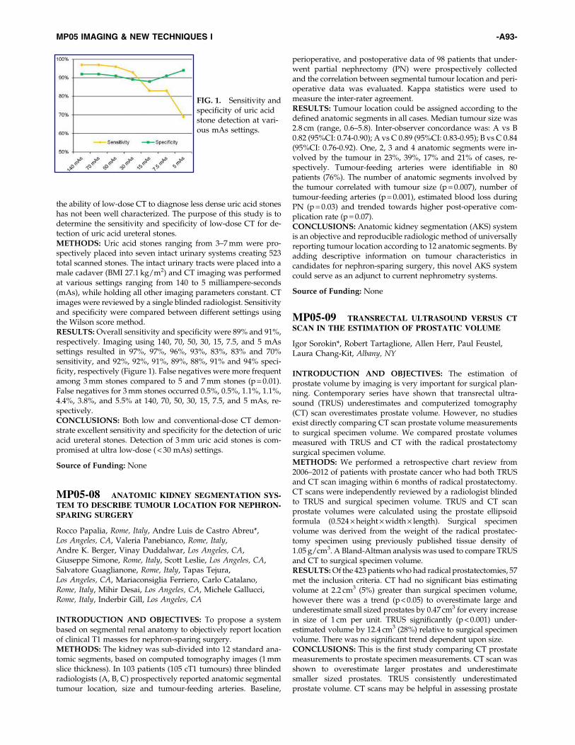
the ability of low-dose CT to diagnose less dense uric acid stoneshas not been well characterized. The purpose of this study is todetermine the sensitivity and specificity of low-dose CT for de-tection of uric acid ureteral stones.METHODS: Uric acid stones ranging from 3–7 mm were pro-spectively placed into seven intact urinary systems creating 523total scanned stones. The intact urinary tracts were placed into amale cadaver (BMI 27.1 kg/m2) and CT imaging was performedat various settings ranging from 140 to 5 milliampere-seconds(mAs), while holding all other imaging parameters constant. CTimages were reviewed by a single blinded radiologist. Sensitivityand specificity were compared between different settings usingthe Wilson score method.RESULTS: Overall sensitivity and specificity were 89% and 91%,respectively. Imaging using 140, 70, 50, 30, 15, 7.5, and 5 mAssettings resulted in 97%, 97%, 96%, 93%, 83%, 83% and 70%sensitivity, and 92%, 92%, 91%, 89%, 88%, 91% and 94% speci-ficity, respectively (Figure 1). False negatives were more frequentamong 3 mm stones compared to 5 and 7 mm stones (p = 0.01).False negatives for 3 mm stones occurred 0.5%, 0.5%, 1.1%, 1.1%,4.4%, 3.8%, and 5.5% at 140, 70, 50, 30, 15, 7.5, and 5 mAs, re-spectively.CONCLUSIONS: Both low and conventional-dose CT demon-strate excellent sensitivity and specificity for the detection of uricacid ureteral stones. Detection of 3 mm uric acid stones is com-promised at ultra low-dose ( < 30 mAs) settings.
Source of Funding: None
MP05-08 ANATOMIC KIDNEY SEGMENTATION SYS-
TEM TO DESCRIBE TUMOUR LOCATION FOR NEPHRON-
SPARING SURGERY
Rocco Papalia, Rome, Italy, Andre Luis de Castro Abreu*,Los Angeles, CA, Valeria Panebianco, Rome, Italy,Andre K. Berger, Vinay Duddalwar, Los Angeles, CA,Giuseppe Simone, Rome, Italy, Scott Leslie, Los Angeles, CA,Salvatore Guaglianone, Rome, Italy, Tapas Tejura,Los Angeles, CA, Mariaconsiglia Ferriero, Carlo Catalano,Rome, Italy, Mihir Desai, Los Angeles, CA, Michele Gallucci,Rome, Italy, Inderbir Gill, Los Angeles, CA
INTRODUCTION AND OBJECTIVES: To propose a systembased on segmental renal anatomy to objectively report locationof clinical T1 masses for nephron-sparing surgery.METHODS: The kidney was sub-divided into 12 standard ana-tomic segments, based on computed tomography images (1 mmslice thickness). In 103 patients (105 cT1 tumours) three blindedradiologists (A, B, C) prospectively reported anatomic segmentaltumour location, size and tumour-feeding arteries. Baseline,
perioperative, and postoperative data of 98 patients that under-went partial nephrectomy (PN) were prospectively collectedand the correlation between segmental tumour location and peri-operative data was evaluated. Kappa statistics were used tomeasure the inter-rater agreement.RESULTS: Tumour location could be assigned according to thedefined anatomic segments in all cases. Median tumour size was2.8 cm (range, 0.6–5.8). Inter-observer concordance was: A vs B0.82 (95%CI: 0.74-0.90); A vs C 0.89 (95%CI: 0.83-0.95); B vs C 0.84(95%CI: 0.76-0.92). One, 2, 3 and 4 anatomic segments were in-volved by the tumour in 23%, 39%, 17% and 21% of cases, re-spectively. Tumour-feeding arteries were identifiable in 80patients (76%). The number of anatomic segments involved bythe tumour correlated with tumour size (p = 0.007), number oftumour-feeding arteries (p = 0.001), estimated blood loss duringPN (p = 0.03) and trended towards higher post-operative com-plication rate (p = 0.07).CONCLUSIONS: Anatomic kidney segmentation (AKS) systemis an objective and reproducible radiologic method of universallyreporting tumour location according to 12 anatomic segments. Byadding descriptive information on tumour characteristics incandidates for nephron-sparing surgery, this novel AKS systemcould serve as an adjunct to current nephrometry systems.
Source of Funding: None
MP05-09 TRANSRECTAL ULTRASOUND VERSUS CT
SCAN IN THE ESTIMATION OF PROSTATIC VOLUME
Igor Sorokin*, Robert Tartaglione, Allen Herr, Paul Feustel,Laura Chang-Kit, Albany, NY
INTRODUCTION AND OBJECTIVES: The estimation ofprostate volume by imaging is very important for surgical plan-ning. Contemporary series have shown that transrectal ultra-sound (TRUS) underestimates and computerized tomography(CT) scan overestimates prostate volume. However, no studiesexist directly comparing CT scan prostate volume measurementsto surgical specimen volume. We compared prostate volumesmeasured with TRUS and CT with the radical prostatectomysurgical specimen volume.METHODS: We performed a retrospective chart review from2006–2012 of patients with prostate cancer who had both TRUSand CT scan imaging within 6 months of radical prostatectomy.CT scans were independently reviewed by a radiologist blindedto TRUS and surgical specimen volume. TRUS and CT scanprostate volumes were calculated using the prostate ellipsoidformula (0.524 · height · width · length). Surgical specimenvolume was derived from the weight of the radical prostatec-tomy specimen using previously published tissue density of1.05 g/cm3. A Bland-Altman analysis was used to compare TRUSand CT to surgical specimen volume.RESULTS: Of the 423 patients who had radical prostatectomies, 57met the inclusion criteria. CT had no significant bias estimatingvolume at 2.2 cm3 (5%) greater than surgical specimen volume,however there was a trend (p < 0.05) to overestimate large andunderestimate small sized prostates by 0.47 cm3 for every increasein size of 1 cm per unit. TRUS significantly (p < 0.001) under-estimated volume by 12.4 cm3 (28%) relative to surgical specimenvolume. There was no significant trend dependent upon size.CONCLUSIONS: This is the first study comparing CT prostatemeasurements to prostate specimen measurements. CT scan wasshown to overestimate larger prostates and underestimatesmaller sized prostates. TRUS consistently underestimatedprostate volume. CT scans may be helpful in assessing prostate
FIG. 1. Sensitivity andspecificity of uric acidstone detection at vari-ous mAs settings.
MP05 IMAGING & NEW TECHNIQUES I -A93-

volumes in preoperative planning. This is very useful given thewidespread use and availability of CT scans.
Source of Funding: none
MP05-10 THE PREDICTION OF POSTOPERATIVE
TOTAL RENAL FUNCTION BY RENAL CORTEX VOLU-
METRY WITH MULTIDETECTOR COMPUTED TOMO-
GRAPHY (MDCT) AFTER NEPHRECTOMY
Shuji Isotani*, Masahiro Inoue, Itabashi, Tokyo, Japan,Hiroshi Shimoyama, Yasuhiro Noma, Bunkyo, Tokyo, Japan,Shino Tokiwa, Keisuke Saito, Takashi Yoshi, Hisamitsu Ide,Satoru Muto, Koji Takeshita, Itabashi, Tokyo, Japan, Shigeo Horie,Bunkyo, Tokyo, Japan, Raizo Yamaguchi, Itabashi, Tokyo, Japan
INTRODUCTION AND OBJECTIVES: To determine thepostoperative total renal function and predict the develop-ment of chronic kidney disease (CKD) after renal surgery,we measured renal cortex volume (RCV) and examined theestimation of postoperative eGFR of from multidetectorCT volumetry.METHODS: From 2011 to 2013, 42 patients 29 men and 13women (median; 61.0 years old, range 34 to 81) with preop-erative contrast enhanced multidetector CT imaging in slicethickness was 0.5 mm to 3 mm, were underwent renal removalsurgery at Teikyo University Hospital in Japan. Preoperativeand postoperative renal function was assessed by serum Crlevel and estimating glomerular filtration rate (eGFR) usingequation for Japanese population. The renal cortex volume(RCV) was also calculated manually with using imagingvolumetry computer software. Addition of normal sideRCV and disease side RCV that was subtracted by tumorregion determined as total RCV. The predicted postoperativeRCV was defined as normal side RCV in the nephrectomy ornephroureterectomy cases, and in the partial nephrectomycases predicted RCV was estimated by subtraction of tumorregion with 1 cm margin. Postoperated eGFR was estimatedfrom predicted RCV, and determined the association of pre-dicted RCV and the development of CKD (GFR < 60 mL/min/1.73 m2).RESULTS: The predicted postoperative RCV were associatedwith the development of CKD (p = < 0.05) and strongly corre-lated with postoperative renal function. A strong correlation(r = 0.881, p < 0.001) was also seen between postoperated eGFRand calculated predicted eGFR from CT volumetry.CONCLUSIONS: Preoperative CT based RPV measurements areindependently associated with the development of CKD in pa-tients undergoing renal surgery. Our results also demonstratedthat it might be the prediction tool with postoperative renalfunction from preoperative multidetector CT volumetry.
Source of Funding: none
MP05-11 RADIATION EXPOSURE DURING IMAGE-
GUIDED ABLATION OF SMALL RENAL MASSES: A MULTI-
CENTER CHARACTERIZATION OF RISK FACTORS
Chad R. Tracy*, Amit Gupta, Iowa City, IA, Jeffrey Gahan, Dallas,TX, Zhamshid Okhunov, South Orange, CA, Sammy E Elsamra,Nithin Theckumparampil, New Hyde Park, NY,Maurilio Garcia-Gil, Dallas, TX, Shiliang Sun, Iowa City, IA,Igor Lubko, New Hyde Park, NY, Sandy Lall, South Orange, CA,Louis R. Kavoussi, New Hyde Park, NY, Jaime Landman,South Orange, CA, Jeffrey A. Cadeddu, Dallas, TX
INTRODUCTION AND OBJECTIVES: Despite the wide utili-zation of CT-guidance for performance of percutaneous renalablation, the total dosage of radiation per patient has not beenevaluated. We sought to define the typical radiation exposurethat occurs during percutaneous ablation of renal tumors and todetermine the factors that may influence these levels.METHODS: A multicenter retrospective study of all patientsundergoing percutaneous ablation of a renal mass by eithercryoablation (CA) or radiofrequency ablation (RFA) was per-formed. The analysis included 123 patients with a solitary renalmass £4 cm who underwent treatment between 1/2010 and 5/2013 at four large academic centers and had radiation exposuredata available for review. Total effective abdominal radiationexposure expressed in millisieverts (mSv) was calculated directlythrough conversion of the dose–length product (DLP) usingpublished conversion coefficients. Effective dose was comparedamongst multiple variables that may effect exposure.RESULTS: The mean age was 70 years and 69% of the patientswere male. The mean BMI and tumor size were 29.4 and 2.2 cm,respectively. The average nephrometry score was 6.5, with 52%categorized as low (4–6), 42% as moderate (7–9) and 6% as high(10–12) complexity lesions. The median radiation dose per abla-tion was 35 mSv. Large variations were seen between institutionsand median exposure varied from 6.3 to 52.4 mSv (p < 0.001; fig-ure 1). On multivariate linear regression analysis, institution andBMI predicted the total radiation exposure (p < 0.001 for both).Other characteristics such as tumor size, location and depth,proximity to collecting system, nephrometry score, age andgender did not predict radiation exposure.CONCLUSIONS: The median radiation dose delivered duringpercutaneous ablation of a small renal mass is 35 mSv. Total ex-posure is more dependent on institution and patient BMI than on
-A94- MP05 IMAGING & NEW TECHNIQUES I

tumor characteristics. Future work should include a rigorous re-view and standardization of CT protocols amongst institutions inorder to lower overall radiation exposure for the patient.
Source of Funding: none
MP05-12 NOVEL USE OF INDOCYANINE GREEN FOR
IDENTIFICATION OF SENTINEL LYMPH NODES AND
MESENTERIC ANGIOGRAPHY TO ASSESS BOWEL VAS-
CULARITY DURING ROBOTIC RADICAL CYSTECTOMY
WITH INTRACORPOREAL URINARY DIVERSION
Ted Manny*, Ashok Hemal, Winston-Salem, NC
INTRODUCTION AND OBJECTIVES: Indocyanine green(ICG) has been described in the urologic literature during partialnephrectomy. Further urologic applications of the dye existbased on its ability to act as an intravenous contrast agent toperform real time fluorescence angiography and interstitial in-jection to perform real time fluorescence lymphangiography.We describe the first clinical series of fluorescence enhancedrobotic radical cystectomy (FERRC) in which we perform real-time interstitial injection for lymph node mapping and intra-venous injection to assess bowel vascularity during in-tracorporeal urinary diversion.METHODS: Ten patients with high-grade bladder cancerunderwent FERRC. Before robot docking rigid cystoscopywas performed during which a 2.5 mg/mL ICG solution wasinjected in the bladder submucosa and detrusor circumferen-tially around the tumor. After robot docking, parameters de-scribing the time course of tissue fluorescence and pelviclymphangiography were systematically recorded. Lymphaticpackets containing hyperfluorescent lymph nodes were con-sidered the sentinel drainage. After cystectomy an ileal seg-ment was marked for urinary diversion and an additional5 mg of ICG was given intravenously. Real-time fluorescence
mesenteric angiography was then performed to visualize thevascular arcades of the conduit segment and guide endo-scopic stapling.RESULTS: Potential sentinel lymphatic drainage was visual-ized in 90% (9/10) patients at a median 30 minutes post-injection (Fig 1). During intracorporeal urinary diversion,hyperfluorescent mesenteric arcades were visible in all patients(Fig 2).CONCLUSIONS: Fluorescence-enhanced robotic radical cy-stectomy utilizing combined percutaneous and intravenous in-jection of ICG is safe and feasible. FERRC allows for reliablebladder tumor marking, identification of sentinel lymphaticdrainage, and identification of mesenteric vasculature in themajority of patients.
Source of Funding: none
MP05-13 DEFINING THE ROLE OF INTRAOPERA-
TIVE TRANSESOPHAGEAL ECHOCARDIOGRAPHY DUR-
ING INFERIOR VENA CAVAL TUMOR THROMBECTOMY
IN RENAL CELL CARCINOMA
Mark Ball*, Vivek Arora, Mary Beth Brady, Ashish Shah,James Black, Mohamad Allaf, Baltimore, MD
INTRODUCTION AND OBJECTIVES: Previous reports de-scribing the use of TEE in patients undergoing inferior vena cava(IVC) tumor thrombectomy and radical nephrectomy (RN) forrenal cell carcinoma (RCC) have demonstrated the feasibility ofthe technique, but data to support its utility for surgical decisionmaking is lacking. Here we present a retrospective review ofpatients with RCC undergoing nephrectomy and IVC tumorthrombectomy and define the impact of TEE on their in-traoperative surgical management.METHODS: Our departmental renal mass database was que-ried for patients undergoing RN and IVC thrombectomy from2007 to 2012. IVC thrombus extension was classified as level I:renal vein, level II: subhepatic IVC, level III: retrohepatic IVCand level IV: right atrium. Level I cases were excluded fromanalysis. Cases in which intraoperative TEE was used were re-viewed, and impact on surgical management was determinedfrom the operative records and TEE report. Changes in man-agement were defined when TEE determined new extent ofthrombus, identified incomplete resection, identified new pul-monary embolus (PE), or identifed propogation of PE. Changesin surgical approach were defined as need to initiate cardio-pulmonary bypass (CPB), need to re-initiate CPB, or ability toavoid CPB.RESULTS: A total of 36 cases of RCC with IVC involvement wereidentified. Of these cases, 9 had level II, 19 had level III and 8 hadlevel IV involvement. A comprehensive intra-operative TEE
Table 1. Diagnostic Utility and Surgical Impact
of Intraoperative TEE
MP05 IMAGING & NEW TECHNIQUES I -A95-

examination was performed in 23 (64%) cases – 3 (33%) in level II,11 (57%) in level III and 9 (100%) in level IV. In level II, newdiagnostic information was obtained in 11% (n = 1) of patients. Ingroup III and IV new diagnostic information was obtained in 37%(n = 7) and 100% (n = 8) patients. Changes in surgical approachoccurred in 5%(n = 1) and 62% (n = 5) of patients in level III and IVrespectively.CONCLUSIONS: Intraoperative TEE provides real-time infor-mation regarding the presence and extent of IVC involvement.The diagnostic yield of TEE increases in patients with greater IVCtumor thrombus extension and the information obtained has asignificant impact on surgical decision making particularly inpatients with intracardiac tumor extension. TEE is a safe anduseful modality that should be considered in all IVC thrombec-tomies.
Source of Funding: none
MP05-14 ONCOLOGIC RESULTS OF PERCUTANEOUS
RENAL CRYOABLATION AT A MEDIAN FOLLOW-UP OF 24
MONTHS
Anees Fazili*, Tiffany Lee, Sriram Venigalla, Louis Eichel,Rochester, NY
INTRODUCTION AND OBJECTIVES: Percutaneous renalcryoablation is becoming increasingly offered for patients withsuspicious renal lesions who are poor surgical candidates, yetthere are few large studies that have examined its long-term ef-ficacy. The purpose of the present study was to examine the on-cologic outcomes of percutaneous renal cryoablations over an 8year period at our institution.METHODS: We retrospectively reviewed the charts of all pa-tients who underwent a percutaneous renal cryoablation at ourinstitution from April 2005 to February 2013. This included 139patients being treated by 7 urologists and 4 interventional radi-ologists. Follow-up data was available on 123 patients. Post-ablation follow-up imaging was reviewed for areas of contrastenhancement concerning for residual or recurrent disease. Re-sidual disease was defined as enhancement on follow-up imagingwithin the initial 3 months following treatment. Recurrence wasdefined as new enhancement found on imaging after this 3 monthperiod. Indications for percutaneous cryoablation as well as im-mediate and long-term complications of the procedure were alsonoted.RESULTS: Mean and median follow-up were 28 and 24 months,respectively (range 1- 85). Mean tumor size was 2.60 cm (range1–5). Although 27/123 patients (22%) experienced a complicationfrom the procedure, only 2 patients (1.6%) experienced a highgrade complication. Biopsies were performed on 44 patients, andrevealed 28 patients with renal cell carcinoma, 2 with oncocyticneoplasms, 6 with oncocytomas, and 8 with biopsies negative formalignancy. Nine patients (7.3%) had residual disease, and an-other 9 patients ultimately had recurrence of disease. Amongthese 18 patients, 12 underwent repeat percutaneous cryoabla-tion, which was successful in 8 patients; 4 underwent radicalnephrectomy; and 4 were ultimately referred to medical oncologyfor metastatic disease. Mean tumor size was not statistically dif-ferent between patients who remained disease free and thosepatients who experienced residual or recurrent disease (2.55 vs.2.68 cm, p = 0.59). Patients with endophytic tumors had a statis-tically significant increased risk of having residual or recurrentdisease compared to those with exophytic tumors (50.0% vs.12.2%, p = 0.016).
CONCLUSIONS: Percutaneous renal cryoablation remains asafe and effective method of local tumor control in properly se-lected patient populations. Endophytic tumor status was associ-ated with an increased risk of treatment failure.
Source of Funding: None
MP05-15 REPEAT PERCUTANEOUS CT-GUIDED
CRYOABLATION FOR LOCALLY RECURRENT RENAL
CELL CARCINOMA
Zhamshid Okhunov*, Orange, CA, Samuel Juncal, Arvin George,Orange, CA, Sammy Elsamra, New Hyde Park, CA, Daniel Moreira,New Hyde Park, NY, Nithin Theckumparampil, Orange, CA,Martin Hofmann, Please choose an option below,Fotima Asqarova, Puja Patel, Orange, CA, Louis Kavoussi,New Hyde Park, NY, Igor Lobko, New Hyde Park, NY,Jaime Landman, Orange, CA
INTRODUCTION AND OBJECTIVES: The management oflocal recurrence after cryoablation for renal cell carcinoma (RCC)remains a dilemma. We report our experience with repeat per-cutaneous cryoablation (PCA) for local recurrence following priorrenal cryoablation.METHODS: We performed a retrospective review of prospec-tively established databases of patients who underwent cryoa-blation for RCC at two academic institutions. Patients whounderwent repeat PCA for local recurrence following primarycryoablation were included in the study. Perioperative compli-cations and oncologic outcomes were evaluated.RESULTS: A total 250 patients underwent primary cryoabla-tion for RCC and 20 (8%) patients were identified who un-derwent repeat PCA for 21 recurrent tumors. Sixteen patientsfailed PCA and four failed laparoscopic cryoablation (LCA).There were 14 males (70%) and 6 (30%) females with the meanage of 64.8 (range 47–82). Nine patients with ASA grade II and11 with ASA grade III. Seven (33%) tumors were located in theupper pole, 7 (33%) inter-polar, and 7 (33%) in the lower pole.Seven (33%) tumors were located anteriorly and 14 (67%)posteriorly. Biopsy at the time of repeat CA procedure revealedclear cell RCC in 14 patients, 3 papillary RCC, 1 chromophobeRCC and 3 were non-diagnostic. All repeat cryoablation pro-cedures were completed successfully. There were no majorcomplications or deaths associated with the procedure. Onlyone patient was identified to have perirenal hematoma in thepostoperative period. No embolization or transfusion was re-quired.
Of the 18 biopsy proven RCC patients, with the median follow-up of 30 months (range 7–63), 3 (16%) were identified to havelocal recurrence. One patient was identified to have enhancinglesion at 13 months following repeat PCA which was managedwith a third PCA. Additional, two patients had recurrence at 6and 35 months, after LCA. Both patients successfully underwentlaparoscopic partial nephrectomy. No patient developed local ordistant metastasis.CONCLUSIONS: Repeat percutaneous renal cryoablation forlocally recurrent disease is technically feasible, has a lowcomplication rate, and demonstrates acceptable short-termoncologic outcomes in this challenging population. Furtherstudies with larger number of patients and durable follow-upare required to elucidate the role of PCA in this patientpopulation.
Source of Funding: none
-A96- MP05 IMAGING & NEW TECHNIQUES I

MP05-16 DEVELOPMENT OF A TARGETTED PHO-
TOIMMUNOTHERAPY PLATFORM IN THE MANAGE-
MENT OF BLADDER CANCER
Srinivas Vourganti*, Michael Weintraub, Quentin Li,Piyush Agarwal, Bethesda, MD
INTRODUCTION AND OBJECTIVES: We have developed anovel precision medicine based photoimmunotherapy (PIT)platform to treat patients with bladder cancer. This techniqueinvolves the conjugation of specifically chosen targeted mono-clonal antibodies to a phthalocyanine dye, IR700, which is acti-vated by near-infrared light. When activated by light, IR700 dyerapidly destroys cells though the production of reactive oxygenspecies.METHODS: Using flow cytometry immunostaining, we char-acterized the expression profile of bladder cancer cell lineUMUC5 and confirmed strong expression of EGFR. Panitu-mumab (human monoclonal anti-EGFR; PAN) was conjugatedto IR700 photosensitzer dye in order to specifically target thecytotoxic effect to overexpressors of the receptor. Cells weretreated in the following treatment arms: 1) Control; 2) Un-conjugated PAN; 3) Conjugated PAN-IR700; 4) and blockingcondition of initial treatment with unconjugated PAN subse-quently treated with conjugate PAN-IR700. An additional 4treatment arms were exposed to 4J of near infrared light: 5)Light alone; 6) PAN with Light; 7) Conjugated PAN-IR700 withLight; and 8) blocking condition as above with Light. Cyto-toxocity was evaluated with flow cytometric detection of cal-cein acetomethoxy derivate viability surface staining andpropidium iodide as well utilizing raw hemacytometer countsof trypan blue exclusion. Treatments were repeated using anEGFR negative control cell line Balb3T3.RESULTS: Within 15 minutes of light exposure, PIT resulted inprofound cytotoxic effect in cells treated by PAN-IR700 conjugate(98% of cells nonviable). This effect was reversed by pretreatmentby unconjugated antibody (92% viable). Light exposure aloneresulted in minimal toxicity (96% viable). For negative controlEGFR negative Balb3T3, no cytotoxic effect was noted (between94 and 97% viability in all conditions). Of UMUC5 cells detectedviable on flow cytomtery, IR700 binding (as detected on the FL4channel) was significantly lower than in those cells detected asnonviable.CONCLUSIONS: PIT represents a targeted, precision medicineapproach to tailoring cytotoxic therapy to specific bladder cancermarkers utilizing commercially available antibody therapeutics.Our group is currently developing in vivo orthotopic rodentmodels of this platform in the hopes to fast track this technologyinto Phase I clinical trials in humans.
Source of Funding: NIH Intramural Funding
MP05-17 INTEROBSERVER RELIABILITY AND RE-
PRODUCIBILITY OF S.T.O.N.E. NEPHROLITHOMETRY
FOR RENAL CALCULI
Zhamshid Okhunov*, Orange, CA, Alberto Perez-Lanzac,Mohammad Helmy, Ashleigh Menhadji, Philip Bucur, Orange,CA, Surendra Kolla, Orange, CA, Jane Cho, Kathy Osann,Achim Lusch, Jaime Landman, Orange, CA
INTRODUCTION AND OBJECTIVES: Objective: The STONEnephrolithometry was previously developed as a stone com-plexity scoring system and prospectively validated in a singlecenter percutaneous nephrolithotomy (PCNL) cohort. In this
study we assess the reliability of the S.T.O.N.E. nephrolithometrythe scoring system by testing its reproducibility between differentobservers.METHODS: Preoperative images of 58 subjects that underwentPCNL were reviewed. Medical students, urology residents, onefellow and a urology attending independently reviewed allimages and scored the renal stones. Interobserver reliabilities ofthe total score for all categories and each component wereevaluated by the intraclass correlation (ICC) and a Kappa co-efficient.RESULTS: The interobserver reliability for the total scoredemonstrated high correlations for all components and totalscore (ICC = S, T, O, N, E and total 0.80, 0.97, 0.89, 0.84, 0.91 and0.87 respectively). Kappa rates for individual components be-tween 2 medical students were 0.36, 1, 0.31, 0.45, 0.33 and 0.30for the S, T, O, N, E components and total score, respectively.Kappa values between the two urology residents were 0.71, 1,0.92, 0.79, 0.93 and 0.67 for S, T, O, N, E components and totalscore respectively. Kappa values between the urology fellowand an attending physician were 0.95, 1, 0.88, 0.94, 0.89 and 0.87for S, T, O, N, E components and total score respectively. P valuefor all the scoring components was < 0.05, indicating that theestimated kappa was not a result of chance.CONCLUSIONS: The S.T.O.N.E. nephrolithometry has excellentinterobserver reliability. Quantifying the S and N metrics werethe most challenging and least reliable. Standardized protocols tomeasure these components should be considered to improve ac-curacy and reproducibility of the scoring system.
Source of Funding: none
MP05-18 ENDOVASCULAR COIL OCCLUSION (ECO) -
THE REAL ALTERNATIVE METHOD FOR VENOUS LEAK
CORRECTION
Dmitriy Kurbatov*, Alexandr Lepetukhin, Ivan Sitkin,Sergey Dubsky, Moscow, Russian Federation
Table 1. Kappa Test Results for Pairs of Raters
with Similar Level of Training
Table 2. Intraclass Correlation as Measure
of Agreement Between all Six Raters
and 4 Raters Excluding Medical Students
MP05 IMAGING & NEW TECHNIQUES I -A97-

INTRODUCTION AND OBJECTIVES: Pathological venousdrainage (PVD) can be the basic or accompanying symptom at20–60% of patients with erectile dysfunctions (ED). Is is knownthat PVD includes of three types: distal, proximal and mixed, thatcourses the choice the method of the surgery. Since 2009 we ap-plied the safe and highly effective alternative technique - en-dovascular coil occlusion (ECO) for proximal/mixed type ofvenous leak correction.METHODS: 1649 pts with complaints on ED were examinedduring the 2009–2012. 1053 pts (63%) age of 18–59 ys sufferedvascular ED and 117 pts (11%) of them were with isolated veno-occlusive ED of proximal/mixed types of PVD. All 117 pts re-ceived initially maximal dozes of inhibitors PDE-5 not less than 3months but were not responded. Conventional diagnostic testincluded hormonal tests, Doppler Duplex Sonography, DynamicInfusion Cavernosometry and Cavernosography. All patientshad normal arterial blood inflow, testosterone, prolactine, TSH,were not smoking and underwent ECO. After pathological ve-nous leak visualization by angiographic technique via peniledeep dorsal vein intraluminal coil/coils were inserted in theSantoriny’s venous plexus. The surgery time duration was 10–40 min.RESULTS: ECO is out-patient operation or need one day hospi-talization with minimal complications. The follow-up was 34 – 21(2–36) months. Totally 114 pts (97,4%) – restored the erectilefunction. 105 pts (89,7%) – do not use PDE-5 inhibitors further and9 pts (7,8%) use low dose PDE 5 inhibitors on demand. 3 pts(2,5%) had negative results due to cavernous fibrosis and un-derwent penile prostheses implantation.CONCLUSIONS: ECO might be seen as the method of choiceand the last chance for patient suffering veno-occlusive ED beforepenile prosthesis implantation.
Source of Funding: self funding
MP05-19 MR FUSION PROSTATE BIOPSIES ARE
FEASIBLE AND USEFUL IN A BUSY UROLOGIC PRACTICE
David Hatcher*, Joshua Cohn, Chicago, IL, Robert Silvers,Michael McGuire, Evanston, IL
INTRODUCTION AND OBJECTIVES: Magnetic resonanceimaging-transrectal ultrasound fusion (MR fusion) techniquesmay have improved diagnostic reliability as compared with tra-ditional transrectal ultrasound-guided (TRUS) biopsies in selectpatients. Its utilization to date has been primarily in researchcenters; we investigated its usage in a busy clinical urologypractice.METHODS: We reviewed men undergoing MR fusion biopsybetween October 2012 and May 2013. Selection for MR fusionbiopsy was based upon enrollment in active surveillance (AS),persistently elevated PSA with negative biopsy history, or patientpreference. Transabdominal 3-tesla multiparametric prostateMRI was performed on all patients at least 1 week prior to clinicbiopsy visit. One staff radiologist (RS) identified all suspiciouslesions, and fusion images were generated and uploaded to theBioJet (Geo Scan, Lakewood Ranch, FL) fusion machine. Biopsieswere targeted at MRI-identified regions of interest (ROI) as wellas normal-appearing prostate (non-ROI). All biopsies were per-formed by a single urologist (MSM) with the assistance of onemedical assistant. Analysis was performed to compare targetedand non-targeted biopsy results.RESULTS: 38 patients met inclusion criteria. Mean age was 66years [95% CI 63-68] and median PSA 6.2 ng/mL [IQR 4.4-12.3].50% of men were enrolled in AS and 74% had undergone previ-
ous TRUS biopsy (mean 2.6 prior biopsies [95% CI 2.0-3.2]). Themean number of ROI and non-ROI cores sampled was 3.3 [95% CI2.4-4.3] and 8.5 [95% CI 7.4-9.6], respectively (p < 0.001). MeanGleason score of identified cancers on targeted MR fusion biopsywas significantly higher than those found on prior non-targetedTRUS biopsy (7.2 [95% CI 5.8-8.4] vs. 6.1 [95% CI 5.7-6.4]). Meanurologist time required for biopsy completion was 11 minutes[range 8–15].CONCLUSIONS: MR fusion biopsy can be done effectively andefficiently in the busy urologist’s office. Given these findings andthat MR fusion technology may increase the likelihood of diag-nosing more aggressive cancers, more widespread use of thismodality is warranted.
Source of Funding: none
MP05-20 COMPARISON OF RELIABILITY OF THE
RENAL NEPHROMETRY SCORE BETWEEN RADI-
OLOGISTS AND UROLOGISTS
Samay Jain*, Khaled Shahrour, Toledo, OH
INTRODUCTION AND OBJECTIVES: The RENAL ne-phrometry score was developed to help categorize and stan-dardize renal tumor complexity as seen on cross sectionalimaging. For such scoring system to be useful, it not only needs tobe simple but also reliable and reproducible. Previous studieshave shown high reliability in assigning a RENAL score betweenurologists, but none have reported on the inter-observer reli-ability between urologists and radiologists. The purpose of thisstudy was to test the hypothesis that the RENAL score is a reliableand reproducible measure between urologists and radiologists.METHODS: In this institutional review board-approved study, aretrospective review of computerized tomography (CT) scans ofpatients with renal masses who had presented to our institutionwithin the past five years was reviewed. Masses were excluded inpatients with congenital renal anomalies such as APCKD,horseshoe, etc. Masses on CT scans that met the inclusion criteriawere assigned a RENAL nephrometry score by 5 full-time facultymembers; 3 radiologists and 2 urologists. They were providedwith a standardized description of the RENAL score and its ap-plication at the beginning of the study. Each observer docu-mented detailed RENAL score for each mass independently usingCT software (PACS, GE). The Krippendorff’s alpha coefficientwas used to detect inter-rater reliability of ratings done by thedifferent examiners. Paired t-test was used to compare betweenradiologists and urologists groups.RESULTS: A total of 64 patients and 72 distinct renal tumors wereanalyzed in this study. Mean RENAL nephrometry score was7.8 – 2.0 and 8.4 – 2.1 by the radiologists and urologists, respec-tively. Mean difference between both groups was 0.60 points(p < 0.05, Confidence Interval 0.39–0.80). Reliability coefficient foreach component of the RENAL score and total score were 0.89 (R),0.41 (E), 0.58 (N), 0.43 (A), 0.65 (L), and 0.77 respectively. Reliabilitycoefficients for both groups were high but that between radiolo-gists was higher than that between urologists (0.81 vs. 0.73).CONCLUSIONS: The RENAL nephrometry scoring system is areliable tool that should be used by both urologists and radiolo-gists when reporting renal masses found on CT. Higher interob-server correlation among radiologists than among urologists isevidence that adoption of such score by radiology is feasible as ameans of standardizing the reporting of renal masses.
Source of Funding: None
-A98- MP05 IMAGING & NEW TECHNIQUES I

MP05-21 COMBINED PATIENT AND STONE MOR-
PHOMETRY ENHANCE PREDICTION OF STONE COMPO-
SITION
Kara L Watts*, Tian C Zhou, Joseph Divito, David M Hoenig,Bronx, NY
INTRODUCTION AND OBJECTIVES: The use of Hounsfieldunits (HU) on CT imaging for prediction of stone composition hasbeen reported. Uric acid (UA) calculi have a much lower averageattenuation compared to calcium oxalate calculi, with reportedaverages ranging from < 300–400 HU and > 655–800, respectively.We have previously demonstrated a significant association of vis-ceral fat area (VFA), as calculated on CT imaging, with UA stoneformation. We sought to determine if the use of a region of interest(ROI) pixel analysis of calculi, in combination with VFA mea-surements, would enhance the prediction of stone composition.METHODS: Retrospective review of an IRB-approved databaseof 219 patients undergoing PCNL was performed. Twenty pa-tients with predominantly UA stone composition ( > 80%) wereidentified and compared to 20 patients with > 90% calcium oxa-late (CaOx) monohydrate or dihydrate calculi. ROI HU rangeswere obtained for each patient from a single axial CT slice con-taining the largest cross-sectional stone image. VFA was alsocalculated from a single axial CT slice at the level of the umbilicusfor each patient (TerraRecon Inc Aquarius iNuition, version4.4.6.100.2862). These values were compared using simple 2-si-ded t-tests.RESULTS: Stone ROI analysis revealed a significantly loweraverage minimum and maximum HU range for UA compared toCaOx stone formers, 211 vs 370 (p < 0.001) and 602 vs 1261(p < 0.0000001). Average HU was also significantly higher amongCaOx stone formers versus UA stone formers, 962 vs 448(p < 0.000001). Finally, VFA was significantly higher among theUA group, 243 kg/m2 vs 129 kg/m2 (p < 0.00001).CONCLUSIONS: ROI pixel-by-pixel analysis of HU attenuationfor UA and CaOx stones yielded higher average HU attenuationscompared to those previously reported. In addition, the range ofHU values for each stone was wide (larger in CaOx stones), withthe average minimum and maximum HU units for each stonetype much lower than or higher than previously reported averagevalues, respectively. The use of an ROI HU range for stone pre-diction may, therefore, offer a more comprehensive and accuratemethod of predicting stone composition compared to single pointHU analysis. In addition, the use of VFA calculation may alsofurther enhance prediction of stone composition, with higherVFA values associated with UA stones versus CaOx stones. Thecombination of ROI pixel analysis with VFA calculation suggeststhe possibility of enhanced prediction of predominantly-uric acidstones, including nomogram creation.
Source of Funding: none
MP05-22 PREOPERATIVE PLANNING WITH NON-
CONTRAST COMPUTED TOMOGRAPHY IN THE PRONE
AND SUPINE POSITION FOR PERCUTANEOUS NE-
PHROLITHOTOMY: A PRACTICAL OVERVIEW
Giovanni S. Marchini*, Fernanda Berto, Fabio C. Vicentini,Eduardo Mazzucchi, Miguel Srougi, São Paulo, Brazil
INTRODUCTION AND OBJECTIVES: Preoperative planningfor PCNL usually comprises a NCCT with the patient in the su-pine position; however, most PCNLs are performed in the pronedecubitus worldwide; the purpose of our study was to system-
atically evaluate if the supine NCCT allows accurate prediction ofsurgical parameters when the patient is turned prone.METHODS: After IRB approval, patients scheduled for PCNLwere prospectively enrolled in the study. All patients underwenta NCCT in the supine position and subsequently a NCCT in theprone position with boosters by the shoulders and abdomen. Theprimary end-point was to compare between positions twoimaginary lines as previously defined by Prassopoulos et al foreach posterior calyx (upper; mid – renal hilum level; lower pole)of both kidneys: Line I was drawn horizontally in the coronalplane in contact with the posterior edge of the kidney; line II fromthe antero-lateral edge of the vertebra through the middle of thecalyces (ideal puncture line). Organs crossed or posterior to LinesI/II were considered at risk. Secondary end-points were: distancebetween Line I and anterior extremity of the vertebra (renaldepth) and maximum access angle (vertex in the calyx, one line inthe first adjacent organ; another in the lateral border of theparaspinous muscle). The Student T Test and Chi-square/Fisherexact test were used. Significance was set at p < 0.05.RESULTS: From May/2012 and March/2013, 37 patients met theinclusion criteria and were considered in the analysis; 21 (56.7%)were female with mean BMI of 28.3 – 4.9 (16.8–42.0) Kg/m2. Forthe right kidney, supine position was associated with more or-gans crossed by Line I and II in the upper calyx (table 1); For theleft kidney, a significant difference was only found for Line II inthe upper calix (p = 0.03). Both kidneys showed a tendency to bedeeper in the supine position, while the supine decubitus usuallyprovided a wider access angle.CONCLUSIONS: The supine NCCT is not accurate to planPCNL access in the prone position. Prone decubitus is associatedwith more potential organ injuries mainly in the upper pole whenconsidering an ideal tract line. Although tract length is expectedto be longer in the supine position, it may be compensated by awider access angle.
Source of Funding: None
MP05-23 THE FIRST UNITED STATES SERIES USING
THE TRANSURETHRAL SUPRAPUBIC ENDO-CYSTOST-
OMY DEVICE FOR SUPRAPUBIC CATHETER INSERTION
Robert Larke*, Vassilis Siomos, Brian Flynn, Aurora, CO
Table 1. Comparative Analysys of Primary
and Secondary Parameters Acquired from
NCCT Between the Prone and Supine Position
MP05 IMAGING & NEW TECHNIQUES I -A99-

INTRODUCTION AND OBJECTIVES: The Transurethral Su-prapubic endo-Cystostomy Device (T-SPeC�) device, is a newdevice used for introducing a suprapubic catheter via a retro-urethral (inside-to-out) approach. This device can be compared toa Lowsley retractor, however the T-SPeC� device adjusts tocompensate for abdominal girth and is disposable. An early Ca-nadian series of four patients demonstrated the device to be safeand efficient. The objective of this study is to review our earlyexperience using the T-SPeC� device.METHODS: After receiving patient consent, urology patients atthe University of Colorado Hospital underwent placement of asuprapubic catheter via the T-SPeC� device by a single surgeon.RESULTS: Thirty-five 18 F suprapubic catheters were successfullyplaced in patients using the T-SPeC� device. The indications forplacement were: neurogenic bladder, urethral stricture, urinaryretention, or for urethral rest as a part of complex genitourinaryreconstruction. Thirty of the patients were female. Thirty-threepatients underwent general anesthesia, one with sedation, andanother with strictly local anesthetic. The mean patient age was 56(range 41–90) and the mean BMI was 29 kg/m2 (range 17–50). Theaverage skin to bladder distance was 6.6 cm (range 4–11). The meanoperative time was 4.2 minutes (range 3–5.5). There were no intra-operative complications and all patients had successful placementof a suprapubic catheter. The capture housing was missed twicefollowing activation of the trocar, but this did not affect the out-come. There were no post-operative complications within 30 daysof the procedures, such as urinary tract infection or wound infec-tion. Three urinary tract infections were reported within 60 days ofprocedure. One patient required a urethral catheter due to recur-rent catheter obstruction from urinary sediment and another pa-tient pulled out her suprapubic catheter.CONCLUSIONS: Our series demonstrates that the T-SPeC�
device can be placed safely, efficiently, and effectively in patientsrequiring long-term or temporary suprapubic tube placement.
Source of Funding: Swan Valley Medical donated the first 25devices
MP05-24 RENAL CALCULI AND PLAIN IMAGING-
RADIOLUCENT OR RADIO OPAQUE?
Paul Healy*, Dublin, Ireland, Saraswathy Suresh Babu, Leicester,United Kingdom, Priya Kumar, Preston, United Kingdom,Masood Khan, Leicester, United Kingdom
INTRODUCTION AND OBJECTIVES: It is traditionally taughtthat up to 90% of urinary tract calculi are visible on plainabdominal radiographs (XR). In studies comparing XR withcomputed tomography (CT) the radio-opacity rates vary from63–77%. This is of relevance in deciding how to follow up stones.We compared the radio-opacity rates and stone size for XR versusCT in our institute.METHODS: We identified 83 patients who presented with renalcolic to our institute between April 2011 and July 2010. Criteria forinclusion included having both a XR and CT. 16 patients wereexcluded as they did not have any plain imaging. All XRs werereviewed by an independent radiologist who was blinded to theresults.RESULTS: Of the 67 renal calculi identified on CT imaging, 23/67(34%) had calculi visualised on plain XR. The mean stone size was5.7 mm (2–13 mm) and 4.6 mm (1–16 mm) for stones on plain XRand CT respectively.CONCLUSIONS: Our study shows that plain radiographsidentified only 34% of renal calculi subsequently highlighted by
CT imaging – a much lower figure than conventionally thought.Plain film alone is not therefore universally reliable to follow upall stones.
Source of Funding: None
MP05-25 PERCUTANEOUS NEPHROSTOMY MADE
EASY: ELECTROMAGNETIC NEEDLE GUIDANCE WITH
TRACKED ULTRASOUND SNAPSHOTS IN A SIMULATION
MODEL
Michael Fuoco*, Tamas Ungi, Rob Siemens, Gabor Fichtinger,Darren Beiko, Kingston, Canada
INTRODUCTION AND OBJECTIVES: Percutaneous ne-phrostomy (PCN) is often required urgently in the context ofacute renal failure or urosepsis, and may be performed electivelyfor percutaneous nephrolithotomy (PCNL). Many urologistscontinue to use fluoroscopic guidance for renal access duringPCNL despite the hazardous and cumulative radiation dose.Electromagnetic tracking with tracked ultrasound snapshot(TUSS) guidance has been shown to improve success rates andreduce procedure time in spinal facet joint injections. The objec-tive of this study was to investigate the possible benefit ofTUSS technology in a simulation model in novice traineesperforming PCN.METHODS: Four urology residents with minimal or no priorexperience in US-guided PCN participated in this study as op-erators, using an augmented reality training system called PerkTutor. Each operator performed two TUSS-navigated proceduresand two conventional US-guided procedures in an alternatingpattern, with the latter serving as a control. Two operators startedwith TUSS guidance and two started with US guidance. Data wasobtained for total needle motion in tissue, needle insertion at-tempts, total procedure time, and needle time in tissue.RESULTS: TUSS-guided PCN was found to have significantlydecreased amount of needle motion in tissue (mean 315 vs965 mm, p = 0.04) and number of attempts (mean 1.13 vs 2.75,p = 0.02) when compared to conventional US guided PCN. TUSS-guided PCN also had less needle time in tissue (mean 41 vs 77seconds, p = 0.16) and a shorter procedure time (mean 105 vs 223seconds, p = 0.14), though it was not statistically significant.CONCLUSIONS: Electromagnetic needle guidance with TUSSdecreases needle motion and number of attempts during PCN innovice trainees. Although not statistically significant likely due tothe limited study size, TUSS guidance was associated withshorter procedure time and shorter needle time in tissue com-pared to conventional US guidance. TUSS may lead to fewercomplications in clinical scenarios. Residents inexperienced in USguidance were able to use TUSS effectively, suggesting TUSSadoption would enable a wider range of operators to performPCN clinically. Further studies are needed with more operatorsand attempts to fully assess the potential clinical impact of TUSS-guided PCN.
Source of Funding: none
MP05-26 INCIDENCE AND DIAGNOSIS OF RENAL
ARTERY PSEUDOANEURYSM FOLLOWING LAPARO-
SCOPIC AND ROBOT-ASSISTED PARTIAL NEPHRECTOMY:
A SYSTEMATIC REVIEW OF THE LITERATURE
Samay Jain, Toledo, OH, Andrew Tracey*, Nina Harkhani,Jennifer Yates, Ravi Munver, Hackensack, NJ
-A100- MP05 IMAGING & NEW TECHNIQUES I

INTRODUCTION AND OBJECTIVES: Laparoscopic and ro-bot-assisted partial nephrectomy are being performed morecommonly for patients with renal masses as a means of pre-serving renal function. A potential complication of partial ne-phrectomy (PN) is renal artery pseudoaneurysm (RAP). The aimof this study is to report the incidence, diagnosis, and manage-ment of RAP after minimally invasive (laparoscopic or robot-assisted) partial nephrectomy (MIPN).METHODS: The Ovid Medline and PubMed Databases werequeried to locate published reports of RAP after PN. Studies metinclusion criteria for statistical analysis if they were in English,reported the number of RAPs and total number of PN proceduresperformed, as well as perioperative complications. All statisticalanalyses were performed using a chi squared test.RESULTS: A total of 30 published series and case reports werereviewed, which represented a total of 77 RAPs diagnosed in 5229patients (1.47% incidence) after PN. A total of 13 series met in-clusion criteria in which PN was performed vial a minimally in-vasive approach. The included studies represented a total of 2735patients that underwent MIPN, and in which 52 psedoaneurysms
were reported (1.90% incidence). Ninety-seven percent of thesepatients were symptomatic at the time of presentation. Patientsdiagnosed with a RAP presented at a mean of 14.9 days aftersurgery and 87.3% had gross hematuria at initial presentation. Ofthe patients with RAP, 58% were diagnosed with CT or CT an-giography and 42% were diagnosed with ultrasound, MRI, orpercutaneous angiography. All patients were treated with per-cutaneous angioembolization with a 96% success rate.CONCLUSIONS: This is the first meta-analysis describing theincidence of renal artery pseudoaneurysm after minimally inva-sive partial nephrectomy. Despite a comprehensive literaturereview, the pathophysiology and risk factors of RAP formationafter PN was not fully elucidated. While RAP after PN remainsuncommon, it is a significant potential complication of the pro-cedure. As more MIPN procedures are performed and data isprospectively collected, additional information may serve to as-sist in identifying patients that are at risk for developing thiscomplication.
Source of Funding: None
V01 ROBOTICS: UPPERTRACT I
V01-01 APPLICATION OF NEAR-INFRARED FLUOR-
ESCENCE IMAGING IN ROBOT-ASSISTED SURGERY
Melanie Gan*, Aalst, Belgium, Alessandro Volpe, Novara, Italy,Vincenzo Ficarra, Padua, Italy, Geert De Naeyer, Aalst, Belgium,Michael Stifelman, New York, NY, Alexandre Mottrie,Aalst, Belgium
INTRODUCTION AND OBJECTIVES: Modern technologiessuch as near infrared (NIR) indocyanine green (ICG) fluorescenceimaging represent potentially useful tools to facilitate in-traoperative assessment of vascularization and tissue perfusion.We describe surgical technique of robot-assisted partial ne-phrectomies (RAPNs) and other procedures performed with useof fluorescence, in order to assess the results and the impact of thistool on surgical decisions.METHODS: A retrospective review of medical records of 16patients who underwent RAPN with use of fluorescence for renalparenchymal tumors at our centre from June 2011 to June 2012was performed. RAPN with use of fluorescence was performed inall cases with a standardized technique using the ‘‘Da Vinci sur-gical system’’ with the integrated Firefly�Fluorescence imagingmode. A test of selective clamping of the tumor-feeding artery/ies was always attempted after injection of 10 mg of ICG to assessthe adequacy of tumor ischemia. The perfusion of renal paren-chyma was also checked with ICG fluorescence after completionof the renorrhaphy. Furthermore, we incidentally used ICG toassess tissue vascularization in redo-pyeloplasty and creation ofan ileal conduit.
Intraoperative variables, postoperative complications andoutcomes of RAPN were assessed. The impact of the use of NIRICG fluorescence on surgical decisions and outcomes was eval-uated. A descriptive statistical analysis was performed.RESULTS: The test of selective clamping with use of NIR ICGfluorescence showed adequate tumor ischemia in 9 cases.
Clamping of the main renal artery was carried out in 7 cases dueto incomplete tumor ischemia at the test. No significant subjectivedifferences in fluorescence intensity between tumoral tissue andnormal renal parenchyma was observed. Hypoperfusion of therenal healthy tissue after renorrhaphy was observed in one caseand resolved after partial release of the sutures. No side effects ofICG injection occurred. No significant differences between pre-operative and postoperative renal functional outcomes were ob-served.CONCLUSIONS: Intraoperative NIR ICG fluorescence imagingrepresents a useful tool to support surgical decisions duringRAPN. Further studies are needed to confirm our findings and toshow whether its use can improve postoperative functional out-comes. More experience is needed to conclude what role its usemight have in other applications.
Source of Funding: None
V01-02 RERESECTION OF A GROSS POSITIVE SUR-
GICAL MARGIN DURING ROBOTIC PARTIAL NE-
PHRECTOMY: LESSONS LEARNT
Jitendra Jagtap*, Raguram Ganesamoni, Jigish Vyas,Shashikant Mishra, Arvind Ganpule, Nadiad, India, Mihir Desai,Los Angeles, CA, Ravindra Sabnis, Mahesh Desai, Nadiad, India
INTRODUCTION AND OBJECTIVES: The surgeons assess-ment of a gross negative surgical margin is of paramount im-portance in a robotic partial nephrectomy. We describe ourexperience of reresection of a gross positive surgical margin witha step by step explanation of the technical nuances involved inperforming the surgery.METHODS: A 37-year-old man with VHL presented with bi-lateral multiple hypervascular, heterogeneously enhancing
V01 ROBOTICS: UPPERTRACT I -A101-

lesions on Contrast enhanced CT of sizes 3.8 · 2.3 & 3.1 · 2.1 cmon the right and 1.6 · 1.9 cm on the left respectively. Following abiopsy showing Clear cell renal cell carcinoma a course of sor-afenib was given which he could not complete due to ADRs. Aright Robotic partial nephrectomy was planned. The patient’sBMI was 19.50 kg/m2, S. Creatinine was 0.75 mg/dL, and eGFRwas 109 mL/min. Standard five-port RPN was begun using three8-mm ports for robotic instruments and two 12-mm ports. Thesegmental arterial branch supplying the lower pole tumor wassecured and cut thus avoiding global renal ischemia. The lowerpole tumor was excised with electrocautery. Thereafter the renalartery was clamped and the posteriorly placed upper pole tumorwas excised. Due to excessive hemorrhage, an inadvertent inci-sion caused a gross positive surgical margin. On further dissec-tion an accessory upper polar renal artery was discovered whichhad not been noted on the preoperative CT. Frozen section sentfrom the tumor bed turned out to be positive. The robot wasredocked and reresection of the tumor bed was performed withan adequate rim of normal renal parenchyma.RESULTS: Total warm ischemia time was 23.5 minutes, 10minutes for the first resection and 13.5 minutes for the reresection.Operative time was 234 minutes with EBL of 380 mL. Patient hadno postoperative complications. Histopathology report showed pT1b Nx, Clear cell renal cell carcinoma, Fuhrman grade I, withnegative surgical margin for the reresected rim of renal paren-chyma. Hospital stay was 4 days. The eGFR at 1 month was102 mL/min (drop of 7 mL/min, - 6.43%) with a Serum Creati-nine of 0.80 mg/dL. Patient is asymptomatic at 2 months fol-lowup. We were thus able to achieve the 3 key trifecta outcomesof negative cancer margin, minimal renal functional decrease andno urological complications.CONCLUSIONS: Reresection of a gross positive surgical marginduring RPN can be performed safely and successfully. A welldone CT angiography is of paramount importance in planningbefore RPN. The application of the robotic surgical platform en-ables technically challenging and complex resections to be per-formed with ease to achieve ‘‘Trifecta’’ outcome in RPN.
Source of Funding: none
V01-03 ‘‘ZERO ISCHEMIA’’ ROBOTIC ASSISTED
PARTIAL NEPHRECTOMY FOR TUMORS WITH HIGH
NEPHROMETRY SCORE
Giuseppe Simone*, Rocco Papalia, Mariaconsiglia Ferriero,Salvatore Guaglianone, Michele Gallucci, Rome, Italy
INTRODUCTION AND OBJECTIVES: To demonstrate feasi-bility and effectiveness of ‘‘Zero Ischemia’’ Robotic assisted par-tial nephrectomy for tumors with high nephrometry score.METHODS: The video shows two cases of renal tumors withhigh nephrometry scores treated with ‘‘Zero Ischemia’’ Roboticassisted partial nephrectomy.
The former is a case of a 4.3 cm left renal tumor dislocatingurinary collecting system, the latter is a case of a totally endo-phytic right renal tumor.RESULTS: Operative time was 90 minutes for the first case and110 minutes for the second one. Intraoperative blood loss were200 mL and 300 mL, respectively. Patients were discharged on 3rdpostoperative.CONCLUSIONS: Robotic approach simplified surgical proce-dure and allowed us to expand indications to partial nephrec-tomy to tumors with high nephrometry score.
Source of Funding: None
V01-04 ROBOT ASSISTED LAPAROSCOPIC TRANS-
MESOCOLIC PYELOPLASTY
Bilal Firat Alp, Seref Basal*, Ankara, Turkey, Zafer Demirer,Eskisehir, Turkey, Ali Guragac, Ankara, Turkey, Sami Uguz,Agri, Turkey, Ercan Malkoc, Corlu, Turkey, Ibrahim Yidirim,Ankara, Turkey
INTRODUCTION AND OBJECTIVES: However open Ander-son-Hynes pyeloplasty is still the gold standard for primary ur-eteropelvic junction obstruction (UPJO) surgery since it wasfirstly described in 1949. It has been shown robotic pyeloplastyhas a similar clinical outcomes with open surgery. We performedtransmesocolic robot assisted laparoscopic dismembered pyelo-plasty in a patient with primary ureteropelvic junction obstruc-tion (UPJO).METHODS: A 25 year old male patient presented with longstanding right flank pain. There was revealed grade 3–4 hydro-nephrosis on right kidney. Right pelvis AP diameter measured40 mm. These findings were related with UPJO. These were alsoconfirmed with preoperative MR Urography (Fig. 1). MAG3 di-uretic renogram was done to assess differantial and total renalfunctions. t ½ was found greater than > 30 minutes on diureticrenal scan (Fig. 1). We performed transmesocolic robot assistedlaparoscopic dismembered pyeloplasty by transperitoneal ap-proach via four ports. The colon didn’t mobilized medially. Amesocolic gap was performed from the unvesseled area on themesocolon. The ureter was identified and dissected up to theUPJO area. Circumferential dissection of the ureter and renalpelvis was performed carefully. The anastomosis was formedusing 4-0 vicryl running suture with an atraumatic needle. D-Jstent was placed via an antegrade approach before closing therenal pelvis. After the renal pelvis, the mesocolic gap was closedwith 2-0 vicryl.10 Fr Blake drain was placed at the end of the case.RESULTS: Total operating time was 86 minutes and estimatedblood loss of 180 ml. The patient had a smooth post-operativerecovery. Drain was removed postoperative 2nd day and patientdischarged. The D-J stent will remove 4 weeks postoperativelythen MAG3 renal scan with furosemide will perform to evaluaterenal drainage and function 4 weeks after stent removal. MAG 3renal scan will repeat 3 and 6 months, and 1 year after surgery,and then at the second year anniversary of surgery.CONCLUSIONS: We can say that this minimally invasive sur-gical approach appears to be an effective alternative to classicalopen pyeloplasty with reduced morbidity in selected patients.Robotic technology also reducing the limitations associated withlaparoscopy tecnique with it’s offers.
Source of Funding: None
-A102- V01 ROBOTICS: UPPERTRACT I

V01-05 MANAGEMENT OF DOUBLE COLLECTING
SYSTEM WITH BOTH UPJ OBSTRUCTION AND URETERAL
STONE
Ilter Tufek*, Omer Burak Argun, Selcuk Keskin, Ahmet Sahin,Ali Riza Kural, Istanbul, Turkey
INTRODUCTION AND OBJECTIVES: A duplex or duplicatedsystem describes a renal unit in which the kidney has two pel-vicalyceal systems. It may include a kidney with single or bifidureters or two ureters that drain separately into the bladder.
A bifid system has two pelvicalyceal systems that join atthe pelviureteric junction or two ureters (bifid ureters) joiningbefore entering the bladder. This represents a partial duplication.The incidence of duplex kidneys in post-mortem series is between0.5–1.25%. To investigate the feasibility and safety of robot as-sisted laparoscopic pyeloplasty and upper ureteral stone removalin a patient with duplex system (bifid ureters) with a lower UPJobstruction and upper pole ureteral stone.METHODS: Patient was placed in left flank position and atransperitoneal approach was chosen. A stonecone (Boston Sci-entific Corp, Natick, MA) device was placed prior to the surgeryfor prevention of stone migration to the collecting system. AnAnderson-Hynes Dismembered pyeloplasty technique was per-formed for the lower collecting system repair followed by theupper ureteral stone removal.RESULTS: The console time was 120 minutes and estimated bloodloss was 100 ml. The patient was discharged postoperative day 3CONCLUSIONS: Our initial experience with this procedureshowed promising outcomes. Robot assistance seems ideal forthis surgically demanding cases.
Source of Funding: none
V01-06 ROBOT-ASSISTED LAPAROSCOPIC HEMINE-
PHRECTOMY FOR A NON-FUNCTIONING UPPER MOIETY:
LESSONS LEARNT FROM 3 CASES
Paul Sturch*, Matt Bultitude, Declan Cahill, Prokar Dasgupta,Ben Challacombe, London, United Kingdom
INTRODUCTION AND OBJECTIVES: Duplex ureter or kidneyis one of the commonest urogential abnormalities and while themajority of cases are asymptomatic surgical intervention is indi-cated in the setting of recurrent UTI and/or loin pain. Open andlaparoscopic heminephrectomy techniques are established, butonly small series of robot assisted cases are reported in the liter-ature. We discuss several points-of-technique learnt from 3 robot-assisted laparoscopic upper moiety heminephrectomies (RALH).METHODS: RALH was carried out in 3 consecutive patients byone console surgeon for painful and infected non-functioningupper pole moieties of duplex systems (2 right and 1 left). Thelower pole ureter was retrogradely stented prior to surgery and atransperitoneal approach with 5 port access was used in all cases.Total operation time, warm ischaemia time (WIT) and estimatedblood loss (EBL) were recorded.RESULTS: Points of technique learnt included:
� Stenting of the lower ureter made intraoperative identifi-cation easier
� Full mobilisation of the kidney and ureters is required,� Use of the robot facilitated meticulous dissection of hilar
vessels which are usually complex and multiple.� Only selected upper vessel clamping was necessary and
oversewing of the cut edge was facilitated by the da Vincirobot.
� The distal end of the ureter was left unclipped and open toprevent infection.
Mean operative time was 200 mins (150 mins-240 mins), Warmischaemia time was 20 mins (11–35 mins) and estimated bloodloss was 200 mls (100–300 mls). No blood transfusion, open con-versions or total nephrectomise were necessary.CONCLUSIONS: Robot-assisted laparoscopic hemine-phrectomy permits precise dissection and excision of the upperpole moieties in the setting of obstructed duplex kidneys.
Source of Funding: none
V01-07 ROBOT-ASSISTED PARTIAL CYSTECTOMY IN
TREATMENT OF A BLADDER PARAGANGLIOMA OF THE
URINARY BLADDER
Michael Weintraub*, Minhaj Siddiqui, Srinivas Vourganti,Brian Shuch, Jeffrey Nix, Chris Ricketts, W. Marston Linehan,Piyush K. Agarwal, Bethesda, MD
INTRODUCTION AND OBJECTIVES: Urinary bladder para-gangliomas (UBPgl) (also commonly called ‘‘bladder pheochro-mocytomas’’) account for 0.06% of bladder tumors but may occurmore frequently with familial syndromes, in particular vonHippel-Lindau (VHL) syndrome and succinate dehydrogenase B(SDHB) mutations. We characterize the management of UBPgland provide an example of a pediatric patient treated with arobot-assisted partial cystectomy.METHODS: A robot-assisted partial cystectomy of a left anteriorwall UBPgl was performed in a 14 year old male with a history ofVHL. Transmural illumination via intraoperative cystoscopy wasused to aid in lesion localization. Following excision, cystoscopicexamination of the excision site was performed and leak testingvia cystoscope confirmed proper closure. Subsequently a retro-spective review was performed of all cases of UBPgl treated bythe Urologic Oncology Branch at the National Institutes of Health(NIH) Clinical Center from 1989–2013.RESULTS: Diagnosis of UBPgl in this patient was suspectedbased on micturition-related symptoms and confirmed by cate-cholamine testing and imaging with MIBG scintigraphy and MRI.Preoperative blockade was given and a robot-assisted partialcystectomy with cystoscopic lesion localization was performed asdemonstrated in the video. The patient had an uneventful re-covery with complete resolution of symptoms. Review of previ-ous cases of UBPgl treated at our institution revealed eightpatients diagnosed with UBPgl during the study period. Meanage of diagnosis was 21.6 years old (range 6–42 years). Five of thecases (62.5%) had familial syndromes. Familial BP tended to besmaller than sporadic cases (mean 4 cubic centimeter (cc) vs. 33 cctumor volume). BP detected in pediatric patients ( < 18 years old)were generally small (9.8 cc versus 17.7 cc for patients > 18 yearsold). All patients with suspected UBPgl received a preoperativeblockade. TURBT was sufficient for management in three of thepatients (mean tumor volume 2.2 cc), partial cystectomy for fourof the patients (mean tumor volume 11.6 cc), and a radical cy-stectomy for one patient (tumor volume 63 cc).CONCLUSIONS: Surgical management of UBPgl includesTURBT, partial cystectomy, and radical cystectomy and is largelydictated by the extent of disease. Robot-assisted partial cystect-omy with cystoscopic lesion localization is an option in patientswith small tumors, including pediatric patients.
Source of Funding: National Institutes of Health
V01 ROBOTICS: UPPERTRACT I -A103-

V01-08 ROBOT-ASSISTED LAPAROSCOPIC URETER-
OURETEROSTOMY FOR RETROCAVAL URETER
Scott Tobis, Anees Fazili*, Guan Wu, Jean Joseph, Rochester, NY
INTRODUCTION AND OBJECTIVES: To demonstrate a safeand effective approach to the robotic repair of a retrocaval ureterwith proximal obstruction.METHODS: A 34 year old female presented with a two monthhistory of right flank pain. Computed tomography and retro-grade pyelography revealed hydronephrosis secondary to a rightretrocaval ureter. She subsequently underwent a transperitonealrobot-assisted laparoscopic ureteroureterostomy with transposi-tion of the ureter anterior to the inferior vena cava (IVC). Thisincluded a complete release of the retrocaval ureteral segmentand antegrade placement of a ureteral stent.RESULTS: The operative time was 182 minutes, with anestimated blood loss of 100 cc. The patient was discharged onpostoperative day 1 after removal of the Foley catheter andclosed-suction drain. The ureteral stent was removed in the officeafter 4 weeks. She had no complications as of her last follow-up at7 weeks post-operatively. She remains free of renal colic and apostoperative IVP demonstrates a straightened course of theureter with good dependent drainage.CONCLUSIONS: The key steps of our technique for the suc-cessful robotic repair of a retrocaval ureter include modified flankpositioning, ureteral identification inferior to the IVC, and atension-free anastomosis anterior to the IVC.
Source of Funding: None
V01-09 ROBOTIC PARTIAL NEPHRECTOMY IN A
PELVIC KIDNEY MASS: SURMOUNTING ANATOMIC
CHALLENGES
Vikram Narayan*, Joseph Ellen, Christopher Nelsen,Li-Ming Su, Gainesville, FL
INTRODUCTION AND OBJECTIVES: Surgical managementof renal tumors arising in ectopic kidneys are often complicatedby the presence of supernumerary arteries and veins. In our case,we present our experience with a 59-year-old male, who had anincidental finding of a left pelvic kidney and underwent a robot-assisted partial nephrectomy to excise an upper pole, posteriorlocated renal mass with 5 renal arteries.METHODS: The patient presented with abdominal pain and onCT was found to have a 2 cm solid renal mass in a left pelvickidney. He was counseled on the options of partial nephrectomy,ablative therapies, and surveillance, and opted for a roboticpartial nephrectomy. Trocars were placed in an invertedU-shaped configuration similar to a robotic prostatectomy setup.The patient was noted to have multiple anomalous vessels: a largeartery emanating from the aortic bifurcation, two additional ar-teries stemming from this large artery, a 4th artery off the leftcommon iliac, and a 5th artery off the retroperitoneum. The masswas identified at the superior aspect of the kidney and skele-tonized. Because the kidney was attached to the retroperitoneumby the 5th posterior artery, we were unable to rotate the kidney togain better exposure of the mass. Laparoscopic ultrasonographywas used to delineate the tumor margins. 12.5 g of mannitol wereadministered intravenously and arteries supplying the upperpole of the kidney were selectively occluded using laparascopicbulldog clamps. The 4th and 5th arteries were left unclamped toallow perfusion to the lower pole of the kidney. The mass wasexcised with visually clean margins and the defect closed in a
two-layered fashion. After completion of the renorrhaphy, thebulldog clamps were removed with excellent pulsations noted inall arteries as well as perfusion to the upper pole of the kidney.RESULTS: Total OR time was 4.5 hours, with an EBL of 50 mLand selective upper pole warm ischemia time at 27 minutes. Thepatient’s length of stay was 3 days. There were no intraoperativecomplications. His preoperative creatinine was 0.9 (eGFR 89) andwas the same at discharge. At 6 months follow up, his creatininewas 1.0 (eGFR 76). Surgical pathology revealed a 2.2 cm grade 3papillary renal cell carcinoma, with a 2 mm negative margin.CONCLUSIONS: We believe that robotic partial nephrectomy isan ideal technique for excising tumors arising within an ectopickidney, due to the superior visualization and ergonomic advan-tages offered by the wristed instrumentation. In our case, thistechnique allowed for successful resection of the tumor despite itschallenging location and complex vasculature.
Source of Funding: None
V01-10 ROBOTIC RIGHT ADRENALECTOMY FOR
LARGE PHEOCHROMOCYTOMA
Gautam Jayram*, Petra Szima-Cotter, Mohamad Allaf,Misop Han, Baltimore, MD
INTRODUCTION AND OBJECTIVES: A 40 year old malepresenting with abdominal pain and hypertension found to havea large right pheochromocytoma, confirmed with biochemicalstudies.METHODS: Utilizing a standard 5 port robotic right renal ap-proach, the adrenal mass was delicately removed with care not tomanipulate or enter the mass.RESULTS: Total operative time was 90 minutes, and estimatedblood loss was 50 ml. The patient was discharged uneventfully onPOD#2. Final pathology revealed a 7.5 cm pheochromocytoma,with negative margins.CONCLUSIONS: Robotic Adrenalectomy for large mass pheo-chromocytomas can be feasible utilizing the same anesthetic andsurgical principles as for open or laparoscopic adrenalectomy.
Source of Funding: none
V01-11 COMPARATIVE TECHNIQUES FOR MAIN-
TAINING HEMOSTASIS DURING LAPAROSCOPIC/RO-
BOTIC ASSISTED PARTIAL NEPHRECTOMY
Jagan Kansal*, Jason C Sea, Clinton D Bahler,Chandru P Sundaram, Indianapolis, IN
INTRODUCTION AND OBJECTIVES: Partial nephrectomieshave shown to have superior long-term renal function outcomeswhen compared to radical nephrectomies and have consequentlybecome the standard of care for small renal tumors. Ischemic timeis the most modifiable risk factor associated with renal functionloss. This video illustrates different methods of vascular controlduring and after tumor resection during partial nephrectomiesthat can help minimize intraoperative blood loss and eliminatethe detrimental effects associated with prolonged ischemic time.METHODS: Partial nephrectomies have shown to have superiorlong-term renal function outcomes when compared to radicalnephrectomies and have consequently become the standard ofcare for small renal tumors. Ischemic time is the most modifiablerisk factor associated with renal function loss. This video illus-trates different methods of vascular control during and aftertumor resection during partial nephrectomies that can help
-A104- V01 ROBOTICS: UPPERTRACT I

minimize intraoperative blood loss and eliminate the detrimentaleffects associated with prolonged ischemic time.RESULTS: The trade-off between blood loss and ischemic timemust be weighed in order to determine if clampless partial ne-phrectomies are beneficial to the patient. Of note, the selectivelyclamped partial nephrectomy had a higher total blood loss due tothe location and continued vascular supply from the other renalartery. The warm ischemic time of 13 minutes was only selectiveto a small area in the upper pole. The clampless partial ne-phrectomy had zero ischemic time preserving perfusion to therest of the kidney. The continued perfusion to the entire kidney,however, led to a higher total blood loss when compared to thecompletely clamped partial nephrectomy.CONCLUSIONS: Different suturing techniques during and aftertumor resection can impact hemostatic control and determine theneed to clamp the renal vasculature. In contrast to identifying andsuturing bleeding vessels after resection, suturing during tumorresection as seen in the non-clamped partial nephrectomy canhelp reduce blood loss while eliminating ischemic time.
Source of Funding: none
V01-12 ROBOTIC RIGHT NEPHRECTOMY AND IN-
FERIOR VENA CAVA TUMOR THROMBECTOMY WITH
CAVAL PATCH GRAFT RECONSTRUCTION
Ziho Lee*, Christopher Reilly, Blake Moore, Daniel Parker,Linsey Parkes, Eric Choi, Jack Mydlo, Daniel Eun,Philadelphia, PA
INTRODUCTION AND OBJECTIVES: Robot-assisted radicalnephrectomy with IVC tumor thrombectomy has been reported
with good outcomes. We present our technique and peri-operative results of the first robot-assisted radical nephrectomyand IVC tumor thrombectomy and reconstruction using a syn-thetic patch graft.METHODS: The procedure was performed by a single surgeon(DDE) using the da Vinci� Si. The major portions of the procedureincluded: interaortocaval dissection for proximal renal arteryclamping, determination of tumor thrombus extent using lapa-roscopic ultrasound probe, retro-hepatic caval dissection, con-firmation of absent arterial inflow into right kidney viaintravenous indocyanine green visualization under near-infraredlight, staging lymphadenectomy, clamping of supra-renal IVC,infra-renal IVC, and contralateral renal vein, cavotomy, vascularreconstruction using a 6 cm · 3 cm synthetic patch graft, andradical nephrectomy.RESULTS: With the patient fully heparinized, IVC cross-clamptime was 69 minutes, and estimated blood loss was 550 milliliters.Operative time was 205 minutes. The patient was transferred outof the Intensive Care Unit on postoperative day 1, and was dis-charged home on postoperative 7 for Coumadin optimization.There were no intraoperative and no postoperative complica-tions. Pathological analysis revealed a 7.1 centimeter poorly dif-ferentiated collecting duct carcinoma with tumor extension intothe IVC wall, renal pelvis, sinus fat, perinephric fat, and ipsilat-eral adrenal gland. Seven out of 25 nodes were positive. At 3months postoperatively, her performance status was excellentand abdominal CT showed no disease progression.CONCLUSIONS: In the appropriate candidate, robot-assistedIVC thrombectomy with cross-clamping and patch graft recon-struction is feasible.
Source of Funding: None
V02 PERCUTANEOUS SURGERY
V02-01 TUBELESS, PRONE-FLEXED PERCUTANEOUS
NEPHROLITHOTOMY: TECHNIQUE FOR TETHERED
DOUBLE-J URETERAL STENT INSERTION
Kirsten Foell*, R. John D’A. Honey, Toronto, Canada
INTRODUCTION AND OBJECTIVES: During tubeless percu-taneous nephrolithotomy (PCNL), ureteral stent insertion isroutinely performed in an antegrade fashion. However, placing astent with a tether protruding from the urethral meatus facilitatessubsequent stent removal, and can be accomplished in a sterile,retrograde fashion. This sterile technique will be demonstrated inthis video, for patients undergoing tubeless PCNL in the prone-flexed position.METHODS: With the patient positioned supine, flexible cys-toscopy is performed for placement of a ureteral catheter. Ret-rograde pyelography is performed after urine is aspirated out ofthe system. Over a guidewire, the catheter is exchanged to a 6 FUPJ occlusion balloon. Up to 1.0 cc of saline is used to fill theballoon, which is positioned at the UPJ. A coude catheter is in-serted, with 10 cc of 50% contrast in the balloon. With the bal-loon positioned at the base of the bladder, the sterile cystoscopeirrigation tubing and bag are used for straight drainage to the
catheter. The UPJ occlusion and Foley catheters are secured toeach other with sterile tape, while the occlusion balloon remainspositioned at the UPJ. One sterile towel is placed beneath bothcatheters, and another is placed overtop. The outer, lower towelwill later be removed. A contrast infusion is attached to the 3-way catheter, which will distend the collecting system while thepatient is repositioned. Later, after the procedure while the pa-tient is prone, the drapes are cut to reveal the towels. The outertowel is removed, to release the catheter still wrapped in theinner, sterile towel and gloves are changed. The UPJ occlusionballoon is transected to deflate its balloon, and it is exchangedfor a guidewire. A ureteral access sheath (UAS) is used tomaintain sterility of the stent as it is advanced. The UAS is po-sitioned just at the level of the bladder neck, which is marked bythe Foley catheter balloon. A double-J ureteral stent, with itstether, is advanced through the UAS and the distal end is thenpushed just up to the level of the bladder neck, as marked by theFoley balloon. The UAS is then removed, leaving the stent tetherprotruding from the urethra. The PCNL sheath is removed, andpressure is applied for 2–3 minutes. Our preference is to close thetract with steristrips.RESULTS: This technique has been used successfully in over 100patients at our institution.
V02 PERCUTANEOUS SURGERY -A105-

CONCLUSIONS: The simple technique demonstrated in thisvideo enables the urologist to leave a tethered stent during PCNL.The tether facilitates future stent removal, without patients re-quiring cystoscopy.
Source of Funding: None
V02-02 ONE STAGE PCNL FOR STAGHORN CALCULI
BY 3 ACCESSES
Zhang Shudong*, Beijing, China, People’s Republic of
INTRODUCTION AND OBJECTIVES: PCNL is currently therecommended treatment for patients with staghorn calculi basedon the superior outcomes and acceptable low morbidity. PCNL isassociated with high-risk complications such as renal hemor-rhage especially for staghorn calculi. The stone-free rate of stag-horn calculi following PCNL is lower by single tract. This videopresent one stage PCNL for staghorn calculi by three standardtracts (24 F-26 F).METHODS: A 46-year old gentleman presented with left hugestaghorn calculi one weeks ago. We performed one-stage three-tracts PCNL by ultrasound guidance. Tract dilatation was seriallyaccomplished using Amplatz dilators from 10 F to 26 F. One typeof three working sheath was 26 F and two was 24 F. The stoneswere fragmented with Cyberwand dual probe ultrasonic in-tracorporeal lithotripter through WOLF 20.8 F rigid nephroscope.RESULTS: The estimated blood loss was 100 ml, and operativetime was 90 min. There was no intraoperative and postoperativecomplications. Nephrostomy tube was removed after 3 days,provided no significant residual stone was seen on the KUB. The6 F Double-J stent was removed after one month. No stone re-currence is seen after 10 months follow-up.CONCLUSIONS: One-stage PCNL by multiple accesses morethan 24 F under ultrasound guidance can be safe and selective byexperienced surgeon.
Source of Funding: none
V02-03 TIPS AND TRICKS FOR PERCUTANEOUS NE-
PHROLITHOTRIPSY
Ioannis Georgiopoulos*, Iason Kyriazis, Panagiotis Kallidonis,Stavros Kontogiannis, Evangelos Liatsikos, Patras, Greece
INTRODUCTION AND OBJECTIVES: Percutaneous ne-phrolithotripsy (PCNL) is a technically demanding intervention,but may be made easier by implementing a few tricks, especiallyregarding the important steps of access and dilation.METHODS: The initial steps of access and dilation are of crucialimportance to the successful outcome of PCNL. We describesimple steps that may benefit beginners and more experiencedcolleagues. First, we show how to properly handle a percutane-ous needle, to circumvent an intervening rib. Then the use of anangiographic hydrophyllic catheter is shown, a tool that manyurologists are unfamiliar with. Then, tips on repositioning aslipped ureteral catheter are shown, and practical ideas to handlestaghorn calculi or any other stone that requires multiple ac-cesses. Finally, repositioning of a stone for easier breakage using apercutaneous needle is demonstrated.RESULTS: The implementation of these tips and tricks has sub-stantially aided younger residents and fellows manage their firstattempts at PCNL, and are of value to more experienced col-leagues as well.
CONCLUSIONS: Practical tips and tricks may help facilitate theperformance of PCNL, especially in the important steps of accessand dilation.
Source of Funding: None
V02-04 LASER ENDOPYELOTOMY
Prem Kumar*, Mohan Keshavamurthy, Shakir Tabrez,Uday Bhaskar, Mohan Balaiah Ashwathaiah, Bangalore, India
INTRODUCTION AND OBJECTIVES: Pelvi ureteric junctionobstruction has been treated with endopyelotomy in selected cases,especially in those patients with intrarenal pelvis or with renalcalculi. The standard technique of endopyelotomy is by cold knifeincision on the infero-postero lateral wall of the pelvi uretericjunction. Use of LASER in urology has recently been extensive. Wehave been using LASER for endopyelotomy successfully.METHODS: Four patients presenting with post operative re-current PUJ obstruction and primary PUJ obstruction with asignificantly intrarenal pelvis were subjected to LASER en-dopyelotomy at our centre between January 2011 and March2013. Under general anaesthesia and with the patient in a lateralposition, percutaneous access is achieved and the pelvis entered.A guidewire is placed into the ureter and the lateral wall of thePUJ delineated. Using a diode (CO2) laser, an endopyelotomywas performed and an endopyelotomy stent inserted.RESULTS: Endopyelotomy stents were removed a month after theprocedure. No post operative complications were recorded. Pa-tients were followed for a mean duration of 1 year after the pro-cedure. All patients were followed up with ultrasound scans andrenal function tests. No recurrence was detected on follow up.CONCLUSIONS: LASER endopyelotomy is a good alternativeto conventional endopyelotomy with better precision and control.No added complications or adverse events were recorded. In thecurrent era of use of LASERS in urology it is feasible to use LA-SERS for the same.
Source of Funding: None
V02-05 RETROGRADE ACCESS VERSUS CLASSIC
PERCUTANEOUS CYSTOLITHOLAPAXY IN BLADDER
STONE MANAGEMENT
Shahrokh Sakhaei, Kermanshah, Iran, Babak Kazemzadehazad*,Tehran, Iran
INTRODUCTION AND OBJECTIVES: Bladder stones are themost common manifestation of the lower urinary tract lithiasisincluding 1.5% of urologic admissions.
Percutaneous cystolitholapaxy (PCCL) as a surgical treatmentof bladder stones generally is attractive due to superior cosmeticresults and less hospitalization, comparing with open procedure,and, less urethral traumatization and specially lack of stoneburden limitation, weigh against transurethral process.
Classically, PCCL is done by a blind access through an incisionabout the pubic area called Antegrade PCCL (APCCL).
Potential risk of bowel injury while entrance of Amplatzsheath, and Dilatator over advancement is the most hazardouscomplication of APCCl.
Retrograde approach is a modification that may solve thesecomplications and make the PCCL even more trendy method.METHODS: There were 25 adult male patients underwent Per-cutaneous cystolitholapaxy in our hospital between October 2010and January 2013.
-A106- V02 PERCUTANEOUS SURGERY

As you will see in the video, a lowsley sound or an appropriatebenique is conducted through urethra into the bladder. Palpatingthe suprapubic region, an almost 1.5 cm incision is done over thetip, then an amplatz sheath is placed over it treading into thebladder; further cystolitholapaxy is done by routine order.RESULTS: 15 patients underwent APCCL and 10 patients weretreated with RPCCL plan.
Average stone burden was 4.2 (2–5.5) cm. The surgery period wassignificantly less in RPCCL (25 min instead of 45 min in APCCL).
(p < 0.05)Postoperative hospital staying (1–2 days) and overall stone free
rate ( > 90%) was the same in two groups.Hematuria (prolonged at least two days) is higher in Classic
method (4 cases in APCCL and 1 in RPCCL).There were two cases (13.3%) of tract loss in the group of
APCCL and none in RPCCL.We fortunately didn’t have any rectum injury in both groups.
CONCLUSIONS: Percutaneus cystolitholapaxy by Retrogradeaccess is rather safer method, judge against Antegrade approach,which contains less complications (hematuria and tract loss),morbidity (hospitalization), and operation time.
Source of Funding: none
V02-06 TECHNIQUE OF PERCUTANEOUS TREAT-
MENT IN THE SUPINE POSITION OF A RENAL DIVERTI-
CULUM
Ioannis Kartalas Goumas*, Emanuele Itri, Francesco Dell’Aglio,Fabrizio Pozzoni, Lorenzo Innocenti, Gianpaolo Zanetti,Vimercate, Italy
INTRODUCTION AND OBJECTIVES: To prove the feasibilityand describe the technique of the percutaneous treatment in thesupine position of renal diverticula.METHODS: A 56 years old female patient was admitted for arenal colic, fever and a left urinoma caused by a rupture of a3.7 cm left renal diverticulum due to obstruction from a pelvicureteral stone. The diverticulum was located on the upper half ofthe kidney and contained a semilunar milk of calcium. The ure-teral stone was removed ureteroscopically and a JJ stent and abladder catheter were inserted for one month. An ureteroreno-scopic approach was then performed during which the infun-dibulum of the diverticulum could not be reached due to theextremely angled position of the infundibulum. Therefore it wasdecided to proceed with a percutaneous access to the diverticu-lum. With the patient in a supine oblique position, a subcostalultrasound guided fluoroscopically adjusted puncture was per-formed through the diverticulum. Due to the proximity to thespleen, renal puncture was particularly delicate and ultrasoundevaluation of the kidney proved to be very useful. Despite thepuncture was successful after a single attempt, a modest bleedingfrom the percutaneous tract was observed. It was decided thoughnot to proceed with the tract dilation but rather perform a stagedprocedure. After 1 month a percutaneous approach in the supineposition was performed. The percutaneous tract was dilated bymeans of a balloon dilator. A rigid nephroscope was inserted intothe diverticulum. The infundibulum was first identified throughinjection of indigo carmine in a retrograde fashion and subse-quently was dilated by means of serial fascial dilators up to 20 Fr.A holmium laser incision was then performed further wideningthe infundibulum. At the end of the procedure a nephrostomytube and a JJ stent were left in place.RESULTS: There were no intra-or postoperative complications.The nephrostomy tube was removed in the second postoperative
day and the JJ stent after 1 month. At 3 months the CT scan revealeda wide diverticular infundibulum and a decrease in the diameter ofthe diverticulum (2 cm). The patient was asymptomatic.CONCLUSIONS: Treatment of symptomatic renal diverticulacan be challenging. Percutaneous treatment in the supine positionis feasible even if it can be difficult. Initial dilatation of the stenoticinfundibulum with subsequent holmium laser incision seems tobe efficacious.
Source of Funding: None
V02-07 MICROPERCUTANEOUS NEPHROLITHOTOMY
GUIDED BY RETROGRADE FLEXIBLE URETEROSCOPY:
PRELIMINARY EXPERIENCE
Cesare Marco Scoffone*, Fabiola Liberale, Cecilia Maria Cracco,Torino, Italy
INTRODUCTION AND OBJECTIVES: Micropercutaneousnephrolithotomy (Microperc) is the last born of the miniinvasivepercutaneous approaches. The reduced size of the renal accesswithout tract dilation reduces its morbidity, especially bleeding.Unfortunately, Microperc suffers from intrinsic limitations, i.e.reduced illumination and resolution of the working field due tothe micro-optical system containing few fibers, a limited field ofvision for the rigidity of the optics, the lack of extraction of stonefragments after lithotripsy because of the small diameter of thedevices, the need to prolong operative time to obtain completestone disintegration, the risk of developing high intrarenal pres-sures in absence of an adequate drain. Since we experienced foryears ECIRS (Endoscopic Combined IntraRenal Surgery) with thepatient in a modified supine position, we decided to try andperform micro-ECIRS, combining Microperc with retrogradeflexible ureteroscopy, with the aim to maximally exploit the ad-vantages of Microperc.METHODS: Patients were arranged in the Galdakao-modifiedsupine Valdivia position. One patient had a 16 mm stone in alower calyx with long and deep infundibulum, the other a 15 mmstone impacted in a narrow lower calyx infundibulum. The 4.85 Fall-seeing needle (PolyDiagnost, Pfaffenhofen, Germany) with0.9 mm micro-optical system, the three-way connector and the200 micron holmium laser fiber were used. Both analogic anddigital flexible ureteroscopes were used, with a 12–14 F ureteralaccess sheath (UAS) in one case.RESULTS: Dynamic anatomy of the collecting system and stonefeatures were initially ascertained for optimal strategic planning;the correct introduction of the needle within the chosen calyx andlithotripsy was followed under Endovision control; vision wasimproved by retrograde illumination and irrigation, progres-sively removing sand; low pressures were guaranteed by UAS;major stone fragments were removed retrogradely with a basket;final exploration of all calyces was performed for complete frag-ments’ removal.CONCLUSIONS: Dynamic anatomy of the collecting system andstone features were initially ascertained for optimal strategicplanning; the correct introduction of the needle within the chosencalyx and lithotripsy was followed under Endovision control;vision was improved by retrograde illumination and irrigation,progressively removing sand; low pressures were guaranteed byUAS; major stone fragments were removed retrogradely with abasket; final exploration of all calyces was performed for com-plete fragments’ removal.
Source of Funding: None
V02 PERCUTANEOUS SURGERY -A107-
V02-08 MICROPERCUTANEOUS NEPHROLITHOTOMY
(MICROPERC): THE FIRST ITALIAN EXPERIENCE
Giampaolo Bianchi*, Alessio Zordani, Marco Rosa,Riccardo Galli, Modena, Italy, Ahmed Ghaith, Tanta, Egypt,Corradino Di Pietro, 41124, Italy, Salvatore Micali, Modena, Italy
INTRODUCTION AND OBJECTIVES: The micropercutaneousnephrolithotripsy (Microperc) represents the evolution of thestandard PCNL for the surgical treatment of simple kidneystones. In this video we present our experience, the first in Italy,with Microperc technique.METHODS: From February 2013 until April 2013 we performed4 Microperc procedures. The procedures requires access to renalcalyces under ultrasound and fluoroscopic guidance, and alsounder direct vision through a 3-part ‘‘all-seeing needle’’, consist-ing of a needle, a outer sheath of 4.85 Fr and working channel thatallows the insertion of a micro-optics with a diameter of 0.9 mmand a resolution up to 10,000 pixels (Polydiagnost). A 3-wayconnector is then attached to the proximal end of the sheath forthe application of an irrigation pump, micro-optics, 200 mc Hol-miium YAG laser fiber VersaPlus P20 (Lumenis). For the frag-mentation fo renal stone.RESULTS: In 4 patients treated, the average size of the calculiwas 15 mm (range 5–27 mm), average value on CT Hounsfieldunits was 1032 (range 931–1130). Anatomical abnormalitieswere present in one patient (horseshoe kidney). Calculi werelocated in the renal pelvis (3) and in the lower calyce (1). Stonefragmentation was obtained using work sheath diameter of4.8 Fr or 8 Fr in two patients respectively. Mean time of surgerywas 93.5 minutes (range 76–117 min). The average time of ex-posure to fluoroscopy was 187 seconds (range 128–283 seconds).Patients were discharged after 3.5 days (range 1–9 days). Therewas no significant blood loss, and no blood transfusions werenecessary.CONCLUSIONS: The Microperc technique was introduced asan evolution of PCNL with the intent to reduce this complica-tion. We report our microperc experience, as the first Italiancenter to perform this procedure. Our data confirms that thistechnique is possible, reproducible and safe as was reported alsoin literature by Desai and Armagan. The minimal invasive mi-croperc with optical puncture system, characteristic of thistechnique, makes it possible to avoid visceral lesions and con-firms the access using visual references. Another advantage isthat it takes place in a ’one step’ reduction of lithotripsy. Mi-croperc is a viable alternative to PCNL for the treatment ofsimple renal stones. The minimum size of work sheath andoptical puncture system allows to decrease the damage to therenal parenchyma. Thanks to this minimally invasive procedureresults, both functional and esthetic, microperc turns out to bevery advantageous in single-kidney patients, co-morbidity casesand pediatric surgery.
Source of Funding: none
V02-09 PERCUTANEOUS PLACEMENT OF A SAFETY
GUIDEWIRE AT NO COST
Mohammed Lezrek*, Khalil Bazine, Ahmed Fethi, Hicham Tazi,Mohammed Alami, Meknes, Morocco
INTRODUCTION AND OBJECTIVES: The use of a reservesafety guidewire is an inexpensive insurance to prevent thecomplete loss of an access tract during endourologic procedures.
Various safety guidewire introducers are available. We present avideo of tricks to insert a safety guidewire with no additional cost.METHODS: To reduce the cost of the inserting-device of a safetywire, we present 3 techniques:
The dispenser tube housing the guidewire: after calyx punctureand insertion of a first guidewire, dilation to 12 Fr is performed. A15 cm segment is severed from a 9 Fr guidewire dispenser. Thesegment is advanced over the guidewire until reaching the cali-ceal system, and clear fluid is recuperated. The second guidewireis placed.
Otherwise, the central rod of Alken metallic dilators is ad-vanced over the working guidewire; a 24 or 30 Fr Amplatz dilatoris advanced over the metallic rod until reaching the calyx cavity.A second guidewire is inserted through the Amplatz dilator ad-jacent to the metallic rod until passing into the calyx, between thedilator and the olive-tip of the metallic rod. The Amplatz dilator isretrieved. The safety guidewire is recuperated from the dilator.The Amplatz dilator and sheath are reintroduced once againinto the caliceal cavity.
The fascial dilator technique: A hole is performed in a 12 Frfascial dilator, 2 cm below its tapered tip, with the 18-gaugepuncture needle or its mandrin. The tip of a straight guidewire isinserted in the hole. The dilator is advanced over the workingguidewire, until the safety wire reaches the calyx. The safety wireis held tight and the dilator is advanced until the tip of theguidewire is dislodged from its hole. Then, the safety guidewire iscoiled into the kidney.RESULTS: The dispenser tube of the guidewire is always presentin the operating table, so this is the technique usually used in mostof percutaneous renal surgeries. However, it is a rigid tube andinsertion must be performed smoothly to avoid kinking of theguidewire. There is no radioopaque-markers incorporated; thecorrect position is ensured by the tactile feeling of the skilled-surgeon, the clear fluid, and/or the tip of the safety guidewire.The technique using the Amplatz dilator is performed if the dis-penser coils tube of the used guidewire is of a large diameter,otherwise, to avoid the use of the small fascial dilators. Nocomplications were noted with these techniques.CONCLUSIONS: These cost-free tricks had allowed us the in-sertion of a safety guidewire without using any specific safetyguidewire introducers. Other materials, usually present on theoperating table, can be used: a large double J stent pusher .
Source of Funding: none
V02-10 A SECOND SIMULTANEOUS PERCUTANEOUS
RENAL TRACT WITH THE RIGID URETEROSCOPE
Mohammed Lezrek*, Khalil Bazine, Adil Slimani Alaoui,Hicham Tazi, Mohammed Alami, Meknes, Morocco
INTRODUCTION AND OBJECTIVES: We describe a video ofour experience in managing complex renal calculi by combining astandard first tract using a nephroscope and a simultaneoussecond percutaneous tract using a rigid ureteroscope.METHODS: Between January 2005 and June 2011, data from 30patients (19 men and 11 women) corresponding to 32 renal unitswith multiple complex renal stones, which required multiple ( ‡ 2)access tracts in a single session, were analyzed retrospectively.Patient’s age ranged from 21 to 75 years (mean 38.7).
Operative technique: percutaneous surgery is performed inthe split leg modified lateral position. The first tract is achievedwith a 24 or 30 Fr Amplatz sheath, using a 20.8 Fr nephroscope.When another percutaneous tract is necessary, a second calicealpuncture is performed. Dilation to 10 or 12 Fr and insertion of a
-A108- V02 PERCUTANEOUS SURGERY

safety guidewire are mostly performed under endoscopiccontrol without radiation exposure. The rigid ureteroscope (6or 8 Fr) is introduced over a guidewire under direct vision.Then, it is used to mobilize stones from the calyx to the renalpelvis. Stone fragmentation and extraction is performed via thefirst tract by the nephroscope, simultaneously if possible, oralternately. At the end of the procedure a 20 Fr Foley catheterand an 8 Fr drain tube are respectively inserted in the first andsecond tract.RESULTS: A second or third mini tract, using the rigid uretero-scope, was possible in all patients. A total of 67 percutaneousaccess tracts were realized. 28, 23, and 16 tracts were respectivelythrough the upper, middle and lower calyx. The mean operatingtime was 125 min (extreme 85–192 min). The number of accesswas 2 in 29 renal units, and 3 in 3 renal units. The averagedecrease in serum hemoglobin was 2.7 – 0.9 g/dl. The meanhospital stay was 4.5 days (3–6 days). A single-stage PCNLresulted in complete clearance in 27 renal units (84%). No trans-fusion was required. 4 patients had a postoperative fever ofmore than 38.51 C�, were treated by antibiotic therapy. 1 patientwith 3 accesses a perirenal collection that resolved spontaneously.CONCLUSIONS: A second or third mini tract, using the rigidureteroscope, was possible and safe. It is another application forthe rigid ureteroscope, already available in every endourologyoperating room, so there is no need for new equipment (mini-nephroscope). In addition, this mini second tract might provideless morbidity than a standard second tract. Also, it might avoidthe need for a second look, and the need for flexible instruments(baskets, flexible lithotripter.), which are more expensive.
Source of Funding: none
V02-11 THORACIC LITHIASIS: AN UNUSUAL COM-
PLICATION OF PERCUTANEOUS RENAL SURGERY
Mohammed Lezrek*, Hicham Tazi, Adil Slimani Alaoui,Khalil Bazine, Mohammed Alami, Meknes, Morocco
INTRODUCTION AND OBJECTIVES: we present a video of anatypical complication of percutaneous nephrolithotomy (PCNL)with the migration of a renal stone in the thoracic cavity.METHODS: 1 year ago, 17-years-old girl had open surgery for astag-horn calculus. She had 3 residual stones of about 7–9 mm,so PCNL was decided. The kidney was fixed in a higher posi-tion. An intercostal, between the 11th and 10th rib, upper polecalyx percutaneous access was performed and a 24 Fr workingsheath was placed. 2 stones were removed by the rigid ne-phroscope without fragmentation. The third stone was localizedby the flexible nephroscope, and was grasped with a Nitinolbasket. After, an abrupt pull, the Amplatz sheath, nephroscopeand the stone were in the retroperitoneal tract and the stone waslost. Fluoroscopy showed the stone in the thorax. The rigid ne-
phroscope and a 30 Fr Amplatz sheath are inserted through thepleural injury and thoracoscopy was performed. The stone wasremoved. A 24 Fr chest-tube was inserted through the percuta-neous tract. A double-J-stent was inserted in the kidney, and nonephrostomy tube was placed.RESULTS: the chest-tube was removed the second postoperativeday, after a normal chest X-ray. The patient had an uneventfuldischarge on the fourth postoperative day.CONCLUSIONS: thoracoscopy was possible using the rigidnephroscope through the same percutaneous tract, and the stonewas removed. There is a high risk of thoracic complications withintercostals punctures, especially, above the 11th. Gentle ma-neuvers are necessary during PCNL to avoid complications.
Source of Funding: none
V02-12 A NEW TECHNIQUE OF PERCUTANEOUS EN-
DOSCOPIC NEPHROPEXY
Mohammed Lezrek*, Khalil Bazine, Hicham Tazi,Adil Slimani Alaoui, Mohammed Alami, Meknes, Morocco
INTRODUCTION AND OBJECTIVES: we present a video ofpercutaneous endoscopic nephropexy technique, using ne-phrolysis and a polyglactin suture passed through the kidney.METHODS: An intercostal upper pole calyx percutaneous accesswas performed and a 24 Fr working sheath was placed. Anotherneedle access was performed through a lower pole calyx and anumber 2-polyglactin suture was passed into the renal pelvis.Then, it was pulled out through the upper pole tract by the ne-phroscope. A retroperitoneoscopy was performed, and the tip ofthe nephroscope achieved a nephrolysis. After insertion of thenephrostomy tube, the polyglactin suture was passed in thesubcutaneous tissue, and then tied without too much tension.RESULTS: Four women, presenting symptomatic right ne-phroptosis, underwent a percutaneous endoscopic nephropexy.The operative time was 33 minutes, and postoperative hospital-ization was 3.5 days. The nephrostomy catheter was removed thefifth-postoperative day. No complication was noted, especiallyhemorrhagic, infectious, lithiasic or thoracic complication. The 4patients were relieved of the symptoms they suffered preopera-tively, with a mean follow up of 28 months. Postoperative ul-trasonography and/or IVP showed the kidney at a higherlocation in erect position.CONCLUSIONS: This technique combines the nephrostomytract of percutaneous techniques, and the suture and nephrolysisof laparoscopic techniques. Moreover, this procedure seems to bea safe technique with satisfactory anatomical and clinical resultsat a lower morbidity. However, a larger series will be necessary toestablish its long-term morbidity and success.
Source of Funding: none
V02 PERCUTANEOUS SURGERY -A109-

V03 LAPAROSCOPIC EDUCATION, SIMULATORS,FEMALE UROLOGY
V03-01 PLACEMENT OF METALLIC URETERAL
STENTS - ANTEGRADE, RETROGRADE AND URINARY DI-
VERSION APPROACHES
Ioannis Georgiopoulos*, Iason Kyriazis, Panagiotis Kallidonis,Patras, Greece, Jens-Uwe Stolzenburg, Leipzig, Germany,Evangelos Liatsikos, Patras, Greece
INTRODUCTION AND OBJECTIVES: Metallic ureteral stentsare placed in patients with ureteral obstruction when long-termurinary drainage must be ensured. We demonstrate three caseshighlighting the alternative placement choices with Resonanceªstents.METHODS: Placement through urinary diversions is demostratedin a young female patient with a history of bladder exstrophy andthe creation of a cutaneous ureterostomy. Frequent obstructions ofthe left ureter required the use of a long-term ureteral stent. Oncethe ureter is catheterized, a stiff wire is advanced to the pelvis. Theinsertion catheter is then advanced to the pelvis, and through it thethe stent is placed. Proper placement of the stent is ascertained bythe radioopaque markings on the introduction sheath. As the in-troduction sheath is removed, the stent coils in the pelvis and re-mains in place.
Retrograde transurethral insertion is the the most commonmethod of metallic stent insertion. We demonstrate the simulta-neous bilateral placement of two Resonanceª stents in a patientwith extrinsic ureteral compression due to advanced ovarian can-cer. After inserting stiff guidewires through the ureters into therenal pelvises, both introduction sheaths are advanced simulta-neously. Proper placement is confirmed with the radioopaquemarkers, and the stents are then also simultaneously inserted onboth sides. The introduction sheaths are removed carefully, en-suring the formation of the distal and proximal stent coils in thebladder and pelvis respectively.
The Resonanceª stent may also be placed percutaneously in anantegrade fashion. Upon gaining percutaneous access into thecollecting system, a stiff guide wire is advanced to the bladder.The introducing sheath is placed over the wire and the stent isthen placed through the sheath. As the sheath is removed, thestent is seen to coil in the bladder and in the renal pelvis, ensuringits permanence in the ureter.RESULTS: Resonanceª stents have been successful in managingureteral obstruction, and have been used in a variety of indica-tions. Metallic ureteral stents are well tolerated by patients, re-quire infrequent changes (at 12 months) and provide a solution tothe need for long-term urinary drainage. We demonstrate thatplacement of the stents is feasible in a variety of patients, withsatisfactory resultsCONCLUSIONS: Ureteral metallic stents may be placed throughvarious approaches: through urinary diversions, retrogradely andanterogradely. Proper placement of the stents is important to en-sure their proper function and to minimize discomfort for patients.
Source of Funding: none
V03-02 A PRACTICAL TRAINING SYSTEM OF LAPA-
ROSCOPIC SURGERY
Hideo Yuki*, Miki Fuse, Tomoya Mizuno, Akinori Masuda,Hironori Betsunoh, Hideyuki Abe, Masahiro Yashi,Yoshitatsu Fukabori, Tomonori Yamanishi, Takao Kamai,Mibu-machi, Japan
INTRODUCTION AND OBJECTIVES: We developed an eco-nomical and practical training system for laparoscopic surgery.METHODS: T,Eingen MIC Trainer is widely used in Europe.This console is useful for laparoscopic surgical training and onlyorgans are fixed in it. In addition to organs, we also include othercheap equipment in the trainer such as filters of exhaust fans andwater balloons. Finally, we conducted training with the intra-peritoneal pseudo-environments that we created.RESULTS: During simulation of nephrectomy, the assistant had toclear the surgical field of other obstructive equipments. The assis-tant had to either include or exclude the filters, organs and otherequipments. Extracorporeal technique training was also con-ducted, especially for laparoendoscopic single-site surgery (LESS).CONCLUSIONS: Generally, the training is conducted only forsurgeons. However, an assistant may also play an important rolein the surgical field. In our method, filters and other equipmentinterfered with the training procedures. In addition, assistantshave to ensure that their technique does not interfere with that ofthe surgeon. Therefore, our method is very useful in surgicaltraining not only for surgeons but also for assistants. It is alsouseful for robotic surgery and LESS training, particularly LESSwhich is difficult to perform not only by surgeons, but also byassistants. The T,Eingen MIC Trainer can be used to simulateand train for novel surgeries. Thus, we introduce a good trainingand simulation method for novel surgical techniques.
Source of Funding: none
-A110- V03 LAPAROSCOPIC EDUCATION, SIMULATORS, FEMALE UROLOGY

V03-03 A GLOVE MODEL FOR ACQUIRING SKILLS
OF ENDOUROLOGIC STONE MANIPULATION
Mohammed Lezrek*, Hicham Tazi, Adil Slimani Alaoui,Khalil Bazine, Mohammed Alami, Meknes, Morocco
INTRODUCTION AND OBJECTIVES: We present a video of amodel using a latex glove for learning of endoscopic stone ma-nipulations.METHODS: The glove opening is closed around an Amplatzsheath of 24 or 30 Fr, using a few ligations. The standard equip-ment of cystoscopic lithotripsy, PCNL, and or ureteroscopy withdifferent stones sizes can be used. Urologist with no PCNL skillsand residents were taught nephroscopy, ureteroscopy and stonesmanipulation.RESULTS: The glove model closely simulates percutaneous ne-phroscopy, stone disintegration, and stone removal. The endo-scopic exploration of the glove’s fingers is similar to intra-renalexploration. The flexible nephroscope, rigid and flexible uretero-scope can be used to simulate anterograde ureteroscopy. More-over, stones manipulation in the glove’s palm can simulate bladderlithotripsy, and in a glove’s finger can simulate rigid ureteroscopy.
However, there is a limitation in terms of ‘‘tissue feeling’’ and foranatomic relations. Evaluations submitted by training sessionparticipants revealed a high degree of satisfaction with model ef-fectiveness in the application of endoscopic lithotomy techniques.CONCLUSIONS: Our glove model can simulate stone manipu-lation in the bladder kidney and ureter. The glove model is simpleto set up, with a preparation time of about a few minutes. It iseconomical, by using widely available material. It can be per-formed in the operating room, since it is a non-biological model,so there is no risk of biologic contamination, nor any hygiene orsterilization issue. However, further technical experience andcomparative studies with biologic and virtual reality simulatorsare necessary to evaluate this technique.
Source of Funding: none
V03-04 PERCUTANEOUS CALYX PUNCTURE SIMU-
LATION IN A GLOVE MODEL
Mohammed Lezrek*, Hicham Tazi, Adil Slimani Alaoui,Khalil Bazine, Mohammed Alami, Meknes, Morocco
INTRODUCTION AND OBJECTIVES: Many biological modelswith porcine kidney and virtual reality simulator have been de-veloped, to facilitate the learning and training of percutaneousrenal surgery. We present a video of a cheaper and easily avail-able model using a latex glove, for percutaneous calyx puncture.METHODS: Two or three foam layers are used to simulate theabdominal wall layers. A ureteral catheter is inserted in a latexglove, which is closed using a few ligations. The glove is filledwith saline solution and contrast media. It is placed on the distal-half of the foams with the fingers looking toward the middle. Thefingers are fixed to the foam using medical tapes. The foams arefolded, to cover the glove. The puncture, guidewire insertion, andsmall dilation are performed under fluoroscopic guidance, withthe X-ray beam perpendicular to the tract. Urologist and residentswere taught needle access, and the beginning of tract dilation.RESULTS: The glove model is simple to set up, with a prepara-tion time of about a few minutes. It is economical, by using widelyavailable material. It can be performed in the operating room,
since it is a non-biological model, so there is no risk of biologiccontamination, nor any hygiene or sterilization issue. Percuta-neous puncture and limited dilation were possible in all the fin-gers. The needle mobilization and puncture of the glove’sfingertip are similar to the feeling of the calyx puncture.
The fingers have to be tightly filled, in order to be firm to theneedle puncture. Otherwise, if the finger is flaccid the needle willpush the fingertip without puncturing it. Moreover, dilation andAmplatz sheath insertion are difficult or impossible; the finger ispushed by the dilator. It is a non-biological model so there is alimitation in terms of ‘‘tissue feeling’’ and for anatomic relations.In addition, ultrasound-imaging guidance cannot be used. Eva-luations submitted by training session participants revealed ahigh degree of satisfaction with model effectiveness in the ap-plication of percutaneous calyx puncture.CONCLUSIONS: This glove model seems to be an effectivemean of skills acquisition for percutaneous calyx puncture.Moreover, it is cheap, simple and quick to set up, with no risks ofbiologic contamination. However, further technical experienceand comparative studies with biologic and virtual reality simu-lators are necessary to evaluate this technique.
Source of Funding: none
V03-05 MANAGEMENT OF STRESS URINARY INCON-
TINENCE AND VAGINAL PROLAPSE USING A SELF-
TAILORED POLYPROPYLENE MESH.
Mohammed Lezrek*, Omar Laghzaoui Boukaidi,Adil Slimani Alaoui, Khalil Bazine, Mohammed Alami,Meknes, Morocco
INTRODUCTION AND OBJECTIVES: we present a video ofour technique for the management of stress urinary incontinenceand vaginal prolapse, using a self-tailored polypropylene mesh.METHODS: for economic reason we use a polypropylene mono-filament mesh of 15/15 cm for transobturator tape treatment ofstress urinary incontinence and for cystocele synthetic repair.
For the sub-urethral sling, a tape of 1 cm large and 15 cm long isharvested from the polypropylene mesh. A vaginal incision un-der the mid-urethra is performed and then the mesh is implantedwith the transobturator outside-in technique.
The cystocele mesh is tailored from the remaining 14/15 cmpolypropylene mesh, with 2 or 4 arms. A transversal vaginal in-cision is performed 1 cm above the cervix. The vaginal wall isdissected from the bladder. The cystocele mesh is placed throughthe vaginal incision and the arms are placed with the transob-turator outside-in technique.RESULTS: the use of self-tailored polypropylene mesh is possiblefor using as sub-urethral sling for urinary incontinence. In addition,it can be used for the transobturator cystocele synthetic repair. A15/15 cm mesh was sufficient for both repairs. No infectiouscomplication or mesh erosion was noted. The great benefit of thisself-tailored mesh is the economic gain since it is at least 6-timescheaper than the manufactured ones. Moreover, in this case onesingle mesh is used instead of 2, to treat both pathologies.CONCLUSIONS: the use of a self-tailored mesh is possible, andsafe for the transobturator repair of stress urinary incontinenceand cystocele synthetic repair. In addition, it has a real economicbenefit compared to the usual meshes.
Source of Funding: none
V03 LAPAROSCOPIC EDUCATION, SIMULATORS, FEMALE UROLOGY -A111-

V03-06 FOUR PORT ROBOTIC SACROCOLPOPEXY:
DEMONSTRATION OF A NOVEL TECHNIQUE AND FEA-
SIBILITY
Christopher Tenggardjaja*, Nitya Abraham, Georges Haber,Raymond Rackley, Cleveland, OH
INTRODUCTION AND OBJECTIVES: Surgery for apicalvaginal prolapse has involved both transabdominal and vaginalapproaches. The indications for transabdominal surgery includeconcomitant abdominal procedures, poor vaginal exposure, andalso surgeon’s preference. Patients that are undergoing vaginalrepair of prolapse have a quicker convalescence and noted tohave less pain. Adoption of laparoscopic sacrocolpopexy hasbeen hampered by the steep learning curve. The use of roboticsacrocolpopexy allows for minimally invasive techniques to beapplied with a decreased learning curve. Advantages includedecreased pain and morbidity compared to open transabdominalsacrocolpopexy. Given the evolution of minimally invasivetechniques, we present our technique for utilizing four portsduring robotic sacrocolpopexy for prolapse repair. To ourknowledge, this is the first demonstration of robotic sacrocolpo-pexy without an assistant port.METHODS: A 63 year old female with a history of a vaginalbulge for multiple years was found to have stage III pelvic organprolapse. After the treatment options were discussed with thepatient she chose to have a robot assisted laparoscopic sacroco-colpopexy. Robotic sacrocolpopexy started in a standard fashionwith obtaining pneumoperitoneum with a Veress approach. Oncepneumoperitoneum was achieved, robotic ports were configuredin a W configuration with a 12 mm camera port supraumbilicalwith three 8 mm ports utilized for robotic ports. The 12 mm lateralassistant port was not utilized. After side docking the robot, sa-cropolpexy proceeded in standard fashion utilizing ProGrasp�forceps, monopolar shears and Mega SutureCut� Needle Dri-vers. The mesh utilized for sacrocolpopexy was delivered trans-vaginally through perivaginal stab incisions. Sutures weredelivered into the abdomen through the camera port while thecamera was removed and being cleaned. Suture not being utilizedat any given time were banked in the median umbilical ligament.At the end of the case, all needles and excess mesh material weredelivered through the third arm port.RESULTS: Perioperative variables included an estimated bloodloss of 100 cc, operating time from incision to closure of 219minutes. No complications were noted during surgery. Post-operatively, the patient did well with minimal complaints of pain.She was discharged postoperative day 1, tolerating her diet andwithout a urethral catheter.CONCLUSIONS: Robotic sacrocolpopexy utilizing four ports isfeasible at a specialized urologic center. Further studies will beneeded to evaluate its clinical efficacy versus standard portplacement.
Source of Funding: none
V03-07 ROBOTIC ASSISTED VESICOVAGINAL FISTU-
LA REPAIR WITH EXCISION OF MESH AND SIMULTA-
NEOUS INTRAVESICAL URETERAL REIMPLANT
Jeffrey Marotte*, Conway, AR, Wilson Alobuia, Little Rock, AR
INTRODUCTION AND OBJECTIVES: Urologic complicationsfrom hysterectomy such as vesicovaginal fistula (VVF) and ure-teral injury are known to occur at a rate of approximately 1%.When a hysterectomy is indicated for uterine prolapse, simulta-
neous sacrocolpopexy can also result in a different complicationsuch as mesh erosion. Using robotic assisted approach has beenshown to be a favorable option when the abdominal approach iswarranted in the event ureteral reimplant is needed for ureteralorifices positioned near the fistula tract or a co-existent ureter-ovaginal fistula. For distal, intramural ureteral strictures, in-travesical ureteral excision and mobilization can be performedobviating the need to perform an extravesical ureteral reimplant.METHODS: We present a case of a 48 year old women who had arobotic total hysterectomy with sacrocolpopexy performed. Shedeveloped a VVF and underwent two failed repairs; one ab-dominal and one vaginal, with the later causing a distal ureteralstricture not successfully managed with stenting. A robotic VVFrepair was performed with an intravesical ureteral reimplant.RESULTS: The patient had a successful repair of her VVF andexcision of a distal ureteral stricture with no hydronephrosis norincontinence.CONCLUSIONS: To our knowledge this is the first report ofrobotic assisted VVF repair with excision of mesh and intravesicalureteral reimplant. The two failures from her prior surgerieslikely failed secondary to incomplete removal of mesh involvedwith the fistula tract. The video emphasizes the importance ofadherence to strict surgical principles of repair of VVF as histor-ically documented in the literature, but also the importance forcomplete mesh excision. We introduce a novel surgical option ofintravesical ureteral reimplant/mobilization should the in-tramural ureter have injury or if it is too close to the fistula tract.
Source of Funding: none
V03-08 THE MANAGEMENT OF COMPLEX RENAL
MASSES BY EX - VIVO PARTIAL NEPHRECTOMY AND
AUTO - TRANSPLANTATION: CASE SERIES AND VIDEO
PRESENTATION
Jasmir Nayak*, Joshua Koulack, Thomas McGregor,Winnipeg, Canada
INTRODUCTION AND OBJECTIVES: Nephron sparring sur-gery has become the gold standard for patients with tumors insolitary kidneys, bilateral renal masses, genetic renal masses,chronic renal dysfunction or those at risk of future renal impair-ment. We describe our approach for the treatment of complexcentral renal masses not otherwise amendable to standard openor laparoscopic partial nephrectomy. We present a case series ofex-vivo partial nephrectomy and auto-transplantation for thetreatment of complex renal tumors.METHODS: We present three cases of complex central tumorstreated with ex-vivo partial nephrecromy and auto-transplanta-tion with up to 1 year follow-up. We describe our approach withvideo demonstration and highlight our outcomes.RESULTS: Laparoscopic donor nephrectomy was performedfollowed by immediate renal cooling and perfusion with isotonicsolution. Ex-vivo partial nephrectomy and renography were thenperformed, followed by successful auto-transplantation. Coldischemic times averaged less than forty minutes and warm is-chemic times were negligible ( < 2 min). Average blood loss wasminimal with no significant complications. Post-operative coursewas uneventful in all three cases. All patients displayed improvedor stable renal function postoperatively. Pathology demonstratedclear cell renal carcinoma with negative margins. All are diseasefree at up to 1 year follow-up.CONCLUSIONS: Ex-vivo partial nephrectomy and auto-transplantation is a viable option for patients with a complexcentral renal mass in which a nephron-sparring approach is
-A112- V03 LAPAROSCOPIC EDUCATION, SIMULATORS, FEMALE UROLOGY

warranted. This technique facilitates both preservation of renalfunction and good oncologic control. The ex-vivo nature of theprocedure allows for excellent exposure, a bloodless field andcomplex renography to be performed with negligible warm is-chemia times.
Source of Funding: None
V03-09 LAPAROSCOPIC REPAIR OF POST RADICAL
CYSTECTOMY PARASTOMAL HERNIA
Manickam Ramalingam*, Kallappan Senthil,Anandan Murugesan, Mizar Ganapathy Pai,Coimbatore, India
INTRODUCTION AND OBJECTIVES: Radical cystectomywith ileal conduit is the treatment option for patients with car-cinoma bladder who are unfit for neobladder. Conduit, in thelong run, leads to stomal complications such as stomal retrac-tion, prolapse and parastomal hernia. we present the video ofa patient who underwent laparoscopic repair of parastomalhernia.METHODS: A 72 yr old male had underwent radical cystect-omy with ileal conduit urinary diversion for muscle invasivetransitional cell carcinoma bladder 3 yrs back. He presentedwith history of protrusion of stoma and bulging abdominalwall around the stoma. He was found to have parastomalhernia. Under general anesthesia, through 4 ports, laparoscopywas done transperitoneally. Adhesions around the conduitwere released and the herniated bowels pulled into the peri-toneal cavity. The abdominal wall defect was closed. A bi-layered mesh, one layer polyglactin and the other, prolene, wasfashioned to fit around the conduit, and placed with thepolyglactin layer facing the bowels. The Mesh was sutured toabdominal wall with prolene sutures. Port sites were closedafter ensuring hemostasis.RESULTS: The operative time was 110 minutes. The blood losswas negligible. Patient was started on oral feeds after 24 hoursand he was mobile after 24 hours. The conduit was functioningwell post operatively and the patient was recurrence free at afollw up of 24 months.CONCLUSIONS: Laparoscopic repair of parastomal hernia is apreferrable option for managing parastomal hernia, since theconvalesence period is minimal; there is no need for further in-cisions and there is no need to resite the stoma.
Source of Funding: None
V03-10 COMPILATION OF TWO CASES OF INVERTED
PAPILLOMA THAT MIMICS TRANSITIONAL CELL NEO-
PLASIA IN YOUNG MEN PATIENTS; OUR TUR-BT EX-
PERIENCE
Serdar Yalcin*, Bilal Firat Alp, Sercan Yilmaz, Ibrahim Yildirim,ANKARA, Turkey
INTRODUCTION AND OBJECTIVES: Our objective with thisvideo to show the similarity of inverted papillomas and transi-tional cell neoplasia/carsinoma.METHODS: We have chosen three patients video to prepare thisvideo. all patients have applied to our clinic with the complaint ofhematuria and disuria. all this three patients are men and 21 yearsold. all of them are heavy smokers (nearly 10 years of smokinghistory). we have performed the all three tur-bt operations withthe same tur system. we used the same tecnique for all operations.
in all operations we first have resected the tumor and than ful-garized or resected the tumor bases. all speciments have ex-amined in the same pathology department.RESULTS: First patient in the first part of the video presenta-tion has only inverted papilloma in his pathology report. Sec-ond patient in the second part of the video has invertedpapilloma accompanied papillary urothelial carcinoma of lowmalignant potential. and the last patient in the third part of thevideo has only papillary urothelial carcinoma of low malignantpotential.CONCLUSIONS: Inverted papillomas are rare tumors of lowerurinary tract, characterized by an endophytic pattern liketransitional cell carsinomas of lower urinary tract. invertedpapillomas generally regarded as benign urothelial tumors.rarely inverted papillomas can contain papillary urothelialcarsinoma and rarerly transform to the inverted carcinomas.inverted papilomas are benign tumors of bladder but have tobe watched like malign ones acording to our knowledge andexperience.
Source of Funding: None
V03-11 ROBOTIC ASSISTED MICROSURGICAL REPAIR
OF TESTICULAR ARTERIAL INJURY
Jamin Brahmbhatt, Ahmet Gudeloglu*, Sijo Parekattil,Winter Haven, FL
INTRODUCTION AND OBJECTIVES: Microsurgical dener-vation of the spermatic cord (MDSC) is a minimally invasivetreatment option for chronic groin or scrotal content pain. Roboticassistance during this microsurgical procedure is a state of the artapproach. This video illustrates the successful robotic assistedmicrosurgical repair of a testicular arterial injury during MDSCfor chronic testicular pain.METHODS: 40-year-old male was suffering chronic left testicu-lar pain due to recurrent left epididymitis for three years. Heunderwent left robotic MDSC (RMDSC) for pain that had notresponded to standard medical treatment. We ligated three spe-cific areas of the spermatic cord: the cremasteric muscle fibers,peri-vasal tissue, and posterior lipomatous tissue. Normally thetesticular artery and vein are preserved during the procedure.However, during this case there was an inadvertent injury to thetesticular artery that was confirmed with micro-Doppler ultra-sound probe (VTI Vascular Technology, Nashua, NH). Theproximal and distal ends of the artery were isolated and then re-anastomosed with assistance of the da Vinci robotic surgicalsystem (Intuitive Inc., Sunnyvale, CA) using interrupted 10-nylonsutures.RESULTS: After 1 year follow up scrotal Doppler ultrasoundconfirmed that the testicular blood supply was normal on the re-anastomosed side. The patient was still pain free at the end of1-year follow up. This is one of two successful testicular arterialinjury repairs from over 500 RMDSC procedures (the other pa-tient also showed good flow and no evidence of testicular atrophypost-op). The micro-Doppler probe allowed us to identify testic-ular arterial injury. We were able to put 8 interrupted sutures on a1 mm testicular artery with the robotic assisted microsurgicaltechnique.CONCLUSIONS: Inadvertent testicular arterial injury is a rarecomplication of spermatic cord surgery. However, if it occurs it canbe successfully repaired using robotic microsurgical techniques.
Source of Funding: none
V03 LAPAROSCOPIC EDUCATION, SIMULATORS, FEMALE UROLOGY -A113-

MP06 ROBOTICS/LAPAROSCOPY: PROSTATE,LOWER TRACT I
MP06-01 OPEN OR ROBOT-ASSISTED RADICAL
PROSTATECTOMY AS THE PRIMARY TREATMENT OF
HIGH-RISK PROSTATE CANCER: ONCOLOGIC OUT-
COMES AND INCIDENCE OF SUBSEQUENT THERAPIES
Mary Achim*, Brian Chapin, Surena Matin, John Davis,Houston, TX
INTRODUCTION AND OBJECTIVES: High-risk prostatecancer (HRPCa) has no uniform standard of care. Radical pros-tatectomy (RP) has been advocated as the first step in the treat-ment of advanced disease with the application of multimodaltherapies based on pathologic/PSA outcomes. We sought to de-termine clinical and biologic outcomes after open (ORP) and ro-botic RP (RRP) in a large academic practice.METHODS: A retrospective analysis of HRPCa patients under-going RP with lymphadenectomy from 5/2006–6/2010 by 5 sur-geons at one center was conducted. A total of 215 patients wereclassified as HRPCa (NCCN guidelines: PSA > 20 ng/mL, GleasonScore ‡ 8, Clinical stage ‡ T3a). Baseline demographics, perio-perative/pathologic outcomes and adjuvant/salvage therapieswere included. Adjuvant external beam radiation (adj-XRT, de-fined in the setting of a non-detectable PSA and £ 9 months of RP).Biochemical failure (BCF) was defined as a PSA of ‡ 0.2 ng/mL.RESULTS: Median age was 62 (43–80), pre-operative PSA 5.6 ng/mL (0.1–119). 33 (53%) of 66 patients receiving neoadjuvant ther-apy were on clinical trial. Median follow-up was 4.7 yrs (1.4–9).Surgical approach was open in 91 (42%) and robotic for 124 (58%).RP was the only treatment applied in 108 (50.2%) of patients. Organconfined disease was present in 128 (60%) patients. Rate of positivesurgical margins (SM + ) rate was 27% (RRP 21%, ORP 35%). 56(26%) were pathologic N1 (pN1). Adj-XRT was applied to 7 (3.3%)patients while 46 (21.4%) underwent salvage XRT. BCF at 6 weekspost RP was found in 15 (7%) of patients, while BCF free survival at5 yrs was 51% (median not reached). On univariate analysis path-ologic stage (p < 0.001), SM + (HR: 2.21, 1.5–3.4), pN1 (HR: 3.6, 2.3–5.4), and number of high-risk features (2–2.6, 1.77–4.59 vs 3–6.8,2.7–17) were predictors of BCF. On multivariable analysis preop-erative PSA (HR: 1.02, 1.01–1.04), path Gleason score (4 + 3, p = 0.15;8, 9 or Hormone, p = 0.03), path stage (3a, p = 0.3; 3b, p = 0.05; 4,p = 0.01) and pN1 status (HR: 1.98, 1.20–3.27) remained significantpredictors of BCF. Surgical approach was not a significant pre-dictor of BCF on univariate or multivariable analysis.CONCLUSIONS: Multiple therapeutic pathways exist for thetreatment of HRPCa. Standardized approaches are unlikely giventhe inconsistency and variability in clinical and biologic out-comes. RP, regardless of approach, is a reasonable first step in themultimodality treatment of HRPCa.
Source of Funding: None
MP06-02 DOES LIGASURE VESSEL SEALING SYSTEM
PROVIDES SAFE AND EFFECTIVE SOLUTION ON SECUR-
ING DORSAL VEIN IN LAPAROSCOPIC RADICAL PROS-
TATECTOMY?
Onur Kaygisiz, Yakup Kordan, Bursa, Turkey, Cabir Alan,Cxanakkale, Turkey, Burhan Coskun, Ömür Günseren, Bursa,Turkey, Ali Erhan Eren, Cxanakkale, Turkey, Berna Aytaç,Hakan Vuru5kan*, Bursa, Turkey
INTRODUCTION AND OBJECTIVES: The aim of this studywas to investigate the safety and efficacy of dorsal vein ligationwith LigaSure vessel sealing system in laparoscopic radicalprostatectomy.METHODS: Patients who were operated with laparoscopic bi-lateral nerve sparing radical prostatectomy by two senior urolo-gist were included in the present study. Dorsal vein ligation wasdone with suture in group one and with LigaSure vessel sealingsystem in group two. First 20 cases of each group, patients treatedwith adjuvant and neoadjuvant therapies and the patients whowere followed up less than one year were excluded from thestudy. 37 patients in group one and 32 patients in group two wereincluded into the study. Groups were compared according tobleeding, operation time, hospitalization time, positive surgicalmargin status, and continence.RESULTS: Age of the patients was similar for both groups. PSAand decreasing of hemogram were higher in group one than ingroup two (p = 0.004 and p = 0.023, respectively) and total numberof blood transfusion were 5 and 1 in group one and two, re-spectively (p = 0.127)(Table). Operation time was significantlyless in group two than in group one (p < 0.001) (Table 1). Con-tinence rate at one year follow up after surgery was similar inboth groups (Table). Only one patient had grade 3 modifiedClavien complication in each groups. Positive surgical marginwere similar but PSA elevation in the first year was two and onein group one and group two, respectively.CONCLUSIONS: Dorsal vein ligation with LigaSure vesselsealing system is seemed to be safe and effective procedure inlaparoscopic radical prostatectomy.
Source of Funding: none
MP06-03 OUTPATIENT ROBOTIC RADICAL PROS-
TATECTOMY: THE USC EXPERIENCE
Andre Berger*, Andre Luis de Castro Abreu, Arnaud Marien,Dennis J. Lee, Sheaumei Tsai, Scott Leslie, Raed Azhar,Sumeet Syan, Mihir M. Desai, Monish Aron, Inderbir S. Gill,Los Angeles, CA
INTRODUCTION AND OBJECTIVES: To report our experi-ence with outpatient robot-assisted radical prostatectomy (RRP)as an outpatient procedure.METHODS: From August 2011 to October 2012, we performed 24outpatient RARP. Inclusion criteria were age < 70 years, ASA < 3,localized prostate cancer and patient’s motivation to be discharged
Table. Comparison of Two Groups’ Parameters
(*: parametric data. **: nominal data, others: nonparametric data).
-A114- MP06 ROBOTICS/LAPAROSCOPY: PROSTATE, LOWER TRACT I

on the same day of surgery. Discharge criteria were hemodynamicstability, pain control with oral medication, oral tolerance to liquiddiet and ambulation. Patients were called night of discharge andday after, and filled out satisfaction questionnaires. Seventeen pa-tients had at least one month of followup. We performed a mar-ched-paired analysis based on age (60 vs. 60 years), ASA andclinical stage with 17 patients who standard inpatient RRP.RESULTS: Fourteen patients were successfully discharged thesame day of surgery. One patient was not discharged given theneed of significant bladder neck reconstruction during surgery.Two patients met discharge criteria, but decided to stay in thehospital overnight. No readmissions occurred.CONCLUSIONS: Outpatient RRP seems to be feasible and safe.Patient selection, preoperative patient motivation and counselingare essential. Surgeon experience, assessment of intraoperativefindings, and adequate postoperative assessment are required.
Source of Funding: none
MP06-04 THE IMPACT OF OBESITY ON THE COM-
PLICATION AND SUCCESS RATES IN LAPAROSCOPIC
RADICAL PROSTATECTOMY
Onur Kaygisiz, Yakup Kordan, Hakan Vuruskan*,Ömür Günseren, Burhan Coskun, Hakan Kilicarslan,Berna Aytaç, Ismet Yavascaoglu, Bursa, Turkey
INTRODUCTION AND OBJECTIVES: The aim was to evaluatethe effect of obesity on success and complications in laparoscopicradical prostatectomy.METHODS: 235 patients who had undergone laparoscopicradical prostatectomy between April 2004 and January 2012 wereexamined. Patients were treated with adjuvant and neoadjuvanttherapy, patients without one year follow up records and withoutbody mass index records were excluded. The sample consisted of190 patients. Patients with BMI < 25 kg/m2 were defined as nor-mal weight, 25–30 kg/m2 overweight, and BMI > 30 kg/m2 ob-ese. Patients were divided into 3 groups according to time ofsurgery; first 66 patients, second 66 patients and the last 68 pa-tients composed group 1, group 2, and group 3 respectively.Groups were compared according to blood loss, operation time,hospitalization time, positive surgical margin and continence.Complication rates were compared with the modified Claviengrading system. Patients’ age and PSA were similar for eachgroup.RESULTS: Operation time, decrease in hemoglobin levels, cath-eterization and hospitalization times were not affected by obesityand the results were similar across the groups. Continence ratewas less in overweight and obese patients one year after surgery,however difference was not statistical significant (p = 0.15). Posi-tive surgical margins and complication rates were similar in obesepatients in all groups.
CONCLUSIONS: Obesity does not affect the intraoperativecomplication, operation time, positive surgical margins andcontinence even in initial cases in laparoscopic radical prosta-tectomy.
Source of Funding: none
MP06-05 PELVIC LYMPHADENECTOMY IN INTER-
MEDIATE TO HIGH-RISK PROSTATE CANCER PATIENTS:
A COMPARISON OF ROBOTIC AND OPEN APPROACHES
Andrew Michigan*, Don T. Bui, Fray F. Marshall,John G. Pattaras, Atlanta, GA
INTRODUCTION AND OBJECTIVES: Clinical practiceguidelines recommend pelvic lymphadenectomy in patients withintermediate to high-risk prostate cancer. Some studies haveshown improved disease-specific survival with an increase inlymph node yield. However, more extensive pelvic lymphade-nectomy has also been associated with higher rates of complica-tions, including lymphoceles, deep venous thromboses, andpulmonary emboli. Studies have shown mixed results about whatimpact surgical approach has on intraoperative lymph nodeyield. We compared lymph node yields and complication rates inintermediate and high-risk prostate cancer patients undergoingrobotic or open radical prostatectomy with pelvic lymphade-nectomy.METHODS: A retrospective review was performed of patientswith intermediate to high-risk prostate cancer who underwentrobotic or open radical prostatectomy with pelvic lymphade-nectomy at Emory University Hospital from 2007–2013. Datafrom two high-volume surgeons was compared (FFM and JGP).RESULTS: One hundred nine patients fulfilled the inclusioncriteria. 75 underwent robotic prostatectomy and 34 underwentopen prostatectomy. Pre-operative PSA was significantly higherin patients undergoing robotic prostatectomy (12.2 vs. 7.14 ng/mL; p = 0.012). No other significant differences in baseline char-acteristics were noted. Mean lymph node yield was significantlyhigher in the robotic group (18.95 [2–43] vs. 12.74 [4–25];p = 0.00003). No significant difference in the rate of lymph nodeinvolvement (18.7% vs. 14.7%; p = 0.617) was demonstrated.Rates of complications, including lymphocele (4.0% vs. 5.9%;p = 0.660), deep venous thrombosis (1.33% vs. 5.9%; p = 0.180),and pulmonary embolus (1.33% vs. 5.9%; p = 0.180), were notsignificantly different but trended towards robotic advantages.CONCLUSIONS: For patients with intermediate and high-riskprostate cancer undergoing radical prostatectomy with pelviclymphadenectomy, robotic surgery represents a safe, suitablealternative to open surgery. Our study demonstrated signifi-cantly higher lymph node yields with decreased, though notstatistically significant, complication rates in the robotic surgerygroup.
Source of Funding: None
MP06-06 GLEASON SCORE 6 PROSTATE CANCER
AND PRESENCE OF EXTRAPROSTATIC EXTENSION
Aria A. Razmaria*, Chciago, IL, Edris Negron, Gladell P. Paner,Chicago, IL, Michael McGuire, Evanston, IL, Gregory P. Zagaja,Arieh L. Shalhav, Scott E. Eggener, Chicago, IL
INTRODUCTION AND OBJECTIVES: Recent studies reasses-sing large series of prostatectomies with re-review of historicspecimens and slides substantiate the notion that Gleason pattern
Table. Clinical Examinations
(*: nominal data, others: nonparametric data).
MP06 ROBOTICS/LAPAROSCOPY: PROSTATE, LOWER TRACT I -A115-

6 lacks the ability to form metastasis. In this study we aimed atdescribing the proportion of patients with Gleason pattern 6 atfinal pathology that demonstrate extraprostatic extension orseminal vesicle invasion using a large sized contemporary seriesat a single institution.METHODS: From April 2003 to June 2012, 2659 patients under-went robotic radical prostatectomy at the author’s institution. Pa-tients that had undergone salvage prostatectomy, patients withaborted cases as well as patients with incomplete data were ex-cluded. Clinico-pathological data were collected with focus on pa-tients with Gleason score 6. Tumor volume was assessed in furtherdetail. The estimate of tumor volume by the attending pathologistbuilt the basis of the tumor volume variable. We calculated theprostate volume based on the gross specimen dimension in thepathology report. Based on this volume and the estimated tumorvolume we calculated the tumor volume in cubic centimeters.RESULTS: 916 patients met the inclusion criteria. The cohort hada median age of 59 (IQR 54–64), BMI of 27 (IQR 25- 30) with 14%being African American and 86% Caucasian, respectively. Med-ian preoperative PSA was 4.8 (IQR 3.7–6.2). Of the eligible cohort41 (4.5%) had pT3a and 6 (0.7%) pT3b disease with all othershaving organ confined disease. 206 (23%) had pelvic lymph nodedissection without any evidence of nodal involvement. 96 (10.5%)had positive surgical margins. Calculated median tumor volumewas 2.8 cubic cm (IQR 1.5–5.8) in the whole cohort, 3.1 (IQR 1.6–5.9) in pT2c, 4.7 (IQR 3.3–10.1) in pT3a and 9.8 (2.5–16) in pT3b.CONCLUSIONS: Presence of extra prostatic extension andseminal vesicle involvement is in general a rare finding in Gleason6 disease with 47 cases in this cohort (5.2%). Median calculatedtumor volume appears to correlate well with the stage. Evalua-tion of oncological significance of extraprostatic extension andseminal vesicle involvement in Gleason 6 disease needs to befurther elucidated.
Source of Funding: None
MP06-07 VIDEO ASSISTED DOCKING OF THE DA
VINCI SURGICAL SYSTEM PATIENT CART
Saum Ghodoussipour, Kristen Coffey, Tamim Khaddash,John Gaughan, Zachary Smith*, Philadelphia, PA, Michael Louie,Chino, CA, Phillip Mucksavage, Philadelphia, PA,Aaron Bernie, Please choose an option below
INTRODUCTION AND OBJECTIVES: To determine if videocamera assistance improves accuracy, timing, and ease of set upduring docking of the patient side cart during robotic-assistedlaparoscopic surgery.METHODS: After IRB approval, a da Vinci Surgical System pa-tient side cart was outfitted with a camera and viewing screen.Eleven subjects (10 novices and 1 expert) were randomly assignedto drive the patient side cart to a predetermined ideal locationwith video assistance (VA) or without video assistance (WVA).Each task was carried out three times in a randomized order.Endpoints measured were time to completion of task, distancefrom ideal positioning (cart to front of table, cart to side of table,camera arm ‘‘sweet spot’’), and overall subjective workloadmeasured via the NASA Task Load Index questionnaire.RESULTS: One out 11 (9.1%) subjects had prior experiencedriving the patient side cart, 5/11 (45.5%) were familiar with theda Vinci robot but had never driven the patient side cart, and 5/11 (45.5%) had never seen the da Vinci robot before participatingin the protocol. The distances from both the ‘‘sweet spot’’ on thecamera (1.8 cm vs 0.47 cm, p = 0.0001) and front of the robot(7.2 cm vs 3.4 cm, p < 0.0001) to the patient side cart were both
significantly better with VA. Overall, the robot was 4.3 timesmore likely to be centered with VA (95% CI 1.86-10.05, p = 0.0007).VA positioning took slightly longer to complete (34.5 seconds vs30.8 seconds, p = 0.0494). Subjective perception of performancewas better with VA (p = 0.0272) while there was no difference inmental, physical, or temporal demands, or level of effort orfrustration experienced by subjects.CONCLUSIONS: VA appears to aid in improving the accuracyof positioning the patient side cart of the da Vinci Surgical System.While requiring slightly longer time to completion, subjects felttheir performance was better and experienced no increased feel-ings of effort or frustration.
Source of Funding: None
MP06-08 ROBOTIC ASSISTED RADICAL PROSTA-
TECTOMY IN BIOPSY PROVEN HIGH-GRADE PROSTATE
CANCER: EXPERIENCE FROM TWO TERTIARY CENTERS
WITH GLEASON DOWNGRADING AT FINAL PATHOLOGY
ASSESSMENT
Naif Alhathal, Assaad El-Hakim*, Montreal, Canada,Vladimir Mouraviev, David M Albala, Syracuse, NY,Matt Kardjian, Syracuse, NY, Pierre-Alain Hueber,Montreal, Canada, Kevin C Zorn, Montreal, Canada
INTRODUCTION AND OBJECTIVES: The use of radicalprostatectomy as part of the treatment algorithm in high-gradeprostate cancer (HGPCa) remains controversial. On the other hand,there are well-known limitations of conventional TRUS-guidedbiopsy such as insufficient tissue sampling and pathologist expe-rience. Such limitations raise concerns about the accuracy ofGleason grading as a main predictor of PCa aggressiveness. Basedon validation by the final pathology assessment of prostatecto-mized specimens, we searched a cohort of patients with Gleasondowngrading regarding association with other pathology charac-teristics, oncological and functional outcomes.METHODS: Among a total of 965 collective RARP consecutivecases, 59 (6.1%) patients with high-grade PCa underwent RARPat two, high-volume tertiary centers between October 2006–August 2012. We assessed the rate of pathological Gleasondowngrading, status of surgical margins, extarcapsular exten-sion, seminal vesical invasion, lymph node involvement, bio-chemical recurrence (PSA ‡ 0.20 ng/ml) and recovery of urinecontinence (0 pads usage).RESULTS: Median follow-up was 12 months (range 1–24). Six-teen patients (27.1%) had positive surgical margins, majority(70%) where pT3-disease. Nineteen men (32.3%) had extra-capsular extension and eight (13.5%) had seminal vesicle inva-sion. Six patients (10.1%) did not reach undetectable PSA oninitial post-op visit and were treated with ADT, 3 of which hadpositive lymph nodes. Overall biochemical recurrence was ob-served in a total of 7 patients (11.8%) with median time for re-currence 12 months. Only four men had PSA ‡ 0.20, the remaining
-A116- MP06 ROBOTICS/LAPAROSCOPY: PROSTATE, LOWER TRACT I

had early salvage EBRT with PSA < 0.20. Nine patients (15.2%)underwent adjuvant/salvage EBRT + /minus; ADT. In total, 34patients (57.6%) were downgraded to Gleason 7 on final surgicalpathology, and yet another two patients downgraded to Gleason5 and 6. Finally, pad-free urine continence at 3 and 12 monthswere 64.5% and 82.9%, respectively.CONCLUSIONS: In spite of advances in prostate biopsy diag-nosis of HGPCa, we observed a significant likelihood for diseasedowngrading on final pathology. Most patients had organ/specimen confined disease, adequately served by RARP andavoided ADT, while maintaining known advantages of RARP.Therefore, it should be taken into consideration by robotic sur-geons that not necessarily all biopsy proven HGPCa will havethese features at final pathology.
Source of Funding: None
MP06-09 POTENCY OUTCOMES OF ANATOMICAL
GRADING OF NERVE SPARING (NS) DURING ROBOT AS-
SISTED RADICAL PROSTATECTOMY (RARP)
Oscar Schatloff, Zerifin, Israel, Haidar Abdul-Muhsin,Srinivas Samavedi*, Celebration, FL, Rafael Coelho, Sao Paulo,Brazil, Bernardo Rocco, Milan, Italy, Kenneth Palmer, Celebration,FL, Vipul Patel, Celebration, FL
INTRODUCTION AND OBJECTIVES: We previously de-scribed our NS grading system based on intraoperative anatom-ical landmarks. We now report the potency outcomes of thistechnique.METHODS: 133 consecutive patients underwent RARP by asingle surgeon. Data included patient demographics, grading ofNS and cross-sectional area of extracapsular nerve tissue (ECNT)on the posterolateral aspect of the prostate on both sides. Potencywas evaluated using a self-administrated questionnaire (SHIM).The quality of NS (sides independently) was graded by the sur-geon intraoperatively as follows: 1: non NS (0%), 2: < 50% NS, 3:50% NS, 4: 75% NS and 5: full NS ( > 95%). The sum (LT and RTcombined) was denoted as the nerve sparing score (NSS) rangingfrom 2 to 10 with 2 indicating non-NS and 10 indicating bilateralfull NS. Patients were then categorized as follows: group A
(NSS = 10), group B (NSS 8 or 9), group C (NSS 6 or 7), group D(NSS 5 or less).RESULTS: Mean age (SD) was 60 – 8 years. Median (IQR) BMI,preoperative SHIM score, AUA-SS and PSA were 28 (26–31), 20(13–25), 7 (3–14) and 5.2 ng/ml (4.2–7), respectively. Recovery ofpotency at 12 months for all the cohort (not stratified by SHIM)was 73.8%, 56.8%, 37.5% and 8.3% for NSS 10, 8–9, 6–7 and £ 5,respectively (p < 0.01). Kaplan Meier analysis showed better po-tency for patients with higher NSS (p = < 0.01, log rank test, Fig 1).There was significant difference in the median (IQR) cross-sectional area of ECNT among potent and non-potent patients:2.5 (0–4.8) mm2 vs 7.8 (2.4–20) mm2; p < 0.01. A decreasing area ofECNT was significantly correlated with improved potency(p < 0.01). In a multivariable model, higher preoperative SHIMscore and higher NS group were independent predictors of re-covery of potency when the area of nerve tissue was not includedin the model. However, when the latter was added to the model,only a higher preoperative SHIM score (p < 0.01) and decreasingarea of ECNT (p = 0.01) were independent predictors.CONCLUSIONS: Increasing degrees of nerve preservation isassociated with improved recovery of potency. Our anatomicalgrading of NS is a valid tool that can help surgeons achieve moreconsistent NS and potency outcomes during RARP.
Source of Funding: none
MP06-10 OBJECTIVE PERIOPERATIVE PARAMETERS
PREDICT LENGTH OF STAY FOR ROBOTIC-ASSISTED
RADICAL PROSTATECTOMIES
Ariel Schulman*, Robert Lacivita, Peter Homel, Piyush Gupta,David Silver, Brooklyn, NY
INTRODUCTION AND OBJECTIVES: The majority of radicalprostatectomies at our institution are performed as robotic-as-sisted laparoscopic radical prostatectomies (RALRP.) We utilize a‘‘short-stay’’ clinical model with early ambulation and diet withmost patients discharged home on post-operative day (POD) 1.We examined perioperative factors in our patients that predictedlength of stay (LOS.)METHODS: A retrospective review of all RALRPs by a singlesurgeon (DS) between July 1st, 2009 and December 31st, 2010 wasperformed. We used generalized linear modeling with SAS sta-tistical software to examine the relationship between LOS andpreoperative and perioperative factors. Preoperative clinicalvariables included age, body mass index, American Society ofAnesthesiologists (ASA) Score, hemoglobin, hematocrit, andcreatinine. Perioperative parameters included trendelenbergtime, total-case average mean arterial pressure (MAP,) in-traoperative intravenous fluids and post-operative pain score.Univariate and multivariate analyses were performed.RESULTS: The cohort included 95 cases of RALRP. The majorityof patients (n = 82) were discharged home on POD 1. The
FIG 1. Recovery of erectile function according to the quality ofnerve sparing
Descriptive Table of Significant Predictors
of Length of Stay (LOS)
a:frequence(%) b:univar p-value (multivar p-value) c: mean + /-standard deviation.
MP06 ROBOTICS/LAPAROSCOPY: PROSTATE, LOWER TRACT I -A117-

remainder of cases (n = 13) stayed for greater than one day. [Range2–7 days.] On univariate analysis, three factors were significantlycorrelated with LOS: ASA Score (p < 0.001,) MAP (p = 0.003,) andpreoperative hemoglobin (p = 0.03.) On multivariate analysis,ASA score (p = < 0.01) and MAP (p = < 0.001) remained signifi-cant. As shown in the descriptive table below, higher ASA scoreand lower MAP were associated with increased LOS.CONCLUSIONS: Objective perioperative parameters includingASA score and MAP significantly influence LOS stay in roboticprostatectomies. Care plans should include these universallymeasured clinical data points to optimize patients before surgeryand identify patients who may require more intensive post-operative care. Further development of more robust nomogramsto predict outcomes including LOS and complications usingperioperative variables would be clinically useful.
Source of Funding: None
MP06-11 SINGLE INSTITUTION OUTCOMES OF
AFRICAN AMERICANS FOLLOWING ROBOTIC ASSISTED
LAPAROSCOPIC PROSTATECTOMY
Samuel Ohlander*, Victor Gappmaier, Leslie Deane, Chicago, IL
INTRODUCTION AND OBJECTIVES: Prostate cancer willaccount for an estimated 28% of new cancer cases in 2013 ac-cording to the American Cancer Society’s annual report, withincidence in African American men being over 1.5 times that ofCaucasians. We interrogated our single institution database ofrobotic assisted laparoscopic prostatectomy (RALP) patients toinvestigate the pathological and clinical outcomes of patients ofdiffering race, hypothesizing that African Americans (AA) hadsimilar outcomes to Caucasians.METHODS: We compiled a database of 186 consecutive patientsundergoing RALP for localized prostate cancer (PCa) over a5-year period. Patient charts were retrospectively reviewed. Theprimary pathological outcomes of interest were final pathologicalstage and surgical margin status. Primary clinical outcomes werelength of stay (LOS), need for blood transfusion, and readmissionrate. Three patients were excluded from the clinical outcomeanalysis due to increased length of stay secondary to unrelatedmedical preconditions. The sample proportions were analyzedusing two proportion Z - test at 95% confidence level, and Z scorewas used to interpret two tailed P values.RESULTS: Of the 186 men, 111 were African American, 43Caucasian, 18 Hispanic and 14 were classified as ‘‘other’’. Studypositive margin rate (PMR) was 19.35%. African American PMRwas 18.92% versus 23.26% for Caucasians (p = 0.5635). Overall128 of 186 patients had pT2 disease. Thirteen of the 128 patientswith pT2 disease had positive surgical margins (10.16%). Ten ofthe 80 African Americans (12.5%) with pT2 disease had positivesurgical margins compared to three of 28 Caucasians (10.71%,p = 0.7543). There was no significant difference in LOS or bloodtransfusion rate. The overall transfusion rate was 4.4%. AverageLOS (standard deviation) was Caucasians, 1.90 (1.07); AAs, 2.17(1.61); Hispanics, 1.61 (0.78); Other, 1.62 (0.65). Readmission rateswere significantly higher in AAs than Caucasians (p < 0.05), withrates of 9.9% and 2.4% respectively.CONCLUSIONS: Pathological outcomes for African Americanmen with PCa were not different from that of Caucasian men foroverall PMR or pT2 PMR. AAs had a significantly greater read-mission rate than Caucasians; however, there was no difference inLOS or peri–operative transfusion rate between groups.
Source of Funding: None
MP06-12 COMBINED OPERATION OF ROBOT-
ASSISTED RADICAL PROSTATECTOMY AND LAPARO-
SCOPIC NEPHROURETERECTOMY
Yoko Kyono*, Kazunori Hattori, Kohsuke Hishiki,Kazuhito Matsushita, Masaki Shimbo, Fumiyasu Endo,Kenichi Tobisu, Osamu Muraishi, Tokyo, Japan
INTRODUCTION AND OBJECTIVES: The utilization of robot-assisted surgery has begun to expand beyond radical prostatec-tomy. We present the case report of the combined operation ofrobot-assisted radical prostatectomy (RARP) and laparoscopicnephroureterectomy for the treatment of two synchronous uro-logic malignancies.METHODS: A 71-year-old Japanese man presented with macro-hematuria. Laboratory data showed mild renal dysfunction, andthe serum prostate specific antigen concentration was 3.8 ng/mL.Computed tomography demonstrated a 5.0-cm soft-tissue mass inthe right lower ureter and hydronephrosis. Magnetic resonanceimaging demonstrated a nodule in the left peripheral zone of theprostate. Histopathological diagnoses were prostate cancer andurothelial carcinoma of the ureter. The patient underwent a com-bined RARP and laparoscopic nephroureterectomy in a singlesession via the approach using a routine 6-port for RARP. An ad-ditional port was not placed through the operation.RESULTS: The total operative time was 8 hours including 3hours for nephroureterectomy and 5 hours for RARP. The esti-mated blood loss was 30 mL. He did well postoperatively anddischarged on postoperative day 7 after removal of his catheter.The final pathology report was stage T2c Gleason Score 3 + 3adenocarcinoma of the prostate and stage T1 grade 2 invasiveurothelial carcinoma of the ureter, with negative surgical mar-gins, respectively. The patient had no evidence of disease onfollow-up at 3 months.CONCLUSIONS: The procedure was successful to reduce theinvasion to the patient by using the same port. Robotic assistanceappears to perform ureterectomy with bladder-cuff excision re-pair much easier.
Source of Funding: none
MP06-13 THE SURGICAL LEARNING CURVE FOR
PROSTATE CANCER CONTROL AFTER ROBOTIC-AS-
SISTED LAPAROSCOPIC RADICAL PROSTATECTOMY:
EXPERIENCE OF A SINGLE SURGEON OF 500 CASES IN
TAIWAN
Yen-Chuan Ou*, CK Yang, Taichung, Taiwan
INTRODUCTION AND OBJECTIVES: As surgeon¡fs experi-ence increases, cancer control after 250 cases of retropubic radicalprostatectomy is improved. We analyzed the surgical learningcurve for cancer control in the initial 250 cases and subsequent 250cases of robotic-assisted Laparoscopic radical prostatectomy(RALP) were performed by a single surgeon in Taiwan.METHODS: Five hundred consecutive patients with clinicallylocalized prostate cancer received RALP. Patient was classified asGroup I: case 1–250 and Group II: case 251–500. The clinicalcharacteristics of both groups were recorded. Pathological out-comes, positive surgical margin and biochemical recurrence wereassessed. We analyzed relationship of positive surgical margin(PSM) with neurovascular bundle (NVB) preserving, D¡fAmicoclassification risk classification and pathological stage amongeach 50 cases of RALP.
-A118- MP06 ROBOTICS/LAPAROSCOPY: PROSTATE, LOWER TRACT I

RESULTS: The Group II have more advanced prostate cancerthan the Group I (22.2% vs 14.2% of biopsy Gleason score 8–10with p = 0.33; 12.8% vs 5.6% of clinical stage T3 with p = 0.017).The perioperative complication rate was reduced from 9.6% (24/250) in Group I to 5.6% (14/250) in Group II. PSM of pT3 wassignificantly decreased from 49% (Group I) to 32.6% (Group 2).The meaningful trend of decreasing PSM in each 50 cases, in-cluding pT3, intermediate risk and high risk. The NVB preser-vation was significantly influenced the PSM in high risk patients(84.1% in NVB preservation group vs 43.9% in non- NVB pres-ervation group).CONCLUSIONS: PSM of pT3 was significantly decreased withexperience of 250 cases. The trend of surgical learning curve ofdecreasing PSM in each 50 cases, including pT3, intermediate riskand high risk. The NVB preservation during RALP was notsuggested for high risk prostate cancer due to increasing PSM.
Source of Funding: None
MP06-14 LONG TERM STABILITY OF PEAK FLOW
RATES ACROSS AGES OR IN MEN WITH BASELINE
PFR<10 FOLLOWING ROBOTIC ASSISTED RADICAL
PROSTATECTOMY (RARP)
Harleen Dhaliwal, Adam Gordon, Douglas Skarecky,Kathyrn Osann, Blanca Morales, Thomas Ahlering*,Orange, CA
INTRODUCTION AND OBJECTIVES: Previous studies havedemonstrated an ‘aging’ process of declining peak flow rates(PFRs) of approximately 2 cc per decade starting at age 40 ( J Urol.150:887–892, 1993). Few studies have documented long termchanges in uroflowmetry after RARP. We analyzed the effect ofremoving the prostate on changes in peak flow rates (PFRs) andpost void residual (PVR) related to age cohorts.METHODS: RARP was performed by one surgeon (TA) (cases 1–670) from 2002–2007. Uroflowmetric PFR (Peak Flow Rates), PVR(Post Void Residual) and Voided volume (VV), were collected forall 97 men at preOP and Long term follow up (FU), at average FUof 4.8 years (range 3.0–9.7 Yrs), 72% available at 3 months and55% at 9 months. Men were asked to come to the clinic with a fullbladder for pre/postoperative visits. Only voided volumes> 150 cc were included, and all test strips were manually re-viewed for voiding artifacts.RESULTS: Overall for all men, Peak flow rates improved from17.6 to 28.9, 31.5, and 33.4 at 3 month, 9 month, and 4.8 yearfollow up (all p < 0.001). Average age was 61.2 years, range 42 to
77 years. Post void residual volumes on average declined from113 cc preop to 27.2 cc *5 years postop (p < 0.001). Voided vol-umes remained stable from 525 cc to 456 cc (p > .65). Figure 1demonstrates that PFR nearly doubled from 17.6 to 33.4 overall,and all age cohorts had similar improvements. PFR continuallyrose throughout the 5 year follow up, again across all agecohorts.
For men with preoperative low Peak Flow rate of < 10, (N = 20)preoperative to 5 year changes were PFR 6.9 to 23.0, PVR 175 to22, Voided Volume 319 to 293, all p < .001, respectively. For menwith preoperative normal Peak Flow rate of ‡ 10, (N = 77) pre-operative to 5 year changes were PFR 21.1 to 36.2, PVR 97 to 28,Voided Volume 436 to 385, all p < .001, respectively.CONCLUSIONS: This study of uroflowmetry following RARPis unique in that we have paired pre and long term postoper-ative results. After RARP we found durable benefits across allages in PFR, reductions in PVR and stable voided volumespersisting over time and *5 years after surgery. For men withlow preoperative PFR, the flow rates increased by 3 fold aftersurgery, with a 8X reduction in PVR, and stable voided vol-ume. In contrast to the general decline of PFR in communitybased populations, RARP appears to reset the PFR > 30 formost men, and is stable across long term follow up and ad-vancing age.
Source of Funding: None
MP06-15 GRADED ANATOMIC NERVE SPARING
DURING ROBOT-ASSISTED RADICAL PROSTATECTOMY
(RARP)
Ziho Lee*, Shailen Sehgal, Reid Graves, Yu-Kai Su,Elton Llukani, Kelly Monahan, Alice Mcgill, David Lee,Philadelphia, PA
INTRODUCTION AND OBJECTIVES: Contemporary reportssuggest that the neurovascular bundle can sustain varying de-grees of injury during RARP. The purpose of our report is todescribe the degrees of nerve sparing (NS) we have encountered,and to assess the role of increasing NS on oncologic and func-tional outcomes.METHODS: This is a retrospective study of all patientswith preoperative sexual health inventory for men (SHIM)score ‡ 22 and ‡ 1 y follow-up, who received RARP by a singlesurgeon (DIL) at a single institution between November 2007and June 2012; 908/1966 patients were selected. All patientsreceived NS that was determined by preoperative risk-strati-fication based on clinical exam, biopsy, and imaging. A specificNS grade between 0–3 was assigned to each lateral prostatelobe, with higher grades corresponding to an increasing degreeof NS. Overall NS grade was defined as the sum of the rightand left NS grades. Urinary continence was defined as using 0pads per day. Erectile function was assessed in three ways: 1)Patient estimated percent erectile fullness (%EF) of 0%, > 25%,> 50%, > 75%, and 100% of preoperative erectile function, 2)SHIM score, 3) Ability to have erection sufficient for inter-course.RESULTS: There was a significant difference in %EF at 3 m(p = 0.041), %EF at 1 y (p = 0.002), SHIM score at 1 y (p = 0.012),and % of patients able to have erection sufficient for vaginalpenetration at 1 y (0.026), such that patients receiving grade 6 NS(40%, 61%, 13.1, and 70%, respectively) had higher erectile func-tion scores than grade 5 NS (38%, 55%, 11.5, and 60%, respec-tively), grade 4 NS (31%, 46%, 8.7, and 48%, respectively), grade 3NS (21%, 40%, 7.1, and 40%, respectively), grade 2 NS (18%, 33%,
MP06 ROBOTICS/LAPAROSCOPY: PROSTATE, LOWER TRACT I -A119-
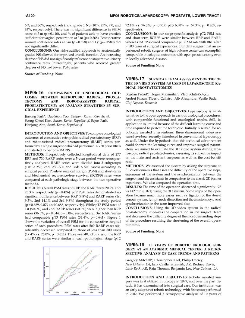
6.3, and 36%, respectively), and grade 1 NS (10%, 25%, 9.0, and33%, respectively). There was no significant difference in SHIMscore at 3 m (p = 0.410), and % of patients able to have erectionsufficient for vaginal penetration at 3 m (p = 0.368). Postoperativeurinary continence rates at 3 m (p = 0.558) and 1 y (p = 0.980) didnot significantly differ.CONCLUSIONS: Our risk-stratified approach to anatomicallygraded NS allowed for improved erectile function. An increasingdegree of NS did not significantly influence postoperative urinarycontinence rates. Interestingly, patients who received greaterdegrees of NS had lower PSM rates.
Source of Funding: None
MP06-16 COMPARISON OF ONCOLOGICAL OUT-
COMES BETWEEN RETROPUBIC RADICAL PROSTA-
TECTOMY AND ROBOT-ASSISTED RADICAL
PROSTATECTOMY: AN ANALYSIS STRATIFIED BY SUR-
GICAL EXPERIENCE
Jinsung Park*, Dae-Seon Yoo, Daejeon, Korea, Republic of,Seong Cheol Kim, Busan, Korea, Republic of, Sejun Park,Hanjong Ahn, Seoul, Korea, Republic of
INTRODUCTION AND OBJECTIVES: To compare oncologicaloutcomes of consecutive retropubic radical prostatectomy (RRP)and robot-assisted radical prostatectomy (RARP) series per-formed by a single surgeon who had performed > 750 prior RRPsand started to perform RARPs.METHODS: Prospectively collected longitudinal data of 277RRP and 730 RARP series over a 5-year period were retrospec-tively analyzed. RARP series were divided into 3 subgroups(1st: < 250, 2nd: 250–500 and 3rd: > 500 cases) according tosurgical period. Positive surgical margin (PSM) and short-termand biochemical recurrence-free survival (BCRFS) rates werecompared at each pathologic stage between the two operativemethods.RESULTS: Overall PSM rates of RRP and RARP were 20.9% and23.3%, respectively (p = 0.426). pT2 PSM rates demonstrated nosignificant difference between RRP (7.8%) and RARP series (1st9.5%, 2nd 14.1% and 3rd 9.8%) throughout the study period(p = 0.689, 0.079 and 0.688, respectively). While pT3 PSM rates of1st (50.6%) and 2nd RARP series (50.0%) were higher than RRPseries (36.5%; p = 0.044, p = 0.069, respectively), 3rd RARP serieshad comparable pT3 PSM rates (32.4%, p = 0.641). Figure 1shows the variation of overall PSM for the consecutive surgicalseries of each procedure. PSM rates after 500 RARP cases sig-nificantly decreased compared to those of less than 500 cases(17.4% vs. 26.0%, p = 0.011). Three year-BCRFS rates of the RRPand RARP series were similar in each pathological stage (pT2
92.1% vs. 96.8%, p = 0.517; pT3 60.0% vs. 67.3%, p = 0.265, re-spectively).CONCLUSIONS: In our stage-specific analysis pT2 PSM rateand short-term BCRFS were similar between RRP and RARP,whereas RARP showed comparable pT3 PSM rate with RRP after> 500 cases of surgical experiences. Our data suggest that an ex-perienced robotic surgeon of high volume center can accomplishcomparable oncological outcomes with open prostatectomy evenin locally advanced disease.
Source of Funding: None
MP06-17 SURGICAL TEAM ASSESSMENT OF THE OF
THE 3D VIDEO SYSTEM AS USED IN LAPAROSCOPIC RA-
DICAL PROSTATECTOMIES
Bogdan Petrut*, Hogea Maximilian, Vlad Schițcu,Andrei Kozan, Tiberiu Calistru, Alb Alexandra, Vasile Buda,Cluj Napoca, Romania
INTRODUCTION AND OBJECTIVES: Laparoscopy is an al-ternative to the open approach in various urological procedures,with comparable functional and oncological results. Still, itsapplication is limited because of the difficult learning curve andtime required to perfect the technique. Initially reserved for ro-botically assisted interventions, three dimensional video sys-tems have been recently introduced in conventional laparoscopyas well. Under the hypothesis that this technical advancementcould shorten the learning curve and improve surgical param-eters, we aimed to evaluate the 3D video system during lapa-roscopic radical prostatectomies, assessing its subjective impacton the main and assistant surgeons as well as the cost-benefitratio.METHODS: We assessed the system by asking the surgeons tofill questionnaires that asses the difficulty of the operative steps,ergonomy of the system and the synchronization between thesurgeon and the assistants in comparison to the classic 2D formerexperience. We also compared the operation time.RESULTS: The time of the operation shortened significantly 128vs 142 min (0.021) using the 3D system. Some steps of the oper-ation became much more easier such as: ligation of the dorsalvenous system, lymph node dissection and the anastomosys. Andsynchronization in the team improved also.CONCLUSIONS: Using the 3D video system in the radicalprostatectomy improves the cooperation in the surgical teamand decreases the difficulty degree of the most demanding stepsof the procedure resulting the shortening of the overall opera-tion time.
Source of Funding: None
MP06-18 10 YEARS OF ROBOTIC UROLOGIC SUR-
GERY AT AN ACADEMIC MEDICAL CENTER: A RETRO-
SPECTIVE ANALYSIS OF CASE TRENDS AND PATTERNS
Gregory Mitchell*, Christopher Keel, Philip Dorsey,New Orleans, LA, Erik Castle, Scottsdale, AZ, Rodney Davis,Little Rock, AR, Raju Thomas, Benjamin Lee, New Orleans, LA
INTRODUCTION AND OBJECTIVES: Robotic assisted sur-gery was first utilized in urology in 1999, and over the past de-cade, it has disseminated into surgical care. Our institution wasan early adopter of robotic technology, with first cases performedin 2002. We performed a retrospective analysis of 10 years of
-A120- MP06 ROBOTICS/LAPAROSCOPY: PROSTATE, LOWER TRACT I

robotic surgery at an academic institution to review diseasemanagement trends and practice patterns.METHODS: After obtaining Institutional Review Board ap-proval, a database of all robotic surgeries performed between2002 and 2012 at our institution was constructed. The cases wereindexed by surgeon and by procedure and were then tabulated.Records were cross referenced between case logs in addition tooperating room statistics. Management of trends for prostatecancer, kidney disease, ureteropelvic junction obstruction, blad-der cancer, and reconstructive robotic procedures were reviewed.RESULTS: Surgeons in the Tulane Department of urology haveengaged the Da Vinci surgical system in over 1600 urologic pro-cedures since 2002. Early in its implementation, equivalent utili-zation of robotic pyeloplasties and prostatectomies wereconducted. Beginning in 2004 and continuing since that time,robotic assisted radical prostatectomy took the lead as our mostfrequently performed robotic procedure by as much as 56% oftotal robotic case volume. There was a deviation from the upwardtrend in our case volume that occurred in 2005, corresponding toHurricane Katrina, which devastated New Orleans and forcedmovement of the robotic program to another facility several milesfrom our home institution. Despite this challenge, our programresumed clinical practice 3 months later. In 2008, the number ofrobotic partial nephrectomies performed at our institution shar-ply increased, and that quantity has increased by 427% as of 2012.At this point, partial nephrectomy is our second most frequentlyperformed robotic surgery. In 2009, 35% of the cases performedby urology faculty utilized the robot.CONCLUSIONS: The advent of robotic technology in urologicsurgery has greatly altered care at our institution. As the shiftingglobal healthcare and economic climate has the potential todrastically alter the use of the robot, we will continually evaluateour practice patterns regarding this exciting surgical modality.
Source of Funding: none
MP06-19 USE OF TRANEXAMIC ACID IN ROBOTIC
ASSISTED LAPAROSCOPIC RADICAL PROSTATECTOMY –
OUTCOMES AND COMPLICATIONS
Sarvpreet Ubee*, Masilamani Selvan, Phillip El-Dalil,Jane Boddy, Bhagyashree Netke, Rangaswamy Chandrashekar,Peter Cooke, Wolverhampton, United Kingdom
INTRODUCTION AND OBJECTIVES: Tranexamic acid (TXA)has been proven to reduce the morbidity and mortality related tointraoperative bleeding during elective surgery but its role tocause significant cardiovascular & thromboembolic events(CVTEE) when used during the peri-operative period remainsuncertain.[1] Its role has been demonstrated in open radicalprostatectomy.[2]
In this first reported study, we present our results with the useof TXA during robotic assisted laparoscopic radical prostatec-tomy (RALP).METHODS: In a prospective study intravenous TXA was ad-ministered at the time of anaesthesia to 36 consecutive patients(TXAP group) undergoing RALP. Data was collected for the age,Basal Metabolic Index (BMI), American Association of Anaes-thetists (ASA) score, haemoglobin level, blood loss, blood trans-fusion rate, drain output after 24 hours, time to drain removal,need for cystogram, total operating and console time along withattempt to nerve spare. Morbidity and mortality at 30 postoper-ative day was also noted.
Low Molecular Weight Heparin was given in the postoperativerecovery area, and was continued for the next 28 days alongwithcompression stockings.
Data was then retrospectively compared with the previous 36patients (NTXAP group) who did not have TXA.RESULTS: The patients in both groups matched closely in age,BMI and ASA score. Blood loss, drain output and time to drainremoval were less for higher number of patients in the TXAPgroup.
Table–1 summarises the comparative data between the twogroups. In either of the groups there were no CVSTEE compli-cations or mortality reported during the first 30 day postoperativeperiod.CONCLUSIONS: Use of TXA during RALP can reduce the in-traoperative blood loss related morbidity without increasing theCVSTEE despite the long operating time and trendlenberg posi-tion. This may indirectly reduce the total console time.
MP06 ROBOTICS/LAPAROSCOPY: PROSTATE, LOWER TRACT I -A121-
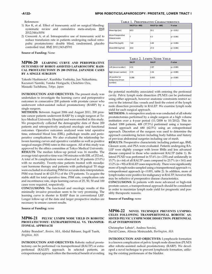
References:1) Ker K, et al. Effect of tranexamic acid on surgical bleeding:
systematic review and cumulative meta-analysis. BMJ2012;344:e3054
2) Crescenti A, et al. Intraoperative use of tranexamic acid toreduce transfusion rate in patients undergoing radical retro-pubic prostatectomy: double blind, randomised, placebocontrolled trial. BMJ 2011;343:d5701
Source of Funding: None
MP06-20 LEARNING CURVE AND PERIOPERATIVE
OUTCOMES OF ROBOT-ASSISTED LAPAROSCOPIC RADI-
CAL PROSTATECTOMY IN 200 INITIAL JAPANESE CASES
BY A SINGLE SURGEON
Takeshi Hashimoto*, Kunihiko Yoshioka, Jun Nakashima,Kazunori Namiki, Yutaka Horiguchi, Choichiro Ozu,Masaaki Tachibana, Tokyo, Japan
INTRODUCTION AND OBJECTIVES: The present study wasundertaken to investigate the learning curve and perioperativeoutcomes in consecutive 200 patients with prostate cancer whounderwent robot-assisted radical prostatectomy (RARP) by asingle surgeon.METHODS: Between August 2006 and August 2011, 200 pros-tate cancer patients underwent RARP by a single surgeon at To-kyo Medical University Hospital and were enrolled in this study.We prospectively collected the demographic data of these 200patients and retrospectively analyzed oncologic and functionaloutcomes. Operative outcomes analyzed were total operativetime, estimated blood loss (EBL), pathologic results and perio-perative complications. We also evaluated the relationship be-tween learning curves and operative variables as well as positivesurgical margin (PSM) rates in this surgeon. All of this study wasapproved by the ethics committee at Tokyo Medical University.RESULTS: The median follow-up period was 16 months. Theaverage total operative time was 190 minutes with EBL of 252 mL.A total of 36 complications were observed in 30 patients (15.0%)with no mortality. Twenty-nine patients treated with neoadju-vant hormone therapy and patients with pathological T0 stagewere excluded in evaluating PSM for accurate data interpretation.PSM was found in 40 (23.5%) of the 170 patients. To acquire thestable skill for total operative time, PSM rate, complication rateand incontinence rate, slope learning curves of 25, 50, 50 and 100cases were required, respectively.CONCLUSIONS: The functional and oncologic results of thisminimally invasive procedure seem to be very promising. Thelearning curve is shorter in RARP than in conventional LRP.Longer follow-up of the data and larger prospective studies arenecessary to ensure current results.
Source of Funding: none
MP06-21 PELVIC LYMPH NODE YIELD IN ROBOTIC
PROSTATECTOMY: EXTRAPERITONEAL VS. TRANSPER-
ITONEAL APPROACH
Ashley Brandon*, Boston, MA, Abdul Babaeer, Ingolf Tuerk,Brighton, MA
INTRODUCTION AND OBJECTIVES: Robotic radical prosta-tectomy can be performed via transperitoneal (RALTP) or extra-peritoneal (RALEP) approach. In selected patients, anextraperitoneal approach offers the theoretical benefit of avoiding
the potential morbidity associated with entering the peritonealcavity. Pelvic lymph node dissection (PLND) can be preformedusing either approach, however anatomic constraints restrict ac-cess to the internal iliac vessels and limit the extent of the lymphnode dissection proximally in RALEP. We examine lymph nodeyield for each surgical approach.METHODS: A retrospective analysis was conducted of all roboticprostatectomies performed by a single surgeon at a high volumeinstitution over a 4-year period (11/2008 to 10/2012). This in-cluded 1088 patients, 408 (37.5%) performed using a transper-itoneal approach and 680 (62.5%) using an extraperitonealapproach. Discretion of the surgeon was used to determine theapproach considering factors including body habitus and historyof previous abdominal surgeries including use of mesh.RESULTS: Preoperative characteristic including patient age, BMI,Gleason score, and PSA were evaluated. Patients undergoing RA-LEP were slightly younger with lower BMIs and less advanceddisease compared to those who underwent RALTP (table 1). Bi-lateral PLND was performed in 57.6% (n = 235) and unilaterally in15.7% (n = 64) of all RALTP cases compared to 23.7% (n = 161) and13.2% (n = 90) of RALEP cases respectively. There were significantlymore of lymph nodes sampled using a transperitoneal approach vs.extraperitoneal approach (p < 0.001, table 2). In addition, more oflymph nodes were positive for malignancy in RALTP, however thismay be reflective of preoperative disease characteristics.CONCLUSIONS: In patients with more advanced or high-riskprostate cancer, a transperitoneal approach should be consideredin order to maximize lymph node yield for prognostic and pos-sibly therapeutic reasons.
Source of Funding: none
MP06-22 NOVEL TECHNIQUE PREVENTS LYMPHO-
CELES FOLLOWING TRANSPERITONEAL ROBOTIC AS-
SISTED PELVIC LYMPH NODE DISSECTION: PERITONEAL
FLAP INTERPOSITION
Christopher Lebeis*, Andrea Sorcini,David Canes, Alireza Moinzadeh, Burlington, MA
INTRODUCTION AND OBJECTIVES: Lymphocele formationis a known complication of pelvic lymph node dissection (PLND)after robotic-assisted radical prostatectomy (RARP). We devel-oped a novel technique to prevent lymphocele formation, utiliz-ing the existing peritoneum of the bladder.
Table 1. Preoperative Characteristics
Table 2. Lymph Node Yield
-A122- MP06 ROBOTICS/LAPAROSCOPY: PROSTATE, LOWER TRACT I

METHODS: We evaluated 155 consecutive patients undergoingRARP with PLND over 24 months. Group A included the first 77patients with PLND using standard technique (no peritonealflap). Group B included the subsequent 78 patients with PLNDand peritoneal interposition flap. The peritoneal interposition flapis created by rotating and advancing the peritoneum (Point A inFigure 1) around the lateral surface of the ipsilateral bladder to thedependant portion of the pelvis (Arrow in Figure 1) and fixing itto the bladder itself (Figure 2). This prevents the bladder adiposetissue from contacting the lymph node dissection bed and scaringdown. A cystogram was performed in 91% of the patients 7–14days after the surgery. Lymphocele formation rates were com-pared (based on symptoms, cystogram findings, and radio-graphic confirmation).RESULTS: The two groups were statistically equivalent in termsof PSA, age, blood loss, body mass index, Gleason score, prostatesize, pathology, or heparin use. Symptomatic lymphocele for-mation occurred in 9/77 (11.6%) Group A patients and in 0/77Group B patients (p = 0.003). Mean time to lymphocele detectionin Group A was 30.4 days. Mean follow up in Groups A and Bwere 374.3 and 113.8 days respectively (p < 0.001).CONCLUSIONS: Strategic rotation and fixation of a peritonealflap around the lateral aspect of the bladder during transper-itoneal RARP with PLND is a novel technique to prevent symp-tomatic lymphocele formation. Given the sample size and singleinstitutional study, a prospective randomized, multi-institutionaltrial is planned.
Source of Funding: none
MP06-23 IDENTIFICATION OF FAVORABLE RISK
FACTORS IN PATIENTS WITH POSITIVE SURGICAL
MARGIN AFTER ROBOT-ASSISTED RADICAL PROSTA-
TECTOMY
Yun-Sok Ha*, North Brunswick, NJ, Dong Il Kang, Busan, Korea,Republic of, Jeong Hyun Kim, Chuncheon, Korea, Republic of,Jae Young Joung, Goyang, Korea, Republic of, Ji Hyeong Yu,Seoul, Korea, Republic of, Wun-Jae Kim, Cheongju, Korea,Republic of, Isaac Kim, New Brunswick, NJ
INTRODUCTION AND OBJECTIVES: Positive surgical mar-gin (PSM) has classically been associated with biochemical re-currence (BCR) following radical prostatectomy (RP) andimmediate adjuvant radiotherapy has been advocated based ontwo large randomized prospective clinical studies. However, asignificant percentage of patients with PSM never experienceBCR. This study evaluated factors potentially affecting risk ofBCR among the patients with PSM after robot-assisted radicalprostatectomy (RARP).METHODS: From a prospectively maintained database, 699patients with localized prostate cancer who underwent a RARPwithout any adjuvant therapy were identified. Median follow-upwas 46.0 months. To determine the pathologic and clinical factorsthat influenced BCR, univariate and multivariate analyses usingthe Cox proportional hazards model were performed. BCR-freesurvival curves were estimated with Kaplan-Meier method.RESULTS: Surgical margins were positive in 115 patients(16.5%), of whom 23 (20%) had BCR. In the univariate analyses,serum PSA level, surgical Gleason score (GS), and non-organconfined disease were significantly associated with BCR in menwith PSM. Multivariate Cox analysis showed that BCR was sig-nificantly associated with PSA (P = 0.011), and the surgical GS(P = 0.008). In patients with lower PSA cutoff (5.3 ng/ml), GS â&¤
7, and organ-confined disease, there were no BCR.CONCLUSIONS: In this study, we identified favorable riskfactors in patients with PSM following RARP. The results suggestthat immediate adjuvant therapy for PSM may not be necessaryin men with Gleason score 7 or less, organ-confined disease, andlow pre-operative PSA.
Source of Funding: none
MP06-24 RISK FACTORS OF POSITIVE SURGICAL
MARGINS ON THE APEX OF PROSTATE ON ROBOT-AS-
SISTED LAPAROSCOPIC PROSTATECTOMY
Risa Muneishi*, Ryuta Tanimoto, Yasuyuki Kobayashi,Motoo Araki, Shin Ebara, Toyohiko Watanabe,Yasutomo Nasu, Hiromi Kumon, Okayama, Japan
INTRODUCTION AND OBJECTIVES: Positive surgical mar-gins (PSM) during laparoscopic prostatectomy are generallyconsidered an adverse event. We evaluated the risk factors ofpositive RM on Robot-assisted laparoscopic prostatectomy(RALP) with specific attention to the exposed urethral length (UL)just before cutting urethra.METHODS: The data of 81 patients who underwent RALP in ourhospital were retrospectively evaluated. The standard clinicalcharacteristics (age, prostate volume, prostate-specific antigen(PSA) before operation, Gleason score, clinical T stage and ex-posed urethral length (UL) just before cutting urethra wereevaluated. UL was measured based on the proportion of devicediameter on the operation videos.
MP06 ROBOTICS/LAPAROSCOPY: PROSTATE, LOWER TRACT I -A123-

RESULTS: Eighteen out of 81 (22%) patients had PSM on pros-tate, and 11 of them had PSM on the apex of prostate. UL in thegroup of PSM on prostate as well as on the apex of prostate weresignificantly shorter than in the group of no PSM. On univariateanalysis, we found the shorter UL was only significantly associ-ated with an increased risk of PSM on the prostate as well as onthe apex of prostate (p = 0.0014, 0.0136, respectively), but not age,PSA, prostate volume, clinical T stage or Gleason Score. Onmultivariate analysis, UL remained a significant predictive factorof PSM.CONCLUSIONS: The risk factor of PSM was the shorter urethrallength. The greater care should be taken on the dissecting urethraduring Robot-assisted laparoscopic prostatectomy.
Source of Funding: none
MP06-25 DOES SURGEON SUBJECTIVE NERVE
SPARING SCORE PREDICT RECOVERY TIME OF EREC-
TILE FUNCTION FOLLOWING ROBOT-ASSISTED RADI-
CAL PROSTATECTOMY?
Sung Gu Kang*, Seoul, Korea, Republic of, Abdul Haidar, Orlando,FL, Srinivas Samavedi, Kenneth Palmer, Celebration, FL,Jun Cheon, Seoul, Korea, Republic of, Vipul Patel, Celebration, FL
INTRODUCTION AND OBJECTIVES: During robot-assistedradical prostatectomy (RARP), the quality of nerve sparing (NS)was usually classified by laterality of NS (no NS, unilateral NSand bilateral NS) or degree of NS (no NS, partial NS and full NS).Recently, side-specific nerve sparing have been more frequentlyperformed, but previous nerve sparing grading system might notreflect the differential NS in each side. Herein, we assessedwhether a subjective NS score (NSS) incorporating both degree ofNS and NS laterality can predict the time to potency recoveryfollowing RARP.METHODS: Data was analyzed from 1,898 patients who had leftand right neurovascular bundle sparing quality scores and atleast one year of follow-up after RARP performed between Jan-uary, 2008 and October, 2011. Cox proportional hazards methodanalyses were used to determine predictive factors for early re-covery. Multivariate linear regression models were used to assesssubjective NSS in an effort to predict time to potency recovery.
Subjective NSSs were compared to a model based on the threegrades according to laterality and degree.RESULTS: Time to potency recovery showed a statistically sig-nificant difference in favor of higher NS grade by the Cox pro-portional hazard regression analysis (grade 0 vs. grades 3, 4, and5; P < 0.01), but there were no differences between grade 0 versus1 (P = 0.579) or grade 0 versus 2 (P = 0.449). The regression modelindicated that the subjective NSS better explained potency out-comes than the systems using conventional three-grade scales(adjusted R-squared = 0.207 in subjective NSS; subjective R-squared = 0.185 in NS laterality; adjusted R-squared = 0.187 indegree of NS). The regression equation with subjective NSS wasas follows: Log (Time) = 5.163 - (0.035 · SHIM Score) + 0.028 Age -(0.101 · Subjective NSS).CONCLUSIONS: Our subjective NSS model can more accuratelypredict the time to potency recovery and better explain potencyoutcomes than systems based on NS laterality and the degreeof NS.
Source of Funding: none
MP06-26 IMPACT OF LENGTH OF POSITIVE SURGI-
CAL MARGINS (PSM) ON BIOCHEMICAL RECURRENCE
(BCR) AFTER ROBOTIC-ASSISTED RADICAL PROSTA-
TECTOMY (RARP), IN A SINGLE CENTER SERIES WITH
FOLLOW-UP OF AT LEAST FIVE YEARS
Prasanna Sooriakumaran*, Achilles Ploumidis, Leif Haendler,Tommy Nyberg, Mats Olsson, Stefan Carlsson, Gunnar Steineck,Peter Wiklund, Worcester Park, United Kingdom
INTRODUCTION AND OBJECTIVES: It is well establishedthat PSM is a risk factor for BCR, and in our own series, PSM wasassociated with a 2–3 fold increase in the risk of BCR. In this studywe aim to assess the impact of length of PSM as a predictivevariable for BCR.METHODS: From Jan 2002 to Dec 2006, 944 men with clinicallylocalized or locally advanced (cT1–3) prostate cancer underwentRARP at our institution. Data were prospectively collected andincluded: age, BMI, preoperative PSA, prostate volume, clinicalstage, biopsy Gleason score, pathological stage, specimen
Table 1. Multiple Linear Regression Models Based on
NS Scoring Systems (NS Laterality, Degree of NS, and
Subjective NSS) and Including Age, BMI, Preoperative
SHIM Score, PSA, Diabetes, Coronary Artery Disease,
and Hypertension as Variables.
Results of Three Linear Regression Models Based
on Three Types of NS Classification Systems.
Of Note, Adjusted R-squared is Highest
in Model Including Subjective NSS.
Table 2. Of Note, the Beta Coefficient for Subjective
NSS is Larger Than for SHIM Score and Age,
Suggesting that Subjective NSS is Most Important
in the Following Regression Equation
for Predicting Time to Potency Recovery:
[Log (Time) = 5.163 - (0.035 · SHIM Score) + 0.028Age - (0.101 · Subjective NSS)]
-A124- MP06 ROBOTICS/LAPAROSCOPY: PROSTATE, LOWER TRACT I

Gleason score (GS), margin status (positive, negative), number ofPSM (single, multiple) and extent of PSM (mm) and were asso-ciated with BCR rate. Margin status and length were measured byseven uropathologists. BCR was defined as PSA 0.2 ng/
ml on two consecutive measurements. Kaplan-Meier survivalplots with log-rank tests and Cox univariable and multivariableregression analysis were used to determine biochemical recur-rence-free survival and identify predictors of BCR.RESULTS: The overall median follow-up was 6.3 (IQR: 5.6–7.2)years. In total, 194 (21.6%) patients were found to have PSM ofwhich, 104 (16.0%) were pT2, 64 (33.3%) were pT3a and 22(57.9%) were pT3b. The location of PSM was in 66 (7.4%) patientsat the apex, in 87 (9.8%) patients posterolateral, in 33 (3.7%) pa-tients anterior and in 20 (2.2%) patients at the base of the prostate.PSM in multiple locations was noted in 38 patients. In multivar-iable regression analysis: PSA > 10 (HR: 1.86, 95%CI: 1.27-2.72,p = 0.0013), pT3a stage (HR: 1.62, 95%CI: 1.07-2.46, p = 0.0221),pT3b stage (HR: 3.09, 95%CI: 1.69-5.67, p = 0.0003), GS: 3 + 4 (HR:2.20, 95%CI: 1.34-3.60, p = 0.0017), GS: 4 + 3 (HR: 5.08, 95%CI:2.95-8.74, p < 0.0001), GS: 8 (HR: 3.89, 95%CI: 1.90-7.93,p < 0.0002) and PSM length  3 mm or multifocality (HR:2.84, 95%CI: 1.76-4.58, p < 0.0001) remained independent predic-tors of BCR.CONCLUSIONS: In a large RARP series with median follow-upof 6.3 years, PSM length > 3 mm was an independent predictor ofBCR. Patients with PSM < 3 mm had similar BCR outcomes tothose with negative surgical margins.
Source of Funding: None.
MP07 ESWL/UROLITHIASIS
MP07-01 ESWL TREATMENT IN GERMANY IN 2013:
RESULTS FROM A NATION-WIDE HOSPITAL SURVEY
Wolfgang Brummeisl*, Christian Chaussy, Regensburg, Germany,Jens Rassweiler, Heilbronn, Germany, Thomas Knoll, Sindelfingen,Germany, Andreas Gross, Hamburg, Germany, Kai Koehrmann,Mannheim, Germany, Wolf Wieland, Hans-Martin Fritsche,Regensburg, Germany
INTRODUCTION AND OBJECTIVES: The aim of the surveywas to gain information on current ESWL treatment-strategiesand modalities in German urology departments.METHODS: A survey was sent to 306 urologic departmentswithin Germany under the auspices of the German Society forShock Wave Lithotripsy (DGSWL). The questionnaire containedquestions on the medical, technical and organizational modalitiesof stone-therapy.RESULTS: 99 surveys (33%) had been answered, 25% of re-spondents were university departments. In 69% ESWL is per-formed only under in-patient conditions, in 15% only underoutpatient conditions, and in 28% both in- and outpatient.
96% of the departments consider renal ultrasound to be anessential diagnostic measure prior to ESWL treatment, whereas56% use intravenous urography, 54% a plain abdominal X-Ray,and only 18% a CT.
85 % of the departments do not necessitate a pre-interventionalblood coagulation test in case of unsuspected anamnesis.
A pre-interventional urine dipstick is used in 69% of the de-partments, 47% perform a urine sediment analysis, while 29%
perform a routine urine culture despite unsuspected anamnesisregarding urinary tract infection. Only 5% do not perform anykind of urine analysis based on unsuspected anamnesis. In case ofrenal stone and ureter stone lithotripsy there is a difference insuspending anticoagulant medications.
For pain management 76% of the departments perform an in-travenous analgo-sedation, 6% only use oral medication. In 18%,an anesthesiologist is always present during the procedure, 6%sometimes use general anesthesia.
Relating to the treatment modalities 48% of the departmentsperform an ultrasound-guided ESWL. All departments graduallyincrease shock wave energy (power ramping); 7 % exceed themaximum energy and number of shock waves recommended bythe manufacturer. The average shock wave frequency for renaland ureteral stones is 90 shocks/min (range 50–180). The maxi-mum number of ESWL treatments per stone before switching toendourologic measures varies from 6%/one session, 32%/ twosessions, 44%/ three sessions, and up to 10% place no limit on thenumber of treatments. 8% of the departments perform re-ESWLof kidney stones within one day, 41% with a two day interval,while 28% wait more than a week.CONCLUSIONS: The present survey shows the current ESWLtreatment-strategies modalities of the German urology depart-ments. Large disparities exist with respect to the single parame-ter. ESWL strategies and modalities still appear to be a matter ofmuch debate.
Source of Funding: A survey within german urologic depart-ments under the auspices of the German Society for Shock WaveLithotripsy (DGSWL).
MP07 ESWL/UROLITHIASIS -A125-

MP07-02 TRIPLE D SCORE IS A READILY INTER-
PRETED SINGLE SCORE THAT PREDICTS SHOCKWAVE
LITHOTRIPSY STONE FREE RATES FOR RENAL CALCULI
Timothy Tran*, Kathryn McGillen, Eugene Cone, Shadi Al Ekish,Damian Dupuy, Gyan Pareek, Providence, RI
INTRODUCTION AND OBJECTIVES: Over the last decade,numerous studies have suggested improved shockwave litho-tripsy (SWL) success rates with consideration of stone density,skin-to-stone distance and stone diameter. Manual determi-nation of these predictors on computed tomography (CT) im-aging is a time-consuming process that may limit theutilization of these predictors. Here, we describe an easy-to-interpret score that integrates these predictors and is reportedby radiologists.METHODS: Stone density based upon Hounsfield units (HU),skin-to-stone distance, ellipsoid stone volume and stone-free rateswere determined for all patients that underwent SWL at our in-stitution from 2011–2012. Receiver operator curves were gener-ated to determine the optimal cutoff values for each parameter.Each stone was assigned a score based upon whether these pa-rameters were below the established cutoff values (Triple DScore). Additionally, a survey was sent to local Urologists to as-sess their current utilization of these predictors and to gauge theirinterest in a unifying score.RESULTS: 116 of the 186 patients (62.3%) that underwent SWLwere stone free. 52/84 (61.9%) of the patients with available pre-operative imaging were stone free. In univariate analysis, ellip-soid stone volume and stone density were significant predictorsof SWL success while skin-to-stone distance trended towardssignificance. Cutoffs of < 150 ll for ellipsoid stone volume,< 12 cm for SSD and < 600 HU for density were established. ATriple D score of 0, 1, 2, and 3 had SWL success rates of 40.0%,51.5%, 71.4% and 93.3%, respectively. Only two-thirds of sur-veyed urologists currently utilize SSD and HU in SWL planning.However, over 95% are aware of their predictive value and statedthey would utilize a integrative score if it were included in aradiology report.CONCLUSIONS: Inclusion of the Triple D Score in radiologyreports offers a readily accessible means to integrate predictiveparameters from CT imaging into preoperative planning forSWL. This may enhance the cost efficacy of SWL by limiting it topatients likely to have favorable outcomes.
Source of Funding: none
MP07-03 A NON-INFERIORITY TRIAL COMPARING
THE COMBINATION OF ETORICOXIB AND DICLOFENAC
AGAINST PETHIDINE AS ANALGESIA DURING EXTRA-
CORPOREAL SHOCKWAVE LITHOTRIPSY
Cheuk Fan Shum*, Amit Mukherjee, Chin Hu Ong,Tow Poh Lim, Chang Peng Colin Teo, Singapore, Singapore
INTRODUCTION AND OBJECTIVES: Opiates are commonlyused as analgesia during extracorporeal shockwave lithotripsy(ESWL). They have good analgesic efficacy and rapid onset ofaction; but are often associated with significant adverse effectslike nausea, respiratory depression and hypotension. Non-steroidal anti-inflammatory drugs (NSAIDs) are alternativeswithout the adverse effects of opiates, but their analgesic effectand onset of action seem inferior to opiates. By combining etor-icoxib with diclofenac, we aim to spare the use of opiates and yetprovide equivalent analgesia throughout ESWL.
METHODS: A test of non-inferiority was conducted. Patientswith renal or ureteric stones undergoing ESWL would be ran-domised into 2 groups. Group A would receive oral etoricoxib120 mg at 2 hours and intramuscular diclofenac 75 mg immedi-ately before ESWL. Group B would receive intravenous pethidine50 mg with metoclopramide 10 mg immediately before ESWL. AllESWLs were performed with a Siemens Modularis Variostarelectromagnetic lithotripter following a standard treatment pro-tocol. At the end of ESWL, all patients would rate pain severityusing a visual analogue scale from 0 (no pain) to 10 (worst pain).Power analysis (1-b = 0.80; a = 0.025) determined the minimumsample size to be 284 patients, at a non-inferiority margin of 1.Besides the severity of pain, any drug-related adverse effects andunexpected interruption to ESWL were also recorded and com-pared between the groups.RESULTS: A total of 372 patients were randomised into 2 equalgroups over a period of 3 years. The 2 groups were similar inpatients’ demographics, stone characteristics and ESWL details.The mean pain scores for Group A and Group B were 4.7 and 4.5respectively. Results showed that non-inferiority was claimed,t (370) = 4.17, p < 0.001. The lower bound of the confidence limitwas 0.39, which was within the non-inferiority margin. Six pa-tients in each group had premature interruption to ESWL due tosevere pain. There were no NSAID-related adverse effects inGroup A. Twenty one patients in Group B suffered from nauseaand vomiting, with 1 of them requiring hospitalisation for per-sistent nausea. There was 1 Group B patient who died from acutemyocardial infarct 6 days after ESWL, but was deemed unrelatedto opiate use.CONCLUSIONS: The combination of etoricoxib and diclofenacis not inferior to pethidine as analgesia during ESWL, and is notassociated with any adverse effects.
Source of Funding: none
MP07-04 TO ASSESS THE EFFECTS OF DIFFERENT
TREATMENT PROTOCOLS ON THE DEGREE OF RENAL
INJURY IN PATIENTS WITH RENAL STONES RECEIVING
SHOCKWAVE LITHOTRIPSY
Ye Lu*, Wai Man Yuen, Danny Gohel, Chi Fai Ng, Hong Kong,Hong Kong
INTRODUCTION AND OBJECTIVES: Animal studies aimedat minimizing renal injury caused by extracorporeal shockwavelithotripsy (SWL) suggested including modifications in treatmentprotocols, such as low energy shockwave pretreatment protocoland pause-protection protocol. Our study was designed to assess
-A126- MP07 ESWL/UROLITHIASIS

the effects of these protocols on acute renal injury in humansubjects.METHODS: Adult patients with renal stone planned for SWLwere recruited and randomized to follow one of the protocols, (1)80% power (19.2 kV) from the beginning till the end (Control); (2)the first 100 shocks at 40% power (9.6 kV), followed by SWs at80% power till the end; (3) the first 100 shocks at 40% power,followed by a 3-minute pause and then further SWs at 80% powertill the end; (4) the first 100 shocks at 80% power, followed by a3-minute pause and then further SWs at 80% power till the end.Spot urine samples were collected immediately before and afterSWL. Urinary Interleukin-18 (IL-18) and microalbumin (MA)were measured and expressed as ratios to creatinine for com-parison. The incidence of renal haematoma was accessed by MRIor NCCT on Day-2 after SWL.RESULTS: 166 patients were recruited. The baseline informationand treatment parameters of the 4 groups were comparable. Thefigure showed significant increase of urinary IL-18 levels afterSWL compared to baseline levels in all groups (p < 0.005). Besides,the mean post-SWL IL-18 levels of Group 3 and 4 were signifi-cantly lower than in Group 1 (p < 0.05). However, there was nosignificant difference between the post-SWL levels of IL-18 inGroup 2 and Group 1. Urinary MA levels also increased afterSWL, but post-SWL levels between 4 groups showed no differ-ence. There was no significant difference in the incidence of renalhaematoma between 4 groups.CONCLUSIONS: Patients who received the first 100 shocks at40% or 80% power followed by a 3-minute pause had signifi-cantly less increase in urinary IL-18, compared to those had nolow-energy pretreatment and pause. This suggested that pauseprotocol alone and combination of the two protocols may lead toless post-SWL acute renal injury.
Source of Funding: Hong Kong RGC General research fund(RGC Ref. No. CUHK472111)
MP07-05 IN VIVO STONE COMMINUTION PRO-
DUCED BY A MODIFIED ACOUSTIC LENS FOR ELEC-
TROMAGNETIC LITHOTRIPTERS
Andreas Neisius*, Nathan Smith, Nicholas Kuntz,Tim Schykowski, Gaston Astroza, Richard Shin, Ramy Youssef,Muhammad Iqbal, Michael Ferrandino, Michael Lipkin,W. Neal Simmons, Glenn Preminger, Pei Zhong, Durham, NC
INTRODUCTION AND OBJECTIVES: The acoustic lens of aSiemens Modularis electromagnetic shock wave lithotripter hasbeen further modified to reduce pre-focal cavitation while gen-erating a pressure waveform and broad focal zone mimickingthat of the Dornier HM3. In this study, we sought to determinethe comminution efficiency of the modified lens following aclinical therapy regime and compared the results to comminutionproduce by the original lens in a swine model.METHODS: Using the maximum energy level (E + = 44 mJ) thatis safe for lithotripsy (i.e., without formation of gross hematoma),we have compared stone comminution produced by the originaland modified lenses in vivo using hard cylindrical Begostones of5 · 10 mm, surgically implemented in pig kidneys. A clinicalprotocol with a soft ramping scheme was used to deliver 3000shock waves to each kidney using a pulse repetition frequency of1.5 Hz, leading to a total accumulated energy of 112.84 J. Fol-lowing lithotripsy, kidneys were harvested, dissected, all visiblestone fragments were collected, dried and filtered with standardsieves with decreasing mesh size of 4, 2.8, and 2 mm, respectively.Final comminution efficiencies were determined by the percent-
age (by weight) of fragments less than 2 mm. An unpaired two-tailed student’s t-test was used to compare fragmentation results.RESULTS: At E + = 44 mJ, the modified lens (12 renal units)showed a comminution efficiency of 72.8% ( – 21.4%) comparedto 63.6% ( – 21.8%) for the original lens (10 renal units) overall,p = 0.058. When considering stones not trapped in the DJ loop andthose with no spread > 2 cm, comminution efficiency was 88.8%( – 10.9%) for the modified lens vs 54.1% ( – 23.3%) for the originallens, p = 0.013.CONCLUSIONS: With our latest lens modification the electro-magnetic lithotripter demonstrates better stone comminutionunder a clinically relevant energy setting and treatment protocolwhen compared to the original lens. This lens modification couldbe retrofitted to most electromagnetic lithotripters, thereby im-proving efficiency of clinical stone fragmentation.
Source of Funding: This work was supported in part by NIHthrough grant R37-DK052985-15, Siemens (for providing theelectromagnetic shock wave generators and lenses used in thiswork), and a Ferdinand Eisenberger grant of the Deutsche Ge-sellschaft für Urologie (German Society of Urology), ID NeA1/FE-11 (Andreas Neisius).
MP07-06 ENERGY AND PULSE REPETITION FRE-
QUENCY DEPENDENT TISSUE INJURY PRODUCED BY A
MODIFIED ACOUSTIC LENS FOR ELECTROMAGNETIC
LITHOTRIPTERS
Andreas Neisius*, Nathan Smith, Nicholas Kuntz,Tim Schykowski, Gaston Astroza, Richard Shin, Ramy Youssef,Muhammad Iqbal, Michael Ferrandino, Michael Lipkin,W. Neal Simmons, Glenn Preminger, Pei Zhong, Durham, NC
INTRODUCTION AND OBJECTIVES: The acoustic lens of aSiemens Modularis electromagnetic shock wave lithotripter hasbeen further modified to reduce pre-focal cavitation while gen-erating a pressure waveform and broad focal zone mimicking theDornier HM3 electrohydraulic device. We sought to determinethe threshold for the maximum acoustic energy which can besafely applied to a kidney under clinically relevant treatmentprotocols, and the dependency of tissue injury on pulse repetitionfrequency (PRF).METHODS: Tissue injury (TI) produced by the original andmodified lenses in a swine model were first evaluated startingfrom the highest output level to determine the threshold energyfor safe lithotripsy. Thereafter, TI was assessed under an effectiveacoustic pulse energy for the leading compressive wave (E + ) of44 mJ. A clinical protocol with a soft ramping scheme was used todeliver 3000 shock waves to each kidney using a PRF of 1.0 and1.5 Hz, leading to a total accumulated energy of 112.84 J. Fol-lowing lithotripsy, kidneys were perfused, harvested, dehy-drated, cast in paraffin wax, and sectioned. Photographic imageswere taken every 120 m and analyzed to determine thefunctional renal volume (FRV) damage.RESULTS: Gross subcapsular hematomas were produced byboth the original and modified lenses at E + of 51 mJ. Using E + of44 mJ, the modified lens showed quantatively macroscopic tissueinjury (subcapsular hematoma) in 1/6 renal units (17%) in the1.5 Hz group. No macroscopic tissue injury was detected in the1.0 Hz group (0/6 renal units). After processing of the digitalizedimages TI was detected in 0.432 % ( – 0.51%) of the FRV (with amaximum level of 1.324% in the kidney with the gross hemato-ma) in the 1.5 Hz group and in 0.009% ( – 0.015%) of the FRV inthe 1 Hz group (p = 0.025), respectively.
MP07 ESWL/UROLITHIASIS -A127-

CONCLUSIONS: This study demonstrates that the energythreshold for gross TI of the modified lens is comparable to theoriginal lens. Our data further confirms that the initiation of TIdepends on acoustic pulse energy, and the extent of TI dependson the total accumulated acoustic energy delivered to a kidney, aswell as on PRF. Overall, our results suggest that a rational designof treatment strategies to minimize tissue injury in SWL is war-ranted.
Source of Funding: This work was supported in part by NIHthrough grant R37-DK052985-15, Siemens (for providing theelectromagnetic shock wave generators and lenses used in thiswork), and a Ferdinand Eisenberger grant of the Deutsche Ge-sellschaft für Urologie (German Society of Urology), ID NeA1/FE-11 (Andreas Neisius).
MP07-07 A NOVEL METHOD OF EXTRACORPOREAL
LITHOTRIPSY WITHOUT SHOCK WAVES: IN VITRO
FRAGMENTATION OF ARTIFICIAL AND HUMAN CALCULI
Adam Maxwell*, Bryan Cunitz, Wayne Kreider,Oleg Sapozhnikov, Ryan Hsi, Mathew Sorensen,Jonathan Harper, Michael Bailey, Seattle, WA
INTRODUCTION AND OBJECTIVES: Since the introductionof shock wave lithotripsy (SWL) over 30 years ago, most treat-ments have employed similar acoustic output protocols, deliv-ering 2000–3000 shock waves at a rate of 1–2 per second. Whilethis technology has been effective in many patients, the subop-timal stone-free rate has resulted in a shift in clinical preferencetowards more invasive approaches such as ureteroscopy. Basedon previous work identifying the mechanisms of stone fracture inSWL, we are investigating a new method to fragment stonesusing ultrasound pulses without shock waves. This study eval-uated the exposures needed to fragment artificial and humanstones in vitro.METHODS: Ultrasound exposures were tested on 1) cylindricalartificial stones (Begostone, 6 mm diameter · 10–12 mm length)with acoustic properties similar to calcium oxalate and 2) humanstones (uric acid, struvite, cystine, calcium oxalate) extracted bypercutaneous nephrolithotomy. Stones were fixed to a plasticmembrane and positioned at the focus of an ultrasound trans-ducer in a degassed water bath. The transducer was electricallydriven with a radiofrequency amplifier between 170 to 255 kHz tooutput 10-cycle ultrasound pulses. Focal pressure amplitudeswere lower than those in SWL (p £ 6.5 MPa), but pulses weredelivered at a higher rate (40–1000 Hz). Stone fracture time wasrecorded and fragments were collected to evaluate the size dis-tribution. A numerical elastic wave model was used to comparethe stress created in stones by both SWL shock waves and ul-trasound pulses applied in this study.RESULTS: Exposures resulted in fragments < 3 mm breaking offthe intact stone, with fracture initiating near the stone surfaceclosest to the transducer and progressing to the distal surfaceduring treatment. The pressure threshold for fracture of artificialstones was between p = 2.3–2.8 MPa. Comminution of artificialstones required 10.9 – 2.5 minutes (n = 6) at 200 Hz pulse rate andp = 6.5 MPa, and as little as 5 minutes at higher rates. Humanstones were completely fragmented in 10 seconds (5 mm, uricacid) to 21 minutes (12 · 7 · 5 mm, cystine) with the same pa-rameters. Stones treated at higher ultrasound frequency gener-ated smaller fragments. Simulations provided a possibleexplanation for the relationship between ultrasound frequencyand fragment size.
CONCLUSIONS: Pulsed ultrasound without shock waves canfragment artificial and human renal calculi in vitro over a timeframe comparable to SWL. In contrast to SWL, the resultingfragment sizes may be controllable through selection of ultra-sound frequency.
Source of Funding: Work supported by NIH 2T32DK007779-11A1, 2R01EB007643-05, 2P01DK043881-15, 1R01DK092197-02,and NSBRI through NASA NCC 9–58.
MP07-08 HYPOTHERMIA (ICE PACK) VERSUS CY-
CLOOXYGENASE 2 SELECTIVE INHIBITOR AS PAIN
CONTROL FOR POST-EXTRACORPOREAL SHOCK WAVE
LITHOTRIPSY PATIENTS
Ernesto Manuel Jr. Romero*, Omar Cortes,Michael Eufemio Jr. Macalalag, Quezon City, Philippines
INTRODUCTION AND OBJECTIVES: Extracorporeal shock-wave lithotripsy (ESWL) has now become the leading mode ofmanagement for urinary calculi. Acoustic shock waves breakstones down into sand-like fragments (Ayt, et.al., 2008), ac-counting for the passage of sand-like urine. The treatment may beassociated with significant pain, depending on the strength of theshock waves delivered. ESWL has shown excellent success ratesin treatment of urinary calculi. The Hippocratic Oath that ‘‘I willnever cut for a Stone’’ has become a welcome reality with the helpof ESWL.
To assess the analgesic effect of Hypothermia (Ice Pack) versusCox - 2 inhibitor in post-extracorporeal shockwave lithotripsy(ESWL) patients for renal calculi.METHODS: The study was a randomized controlled trial in-volving 160 patients who, prior to ESWL for renal calculi, wereproperly screened and were divided into two groups. Patients inthe ICED group (n = 80) received ice pack over the affected site ontwo occasions, 8 hours and 24 hours post ESWL. Patients in theCOX-2 group (n = 80) received COX-2 inhibitor post ESWL. Theeffectiveness was assessed with the use of a visual analog scale(VAS).RESULTS: The mean VAS (1.9875 – 0.169) was noted to be higherin patients under the ICED Group compared to the mean VAS(1.936 – 0.170) of the patients in the COX-2 group. However, theobtained p values for the two groups yielded no statistical sig-nificance for the two groups. VAS and age have an inversePearson correlation (r = -0.04698).CONCLUSIONS: Both Hypothermia and COX-2 selective in-hibitors are effective as pain control for post ESWl patients. Hy-pothermia (ice pack) as analgesic for post-ESWL patients does nothave statistical difference in terms of its analgesic effect whencompared with COX-2 inhibitors.
Source of Funding: none
MP07-09 EXTRACORPOREAL SHOCK WAVE LITHO-
TRIPSY WITH DORNIER LITHOTRIPTER S: LONG TERM
SIDE EFFECTS ASSESSMENT AFTER 10 YEARS OF
FOLLOW UP
Marco Rosa*, Alessio Zordani, Maria Chiara Sighinolfi,Alessandro Mofferdin, Riccardo Galli, Salvatore Micali,Giampaolo Bianchi, Modena, Italy
INTRODUCTION AND OBJECTIVES: The extracorporealshock wave lithotripsy (ESWL) is a minimally invasive treatment
-A128- MP07 ESWL/UROLITHIASIS

of urolithiasis. Starting from the electrohydraulic prototype(HM3), the evolution of lithotripters led to a significant im-provement in the safety of the treatment. Long-term side effectsincidence is still a controversial topic; among the potential sideeffects of the first devices, the most important were the onset ofchronic renal failure, arterial hypertension, and diabetes mellitus.The aim of our work is to evaluate the safety of ESWL treatmentby using a last generation electromagnetic lithotripter.METHODS: Between 2002 and 2012, we performed nearly 4000ESWL treatments. We retrospectively reviewed the data of thefirst 47 patients treated for kidney stones in 2002 (current averageage: 48.3 + / - 9.8, 36 men, 14 women). The average number ofshock waves was equal to 2000 SW ( + /–100). No relevant shortterm side effects were identified. The same patients were con-tacted during 2012 and undergo clinical assessment with partic-ular attention to the onset of hypertension, renal failure, anddiabetes mellitus.RESULTS: Our follow-up after 10 years showed that nine pa-tients (9/47) have developed arterial hypertension (19%). Thecurrent age of 8 of these patients is between 45 and 64 years. Aliterature review shows that among the general population (un-treated) the incidence of hypertension is 30% in the age groupbetween 45 and 54 years old, and 42% between 55 and 64 yearsold (Woltz M, Am J Hypertension 2000). No patients had onset ofinsulin-dependent diabetes mellitus or renal dysfunction at 10years follow up.CONCLUSIONS: Electromagnetic lithotripter used in the ex-tracorporeal shock wave lithotripsy does not seem to inducesignificant side effects in long-term follow-up. The incidence ofhypertension, as possible complication, does not seem to differ inpatients who underwent ESWL treatment and general popula-tion. However, this study is still in progress, and the future fol-low-up of more cases will eventually confirm these results.
Source of Funding: none
MP07-10 EFFECT OF ROWATINEX ON URINARY
CALCULUS CLEARANCE AFTER EXTRACORPOREAL
SHOCK WAVE LITHOTRIPSY
Won Sik Jeong*, Won Sik Ham, Hong Sang Moon,Tchun Yong Lee, Sung Yul Park, Seoul, Korea, Republic of
INTRODUCTION AND OBJECTIVES: A special terpene com-bination (Rowatinex�) was known to help the emission of urinarystones. This study is how Rowatinex� effects the emission ofremnant stones after shock wave lithotripsy (SWL).METHODS: From January 2007 to August 2012, total 755 pa-tients, who were diagnosed with urinary stones and underwentSWL. All patients were diagnosed with urinary stones throughKUB, IVP or Abdomen CT. The patient¡�s progress was checkedevery 2 weeks to confirm remnant stones by KUB after SWL. Ifremnant stones existed, the patients underwent SWL again.Group 1 is composed of patients who were prescribed NSAIDs,Tamsulosin 0.2 mg and Rowatinex�. Group 2 is composed ofpatients who were prescribed NSAIDs and Tamsulosin 0.2 mg.When we performed SWL, the shock wave velocity was 60 timesper minute to all patients equally, and the power increased fromAC120V to AC170V. It was performed in all groups from 3,000times to 4,000 times equally. The patient¡�s age, sex, the location ofthe urinary stone, the size of the urinary stone, the number ofsessions, the treatment¡�s duration and complications were com-pared and analyzed by each group.RESULTS: Group 1 and Group 2 showed no difference regardingpatient¡�s age, sex and the location of the urinary stone. The av-
erage size of the urinary stone was not different statistically be-tween Group 1 and Group 2 (8.5¡3̌̌/4 3.9 mm vs 8.7¡3̌̌/4 4.3 mm,p = 0.466). Two weeks after the first SWL, the clearance rate of theurinary stone was not different in the 2 groups, and after 4 weeks,it increased significantly in Group 1 (60.7% vs 47.5%, p = 0.002).The final clearance rate of the urinary stone was increased inGroup 1 (68.7% vs 57.1%, p = 0.003). The number of sessions inGroup 1 is greater than Group 2 (2.20¡3̌̌/4 1.43 vs 1.96¡3̌̌/4 1.29,p = 0.032). Thirty-six patients out of the total 755 patients didn¡�temit a urinary stone after SWL. Seven patients out of 18 in Group1 (6.8%), 5 out of 18 in Group 2 (4.7%) had ureteroscopic removalof the stone. Acute pyelonephritis occurred in one patient inGroup 1 (1.0%) and in 2 patients in Group 2 (p = 0.589).CONCLUSIONS: The long-term administration of Rowatinex�
for more than 4 weeks increases the clearance rate of urinarystones after SWL.
Source of Funding: none
MP07-11 THE EFFECT OF PREOPERATIVE COUN-
SELING ON PATIENT PREFERENCE IN MANAGEMENT OF
ASYMPTOMATIC RENAL CALCUL
Gregory Lieser*, Giovanni Marchini, Carl Sarkissian,Julie Cheng, Manoj Monga, Cleveland, OH
INTRODUCTION AND OBJECTIVES: With the developmentof new minimally invasive treatment modalities, patients arefrequently presented with more than one option for the man-agement of their stone disease. We evaluated the effect preoper-ative counseling had on patient decision making.METHODS: We retrospectively reviewed the records of all pa-tients seen by a single surgeon in our stone clinic over a three yearperiod (2010–2012). Patients were included in the study if theywere presenting with asymptomatic renal stone(s) and werecandidate for more than one procedure. Those with renal colicand ureteral stones were excluded. All patients at time of pre-operative visit were quoted potential success rates based onpublished data, and according to stone size, location, skin to stonedistance, and attenuation.RESULTS: A total of 201 patients met criteria for inclusion, and152 of those patients ultimately underwent intervention with109 opting for Shock Wave Lithotripsy (SWL) (n = 68) or Ur-eteroscopy (URS) (n = 41). The mean preoperative success ratequoted to patients choosing SWL 72% (Q1–Q3 70–80%) wassignificantly lower than the mean preoperative success ratequoted to those who chose URS 90% (Q1–Q3 90%-95%)(p < 0.001). Patients were more likely to chose SWL as long as thesuccess rate was within 25% of that quoted for URS. If the
Table 1. Difference in Preoperative Success Rate
Quoted for URS and SWL Versus Percentage
of Patients Who Elected URS or SWL. URS-
Ureteroscopy, SWL - Shock Wave Lithotripsy.
MP07 ESWL/UROLITHIASIS -A129-
difference in success rate was > 50% for URS, all patients electedURS. (table 1).CONCLUSIONS: Our patients favored SWL, the least invasiveprocedure, and were willing to accept a lower success rate whenelecting management for asymptomatic renal stones.
Source of Funding: none
MP07-12 VALIDATION OF A CLINICAL NOMOGRAM
TO PREDICT SUCCESSFUL SINGLE TREATMENT
SHOCKWAVE LITHOTRIPSY OF RENAL AND URETERAL
CALCULI
Andrea G. Lantz*, Daniela Ghiculete, Kirsten Foell,Tarek Alzahrani, R. John Honey, Kenneth T. Pace, Toronto,Canada
INTRODUCTION AND OBJECTIVES: Shock wave lithotripsy(SWL) is dependent on patient and stone-related factors; there arefew reliable algorithms predictive of SWL success. We previouslycreated a clinical nomogram using pre-treatment patient andstone variables to predict successful SWL with the Phillips Li-thotron. The objective of this study is to determine if the nomo-gram is generalizable by validating it on a more current grouptreated on an electromagnetic lithotripter.METHODS: Patients treated between June 2010 to Mar 2013were reviewed. Analysis was restricted to patients with a solitarytreated renal or ureteral calculus < 20 mm, with a pre-treatmentCT scan at our institution within 4 weeks of SWL, and follow-upat our institution. Demographic, stone, patient, treatment andfollow-up data were collected from a prospective database. Allpatients were treated on the StorzModulith SLX-F2 lithotripter.RESULTS: 270 patients (71.5% male) were analyzed. Mean stonearea was 51.5 – 35.9 mm2 for ureteral stones and 65.9 – 54.3 mm2
for renal stones, with 81 (55.1%) of the renal stones located in thelower pole. Single treatment success rates for ureteral and renalstones were 74.8% and 59.7%, respectively. By multivariate lo-gistic regression, stone area, mean stone density and skin-to-stonedistance (SSD) were significant predictors of successful SWL forrenal stones (AUC = 0.78); for ureteral stones, patient body massindex, stone area, mean stone density, and SSD were significantpredictors (AUC = 0.72).CONCLUSIONS: Patient and stone parameters have been iden-tified to create a nomogram that predicts SWL outcomes. Use of aclinical nomogram can facilitate optimal treatment-based decisionsand provide patients with more accurate single-treatment successrates for SWL that are tailored to patient-specific situations.
Source of Funding: None
MP07-13 OPTICAL COUPLING CONTROL IN EXTRA-
CORPOREAL SHOCK WAVE LITHOTRIPSY: FIRST CLIN-
ICAL EXPERIENCE
Geert Tailly*, Kapellen, Belgium
INTRODUCTION AND OBJECTIVES: In shock wave litho-tripsy (SWL) the quality of coupling of the SW-source to the pa-tient is one of the most important factors in energy transfer andhence in quality of stone disintegration. Air bubbles in the cou-pling area have a deleterious effect on the propagation of theshockwave and consequently on the disintegration capacity.After coupling the coupling area is invisible to the operator(‘‘blind’’ coupling) and despite the most meticulous attention toproper coupling more often than not air bubbles persist in the
coupling area. Since september 2012 we monitor coupling qualitywith a video camera installed in the water cushion of the therapyhead.METHODS: In order to check for air bubbles in the coupling areabetween the water cushion and the patient a videocamera and aLED-light were installed in the therapy head of our DornierGemini lithotripter. All air bubbles observed in the coupling zoneare then removed under visual control by manually swiping be-tween water cushion and patient. We evaluated the effect of thisoptically controlled coupling (OCC) on treatment results (01/10/12–31/03/13) and compared these to the results obtained in a‘‘blind’’ coupling mode (01/04/11–30/04/12).RESULTS: Video monitoring of the coupling area with opticallycontrolled removal of air bubbles significantly reduces the re-quired number of SW with 19.6% for renal stones and 23.2% forureteral stones. Energy level is reduced by 23.1% for renal stonesand by 22.5% for ureteral stones.
Thus total applied energy is reduced by 38.0% for renal stones.Effectiveness quotients were comparable: in the ‘‘blind’’ mode
73 for renal and 74 for ureteral stones respectively; in the OCCmode 74 for renal and 77 for ureteral stones respectively.CONCLUSIONS: ‘‘Blind’’ swiping of the coupling zone doesn’talways remove all air bubbles. Swiping with optical control ef-fectively removes all air bubbles.
This significantly reduces the total energy needed to obtaincomparable treatment results.
Theoretically this should also reduce incidence and severity ofSW-induced adverse effects.
Source of Funding: NONE
MP07-14 IS SCREENING PATIENTS FOR URINARY
TRACT INFECTIONS BEFORE EXTRACORPOREAL
SHOCK WAVE LITHOTRIPSY USING URINE DIPSTICK
NECESSARY?
Ben Tschobotko, Milad Hanna*, Ranan DasGupta, London,United Kingdom
INTRODUCTION AND OBJECTIVES: The presence of UrinaryTract Infection (UTI) contraindicates the use of ExtracorporealShock Wave Lithotripsy (ESWL) for the treatment of urinary tractcalculi. The gold standard for diagnosing a UTI is microbiology,culture and sensitivity (MC&S). It is the policy of our Urologydepartment to dipstick urine and send dipstick positive urine(nitrite positive) for MC&S for all patients before ESWL. Patientswith nitrite positive urine are excluded from ESWL. Our aim wasto consider whether performing a urine dipstick of all patients’urine prior to ESWL is necessary and whether a nitrite positiveurine dipstick is specific for culture positive infection.METHODS: The urine dipstick and urine MC&S results wereanalysed from all patients who had undergone ESWL for urinarytract calculi in our department between November 2012 and May2013. Patient details were accessed from our lithotripsy database,urine dip results were accessed from patient notes and MC&Sresults were accessed from the hospital records system.RESULTS: 172 patients were analysed. 7 patients were found tohave dipstick positive urine (nitrite positive). Of these, 5 sampleswere ‘culture positive’.CONCLUSIONS: Only 4% of patients were nitrite positive andonly 3% were culture positive. Furthermore, of the 5 ‘culturepositive’ patients 2 were symptomatic for UTI and 2 had staghorncalculi and ureteral stents in situ and were thus very high risk forUTI. Therefore targeted testing of patients with signs andsymptoms of UTIs may be more appropriate than screening.
-A130- MP07 ESWL/UROLITHIASIS

Moreover, previous results have suggested that nitrites are highlyspecific for bacteriuria. However, only 71% of nitrite positiveurine was ‘culture positive’. In addition, studies have suggestedthat nitrites are only around 50% sensitive for bacteriuria. Over-all, screening patients for UTIs by testing for nitrite positive urine,which may be poorly sensitive and specific for culture positiveurine, may not be appropriate.
Source of Funding: None
MP07-15 EARLY OUTCOMES AND PREDICTORS OF
TREATMENT SUCCESS AFTER SINGLE SESSION EXTRA-
CORPORAL SHOCK WAVE LITHOTRIPSY (ESWL) FOR
UROLITHIASIS
Mano Roy, Danjuma U Kalba*, Nir Polak, Ronen Holland,Pinhas M Livne, David Lifshitz, Tel Aviv, Israel
INTRODUCTION AND OBJECTIVES: Reported outcomesfollowing extra-corporal shock wave lithotripsy (ESWL) forurolithiasis are inconsistent. Various definitions of successfuloutcome, inclusion of additional treatments, and follow-uplength account for part of this inconsistency. Reporting the earlysuccess rates after a single ESWL is important when comparingoutcomes to those of alternative treatment options.
In this prospective study we aimed to report the early outcomesand predictors of successful treatment following a single ESWLsession.METHODS: Patients with normal renal anatomy referred forSWL with a single renal or ureteral stone which was not treatedpreviously were included in the study cohort, and prospectivelyreviewed for clinical characteristics and treatment outcomes.Follow-up imaging with an ultrasound and an abdominal x-raywere performed one month after the procedure for all patients.Treatment success was defined as the absence of urinary stones‡ 2 mm, without additional procedures, one month following theinitial ESWL. Clinical characteristics were evaluated as predictorsof treatment outcome.RESULTS: The study group included 69 men and 16 women witha median stone size of 10 mm (5–21). The urinary stone was lo-cated in the upper calyx, middle calyx, lower calyx, renal pelvisand ureter in 5%, 16%, 36%, 19% and 24% respectively. Patientswere treated by a mobile lithotripter (Econolith 2000� andDuet�). Most patients (65%) were overweight (BMI > 25).
During the procedure, the surgeon’s impression of operativefragmentation was noted in 57 patients, describing a complete,partial and lack of response in 65%, 25% and 11% of the proce-dures respectively. An additional ESWL was required in 20 pa-tients, and an auxiliary procedure in 13 patients.
Overall success rate at one month after a single treatment ses-sion was only 40%. Success rates were higher (58%) in patientswith a stone < 10 mm. On univariate analysis stone size of lessthan 10 mm (p = 0.012) and the surgeon’s impression of completefragmentation during the procedure (p = 0.0027) were significantpredictors of treatment success.CONCLUSIONS: Treatment success one month after a singlesession of ESWL occurred in less than half of the patients. Stonesize < 10 mm is a significant pre-operative predictor of successfuloutcome. This information should be presented to patients atcounseling, before deciding on the preferred treatment for uro-lithiasis.
Key Words: shockwave lithotripsy, stone free rate
Source of Funding: None
MP07-16 INDUCIBLE NITRIC OXIDE SYNTHASE IN-
HIBITORS FOR PROTECTION FROM RENAL DAMAGE
DUE TO EXTRACORPOREAL SHOCKWAVE THERAPY
Bilal Firat Alp*, Ankara, Turkey, Ercan Malkoc, Tekirdag, Turkey,Zafer Demirer, Eskisehir, Turkey, Ali Guragac, Ahmet Guven,Ayhan Ozcan, Ibrahim Yildirim, Ankara, Turkey
INTRODUCTION AND OBJECTIVES: Extracorporeal shockwaves lithotripsy (SWL) is not a completely harmless treatment inwhich peripheral tissues, renal paranchyma and other organs aredamaged during process. The best known mechanism of renaldamage occurring during SWL is the production of free radicalsdue to oxygen and nitrogen. We searched the protective effect ofthe inhibition of nitric oxide (NO) production with high selective1400 W inducible nitric oxide synthase (iNOS) inhibitor againstrenal damage due to extracorporeal shockwave therapy (SWT).METHODS: Right nephrectomy applied 24 rats were equallydivided into three groups as control group, only SWT and SWTwith 1400 W applied. The SWT application was done with 2000shot with 15 joule energy to left renal; the peripheral tissues weresurrounded by protective circle. 1400 W was given sequentiallyfor 3 days with dose of 10 mg/kg 2 hours before SWT and at thebeginning of SWT intraperitoneally. At the end of the third day,after the rats had been killed with decapitation, blood sampleswere taken from aorta and nephrectomy was applied. The half ofeach kidney were immediately preserved for superoxide dis-mutase (SOD), glutathione peroxidase (G-Px) tissue lipid perox-idation (Malondialdehyde-MDA) and nitrite/nitrate (NOx)levels. The rest of the tissue was fixed in formalin for histopath-ological examination.RESULTS: It was determined that SWT causes renal tubulardamage, lipid peroxidation and increase in antioxidant enzymeactivity. Also SWT caused increase in nitro oxidative products.INOS inhibition and 1400 W application reduced renal damageand MDA, SOD, GP-x and NOx levels. In addition when comparedto SWT group, histhopathological changes in SWT + 1400 W groupimproved.CONCLUSIONS: The renal damage due to SWL is related to theextreme increase of free oxygen radicals and NO productions. INOSinhibition has protective effect from renal damage occurring afterSWL treatment. 1400 W iNOS inhibitor can be used to be protectedfrom morphological and functional damages due to SWT.
Source of Funding: None
MP07-17 DOES OZONE THERAPY HAVE PROTEC-
TIVE EFFECT ON RENAL DAMAGE CAUSED BY EXTRA-
CORPOREAL SHOCKWAVE THERAPY?
Ercan Malkoc*, Tekirdag, Turkey, Bilal Firat Alp, Ankara, Turkey,Sami Uguz, Agri, Turkey, Ali Guragac, Ankara, Turkey,Ferhat Ates, Istanbul, Turkey, Bulent Uysal,Yasemin Gulcan Kurt, Ayhan Ozcan, Ibrahim Yildirim,Ankara, Turkey, Kenan Karademir, Istanbul, Turkey
INTRODUCTION AND OBJECTIVES: Kidney damage mayoccur during the body extracorporeal shock wave lithotripsy(SWL) process. One of the known mechanisms of this damage isproduction of free radicals due to oxygen and nitrogen. Ozonetherapy eliminates the apoptosis process by transferring the in-flammation process to another part of the body. In our study, weinvestigated the effect of ozone therapy as protection againstkidney damage due to extracorporeal shockwave therapy (SWT).
MP07 ESWL/UROLITHIASIS -A131-

METHODS: Sprague Dowley type 24 rats were divided intothree equal groups after right nephrectomy as; control group,SWT performed and ozone therapy administered along withSWT. SWT application to the left kidney was performed with2000 shots and a total of 15 joules of energy. Tissues except thekidney were preserved by circle of protection. Nephrectomy wasperformed after the rats were sacrificed and histopathologicalchanges were examined. Glomerular, tubular, interstitial andvascular damage was scored from 0 to 4. Total scores werecompared between groups.RESULTS: Vascular damage was not observed in any group. Inthe control group for glomerular, tubular and interstitial changes(congestion, inflammation, necrosis, edema, vascular, tubulardegeneration), the total median score was 4 (1–6), while the scoreof the SWT group was 18 (11–22), and in SWT + ozone therapygroup was calculated as 8 (2–11). Histopathological changes werestatistically different between the three groups (p > 0.05).CONCLUSIONS: SWL treatment leads to histopathologicalchanges in the kidney. Ozone therapy has a protective effectagainst histopathological changes in the kidney. Ozone therapycan be used against morphological and functional changescaused by SWL.
Source of Funding: None
MP07-18 VALPROIC ACID TREATMENT IN RENAL
DAMAGE DUE TO EXTRACORPOREAL SHOCKWAVE
THERAPY
Zafer Demirer*, Eskisehir, Turkey, Ercan Malkoc, Tekirdag, Turkey,Bilal Firat Alp, Ankara, Turkey, Sami Uguz, Agri, Turkey,Yavuz Poyrazoglu, Ankara, Turkey, Hasan Kocoglu,Kocaeli, Turkey, Emin Oztas, Yasemin Gulcan Kurt,Ayhan Ozcan, Seref Basal, Ibrahim Yildirim, Ankara, Turkey
INTRODUCTION AND OBJECTIVES: Extracorporeal shockwave lithotripsy (SWL) process may cause renal damage. One ofthe known mechanisms of this damage is the production of freeradicals due to oxygen and nitrogen. Valproic acid is a medicineused against oxidative stress. We examined the effect of valproicacid treatment against renal damage due to extracorporealshockwave therapy (SWT).METHODS: Sprague Dowley type 24 rats were divided equallyinto three groups as control group, SWT applied and valproicacid treatment with SWT applied. The SWT was performed with2000 shots and a total of 15 joules of energy to left kidney, theperipheral tissues were surrounded by protective circle. After therats had been sacrified, nephrectomy was applied and histo-pathological changes were examined. Glomerular, tubular, in-terstitial and vascular damages were scored between 0 and 4. Thetotal scores were compared between groups.RESULTS: Vascular damage was not seen in any of the groups.Total median score was 5 in terms of glomerular, tubular, inter-stitial changes (congestion, inflammation, necrosis, oedema,vascular tubular degeneration), this score was 18 in SWT groupand 8 in SWT with valproic acid treatment group. Three groupswere statistically different from each other (p > 0.05) in terms ofhistopathological changes.CONCLUSIONS: SWL treatment causes histopathologicalchanges in kidney. Valproic acid treatment makes protective ef-fect against histopathological changes in kidney. Valproic acidtreatment can be used against morphological and functionalchanges due to SWL.
Source of Funding: None
MP07-19 CHANGE OF OXIDATIVE STRESS BEFORE
AND AFTER ESWL FOR PATIENTS WITH RENAL STONE
Saint Shiou-Sheng Chen*, Allen W. Chiu, Taipei, Taiwan
INTRODUCTION AND OBJECTIVES: To evaluate the changeof oxidative stress before and after extracorporeal shock wavelithotripsy (ESWL) for patients with renal stone.METHODS: Forty patients with renal stone and receiving ESWLwere recruited in this study. The parameters for comparison in-cluded stone size, shock wave numbers and KV, age, and bodymass index (BMI). The oxidative stress and antioxidant capacitywere evaluated by measuring malondialdehyde (MDA), mito-chondrial DNA (mtDNA) copy number and total capacity ofantioxidants (TOA) in the blood. The data were correlated withserum creatinine, which were measured before and immediatelyafter ESWL in all patients.RESULTS: Serum creatinine increased (0.89 + 0.18 vs. 1.03 +0.20 mg/dL). MDA (15.6 + 2.8 vs. 22.1 + 5.8fÝM) was increased andTOA (148.7 + 53.2 vs. 69.4 + 32.3 mM) decreased in all the patientsimmediately after ESWL compared with the data before ESWL.Patients with larger renal stone ( > 2 cm) had significantly higherMDA (23.9 + 2.8 vs. 21.1 + 5.1 fÝM), lower mtDNA copy number(0.24 + 0.12 vs. 0.34 + 0.12), and lower TOA (61.7 + 29.2 vs.76.4 + 32.5 mM) immediately after ESWL than did those withsmaller stone ( < 2 cm). By multiple regression, larger renal stone,higher shock wave numbers and KV were associated with higheroxidative stress and lower antioxidant capacity immediately afterESWL. Besides, negative correlation was found between oxidativestress and renal function immediately after ESWL.CONCLUSIONS: An increase of MDA and decrease of TOA inblood in all the patients immediately after ESWL, which mightinduce renal damage. Moreover, the oxidative stress levels inblood was higher and antioxidant capacity was lower in patientswho had larger renal stone ( > 2 cm), and receive higher shockwave numbers ( > 4000) and KV ( > 17).
Source of Funding: none
MP07-20 COMPARISON EARLY WITH DELAYED LI-
THOTRIPSY IN COLIC PATIENTS WITH SOLITARY UR-
ETER CALCULI
Han Jung*, Sang-Jin Yoon, Khae-Hawn Kim, Tae-Beom Kim,Jin-Kyu Oh, Kyung Jin Chung, Kwang Taek Kim, Incheon, Korea,Republic of
INTRODUCTION AND OBJECTIVES: This study was con-ducted to evaluate safety and efficacy of early extracorporealshock wave lithotripsy (eESWL) performed within 24 hours ofsymptom onset by comparing eESWL with delayed extracorpo-real shock wave lithotripsy (dESWL) performed at least 24 hoursafter symptom onset in urolithiasis patients with acute renal colic.METHODS: Among patients with acute renal colic visiting thisinstitution from January 2008 to November 2012, 259 patientswith solitary radio-opaque urinary stone of 5–20 mm in diameterwere enrolled in this study. Patients were divided into twogroups; those treated with eESWL performed within 24 hours ofsymptom onset and those treated with dESWL performed at least24 hours after symptom onset. We retrospectively analyzed pre-procedural factors such as sex, age, body mass index (BMI), he-matologic tests, urinalysis, screening of concurrent diseases, sizeand location of the stone, unilaterality, presence of hydrone-phrosis, and skin-to-stone distance, as well as post-proceduralfactors including the number of ESWL performed, complications
-A132- MP07 ESWL/UROLITHIASIS

following ESWL, and time from ESWL to stone expulsion, etc.Kidney, ureter, and bladder (KUB) X-ray was performed weeklybefore and after ESWL and the treatment success was defined asno visible residual fragments on radiologic image after the initialESWL.RESULTS: Ninety were assigned to eESWL group and 169 wereassigned to dESWL group. There were no significant differencesin terms of sex, age, BMI, hematologic results, urinalysis, andconcurrent diseases between the two groups. The mean ( – SD)stone sizes in eESWL group and dESWL group were 5.5 – 2.5 mmand 5.68 – 3.1 mm, respectively. There were no significant differ-ences in location and unilaterality between stones found in upper,middle and lower ureter. Treatment success in each group was78.2% and 62.7%, respectively, with eESWL group having a sig-nificantly higher rate of success. The number of procedures per-formed in each group were 2.7 – 1.1 and 3.1 – 1.3, respectively.Time required from the ESWL to stone expulsion were 28.4 – 18.5days and 39.7 – 57.7 days,(p < 0.05) respectively, with eESWLgroup having significantly better outcome in both factors. Espe-cially with stones measuring 15 mm or less, the treatment successwere 87.4% and 66.1%, respectively, which were significantlyhigher than the total success rates (p < 0.05).CONCLUSIONS: The early extracorporeal shock wave litho-tripsy performed within 24 hours of the onset of renal colic ismarkedly more effective than the delayed extracorporeal shockwave lithotripsy performed at least 24 hours after symptomonset.
Source of Funding: none
MP07-21 NEUTROPHIL GELATINASE-ASSOCIATED
LYPOCALIN (NGAL) VALUES CHANGE BEFORE AND
AFTER SHOCK WAVE LITHOTRIPSY
Alessandro D’addessi*, Matteo Vittori, Silvia Baroni,Chiara De Waure, Matteo Raponi, Aniello Primiano,Francesco Sessa, Giuseppe Palermo, Pierfrancesco Bassi,Rome, Italy
INTRODUCTION AND OBJECTIVES: The most promisingmarker used in the diagnosis of acute renal failure in recent yearsis NGAL (Neutrophil Gelatinase-associated Lypocalin). Aim ofthis study was to determine possible acute kidney injury, mea-suring urine NGAL values change in patients undergoing shockwave lithotripsy (SWL).METHODS: Fourteen patients were considered in the study. Allpatients had a single stone in the kidney with an average size of10 mm. They were treated with the electromagnetic lithotripterSiemens Lithoskop (3000 shock waves at 90 shock waves/min,total power of 60 Joules).
Serum BUN and creatinine, urine creatinine and NGAL levelswere determined before and at 3, 24 hours and 30 days after thetreatment. BUN and creatinine were measured by kit Roche onCobas; NGAL was assayed by kit Abbott on Architect.
Exclusion criteria were: recent episodes of renal colic (less than3 months), the presence of radiotransparent lithiasis, renal im-pairment; upper urinary tract obstruction and malignancy, acutepyelonephritis, patients who used potentially nephrotoxic drugswithin 2 weeks before the evaluation.RESULTS: A general linear model for repeated measurementswas applied in order to evaluate the relationship between timeand NGAL and NGAL/Creatinine Ratio (in order to excludedifferences due to the hydration of patients). A post-hoc analysiswas carried out with Bonferroni correction. No significant dif-
ference was found with respect to NGAL and NGAL/CreatinineRatio when T0, T3h, T24h and T30 days were compared.CONCLUSIONS: Actually NGAL is the most accurate bio-marker that can predict and quantify possible acute kidneydamage. Our work suggests that there is no demonstrable kidneydamage after a single treatment SWL (total power supplied 60Joules with a frequency of 90 shock waves/min).
Source of Funding: none
MP07-22 SELECTIVE LUNG INTUBATION MAY IM-
PROVE STONE TARGETING DURING SECONDARY SWL
Vitaly Sherman, Shmuel Roizman*, Amnon Zisman,Amir Cooper, Zoya Haitov, Yoram I. Siegel, Zerifin, Israel
INTRODUCTION AND OBJECTIVES: Respiratory motionduring SWL impairs stone targeting. The study aim was to ex-amine the ability of bronchial blocker (BB) ipsilateral to the stoneside to prevent stone motion and improve the outcome.METHODS: We compared 13 patients who had failed the firstSWL treatment and were given second SWL under general an-esthesia (GA), using BB with 17 controls who underwent sec-ondary SWL treatment under conventional. Success was definedas stone free state at 3 month.RESULTS: The overall treatment duration was significantlyprolonged in the study group: 43 minutes (range 30–55) versus 27minutes (25–40) in the control group, p < 0.001. The success ratewas 9/13 (69%) in the study group versus 10/17 (59%) in thecontrol group. Although this difference did not reach statisticalsignificance (p = 0.12) in a subset analysis of patient with upperpole stones 9/12 (75%) versus 7/11 (64%) were successful, re-spectively (p = 0.02).CONCLUSIONS: In a small group of patients undergoing sec-ondary SWL for upper pole stones, general anesthesia and ipsi-lateral temporary bronchial blockade may be considered in orderto improve outcome. Further study using larger cohorts of pa-tients is required.
Source of Funding: none
MP07-23 EXTRACORPOREAL SHOCK WAVE LITHO-
TRIPSY (ESWL) FOR THE TREATMENT OF RENAL COLIC
INDUCED BY URETERAL STONES
Huang Donglong*, Shenzhen, China, People’s Republic of
INTRODUCTION AND OBJECTIVES: Renal colic is commonin clinical manifestations of ureteral calculi and the main treat-ments are used by drugs of analgesia spasmolysis. But the drugsare ineffective for some patients. Some authors reported thatNon-deferred ESWL for acute renal colic secondary to obstructingureteral stones has a satisfactory success rate and very low mor-bidity. To explore the application we choosed ESWL with tradi-tional Chinese medicine for the treatment of ureteral calculi in thispaper.METHODS: All patients were treated with a Lithotripsy Doniervision (FS 2000, Germany) by one physician in an outpatientsetting. The intensity of the shock waves ranged from one to two,and the frequency of the waves was about 60 times/min. Tradi-tional Chinese medicine was used for 7 days after ESWL. Treat-ment effect was evaluated by KUB or ultrasound at 2 weeks aftertreatment.RESULTS: Stone access was successful in all patients. Stonefragmentation was found to be inadequate by shock waves from
MP07 ESWL/UROLITHIASIS -A133-

100 to 200 times, effective in shock waves with about 300 timesand adequate by shock waves with about 500 times.CONCLUSIONS: Low-frequency ESWL with Chinese tradi-tional medicine therapy may be a better treatment of renal colicinduced by small ureteral calculi.
Source of Funding: none
MP07-24 FIRST YEAR’S EXPERIENCE WITH A DOR-
NIER GEMINI MULTIFUNCTIONAL LITHOTRIPTOR
Geert Tailly*, Kapellen, Belgium
INTRODUCTION AND OBJECTIVES: Modern stone man-agement is based on a judicious combination of both shock wavelithotripsy (SWL) and endourologic techniques. Medium andhigh volume stone centers are therefore interested in a multi-functional machine that allows the performance of both SWL andendourology.
We report our first year’s experience (01/04/2011–30/04/2012) with a Dornier Gemini which we used both for SWL and forendourologic procedures.METHODS: Our Dornier Gemini is equipped with a high pen-etration electromagnetic shock wave emitter, a dual imagingsystem with on-line use of both fluoroscopy and ultrasound, atherapy head with versatile coupling possibilities above andunder table and a fully translucent carbon table top.
We evaluated SWL in 132 renal and 143 ureteral stones. BMIranged from 18.2–40.4 in males (mean 27.1) and 18.2–40.0 in fe-males (mean 25.8).
Apart from SWL we also performed PNL, URS, TURp, TURb,.on the system.RESULTS: For renal and ureteral stones respectively mean stonesize was 56.2 mm2 and 47.5 mm2, mean number of SW 1496 atlevel 7 (3–10) and 1659 at level 8 (3–11).
Overall retreatment rate was 15.9% for renal and 15.4% forureteral stones; auxiliary procedure rate 6.0% and 13.9% respec-tively; stone free rates at 1 month 88,6% and 96.5%; extended EQ73 and 75.CONCLUSIONS: With the high penetration EMSE 220 F-XXPHP we obtained excellent EQ’s at relatively low total energieseven in the (morbidly) obese.
The high quality imaging with flat panel detector and the car-bon table top with a 250 kg capacity contribute to an excellentendourology function.
Source of Funding: NONE
MP07-25 WITH THE ERA OF ADVANCED ENDO-
SCOPIC TECHNOLOGY, IS ESWL STILL NEEDED?
Hamed El-Darawany*, Alkhobar, Saudi Arabia
INTRODUCTION AND OBJECTIVES: ESWL, PCNL, and ur-eteroscopy with or without intracorporeal lithotripsy, are themost commonly offered procedures in renal and ureteric stone.ESWL treatment is less invasive and of low complications but hassome limitations. Advances in the endoscopic interventionalprocedures have increased the success rate and reduced compli-cation rates and become more used over ESWL.
ObjectivesTo examine evidence from published trials on the outcomes of
ESWL, PCNL, or Ureteroscopy in the treatment of renal andproximal ureteric calculi.
METHODS: A nonsystematic review of the literature was per-formed using the National Library of Medicine database(PubMed), science direct, Google, and the Cochrane Library toassemble appropriate evidence-based reference literature. TheMedline search was performed with special emphasis on themanagement of kidney and proximal ureteral stones withthe following terms: comparison between ESWL, PCNL in kidneystone, comparison between ESWL and RIRS in kidney stones, andcomparison between ESWL and URLS in ureteral calculi. Onlycomparison trials and papers written in English were included inmeta-analysis.
Meta-analysis: Statistical analyses were performed usingMedCalc for Windows, version 9.5.0.0 Dichotomous data wereextracted and summarized using Odds Ratio (ORs) with 95%confidence intervals (CIs). A fixed effects model was used bydefault, unless test of heterogeneity was found significant.RESULTS: Four publications comparing the results of ESWL andPCNL in treatment of renal calculi, three publications comparingthe results of ESWL and retrograde intrarenal surgery (RIRS) intreatment of renal calculi, and, five publications comparing theresults of ESWL and ureteroscopy in treatment of proximal ure-teric calculi were included. The three months stone-free rateswere lower in the ESWL group than the other endoscopic inter-ventional procedures in all publications. The retreatment rateswere lower but not significant in the ureteroscopy and PCNL. Therate of complications is insignificantly lower in the ESWL group.Length of hospital stay, and the operative time was significantlyless for ESWL treatment than the other procedures.CONCLUSIONS: PCNL or RIRS of kidney stones and URLS ofproximal ureteric stones achieves a higher stone-free state withinsignificant a higher complication rate and a longer hospital stay.With the advances in Endoscopic technology, complications andhospital stay of PCNL, RIRS and URLS will be comparable toESWL. With time, ESWL will be limited in indications; and it maybe replaced by PCNL, RIRS and URLS in treatment of urinarycalculi.
Source of Funding: -
MP07-26 REGIONAL VARIATION IN THE SURGICAL
MANAGEMENT OF KIDNEY STONE DISEASE IN THE
PROVINCE OF ONTARIO
Michael Ordon*, R. John D’A. Honey, Kenneth T. Pace, Toronto,Canada
INTRODUCTION AND OBJECTIVES: In the province of On-tario shockwave lithotripsy (SWL) is a regionalized resource withonly 3 treatment centres to service a population of 13 million resi-dents. Access to ureteroscopy (URS) and percutaneous ne-phrolithotomy (PCNL) is more widely available, however manycentres may still lack the necessary technology and technical ex-pertise to treat stones of varying complexity and achieve high suc-cess rates. Little research exists examining regional variation inSWL, URS and PCNL utilization in the management of kidneystone disease, as well as variation in the need for ancillary treatment.METHODS: Using the Ontario Health Insurance Plan physicianclaims database all kidney stone treatments performed in Ontariobetween Jan. 1, 2005 and Dec. 31, 2010, were identified. Regionalarea rate variation in the utilization of SWL, URS and PCNL wascalculated across each of the 49 counties (CNT) in the province ofOntario and was quantified using 4 measures of small-area var-iation, including the systematic component of variation (SCV).Additionally, the Chi-square test was used to test for variationbetween CNT, while controlling for age and sex differences. A
-A134- MP07 ESWL/UROLITHIASIS

Chi-square statistic was also used to compare each CNT with theoverall mean rate for the province. In a similar manner, the re-gional area rate variation for ancillary treatment was evaluated.RESULTS: We identified 73,055 procedures (SWL = 28,319,URS = 39,602, PCNL = 5,104). The number of procedures increasedfrom 11,474 in 2005 to 13,183 in 2010 and the age and sex adjustedrate for stone procedures across all 6 years was 140.23/100,000.There was a moderately large degree of variation in the utilizationof both SWL (SCV 167.56) and URS (SCV 200.38). For SWL 5 CNThad rates significantly higher than the provincial rate (p < 0.0001)and included the 3 CNT with lithotripters. Accordingly the 3 CNTwith lithotripters, in addition to 2 others, had significantly lower
rates of URS (p < 0.0001). Five of the 11 CNT with significantlyhigher rates of URS had significantly lower rates of SWL. Minimalregional variation was seen for PCNL utilization, as well as forancillary treatment across all modalities (p > 0.05).CONCLUSIONS: Regional variation exists in the utilization ofSWL and URS in Ontario and is closely related to proximity toSWL centres. Conversely, utilization of PCNL was more uniformacross the province. Despite variability in treatment utilizationrate, need for ancillary treatment did not vary greatly acrossregions.
Source of Funding: none
MP08 ROBOTICS/LAPAROSCOPY: PROSTATE/BLADDER I
MP08-01 ROBOTIC RADICAL CYSTECTOMY WITH
TOTALLY INTRACORPOREAL URINARY DIVERSION:
PRELIMINARY RESULTS
Mariaconsiglia Ferriero*, Giuseppe Simone, Rocco Papalia,Salvatore Guaglianone, Rome, Italy, Mihir Desai, Inderbir Gill,Los Angeles, CA, Michele Gallucci, Rome, Italy
INTRODUCTION AND OBJECTIVES: In the last few yearsrobotic radical cystectomy (RRC) for muscle invasive bladdercancer began to gain popularity. However total intracorporealdiversion is a challenging procedure.
In this series we presented feasibility, complication rate andperioperative outcome of our first 21 patients treated with RRCand intracorporeal urinary diversion.METHODS: From January to March 2013, 21 consecutive unse-lected patients with cT2–4a/cN1–3/cM0 bladder cancer under-went RRC, super-extended lymphadenectomy and totallyintracorporeal diversion. Baseline demographics and periopera-tive data were collected and reported.RESULTS: Robotic intracorporeal urinary diversion was suc-cessfully performed in all patients (Studer neobladder: 7 patients,Padua Ileal Blabber: 7 patients, Ileal Conduit: 7 patients). Medianage and BMI were 64.5 yr and 26.5, respectively.
Median operative time was 240 (IQR 226.5–274) minutes forON group and 255 (IQR 185–278) minutes for IC group, respec-tively, (p = 0.227).
In the whole cohort, median preoperative and postoperativehaemoglobin were 14.5 g/dL (IQR 13.6–15.7 g/dL) vs 12.2 g/dL(IQR: 11.3–13. 2) respectively, median preoperative and post-operative serum creatinine levels were 0.83 mg/dL (IQR 0.69–1.03) vs 1.29 mg/dL (IQR 0.83–1.78). Median time to regular dietwas 6 d (IQR 5–7), median hospital stay was 9 d (IQR 7–13.7). Themedian number of lymph nodes removed at pathologic exami-nation was 37 (IQR 32.75–41).
Perioperative Clavien grade 3–5 complication rate was 23.8%(n = 5/21).
The first 5 patients with ON experienced grade 3b complica-tions (including bowel anastomosis dehiscence and urinaryleakage, treated with re-anastomosis and bilateral ne-phrostomies, respectively).
Pathologic data were summarized in Table 1.CONCLUSIONS: RRC with totally intracorporeal urinary diver-sion is feasible and safe. The orthotopic reconstruction is subject toa higher rate of 3–5 grade complications than IC even if it hap-pened in the first cases. A step-wise standardization of techniquereduces operative time and perioperative complication rate.
Source of Funding: None
MP08-02 THE FIRST NATIONAL EXPERIENCE OF
INTRAVESICAL INJECTION OF THE TRACEIT— TISSUE
MARKER UNDER A LOCAL ANESTHESIA FOR IMAGING
VISUALIZATION OF MUSCLE-INVASIVE BLADDER CAN-
CER FOR THE TARGETED IMRT
Joel Bass*, Po Lam, Christopher Pieczonka, Syracuse, NY,Patrick Campbell, Waltham, MA, David Albala,Howard Williams, Vladimir Mouraviev,Neil Mariados, Syracuse, NY
INTRODUCTION AND OBJECTIVES: The treatment of musleinvasive bladder tumors remains challenging for urologic on-cologists. Targeted radiation therapy coupled with chemother-apy has become as a promising treatment modality comparablewith a radical cystectomy according to the cancer control results.Radiation oncologists often combine, or fuse, MR and CT imagesto improve dose planning. accuracy. However, most markers donot have equivalent visibility on both CT and MR, creating apermanent image artifact in areas of particular interest and lim-iting their usefulness for image fusion. The TraceIT� TissueMarker (Augmenix, Waltham, MA) is an injectable polyethyleneglycol based hydrogel marker designed to be visible under CT,cone beam computed tomography (CBCT), MR and ultrasoundimaging for three months after implantation, and then to absorbwithin seven months.METHODS: Patient M., 80 years with history of left ne-phrureterectomy for upper tract urothelial carcinoma 1.5 years
MP08 ROBOTICS/LAPAROSCOPY: PROSTATE/BLADDER I -A135-

ago diagnosed with recurrent bladder cancer. Cystoscopy wasperformed where a large papillary tumor more than 5 cm onanterior wall was found and resected by TURBT. The histologyconfirmed a high-grade muscle-invasive urothelial carcinomawith lymphatic invasion. Patient declined radical cystectomy andchose combination radiotherapy and chemotherapy. In order tooutline a bladder tumor margins, the patient agreed to undergoan injection of TraceIT� Tissue marker before IMRT. Under localanesthesia (intraurethral 2% lidocaine gel and intravesical 1%-lidocaine) a rigid 20 Fr. resectoscope was introduced into bladder,systematic cystoscopy was performed and of tumor was local-ized. TraceIT� was injected using a 23G needle wiith 0.3 ml into 6locations around tumor resection bed within 1 cm from cancerborderin total amount of 1.8 ml were injected.RESULTS: Patient tolerated a procedure well and immediatelyunderwent planning CT scan following the injection. The patientwas discharged following completion of the planning CT scan.Three days later, IMRT radiation therapy was started for a planeddose of 65 Gy in total on the Varian image-guided linear accel-erator using Rapid Arc technology. The exact outlining of tumormargins on CBCT provided with TracelIT hydrogel� allowed usto use a targeted boost IMRT regimen that led to cancer eradi-cation with minimal toxicity.CONCLUSIONS: Next generation absorbable tissue markerssuch a TraceIT hudrogel� extends our ability to exactly map thetumor margins for targeted radiation therapy.
Source of Funding: none
MP08-03 ROBOTIC RADICAL CYSTECTOMY AND
COMPLETELY INTRACORPOREAL URINARY DIVERSION:
THE USC EXPERIENCE
Andre Berger*, Andre Luis de Castro Abreu, Adrian Fairey,Sheaumei Tsai, Mehrdad Alemozaffar, Alvin Goh,Hamed Hamadi, Dennis J. Lee, Scott Leslie, Raed Azhar,Sumeet Syan, Monish Aron, Inderbir S. Gill,Mihir M. Desai, Los Angeles, CA
INTRODUCTION AND OBJECTIVES: Robotic radical cy-stectomy (RRC) and lymph node dissection (LND) is becoming amore standardized procedure; however, intracorporeal diversionis still in the early stages and only performed at very few centersaround the world. Here we describe the evolution of the tech-nique and lessons learned during our initial experience at USC.METHODS: From July 2010 to November 2012, RRC and LNDwith intracorporeal urinary diversion patients were performed in50 patients with muscle-invasive bladder cancer by the samesurgical team in 3 different institutions. Data were analyzed in 37patients who had at least 3 month followup (18 patients neo-bladders and 19 patients ileal conduits). Operative times for thecystectomy, LND and diversion portions of the operation werecompared across all 37 patients.RESULTS: Median time for the cystectomy and LND portions ofthe operation for all 37 patients, were 77 min (IQR, 61.5–123 min)and 63 min (IQR, 52–82.5 min), respectively. Median time forneobladder diversion in 18 patients was 172.5 min (IQR, 148.75–212.5 min) and for ileal conduit diversion in 19 patients was 62 min(IQR, 28–130 min). There was a significant decrease in time oversuccessive cases for cystectomy, but not for LND or neobladderdiversion (r = -0.415, p = 0.02; r = -0.158, p = 0.395; r = - .0129,p = 0.6329; respectively). For all 37 patients, median estimatedblood loss (EBL) was 250 cc (IQR, 187.5–325 cc), lymph node yield(LNY) was 35 nodes (IQR, 18–48 nodes), and hospital stay was 9days (IQR, 7–17 day), with a significant decrease in EBL and hos-
pital stay over successive cases but not for LNY (r = - 0.347,p = 0.038; r = -.382, p = 0.022; r = 0.190, p = 0.261; respectively).
Overall 30-day complication rate was 67.5% (Clavien III-IV,13.5%).CONCLUSIONS: Our technique for RRC and LND with in-tracorporeal diversion is evolving and while operative times forthe cystectomy portion have significantly decreased, LND oper-ative times have stayed fairly consistent, and we have modifiedour approach for the diversion in an effort to minimize potentialproblems in order to achieve shorter, more consistent operativetimes.
Source of Funding: none
MP08-04 COMPARISON OF OUTCOMES BETWEEN
INTRACORPOREAL AND EXTRACORPOREAL ILEAL
CONDUITS IN 164 CONSECUTIVE ROBOT-ASSISTED RA-
DICAL CYSTECTOMIES
Anees Fazili*, Helen Levey, Justin Houman, Changyong Feng,Hani Rashid, Guan Wu, Rochester, NY
INTRODUCTION AND OBJECTIVES: We present our initialexperience with robot-assisted radical cystectomy with ilealconduit (RACI), and compare our outcomes for intracorporealcreation of ileal conduit (ICIC) versus extracorporeal creation ofileal conduit (ECIC).METHODS: A retrospective review was performed on all pa-tients that underwent RACI at our institution between January2007 and February 2013. This included 164 patients, among whichICIC was performed on 124 patients, and ECIC was performed on40 patients. Pre-operative patient characteristics, intra-operativedata, as well as peri-operative and pathologic outcomes werecompared between the two groups.RESULTS: The median age for our RACI population was 70years old, with a mean ASA of 2.6, and mean Charleston Co-morbidity Index (CCI) score of 6.2. In regards to pre-operativecharacteristics, there were no significant differences between theICIC and ECIC cohorts in regards to patient age, gender, ASAscore, CCI score, pre-operative GFR, or history of multiple ab-dominal surgeries. The ECIC cohort had a significantly higherBMI (29.7 vs. 27.4, p = 0.04) as well as rate of utilization of neo-adjuvant chemotherapy (23% vs. 10%, p = 0.04). In regards tointra-operative characteristics, whereas the frequency of lysis ofadhesions, lymph node yields, and surgical margin status weresimilar, EBL was significantly lower in the ICIC cohort (372 vs.548 mL, p = 0.001), as was operative time (369 vs. 411 min,p = 0.004). The mean length of stay for all RACI patients was 9.6days, with no significant difference between our two cohorts. Inregards to pathologic stage, there was a clear trend towards in-creased rates of non-organ-confined disease in our ICIC cohort,which attained marginal statistical significance (45.4% ICIC vs.28.2% ECIC, p = 0.06). Nonetheless, rates of adjuvant chemo-therapy utilization were similar between our groups, as wererates of local and metastatic recurrence with a mean and medianfollow-up of 19 and 13 months, respectively. There were also nosignificant differences between ICIC and ECIC in terms of overallcomplication rates, immediate post-operative ICU admissions,unplanned ICU admissions, or return to the operating roomwithin 30 and 90 days.CONCLUSIONS: Robot-assisted ICIC is technically feasible, andcan be accomplished safely with acceptable and comparable op-erative as well as oncologic outcomes when compared to ECIC.
Source of Funding: None
-A136- MP08 ROBOTICS/LAPAROSCOPY: PROSTATE/BLADDER I

MP08-05 THE EFFECT OF SURGEON VOLUME ON
THE MORBIDITY OF RADICAL CYSTECTOMY IN THE
UNITED STATES: A CONTEMPORARY POPULATION-
BASED ANALYSIS
Jeffrey Leow*, Wei Jiang, Stephen Reese, Stuart Lipsitz,Boston, MA, Benjamin Chung, Stanford, CA, Steven Chang,Boston, MA
INTRODUCTION AND OBJECTIVES: Radical cystectomy, thegold standard treatment for invasive bladder cancer, is a morbidprocedure associated with high costs. In an effort to improvequality and safety, the Leapfrog Initiative calls for hospitals tomeet volume/year criteria; otherwise referral to a high volumecenter is warranted. This study evaluates the evidence behind thispublic health policy by examining the relationship between sur-geon volume and radical cystectomy morbidity including theimpact of the increasing common robotic approach.METHODS: We captured all who underwent a radical cystect-omy (ICD-9 code 57.71) between 2004 to 2010, from a nationallyrepresentative discharge database representing over 600 non-federal hospitals across the United States. Review of the hospitalchargemaster was performed to identify robotic procedures. Pa-tient-level (age, gender, race, insurance status, Charlson co-morbidity) and hospital-level (bed size, teaching status, location)characteristics were evaluated. Volume was based on the annualnumber of cystectomies performed by surgeon in the year theprocedure was performed on a given patient (low: < 6; high: ‡ 6cases). Propensity-weighting statistical techniques were em-ployed to reduce selection bias. Survey weighting with clusteranalysis was performed to ensure nationally representative esti-mates. The outcomes of interest were 90-day major complications(Clavien 3–5) as defined by ICD-9 diagnosis codes, and meaninpatient length of stay (LOS).RESULTS: The weighted cohort included 43,506 radical cystec-tomies (41,484 non-robotic and 2022 robotic) during the studyperiod with an overall major complication rate of 17.2%. Highvolume surgeons were associated with a 20% decreased odds formajor complications (OR 0.80, p = 0.03) and 1.2 days shorter LOS(p = 0.02). Compared to non-robotic surgery, robotic radical cy-stectomy had similar 90-day major complication rates (p = 0.63)but a 1.9 day shorter LOS (p < 0.01) (Fig 1).CONCLUSIONS: Our contemporary evaluation of radical cy-stectomy suggests that by encouraging centralization of complex
surgical procedures to high-volume providers, the Leapfrog In-itiative would reduce the overall burden of disease associatedwith radical cystectomy. The impact of robotic cystectomy war-rants further evaluation.
Source of Funding: none
MP08-06 EFFECTS OF OPIOID-BASED ANALGESICS
AND THE UTILIZATION OF THE ROBOTIC SYSTEM ON
THE DURATION OF POSTOPERATIVE ILEUS FOLLOWING
RADICAL CYSTECTOMY WITH ILEAL CONDUIT
Kyo Chul Koo*, Sey Kiat Lim, Tae Young Shin,Young Eun Yoon, Sang Woon Kim, Koon Ho Rha,Seoul, Korea, Republic of
INTRODUCTION AND OBJECTIVES: Postoperative ileus(POI) is common following bowel resection for radical cystect-omy with ileal conduit (RCIC). Prolonged POI is associated withpatient discomfort, increased postoperative morbidity, andlength of hospital stay (LOS). The current study was to determinefactors associated with prolongation of POI following RCIC inregard to operative and postoperative treatment regimens.METHODS: 95 patients who received RCIC were identified be-tween 2007 and 2009 at a single institution. 69 (72%) and 26 (28%)patients underwent RCIC via open and robot-assisted techniques,respectively. Extracorporeal urinary diversions were performedfor all robot-assisted RCICs. Pre, intra, and post-operative recordswere retrospectively obtained to assess their associations withdays to passage of flatus, tolerance to oral diet, and LOS. Prior togeneral anesthesia, all patients received an epidural patient-controlled analgesia (ePCA) containing fentanyl with its doseadjusted to body mass index (BMI). Postoperatively, single in-travenous tramadol 50 mg injections were applied at patient¢swill. Postoperative nasogastric decompression was routinelyapplied until the passage of flatus.RESULTS: Age, BMI, sex, estimated blood loss, excessive in-traoperative fluid intake, and operative time had no associationswith the duration of POI. Doses of ePCA fentanyl and cumulativedoses of tramadol injections within five post-operative days eachrevealed significantly positive associations with time to flatus( p = 0.003, t = 3.08; p = 0.005, t = 2.88), tolerance to diet ( p < 0.001,t = 4.0; p = 0.003, t = 3.05), and LOS ( p = 0.045, t = 2.05; p = 0.011,t = 2.60), respectively. Compared to the open technique, utiliza-tion of the robotic-system showed significantly shorter time todiet (5.5 – 1.5 vs. 7.8 – 3.1; p < 0.001, t = - 3.94) and accordinglyLOS (11.1 – 2.3 vs. 15.1 – 4.5; p < 0.001, t = - 4.43), respectively;however, no significant correlations were noted with time toflatus.CONCLUSIONS: Minimizing the dosage of opioid-based anal-gesics can shorten the duration of POI following RCIC. In addi-tion, the utilization of the robotic-system could be an option inshortening time to bowel recovery for selected patients.
Source of Funding: none
MP08-07 PATIENT SURVIVAL COMPARISON BE-
TWEEN CONVENTIONAL VS. OTHER VARIANT SUBTYPES
OF BLADDER UROTHELIAL CARCINOMA
Ahmed Abd El Latif*, Beni Suef, Egypt, Ranko Miocinovic,Detroit, MI, Adrian Hernandez, Ryan Berglund, Cleveland, OH
INTRODUCTION AND OBJECTIVES: We compared theoverall survival (OS) between conventional urothelial carcinoma
FIG. 1: The effect of surgeon volume and robotic surgery onmorbidity outcomes of radical cystectomies.
MP08 ROBOTICS/LAPAROSCOPY: PROSTATE/BLADDER I -A137-

of the bladder (cUC) and variant subtype urothelial carcinoma(vUC) both treated with radical cystectomy (RC), and secondarilythe differential effect of platinum-based perioperative chemo-therapy on OS, recurrence and recurrence-free survival (RFS) incUC and vUC.METHODS: We retrospectively evaluated 524 patients who un-derwent RC between 2004 and 2011; tumors were classified basedon the presence of non-urothelial components as either cUC(n = 396) or vUC (n = 128). Patients had the following outcomes:death from any cause (n = 179), recurrence (n = 81), and alivewithout recurrence (n = 321). Non-urothelial components in-cluded squamous, sarcomatoid, nested, glandular, micro-papillary and mixed differentiation. Multivariable cox model andcompeting risk regression were used to assess the impact ofperioperative chemotherapy on OS, recurrence and RFS adjustedfor age, smoking, gender, American Society of Anesthesiology(ASA) score, path T stage, path N stage, surgical margins (SM),lymphovascular invasion (LVI) and carcinoma in situ (CIS).RESULTS: Using KM and cumulative incidence analyses, therewas a difference in OS between the two groups, but no differencewas observed regarding cancer recurrence rates (Fig 1). In mul-tivariable analysis (MVA), there was a significant benefit ofchemotherapy on OS (Hazard Ratio [HR] 0.63; 95% CI 0.42-0.93;P = 0.02) and on recurrence (subhazard HR [sHR] 0.65; 95% CI0.44-0.96; P = 0.03) but there was no significant benefit of che-motherapy on RFS (HR 0.75; 95% CI 0.53; 1.07; P = 0.1). In sec-ondary MVA, we found that in the vUC group patients receivingchemotherapy had significantly better OS than those who did notreceive it (HR 0.42; 95% CI 0.21-0.83; p = 0.01), however theseresults are limited by the small sample size and relatively fewevents. Patients with cUC had a non-significant improved sur-vival with chemotherapy (HR 0.76; 95% CI 0.46-1.24; P = 0.2).CONCLUSIONS: Patients with cUC seem to have a better OS incomparison to those with vUC disease. However, there may be abenefit in terms of improved OS in patients with vUC treatedwith perioperative platinum-based chemotherapy.
Source of Funding: None
MP08-08 PRE-PERITONEAL LAPAROSCOPIC PAR-
TIAL CYSTECTOMY OF THE BLADDER PHEOCHROMO-
CYTOMA (WITH VIDEO)
Xiaojun Tian*, Lulin Ma, Yi Huang, Beijing, China, People’sRepublic of
INTRODUCTION AND OBJECTIVES: The objective of thisstudy was to evaluate the pre-peritoneal laparoscopic partialcystectomy of bladder pheochromocytomas.METHODS: A 34-year-old male presented to our clinic with a2-year intermittent history of dizziness, palpitation and chestdistress after voiding. It was subsequently found that his bloodpressure got elevated up to 190/120 mmHg and heart rate rose to90/min after voiding. During cystoscopy examination, a 2 cmsolid tumor was found protruding from the left posterior wall ofthe bladder. CT scan of urinary bladder revealed a solid masswithin the left posterior wall.RESULTS: Under general anesthesia, the patient was placed inthe Trendelenburg position and catheterized. A 4-port extra-peritoneal approach was used. Pre-peritoneal laparoscopicpartial cystectomy was performed. The cystotomy was re-paired, using a 2-0 absorbable suture in a single layer. Totalsurgical time (including laparoscopic excision of the tumorwith bladder reconstruction) was 90 minutes. Estimated bloodloss was less than 30 ml. Patient did not complain of micturi-tion induced symptoms like dizziness, palpitation and chestdistress following the surgery and his blood pressure and pulseremained normal through out the hospital stay. Final pathol-ogy report was consistent with pheochromocytoma of theurinary bladder.CONCLUSIONS: This is the first case of pheochromocytoma ofurinary bladder managed by LPC (pre-peritoneal approach).Potential advantages of this technique are lower post-operativemorbidity, faster gastrointestinal function recovery and fasterpost operative recovery.
Source of Funding: none
MP08-09 LAPAROSCOPIC RADICAL CYSTECTOMY
FOR BLADDER CANCER: FOLLOW-UP OF 53 CASES IN
SINGLE CENTER
Jianlin Huang*, Yong Liao, Mingxing Qiu, Chengdu, China,People’s Republic of
INTRODUCTION AND OBJECTIVES: To report the follow-upresults of 53 cases of bladder cancer after laparoscopic radicalcystectomy (LRC), and discuss the safety and curative effect ofthis surgery.METHODS: The clinical data of 53 cases of bladder cancer treatedwith LRC and urinary diversion from October 2006 to October2012 in our hospital were reviewed retrospectively. Post-operationcomplications and survival conditions were followed up. Post-operative 1-year and 3-year actuarial overall£:recurrence-free andcancer-specific survival rates were calculated respectively withKaplan-Meier analysis.RESULTS: Most tumors were urothelial carcinoma (92.5%),high-grade (57.1%) and muscle-invasive cancer (77.4%). Themean operation time was 417 minutes, the mean blood loss was491 ml, the transfusion rate was 34.0%, and the average post-operative hospital stay was 21 d. A total of 20 patients (37.7%)experienced at least one complication within 90 d of surgery,and 12 patients (22.6%) experienced a complication after 90 d ofsurgery. The mean (range) follow-up was 23¡À19 (1*72)months and 14 completed a least 3 years of follow-up. At thelast follow-up, 29 patients (54.7%) were alive with no evidenceof disease. The 1-year and 3-year actuarial overall£:recurrence-free and cancer-specific survival rates were 76% and 57%, 85%and 72%, 88% and 74%, respectively.CONCLUSIONS: LRC is safe and effective. The oncologicaloutcome is comparable to that of open radical cystectomy at 3-
FIG. 1. A. KM analysis of overall survival B. Cumulative in-cidence of recurrence
-A138- MP08 ROBOTICS/LAPAROSCOPY: PROSTATE/BLADDER I

year follow-up. However, longer follow-up in a larger cohort ofpatients is needed to assess long-term oncological and functionaloutcomes.
Source of Funding: none
MP08-10 MATCHED-COMPARISON OF ROBOTIC AS-
SISTED AND OPEN RADICAL CYSTECTOMY: EXPERI-
ENCE FROM A SINGLE INSTITUTION
Sailaja Pisipati*, Datesh Daneshwar, Elizabeth Waine,Christian Bach, David Gillatt, Anthony Koupparis,Edward Rowe, Bristol, United Kingdom
INTRODUCTION AND OBJECTIVES: Robotic-assisted radicalcystectomy (RARC) continues to evolve as a surgical option in themanagement of muscle-invasive bladder cancer. We aimed toevaluate our initial RARC experience compared with a robustopen radical cystectomy (ORC) series performed at a single in-stitution using a matched-pair analysis.METHODS: All patients who underwent RARC between Janu-ary 2011 and February 2013 were identified through a prospec-tively maintained institutional registry. Data on basicdemographics, length of stay (LOS), blood loss, type of diversionwas recorded. Outcomes were compared to an equal number ofmatched ORC cohort from pre- and post-enhanced recoveryprogramme period (ERP) respectively.RESULTS: 83 patients had RARC during this period, eight ofwhom needed conversion to open. The results are summarised inthe table.CONCLUSIONS: Early experience with RARC compared with arobust ORC experience demonstrated at least similar, if not better,perioperative outcomes. Continued experience with RARC hasthe potential to bring improved perioperative results.
Source of Funding: None
MP08-11 ROBOTIC VERSUS LAPAROSCOPIC IN-
TRACORPOREAL URINARY DIVERSION AFTER RADICAL
CYSTECTOMY
Idir Ouzaid*, Riccardo Autorino, Emad Rizkala,Dinesh Samarasekera, Vishnuvardhan Ganesan, Robert Stein,Jihad Kaouk, Georges-Pascal Haber, Cleveland, OH
INTRODUCTION AND OBJECTIVES: Pure robotic (PRIUD)and pure laparoscopic (PLIUD) intracorporeal urinary diversionsafter radical cystectomy have been reported as minimally inva-sive alternatives to the open approach. Herein, we report perio-perative outcomes of PRIUD and PLIUD.METHODS: Fifty-two patients who underwent minimally in-vasive intracorporeal urinary diversion at our center were re-viewed and analyzed. Baseline demographics, main surgicalparameters (operative time and estimated blood loss), and post-operative outcomes (time to oral intake, complications, secondaryprocedures) between PRIUD (n = 35, 28 ileal conduits and 7neobladders) and PLIUD (n = 17, 7 ileal conduits and 9 neo-bladders) were compared.RESULTS: The 2 groups were similar in terms of age, BMI, ASAscore, and previous abdominal surgery. Operative time (562 – 88vs. 435 – 104 min; p < 0.0001), EBL (788 – 481 vs. 350 – 205 ml;p < 0.0001) were higher in the PLIUD group. Time to oral intake(6 – 5 vs. 3.7 – 1.3 days; p < 0.013) was shorter in PRIUD group.Mean follow-up of 15 – 13 and 7 – 6 months. Overall complicationrate was reported to be up to 88.2% and 37.1% in the PRIUD andPLIUD groups (p = 0.0008). Accordingly, secondary procedureswere respectively reported in 35.2 % and 8.5 % (p = 0.044) of thecases.CONCLUSIONS: To our knowledge, this is the initial compar-ative study in the literature. Our data suggest that pure roboticintracorporeal urinary diversion offers superior outcomes com-pared to the pure laparoscopic approach.
Source of Funding: None
MP08-12 CONTEMPORARY OUTOMES OF ROBOT
ASSISTED RADICAL CYSTECTOMY WITH EXTENDED
PELVIC LYMPH NODE DISSECTION
Ryan Dorin*, Halili Kiziloz, Kyle Finnegan, Joseph Wagner,Anoop Meraney, Hartford, CT
INTRODUCTION AND OBJECTIVES: Robotic assisted radicalcystectomy with extended pelvic lymph node dissection (RARC)is proposed to be less morbid than open RC with similar onco-logic outcomes. We present the complication rates and survivaloutcomes of patients undergoing RARC for bladder cancer at ourinstitution.METHODS: Our institutional IRB approved bladder cancer da-tabase was reviewed for patients undergoing RARC between 4/2004–10/2012. Patient demographics, clinical and pathologicalstage, and perioperative complications were analyzed utilizingChi Square and/or Fisher’s Exact test. Overall survival (OS) anddisease specific (DSS) survival were estimated utilizing the Ka-plan-Meier method. Cox-Proportional regression analysis wasemployed to evaluate associations between potential risk factorsand outcomes.RESULTS: 103 patients (18 female) met inclusion criteria, with amean age at surgery of 67.2 – 10 (range, 40–89) years and a meanAmerican Society of Anesthesiologists (ASA) score of 2.6 – 0.8.Mean BMI was 28, and 26 (25.2%) patients received neoadjuvantchemotherapy. 69 (67.7%) patients underwent ileal conduit di-version. Positive LNs were identified in 20 (19.4%) of patients,and there were 8 positive margins (PSM). 70 (68%) patients ex-perienced an early postoperative complication, including 30(29%) Clavien Grade ‡ 3a complications. Tumor stage distribu-tion is listed in table 1.
Median follow up was 23 months (1–92). Estimated 2 and 4year OS was 74% and 58%, and estimated 2 and 4 year DSS was80% and 65%, respectively. Patients with £ pT2 tumors
MP08 ROBOTICS/LAPAROSCOPY: PROSTATE/BLADDER I -A139-

demonstrated 2 and 4 yr OS rates of 92% and 82%, vs. 48% and31.6% for patients with ‡ pT3a tumors. In Cox regression anal-ysis, ‡ pT2 (p < 0.01; HR 4.7), lymph node involvement (p < 0.01;HR 4), PSM (p = 0.04; HR 3.2), and complications (p < 0.01; HR1.28) were significantly associated with decreased OS. Age, gen-der, BMI, ASA score and neoadjuvant chemotherapy were notsignificantly predictive of survival.CONCLUSIONS: OS and early complication rates after RARCwere comparable with those of open RC series. Neoadjuvantchemotherapy did not influence OS, but a larger cohort size maybe required for accurate analysis. Longer follow up is needed todefinitively assess equivalence of RARC.
Source of Funding: none
MP08-13 LAPAROSCOPIC RADICAL CYSTECTOMY
WITH ILEAL CONDUIT: THE FIRST EXPERIENCE FROM
SOUTH AFRICA
Sunil Sinha*, Cape Town, South Africa
INTRODUCTION AND OBJECTIVES: The use of laparoscopicradical cystectomy (LRC) for muscle-invasive bladder cancer isnot yet widespread because of the technical difficulties of theprocedure and the lengthy operating time.
It’s ironical that we are only sharing our first experience withlaparoscopic radical cystectomy now when rest of the world hasalready made significant progress in robotic assisted LRC andthat too from the country where first heart transplant in the worldwas done. What went wrong? Why did we lag so much behindfrom being world leaders?
In this study we report a single surgeon’s experience with LRC.METHODS: Forty two patients (29 men and 13 women) withMuscle Invasive Bladder Cancer underwent LRC and ileal con-duit between May 2010 and May 2013. An extracorporeal urinarydiversion was performed through the opening for the urostomysite, which was also used for specimen extraction.RESULTS: The majority of bladder cancer was TCC (73.78%),while SCC was 14.28%; adenocarcinoma was 4.76% and othersbeing 7.18%. 16.66% had associated adenocarcinoma of prostateand 11.9% had TCC invasion of prostate. 30.94% was pT3 tumours,28.56% was pT4a tumours while 21.42 % was pT2 tumours. 28.56%had nodal involvement. 66.64% of the tumours were high grade.
The median operating time and estimated blood loss were 365minutes and 450 ml respectively. There were three conversions toopen surgery due to locally advanced tumours that had palliativeprocedure. The median time to oral intake and post-operativehospital stay was 5 days and 10 days respectively.CONCLUSIONS: LRC is feasible for the management of invasivebladder cancer in third world country as well where facilities arelimited and with appropriate patient selection, can be a goodalternative to open or robot-assisted radical cystectomy in the eraof robot-assisted surgery.
Source of Funding: None
MP08-14 COMPARITIVE OUTCOMES OF STUDER
ILEAL NEOBLADDER AND BRICKER ILEAL CONDUIT
URINARY DIVERSION AFTER LAPAROSCOPIC RADICAL
CYSTECTOMY: EIGHT-YEAR FOLLOW-UP OF 77 CASES
Jianfei Ye*, Jianfei Ye, Lulin Ma, Beijing, China, People’s Republic of
INTRODUCTION AND OBJECTIVES: To explore the com-paritive outcomes of Studer ileal neobladder and Bricker ilealconduit urinary diversion after laparoscopic radical cystectomy.METHODS: From September 2004 to December 2012, 32 patientsunderwent laparoscopic radical cystectomies with Bricker uri-nary diversion (Group 1) and 45 patients with Studer orthotopicileal neobladder (Group 2). All operations were performed suc-cessfully under laparoscopy.RESULTS: The mean follow-up was 53.2 (5–88) months in all. Twogroups were comparable in terms of risk factors, perioperativecharacteristics except operative time, pathological characteristicsexpect tumor size. The mean size of tumors in Group 1 was sig-nificantly larger than Group 2 (4.9 vs 3.7 cm, p = 0.008). The meanoperation time in Group 1 was longer than Group 2 (432.1 vs 375.3minutes, p = 0.02). No recurrence was found during follow-up inboth groups. The postoperative complication rates were 9.4% and8.9% respectively. The daytime and nighttime continent rate was81.3% and 82.8% in group 2 respectively in 3 months postopera-tively. The median survival time in Group 1 was 44.7 months, logrank p = 0.024, whereas longer than 42.2 months in Group 2.CONCLUSIONS: There are no differences in terms of perio-perative and oncologic results of Studer orthotopic ileal neo-bladder and Bricker conduit. However, the life quality of Studerorthotopic ileal neobladder is a bit more satisfactory.
Source of Funding: none
MP08-15 A COMPARISON OF ROBOTIC AND OPEN
RADICAL CYSTECTOMY FOR UROTHELIAL CARCINOMA
IN OCTOGENARIANS
Amar Singh*, Sarah Hunt, Argil Wheelock, Norman Galen,Colin Goudelocke, Juan Class, Chattanooga, TN
INTRODUCTION AND OBJECTIVES: The prevalence ofbladder cancer is increasing among octogenarians as this popu-lation continues to increase in size. Elderly patients are often as-sumed to have an increased surgical risk and thus are frequentlynot offered radical extirpative surgical options. Here we presentour single institution experience and perioperative outcomeswith radical cystectomy for urothelial carcinoma in an octoge-narian patient population.METHODS: A total of twenty four patients of age eighty or olderunderwent radical cystectomy between 2008 and 2013 by a singlesurgeon. Eleven patients underwent robotic assisted laparoscopicradical cystectomy (RALRC) and Thirteen underwent open rad-ical cystectomy ORC). Ileal loop urinary diversion was performedin all patients. Demographic, operative, perioperative, and com-plications data were collected prospectively and analyzed.RESULTS: The average age and ASA score for the RALRC andORC groups were comparable (82 years and 3). There were fivefemale and Six male patients in the RALRC group and six femaleand seven male patients in the ORC group. The average operativetime for the RALRC and ORC was 246 min and 252 min respec-tively (p = 0.76). Estimated blood loss (EBL) was significantlylower in the RALRC group (175 cc) when compared to the ORCgroup (461 cc) (p = 0.004). Similarly, length of stay (LOS) was alsosignificantly less in the RALRC group (6 days) in comparison to
Table 1: Clinical and Pathologic Tumor Staging
*Patients who were pT0 at cystectomy were staged according toTUR specimen
-A140- MP08 ROBOTICS/LAPAROSCOPY: PROSTATE/BLADDER I

the ORC group (14 days) (p = 0.04). Six of thirteen patients un-dergoing ORC required placement into a rehabilitation facilityand one of eleven from the laparoscopic group required reha-bilitation facility admission. The RALRC group had one case ofprolonged ileus. The ORC group had one patient with DVT andone death from respiratory distress secondary to sepsis.CONCLUSIONS: Radical cystectomy as a treatment option forinvasive urothelial carcinoma is a feasible option in octogenari-ans. Due to a higher prevalence of medical comorbidities in theoctogenarians, RALRC may be superior to ORC as a extirpativesurgical treatment option and may lead to lower EBL and ashorter LOS. Larger studies with longer follow-uptimes are nee-ded to further validate these observations.
Source of Funding: none
MP08-16 MINIMALLY-INVASIVE URETERAL RE-
IMPLANTATION: A SINGLE INSTITUTION FIVE-YEAR
EXPERIENCE
Samuel Kaffenberger*, Aaron Benson, Ryan Pickens,S. Duke Herrell, Nashville, TN
INTRODUCTION AND OBJECTIVES: Minimally-invasiveureteroneocystostomy (MIUNC) has become an increasinglypopular approach for distal ureteral reconstruction; however,data regarding patient selection, complication rates, and out-comes are scarce. We report our five-year experience withMIUNC for benign ureteral stricture disease.METHODS: We retrospectively reviewed the medical records for23 consecutive patients who underwent MIUNC from 2008 to2012. Patients were evaluated for demographic and clinicalcharacteristics, perioperative outcomes, and postoperative ra-diographic findings. Complications were graded according to theClavien-Dindo classification system.RESULTS: The median age of the cohort was 45.6 years (inter-quartile range [IQR] 39.5–61.0 years). Median BMI was 28.6 kg/m2(IQR 23.8–30.7 kg/m2). Etiologies of ureteral stricture included:ureteral calculi (n = 8, 35%), iatrogenic ureteral injury from priorsurgery (n = 7, 30%), endometriosis (n = 3, 13%), retroperitoneal fi-brosis (n = 1, 4%), transplant ureteral stricture (n = 1, 4%), and un-known (n = 3, 13%). Median stricture length was 1.5 cm (IQR 1.0–4.0 cm). Two patients underwent laparoscopic MIUNC, whereasrobotic-assistance was utilized in the remainder. Twelve patients(52%) received UNC with psoas hitch, while 10 patients (43%)underwent UNC with boari flap. Median operative time was 336minutes (IQR 298–366 minutes). Median blood loss (EBL) was101 ml (IQR 100–200 ml) and no patients required transfusion.Median length of stay (LOS) was 2 days (IQR 2–3 days).
Median follow-up was 9.3 months. On postoperative imaging,two patients (9%) demonstrated recurrence of their ureteralstricture. Two patients had readmission for ileus and one devel-oped hospital-acquired pneumonia (Clavien grade II). Four pa-tients (17%) required reoperation (Clavien grade IIIb), including 1who underwent a normal retrograde pyelogram, 1 who under-went endopyelotomy after a boari flap-to-renal pelvis anasto-mosis for a 10 cm ureteral stricture, and two nephrectomies—onedue to anastomotic failure in a minimally-functioning kidney,and the other due to persistent pain despite radiographic dem-onstration of patency. One patient suffered a postoperative stroke(Clavien grade IVa).CONCLUSIONS: In one of the largest, single-institution expe-riences to date, MIUNC appears to be safe and efficacious. Whilethis series is descriptive in nature, primary advantages of theminimally-invasive approach appear to be high success rates
with few complications, low EBL, and short LOS. Further follow-up is required to determine the long-term efficacy of MIUNC.
Source of Funding: none
MP08-17 LAPAROSCOPIC RADICAL CYSTECTOMY
WITH INTRACORPOREAL CONSTRUCTION OF ORTHO-
TOPIC SIGMOID NEOBLADDER: TECHNIQUES AND RE-
SULTS
Hao Liu, Tianxin Lin, Kewei Xu, Chun Jiang, Jinli Han,Hai Huang, Wen Dong, Xinxiang Fan, Hao Yu, Jian Huang*,Guangzhou, China, People’s Republic of
INTRODUCTION AND OBJECTIVES: Laparoscopic radicalcystectomy (LRC) is recognized as an alternative approach forpatients with muscle-invasive bladder cancer, with less blood lossand earlier recovery while comparing with open approach. Al-though feasibility and advantages of intracorporeal urinary di-version were demonstrated, it remains a skill-challengingprocedure. Sigmoid colon may be ideal for intracorporeal con-struction of neobladder as the procedure is simple under lapa-roscope, and the specimen can be extracted through the anuswithout additional incision. Only few center report the experienceof pure LRC and orthotopic sigmoid neobladder.METHODS: Between 2004 and 2012, 10 male patients (aged 56–76) underwent LRC with intracorporeal construction of sigmoidneobladder. After laparoscopic cystectomy with standard lym-phadenectomy, the sigmoid is transected about 15 cm proximal tothe anus. The specimen was extracted through the anus. A 15 cmsigmoid is isolated and continuity of the bowel is restored withstapler. The intracorporeal construction of sigmoid neobladderwas categorized into two methods. In the first 4 cases, the isolatedsigmoid is anastomosed with urethra and ureter before fashionedas neobladder with detaenial technique (Method A). In the latter6 cases, the isolated sigmoid is detubularized and sutured asU-shaped neobladder (Method B). The perioperative outcomesand follow-up results are evaluated.RESULTS: All 10 operations were finished unevenfully, withoutconversion to open surgery or extracorporeal diversion. The av-erage operating time is 473 minutes (420–560 minutes) for 4 caseswith Method A and 337 minutes (270–390 minutes) for 6 cases withMethod B. The average estimated blood loss is 260 ml (200–350 ml).None of the patients needs transfusion. The average time to flatus is3.2 days. No major perioperative complication (Grade 3–5) wasrecorded. The mean time of follow-up is 47.7 months (12–103months). The continence rate is 80% for daytime and 40% fornighttime 3 months after surgery, and 90% and 60% respectively at1-year follow-up. The average volume of the neobladder is 250 ml.All the patients were satisfied in terms of cosmesis. All the patientswere alive at the last follow up without evidence of recurrence.CONCLUSIONS: Our preliminary results demonstrate LRC withintracorporeal construction of orthotopic sigmoid neobladder isfeasible, with excellent cosmetic effect, acceptable clinical out-comes. Refinement and standardization of the techniques areneeded to decrease operating time and optimize clinical outcomes.
Source of Funding: None
MP08-18 LAPAROSCOPIC ILEOCYSTOPLASTY IN
NEUROGENIC BLADDERS - IDEAL ILEAL LENGTH RE-
DEFINED
Manickam Ramalingam*, Anandan Murugesan,Kallappan Senthil, Mizar Ganapathy Pai, Coimbatore, India
MP08 ROBOTICS/LAPAROSCOPY: PROSTATE/BLADDER I -A141-

INTRODUCTION AND OBJECTIVES: Upper tract damageand distressing urine leak necessitate surgery for most patientswith neurogenic bladder. Augmentation ileocystoplasty is one ofthe final options in managing such bladders refractory to maxi-mal medical management. However the ideal ileal segmentlength has been arbitrarily fixed irrespective of the bladder ca-pacity. We analysed the results of our patients who underwentlaparoscopic Ileocystoplasty with an ileal segment of about 10 cm.METHODS: We analysed the pre operative and post operativefindings of the patients who underwent laparoscopic Ileocysto-plasty in our institution. The patients were initially evaluatedwith bladder diary and urodynamic study and then started onmedical management and CIC. Patients who have troublesomeleak or upper tract deterioration were taken up for ileocystoplastyafter · ray or CT cystogram. Under general anesthesia, using 4ports, bladder was mobilised anteriorly followed by transversecystotomy. A loop of ileum, 25 cm from ileo cecal junction, whichcould reach the pelvis easily was selected and brought out extra-corporeally by extending the para rectus port site. A 10 cm ilealloop was isolated, detubularised and internalized. The isolatedsegment was sutured to the bladder using 2-0 or 3-0 polyglactincontinuous sutures. Supra pubic and urethral catheters wereplaced and port sites closed after placing a drain. All patientswere followed up with cystogram at 3 weeks; and urodynamicstudy and repeat cystogram at 6 months and yearly thereafter.RESULTS: 10 patients underwent laparoscopic ileocystoplasty (9laparoscopy assisted and 1 total laparoscopcic). The age rangedfrom 7 yrs to 55 years. All patients had poor compliance and grossoveractivity and 5 had small capacity bladder ( < 150 ml) on ur-odynamic studies. The mean operative time was 280 (200 to 330)minutes and post op stay was 5.4 (4–7) days respectively. Nonerequired blood transfusion. One patient developed subacute in-testinal obstruction and one developed had prolonged ileus. 9patients are on clean intermittent catheterization at a mean followup of 56 months (6–96). Follow up urodynamic parameters re-vealed there was a significant increase in the bladder capacity,compliance, and residual urine and significant decrease in the enddetrusor pressure, overactive contractions and leak episodes.CONCLUSIONS: Laparoscopic Ileocystoplasty is a feasibleminimally invasive option for patients with poorly compliantoveractive bladders. Using a shorter ileal segment for augmen-tation, may help in reduction of long term complications withoutcompromising the outcome.
Source of Funding: None
MP08-19 CLINICAL SIGNIFICANCE OF RADICAL
CYSTECTOMY WITH EXTENDED LYMPHADENECTOMY
AND INFLUENCING FACTORS ASSOCIATED WITH RE-
CURRENCE OF BLADDER CANCER
Gongxian Wang*, NanChang, China, People’s Republic of
INTRODUCTION AND OBJECTIVES: To compare the prog-nosis of patients with extended lymphadenectomy versus stan-dard lymphadenectomy at radical cystectomy (RC).METHODS: A retrospective analysis was done of 75 cases withbladder cancer who received radical cystectomy from March 2008to July 2012 in the NanChang University First Hospital. The pa-tients did not receive any neoadjuvant or adjuvant therapy. Tofind the main factors relating to prognosis through multiple-fac-tor analysis.RESULTS: Of the 75 patients, 33 received extended lymphade-nectomy and the other 42 underwent standard lymphade-nectomy. The 6¡¢12¡¢18¡¢24 months disease-free survival in the
extended lymphadenectomy group were 93%¡¢89%¡¢84%¡¢77%respectively. In the standard lymphadenectomy the correspond-ing figures were 85%¡¢76%¡¢62%¡¢59% respectively. (P = 0.081).When restricting the analyses to lymph nodëCpositive patients,patients with extended lymphadenectomy had much better dis-ease-free survival compared with patients with standard lym-phadenectomy (P = 0.048). Among the patients after February2010£:The average number of lymph node removed in the ex-tended lymphadenectomy group were 20.30¡À6.66 comparedwith 13.95¡À6.49 in the standard lymphadenectomy (P < 0.05).There’s no statistical significance between two group’s meanoperation time ¡¢blood loss amount and the incidence rate ofcomplications (P > 0.05). By bringing the age, sex, pT stage,pathological grade, extent of lymph node resection, number oflymph node removed, lymph node metastasis, intraoperativetransfusion, urinary diversion as concomitant variable into theregression analysis model. The main factors for prognosis wasestablished. Extent of lymph node resection, pT stage, patholog-ical grade and lymph node metastasis had independent influ-ences on DFS in every Cox regression model.CONCLUSIONS: 1. Extended lymphadenectomy is associatedwith better disease-free survival for lymph nodëCpositive pa-tients who underwent radical cystectomy.
2. The ePLND do not contribute significantly to the operativetime, intraoperative bleeding and postoperative complications.
3. The extent of lymph node resection is a important prognosticfactor for bladder cancer after operation.
Source of Funding: This project has been supported by the Na-tional Natural Science Foundation of China
MP08-20 80 IS THE NEW 60: ROBOTIC CYSTECTOMY
IN OCTOGENARIANS AFFORDS PATIENTS STANDARD OF
CARE WITH LIMITED COMPLICATIONS
Elizabeth Phillips, Vik Uberoi, Boston, MA, Audley Osbourne,Lewiston, ME, Ingolf Tuerk, Brighton, MA, Chris Hoover*,Please choose an option below
INTRODUCTION AND OBJECTIVES: Radical cystectomy is thestandard of care for muscle-invasive bladder cancer; however, SEER
-A142- MP08 ROBOTICS/LAPAROSCOPY: PROSTATE/BLADDER I

data found only 14% of patients over age 75 with this diagnosisactually undergo cystectomy. Significant co-morbidities coupledwith a complex operation lead to morbidity and mortality rates ap-proaching 57% and 10% respectively. These figures often discouragepatients and physicians from surgical management, worsening on-cologic outcomes. Minimally invasive surgery may reduce surgicalmorbidity allowing elderly patients potentially life-extending pro-cedures with fewer than expected complications. We report our dataregarding peri-operative morbidity and complications of robotic-assisted radical cystectomy (RARC) in octogenarians.METHODS: After IRB approval, all RARC performed between2009 and 2012 from our institution were reviewed and 23 cases inpatients over age 80 were identified. Data analyzed included age,indication for cystectomy, ASA score, Charlson comorbidity in-dex, pathologic stage, EBL, transfusion rate, and 90-day mor-bidity and mortality rate.RESULTS: 23 patients over age 80 underwent RARC with ilealloop urinary diversion by a single surgeon (IT) between April 2009and October 2012. The average age was 83.7 years (range 80–88years) with average Charlson comorbidity index score of 4.3 andage-weighted Charlson comorbidity index of 8.3. Reason for cy-stectomy was oncologic in all cases (21 bladder cancers, 1 metastaticprostate cancer with hemorrhagic cystitis and 1 hemorrhagic cystitisin the setting of previous external beam radiation for prostate can-cer). The average blood loss and operative times were 208 cc (range50–650 cc) and 253 minutes (range 175–365 minutes). Seven patientsrequired blood transfusions (30.4%). The average length of hospitalstay was 8.2 days (range 6–24 days). Within the first 90 days aftersurgery, there were complications in eight patients (34.8%). (Ileus(3), wound dehiscence (2), Clostridium difficile infection (2), urinarytract infection (2), PE/DVT (1), readmission (2)). The overall 90-daycomplication rate was 34.8%. There was no mortality to date in ourpatient group, with the longest follow up being 34 months.CONCLUSIONS: RARC is an option for patients over 80 withclinical indications for cystectomy. Complication rate is accept-able even in complicated patients with multiple comorbiditiesand those with previous abdominal surgery or pelvic radiation.Hospital stay remains shorter than with open surgery, andcomplication rates appear to be lower than previously reportedfor this age group.
Source of Funding: none
MP08-21 REPAIR OF IATROGENIC URINARY TRACT
INJURY USING THE SAME MINIMALLY-INVASIVE TECH-
NIQUE AS THE ORIGINAL PROCEDURE
Dana Kivlin*, Michael Hanzly, Jamison Jaffe, Justin Harmon,Philadelphia, PA
INTRODUCTION AND OBJECTIVES: To examine the intra-operative consults Urologists encounter for iatrogenic urinarytract injury and to establish the role of minimally-invasive tech-nique for approaching their repair. Iatrogenic injury to the uri-nary tract is a well-established complication of abdominoplevicsurgery. With the use of minimally-invasive approaches to sur-gery, specifically laparoscopy and robot-assisted laparoscopy, itis necessary to reexamine urinary tract injury and the approachthat can be taken for immediate repair.METHODS: This is a retrospective, single-center study examin-ing all iatrogenic urinary tract injury that occurred over one yearthat resulted in intra-operative consultation to a Urologist forimmediate repair. The repairs were performed by one of twofellowship-trained Urologists.
RESULTS: Nine iatrogenic injuries occurred over the course ofone year; 3 were during robotic-assisted cases, 3 during laparo-scopic cases and 3 during open cases. Each repair was performedusing the same approach as the original procedure. Therefore, noconversion from minimally-invasive to open occurred. All in-juries included at least a cystotomy whereas only two injuries alsoincluded ureteral transection requiring ureteral reimplantation.Two of the open cases were with general surgery while all therobotic and laparoscopic cases were gynecological cases. To date,no patients required additional surgeries for the injury.CONCLUSIONS: With the advent of minimally invasive surgeryand the increasing rate at which it is being used, Urologistsshould expect intra-operative consultations for immediate repairof iatrogenic urinary tract injury and the challenge to repair themwith the same approach. Recently, there have been case-reportsand small series where laparoscopy or robotic techniques wereused to repair iatrogenic urinary tract injury, but most were forinjuries identified post-operatively where delayed-repair allowedfor the advantage of pre-operative imaging to assist in the selec-tion of patients best suited for a minimally invasive approach.This is the first study, to our knowledge, that establishes the roleof a minimally-invasive approach for immediate repair.
Source of Funding: None
MP08-22 INITIAL EXPERIENCE OF ROBOTIC-
ASSISTED RADICAL CYSTECTOMY WITH INTRA-COR-
POREAL URINARY DIVERSION: A COMPARISON WITH
EXTRA-CORPOREAL METHOD.
Sailaja Pisipati*, Datesh Daneshwar, Christian Bach,Elizabeth Waine, David Gillatt, Edward Rowe,Anthony Koupparis, Bristol, United Kingdom
INTRODUCTION AND OBJECTIVES: We aim to evaluate ourinitial RARC experience comparing the outcomes of extra-
MP08 ROBOTICS/LAPAROSCOPY: PROSTATE/BLADDER I -A143-

corporeal (EC) versus intra-corporeal (IC) urinary diversionsperformed at a single tertiary referral centre.METHODS: All patients who underwent RARC between January2011 and February 2013 were identified through an institutionalregistry. Data on basic demographics, type of diversion, length ofstay (LOS), blood loss, complications and mortality rates were re-corded and outcomes were compared for the IC and EC groups.RESULTS: 83 patients had RARC during this period, eight ofwhom were converted to open and one patient was renderedanephric. The outcomes are compared in the table below.CONCLUSIONS: RARC with IC urinary diversion, althoughtechnically challenging, is feasible, effective and safe with nodefinite benefits associated with IC over the EC diversion. Ourdata has suggested a reduced LOS by 2 days in the IC cohort.Large-scale, prospective comparative studies will be needed todemonstrate the benefit of IC urinary diversion.
Source of Funding: None
MP08-23 NOVEL LAPAROSCOPE DEFOGGING AND
CLEANING DEVICE FOR ROBOT-ASSISTED LAPARO-
SCOPIC PROSTATECTOMY (RALP)
Carson Wong*, Middleburg Heights, OH, Xiao Gu, Yangzhou,China, People’s Republic of, Motoo Araki, Okayama, Japan,Sara Heider, Middleburg Heights, OH
INTRODUCTION AND OBJECTIVES: Maintaining efficiencyand optimal visualization are critical components of any surgicalprocedure, particularly in robot-assisted cases, where removal ofthe laparoscope to defog or clean the lens requires time and can becumbersome. We evaluate the Advanced Laparoscopic Care Kit(New Wave Surgical, Coral Springs, FL) as a novel set of acces-sories that defogs and cleans the laparoscope during RALP.METHODS: Laparoscope warming and cleaning equipmentwere replaced in our operating suite with the Advanced La-paroscopic Care Kit (‘‘Care Kit’’) for patients who underwenttransperitoneal RALP. Our observations and the features of theCare Kit are reviewed.RESULTS: The Care Kit includes a defogging device that heats aninternal reservoir of surfactant based alcohol-free anti-fog solu-tion to 120 F. The device remains heated for 5 hours. The heatedsurfactant acts as a soap that can quickly remove dried debrisfrom the lens. Because the device is self-contained and hand held,it can be brought to the laparoscope with minimal displacementof the laparoscope from the trocar. The Care Kit also includesmicrofiber cleaning pads (fibers 2000 times smaller than surgical
gauze) that clean oils better than surgical gauze and avoidscratching the delicate lens. A trocar cleaning sponge for re-moving debris that can be trapped inside the trocar cannula isalso supplied. Subjectively, the Care Kit proved to be simple touse by the bedside assistant and effective at maintaining optimallaparoscope visualization. Transfer of the laparoscope to the backtable for warming intraoperatively was not required in 86 con-secutive cases to date. The Care Kit protects the laparoscope whileit is initially lying flat on the back table from the dangers ofscratching and falling associated with using traditional laparo-scope warmers.CONCLUSIONS: The Care Kit was effective in preventingfogging of the laparoscope during RALP. It has the potential tominimize delays that can result from laparoscope defoggingand cleaning, thereby reducing operative times. Further stud-ies will be required to quantify the potential time savingsand determine whether there is a reduction in laparoscopedamage and subsequent repair costs associated with use of thisdevice.
Source of Funding: None
MP08-24 INCIDENCE AND SEVERITY OF SURGICAL
COMPLICATIONS IN MINIMALLY INVASIVE RADICAL
PROSTATECTOMY
Ana Maria Autran-Gomez*, Rafael Sanchez-Salas,Dominique Prapotnich, Paris, France, Fernando Secin,Buenos Aires, Argentina, Eric Barret, Francois Rozet,Marc Galiano, Annick Mombet, Nathalie Cathala,Xavier Cathelineau, Paris, France
INTRODUCTION AND OBJECTIVES: To characterize thetype, incidence and severity of surgical complications usingClavien system in patients treated with LRP and RARP in a highvolume center.METHODS: A prospectively maintained data base was used toevaluate 3829 consecutive patients who were treated with LRPand 1552 underwent RARP at our institution between 1998 and2012. Pre, intra and post-operative data were collected. The sur-gical complications were reported using Clavien system andclassified as early- occurring within 30 days post-surgery andlate- beyond 30 days. The risk factors for morbidity were exploredusing Cox regression models.RESULTS: Table 1 shows the clinical and pathological character-istics of study population. 921 (24%) patients experienced a total of1029 surgical complications in LRP and 282 complications were
Table 1. Clinical and Pathological Characteristics
of Study Population
-A144- MP08 ROBOTICS/LAPAROSCOPY: PROSTATE/BLADDER I

identified in 229 (15%) patients underwent RARP. Hematological(34%vs 52%) was the most frequent event reported among thegroups. The LRP group had worse rates of high-grade complica-tions (Grade 3–4) 38% vs. 19% (p = 0.0006) compared with RARPpatients. Early complications were reported in 60% vs 56% and latein 40% vs 44% respectively. In both groups patients greater than70 years of age had significantly higher complications relative topatients 70 years old or younger (35% vs. 21%, p = 0.002). Trans-fusion rates were reported in 3% vs 4% respectively. At multivar-iable analysis BMI [HR2.476,95%CI:1.056-5.807 (p = 0.037)] wasidentified as significant predictor for complications. The earlymortality rate was 0% in both groups.CONCLUSIONS: Our results showed worse rates of high-gradecomplications (Grade 3–4) in patients treated with LRP comparedwith RARP. The latter should be seen in the context of the pioneerexperience in minimally invasive radical prostatectomy. Stan-dardized reporting of surgical complications is essential to com-pare studies and appropriately counsel patients with easierinterpretation of complications across the different series.
Source of Funding: None.
MP08-25 BLADDER ASPECT RATIO AND VESICO-UR-
ETHRAL ANASTOMOSIS POSITION ON POSTOPERATIVE
CYSTOGRAPHY AS THE PREDICTIVE FACTORS OF UR-
INARY CONTINENCE RECOVERY AFTER LAPAROSCOPIC
RADICAL PROSTATECTOMY
Nobuyuki Nakajima*, Yuuki Shimizu, Taro Higure,Mamoru Fukuda, Masayoshi Kawakami, Masahiro Nitta,Kazuya Hanai, Akio Hoshi, Takeshi Nomoto,Toshiro Terachi, Isehara, Japan
INTRODUCTION AND OBJECTIVES: To evaluate the value ofbladder aspect ratio and vesico-urethral anastomosis (VUA) po-sition on postoperative cystography as the predictive factors ofearly recovery of urinary continence after Laparoscopic RadicalProstatectomy (LRP).METHODS: A total of 104 patients who underwent non-nervesparing LRP between April, 2008 and July, 2011 were included inthis study. The patients who underwent nerve sparing LRP wereexcluded to remove the influence of the procedure on urinarycontinence. Bladder aspect ratio and VUA position were deter-mined by postoperative cystography. The bladder was filled with140 ml mixture solution (100 ml saline solution and 40 ml contrast
medium), then the image was obtained in the supine position.The patients were divided into two groups by bladder aspectratio (Group A and B) and three groups (GroupI, II and III) byVUA position, respectively. Group A: bladder aspect ratio is < 1.5.Group B: bladder aspect ratio is ‡ 1.5. Group I: VUA position isabove the upper margin of the pubic symphysis. GroupII: VUAlocation is between the upper margin and the middle of the pubicsymphysis. GroupIII: VUA position is below the middle of pubicsymphysis. Continence is defined as no pad or one security padper day.RESULTS: The continence rates at 3, 6 and 12 months in Group Awere 39 %, 64 % and 81 %, respectively, and those of Group Bwere 20 %, 50 % and 60%, respectively. Those of GroupI were 75 %,100 % and - %(was not reached to 12 months). Those of Group IIand Group III were 42 %, 74 %, and 86 %, and 26 %, 45% and 63%,respectively. Bladder aspect ratio < 1.5 group showed signifi-cantly earlier recovery of urinary continence compared with thebladder aspect ratio ‡ 1.5 group. (log-rank test p = 0.037) Thegroup with higher VUA position showed earlier recovery ofurinary continence after LRP. (log-rank test p < 0.01)CONCLUSIONS: Bladder aspect ratio and VUA position on thepostoperative cystography seem to be the predictive factors forearly recovery of urinary continence after LRP.
Source of Funding: none
MP08-26 REAL-TIME VISUAL TISSUE STIFFNESS
FEEDBACK IN MINIMALLY INVASIVE SURGERY
Gautam Mehra*, Min Li, Geoffrey Lane, Kaspar Althoefer,Prokar Dasgupta, London, United Kingdom
INTRODUCTION AND OBJECTIVES: There has been an in-crease in the uptake of laparoscopic and robotic surgery for pelvicsurgery. While surgeons increasingly offer minimally invasivesurgery (MIS) because of its advantages over traditional surgery,both the robotic and laparoscopic surgery have been criticised forthe lack of live feedback on tissue properties available for thesurgeon. In practise, this is important in identifying abnormalitiesin tissue, localise tumours and ensure complete excision of tu-mours with clear margins.
Pathological tumours located within pelvic organs, such asprostrate and uterus, should vary the tissue stiffness because thecells multiply at an accelerated pace, invade and tend to occupyspace. We test a novel method using force sensor and visualstiffness feedback giving live feedback on tissue stiffness and testit to locate tumours using silicone models.METHODS: Tissue stiffness was calculated by measuring forceand indentation depth at different points on the organ. To con-duct our experiments, we created silicone models of uterus andprostrate with embedded tumour. The PHANToM Omni, whichis capable of three-dimensional motion tracking, attached with a
Table 2. Complications Following RARP and LRP
MP08 ROBOTICS/LAPAROSCOPY: PROSTATE/BLADDER I -A145-

nano-17 force sensor was used to capture real-time position andforce data. Uniaxial indentation palpation behaviour was used.The indentation depth was calculated using the displacement ofprobe from the surface to the deepest point pressed at each con-tact. The tissue stiffness was calculated by using formula: E = 3f(1 + v)/8dinOrdin, where E = modulus, f = tissue reaction force,r = radius of the indenter, din = indentation depth and v = Poissonratio. The tissue stiffness map was generated in real time byconverting stiffness values into RGB values. The stiff areas on themap were correlated with the exact location of tumour in thesilicone models.RESULTS: The artificial organ surface was reconstructed andstiffness map generated after several indentation behaviours. The
stiffness map was correlated with the actual location of tumourswithin the silicone models. The locations of tumours were iden-tified accurately using the stiffness map generated.CONCLUSIONS: This novel technique can be used to identifyareas of abnormalities in soft tissue that alter tissue propertiessuch as tissue stiffness. It is useful in identifying the location ofnodular areas within tissue providing an instant live feedback tothe surgeon while operating. This technology has the potential tobe used in robotic and laparoscopic surgery to assist the surgeonto locate tumours in pelvic organs such as prostrate and uterusand to ensure they are completely excised at surgery.
Source of Funding: Guy’s & StThomas Charity
MP09 TUR SURGERY
MP09-01 HISTOTRIPSY EFFECTS ON THE BLADDER
TRIGONE: FUNCTIONAL AND HISTOLOGIC CON-
SEQUENCES IN THE CANINE MODEL
Christopher Allam, J. Erby Wilkinson, Xu Cheng, Kimberly Ives,Timothy Hall, William Roberts*, Ann Arbor, MI
INTRODUCTION AND OBJECTIVES: Histotripsy is an ex-tracorporeal therapeutic ultrasound technology where high-amplitude acoustic energy is applied to targeted tissue. Previousresearch has demonstrated the feasibility, safety, and effective-ness of histotripsy tissue homogenization and debulking of theprostate in the canine model. Prior to translating this technologyfor human use, it is prudent to examine the susceptibility ofcritical periprostatic structures to cavitation injury in the eventof histotripsy mistargeting. In this study, we sought to charac-terize the tissue effects and biologic response of directly treatingthe bladder trigone with histotripsy.METHODS: In 8 anesthetized canines, 750,000 histotripsy pulseswere applied uniformly across a 2 · 1.5 cm area encompassing thebladder trigone and ureteral orifices. Prostate and bladder trigonewere harvested immediately after treatment (2) or at 14 days (6).Flexible cystourethroscopy, ultrasound imaging, and creatininelevels were obtained at intervals until harvest 14 days aftertreatment.RESULTS: Transrectal ultrasound imaging revealed presence ofa cavitation bubble cloud on the surface of the bladder trigoneand progressive development of tissue edema during treatment(Figure). Flexible cystourethroscopy immediately after treatment
confirmed edema as well as erythema of the trigone. In the sixsubjects survived two weeks after treatment, one incidence oftransient, self-limited ureteral obstruction was noted based onhydronephrosis and creatinine levels. At harvest, ureteral orificeswere confirmed patent by passage of a guide wire. Histologicevaluation revealed hemorrhage acutely with mild localized fi-brosis at 14 days.CONCLUSIONS: In this study, designed along the lines of a‘‘worst-case, destructive testing’’ scenario, direct targeting of thebladder trigone with a supratherapeutic histotripsy dose failed toinduce significant tissue damage or clinical complication. Theseresults are reassuring and will guide treatment strategy in up-coming human clinical trials of histotripsy treatment for BPH.
Source of Funding: NIH R01 DK087871
MP09-02 EN BLOC TURB WITH PLASMAKINETIC
BUTTON TURIS: IS A BETTER TREATMENT OPTION?
Barbara Cristina Gentile*, Roberto Giulianelli, Luca Albanesi,Francesco Attisani, Gabriella Mirabile, Francesco Pisanti,Manlio Schettini, David Granata, Rome, Italy
INTRODUCTION AND OBJECTIVES: Transurethral resectionof bladder tumor (TURBT) is the cornerstone in the managementof bladder cancer. Aim of the TURBT is to completely remove thetumor and to stage it by identifing detrusor muscle invasion.Conventional endoscopic resection removes tumors in piecemealand the adequacy of TURBt is often identify only later by histo-patology. Presence of detrusor is a surrogate for the completenessof resection but various series reported the absence of detrusormuscle in TURBT specimen in up to 50% of the cases. RestagingTURBT has shown presence of residual disease in up to 76% ofcases.
Aim of the study is to valuate the adequacy of en bloc TURBspecimen by the presence of the detrusor muscle. We performeden-bloc TURBT with Plasmakinetic Button Turis Gyrus systemMETHODS: From June 2010 to December 2011, 513 consecutivepatients, 342 male and 171 female, affected by non-muscle inva-sive bladder tumours, underwent WL plus NBI bipolar TURBT.In 26 pts we performed en bloc NBI bipolar TURBT. All patientsprovided written informed consent prior to the study.
-A146- MP09 TUR SURGERY

All procedures began by performing a white light cystoscopy,after this, the use of NBI confirmed what had been seen andreported eventual more suspicious areas. All endoscopic resec-tions were performed with a button loop and Olympus Gyrusbipolar generator (Olympus, Tokyo, Japan), in saline, with 30degrees optic. Resection of each lesion was carried out withwhite light, whilst the resection of surgical margins and bed ofresection ware performed using only NBI light, which were sentseparately.RESULTS: Of 26 patients in the en-bloc group, 25 (96.1%) haddetrusor muscle in their initial specimen. In the en-bloc group, theprocedure could be completed without any bladder perforationas vision was much better due to better hemostasis.
Median catheterization time was 30 hours (24–36), mean hos-pital stay was 42 hours (36–48), mean bleeding loss were 0,9 gr/dl(0,3–1,5) and no death during peri or post-operative follow-up.
Early Adverse Events were dysuria (52,1%), urgency (15,3%),haematuria (11,5%) and AUR with re-catheterization (3,8%). NoSecond look hemostatic endoscopy was performed.CONCLUSIONS: An ideal TURBT would mean complete re-section of the visible tumor, resection of the surrounding healthylooking mucosa for up to 1 cm and then the removal of the de-trusor muscle.
Inadequate TURBT, is not only judged by the absence of musclein the specimen but also by the rate of recurrence at the same site.It is common knowledge that recurrence is seen in 50–80% of non-muscle-invasive bladder cancer mostly during the first year.
Source of Funding: none
MP09-03 FOUR-YEAR NON-MUSCLE INVASIVE
BLADDER CANCER RECURRENCE RATES – A PRO-
SPECTIVE, RANDOMIZED COMPARISON BETWEEN HEX-
AMINOLEVULINATE BLUE LIGHT AND STANDARD
WHITE LIGHT CYSTOSCOPY
Bogdan Geavlete*, Razvan Multescu, Dragos Georgescu,Marian Jecu, Florin Stanescu, Cristian Moldoveanu,Petrisor Geavlete, Bucharest, Romania
INTRODUCTION AND OBJECTIVES: A prospective, ran-domized, long term study was performed aiming to compare thelong term recurrence rates provided by the hexaminolevulinateblue light cystoscopy (HAL-BLC) versus the standard white lightcystoscopy (WLC) in cases of non-muscle invasive bladder cancer(NMIBC).METHODS: A total of 362 NMIBC suspected patients were in-cluded in the trial based on positive urinary cytology and/orultrasound suspicion of bladder tumors. The 181 cases of thestudy group underwent both WLC and HAL-BLC, while patientsin the control group benefited from standard cystoscopy alone. Asingle postoperative mytomicin-C instillation was performed inall resection cases. No additional instillations were used in lowrisk patients, while adjuvant chemotherapy was applied in in-termediate risk cases and BCG immunotherapy for high riskpatients. The follow-up protocol consisted of abdominal ultra-sound, urinary cytology and WLC, performed every 3 months fora period of 2 years and every 6 months in the third and fourthyear.RESULTS: In the 142 NMIBC patients of the study group, HAL-BLC emphasized significantly improved CIS (95.2% versus71.4%), pTa (95.3% versus 87.1%) and overall (95.8% versus85.9%) cases’ detection rates. Additional tumors were found byHAL-BLC in a significantly higher proportion of cases (35.2%versus 14.1%). Consequently, the recurrence (16.2% versus 4.9%)
and progression (21.1% versus 7%) risk categories of patientschanged significantly due to HAL-BLC by comparison to WLC.As a result, the postoperative treatment was modified due tofluorescence cystoscopy for a significantly larger proportion ofpatients (19% versus 6.3%). The 3 months’ recurrence rate wassignificantly lower in the HAL-BLC series (7.2% versus 15.8%)due to fewer other site recurrences (0.8% versus 6.1%). During thelong term follow-up, the overall one (21.6% versus 32.5%), two(31.2% versus 45.6%), three (35.6% versus 51.9%) and four (40.8%versus 58.8%) years’ recurrence rates were significantly reducedin the HAL-BLC study arm.CONCLUSIONS: Fluorescence cystoscopy emphasized superiorNMIBC patients’ detection rates as well as a significant impact interms of additional tumors’ cases, risk category changes andpostoperative treatment modifications. Subsequently, the longterm recurrence rates were significantly improved in the HAL-BLC group.
Source of Funding: None
MP09-04 CLASSIFICATION AND ENDOSCOPIC RE-
PAIR OF INFRAVESICAL OBSTRUCTION AFTER HIFU
Stefan Thueroff*, Munich, Germany, Christian Chaussy,Regensburg, Germany
INTRODUCTION AND OBJECTIVES: Post-HIFU obstructioncaused by fibrotic stenosis in different locations within the pros-tatic capsule is the most common side effect after ,,radical HIFU‘‘(TURP & complete HIFU). We classify post-HIFU infravesicalobstructions types, their frequency and describe the endoscopicalrepair.METHODS: The relevant group was extracted from the pro-spective Harlaching HIFU database (n = 2.735), the individualHIFU-files and OR-Reports were reviewed and categorized inregard to different types of obstructions. A endoscopical surgicalfast and simple mode of transurethral resection was developed toopen these stenosis.RESULTS: 19.5% (n = 533) of 2.735 HIFU treatments performed inour department since ’96 needed secondary endourological in-tervention for postoperative infravesical obstruction.
4 ‘‘types’’ of TUR/HIFU shrinkage induced infravesical ste-nosis were identified: ‘‘classical bladderneck stenosis’’ in combi-nation with necrotic residual tissue in 12% (typ I), intraprostatic‘‘sand glass’’ stenosis in 5% (typ II), apico-retrosphincteric ste-nosis in 2% (typ III) and classical penile urethral stenosis in< 0.5% (typ IV).
MP09 TUR SURGERY -A147-

Reason for typ I was insufficient neoadjuvant TUR, type IIcorrelated to small preoperative prostatic volume, type III to in-sufficient apical TUR with residual tissue transformed to fibrosis,typ IV as typical side effect of TUR -not HIFU related-.
‘‘Endo-V-Plastic’’ was developed to open fast, safe and effectiveintraprostatic type I-III stenosis without a trauma of previousdilatations. Inserting the resectoscope, passing the externalsphincter leads to the typical 5 french concentric fibrotic stenosis:180� rotation of the resectoscope with visable loop (Up-side-down’’ and 45� ventral angulation allows a simple antegrad re-section of the ventral half of the stenosis.
Intravesical 180� re-rotation in 0� position and retrograde re-section opens the stenosis completely, followed by a circular re-section of the bladder neck up to an ‘‘V-shaped’’ anatomy.CONCLUSIONS: Post-HIFU obstruction after TURP & HIFU iscommon and mostly correlated to small prostates.
HIFU induced shrinkage of the prostatic capsule induces in-tracapsular ,,sand glass‘‘ type stenosis in 3 different locations andoccurs typically after ½ a year of asymptomatic micturition,which means 6–9 months after treatment (obstruction caused bynecrotic tissue occurs early and does not have the interval ofasymptomatic micturition).
Concept of ,,Endo-V-plastic‘‘ showed to be a fast endoscopicalprocedure for stenosis repair, while cold-loop curettage seemedto be the adequate therapy for necrotic tissue resection.
Source of Funding: Harlachinger Krebshilfe e.V.; Lingen foun-dation.
MP09-05 HIGH INTENSITY FOCUSED ULTRASOUND
IN INCIDENTAL PROSTATE CANCER -A NON INVASIVE
CURATIVE THERAPY-
Stefan Thueroff*, Derya Tilki, Munich, Germany,Christian Chaussy, Regensburg, Germany
INTRODUCTION AND OBJECTIVES: Up to 8% of patients,who undergo TURP/adenomectomy for BPH, show unexpectedPCa in histology. These patients might deserve or wish a defini-tive cancer therapy.
We performed a prospective monocentric study treating thesepatients after TURP with robotic HIFU as a definitive, non inva-sive, single session local therapy.METHODS: Since 2000, 65 patients with incidental PCa weretreated with robotic HIFU.
Age was *70 (57–87), initial PSA was *4.9 (1–32), prostaticvolume *39 cc (16–130). *20 gr (1–95) had been resected by TURP.Histology of TURP showed *5% positive chips with a Gleason of*5 (3–9). We used Ablatherm� integrated imaging (EDAP-TMS,Lyon) in a single session. (* = median).RESULTS: PSA Nadir of *0.07 (0–3.67) after *1.8 (0.7–5.9) months(including 62% < 0.1 / 81% < 0.5 ng/ml). PSA last of 0.13* (0–8.3) -equivalent to a PSA velocity of 0.01 ng/ml/year*- after a meanfollow up of 48 months (3–110) was registered. Perioperativepercourse was free of side effects but within the first year followup showed 19% of secondary obstructions caused by necrotictissue or bladder neck stenosis. Long term side effects after thefirst year were mild: intermediate urinary stress incontinenceGrade I (11%) and UTI (14%).CONCLUSIONS: *PSA Nadir of 0.07 ng/ml as well as the *PSAvelocity of 0.01 ng/ml/year indicate that HIFU can be used as acurative therapy for patients with incidental PCa.
Psychological burden of patients who are confronted eitherwith untreated cancer disease in cases of ,,wait & see‘‘ or with fearof significant side effects in cases of radical surgery or radiation,
can be avoided by this non invasive, transrectal, single sessiontherapy.
Mid term obstructive side effects might be avoided by a specificTUR before HIFU, resecting ventral and bladderneck tissuecompensating the HIFU induced shrinkage of the prostaticcapsule.
Source of Funding: Harlachinger Krebshilfe e.V. and Lingenfoundation
contact: ([email protected])
MP09-06 A RANDOMIZED COMPARATIVE EFFEC-
TIVENESS TRIAL ON TRANSURETHRAL ENUCLEATION
WITH OLYMPUS BUTTON ELECTRODE (B-TUEP) VERSUS
‘‘TRADITIONAL LOOP’’ GYRUS PK FOR THE TREATMENT
OF CLINICAL OBSTRUCTING BENIGN PROSTATE HY-
PERPLASIA. RUA’S EXPERIENCEA RANDOMIZED COM-
PARATIVE EFFECTIVENESS TRIAL ON TRANSURETHRAL
ENUCLEATION WITH OLYMPUS BUTTON ELECTRODE
(B-TUEP) VERSUS ‘‘TRADITIONAL LOOP’’ GYRUS PK FOR
THE TREATMENT OF CLINICAL OBSTRUCTING BENIGN
PROSTATE HYPERPLASIA. RUA’S EXPERIENCE
Barbara Cristina Gentile*, Roberto Giulianelli, Luca Albanesi,Francesco Attisani, Gabriella Mirabile, Francesco Pisanti,Manlio Schettini, Rome, Italy
INTRODUCTION AND OBJECTIVES: Transurethral resectionof the prostate (TURP) has, for many decades, been the referencestandard in the surgical management of lower urinary tractsyndrome (LUTS) due to bladder outlet obstruction. Aim of thestudy was to compare the outcome of ‘‘traditional’’ bipolar TURPwith Gyrus PK vs the new technique called B-TUEP (Trans-ure-thral in saline enucleation with Olimpus Button electrode).METHODS: From February 2011 to February 2012, 120 consec-utive patients, with a mean age of 63,34 – 7,1 years, who hadLUTS of BPE were enrolled in this study. A total of 60 patientswith a mean age of 62,5 – 6,9 years were randomised to undergobipolar ‘‘traditional loop’’ TURP (Traditional Gyrus group) and60 with a mean age of 64,18 – 7,2 years to Transurethral in salineenucleation (B-TUEP group). Preoperative work-up was assessedby administering I.P.S.S., I.I.E.F.-5 and Qol. In traditional TURPand B-TUEP groups, I.P.S.S., I.I.E.F.-5 and Qol, uroflowmetry,TRUS, measurament post-voidal residual urine (PVR), PSA de-termination and number of reoperations were evaluated. Thus, intraditional TURP and B-TUEP groups were analized operativetime, resected tissue weight and perioperative complications.Total postoperative catheter time, total post-operative hospitalstay, haemoglobin loss were recorded in the 2 groups.RESULTS: Comparative data on IPSS symptom score, I.E.F.F.-5and Qol, PSA, peak urinary flow rates and post-void residualurine volume in the 2 groups are similar but showed a signifi-cative improvements regarding baselines value.
-A148- MP09 TUR SURGERY

The postoperative haemoglobin levels (13,6 – 0,6 versus11,4 – 0,5, respectively), postoperative catheterization, hospitalstay and 1-yr overall surgical re-treatment-free rate (5% vs11,65%, respectively) were better in the Bipolar TURP group.CONCLUSIONS: B-TUEP has a comparable outcome to Bipolar‘‘traditional’’ TURP at short and medium term to subjective andobjective outcome measures. Its impact on bladder outlet func-tion is also similar to that of Bipolar ‘‘traditional’’ TURP. Im-provement in I.-P.S.S., Qol index, I.E.E.F.-5, Qmax and post-voidresidual urine volume were comparable in both group denotingsimilar efficacy of the techniques.
Source of Funding: none
MP09-07 TRANSURETHRAL ENUCLEATION AND RE-
SECTION FOR PROSTATIC HYPERPLASIA BEYOND 80 CC
Kenji Kawamura*, Ishikawa, Japan
INTRODUCTION AND OBJECTIVES: Transurethral resectionof the prostate (TURP) is the standard treatment for men withbenign prostatic hyperplasia, but because of the risks of bleedingand TUR syndrome, patients with large prostates are usuallyoffered open removal of prostate tissue. However, the need for anabdominal incision and prolongation of the hospitalization andrecovery periods are major disadvantages.METHODS: We assessed 43 patients with prostates of > 80 ccwho underwent transurethral enucleation and resection of theprostate (TUERP) between January 2008 and December 2012. InTUERP, subtotal enucleation is performed with a Olympus de-taching blade, and this is followed by removal of the prostatictissue by TURP.
Figure A, B, C: Incision on urethral mucosa along ver-umontanum. Figure D, E, F, G: Detaching prostate lobes by in-serting the TUEB detaching blade.RESULTS: The mean preoperative total prostate and adenomavolumes were 96.4 and 56.3 cc, respectively. The mean weight ofremoved prostate tissue was 51.4 g. The mean duration of surgerywas 93 min. The mean decrease in the level of hemoglobin was0.4 g/dl. At preoperative baseline the mean symptom score was21.4, mean peak urinary flow rate 7.6 cc/s, and mean post-voidresidual urine volume 155 cc. At 3 months postoperatively themean symptom score was 6.0, mean peak urinary flow rate13.6 cc/s, and mean post-void residual urine volume 10 cc. Sig-nificant improvements in all parameters were observed aftersurgery. There was a significant reduction in prostatic volume(preoperative, 96.4 cc; postoperative, 12.4 cc), and there was asignificant reduction in PSA level (preoperative, 10.6 ng/ml;postoperative, 1.4 ng/ml). There were no major complications,
and no patients developed transurethral resection syndrome orrequired blood transfusion.CONCLUSIONS: The short-term outcomes showed that thistechnique was a suitable alternative to open prostatectomy andTURP.
Source of Funding: none
MP09-08 2-MICRON LASER TRANSURETHRAL RE-
SECTION OF REPEATED RECURRENCE OF MULTIPLE
NON-MUSCLE INVASIVE BLADDER TUMORS: SINGLE-
CENTER EXPERIENCE
Yanbo Wang, Jingjing Guan, Min Liu, Haifeng Zhang,Chunxi Wang*, Changchun, China, People’s Republic of
INTRODUCTION AND OBJECTIVES: To evaluate the safetyand efficacy of RevoLix 120 W 2 micron laser transurethral re-section of repeated recurrence of multiple non-muscle invasivebladder tumor (RRMNMIBT) for high-risk patients.METHODS: We retrospectively analyzed 31 RRMNMIBT pa-tients underwent transurethral RevoLix 120 W 2 micron laserresection in our hospital from May 2010 to Feb. 2012. Averagerelapse frequency was 4.5 (2 * 13), Average age was 74.3 years(48 years to 95 years). All patients were high-risk patients whocould not tolerate radical cystectomy. Patients with a history ofcardiac stent were 17, and the remaining patients had varyingdegrees of hypertension, diabetes, coronary heart disease, strokeand history of cerebral hemorrhage.RESULTS: The number of tumor was 3 * 95 and mean operativetime was 23.5 min (8 min * 48 min). All patients did not haveintraoperative obturator nerve reflex, bladder perforation andblood transfusion. Indwelling Foley catheter was placed about 1* 3d and there were no bladder irrigation. All patients were ableto get postoperative pathology specimens. Pathology results of 22patients were non- muscle invasive bladder cancer (high level),the others were muscle invasive bladder cancer (high level). 25patients did not show recurrence at 3 month follow-up period. 6patients were lost to follow-up.CONCLUSIONS: For high-risk patients, RevoLix 120 W 2 mi-cron laser was a choice to treat non-muscle-invasive bladdertumor.
Source of Funding: none
MP09-09 HOLMIUM LASER ENUCLEATION OF THE
PROSTATE VERSUS PHOTOSELECTIVE VAPORIZATION
OF THE PROSTATE FOR PATIENTS WITH BENIGN
PROSTATIC HYPERPLASIA AND CHRONIC URINARY
RETENTION
Christopher Jaeger*, Christopher Mitchell, Lance Mynderse,Amy Krambeck, Rochester, MN
INTRODUCTION AND OBJECTIVES: While poorly definedin the literature, chronic urinary retention (CUR) affects a sig-nificant number of aging males, including up to 25% of menundergoing transurethral prostate surgery. Surgical therapy invarious forms has been shown to reduce post void residual urinevolumes (PVR) to avoid permanent indwelling or clean inter-mittent catheterization (CIC). We sought to compare outcomesof holmium laser enucleation of the prostate (HoLEP) to pho-toselective vaporization of the prostate (PVP) in this specificpatient population.
MP09 TUR SURGERY -A149-

METHODS: We retrospectively identified 103 patients with CURwho underwent HoLEP or PVP over a three year time period.CUR was defined as a persistent preoperative postvoid residual(PVR) > 300 mL or refractory retention requiring catheterizationdespite voiding trials. Exclusion criteria included a history ofprostate cancer, prior BPH surgery, history of urethral stricture,neurologic etiology, or acute urinary retention. Preoperativecharacteristics, perioperative parameters, and postoperativeoutcomes were compared between groups.RESULTS: 72 patients who underwent HoLEP and 31 patientswho underwent PVP were identified. Patient age, median pre-operative AUA Symptom Index (AUASI) (18 vs. 21, p = 0.24),preoperative peak urinary flow (Qmax) (5.1 vs. 5.6, p = 0.26),preoperative PVR (555 vs. 473 mL, p = 0.096), and median dura-tion of preoperative catheterization (3 vs. 5 months, p = 0.72) weresimilar between groups. A larger median prostate volume (88.5vs. 49 cm3, p = < 0.001) and higher preoperative PSA value (4.5vs. 2.4 ng/mL, p = 0.001) was observed in the HoLEP group. Atmedian 6 months follow-up, 71 (99%) patients in the HoLEPgroup and 23 (74%) patients in the PVP group were catheter freedue to sustained low PVRs (p = 0.0018). Among voiding patients,postoperative median AUASI (3 vs. 4, p = 0.06), Qmax (23 vs.18 mL/sec, p = 0.28), and PVR (56.5 vs. 54 mL, p = 1.0) were sim-ilar between groups. When compared to preoperative parame-ters, statistically significant improvement in postoperativeAUASI, Qmax, and PVR were found in both groups.CONCLUSIONS: Both PVP and HoLEP are effective at im-proving urinary parameters, including AUASI, Qmax, and PVRvolumes, in men with CUR. Despite larger preoperative prostatevolumes, HoLEP had a 99% deobstruction rate, rendering pa-tients catheter free.
Source of Funding: None.
MP09-10 FACTORS AFFECTING DE NOVO URINARY
RETENTION AFTER HOLMIUM LASER ENUCLEATION OF
THE PROSTATE
Sung Han Kim, Kyanggi, Korea, Republic of, Minsoo Choo,Jae-Seung Paick, Hahn–Ey Lee*, Seung–June Oh, Seoul, Korea,Republic of
INTRODUCTION AND OBJECTIVES: Patients can experienceurinary retention after Holmium laser enucleation of the prostate(HoLEP), which requires urinary bladder distension during bothenucleation and morcellation procedures. The aim of this retro-spective study was to identify factors affecting urinary retentionafter HoLEP.METHODS: 336 patients that underwent HoLEP for symptom-atic benign prostatic hyperplasia between July 2008 and March2012 were included in this study. Urethral catheters were rou-tinely removed one or two days after surgery. Urinary retentionwas defined as the need for an indwelling catheter secondary tofailure to void after catheter removal. Demographic and clinicalparameters were compared between urinary retention (UR;n = 37) and non-retention (non-UR; n = 299) groups.RESULTS: Mean patient age was 68.3 (¡3̌̌/4 6.5) years and meanoperative time was 75.3 (¡3̌̌/4 37.4) min. Thirty-seven patients(10.1%) experienced postoperative urinary retention. UR patientsvoided catheter free after a mean of 1.9 (¡3̌̌/4 1.7) days after urinaryretention. With regard to causes of retention, 24 (6.6%) experi-enced clot-related retention and 13 (3.5%) non-clot related re-tention. Significant differences were found between the UR andnon-UR groups with regard to bleeding-related complication,such as, reoperation for bleeding (OR 0.039, CI 0.004-0.383) or
transfusion (OR 0.144, CI 0.027-0.877), and for morcellation effi-ciency (HR 0.701, CI 0.498-0.988) by multivariate analysis(p < 0.05). Age, diabetes, prostate volume, pre-operative post-void residual, bladder contractility index, and operative timewere not significantly associated with urinary retention (p > 0.05).Learning curve analysis showed no relationship between opera-tor experience and UR (p > 0.05).CONCLUSIONS: De novo urinary retention after HoLEP wasfound to be self-limited and not related to learning curve, patientage, diabetes, or operative time. However, the study showsthat efficient morcellation and careful control of bleeding to reduceclot formation decrease the risk of urinary retention after operation.
Source of Funding: none
MP09-11 MEDIUM TERM OUTCOME OF BIPOLAR
PLASMA VAPORIZATION IN PROSTATE CANCER PA-
TIENTS – A PALLIATIVE MODALITY OF PRESERVING
SPONTANEOUS VOIDING
Bogdan Geavlete, Cristian Moldoveanu, Gheorghe Nita,Florin Stanescu, Marian Jecu, Petrisor Geavlete*,Bucharest, Romania
INTRODUCTION AND OBJECTIVES: This retrospectiveanalysis evaluated the efficiency, safety and medium term post-operative results of bipolar plasma vaporization (BPV) in prostatecancer (PCa) cases associating complete urinary retention.METHODS: A series of 40 patients diagnosed with locally ad-vanced or metastatic PCa and complete urinary retention re-quiring a Foley catheter indwelling underwent BPV aiming torestore spontaneous voiding. A total of 35 patients completed theone year evaluation protocol consisting of International ProstateSymptom Score (IPSS), quality of life score (QoL), maximum flowrate (Qmax) and post-voiding residual urinary volume (PVR),measured at 1, 3, 6 and 12 months after surgery.RESULTS: BPV was successfully performed in all cases withsatisfactory efficiency, as confirmed by the mean operation time(42.8 minutes) and hemoglobin drop (0.7 g/dl). A fast and safepostoperative recovery period was described in this series (he-maturia rate – 7.5%; mean catheterization period – 36 hours; meanhospital stay – 2.5 days; early irritative symptoms’ rate – 15%). At1, 3, 6 and 12 months, satisfactory values were determined interms of IPSS, Qmax, QoL and PVR. These parameters empha-sized a stable evolution throughout the entire follow-up, as 88.6%of the patients maintained spontaneous voiding.CONCLUSIONS: The present trial confirmed the plasma-buttonvaporization as a promising therapeutic approach in PCa casesassociating complete urinary retention. The technique displayedgood efficacy, low periopearative morbidity, short convalescenceand satisfactory urodynamic and symptom score parametersduring the one year follow-up period.
Source of Funding: None
MP09-12 A PROSPECTIVE COMPARISON BETWEEN
NBI AND STANDARD WHITE LIGHT CYSTOSCOPY IN
CASES OF NON-MUSCLE INVASIVE BLADDER CANCER
Bogdan Geavlete, Marian Jecu, Florin Stanescu,Cristian Moldoveanu, Leon Adou, Petrisor Geavlete*,Bucharest, Romania
INTRODUCTION AND OBJECTIVES: The trial aimed to assessthe impact of narrow band imaging (NBI) cystoscopy in cases of
-A150- MP09 TUR SURGERY

non-muscle invasive bladder cancer (NMIBC). A single centre,prospective comparison to the standard white light cystoscopy(WLC) was performed.METHODS: A total of 95 NMIBC suspected consecutive caseswere enrolled. The inclusion criteria were hematuria, positiveurinary cytology and/or ultrasound suspicion of bladder tumors.All patients underwent WLC and NBI cystoscopy. Standard re-section was performed for all lesions visible in WL and NBI-TURBT for only NBI observed tumors.RESULTS: The overall NMIBC and CIS patients’ detectionrates were significantly improved for NBI (96.2% versus 87.2%and 100% versus 66.7%). Also, on a lesions’ related basis, NBIcystoscopy emphasized a significantly superior detection con-cerning the CIS, pTa and overall tumors (95.2% versus 61.9%,93.9% versus 85.2% and 94.8% versus 83.9%, respectively).Additional tumors were diagnosed by NBI in a significantproportion of CIS, pTa, pT1 and NMIBC patients (55.5% versus11.1%, 26.5% versus 10.2%, 30% versus 10% and 30.8% versus10.3%) More over, pathologically confirmed positive tumoralmargins secondary to white light TURBT were found at theNBI control in 10.3% of the cases. The postoperative treatmentwas significantly improved due to NBI results (16.7% versus5.1%).CONCLUSIONS: NBI cystoscopy represents a valuable diag-nostic alternative in NMIBC patients, with significant improve-ment of tumor visual accuracy as well as detection. This approachprovided a substantial amelioration to the bladder cancer thera-peutic management.
Source of Funding: None
MP09-13 SALVAGE HOLMIUM LASER ENUCLEA-
TION OF PROSTATE TO TREAT RECURRING BENIGN
PROSTATIC HYPERPLASIA
Jin Kyu Oh*, Incheon, Korea, Republic of, Hahn Ey Lee,Seoul, Korea, Republic of, Jungbum Bae, Goyang, Korea, Republic of,Chang Wook Jeong, Jae-Seung Paick, Seung-June Oh,Seoul, Korea, Republic of
INTRODUCTION AND OBJECTIVES: Application of Hol-mium laser enucleation of prostate (HoLEP) technique for re-moving recurring adenoma has not been reported. SalvageHoLEP enables anatomical enucleation of recurring adenoma inpatients who have previously undergone surgical treatment. Weintended to describe not only anatomical insights into the fre-quent location of adenoma recurrence but also the salvage HoLEPtechnique.METHODS: We retrospectively reviewed a database containingHoLEP video records for 35 patients out of a total of 528 indi-viduals on whom HoLEP was performed by two surgeons (SJO &SJP) between July 2008 and June 2011.RESULTS: Conventional procedures that were previously per-formed included sixteen cases of transurethral resection of pros-tate, seventeen cases of vaporization of prostate, and two cases oftransurethral needle ablation. After undergoing salvage HoLEP,the patients were followed for over 6 months. Significant post-operative increase in the maximum urine flow rate and decreasein symptom scores were noted. There were no intra-operative orpostoperative complications except minor capsule tearing in onepatient which was restored by catheter indwelling. Postoperativetransient urinary incontinence was observed in three cases. Therewas significant remnant tissue around the verumontanum incases of BPH recurrence which might be predominantly respon-sible for recurring obstructive symptoms. Additionally, the lat-
eral lobes were also incompletely removed by previousconventional procedures. A significant amount of tissue in thelateral lobes near the prostatic capsule tended to persist, espe-cially in the larger prostate.CONCLUSIONS: Even for cases of recurring BPH, salvageHoLEP was a feasible and effective procedure for treating re-curring adenoma along the anatomical plane.
Source of Funding: none
MP09-14 THREE-YEAR IMPACT OF COMBINED
NARROW BAND IMAGING CYSTOSCOPY AND BIPOLAR
PLASMA VAPORIZATION IN LARGE NON-MUSCLE IN-
VASIVE BLADDER TUMORS’ CASES – A PROSPECTIVE,
RANDOMIZED COMPARISON TO THE STANDARD
APPROACH
Bogdan Geavlete*, Razvan Multescu, Dragos Georgescu,Florin Stanescu, Marian Jecu, Cristian Moldoveanu,Petrisor Geavlete, Bucharest, Romania
INTRODUCTION AND OBJECTIVES: A prospective, ran-domized, comparison between narrow band imaging (NBI) cys-toscopy and bipolar plasma vaporization (BPV) versus standardwhite light cystoscopy (WLC) and monopolar transurethral re-section of bladder tumors (TURBT) was performed, aiming toevaluate the long term recurrence rates specific to the 2 ap-proaches in cases of large bladder tumors.METHODS: A total of 220 patients with at least one apparentlynon-muscle invasive bladder tumor (NMIBT) over 3 cm wereincluded in the trial based on abdominal ultrasound, computertomography and flexible WLC. In one arm, 110 patients under-went WLC, NBI cystoscopy and BPV, while cases in the secondarm only benefited from WLC and TURBT. A single postopera-tive mitomycin-C instillation, standard monopolar Re-TUR at 4weeks and one year’ BCG immunotherapy were applied in allNMIBT cases. The follow-up protocol included ultrasound, uri-nary cytology and WLC, performed every 3 months for a periodof 2 years and every 6 months in the third year.RESULTS: In the NBI-BPV series, the CIS (94.6% versus 67.6%),pTa (93% versus 82.4%) and overall NMIBT (94.9% versus 84.3%)detection rates were significantly improved for NBI cystoscopyby comparison to WLC. NBI diagnosed significantly more casesof additional tumors (30.5% versus 9.5%) as well as extendedtumoral margins in 10.5% of patients. The obturator nerve stim-ulation (3.2% versus 18.6%), bladder wall perforation (1.1% ver-sus 7.2%), mean hemoglobin drop (0.2 g/dl versus 0.9 g/dl) andpostoperative bleeding (1.1% versus 6.2%) rates were signifi-cantly reduced for BPV when compared to TURBT. The cathe-terization period (47.2 versus 73.6 hours) and hospital stay (2.9versus 4.1 days) were significantly shorter subsequent to BPV.The overall (6.3% versus 17.5%) and primary site (4.2% versus13.4%) Re-TUR residual tumors’ rates were significantly lower forNBI-BPV patients. The one (7.9% versus 17.8%), two (11.5% ver-sus 25.8%) and three (16.3% versus 33.3%) years’ NMIBT recur-rence rates were significantly reduced in the NBI-BPV group.CONCLUSIONS: NBI cystoscopy displayed significantly im-proved diagnostic accuracy and BPV emphasized superior effi-cacy, reduced morbidity and faster postoperative recovery inlarge NMIBT cases. The NBI-BPV technique provided a lower Re-TUR residual tumors’ rate as well as reduced 1, 2 and 3 years’recurrence rates by comparison to the standard approach.
Source of Funding: None
MP09 TUR SURGERY -A151-

MP09-15 EVALUATION AND PREDICTION OF POST-
OPERATIVE COMPLICATIONS AFTER PRIMARY TURB
FOR NON-MUSCLE INVASIVE-BLADDER CANCER
(NMIBC)
Rafael Sanchez-Salas*, Ana Maria Autran-Gomez,Dominique Prapotnich, Paris, France, Fernando Secin,Buenos Aires, Argentina, Eric Barret, Francois Rozet,Marc Galiano, Annick Mombet, Nathalie Cathala,Xavier Cathelineau, Paris, France
INTRODUCTION AND OBJECTIVES: Approximately 80% ofurothelial tumours are NMIBC. The complete endoscopic resec-tion (TURB) is the initially treatment. Although TURB is a stan-dard procedure, it is not morbidity-free.
To evaluate the applicability of the modified Clavien classifi-cation system in grading postoperative complications after pri-mary TURB for NMIBC and to determinate the potentialpredictors by occurrence of early complications.METHODS: 182 consecutive patients with NMIBC who under-went primary TURB at our institution between 2004 and 2012were evaluated for early complications occurring within 30 dayspost surgery. The surgical complications were recorded andclassified according the Clavien system. All patients underwent amonopolar TURB. The risk factors for morbidity were exploredusing Cox regression models.RESULTS: Table 1 shows the general characteristics of studypopulation. 26 surgical complications were identified in 20 (11%)patients. Infectious (42%) was the most frequent event followed ofGenitourinary (27%) and Hematologic (23%) respectively. Mostof them were classified as Clavien grade 1–2 (85%) and the mostcommon reported were urinary tract infection and haematuria.Early mortality rate was 0%. One patient (5%) required re-inter-vention due post-operative bleeding. The incidence of high-grade(Grade 3–5) of complications was significantly associated with
the size (¡Ý3 cms) and number of tumors. A precedent history ofcardiovascular disease (p = 0.001) was the only risk factor by oc-currence of complications.CONCLUSIONS: In our cohort, using Clavien grading system,TURB results a safe procedure with low risk of morbidity. Post-operative bleeding was the most significant complication thatdetermined a re-intervention. A precedent history of cardiovas-cular disease was a significant predictor by occurrence of notserious complications.
Source of Funding: None
MP09-16 VISUAL INTERNAL URETHROTOMY FOR
URETHRAL STRICTURE RECURRENCE AFTER PERINEAL
BULBOPROSTATIC ANASTOMOSIS: WHO WILL BE SUI-
TABLE?
Dong Soo Park*, Seung Ryeol Lee, Seongnam-si, Korea, Republic of
INTRODUCTION AND OBJECTIVES: To evaluate the clinicalfactors as a predictor of successful visual internal urethrotomy(VIU) for urethral stricture recurrence (USR) after perineal bul-boprostatic anastomosis (PBA) and to find the suitable patientsfor VIU after PBA.METHODS: We retrospectively reviewed the records of 68 pa-tients who underwent VIU for USR after PBA between February2001 and June 2011. Of these patients, 59 patients whose the follow-up duration was more than 1 year after VIU were selected. The
Table 1. Clinical and Pathological
Characteristics of Study Population
Table 2. Complications Following Transurethral
Resection of Bladder Tumor (TURB)
Table 1. Comparison Between Patients with and
Without Success who Underwent Visual Internal
Urethrotomy for Recurrence After Transperineal
Bulbo-Prostatic Anastomosis [Mean – SD, (Range)]
SD = Standard deviation; TA = Traffic accident; UM = urethralmobilization; CS = corporal separation; IP = inferior pubectomy;UR = urethral rerouting; 1 = t test; 2 = chi-square test; 3 = Fisher’sexact test. *p < .05 was considered statistically significant. **Timeinterval between the original urethral injury and the bulbo-prostaticanastomosis or between a previous urethroplasty and the bulbo-prostatic anastomosis. ***Time interval between the bulbo-prostaticanastomosis and the first urethral stricture recurrence.
Table 2. Logistic Regression Analysis for Factors
Affecting Surgical Outcome in Patients with Visual
Internal Urethrotomy for Recurrence After
Transperineal Bulboprostatic Anastomosis
*Parameters were analyzed as a continuous variable per unit.OR, odds ratio; CI, confidence interval{Time interval between the original urethral injury and the bulbo-
prostatic anastomosis or between a previous urethroplasty and thebulbo-prostatic anastomosis.
-A152- MP09 TUR SURGERY

USR was confirmed by retrograde urethrogram or cysto-urethro-scopy. Success was defined as the peak urinary flow rate wasgreater than 15 mL/s 1 year after VIU without USR. Clinical factorsassociated with PBA were evaluated as predictors of USR (Table 1).RESULTS: Among 59 patients, the overall success rate was44.1%, and mean follow-up period was 47.3 – 28.8 months (range,12–148 months). Success rates per number of VIU were 76.2%(16/21) for 1 VIU, 50.0% (8/16) for 2 VIU, 25.0% (2/8) for 3 VIU,0% (0/4) for 4 VIU, 0% (0/2) for 5 VIU, 0% (0/3) for 6 VIU, and 0%(0/5) for 7 VIU, respectively. Urethral defect length and timeinterval between the original urethral injury and the PBA or be-tween a previous urethroplasty and the PBA were significantlyshorter in patients with successful VIU (Table 1). There weresignificant differences of previous urethroplasty history betweenthe patients with and without success (Table 1). Logistic regres-sion analysis revealed that only previous urethroplasty historywas a significant predictor of successful VIU for USR after PBA(OR = 0.239, 95% CI = 0.063–0.900, p = 0.034) (Table 2).CONCLUSIONS: Previous urethroplasty history was a signifi-cant predictor of successful VIU for USR after PBA. If the urethralstricture recurs after the PBA, only one or two VIU will be helpfulin patients without prior surgical intervention before PBA.
Source of Funding: none
MP09-17 TWO YEARS ENDOSCOPIC RESECTION OF
BLADDER WITH PHOTODYNAMIC BLUE LIGHT: PRE-
LIMINARY RESULTS.
Tommaso Brancato*, Roma, Italy, Francesca Suriano,Roberto D’Ascenzo, Pietro Nupieri, Gianni Paulis,Giuseppe Orsolini, Albano Laziale Roma, Italy,Rosaria Alvaro, Roma, Italy
INTRODUCTION AND OBJECTIVES: Bladder cancer (BC) isthe most common malignancy of the urinary system. Incidence inmen is higher than women (4:1). In 2012 an estimated 73,510 newcases were diagnosed and contributed to 14,880 bladder cancerrelated deaths in USA.
Endoscopic resection (TURB) is the first diagnostic-therapeuticapproach. Rate of clinical understaging in non-muscle invasivebladder cancer (NMIBC) after an initial TUR is higher (almost40%), particularly for high-grade disease with a major impact onprognosis.
Photodinamic cystoscopy and endoscopic resection (PDD)with hexaminolevulinate (HAL) has shown to improve detectionand thoroughness of resection of BC enhancing visualisation ofBC during TUR compared to standard white light cystoscopy(WLC).METHODS: This Open Label study started in may 2011 treatingall our NMIBC patients with WLC and PDD evaluation andsubsequent endoscopic resection to evaluate 5 years long termresults of Safety/Efficacy/Disease free/relapse data. All patients,after normal preliminary usual diagnostic procedures are treatedpreviously with HAL instillation and one hour later with WLC/PDD TURB. Demographics, medical history, risk factors, per-formance of any previous intravesical therapy and the date of thelast instillation are recorded.
Any visible lesion was separately for histological examinationand categorized according to usual cystoscopy bladder scheme.Patients are followed up with the normal crowd-expected su-perficial bladder cancers, reporting any given follow-up exami-nation and date of occurrence and any relapse. Data are stored ina database, and tested with statistical analysis.RESULTS: Since May 2011 to april 2013, 263 patients (203 malesand 60 females, mean age 69,8 years) were treated with WLC/PDDTURB for NMIBC. Mean actual follow up was now 1,03 years.
In 104 patients just one lesion was found, in 145 two or morelesions. In patients with one lesions PDD detection rate was 82,7%vs 73,1% for PDD ( + 9,1%). Results of sensibility, specificity, Pre-dictive Positive Value and Negative Predictive Value are in table 1.CIS was detected in 22 of 263 patients (8,3%), 11 with PDD alone( + 36,3% p < 0,01).CONCLUSIONS: In our preliminary experience TURB PDD hasproved useful in improving diagnosis and prognosis of patientswith BC.
Source of Funding: none
MP09-18 SHOULD WE STOP ASPIRIN BEFORE TURP?
Alireza Farshi Haghro*, Tabriz, Iran
INTRODUCTION AND OBJECTIVES: Background: Some ofbenign prostatic hyperplasia patients who are transurethral re-section of prostate (TURP) candidates cardiovascular diseases andare under low dose aspirin (80 mg/daily) medication and cessa-tion of the drug prior to surgery may have some procoagulatoryrisks for the patients and its continuation may pose the patient tothe risk of post surgical excessive bleeding. The debate aboutwhether to discontinue aspirin before implementing a TURP ornot has been a long discussion and despite the previous recom-mendations to stop aspirin taking several days prior to surgery,some scientific sources advocate continuation of aspirin speciallyin high risk patients with previous cerebrovascular complications.
Objective: The general objective of the study was to determinethe impact of aspirin on peri-operative blood loss. The specificobjectives were to measure effect of aspirin on intraoperativehemorrhage, hemoglobin and hematocrit drop, intraoperativeand post operative blood transfusion, time to urinary clearance.METHODS: Patients and methods: We describe a prospectivestudy composed of 105 TURP candidates who 26 (24.8%) wereunder aspirin medication and 79 cases (75.2%) didn’t take aspirinin which peri-surgical bleeding of these two groups was com-pared in a period of 18 months.RESULTS: Results: The average bleeding in aspirin taking patientswas 128.7 and in non aspirin takers 105.4 ml and this difference wasnot significant (P = 0.11). In aspirin taking patients we had a meanhemoglobin drop of - 1.5 mg/dl and in non aspirin takers it was -1.13 mg/dl. The average hemoglobin drop in aspirin taking patientswas higher but this was statistically insignificant (P = 0.12). Alsothere is no significant difference in hematocrit drop, intraoperativeand postoperative blood transfusion and time to urinary clearance.Also blood loss had a significant correlation with the prostate vol-ume and it was not association with type of anesthesia.CONCLUSIONS: Conclusion: Aspirin cessation could be a detri-mental step in pre surgical management of patients especially incases which take it based upon secondary preventive grounds andthere was no significant discrepancy between the two aspirin andnon aspirin takers regarding the volume of peri-surgical bleeding butmore study seems mandatory for more clarification of the matter.
Source of Funding: Research vice chancellor of tabriz medicalsciences university
Table 1
MP09 TUR SURGERY -A153-

MP09-19 THE ROLE OF URODYNAMICS PRIOR TO
HOLMIUM LASER ENUCLEATION OF THE PROSTATE IN
MEN WITH BPH: A SERIES OF OVER 300 PATIENTS
Chinedu Mmeje*, Rafael Nunez-Nateras, Meng-Ru Cheng,Yu-Hui Chang, Mitchell Humphreys, Phoenix, AZ
INTRODUCTION AND OBJECTIVES: We reviewed our ex-perience with Holmium laser enucleation of the prostate (HoLEP)to determine whether pre-operative multi-channel urodynamicstudy (UDS) may help predict short and long-term quality of lifeand functional outcomes after HoLEP.METHODS: A retrospective analysis was performed on 311patients who underwent HoLEP for LUTS or urinary retentionfrom August 2007 to June 2011. All patients were assessed pre-operatively with an American Urologic Association SymptomScore (AUA SS), Quality of Life score (QoL), post-void residual(PVR), and maximum flow (Qmax). UDS was obtained in selectpatients at the provider’s discretion. Quality of life (AUA SS,QoL) and functional outcomes (PVR, Qmax) were comparedbetween patients receiving pre-op UDS versus those who didnot undergo pre-op UDS at 6 weeks and 12 months after HoLEP.Multivariable regression analysis was used to determine pre-dictors of improved quality of life, defined as a QoL < 3 or AUASS < 8, and functional outcomes.RESULTS: Sixty-four patients underwent UDS prior to HoLEP,while 267 proceeded to surgery without pre-operative UDS. Pa-tients who underwent UDS prior to surgery had smaller pre-operative prostate volumes (80.0 cm3 vs. 99.6 cm3; p = 0.02), and ahigher prevalence of pre-operative urinary retention (57.8% vs.27.9%; p = < 0.01) and incontinence (27.0% vs. 12.6%; p < 0.01).Short term (6-weeks) and long-term (12-months) AUA SS andQoL after HoLEP was similar between both groups. At 12 monthsthere was no significant difference in the amount of patients withan improved QoL (93.8%; vs. 79.6%; p = 0.17) or improved AUASS (81.3% vs. 72.8%; p = 0.47) between the two groups. In terms offunctional outcomes, Qmax was similar between the two groupsat 6 week and 12 month follow up. Post-void residual was sig-nificantly higher in patients undergoing UDS prior to HoLEP at 6weeks (88.9 mL vs. 40.8 mL; p < 0.01) and at 12 months (103.3 mLvs. 46.6 mL; p = 0.04). Multivariable regression analysis did notfind pre-operative UDS to be a predictor of improved AUA SS,QoL, or functional outcomes at 6 weeks and 12 months.CONCLUSIONS: There is no difference between short and longterm quality of life outcomes after HoLEP in patients receivingpre-operative UDS compared to those proceeding straight tosurgery. UDS prior to HoLEP is not predictive of quality of life orfunctional outcomes. Patients undergoing pre-operative UDSwere associated with a higher PVR after HoLEP, this informationcan be used for patient counseling.
Source of Funding: n/a
MP09-20 TRANSURETHRAL LOW ENERGY HOL-
MIUM LASER ENUCLEATION OF THE PROSTATE (HOLEP)
– A COMPARISON OF SINGLE SURGEON PERFORMANCE
IN THE BEGINNING AND IN THE PRESENT
Philip Emanuel Rieker*, Jan T. Klein, Michael Schulze,Marcel Hruza, Koray Genisoglu, Jens J. Rassweiler,Heilbronn, Germany
INTRODUCTION AND OBJECTIVES: In our department 284laser enucleations (enuc) were done, including 144 with theHolmiumGL (Ho) 45 W laser (StarmedTech, Starnberg, Germany)
and 41 with the Ho 45 W laser including a HD camera since 2004.In the beginning the extraction of the prostatic gland after enucwas done by bipolar resection. Later removal with the WolfMorcellator (Morc) (Knittlingen, Germany) has become standard.This tool was technically updated once in the end of 2009. Todaythe standard in our department is the enuc with the Ho 45 Wincluding a HD camera, morcellation (morc) is done using thePiranha Wolf Morc and if necessary resection of residual prostaticgland is performed using the bipolar Storz resectoscope (Tut-tlingen, Germany).METHODS: All patients and different parameters were regis-tered using an Excel database (Microsoft, Redmond, USA). Tospecify progress in performance two groups of patients wereselected out of the 284 treated by laser enuc. The first groupincludes the first 40 patients treated with the Ho 45 W laser andWolf Morc. The second group includes the last 40 patientstreated with the Ho 45 W including a HD camera and the up-dated Wolf Piranha Morc. Parameters of group one: Mean ageat time of surgery 68.7 years, mean preoperative measured sizeof the prostate 85.8 cm3, mean weight of removed prostaticgland 64.9 g, mean days till catheter removal 3.7, mean days ofhospitalization after surgery 5.6. Group two: 73.1 years,103.65 cm3, 94.6 g, 3.9 days, 6 days. To detect progression of thesurgical procedure the performance times were recorded andcompared. In group one: mean time of surgery 104.2 min, meantime of laser use and bipolar resection 72.5 min, mean time ofmorc 24.9 min. In group two: mean time of surgery 91.8 min,mean time of laser use and bipolar resection 63.4 min, meantime of morc 19.4 min.RESULTS: The mean time of surgery was reduced by 12.4 minbut the mean weight of removed prostatic tissue was raised by29.7 g. In group one the mean resection speed is 0.92 g/min, themean morc speed 3.79 g/min. In group two the mean resectionspeed is 1.55 g/min, the mean morc speed 6.69 g/min. The shareof mean resection (group one 72.8 %, group two 71.3 %) and morctime (group one 20.3 %, group two 19.7 %) in mean time of sur-gery is almost the same.CONCLUSIONS: The speed of resection and morc in HoLEP doneby a single surgeon has increased considerably from the beginninguntil now. On one side this belongs to better equipment but themain part belongs to the growing experience of the surgeon.
Source of Funding: none
MP09-21 OVERCOMING THE LEARNING CURVE: A
PERFORMANCE IMPROVEMENT ANALYSIS OF 210 CON-
SECUTIVE ANTEROPOSTERIOR DISSECTION HOLEP
Fumiyasu Endo*, Masaki Shimbo, Kazuhito Matsushita,Kohsuke Hishiki, Yoko Kyono, Akiko Fujisaki,Takayuki Sugimura, Kazunori Hattori, Kenichi Tobisu,Osamu Muraishi, Tokyo, Japan
INTRODUCTION AND OBJECTIVES: Subsequent the learn-ing curve after successful initiation has yet to be examined. Thisreport aims to clarify parameter change in relation to increasingsurgical volume.METHODS: 625 HoLEPs have been performed in our tertiaryreferral hospital since August 2006. After a revised technique,anteroposterior dissection HoLEP (AP-HoLEP) has been ourtechnique of choice since August 2008, comprising 518 cases. Allsurgeries were performed by a single surgical attending (FE), aloneor directly supervising operating urologists. Out of 518 cases, 210consecutive patients who underwent AP-HoLEP by this attendingbetween August 2008 and May 2013 were included in this analysis.
-A154- MP09 TUR SURGERY

Surgeries were divided into 5 temporally sequential groups, eachof 42 patients. Surgical characteristics (enucleation volume, PSAreduction rate, enucleation time, enucleation efficacy, morcellationefficacy), perioperative variables (hemoglobin decrease), andcomplications were analyzed.RESULTS: Mean age and estimated prostate volume were notstatistically different between groups. No statistical differences inHb decrease, resected prostate volume, or PSA reduction were seen.Both enucleation time and efficacy in later groups (4, 5) were shorterthan earlier groups (1–3). Morcellation efficacy wasn’t significantlydifferent over time. There were no differences in complications,including urethral strictures and early SUI, between groups.CONCLUSIONS: Although recent reports have indicated oper-ative learning curves plateau after the first 20 cases, our datasuggest that the enucleation step continues to improve withsurgical experience. In this series, the surgeon’s enucleationtechnique progressed rapidly at case 119 (group 3), resulting insubsequent rapid shortening of enucleation times and increasingefficacy. No significant differences in complications amonggroups were found, though complications tended to decrease;shorter operation times and smoother endoscope handling mayreduce urethral stress. While AP-HoLEP can be performed safelyand effectively by even inexperienced surgeons, increasing ex-perience continues to provide outcome benefits in terms of timeand efficacy. The initial operative learning curve plateau can beimproved upon in the subsequent 100 cases.
Source of Funding: none
MP09-22 OUT-PATIENT TRANSURETHRAL LASER
ABLATION (TULA) OF UROTHELIAL TUMOURS USING
THE 1470 NM DIODE LASER
Sachin Agrawal*, Uwais Mufti, Rosemarie Richards,David Hrouda, Altaf Shamsuddin, London, United Kingdom
INTRODUCTION AND OBJECTIVES: Recurrent low gradeTCC often necessitates multiple procedures in an increasinglyelderly and multi-morbid group. Additionally the incidence ofbladder cancer is increasing, placing additional demands on in-patient resources and theatres. We report our experience of es-tablishing an outpatient-based service to address these challengesusing the 1470 nm Diode laser.METHODS: An initial 4-month audit, literature review andservice evaluation of inpatient TCC management was conducted.A service improvement initiative was established following In-stitutional new technology, ethics and laser safety board reviews.All patients with recurrent low grade TCC or who were deemedhigh risk for general/regional anaesthesia underwent TULA us-ing the Biolitec DIODE laser (1470 nm), under local anaesthetic(LA) using Instillagel (2% lidocaine). 200, 400 or 600 micron fibreswere used. Additionally patients who had declined intravesical
therapy or cystectomy for high-risk disease underwent palliativeablation. Biopsies were performed using Radial Jaw biopsy for-ceps. Data was collected prospectively and included a bladdermap, procedure times, laser usage, recurrences and patient re-ported satisfaction scores.RESULTS: Our initial urothelial service audit identified that, 38%of procedures were for low-grade recurrences (36/95 cases). The1470 nm Diode laser was chosen due to portability, ablativeproperties and lower set-up costs. Between November 2011 andMay 2013, 80 patients have undergone 162 procedures. Median agewas 77 (range 43–93). 97 ablations were performed 61 for multifocaldisease, 36 unifocal. Median procedure time was 10 minutes, meanenergy 719.52J and actual Laser time 85.27 seconds. 78 ablationsexperienced no discomfort, 12 mild and 7 moderate pain. All pa-tients would opted for repeat outpatient based LA TULA onquestioning post procedure. No patients required admission. Todate there have been 9 recurrences (9%) at new sites with a meantime of 6 months to recurrence. 8–10 ablations are performed persession reducing demand on theatre and inpatient capacity.CONCLUSIONS: Outpatient based TULA of TCC using the1470 nm Diode laser, provides a safe and effective alternative totheatre-based procedures, with high patient satisfaction levels.Recurrence rates and complication rates appear equivalent tothose reported with the Holmium and Thulium Lasers.
Source of Funding: NONE
MP09-23 HARD NODULE RESISTANT TO MORCEL-
LATION DURING HOLMIUM LASER ENUCLEATION OF
PROSTATE
Jungbum Bae*, Goyang, Korea, Republic of, Hahn-Ey Lee,Kyung Chul Moon, Seung-June Oh, Seoul, Korea, Republic of
INTRODUCTION AND OBJECTIVES: Holmium laser enucle-ation of prostate (HoLEP) for benign prostatic hyperplasia (BPH)consists of enucleation and subsequent morcellation. During mor-cellation, hard prostatic nodules resistant to morcellation (HNRM)were not infrequently encountered. To date, however, such HNRMhas not been well characterized. We aimed to identify the nature ofHNRM and clinical parameters associated with HNRM.METHODS: Between Jul 2008 and Oct 2011, 246 patients under-went HoLEP for symptomatic BPH by a single surgeon (sjo). Pa-tients had baseline evaluation including international prostaticsymptom score (IPSS), uroflowmetry with post-void residual, se-rum prostatic specific antigen (PSA), transrectal ultrasonographyand pressure flow. Patients were divided into two groups(HNRM, non-HNRM groups) according to the necessity for anintraoperative conversion to the salvage conventional transure-thral resection to remove HNRM. Clinical parameters were com-pared between the two groups. Removed prostatic tissues wereweighed and sent for pathologic examination. An image analysiswas performed after Masson¢s trichrome stain of HNRM. Initial 30patients were excluded from analysis due to learning curve.RESULTS: Twenty-five (11.6%) patients had HNRM. There weresignificantly higher total prostate volume (p < 0.0001), higher PSA(p = 0.018), higher voiding symptom score of IPSS (p = 0.015), moreoperation time (p = 0.001) and more morcellation time (p = 0.005)in HNRM groups compared with non-HNRM group. However,morcellation efficiency, rates of bladder or prostatic capsular in-jury were not different between the two groups. Pathological ex-amination revealed that there was no malignant cell and mostlydense fibrous tissue was found in the HNRM. Mean percentage offibrous tissue was 42.2 (range, 23.7–72.1)% in the HNRM group,while 10.2 (7.8–17.2)% in the non-HNRM group (p < 0.05).
MP09 TUR SURGERY -A155-

CONCLUSIONS: The HNRM, responsible for difficult morcel-lation, was found in larger adenoma and mostly consisted offibrous tissue.
Source of Funding: None
MP09-24 OUR EXPERIENCE OF PHOTOSELECTIVE
VAPORISATION OF THE PROSTATE (PVP): A SINGLE IN-
STITUTIONAL STUDY OF 168 PATIENTS
Salil Umranikar*, Solihull, United Kingdom, Salahuddin Ghiblawi,Shrewsbury, United Kingdom
INTRODUCTION AND OBJECTIVES: Report our experiencewith PVP using the green light laser focusing on patient safety,outcomes and developing a pathway as a daycase procedureminimising hospital stay & cost.METHODS: 168 patients underwent PVP from July 2009 to March2012 with 24 done as a daycase. Age: 31–89 years. The main in-dication was medical therapeutic failure. All patients had a stan-dard preoperative workup and general/regional anaesthesia. Astandard technique was adopted with catheters usually removedwithin 24 hours. Outcome at 3 months were assessed with symp-tom relief, patient satisfaction, qmax and post void residual.RESULTS: 163 patients were assessed at 3 months. 74.2% patientsreported satisfaction while 14.1% and 11.7% were unsatisfied orunclear respectively. 67% of patients were discharged at firstfollow up.
Average increase in qmax was 10.99 ml/s while average im-provement in post void residual was 263 mls.
71% of patients had a successful trial of catheter removal within24 hours.
70% patients were discharged within 24 hours.Early and late complications were seen in 13% and 7% of pa-
tients respectively.CONCLUSIONS: PVP as a modality for the treatment of symp-tomatic BPH is safe and viable. It has a proven track record forsafety. Short and midterm outcomes were comparable to TURP.
Suitable as daycase procedure with either catheters removedshortly after or within the community.
Limiting factors would include social circumstances, patientacceptability, support networks in the community etc.
Long term outcomes needed.Review of the literature supports our findings.
Source of Funding: none
MP09-25 ENDOUROLOGY AND FOREIGN BODIES IN
THE BLADDER: HISTORICAL REVIEW
Michael Moran*, Tucson, AZ
INTRODUCTION AND OBJECTIVES: The urologist has beeninvolved with the bizarre and macabre history of patients whohave undo interest in placing foreign bodies in their lower urinarytract. This represents a historical review of this practice andmedical responses through history.METHODS: Review of the surgical writings from Ancientto Modern times categorized those mentioning foreign bodies inthe lower urinary tract. In one of the oddest textbooks of urology,one must rank Wirt Bradley Dakin’s Urological Oddities as one ofthe most peculiar. This is a review of these findings.RESULTS: Wirt Dakin begins chapter four ominously with histitle, Foreign Bodies in the Bladder. He continues for 67 pagesillustrating one odd case after another. He has a total of 212 casesof foreign bodies in all. New York State was most representedbeating out California 28 cases to 23. In all, 35 States were re-presented plus D.C., Canada, England, and New Zealand. 111were males and 61 females, and the remaining 40 cases were notspecified. Every ethnicity was included except for Inuits. Agesranged from 4 to 76. Chewing gum was most common (nomention of flavor), bobby or hair pins (22), glass rods (18), ther-mometers (11), crayons and candles (17), and more macabre ar-tifacts (nail file, squirrel’s tail, squirrel’s penis, earthworm, snake(without head), French fry, carrots, snails and such). There areeven more bizarre cases than those just mentioned.CONCLUSIONS: Giovanni Morgagni’s Book III, Letter XLII,article 20 is one of the first descriptions of foreign body. ‘‘Acountry girl.died in her fourteenth year. For having introc’d abrass hair-bodkin, notwithstanding it was bent in the middle,very high into the urethra.she was silent as to the true cause ofthe pains. For even the bodkin could not be extracted, by reason ofa calculus that was form’d upon it. But the ureters, and the kidniesthemselves, were in a very bad condition indeed.’’ Precious littlehas been reported to the psychopathology of these individuals,but they require innovative endourologic management at times.The Western Section of the AUA has annually collected andpresented such bizarre cases at their Round Table portion annu-ally accompanied by the limericks of Dr. John C. Prince. Alsonoted is a rising trend by incarcerated individuals to perform thisself-mutilation activity to gain narcotics and hospitalization.
Source of Funding: None
MP10 IMAGING & NEW TECHNIQUES II
MP10-01 IMPACT OF RENAL NEPHROMETRY SCORE
ON RENAL BIOPSY UTILITY: ANALYSIS OF THE TRACE
REGISTRY DATA
Jason Bylund, Lexington, KY, Jaime Landman, Irvine, CA,Stephen Savage, Charleston, SC, Peter Clark, Stanley Herrell,Nashville, TN, Chad LaGrange, Omaha, NE, David Schulsinger,Stony Brook, NY, Stephen Strup*, Lexington, KY
INTRODUCTION AND OBJECTIVES: Renal mass biopsy hasassumed a more prominent role in the decision-making process
for the management of the small renal mass in recent years.Biopsy may be of particular value for lesions in difficult locations,for which nephron-sparing surgery can be challenging. To date,there is no data regarding the impact of RENAL nephrometryscore on the diagnostic yield of renal mass biopsy.METHODS: The TRACE (Tracking renal tumors after cryoabla-tion evaluation) registry is an IRB-approved multi-institutionalregistry developed to prospectively collect data for renal cryoa-blation patients since July 2010. A total of 117 patients from 7centers were available for analysis. Biopsy data were obtainedfrom the database, including approach, RENAL nephrometry
-A156- MP10 IMAGING & NEW TECHNIQUES II

score, and final pathological report. Biopsies were consideredindeterminate when the final result was recorded as ‘‘benign re-nal parenchyma,’’ ‘‘insufficient specimen,’’ ‘‘NOS,’’ or ‘‘N/A.’’RESULTS: A total of 103 renal mass biopsies were performed. Ofthese, 53 were performed percutaneously, 49 laparoscopically,and 1 open. Sixty-two (60.2%) were performed for male patients,median age was 66.9 years (IQR 60.6, 70.9), median BMI was 29.6(IQR 25.8, 34.2), and median tumor size was 2.3 cm (IQR 2.0, 3.2).Six of the biopsies were FNA only, 85 were core only, and 12 wereboth. Of the 103 biopsies, 82 were considered diagnostic (15 be-nign and 67 malignant) and 21 were indeterminate. Four of the sixFNAs were diagnostic. RENAL nephrometry scores for the di-agnostic and indeterminate groups were statistically similar forboth total score (p = 0.6) and low (score 4–6) vs moderate (7–9) orhigh ( > 9) complexity category (p = 0.4). There were no statisti-cally significant differences between the two groups for lesiondiameter, size > 2 cm, approach, number of cores, anterior orposterior location, age, gender, BMI, or ASA class. For percuta-neous biopsies only, there was a statistically significant differencein mean size, 3.0 cm for the diagnostic group vs 1.9 cm for theindeterminate group (p < 0.001). There was no difference in thediagnostic yield for percutaneous vs laparoscopic biopsy formore complex lesions or anterior lesions.CONCLUSIONS: Increasing RENAL nephrometry score doesnot negatively impact diagnostic yield of renal mass biopsy. Al-though not all were purely diagnostic, this otherwise represen-tative sample suggests that renal mass biopsy for these patientsmay help guide management decisions for patients with morecomplex small renal masses. Smaller lesions are better biopsiedduring treatment if they are being treated laparoscopically.
Source of Funding: Galil Medical
MP10-02 PROSTATE HISTOSCANNING AS A TOOL
FOR DECISION MAKING IN PROSTATE CANCER THER-
APEUTICS
Petr Macek*, Eric Barret, Rafael Sanchez-Salas, Marc Galiano,Luca Lunelli, Youness Ahallal, Laurent Mascle,Camilo Giedelman, Josep M Gaya, Dominique Prapotnich,Francois Rozet, Xavier Cathelineau, Paris, France
INTRODUCTION AND OBJECTIVES: Prostate histoscanning(PHS) is ultrasound-based application for differentiation of tissuesuspected from being malignant. Our objective was to analyze theaccuracy of PHS in patients with biopsy confirmed prostatecancer (PC).METHODS: The study was conducted prospectively betweenJanuary and September 2012. Cohort consisted of patients withbiopsy confirmed PC, who were scheduled for radical prosta-tectomy. Relevant data were available from 98 patients. PHS wasdone under general anesthesia before surgery with BK-ProFocusUltraview ultrasound, 8818 transducer and rotation magneticholder. Customized form identical for PHS and histology wasused for lesions localization. On the form, the prostate was di-vided into 12 sectors - anterior and posterior left and right, eachfor apex, mid and base. ‘‘Apex’’ / ‘‘base’’ were considered as 1 cmdistance from the outer margin of the gland. Pathologic section-ing of prostate was done by Stanford technique; all detected le-sions were marked. Histoscanning software was version 2.3.Lesion volume limit on PHS analysis was set at 0.1 cm3, bothsuspicious (purple) and positive (red) lesions were included.Urologist and pathologists were blinded to each other’s results.Patients with major artifacts on PHS were excluded.
RESULTS: Patients’ characteristics were: median age 63 years(IQR 60–66), median PSA 6.4 (IQR 5.2–8.5), median GS 7 (IQR 6–7) and 55% having cT1c. PHS identified 287 lesions; 1 to 5 perpatient (average 3), distributed over 523 sectors. Pathology cancerfoci were distributed over 473 sectors. Median index cancer vol-ume (ICV) was 1.38 cm3 (IQR 0.66–2.97), median total cancervolume (TCV) was 2.24 cm3 (1.1–4.06); individual lesion volumeranged 0.1 cm3 to 9.3 cm3. In histologically positive sectors wasthe PHS positivity significantly influenced by biopsy and path-ological Gleason score (GS) (p < 0.01) with detection improved forhigher GS. Overall PHS performance was dependent on the dis-tance from probe, bladder fullness, ICV, TCV and sector (allp < 0.05). Sector based detection (for lesions from 0.1 cm3) reachedsensitivity 60% and specificity 66%, with area under curve (AUC)0.63. However, if PHS positivity and sector were used in a model,AUC was 0.751, sensitivity 73% and specificity 66%.CONCLUSIONS: PHS seems to have reasonable detection rategiven the setting of the study (very small lesion limit) which islikely to be better for bigger lesions.
Source of Funding: None
MP10-03 LABEL-FREE DETECTION OF POSITIVE
SURGICAL MARGINS DURING RADICAL PROSTA-
TECTOMY USING COHERENT ANTI-STOKES RAMAN
SCATTERING MICROSCOPY
Alvin Goh*, Xiaoyun Xu, Houston, TX, Liang Gao, Rockingham,VT, Haijun Zhou, Michael Thrall, Xi Wang, Xu Chen,Zhengfan Liu, Houston, TX, Ganesh Palapattu,Ann Arbor, MI, Stephen Wong, Houston, TX
INTRODUCTION AND OBJECTIVES: Positive surgical mar-gins after radical prostatectomy can increase the risk of diseaserecurrence. However, during radical prostatectomy, it is not al-ways possible to clearly distinguish prostate from non-prostate orbenign from malignant tissue in real-time, demanding the need ofan intra-operative tool to assess surgical margins during surgery.We evaluated the capability of a novel, label-free imaging mo-dality to identify both normal and cancerous prostatic glandswith cellular resolution.METHODS: Human prostate tissues were obtained from 9 pa-tients undergoing radical prostatectomy. These specimens wereimaged ex vivo using a coherent anti-Stokes Raman scattering(CARS) microscope tuned to intrinsic signals from symmetricC-H bond stretching. Specimens were then stained with H&E andstandard light microscopy was performed by a certified pathol-ogist for comparison.RESULTS: We were able to obtain real-time label-free CARSimages of prostate glands with sufficient resolution and contrastto allow histological identification of prostatic tissues. Twenty-
FIG. 1. CARS and corresponding H&E images of normal (A-C)and cancerous human prostate glands (D-F).
MP10 IMAGING & NEW TECHNIQUES II -A157-

eight samples were imaged of 12 malignant and 16 normal areas.Cancerous tissues were well differentiated from normal and allresults correlated well with H&E staining. Fig. 1 illustrates rep-resentative CARS images of both normal and malignant prostaticglands and corresponding H&E microscopy.CONCLUSIONS: CARS imaging is a real-time, high-resolutionplatform, which can readily identify prostatic glands and differ-entiate cancer from benign tissue based on intrinsic molecularcontrast without the use of any external labels. This novel imag-ing technology may provide the ability to perform an instant‘‘optical biopsy’’ of the prostate and periprostatic tissues at thetime of radical prostatectomy. Currently we are designing anendoscopic probe to enable intra-operative CARS imaging. Intra-operative identification of benign and malignant prostate tissueby real-time, label-free CARS microscopy may obviate the needfor frozen section and improve the ability to achieve negativesurgical margins.
Source of Funding: The funding of this research is supported byDoD grant W81XWH-12-1-0375, John S Dunn Research Founda-tion, and Bioengineering and Bioinformatics Program Grant ofThe Methodist Hospital Research Institute to Stephen T. C. Wong.
MP10-04 OFFICE-BASED IMAGED-GUIDED PERCU-
TANEOUS CRYOABLATION OF CLINICAL T1A RENAL
MASSES: EXPERIENCE OF A SINGLE PRIVATE-PRACTICE
UROLOGIST IN THE COMMUNITY SETTING
James Siegert*, Cameron Jirschele, Thai Nguyen, Joliet, IL
INTRODUCTION AND OBJECTIVES: Image-guided percuta-neous cryoablation is a treatment alternative for selected patientswith renal cell carcinoma. Dramatic increases in ownership of CTscanners by nonradiologist physician groups have created newopportunities for many urologists. The objective of the presentstudy is to determine the efficacy and safety of patients under-going office-based CT-guided percutaneous cryoablation for thetreatment of clinical T1a renal masses in a private practice officeby a single urologist.METHODS: We retrospectively analyzed patients undergoingoffice-based percutanous renal cryoablations (mean tumor size,2.65 cm) in fourteen patients (mean age, 69.4 years) performedover 29 months by a single urologist in private practice settingwithout assistance of interventional radiology. The size of thetumor, procedural complications, renal biopsy pathology, andclinical and radiographic follow-up parameters were recorded.Radiographic surveillance was performed beginning 6 monthspost-ablation. Charts were reviewed retrospectively for variablesduring and after cryoablation, and for clinical outcomes, includ-ing the efficacy of the procedure in achieving overall, cancer-specific and recurrence-free survival.RESULTS: Fourteen patients underwent office-based percuta-nous renal cyroablation and renal mass biopsy under conscioussedation in a private practice office by a single urologist. Theaverage procedure was 101 minutes in duration. All patients weredischarged home following the procedure except 1 patient whosuffered a small pneumothorax managed conservatively withinpatient observation. After a median clinical and radiographicfollow-up of 34 months, two patients showed locally recurrentdisease at 10 and 22 months, respectively, and both were su-cessfully treated with laparoscopic renal cryoablation. All otherpatients showed a hypodense mass at 6 month CT scan withoutenhancement. Subsequent examinations showed stable or de-creasing lesion size and attenuation without contrast enhance-ment.
CONCLUSIONS: Office-based CT-guided percutaneous cryoa-blation is an accessible, well-tolerated and safe treatment optionfor clinical T1a renal masses with proven efficacy at intermediatefollow-up.
Source of Funding: None
MP10-05 FINE TILT TUNING OF A LAPAROSCOPIC
CAMERA BY LOCAL MAGNETIC ACTUATION: TWO-PORT
LAPAROSCOPIC NEPHRECTOMY EXPERIENCE ON HU-
MAN CADAVERS
Ryan Pickens*, Knoxville, TN, Massimliano Simi, Duke Herrell,Pietro Valdastri, Nashville, TN
INTRODUCTION AND OBJECTIVES: Magnetic coupling isone of the few physical phenomena capable of transmitting forcesacross a physical barrier. This ability enables an entirely newparadigm for surgical instruments. Magnetic Anchoring andGuidance Systems (MAGS), introduced in 2007 by UT South-western Medical Center, harness magnetic forces to steer andoperate completely insertable intracorporeal tools via externallyhandheld magnets. Constant external pressure to the abdominalwall has to be applied to achieve any upward/ downward tiltmotioning. To overcome this problem, the concept of LocalMagnetic Actuation (LMA) can be applied. LMA, achieved by amix of anchoring and local actuation couples of permanentmagnets linked across the abdominal wall, consists in changingthe orientation and/or the position of one magnet of the actuationpair, causing the magnetically coupled surgical camera to locallymove on the inside. Building on the LMA concept, we developeda softly-tethered miniature magnetic camera that does not requiremanual motion of the external handle to achieve tilting of theview in the vertical plane.METHODS: The LMA camera used in this study consists of twomain parts, head (local actuation module) and tail - linked by a
FIG. 3. Tridimensional model of the designed prototype. Thehead module embeds ring magnets, a motor with gears and avision system; the tail module only two magnets for the systemanchoring and external rough motion.
FIG. 4. The fabricated MLS endoscopic robot prototype. Thetwo modules are linked by an elastic PTFE sheath that protectthe wires. The camera cable runs in parallel to the flexible joint.
-A158- MP10 IMAGING & NEW TECHNIQUES II

flexible joint - resulting in a 95 mm long and 12.7 mm wide cy-lindrical device. The tail module embeds two magnets for an-choring, stabilization and manual rough positioning. The headmodule incorporates a couple of donut-shaped magnets (dia-metrical magnetization) that can be rotated by an internal mini-ature motor to achieve local actuation when coupled with anexternal static magnetic field. The operations were carried out inthe Vanderbilt Cadaver Laboratory in accordance with all ethicalconsiderations and regulations related to cadaveric experiments.RESULTS: We first placed two laparoscopic ports in the abdo-men and achieved a pneumoperitoneum. We then placed theLMA camera into the abdomen and measured its total range ofmotion which averaged 80 degrees. A trained laparoscopic sur-geon then performed a standard laparoscopic nephrectomy usingthe LMA camera. All five cases were successful.CONCLUSIONS: Cadaver trials described in this work let usconclude that LMA is an effective strategy to provide magneticcameras with wide and high resolution vertical motion.
Source of Funding: none
MP10-06 POST-TREATMENT IMAGING CHARACTER-
ISTICS OF RENAL MASSES AFTER CRYOABLATION:
RESULTS FROM A MULTI-INSTITUTIONAL DATABASE
James Rosoff, Charleston, SC, Jaime Landman, Irvine, CA,S. Duke Herrell, Peter Clark, Nashville, TN, Stephen Strup,Lexington, KY, Chad LaGrange, Albequerque, NM,David Schulsinger, Stony brook, NY, Stephen Savage*,Charleston, SC
INTRODUCTION AND OBJECTIVES: Post-treatment imagingin cryoablation can often lead to confusing interpretations byradiologists not familiar with the technique. We sought to char-acterize changes in lesion size and CT enhancement characteris-tics in 108 renal masses that underwent cryoablation.METHODS: Data were collected prospectively on 108 renal le-sions in 106 patients that underwent tumor ablation at multiplecenters in the U.S. Lesion size and CT enhancement characteris-tics were analyzed pre- and post-procedure. Biopsies were ob-tained in 103 cases.RESULTS: The mean patient age was 66.7 – 9.1 years (mean – SD).The mean follow-up time was 7.1 – 4.8 months. Post-procedureimaging demonstrated a significant increase in lesion volume inmonths 1–3 following cryoablation ( + 7.2 – 13.8 cm3), no change insize in months 4–6, and a significant decrease in size in months 7–9( - 3.1 – 8.0 cm3) and beyond 10 months ( - 5.0 – 9.7 cm3); (ANO-VA, p < 0.001). Overall, lesions demonstrated a significant decreasein contrast enhancement (arterial-noncontrast) on CT followingcryoablation (59 – 36 HU pre-procedure vs. 9 – 17 HU post-proce-dure; t-test, p < 0.001). There was no difference in this change withrespect to time since ablation (ANOVA; p = 0.11). The first post-operative single venous phase enhancement was 40 + /minus; 42HU. Biopsy of the renal lesions showed clear cell RCC in 29% ofcases, papillary RCC in 17%, chromophobe RCC in 4%, oncocy-toma in 12%, AML in 3%, benign kidney in 8%, and unclassified/insufficient/unknown in 27%. All subtypes demonstrated a sig-nificant reduction in enhancement after cryoablation (t-test,p < 0.05 for all biopsy groups). However, papillary RCC demon-strated significantly less baseline enhancement than clear cell RCC(21 – 15 HU vs. 65 – 29 HU; t-test, p < 0.001).CONCLUSIONS: Apparent growth of a renal tumor in the firstthree months following cryoablation, with high HU on singlephase contrast is not unusual, and constitutes the ablation zonerather than true tumor size. The measurable lesion size returns to
the size of the initial lesion in 4–6 months following cryoablationwith significant reduction in size in months 7–9 and beyond.Notably, tumors demonstrated an absence of enhancement irre-spective of time or histology. Although cryoablation demon-strates efficacy with absence of enhancement on multi-phase CT,care must be taken to not misinterpret an increase in size and highHounsfield units on single phase early studies.
Source of Funding: Educational support from Galil, medical
MP10-07 A NEW METHOD TO ESTIMATE URINARY
STONE SIZE: SEGMENTATION ALGORITHM-BASED CT
Xiaobo Ding*, Liang Chen, Jiping Wang, Changchun, China,People’s Republic of, Gang Jin, Herbin, China, People’s Republic of
INTRODUCTION AND OBJECTIVES: The objective of thepresent study was to develop a reader independent urinary cal-culus segmentation algorithm using well-known digital imageprocessing steps and to validate the method against size estima-tions by several readers.METHODS: 80 clinical CT examinations demonstrating urinarycalculi were included. Each calculus was measured independentlyby 15 readers. The mean value of their size estimations was used asvalidation data for each calculus. The segmentation algorithmconsisted of interpolated zoom, binary thresholding and mor-phological operations. Ten examinations were used for algorithmoptimisation and 40 for validation. Based on the optimisation re-sults three segmentation method candidates were identified.RESULTS: Between the primary segmentation algorithm usingcubic spline interpolation and the mean estimation by 15 readers,the bias was 0.0 mm, the standard deviation of the difference0.26 mm and the Bland-Altman limits of agreement 0.0 ¡À 0.5 mm.CONCLUSIONS: The validation showed good agreement be-tween the suggested algorithm and the mean estimation by a largenumber of readers. The limit of agreement was narrower than theinter-reader limit of agreement previously reported for the samedata. The size of kidney stones is usually estimated manually by theradiologist. An algorithm for computer-aided size estimation is in-troduced. The variability between readers can be reduced. A re-duced variability can give better information for treatment decisions.
Source of Funding: none
MP10-08 PROSPECTIVE COMPARISON OF FLEXIBLE
FIBEROPTIC CO2 LASER AND STANDARD MONOPOLAR
CAUTERY FOR ROBOTIC MICROSURGICAL DENERVA-
TION OF THE SPERMATIC CORD PROCEDURE
Landon Trost, Ahmet Gudeloglu*, Jamin Brahmbhatt,Sijo Parekattil, Winter Haven, FL
INTRODUCTION AND OBJECTIVES: Microsurgical denerva-tion of the spermatic cord is a treatment option for patients withchronic orchialgia. The procedure requires precise tissue dissectionto ensure ablation of small diameter nerve fibers while preservingsurrounding structures. Due to inherent precision and minimal en-ergy scatter, laser ablation provides targeted dissection while limit-ing collateral peripheral tissue injury. Our group sought to evaluatethe extent of peripheral tissue damage sustained with the CO2 laserand compare against standard monopolar cautery dissection duringrobotic microsurgical denervation of the spermatic cord (RMDSC).METHODS: A cadaveric model was utilized to perform RMDSCon one spermatic cord using monopolar cautery and CO2 laser
MP10 IMAGING & NEW TECHNIQUES II -A159-

dissection on the contralateral spermatic cord. A flexible fiber-optic CO2 laser system (OmniGuide, Cambridge, MA, set at 14watts) was utilized to perform RMDSC on one side, while con-tralateral dissection was performed with standard monopolarcautery (ERBE Inc., Atlanta, GA, set at 80 watts). Followingcompletion of the procedures, corresponding histologic sectionswere obtained of several regions of the spermatic cord bilaterallyand evaluated by a pathologist blinded to dissection technique.Macroscopically, the cord was assessed for collateral injury tosurrounding vasculature or vasa deferentia.RESULTS: Nine histologic sections were obtained bilaterally. Pa-thologic analysis demonstrated significantly decreased peripheralthermal injury sustained with the CO2 laser (mean 0.17 mm, range0.15–0.25 mm) compared to monopolar cautery (0.72 mm [0.60–0.75 mm])(p < 0.001). No injuries to spermatic vasculature or vasadeferentia were identified with either technique.CONCLUSIONS: Cadaveric RMDSC using a CO2 laser results insignificantly decreased collateral thermal injury compared tostandard monopolar electrocautery. These initial findings suggestpotential advantages of the CO2 laser over traditional monopolarcautery in cases requiring minimal collateral tissue damage.
Source of Funding: None
MP10-09 LASER ACTIVATED NANOPARTICLE ABLA-
TION OF PROSTATE CANCER: A PILOT STUDY IN HUMANS
Joshua Stern*, Larchmont, NY, Elena Elena Sazykina, Mexico City,Mexico, Jon Schwartz, Houston, TX
INTRODUCTION AND OBJECTIVES: Focal ablation technol-ogies are all challenged by the delicate balance between accuratetargeting of a tumor bed and preservation of surrounding vitalstructures. Laser activated gold nanoshell (GNS) thermal ablationrepresents a new, minimally invasive emerging technology thatoffers precise, benign tissue sparing thermal ablation of prostatecancer.METHODS: 150 nm diameter GNSs with a dielectric silica coreand a 15 nm thick gold shell were provided by NanospectraBiosciences, Inc. (Houston, TX.). Designed to optimize opticalscattering and absorption properties, these particles can act asintense near infrared (NIR) absorbers that selectively accumulatewithin tumor neovasculature. A water-cooled optical fiber cath-eter terminated with a 1 cm-long isotropic diffuser was insertedtrans-perineally once into each prostate hemisphere. The opticalfiber was energized at 4.5 or 5.0 W for 3 or 4 minutes. Patientsunderwent a radical prostatectomy 3–4 days after the laser pro-cedure. The resected prostates were reviewed by pathology.Samples from 2 pts were submitted for Nuclear Activation Ana-lysis (NAA) in order to quantify localized GNS concentration.RESULTS: We are reporting on a safety and laser dosimetrystudy on 4 patients with localized prostate cancer. The aim wasnot to accurately ablate all known prostate cancer, as all of thesepatients subsequently underwent radical prostatectomy. Patientsdid not have US detected prostate lesions. Future study will fol-low a protocol of hemi-ablation and will focus on maximizing ourlaser targeting to a target lesion.
All 4 patients had no changes in serum electrolytes measured atvarious points over the course of 6 months following GNS infu-sion. No patient reported any systemic or adverse reaction tonanoshell infusion. H&E consistently shows areas of ablationwith sharp demarcation from areas of viable tissue. Areas of un-ablated tissue represent zones where the laser did not pass. Thisstudy was not equipped to accurately target all areas of tumorand as such we are not surprised to see viable unablated tumor. In
areas of laser ablation however, we do see very focal and precisetissue ablation. NAA data shows nanoshell accumulation in-creased nearly 5-fold in cancerous tissue as compared to benignprostate (20.5lg/g and 4.5lg/g respectively).CONCLUSIONS: This pilot study provides evidence that sys-temically infused nanoshells preferentially accumulate in cancertissue and that laser activation of these nanoshells can ablate Pcatissue successfully.
Source of Funding: None
MP10-10 DIAGNOSTIC EFFICACY OF CONTRAST-
ENHANCED ULTRASOUND FOR SOLID RENAL TUMOR
Ill Young Seo*, Tae Hoon Oh, Jae whan Lee, Iksan, Korea,Republic of
INTRODUCTION AND OBJECTIVES: Contrast-enhanced ul-trasound (CEUS) is a recently introduced, promising technique inthe evaluation of the kidney. The purpose of this study was todetermine whether CEUS could improve the diagnostic confi-dence of solid renal masses.METHODS: From January 2011 to December 2012, 51 patientsunderwent CEUS for evaluation of renal mass. CEUS imageswere retrospectively analyzed for angiomyolipoma (AML) cases(n = 9) and renal cell carcinoma (RCC) cases (n = 16). The tumorechogenicity, enhancement patterns, degree of enhancement atdifferent phases and Dotlike enhancement were evaluated. Thediagnostic efficacy of CEUS in differentiating the two diseaseswas compared.RESULTS: On CEUS, the features of wash-out from hyper-enhancement or isoenhancement to hypoenhancement over time(observed in 11.1% of AMLs and 75.0% of RCCs; P < .05), diffuseheterogeneous enhancement (observed in 22.2% of AMLs and81.3% of RCCs; P < .05), and an enhanced perilesional rim (observedin 22.2% of AMLs and 62.5% of RCCs; P < .05) achieved significantdifference between RCCs and RAMLs. The wash-out (early or de-layed) and heterogeneous enhancement or enhanced peritumoralrim were found to be characteristic patterns in RCC. The corre-sponding sensitivity, specificity, positive predictive value, negativepredictive value, and accuracy were 93.8% (15 of 16), 77.8% (7 of 9),88.2% (15 of 17), 87.5% (7 of 8), and 88.0% (22 of 25), respectively.CONCLUSIONS: Our results suggest that the CEUS features ofwash-out, heterogeneous enhancement, and an enhanced peri-tumoral rim could have a potential diagnostic factor in evaluationof solid renal mass. The CEUS is feasible in differentiating RCCfrom AML.
Source of Funding: none
MP10-11 CONTEMPORARY UROLOGIC MINI-LA-
PAROSCOPY: INDICATIONS, TECHNIQUES AND SURGICAL
OUTCOMES IN A MULTI-INSTITUTIONAL EUROPEAN
COHORT
Riccardo Autorino*, Cleveland, OH, Francesco Porpiglia,Orbassano, Italy, Vincenzo Pagliarulo, Bari, Italy,Alessandro Volpe, Novara, Italy, Mario Falsaperla, Catania, Italy,Antonio Celia, Bassano del Grappa, Italy, Alberto Breda, Barcelona,Italy, Francesco Greco, Halle/Salle, Germany, Antonio Cicione,Braga, Portugal, Marco De Sio, Napoli, Italy, Alberto Saita,Catania, Italy, Angelo Venneri Becci, Bari, Italy, Monica Zacchero,Novara, Italy, Riccardo Bertolo, Orbassano, Italy, Paolo Fornara,Halle/Salle, Germany, Christian Fiori, Orbassano, Italy,Carlo Terrone, Novara, Italy, Estevao Lima, Braga, Portugal,Jens Rassweiler, Heilbronn, Germany
-A160- MP10 IMAGING & NEW TECHNIQUES II
INTRODUCTION AND OBJECTIVES: Mini-laparoscopy hasbeen re-discovered over the last 3 years in urologic surgery, basedon the rationale of a scarless surgery and thanks to the recentavailability of a more reliable instrumentation. Aim of this studyis to report a the first large series of contemporary mini-laparos-copy in urology.METHODS: Cases of urologic mini-laparoscopy performed be-tween 2009 and 2013 at 9 european institutions were retrospec-tively gathered. Each group performed a variety of proceduresaccording to its own protocols, entry criteria, and techniques.Main demographic data, and surgical outcomes were analyzed.Postoperative complications were recorded using a standardizedreporting system.RESULTS: Overall, 190 patients (mean age 44.5; mean BMI 24.8;mean ASA 1.8; history of previous abdominal surgery: 16%) wereincluded in the analysis. The most common procedure was pye-loplasty (n = 103; 54%), but a variety of other extirpative proce-dures were performed, including adrenalectomies (37; 19.4%),radical prostatectomies (21; 11%), nephrectomies (13; 7%). Themost common approach was transperitoneal (68%). A 10 mmscope was most commonly used, placed at level of the umbilicus.Most of the ports were 3 mm (67% of total). No intraoperativecomplications were recorded, and no conversions to open sur-gery. Overall, mean OR time was 132 min and mean EBL was60 ml. Postoperative complications were recorded in 30% of cases,but only 1.5% being major (grade 3) ones.CONCLUSIONS: This study provides a view of the recent evo-lution of urologic mini-laparoscopy in multiple European centers.A broad range of procedures can be safely and effectively per-formed with this newly re-discovered technique, given the cur-rent availability of purpose-built instrumentation. By duplicatingthe principles of standard laparoscopy, but potentially offeringless surgical scar and trauma, mini-laparoscopy can be regarded alow-cost approach in the evolving field of minimally invasiveurologic surgery.
Source of Funding: None
MP10-12 COMPARISON OF LAPAROSCOPIC AND
PERCUTANEOUS APPROACHES FOR CRYOABLATION OF
SMALL RENAL CORTICAL NEOPLASM
Zhamshid Okhunov*, Orange, CA, Jaime Landman, Orange, CA,Peter Clark, Duke Herrell, Nashville, TN, Stephen Savage,Charlston, SC, Stephen Strup, Lexington, KY, David Schulsinger,NY, NY, Chad LaGrange, Omaha, NE
INTRODUCTION AND OBJECTIVES: Renal cryoablation canbe performed via laparoscopic or percutaneous approaches. Weaim to determine any difference in tumor parameters, perio-perative outcomes, and adverse events (AEs) in patients under-going laparoscopic renal cryoablation (LRC) vs. percutaneousrenal cryoablation (PRC).METHODS: An IRB approved multi-institution tumor registrywas developed to prospectively collect data on renal cryoablationpatients from July 2010 to October 2012. Data on 117 patientsfrom 7 centers was available. One patient underwent an openapproach and is excluded from this analysis. Immediate anddelayed postoperative AEs within 30 days of surgery weredocumented using Clavien Classification System.RESULTS: A total of 121 tumors were treated in 116 patients (51LRC and 65 PRC). Three patients had multiple unilateral lesionswhich were all treated via LRC. No significant difference notedfor age, BMI, history of abdominal/renal surgery, solitary kidney,
or preoperative GFR between the two groups. Mean ASA classwas lower in the PRC group (2.48 vs. 2.61, p = 0.04). All patients inthe LRC group had general anesthesia, compared to only 27 pa-tients (42%) in the PRC group (38 local/sedation). Operative timeand EBL was significantly less for PRC compared to LRC (104 minvs. 142 min and 21 ml vs. 77 ml, respectively). Mean tumor sizeand Nephrometry Score was not significantly different betweenthe two groups. However, tumors in the PRC group were sig-nificantly more likely to be posterior in location. The mean lengthof stay and drop in post-op hematocrit was similar in the twogroups. Sixteen patients (14%, 6 PRC vs. 10 LRC) experienced 22AEs (8PRC vs. 14 LRC). The incidence and severity of AEs wasnot significantly different between the LRC vs. PRC approach. Onunivariate analysis, only tumor size > 3 cm (p = 0.46) and numberof cryoprobes ‡ 5 (p = 0.0008) were significant predictors of AEs.With multivariate analysis, only number of cryoprobes ‡ 5 wassignficant (p = 0.0101) No AEs were seen in patients with tumors< 2 cm.CONCLUSIONS: Perioperative outcomes and tumor character-istics appear similar when either a laparoscopic or percutaneousapproach to renal cryoablation is utilized. Tumors in a posteriorlocation are more commonly treated with PRC due to difficulty inreaching these tumors via a transperitoneal laparoscopic ap-proach. Interestingly, ASA class was lower in the PRC group,which is more frequently done without general anesthesia. Theoverall incidence of AEs is low, but larger tumors requiring morecryoprobes is a significant predictor of AEs regardless of ap-proach used.
Source of Funding: none
MP10-13 THREE-DIMENSIONAL VERSUS STANDARD
LAPAROSCOPY: COMPARATIVE ASSESSMENT USING A
VALIDATED PROGRAM FOR LAPAROSCOPIC UROLOGIC
SKILLS
Riccardo Autorino*, Cleveland, OH, Antonio Cicione, Braga,Portugal, Alberto Breda, Barcelona, Spain, Marco De Sio, Napoli,Italy, Rocco Damiano, Catanzaro, Italy, Francesco Greco,Halle/Salle, Germany, Ferdinando Fusco, Napoli, Italy,Emanuel Carvalho-Dias, Paulo Mota, Crisitina Noguiera,Pedro Pinho, Braga, Portugal, Vincenzo Mirone, Napoli, Italy,Jens Rassweiler, Heilbronn, Germany, Estevao Lima,Braga, Portugal
INTRODUCTION AND OBJECTIVES: Three dimensional im-aging (3D) has been introduced in the field of laparoscopic sur-gery with the potential of optimizing surgical performance. Aimof this study was to compare the last generation of 3D versusstandard 2D laparoscopy.METHODS: A prospective observational study was conductedduring the 4th Minimally Invasive Urological Surgical WeekCourse held in Braga (Portugal) on April 2013. Course partici-pants and faculty were asked to perform standardized tasks in thedry lab setting and they were randomly assigned into two studygroups, one starting with 3D, the other with 2D laparoscopy. Thefive tasks of the European training in Basic Laparoscopic Ur-ological Skills (E-BLUS) (peg transfer, cut a circle, suturing, singleknot tying, clip and cut, needle guidance) were performed. Timeto complete each of five task and errors made were recorded andanalyzed using nonparametric tests and Spearman correlationcoefficient (rs). An end-of-study questionnaire was also filled byeach participant.
MP10 IMAGING & NEW TECHNIQUES II -A161-

RESULTS: Ten laparoscopic experts and 23 laparoscopy-naı̈veresidents were included. Overall, a significantly better perfor-mance was obtained using 3D in terms of time (1115 seconds, IQR596–1469 vs 1299 seconds, IQR 620–1723; p = 0.027) and numberof errors (2, IQR 1–3 vs 3, IQR 2–5.5; p = 0.001). However, theexperts were faster only in the ‘‘peg transfer’’ task when using the3D, whereas naı̈ves improved their performance in 3 out the 5tasks. A linear correlation between level of experience and per-formance was found. 3D was perceived as ‘‘easier’’ by a third ofthe laparoscopy-naı̈ve participants (p = 0.027). A higher benefitprovided by the 3D was subjectively perceived for the ‘‘needleguidance’’ task. Small sample and dry lab setting represent thelimitations of this study.CONCLUSIONS: 3D imaging seems to facilitate surgical per-formance of urologic surgeons without laparoscopic backgroundin the dry lab setting. The advantage provided by 3D for thosewith some degree of laparoscopic experience remains to bedemonstrated. Moreover, further studies are needed to determinethe actual advantage of 3D laparoscopy over standard 2D lapa-roscopy in the clinical setting.
Source of Funding: None
MP10-14 COMPLETE HIFU IN LOCALIZED PROS-
TATE CANCER
Christian Chaussy*, Regensburg, Germany, Derya Tilki,Stefan Thueroff, Muenchen, Germany
INTRODUCTION AND OBJECTIVES: High intensity focusedultrasound (HIFU) as definitive therapy for localized prostatecancer is combined with TURP to standardize prostatic volumefor complete prostatic ablation with HIFU.
To evaluate efficacy and side effects three treatment strategieswere analyzed: A) HIFU Monotherapy (without TURP), B) TURPand HIFU in one session and C) TURP one month before HIFU.Influence of neoadjuvant TURP on PSA levels, side effects andmetastasis induction was evaluated.METHODS: The prospective monocentric Harlaching HIFUDatabase (n > 2.300, since 1996) was the primary data source(managed independently by Harlachinger Krebshilfe e.V. cancerfoundation). Only T1–2, N0, M0 patients with sufficient follow upwere included: Group A) 1998–2000, group B) 2001–2004, groupC) 2005–2010. None of the patients had previous long term PCa/PSA influencing therapy. All patients were treated completelywith Ablatherm � (EDAP-Lyon-France). No patients with focalor partial HIFU treatments were included.RESULTS: During a median follow up time of 5.5 years (range 0.5âe‘‘ 15 years) a PSA Nadir < 0.1 ng/ml and a PSA velocity of< 0.05 ng/ml/year was observed.
Patients showed median PSA levels of < 0.3 ng/ml after me-dian 5 years of follow up.
Side effects were moderate, infravesical stenosis was the mostfrequent one (24 %) with the need for 2nd TURP.CONCLUSIONS: TURP before HIFU resolves technical restric-tions, expands indications, standardizes any prostate but doesnot resolve the problem of secondary TURP for fibro-stenotictissue within the prostate.
PSA Nadir < 0.1 ng/ml, PSA velocity/year of 0.05, last medianPSA levels after 5 years of 0.3 ng/ml showed a high oncologicalefficacy. Retreatment rate for recurrent PCa decreased to 15%within the last 5 years.
Source of Funding: Harlachinger Krebshilfe e.V., Lingen foun-dation
MP10-15 SUCCESS OF PERCUTANEOUS ABLATION
FOR RENAL CELL CARCINOMA IS DEPENDENT ON TU-
MOR SIZE: A MULTI-INSTITUTIONAL STUDY
Sara Best*, E. Jason Abel, Madison, WI, Ali Khalifeh, Cleveland,OH, Meghan Lubner, Sutchin Patel, Stephen Nakada, Madison,WI, Jihad Kaouk, Cleveland, OH
INTRODUCTION AND OBJECTIVES: Tumor diameter is apredictor of outcomes in renal cell carcinoma (RCC) after surgerybut few studies have evaluated the impact of tumor size afterpercutaneous ablation. Success in prior ablation studies has beendifficult to interpret since not all cases were biopsy-proven RCC.The objective of this study was to evaluate the impact of tumordiameter on disease free survival in biopsy proven RCC patientstreated with percutaneous ablation.METHODS: With IRB approval, institutional databases identi-fied all patients with organ-confined, biopsy proven RCC treatedwith primary percutaneous ablation from 2 institutions andcomprehensive clinical and pathologic data was reviewed foreach patient. Ablation failures were categorized as incompleteablation (persistent enhancement on the initial post-ablation im-aging study), local recurrence (new enhancement on a follow upimaging study or biopsy-proven viable tumor), or metastatic.RESULTS: From 2003–11, 90 RCC patients were treated withpercutaneous cryo-, radiofrequency, or microwave ablation andmet inclusion criteria. Core biopsy revealed 66 clear cell, 18papillary, 1 chromophobe and 5 unspecified renal carcinomas. 62were low grade (Fuhrman 1 or 2 or ‘‘low’’) and 9 were high grade(Fuhrman 3 or 4 or ‘‘high’’). The mean patient age was 64 years(range 41–87) and mean tumor size was 2.6 cm (range 1.0–5.8 cm).
Following initial ablation, 81% of patients remained cancer-freewith mean follow up of 31 mo – 21.9. Disease free survival im-proved to 90% after repeat ablation was performed in 9 patients.Of the 17 failures, 5 were incomplete ablations, 9 were local re-currences and 3 patients developed metastatic disease. In 2 of 3patients who developed metastases, there was a prior history ofRCC that was not treated with ablation. Patients with tumors< 3 cm were more likely to remain disease-free after one or twoablation sessions (91 and 95% versus 64 and 82%, p = 0.0003 and
-A162- MP10 IMAGING & NEW TECHNIQUES II

0.036). Tumor diameter was ‡ 3 cm in 12/17 (70.6%) patientswho failed percutaneous ablation. Six of these patients under-went successful reablation and remain NED.CONCLUSIONS: Percutaneous ablation provides excellentoutcomes, particularly in patients with tumors < 3 cm (95% dis-ease free). Patients with tumors ‡ 3 cm should be counseledabout the higher retreatment and failure rates.
Source of Funding: None
MP10-16 DOES STRENGTH OF MRI SCANNER AND
TIME BETWEEN BIOPSY AND EMRI SIGNIFICANTLY IN-
FLUENCE THE ABILITY OF ENDORECTAL MAGNETIC
RESONANCE IMAGING (EMRI) TO PREDICT EXTRA-
CAPSULAR EXTENSION (ECE) AND SEMINAL VESICLE
INVASION (SVI) IN PATIENTS WITH CLINICALLY LOCA-
LIZED PROSTATIC CANCER?
Ziho Lee*, Shailen Sehgal, Reid Graves, Yu-Kai Su,Elton Llukani, Kelly Monahan, Alice Mcgill, David Lee,Philadelphia, PA
INTRODUCTION AND OBJECTIVES: eMRI, due to its highspatial and contrast resolution, has been suggested as a promisingimaging modality for the preoperative staging of clinically lo-calized prostate cancer. Differences in the strength of MRI scan-ner, and post biopsy hemorrhage and inflammatory changeslasting up to 6–8 weeks, may have the potential to alter eMRIinterpretations. Herein, we determine whether strength of MRIscanner, and time between biopsy and eMRI significantly influ-ence eMRI interpretations.METHODS: We retrospectively analyzed 237 patients withclinically localized prostate cancer who received eMRI prior toRARP at a single institution between January 2007 and December2010. Indications for eMRI included all patients with clinicalstage ‡ T2b, preoperative PSA ‡ 10, and/or biopsy Gleason ‡ 7.eMRI was performed using a 1.5 or 3T MRI scanner based onscanner availability. All reports were read by staff radiologistswith specialized training in abdominal imaging from our insti-tution. eMRI interpretations were dichotomized into positive ornegative for ECE and/or SVI, and compared with final histopa-thology. After grouping patients according to strength of MRIscanner (1.5T, n = 154 vs 3T, n = 83) and days between biopsy andeMRI ( £ 21d, n vs 21–56d vs > 56d), two-tailed chi-square testswere used to determine significant differences (p < 0.05).RESULTS: The overall sensitivity, specificity, and accuracy foreMRI to predict ECE was 24/99 (24.2%), 127/138 (92.0%), and151/237 (63.7%), respectively; and to predict SVI was 11/40(27.5%), 193/197 (98.0%), 204/237 (86.1%), respectively. Strengthof MRI scanner (1.5T vs 3T) did not significantly influence thesensitivity (p = 0.122, p = 0.416), specificity (p = 0.925, p = 0.125),
and accuracy (p = 0.868, p = 0.315) to predict ECE or SVI, respec-tively. Days between biopsy and eMRI ( £ 21d vs 21–56d vs> 56d) did not significantly influence the sensitivity (p = 0.700,p = 0.394), specificity (p = 0.328, p = 0.511), and accuracy (p = 0.069,0.650) to predict ECE or SVI, respectively.CONCLUSIONS: Strength of MRI scanner (1.5T vs 3T), and timebetween biopsy and eMRI ( < 21d vs 21–56d vs > 56d) does notsignificantly influence the ability of eMRI to preoperatively detectECE and SVI.
Source of Funding: None
MP10-17 HISTOPATHOLOGICAL DIFFERENCES BE-
TWEEN PROSTATE CANCER FOCI THAT ARE DETECTED
AND MISSED USING MULTIPARAMETRIC MAGNETIC
RESONANCE IMAGING IN KOREAN PATIENTS
Jae Dong Chung*, Seung Hyun Ahn, Jong Kyou Kwon,Tae-Hyoung Kim, Soon Chul Myung, Young Tae Moon,Kyung Do Kim, In Ho Chang, Seoul, Korea, Republic of
INTRODUCTION AND OBJECTIVES: We investigated whe-ther multiparametric magnetic resonance imaging (MRI) is ap-propriate to localize prostate cancer foci in Korean patients.METHODS: We enrolled 141 tumor foci in 115 prostate speci-mens from patients with prostate cancer who had undergoneradical prostatectomy with pre-op 3 T multiparametric magneticresonance imaging (mMRI) including T2-weighted imaging (T2WI), diffusion weighted imaging (DWI), and MR spectroscopy(MRS). We investigated the differences in the histopathologicfindings between detected and undetected tumor foci on mMRI.RESULTS: Mean tumor size was 1.9 cm and 31.9%, 48.9%, and19.9% of the patients had Gleason scores of 6, 7, or ‡ 8, respec-tively. The detection rates of tumor foci were 54.6%, 57.4%, 55.3%,and 45.4% on mMRI, T2 WI, DWI, and MRS, respectively.
In a multivariate analysis, tumor size ‡ 1.5 cm (odds ratio [OR],3.1; 95% confidence interval [CI], 1.31-7.49), Gleason score > 7(4 + 3) (OR, 2.9; 95% CI, 1.05-8.05), and a malignant epithelium/stroma ratio of ‡ 60% (OR, 2.9; 95% CI, 1.14-7.20) were significantindependent predictors of tumor detection on mMRI and DWI.
MP10 IMAGING & NEW TECHNIQUES II -A163-

In a multivariate linear model analysis, the apparent diffusioncoefficient value was inversely associated with maximum tumordiameter (b = - 0.242, p < 0.05), Gleason score (b = - 0.234,p < 0.05), and high malignant epithelium/stromal ratio (b =- 0.229, p < 0.05).CONCLUSIONS: The multiparametric MRI detection rate wasnot high and biases existed due to histopathological factors;therefore, MRI is questionable to play a role in selecting patientfor active surveillance.
Source of Funding: none
MP10-18 INTRAOPERATIVE OPTICAL BIOPSY DUR-
ING ROBOTIC-ASSISTED RADICAL PROSTATECTOMY
USING CONFOCAL LASER ENDOMICROSCOPY: INITIAL
FEASIBILITY STUDY
Joseph Liao*, Daniel Bui, Jen-Jane Liu, Kathleen Mach, Stanford,CA, Theodore Harris, Palo Alto, CA, John Leppert, Stanford, CA
INTRODUCTION AND OBJECTIVES: Advances in in-traoperative optical imaging technologies can potentially im-prove oncologic and functional outcomes of robotic-assistedradical prostatectomy (RARP). Confocal laser endomicroscopy(CLE) is an emerging technology that enables non-invasive op-tical biopsy of tissues of interest with dynamic visualization oftissue microarchitecture and cellular features. We report initialfeasibility study of CLE during of RARP.METHODS: With IRB approval, seven patients scheduled forRARP were recruited. Probe-based CLE was performed withthe Cellvizio� system (Mauna Kea Technologies, Paris France)using either a 0.85-mm (AQ-Flex 19) or 2.6-mm probe (Gas-troFlex UHD) inserted through the standard laparoscopic port.Intravenous fluorescein (10%) was used as the optical contrastagent. Standard robotic instruments were used to grasp anddirect the probe for image acquisition. TilePro� functionality(Intuitive Surgical, Sunnyvale) was used to integrate 3D-viewof the operative field with confocal imaging. In selected pa-tients, additional ex vivo imaging was performed on prosta-tectomy specimens following removal. Confocal videosequences were recorded for additional image processing afterthe procedure and compared with standard histopathologicalanalysis.RESULTS: In vivo confocal endomicroscopy was successfullycompleted in all 7 patients with 48 video sequences obtained.There were no intraoperative or postoperative adverse eventsrelated to image acquisition. Prostatic and periprostatic re-gions including capsule, neurovascular bundle, urethra, andbladder neck were imaged. Confocal imaging of the neuro-vascular bundle after nerve-sparing procedure revealed dy-namic vascular flow and intact axonal fibers. Imaging of the
bladder neck demonstrated normal urothelium. Ex vivo im-aging of prostatic tissue slices demonstrated parenchymalglandular and stromal structures with correlation to standardH&E staining.CONCLUSIONS: We report the initial clinical feasibility andsafety of in vivo microscopy during robotic-assisted surgery.Pending further investigation, optical biopsy of prostatic andperiprostatic tissues using CLE may be used for intraoperativesurgical guidance to improve oncological and functional out-comes of RARP.
Source of Funding: NIH R01 CA160986
MP10-19 FOCAL THERAPY OF LOCALIZED RENAL
CELL CARCINOMA. INTERSTITIAL LASER COAGULATION
Oleg Teodorovich, Stanislav Naryshkin*, Gennady Borisenko,Elena Rasshchupkina, Alina Ivannikova, Andrey Ryazantsew,David Kochiev, Moscow, Russian Federation
INTRODUCTION AND OBJECTIVES: The last 10 years allover the world is searching for different focal techniques for renalcell carcinoma treatment. Our goal - to develop a method of Nd:YAG laser destruction of small renal masses.METHODS: The interstitial laser coagulation (ILC) was per-formed using Nd: YAG laser (wavelength 1064 lm) surgicalsystem «Lasurite» operating in the free running mode.
Laser radiation delivered through a 600l bare fiber was used inpatients with T1N0M0 renal cell carcinoma in cases where itwas impossible to perform resection or patients refused surgery.Tumor size varied from 5 to 60 mm. In 3 patients the tumor waslocated in a single kidney.
The puncture and laparoscopic (1 patient) accesses were used.For puncture access the needles were positioned with stereo-tactic targeting by ultrasound or multidetector CT (MDCT)guidance.
The laser operated at the output power 16–24 W, 100 Hz repe-tition rate, exposure time 10–20 s. A single time laser exposurewas used to coagulate only a small fraction of tissue which wecalled ‘‘elementary volume’’ of coagulation, while the requiredvolume of coagulation was achieved by repetitive laser exposure.
During 2008–April 2013 ILC of small renal tumors was per-formed on 18 patients (13 men and 5 women). Before the ILC allpatients underwent needle biopsy.RESULTS: During ultrasound examination immediately after theexposure the coagulation zone was detected as hyperechoicstructure, while it looks as structure of low density with areas ofdestruction on MDCT. In 4 cases, in 3–5 months re-ILC wasperformed, which improved the results achieved the first proce-dure.
One year post procedure in 5 patients tumor was fully replacedby scar tissue (the maximum follow up period - 36 months. In theother 5 patients reduction in tumor size and reduced blood flowwere detected. Then no tumor growth was found up to now. Forone patient from this group after 14 months follow-up, ne-phrectomy was performed at her request. In the last seven cases,the observation time is too small.
In 2 patients with the largest centrally located tumors, wherethe total energy during the procedure was high (one of them hada tumor diameter of 60 mm), the postoperative period was com-plicated by hydronephrosis. In other patients complications werenot observed.CONCLUSIONS: ILC of localized kidney tumors by Nd: YAGlaser operating in free running mode is quite safe and efficient andit can be used as an independent method of treatment when
-A164- MP10 IMAGING & NEW TECHNIQUES II

radical treatment is not feasible, or in combination with targetedtherapy. The main problem is a precise navigation of punctureneedles.
Source of Funding: ‘‘none’’
MP10-20 THE DIAGNOSTIC ACCURACY OF MULTI-
DETECTOR COMPUTED TOMOGRAPHY WITH MULTI-
PLANAR REFORMATTED IMAGING AND VIRTUAL
CYSTOSCOPY IN THE EARLY DETECTION AND EVALUA-
TION OF BLADDER CARCINOMA: COMPARISON WITH
CONVENTIONAL CYSTOSCOPY
Amr Abdel Hamid*, El Minia, Egypt
INTRODUCTION AND OBJECTIVES: Urinary bladder canceris a frequently occurring malignancy that is distributed world-wide, with a higher prevalence in developed countries. Basically,urinary bladder cancer has multifocal manifestations withhigh rate of recurrence. Therefore, it requires reliable diagnostictechniques.
Our objective is to evaluate the diagnostic accuracy of multi-detector computed tomography (MDCT) with multiplanar re-formatted imaging and virtual cystoscopy (VC) in early detectionand evaluation of bladder masses with comparison with con-ventional cystoscopic findings.METHODS: This prospective study included 35 patients withsuspected bladder cancer were studied by computed tomo-graphic cystography (CTC) and virtual cystography (VC) in boththe supine and prone positions after distending the bladder withair. The patient population was divided into three groups basedon lesion size at conventional cystoscopy (CC). Results of the CTstudy were compared with those of CC. Main outcome measures:Sensitivity, specificity, positive, and negative predictive valuewas used to study the association between VC and CC as re-garding lesion detection.RESULTS: The size of the tumors varied from 2 mm to occupyingmore than three quarters of the bladder. Of the 71 lesions detectedon CC, 47 lesions were positive in histopathology, 28 were< 4 mm (19 were positive and 9 were negative for neoplasia), andfour of these were missed on VC, with one of 3 mm missed on CCand correctly located by VC. Thus, all lesions of > 4 mm weredetected by VC and 24 of 28 < 4 mm. The locations of all werecorrectly described at VC when compared with CC. The overallsensitivity of VC vs. CC was 94.36%, specificity 71.42%, PPV was97.1%, and NPV was 55.55%.CONCLUSIONS: Cystoscopy remains the standard of referencefor the evaluation of the urinary bladder, but MDCT is indicatedfor examination of patients on whom CC is contraindicated, dif-ficult to perform, unsatisfactory in interpretation, and as an ad-juvant tool in the evaluation of areas difficult to assess with CC.
Source of Funding: None
MP10-21 WHITE LIGHT AND NARROW BAND IMAGE
OF BLADDER UROTHELIAL CARCINOMA: COLOR COM-
PONENT ANALYSIS
Yi-Chia Lin*, Chen-Kun Liaw, Te-Fu Tsai, Kuang-Yu Chou,Hung-En Chen, Thomas I.S. Hwang, Taipei, Taiwan
INTRODUCTION AND OBJECTIVES: Bladder urothelial car-cinoma (UC) was traditionally detected and diagnosed withwhite light (WL) cystoscopy. However, the recurrence rate of thebladder UC was still high. Several novel optical technologies have
been used to increase the detection of the tumors. Narrow bandimage (NBI) is an image enhancing technology to highlight themucosa and submucosal vasculature. We analyze the colorcomponent of the bladder UC under WL and NBI.METHODS: Fifteen patients with bladder urothelial carcinomawere recruited. Total 36 tumors were evaluated with WL imageand NBI. Photoshop were used for histogram evaluation and themean distribution of the color level. The color component analysiswas performed with a self-made software. The red (R), green(G),blue(B) modes were analyzed. Grey scale, hue and saturationwere also calculated.RESULTS: The median age of the patients was 68 (58–84) yearsold. Median tumor number was 3 (1–7). Ten of the tumors werelow-grade tumors and other 26 tumors were high-grade. Therewas 6 patients were categorized as Ta stage, 7 as T1 and 2 as T3.All of the tumors were urothelial carcinoma. To compare thenormal tissue in WL and NBI, G, B and Grey modes were sta-tistically significant different (p < 0.05). As for tumorous tissue inWL and NBI, G, B, grey and saturation mode were statisticallydifferent. When comparing normal and tumorous tissue in theWL, no difference can be found in all the parameters. As fornormal and tumorous tissue in NBI, only hue and saturationmode were significantly different.CONCLUSIONS: The color components of WL and NBI weredifferent especially at green, blue and grey components. No dif-ference in the color component can be found between normaltissue and tumor in WL images. However, hue and saturationcomponent were different between normal tissue and tumor inNBI. The component analysis may be used for future develop-ment for auto-detection of bladder tumors.
Source of Funding: none
MP10-22 RADIATION EXPOSURE ASSOCIATED WITH
DEDICATED RENAL MASS COMPUTER TOMOGRAPHY
PROTOCOL: IMPACT OF PATIENT CHARACTERISTICS
Matvey Tsivian, Michael Abern, Peter Qi, Durham, NC,John Yoo, Durham, NC, Paul Evans, Charles Kim,Michael Lipkin, Thomas Polascik, Michael Ferrandino*,Durham, NC
INTRODUCTION AND OBJECTIVES: Dual phase abdomen-pelvis computer tomography (CT) is currently the mainstay fordiagnosis, characterization and surveillance of small renal mas-ses. We correlated the radiation exposure from this imagingmodality to patient characteristics.METHODS: We retrospectively reviewed the records of 247 pa-tients treated for a small renal mass (cT1a) between 2005–2011 atour institution, recorded demographic (age, race, gender, heightand weight) and clinical (tumor size, mode of presentation) char-acteristics as well as the effective dose for dual phase abdomen-pelvis CT. Body mass index (BMI) was stratified as normal weight,overweight, obese and morbidly obese ( £ 25, 25.1–30, 30.1–35 and> 35.1, respectively). Effective dose was calculated through thedose length product multiplied by a factor coefficient (0.015). Ef-fective doses in millisieverts (mSv) were correlated to patientcharacteristics using univariable and multivariable analyses.RESULTS: In this cohort, median patient age was 61, medianBMI 28.7 kg/m2; 72% were Caucasian and 56% were male.Median effective dose was 26.1 mSv (IQR 20.6–35.3). On uni-variable analyses, male gender, age, height and weight werepositively associated with increased doses. When stratified byBMI, the median effective doses were 18.9, 25.2, 27.7 and 36.2 mSvfor normal weight, overweight, obese and morbidly obese
MP10 IMAGING & NEW TECHNIQUES II -A165-

patients, respectively (p < 0.001). On multivariable analyses, BMI,age and male gender were significantly associated with increasedeffective dose (p < 0.001, p = 0.023 and p < 0.001, respectively).Compared to normal weight patients overweight, obese andmorbidly obese patients’ effective doses increased by 6.0, 9.1 and18.5 mSv (all p < 0.001).CONCLUSIONS: In this series the median effective dose for dualphase abdomen-pelvis CT was 26.1 mSv. Obesity was indepen-dently associated with markedly increased radiation exposure,with morbidly obese patients exposed to almost twice the amountof radiation compared to normal weight. These findings shouldbe considered when assessing management strategies in patientswith a renal mass as well as strategies to effectively reduce or limitmedical ionizing radiation exposure.
Source of Funding: none
MP10-23 COMPARISON OF ONCOLOGIC OUTCOMES
BETWEEN SUBJECTS UNDERGOING ON AND OFF CLAMP
LAPAROSCOPIC PARTIAL NEPHRECTOMY
Paras Shah*, Arvin George, Louis Kavoussi, Manaf Alom,Sammy Elsamra, Soroush Rais-Bahrami, Nikhil Waingankar,New Hyde Park, NY
INTRODUCTION AND OBJECTIVES: An off-clamp approachto laparoscopic partial nephrectomy is being adopted in an effortto avoid ischemia-reperfusion injury associated with clamping ofthe hilar vessels. We sought to evaluate the oncologic efficacy ofthe laparoscopic off-clamp technique in comparison to the on-clamp approach.METHODS: Patients who underwent laparoscopic partial ne-phrectomy between 2007 and 2011 by either on-clamp or off-clamp techniques were retrospectively evaluated. ProgressionFree Survival (PFS) and Overall Survival (OS) were assessedusing the Kaplan-Meier product-limit method. Survival curveswere compared using the log-rank test.RESULTS: The study population comprised 608 patients, ofwhom 408 underwent partial nephrectomy with clamping of hi-lar vessels and 200 who underwent the procedure unclamped.There was no significant difference in age (p < 0.18), BMI(p < 0.47), race (p < 0.10) between both groups. Tumors in theclamped cohort were larger than in the unclamped cohort (mean
size 3.4 cm vs. 2.8 cm, respectively; p < 0.001). There was no sig-nificant difference in margin status between both groups. Medianfollow-up times in the clamped group and unclamped groupswere 32.73 months (95% CI: 29.90, 36.38) and 27.01 months (95%CI: 23.75, 31.51), respectively. There were a total of 16 recurrences(13 in the clamped cohort and 3 in the off-clamp cohort). A total of24 death were observed (20 in the clamped cohort, 4 in the off-clamp cohort). No significant difference in PFS survival was ob-served between clamped and unclamped subjects (p < 0.45). Nosignificant difference in OS was appreciated between clampedand unclamped subjects (p < 0.2).CONCLUSIONS: Incipient survival outcomes achieved with off-clamp laparoscopic partial nephrectomy are comparable to thosewith complete hilar control.
Source of Funding: None
MP10-24 IN VITRO COMPARISON OF A NOVEL FA-
CILITATED ULTRASOUND TECHNOLOGY VERSUS STAN-
DARD TECHNIQUE APPROACH FOR PERCUTANEOUS
RENAL BIOPSY
Ashleigh Menhadji*, Vien Nguyen, Jane Cho, Ringo Chu,Kathyrn Osann, Philip Bucur, Puja Patel, Achim Lusch, Orange,CA, Elspeth McDougall, Vancouver, Canada, Jaime Landman,Orange, CA
INTRODUCTION AND OBJECTIVES: Advances in the un-derstanding of the biology and epidemiology of renal corticalneoplasms (RCN) have made pre-treatment biopsy increasinglyappealing. We evaluated a facilitated ultrasound technology(FUT) in which the needle can be passed through the transducerprobe. The FUT allows the needle to follow a virtual ‘‘dotted line’’on the monitor to the target. We compared the FUT to standardpercutaneous biopsy technique.METHODS: Forty eight participants, including 10 undergradu-ates, 11 medical students, 12 residents, 6 fellows and 9 attendingswere recruited. Participants performed ultrasound-guided biop-sies on phantom ultrasound models using both the FUT and thestandard (needle deployed percutaneously next to probe) biopsytechnique in a randomized sequence. Phantom models consistedof pimento olives embedded in an opaque mold of Metamuciland Knox gelatin. Subjects were given up to ten attempts to
FIG. 1. Kaplan-Meier Progression-free survival curves forClamped and Unclamped Subjects.
FIG. 2. Kaplan-Meier Overall Survival curves for Clamped andUnclamped Subjects.
-A166- MP10 IMAGING & NEW TECHNIQUES II

achieve 3 complete specimens (core sample including green-red-green) from the olives. Subjects rated each biopsy technique.RESULTS: The mean time to obtain 3 complete biopsy specimenswas significantly faster for FUT compared to the standard tech-nique (140 seconds vs. 246 seconds, p = 0.0001). The mean numberof attempts needed to obtain 3 specimens was significantly lesswith the FUT compared to the standard technique (4.3 vs. 5.6attempts), (p = 0.0007). Subjects also reported the FUT was sig-nificantly easier to use compared to the standard technique(p = 0.0005). No significant order effect (standard versus FUTfirst) was observed. Level of experience did not affect any of themetrics tested with either technique.CONCLUSIONS: The FUT increased the efficiency and efficacyof percutaneous biopsy for users of all experience levels. FUT mayallow urologists with limited percutaneous renal biopsy experi-ence to perform the procedure reliably and easily. Clinical eval-uation of this technology is actively in progress.
Source of Funding: Hitachi Aloka provided the ultrasound ma-chine and ultrasound probe.
MP10-25 IN VITRO COMPARISON OF A STANDARD
AND NOVEL ECHOGENIC NEEDLE FOR ULTRASOUND-
GUIDED PERCUTANEOUS RENAL BIOPSY
Ashleigh Menhadji, Vien Nguyen*, Jane Cho, Ringo Chu,Kathyrn Osann, Philip Bucur, Puja Patel, Achim Lusch, Orange,CA, Elspeth McDougall, Vancouver, Canada, Jaime Landman,Orange, CA
INTRODUCTION AND OBJECTIVES: Urologists are becom-ing increasingly aware of the importance of pre-treatment per-cutaneous renal biopsy of small renal cortical neoplasms. Abarrier to the routine performance of ultrasound-guided percu-taneous renal biopsy has been the technical challenges associatedwith the procedure. We evaluated a new modified needle whichincorporates an echogenic needle tip, designed to improve theneedle tip’s visibility under ultrasound. We evaluated and com-pared the ultrasonic imaging quality of the echogenic needle (EN)and a standard needle (SN).METHODS: Forty-eight participants of varying levels of ultra-sound experience were recruited to perform ultrasound-guidedneedle targeting using phantom models consisting of 2 cm waterballoons (‘‘cysts’’) embedded in an opaque gel mold. Each
participant was blinded to the type of needle being deployed andwas asked to identify and aspirate the balloon using both needlesunder ultrasound guidance. Both needles were tested at threeultrasound-aiming angles, (0, 15, and 30 degrees). The quality ofneedle visibility under ultrasound imaging was assessed via aquestionnaire, including needle preference and a visibility score(Likert scale 1–10) at each aiming angle. Participants were strat-ified by level of ultrasound experience.RESULTS: For each angle tested, the EN received higher visi-bility ratings. Mean visibility scores for the EN versus the SNwere 6.44 vs. 5.52 at 0 degrees (p = 0.001), 7.77 vs. 6.96 at 15 de-grees (p = 0.0004) and 8.33 vs. 7.54 at 30 degrees (p = 0.0003).Participants reported significantly greater comfort using the ENneedle compared to the SN (p = 0.001). These results held trueregardless of the sequence of needle tested first. Also there was asignificant difference in visibility scores by angle, p = 0.0001, withlarger angles (30 > 15 > 0) having larger scores. This differencebetween angles varied by user experience, with the biggest dif-ferences seen in those with the least experience, p = 0.03.CONCLUSIONS: Use of the EN improved needle visibility anduser comfort regardless of user level of experience, which maylead to its increased adoption during ultrasound-guided renalbiopsy procedures.
Source of Funding: Hitachi Aloka provided use of ultrasoundmachine and probes and COOK medical provided the needles.
MP10-26 VIDEO ASSISTED DOCKING OF THE DA
VINCI SURGICAL SYSTEM PATIENT CART
Saum Ghodoussipour*, Kristen Coffey, Tamim Khaddash,John Gaughan, Philadelphia, PA, Michael Louie, Irvine, CA,Phillip Mucksavage, Philadelphia, PA
INTRODUCTION AND OBJECTIVES: As the indications forrobotic surgery expand, docking the patient side cart in new orunusual positions could result in significant preparation times.Our aim was to determine if video camera assistance improvesaccuracy, timing and ease of set up during docking of the patientside cart.METHODS: After IRB approval, 11 subjects (10 novices and 1expert) were randomly assigned to drive the patient side cart of
Table I: Data Measurements
P values determined by ANOVA Type 3 Test of Fixed Effects witheach dependant variable measured against camera assistance.
MP10 IMAGING & NEW TECHNIQUES II -A167-

the da Vinci surgical platform to a predetermined ideal locationusing video assistance (VA) or without video assistance (WVA)(Figure I, II). Subjects were tested on time, distances from idealpositioning, and overall subjective workload with the task.RESULTS: One subject had prior experience driving the patientside cart and 5/11 (46%) had never seen the da Vinci robot beforeparticipating in the protocol. Distance from the ‘‘sweet spot’’ onthe camera and front of the robot was significantly better with VA(p < 0.0001 for both measurements); however, VA took signifi-cantly longer to complete (average 3.7 seconds, p = 0.0494) (TableI). Overall, the robot was 4.3 times more likely to be centered withVA (95% CI 1.86-10.05, p = 0.0007) and subjective performancewas better with VA (p = 0.0272) (Table II).CONCLUSIONS: Our study showed VA improves the accuracyof positioning the patient side cart of the da Vinci Surgical Plat-form. This could aid trainees or experienced operators to helpimprove OR efficiency.
Source of Funding: none
V04 BPH & ROBOTICS: LOWER TRACT
V04-01 TREATMENT OF LARGE VOLUME BPH WITH
‘‘SANDWICH’’ TECHNIQUE: DIODE LASER VAPORIZA-
TION COMBINED WITH BIPOLAR PLASMA KINETIC
TRANSURETHRAL RESECTION
Joao Padua Manzano*, Frederico Teixeira Barbosa,Gabriel Moulin Gouvea, Adalberto Andriolo Jr, Roberto Soler,Joaquim Francisco De Almeida Claro, Sao Paulo, Brazil
INTRODUCTION AND OBJECTIVES: With the increasing ef-fectiveness of drug therapy, surgical treatment for BPH is in-creasingly delayed. Thus, more and more patients with largerprostates appear at the point of treatment, and, therefore, the newsurgical treatment is required to meet this challenge. The associ-ation of endoscopic approach has become alternative procedureto open surgery for large prostate. In this video we demonstratethe efficacy and safety of ‘‘sandwich ‘‘technique with bipolarplasma kinetic (PK) transurethral resection of the prostate com-bined with 940 nm diode laser vaporization (VP) in managementof large prostates ( > 120 mL).
METHODS: Prospective data were collected from 39 consecutivepatients with prostate larger than 120 mL undergoing ‘‘sand-wich’’ laser VP and PK resection technique at our institution fromJanuary 2011 to November 2012. Perioperative data, surgery-related complications and safety assessments were documented.Comparative analysis between pre and postoperative data wasconducted using paired Student’s t test. The same experiencedsurgeon performed all the surgeries.RESULTS: The ‘‘sandwich’’ technique was successfully per-formed in all patients with no intraoperative complications. Themean – SD prostate volume was 146.5 mL – 31.1 (range 120–264 mL). Mean operative time, proportion of patients needingbladder irrigation and catheterization time was 121.5 – 30.2minutes (50–190 min), 46.1% (18 patients) and 33.7 – 27.3 hours(7–120 h), respectively. Mean applied laser energy was419.4 – 151.4 kJ. Hospitalization time was 26.9 – 14.7 h (range 12–72 hours). Rate of rehospitalization in the first 30 days was 2.56%(1 patient), of retreatment was 2.56% and of emergency roomvisits was 7.69% (3 patients had urinary infection). Urethralstricture and bladder neck stricture developed in one patient each
Table II: Subjective Workload Measurements
The NASA Task Load Index assesses subjective workload basedon 6 scales with gradation ranging from 0 for âeœVery Lowâe and20 for âeœVery Highâe. P values were determined by Type 3 Test ofFixed Effects with workload measured against camera assistance.
-A168- V04 BPH & ROBOTICS: LOWER TRACT

(2.56%). Temporary urinary incontinence occurred in 1 patient,none stress incontinence and urge-incontinence occurred. Fivepatients had early urinary retention (12.8%). Irritative symptomsare not important. No patient required blood transfusion or hadTUR syndrome. Statistically significant improvements werenoted in all key parameters postoperatively, after 3 months:Qmax (3.9 – 2.8 vs 15,0 – 4.9 mL/s; p < 0.0001), IPSS (28,7 – 4.5 vs8.9 – 3.3; p < 0.0001) IPSS-QoL (5.3 – 0.7 vs 1.5 – 1.2; p < 0.0001),PVR (212.3 – 190.2 vs 13,9 – 17.6 mL; p < 0.0001) and PSA (4.9 – 2.4vs 1.9 – 1.8 ng/mL; p < 0.0001).CONCLUSIONS: The ‘‘sandwich’’ laser VP and PK resectiontechnique for treatment of large benign prostatic hyperplasia is aneffective and safe procedure and less invasive treatment optionthan open surgery. However, further studies are needed to con-firm theses data.
Source of Funding: none
V04-02 THE USE OF THE EVOLVE— 300 DUAL DIODE
LASER FOR THE MINIMALLY INVASIVE TREATMENT OF
BENIGN PROSTATIC ENLARGEMENT: TECHNIQUE AND
PRELIMINARY RESULTS
Ioannis Kartalas Goumas*, Emanuele Itri,Francesco Dell’Aglio, Fabrizio Pozzoni, Lorenzo Innocenti,Gianpaolo Zanetti, Vimercate, Italy
INTRODUCTION AND OBJECTIVES: To describe the tech-nique and present our preliminary experience in the treatment ofbenign prostatic enlargement with the Evolve � Laser SystemDual 300.METHODS: The Evolve diode laser system operates in the dualwavelength of 980 nm and 1470 nm so the laser energy is ab-sorbed by both water and hemoglobin. The laser delivers amaximum power of 150 W. Each wavelenght produces a powerthat is modulated by the total power delivered by the system. Weevaluated the technique of prostatic vaporization and vapor-esection and recently introduced the enucleation of the prostate.The choice of the technique used depends on the size of theprostate gland: vaporization is performed for glands of less than40 grams, vaporesection for glands between 40–60 gr, enucleationfor glands more than 60 gr. In all cases a 600 nm ’end-firing’ singleuse fiber is used. Power settings are defined as follows: 110–130 Wfor vaporization and vaporesection, using 110 W near the pros-tatic apex, and only 110 W for prostatic enucleation. After enu-cleation a morcellator is used in order to remove the prostaticlobes from the bladder.RESULTS: A total of 16 patients were treated: 8 with vaporiza-tion, 4 with vaporesection and 4 with enucleation. There were nointra-or postoperative complications. The transfusion rate was0%. Retreatment rate was 6% and was observed only on the va-porization group. Acute urinary retention was temporarily ob-served in 25% of patients, (3 cases post-vaporization and 1 casepost-vaporesection).CONCLUSIONS: We demonstrated the feasibility and safety ofthe procedure. The Dual diode laser system offers a high simul-taneous absorption in water and haemoglobin and combines ahigh tissue ablative properties with an excellent haemostasis.Concerns have been expressed earlier about the depth of the di-ode laser coagulation effect. Our preliminary experience indicatesthat when vaporization is performed using high power settings,there is a higher risk of acute urinary retention or retreatment rate.Enucleation offers the possibility to treat larger prostates using
lower power settings with excellent results. Still it is important toemphasize that these are results relative to the learning curve. Agreater number of patients and a longer follow-up are needed toreach definitive conclusions.
Source of Funding: None
V04-03 HOLEP: POINT OF TECHNIQUE, TIPS AND
TRICKS
Ivano Vavassori*, Yasser Hussein, Giovanni Giliberto,Luca Cau, Francesca Ceresoli, Treviglio, Italy
INTRODUCTION AND OBJECTIVES: Holium Laser En-ucleation of the Prostate (Holep) is an endoscopic procedure thatwas first introduced in 1998 by Peter Gilling. Numerous studieshave since confirmed that the technique is efficacious with du-rable outcomes, and many investigators now consider Holep thenew gold standard.METHODS: The surgeon searches for the capsular plane bymaking two incisions at the 5 and 7 o’clock positions beginning atthe bladder neck and ending at the verumontanum.
It is important at this stage to proceed one layer at a time whileopening the bladder neck until the circular fibers of the neck areexposed.
The surgeon should then proceed from the neck to the verumThe surgeon should take care at this stage to push laterally with
the whole instrument in the apex region detaching the adenomafrom the capsule with the laser turned off thus creating an apicalincision along the cleveage plane causing subsequent detachmentof the lateral lobe.
The transversal incision in front of the verum makes it possibleto detect the central cleveage plane at this stage of the procedureconsists in lifting and pushing the median lobe distally detachingit along the groove plane until the circular fibers of the neck arevisualized.
Another useful suggestion concerns the central incision which,at the level of the neck, must be extended laterally from the 12 to 1o’clock positions and from the 12 to 11 o’clock positions, sepa-rating the lateral lobes and creating a space in which the instru-ment can be inserted. The lobe should be pushed in a posteriordirection in order to uncover the cleveage plane which is openedby the laser’s pulsating action.
At this point the surgeon proceeds to enucleate the lateral lobebeginning at the cleveage plane at the 6 o’clock postion untildetachment is attained at the level of the neck.RESULTS: During the hemostasis, the surgeon uses warm irri-gation fluid, inspects for only small arteries and defocuses thelaser to 2–3 mm. Coagulation should be carried out using a highenergy for 2–3 seconds.
After engaging the piece of the prostate and bringing it to thecenter of the bladder, morcellation should be carried out usingwarm irrigation liquid with the distended bladder.CONCLUSIONS: To conclude, during the learning curve wesuggest that surgeons attempt medium adenomas weighing30–40 grams while they are under strict tutoring; prostate volumeis an indepedent variable for expert surgeons; the time necessaryfor morcellation does not depend on the surgeon’s experience buton the composition of the adenomatous tissue; always search forand detect the cleveage plane and the sphincter; surgeons needtutoring during the learning curve.
Source of Funding: none
V04 BPH & ROBOTICS: LOWER TRACT -A169-

V04-04 B-TUEP: PLASMA ENUCLEATION USING
OLYMPUS BUTTON ELECTRODE IN BPH
Francesco Pisanti*, Luca Albanesi, Francesco Attisani,Barbara Cristina Gentile, Luca Mavilla, Gabriella Mirabile,Manlio Schettini, Roberto Giulianelli, Rome, Italy
INTRODUCTION AND OBJECTIVES: The morbidity oftransurethral resection of the prostate necessitates constant at-tempts of modifications of standard equipment and technique.
Recently the TURIS-plasma vaporization (TURIS-V) techniqueuses Olympus UES-40 Surgymaster generator and ‘button’ vapo-resection electrode, was introduced in clinical practice. We eval-uated prostate Transurethral Enucleation with Button electrodefor the treatment of BOO due to BPH.METHODS: Between July 2011 to March 2012, the same urolo-gist, performed 18 Prostate Transurethral Enucleation with But-ton electrode using Gyrus PK system.
The average age was 69 yrs.The investigation protocol included DRE, PSA, IPSS, QoL, ur-
oflowmetry with PVR and TRUS assessing prostate volume. Be-fore surgery we performed Hbg dosage and we repeat it the dayafter. Catheter duration, hospital stay and eventual transfusionswere also valued.
The surgical technique consists on enucleation of the two ad-enoma’s lobes starting from an apical incision at 5 and 7 usingButton electrode. Exploiting its ability to vaporesect is easy to findout a bloodless cleavage of the adenoma that with a gentle trac-tion from the apex to the bladder neck permits to create an ade-noma’s peducle, easily attacked with a traditional loop, makingthe demolition tissue rapid and safety.
The weight of tissue resected was 35 gr.RESULTS: The mean operating time was 30 minutes, the mediancatheterization time was 36 hours and the mean hospital stay was48 hours. Mean bleeding loss were 0,8 gr/dl. No death duringperi or post-operative follow-up.CONCLUSIONS: Prostate Transurethral Enucleation with But-ton electrode (TUEP-B) using Gyrus PK system is a rapid andsafety technique.
Source of Funding: none
V04-05 TRANSURETHRAL ‘‘ONE-PIECE’’ ENUCLEA-
TION WITH BIPOLAR; SIMPLE AND RATIONAL SURGICAL
PROCEDURE FOR BENIGN PROSTATIC HYPERPLASIA
Takao Mishima*, Tadashi Matsuda, Hirakata, Japan, Jiro Harada,Kenta Takayasu, Gen Kawa, Osaka, Japan, Shigenori Kurumada,Atsushi Yoshimizu, Niigata, Japan
INTRODUCTION AND OBJECTIVES: Since the introductionof the holmium YAG laser for the transurethral enucleation ofbenign prostatic hyperplasia (BPH), its usefulness has been wellrecognized in recent years. Although various enucleation proce-dures have been reported, the prostate adenoma is divided intoseveral pieces during holmium YAG laser enucleation in general.On the other hand, Hiraoka et al. reported an original method forenucleating a whole prostate lobe for BPH using a prostate-detaching blade (Urol Int. 2007). The holmium YAG laser couldalso be used to duplicate this novel method (http://www.ngt.saiseikai.or.jp/holep/download.html); however, thepurchase and maintenance cost of the laser output device is high.We developed a ‘‘one-piece’’ enucleation method using transur-ethral bipolar instruments (TUEB system, Olympus, Japan),which is a relatively inexpensive procedure.
METHODS: The entire procedure was performed under salineirrigation. The output power was 300 W for cutting and 100 W forcoagulation. After the mucosa just proximal to the external ure-thral sphincter was cut circularly with a needle electrode, the apexof the internal prostate gland was detached bluntly using thespatula device of the TUEB system. During dissection, thebleeding area was easily coagulated with the electrode attachedto the spatula device. The ventral aspect of the adenoma wasdivided until the endoscope penetrated into the bladder cavity.Detachment manipulation using the spatula device was thenextended to the entire circumference of the adenoma. The isolatedadenoma was removed by morcellation or by resection with abipolar cutting loop device.RESULTS: Since ‘‘one-piece’’ enucleation involves minimumsurface delamination compared to dividing the adenoma intoseveral pieces, the intraoperative bleeding volume and operativeduration can be reduced. In addition, postoperative urinary in-continence is suggested to develop very rarely because the ade-noma is detached from the external sphincter early in surgery.CONCLUSIONS: This novel ‘‘one-piece’’ enucleation procedurecan also be applied for transurethral enucleation with transure-thral bipolar instruments. Moreover, this transurethral ‘‘one-piece’’ enucleation with bipolar instruments facilitates the use ofthe TUEB system, which has been relatively difficult to learn.
Source of Funding: nothing
V04-06 SAFETY IS OUR TOP PRIORITY: TIPS AND
TRICKS FOR SAFE MORCELLATION DURING HOLEP
Hahn-Ey Lee*, Myong Kim, Jae-Seung Paick, Seung-June Oh,Seoul, Korea, Republic of
INTRODUCTION AND OBJECTIVES: Recently, holmium la-ser enucleation of prostate (HoLEP) has been increasingly se-lected as a treatment method for benign prostate hyperplasia. Atour institution, we have performed HoLEPs since 2008, and wehave accumulated practical knowledge and experience in HoLEP.HoLEP consists of two steps, which are enucleation and morcel-lation. During morcellation, there is always risk of sudden andunexpected bladder injury, and therefore, precaution is necessaryand helpful. In this video, we aim to provide useful tips formorcellation during HoLEP, with special focus on safety.METHODS: We reviewed the surgical video database of HoLEPof our hospital between 2008 and 2012.RESULTS: We mentioned detailed surgical tips not only for theoverall process of morcellation during HoLEP, but also inspecial situations. We provided tips including importance ofacquisition of clear endoscopic vision before morcellation, ad-justment of suction level and morcellation speed, fluid leveladjustment, securing adequate length between bladder neckand blade tip, baseline evaluation of bladder distension, andother tips for safe morcellation. Some novel techniques devel-oped in our institution will be introduced. As for special situ-ations, caution for adequate bladder filling, awareness of thepossibility of chips getting lodged in the trabeculated bladder,the necessity of salvage TUR for hard prostate nodules,awareness of equipment malfunction, and precaution to per-form cystolitholapaxy before morcellation, can be of helpduring morcellation.CONCLUSIONS: The surgeon may encounter some difficultsituations during morcellation. We hope that our tips for safemorcellation can be of great help.
Source of Funding: None
-A170- V04 BPH & ROBOTICS: LOWER TRACT

V04-07 TRANSURETHRAL RESECTION OF THE PRO-
STATE WITHOUT POSTOPERATIVE IRRIGATION
Mohammed Lezrek*, Khalil Bazine, Adil Slimani Alaoui,Hicham Tazi, Mohammed Alami, Meknes, Morocco
INTRODUCTION AND OBJECTIVES: we present a video ofour experience of transurethral resection of the prostate, withideal hemostasis and possibly without postoperative irrigation.METHODS: 33 consecutive patients were operated by one sur-geon, recruited without selection through his consulting room.The patients had a mean age of 59 years, and presented withbenign hyperplasia of the prostate unresponsive to medicaltreatment, with or without urinary retention. The average pros-tate size was 40 to 150 g.
Operative technique: Under spinal anesthesia and lithotomyposition, an injection of 10 ml of 2 % lidocaine adrenaline solutionis performed, in each para-prostatic space via a supra-pubicroute. Then a monopolar transurethral resection of the prostate isperformed with a slow progression of the cutting loop,throughout the entire procedure. At the end of the resection, theentire surface of resection was systematically coagulated with abarrel-shaped coagulating electrode. Patients were hydrated in-travenously, with the help of intra venous 20 mg of furosemide, ifneeded, in order to have a good diuresis immediately when the3-way catheter is inserted. If the urine is clear or pink, postoper-ative irrigation is not installed.RESULTS: The average time of the procedure was 70 min. Theintraoperative bleeding was minimal, and no complications werenoted. Postoperative Irrigation was performed only in 4 patients.For the other 29 patients, no irrigation was needed in the theaterroom or after. No patient presented late hemorrhage or clotting.The mean hemoglobin loss was 1,25 g/dl. The catheter was re-moved after a mean 56 hours. The patients were reviewed at onemonth and no complications were reported, especially latehemorrhagic complications that we were concerned about afterextensive coagulation.CONCLUSIONS: Postoperative bleeding is not a normal resultor fate after TURP. Hemostasis almost perfect is possible. Most ofthe times, a single postoperative drainage, without irrigation, canbe enough. We think that the injection of adrenaline in para-prostatic space seems to provide a better homeostasis and a se-cure TURP. Only a comparative study, with or without adrena-line, can tell.
Source of Funding: none
V04-08 ROBOTIC SUPRAPUBIC PROSTATECTOMY:
AN INTRAVESICAL APPROACH
Marc Bjurlin*, Arielle Miller, Suzannah Sorin, Benjamin Brucker,Michael Stifelman, New York, NY
INTRODUCTION AND OBJECTIVES: Compared to endo-scopic transurethral resection of prostate open prostatectomyallows for more complete removal and lower retreatment rates.Proposed disadvantages of the open approach include the needfor an abdominal incision, longer hospitalization and recovery.We believe that mimicking the open suprapubic prostatectomywith minimally invasive robotic technology afford the surgeonthe advantages of open prostatectomy without the potentialdisadvantages. As the first step in testing this notion, we presentour surgical technique for suprapubic prostatectomy, an in-travesical approach.
METHODS: Using the daVinci Si robot, with port placementsimilar to a robotic radical prostatectomy, a suprapubic prosta-tectomy in preformed and described. The steps needed to achieveenunciation of prostatic adenoma are systematically presented.We present potential advantages of the intravesical approach andresults of our series.RESULTS: The critical steps of the robotic suprapubic prosta-tectomy are illustrated and described in our video. The surgeonfirst drops that bladder from the anterior abdominal wall. Thebladder neck is then opened in a horizontal fashion. The bladdermucosa is circumscribed at the bladder neck, allowing for thedevelopment of a plain between the adenoma, bladder mucosaand prostatic capsule. The enuclation is carried out from thebladder neck to the apex. The prostatic adenoma is removed withlittle blood loss and excellent visualization though out. Two fig-ure-of-eight sutures are placed achieving excellent hemostatis. Arunning v-lock suture is used to recreate the trigone. The bladderis then closed using another v-lock suture. This technique hasbeen used in 5 patients, the results of which are shown in Table 1.CONCLUSIONS: This video presents our technique for man-aging benign prostatic hypertrophy causing lower urinary tractsymptoms when traditionally an open approach would havebeen performed. In our early experience the steps of this tech-nique mimics those of the open approach with improved visu-alization ease of reconstruction than an open approach.
Source of Funding: None
V04-09 ROBOTIC ASSISTED LAPAROSCOPIC PARTIAL
CYSTECTOMY WITH CYSTOSCOPIC GUIDANCE FOR
URACHAL CARCINOMA
Jacob Khurgin*, Ronak Gor, Justin Harmon, Philadelphia, PA
INTRODUCTION AND OBJECTIVES: Urachal carcinoma is arare malignancy, representing less than 1% of all bladder cancers.The understanding of this disease has evolved greatly since itsfirst description by Hue and Jacquin in 1863. Contemporary lit-erature supports surgery as the mainstay of disease management:the goal is en bloc resection of the bladder dome, urachus, pos-terior rectus fascia and peritoneum with negative margins andpelvic lymphadenectomy. A minimally invasive approach tourachal carcinoma is first reported in 2006, with laparoscopicexcision described by Milhoua et al and Wadhwa et al. The firstrobotic partial cystectomy for this disease is described in 2007 byRabah et al. Cystoscopic assistance is mentioned by several
Table 1. Preoperative, Intraoperative, and Postopera-
tive Variables of Patients Undergoing an Intravesical
Approach to a Robotic Simple Prostatectomy
V04 BPH & ROBOTICS: LOWER TRACT -A171-

authors (Porpiglia 2007, Spiess 2009, Guillotreau 2012 and Kim2012). To our knowledge, the cystoscopic images of a urachaltumor during robotic resection has yet to be presented in videoformat. We present footage illustrating our technique.METHODS: A 52 year old female presented with gross hematuria.Evaluation with cross sectional imaging showed a 4 cm anteriorbladder mass. Cystoscopic biopsy findings revealed adenocarci-noma. Metastatic workup was negative. The patient opted for arobotic partial cystectomy with en bloc umbilectomy and pelviclymphadenectomy. After obtaining access to the peritoneum,bladder scoring was performed. Close communication betweenthe robot and cystoscope operators as well as transilluminationfrom the cystoscope and camera port allowed identification of atwo-centimeter margin from the tumor. Once dissection of theperivesical fat and detrusor was complete, the robot was tempo-rarily undocked. An umbilectomy was performed and the umbi-licus placed into the peritoneum. The fascial defect was closed andthe robot re-docked. After dissecting the bladder from the anteriorabdominal wall, four holding sutures were placed around thetumor margins. A stapler was used to excise and close the bladderdome, including the tumor. Cystoscopic guidance ensured ade-quate margins. A two-layer QuillTM suture closure was thenperformed and a watertight seal achieved. Finally, a bilateral pelviclymphadectomy was performed.RESULTS: The patient had an uneventful postoperative course.Surgical margins were negative and all lymph nodes were free ofdisease. No evidence of recurrence on 6 months of follow up withMRI and cystoscopic evaluation.CONCLUSIONS: We describe the use of cystoscopic guidance toassist in achieving negative surgical margins in robotic partialcystectomy for urachal carcinoma. Video footage of this novelapproach is presented.
Source of Funding: none
V04-10 A SERIES BEGINNING: ROBOTIC CYSTO-
LITHOTOMY AND SIMPLE PROSTATECTOMY
Peter Chang*, Daniel Welchons, Peter Steinberg,Andrew Wagner, Boston, MA
INTRODUCTION AND OBJECTIVES: Although transurethralresection of the prostate (TURP) accounts for greater than 90% ofsurgery performed for BPH, indications such as large prostaticvolume and concurrent bulky bladder stone burden are importantindications for simple prostatectomy. Combination cystolithotomyand simple prostatectomy is most commonly addressed using asuprapubic transvesical approach, popularized by Freyer in 1900.While effective, transfusion rates for this approach range from0–36%, with the largest series of 1800 patients describing a rate of12%. This video describes the first case in our institution’s series ofrobotic simple prostatectomy, and illustrates the transvesical ap-proach in a case of innumerable bladder stones.METHODS: L.S. is a 67 year-old Cape Verdean man with a longhistory of bladder stones on maximal medical therapy. He isstatus post cystolithopaxy with EHL in 2006 and 2008. His pre-operative AUA-SI score was 20 with a bother score of 4. Pelvic CTdemonstrated innumerable bladder stones and a large medianlobe of the prostate.
Surgical technique
Our video demonstrates our technique for robotic-assisted lap-aroscopic cystolithotomy and transvesical simple prostatectomy.Pneumoperitoneum was established with a veress needle. Portswere placed similarly to as in robotic radical prostatectomy. A large
vertical midline bladder incision was made, and all stones wereplaced into a 10 mm Endo-catch bag. We obtained transvesicalexposure of the bladder neck and placed a 0-silk retracting stitchinto the adenoma. We then enucleated the adenoma using a com-bination of sharp and blunt dissection and removed it en bloc usinga separate 10 mm Endo-catch bag. The prostatic urethral stumpwas reapproximated with the posterior bladder neck with a single2-0 vicryl stitch to encourage remucosalization and for additionalhemostatic compression. The bladder was closed in two layers.RESULTS: Total operative time was 150 minutes. Estimated bloodloss was 50 cc. No continuous bladder irrigation was requiredpostoperatively. 85 grams of bladder stones and 27 grams ofprostatic adenoma were removed. Hospital stay duration was 2days, and foley catheter duration was 7 days. The patient’s onemonth post-operative AUA-SI score was 2 with a bother score of 1.CONCLUSIONS: The robotic assisted laparoscopic approach totransvesical cystolithotomy and simple prostatectomy is feasibleand may offer the advantages of superior visualization and he-mostasis compared to the traditional open approach. This videoabstract represents the first in our series of robotic simple pros-tatectomy at our institution.
Source of Funding: None
V04-11 COMBINED ROBOTIC AND LAPAROSCOPIC
REPAIR OF COLO-VESICAL FISTULA DUE TO SIGMOID
DIVERTICULITIS
Haluk Akpinar*, Fatih Altunrende, Mehmet Hakan Tekelioglu,Erdal Ergin, Istanbul, Turkey
INTRODUCTION AND OBJECTIVES: 51 years old male pa-tient admitted to our clinic with recurrent urinary tract infectionand pneumaturia. MRI revealed free air in bladder and demon-strated the diverticular image between left postero-lateral wall ofbladder and sigmoid colon. Poppy seed test was positive. Colonictumor was excluded with colonoscopy and biopsy. Diverticularregion was marked colonoscopically.METHODS: Under general anesthesia, cystoscopy was per-formed. Although fistula region were seen in the bladder, cath-eterization was not possible. A ureteral catheter was placed intoleft ureter. In legs apart Trendelenburg position pneumoper-itoneum was created. After laparoscopic exploration, three 8 mmand two 12 mm trocars were inserted and da Vinci� robot wasdocked. The sigmoid region adjacent to fistulised bladder wallwas freed with limited partial cystectomy. Ureteral orifices werechecked and bladder was then closed anatomically in two layerswith 2/0 polyglactin suture. The watertight closure was checkedwith 200 cc serum and the patient was referred to general surgeryteam for sigmoid resection. Sigmoid colon was resected betweenborders colonoscopically marked with methylene blue. End-to-end anastomosis was performed using laparoscopic and transrectal staplers. Operation was ended with placing two Jackson-Pratt drains one in to Douglas’ and one in to Retzius’ spaces.RESULTS: Overall operation time was 245 minutes and estimatedblood loss was 125 cc. The patient tolerated the operation well. JPdrains were removed on postoperative day 2 and 5, urethralcatheter on day 6 and he was discharged home the same day.CONCLUSIONS: Robotic repair of colovesical fistula appearsfeasible and may represent an attractive alternative to open andlaparoscopic approaches.
Source of Funding: none
-A172- V04 BPH & ROBOTICS: LOWER TRACT

V04-12 THULIUM LASER ENUCLEATION AS CON-
SERVATIVE RESECTION OF PROSTATIC URETHRA UR-
OTHELIAL CANCER NEAR SPHINCTER
Shih-Che Tseng*, Wei-Ting Kuo, Jentai Lin, Kaohsiung, Taiwan
INTRODUCTION AND OBJECTIVES: Urothelial carcinomaover prostatic urethra involving apex is rare. We presented a casewho was not suitable for radical surgery. Conservative resectionwas made with Thulium laser enucleation and stress incontinencewas transient.METHODS: An 88 y/o man was diagnosed to have urothelialcarcinoma in UB and had received nine TUR surgeries during 5years. Recurrence developed over prostatic urethra about 3months before and was managed by Greenlight laser vaporization.He then completed BCG instillation for six but followup scopyrevealed tumors over left prostate apex. Thulium laser radicalenucleation of the whole left prostate lobe was done. Prostatestromal invasion by the tumor was noted during the procedure.RESULTS: Stress incontinence last for about 1 month after thesurgery. Then he had the full control of urination. As prostaticstromal invasion was noted pelvic radiation (55.8 Gy) withoutsystemic chemotherapy was done. CT scan did not show evi-dence of lymph node metastasis until 12 month after that sur-
gery. Small recurrence in UB was noted twice in 24 months afterthat surgery. He remained good in function as hematuria sel-dom occurred.CONCLUSIONS: Using laser as conservative resection for ure-thral tumor may have the least adverse effect on continence.Enucleation may be bettter than vaporization ablation whenstromal invasion exists.
Source of Funding: None
V05 LAPAROSCOPY/ROBOTICS: PROSTATE,BLADDER & LOWER TRACT I
V05-01 APPLICATION OF SINGLE NEEDLE RUNNING
SUTURE BY TWO HANDS IN LAPAROSCOPIC RADICAL
PROSTATECTOMY
Ma Lulin*, Zhang Shudong, Qiu Min, Beijing, China, People’sRepublic of
INTRODUCTION AND OBJECTIVES: Urethrovesical anasto-mosis is a difficult problem during the laparoscopic radicalprostatectomy. Traditional continuous suture or interrupted su-ture has a long learning curve, and influences urinary continenceand wound healing. Based on more than 500 cases of laparoscopicradical prostatectomy, we summarize an effective and easymethod named the single needle running suture by two hands.METHODS: Using a 2-0 monocryl (5/8 arc) with a hem-o-lok tosuture from the location of 5 or 7 o¡fclock with continuous suture.Suture to the left when available right hand needle holder, andstitch to the right when the available left hand to hold the needleholder. Sutured to the location of 1 o¡fclock, and used the left handneedle clamping to tighten the suture line. Then lift the tail in thelocation of 3 o¡fclock, and knotted suture after cut hem-o-lok. Thesuture was complete.RESULTS: Almost no urine leakage with this method after su-turing. The catheter can be removed after 1 week, and patient canbe early recovery of urinary continence.CONCLUSIONS: The method of single needle running suture bytwo hands improved success rate of anastomosis, and reducedthe postoperative urinary leakage and urethral stricture rate. Italso can shorten the operative time and the learning curve of
vesicourethral anastomosis. Beginners can quickly grasp themethod, so it has high clinical application value.
Source of Funding: none
V05-02 IATROGENIC DIRECT RECTAL INJURY:
AN UNUSUAL COMPLICATION DURING SUPRAPUBIC
CYSTOSTOMY INSERTION AND ITS LAPAROSCOPIC
MANAGEMENT
Theodoros Tokas*, Ali Serdar Gözen, Rakesh Rajmohan,Bernardo Aguilar-Davidov, Jens Rassweiler, Heilbronn, Germany
INTRODUCTION AND OBJECTIVES: Suprapubic cystostomy(SPC) is commonly used instead of indwelling urethral cathe-terization, as indicated, in many pathological conditions. Bowelinjury can be a serious complication with the small intestine af-fected in the majority of cases. It is of utmost importance to rec-ognize it immediately, since primary repair represents the besttherapeutic option. We present a case of an accidental rectal injuryby a suprapubic catheter misplacement, in a 76 year old de-mented patient with prostatic hyperplasia and chronic urinaryretention.METHODS: Cystography was done after reintroducing a ure-thral catheter, which showed an intact bladder outline with SPCoutside the bladder. After injecting contrast through the SPC, itfilled the rectal lumen without signs of extravasation. A primaryrepair of the rectum, under general anesthesia, was performedusing our described five port laparoscopic extraperitoneal
V05 LAPAROSCOPY/ROBOTICS: PROSTATE, BLADDER & LOWER TRACT I -A173-

approach, as in our extraperitoneal laparoscopic radical prosta-tectomy technique. A cautious dissection, initially around theSPC and then downwards, following the catheter towards therectum, was performed. Two stay sutures were placed on therectal wall, after an adequate dissection, and the catheter wasremoved. The rectum was then closed in 3 layers, with inter-rupted 3/0 vicryl sutures followed by a running 3/0 V-loc suture,and tested for the absence of leakage. Finally, a drain was left inplace and a rectal tube was placed.RESULTS: The patient was kept on intensive care unit for 2 days.The postoperative course was uneventful apart from mild pyrexiaduring the first two days. Parenteral antibiotics were adminis-tered for 7 days and total parenteral nutrition for 5 days. Thedrain and rectal tube were removed on 3rd and 5th postoperativeday respectively. The patient was discharged after 10 days.CONCLUSIONS: The above method, in experienced hands, canbe an effective primary treatment option for such rare but dev-astating complications. The case and management is unique as, toour knowledge, no similar cases have been presented.
Source of Funding: None
V05-03 PELVIC LYMPH NODE DISSECTION IN
LAPAROSCOPIC RADICAL CYSTECTOMY - THREE-YEAR
RESULTS
Wing Hang Au*, Chi-fai Kan, Ho-yin Ngai, Lap-yin Ho,Steve Wai-hee Chan, Kowloon, Hong Kong
INTRODUCTION AND OBJECTIVES: We report the three-year results of pelvic lymph node dissection in laparoscopoicradical cystectomy in our centre.METHODS: Laparoscopic pelvic lymph node dissection wasperformed according to the template proposed by Roth et al(2010) in patients with bladder cancer undergoing laparoscopicradical cystectomy in our centre since May 2010. Patient demo-graphic data, operative data, pathology finding and post-op complications were recorded in a prospective database.RESULTS: Twenty-four patients (17 male and 7 female; mean age67 years, range 43 ¡V 84 years) received laparoscopic pelvic lymphnode dissection from May 2010 to Apr 2013. On average an ad-ditional 2 hours required for the procedure. Mean total number oflymph nodes retrieved was 26.6 (9 ¡V 51). 3 and 5 patients turnedout to have N1 and N2 disease respectively and the rest of thepatients have N0 disease. One patient was readmitted for serousdischarge from drain wound which resolved with conservativetreatment.CONCLUSIONS: Laparoscopic pelvic lymph node dissectionusing the proposed template yielded an adequate number oflymph node for N-staging in patient undergone laparoscopicradical cystectomy.
Source of Funding: none
V05-04 EXTRAPERITONEAL VERUS TRANSPER-
ITONEAL PELVIC LYMPH NODE DISSECTION IN LA-
PAROSCOPIC RADICAL PROSTATECTOMY
Trevor Churk Fai Li*, Wai Hee Steve Chan, Kowloon, Hong Kong
INTRODUCTION AND OBJECTIVES: Both extraperitonealand transperitoneal approaches are feasible in performing pelviclymph node dissection in laparoscopic radical prostatectomy.Techniques will be shown by Intraoperative video and data col-
lected from our center, strength and limitation of each methodwill be discussed.METHODS: Operative steps of extraperitoneal approach will beillustrated on the video. Indications for pelvic lymph node dis-section include D’amico intermediate to high risk groups, to-gether with abnormal pelvic lymph node status on pelvic MRI.Extend of lymph node dissection was from the bifurcation ofcommon iliac artery cranially, the node of cloquet distally, thegenitofemoral nerve laterally, and the bladder wall medially,aiming in removal in one packet. Retrospective review of perio-perative data including lymph node yield, number of positivenodes, post-operatively day of resume diet, day of tubo-drainremoval, morbidity was recorded.RESULTS: From 6/2010 to 6/2012, a total of 33 patients hadundergone laparoscopic radical prostatectomy with pelvic lymphnode dissection, 10 out of 33 (30%) patients were done extra-peritoneally by surgeon’s preference. Extraperitoneally, less bo-wel retraction was required but operative view, dissection extendwas limited comparing to transperitoneal approach. Mean lymphnode yield in the extraperitoneal group was 16.7 (5–46), trans-peritoneal group 19.3 (9–46) (p = 0.51). 8.7% (2/23) of the trans-peritoneal group have positive nodal metastasis, while 0% (0/10)for the extraperitoneal group (p = 0.69). Postoperatively diet wasresumed at a median time of 2 days for both the transperitonealand extraperitoneal group (p = 0.81). Tubo-drain was removed ata median 4.5 days for the extraperitoneal group while 8.0 days forthe transperitoneal group (p = 0.48). No lymphocele was detectedin all 33 patients postoperatively.CONCLUSIONS: Extraperitoneal approach was a comparablemethod to transperitoneal approach, with potentially earlierfunctional recovery but possibly less lymph node yield.
Source of Funding: none
V05-05 LAPAROSCOPIC CLOSURE OF FAILED MITRO-
FANOFF CATHETERISABLE STOMA - IS IT FEASIBLE
Manickam Ramalingam*, Kallappan Senthil,Anandan Murugesan, Mizar Ganapathy Pai, Coimbatore, India
INTRODUCTION AND OBJECTIVES: Augmentation cysto-plasty with continent catheterisable channel based on Mitrofanoffprinciple is performed for neurogenic bladder. Intervention maybe needed in case of complications like obstruction or leakage. Wereport the laparoscopic approach for closure of Mitrofanoff con-tinent catheterisable conduit to control the persistent leak of urinefrom stoma.METHODS: A 51 yr old male diabetic presented with nocturnalenuresis and voiding symptoms. Urodynamic study revealed lowcompliant overactive bladder. After failed trial of anticholinergicsand non compliance with clean intermittent catheterisation (CIC),he underwent laparoscopy assisted ileocystoplasty with Mi-trofanoff continent catheterisable conduit (Casale modification).Post operatively he was doing (CIC) through the channel forabout a year. However the patient had significant leak throughthe stoma in between CIC. This was causing distress to the extentthat he developed suicidal tendencies. Urodynamic evaluationrevealed reduced overactivity and improved compliance andstomal leak was present even at a detrusor pressure of 25 cmH2O. Under general anesthesia, with the patient in supine posi-tion, pneumo peritoneum was created and 3 ports were placed.The intraperitoneal adhesions were released and the cathe-terisable conduit was completely delineated. The stoma wasdisconnected from the skin by a circumstomal incision and dis-sected till the bladder end of the conduit. The mesentery of the
-A174- V05 LAPAROSCOPY/ROBOTICS: PROSTATE, BLADDER & LOWER TRACT I

conduit was ligated and divided with ultrasonic shears. Theconduit was pushed into the peritoneal cavity. The rectus sheathwas closed. The bladder end of the conduit was cut flush withbladder and closed with 2-0 polyglactin suture. The excisedspecimen was removed through the 10 mm port. Tube drain wasplaced and port sites were closed after achieving hemostasis.RESULTS: The operative duration was 140 minutes and bloodloss was negligible. Post operatively, patient was discharged onthird day after drain removal. Cystogram was done after 21 daysto confirm integrity of the bladder and urethra and then urethralcatheter was removed. Since then he is able to comfortably drainhis bladder by CIC 3–4 times daily for the past 12 months.CONCLUSIONS: Laparoscopic closure of Continent cathe-terisable conduit is an effective, safe and precise technique tomanage stomal leak, in selected patients in whom alternativeurinary drainage channel (urethra) is available.
Source of Funding: None
V05-06 LAPAROSCOPIC URETEROCYSTOPLASTY WITH
FASHIONING OF URETER.
Manickam Ramalingam*, Kallappan Senthil,Anandan Murugesan, Mizar Ganapathy Pai, Coimbatore, India
INTRODUCTION AND OBJECTIVES: Augmentation Cysto-plasty is usually done with ileum. Urothelium lined ureter ispreferred for augmentation when suitable (non functioning kid-ney with dilated ureter). We present the video of CompletelyLaparoscopic Ureterocystoplasty in a child.METHODS: A 11 yrs old boy who had PUV fulguration at oneyear, presented now with urge incontinence and Azotemia(Se.Cr1.8). Left Kidney was moderately hydronephrotic due tostricture at VUJ and Right Kidney had gross hydronephrosis withthinned out cortex He was evaluated and found to have poorlycompliant overactive bladder. Trial of anticholinergics was un-successful and hence he was planned for augmentation cysto-plasty. Under GA, with patient in supine position, pneumoperitoneum was created and 4 ports were inserted. Left uretericreimplantation was done for left VUJ obstruction. After rightnephrectomy, the dilated ureter was detublarised, refashioned tofit over the small capacity bladder and sutured to the verticalCystotomy.RESULTS: The operative time was 360 min. Blood loss was150 ml. Patient was tube free by 3 weeks. He is presently onCIC and his creatinnine is 1.2 mg. Compliance improved postoperatively.CONCLUSIONS: Completely Laparoscopic Ureterocystoplastyis safe effective and preferred modality of management of poorlycompliant overactive bladder (valve bladder) in selected patients.
Source of Funding: None
V05-07 LAPAROSCOPIC EXCISION OF A BLADDER
CARCINOID TUMOR
Abraham Kurien*, Nagarajan Palaniappan, Ranjit Vijayan,Ashish Verma, Chennai, India
INTRODUCTION AND OBJECTIVES: The occurrence of pri-mary carcinoid of urinary bladder is extremely rare with less than20 previously reported cases in the literature as per our knowl-edge. To our knowledge this is the first time a bladder carcinoidtumour is excised completely using laparoscopic techniques.
METHODS: A 48 year old lady presented with complaints ofgiddiness on passing urine since last two months. Physical ex-amination was normal. Patient had no relevant past medical orfamily history. On screening ultrasound a bladder mass was seenat the base of the bladder. CT Scan confirmed an enhancing masslesion in the bladder base. To rule out a bladder pheochromo-cytoma, 24 hr urinary VMA, metanephrines and normetane-phrines were done and were all found to be within normal limits.MIBG scan was also negative. A cystoscopy done revealed a1.3 cm sessile sub mucosal mass in the midline 2 cms above thetrigone. During the procedure it was noticed that the BP increasedfrom 100/60 mm of Hg to 190/100 mm of Hg on bladder dis-tension. Endocrinology opinion was sought and decision wastaken to excise the mass completely laparoscopically after twoweeks of phenoxybenzamine.RESULTS: Cystoscopically using Collings knife the bladder wasincised around the bladder mass keeping a 1 cm margin afterplacing bilateral ureteric catheters. Then, transperitoneally,through standard 3 ports with camera port at the umbilicus andwith patient in trendelenburg position the peritoneal reflectionbetween bladder and uterus was incised and the bladder wasdissected off the uterus and vagina. Through a posterior cystot-omy the tumour was approached and the tumour was completelyexcised. The cystotomy was closed intracorporeally with 2-Ovicryl sutures in single layer. The operative time was 176 minutes.Estimated blood loss was 50 ml. Patient did not require any an-algesics after 2nd post operative day. The ureteric catheters wereremoved the next day and a tube drain placed was removed aftertwo days. The uretheral catheter was removed on the 6th day andpatient was discharged. There were no complications.CONCLUSIONS: Laparoscopic excision of a bladder carcinoidtumour is feasible and safe with no significant morbidity.
Source of Funding: none
V05-08 ROBOTIC-ASSISTED INGUINAL LYMPHADE-
NECTOMY FOR URETHRAL CARCINOMA
Thomas Frye*, Alex Gorbonos, Springfield, IL
INTRODUCTION AND OBJECTIVES: A 69 year old femalewith history of distal invasive urethral squamous cell carcinomatreated with distal urethrectomy was found to have an enlargedleft inguinal node during surveillance 17 months after the oper-ation. Positron emission tomography (PET)/computed tomog-raphy (CT) showed a 1.5 cm lymph node (LN), which displayedincreased activity on PET. Biopsy of this lesion revealed poorlydifferentiated squamous cell carcinoma. This video describes ourtechnique for a robotic-assisted inguinal lymphadenectomy, run-time 6 minutes 30 seconds.METHODS: The patient was placed on the split leg table. A 2 cmincision was made down to fascia lata in the mid-thigh 18 cminferior to the inguinal ligament. Blunt dissection was used tocreate this plane above fascia lata. A Hassan-type 10 mm trocarwas placed through the 2 cm incision and insufflation to 15 mmHg was initiated. Two robotic ports were placed under directvision 8 cm on both sides of the camera port. A 10 mm assistantport was also placed in between the camera and the lateral roboticports. The DaVinci Si robot was docked, and fenestrated bipolargrasper and monopolar scissors were used throughout the case asdissecting instruments. The saphenous vein was identified andfollowed to its insertion into the femoral vein. The split and rolltechnique was used to remove lymphatic tissue around vessels.Weck and metallic clips were used for vascular and lymphaticcontrol. The boundary of the lymph node dissection was the
V05 LAPAROSCOPY/ROBOTICS: PROSTATE, BLADDER & LOWER TRACT I -A175-

inguinal ligament superiorly, adductor longus muscle medially,and sartorius muscle laterally. Lymph nodes from above andbelow fascia lata were sent separately as superficial and deeplymph node packets, respectively. Adequate hemostasis wasensured and a 15 Fr Blake drain was placed through a roboticport site.RESULTS: The robotic console time was 3.5 hours with minimalblood loss. There were no intraoperative complications. Post-operatively, she developed minimal lower extremity lymphede-ma managed with compression stockings and neuropathy man-aged effectively with gabapentin. She was discharged home onpost-op day 2. The drain was removed in the clinic on POD 13.Final pathology revealed 1 out 10 LNs positive for metastaticsquamous cell carcinoma.CONCLUSIONS: Robotic-assisted inguinal lymphadenectomycan be performed safely for treatment of distal urethral carcinomawhile utilizing the same technical principles as the standard opensurgical approach. The minimally invasive approach may lead tofewer post-operative complications, particularly related towound healing, while maintaining good oncologic outcomes.
Source of Funding: none
V05-09 ROBOTIC RADICAL PROSTATECTOMY FOL-
LOWING SLEEVE GASTRECTOMY WITH CONCOMITANT
ABDOMINOPLASTY CASE REPORT
René Sotelo*, Juan Castro, Ruben Contreras, Oswaldo Carmona,Javier Manrique, Robert De Andrade, Golena Fernandez,Roberto Garza, Roberto Cisneros, Caracas, Venezuela,Mihir Desai, Los Angeles, CA
INTRODUCTION AND OBJECTIVES: Robotic surgery maybe diificult in patients with morbid obesity. Even patients un-dergoing rapid weight loss present challenges due to an oftenlarge and redundant pannus. We present a novel approach ofconcommitant robotic radical prostatectomy following andabdominoplasty in a morbidly obese patient with prostatecancer.METHODS: A 53 years old male with a BMI of 55.16 kg/m2(grade III obesity) presented with a PSA of 4 ng/ml and Gleason 6(3 + 3) adenocarcinoma of the prostate without evidence of met-astatic disease. First, a laparoscopic partial gastrectomy wasperformed resulting in a weight loss of 26 kg in 8 weeks (BMI45.7 kg/m2). Second, we performed robotic-assisted laparoscopicprostatectomy (RALP) and concomitant abdominoplasty de-scribed by Regnault. The excessive skin and subcutaneous tissueswas excised using ‘‘fleur de lis’’ technique, resulting in an inverted‘‘T’’ shape scar. Sunsequently, robotic access and subsequentprostatectomy was performed through the exposed abdominalfascia devoid of overlying integument. Lastly, the port sites wereclosed and the skin flaps re-approximated thereby completing theabdominoplasty. We left 2 subcutaneous drains without placingany intra-abdominal drain.RESULTS: The procedure was technically successful withoutneed for conversion to open surgery. Total operative time was270 min (180 minutes for the abdominoplasty and 90 minutes forthe robotic prostatectomy). Estimated blood loss was 1000 cc(abdominoplasty 900 cc, prostatectomy 100 cc). The skin excisedfrom the abdominal wall weighed 24 kg. The hospital stay was 4days, and Foley catheter was removed on the 12th postoperativeday. Final pathology confirmed adenocarcinoma Gleason score 6(3 + 3) with negative surgical margins. At 1 month follow-up thepatient reported mild urinary incontinence (1 pad/day) andSHIM score of 15 with PDE5-inhibitors (baseline SHIM 23).
CONCLUSIONS: Combined abdominoplasty and robotic radi-cal prostatectomy offers an attractive option for surgical treat-ment in morbidly obese individuals with prostate cancer. Thisprinciple may be extended to various conditions in obese patientsthat require surgical treatment.
Source of Funding: None
V05-10 ROBOTIC ASSISTED LAPAROSCOPIC REPAIR
OF IATROGENIC BLADDER INJURY
Ahmet Gudeloglu*, Jamin Brahmbhatt, Sijo Parekattil,Winter Haven, FL
INTRODUCTION AND OBJECTIVES: Robotic assisted lapa-roscopic hysterectomy is a common procedure. Iatrogenic blad-der repair is a potential complication. This video illustratesrobotic assisted laparoscopic repair of an iatrogenic bladderinjury.METHODS: 37-year-old female that had recently undergonerobotic assisted laparoscopic hysterectomy. On post-op day 3,she had intractable abdominal pain and distension accompa-nied by acute serum creatinine elevation. CT cystogram con-firmed an iatrogenic intra-peritoneal bladder injury. Intra-opcystoscopy confirmed a perforation of the bladder dome. Noextravasation of contrast was seen on bilateral retrogradepyelogram. Two-layer bladder repair was then performedutilizing a robotic assisted laparoscopic technique through theprevious robotic port sites.RESULTS: The robotic portion of the procedure took approxi-mately 30 minutes. Serum creatinine was normal post-op. Thepatient was discharged on post-op day 2. Foley catheter was re-moved after no leak was seen on cystogram 1-week post-op. At6 month f/u patient is without complaints.CONCLUSIONS: This video demonstrates the ability to repairiatrogenic bladder injuries in the early post-operative period afterrobotic assisted laparoscopic hysterectomy using previous portsites in a minimally invasive manner.
Source of Funding: None
V05-11 ROBOTIC POST-CHEMOTHERAPY RETRO-
PERITONEAL LYMPH NODE DISSECTION WITH ASSO-
CIATED DUPLICATED INFERIOR VENA CAVA
Christopher Keel*, Erik Castle, Benjamin Woodson,Raju Thomas, New Orleans, LA
INTRODUCTION AND OBJECTIVES: Robotic RetroperitonealLymphnode Dissection (RPLND) is an emerging option for thoseundergoing both primary and post-chemotherapy treatment.While open RPLND has traditionally been the standard of care, assurgeons become more accomplished utilizing robotic assistance,so to has this procedure evolved.
The post-chemotherapy patient usually presents a specificchallenge due to the dense desmoplastic fibrosis associate withchemotherapy.
In this video we present particularly unique case in a patientwith duplicated inferior vena cava, and demonstrate an inter-esting and safe means of RPLND utilizing robotic assistance inthe post-chemotherapy setting.METHODS: A robotic approach was utilized to perform a bi-lateral template dissection of the retroperitoneum sparing theright sympathetic chain and superior hypogastric plexus. Esti-mated blood loss was 150 cc.
-A176- V05 LAPAROSCOPY/ROBOTICS: PROSTATE, BLADDER & LOWER TRACT I

RESULTS: Final Pathology yielded an 8 cm mature teratoma. NoEmbryonal Carcinoma was seen in zero of 31 lymph nodes.CONCLUSIONS: Robotic RPLND in the post-chemotherapy isa safe and feasible alternative to open RPLND with reducedmorbidity. Further studies are needed to validate the onco-logic efficacy of the approach against the standard openapproach.
Source of Funding: none
V05-12 ROBOTIC BLADDER BOARI FLAP RE-
CONSTRUCTION FOR REPLACEMENT OF DISTAL URETER
Mathew Oommen, Janet Colli, Ashley Bowen, Kush Patel,Philip Dorsey*, Andrew Pridjian, Raju Thomas, New Orleans, LA
INTRODUCTION AND OBJECTIVES: Replacement of thelower ureter is needed for indications such as ureteral strictures ofdifferent etiologies and select cases of lower ureteral tumors.
METHODS: Robot-assisted excision of the left distal ureter withboari flap ureteral reconstruction and psoas hitch was performed.Caveat points included: A) isolation and control of ureter proxi-mal to strictured or tumor segment, B) excision of the distalureter, C) Flexible Ureteroscopy to evaluate proximal ureter, D)formation of the bladder (Boari) flap, E) reconstruction of thedistal ureter by tubularization of the boari flap with ureteral stentplacement, and F) anastomosis of proximal ureter to Boari Flap.RESULTS: Robotic excision of the distal ureter with ureteral re-construction was performed without complications. Estimatedblood loss averaged 150 cc. Patients were discharged on postop-erative day 2. Ureteral stent was removed after 4 weeks. Post-operative imaging revealed no hydronephrosis with functioningBoari Flap.CONCLUSIONS: Distal ureteral strictures and select tumors canbe treated with bladder Boari Flap. Robot assisted reconstructivesurgery can be offered as a feasible option for patients requiringreplacement of the distal ureter.
Source of Funding: None
V06 IMAGING & NEW TECHNIQUES
V06-01 EN-BLOC RESECTION OF NON-MUSCLE IN-
VASIVE BLADDER TUMORS WITH A NOVEL HYBRID IN-
STRUMENT (HYBRIDKNIFE�)
Christian Schwentner*, Tuebingen, Germany
INTRODUCTION AND OBJECTIVES: Bladder cancer repre-sents the most frequent malignancy of the urinary tract. GlobalCancer Statistics report a yearly incidence of 330,000 people di-agnosed with bladder cancer and a total mortality of over 130,000patients. Approximately 75–85 % of patients present with non-muscle invasive tumor stages. With conventional transurethralresection (TUR-BT) the oncological principle of en-bloc resectioncannot be accomplished in the majority of bladder tumors. Due tolimitations of the resection loop, the tumor can only be resected inseveral fractions, in a so-called piecemeal technique. The en-blocresection of gastro-intestinal tumors, known as endoscopic sub-mucosal dissection (ESD), is well established and has led to lowerrecurrence rates.METHODS: We used the combination instrument HybridKnife�
(HK) for transurethral en-bloc resection (TUEB-RT) in analogy toESD. The probe has a diameter of 7 F and consists of a high-frequency electrode at the distal end whose hollow shaft is alsoused as a waterjet nozzle. The resection area was marked withpunctiform coagulations and a water cushion was created un-derneath the tumor. The water cushion was created by the den-sely focused waterjet penetrating the bladder mucosa andforming a spacer in the subepithelial layer. Afterwards the mu-cosa was incised and the tumour undermined and excised. Afterextensive training with animal models we used this technique as apilot study to determine patients’ safety, technical feasibility andquality of pathological specimen. Complications were reportedusing the modified Clavien-Dindo classification.RESULTS: We performed TUEB-RT in 16 patients with the HK.15 patients were solely operated with the HK, in 1 patient theprocedure was finished using conventional TUR-BT. Mean re-
section time per lesion was 27 minutes (range 15–35 minutes). Nomajor complications ( > Clavien �II) nor a significant drop in he-moglobin ( > 1 g/dl) was observed. Postoperative irrigation wasstopped on the day of surgery in all patients. Pathology revealed 4benign results, 3 Cis, 7 pTa, 2 pT1 and 1 pT2a tumors. Even incases of accidental obturator stimulation handling was safelypossible in all patients with a low risk of perforation.CONCLUSIONS: TUEB-RT was feasible and reproducible in allcases. The technique is a promising attempt to implement onco-logical principles of tumor resection into endo-urological treat-ment of bladder tumors. A prospective randomized multicentrestudy to evaluate the applicability as well as the functional andoncological outcome was initiated in September 2012.
Source of Funding: None
V06-02 ‘‘5-PANG SYSTEM’’-VERSION 2: A MULTI-
UTILITY SYSTEM FOR PERCUTANEOUS RENAL ACCESS
Ashish Rawandale*, Lokesh Patni, Preeti Patil, Dhule, India
INTRODUCTION AND OBJECTIVES: We describe and ana-lyse our innovative 5-PANG (5 Part percutaneous Access NeedleWith Glidewire) -version-2 system for miniPCNL, antegrade ur-eterorenoscopy and conventional PCNL.METHODS: The 5-PANG-version 2 system (fig 1); designed,fabricated, patented by our institute, consists of 11 parts allowingpuncture, initial tract dilatation, 12 Fr miniPCNL/antegrade ur-eterorenoscopy or conventional PCNL. Puncture is performedwith the first/inner 2 parts. Part 1 is removed and a glide wireparked into the pelvicalyceal system. Parts 3,4 & 5 are telescopedover part 2. Part 2,3&4 removed. A flexible or rigid ureteroreno-scope (4.5 Fr) can then be passed through part 5 and miniPCNLcompleted. The option of introducing a safety wire exists at thisstage. For conventional PCNL; a specially designed dilator rodcan be used to continue telescopic dilatation to required sheath
V06 IMAGING & NEW TECHNIQUES -A177-

size (retaining part 5).73 successive‘‘5-PANG PCNLs’’ wereprospectively evaluated for advantages, disadvantages, safety,efficacy.RESULTS: 5-PANG system reduces exchange of dilators thusmaking the tract establishment fast. Low radiation, conversion toconventional method and transfusion rate (due to continuoustract tamponade) were observed. Extrarenal wire kinking/slip-page were not seen. It facilitated PCNLs inpreviously operatedkidneys. No failure, limitations, early or late complications re-lated to the system, were observed.CONCLUSIONS: 5-PANG (version 2) is a versatile PCNL sys-tem. It makes initial tract dilatation easy, safe and cheap. It pre-vents extrarenal wire kinking and slippage, straightens curvedpuncture tracts, avoids extravasation of contrast, decreases pro-cedure and radiation time. It is fluoroscopy/ultrasound com-patible and reusable. Can be used for mini and conventionalPCNLs.
Source of Funding: NONE
V06-03 A PERITONEAL WALL ANCHOR TECHNIQUE
USING A NEW DEVICE FOR LAPAROSCOPY-ASSISTED
CATHETER PLACEMENT IN PERITONEAL DIALYSIS
Yoshiyuki Miyaji*, Keita Hirata, Hiroyasu Takasaki,Seitetsu Kin, Aya Takahara, Kazuhiko Fukumoto, Mikako Kaifu,Tomohiro Fujii, Yoshimasa Jo, Teruhiko Yokoyama,Atsushi Nagai, Kurashiki, Japan
INTRODUCTION AND OBJECTIVES: Preparation and main-tenance of favorable peritoneal access are important for im-provement of the therapeutic outcome of peritoneal dialysis (PD).Catheter malposition in PD is a complication that causes difficultyin infusion and drainage of dialysate and markedly influencescontinuation of PD. In 2003, Fukazawa et al. described the peri-toneal wall anchor technique (PWAT) for prevention of cathetermalposition, in which the catheter is fixed to the abdominal wallwith nylon thread inserted through the abdominal wall to shortenthe mobile region of the catheter in the abdominal cavity.
Presented here is a new technique for single port laparoscopy-assisted PD (LAPD) catheter placement with PWAT with using aLoop Fixture II (Create Medic Co.), which is used for abdominaland gastric wall fixation in percutaneous endoscopic gastro-stomy.METHODS: Operative technique: A small incision is made in theupper abdomen for preparation of a camera port. A regionwithout adhesion is selected for safe catheter placement in the lowabdominal rectus muscle and created an abdominal tract. PDcatheter is advanced with laparoscopic guidance to an appro-priate region in the pelvic floor. A site for PWAT is identified atseveral centimeters below the catheter insertion site. Two needlesof the Loop Fixture II are inserted so as to position the catheterbetween the both needles. The thread holding loop is formedbelow the needle for thread insertion, and a nylon thread ispassed through the loop. The loop is accommodated in the needleby pushing the release button, through which the nylon thread isheld at the needle tip. Withdrawing the device from the body, thethread is guided out of the body via the site for puncture witheach of the 2 needles while slinging the catheter. A subcutaneoustunnel is prepared on the fascia for the proximal end of thecatheter.RESULTS: We successfully performed single port LAPD catheterplacement with a simplified process for slinging the catheter overthe abdominal wall by using a new device. PWAT with one handusing Loop Fixture II was accomplished easily in a short opera-tive time. None of the patients had post-op catheter malposition.CONCLUSIONS: Simple and reliable slinging of a PD catheter ispossible with only a single port using the Loop Fixture II, gastricwall fixation device.
Source of Funding: None
V06-04 URETERIC AND BLADDER ENDOSCOPIC SU-
TURING IN NEPHROURETERECTOMY WITH ENDOSCOPIC
URETERAL DETACHMENT: INITIAL EXPERIENCE
Mohammed Lezrek*, Hicham Tazi, Adil Slimani Alaoui,Khalil Bazine, Mohammed Alami, Meknes, Morocco
INTRODUCTION AND OBJECTIVES: For percutaneous en-dopyeloplasty, we perform endoscopic sutures using a needle
Table 1.
-A178- V06 IMAGING & NEW TECHNIQUES

holder through the nephroscope and a conventional suture. Withthe same technique, we evaluate the feasibility of bladder sutur-ing. We present a video of our initial experience of endoscopicsealing by suturing of the ureteral orifice before the intramuralureter is detached in the first part of nephroureterectomy; andthen the bladder is sutured.METHODS: we present a case of 70-years-old woman, with atransitional cell carcinoma of the left renal pelvis and upper ca-lyxes. Diagnosed by ultrasound and CT-scan for gross hematuria,retrograde flexible ureteroscopy and biopsy confirmed the car-cinoma.
Operative technique:The left nephroureterectomy is performed with ‘‘pluck’’
transurethral detachment of the intramural ureter. Under generalanaesthesia, the patient is placed in the lithotomy position. AnAmplatz dilator and sheath of 26-F is placed in the bladder viaurethra. Cystoscopy is performed using a 24 F nephroscope. Theureteral orifice is closed using a figure of eight suture using a3.5-mm needle holder through the nephroscope and a 13-mmneedle suture. After a good bladder washing, the ureteral orificeis detached using the resectoscope. The bladder wound is suturedusing the nephroscope. Then, open-nephro-ureterectomy is per-formed.RESULTS: the endoscopic suturing of the ureteral orifice andthen, the bladder suturing was successfully completed in the firsttry. The Foley catheter was removed in the third postoperativeday. No peri-operative complication was noted.CONCLUSIONS: the endoscopic sealing of the ureteral orifice,before detachment in the first part of nephroureterectomy, andthe endoscopic suture of the bladder wound are feasible. It is justan initial experience that suggests the feasibility of the endoscopicbladder suture using a needle holder through the nephroscopeand a conventional suture. This bladder suture might be used fortransvesical Notes or in case of bladder rupture.
Source of Funding: none
V06-05 INSERTION OF TWO MEMOKATH 051 TO
STENT THE WHOLE URETER
Andreas Bourdoumis, Stefanos Kachrilas*, Junaid Masood,Noor Buchholz, London, United Kingdom
INTRODUCTION AND OBJECTIVES: We present our tech-nique and unique case of sequentially inserting two Memokath051 ureteric stents in one ureter as a single step procedure, aimingto treat an extensive stricture in a young patient.METHODS: We treated a 32 year old female patient who suf-fered from post radiation treatment bilateral ureteric stricture. A10 cm Memokath stent was inserted on the right side 4 monthspreviously. On the left, the measured stricturelength encompassed the whole ureter, measuring at 22 cm. Thelongest comercially available Memokath 051 ueretric stent is20 cm long. Therefore, we decided to use two of the commerciallyavailable lengths that would be suitable in a sequential manner.RESULTS: The procedure typically begins with cystoscopy andretrograde study of the involved ureter. For stricture lengthmeasurement, we use a dual lumen ureteric catheter and performmeasurements twice as a standard. We decided to insert a 15 cmlong stent proximally as a first step, followed by a 10 cm long stentfor the remainder of the ureter. A crucial step in the procedure isaccurate marking of the position of the pelviureteric junction aswell as the ureteric orifice, as success depends upon bridging ofthe stent at these two pints in cases of total length strictures.Indeed, following successful insertion of the proximal stent, an
additional measurement of the remaining stricture was per-formed, resulting in a 7 cm residual segment. The second stentwas inserted under radiologic control with the proximal endapproximating the distal end of the first stent. Complete expan-sion of the proximal tip of the second stent was achieved byselective catheterization of the stent after placement and injectionof hot water in situ. Final retrograde study demonstrated thepatency of the stents with no contrast extravasation as well ascomplete drainage afterwards.CONCLUSIONS: The patient was discharged on the same dayand remains asymptomatic and without complications at 3 monthsfollow up. This is a first such procedure of its kind to our knowl-edge and has proven to be technically feasible, efficient and safe.
Source of Funding: None
V06-06 INSERTION OF URETERIC ALLIUM STENT
Stefanos Kachrilas*, Andreas Bourdoumis, Junaid Masood,Noor Buchholz, London, United Kingdom
INTRODUCTION AND OBJECTIVES: We present the tech-nique of insertion of ureteric Allium stent at the Royal LondonHospital in April 2013. (Run time: 5 minutes and 12 seconds).METHODS: The patient is a 77 year old female with a history ofcolon carcinoma and a 10 cm stricture in the distal right ureter.The patient was consented for the above procedure and forfilming and photography. Placement of a hydrophilic guidewireto the right kidney is followed by marking of the radiologic po-sition of the ureteric orifice. A 10 Fr dual –lumen ureteric catheteris introduced over the guidewire and the proximal and distallimits of the stricture are identified and marked. It is advised thatthe measurement is repeated at least twice before definite deci-sion on stent length. Dilation of the stricture is the next step, using15 Fr. ureteric balloon dilator, and it is recommended to be doneto at least 12 F, otherwise the stent will not fully expand. Attentionshould be paid to creating a vacuum before using the balloon forthe first time, so as to remove any air bubbles remaining in thelumen. It is recommended the process is repeated 3 times con-secutively. Advancement of the balloon and dilation happensunder radiologic control in a serial fashion, to the proximal,middle and distal part of the stricture respectively. The Alliumureteric stent comes mounted in a placement mechanism andrequires flushing before manipulation. Introduction is through astiff guide wire, initially up to the level of the proximal end of thestricture. The position is guided by three (3) radiopaque markerson the surface of the stent. Respective radiopaque markers arelocated on the distal tip of the stent as well. The stent is secured atthe proximal tip by tightening the knob of the placement mech-anism and the rest of the stent is then deployed by a constantpush-and pull maneuver, until both sets of radiopaque markersare visible encompassing the length of the stricture. The proce-dure is concluded by direct visualization of the anchor of the stentin the urinary bladder right outside the ureteric orifice.RESULTS: Appropriate retrograde placement of the Alliumureteric stent demands strict adherence to sound endourologicprinciples, as well as knowledge of the deployment mechanism.The ureteral orifice and the large caliber body of the URS do notpermit its distal migration, while the bladder side anchor of theURS prevents its upward migration.CONCLUSIONS: The Allium ureteric stent represents a viablealternative in case of chronic ureteral obstructions in the lowerpart of the ureter which doesn’t involve the orifice.
Source of Funding: None
V06 IMAGING & NEW TECHNIQUES -A179-

V06-07 FOCAL SALVAGE CRYOSURGERY FOR
PROSTATE CANCER IN A POST RADIATION THERAPY
PATIENT: TECHNIQUE
Aaron Katz*, Ajayram Ullal, Anne Darves-Bornoz,Maureen Regan, Scott Quarrier, Mineola, NY
INTRODUCTION AND OBJECTIVES: Salvage cryosurgery isa recognized option for patients that fail primary radiation ther-apy for prostate cancer. Studies reporting long term follow-up forprimary and salvage focal cryosurgery are now reaching the lit-erature. We present the surgical technique utilized in our centerfor focal salvage cryosurgery.METHODS: We present a salvage right focal cryosurgery per-formed in an ambulatory setting. The patient had a history ofprostate cancer treated by radiation therapy. Radiation failurewas detected by elevated PSA (10). Post radiation treatment bi-opsy confirmed Gleason 7 (4 + 3) low volume disease confined tothe right prostate. The Galil cryotherapy surgical system (GalilMedical, Inc., Arden Hills, MN) was used.RESULTS: Right focal salvage cryosurgery of the prostate wasperformed with no complications. The blood loss was minimal.Patient left the ambulatory facility post op day # 0. A foleycatheter was placed at the end of the procedure and was drainingclear urine. Continuous bladder irrigation was performed for1 hour post-op. The foley catheter was removed on post-op day 4.CONCLUSIONS: Focal salvage cryosurgery is a viable optionthat should be considered for select patients. Oncologic controlcan be obtained from an ambulatory procedure with a minimalcomplication profile. More studies looking at focal treatment inthe radiation failure patient must be conducted.
Source of Funding: None
V06-08 ROBOTIC RETROPERITONEAL LYMPH NODE
DISSECTION FOR CLINICAL STAGE I TESTIS CANCER
Gautam Jayram*, Mark Ball, Trinity Bivalacqua,Mohamad Allaf, Baltimore, MD
INTRODUCTION AND OBJECTIVES: Retroperitoneal LymphNode Dissection is a therapeutic and prognostic treatment optionin the management of Stage I NSGCT. We now describe ourtechnique and outcomes for robotic RPLND for clinical stage Itestis cancer.METHODS: Patients are given a limited mechanical bowel prepprior to surgery. Updated cross-sectional imaging is obtained the
week of surgery to confirm no new anatomical changes. Patientsare positioned in full flank position with the tumor side up. Ro-botic port positioning is similar to robotic renal surgery with 2robotic ports and 2 12-mm assist ports. A 4th arm can be utilizedin obese or very muscular patients.RESULTS: See TableCONCLUSIONS: Early experience with robotic RPLND forclinical stage I disease suggests safety and feasibility. Adequatenodal yield appears to reflect good access to all major anatomiclanding zones. Larger experiences with longer follow-up arenecessary to evaluate oncologic outcomes.
Source of Funding: none
V06-09 FEASIBILITY OF INTERNAL ORGAN RE-
TRACTOR SYSTEM FOR UROLOGIC LAPAROENDO-
SCOPIC SINGLE-SITE SURGERY: INITIAL EXPERIENCES
IN 17 PATIENTS
Wonho Jung*, Seol Ho Choo, Seong Soo Jeon, Hyung Keun Park,Deok Hyun Han, Seoul, Korea, Republic of
INTRODUCTION AND OBJECTIVES: Laparoendoscopic sin-gle-site surgery (LESS) is a developing technique in minimallyinvasive surgery. Many urologic surgeries are performed byLESS. However, LESS is not popular method yet because of itstechnical difficulties. One of major obstacles in LESS is the ac-complishing optimal retraction of adjacent organs, because thenumber of working instrument is limited and the working spaceis small between instruments. Although many retraction methodshave been published, there are not ideal retractors yet. Herein weintroduce our experience with the Internal Organ Retractor (IOR,Aesculap AG, Tuttlingen, Germany) and its feasibility.METHODS: IOR is an internally anchored retracting device. It iscomposed of atraumatic clip and silicone ring with anchoringneedle (Figure 1). It is introduced into the abdomen through 12-mm port using the clip applier and applied directly to the struc-ture that is to be retracted. The anchoring needle is grasped with aneedle holder and anchored to the abdominal wall (Figure 2).During renal and adrenal surgeries, the IOR was used to retractliver in right side and spleen and pancreas in left side. It was alsoused for retraction of descending and sigmoid colon during ne-phroureterectomy. A total of 17 patients who underwent LESSusing IOR between December 2012 and May 2013 were identified.8 nephrectomies, 6 adrenalectomies, 1 pyeloplasty and 2 ne-phroureterectomies were performed. Complications related toIOR and Application/removal times of IOR were measured.RESULTS: We accomplished adequate retractions in all cases.IOR helped to maintain a consistent operation field throughoutthe surgery. Mean application and removal time for each IOR
-A180- V06 IMAGING & NEW TECHNIQUES

were 108.3 – 10.9 seconds and 85.8 – 10.9 seconds. Average 2.1 setof IOR was used (range: 1–4) and mean total application andremoval time were 222.9 – 45.1 seconds and 121.1 – 23.2 secondsin a single operation. There were no perioperative complicationsassociated with IOR.CONCLUSIONS: IOR provided satisfactory retractions duringLESS and its application/removal was very simple and easy. IORreduced necessity of additional laparoscopic ports for retractions.We believe that it could be an attractive retraction method duringLESS and possibly even in multiport laparoscopic surgeries.
Source of Funding: none
V06-10 SINGLE PORT LAPAROSCOPIC DONOR NE-
PHRECTOMY: FIRST CASE REPORT IN THAILAND
Wisoot Kongchsreonsombat*, Bangkok, Thailand
INTRODUCTION AND OBJECTIVES: To report the first caseof single port donor nephrectomy in Thailand.METHODS: A 48-year-old healthy Thai man would like to do-nate his kidney to his son. His son was 18-year-old suffer from IgA nephropathy and end up with ESRD. The CTA shows singlerenal artery and vein both kidney. So we selected the lt. side forharvesting. His blood chemistry; BUN/Cr 13/0.9.
The donor was in Flank’s position under GA. And we used theseven-centimeter gel port then one ten and two centimeters portwere placed pneumoperitoneum was made with CO2 by15 mmHg limited pressure.RESULTS: The operative time take for 3 hours. The estimatedblood loss was 50 ml. and no blood transfusion was required.after the operation he need only 3 mg of morphine. Then donorcould discharge home safely without any complication. His sonafter received the kidney the urine come out very well and theserum creatinine was 1.2 mg/dl at the second day after operation.CONCLUSIONS: The single port donor nephrectomy is feasibleand safe in experience team. The pain and surgical scar is less thanopen or conventional laparoscopic. But the operative time islonger than the other technique.
Source of Funding: none
V06-11 ROBOTIC-ASSISTED LAPAROSCOPIC TRANS-
ABDOMINAL VASOVASOSTOMY
Dinesh Samarasekera*, Riccardo Autorino, Christopher Starks,Edmund S Sabanegh, Jihad H Kaouk, Cleveland, OH
INTRODUCTION AND OBJECTIVES: Microsurgical vasec-tomy reversal can be technically challenging. Recently, robotic-assisted vasovasostomy has been performed by surgeons via ascrotal incision. Robotic assistance has been shown to provide themicrosurgeon with improved visualization, elimination of tre-mor, and decreased fatigue. Early results have been comparableto the standard microsurgical vasovasostomy. We present a un-ique case of a transabdominal robotic-assisted laparoscopic va-sovasostomy in the accompanying video.METHODS: A 34 year old male under went a transabdominalvasectomy at the time of a diagnostic laparoscopy for the evalu-ation of inguinal hernias. He presented post vasectomy withchronic bilateral orchalgia, which was unresponsive to analgesia.The patient had been evaluated by the chronic pain service, andhad no resolution of his symptoms. He was diagnosed with post-vasectomy pain syndrome, and referred for a vasectomy reversal.
Additionally, he has three children with his current partner,however the couple was now interested in additional children.His BMI was 27.67 kg/m2 and his past surgical history includedthe diagnostic laparoscopy/vasectomy, and a laparoscopic cho-lecystectomy. A urinalysis was negative. All options were pre-sented to the patient, and decision was made to proceed with arobotic-assisted laparoscopic vasovasostomy. The patient wasplaced in the lithotomy position, in steep trendelenberg. Portplacement was similar to that of a radical prostatectomy, in aW-configuration. The robot was docked between the patient’slegs in a standard fashion. The surgical clips on each vas wereremoved, and the vasovasostomy was performed with 9-0monofilament suture, after patency had been documented.RESULTS: Operative time was 300 minutes, and estimated bloodloss was 50 cc. There were no intraoperative complications. Thepatient was discharged home the same day. Semen analysis 6months post-procedure revealed normal volume and concentra-tion, but 99% immotility. Anti-sperm antibody testing was posi-tive, and the patient is being treated with steroid therapy.Additionally, he has had complete resolution of his post vasec-tomy pain syndrome.CONCLUSIONS: Transabdominal vasovasostomy is technicallyfeasible. This represents a unique case, as most vasectomies arenot done in a transabdominal fashion. However, this demon-strates the ability to perform complex reconstructive proceduresrobotically. Advantages include elimination of tremor, excellentvisualization, and precise dissection and suturing with roboticmicro-instruments.
Source of Funding: none
V06-12 ROBOT-ASSISTED LAPAROSCOPIC DIVERTI-
CULECTOMY USING INTRAOPERATIVE CYSTOSCOPY
FOR RECURRENT BLADDER CANCER
Andrew Harbin*, Nathan Rojek, Mohan Verghese,Jonathan Hwang, Washington, DC
ABSTR
AC
TW
ITH
DR
AW
N
V06 IMAGING & NEW TECHNIQUES -A181-

MP11 ROBOTICS/LAPAROSCOPY: UPPER TRACT II
MP11-01 REVIEW OF MANAGEMENT OF BILATERAL
RENAL MASSES: ONCOLOGIC AND FUNCTIONAL OUT-
COMES
Ben Woodson*, Sree Mandava, Phil Dorsey, Benjamin Lee,New Orleans, LA
INTRODUCTION AND OBJECTIVES: 65,000 new patientswere diagnosed with renal cell carcinoma (RCC) in the UnitedStates in 2011. The incidence of RCC increased by 3% per yearfrom 1975–1995. Three to four percent of these patients willpresent with bilateral renal masses (BRM). Our objective was toassess timing of contralateral resection and assess renal function,and also to review the intermediate term oncologic and functionaloutcomes after the surgical management of bilateral renal masses.METHODS: After obtaining Institutional Review Board ap-proval, the Tulane renal surgery database (n = 890 patients) wasqueried for patients presenting with bilateral enhancing renalmasses (n = 36 renal units), we performed a retrospective chartreview evaluating oncologic and functional outcomes over this 56month period, specifically with respect to local recurrence andmetastatic disease. We also reviewed changes in estimated GFRfor patients during this time interval.RESULTS: Of the 18 patients (average age: 63.7 years) comprising36 renal units, 28 kidneys harbored malignancy (77.8%). The av-erage pathologic tumor size was 3.4 cm. The 36 kidneys weretreated as follows: 25 robotic partial nephrectomy, 8 lap ne-phrectomy, 3 lap cryo ablations, 1 open nephrectomy, and oneobserved due to recent discovery of competing malignancy.Average warm ischemic time for robotic partial nephrectomy was19 minutes. The average GFR before treatment of either kidneywas: 65.32 ml/min. The average GFR after treatment of one kid-ney was: 55.23 ml/min. Seventeen patients had synchronous le-sions; the average length of time between treatment of the twokidneys was 107.1 days. Of the 18 total patients, one had meta-static recurrence less than one year after his second treatment.One patient had metastasis prior to any surgical intervention.Based on available retrospective follow up, the remainder have noevidence of disease. Average oncologic followup was 392.3 days.
Key findings include maintenance of eGFR after treatment ofbilateral renal masses sufficient to avoid dialysis in all cases, de-spite the average eGFR prior to any surgical intervention beingonly 65 ml/min/1.73 m2 in this group of patients. Average pe-riod of time between staged treatment sides was 107.1 days, rangewas between 34 and 403.CONCLUSIONS: Our data continues to underscore the need fornephron sparing surgery whenever clinically possible, especiallyin the setting of bilateral renal masses. Oncologic outcomes ap-pear durable based on these results, combined with the ability toavoid dialysis and its associated morbidity and mortality byseeking nephron sparing treatment.
Source of Funding: Departmental
MP11-02 ROBOTIC PARTIAL NEPHRECTOMY: IN-
TERMEDIATE ONCOLOGIC & FUNCTIONAL OUTCOMES
AT 58 MONTHS
Ben Woodson*, Sree Mandava, Michael Maddox, Alex Beazer,Benjamin Lee, New Orleans, LA
INTRODUCTION AND OBJECTIVES: To review the interme-diate term oncologic & functional outcomes of robotic partialnephrectomies. Furthermore, we assessed the effects of partialnephrectomy on functional outcomes, specifically glomerularfiltration rate (GFR).METHODS: 209 patients underwent robotic partial nephrectomyat our institution between July 2008 and March 2013. We per-formed a retrospective chart review evaluating outcomes overthis 58 month period, specifically the changes in GFR for patientsduring their follow-up period. We also reviewed the rates of localrecurrence and metastatic disease.RESULTS: Of the 209 patients (average age: 60.13 yrs), 156 pa-tients had malignancy (74.6%). The average pathologically de-termined renal mass size was 3.13 cm. Average ischemia time was19.3 minutes using an early unclamping transperitoneal ap-proach. Average procedure time was 178.7 minutes. Averageestimated blood loss (EBL) was 192 mL. Average length of stay(LOS) was 2.5 days. Of the tumors excised, 156/209 (74.6%) weremalignant. Of the patients with available data for GFR follow up,the average pre-op GFR was 64.5 mL/min, with the average post-op GFR being 55.9, a change of 8.6. Average follow-up in asses-sing post-op GFR was 132.05 days. The average follow-up inoncologic assessment was 6.76 months. Of patients with onco-logic follow-up data available, there was 1 patient with local re-currence, one patient died during the post operative period with ahistory of hepatic failure due to cirrhosis.CONCLUSIONS: Our data continues to support robotic partialnephrectomy as a means of treating renal cell carcinoma (RCC),especially in T1a lesions, in which properly selected patientsshould be offered nephron sparing treatment. Oncologic out-comes continue to be durable when compared with open seriesand radical nephrectomy.
Source of Funding: Departmental
MP11-03 RETROPERITONEAL LAPAROSCOPIC SUR-
GERY FOR THE PARAPELVIC CYST£�A SINGLE CENTER¡�SEXPERIENCE
Qi Hui Chen, Zhi Hua Lu, Xiao Qing Wang*, Changchun, China,People’s Republic of
INTRODUCTION AND OBJECTIVES: Parapelvic cyst is a kindof benign cyst which originates from renal parenchyma in therenal hilus and is not connected with collective system. It can betreated with retroperitoneal laparoscopy procedure with minortrauma. This study was disign to explore the therapeutic effectand clinical experience of retroperitoneal laparoscopy for para-pelvic cyst of kidney.METHODS: We retrospectively analyze the records of 32 casewith parapelvic cyst treated with retroperitoneal laparoscopicfenestration. In the procedure, the kidney was mobilized in theadipose capsule. The parapelvic cyst was mobilized in the renalhilus, and then the cyst wall was resected out of the renal sinus.RESULTS: All 18 cases underwent operations successfully. Theoperation time was 32–90 min, with the mean time of 56 min. Thebleeding was 5–60 ml. No complication occurred. The length ofstay was 4–5 days. Following up 6–12 months postoperatively,the cysts were not recurrent.
-A182- MP11 ROBOTICS/LAPAROSCOPY: UPPER TRACT II

CONCLUSIONS: In the retroperitoneal laparoscopy for para-pelvic cyst, some techniques should be keep in mind: (1) Therelationship between cyst and peripheral vessels and collectivesystems should be decided preoperatively. (2) Be careful to avoidinjure renal pedicle vessels when mobilizing the cyst. (3) If it isdifficult to differentiate cyst and dilated pelvis, retrograde ure-teral catheterization can be done preoperatively and methyleneblue is administrated through it during the operation. (4) Thereniportal fat can be filled into the parapelvic cyst to preventrecurrence.
Source of Funding: none
MP11-04 PRACTICAL AND INTUITIVE SURGICAL
APPROACH RENAL RANKING (SARR) TO PREDICT
OUTCOMES IN THE TREATMENT OF RENAL TUMORS: A
NOVEL SCORE TOOL
Matheus Tannus, Salvador, Brazil, Suzan Goldman,Cassio Andreoni*, Sao Paulo, Brazil
INTRODUCTION AND OBJECTIVES: Surgery continues to bethe main form of treatment for renal tumors. We create a morepractical and intuitive score for renal tumor classification.METHODS: 80 patients underwent surgery for renal tumors andwere prospectively enrolled. The tumors were classified using thefollowing variables: (1) tumor size, (2) endophytic or exophytictumor, (3) longitudinal location of the tumor, (4) the extent of theimpairment renal parenchyma, (5) relationship with the renalsinus, (6) anterior or posterior.RESULTS: The mean operative time, tumor size and bleedingincreased proportionally to the increased complexity of the tumormeasured by scores (p < 0.0001, p < 0.0001 and p = 0.036, respec-tively). The mean total score was 8.7 points for patients undergoingpartial nephrectomy (PN) and 14.4 points for those undergoingradical nephrectomy (RN) (p < 0.0001). Larger tumors, completelyendophytic, which exceeded the renal medulla and centrally lo-cated underwent radical nephrectomy (RN) more often (86.7% -p < 0.0001, 64% - p = 0.01, 77% - p < 0.0001 and 78.9% - p < 0.0001,respectively). In univariate analysis, RN was associated with tu-mors larger than 7 cm (p = 0.001), tumors that exceeded the renalmedullary ( < 0.001), centrally located tumors (OR = 150 p < 0.001)and tumors of high complexity (p < 0.001). Analysis showed noassociation between complications and variables in the score.CONCLUSIONS: SARR is a simple, practical and intuitiveclassification for renal tumors that can be used in the decision-making process and to predict outcomes in the surgical treatmentof renal tumors.
Source of Funding: None
MP11-05 THE INTRAOPERATIVE TRENDS FOR RA-
DICAL AND PARTIAL NEPHRECTOMIES IN LOCALIZED
CANCER IN LARGE UROLOGY GROUP
David Albala*, Colin O’Keefe, Po Lam, Andres Madissoo,Angelo DeRosalia, Elan Salzhauer, Harvey Sauer, Ilija Aleksic,Vladimir Mouraviev, Syracuse, NY
INTRODUCTION AND OBJECTIVES: Robotic assisted lapa-roscopic partial nephrectomy (RALPNx) is an increasingly uti-lized modality for primary definitive therapy. da Vinci Surgery isconsidered as minimally invasive robotic-assisted surgery. Whencompared to open surgery, da Vinci Surgery provides patientswith the following potential benefits: shorter hospital stay, less
blood loss, lower risk of complications, less pain and faster re-covery and return to normal activities.METHODS: We analyzed the results of the 549 patients whounderwent the definitive treatment for localized kidney cancer inour group from January 2008 to August 2012 in order to defineintraoperative trends. The following treatment modalities wereused: RALPNx, open (OPNx) or laparoscopic partial nephrecto-mies (LPNx) versus (vs.) robotic assisted radical nephrectomy(RARNx), open (ORNx) or laparoscopic radical nephrectomy(LRNx).RESULTS: The median and range of estimated blood loss (EBL)for partial nephrectomies (PNx) for each surgical technique are asfollows: LPNx was 200 ml blood (25–900 ml), OPNx - 250 ml (50–700 ml) and RALPNx - 200 ml (50–3000 ml), respectively (Fig. 1).The median and range of EBL for radical nephrectomies (RNx)were as follows: LRNx- 100 ml (50–2000 ml), ORNx- 250 ml (50–3500 ml), RALRNx- 200 ml (50–850 ml) (Fig. 1). The average hos-pital stay after RNx nephrectomies was 4 – 4.29 days, while afterPNx was shorter as of 2.5 – 2.19 days. For PNx the average bloodtransfusion rate was 0.06 – .41 unit and for Rx was of 0.08 – .52 unit.The positive surgical margins rate was 4.5% (17 out of the 375patients) for RNx vs. 7.4% (13 out of 174) in PNx group (p £ 0.05).CONCLUSIONS: Our results suggest that despite increased es-timated blood loss after partial nephrectomies and positivemargins rate, a struggle for nephron-sparing approach is feasibleand worthwhile in community setting.
Source of Funding: none
MP11-06 ROBOTIC PARTIAL NEPHRECTOMY IN
RENAL UNITS WITH MULTIPLE RENAL ARTERIES
Kyrollis Attalla*, Emad Rizkala, Riccardo Autorino,Ali Khalifeh, Dinesh Samarasekera, Humberto Laydner,Georges Pascal-Haber, Robert Stein, Jihad Kaouk, Cleveland, OH
INTRODUCTION AND OBJECTIVES: Multiple renal vesselscan at times be perceived as adding to the complexity of a partialnephrectomy case. We aim to assess and compare robotic partialnephrectomy (RPN) peri- and post-operative outcomes in kidneyswith single renal artery versus those with multiple renal arteries.METHODS: Data of patients who underwent RPN between May2007 and April 2012 was collected and analyzed from a databasethat was prospectively maintained in an investigative reviewboard (IRB)-approved protocol. Patients were divided into twogroups based on the number of vessels identified on pre-operated
MP11 ROBOTICS/LAPAROSCOPY: UPPER TRACT II -A183-
CT scan: One vessel (Group 1) and two vessels or more (Group 2).Patients who underwent RPN for multiple epsilateral tumorswere excluded.RESULTS: A total of 428 patients were identified: 338 patients inGroup 1 and 90 patients in Group 2. Patient characteristics of age,sex, body mass index, American Society of Anesthesiology (ASA)classification, tumor laterality, R.E.N.A.L. Nephrometry score,and hilar location of tumor were similar between both groups(Table 1). Group 2 had a slightly higher pre-operative estimatedglomerular filterate rate (eGFR), as compared to Group 1(p = 0.026). Perioperative outcomes were similar between bothgroups in terms of blood loss (p = 0.803), operative time(p = 0.958), and warm ischemia time (WIT) (p = 0.512) (Table 2).Postoperative outcomes were similar with regard to length of stay
(p = 0.530) and pathologic findings (p = 0.273). Intraoperativecomplications (p = 0.165) and postoperative complications be-tween both groups (p = 0.760).CONCLUSIONS: There is no difference in peri-operative andpost-operative outcomes between RPNs performed in renal unitswith single versus multiple renal arteries. In specific, complica-tions, bleeding and WIT were similar in both groups. Thus,complex renal vasculature should not be a deterrent to the per-formance of RPN.
Source of Funding: None
MP11-07 FIRST CLINICAL USE OF THE 3 DIMENSION
(3D) HIGH DEFINITION (HD) ENDOEYE FLEX LAPARO-
SCOPIC TECHNOLOGY AND EVALUATION OF IMPAIRING
SYMPTOMS USING A VALIDATED SIMULATOR SICKNESS
QUESTIONNAIRE (SSQ) QUESTIONNAIRE DURING LA-
PAROSCOPIC PYELOPLASTY
Fernando Kim, David Sehrt, Wilson Molina*, Kyle Rove,Jason Phillips, Aurora, CO
INTRODUCTION AND OBJECTIVES: Three-dimensionallaparoscopy has been shown to improve laparoscopic experienceby enhancing the depth perception during surgery. Yet there arelimited reports of symptoms such as nausea and fatigue incurredduring observation of this technology in the operating arena. Wedescribe our initial experience with 3D HD EndoEYE Flex La-paroscopic Technology during a laparoscopic pyeloplasty andinvestigate the symptoms experienced by operators using a val-idated questionnaire.METHODS: The 3D HD EndoEYE Flex Laparoscopic Technologysystem utilized polarization technology requiring a specialized ar-ticulating laparoscope, 3D monitors, and passive glasses. On April8th, 2013, the first laparoscopic pyeloplasty and nephrolithotomywas performed in a 33 year old man with a history of symptomaticright Uretero-pelvic junction (UPJ) obstruction and renal stone atthe UCD. All medical staff who wore the glasses for the entireprocedure was provided a validated 16 item Simulator SicknessQuestionnaire (SSQ) which evaluated Nausea and Oculo-Motorsymptoms. Each item of the questionnaire was graded from 0 to 3points categorized by ‘‘None’’, ‘‘Slight’’, ‘‘Moderate’’, and ‘‘Severe’’,respectively. Descriptive statistics were generated from the SSQ.RESULTS: The entire procedure was successfully performed inthe 3D mode (Figure 1). There were no intraoperative or short-term complications. A total of 5 staff members completed theSSQ. There were single reports of ‘‘slight’’ discomfort, ‘‘slight’’fatigue, and ‘‘slight’’ difficulty focusing. All other symptoms werereported as ‘‘none’’. The average Nausea Score of the SSQ was0.25/27 and Oculo-motor Score was 0.5/21. One resident re-ported blurred vision while wearing a plastic protective shieldwith the 3D glasses. This was corrected by removing the shield.CONCLUSIONS: The utilization of 3 Dimension HD EndoEYEFlex Laparoscopic Technology safely enhances laparoscopy byproviding accurate perception of depth to the surgical field. Ob-servers reported insignificant sickness symptoms from the 3Dlaparoscopic system.
Source of Funding: none
MP11-08 POSTERIOR RETROPERITONEOSCOPIC
ADRENALECTOMY FOR ADRENAL TUMORS
Zhong Wu*, Hao wen Jiang, Chen-chen Feng, Peng Gao,Lu jia Wang, Qiang Ding, Shanghai, China, People’s Republic of
Table 1. Patient Characteristics
Data is represented by frequencies and percentages (in parenthe-ses), unless otherwise specified. {281 patients in Group 1 and 84patients in Group 2 available for analysis.
Table 2. Peri- and Post-operative Outcomes
Data is represented by frequencies and percentages (in parenthe-ses), unless otherwise specified. {Defined as a complication occurringmore than 90 days after surgery.
-A184- MP11 ROBOTICS/LAPAROSCOPY: UPPER TRACT II

INTRODUCTION AND OBJECTIVES: To evaluate the safetyand efficacy of posterior retroperitoneoscopic adrenalectomy(PRA) for treatment of adrenal tumors.METHODS: From April 2010 to January 2013, 85 patients withadrenal tumors underwent PRA. Patient demographics, diagno-sis, length of hospital stay, histology and and all operative andpost-operative details were evaluated.RESULTS: 85 PRAs were successfully performed (79 unilateraland 6 bilateral). Indications were pheochromocytoma in 14 pa-tients, Cushing’s syndrome or Cushing’s disease in 10 patients,aldosteronoma in 38 patients, and non functional adenoma in 23patients. No conversion to open surgery was needed and no majorcomplications developed. No patient needed blood transfusion.Mean follow up was 6.1 months, all patients showed improvementin hypertension after surgery. The plasma renin activity and al-dosterone returned to normal£:and no patient required potassiumsupplements postoperatively in 38 patients with aldosteronoma.CONCLUSIONS: PRA is a safe and effective alternative for avariety of adrenal lesions£:fulfilling the criteria for the idealminimally invasive procedure£: and can provide very favorablesurgical outcomes.
Source of Funding: none
MP11-09 HAND ASSISTED RETROPERITONEOSCOPIC
NEPHROURETERECTOMY WITH BLADDER CUFFING
AFTER BOTH PRE- AND RETRO-PERITONEAL PERIVESI-
CAL BALLOONING
Kwang Taek Kim*, Chang Hee Kim, Khae Hawn Kim, Sang JinYoon, Incheon, Korea, Republic of
INTRODUCTION AND OBJECTIVES: To carry out hand-as-sisted retroperitoneoscopic nephroureterectomy (HARNU) withopen bladder cuffing after both pre- and retro-peritoneal peri-vesical ballooning.METHODS: Twenty-six consecutive patients with upper tracttransitional cell carcinoma received HARNU and open bladdercuff excisions. The concept is that repeated retropubic pre- andretroperitoneal ballooning have excellent perivesical dissectioneffects and if an extraperitoneal ballooning state is maintained, alow positioned extraperitoneal hand port incision can be madenear the bladder without peritoneal injury, through whichbladder cuffing may be easily performed by open surgicalmethod. The procedure was carried out through the 7.5 cmlower Gibson incision and two or three additional laparoscopicports. 1) Camera port incision at post axillary line 2) Multipleand repeated pre-and retroperitoneal ballooning at both postaxillary line and umbilicus. 3) 7.5 cm sized skin incision fromsuprapubic to the low inguinal area with extraperitoneal bal-looning state maintained. 4) Hand assisted laparoscopic retro-peritoneal nephroureterectomy 5) Cessation of gas 6) Eitherintra- or extravesical cuffing by open surgical method.RESULTS: All procedures were performed without complica-tion. The mean estimated blood loss was 250 mL. The mean op-eration time was 190 minutes. Morphine (mean, 17.6 mg) wasadministered for pain relief for 1–3 days following surgery. Themean time for recommencing oral intake was 1.2 days, and that toambulation was 1.5 days. There were one wound complicationsrelated to main wound. The 7.5 cm sized main scar could behidden by underwear.CONCLUSIONS: The HARNU seems to be a reasonable surgicalprocedure, because the lower Gibson incision can be utilized, notonly as a route for hand assistance, but also as a window foropen surgery when resecting the distal ureter as well as for ex-
traction of surgical specimens. The procedure is a safe alterna-tive to conventional open surgery or other laparoscopicnephroureterectomy for upper urinary-tract tumors fromoncologic and cosmetic viewpoints.
Source of Funding: none
MP11-10 RENAL RECURRENCE AFTER ZERO ISCHE-
MIA PARTIAL NEPHRECTOMY
Rocco Papalia*, Giuseppe Simone, Mariaconsiglia Ferriero,Salvatore Guaglianone, Manuela Costantini, Michele Gallucci,Rome, Italy
INTRODUCTION AND OBJECTIVES: to evaluate renal re-currence (RR) and incidence of positive surgical margins (PSMs)in patients who underwent zero ischemia partial nephrectomy(ZIPN) regardless the approach performed (open, laparoscopicand robot-assisted).METHODS: we retrospectively analyzed data of 534 consecutivepatients with diagnosis of renal tumors and treated with open,laparoscopic and robot-assisted ZIPN between 2002 and 2012.Survival curves were extimated with Kaplan Meier method andcompared with Log Rank test.RESULTS: At a median follow-up of 28 months (range 1–120);renal recurrence was observed in 31 patients (5,8%) with a me-dian time to renal recurrence of 16 months (range 2–80). PSMswere observed in in 23 patients (3,8%). Five years renal recurrencefree survival (RRFS) was 89%. After stratifying for:
- Fuhrman grade 5 yrs RRFS was 93% for G1–G2 vs 74% for G3–G4 [p = 0,0001]);
- Histological subtype 5 yrs RRFS was 100% for chromophoberenal tumors, 100% for papillary type 1, 86% for papillary type 2and 87% for ccRCC [p = 0,008]);
- Tumor size 5 yrs RRFS for pT1a and pT1b was 89% and 90%respectively [p = 0,2]);
- Positive and negative surgical margins 5 yrs RRFS was 89% inboth groups [p = 0,75]).
Disease free surviva (DFS) for G1–2 and G3–4 patients was 87%and 60%, respectively. Adjusted for histological subtype DFS was97% for chromophobe renal tumors, 89% for papillary type 1, 53%for papillary type 2 and 79% for ccRCC. pT and PSMs didn’t showany impact on renal recurrence.CONCLUSIONS: Based on our results avoiding hilar clampingdoesn’t undermine oncologic outcomes in terms of 5 years renalrecurrence.
Source of Funding: none
MP11-11 UROLOGIC RECONSTRUCTIVE SURGERY
OF THE UPPER TRACT: IMPACT OF THE ROBOT
Aaron Boonjindasup*, Daniel Rittenberg, Eric Shaw,Phillip Dorsey, Raju Thomas, New Orleans, LA
INTRODUCTION AND OBJECTIVES: Trauma to the upperurinary tract can lead to severe progressive renal damage due tostricture disease. Reconstruction of the ureter is often necessary.Robotic –assisted surgery has allowed surgeons to complete me-ticulous reconstruction in a minimally invasive fashion, reducingpatient morbidity. We present our initial experience with roboticassisted upper urinary tract reconstruction, including ureter-oneocystostomy, ureteroureterostomy, and Boari flap creation.
MP11 ROBOTICS/LAPAROSCOPY: UPPER TRACT II -A185-

METHODS: A retrospective review of robotic assisted upperurinary tract cases was performed at a single institution between2009 and 2013 after IRB review. Basic preoperative patient infor-mation including gender, laterality, BMI, and age was obtained.We also obtained description information on the stricture includinglaterality, etiology of stricture, type of previous surgery, previousendourologic procedure, and presence of tumor was determined.Perioperative information of estimated blood loss, operative time,conversion rate, and complications were also obtained.RESULTS: 34 robotic-assisted upper tract procedures includingureteroneocystotomy (n = 19), ureteroureterostomy (n = 9), Boariflap creation (n = 4), ureterolysis (n = 2). Median estimated bloodloss was 100 mL, median operative time was 264 minutes, meanlength of stay (LOS) was 2.18 days. 44% (15) of our cases werefrom iatrogenic injuries with the majority (12) coming from pre-vious minimally invasive surgeries. 5 patients underwent previ-ously failed endourologic treatment for their stricture disease.CONCLUSIONS: Upper urinary tract reconstruction can beachieved with a robotic approach. This offers the patient a min-imally invasive option with a short hospital stay and minimalpatient morbidity. The increasing use of laparoscopic monopolarcautery is a major source of ureteral injury. A robotic approachmay offer the patient a reasonably attractive option to repair aniatrogenic injury. Long-term post-operative renal function andreconstruction success should be determined.
Source of Funding: none
MP11-12 MINIMALLY INVASIVE PARTIAL NEPHRE-
CTOMY WITHOUT HILAR CLAMPING IN PATIENS WITH
SOLITARY KIDNEY
Papalia Rocco*, Giuseppe Simone, Mariaconsiglia Ferriero,Salvatore Guaglianone, Manuela Costantini, Michele Gallucci,Rome, Italy
INTRODUCTION AND OBJECTIVES: Partial nephrectomy(PN) without vascular clumping may reduce the risk of acuterenal failure (ARF) and chronic kidney disease (CKD). Thereforewe evaluated feasibility, safety, functional and oncologic out-comes of 12 patients with solitary kidney underwent laparoscopicPN without hilum clamping.METHODS: Between May 2005 and June 2012, 7 laparoscopicpartial nephrectomies (LPN) and 5 robotic assisted partial ne-phrectomies (RAPN) were performed for renal tumors in soli-tary kidney. Data, including tumor characteristics, surgerydetails, complications, postoperative renal function and onco-logical outcomes, were collected in a prospective maintaineddatabase. Five patients underwent LPN after preoperative su-perselective transarterial embolization, 2 patients with low ne-phrometry score underwent‘‘zero ischemia’’ sutureless LPN andthe last 5 patients underwent ‘‘ zero ischemia’’ RAPN withcontrolled hypotension.RESULTS: All cases were successfully completed. No early andlate complications occurred. Median tumor size was 3.1 cm.Median blood loss was 160 cc (50–350).
No patients required intraoperative transfusions. Three pa-tients (25%) required transfusion postoperatively. No majorcomplications occurred.
Histopathologic evaluations revealed clear cell renal cell car-cinoma in 9 patients (75 %) and oncocytomas in 3 patients (25%).All margins were negative.
Median pre and postoperative serum creatinine levels were1.29 mg/dl and 1.6 mg/dl, respectively. Postoperative dialysiswasn’t necessary in any patient.
Two (16.6 %) patients had preoperative CKD, while seven (58.3%) of them developed CKD postoperatively.
At median follow-up of 48 months three patients had localrecurrence, two of them at 24 months and one patient 11 monthsafter surgery. Two patients with local recurrence experienceddistant metastasis at 11 and 45 months, respectively. Four yearcancer specific survival and overall survival were 100% and91.7%, respectively.CONCLUSIONS: In patients with solitary kidney minimallyinvasive PN without hilar clamping is feasible. All procedureswere safely completed with satisfactory oncologic and functionaloutcomes.
Source of Funding: none
MP11-13 SAFETY AND FEASIBILITY OF A NEW AB-
SORBABLE CLIP DURING SLIDING CLIP RENORRHAPHY
IN ROBOTIC PARTIAL NEPHRECTOMY
Christopher Reilly*, Elton Llukani, Blake Moore, Ziho Lee,Jack Mydlo, Daniel Eun, Philadelphia, PA
INTRODUCTION AND OBJECTIVES: The sliding clip renor-rhaphy technique during robot assisted partial nephrectomy, aspopularized by Bhayani et al., has been shown to be reproducibleand effective. The described technique uses clips that are non-absorbable. Due to the potential for clip migration and erosioninto the urinary tract, we have modified the technique using ab-sorbable clips (Lapro-clip�, Covidien) for renorrhaphy duringrobotic partial nephrectomy. We present the short term peri-operative data from our early experience to show that this clip is asafe and feasible alternative.METHODS: Starting in October 2012, a total of 33 patients un-derwent robotic partial nephrectomy by a single surgeon (DDE) atour institution with a modified sliding clip renorrhaphy, utilizingthe Lapro-clip� absorbable clip. This self-retaining 12 mm longhemostatic clip, is constructed of polyglycolic acid and poly-glyconate and is reported to reabsorb within 180 days. It is de-ployed via a 10 mm diameter reposable applicator. Patientpreoperative and perioperative data were reviewed retrospec-tively.RESULTS: Mean patient age was 64.9 years (41–86), mean BMI28.9 (20.7–40.1), mean ASA 2.78 (1–4), mean pathologic tumorsize 4.14 cm (0.9–11.8 cm), mean RENAL nephrometry score 7.46(4–10). Mean console time was 133.3 min (67–229), mean warmischemia time 18.7 min (12–31), mean EBL 217.4 ml (50–600), andmean hospital length of stay 1.16 days (1–2). There were nopositive margins and all procedures were completed as plannedwithout open conversion or intra-operative complications. Therewere no reported misfires, clip or applicator malfunctions. Therehave been no transfusions or postoperative complications to date.CONCLUSIONS: In this limited and short term study, using aLapro-clip� absorbable clip to perform sliding clip renorrhaphyduring robotic partial nephrectomy appears to be safe and ef-fective with no significant complications.
Source of Funding: none
MP11-14 PNEUMOVESICUM APPROACH TO EN-
BLOC LAPAROSCOPIC NEPHROURETERECTOMY WITH
BLADDER CUFF EXCISION FOR UPPER TRACT UR-
OTHELIAL CANCER: INITIAL EXPERIENCES OF 7 CASES
Mohammed Bhuiyan*, MF Islam, ZH Bhuiyan, KMH Tawhid,NIU Ahmed, K Salahuddin, Dhaka, Bangladesh
-A186- MP11 ROBOTICS/LAPAROSCOPY: UPPER TRACT II

INTRODUCTION AND OBJECTIVES: Nephroureterectomywith cuff of urinary bladder is the treatment for upper tractTransitional cell carcinoma. To maintain oncological principal it isimportant to close lower ureter. Here we reported initial experi-ences of Pneumovesicum approach to en-bloc laparoscopic ne-phroureterectomy with bladder cuff excision for upper tracturothelial cancer.METHODS: From January 2008 to July 2012, 7 patients withupper tract urothelial cancer were underwent Pneumovesicumapproach for Laparoscopic Nephroureterectomy with bladdercuff. Laparoscopic ports were inserted into the bladder via a su-prapubic route, and carbon dioxide Pneumovesicum was in-duced. Laparoscopic dissection of the lower ureter and excision ofthe bladder cuff were then performed. The bladder defect wassecurely closed using laparoscopic suturing, and standard La-paroscopic Nephroureterectomy was followed.RESULTS: Age range was 50- 75 years. Among the Seven pa-tients four had renal pelvic tumor, two had upper ureter tumor,one had midureter tumor. Five patients had T1 and two patientshad T2 diseases. All of the patients had Grade II (GII) diseases.Average operation time was 180 minutes. Average hospital staywas 3 days. Analgesic requirement was single dose of inj. Pe-thedine as per body weight. No significant per operative and postoperative complication were observed.CONCLUSIONS: Pneumovesicum approach for LaparoscopicNephroureterectomy with bladder cuff is safe and effective.Pneumovesicum approach strictly maintain the oncologicalprincipal. Hospital stay and return to normal activity is fasterthan open procedure. We dream for robot assisted laparoscopicprocedure.
Source of Funding: none
MP11-15 ROBOTIC UROLOGIC SURGERY IN THE
ELDERLY: IS THERE AN INCREASED RISK OF COMPLI-
CATIONS?
Kristen Scarpato*, Farmington, CT, Halil Kiziloz, Kyle Finnegan,Ryan Dorin, Steven Shichman, Anoop Meraney, Joseph Wagner,Stuart Kesler, Hartford, CT
INTRODUCTION AND OBJECTIVES: Elderly patients are in-creasingly referred for robotic surgical procedures. Robotic sur-gery has been associated with decreased perioperative morbidity,but concerns remain regarding trendelenberg positioning andprolonged pneumoperitoneum in this population.METHODS: A single institution, prospectively managed data-base of patients undergoing robotic urologic surgery from 2005 to
2012 was reviewed. Patients ‡ 75 years of age undergoing partialnephrectomy (PN), radical cystectomy (RC), radical prostatec-tomy (RP), or upper tract reconstructions were included. Patientcharacteristics including age, sex, body mass index (BMI), andAmerican Society of Anesthesiologists (ASA) score, as well assurgery type, length of stay (LOS), and estimated blood loss (EBL)were recorded. Early postoperative complications were reviewedand graded according to the 2004 Clavien System. Complicationrates were compared to a younger cohort ( < 75 years of age) at thesame institution.RESULTS: Seventy-nine elderly patients underwent 82 roboticsurgeries during the study period, including 39 PN, 20 RC, 17 RP,and 6 reconstructive procedures. Mean age at surgery was 77.8years, mean BMI was 26.5, with a median ASA of 3. Mean LOSand EBL were 4.8 days and 333 ml, respectively. Major compli-cations, all Clavien 3, occurred in 3 patients, and there were 26minor complications in 19 patients for an overall complicationrate of 23%. Infectious and gastrointestinal complications werethe most common. Cardiopulmonary complications occurred in 2of patients. ASA score (p = 0.049) and type of procedure (p < 0.05)were significantly associated with the development of compli-cations. When compared with patients < 75 years, complicationrates showed a trend but were not significantly higher in theelderly patients (p > 0.05 for all surgery subgroups). (Figure 1).CONCLUSIONS: Elderly patients undergoing robotic surgerydid not have a significantly higher rate of early postoperativecomplications. Complications were more common with highASA score and robotic cystectomy.
Source of Funding: None
MP11-16 RENAL NEPHROMETRY SCORE PREDICTS
RENAL PARENCHYMAL ATROPHY AFTER ROBOTIC
PARTIAL NEPHRECTOMY
Young Eun Yoon*, Kyung Hwa Choi, Kyo Chul Koo,Joo Yong Lee, Won Sik Ham, Koon Ho Rha, Young Deuk Choi,Woong Kyu Han, Seoul, Korea, Republic of
INTRODUCTION AND OBJECTIVES: Preservation of func-tional renal parenchyma is essential for those who undergoingpartial nephrectomy. Renal parenchymal atrophy (RPA) afterpartial nephrectomy possibly results from ischemia-reperfusioninjury. We aim to determine whether RENAL nephrometry scorehas impact on the RPA for patients undergoing robotic partialnephrectomy (RPN).METHODS: Present study was conducted to evaluate 101 pa-tients underwent RPN between January 2008 and July 2010. Ofthese, complete information, preoperative and postoperative CTimages were available for 92 patients. Postoperative imagesobtained at a median of 14.1 months after RPN and RPA wascalculated by the ratio of postoperative-to-preoperative paren-chymal thickness. Univariable analysis and logistic regressionanalysis were used for factors associated with RPA after RPN.RESULTS: Mean age was 52.2 years. Mean RENAL nephrometryscore was 7.0 and mean renal parenchymal atrophy was 3.7 %. Onunivariable analysis, RPA was associated with size (R) (p = 0.003),nearness to colleting system (N) (p = 0.032) and location relative topolar lines (L) (p = 0.036). In addition, there was also a correlationbetween overall RNS complexity and RPA (p = 0.003). In the se-vere RPA group ( > 10% RPA), postoperative GFR change wasgreater than those who showed mild RPA ( - 15.2 – 12.2 and -1.5 – 17.4, respectively; p = 0.006). On the logistic regression, highcomplexity was a significant predictor of severe RPA (high vsmild complexity) (OR: 18.500; 95% CI, 2.54–134.83; p = 0.004).
MP11 ROBOTICS/LAPAROSCOPY: UPPER TRACT II -A187-

CONCLUSIONS: Renal nephrometry score was associated withRPA after RPN and severe RPA group showed lower levels ofGFR postoperatively. These findings can be used to predict whichpatients will likely have changes in GFR after RPN.
Source of Funding: none
MP11-17 COMPLICATIONS IN LAPAROSCOPIC UR-
OLOGY: 8 YEARS’ EXPERIENCE WITH 793 CASES
Oner Sanli, Tzevat Tefik*, Tayfun Oktar, Mazhar Ortac,Selcuk Erdem, Emre Salabas, Mohammad Khodr,Serkan Karakus, Asif Sadiqli, Feyyaz Ural, Baris Yucel,Faruk Ozcan, Ismet Nane, Murat Tunc, Istanbul, Turkey
INTRODUCTION AND OBJECTIVES: To evaluate the lapa-roscopic operations performed in our department according tomodified Clavien classification system of complications.METHODS: Between September 2005 and February 2013 a totalof 793 laparoscopic cases were performed. Simple nephrectomy(120), radical nephrectomy (191), partial nephrectomy (143), do-nor nephrectomy (10), adrenalectomy (7), simple ne-phroureterectomy (26), radical nephroureterectomy (27), renalcyst decortication (34), heminephroureterectomy (6), pyeloplasty(42), ureteroureterostomy (5), renal cyst decortication and pye-lolithotomy (1), pyelolithotomy (1), ureterolithotomy (29), ur-eterolysis (5), excision of retroperitoneal liposarcoma (1), excisionof local recurrence (6), retroperitoneal residual mass excision (12),radical prostatectomy (78), radical cystectomy (21), Fowler Ste-phens orchidopexystage I. and II. (24), oophorectomy (1),
Table 1. Patients Characteristics and Postoperative
Outcomes According to the Severity
of Renal Parenchymal Atrophy
-A188- MP11 ROBOTICS/LAPAROSCOPY: UPPER TRACT II

obturator lymphadenectomy (2) and Burch colposuspention (1)were the performed laparoscopic operations. The peroperativecomplications were evaluated according to the modified Clavienclassification system of complications.RESULTS: A total of 184 (23.2%) complications were observedaccording to the modified Clavien classification. Minor (Clavien1–2) and major (Clavien 4 ve 5) complication rate was 20.1%(n = 159) and 3.2% (n = 21), respectively. Clavien I was the mostlyencountered complication. The rate of complications were higherin the radical cystectomy operations (81%). Complications ac-cording to procedures were mentioned in the table.CONCLUSIONS: Awareness of the complication rates contrib-utes significantly to the quality of the performed surgery. Thecomplications encountered in our laparoscopic surgery experi-ence are predominantly minor and acceptable.
Source of Funding: none
MP11-18 ROUTINE EN BLOCK STAPLING OF THE
RENAL HILUM DURING LAPAROSCOPIC NEPHRECTOMY
IS SAFE AND EFFECTIVE
David Albala*, Harvey Sauer, Po Lam, Elan Salzhauer,Angelo Rosalio, Jeffrey Sekula, Benjamin McHone,Vladimir Mouraviev, Syracuse, NY
INTRODUCTION AND OBJECTIVES: En block hilar ligation isnot routinely performed due to the concern for risks of arterio-venous (AVF) fistula formation. Traditionally, most urologistsdissect out both the renal artery and vein and ligate them sepa-rately using either clips or staples. This can cause increased op-erative times and an increase risk of vascular injury.METHODS: A retrospective chart review was performed onpatients undergoing laparoscopic nephrectomy between 2010and 2013. We identified 188 consecutive patients that underwentlaparoscopic nephrectomy. Approximately 87 patients under-went en block hilar stapling while 101 patients underwent dis-section of the artery and vein individually. Radical nephrectomywas carried out for a variety of different renal pathologies. Tumorcharacteristics, pathologic stage, operative time, blood loss, fol-low up imaging modalities, clinical exam for evidence of ab-dominal bruit or complications were evaluated.RESULTS: Eighty-seven patients underwent laparoscopic ne-phrectomy with en block stapling of the renal hilum using a vas-cular stapler. (Forty-six right renal units, 41 left renal units). Themean operative time was 160 minutes (range 80–350 minutes). Themean estimated blood loss (EBL) was 122 mL (range 50–400 mL).The mean tumor size was 6.6-cm, (range 4.3–11.9 cm). Seventy-sixpercent of patients received post procedure imaging: 21% had a CTscan with IV contrast, 20% had a non-contrast CT scan, 12% had anultrasound with Doppler and 24% had an MRI with contrast. Thepredominant pathology was renal cell carcinoma in most of thepatients. Other pathology included oncocytoma and XGP. Nocomplications were noted at the time of surgery. No patients (0%)developed clinical evidence of an AVF with a mean follow up of 19months (no abdominal bruit and the lack of a palpable abdominalthrill). Imaging studies confirmed the absence of AVF.CONCLUSIONS: Ligation of the renal hilum with en block sta-pling during laparoscopic nephrectomy is a safe and effectiveprocedure. No patients in our cohort developed any immediatesurgical complications as a result of en block ligation. Also, nopatients developed any clinical or radiological evidence of AVFon follow up.
Source of Funding: none
MP11-19 MINIMAL-MARGIN ROBOTIC PARTIAL NE-
PHRECTOMY: ZERO-ISCHEMIA TECHNIQUE
Andre Berger*, Andre Luis de Castro Abreu, Dennis J. Lee,Sheaumei Tsai, Scott Leslie, Raed Azhar, Sumeet Syan,Mihir M. Desai, Monish Aron, Inderbir S. Gill, Los Angeles, CA
INTRODUCTION AND OBJECTIVES: We present a techniqueof minimal-margin, zero-ischemia robotic partial nephrectomy(MMRPN) to maximize volume preservation while eliminatingclinical-discernible global renal ischemia.METHODS: Technique: All patients underwent preoperative0.5 mm-cut CT with 3-D reconstruction in order to delineate re-novascular anatomy. During MMRPN, vascular microdissectionwas performed to obtain superselective tumor devascularization.The contour of the tumor was followed during the entirety ofresection in the near-enucleative plane in order to attain a ¡Ü2 mmmargin.
Renal Functional Assessment: To test validity of MMRPN,volumetric calculations were performed for 10 patients and usedto calculate predicted eGFR. From pre-operative CT, total func-tional kidney volume (total parenchymal volume excluding tu-mor volume) was calculated. Post-operative resection volumeswere measured by water displacement or post-operative CT andused to calculate functional parenchymal loss. eGFR was calcu-lated for the pre-operative period, at discharge, and at follow-up(1–6 months) using the MDRD formula. Univariate and multi-variate analysis was performed.RESULTS: Between September 2011 and October 2012, 47 pa-tients underwent MMRPN by a single surgeon. Of these, 20 pa-tients had 6 months follow-up renal function data available andwere included. Median tumor size was 4.3 cm (1.5–14), 47 % werecentral, median RENAL score was 7.3. All margins were negative;median margin width was 2 mm ( < 1–4). Median pre-operative,discharge and follow up eGFR was 89 (44–124), 88 (46–124) and74 (39–124) respectively. Median percent decrease in eGFR was0% ( - 27 to 43) at discharge and 10% ( - 27 to 3) at follow up. In thetest group of 10 patients, median preserved functional volumewas 98%, while median percent decrease in eGFR was 5%. Onunivariate and multivariate analysis of the entire cohort, therewas no difference in follow-up eGFR regardless of EBL, use ofhemostatic suture, microbulldog clamp or bolster.CONCLUSIONS: MMRPN is technically feasible, and early renalfunctional outcomes are acceptable. Given the discrepancy be-tween the measured change in volume and the actual change ineGFR, it is likely that there are factors other than parenchymalvolume loss that contribute to postoperative renal functionaloutcomes. A larger cohort and further follow up is required tofully determine the impact of this technique on renal functionalpreservation.
Source of Funding: none
MP11-20 RETROPERITONEAL LAPAROSCOPIC PAR-
TIAL NEPHRECTOMY FOR ¡�R.E.N.A.L NEPHROMETRY
SCORE 7 TO 9¡6 HILAR TUMORS
Ben Xu*, Qian Zhang, Jie Jin, Beijing, China, People’s Republic of
INTRODUCTION AND OBJECTIVES: To report our initialexperience in retroperitoneal laparoscopic partial nephrectomy(LPN) for hilar tumors with the R.E.N.A.L nephrometry score(RNS) 7 to 9 and summarize the feasibility of the retroperitoneallaparoscopic approach.
MP11 ROBOTICS/LAPAROSCOPY: UPPER TRACT II -A189-

METHODS: A total of 14 patients with hilar tumors underwentretroperitoneal LPN between June 2010 and January 2012 at ourinstitution. All of the masses were evaluated with the RNS systemand defined as moderate complex lesions (RNS 7 to 9) by thesurgeon preoperatively, including 3 patients of total RNS = 7, 6patients of total RNS = 8 and 5 patients of total RNS = 9. An only 3-port, retroperitoneal approach with IUPU (Abbreviation of In-stitute of Urology, Peking University) technique was used in allcases. Serum creatinine was measured postoperatively withcomputed tomography performed approximately 4–6 weekslater.RESULTS: All operations were performed successfully withoutconversion to radical nephrectomy or open surgery. The meanoperative time was 134.3 min. The mean warm ischemia time was30.6 min. The mean estimated blood loss was 133.2 ml. The meanretroperitoneal drainage was 2.2 days. The mean postoperativehospital stay was 4.7 days. No complication or transfusion oc-curred. The pathologic tumor stage was pT1a in 11 patients(78.6%) and pT1b in 3 patients (21.4%). The level of serum cre-atinine postoperatively were all within normal limits and no re-currence or metastasis occurred in these patients at a meanfollow-up of 14.6 months.CONCLUSIONS: Our experience suggests that retroperitonealLPN is a feasible, safe and effective procedure for hilar tumors,even though the moderate complex lesions (RNS 7 to 9). Althoughit remains technically demanding and challenging, this approachwhich offers an excellent minimally invasive treatment optionand renal function preservation can be recommended in somecomplex cases with experienced hands.
Source of Funding: none
MP11-21 LAPAROSCOPIC VERSUS OPEN SURGERY
FOR RENAL MASSES WITH LEVEL I TO II TUMOR
THROMBUS: THE LARGEST SERIES OF RETROPERI-
TONEAL EXPERIENCE FROM UROLOGICAL CANCER
CENTER OF CHINA
Ben Xu*, Qian Zhang, Jie Jin, Beijing, China, People’s Republic of
INTRODUCTION AND OBJECTIVES: To compare the treat-ment outcome of laparoscopic and open nephrectomy withthrombectomy for renal masses with level I to II tumor thrombus.METHODS: Clinical data of 49 consecutive patients (34 male and15 female) with renal masses with level I to II tumor thrombuswere analyzed retrospectively between January 2009 and January2013 at our institution. The initial diagnose including the detailedlevel of tumor thrombus was confirmed by the computed to-mography and/or magnetic resonance imaging preoperatively.All of them received surgical treatment by laparoscopic or openapproach. They were divided into 2 groups based on the surgical
approach applied. Group 1 (laparoscopic surgical group) con-sisted of 17 patients (34.7%) and group 2 (open surgical group)consisted of 32 patients (65.3%). In group 1, the mean tumor sizewas 7.9 m (range 5.0–16.0 cm), 5 patients (29.4%) were classifiedinto level I tumor thrombus and 12 patients (70.6%) into level IItumor thrombus. In group 2, the mean tumor size was 9.4 cm(range 5.1–15.3 cm), 13 patients (40.6%) were classified into level Itumor thrombus and 19 patients (59.4%) into level II tumorthrombus. Patient demographics, intraoperative variables andpostoperative outcomes were reported and analyzed.RESULTS: All operations were performed successfully withoutsevere complications. No case in group 1 was converted to opensurgery until the completion of laparoscopic procedure. In group1, the mean operative time was 208.5 min, the mean estimatedblood loss was 147.1 ml, the mean postoperative hospital stay was5.8d and 2 patients (11.8%) accepted blood transfusion with amean quantity of 23.5 ml. In group 2, the figures were 268.3 min,1345.3 ml, 12.5d and 17 patients (53.1%) with a mean of 790.6 mlrespectively. The differences of all these parameters were statis-tically significant between the two groups (p < 0.05). There wereno positive margins of pathological specimen in both groups.During a mean follow-up of 15.2 months (range 1–30 months), 13patients (76.5%) in group 1 and 26 (81.3%) in group 2 survivedwithout any recurrence or metastasis.CONCLUSIONS: Although it remains technically complex, de-manding and challenging for renal masses with level I to II tumorthrombus, the laparoscopic surgery can make remarkable resultsin well-selected patients compared with traditional open ap-proach with experienced hands. We also recognize the retro-spective nature, limited follow-up and sample size asshortcomings of this clinical research.
Source of Funding: none
MP11-22 LAPAROSCOPIC ANATROPHIC NE-
PHROLITHOTOMY FOR MANAGEMENT OF COMPLETE
STAGHORN RENAL STONE: CLINICAL EFFICACY AND
INTERMEDIATE-TERM FUNCTIONAL OUTCOME
Alireza Aminsharifi, Firoozeh Afsar*, Shiraz, Iran
INTRODUCTION AND OBJECTIVES: To report the clinicalefficacy and intermediate-term functional outcome after laparo-scopic anatrophic nephrolithotomy (LAN) as an alternativetreatment modality for complete staghorn renal stone.METHODS: The demographic and perioperative parameters aswell as the intermediate outcome of 10 adults (9 men) who
FIG. 1. After complete dissection of renal pedicle, the renalartery was temporary clamped by a bulldog clamp (A), the stonewas extracted through a nephrotomy incision (B) which wasclosed with free hand suturing and Hem-o-Lok clips instead ofknot tying (C). The extracted staghorn stone (D).
-A190- MP11 ROBOTICS/LAPAROSCOPY: UPPER TRACT II

underwent transperitoneal LAN for complete staghorn renalstone were analyzed. Functional imaging studies consisted ofintravenous urography and technetium-99 dimercaptosuccinicacid scintigraphy (99Tc-DMSA) renal scan done before the op-eration and at the last follow-up visit.RESULTS: Mean age of patients was 48.7 years (range: 37–64years). Mean stone size was 67.3 mm (50–90 mm). Mean operativetime was 192 minutes (110–240 minutes) and mean warm ische-mia time was 32.8 minutes (15–40 minutes). During follow upperiod early after the operation, we detected an 8 mm lower ca-liceal stone and a 25 mm mid-caliceal stone in 1 patient each(stone free rate: 80%). After a mean follow up of 11.9 months (6–19months), 85.5% of corresponding renal unit function was pre-served; however, there was a significant mean decrease in 99Tc-DMSA uptake from 48.4% – 8.83 before surgery to 41.4% – 13.98afterwards. ( - 7% – 6.53; P = 0.008). Nevertheless, renal unitswere completely functional at follow-up IVU with a significantimprovement in obstruction in all patients.CONCLUSIONS: In centers with enough laparoscopic experi-ence, LAN is a feasible and promising alternative minimally in-vasive approach for one-session management of patients withcomplete staghorn renal stone. It offers acceptable rates of stoneclearance and operative complications but has a minimal loss offunction in the affected kidney during an intermediate-term fol-low up period. Long-term significance of this functional loss is yetto be determined.
Source of Funding: none
MP11-23 A COMPARISON OF LAPAROSCOPIC VER-
SUS ROBOTIC PARTIAL NEPHRECTOMY PERIOPERA-
TIVE OUTCOMES: A SINGLE SURGEON ANALYSIS
Junjian Huang*, Zachary Klaassen, Qiang Li,W. Bruce Shingleton, Kelvin A. Moses, Martha K. Terris,Rabii Madi, Augusta, GA
INTRODUCTION AND OBJECTIVES: Robotic surgery isgradually replacing laparoscopy as the preferred approach forpartial nephrectomy. The objective of this study was to report theperioperative outcomes of 113 consecutive patients treated withpartial nephrectomy by a single surgeon using either a laparo-scopic or robotic approach.METHODS: Between August 2006 and April 2013, 113 consec-utive patients underwent laparoscopic partial nephrectomy(LPN) (n = 48) and robotic partial nephrectomy (RPN) (n = 65) by asingle surgeon with minimally invasive and oncology fellowship
training at an academic institution. Clinical variables, operativeparameters and renal functional outcomes were analyzed. Stu-dent t-test analysis was used for comparisons of mean variablesbetween the two groups and Pearson Chi-square analysis wasused for categorical data comparisons.RESULTS: Pre-operative patient and tumor characteristics werecomparable between both groups except for tumor size (LPN3.1 cm vs. RPN 4.0 cm, p = 0.017). Surgery time was similar (LPN224 min vs. RPN 239 min, p = 0.174) for both groups, howeverclamping time was shorter in the LPN group (21 min vs. 28 min,p = 0.004). There were no intraoperative complications in the LPNgroup and five (7.7%) in the RPN group (p = 0.057); both groupshad eight (LPN 16.7% vs. RPN 12.3%, p = 0.516) post-operativecomplications. There were three (6.3%) positive margins in theLPN group and four (6.2%) in the RPN group (p = 0.116). Esti-mated blood loss, length of stay, immediate post-operative cre-atinine and 3-month post-operative creatinine were similar forboth groups.CONCLUSIONS: Perioperative outcomes were comparable forLPN and RPN in this single surgeon cohort of patients. Roboticsurgery continues to emerge as the minimally invasive modalityof choice for partial nephrectomy in patients with feasible T1a andT1b renal masses.
Source of Funding: None
MP11-24 COMPARISON BETWEEN RETROPERITO-
NEAL LAPAROSCOPIC AND OPEN DISMEMBERED PYE-
LOPLASTY (WITH 63 CASES)
Min Qiu*, Beijing, China, People’s Republic of
INTRODUCTION AND OBJECTIVES: To evaluate the clinicalvalue of retroperitoneal laparoscopic surgery for UPJO, andcompare this with open dismembered pyeloplasty.METHODS: 63 patients underwent dismembered pyeloplastyoperation, and 47 patients were laparoscopic surgery, 16 patientswere open surgery. Using t-test to compare operation time, esti-mated blood loss, postoperative length of hospital stay, andlength of keep drainage tube between the two kinds of surgery.RESULTS: Retroperitoneal laparoscopic dismembered pyelo-plasty and open dismembered pyeloplasty have no statisticaldifference on operation time (157.7 VS 150.9 min), estimatedblood loss (69 VS 100 ml), postoperative length of hospital stay(7.5 VS 9.5 d), and length of keep drainage tube (5.7 VS 5.4 d).CONCLUSIONS: Retroperitoneal laparoscopic dismemberedpyeloplasty is effective for UPJO, and can be comparable withopen dismembered pyeloplasty. Retroperitoneal laparoscopicdismembered pyeloplasty would be recommended as the goldstandard for treating UPJO.
Source of Funding: none
MP11-25 ROBOTIC PARTIAL NEPHRECTOMY FOR
ANTERIOR VERSUS POSTERIOR TUMORS: ANALYSIS OF
PERIOPERATIVE OUTCOMES.
Gordon Fifer*, Michael Woods, Matthew Nielsen, Eric Wallen,Mathew Raynor, Chapel Hill, NC
INTRODUCTION AND OBJECTIVES: Partial nephrectomyhas become a standard practice for the surgical management ofsmall renal masses. Tumor location can affect operative approachand outcomes. We sought to examine whether anterior or
FIG. 2. Preoperative plain X-ray and IVU (A,B) and 12-monthpostoperative plain X-ray and IVU(C,D) showing completelyfunctional renal units.
MP11 ROBOTICS/LAPAROSCOPY: UPPER TRACT II -A191-

posterior location of a renal mass affects surgical approach orearly postoperative and pathologic outcomes.METHODS: After obtaining Institutional Review Board ap-proval, a single institution retrospective study was performed forall patients undergoing planned RAPN from January 2008through July 2012. Tumor complexity and location was measuredusing the RENAL nephrometry score based on preoperativecross-sectional imaging. Patients were then grouped according totumor location (anterior or posterior). Demographic data, perio-perative, and pathologic outcomes were assessed.RESULTS: A total of 121 patients underwent robotic partial ne-phrectomy during the specified time period. Of these 121 patients,93 were identified with either anterior (48) or posterior (45) tumorlocations. There were no differences in baseline demographic data,renal mass size, or total nephrometry score. Operative time, esti-mated blood loss, warm ischemia time, and hospital length of staywere similar between the two groups. Patients with posterior tu-mors were more likely to undergo a retroperitoneal approach(p = 0.0003). Pathologic outcomes were similar. There were a totalof 3 positive margins for renal cell carcinoma, all occurring in theposterior group. There were a total of 6 major complications for theentire cohort (4 anterior, 2 posterior).CONCLUSIONS: Theoretically, posterior tumors may prove morechallenging, due to the need for increased kidney mobilization foradequate visualization. However, our data demonstrate similarperioperative and pathologic outcomes for anterior or posteriortumors. Patients with posterior tumors were more likely to undergoa retroperitoneal approach to robotic partial nephrectomy.
Source of Funding: None
MP11-26 NORMOTENSIVE LAPAROSCOPIC ZERO
ISCHEMIA PARTIAL NEPHRECTOMY WITHOUT HILAR
PREPARATION: INITIAL EXPERIENCE OF ONE SINGLE
SURGEON IN LOW VOLUME FACILITY
Hitoshi Yanaihara*, Fuminari Hanashima, Hiroshi Kaguyama,Koichirou Ogihara, Hirofumi Sakamoto, Kayo Aonuma,Kaori Matsuda, Yoko Nakahira, Hirotaka Asakura,Saitama, Japan
INTRODUCTION AND OBJECTIVES: Shortening warm is-chemia time is the biggest challenge in partial nephrectomy. Thefeasibility of either laparoscopic or robotic zero ischemia partialnephrectomy has been widely accepted recently. The surgeonsare supposed to be quite skilled for cutting renal parenchymaunder normal blood flow mainly because of the possibility ofexcessive blood loss. However, the benefit of zero ischemia partialnephrectomy is probably promised as long as it would be prop-erly done. We have started normotensive laparoscopic zero is-chemia partial nephrectomy without hilar preparation on 2012.We compared these cases to previous hilar-clumped laparoscopicpartial nephrectomy.METHODS: This surgeon had already done 15 laparoscopicpartial nephrectomies before starting zero ischemia partial ne-phrectomy. Of these, 3 traditional laparoscopic partial nephrec-tomies for T1b tumor were excluded for comparison.Approximately 5 mm thick of normal parenchyma was resectedwith tumor. Zero ischemia partial nephrectomies have been doneto 5 cases since October, 2012. Renal parenchyma was incisedunder normotensive status without hilar preparation in 4 cases. Inthese cases, perioperative parameters were analyzed.RESULTS: Pneumoperitoneum time was significantly shorter inZero-ischemia group (65.2 vs 149.6 min). Amount of blood losswas also significantly less in Zero ischemia group (14.0 vs79.4 ml). eGFR ratio (first post-operative day/before operation)was 85.7 % in Zero Ischemia group and 81.9 % in traditionalpartial nephrectomy group.CONCLUSIONS: Zero ischemia partial nephrectomy is a novelprocedure to remove renal mass with the true advantage to pre-vent renal damage. In this procedure, considering the possibility ofexcessive blood loss, hilar preparation for vessel clump is usuallyrecommended. However, bleeding from tumor base is usuallylimited especially in T1a tumor. Our result suggested this newprocedure is beneficial to shorten warm ischemia time, to shortenoperation time and to reduce blood loss. Furthermore, it has beensafely undergone even by a less experienced surgeon. Technicallimitation would be discussed with the indication of this proce-dure. However, normotensive laparoscopic zero ischemia partialnephrectomy could be next standard for certain cases.
Source of Funding: none
MP12 URETEROSCOPY I
MP12-01 IN VIVO AND EX VIVO COMPARISON OF
OPTICS AND PERFORMANCE OF A NOVEL DUAL CHAN-
NEL FIBEROPTIC URETEROSCOPE
Achim Lusch*, Zhamshid Okhunov, Renai Yoon,Michael A Del Junco, Ramtin Khanipour, Ashleigh Menhadji,Jaime Landman, Orange, CA
INTRODUCTION AND OBJECTIVES: Vision during uretero-scopic laser ablations of large kidney stones or upper tract tran-sitional cell carcinomas can be challenging due to the diminishedflow during the procedure. Hence we evaluated and comparedcharacteristics of a novel dual working channel fiberoptic ur-eteroscope Ureteroscope [Cobra] [Wolf Cobra, Richard Wolf,
Knittlingen, Germany] to two single channel fiberoptic ureter-oscoes [Viper], [X2] [Wolf Viper, Richard Wolf, Knittlingen,Germany and Storz X2, Karl Storz, Tuttlingen, Germany] and to asingle channel distal standard definition sensor digital uretero-scope URF-V [SD-DS] [Olympus America Inc., Center Valley,USA].METHODS: Four new ureteroscopes (Cobra, Viper, X2, SD-DS)were compared for active deflection, irrigation flow and opticalcharacteristics (resolution, grayscale imaging, color representa-tion, depth of field and image brightness). Each ureteroscope wasevaluated with an empty working channel and with various ac-cessories. We performed a porcine ureteroscopy and measuredthe time for cleaning the middle calyx after injection of 10 cc’s ofan standardized bloody solution.
-A192- MP12 URETEROSCOPY I

RESULTS: The SD-DS showed a higher resolution [7.42 lines/mm] compared to the fiberoptic ureteroscopes; among the fiber-optic ureteroscopes the Cobra had the highest resolution com-pared to Viper and X2 [4.86 vs. 4.33 vs. 3.56 lines/mm, p = 0.0001].Grayscale distribution and color representation was identical forthe fiberoptic ureteroscopes, whereas the SD-DS provided a su-perior color representation and a significant higher depth of field.The dual channel ureteroscope provided superior flow withempty working channel [86 [Cobra] vs. 68 [Viper] vs. 62,5 [X2] vs.62 cc/min [SD-DS], p = 0.0001] and with various accessories in theworking channel [p < 0.0001]. With regards to deflection the X2and the Cobra provided superior deflection up and down withempty working channel and several accessories [p < 0.0001], ex-cept for downward deflection with a guide wire and 3.2 F deltawire grasper, where the SD-DS showed superior results. Whenevacuating a standardized bloody field, the Cobra providedsignificant shorter evacuation times compared to Viper, X2 andSD-DS [36.6 sec vs. 72 sec vs. 65.6 sec vs. 72.6 sec, p = 0.0001].CONCLUSIONS: In this in vitro and porcine evaluation the dualchannel ureteroscope Cobra provides superior flow without andwith various accessories. The additional working channel mayimprove vision and performance during challenging uretero-scopic cases by providing an increased flow, but has to be bal-anced with a larger ureteroscope diameter.
Source of Funding: None
MP12-02 DOES PREOPERATIVE STENTING, AN-
ESTHESIA AND STONE SIZE IMPACT IN-SITU TREAT-
MENT OF LOWER POLE STONES?
Sri Sivalingam*, Priyanka Sehgal, Stephen Nakada, Madison, WI
INTRODUCTION AND OBJECTIVES: We sought to evaluatepreoperative stenting and stone size on in-situ ureteroscopictreatment of lower pole renal calculi, and whether endotrachealintubation (ETT) vs laryngeal mask airway (LMA) yielded anydifferences in outcomes.METHODS: After institutional research ethics approval, retro-spective review was conducted for all ureteroscopic stone pro-cedures between 2005 to 2009 performed. Factors evaluatedincluded demographic information, stone size, location, pre-operative stent placement, type of anesthesia, procedural details,and outcomes including success rates (SR), operative times andcomplications. The goal of ureteroscopy was to fragment stonesto completion. SR was defined as residual fragments < 4 mm, andwas based on post-operative KUB imaging.RESULTS: 449 patients who underwent ureteroscopic lithotripsywere reviewed, and the results were filtered to include only thosetreated for lower pole calculi by a single surgeon. 79 patients wereincluded in the final analysis (42 males, 37 females, p = 0.80). Allstones in this subset were treated in-situ in the lower pole locationusing a 270 micron laser fiber, without stone distraction or frag-ment extraction. 62 (78%) patients were not pre-stented, while 17(22%) patients were pre-stented. 52 (66%) patients had ETT and27 (34%) had LMA. There were no differences in gender (P = 0.29),stone size (P = 0.29) or laterality (P = 0.055) between groups.Overall SR was 72%; the SR was significantly higher for stones< 10 mm vs > 10 mm (83.3% vs 58%, respectively; P = 0.012). Therewere no differences in SRs between the non pre-stented vs pre-stented groups (73% vs 71%, respectively; P = 1.0), or in the ETT vsLMA groups (69.2% vs 78%, respectively; P = 0.60). The SR of leftvs right-sided procedures was 67% vs 79%, respectively (P = 0.3).Mean operative time was 50.1 min, with significantly shorteroperative times in the non-stented group (46.4 min vs 65.1 min,
P = 0.003), and no difference in the ETT vs LMA group (52 min vs45.3 min, respectively, P = 0.19). No major complications wereobserved in either group.CONCLUSIONS: We observed 83% SRs for stones < 10 mmcompared to 58% in stones > 10 mm after in situ ureteroscopy forlower pole renal calculi. We did not find an advantage with pre-operative stent placement with respect to SRs, operative times, orcomplication rates. Additionally, we did not observe any benefitsin using ETT over LMA, suggesting LMA controlled anesthesiamight be sufficient for the treatment of lower pole stones.
Source of Funding: none
MP12-03 URETEROSCOPY WITH LASER LITHO-
TRIPSY IN PATIENTS WITH NEUROLOGIC CONDITIONS
AND NEUROGENIC BLADDER: SAFETY AND EFFICACY
Didi Pathak*, Mark Katz, Richard Babayan, David Wang,Boston, MA
INTRODUCTION AND OBJECTIVES: Upper urinary tractstones are common in patients with neurologic conditions, suchas spinal cord injury and spina bifida. Ureteroscopy (URS) withlaser lithotripsy is increasingly used in this patient population totreat renal and ureteral stones. We sought to review our experi-ence with URS and laser lithotripsy in patients with neurologicconditions such as spinal cord injury, multiple sclerosis, cerebralpalsy, stroke, and other neurodegenerative conditions.METHODS: We retrospectively reviewed patients who under-went URS and laser lithotripsy for upper urinary tract stonesbetween 2007 and 2012 conducted by one surgeon and identified480 patients, of which 80 were identified as having neurologicconditions. Data collected included presentation of patient, pro-cedure performed, stone size and location, preoperative andpostoperative urosepsis, ICU admission, length of stay, andstone-free rate. We also sought to identify predictors for successand complication rate for different neurologic conditions.RESULTS: A total of 80 patients were surveyed, with overall 68patients (85%) being stone-free. A total of 33/80 patients (41%)presented with urinary tract infections. Twenty-one patients(26%) required either a stent or percutaneous nephrostomy tubeprior to URS with laser lithotripsy. Following URS, 71 patients(89%) had a stent inserted postoperatively, all of which wereremoved in the outpatient clinic. Only 2 patients (3%) experi-enced post-operative urosepsis, with an average ICU stay of 7.5days. Of note, 24/80 patients (30%) had repeat procedures forpersistent or recurrent stones. Bilateral URS was performed in 18cases (23%). There were no differences in success rate, compli-cation rate, sepsis rate, or retreatment rate in unilateral versusbilateral cases. Overall, there were no cases in which access tothe upper urinary tract was not possible through URS, even inthose patients with severe lower extremity flexion contracturedeformities. There were no differences in outcomes or compli-cation rates when the data were stratified for different neuro-logic conditions.CONCLUSIONS: URS with laser lithotripsy is a safe and effec-tive method for treating renal and/or ureteral stones in patientswith neurologic conditions and neurogenic bladder, with a verylow rate of postoperative urosepsis and overall low complicationrate. Patients in this cohort required more procedures to clearstones and/or for recurrent stones than the general population.
Source of Funding: None
MP12 URETEROSCOPY I -A193-

MP12-04 A PROSPECTIVE, MULTI-INSTITUTIONAL
STUDY OF FLEXIBLE URETEROSCOPY FOR PROXIMAL
URETERAL STONES < 2 CM
Elias Hyams, Lebanon, NH, Manoj Monga, Cleveland, OH,Margaret Pearle, Dallas, TX, Vernon Pais, Lebanon, NH,Glenn Preminger, Michael Lipkin, Durham, NC,James Lingeman, Indianapolis, IN, Brian Eisner, Boston, MA,Ojas Shah, New York, NY, Roger Sur, San Diego, CA,Michelle Semins, Pittsburgh, PA, Dean Assimos, Birmingham, AL,Brian Matlaga*, Baltimore, MD
INTRODUCTION AND OBJECTIVES: Although ureteroscopyhas emerged as the first-line treatment approach for distal ure-teral calculi, the optimal approach for proximal ureteral stonesremains unclear. This may be related, in part, to the dearth ofprospective evidence regarding the outcomes of ureteroscopictreatment of these patients. Therefore, we designed a prospective,multi-institutional study of outcomes for ureteroscopic manage-ment of proximal ureteral stones < 2 cm to determine benchmarkefficacy of this approach.METHODS: Adult patients with proximal ureteral calculi< 2 cm were prospectively identified. Patients with concomitantipsilateral renal calculi or prior ureteral stenting were ex-cluded. Flexible ureteroscopy, Holmium laser lithotripsy,and ureteral stent placement was performed. Ureteral accesssheath use, laser settings, and postoperative analgesia werebased on individual surgeon preference. Stone clearance wasdetermined by renal ultrasound and KUB results at 4–6 weekspostoperatively.RESULTS: Forty-three patients were enrolled (25 male, 18 fe-male) at 11 centers. Mean age was 46.2 years (range 28–82). ASAscores were 1 in 11 patients, 2 in 23 patients, 3 in 7 patients, and 4in 2 patients. Mean BMI was 31.6 (range 21–45). Mean stone sizewas 7.4 · 8.0 mm. Mean OR time was 53.2 minutes. All cases wereperformed under general anesthesia. Access sheaths were used in50% of cases. There was 1 intraoperative complication (ureteralabrasion from sheath). There were 5 postoperative complicationsincluding urinary retention (2 patients), urinary tract infection (2),and flash pulmonary edema (1). 94% of patients were stone free;there were 2 cases of 3 mm residual fragments seen on ultra-sound.CONCLUSIONS: Flexible ureteroscopy is associated with ex-cellent clinical outcomes when applied to proximal ureteralstones < 2 cm. These data compare favorably with treatmentoutcomes reported for shock wave lithotripsy, and may serve asbenchmarks for future study. With the increasing pressure onresource utilization in our health care environment, these find-ings will better inform the debate regarding the optimal treatmentof stone-forming patients.
Source of Funding: None
MP12-05 CONTEMPORARY PRACTICE PATTERNS IN
THE MANAGEMENT OF ACUTE OBSTRUCTING UR-
ETERAL STONES: A SURVEY OF ENDOUROLOGISTS
Sri Sivalingam*, Ian Stormont, Stephen Nakada, Madison, WI
INTRODUCTION AND OBJECTIVES: Acute renal colic fromureteral stones, in the absence of fever or pyuria, can be managedwith pain control, hydration, observation or medical expulsivetherapy. However, if pain is unrelenting, management optionsare narrowed to ureteral stenting, percutaneous nephrostomy ortreatment of the offending stone with ureteroscopy or SWL. The
purpose of our study was to elucidate the practice patternsamongst endourological society members.METHODS: A practice pattern survey was designed and sent toall members of the endourological society using the SurveyMonkey platform. The following question stem was given: ‘‘Pa-tient presents to the ER with acute renal colic and intractable pain,no signs of infection and no pyuria. Stone is obstructing, andcausing intractable pain; thus observation or MET is not appro-priate’’. A follow-up stem was provided for specific scenarios:‘‘Calculus measuring · mm at · location. What is your preferredmanagement option?’’ The options given for immediate man-agement included SWL, URS, stent placement or percutaneousmanagement.RESULTS: 416 complete responses of approximately 2000 werereceived. Detailed responses categorized by stone size and loca-tion are shown in Table 1. There was a significant difference inmanagement choice based on stone location (P < 0.001) and stonesize (P < 0.001). Ureteroscopy was the predominant modalityused for urgent treatment of acute proximal ureteral stones from5, 10 and 15 mm except for calculi of 20 mm, where the preferencewas for percutaneous management. Immediate ureteroscopy wasthe preferred choice for all distal and mid ureteral stones, re-gardless of size. The use of stents versus percutaneous ne-phrostomy drainage was similar (18% versus 16%, respectively)for proximally obstructing calculi, while stent insertion waspreferred over NT for mid and distal stones.CONCLUSIONS: Current practice patterns among en-dourologists indicate a strong preference for immediate uretero-scopic management over stent placement or SWL for acutelyobstructing ureteric calculi with exception to 20 mm stones in theproximal ureter, where the preference was for percutaneousmanagement.
Source of Funding: none
MP12-06 IN-VIVO EVALUATION OF A REVERSE
THERMOSENSATIVE POLYMER FOR URETEROSCOPY
WITH LASER LIHTOTRIPSY: PORCINE MODEL
Jonathan Mobley*, Goutham Vemana, Robert Figenshau,Gerald Andriole, Marshall Strother, Joel Vetter, Brian Benway,St. Louis, MO
Treatment Preferences for Acutely Obstructing
Ureteral Calculi, Grouped by Stone Size and Location
-A194- MP12 URETEROSCOPY I

INTRODUCTION AND OBJECTIVES: A new reverse ther-mosensitive polymer (RTP) is commercially available for theprevention of stone retropulsion during ureteroscopy (URS). Thepolymer is injected as a gel into the ureter proximal to the stone.At body temperature, the RTP forms a soft plug preventing stoneretropulsion. After completion of the procedure, the RTP is dis-solved and flushed from the ureter with cold saline. We evaluatedthe effects of the RTP on stone migration lengths, operative times,and intrapelvic renal pressures during URS in an in-vivo porcinemodel.METHODS: Three pigs underwent general anesthesia followedby bilateral nephrostomy tube placement for the measurement ofintrapelvic renal pressures. Two uniform 3 mm stone phantoms,composed of BegoStone (BEGO USA, Lincoln, RI), were placedretrograde into each ureter through a ureteral access sheath. Ur-eteroscopy with laser lithotripsy was then performed in oneureter with the RTP and in the contralateral, control ureterwithout RTP. Stone migration length, operative time, laser time,laser energy usage, and intrapelvic pressures were compared forboth sides.RESULTS: The RTP was successfully deployed and dissolved inall three pigs. The mean operative times were 25.7 + / - 8.73 vs.32.7 + / - 8.73 minutes per side for the RTP and control groups(p = 0.306). The mean time required to deploy and dissolve theRTP was 11 + / - 6 minutes. The time required to deploy the RTPdecreased with each pig and was 4 minutes for the final pig. Themean laser time for the URS with RTP was 14.7 + / - 5.5 minutescompared to 31.0 + / - 10.1 minutes for the control (p = 0.030).The required laser energy was 2007 + / - 980 for the RTP URS vs.3020 + / - 810 Joules for the control URS (p = 0.168). The retro-pulsion length for the RTP and control was 2.0 + / - 1.8 vs.4.67 + / - 0.58 cm (p = 0.177), with complete antegrade propul-sion of fragments into the bladder noted in one instance of RTPusage. The average pelvic pressures were 19 + / - 7.6 vs.16.4 + / - 2.9 minutes for the RTP and control groups (p = 0.551).CONCLUSIONS: The use of a RTP for URS resulted in a sig-nificant decrease in laser times by over 50%, while laser energyusage decreased by 34%. Operative times and retropulsionlengths were decreased in URS with RTP but, perhaps due to thesmall sample size, did not reach statistical significance. We an-ticipate a continued trend towards significance with furtherstudy.
Source of Funding: Funding for this project was supported by theMidwest Stone Institute’s Research Director’s Fund.
MP12-07 FLEXIBLE URETEROSCOPY WITH A UR-
ETERAL ACCESS SHEATH: WHEN TO STENT?
Bryan Hinck*, Fabio Torricelli, Shubha De, Mark Noble,Manoj Monga, Cleveland, OH
INTRODUCTION AND OBJECTIVES: Postoperative ureteralstenting after flexible ureterorenoscopy (URS) is still controver-sial. Most of surgeons routinely leave a stent at the end of theprocedure. Herein, we aim to compare intra- and postoperativedata of patients who underwent URS with and without postop-erative stenting.METHODS: Patients were randomly assigned to one of twosurgeons; one who routinely stents after flexible ureteroscopyand a second who selectively stents. Outcomes were retrospec-tively reviewed from our database identifying patients; 51 pa-tients who were stented and 51 patients were not stented afterURS. Patients were matched by operative time as a surrogatemeasure of complexity of the procedure. Intra- and postoperative
data were compared. We also analyzed if preoperative stenting orsheath diameter had any impact on postoperative pain score foreach group. Significance was set at p < 0.05.RESULTS: Patients in the postoperative stented group weresignificantly older than patients left without a stent (p < 0.001).Patients stented postoperatively had significantly larger ureteralaccess sheaths (p < 0.001) as well as greater stone burden(p < 0.001). Patients who were not postoperatively stented had agreater pain score (p = 0.025) as well as sought medical assistancefor pain more often than stented patients (p = 0.007). The lack of apre-operative stent was associated to a greater pain score in pa-tients were did not receive a post-operative stent (p = 0.047).(Table 1).CONCLUSIONS: Postoperative stenting after flexible URS witha ureteral access sheath should be considered to decrease post-operative pain, especially if the patient was not pre-stented.
Source of Funding: None
MP12-08 DOES TUMOR SIZE INFLUENCE THE AC-
CURACY OF URETEROSCOPIC BIOPSY FOR UPPER
TRACT UROTHELIAL CARCINOMA?
Sima Porten, Andrew Park, Charles Guo, Ashish Kamat,Lianchun Xiao, Surena Matin*, Houston, TX
INTRODUCTION AND OBJECTIVES: Select low-grade tu-mors of the upper urinary tract can be effectively managed withnephron-sparing endoscopic therapy. Unfortunately, some pa-tients who present with low grade disease based on currentstaging paradigms are upstaged after surgery. We examine clin-icopathologic factors associated with accuracy of diagnostic bi-opsy for upper tract urothelial carcinoma (UTUC), in particularthe utility of tumor size.METHODS: Clinicopathologic records of patients diagnosedwith UTUC and treated surgically by a single urologist withoutneoadjuvant systemic therapy were reviewed. Clinical cTx stag-ing was assigned when the degree of invasion was not apparent
Table 1 Postoperative data
*Mean – Standard Deviation (Range).
MP12 URETEROSCOPY I -A195-

on biopsy and imaging findings. All pathologic specimens werere-reviewed by an experienced genitourinary pathologist (CG)and tumor size was assessed by 4 types of measurement 1) surfacearea (cm2) 2) index tumor volume (cm3) 3) aggregate volume(cm3) of all tumors 4) index tumor single dimension (cm). Diag-nostic biopsy grade (bG) was compared with pathologic grade(pG) and stage with McNemar’s test of agreement. Fisher’s exactor Wilcoxon rank sum was used to determine the association ofclinical and pathologic features to changes in grade and stage.RESULTS: We identified 66 patients meeting inclusion criteria.The majority were male (61%) and Caucasian (92%), with meanage of 74.6 years (SD 9.9). 17 of 40 low bG patients (43%) wereupgraded and 2 of 26 (8%) with high bG were downgraded atnephroureterctomy (NU). Overall, bG was significantly differentfrom pG (p = 0.006) or stage (p = 0.025). Age, sex, clinical stage,multifocality were not associated with change in bG. The pre-dictive value of low bG for non-invasive disease at NU was 80%and the predictive value of high bG for muscle invasive disease atNU was 62%. In cTx patients tumor surface area was significantlyassociated with higher pT stage (p = 0.049). Patients with low bGfound to have advanced stage at NU were more likely to have anindex tumor dimension > 3.8 cm.CONCLUSIONS: Overall, low bG can predict candidates with-out invasive disease for nephron sparing therapy in 80% of cases.In patients with low bG, tumor dimension < 3.8 cm and (cTa, cT1,cTis, cTx) and tumor surface area < 8.1 cm2 (cTx), may help selectthose least likely to be upstaged.
(Drs. Porten and Park contributed equally to this work.)
Source of Funding: none
MP12-09 A CRITICAL EVALUATION OF THE THER-
MEDX FLUID MANAGEMENT SYSTEM (TFMS) IN A UR-
ETEROSCOPIC (URS) MODEL
Shubha De, Fabio Torricelli, Ganesh Kartha*, Carl Sarkissian,Manoj Monga, Cleveland, OH
INTRODUCTION AND OBJECTIVES: TFMS is an automatedpressurized irrigation system that allows concurrent temperaturecontrol. The objective of this study was to critically appraise the
functional characteristics and precision of the device, regardingirrigation temperature, flow and pressure during URS.METHODS: Using an in vitro model the TFMS was assessed witha short semi-rigid URS (Wolf 478532). Pressures and tempera-tures were measured using digital sensors (Meriam m1550-gi0015, and Taylor Digital Thermometer 9842) Stepwise increasesin settings were measured with and without a URS (from 60 to200 mmHg), and safety valve (included with the tubing). Con-tinuous measures (60/min) were recorded, and compared tovalues reported by the TFMS unit display.RESULTS: Using a URS at room temp (17.60 c), starting irrigationtemp was 19.50 C, increasing to 35.50 C after 431 s at max settings(400 C). Irrigation returned to baseline temp after 402 s, with theheater disabled. Irrigation temp was not affected by pressuresettings. The unit’s readings were 4–60 C higher than that mea-sured at the tip of URS.
Flow rates detected at the tip of the URS were 2–8% less thenthe unit detected (Figure 1). Pressures were between 8–17%higher using URS, and highest values were encountered when thephysical safety pop-off valve was removed from the tubing(Figure 2).CONCLUSIONS: The TFMS provides pressurized/warmed ir-rigation for URS using sensors within the main unit to provideflow at the end of the scope. This system underestimates actualpressures at the tip of the URS while overestimating the impact onirrigant flow rates and temperatures.
Source of Funding: none
MP12-10 URETEROSCOPIC MANAGEMENT OF
LARGE (>2 CM) UPPER TRACT UROTHELIAL CARCINOMA
Nir Kleinmann*, Scott G Hubosky, Kelly A Healy,Marluce Bibbo, Demetrius H Bagley, Philadelphia, PA
INTRODUCTION AND OBJECTIVES: According to the EAUand NCCN guidelines, patients with large upper tract urothelialcarcinoma (UTUC) tumors should be treated with radical ne-phroureterectomy (RNU). The purpose of this study was toevaluate the effectiveness of ureteroscopy (URS) with laser abla-tion as an alternative treatment for UTUC lesions larger than2 cm.METHODS: A retrospective review was performed of all pa-tients undergoing URS for UTUC at our institution from Jan 2001to Dec 2010. A total of 347 patients underwent URS for evaluationand treatment of UTUC. Of these, 128 patients were identified as
FIG. 1. Measured flow through ureteroscope (blue) comparedto stated flow on display (red).
FIG. 2. Time for irrigation to reach maximal temperature set-tings, and return to baseline temperature.
-A196- MP12 URETEROSCOPY I

having initial UTUC tumor size of at least 2 cm. In this cohort, 31patients underwent immediate RNU while 24 returned to theirreferring institutions, and one patient underwent percutaneoustreatment. The remaining 72 patients were managed ureter-oscopically. While 10 (13.9%) patients underwent palliative URS,62 (86.1%) patients were treated with curative intent. Demo-graphic data as well as tumor characteristics (including tumorsize, location and grade) were obtained for all patients. In addi-tion, patients’ courses were reviewed for tumor recurrence, gradeprogression and for the development of locally advanced ormetastatic disease. Subgroup analysis was performed for thosepatients ureteroscopically treated with curative intent.RESULTS: Among patients initially managed with intent to cure,mean age was 73.5 years (range 46–91), mean tumor size was33.4 mm (20–60). Location included: kidney 37 (59.7%), ureter 19(30.6%) or both 6 (9.7%). Mean follow up was 36.7 months (3–132). Bladder tumors were diagnosed prior to, concomitant with,or after the diagnosis of UTUC in 27 (43.5%), 7 (11.3%) and 16(25.8%) patients, respectively. Fifty one patients (82.3%) had tu-mor recurrence during the surveillance period. Mean time to firstrecurrence was 15.1 months. Seven (11.2%) patients progressed tohigh grade disease at a mean time of 34.4 months, and 4 (6.5%)developed metastatic disease at a mean time of 26 months. Seveneventually underwent RNU (3 grade progression, 2 extensivedisease, 2 ureteral stricture/non-functioning kidney).CONCLUSIONS: Ureteroscopic treatment of low grade UTUCtumors larger than 2 cm provides reasonable oncologic control.Local recurrences are common and are usually amenable toendoscopic management. Strict ureteroscopic surveillance isessential.
Source of Funding: None
MP12-11 ENDOSCOPIC FORCEPS FOR URETERO-
SCOPY: A COMPARATIVE IN VITRO ANALYSIS
Giovanni Marchini, Raman Unnikrishnan*, Shubha De,Carl Sarkissian, Manoj Monga, Cleveland, OH
INTRODUCTION AND OBJECTIVES: The objective of thisstudy was to compare different grasper devices in terms ofopening dynamics, grasping effectiveness, safety, and influenceon flexible ureteroscope deflection.METHODS: The Captura (2.8 F, Cook Medical), Platinum (3.0 F,Bard Urological), TriClaw (2.4 F, UroGyn), Graspit (2.6 F, BostonScientific), and Tricep (2.4 F, 3.0 F; 3.0 F non-retracting, BostonScientific) graspers were evaluated (opening dynamics – highresolution imaging; grip strength – tensile force during stonegrasping model (fig. 1); perforation forces; ureteroscope deflec-tion/bending radius with grasper in place.RESULTS: The TriClaw and Graspit had greater grip strengthsthan all other devices (Fig. 1C). Captura (1.92 N) and Tricep 2.4 F(1.72 N) required the greatest forces to perforate. The Triceps andCaptura required the least distance to attain a grasping width of5 mm. Ureteroscope deflection was least impacted by the Tricep2.4 F (213�; 1.35 cm radius), Graspit (207�; 1.35 cm radius) andTriClaw (206�; 1.3 cm radius).CONCLUSIONS: There are significant differences in openingdynamics, grip strength, perforation forces, and ureteroscopedeflection among ureteroscopic graspers that may predict clinicalperformance capabilities.
Source of Funding: none
FIG. 1. Safety profile was evaluated by advancing each grasperthrough the dilator of a Cook Flexor 12/14 F ureteral accesssheath tube and deploying the grasper portion of the deviceinside a 10 mm inner diameter tubing until perforation of alu-minum foil occurred (A). Average maximum force required foreach device to perforate aluminum foil is presented in B.
FIG. 2. Total impact on the angle of ureteroscope deflection (Aand B), and radii (B) varied among grasper devices.
MP12 URETEROSCOPY I -A197-

MP12-12 A COMPARISON OF FLUOROLESS AND
CONVENTIONAL URETEROSCOPY
Gaudencio Olgin*, Gene O Huang, Steven R Engebretsen,Don C Arnold II, D Duane Baldwin, Loma Linda, CA
INTRODUCTION AND OBJECTIVES: Patients with kidneystones are exposed to significant amounts of radiation duringtheir initial work-up, surgical treatment, and follow-up. In aneffort to reduce patient exposure to ionizing radiation, a tech-nique for ureteroscopy was developed that eliminated the needfor intraoperative fluoroscopy. The purpose of this study was tocompare outcomes with this technique with a cohort of conven-tional ureteroscopic patients.METHODS: A retrospective review of 50 consecutive patientsundergoing ureteroscopy using a completely fluoroless techniquebetween January 2009 and November 2012 was performed.Fluoroless procedures were performed by inserting guidewiresand instruments using tactile feedback, direct visualization, andexternal visual cues to substitute for fluoroscopy. This cohort wascompared to 50 conventional ureteroscopies in this time period.Patient characteristics, perioperative factors, complication rates,and stone-free rate were compared.RESULTS: All fluoroless ureteroscopies were successfully per-formed without image guidance. For this cohort, the mean op-erative time was 59.2 minutes (25–120 min.), overall stone burdenwas 91.5 mm2 (2–480 mm2), complication rate was 4%, and repeatprocedure rate of 8%. When compared to conventional uretero-scopy, there was no statistical difference between operative time,complication rate, repeat procedure, age, gender, ASA, BMI, andlaterality. The fluoroless patients had statistically larger stoneburden 91.5 vs. 56.5 mm2 (p = 0.042).CONCLUSIONS: We demonstrate a simple technique that hasbeen developed for performance of fluoroless ureteroscopy. Ifany concerns arise regarding wire or stent placement, radiationreduction can still be achieved using low-dose fluoroscopy. Thistechnique may be particularly useful in younger or pregnantpatients, in whom risk from ionizing radiation is highest. Furtherstudy and comparison with conventional approaches will benecessary to establish optimal application.
Source of Funding: None
MP12-13 UPPER TRACT URETEROSCOPIC LASER
LITHOTRIPSY: A CASE MATCHED COMPARISON BE-
TWEEN CALCIUM OXALATE MONOHYDRATE VERSUS
CALCIUM OXALATE DIHYDRATE STONES
Priyanka Sehgal*, Sri Sivalingam, Kristina Penniston,Stephen Nakada, Madison, WI
INTRODUCTION AND OBJECTIVES: Stone composition mayimpact lithotripsy time. We sought to determine if there was anydifference in ureteroscopic laser lithotripsy time and stone freerate (SFR) for calcium oxalate monohydrate (COM) versus cal-cium oxalate dehydrate (COD) renal stones.METHODS: A retrospective chart review of 319 consecutive URScases by a single surgeon from 2005–2009 at our institution wasperformed. We identified renal stone cases with available stonecomposition data (stones typically fragmented and not extracted)and selected for cases with COM or COD stones (defined by> 51% predominant component). We excluded cases without ac-curate operative time or imaging data, and total stone burden wascalculated as maximal total diameter. Demographic information,stone burden, location and composition were used in selecting
case matches. SFRs (defined as residual stone fragments < 4 mmon post-op imaging) and stone fragmentation time (expressed asmin per millimeter of stone) were compared between the twostone groups. Statistical analysis was performed using Student’st-test and Fisher’s exact test.RESULTS: Eight predominantly COD and 27 predominantlyCOM cases were identified. Of these, 16 case-matched patientswere included in the final analysis with 8 in each group; therewere no significant differences in the mean age, gender, BMI,stone laterality or stone parameters such as size and location ineither group. Table 1 highlights the results. All stones werefragmented using Ho: YAG with a 270 micron laser fiber at 0.8Joules and 10 Hz. Fragmentation time was similar for COD andCOM stones, at 6.53 min/mm and 6.85 min/mm, respectively(p = 0.87). However, subgroup analysis for stones > 10 mm re-vealed a significant difference in fragmentation times at 2.57 min/mm vs 5.35 min/mm, for COD and COM stones, respectively(p = 0.009). There was no difference in fragmentation time forstones < 10 mm in the subgroup analysis (p = 0.322). The SFRsafter the single primary procedure did not differ significantly forCOD and COM stones, respectively (P = 0.266).CONCLUSIONS: For calcium oxalate stones, although stonecomposition had no impact on SFRs, COM stones larger than10 mm required a significantly greater fragmentation time.
Source of Funding: none
MP12-14 NEEDLEOSCOPY: THE INCORPORATION
OF A NEW TECHNOLOGY FOR THE MANAGEMENT OF
PEDIATRIC URETERAL CALCULI
Daniel Martinez*, Tampa, FL, Hubert Swana, Mark Rich,Orlando, FL
INTRODUCTION AND OBJECTIVES: The incidence of pedi-atric stone disease has become more prevalent. Different endo-scopic treatment options exist for the management of ureteralcalculi which can be challenging due to the small and delicateanatomy of ureters in children. We present our technique anddescribe our experience using the new Richard Wolf 4.5/6 French0� blunt needle tip fiber optic cystoscope for cystoureteroscopyand laser lithotripsy in our last five cases of pediatric distal ure-teral calculi.METHODS: Of the five cases in our series, all five were completedusing a standardized endoscopic protocol. Cystourethroscopy was
Table 1: Case Matched Results of COD vs COM Stones
-A198- MP12 URETEROSCOPY I

performed using the Richard Wolf 4.5/6 Fr. 0� Newborn bluntneedle tip fiber optic cystoscope, followed by placement of a guidewire into the ureter. The short, 110 mm working length, infantilecystoscope was utilized to perform distal ureteroscopy without theneed for ureteral dilation. Using normal saline irrigation on high-pressure flow the stone was visualized and lithotripsy performedby holmium laser.RESULTS: Of the five cases, all were treated successfully, definedas no residual stone burden. The five patients ranged in age from5–15 years old. There were 2 male and 3 female patients. Stonesizes ranged from 4–8 mm in size. Basket removal of residualfragments was possible with up to a 2.4 Fr. stone basket. All fivestones were in the distal ureter. All five patients also had place-ment of temporary ureteral stent.CONCLUSIONS: We believe this to be the first report utilizingthis new infant cystoscopic technology for therapeutic treatmentof distal ureteral calculi in children. In the past the smallworking channels of these scopes only made them useful fordiagnostic evaluation. The Richard Wolf 4.5/6 Fr. 0� Newbornblunt needle tip fiber optic cystoscope provides superior optics,a 3 Fr. working sheath and narrow tip for easier access to theureter without need for ureteral dilation. Using miniaturizedtechnology that serves both cystoscopic and ureteroscopicfunctions simplifies the procedure and makes the managementof distal urolithiasis less cumbersome than with longer adultureteroscopes. We believe this new pediatric needle cystoscopeshould be added to the armamentarium of the pediatric urolo-gist for management of distal ureteral stones and ureteroscopyin children.
Source of Funding: none
MP12-15 ERGONOMICALLY ENHANCED FLEXIBLE
ENDOSCOPY SYSTEM: DESIGN, CONTROL AND PHAN-
TOM TESTING
Linan Zhang, Tianjin, China, People’s Republic of, Rahul Khare,Aaron Martin, Kyle Wu, Kevin Cleary, Craig Peters*,Washington, DC
INTRODUCTION AND OBJECTIVES: Flexible ureteroreno-scopy has become commonplace in the urologist’s skill set formultiple procedures. However, current endoscopes continue torely on rudimentary manual controls and non-ergonomic, complexsurgeon movements, which unnecessarily complicate the proce-dure and increase patient risk. To enhance safety, efficacy, andefficiency of ureterorenoscopy, we developed a robot-assisted en-doscopy system with ergonomic intuitive controls and proceededwith phantom testing of our second generation prototype.METHODS: The robot-assisted endoscopy prototype and soft-ware platform were developed with the goals of increased safety,efficiency, and efficacy in renoscopic procedures while consid-
ering easy integration into current practice. The prototype pro-vides 3 degrees of freedom by using independent Maxon motorsand gear boxes for rotation, flexion, and translation of a mountedflexible ureteroscope. Enhancements were made for facile secur-ing of the endoscope, a sterile interface between components, andendoscopic shaft support for stable movement control (Fig 1). Thedeveloped software allows communication between the roboticplatform and the user-interface consisting of a joystick- basedgame controller and foot pedals. Efficiency and accuracy weretested in a ureteroscopy phantom trainer with pre-placed targetswithin various calyces as well as a hemi-spherical phantom toassess the limits of the platform and user control using electro-magnetic tip tracking.RESULTS: The ureteroscope was able to be easily attached to therobotic platform and provided smooth, consistent movement inall 3 axes. Navigation in the renal phantom was accomplishedsmoothly and successfully by all testers. In the hemi-sphericalphantom, tip tracking revealed steadier movements robotically;however, manual task completion was slightly quicker due tospeed limitations with the robotic platform.CONCLUSIONS: Ureterorenoscopy can benefit from currenttechnologic advancements to improve efficiency, efficacy, andultimately patient safety. Our second generation prototype hasshown great promise in design and control in preliminaryphantom testing. Ongoing improvements are underway to makethis technology feasible in an operative setting.
Source of Funding: The Sheikh Zayed Institute for PediatricSurgical Innovation
MP12-16 NEPHRON SPARING ENDOSCOPIC AP-
PROACH FOR UPPER TRACT LOW GRADE TRANSITIONAL
CELL CARCINOMA COMPARED TO NEPHROURETE-
RECTOMY: MID TERM FOLLOW-UP RESULTS
Azik Hoffman, Danjuma U Kalba*, Ofer Yossepowitch,Dov Lask, Pinhas M Livne, Ronen Holland, David Lifshitz,Tel Aviv, Israel
INTRODUCTION AND OBJECTIVES: Purpose: Ne-phrouerterectomy is the gold standard treatment for upper tractTCC (UT-TCC). Recently, nephron sparing endoscopic techniquesare utilized in well selected patients with low grade and volumeUT-TCC tumors. However, there is little data as to the outcome ofthese procedures in comparison to nephroureterectomy. We con-ducted a retrospective analysis comparing the results of patientstreated endoscopically to a matched group which underwent openand lapMETHODS: From a prospective data base we identified 25 pa-tients who underwent ureteroscopic resection of low gradeUT-TCC (group 1). Imperative indications (single kidney andrenal failure) were present in 9 patients. The technique includedendoscopic tumor biopsy followed by tumor resection and/orfulguration when technically feasible. A matched cohort of 23patients who underwent nephrouereterctomy for low gradeUT-TCC was selected for comparison (group 2). Data includingoverall and disease related mortality, bladder and ureteral TCCrecurrence and renal function was analyzed and compared be-tween the groups.RESULTS: Median follow up was 26 months and 57 months forgroups 1 and 2 respectively (p < 0.05). In group 1, 11 (44%) pa-tients had bladder recurrence at a median follow-up of 9 monthsfollowing endoscopic resection, compared to 9 (39%) in the group2 (P < 0.05). At a median follow-up of 9 months, 9 patients ingroup 1 presented with low grade UT-TCC recurrence and wereFIG. 1. Prototype of second generation of urete roscopic robot.
MP12 URETEROSCOPY I -A199-

Re-treated endoscopically. No disease related mortality was re-corded in group 1 and no decline in renal function was noted.CONCLUSIONS: Nephron -sparing endoscopic approach forlow grade UT-TCC while preserving renal function is associatedwith a higher bladder recurrence rate in comparison to NU. Inaddition UT-TCC recurrence is common.
Source of Funding: None
MP12-17 ENDOSCOPIC MANAGEMENT OF GENI-
TOURINARY FOREIGN BODIES: WASHINGTON UNI-
VERSITY CASE SERIES
Joseph Song*, St Louis, MO, Youssef Tanagho, Saint Louis, MO,Mohammed Haseebuddin, Brian Benway, St Louis, MO,Alana Desai, Sam Bhayani, Robert Figenshau, Saint Louis, MO
INTRODUCTION AND OBJECTIVES: Retrieval of foreignbodies from the genitourinary system, most commonly insertedfor sexual satisfaction or as a result of a psychiatric illness, canpose a significant surgical challenge. Due to their breadth of size,shape, and location within the genitourinary system, endoscopicmanagement can be difficult. Despite these challenges, minimallyinvasive techniques offer several advantages, including de-creased complication rates and more rapid recovery. Here weexplore the surgical approach and method of care in four selectcases of foreign body insertion.METHODS: Four male patients were selected to highlight theirsurgical approach and management (table 1). In two cases, for-eign bodies were inserted for autoeroticism. In the other twocases, insertion was a result of psychiatric illness. In all cases,endoscopic removal was successful, although an open approachwas unsuccessfully attempted in one patient prior to transfer toour institution.RESULTS: By endoscopically cutting the threads between theMardi-Gras beads in one case, the beads were freed and removedindividually. Atraumatic extraction of 82 magnetic beads wasperformed via a 28 French resectoscope sheath in another case.Two patients with psychiatric illness had presented multipletimes with a range of foreign objects. In one case, sharp pointswere embedded within the urethral mucosa. By displacing theforeign bodies in a retrograde fashion, the sharp ends were freedand the objects could be removed without further mucosal injury.
CONCLUSIONS: An endoscopic approach can remove a largevariety of foreign objects. Sharp objects caught in the urethralmucosa must be freed prior to antegrade extraction. Retrogradedisplacement of urethral foreign bodies into the bladder facili-tates insertion of a protective cystoscope or resectoscope sheaththrough which foreign bodies can be extracted efficiently andatraumatically. Prompt endoscopic management of foreign bod-ies can be associated with minimal morbidity.
Source of Funding: None
MP12-18 URETEROSCOPY VIA AN ORTHOTOPIC
NEOBLADDER: SURGICAL TECHNIQUES AND COMMON
PITFALLS
Marcelino Rivera*, Amy Krambeck, Rochester, MN
INTRODUCTION AND OBJECTIVES: We assessed our expe-rience performing ureteroscopy (URS) in patients with an or-thotopic neobladder.METHODS: A retrospective review was performed of patientswith an orthotopic neobladder requiring URS for diagnostic ortherapeutic purposes from 2002–2013.RESULTS: We identified 16 male and 2 female patients with anorthotopic neobladder with a mean age of 64.7 (49–77) who un-derwent 19 procedures. Indication for URS were diagnostic in 15(78%), stone disease in 2 (11%), and stricture in 2 (11%). Ur-eteroscopy was attempted an average of 5.4 (1–15) years aftercystectomy. Anterograde URS was performed in 8 and retrogradewas attempted in 11 with 7 (64%) successful. Inability to identifythe afferent limb or ureteral orifice, tortuosity, angulation andlength of the afferent limb were causes for unsuccessful retro-grade URS. Of the successful retrograde URS 5 (71%) occurredwithin the past 5 years. There were no complications in the suc-cessful retrograde URS group. Ureteral stents or nephrostomytubes were left in place post procedure in 14 (74%) patients.CONCLUSIONS: While technically challenging, retrograde ur-eteroscopy can be performed successfully in patients with anorthotopic neobladder once the learning curve for the procedureis overcome.
Source of Funding: None
MP12-19 FLEXIBLE URETERORENOSCOPY FOR
STONES: SAFE AND EFFECTIVE EVEN DURING THE
INITIAL LEARNING CURVE
Francesco Berardinelli, Luca Cindolo, Petros Sountoulides*,Fabrizio Pellegrini, Fabio Neri, Fabiola Tanburro, Luigi Schips,Vasto, Italy
FIG. 1. Bladder reccurence.
Table 1. Four Male Patients Presented for Removal of
Foreign Objects from their Genitourinary System.
-A200- MP12 URETEROSCOPY I

INTRODUCTION AND OBJECTIVES: Flexible ureteroreno-scopy (f-URS) is gaining space as first-line treatment for renalstones up to 2 cm with stone-free rates higher than ESWL andmorbidity lower than PCNL. The aim of the study is to analyzethe outcome of our initial 50 f-URS procedures.METHODS: We prospectively collected demographic, perio-perative and postoperative data on 50 consecutive patients thatunderwent f-URS for renal stones. Complications were classifiedaccording to the Clavien classification.RESULTS: From March 2012 to March 2013 we performed 50f-URS in 44 patients (25 males, 19 females). Mean patient age was54 years (range: 15–80), mean BMI was 27.3 kg/m2 (range: 16–33)and mean stone size was 16 mm (range: 5–30 mm).
The stones were radiopaque in 36 cases (81%) and were local-ized in the renal pelvis (n = 21), the upper calyces (n = 3), themiddle calyces (n = 3), the lower calyces (n = 7), or in multiple ca-lyceal locations (n = 10). In 6 patients (13%) a DJ stent was placedbefore surgery for dilatation with pain or fever. The interventionswere performed by 4 different surgeons with significant experi-ence in rigid ureteroscopy but little experience in f-URS.
A ureteral access sheath was utilized in all cases (100%) (usu-ally a 9.5/11,5 F sheath). The Flex-X2 ureteroscope was used in 46procedures (92%), and the digital Flex-XC in 4 (8%) procedures. A200 micron fiber laser was used in all cases. Mean operative timewas 86 minutes (range: 40–120), and a DJ was placed at the end ofthe intervention in 45 cases (90%). No serious intraoperativecomplications were encountered (bleeding, perforation etc) andall cases were uneventfully completed. Mean hospital stay was 2days. Postoperatively, 3 patients (6%) developed fever > 38CÂ�and 1 patient (2%) complained of severe sciatic nerve pain (ac-cording to Clavien: grade I complications, treated with medicaltherapy). At 1 month a KUB and renal US was performed in allpatients. 32 patients (73%) were complete stone-free, 7 patients(16%) had residual fragments smaller than 3 mm (for which 3received ESWL) and 5 patients (11%) had fragments larger than5 mm. For these 5 patients (all had initial stones > 2 cm) a secondf-URS was performed. 4 patients were rendered complete stone-free and only for 1 patient a third procedure was necessary toachieve stone-free status.CONCLUSIONS: f-URS is a safe procedure even during theinitial learning curve. f-URS is effective for treatment of renalstones up to 2 cm and should be considered as alternative optionfor even larger stones. In these cases however the need for ad-ditional procedures should be considered in order to render thepatient stone-free.
Source of Funding: none
MP12-20 COMPLICATIONS OF FLEXIBLE URETER-
ORENOSCOPY CLASSIFIED BY THE MODIFIED CLAVIEN
GRADING SYSTEM (A SINGLE CENTER’S EXPERIENCE
OVER 10 YEARS)
Saeed M. Al-Qahtani*, Julien Letendre, Sabrina Benbouzid,Gauthier Raynal, Achilles Ploumidis, Sixtina De Medina,Mohamed Tligui, Olivier Traxer, Paris, France
INTRODUCTION AND OBJECTIVES: With the huge devel-epment of flexible ureteroscopes and mini-instrumentation. Ouraim is to review the complications of flexible ureterorenoscopy inthe largest stone single tertiary French hospital using the modi-fied Clavien system.METHODS: From 2002 to 2012, 1880 cases of F-URS were per-formed at our institute. Medical records were reviewed retro-spectively. Using multiple factors, we reviewed and analyzed
1245 patients for complication rates classified by the modifiedClavien grading system, along with success rates.RESULTS: In 1015 patients, stone disease patients accounted for81.6%. Initial and overall stone-free rates were 77% and 88.8%.Complications were documented in 282 procedures (15%). Ac-cording to the modified Clavien classification, grade I, II, III, andIV complications were observed in 249, 17, 11, 5 patients, re-spectively. Double J stent discomfort was the most commoncomplication followed by transient fever 38 C or more (10.9% and2.2% respectively). Blood transfusion was documented in trans-fusion in 10 patients (0.9%). Other individual complications oc-curred in less than 1.5% of cases. In patients with stag horn stones,grade I, II, III, and IV complications were significantly morecommon, and all grade IV complications occurred in patientswith stone more than 3 cm in certain indications.CONCLUSIONS: The modified Clavien classification provides astandardized grading system for complications of F-URS. A shorteroperation time is imperative to achieve fewer complications. Pre-vious stone-related fever, large stones and pre-stenting are signif-icant contributing factors for developing postoperative fever.
Source of Funding: None
MP12-21 URETEROSCOPIC BIOPSY OF UPPER
TRACT UROTHELIAL CARCINOMA: COMPARISON OF
BASKET AND FORCEPS
Nir Kleinmann*, Kelly Healy, Scott Hubosky, Philadelphia, PA,David Margel, Toronto, Canada, Marluce Bibbo,Demetrius Bagley, Philadelphia, PA
INTRODUCTION AND OBJECTIVES: To compare two dif-ferent biopsy devices for upper tract urothelial carcinoma(UTUC) and evaluate the pathologic result obtained by thesedevices.METHODS: From January 2008 to December 2010, 414 ureter-oscopies were performed and 504 biopsies were taken for eval-uation of UTUC. Two biopsy devices were compared: 2.4 Fstainless steel flat wire basket and 3 F cup biopsy forceps. Theeffect of biopsy device on obtaining an adequate pathologicalspecimen was evaluated using univariate and multivariate binarylogistic regression analysis. We also investigated whether tumorgrade determination was affected by biopsy device among pa-tients with diagnostic biopsy.RESULTS: Diagnosis was successful in 63% and 94% in the for-ceps and basket groups, respectively (p < 0.0001). Among biopsieswith a definite diagnosis of UTUC, specific grade was determinedin 80% and 93% in the forceps and basket groups, respectively(p = 0.033). In sub-group analysis of tumors larger than 10 mm indiameter, diagnosis was obtained in 80% and 94% in the forceps
Multivariate Analysis of Obtaining
a Pathology Result
MP12 URETEROSCOPY I -A201-

and basket groups, respectively (p = 0.037). Cytologic evaluationwas found to increase diagnostic rates.
Compared to the forceps, the basket also demonstrated supe-rior deflection characteristics (Figure 1). As a result, lower polelesions are more accessible when using the basket.CONCLUSIONS: Stainless steel flat wire basket was shown to besuperior to the 3 F cup biopsy forceps in terms of obtaining tissuediagnosis and providing specific grade.
Source of Funding: None
MP12-22 RIGID, FLEXIBLE? WHY NOT SIMULTA-
NEOUSLY BOTH?
Jorge Campos Castellanos*, Huixquilucan, Mexico
INTRODUCTION AND OBJECTIVES: We are devoted to im-prove our results, make procedures easier, faster, atraumatic forpatients and if possible with less worn out for the surgeon. An
ergonomic, intuitive and coherent concept for endoscopy hasbeen developed for these purposes.METHODS: An instrument with the stability of the rigid formaneuvering with the associated capabilities of the flexible hasbeen built-up. The handle is ergonomic with a rotor that turns theactive flexible distal section towards right to left at its restingposition, which are the urinary orifices and kidney infundibulumpositions while under anesthesia. A shaft with different elasticproperties allows a proximal firm section to provide rotation feel,stabilize and control the distal flexible sections. An intermediatepassive flexible section can be readily pushed by the previoussection and be adapted to the urinary contour anatomy. A face tipassembly facilitates insertion even more.RESULTS: With the first prototype 45 patients were operated on.37 were pre-stented after ureteral stone withdrawal. All of themshowed in the CAT-scan 2–4 renal stones. In 8 cases it was usedduring the acute episode of renal colic for proximal and midureteral stones. In all of them soft coaxial ureteral dilatation wascarried out. The procedures were completed in all cases. Thisbeing defined as calcifications in renal papillae from approxi-mately 200 microns to 12 mm in size were lasered, fragmented orretrieved with a basket either alone or in combination and ure-teral stones completely extracted.CONCLUSIONS: This concept allows the surgeon to be fasterand seated during procedures, no fluoroscopy is necessary dur-ing exploration. It permits to be gentler to the patient with betterresults. Reproducible smooth and efficient procedures can beachieved. By author’s own means and resources the research anddevelopment of the instrument was completed and two patentsobtained after several years. System, apparatus, and method forviewing a visually obscured portion of a cavity. US 8075478 B2.Face tip assembly for an endoscope US 8221311 B2.
Source of Funding: None
MP12-23 VARIABLES AFFECTING RADIATION EX-
POSURE IN PATIENTS UNDER GOING ENDO-UROLOGICAL
PROCEDURES
Samuel Parker*, Sashi Kommu, Munir Ahmed, London,United Kingdom
Multivariate Analysis of Obtaining a Pathology
Result for Tumors ( 10 mm
Multivariate Analysis of Obtaining a Specific Grade
Among Biopsies with UTUC
*Stones were from approximately 200 microns to 12 mm.**Proximal or mid ureteral stones ***Without counting ureteraldilatation ****Same instrument utilized to chase the stone.
-A202- MP12 URETEROSCOPY I

INTRODUCTION AND OBJECTIVES: Previously we havepublished data showing how clinician awareness surroundingradiation doses from common diagnostic modalities is poor.Evidence suggests that radiation exposure, from diagnostic andinterventional procedures is placing patients at a small but sig-nificant increased risk of malignancy. As the usage of radiation inendourological surgery increases, urologists need to have anunderstanding of the variables affecting perioperative radiationexposure. Here we look at how the operating consultant, thepatient triage status (elective vs. emergency admission) and theendourological procedure performed impacts upon perioperativeradiation exposure.METHODS: One hundred patients, who had undergone an en-dourological procedure in 2012 were randomly selected. Theimage intensifiers were interrogated for stored data of radiationdose and screening times. Computerized patient records pro-vided the consultant’s identity (A-D), the patient’s triage statusand the exact endourological procedure performed.RESULTS: There was no difference in radiation exposure whencomparing elective and emergency patients. Endourologicalprocedures requiring therapeutic intervention led to higherlevels of radiation exposure when compared with diagnosticendourological procedures (p value < 0.05). The operatingconsultant significantly varied the radiation exposure. Con-sultant B’s radiation dosages and screening times were signif-icantly lower compared to consultants A, C and D (p values< 0.05).CONCLUSIONS: The operating consultant causes a significantvariation in the perioperative radiation dosage. This could be dueto a subjective judgment about the importance of using perio-perative imaging. Alternatively it could be due to the consultant’soperating technique, the communication methods used with theradiographer or the consultant’s own awareness about the risksof radiation exposure. With the usage of perioperative radiationincreasing, further studies are warranted to better understand thevariables that affect and increase its use. This will allow futuresurgeons to consciously reduce perioperative radiation exposureand consequently patient harm.
Source of Funding: none
MP12-24 DOES URETERAL ACCESS SHEATH USE
CONTRIBUTE TO POOR OUTCOME DURING URETER-
OPYELOSCOPY? WHAT SIZE OF STONE BURDEN IN-
CREASES RISK OF POOR OUTCOME? THE UNIVERSITY OF
NEW MEXICO HEALTH SCIENCES CENTER EXPERIENCE
Maxx Gallegos*, Maire Brennen, Michael Yap, Stephanie Tran,Antonia Harford, Susan Paine, Michael Davis, Albuquerque, NM
INTRODUCTION AND OBJECTIVES: The use of ureteralaccess sheaths has revolutionized the treatment of renal andureteral stones and made ureteropyeloscopy easier than it hasever been. We examined the use of the ureteral access sheathfor the treatment of stones that might otherwise have beenperformed using percutaneous methods. We also investigatedwhether the sheath caused an increase in complications sec-ondary to its use.METHODS: We performed a retrospective review of 501 lithiasiscases at the University of New Mexico. Individual stones andtotal stone burden were categorized as either stone size < 2 cm or> = 2 cm. We developed a composite outcome composed ofwhether the patients were stone free on the first procedure,
complication associated with the procedure, and a UTI as a resultof the procedure. We examined the use of the ureteral accesssheath to determine if its use was associated with the compositerisk of a poor outcome.RESULTS: In our population the risk of having a poor outcomewas primarily related to stone size or total stone burden. If thetotal stone size or stone burden was 2 cm or greater the patientwas at higher risk of the composite poor outcome (p < 0.0001). Therisk of having a poor outcome based solely on the use of theureteral access sheath did not achieve statistical significance(p = 0.6).CONCLUSIONS: Ureteral access sheathes have allowed forgreater ease in treatment of larger stones using ureteropyelo-scopy. We found that total stone size or stone burden of 2 cm orgreater predicted a poorer outcome when compared those pa-tients having less than 2 cm. The ureteral access sheath itself issafe and does not contribute to poorer outcomes based on ourexperience.
Source of Funding: None
MP12-25 ENDOSCOPIC MANAGEMENT OF UPPER
URINARY TRACT TCC USING THE 1470 NM DIODE LASER
Sachin Agrawal*, Taufiq Shaikh, David Hrouda,Altaf Shamsuddin, London, United Kingdom
INTRODUCTION AND OBJECTIVES: The management ofupper urinary tract TCC in an elderly and multi-morbid groupcan prove challenging. Endoscopic ablation has recently becomepart of the urological armamentarium. Based on our centres ex-perience using the Diode 1470 nm laser in the lower urinary tract.We report the first clinical use of the Diode 1470 nm laser in theupper urinary tract in a palliative setting.METHODS: Following Institutional new technology, ethics andlaser safety board reviews. A prospective cohort of patients withupper urinary tract TCC, who had opted for endoscopic man-agement of their tumours, underwent palliative ablations. Thiswas based on co-morbidities, the high risk of dialysis or patientchoice.
Ablations were performed using the Biolitec Diode laser(1470 nm) using 200 or 400 micron fibres. Where indicated biop-sies were performed using Piranha or BIGopsy biopsy forceps.Data was collected prospectively and included initial tumourgrade and stage, procedure times, laser energy and usage.Follow-up was based on tumour burden and multi-focality.RESULTS: The 1470 nm Diode laser was chosen due to ablativeproperties and effectiveness in the lower urinary tract. BetweenNovember 2011 and May 2013, 8 patients have undergone 14ablations. Median age was 80 (range 64–88). All patients hadmultifocal disease with baseline histology G1-2pta (N = 2) andG3pta/1 (N = 6). Mean laser time and energy usage was 338seconds and 1633J respectively at 3–8 W settings (Mode 5 W). Todate there have been no deaths or complications, 3 patients haveundergone nephro-ureterectomy due to tumour burden or pa-tient choice, specimens showed g1pta, g3pta and atypia. Follow-up regimes varied between 4–6 monthly. No complications havebeen seen.CONCLUSIONS: The management of upper urinary tract TCCusing the 1470 nm Diode laser appears safe. Ablative effects ap-pear equivalent to the Holmium and Nd: YAG Lasers. Furtherevaluation and comparative studies are required.
Source of Funding: none
MP12 URETEROSCOPY I -A203-

MP12-26 A QUALITY AUDIT OF UPPER RENAL
TRACT UROTHELIAL BIOPSY: OUR INITIAL EXPERIENCE
OF BIGOPSY FORCEPS
Basharat Hussain*, Wai Man Chow, Manchester, United Kingdom,Khalid Ahmed, Oldham, United Kingdom, John Calleary,Manchester, United Kingdom, Mukesh Gupta, Oldham, UnitedKingdom, Jacob Cherian, Manchester, United Kingdom,Zahid Hussain, Arun Jain, Ramesh Vennam,Raveendra Surange, Oldham, United Kingdom
INTRODUCTION AND OBJECTIVES: Tissue biopsy is the goldstandard for diagnosis and treatment of upper renal tract urothelialmalignancy. It is always a urologist’s challenge to achieve a rep-resentative sample of an upper renal tract lesion to facilitate a timelydiagnosis and expeditious subsequent oncological treatment. Re-cent reports suggested true positive and true negative diagnoseswere achieved with biopsy size at 3 mm, whereas false negativefindings are found in specimens of 2 mm. There are various devicesavailable in the market but few provides a consistently represen-tative sample. We present a quality audit on our 2 year experiencewith different devices in sampling of upper renal tract lesions.METHODS: 45 patients underwent ureteroscopic and ureteror-enoscopic biopsy of upper renal tract lesions including papillarytumours between July 2011 and December 2012 were reviewed.All biopsies were immunohistochemically analyzed. The qualityof samples were assessed by reporting histopathologist with aspecial interest and expertise in urological pathology.
RESULTS: 33 patients (23M:10 F) aged 50–90 (mean 65) whohad biopsies using conventional biopsy devices, includingreusable forceps (2), Dormia basket (1), Piranah (29) and anundocumented device (1) for pelvicalyceal lesions (2) andureteral lesions (31). The size of specimens ranged from lessthan 1 mm (5),1 mm (14), 2 mm (8), and 3 mm (2). 8 patientsneeded a re-biopsy due to inadequate sample. Majority wereconsidered of low quality for histopathological assessment.Further extensive immunohistochemical characterization (4),and repetitive reviews (4) were required for confirmation ofdiagnosis.
12 patients (8M:4 F) aged 50–90 (mean 64), underwent biopsiesto ureteral lesions (10) and pelvicalaceal lesions (2) using BIGopsyforceps. All specimens achieved during first examination mea-sured > 3 mm. Unequivocal histopathological diagnoses wereachieved. 10 patients were diagnosed with urothelial carcinoma,and 2 with chronic inflammation. 11 patients were dischargedwithin 24 hours, with one patient needed ureteral stenting forsuspected deep biopsy.CONCLUSIONS: There are various biopsy devices available butfew provides a satisfactory representative sample on a consistentbasis for diagnosis. We experienced unpredictable outcome with25% re-biopsy rate prior to the introduction of BIGopsy forceps.Accurate histologpathological diagnoses were achieved with asignificant impact on the timely treatment for urothelial malig-nancies of the upper renal tract.
Source of Funding: none
MP13 ENDOUROLOGY & NEW TECHNOLOGIES I
MP13-01 A MODIFIED RENAL NEPHROMETRY
SCORE PREDICTS ONCOLOGIC OUTCOMES FOR RADIO
FREQUENCY ABLATION
Jeffrey Gahan*, Michael Richter, Casey Seideman, Danny Chan,Mathew Weaver, Dallas, TX, Ephrem Olweny, New Bruswick, NJ,Jeffrey Cadeddu, Dallas, TX
INTRODUCTION AND OBJECTIVES: The RENAL ne-phrometry score was developed as a standardized way toquantify renal mass complexity for tumors undergoing ex-tirpative surgery. We hypothezied that because this system in-corporates both size and location variables, it may have value inpredicting outcomes for patients undergoing radiofrequencyablation (RFA). We examine how the RENAL and a modifiedRENAL nephrometry score can predict oncologic outcomes inpatients following radiofrequency ablation (RFA).METHODS: Consecutive patients who underwent RFA wereidentified from 2002–2011 and R.E.N.A.L nephrometry scoringwas performed on those who met inclusion criteria. Analysis ofstandard RENAL revealed that it lacked sensitivity for tumorsundergoing RFA because of the small sizes of treated tumors. Amodification to the nephrometry score was then developed(m-RENAL) where tumors were given an R score of 1 if < 3 cm, 2if 3–4 cm, and 3 if > 4 cm. Other RENAL variables were notchanged. Complexity tertiles were defined as low 4–6, medium
7–9, and high > 9. The Kaplan-Meir method was used to deter-mine survival estimates for size, RENAL and m-RENAL scoresand groups were compared using log rank test.RESULTS: Nephrometry scoring was performed on 194 renalmasses. Median follow-up was 32.2 months and median tumor
-A204- MP13 ENDOUROLOGY & NEW TECHNOLOGIES I
diameter was 2.7 cm. Initial ablation success and overall 5 yearlocal recurrence free survival (RFS), was 96.4% and 93.7% re-spectively. When stratified by m-RENAL score, local recurrencefree survival (RFS) was significantly different between low (5-year RFS 100%) and medium (5-year RFS 91.7%) complexity tu-mors compared to the highest complexity tumors (5-year RFS68.9%) (p = 0.016). There was no difference between complexitytertiles for RFS when stratified by RENAL nephrometry score forRFS (p = 0.060). Based on a size stratification alone, RFS was beststratified by a tumor size cutoff of 3 cm (p £ 0.001) (Figure 1). CSSand MFS where not compared as there were too few events towarrant a valid amalysis.CONCLUSIONS: A modified RENAL scoring system, m-RENAL,increases the accuracy of the nephrometry score when predictingRFS for tumors undergoing RFA compared to the standard RENALscore. The m-RENAL may apply to other ablative methods; how-ever, confirmatory studies are needed.
Source of Funding: None
MP13-02 LONG-TERM OUTCOMES IN HEALTHY
ADULTS FOLLOWING RADIOFREQUENCY ABLATION OF
T1A RENAL TUMORS
Yunbo Ma, Selhattin Bedir, Monica Morgan*, Jeffrey Cadeddu,Jeffrey Gahan, Dallas, TX
INTRODUCTION AND OBJECTIVES: Radio frequency abla-tion of small renal masses has been historically relegated fortreatment in older and/or unhealthy patients. We sought to re-port the long-term oncologic and renal function outcomes inhealthy adults treated with radiofrequency ablation for smallrenal masses.METHODS: We retrospectively analyzed the medical records ofhealthy patients (American Society of Anesthesiologists symp-tom score 1 or 2) with clinical T1a renal tumors who underwentradiofrequency ablation (RFA) at our institution from March2001-July 2012. Radiographic follow-up was performed at 6weeks, 6 months, 1 year and yearly with contrast imaging. Localrecurrence was defined as any new enhancing lesion (more than10 Hounsfield units) after the initial negative post-treatment CT.Glomerular filtration rate (GFR) before and after ablation wascalculated using the Cockgroft-Gault equation.RESULTS: Of the 354 patients having undergone RFA within thestudy period, 117 patients were ASA 1 or 2. Of these, 54 patientshad follow-up ‡ 3 years and of these 52 had RFA performed asprimary treatment. Median age at the time of ablation was 57(IQR 51–63) years. The mean tumor size was 2.2 cm with a medianfollow-up of 60 (IQR 48–90) months. Three (5.1%) of the treatedmasses had tumor recurrence after initial ablation for a 5- and 10-year recurrence-free survival (RFS) of 94.2%. There were no re-currences after 3 years in this population. Three (5.1%) patientsdied during the follow-up (non of RCC) for a 5- and 10-yearoverall survival of 95.7% and 91.1% respectively. No patientsdeveloped metastatic RCC. The paired change in eGFR before(106.3 ml/min/m2) and after RFA (99.2 ml/min/m2) was notsignificantly different (p = 0.06).CONCLUSIONS: With long-term follow-up, radiofrequencyablation provides durable oncologic and functional outcomes forselect T1a renal tumors in otherwise healthy patients.
Source of Funding: None
MP13-03 POST URETERAL STENT REMOVAL SYMP-
TOMS
Nithin Theckumparampil*, Sammy Elsamra, Hector Motato,Arvin George, New Hyde Park, NY, Justin Friedlander, Ne, NY,Jay Motola, Arthur Smith, Zeph Okeke, New Hyde Park, NY
INTRODUCTION AND OBJECTIVES: In our clinical practice itis not uncommon to encounter patients who experience variousdegrees of discomfort after ureteral stent removal. The etiology isunclear but is often self limiting. We counsel patients on their riskof having post ureteral stent removal pain based on anecdotaldata. Herein, we sought to evaluate the incidence of post ureteralstent removal pain and attempt to find a correlation with the typeof procedure underwent.METHODS: All the patients who had a ureteral stent placed andsubsequently removed by three surgeons for various reasonswere evaluated by filling a survey conducted by a member of thehealthcare team 1 to 3 weeks post ureteral stent removal. Uni-variate and multivariate analysis were used to assess correlationbetween demographics, operative procedures, convalescent time,and post ureteral stent removal symptoms.RESULTS: Of the 89 patients in the final cohort, 35% had poststent removal symptoms (pain, hematuria, frequency, urgencyand fever). A third of the patients (33.7%) experienced pain/discomfort after stent removal. Univariate analysis showed sig-nificant correlation between post-stent removal pain with pro-cedures involving basket extraction, use of laser in the kidney, in-situ stent discomfort, method of stent removal, other post stentsymptoms, medication use after stent removal and convalescenttime. On multivariate analysis, correlation in procedures in-volving basket extraction and the use of medications post stentremoval maintained significance.CONCLUSIONS: The patient series suggests that one third of thepatients who undergo ureteral stent removal experience pain anddiscomfort. Those who undergo basket extraction and laserfragmentation within the kidney may be at highest risk for poststent removal pain. As expected, those with pain were more likelyto take medications and take longer to return to regular activities.Patients should be counseled about the risk of post ureteral stentremoval pain particularly for those who undergo basket extrac-tion and laser fragmentation within the kidney.
Source of Funding: None
MP13-04 EFFECTIVENESS OF A SEGMENTAL THER-
MO-EXPANDABLE METAL ALLOY SPIRAL STENT (MEM-
OKATH 051TM) AND SELF-EXPANDABLE COVERED
METALLIC STENT (UVENTATM) IN THE MANAGEMENT
OF URETERAL OBSTRUCTION
Kang Sup Kim*, Yong Sun Choi, Sung-Hoo Hong,Hyuk Jin Cho, Tae Kon Hwang, Seoul, Korea, Republic of
INTRODUCTION AND OBJECTIVES: In the UVENATMstent, polytetrafluoroethylene (PTFE) prevents the risk of tumoringrowth and reduces encrustation. And in the Memokath 051TM,the ureteric wall preserves peristalsis and minimizes the risk ofsecondary ischemic damage to the ureter. We review our hospitalexperiences using Memokath 051TM and UVENTATM in treatingmalignant and benign ureteral obstruction.METHODS: A total 25 patients (Memokath 051TM:10 patients,UVENTATM: 16 patients) performed placement of metal ureteralstents from November 2011 to May 2013. Each stent was insertedby each surgeon, and all patients had received both stent
MP13 ENDOUROLOGY & NEW TECHNOLOGIES I -A205-

insertions by retrogradely under cystoscopy and fluoroscopy. Weevaluated the success rate, preoperative and postoperative cre-atinine, complications.RESULTS: The main causes of inserted both stents were dis-couraging of regular change of double-J stent, bladder irritationand gross hematuria by double-J stent distal tip. 5 patients hadbilateral ureteral obstructions, total of 15 ureter units stents wereinserted Memokath 051TM in 10 patients (Benign stricture: 5 pa-tients, malignant neoplasm: 5 patients). The numbers of insertedUVENTATM stent was 1–3 (1 stent: 5 patients, 2 stents: 7 patient, 3stents: 4 patients). The causes of inserted UVENTATM were be-nign stricture (2 patients), malignant neoplasm (13 patients). Themean follow-up of Memokath 051TM and UVENTATM were 11.2months (7–18 months), 4.5 months (1–8months). The success rateof Memokath 051TM and UVENTATM were 60% (9/15), 87.5%(14/16), P = 0.061. Preoperative, postoperative serum creatininelevel of Memokath 051TM was decreased (1.66 – 1.11 Vs1.47 – 1.04 ml/min P = 0.038). Preoperative, postoperative serumcreatinine level of UVENTATM was decreased but not showedstatistical difference (1.22 – 0.52 ml/min Vs 1.03 – 0.42 ml/minP = 0.051). The main complications of Memokath 051TM was 5stent migrations in 3 patients, 3 ureter stents were substituted forUVENTATM and 2 ureter stents were re-inserted by Memokath051TM. Obstruction by tumor progression, acute pyelonephritiswas found in 1, 1 patient, respectively. The complications ofUVENTATM included that incrustation, pain was found in 1, 3patients, respectively.CONCLUSIONS: Both Memokath 051TM and UVENTATM mayresolve ureteral obstruction safely and effectively without sig-nificant complications.
Source of Funding: none
MP13-05 COMPARISON OF OUTCOMES IN PATIENTS
UNDERGOING PERCUTANEOUS RENAL CRYOABLATION
WITH SEDATION VERSUS GENERAL ANESTHESIA
Samuel Juncal*, Zhamshid Okhunov, Michael Ordon,Achim Lusch, Philip Bucur, Orange, CA, Arvin George,Fatima Askarova, Louis Kavoussi, New York, NY,Cassio Andreoni, São Paulo, Brazil, Jaime Landman, Orange, CA
INTRODUCTION AND OBJECTIVES: Percutaneous renalcryoablation (PRC) has emerged as a promising treatment mo-dality for small renal cortical neoplasms. PCR was originallyperformed with patients under general anesthesia (GA). How-ever, the use of local anesthesia with conscious sedation (LACS) isnow an option. We compared the efficacy and safety of LACS andGA general in patients undergoing PRC.METHODS: We performed a retrospective multi-center reviewof patients undergoing PRC between 2003 and 2013 at two aca-demic institutions. Patient demographics, tumor characteristics,peri-operative, postoperative and follow-up data were recordedand reviewed.RESULTS: A total of 235 patients with available data were in-cluded in our analysis. Of these, 82 patients underwent PRCunder GA and 153 patients under LACS. The two groups weresimilar with regard to age, gender, BMI, ASA Score, tumor size,polarity, location and depth, preoperative serum creatinine andhematocrit. The mean procedure time for LACS was significantlyless compared to GA (102 vs. 133 minutes, P < 0.001). The meanhospital stay was shorter under LACS (1.08 vs. 1.95 days,P < 0.0001). The mean follow-up time for LACS and GA was 21and 37 months, respectively (P < 0.0001). There was no differencein treatment-related complications and postoperative serum
creatinine and hematocrit. Both groups had a similar percentageof patients with biopsy proven renal cell carcinoma (64.2% and68.5% in the LACS and GA, respectively, P = 0.621). There was nodifference in immediate or delayed failures between LACS andGA (1.9% and 3.9%; 0 and 11%, respectively, P = 0.051).CONCLUSIONS: Percutaneous renal cryoablation for small re-nal masses under LACS is effective and safe. Our data shows thatLACS has the advantage of decreased procedure time and ashorter hospital stay. Prospective randomized studies are neededto confirm these preliminary findings.
Source of Funding: none
MP13-06 EVALUATION OF CLINICAL OUTCOMES
FOR PATIENTS WITH NONDIAGNOSTIC BIOPSY DURING
CRYOABLATION OF RENAL CORTICAL NEOPLASMS
Samuel Juncal*, Michael Ordon, Zhamshid Okhunov,Achim Lusch, Orange, CA, Cassio Andreoni, São Paulo, Brazil,Jaime Landman, Orange, CA
INTRODUCTION AND OBJECTIVES: Nondiagnostic biopsyfindings pose a dilemma in the follow-up of patients after renalcryoablation (RC). We compared the outcomes of patients withbiopsy-proven renal cell carcinoma sub-types (RCC), biopsy-proven benign tumors, and nondiagnostic biopsies who under-went RC.METHODS: We retrospectively reviewed the medical records of114 patients who underwent RC for renal tumors between 2003–2013. The tumors were divided into three groups according totheir biopsy histopathological findings: RCC, benign and non-diagnostic. Patient characteristics, tumor features, perioperativeand oncological outcomes were recorded.RESULTS: Renal cryoablation was done in 114 patients for 117tumors. Seventy-two tumors (61.5%) had biopsy-proven RCC, 18tumors (15.4%) had biopsy-confirmed oncocytoma/angiomyli-poma, and 27 tumors (23.1%) had nondiagnostic biopsy findings.Patient characteristics, tumor features and perioperative out-comes were similar across the 3 groups. Median follow-up(months) was 26.5 in patients with RCC, 26.0 in those with benigntumors, and 22.0 in patients with nondiagnostic biopsies(P = 0.182). No significant difference in the rate of residual diseasebetween RCC, benign and nondiagnostic tumors (1.4%, 0% and7.4%, respectively: P = 0.174) was demonstrated. There was nosignificant difference in the recurrence rate demonstrated acrossthe three groups (11.1% for RCC vs. 0% for benign vs. 0% fornondiagnostic, P = 0.068). In patients with biopsy-proven RCC,biopsy-confirmed benign tumor and nondiagnostic biopsy find-ings, the 2- and 5-year recurrence-free survival was 91.5% and78%, 100% and 100%, and 100% and 100%, respectively(p = 0.101). All patients with biopsy-proven RCC and locally re-current disease were managed with repeated RC. Subsequently,three patients developed local re-recurrence disease and under-went salvage treatment, with one patient ultimately developingmetastatic disease. The 5-year disease-free survival across thegroups was 96.5% in patients with biopsy-proven RCC, 100% inpatients with biopsy-confirmed benign tumors, and 100% in pa-tients with nondiagnostic biopsy findings (P = 0.70).CONCLUSIONS: On intermediate term follow-up, the rate ofboth residual and recurrent disease was not significantly differentbetween patients with biopsy-proven RCC, biopsy proven benigntumors and nondiagnostic biopsies. Long-term follow-up isneeded to further evaluate these preliminary results.
Source of Funding: none
-A206- MP13 ENDOUROLOGY & NEW TECHNOLOGIES I

MP13-07 HYDROPHILIC GUIDEWIRES: EVALUA-
TION AND COMPARISON OF THEIR PROPERTIES AND
SAFETY
Fabio Torricelli, Shubha De*, Carl Sarkissian, Manoj Monga,Cleveland, OH
INTRODUCTION AND OBJECTIVES: Choosing the correctguidewire during endourology procedures may help to improvesuccess rates and minimize morbidity. Herein, we aim to com-pare physical and mechanical properties of ten commerciallyavailable hydrophilic guidewires.METHODS: In vitro testing was performed to evaluate ten dif-ferent straight hydrophilic guidewires (five regular, five stiffwires): Glidewire (Boston Scientific, Natick, MA), NiCore (BardUrological, Covington, GA), EZ Glider (Gyrus ACMI, South-borough, MA), Hiwire (Cook Medical, Indianapolis, IL) andZipwire (Boston Scientific, Natick, MA). Forces required for tipperforation, tip bending, shaft bending, and friction duringmovement were measured for all wires. Tip contour was mea-sured with high power light microscopy.RESULTS: The Glidewire required the greatest force to perforateour model (p = 0.01). EZ Glider, Zipwire, Glidewire had thelowest tip bending forces (p < 0.001). The Glidewire had thestiffest shaft (p < 0.001). EZ Glider and Glidewire requiredthe greatest forces in the friction test (p < 0.001). Regarding thestiff guidewires, the GlidewireS required the greatest force in theperforate test (p < 0.05). GlidewireS and EZ GliderS required thelowest tip bending force (p < 0.004). ZipwireS and NiCoreS hadthe stiffest shafts (p < 0.01). GlidewireS required the greatest forcein the friction test (p < 0.001). Tip contour showed Zipwire, Hi-wireS and EZ GliderS with the roundest tips.CONCLUSIONS: Each wire has unique properties with advan-tages and disadvantages. The Glidewire (both stiff and regular)have the lowest potential for perforation though they are lessslippery. The Glidewire and the EZ Glider wires require the leasttip force to bend around a point of obstruction.
Source of Funding: None
MP13-08 DOES DIAZEPAM ALTER PAIN PERCEP-
TION DURING PROSTATE BIOPSY?
Jonathan D Creech, David J Culpepper, Gene O Huang,Caroline L Wallner, Steven R Engebretsen, Gaudencio Olgin,Don C Arnold II, Roger Li*, Michelle A Lightfoot,Herbert C Ruckle, D Duane Baldwin, Loma Linda, CA
INTRODUCTION AND OBJECTIVES: Ultrasound probe in-sertion and needle biopsy of the prostate may result in patientanxiety and discomfort. The effect of oral anxiolytics in diminish-ing patient anxiety and pain perception has been demonstrated inendoscopy, bone marrow aspiration, and lumbar puncture, but hasnot been studied in the setting of prostate biopsy. The purpose ofthis study was to investigate the effect of diazepam on pain per-ception during and after prostate biopsy using a prospective,randomized, double-blinded, placebo-controlled study.METHODS: One hundred patients undergoing prostate biopsyat a single academic institution were enrolled. Patients allergic tobenzodiazepines, unaccompanied by a driver, older than 75years, or with previous history of prostate biopsy were excluded.Between 13 and 28 core biopsies were performed in a standard-ized fashion with a periprostatic nerve block (20 mL of 1% plain
lidocaine). Prior to the procedure, a questionnaire was adminis-tered to determine baseline discomfort and pain history.
Patients were surveyed about pain associated with each step ofthe procedure immediately after biopsy and at one-week follow-up. Pain perception was analyzed using a Visual Analog Scale.Responses were compared between groups using Mann-WhitneyU, Fisher’s exact, and multivariate linear regression analyses.RESULTS: A total of 60 patients (29 valium, 31 placebo) hadcompleted pre- and post-biopsy surveys for analysis. Number ofcores sampled during biopsy was controlled during analysis, andwas found to have no correlation with total pain measured(p = 0.179). There were no differences between valium and pla-cebo groups in age, pre-biopsy survey results, or immediate post-biopsy survey results. However, upon one-week recall of thesame pain parameters, the diazepam group displayed signifi-cantly greater pain scores during probe insertion (p = 0.012).CONCLUSIONS: According to the results of this study, diaze-pam does not improve patient perception of pain after prostatebiopsy. Omitting diazepam simplifies the biopsy procedure andallows the patient to drive himself home. Based on our results,routine use of diazepam in prostate biopsy is not recommended.
Source of Funding: None
MP13-09 INTRAOPERATIVE FROZEN SECTION ANA-
LYSIS: PRACTICE PATTERNS OF HIGH-VOLUME SUR-
GEONS PERFORMING ROBOT-ASSISTED LAPAROSCOPIC
PARTIAL NEPHRECTOMY
Nina Harkani*, Khushabu Kasabwala, Pengbo Jiang,Jennifer Yates, Ravi Munver, Hackensack, NJ
INTRODUCTION AND OBJECTIVES: Robot-assisted laparo-scopic partial nephrectomy (RAPN) is being increasingly per-formed for complex renal masses. The role for intraoperative frozensections analysis (FSA) at the time of RAPN remains undefined.METHODS: A group of 29 high-volume robotic surgeons per-forming RAPN were surveyed regarding the use of FSA duringRAPN. Responses were reviewed anonymously and practicepatterns were evaluated.RESULTS: A total of 25 surgeons (86%) completed the survey.Eight surgeons (32%) utilize FireflyTM (or similar) software to assistwith tumor margin assessment during RAPN. Twenty surgeons(80%) perform FSA at least occasionally. Of these surgeons, 3 (12%)perform FSA of the tumor only, 13 (52%) perform FSA of the renalparenchymal defect only, and 4 (16%) perform FSA of both theresected tumor and the renal parenchymal margin. Five surgeons(20%) report never performing FSA. Of surgeons who perform FSA,only 21.7% perform FSA on every renal mass. Ninety-five percent ofsurgeons who selectively perform FSA cite intraoperative subjec-tive perception of margin for this decision. Twenty-two percent ofsurgeons report that the result of FSA has never altered in-traoperative management. Sixty-seven percent of surgeons reportthat, in at least one instance, a positive intraoperative frozen sectionmargin led to the resection of additional renal tissue; 26% haveremoved a kidney in the setting of a positive FSA. Thirteen percentof surgeons have had a positive FSA, and used the information tocounsel a patient regarding subsequent follow-up. Sixty-threepercent of surgeons have experienced a discrepancy between in-traoperative FSA and the final permanent pathologic specimen.Eighty percent of surgeons have requested intraoperative consul-tation from Pathology to bisect the tumor at the time of RAPN.
MP13 ENDOUROLOGY & NEW TECHNOLOGIES I -A207-

CONCLUSIONS: A majority of high-volume robotic kidneysurgeons utilize intraoperative FSA, with surgeon perception ofmargin status being the main determinant of whether or not toperform FSA. Almost 80% of surgeons who perform FSA havechanged their intraoperative management based on the results.Prospective monitoring may serve to better delineate the indica-tions for FSA and the potential complications associated with FSAsampling. In addition, a potential discrepancy between FSA andpermanent pathologic specimen evaluation must be furtherevaluated before advocating the use of routine FSA.
Source of Funding: None
MP13-10 SHIM VS. SHAM SCORE: PREOPERATIVE
ERECTILE FUNCTION IS IN THE EYE OF THE BEHOLDER
Peter Sunaryo*, Nina Harkhani, Pengbo Jiang, Jennifer Yates,Ravi Munver, Hackensack, NJ
INTRODUCTION AND OBJECTIVES: The SHIM (sexualhealth inventory for men) score is widely used to assess preop-erative erectile dysfunction (ED) for men undergoing robot-assisted laparoscopic radical prostatectomy (RALP). The severityof ED according to a SHIM score is from 1 (worst) to 25 (best). Dueto the subjectivity of SHIM score reporting, we sought to assessthe consistency and variability of SHIM score reporting. We in-dependently provided the same 5 question form to patients andtheir spouses and asked them to complete the questionnairebased on their assessment of the patient’s sexual function. If aman’s score was inconsistent with that of his spouse, or postop-erative erectile function, then score was termed ‘‘SHAM score.’’METHODS: Following IRB approval, 25 men and their spousesagreed to complete the SHIM form questionnaire independentlyand preoperatively. Patients that had a bilateral nerve-sparingRALP procedure were included for analysis. Penile rehabilitationconsisted of postop sildenafil 25 mg nightly as well as on-demanddosing. Responses to the SHIM questionnaire and postoperativeerectile function were assessed at 24 months or later and werecorrelated to return of erectile function.RESULTS: A total of 25 patients met inclusion criteria if theywere in a stable relationship (ie. men who had an opportunity forsexual activity). Mean age was 58.5 years (range 45–72), and meanpreoperative SHIM was partitioned into five severity grades: noED (22–25) (n = 5), mild ED (17–21) (n = 5), mild to moderate ED(12–16) (n = 5), moderate ED (8–11) (n = 5), and severe ED (1–7)(n = 5). Patient and spouse SHIM scores between 22–25 werestatistically similar in men 45–59 years of age and statisticallydifferent in men > 65 years of age (p < 0.01). In men with SHIMscores between 1–16, scores were statistically similar across allages. When there was a discrepancy between the SHIM score ofthe men and their spouses, the men’s SHIM score was typicallyhigher (mean 2.8 higher) than that of his spouse. The greatestvariability of SHIM score was in patients with diabetes, cardiacdisease, or hypertension. The median follow up was 28 months atwhich point SHIM scores were compared to erectile function.CONCLUSIONS: Younger men reported scores that were simi-lar to that of their spouses and which correlated with postoper-ative erectile function (true SHIM score). Older men and thosewith medical histories had the greatest variability of their scorewith that of their spouse (SHAM score). With the understandingthat this is a small initial patient cohort, we plan to continue toaccrue data to better characterize and predict accuracy in SHIMscore reporting.
Source of Funding: None
MP13-11 CURRENT PATTERNS OF PRESENTATION
AND TREATMENT OF RENAL MASSES: A CROES PRO-
SPECTIVE STUDY
M. Pilar Laguna*, Amsterdam, Netherlands, Ferran Algaba,Barcelona, Spain, Jeffrey Cadeddu, Dallas, TX, Ralph Clayman,Irvine, CA, Inderbir Gill, Los Angeles, CA, Guillermo Gueglio,Buenos Aires, Argentina, Markus Hohenfellner, Heidelberg,Germany, Adrian Joyce, Leeds, United Kingdom, Jaime Landman,Irvine, CA, Benjamin Lee, New Orleans, LA, Hein Van Poppel,Leuven, Belgium
INTRODUCTION AND OBJECTIVES: To assess clinical andpathological patterns of presentation and treatment in a con-temporary population with renal masses and detect possibledifferences among continents.METHODS: The Clinical Research Office of the EndourologySociety (CROES) collected prospective data on consecutive pa-tients with renal masses treated over a 1-year period in 98 centersworldwide. Preoperative assessment and treatment were per-formed according to local clinical practice guidelines. Data onimmediate perioperative variables and complications were re-corded. Epidemiological, clinical and operative characteristicswere assessed.RESULTS: From January 2010 to February 2012, 4288 patients(4355 cases), were treated for a renal mass. A number of differ-ences were reported in treated patients from different continents,including BMI, co-morbidities, diagnosis, tumor size, treatmentand tumor pathology. There was a high rate of comorbidity in thepatient cohort (65.6%), including a 48.5% rate of hypertension;one-third of patients had a combination of two or more co-morbidities. One-third of patients had a risk factor for a renalmass, of which smoking (21.7%) and other previous cancer thanrenal cell carcinoma (RCC) were the most common. The diag-nostic was incidental in 67% of cases and 50.4 % of the tumorswere SRMs. CKD stage ‡ III was present at baseline in 20.3% ofcases. Overall 94% of the cases were surgically treated (RadicalNephrectomy 49.1%, Partial Nephrectomy/Enucleation 40%, andablation 3.3%). From the extirpated cases 86% were malignanttumors. From the RCCs 42% were pT1a.CONCLUSIONS: The current incidence of SRMS accounts clin-ically for 50% and pathologically for 42% of all renal masses.Patients with renal masses have a high rate of comorbidity and20% have CKD ‡ III at diagnostic. In spite of these findings thenumber of cases treated by NSS has significantly increased.
Source of Funding: The Renal Mass Global Study was supportedby an unrestricted educational grant from Storz.
MP13-12 START OF SALVAGE TREATMENT FOR
PERSISTENT POSITIVE BIOPSIES AFTER HIFU
Rafael Sanchez-Salas*, Dominique Prapotnich, Paris, France,Fernando Secin, Buenos Aires, Argentina, Eric Barret,Francois Rozet, Marc Galiano, Annick Mombet,Nathalie Cathala, Xavier Cathelineau, Paris, France
INTRODUCTION AND OBJECTIVES: To report long termoncologic outcome of HIFU-Ablatherm in patients with low risklocalized prostate cancer treated with HIFU and receiving sec-ondary therapy due to positive persistent biopsies.METHODS: From December 1995 to April 2010, 534 patientswith localized PCA were treated in our institution. Patients withavailable positive biopsies and long term follow-up were con-sidered for the study. Data on Prostate-specific antigen (PSA),
-A208- MP13 ENDOUROLOGY & NEW TECHNOLOGIES I

clinical stage, cancer control, recurrence, post-HIFU biopsies andsecondary treatment patterns have been prospectively collectedand retrospectively analyzed. Sextant biopsies were indicated atrising PSA under physicians criteria. Chi-square test was used forstatistical analysis distribution with a p = < 0.05. Salvage freesurvival (SFS) was estimated with Kaplan-Meier curves.RESULTS: We identified 56 patients with more than 5 years ofreliable follow-up data and post HIFU positive biopsies. Medianpost-biopsy follow-up was 2 years (0.6, 10.2). Median age at timeof treatment was 73 years (range 57–82), median PSA 8.04 ng/ml(range 2.48–28), median prostate volume 30 g (15–65) and medianGleason score 6 (4–7). Stratification according to D’Amico’s riskgroup was low, intermediate, and high in 29 (52%), 21 (37%) and 6(11%) of patients, respectively. The median PSA nadir was1.4 ng/ml (0.1–13.94) with a median time to nadir of 9.8 weeks (4–49). Phoenix biochemical recurrence and salvage treatment had arelated distribution in patients with positive biopsies (Con-tingency table). Secondary therapy free survival rates were 75%,65%, and 100% (N.S.) for low-, intermediate-, and high-risk pa-tients, respectively. Secondary treatment was 23 (41%), 16 (29%),1 (2% for hormonotherapy, radiotherapy and brachytherapy,respectively] 43 (17%) patients received re -treatment with HIFU.16 (21.4%) of the patients were re-treated with HIFU, and 23(41%), 15 (27%) and 1 (2%) received salvage hormonotherapy,radiotherapy and brachytherapy, respectively.CONCLUSIONS: Post-HIFU biopsies have a significant impactin therapeutic decision for patients with localized prostate cancer.The procedure should be incorporated in a standardized wayduring post-HIFU follow-up.
Source of Funding: None
MP13-13 THE SECOND ‘‘TIME-OUT’’: A SURGICAL
SAFETY CHECKLIST FOR LENGTHY ROBOTIC SUR-
GERIES
Joseph Song*, Goutham Vemana, Jonathan Mobley, SamBhayani, St Louis, MO
INTRODUCTION AND OBJECTIVES: Robotic surgeries oflong duration are associated with both increased risks to patientsas well as distinct challenges for care providers. We propose a
surgical checklist, to be completed during a second ‘‘time-out’’,aimed at reducing peri-operative complications and addressingobstacles presented by lengthy robotic surgeries.METHODS: A review of the literature was performed to identifythe most common complications of robotic surgeries with ex-tended operative times. A surgical checklist was developed withthe goal of addressing these issues and maximizing patient safety.RESULTS: Extended operative times during robotic surgery in-crease patient risk for position-related complications and otheradverse events. These cases also raise concerns for surgical, an-esthesia, and nursing staff which are less common in shorter, non-robotic operations. Four sections were created for the checklist,including general patient considerations, surgeon considerations,anesthesia considerations, and nursing considerations (fig 1). Keyelements of the checklist were designed to coordinate operativestaff in verifying patient safety while addressing the uniqueconcerns within each specialty.CONCLUSIONS: As robotic surgery is increasingly utilized,operations with long surgical times may become more commondue to increased case complexity and surgeons overcoming thelearning curve. A standardized surgical checklist, conductedthree to four hours after the start of surgery, may enhance peri-operative patient safety and quality of care.
Source of Funding: none
MP13-14 TANDEM URETERAL STENTS FOR THE
DECOMPRESSION OF MALIGNANT AND BENIGN OB-
STRUCTIVE UROPATHY
Sammy Elsamra*, Hector Motato, Arthur Smith, Zeph Okeke,New Hyde Park, NY
INTRODUCTION AND OBJECTIVES: To evaluate the utilityof two ipsilateral ureteral stents placed for benign and malignantureteral obstruction.METHODS: We performed a retrospective analysis of all cases oftandem ureteral stent (TUS) insertion at our institution from July2007 thru Jan 2013. Student’s t-test and Fisher’s exact test wereused for continuous and categorical variables, respectively. Sur-vival curves were constructed using Kaplan-Meier method andLog-Rank test was performed to assess for significance of ap-parent survival differences.RESULTS: TUS insertion or exchange was performed in 187cases. 66 patients (75 renal units) underwent successful tandemstent insertion. Malignant ureteral obstruction (MUO) was thecause for obstruction in 39 renal units (34 patients) vs benignureteral obstruction (BUO) in 36 renal units (32 patients). Fourpatients with BUO and 15 patients with MUO underwent stentexchanges at a mean 145 and 128 days, respectively. Serum cre-atinine levels were stable post stent placement in both cohorts
Contingency Table
FIG. 1. Checklist for a Second Time-out in extended RoboticSurgeries.
MP13 ENDOUROLOGY & NEW TECHNOLOGIES I -A209-

(p = 0.4). Degree of hydronephrosis was noted to improve (pairedt-test p < 0.03) after stent placement for both benign and malig-nant cohorts. TUS were noted to fail (flank pain with worseninghydronephrosis or increasing creatinine) in 5 renal units withMUO (12.8%) and none with BUO. Stent failure (either conven-tional or TUS) suggested worsening survival in those with MUO.Median survival for those with MUO and a history of stent failure(10 died of 14, 71%) was 66 days compared with 432 days forthose without a history of stent failure (8 died of 20, 40%) (Log-Rank Test p = 0.007).CONCLUSIONS: Our experience with TUS, the largest to date,demonstrates that they are highly successful in both benign andmalignant causes of obstruction. Stent failure may be predictivefor shorter survival.
Source of Funding: none
MP13-15 AUTO TRANSPLANTATION – AN EXCEL-
LENT TECHNIQUE FOR COMPLEX URETERAL AND RENAL
VASCULAR PATHOLOGY
Richard Johnston*, Natalya Lopushnyan, Thomas Hefty,Paul Kozlowski, Seattle, WA
INTRODUCTION AND OBJECTIVES: Renal autotransplanta-tion has been described in the past as a management option for
complex ureteral and renal vascular pathology. We report asingle center long-term outcomes and late complications afternephrectomy with autotransplantation for a variety of pre-operative indications.METHODS: We retrospectively reviewed clinical data on allpatients who underwent nephrectomy with auto transplantationbetween July 2007 and August 2012. Indications, intra- andperioperative complications as well as late complications wereanalyzed.RESULTS: A total of 21 patients were identified. Auto-transplantation of a solitary kidney was performed in 3 out of 21patients. Indications for autotransplantation included complexureteral stricture disease in 10 patients, 3 failed UPJ repairs, 3 renalartery aneurysms, 3 patients with recurrent stone disease and pain,1 retroperitoneal fibrosis, and 1 desmoid tumor. Median age atsurgery was 49 years (range 25 to 74). Median follow up was 24months. One patient (4.5%) required immediate reoperation due torenal artery thrombus, this patients graft was saved with no longterm impact on renal function. No patient required repeat surgeryfollowing initial discharge. All other grafts performed well with nodiscernible reduction in renal function over the follow up period.Mean Cr decreased overall from 1.19 to 0.96 mg/dL.CONCLUSIONS: Complex ureteric or renal vascular abnor-malities are difficult to manage. In selected patients auto-transplantation provides safe and effective approach to preventrenal loss and preserve existing renal function.
Source of Funding: None
MP13-16 RELIABILITY OF THE CT-SCAN PERCUTA-
NEOUS BIOPSY FOR SMALL RENAL MASSES
Alexandre Ingels*, Eric Barret, Francois Audenet, Luca Lunelli,Guilherme Prada Costa, Francois Rozet, Marc Galiano,Rafael Sanchez-Salas, Stephane Lenoir, Pierre Validire,Xavier Cathelineau, Paris, France
TUS for BUO and MUO: Patient and Case
Characteristics
-A210- MP13 ENDOUROLOGY & NEW TECHNOLOGIES I

INTRODUCTION AND OBJECTIVES: The aim of this studywas to assess the veracity of the outcomes from renal biopsyperformed under CT-scan control for patients harboring cT1astage tumors.METHODS: In a retrospective study, we analyzed the outcomesof renal biopsies performed under CT-scan control for patientsharboring tumors under 4 cm. Pathology has been comparedbetween biopsy sample and nephrectomy specimen. Every pa-tient included had a small renal mass (cT1a) and had to undergo aCT-scan guided renal biopsy before nephron sparing or enlargednephrectomy (that had to be done within 6 months after thebiopsy).RESULTS: From January 2007 to December 2012, we performed79 CT-scan guided renal biopsies. Among these patients, biopsylead to the diagnosis of the renal lesion for 70 cases. For 9 cases,biopsy was not contributive. Following the biopsy, 19 patientsunderwent a nephron sparing surgery and 10 a radical nephrec-tomy. Comparison of the pathological outcomes between biopsyand surgical specimen showed an agreement for the diagnosis ofcarcinoma in 28 cases (97%): one case of chromophobe carcinomaon biopsy sample analyze happened to be an oncocytoma on thesurgical specimen. Sensitivity was 100% and Specificity 67%.CONCLUSIONS: Renal biopsy under CT-scan control yield to atrustful diagnosis of small renal mass in most of the cases. Itseems relevant to systematically offer this strategy to the patientsin order to adapt their following treatment.
Source of Funding: None
MP13-17 THE USE OF A THERMOEXPANDABLE ME-
TAL ALLOY STENT IN THE MINIMALLY INVASIVE
TREATMENT OF RETRO PERITONEAL FIBROSIS - A SIN-
GLE CENTRE EXPERIENCE
Andreas Bourdoumis, Stefanos Kachrilas*, Sona Kapoor,Faruquz Zaman, Shafi Wardak, Athanasios Papatsoris,Noor Buchholz, Junaid Masood, London, United Kingdom
INTRODUCTION AND OBJECTIVES: To review our experi-ence and the effectiveness of the ureteric Memokath 051� metalstent in retroperitoneal fibrosis patients with ureteric strictureand renal impairment.METHODS: We retrospectively reviewed the records of 14 pa-tients treated between 2008 and 2012. Benign and malignantcauses of RPF were included. Success was defined as improve-ment of renal function and lack of complications following stentplacement. Follow up was at 6 weeks, 3 months, 6 months andannually thereafter.RESULTS: Mean length of follow up was 22.5 months (range 3 to56 months). The study included eight (8) female and six (6) malepatients (n = 14). Mean age was 60.2 years + / - 8.37 SD. Themajority of patients had idiopathic RPF (n = 12, 85.7%). A total of23 renal units were stented in 14 patients, nine of which hadbilateral disease (64.3%). Ten patients (71.4%) had previouslyreceived medical treatment, while three (21.4%) presented withrecurrent disease post ureterolysis. The mean stricture length was
7.59 cm + / - 6.16 SD on the right and 7.71 + / - 5.08 SD(p = 0.925). Patients tolerated the stents well with minimal dis-comfort. There was improvement in renal function in all patients,with mean preoperative creatinine 243.35 + / = 135.84 SD andmean postoperative creatinine 153 + / - 83.69 SD (p < 0,001).Three patients (21.4%) presented with stent obstruction due tomigration (two) and blockage (one), requiring subsequent re-moval. All these patients had bilateral disease and all six (6) renalunits were managed with conventional double J stent insertion.Average presentation of complications was at 11.75 months(range 8–14 months). Complete resolution of the obstruction wasobserved in one of the four patients with migrated stents and hasbeen stent free with normal renal function for 14 months.CONCLUSIONS: Ureteral obstruction was managed success-fully in 78.57% of patients (n = 11/14). All patients presented withimprovement of renal function. To our knowledge, this is the firststudy using metal stents in retroperitoneal fibrosis patients. Ret-rograde placement of the ureteric Memokath 051� metal stentcan be considered as a promising alternative for initial or salvagepost ureterolysis minimally invasive management of ureteralobstruction in retroperitoneal fibrosis.
Source of Funding: None
MP13-18 ESTABLISHMENT AND QUANTITATIVE
ANALYSIS OF AN ORTHOTOPIC MURINE BLADDER
CANCER MODEL
Lingbo Yang, Yuchuan Hou*, Changchun, China,People’s Republic of
INTRODUCTION AND OBJECTIVES: To establish a simpleand efficient method for establishing an orthotopic murinebladder cancer animal model and investigate the value ofTA (Tumor area) for quantitative analysis of bladder tumordevelopment.METHODS: The bladders of C57BL/6 mice were intravesicallypretreated with Poly-L-lysine to establish the GFP-MB49 mice insitu bladder carcinoma model. The experimental group wastreated with mitomycin perfusion, the control group received PBSperfusion. HE staining of mouse bladder tissue was used forpathological examination. Tumor area percentage TA (Tumorarea) was used to evaluate tumor development.RESULTS: Nearly 100% tumor rates were achieved by usingPoly-L-lysine pretreation of the bladder to establish the mice insitu bladder carcinoma model. The orthotopic animal modelcould simulate the progression of human bladder cancer ap-proximately. Mitomycin therapy reduced bladder weights andTA significantly, as compared to PBS.CONCLUSIONS: Poly-L-lysine pretreated mice bladder can es-tablish a simple and efficient animal model of orthotopic bladdercancer, which could simulate the progression of human bladdercancer approximately. TA can used to accurate quantitativeanalysis of bladder tumor.
Source of Funding: none
MP13-19 ACTIVE SURVEILLANCE OF RENAL MAS-
SES: AN ANALYSIS OF GROWTH KINETICS AND CLIN-
ICAL OUTCOMES STRATIFIED BY RADIOLOGICAL
CHARACTERISTICS AT DIAGNOSIS
Ryan Dorin*, Max Jackson, Halil Kiziloz, Kyle Finnegan,Stuart Kesler, Anoop Meraney, Steven Shichman, Hartford, CT
MP13 ENDOUROLOGY & NEW TECHNOLOGIES I -A211-

INTRODUCTION AND OBJECTIVES: To determine the ex-pected growth rate of renal masses (RM) under an active sur-veillance (AS) protocol, and to illustrate the clinical outcomes ofpatients on AS.METHODS: Our prospectively maintained AS database of RMpatients was reviewed for demographics and radiological andpathologic characteristics. Patients were followed at 6–12 monthintervals for > 1 year with serial CT, MRI, or renal ultrasoundabdominal imaging. Kaplan-Meier analysis determined the an-nual likelihood of intervention. RMs were divided into 3 radio-graphic subcategories (solid, angiomyolipoma (AML), andBosniak ‡ IIF cystic). A mixed regression model determined RMannual linear growth rates.RESULTS: 130 RMs in 116 patients met the inclusion criteria.Median patient age, median Charlson Comorbidity Index andmean follow up was 68 years, 6 and 3.6 – 2.2 years, respectively.The mean tumor maximal diameter (MTD) at diagnosis was2.1 – 1.3 cm. 58 RMs exhibited negative or zero net growth. MeanMTD growth rate for all RMs was 0.9 – 2.5 mm/year. Whenstratified by MTD at diagnosis, RM growth rates were 1.8, 0.8, 0.5,0.9 and 0.6 mm/year for SRMs < 1 cm, 1–2 cm, 2–3 cm, 3–4 cmand ‡ 4 cm, respectively (p < 0.01).
Surgical intervention was performed on 12 RMs. The 5 and 10year freedom from intervention rates were 92.1% and 64.9 %,respectively.CONCLUSIONS: RMs under AS had a low rate of tumorgrowth, a low incidence of surgical intervention. Solid enhancingmasses grew slowly, but were more likely to trigger intervention.AS should be considered for all patients with RMs.
Source of Funding: None
MP13-21 PERCUTANEOUS RADIOFREQUENCY AB-
LATION VERSUS PERCUTANEOUS CRYOABLATION: A 10
YEAR SINGLE CENTER EXPERIENCE AFTER ABLATION
FOR RENAL CELL CARCINOMA
Dinesh Samarasekera*, Ali Khalifeh, Riccardo Autorino,Jihad Kaouk, Cleveland, OH
INTRODUCTION AND OBJECTIVES: The incidence of smallrenal masses (SRMs) continues to increase due to the widespreaduse of cross-sectional imaging. Thermal ablative therapies rep-resent an alternative to extirpative surgery with minimal mor-bidity. As a result they have been used in patients with multiplecomorbidities. Short-term oncologic efficacy is approaching thatof partial nephrectomy. We present our single center experience
with percutaneous cryoablation (PCA) and radiofrequencyablation (RFA).METHODS: We retrospectively analyzed our thermal ablationdata base and identified 138 patients (160 tumors) who under-went PCA, and 96 patients (121 tumors) who underwent RFAbetween April 2002 and July 2012. Patient demographics, baselineclinical information, tumor characteristics, renal function, andoncologic outcomes were analyzed. Patients were only includedif they had confirmed RCC on biopsy prior to ablation. Recurrenttumor was defined as any enhancing lesion in the ablation bed onfollow-up CT or MRI.RESULTS: Mean age was 66.92 – 12.11 years for the PCA groupversus 65.76 – 12.10 years for RFA (p = 0.448). Mean tumor sizewas 2.41 – 0.92 cm versus 2.57 – 0.98 cm for PCA and RFA, re-spectively (p = 0.188). There were no significant differences inpatient demographics, comorbidities, or renal function at base-line. Mean follow-up was 29.0 – 20.3 months for PCA vs53.4 – 31.2 months for RFA (p < 0.001). Estimated glomerular fil-tration rate (eGFR) at last follow-up was 57.85 – 24.78 ml/min/1.73 m2 for PCA and 54.50 – 29.04 ml/min/1.73 m2 for RFA(p = 0.32). Five-year overall, cancer specific, recurrence free, andmetastasis-free survival rates for PCA vs RFA were 69% vs 67%,99% vs 93%, 85% vs 68%, and 84% vs 94%, respectively. Nine-yearoverall and cancer specific survival for RFA was 56% and 91%,respectively.CONCLUSIONS: Long-term thermal ablation survival data isbeginning to mature. There was no significant difference in on-cologic outcomes between the two treatment modalities, exceptfor local recurrence-free survival which favored the PCA group.Thus, percutaneous RFA is associated with a higher rate of re-currence and retreatment when compared to PCA. However,both PCA and RFA remain a reasonable treatment option forselect patients with a small renal mass.
Source of Funding: none
MP13-23 ANALYZING COSTS OF KIDNEY CANCER
SURGERY: IN-DEPTH ANALYSIS OF PROVIDERS WITHIN
A SINGLE INSTITUTION
Blake Hamilton*, Salt Lake City, UT
INTRODUCTION AND OBJECTIVES: With the current state ofhealthcare reform in the United States, it is becoming increasinglyimportant to analyze the costs associated with the clinical activ-ities performed. Many studies address this by using chargedata—that is, by calculating the totals of billed charges. Thesenumbers don’t correspond to the cost of delivering care. In ourinstitution, we have developed a software system that enablesassignment of actual costs to every aspect of every patient’s care.Using these data, it is possible to analyze the practice patterns ofphysicians doing the same work and evaluate the cost of the caredelivery. For this paper, we use the example of surgery for kidneycancer to analyze the practice patterns of urologists within asingle institution.METHODS: Using a proprietary software package developed atthe University of Utah, we have compiled the cost data for 6urologists at a single institution performing kidney cancer sur-gery during the period July 1, 2011 through June 30, 2012. Weselected the DRG 658: Kidney surgery for neoplasm withoutcomplications. We then break down the actual cost of deliveringthis service and compare the practice patterns of the individualurologists by using the lowest cost practice as the referencepoint.
-A212- MP13 ENDOUROLOGY & NEW TECHNOLOGIES I

RESULTS: The 6 urologists performed a total of 95 proceduresduring the study period. Of these, 40 procedures fit the DRGcriteria selected. The majority (38) were completed by 4 of theurologists, who all routinely perform kidney surgery. Using thelowest cost provider as reference, the other providers cost 1.16,1.29, and 1.60 times the reference. The mean length of stay (LOS)was 0.66, 0.87, and 1.29 times the LOS of the reference doctor.Drilling down in the data revels further discrepancies that shedlight on practice patterns.CONCLUSIONS: We found a significant difference in the costof delivering the same service (kidney cancer surgery) to ourpatients. Using actual cost data can identify practices that arehigher cost. These data must be matched with individualphysicians, allowing for the disparities in their practice, todecrease the variability of delivered services. This can be usedas a tool to control our cost of care in the changing healthcareenvironment.
Source of Funding: None
MP13-24 CLINICOPATHOLOGIC CHARACTERISTICS
OF PROSTATE CANCER IN KOREAN MEN: IMPACT OF
PSA SCREENING
Wansuk Kim*, Busan, Korea, Republic of, Cheryn Song,Hanjong Ahn, Seoul, Korea, Republic of
INTRODUCTION AND OBJECTIVES: Since the introductionof PSA, most patients with prostate cancer, if not at all, have beendiagnosed at low PSA. Despite lack of nationwide PSA screening,exposure to PSA has been continuously increasing in Korea. Weinvestigated whether PSA screening enhanced the detection oflow stage and low grade disease.METHODS: Between 2000 and 2011, 2,199 men (mean age: 68.6years) were diagnosed as prostate cancer among 8,118 men whounderwent prostate biopsy at our institution. We analyzed clin-ical and pathological characteristics of tumor according to thediagnostic period (2000–2002, 2003–2005, 2006–2008 and 2009–2011) for comparative analysis of clinical stage, serum PSA andbiopsy Gleason score. We investigated whether PSA screeningcontributed to the downward migration of stage and grade usingPoisson regression.RESULTS: Number of prostate biopsy itself had increased withtime: 669, 1416, 2037 and 3996 during the year-periods, andnumber of people exposed to PSA screening had increased from23.8% to 33.7%, as well. The proportion of clinically localizeddisease and low grade tumors were significantly increased over theperiod. Likewise, serum PSA at diagnosis had also significantlyreduced from 19.5 ng/ml during 2000–2002 periods to 7.1 ng/mlduring 2009–2011 periods. Trend of PSA decrease was significantin non-screened group (p < 0.001), but not significant in screenedgroup (p = 0.344). Overall impact of screening on increase of lowstage disease (OR = 4.047, p < 0.001) and low risk disease(OR = 1.929, p < 0.001) was significant. But, time trend of screeningon increased low stage disease (OR = 0.801, p = 0.145) and in-creased low risk disease (OR = 1.023, p = 0.853) was not significant.CONCLUSIONS: Downward migration of the clinical stage andtumor grade attributable to the increased use of PSA testing wereapparent in Korean men. However, lack of time trend impact ofPSA screening on this downward migration may be attributed toa recent significant decrease of PSA in non-screened group. Thiscould be explained by better awareness of prostate cancer ingeneral population and frequent use of PSA test by physician.
Source of Funding: none
MP13-25 PSA BOUNCE REDUCED THE RISK OF
BIOCHEMICAL FAILURE AFTER IODINE-125 BRA-
CHYTHERAPY
Takuya Sadahira*, Ryuta Tanimoto, Yasuyuki Kobayashi,Shin Ebara, Yasutomo Nasu, Hiromi Kumon, Okayama, Japan
INTRODUCTION AND OBJECTIVES: To determine clinical ordosimetric factors associated with a prostate-specific antigen(PSA) bounce, as well as an association between a PSA bounceand biochemical relapse-free survival (bRFS), in patients treatedwith iodine-125 brachytherapy.METHODS: A variety of clinical and treatment factors were ex-amined in 266 patients with T1–T2cN0M0 prostate cancerwho were treated with brachytherapy without hormonal therapy.A PSA bounce was defined as a rise of at least 0.2 ng/mL greaterthan a previous PSA level with a subsequent decline equal to, orless than, the initial nadir. We used the Phoenix definition, PSAnadir plus 2 ng/mL, for biochemical failure.RESULTS: The median follow-up was 58 months. A PSA bouncewas noted in 59 (28%) of 266 patients. The median time to initialPSA nadir in patients with PSA bounce was 12 months. On uni-variate analysis, younger age was only significantly associatedwith a PSA bounce (P < 0.0001), but not any other clinical or do-simetric factors. Patients who experienced a PSA bounce wereless likely to have biochemical failure (P = 0.011). Overall, thebRFS rate at 5 years in those experiencing a PSA bounce was95.8% versus 86.7% in those with no bounce. (p = 0.0194).CONCLUSIONS: A PSA bounce was associated not only withyounger age but improved bRFS. Immediate salvage therapy inpatients with a rising PSA level after permanent prostate bra-chytherapy should not be initiated, especially younger patients.
Source of Funding: none
MP13-26 EFFECTS OF NATIVE NEPHRECTOMY ON
BLOOD PRESSURE IN PATIENTS WITH ADPKD AFTER
RENAL TRANSPLANTATION
Leah Chiles*, Patrick Lowry, Mohanram Narayanan,Daniel Jupiter, Temple, TX
INTRODUCTION AND OBJECTIVES: Evaluate effects of re-moval of polycystic kidneys on blood pressure and use of anti-hypertensive medications in renal transplant patients with adultpolycystic kidney disease.METHODS: Retrospective review of native nephrectomies onrenal transplant recipients performed by a single urologic
biochemical relapse-freesurvival
MP13 ENDOUROLOGY & NEW TECHNOLOGIES I -A213-

surgeon at our institution between December 2003 and January2012. Preoperative and postoperative systolic and diastolic bloodpressures (SBP & DBP), creatinine (Cr), hematocrit (Hct), andantihypertensive medications were reviewed. We averaged the
SBP, DBP, Cr, and Hct values the year prior to surgery and overthe year following surgery. Paired T-test was then used to eval-uate if these markers changed from pre- to post-operation.Statistical analysis of antihypertensive medications requiredpre- and post-operatively was also performed.RESULTS: Total of 18 nephrectomies performed on 13 patients.Of the cases, 16 (88.89%) were male, 10 (55.56%) were left sided, 2(11.11%) eventually required dialysis after nephrectomy and 8(44.44%) had undergone prior nephrectomy either at our insti-tution or at the time of renal transplantation. SBP dropped anaverage of 1.91 (11.88), DBP dropped 2.52 (8.68), Cr increased 0.13(0.30), and Hct dropped 3.63 (2.66). Only the change in Hct wasstatistically significant (P < 0.001). The mean and median num-bers of drugs patients were on before and after surgery are 1.94and 1.5 pre-surgery, and 1.5 and 1 post-surgery respectively.After nephrectomy, 14 (77.78%) cases saw their number of drugseither remain the same or decrease. This is a statistically signifi-cantly higher proportion than 50%. Eight of these 14 cases(44.44%) saw their number of drugs decrease. This is not a sta-tistically significant different proportion than 50%; however, ithad significant clinical benefits for at least two patients. One wasa young hypertensive patient who went from three to zeromedications. Another was a patient with renal artery stenosis thatwent from four to one medication, which was reduced in dose.CONCLUSIONS: Select renal transplant patients experienceimprovement in blood pressure control after removal of poly-cystic kidneys as measured by reduction in blood pressureand/or antihypertensive medication requirements.
Source of Funding: none
MP14 UROLITHIASIS I
MP14-01 RADIATION PRACTICE PATTERNS AND
EXPOSURE IN THE HIGH-VOLUME ENDOUROLOGIST
Anne Dudley*, Michelle Semins, Pittsburgh, PA
INTRODUCTION AND OBJECTIVES: Endourology is anevolving field with minimally invasive procedures often guidedby fluoroscopy. To date, research has focused on radiation risk tothe patient, however limited literature exists to characterize sur-geon exposure. We aim to define radiation exposure and practicepatterns among endourologists by evaluating high-volume sur-geons with varying experience.METHODS: Surveys were obtained from the Research on Cal-culus Kinetics (R.O.C.K.) Society members from 14 different in-stitutions across North America. All surgeons practice at highvolume academic institutions as surgical stone specialists. Pro-tective equipment, fluoroscopy variables and practice patternswere recorded. Dosimeter readings for the past year were ob-tained from those available and analyzed. Effective delivereddose was examined as a function of the practice variables studied.RESULTS: 15 surveys were returned, with a response rate of 94%.Fluoroscopic procedures comprised 87% of surgeon cases. Sur-geon mean experience was 12.4 years (range 1–32). On average,surgeons operated with a fellow 30.4% of cases, with a resident92.9% of the time, and scrubbed for 70.1% of cases. Lead aprons
were worn in 99.3% of cases, thyroid shields in 98.7%, radiationglasses in 52.7%, and lead gloves in 9.7%. About 2/3 of surgeonscontrolled the foot pedal themselves and less than half used adedicated radiology technologist. Only 33.3% of surgeons regu-larly wore dosimeters. Of these surgeons, average deep doseequivalent was 816.6 mrem/year, or 68.1 mrem/month. Lensdose equivalent was 1303.4 mrem/yr or 108.6 mrem/month.Shallow dose equivalent was 1286 mrem/year or 102.8 mrem/month. With only 4 surgeons having dosimeter readings, com-parisons were limited, but there did appear to be a trend whencomparing those surgeons with equal to or greater than 10 yearsof practice experience to less experienced surgeons (25.3 mremcompared with 110.7 mrem). All surgeons with dosimeter read-ings used pedal control of fluoroscopy and obtained their ownaccess for percutaneous nephrolithotomy.CONCLUSIONS: Endourologists receive moderate radiationexposure, which can be further reduced with practice experienceand improved education. As low as reasonably achievable(ALARA) principles should be in place and judiciously followed.Dosimeter use remains quite low, limiting analysis of other po-tentially significant variables. Improved monitoring and educa-tion should assist with reduction of radiation exposure to both thepatient and endourologist.
Source of Funding: None
-A214- MP14 UROLITHIASIS I

MP14-02 TURKISH ROBOT ROBOFLEXAVICENNA
FOR FLEXIBLE URETERORENOSCOPIC SURGERY: IN-
ITIAL MULTI-CENTRIC ANALYSIS
Remzi Saglam, Ahmet Y. Muslumanoglu*, Emrah Yuruk,Zafer Tokatli, Ahmet Sinan Kabakci, Mut Safak, Ankara, Turkey,Abdullah Armagan, Istanbul, Turkey, Atilla Semercioz,Murat Binbay, Ankara, Turkey
INTRODUCTION AND OBJECTIVES: Flexible ureteroreno-scopy has rapidly gained popularity in treatment of renal stonesduring the last decade. With increasing indications and utiliza-tion efforts concentrate on increased maneuverability and dura-bility of scopes while decreasing surgeons‘ fatigue and radiationexposure. We herein report our multi-center preliminary resultsof the new Turkish remote robotic control system for flexibleureterorenoscopy called RoboflexAvicenna.METHODS: All patients suitable for flexible ureterorenoscopicstone treatment from four academic centers were enrolled in thismulti institutional study. Routine preoperative tests and imagingwere performed to all patients. After insertion of ureteral accesssheath, flexible scope was introduced into the collecting systemmanually. Then the scope was attached to the robot Robo-flexAvicenna. Ergonomic chair and control panel can be adjustedin a comfortable position according to the user.
Deflection can be performed precisely by manipulating theright handle similar to standart fURS.
The rotation and forward and backward movement can becontrolled by left handle. The selectible speed control for theforward/backward movement of the fURS serves precision inmovements.
Ureterorenoscopic exploration of all calyceal systems wereperformed and stones were fragmented using Ho-YAG laser.Fragments smaller than 2 mm in maximum diameter were left forspontaneous passage. At the end of procedures, ureteral j stentswere placed whenever indicated.RESULTS: Overall 32 patients underwent robotic assisted flexi-ble ureterorenoscopic stone treatment. Mean patient age was45.2 – 12.5 years old. Female/male ratio was 10/22. Mean stonesize was 15.1 – 5.3 mm. Mean operative time including roboticpreparation and scope placement was 53.9 – 24.6 minutes. Stoneswere located in upper calyx, middle calyx, lower calyx, renalpelvis and pelvis and multiple calyces in 8, 5, 9, 6 and 4 cases,respectively. Stone-free status including fragments up to 2 mm ascontrolled with plain x-ray on day 1 was achieved in all cases.Fever not exceeding one day was seen in 2 patients.CONCLUSIONS: Robot assisted flexible ureterorenoscopy usingAvicenna offers performing the procedure out of radiation ex-
posure area while sitting. The operative results are also acceptablecompared to literature.
Source of Funding: None
MP14-03 EXPULSIVE THERAPY VERSUS EARLY EN-
DOSCOPIC STONE REMOVAL IN ACUTE RENAL COLIC: A
COMPARISON OF INDIRECT COSTS
Casey A. Dauw, M.D.*, Brent K. Hollenbeck, M.D., M.S.,Samuel R. Kaufman, M.A., Jeffrey S. Montgomery, M.D.,M.H.S.A., Alon Z. Weizer, M.D., William W. Roberts, M.D.,Gary J. Faerber, M.D., J. Stuart Wolf, Jr., M.D.,John M. Hollingsworth III, M.D., M.S., Ann Arbor, MI
INTRODUCTION AND OBJECTIVES: When compared withearly endoscopic stone removal, an initial trial of medical ex-pulsive therapy (MET) is associated with lower total expendi-tures around episodes of acute renal colic. However, the effects ofMET on indirect costs (e.g., lost productivity) have not been ex-amined. Since these costs are arguably more important to thepatient, we compared differences in filings for short-term dis-ability between patients prescribed MET and those treated withstone surgery.METHODS: Using a previously validated claims-based algo-rithm, we identified a cohort of patients with acute renal colic.After determining their upfront treatment type (i.e., an initialcourse of MET versus early endoscopic stone removal), we thencompared differences in their rates of short-term disability filing.To help adjust for possible selection bias, we used propensity
MP14 UROLITHIASIS I -A215-
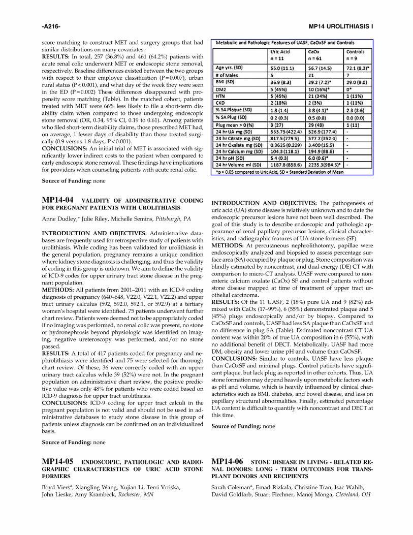
score matching to construct MET and surgery groups that hadsimilar distributions on many covariates.RESULTS: In total, 257 (36.8%) and 461 (64.2%) patients withacute renal colic underwent MET or endoscopic stone removal,respectively. Baseline differences existed between the two groupswith respect to their employee classification (P = 0.007), urbanrural status (P < 0.001), and what day of the week they were seenin the ED (P = 0.002) These differences disappeared with pro-pensity score matching (Table). In the matched cohort, patientstreated with MET were 66% less likely to file a short-term dis-ability claim when compared to those undergoing endoscopicstone removal (OR, 0.34, 95% CI, 0.19 to 0.61). Among patientswho filed short-term disability claims, those prescribed MET had,on average, 1 fewer days of disability than those treated surgi-cally (0.9 versus 1.8 days, P < 0.001).CONCLUSIONS: An initial trial of MET is associated with sig-nificantly lower indirect costs to the patient when compared toearly endoscopic stone removal. These findings have implicationsfor providers when counseling patients with acute renal colic.
Source of Funding: none
MP14-04 VALIDITY OF ADMINISTRATIVE CODING
FOR PREGNANT PATIENTS WITH UROLITHIASIS
Anne Dudley,* Julie Riley, Michelle Semins, Pittsburgh, PA
INTRODUCTION AND OBJECTIVES: Administrative data-bases are frequently used for retrospective study of patients withurolithiasis. While coding has been validated for urolithiasis inthe general population, pregnancy remains a unique conditionwhere kidney stone diagnosis is challenging, and thus the validityof coding in this group is unknown. We aim to define the validityof ICD-9 codes for upper urinary tract stone disease in the preg-nant population.METHODS: All patients from 2001–2011 with an ICD-9 codingdiagnosis of pregnancy (640–648, V22.0, V22.1, V22.2) and uppertract urinary calculus (592, 592.0, 592.1, or 592.9) at a tertiarywomen’s hospital were identified. 75 patients underwent furtherchart review. Patients were deemed not to be appropriately codedif no imaging was performed, no renal colic was present, no stoneor hydronephrosis beyond physiologic was identified on imag-ing, negative ureteroscopy was performed, and/or no stonepassed.RESULTS: A total of 417 patients coded for pregnancy and ne-phrolithiasis were identified and 75 were selected for thoroughchart review. Of these, 36 were correctly coded with an upperurinary tract calculus while 39 (52%) were not. In the pregnantpopulation on administrative chart review, the positive predic-tive value was only 48% for patients who were coded based onICD-9 diagnosis for upper tract urolithiasis.CONCLUSIONS: ICD-9 coding for upper tract calculi in thepregnant population is not valid and should not be used in ad-ministrative databases to study stone disease in this group ofpatients unless diagnosis can be confirmed on an individualizedbasis.
Source of Funding: none
MP14-05 ENDOSCOPIC, PATHOLOGIC AND RADIO-
GRAPHIC CHARACTERISTICS OF URIC ACID STONE
FORMERS
Boyd Viers*, Xiangling Wang, Xujian Li, Terri Vrtiska,John Lieske, Amy Krambeck, Rochester, MN
INTRODUCTION AND OBJECTIVES: The pathogenesis ofuric acid (UA) stone disease is relatively unknown and to date theendoscopic precursor lesions have not been well described. Thegoal of this study is to describe endoscopic and pathologic ap-pearance of renal papillary precursor lesions, clinical character-istics, and radiographic features of UA stone formers (SF).METHODS: At percutaneous nephrolithotomy, papillae wereendoscopically analyzed and biopsied to assess percentage sur-face area (SA) occupied by plaque or plug. Stone composition wasblindly estimated by noncontrast, and dual-energy (DE) CT withcomparison to micro-CT analysis. UASF were compared to non-enteric calcium oxalate (CaOx) SF and control patients withoutstone disease mapped at time of treatment of upper tract ur-othelial carcinoma.RESULTS: Of the 11 UASF, 2 (18%) pure UA and 9 (82%) ad-mixed with CaOx (17–99%), 6 (55%) demonstrated plaque and 5(45%) plugs endoscopically and/or by biopsy. Compared toCaOxSF and controls, UASF had less SA plaque than CaOxSF andno difference in plug SA (Table). Estimated noncontrast CT UAcontent was within 20% of true UA composition in 6 (55%), withno additional benefit of DECT. Metabolically, UASF had moreDM, obesity and lower urine pH and volume than CaOxSF.CONCLUSIONS: Similar to controls, UASF have less plaquethan CaOxSF and minimal plugs. Control patients have signifi-cant plaque, but lack plug as reported in other cohorts. Thus, UAstone formation may depend heavily upon metabolic factors suchas pH and volume, which is heavily influenced by clinical char-acteristics such as BMI, diabetes, and bowel disease, and less onpapillary structural abnormalities. Finally, estimated percentageUA content is difficult to quantify with noncontrast and DECT atthis time.
Source of Funding: none
MP14-06 STONE DISEASE IN LIVING - RELATED RE-
NAL DONORS: LONG - TERM OUTCOMES FOR TRANS-
PLANT DONORS AND RECIPIENTS
Sarah Coleman*, Emad Rizkala, Christine Tran, Isac Wahib,David Goldfarb, Stuart Flechner, Manoj Monga, Cleveland, OH
-A216- MP14 UROLITHIASIS I

INTRODUCTION AND OBJECTIVES: Historically patientswishing to donate their kidney to living related recipients weredeemed ineligible if pre-operative imaging demonstrated ne-phrolithiasis. We assess the outcomes of donors with ne-phrolithiasis and the outcomes of their recipients.METHODS: Donors undergoing nephrectomy between 2001 and2011 who had nephrolithiasis on preoperative CT imaging or ahistory of stone passage were identified. A retrospective chartreview documented donor and recipient demographics, donor24-hour urine collections, stone size and location, stone eventsfollowing transplant and graft function. A 7-question telephonesurvey regarding development and/or presence of symptomaticnephrolithiasis was conducted.RESULTS: 54 donor-recipient pairs met the inclusion criteria.Twenty-eight (51.9%) patients had valid pre-operative 24-hoururine collection, seven (25%) of which had hypercalciuria. Seven(13%) patients had previous symptomatic nephrolithiasis, but nostones on imaging. 41 patients donated a kidney with at least onestone, with a mean stone size of 2.4 mm (range 1–6 mm). Medianfollow-up for donors and recipients was 22.5 months [IQR 1–79.3]and 47.4 months [IQR 25.1–76.1], with 50% and 77.7% having afollow-up of more than two years, respectively. One donor withnephrolithiasis on pre-operative imaging who donated the con-tralateral kidney passed a stone spontaneously after visiting theemergency room. Otherwise, no other donors or recipients ex-perienced any stone episodes during the follow-up period.CONCLUSIONS: The risk of clinical stone recurrence in donorsand recipients is low: as such, presence of small caliceal stonesshould not constitute an exclusion for living-related kidneydonation.
Source of Funding: None
MP14-07 COST EFFECTIVENESS COMPARISON OF
RENAL CALCULI TREATED WITH URETEROSCOPY WITH
LASER LITHOTRIPSY VERSUS SHOCK WAVE LITHO-
TRIPSY
Eugene Cone, Durham, NC, Brian Eisner, Michal Ursiny, Boston,MA, Gyan Pareek*, Providence, RI
INTRODUCTION AND OBJECTIVES: Shock wave lithotripsy(SWL) and ureteroscopy with in situ lithotripsy (URS) are themost commonly performed procedures in the United States fortreatment of ureteral calculi. Current guidelines for the manage-ment of ureteral stones recommend that for patients requiringstone removal both SWL and URS are acceptable first-line treat-ments. Importantly, this discussion includes only clinical risksand benefits, but does not mention cost of treatment. We evalu-ated the cost effectiveness of SWL versus URS for patients with
renal stones less than 1.5 cm in diameter, with the added objectiveof determining at what stone free rate SWL would become costeffective.METHODS: Patient age, stone diameter, stone location, andstone free status were recorded for patients treated with SWL orURS for renal stones under 1.5 cm in maximal diameter over a oneyear period. Institutional charges were obtained from in-housebilling. A decision analysis model was constructed to compare thecost-effectiveness of SWL and URS and using our results andsuccess rates for modeling. Three separate models were created toreflect practice patterns for shock wave lithotripsy.RESULTS: One hundred fifty-eight patients were included in thestudy – 78 underwent SWL and 80 underwent URS as primarytreatment. Single procedure stone free rates for SWL and URSwere 55% and 95% respectively (p < 0.0001). Decision analysismodeling demonstrated cost effectiveness of SWL when SWLsingle procedure stone free rates (SFR) were 65–67% or when URSsingle procedure SFR were 72–84%.CONCLUSIONS: This retrospective study revealed superior SFRresults for renal stones under 1.5 cm for URS compared to SWL.Our decision analysis model demonstrates that for SWL stonefree rates less than 65–67% or for URS stone free rates greater than72–84%, SWL is not a cost-effective treatment option. Based onthese findings, careful stratification and selection of stone patientsmay enable surgeons to increase the cost effectiveness of SWL.
Source of Funding: None
MP14-08 PATIENT EXPERIENCES AND PREFERE-
NCES WITH DIFFERENT URETERAL STENT REMOVAL
TECHNIQUES
Roger Low, Sacramento, CA, Mike Nguyen, Los Angeles, CA,Manoj Monga*, Cleveland, OH
INTRODUCTION AND OBJECTIVES: Stent removal is a majorsource of anxiety for patients. We determined patient experienceswith stent removal methods and identify which approach ispreferred.METHODS: A convenience sample of visitors to a kidney stonerelated website (www.kidneystoners.org) was used. Re-spondents were asked which method was used for their stentremoval (pulling out string by themselves, having their doctorpull out the string, having cystoscopy in the clinic, or havingcystoscopy in the OR). Pain associated with the procedure, painoccurring after the procedure, and preferences and opinionsabout stent removal were queried. Fisher’s exact and ANOVAtests were used to identify significant differences in responses bythe type of technique used.RESULTS: 82 respondents were surveyed. 12.4% and 17% hadstents removed using self-string or doctor-string. 44.4% and22.2% underwent office-cysto or OR-cysto. Pain was highest forthose who had their stent removed by office-cysto (6.0 out of ascale of 1 to 10) followed by self-string (5.2), doctor-string (5.1),and OR-cysto (2.8; p = 0.0028). 37% experienced a severe attack ofpain after stent removal, including 14.1% who required a trip tothe ER. Severe pain after stent removal was most common in withself-string (60%) and least likely with OR-cysto (11.1%; p = 0.033).Those who underwent OR-cysto were most likely to chose thesame technique again (82.4% replying probably or definitelywould choose this method again) followed by self-string (50%),doctor-string (47.1%), and office-cysto (42.3%; p = 0.003). 32.9%reported that their experience was what they expected. 27.9% feltit was a much worse and 25.3% felt it was not as bad or not nearly
MP14 UROLITHIASIS I -A217-

as bad. Those who underwent office-cysto were most likely toreport it being much worse (41.7%; p = 0.046). Being informed ofwhy a stent was placed was the biggest priority for respondents(4.8 on an importance scale of 1–5). Being informed about theprocess was also a high priority (4.7). Being able to have generalanesthesia was a moderate priority (3.5), while being able to see avideo/diagram of the process was a low priority (2.9).CONCLUSIONS: Most patients experienced a moderate amountof pain during stent removal. Over a third also experienced arecurrent episode of severe pain afterwards. Stent removal byoffice cystoscopy was associated with the most pain and was theleast preferred method of stent removal. OR cystoscopy was as-sociated with the least pain and was the most preferred method ifpatients were given a choice. Being informed about why a stent isneeded and the steps involved in stent removal were high pri-orities for patients.
Source of Funding: none
MP14-09 CONCURRENT VALIDITY OF THE WIS-
CONSIN STONE QUALITY OF LIFE SURVEY
Kristina Penniston, Rachel Bell, Stephen Nakada*, Madison, WI
INTRODUCTION AND OBJECTIVES: Using general healthsurveys, decrements in the quality of life (QOL) of patients whoform kidney stones are documented. Until the development of theWisconsin Stone-QOL (Wis-QOL), a 28-item disease-specificquestionnaire, no QOL instrument for urolithiasis existed. Afterconfirming the instrument’s reliability, as well as its content andconstruct validity, we assessed the concurrent validity of the Wis-QOL against the ‘‘gold standard’’ SF-36, a validated and widelyused, 36-item, general health-related QOL instrument.METHODS: Patients reporting to our urology clinic betweenJanuary and May 2013 were invited to participate (n = 70; age52.2 – 16.5 y; 56% male; duration of disease 11.8 – 13.8 y; 33% werefirst-time stone formers). After providing informed consent and abrief medical history, patients were given both the Wis-QOL andthe SF-36 in random order; patients chose which they completedfirst. Scores for each instrument were calculated (QualityMetricHealth Outcomes Scoring Software 4.5). Pearson correlation co-efficients were calculated for total survey scores and for specificdomains that were similar in both surveys; i.e., bodily pain, vi-tality, general health, and social functioning. Multivariate anal-ysis evaluated the impact on QOL of age, gender, BMI, surgicalhistory, and duration of disease.RESULTS: Overall, there was excellent agreement between the 2instruments for total score (Pearson correlation coefficient,r = 0.56, p < 0.0001). There was high correlation between surveysfor bodily pain (r = 0.64; p < 0.0001) and good correlation forquestions related to vitality, general health, and social function-ing (r = 0.51, 0.43, and 0.41, respectively; p < 0.001). Scatterplotsrevealed some patients with lesser agreement between surveys.Responses from these patients were investigated with multivar-iate analyses which revealed no influence of age, gender, BMI, orsurgical history. However, the scores of patients with prior stonestended to be divergent between instruments ( p = 0.06), possiblyexplaining the reason for lower instrument concurrence in thisgroup. Notably, items for which patients scored lowest on theWis-QOL were those for which there were no similar questions onthe SF-36, supporting the utility of a disease-specific instrument.These were bothersome nocturia, poor quality sleep, and feelingtired.CONCLUSIONS: The Wis-QOL captures general physical andmental health comparably to a generic survey, the SF-36, con-
firming its concurrent validity. The benefit of the Wis-QOL is itsability to extract additional information about QOL unique topatients with urolithiasis.
Source of Funding: None
MP14-10 THE GEOMETRIC COMPLEXITY INDEX: A
NEW MEASURE OF STONE BURDEN
Michael Conlin*, Brian Duty, Gregory Adams, Matthias Schabel,Eugene Fuchs, Portland, OR
INTRODUCTION AND OBJECTIVES: There is no acceptedstandard method to quantify stone burden. Comparing results ofdifferent treatment modalities is difficult due to this lack ofstandardization. With the use of a new open-source image pro-cessing computer program (Osirix), we can now easily perform3-D reconstruction, and calculate stone volume. We sought todevelop a better measure of stone complexity to improve ourquantification of stone burden and prediction of stone free out-comes. The purpose of this study is to describe this index, anddetermine its ability to predict stone-free status after percutane-ous nephrolithotomy (PCNL).METHODS: We reviewed 50 consecutive patients from ourPCNL database who had both pre and postoperative CT imagingavailable. We performed 3-D reconstruction of all stones usingOsirix. We wrote a software program (as a plug-in to Osirix) thatmeasures the volume, surface area, and calculate the geometriccomplexity index (GCI) for each patient’s stone burden. The GCIis determined by dividing the measured stone surface area, by thecalculated surface area of an imaginary isovolumetric sphere(same volume of the total stone burden). A stone that is a perfectsphere would have a GCI of 1.0, and increasing branches andnumber of stones increase the GCI. We also collected patientdemographics, comorbidities, and surgical and outcomes data.We performed univariable and multivariable analysis to assesswhich factors best predict stone-free status following PCNL.RESULTS: There were 32 women, 18 men, and the mean age ofthe patients was 56 years. The stone free rate (all patients) was80%. The location of the stone, side of the stone, BMI, gender,stone composition, stone volume and stone surface area were notpredictive of stone-free status. The geometric complexity indexwas found to be the most significant predictor of stone-free status(p = 0.01) in our multivariate analysis (table 1).CONCLUSIONS: We describe a new measure of stone burdenthat helps predict outcomes after percutaneous nephrolithotomy.It is easy to calculate using the free image processing programOsirix, with our software plugin. This index may allow for moreaccurate comparison of studies of different stone treatments.These results suggest that further study of its usefulness shouldbe performed.
Source of Funding: None
-A218- MP14 UROLITHIASIS I

MP14-11 EFFICACY OF TAMSULOSIN IN THE MAN-
AGEMENT OF LOWER URETERAL STONES: A RANDO-
MIZED DOUBLE-BLIND PLACEBO-CONTROLLED STUDY
OF 100 PATIENTS
Abdulla Al-Ansari*, Abdulla Al-Naimi, Abdulkader Alobaidy,Khalid Assadiq, Mohamed Azmi, Doha, Qatar, Ahmed Shokeir,Almansoura, Egypt
INTRODUCTION AND OBJECTIVES: To study the impact oftamsulosin on the rate of spontaneous passage of distal ureteralstones.METHODS: A total of 100 patients with stones sized 10 mm orsmaller, located in the distal part of the ureter were included.Patients were randomly assigned to 2 equal groups. Group 1received 0.4 mg tamsulosin once daily and group 2 receivedplacebo. The investigators and the patients were masked to thetype of treatment. Patients were followed-up until passage of thestone, or for a maximum of 4 weeks. The number of pain episodes,need for analgesia, stone expulsion rate and time, and possibleside effects of medications were observed in both groups.RESULTS: Apart from 4 patients in the placebo group who werelost to follow-up, all patients complied with the prescribedmedications and continued the study. Stone expulsion occurredin 41 of 50 patients (82%) in group 1 and in 28 of 46 patients (61%)in group 2 (P _ .02). The chance of stone expulsion was 3 timeshigher in the tamsulosin group (relative risk [RR] _ 2.93; 95% CI,1.152-7.45). In group 1, patients with stones sized _5 mm showed
a significantly higher expulsion rate compared to those withlarger stones (_5 mm). Age, gender, and stone laterality had nosignificant impact on the expulsion rate. The expulsion time wassignificantly shorter in the tamsulosin group (6.4 _ 2.77 days vs9.87 _ 5.4 days for groups 1 and 2, respectively).
Moreover, the frequency of pain episodes, the need for diclo-fenac, and its total dosage were significantly lower in the tam-sulosin group. Side effects observed in both groups werecomparable and mild, and no patient withdrew because of them.CONCLUSIONS: Tamsulosin is a safe and effective drug thatenhances spontaneous passage of distal ureteral stonessized10 mm or smaller.
Source of Funding: none
MP14-12 EFFECT OF TAMSULOSIN ON STONE EX-
PULSION IN PROXIMAL URETERAL CALCULI: AN OPEN-
LABEL RANDOMIZED CONTROLLED TRIAL
Jinsung Park*, Seung Hyo Woo, Daejeon, Korea, Republic of,Seung Wook Lee, Guri, Korea, Republic of
INTRODUCTION AND OBJECTIVES: Medical expulsivetherapy (MET) using alpha-blockers is effective for distal ureteralcalculi (UC). However, given the fact that the distributions anddensities of alpha-1 receptors along the ureter differ dependingon the location, effectiveness of alpha-blockers for stone expul-sion in proximal UC remains unclear. In this study, we aimed toevaluate the efficacy of tamsulosin for proximal UC expulsion.METHODS: An open-label randomized controlled multicentertrial was conducted from July 2010 to August 2012. A total of 108patients who agreed to conservative management for single, ra-diopaque, proximal UC £ 6 mm were randomized into group A(n = 54, conservative management only) or B (n = 54, 0.2 mg tam-sulosin once a day). Patients were evaluated by KUB and urineanalysis every week until stone expulsion or to post-trial 4 weeks,and final CT was checked to confirm the stone passage. The pri-mary endpoint was stone passage rates (SPR) in the intention-to-treat population in 4 treatment weeks. The secondary endpointswere estimated in per-protocol population and were time to stonepassage, post-trial Euro-quality-of-life (EuroQOL) score, oralanalgesic requirements, and willingness to undergo conservativetreatment again.RESULTS: The two groups were well balanced in terms ofbaseline patient and stone characteristics. Seventy nine patients(73.2%; 35 of group A and 44 of group B) completed the studyprotocol. The group A patients (20.4%, 11/54) tended to be morelikely to convert to active treatment, SWL or ureteroscopic sur-gery, than group B (7.4%, 4/54; p = 0.051). The overall SPR was60.2% (65/108). Group B had a higher SPR (74.1%; 40/54) thangroup A (46.3%; 25/54; p = 0.003) and a significantly shorter timeto stone passage (mean days, A: 19.6 vs B: 14.3, p = 0.005). Thegroups did not differ in post-trial EuroQOL score or oral analgesicrequirements, whereas 74.3% (26/35) of group A and 90.9% (40/44) of group B were willing to undergo conservative treatmentagain (p = 0.048). Univariate logistic regression analysis showedthat stone size (OR = 1.447, p = 0.045) and tamsulosin treatment(OR = 3.314, p = 0.004) significantly predicted stone expulsion. Onmultivariate analysis, only tamsulosin was statistically significant(OR = 3.198, p = 0.021).CONCLUSIONS: Tamsulosin medication was associated withsignificantly higher stone expulsion rate and shorter expulsiontime in proximal UC. Our results indicate that MET with tamsu-losin is a reasonable option for proximal UC as well as distal UC.
Source of Funding: None
MP14 UROLITHIASIS I -A219-

MP14-13 NO DIFFERENCE IN 24-HOUR URINE
PARAMETERS BETWEEN PATIENTS WITH OBSTRUCTING
AND NON-OBSTRUCTING UROLITHIASIS PRESENTING
TO A TERTIARY REFERRAL CENTER
Tarek Alzahrani*, Daniela Ghiculete, Andrea G Lantz,Kenneth T Pace, Jason Y Lee, R. John Honey, Toronto, Canada
INTRODUCTION AND OBJECTIVES: Metabolic work-up ofstone formers often includes at least one 24-hour urine collection.Conventional teaching has been to perform the 24-hour urinecollection after an acute stone event and after obstruction has beenrelieved. However, by the time a patient is seen in follow-up somestone prevention counseling is likely to have occurred, altering the24-hour urine collection results (‘‘clinic effect’’). At our institution,patients bring a 24-hour urine collection to the initial assessmentfor extracorporeal shockwave lithotripsy (SWL). The study objec-tive is to determine if there are differences in 24-hour urine pa-rameters for patients with obstructing vs. nonobstructing stones.METHODS: In 2011, 2670 SWL treatments were performed atour institution. Newly referred, unstented patients with pre-SWL24-hour urine data were reviewed (n = 849). Institutional axialimaging was available for retrospective review in few patients;therefore, patients were grouped into renal vs. ureteral stonesassuming most renal stones are non-obstructing and unstentedureteral stones have some degree of obstruction.RESULTS: 615 renal and 191 ureteral stones were included. 65.4%were male with more males in the ureteral stone group (73.8% vs.62.8%, p = 0.005). Average age was 51.3 years ( – 13.1) with an av-erage BMI of 27.5 kg/m2 ( – 5.4). Mean stone size was similar be-tween groups (p = 0.898). The rate of hypercalciuric patients wassignificantly higher in the renal group (17.3% vs. 11.1%, p = 0.039)but no other parameters differed between groups.CONCLUSIONS: There are few differences in 24-hour urineparameters between obstructive and non-obstructive urolithiasiswhen location of stone is used as a proxy for obstruction. Onlyurinary calcium was statistically different between groups butthis may be of little clinical significance. This study may supportearlier 24-hour urine evaluation, in order to minimize the ‘‘cliniceffect’’, regardless of urinary obstruction.
Source of Funding: none
MP14-14 PATIENT EDUCATION AND THE IMPACT
ON URETEROSCOPY EXPERIENCE
Nathan Grunewald, Alison Marciniak, Carley Davis*,Milwaukee, WI
INTRODUCTION AND OBJECTIVES: Ureteroscopy is a com-mon procedure in Urology requiring instrumenting a patient’sureter with a ureteroscope and placement of a ureteral stent.Managing stent related issues post-operatively can result in nu-merous patient phone calls and/or emergency department/clinicvisits. Our goals were to provide patient education materials in theform of a new handout and intensive counseling prior to and afterureteroscopy to improve the patient’s post-operative experience.METHODS: Retrospective chart review of all patients from asingle surgeon who underwent ureteroscopy over a six monthperiod. During the review period, implementation of intensive pre-operative counseling and dissemination of a newly designed ur-eteroscopy handout. Primary outcome was measured as whether apatient made a post-operative phone call and/or clinic/emergencydepartment contact related to morbidity from their procedure.
RESULTS: Over the course of the 6 month review period a totalof 93 patients underwent ureteroscopy. 56 patients were in thenon-pre-operative intensive counseling and patient handoutgroup, while 37 patients received the patient handout with ac-companying pre-operative intensive counseling. In the non-handout group 54% of patients had a qualified postop morbiditycontact as compared to 38% in handout group. Additionally,20% of patients in the pre-handout group had at least onequalified post-operative morbidity contact related to stent painas compared to 11% in the handout group.CONCLUSIONS: Patient post-ureteroscopy experience can beimproved with intensive counseling and patient education ma-terials leading to decreased phone calls and clinic visits.
Source of Funding: None
MP14-15 A MULTICENTER, RANDOMIZED, DOUBLE-
BLIND, PLACEBO-CONTROLLED STUDY OF SILODOSIN
TO FACILITATE MEDICAL EXPULSION OF URETERAL
CALCULI
Roger Sur*, San Diego, CA, Scott Olsen, Salt Lake City, UT,Ojas Shah, New York, NY
INTRODUCTION AND OBJECTIVES: The alpha 1A-adreno-ceptor (AR) has been shown to play a role in phenylephrine-induced human ureteral contraction. We evaluated the efficacyof silodosin, a selective alpha 1A AR antagonist, in the medicalexpulsion of proximal, mid, and distal ureteral calculi.METHODS: Two hundred forty-six patients with a unilateralureteral calculus (4–10 mm) were randomized to receive eithersilodosin 8 mg or placebo once daily for up to 4 weeks in a double-blind design. If there was no stone passage after 4 weeks, or if thepatient required intervention at any point, the patient was dis-continued from the study. Two hundred thirty-two patients re-ceived study medication and completed the study follow-up.Univariate logistic regression was utilized to compare the pri-mary endpoint of stone passage at 4 weeks with intention-to-treatanalysis. Secondary endpoints included time to stone passage,need for emergency department visits, surgical intervention, painassessment scores, and analgesic use.RESULTS: For patients with distal ureteral stones, treatmentwith silodosin resulted in a significant improvement in thespontaneous expulsion rate at 4 weeks (69.2% vs 45.8%,p = 0.0138) and time to stone passage was lower (approachingstatistical significance). A trend towards improved passage ratewas also observed in patients treated with silodosin for all ure-teral calculi combined but not for proximal or mid ureteral calculisubsets. In addition, patients with distal ureteral stones receivingsilodosin reported significantly greater improvements in averagepain scores at study exit. Among all patients, larger stones (6–10 mm) had higher passage rates with silodosin than with placebo(33% vs 9.1%, p = 0.0573).CONCLUSIONS: Silodosin significantly improved the sponta-neous expulsion rate of distal ureteral stones and resulted ingreater improvements in average pain scores and time to passagefor these patients. Additionally, silodosin improved the passagerate of large ureteral stones. Our findings indicate that silodosinprovided advantages over placebo in medical expulsion therapyof ureteral calculi.
Source of Funding: Watson Laboratories provided grant supportfor the study.
-A220- MP14 UROLITHIASIS I

MP14-16 MODIFIED SEOUL NATIONAL UNIVERSITY
RENAL STONE COMPLEXITY SCORE FOR RETROGRADE
INTRARENAL SURGERY: EVALUATION WITH SINGLE
SURGEON¢S COHORT
Byung Ki Lee*, Jin-Woo Jung, Yong Hyun Park, Sangchul Lee,Seok-Soo Byun, Sang Eun Lee, Seongnam, Korea, Republic of,Chang Wook Jeong, Seoul, Korea, Republic of
INTRODUCTION AND OBJECTIVES: The Seoul NationalUniversity Renal Stone Complexity (S-ReSC) score is well vali-dated predicting tool for the stone-free after percutaneous ne-phrolithotomy. We modified S-ReSC score system to apply toretrograde intrarenal surgery (RIRS). We evaluated the modifiedS-ReSC score with single surgeon¢s cohort of RIRS.METHODS: A total of 88 patients who underwent RIRS bya single surgeon (CWJ) from June 2011 through April 2013were included. All patients were evaluated with pre- and post-operative computed tomography (CT). ¡�Stone-free¡ – was de-fined as no evidence of residual stones on postoperative imagesfor 1 month. The modified S-ReSC score was assigned on thenumber of sites involved in the renal pelvis (#1), superior andinferior major calyceal groups (#2–3), and anterior and posteriorminor calyceal groups of 12 the superior (#4–5), middle (#6–7),and inferior calyx (#8–9). Then if stone involved in inferior sites(#3, #8–9), 1 score per site was added to original score. Thusmodified S-ReSC score varies from 1 to 12 points in total. Thestone-free rates were examined according to the modified S-ReSCscores. To evaluate the predictive accuracy, the area under thereceiver operating characteristic curve (AUC) was used. Thiswas compared with Resorlu-Unsal Stone Score (Urology 2012;80:512–8).RESULTS: Thirty-seven (42.0%) patients had multiple stones, 53(60.2%) had lower pole stones. Mean diameter of the largest stonewas 12.0 – 6.1 mm. Overall and clinical stone-free rates ( < 4 mmresidual stone) were 85.2%, and 90.9%, respectively. Stone-freerates were significantly decreased in the order of the low (1–2:94.2%), medium (2–4; 84.0%), and high ( > 4; 45.5%) modifiedReSC score groups (p < 0.001). Resorlu-Unsal Stone Score wasalso associated with stone-free rates (0; 93.6%, 1; 78.1%, ‡ 2;66.7%, p = 0.043). However, AUCs of modified S-ReSC score(0.806) and score group (0.766) were higher than AUC of Resorlu-Unsal Stone Score (0.692).CONCLUSIONS: RIRS is safe and effective therapy for selectedrenal stones. Modified S-ReSC score well predicts the stone-freerate after RIRS. Furthermore, its predictive accuracy is higherthan accuracy of Resorlu-Unsal Stone Score.
Source of Funding: None
MP14-17 URETEROSCOPY FOR THE MANAGEMENT
OF UPPER URETERAL CALCULI; WHETHER MUCOSAL
IMPACTED STONES OR NOT, DOES IT MATTER?
Dong-Hoon Lim, Min-Seok Kim*, Joon Roh, Chul-Sung Kim,Dong-Hoon Yoo, Ju-Sung Kim, Gwangju, Korea, Republic of
INTRODUCTION AND OBJECTIVES: Management of im-pacted stones in the upper ureter remains challenging. Eventhough ureteroscopic removal is one of efficacious treatmentoptions in the management of upper ureteral stones, it is oftenproblematic due to the inflammatory and edematous mucosaenveloping the impacted calculus. Aim of this study is to identifysurgical influence of mucosal impaction in ureteroscopic removal
of upper ureteral stones by analyzing the operative outcomes ofureteroscopy.METHODS: From January 2008 to December 2012, a total of 103patients underwent retrograde ureteroscopic removal of upperureteral stones with a pneumatic lithotripter by a single surgeon.The patients were retrospectively divided into two groups (group1; impacted VS group 2; not impacted) according to presence ofmucosal impacted stone. Demographic and clinical characteris-tics of patients and operative results including Stone-free rate,retreatment rate, auxiliary treatment rate, and efficiency quotient(EQ) were analyzed and compared between two groups.RESULTS: 39 out of 103 patients had mucosal impacted stones.Demographic and clinical characteristics of patients are in table 1.Mean operative time and hospital days in group 1 was signifi-cantly longer than that in group 2. The stone-free rate at the 1month follow-up was 84.6% in group 1 and 96.8% in group 2;retreatment rate 7.7% and 3.1%; auxiliary treatment rate 0% and3.1%, respectively. However, these were all insignificant(p > 0.05). Also, all operation related complication rate betweentwo groups was insignificant (Table 2).CONCLUSIONS: Ureteroscopic management for upper ureteralstones is a safe and effective method. Even though the presence ofmucosal impaction seems to be associated with longer operationtime and hospital days, there is no need to hesitate to perform ur-eteroscopy for the management of impacted upper ureteral stones.
Source of Funding: none
Table 1. Demographic and Clinical
Characteristics of Patients
Table 2. Operative Outcomes
*Studentâe�s t-test, âe Chi-square test.
MP14 UROLITHIASIS I -A221-

MP14-18 MEDICAL EXPULSION THERAPY IS UN-
DERUTILIZED IN WOMEN PRESENTING TO AN EMER-
GENCY DEPARTMENT WITH ACUTE URINARY STONE
DISEASE
Herman Bagga*, Ayesha Appa, Ralph Wang, Thomas Chi,Joe Miller, Jersey Neilson, Rebecca Smith-Bindman,Marshall Stoller, San Francisco, CA
INTRODUCTION AND OBJECTIVES: Medical expulsiontherapy (MET) with alpha-adrenergic blockade (AB) is an es-tablished treatment option for ureteral stones. However, utiliza-tion rates remain low. Specific reasons for this remain largelyunknown. We hypothesized that low utilization of MET would bemost pronounced in women as AB medications are routinelyused for men with urologic conditions such as benign prostatichyperplasia (BPH).METHODS: Retrospective chart review of adults with an EDdiagnosis of acute ureteral stone disease between 2006–2012 wasundertaken to identify variables related to presentation, history,management, and disposition. Patients with a known diagnosisof ureteral stone disease within the past month or regular homeuse of AB were excluded.RESULTS: Between 2006–2012, 672 patients were dischargedfrom our ED with a diagnosis of ureteral stone disease. 57% ofpatients were discharged with AB for MET. On multivariateanalysis, male sex was independently associated with dischargewith MET compared to female sex (OR 1.86, 95% CI 1.33–2.6). Useof imaging (computerized tomography or ultrasound) for diag-nosis was also independently associated with MET use comparedto patients with a clinical diagnosis alone (OR 1.85, 95% CI 1.14–2.94). Age, primary language, race, occupation, history of stonedisease, and presentation factors such as hematuria and painassociations were not significantly associated with MET use.
Analyses of trends of MET utilization found that rates of use tohave peaked and stabilized since 2008.CONCLUSIONS: In a tertiary care ED, MET use remains lowand women are nearly half as likely to be discharged with METcompared to men. This may be due to providers’ perception thatAB agents are for male urologic conditions only. Rates of MET usehave stabilized since 2008, suggesting increased awareness andeducation regarding proper utilization of MET, particularly inwomen, is important to promote appropriate, increased useamongst ED care providers.
Source of Funding: none
MP14-19 IN VITRO EVALUATION OF THE URONET�STONE FRAGMENT RETRIEVER FOR PERCTUANEOUS
NEPHROLITHOTOMY
Kelly A. Healy*, Ryan C. Cleary, Demetrius H. Bagley,Philadelphia, PA
INTRODUCTION AND OBJECTIVES: Percutaneous ne-phrolithotomy (PCNL) is the typical treatment for large renalstone burdens. Various lithotripters and accessory devices maybe utilized to fragment and extract calculi. Traditionally, 2- and 3-prong graspers as well as baskets have been the mainstay forremoval of small to medium-sized residual stone fragmentsduring PCNL. The UroNet� retriever (US Endoscopy) employs anew method of stone extraction. This study aims to compare theefficiency, reliability, and overall user satisfaction of the UroNet�
retriever to three contemporary competitors.METHODS: Nine participants from three experience levels(novice, intermediate, expert) completed a 30 minute in vitro rigidPCNL simulation using four stone retrieval devices (Cook PercNCircle�, Bard Dimension� articulating basket, Bard Flatwire�
basket, and US Endoscopy UroNet� retriever). Twelve beads ofvarying sizes (3 of each size: 2 mm, 4 mm, 6 mm, 8 mm) wereextracted from a previously described calyceal model in threeseparate trials. Efficiency, errors, and satisfaction were evaluated.RESULTS: Across all experience levels, the UroNet� retrieverhad the shortest time to remove all beads (mean 69.8 seconds),fewest in and out movements (6.5), and highest number of beadsremoved per attempt (1.8). Perc NCircle� performed second best,with 76.8 seconds, 9.1 movements, and 1.3 beads, respectively.Compared to the Perc NCircle�, the UroNet� demonstrated su-periority in terms of the number of in and out movements(p = 0.013) and number of beads retrieved per attempt (p = 0.007)using two-tailed student t-test analysis. UroNet� and PercNCircle� received similar satisfaction scores (7.8 and 7.9,p > 0.05), which were significantly different from the Bard Di-mension� (6.2, p = 0.013) and Flatwire� baskets (2.0, p < 0.00002).The number of beads dropped was not significantly differentbetween the four devices.CONCLUSIONS: The UroNet� stone retriever demonstrated thehighest efficiency, particularly in grasping 2 mm and 4 mm beads.Overall user satisfaction was equivalent between the UroNet�
and Perc NCircle�. During PCNL, the ideal use for the UroNet�
appears to be following stone debulking for the removal ofmultiple small to medium-sized stone fragments, which mayotherwise prove difficult or cumbersome to extract.
Source of Funding: None.
-A222- MP14 UROLITHIASIS I

MP14-20 THE IMPACT OF FISH OIL SUPPLEMENTA-
TION ON ENDOGENOUS OXALATE SYNTHESIS AND UR-
INARY OXALATE EXCRETION
Jessica Lange*, Winston-Salem, NC, Patrick Mufarrij, Washington,D.C., DC, Linda Easter, Winston-Salem, NC, John Knight,Ross Holmes, Dean Assimos, Birmingham, AL
INTRODUCTION AND OBJECTIVES: It has been reportedthat the administration of omega 3 fatty acid-rich fish oil sup-plements reduces urinary oxalate excretion. The mechanism ofthis response has not been defined. One other possibility is re-duced endogenous oxalate synthesis. This study was undertakento assess this hypothesis. The rationale for this is that fish oilreduces inflammation which is also thought to reduce oxidativestress. A reduction in the latter has been proposed to promote adecline in endogenous oxalate synthesis.METHODS: Fifteen healthy, non-stone forming adults (averageage 25.3 – 2.7 years, BMI £ 30, 8 male, 7 female) participated in thestudy. Subjects first abstained from utilizing any supplements in-cluding vitamins, medications, or foods enriched in omega 3 fattyacids for thirty days. After this, they collected two random twenty-four hour urine specimens while consuming a self-selected diet.After subjects consumed an extremely low oxalate (50 mg/day)and normal calcium (1000 mg/day) for five days, they collectedtwenty-four hour urine samples on the last three days of this die-tary regimen. Next, the subjects took two fish oil capsules con-taining 650 mg eicosapentaenoic acid and 450 mg docosahexaenoicacid twice daily for 30 days. They consumed a self-selected diet ondays 1–25 and the aforementioned controlled diet on days 26–30.Twenty-four hour urine samples were collected on days 28–30.Excretion patterns of urinary parameters including oxalate wereanalyzed using repeated measures ANOVA and Student’s t test.RESULTS: While there was significant reduction in urinary ox-alate, magnesium, and potassium excretion and increase in uricacid excretion during the controlled dietary phases, there were nosignificant differences in their excretion during controlled dietphases with and without fish oil supplementation.CONCLUSIONS: These results suggest that fish oil supple-mentation does not reduce endogenous oxalate synthesis or uri-nary oxalate excretion in normal adults during periods ofextremely low oxalate intake.
Source of Funding: National Institutes of Health grant R01DK62284
MP14-21 HOW LONG IT TAKES TO LEARN TO USE
ROBOFLEXAVICENA FOR FLEXIBLE URETERORENO-
SCOPY, COMPARISON OF AN EXPERIENCED FLEXIBLE
URS USERS AND TRAINEES
Zafer Tokatli*, Ankara, Turkey, Ahmet Yaser Muslumanoglu,Murat Binbay, Abdülkadir Tepeler, Istanbul, Turkey, Evren Süer,Ankara, Turkey, Remzi Saglam, Ankara, Turkey
INTRODUCTION AND OBJECTIVES: Our objective was tocompare the performance of flexible ureterorenoscopy (fURS)experts and trainees on robotic fURS on a Minnesota UniversityKidney Model.METHODS: A total of 5 experts (Group 1) and 5 trainees (Group2) of fURS were included to the study group. All the participantsused Storz flexX2 for manual fURS and Robotic fURS. A Min-nesota University Kidney Model was used. Participants who had
performed > 50 and < 10 flexible URS were accepted as expertsand trainees, respectively. After watching a video showing allfunctions of RoboflexAvicena all of the participant requested todrive to the upper calices, middle calix and lover calices and comeback to the urether. They did same manually. All of the partici-pants performed both flexible and robotic fURS two times. Dur-ing all procedures a record was kept of the duration and the meantime values of the groups were compared.RESULTS: Mean age of the study group was 39 years old andboth of the groups mean age values were similar. The mean 1stand 2nd manuel flexible URS durations were 58–91 seconds and69–110 seconds for group 1 and group 2, respectively. Compar-ison of these values demonstrated a significant shorter time forparticipants in group 1 (experts)(p < 0,05). In group 1 the 1st and2nd mean robotic URS durations were 171 and 126 seconds. Theseoutcomes did not display any significant difference than theoutcomes of group 2 (trainees) (180 and 128 seconds, (p > 0,05).CONCLUSIONS: Although flexible URS is an effective modalityfor the treatment of upper urinary tract stones, dexterity andexperience are essential features for the success of the procedure.Learning time of robotic fURS is very short and is demonstratedas an easy and applicable modality even for trainees. The numberof participants will be increased.
Source of Funding: none
MP14-22 TRENDS AND DEMOGRAPHICS IN STONE
COMPOSITION - TRANSITION FROM NON-URIC ACID
TO URIC ACID STONES
Kara L Watts*, Tian C Zhou, Joseph Divito, David M Hoenig,Bronx, NY
INTRODUCTION AND OBJECTIVES: We have previouslyreported a significant association between visceral adiposity, asmeasured by visceral fat area (VFA) on CT scan, and uric acid(UA) stone formation. Previous reports have also associatedmetabolic syndrome with UA stone disease. We sought to iden-tify patients who underwent PCNL that transitioned betweenpartial- or pure UA stones and non-UA containing stones overtheir clinical course of stone disease.METHODS: Fifty patients with UA stone disease (pure or partial)who underwent PCNL were identified in an IRB-approved data-base. A retrospective review was performed to identify precedingsurgical stone analyses with respect to composition. Patients whopreviously had non-UA containing stones, and subsequently de-veloped UA stones, were assessed for clinical changes with respectto VFA, BMI, and other medical comorbidities. VFA was calculatedusing a ‘‘Fat Analysis’’ tool on a single axial slice from a CT scan atthe level of the umbilicus, using specialized software (TerraReconInc Aquarius iNuition, software version 4.4.6.100.2862).RESULTS: Two patients were identified with a transition fromnon-UA to UA-containing stones, with a mean interval of 8.25years between stone episodes (range: 7.5–9 years). Patient A’sinitial stone composition (50% calcium oxalate (CaOx) mono-hydrate, 25% carbonate apatite, 15% CaOx dihydrate, protein)differed from his second episode (80% UA dihydrate, 20% CaOxmonohydrate), which occurred 9 years later. During this interval,he had a significant increase in VFA (305 to 470 cm2) and devel-oped type 2 Diabetes Mellitus. Patient B’s initial stone (75% CaOxmonohydrate, 15% CaOx dihydrate, 10% carbonate apatite)transitioned to a predominantly UA-containing stone 7.5 yearslater (80% UA dehydrate; 20% CaOx monohydrate). During this
MP14 UROLITHIASIS I -A223-

interval, she was diagnosed with hypercholesterolemia, under-went a subtotal colectomy for ulcerative colitis, and demon-strated a decrease in VFA (120 to 73 cm2).CONCLUSIONS: Transition between non-UA and UA-contain-ing stones occurs infrequently and has not been previously re-ported in the literature. However, preliminary data suggest that itshould be suspected in patients with new medical comorbidities,an increase in visceral adiposity, or development of metabolicsyndrome. Recognition of this unusual phenomenon is criticalwhen considering medical therapy and prevention strategies formanagement of stone disease.
Source of Funding: none
MP14-23 TOTAL CHOLESTEROL AS A SERUM BIO-
MARKER OF HYPERCALCIURIA IN PATIENTS WITH
KIDNEY STONES
Raman Unnikrishnan*, Fabio Torricelli, Shubha De,Nima Almassi, Carl Sarkissian, Ina Li, Manoj Monga,Cleveland, OH
INTRODUCTION AND OBJECTIVES: Metabolic syndromehas been associated with an increased risk of kidney stones. Infact, there is some evidence that obesity and hyperlipidemia mayplay an important role in urolithiasis. However, there is little dataregarding the association between lipid profile and 24-hour urineanalysis. Herein, we aim to study the impact of hypercholester-olemia on 24-hour urine analysis and stone composition.METHODS: We retrospectively identified patients with ne-phrolithiasis who also had a 24-hour urine analysis and lipidprofile evaluation from July 2001 through March 2013. Patients onstatin treatment before the 24-hour urine analysis were excluded.Patient’s data collected included age, gender, body mass index(BMI), 24-hour urine analysis (volume, sodium, potassium, cal-cium, oxalate, uric acid, citrate, magnesium, phosphate, chloro,creatinine, and pH), serum total cholesterol (TC), and stone com-position. Patients were divided in two groups based on their TClevel (TC < 200 mg/dL and TC > 200 mg/dL). Both groups werecompared based on their demographic data, each component of24-hour urine analysis, and stone composition, using the t-studenttest for continuous variables and the fisher exact test for categoricalvariables. Then, a multivariate analysis including all significantvariables from univariate analysis was performed to confirmwhich variables were still significantly correlated to the TC level.SPSS 20.0 (IBM, Chicago, IL) for Macintosh was used to statisticalanalysis and significance was set at two tail p-value < 0.05.RESULTS: 2442 patients (52% males) were included, with a meanage of 51.1 + - 14.3 years and mean BMI of 29.7 + - 7.1. After theunivariate analysis, patients with higher TC level had higherurinary potassium (74.0 + - 29.1 vs. 61.4 + - 25.6 mg/dL; p = 0.025)and calcium (245.6 + - 108.2 vs. 199.0 + - 125.5 mg/dL; p = 0.036),as well as lower oxalates (46.6 + - 21.3 vs. 49.8 + -27.7 mg/dL;p = 0.002). Patients with higher TC level also had a higher rate ofuric acid stones (16.7% vs. 6.9%; p < 0.001). After the multivariateanalysis only potassium (p = 0.009) and calcium (p = 0.013) urinarylevels remained significantly associated with higher TC level.CONCLUSIONS: Serum total cholesterol is significantly associ-ated with higher urinary potassium and calcium levels. Thissuggests a potential link between hypercholesterolemia and hy-percalciuria.
Source of Funding: None
MP14-24 DEFINING VARIATION IN URINARY
OXALATE IN HYPEROXALURIC STONE-FORMERS
Jodi Antonelli, Justin Friedlander*, Dallas, TX, Craig Langman,Chicago, IL, John Poindexter, Beverley Huet, Margaret Pearle,Dallas, TX
INTRODUCTION AND OBJECTIVES: The development ofeffective preventative therapy for renal calculi in patientswith secondary hyperoxaluria (2�HO) relies upon establishingthe pattern of normal variation in urinary oxalate (uOx)and attempting to reduce it. Therefore, we evaluated uOxat baseline and at subsequent time points in stone-formerswith 2�HO.METHODS: We reviewed the charts of 201 recurrent stone for-mers with 2�HO (uOx ‡ 40 mg/day). 24-hour urine collections atbaseline and after initiation of clinician-directed therapies wereanalyzed. Mixed models were constructed to analyze uOx overtime for individual patients and as a group. Subgroup analyseswere performed for enteric and idiopathic 2�HO. Coefficients ofvariation were computed using the root mean square error fromlinear models.RESULTS: The etiology of 2�HO was enteric in 17.9% and idio-pathic in 82.1% of patients. Among the 943 urine collections an-alyzed, 196 oxalate values were derived from the enteric groupand 747 from the idiopathic group. The median number ofuOx values measured per person was 4. The median 24-hr uOx(mg/day) was significantly higher for the enteric group than forthe idiopathic group both at the time of diagnosis [64.4 (inter-quartile range (IQR) = 48–90)] versus 46.0 (IQR = 38–56), p < 0.001]and during follow-up [58.2 (IQR = 46–86) versus 44.2 (IQR = 35–53), p < 0.001]. Over a median follow-up of 22.5 months, 44.4% ofthe enteric and 61.8% of the idiopathic patients had at least onenormal uOx value (p = 0.06). The coefficients of variation for theenteric and idiopathic groups were 40.8% and 27.3%, respec-tively, with variation randomly displayed in either direction forboth groups.CONCLUSIONS: Among patients with 2�HO, uOx demon-strates significant random variation over time even with theincorporation of standard treatments, with enteric HO demon-strating higher values and greater variance than idiopathic HO.
Source of Funding: None
MP14-25 GOUT, STONE COMPOSITION AND UR-
INARY STONE RISK: A CASE MATCHED COMPARATIVE
STUDY
Ryan Mori*, Shubha De, Giovanni Marchini, Carl Sarkissian,Devin Tian, Manoj Monga, Cleveland, OH
INTRODUCTION AND OBJECTIVES: To establish the mostcommon stone composition and urinary biochemical features ofpatients with gout and urolithiasis we performed a matchedcomparison analysis.METHODS: A case-matched cohort was matched by age, genderand BMI. Primary end-points were 24-hour urines, and stonecomposition. Allopurinol’s effect composition was evaluated. Stu-dent’s t test, Chi-square/Fisher’s exact tests were used for analyses.RESULTS: Stone panel evaluation: 181 patients met our inclusioncriteria, with no differences in demographic characteristics, or 24-hour UA between non-gout and gout cohorts. Hyperoxaluria wasmore common with gout (74 vs. 61%; p = 0.009). Stone composi-
-A224- MP14 UROLITHIASIS I

tion analysis (n = 393): Gout had lower amounts COM (39.4 vs.54.7%), COD (6.0 vs 11.2%) and CaP (9.6 vs 14.1%; p < 0.001) andhigher UA (42.7 vs 18.2%; p < 0.001). Pure UA were more commonin gout (52.2 vs 22.3%; p < 0.001). Allopurinol use was associatedwith a shift in stone composition (figure 1), less pure UA (30.4vs.56.4%; p = 0.001) and more COMH stones (69.6 vs 40.7%;p < 0.001).CONCLUSIONS: UA stones are the most common pure stone inpatients with gout, despite having similar urinary acid levels tonon-gout patients. Allopurinol changes stone composition to apattern similar to those without gout.
Source of Funding: none
MP14-26 RETROGRADE INTRARENAL SURGERY
(RIRS) VS MINIMALLY INVASIVE PERCUTANEOUS
NEPHROLITHOTRIPSY (MPNL) FOR RENAL STONES BE-
TWEEN 1 TO 2 CM: A PROSPECTIVE COMPARATIVE
STUDY
Rajesh Kukreja*, Indore, India
INTRODUCTION AND OBJECTIVES: The Minimally InvasivePercutaneous Nephrolithotripsy (MPNL) and Retrograde In-trarenal Surgery (RIRS) have shown high efficacy and safety forthe management of small renal stones. This study compares theefficacy and morbidity between MiniPNL (15 to 16.5 Fr) vs RIRSin renal stones sized from 1 to 2 cm.METHODS: This was a prospective single center study with twogroups (35 patients in each). The first group comprised patientswho underwent MPNL (15–16.5 Fr), while in the second groupunderwent RIRS with flexible ureterorenoscope (Flex X2). Theprimary end points were haemoglobin drop after surgery asequivalents of safety and stone clearance rate in terms of efficacy.The secondary end points comprised procedure time, need forstenting or nephrostomy drainage tube, pain level, and hospitalstay.RESULTS: The mean stone size was same in both the groups (1.46vs 1.41 cm). The procedural time was shorter in the MPNL group(23.6 min vs 35.5 min). 5 patients in the RIRS group had a tightureter not allowing passage of the scope. One of these was stentedand treated by RIRS 7 days later. The remaining 4 were convertedinto MPNL and stone cleared in the same anaesthesia. The meanhaemoglobin drop after the procedure was significantly lower inthe RIRS group (0.83 vs 0.47 gm %). Similarly, pain score andhospital stay were marginally lower after RIRS in comparisonwith MPNL. Calculus clearance was 97.1% in the MPNL groupand 94.3% in the RIRS group. Post procedural DJ stent was placedin 4 cases in the MPNL and 33 cases in the RIRS group. None ofthe patients had a nephrostomy tube postoperatively. There wereno significant complications (Clavian grade 2 or above) in eitherof the groups. In the RIRS group, 5 patients had significant stentrelated dysuria or hematuria.CONCLUSIONS: MPNL and RIRS are both effective options intreatment of medium sized renal stones with minimal morbidity.MPNL has the advantage of better clearance in a single sessionwhereas RIRS may require prestenting in 14% cases. Periopera-tive morbidity is lower in the RIRS group however the need forstenting and stent related symptoms are a disadvantage. Propercounselling with the patient prior to the procedure helps inchoosing the correct procedure in an individual patient.
Source of Funding: None
MP15 ROBOTICS/LAPAROSCOPY: PROSTATE, LOWERTRACT II
MP15-01 TIPS TO AVOID POSITIONING INJURY TO
THE PATIENT AND ASSISTANT DURING ROBOTIC SUR-
GERY
James Borin*, Jeff McDaniel, Baltimore, MD
INTRODUCTION AND OBJECTIVES: Positioning injury is apotentially avoidable complication of robot assisted surgerywhich can lead to prolonged recovery, severe debilitation, andlegal action. This can be particularly evident during the learningcurve when cases have a longer duration. In addition to patientinjury, the bedside assistant can be at risk for neuromuscular or
traumatic injury due to suboptimal port placement. We describeseveral adjuncts to positioning which may help to avoid thesetypes of complications.METHODS: For pelvic surgery in steep Trendelenburg position,the patient is at increased risk of ulnar, femoral, and commonperoneal nerve injury due to pressure or stretch. This can beavoided by careful padding and ensuring a bent-knee position.The patient must be well balanced and secured to the table toprevent sliding which can cause femoral stretch and pressureulceration. For renal surgery in modified or full flank position,the ipsilateral arm must be protected. Minimal use of flexion orkidney rest can avoid rhabdomyolysis, and judicious use of an
FIG. 1. Overall stone composition distribution among non-gout individuals and gout patients under no medication and onAllopurinol treatment.
MP15 ROBOTICS/LAPAROSCOPY: PROSTATE, LOWER TRACT II -A225-

axillary roll is important to avoid bracheal plexus damage. Theassistant’s ports must avoid the camera arm; the surgeon shouldrefrain from moving the camera at all while the assistant isinserting robotic instruments in order to prevent a traumaticcollision.RESULTS: We have found that the Pigazzi Patient PositioningSystem (Xodus Medical) facilitates several of the key portions ofTrendelenburg positioning; it is anti-slip and provides a mecha-nism to easily tuck the arms, which we wrap in 2 egg-crate foampads. A thumbs-up position is critical, and all wires and i.v.tubing should be placed on the outside of the padding; the handsmay be covered with foam pads as well. A tilt-test will ensure thatthe patient is stable and will not slide. For flank position, weprefer a modified lateral decubitus facilitated by a large gel rollunder the ipsilateral shoulder and hip, with the ipsilateral armpadded and tucked at the patient’s side. The legs are straight andrest on pillows, the table is not flexed nor is the kidney rest de-ployed. A slightly lateral position of the camera port will provideextra room for the assistant to maneuver; a bariatric-length(45 cm) suction cannula is paramount as well. Ports should beplaced strategically to avoid the transit of the camera, so the as-sistant may sit comfortably.CONCLUSIONS: Adopting proactive strategies to protect thepatient and the assistant can reduce the risk of neuromuscularinjury.
Source of Funding: none
MP15-02 FUNCTIONAL AND ONCOLOGICAL OUT-
COMES OF ROBOTIC ASSISTED RADICAL PROSTA-
TECTOMY (RARP) LEARNING CURVE ANALYSIS OF
418 CONSECUTIVE PATIENTS WITH UP TO 7 YEAR
FOLLOW-UP
Philippe Violette*, Stephen Pautler, London, Canada
INTRODUCTION AND OBJECTIVES: The transition to roboticassisted radical prostatectomy requires the surgeon to go througha learning curve with this new approach. There is limited data onthe effect of the robotic learning curve on long term oncologicaland functional outcomes. Our objective is to describe and identifyindependent predictors of short and long term outcomes in thecontext of the robotic learning curve.METHODS: We retrospectively evaluated a cohort of 418 pa-tients from a prospectively collected consecutive series of patientswho underwent RARP at an academic institution from 2006 to2012. Oncologic learning curve was characterized by decile withrespect to margin status. Descriptive statistics were performedusing student-t and Chi-square where appropriate. Analyticalstatistics were performed using multivariate logistic regressionmodeling to define independent predictors of margin status, andshort and long term complications. Multivariate cox regressionwas used to evaluate PSA recurrence and continence.RESULTS: Our cohort was made up of men aged 60 – 7 years,with low or intermediate risk prostate cancer (PSA 7 – 3,61% Gleason 6, 39% Gleason 7, and 71 % stage 1 disease) pre-operatively and a mean BMI of 27 – 3. The majority underwentnerve sparring (75%) prostatectomy with pelvic lymph nodedissection (44%). The learning curve was identified to plateauafter the first 84 cases. On univariable analysis the learning curvewas associated with longer OR time (223 – 49 vs 184 – 30 min,p < 0.001), increased blood loss (307 – 471 vs 243 – 172, p = 0.04),higher rates of positive surgical margins (28 vs 12%, p < 0.001),and PSA failure (10 vs 2%, p0.02). There were no differences be-tween groups for continence, Clavien complications or long term
complications. Multivariable logistic regression revealed thatlearning curve was the strongest independent predictor of marginstatus (OR 4.3, 95%CI 2.26-8.20, p < 0.001) comparable to patho-logic stage (OR 3.47, 95%CI 1.90-6.32, p < 0.001). In contrast, onmultivariate analysis learning curve did not predict PSA recur-rence, continence, short term or long term complications.CONCLUSIONS: The learning curve for RARP was found to bepredictive of increased positive margins, however this did nottranslate into increase PSA recurrence or rates of complications.
Source of Funding: None
MP15-03 ROBOTIC ASSISTED RADICAL PROSTA-
TECTOMY FOR HIGH RISK PROSTATE CANCER: QUAL-
ITY OF LIFE OUTCOMES IN MEN WHO ELECT SURGERY
AS PRIMARY THERAPY
Hoyt Doak*, Farmington, CT, Ryan Dorin, Halil Kiziloz,Joseph Wagner, Hartford, CT
INTRODUCTION AND OBJECTIVES: Robotic prostatectomywith pelvic lymph node dissection (RARP) is increasingly per-formed on patients with high risk prostate cancer. There are fewstudies on quality of life (QOL) outcomes following RARP forhigh risk disease. We used a validated QOL questionnaire toevaluate of the long term morbidity associated with this treat-ment paradigm.METHODS: Men included in our institutional IRB approvedprostatectomy database are asked to complete the ExpandedProstate Cancer Index Composite (EPIC-26) questionnaire pre-operatively and at each follow-up visit. We queried the databasefor all patients who underwent RARP as initial therapy forD’Amico high-risk prostate cancer between 2/2004- 11/2011.Patients were excluded if they had undergone previous radio-therapy or androgen deprivation therapy. Questionnaire scoreswere aggregated into a composite index score of 0–100, with 100representing the most favorable score for the following domains:urinary function (UF), bowel health (BH), hormonal function(HF), and sexual function (SF). Index scores were stratified bynerve sparing (NS) status. Linear regression models and analysisof variance were used for statistical comparisons.RESULTS: 277 patients met inclusion criteria, median age atsurgery was 62, median follow up interval was 11.7 months (max:145 months), and a mean of 4.4 questionnaires were completedper patient. 81 men underwent a bilateral NS procedure, 81 aunilateral NS procedure, and 115 a non-NS procedure. Mean in-dex scores on the UF, HF, and SF domains changed significantlyfrom baseline (p < 0.01 for all), while mean index scores on the BHdomain did not change significantly (p = 0.234). At 2 years, nervesparing status was associated with a statistically significant im-provement in scores in the SF domain only, with patients un-dergoing unilateral (p = 0.01) or bilateral NS (p < 0.01) reportinghigher scores than non-NS patients. (Figure 1.)
-A226- MP15 ROBOTICS/LAPAROSCOPY: PROSTATE, LOWER TRACT II

CONCLUSIONS: QOL outcomes based on EPIC-26 scores forpatients undergoing RARP for high risk prostate cancer weresimilar to those of lower risk cohorts in previously publishedstudies. Randomized comparisons of QOL outcomes betweentreatment modalities for high risk patients are needed to optimizetreatment strategies.
Source of Funding: None
MP15-04 ONCOLOGIC OUTCOMES FOLLOWING
ROBOT ASSISTED LAPAROSCOPIC RADICAL PROSTA-
TECTOMY IN OBESE PATIENTS WITH NON-ORGAN
CONFINED PROSTATE CANCER
Edris Negron*, Kyle Richards, Joshua Cohn, Scott Eggener,Gregory Zagaja, Arieh Shalhav, Chicago, IL
INTRODUCTION AND OBJECTIVES: Obesity is a growinghealth epidemic in Western culture and is highly prevalentamong patients with prostate cancer (PCa). The relationship be-tween obesity and PCa has been widely studied. However, thereis scant data on the outcomes of robot assisted laparoscopicprostatectomy (RALP) in obese patients with high risk disease.Our goal is to assess the impact of body mass index (BMI) onmidterm oncological outcomes following RALP in patients withpathologic non-organ confined (pT3) disease.METHODS: In our IRB approved and prospectively attainedclinical database, we identified 3,063 patients who underwentRALP from March 2003 to January 2012. Follow up has beenmaintained every 6 months for 2 years, and yearly thereafter.From this group, patients with more than 12 months of followup and pT3 disease were selected. Exclusion criteria includedreceipt of adjuvant therapy, salvage RALP, or persistent PSAelevation post-operatively. Patients were separated into 4 co-horts based on BMI: 1. BMI < 25 kg/m2, 2. BMI 25–30 kg/m2, 3.BMI 30–35 kg/m2, and 4. BMI > 35 kg/m2. We defined bio-chemical recurrence (BCR) as PSA > 0.2 ng/ml. Time to BCR wascompared across BMI groups via Kaplan Meier analysis and thelogrank test.RESULTS: 368 patients met inclusion criteria with overallmedian follow-up of 26 months. There was no difference infollow-up, baseline demographic, clinical, or pathologic featuresbetween the 4 BMI groups. Perioperative outcomes were alsosimilar between the 4 groups with the exception of operative time.Relative to group 1, there was no difference in mean OR time ingroup 2 (p = 0.49), whereas OR times were significantly longer in
groups 3 and 4 (mean increase of 27 minutes, p = 0.006 and 30minutes, p = 0.037, respectively). Overall, BCR was noted in 74(20.1%) patients with a median time to BCR of 20.8 months[IQR 12.9, 34.7]. Kaplan Meier survival analysis did not demon-strate any significant difference in BCR among the 4 BMI groups(Figure 1).CONCLUSIONS: RALP is feasible in obese patients with PCawith similar perioperative outcomes to non-obese patients withthe exception of longer operative times in obese and morbidlyobese patients. Our data do not support an impact of BMI onmidterm oncologic outcomes in patients with pT3 disease; how-ever, long-term follow-up remains essential.
Source of Funding: none
MP15-05 DOES SIMULTANEOUS EXTRAPERITONEAL
LAPAROSCOPIC HERNIA REPAIR INFLUENCE MORBIDITY
AND PAIN LEVELS AFTER EXTRAPERITONEAL LAPARO-
SCOPIC RADICAL PROSTATECTOMY?
Ali Gözen*, Theodoros Tokas, Heilbronn, Germany, Gökhan Atis,Istanbul, Turkey, Marcel Hruza, Jens Rassweiler, Heilbronn,Germany
INTRODUCTION AND OBJECTIVES: Simultaneous treat-ment of prostate cancer and inguinal hernia, with laparoscopictechniques, can be safe and feasible. Quantitative differences inpostoperative pain levels have not yet been assessed. This studyaims to compare the postoperative pain levels and postoperativemorbidity between patients that underwent concomitant extra-peritoneal laparoscopic radical prostatectomy (EPLRP) with ex-traperitoneal laparoscopic hernia repair (EPLHR) using amatched paired analysis.METHODS: From December 2003 to December 2012, 54 patientsunderwent EPLRP with simultaneous EPLHR (Group 1). Theirpostoperative pain levels were quantitatively compared withthose of 54 patients undergoing only EPLRP (Group 2), in amatched paired analysis, according to age, BMI, ASA score andpelvic lymphadenectomy. For this comparison, the visual ana-logue scale (VAS) scoring system was used.RESULTS: Patients of the two groups had comparable demo-graphic and tumor characteristics. No significant differences inoperation time and estimated blood loss were observed betweenboth Groups (p Value > 0,05). Furthermore, no significant dif-ference in mean duration of hospitalization between the twogroups was noted. Postoperative total morphine derivative an-algesic requirements (9.17 vs 8.06 mg) and total VAS scores (5.65vs 4.98) were also comparable between the two groups. Finally,no statistically significant differences in complications were re-corded between the two groups. The follow up period lasted for21 months.CONCLUSIONS: This is the first study comparing simultaneousEPLRP and EPLHR with simple EPLRP using a scale gradingsystem. The results indicate that simultaneous laparoscopictreatment of prostate cancer and inguinal hernias is feasible andas tolerable as only treating the prostate cancer.
Source of Funding: none
MP15-06 POPULATION-BASED DETERMINANTS OF
RADICAL PROSTATECTOMY OPERATIVE TIME
Stacey Carter*, Los Angeles, CA, Stuart Lipsitz, Boston, MA,Ya-Chen Shih, Chicago, IL, Paul Nguyen, Quoc-Dien Trinh,Boston, MA, Jim Hu, Los Angeles, CA
MP15 ROBOTICS/LAPAROSCOPY: PROSTATE, LOWER TRACT II -A227-

INTRODUCTION AND OBJECTIVES: Operative time assess-ment is inherent to defining surgeon learning curves and evalu-ating quality of care. The objective of our study is to determinefactors that influence radical prostatectomy (RP) operative times.
METHODS: Population-based observational cohort study usingUS Surveillance, Epidemiology, and End Results (SEER)-Medicare linked data of men diagnosed with prostate cancerduring 2003–2007 who underwent robotic assisted radical pros-tatectomy (RARP, n = 3,458) and retropubic radical prostatec-tomy (RRP, n = 6,993) through 2009. We obtained medianoperative time using anesthesia administrative data for RP andused median regression to assess the contribution of patient,surgeon, and hospital factors to operative times.RESULTS: Median RARP operative time decreased from 315minutes to 247 minutes from 2003 through 2008–09 (p < 0.001)while median RRP operative time remained similar (195 vs. 197minutes, p = 0.90). In adjusted analysis, RARP vs. RRP (parameterestimate [PE] 70.9; 95% confidence interval [CI] 58, 84; p < 0.001)and obesity (PE 15; 95% CI 7, 23; p < 0.001) were associated withlonger operative times while higher surgeon volumes were as-sociated with shorter operative times (p < 0.001). Prostatectomiesperformed by surgeons employed by group (PE - 22.76; 95%CI - 38, - 7.49; p = 0.004) and non-government (PE - 35.59; 95%CI -68.15, - 3.03; p = 0.032) vs. government facilities and non-profit vs. government hospital ownership (PE - 21.85; 95%CI -32.28, -11.42; p < 0.001) were associated with shorter opera-tive times.CONCLUSIONS: During our study period, RARP operativetimes decreased by 68 minutes while RRP operative times re-mained stagnant. Higher surgeon volume was associated withshorter operative times, and selective referral or improved effi-ciency to the level of high volume surgeons would net almost $15million dollars in annual savings.
Source of Funding: none
MP15-07 DRAINS ARE NOT NECESSARY IN THE
MAJORITY OF ROBOTIC UROLOGIC PROCEDURES
Daniel Gilbert*, Jordan Angell, Ronney Abaza, Dublin, OH
INTRODUCTION AND OBJECTIVES: Urologic dogma dic-tates that externalized drains be placed after surgery involvingurinary tract violation or reconstruction. This is based on customrather than data. While typically harmless, drains may causediscomfort, and data from other disciplines suggests they maycontribute to infections. Also, evidence suggests that suctiondrains may promote urine leakage, delayed hemorrhage, or rarebowel complications and hernias. With experience, we found thatrobotic surgery allows water-tight urinary tract reconstruction.For over four years, we have performed most robotic proceduresinvolving urinary tract violation with routine omission of exter-nalized abdominal drainage. We review of our outcomes to de-termine if this practice is safe.METHODS: A prospectively-collected database of all patientswho underwent any type of robotic surgery by a single surgeon(RA) from 2008 to 2012 was reviewed. All procedures weretransperitoneal. All cases involving violation of the urinary tractor typically managed with drains (e.g. partial nephrectomy) wereincluded in the study. Clinically significant urine leaks, defined asthose needing readmission or any intervention, were identified.RESULTS: Of 1,899 robotic procedures, 1,772 met the study cri-teria. Among these were 1,483 prostatectomies, 180 partial ne-phrectomies, 55 pyeloplasties, 27 nephroureterectomies, 18robotic ureteral resections/reconstructions, 5 partial cystec-tomies, 2 simple prostatectomies, 1 bladder diverticulectomy, and1 urachal mass excision. Mean patient age was 59 yrs (17–90),mean BMI was 30.3 kg/m2 (16–63), mean blood loss was114.9 mL (5–1500), and mean length of stay was 1.0 days (0–15).
Overall Cohort on Adjusted Analysis
FIG. 1. Robotic-assisted radical prostatectomy (RARP) vs Ret-ropubic Radical Prostatectomy (RRP) Operative Time by Year.
-A228- MP15 ROBOTICS/LAPAROSCOPY: PROSTATE, LOWER TRACT II
No drain was placed in 1747 of 1772 patients (98.6%), including99% of prostatectomies, 94% of partial nephrectomies, 98% ofpyeloplasties, 96% of nephroureterectomies, 89% of ureteralprocedures, and 100% of partial cystectomies and the remainingvarious cases. All 25 patients who had drains placed duringsurgery had these removed prior to discharge, such that all wereprecautionary and none therapeutic. Within 90 d, there were 3patients without drains left during surgery who later requiredpercutaneous drain placement for urine leaks (0.17%), all withoutinfection. Readmissions for any cause within 90 d occurred in 68/1772 patients (3.8%).CONCLUSIONS: Routine placement of drains after the majorityof robotic urologic surgery can be safely omitted with low rate ofclinically significant urine leak and a very low overall read-mission rate.
Source of Funding: none
MP15-08 IS THE SURGICAL, ONCOLOGIC AND
FUNCTIONAL OUTCOMES OF RALP AFFECTED BY THE
PERIOD OF TIME AFTER TURP?
Sang Woon Kim*, Kyo Chul Koo, Young Eun Yoon,Yong Seung Lee, Joo Yong Lee, Woong Kyu Han,Byung Ha Chung, Koon Ho Rha, Seoul, Korea, Republic of
INTRODUCTION AND OBJECTIVES: The effect that previousTURP has on RP has been controversial. Retropubic radicalprostatectomy (RP) after previous transurethral resection of theprostate (TURP) was known to increase intraoperative andpostoperative morbidity as well as oncologic and functionaloutcomes. However, recently published data reported that pa-tients who had undergone laparoscopic RP after previous TURPhad similar outcome.
The objective of this study was to evaluate the surgical, onco-logical and functional ontcomes of patients who undergone robotassistd laparoscopic prostatectomy (RALP) with the history ofTURP and figure out if the period of time after TURP affect theoutcomes.METHODS: The records of 1036 patients who underwent RALPfrom May 2005 to December 2011 in our institution by a singlesurgeon (Rha) were retrospectively reviewed. Total 45 patientshad undergone TURP prior to RALP. Patients with metastaticdiseases or suspected lymph node involvement (LNI) at diag-nosis or on neo-adjuvant hormonal therapy were excluded.Propensity scoring match was done by parameters including pre-operative variate including age, BMI, Gleason score, PSA, clinicalstage of disease, prostate volume and biopsy results. Patients whounderwent TURP before RALP were divided into two groups bythe period of time after TURP.16 patients undergone RALPwithin 3 months after RALP (Group 1) and 20 patients had RALPat least 3 months after TURP (Group 2).RESULTS: Total 72 patients were evaluated after propensityscore matching. The pre-operative parameters including age,BMI, PSA, Gleason score, clinical stage, prostate volume, biopsyresults were similar between two groups. There were no signifi-cant differences in mean length of hospital stay (4.9 versus 5.2days, p = 0.11), operative time (175 versus 193 mins, p = 0.26), es-timated blood loss (193 versus 270 ml, p = 0.11). Functional andoncological outcomes were not different between two groups. Onsubgroup analysus, the functional outcomes (continent rate) weresignificantly worse in Group 1 on 3 months after RALP (60% vs90%, p = 0.05) which was similar on 12 month-visit after operation(93% vs 90%). The rate of positive surgical margin was higher inGroup 1 (43% vs 20%). Estimated blood loss, the time of opera-
tion, the mean length of hospital stay were not significantly dif-ferent between two groups.CONCLUSIONS: Although the rate of positive surgical marginand short-term incontinence were higher in patients who hadRALP within 3 months after TURP, RALP can be safely per-formed without compromising surgical, functional and onco-logical results.
Source of Funding: none
MP15-09 A COMPARATIVE STUDY OF LAPARO-
SCOPIC RADICAL PROSTATECTOMY BY EXTRAPERITO-
NEAL VS SINGLE-PORT TRANSVESICAL APPROACH FOR
LOW-RISK
Xin Gao*, Ke Li, Dongen Jiang, Jelin Li, Li Lu, Guangzhou,China, People’s Republic of
INTRODUCTION AND OBJECTIVES: To compare the perio-perative, pathologic and functional outcome of single-porttransvesical laparoscopic radical prostatectomy (STRP) withnerve-sparingns laparoscopic radical prostatectomy (ELRP).METHODS: Fifty patients with low-risk organ-confined PCafrom January 2011 to December 2012 were enrolled in this study.Patients were randomly devided into the STRP and nsELRPgroup. Parameters including age, comorbidity, BMI, serum PSA,prostate volume, biopsy Gleason score and stage, the Interna-tional Index of Erectile Function 5 (IIEF-5), the nocturnal peniletumescence (NPT), penile brachial index (PBI) and the blood flowvelocity of penile artery were collected. Relevant operation datainvolved operation duraion, estimated blood loss (EBL), trans-fusion rate, complication, postive surgical margin (PSM), dura-tion of catheterization and hospital stay were recorded.Postoperative data including pathologic Gleason score and stage,the urinal pad used, PSA, IIEF-5, NPT, PBI and the blood flowvelocity of penile artery were analyzed.RESULTS: No significant differences in the baseline characteris-tics were found between the two groups. All the procedures weresuccessfully perfomed. For the operative data, no difference wasobserved in EBL, transfusion rate, introperative complication andPSM. No introperative complication occurred and no patient hadpositive surgical margins in both groups. Significantly, less op-eration duration, catheterisation time and hospital stay wereobserved in patients who had undergone STRP than in those whohad received nsELRP (P£1̌̌/4 0.05, respectively). The continencerates were 84% VS 52%, 100% VS 84%, 100% VS 96% and 100% VS96% at the removal of the catheter, at 1, 3 and 6 months fromcatheter removal, respectively. The potency rates were 48% VS28%, 64% VS 52% and 76% VS 68% at 3, 6 and 12 months post-operatively, respectively, with an IIEF-5 score ¡Ý18. The differ-ence in PBI and the blood flow velocity of penile artery of allpatients before and after the surgery was not statistically signif-icant. The postoperative complication (grade II) rates were 32%VS 40%, respectively. No cases demonstrated biochemical re-currence in both groups during follow-up for 12 months.CONCLUSIONS: Both STRP and nsELRP are suitable for pa-tients with low-risk organ-confined PCa, the procedure is safewith a rapid postoperative recovery. The short-term tumor-control effect of STRP is similar to nsELRP. ii) Patients from theSTRP group demonstrate significantly better recovery outcomesof early sexual function and continence.
Source of Funding: This work was supported by Chinese Na-tional Natural Science Foundation (30973011, 30901496,81172430), Chinese National Key Basic Research and
MP15 ROBOTICS/LAPAROSCOPY: PROSTATE, LOWER TRACT II -A229-

Development Plan (973 Program,2012CB518303), Key Project ofChinese Ministry of Health, 5010 Program of Sun Yat-Sen Uni-versity (2007028)£:Cultivation Project of Scientific Research In-novation Team of Sun Yat-Sen University, the FundamentalResearch Funds for the Central Universities (10yk-py07,11ykzd15).
MP15-10 BLADDER NECK CONTRACTURE – INCIDE-
NCE AND MANAGEMENT FOLLOWING CONTEMPORARY
ROBOT ASSISTED LAPAROSCOPIC PROSTATECTOMY
TECHNIQUES
Jaspreet Parihar*, Isaac Kim, New Brunswick, NJ
INTRODUCTION AND OBJECTIVES: Bladder neck contrac-ture (BNC) is a well recognized complication following radicalprostatectomy for treatment of localized prostate cancer with areported incidence of up to 3% using DaVinci robot assistedlaparoscopic prostatectomy techniques. In this series we analyzeand report our institutional experience and management results.METHODS: A retrospective case analysis was conducted amongpatients who underwent DaVinci robot assisted laparoscopicprostatectomy by a single surgeon from 2005 to 2012. Water tightbladder neck to urethral anastamosis was performed using VanVelthovan technique using a double armed 3-0 monocryl suture ina running fashion starting at 6 o’clock. All patients were dischargedhome with a 16 french foley catheter which was removed at firstpost operative visit in seven days. Symptomatic patients noted tohave a narrowed bladder neck lumen on flexible cystoscopy wereidentified to have bladder neck contracture. Subsequently, thesepatients underwent bladder neck incisions using cold knife. Pa-tients then followed a strict self regimen of clean intermittentcatheterization (16 french) once daily for one month, then everyother day for one month and finally once a week for one month. Inthis series we identify the patient demographics, incidence of BNC,associated risk factors and their subsequent management.RESULTS: Total of 800 patients were identified who underwentRALP for localized prostate cancer. Bladder neck contracture wasidentified in 11 patients, 1.38% incidence. Mean patient age was59.3 years old with an average PSA of 8.07. Mean operative timewas 200 minutes. Mean estimated blood loss was 391 cc, median500 cc. None of the patients required bladder neck reconstruction.Patients most commonly presented with urinary symptoms ofbladder outlet obstruction within 2 months to 1 year post operativeinterval. All patients underwent cystoscopy and bladder neck in-cision in the operating room with instructions for CIC regimen.Out of the 11 index patients, none had a BNC recurrence.CONCLUSIONS: Bladder neck contracture was identified in1.38% of patients in our series. Meticulous surgical techniquessuch as mucosal apposition, water tight anastamosis and lowoperative blood loss allow proper healing of the surgical ana-stamosis. Early identification of bladder neck stricture and insti-tution of self CIC regimen following bladder neck incision resultsin post operative period free of recurrence.
Source of Funding: none
MP15-11 D’AMICO RISK STRATIFICATION CORRE-
LATES WITH PERIOPERATIVE COMPLICATION RATES
FOLLOWING ROBOTIC PROSTATECTOMY
Katherine Rotker*, Michael Maddox, Shadi Al Ekish,Gyan Pareek, Dragan Golijanin, Joseph Renzulli,Providence, RI
INTRODUCTION AND OBJECTIVES: Some patients may bemore susceptible to complications than others following robotassisted laparoscopic Radical prostatectomy (RALRP), but it isnot established whether preoperative risk assessment based onD’Amico criteria is predictive of complications. We determined ifhigher pre-operative risk criteria predisposes patients to in-creased perioperative complications associated with RARP in oursingle-institution experience.METHODS: A total of 178 patients were evaluated with com-plete pre-operative risk stratification available. Data were as-similated through an IRB approved blinded prospective databaseby an independent third party committee and patients were fol-lowed prospectively for 30 days postoperatively. Perioperativecomplications were strictly defined as any deviation from thenormal postoperative course. The Modified Clavien system wasutilized to grade complications, with grade I and II representingminor and grades III, IV and V representing major complications.The patient’s were stratified into low, intermediate and high riskaccording to the D’Amico risk criteria and complication rateswere compared. A chi-square statistical analysis was used tocompare complication rates between low-risk patients and in-termediate/high-risk patients in combination.RESULTS: Of our 178 patients, 142 (79.7%) had an uneventfulpostoperative course and the remaining 36 patients (20.2%) ex-perienced complications. Complications stratified by Claviengrade among the three groups are seen in Table 1. Of the 82patients defined as low-risk, 10 (12.2%) experienced a complica-tion, while 17 of 65 (26.2%) patients defined as intermediate-riskand 9 of 31 (29%) patients defined as high-risk experienced acomplication. The low-risk cohort was found to have a signifi-cantly lower complication rate when compared to the interme-diate and high-risk groups collectively (p = 0.014).CONCLUSIONS: Preoperative surgical risk as defined by theD’Amico risk criteria may help to predict a higher complicationrate following robotic assisted laparoscopic radical prostatec-tomy in intermediate and high risk patients.
Source of Funding: None
MP15-12 INCIDENCE OF CORNEAL ABRASION FOL-
LOWING ROBOTIC-ASSISTED LAPAROSCOPIC PROSTA-
TECTOMY AT A SINGLE HIGH-VOLUME INSTITUTION
Michael Large, Edris Negron*, Joshua A. Cohn, Gregory Zagaja,Scott Eggener, Arieh Shalhav, Chicago, IL
INTRODUCTION AND OBJECTIVES: An association betweenrobotic-assisted laparoscopic prostatectomy (RALP) and post-operative corneal abrasion has been posited. Herein we describeour experience with corneal abrasion following RALP at theUniversity of Chicago Medical Center.METHODS: We identified 3001 consecutive patients who un-derwent RALP from 2/03 to 01/13 from our IRB-approved,prospectively-maintained database. Complication of cornealabrasion was queried from the database. Secondly, we queriedour electronic medical record and billing records for patients withprincipal diagnosis ICD-9 code for prostate cancer (185) andsecondary diagnosis of corneal abrasion (918.1), and those withprincipal procedure codes for prostatectomy (60.2–60.7) withsecondary diagnosis of corneal abrasion (918.1). A final reviewwas performed using principal diagnosis code of prostate cancer(185) with inpatient consultation codes 99251–5. The first 1500cases comprised the early cohort, the second 1501 the late.RESULTS: Of the 3001 patients who underwent RALP between2/03 and 1/13, 18 (0.59%) were identified who were diagnosed
-A230- MP15 ROBOTICS/LAPAROSCOPY: PROSTATE, LOWER TRACT II

with and treated for corneal abrasion within the first postopera-tive day. Mean age (60.3 vs. 59.6) Gleason sum (6.8 vs. 6.7), op-erative time (227 vs. 211 min), estimated blood loss (179.7 vs.205.3 min), and intraoperative intravenous fluids (2500 vs.2641 ml) did not differ significantly between those patients withcorneal abrasion and those without. Three patients with cornealabrasion had an unrelated postoperative complication (bleeding,acute kidney injury, paresthesia). 12/18 corneal abrasions oc-curred in the early cohort versus 6/18 in the late (p = 0.38). Lengthof hospital stay did not differ between those who were and werenot diagnosed with postoperative corneal abrasion (1.06 vs. 1.11,respectively), and all patients reported resolution of symptoms attheir 1 week postoperative visit.CONCLUSIONS: Postoperative corneal abrasion is a rare com-plication associated with robotic-assisted laparoscopic prosta-tectomy. Postoperative corneal abrasion does not appear to beassociated with increased length of stay or long-term morbidity,with all patients in our study cohort having resolution of symp-toms within 7 days.
Source of Funding: none
MP15-13 PREDICTIVE FACTORS OF BIOCHEMICAL
RECURRENCE IN MEN WITH SEMINAL VESICLE INVASION
Edan Shapiro*, Matthew Pagano, Kyle Scarberry, Trushar Patel,Ari Bergman, Arindam RoyChoudhury, James McKiernan,Mitchell Benson, Ketan Badani, New York, NY
INTRODUCTION AND OBJECTIVES: Seminal vesicle inva-sion (SVI) at the time of radical prostatectomy (RP) is an adversepathologic feature. However, only a few predictors of biochemi-cal recurrence (BCR) have been identified, with conflicting re-sults. We reviewed our experience of patients with SVI todetermine identifiable risk factors for BCR.METHODS: We reviewed the Columbia Urologic-OncologyDatabase to identify patients who underwent RP from 1990–2010and had pT3b disease. Regression analysis was performed to
determine factors that were predictive of BCR, which was definedas a postoperative PSA > 0.2 ng/mL.RESULTS: A total of 188 patients with pT3b prostate cancer wereidentified. Mean patient age was 63 years, median PSA 9.1, andmedian pathologic Gleason was 7. At a median follow-up of 26months, 125 patients (67%) experienced BCR. 5-year BCR-freerate was 33%. On regression analysis, only preoperative PSA(HR = 1.02, p < 0.001) and positive lymph node status (HR = 1.93,p < 0.001) were independently associated with BCR, while marginstatus, number of positive margins, location of positive margin,and pathologic Gleason were not. In a subset of patients withGleason £ 7 (n = 78), total number of positive margins (HR 1.33,p = 0.02) and bladder neck involvement (HR = 4.6, p = 0.02) werepredictive of BCR.CONCLUSIONS: In the present study, PSA and lymph nodeinvolvement were the only factors associated with an increasedrisk of BCR. Additionally, in men with low-grade disease, totalnumber of positive margins and bladder neck involvement werealso significantly predictive of BCR. These results suggest thatseveral factors need to be considered when counseling men withSVI regarding their risk for recurrence.
Source of Funding: None
MP15-14 NOVEL UROFLOW STOP TEST AT TIME OF
CATHETER REMOVAL IS A STRONG PREDICTOR OF
EARLY URINARY CONTINENCE RECOVERY FOLLOWING
ROBOTIC-ASSISTED RADICAL PROSTATECTOMY
Assaad El-Hakim*, Naif Alhathal, Montreal, Canada,Talal Al-Qaoud, Montreal, Canada, Ginette Gagné,Suzanne Larocque, Ronald Denis, Kevin C Zorn,Montreal, Canada
INTRODUCTION AND OBJECTIVES: To study whether theability to completely stop urinary flow during voiding at time ofcatheter removal, measured objectively using uroflometry, canpredict early recovery of urine continence following robotic-assisted radical prostatectomy (RARP).METHODS: In this prospective study, 108 patients with a min-imum of 2 years follow-up, operated by a single surgeon (AEH)were subjected to an uroflowmetry at the time of urethral catheterremoval following RARP. Normal Saline (150 ml) was instilledintravesically prior to catheter removal and patients were in-structed to attempt to stop urine flow during voiding in uro-flowmeter. Two groups were studied, group one with positiveStop Test (n = 80) and group two with negative Stop Test (n = 28).Covariates included age, BMI, IPSS score, PSA, tumor stage,prostate volume, nerve sparing status and estimated blood loss.RESULTS: Basic characteristics were not statistically differentbetween both groups. Early continence recovery was significantly
Table 1: Pad-free Urinary Continence Rates (%) at
Different Time Intervals
MP15 ROBOTICS/LAPAROSCOPY: PROSTATE, LOWER TRACT II -A231-
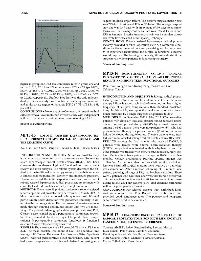
higher in group one. Pad-free continence rates in group one andtwo at 1, 3, 6, 12, 18 and 24 months were 62% vs 7% (p < 0.001),85.7% vs 28.5% (p < 0.001), 93.5% vs 67.8% (p 0.001), 93.5% vs82.1% (p 0.079), 97.3% vs 82.1% (p 0.006), and 97.4% vs 85.7%(p 0.023), respectively. Uroflow StopTest was the only indepen-dent predictor of early urine continence recovery on univariateand multivariate regression analysis [OR 2.87 (95%CI 1.34-4.38,p = < 0.001)].CONCLUSIONS: �Novel use of uroflowmetry at time of urethralcatheter removal is a simple, non-invasive study with independentability to predict early continence recovery following RARP.
Source of Funding: None
MP15-15 ROBOTIC ASSISTED LAPAROSCOPIC RA-
DICAL PROSTATECTOMY: INITIAL EXPERIENCE AND
THE LEARNING CURVE
Kau Han Lee*, Chien-Liang Liu, Steven K Huan, Tainan, Taiwan
INTRODUCTION AND OBJECTIVES: Radical prostatectomyis a common treatment for localized prostate cancer. Robotic as-sisted laparoscopic radical prostatectomy (RALP) has beenshown with favorable oncologic and functional outcome in recentreview and meta-analysis. The robotic system decreased the dif-ficulty of the traditional laparoscopy surgery through its superior3-dimensional magnification, dexterity and improved precision.Herein, we report the initial experience and learning curve ofrobotic assisted laparoscopic radical prostatectomy for men withclinically localized prostate cancer by a single surgeon.METHODS: There were 21 patients underwent robotic assistedlaparoscopic radical prostatectomy between 2012 March till 2013March. A 6-trocar transperitoneal technique was used. Bilateralpelvic lymph nodes dissection was performed routinely to de-termine the pathologic stage. The urethrovesical anastomosis wasmade through running continuous suture with two 3-0 mono-vicryl. The patient¡fs demographic data (age, prostate size, PSA,Gleason score, clinical stage), perioperative parameters (opera-tive time, estimated blood loss, days of hospitalization, compli-cations) & postoperative parameters (oncologic & functionaloutcomes) were retrospectively reviewed.RESULTS: The mean age was 65.5 year-old. The mean PSA was29.1. The mean prostate was about 28.6 cc. The operative timeaveraged 391.2 mins. The mean blood loss was 578 cc. 5 patients(23.8%) received perioperative blood transfusion. One patienthad major complication with intestinal obstruction causing sub-
sequent multiple organ failure. The positive surgical margin ratewas 22% for T2 lesion and 42% for T3 lesion. The average hospitalstay day was 13.7 days with an average of 8.9 days foley cathe-terization. The urinary continence rate was 65% at 1 month and85% at 3 months. Erectile function analysis was incomplete due torelatively few cases had nerve-sparing technique.CONCLUSIONS: Robotic assisted laparoscopic radical prosta-tectomy provided excellent operation view & a comfortable po-sition for the surgeon without compromising surgical outcome.With experience accumulates, the surgical & functional outcomewould improve. The learning curve is significantly shorter if thesurgeon has wide experience in laparoscopic surgery.
Source of Funding: none
MP15-16 ROBOT-ASSISTED SALVAGE RADICAL
PROSTATECTOMY AFTER RADIATION FAILURE: INITIAL
RESULTS AND SHORT-TERM FUNCTIONAL OUTCOMES
Wei-Chun Weng*, Chun-Kuang Yang, Yen-Chuan Ou,Taichung, Taiwan
INTRODUCTION AND OBJECTIVES: Salvage radical prosta-tectomy is a treatment option for certain patients after radiationtherapy failure. It is more technically demanding and has a higherfrequency of surgical complications than standard prostatec-tomy. In this article, we report the results and short-term func-tional outcomes by a single surgeon¡fs experience to date.METHODS: From December 2005 to May 2013, 565 consecutivepatients with clinically localized prostate cancer received robot-assisted radical prostatectomy (RARP) by a single surgeon.Among the 565 patients, there were five patients (0.88%) who hadprior radiation therapy for prostate cancer (PCa) and radiationfailure developed during follow-up. The five patients were trea-ted with robot-assisted salvage radical prostatectomy (RASRP).RESULTS: Among the five patients who received RASRP, 3patients were treated with external beam radiation therapy(EBRT); one patient was treated with brachytherapy, and theother patient was treated with the CyberKnife radiosurgery sys-tem. Median time from primary therapy to RASRP was 24.4months. Median preoperative prostate specific antigen was5.39 ng/ml. Median operative time was 128 minutes, and bloodloss was 84 ml. All surgical margins were negative by patholog-ical examination. After a median follow-up of 16 months, onepatient, pathological stage of T3b, had biochemical failure. Therewere 2 patients who had their neurovascular bundle preserved,but their erection function was insufficient for sexual intercourseduring follow-up. Four patients (80%) had excellent continencewithin the postoperative 5 weeks.CONCLUSIONS: For selected patients with confirmed, local-ized, radiation-recurrent PCa, RASRP was feasible, safe, andprovided good continence rates. The potency and long-termcancer control need to be evaluated.
Source of Funding: none
MP15-17 LONG-TERM ONCOLOGICAL RESULTS OF
RADICAL PROSTATECTOMY FOR HIGH-RISK PROSTATE
CANCER: A SINGLE-CENTRE EXPERIENCE
Youness Ahallal*, Rafael Sanchez-Salas, Laurent Mascle,Luca Lunelli, Petr Macek, Camilo Giedelman,Dominique Prapotnich, Eric Barret, Francois Rozet,Marc Galiano, Annick Mombet, Nathalie Cathala,Xavier Cathelineau, Paris, France
-A232- MP15 ROBOTICS/LAPAROSCOPY: PROSTATE, LOWER TRACT II

INTRODUCTION AND OBJECTIVES: In our cohort, patientstreated with RARP showed better EF recovery compared withLRP. This results should be carefully analyzed as they representthe very first outcomes of an invention curve for both LRP andRARP. Furthermore, initial learning from LRP experience shouldhave an impact in RARP outcomes. A careful pre-operative pa-tient selection is mandatory to improve the chance of EF recoveryfollowing RARP and LRP.METHODS: Between 1998 and 2006, a total of 239 patientsunderwent RP for high-risk PC. High-risk PC was defined asfollows: prostate-specific antigen (PSA) level > 20 ng/mL,Gleason score 8–10, and/or clinical stage T2c-T4 disease. Bio-chemical recurrence (BCR) was defined as a postoperative PSAvalue ¡Ý0.10 ng/mL and a confirmatory rise, or a postoperativePSA ¡Ý0.10 ng/mL while receiving salvage treatment. Kaplan-Meier and multivariate cox regression analysis were used todetermine BCR, cancer specific survival (CSS) and the signifi-cant predictors of BCR.RESULTS: The median follow-up was 64 months. Organ-confined disease, seminal vesicle invasion and lymph node me-tastasis were reported in 46%, 27% and 3%, respectively. Thepositive surgical margin rate was 29%. Overall, the predicted 5-year and 10-year BCR-free survival rates were 31% and 29%, re-spectively; and the predicted 5-year and 10-year CSS rates were98% and 89%, respectively. After adjusting for other covariates,higher pathologic stage, positive nodes, positive surgical marginsand Gleason 8–10 are significantly associated with BCR.CONCLUSIONS: Our data support evidence that RP can resultin encouraging long-term oncologic outcomes for the manage-ment of high-risk PC.
Source of Funding: None
MP15-18 ROBOTIC SIMPLE PROSTATECTOMY FOR
SEVERE BENIGN PROSTATIC HYPERPLASIA
Wesley White, Knoxville, TN, David Thiel, Jacksonville, FL,James Bienvenu*, Frederick Klein, Bedford Waters,Knoxville, TN
INTRODUCTION AND OBJECTIVES: Simple prostatectomyfor the treatment of severe benign prostatic hyperplasia (BPH)can offer men with large prostates effective and durable symptomrelief. We aim to demonstrate the surgical technique and describeearly outcomes among men undergoing robotic simple prosta-tectomy (RSP) for severe BPH.METHODS: A prospective, multi-centered, longitudinal studywas performed to assess perioperative and intermediate termoutcomes among men who underwent transperitoneal RSP forthe treatment of severe BPH. All patients were seen and eval-uated at one of 2 tertiary academic centers between the years of2010–2012. Men with BPH refractory to medical therapy un-derwent history and physical examination, post-void residual(PVR) measurement, multi-channel urodynamics (where ap-plicable), transrectal ultrasound measurement of the prostate,and cystoscopy. Among men with a prostate volume of greaterthan 80 grams, treatment options including endoscopic, ro-botic, and open enucleation of the prostate were offered. Menwho elected to undergo RSP were accrued and data collectedprospectively. A minimum follow-up of 10 months was ob-served. Perioperative parameters and the presence of adverseevents were recorded, as was durability and efficacy of theprocedure.RESULTS: Following induction, all men underwent transper-itoneal port placement in a standard configuration. The space
of Retzius was developed with preservation of the endopelvicfascia. The anterior bladder neck was identified and a supra-pubic cystotomy created. The ureteral orifices were identified.Enucleation of the prostate was initiated at the posterior aspectof the adenoma and continued laterally. Hemostasis wasachieved using a tissue sealing device and/or monopolarcautery. The enucleation was continued anteriorly and towardsthe apex. The urethra was transected and the adenoma pro-cured and entrapped. The posterior bladder neck was affixedto the posterior prostatic capsule using a running absorbablesuture. Following placement of a large bore catheter, the cy-stotomy was closed in multiple layers. A total of 10 men un-derwent RSP. Mean operative time was 92 minutes. Meanestimated blood loss was 120 mL. Mean volume of the prostatespecimen was 123 g (range 104–250 g). There were no imme-diate or delayed adverse events. At a mean follow-up of 23.7months, all men were voiding spontaneously with satisfactoryresidual volumes.CONCLUSIONS: RSP appears to be safe and reproducible, andoffers men with severe BPH a less invasive alternative to opensimple prostatectomy. Surgeons comfortable with RARP mayrapidly adopt this technique.
Source of Funding: none
MP15-19 ROBOTIC-ASSISTED LAPAROSCOPIC VER-
SUS OPEN URETERAL REIMPLANTATION: A SINGLE IN-
STITUTION MATCHED COHORT REVIEW
David Duchene*, Daniel Zainfeld, Kirk Redger,Andrew Windsperger, Kansas City, KS
INTRODUCTION AND OBJECTIVES: Distal ureteral recon-struction and reimplantation is a procedure which is now oftenperformed laparoscopically with robotic-assistance. We sought toevaluate and compare perioperative and long-term outcomesamong similar patients who underwent treatment with eitherrobotic-assisted laparoscopic ureteral reimplantation (RALUR) oropen ureteral reimplantation to assess for any clinical differencein outcomes between the two procedures.METHODS: Patients who underwent robotic-assisted laparo-scopic ureteral reimplantation (RALUR) or open ureteral re-implantation between 7/2006 and 10/2012 were identified. Allprocedures were performed at a single institution. Cohortswere matched in terms of patient characteristics and indica-tions for procedure. A retrospective review of perioperativeand clinical data was performed assessing operative time, es-timated blood loss, length of admission, and success of pro-cedure as indicated by the absence of obstruction on follow-upimaging.RESULTS: A total of 21 patients underwent RALUR in the studyperiod and were matched to a similar cohort who underwentopen ureteral reimplantation. Mean ages of the groups werecomparable at 43.2 and 46.5 years for the robotic and opengroups, respectively. 20 of the RALUR were completed lapar-oscopically with conversion to open in one patient. Etiology ofinjury was similar between cohorts, including 11 due to gyneco-logic procedural injury and three secondary to iatrogenic ureteralinjuries in each of the groups with the remainder comprised ofcongenital, non-gynecologic surgery, radiation, and idiopathic.Mean operative time was 453 minutes for the open group incomparison to 237 minutes for the robotic group. Mean estimatedblood loss was 225 cc for the open cohort versus a mean of 85 ccfor those undergoing RALUR. Mean length of hospital admissionin the open reimplant group was 4.5 days compared to 3.1 days in
MP15 ROBOTICS/LAPAROSCOPY: PROSTATE, LOWER TRACT II -A233-

those who underwent RALUR. Currently, all patients remainnon-obstructed among the RALUR by Lasix renogram. Amongthe open group, one patient developed an obstruction and un-derwent repeat ureteral reimplant. An additional patient has beenfound to have evidence of low grade partial obstruction on re-nogram but has not required further intervention.CONCLUSIONS: RALUR is a safe and effective option for distalureteral reconstruction in appropriate patients and may havebenefits in terms of decreased blood loss, decreased duration ofhospitalization, and potentially decreased operative time incomparison to open ureteral reimplantation.
Source of Funding: None
MP15-20 ANALYSIS OF FACTORS PREDICTING RE-
COVERY OF ERECTILE FUNCTION AFTER LAPARO-
SCOPIC RADICAL PROSTATECTOMY
Bi Hai*, Ma Lu-lin, Hou Xiao-fei, Beijing, China,People’s Republic of
INTRODUCTION AND OBJECTIVES: To report the recoveryrate of erectile function (EF) and identify various factors pre-dicting the recovery of EF in men undergoing laparoscopic rad-ical prostatectomy (LRP) in our center.METHODS: From January 2011 to December 2012, a total of 106men with localized prostate cancer underwent LRP in our centerby one surgeon, and we gathered the preoperative EF conditionand perioperative factors of these patients. We followed all thepatients through telephone in March 2013, and only 89 patientswere available. We collected the recovery of EF of available pa-tients after LRP, and analyzed the perioperative factors predict-ing the recovery of EF after LRP.RESULTS: A total of 33 patients with preoperative sexual inter-course and over 1 year follow-up were included in statistical re-search group, and there was no difference of the perioperativeclinical data with total series. Recovery of potency was defined aspostoperative penile erection. The recovery rate of EF was 51.5%without the use of any drugs or devices to assist erection. In ourstudy, age and preoperative EF were the significant factors of re-covery of potency, and preservation of neurovascular bundle andaccessory pudendal artery in LRP were also the predictor factors.CONCLUSIONS: A lot of factors predict the recovery of EF afterLRP. In our study, accurate patient selection and adequate sur-gical technique were major determinants of postoperative re-covery of EF. We should take these factors into account foradequate patient stratification and counseling, in order to im-prove the recovery of EF after LRP.
Source of Funding: none
MP15-21 ANALYSIS OF ROBOT ASSISTED RADICAL
PROSTATECTOMY COMPLICATIONS ACCORDING TO
DURATION OF FOLLOW-UP
Ahmed Ghazi*, Nandini Venkateswaran, Vineet Agrawal,Emelian Scosyrev, Jean Joseph, Rochester, NY
INTRODUCTION AND OBJECTIVES: The modified and val-idated Clavien classification system for surgical complicationsnow represents the upcoming standard for both reportingcomplications and quality standard. Many authors have adop-ted this classification for reporting complications of robot as-sisted radical prostatectomy (RARP) to evaluate complicationsin a consistent manner, allowing comparisons across institutions
and among different approaches. However with substantialvariations in the reported duration of follow-up (range < 30days to 1 year and recent up to 5 years), comparisons remain atedious task.
Objective: To analyze complications of a large series of robotassisted radical prostatectomy according to duration of follow up.METHODS: A total of 1503 patients underwent extraperitonealRARP for the treatment of clinically localized prostate cancerfrom July 2003 to August 2010 at a tertiary referral center. Themedian follow-up was 28.9 months (90–1800 days). Complica-tions were classified according to the modified Clavien systemand categorized according to duration of follow-up ( < 30 days,31–90 days, 91–180 days, 181–360 days and > 360 days). Incidenceand trends of complications were analyzed accordingly.RESULTS: 78.9 %, 9,2% and 11.8% of complications were re-ported within 30 days, 30–90 days and > 90 days. 100% of minorcomplications and only 63.3% of major complications were re-ported within 90 days. The highest incidence of grade 3 compli-cations, requiring intervention was at 91–180 days timeline, whilethose managed conservatively were at < 30 days. Lymphocelewas most commonly reported within 30 days (68.2%). However,lymphocele presenting after 30 days were more likely to requireintervention for drainage. No cases of Bladder neck contracture inthis series were reported within 30 days, with no significant dif-ference in rates across other time points (0%, 25%, 25%, 30% and20% at < 30, 31–90, 91–180, 181–360 and > 360 days). Trends ofcomplications are displayed in Figure 1.CONCLUSIONS: The majority of complications were reportedwithin 30 days; a significant number of complications were re-ported beyond 91 days with a significant percentage of procedurespecific complications reported > 30 days. Reporting complica-tions of RARP with a 30-day follow-up may underestimate pro-cedure specific, late complications.
Source of Funding: NONE
MP15-22 EFFECT OF PUBOPELVIC ANGLE ON SUR-
GICAL OUTCOMES IN RETROPUBIC AND ROBOT-
ASSISTED RADICAL PROSTATECTOMY
Kyungtae Ko*, Woo Suk Choi, Ja Hyeon Ku, Hyeon Hoe Kim,Cheol Kwak, Seoul, Korea, Republic of
INTRODUCTION AND OBJECTIVES: Recent prior studiesreport that a bony pelvic dimension, pelvimetry, is not related tosurgical outcome, estimated blood loss (EBL), operative time(OT), and surgical margin (SM).
Trends of procedure specific complications according to follow up.
-A234- MP15 ROBOTICS/LAPAROSCOPY: PROSTATE, LOWER TRACT II

The aim of this study was to evaluate the effects of the pelvisangle on surgical outcomes at retropubic radical prostatectomy(RRP) and robot-assisted laparoscopic prostatectomy (RALP).METHODS: A total of 44 patients with RRP and 52 patients withRALP were enrolled. Various bony pelvic dimensions that reflectthe shape of pelvis were measured. Three angles were measuredfor the relationship between the prostate and pelvis. The pelvicangle between an imaginary horizontal line and the line from thesymphysis pubis to the sacropromontory junction was deter-mined. Second, the pubic angle between an imaginary verticalline and the line from the prostate apex to symphysis pubis wasdetermined. Third, the pubopelvic (PP) angle was measured be-tween the previous two virtual lines on the mid sagittal imagefrom magnetic resonance imaging.RESULTS: Surgical outcomes of RRP and RALP were not sig-nificantly different according to the pelvic dimensions. Operativetime and surgical margin status were not different according tothe PP angle. At RRP, as the PP angle was increased by 1 degree,the estimated blood loss was increased by 22.63 ml, which wasstatistically significant (P = 0.007). At RALP, a higher PP anglehad a smaller EBL tendency (PE - 12.70, P = 0.057).CONCLUSIONS: As PP angle increases, the estimated blood losstends to decrease with RALP, while the estimated blood loss in-creases with RRP. The findings suggest that, in cases of largePP angle ( ‡ 65�), RALP may be a more feasible surgical optionthan RRP.
Source of Funding: none
MP15-23 CONTEMPORARY SERIES OF ROBOTIC
ASSISTED DISTAL URETERAL RECONSTRUCTION UTI-
LIZING SIDE DOCKING POSITION
Julie Riley*, Albuquerque, NM, Yaniv Shilo, Michael Ost,Pittsburgh, PA
INTRODUCTION AND OBJECTIVES: The robotic approach todistal reconstruction has been increasingly utilized in urology.Traditionally the patient is placed in lithotomy position with thepatient cart placed between the legs however access to the blad-der for stent placement is limited. We examined the use of sidedocking the Da Vinci robotic patient cart to perform distal ure-teral reconstruction.METHODS: A retrospective review of distal ureteral recon-struction performed robotically was performed at a single in-stitution. These cases included ureteral reimplantation anduretero-ureterostomy in the adult population. All cases utilized theDi Vinci robotic Si surgical platform and were performed posi-tioning the patient side cart on the side of patient facing towardsthe head of the patient, i.e. side docking. Patients were placed inlithotomy position with the effected side raised up at a slight angle.RESULTS: A total of 11 cases were identified from 2011–2013performed by a single surgeon. Ten patients underwent ureteralreimplantation and one patient had a uretero-ureterostomy for adistal stricture. Indication for procedure included ureteral injurysecondary to hysterectomy in 7, reflux in 2, congenital stricture in 1and megaureter in 1. Two patients required a boari flap due toextensive ureteral injury. Reimplants were performed in a non-refluxing manner either in LeDuc (9/10) or Lich Gregoire (1/10).Ureteral stents and Jackson Pratt drains were placed in all patients.Average OR time was 297 minutes (189–364) and average bloodloss was 43.6 cc (10–200). Patients were discharged with a urethralcatheter in place and this was removed approximately 2 weeksafter surgery. The ureteral stents were removed on average 49 daysafter surgery (26–82). All follow up imaging via renal ultrasoundhas revealed no ureteral stricture or complication. Follow up im-aging was performed at 3 months and yearly thereafter.CONCLUSIONS: Robotic assisted laparoscopic ureteral recon-struction of the distal ureter is safe and effective. Utilizing the sidedocking of the robot allows access to the perineum during thecase and acceptable placement of the robot to successfully com-plete ureteral repair.
Source of Funding: None
MP15-24 MULTI-INSTITUTIONAL OUTCOMES OF
ROBOTIC-ASSISTED LAPAROSCOPIC DISTAL URETERAL
REIMPLANTATION
Tracy Marien, New York, NY, Darko Kröpfl, Lukas Hohenhorst,Essen, Germany, Matthew Bilbily*, New York, NY, Gaurav Rao,Buffalo, NY, Ojas Shah, Michael Stifelman, New York, NY
INTRODUCTION AND OBJECTIVES: Robotic-assisted lapa-roscopic (RAL) surgery is optimally designed for upper urinarytract reconstruction. It maintains the principles of open surgeryincluding magnification, the ability for delicate tissue handling,and precise suture placement for creation of tension free anasto-moses all done via a minimally invasive platform, which is usedby many urologist. The objective of this series is to assess perio-perative and postoperative outcomes of RAL surgery as it is ap-plied to distal ureteral reimplantation (RALDUR).METHODS: A retrospective chart review was performed on 46consecutive patients who underwent RALDUR for benign ormalignant disease between 2004 and 2012 at two institutions.Demographic characteristics, perioperative data and postopera-tive outcomes were captured. Radiographic improvement wasassessed either with nuclear renal scan with lasix, ultrasound (US)or cross sectional imaging. Radiographic success was defined asT1/2 < 20 minutes after administration of diuretic on nuclearrenal scan or improvement in hydronephrosis on US and/or cross
Table Linear Regression Analysis and Logistic
Regression Analysis of the Effect
of PP Angle on Surgical Outcomes
MP15 ROBOTICS/LAPAROSCOPY: PROSTATE, LOWER TRACT II -A235-

sectional imaging. Symptomatology was assessed by history onfollow-up visits.RESULTS: Results are listed in Table 1 and Table 2. Fifteen of the17 patients with distal ureteral urothelial cancer underwent pelviclymph node dissection (mean number of lymph nodes was 9).CONCLUSIONS: This is the largest series of RALDUR for thetreatment of benign or malignant distal ureteral pathology pub-lished to date. RALDUR is not only feasible while minimizingmorbidity but also very effective. We hope that as more urologistsgain experience with robotic surgery this technique will flourish.
Source of Funding: none
MP15-25 IS R.E.N.A.L NEPHROMETRY SCORING
SYSTEM ASSOCIATED WITH THE OUTCOME OF LPN?
Masahiro Sugiura*, Kazuhiro Araki, Kyokusin Ho, MasahikoInahara, Ichihara, Japan, Syuko Cho, Nobuyuki Sekita, Chiba,Japan, Hiroshi Masuda, Yukio Naya, Ichihara, Japan
INTRODUCTION AND OBJECTIVES: Laparoscopic partialnephrectomy is recommended for small renal cancer patients topreserve their renal function. To investigate the clinical signifi-cance of preoperative aspects and dimensions used for radiusexophytic/endophytic nearness anterior/posterior location(RENAL) scoring systems (RNS) for renal neoplasms in patientsundergoing laparoscopic partial nephrectomy (LPN).METHODS: From April 2010 to January 2013, 45 patients withrenal tumor underwent LPN was evaluated. These patient wereanalyzed into three groups: Low (RNS is 4–6), middle (7–9) andhigh (10 or more) We investigate the relation between Age, Sex,Body mass index (BMI), estimated glomerular filtration rate (e-GFR) before surgery, RNS group and Warm ischemia time (WIT)and duration of surgery, e-GFR 3 months after operation, post-operative complications.RESULTS: 15 people is low risk, 22 people is middle risk and 8people is high risk. There is no difference of tumor side, age, sexamong these groups. Median tumor size is significantly larger inhigh risk group than middle and low risk group (2.8 cm in low,3.1 cm in middle and 5.1 cm in high risk group, respectively).There is no difference among these three groups of Median WIT,estimated blood loss and duration of surgery (WIT: 24 minutes, 35minutes and 31 minutes, respectively. Estimated blood loss:66 ml, 334 ml, 237 ml, respectively, duration of surgery: 201minutes, 209 minutes and 212 minutes, respectively.) Change of eGFR before and 3 month after surgery is not differ from thesegroups ( - 6.914, - 7.182 and - 6.2, respectively).
Multivariate analysis showed only the preoperative e-GFR wasassociated with e-GFR 3 months after operation.CONCLUSIONS: R.E.N.A.L nephrometry scoring system wasnot associated with the outcome of LPN. Furthermore study isnecessary.
Source of Funding: none
MP15-26 RENAL HILAR CLEARANCE (DENERVA-
TION) AS A TREATMENT FOR INTRACTABLE/
REFRACTORY HYPERTENSION
Shakir Tabrez, Mohan Keshavmurthy, Prem Kumar*,Uday Bhaskar, Mohan Balaiah Ashwathaiah, Bangalore, India
INTRODUCTION AND OBJECTIVES: Refractory hyperten-sion has been a clinical challenge in recent years. Refractory
Table 1. Patient and Disease Characteristics
BMI, body mass index.*One case with associated ureterocele.
Table 2. Outcomes
EBL, estimated blood loss; ml, milliliters; LOS, length of stay; CG,clavien grade *Some patients with multiple complications, only 16patients with any complication.
-A236- MP15 ROBOTICS/LAPAROSCOPY: PROSTATE, LOWER TRACT II

hypertension is defined as blood pressure that remains abovegoal in spite of the concurrent use of 3 antihypertensive agents ofdifferent classes. Ideally, one of the 3 agents should be a diureticand all agents should be prescribed at optimal dose amounts.There have been many cases of hypertension which are notresolved by any number of antihypertensive medications ofwhatever class. We could treat them by Laparoscopic renal de-nervation (sympathectomy).METHODS: Four patients were referred to us with refractory /intractable hypertension. On extensive investigations all thepossible secondary causes were ruled out. All the patients wereon more than 4 antihypertensive medications and had a meansystolic BP of > 140 mm Hg and a mean diastolic BP of > 90 mmHg even with maximal dose of antihypertensives. All the patientswere counseled appropriately and underwent a laparoscopic bi-lateral renal denervation. The renal surface and the renal hilum,
including the proximal ureter were skeletonised bilaterally. Allthe patients had an uneventful intra and post operative period.RESULTS: Follow up of these patients showed an overall re-duction of blood pressure on the 2 nd post operative day on-wards. The latest BP measurements on 6 months follow up is< 120 mm Hg mean systolic and < 90 mmHg mean diastolicpressure. One patient is off medications completely and 3 patientsare on a single antihypertensive medication.CONCLUSIONS: The present study done on the refractory hy-pertensive patients shows that transection of the renal (sympa-thetic) neural network at the hilum gives a better relief of thehypertension. There have been reports of endovascular radio-frequency denervation of the renal hilum with some success. Thepresent procedure is accessible to many patients since availabilityof Laparoscopy is universal now.
Source of Funding: Self/Institution
V07 LAPAROSCOPY: UPPER TRACT I
V07-01 RETROPERITONEAL LAPAROSCOPIC PARTIAL
NEPHRECTOMY FOR COMPLETELY INTRARENAL TUMOR
Ma Lulin*, Zhang Shudong, Beijing, China, People’s Republic of
INTRODUCTION AND OBJECTIVES: It is very difficult toestimate the tumor boundary and the depth of tumor invasion byeye for completely intrarenal tumor. Laparoscopic partial ne-phrectomy is very difficult and high risk for this kind of renaltumor. This video will introduce Laparoscopic partial nephrec-tomy with intraoperative ultrasound.METHODS: This case was a 39 years old woman, who wasphysical examination found a 3 cm completely intrarenal tumorin the right renal with inhomogeneous enhancement. Tumor waslocated in renal dorsal central and close to the renal hilum byintraoperative ultrasound. Determine the depth and the bound-ary of tumor with ultrasound, then occlude the renal artery. Cutoff in the middle of renal parenchyma, and complete resection ofthe tumor. Collection system was sutured by 3-0 monocryl with ahem-o-lok, and renal parenchyma was sutured by 2-0 monocryl.RESULTS: Renal artery occlusion time was 20 min, and estimateblood loss was 20 ml. Pathology of the tumor was renalhamartoma, and patient was recovered well. During 3 monthsfollow-up time, no recurrence of tumor.CONCLUSIONS: Using intraoperative ultrasound to determinethe boundary and depth of tumor can elevate the safety andfeasibility of laparoscopic partial nephrectomy for completelyintrarenal tumor. Continuous knotless suture is simple and fea-sible, and can significantly shorten the learning curve, so it hashigher clinical application value.
Source of Funding: none
V07-02 LAPAROSCOPIC RESOLUTION OF VASCULAR
COMPLICATIONS IN RETROPERITONEAL LAPARO-
SCOPIC NEPHRECTOMY
Zhang Shudong*, Ma Lulin, Beijing, China, People’s Republic of
INTRODUCTION AND OBJECTIVES: Laparoscopic nephrec-tomy and radical nephrectomy is classical surgery in urological
laparoscopy. There is some kinds of vascular complicationsduring the surgery inevitablely. The rate of vascular injury was1.6–4.7% by literature reviewed during laparoscopic nephrec-tomy. This video present the successful experiences with lapa-roscopic resolution of vascular injury in laparoscopicnephrectomy by retroperitoneal approach.METHODS: We present four types of vascular injury duringlaparoscopic nephrectomy, which include renal vein wall injuryin moblizing with suction, renal vein cutten in scissoring the ar-tery, vena cava injury by hem-o-lok in live donor nephrectomy,renal artery injury by harmonic device.RESULTS: All the cases were successful managed by laparos-copy without conversion. The blood losses were 20 to 2000 ml. Nopost-operative complications occurred.CONCLUSIONS: Vascular injury is full of high risk, but it can besolved by experienced laparoscopy specialist without conversion.The most delicate point is the timing of when conversion is required.
Source of Funding: none
V07-03 LAPAROSCOPIC DONOR NEPHRECTOMY:
MANAGING COMPLEX VENOUS ANOMALIES
Ahmed Mansour*, Ibrahim Eraky, Ahmed R. El-Nahas,Bedeir Ali-El-dein, Ahmed Shokeir, Mansoura, Egypt
INTRODUCTION AND OBJECTIVES: Laparoscopic donornephrectomy has evolved to become the standard approach forharvesting grafts for live kidney donation. Complex venousvascular anomalies are often encountered especially during leftsided nephrectomies. We report our approach for managingcomplex venous vascular anomalies during laparoscopic donornephrectomy.METHODS: We describe our approach in managing complexvenous anomalies during laparoscopic donor nephrectomies. Thevideo illustrates managing cases with multiple renal veins, ret-roaortic veins, complex lumbar, gonadal and suprarenal veinsdraining into the main renal vein.RESULTS: Meticulous dissection in addition to careful retractionof the renal vein can help identification of anomalies that are oftenmissed in the preoperative imaging. A vessel loop passed around
V07 LAPAROSCOPY: UPPER TRACT I -A237-

the renal vein facilitates safe and efficient manipulation. Vesselsealing devices are used liberally avoiding the placement of sur-gical clips that may interfere with the placement of the vascularstapler for the control of the renal artery and vein.CONCLUSIONS: Complex renal venous anomalies can be safelyand efficiently managed during laparoscopic donor nephrectomies.
Source of Funding: None
V07-04 A STANDARDIZED SUTURE TECHNIQUE
USING A BARBED SUTURE IMPROVES EFFICACY AND
SAFETY IN RETROPERITONEOSCOPIC PARTIAL NE-
PHRECTOMY
Christian Wülfing*, Niclas Flechtenmacher, Serkan Filiz,Johannes Göckschu, Jörn Bode, David Marghawal, Hamburg,Germany
INTRODUCTION AND OBJECTIVES: In the last years a par-adigm shift preferring partial nephrectomy over radical nephrec-tomy has been encountered. The main reasons are modernimaging, leading to earlier and better tumor detection, higher ratesof benign or low-malignant tumors and medical implications.Several studies have clearly shown that radical nephrectomy in-creases liftetime risk for deadly cardiovascular complications.
Most international guidelines now recommend partial ne-phrectomy for tumors up to 7 cm. When partial nephrectomy iscarried out using a minimally-invasive method, a warm ischemiatime (WIT) can – in most cases - not be avoided. The maininfluencing factors for WIT are tumor excision and intracorporalsuturing which can be quite time-consuming.METHODS: In our study we analzyed a modified suture tech-nique using two barbed sutures (V-Locª, Covidien Inc.) which wasused in 94 consecutive patients undergoing retroperitoneoscopicpartial nephrectomy by one surgeon (C.W.) in our department. Inbrief, after exposing the tumor and placing an endo-bulldog clip,the tumor was exzised. In the first step, the deep tumorbed wassutured with the first V-loc suture applying a running suturewithout clips. In the second step, the same suture, but with a biggerneedle was used for a running parenchymal suture which wascombined with placement of Surgicelª. We compared this tech-nique to the first 14 patients of our series, where the well-described,,sliding-clip rhenorraphy‘‘-technique had been used.RESULTS: 108 patients were included in the study. When com-paring the 94 patients where our modified technique with twoV-locª running sutures was applied, to the 14 patients that un-derwent the sliding clip rhenorrhapy technique, statistically sig-nificant better results were found for operating time (88 vs113 min), blood loss (132 vs. 275 ml) and warm ischemia time(14 min vs. 19.5 min) (all p < 0,05).CONCLUSIONS: WIT is a critical aspect in minimally-invasivepartial nephrectomy and should not exceed 20 minutes. Themodified suture technique presented in this film was able tosignificantly reduce operating time, blood loss and WIT. In ourview this technique combines elegantly with the advantages ofthe retroperitoneal access in minimally-invasive partial ne-phrectomy, which is the standard access at our institution.
Source of Funding: Asklepios Research Fund
V07-05 CLAMPLESS LAPAROSCOPIC PARTIAL NE-
PHRECTOMY: OUR TECHNIQUE
Ioannis Georgiopoulos*, Iason Kyriazis, Panagiotis Kallidonis,Stavros Kontogiannis, Evangelos Liatsikos, Patras, Greece
INTRODUCTION AND OBJECTIVES: Partial nephrectomy isthe established mode of treatment for small tumor masses whennephron sparing is indicated. Laparoscopic clampless partialnephrectomy has the added benefit of completely eliminatingrenal ischemia and its detrimental consequences.METHODS: 11 patients have undergone clampless laparo-scopic partial nephrectomy at our department, 7 on the leftside and 4 on the right. Ideally suited candidates are patientswith small masses (T1a) with exophytic polar tumors. Outtechnique consists of full renal mobilization and meticulouspreparation of the renal vessels in case of necessary urgentclamping. The tumor is sharply and bluntly dissected. He-mostasis is ensured with bipolar cautery and two suture layers.The internal tumor bed layer is sutured with self-retainingbarbed sutures, and the external compressive layer with Vicrylsutures. Both layers are tensioned with the use of clips at theterminal ends. Hemostatic foam and sponge materials are usedto ensure ideal hemostasis.RESULTS: Mean operative time was 106 minutes, and meanblood loss was 290 ml. Drains and catheters were removedon the first post-operative day, and patients were dischargedfrom the hospital in the second post-operative day. Two ofthe eleven masses were benign (renal oncocytoma), and theremaining masses were malignanat (clear cell renal carcinoma).No malignant masses exhibited positive surgical margins.Six-month follow up imaging has not revealed any cancerrecurrence.CONCLUSIONS: Clampless laparoscopic partial nephrectomyis a safe and effective treatment in selected patients with renaltumors. Ideal candidates are patients with small exophytic mas-ses in polar locations.
Source of Funding: None
V07-06 RETROPERITONEOSCOPIC DISMEMBERED
PYELOPLASTY IN THE TREATMENT OF PUJ OBSTRUC-
TION- OUR EXPERIENCE WITH 60 CASES
Venugopal Ganapathy*, Haris C H, Thiruvananthapuram, India,Sudin S R, Kollam, India, Navin C Angus, Ginson V George,Sandeep Krishnan, Thiruvananthapuram, India, Suchindra P Unni,Kochi, India, Jibu K Pillai, Thiruvananthapuram, India,Jithunath M R, Thrissur, India
INTRODUCTION AND OBJECTIVES: Retroperitoneoscopicdismembered pyeloplasty (RDP) has very recently replacedTransperitoneal Laparoscopic dismembered pyeloplasty (TDP) inthe treatment of PUJ Obstruction (PUJO). Our objective is to studythe perioperative outcomes in patients who had undergone RDPfor PUJO.METHODS: From October 2008- January 2013, 60 RDP wasperformed on 60 patients with PUJO in our instituition. All thesepatients were retrospectively analysed for perioperative out-comes. Datas are expressed in mean and standard devia-tion;Student’s T-test used to compare mean between 2 groups.RESULTS: Age group-6–60 yrs (mean-27.05 yrs), mean months offollow up-21.98, Left/right ratio-31/29 cases, number of portsused-4, mean duration of surgery-153.54 mins, mean day of oralintake-1.19, mean post-op analgesia requirement-25.95 (mor-phine equivalent), mean day of drain removal-2.46, mean day ofdischarge-3.19.
Blood loss was minimal and no patient required transfusion. 1Conversion was due to a peritoneal rent tackled by TDP. In 65%of cases lower polar crossing vessel was the cause of PUJO and inall these cases the vessel was posteriorised during RDP. In all
-A238- V07 LAPAROSCOPY: UPPER TRACT I

patients stenting was done antegrade and removed after 1 month.Patients were followed up with an USG and IVP on the 3rd monthwhich showed decrease in hydronephrosis in all cases; and re-nogram in the 6th month showed a non-obstructive pattern in allthe patients. Failure rate was 0%.CONCLUSIONS: Thus RDP has the advantage of reduced op-erative blood loss, reduced post-op analgesia requirement, noincidence of paralytic ileus, shorter hospital stay, early conva-lescence and equal success rate proving to be an effective alter-native to conventional TDP in the management of PUJO.
Source of Funding: none
V07-07 LAPAROSCOPIC PYELOLITHOTOMY IN A
HORSE SHOE KIDNEY IN THE ERA OF PCNL AND RIRS
Manickam Ramalingam*, Kallappan Senthil,Anandan Murugesan, Mizar Ganapathy Pai,Coimbatore, India
INTRODUCTION AND OBJECTIVES: Percutaneous ne-phrolithotomy and retrograde intra renal surgery are the cor-nerstones in stone management nowadays. But pyelolithotomystill holds good in selected cases. We present the video oflaparoscopic pyelolithotomy in a patient with horse shoekidney.METHODS: A 58 years old male patient presented with com-plaints of recurrent pain in the right loin since 2 years. He wasclinically unremarkable ans creatinine was 1.3 mg%. Contrast CTrevealed horse shoe kidney with bilateral renal calculus with alarger stone measuring 30 · 20 mm on the right side. The pelviswas dilated and extra renal in configuration. Under general an-esthesia, ureter was stented with ureteric catheter. Using 4 portslaparoscopy was done. Colon was reflected medially and theureter was identified and traced upwards till pelvis. Verticalpyelotomy was done and stone removed in toto. DJ stent wasplaced and pyelotomy closed with polyglactin sutures. Port siteswere closed after placing the drain.RESULTS: The operative time was 125 minutes. The blood losswas approximately 50 ml. Patient stayed for 3 days post opera-tively. Drain was removed on day 2 and stent removed after 4weeks after confirming stone clearance with · ray KUB. Patientunderwent ESWL for the left side stone.CONCLUSIONS: Laparoscopic pyelolithotomy is still feasibleand effective alternative to PCNL for patients, particularly thosewith anomalous kidneys.
Source of Funding: None
V07-08 LAPAROSCOPIC URETEROCALICOSTOMY:–
PELVICURETERIC JUNCTION OBSTRUCTION IN IN-
TRARENAL PELVIS WITH MULTIPLE SECONDARY CALCULI
Vikas Agarwal*, Atul Goswami, Delhi, India
INTRODUCTION AND OBJECTIVES: Pelvicureteric junctionobstruction (PUJO) is common anomaly in paediatric age group.Anderson hynes pyeloplasty is considered as versatile procedurefor PUJO but for intrarenal pelvis, Ureterocalicostomy is consid-ered as standard treatment. We present our video of laparoscopicueterocalicostomy.METHODS: 15 yr old boy, known case of ITP, underwent opensplenectomy 2 yr back presented to us with fever and flank painon evaluation found to have right PUJO with intrarenal pelvisand multiple secondary calculi. He underwent transperitoneallaparoscopic ureterocalicostomy. Standard 4 ports were placed.Colon mobilised and kidney lower pole and hilum was dissected.Parenchyma at lower pole was dissected and stone were re-trieved. Ureter was dissected and Ureterocalicostomy was donewith vicryl 3-0 and 6/26DJ was placed.RESULTS: Patient tolerated procedure well. Total operating timewas 170 minutes. Patient accepted orally next day and catheterwas removed on 2nd postop day and was discharged in stablecondition on 4th day. At 6 week DJ was removed and at 3 monthCT IVP revealed good drainage.CONCLUSIONS: Though ureterocalicostomy is a technicallydemanding procedure, use of laparoscopic assistance allows thesurgeon to perform the reconstruction precisely and with mini-mal morbidity to patient.
Source of Funding: none
V07-09 LAPAROSCOPIC CYTOREDUCTIVE NEPHREC-
TOMY: MPUH EXPERIENCE
Amit Bhattu*, Nadiad, Gujarat, India, Shashikant Mishra,Arvind Ganpule, Jitendra Jagtap, Raguram Ganesamoni,Jigish Vyas, Ravindra Sabnis, Mahesh Desai, Nadiad, India
INTRODUCTION AND OBJECTIVES: Laparoscopic cytor-eductive nephrectomy (LCRN) is feasible in select renal cancerpatients with specific advantage of early recovery and initiationof targeted therapy. The aim of this study was to assess the peri-and postoperative safety and hence feasibility of LCRN.METHODS: 21 LCRNs were performed at our centre. Excusioncriteria were patients with IVC thrombus and trans abdominalultrasound showing restricted mobility. The perioperative andpostoperative surgical, functional and oncological outcomes wereanalysed.RESULTS: Mean age, male to female ratio, mean size, and ASAscore of patients were 58 – 12 years, 16:5, 11.31 – 3.12 centimetersand 2 respectively. There were no intraoperative complications &there were no conversion to open surgery. Analgesic require-ment, haemoglobin drop, stay in high dependency unit & hos-pital stay was 282 – 68 milligrams of tramadol, 2.1 – 1.52 grams,665 – 69 mins and 3.8 – 2.1 days respectively. Post operative his-topathology is suggestive of renal vein thrombus, involvement ofGerota’s fascia or beyond; hilar lymph nodes involvement anddistant metastasis were seen in 10,8,6 and all patients respec-tively. Average follow up was 19 – 13 months. Death due toprogression of the disease occurred in 9 patients. Average dura-tion to start target therapy was 3 weeks. The 1 years survival is66.8%.CONCLUSIONS: Laparoscopic cytoreductive nephrectomy ischallenging surgery but safe in experienced centres. It achievesearly post operative recovery facilitating the early initiation oftargeted therapy.
Source of Funding: None
Learning curve of sur-geon:
V07 LAPAROSCOPY: UPPER TRACT I -A239-

V07-10 NEPHRON-SPARING SURGERY FOR RENAL
TUMORS USING PARENCHYMAL CLAMPING
Mohammed Lezrek*, Khalil Bazine, Hicham Tazi,Adil Slimani Alaoui, Mohammed Alami, Meknes, Morocco
INTRODUCTION AND OBJECTIVES: We present a video ofour preliminary experience of open surgery partial nephrectomywith parenchymal clamping.METHODS: Between May 2003 and May 2012, 15 patients (9 menand 6 women) were treated with this technique. The patients hadan average age of 56 years-old (35 to 78). The tumors were dis-covered incidentally in all the patients. 10 tumors were localized inthe right kidney and 5 in the left kidney. Preoperative renalfunction was normal in all the cases. Indication for nephron-sparing surgery was elective in 12 patients, relative in three othersand no one was imperative. The average size of the tumor was5.66 cm (3–10). The location of the tumor was in the upper pole,lower pole and middle portion in 5, 4 and 6 patients, respectively.RESULTS: The average operative bleeding was 300 ml. The av-erage duration of clamping was 25 min and the mean interventiontime was 135 min. The mean hospital stay was 5 days. All thetumors were resected with negative margins. 11 tumors were RCCand 4 were oncocytoma. With a follow-up of 10 to 76 months, thereis no evidence of recurrence in 13 patients. One patient presentedwith pulmonary metastasis after 36 months of follow-up. Anotherpatient had a left nephrectomy for local recurrence after 40 months.CONCLUSIONS: The partial nephrectomy with parenchymalclamping is an attractive, safe, and effective technique in theconservative surgery of the kidney.
Source of Funding: none
V07-11 ROBOTIC ASSISTED LAPAROSCOPIC RETRO-
PERITONEAL LYMPH NODE DISSECTION FOR STAGE I
NONSEMINOMATOUS GERM CELL TUMOR: PORT PLA-
CEMENT AND SURGICAL TECHNIQUE
Dinesh Samarasekera*, Maria C Mir, Riccardo Autorino,Joseph Klink, Andrew J Stephenson, Jihad H Kaouk,Cleveland, OH
INTRODUCTION AND OBJECTIVES: The management ofstage I nonseminomatous germ cell tumors (NSGCT) remainscontroversial, as several therapeutic options exist. Surveillance,primary chemotherapy, and retroperitoneal lymph node dissec-tion (RPLND) are management strategies that are used in thisclinical scenario. With regards to RPLND, open surgery has beenthe standard of care. However, with the widespread adoption ofminimally invasive surgical techniques in Urology, these ap-proaches have been applied to RPLND. Laparoscopic RPLND hasbeen reported by experienced surgeons at high volume centers,and remains a challenging procedure. With the emergence ofrobotic surgery and its well documented benefits, there are now afew small series describing robotic-assisted laparoscopic RPLND.We present our port placement and surgical technique in theaccompanying video.METHODS: A 37-year-old male presented with a left interme-diate risk (95% embryonal carcinoma, 5% yolk sac tumor, nolymphovascular invasion) NSGCT. His BMI was 31 kg/m2. Tu-mor markers at diagnosis were AFP: 7.3 ng/mL, HCG: 23.5 mU/mL, LDH: 173 U/L. Markers were within normal limits post-orchiectomy. A CT scan of the abdomen showed a small (5–8 mm)inter-aortocaval lymph node, and a chest X-ray was negative. Thepatient was informed of his options, and elected to undergo arobotic assisted laparoscopic RPLND. The patient was placed in a60� right lateral decubitus position and the robot was docked at90�, using a three-armed approach. A left-modified templatedissection was performed.RESULTS: Operative time was 368 minutes, and estimated bloodloss was 50 mL. There were no intraoperative complications. Thepatient was discharged home on post-operative day 3. He wasmobilizing well, pass flatus, and this pain was minimal. In total 20lymph nodes were removed, and they were all benign. The pa-tient’s tumor markers remained negative on follow-up 2-weekspost surgery.CONCLUSIONS: Robotic-assisted RPLND for low stage NSGCTis technically feasible. We were able to mimic the oncologicprinciples of open surgery, including the ‘‘split-and-roll’’ tech-nique. We found our linear port placement, and 90� docking ofthe robot allowed for full range of motion of the robotic arms,without any clashing.
Source of Funding: none
V08 LAPAROSCOPY/ROBOTICS: PROSTATE,BLADDER & LOWER TRACT II
V08-01 LAPAROSCOPIC URETEROURETEROSTOMY
FOR THE MANAGEMENT OF OBSTRUCTIVE UROPATHY
CAUSED BY CONGENITAL URETERIC ENTRAPMENT IN
THE ILIAC BONE
Alireza Aminsharifi*, Shiraz, Iran
INTRODUCTION AND OBJECTIVES: Idiopathic or congenitalureteral entrapment in bony structures is extremely rare. Weshowed the role of laparoscopy for diagnosis and management of
a rare case of congenital entrapment of left ureter in an iliac bonecanal causing left side hydroureteronephrosis.METHODS: A 28-year-old man presented with vague left flankpain for 3 months. Preoperative intravenous urography (IVU)showed left side hydroureteronephrosis down to mid-ureter;with no definite cause (Figure 1A, B). Ureteroscopy failed due totightness at mid-ureter. Transperitoneal laparoscopic dissectionof left ureter was used for diagnosis the cause of obstruction andfor adequate management.
-A240- V08 LAPAROSCOPY/ROBOTICS: PROSTATE, BLADDER & LOWER TRACT II

RESULTS: Laparoscopy confirmed the abnormal retroiliaccourse of the left ureter, entry into an iliac bone canal and exitfrom iliac bone, then it continued as pelvic ureter (Figure 2A, B).After division, adequate mobilization and spatulation of distalureter, the ends were repositioned over iliac vessels and anasto-mosed to each other over an 8-Fr stent using interrupted stitches(Figure 2 C, D).
The stent was removed 6 weeks postoperatively and 3-monthfollow-up IVU showed complete resolution of hydronephrosis(Figure 1C). Review of the IVUs and 3-month postoperative ab-dominopelvic computed tomography image revealed a well-formed iliac bone canal which hosted the left ureter (Figure 1).CONCLUSIONS: In our experience laparoscopy provided aminimally invasive milieu for both diagnosis and correction ofthis rare cause of obstructive uropathy. Consistent with previous
reports, we also found laparoscopic ureteroureterostomy to be arewarding minimally invasive technique for the management ofthis obstructive ureteral pathology.
Source of Funding: none
V08-02 LAPAROSCOPIC REPAIR OF VESICOUTERINE
FISTULA USING TACHOSILTM SPONGE
Roberto Sanseverino*, Tommaso Realfonso, Oliver Intilla,Umberto Di Mauro, Giovanni Molisso, Giorgio Napodano,Nocera Inferiore, Italy
INTRODUCTION AND OBJECTIVES: Vesicouterine fistulas(VUF) represent about 4% of all urogenital fistulas. The most com-mon causes of fistulas are surgical, obstetric and related to radiationtherapy. Depending on their localization, we distinguish corporealand cervical fistula. We show a case of a patient affected by a cor-poreal vesicouterine fistula repaired by laparoscopic approach.METHODS: A 35 years old female presented at our institutionwith a corporeal vesicouterine fistula resulted from a previousCaesarean section. The patient complained of Youssef syndrome(amenorrhea, menouria, urinary continence). Cystography andcystoscopy revealed vesicouterine fistula. After positioning bi-lateral ureteral catheter, a transperitoneal approach is performed.After resection of the fistolosous tract, bladder is repaired with adouble layer suture. A TaschosilTM sponge is applied on uterinsurface. A drain is placed in peritoneal cavity.RESULTS: Surgical procedure time and fluid loss are 150 minand 150 ml, respectively. Catheter is removed after 10 days. Norelapse of fistula occurred.CONCLUSIONS: Corporeal vesicouterine fistulas are very in-frequent and present with a classical Yousef syndrome. Lapara-scopic repair of vesicouterine fistulas is a safe and reproducibletechnique offering excellent results.
Source of Funding: none
V08-03 LAPAROSCOPIC TREATMENT OF BLADDER
DIVERTICULA: THE PROCEDURE STEP BY STEP
Filippo Nigro, Paolo Ferrarese, Enrico Scremin,Giuseppe Benedetto, Ciro Ammendola, Andrea Tasca*,Vicenza, Italy
INTRODUCTION AND OBJECTIVES: Bladder diverticularepresent an insidious condition which is usually treated duringsurgery for prostatic adenoma.
When left untreated, they can sustain a clinical picture evenafter lower urinary tract disobstruction.
This video illustrates step by step laparoscopic approach to asmall bladder diverticulum.METHODS: With the patient in supine position four trocars areinserted: one paraumbilically, two laterally to the rectal musclesand one in the suprapubic area.
After medial displacement of the left colon, the peritonealwindows is progressively widened anche the left umbilical arteryis identified and transected after cauterisation.
The left umbilical is identified close to the bladder wall anddisplaced cranially in order to visualize the underlying ureter.
An antigrade dissection of the left vas deferens is then per-formed to expose the diverticular pouch.
A progressive dissection of diverticular wall is then carried outfollowing the plane of the left ureter as the caudal landmark.
FIG. 1. Preoperative intravenous urogram showed significantleft side hydroureteronephrosis (A, B), which had resolvedmarkedly in the 3-month postoperative intravenous urogram (A-C). Arrows indicates the iliac bone canal in X-ray images (A-C)and in the postoperative abdominopelvic CT scan (D). The canalhosted the left ureter.
FIG. 2. Intraoperative view of the left ureter. Through a ret-roiliac course the ureter entered the iliac bone canal (A, B). Afterpassing through the canal it continued as the pelvic ureter. La-paroscopic ureteroureterostomy was accomplished after divi-sion, repositioning of the ureteric ends over the iliac vessels andspatulation of the distal ureter (C). The trocar sites over theabdominal belly are shown (D).(I.A. Iliac Artery, I.V. Iliac Vein, U. Ureter, P.U. Proximal Ureter,D.U. Distal Ureter, B.C. Bone canal)
V08 LAPAROSCOPY/ROBOTICS: PROSTATE, BLADDER & LOWER TRACT II -A241-

The dissection coninues on the lateral and anterior aspect of thediverticulum transecting the small vessels of pericystium.
The intraoperative flexible cystoscopy gives clear evidence ofthe correct identification of the diverticulum.
The diverticular neck is progressively skeletonized and openedin order to perform its step by step transection and closure with arunning suture.
The closure of bladder wal is perfected with two running su-tures including the detrusor and the pericystium.
A small tubular drainage is placed nearby the treated area andthe anteriorior peritoneum sutured.RESULTS: Five patients have been treated with this technique.
Diameter of divertula ranged from 3.5 to 8.5 cm.Average surgical time was 132 (120–180) min.We had 2 post operative grade 1 complications according to
Clavien Dindo classification.Positive outcome was verified in all patients.
CONCLUSIONS: Laparoscopic bladder diverticulectomy, ex-cept for peritoneal approach, reproduces basically the steps ofopen surgery with the advantages of minor invasiveness and amore detailed anatomical dissection.
Source of Funding: NONE
V08-04 CYSTOSCOPY-ASSISTED LAPAROSCOPIC PA-
RTIAL CYSTECTOMY FOR TREATMENT OF BLADDER
ENDOMETRIOSIS
João Cabral*, Isaac Braga, Nuno Azevedo, Avelino Fraga,Frederico Branco, Luı́s Osório, Porto, Portugal
INTRODUCTION AND OBJECTIVES: The incidence of uri-nary tract endometriosis (UTE) ranges from 1% to 3%, the bladderbeing the most affected organ (85% of UTE). Our purpose is todescribe a technique of laparoscopic partial cystectomy withcystoscopic guidance for bladder endometriosis.METHODS: We present a case of a 42-year-old female with lowerurinary tract symptoms and recurrent episodes of hypogastricdiscomfort, referred from gynaecology consultation with a sus-pect bladder lesion in transvaginal ultrasound. The cystoscopyand magnetic resonance confirmed a 4 cm nodule in the bladderdome suggestive of endometriosis. Patient was positioned inextended lithotomy. The first 10 mm-port was placed at the um-bilicus with an open access technique. Three additional portswere placed in diamond configuration. We performed laparo-scopic resection employing a cystoscopy-assisted technique inorder to preserve the integrity of the bladder mucosa and to limitthe margins of resection. Partial cystectomy was performed withcold scissors and specimen was immediately placed inside En-doCatch bag to prevent seeding. Bladder closure was performedin a water tight two layer running suture and tested for leakage.Procedure was completed with the closure of the peritoneum andreconstitution of pelvic anatomy. No drainage was left. One weekafter the procedure we removed the urinary catheter as no evi-dence of leakage was found at the cystography.RESULTS: The operative time was 100 min. The blood loss wasnegligible. No complications were registered. Patient was dis-charged on day 1 medicated with analgesics and antibiotics. Bladdercatheter was removed on day 7. Pathologic examination confirmeda endometriotic lesion. Patient remains asymptomatic in follow up.CONCLUSIONS: Surgery is the only curative treatment of blad-der endometriosis because recurrence is the rule after hormonetreatment discontinuation. Transurethral resection of the en-dometriotic lesion is in disuse because the risk of bladder perfo-ration and recurrence and authors stated that partial cystectomy is
the adequate approach. The present video shows that the laparo-scopic partial cystectomy assisted by cystoscopy is a minimallyinvasive procedure that assures a complete and safe removal ofthese lesions. We predict whether this combined approach can beapplied in the treatment of other pathologic bladder conditions.
Source of Funding: None
V08-05 LAPAROSCOPIC VESICULECTOMY – A FEA-
SIBLE TECHNIQUE TO MANAGE A RARE PATHOLOGY
Daniel Oliveira-Reis*, Isaac Braga, Nuno Azevedo,Avelino Fraga, Severino Ribeiro, José Soares, Luı́s Osório,Porto, Portugal
INTRODUCTION AND OBJECTIVES: Cystic lesions of theseminal vesicles (SV) are very rare. When symptomatic a treat-ment should be offered to the patient. With the lack of evidence tosupport the best choice of treatment, many therapeutic optionsare presented in the literature.
Our purpose is to present our experience in laparoscopic ve-siculectomy (LapV) for benign symptomatic lesions of the SV.METHODS: We present 2 cases of LapV for benign cyst of SV. A31-year-old man complaining of pelvic discomfort associated withdysuria and liquefaction of sperm with 6 months of evolution. Thepatient had tender pelvic firm mass at the base of the prostate. Theimagiologic evaluation showed a 12 cm cystic lesion in the left SVwith no upper urinary system abnormality. Another 62-year-oldman referred to urology consultation with elevated PSA andcomplaining of perineal pain on defecation. The patient had a massat the base of the prostate. Ultrassound revealed a cyst of leftSV with 6 cm. Biopsy excluded prostate cancer. Imagiologic re-evaluation with MRI showed a cyst with 10 cm of greatest diame-ter. No urinary tract malformations. The cyst relapsed in a fewweeks after ultrasound guided puncture. Both patients were sub-mitted to LapV. For this operation patients were positioned in su-pine. The procedure was performed successfully using thestandard pelvic laparoscopic 5-ports placement along an inverted Vline between the umbilicus and both anterior superior iliac spines.
Peritoneum was incised between the two obliterated umbilicalligaments, just anterior to the rectum in the pouch of Douglas. TheSV cyst was dissected distally and the wall was excised. Thespecimens were placed in an EndoCatch bag and then removed.A transperitoneal drain was left in both cases.RESULTS: The operative time was 130 and 140 minutes, re-spectively. No peri-operative complication was registered. Drai-nages were removed the next day of the surgery. Pathologyconfirmed cyst of the seminal vesicle with inner urothelial lining,with some areas corresponding to serous cystadenoma. Patientsare completely asymptomatic at follow up.CONCLUSIONS: Transperitoneal laparoscopic excision of SVcysts should be considered as a treatment of choice for the man-agement of symptomatic seminal vesicle cysts, independent ofthe size, because of its minimal invasiveness, short postoperativecourse and complete recovery.
Source of Funding: None
V08-06 LAPAROSCOPIC PYELOURETEROSTOMY
WITH RECIPIENTS URETER IN A TRANSPLANTED KID-
NEY WITH URETERAL STRICTURE
Dmitry Perlin*, Ilya Alexandrov, Alexey Nikolaev,Olga Shevchenko, Vladislav Efremov,Voljsky, Russian Federation
-A242- V08 LAPAROSCOPY/ROBOTICS: PROSTATE, BLADDER & LOWER TRACT II

INTRODUCTION AND OBJECTIVES: Ureteral obstructionsecondary to ischemia is the most common urologic complicationof kidney transplantation. Pyelourereteral anastomosis with re-cipient ureter has shown most satisfactory long-term results in itsmanagement. Existing urinary infection and immunosupressiondetermine the high risk of wound complications. Till last time thisprocedure has been performed through open surgery, however in2006 Orvieto M.A. et al. first reported minimally invasive ap-proach using the Da Vinci robotic system.METHODS: We have experience more than 30 procedures ofureteral strictures repair after kidney transplantation by opensurgery during 20 years. Since February 2012 we used pyelour-ereteral anastomosis with recipient ureter in two patients bylaparoscopic approach. The operations lasted 215 and 275 min.respectively.
In both cases the surgery was performed after percutaneousnephrostomy because of deterioration of transplanted kidneyfunction. Internal stent was indwelled laparoscopicaly. No draintube was left.RESULTS: The nephrostomy tubes were removed after 7 and10 days respectively. The stents were removed after 20 and 27days respectively. No complications were seen during thesurgery and postoperative period. Now serum creatinine levelis 0.12 and 0.15 mmol/l after 15 and 12 months after surgeryrespectively.CONCLUSIONS: In spite of some difficulties related with to-pographic landmarks and severe tissues fibrosis after transplan-tation laparoscopic pyeloureterostomy in transplanted kidney issafe and feasible procedure. The main advantage is absence ofrisk of most serious complications related with wound infectionin immune compromised patients. Moreover, early recovery tousual activity and diet facilitates to prevent pulmonary infectionsand to normalize intestinal absorbability of the immunosup-pressive drugs.
Source of Funding: none
V08-07 ROBOT ASSISTED MANAGEMENT OF OB-
STRUCTING PROSTATE GLANDS GREATER THAN 150 GM
Mathew Oommen, Kush Patel, Philip Dorsey*, Janet Colli,Michael Pinsky, Benjamin Lee, Raju Thomas, New Orleans, LA
INTRODUCTION AND OBJECTIVES: Urologists performover 500,000 invasive procedures annually for symptomatic uri-nary outflow obstruction. In general, transurethral resection witheither bipolar/laser techniques are more than adequate. Forglands larger than 150 gm, the gold standard has been the opensuprapubic approach. In this video, we present our techniques fora robot assisted suprapubic prostatectomy.METHODS: A single institution’s experience with robot assistedsuprapubic prostatectomy for large benign prostate glands ispresented. Techniques to facilitate this procedure such as use ofthe robotic tenaculum, management of a large median lobe, andidentification of the ureteral orifices are discussed.RESULTS: There were no major perioperative or postoperativecomplications. Blood transfusions were not required. All patientswere discharged on postoperative day one.CONCLUSIONS: Robot-assisted suprapubic prostatectomy mayprove to be a feasible alternative in the management of largevolume benign prostate glands. Further evaluation and assess-ment is required to evaluate the long-term clinical application ofthis technique.
Source of Funding: None
V08-08 TRANSVESICAL LAPAROSCOPIC CLOSURE
OF URETHROVESICAL FISTULAE
Gamal Ghoniem, Orange, CA, Zhamshid Okhunov*, Orange, CA,Jordan Siegel, Mina Samaan, Orange, CA, Hanul Choi,Please choose an option below
INTRODUCTION AND OBJECTIVES: Genitourinary systemfistulas are rarely simple and present a significant treatmentchallenge requiring expert skills for evaluation and management.We present our laparoscopic technique of transvesical closure ofurethrovesical fistula.METHODS: This is a 63-year old T4 spinal cord patient, who isstatus post ileovesicostomy and bladder neck closure in 2004. Theprocedure was complicated by postoperative bowel obstructionrequiring a small bowel resection, which was complicated by je-junal injury with subsequent additional surgical intervention. At12 months in postoperative period patient developed urineleakage per penis. A cystoscopy was performed, which revealed afistula opening at the previous bladder neck closure site. Givenhis previous abdominal surgical history a decision was made toapproach the fistula laparoscopically.RESULTS: For more details of the procedure please refer to thevideo.CONCLUSIONS: Laparoscopic transvesical fistula closure isfeasible, safe method of treatment for urethrovesical fistulas.
Source of Funding: none
V08-09 ODDITIES OF THE PELVIS: MANAGEMENT OF
ATYPICAL MASSES IN THE PELVIS
Jonathan Melquist*, Rosa Park, Stony Brook, NY,Rahuldev Bhalla, New Bruinswick, NJ
INTRODUCTION AND OBJECTIVES: We review the evalua-tion, differential diagnosis, and management of rare pelvic mas-ses through the context of two patients, one with mature cysticteratoma and the other with myxofibrosarcoma.METHODS: Two patients underwent minimally-invasive re-sections of pelvic masses of unknown etiology. Patient One is a26 year-old female with a 6.5 cm recurrent pelvic mass afterprevious resection of a coccygeal lipoma nine years prior. Pa-tient Two is a 60 year-old male with a 6.2 cm para-prostatic massdiscovered on evaluation of elevated PSA. MRI was suspiciousfor schwannoma.RESULTS: Our two patients have pathologic diagnoses differentthan our pre-operative predictions. A minimally-invasive ap-proach (laparoscopic for Patient One and robotic for Patient Two)
Table 1. Results
V08 LAPAROSCOPY/ROBOTICS: PROSTATE, BLADDER & LOWER TRACT II -A243-

was successful in complete resection of these masses. The pro-cedures were associated with minimal morbidity (Table 1). Inreviewing the literature, the differential diagnosis of pelvic mas-ses is vast and must constantly be considered (Table 2).CONCLUSIONS: The urologic oncologist must be prepared forthe oddities that present in the pelvis and retroperitoneum. Onemust consider a broad non-urologic differential diagnosis whenoperating in this area. Laparoscopic and robotic expertise in theretroperitoneum and pelvis makes urologists ideal surgeons forresection of these masses.
Source of Funding: none
V08-10 ROBOT-ASSISTED LAPAROSCOPIC EXTRA-
VESICAL BLADDER DIVERTICULECTOMY WITH UR-
ETERAL RECONSTRUCTION
Samay Jain*, Khaled Shahrour, Toledo, OH
INTRODUCTION AND OBJECTIVES: Bladder diverticula canpresent significant morbidity for patients. They can be associatedwith urinary retention and recurrent urinary tract infections.Previously, diverticula of the bladder were excised using an openapproach, but with the advent and integration of technology,these procedures can be completed using laparoscopic tech-niques. We present two cases of robotic bladder diverticulectomy,one of which uniquely also involves a Hutch diverticulum withureteral reimplantation.METHODS: Inclusion criteria were urinary retention secondaryto a bladder diverticulum, work up with urodynamics to ensurethe bladder was able to generate pressures sufficient for voiding,no evidence of bladder outlet obstruction. Both patients firstunderwent photovaporization of the prostate (PVP) and subse-quently, went back into retention and required catheterization.
Each patient was positioned in dorsal lithotomy while in steepTrendelenberg. For both procedures, five total laparoscopic ports
were used: three 8 mm ports and two 12 mm ports. Each proce-dure was begun by identifying the ureter. We then performedcystoscopy per urethra and placed the scope into the diverticu-lum. Once the scope was in the diverticulum, the light source forthe robot was extinguished in order to visualize the cystoscopelight. This was used to help delineate the borders of the diver-ticulum and initiate dissection. The neck of the diverticulum wasthen dissected circumferentially and transected. The bladder wasthen repaired in a two layer, water tight, fashion. Unique to thesecond procedure, a Hutch diverticulum was identified and aftercompleting excision of the diverticulum, the ureter was re-implanted into a neo-hiatus on the bladder. The ureter was se-cured into place with a running, water tight, tension freeanastamosis. The posterior lip was first sutured into place. Then, acystoscope per urethra was used to guide a wire and stent into theureter. Following this, the ureteral anastamosis was completedand the detrusor closed over it for a tunneled, two layer closure.RESULTS: Both patients spent only one night in the hospital andhad their catheters removed seven days post operatively. Neitherpatient experienced a complication. At a mean of 6 months followup, both patients are voiding to completion and catheter free.CONCLUSIONS: Robot-assisted laparoscopic bladder diverti-culectomy offers excellent functional outcomes with minimalmorbidity for select patients in urinary retention.
Source of Funding: none
V08-11 ROBOTIC URETERONEOCYSTOSTOMY WITH
PSOAS HITCH FOR DISTAL URETERAL THERMAL INJURY
Omer Burak Argun*, Ilter Tufek, Selcuk Keskin, Hakan Ozveri,Ahmet Sahin, Ali Riza Kural, Istanbul, Turkey
INTRODUCTION AND OBJECTIVES: Ureteroneocystostomycan be used for the treatment of patients with a wide variety ofureteral pathology. Over the last decade, robot-assisted surgeryhas become more commonly used as a minimally invasive ap-proach for reconstructive urinary tract procedures. The aim ofthis study is to present our experience with robot-assisted ur-eteroneocystostomy with psoas hitch for thermal injury of thedistal ureter.METHODS: We present a 48-year-old woman with a thermalinjury of right distal ureter occurred during robotic hysterectomy.After isolation and mobilization of the ureter, bladder was lib-erated and pulled cephalad direction for a tension free anasto-mosis. Psoas hitch and extravesical ureteroneocystostomy wasperformed using extravesical Lich-Gregoir technique.RESULTS: The total operative time was 120 minutes with anestimated blood loss of 100 mL. Postoperative period was un-eventful. The patient was discharged on postoperative day 5without a Foley catheter or drain. The JJ stent was removed after 4weeks. An intravenous pyleogram (IVP) was performed 4 weeksafter removal of the stent. Right kidney was functioning properlyand the ureterovesical junction was seeming patent without anydilatation proximal to it. The patient did not have any complaintat her last follow-up visit.CONCLUSIONS: Robotic ureteroneocystostomy and psoashitch is a safe and effective treatment option for the managementof distal ureteral injuries. Robotic assistance provided a safe andeffective approach for this complex ureteral reconstruction whileminimizing morbidity.
Source of Funding: none
Table 2. Differential Diagnosis of Pelvic Masses
-A244- V08 LAPAROSCOPY/ROBOTICS: PROSTATE, BLADDER & LOWER TRACT II

V08-12 THE USE OF 30-DEGREE CAMERA IN UP PO-
SITION FOR DORSAL VEIN CONTROL DURING ROBOT
ASSISTED RADICAL PROSTATECTOMY (RARP)
Omer Burak Argun*, Ilter Tufek, Selcuk Keskin, Ahmet Sahin,Ali Riza Kural, Istanbul, Turkey
INTRODUCTION AND OBJECTIVES: The dorsal venouscomplex (DVC) control and urethrovesical anastomosis areparticular challenging steps of robot assisted radical prostatec-tomy (RARP). Failure to achieve a proper DVC control maycompromise apical dissection and can be associated with in-creased blood loss. In our practice, 30 degree lens is used indown position starting at bladder neck dissection till the end ofrobot assisted radical prostatectomy. If the pelvis is narrow ordeep, and the prostatic apex is hidden behind the pubic bone, itmay be difficult to see and control the dorsal vein complex even
with 0 degree lens. Using the 30 degree lens in up position is anew and easy technique that may lead to overcome this problemduring RARP.
Our objective is to present our modified technique of using 30degree lens in upward position in patients with narrow pelvisand hidden prostatic apex.METHODS: In our 750 robotic prostatectomy series, we used thismodification in 16 patients. The DVC control and apical dissec-tion was performed successfully in all patients without anycomplications.RESULTS: All cases were completed successfully without majorcomplication or conversion to open surgery.CONCLUSIONS: Using 30-degree lens in up position was suc-cessful in all challenging cases. Comparative studies are neededto definitely establish the advantage of this technique.
Source of Funding: none
V09 URETEROSCOPY
V09-01 EXTRACORPOREAL LITHOTRIPSY ENDOSCO-
PICALLY CONTROLLED BY URETERORENOSCOPY (LE-
CURS) USING DIGITAL URETERORENOSCOPE FOR THE
TREATMENT OF KIDNEY STONES. A CASE SERIES
Julien Letendre*, Olivier Traxer, Paris, France
INTRODUCTION AND OBJECTIVES: While laser Holmiumlithotripsy is the gold standard for the treatment of kidney stoneby flexible ureterorenoscopy (FURS), fragmentation of largestones is inefficient and demands long operative times. In order toseek for other options, we wanted to evaluate the feasibility andusefulness of Extracorporeal Lithotripsy Endoscopically Con-trolled by Ureterorenoscopy (LECURS).METHODS: With the patient positioned in lithotomy on an ex-tracorporeal shock wave lithotripsy (ESWL) table, a digital flex-ible ureterorenoscope was used to reposition the stones in agravity dependant calyx and to regroup stones when possible.Under direct vision and with constant irrigation creating a hydricchamber, stones were fragmented with ESWL. Real time adjust-ment of the focal zone combined with periods of apnea maxi-mized the amount of shock delivered to the stone and optimizedthe number of shocks delivered. Holmium laser lithotripsy couldbe used simultaneously to treat the same stone or to treat a stonein a different location. When stones fragments were less then3 mm, a JJ stent was left in place and patients were evaluated withan ultrasound on post-op day one and with a CT scan 3 weekslater to assess for residual fragments.RESULTS: Nine patients were treated with LECURS. Stones sizeaveraged 15 mm (5–30) and mean operative time was 83 min. A100% stone fragmentation rate was obtained and 5/9 patientswere considered stone free (no residual fragments) after onesession. Four patients underwent a second look procedure forextraction of retained residual fragment, with one of these pa-tients having horseshoe kidneys with documented partialuretero-pelvic junction obstruction. Post-operative ultrasoundshowed slight peri-renal infiltration in two patients. One patientdeveloped an obstructive pyelonephritis requiring antibiotics andplacement of a ureteral catheter. On one occasion, the vision of adigital scope became blurred during ESWL requiring repairs.
CONCLUSIONS: LECURS seems to be safe and feasible. It seemsto combine the advantages of both FURS and ESWL into oneintervention and will probably help expand the indications forFURS. Risks for digital flexible ureterorenoscope still need to beinvestigated further. More studies are necessary to evaluate thebenefits and risks of this approach.
Source of Funding: None
V09-02 LOWER- MIDDLE CALYX PATHOLOGY AND A
NOVEL ENDOSCOPE CONCEPT
Jorge Campos Castellanos*, Huixquilucan, Mexico
INTRODUCTION AND OBJECTIVES: Lower-Middle calyxpathology is the most difficult to reach, particularly if small innature. A bulbous hemangioma and a microscopic urinary stoneat the same infundibulum are treated with a new concept of en-doscope that provides readily access to this area by maneuveringthe instrument with one hand only and being comfortably seatedalong the procedure.METHODS: An instrument developed by the author with anergonomic handle with a rotor at its top that directs the distalactive flexible section of the endoscope right to left at its restingposition, a shaft with at least three different elastic propertiesfrom rigid to flexible and a tip for easier and nearly atraumaticinsertion is utilized. The hemangioma is Holmium lasered and amicroscopic stone is extracted by surgeon’s self-coordination.RESULTS: A successful complete treatment of two small anddifferent pathologies is implemented in a short period of timewith no fluoroscopy required and a relaxed surgeon.CONCLUSIONS: An ergonomic instrument that adapts to cur-rent endoscopic circumstances and operating room has been de-signed. By author’s own means and resources the research anddevelopment of the instrument was completed and two patentsobtained after several years. System, apparatus, and method forviewing a visually obscured portion of a cavity US 8075478 B2.Face tip assembly for an endoscope US 8221311 B2.
Source of Funding: None
V09 URETEROSCOPY -A245-

V09-03 A NOVEL CONCEPT FOR ENDOUROLOGY IN-
STRUMENTATION.
Jorge Campos Castellanos*, Huixquilucan, Mexico
INTRODUCTION AND OBJECTIVES: An endoscopic conceptto aid in the everyday endourology procedures is shown byproviding an adequate and swift control of the situation with aninstrument that adapts to the hand with each maneuvering alongthe entire procedure, it moves to pre selected areas by surgeon’sreflex reaction. It provides the stability and ease of use of a rigidendoscope with the capabilities of the flexible.METHODS: An instrument with a handle having a rotor at thetop that directs the distal active flexible section of the endoscoperight to left at its resting position. A shaft with at least threedifferent elastic properties form rigid to flexible. A proximal rigid,an intermediate passive flexible and the distally controlled activeflexible by the rotor section. A specifically design tip for easiernon-traumatic insertion is at its front.RESULTS: This concept in the form of a cystoscope and a ur-eteroscope provides two single instruments with straightforwardmaneuverability, a plug and play assembly with cables that runparallel to instrument’s longitudinal axis that avoids twisting andentanglement. One hand gliding insertion is an attribute as wellas that the surgeon can be comfortably seated with restful handmotions along the procedure and self coordination during toolhandling is also possible.CONCLUSIONS: A concept of endoscopy that eases surgeon’sprocedures by being comfortable, readily and faster completingtheir procedures and barely distressing the patients has beendeveloped. By author’s own means and resources the researchand development of the instrument was completed and twopatents obtained after several years. System, apparatus, andmethod for viewing a visually obscured portion of a cavity US8075478 B2. Face tip assembly for an endoscope US 8221311 B2.
Source of Funding: None
V09-04 A PRECLINICAL FEASIBILITY STUDY OF
RETROGRADE ENDOPYELOPLASTY
Mohammed Lezrek*, Hicham Tazi, Adil Slimani Alaoui,Khalil Bazine, Mohammed Alami, Meknes, Morocco
INTRODUCTION AND OBJECTIVES: Percutaneous en-dopyeloplasty is a horizontal suturing of the longitudinal en-dopyelotomy incision, via a single percutaneous tract. Someauthors think that generally one suture is enough. Therefore, canthis suture be performed in a retrograde fashion? We present avideo of a preclinical feasibility study of an endoscopic suturingtechnique using a rigid ureteroscope.METHODS: A 5/0 or 4/0 suture with a conventional curved 13or 17 mm fine needle is used. The needle is straightened and thethread is folded over the needle, and it is tied in the middle of theneedle using another 6/0 suture. When the first suture is attractedthrough tissue, the needle will be horizontal and will anchor thesuture. A 3 Fr basket is retrieved in its sheath until a 13 mm ofthe sheath is empty. The straightened needle is inserted in thesheath’s empty space. The sharp needle’s end is left in the distalextremity of the sheath. The suture is left outside the sheath. Thismodified basket is used through the ureteroscope. A longitudinalincision is performed. The distal extremity of the basket is applieda few millimeters below the lower apex of the incision. The basketis opened so the straightened needle and then the sheath passthrough the tissue, and the needle is completely pushed outside.
The basket is retrieved. The suture is attracted and the needle isanchored. Similarly, another suture is anchored in the upper in-cision apex. The 2 sutures are inserted in a ureteral catheter andtraction on the 2 sutures will close the incision; approximating theincision distal and proximal apex. Thus, the longitudinal incisionwill become transversal in a Heineke-Mikulicz plasty.RESULTS: The endoscopic suturing technique was achievedeasily and quickly. It was feasible directly in a glove model.Moreover, it was possible through the rigid ureteroscope in theglove model, a sheep vena cava model, and a left ne-phroureterectomy specimen. The limitation of this technique isthat the needle will stay in the incision.CONCLUSIONS: The feasibility of this endoscopic suturingtechnique, through the ureteroscope, suggests the possibility ofretrograde endopyeloplasty, which might be quicker, and mightneed only, the more common, ureteroscopic skills. Additionally,retrograde Heineke-Mikulicz plasty might be used for calicealdiverticulum neck, infundibulum stenosis, Fraley’s syndrome,ureteral stricture.However, more studies are necessary, andfurther development of the suturing technique is needed, like tohave an absorbable ‘‘needle’’ or anchor, which might be per-formed using rigid or flexible ureteroscope.
Source of Funding: none
V09-05 URETERAL STONE RETROPULSION PREVEN-
TION DURING AN URETEROSCOPIC LITHOTRIPSY USING
A NOVEL REVERSE THERMO-SENSITIVE WATER-
SOLUBLE POLYMER
Alberto Perez-Lanzac de Lorca*, Jesus Rosety Rodriguez,Angela Conde Giles, Cristina Leon Delgado, Javier Amores,Javier Cañete, Rodrigo Garcia-Baquero, Maria Jose Ledo Cepero,Juan Solo Villalva, Blanca Madurga Patuel,Jose Luis Alvarez-Ossorio Fernandez, Cadiz, Spain
INTRODUCTION AND OBJECTIVES: Diverse devices havebeen used by urologists to prevent upward migration of stonefragments during endoscopic lithotripsy procedures. These ma-terials are not aware of limitations such as interferences withenergy sources, hard handling or ureteral damage. We presentthe use of a novel reverse thermo-sensitive water-soluble polymer(BackStop�) to prevent the retrograde migration of stone frag-ments during an endoscopic lithotripsy procedure.METHODS: A 78 years old male with two stones in the middleureter was operated. This polymer appears as a liquid at tem-peratures below 16�C, at room temperature has a thicker viscositybut it is injectable and at body temperature its thickness increasesto a viscous gel. Before stone fragmentation the polymer is dis-pensed above the stone and form a gel plug that conforms to theureter where it is applied. After complete stone extraction Back-Stop� gel is completely dissolved by using saline irrigation. Ir-rigating with cold saline accelerates dissolution.RESULTS: We started the procedure introducing the uretero-scope helped by a guide. Under direct visualization and usinga catheter the liquid was injected above the lithiasis. When thepolymer contacted with the body heat, it switched from liquidstatus to gel status and sealed the urinary tract. BackStop� iscompatible with all energy modalities and has not mechanicalpart that could hinder with the procedure. The fragmentationof the stone was successful and without complications. The gelwas reversed to liquid by irrigation with saline at lower tem-perature than the body temperature. On completion of thefragmentation and stone extraction a double J catheter wasplaced. One month later a radiological exam was performed to
-A246- V09 URETEROSCOPY

assess that all stone fragments were eliminated, and then thecatheter was removed.CONCLUSIONS: The use of this thermo-sensitive gel to tem-porarily occlude the ureter during ureteroscopic lithotripsy isfeasible and effective. Its dispensation above the lithiasis preventsstone fragments retropulsion to the upper urinary tract.
Source of Funding: none
V09-06 ENDOSCOPIC REMOVAL OF GIANT URETERAL
POLYP
Oktay Demirkesen*, Cetin Demirdag, Cagatay Dogan,Bulent Alici, Istanbul, Turkey
INTRODUCTION AND OBJECTIVES: A fibroepithelial polypof the ureter is a rare benign tumor which develops from meso-dermal germ layer. Approximately 200 cases have been reportedin the literature. In this video we present a patient with ureteralpolyp who has got endoscopic removal of the polyp using hol-mium laser.METHODS: A 79 years old female, who has a history of inter-mittent abdominal pain and macroscopic hematuria, was evalu-ated with urine cytology and culture and computed tomography(CT). Urine cytology and culture were normal. CT demonstratedsolid mass in the urinary bladder. Thereafter, the patient under-went cystoscopy for further evaluation of the bladder mass.RESULTS: Cystoscopy showed a polypoid tumor, which wasmobile and protruded from the right ureteric orifice into theurinary bladder. It was detected by ureterorenoscopy that thislesion was originated from the mid portion of the ureter. It wasexcised with holmium laser and removed endoscopically. Thepatient was discharged from the hospital without any in-traoperative or early postoperative complications. Pathologicexamination revealed a fibroepithelial polyp.CONCLUSIONS: Endoscopic removal of ureteral polyp is afeasible and successful surgical technique. However it should bedistinguished from the transitional cell carcinoma of the ureter toavoid overtreatment such as nephroureterectomy or segmentalureteral resection.
Source of Funding: none
V09-07 TURKISH ROBOT FOR COMMERCIALLY
AVAILABLE FLEXIBLE URETERORENOSCOPES FOR THE
RETROGRADE INTRARENAL SURGERY (RIRS)
Remzi Saglam*, Ankara, Turkey, Jens J. Rassweiler, Heilbronn,Germany, Ahmet Yaser Muslumanoglu, Istanbul, Turkey,Ahmet Sinan Kabakci, Ankara, Turkey
INTRODUCTION AND OBJECTIVES: In this presentation weaimed to show some advantages of the new robot Robo-flexAvicenna in the treatment of kidney stones, such as precisepositioning of a ureterorenoscope distal end, and its stabilitywithin the caliceal system, the remote control movement of thelaser fiber, to operate seated and away from the radiation.METHODS: After insertion and fixing the access sheat andcovering the robot with a sterile, transparent, plastic sheath, theflexible UreteroRenoScope (fURS) is introduced into the sheathmanually and connected to the robot.
Ergonomic chair and control panel can be adjusted in a com-fortable position according to the user.
Deflection can be performed precisely, by manipulating theright handle similar to standart fURS.
The rotation and forward and backward movement can becontrolled by left handle. The selectible speed control for theforward and backward movement of the fURS serves precision inmovements.
Attached water pump can be adjusted remotely to increase ordecrease the flow rate to provide better vision.
The advantages of the robot to protect the flexible URS:
* Before the insertion of the laser fiber, pressing the button on thetouch screen, the tip of the fURS comes automatically in astraight position.
* Laser fiber can be remotely moved forward and backward, It ispossible to read the length of laser fiber in mm.
* When necessary, pressing the button on the touch screen, thelaser tip moves completely backward.
* The laser can not be shot while the tip of the laser fiber is closerthan 2 mm to the tip.
RESULTS: We successfully controlled all functions of the flexibleURS by using robot and dusted/fragmented all of the stones.
The connection time of FURS to the robot was shorter than 1minute. There were no damage of the fURS.CONCLUSIONS: We found that robotic use of fURS is verysuitable to perform it in sitting position, outside of the radiationzone, the position and stability of the tip was satisfied, connectionto the robot takes a short time, adjustable irrigation fluid flowprovides better vision.
Source of Funding: The Scientific and Technological ResearchCouncil of Turkey, Elmed Lithotripsy Systems
V09-08 MODIFIED TECHNIQUE OF RETROGRADE
INTRA RENAL SURGERY
Nilkamal Joshi*, Al Ain, United Arab Emirates
INTRODUCTION AND OBJECTIVES: Retrograde Intra RenalSurgery (RIRS) is an established procedure. It is mainly used forremoval of upper urinary stones. The procedure has many es-tablished advantages and is preferred due to its non invasiveness.
In standard technique,
1. Operating surgeon handles both - flexible scope and cameraby himself. This makes maneuvering of the scope, targetingthe stone & manipulating the stone very cumbersome &tiring. This leads to lengthy procedure, fatigue to the sur-geon, non retrieval of stone & equipment breakage.
2. The camera does not stay in neutral position, making endovision orientation difficult.
3. The scope is curved near the handle leading to high chanceof damage to the inner channel.
METHODS: The Cystoscopy Sheath is introduced in the bladder.6 Fr and 8 Fr Semi rigid ureterorenoscope are used to dilate theureter under vision to facilitate the introduction of the initial ac-cess sheath. Two guide wires are inserted. Initial access sheath isinserted.
The main modification of the technique is:The scope is held straight at every point.1st Assistant stabilizes the scope & maintains the camera
alignment.Surgeon’s left hand does to & fro movement of scope or device
manipulation. And right hand does rotation & deflection move-ments of the scope.
2nd Assistant - Catches the stone by a special basket.By keeping the camera stabilized, the endo orientation is never
lost.
V09 URETEROSCOPY -A247-

By this method the inner channel stays straight, decreasing thechance of inner channel damage.
The surgeon has full control of his movements and easilyconcentrates on the procedure without getting fatigue or losingthe field of vision. Help of two assistants takes away the strain.
Double channel Flexible scope has advantage of non leakage ofirrigation while working with a device. Use of smaller laser fiberleads to less chance of damage.
Laser fiber must be inserted gently and in a straight scope.Laser tip should be just visible before flexing the scope to target
the stone.A Specially designed basket catches stones very smoothly from
the cup of the calyx.The double channel scope allows stone being held with a basket
and laser inserted through another channel to fragment the stone.If stone is not focused for grasping or lasing, saline is flushed by
a syringe to dislodge the stone.RESULTS: More than 130 cases of RIRS were done with onescope. Time from starting the procedure to visualize the stonewas on average 10 minutes. The operating time reduced fromaverage 3 hours to about 1.5 hour by this technique. Stoneclearance rate was 85%.CONCLUSIONS: Modified technique of RIRS increases the du-rability of the scope. And also makes the procedure easy, fast &cost effective.
Source of Funding: None
V09-09 EXTRA-ANATOMIC STENTS: CHANGE IN RE-
FRACTORY URETERIC OBSTRUCTION
Samuel Grimsley*, Kesapavilla Subramonian, Birmingham,United Kingdom
INTRODUCTION AND OBJECTIVES: The extra - anatomicstent (EAS) is an alternative to long term nephrostomy in patientswith incurable benign or malignant ureteric obstruction in whomconventional ureteric stenting is no longer effective. Both tem-porary (3 months to one year) and permanent stents exist but havesignificant attrition due to occlusion or skin erosion. In patientswith benign or slow progressing malignant obstruction change ofEAS can prolong the lifetime of this method of diversion.METHODS: A 77 year old female patient with bilateral uretericobstruction secondary to slowly progressive retroperitonealneuroendocrine tumour had Paterson Forrester extra-anatomicstents inserted bilaterally 12 months earlier. Using image inten-sifier guidance the original stent is located as it runs subcutane-ously in the anterior abdominal wall. The original stent isexposed, clamped and divided with a guidewire passed througheach half to the renal pelvis and bladder respectively. The twostent halves are removed and a replacement stent placed overguidewire under image intensifier guidance.RESULTS: The replaced extra – anatomic stent is confirmed inposition by image intensifier and cystocopy.CONCLUSIONS: We demonstrate successful change of extra-anatomic stent for refractory ureteric obstruction.
Source of Funding: None
V09-10 THE TWO-WIRE TECHNIQUE: SHEATHLESS
RETROGRADE INTRODUCTION OF FIBREOPTIC UR-
ETERORENOSCOPE IN INTRARENAL ENDOSCOPIC LA-
SER LITHOTRIPSY
Basharat Hussain*, Wai Man Chow, Manchester, United Kingdom
INTRODUCTION AND OBJECTIVES: Ureteral access sheath(UAS) induced ureteral wall injury during retrograde intrarenalsurgery (RIRS) has been reported as high as 46.5%, ranging fromsuperficial mucosal breaches (33%) to severe injury involving themuscle layer (13.5%). We present a series of 43 consecutive pa-tients who underwent RIRS for upper tract stones using the two-wire technique for introduction of fibreoptic ueterorenoscope tothe upper renal tract.METHODS: 43 patients (F18:M25) with renal calculi were treatedby RIRS with fibreoptic ureterorenoscope introduced by retro-grade rail-roading using the 2-wire technique without an accesssheath. 70% were ESWL failures with 80% lower pole calycealstones.80% were multiple stones and 26% pre-stented. Stone sizeranges from 3 to 22 mm (median 13 mm) and varies from 1 to 7stones. Fragmentation aiming at complete pulverisation wasachieved by using a 273 micron fibre on the Oddyssey 30(Hol:YAG) system (Cook Medical). Maximum energy (range 690–9800 Joules) was used for complete pulverization with the doublepulse duration option. Visual onfirmation of any ureteral brea-ches was carried out on exit of the fibreoptic ureterorenoscope.Clearance was assessed on visual pulverisation, absence of frag-ment > 3 mm during treatment, and 3-month follow up imaging.We present the two wire technique in the video presentation.RESULTS: Complete stone clearance was achieved in 40 patientsafter single treatment. One patient needed 2 treatments for a22 mm stone and one had 3 sessions to clear seven stones. Wefailed to gain access in one patient with horseshoe kidney.Treatment lasted between 45 to 80 minutes and all patients exceptone were discharged within 24 hours. Significant ureteral mus-cosal/submucosal breach, incidence of sepsis or significant hae-maturia was not observed.CONCLUSIONS: The use of access sheath (UAS) is not routinelyrequired in RIRS for ureteral access and repetitive stone fragmentretrieval from the upper renal tract, especially when stone pul-verization and subsequent clearance is achieved by the effectiveand safe Oddyssey 30 (Hol:YAG) system utilising the dual pulseduration option. The video presents the two-wire techniquewithout deployment of ureteral access sheath is safe. Rountineureteral pre-stenting is not mandatory. Ureteral wall injury hasnot been observed in this series.
Source of Funding: none
V09-11 UPPER URINARY TRACT AMYLOIDOSIS:
CASE PRESENTATION AND ENDOSCOPIC VIDEO
Ariel Schulman*, William Hilton, Antoun Toubaji,Hikmat Al-Ahmadie, Jonathan Coleman, New York, NY
INTRODUCTION AND OBJECTIVES: Amyloidosis is a pro-cess where insoluble protein is deposited in the extracellularspace of various organs. It occurs as a primary disorder fromimmune dysfunction or secondary to chronic inflammation. Thedisorder often occurs in systemic fashion with multi-organ in-volvement and a progressive course. Less commonly, amyloid islocalized to a single organ and has a stable course. It is known toaccumulate in the heart, respiratory tract and nervous system andin rare cases in the genitourinary tract. Pathologic diagnosis ismade by Congo Red stain. Affected tissue displays a classic ap-ple-green birefringence under polarized light. Presenting symp-toms of amyloidosis of the genitourinary tract include flank pain,hematuria and irritative voiding. Imaging and endoscopic ap-pearance are very similar to urothelial carcinoma.METHODS: We present an index case of a 69 year old femalewith two years of gross hematuria and flank pain with abnormal
-A248- V09 URETEROSCOPY

thickening of the right upper pole calyx and mid-ureter on CTurogram. Imaging findings and symptoms have been persistent,but stable. Two previous non-diagnostic ureteroscopies wereperformed and previous management recommendations in-cluded observation and nephroureterectomy. The accompanyingvideo includes clinical history, imaging, endoscopic right ur-eteroscopy and pyeloscopy and pathology.RESULTS: Initial re-biopsy and second confirmatory biopsy andsoft tissue debridement confirmed the presence of amyloidosis ofthe upper urinary tract and absence of urothelial carcinoma. Thevideo highlights the endoscopic appearance of amyloid deposi-tion and the similar appearance to carcinoma. Systemic amy-loidosis was ruled out with normal serum immunoglobulin andfree light chain studies. A PET scan was scheduled to re-evaluatethe patient’s known lymphoma and urothelial amyloid in thenear future. In consultation with the hematology/oncology ser-vice, future management will consist of interval imaging andexpectant management of the urinary tract for symptoms.CONCLUSIONS: Urinary tract amyloidosis is a rare but signif-icant finding during the evaluation of common Urologic symp-toms or abnormal imaging findings. It may be mistaken forcarcinoma, and in some cases, result in unnecessary overtreat-ment. Urologists should be familiar with the endoscopic ap-pearance and natural history of the disease. If localized, it usuallyfollows a benign clinical course and can be safely managed withendoscopic fulguration and serial imaging.
Source of Funding: None
V09-12 FLUOROLESS URETEROSCOPY
Min S Jun, Gene O Huang, Roger Li*, D Duane Baldwin,Loma Linda, CA
INTRODUCTION AND OBJECTIVES: Modern radiologictechniques have transformed diagnostic and treatment imagingmodalities in medicine. Since 1980, effective radiation dose in theUnited States has increased sixfold. Furthermore, the use of fluo-roscopy has doubled since 1990. Patients with urolithiasis are at
risk for repeated exposure to radiation at diagnosis, treatment, andfollow-up. We have adopted the as low as reasonably achievable(ALARA) radiation principle at our institution, with the ultimategoal of eliminating unnecessary radiation where possible. In thisvideo, we demonstrate how ureteroscopic laser lithotripsy can beaccomplished without the use of any ionizing radiation.METHODS: The steps for performing safe ureteroscopy withoutthe use of image guidance will be demonstrated in this video. Thefirst step of fluoroless ureteroscopy involves a thorough review ofpertinent imaging. While the use of the C-arm is not anticipated, itis prudent to have the machine and the fluoroscopic technologistpresent. After cystoscope insertion, wire access to the kidneythrough the ureteral orifice is achieved under the guidance ofvisual and tactile cues. A dual lumen catheter is utilized to dilatethe ureteral orifice and allow passage of a safety wire. This wire issecured and the ureteroscope is passed over the working wire.Ureteroscopy and lithotripsy commence under visual guidance.Upon completion of lithotripsy the length of the ureter is mea-sured during ureteroscope removal. Measured ureter length aidsin deploying the ureteral stent without fluoroscopy. Observationof distal curl formation via cystoscopy marks procedure com-pletion. Ideal candidates have high-quality pre-operative imag-ing and a single stone with normal anatomy. Factors which maycomplicate this technique include infection, impaction, andpresence of ureteral stricture.RESULTS: This video demonstrates successful fluoroless ur-eteroscopic laser lithotripsy in a morbidly obese 51 year-old fe-male with multiple right renal calculi.CONCLUSIONS: We demonstrate a simple technique that hasbeen developed for performance of fluoroless ureteroscopy. Ifany concerns arise regarding wire or stent placement, radiationreduction can still be achieved using low-dose fluoroscopy. Thistechnique may be particularly useful in younger or pregnantpatients, in whom risk from ionizing radiation is highest. Al-though this video demonstrates the feasibility of the fluorolesstechnique, further study and comparison with conventional ap-proaches will be necessary to establish its optimal application.
Source of Funding: None
MP16 UROLITHIASIS II
MP16-01 ENDOCKSCOPE: USING MOBILE TECH-
NOLOGY TO CREATE GLOBAL POINT OF SERVICE EN-
DOSCOPY
Renai Yoon*, Samir Shreim, Atreya Dash, Ralph Clayman,William Sohn, Hak J. Lee, Irvine, CA
INTRODUCTION AND OBJECTIVES: Advances in mobiletechnology have led to new innovations in healthcare. Herein, wepresent our experience with the 2nd generation Endockscope(Orange, CA), a specialized lens and docking system with anincorporated LED light source that promotes the coupling of asmartphone (iPhone 4S, Cupertino, CA) with modern fiber-opticendoscopes.METHODS: The weight (lbs) and lumens output (lux) of theEndockscope system with incorporated LED light source werecompared to the standard HD endoscopic equipment with xenon
light source. A cost comparison was also completed. Image res-olution (line pairs/mm) was compared using a USAF resolutiontarget (Edmund Optics, NJ) and flexible cystoscope (Storz, Ger-many) coupled to both the Endockscope-iPhone and a HD cam-era (Storz H3-Z Versatile HD Camera). A 30� laparoscope and aMunsell ColorChecker chart were used to compare color resolu-tion. Finally, cystoscopic and ureteroscopic images of a porcinemodel with both systems were blindly compared and evaluatedby 5 expert endoscopists using a Likert scale.RESULTS: The weight of the Endockscope system with incor-porated LED light source was 0.67 lbs and the combined weightof the Storz HD camera and a standard light cable was 2.07 lbs.The incorporated LED light source yielded 3.27% of the lumensoutput of a standard xenon light source (1589.4 vs. 48580 lux). Theoverall cost of the mobile-coupled system with incorporated LEDlight source was $624 compared to $64,378 for a standard HDendoscopic system. The image resolution was same for both
MP16 UROLITHIASIS II -A249-

systems (4.49 vs 4.49 lp/mm). Blue and green demonstratedsimilar color resolution, while red demonstrated better colorresolution for Storz HD Camera. Evaluation of cystoscopic im-ages acquired with the Storz camera were superior in image,color, and overall quality (p = 0.001, .016. 0.049). Evaluations ofureteroscopic images yielded no statistical difference in imagequality and color (p = 0.120, 0.310), while overall quality was su-perior for the Storz system (p = 0.027). All experts concluded thatthe images taken with the Endockscope were acceptable fortherapeutic and diagnostic use.CONCLUSIONS: The Endockscope with incorporated LED lightsource was found to be lighter and less expensive in comparisonto standard systems. The mobile-coupled device also acquiredimages of the same resolution and similar colors. Finally, theEndockscope yielded acceptable images for both diagnostic andtherapeutic use by all expert reviewers.
Source of Funding: None
MP16-02 ENDOSCOPIC MANAGEMENT OF UROLIT-
HIASIS IN THE HORSESHOE KIDNEY
Marcelino Rivera*, Amy Krambeck, Rochester, MN
INTRODUCTION AND OBJECTIVES: We assessed outcomesof patients diagnosed with a horseshoe kidney and symptomaticurolithiasis.METHODS: A retrospective review was performed of patientsdiagnosed with a horseshoe kidney who underwent endoscopicmanagement from 2007–2012.RESULTS: We identified 29 patients with 31 moieties of which 20(67%) were male. Overall mean age was 48.4 (23–71) and meanstone size was 1.5 cm (0.2–3). The most common presentingsymptom was pain in 22 (76%), urinary tract infection (UTI) in 6(21%) and hematuria in 1 (3%). Of the 31 moieties, 21 (68%) un-derwent percutaneous nephrolithotomy (PCNL), 9 (29%) ur-eteroscopy and 1 (3%) extracorporal shockwave lithotripsy. Morethan one access was utilized in 3 (14%) patients who underwentPCNL. Six (29%) in the PCNL group required additional proce-dures, of those 5 were ureteroscopy and 1 a secondary PCNL. Allpatients, except for 3 (10%), were stone-free on follow–up imag-ing. Of patients who were not stone-free, all underwent PCNL.One patient who underwent PCNL was readmitted for infection.CONCLUSIONS: After careful consideration of the anatomy,individuals with horseshoe kidney and symptomatic urolithiasiscan be managed safely by a variety of endoscopic approacheswith excellent outcomes.
Source of Funding: None
MP16-03 SINGLE ENDOUROLOGY CENTRE EXPERI-
ENCE IN PERCUTANEOUS CHEMOLYSIS OVER THE LAST
TEN YEARS
Stefanos Kachrilas*, Andreas Bourdoumis, Faruquz Zaman,Christian Bach, Anuj Goyal, Athanasios Papatsoris,Junaid Masood, Noor Buchholz, London, United Kingdom
INTRODUCTION AND OBJECTIVES: Over the last decades,percutaneous chemolysis as a primary or adjuvant treatment forurinary stones has fallen in and out of favor. We present ourDepartment’s experience upon percutaneous chemolysis over thelast ten years.METHODS: Retrospectively, all patients undergoing percuta-neous chemolysis between 2001–2011 were identified and the
patient’s files were reviewed. Duration of the treatment, com-plications, dissolution results and recurrence rates were ana-lyzed.RESULTS: A total of 29 patients with complete or partial infec-tion staghorn stones (mean size:3,9 cm) were treated in our De-partment, with adjunctive local chemolysis. We used Suby-Gsolution for irrigation chemolysis. There were 17 women and 12men, presented with a mean age of 62 years. Most of the patientshad multiple co-morbidities and/or were unfit for a second lookpercutaneous nephrolithotripsy or other intervention. The dura-tion of the treatment was 4–12 days (mean:9,6 days). No seriouscomplications were recorded. In 16 cases (55.1%) kidneys werecompletely stone free at the end of the chemolytic lavage, 8 stones(27.5%) showed partial dissolution, half of them presenting re-sidual fragments less than 5 mm. The follow-up was from 1 to 11years (mean: 63 months) and the recurrence rate was 13.7%.CONCLUSIONS: Percutaneous chemolysis is an effective andsafe adjuvant method for decreasing the burden of residual renalstones.
Source of Funding: none
MP16-04 PREVALENCE OF 24-HOUR URINE COL-
LECTION AMONG HIGH-RISK STONE FORMERS
Jaclyn Milose*, Brent Hollenbeck, Samuel Kaufman,Gary Faerber, J. Stuart Wolf, Jeffrey Montgomery,Alon Weizer, John Hollingsworth, Ann Arbor, MI
INTRODUCTION AND OBJECTIVES: Urinary stone disease isa chronic condition, for which secondary prevention plays an im-portant management role. While contemporary guidelines call formetabolic workups in stone formers, empirical work characteriz-ing the frequency of their use is lacking. To fill this knowledge gap,we measured the prevalence and determinants of 24-hour urinecollections among patients at high risk for stone recurrence.METHODS: Using a previously validated claims-based algo-rithm, we selected patients with incident urinary stones. We thenidentified a subset with risk factors for recurrent stone disease (e.g.,intestinal malabsorption, nephrocalcinosis) for whom a metabolicevaluation is warranted. Next, we measured the frequency withwhich these patients received 24-hour urine testing. Finally, weperformed multivariable logistic regression, examining patient-and provider-level factors associated with metabolic workup use.
-A250- MP16 UROLITHIASIS II

RESULTS: Although the use of 24-hour urine collections amongpatients at high risk for recurrent stone disease was noted to riseover the study interval (7.6% in 2003 to 9.1% in 2006; P = .002 fortrend), the overall prevalence of use was exceedingly low at 8.1%.On multivariable analysis, factors independently associated withmetabolic testing included a patient’s geographic region of resi-dence, his level of comorbid illness, and whether or not he wasseen by a urologist or nephrologist within 6 months of his incidentstone claim (Table).CONCLUSIONS: Metabolic evaluations in patients at high riskfor recurrent stone are uncommon, raising a potential quality ofcare concern.
Source of Funding: none
MP16-05 MANIFOLD PURE URIC ACID BLADDER
STONES: PRESENTATION AND ENDOSCOPIC MANAGE-
MENT
Shih-Chieh Jeff Chueh*, Fabio C. M. Torricelli, Shujane Shen,Manoj Monga, Cleveland, OH
INTRODUCTION AND OBJECTIVES: Bladder stones developin only 3–8% of cases with lower urinary tract symptoms(LUTSs) and bladder outlet obstruction (BOO) even with theirtight associations. The pathophysiology is not completely un-derstood, but most bladder stones are composed by calcium. In afew cases, uric acid bladder stones were reported. We present aman with > 30 bladder stones and successfully managed solelywith endoscopy.METHODS: A 65 y/o diabetic man presented with acute urinaryretention and gross hematuria to our Emergency Department. Hehad reported LUTSs for years, with good symptomatic response toalpha-blockers. After Foley catheterization computed tomographyscan revealed significant prostate enlargement and > 30 bladderstones, the largest 17 mm (Figure 1). Despite very large stone bur-den, he underwent transurethral cystolithotripsy with Holmiumlaser (Figure 2). A 1000 micron laser fiber was utilized through a 21Fr rigid cystoscope with settings of 1.0 J · 10 Hz*1.5 J · 12 Hz.RESULTS: Fragmentation of all the stones was successful.Fragments were actively extracted with Ellic evacuator. Therewere no intra-operative or postoperative complications. A newurethral catheter was removed in 4 days, and he reported smoothvoiding afterwards with combination therapy. Stone analysisrevealed 100% uric acid stone.CONCLUSIONS: Uric acid bladder stone pathogenesis seems tobe multifactorial with local and systemic factors contributing indifferent manners and even large stone burdens may be cystos-copically managed. Lower 24-hour urinary magnesium, and pH,increased 24-hour urinary uric acid supersaturation, and the as-sociation with severe diabetes, as in this patient have been re-ported. Regarding the bladder stone treatment, the stone size isthe most important parameter when choosing between open andendoscopic technique. The cystolithotripsy is usually reserved tostone burden lower than 2 to 3 cm, but in our case it was feasibleand safe.
Source of Funding: none
MP16-06 IS THERE A DIFFERENCE IN 24-HOUR UR-
INE COMPOSITION BETWEEN STONE PATIENTS WITH
AND WITHOUT DIABETES MELLITUS?
Christopher Hartman*, Justin Friedlander, Daniel Moreira,Sammy Elsamra, Arthur Smith, Zeph Okeke, New Hyde Park, NY
INTRODUCTION AND OBJECTIVES: Previous studies havedemonstrated an increased risk of nephrolithiasis in individualswith diabetes mellitus (DM), especially uric acid (UA) stones.However, whether the urine composition of patients with DMsignificantly differs from those without DM has only been eval-uated in a small number of studies of limited sample sizes.Therefore, we sought to determine the differences in 24-hoururine composition between patients with and without DM in alarge cohort of stone formers.METHODS: We performed a retrospective review of 1117 pa-tients with nephrolithiasis and 24-hour urinalysis (Litholink�) atour institution between March 2002 and February 2012. Uni-variate t test analysis of 24-hour urine profiles was performedcomparing patients with and without DM. Multivariate linearregression models were performed adjusting for clinical patientcharacteristics and 24-hour urine volume and creatinine.RESULTS: Of the 1117 patients available for inclusion in thisstudy, a total of 181 (16%) had DM and 936 (84%) did not haveDM at the time of urinalysis. Univariate analysis demonstrated
Table 1. Univariate Analysis of 24-hour Urine
Composition by DM Status
MP16 UROLITHIASIS II -A251-

significantly higher BMI, total urine volume, citrate, UA, sodium,potassium, sulfate, oxalate, chloride, and supersaturation (SS) ofUA in individuals with DM (all p < 0.05). However, DM patientshad significantly lower SS of calcium phosphate (CaP) and pH (allp < 0.05; Table 1). Multivariate analysis showed that patients withDM had significantly lower urinary pH (mean difference = - 0.23)and SS CaP ( - 0.18) but significantly greater citrate (mean dif-ference = 176.7 mg), UA (0.044 g), sulfate (3.3 mmol), oxalate(2.83 mg), SS UA (0.31), SS calcium oxalate (CaOx) (0.59), andvolume (0.15 L) than patients without DM (all p < 0.05; Table 2).CONCLUSIONS: In this large retrospective review of stoneformers, DM was associated with multiple differences on 24-hoururinalysis compared to those without DM. Specifically, DM pa-tients had significantly higher uric acid and lower pH. Therefore,control of urinary uric acid and pH may reduce stone formation inthis patient population.
Source of Funding: None
MP16-07 EFFECTIVENESS OF FLEXIBLE URETERO-
SCOPIC STONE REMOVAL FOR TREATING URETERAL
AND IPSILATERAL RENAL STONES: A SINGLE CENTER
EXPERIENCE
Jae Dong Chung*, Seung Hyun Ahn, Jong Kyou Kwon,Tae-Hyoung Kim, Soon Chul Myung, Young Tae Moon,Kyung Do Kim, In Ho Chang, Seoul, Korea, Republic of
INTRODUCTION AND OBJECTIVES: The aim of this studywas to evaluate the effectiveness of simultaneous flexible ur-eteroscopic removal of stones (URS) for ureteral and ipsilateralrenal stones and to analyze the predictive factors for renal stone-free status.METHODS: We retrospectively reviewed the records of patientswho underwent simultaneous flexible ureteroscopic removal of
ureteral and ipsilateral renal stones from January 2010 to May2012. All operations used a flexible ureteroscope. We identified 74cases of retrograde intrarenal surgery and 74 ureteral stones (74patients). Stone-free status was respectively defined as no visiblestones and clinically insignificant residual stones < 3 mm on apostoperative image study. Predictive factors for stone-free statuswere evaluated.RESULTS: The immediate postoperative renal stone-free ratewas 70%, which increased to 83% at 1 month after surgery. Theimmediate postoperative ureteral stone-free rate was 100%.Among all renal stones, 15 (20.3%) were separately located in therenal pelvis, 11 (14.8%) in the upper calyx, 15 (20.3%) in the mid-calyx, and 33 (44.6%) in the lower calyx. The mean cumulativestone burden was 92.22 – 105.75 mm2. In a multivariate analysis,cumulative stone burden < 100 mm2 was a significant predictivefactor for postoperative renal stone-free status after 1 month(p < 0.01).CONCLUSIONS: Flexible URS can be considered simulta-neously for both ureteral and renal stones in selected patients.Flexible URS is a favorable option that promises high stone-freestatus without significant complications for patients with a stoneburden < 100 mm2.
Source of Funding: none
MP16-08 AN EVALUATION OF THE IMPACT OF
CREATININE TO WEIGHT RATIO BY GENDER IN 24-HOUR
URINE STUDIES FOR RENAL STONE PREVENTION
Justin M. Watson*, Shaun Wason, Michael D. Fabrizio, Norfolk,VA, Nicole L. Miller, Nashville, TN, Mark D. Sawyer, Norfolk, VA
INTRODUCTION AND OBJECTIVES: Proper interpretation of24-hour urines with abnormal or ‘‘inadequate’’ Cr/kg ratios hasbeen recently questioned. We sought to determine if data fromour large cohort of stone-formers confirmed limitations of thismeasure.METHODS: Initial 24-hour urine studies (1/2006–8/2012) for2,956 non-cystinuric adult stone-formers (M = 1618, F = 1338)were examined. Patients were stratified by gender into low, ex-pected and high Cr/kg ratio using Litholink reference ranges(18.0–24.0 mg/kg for males; 15.0–20.0 mg/kg for females).
Table 2. Multivariate Analysis Comparing Patients
without DM to Those with DM
-A252- MP16 UROLITHIASIS II
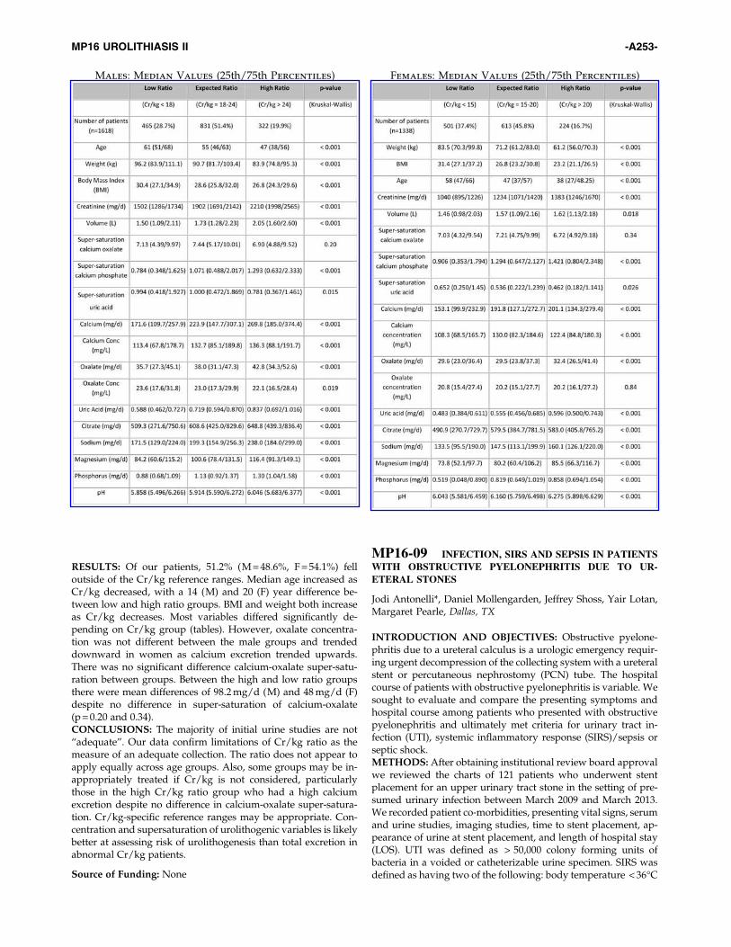
RESULTS: Of our patients, 51.2% (M = 48.6%, F = 54.1%) felloutside of the Cr/kg reference ranges. Median age increased asCr/kg decreased, with a 14 (M) and 20 (F) year difference be-tween low and high ratio groups. BMI and weight both increaseas Cr/kg decreases. Most variables differed significantly de-pending on Cr/kg group (tables). However, oxalate concentra-tion was not different between the male groups and trendeddownward in women as calcium excretion trended upwards.There was no significant difference calcium-oxalate super-satu-ration between groups. Between the high and low ratio groupsthere were mean differences of 98.2 mg/d (M) and 48 mg/d (F)despite no difference in super-saturation of calcium-oxalate(p = 0.20 and 0.34).CONCLUSIONS: The majority of initial urine studies are not‘‘adequate’’. Our data confirm limitations of Cr/kg ratio as themeasure of an adequate collection. The ratio does not appear toapply equally across age groups. Also, some groups may be in-appropriately treated if Cr/kg is not considered, particularlythose in the high Cr/kg ratio group who had a high calciumexcretion despite no difference in calcium-oxalate super-satura-tion. Cr/kg-specific reference ranges may be appropriate. Con-centration and supersaturation of urolithogenic variables is likelybetter at assessing risk of urolithogenesis than total excretion inabnormal Cr/kg patients.
Source of Funding: None
MP16-09 INFECTION, SIRS AND SEPSIS IN PATIENTS
WITH OBSTRUCTIVE PYELONEPHRITIS DUE TO UR-
ETERAL STONES
Jodi Antonelli*, Daniel Mollengarden, Jeffrey Shoss, Yair Lotan,Margaret Pearle, Dallas, TX
INTRODUCTION AND OBJECTIVES: Obstructive pyelone-phritis due to a ureteral calculus is a urologic emergency requir-ing urgent decompression of the collecting system with a ureteralstent or percutaneous nephrostomy (PCN) tube. The hospitalcourse of patients with obstructive pyelonephritis is variable. Wesought to evaluate and compare the presenting symptoms andhospital course among patients who presented with obstructivepyelonephritis and ultimately met criteria for urinary tract in-fection (UTI), systemic inflammatory response (SIRS)/sepsis orseptic shock.METHODS: After obtaining institutional review board approvalwe reviewed the charts of 121 patients who underwent stentplacement for an upper urinary tract stone in the setting of pre-sumed urinary infection between March 2009 and March 2013.We recorded patient co-morbidities, presenting vital signs, serumand urine studies, imaging studies, time to stent placement, ap-pearance of urine at stent placement, and length of hospital stay(LOS). UTI was defined as > 50,000 colony forming units ofbacteria in a voided or catheterizable urine specimen. SIRS wasdefined as having two of the following: body temperature < 36�C
Males: Median Values (25th/75th Percentiles) Females: Median Values (25th/75th Percentiles)
MP16 UROLITHIASIS II -A253-

or > 38�C, heart rate > 90 beats/minute (min), respiratory rate> 20 breaths/min, or leukocytes < 4000 cells/mm3 or > 12,000cells/mm3. Sepsis was defined as SIRS and positive blood orurine culture. Comparisons among groups were made usinganalysis of variance (ANOVA) for continuous variables and Chi-square for categorical variables.RESULTS: Among the 121 patients, 34 (28.1%) met criteria forUTI, 79 (65.3%) for SIRS/sepsis and 8 (6.6%) for septic shock.Table 1 shows the variables analyzed and the means of each forthe 3 groups. (Table).CONCLUSIONS: Among patients undergoing stent placementfor obstructive pyelonephritis due to stones, the single modifiablefactor that differed across the spectrum of infection severity wastime to stent placement. These data suggest that prompt de-compression of the collecting system in the face of obstructionand infection can be associated with improved outcomes. Furtheranalysis, including those patients who undergo PCN placementin this setting is underway to validate these findings.
Source of Funding: None
MP16-10 MULTIPLE SCLEROSIS AND KIDNEY STONE
DISEASE
Shubha De*, Giovanni Marchini, Benjamin Larson,Manoj Monga, Cleveland, OH
INTRODUCTION AND OBJECTIVES: Multiple sclerosis (MS)is a progressive disease that can involve bladder dysfunction,recurrent UTIs, physical, and sensory changes. Our objective wasto characterize stone formers with MS, in terms of stone compo-sitions, urine biochemistry, and clinical presentations.METHODS: A case matched comparison of 77 patients with MSand stones was performed (controlling for age, sex, BMI). De-mographics, voiding function, past medical history, acute pre-sentation, composition and urinary indices were analyzed usingStudent’s t-tests (p < 0.05).RESULTS: The average MS patient was 57 y/o, BMI 26.7, and23.2% could not ambulate independently. MS had significantly
more catheters (25%), incontinence (14%), OAB (7.5%) and re-current UTIs than Matched (p < 0.001).
MS patients presented less with acute flank pain (45% vs. 60%),and atypical pain was uncommon (MS 5%, Match 4.1%). MS hadmore recurrent stones (57% vs 46%), with comparable stoneburdens. Stone procedures were equivalent, however conserva-tive management was less common in MS (22%vs 9.6%, p = 0.032).Calcium phosphate stones were more common in MS patients(33%vs 15.3%, p = 0.016). 24 hr urine collections identified oxalate(50.37 vs 42.8p = 0.048), and pH (6.48 vs 5.98, p = 0.007) levelswere significantly higher for MS patients. The use of potassiumcitrate, HTCZ, allopurinol, were not significantly different.
When patients with significant urinary dysfunction (surgical orcatheter drainage) were compared to matched patients, signifi-cantly less COM (40 vs 55%, p = 0.04), more CaP (40 vs 16%,p = 0.002), and more struvite (11 vs 1.2%, p = 0.004) stones wereidentified. 24 hr urine collections showed significantly higheroxaluria (56.1 vs 42.8, p = 0.014), volume (2497 vs 1879, p = 0.15),and pH (6.58 vs.5.96, p = 0.022). No significant differences wereidentified between MS patients with and without bladder dys-function.CONCLUSIONS: MS patients display greater rates of bladderdysfunction, UTIs and immobility then matched controls. Withgreater amounts of calcium phosphate stones, alkali urine, andoxaluria, these derangements are potentiated with bladder dys-function (in addition to increased struvite stones).
Therefore, careful evaluation and management of these pa-tients is warranted, as concomitant bladder dysfunction increasesthe complexity of their stone disease.
Source of Funding: none
MP16-11 BILATERAL TUBELESS AND JJ STENTLESS
PCNL EXPERIENCE OF 37 CASES
Mohammed Bhuiyan*, MF Islam, ZH Bhuiyan, KMH Tawhid,NIU Ahmed, K Salahuddin, Dhaka, Bangladesh
INTRODUCTION AND OBJECTIVES: PCNL is well-acceptedmethod of management of renal stone. In PCNL post operativelyusually Nephrostomy and D-J stent are used. But Nephrostomyand D-J stent has some morbidity. Tubeless and stentless PCNL isselected cases reduces the morbidity. This study will evaluate theresults of tubeless and JJ stentless PCNL in the management ofbilateral renal stone in selected cases.METHODS: From January 2009 to January 2013 total 37 cases ofbilateral renal stone were managed by tubeless and JJ stent lessPCNL. All cases were operated in urology department of Ban-gladesh Medical College Hospital, Dhaka. In all cases initiallyplaced a ureteric catheter and at the end of the procedure com-pression at renal angle about 10 min. Ureteric catheter was re-moved 24 hour after operation. Exclusion criteria for the tubelessand stentless approach were more than two percutaneous access,significant perforation of the collecting system, large residualstone burden, significant postoperative bleeding, ureteral ob-struction and renal anomaly. The incidence of complication,length of hospitalization, analgesia requirement and interval toreturn to normal activities were evaluated.RESULTS: All 37 percutaneous procedures were performed without significant complication, none of the patients demonstratedurinoma in postoperative renal ultrasound scan. The averagelength of hospital stay was 1.6 day; the average analgesia re-quirement was single dose of inj. pethedin, patient return tonormal activity within 12 days.
-A254- MP16 UROLITHIASIS II

CONCLUSIONS: Tubeless and JJ stent less PCNL is a safe andeffective procedure in the management of bilateral renal stone inselected cases. The hospitalization and analgesia requirementsare less and returns to normal activities are faster.
Source of Funding: none
MP16-12 NON-LINEAR LOGISTIC REGRESSION
MODEL FOR OUTCOMES AFTER ENDOUROLOGIC PRO-
CEDURES: A NOVEL PREDICTOR
Adam Kadlec*, Maywood, IL, Samuel Ohlander, James Hotaling,Chicago, IL, Jessica Hannick, Maywood, IL, Craig Niederberger,Chicago, IL, Thomas Turk, Maywood, IL
INTRODUCTION AND OBJECTIVES: We aimed to design athorough and practical non-linear logistic regression model thatcan be used for outcome prediction after various forms of en-dourologic intervention.METHODS: Input variables and outcome data from 382 renalunits endourologically treated at a single institution were used tobuild and cross-validate an independently designed non-linearlogistic regression model. Model outcomes were stone-free statusand need for a secondary procedure.RESULTS: The model predicted stone-free status with sensitivity75.3% and specificity 60.4%, yielding a positive predictive value(PPV) of 75.3% and negative predictive value (NPV) of 60.4%,with classification accuracy of 69.6%. Receiver operating charac-teristic area under the curve (ROC AUC) was 0.749. The modelpredicted the need for a secondary procedure with sensitivity30% and specificity 98.3%, yielding a PPV of 60% and NPV of94.2%. ROC AUC was 0.863. The model had equivalent predictivevalue to a traditional logistic regression model for the secondaryprocedure outcome.CONCLUSIONS: This study is proof-of-concept that a non-lin-ear regression model adequately predicts key clinical outcomesafter shockwave lithotripsy, ureteroscopic lithotripsy, and per-cutaneous nephrolithotomy. This model holds promise forfurther optimization via dataset expansion, preferably withmulti-institutional data, and could be developed into a predictivenomogram in the future.
Source of Funding: None
MP16-13 RANDALL’S PLAQUE AND INTRADUCTAL
PLUGGING: ASSOCIATION WITH STONE EVENTS AND
COMPOSITION
Mitra de Cogain*, Amy Krambeck, Eric Bergstralh, Xujian Li,John Lieske, Rochester, MN
INTRODUCTION AND OBJECTIVES: We examined the rela-tionship between papillary Randall’s plaque or intermedullarycollecting duct (IMCD) plugging and urolithiasis characteristicsin percutaneous nephrolithotomy (PCNL) patients.METHODS: We prospectively enrolled 105 consecutive PCNLpatients. Post stone removal papillary mapping for precursorlesions (plaque and plug) was performed, along with papillarybiopsy.RESULTS: The number of stone events (prior spontaneous pas-sage and previous stone procedures) correlated with the degree ofIMCD plugging (p = 0.03), but not with the amount of Randall’splaque (p = 0.44). The number of renal calculi identified on com-puted tomography at the time of PCNL did not correlate with theamount of IMCD plugging (p = 0.17), and negatively correlated
with the amount of plaque (p = 0.03). Calcium phosphate stoneformers were more likely to have IMCD plugging, although thiswas not significant (p = 0.29). Calcium oxalate stone formers hadhigher plaque coverage, although this was not significant(p = 0.06).CONCLUSIONS: In this cohort of various stone formers, IMCDplugging correlated with the severity of stone disease, whileRandall’s plaque coverage did not. Thus, patients with largeramounts of IMCD plugging may have a clinically more severestone phenotype and a higher likelihood of calcium phosphatestones. Patients with calcium oxalate stones are more likely tohave Randall’s plaque; however the severity of stone disease doesnot strongly correlate with the amount of plaque present.
Source of Funding: National Institute of Health through theMayo Clinic O’Brien Urology Research Center (DK83007)
MP16-14 DIABETES MELLITUS AND UROLITHIASIS:
DOES RANDALL’S PLAQUE ACCOUNT FOR INCREASED
STONE RISK?
Mitra de Cogain*, Amy Krambeck, Eric Bergstralh, Xujian Li,John Lieske, Rochester, MN
INTRODUCTION AND OBJECTIVES: Diabetics are at in-creased risk for urolithiasis; however, the link between precursorstone lesions and diabetes mellitus (DM) has not been explored.We evaluated the relationship between Randall’s plaque, inter-medullary collecting duct (IMCD) plugging and DM.METHODS: We prospectively enrolled percutaneous ne-phrolithotomy patients. Following stone removal, papillarymapping and papillary biopsy was performed.RESULTS: We identified 105 patients, 17 with and 88 withoutDM. Urinary composition and crystal supersaturations were notdifferent between groups (p > 0.05). Mean urine pH in diabetics(range 4.9–6.8) and non–diabetics (range 5.0–7.9) was identical 6.1(p = 0.8). Diabetics had larger mean stone size (12.0 mm vs.6.5 mm, p = 0.01) and trended towards lower amounts of severe( > 1%) IMCD plugging (29.4% vs. 45.5%, p = 0.22) and Randall’splaque papillary coverage (3.6 % vs. 6.6 %, p = 0.08). Both dia-betics and non–diabetics most commonly had calcium oxalatestones (64.7% vs. 58.6%). However, non–diabetics more oftenformed brushite (8.0% vs. 0%) and hydroxyapatite calculi (24.1%vs. 5.9%) while diabetics formed uric acid (17.6% vs. 1.1%) andstruvite (11.8% vs. 8.0%) calculi more commonly (p < 0.01).CONCLUSIONS: Although diabetics had similar or lesseramounts of IMCD plugging and Randall’s plaque coverage, theirstones were larger, with a significantly different composition.Therefore, the pathway to stone formation may involve differentprecursor lesions and subsequent processes in diabetics.
Source of Funding: National Institute of Health through theMayo Clinic O’Brien Urology Research Center (DK83007)
MP16-15 NEW DESIGNED TIP-FLEXIBLE SEMI-RIGID
URETEROSCOPY IN THE MANAGEMENT OF RENAL
STONES: INITIAL EXPERIENCE
Yinghao Sun*, Xiaofeng Gao, Shanghai, China, China, People’sRepublic of, Ling Li, Shanghai, China, People’s Republic of,Yonghan Peng, Shanghai, China, China, People’s Republic of
INTRODUCTION AND OBJECTIVES: A new designed tip-flexible semi-rigid ureteroscopy has been developed for themanagement of renal stones. We present our initial clinical
MP16 UROLITHIASIS II -A255-

experience in a prospective study between this new ureteroscopyand the flexible ureteroscopy.METHODS: 40 consecutive patients with renal stones 1*1.5 cmwere enrolled in this study and randomized into two groups. Theureteroscopic lithotripsy with holmiun laser was conducted withthis tip-flexible semi-rigid ureteroscopy (group A) and flexibleureteroscopy (FLEX X2, KARL-Storz, Germany)(group B). Stonelocation and burden were assessed before operative procedure bycomputer tomography. Operating time were calculated for thetwo groups. Complication and stone-free rates were comparedbetween the two groups. Success was based on postoperativeimaging with 1-month follow-up.RESULTS: The success rates of the ureteroscopic insertion forgroup A and group B were 85% and 95%, respectively. The av-erage stone size were 1.2¡À0.1 cm (group A) and 1.3¡À0.1 cm,respectively. The average operation time were 48¡À12 min forgroup A and 55¡À5 min for group B. The stone free-rate were82.3%£̈14̈M17£ªand 94.7%(18/19) for group A and group B, re-spectively. Complications between the two groups were compa-rable: 1 case in group A suffered high fever ( > 38¡æ)postoperatively while none had severe complication in group B.The average postoperative hospital stay was 1.2¡À0.2 d for groupA and 1.1¡À0.1 d for group B.CONCLUSIONS: The tip-flexible semi-rigid ureteroscopy is asafe and procedure, which combined with these advantages ofrigid and flexible ureteroscopy, specially for renal stones withrelative small size.
Source of Funding: none
MP16-16 EVALUATION OF HEAVY METALS AND
TRACE ELEMENTS CONTENTS IN URINARY CALCULI:
MICROANALYTICAL STUDY
Mahmoud Elnagar*, Naimat Alsayegh, Alain, United ArabEmirates, Bedier Ali-El-Dein, Mansoura, Egypt, Saeed Alqahtani,Paris, France, Sanjeev Mehta, Ahmadabad, India, Nilkamal Joshi,Alain, United Arab Emirates, Emad Elsobky, Abu Dhabi,United Arab Emirates
INTRODUCTION AND OBJECTIVES: The presence and roleof heavy metals and trace elements in urinary stones formation isdebated. The purpose of this study was evaluation and deter-mination of heavy metals and trace elements contents in retrievedurinary stones from patients living in different geographicalregions.METHODS: After approval of Institutional Research Commit-tees, a total of 559 consecutive urinary calculi were retrieved frompatients of 6 International Hospitals. The stones were subjected tomicroanalysis for determination of heavy metals and trace ele-ments contents in each individual sample (22 elements in eachstone) by using Inductively Coupled Plasma-Optical EmissionSpectrometry (ICP-OES) analytical technique. Data analysis ofelements contents in all examined stones and concentration cor-relation of each element with Calcium in each stone were deter-mined by One-Way ANOVA test using SPSS software.RESULTS: Out of 559 analyzed stones there were 482 (89.2%)Non-Urate [318 Calcium oxalate (56.9%), 164 Calcium phosphate(29.3%)] and 77 were Urate stones (13.8%). Heavy metals con-centrations of As, Ba, Cr, Fe, K, Mg, Ni, P, Pb, S, Se, Sr and Znwere significantly high in all analyzed stones (P < 0.01). Con-centration correlation between each element and Calcium contentof the stone was significantly high in all types of stones. The ratiowas higher in Urate compared to Non-Urate stones. Cu, Fe, Pb,Se, and Ba were noticed in higher concentration in European
when compared to Non-European stones. Al, As, Bo, Ca, Cdconcentrations were higher in Asian when compared to Non-Asian stones. K, Mg, and Fe concentrations were higher in Africanstones than other groups.CONCLUSIONS: Our study showed that many heavy metalsand trace elements are present significantly in urinary stonesfrom different geographical regions. The concentration of variouselements in these stones differs from country to country. This maybe due to either underlying environmental pollution or nutri-tional factors. Urate stones contained the highest concentrationcontent of heavy metals and trace elements.
Source of Funding: non
MP16-17 EVALUATION OF MICRO CT ANALYSIS OF
URINARY STONE COMPOSITION
James Williams*, Indianapolis, IN, Kalba Usman, Ruth Tor,Petach Tikva, Israel, James McAteer, Indianapolis, IN,David Lifshitz, Petach Tikva, Israel
INTRODUCTION AND OBJECTIVES: Micro-computed to-mography (micro CT) has been shown to provide outstandingimages of urinary stones. The aim of this study was to evaluatethe reliability of using micro CT alone to identify stone mineral ina clinical scenario.METHODS: Urinary stones were retrieved from 60 consecutiveendoscopic procedures performed in Rabin Medical Center. Thesamples were analyzed by micro CT using a SkyScan 1172 MicroCT System using voxel sizes between 14 and 22 um; an experi-enced worker identified mineral content of each specimen usingmicro CT alone, and then samples were taken for Fourier-trans-form infrared spectroscopy (FT-IR). Samples were then returnedto Israel for a secondary analysis of the samples by FT-IR (blindedto the initial micro CT results).RESULTS: Table 1 lists the sensitivity and specificity of micro CTanalysis for different minerals: calcium oxalate (CaOx) mono-hydrate (COM); CaOx dihydrate (COD); apatite; uric acid; cys-tine; brushite; and struvite. This study shows that micro CT wasvery good at distinguishing COM and COD (which is done byobserving a combination of crystal morphology and x-ray atten-uation). Micro CT was also very good at identifying uric acid andcystine, which both have distinctive x-ray attenuation values.Micro CT had a poor sensitivity for brushite (which has an x-rayattenuation value similar to that of CaOx) and also missed stru-vite on 2 specimens (one false positive and one false negative).CONCLUSIONS: Micro CT shows promise as a routine methodfor analysis of urinary stone mineral. A primary advantage ofmicro CT is its ability to non-destructively image a stone speci-men in its entirety, so that small regions of distinct mineral can bedetected even in a large specimen. This was manifested in thepresent study by the differences between the FT-IR analyses doneblinded or with knowledge of micro CT, where micro CT guid-ance allowed detection of minor components that were not dis-tributed homogeneously within the specimen. In its presentdevelopment, micro CT is weakest in its recognition of brushite
-A256- MP16 UROLITHIASIS II

and struvite, which both are minerals for which the urologistwould like to have knowledge in planning patient treatment. Thisweakness could possibly be improved by proper training of themicro CT technician, but at present it appears that micro CTanalysis of urinary stone mineral should be used along with an-other method (such as FT-IR) in order to maximize accuracy.
Source of Funding: NIH R01DK059933
MP16-18 SIMULTANEOUS DIGITAL FLEXIBLE UR-
ETERORENOSCOPY AND EXTRACORPORAL SHOCK
WAVE LITHOTRIPSY: A NEW APPROACH FOR RENAL
STONE TREATMENT?
Thomas Knoll*, Patrick Honeck, Sindelfingen, Germany,Ulf Balsmeyer, Dirk Fahlenkamp, Chemnitz, Germany,Gunnar Wendt-Nordahl, Sindelfingen, Germany
INTRODUCTION AND OBJECTIVES: Extracorporal shockwave lithotripsy (SWL) has been the standard treatment for mostkidney stones. In recent years, refinements of endoscopic tech-niques and the idea of immediate complete stone removal haveled to a shift towards ureteroscopy (URS) and percutaneous ne-phrolithotomy (PNL). However, depending on stone location andrenal anatomy the surgeon may have difficulties to access andremove the stone in one procedure with a single treatment mo-dality. Simultaneous antegrade-retrograde approaches combin-ing flexible URS with PNL have been introduced for suchconditions. Based on the same idea we developed a differentconcept of a combined approach by using simultaneous digitalflexible URS and SWL. Our aim was to evaluate the feasibility ofsuch a combined approach.METHODS: 3 patients with solitary lower pole stones (8–13 mm)were treated by simultaneous flexible URS and SWL. One patientwas presented. We used an 8 F digital flexible scope (FlexXc, KarlStorz, Germany) for retrograde access and an 3rd generationelectromagnetic lithotripter (Modulith SLK or SLX, Storz Medi-cal, Switzerland). Patients were treated in lithotomy position andgeneral anaesthesia. After retrograde access and visualization ofthe stone SWL treatment was started under endoscopic andfluoroscopic control. Shock wave rate was 1 Hz, Energy settingwas 4–6, and number of shock waves was 2,000–4,000.RESULTS: All patients could be rendered stone free. Completestone disintegration by SWL could be achieved in 2/3 patients.Laser lithotripsy was used in one patient. All patients received aDJ stent. Significant complications were not observed. Stoneanalysis revealed calcium oxalate monohydrate stones (100%) inall patients. The flexible endoscope was not affected by the si-multaneous shock wave delivery.CONCLUSIONS: Our first treatments have proven that combinedendoscopic and extracorporal shock wave treatment of urinarystones is feasible and effective. Further studies have to evaluate thisnew approach and to define ideal indications and settings.
Source of Funding: none
MP16-19 HIGHER HEMOGLOBIN A1C IS ASSO-
CIATED WITH A GREATER LIKELIHOOD OF URIC ACID
STONE FORMATION: IS CONTROL OF DIABETES IM-
PORTANT FOR STONE PREVENTION?
Sara Best, Jonathan Shiau*, Rachel Bell, Kristina Penniston,Madison, WI
INTRODUCTION AND OBJECTIVES: Patients with diabetesmellitus (DM) are known to have a propensity for lower urine pH,higher calcium excretion and a higher prevalence nephrolithiasis.Hyperinsulinemia has been linked to hypercalciuria and the co-incidence of obesity and DM also adds additional lithogenic risks.The impact of glycemic control on stone composition, however, isunknown. We examined the influence of hemoglobin A1c(HgbA1c), a measure of long-term diabetes control, on stonecomposition.METHODS: Using an approved institutional stone compositiondatabase, we performed a retrospective chart review of 540 stoneformers and extracted clinical characteristics known to be asso-ciated with stone formation. We also collected data on diabetesmanagement and HgbA1c to assess glycemic control. Patientswere categorized based on both major stone composition ( > 50%),and on the subtype (whether it contained any uric acid, brushite,or > 5% calcium phosphate). Variables were compared usingFisher’s exact tests and ANOVA.RESULTS: In our cohort, we confirmed that diabetic stone for-mers (n = 107) were more likely to have ‡ 50% uric acid stonesthan non-diabetics (20 % and 5% respectively, p < 0.0001), and lesslikely to have brushite stones (0% in DM, 4% in non-DM,p = 0.019). In analyzing subtype stone compositions ( < 50%), pa-tients with stones containing any component of uric acid hadhigher mean HgbA1cs than those with pure CaOx calculi (5.85 vs6.72, p = 0.02, Figure 1). Additionally, patients whose diabetes ismanaged with oral hypoglycemic medications had more uric acidstones than those managed in other ways (34% vs 10%, p = 0.005).This likely reflects poorer diabetic control in patients prescribedoral medications (mean HgbA1c 7.3% vs 6.2%, p < 0.0001).CONCLUSIONS: In our cohort, we confirmed that diabetic stoneformers were more likely to form uric acid stones. Better glycemiccontrol, as measured by HgbA1c, may reduce the likelihood of uricacid nephrolithiasis. Additionally, a high index of suspicion for uricacid stones should be maintained in patients with high HgbA1cand targeted therapy considered. Our findings support the inclu-sion of diabetic control as a component of the multidisciplinarymedical management of stone forming patients with diabetes.
Source of Funding: none
MP16-20 URETEROSCOPY VERSUS SHOCK WAVE
LITHOTRIPSY – FACTORS INFLUENCING PATIENT
TREATMENT PREFERENCES
Adam Shrewsberry*, Steven Gerhard, Tania Solomon,Dana Nickleach, Yuan Liu, John Pattaras, Kenneth Ogan,Atlanta, GA
MP16 UROLITHIASIS II -A257-

INTRODUCTION AND OBJECTIVES: For patients with un-complicated ureteral calculi that require treatment, ureteroscopy(URS) and shock wave lithotripsy (SWL) are acceptable first-linetreatments. As neither treatment has been confirmed superior tothe other, the decision of treatment modality is left up to theprovider and patient. Herein, we examine patient preferences fortreatment of a ureteral calculus and assess the factors that influ-ence treatment selection.METHODS: Patients with a urolithiasis (first time or recurrentstone formers) were either self-administered or mailed a ques-tionnaire with a clinical scenario of a stone in the ureter andoutcome statistics derived from a Cochrane Review for URS andSWL comparing four parameters: (1) stone-free success rates, (2)complication rate, (3) need for ureteral stent placement, and (4)need for additional surgery. The subjects were asked to chooseURS or SWL based on the provided outcomes and to indicate therelative importance that each of the four outcome parameters hadon their treatment selection.RESULTS: A total of 163 patients completed the surveys. Themean age was 53.7 years, 59% were male, and 79% had a historyof prior stone-related surgery. Overall, more patients chose URSvs. SWL (62.6% vs. 37.4%, p = 0.001). Ninety-four percent of pa-tients cited stone-free success as extremely or largely important,followed by risk of complications (80%), need for second surgery(69%), and the need for a stent (48%). For patients who chose URS,stone-free success rate and need for a second surgery were ofgreater importance compared to those who chose SWL. Con-versely, for those who chose SWL the need for a stent after sur-gery was of greater importance compared to the URS group.Patients with a history of URS or a history of URS and SWLfavored URS while there was no significant association betweenhistory of SWL and procedure preference.CONCLUSIONS: In our study, patients preferred URS to SWLfor treatment of a ureteral calculus. Patient preferences for treat-ment of ureteral calculi are impacted by a number of factors thatthe urologist must discuss prior to choosing URS vs. SWL.
Source of Funding: None
MP16-21 HYPERLIPIDEMIA IS ASSOCIATED WITH
AN INCREASED RISK OF NEPHROLITHIASIS
LCDR James H Masterson, Jason R Woo, David C Chang,CDR James O L’Esperance, San Diego, CA, Marshall L Stoller,San Francisco, CA, Roger L Sur*, San Diego, CA
INTRODUCTION AND OBJECTIVES: The pathophysiology ofnephrolithiasis is multifactorial and obesity, diabetes mellitus(DM) and hypertension (HTN) have been implicated in its for-mation. Hyperlipidemia (HLD) has recently also received atten-tion as a cause. Congruent with a vascular etiology in stoneformation, hyperlipidemia theoretically would be associated withan increased risk. We investigated a possible association of HLDwith nephrolithiasis.METHODS: A random cohort of 60,000 patients was establishedby collecting the first 5000 patient charts per month in the year2000. After excluding all pediatric patients, a retrospective studywas performed by reviewing age, sex, comorbidities, and lastfollow up. Median lipid laboratory levels for each patient avail-able were also reviewed. Descriptive statistics were performed aswell as Cox proportional-hazards analysis, both univariate andmultivariate, to identify factors associated with nephrolithiasis.RESULTS: 52,184 (22,717 women/29,467 men) patient chartswere reviewed. The average age was 31.0 +/- 15.2 years. Onunivariate analysis a diagnosis of HLD had a hazard ratio (HR) of
2.2 [1.9–2.5, 95% Confidence Interval (CI), p < 0.001] associatedwith nephrolithiasis. On multivariate analysis it was HR = 1.2[1.0–1.5, 95% CI, p = 0.033]. Low-density lipoprotein (LDL) andtriglycerides had no association with stone disease, but high-density lipoprotein (HDL) with values below 45 for men and 60for women had a HR of 1.4 [1.1–1.7, 95% CI, p = 0.003] for stoneformation. On multivariate analysis it was a HR = 1.27 [1.03–1.56,95% CI, p = 0.024].CONCLUSIONS: HLD is associated with nephrolithiasis. Lowlevels of HDL appear to increase the risk of stone formation, butthe same effect was not seen with LDL or triglyceride levels.
*The views expressed in this article are those of the author anddo not reflect the official policy or position of the Department ofthe Navy, Department of Defense, or the United States Govern-ment.
Source of Funding: None
MP16-22 GENDER INEQUALITIES IN URINARY ME-
TABOLIC EVALUATION OF STONE FORMERS: A 10 YEAR
EXPERIENCE FROM A WESTERN INDIAN STONE BELT
REGION
Jitendra Jagtap*, Raguram Ganesamoni, Jigish Vyas,Shashikant Mishra, Arvind Ganpule, Ravindra Sabnis,Mahesh Desai, Nadiad, India
INTRODUCTION AND OBJECTIVES: To identify the variousurinary metabolic abnormalities among Indian stone formers andto investigate the differences between the urinary biochemicalprofile of male and female stone formers.METHODS: We retrospectively reviewed clinical data for allstone formers who had undergone 48 hour urinary metabolicevaluation from 2003 to 2013 at our tertiary referral center inWestern India. We analyzed patient age, gender, 48 hour urinarymetabolic parameters of these patients. After excluding the im-properly collected samples, data was available for 1857 out of theinitial 1986 patients reviewed. Data was analyzed using SPSS 15.0software.RESULTS: Mean age was 42.26 – 16.15 for males and41.92 – 17.99 for females. The Male: Female ratio was 3.26:1(1421:436). The average 24 hr. urine volumes were2472.83 – 1397.27 ml for females vs. 2880 – 1599.26 ml for malesrespectively. On urine metabolic evaluation the incidence ofHypercalciuria was 40.4% in females vs. 52.9% in males(p < 0.0001*) with mean values being 152 – 100.32 mg/24 hr vs.177.52 – 109.79 mg/24 hr respectively. Similarly the values forHyperoxaluria were 76.4% in females vs. 62.6% in males(p < 0.0001*) with mean values being 49.3 – 29.66 mg/24 hr vs.58.50 – 37.53 mg/24 hr respectively. For Hyperphosphaturia andHyperuricemia the values were 0% in females vs. 0.6% in males(p = 0.127, NS) with mean values being 344.35 – 173.86 mg/24 hrvs. 443.75 – 231.91 mg/24 hr and 1.1% in females vs. 2.5% in males(p = 0.094, NS) with mean values being 321.82 – 140.18 mg/24 hrvs. 391.13 – 167.27 mg/24 hr respectively. While the incidence ofHypocitraturia was 64.9% in females vs. 17.5% in males(p < 0.0001*) with mean values being 245.58 – 192.45 mg/24 hr vs.267.35 – 228.55 mg/24 hr respectively. For Hypomagnesuria thevalues were 50% in females vs. 39.6% in males (p < 0.0001*) withmean values being 92.29 – 67.64 mg/24 hr vs. 107.62 – 77.58 mg/24 hr respectively.CONCLUSIONS: This study provides valuable information onthe major urinary metabolic abnormalities in the Western Indianpopulation. There was a significantly higher incidence of Hy-percalciuria in the males while there was a predominance of
-A258- MP16 UROLITHIASIS II
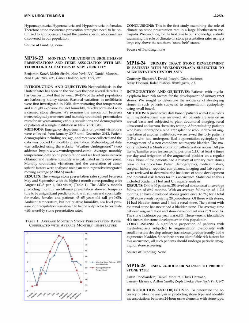
Hypomagnesuria, Hyperoxaluria and Hypocitraturia in females.Therefore stone recurrence prevention strategies need to be op-timized to appropriately target the gender specific abnormalitiesdiscovered in our population.
Source of Funding: none
MP16-23 MONTHLY VARIATIONS IN UROLITHIASIS
PRESENTATIONS AND THEIR ASSOCIATION WITH ME-
TEOROLOGICAL FACTORS IN NEW YORK CITY
Benjamin Katz*, Mohit Sirohi, New York, NY, Daniel Moreira,New Hyde Park, NY, Caner Dinlenc, New York, NY
INTRODUCTION AND OBJECTIVES: Nephrolithiasis in theUnited States has been on the rise over the past several decades. Ithas been estimated that between 10–15% of the adult populationare harboring kidney stones. Seasonal variations in urolithiasiswere first investigated in 1960, demonstrating that temperatureand sunlight exposure, but not humidity, directly correlated withincreased stone disease. We examine the association betweenmeteorological parameters and monthly urolithiasis presentationrates for six years among various populations and demographicsof patients at a single institution in New York City.METHODS: Emergency department data on patient visitationswere collected from January 2007 until December 2012. Patientdemographics including sex, age, and race were collected and thedata was pooled by monthly presentation. Meteorological datawas collected using the website ‘‘Weather Underground’’ (webaddress: http://www.wunderground.com). Average monthlytemperature, dew point, precipitation and sea level pressure wereobtained and relative humidity was calculated using dew point.Monthly urolithiasis visitations and the correlation of atmo-spheric factors were analyzed using an auto-regressive integratedmoving average (ARIMA) model.RESULTS: The average stone presentation rates spiked betweenMay and September with the highest month corresponding withAugust (43.8 per 1, 000 visits) (Table 1). The ARIMA modelspredicting monthly urolithiasis presentation showed tempera-ture to be a significant predictor for the all comers and specificallyfor males, females and patients 45–65 years-old (all p < 0.05).Ambient temperature, but not relative humidity, sea level pres-sure, or precipitation was shown to be the only factor to correlatewith monthly stone presentation rates.
CONCLUSIONS: This is the first study examining the role ofclimate on stone presentation rate in a large Northeastern me-tropolis. We conclude, for the first time to our knowledge, a studyexamining the role of climate on stone presentation rates using alarge city above the southern ‘‘stone belt’’ states.
Source of Funding: none
MP16-24 URINARY TRACT STONE DEVELOPMENT
IN PATIENTS WITH MYELODYSPLASIA SUBJECTED TO
AUGMENTATION CYSTOPLASTY
Courtney Shepard*, David Joseph, Dean Assimos,Betsy Hopson, Ralee Bishop, Birmingham, AL
INTRODUCTION AND OBJECTIVES: Patients with myelo-dysplasia have risk factors for the development of urinary tractstones. We sought to determine the incidence of developingstones in such patients subjected to augmentation cystoplastyusing small bowel.METHODS: A prospective data base of patients with 435 subjectswith myelodysplasia was reviewed. All patients are seen on anannual basis and subjected to plain abdominal imaging, renalultrasound and serum chemistry testing. After excluding patientswho have undergone a renal transplant or who underwent aug-mentation at another institution, we reviewed the forty patients(9.2%) who had undergone ileal augmentation cystoplasty formanagement of a non-compliant neurogenic bladder. The ma-jority included a Monti stoma for catheterization access. All pa-tients/families were instructed to perform C.I.C. at least 4 timesper day and irrigation of the augmented bladder on a regularbasis. None of the patients had a history of urinary tract stonesprior to this procedure. Patient demographics, medical history,surgical history, reported compliance, imaging and lab reportswere reviewed to determine the incidence of stone developmentand potential risk factors for this occurrence. Statistical analysisincluded Student’s t test and Chi square analysis.RESULTS: Of the 40 patients, 25 have had no stones at an averagefollow-up of 89.9 months. With an average follow-up of 117.2months, 15 have developed stones (prevalence 37.5%) for a totalof 20 stone events requiring 20 procedures. Of those with stones,14 had bladder stones and 1 had a renal stone. The patient withthe renal stone has never had a bladder stone. The average timebetween augmentation and stone development was 26.9 months.The stone incidence per year was 6.8%. There were no identifiablerisk factors for stone development in this population.CONCLUSIONS: A significant proportion of patients withmyelodysplasia subjected to augmentation cystoplasty withsmall intestine develop urinary tract stones, predominantly in theaugmented bladder. Since there are no identifiable risk factors forthis occurrence, all such patients should undergo periodic imag-ing for stone screening.
Source of Funding: None
MP16-25 USING 24-HOUR URINALYSIS TO PREDICT
STONE TYPE
Justin Friedlander*, Daniel Moreira, Chris Hartman,Sammy Elsamra, Arthur Smith, Zeph Okeke, New Hyde Park, NY
INTRODUCTION AND OBJECTIVES: To determine the ac-curacy of 24-urine analysis in predicting stone type and identifythe associations between 24-hour urine elements with stone type.
Table 1. Average Monthly Stone Presentation Rates
Correlated with Average Monthly Temperature
MP16 UROLITHIASIS II -A259-

METHODS: We performed a retrospective review of 503 stoneformers with both stone composition analysis and 24-hour uri-nalysis available. Analysis of 24-hour urine elements across stonetypes was performed using Fisher’s exact test and analysis ofvariance. Multinomial logistic regression was used to predictstone type based on 24-hour urinalysis.RESULTS: A total of 280 (56%) patients had predominantly cal-cium oxalate, 103 (20%) had uric acid, 93 (19%) had calciumphosphate, 16 (3%) had mixed and 11 (2%) had other stone types.There were several significant patient characteristics and 24-hoururinalysis differences across stone type groups. The statisticalmodel predicted 371 (74%) calcium oxalate, 78 (16%) uric acid, 52(10%) calcium phosphate, zero mixed and 2 ( < 1%) other stonetypes. The model correctly predicted calcium oxalate stones in
85%, uric acid in 51%, calcium phosphate in 31%, mixed in 0%and other stone types in 18% of the cases. Of the predicted stonetypes, correct predictions were 61%, 69%, 56% and 71% for cal-cium oxalate, uric acid, calcium phosphate and other stonestypes, respectively. The overall accuracy was 64%. Plots wereused to explore the associations between each 24-hour urine el-ement with each predicted stone type adjusted for all the othersurinary elements.CONCLUSIONS: 24-hour urinalysis alone does not accuratelypredict stone type; however, it may be used in conjunction withother variables to predict stone composition.
Source of Funding: none
MP16-26 THE PREDICTION FACTORS OF FLEXIBL
URETERORENOSCOPY REQUIREMENT AFTER RIJID UR-
ETERORENOSCOPY IN RENAL PELVIS STONES
Evren Suer, Omer Gulpinar, Cihat Ozcan*, Cagatay Gogus,Seymur Kerimov, Siddik Mut Safak, Ankara, Turkey
INTRODUCTION AND OBJECTIVES: In this study we ex-amine the success rate of rijid ureterorenoscopy (R-URS) in renalpelvis stones whose size are between 1 and 2 cm and consider thepredictive factors of flexibl ureterorenoscopy (F-URS) require-ment at the time of failure of R-URS.METHODS: We consider R-URS success in renal pelvis stonesand when it fails the predictive factors for F-URS requirement atthe same time in 88 patients who underwent retrograd intrarenalsurgery (RIRS) in between 2000 and 2012 years. The patients whohave stones except renal pelvis are excluded.RESULTS: The mean patient age was 48,6 – 16,5 years and followperiod 39 – 11,5 months. In 48 patients R-URS became successfuland we did not need to use F-URS. In the other 40 patients weused F-URS and the predictive factors for its requirement weremale sex, height and severe hydronephrosis. At the end of thefollow period the stone free rates in R-URS and F-URS were 83%and 87% sincerely.CONCLUSIONS: The usability of R-URS in renal pelvis stonesbetween 1 and 2 cm is influenced with some factors like male sex,height and hydronephrosis. These predictive factors help us tochoice the right treatment.
Source of Funding: none
Table 1. Univariable Comparison of Baseline Patient
Characteristics Across Stone Type Groups
*Fisher’s exact test for categorical variables and analysis ofvariance for continuous variables.
SD: standard deviation.
Table 2. Univariable Comparison of 24-hour
Urine Elements Across Stone Type Groups
*Analysis of varianceCa: calcium, SD: standard deviation, SS: supersaturate.
Table 3. Comparison of Actual and Predicted
Stone Types
Note: shaded cells represent correct predictions.n/a: not available.
-A260- MP16 UROLITHIASIS II

MP17 FEMALE UROLOGY/ADRENAL/TESTIS
MP17-01 SINGLE PORT INTRAVESICAL LAPARO-
SCOPIC REPAIR OF VESICOVAGINAL FISTULA
Ahmed Moharram*, Giza, Egypt
INTRODUCTION AND OBJECTIVES: to report a new tech-nique for repair of small sized (less than 5 fr.), supratrigonal ve-sicovaginal fistula using a minimally invasive single portlaparoscopic approach to the fistula.METHODS: In all 10 women with vesicovaginal fistula wereoperated upon in the period between 2010 and 2012 in El Galaateaching hospital using a single port intratrasvesical laparoscopicapproach of the fistula where a 2 cm suprapubic incision was done,an anterior bladder wall incision was done in which the port wasplaced and fixed in the urinary bladder. Complete dissection of thefistulous tract, and complete separation of the urinary bladderfrom the underneath vaginal wall was done. The urinary bladderwas sutured in 2 continuous layers using 000 vicryl.RESULTS: Follow up for 8 cases was for one year, and 5 monthsfor 2 patients. Follow up was for: urine leakage, any lower urinarysymptoms such as burning micturition, frequency, urgency or urgeincontinence. Urine analysis was done every 3 months to ensureabsence of urinary tract infection. None of the 10 cases had anyleakage after the repair. Only one case showed UTI, urgency fromthe group operated upon for the first time which was treatedmedically for 3 weeks. No other complications were reported.CONCLUSIONS: This approach is a new minimally invasivetechnique where the fistula was exposed directly and has the ad-vantages of short operative time, essay technique, no or very fewintra and post-operative complications and short hospital stay.
Source of Funding: None
MP17-02 BILATERAL LAPAROSCOPIC ADRENA-
LECTOMY IN A PREGNANT WOMAN WITH CUSHING’S
SYNDROME
Mohammad Aslzare*, Amirabbas Asadpour,Mohammad Alipour, Mohammadreza Darabi Mahboub,Morteza Taghavi, Alireza Ghoreifi, Mashhad, Iran
INTRODUCTION AND OBJECTIVES: Cushing’s syndrome(CS) occurs rarely during pregnancy and usually associated withsevere maternal and fetal complications. Surgical treatment is themainstay of therapy of CS in pregnancy.
Here we presented a 31 old pregnant woman with cushingsyndrome who underwent bilateral laparoscopic adrenalectomyin her 18th week of pregnancy. Our medline search revealed thatthis is the first report of ‘‘bilateral’’ laparoscopic adrenalectomyduring pregnancy.METHODS: A 31-year old woman, gravid 1, para 0, was referredto us at 4th months’ gestation with Cushing syndrome. Medicalhistory was remarkable for hypertension and paroxysmal sup-raventricular tachycardia (PSVT) that was ablated 4 months ago.
After complete evaluation she underwent bilateral laparo-scopic adrenalectomy under general anesthesia with transper-itoneal approach. At first the patient was placed in the left flankposition and open access was done for pneumoperitoneum cre-ation. After completion of left adrenalectomy, position changedto right flank and new trocar insertion was done and right ad-
renalectomy completed. The carbon dioxide pressure was main-tained between 8–12 mmHg during the procedure.RESULTS: The operation was done without any complicationand no need to blood transfusion. Operative time was about 275minute. Dissection of glands was difficult due to obesity andfragile visceral fat. Post-operative fetal sonography showed noabnormality. After surgery basal cortisol decreased from 60 to2.8 lg/dl, and 24 hr urine cortisol decreased from 730 to 29 lg/dl.
At 32 weeks she underwent cesarean section. A preterm infantwith intrauterine growth retardation (IUGR) was born. The mo-ther and baby were good after discharge and had good conditionin a one year follow up.CONCLUSIONS: Diagnosis and management of CS in preg-nancy are still challenging.
When Cushing’s syndrome is diagnosed during pregnancy,therapeutic options depend on the underlying etiology. Surgicaltreatment includes trans-sphenoidal surgery and bilateral adre-nalectomy for patients with Cushing’s disease, and unilateraladrenalectomy for patients with adenoma.
Surgery is the treatment of choice for CS in pregnancy, exceptperhaps late in the third trimester. Classical or laparoscopicmethods have been performed in practice. The end of the firsttrimester and the first half of the second trimester are consideredthe best time for surgery. This case shows that laparoscopic ad-renalectomy including bilateral procedure is possible duringpregnancy and may be considered safe and minimally invasivefor these patients.
Source of Funding: none
MP17-03 LAPAROSCOPIC VARICOCELECTOMY FOR
CHRONIC SCROTAL PAIN. A SINGLE CENTRE EXPERIENCE
Stefanos Kachrilas*, Elenko Popov, Andreas Bourdoumis,Faruquz Zaman, Christian Bach, Anuj Goyal,Athanasios Papatsoris, Junaid Masood, Noor Buchholz,London, United Kingdom
INTRODUCTION AND OBJECTIVES: Surgical ligation ofvaricocele is widely used and common procedure, although al-most exclusively as a treatment modality for male infertility. Onlyfew reports have studied varicocelectomy as an option for thetreatment of chronic scrotal pain due to varicocele. Pain has beenthe predominant complain of patients with varicocele in 2–10% ofpatients according to big population-based studies. Our studyaims to study the role of laparoscopic varicocelectomy as atreatment option for chronic pain due to varicocele.METHODS: Our cohort encompasses 35 patients with dullscrotal pain, worsening with physical activity and clinically de-tected varicocele. Patients with sharp scrotal pain or any otherpathology of the male reproductive system were excluded fromthe study. Patients mean age was 37.42 y. The mean follow-upwas 19,6 months. In 5 patients (14%) the varicocele was bilateraland they were subjected to bilateral surgical ligation of testicularvein. In the rest of the patients (n = 30, 86%) the laparoscopicprocedure was unilateral – 29 on the left side (83 %) and 1 on theright side (3%). The grade of varicocele was 3 in 17 patients(48.5%), 2 in another 17 (48.5%) and 1 in 1 patient (3%). The meanpre-op VAS score was 2.39 in the scale of 0–5, and in all patient
MP17 FEMALE UROLOGY/ADRENAL/TESTIS -A261-

fertility was not an issue. All patients were followed up at 3months and bi-annually thereafter.RESULTS: After the operation 32 patients (91%) had significantimprovement in VAS, two had partial improvement (6%), and onehad no change (3%). In our cohort we haven’t observed post-opworsening of the symptoms. Mean VAS score post-op at 3 monthswas 0.4. During the follow-up period we observed 4 recurrences(12%) with 2 re-do procedures, performed by inguinal approach. In3 of these recurrences (75%) the improvement in pain symptomswas significant. We have observed two cases of wound infection(6%) which resolved uneventfully. Post operatively three cases(9%) of de novo developed hydroceles were observed.CONCLUSIONS: Laparoscopic varicocelectomy is highly suc-cessful option for surgical treatment of varicocele in our cohort,with significant improvement of symptoms in approximately90% of the patients and minimal number of complications. Wefeel that this very high success rate can be attributed to the carefulselection of patients and excluding those with sharp radiatingscrotal pain. Another factor that possibly contributes to this re-sults is the fact that our cohort doesn’t includes small subclinicalvaricoceles, although this was not explicit excluding criteria.
Source of Funding: none
MP17-04 EXTRAPERITONEAL LAPAROSCOPIC RET-
ROPERITONEAL LYMPH NODE DISSECTION FOR TESTI-
CULAR NONSEMINOMATOUS GERM CELL TUMORS
Changjun Yin*, Nanjing, China, People’s Republic of
INTRODUCTION AND OBJECTIVES: To investigate a modi-fied laparoscopic retroperitoneal lymph node dissection(LRLND) technique using an extraperitoneal approach andevaluate its feasibility.METHODS: A cohort of 26 consecutive patients from a single in-stitution who received extraperitoneal LRLND £̈EL-RLND£ªweeks after orchiectomy for primary testicular nonseminomatousgerm cell tumor (NSGCT) were analyzed. Patients were placed insupine position with 15 degree tilted up in affected side. Four trocarswere introduced. The boundaries of dissection included renal vein(proximal), bifurcation of common iliac vessel (distal), ureter (lat-eral) and para-abdominal aorta/inferior vena cava area (medial).RESULTS: EL-RLND was successfully performed in all 26 patients(left 11 cases, right 15 cases). Mean operative time was 154 min.Mean blood loss was 280 ml and without blood transfusion. Anintraoperative complication was injury of the vena cava in one case.Mean postoperative intestinal function recovery time was 2 days.Mean postoperative hospitalization was 5 days. Normal antegradeejaculation was preserved in all patients. Pathologic studies re-vealed positive lymph nodes in 5 cases (19.2%). No recurrence ordistantmetastasis occurred during 3–48 months of follow-up.CONCLUSIONS: EL-RLND is a feasible procedure throughimproved extroperitoneal approach, which provides minimalinvasion and rapid recovery of patients.
Source of Funding: none
MP17-05 LAPAROSCOPIC PARTIAL VERSUS TOTAL
ADRENALECTOMY FOR ADRENAL MASS
Young Gon Kim, Hyung Jin Kim, Young Beom Jeong*, Jeonju,Korea, Republic of
INTRODUCTION AND OBJECTIVES: Many patients withadrenal masses undergo total adrenalectomy. We evaluated lap-
aroscopic partial adrenalectomy (LPA) and laparoscopic total ad-renalectomy (LTA) outcomes to determine the usefulness of LPA.METHODS: Between May 2004 and January 2013, 71 transper-itoneal laparoscopic adrenalectomies were performed for adrenalgland masses (14 Conn’s adenomas, 15 pheochromocytomas, 6Cushing’s adenomas, 19 nonfunctioning tumors, 3 adrenal cysts, 5metastatic tumors from other site, 3 primary adrenal carcinoma, and2 neural tumors). LTAs were performed in 44 patients and LPAs in27. In 27 patients 30 tumors were removed by LPA. We comparedperioperative and long-term outcomes of two surgical methods.RESULTS: 44 patients underwent LTA, 27 including 3 patientswith bilateral mass did LPA. There were no differences betweenthe two groups (LTA versus LPA) with regard to patients¡�meanage at presentation (49.47 – 12.73 vs 50.92 – 9.20, p = 0.609), meantumor size (2.91 – 1.77 cm vs 3.27 – 1.66 cm, p = 0.407), postopera-tive stay (5.54 – 2.25 days vs. 5.29 – 2.03 days, p = 0.641), analgesicrequirement and time to diet. However, mean operating time wassignificantly shorter in LPA than that of LTA (201.50 – 59.33 minvs.145.78 – 55.02 min, p < 0.001). Mean estimated blood loss in LPAgroup was significantly higher than in the LTA group(43.63 – 55.65 ml vs. 67.03 – 35.82 ml, p = 0.028) but no patient nee-ded blood transfusion. One conversion to open surgery was in LTAand no major complications developed. Mean followup was25.11 – 26.48 months in LTA, 28.11 – 19.46 months in LPA, re-spectively. Hypokalemia was corrected postoperatively in all pa-tients with Conn’s adenoma, but 1 patient for each group was stillhypertensive and prescribed decreased dose of antihypertensivemedications at final followup. All patients with functional adrenalmasses such as pheochromocytoma, Conn’s adenoma and Cush-ing’s adenoma had biochemically normalized data after surgery.There were no local recurrences at operation site in patients withneural tumors as well as metastatic tumors during followup.CONCLUSIONS: Our data demonstrate that LPA is a safe,useful procedure in patient with adrenal mass whether it is hor-monally active or not.
Source of Funding: none
MP17-06 LAPARASCOPIC SACROPEXY USING A TI-
TANIZED POLYPROPYLENE MESH FOR PELVIC ORGAN
PROLAPSE: FUNCTIONAL AND ANATOMICAL OUTCOME
AFTER 2.5 YEARS
Jan-Peter Jessen, Gunnar Wendt-Nordahl, Thomas Knoll,Patrick Honeck*, Sindelfingen, Germany
INTRODUCTION AND OBJECTIVES: Aim of this study wasto describe our experience with a titanized polypropylene mesh(pfm, Germany) for laparoscopic sacropexy (LSC). The titanizedpolypropylene mesh is a compound material with a covalentbound nanotechnological titanium layer and laser-cut meshedges. We evaluated 44 consecutive patients undergoing LSCduring the last 2.5 years.METHODS: In this prospective study we evaluated 44 consec-utive cases with pelvic organ prolapse undergoing LSC using atitanized polypropylene mesh between June 2009 and January2012. All patients received a preoperative urodynamic evalua-tion. Principal outcome measures were anatomic cure (grade I orlower) and subjective cure as well as impact on quality of lifemeasured by a standardised interview and by a prolapse-specificquality-of-life questionnaire (Kings Health and P-QOL ques-tionnaire). Data was also analyzed regarding average operationtime, hospitalisation, complications and postoperative outcome.RESULTS: The mean POP-Q score for pelvic organ prolapsed was2.7. At a mean follow-up of 16 months, the anatomic cure rate was
-A262- MP17 FEMALE UROLOGY/ADRENAL/TESTIS

95% and no patients required reoperation for recurrent prolapse.Mean operation time for laparoscopic sacropexy was 177 min(range: 108–322 min). The mean hospitalisation after laparoscopicsacropexy was 5.3 days. No major complications were observed.Symptoms of preoperative stress urinary incontinence were seen in55% of the patients. Postoperatively stress urinary incontinencewas seen in 18%. The rate of de novo stress urinary incontinencewas 4.5%. De novo dyspareunia developed in 2.3%. Patients’quality of life improved significantly regarding the Kings Healthand P-QOL questionnaire (p = 0.02 and p = 0.0008). Erosions didnot occur within a maximum follow up of 26 months.CONCLUSIONS: We demonstrated that LSC using a titanizedpolypropylene mesh results in a good anatomic and functionaloutcome as well as significant improvement in life quality after2.5 years. The titanized mesh seems to have an excellent bio-compatibility.
Source of Funding: none
MP17-07 OUTCOMES OF ROBOT-ASSISTED UR-
ETERONEOCYSTOSTOMY IN THE MANAGEMENT OF
GYNECOLOGICAL SURGERY ASSOCIATED URETERIC
COMPLICATIONS
Amar Singh*, Sarah Hunt, Argil Wheelock, Norman Galen,Colin Goudelocke, Juan Class, Chattanooga, TN
INTRODUCTION AND OBJECTIVES: Ureteroneocystostomyas a treatment for ureteral stricture, ureteral injury, and uretero-vaginal fistula has been described. Robot-assisted pelvic laparo-scopic surgery is commonly used as a minimally invasive ap-proach to pelvic genito-urinary reconstructive surgery in themanagement of the lower ureteral pathology. Here we present ourpresent our experience with robot-assisted ureteroneocystosctomy(RAUN) in the management of lower ureteral reconstructive sur-gery in gynecological surgery related ureteral complications.METHODS: Patients undergoing robot-assisted ureter-oneocystosctomy (RAUN) with psoas hitch for gynecologicalureteral complications are included in the study. Demographic,operative, perioperative, and complications data were collectedprospectively and analyzed.RESULTS: A total of twenty four female patients underwent robot-assisted ureteroneocystosctomy (RAUN) with psoas hitch between2008 and 2013 by a single surgeon at our institution. Nineteen pa-tients underwent the procedure for an unrecognized delayed ure-teral injuries during gynecological surgeries. Three patients hadureterovaginal fistula as a delayed complication of hysterectomy.Two patients suffered from ureteral stricture as a sequelae of pelvicradiation for gynecological malignancies. Three patients in thegroup also underwent a Boari flap reconstruction in addition to theureteroneocystostomy. Eleven patient underwent reconstruction onthe right ureter and thirteen on the left ureter. The average age forthe cohort was 48 years (range 23–89). Mean OR time for the groupwas 154 minutes. Average EBL was 70 cc with average length ofstay being 2.2 days (range 1–5 days). All patient underwent a nu-clear renal scan at 3 months post procedure that did not show anyevidence of obstruction. At a median follow up of 30 months, nocases of recurrent ureteral obstruction has been reported.CONCLUSIONS: Robot-assisted ureteroneocystosctomy(RAUN) provides a viable minimally invasive approach to gy-necological surgery associated ureteral complications. This ap-proach yield an excellent outcome without any evidence ofrecurrent obstruction and favorable length of stay when com-pared to the outcomes of traditional open ureteroneocystostomy
in the literature. Larger studies with longer follow up are requiredto further validate these observations.
Source of Funding: none
MP17-08 COMPARISON OF POSTOPERATIVE PAIN
AFTER TRANSOBTURATOR OR RETROPUBIC SUB-
URETHRAL SLING FOR FEMALE STRESS URINARY IN-
CONTINENCE: A PROSPECTIVE RANDOMIZED STUDY
Jin Ho Choe*, Hui Dai Lee, Se Hwan Park, Dae Gi Jo,Hyo Serk Lee, Joong Shik Lee, Ju Tae Seo, Seoul, Korea, Republic of
INTRODUCTION AND OBJECTIVES: The postoperative painrelated to the suburethral sling procedures in female stress uri-nary incontinence is unpredictable even if the procedures areperformed by experienced surgeons while most of the procedurerelated complications may be preventable. Women may presentvarious degree and duration of pain after the procedures. Ad-ditionally, many concerns are raised regarding the lithotomyposition during the procedures. To evaluate postoperative painafter suburethral sling procedures for female stress urinary in-continence and compare the pain following transobturator tape(TOT) with tension-free vaginal tape (TVT) in randomized trial.METHODS: A total of 41 consecutive women scheduled to un-dergo suburethral sling were randomized to receive a TVT or TOT,performed under general anesthesia. Pain perception at the arearelated to dorsal lithotomy position (hip/back/front region ofthigh) and operation site (groin or suprapubic area) was recordedseparately. Postoperative pain was assessed using a ten-point vi-sual analogue scale (VAS) at fixed time-points: 30 min, 3 h and 24 hafter surgery. Eligibility criteria included the ability to complete aquestionnaire. Exclusion criteria included prior spine surgery, backpain, scoliosis, traumatic spine injury, neurological disease, or hipor knee surgery.RESULTS: There were no differences between the TVT and TOTgroups with regard to age (55.6 – 7.4 vs. 53.7 – 7.9 years, p = 0.715)and time of procedures (43.8 – 7.8 vs. 37.1 – 7.8 minutes, p = 0.098),respectively. Higher VAS scores in pain perception related to thelithotomy position were observed in TOT group (1.5 – 1.6 at30 min, 1.5 – 1.5 at 3 h) compared to TVT (0.9 – 1.9 at 30 min,0.9 – 1.9 at 3 h) but those were not statistically significant. Theperception of pain related to operation sites at the three time points(30 min, 3 h and 24 h) was lower in TVT group compared to TOT,which was not significantly different (mean VAS scores 1.4 – 1.8,1.0 – 1.3, 0.5 – 1.1 for TVT group vs.1.8 – 1.9, 1.6 – 1.8, 0.6 – 1.1 forTOT, respectively) ( p > 0.05). There were no significant differencesin peri-operative complications between the groups.CONCLUSIONS: The risk of postoperative pain developmentafter suburethral procedures seems not to be related with theapproach methods and most of the pain could be resolved withtime. Complications of the lithotomy position may be clinicallyneglectable in this short-time procedure setting. We would like tohave further larger scaled study to clarify the difference in thepain between TOT and TVT more clearly.
Source of Funding: none
MP17-09 A PROSPECTIVE RANDOMIZED CON-
TROLLED TRIAL OF THE EFFICACY OF TAMSULOSIN
AFTER EXTRACORPOREAL SHOCK WAVE LITHOTRIPSY
FOR A SINGLE PROXIMAL URETERAL STONE
Yong Hyun Park*, Gyeonggi-do, Korea, Republic of,Jae Young Park, Ansan, Korea, Republic of, Hyunsoo Ahn,Hyeon Hoe Kim, Seoul, Korea, Republic of
MP17 FEMALE UROLOGY/ADRENAL/TESTIS -A263-

INTRODUCTION AND OBJECTIVES: The objective of thisstudy was to evaluate the efficacy of tamsulosin on stone clear-ance after extracorporeal shock wave lithotripsy (ESWL) in pa-tients with a single proximal ureteral stone.METHODS: This prospective randomized controlled trial wasperformed on 88 patients with a single proximal ureteral stone.After consenting with a doctor, patients were allocated to thetreatment (tamsulosin 0.2 mg once a day) or control (no medica-tion) group, and the efficacy of tamsulosin was evaluated. Theprimary outcome of this study was the stone-free rate, and thesecondary outcome was the period until clearance, pain intensity,analgesics requirement, and incidence of complication.RESULTS: Stone-free state was reported in 37 (84.1%) patients inthe treatment group, and 29 (65.9%) in the control group(p = 0.049). The mean expulsion period of the stone fragments was10.0 days in the treatment group, and 13.2 days in the controlgroups (p = 0.012). There was no statistically significant differencein aceclofenac requirement and pain score between the twogroups. Only one patient in the treatment group experiencedtransient dizziness associated with medical expulsive therapy,and the adverse event disappeared spontaneously.CONCLUSIONS: The results of this prospective randomizedcontrolled trial of the efficacy of tamsulosin after extracorporealshock wave lithotripsy for a single proximal ureteral stone sug-gest that tamsulosin helps in the earlier clearance of stone frag-ments and reduces the expulsion period of the stone fragmentsafter ESWL.
Source of Funding: This research was supported by the grantfrom the Astellas Pharma Korea (06-2008-2480)
MP17-10 ROBOTIC RETROPERITONEAL LYMPH
NODE DISSECTION FOR CLINICAL STAGE I TESTIS CAN-
CER: INITIAL SERIES AND FEASIBILITY STUDY
Gautam Jayram*, Mark Ball, Trinity Bivalacqua,Mohamad Allaf, Baltimore, MD
INTRODUCTION AND OBJECTIVES: Retroperitoneal lymphnode dissection (RPLND) is a management option with diag-nostic and therapeutic intent for low-stage non-seminomatoustesticular cancer (NSGCT). Our group recently demonstratedacceptable safety and oncologic outcomes for pure laparoscopicRPLND. This study compared outcomes of an early series of ro-botic RPLND to a matched cohort of laparoscopic cases.METHODS: A retrospective review was performed of all roboticand the most recent laparoscopic RPLNDs performed at our in-stitution. Perioperative, pathologic, and functional outcomeswere compared to a corresponding number of pure laparoscopic
RPLNDs performed for clinical stage (CS) I NSGCT. Robotic andlaparoscopic RPLNDs utilized side-specific modified templatesand had nerve-sparing intent. Left-sided modified template in-cluded para-aortic, interaortocaval and iliac regions. Right-sidedmodified template included para-caval, interaortocaval, pre-aortic, and iliac regions. All cases were performed with the pa-tient in the full flank position and no patient repositioning wasperformed during the operation.RESULTS: A total of 10 robotic RPLNDs have been performed atour institution. No patients experienced open conversion or amajor perioperative complication (Clavien > 2). Table 1 provides a2:1 matched comparison between robotic RPLNDs and the mostrecent 20 laparoscopic RPLNDs performed by the same surgeon(MEA).CONCLUSIONS: Robotic RPLND appears to be feasible and safein the management of CS I NSGCT. Adequate lymph node yieldscan be obtained and complications are rare. In experienced hands,the learning curve is short and perioperative outcomes are similarto laparoscopic RPLND. Although long-term study is required toconfirm oncologic efficacy, this procedure appears to be a viabletreatment option in the correctly selected patient with CS I NSGCT.
Source of Funding: none
MP17-11 CORRELATION OF ULTRASOUND WITH
SURGERY IN PATIENTS WITH UNILATERAL NON-
PALPABLE TESTICLES: IMPACT OF WEIGHT AND
CONTRALATERAL TESTICULAR SIZE
Wissem Hmida, Ghassen Tlili*, Faouzi Mallat, Sidiya Chavey,Nadia Mama, Mahdi Jaidane, Faouzi Mosbah, Sousse, Tunisia
INTRODUCTION AND OBJECTIVES: Surgical exploration(SE) is the gold standard in managing non-palpable (NPT). Thevalue of preoperative ultrasound (US) is debatable with possiblevalue in obese pts and in measuring contralateral descended tes-ticular size, where hypertrophy may be predictive of absent testicle.Herein, we examine the current predictive accuracy of US com-pared to SE considering body wt and contralateral testicular size.METHODS: This is a single centre study of patients managed forunilateral NPT over 1 year. All underwent preop US for ipsilaterallocalization and contralateral size. Pts with testicles rendered
Robotic vs. Laparoscopic RPLND
-A264- MP17 FEMALE UROLOGY/ADRENAL/TESTIS

palpable under anesthesia were excluded. Wt was recorded and asub-analysis for obese pts (wt > 95th percentile on wt-for-agecurve) was done. Contralateral size was compared to size of anage matched control group and correlated to SE outcome.RESULTS: Analysis was completed on 21 pts. On US, 6 testicleswere localized and 15 were not visualized. On surgery, 8 testicleswere localized and 7 confirmed absent; hence, 2/8 testicles weremissed by US. 11 of 15 missed testicles by US were in obese pts.Contralateral hypertrophy significantly correlated with absenttesticle on surgery with a (mean – SD) testicular length of10.7 – 1.8 mm vs. 17.0 – 1.9 mm and a (mean – SD) testicular vol-ume of 0.33 – 0.12 mL vs. 0.76 – 0.26 mL, in the localized testis vs.the absent testis groups, respectively. Of note, control groupmeasurements were 11 – 1.0 mm for length and 0.30 – 0.12 mL forvolume. Overall, no testis was found by surgery for a contralat-eral cut-off length of 15 mm.CONCLUSIONS: Though US cannot replace the diagnostic cer-tainty of surgery, it localizes NPT in the majority of pts. Its ac-curacy declines in obese pts, limiting its value. Contralateral sizemay be a useful predictor of surgery outcome. Whenever avail-able preoperatively, the results of an US may aid in counsellingpts with NPT.
Source of Funding: none
MP17-12 COMPARATIVE ANALYSIS OF ROBOTIC VS.
OPEN RETROPERITONEAL LYMPH NODE DISSECTION
FOR TESTICULAR CANCER STAGING
Sean Stroup*, San Diego, CA, Michael Liss, Kerrin Palazzi,La Jolla, CA, James Masterson, San Diego, CA, Ithaar Derweesh,La Jolla, CA, James L’Esperance, San Diego, CA
INTRODUCTION AND OBJECTIVES: Robotic retroperitoneallymph node dissection (RPLND) for testicular cancer has beenslow to gain acceptance due to technical challenges and concernsabout patient safety and oncologic equivalence. Here we compareour initial series of testicular cancer staging robotic RPLNDs to acontemporary series of open RPLNDs to evaluate oncologic andperioperative outcomes.METHODS: We performed a multicenter review of open androbotic staging RPLNDs for testicular cancer between 2002 and2012. Robotic RPLND was performed using a camera and 3 ro-botic arm technique with 2 assistant ports, while open templateRPLND was performed in the standard fashion. Clin-icopathologic variables including age, BMI, lymph node counts,blood loss, length of stay and Clavien complications were enteredinto the database and perioperative and oncological outcomeswere assessed.RESULTS: During this period we evaluated 33 open RPLNDsand 16 robotic RPLNDs. Mean follow-up was significantly longerin open than in the robotic series (8 vs. 1.7 months, p < 0.034). Nosignificant differences were noted between the open and roboticgroups with respect to mean age (28 – 6.7 years vs. 28 – 7.4 years),mean BMI (26.1 – 3.8 kg/m2 vs. 27.5 – 3.6 kg/m2), or tumorcharacteristics (p > 0.05 for all). Mean operative time was similarin the open and robotic groups (319 – 47 min. vs. 312 – 51 min.;p = 0.672), as was estimated blood loss 200 ml vs. 150 ml(p = 0.323). Post-operative day #1 change in hemoglobin, as asurrogate for EBL, was greater in the open RPLND group than inrobotic RPLND ( - 2.8 mg/dl vs. - 1.7 mg/dl; p = 0.004). Lymphnode counts were similar with a mean of 30.6 – 11.9 in the opengroup and 27.7 – 10 in the robotic group (p = 0.41). There were nodifferences in narcotic requirements or Clavien grade complica-tions between groups. Length of stay was 5.6 – 2.5 days in the
open group and 3.8 – 1.3 days in the robotic group (p = 0.004).There was 1 recurrence in the open RPLND group.CONCLUSIONS: In this multi-center study of robotic vs. openRPLND with short-term follow up we found oncologic equiva-lence with very few recurrences and statistically similar lymphnode yields and complication rates. Robotic RPLND had a sig-nificantly decreased EBL and length of stay. Although futurestudies with longer follow up are needed to better assess onco-logic outcomes, robotic RPLND is a promising surgical approachfor testicular cancer staging.
Source of Funding: None. The views expressed in this article arethose of the authors and do not reflect the official policy or po-sition of the Department of the Navy or Department of Defense.
MP17-13 POST-CHEMOTHERAPY LAPAROSCOPIC
RETROPERITONEAL LYMPH NODE DISSECTION IN LOW
VOLUME RESIDUAL GERM CELL CANCER
Christian Schwentner*, Tilman Todenhöfer, Georgios Gakis,Stefan Aufderklamm, Tuebingen, Germany
INTRODUCTION AND OBJECTIVES: Retroperitoneal lymphnode dissection (RPLND) is the most appropriate method for thedetection of residual tumor tissue and mature teratoma after che-motherapy in patients with advanced nonseminomatous (NSGCT)or seminomatous (SGCT) germ cell tumors in clinical stage II-III.Open surgical procedures are associated with higher morbidityrates and laparoscopic RPLND offers a minimally invasive pro-cedure with equivalent oncological safety and low morbidity.METHODS: In 44 patients laparoscopic RPLND (L-RPLND) af-ter platinum-based chemotherapy for clinical stage IIa-III NSGCTwas performed unilaterally as well as bilaterally by two surgeons.Patients with retroperitoneal residual tumor > 1 cm and nor-malization of tumor markers after chemotherapy were included.Bilateral L-RPLND was performed with complete contralateralnerve sparing while the decision for ipsilateral nerve preservationwas based on the volume of the residual mass in the respectivestandard field.RESULTS: L-RPLND was completed in all patients withoutconversion. Median operation time was 248 min (range 95–397 min) and mean hospitalization time was 5 days (range 3–14days). Furthermore, there was no difference in recurrence rate ofthe disease (p = 0.45) between patients with unilateral or bilateraldissection. The postoperative ejaculatory function was normal in41 out of 44 patients. The median follow-up period was 18.5months (range 3–38 months) and 3 out of 44 patients developedrecurrence (6.8 %).CONCLUSIONS: Post-chemotherapy L-RPLND is feasible witha lower complication rate and an adequate oncological safety andfunctional outcome. Due to the complexity of L-RPLND theprocedure remains limited to institutions with extensive laparo-scopic experience.
Source of Funding: None
MP17-14 ROBOTIC VERSUS LAPAROSCOPIC ADRE-
NALECTOMY: A COMPARISON OF TECHNIQUES
Brett Parker*, Mohammad Malik, Jennifer Yates, Ravi Munver,Hackensack, NJ
INTRODUCTION AND OBJECTIVES: Robotic adrenalectomy(RA) is gaining popularity relative to the more established lapa-roscopic approach. We compared our experience with robotic
MP17 FEMALE UROLOGY/ADRENAL/TESTIS -A265-

and laparoscopic adrenalectomy (LA) techniques and evaluatedperioperative variables, outcomes, and complications.METHODS: A total of 48 consecutive procedures were evalu-ated since 2003 including 11 RA and 37 LA, of which 4 and 3were partial adrenalectomy procedures respectively. All caseswere performed by a single surgeon in a tertiary care medicalcenter. Data was prospectively collected and maintained in adatabase that was routinely updated. All parameters wereanalyzed using the Student t-test and described as the mean –standard deviation.RESULTS: The age, gender, and BMI were statistically similarbetween the RA and LA cohorts (Table 1). Tumor pathologyamongst the two groups is described per Table 2. The averagetumor size for RA was 4.2 – 2 cm (1.5–6.8) and 3.3 – 1.6 cm (0.4–7.2)for LA. Total operating time was similar between RA (157 – 29[105–210]) and LA (159 – 57 [80–288]). The estimated blood losswas statistically higher with RA (98 mL – 139 [10–500]) versus LA(43 mL – 89 [5–500]) [p < 0.05]. With LA, major complications in-cluded one incident of intraoperative adrenal vein bleeding/hemorrhage and one episode of transient hand parasthesia. In theRA group there was one incident of intraoperative bleeding/hemorrhage secondary to preoperative antiplatelet therapy. Therewere no conversions to open surgery and no mortality.CONCLUSIONS: Robotic adrenalectomy is an evolving proce-dure that is safe, effective, and comparable to laparoscopic ad-renalectomy. The distinct advantages of robotics are not acutelyevident from the results of this study. Surgeon preference andexperience, along with cost, must be considered when choosingbetween the two approaches.
Source of Funding: None
MP17-15 COEXISTENCE OF LOWER EXTREMITY
VARICES AND STRESS URINARY INCONTINCE IN WOMEN
Alper Otunctemur, Emin Ozbek*, Murat Dursun,Mustafa Erkoc, Eyyup Danis, Recep Bayraktarli,Tahsin Yasar, Istanbul, Turkey
INTRODUCTION AND OBJECTIVES: Stress urinary inconti-nence (SUI) is assosiated with high financial, social and emotionalcosts. Our knowledge shows that the mechanism of SUI and ve-nous disease in lower extremity may be similar. Because of that, in
this study we evaluated varicose vein disease in women whohave stress urinary incontinence.METHODS: We evaluated 214 women who have urinary in-continence and 108 continent women. The women’s ages, co-morbidities, body mass indexes (BMI) and parity weredetermined. We used CEAP classification at the physical exami-nation for varicose veins in all women. We assesed the women forSUI at gynecologic position and we determined stress test. Chi-square test was used for statistical analysis.RESULTS: Mean age was 52,1 (29–73) in women who have SUI and50,8 (31–65) in continan women. There were no statistical differencefor age, parity and menopausal situation between groups. BMI werestatistically higher in study group than control group. Varicose veindisease more in women with SUI than continan women.CONCLUSIONS: The results from this study showed a signifi-cant correlation between SUI and varicose vein disease. SUI andvaricose vein disease share common pathophysiological mecha-nisms. Because of easy and noninvasive evaluation of varicoseveins, patients with SUI should be checked by physical exami-nation or females who have varicose veins subjects further eval-uated for SUI.
Source of Funding: NONE
MP17-16 MINIMALLY INVASIVE TREATMENT OF
URETEROVAGINAL FISTULA: A REVIEW AND REPORT
OF A NEW TECHNIQUE
Khalid Al Otaibi, Reem Al Damanhori*, Alkhobar,Eastern Province, Saudi Arabia
INTRODUCTION AND OBJECTIVES: An iatrogenic ureter-ovaginal fistula (UVF) can be a consequence of difficult pelvicsurgery. The patient must endure a long wait before having majorsurgery to reconstruct the injured ureter. Reports that address theminimally invasive treatment of UVF are limited, and are re-viewed here. We introduce the concept of using a Memokath�051 stent (PNN A/S, Hornbaek, Denmark) as a promising mini-mally invasive approach for UVF.METHODS: We used PubMed, Science Direct, Google and theCochrane Library to assemble appropriate evidence-based refer-ence reports. The keywords used for the search were: ‘Memokath’,‘stent’; ‘ureterovaginal fistula’ and ‘ureteral injury’. The reviewshowed 42 relevant articles published up to September 2011.RESULTS: Ureteric stenting consistently stopped the vaginalleak of urine. The long-term results were not encouraging afterremoving the JJ stents at 3 months after insertion. Most patientshad a recurrence of the vaginal leak of urine. The outcome wasdifferent with the Memokath stent, that remained in situ for aduration far exceeding that of the JJ stent. The Memokath stoppedthe vaginal leak of urine with no episodes of urinary tract infec-tion and no evidence of stent migration.CONCLUSIONS: Long-term ureteric stenting has two advan-tages, in that it facilitates urine flow through the ureteric stric-tured segment down to the bladder, and it stops urine leakagealong the fistula. It further promotes the resolution of the uretericstricture and healing of the fistula. A duration of 3 months wasinadequate when a JJ stent was used, whereas longer periods arepossible with the Memokath stent. The optimum stenting periodrequired for complete healing of a UVF remains to be defined.Long-term Memokath ureteric stenting can be an effective alter-native and minimally invasive approach to conventional surgicalrepair in selected cases.
Source of Funding: none
Table 1. Patient Demographics
Table 2. Pathology (n)
-A266- MP17 FEMALE UROLOGY/ADRENAL/TESTIS

MP17-17 INTRAVESICAL MIGRATION OF IUCD (IN-
TRAUTERINE DEVICE) WITH CALCULUS FORMATION
Yoheeswaran Nallaswami*, Sangeetha Karunanidhi, Erode, India
INTRODUCTION AND OBJECTIVES: IUCD has been con-sidered to be safest and effective method of contraception, butthey also have their own set of complications. Here we report acase of IUCD migration into the bladder with stone formation.METHODS: Here we report a case of 48 year old woman whounder went a cystoscopy removal of IUCD from the bladderwhich had been inserted 16 years ago. She became pregnant 2years after IUCD was inserted and delivered a normal infant. Thepatient was referred to our facility with history of recurrenthaematuria, dysuria and frequency for more than 2 years. Cy-stoscopy revealed a calculi adherent to the bladder wall. Onfragmenting the calculi one limb of IUCD was found within thecalculus and remaining part of the IUCD was embedded in thebladder wall which was freed from the bladder wall using collinsknife and removed transurethrally.RESULTS: Intrauterine device is the most common reversiblemethods of contraception but has a small failure rate. Its com-plications though rare include spontaneous abortion, pelvic in-fection, and ectopic pregnancy and migration.
Migration of IUCD into adjacent organs including pelvis, Ap-pendix, small bowel rectum and iliac veins, have been reported.IUCD migration is more frequent in woman who undergo laborwith IUCD in place. Due to thinning of uterine walls duringpostpartum and lactation period, uterine wall is more susceptibleto perforation. In our case the patient had undergone a normaldelivery following the insertion of IUCD which may explain thecause of migration of IUCD.
Migration of IUCD into the bladder may cause visible hae-maturia, dysuria, urgency and frequency. The interval betweeninsertion and symptoms vary from 6 months to 30 years. In ourcase patient presented with symptoms of non resolving dysuriaand urgency after 13 years of insertion of IUCD. Bladder stoneformation, following migration of IUCD into bladder is notcommon. One of the factors for stone formation is the duration ofIUCD in the bladder. Computerised Tomography evaluation oflost IUCD will not only localise the IUCD but also helps inplanning the treatment modality.
There are various methods to recover migrated IUCD frombladder including laparoscopy, suprapubic cystostomy or as inour case removed by cystoscopy.CONCLUSIONS: IUCD migration in to bladder is uncommonand stone formation around the IUCD is rare. It should be con-sidered in patients, who have delivered in the presence of IUCD,and presence of abdomen pain and urinary tract symptoms re-sistant to treatment. Cystoscopic removal is a safe and efficientmethod with low morbidity for retrieval of migrated IUCD in tobladder in selected cases.
Source of Funding: none
MP17-18 FERTILITY OF PATIENTS WITH A HISTORY
OF BILATERAL CRYPTORCHIDISM TREATED
Wissem Hmida, Ghassen Tlili*, Faouzi Mallat, Sidiya Chavey,Nadia Mama, Mahdi Jaidane, Faouzi Mosbah, Sousse, Tunisia
INTRODUCTION AND OBJECTIVES: Assessing fertility(sperm and the rate of surgical sperm collection) in a populationof patients with a history of treated bilateral cryptorchidism.
METHODS: Retrospective and comparative study made in adepartment of urology and obstetrics and gynecology depart-ment of 45 patients aged between 18 and 30 years divided intofour groups: group I (20 patients with bilateral testicular lower-ing), group II (30 patients with unilateral testicular lowering)Group III (20 patients with untreated bilateral testicular ectopia),group IV (5 patients with untreated unilateral undescended tes-tes). A semen analysis and a testicular sperm extraction (TESE)was performed. We studied the reduction of age before or afterthe age of ten. Then we studied the different parameters of semenanalysis, and calculated the rate of positive surgical extraction ofsperm according to these different. Then, we studied the rates ofsuccessful TESE according to these various characteristics.RESULTS: The mean age was 28.6 years. A great majority of thepatients (81.2%) has benefited of an orchidopexy before the age of7 years, which does not seem to represent a factor of better fore-cast of surgical extraction of sperm cells. In the subgroup of thebilateral cryptorchidy, the rate of extraction was 66%. In thesubgroup of the one-sided cryptorchidy, it was 63%. The spermwas altered in 78% of patients with a history of cryptorchidism.CONCLUSIONS: For us, history of cryptorchidism is an etiologyof relatively good prognosis for infertility, since the rate of TESEwith positive sperm retrieval is 65%. In our population, the sub-groups of patients whose FSH is normal and/or whose testicularvolume is higher than 10 cm3 are those whose forecast is still better,because the rate of TESE with positive sperm retrieval is 75%.
Source of Funding: none
MP17-19 ROBOT-ASSISTED LYMPH NODE DISSEC-
TION IN TESTIS CANCER. EXPERIENCE IN 12 CONSECU-
TIVE CASES
Octavio Castillo, Ivar Vidal-Mora, Daniel Revello*,Matias Poblete, Andres Silva, Jorge Lopez-Vallejo, Santiago, Chile
INTRODUCTION AND OBJECTIVES: To analyze the perio-perative outcomes of our initial experience in 12 consecutive ro-bot-assisted retroperitoneal lymph node dissection (RLND).METHODS: From December 2009 to March 2013, 12 consecutivebilateral robot-assisted RPLND (R-RPLND) were performed us-ing the da Vinci S-HD� at a single centre. The database wasperformed prospectively. We evaluated demographic, surgicaltechnique and postoperative data.RESULTS: The mean age was 29 years (23–44). All patients wereASA 1. In four patients we performed a primary R-RPLND and in8 patients a post chemotherapy R-RPLND. The mean post-chemotherapy residual mass size was 2.56 centimeters (1.2–6).The mean surgical time was 197.5 minutes (120–300) and themean blood loss was 266.67 mL (100–800). The average hospitalstay was 1.8 days (1–2) and the lymph nodes resected 21.67 (16 to43). An aorta and cava vein injury was successfully repaired inone patient (8.3%) without surgical conversion. Four patientspresented Clavien II complications. R-RPLND specimens showedteratoma in 4 patients and active tumor in 1 patient. During amean follow-up of 24 mo, no patient recurred. No recurrence wasobserved inside the applied surgical field. No patient died oftumor progression. After bilateral nerve-sparing R-RPLND,95.2% of patients reported antegrade ejaculation.CONCLUSIONS: R-RPLND is a feasible procedure and theoutcomes are promising, however, more cases and follow-up arenecessary to evaluate the perioperative and oncological safety ofthis procedure.
Source of Funding: None
MP17 FEMALE UROLOGY/ADRENAL/TESTIS -A267-

MP17-20 DIAGNOSIS AND TREATMENT OF ELEVEN
CASES OF ADRENOCORTICAL ONCOCYTIC ADENOMA:
REPORT OF 11 CASES
Xiao Qing Wang, Yan Wang, Hai Feng Zhang, Hai Ming Wang,Chun Xi Wang*, Changchun, China, People’s Republic of
INTRODUCTION AND OBJECTIVES: To investigate the diag-nosis and surgical treatment of adrenocortical oncocytic adenoma.METHODS: The clinical data from 11 cases of adrenocorticaloncocytic adenoma were retrospectively analyzed and discussedin light of the relevant literature.RESULTS: 5 males and 6 females patients. Ages 6–58 years old.Two patients had a Cushing’s syndrome, and 2 patients had ahypertension, the other tumor were the incidentalomas. The se-rum hormone were nomal in 10 patients. The patents who had aCushing’s syndrome had a high serum cortisol. Ultrasound, CTscan or MRI were performed before surgery. The mean tumor sizewas 6.8 – 3.2 (2–13) cm. Seven patients underwent a retroperito-neal laparoscopic procedure, and 4 patients received an opensurgery for the resection of the tumor. All the operation weresuccessfully performed. The pathological diagnosis of tumor wasadrenocortical oncocytic adenoma. No recurrence or metastasiswas observed after close follow-up observation for 5–30 months.CONCLUSIONS: Most of adrenocortical oncocytic adenoma arebenign tumors and have favorable prognosis. It was hard to makea confirmed diagnosis of adrenocortical oncocytic adenoma onclinical symptoms and imaging features before surgery. Reportsin the literature are not sure malignant potential, and close follow-up observation is essential.
Source of Funding: none
MP17-21 IS THERE ANY INDICATION OF PERCUTA-
NEOUS BIOPSY OF A LARGE ADRENAL MASS BEFORE
CONSIDERING SURGERY?
Janica Chavda*, Aditi Kumar, Tamer El-Husseiny,Nuwan Premachandra, Birmingham, United Kingdom,Sashi Kommu, London, United Kingdom, Aniruddha Chakravarti,Birmingham, United Kingdom
INTRODUCTION AND OBJECTIVES: The increasing use ofCT imaging has led to a rise in the incidental findings of adrenalmasses. It is necessary to distinguish if the mass is functioning byhormone secretion tests and if the lesion is benign or malignant byCT or MRI according to size and density criteria. Indications foradrenalectomy include abnormal hormone secretion and/orperceived risk of malignancy when more than 6 cm in size orabnormal CT or MRI. Percutaneous biopsy of a non functioningadrenal mass of more than 6 cm is generally not recommended.We aim to assess whether percutaneous biopsies of adrenalmasses are useful preoperatively to determine management planin three atypical presentations of large right adrenal mass.METHODS: Three consecutive laparoscopic adrenalectomy (LA)procedures were done in our Urology department for adrenalmasses > 9 cm in the last five months. The first case presentedwith a pathological fracture of her femur and no convincing di-agnosis of the primary was obtained from the histology of herbone. CT revealed a 15 cm right adrenal mass and a few coeliaclymph nodes. Two attempts at percutaneous biopsies were madefrom the right adrenal mass. The second case was a presentationof a 9 cm right adrenal mass on CT done for right loin discomfortand increasing tiredness and night sweats. Biopsies were notdone as CT was confidently reported as a right adrenal tumour
most likely to be malignant. The third patient presented with anincidentally diagnosed 12 cm right adrenal mass on CT. Biopsieswere taken. All patients were tested extensively to ensure themasses were not hormonally active.RESULTS: Both the adrenal biopsies from the two patientsshowed necrotic tissue only. All three patients underwent LA.Histology results post-operatively confirmed pT2N0M0 (clini-cally M1) adrenocortical carcinoma in the first, Non HodgkinsLymphoma (NHL) in the second and a benign cystic haemor-rhagic mass in the third. All excision margins were clear.CONCLUSIONS: Adrenal biopsy in two of the three patients didnot reveal significant findings to refute an alternative manage-ment plan other than LA. However, if the patient diagnosed withNHL had been biopsied prior to LA, then chemotherapy wouldhave been the preferred option. The usefulness of adrenal biopsyremains uncertain and therefore a larger cohort of patients is re-quired for further investigations.
Source of Funding: None
MP17-22 INVESTIGATION AND MANAGEMENT OF
INCIDENTAL ADRENAL MASSES
Jorge Clavijo-Eisele*, Mark Rogers, Shahzad Laghari,Lucy Nicholson, Grimsby, United Kingdom, Diego Abreu,Montevideo, Uruguay
INTRODUCTION AND OBJECTIVES: Adrenal masses are of-ten identified on a CT or MRI scan of the abdomen that has beenperformed for another indication. They are present in up to 10%of adults and the prevalence increases with age. 85% of adrenalincidentalomas are non-functioning adrenal adenomas.METHODS: Literature review, including Pubmed database andUK guidelines.
Assessing Malignant Potential: size, configuration, presence oflipids, and enhancement.
Assessment of Functionality: clinical, urine metanephrines, se-rum cortisol, serum potassium, and serum testosterone in women.
Cost analysis of diagnostic and management options.Generation of algorithm for clinical and multi-disciplinary
team meetings use.RESULTS: Please view table.CONCLUSIONS: The algorithm should be used as a guide. Pa-tients with significant co-morbidities or diagnosis of adrenalmyelolipoma, could be followed or left alone. Adrenalectomy isusually possible laparoscopically. Adrenal biopsy cannot reliably
-A268- MP17 FEMALE UROLOGY/ADRENAL/TESTIS

distinguish an adenoma from an adrenocortical carcinoma. CT-guided biopsy may be useful when an unusual diagnosis is sus-pected and in patients with known extra-adrenal malignancy.
Source of Funding: none
MP17-23 CHANGING TRENDS IN ACUTE SCROTUM
PRESENTATION: UK EXPERIENCE
Syed Ali Shahzad*, Ugo Otite, Birmingham, United Kingdom
INTRODUCTION AND OBJECTIVES: Acute scrotal pain isone of the commonly encountered urological emergencies. Out ofvarious aetiologies, testicular torsion remains of foremost im-portance in terms of intervention and its consequences. We aimedto look at the presentation, management and outcome of the acutescrotal emergencies in a District General Hospital to identify theincidence of testicular loss and factors involved.METHODS: Retrospective single-centre study of acute scrotalpresentations was undertaken between 2009 and 2011. Electronicdatabase was searched using appropriate OPCS codes to identifypatients with acute scrotal pain. Duration of symptoms, delay inpresentation and outcome of the intervention was recorded andanalysed.RESULTS: 217 patients were identified predominantly includingtesticular torsion 11% (n = 24), intermittent torsion 2.7 %(n = 6),torted cyst of morgagni 0.5% (n = 1) and epididymo-orchitis63.10% (n = 137). 14.2% (n = 31) underwent scrotal explorationwith testicular loss in 35.4% (n = 11). Mean age was 17 years.Epididymo-orchitis was confirmed on imaging and treated withAntibiotics following STI screens. Right sided symptoms wereassociated with poor outcome. Mean duration of symptoms in thetesticular loss group was 35 hours compared to 5.5 hrs in salvagedtesticle group. Standard 3-point testicular fixation was performedwhere appropriate.CONCLUSIONS: Delay in presentation due to lack of awarenessis identified as an independent risk factor of the poor outcomeresulting in testicular loss following acute scrotal pain. Risingincidence of sexually transmitted infections is contributory tomore acute scrotal presentations. Authors reiterate the need formass education of the community using social media to improveoutcomes in management of acute scrotum.
Source of Funding: None.
MP17-24 THE UTILITY OF NOVEL REUSABLE DOP-
PLER PROBE INTEGRATED WITH GRASP FORCEPS
Junichi Inokuchi*, Takeshi Ohdaira, Keijiro Kiyoshima,Katsunori Tatsugami, Akira Yokomizo, Makoto Hashizume,Seiji Naito, Fukuoka, Japan
INTRODUCTION AND OBJECTIVES: The identification andisolation of vascular structures are crucial and technically de-manding aspects of laparoscopic adrenal or renal surgery.Although there have been many reports regarding the use of intra-operative ultrasound to identify vascular structures, there are fewreports to examine the utility of Doppler technology for identifi-cation and dissection of these vessels during laparoscopic surgery.We report our initial experience of laparoscopic surgery withnewly developed Doppler device integrated with grasp forceps.METHODS: The novel reusable Doppler probe device integratedwith single swing grasper (Mizuho Ikakogyo, Co., Japan) was in-serted through a 5-mm laparoscopic port during conventionallaparoscopic surgery or laparoendoscopic single site surgery
(LESS). The Doppler device was moved systematically to obtain anaudible signal from the vessels including renal artery, renal veinand left adrenal vein. Various endpoints such as ease of probe use,grasp forceps use, and detection of hilum vessels were recorded.RESULTS: The Doppler probe was used in laparoscopic renalsurgery (n = 16) and adrenal surgery (n = 1) by 7 surgeons andlaparoendoscopic single site left adrenalectomy (n = 8) by singlesurgeon. The use of this probe had good concordance with dis-section, and there were no technical difficulties or complications.According to the results of a questionnaire for surgeons, theDoppler probe device was shown to be very easy to use in theidentification of the renal hilum. However, several surgeons didnot use as a grasper due to lack of energy device.CONCLUSIONS: The use of this novel Doppler probe integratedwith grasper provides fast, safe, efficient and easy to use duringlaparoscopic surgery. Surgeon,fs opinions suggested that thisnovel device might have added benefit and utility in patients withhigh BMI, anomalous vessels, significant adhesions, or duringlaparoscopic surgery by less experienced surgeons. Randomizedcomparison with and without Doppler assistance is necessary toconfirm the utility of this technology.
Source of Funding: none
MP17-25 TRANSURETHRAL SEMINAL VESICULO-
SCOPY FOR INTRACTABLE SEMINAL VESICULITIS
Bianjiang Liu, Jie Li, Jiexiu Zhang, Pengchao Li, Zengjun Wang*,Nanjing, China, People’s Republic of
INTRODUCTION AND OBJECTIVES: To investigate the effi-cacy and safety of transurethral seminal vesiculoscopy for in-tractable seminal vesiculitis.METHODS: From May 2008 to October 2011, we recruited 64patients with intractable seminal vesiculitis. All patients had beentreated with systemic anti-inflammatory drugs and topicalphysical therapy for at least 3 months. Before operation, all thepatients received transrectal ultrasound (TRUS) and pelvicmagnetic resonance (MRI) to rule out the abnormalities or tumorsof urogenital tract. Under general anesthesia, an F7 or F9 seminalvesiculoscopy was inserted into the bilateral ejaculatory ductopenings and seminal vesicle cavities through urethra. The ejac-ulatory ducts and seminal vesicles were visualized to confirm thediagnosis of seminal vesiculitis. The ejaculatory duct was dilatedby the endoscope if the stenosis or obstruction existed. An anti-biotic solution was injected through the working channel of theendoscope to wash the seminal vesicle cavities.RESULTS: The average operative time was 30 (range 25–40)minutes. The average length of hospitalization was 3 days. Pa-tients were followed up for 6 to 12 months. 62 patients receivedsuccessfully bilateral seminal vesiculoscopy. In 57 of the 62 pa-tients (92%), the hematospermia disappeared during follow-up.Pain and discomfort were decreased or relieved in all patients.Three patients had painful ejaculation after the procedure thatwas relieved by oral antibiotics and fÁ-blockers. One case ofpostoperative epididymitis was cured after one week of antibi-otics. No other complications (such as retrograde ejaculation,rectal injury, or urethral sphincter damage) were observed.CONCLUSIONS: Seminal vesiculoscopy can be performed todiagnose and treat intractable seminal vesiculitis. The procedureis accurate, intuitive, simple, effective, and minimally invasive.
Source of Funding: A Project Funded by the Priority AcademicProgram Development of Jiangsu Higher Education Institutions( JX10231801).
MP17 FEMALE UROLOGY/ADRENAL/TESTIS -A269-

MP18 ROBOTICS/LAPAROSCOPY: UPPER TRACT III
MP18-01 ROBOT-ASSISTED LAPAROSCOPIC PAR-
TIAL NEPHRECTOMY FOR CLINICAL T1B RENAL MASSES
Yong Hyun Park*, Gyeonggi-do, Korea, Republic of, Hyunsoo Ahn,Seoul, Korea, Republic of, Jung Keun Lee, Byung Ki Lee,Jin-Woo Jeong, Chang Wook Jeong, Seok-Soo Byun,Gyeonggi-do, Korea, Republic of
INTRODUCTION AND OBJECTIVES: Although few authorshave published results of robot-assisted laparoscopic partial ne-phrectomy (RALPN) for complex renal masses larger than 4 cm insize, there are promising operative and short term oncologicalresults. Our aim was to assess operative and pathological resultsobtained after RALPN in renal masses over 4 cm.METHODS: Between 2008 and 2012, 178 RALPNs were per-formed at Seoul National University Bundang Hospital. Datawere retrospectively collected: age, BMI, pre and post-operativeeGFR, operative time, ischemic time, estimated blood loss, lengthof hospital stay, complications according to the Clavien-Dindocomplication, and pathological results.RESULTS: Overall, 26 patients with renal masses over 4 cm wereincluded. Median follow up was 16 months. Median age at sur-gery was 61.3 years. Median operative time and estimated bloodloss were 165 min (110–325) and 116 ml (0–2250). Blood transfu-sion occurred in 3 cases (13.0%). Median ischemic time was21 min (5–55). Median hospital stay was 5 days (2–7). Five pa-tients presented postoperative complications of which was lessthan Clavien-Dindo grade II. Median preoperative and postop-erative eGFR was 91 (46–128) and 85 ml/min (41–119) (p = 0.012),respectively. Median tumor size was 53 mm (40–64). No patientshad positive surgical margins. 23 tumors were renal cell carci-noma; 21 tumors were pT1b and 2 were pT3a.CONCLUSIONS: Our results support the notion that RALPNcan effectively treat tumors greater than 4 cm in absolute orelective cases.
Source of Funding: None
MP18-02 ROBOTIC ASSISTED LAPAROSCOPIC PYE-
LOPLASTY: LESSONS LEARNT AFTER 9 YEARS
Nicholas Faure Walker*, Oussama Elhage, Corinna Slawinski,Ben Challacombe, Prokar Dasgupta, London, United Kingdom
INTRODUCTION AND OBJECTIVES: Dismembered pyelo-plasty remains the standard management of pelvi-uretericjunction obstruction. Robotic assisted laparoscopic pyeloplasty(RALP) has gained popularity in recent years, offering mini-mally invasive access with suturing dexterity. Here, we presentour 9 year experience of our previously described technicalmodifications for robot-assisted laparoscopic pyeloplasty(RALP) and assess our results in comparison to other reportedseries.METHODS: All transperitoneal Anderson-Haynes dismem-bered RALPs performed between 2004–13 were prospectivelyanalysed. Our technical modification focused on suturing theanterior wall of the anastomosis first with a 4-0 vicryl runningsuture before the renal pelvis and then the posterior wall. Weavoid over reducing the size of renal pelvis and try to avoid pre-operative stenting to reduce inflammation and infection. Pre-
operative investigations included full blood count, serum ureaand electrolytes, mid stream urine, MAG 3 renogram, and 3Dreconstructed CT. JJ stent removal and follow-up MAG3 reno-grams were performed at 6 and 12 weeks respectively.RESULTS: Of 60 consecutive RALPs, (22 left/38 right), the meanage was 36 (20–67) years. Mean operative and anastomosis timeswere 168 (55–391) and 42 (20–60) minutes respectively. Meanblood loss was 50 (10–300) mls and hospital stay was 2.5 (1–6)days. Complication rate was 10%. One patient needed conver-sion to open surgery due to difficult anatomy. Three patientshad worsening differential renal function; one required a ne-phrectomy and one an open revision pyeloplasty. The remaining57 patients (95%) had symptomatic improvement and adequatedrainage on post-operative renograms, at a mean follow up of 46(6–92) months. The insertion of antegrade stent at time of op-eration saves time. We have recently started using a barbedsuture.CONCLUSIONS: RALP is a safe and effective procedure.We now perform pyeloplasty robotically for all patients in ourinstitution.
Source of Funding: none
MP18-03 THE LEARNING CURVE FOR ROBOTIC
PARTIAL NEPHRECTOMY IN A HIGH VOLUME ROBOTIC
SURGERY CENTRE
Osayuki Nehikhare*, Ahalya Kadirvelarasan, Paul Sturch,Amit Patel, Matthew Brown, Nikita Joji, Gordan Kooiman,Tim O’Brien, Ben Challacombe, London, United Kingdom
INTRODUCTION AND OBJECTIVES: Robotic partial ne-phrectomy (RPN) is increasingly popular in definitive treatmentof renal tumours < 4 cm. We assess early RPN outcomes at ahigh volume centre for other robotic surgery (RARP/RARC).The initial learning curve of a surgeon experienced in roboticradical prostatectomies ( > 300 cases) but naive to RPN wasevaluated.METHODS: From 2010–2012, 80 patients underwent RPN per-formed by a single surgeon. Data were prospectively recorded ina dedicated database and peri-operative and oncologic outcomeswere recorded.RESULTS: Mean tumour size was 2.97 cm, operative time183 mins (150–300), warm ischemia time (WIT) 18.2 mins (9.5–32)and estimated blood loss (EBL) 159 ml (20–600). With a medianhospital stay of 3.48 days (2–9), and no blood transfusions, therewere 2 Clavien grade II and 1 IIIb (ureteric stent) complicationsand 2 focal positive margins (base biopsies clear), with oneconversion to radical nephrectomy. Final pathology confirmedmalignancy in 60/80 (75%).
Comparing the initial 20 patients to the last 20, mean tumoursize increased from 2.49 to 2.84 cm (NS) and mean PADUAcomplexity score increased from 6.84 to 7.62 (p < 0.05). Despiteattempting larger and more difficult tumours, mean WIT reducedfrom 20.08 to 17.16 mins (p < 0.05) with no significant change inEBL whilst mean hospital stay decreased from 4.7 to 2.85 days(p < 0.05).CONCLUSIONS: RPN is a safe procedure with low morbidityand good oncological outcomes despite increasingly complex
-A270- MP18 ROBOTICS/LAPAROSCOPY: UPPER TRACT III

case selection. The learning curve may have been accelerated dueto significant prior robotic surgical experience.
Source of Funding: None
MP18-04 COMPARISON OF SELECTIVE PARENCHYM-
AL CLAMPING TO HILAR CLAMPING DURING ROBOTIC-
ASSISTED LAPAROSCOPIC PARTIAL NEPHRECTOMY
Ryan Hsi*, Liam Macleod, John Gore, Jonathan Wright,Jonathan Harper, Seattle, WA
INTRODUCTION AND OBJECTIVES: Selective regional is-chemia during robotic-assisted partial nephrectomy (RALPN)can be achieved using a laparoscopic parenchymal clamp. Thisstudy aims to compare perioperative outcomes following RALPNwith hilar clamping versus parenchymal clamping.METHODS: A retrospective review of RALPN patients at asingle institution with either hilar or parenchymal clamping wasperformed. The associations between perioperative factors andclinicopathologic outcomes were determined using the t-test,Fisher’s exact test, and multivariate linear regression.RESULTS: In 51 patients undergoing RALPN, 36 (71%) and 15(29%) were performed with hilar and selective parenchymalclamping, respectively. Median tumor diameter was 2.8 cm forboth groups (1.1–6.1, p = 0.93). Tumor complexity by ne-phrometry score was mild (69 vs. 80%), moderate (29 vs. 20%),and high (2 vs. 0%) in the respective groups (p = 0.65). Operativetime was significantly less in the parenchymal clamp group(median 245 vs. 320 min, p < 0.0001). There was no difference inblood loss and need for transfusion. On multivariate analysis,hilar-clamping (p < 0.01), higher BMI (p = 0.01), and high com-plexity tumors (p = 0.02) were significantly associated withlonger operative times. The parenchymal clamp group hadbetter preservation of immediate postoperative GFR frombaseline to POD2 (median DGFR 0 vs. - 18 ml/min/1.73 m2,p = 0.02). These differences from baseline did not persist (medianDGFR - 6 vs. - 7 ml/min/1.73 m2, p = 0.35) at a median fol-lowup of 6.6 months. Final pathology determination of malig-nancy (p = 0.51) and positive margin rates (p = 0.26) were similarin both groups.CONCLUSIONS: Compared to hilar clamping, selective regionalischemia with the parenchymal clamp for mild-moderatelycomplex tumors is feasible and safe during RALPN. Parenchymalclamping is associated with enhanced immediate preservation ofGFR and shorter operative times.
Source of Funding: None.
MP18-05 RETROPERITONEAL VERSUS TRANSPER-
ITONEAL ROBOTIC PARTIAL NEPHRECTOMY FOR POS-
TERIOR RENAL TUMORS: RESULTS OF MATCHED-PAIR
COMPARISON FROM TWO MAJOR REFERRAL CENTERS
Marc Bjurlin*, New York, NY, Hamed Ahmadi, Los Angeles, CA,Tyler McClintock, James Wysock, New York, NY, Mihir Desai,Monish Aron, Inderber Gill, Los Angeles, CA, William Huang,Samir Taneja, Michael Stifelman, New York, NY
INTRODUCTION AND OBJECTIVES: The retroperitonealapproach to renal masses offers several advantages for posteriortumors based on its more direct access to the lesion and hilum.However, there is a paucity of well-designed studies directlycomparing surgical and functional outcomes between transper-itoneal and retroperitoneal robotic partial nephrectomy. We
compared perioperative outcomes between matched cohorts ofretroperitoneal and transperitoneal robotic partial nephrectomyfor posterior renal tumors.METHODS: Between 2010 and 2012, RPN was performed via theretroperitoneal approach in 63 patients at 2 institutions. This co-hort was matched 1:1 to a group of 63 patients undergoingtransperitoneal RPN during the same time period. All renal tu-mors for both cohorts were posterior. Matches were based onradiologic tumor size, RENAL nephrometry score, and laterality.Study parameters included operative time, estimated blood loss(EBL), warm ischemia time (WIT), length of hospital stay, andpathological features, along with pre- and post- operative renalfunction. Postoperative complications were classified accordingto the Clavien Dindo system.RESULTS: In matched-pair analysis, the retroperitoneal ap-proach to RPN was associated with a decreased median operativetime (167.0 vs 248.0 min; p < 0.01), less EBL (100.0 vs 187.5 ml;p = 0.01), shorter hospital length of stay (2.0 vs 4.0; p < 0.01), with asimilar WIT (19.0 vs 17.0; p = 0.14) while providing equivalentshort-term functional and oncologic outcomes as well as com-plications compared to the transperitoneal approach (p > 0.05).Rates of malignant tumors were similar between cohorts.CONCLUSIONS: Retroperitoneal RPN is safe and feasible forposterior renal tumors. Our matched pair comparison with thetransperitoneal approach showed shorter total operating time, lessblood loss, and shorter duration of hospitalization. There were nostatistically significant differences in surgical margin status orcomplication rates. The retroperitoneal approach may provide anattractive alternative in patients with posterior renal tumors.
Source of Funding: None
MP18-06 EVALUATION OF PERIOPERATIVE OUT-
COMES IN ELDERLY PATIENTS AND COMPARISON WITH
YOUNGER PATIENTS UNDERGOING ROBOTIC ASSISTED
PARTIAL NEPHRECTOMY
Louis Krane*, Victor Romero, Ashok Hemal, Winston-Salem, NC
INTRODUCTION AND OBJECTIVES: With an aging popula-tion the appropriate management of small renal masses has notbeen fully identified. Particularly in the elderly patient popula-tion, the safety of minimally invasive partial nephrectomy has notbeen identified. We hypothesize that these patients could un-dergo surgery safely without any compromised outcomes.METHODS: 307 consecutive patient undergoing robotic partialnephrectomy at a single institution were include in this analysis.Patients were defined as elderly if they were > 70 years of age at thetime of the procedure. This group was compared to those patientswho were younger than 70 at the time of their surgery. Chronickidney disease stage progression was defined as increasing fromstage I or II (GFR > 60) to stage III (GFR < 60) or higher. Patients whoprogressed from Stage III to Stage IV or higher were also included.Complications were based on the Clavien Scale with complicationof grade III or high as considered major complications.RESULTS: Sixty-five patients (21%) were identified as elderly atthe time of their procedure. Perioperative outcomes were similarbetween both groups. The percentage of patients in the elderlygroup with malignant lesions was 64% as compared to 78% in theyounger group (p = 0.03). Elderly patients were much more likelyto have a history of hypertension (86% vs 60% p < 0.001) or a his-tory of coronary artery disease (23% vs 8% p < 0.001). Overall, 23%of the elderly patients progressed to in CKD stage as compared to12% in the younger cohort (p = 0.04). However when performingcox proportion hazards evaluating risk of CKD progression
MP18 ROBOTICS/LAPAROSCOPY: UPPER TRACT III -A271-

including tumor size, ischemia time and clamping technique, el-derly age was no longer statistically significant (RR 2.63, 95%CI0.87–7.13, p = 0.08). Overall and major complication rate weresimilar between the two cohorts (p = 0.45 and 0.24, respectively).CONCLUSIONS: Elderly patients can undergo robotic assistedpartial nephrectomy without increased risk of adverse events atthe time of surgery. They are not at increased risk for CKD pro-gression following procedure. In elderly patients fit for surgicalprocedures, the option of robotic partial nephrectomy is a safeoption for management of small renal masses.
Source of Funding: None
MP18-07 COMPARING MINIMALLY INVASIVE SUR-
GICAL APROACHES TO RADICAL NEPHRECTOMY
Priayanka Arshanapalli, Tim Large*, Jason Sea, Clint Bahler,Thomas Gardner, Chandru Sundaram, Indianapolis, IN
INTRODUCTION AND OBJECTIVES: Introduction: We com-pared operative outcomes of robot assisted (RALN) versus purelaparoscopic (LN) radical nephrectomies (RN). The objective wasto observe expected changes in outcomes suggestive of a quickerrecovery from surgery between the two minimally invasive sur-gery (MIS) approaches that are increasingly being adopted byurologists.METHODS: Methods: Retrospective evaluation of patients whounderwent LN and RALN for malignancy, ADPKD, and symp-tomatic non functioning renal units. Of 183 patients undergoingRN identified from May 2007 to July 2012, 134 patients met in-clusion criteria for our study.RESULTS: Results: 100 patients undergoing LN were comparedto 34 undergoing RALN with similar demographics and pre-surgical clinical status (table 1). No significant differences in anyof our measured outcomes were observed.CONCLUSIONS: Results: 100 patients undergoing LN werecompared to 34 undergoing RALN with similar demographics
and presurgical clinical status (table 1). No significant differencesin any of our measured outcomes were observed.
Source of Funding: None
MP18-08 OPEN AND ROBOTIC NEPHRON SPARING
SURGERY FOR T1B OR GREATER RENAL CELL CARCI-
NOMA: A SINGLE CENTER EXPERIENCE
Amar Singh*, Sarah Hunt, Argil Wheelock, Norman Galen,Colin Goudelocke, Juan Class, Chattanooga, TN
INTRODUCTION AND OBJECTIVES: Nephron sparing sur-gery for renal cell carcinoma (RCC) with tumor size greater than4 cm (T1b) is becoming prevalent. Outcome data on robotic (RPN)versus open partial nephrectomy (OPN) has been reported. Wereport a single surgeon experience with RPN and OPN for T1b orgreater RCC at our institution.METHODS: Patients undergoing RPN and OPN for T1b orgreater RCC between 2007 and 2012 are included. Demographics,operative, perioperative, complications, and recurrence datawere prospectively collected and analyzed.RESULTS: Seventy one patients underwent partial nephrectomyfor T1b or greater RCC. Twenty-nine of these patients underwentRPN and forty two patients underwent OPN. Patient demo-graphics, ASA, and BMI were similar for both groups. The averagetumor size for the RPN and OPN group was 5.9 and 6.1 cm re-spectively. The two groups had a comparable mean operativetimes (RPN 152 minutes, OPN 158 minutes, p = 0.18). The averagewarm ischemia time for the RPN group was 27 minutes. Twentytwo OPN cases were performed without hilar clamping andtwenty cases required cold ischemia time with the average hilarclamping time of 25 minutes. Estimated blood loss for both groupswas comparable (RPN 347 cc, OPN 289 cc) with a 18% transfusionrate. Length of stay (LOS) for RPN was significantly shorter thanthe OPN (2.5 days vs 5.3 days p = 0.003). There was no significantchange in the preoperative, postoperative, and 3 month calculatedGFR. RPN and OPN had similar complication rates. RPN had 2cases of prolonged urine leak. OPN had 7 complications. Fourcases of prolonged urine leak requiring stents, 2 wound infectionsrequiring negative pressure dressing, and 1 arteriovenous mal-formation requiring angioembolization. At average follow up of 29months, there was one case of local recurrence and another ofsystemic metastatic disease without local recurrence.CONCLUSIONS: OPN and RPN are efficacious treatments forT1b or greater RCC with acceptable morbidity and recurrence risk
-A272- MP18 ROBOTICS/LAPAROSCOPY: UPPER TRACT III

in short term follow-up. Minimally invasive approach may lead toa shorter LOS and earlier convalescence. Larger studies with longerfollow up are needed to support these early observations.
Source of Funding: none
MP18-09 ROBOT-ASSISTED MANAGEMENT OF BE-
NIGN DISTAL URETERAL STRICTURES IN REOPERATIVE
FIELD: SINGLE INSTITUTION EXPERIENCE
Alonso Carrasco*, Matthew Gettman, George Chow,Matthew Tollefson, Rochester, MN
INTRODUCTION AND OBJECTIVES: Benign distal ureteralstrictures are often cause by unrecognized iatrogenic injuriesduring pelvic surgery. We present our experience with Robotic-Assisted Ureteral Reimplant (RAUR) in management of benigndistal ureteral strictures in a reoperative field.METHODS: A retrospective chart review at Mayo Clinic wasperformed.RESULTS: Fifteen patients with mean age of 52 years at time ofRAUR were identified. Eleven patients (73%) had a ureteral stric-ture secondary to unrecognized ureteral injury at time of priorpelvic surgery and 4 due to prior endoscopic ureteral manipula-tion. Mean operative time, blood loss, and length of hospitalizationwere 217 minutes, 132 mL, and 2.4 days, respectively. Four patients(25%) required psoas hitch. Three (20%) patients were converted toopen surgery. Postoperative complications included 1 urine leaksrequiring percutaneous nephrostomy tube, 1 UTI, and 2 stentmanipulation in three patients. At mean follow-up of 15 monthsnone of the patients had developed recurrence of strictures.CONCLUSIONS: The RAUR can be safely accomplished in pa-tients with benign ureteral strictures even in the setting of re-operative fields, which constitutes the majority of patients withbenign distal ureteral strictures.
Source of Funding: None
MP18-10 PERINEPHRIC FAT THICKNESS INCREASES
OPERATIVE COMPLEXITY INDEPENDENT OF NEPHRO-
METRY SCORE DURING ROBOTIC PARTIAL NEPHRE-
CTOMY
Liam C. Macleod*, Ryan S. Hsi, John L. Gore,Jonathan L. Wright, Jonathan D. Harper, Seattle, WA
INTRODUCTION AND OBJECTIVES: Partial nephrectomy(PN) in obese patients may be preferred over radical nephrec-tomy for small renal masses, especially since obesity is an inde-pendent risk factor for renal cell carcinoma and the developmentof end-stage renal disease. While obesity is categorized by bodymass index (BMI), it does not predict a patient’s fat distributionincluding abdominal wall and visceral fat. Visceral fat accumu-lates around the internal organs and may be a better predictor ofoperative complexity during PN. The purpose of this study wasto determine and compare the association between multiple fatmeasurements and operative complexity during robot-assistedpartial nephrectomy (RAPN).METHODS: Data was collected from the records and pre-operative images of 53 consecutive RAPN cases. Multivariateanalyses were performed testing the association between twooutcomes (operative time and blood loss) and indicators of fatmass (BMI; fat thickness measurements of the abdominal wall inanterior, posterior, posterolateral and lateral orientation andperinephric fat thickness measured in anterior, posterior, medial
and lateral orientation). Adjustments were made for age, gender,tumor side/size, nephrometry score, comorbidities and priorabdominal surgery.RESULTS: 53 patients underwent RAPN with mean BMI of30.8 kg/m2 + /minus; SD 6.9 kg/m2 (range 20.6 to 49.6 kg/m2)and mean follow up of 6.6 months. On multivariate linear re-gression, each perinephric measurement was independently as-sociated with increased blood loss and operative time, withmedial and posterior having the strongest associations. For each1 mm increase in medial perinephric fat, the EBL increased 24 ml(95%CI 13–34 ml) and operative time increased 3.3 minutes(95%CI 1.0–5.7 minutes). For each 1 mm increase in posteriorperinephric fat, the EBL and operative time increases were 19 ml(95%CI 8.1–30 ml) and 3.3 minutes (1.0–5.6 minutes), respectively.BMI and abdominal wall fat were not associated with the out-comes of interest.CONCLUSIONS: Perinephric fat thickness, particularly medialand posterior fat, is associated with increased blood loss andoperative time in RAPN cases independent of BMI and ne-phrometry score. These data may be helpful for preoperative riskassessment and counseling.
Source of Funding: None
MP18-11 ROBOTIC NEPHRECTOMY IS NOT COST-
LIER THAN STANDARD LAPAROSCOPY WHEN ROBOT
AVAILABLE
Daniel Gilbert*, Dublin, OH, Iahn Gonsenhauser, Columbus, OH,Ronney Abaza, Dublin, OH
INTRODUCTION AND OBJECTIVES: While robotic surgicalsystems are unlikely to be purchased by institutions specificallyto perform nephrectomy, it is uncertain whether surgeons athospitals already owning robotic equipment should performstandard laparoscopic nephrectomy (LN) versus robotic ne-phrectomy (RN). We compared the cost of RN and LN performedat our institution and also developed a cost model based upon ourpatient data specifically addressing whether the cost of dispos-ables is higher for RN versus LN.METHODS: All nephrectomy procedures performed during a 2-year period from February 2008 to February 2010 at our institu-tion were included in the study. During this time, RN was per-formed uniformly by one surgeon and LN performed uniformlyby three surgeons such that no selection bias existed in choice ofLN versus RN. Cost minimalization analysis was used to com-pare the average total costs (direct and indirect) of the RN and LNprocedures performed, including pharmacy, anesthesia, operat-ing room, laboratory, and hospitalization costs. Additionally, theaverage cost of standard instrumentation used for RN and LNwere compared.RESULTS: A total of 150 nephrectomies were performed duringthe study period, including 90 LN and 60 RN. The average totalcosts of LN (mean = $12,021) and RN (mean = $11,861) did notdiffer (p = 0.79) when assuming a robot was available. The addi-tional cost contributed to each RN procedure if including thefixed cost of owning robots was $1,040. The average cost ofstandard instrumentation and disposables used for LN and RN atour institution were $2,344 and $2,010, respectively. The cost ofrobotic instruments was outweighed by the number of disposabletrocars used for LN and the cost of stapling devices, clip appliers,and advanced energy dissection devices.CONCLUSIONS: Surgeons at institutions already owning ro-botic surgical systems can perform RN when a robot is availablewithout being costlier than LN. When the fixed costs of investing
MP18 ROBOTICS/LAPAROSCOPY: UPPER TRACT III -A273-

in having robots (depreciation and maintenance) are included,these additional costs per case surpass savings in disposables ascompared with LN.
Source of Funding: None
MP18-12 THE IMPACT OF BODY MASS INDEX ON
RENAL FUNCTIONAL OUTCOMES FOLLOWING MINI-
MALLY INVASIVE PARTIAL NEPHRECTOMY
Edris Negron*, Kyle Richards, Joshua Cohn, Zoe Steinberg,Scott Eggener, Arieh Shalhav, Chicago, IL
INTRODUCTION AND OBJECTIVES: Partial nephrectomy isthe standard of care for the management of small renal massesand is increasingly performed via minimally invasive partialnephrectomy (MIPN). Obesity is a known risk factor for kidneycancer and can potentially make surgery more challenging. Ouraim is to assess the impact of body mass index (BMI) on perio-perative and renal functional outcomes in patients undergoingMIPN.
METHODS: In our IRB-approved, prospectively maintainedclinical database, we identified 1,206 patients who underwentkidney surgery from August 2002 until March 2013. Follow uphas been maintained every 6 months for 2 years, and yearlythereafter with axial imaging at the discretion of the physician.Estimated glomerular filtration rate (eGFR) was obtained atbaseline and at each follow-up visit. From this group, patientsthat underwent MIPN with more than 12 months of follow upwere selected. Exclusion criteria included missing preoperativeeGFR or conversion to radical nephrectomy. Patients were sepa-rated into 4 cohorts based on BMI: 1. BMI < 25 kg/m2, 2. BMI 25–30 kg/m2, 3. BMI 30–35 kg/m2, and 4. BMI > 35 kg/m2. Change ineGFR was compared across demographic and clinical variablesvia univariate and multivariate regression models.RESULTS: 235 patients met inclusion criteria with overall me-dian follow-up of 17 months [IQR 7, 37]. There was no differencein follow-up, baseline demographic, clinical, perioperative, orpathologic features across BMI groups. Increasing BMI was as-sociated with a significant absolute reduction in eGFR at 1 year onunivariate analysis (0.44 mL/min/BSA reduction in GFR per1 kg/m2 increase in BMI, p = 0.008). BMI (p = 0.046), CCI(p = 0.007), tumor size (p = 0.012), and preoperative eGFR(p < 0.01) were independently associated with significant absolutereduction in eGFR in a multivariate model adjusted for gender,race, BMI, age-adjusted Charlson Comorbidity Index (CCI),warm ischemia time, and preoperative eGFR. Controlling for thesame variables, gender (p = 0.05), BMI by category (p = 0.035), CCI(p = 0.041), and preoperative eGFR (p < 0.01) were independentlyassociated with increased odds of being chronic kidney diseasestage ‡ III at 1 year.CONCLUSIONS: MIPN is feasible in obese patients with similarperioperative outcomes to non-obese patients. BMI is an inde-pendent risk factor for worsening kidney function followingMIPN, and these patients should be counseled accordingly.
Source of Funding: None
MP18-13 COST AND SAFETY ANALYSIS OF LA-
PAROSCOPIC CRYOABLATION AND ROBOTIC PARTIAL
NEPHRECTOMY FOR MANAGEMENT OF RENAL MASSES
Qiang Li*, Zachary Klaassen, Bruce Shingleton, Rabii Madi,Augusta, GA
INTRODUCTION AND OBJECTIVES: To compare the costeffectiveness and peri-operative outcomes of laparoscopiccryoablation and robotic partial nephrectomy for the manage-ment of T1 kidney tumors.METHODS: From March 2012 until March 2013, we concurrentlyperformed 27 laparoscopic cryoablation (LC) and 25 roboticpartial nephrectomies (RPN) for enhancing stage 1 kidney tu-mors. Peri-operative outcomes as well as operative costs werecompared between both groups. Selection of approach wasmostly based on surgeon’s preference as well as patient’s andtumor’s characteristics.RESULTS: Both groups did not differ in age, sex, BMI, ASA, pre-operative creatinine and hemoglobin. The mean tumor size in theRPN group was bigger than the LC group (4.0 cm versus 2.6 cm,p = 0.019). Nephrometry score was higher as well in the RPNgroup (9 versus 5, p < 0.001). Total surgery time was longer (241minutes versus 132 minutes, p = 0.0001) and estimated blood losswas higher in the RPN (203 ml versus 62 ml, p = 0.001). None ofthe patients in both groups had any intra-operative complica-tions, but 2 patients in the RPN groups presented with delayedkidney bleeding necessitating embolization. One patient in the
-A274- MP18 ROBOTICS/LAPAROSCOPY: UPPER TRACT III

cryotherapy group had ischemic CVA. There were no differencesin the duration of hospital stay, discharge creatinine, and dis-charge hemoglobin between both groups. Pathology confirmedthe presence of malignancy in 12 (92%) patients who underwentRPN, while only 8 (53%) patients of the LC group showed posi-tive biopsy. There was no evidence of tumor recurrence in bothgroups. Total OR cost was significantly lower in the LC group($7811 versus $12115 for RPN, p < 0.001).CONCLUSIONS: Laparoscopic cryoablation is a relatively safeand cost-effective procedure for selected patients with significantcomorbidity who are reluctant to accept more invasive ex-tirpative surgery and are willing to accept the potential need forretreatment for recurrent diseases. Long-term oncological out-come is needed.
Source of Funding: none
MP18-14 PREOPERATIVE, POSTOPERATIVE RENAL
FUNCTION TEST USING TC 99 M-DTPA FOLLOWING
LAPAROSCOPIC AND ROBOT-ASSISTED PARTIAL NE-
PHRECTOMY AT SHORT WARM ISCHEMIC TIME
Kang Sup Kim*, Yong Sun Choi, Hyuk Jin Cho,Sung-Hoo Hong, Seoul, Korea, Republic of
INTRODUCTION AND OBJECTIVES: Nephron sparing sur-gery is considered the standard of treatment for most patientswith renal mass. We evaluate preoperative, postoperative glo-merular filtration rate (GFR) of patient undergoing laparoscopicpartial nephrectomy (LPN) and robot-assisted partial nephrec-tomy (RAPN) at short warm ischemic time (WIT).METHODS: We included 42 patients of short WIT ( < 30 min)who underwent LPN (n = 34) and RAPN (n = 8) from November2011 to February 2013. Technetium Tc 99 m-diethylene-triaminepentaacetic acid (TC 99m-DTPA) to determine GFR wasperformed before and 3 month after operation. We analyzed TC99m-DTPA scintigraphy GFR both operated kidney and contra-lateral kidney. GFR was normalized by body surface area (BSA)and the GFR using serum creatinine was also estimated a modi-fied Cockcfort-Gault formula adjust for BSA (CG-GFR).RESULTS: Median WIT was 23.1¡3̌̌/4 5 minutes. Preoperative,postoperative serum creatinine level did not show a difference(0.9 – 0.2 mg/ vs 0.9 – 0.3 mg P = 0.521). Reduction in GFR ofoperated kidney and increasing GFR of contralateral kidneyshowed a significant difference (54.3 – 13 ml/min/1.73 m2 vs43.7 – 13.8 ml/min/1.73 m2 P = 0.000, 50.17 – 8.1 ml/min/1.73 m2
VS 59.2 – 14.7 ml/min/1.73 m2 P = 0.001 respectively). Pre-operative and postoperative GFR of total kidney showed differ-ence (109.4 – 21.9 ml/min 1.73 m2 vs 102.2 – 24.8 ml/min 1.73 m2
P = 0.026), but preoperative and postoperative CG-GFR of totalkidney was increased but not show statistical difference(88.6 – 20.2 ml/min/1.73 m2 VS 93.2 – 26 ml/min/1.73 m2
P = 0.073).CONCLUSIONS: Our result showed that short WIT cannotprevent decreasing renal function of operated kidney, and con-tralateral kidney compensate decreasing renal function of oper-ated kidney. Because of declining postoperative GFR of totalkidney, Possible to preserve the kidney is considered better. Weidentified that Partial nephrectomy was important role for pre-serving the renal function measure by TC 99m-DTPA. But thelimitation of this study was small enrolled numbers and shortfollow-up periods.
Source of Funding: none
MP18-15 IMPACT OF EARLY ARTERIAL UNCLAMP-
ING DURING ROBOTIC PARTIAL NEPHRECTOMY
Christopher Reilly*, Elton Llukani, Blake Moore, Ziho Lee,David Lee, Jack Mydlo, Daniel Eun, Philadelphia, PA
INTRODUCTION AND OBJECTIVES: During robot-assistedpartial nephrectomy (RAPN), improvements in renorrhaphytechnique may translate into decreased warm ischemia times. Wecurrently utilize a modified sliding clip technique with a doublearmed barbed suture, and have found that the technique lendsitself well to early arterial unclamping. In this technique, an ‘‘in-ner’’ layer of closure along the cortico-medullary junction iscompleted, followed by early vessel unclamping, prior to com-pleting the capsular closure. We compare our outcomes for earlyarterial unclamping versus standard arterial unclamping aftercompletion of renorrhaphy.METHODS: 115 patients underwent RAPN between Sept 2010and May 2013 utilizing our double armed suture technique. 32patients were excluded due to multiple lesions being resected, offclamp resection, selective arterial clamping, or multiple con-comitant procedures, leaving a total of 83 patients for analysis. Ofthese, 35 patients had standard unclamping (Group 1) and 48patients had early unclamping after the first layer of closure(Group 2). We retrospectively evaluated peri-operative data in-cluding age, BMI, tumor size, ASA, robotic console time, warmischemia time (WIT), estimated blood loss (EBL), length of stay(LOS) and estimated change in GFR (MDRD formula).RESULTS: Mean patient age (59.4 vs. 63.2 years), mean BMI (30.8vs. 30.0), mean tumor size (3.89 vs. 3.81 cm), mean RENAL ne-phrometry score (8.4 vs. 7.9), mean console time (113.3 min vs.126.3 min), mean EBL (160.6 ml vs. 176.9 ml) and mean LOS (1.8days vs. 1.4 days) were statistically comparable between Group 1and 2, respectively. Preoperative ASA score was statisticallydifferent between Group 1 and Group 2 (2.3 – 0.63 vs. 2.7 – 0.66;p = 0.0006).
We found a statistically significant difference in mean WIT(p = 0.00015) in Group 1 (23.9 min – 7.5) vs Group 2 (17.9 min-utes – 5.3) and mean delta eGFR (p = 0.031) in Group 1(12.5 – 17.8) vs. Group 2 (4.1 – 12.3). Mean follow-up was 7months (Group 1) vs 5.6 months (Group 2). There were 2 post-operative complications (Clavien grade ‡ 2) in Group 1, and 1 inGroup 2.CONCLUSIONS: In our series, early arterial unclamping re-sulted in a statistically significant reduction in warm ischemiatime and change in eGFR during RAPN, but was not associatedwith alterations in blood loss, console time, postoperative com-plications or length of stay.
Source of Funding: none
MP18-16 OUTCOMES OF PARTIAL NEPHRECTOMY
IN PATIENT WHO MEET PERCUTANEOUS ABLATION
CRITERIA
Mark Ball*, Michael Gorin, Gautam Jayram, Mohamad Allaf,Baltimore, MD
INTRODUCTION AND OBJECTIVES: The treatment optionsfor small renal masses are partial nephrectomy (PN), ablation andactive surveillance. Previous series have compared oncologicaloutcomes between PN and percutaneous ablation; however,these series include considerable selection bias. We sought tocompare patients who met criteria for percutaneous ablation butunderwent robotic PN to the rest of our robotic PN cohort.
MP18 ROBOTICS/LAPAROSCOPY: UPPER TRACT III -A275-

METHODS: Our departmental robotic PN was queried for pa-tients who met criteria for percutaneous ablation – solitary lesionsless than 3 cm, located posteriorly. A total of 40 cases wereidentified. These were compared to the rest of the PN cohort.Demographics, perioperative characteristics and recurrence datawere compared.RESULTS: Overall, 264 robotic partial nephrectomies wereidentified. Of these, 40 met ablation criteria while 224 did not.Among preoperative characteristics, Age and BMI were similar inboth groups; however females were more likely to meet ablationcriteria (p value = 0.0085). Among operative characteristics, esti-mated blood loss (EBL), operative time, and warm ischemia timewere similar. Mean tumor size at final pathology was larger forpatients who did not meet ablation criteria (2.8 cm vs 2.1 cm, pvalue = 0.003. Among post-operative characteristics, complica-tions were similar overall and when stratified by Clavien grade.There were no recurrences in either cohort.CONCLUSIONS: Robotic partial nephrectomy is a safe, effectivetreatment option for small renal masses, even in patients whomeet ablation criteria. There were no recurrence in our cohort,and the majority of complications were Clavien 1. Ablation re-mains an alternative treatment option.
Source of Funding: none
MP18-17 ROBOT-ASSISTED VERSUS PURE LAPARO-
SCOPIC RADICAL NEPHRECTOMY: ARE ADDITIONAL
COSTS OFFSET BY TECHNICAL GAINS?
Scott Tobis, Helen Levey*, Anees Fazili, Justin Houman,Deep Trivedi, Bryce Allio, Tamara John, Emelian Scosyrev,Jean Joseph, Hani Rashid, Guan Wu, Rochester, NY
INTRODUCTION AND OBJECTIVES: Radical nephrectomy isstill performed in a pure laparoscopic fashion in some centerswith access to a robot, primarily due to concerns over cost ef-fectiveness. The objective of the present study was to comparetraditional and robot-assisted laparoscopic radical nephrectomywith respect to operative outcomes and cost data.METHODS: Patients undergoing laparoscopic or robot-assistedlaparoscopic radical nephrectomy at our institution between 2007and 2011 were included in this retrospective study. Hospital fi-nancial records were analyzed for each patient’s surgical hospi-talization. Charges, expected revenues, and costs generated
during the surgical admission were obtained from the hospitalfinance department. Distributions of continuous variables werecompared between the laparoscopic vs. robotic cases using theWilcoxon-Mann-Whitney test or the t-test (as appropriate). Dis-tributions of categorical variables were compared with Fisher’sexact test.RESULTS: Of the 207 patients included, 114 were laparoscopicand 93 robot-assisted procedures. There were no significantdifferences in the age, BMI, or Charlson score of either group.Among patients with a tumor, robotic cases were performed onsignificantly higher clinical stages compared to laparoscopiccases (p = 0.02). Mean estimated blood loss was higher in therobotic group, 230 mL (range, 20–1650) versus 146 mL (20–1000),p = 0.006. Mean operative times were 190 min (117–416) and187 min (82–325) in the robotic and laparoscopic groups, re-spectively (p = 0.95). Retroperitoneal lymph node dissection wasperformed more frequently in the robotic group (42% vs 8%,p < 0.001). Pathologic stage T2–4 disease was more common onfinal pathology in the robotic group (46%) compared to thelaparoscopic group (25%, p < 0.001). There were no significantdifferences in positive margins (4 robotic, 2 laparoscopic) ormajor complications (Clavien grade ‡ 3b) between the twogroups (5 each). Mean revenues expected for each procedurewere similar (laparoscopic $12,663 and robotic $13,257, p = 0.39),but the average total cost was significantly lower in the lapa-roscopic group ($7,979) compared to the robotic group ($10,554),p < 0.0001.CONCLUSIONS: Surgical outcomes are relatively similar be-tween laparoscopic and robot-assisted laparoscopic radical ne-phrectomy procedures. Costs are significantly lower with purelaparoscopic procedures. However, higher stage tumors andretroperitoneal lymph nodes are more readily removed when therobot is used.
Source of Funding: None
MP18-18 THE ‘‘EUNORRHAPHY’’: A MODIFIED SLID-
ING CLIP RENORRHAPHY USING A DOUBLE-ARMED BAR-
BED SUTURE FOR ROBOTIC PARTIAL NEPHRECTOMY
Christopher Reilly*, Elton Llukani, Blake Moore, Ziho Lee,David Lee, Jack Mydlo, Daniel Eun, Philadelphia, PA
INTRODUCTION AND OBJECTIVES: As robotic assistedpartial nephrectomy (RAPN) has increased in popularity, theprocedure has undergone numerous technical modificationswhich have simplified the technique and improved operativeparameters. We have developed a modification of the sliding cliprenorrhaphy, which involves a double armed barbed suture, thatwe feel significantly decreases the technical difficulties involvedwith the use and transfer of multiple needles during the repair.We present the data from our series of RAPN utilizing thistechnique.METHODS: 158 patients underwent RAPN by a single surgeon(DDE) between September 2010 and May 2013, of which 115 wereperformed using this renorrhaphy technique. 32 patients wereexcluded from analysis due to multiple tumors resected, con-comitant procedures, off clamp resection or subsegmental arterialclamping. The charts of the remaining 83 patients were reviewedretrospectively.
Our technique involves looping a 6’’ 3-0 V-Loc� (CV-23 needle)and a 12’’ 0 V-Loc� (GS-21 needle) barbed suture together. Thelarger suture is placed in to out at the apex of the parenchymaldefect, and locked in place at the level of the capsule with a clip.The 3-0 suture is then used to close the collecting system and any
Table 1. Demographic and Perioperative
Characteristics
-A276- MP18 ROBOTICS/LAPAROSCOPY: UPPER TRACT III

vessels in a running fashion, and is anchored at the completion ofthis layer outside the capsule with a clip. The capsular closure isthen performed with the 0 arm of the suture in a horizontalmattress fashion, anchoring each capsular exit point with a slid-ing clip.RESULTS: Mean patient age was 62 years (29–86), mean patientASA 2.5 (1–4), mean BMI 30.8 (19.7–61.7), mean pathologictumor size 3.88 cm (0.9–15), mean RENAL Nephrometry score 8.1(4–11).
Mean console time was 120 min (52–229), mean warm ischemiatime 20.3 min (9–45), mean EBL 163 mL (25–600), and mean hos-pital length of stay 1.5 days (1–8). There were no open conver-sions and we had one intraoperative complication of an upperpole segmental arterial laceration that was suture repaired. Therewere 3 postoperative complications of Clavien grade ‡ 2 and onepositive margin.CONCLUSIONS: The Eun Renorrhaphy modification using asliding clip technique on a double armed barbed suture appearsto be safe and effective.
Source of Funding: none
MP18-19 MINIMALLY INVASIVE PARTIAL NE-
PHRECTOMY VERSUS RENAL CRYOABLATION FOR
SMALL RENAL MASSES
Samuel Juncal*, Michael Ordon, Achim Lusch,Zhamshid Okhunov, Orange, CA, Cassio Andreoni,São Paulo, Brazil, Jaime Landman, Orange, CA
INTRODUCTION AND OBJECTIVES: Minimally invasivepartial nephrectomy (MIPN) is the first-line option for the man-agement of small renal masses (SMRs). Renal cryoablation (RC) isbeing increasingly offered to patients. We compared the perio-perative, functional, and oncologic outcomes of MIPN and RC inthe treatment of patients with SRMs.METHODS: Retrospectively reviewed the medical records of 264patients who underwent minimally invasive nephron-sparaingapproach (percutaneous renal cryoablation, laparoscopic renalcryoablation, laparoscopic partial nephrectomy, and roboticpartial nephrectomy) as a primary treatment for renal mass fromJanuary 2003 to March 2013. The tumors were divided into twogroups according to the procedure performed: renal cryoablation(percutaneous and laparoscopic renal cryoablation) and mini-
mally invasive partial nephrectomy (laparoscopic and roboticpartial nephrectomy).RESULTS: A total of 271 SRMs were identified in 264 patients(RC, n = 123; MIPN, n = 148). Patients undergoing MIPN weresignificantly younger (mean age: 60.6 years vs 65.8 years;P < 0.0001). Patients treated under RC were more likely to have asolitary kidney and previous surgery on the ipsilateral kidney(P = 0.003 and P = 0.013, respectively). Median renal mass size(cm) was larger in the MIPN group (3.0 vs 2.4; P < 0.0001). Therewas no difference in the incidence of perioperative complications(14.6% for RC vs 23.6% for MIPN;P = 0.062). RC was associatedwith a shorter mean operative and anesthesia time (P < 0.001 andP < 0.0001, respectively), and a shorter mean hospital stay(P < 0.0001). Patients who underwent RC had a significantlyhigher immediate and delayed treatment failure rate (8.9% vs2.0%;P = 0.01). The 3-,5-, and 10-year recurrence-free survival ratewas 94.9%, 86.3% and 86.3% and 97.4%, 97.4% and 91.3% for theRC and MIPN groups, respectively (p = 0.123). The 10-year dis-ease-free survival probability was 95.5% for patients in the RCgroup and 98.5% for patients in the MIPN group (P = 0.930).When comparing exclusively the eGFR postoperative day 1 valuebetween both groups, the MIPN group had a significantly lowervalue compared to the RC group (P = 0.019) and a significantlyreclassification to a higher chronic kidney disease stage(P = 0.033).CONCLUSIONS: Renal cryoablation and minimally invasivepartial nephrectomy remain viable treatment options in themanagement of SRMs. There were no differences in complicationrates. Although MIPN had a lower local recurrence rate, therewas an equivalent treatment effect when patients initially treatedunder RC were salvaged.
Source of Funding: none
MP18-20 ROBOTIC PARTIAL NEPHRECTOMY IN
PT1B AND LARGER RENAL MASSES: SINGLE INSTITU-
TION ANALYSIS WITH 3 YEAR FOLLOW UP
Louis Krane*, Victor Romero, Ashok Hemal, Winston-Salem, NC
INTRODUCTION AND OBJECTIVES: Robotic partial ne-phrectomy has become an established mechanism to afford pa-tients the advantages of a minimally invasive procedure for smallrenal masses. The outcomes of larger or more aggressive tumors,particularly those greater than 4 cm have not been fully described.Here we report outcomes from our institution for patients withrenal masses size 4 cm or larger. In addition, we compare these tosurgeries performed on masses less than 4 cm with focus onperioperative outcomes and long-term renal function.METHODS: 307 consecutive patient undergoing robotic partialnephrectomy at a single institution were include in this analysis.Patients were classified as those having large renal masses (4 cm orlarger) based on preoperative axial imaging. Chronic kidney dis-ease stage progression was defined as increasing from stage I or II(GFR > 60) to stage III (GFR < 60) or higher. Patients who pro-gressed from Stage III to Stage IV or higher were also included.Complications were based on the Clavien Scale with complicationof grade III or high as considered major complications. All peri andpostoperative events were recorded prospectively.RESULTS: 52 (17%) of patients had renal masses > 4 cm. Ofthese, 76% were malignant lesions and this was similar to thesmaller renal mass cohort (p = 0.73). Patients with larger renalmasses had a fewer percentage undergoing unclamped proce-dure (10% vs 20%, p = 0.05) and required more warm ischemiatime when they did have renal hilar clamping (median 24 vs
MP18 ROBOTICS/LAPAROSCOPY: UPPER TRACT III -A277-

17 min, p = 0.001). Median estimated blood loss was greater inthis cohort (150 vs 75 mL, p = 0.001) and there was a larger dropin post op day 1 hemoglobin concentration in this cohort as well( - 17% vs - 15%, p = 0.03). Overall complications rate wasslightly higher (p = 0.03), however the major complication ratewas similar between both cohorts (10% vs 8%, p = 0.7). Kaplanmeier curve demonstrating progression of CKD stage is in-cluded in figure 1, with no difference between the two cohorts(p = 0.39)CONCLUSIONS: Overall, robotic partial nephrectomy can beperformed safely in patients with renal masses 4 cm or larger. Themajor complication rate and long term renal functional outcomesappear similar to patients with smaller renal masses.
Source of Funding: None
MP18-21 EQUIVOCAL OVERALL SURVIVAL IN RO-
BOTIC, LAPAROSCOPIC, AND OPEN NEPHROURETE-
RECTOMY AT INTERMEDIATE FOLLOW UP
David Koslov*, Louis Spencer Krane, Winston Salem, NC,Theodore Manny, Ashok Hemal, Winston-Salem, NC
INTRODUCTION AND OBJECTIVES: Upper urinary tracturothelial carcinomas (UTUC) comprise a small subset of urinarytract cancers. Traditional therapy is an open (ONU) removal ofthe kidney, ureter, and bladder cuff. Developments in surgicaltechnique offer less invasive options, such as laparoscopic (LNU)and robotic (RALNU) approaches, yet data comparing outcomesin these three groups are still unsatisfactory. The purpose of thisstudy is to evaluate overall survival in patients undergoing ONU,LNU and RALNU at a high volume tertiary care center.METHODS: This is a retrospective single institution chart reviewof patients with UTUC treated from August 2006 to June 2012 withONU, LNU, or RALNU. Surgical approach was at the discretion ofthe 8 attending physicians. Primary outcome assessed was survivalbased on total follow up months. Survival was plotted on Kaplan-Meier curves, and differences between groups evaluated with logrank analysis. Univariate and multivariate cox proportional haz-ards was also conducted with a model including effect of age,pathologic tumor stage, grade, and surgical approach on survival.RESULTS: 22 patients had ONU, 48 had LNU, and 34 had pureRALNU. Mean follow up was 28 months (range 1–66). Mean
overall survival was 39 months. Cohorts were well matched ac-cording to age (p = 0.60) and BMI (p = 0.90). In our cox proportionhazards analysis, older patients had higher risk of overall mor-tality (table 1). High grade tumors and higher pathologic stagewere significantly related to mortality risk on univariate, but notmultivariate analysis. In our univariate analysis, no significantdifference in survival comparing either laparoscopic or robotic toan open approach was detected and therefore this wasn’t in-cluded in the multivariate model.CONCLUSIONS: This study shows that survival outcomes at ahigh volume UTUC treatment center may be equivocal betweenthe three approaches. While retrospective studies have inherentshortcomings compared to prospective randomized controlledtrials, our study indicates survival may be equivocal for theseapproaches.
Source of Funding: Departmental
MP18-22 DEFINING THE LEARNING CURVE OF RO-
BOTIC PARTIAL NEPHRECTOMY: ANALYSIS OF ISCHE-
MIA TIMES
Philip Dorsey*, Eric Shaw, Sree Mandava, Benjamin Lee,New Orleans, LA
INTRODUCTION AND OBJECTIVES: Minimally invasive,nephron-sparing partial nephrectomy is the optimal treatment for
FIG. 1 - Kaplan-Meier Curves Comparing Overall Survival inPatients Following Open, Laparoscopic and Robotic Survival.
Table 1. Patient Characteristics
-A278- MP18 ROBOTICS/LAPAROSCOPY: UPPER TRACT III

T1a renal tumors. The learning curve of Robotic partial ne-phrectomy (RPnx) is one barrier to widespread adoption of thissurgical modality. Warm ischemia times (WIT) < 30 minuteshave minimal impact on post-operative renal function following(RPnx). Our objective was to define the learning curve by re-viewing the number of cases needed to achieve a WIT based on alogarithmic regression to the mean. Previous authors have re-ported the learning curve for RALPN is between 20–50 cases butthere is considerable variability among reported series.METHODS: Using the prospectively maintained InstitutionalReview Board approved database, we searched for patients whounderwent RPnx, at our facility from August 2008 to January2013. Demographic, perioperative and oncologic variables wereanalyzed using Excel and SPSS. Warm ischemia times wereplaced on a scatter plot, and a logarithmic line-of-best-fit modelwas used to extrapolate the learning curve based on achievementof the mean. Subset analysis of our first 50 and last 50 cases wasalso performed.RESULTS: A total of 204 patients underwent RALPN during thisinterval. The average WIT for all patients was 18.16 minutes.During the first 50 cases the average WIT was 21.93 minutes,while the last 50 cases the WIT was 17.34 minutes. We extrapo-lated a learning curve of 36 cases which achieved the regression tothe mean ischemia time of 18.16 minutes. Average blood loss wasdetermined to be 235 mL, and the average age at surgery 59.7years. The average tumor size was 3.12 cm. Review of our data-base showed the average tumor size was larger in our more recentcases.CONCLUSIONS: Review of our experience with implementingrobotic assisted partial nephrectomy at our institution found alearning curve is overcome reliably at 36 cases. Other outcomesthat may serve as indicators of the robotic learning curve includeestimated blood loss, operative time and tumor margin status.Comparing these other measures to ischemia times is a potentialarea of further inquiry.
Source of Funding: Departmental
MP18-23 A COMPARATIVE STUDY OF OPEN, LA-
PAROSCOPIC AND ROBOTIC PARTIAL NEPHRECTOMY
IN OBESE PATIENTS
Clairese Webb*, Mohamed Kamel, Little Rock, AR,Ehab Eltahawy, Little Rock, AR, Mohammed Elfaramawi,Rodney Davis, Nabil Bissada, Little Rock, AR
INTRODUCTION AND OBJECTIVES: Partial nephrectomy isthe standard intervention for the treatment of small renal tumors.Our objective was to compare the outcomes of three differentpartial nephrectomy surgical methods (open, laparoscopic androbotic assisted) in obese (BMI ‡ 30 Kg/m2) patients with renaltumors.METHODS: Between 2005–2011, 66 obese patients had partialnephrectomy. Patients were divided into three groups accordingto the intervention that they received: open (n = 21), laparoscopic(n = 31) and robotic (n = 14). The outcome variables, blood loss,length of hospital stay and complications were assessed.RESULTS: In univariate analysis, the mean blood loss in thelaparoscopic group (100 mls) was significantly less than opengroup (300 mls) and no difference between laparoscopic and ro-botic groups (150 mls). In multivariate analysis both laparoscopicand robotic interventions were significant predictors of less bloodloss in the obese population. With regards to hospital stay, inunivariate analysis laparoscopic group had significantly shortermedian hospital stay (2 days) than open group (4 days) and nodifference between laparoscopic and robotic groups (3 days). Inmultivariate analysis the method of partial nephrectomy was nota predictor of shorter hospital stay in the obese population. Basedon Clavien-Dindo classification, 2 patients had grade 2 and onepatient had a grade 3 complication, and all of them belonged tothe laparoscopic group. Tumor location, pathology, patient gen-der, age, preoperative creatinine and creatinine at discharge werenot different among the groups (P values > 0.05). The mean tu-mor size in the laparoscopic group (2.70 cms) was significantlysmaller than that of the open group (4.22 cms) (P < 0.05), but notstatistically different from that of the robotic group (2.99 cms).CONCLUSIONS: Our study demonstrates that in obese patients,both laparoscopic and robotic partial nephrectomy are associatedwith less blood loss than open partial nephrectomy. Second, thelength of the hospital stay was not related to the type of utilizedsurgical procedure. There was a tendency to offer open ratherthan laparoscopic or robotic partial nephrectomy in the obesepopulation in our institution.
Source of Funding: none
MP18-24 DO WE NEED TO CLAMP THE RENAL HI-
LUM LIBERALLY DURING THE INITIAL PHASES OF THE
LEARNING CURVE FOR ROBOT-ASSISTED NEPHRON-
SPARING SURGERY?
Tarik Esen, Omer Acar*, Ahmet Musaoglu, Metin Vural,Istanbul, Turkey
INTRODUCTION AND OBJECTIVES: We aimed to comparethe results of our initial robot-assisted nephron-sparing surgeries(RANSS) performed with or without hilar clamping.METHODS: After the adaptation of robotic technology in ourinstitution, as of May 2010, a total of 38 RANSS have been per-formed by a single surgeon. Data were retrospectively collectedfrom a prospectively structured database. Patient and tumorcharacteristics, perioperative and functional results were ab-stracted from this database. Tumoral complexity was scoredbased on R.E.N.A.L., P.A.D.U.A. and C-index methods. Com-plications occuring within 30 days of surgery were recorded ac-cording to the modified Clavien system. Estimated glomerularfiltration rate (eGFR) was calculated using the modification ofdiet in renal disease quation. RANSS which were carried underperfused conditions (group 1, n = 28) and those that requiredwarm-ischemia (group 2, n = 10) were compared.
MP18 ROBOTICS/LAPAROSCOPY: UPPER TRACT III -A279-

RESULTS: Mean patient age was 50.3 and 49.5 years in group 1and 2, respectively (p = 0.867). Mean American Society of An-esthesiologists score did not differ significantly between 2 groups(1.43 in group 1 vs. 1.3 in group 2, p = 0.18). The difference be-tween two groups was insignificant regarding mean tumor size(3.9 cm in group 1 vs. 4.0 cm in group 2). Although tumors op-erated under warm-ischemia had higher mean R.E.N.A.L. (6.8 vs.5.8), P.A.D.U.A scores (7.9 vs 7.2) and lower mean C-index (1.4 vs.1.5) value, their difference remained statistically insignificant.
Mean operative duration was nonsignificantly shorter in group1 (144.6 vs. 156.0 minutes). Mean warm-ischemia time was 20.5minutes in group 2. Mean estimated blood loss amount and meanlength of hospitalization was similar (p > 0,05). The most commonpathologic diagnosis was clear cell renal cell carcinoma in bothgroups.
Two patients in group 1 and 1 patient in group 2 required bloodtransfusion postoperatively. A total of 8 Clavien grade 2 andhigher complications occured within 30 days of surgery (4 in eachgroup). Open conversion rates did not differ significantly (4/28 ingroup 1 vs. 1/10 in group 2). The difference between 2 groups interms of the mean postoperative change in eGFR was insignifi-cant. We did not encounter any local recurrence after a meanfollow-up of 14.45 months.CONCLUSIONS: Perioperative and functional outcomes of theinitial RANSS performed without hilar clamping were similar tothat of RANSS performed under ischemic conditions. Creatingwarm-ischemic conditions during RANSS should not be a liberaldecision, even in the initial phases of the learning curve.
Source of Funding: none
MP18-25 LAPAROSCOPIC NEPHROURETERECTOMY
IS A SAFE AND ADHERENT MODALITY IN PATIENTS
WITH UPPER TRACT UROTHELIAL CARCINOMA IRRE-
SPECTIVE OF BODY MASS INDEX
Takahiro Hirayama*, Kazumasa Matsumoto, Morihiro Nishi,Kenichi Tabata, Daisuke Ishii, Tetsuo Fujita, Kazunari Yoshida,Masatugu Iwamura, Kanagawa, Japan
INTRODUCTION AND OBJECTIVES: Obesity has been one ofthe social health problems in all over the world including Japan.In terms of laparoscopic surgery, physician takes into consid-eration of patient’s body weight because obesity relativelymakes it difficult to perform the meticulous operation in thenarrow surgical space like retroperitoneal cavity. We evaluatedthe association of body mass index (BMI) with perioperat-ive outcomes in patients who underwent laparoscopic or opennephroureterectomy.METHODS: This study included the 78 patients who had beendiagnosed with upper tract urothelial carcinoma based on his-tological examination. From January 2000 to March 2012, weanalyzed the consecutive patients who treated with laparoscopic(Lap group, n = 29) or open nephroureterectomy (Open group,n = 49) in this retrospective single-center study. In laparoscopicsurgery, nephroureterectomy was performed via retroperitonealapproach following open partial cystectomy. These patients weredivided into the following two groups according to BMI, thenormal BMI group (less than 25 kg/m2) and high BMI group(more than or equal to 25 kg/m2) using a definition in The Japa-
nese Society for the Study of Obesity. Perioperative parameterswere analyzed in both BMI and treatment groups.RESULTS: Seventeen patients were in high BMI group including9 in Lap group and 8 in Open group. Estimated blood loss (EBL)was significantly decreased in Lap group compared to Opengroup regardless of BMI (p = 0.016 and p = 0.019, respectively).There were no significant differences of EBL and operative time inLap group irrespective of BMI. EBL was significant higher inOpen group with high BMI than with normal BMI (p = 0.019). Alinear correlation between BMI and EBL was shown in Opengroup (p = 0.01, r2 = 0.20). However, there were no correlations ofBMI with EBL and with operative time in Lap group. In addition,there were no statistical differences in terms of pathologic stageand bladder cancer recurrence after the surgery between Lap andOpen group.CONCLUSIONS: Laparoscopic nephroureterectomy throughretroperitoneal approach is safely performed with significantlyreduced EBL irrespective of BMI in patient with upper tract ur-othelial carcinoma.
Source of Funding: none
MP18-26 LAPAROSCOPIC PARTIAL NEPHRECTOMY
WITH SEGMENTAL RENAL ARTERY CLAMPING
Changjun Yin*, Pengfei Shao, Chao Qin, Nanjing, China,People’s Republic of
INTRODUCTION AND OBJECTIVES: Warm ischemic (WI)injury is inevitable in conventional laparoscopic partial ne-phrectomy (LPN). A novel technique by clamping segmental re-nal artery can potentially minimize WI injury and preservepostoperative renal function at most. The present study was toevaluate the feasibility and efficiency of this novel technique.METHODS: From January 2008 to February 2013, 278 patientswith renal tumor received LPN with segmental renal arteryclamping through retroperitoneal approach. Before operation, allpatients received computer tomography angiography (CTA)with three-dimension reconstruction of parenchyma and bran-ches of renal artery. Split renal function was evaluated in all casesthrough glomerular filtration rate (GFR) study before and 3 moafter the operation. By the orientation of radioimaging study, wedissected the target segmental artery and clamped it with bull-dog. Resection of tumor and parenchymal renorraphy wereperformed as conventional methods.RESULTS: All LPNs were completed without conversion to opensurgery or total nephrectomy. The tumor size was 1 cm to 8.6 cmwith mean diameter 3.9 cm. The mean operative time was 83 minwith mean blood loss 178 ml (range from 50 to 800 ml). The meanWI time was 21 min. The pathologic analysis revealed 197 clearcell carcinoma, 15 papillary carcinoma, 21 chromophobe carci-noma, 38 PEComa and 7 oncocytoma, without positive margin inall specimens. 3 months postoperative GFR examination shownfine function of the affected side.CONCLUSIONS: LPN with segmental artery clamping is safeand feasible in clinical practice with minimizing the in-traoperative WI injury.
Source of Funding: None
-A280- MP18 ROBOTICS/LAPAROSCOPY: UPPER TRACT III

MP19 IMAGING, UROLITHIASIS & NEW TECHNIQUES
MP19-01 EMERGENCY SEMIRIGID URETROSCOPY
FOR MANAGEMENT OF DIFFERENT URETERAL DISEASES
Mohamed Mohamed*, Mostafa Shamaa, Hassan A. Elwahab,Gamal Elatrash, Ismailia, Egypt
INTRODUCTION AND OBJECTIVES:
Objectives: To evaluate outcome and early complications ofsemirigid ureteroscopy (URS) as an emergency procedure inmanagement of different ureteral diseases.METHODS: A retrospective analysis of a total 88 patients with91 ureterorenal units (3 bilateral at same session) underwentemergency semirigid URS, from August 2002 to December2012. The mean age was 46.4 ( – 13.365) years of 57 males and31 females. Eighteen (20.5%) cases of emergency URS wereperformed in solitary or single functioning ureterorenal units.Spinal anesthesia was reported to in 84 (95.5%) patients andgeneral in only 4 (4.5%) patients. More than 77% of cases wereperformed in the last 5 years. The files of the patients werereviewed as regard indication of emergency intervention, out-come, surgeon level and complications. Emergency laboratorystudies in form of complete blood picture, bleeding profile andrenal function tests; and radiological evaluation in form ofpelvi-abdominal ultrasonography, KUB and CT scan weredone.RESULTS: The most common causes of emergencies wereacute obstructive renal failure in 51 (58%) cases, and persistentrenal colic in 26 (29.5%) cases. Ureteral stone extraction was themajor indication for URS in 69 (75.8%) ureterorenal units. Thesecond frequent indication was manipulation for ureteralstenting in 16 (17.6%) ureterorenal units. Intraoperative com-plications were found in four ureterorenal units (4.4%), ureteralperforation was reported in two ureters (2.2%), ureteral mu-cosal injury in only one ureter (1.1%) and missed stone in oneureter (1.1%). The overall success rate is 91.2% (83/91 ureter-orenal unite). The main cause of failure was access failure in 5ureters (5.5%), due to ureteric stricture, kinks and unidentifiedorifice. Early postoperative complications were in form of feverin 5 cases (5.6%), and urosepsis in 2 Cases (2.3%). They weremanaged conservatively. Forty six (52.3%) cases were per-formed by urologists under training, who were supervised bysenior staff member. There is no significant difference betweensenior staff and trainee regarding success rate (90.7% and91.7%, p = 0.55) intraoperative complications (4.6% and 4.2%,p = 0.59) and early post operative complications (7.4% and8.3%, p = 0.67) respectively.CONCLUSIONS: Emergency semirigid URS proved to be safe,successful, immediate and definitive intervention for differentureteral diseases.
Source of Funding: Suez Canal University
MP19-02 RETROGRADE ENDOSCOPIC URETERAL
STENT PLACEMENT IN TRANSPLANT KIDNEYS: TECH-
NIQUE AND OUTCOMES OF A CONTEMPORARY SERIES
Shailja Mehta*, Chris Wright, Khushabu Kasabwala,Jennifer Yates, Michael Shapiro, Nicole Ali,Ravi Munver, Hackensack, NJ
INTRODUCTION AND OBJECTIVES: Following renal trans-plantation, ureteral complications that compromise allograftfunction are often acutely managed via percutaneous renal ac-cess. Retrograde endoscopic ureteral access in this setting istechnically challenging and may be associated with a low successrate. As an endoscopic approach can offer a less morbid treatmentoption, retrograde ureteral stent placement may be utilized tomanage chronic, long-term complications without the need for anexternally-draining appliance. This may offer improved qualityof life and perhaps a decreased risk of urinary tract infection in asusceptible population. We reviewed our experience in a con-temporary series of patients undergoing retrograde ureteral stentplacement in transplanted kidneys for the management of ure-teral complications.METHODS: The records of all patients (9/2003–5/2013) pre-senting at a single tertiary medical center with ureteral compli-cations after renal transplantation were reviewed. Patients thatunderwent attempted retrograde management of the complica-tion were included in the analysis. All patients had undergoneLich-Gregoir ureteral reimplantation at the right or left bladderdome.RESULTS: A total of 55 retrograde endoscopic procedures wereperformed to manage ureteral pathology, including hydrone-phrosis, ureteral stricture, and migrated ureteral stents. The meanpatient age was 55.3 years (range 30–78). The mean time fromtransplant to first endoscopic procedure was 20.9 months and themedian time was 14 months. Stents were successfully placed in 47of the 55 procedures for a success rate of 85.5%. The use of a 5French angled angiographic catheter was utilized in all cases inorder to facilitate ureteral access. Of the 8 failures, the stent wouldnot pass over an appropriately positioned guidewire (n = 3), theureteral orifice could not be accessed due to severe obstruction(n = 3), or there was severe angulation of the distal ureter (n = 2).These patients were managed with percutaneous nephrostomyplacement.CONCLUSIONS: Retrograde endoscopic access to the trans-planted ureter is challenging due to the location of the ureteralorifice on the bladder dome. Endoscopic management of ure-teral complications after renal transplantation may be a suit-able option for certain individuals and offers a less morbidtreatment option then a percutaneous or open surgical ap-proach. In our experience, the use of an angled angiographiccatheter is a valuable tool for accessing the transplant ureteralorifice.
Source of Funding: None
MP19-03 SPECTROSCOPIC HIGH-INTENSITY FO-
CUSED ULTRASOUND (HIFU) MONITORING IN KIDNEY
Mei Wang*, Kate Elfer, Nguyen Hoang, Damir Khismatullin,J. Quincy Brown, New Orleans, LA
INTRODUCTION AND OBJECTIVES: In this study we used aquantitative tissue spectrometer- Zenascope PC1 to quantita-tively monitor porcine kidneys during HIFU ablation. HIFU isused to precisely destroy cancerous tissue by coagulation necrosiswith heat generated by focusing sonic energy. HIFU has a lot ofpotential in cancer management; however, little attention hasbeen paid toward the monitoring of the ablation site. The current
MP19 IMAGING, UROLITHIASIS & NEW TECHNIQUES -A281-

method of monitoring is to observe visual changes, which lacksprecision. Zenascope PC1 provides information such as absorp-tion, reflectance, and scattering properties of the tissue. We col-lected spectroscopic data before and after the HIFU ablationin vitro using the Zenascope PC1, and comparing the spectros-copy results to the histology.METHODS: Porcine kidney was sectioned into around 1 inch ·1.5 inch sections. Each kidney sample was ablated with HIFU at8.72 W (lv 4), 11.974 W (lv 5), and 15.1 W (lv 6) for 30 seconds. Ateach ablation point, 10 spectroscopy measurements were takenbefore and after ablation using a fiber optic probe with diameterof 7 mm. A histologic specimen of each ablated point was ex-amined to provide reference. The data from Zenascope PC1 wasanalyzed with MATLAB and shown in the Figure.RESULTS: Here we demonstrated a method to monitor HIFUablation at different electric potential (lv4, lv5, and lv6). The resultshows spectroscopy is capable of detecting the change of ab-sorption, reflectance, scattering property of the tissue before andafter ablation quantitatively. At different electric potential, thetissues display changes in absorption coefficient and scatteringcoefficient. We recorded the histological changes of the tissue aswell. Figure shows the average spectra of 4 porcine kidneysamples before (baseline) and after different levels (lv4, lv5, andlv6) of ablation.
As shown in the Figure, clear changes in spectral properties andquantitative tissue optical properties are observed with HIFUablation.CONCLUSIONS: Our result shows that the spectroscopic in-strument is capable of monitoring HIFU ablation. We recommendfurther investigation of spectroscopy for this type of application.
Source of Funding: This work is supported by Start-up grantfrom Tulane University
MP19-04 FLUORESCENCE STAINING STRATEGIES
FOR HISTOLOGICAL ASSESSMENT OF PROSTATE BIOPSY
Kate Elfer, New Orleans, LA, Andrew Moore*, Hillary Kimbrell,New Orelans, LA, J. Quincy Brown, New Orleans, LA
INTRODUCTION AND OBJECTIVES: The success of surgicalremoval of a prostate tumor depends on achieving negativesurgical margins (NSM). Histological examinations with hema-
toxylin and eosin (H&E) stains of sections taken from the excisedspecimen are performed to confirm NSM. Because these examsare post-operative, a positive surgical margin cannot be easilycorrected. Our goal is to develop a fast microscopy system andfluorescent staining approach for intraoperative tumor imaging.In this study, we are systematically optimizing fluorescencestaining to determine the similarity of histological detail onprostate biopsies between fluorescence and H&E. Two fluores-cent dyes are used to stain different features: a cationic dye andanionic dye. For optimum staining results, we are investigatingthe effects of pH and dye concentration on the staining selectivity.The fluorescent stains will then be compared to the H&E stain forlater use in NSM assessment.METHODS: For stain optimization, cationic dyes were adjustedfrom low to neutral pH levels; anionic dyes were adjusted fromneutral to high pH levels. Then, the concentration of the dyeswas escalated until maximum fluorescence intensity wasachieved. Counter-staining procedures were optimized in orderto overlay one type of dye with the other for replication of theeffects of the H&E stain. Each tissue specimen was imaged witha fluorescence microscope followed by standard H&E staining.The staining similarities, and clinical diagnosis of each specimenusing A) fluorescence imaging and B) H&E imaging was com-pared.RESULTS: Preliminary data has been acquired for altering thepH of the cationic dye, acridine orange, on a prostate biopsyspecimen, seen in figures one and two; a prostate biopsy section (4microns) stained with acridine orange. A) ph *3 B) pH *4.5 C)pH *6.CONCLUSIONS: As shown above, as pH increases, theamount of structures stained also increase. Here, decreasingpH is associated with more nuclear staining selectivity. Furthertests will be done with other cationic dyes, as well as anionic(cytoplasmic and stromal selective) dyes at varying pH andconcentration.
Source of Funding: NIH R21CA159936
MP19-05 IN VIVO OPTICAL COHERENCE TOMO-
GRAPHY FOR THE EVALUATION OF UPPER URINARY
TRACT UROTHELIAL CARCINOMA: INITIAL RESULTS
FROM A PILOT STUDY
Mieke Bus*, Berrend Muller, Daniel de Bruin, Dirk Faber,Maria Laguna Pes, Ton van Leeuwen, Theo de Reijke,Jean de la Rosette, Amsterdam, Netherlands
INTRODUCTION AND OBJECTIVES: Minimal invasive en-doscopic treatment for Upper Urinary Tract Urothelial Carcino-ma (UUT-UC) is used in patients with low grade, low stagedisease. For this reason, reliable information on tumour stage andgrade is imperative. Whereas diagnostic ureterorenoscopy com-bined with histology is the gold standard, the evaluation issometimes inconclusive. Optical Coherence Tomography (OCT)is a new high resolution imaging technique based on back-scattered light intensity in depth, which is hypothetically lower inmalignant tissue due to larger and irregularly shaped nuclei witha higher refractive index. These changes can be derived from thelight scattering property of OCT images, which is quantified by
NSF Graduate ResearchFellowship Program
-A282- MP19 IMAGING, UROLITHIASIS & NEW TECHNIQUES

the attenuation coefficient loct. Secondly, as each distinct ana-tomical layer of the ureter has different scattering properties itenables visualizing the three layers of the urothelial wall anddiscriminate between invasive and non-invasive lesions. In thispilot study the first results of OCT in grading and staging of UUT-UC are presented.METHODS: Ten patients with UUT-UC underwent URS thatincluded the simultaneous use of 1300 nm OCT. Imaging wasperformed at places that showed tumor growth. For lesions vis-ible in the OCT image, the attenuation coefficient (loct) andpresence of the basal membrane under the lesion was determined.OCT diagnosis was compared with biopsy histology and histol-ogy findings in nephroureterectomy specimens.RESULTS: In lesions with histology confirmed G1–2 UUT-UCmedian (interquartile range) loct was 2.296 mm-1 (1.505–2.814)compared to 3.528 mm-1 (2.744–3.941) in histological confirmedG3 UUT-UC lesions. Individual tissue layers are clearly seen andthe basal membrane appears as a dark line. Presence of a darklayer underneath the lesion indicates a non-invasive tumour.Interruption of this line was seen in invasive lesions.CONCLUSIONS: OCT is a high potential imaging modality thatis able to distinguish invasive lesions and non-invasive lesions. Inaddition there is a difference in loct between low grade and highgrade tumour, suggesting that OCT is able to provide informa-tion on tumour stage and grade. As this is a pilot study, validationof these results is needed in an extended study.
Source of Funding: none
MP19-06 ENDOSCOPIC MANAGEMENT OF URINE
LEAKS AFTER PARTIAL NEPHRECTOMY WITH URETE-
ROSCOPIC RETROGRADE FIBRIN SEALANT
Aaron D Weiss*, John G Pattaras, Atlanta, GA
INTRODUCTION AND OBJECTIVES: Urine leak is a knowncomplication of partial nephrectomy with a reported incidence of3–6%. Especially when the leak fails to resolve with traditionalmethods of drainage such as ureteral stent placement, patients areoften subjected to significant morbidity. Long term failures maybe managed by nephrectomy. We present 3 cases of prolongedurine leak after partial nephrectomy managed with ureteroscopicinjection of fibrin glue. This series confirms that the use of fibrinsealant is a viable option in the treatment algorithm for urine leakafter partial nephrectomy.METHODS: This is a case series of 3 patients that developedurinary fistulae after partial nephrectomy between 2007 and 2013.Each patient had prolonged urine leakage ( > 3 months) following
either open or robotic partial nephrectomy that had failed to re-solve with ureteral stent placement and bladder drainage usingeither a suprapubic or Foley catheter. Each patient underwentureteroscopy with laser ablation or fulguration of the fistula tractusing the holmium laser or a Bugbee, followed by retrogradeinjection of fibrin sealant into the tract through two side-by-side5 Fr open-ended ureteral catheters.RESULTS: Three patients (mean age 67, range 64–70 years) un-derwent retrograde ureteroscopic ablation of their fistula tractfollowed by instillation of fibrin sealant by the same surgeon at atertiary-care center. All patients also underwent placement of aretroperitoneal drain in the same setting or pre-operatively. Aureteral stent was also left in place. Initial success was achievedin 67% of the cases (2/3 patients) as determined by cessationof drainage from percutaneous drains and post-operativecross-sectional imaging at 4–8 weeks. One patient subsequentlyrequired combined open renal exploration, ureteroscopy, andclosure of his fistula with success. All three patients had theirkidneys saved.CONCLUSIONS: Retrograde ureteroscopic ablation and injec-tion of fibrin sealant can be a viable technique for the manage-ment of post-operative urinary fistula after partial nephrectomy.Larger series are needed to refine this technique and to determinelong-term durability and success.
Source of Funding: none
MP19-07 THE CLINICAL UTILITY OF MRI IN CHAR-
ACTERIZING THE DYNAMIC EFFECTS OF 5-ALPHA RE-
DUCTASE INHIBITORS ON PROSTATE ZONAL VOLUMES
Hong Truong*, Jennifer Logan, Baris Turkbey,M. Minhaj Siddiqui, Soroush Rais-Bahrami, Anthony Hoang,Chad Pusateri, Brian Shuch, Annerleim Walton-Diaz,Srinivas Vourganti, Jeffrey Nix, Lambros Stamatakis,Peter L. Choyke, Bradford J. Wood, Peter A. Pinto,Bethesda, MD
INTRODUCTION AND OBJECTIVES: Volumetric effects of 5areductase inhibitors (5ARI) on the prostate have been studiedusing transrectal ultrasound, which provides poor visualizationof prostatic zones. We investigate the temporal impacts of 5ARIon prostate zonal volumes and the clinical utility of prostatesegmentation based on high-resolution MRI in men with en-larged prostates.METHODS: A retrospective cohort study of patients who un-derwent serial 3.0 Tesla prostate MRI from 2007 to 2012 in thesetting of PSA elevation and prior negative biopsy or activesurveillance protocol for low risk prostate cancer. Nineteen pa-tients who had benign prostatic hyperplasia (BPH) with baselineMRI prior to 5ARI initiation and subsequent MRI follow-up wereselected. An age-matched group of 40 patients who had not re-ceived any form of therapy was randomly selected as the controlcohort. Total prostate volume (TPV), transition zone volume(TZV), and peripheral zone volume (PZV) were calculated using3D reconstructions and prostate segmentation from T2-weightedMRI. Changes in volumes were correlated with the duration oftreatment using linear regression analysis.RESULTS: Following over two years of treatment, 5ARI de-creased TPV significantly (16.7%, p < 0.0001). There were similardecreases in TZV (7.5%, p < 0.001) and PZV (27.4%, p = 0.0002)from baseline. In the control group, TPV and TZV increased(p < 0.0001) while PZV remained stable. When adjusted for thenatural growth of prostate zonal volume dynamics seen in thecontrol cohort, approximately 60% reduction of TPV from 5ARI
FIG. 1. A: Cross-sec-tional OCT image ofnormal appearing ure-ter. B: anatomical layersof the ureter can beidentified. C: matchinghistology. D&E: Cross-sectional OCT imagesuggesting non-inva-sive tumor. F: Corre-sponding histology.G&H: Cross-sectionalOCT image suggestinginvasive tumor. I: Cor-responding histology.
MP19 IMAGING, UROLITHIASIS & NEW TECHNIQUES -A283-

resulted from change in TZV and 40% reduction from change inPZV.CONCLUSIONS: 3.0 Tesla MRI characterization of the dynamiceffects of 5ARI on prostate zonal volumes demonstrates signifi-cant decreases in TPV, TZV, and PZV. 5ARI blocks the naturalgrowth of TZV as men age and decreases both TZV and PZVbelow their baselines. As imaging technology improves, prostateimaging segmentation based on high-resolution MRI allows formore accurate assessment of drug effect and evaluation of pa-tients with benign prostatic hyperplasia.
Source of Funding: None
MP19-08 HIGH INCIDENCE OF DONOR GIFTED
LITHIASIS FROM CADAVERIC GRAFTS SCREENED USING
NON CONTRASTED COMPUTERIZED AXIAL TOMO-
GRAPHY INITIAL RESULTS FROM A TERTIARY CENTRE
IN MEXICO CITY
Jorge David Magaña Rodriguez*, Christian Isaac Villeda,Carolina Culebro Garcia, Ricarlo Alonso Castillejos Molina,Bernardo Gabilondo Pliego, Jorge Vazquez Lamadrid,Carlos E Mendez Probst, Mexico, Mexico
INTRODUCTION AND OBJECTIVES: Urinary lithiasis is arare complication in kidney transplantation, occurring in lessthan 1% of the transplanted organs, due to its low incidence,screening imaging in cadaveric donors is not routinely performedin most centres, this could place cadaveric graft recipients at ahigher risk of donor gifted lithiasis and its ensuing complications,this could be especially hazardous in countries were the preva-lence of kidney stones is high.
We present our initial experience using non contrasted com-puted tomography (NCCT) for urinary lithiasis screening in ca-daveric donor kidneys (ex vivo).METHODS: We prospectively performed NCCT to all cadavericdonor kidney received at our institution from March to May 2013,at their arrival, organs were removed from transport coolers andplaced in the computed tomography gantry and scanned using amultidetector scanner (64) in less than 15 seconds (cooler out-scan-cooler in), with a constant 3 mm slice interval and a slicethickness of 0.06 to 3 mm, all images were then viewed usingmultiplanar reconstruction by a board certified radiologist.
RESULTS: During the study period a total of 8 kidneys fromcadaveric donors were screened, urinary lithiasis was present in6 organs (75%), 4 organs from 2 male donors and 2 organs from 1female donor, the mean donor age was 36 years (range 25 to 50),one organ was a horseshoe kidney. In total 15 stones were found,the mean stone diameter was 2.2 mm (range 1 to 6 mm), stoneswere located in a calyx in all cases, the range of stone densitywas from 154 to 764 HU (mean 285 HU). Three cases were fur-ther corroborated by ex vivo flexible nephroscopy, 5 organswere successfully transplanted, one organ was discarded (HIVpositive antibodies), in 2 organs stone extraction was success-fully performed at the backtable using flexible nephroscopy,stone baskets and irrigation, 3 organs were transplanted withoutfurther therapy and the receptors are currently under surveil-lance.CONCLUSIONS: In our small cadaveric kidney donor cohort theincidence of renal lithiasis is considerably higher than previouslyreported, our findings support, at least in our population, theadoption of NCCT screening for all cadaver kidneys, we arecurrently investigating the optimal scan parameters. At this pointdue to the small size of the located stones treatment may not bewarranted in every case, however long term surveillance of thestone bearing grafts for complications is ongoing.
Source of Funding: None
MP19-09 REDUCTION OF RADIATION EXPOSURE TO
ENDOUROLOGIC SURGEONS WITH THE URO DYNA-CT
Manuel Ritter*, Marie-Claire Rassweiler, Carolin Hörmann,Mannheim, Germany, Alexandre E. Pelzer, Ingolstadt, Germany,Axel Häcker, Maurice-Stephan Michel, Mannheim, Germany
INTRODUCTION AND OBJECTIVES: In 2011 the worldwidefirst Uro Dyna-CT has been installed in the endourologicaloperation room (OR) in Mannheim (Germany). The amount ofradiation exposed to the surgeon during everyday endo-urological interventions has been completely unclear. Thereforewe wanted to assess the radiation exposure of endourologicsurgeons by frequently performed endourologic interventionswith the Uro Dyna-CT.METHODS: During a period of 3 months 13 ureteral stentplacements (USP), 15 ureteral stent changing (USC), 19 ne-phrostomy changes (NC), 5 percutaneous nephrolithotomies(PCNL) and 24 ureterorenoscopies (URS) were assessed. Wemeasured the surgeon’s radiation exposure with one thermolu-minescent dosimeter (TLD) at the forehead and one at the sur-geon’s ring finger. Each intervention per side was counted and 76interventions in 64 patients, 40 males and 24 females with a me-dian age of 63,5 [26–91] years were included. TLD were analysedat a central institute (Helmholtz Center, Munich, Germany). Themean effective dose was calculated by the number of performedinterventions. Radiation dose detected at the forehead werecounted as representative for the lens of the eye and the thyroid.RESULTS: The mean effective radiation doses detected for thesurgeon’s hands were as follows: 0.028 mSv (USP), 0.017 mSv(USC), 0.012 mSv (NC), 1.19 mSv (PCNL) and 0.017 mSv (URS).The doses detected for the surgeons thyroid and eye lenses were:0.007 mSv (USP), 0.022 mSv (USC), 0.009 mSv (NC), 0.08 mSv(PCNL) and 0.007 mSv (URS).CONCLUSIONS: In comparison to standard stationary imagingsystems in the endourologic OR [1] the radiation exposure toendourologic surgeons can be reduced up to 15 times (URS) if theinterventions are performed with the Uro Dyna-CT.
FIG. 1. Screen captureof software used tocontour total prostategiand (top) and transi-tion zone (bottom) fortotal prostate volumeand transition zonevolume measurements.
-A284- MP19 IMAGING, UROLITHIASIS & NEW TECHNIQUES

1. Ritter M, Krombach P, Martinschek A et al. Radiation expo-sure during endourologic procedures using over-the-tablefluoroscopy sources. J Endourol 2012; 26: 47–51
Source of Funding: none
MP19-10 PRELIMINARY INVESTIGATION OF MICRO-
WAVE ABLATIONS AT 915 MHZ IN EX-VIVO AND IN-VIVO
PORCINE KIDNEYS
Karli Pease*, Gideon Lorber, Raymond Leveillee, Nelson Salas,Miami, FL
INTRODUCTION AND OBJECTIVES: Microwave ablation(MWA) produces faster heating over larger volumes of tissuewith less susceptibility to heat sink compared to radiofrequencyablation. Permittivity, which is dependent on frequency, differsbetween the antenna and surrounding tissue, resulting in loss ofenergy due to reflection at the boundary. A MWA system thatoffers frequency variability between 902–928 MHz to minimizereflectivity loss and enhance energy output without antenna tipcooling is currently available. The objective of this experimentwas to assess the temperatures and ablation sizes in ex-vivo andin-vivo porcine kidneys using the MedWaves AveCure 915 MHzMWA system.METHODS: Two ablations per kidney were performed in eitherex-vivo or in-vivo porcine kidneys using the AveCure 915 MHzSystem (San Diego, CA) in power mode (24 W) with a single 16gauge needle. Each ablation was performed at a set temperature ofeither 96 or 106 �C and irradiation time of 3 or 5 minutes. Theneedle was inserted 4.0 cm into the kidney at either the upper orlower pole, parallel to Brodel’s line. Temperatures during treat-ment were measured with 8 fiber optic thermal sensors placed 5and 15 mm from the antenna axis. Gross lesion volumes, maximumtemperatures, and total treatment times (irradiation time plus timebetween pulses) for each ablation were recorded and compared.RESULTS: Coagulation zones were ellipsoidal with the major axisin the direction of the needle shaft. In-vivo lesion volumes weresmaller than ex-vivo. Coagulation volumes increased with in-creasing irradiation time at the same set temperature, except forthose in-vivo at 96�C due to the number of power stoppages duringthe 5 minute treatment. Ex-vivo coagulation volumes were signif-icantly different between experiments 1 and 4. Reverse powersover 12 W and subsequent system disruptions were experienced in4 of the 5-minute trials, resulting in the inability to complete the
irradiation, as noted by the maximum temperature variability(Table 1). Ex-vivo cases required longer total treatment time.CONCLUSIONS: The AveCure 915 MHz MW system withtemperature feedback near the probe tip and variable fre-quency and power output is capable of inducing lesions ofvarious sizes in renal tissue. Reverse power may still be anissue. Additional in-vivo studies will be performed to furthersupport the results.
Source of Funding: none
MP19-11 REDUCED RADIATION EXPOSURE OF
ENDOUROLOGICAL PATIENTS BY NEW PROTOCOLS IN
THE URO DYNA-CT
Marie-Claire Rassweiler*, Mannheim, Germany,Rosemarie Banckwitz, Christoph Koehler,Bernd Mueller-Allisaat, Forchheim, Germany,Maurice-Stephan Michel, Axel Haecker, Manuel Ritter,Mannheim, Germany
INTRODUCTION AND OBJECTIVES: The better image qual-ity of a Computed Tomography (CT) is associated with a higherradiation exposure to the patient than conventional X-ray or otherimaging modalities. The effective dose of an enhanced CT is 25–35 mSv and of a low dose non contrast CT 0.97–1.9 mSv. With theintroduction of the Uro Dyna-CT (Siemens Healthcare, Germany)we have the opportunity of interventional cross sectional 3D-imaging during endourological interventions. We developed newexamination protocols to optimize the image quality and reducethe radiation exposure in the Uro Dyna-CT.METHODS: The protocols were developed with a standardRando-Alderson phantom. Two artificial stones (Plaster of paris)were placed in the phantom’s left kidney region. Relevant pa-rameters of the standard abdomen protocol were changed. Aftereach modification another Dyna-CT was performed and twourologists evaluated the image quality regarding detection andvisibility of the stones. The dose area product (DAP) served as acorresponding measurement. We developed two stone protocolsfor high contrast imaging (‘‘Urology stone’’ and ‘‘Urology stonelow-dose’’). To optimize the examination protocol for soft tissueimaging a Cone Beam phantom (Siemens Healthcare) was usedThis phantom contains three different sections of contrast levelsin hounsfield units (3–200 HU). We developed a soft tissue pro-tocol with similar image quality but lower radiation dose. Toevaluate the effective radiation dose to the patient we embedded126 thermoluminescence dosimeters (TLDs) in the Rando-Al-derson phantom. Every protocol was performed five (UrologySoft tissue program, Urology Stone program) and 10 times(Urology Stone low-dose program) respectively. Mean effectivedose values per 3D-examination were calculated.RESULTS: The developed stone protocols for high contrast 3Dacquisition program applied a DAP of 549.7 lGym2 and163.5 lGym2 and an effective dose of 1,96 mSv and 0,33 mSvrespectively. The Soft tissue 3D acquisition program showed aDAP of 5070lGym2 and an effective dose of 7.76 mSv.CONCLUSIONS: Interventional soft-tissue 3D-imaging can beperformed with an acceptable radiation dose. High-contrast3D-imaging in order to identify kidney stones is feasible withextremely low radiation dose. These results encourage to usethe advantages of the Uro Dyna-CT interventional 3D-imagingwith better image quality for diagnostic and interventionalapplication in Urology.
Source of Funding: none
Table 1. Ex-vivo (n = 5) and In-Vivo (n = 1)
Ablation Data
(Experiment legend: 1: 96�C, 3 min; 2: 96�C, 5 min; 3: 106�C, 3 min;4: 106�C, 5 min).
MP19 IMAGING, UROLITHIASIS & NEW TECHNIQUES -A285-

MP19-12 EFFICACY OF A NOVEL TECHNIQUE FOR
PERSISTENT AND RECURRENT HEMOSPERMIA BY TRANS-
URETHRAL SEMINAL VESICULOSCOPY IN A SINGLE-
CENTER OF SOUTH CHINA: LONG-TERM FOLLOW-UP
RESULTS
Heng-Jun Xiao*, De-Juan Wang, Jun Chen, Jun Pang, Yun Luo,Xiao-Peng Liu, Yan Zhang, Jin-Ming Di, Jian-Guang Qiu,Xiang-Fu Zhou, Xin Gao, Guangzhou, China,People’s Republic of
INTRODUCTION AND OBJECTIVES: To describe the long-term efficacy of a novel technique for persistent and recurrenthemospermia by transurethral seminal vesiculoscopy.METHODS: Fifty-eight patients with persistent and recurrenthemospermia in our single clinic were successfully performedby transurethral seminal vesiculoscopy since July 2008. The ageranged from 16 to 67 years (mean 32 years), and the course ofhemospermia was 6 months to 17 years (mean 30 months). Allthe patients with hemospermia were not cured by medicaltreatment or any other physical therapy. The ejaculatory ductand seminal vesicle were conducted to observe under directvision through the distal seminal tracts using a 4.5/6.5 F rigidureteroscope.RESULTS: All the patients with persistent and recurrent he-mospermia were confirmed by transurethral seminal vesicu-loscopy (23 seminal vesiculitis and 19 seminal stone secondaryto them, 7 ejaculatory duct dilation, 5 cysts of seminal vesicleand 4 ejaculatory ducts obstruction). The mean operative timewas 35 min (range 10–135 min). There were no complica-tions including injury of urethra and seminal vesicle andpostoperative discomforts in the perineal region. The meanfollow-up period was 24 months (range 6–52 months). Hema-tospermia in 53 cases disappeared and 4 patients respectivelyrecurred in 5 to 30 months after receiving transurethral seminalvesiculoscopy. Another one patient with a course of 17 years¡�hemospermia was cured by re-transurethral seminal vesiculo-scopy combined with transurethral resection of seminal vesiclecyst.CONCLUSIONS: The aetiologies of persistent and recurrenthemospermia are mostly associated with seminal vesiculitis andseminal stone secondary to vesiculitis or ejaculatory ducts ob-struction incompletely. Our long-term follow-up results suggestthat transurethral seminal vesiculoscopy with low invasion be aneffective therapeutic procedure for it.
Source of Funding: Guangdong Provincial Science and Tech-nology Program (2011B060300003), Fundamental ResearchFunds for the Central Universities (12ykpy40)
MP19-13 CAN RENAL MASS FEATURES ON CT PRE-
DICT POSITIVE MARGINS?
Brian Duty*, Nick Tadros, Michael Conlin, Portland, OR
INTRODUCTION AND OBJECTIVES: The C-Index, PADUA,and RENAL nephrometry scores are three standardized scoringsystems commonly used to quantify renal mass complexity.These scoring systems are based upon tumor size and location.They do not take into account tumor characteristics such as het-erogeneity and shape. The purpose of our study was to determineif renal mass features on computed tomography (CT) can predictmargin status in patients undergoing partial nephrectomy.METHODS: This is a case-control study of patients treated bypartial nephrectomy comparing patients with positive margins
(cases) to those with negative margins (controls). Patients wereidentified from the Oregon Health & Science University tumorregistry. OsiriX, an open source medical imaging application,was used to measure tumor heterogeneity, perinephric fatstranding, distinctness of the tumor/kidney interface, tumorgeometric complexity and percentage of visceral fat relative tosubcutaneous fat. Imaging characteristics of 15 individuals withpositive margins were compared to 15 patients with negativemargins.RESULTS: Baseline demographics (age, gender), tumor size,RENAL nephrometry score, treatment modality (open versuslaparoscopic) did not differ between the two groups. On uni-variable and multivariable analysis, no difference was foundbetween the two groups with regard to tumor heterogeneity,perinephric fat stranding, distinctness of the tumor/kidney in-terface, tumor geometric complexity and percentage of visceralfat (Table 1 and 2).CONCLUSIONS: Although no difference was found in tumorcharacteristics between patients with and without positive sur-gical margins, there was a trend towards significance for tumorgeometric complexity. We plan on expanding our sample sizeand evaluating geometric complexity as a predictor of pathologic(benign versus malignant, tumor subtype, tumor grade, and tu-mor stage) and surgical outcomes.
Source of Funding: None
MP19-14 INITIAL INVESTIGATION OF A 2450 MHZ
MICROWAVE SYSTEM FOR ABLATION OF RENAL TIS-
SUES
Karli Pease*, Arturo Castro, Raymond Leveillee, Nelson Salas,Miami, FL
Table 1. Univariable Analysis
Table 2. Multivariable Analysis
-A286- MP19 IMAGING, UROLITHIASIS & NEW TECHNIQUES

INTRODUCTION AND OBJECTIVES: Microwave ablation(MWA) produces faster heating over larger volumes of tissuewith less susceptibility to heat sink compared to radiofrequencyablation. A MWA system which runs at 2450 MHz for 2–8 min-utes with a maximum power of 180 W is currently available. Theobjective of this experiment was to determine the effect of outputpowers on the lesion size and temperature distribution in an in-vivo porcine model when using the MicroSulis Acculis 2450 MHzMTA system.METHODS: Up to 2 ablations per kidney were performed on 4Yorkshire pigs (45–49 kg) using the Acculis MTA System (Den-mead, Hampshire UK) with a single saline-cooled 1.8 mm needle.The needle was inserted 3.5 cm into the kidney at either the upperor lower pole parallel to Brodel’s line. Temperatures were mea-sured using 8 fiber optic thermal sensors placed 5 and 10 mm fromprobe axis. Five ablations per output power (60 W, 120 W, 180 W)were performed for 2 minutes. Animals were euthanized followingall procedures, and the kidneys were harvested for gross analysis.Axial diameter, minimum and maximum radii, and minimum andmaximum transverse diameters of each lesion were measured.RESULTS: Coagulation zones were ellipsoidal with the major axisin the direction of the needle shaft, although all except 3 lesionsspanned from the anterior to posterior surfaces (Fig. 1). Averageaxial diameters (29.74 – 1.08 mm @ 60 W to 37.02 – 6.46 mm @180 W), minimum transverse diameters (14.50 – 4.32 mm @ 60 W to20.62 – 1.05 mm @ 180 W), and maximum transverse diameters(25.86 – 3.18 mm @ 60 W) to 33.20 – 4.15 mm @ 180 W) increasedwith increasing output power and were significantly differentwhen comparing between 60 and 180 W. Minimum and maximumradii did not show the same increasing trend. Maximum temper-atures of up to 104.8 – 2.6 �C (180 W) were measured, but were notsignificantly different between output powers.CONCLUSIONS: MWA with the 2450 MHz Acculis systemachieves rapid increases in temperature to induce lesions of var-ious sizes in renal tissue. Ablations of up to 3.7 cm were achieved,yet maximum temperatures between each of the output powerswere not significantly different. Further studies to determine therelationship between treatment parameters and ablation volumeare warranted.
Source of Funding: none
MP19-15 EVIDENCE FOR REVERSE STAGE MIGRA-
TION AFTER ROBOT-ASSISTED LAPAROSCOPIC RADICAL
PROSTATECTOMY
Aaron Bernie*, Ranjith Ramasamy, Adnan Ali, AshutoshTewari, New York, NY, Zachary Smith, Please choose an optionbelow
INTRODUCTION AND OBJECTIVES: When PSA testing wasinstituted for the screening of prostate cancer on a population-based level, there was a significant shift in the distribution of PSA
level, lower age and lower stage and grade cancers undergoingprostatectomy, leading to a downward stage migration. A reversestage migration, or shift back to operating on higher-risk prostatecancer patients, has been shown with retropubic radical prosta-tectomy. The objective of this analysis was to evaluate for reversestage migration in patients undergoing robot-assisted laparo-scopic prostatectomy (RALP).METHODS: After IRB approval, the NewYork Presbyterian-WeillCornell prostatectomy database was used to review all patientsthat had undergone robotic prostatectomy. These patients wereseparated into three categories: an early era of 2005–2008, inter-mediate era of 2009–2010 and a current era of 2011–2012.RESULTS: A total of 3451 patients underwent robotic prosta-tectomy by a single surgeon from 2005–2012. Men with clinical T1tumors declined from 88.3% in the early era to 72.2% in the cur-rent era (p < 0.0001). Men with preoperative biopsy Gleason 6disease decreased from the early to current era (p < 0.0001), whilemen with preoperative biopsy Gleason > / = 8 showed the op-posite trend, increasing from the early to current era (p = 0.0002).From the early to current era, the proportion of patients withNCCN low risk prostate cancer decreased and those with NCCNintermediate and high risk disease increased (Figure 1). Patho-logic T3 disease increased from 15.5% in the early to 30.6% in thecurrent era (p < 0.0001). On the other hand, pathologic T2/ + SMSdecreased from 6.6% in the early era to 3.1% in the current era(p = 0.0002) (Table 1).CONCLUSIONS: We have demonstrated a reverse stage mi-gration in men undergoing robotic prostatectomy. Despite theincreasing proportion of men with extra-capsular disease un-dergoing RALP, the surgical margin status has remained similar.This could reflect both the changing dynamics of the populationopting for surgery as well as the learning curve of the surgeon.
Source of Funding: None
MP19-16 IMPACT OF URETERAL JET FLOW ON
URINARY TRACT STONE FORMATION
Serdar Celik*, Canan Altay, Sakir Ongun, Gorkem Uz,Ozan Bozkurt, Omer Demir, Mustafa Secil, Guven Aslan,Izmir, Turkey
INTRODUCTION AND OBJECTIVES: To investigate the rela-tionship between ureteral jets measured by color Doppler so-nography and stone disease.
FIG. 1. Transverseview of the kidney thatwas ablated with 180Watts for 2 minutes.
Table 1. Pathologic Characteristics of the
Sample Categorized by Era
FIG. 1. Percentage of Sample Categorized as NCCN High RiskCategorized by Era.
MP19 IMAGING, UROLITHIASIS & NEW TECHNIQUES -A287-

METHODS: Patients between 18–40 ages who admitted to ourclinic between August 2012- June 2013 with flank pain wereconsecutively included in the study. Bilateral ureteral jet mea-surements were performed with Doppler sonography after onehour oral hydration with 1000 ml of water to all patients, then allunderwent non-contrast enhanced CT. Subjects were grouped asnonobstructive unilateral calyx calculi (Group 1) and controlgroup who have no signs of stone disease (Group 2). Mean jetflow ( JETmean) and jet flow patterns ( JETpattern) were deter-mined and compared between the renal units of these twogroups.RESULTS: JETmean were significantly lower in group 1 com-pared to group 2 (p < 0.05) (Table 1). Median JETmean was9 cm/s in group 1; further analysis for a JETmean lower than acut-off value of 9 cm/s gave an Odds ratio of 19.3 for stone risk.It was also found that this value had a 61.3% sensitivity and87.5% specifity for stone formation. Also 95% patients withcontinuous jet flow pattern had stone disease determined onNCCT.CONCLUSIONS: Lower ureteral jet flow which refers to de-creased ureteral peristaltism seems to have a role in stoneformation and subjects with JETmean < 9 cm/s need close follow-up for stone formation. Continous jet-flow pattern might be apredictor of stone disease. Ureteral jets can be used for the diag-nosis and follow-up of stone disease.
Source of Funding: none
MP19-17 EVALUATION OF A DISPOSABLE CYSTO-
SCOPE IN CONJUNCTION WITH PHOTOSELECTIVE LASER
VAPORIZATION OF THE PROSTATE (PVP)
Joseph V. DiTrolio, M.D.*, Roseland, NJ, Nina N. Harkhani,Newark, NJ, Rahuldev Bhalla, M.D., Millburn, NJ
INTRODUCTION AND OBJECTIVES: Endoscopic urologicalprocedures are required for direct vision PVP treatment of theprostate. Because of the energy utilized in the vaporization ofthe prostate, a major risk factor is the destruction of the cysto-scope lens or sheath. At our institution, lens destruction ap-proaches 5% of the cases performed, at a cost of over $1000/lens.With that in mind, a devoted disposable cystoscope for laserprostatectomy with built-in safety precautions has a place inmodern urology.METHODS: We had the opportunity to evaluate and employ anFDA approved, 4.5 mm diameter chip-on-the-tip disposable(CMOS) cystoscope with 23 FR Operating Sheath and a 7 FRworking channel for irrigation and laser fiber deployment fromProSurg�. Using a standard laptop (HP DV4), we were able topower the integrated LED light source and the chip-on-the-tipcamera as well as store and transmit images to the laptop harddrive. At the time of treatment, all that was required was thefollowing:
1. Disposable cystoscope with Operating Sheath2. Bag of irrigation fluid with tubing3. Laptop/Tablet computer (Windows 7/Windows 8)4. USB 2.0 Connecting Cable5. PVP laser generator and fiber
RESULTS: Using the 23 FR ProSurg NeoLaserScope�, withOperating Sheath, we were able to insert into the male urethraand under direct vision, vaporize prostatic tissue. The laptopwas able to record an AVI video and take JPEG and BMP im-ages. Complete endoscopic evaluation of the bladder waspossible. The disposable Operating Sheath 23 FR and theCMOS viewing endoscope allowed for good irrigation andinstrumentation.CONCLUSIONS: Cost to maintain and sterilize endoscopicequipment is approaching $60/case. Along with the high risk oflaser destruction to cystoscope components, one can make a casefor a disposable cystoscope. Additionally, the power light source,video monitor, and documentation device all being replaced byan everyday laptop/tablet computer only adds to the benefits.The portability, disposability, and improving quality demands afunctional, low-cost endoscopic system and warrants furtherevaluation.
Source of Funding: None
MP19-18 STONE DETECTION FOR RENAL COLIC:
COMPARATIVE RESULTS BETWEEN IVU AND NCCT
Saher Anwer*, Tim Bryant, Iain Wilson, Bhaskar Somani,Southampton, United Kingdom
INTRODUCTION AND OBJECTIVES: IVU and NCCT areboth used for diagnosis of renal calculi for suspected renal colic.We wanted to compare the stone detection between IVU and aNCCT and whether stone location had an effect on it.METHODS: Data was collected from 350 IVUs and NCCT, re-spectively (selected at random) done between Jan 2008 to Dec2012 in our University teaching hospital for patients presentingwith acute renal colic and the results were compared.RESULTS: NCCT had a stone pick up rate of 49.7% (174/350) ofwhich 39.7% (69/174) had multiple stones. On the contrary IVUhad a stone pick up rate of 26.9% (94/350) of which 23.4% (22/94) had multiple stones. A total of 103 stone diagnoses weremade on the IVU compared to 269 on the NCCT (as shown in the
Table 1. Comparison of Demographic Data and
Average JETMean for Group 1 and 2
-A288- MP19 IMAGING, UROLITHIASIS & NEW TECHNIQUES

table). There was no difference in the diagnosis of VUJ stone(IVU, n = 24; NCCT, n = 25). There was a two to seven fold dif-ference in the diagnosis of renal stones with NCCT picking up166 renal stones compared to 41 stones on IVU. Upper, mid andlower pole renal calculi were picked up in 16, 5 and 20 patients,respectively on IVU and 43, 35 and 88 patients, respectively onNCCT. There was no statistical difference between the pick up ofrenal pelvic/PUJ stones (IVU, n = 23; NCCT, n = 28). Upper, midand lower ureteric stones were picked up in 2, 5 and 8 patients,respectively on IVU and 9, 8 and 21 patients, respectively onNCCT. While 12 bladder stones were picked up on NCCT, nonewere picked up on IVU.CONCLUSIONS: NCCT is statistically significantly better inpicking up renal and ureteric stones. The rates of detection of VUJstone and renal pelvic/PUJ stones are similar between IVU andNCCT. These findings of additional stones picked up on NCCTmight be quite significant in patients with acute renal colic or withrepeat/recurrent renal colic.
Source of Funding: Nil
MP19-19 A NOVEL TECHNIQUES OF RETROPERI-
TONEAL LAPAROSCOPIC NEPHRON-SPARING SURGERY
WITHOUT SUTURE (WITH VIDEO)
Lulin Ma*, Xiaojun Tian, Yi Huang, Beijing, China, People’sRepublic of
INTRODUCTION AND OBJECTIVES: To evaluate a noveltechniques of retroperitoneal laparoscopic nephron-sparing sur-gery for kidney tumors without suture.METHODS: 3 selected patients with kidney mass most of whichwere out of kidney surface underwent nephron-sparing opera-tion through retroperitoneal laparoscopy approach without su-ture in our hospital from December, 2010 to May, 2013. Therewere 2 men and 1 woman with the mean age 58 year old. Themean tumor size was 2.2 cm in diameter (range from 1.5 to3.0 cm). The clamp of renal artery without suture were performedduring the laparoscopic operations.RESULTS: All the procedures were technically successful with-out any conversion to open and without no blood transfusion.There were no any haemorrhage, urine leakage complicationsoccurred after operations. The average warm ischemia time is5 mins (4–7 mins), with average blood loss 50 ml (30–80 ml). Pa-thological examination confirmed renal clear cell carcinoma in 3patients. All of the mass margins were negative.CONCLUSIONS: Retroperitoneal laparoscopic partial nephrec-tomy without suture is safe and effective to some selected patientswith a shorter warm ischemia time which is beneficial to kidneyfunction.
Source of Funding: none
MP19-20 THE IMPACT OF MUSIC ON PAIN AND
ANXIETY EXPERIENCED IN PATIENTS UNDERGOING
FLEXIBLE CYSTOSCOPY: A PROSPECTIVE RANDOMIZED
CONTROLLED TRIAL
Luke Dixon*, Alberto Coscione, Mathias Winkler, London,United Kingdom
INTRODUCTION AND OBJECTIVES: To ascertain the effect ofmusic on patients’ experienced anxiety and pain whilst under-going routine office-based flexible cystoscopies.
METHODS: Patients scheduled for flexible cystoscopy over aone-month period were randomized into two groups. Those ingroup 1 listened to music during cystoscopy and those in group 2did not listen to music. The degree of experienced pain andanxiety was determined through patients completing 100 mmvisual analogue scales before and after cystoscopy. Both femaleand male patients were included. Whether the patients had pre-vious cystoscopies was also recorded to evaluate the possibleameliorating effect of prior experience.RESULTS: Forty patients were enrolled, twenty in group 1 andtwenty in group 2. Demographic characteristics were comparablebetween the two groups. Prior to flexible cystoscopy patients ingroup 1 (exposed to music) had a lower mean pain (0.4) andanxiety (17.9) versus mean pain (13) and anxiety (43.9) in group 2(p < 0.05). After flexible cystoscopy patients in group 1 had alower mean pain (3.4) and anxiety (3.7) versus mean pain (17.2)and anxiety (27.8) in group 2 (p < 0.05).CONCLUSIONS: Listening to music during flexible cystoscopyappears to help reduce patients’ experienced pain and anxiety.Finally employing music is a simple, low-cost and safe adjunct toimproving patient experience during flexible cystoscopy.
Source of Funding: none
MP19-21 ARE PSA, FREE TO TOTAL PSA RATIO, &
P2PSA LEVELS HELPFUL TO MONITOR PATHOLOGICAL
GRADES OF PROSTATE CANCER PATIENTS ON ACTIVE
SURVEILLANCE?
Waseem Akhter*, Sally Benton, Frank Chinegwundoh, London,United Kingdom
INTRODUCTION AND OBJECTIVES: Repeat TRUS guidedand Template prostate biopsy procedures are mainstay investi-gations to monitor the grade of prostate cancer for patients onactive surveillance. We studied the biochemical correlation ofPSA, free to total PSA ratios & p2PSA to different grades of CaP.
Objective:To assess relationship of biochemical markers and grade of
prostate cancer.METHODS: Retrospective data collection of 26 patients who hadTRUS guided prostate biopsy and histology confirmed prostatecancer and grade.RESULTS: Total no. of 26 patients diagnosed with prostatecancer. Low grade (5), Intermediate (12), High grade (9). In lowand intermediate grade cancers the values of PSA, f/t psa, andp2PSA were almost similar while these biochemical markers werefound raised for high grade prostate cancers.CONCLUSIONS: We concluded that biochemical markers (psa,f/t psa ratios, p2psa) could be useful to predict the grading of theCaP patients on active surveillance and would require repeatprostate biopsy when these biochemical markers rise significantlyto decrease the morbidity & mortality. However, to prove thisfinding we need a bigger sample size and randomisation.
Source of Funding: None
MP19 IMAGING, UROLITHIASIS & NEW TECHNIQUES -A289-

MP19-22 ULTROSOUND-GUIDED CRYOABLATION
OF SMALL RENAL TUMORS
Jian Bo Lu, Xiao Qing Wang*, Changchun, China, People’s Republic of
INTRODUCTION AND OBJECTIVES: Partial nephrectomyhas been shown to be a effective and reliable method of renalparenchymal preservation for small renal tumors. Renal cryoa-blation remains an option for the treatment of small peripheralrenal masses in patients with significant comorbidities. Thepresent study was designed to evaluate the effects of ultrosound-guided renal cryoablation.METHODS: The records of fourty two patients with small renalmasses (2.0 to 4.2 cm) who underwent ultrosound-guidedcryoablation were retrospectively analyzed. A ultrasoundguidance percutaneous approach was used to localize the tu-mors and monitor iceball formation. A double-freeze techniquewas used. Needle biopsies of solid masses were performed in-traoperatively.RESULTS: Renal biopsies revealed renal cell carcinoma in 39 ofthe 42 patients. The mean operative time was 185 – 38 min. Theserum creatinine show no significant change before and afterthe procudure. The mean followup time was 18 months. Nopancreace and other abdominal organs injury occurred in allthe procedure. There is no evidence of recurrent disease at lastfollow-up in 32 patients, and follow-up CT scans showed noenhancement of any lesions. Seven patients had a tumor re-currence.CONCLUSIONS: Renal cryoablation appears to be an effectivetool for ablation of small renal lesions. A short period follow-upcontinues to demonstrate efficacy because no patients hadgrowth of treated pathologic lesions or developed metastasis todate. Further data with long-term follow-up and big sample sizeis necessary to determine the long-term efficacy.
Source of Funding: none
MP19-23 HOW TO DO PROSTATE BIOPSY IN PATIE-
NTS WITH NO ANUS
Andy Huang*, Allen Chiu, Thomas Hsueh, Taipei City, Taiwan
INTRODUCTION AND OBJECTIVES: Patients who have re-ceived anal resection for colorectal cancer will also have prostateproblems such as lower urinary symptoms and prostate specificantigen (PSA) elevation. We report five patients who were vic-tims of colonrectal cancer received abdominoperineal resectionprocedure from 2010 to 2013 have rising PSA. There were nei-ther local recurrence nor distant metastasis of rectal cancer. Thepatients regular follow up in our urologic clinic due to lowerurinary tract symptoms, receiving alpha-blocker treatment andthe sizes of prostate only could be estimated via suprapubicultrasound. If the patients whose PSA test revealed twice ele-vation, prostate biopsy was considered. However, transrectalsonoguide biopsy could not be performed due to previous sur-gery. Suprapubic ultrasound guided with sextant prostate bi-opsy via perineal area was performed in these patients. Hereinwe report our experience in suprapubic, non-transrectal, ultra-sound guided transperineal prostate biopsy for patients re-ceived abdominoperineal procedure before and reviewed theworld literatures.METHODS: We collected five patients who were victims of co-lonrectal cancer received abdominoperineal resection procedurebefore and their anus are closed. Prostate biopsies by positionedin lithotomy position were done due to PSA rising. The procedure
includes stay sutures to fix and elevate the scrotum, then su-parpubic ultrasound guided transperineal sextant biopsies wereperformed.RESULTS: Suparpubic ultrasound guided prostate biopsy weredone. Although some angles are hard to take, sextant biopsy wasdone successfully.CONCLUSIONS: Suparpubic, non-transrectal ultrasound gui-ded prostate biopsy can be done smoothly for patients who do nothave anus.
Source of Funding: none
MP19-24 OUTCOME OF SEMIRIGID URETROSCOPY
FOR BILATERAL URETERAL DISEASES
Mohamed Bakr*, Hassan Abd Elwahab, Gamal Elatrash,Mostafa Shamaa, Ismailia, Egypt
INTRODUCTION AND OBJECTIVES: OBJECTIVES: To eval-uate outcome and early complications of simultaneous bilateralsemirigid ureteroscopy (URS) in patients with bilateral ureteraldiseases.METHODS: A retrospective analysis of a total of 22 patients with44 ureterorenal units underwent simultaneous bilateral semirigidURS from September 2000 to June 2012. The mean age was 46( – 10.069) years of 17 males and 5 females. Nineteen cases hadelective procedure, while three cases had emergency uretero-scopic intervention. All the cases were carried out under spinalanesthesia. Fourteen cases were managed by a senior staff, while8 cases by urologists under training.RESULTS: Ureteral stone disease was the major indication forURS in 29 (65.9%) ureterorenal units. The second frequentindication was diagnostic URS for a filling defect in 10 (22.7%)ureters. Intraoperative complications were found in three ur-eterorenal units (6.8%), whereas ureteral mucosal injury wasreported in two ureters (4.5%) and ureteral perforation wasfound in one ureter (2.3%). The overall success rate was 90.9%(40 ureterorenal unites). Causes of failure were inability toaccess the ureter in one ureter (2.3%) and inability to extractthe stone because of ureteric stricture, kink and perforation in3 ureters (6.8%). Correlation between senior staff and trainee,regarding intraoperative complications (3.5%, 12.5%) andfailure rate (10.7%, 6.2%) respectively, was insignificant(p > 0.05). Early postoperative complications were reported inonly one patient (4.5%), who had urosepsis; it was carried outby urologist under training and managed conservatively.CONCLUSIONS: Simultaneous bilateral semirigid URS is asuccessful, safe intervention for bilateral ureteral diseases insame session with low morbidity by the experienced en-dourologist. It prevents repeated anesthesia, and hospitalizationwith its cost.
Source of Funding: suez canal university
MP19-25 LARGE OBSTRUCTED UPPER URETERIC
CALCULIAIM5TO COMPARE THE EFFICACY BETWEEN
LAPAROSCOPY & URS IN THE TREATMENT OF LARGE
UPPER URETERIC CALCULI (>2.5 CM)
Prashant Pattnaik, Mumbai, India, Prashant Pattnaik,Prashant Pattnaik*, Mumbai, India
INTRODUCTION AND OBJECTIVES: Aim = To compare theefficacy between Laparoscopy & URS in the treatment of largeupper ureteric calculi ( > 2.5 cm).
-A290- MP19 IMAGING, UROLITHIASIS & NEW TECHNIQUES

METHODS:
- Between Oct. 2008 to Oct. 2010–18 (group I). Patients withupper ureteric calculi underwent Laparoscopic surgery & 27patient of upper ureteric calculi (group II) were subjected toURS + Holmium Laser fragmentation.
- Stone size in group I range from 2.5 to 4.2 cm in group II (2.5 to3 cm)
- Laparoscopic group underwent retroperitenoscopy for removalof calculus. The operating time was 130 mnts. Hospital stay wasfor 48 hrs. Minimal complication such as pain at port site,collection in retroperitoreum which were aspirated later.
In URS group, holmium laser was used for fragmentation &procedures were done under regional anaesthesia, average du-ration of procedure was 35 mints. In 17 cases, fregments flew intothe kidney, SWL was given as a recording procedure. D. J. Stentwas kept done in both groups. Hospitalization was for 24 hrs.RESULTS: Stone free rate in laparoscopy group was 100%. 5cases had ureteric obstruction following removal of D.J. Stent, inthem restenting was done for 6 weeks and subsequently theybecome asymptomatic. In URS group, in 7 cases multiple SWL (3–4 sitting) was given for clearance of fragments. Stone free rate as aprimary procedure was 86%.CONCLUSIONS: Retroperitoneal laparoscopic technique hasdistinct advantage for management of large upper ureteric calculibecause of its faster clearance rate, minimal morbidity and lessercomplication.
Source of Funding: none
MP19-26 SINGLE-SESSION INTRACORPOREAL PNEU-
MATIC LITHOTRIPSY FOR NUMEROUS URETERIC CAL-
CULI IN ONE URETER OF A COMPLETE PELVI-CALYCEAL
AND URETERIC DUPLICATION: A CASE REPORT
Arif Hamid*, Srinagar, India
INTRODUCTION AND OBJECTIVES: A 42 year female pre-sented to our urologic clinic with complaints of recurrent lowerurinary tract infections. A general physical examination of thepatient was unremarkable. Urine examination revealed 10–12leucocytes per high power field. Blood chemistry & renal functiontest were within normal limits. Ultrasound examination of ab-domen revealed double pelvi-calyceal system of left kidney withmultiple calculi in the left lower ureter. A plain radiograph of theabdomen showed numerous well defined radio-opaque shadows( > 12 in number) in the left lower ureteric region. Intravenousurography showed double pelvi-calyceal system of left kidneywith two separate ureters and multiple calculi in one of the leftlower ureters. The pelvi-calyceal system & ureter of the rightkidney appeared normal on the intravenous urogram.METHODS: Patient was planned for ureteroscopic pneumaticlithotripsy. Cystoscopic examination under general anesthesiarevealed two separate ureteric orifices on the left side. Uretero-scopy using 8/9.8 F Wolf semi-rigid ureteroscope revealed mul-tiple mobile calculi in one of the two left ureters while the otherleft ureter was normal & without any calculi. Pneumatic litho-tripsy using Swiss lithoclast was performed to fragment themultiple ureteric calculi.RESULTS: All the calculi were gradually fragmented into smallerfragments & at the end of the fragmentation a 4 F ureteric cathetercould be passed beyond the proximal limit of the stone column. Aplain radiograph of abdomen done on first post-operative dayshowed multiple small stone fragments in the left lower uretericregion and few in the bladder area. Oral antibiotics & tamsulosin
0.4 mg once daily were prescribed to the patient for one week.Patient continued to pass stone fragments with urine for about7–10 days. An X-ray KUB done after three weeks showed com-plete clearance of all stone fragments. Stone analysis of the re-trieved fragments revealed calcium oxalate to be the predominantcomponent.CONCLUSIONS: Intracorporeal pneumatic lithotripsy is an ac-cepted treatment option for lower ureteric calculi & appears to beequally effective for multiple ureteric calculi. Although laserlithotripsy is usually considered to be a better option, but itsavailability and the cost involved may be a constraint, especiallyin the developing countries. Hence intracorporeal pneumaticlithotripsy may be considered as a reasonable & cost-effectivetreatment option for multiple ureteric calculi, with comparableresults.
Source of Funding: none
MP19-27 ENDOUROLOGY AND INFECTION CONTROL
Nilkamal Joshi*, Al Ain, United Arab Emirates
INTRODUCTION AND OBJECTIVES: Endourology is classi-fied as a clean contaminated procedure. There is a tendency to ig-nore the protocols of infection control and replace it with antibiotics.
Urologist & staff members have to take due care to preventtransmission of the infection involved with endourology proce-dures.
Many patients are potential source of the infection.Higher chance of infection is with immuno compromised pa-
tients. (Ref. 1)Special risk factors associated with an increased bacterial Load.
(Ref. 2)METHODS: Because endoscopes are inserted into a sterile bodycavity classified as ‘‘critical’’ in the Spaulding classification, ster-ilization is a must.
Equipment are having their tailor made protocols for their reprocessing
1. After surgical use, residues MUST be removed by throughwashing to remove any organic substance. Proper brush isused to clean all channels. All equipment MUST becleaned with Sterile, fully demineralized WATER. Neveruse Normal Saline for re processing and sterilization as itcauses corrosion.
2. Cleaning with demineralized water & enzymatic cleans-ers. Alkaline detergents may damage flexible scopes. Ul-trasonic bath can be used for all accessories butendoscopes.
3. Proper packaging to prevent contamination duringtransportation and storage.
4. Rust can get transmitted from one to other equipment.5. Sterilization is only possible with Autoclaving, Plasma
sterilization (low temperature H2O2) or ETO (ethyleneoxide gas - banned in some countries).
6. Gluteraldihyde can only give high level disinfection, it cannot kill bacterial spores.
7. Not advisable to do Surface cleaning of equipment withSpirit or Betadine. Formalin tablet containers are of NOUSE AT ALL !
8. Doctors & staff to follow universal precautions for all pro-cedures. Level of disinfection, safety of equipment, & inci-dences of hospital acquired infections (HAI) are monitored.
13 EUA Guideline & AKI protocols are followed.
MP19 IMAGING, UROLITHIASIS & NEW TECHNIQUES -A291-

RESULTS: Autoclaving is an inexpensive and safe sterilizationtechnique. Plasma sterilization is costly, but safe for flexible en-doscopes. After strictly following the guidelines, HAI incidencesdecreased significantly.CONCLUSIONS: All medical personnel involved in urologycare must adhere to standard protocols of sterilization duringendourology care to prevent HAI. Any loop hole can pose a threatof HAI.Ref. (1) International Journal of Urology (2011) 18, 495–502,
Guideline for the prevention of health care-associatedinfection in urological practice in Japan, R. Hamasuna.
Ref. (2) J Urol. 1993 May;149(5):1058–60. The risk of exposure topotentially contaminated body fluids in urological sur-gery. Kapoor DA,
Source of Funding: None
MP19-28 COMPLICATIONS IN UPPER URETERO-
SCOPY AND LITHOTRIPSY
Andrius Gaizauskas*, Sergejus Gaizauskas, Vilnius, Lithuania
INTRODUCTION AND OBJECTIVES: Ureteroscopy is anusual procedure for ureter stones. In literature data stone free rateis high and complication rate is low, but incidence of complica-tions in upper part of ureter (segment between L2–L5) is higher.METHODS: From 2007 till 2012 in our department we performed521 various localisation ureteroscopy cause of stone in ureter. Weuse 9.8 Ch semirigid ureteroscope and in case of lithotripsy -holmium laser. We allways use a safety guidewire. From 2011 westarted recording all ureteroscopy complications in upper part of
ureter. We performed re-examination and repeated sonoscopyafter 2, 4 and 12 weeks after operation to all patients after ur-eteroscopy in upper part of ureter.RESULTS: In period 2011–2012 we performed 27 high ureter-oscopies and lithotripsies. We achieved 88,9% stone free rate atthe end of operation and 100% stone free rate after 1 month afterureteroscopy. But we registrated high incidence of complications.
Simple complications: in 1 (3,7%) case we did not reach thestone cause of narrow ureter; 1 (3,7%) patient had serious stentirritative simptoms; in 6 cases (22,2%) stones migrated fromureter to kidney.
Serious complications: 2 (7,4%) cases of ureter perforation; in 2cases (7,4%) after 3 months after ureteroscopy we diagnosedureter strictures. Both patient were asimptomatic and neededureter repair operations (first case - ureteroscopy + dilatation;second case - ureter resection). In 1 case we had complete ureteravulsion (two point or scabbard avulsion). 5 months after ur-eteroscopy we performed ileal segment (30 cm) reposition.CONCLUSIONS: Irrespective of high stone free rate in upperureteroscopy, complication rate is high. Small complications,such as stone migrations or narrow ureter, which do not allow toreach the stone in ureter, are simple to manage. But large ur-eteroscopy complications: stricture formation or avulsion alwaysis a chalange in urology.
We recommend to perform high ureteroscopies only in de-partments which have appropriate equipment for ureteroscopy(laser, flexible ureteropyeloscopes, baskets etc.). Late (after 3months) double-check for patients, who underwent high ur-eteroscopy is recommended.
Source of Funding: none
MP20 ENDOUROLOGY & NEW TECHNOLOGIES II
MP20-01 UROLOGY AND INTERVENTIONAL RADI-
OLOGY: MOVING TOWARDS AN INTERGRATED CARE
MODEL
Alberto Coscione*, Luke Dixon, Lisa Henderson,Rebecca Powell, Sachin Agrawal, Altaf Shamsuddin,London, United Kingdom
INTRODUCTION AND OBJECTIVES: Interventional radiol-ogy can play a key role in the management of the urologicalpatient. Post procedure follow up is very important as JJ uretericstents are only licensed for placement for a period of up to 6months and studies have shown an increased rate of complicationeven after 6 weeks (El-Faqih et al 1991).
The Imperial College Healthcare NHS Trust performs ante-grade ureteric stenting at multiple trust sites based on referralsfrom a variety of specialties. This poses challenges in terms ofcoordinating what team is responsible for arranging follow upand may lead to patients being missed or not followed up in atimely fashion.
We have introduced a system whereby all antegrade uretericstent procedures are recorded in a database compiled by the In-terventional Radiology department so that follow up can be au-dited and any delays rectified.
We present a retrospective study that compares our Trust’sperformance in the follow up of patients before and after theintroduction of this database.METHODS: All patients undergoing antegrade ureteric stentingprior to the introduction of the database were identified by
Table of Results
-A292- MP20 ENDOUROLOGY & NEW TECHNOLOGIES II

examining paper records from the Interventional Radiology De-partment for the period of January 2011 to June 2011.
Patients post introduction of the database were identified usingthe database for the period of August 2012 to December 2012.
Patients were excluded if they died within 6 months of stentinsertion or they moved to another area.
The data collected was:
� Requesting team� Clinical indication for intervention� Whether patients were followed up� Time to change/removal of stent
This was performed by using electronic clinic attendance re-cords and imaging records.RESULTS: After exclusion there were 21 stents in the pre Data-base group and 18 stents in the post database group.
See Results Table:CONCLUSIONS: Our results suggest that an integrated data-base which is compiled by Interventional Radiology and isavailable to the Urology department leads to improvements bothin the reducing the number of patients missed to follow up andthe time to stent change or removal.
We are working towards creating an automated system elec-tronic system to generate database entries.
Source of Funding: None
MP20-02 MAPPING OF THE USSQ SCORES TO EURO-
QOL VALUES TO PERFORM ECONOMIC AND UTILITY
IMPACT OF URETERAL STENTS
Ana Carvalho, Prof. S. Salek, Hrishi Joshi*, Cardiff,United Kingdom
INTRODUCTION AND OBJECTIVES: Deployment of uretericstents is a commonly used technology. The Urtereic StentSymptoms Questionnaire (USSQ) is a valid intervention specificinstrument to measure QoL impact of stents. Health technologyassessments (HTA) prefer comparative utility data. The transla-tion of the USSQ scores would be useful in utility analyses andeconomic appraisal. This study presents a model for directtranslation of the USSQ to utility scores that would make itsuitable for HTA.METHODS: A secondary analyses of two propspective studies,involving 86 patients (154 observations, 86 with and 68 withoutstent) who completed the USSQ and EQ-5D questionnaires, wasperformed. Following study of the existing models and correla-tion analyses, OLS (Ordinary Least Squares) regression was usedto develop formulas for converting the USSQ domain scores intoEQ-5D utility values.RESULTS: The EQ-5D utility score (mean + S.D) with the stent insitu was 0.72 + /minus; 0.2. The OLS regression model, fieldeddifferent formulas to convert USSQ domain scores into EQ-5Dutility values (UV). For patients with stents, the formulas for thefour main global scores were, UV (urinary symptoms) = 99.22 +(- 0.606 · US scores), UV (pain) = 99.22 + ( - 0.271 · PN scores), UV(General health) = 99.22 + ( - 2.264 · GH scores) and UV (workperfdormance) = 99.22 + ( - 0.352 · WP scores). Similar results fromthe analyses of the post stent scores helped demonstarte reliabilityfor this approach.CONCLUSIONS: Correlations between the USSQ and EuroQoLutility and VAS scores are strong and statistically significant. Highcorrelations and the conversion formulas developed for the USSQappear to be valid in their use to calculate health state values in
cost-utility analyses. It presents additional way to assess impact ofcurrent and future ureteric stents and economic appraisal.
Source of Funding: None
MP20-03 ANTEGRADE AND RETROGRADE ENDO-
PYELOTOMY AS TREATMENT FOR PRIMARY AND SEC-
ONDARY URETEROPELVIC OBSTRUCTION (PUJO)
Stefanos Kachrilas*, Andreas Bourdoumis, Faruquz Zaman,Christian Bach, Anuj Goyal, Athanasios Papatsoris,Noor Buchholz, Junaid Masood, London, United Kingdom
INTRODUCTION AND OBJECTIVES: We present our experi-ence with endopyelotomy over the last 9 years. We evaluate andcompare the results of an antegrade versus retrograde approach,using either laser or cold knife, for primary and secondary (PUJO).METHODS: 55 patients underwent endopyelotomy as eitherprimary or salvage procedure for PUJO. Both antegrade and ret-rograde approaches with the use of cold knife and/or laser wereused. We retrospectively analysed the pre- and postoperativesymptoms, renal function and drainage as evaluated by MAG-3renogram. Success was defined as radiological and/or renal splitfunction improvement with concomitant symptomatic improve-ment. Level of statistical significance was considered at p < 0.05.RESULTS: Of 55 patients (mean age 47 yrs), 32 underwentantegrade endopyelotomy (Group A) and 23 retrograde en-dopyelotomy (Group B). 22 patients from group A were treatedby using cold knife and the rest of the patients from group A andB with laser. Mean follow up was 24.5 months. The overall suc-cess rate for the antegrade approach was 78,1% and for the ret-rograde group 78,2%.
Primary endopyelotomy was performed in 67% and salvageprocedures in 33% of the patients. The success rates were 84,6%and 61,1% for primary and salvage endopyelotomy respectively.Success rates for antegrade and retrograde primary procedureswere 90% and 80% respectively. There was no statistically sig-nificant difference between success rates for antegrade and ret-rograde laser endopyelotomy in primary (PUJO).
For salvage procedures, antegrade approach presented a suc-cess rate of 70%, whereas 50% of the retrograde procedures weresuccessful in this group. There was not a statistically significantdifference between success rates for the antegrade approach(using laser or cold knife) and retrograde approach (laser) in thesalvage subgroup. However, there was a statistical significance atthe 0.05 level between antegrade endopyelotomy with cold knifevs laser fibre in the salvage subgroup.CONCLUSIONS: Our results for primary and salvage en-dopyelotomy (antegrade or retrograde) compare favourably withthe literature. There was no statistically significant difference inoutcomes, between antegrade and retrograde endopyelotomyperformed as primary or salvage procedure for (PUJO). Successrate of the cold knife versus laser in the salvage antegrade en-dopyelotomy, was statistically significant in favour of the laser.
Source of Funding: none
MP20-04 RESCUE FROM PCN DRAINAGE AFTER
FAILURE OF INTERNAL URETERAL STENT PLACEMENT
FOR EXTRINSIC URETERAL OBSTRUCTION WITH A
WIRE-REINFORCED URETERAL STENT
Casey A. Dauw, M.D.*, Gary J. Faerber, M.D.,John M. Hollingsworth III, M.D., M.S, William W. Roberts, M.D.,Ann Arbor, MI, J. Stuart Wolf Jr., M.D., 48109, MI
MP20 ENDOUROLOGY & NEW TECHNOLOGIES II -A293-

INTRODUCTION AND OBJECTIVES: Ureteral obstructiondue to extrinsic compression is associated with significant mor-bidity and mortality. Management options for this condition in-clude renal drainage with percutaneous nephrostomy (PCN) orinternal ureteral stent placement. A significant portion of patientswill have disease progression leading to internal stent obstructionwhich is almost uniformly managed with PCN. We evaluated anovel, wire-reinforced internal ureteral stent as an alternative toPCN in those patients that fail initial internal ureteral stentplacement.METHODS: A retrospective chart review was performed toidentify patients with extrinsic ureteral obstruction that failedconventional plastic internal ureteral stent placement and ulti-mately underwent placement of wire-reinforced internal ure-teral stents (Scaffold) at the University of Michigan HealthSystem between 2006–2011. Outcomes assessed included time toScaffold stent failure and failure free time with Scaffold stent inplace.RESULTS: A total of 8 patients were identified with extrinsicureteral obstruction that failed initial conventional ureteralstenting and had a Scaffold stent placed. Scaffold stents ulti-mately failed in 3 out of 8 patients. Mean time to Scaffold stentfailure was 197 days (range 20–536). In the remaining 5 patients,mean failure-free time with Scaffold stents in place was 277 days(range 18–774).CONCLUSIONS: Scaffold stent placement is a viable alternativeto PCN in those patients with extrinsic ureteral obstruction whofail conventional internal ureteral stent placement.
Source of Funding: none
MP20-05 CONSERVATIVE RESECTION AND BCG IN-
STILLATION FOR UPPER URINARY TRACT UROTHELIAL
CARCINOMA – PRELIMINARY DATA FROM A SINGLE
INSTITUTE IN TAIWAN
Jentai Lin*, Chia-Cheng Yu, Tony Wu, Kaohsiung, Taiwan
INTRODUCTION AND OBJECTIVES: We presented the pre-liminary result of conservative resection followed by BCG in-stillation for upper urinary tract urothelial carcinoma in caseswith limited renal function reserve.METHODS: Seven patients (5 female and 2 male) received con-servative resection for upper urinary tract urothelial carcinomadue to limited renal function reserve. Either electrocautery re-section or Thulium laser ablation was used depending on thetumor location. BCG instillation was performed either ante-gradely via PCN tube or retrogradely via ureteral catheter. Allpatients were requested to receive regular followup survey every3 months.RESULTS: The median followup period was 20 months (4*44months). Two cases were found to have recurrent tumor. Onereceived another laser ablation and then remained tumor-freestatus. The other received nephroureterectomy due to tumorprogression. Complications included asthenia, hematuria, feverand even urosepsis. No case had disseminated tuberculosis in-fection.CONCLUSIONS: Conservative resection combined with BCGinstillation is an acceptable alternative treatment for upper uri-nary tract urothelial carcinoma in cases with limited renal func-tion reserve. Intensive followup is required.
Source of Funding: None
MP20-06 LONG-TERM OUTCOMES OF RESONANCE
METALLIC STENTS FOR MALIGNANT AND CHRONIC-
BENIGN URETERAL OBSTRUCTION
Crystal Castaneda*, Yungkhan Tan, Edan Shapiro,Natasha Leigh, Mantu Gupta, New York, NY
INTRODUCTION AND OBJECTIVES: Conservative man-agement of ureteric compression from pelvic malignancy and/or chronic ureteral strictures is with an indwelling ureteralstent. However, traditional polymeric stents often fail to relievehydronephrosis, particularly with tight or long strictures, andrequire frequent stent changes. Resonance� metallic stents (RS)remain a potential alternative for patients failing traditionalstenting. Given the limited reports on the long-term effective-ness of these stents, we sought to investigate the natural historyand long-term outcomes of RS in patients with chronic ob-struction.METHODS: Candidates for RS included patients with ureteralobstruction due to pelvic malignancy and/or chronic benignpathology who failed single or parallel stent placement. Follow-ing initial RS placement, serum creatinine (sCr) evaluation andrenal ultrasonography were performed 2 weeks post-operatively,and then every 6 months. RS were changed annually, or earlier ifpatients were symptomatic or had evidence of renal functiondecline. RS failure was defined as discontinuation of RS for an-other management modality secondary to discomfort, hematuria,rising sCr, or worsening hydronephrosis.RESULTS: Review of the Columbia University Database iden-tified 71 RS placed between 1999–2013 in 21 patients (28 renalunits, RU). Primary etiology of obstruction was malignant in 19RU (68%) and chronic stricture disease in 9 RU (32%). FollowingRS placement, there was radiographic evidence of drainage in20/21 patients; the remaining patient became anuric and the RSwas immediately removed. Overall median follow up time was28.8 months. Ten patients (14 RU) failed RS management (at amean of 19.2 months) and required alternative modalities ofdrainage. Failure was more common to occur in patients withmalignant obstruction (63% vs. 22%, p = 0.01). The remainingpatients, who either continue to be managed with RS or died ofdisease, were successfully managed with RS for a mean durationof 20.5 months, and with RS exchanged at an average of 12month intervals. Side effects included: dysuria (52%), frequen-cy/urgency (24%), stent colic/flank pain (62%), and hematuria(33%).CONCLUSIONS: RS provides an effective, long-term alternativeto nephrostomy tubes or invasive surgical intervention for a selectgroup of patients failing polymeric stenting, especially those withobstruction due to chronic benign stricture disease. The mediantime between RS exchange (12 months) is significantly longerthan that for most polymeric stents, allowing the patient to avoidmore frequent stent changes.
Source of Funding: Crystal Castañeda was supported by a grantfrom the Doris Duke Charitable Foundation.
MP20-07 BASELINE SERUM 25-HYDROXYVITAMIN D
LEVELS IN MEN UNDERGOING RADICAL PROSTA-
TECTOMY: IS THERE AN ASSOCIATION WITH ADVERSE
PATHOLOGIC FEATURES?
Edan Shapiro*, William Berg, Ari Bergman, Kyle Scarberry,Trushar Patel, Ketan Badani, New York, NY
-A294- MP20 ENDOUROLOGY & NEW TECHNOLOGIES II

INTRODUCTION AND OBJECTIVES: Epidemiologic dataindicates that high vitamin D (VitD) levels may be protectiveagainst certain cancers. However, the association between VitDand prostate cancer has been controversial, and data suggest-ing an increased risk of aggressive cancer in men with lowlevels of VitD has been inconsistent. To our knowledge, thecorrelation between preoperative VitD levels and patho-logic outcomes in men undergoing radical prostatectomy forclinically localized prostate cancer has never been reported. Wesought to evaluate the prevalence of VitD deficiency in thispopulation and determine if there is an association betweenpreoperative VitD levels and adverse pathologic features.METHODS: Patients scheduled to undergo radical prostatec-tomy for clinically localized disease from January-August 2012were prospectively followed, and those with available pre-oper-ative serum 25-hydroxyvitamin D [25(OH)D] levels were in-cluded. All samples were collected for the purpose of the study.Men with a known diagnosis of VitD deficiency or on VitDsupplementation were excluded. Regression analysis was per-formed to determine whether pre-operative VitD level is predic-tive of adverse pathologic outcomes.RESULTS: One hundred consecutive men were included. Meanage was 62 years (42–79) and mean VitD level was 26 ng/ml(range: 6–57). Overall, 65 men (65%) had suboptimal levels ofVitD ( < 30 ng/ml), and 32 (32%) were deficient ( < 20 ng/ml).There was no significant correlation between VitD and age(p = 0.5). On logistic regression, VitD level was not predictive ofpathologic Gleason (p = 0.11), pathologic stage (p = 0.7), or posi-tive margin status (p = 0.8).CONCLUSIONS: We found that baseline pre-operative VitDlevel was not associated with any adverse pathologic features.However, VitD deficiency is a common finding in this population,and is unrelated to patient age. These results represent the firsttime the correlation between VitD and prostate cancer has beenevaluated in a cohort of men undergoing radical prostatectomy.
Source of Funding: None
MP20-08 ANTERIOR URETHRAL VALVE- A STUDY
OF 8 PATIENTS
Dipak Rajyaguru*, Mehsana, India, Jitendra Amlani, Rajkot, India,Ashwin Gami, Palanpur, India
INTRODUCTION AND OBJECTIVES: Anterior urethralvalves (AUV) is a very rare congenital anomaly. Like posteriorurethral valve, it produces symptoms of varied intensity. Wehave studied 8 patients that we have encountered in three dif-ferent centers.METHODS: In last 5 years we have treated 8 patients with AUV.Mean age group of those patients were 3.5 years. Parametersstudied were- their presenting symptoms, ultrasonography(USG) findings, micturating cystourethrogram (MCUG) findings,mode of treatment and ultimate outcome. Five cases were treatedendoscopically, 2 cases by open excision and primary closure and1 patient required marsupialization with late secondary closuredue to active urinary infection. 9.5. Fr pediatric scope was used,Valve fulgurated with 3 Fr Bugbee electrode. In primary repair,the diverticulum was excised and closure was done with vicryl4.0 over 8 Fr infant feeding tube. One patient presented with grossinfection in whom marsupialization followed by delayed sec-ondary closure was done. Foley’s catheter was removed on 2 ndpost operative day after endoscopic treatment and 5th post op-erative day after open repair.
RESULTS: All of the patients had difficulty in passing urine.Seven patients presented with post void dribbling, four pa-tients presented with swelling in urethra. One presented withrecurrent urinary tract infection. None of the patients on ul-trasound had upper tract dilatation, though four patients onMCUG showed mild degree of vesicoureteral reflux. All caseswere diagnosed on MCUG- 5 penoscrotal, 3 proximal penile.Mean follow up was 2.3 years. All our patients recoveredcompletely. None of the patients had recurrence of symptoms,UTI or deterioration of renal functions.CONCLUSIONS: Congenital anterior urethral valves thoughrare should be kept in mind whenever the diagnosis of posteriorurethral valve is made. Though endoscopic management istreatment of choice, in some cases open surgery remains viableoption.
Source of Funding: None
MP20-09 EVOLUTION OF TECHNOLOGY: SHORT
TERM OUTCOMES WITH INTEGRATED IMAGING
ABLATHERM-HIFU.
Rafael Sanchez-Salas*, Dominique Prapotnich, Paris, France,Fernando Secin, Buenos Aires, Argentina, Eric Barret,Francois Rozet, Marc Galiano, Annick Mombet,Nathalie Cathala, Xavier Cathelineau, Paris, France
INTRODUCTION AND OBJECTIVES: Single-session HIFU isrecommended as a curative approach in elderly patients with low-risk PCa. The technology Ablaterm-HIFU (AH) has been appliedthrough an initial prototype, the Maxis model and more recentlywith the Integrated imaging system (IIS). The real time 7.5 MHztransducer of Ablatherm� HIFU offers a high quality image of theprostate and 3 MHz is the best operating frequency for prostatetreatment with HIFU. This ensures that optimal values are used forreal time monitoring during the procedure. Previous reports onAH stated a recurrence free survival rate from 73% to 82% at 5 yrfor low risk prostate cancer. To report the oncologic outcome of AHin patients with localized prostate cancer treated with the latestgeneration device Ablatherm-HIFU (IIS).METHODS: From December 1995 to April 2010, 189 patientswith low risk localized PCA have been treated in our institution.Out of these, 43 patients whom received treatment with the IISand achieved a minimum of 1 year follow-up were considered inthis study. Transurethral resection of the prostate was performedbefore HIFU to downsize large prostate glands larger than 50 cc.Data on Prostate-specific antigen (PSA), clinical stage, cancercontrol, recurrence and complications has been prospec-tively collected and retrospectively analyzed in order to defineoncologic efficacy, recurrence defined as PSA nadir + 2 (Phoenixdefinition). Disease-free survival (DFS) was estimated withKaplan-Meier curves.RESULTS: We identified 43 patients with a median follow-up of2.96 years (1–4). Median age at time of treatment was 73 years(50–80), median Pre HIFU-PSA 6.9 ng/ml (1.12–10), medianprostate volume 34 g (16–98) and median Gleason score 6 (5–7).Median PSA nadir was 1.76 ng/ml (0.1–9.73) with a median timeto nadir of 16.4 weeks (11–48). Recurrence free survival (RFS)rate at 2 yr was 83%. Secondary therapy free survival (STFS) ratewas 97%. All patients were retreated with androgen depriva-tion. 40% of patients presented only with grade 1 complications(dysuria).CONCLUSIONS: The most recent experience with Ablatherm-HIFU at our center shows and adequate local control and recur-rence free survival (RFS at 2 yr: 83% and STFS at 2 yr: 97%) at
MP20 ENDOUROLOGY & NEW TECHNOLOGIES II -A295-

short follow-up for patients harboring low risk disease. Evalua-tion of learning curve for the technique and detailed technologicalmodification are warranted to verify the further expansion andapplication of the technology in patients with intermediate andhigh risk disease.
Source of Funding: None
MP20-10 ENDOSCOPIC INGUINOFEMORAL LYMPH
NODE DISSECTION FOR MALIGANCIES OF THE
EXTERNAL GENITALIA
Christian Schwentner*, Tilman Todenhöfer,Stefan Aufderklamm, Georgios Gakis, Tuebingen, Germany
INTRODUCTION AND OBJECTIVES: Inguinofemoral lym-phadenectomy (IFLA) is a standard procedure for malignanciesof the external genitalia including penile cancer and melanoma.Open lymphadenectomy (O-IFLA) exhibits significant compli-cation rates of over 50%. Hence, many surgeons are reluctant tooffer IFLA despite a significant survival advantage. We aredemonstrating our extended experience with a modified endo-scopic approach (E-IFLA) for groin lymphadenectomy to mini-mize morbidity.METHODS: Patients with non-palpable as well as those withpalpable nodes who had IFLA were identified. O-IFLA com-prised both superficial and deep inguinal lymph node dissection.E-IFLA was done using a 3-trocar approach in the same field. Weemployed a reduced CO2-pressure of < 5 mmHg. A suction drainwas always placed. Peri- data and post-operative outcomes weresystematically assessed followed by statistical analysis. Compli-cation reporting was done using the Clavien-Dindo classification.RESULTS: We performed 62 IFLAs in 42 patients. Twenty-eightprocedures were completed endoscopically. Follow-up was 57.8months (2–87). Mean OR-time for O-IFLA was 101.7 minutes (38–195) being shorter than for E-IFLA (136.3 minutes, 87–186),p < 0.001. Both groups are comparable regarding the number ofnodes (O-IFLA 7.2, 2–16 vs E-IFLA 7.1, 4–13) as well as withregards to the number of positive nodes (O-IFLA 1.8 vs E-IFLA1.6). Secondary wound healing and leg edema were extremelyrare events (1/28) after E-IFLA. The overall complication rate was7.1 % (both grade III). There were no problems related to CO2-insufflation. Complications appeared in 55.8% of the O-IFLA-cases (19/34). Of those 3 had grade II and 16 had grade III com-plications, with the majority being related to insufficient woundhealing in the groin. Lymphatic edema occurred in 15/34 patients(44.1%). Rates of local recurrence were comparable in O-IFLA(7.7%) and E-IFLA (6.6%).CONCLUSIONS: O-IFLA and E-IFLA are efficient with respectto oncological safety. E-IFLA is technically more challenging.E-IFLA can avoid secondary wound healing and lymphaticcomplications. E-IFLA is a safe procedure while a reduction ofCO2-pressures optimizes the safety profile. Since cancer controlrates remained equivalent during an extended follow-up onco-logical durability could be confirmed.
Source of Funding: None
MP20-11 UPPER URINARY TRACT RECURRENCE
FOLLOWING RADICAL CYSTECTOMY FOR BLADDER
CANCER WITH ITS HISTOLOGY: AN ANALYSIS OF IN-
CIDENCE AND RISK FACTORS
JungHoon Lee*, Jae Hyun Jung, Sung Han Kim, EunSik Lee,EunSik Lee, Seoul, Korea, Republic of
INTRODUCTION AND OBJECTIVES: About 20–30% ofbladder cancer (BC) is reported to find at muscle-invasive statusat the time of initial presentation and even 20–50% of early de-tected superficial BC also progressed to invasive with 5–15% ofloco-regional recurrence rate during follow-ups. For thosemuscle-invasive or frequently recurred BC, radical cystectomy(RC) with urinary diversion of either neobladder or ileal conduitis a standard treatment, and upper urinary tract recurrence(UUTR) is another important issue during postoperative follow-ups with peviously suggested various risk factors for UUTR. So itis important to predict an increased likelihood of UUTR and itsrisk factors of including histological types to define the moni-toring follow-up strategy. The study is aimed to examine the in-cidence of UUTR and its risk factors following RC in patients withBC, and to evaluate the relationship of UUTR with types of uri-nary diversion and with the histology of BC.METHODS: From April 1995 to December 2011, medical recordsof 331 RC patients were retrospectively reviewed. Patients with ahistory of renal surgery, or concomitant UUTR at RC were ex-cluded.
UUTR was defined as any abnormal findings occurred andproven cancer recurrence in radiographic, endoscopic or pathol-ogy along the upper urinary tract. For the statistical analysis, allclinicopathologic parameters and perioperative parameters wereconsidered.RESULTS: The median age was 63.0 years old with 44 months ofmedian follow-up. Ileal conduit was performed in 175 (52.9%)patients and orthotropic neobladder in 156 (47.1%). The UUTRoccurred in 11 (3.3%) patients following nephroureterectomy(3 papillary, and 8 transitional cell). Four variant types of RC(3 papillary, 1 squamous cell) had insignificantly poorer survivalthan the rest of pure transitional cell (35.4 ¡3̌̌/4 8.8 mo. vs. 40.7 ¡3̌̌/4
9.2 mo, p = 0.468). Among the UUTRs, 8 UUTRs had the samehistologic pathology of RC (6 transitional, 2 papillary cells) andthe rest of UUTR had different (1 papillary, 1 squamous, 2 tran-sitional cells). The UUTR had poorer survival than non-UUTR(median 39.1 mo vs. 67.3 mo, respectively). The prognostic factorsof UUTR showed age (OR 1.04), T stage (OR 7.890), N stage (OR0.615), and the presence of local recurrence (OR 4.254) significant(p < 0.05).CONCLUSIONS: The study showed that the incidence of UUTRwas 3.3% and that the T, N stages and presence of pelvic localrecurrence were important risk factors to consider more carefulattention during follow-ups.
Source of Funding: none
MP20-12 ONCOLOGIC OUTCOME FOLLOWING
PRIMARY CRYOABLATION FOR THE TREATMENT OF
PROSTATE CANCER ACCORDING TO RISK STRATIFICA-
TION: UPDATED DATA FROM THE COLD REGISTRY
Stephen Williams*, Garo Tertzakian, Orange, CA, John Ward,Houston, TX, Stephen Jones, Cleveland, OH
INTRODUCTION AND OBJECTIVES: We report updated dataregarding the oncologic efficacy of primary prostate cryoablation.METHODS: The COLD (Cryo On-Line Data) Registry consists ofcase report forms obtaining pretreatment and posttreatment in-formation for patients undergoing whole gland prostate cryoa-blation treated without use of secondary androgen deprivationtherapy. A total of 1,191 patients were stratified into low, inter-mediate and high risk groups. Biochemical success was definedaccording to the traditional American Society for TherapeuticRadiology and Oncology definition (3 increases) and the newer
-A296- MP20 ENDOUROLOGY & NEW TECHNOLOGIES II

(Phoenix) definition (nadir + 2). Biopsy was performed at physi-cian discretion but most commonly for cause if a patient had anincreasing or suspicious prostate specific antigen.RESULTS: Average patient age was 69.9 +/- 9.9 years. Medianpretreatment prostate specific antigen was 5.8 +/- 5.7 ng/mlwith clinical stage < T2b being identified in 75%. The 5-year bio-chemical disease-free status for the entire population was71.0% +/- 2.1% (American Society for Therapeutic Radiologyand Oncology) and 65.7% +/- 2.1% (Phoenix). Seven-yearAmerican Society for Therapeutic Radiology and Oncology bio-chemical disease-free status was 84.7% +/- 4.5%, 73.4% +/-4.3% and 75.3% +/- 3.7% for the low, moderate and high riskgroups, respectively. Using the Phoenix definition the biochemi-cal disease-free status was 91.1% +/- 2.9%, 78.5% +/-3.6% and 62.2% +/- 4.9%, respectively. Biopsy after cryotherapywas positive during empiric without cause biopsy in 29 of 253patients (11.5%). The rectal fistula rate was 0.3% and incontinencewas 3.5% with 6.8% of patients using any pads. Intercourse wasreported by 33.4% of patients who were potent at baseline.CONCLUSIONS: Whole gland primary cryoablation from alarge data registry appears to offer oncologic efficacy with limitedperioperative and urinary complications.
Source of Funding: None
MP20-13 LONG-TERM ONCOLOGIC OUTCOME AND
COMPLICATIONS OF LAPAROSCOPIC RADICAL NEPHR-
ECTOMY IN 286 CONSECUTIVE PATIENTS IN AKITA-
JAPAN: RELATIVELY HIGH RECURRENCE RATE IN CT1
TUMORS
Kazuyuki Numakura*, Hiroshi Tsuruta, Susumu Akihama,Mitsuru Saito, Takamitsu Inoue, Shintaro Narita,Norihiko Tsuchiya, Shigeru Satoh, Tomonori Habuchi,Akita, Japan
INTRODUCTION AND OBJECTIVES: To assess the oncologicoutcome and the incidence of complications of laparoscopicradical nephrectomy (LRN) for renal cell carcinoma (RCC) per-formed by various indications for 12 years period at a Japanesesingle-center and its affiliated community hospitals.METHODS: From 2000 to 2011, 286 laparoscopic radical ne-phrectomies consisting of 208 transperitoneal and 78 retroperi-toneal nephrectomies were performed. The factors related tooncologic outcomes and perioperative complications used Satavaclassification and modified- Clavien classification were retro-spectively analyzed.RESULTS: The median duration of postoperative follow-up was39 months with the minimum follow-up period 4 months. Themean tumor size, operative time and blood loss were 4.4 cm(range 1–12 cm), 221.1 min (range 121–448 min) and 120.5 (range0–2041 ml), respectively. The 5-year cancer specific survival was95.0, 83.3, 88.9, and 0%, in Stage I, II, III, and IV, respectively.The 5-year recurrence-free survival was 87.6, 82.5, and 36.4%, inStage I, II, and III, respectively. A total of 32 complications(11.2%) occurred in 286 patients, intraoperative in 16 (all Satavagrade II), early postoperative in 16 (12, 3, and 1, in modified-Clavien grade I, II, and IV, respectively). Major complicationsrequiring open conversion or hand assist occurred in 7 patients(2.4%).CONCLUSIONS: Although laparoscopic radical nephrectomywas performed safely, there was over 10% recurrence rate in stageI (cT1N0M) tumors treated with laparoscopic nephrectomies. Theresults indicated the need for the accurate assessment of tumorbiology in this era of nephron sparing surgeries for stage I tumors.
Source of Funding: none
MP20-14 5-YEAR EXPERIENCE OF METALLIC MESH
STENT (UVENTA) IN MALIGNANT URETERAL OBSTRUC-
TION
Wonho Jung*, Seol Ho Choo, Seong Soo Jeon, Hyung Keun Park,Deok Hyun Han, Seoul, Korea, Republic of
INTRODUCTION AND OBJECTIVES: Malignant ureteral ob-struction (MUO) is an intractable condition for patients withadvanced cancer. Metallic stents have used to correct MUO ofpatients with malignancy. Our objective was to evaluate theclinical efficacy and safety of metallic mesh stent (Uventa) inMUO from 5-year experience.METHODS: We retrospectively reviewed 110 ureter units from2009 to 2013. Indications were those who had MUO with apolymeric double-J stent malfunction, severe polymeric stent ir-ritation, or severe pain during periodic stent change. Metallicstents were inserted via cystoscopy or ureteroscopy in a retro-grade fashion. Primary success was defined as no obstruction andno additional intervention. Overall success was defined as noobstruction on diuretic renogram or intravenous urography at the
Time to Failure * (ASTRO) for Full Gland by Risk.
Time to Failure * (PHOENIX).
MP20 ENDOUROLOGY & NEW TECHNOLOGIES II -A297-

last follow-up and no additional interventions, except supple-mentary metallic stent instillation.RESULTS: Metallic stents were inserted without procedure-re-lated complications in 80 patients with MUO. The overall successand primary success rates were 82.7% (91/110 ureter units) and70.9% (78/110 ureter units) during the follow-up period, whichhad a median of 331 days (36–1078 days). The most commoncause for primary failure was tumor progression beyond the stentsegment (71.9%), followed by mucosal hyperplasia at the stent tip(12.5%), bladder invasion of the primary tumor (9.4%) and stent-related pain (6.3%). The complications are persistent pain (15.5%),lower urinary tract symptoms (8.2%), persistent hematuria(5.5%), stent migration (3.6%) and acute pyelonephritis (2.7%).CONCLUSIONS: Patients with MUO can be managed suffi-ciently with the metallic stent. This option can be an effective andsafe for palliative treatment in advanced cancer.
Source of Funding: none
MP20-15 AN OVERVIEW OF THE USE OF CHECK-
LISTS IN SURGICAL SPECIALITIES - A SYSTEMATIC
REVIEW
Janki Patel*, Kamran Ahmed, London, United Kingdom,Khurshid Guru, Buffalo, NY, Howard Marsh, Kent,United Kingdom, Mohammed Shamim Khan, Prokar Dasgupta,London,United Kingdom
INTRODUCTION AND OBJECTIVES: Surgical procedurespresent an immense risk to patients, and adverse patient out-comes are frequently due to substandard non-technical skillsamongst surgical staff. The implementation of a 19-item SurgicalSafety Checklist, developed by the World Health Organisation, isbeing enforced in operating theatres globally. Significant im-
provements have been seen in post-operative complications andmortality following the use of the checklist. The objective is tosystematically analyse published literature to assess the use ofchecklists and their impact in different surgical specialties.METHODS: An English literature search was carried out inJanuary 2013 using MEDLINE, EMBASE and PsycINFO data-bases. Relevant information was extracted relating to the checklistused, modifications made, surgical specialties in which thechecklist was used, compliance with the checklist, the effect ofchecklist use on patient outcomes, and staff perceptions of thechecklist. The PRISMA guidelines (Preferred Reporting Items forSystematic Reviews and Meta-Analyses) were followed.RESULTS: The literature search found 916 potentially relevantarticles, which were narrowed down following an abstract reviewand a full text review. A final total of 16 studies were identifiedthat observed the use of checklists in various surgical specialties;
Outcomes of Metallic Ureteral Stent
Placement (Uventa)
Table 1. An Overview of the Use of the WHO Surgical
Safety Checklist in Different Surgical Specialties
-A298- MP20 ENDOUROLOGY & NEW TECHNOLOGIES II

all surgical specialties (n = 5), adult surgical cases (n = 5), paedi-atric surgery (n = 2), orthopaedic surgery (n = 2), otorhinolaryn-gology surgery (n = 2).CONCLUSIONS: Surgical safety checklists have been shown tosignificantly improve morbidity and mortality rates amongstpatients subsequent to surgery in many specialties, and thereforetheir use is being widely encouraged and accepted. The use of theWHO Surgical Safety Checklist has drastically improved patientoutcomes. The rate of death among surgical patients has shown areduction from 1.5% to 0.8%, and the incidence of in-patientcomplications has decreased from 11% to 7% since the introduc-tion of the checklist. Further research is required to determine thelong-term effects of checklist implementation. Continual feed-back could be given to make staff aware of potential improve-ments that can be made and to maintain high checklistcompliance, and thus high patient safety.
Source of Funding: None
MP20-16 LONG-TERM RESULTS FOR SUBCUTANE-
OUS DETOUR� PROSTHESIS FOR URETERAL OBSTRUC-
TION: EXPERIENCES OF IMPLANTATION, AFTERCARE
AND MANAGEMENT OF COMPLICATIONS
Andreas Janitzky, Markus Porsch, Uwe-Bernd Liehr,Martin Schostak*, Magdeburg, Germany
INTRODUCTION AND OBJECTIVES: We present the long-term results of implementation of the Detour� prosthesis as analternative to established methods of surgical, percutaneous orinternal urinary diversion.METHODS: Between 2004 and 2012 a total of 43 prostheses wereimplanted in 33 patients (mean age 65 years) with ureteral stric-tures of various origins. In the follow-up the patients underwentexaminations and completed questionnaires.RESULTS: The average follow-up was 23 months (range 1–92months). Sonographic examinations showed no urinary retentionin 38 out of 40 implanted systems. The retention values werestable in 19 patients, improved in 7 and worse in 3. The quality oflife (QoL) was high (EORTC QLQ-C30 90%). Intraoperativecomplications were 2 intestinal lesions and 3 bleeding of the re-nal-pelvic system. Postoperative urinary tract infections andwound complications were encountered. In the long-term coursethree infected hydronephroses occurred which were treated andcured with antibiotics and temporary nephrostomy and 4 sys-tems were explanted, including 2 exchanges.CONCLUSIONS: The system may be considered for patientswith ureteral strictures with palliative and curative intent. Therewere no significant disadvantages in comparison with estab-lished methods. There were fewer risks in implantation andcomplications were manageable. The quality of life was signifi-cantly improved.
Source of Funding: None
MP20-17 DOES SOCIOECONOMIC DEPRIVATION,
BODY MASS INDEX AND SMOKING STATUS INFLUENCE
GRADE, STAGE, MULTIFOCALITY AND RECURRENCES IN
UPPER TRACT TRANSITIONAL CELL CARCINOMA?
Angela Gillan*, Paimaun Zakikhani, Bhavan Rai, Campbell Tait,Ghulam Nabi, Dundee, United Kingdom
INTRODUCTION AND OBJECTIVES: Socio-economic depri-vation is associated with aggressive disease and reduced survival
in many cancers, however little is known regarding its impact ondisease aggressiveness and pattern of recurrences of upper tracttransitional cell carcinoma (UT-TCC). We investigated the rela-tionships between Scottish Index of Multiple Deprivation (SIMD),body mass index and smoking status and recurrence in patientswith clinically localised resectable UT-TCC.METHODS: Patients who underwent nephroureterectomy forUT-TCC between 1998 and 2012 at Ninewells Hospital, Dundeewere retrospectively evaluated. Patients whose area of residencewas ranked in the lowest quantile according to the national Indexof Multiple Deprivation were classed as deprived. Overall sur-vival and cumulative risk of recurrence were calculated using theKaplan-Meier method, conventional and time-dependent coxregression analysis.RESULTS: 106 patients underwent nephroureterectomy forclinically localized UT-TCC in the study period. Mean age 71.4years (35–88 years). Mean follow up-4.6 years (1–172 months).Although patients from higher deprivation index had advancedstage ( > = T2) this wasn’t statistically significant (39.6% vs. 54.4%, p = 0.17), high grade disease was similar between the twogroups (61.4% vs.58.3%, p = 0.84). Lower deprivation index hadhigher rates of multifocal disease (37.5% vs. 21.2% p = 0.08),however this wasn’t statistically significant. Overall survivalrates was slightly higher for the lower deprivation index category(log rank test, p = 0.667). 6-yr raw cumulative recurrence was22.9% and 24.6% in deprived and non-deprived patients respec-tively.
Time-dependent Cox regression analysis did not show thatdeprivation was a significantly stronger predictor of recurrencewith or without adjustment for other prognostic variables.Smoking did not influence stage (p = 0.85), grade (P = 0.54) ormultifocality (p = 1). Similarly high BMI ( > = 25) did not influ-ence stage (p = 0.28), grade (p = 0.1) or multifocality (p = 1).CONCLUSIONS: Socio-economic status, smoking and BMI donot appear to be independent predictors of stage/grade, multi-focality, recurrence and overall survival in UT-TCC.
Source of Funding: None
MP20-18 TRENDS IN THE MANAGEMENT PATTERNS
OF RENAL ANGIOMYOLIPOMAS: A CASE SERIES OF 392
PATIENTS
Idir Ouzaid*, Riccardo Autorino, Richard Fatica,Vishnuvardhan Ganesan, Robert Stein, Jihad Kaouk,Georges-Pascal Haber, Cleveland, OH
INTRODUCTION AND OBJECTIVES: To describe the clinicaland radiological features and the trends in the managementpattern of renal AML in tertiary care center over the last 30 years.METHODS: We retrospectively reviewed the charts of patientsreferred to our institution from 1980 to 2012 with final diagnosisof AML. Demographics, clinical presentation, radiological find-ings, serum creatinine, chronic kidney disease, and treatmentoptions were recorded. Clinical features and management trendsover the study period were analyzed.RESULTS: Overall, three hundred and ninety two patients (meanage 61.7 – 16.9 years) with a confirmed diagnosis of AML wereincluded in this analysis. The diagnosis was incidental in 75.2% ofcases and symptomatic in 24.8% of them. Of all, 277 (70.5%) and115 (29.5%) patients were treated with active treatment and sur-veillance, respectively. Partial nephrectomy was the first electedsurgical approach (49%). The size at diagnosis continuously de-creased over the study period (p < 0.001) and treatment optionsshifted, with significant trending towards less invasive
MP20 ENDOUROLOGY & NEW TECHNOLOGIES II -A299-

approaches, such as angio-embolization or active surveillance(figure 1).CONCLUSIONS: AMLs are currently being diagnosed at asmaller size than before, because of the widespread utilizationof different imaging modalities. Moreover, their clinical man-agement has significantly shifted towards nephron-sparingstrategies.
Source of Funding: None
MP20-19 KIDNEY SIZE AND CANCER SPECIFIC
SURVIVAL FOR PATIENTS UNDERGOING NEPHRECTOMY
TO TREAT PT1 CLEAR CELL RENAL CELL CARCINOMA
Jacob Jorns*, David Thiel, Jacksonville, FL, Christine Lohse,Rochester, MN, Adrienne Williams, Michelle Arnold, Jacksonville,FL, John Cheville, Bradley Leibovich, Rochester, MN,Alexander Parker, Jacksonville, FL
INTRODUCTION AND OBJECTIVES: The incidence of small,localized clear cell renal cell carcinoma (ccRCC) continues to rise,underscoring the need for novel predictors of outcome followingsurgery for pT1 ccRCC. To date, there has been no effort to explorewhether kidney size provides prognostic information for pT1ccRCC patients. Herein, we examine the association of kidney sizeand RCC death among pT1 ccRCC patients undergoing surgery.METHODS: We employed a case-cohort design and sampled 191patients from our Registry who underwent open radical ne-
phrectomy for unilateral, sporadic, pT1, pN0/NX, M0, non-cysticccRCC between 1989 and 2004. Of these, 47 patients died fromRCC (‘cases’) and 144 patients were alive at last follow-up or diedof other causes (‘cohort’). Univariable and multivariable associ-ations with RCC death were evaluated using weighted Cox re-gression and summarized with hazard ratios (HR) and 95%confidence intervals (CIs).RESULTS: We observed a positive association between increas-ing kidney size and risk of RCC death. A 1 cm increase in kidneysize was associated with an 18% increased risk of RCC death evenafter adjusting for tumor size, nuclear grade and presence of ne-crosis. After multivariable adjustment, those patients with akidney size above the median were 95% more likely to experienceRCC death compared to those with kidney size below the median(HR 1.95; CI 1.00–3.81; p = 0.05).CONCLUSIONS: Our data suggest that kidney size is an inde-pendent predictor of pT1 ccRCC survival. Future investigationsare needed to confirm this finding and investigate this issue inlarger, organ-confined tumors and other RCC subtypes.
Source of Funding: none
MP20-20 RADIOFREQUENCY ABLATION OF RENAL
TUMORS:4-YEAR FOLLOW-UP RESULTS IN 47 PATIENTS
Soo-dong Kim*, Seong Guk Yoon, Gyung Tak Sung, Busan,Korea, Republic of
INTRODUCTION AND OBJECTIVES: To retrospectivelyevaluate the intermediate results of radiofrequency ablation(RFA) of small renal masses (SRMs).METHODS: Percutaneous or laparoscopic RFA was performed on48 renal tumors in 47 patients. The follow-up included a physicalexamination, chest radiography, creatinine, and contrast-enhancedCT or MRI. To confirm the pathologic criteria of complete ablation,35 patients underwent a follow-up biopsy. Recurrence was definedas contrast enhancement after 3 months, lesion growth at subse-quent imaging, or viable cancer cells on follow-up biopsy.RESULTS: Technical success was achieved in 43 of 48 (89.6%)renal tumors. The mean tumor size was 2.3 cm and the meanfollow-up period was 49.6 months. Repeated RFA was necessaryin 5 tumors due to incomplete ablation. The overall complicationrate was 35.8%, of which 96.2% were low-grade complications.Serum creatinine levels at 12 months after RFA did not differ fromthose before RFA (1.28 vs. 1.36 mg/dL). Four patients were foundto have recurrence at various follow-up intervals, and distantmetastasis was not found in any cases.CONCLUSIONS: Radiofrequency ablation is considered to be auseful treatment for selected patients with SRMs as well as fornephron-sparing. Our 4-year follow-up results suggest an excel-lent therapeutic outcome with RFA, while achieving effectivelocal tumor control.
Source of Funding: Dong-A university
MP20-21 ISOLATION AND BIOLOGICAL BEHAVIOR
STUDY OF CD133 POSITIVE CELLS FROM BLADDER
CANCER CELL LINE 5637
Shuaiqi Chen, Tao Yang, Yuchuan Hou*, Changchun, China,People’s Republic of
INTRODUCTION AND OBJECTIVES: To isolate and culturethe CD133 Positive cells from bladder cancer cell line 5637, toassess their invasiveness, tumorigenic ability, and to explore
FIG. 1. Evolution of treatment options over the study period.The analysis of variance showed a significant evolution in treat-ment option over the study period (p < 000.1) in favor of activesurveillance, angioembolization and cryoablation. Green linesrepresent the means for every treatment. OPN: Open partialNephrectomy; LPN: laparoscopic partial nephrectomy, RALPN:Robotic-assisted laparoscopic partial nephrectomy; ORN: openradical Nephrectomy; LRN: laparoscopic radical nephrectomy;RALRN: Robotic-assisted laparoscopic radical nephrectomy.
-A300- MP20 ENDOUROLOGY & NEW TECHNOLOGIES II

whether the CD133-positive human bladder cancer cell have thetumor stem cells properties.METHODS: To study the expressions of CD133 in bladder cancercell line 5637, flow cytometry applied with the magnetic cellsorting (MACS) technology was adopted. We purified CD133positive cells from bladder cancer cell line 5637 and identified byflow cytometry. Identification of proliferation, Clonogenic ca-pacity and migration capacity of CD133 + and and CD133¡acellsby MTT assay, Flat colony formation£:Wound healing as-say£»Identification of differentiation by flow cytometer.RESULTS: (1) CD133 + was expressed on a small fraction of cellsin bladder cancer cell line 5637, the percentage of CD133 + cellswas 1.45%Detection of expression of CD133 in bladder cancercells line 5637 by flow cytometer.
(2) CD133 + cells purified by MACS were in a considerablepurity of 93.42%. Purity examination of CD133 + cells by flowcytometer.
(3) The results of MTT assay, Flat colony formation£:Woundhealing assay show that CD133 + cells display higher prolifera-tive potential, clonogenic capacity and migratory capacity thanCD133_cells.
(4) After cultured in serum-containing medium, CD133 + cellscould differentiate into a different phenotype of CD133¡atumorcells, the percentage of CD133 + cell population decreased rap-idly as detecded by flow cytometer.CONCLUSIONS: Detection of expression of CD133 in bladdercancer cell line 5637 by flow cytometry. CD133 + cells could beeffectively obtained high-speed by MACS£»CD133 + cells sortedby flow cytometer were in a considerable purity of 93.45% andcan be used for the follow-up experiment. CD133 + cells dis-played stem cell-like properties of self-renewal and differentia-tion compared with the CD133¡acells in vitro. CD133 + cellsposessed stronger motility than CD133¡acells in vitro, CD 133 +cells may contribute to the recurrence and metastasis of bladdercarcinoma cell.
Source of Funding: none
MP20-22 A NOVEL PNEUMATIC ELECTROCAUTERY
DEVICE
Herman Bagga*, Joe Miller, Thomas Chi, San Francisco, CA,Michael Blomeyer, Walnut Creek, CA, Marshall Stoller,Maxwell Meng, San Francisco, CA
INTRODUCTION AND OBJECTIVES: Advantages of electro-cautery include precision and ability to effectively coagulatebleeding tissue. When target materials are submerged in fluidssuch as blood, this fluid must be removed with a suction orsponge to optimize vision for effective and safe electrocautery.This can cause delays and often requires trained surgical assis-tance. A monopolar electrocautery device using pressurized air todisplace fluids was developed to improve visualization of oth-erwise submerged tissues during surgery.METHODS: A monopolar handheld ‘pencil’ electrocautery devicewas developed with a nozzle positioned just proximal and adja-cent to the electrode blade. This nozzle expelled pressurized air at arate of 5 liters per minute into the surgical field, focused at targettissues and displacing overlying fluid. Using submerged bovinetissue in saline we evaluated the depth at which our device coulddesiccate tissue in comparison to a traditional handheld Bovie.Next, shaped patterns were drawn on bovine tissue submergedunder 0.5–1 cm of opaque red fluid, simulating blood. Our devicewas used to displace overlying fluid to reveal the patterns, and weassessed its ability to coagulate the targeted shapes.
RESULTS: When bovine tissue was submerged in saline, thetraditional Bovie device was able to dessicate tissue at a maximaldepth of 0.3 cm. In contrast, our pneumocautery device was ableto displace fluid to allow for precise tissue dessication at a depthup to 1.5 cm. Figure 1 illustrates the ability of our device to exposesubmerged tissues, which allowed for effective electrocautery.There was no significant splashing of fluid. Figure 2 demonstratesability to effectively coagulate submerged target lines.CONCLUSIONS: The use of this novel pneumatic electrocauterydevice allowed for effective displacement of obscuring overlyingfluid, improving visualization for improved, precise electrocautery.
Source of Funding: none
MP20-23 THE IMPACT OF OUTPATIENT ENDO-
SCOPY ON UROLOGY SERVICES IN A TERTIARY HOSPI-
TAL IN A LOW RESOURCE ENVIRONMENT
Augustine Takure, Sikiru Adebayo, Olayiwola Shittu,Linus Okeke, Oluwabunmi Olapade-Olaopa,Augustine Takure*, Ibadan, Nigeria
INTRODUCTION AND OBJECTIVES: Since the acceptance ofendourology in the 1980’s, a significant number of outpatientendoscopic procedures are now done. We reviewed all outpatientendoscopy procedures done over a 9 year period in our tertiaryhospital to see what impact it has on urology services. There islimited inpatient theatre space in our multispecialty surgicalhospital. The cost of inpatient admission is also high.METHODS: All patients who had endoscopy from April 2004 toMarch 2013 were reviewed and the data analysed. The anaes-thesia administered was either caudal block or topical. The av-erage cost of outpatient endoscopy ranged from 78–156USDwhile it ranged from 625–3125 USD in the inpatient theatre.RESULTS: A total of 1031 patients had 1150 endoscopy proce-dure done in the outpatient theatre, while 62 endoscopic proce-dures were performed in the inpatient theatre. Thus 94.3% of thepatients had outpatient endoscopy performed in the hospital. Thecommonest procedures were urethrocystoscopy (518), cystos-copy alone (250), direct visual internal urethrotomy (118) and
MP20 ENDOUROLOGY & NEW TECHNOLOGIES II -A301-

cystoscopy with bladder biopsy (63). 1.1% of the procedures wereabandoned due to complications.CONCLUSIONS: Outpatient endoscopy is attractive, affordable,with sedo-anaesthesia. A significant number of procedures aredone thus reducing the pressure for space in the inpatient theatreand a corresponding increase in the urological care in a resourcelimited environment.
Source of Funding: none
MP20-24 1. TITLE: ‘‘CLOSED ACCESS’’ STERILE
DRAINAGE: IT WORKS FOR PERITONEAL DIALYSIS—IT
WILL WORK FOR THE URINARY BLADDER, ALSO
Chris Smith*, Don Griffith, Houston, TX
INTRODUCTION AND OBJECTIVES: Urinary bladder andthe peritoneal cavity are native ‘sterile organs’. 100% of bladderdrainage systems are ‘‘open access’’ whereby skin microbes ad-here, replicate, and migrate on the external catheter surface intobladder lumen. 100% of chronic urinary drainage [Center forDisease Control [CDC] defined as 30 + continuous days] havecolonized urine. Tenckhoff Peritoneal Dialysis systems use ‘‘Da-cron Cuffs’’ placed within abdominal wall muscles to block ‘ex-ternal surface’ migration of commensal skin microbes. Tenckhoffsystems commonly have 3–10 cases of exit-site infections orperitonitis per 1000 days of catheter use—which is drasticallybetter than that achieved via ‘open access’ urinary drainage. Anew Tissue Bonding Cystostomy combines features of theTenckhoff and the Foley drainage systems.METHODS: Water-tight, leak-proof porous polymer coated sil-icone components have been evaluated in multiple animal spe-cies. Bonded components have been challenged, repeatedly withmicrobial cultures.RESULTS: Chronic intra-lumenal bacteriuria did not damage ordisrupt Prosthesis-smooth muscle unions and body wall implantsbonded robustly such unions were not disrupted by repeatedhigh concentration microbial challenge.
Body Wall Implants
Model- # Implants Duration Result
Dog 5 < 1 wk All ‘‘Chewed Off’’Rat 18 32 wk 100% bondedRabbit*** 8 6 wk 100% bondedGoat*** 4 32 wk 100 bonded
Bladder & Ureteral Anastomoses to Porous Polymer
Model- # Implants Duration Result
Dog-supra trig. bladder*** 5 1–3 Yr 100% bonded;Dog-ureteral segment 5 3 mo 100% bonded;Dog-urethral segment 2 3 mo 100% bonded
CONCLUSIONS: Conclusions: Proof of Principle studies sup-port human trials of a new ‘closed access’ Tissue Bonding Cy-stostomy for management of chronic bladder drainage.
Source of Funding: Uro-Research, LLC
Table 1: Therapeutic Endoscopic Procedures
Table 2: Diagnostic Endoscopic Procedures
-A302- MP20 ENDOUROLOGY & NEW TECHNOLOGIES II

MP20-25 RETROPERITONEAL AND EXTRA-
PERITONEAL LAPAROSCOPIC UROLOGICAL SURGERY
USING SPACEMAKERTM, DISSECTION BALLOON (COVI-
DIEN, USA): SINGLE SURGEON¡�S EXPERIENCE OF THE
FIRST 27 CASES
Cheol Kyu Oh*, Seok San Park, Sang Hyun Park,Jae Seung Chung, Seong Cheol Kim, Busan, Korea, Republic of
INTRODUCTION AND OBJECTIVES: We report the results ofretroperitoneal and extraperitoneal laparoscopic surgery per-formed by single surgeon.METHODS: Between October 2011 and July 2012, a total 27 pa-tients underwent retroperitoneal and extraperitoneal laparoscopicsurgery performed by single surgeon. Mean patients age was52.24¡3̌̌/4 22.35 years and male to female ratio was 15:12. The caseswere consisted of 9 adrenalectomy including 5 LESS (laparo-endoscopic single site) and 2 needlescope-assisted surgery, 1 ne-phrolithotomy without renal ischemia, 1 needlescope-assisted renalcyst marsupialization, 1 partial nephrectomy, 1 simple nephrec-tomy, 2 metastasectomy including 1 adrenal gland and 1 retro-peritoneal lymph nodes metastasized solitarily from lung cancer, 10radical prostatectomy, and results were analyzed retrospectively.RESULTS: All cases were performed without conversion to opensurgery. Mean follow-up period was 15.34¡3̌̌/4 5.26 months. Meanoperation time was 190.81¡3̌̌/4 115.14 minutes and average bloodloss was 152.85¡3̌̌/4 98.14 ml. Transfusion was performed in 1cases. Mean postoperative hospital stay was 11¡3̌̌/4 4.52 days. Allpatients started diets on first day after surgery. Major complica-tion did not occur, but just one intestinal obstruction was detectedand resolved by supportive care.CONCLUSIONS: Retroperitoneal and extraperitoneal laparos-copy needs the learning curve in making surgical space and un-derstanding anatomy, but it can be overcome within a short periodof time. Because of early recovery and low complication rate, wecan expect this technique to be standard method for the patientswith history of previous open surgery and morbid obesity.
Source of Funding: none
MP20-26 A DECADE OF NATIONAL AND INTERNATI-
ONAL TRENDS IN BASIC SCIENCE UROLOGIC RESEARCH:
2002–2011
Christopher Miller, David Kurz, Philadelphia, PA,Saum Ghodoussipour, Los Angeles, CA, John Graham Jr.,Brooklyn, NY, Phillip Mucksavage*, Philadelphia, PA
INTRODUCTION AND OBJECTIVES: The processes of glob-alization and regionalization changed many fields, includingurology. Unfortunately, opinions regarding these phenomena inthe urologic community are presently often subjective. We ex-amined the locations and quality of the past decade’s basic scienceresearch publications to better inform the debate.METHODS: We searched the Journal of Urology for In-vestigative Urology articles from 2002–2011 and reviewed all1345 studies. The date of publication and senior author’s nationwas recorded. For U.S. papers, the location was divided into fourregions. The total number of articles per year was found and thepercentage from each region was calculated. To measure quality,the number of times an article was cited was found using theScopus database. To account for varying times since publication,the citations per month was found.RESULTS: Our analysis of the international data showed that foreach year, articles from the U.S., Europe and Asia representednearly 90% of the studies. Over the course of the decade, thepercentage of U.S. and European papers decreased with trendlines of - 0.28 (p = 0.75) and - 0.50 (p = 0.42) respectively, whileAsian studies increased with a trend line of 0.50 (p = 0.35). TheU.S. was the largest contributor each year while the trend line forAsia surpassed Europe in 2009. Nationally, both the Northeastand the Midwest trended downward at slopes of - 0.96 (p = 0.13)and -0.45 (p = 0.49) respectively, while the South and West in-creased with slopes of 0.66 (p = 0.37) and 0.75 (p = 0.13). The ci-tation score showed that for each successive year of U.S. articles,the score decreased by 0.01 (p = 0.01), while for Europe and Asia itincreased annually by 0.01 (p = 0.93) and 0.01 (p = 0.92), respec-tively. Patterns in the U.S. regarding numbers of citations annu-ally demonstrated declines in all four regions: Northeast - 0.01(p = 0.04), South - 0.02 (p = 0.02), Midwest - 0.01 (p = 0.08), andWest - 0.02 (p = 0.05).CONCLUSIONS: Although statistically inconclusive, ouranalysis seems to suggest that the percentage and quality ofbasic science urologic publications from the U.S. decreased,while the percentage and quality of Asian articles increased.Europe showed a decline in the percentage of publicationsannually, but a small rise in citation score. In the U.S., theamount of research in the Northeast and Midwest may havedeclined, while in the South and West it likely grew. It is be-yond the scope of this project to explain these trends. However,identifying them is the first step in addressing the political,intellectual and economic environments that foster basic sci-ence growth.
Source of Funding: None
V10 UROLITHIASIS
V10-01 RETROGRADE RENAL ACCESS IN PATIENTS
WITH URINARY DIVERSION
Aryeh Keehn*, David Hoenig, Bronx, NY
INTRODUCTION AND OBJECTIVES: In patients with urinarydiversion, retrograde access can be technically challenging sec-ondary to new anatomy that must be negotiated during surgicalintervention. Yet, there are distinctive benefits to retrograde ac-
cess, which is generally safer, and associated with less morbiditythan antegrade/percutaneous techniques. In this video, we il-lustrate retrograde renal access in a patient with a history ofurinary diversion.METHODS: An 86 year old female with a history of bladdercancer and cystectomy with ileal conduit was found to have 3 cmmass in the left collecting system on follow up CT. After informedconsent was obtained, the patient was taken to the operatingroom for retrograde renal access to further define the lesion.
V10 UROLITHIASIS -A303-

RESULTS: Operative time was 60 minutes. Estimated blood losswas negligible. There were no intra-operative complications. Thepatient tolerated the procedure well and was discharged on thesame day as the intervention.CONCLUSIONS: Retrograde access in patients with urinarydiversion is feasible despite being a technically challenging pro-cedure. Careful review of prior operative notes, use of copiousirrigation prior to looposcopy, closing off of the conduit to facil-itate dilation, and use of access sheaths, are techniques that can beused to aid the surgeon in this difficult intervention.
Source of Funding: None
V10-02 MYTHS, DOUBTS AND FACTS ABOUT LASER
LITHOTRIPSY
Peter Kronenberg*, Amadora, Portugal, Olivier Traxer, Paris,France
INTRODUCTION AND OBJECTIVES: Urologists have di-verging opinions and beliefs concerning the ideal laser lithotriptersetting. We decided to confirm or reject common beliefs byevaluating laser lithotripsy efficiency and retropulsion using in-novative testing methods free from any human interaction bias.METHODS: An automated laser fragmentation testing systemwas developed, where laser fibers moving at constant velocityover the surface of artificial stones create a fissure, whose wide,depth and volume were analyzed. Lithotripter settings includeddifferent pulse energies (0.2–1.2J), frequencies (5–40Hz), powerlevels (4–20 W) and laser fiber diameters (200 and 550 lm), in-cluding high frequency vs. low frequency experiments. Otherartificial stones were put in direct contact with a laser fiber tip,through which a single laser pulse was fired at various pulseenergies (0.2–3.5J), using small and large laser fibers. All experi-ences were recorded with high-speed video, the distance thestones travelled measured and each video analyzed in order todetect any unexpected event.RESULTS: Low frequency with high pulse energy settingsshowed to be several times (2.1–6.2 x) more efficient than highfrequency with low pulse energy at the same power level tested(p < 0.00001). A linear correlation was found between pulseenergy and fragmentation volume (p < 0.00001), fissure wide(p < 0.00001) and fissure depth (p < 0.00001). No correlation wasfound between power (W) used and fragmentation volume(p = 0.29), fissure depth (p = 0.06) or fissure wide (p = 0.7). Nodifferences were detected concerning laser fiber diameter andfragmentation volume (p = 0.81) except at very low pulse en-ergies (0.2J) were the large fiber was less efficient (p < 0.015). Aspulse energy rises, so does the retropulsion distance regardlessof fiber size. At equal energy levels, larger laser fiber diametersare associated with significantly greater stone displacement.High-speed video analysis detected that: retropulsion increasesconsiderably if the laser fiber tip is inside a fragmentationcrater; stone position is affected by cavitation bubbles resultingafter laser emission from fibers that are not in direct contactwith the stone even over considerably large distances; veryhigh pulse energies (2.0J or higher) produce turbulence waveswhich push the stone even further away than the retropulsioneffect alone.CONCLUSIONS: Automated laser fragmentation testing sys-tems and high-speed video analysis can bring new insights con-cerning laser lithotripsy, and reveal details that might otherwiseremain concealed to the human eye.
Source of Funding: none
V10-03 PERCSAC: A NOVEL DEVICE TO PREVENT
STONE FRAGMENT MIGRATION DURING PERCUTA-
NEOUS LITHOTRIPSY
Jodi Antonelli*, Jeffrey Gahan, Justin Friedlander,Heather Beardsley, Margaret Pearle, Jeffrey Cadeddu,Dallas, TX
INTRODUCTION AND OBJECTIVES: Up to 50% of patientswith residual stone fragments after percutaneous ne-phrolithotomy (PCNL) will experience a stone-related event.These events can include the need for prolonged hospitalization,readmission, or secondary surgical procedures and can be asso-ciated with increased morbidity and cost. We have developed adevice to capture large stones in an enclosed bag, enabling stonefragmentation during PCNL or cystolithopaxy without fragmentmigration.METHODS: The ‘PercSac’ device is a polyethylene bag that isplaced over a rigid nephroscope. The bag can be advanced andretracted such that the distal tip extends beyond the tip of thescope to capture a stone. The opening of the bag is then tightenedto allow containment of the stone. The stone can be treated withinthe ’PercSac’ by ultrasonic lithotripsy. The device was tested withboth 24 F and 26 F rigid nephroscopes, through a 30 F accesssheath, into a 500 ml beaker.RESULTS: The ‘PercSac’, loaded on either a 24 F or 26 F rigidnephroscope, was passed through a 30 F access sheath. Stonesmeasuring up to 2.6 cm in diameter were captured within the‘PercSac’. Ultrasonic lithotripsy was performed through the rigidnephroscope, within the cinched bag, without bag destruction orescape of stone fragments. The ‘PercSac’, containing small frag-ments that weren’t aspirated through the lithotripter, was re-moved through a 30 F access sheath.CONCLUSIONS: The ‘PercSac’ has the potential to reduce re-sidual fragments after PCNL or cystolithtomy by containing thestone in an enclosed space during fragmentation and aspirationwith an ultrasonic lithotriptor. The ‘PercSac’ allows fragmentationof stones measuring up to 3 cm with containment of all residualfragments, thereby preventing migration of stone fragments intoinaccessible calyces. Further in vitro testing in both bladder andkidney models using a control comparison is planned.
Source of Funding: None
V10-04 COMBINED ROBOTIC CYSTOLITHOTOMY,
URETEROLITHOTOMY, AND PYELOLITHOTOMY IN A
MORBIDLY OBESE PATIENT
Mathew Oommen*, Ahmet Gudeloglu, Kevin Lee,Jamin Brahmbhatt, Sijo Parekattil, Winter Haven, FL
INTRODUCTION AND OBJECTIVES: Calcified and forgottenureteral stents are a challenge in management. However, thesame scenario of a significantly calcified and fragmented ureteralstent – combined with a significant bladder calculus and ureteraland renal pelvic calculus in a morbidly obese patient is even morechallenging.METHODS: A 71 year old female patient weighing 301 poundspresented with chronic suprapubic discomfort and right flankpain. The work-up revealed a significant stone burden, not onlyin the bladder, but also in the upper ureter and the renal pelvic.
A one-step approach utilizing the Da Vinci robot was used toperform a robotic cystolithotomy with extraction of the lowermoiety of the stone burden. Without undocking, the robot wasutilized to perform a ureterolithotomy and pyelolithotomy. The
-A304- V10 UROLITHIASIS

stones and the calcified stent were removed intact. Flexible ne-phroscopy and antegrade flexible ureteroscopy did not reveal anyresidual fragments.RESULTS: After post-operative day 1 the patient was dis-charged. A cystogram on postoperative day 10 revealed no leak;the foley catheter and JP drain were removed. The ureteral stentwas removed six weeks post operatively. The patient is stone-free, symptom-free, and infection-free.CONCLUSIONS: Innovative approaches can be utilized usingrobotic and laparoscopic techniques to remove large stone vol-umes in the bladder and renal pelvis.
Source of Funding: None
V10-05 TRANSPERITONEAL FLEXIBLE ENDOSCOPY
FACILITATES STONE REMOVAL DURING MINIMALLY
INVASIVE PYELOLITHOTOMY
James Borin*, Jeff McDaniel, Sarah Chan, Baltimore, MD
INTRODUCTION AND OBJECTIVES: Large stones in the re-nal pelvis and proximal ureter may be amenable to laparoscopicor robotic pyelolithotomy. In select patients, this will increase thechances of rendering a patient stone-free with decreased risk ofbleeding or sepsis. In this video, we demonstrate our technique ofminimally invasive pyelolithotomy facilitated by transperitonealendoscopy to remove residual stone fragments.METHODS: Two patients with significant stone burdens in theproximal ureter (1.9 · 2.5 cm) and renal pelvis (4.6 · 4.0 cm) un-derwent laparoscopic and robotic pyelolithotomy, respectively.In addition to the primary calculus, preoperative CT scans dem-onstrated several lower pole stones in both patients.RESULTS: Mean OR time was 222 minutes and there were nointraoperative complications. Lower pole stones were removed viatransperitoneal renal endoscopy using a flexible cystoscope andbasket. Both patients required an additional ureteroscopy; onepatient to remove small stones in the contralateral kidney, the otherto remove a lower pole stone which was not accessible in-traoperatively. Stents were removed at the time of ureteroscopy.CONCLUSIONS: Laparoscopic and robotic pyelolithotomy canprovide an alternative to percutaneous nephrolithotomy in se-lected patients. Transperitoneal flexible endoscopy is a usefuladjunct to facilitate stone removal. Ureteroscopy may be per-formed, if necessary, at the time of stent removal.
Source of Funding: none
V10-06 IATROGENIC COLON INJURY DURING PER-
CUTANEOUS STONE EXTRACTION
Sammy Elsamra*, Nithin Theckumparampil, Justin Friedlander,Hector Motato, Arthur Smith, Zeph Okeke, New Hyde Park, NY
INTRODUCTION AND OBJECTIVES: Iatrogenic colonic per-foration is a rare yet well described complication of percutaneousstone extraction (PSE). Herein we present a video summarizingthe clinical course of a patient who sustained this complication.Her clinical management and outcome is also portrayed.METHODS: An 82 year old woman with partial staghorn calculusin the left lower pole moiety of horseshoe kidney underwent PSE.Her case was unremarkable. A computerized tomography of herabdomen and pelvis performed on postoperative day one to assesfor residual stone burden revealed a colonic perforation. Given herstable clinical course, she was placed on broad spectrum antibioticsand taken back to the operating room for placement of a double-J
ureteral stent and percutaneous colostomy tube through the orig-inal tract. We demonstrated our technique for proper intra-luminalplacement of the percutaneous colostomy tube with the use of acobra catheter.RESULTS: The patient had an uneventful course thereafter and apercutaneous colostogram performed on postoperative day tenrevealed no leakage from the colon at the prior site of injury.CONCLUSIONS: Horseshoe kidney is associated with increasedrisk for iatrogenic colonic injury during PSE. Expectant man-agement of an iatrogenic colonic perforation is suitable in selectpatients. Cobra catheter may aid in intra-luminal placement ofpercutaneous colostomy tube.
Source of Funding: None
V10-07 RETROGRADE ENDOSCOPIC MANAGEMENT
OF CALYCEAL DIVERTICULUM WITH CONCOMITANT
RENAL CALCULI
Mathew Oommen, Kush Patel, Arthur Caire, Philip Dorsey*,Ben Woodson, Raju Thomas, New Orleans, LA
INTRODUCTION AND OBJECTIVES: The presence of symp-tomatic renal calculi within a calyceal diverticulum, especially asmall one, can pose a treatment challenge to a practicing urolo-gist. We present our technique of retrograde intrarenal surgery asa viable option for optimal therapy.METHODS: CT scan and retrograde pyelogram was diagnosticfor a calyceal diverticulum containing numerous calculi. Optionswere discussed including a percutaneous renal surgery, lapa-roscopy, and retrograde intrarenal surgery. Based on the distancefrom the renal capsule, body habitus, and small size of the di-verticulum, the decision was made to perform a retrogradeapproach.RESULTS: Retrograde intrarenal surgery was performed withthe aid of the flexible ureteroscope, holmium laser, and fluoro-scopic guidance. Once the wall of the diverticulum was localizedwithin the renal pelvis, the holmium laser was used to enter thediverticulum. Upon entry, multiple renal calculi were identified.Basket extraction was performed along with holmium laser lith-otripsy. A double-j ureteral stent was placed once the largerstones had been extracted and a large enough channel had beencreated for easy passage of the residual fragments.CONCLUSIONS: Retrograde intrarenal surgery for the treat-ment of calyceal diverticulum with concomitant renal calculi is aviable option. Patient selection is crucial when choosing the bestapproach for treatment.
Source of Funding: None
V10-08 ANTEROGRADE URETERORENOSCOPY – PE-
RCUTANEOUS LITHOTRIPSY AND INTESTINAL DILATION
OF URETEROSIGMOIDOSTOMY STENOSIS AND ASSO-
CIATED LITHIASIS
Sandro Gaspar*, José Dias, Tito Leitão, Ricardo Silva,Joao Lopes, Tomé Lopes, Lisboa, Portugal
INTRODUCTION AND OBJECTIVES: Ureterosigmoidostomystenosis and consequent obstructive lithiasis in a patient with asingle kidney, could immediately imply, just a few years ago, anopen surgery, possibly with a new urinary tract diversion. Withthe advent of endourological and percutaneous techniques, pa-tients and urologists have an extra set of therapeutical strategiesavailable.
V10 UROLITHIASIS -A305-

We present a case of a 66-year-old male, who performed aureterosigmoidostomy during a surgical correction of a bladderexstrophy, as a child. He also had a right kidney nephrectomy fora non-functioning kidney 25 years before.
He referred to the emergency room one month before surgerywith renal colic. Imaging revealed a distal ureteral stenosis, with alarge obstructive stone (3 cm of diameter), located proximal to theuretero-colic anastomosis. A left percutaneous nephrostomy wasplaced.METHODS: The usual preferred retrograde approach in urinarylithiasis was not possible due to the distal ureteral stenosis andthe potential infectious complications. A combined approach wastherefore selected.
An anterograde approach was proposed with simultaneouscolonoscopy and ureterocolic anastomosis retrograde dilation. Thepatient was placed in the modified Valdivia position and thepercutaneous nephrostomy access was used to dilate the entrytract until a 24 French working sheath was placed. An anterogradeflexible ureterorenoscopy was performed and a Holmiumm laserfiber was introduced and endoscopic lithotripsy was initiated. Aguide-wire was place through the uretero-colic anastomosis. Si-multaneous colonoscopy was performed and retrograde mechan-ical dilation of anastomosis was attempted. Stone fragments werethen removed and a 8th French nephrostomy tube was placed. AMono-J stent was also placed through the previously dilated ur-etero-colic junction and through the percutaneous tract.RESULTS: Mono-J stent was removed through colonoscopy afterseven days and the patient discharged.
Nephrostography was performed after one month confirmingunobstructed urinary tract and left percutaneous nephrostomywas removed.CONCLUSIONS: Percutaneous approach is an excellent tech-nique, suited to deal even with the most challenging situations.
Source of Funding: None
V10-09 RETROGRADE PERCUTANEOUS EXTRAPEL-
VIC LASER ENDOPYELOTOMY
Khalid Alotaibi*, Khobar, Saudi Arabia
INTRODUCTION AND OBJECTIVES: Minimally invasivesurgery is gradually replacing conventional surgery as a primaryline of treatment for ureteropelvic obstruction.
Percutaneous endopyelotomy is not without problems.We describe our experience with retrograde approach for both
percutaneous renal puncture and guide wire advancement. Wealso describe extrapelvic laser endopyelotomy that avoids injuryto any peri-pelvic blood vessels during pyelotomy as those ves-sels are easily detected during the procedure.METHODS: A total of 39 patients (13 males and 26 females) aged13–55 years (mean 37 years) had percutaneous laser en-dopyelotomy between March 1998 and December 2013.
Retrograde percutaneous access is completed with the patientin the dorsal lithotomy position with a support to the hemipelvisand scapula on the operated side. A Lawson catheter is directedinto a selected posterior middle or upper calyx by the deflectingwire. The penetrating wire is passed over the 12th rib. On itsprotrusion under the skin, a 1 cm incision is made over its tipallowing it to exit through the skin. A second safety guide wire issubsequently advanced antegradely through the Lawson cathe-ter. The distal tip of the safety guide wire emerges from the cys-toscope side channel. The percutaneous renal tract is dilatedusing high pressure nephromax, and the nephroscope is intro-duced through a 34 Fr sheath.
A 0.365 micron laser fiber is passed through a 5 inch ureteralcatheter. It is used to complete a 1.5 cm long full thickness incisionin the wall of the renal pelvis at the 7 o’clock position. Thiswindow in the pelvic wall facilitates visualization of peripelvicfat, ureteropelvic junction and upper ureter from the outside. Anycrossing blood vessels are easily noticed and avoided, while smallblood vessels are coagulated using holmium laser. The initialincision made in the pelvic wall is extended towards the ureter-opelvic junction and downwards for at least 1 cm along the upperureter where a healthy ureteral wall is reached. At the end of theprocedure, 14–7 F endopyelotomy tapered stent is placed.RESULTS: With a mean patient follow-up of 24 months (range 12to 48 months), the overall success was 87 % (34 of 39) .the successrate for primary and secondary UPJ obstruction was 86.6% (26 of30) and 88.8% (8 of 9), respectively. The mean time to failure was15 months (range 1 to 79).CONCLUSIONS: Percutaneous extrapelvic laser endopyelo-tomy. with retrograde nephrostomy access technique while pa-tient is in the dorsolithotomy postion is a safe and effectivetreatment for UPJ obstruction.
Source of Funding: none
V10-10 TREATMENT OF PEDIATRIC RENAL MATRIX
STONE WITH PERC NCircle� in MINIMALLY INVASIVE
PERCUTANEOUS NEPHROLITHOTOMY
Hakan Kilicarslan*, Onur Kaygisiz, Hakan Vuruskan,Yakup Kordan, Sinan Celen, Burhan Coskun, Bursa, Turkey
INTRODUCTION AND OBJECTIVES: In this video we showedthe efficacy of Perc NCircle in pediatric renal Matrix Stone.METHODS: 7 years old boy with history of multiple bilateralopen renal stone operation, right pyeloplasty and febrile urinarytract infection was admitted to urology clinic. Noncontrast com-puter tomography (CT) scan showed 2 cm stone in middle poleand 2 cm stone in lower pole with grade 2 hydronephrosis in rightkidney and 2 cm renal pelvis stone with grade 3 hydronephrosisin left kidney.RESULTS: The patient had undergone left flexible ureteroreno-scopy and was found to have matrix stone in ureteropelvicjunction. The laser lithotriptor had minimal effect on stone dis-integration so patient was converted to minimally invasive per-cutaneous nephrolithotomy (mini-PNL) with 20 F access sheathin the same session. Pneumatic Lithotriptor was also not effectivefor the matrix stone disintegration. Although CT demonstratedmultiple pe lvic stone, a staghorn calculus was recognized duringthe mini-PNL. Stone forceps were inadequate for stone clearance.Finally, Perc NCircle� was used for stone extraction. Weachieved stone free status in 25 minutes.CONCLUSIONS: Utilization of Perc NCircle�in minimally in-vasive percutaneous nephrolithotomy is very effective in renalmatrix stone extraction.
Source of Funding: none
V10-11 THE WORLD’S FIRST SUPINE ULTRA-MINI
PERCUTANEOUS NEPHROLITHOLAPAXY (UMP)
Andreas Bourdoumis, Stefanos Kachrilas*, Junaid Masood,Noor Buchholz, London, United Kingdom
INTRODUCTION AND OBJECTIVES: We present the tech-nique of ultra-mini percutaneous nephrolitholapaxy (UMP) per-formed in supine position at Royal London Hospital in April 2013.
-A306- V10 UROLITHIASIS

METHODS: The patient is a 35 year old male with a 1.5 cm lowerpole stone on the right kidney and was consented for the aboveprocedure and filming/photography. The patient is positioned su-pine according to the Barts ‘’flank-free’’ modification for PCNLprocedures. Anatomical landmarks are identified and marked andretrograde opacification of the kidney guides the puncture into aninferior lower pole calyx. An 8 Fr. dilator is followed by placement ofa guidewire through to the bladder and cystoscopic extraction tosecure access. Further dilation of the tract to 14 Fr. follows and aworking guide wire is introduced over which the 13 Fr outer Ultra-mini sheath is advanced. A 3.5 Fr Ultra-mini optic fiber is advancewith the inner sheath into the kidney to identify the stone. Lithotripsyis achieved by using a 200-micron laser fiber and a Nd: YAG Hol-mium laser. Fragments are flushed out of the kidney via retrogradegradient provided by a dual lumen catheter previously advanced tothe pelviureteric junction. Antegrade flushing is also possible via theouter sheath inflow channel that is connected to a two-lumen stopcock extension tube. The procedure is concluded without placementof a nephrostomy or double J stent (totally tubeless).RESULTS: Stone free status was achieved for this patient whocompleted an unremarkable recovery and was discharged fromhospital within 24 hours of admission.CONCLUSIONS: Complete stone clearance along with shorthospital stay, possibly even as a day case, a minimal surgical scarand the possibility of a totally tubeless procedure constitute themain advantages of ultra mini PCNL. The procedure appears tobe efficient and safe for appropriately selected patients. Furtherstudies are required in order to establish the procedure as stan-dard for renal stones.
Source of Funding: None
V10-12 HEMOSTATIC PLUG: NOVEL TECHNIQUE FOR
CLOSURE OF PERCUTANEOUS NEPHROSTOMY TRACTS
Joel E Abbott*, Roger W Jump III, Detroit, MI, Arman Cicic,Des Moines, IA, Julio G Davalos, Baltimore, MD
INTRODUCTION AND OBJECTIVES: Percutaneous ne-phrolithotomy is utilized as standard therapy in the managementof large or complex renoureteral stones. The PCNL procedure hastraditionally included post-operative placement of a ne-phrostomy tube to allow for drainage and reentry. This meth-odology was originally implemented after complicationsincurred in a small patient population following tubeless PCNL.Increasing interest in minimally invasive procedures has
prompted a reevaluation of the standard protocol of post-proce-dural NT placement. The advent of access site closure in tubelessPCNL has further spawned debates as to the best method ofclosure, with liquid and fibrin-based products being among themost popular. Techniques delineating optimal site closure havebeen few in number. We compare standard PCNL to its tubelessvariant, discuss various agents utilized in closure of dilated accesssites, and outline a novel reproducible technique for dilatedpercutaneous renal access site closure.METHODS: We performed a review of literature comparingstandard PCNL to its tubeless variant, with primary end-pointsincluding duration of hospitalization, use of analgesia, and ex-travasation of urine. We reviewed literature comparing variousagents utilized in access site closure, primarily fibrin and liquid-based agents. Finally, we describe features of our technique foraccess site closure, including pre and post-procedural radiographs.RESULTS: See Figure 1.CONCLUSIONS: The advent of tubeless PCNL has been asso-ciated with decreased length of hospitalization, decreased urineextravasation, and decreased analgesic requirement compared topatients receiving a small bore 10-F Cope loop NT with stentplacement. Our ultimate goal is to provide effective and safesurgery while concurrently allowing for optimal quality-of-lifeprofiles. To that end, our technique for PCNL closure has resultedin decreased postoperative morbidity through primary plugclosure and non-dilated site access. Our technique is reproduc-ible, time efficient, safe, cost effective, and has provided excellentclinical outcomes and results.
Source of Funding: none
V11 LAPAROSCOPY/ROBOTICS: PROSTATE,BLADDER & LOWER TRACT III
V11-01 ROBOT – ASSISTED RADICAL CYSTECTOMY
FOR FEMALE PATIENTS. THE O.L.V. VATTIKUTI RO-
BOTIC SURGERY INSTITUTE TECHNIQUE
Achilles Ploumidis*, Melanie Gan, Geert De Naeyer,Peter Schatteman, Aalst, Belgium, Alessandro Volpe, Novara,Italy, Alexandre Mottrie, Aalst, Belgium
INTRODUCTION AND OBJECTIVES: The standard operationfor transitional cell carcinoma (TCC) of the bladder in women isanterior exenteration with removal of bladder, urethra, uterus,vagina, and ovaries. The promising results of robotic-assistedradical cystectomy (RARC) in males has encouraged the im-plementation of this minimally invasive operation in women asan alternative to open surgery.
V11 LAPAROSCOPY/ROBOTICS: PROSTATE, BLADDER & LOWER TRACT III -A307-

METHODS: From September 2007 until December 2012, 4 femalepatients underwent RARC with anterior pelvic exenteration. Thepatient positioning and port placement was similar to that used inRARC for males with the advantage of the wider pelvis givingample space for trocar placement. Intraoperatively, a stay suturelifts the uterus. After the division of the suspensory ligament ofthe ovary and the supporting ligaments of the uterus, the uretersare identified and dissected free. An extended lymph node dis-section is then performed. The space of Douglas is incised givingaccess to the plane between the posterior wall of the vagina andthe rectum. Lateral dissection of the bladder is perfomed until thepubic bone and endopelvic fascia are visualized. Both pedicles ofthe bladder are dissected, clipped and divided. The bladder isdetached from the anterior abdominal wall and the endopelvicfascia is opened. The posterior and lateral wall of the vagina areincised and an endoscopic bag is used to extract the specimenthrough the vagina. An intracorporeal ileal conduit urinary di-version was offered to all patients.RESULTS: Mean age was 64 years and mean ASA score was 3.Mean estimated blood loss was 250 mL, mean console time was280 minutes and mean number of lymph node removed was12. Convalescence was uneventful. The drain was removed onthe first or second postoperative day, while the double J stentswere removed 12 days after surgery. The average hospital staywas 8 days. A fast track protocol was followed in order toregain rapid bowel function. The pathology report revealedpT2 transitional cell carcinoma in all patients, with negativesurgical margins.CONCLUSIONS: RARC in males is well established in centers ofexcellence. The growing experience in female pelvic proceduresprovides the opportunity to implement RARC also in femalepatients with TCC of the bladder. A good knowledge of the fe-male pelvic anatomy and the adoption of the principles of opensurgery are the key factors to optimize the outcomes of thistechnique.
Source of Funding: none
V11-02 ROBOT–ASSISTED SACROCOLPOPEXY FOR
PELVIC ORGAN PROLAPSE (POP). THE O.L.V. TECHNIQUE
Anne-Françoise Spinoit*, Achilles Ploumidis, Melanie Gan,Geert De Naeyer, Peter Schatteman, Alexandre Mottrie, Aalst,Belgium, Alessandro Volpe, Novara, Italy
INTRODUCTION AND OBJECTIVES: Pelvic organ prolapse(POP) represents a common female pelvic floor disorder that has aserious impact on quality of life. Various surgical approaches havebeen proposed and the optimal management is still in debate. Robot-assisted sacrocolpopexy (RASC) has been implemented in ourinstitution since 2006. We describe our contemporary surgicaltechnique and assess the outcomes of RASC in our high-volumecenter.METHODS: From April 2006 to December 2011, 95 consecutivepatients underwent RASC for POP at our centre. The median agewas 67 (IQR:63–73). The indications for RASC were primary orrecurrent POP, including deficiency of anterior compartment(cystocele N = 42), middle compartment (vaginal vault prolapseN = 0, enterocele N = 1), posterior compartment (rectocele N = 3),or a combination of them (N = 49).RESULTS: Median operative time was 101 minutes. No con-version to open surgery was needed. One vaginal and twobladder injuries occurred and were repaired intraoperatively.Only one Clavien grade III postoperative complication wasobserved (bowel obstruction treated laparoscopically). At a
median follow-up of 34 months persistent POP was observed in4 cases (4.2%). One mesh erosion occurred and required robot–assisted removal of the mesh. Ten (10.5%) patients complainedof de novo urgency after RASC. No significant de novo bowel orsexual symptoms were reported.CONCLUSIONS: RASC is a safe and effective technique fortreatment of POP for all three pelvic compartments. The roboticplatform facilitates intracorporeal suturing while limitingcomplications due to the limited dissection needed for preciseand accurate placement of the meshes. Good long-term resultsand short operative time can be easily achieved with experi-ence.
Source of Funding: none
V11-03 ROBOTIC RADICAL CYSTECTOMY. TIPS AND
TRICKS IN PELVIC LYMPHADENECTOMY AND NEURO-
VASCULAR BUNDLES PRESERVATION
Camilo Giedelman*, Bogota, Colombia, Francois Rozet,Rafael Sanchez-salas, Eric Barret, Marc Galiano, Luca Lunelli,Youness Ahallal, Paris, France, Petr Macek,Prague, Czech Republic, Laurent Mascle, Xavier Cathelineau,Paris, France
INTRODUCTION AND OBJECTIVES: Bladder cancer is thefourth most common tumor in men in the United States and inEurope it represents 8.7% of the total cancers and 5.9% of totaldeaths for cancer. Robot-assisted procedures are increasinglybeing performed as minimally invasive surgical approaches. Wereport some tips and tricks in of robot assisted radical cysto-prostatectomy with focus on pelvic lymphadenectomy and neu-rovascular bundle preservation.METHODS: Between 2001 to 2012, there were 218 radical cy-stectomies performed at Institut Montsouris. Since 2009, 90% hasbeen done by minimally invasive approach. Neurovascular bun-dles preservation technique was performed in 57 cases. The presentstudy is a combination of (1) a literature review, (2) presentation ofa robotic assisted radical cystoprostatectomy, in which some trickswere made to solve some difficult steps of the procedure.RESULTS: We show strategies to get complete lymph nodepackages, including solution of challenging situation that canoccur during lymphadenectomy. We also present the key prin-ciples of our nerve sparing technique together with our anatom-ical approach to the neurovascular bundles. Results obtainedwith the application of these lessons have led to continuous im-provement of peri- and postoperative outcomes.CONCLUSIONS: Pelvic Lymphadenectomy dissection is an es-sential part of the surgical treatment of bladder cancer for itsstaging, curative, and prognostic role. To date, robot-assistedPelvic Lymphadenectomy dissection can produce comparableresults to open surgery. This feasibility technique of a robot-assisted, nerve-sparing radical cystoprostatectomy shows thatthe procedure combines the oncological concepts of open surgerywith the technical benefits of robotic surgery.
Source of Funding: none
V11-04 EXPLORING THE MARCILLE TRIANGLE DUR-
ING ROBOT-ASSISTED PELVIC LYMPHADENECTOMY FOR
BLADDER CANCER: REPLICATING THE OPEN SURGICAL
TECHNIQUE
Ahmed M. Mansour*, Hassan Abol-Enein, Mansoura, Egypt,Murugesan Manoharan, Miami, FL
-A308- V11 LAPAROSCOPY/ROBOTICS: PROSTATE, BLADDER & LOWER TRACT III

INTRODUCTION AND OBJECTIVES: Adequate lymphade-nectomy in the setting of radical cystectomy has been proven tobe an independent predictor for disease-free survival in patientswith muscle-invasive bladder cancer. Robot-assisted radical cy-stectomy has evolved to become an accepted alternative to thestandard open technique. We describe our refinement of thetechnique of robot-assisted pelvic lymphadenectomy for bladdercancer.METHODS: After completion of cystectomy and removal of thebladder specimen. Pelvic lymphadenectomy is started by dis-secting the triangle of Marcille The Marcille triangle is boundedlaterally by the medial border of the Psoas and the Genitofemoralnerve, medially by the iliac vessels, the floor is formed by theiliolumbar and lumbosacral ligaments. Dissection of the Marcilletriangle allows for mobilization of the iliac vessels together withthe lymphatic package away from the lateral pelvic wall, thisallows for increasing the working space for the robotic arms. Thelymphatic sheath over the artery is then incised and rolled overthe ilac vein. The external iliac vein is subsequently skeletonizedusing split and roll technique. The lymphatic package is thenfreed easily from the obturator nerves and vessels and the obtu-rator fossa is completely cleared.RESULTS: This approach facilitates the mobilization of the iliacvessels and lymphatic package away from the pelvic wall,through an avascular plane that gives access to the obturatornerve and the endopelvic lymphnodes without encountering thebranches of the internal iliac vessels.CONCLUSIONS: Dissection of the Marcille triangle during ro-bot-assisted pelvic lymphadenectomy for bladder cancer facili-tates, through an avascular plane, the access to the obturatornerve and the endopelvic lymph nodes. This allows for safeclearance of the lymphatic tissue from the endopelvic region andremoval of lymphatic package en-bloc.
Source of Funding: None
V11-05 ROBOTICALLY ASSISTED BILATERAL VASOVA-
SOSTOMY, AFTER A SUCCESSFUL BILATERAL VASECTOMY
Theodoros Tokas*, Ali Serdar Gözen, Jens Rassweiler, Heilbronn,Germany
INTRODUCTION AND OBJECTIVES: Vasectomy is an ef-fective method of permanent male surgical sterilisation. Prox-imal vas obstruction after vasectomy requires microsurgicalvasectomy reversal. Vasovasostomy is also required in othercases of proximal vasal obstructions (e.g., iatrogenic, post-traumatic, or post-inflammatory). Published success rates reach90%, depending on the time between vasectomy and re-ferti-lisation, type of vasectomy, type of reversal, and whether re-versal was unilateral or bilateral. We present a case ofrobotically assisted bilateral vasovasostomy, in a 36 year oldman, that underwent successful bilateral vasectomy 13 yearsago.METHODS: The preoperative ultrasound was unremarkable.Initially, a 3 cm long incision in the former incision area wasperformed. The Dartos fascia was incised and the right testiclewas freed from its attachments inside the scrotum. The isolatedby palpation vas deferens was prepared and undermined witha vessel loop. A 2 cm long stenosis was recognized just distallyto the vasectomy location. Resection and removal of the ste-notic part was then performed. The remaining edges werespatulated and a vessel canula was inserted into the lumen.The distal end was washed with NaCl saline. The vein canulawas then introduced to the proximal end that was also washed
with NaCl saline. Then, the DaVinci Robot was connected anddocked. A successful robotically assisted end-to-end anasto-mosis of the vas deferens, in two layers (Musculo-mucosa,Adventia), and using a PDS 7/0 suture was then performed.After meticulous coagulation, the testicle was placed back intothe scrotum and the Dartos fascia, as well as the scrotal skin,was sutured with a Safil 1 · 0 suture. The same procedure isperformed to the contralateral site.RESULTS: The patient was discharged the next day with a scrotalsupporter. The postoperative period was uneventful. He returnedto work in the 3rd postoperative day. A semen analysis wasperformed 2 months postoperatively, showing oligozoospermia(11 million/mL), but adequate spermatozoa mobility (40%) andno teratozoospermia.CONCLUSIONS: The above described method can be an effec-tive technique of vasectomy reversal, offering improved visionand movement precision during the vas deferens anastomosis.
Source of Funding: None
V11-06 ROBOT-ASSISTED LAPAROSCOPIC REPAIR
OF RECTO-VESICAL FISTULA FOLLOWING OPEN RADI-
CAL PROSTATECTOMY
Raguram Ganesamoni*, Shashikant Mishra, Arvind Ganpule,Jigish Vyas, Jitendra Jagtap, Amit Bhattu, Ravindra Sabnis,Mahesh Desai, Nadiad, India
INTRODUCTION AND OBJECTIVES: Recto-vesical is a rarebut devastating complication of radical prostatectomy. Repair ofthis complex fistula is usually performed with open surgery. Inthis video we present robot-assisted laparoscopic repair of arecto-vesical fistula which occurred following open radicalprostatectomy.METHODS: A 59 year-old-male patient was diagnosed to haverectovesical fistula following open radical prostatectomy doneelsewhere. His disease stage was T3bN1 and serum PSA atpresentation was 0.06 ng/ml. He had undergone sigmoid loopcolostomy in another hospital. Cystoscopy showed a 2 cm di-ameter fistulous communication between bladder trigone andrectum. Biopsy taken from fistula margin was negative for ma-lignancy. Before starting the procedure ureteric catheters wereplaced in both ureters and Foley catheter was left in bladder.Standard five ports were used. Because of previous surgerythere were adhesions which were released and robot wasdocked. Dissection was started in the rectovesical space andfistula site was identified. Rectum was opened at fistula site andboth bladder and rectum were separated. Rectum was closedwith barbed 2-0 suture in two layers under direct vision. Closureof rectum was checked by injecting air through rectum. Thenbladder was closed with barbed 2-0 suture. After that bladderclosure was checked by injecting saline. At the end a pelvic drainwas kept in pelvic cavity and two pigtail catheters with 14 FrFoley catheter were left in situ for three weeks.RESULTS: The operating time was 110 minutes. Estimated bloodloss was 150 ml. Cystogram at 3 weeks showed no evidence ofleak. After 3 months, sigmoid colostomy was closed. At onemonth follow-up the patient is continent with no leak per rectum.CONCLUSIONS: Robot-assisted laparoscopic repair of recto-vesical fistula is safe and feasible. This complex procedure couldbe comfortably performed in the depths of pelvis with roboticassistance.
Source of Funding: None
V11 LAPAROSCOPY/ROBOTICS: PROSTATE, BLADDER & LOWER TRACT III -A309-

V11-07 NERVE-SPARING ROBOTIC ASSISTED LAPARO-
SCOPIC RADICAL CYSTECTOMY AND URINARY DIVER-
SION
Amit Bhattu*, Nadiad, Gujarat, India, Shashikant Mishra,Jitendra Jagtap, Raguram Ganesamoni, Jigish Vyas,Arvind Ganpule, Ravindra Sabnis, Nadiad, India, Mihir Desai,Los Angeles, CA, Mahesh Desai, Nadiad, India
INTRODUCTION AND OBJECTIVES: Robotic approach pro-vides better dexterity and visibility for radical cystectomy. Theaim of study is to evaluate technique and outcome of roboticapproach for nerve sparing in radical cystectomy.METHODS: In early 2013 we performed nerve sparing roboticradical cystectomy in 2 male patients. First patient had trigonal ur-othelial carcinoma in situ and second patient had muscle invasiveurothelial carcinoma at the dome of the bladder. Initially using 6 portapproach with da Vinci Si surgical system (Intuitive Surgical, Sun-nyvale, CA, USA) radical cystectomy with bilateral pelvic lymphnode dissection was done with preservation of neurovascular bun-dles. Lymph node dissection was standard in first and extended insecond patient. Both patients underwent robotic assisted urinarydiversion, first as ileal conduit and second as Studer’s neobladder.Patient demographics, operative parameters, postoperative out-come, pathological findings and complications were evaluated.RESULTS: Age of first patient was 54 years and second patientwas 43 years. The American society of Anaesthesiologist scorewas 1 in both the patients. Preoperatively the International indexfor erectile function (IIEF) was 24 in first patient and 23 in secondpatient. The IIEF at 12 wks postoperatively was 19 in first and 17in second patient. The operative time was 330 minutes in first and390 minutes in second patient. The haemoglobin drop was 1.1grams in first and 2.4 grams in second patient. The stay in highdependency unit was 12 hours in first and 17 hours in secondpatient. Hospital stay was 8 days in both patients. The histo-pathological findings was pTisN0 in first patient with lymphnode yield of 20. It was pT2b N0 in second patient with lymphnode yield of 58. Clavien Dindo complication grade was 1 in boththe patients. The patient with Studer neobladder was completelycontinent after catheter removal.CONCLUSIONS: Bilateral neurovascular bundle preservingradical cystectomy is a procedure with acceptable morbidity,good early postoperative surgical, functional and oncologicaloutcome. Studies with more number of patients and longer followup are necessary
Source of Funding: None
V11-08 ROBOTIC ASSISTED TOTALLY INTRACO-
RPOREAL STAPLED ILEAL NEOBLADDER IN FEMALE
Giuseppe Simone*, Rocco Papalia, Mariaconsiglia Ferriero,Salvatore Guaglianone, Michele Gallucci, Rome, Italy
INTRODUCTION AND OBJECTIVES: The incidence of stoneformation in our open series of open ‘‘Padua Ileal Neobladder’’performed with stapler was 8%.
The video shows surgical steps of robotic assisted totally in-tracorporeal stapled ‘‘Padua Ileal Neobladder’’ in a female patientusing a motorized stapler.METHODS: Surgical steps were as follows: fixation of ileum tourethra, detubularization of ileum and its double folding, neo-bladder neck configuration and closure of posterior aspect of theneobladder with motorized stapler. Urethro-neovesical anasto-
mosis, ureteral reimplantations and closure of the anterior neo-bladder wall with running suture.RESULTS: Operative time for neobladder was 110 minutes, pa-tient was discharged on 5th postoperative day. Double-j stentsand neobladder catheter were removed after 3 weeks.CONCLUSIONS: Robotic assisted totally intracorporeal stapledneobladder is easy to perform. The use of a motorized staplersignificantly reduces operative time.
Source of Funding: None
V11-09 ROBOT ASSISTED LAPAROSCOPIC URETER-
ONEOCYSTOSTOMY FOR VESICOURETERAL REFLUX
AFTER URETEROCELE INCISION FOR HUGE URETER-
OCELE WITH MULTIPLE STONES
Won Sik Jeong*, Won Sik Ham, Hong Sang Moon,Tchun Yong Lee, Sung Yul Park, Seoul, Korea, Republic of
INTRODUCTION AND OBJECTIVES: We report the experi-ence of robot assisted laparoscopic ureteroneocystostomy forvesicoureteral reflux which was occurred after ureterocele inci-sion for huge ureterocele with multiple stones.METHODS: The patient was a 33-year-old man who presented tothe urology department with voiding difficulty and bilateral ur-eterocele. Computed tomography revealed left ureterocele withmultiple stones and hydroureter. Cystoscope showed mild trabe-culation and bilateral ureterocele, and bladder neck was obstructedby left huge ureterocele. Mercaptoacetyltriglycine diuretic scanrevealed mildly decreased relative function and obstruction on leftside. Bilateral ureterocele incision and cystolithotripsy of multiplestones with laser were performed. However, 3 months after sur-gery, voiding cystoureterography showed grade 5 vesicoureteralreflux on left side. The patient underwent robot assisted laparo-scopic ureteroneocystostomy which was performed transper-itoneally with Politano-Leadbetter technique via 4 ports intrendelenburg position. Ureteral tailering and reimplantation wereperformed intracorporeally with 4-0 Vicryl interrupted suture.RESULTS: Total operative time was 355 minutes. During theoperation, estimated blood loss including urine leakage were800 ml. There were no complications and blood transfusion.Hospital stay was 7 days. 3 months after surgery, vesicoureteralreflux was not seen in voiding cystoureterography. In addition, 6months after ureteroneocystostomy, mercaptoacetyltriglycinediuretic renal scan showed no evidence of obstruction or abnor-mal renal function.CONCLUSIONS: Robot assisted laparoscopic ureteroneo-cystostomy is a feasible alternative for patient with vesicoureteralreflux which was occurred after ureterocele incision for hugeureterocele.
Source of Funding: none
V11-10 ROBOTIC INTRACORPOREAL STUDER POUCH
CONSTRUCTION
Erdal Alkan, Oguz Ozkanli, Merve Yilmaz, Derya Balbay*,Istanbul, Turkey
INTRODUCTION AND OBJECTIVES: To define our techniqueof robotic intracorporeal Studer pouch construction and espe-cially punctuate our suturing and stenting techniques which wereport for the first time.METHODS: Identifying the ileo ceacal junction, stay sutures areplaced on the antimesenteric border of the ileum 20 cm apart.
-A310- V11 LAPAROSCOPY/ROBOTICS: PROSTATE, BLADDER & LOWER TRACT III

Most dependent region of the ileal segment to be used for thepouch approximately 40 cm from the ileaceal junction is deter-mined by bringing the ileal wall down to the membranous urethra.A 2 cm opening at this site is made and membranous urethra issutured to the ileum at this opening. Intestinal staplers are appliedat 20 cm and 50–55 cm from the ileoceal junction across ileal wallincluding adjacent meso. Sparing the most proximal 10–15 cm ofileum as afferent loop, remaining segregated ileal segment isopened at its antimesenteric border without disturbing the urethralanastomosis. Posterior wall of the pouch is brought together withinterrupted sutures placed 5 cm apart and a running suture be-tween the edges of the ileal segment. Running suturing is per-formed by passing 4–5 throws from the adjacent intestinal wallsforming loose loops before pulling it up and tightening the sutureline. A 30 cm long silk tie is threaded through the opening of theFoley catheter for future handling of the nelaton tube and ma-neuvers. Anterior wall of the pouch is folded downwards from itsmid and a transvers anastomosis is accomplished using a running3/0 monocryl. A Wallace type ureteroureterostomy is done be-tween the spatulated ends of the ureters. Silk tie is pulled upbringing the nelatone tube through the opening at the level ofureterointestinal anastomosis. Two DJS (double J stent) are intro-duced into the nelatone outside the body and guided to bothureters. Additionally another guide wire is sent and the nelatonetube is removed. Finally a 20 F Foley catheter with a slit at its tip isinserted into the pouch over this guide wire after distal ends of theDJS are tied together and to the Foley catheter outside.RESULTS: Tightening the suture line after passing 4–5 throws sothat string makes big loops prevents using laparoscopic hooksand shortens the procedure. Internalizing DJSs prevents bridgingof the intestinal elements around them intraabdominally outsidethe pouch. It may also reduce infectious complications and de-crease the amount of mucus clogs due to the diversion of pro-duced urine into the pouch which result in low pressurecontinuous irrigation with patients own urine.CONCLUSIONS: Our suturing and stenting techniques areuseful adjunct to a complicated robotic surgical procedure whichwe believe decrease peri and postoperative complication rates.
Source of Funding: none
V11-11 RE-DO ROBOTIC-ASSISTED RADICAL PROS-
TATECTOMY AT 48 HOURS: SUCCESSFUL RE-RESECTION
FOR POSITIVE MARGINS
Paul Sturch*, Declan Cahill, Prokar Dasgupta, Rikki Vendivil,Ashish Chandra, Ben Challacombe, London, United Kingdom
INTRODUCTION AND OBJECTIVES: Management of posi-tive margins following radical prostatectomy usually involvesPSA observation or adjuvant treatment with external beam ra-diotherapy and/or hormonal therapy. We describe a case of apositive margin identified early and managed with a return totheatre within 48 hours for robot-assisted re-resection of thebladder neck on New Year’s Day 2010.METHODS: 41 year old male. PSA 3.6lg/L Gleason 3 + 3 lowvolume (Right peripheral zone) on Transperineal biopsy.
MRI showed organ confined disease.The patient elected for robot assisted radical prostatectomy
with bilateral nerve spare.A firm nodule at the resection margin at the junction of the
prostate and bladder was identified when the specimen was re-viewed on the back bench.
An overnight pathology review of the nodule showed highvolume 4 + 4 disease involving the bladder neck margin. A deci-
sion was made to return to theatre for re-resection of the bladderneck.RESULTS: Gentle irrigation cleared the surgical field and en-abled easy exploration and dissection of the anastomosis.
Re-resection of the involved bladder margin without damagingthe ureteric orifices facilitated with robotic dissection.
Histology: Gleason 4 + 4 on prostate side of bladder neck re-resection only, additional base biopsies clear.
� Operative time: 60 mins� Blood loss: minimal� Post op recovery: 3 days
Long term outcome:
� Continent of urine by 6 weeks� PSA undetectable at 24 months.� Unassisted intercourse 1 year
CONCLUSIONS: The importance of macroscopic inspection of thespecimen is highlighted by this case. If involved margins are sus-pected, rapid pathological assessment may facilitate surgical inter-vention. Re-operation did not affect the initial bilateral nerve spare.
Source of Funding: none
V11-12 TECHNICAL MODIFICATIONS TO ROBOTIC-
ASSISTED LAPAROSCOPIC PROSTATECTOMY FOR PA-
TIENTS WITH HIGH-RISK PROSTATE CANCER AS DEFINED
BY NATIONAL COMPREHENSIVE CANCER NETWORK
CRITERIA
Wassim Bazzi*, New York, NY, Jonathan Silberstein, New Orleans,LA, Vincent Laudone, New York, NY
INTRODUCTION AND OBJECTIVES: Men with high-riskprostate cancer (HRPC) according to National ComprehensiveCancer Network (NCCN) criteria are at significant risk for me-tastases and death from disease. Robotic-assisted laparoscopicprostatectomy (RALP) with bilateral pelvic lymph node dissection(bPLND) can be an effective treatment option for such high-riskpatients but only when properly performed. This video highlightsimportant surgical modifications in treating patients with HRPC.METHODS: The surgical modifications are illustrated: estab-lishing a plane of dissection below Denonvilliers’ fascia, per-forming the appropriate degree of nerve sparing, keeping theanterior periprostatic fat in place, widely resecting the bladderneck, transecting the dorsal vascular complex before ligation, andthoroughly removing the iliac, obturator, and hypogastric nodes.RESULTS: We performed RALP with bPLND on 119 patients withHRPC between October 2008 and May 2012. Characteristics for thiscohort included a median age of 62 years (interquartile range [IQR]57, 62), a median prostate-specific antigen (PSA) of 6.98 (IQR 5, 13),with 71% of patients having Gleason Score 8–10, 37% having ‡ T3aprostate cancer, and 17% having PSA > 20. Final pathologic featuresincluded non-organ-confined disease in 79% of patients, positivesurgical margins in 23%, and positive lymph nodes in 29%, with amean number of 18 (12–25) lymph nodes removed.CONCLUSIONS: Adherence to strict oncologic principles withcomplete resection of the primary tumor and a thorough bPLNDis necessary for HRPC. We recommend the technical modifica-tions outlined in this video as means to improve the pathologicoutcomes for patients with high-risk prostate cancer as defined byNCCN criteria.
Source of Funding: none
V11 LAPAROSCOPY/ROBOTICS: PROSTATE, BLADDER & LOWER TRACT III -A311-

V12 ADRENAL, IMAGING & NEW TECHNIQUES
V12-01 LAPAROSCOPIC RETROPERITONEAL LYMPH
NODE DISSECTION (L-RPLND): CHALLENGING YET
TECHNICALLY FEASIBLE WITHOUT CHANGE IN PA-
TIENT POSITION
Arup Mandal*, Chandigarh, India, Ravimohan Mavuduru,Chandigarh, India, Shrawan Singh, Chandigarh, India,Sudheer Devana, Chandigarh, India
INTRODUCTION AND OBJECTIVES: Teratomas do not re-spond well to chemotherapy and hence the preferred treatmentoption for a stage I tumors remains RPLND especially in patientswith risk factors or poor follow-up. Laparoscopic RPLND istechnically demanding. It often requires change in patient posi-tion and the entire armamentarium from one side to other toensure completeness. Complete removal of the templates alsohappens to be the major criticism against laparoscopic RPLND.We present a single case video of laparoscopic right sided tem-plate RPLND, which was done to its completeness withoutchange in position of the patient.METHODS: A 32 years man, father of two boys presented withright testicular mass. Pre-orchidectomy tumor markers wereelevated to SI range which normalized after orchidectomy. CECTabdomen showed no retroperitoneal lymph-adenopathy. Histo-pathology revealed immature teratoma, with tumor size of 6 cm,with no embryonal elements and lymphovascular invasion. Thepatient was explained about treatment options and he opted forRPLND.RESULTS: Laparoscopic right template RPLND was done. Theposition of the patient was supine with body properly strapped atfoot and head end with cotton wool padding of all pressurepoints. The table was tilted to right up for 40� and entire proce-dure could be completed with four midline ports without changein position to other side.CONCLUSIONS: L-RPLND is feasible and a good option forcarcinoma testis patients. Our video shows that it can be com-pleted safely without change in position and compromise of on-cologically described templates.
Source of Funding: None
V12-02 RETROPERITONEAL LAPAROSCOPIC ADRE-
NALECTOMY
Roberto Sanseverino*, Giorgio Napodano, Oliver Intilla,Umberto Di Mauro, Carmine Cicalese, Tommaso Realfonso,Nocera Inferiore, Italy
INTRODUCTION AND OBJECTIVES: We report a case of a 48years old female affected by adrenal mass and increased aldo-sterone plasma level.METHODS: The patient underwent laparoscopic retroperitonealadrenalectomy. After creating retroperitoneal space uwith ballontrocar, we proceed with identification of adrenal vein that isclamped and cut. The adrenal gland is isolated using Ligasure.After removing the gland in endobag, a drain is left in abdomen.RESULTS: The patient was discharged after 2 days withoutcomplications. Hystopatological evaluation revealed a adrenaladenoma.
CONCLUSIONS: Retroperitoneal laparoscopic adrenalectomyis a safe and feasible procedure in the treatment of adrenalpathology.
Source of Funding: none
V12-03 LAPAROSCOPIC REMOVAL OF A PARA-
GANGLIOMA SURROUNDED WITH THE INFERIOR VENA
CAVA AND THE RENAL VEIN AND ARTERY AT THE
RIGHT RENAL HILUM: A CASE REPORT
Mamoru Fukuda*, Masayoshi Kawakami, Nobuyuki Nakajima,Toshiro Terachi, Isehara City, Japan
INTRODUCTION AND OBJECTIVES: Retroperitoneal para-gangliomas are often located adjacent to the renal hilum and theaorta or the inferior vena cava (IVC), and some of them might havevery short but bold draining veins (DVs) directly to the IVC orrenal vein (RV). Laparoscopic surgery under the magnified view isrecently more often selected than open surgery with big incision forremoval of such tumors to perform delicate dissection procedure toavoid unnecessary bleeding. We present the procedure of laparo-scopic removal of a functional paraganglioma surrounded with theIVC, RV and renal artery (RA) at the right renal hilum (figure).METHODS: The patient was a 68-year-old man with a retro-peritoneal functional paraganglioma with 4.5 cm in diameter. Itwas suspected that there were several DVs from the tumor intothe IVC and/or RV. We were afraid of accidental injury of theDVs which might result in concomitant nephrectomy with thetumor removal. The RA, which run posteriorly to the tumor, mustbe freed from the tumor first to expose and divide the DVs be-tween the tumor and the IVC/RV safely. By this reason we haveselected retroperitoneal approach.RESULTS: The patient was placed in the left lateral decubitusposition on the flat operating table. The right retroperitonealspace was created by balloon dissection and 4 trocars wereplaced. First, the right RA was dissected from the tumor andmoved toward the caudal side of it. Then the caudal portion oftumor was moved to the posterior side of the RA by dissecting itfrom the IVC. A branch of the RA (figure) was sacrificed at thisstep accidentally. Dissecting the tumor from the IVC toward thecranial side, three DVs between the tumor and the IVC/RV wereexposed and divided, respectively, and then the tumor was freedfrom the adjacent structures safely. The operative time was237 min. and blood loss was 135 g.CONCLUSIONS: We successfully performed the laparoscopicremoval of the functional paraganglioma surrounded with the
-A312- V12 ADRENAL, IMAGING & NEW TECHNIQUES

IVC, RV and RA at the right renal hilum. We believe that laparo-scopic surgery via retroperitoneal approach was the best approachin this case.
Source of Funding: none
V12-04 POST CHEMOTHERAPY BILATERAL RO-
BOTIC RETROPERITONEAL LYMPH NODE DISSECTION
(PC-RRPLND) FOR TESTICULAR MIXED GERM CELL TU-
MOR (MGCT): THREE ARMS APPROACH
Mohamed Kamel*, Rodney Davis, C Mark Jackson,J Taylor Moore, Little Rock, AR
INTRODUCTION AND OBJECTIVES: There is little literatureon the use of the da Vinciª (Intuitive Surgical, Sunnyvale CA)robotic system in RPLND for testicular MGCT. These reportsusually describe a four arms robotic approach and a modifiedtemplate dissection. This led to a technical difficulty with collisionof the arms during the procedure and reduces the number oflymph nodes removed. In this video we describe a technique forperforming bilateral RRPLND using a three arms approach andwithout changing patient position.METHODS: Thirty two years old male presented with large righttesticular mass and elevated testicular tumor markers (AFP andBHCG). Right radical orchiectomy revealed MGCT. PreoperativeCT scan revealed extensive retroperitoneal lymph node involve-ment. The patient received 3 cycles of BEP chemotherapy. Tumormarkers dropped to normal levels and follow up CT scan showedresidual retroperitoneal LN > 1 cms. The patient was scheduledfor PC- RRPLND using 3 arms approach.
Ports placement: The patient is placed in the right flank posi-tion. A camera port is placed at the level of the umbilicus. Twoworking robotic ports are placed at the level of the lateral borderof rectus abdominus 10 cms cephalic and caudal to camera port.Two 12 mm assistant ports are placed at midline 10 cms cephalicand caudal to camera port.RESULTS: Bilateral RPLND was accomplished without diffi-culty. Twenty lymph nodes were removed and histology showedteratoma and necrosis. Console time was 150 minutes and bloodloss was 120 mls.CONCLUSIONS: Post chemotherapy robotic retroperitoneallymph node dissection may be feasible in selected patients andthe 3 robotic arm approach allows dissection in the retroperito-neal space with less collisions.
Source of Funding: none
V12-05 AN INNOVATIVE INSIDE-OUT APPROACH TO
SUPRAPUBIC CATHETER INSERTION IN THE OBESE PA-
TIENT WITH A NEUROGENIC BLADDER: THE TRANSUR-
ETHRAL SUPRAPUBIC ENDO-CYSTOSTOMY DEVICE
Vassilis Siomos*, Thomas Pshak, Brian Flynn, Aurora, CO
INTRODUCTION AND OBJECTIVES: There are various waysto place a suprapubic catheter. These include an invasive openprocedure or a percutaneous or ‘‘punch’’ technique via an outside-in approach. The punch technique is a blind method and is asso-ciated with high rate of complications (15–20%) and even mortality(0.5–1.8%). We present the Transurethral Suprapubic endo-Cystostomy Device (T-SPeC�) device, which is a new device usedfor introducing a suprapubic catheter via a retrourethral (inside-to-out) approach. This device can be compared to a Lowsley retractor,however the T-SPeC� device adjusts to compensate for abdominalgirth and is disposable. An early Canadian demonstrated the de-
vice to be safe and efficient. The objective of this video is to safelydemonstrate the use of the T-SPeC� device in a morbidly obesepatient who has neurogenic bladder and cannot self-catheterize.METHODS: After receiving patient consent, five patients at theUniversity of Colorado Hospital underwent placement of a su-prapubic catheter via T-SPeC� device.RESULTS: The T-SPeC� device was successfully placed in fivepatients, four of which were female. The indication for placementwas a neurogenic bladder and an inability to self-catheterize. Threepatients underwent general anesthesia, one with sedation, andanother with strictly local anesthetic. The mean patient age was 59(44–84) and the mean BMI was 35 kg/m2 (range 23–50). The av-erage skin to bladder length was 7 cm (range 4–10). There were nointra-operative complications. There were no post-operative com-plications within 30 days of the procedures, such as urinary tractinfection, wound infection, or catheter dislodgement. The meanoperative time was 4 minutes (range 4–4.2). All of the patientsreported to be satisfied with the procedure, and 4 stated theywould recommend the procedure to a friend or colleague.CONCLUSIONS: Our video demonstrates how to safely, effi-ciently, and effectively place a suprapubic catheter using theT-SPeC� device in patients with a neurogenic bladder whocannot self-catheterize.
Source of Funding: None
V12-06 NOVEL LAPAROSCOPIC APPROACH TO PE-
NILE PROSTHESIS RESERVOIR ABSCESS
Erin M. Burns*, Jonathan Picard, Rana C. Pullatt, Charleston, SC
INTRODUCTION AND OBJECTIVES: The patient was a 58year old morbidly obese male who presented to the emergencyroom with suprapubic pain and purulent drainage emanatingfrom an erythematous area in the fold of his pannus. His medicalhistory was significant for morbid obesity, prior abdominal sur-geries, and diabetes. He also had undgergone inflatable penileprosthesis (IPP) placement and removal for infection many yearsprior, with subsequent secondary placement, and later removal formechanical failure. A computerized tomography scan was ob-tained which demonstrated a 17 centimeter fluid collection in thepelvis surrounding a circular foreign body, consistent with an IPPreservoir and abscess, given the patient’s history and clinical examfindings. Discussion was held with the patient regarding treatmentoptions, and the patient elected to proceed with minimally inva-sive laparoscopic abscess drainage and foreign body removal.METHODS: The patient was placed in supine position. A 12millimeter (mm) port was placed at the umbilicus. Two five mmports were placed in the right abdomen, at the midaxillary andmidclavicular lines. The abscess was clearly visualized in thepelvis. Presence of the abscess was confirmed with placement of aspinal needle through the abdominal wall into the abscess underlaparoscopic visualization and aspiration of purulent fluid.Electrocautery was then used to make a small opening in theabscess wall and the fluid was evacuated with a suction device.The reservoir was visualized in the abscess cavity and retrievedwith a grasper. It was placed in an endocatch bag and withdrawnthrough the 12 mm port. The abscess cavity was copiously irri-gated. A foley catheter was manipulated through a five mm portsite into the abscess cavity. The foley balloon was inflated, ef-fectively isolating and draining any remaining abscess contents.The catheter was sutured to the skin and connected to a drainagebag. The port sites were closed using vicryl suture.RESULTS: The patient was ambulating with minimal pain onpostoperative day (POD) one. He was discharged home on POD
V12 ADRENAL, IMAGING & NEW TECHNIQUES -A313-

five. The drain remained in place until one week postoperatively.The patient healed well and had no further complications.CONCLUSIONS: This is the first known report of laparoscopicremoval of a retained IPP reservoir. A minimally invasive ap-proach was advantageous in this obese patient with multiple priorabdominal surgeries. He was spared the high morbidity of an openprocedure, and the potential associated wound complications. Hewas also able to return home and to work quickly, and avoided theincreased pain and recovery time of a large abdominal incision.
Source of Funding: none
V12-07 TECHNIQUE OF PRECISE NEEDLE PLACE-
MENT FOR RENAL BIOPSY AND THERMAL ABLATION
USING THE SIEMENS ARTIS ZEEGO MULTI-AXIS SYSTEM
WITH IGUIDE TECHNOLOGY
Gideon Lorber*, Alexander Poletto, Reymond Leveillee, Miami, FL
INTRODUCTION AND OBJECTIVES: Thermal ablation formanagement of renal masses is commonly preformed under CTguidance or Laparoscopically. The Siemens Artis Zeego system is amulti-axis scanner, which provides large volume CT-like coveragein a fast, 6 second, sweep. It allows unrestricted access to the patientand is equipped with iGuide technology offering live and integratedneedle guidance thus increasing the accuracy of needle procedures.
In this video we present the technique of renal biopsy andthermal ablation using this system.METHODS: Our patient was a 79-year-old male who presentedwith incidentally diagnosed bilateral renal masses. He wasscheduled for a staged left sided RFA of a 2 cm endophytic renalmass, to be followed by right-sided open partial nephrectomy at alater time. The procedure was preformed under general anesthesiato facilitate control over breathing and minimize patient move-ment, enhancing needle placement accuracy. The patient wasplaced in a prone position. The Artis Zeego system was used toacquire CT-like imaging enhanced by IV contrast (Siemens, Mal-vern, PA). Post acquisition workstation planning was used tocreate a precise skin-to-tumor needle trajectory using the iGuideapplication. This trajectory was projected live onto the patient andused to guide precise renal biopsy and ablation probe placement.The tumor was ablated using a single cycle of Radiofrequencyablation (CoolTip�, Covidien, Boulder, CO).RESULTS: There were no perioperative complications. The pa-tient tolerated the procedure well and was discharged home thesame day. We have used this system to perform 10 ablations todate (February-May 2013). Intermediate term data with respect toefficacy of the ablation is not yet available.CONCLUSIONS: The Siemens Artis Zeego system with iGuidetechnology is an innovative safe, precise and convenient modalityfor preforming renal biopsy and thermal ablation. It may offersome advantages compared to traditional cross sectional CTguided needle placement and likely will play an increasing role inthe further development of precise surgical targeted biopsies andthermal ablative techniques.
Source of Funding: None
V12-08 ENDOCKSCOPE: BRIDGING ENDOSCOPY WITH
MOBILE TECHNOLOGY
Philip Bucur, B.S.*, Adam Spjute, B.A., Renai Yoon, B.S.,Ashleigh Menhadji, B.S., Samir Shreim, Ph.D.,Victor Huynh, B.S., Atreya Dash, M.D., Ralph Clayman, M.D.,Hak Lee, M.D., William Sohn, M.D., Orange, CA
INTRODUCTION AND OBJECTIVES: Advances in mobiletechnology, increased accessibility and ubiquity of smartphoneshave ushered in a new wave of advancements in telemedicine.Herein, we present our experience with the second generation En-dockscope (Orange, CA), a novel docking system with an incorpo-rated LED light source that optimizes the coupling of a smartphone(iPhone 4S, Cupertino, CA) with modern fiber-optic endoscopes andalso eliminating the need for a standard light system.METHODS: The docking system was assembled using a com-mercially available lens system, a coupling that was machined foran interference fit to the eyepiece and a LED flashlight with aflexible neck to couple to the fiber-optic cone. Cost comparisonswere made between the Endockscope and standard HD system(using an average of two company’s listed prices). Additional costsfor a secondary monitor for the Endockscope system included thecost of an AppleTVTM and a commercially available high qualitymonitor. Weights were analyzed comparing the Endockscope withmobile platform and a standard Storz HD system. We also testedthe additional wireless capabilities provided by the Apple platformusing the Endockscope and iPhone 4S.RESULTS: The total cost of the Endockscope and mobile plat-form system was $1,198. The total cost of the standard HD camerasystem with light source was $64,378. The total weight was15.3 lbs for the Endockscope and mobile platform system and167 lbs for the Storz HD system (Table 1). Furthermore, theiPhone 4S allows the user to utilize FacetimeTM, video and imagecapture, and directly share intra-operative data via email or SMSto the patient/families/colleagues.CONCLUSIONS: The second generation Endockscope is signif-icantly cheaper and lighter than the standard endoscopic systems.Its low cost and weight makes it an attractive tool for philan-thropic pursuits in third-world countries. Additional functionsare available with the Endockscope that are not provided incurrent endoscopy systems, at no additional cost or equipment.
Source of Funding: None
Table 1. Cost and Weight Analysis
-A314- V12 ADRENAL, IMAGING & NEW TECHNIQUES

V12-09 THE FIRST ASSISTANT SPARING TECHNIQUE
(F.A.S.T) OF ROBOTIC PARTIAL NEPHRECTOMY USING
IMMUNOFLUORESCENCE AIDED SELECTIVE ARTERIAL
CLAMPING
Ketan Badani, Ari Bergman*, Edan Shapiro, Trushar Patel,New York, NY
INTRODUCTION AND OBJECTIVES: Despite the inherentbenefits of robotics in minimally invasive procedures, the com-mon criticism of robotic partial nephrectomy is the reliance on thebedside assistant for key steps of the surgery. We report ourstandardized approach to using intracorporeal preparation (ICP)that minimizes the reliance on the first assistant during partialnephrectomy, in conjunction with the use of immunofluorescenceaided selective arterial clamping.METHODS: A prospective evaluation of 10 consecutive patientsundergoing robotic partial nephrectomy with the F.A.S.T. tech-nique were offered immunfluorescent selective arterial clamping.The ICP approach involves pre-placement of prepared suturesalong the abdominal sidewall adjacent to the kidney in preparationfor hemostasis and renorrhaphy before arterial clamping, the use ofrobotic controlled bulldog clamps preplaced near the hilum of thekidney, and robotic controlled ultrasound imaging. Near infraredimaging is used with indocyanine green intravenous injection toidentify blood flow to kidney after selective arterial clamping.RESULTS: The mean warm ischemia time was 15 min (11–21)with an average estimated blood loss of 70 ml (25–200). Therewere no conversions to radical or open surgery. No patient nee-ded a post-operative transfusion. Ninety percent of patients had adiagnosis of renal cell carcinoma, with one patient having a di-agnosis of angiomyolipoma. No positive margins were identifiedin this series.CONCLUSIONS: We establish the introduction of a new tech-nique that removes the majority of key steps from the hands of thefirst assistant and utilizes current immunoflouresence technologyto allow for safe selective arterial clamping for amenable tumors.
Source of Funding: none
V12-10 FLEXIBLE CO2 LASER FOR ROBOTIC TAR-
GETED MICROSURGICAL DENERVATION OF THE SPER-
MATIC CORD
Jamin Brahmbhatt, Ahmet Gudeloglu*, Sijo Parekattil,Winter Haven, FL
INTRODUCTION AND OBJECTIVES: Robotic microsurgicaldenervation of the spermatic cord (RDMSC) is a treatment optionfor patients with chronic orchialgia. The procedure requiresprecise tissue dissection to ensure ablation of small diameternerve fibers while preserving surrounding structures. Due to in-herent precision and minimal energy scatter, laser ablation pro-vides targeted dissection while limiting collateral peripheraltissue injury. This video illustrates our RMDSC video techniquewith a flexible fiber-optic CO2 laser and also presents our pre-liminary results accompanied by cadaver study outcomes.METHODS: A flexible fiber-optic CO2 laser system (OmniGuide,Cambridge, MA, set at 14 watts) was utilized to perform RMDSCon one side, while contralateral dissection was performed withstandard monopolar cautery (ERBE Inc., Atlanta, GA, set at 80watts) on a cadaveric model. Following completion of the pro-cedure, corresponding histologic sections were obtained of sev-eral regions of the spermatic cord bilaterally and evaluated by apathologist blinded to dissection technique. We then used the
same CO2 laser assisted technique on 38 RMDSC proceduresbetween Nov’12 and Feb’13. Pain evaluation was performed pre-opm 1 month and 3 months post-op utilizing a validated painassessment tool PIQ-6 (QualityMetric Inc., Lincoln, RI).RESULTS: Nine histologic spermatic cord sections were obtainedbilaterally. Pathologic analysis demonstrated significantly de-creased peripheral thermal injury sustained with the CO2 laser(mean 0.17 mm, range 0.15–0.25 mm) compared to monopolarcautery (0.72 mm [0.60–0.75 mm]) (p < 0.001). No injuries to sper-matic vasculature or vasa deferentia were identified with eithertechnique. On the 38 standard RMDSC procedures there was 91%significant pain improvement (60% complete resolution of painand 31% ( ‡ 50% reduction in pain score) partial response) with nocomplication.CONCLUSIONS: Cadaver study demonstrated that RMDSCusing a CO2 laser significantly decreased collateral thermal injurycompared to standard monopolar electrocautery. Preliminaryresults suggest that using CO2 laser during RMDSC procedures isa safe and feasible technique. Further comparative studies areneeded to assess its clinical potential.
Source of Funding: none
V12-11 DEVELOPMENT OF DEDICATED STONE DE-
TECTION PROTOCOLS USING A RESEARCH-BASED UL-
TRASOUND IMAGER
Ryan Hsi*, Bryan Cunitz, Barbrina Dunmire, Marla Paun,Jonathan Harper, Michael Bailey, Mathew Sorensen, Seattle, WA
INTRODUCTION AND OBJECTIVES: CT remains the goldstandard for the detection of kidney stones but there are concernsregarding radiation risk. Ultrasound does not generate ionizingradiation, but is less sensitive and specific than CT. Existing ul-trasound systems are optimized for characterizing soft tissue in-homogeneities and blood flow, but not hard structures such asstones. No dedicated stone detection algorithms are commer-cially available. Using a research ultrasound engine, unique sig-nal processing algorithms can be implemented to generatealternative displays of the ultrasound image. The goal of thisstudy was to evaluate the performance characteristics of a re-search-based ultrasound imager and to develop algorithms toimprove kidney stone detection.METHODS: Patients referred for the evaluation of kidney stoneswith a recent CT (within 60 days) were recruited to undergo arenal ultrasound study with the Verasonics ultrasound imager.The Verasonics device has a research-based platform and allowsthe raw ultrasound signal to be captured, processed, and opti-mized. Blinded reviewers of the CT and ultrasound images sep-arately recorded the location of each stone within the upper,middle, lower poles, and the renal pelvis/UPJ. Performancecharacteristics were calculated based on whether a stone or stoneswere seen (yes/no) in each of the four regions. The raw data fromselected true positive and false positive stones were analyzed andprocessed using new algorithms. The first new algorithm rescaledB mode data and added green color to the image on bright echoesapparent on stones. The second algorithm used Doppler modeand processing similar to that which produces twinkling artifact.RESULTS: In 9 patients with 17 renal units, there were 27 stoneswith mean size 4.4 – 3.3 mm. Compared to CT, the research ul-trasound imager had a sensitivity of 80%, specificity 90%, PPV76%, and NPV 92% with B mode alone. With the first algorithm,false positive stones showed fewer color pixels than with truestones. With the second algorithm, little or no color appeared onthe stone image on false positives, but was present on true stones.
V12 ADRENAL, IMAGING & NEW TECHNIQUES -A315-

Thus, the second algorithm may help avoid identifying falsepositive stones.CONCLUSIONS: The performance characteristics of the Ver-asonics research ultrasound imager compares favorably to com-mercially available ultrasound machines. The raw signal datacaptured by the ultrasound machine can be used to develop al-gorithms dedicated to stone detection. Efforts are underway tovalidate these protocols clinically to determine if they improvethe accuracy of stone detection.
Source of Funding: Work supported by NIH DK43881,DK092197 and NSBRI through NASA NCC 9–58.
V12-12 ROBOTIC RETROPERITONEAL LYMPH NODE
DISSECTION FOR STAGE 1 TESTICULAR CANCER USING
THREE ROBOTIC ARMS
Mohamed Kamel*, Rodney Davis, C Mark Jackson,Samy M Heshmat, Little Rock, AR
INTRODUCTION AND OBJECTIVES: We describe our tech-nique of doing Robotic Retroperitoneal Lymphnode Dissection(RRPLND) for stage 1 testicular cancer. We demonstrate the
feasibility of completing the procedure using 3 robotic arms ra-ther than the traditional 4 arms approach.METHODS: Thirty four years old male patient had right radicalorchiectomy showing Teratoma with poorly differentiated ade-nocarcinoma and a focus of neuroendocrine differentiation. Pre-operative metastatic work-up was negative and tumor markerswere normal. The patient was staged T1N0M0 nonseminomatousgerm cell tumor of the right testicle. Ports placement: The patientis placed in the right flank position. A camera port is placed at thelevel of the umbilicus. Two working robotic ports are placed atthe level of the lateral border of rectus abdominus 10 cms cephalicand caudal to camera port. Two 12 mm assistant ports are placedat midline 10 cms cephalic and caudal to camera port.RESULTS: Robotic retroperitoneal lymph node dissection wasaccomplished without difficulty. Twenty three lymph nodes wereremoved and histology was negative for viable cancer cells orteratoma. Blood loss was 400 mls and no intraoperative or post-operative complications were encountered. The patient was dis-charged on postoperative day 4.CONCLUSIONS: The three robotic arms approach for RRPLNDallows for completion of the RRPLND with less collision of therobotic arms and with no added difficulty.
Source of Funding: none
MP21 ROBOTICS/LAPAROSCOPY: UPPER TRACT IV
MP21-01 ROBOTIC VERSUS LAPAROSCOPIC ADRE-
NALECTOMY: A META-ANALYSIS OF SURGICAL OUT-
COMES
Riccardo Autorino*, Georges-Pascal Haber, Humberto Laydner,Dinesh Samarasekera, Ali Khalifeh, Idir Ouzaid,Luis Felipe Brandao, Cleveland, OH, Marco De Sio, Napoli, Italy,Francesco Porpiglia, Orbassano, Italy, Robert J. Stein,Jihad Kaouk, Cleveland, OH
INTRODUCTION AND OBJECTIVES: Robot assisted adre-nalectomy (RAA) has been proposed as an alternative minimallyinvasive option in the surgical management of adrenal diseases.Aim of this study was to undertake a systematic review and meta-analysis to evaluate the surgical outcomes of RAA versus those ofstandard laparoscopic adrenalectomy (LA).METHODS: An electronic literature search was performed us-ing PubMed. Article selection proceeded according to the searchstrategy based on Preferred Reporting Items for SystematicReviews and Meta-analyses criteria. Only studies comparingRAA versus LA were included. We utilized weighted meandifference (WMD) to measure continuous variables (ie operativetime, EBL, length of hospital stay) and odds ratio (OR) and riskdifference (RD) to measure categorical ones (ie conversion andcomplication rates).RESULTS: We identified nine publications that strictly met oureligibility criteria, published from 2004 to 2013, only being a RCT.Overall, 257 RAA cases and 290 LA cases were included. Meta-analysis of extractable data showed that LA was associated witha shorter operative time (WMD: 11.86 min; 95% CI: 0.68-23.03;p = 0.04) without significant differences in terms of conversionrate (OR 0.79; 95% CI: 0.29-2.17; p = 0.65) and EBL (WMD:
-3.02 ml; 95% CI: - 22.71-16.68; p = 0.76). There was a trendfavoring robotics for perioperative complications (OR 0.59; 95%CI: 0.27-1.27; p = 0.18). Hospital stay was significantly shorter forRAA (WMD: - 0.26 days; 95% CI: - 0.46–0.06; p = 0.01).CONCLUSIONS: RAA and LA are both safe and effective min-imally invasive procedures for the management of adrenal mas-ses. They show comparable surgical outcomes, but the use ofrobotics seems to provide a minimal advantage in terms of peri-operative morbidity and hospital stay. Further comparative as-sessment is required to verify these findings.
Source of Funding: None
MP21-02 MEDIUM TERM RESULTS OF MINI LA-
PAROSCOPIC PYELOPLASTY USING THE SMALL-INCISION
ACCESS RETROPERITONEOSCOPIC TECHNIQUE IN ADULTS
Murad Al Nasser*, Ali Goezen, Theodore Tokas,Giovannalberto Pini, Marcel Hruza, Jens Rassweiler,Heilbronn, Germany
INTRODUCTION AND OBJECTIVES: With the revolution ofminimal invasive surgery instrumentation and the era of less scaror scarless surgery, mini-laparoscopy (ML) has been initiatedover the last 4 years in urologic surgery. The objective of thisstudy is to present our experience and mid-term results withretroperitoneal mini laparoscopic pyeloplasty (mLP) performedby small-incision access retroperitoneoscopy technique (SMART)in adults.METHODS: From August 2010 to October 2012, 23 adult patientsunderwent retroperitoneal SMART pyeloplasties (SMARTp). A
-A316- MP21 ROBOTICS/LAPAROSCOPY: UPPER TRACT IV

6-mm homemade balloon trocar were used to create the retro-peritoneal space. All the procedures were performed with 6-mmoptic trocar and two or three (3-mm) working trocars. All patientsunderwent a ureteral (Double-J) stent placement preoperatively.Our inclusion criteria were: age > 16 years, body mass index < 30,primary ureteropelvic junction obstruction (UPJO), no previoushistory of major abdominal or pelvic surgery and no concomitanturolithiasis. Postoperative pain was evaluated by the visual an-alogue scale. Cosmetic outcome was assessed at three monthspostoperatively by the Patient and Observer Scar AssessmentScale (POSAS). Functional outcome was evaluated by renalscintigraphy, intravenous urography (IVU) and/or renal colour-duplex ultrasonography.RESULTS: Eighteen patients (78.3%) underwent Y-V plasty and 5(21.7%) Anderson-Hynes pyeloplasty. All procedures were per-formed using 3 trocars except 4 cases (17.4%). Mean operativetime was 118 minutes. There was only minimal blood loss, noneed for conversion to standard laparoscopic or open pyelo-plasty, no intraoperative complications and only two postoper-ative complications were recorded: retroperitoneal Haemorrhageand Pyelonephritis and both were treated conservatively. Onerecurrent case was recorded. The cosmetic outcomes according toPOSAS were very good.CONCLUSIONS: SMARTp is proved as an efficacious and tol-erable procedure with good cosmetic results and can be used forthe treatment of UPJO in suited patients. We feel that this tech-nique is likely to become an established procedure.
Source of Funding: none
MP21-03 SURGICAL TEAM ASSESSMENT OF THE OF
THE 3D VIDEO SYSTEM AS USED IN LAPAROSCOPIC
PARTIAL NEPHRECTOMY
Bogdan Petrut*, Hogea Maximilian, Vlad Schițcu,Andrei Kozan, Tiberiu Calistru, Alb Alexandra, Vasile Buda,Cluj Napoca, Romania
INTRODUCTION AND OBJECTIVES: Laparoscopic partialnephrectomy is, according to the actual guidelines the alternativeof choice whenever a part of healthy parenchyma can be spared.The laparoscopic approach must follow the same principles as theopen approach. Primary control of the renal pedicle, tumor exci-sion, renal sutures - all require good orientation in the surgical fieldand high skills in instrument handling. By improving on the in-tracorporeal vision and offering an extra dimension during sur-gery, 3d video systems aim to increase comfort and safety duringlaparoscopy. Our aim was to evaluate the 3d video system inlaparoscopic renal surgery from the viewpoint of the main oper-ator, the assistants, with mentions regarding the cost-benefit ratio.METHODS: We assessed the system by asking the surgeons tofill questionnaires that asses the difficulty of the operative steps,ergonomy of the system and the synchronization between thesurgeon and the assistants in comparison to the classic 2D formerexperience. We also compared the operation time.RESULTS: The time of the warm ischemia shortened signifi-cantly 12 vs 21 min (0.0031) using the 3D system. Some steps ofthe operation became much more easier such as: preparation ofthe renal hilum, excision and the renoraphy. The excision of thelesion is much easier so we can postulate a better control of thesurgical margins.CONCLUSIONS: Using the 3D vision some major steps in thepartial nephrectomy such as excision of the specimen andthe renoraphy can be performed faster which result in shorteningthe warm ischemia.
Besides the system can provide a better orientation for the firstassistant shortening the learning curve and improving the per-formance of the surgical team.
Source of Funding: None
MP21-04 RETROPERITONEAL LAPAROSCOPIC PAR-
TIAL NEPHRECTOMY FOR PT1B AND ABOVE LESIONS
Eric Moskowitz*, Derek Prabharasuth, Andrew Fishman,Michael Grasso, Valhalla, NY
INTRODUCTION AND OBJECTIVES: Partial nephrectomy isa standard therapy in managing T1a renal masses. Long-termoutcomes regarding laparoscopic partial nephrectomy for non-T1a lesions remains limited in the urologic literature. Here wepresent our experience in managing pathologically confirmedT1b and above lesions.METHODS: We retrospectively reviewed 226 consecutive casesof retroperitoneal laparoscopic partial nephrectomy betweenFebruary 2002 and February 2012. Only patients with pT1b andabove lesions were included in our analysis. Clinical, demo-graphic, Charlson Comorbidity Index (CCI), operative, peri-operative, and oncologic outcomes were assessed with long termfollow up.RESULTS: Eighteen patients (8.0%) met inclusion criteria. Meanage was 63.68 years. Mean CCI was 2.3. Mean operative andwarm ischemia times were 252 (170–372) minutes and 33 (0–60)minutes, respectively. Mean EBL was 333.5 ml (20–700). Pre-operative and postoperative renal function were determined us-ing the Cockcroft-Gault equation and were 109.1 mg/dL and84.1 mg/dL. A total of 20 tumors were removed. Mean tumor sizewas 3.7 cm (0.8–8.0). All tumors had negative surgical margins attime of resection. Pathologic tumor staging revealed pT1b (45%),pT2a (5%), pT3a (50%). Eleven tumors (55%) represented classicalclear cell renal carcinoma, 5 tumors (25%) represented papillaryrenal carcinoma, and 4 tumors (20%) represented chromophoberenal carcinoma). Mean follow-up after treatment was 38.4months (0.5–96.5) and cancer specific survival was 100%.CONCLUSIONS: By maintaining adequate negative surgicalmargins and removing all peri-renal fat around tumors, laparo-scopic partial nephrectomy can provide good long term oncologicoutcomes for T1b and above lesions.
Source of Funding: none
MP21-05 REVIEW OF COMPLICATIONS FOR OPEN
VERSUS ROBOTIC PARTIAL NEPHRECTOMY
Rudolph Bowens*, J Sea, C Bahler, C Sundaram, Indianapolis, IN
INTRODUCTION AND OBJECTIVES: Robotic partial ne-phrectomy has become a popular modality for removal of smallsolid tumors. In the past there has been a higher complication ratefor laparoscopic partial nephrectomy when compared to open.This study reviews complications of robotic partial nephrectomyto open.METHODS: We retrospectively reviewed 190 pts from 2010 to2012 at a single institution with 82 robotic and 108 open partialnephrectomies. We reviewed all postoperative complicationsfrom operative, progress, radiological, and discharge records.Data analysis was completed using SPSS Statistics.RESULTS: The data for the open vs robotic groups were as fol-lows: average age was (59.2, 59.10, p < 0.916), tumor size was(4.07, 2.85, p < 0.12), BMI (30.95, 31.06, p < 0.955, and operative
MP21 ROBOTICS/LAPAROSCOPY: UPPER TRACT IV -A317-

time duration (178.4, 220.5, p < 0.001). Open partial nephrecto-mies had a total of 11 complications (10.2%); Clavian 3 and 4 vsrobotic group of 1 complication (1.2%); Clavian 3. There were noClavian 5 complications. The Clavian 4 (5 total) in the open groupwere for ICU transfers of multiple etiologies and open groupClavian 3 (6 total) included 4 urine leaks that required post-operative stent placement. There were no urine leaks in roboticgroup and a total of 6 urine leaks in open group. The Clavian 3complication for the robotic group included an arterial venousfistula that required IR embolization. The Clavian 1 and 2 com-plications for both groups included post operative transfusion, 6in open (5.6%) vs 2 (2.4%) in robotic group.CONCLUSIONS: There is no increase in complications rate fol-lowing the robotic approach to partial nephrectomy when com-pared to the open approach.
Source of Funding: none
MP21-06 DOES THE AMOUNT OF PERI-RENAL FAT
AFFECT TOTAL OPERATIVE TIME DURING ROBOTIC
PARTIAL NEPHRECTOMY?
Piruz Motamedinia*, Trushar Patel, Yungkhan Tan, Vijay Goru,Edan Shapiro, Ari Bergman, Ketan Badani, New York, NY
INTRODUCTION AND OBJECTIVES: RENAL Nephrometryscores have been used to correlate preoperative tumor complexitywith warm ischemia time. However, it cannot be used to predictrenal dissection or overall operative time as a surrogate of sur-gical difficulty. We sought to determine whether peri-renal orabdominal wall fat measurements predict operative time pointsduring robotic partial nephrectomy.METHODS: Our IRB approved Urologic Oncology Databasewas reviewed for patients undergoing robotic partial nephrec-tomy between 2009 and 2012. All fat plane measurements weredone using preoperative axial imaging at the level of the renalvein entering the hilum. Peri-renal fat was the sum of the anterior(distance from the anterior renal capsule to Gerota’s fascia orclosest overlying bowel), posterior (from posterior renal capsuleto closest posterior abdominal wall) and lateral (from inferiorspleen tip/liver edge to kidney) fat. The three abdominal fatmeasurements included; anterior (distance from linea alba toskin), posterior (from tip of spinous process to skin), and lateral(external oblique or serratus anterior fascia to skin). Spearman’srho analyses were used to correlate fat measurements withoverall operative times.RESULTS: A total of 56 patients with complete data were in-cluded. The mean age of patients was 58.9 years (range 26–83).Renal lesions had an average Nephrometry score of 7.0 (range4–11). Mean time from skin incision to renal artery clamping was89 minutes and total operative time was 181.0 minutes, and aver-age warm ischemia time was 18.0 minutes (range 5–28). Mean BMIwas 28.8. Mean peri-renal fat measurements anteriorly, laterally,and posteriorly were 1.1 cm, 1.7 cm, and 1.5 cm, respectively. Meanabdominal wall fat measurements anteriorly, laterally, and pos-teriorly were 2.4 cm, 2.8 cm, and 2.1 cm, respectively. There was nocorrelation between peri-renal and abdominal wall fat measure-ments, or Nephrometry score with operative times. Nephrometryscore was significantly associated with warm ischemia time(rho = 0.41, p = 0.002). BMI was significantly associated with timefrom incision to clamp application (rho = 0.46, p = 0.0005) andoverall operative time (rho = 0.27, p = 0.049).CONCLUSIONS: Increased peri-renal fat is not correlated withoperative complexity as a function of operative time during ro-botic partial nephrectomy. Overall BMI is associated with an in-
creased dissection to clamp application time and overalloperative time. The robotic platform may obviate the difficultyassociated with fat dissection during traditional laparoscopy.
Source of Funding: none
MP21-07 OUTCOMES OF AN AGED AND COMPLEX-
ITY MATCHED COMPARISON BETWEEN OPEN AND
ROBOTIC-ASSISTED PARTIAL NEPHRECTOMY
Osayuki Nehikhare*, Ahalya Kadirvelarasan, Paul Sturch,Amit Patel, Matthew Brown, Gordan Kooiman, Tim O’Brien,Ben Challacombe, London, United Kingdom
INTRODUCTION AND OBJECTIVES: Since commencing ro-botic partial nephrectomy (RPN) in 2010, we compared initialresults with an established open partial nephrectomy practice(OPN) (2004–2012). All cases were performed for elective indi-cations.METHODS: The peri-operative, oncological, and functionaloutcomes of 80 RPNs were compared with 60 OPN cases inmatched cohorts using a prospective database.RESULTS: Mean age was 54.9 years (RPN) and 59.0 years (OPN).The mean tumour size was 2.96 cm for RPN and 3.91 cm for OPN(NS) and mean PADUA score was 7.39 (RPN) and 7.93 (OPN).Operative times were longer for RPN (182 vs 142. minutes,p < 0.01), although estimated blood loss was greater for OPN (245vs 159 mls, p < 0.05) and warm ischaemic times were lower forRPN (18.2 vs 21.0 mins, p < 0.05). The hospital stay was shorter forRPN (3.49 vs 5.62 days, P < 0.001).
There were 2 positive margins in each group but no radiolog-ical recurrences at 14 and 36 months respectively. There was oneconversion to radical nephrectomy in each group. There were 2Clavien grade II and 1 IIIb (ureteric stent) complication in RPNand 5 grade II, 2 IIIb (embolisation, caval filter) and one trans-fusion in OPN. Serum creatinine rose by 7.6 mol/l (RPN) and10.6 mmol/l (OPN) (NS) whilst haemoglobin drop was greaterfor OPN (2.40 vs 1.56 g/dl p < 0.001). 47/60 OPNs and 60/80RPNs were performed for malignancy.CONCLUSIONS: In the elective setting RPN can be performedwith equivalent oncological and functional results with somepotential reduction in complications, hospital stay and blood loss.
Source of Funding: None
MP21-08 SINGLE-CENTER EXPERIENCE OF RETRO-
PERITONEOSCOPIC ADRENALECTOMY FOR ADRENAL
TUMORS LARGER THAN 6 CM
Jianfei Ye*, Lulin Ma, Beijing, China, People’s Republic of
INTRODUCTION AND OBJECTIVES: Whether large adrenaltumors ‡ 6 cm could be removed with a retroperitoneoscopicapproach is controversial because of its limited exposure andanticipated technical difficulty. We report and evaluate retro-peritoneoscopic adrenalectomy in patients with adrenal tumors‡ 6 cm.METHODS: 382 patients with adrenal tomurs underwent ret-roperitoneoscopic adrenalectomy at our institution betweenMarch 2002 and December 2012. 18 case of them underwentretroperitoneoscopic adrenalectomy for tumors larger than 6 cm.4 technical tricks were recommended. Firstly, three relativelybloodless planes were dissected strictly in order to locate theadrenal gland: first plane was between the perirenal fat and an-terior Gerota¢s fascia on the superoomedial side of kidney;
-A318- MP21 ROBOTICS/LAPAROSCOPY: UPPER TRACT IV

following plane could be located between posterior Gerota¢sfascia and lateral aspect of perirenal fat and finally the last planecould be identified on the parenchymal surface of upper renalpole. Secondly, the kidney should be mobilized as inferiorly aspossible to expand the working space for excision of adrenal tu-mors. Thirdly, the dorsal, gastric and superior aspects of adrenaltumor should be mobilized firstly, and then the inferior and inneraspects, because there were branches from renal vessels inferiorlyand branches of aorta and vena cava interiorly. The veins withdiameter £ 2 mm could be coagulated and transected with har-monic shears, and veins with diameter ‡ 2 mm and all the arteriescould be controlled by Hem-o-Lok. Last, the specimens should beretrieved through the first incision located below the 12th ribwhich could be enlarged longitudinally inferiorly for 2 cm be-tween the psoas and abdominal muscles.RESULTS: 18 patients with adrenal tumors ‡ 6 cm underwentretroperitoneoscopic adrenalectomy successfully. Median diam-eters of tumors that were ‡ 6 cm were 8.9 cm (range, 6.0 to15.6 cm). No open coversion occured. There have been no local orregional recurrences except one patient with adrenocortical car-cinoma developed pulmonary metastases and two patients withmetastatic lung cancer. The median operative time was 175.3 min,operative blood loss 88.1 mL, and postoperative hospital stay 5.6days. Pathology revealed that 5 cases of adrenal myelolipomas, 4cases of pheochromocytomas, 3 cases of adrenocortical carcino-mas and 6 of metastatic carcinomas.CONCLUSIONS: Our retroperitoneoscopic adrenalectomycould be performed for adrenal tumors ‡ 6 cm with good safetyand efficacy. The size of an adrenal malignant tumor should notbe a contradiction for retroperitoneoscopic adrenalectomy.
Source of Funding: none
MP21-09 COMPARISON OF ROBOTIC VERSUS LA-
PAROSCOPIC RETROPERITONEOSCOPIC PARTIAL NE-
PHRECTOMY: A SINGLE INSTITUTION EXPERIENCE
Sapan Ambani*, J. Stuart Wolf Jr., Khaled Hafez, Alon Weizer,Ann Arbor, MI
INTRODUCTION AND OBJECTIVES: Minimally invasiveretroperitoneal partial nephrectomy replicates many advantagesof open partial nephrectomy without a flank incision. We com-pared our initial robotic-assisted retroperitoneoscopic partialnephrectomy (RARPN) experience with standard retro-peritoneoscopic partial nephrectomy (SRPN) to evaluate relativeadvantages of the two approaches.METHODS: Demographic, tumor, and peri-operative informa-tion was abstracted on patients undergoing RARPN (n = 48) orSRPN (n = 35) from 2/2008 to 10/2012 from our institutional re-view board approved small renal mass database. Differences incontinuous variables were assessed using a t-test with categoricalvariables compared using a chi-square test or Fisher’s exact test.Multivariate analysis was then performed.RESULTS: No difference was identified between demographicvariables or mean tumor size between groups (2.3 v. 2.3 cm,p = 0.66). Low complexity masses (Nephrometry scores 4–6) weremore common for SRPN than RARPN (51.4% v. 25%, p = 0.01).Hilar clamping was performed less in SRPN than RARPN (63% v.100%, p < 0.01) but ischemia time was longer with SRPN whenhilar clamping was utilized (27 v. 23 min, p = 0.04). Operativetime was shorter with SRPN (mean, 133 v. 182 min, p < 0.01). Nodifference was seen in median EBL (50 v. 100 cc, p = 0.36), in-traoperative (p = 0.63), or postoperative complications as measuredby low (1–2, p = 0.2) or high (3–4, p = 0.25) Clavien score. On
multivariate analysis, RARPN remained associated with increasedoperative time, even when controlling for nephrometry score andBMI. Despite 3 positive margins (2 focal) in the SRPN group andnone with RARPN (p = 0.07), there was no statistical difference inpathology (Fuhrman grade p = 0.17, stage p = 1.00). Follow-up waslonger with SRPN (15 v. 9 mo, p = 0.02) with no evidence of re-currence and minimal change in renal function in both groups.CONCLUSIONS: In our institutional series, RARPN comparedto SRPN was applied to more complex small renal masses, buteven when controlling for this was associated with increasedoperative time. Some differences between groups may representinstitutional practice patterns. Excellent peri-operative outcomessuggest that both SRPN and RARPN represent good options formanaging posteriorly based renal masses.
Source of Funding: none
MP21-10 RETROPERITONEAL LAPAROSCOPIC RA-
DICAL NEPHROURETERECTOMY WITH 3 FORMS OF
BLADDER CUFF CONTROL
Jianfei Ye*, Lulin Ma, Beijing, China, People’s Republic of
INTRODUCTION AND OBJECTIVES: The management of thedistal ureter and ipsilateral bladder cuff remains controversialand challenging in retroperitoneoscopic radical nephroure-terectomy. We compare recurrence and survival outcomes fol-lowing retroperitoneal laparoscopic radical nephroureterctomy forupper tract urothelial carcinoma using 3 forms of bladder cuffcontrol.METHODS: 256 patients with upper tract urothelial carcinomaunderwent retroperitoneal laparoscopic nephroureterectomybetween December 2002 and December 2012. The recorded dataincluded sex, age, mode of diagnosis, smoking, history of bladdercancer, type of surgery, complications, tumor site, tumor size,tumor stage, tumor grade, length of hospital stay, recurrence, andprogression. We also determined the recurrence and survivalrates. Each kidney was retroperitoneoscopically dissected en bloc,together with the perirenal fatty tissue, lymph nodes, and/oradrenal gland, without transecting the ureter. The lower ureterwas resected with the bladder cuff transected using 3 methods.Group A (n = 85): open iliac dissection with specimen retrievalfrom this original incision; Group B (n = 73): transurethral resec-tion of bladder cuff with retrieval from the iliac incision andGroup C (n = 98): transurethral resection of bladder cuff with re-trieval from the lumbar incision.RESULTS: Patient age ranged from 31 to 87 years (mean 65.5).Median followup was 49 months (4–96). Three groups werecomparable in terms of demography, risk factors, perioperativecharacteristics except operative time, pathological characteris-tics. Median survival time of Group A - C was 48, 42 and 45months, respectively, while there was no difference betweenthese three groups. Overall survival was associated with blad-der recurrence (p = 0.03), upper tract urothelial carcinoma stage(p = 0.01) and lymph node involvement (p = 0.04). But overallsurvival was not associated with bladder cuff excision method(p = 0.09).CONCLUSIONS: These data suggest that our approach of ret-roperitoneal laparoscopic radical nephroureterectomy was effi-cient and safe for managing upper tract urothelial carcinoma withaggressive nature according the survival outcomes. With enoughexperience, either open dissection or transurethral resection ofdistal ureter and bladder cuff is good alternative for urologists.
Source of Funding: none
MP21 ROBOTICS/LAPAROSCOPY: UPPER TRACT IV -A319-

MP21-11 NEW TECHNOLOGIES OF IDENTIFICATION
OF RENAL ARTERY IN RETROPERITONEAL LAPARO-
SCOPIC RENAL SURGERY
Yichang Hao*, Lulin Ma, Beijing, China, People’s Republic of
INTRODUCTION AND OBJECTIVES: The objective of thisstudy was to evaluate the feasibility of a new method to identifyrenal vessels during retroperitoneal laparoscopic nephrectomy.METHODS: A total of 90 patients underwent transperitonealradical laparoscopic nephrectomies from January 2010 to August2012. In the first consecutive 30 patients (Group 1) we located renalartery with the standard technique; in the last consecutive 60 pa-tients (Group 2) the medial arcuate ligament (MAL)-psoas musclefat complex was used as an anatomic landmark to identify renalvessels. Comparative analysis was carried out between the twogroups, including mean hilar exposure time, mean blood loss,duration of hospital stay, conversion rate and complication rate.RESULTS: No differences were noted in gender, age, mean bodymass index, tumor side and size of the lesions in the two groups(P > 0.05). Mean hilar exposure times were 10.3 ¡À 3.1 minutes inGroup 1 versus 6.1 ¡À 1.7 minutes in Group 2 (P < 0.001). Meanblood loss was 153.3 ¡À 39.8 ml in Group 1 versus 110.8 ¡À 28.1 mlin Group 2 (P < 0.001). No significant differences were detectedregarding duration of hospital stay, complication rate and con-version rate between the two groups (P > 0.05). No complicationsand no recurrence of disease at CT evaluation were recordedneither in Group 1 nor in Group 2.CONCLUSIONS: Radical laparoscopic nephrectomy in use ofthe MAL-psoas muscle fat complex as an anatomic landmark istechnically feasible and safe. In conclusion, the MAL-psoasmuscle fat complex can serve as an objective and belt-and-bracesanatomic landmark for the identification of the renal vessels inretroperitoneal laparoscopies.
Source of Funding: none
MP21-12 OUTCOMES OF LAPAROSCOPIC PARTIAL
NEPHRECTOMY IN PATIENTS WITH CHRONIC RENAL
DISEASE
Ahmed Alasker, Steve Williams*, Reza Ghavamian, Bronx, NY
INTRODUCTION AND OBJECTIVES: To evaluate the effectof baseline chronic renal disease on global renal function at
intermediate term ( ‡ 3 months) after laparoscopic partialnephrectomy.METHODS: Patients who had Laparoscopic or Robotic PartialNephrectomy performed by single surgeon (RG) for small renalmasses and had chronic renal disease (CRD) stage 3 or more((GFR) < 60 mL/min/1.73 m2) were analysed (n = 29). The se-lected group was compared to patients who had normal renalfunction (n = 93). Significant Renal failure after LPN was definedas a decrease in glomerular filtration rate (GFR) of > 25% (RIFLEcriteria). The GFR before and after LPN was estimated using theModification of Diet in Renal Disease study group equation(MDRD). Chi-square and T-test were used to compare the meansbetween the two groups in term of the change in creatinine andGFR before and after the surgery. In addition, univariable andmultivariable logistic regression models were used to assess adecrease of > 25% in GFR from the preoperative level. Candidatepredictor variables were age, gender, DM, HTN, preoperativeGFR, tumour size, perioperative blood loss, surgery duration andwarm ischemia time.RESULTS: In patients with baseline chronic renal disease, whounderwent laparoscopic partial nephrectomy the mean decreaseof creatinine was 31 % compared to 16% in patients with normalbaseline renal function (p = 0.06). However, and the mean drop ofGFR was 16 % for CRD group compared to 10% normal renalfunction group (p = 0.29). Patient with Chronic renal disease wereolder (p < 0.05) and had a tendency to have more hypertensioncompared to patients with normal renal function (p = 0.09). Multi-variable analysis showed that age (OR = 1.064; 0.047) and warmischemia (OR 1.139; P = 0.002) were the only independent pre-dictors of renal function loss of 25%. Neither chronic renal im-pairment nor preoperative GFR were predictors of renal loss atintermediate term in our series.CONCLUSIONS: Laparoscopic partial nephrectomy in patientwith chronic renal disease is feasible and offers good treatmentoption. Warm ischemia time remains the most important pre-dictor of renal function loss at intermediate term.
Source of Funding: None
MP21-13 SUPER SELECTIVE RENAL ARTERIAL
CLAMPING WITHOUT INTRA-OPERATIVE IMAGINING
IN ROBOT-ASSISTED LAPAROSCOPIC PARTIAL NE-
PHRECTOMY USING PREOPERATIVE CT ANGIOGRAPHY
AND INTRAOPERATIVE PERFUSION VISUALIZATION
Shadi Al Ekish, Michael Maddox*, Andrew Leone, Gyan Pareek,Dragan Golijanin, Providence, RI
INTRODUCTION AND OBJECTIVES: Robot-assisted laparo-scopic partial nephrectomy (RALPN) has become the gold stan-dard surgical treatment of small renal masses (SRMs).Intraoperative imaging with ultrasound or near infra-red fluo-rescence is often used to aid in assessment of perfusion and tumorexcision. We describe the feasibility and safety of super selectiverenal arterial clamping during RALPN for SRMs using preoper-ative imaging and simple intraoperative perfusion assessmentafter clamping.METHODS: A retrospective review was performed on 72 con-secutive patients undergoing RALPN between November 2011and May 2013. Of those, 18 patients underwent super selectiverenal arterial clamping without the use of intraoperative imaging.All patients had a normal preoperative creatinine and a radio-graphically normal appearing contralateral kidney.RESULTS: Of the 18 patients, 7 were males and 11 females. Meanage was 59 years (22–84), and median body mass index was
-A320- MP21 ROBOTICS/LAPAROSCOPY: UPPER TRACT IV

27 kg/m2 (21–42). Median tumor diameter was 2.3 cm (1.2–15)and median R.E.N.A.L. nephrometry score was 5 (4–7). Completepatient and tumor characteristics are shown in Table 1. Mediantime of super selective clamping was 19 minutes (13–26), andmedian blood loss was 100 ml (50–400). Average console timewas 144 minutes (106–220). Intraoperative imaging was not uti-lized in any of our patients. Collecting system repair was per-formed in 3 patients (17%). Pathology revealed clear cellcarcinoma (n = 9), papillary (n = 3), chromophobe (n = 3) and on-cocytoma (n = 3). Surgical margins were negative in all patients.One patient required ureteral stenting for a persistent urine leak.Clavien 3–5 complications were encountered in one patient(5.5%). Early kidney function results at 1, 2 and 90 days postop-eratively showed no significant changes in serum creatinine orclerance. Average hospital stay was 2.5 days (2–4 days).CONCLUSIONS: Super selective clamping of renal arterialbranches without the use of intraoperative imaging is feasible,safe and efficacious when performing RALPN for SRMs. In-traoperative observation of perfusion- ischemia delineation inrenal cortex is a simple technique for resection of SRMs whenother types of intraoperative imaging are not available.
Source of Funding: none
MP21-14 LUMBOSCOPIC SURGERY FOR THE LI-
THIASIS OF LUMBAR URETER
Tania González, Marı́a Elena Suárez, Mayuri Machado,David Perdomo*, Havana, Cuba
INTRODUCTION AND OBJECTIVES: In Cuba, the prevalenceof the urinary lithiasis is approximately 8 % and 40% of them arerecurrent. In circumstances such as big, impacted ureteral lithia-sis, the failed treatment with SWL or ureteroscopy motivated usto develop a technique for lumboscopical ureterolithotomy(LUL), as another therapeutic option in order to substitute theindication for open surgery in this kind of patients.METHODS: A prospective investigation of technological devel-opment was performed in 157 patients using as surgical proce-dure the lumboscopic ureterolithotomy for the lithiasis located inthe lumbar ureter. The investigation was divided in three im-portant moments: the technique’s description, results evaluationand the presentation of our algorithm.
The technique’s standardization: ureteral catheter implanta-tion, lumboscopical access, trocar placement, identification andureter dissection, ureter incision and extraction of the lithiasis,ureteral suture and retroperitoneal drainage placement. We de-scribed all the issues that make easier to develop and reproducethe surgical technique.
The registered data was analyzed using SPSS 17.0.RESULTS: In this investigation we identified and divided thetechnique in 8 steps. This ensures its workability and makes easierthe intervention. We propose a reproducible algorithm due to theobtained results.
The average surgical time was about 121 minutes, a stone-freerate of 98, 7%, and the average hospitalization time was of 2,6days and the major complications rate was 2,5%.CONCLUSIONS: We confirm that the used surgical procedurecan be reproduced. Given its high possibilities in solution andfeasibility, this technique can be considered as a therapeutic op-tion for the lumbar ureter lithiasis with the described character-istics. The proposed work algorithm gives the directions for thepre-operatory evaluation of the patients, the technique indica-tions and the evaluation and follow-up in the post-operatorystage.
Source of Funding: none
MP21-15 ASSOCIATION OF R.E.N.A.L. SCORE, PADUA
SCORE AND CENTRALITY INDEX SCORE WITH PERIO-
PERATIVE OUTCOMES AND POSTOPERATIVE RENAL
FUNCTION
Katsunori Tatsugami*, Junichi Inokuchi, Takumi Adachi,Seiji Naito, Fukuoka, Japan
INTRODUCTION AND OBJECTIVES: To determine the causeof decreased renal function after laparoscopic partial ne-phrectomy (LPN) for renal tumor, we analyzed the relations ofpostoperative renal function, perioperative outcomes and pre-operative aspects used for PADUA score, R.E.N.A.L. -Ne-phrometry Scoring System (RENAL score) and centrality indexscores (C-index).METHODS: From February 2008 to February 2013, the pre- andpostoperative affected renal function was evaluated by 99mTc-mercaptoacetyltriglycine (MAG3) clearance in 69 patients treatedwith LPN. LPN were achieved via retroperitoneal (RPLPN; n = 55)or transperitoneal (TPLPN; n = 14) routes. Affected renal functionwas evaluated according to the ratio of affected to contralateralrenal MAG3 clearance. The relations between change in affectedrenal function and tumor size, operative time, ischemic time, bloodloss and each variable of scoring system were examined.RESULTS: Median tumor size, median operative time, medianischemic time and median blood loss were 2.3 cm (1–4.8), 178 min(91–323), 33 minutes (14–64), 45 ml (10–1103). The percent changein affected renal function after LPN was significantly associatedwith tumor size, operative time and ischemic time. (P = 0.019,P < 0.001 and P = 0.003 respectively). On multivariate analysis,operative time were affected by tumor size, radius, nearness tocollecting system and C-index (P < 0.001, P = 0.019, P = 0.021 andP = 0.001 respectively), whereas ischemia time was affected byretroperitoneal or transperitoneal approach (P = 0.008).CONCLUSIONS: Each scoring system are comprehensive as-sessment tools for delineating renal tumor anatomy. Especially,tumor size, radius, nearness to collecting system and C-indexconcern the surgical complexity of renal tumors treated with LPN.
Source of Funding: none
MP21 ROBOTICS/LAPAROSCOPY: UPPER TRACT IV -A321-

MP21-16 RETROPERITONEAL LAPAROSCOPIC NE-
PHRECTOMY FOR NONFUNCTIONING TUBERCULOUS
KIDNEY: A SINGLE-CENTER EXPERIENCE WITHOUT
LARGE VOLUME OF PATIENTS
Bi Hai*, Hou Xiao-fei, Ma Lu-lin, Beijing, China, People’sRepublic of
INTRODUCTION AND OBJECTIVES: To evaluate the clinicalefficacy of retroperitoneal laparoscopic nephrectomy for non-functioning tuberculous kidneys based on a single-center expe-rience.METHODS: Twenty-one patients with nonfunctioning tubercu-lous kidneys underwent retroperitoneal laparoscopic nephrec-tomy at our institution from October 2003 to November 2009. Thecontrol group consisted of 15 patients who underwent open ne-phrectomy for nonfunctioning tuberculous kidneys. Two groupswere compared in terms of demography, operative time, bloodloss, analgesic requirements, postoperative hospital stay, andcomplications.RESULTS: All retroperitoneal laparoscopic nephrectomies werecarried out successfully, except for two that were converted toopen surgeries. Open nephrectomy was performed successfullyin the 15 control patients. The demographics of the two groupswere comparable. Mean time of postoperative recovery of intes-tinal function was significantly shorter in the retroperitoneallaparoscopic group (2.10 days) compared with the open group(2.79 days) (p = 0.019). Mean analgesic requirements were slightlylower in the retroperitoneal laparoscopic group (0.71 doses)compared with the open group (1.87 doses) (p = 0.043). No sig-nificant difference was observed between the two groups in termsof complications.CONCLUSIONS: Retroperitoneal laparoscopic nephrectomy fornonfunctioning tuberculous kidneys is feasible and safe, with theadvantages of minimal invasion and rapid recovery, which isworthy of popularization.
Source of Funding: none
MP21-17 SELECTIVE RENAL PARENCHYMAL CLA-
MPING IN LAPAROSCOPIC PARTIAL NEPHRECTOMY
Noriaki Utsunomiya*, Yuka Kono, Daisaku Nishihara,Keiyu Matsumoto, Takashi Matsuoka, Toshifumi Yano,Hiroyuki Tsunemori, Takuya Okada, Takehiko Segawa,Koei Muguruma, Mutsushi Kawakita, Kobe, Japan
INTRODUCTION AND OBJECTIVES: We describe our expe-rience using a laparoscopic clamp to induce selective regionalischemia during laparoscopic partial nephrectomy (LPN) withouthilar occlusion.METHODS: After transperitoneal or retroperitoneal renal mo-bilization and delineation of the renal tumor margin using lapa-roscopic ultrasound, the laparoscopic Simon Renal Pole Clamp�
(Aesculap AG, Tuttlingen, Germany) is placed across the renalparenchyma 1–2 cm proximal to the resection line. After tumorexcision, the jaw pressure is temporarily reduced to better visu-alize arterial bleeding sites, which are then cauterized. In caseswith collecting system entry, collecting system closure and pa-renchymal sutures are needed.RESULTS: In 15 patients who were treated for elective indica-tions, 11 cases were transperitoneal approach and 4 cases wereretroperitoneal approach. Parenchymal clamping failed in 2cases with retroperitoneal approach and 1 case with transper-itoneal approach due to insufficient renal ischemia. All 3 cases
converted to central hilar clamping before excision. Successfulrenal ischemia was obtained in 12 patients with a mean age of66.9 – 7.6 years, mean tumor diameter of 25.1 – 10.4 mm, andmedian R.E.N.A.L. nephrectomy score of 4 (range 4–6). Meanselective clamp time was 23.1 – 10.4 minutes and mean esti-mated blood loss was 72.5 ml (10–300 ml). In a mean follow-upof 8.5 – 6.7 months, mean % decrease in estimated glomerularfiltration rate (eGFR) was 11.1%, 11.6% and 9.7% after operationat 1, 3, and 6 months, respectively. Clavien grade IIIa compli-cation of abdominal abscess caused by bowel injury duringtrocar placement was occurred in 1 case. Final pathology de-termination revealed renal cell carcinoma in 11 cases with 1positive margins.CONCLUSIONS: In our experience, regional ischemia using alaparoscopic parenchymal clamp is feasible during LPN for he-mostasis. Retroperitoneal approach may not be suitable for cre-ating renal ischemia.
Source of Funding: none
MP21-18 A MATCHED COMPARISON OF PERIO-
PERATIVE OUTCOMES OF A SINGLE LAPAROSCOPIC
SURGEON VERSUS A MULTISURGEON ROBOT-ASSISTED
COHORT FOR PARTIAL NEPHRECTOMY
Ivar Vidal-Mora, Octavio Castillo, Daniel Revello*,Matias Poblete, Santiago, Chile
INTRODUCTION AND OBJECTIVES: We present a matchedpair analysis of a heterogeneous group of surgeons who per-formed robot-assisted partial nephrectomy and a single experi-enced laparoscopic surgeon who performed conventionallaparoscopic partial nephrectomy. Perioperative outcomes andcomplications were compared.METHODS: 48 conventional laparoscopic and robot-assistedpartial nephrectomy cases from January 2007 to March 2013 werereviewed from our prospectively maintained institutional data-base. Groups were matched 1:1 (24 matched pairs) by R.E.N.A.L.(radius, exophytic/endophytic properties, nearness of tumor tocollecting system or sinus, anterior/posterior, location relative topolar lines) nephrometry score, patient age and hilar nature of thetumor. Statistical analysis was done to compare operative out-comes and complications.RESULTS: Matched analysis revealed that nephrometry score,age, gender, tumor side and American Society of Anesthesiaphysical status classification were similar. Operative timefavored conventional laparoscopic partial nephrectomy. Dur-ing the study period robot-assisted partial nephrectomyshowed significant improvements in estimated blood loss andwarm ischemia time compared to those of the experiencedconventional laparoscopic group. Postoperative complicationrates, and complication distributions by Clavien classifica-tion and type were similar for conventional laparoscopicand robot-assisted partial nephrectomy (41.7% and 35.0%,respectively).CONCLUSIONS: Robot-assisted partial nephrectomy has a no-ticeable but rapid learning curve. After it is overcome the roboticprocedure results in perioperative outcomes similar to thoseachieved with conventional laparoscopic partial nephrectomydone by an experienced surgeon. Robot-assisted partial ne-phrectomy likely improves surgeon and patient accessibility tominimally invasive nephron sparing surgery.
Source of Funding: none
-A322- MP21 ROBOTICS/LAPAROSCOPY: UPPER TRACT IV

MP21-19 METACHRONOUS RENAL CRYOABLATION
FOR BILATERAL RENAL MASSES
Zachary Klaassen*, Qiang Li, Rabii Madi, W. Bruce Shingleton,Augusta, GA
INTRODUCTION AND OBJECTIVES: Renal cryoablation of-fers a minimally invasive, safe and feasible modality for treat-ment of small renal masses. To our knowledge, there have onlybeen two cases of metachronous cryoablation of bilateral renalmasses. We present the perioperative outcomes of the third suchcase.METHODS: A 52-year-old African American male with sar-coidosis, hypertension, diabetes mellitus, hyperlipidemia andchronic obstructive pulmonary disease was referred to the Urol-ogy clinic for microhematuria evaluation. CT of the abdomen andpelvis with intravenous contrast demonstrated bilateral enhanc-ing renal masses (exophytic right mass – 1.4 cm, exophytic leftmass – 0.9 cm). The patient elected for surveillance and repeatimaging six months later demonstrated growth of both masses(right – 3.1 cm, left 2.4 cm). His preoperative creatinine (Cr) was1.08 mg/dL.RESULTS: The patient elected for right laparoscopic renalcryoablation. After mobilization of the kidney and biopsy of themass, three cryoablation probes were inserted into the mass andtwo freeze-thaw cycles were performed. Final pathology dem-onstrated papillary renal cell carcinoma (RCC), Fuhrman nucleargrade 3. The patient’s post-operative Cr was 0.99 mg/dL and his3-month post-operative CT scan demonstrated no enhancementof the previously ablated right mass and interval growth of theleft mass to 3.3 cm. The patient subsequently underwent left renalcryoablation 5 months after his initial procedure. Final pathologydemonstrated papillary RCC, Fuhrman nuclear grade I and hispost-operative Cr was 1.04 mg/dL. For both procedures he wasdischarged on post-operative day one.CONCLUSIONS: Metachronous renal cryoablation for bilateralrenal masses offers a minimally invasive and safe treatmentmodality for appropriately selected patients. Oncologic efficacyand preservation of renal function are achievable.
Source of Funding: None
MP21-20 HYBRID COMBINATION OF LAPAROSCOPE
AND VIDEO-ASSISTED THORACOSCOPIC SURGERY FOR
RENAL CANCER WITH LEVEL IV INFERIOR VENA CAVAL
THROMBUS
Changjun Yin*, Pengfei Shao, Chao Qin, Nanjing, China,People’s Republic of
INTRODUCTION AND OBJECTIVES: Renal cancer with in-ferior vena cava (IVC) tumor thrombus is a complex and difficultclinical problem. The level ¢ô thrombus, in particular, reaches thelevel above the diaphragm, usually need combined abdominaland chest incision. Patients suffer from huge trauma and recoverslowly. Hybrid combination of laparoscope and video-assistedthoracoscopic surgery might be an alternative to open surgery forsuch condition. We describe the initial experience and evaluatethe feasibility and safety of this surgical technique.METHODS: From April 2011 to February 2013, five patients withrenal cancer and level ¢ô tumor thrombus underwent hybridcombination of laparoscope and video-assisted thoracoscopicsurgery at our institution. In two cases of them the thrombusreached into right atrium. Laparoscopic radical nephrectomy wasfirst performed, then the contralateral renal vein, hepatoduodenal
ligament and distal IVC were isolated and prepared for clamping.The IVC was isolated to the diaphragmatic dome. The IVC wallwas opened to remove the thrombus laparoscopically. Simulta-neously, the video-assisted thoracoscopic surgery was performedto clamped the vena cave at the level of entrance of right atrium.For patients with thrombus reached into atrium, extracorporealcirculation was established and right atrium was cut open to re-move the thrombus completely. The IVC and right atrium wererepaired with continuous vascular polypropylene suture and theclamps and vessel loops were then removed.RESULTS: All procedures were performed successfully withoutmajor complications. Mean total operative time was 260 min.Mean intraoperative blood loss was 330 ml. The drainage tubeswere removed 2¡«5 days postoperation. Mean postoperative in-testinal function recovery time was 2 days. Mean postoperativehospitalization was 8 days, Pathologic studies revealed clear cellcarcinoma in all cases. One patient die of brain metastasis 6months postoperation. No recurrence or distantmetastasis oc-curred in the other 4 patients during 3–20 months follow-up.CONCLUSIONS: Hybrid combination of laparoscope and vid-eo-assisted thoracoscopic surgery is a technically feasible ap-proach in renal cancer with level ¢ôIVC tumor thrombus.Optimal patient selection, extensive laparoscopic experience, andmultidisciplinary support are critical elements for the safe ap-plication of this approach.
Source of Funding: none
MP21-21 LEARNING CURVE ANALYSIS FOR TRANS-
UMBILICAL LAPAROENDOSCOPIC SINGLE-SITE SUR-
GERY (LESS) ADRENALECTOMY
Kyungtae Ko*, Jeong Woo Lee, Woo Suk Choi, Yong Hyun Park,Hyeon Hoe Kim, Seoul, Korea, Republic of
INTRODUCTION AND OBJECTIVES: We reported that LESSadrenalectomy has surgical outcomes comparable to conven-tional laparoscopic surgery (CLS) adrenalectomy and superiorcosmetic outcome. However, LESS adrenalectomy is not popularbecause there are some restrictions in the view of techniques.
We studied the learning curve of laparoendoscopic single-sitesurgery (LESS) adrenalectomy for an single laparoscopic surgeonwho has 1500 cases of laparoscopic experiences to reveal thatLESS adrenalectomy is not a hard procedure to learn.METHODS: We retrospectively reviewed twenty-six patientswho received LESS adrenalectomy from April 2009 to March2012. The surgery was performed through a 3–4 cm sized midlineincision through which an octoport was inserted. The operativeprocedure was similar to conventional laparoscopic surgery(CLS) adrenalectomy. We used Spearman¡�s correlation analysisand the moving average method for evaluating the learningcurve. We divided the patients into two groups, the initial 10patients and the last 15 patients. To identify when the surgeonovercame the learning curve, we compared the surgical outcomesof initial and last LESS group with a CLS group (n = 16) that wasperformed at the same period.RESULTS: There was no significant difference between preop-erative clinicopathologic factors of the initial LESS, last LESS andCLS groups. Surgical outcomes of initial LESS group did notdiffer significantly from CLS group except for operative time (OT;157.00 – 64.52 vs. 102.00 – 25.46, P = 0.016). Last LESS group withovercame the learning curve showed similar surgical outcomesincluding OT and postoperative recovery to CLS group.CONCLUSIONS: Transumbilical LESS adrenalectomy is a sur-gical procedure that can enable an experienced laparoscopic
MP21 ROBOTICS/LAPAROSCOPY: UPPER TRACT IV -A323-

surgeon to overcome the learning curve evenwith small casenumbers
Source of Funding: none
MP21-22 RETROPERITONEOSCOPIC HAND-ASSISTED
NEPHROURETERECTOMY USING A HOMEMADE DEVICE
Chien-Hui Ou*, Wen-Horng Yang, Tainan, Taiwan
INTRODUCTION AND OBJECTIVES: To carry out hand-as-sisted retroperitoneoscopic nephroureterectomy (HARN) andopen bladder cuff excision using a homemade hand-assist device.METHODS: Sixty consecutive patients with upper tract transi-tional cell carcinoma (TCC) received HARN and open bladdercuff excisions. The procedures were carried out using a home-made hand-assist device comprising a medium sized Alexiswound retractor and surgical gloves. The Alexis wound retrac-tor was positioned through a 7¡V8 cm Gibson incision ready foruse. The surgeon inserted his double-gloved, non-dominanthand into the retroperitoneal space via the wound retractor.During the procedure, the cuff of the surgeon¡fs outer surgicalglove was turned outside-in, and snapped onto the external ringof the Alexis wound retractor to prevent carbon-dioxide gasleakage. We successfully created pneumoretroperitoneum byinsufflating with carbon dioxide at 15 mm Hg. The procedurewas carried out through a 7¡V8 cm Gibson incision, and twoadditional laparoscopic ports.RESULTS: All procedures were performed without complica-tion. The mean estimated blood loss was 81 mL. The mean op-eration time was 103 minutes. Morphine (mean 17.6 mg) wasadministered for pain relief for 1¡V3 days following surgery. Themean time for recommencing oral intake was 1.5 days, and toambulation was 2.0 days. There were no wound complicationsrelated to the homemade hand device.CONCLUSIONS: Preliminary results show that carrying outHARN using a homemade hand-assist device is safe and feasible.Our homemade hand-assist device offers a cost reduction for theHARN procedure over using commercially available devices.
Source of Funding: none
MP21-23 SIMPLE, EASY AND EFFECTIVE TECHNI-
QUE FOR RENORRHAPHY DURING LAPAROSCOPIC
PARTIAL NEPHRECTOMY: EXTRACORPOREAL SLIDING
KNOT SUTURE
Joon Woo Kim*, Yoon Hyoung Lee, Joon Beom Kwon,Daegu, Korea, Republic of, Kang Soo Sim, Andong, Korea,Republic of, Jae Soo Kim, Daegu, Korea, Republic of
INTRODUCTION AND OBJECTIVES: Efficient closure of therenal parenchyma in laparoscopic partial nephrectomy is im-portant to minimize warm ischemia time and bleeding. We reportour technique of renorrhaphy, extracorporeal sliding knot suture.METHODS: From March 2010 to December 2012, 24 consecutivepatients with renal mass underwent laparoscopic partial ne-phrectomy by a single surgeon in a single institution. Extra-corporeal sliding knot technique follows. After deep suture ofboth sides of renal defect, we pulled the both ends of thread out oftrocar. We made sliding knot and push it down through the trocaruntil appropriate tension was achieved. If needed, we appliedadditional simple knot. Any foreign materials such as bolstersand clips, was not used.RESULTS: All renorrhaphy was successfully performed withoutopen conversion. The mean tumor size was 2.8 cm (1.1–4.0), themean warm ischemic time was 26.4 minutes (18–35) and the meanestimated blood loss was 183.8 ml (50–650). There were 16 cases ofrenal cell carcinoma, 6 cases of angiomyolipoma and 2 cases ofoncocytoma. All cases that were confirmed as renal cell carcinomawere negative surgical margin. There were no uncontrollable in-traoperative bleeding and no case of early postoperative bleeding.CONCLUSIONS: This technique is simple, inexpensive and safe.It makes the approximation of renal parenchyma much easier toperform. Any foreign material is not needed. Extracorporeal
-A324- MP21 ROBOTICS/LAPAROSCOPY: UPPER TRACT IV

sliding knot suture is a useful and more effective technique thanother technique of renorrhaphy.
Source of Funding: None
MP21-24 RETROPERITONEAL LAPAROSCOPIC LIV-
ING-DONOR NEPHRECTOMY AND RECIPIENT OUTCOME
Jhih Cheng Wang*, Yu-Feng Tian, Steven K. Huan, Ling-HsienLee, Tainan City, Taiwan, Allen W. Chiu, Taipei, Taiwan
INTRODUCTION AND OBJECTIVES: Living-donor nephrec-tomy is performed via a standard flank approach during opensurgery in contrast to laparoscopy where kidneys are procuredtransperitoneally.
Being more familiar with retroperitoneal laparoscopic for thesurgery of the upper urinary tract, we investigated the feasibilityof living donor nephrectomy by this approach.METHODS: We perform laparoscopic retroperitoneal nephrec-tomy in 8 living donors. The patients were placed in flank posi-tion. The retroperitoneal space was developed with blunt fingerdissection, through a 2-cm mini-lumbotomy above anterior su-perior ileal crest in the posterior axillary line. After primary accessto the renal artery and vein, these were dissected to their junctionswith great vessels, before freeing the kidney of its perinephricattachments. The kidney was delivered manually, through Gib-son¡fs incision or Pfannenstiel incision.RESULTS: The average duration of surgery was 193 – 48(120*240) min; warm ischemia time less than 5 min (4.9 – 2.0).Most of donors present grade ¢� postoperative morbidity ac-corded to modified Clavien grading system. All 8 kidneys har-vested laparoscopically had immediate function with urineproduction after revascularization and mean post operation day 1serum creatinine level was 4.9 – 2.6 mg/dL. Most patients¡f serumcreatinine levels returned to normal within 1 week. Two patientshad Clavien grade 1 and two patients with grade 3 complications.
CONCLUSIONS: Our data suggest that retroperitoneal laparo-scopic donor nephrectomy may represent a reasonable option incenters in which more extensive experience has been accumu-lated with retroperitoneal than with transperitoneal laparoscopyfor the surgery of the upper urinary tract.
Source of Funding: none
MP21-25 ROBOTIC ASSISTED PARTIAL NEPHREC-
TOMY FOR SMALL RENAL TUMOR: ONE SURGEON¢S IN-
ITIAL EXPERIENCE
Kazunori Namiki*, Yohei Sawada, Hidenori Okubo,Ryo Iseki, Takeshi Hashimoto, Naoya Satake,Yoshihiro Nakagami, Yutaka Horiguchi, Kunihiko Yoshioka,Masaaki Tachibana, Tokyo, Japan
INTRODUCTION AND OBJECTIVES: Nephron-sparing sur-gery (NSS) is now the gold standard for the treatment of smallrenal tumor. Robotic assisted partial nephrectomy (RAPN) is aminimally invasive option of NSS. We review the outcomes of ourinitial five RAPN cases.METHODS: From March 2012, after obtaining the permission ofresearch ethics committee of our institute, one surgeon who hadexperienced robotic assisted radical prostatectomy and laparo-scopic renal surgery, performed RAPN using the da Vinci sur-gical system for 5 renal tumors. The approach (transperitoneal:1,retroperitoneal:4) was selected by the tumor location. The re-norrhaphy was done in two layers with barbed suture. Selectiverenal parenchymal clamping was done in one case.RESULTS: The median age, Body Mass Index, tumor size, theRENAL nephrometry scores, the console time, and estimatedblood loss of the cases was 58, 22.7, 27 mm, 7, 135 minutes, and10 mL, respectively. The warm ischemia time (WIT) was 52, 47,43, 18, 23 minutes in order from the first case (Figure 1). All caseswere completed by robotic surgery without conversion to opensurgery. There were no severe complications. Final pathologyrevealed renal cell carcinoma and negative margins in all cases.The decreased % eGFR was within 10% at 3 month after opera-tion. There were no metastasis and recurrences.CONCLUSIONS: RAPN is a safe and effective approach for NSSin our initial experience. Early oncological and functional out-comes of our experience appear to be acceptable. Continued effortsare required to decrease WIT for preserving better renal function.
Source of Funding: none
Table. Outcome of donor surgery
MP21 ROBOTICS/LAPAROSCOPY: UPPER TRACT IV -A325-

MP21-26 THE EXPERIENCE OF RETROPERITONEAL
LAPAROSCOPIC RENAL CAPSULECTOMY FOR PATIENTS
WITH IDIOPATHIC RENAL SUBCAPSULAR FLUID COL-
LECTION
Dapeng Wu*, Guodong Zhu, Wenbin Song, Linlin Zhang,Zhishang Yang, Dalin He, Xi’an, China, People’s Republic of
INTRODUCTION AND OBJECTIVES: Idiopathic renal sub-capsular fluid collection may occur as a rare presentation of ne-phritic syndrome, and distension of the renal capsule andGerota¢s fascia due to massive fluid accumulation may causepain. In addition, arterial hypertension secondary to renal ische-mia and activation of renin-angiotensin- aldosterone system mayoccur. The objective of this study is to evaluate the surgical out-come of retroperitoneal laparoscopic renal capsulectomy for pa-tients with idiopathic renal subcapsular fluid collection.METHODS: Retrospectively analyzed the diagnosis and treat-ment of 10 patients with idiopathic renal subcapsular fluid col-lection. All the patients were female with flank pain for unknownreasons, and were diagnosed as renal subcapsular fluid collectionwith B ultrasound and enhanced CT scan. All the patients wereundergone retroperitoneal laparoscopic renal capsulectomy. Thevolume of the renal subcapsular fluid for each patient was
documented, the fluid was examined by routine biochemicaltests, and the excisional renal capsules were followed by patho-logical examination. The time period for drainage after the op-eration for each patient was documented, and the follow-up wastaken after 1, 3, 6,12 months, and 2 year after the operation.RESULTS: The retroperitoneal laparoscopic renal capsulectomywere successfully performed in all the patients with no majorcomplications. The average volume of the renal subcapsularfluid was 355 – 5 ml (115–1025 ml), the fluid was in light yellowcolor, and its concentration of creatinine and urea nitrogen wasquite similar to that of serum. The pathological findings indi-cated that the renal capsule became fibrous dysplasia withchronic infiltration of inflammation cells. The average time fordrainage after the operation was 5 days (3–10 days). All thepatients were recovered after the 1 month of the operation andno recurrence was found with the mean follow up period of 12months (6–18 months).CONCLUSIONS: The reason for idiopathic renal subcapsularfluid collection is unknown, and the aim of treatment is mainly toalleviate the symptoms. Based on our experience, we believe thatretroperitoneal laparoscopic renal capsulectomy is an effectivesurgical treatment with no observed recurrence.
Source of Funding: None.
MP22 PERCUTANEOUS NEPHROLITHTOMY II
MP22-01 DOES BODY MASS INDEX IMPACT THE
OUTCOMES OF TUBELESS PERCUTANEOUS NEPHRO-
LITHOTOMY?
Nicholas J Kuntz, Andreas Neisius, Durham, NC,Gastón M Astroza, Santiago, Chile, Matvey Tsivian,Richard H Shin*, Muhammad W Iqbal, Ramy Youssef,Michael N Ferrandino, Glenn M Preminger,Michael E Lipkin, Durham, NC
INTRODUCTION AND OBJECTIVES: Tubeless percutaneousnephrolithotomy (tPNL) has become a widely accepted practice,having been shown to be safe and efficacious for the treatment ofrenal calculi with high stone-free and low complication rates. Wesought to evaluate whether body mass index (BMI) has an impacton the outcomes of tPNL.METHODS: We retrospectively reviewed demographic andclinical characteristics of patients who underwent tPNL at ourinstitution from 2006–2011. Specifically, stone-free rates (no re-sidual fragments at 3 month imaging) complications (Clavien-Dindo classification) and hospital length of stay (LOS) wereassessed. Patients were divided into 4 groups based on BMI:normal < 25, overweight 25–29.9, obese 30–34.9 and morbidlyobese ‡ 35. Baseline characteristics and outcomes were comparedbetween BMI groups. Multivariable logistic regression modelswere used to evaluate the independent contribution of BMI aspredictor of the outcomes.RESULTS: Overall, 268 patients were identified who fulfilledstudy requirements. Obese or morbidly obese patients comprised50% of the study population (BMI > 30). The overall stone free andcomplication rates were 52.5% and 19.0%, respectively. Minor (1–2) and severe (3–4) complication comprised 10.4% and 8.6%, re-
spectively. Univariate and multivariable analyses revealed noassociation between BMI and stone-free or complication rates.However, patients with a normal BMI had significantly highertransfusion rates (p = 0.005), and were significantly more likely tohave a prolonged hospital stay ( ‡ 2 days), when compared to anoverweight BMI (p = 0.032)CONCLUSIONS: In this study, BMI did not impact the stone-free rates, or overall and severe complication rates of tPNL. Therelatively low stone free rate may reflect the use of a strict ra-diographic criteria, stone complexity and absence of routinesecondary procedures. Normal BMI was found to be a risk factor
Table 1. Stone-Free and Complication Rates
of tPNL Stratified BMI
-A326- MP22 PERCUTANEOUS NEPHROLITHTOMY II
for prolonged hospital stay, which may be due to an increase inclinically significant bleeding in this patient population. TubelessPNL appears to be a safe and effective procedure for the treatmentof renal calculi, independent of BMI.
Source of Funding: none
MP22-02 THE EFFECT OF OBESITY ON THE OUT-
COMES OF THE PATIENTS UNDERGOING PERCUTA-
NEOUS NEPHROLITHOTOMY
Alper Otunctemur, Emin Ozbek*, Huseyin Besiro+lu,Murat Dursun, Suleyman Sahin, Ismail Koklu, Mustafa Erkoc,Eyyup Danis, Muammer Bozkurt, Galip Dedekarginoglu,Istanbul, Turkey
INTRODUCTION AND OBJECTIVES: We evaluated the effectof obesity on perioperative and in the patients under going per-cutaneous nephrolithotomy between 2003–2013.METHODS: 976 patients under going percutaneous ne-phrolithotomy in our clinic between 2003 and 2013 were includedin the study. Patients were divided into three groups accord-ing to their BMI. They were categorized as Group 1 normal( < 25 kg/m2), Group 2 overweight (25–29.9 kg/m2), and Group 3obese ( > = 30 kg/m2). The impact of BMI was evaluated on per-operative findings (Access localization, operation time, fluoros-copy time, bleeding); postoperative outcomes (analgesicrequirement, duration of nephrostomy, length of hospital stay),stone-free rates after surgery and complication rates. The out-comes of 976 patients were compared statistically.RESULTS: 568 male and 408 female totally 976 patients with themean age of 47.6 – 10 (17–76) underwent PCNL. The procedurewas performed to the rightside of kidney for 536 patients and leftside for 440. 420 (%43) of the patients were normal-weight, 342(%35) of them were over-weighted and 214 (%22) of them wereobese. There was no significant difference in the rates of supra-costal access requirement, the average operation time, meanfluoroscopy time and decline in blood count (p > 0.05). Averageanalgesic requirements, duration of hospital and nephrostomystay were similar among all groups (p > 0.05). There was no sig-nificantly difference in stone-free and complication rates betweentwo groups (p > 0.05).CONCLUSIONS: As seen in our large series study, PNL is anhigh effective and safety procedure that can be applied to theobese patients.
Source of Funding: NONE
MP22-03 OUR EXPERIENCES OF PERCUTANEOUS
NEPHROLITHOTOMY FOR TEN YEARS
Alper Otunctemur, Emin Ozbek*, Huseyin Besiro+lu,Murat Dursun, Suleyman Sahin, Ismail Koklu, Mustafa Erkoc,Eyyup Danis, Muammer Bozkurt, Galip Dedekarginoglu,Istanbul, Turkey
INTRODUCTION AND OBJECTIVES: The evaluation of thecommonly used procedure PCNL on 976 patients with stonedisease.METHODS: 976 patients (568 men/408 women) underwentpercutaneous nephrolithotomy operation between July 2003 andMay 2013. Giving the patient prone position percutaneous in-tervention was performed under C-arm fluoroscopy. 30 F Am-platz sheath was placed after enlargement the entrance way withAmplatz dilatation. 26 F rigid nephroscope and pneumatic lith-
otriptor were used to disintegrate and cleane the stones. 18 Fnephrostomy tube was placed at the end of the operation. Theresults were compared with 976 cases.RESULTS: The mean age of 47,6 – 10 (17–76) of 568 men and 408women totally 976 patients were performed PNL. PNL was ap-plied to 536 right, 440 left unit. The stone burden was found to be8,1 – 4,21 cm2 (2–30 cm2). The average operation duration was72,1 – 29,7 minutes (30–80 minutes). The stone free rate was cal-culated as 849/976 (%87). 46 patients (%4,8) were applied bloodtransfusion during postoperative period. Complicated urinarytract infection developed in 16 patients. The average duration ofnephrostomy was 2,6 days and the average lenght of hospital staywas 3,4 days.CONCLUSIONS: As can be seen in our study with larger series,percutaneous nephrolithotomy is a safe highly effective methodin stone diseaeses
Source of Funding: NONE
MP22-04 TUBELESS PERCUTANEOUS NEPHROLI-
THOTOMY WITHOUT TRACT SEALANT: FEASIBILITY AND
OUTCOMES
Gautam Jayram*, Jason Michaud, Brian Matlaga, Baltimore, MD
INTRODUCTION AND OBJECTIVES: Tubeless percutaneousnephrolithotomy (PCNL) has gained popularity in the treatmentof high-volume renal stone disease. Many reports utilize a tractsealant to decrease tract complications. We studied our tubelesstechnique without tract manipulation to determine perioperativeoutcomes and safety.METHODS: A retrospective review of all patients undergoingPCNL at our center was performed. All patients had large-volume renal or proximal ureteric calculi. Patients were lefttubeless if good hemostasis and minimal upper urinary tracttrauma were achieved. Tubeless cases were left with an internaldouble-J stent without nephrostomy tract sealant or fulguration.Patients with multiple access tracts, with stones in transplanted orectopic kidneys, or with urinary diversion were excluded. De-mographics, hospital course, and perioperative laboratory valueswere reviewed. A postoperative CT scan was performed imme-diately after the procedure and was reviewed to determine theextent of residual stone.RESULTS: 77 tubeless cases were identified. Median age was 54and 53% were male. Median preoperative stone size based onimaging was 2.3 cm. The mean drop in hematocrit was 2.3%, andthere was no median change in serum creatinine throughout thestudy group. 91% of the cohort was discharged on postoperativeday 1 after an uneventful hospital course. Overall complicationrate was 7%, with 3 patients requiring re-admission for pyelo-nephritis. 1 patient had a significant drop in hematocrit, and a CTdemonstrated a peri-renal hematoma that was managed conser-vatively. One additional patient required anticoagulation for apulmonary embolism. This patient experienced significant he-maturia and renal colic and required a nephrostomy tube to de-compress his kidney. 45.4% of the group was stone free based onpostoperative CT scan, and 72.7% had a stone burden 4 mm orless following PCNL. 20.8% (16/42 patients with residual stones)of all tubeless patients underwent a secondary stone procedure,which were all ureteroscopic stone retrieval.CONCLUSIONS: Tubeless PCNL is safe and reliable in patientsundergoing percutaneous surgery. Renal decompression is rarelyrequired postoperatively and the vast majority of patients are fitfor discharge the following day. Tract complications are infre-quent even without tract sealant or fulguration. An indwelling
MP22 PERCUTANEOUS NEPHROLITHTOMY II -A327-

ureteral stent facilitates a secondary ureteroscopic stone extrac-tion if necessary. In experienced hands, this procedure appears toprovide satisfactory stone outcomes with little risk of majorcomplications or added hospital resources.
Source of Funding: none
MP22-05 MICROPERC: MULJIBHAI PATEL UROLO-
GICAL HOSPITAL EXPERIENCE
Ravindra Sabnis*, Raguram Ganesamoni, Arvind Ganpule,Shashikant Mishra, Jigish Vyas, Jitendra Jagtap, Amit Bhattu,Mahesh Desai, Nadiad, India
INTRODUCTION AND OBJECTIVES: The micro-percutaneousnephrolithotomy (‘micro-PCNL’ or ‘microperc’) is a minimallyinvasive PCNL technique in percutaneous renal access and laserlithotripsy are achieved using a 4.85 Fr needle, with the aim todiminish the morbidity associated with the standard PCNL. Theobjective of this study is to describe the technique of the micropercand to analyze the results of this treatment in our patients.METHODS: All patients undergoing microperc in our institutewere prospectively enrolled in this study. Patient demographics,stone characteristics, operative details, complications, analgesicrequirement, hemoglobin drop, hospital stay and stone clearancerate were analyzed.RESULTS: We performed 85 micropercs from June 2010 to April2013. Mean patient age was 39 – 17 years. There were 61 malesand 24 females. Mean stone size was 11.7 – 3.4 mm. The numberof stones was single in 76 patients and multiple in 9 patients. Thesites of stones were as follows: pelvis in 37, upper calyx in 4,middle calyx in 9, lower calyx in 42 and upper ureter in 3 patients.Mean operative time was 62.9 – 30.3 minutes. The procedure wasdone using 4.85 Fr microperc needle in 50 patients and 8 Frminimicroperc sheath in 35 patients. Seven patients needed con-version to miniperc and one patient was converted to standardPCNL. Intraoperative complications were minor bleeding in fourpatients (Clavien grade 1) and minor pelvic perforation in twopatients (Clavien grade 1). Postoperative complications includedbleeding in two patients (Clavien grade 3A), fever in 10 patients(Clavien grade 1 in 6 patients, grade 2 in 3 patients and 3A in 1patient), perinephric collection in one patients (Clavien grade 1).Auxiliary procedures included double–J ureteric stenting in threepatients and retrograde intra-renal surgery in one patient. Theanalgesic requirement was 88 – 53 mg of tramadol. The meanhemoglobin drop was 0.98 – 0.54 g/dl. The mean hospital staywas 69 – 44 hours. Complete stone clearance was achieved in94.1% of patients.CONCLUSIONS: Microperc is a safe, effective and less morbidalternative to the standard PCNL for small renal calculi.
Source of Funding: None
MP22-06 PERCUTANEOUS STONE EXTRACTION FOR
CALYCEAL DIVERTICULAR STONES: HOW DOES IT
COMPARE?
Sammy Elsamra*, Hector Motato, Zhamshid Okhunov,Arvin George, Nikhil Waingankar, Arthur Smith,Zeph Okeke, New Hyde Park, NY
INTRODUCTION AND OBJECTIVES: While few reports onpercutaneous stone extraction for stones located within calycealdiverticula (CD) demonstrate feasibility and good success in
symptom resolution, few have compared this technically morechallenging anatomy with PCNL performed for standard stones.METHODS: Retrospective review of all patients who underwentpercutaneous stone extraction from July 2006 to Sept 2012 wasperformed. Pre, intra, and postoperative parameters were tabu-lated. Those with CD were identified and their parameters werecompared to all other PCNLs performed.RESULTS: Data for 288 PCNLs performed in this period wasavailable. 28 were identified to be for CD. Mean age was 54 vs 37for standard and CD, respectively (p = 0.00). ASA was significantlyless in the CD cohort (2.29 vs 1.21, p = 0.00). Largest stone diameterwas 19.8 mm for standard and 15.7 mm for CD cohorts (p = 0.01).Estimated blood loss (EBL) was greater in the CD cohort (500 vs240 ml, p = 0.03). This was also reflected by similar difference indelta hematocrit (2 vs 5.7 for standard and CD PCNL, respectively).Transfusion rates, however, were similar 17% and 14% (p = 1.00).Flouroscopic time was also similar 766 vs 790 sec (p = 0.91).Overall complication rate was 19% vs 39% for PCNL done fornon-CD and CD cohorts respectively (p = 0.03). Major complica-tions (both 14%) and sepsis rates (4% for CD and 8% for non-CD),however, were similar (p = 1.00 and 0.70, respectively). Length ofstay (LOS) was significantly greater for non-CD cohort (3.75 vs2.52 days, p = 0.00). Stone free rates (SFR), as determined by CT orUS done 2–3 months post procedure were 75% vs 96% for PCNLdone for non-CD vs CD, respectively (p = 0.03).CONCLUSIONS: Our experience identified those with CD to beyounger, with less comorbidity, and with smaller stones. Despitethis, blood loss was greater in the CD cohort as measured by EBLand change in hematocrit though this did not translate to greatertransfusion rates. Further, while major complication and sepsisrates were similar, overall complications were greater in thosewith CD. Regardless, those with CD enjoyed a shorter LOS andgreater SFR.
Source of Funding: none
MP22-07 PERCUTANEOUS NEPHROLITHOTOMY IN
THE EXTREMELY OBESE (BMI > 40), IS IT STILL SAFE?
Benjamin Larson*, Shubha De, Fabio Torricelli, Manoj Monga,Mark Noble, Cleveland, OH
-A328- MP22 PERCUTANEOUS NEPHROLITHTOMY II

INTRODUCTION AND OBJECTIVES: Over one third of adultsand almost 17% of children in the United States are obese. This is acontinued increase in obesity rates in all age and gender cate-gories. While success rates with percutaneous nephrolithotomy(PNL) have been shown to be unaffected by obesity, we hy-pothesize that there will be greater difficulty in the extremelyobese patient (BMI > 40) than in those who are overweight andobese (BMI 25–35).METHODS: All patients who underwent a PNL for stone relateddisease from 2009 through 2012 at the Cleveland Clinic were as-sessed. Patients with a BMI ‡ 40 kg/m2 were included in a cohortand compared to a control group of consecutive patients with aBMI of 25–35 kg/m2. Preoperative, intraoperative, and peri-operative characteristics were compared between the study co-hort (BMI ‡ 40) and controls (BMI 25–35).RESULTS: Eighty patients were identified with a BMI ‡ 40 (mean44.2, range 40–64.4). Thirty five control patients were identified(median BMI 31.3, range 25.8–34.7). The mean age was 55 for bothgroups. There were more females in the extremely obese group (63%)than controls (40%). American Society of Anesthesiologist score wassignificantly higher for patients in the extremely obese group with75% of the cohort scored either a 3 or 4 compared to only 45% of thecontrol cohort, p = 0.004. Previous kidney stones were noted in thevast majority of both groups with 86% and 74% respectively. Therewas no significant difference in the number of percutaneous accessesrequired between groups. Operative times were comparable be-tween groups with a mean of 144 minutes for both groups. Theincidence of intraoperative blood transfusions was also similar at5.2% and 5.9%, respectively. The length of stay was also similar at 3.1days and 2.8 days. There were 28 perioperative complications re-corded in the extremely obese group. Of these, 7% were Clavien scale4, 18% were 3, 46% were 2, and 29% were 1. The control group had 11perioperative complications recorded with a similar distribution andthere was no significant difference noted on analysis.CONCLUSIONS: The increasing BMI of the population willpresent new challenges to the urologic community. The greatadvantage of PNL for the treatment of large kidney stones con-tinues to be that stone clearance can still be accomplished despiteelevated BMI. We are able to show that with the assistance ofspecialized anesthesia colleagues, PNL can continue to be offeredto patients who have a very high BMI at referral centers equippedto accommodate the super obese.
Source of Funding: None
MP22-08 COMPLIANCE WITH GUIDELINES FOR AN-
TIBIOTICS AFTER PERCUTANEOUS NEPHROLITHOTOMY
DOES NOT INCREASE INFECTIOUS COMPLICATIONS
Sameer Deshmukh*, Seth Bechis, Brian Eisner, Boston, MA
INTRODUCTION AND OBJECTIVES: The overuse of antibi-otics is an important public health issue as it contributes to anti-biotic resistance. The American Urological Association guildelinescommittee recommends £ 24 hours of antibiotics for percutaneousnephrolithotomy. The purpose of this study was to review thepost-operative infectious complications in a series of patients forwhom our practice was compliant with AUA guidelines.METHODS: A retrospective review was performed of consecu-tive percutaneous nephrolithotomy procedures performed by asingle surgeon. In our practice, patients without history of re-current cystitis or struvite stones are treated according to AUAguidelines with antibiotics given when the patients enter theoperating room and for less than 24 hours of total duration. Allpatients are seen within 14 days of their procedure for post-
operative follow-up and incidence of infection was recorded.Inpatient and outpatient records were evaluated.RESULTS: Fifty-two (52) patients met inclusion criteria. Fivepatients (9.6%) were treated with antibiotics for post-operativefever within 72 hours of PCNL. No patients (0%) were admitted tothe intensive care unit for sepsis. No patients (0%) were treated forinfection from post-operative days 3–14. Mean age was was 51.9years and 29% of patients were female. The most common intra-operative antibiotics used were cefazolin (37%), ampicillin +gentamicin (19%), and ceftriaxone (17%). Mean operative time was107.4 minutes (SD 32.0) and 34 accesses (65.0%) were below the12th rib. Stone size was > 2.0 cm in 79% of patients, and 23% ofstones were staghorn calculi. Stone composition was pure or mixedcalcium in 25 patients (46%), mixed calcium and uric acid in 10patients (19%), pure uric acid in 3 (6%), struvite in 4 (8%), cystine in2 (4%), and unknown in 8 (15%). Ureteral stent without ne-phrostomy was the drainage for 35% of procedures; the remainderhad nephrostomy tube drainage for < 48 hours.CONCLUSIONS: In this pilot series of 54 patients in whom ourpractice complied with AUA guidelines for PCNL, 9.6% weretreated for post-operative cystitis with antibiotics. There was nosepsis or delayed infection These results are encouraging andsuggest that in properly selected patients, responsible use of an-tibiotics may not increase infectious complications. Further pro-spective and randomized studies would be helpful to confirm ourfindings.
Source of Funding: none
MP22-09 THE EFFECT OF PRONE-FLEXED POSI-
TIONING (PFP) ON AIRWAY PRESSURES DURING PER-
CUTANEOUS NEPHROLITHOTOMY (PCNL)
Kirsten Foell*, Michael Ordon, Andrea G. Lantz,Kenneth T. Pace, R. John D’A. Honey, Toronto, Canada
INTRODUCTION AND OBJECTIVES: The prone-flexed posi-tion involves 40–60� lumbar flexion and 15� knee flexion, in theprone position. The advantages over the prone position (PP) in-clude increased distance between the tract and the spleen/liver,lowering of the kidneys relative to the ribs, and shortening of theskin-to-stone distance. However, urologists unfamiliar with thePFP have questioned the effect of the increased abdominal com-pression associated with the flexed position on airway pressuresand ventilation. We aimed to quantify the effect of positioning onpeak airway pressure (PAP), and the relationship between ab-dominal girth, body mass index (BMI) and PAP.METHODS: Measurements from consecutive patients undergo-ing PFP PCNL were prospectively recorded from March to De-cember, 2012. After intubation and muscle paralysis, the PAP andpositive end-expiratory pressure (PEEP) were recorded in the SP(supine position), PP, and PFP. After subtracting PEEP, the PAPsacross the 3 positions for each patient were compared with re-peated measure ANOVA.RESULTS: 63 patients (62% male) were recorded, and 1 wasexcluded due to inadequate recordings. All PAPs were belowthe threshold (40 cmH2O) that is considered critically high andcould trigger a change in patient position. The mean PAP washigher in the PFP (20.3 – 5.6 cmH2O) than PP (17.2 – 5.0,p < 0.001), which was higher than the SP (15.4 – 4.1, p < 0.001).The mean BMI was 29.2 – 6.0 kg/m2, and girth was 101 – 12 cm.Obese (BMI > 30, n = 22) and non-obese (n = 40) patients dem-onstrated similar mean PAP changes when repositioning fromSP to PFP (p = 0.13). However, increased girth and higher BMIwere predictive of elevated PAP in all three positions (p < 0.05,
MP22 PERCUTANEOUS NEPHROLITHTOMY II -A329-

on linear regression). No patients required conversion out of thePFP for any reason.CONCLUSIONS: Airway pressures were highest in the PFP,particularly in those with larger body habituses. However, thiselevation is not clinically significant, as all airway pressures re-mained < 40 cmH2O and no patients required conversion to analternate position. Given the anatomical and surgical advantages,the prone-flexed position remains our preference for PCNL andshould be considered by surgeons performing PCNL, particularlyin the prone position.
Source of Funding: None
MP22-10 INFECTIOUS OUTCOMES IN NEPHROSTO-
MY DRAINAGE PRIOR TO PERCUTANEOUS NEPHRO-
LITHOTOMY COMPARED TO CONCURRENT ACCESS
Aaron Benson*, Trisha Juliano, Ryan Pickens, Nicole Miller,Nashville, TN
INTRODUCTION AND OBJECTIVES: We hypothesized thatestablishing percutaneous nephrostomy drainage and treatingwith renal pelvic urine culture-specific antibiotics leads to de-creased sepsis rates for patients with increased infection risk.Renal calculi and pelvic urine cultures have been shown to bebetter predictors of urosepsis than pre-operative bladder culture.Short-term pre-PCNL antibiotics reduce the risk of post-PCNLurosepsis. Our objective was to analyze a single surgeon’s expe-rience in PCNL after prior nephrostomy drainage comparedto PCNL with concurrent percutaneous renal collecting systemaccess.METHODS: The charts of patients who underwent PCNL werereviewed. Data collected included patient age, gender, body massindex, pre-PCNL urine cultures, laterality/location, renal calculiburden size, pelvic urine/stone cultures, length of stay, andsepsis/complications. These data points for patients with pre-placed nephrostomy drainage were compared to patient out-comes regarding PCNL with concurrent access using student’st-test and chi-squared test.RESULTS: Between September 2007 and June 2012, 219 patientsunderwent PCNL. Sixty-seven (30.6%) patients had pre-placednephrostomy drainage (Group 1) for acute obstruction/infection,lower urinary tract reconstruction, or both, while 152 (69.4%)patients had concurrent percutaenous renal access (Group 2)obtained by the urologist. Group 1 pre-PCNL pelvic and lowerurinary tract (LUT) urine cultures were positive in 53.5% and58.6%, respectively, which was higher than Group 2 (23.1%)positive LUT urine cultures (p = 0.0001). Group 1 was treated withpre-operative antibiotics for 13.7 days vs. 6.9 days in Group 2(p = 0.0001). Stone size was larger in Group 2 than Group 1 (4.1 vs.3.4 cm; p = 0.022). Stone culture was positive more often in Group1 than Group 2 (64.2% vs. 25.7%; p = 0.0001). The concordancerate of pre-PCNL pelvic urine culture results with stone cultureresults (30.6%) was higher than those for Group 1 or 2 LUT urineculture (21.4% or 7.3%). There were no sepsis episodes in Group 1and a 6.6% sepsis rate in Group 2 (p = 0.034).CONCLUSIONS: Pre-PCNL nephrostomy drainage with renalpelvic urine culture and culture-specific antibiotic treatment de-creases sepsis rates in high risk patient populations. Stone cultureis important to obtain as our data shows that a significant pro-portion of patients, particularly those with identifiable infectionrisk factors, have positive stone cultures, which would increasethe risk of infectious complication.
Source of Funding: None.
MP22-11 EFFECT OF ADJUVANT INTRACAVITARY
THERAPY ON UPPER TRACT UROTHELIAL CARCINOMA
Amin Herati*, Sammy Elsamra, Hector Motato, Daniel Moreira,Arvin George, Manaf Alom, Arthur Smith, Zeph Okeke,New Hyde Park, NY
INTRODUCTION AND OBJECTIVES: As endoscopic man-agement of upper tract urothelial carcinoma (UTUC) is gainingacceptance, there is a lack of data regarding the utility of adjuvantintracavitary therapy. Herein we seek to describe our experiencewith adjuvant intracavitary therapy after endoscopic treatment ofUTUC.METHODS: A retrospective review of all patients who under-went endoscopic management of UTUC (either ureteroscopicallyor percutaneously) from 2008 to 2012 was performed. Demo-graphics, tumor pathology, adjuvant therapy used and methodof delivery were reviewed. Correlation with recurrence rates,recurrence-free survival, metastasis-free survival was attempted.RESULTS: 34 males and 17 females (51 total patients) with amean age of 72 years, median ASA score 3, and a mean BMI of 27,underwent endoscopic treatment of UTUC since 2008 (73% are orwere smokers). Mean follow up was 28 months. Mean creatinineat last follow-up was not significantly increased from pre-operative creatinine (p > 0.05). Tumors were right-sided in 30patients (58%), low grade in 27 (58%), high-grade in 19 (41%),stage Ta and T1 in 34 (74%) and 9 (20%) patients, respectively.Tumors were most commonly located in the renal pelvis (33%).13, 18, and 18 patients received intracavitary mitomycin C(MMC), BCG, or no therapy with recurrences occurring in 44%,69%, and 67%, respectively (log-rank p > 0.05). Recurrence ratesdid not significantly differ (log-rank p > 0.05) by method of de-livery (intravescial with indwelling stent-65% or percutaneousnephrostomy 42%). Mean time to recurrence was only 207 daysand remained stable thereafter (figure 1). Six patients, 3 with lowand 3 with high grade disease, were identified to progress tometastatic disease and only one of them received intracavitarytherapy.CONCLUSIONS: Endoscopic treatment of UTUC is feasible inappropriately selected patients. Adjuvant intracavitary therapy,regardless of type or method of delivery, does not seem to de-crease recurrence rates or time to recurrence. We recommendprospective studies with larger populations to clearly delineatethe potential benefit of adjuvant intracavitary therapy.
Source of Funding: none
MP22-12 COMPARISON OF RETROGRADE IN-
TRARENAL SURGERY VERSUS SINGLE-TRACT PERCU-
TANEOUS NEPHROLITHOTOMY FOR LOWER POLE
STONES WITH A DIAMETER OF 10 TO 20 MM: A PRO-
PENSITY SCORE-MATCHING STUDY
Sung Yong Cho, Jae Hyun Jung*, Min Soo Choo, Seoul, Korea,Republic of, Chang Wook Jeong, Seongnam, Korea, Republic of,Islah Munjih Ab Rashid, Kyung Tae Ko, Seung Bae Lee,Hwancheol Son, Hyeon Jeong, Hyeon Hoe Kim, Seung-june Oh,Seoul, Korea, Republic of
INTRODUCTION AND OBJECTIVES: This study investigatedstone free rates between PNL and RIRS groups for a main stone of10 to 20 mm located in the lower pole calyx.METHODS: A total of 650 consecutive patients who underwentPNL or RIRS were retrospectively reviewed. Patients with amain stone of 10 to 20 mm located in the lower pole calyx were
-A330- MP22 PERCUTANEOUS NEPHROLITHTOMY II

included. Age, gender, body mass index, creatinine, GFR, he-moglobin, stone size, volume, laterality, location, major com-position, a previous history of treatment, perioperativeparameters were evaluated. ¡�Stone-free¡ – status was defined asno evidence of residual stones on images at 1 month postoper-atively. Each patient of the RIRS group was matched to one ofPNL group based on the calculated propensity scores usingmultiple logistic-regression analysis including age, gender,body mass index, previous treatment history, stone site, maxi-mal stone size and total stone volume. We calculated andcompared perioperative parameters and surgical outcomes ofstone free and complication rates between the unmatched andmatched groups.RESULTS: Mean age was 54.4 ¡3̌̌/4 13.7 years. Perioperative he-moglobin drop were significantly higher in PNL than RIRSgroup. The hospital stay was longer in PNL than RIRS group. Theoperative time was significantly longer in RIRS than PNL group.RIRS showed higher stone free rates than PNL did. Complicationrates were higher in PNL group than RIRS group; however, itshowed no statistical significance. Logistic regression analysesshowed higher body mass index, the presence of stone located inlower anterior minor calyx and the operation method were pre-dictive factors of stone-free status. In PNL group, the presence ofresidual stones was higher in stones of lower anterior minor calyxthan those of lower posterior minor calyx (OR = 16.0, 95% CI 1.694– 151.110, P value = 0.007). In RIRS groups, there were nosignificant differences in the presence of residual stones betweenlower anterior and posterior minor calyces.CONCLUSIONS: RIRS showed better results than PNL for pa-tients with a main stone of 10 to 20 mm located in the lower polecalyx.
Source of Funding: none
MP22-13 RANDOMIZED CONTROLLED TRIAL COM-
PARING TOTALLY TUBELESS, TUBELESS AND EARLY
TUBE REMOVAL IN MINIPERC
Raguram Ganesamoni*, Ravindra Sabnis, Shashikant Mishra,Arvind Ganpule, Jigish Vyas, Jitendra Jagtap, Amit Bhattu,Mahesh Desai, Nadiad, India
INTRODUCTION AND OBJECTIVES: The optimal exit strat-egy following miniperc is still controversial. In this sudy wecompared postoperative pain, analgesia requirement, urine leak,hospital stay, stone clearance rate and need for auxiliary proce-dure between three exit strategies following miniperc.METHODS: After ethics committee approval and informedconsent, ninety patients undergoing miniperc for renal calculi ofsize 1 to 2.5 cm were equally randomized to three groups: groupA (totally tubeless) with only ureteric catheter which was re-moved on first postoperative day, group B (tubeless with double-J stent) with only double-J stent kept for two weeks and group Cwith only 14 Fr nephrostomy tube and ureteric catheter place-ment which were removed on first postoperative day. Parametersevaluated included patient demographics, stone characteristics,complications, hemoglobin drop, postoperative pain, analgesiarequirement, urine leak, hospital stay, stone clearance rate andneed for auxiliary procedure.RESULTS: The patient demographics and stone characteristicswere similar in all three groups (p > 0.121). The duration of sur-gery and complication rates were similar in all the groups(p > 0.322). The mean visual analog scale pain score at 24 hourspostoperatively were 1.5 – 0.9 in group A, 1.9 – 0.6 in group B and2.3 – 1.4 in group C (p values; A vs B = 0.014, B vs C = 0.206, A vs
C = 0.003). The mean analgesia requirement for group A (trama-dol 17 – 24 mg) was significantly lower when compared to groupB (tramadol 52 – 43 mg) and C (tramadol 59 – 26 mg) (p values; Avs B < 0.001, B vs C = 0.424, A vs C < 0.001). The mean hemoglobinloss was similar in all the groups. The incidence of urinary leak-age from the miniperc site was similar all the groups (group A -10%, B - 10% and C - 16.7%; p > 0.44). The average postoperativehospital stay in group A (1.3 – 0.7 days) was similar to group B(1.2 – 0.6 days) but shorter than group C (2.0 – 1.6, p = 0.025). Ingroup B, 46.7% of patients had stent related symptoms at 2 weeksfollow-up. The stone clearance rates and need for auxiliary pro-cedures were similar in all the groups.CONCLUSIONS: Both totally tubeless and tubeless minipercwith ureteric stent reduce postoperative pain, analgesia require-ment and hospital stay. When compared to tubeless minipercwith ureteric stent, totally tubeless miniperc has the advantagesof absence of stent related symptoms and lack of need for uretericstent removal in follow-up.
Source of Funding: None
MP22-14 STANDARDIZATION OF SURGICAL OUT-
COMES REPORTING IN PERCUTANEOUS NEPHROLI-
THOTOMY
Dedan Opondo, Amsterdam, Netherlands, Stavros Gravas*, Larissa,Greece, Adrian Joyce, Leeds, United Kingdom, Margaret Pearle,Dallas, TX, Tadashi Matsuda, Osaka, Japan, Ying Hao Sun,Shanghai, China, People’s Republic of, Dean Assimos, Birmingham,AL, John Denstedt, London, Canada, Jean de la Rosette, Amsterdam,Netherlands
INTRODUCTION AND OBJECTIVES: There exist no globalstandards for defining outcomes in minimally invasive treatmentof renal stone.METHODS: We performed a literature search in PubMed for allrandomized controlled studies which investigated various as-pects of percutaneous nephrolithotomy between 2002 and 2012.We analyzed the outcomes included in each study. Each articlewas examined to identify the definition of the outcomes thatwere reported. We presented the various definitions to a panelof experts in a Delphi process of three rounds. The experts wereasked to select items that they believed would best describe eachclinical outcome. Finally, we defined the most common out-comes based on the various definitions used in current litera-ture.RESULTS: Stone free rate (51.5%), overall complications rate(48.5%), operating time (34.0%) and pain score (34.0%) are themost frequently reported outcomes in randomized controlledstudies involving PCNL. Eighty experts responded to a three-stepDelphi survey. Postoperative CT scan was considered as the mostappropriate imaging method for assessing stone free status(80.5%). Cut off sizes for defining stone free status were proposedas absolute stone clearance (87%), residual stones < 2 mm (6.5%),residual stone < 4 mm (5.2%) and residual stones < 3 mm (1.3%).A final list of definitions for the most commonly reported clinicaloutcomes in PCNL is presented in this paper.CONCLUSIONS: Wide variations exist in the definition and re-porting of outcomes in percutaneous nephrolithotomy. We pro-pose outcome definitions based on a review of literature andexpert opinion. Standardization of outcome definition and re-porting will improve the quality of research in Endourology.
Source of Funding: none
MP22 PERCUTANEOUS NEPHROLITHTOMY II -A331-

MP22-15 A COMPARISON OF PERIOPERATIVE ANTI-
BIOTIC REGIMENS FOR PERCUTANEOUS NEPHROLI-
THOTOMY
Matthew Ferroni*, Pittsburgh, PA, Julie Riley, Albuquerque, NM,Timothy Averch, Stephen Jackman, Michael Ost, Pittsburgh, PA
INTRODUCTION AND OBJECTIVES: Best practice guidelinesrecommend perioperative antibiotics for percutaneous ne-phrolithotomy (PCN) to reduce post-operative infection; how-ever, there are inconsistencies among endourologists regardingappropriate regimens. We analyzed three distinct protocolswithin the confines of a single regional antibiogram to determineappropriate perioperative antibiotic therapy.METHODS: A retrospective review of consecutive patients at asingle institution treated by PCN was performed. Patients werecategorized into three groups based on treatment by one of threeexperienced endourologists. All patients provided a preoperativeurine culture, and if positive were treated with an appropriatecourse of antibiotics. In patients with a negative culture, the fol-lowing antibiotic regimens were applied: group A receivedperioperative ampicillin or cefazolin plus gentamicin, group Breceived a 7 to 10-day course of preoperative sulfamethoxazole/trimethoprim (SMX/TMP) followed by perioperative ampicillinplus gentamicin, and group C received only perioperative cefa-zolin. All groups continued scheduled antibiotics post-operatively until hospital discharge. When appropriate, theseregimens were adjusted to comply with patient antibiotic aller-gies or remote positive urine cultures. The primary outcomestudied was incidence of infection defined as fever, culture pos-itive infection, access site cellulitis, or intensive care unit (ICU)admission from surgery to the first outpatient follow-up between2 and 6 weeks.RESULTS: 185 patients underwent PCN over a 40 month period;118 in group A, 47 in group B and 20 in group C. The prevalence ofpreoperative positive urine cultures was not significantly differentbetween groups. There was no statistical difference in the rate ofpost-operative infection, with incidences of 10.2% (12 patients),4.3% (2 patients), and 10.0% (2 patients) respectively. Overall, onepatient (group A) required ICU admission due to hemodynamicinstability. 167 patients (90.2%) had stone burden over 2 cm andnot significantly different between groups. Operative times weresimilar between group A and C (134.9 minutes and 138.6 minutes,respectively) but slightly shorter in group B (118.0 minutes).CONCLUSIONS: Overall our rate of postoperative infection was8.6%. The efficacy of perioperative cefazolin was comparable todual therapy with the addition of gentamicin. The use of pro-longed preoperative SMX/TMP showed no significant impact onthe rate of infection following PCN.
Source of Funding: none
MP22-16 SUPINE PERCUTANEOUS NEPHROLITHO-
TOMY (SPCNL): UPPER POLE ACCESS IS POSSIBLE
WITHOUT INCREASED RISK OF COMPLICATIONS
Paras Singh*, Ramin Mandegaran, Gideon Ellis,Anthie Papadopoulou, Neil Davies, Antony Goode,Nick Woodward, Dominic Yu, Darrell Allen, Leye Ajayi,London, United Kingdom
INTRODUCTION AND OBJECTIVES: Percutaneous Ne-phrolithotomy (PCNL) is conventionally performed in the proneposition. We reviewed our experience of performing supine
percutaneous nephrolithotomy (sPCNL) in the management oflarge or complicated renal calculi.METHODS: Data was prospectively collected on 102 consecutivecases of s-PCNL performed at our institution from June 2009 untilJune 2012. Patients with inaccessible stones underwent simultaneousretrograde stone manipulation with flexible ureterorenoscopy. Stoneclearance was assessed with non-contrast CT (CT-KUB).RESULTS: The mean age and BMI were 54 y (range 22–85 y) and29 (range 19–49), respectively. Calculi size ranged from 9–40 mm(mean 22 mm) which included staghorn, calyceal, diverticular,pelvis and upper ureteric calculi. Lower pole calyx was utilised asthe most frequent form of access (46%), followed by interpolar(30%) and upper pole (24%). Second renal access was required in4 patients and one had failed access. Simultaneous retrogradeureterorenoscopy was performed in 17 patients. Median opera-tive time and hospital stay were 84 min (range 30–240 min) and 3days (range 2–15 days), respectively. The mean screening timewas 11.5 minutes and the mean radiation dose was 201 mGy.Postoperative complications were encountered in 8 patients(Clavian-Dindo grade 1 in 4; grade 2 in 1; grade 3 in 3). IntervalCT KUBs confirmed complete stone free status in 81% of patients.CONCLUSIONS: sPCNL is safe and effective. Upper polepuncture in sPCNL is feasible with no increased risk of compli-cations. It facilitates simultaneous flexible ureterorenoscopy andremoves the need for a second percutaneous renal access forcomplex stones. We would advocate the extended lithotomyposition in centers performing PCNL.
Source of Funding: None
MP22-17 EFFECT OF STONE COMPOSITION ON OP-
ERATIVE TIME AND SPEED OF STONE CLEARANCE
DURING PERCUTANEOUS NEPHROLITHOTOMY
Johann Ingimarsson*, Lebanon, NH, Seth Bechis, Brian Eisner,Boston, MA, Vernon Pais Jr., Lebanon, NH
INTRODUCTION AND OBJECTIVES: It is hypothesized thatstone composition may determine efficiency of fragmentationduring endourologic surgery. Herein we examine the relationshipbetween stone composition and operative time, stone clearance,stone-free rates for percutaneous nephrolithotomy (PCNL).METHODS: A retrospective review was performed for patientswho underwent PCNL at 2 academic medical centers from 2008–2012. Those with documented operative time, extracted stoneweight, stone composition and a computed tomography or plainabdominal x-ray on post-operative day 1 were included. Thosewith stones that were without a dominant component ( > 50%)were excluded. Stone free status was defined as no residualfragment ‡ 2 mm on imaging on post-operative day 1. Rate ofstone clearance was calculated as mg of stone extracted perminute of operation. Dominant stone composition was defined as> 50% of total stone composition.RESULTS: 242 patients met inclusion criteria. The distribution ofdominant stone composition was: calcium oxalate 48%, calciumphosphate (CaP) 27%, uric acid 13%, struvite 5% and cystine 5%.Average stone weight was 2460 – 5563 mg (median 1130 mg) andoperative time 157 – 72 min (median 144 min). Average stone freerates were 72% with no significant difference between stonecompositions (range 66–76%). On multivariate analysis, stoneweight, but not composition, was a significant predictor of stone-free status (OR:0.997 for each 10 mg increase, 95% CI:0.996-0.998).Struvite and CaP composition were associated with longer op-erative times on univariate analysis, but this did not remain sig-nificant on multivariate regression adjusted for confounding
-A332- MP22 PERCUTANEOUS NEPHROLITHTOMY II

variables. Additionally, increasing stone size correlated withfaster rate of stone clearance, such that for every 1000 mg increasein stone weight, rate increased by 3.1 mg/minute.CONCLUSIONS: On multivariate analysis, stone compositiondid not appear to affect rate of stone clearance or operative times.These parameters appear to be driven by stone size alone. Assuch, presumed composition may not be an important factorwhen considering scheduled operative time.
Source of Funding: None
MP22-18 EXTERNAL VALIDATY OF TWO PRE-
DICTOR SCORES FOR RENAL CALCULI STONE FREE
RATE AFTER PERCUTANEOUS NEPHROLITHOTOMY IN A
TERTIARY REFERRAL CENTER IN MEXICO
Christian Villeda Sandoval*, Carlos Mendez Probst, Mexico City,Mexico
INTRODUCTION AND OBJECTIVES: Different scores havebeen proposed to predict stone free rate after percutaneous ne-phrolithotomy. The Guy¢s stone score and the S.T.O.N.E. scorehave been described, but external validation is lacking. The ob-jective of this study is to test the prediction capacity of this toolsusing the experience of a tertiary referral center in Mexico.METHODS: The Guy¢s stone score and the S.T.O.N.E score wereretrospectively calculated for the patients registered in our pro-spectively maintained institutional database of percutaneousnephrolithotomy. Stone free status was assessed with CT scan inall the cases at 6 and 12 weeks, as described per each score re-spectively. A comparison with the original descriptions for bothscores was done to prove their validity and areas under the curvewere calculated for both.RESULTS: Sixty three procedures were reviewed for the analysisfrom 2010 to 2013. Stone free status was available for 38 patientsat 6 weeks (Guy¢s stone score) and for 40 patients at 12 weeks(S.T.O.N.E. score) and were included in the final analysis. Bothscores were calculated by a Urologist from our institutional staff.The Guy¢s stone score classified 38 patients as I:2, II:16, III: 4 andIV:16. The stone free rate at 6 weeks for each group was 100%,62.5%, 75% and 18.7%, respectively. The S.T.O.N.E. score classi-fied 40 patients as: low risk:7, moderate risk:21 and high risk:12.The stone free rate at 12 weeks for each group was 85.7%, 47.6%and 33.3%, respectively. Receiver operating curves for Guy¢sstone score and S.T.O.N.E. score showed an area under the curveof 0.75 and 0.67 respectively.CONCLUSIONS: Both scores were useful, although Guy¢s stonescore was a superior method of classification. Further research isneeded to create more accurate scores with more precise variables.
Source of Funding: None
MP22-19 PRONE POSITION OR COMPLETELY SUPINE
POSITION IN PERCUTANEOUS NEPHROLITHOTOMY FOR
STAGHORN STONES IN PATIENTS WITH SOLITARY KIDNEY
Chunping Li, Yanbo Wang, Fengming Jiang*, Changchun, China,People’s Republic of
INTRODUCTION AND OBJECTIVES: To evaluate the effec-tivity and safety of percutaneous nephrolithotomy (PCNL) in thetreatment of solitary kidney with staghorn stones in prone posi-tion or in completely supine position.METHODS: We retrospectively reviewed the records of 18 pa-tients with staghorn stones in a solitary kidney treated with
PCNL. 12 patients underwent PCNL in prone position (group A).6 patients underwent PCNL in completely supine position (groupB). Demographic data, number of accesses, operating time, stonefree rate, hemoglobin values, hospital stay and complicationswere studied. Serum creatinine, systolic and diastolic bloodpressure, and new onset hypertension were determined preop-eratively and postoperatively at 3 months.RESULTS: No blood transfusions were required and no ab-dominal or thoracic organ injuries were reported in both groups.The mean operative time was 104 minutes (range:72–145 min-utes) and 128 minutes (range:80–170 minutes), respectively. The Istage stone free rate was 91.7% and 83.3%, respectively. Therewas no new onset hypertension by the end of follow-up in bothgroups. Both groups showed a similar fall in serum creatinine at 3month follow-up period (P = 0.004 and 0.029, respectively). Sys-tolic blood pressure showed a statistically significant improve-ment in group B (P = 0.034).CONCLUSIONS: PCNL is safe and has an acceptably high stonefree rate in patients with solitary kidneys in both prone andcompletely supine position. At short-term follow-up, systolicblood pressure had improved inPCNL in supine position.
Source of Funding: none
MP22-20 CLINICAL OUTCOMES OF 76 CASES OF
PERCUTANEOUS NEPHROLITHOTOMY OVER A 10-YEAR
PERIOD
Go Anan*, Yutaka Chiba, Ikuo Maehara, Sendai, Japan
INTRODUCTION AND OBJECTIVES: Percutaneous ne-phrolithotomy (PNL) is a treatment option for removal of renaland upper ureteral calculi. The goal of this study was to review 76cases of PNL for safety and outcomes.
Demographic and Clinical Preoperative
Characteristics of Patients
MP22 PERCUTANEOUS NEPHROLITHTOMY II -A333-

METHODS: We performed a retrospective analysis of 76 patientswho underwent PNL between April 2002 and April 2012 at ourhospital. Stones were classified into 56 renal calculi and 20 ure-teral calculi. Stone removal was performed using 16.5-Fr rigidnephroscope and a flexible ureteroscope under continuous epi-dural anesthesia rather than general anesthesia. Stones werefragmented and cleared using an ultrasonic lithotripsy device anda holmium laser lithotripsy device.RESULTS: 56 cases of PNL were performed at the same time asnephrostomy, and 18 cases of PNL were performed after ne-phrostomy. The average time of PNL was 61.7 minutes. Theaverage number of PNLs was 1.7 (range, 1 to 7). PNL mono-therapy was completed in 60 cases, while additional shock wavelithotripsy (SWL) was required in 8 cases, and additionaltransurethral lithotripsy (TUL) was required in 11 cases. Thestone-free rate of patients undergoing PNL monotherapy was73.7% (56 cases). The overall stone-free rate of patients under-going PNL and additional treatment was 94.7% (72 cases). In-dications for PNL therapy included large stone (diameter> 20 mm) (53 cases; 69.7%), failed history of SWL (13 cases;17.1%), and failed history of TUL (four cases; 5.3%). Bloodtransfusion was not required for any case. The rate of in-traoperative adverse events was 3.9% (three cases), which in-
cluded major hemorrhage (two cases; 2.6%) and suspectedurinary injury (one case; 1.3%). The rate of postoperative com-plications was 9.2% (seven cases), which included acute pyelo-nephritis (six cases; 7.9%) and ureteral perforation (one case;1.3%). After treatment, there was no significant decrease in se-rum creatinine.CONCLUSIONS: Despite the invasiveness, PNL remains themainstay of surgical management in selected cases, since currentsurgical indication includes complex urolithiasis with failed his-tory of SWL and TUL.
Source of Funding: none
MP22-21 HIGH-RISK PATIENTS FOR PERCUTANEOUS
NEPHROLITHOTOMY: A SINGLE-CENTER EXPERIENCE
Yan Wang*, Yanbo Wang, Yuchuan Hou, Changchun, China,People’s Republic of
INTRODUCTION AND OBJECTIVES: To assess the surgicaloutcomes and safety following percutaneous nephrolithotomy(PCNL) in high- and low-risk patients according to the AmericanSociety of Anesthesiologists (ASA) score.METHODS: We reviewed the patient records of 581 cases olderthan 18 years who had undergone PCNL in the 6 previous yearsin our department. The patients were divided into two groups: alow-risk group (ASA score 1, 2: 320 cases) and a high-risk group(ASA score 3, 4: 261 cases).RESULTS: There were no significant difference between thetwo groups among gender ratio, location and size of thestone, surgical history, number and type of access, surgicalapproach, post-PCNL transfusion rate and decrease in glo-merular filtration rate. What more, the stone-free rate wassimilar in both groups. Overall distribution of different gradesof surgical complications (Clavien system) was similar betweenthe two groups (p = 0.203) although the hospital stay in thehigh-risk patients was significantly greater than in the low-riskcases.CONCLUSIONS: Success rate and surgical complications ofPCNL in high-risk patients were comparable to low-risk patients.It seems that PCNL may be a safe and effective procedure in high-risk patients.
Source of Funding: none
MP22-22 MINIPERC: MPUH EXPERIENCE
Amit Bhattu*, Nadiad, Gujarat, India, Raguram Ganesamoni,Shashikant Mishra, Nadiad, India, Narendra Parekh, Nadiad,Gujarat, India, Jitendra Jagtap, Arvind Ganpule, Jigish Vyas,Ravindra Sabnis, Mahesh Desai, Nadiad, India
INTRODUCTION AND OBJECTIVES: Miniperc is percutane-ous nephrolithotomy (PCNL) with access sheath smaller or equalto 20 Fr. It was initially devised for the PCNL in children and lateralso used in adults for decreasing the morbidity of standardPCNL. The objective of this study is to analyze the outcomes ofminiperc at our centre.METHODS: It is prospective study in which all the patientswho underwent miniperc at our centre from June 2009 to April2013 were enrolled. Inclusion criteria was stones smaller thanor equal to 2.5 centimeters. Exclusion criteria was patients withactive bleeding disorders, active urinary tract infections &pregnant patients. Patient demographics, stone characteristics,procedural details, complications, analgesic requirement,
Operative Characteristics and Stone
Composition of Patients
-A334- MP22 PERCUTANEOUS NEPHROLITHTOMY II

hemoglobin drop, hospital stay, stone clearance rate were an-alyzed.RESULTS: We performed 258 Minipercs from June 2009 to April2013. Mean patient age was 41.8 – 17.9 years. Mean stone size was14.3 – 3.9 milimeter. Number of stones in pelvis, upper calyx,middle calyx and lower calyx were 154, 19, 37, and 127 respec-tively. Procedure was tubeless in 102 patients. Mean haemoglobindrop, mean operative time, mean analgesic requirement, andmean hospital stay was 1.03 – 0.5 grams/deciliter, 56.4 – 17.8minutes, 42.6 – 38.8 miligrams of tramadol and 2.9 – 0.6 days re-spectively. Complication included pelvic perforation in 1 patient,bleeding in 4 patients, perinephric collection in 4 patients; post-operative transient fever in 8 patients. Complete clearance wasachieved in 253 (98.06%) patients.CONCLUSIONS: Miniperc is safe alternative to conventionalPCNL in intermediate size stones with better morbidityprofile.
Source of Funding: None
MP22-23 OUTCOMES OF PERCUTANEOUS NEPHRO-
LITHOTOMY: COMPARISON OF ELDERLY AND YOUNG-
ER PATIENTS.
Bannakij Lojanapiwat*, Thongchai Nakamon,Pruit Kittirattrakarn, Chiangmai, Thailand
INTRODUCTION AND OBJECTIVES: Percutaneous ne-phrolithotomy (PCNL) in elderly patients is challenging from thehigh prevalence of comorbidity and single kidney. We comparedthe results and complications of patients who undergoing PCNLat the age of above and under 65 years old.METHODS: A total 61 of patients aged more than 65 years old(group I) and 385 patients aged 65 years old and under (group II)were treated with PCNL. PCNL was performed by a standardtechnique under fluoroscopic guidance. The operative time,length of hospital stay, success rate, auxillary treatment andcomplication of both groups were compared.RESULTS: Patients older than 65 years old (group I) had morecomorbidities especially of diabetes mellitus, hypertension andhigher level of ASA classification (P < 0.001). The success ratewere 85.24% and 86.24% of group I and II, respectively. Fourpatients (6.56%) of group I and 55 patients (14.29%) of group IIneed auxillary treatment (P = 0.098). Of the complication, onlysepsis was significantly higher in group I (6.56% of group I and1.3% of group II, P = 0.007). The operative time, success rate,hospital stay and complications except sepsis episode did notsignificantly different between the two groups.CONCLUSIONS: Percutaneous nephrolithotomy is effective andsafe in elderly patients even though more comorbidities. Onlysepsis is more common following PCNL in elderly.
Source of Funding: none
MP22-24 A NOVEL 7 F MINI-NEPHROSCOPE
THROUGH A 10 F SHEATH FOR A NOVEL TECHNIQUE OF
SUPER-MINI-PERCUTANEOUS NEPHROLITHOTOMY (SMP):
A PRELIMINARY REPORT
Guohua Zeng*, Zhijian Zhao, Wenqi Wu, Guanghzou, China,People’s Republic of
INTRODUCTION AND OBJECTIVES: Our primary objectivein this preliminary study was to present our novel modified
technique of super-mini-percutaneous nephrolithotomy (SMP)using of a novel 7 Fr mini-nephroscope through a 10 Fr peel-awaysheath to perform pneumatic or laser lithotripsy for the removalof moderate-size (10–25 mm) kidney stones.METHODS: We performed a retrospective chart and databasereview of the initial 41 patients undergoing the SMP procedures.Data collected included patient demographics, operative pa-rameters, stone-free rate and complications. Abdominal radio-graph at hospital discharge and computed tomography (CT) scanat 3 months were used to determine stone-free status (no residualstone). Complications were recorded according to modified Cla-vien classification system.RESULTS: Mean patient age was 47.4 years (11–57), meanbody mass index was 22.63 kg/m2 (17.1–28.1) and mean pre-operative stone size was 16.7 mm (12–24). Mean operative timewas 59.8 (31–94) minutes and mean postoperative hospital staywas 1.3 (1–5) days. No case had to be converted to conven-tional method or abandoned. One patient needed a second-look procedure. The SMP established a stone-free status of85.4% and 92.7% at hospital discharge and 3 months, respec-tively. Complications were noted in 5 (12.2%), including 4(9.8%) of Clavien II and 1 (2.4%) of Clavien IIIa. No patientsneeded the blood transfusion.CONCLUSIONS: SMP is technically feasible, safe and effica-cious and an alternative for moderate-size renal stones diseasewith an advantages of high immediate and final stone free andlow complication rates. However, due to the limits of longeroperative time compared to mini-PCNL, SMP is therefore aprocedure that supplements standard mini-PCNL and that can¡�treplace it.
Source of Funding: NONE
MP22-25 RETROGRADE INTRARENAL SURGERY
VERSUS PERCUTANEOUS NEPHROLITHOTOMY FOR THE
MANAGEMENT OF LARGER THAN 3 CM STONES IN SO-
LITARY KIDNEYS: A MATCHED CASE COMPARATIVE
STUDY
Guohua Zeng*, Zhijian Zhao, Wenqi Wu, Guanghzou, China,People’s Republic of
INTRODUCTION AND OBJECTIVES: The indications for ret-rograde intrarenal surgery (RIRS) have been extended and soli-tary kidneys are associated with increased risk of bleeding. Tocompare the outcomes of RIRS and percutaneous neph-rolithotomy (PCNL) for treatment of larger than 3 cm stones insolitary kidneys.METHODS: Between September 2007 and October 2012, a totalof 63 patients with solitary kidneys and larger than 3 cm stoneswere treated with RIRS. These patients were compared with thepatients who underwent PCNL using matched-pair analysis (1:1scenario). Matching criteria were the size, number and location ofthe stones as well as solitary kidney, age, gender, body massindex, degree of hydronephrosis, presence of previous stone-related surgery. Both groups were compared for stone-free rate(SFR) and complications.RESULTS: The SFR after one session was significantly lower forRIRS (41.2% vs. 74.6%, P < 0.001). Three failures of RIRS (4.8%)were treated with PCNL. The second RIRS procedure was per-formed for significant residual fragments in 34 patients after RIRS(54.0%) and 12 patients after PCNL (19.0%). Three patients inRIRS group (4.8%) need the third RIRS procedure, while twopatients in PCNL group (3.2%) needed ESWL as an auxiliary
MP22 PERCUTANEOUS NEPHROLITHTOMY II -A335-

procedure. Finally, the SFR at the 3 month of the follow-up were93.7% in the RIRS group and 95.2% in the PCNL group. Com-plications were more after PCNL (15.9% vs 7.9%), but the dif-ference was not significant (P = 0.169). Seven patients (11.1%)required blood transfusions in the PCNL group, and one ofthem needed vascular embolization to control the severebleeding.
CONCLUSIONS: Satisfactory outcomes can be achieved withmulti-session RIRS in the treatment of larger than 3 cm stones.Multi-session RIRS has a low complications and blood transfu-sion rate and can be used as a good alternative treatment forpatients with solitary kidney and large stones.
Source of Funding: none
MP23 BPH/LUTS II
MP23-01 THE VERY LARGE PROSTATE: DO ABLA-
TIVE TECHNIQUES HAVE A ROLE? EXPERIENCE WITH
PHOTOSELECTIVE VAPORIZATION OF THE PROSTATE
USING THE 180 W LITHIUM TRIBORATE LASER
Helen Nicholson*, Henry Woo, Sydney, Australia
INTRODUCTION AND OBJECTIVES: Photoselective vapor-isation of the prostate (PVP) is an accepted modality for man-agement of benign prostatic hypertrophy with obstruction,however there is little known about the role of high powered PVPin large prostates. This study aims to evaluate the safety andefficacy of PVP in prostates greater than or equal to 100 cc.METHODS: A retrospective analysis of a prospectively main-tained database (ethics approved) single surgeon, single centreseries between November 2010 and February 2013 of males with aprostate volume greater than or equal to 100 cc who underwentPVP using the 180 W Lithium Triborate Laser (LBO). Periopera-tive and functional outcomes at baseline, 3 and 6 months wereevaluated.RESULTS: 35 males were identifed, median age 70 (IQR 66–79)with prostates ‡ 100 cc (median 131, IQR 118–157). Pre-operatively, 11 (31%) men were in urinary retention and 11(31%) men were on anticoagulants. Perioperative outcomesshowed median laser time 75 mins, operating time 103 mins andenergy use of 750 kJ. The median post operative length of staywas 20 hours, length of catheterisation 12 hours and 30 men(86%) were discharged from hospital catheter free. A total of 11adverse events in 11 (30%) men were recorded and 10 wereClavien Dindo Grade I or II complications. There was 1 GradeIIIa complication.CONCLUSIONS: Early experience with PVP using the 180 WLBO laser in large prostates appears to be feasible and effica-cious. It is also associated with short duration of catheterisation
and post-operative length of stay and acceptable morbiditywas recorded. Enormous energy utilisation was able to be de-livered within acceptable operating times. Early results indi-cate excellent functional outcomes at 3 and 6 months followingPVP.
Source of Funding: None
MP23-02 THULIUM VAPOENUCLEATION OF THE
PROSTATE: FOUR-YEAR OUTCOME
Christopher Netsch, Alexander Engbert, Thorsten Bach,Sophie Knipper*, Andreas Gross, Hamburg, Germany
INTRODUCTION AND OBJECTIVES: A prospective study toassess the safety, efficacy, and long-term durability of ThuliumVapoEnucleation of the prostate (ThuVEP) for the treatment ofsymptomatic benign prostatic obstruction (BPO).METHODS: One-hundred twenty-four patients underwentThuVEP between January 2007 and July 2008 at our institution.Patient demographics, perioperative complications, and long-term follow-up data were analyzed.RESULTS: Median (interquartile range (IQR)) age was 71 (66–76.75) yrs. Median prostate volume was 58.5 (45–70.75) cc. Med-ian operation time was 74 (60–100) and the enucleation time 32.5(25.6–37.5) min, whilst resected tissue weighted 25 (15–49.8) gm.Median catheter time and hospital stay was 2 (2–2) and 5 (4–6)days, respectively. Nine (7.3%) patients required a second-lookoperation in the immediate postoperative course (failed morcel-lation n = 2, clot retention n = 3, residual tissue at the apex of theprostate n = 4). Two patients needed blood transfusions (1.6%)postoperatively. At 12-month follow-up, international prostatesymptom score (IPSS), quality of life (QoL), maximum urinaryflow rate (Qmax), postvoid residual urine (PVR), and prostate-specific antigen (PSA) improved significantly in comparison topreoperative assessment (p < 0.001) and continued to do so dur-ing subsequent follow-up. At four-years follow-up, medianQmax (7.6 vs. 20 ml/s), PVR (107.5 vs. 25 ml), IPSS (21 vs. 4) andQoL (5 vs. 1) improved significantly compared with baseline(p < 0.001). Bladder-neck contractures and urethral strictures eachdeveloped in 1.6% and 0.8% of the patients. None of the patientswere re-treated during follow-up for recurrent prostatic tissue.CONCLUSIONS: ThuVEP is a safe, efficacious, and durableprocedure for the treatment of symptomatic BPO. The incidenceof late complications with ThuVEP was low.
Source of Funding: none
Table 1. Outlines Paired Functional
Outcomes (Median, IQR)
All outcomes were significantly different at 3 and 6 months(p < 0.05), with the exception of PVR at 6 months (p = 0.06).
-A336- MP23 BPH/LUTS II

MP23-03 THULIUM LASER IN PATIENTS WITH BE-
NIGN PROSTATIC HYPERPLASIA ON ANTICOAGULANT
AND ANTIPLATELET DRUGS
Stefano Mattioli*, Alessandro Picinotti, Andreina Burgio,Arezzo, Italy
INTRODUCTION AND OBJECTIVES: Transurethral resectionof the prostate (TURP) is the most common surgical treatment forbenign prostatic hyperplasia (BPH) worldwide, but despite itsminimally invasive nature, perioperative bleeding remains acommon morbidity. Anticoagulant and antiplatelet drugs areincreasingly common in this patient population and furthercontribute to the risk of bleeding and extended hospital stay. Thepurpose of this study is to report our experience for the treatmentof these patients with Thulium Laser comparing the differentPower (W) which they were treated.METHODS: From May 2004 to May 2013 we treated 703 pa-tients (pts) using Thulium. 345 pts (49%) with 70 W (Group 1),198 (28,2%) with 120 W (Group 2), 160 (22,8%) with 150 W(Group 3). Of these, 83 pts (11,80%) were on anticoagulant andantiplatelet medication (warfarin and acetylsalicyclic acid) andwere reviewed retrospectively for the purposes of the study.21 pts (25,3%) belonged to Group 1 were treated with vapor-ization of the prostate (ThuVaP), 42 pts (50,7%) to Group 2 and20 pts (24%) to Group 3 were treated with vaporesection orvapoenucleation (ThuVaRP-ThuVEP). Prostatic volume range:30–120 gr. ThuVaRP is used to vaporize and resect prostate. Forthis procedure frontal as side fibers were used. For large gland( > 80 gr) ThuVEP was performed using also a Wolf Morcellator.No pts discontinued aspirin, instead warfarin was discontinuedfive days before surgery until two days later, but all pts weretreated preoperatively and postoperatively with heparin(4000 U).RESULTS: Mean operative time: 65 min with Group 1; 50 minwith Group 2; 50 min with Group 3. No pts needed blood trans-fusion. Comparing pre and postoperative parameters (INR, CBC-hemogram) there was no difference in all three groups becauseintraoperative and postoperative bleeding was reduced in theThulium Laser also at low Power. Catheter was removed 24 hourslater and were discharged on the second postoperative day. Nomajor complications were reported. At follow-up of 6–12–24 mowe had total resolution of obstruction and in any case it wasnecessary retreatment.CONCLUSIONS: Significant bleeding after endoscopic prostaticsurgery, although increasingly less common, is still a potentiallylife threatening complication. The use of Thulium for ThuVaP,ThuVaRP and ThuVEP of the prostate is safe and efficacy in pa-tients receiving ongoing oral anticoagulation and antiplatelet.Neverthless, results suggested that there aren’t difference in theuse of 70–120–150 Power on coagulation, but is reduced the risk ofblood loss.
Source of Funding: none
MP23-04 PHOTOSELECTIVE VAPORIZATION VER-
SUS TRANSURETHRAL RESECTION FOR BENIGN PRO-
STATIC HYPERPLASIA: A PROSPECTIVE RANDOMIZED
STUDY
Waleed Hasan*, Hussain Al Durazi, Noor Al-Nooh,Zainab Ebrahim, Manama, Bahrain
INTRODUCTION AND OBJECTIVES: to evaluate the efficacyand safety of photoselective vaporization of the prostate with
Greenlight laser in comparison to transurethral resection of theprostate in a prospective randomized study.METHODS: from December 2006 to December 2012, 120 patientswith symptomatic benign prostatic hyperplasia who were ran-domly assigned for surgical treatment with either PVP (n = 60) orTURP (n = 60) were included in this study. Patients were evalu-ated according to the American society of Anesthesiologists(ASA) grade, American Urological Association (AUA) symptomsscore, peak flow rate, mean operative time, length of catheteri-zation and length of hospital stay.RESULTS: Mean age was 63 (51–77) years in the PVP group and65 (56–72) years in the TURP group. The mean prostate size was73 gm, range 36–113 gm, and 69 gm, range 28–92 gm, for PVP andTURP group, respectively. Preoperatively, the ASA grade rangedfrom 1–4 (mean 2) for the PVP group and 1–3 (mean 2) for TURPgroup. Mean operative time was lengthier for the PVP group with56 mins (range 27–110 mins), while the TURP mean operative timewas 45 mins (range 25–72 mins), (p < 0.05). The mean AUAsymptom score decreased from 23 preoperatively to 9 (39.13%) inthe PVP group and from 21 to 10 (47.61%) for the TURP group after24 months postoperatively. There was a slight increase in the meanAUA symptom score at 5 years posoperatively for both groups(52.17% vs 52.38%). Peak urinary flow improved in both groupswhen compared preoperatively and 5 years postoperatively. Whilethe PVP group score increased from 9 ml/sec (range 4–16) to19 ml/sec (range 15–24), the TURP group improved from 8 ml/sec(range 3–12) to 18 ml/sec (range 14–22). For the PVP group, thetransurethral catheter was removed after 12 (6–16) hours and forthe TURP group after 61 (28–76) hours. Length of hospital stay wasless in the PVP group with the mean 23 hours (range 8–27 hrs)compared with 82 hours (range 39–96 hrs) for the TURP group.CONCLUSIONS: PVP with KTP laser was as effective and safeas TURP. It was more favorable in terms of catheterization timeand hospital stay thus making it a cost-effective and promisingalternative to TURP. However, more studies are needed to es-tablish the long term durability of the procedure.
Source of Funding: none
MP23-05 T1470 LASER FOR ENUCLEATION AND
ABALATION OF PROSTATE AS OUTPATIENT SURGERY
Binod Sinha*, Edison, NJ
INTRODUCTION AND OBJECTIVES: Since January of 2012 T1470 laser by convergent was used in 80 patients as an out patientunder MAC Anaesthesia, median age of patients were 67 (55 to91), all these patients failed medical treatments for over 6 monthswith alpha blocker and Avodart, median size of prostate 45 cc (30to 80 cc), All patient went through full urology Evaluation prior tolaser surgery like Aua symptoms score, HAnd P, PSA, Uro flow,ultrasound exam, flexible cystoscopy, voiding cmg in selectedpatients and Trus biopsy in selected patients/METHODS: 20 patients had median lobe with lateral lobe ob-struction had Median lobe enucleation and lateral lobe Abalation,20 patients had total enucleation and 40 pts had Laser Abalationwith Side fiber, Enucleation was done with End firing fiber withexcellent hemostasis 600 micron fibers were used, all patientswere followed for 18 months.RESULTS: Post op follow up in 2 weeks, 3 months, 6 monthsfollow up with Uro flow, Aua symptom score, post void ultrasound Exam were done. Two patients were admitted to hospitalwith complications after Total Enucleation, left uretral obst re-quired left Per cutaneous nephrostomy for 4 weeks, second pt hadextravasation with undermining of Trigone with huge median lobe
MP23 BPH/LUTS II -A337-

and post op ileus with ARF treated conservatively with foley cathand renal function improved, Median lobe enucleation was donewithout Marcellator Without any complications, Patients had lessirritative symptoms after enucleation or laser Abalation, no urinaryincontinance or sexual dysfunction except dry ejaculation wasobserved .second laser surgery due to residual obstructing or re-currence of LUTS were done in 10 patients after Laser Abalationnone seen in Total Enucleation or median lobe Enucleation.CONCLUSIONS: Laser Abalation or vaporization Alone hashigh failure Rate With Rrepeat surgery Rate which increases costof BPH management. Total Enucleation is difficult with steeplearning curve and should be done with Expert and with Mor-cellator In Hospital Setting, Median lobe Enucleation (MLEP) iseasy to learn and can be done without Marcellator with laterallobe Abalation as an out patient with good success, Median lobeobstruction was found in 20% of BPH and LUTS. T 1470 laser byConvergent has end fiber and Side fiber which works very wellfor Hemostasis and less post op irritative symptoms, it takeslonger to do Laser Abalation of prostate with T 1470, side firingfiber which needs improvement.
Source of Funding: None
MP23-06 HOLMIUM LASER ENUCLEATION OF THE
PROSTATE (HOLEP) AND PERIOPERATIVE DIAGNOSIS
OF PROSTATE CANCER: PREDICTORS OF HIGH RISK
DISEASE
Marcelino Rivera*, Igor Frank, Amy Krambeck, Rochester, MN
INTRODUCTION AND OBJECTIVES: The diagnosis andtreatment of prostate cancer after surgical intervention for benignprostatic hyperplasia (BPH) can be problematic. We assessedoutcomes of patients diagnosed with prostate cancer after HoLEP.METHODS: From July 2009 to August 2012, 450 patients un-derwent HoLEP at our institution. Preoperatively, all underwentprostate specific antigen (PSA) screening, digital rectal exam andtransrectal needle biopsy if indicated. Prostate cancer was diag-nosed in 46 (10.2%) of these patients: 43 (93.5%) in the operativespecimen and 3 (6.5%) during follow-up. Analysis of variancewas performed to determine statistical difference (p < 0.05).RESULTS: Baseline clinical characteristics were similar betweenpatients diagnosed intraoperatively and those diagnosed in thepostoperative period. In the intraoperative diagnosis group, 2(5%) patients had their Gleason score upgraded at follow-up bi-opsy: one from 7 to 8 and the other from 7 to 10. Of the 43 patients,39 (91%) elected for active surveillance and 4 (9%) elected foractive treatment: 2 combined external beam radiation (XRT) andandrogen deprivation (ADT), 1 XRT alone, and 1 robotic radicalprostatectomy. All patients were continent at most recent follow-up, mean 12 months (3–36). In those developing cancer post-HoLEP, mean Gleason score was 7.3 (6–9) as compared to 6.2(5–9) in the operative group (p = 0.01). Mean time to cancer di-agnosis was 16 months (9–23). Of these 3 patients, 1 underwentcombination XRT and ADT, 1 received ADT alone, and 1 choseactive surveillance. None have demonstrated disease progres-sion. When compared to men with Gleason 7, men with Gleason8, had significantly elevated postoperative (p = 0.01), and percentchange in PSA (p = 0.01).CONCLUSIONS: A finding of persistently elevated post-HoLEPPSA or low percent change PSA should raise suspicion for high-risk prostate cancer. HoLEP does not preclude definitive therapyor active surveillance in prostate cancer patients when appropriate.
Source of Funding: None
MP23-07 THE UNDERSTANDING OF THE ANTE-
ROPOSTERIOR DISSECTION HOLEP CAN HELP TO BE AN
INDEPENDENT SURGEON WITH SHORTER LEARNING
CURVE
Masaki Shimbo*, Fumiyasu Endo, Kazuhito Matsushita,Yoko Kyono, Kazunori Hattori, Osamu Muraishi,Kosuke Hishiki, Tokyo, Japan
INTRODUCTION AND OBJECTIVES: Although Holmium-laser enuclation of the prostate (HoLEP) is highly effective andminimally invasive therapy for large benign prostatic hyper-plasia, one of the problems of HoLEP is the difficulty of theacquisition of techniques with long learning curve. We havereported ’the anteroposterior dissection HoLEP (AP-HoLEP)’technique (Urology, 2010) and this provides the simple andsystematic approach to HoLEP. To evaluate the effectiveness ofthe concept of AP-HoLEP maneuver on the education, we ana-lyzed a HoLEP naive surgeon by checking the time of the eachstep and how many cases were needed as an independent sur-geon for HoLEP.METHODS: From June 2012 to May 2013, consecutive 30 casesperformed by one HoLEP naive surgeon (MS) (well-skilled sur-geon for TURP and open prostatectomy) were analyzed. Toprepare the surgery, video leaning of AP-HoLEP, reconfirmationof McNeal prostate anatomy and 10 cases observations of expertoperations were performed. At least first 10 cases were performedunder the instruction of an expert (FE) and checked the technique.The data such as enuclation time, hemostatic time, morcellationtime and enuclated volume were prospectively collected. Weanalyzed the changes according to the experiences and how manycases were needed to complete HoLEP without the expert’s help.RESULTS: The mean enuclated volume was 40.4 (range 11–85)grams. All surgeries were performed safely. The surgeon couldhave completed HoLEP from the first case; but he needed theexpert help with the two cases (3rd and 4th cases) from the dif-ficulty of manipulation. After these two cases, he had completedsurgeries independently. In first ten cases, the enuclation timewas correlated to the enuclated volume (R2 = 0.873), however, thetime became constant with any enuclated volume (R2 = 0.463 and0.222 in cases 11–20 and cases 21–30, respectively). In cases 21–30,the mean enuclation time was 47.8 min (range 37.3–61.2) withmean enuclated volume 43.6 grams (range 11- 81). There wereless relations among hemostatic time, morcellation time and theexperiences.CONCLUSIONS: Although learning HoLEP is said to be neededwith 20–50 experiences, we could have made a HoLEP naivesurgeon as an independent surgeon with about 10 cases experi-ences by understanding the AP-HoLEP technique and the sys-tematic education system. Educating other HoLEP naivesurgeons, especially with less TUR experienced surgeons, wouldwarrant these results.
Source of Funding: none
MP23-08 TREATMENT OF HIGH-RISK PATIENTS
WITH BENIGN PROSTATIC HYPERPLASIA BY 2-MICRON
LASER VAPORESECTION
Fengming Jiang*, Yanbo Wang, Chunping Li, Changchun, China,People’s Republic of
INTRODUCTION AND OBJECTIVES: To evaluate the safetyand efficacy of the RevoLix 120 W 2 micron continuous wave laser
-A338- MP23 BPH/LUTS II

vaporesection in the treatment of high-risk patients with benignprostatic hyperplasia (BPH).METHODS: We retrospectively analyzed 46 high-risk patientswith BPH underwent 2-fÌm continuous wave laser vaporesectionin our hospital from May 2010 to Feb. 2012. High-risk patientswere those who had a history of cardiac stent, had varying de-grees of hypertension, diabetes, coronary heart disease, strokeand history of cerebral hemorrhage. Average age was 76 years (68years to 95 years). The operative time, drop in hemoglobin, in-dwelling catheterization time, operative complications, interna-tional prostatic symptom scores (IPSS), quality of life (QoL),urinary flow rate (Qmax), and post voiding residual urine volume(PVR) were observed.RESULTS: All 46 patients recovered without incident. The meanoperative time was 78 ¡À 18.3 min. Occasional venous hemor-rhage occurred during the operation, but no arterial hemorrhagewas observed. Hemoglobin decreased from preoperative 14.8 g/dL to postoperative 14.2 g/dL. IPSS decreased from preoperative23 ¡À 6.8 to postoperative 9 ¡À 3.5, QoL scores decreased from 5.2¡À 0.7 to 2.2 ¡À 0.6, Qmax increased from 8.5 ¡À 5.3 to 22.2 ¡À11.6 mL/sec, PVR decreased from 91.3 ¡À 52.2 to 22.3 ¡À 6.8 mL.The parameters were significantly different between pre- andpostoperative results.CONCLUSIONS: The RevoLix 120 W 2-fÌm continuous wavelaser vaporesection is safe and effective in the treatment of high-risk patients with BPH.
Source of Funding: none
MP23-09 HOLMIUM LASER ENUCLEATION OF THE
PROSTATE (HOLEP) IN PATIENTS WITH PRE-EXISTING
DIAGNOSIS OF PROSTATE CANCER: AN OUTCOME
ANALYSIS
Marcelino Rivera*, Igor Frank, Amy Krambeck, Rochester, MN
INTRODUCTION AND OBJECTIVES: We assessed outcomesof patients with a diagnosis of prostate cancer undergoingholmium laser enucleation of the prostate (HoLEP) for urinaryobstruction.METHODS: A retrospective review of patients who underwentHoLEP was performed, of which 11 (2.4%) had a preoperativediagnosis of prostate cancer. Patient demographics, complicationrates, voiding symptoms, preoperative and postoperative PSAand pathologic stage were investigated.RESULTS: The mean age was 74.6 (67–82) years and the meanfollow-up was 9.6 months. Mean preoperative American Ur-ological Association symptom score (AUASS) was 21.8 (9–30) andmean preoperative PSA was 12.4 (0.7–44.2) ng/ml. The preoper-ative Gleason score was 6 in 8 (72%) patients, 7 in 2 (18%) patients,and 9 in 1 (9%) patient. Metastatic disease was present in 1 (9%)patient. Mean pre and postoperative peak flow was 8.8 ml/s (3.1–24) and 33 ml/s (13–49) respectively. Mean postoperative AUASSand PSA were 4.5 (1–10) and 5.9 (0.1–33.1) ng/ml, respectively.There were no intraoperative complications. All patients werevoiding spontaneously at most recent follow-up. Two patients(18.2%) had their Gleason score upgraded following the HoLEP.CONCLUSIONS: Using HoLEP for the treatment of lower uri-nary tract symptoms secondary to bladder outlet obstruction inselectively chosen men with known prostate cancer can improveurination and safely relieve obstruction without an increase inpostoperative complications.
Source of Funding: None
MP23-10 PEEZY AT EASE: OUR INITIAL 106 PA-
TIENTS EXPERIENCE ON AN INNOVATIVE DEVICE FOR
COLLECTION OF MID-STREAM URINE (MSU) SAMPLES
Wai Man Chow*, Basharat Hussain, Manchester, United Kingdom
INTRODUCTION AND OBJECTIVES: Collection of a validmid-stream urine (MSU) sample is pivotal in the initial assess-ment of lower urinary tract symptoms (LUTS). Contaminationresulted from inappropriate handling and spillage from con-ventional methods poses risks of unreliable results, delay in di-agnosis of an infection or bacteriuria, thus preventing timelyappropriate treatments, and incurring unnecessary expenses inrepeated sample assessment. We present a patient satisfactionsurvey of 106 patients who used the new Peezy MSU device.METHODS: 106 (76 males, 30 females) consecutive patients whoattended a urology clinic were given the new Peezy device forstandard collection of a MSU sample. Written instructions of usewere given, collection was accomplished by patients subse-quently without further aid and a questionnaire was given forcompletion prior to leaving the clinic. Examination of the exteriorsurface of the specimen bottle was recorded. Microscopy andmicrobiological culture reports were obtained subsequently.RESULTS: 55% of all patients (male 28%, female 80%) reportedproblems mainly in spillage with existing method of MSU col-lection. 65% of patients (M59%, F43%) were not aware of theimportance of a MSU sample as an initial assessment of theirLUTS. When given the Peezy device for MSU collection, 90% ofpatients (M62%, F100%) found the instruction clear, 21% (M24%,F13%) experience problems with the new device. Spillage oc-curred in 6% of the subjects (M5%, F7%). Although the devicewas new to 99% of the patients (M99%, F!00%), 89% of them(M84%, F16%) would prefer to use Peezy in future instead of theprevious existing methods. MSU bacteriology reports suggestedpossible contamination in only 7 specimens (6.5%) as opposed tothe 22.9% of reported incidence in the local laboratory.CONCLUSIONS: Our initial experience and the patient satis-faction survey indicates that the Peezy is a welcome innovationby patients. Spillage is minimized, and hence toilet hygiene ismaintained for general infection prevention. The improvement inpossible unreliable results was impressive suggesting a signifi-cant financial saving due to the common nature of the investi-gation in LUTS assessment.
Source of Funding: none
MP23-11 CONTINUOUS VERSUS CONVENTIONAL
BIPOLAR PLASMA VAPORIZATION OF THE PROSTATE
AND STANDARD MONOPOLAR RESECTION – A RANDO-
MIZED COMPARISON OF A NEW TECHNOLOGICAL AD-
VANCEMENT
Bogdan Geavlete*, Florin Stanescu, Cristian Moldoveanu,Marian Jecu, Leon Adou, Petrisor Geavlete, Bucharest, Romania
INTRODUCTION AND OBJECTIVES: This prospective, ran-domized study evaluated the efficiency and safety of a new en-ergy source enabling a continuous bipolar plasma vaporization ofthe prostate (C-BPVP) to be achieved by comparison to thestandard vaporization (S-BPVP) and to conventional TURP inmedium size BPH cases.METHODS: A total of 180 BPH patients with prostate volumebetween 30 and 80 mL, maximum flow rate (Qmax) below10 mL/s and International Prostate Symptom Score (IPSS) over 19were equally randomized for C-BPVP, S-BPVP and classical
MP23 BPH/LUTS II -A339-

TURP. All cases were evaluated preoperatively and at 1, 3 and 6months after surgery by IPSS, Qmax, quality of life score (QoL)and post-voiding residual urinary volume (PVR).RESULTS: The mean operation time was significantly shorter inC-BPVP cases by comparison to conventional plasma-buttonvaporization and to monopolar resection (31.5 versus 40.6 and49.8 minutes). Consequently, a substantial 22.4% and respectively39.1% difference in surgical length was emphasized in favor of C-BPVP when compared to S-BPVP and TURP. Significantly lowercapsular perforation (1.7% and 1.7% versus 8.3%), and in-traoperative bleeding (1.7% and 3.3% versus 11.7%) rates as wellas mean hemoglobin drops (0.4 and 0.6 versus 1.4 g/dL) wereemphasized in the C-BPVP and S-BPVP series when compared toTURP. Also, significantly reduced mean catheterization periods(24.1 and 23.8 versus 73.6 hours) and hospital stays (2.1 and 2.2versus 4.5 days) were described for the C-BPVP and S-BPVPgroups. The rate of re-catheterization imposed by acute urinaryretention (1.7% and 1.7% versus 6.7%) was significantly higheramong the monopolar resection cases. At 1, 3 and 6 months,statistically superior IPSS and Qmax measurements were deter-mined in the two bipolar vaporization study arms.CONCLUSIONS: The new technical improvement of the BPVPprocedure was able to reduce the surgical time by an averageproportion of 20%. The plasma-button vaporization approachemphasized superior perioperative safety and improved follow-up voiding and symptom scores’ parameters over conventionalTURP.
Source of Funding: None
MP23-12 THE SAFETY AND FEASIBILITY OF CON-
COMITANT SURGERY DURING HOLMIUM LASER EN-
UCLEATION OF THE PROSTATE (HOLEP)
Amar Patel*, Rafael Nunez, M.D., Chinedu Mmeje, M.D.,Mitchell Humphreys, M.D., Phoenix, AZ
INTRODUCTION AND OBJECTIVES: To determine the fea-sibility and outcomes associated with performing simultaneoussurgical procedures during holmium laser enucleation of theprostate (HoLEP) in patients with benign prostatic hyperplasiaand concomitant urologic conditions.METHODS: A retrospective review of all patients that under-went a HoLEP at our institution was performed. Patients thatunderwent a concomitant procedure at time of HoLEP were se-lected and stratified based on the complexity of the secondaryprocedure (simple, intermediate, and complex). Each patient wasmatched based on age and prostate volume to patients who un-derwent a HoLEP without requiring a second procedure. Statis-tical analysis and comparison of the outcomes of each group wasperformed based on pre-operative, operative, and functionaloutcomes.RESULTS: A total of 372 HoLEP’s were performed betweenAugust 2007 and March 2012. Thirty-eight (10.2%) patients un-derwent concomitant procedures at time of HoLEP under thesame anesthetic. Compared to the simple and intermediate sec-ondary surgical procedures, the matched control group that un-derwent a HoLEP alone did not demonstrate statisticallysignificant differences in the pre-operative, operative, or func-tional outcomes. However, the group of patients that underwenta more complex secondary surgical procedure at time of HoLEPhad statistically significant differences in operative time (221 minvs. 65 min, p = 0.007), estimated blood loss (EBL) (92 mL vs.33 mL, p = 0.012), catheter time (8.5 days vs. 1 day, p = 0.041), and
length of hospitalization (2 days vs. 1 day, p = 0.032) compared tothe control group. There were no differences in outcomes be-tween any of the groups and the controls with regard to func-tional outcomes or complications.CONCLUSIONS: Obstructive voiding symptoms secondary toBPH are common in older patients, and may coincide with otherconcomitant lower urinary tract pathology that may requiresurgical intervention. Rather than staging surgical interventionsit appears that definitive treatment of BPH with HoLEP is safeand efficacious in those patients that also require secondaryprocedures.
Source of Funding: none
MP23-13 TRANSURETHRAL HIGH ENERGY WATER
VAPOR THERAPY FOR BPH; INITIAL CLINICAL RESULTS
OF THE FIRST-IN-MAN AND REZUM 1 CLINICAL TRIALS
USING THE REZUM SYSTEM
Christopher Dixon*, NY, NY, Edwin Rijo-Cedano, La Romana,Dominican Republic, Dalibor Pacik, Vitislav Vit, Gabriele Varga,Brno, Czech Republic, Lance Mynderse, Dennis Hanson, Rochester,MN, Thayne Larson, Scottsdale, AZ
INTRODUCTION AND OBJECTIVES: The Rezum SystemTMis a novel transurethral therapy based on phase-change, convec-tive heat transfer using high energy water vapor (steam). It isbeing developed as a very rapid, outpatient or office-basedtreatment for clinical BPH.
The objective was to assess clinical outcomes using a range ofenergy in the First In Man trial (FIM) followed by a standardizedenergy dose for the Rezum 1 trial.
-A340- MP23 BPH/LUTS II

METHODS: IRB approval from 2 centers and informed consentwas obtained in 30 men. (15 in the FIM trial and 15 in the Rezum 1trial.) Both trials used the same, standard entry criteria. The FIMtrial used energy ranging from 190 to 289 calories per injectionwhereas, the Rezum 1 trial used a standardized dose of 208 cal-ories per injection. One to 3 injections per lobe were delivered,including the median lobe if present. Standard outcome measuresincluding, IPSS, QOL, Qmax and PVR were used. Safety wasassessed by serial MRI’s with 3D rendering and standard adverseevent reporting.RESULTS: Energy time per injection ranged from 7–10 secondsand procedure time was about 5 minutes.
Mean baseline IPSS (n = 30) was 23 (range 15–35) improving to19,13, 8.5 and 8.5 at one week (n = 30), one (n = 30), three (n = 24),and six months (n = 24) respectively.
Mean baseline QOL (n = 30) was 4.3 improving to 2.9, 2.3, 1.4and 1.5 at one week (n = 30), one (n = 30), three (n = 24), and sixmonths (n = 24) respectively.
Mean baseline Qmax (n = 30) was 8.4 cc/sec improving to10 cc/sec, 12.8 cc/sec and 12.1 cc/sec at one (n = 29), three (n = 26),and six months (n = 24) respectively.
Mean baseline PVR (n = 28) was 71 cc, remaining stable at 1week (72 cc) and improving to 45 cc at six months (n = 24).
No treatment-related severe adverse events occurred. Tran-sient retention, mild dysuria and hematuria were reported.CONCLUSIONS: Due to the efficiency of heat transfer usingphase-change convective heating with high energy water vapor,procedure times are only *5 minutes. Rapid clinical improve-ment in all parameters at 6 months is promising and occurs withminimal, transient adverse effects. Further study using a multi-center clinical trial design is planned. One year data will beavailable for presentation.
Source of Funding: NxThera Inc. Maple Grove, MN
MP23-14 HOLMIUM LASER ENUCLEATION OF THE
ABLATED PROSTATE (HOLEAP): AN INNOVATIVE SUR-
GICAL TECHNIQUE FOR BENIGN PROSTATIC HYPER-
PLASIA
Sanjay Razdan*, Alaa Hamada, Yubiry Morales, Miami, FL
INTRODUCTION AND OBJECTIVES: Holmium Laser Abla-tion of the Prostate (HoLAP) was introduced as an alternative toTURP, with shorter hospital stay and reduction in perioperativeblood loss. However, it is associated with a prolonged phase ofirritative voiding symptoms and is not suitable for larger glands.In Holmium Laser Enucleation of the Prostate (HOLEP), wholelobes of the prostate are enucleated and removed using a trans-urethral morcellator. We introduced HoLEAP (Holmium LaserEnucleation of Ablated Prostate), which is an amalgam of HoLAPand HOLEP. This technique circumvents the steep learning curveand additional instrumentation required for HoLEP while pro-viding excellent results for moderate size glands.METHODS: In HoLEAP, a 550 micron holmium sidelite fiber isused. The depth of the prostatic adenoma is ascertained as thesuperficial prostate gland is ablated, by ‘paint brush’ movementsof the laser fiber, as in HoLAP. The ablated prostatic tissue is thenenucleated, by ‘gentle saw like’ movements of the laser fiber,similar to HoLEP. Large chunks of prostate tissue, are thenscooped to achieve a large ‘‘clean’’ cavity in an expeditiousmanner. The enucleated prostatic tissue does not require addi-tional morcellation in the majority of cases as it is very friable andeasily retrieved by the Ellick evacuator.
RESULTS: 110 patients underwent HOLEAP BPH over a 12month period. The prostate size varied from 20 to 100 grams (av-erage 60 grams). The mean operating time was 58 minutes. Allpatients had satisfactory improvement in uroflow parameters. Twopatients had recurrent urinary retention. 8 patients had irritativevoiding symptoms posteperatively which resolved spontaneouslyover 3–4 weeks. The overall patient satisfaction rate was 98 %.CONCLUSIONS: Holmium Laser Enucleation of Ablated Pros-tate (HOLEAP) is a safe and efficacious method for the surgicaltreatment of BPH. The advantages of HoLAP (reduced blood loss)and HOLEP (ability to manage larger glands and avoidance ofpostoperative irritative voiding symptoms) are combined in thistechnique. The need for a morcellator with its attendant risk ofbladder injury is avoided. Furthermore, the more robust 550micron sidelite fiber is used instead of the end-fire fiber which ismore flimsy and requires frequent stripping for larger glands.
HoLEAP is expeditious and circumvents the steep learningcurve and additional instrumentation required for HoLEP. Theresults are comparable to HoLEP for moderate sized glands.HoLEAP is easier to adopt for moderate sized glands is an out-patient procedure and hospital stay is avoided.
Source of Funding: None
MP23-15 LOW-POWERED HOLEP FOR THE TREAT-
MENT OF BENIGN PROSTATIC HYPERPLASIA
Peter Gilling*, Rana Reuther, Mark Fraundorfer, Tauranga,New Zealand
INTRODUCTION AND OBJECTIVES: Prostate enucleation isone of the most effective means of removing hyperplastic prostatetissue. To establish the relative merits of the low-powered (50 W)Holmium Laser compared to the High-powered (100 W) machineand Bipolar Plasmakinetic Enucleation (PkEEP), 3 prospectivecohorts of patients were compared.METHODS: Twenty patients were assigned to each treatmentarm with both HoLEP techniques utilising soft tissue morcella-tion. Inclusion criteria included Prostate volume of < 100 g; IPSSScore > 10; QMax < 15 ml/sec; Schaffer Grade > 2; and a postvoid residual volume of < 400 ml. Pre and postoperative mea-sures included International Prostate Symptom Score (IPSS),trans-rectal ultrasound-assessed prostate volume (TRUS), uro-flowmetry, post-void residual urine volume (PVR), urodynamicevaluation and Prostate Specific Antigen (PSA) measurements.The intraoperative measures included procedure length, energyuse, and specimen weight. All adverse events were recorded ateach postoperative visit at 1, 3, 6, 12 months.RESULTS: The three treatment arms were similar at baseline. Themean age in years was 67.4 yr (11.2) in the low power group, 68.9(2.0) in the High power group and 67.0 (1.7) in the Bipolar group.The excised weight was 22.4 g, 21.7 g and 20.0 g respectively.Catheter times and hospital times were 17.5, 25.1, and 24.8 hrsand 26.6, 32.5 and 31.7 hrs respectively. There were no moderateor severe adverse events reported in any group.
There were no differences between groups for IPSS score orQmax at 1, 3, 6 and 12 months post-op. All three had significantand sustained improvement in both parameters.CONCLUSIONS: The use of High or Low powered Laser orBipolar electrocautery are similarly effective in the removal ofbenign prostate tissue with comparable tissue removal, recoveryperiods and adverse events.
Source of Funding: StarmedTec
MP23 BPH/LUTS II -A341-

MP23-16 THE CLINICAL RESEARCH OFFICE OF THE
ENDOUROLOGICAL SOCIETY (CROES) GREEN LIGHT
LASER GLOBAL STUDY: BASELINE CHARACTERISTICS
IN A MULTINATIONAL CONTEMPORARY REGISTRY OF
713 PATIENTS
Jean de la Rosette*, Amsterdam, Netherlands, Seiji Naito, Fukuoka,Japan, Hassan Razvi, London, Canada, Alexander Bachmann, Basel,Switzerland, Gerasimos Alivizatos, Athens, Greece, Gopal Badlani,Winston-Salem, NC, Carson Wong, Middleburg Heights, OH,Alexis Te, New York, NY, Carl-Jørgen Arum, Trondheim, Norway
INTRODUCTION AND OBJECTIVES: Photovaporisation ofthe prostate using 80 W Green Light laser was introduced in 2002for the treatment of men with benign prostatic enlargement(BPE). This abstract provides baseline characteristics of patientswho underwent Green Light laser treatment of BPE in the CROESGlobal Green Light Laser registry.METHODS: Between April 2010 and April 2012, the ClinicalResearch Office of the Endourological Society (CROES) collectedprospective data from consecutive patients in 25 centers globallywho underwent laser treatment for BPE with Green Light laser.Seven hundred and thirteen (713) patients were included. Pa-tients had a mean (sd) age of 69.4 (9.0) years. Patients were di-vided into 3 groups based on the laser energy: 80 W (n = 247),120 W (n = 356) and 180 W (n = 110). Baseline characteristics of thepatients in the 3 groups were summarized and described.RESULTS: Mean (sd) prostate volume was 61.5 (29.8) ml com-pared as follows: 80 W group: 58.6 (25.3) ml; 120 W group:58.3(26.8) ml and 180 W group: 78.6 (41.3) ml. Mean (sd) preoperativemaximum flow was 8.2 (3.9) ml/sec. Median (range) post voidresidual volume was 80 (0–2000) ml, median IPPS score was 21 (3–35) and median quality of life score was 4 (0–6). Anticoagulationwas used in 239 (33.7%) patients, while 370 (52.3%) patients had ahistory of cardiovascular disease. Diabetes mellitus was observedin 96 (13.5%) patients, and 177 (36.7%) suffered erectile dysfunction.CONCLUSIONS: Laser energies of 80 W, 120 W and 180 W arecurrently used in clinical management of selected patients withBPE. 120 W laser was the most commonly used laser in the GLstudy cohort. 180 W laser was used in patients who had higherprostate volume compared to 80 W and 120 W. As expected, pa-tients who underwent laser vaporization of the prostate had a highprevalence of anticoagulation use and cardiovascular disease.
Source of Funding: The Greenlight Laser Study was supportedby American Medical Systems (AMS)
MP23-17 A PROSPECTIVE-RANDOMIZED COMPAR-
ISON OF A 1.9 LM AND 2 LM THULIUM: YTTRIUM-
ALUMINIUM-GARNET LASER DEVICE FOR THULIUM
VAPOENUCLEATION OF THE PROSTATE: FIRST RESULTS
Andreas Gross*, Christian Tiburtius, Sophie Knipper,Christopher Netsch, Hamburg, Germany
INTRODUCTION AND OBJECTIVES: Thulium VapoEnu-cleation of the prostate (ThuVEP) is a transurethral procedurethat uses a thulium: yttrium-aluminium-garnet (Tm:YAG) laserdevice to dissect the whole prostatic lobes anatomically of thesurgical pseudocapsule. We report the first prospective-ran-domized comparison of two commercially available Tm: YAGlasers for the treatment of benign prostatic obstruction (BPO).METHODS: From January to April 2013, fifty-five consecutivepatients were randomized for the ThuVEP procedure with a 2 lm(RevoLix�, LISA Laser products, Katlenburg, Germany) (n = 27)
or 1.9 lm (vela� XL, starmedtec, Starnberg, Germany) (n = 28)Tm: YAG laser device. Preoperative status, surgical details, andimmediate outcome were recorded for each patient. The perio-perative complications were classified according to the modifiedClavien classification system.RESULTS: Median age (69 vs. 72.5 yrs), prostate volume (50 vs60 ml), PSA (3.77 vs. 4.97 ng/dl), operation (52.5 vs. 60 min), enu-cleation (41 vs. 43 min), and morcellation time (12 vs. 10 min), re-sected tissue (34 vs. 42 g), percentage of resected tissue (resectedweight/preop prostate volume) (70 vs. 70%), catheter time (2 vs. 2days), morcellation (2.8 vs. 3.2 g/min), enucleation (0.69 vs. 0.86 g/min), and overall operation efficiency (0.53 vs. 0.66 g/min) (p > 0.05)differed not significantly between 1.9 lm and 2 lm ThuVEP.Median Hb loss was low (1.5 g/dl) and differed not significantlybetween the devices (p > 0.05). At discharge, median maximumurinary flow rate (8.4 vs. 23.4 ml/s) and post-voiding residual urine(400 vs. 10 ml) changed significantly (p < 0.001) without differencesbetween the devices. A total of 16 (29.1%) complications occurred in13 (23.6%) patients. Most of them were minor (Clavien 1: 20%;Clavien 2: 7.3%, Clavien 3b: 1.82%). No Clavien 3a, 4 or 5 compli-cation occurred. Regarding the occurrence of complications, therewere no differences between the Tm: YAG devices devices.CONCLUSIONS: The 1.9 lm and the 2 lm Tm: YAG laser de-vices are both highly effective for the ThuVEP procedure for re-moving obstructing prostatic adenomas. Both laser devices giveequivalent and satisfactory immediate micturition improvementwith low perioperative morbidity.
Source of Funding: none
MP23-18 IS THERE A POTENTIAL ROLE FOR HOL-
MIUM LASER ENUCLEATION OF THE PROSTATE IN
PATIENTS WITH PROSTATE CANCER, VERY LARGE PRO-
STATES, AND OBSTRUCTION?
Jessica E Paonessa*, Naeem Bhojani, James E Lingeman,Indianapolis, IN
INTRODUCTION AND OBJECTIVES: A subset of patientswith elevated prostate specific antigen (PSA) and obstructiveLUTS who undergo prostate biopsy will be found to have a smallfocus of low grade prostate cancer (PCa) and a large gland. Somepatients are poor candidates for radiation therapy due to a largeprostate. Is PSA elevation due to malignancy or simply thepresence of a large gland? Can the PCa be managed conserva-tively while relieving the obstruction through minimally invasivesurgery? We present our experience using holmium laser enu-cleation of the prostate (HoLEP) for patients with obstruction andprostate cancer previously untreated.METHODS: An Institutional Review Board approved prospec-tive database has been maintained at our institution since January1999 for all HoLEP procedures. The database was retrospectivelyreviewed for patients who underwent HoLEP for obstructivesymptoms and who had previously been diagnosed with PCa.RESULTS: From January 2000 to July 2010, we identified 13 pa-tients with a diagnosis of PCa who subsequently underwent HoLEPfor obstructive LUTS. Preop retention was present in 6 patients and4 reported incontinence. Preop mean PSA was 13.63 ng/mL andmean AUA SI was 20.5. Mean enucleated tissue weight was 95.32grams. Mean hospital stay was 26.1 hours and mean postop cath-eterization time was 14 hours. 12 (92%) patients were able to voidafter catheter removal. Pathology results revealed low-volume(range < 2% to 20%) malignancy in 5 patients (7 patients with BPH, 1with high grade PIN/suspicious cells, 4 with Gleason 3 + 3 = 6, 1with Gleason 3 + 4 = 7). At 6 month follow up, mean AUA SI was
-A342- MP23 BPH/LUTS II

3.25 and mean PSA was 2.21 ng/mL. During the follow up period,only one patient reported persistent incontinence ( < 1/3 of thetime). No patient went on to receive additional treatment for PCa.CONCLUSIONS: A subset of patients with elevated PSA andobstructive LUTS who undergo biopsy will be found to have asmall focus of low grade PCa and a large volume gland. It is likelythat the PSA elevation is due primarily to the size of the prostate.In many cases, the low volume, low grade PCa can be managedconservatively while relieving the obstruction using HoLEP.HoLEP offers a minimally invasive approach with excellentoutcomes and low morbidity for large volume prostates. Thetissue removed can provide additional pathologic information.Furthermore, patients interested in radiation therapy for PCa,who would not be ideal candidates due to gland size, may beconsidered for treatment after debulking has been performed.
Source of Funding: None
MP23-19 COMPLICATIONS FOLLOWING PROSTATE
BIOPSY: A RETROSPECTIVE SINGLE-INSTITUTIONAL
STUDY
Sung Chul Kam*, Jinju, Korea, Republic of, Jae Hwi Choi,Seong Uk Jeh, Jeong Seok Hwa, Jae Seog Hyun, Jinju, Korea,Republic of
INTRODUCTION AND OBJECTIVES: Prostate biopsy (P-Bx)is a relatively safe examination. However, the authors infre-quently encounter patients who visit the hospital due to com-plications (Cx) following P-Bx. Accordingly, this study wasconducted to investigate risk factors of Cx following P-Bx.METHODS: This study was retrospectively conducted on 1,083patients who underwent P-Bx for the past 10 years via the reviewof electronic medical records and radiographs. The Cx followingP-Bx and hospitalization rate were examined, and the correlationof Cx with intravenous (IV) ciprofloxacin, enema, P-Bx piecenumber, and the result of P-Bx were also examined.RESULTS: Cx occurred in 69 patients (6.4%) of the 1,083 patients.Acute prostatatitis (AP) was the most common complication,which occurred in 41 patients (3.76%). The subsequent hospitali-zation rate was as follows: AP 37/41 (90.2%), hematoma retention(HR) 4/4 (100%), vasovagal syncope (VS) 1/10 (10%), and non-hematoma retention (NHR) 11/14 (78.6%). Enema was conductedon the patients: Soline enema (SE) in 524 patients and glycerineenema (GE) in 134 patients. 425 patients belonged to the NE group.Cx occurred in 23 patients (4.39%) after SE, 8 patients (5.97%) afterGE, and 38 patients (8.94%) of the NE group. The Cx rate was lowerin the SE and GE groups than in the NE group, and the rates of AP,VS, and NHR were particularly lower in the SE and GE groupsthan in the NE group. Cx occurred in 64 patients (6.28%) after P-Bx12 pieces, and 3 patients (7.32%) after 6 pieces, which showed nosignificant difference. The result of P-Bx showed no tumor (NT) 521patients, adenocarcinoma (AC) 306 patients, nodular hyperplasia(NH) 166 patients, and chronic prostatitis (CP) 56 patients. Cxoccurred in 30 patients (5.76%) of the NT group, 19 patients (6.21%)of the AC group, 13 patients (7.83%) of the NH group, and 6 pa-tients (10.71%) of the CP group. The Cx rate was the highest in theCP group and the lowest in the AP group.CONCLUSIONS: The rate of Cx following P-Bx was low, whereasthe subsequent hospitalization rate was high. The CX was corre-lated with IV ciprofloxacin injection, Enema conduct, and con-current CP. Thus, precautions should be given before P-Bx.
Source of Funding: none
MP23-20 PREDICTING ACUTE URINARY RETENTION
IN PATIENTS WITH ELEVATED POST VOID RESIDUALS
David Cahn, Curtis Ross*, Michelle Persun, Richard Harkaway,Philip Ginsberg, Philadelphia, PA
INTRODUCTION AND OBJECTIVES: To perform a retro-spective analysis in order to evaluate factors that may help pre-dict which men with elevated post void residual urinedeterminations (PVR) that were at increased risk to develop acuteurinary retention (AUR).METHODS: We retrospectively analyzed the records of 44 malepatients who had two consecutive PVR urine determinationsgreater than 100 cc over a 6-month period. Using regressionanalysis, we evaluated patient’s age, PVR volume, PSA andtransrectal ultrasound (TRUS) prostate volume with respect tothe development of AUR over a 24-month period.RESULTS: Of the 44 patients analyzed, 4 developed AUR. Whenall four of the factors were considered, prostate volume was de-termined to be the only factor that was statistically significant(p = .003). A 1 standard deviation increase in prostate volume(12 cc) led to a 19.6% increase risk of developing AUR over thenext 24 months. There was a strong correlation between PSA andprostate volume (0.787). A regression analysis was then repeatedusing age, PVR, and PSA (excluding prostate volume). PSA thenbecame a statistically significant predictor of AUR (p = .007). A 1standard deviation increase in PSA (1.377 ng/ml) increased thepatients’ risk of developing AUR by 12.3%.CONCLUSIONS: In men with an elevated PVR, increased TRUSprostate volume, or in its absence, PSA, may help predict whichpatients have a greater risk of developing AUR within the next 24months. This information may help influence which patients mayneed early surgical intervention versus medical therapy.
Source of Funding: none
MP23-21 BIPOLAR PLASMA VAPORIZATION VERSUS
STANDARD TUR IN CASES OF SECONDARY BLADDER
NECK SCLEROSIS – A PROSPECTIVE, MEDIUM TERM,
RANDOMIZED COMPARISON
Bogdan Geavlete, Cristian Moldoveanu, Florin Stanescu,Marian Jecu, Leon Adou, Petrisor Geavlete*, Bucharest, Romania
INTRODUCTION AND OBJECTIVES: This prospective, ran-domized, medium term trial aimed to assess the efficiency, safetyand postoperative results of the bipolar plasma vaporization(BPV) by comparison to monopolar transurethral resection (TUR)in cases of secondary bladder neck sclerosis (BNS).METHODS: A total of 70 patients with BNS secondary to TURP(46 cases), open prostatectomy for benign prostatic hyperplasia(BPH – 18 cases) and radical prostatectomy for prostate cancer (6cases) were enrolled in the trial. The inclusion criteria consisted ofmaximum flow rate (Qmax) below 10 mL/s and InternationalProstate Symptom Score (IPSS) over 19. All patients were evalu-ated preoperatively as well as at 1, 3, 6, 12 and 18 months aftersurgery by IPSS, quality of life score (QoL), Qmax and post-voiding residual urinary volume (PVR).RESULTS: The mean operation time (10.3 versus 14.9 minutes),catheterization period (0.75 versus 2.1 days) and hospitalstay (1.1 versus 3.2 days) were significantly reduced in the BPVseries. During the immediate postoperative follow-up, the re-catheterization for acute urinary retention only occurred in theTUR group (5.7%). The medium term re-treatment requirementsdue to BNS recurrence were lower in the BPV study arm (2.8%
MP23 BPH/LUTS II -A343-

versus 8.5%). At the 1, 3, 6, 12 and 18 months’ assessment, sta-tistically similar parameters were found concerning the IPSS andQoL symptom scores, Qmax and PVR values specific for the twotherapeutic alternatives.CONCLUSIONS: BPV constitutes a valuable endoscopic treat-ment approach for secondary BNS patients. The method em-phasized superior efficacy, a satisfactory safety profile andsimilar medium term follow-up features when compared tostandard TUR.
Source of Funding: None
MP23-22 DIABETES AND BENIGN PROSTATE HY-
PERPLASIA: CLINICAL, BIOCHEMICAL AND RADIOLO-
GICAL EVALUATION
Alper Otunctemur, Emin Ozbek*, Huseyin Besiro+lu, Istanbul,Turkey, Emre Can Polat, Sanliurfa, Turkey, Murat Dursun,Mustafa Erkoc, Recep Bayraktarli, Mine Adas, Istanbul, Turkey
INTRODUCTION AND OBJECTIVES: To examine the corre-lation between clinical parameters of benign prostate hyperplasia(BPH) and diabetes mellitus (DM) in men with BPH.METHODS: Between 2008–2012, 100 diabetic and 200 non dia-betic patients with benign prostate hiperplasia were enrolled inthe study. The parameters evaluated for each patients includingprostate volume, fasting blood glucose, hemoglobine A1c
(HbA1c), total testosterone, total PSA (t-PSA), triglyceride, totalcholesterol and body mass index (BMI). A questionnaire includ-ing international prostate symptom score (IPSS) and uroflow testmeasuring the peak urinary flow rate were performed to appre-ciate the complaints of the patients objectively.RESULTS: Diabetic patients are more likely to have largerprostate volume. The symptom score evaluated by IPSS and postmicturonal residual volume were also significantly higher in di-abetic groups. The other parameter statistically meaningful be-tween two groups and total testosterone in diabetic patients aretend to have lower levels. Diabetic counterparts were establishedto have higher BMI. No statistically significant differentiation wasobserved at triglyceride, total cholesterol levels and uroflow rates.CONCLUSIONS: Our study suggests a positive correlation hasbeen found between prostate volume and DM in the patients withBPH. We also observed positive correlation between two groupsin symptom scores and post micturitional residual volume sug-gesting that the presence of diabetes is related both static anddynamic components of benign prostate hyperplasia. Ad-ditionally testosteron levels were lower in diabetic patients.Further studies are needed to confirm these relationship with alarger population
Source of Funding: NONE
MP23-23 THE EVALUATION OF THE EFFECTS OF
OBTURATOR NERVE BLOCKAGE IN ENDOSCOPIC RE-
SECTION OF BLADDER TUMOR OPERATION IN PATIENTS
WITH BLADDER SIDE WALL TUMOURS > 3 CM
Mehmet Bilgehan Yüksel*, Manisa, Turkey, Ayhan Karaköse,Sacit Nuri Görgel, Izmir, Turkey, Necip Pirinççi, Van, Turkey,Yusuf Ziya Ate5çi, Izmir, Turkey, Bilal Gümü5, Manisa, Turkey
INTRODUCTION AND OBJECTIVES: Some potential com-plications, such as bladder perforration, can be seen in endoscopicresection of bladder tumor (TUR-B) as a result of reflex adductorcontractions and obturator nerve blockage (ONB) is commonlyused for avoiding these contractions and the possible complica-tions. We analysed the results of TUR-B operation with ONB andwithout ONB in patients with bladder side wall tumours > 3 cm.METHODS: The data of 78 patients with primary bladder sidewall tumours > 3 cm and underwent TUR-B operation were ret-rospectively evalauated. The operation was performed bymonopolar TUR-B system under spinal anesthesia. Some of thepatients underwent ONB before the operation, and the sampledivided in 2 groups according to the presence of ONB. Thus, thedata of 2 groups were compared with the aim of finding theeffects of ONB on the results of TUR-B operation in patients withprimary bladder side wall tumours > 3 cm.RESULTS: The mean age was 56.4 – 8.9 years. All tumours were> 3 cm and located in the side wall. 37 patients were operated afterthe application of ONB (group 1) and the operation was per-formed without any ONB in 41 patients (group 2). In group 1, themean operation time was 51,7 – 5.8 minutes and the perforationoccurred in %27 (10/37) of patients [%40 (4/10) superficial, %60(6/10) full-thickness). Only 1 of 6 full-thickness perforation wasmanaged by surgery. Other 5 of 6 perforations were conserva-tively treated. In group 2, the mean operation time was 53 – 4.9minutes. The perforation occurred in %19.5 (8/41) of patients[%62.5 (5/8) superficial, %37.5 (3/8) full-thickness). All perfora-tions in group 2 were conservatively treated.CONCLUSIONS: It was seen that the use of ONB during TUR-Boperation provided no contribution to the operation time and theoccurrence rate of perforation. However, full-thickness
-A344- MP23 BPH/LUTS II

perforation was more commonly occured in patients who un-derwent ONB before the operation.
Source of Funding: none
MP23-24 NO EFFECT OF HOLMIUM LASER EN-
UCLEATION OF PROSTATE (HOLEP) ON SEXUAL FUNC-
TION IN BENIGN PROSTATIC HYPERPLASIA PATIENTS:
A PROSPECTIVE SHORT-TERM STUDY USING MALE
SEXUAL HEALTH QUESTIONNAIRE
Sung Han Kim*, Kyanggi, Korea, Republic of, Hahn–Ey Lee,Jae-Seung Paick, Seung–June Oh, Seoul, Korea, Republic of
INTRODUCTION AND OBJECTIVES: Many HoLEP reportson sexual function with International Index of Erectile Function(IIEF) questionnaires showed that HoLEP decreased sexualfunction postoperatively up to 76% including erection and ejac-ulation. However, the IIEF is limited in evaluating overall sexualfunction, especially in terms of retrograde ejaculation and itsimpact on quality of life. Therefore it is necessary to evaluatediverse and detailed aspects of overall sexual function includingejaculation and erection and the relationship with improvingLUTS before and after operation. In this study, we analyzed theinfluence of HoLEP on sexual function using a Male SexualHealth Questionnaire (MSHQ) and also explored the relationshipof sexual function with lower urinary tract symptoms (LUTS).METHODS: From February 2010 to October 2011, 60 consecutiveBPH patients with lower urinary tract symptoms (LUTS) wereprospectively enrolled in the study. All patients filled out theMale Sexual Health Questionnaire (MSHQ) for their overallsexual function and International Prostatic Symptom Score (IPSS)for their voiding symptom preoperatively and postoperatively at6-month. The LUTS and sexual function after HoLEP were sta-tistically analyzed.RESULTS: The mean age and body mass index were 67.8 yearsold and 23.7 kg/m2. Comparing the preoperative status to post-operative 6-month, uroflowmetry and IPSS improved signifi-cantly after HoLEP (p < .001). Among the MSHQ sub-domains,overall sexual function including erection, ejaculation, sexualsatisfaction, anxiety and sexual desire, did not significantlychange after HoLEP (p > 0.05), whereas the postoperative satis-factory scores decreased slightly due to retrograde ejaculation in38 patients (63.3%). Sexual satisfaction with MSHQ improvedsignificantly and related with the improvement of LUTS totalsymptoms and quality of life in IPSS after surgery (Quality of life,RR - 0.293; Total Symptom, RR - 0.411; p < 0.05). The nocturiascore was associated with the erectile function score (RR 0.318,p = 0.029). The change of ejaculatory scores had no significantassociation with IPSS scores (p > 0.05).CONCLUSIONS: HoLEP does not influence the sexual functionincluding erectile function. Sexual satisfaction improved pro-portionally to the improvement of lower urinary tract symptoms.
Source of Funding: none
MP23-25 VISCERAL FAT AS A MARKER IN KIDNEY
AND TUMOR PHYSIOLOGY
Aryeh Keehn*, Reza Ghavamian, Joseph Divito,Abhishek Srivastava, Joshua Stern, Bronx, NY
INTRODUCTION AND OBJECTIVES: It is well known thatadipose tissue plays a key role in the metabolic syndrome. Weinvestigated whether visceral fat (VFA) and or subcutaneous fat
(SQFA) levels are associated with the tumor phenotype of smallrenal masses. Additionally, we sought to investigate the relationbetween VFA and baseline renal function as measured by GFR.METHODS: From 2002 through 2011, 125 patients undergoingminimally invasive partial nephrectomy (single surgeon RG)were retrospectively reviewed by a radiologist for VFA andSQFA fat measurements, using standard software. A third pa-rameter, visceral adipose tissue percent (VAT %), was also cal-culated: (VAT% = [VFA/VFA + SQF] · 100%). We used tertiles ofVFA and SQFA content to compare demographic and clinicalcharacteristics. We also looked at VFA as a continuous variable.Associations between co-variables were analyzed using multi-variate logistic regression analysis and OR’s with 95% CI’s. Thediscrimination ability of the multifactorial models was measuredby the area under the ROC curve.RESULTS: 81 patients had renal cell carcinoma and 30 patientshad non RCC pathology. On multi-variate analysis increasingVFA (p = .015), VAT (p = .028), and Charlson comorbidity score(p = .047) were significantly associated with worse pre-operativeMDRD. (Figure 1). In fact of the 26 patients with baseline CKD III,81% were in the 2 and 3rd tertile. In a subset multi-variate analysisof the 81 patients with RCC, increasing VFA was statisticallyassociated with worsening Furhman score. (p = .017) (Figure 2).Specifically 63% of patients with Furhman grade 3 were in thehighest tertile for VFA content.CONCLUSIONS: VFA may be linked to the pathophysiology ofrenal function in patients with kidney evaluated for renal masses.Additionally, VFA may be associated with worsening tumorgrade in patients with small renal masses. Interestingly, SQFAdid not play such a role. This small study proposes an interestingphysiologic link between visceral fat and the biology of bothkidney function and tumor histology. Larger studies are neededto corroborate our findings.
Source of Funding: None
MP23-26 TOO FRAIL FOR SURGERY? PROSPECTIVE
STUDY OF FRAILTY AS A PREDICTOR OF POST-OPERA-
TIVE COMPLICATIONS IN MINIMALLY INVASIVE SUR-
GERY
Louis Revenig*, Daniel Canter, Viraj Master, John Pattaras,Joshua Preiss, Maxwell Taylor, Kenneth Ogan, Atlanta, GA
FIG. 1. Univariate andMultivariate analysisfor independent predic-tors of worse pre-op MDRD in all kidneylesions N = 125.
FIG. 2. Univariate andMultivariate analysisfor independent predic-tors of Furhman grade‡ 3 in patients withRCC N = 81.
MP23 BPH/LUTS II -A345-

INTRODUCTION AND OBJECTIVES: Current surgicaldecision-making is overly subjective and often misjudges a pa-tient’s physiologic state. Frailty potentially represents a mea-sureable phenotype, which can quantify the risk of adversesurgical outcomes. As such, using a standardized protocol tomeasure frailty, we sought to investigate the relationship be-tween preoperative markers of frailty and their ability to predictpost-operative complications in patients undergoing minimallyinvasive surgery (MIS).METHODS: Frailty as defined by the Fried criteria (shrinking,exhaustion, slow gait speed, low activity, and decreased gripstrength) was prospectively measured in the urology clinic inpatients being scheduled for laparoscopic or robotic surgery.Meeting 2–3 of the Fried criteria designated patients as ‘‘in-termediately frail’’ and 4–5 as ‘‘frail’’. Demographic data, co-morbidities, and traditional risk assessments were collected aswell. Our primary outcome measure was the incidence of30-day post-operative complications classified by the Clavien-Dindo system. Statistical analysis was performed withJMP� 10.RESULTS: In our cohort of 63 patients, mean patient age was 59.7years (range = 19–87), 37 (58.7%) and 39 (61.9%) patients weremale and Caucasian. Procedures included 49 (77.8%), 12 (19.0%),
and 2 (3.2%) laparoscopic or robotic renal/ureteral, prostate, andbladder surgeries, respectively. Thirteen (20.6%) patients wereintermediately frail or frail and the remaining 50 (79.4%) patientswere deemed not frail. Five (38.5%) of the intermediately frail/frail patients experienced a grade II or greater complication, in-cluding 3 grade II’s, 1 IIIa, and 1 IVb. Comparatively, only 5(10.0%) of the not frail patients had a complication, including 3grade II’s, 1 IIIa, and 1 IIIb. Univariate analysis showed frailty tobe a significant predictor of any 30-day complication: OR = 5.625;95% CI = 1.30–25.06; p = 0.021. A multivariate analysis with age,ASA, ECOG, and frailty in the same model demonstrated frailtycarries an increased risk for any complication: OR = 16.07; 95%CI = 2.26–168.88; p = 0.005.CONCLUSIONS: The advent of MIS has potentially lured sur-geons into thinking older and co-morbid patients may be able tomore easily tolerate this surgical approach as compared to tra-ditional open techniques. However, our data suggests that ‘‘in-termediately frail’’ or ‘‘frail’’ patients are at increased risk ofexperiencing a post-operative complication regardless of surgicalapproach, further validating the use of this pre-operative as-sessment tool in surgical populations.
Source of Funding: none
MP24 LESS/NOTES
MP24-01 SUPRAPUBIC-ASSISTED LAPAROENDOSCO-
PIC SINGLE-SITE SURGERY IN UROLOGY WITH REPORT
OF 324 CASES
Xiaofeng Zou*, Guoxi Zhang, Gengqing Wu, Yuanhu Yuan,Rihai Xiao, Yijun Xue, Xiaoning Wang, Dazhi Long, Yuting Wu,Min LIu, Folin Liu, Hui Xu, Ganzhou, China, People’s Republic of
INTRODUCTION AND OBJECTIVES: Recently, laparoendo-scopic single-site surgery (LESS) has been being developed for thepurpose of further reducing incisional morbidity and improvingcosmetic outcomes. This study aimed to describe our experiencewith suprapubic-assisted LESS (SA-LESS) in urology.METHODS: Between July 2010 and March 2013, 324 patients,including 219 males and 105 females, with a median age of 48.8(range 6 to 78) years, were subjected to SA-LESS in our center.One 5- and 10-mm (or two 5-mm) trocars were inserted at themedial margin of umbilicus. A 10-or 5-mm trocar was insertedinto abdominal cavity below the ipsilateral pubic hairline. Thetechnique for the SA-LESS is similar to that of the standard lap-aroscopy, with conventional instruments placed in the abdominaltrocars, under direct vision achieved by a 10-mm conventional30�or 5-mm flexible-tip 0� laparoscope placed through the trocarbelow the pubic hairline.RESULTS: SA-LESS surgery was successfully completed in 317patients, without the need for ancillary trocars or additionalinstruments. Six patients required conversion to standard lap-aroscopy because of intraoperative bleeding (n = 5), and failureto progress (n = 1). One patient underwent open conversion
because of gradual bleeding during the dissection of dense ad-hesive renal pedicle due to infection and fibrosis. The variousSA-LESS procedures performed included adrenalectomy(N = 22), renal cyst excision (N = 26), minephroureterectomy(N = 6), pyeloplasnephrectomy (N = 117; simple 92, radical 25),nephroureterectomy (N = 8), nephron-sparing surgery (N = 4),hety (N = 10), ureterovesical reimplantation (N = 3), pyelo-lithotomy (N = 9), and ureterolithotomy (N = 119). The meanoperative time was 84, 110, 113, 145, and 85 minutes, and esti-mated blood loss was 80, 210, 110, 70, and 50 ml for adrenalec-tomy, simple nephrectomy, radical nephrectomy, pyeloplasty,ureterolithotomy, respectively. The mean follow-up of 16.8(range 1 to 32) months showed hidden umbilicus scar. The scarbelow the pubic hairline was not detectable because of thecovering of the pubic hairs.CONCLUSIONS: SA-LESS appears to be feasible, safe and ef-fective. Compared with umbilical laparoendoscopic single-sitesurgery, the placement of trocar at umbilicus and below thepubic hairline not only can decrease the difficulty of operationbut can also lead to little postoperative pain and good cosmeticresults.
Source of Funding: This work was supported by the NationalHigh Technology Research and Development Program (863Program) of China (NO. SQ2011AAJY2801), Important Scienceand Technology Project from the Department of Science andTechnology of Jiangxi Province (20121BBG70032), and ImportantScience and Technology Project from the Department of Health ofJiangxi Province (NO. 20104008)
-A346- MP24 LESS/NOTES

MP24-02 A COMPARISON AMONG SUPRAPUBIC-
ASSISTED LAPAROENDOSCOPIC SINGLE-SITE, UMBILI-
CAL LAPAROENDOSCOPIC SINGLE-SITE AND RETRO-
PERITONEAL LAPAROSCOPIC URETEROLITHOTOMY
Xiaofeng Zou*, Guoxi Zhang, Xiaoning Wang, Yuanhu Yuan,Rihai Xiao, Gengqing Wu, Yijun Xue, Dazhi Long, Min Liu,Hui Xu, Gang Xu, Ganzhou, China, People’s Republic of
INTRODUCTION AND OBJECTIVES: There some differentlaparoscopies for ureterolithotomy. We compared suprapubic-assisted laparoendoscopic single-site (SLS), umbilical lapar-oendoscopic single-site (ULS) and retroperitoneal laparoscopic(RL) ureterolithotomy approaches, and determined whether onetechnique is superior to the other.METHODS: We prospectively evaluated 60 consecutive lapa-roscopic ureterolithotomies performed by one urologist. Therewere 60 patients with proximal or mid ureteral stones under-going SLS ureterolithotomy, ULS ureterolithotomy and RLureterolithotomy. Groups 1, 2 and 3 consisted of 20 patientswho underwent SLS ureterolithotomy, ULS ureterolithotomyand RL ureterolithotomy, respectively. In SLS, two 5- and 10-mm trocars were inserted at the medial margin of umbilicus. A5-mm trocar was inserted into abdominal cavity below theipsilateral pubic hairline. In ULS, three 5-mm trocars were in-serted at the medial margin of umbilicus. Demographic andclinical characteristics as well as postoperative data were col-lected and analyzed.RESULTS: All the procedures were successfully performed.There was no major perioperative complication occurred. Sig-nificant differences between group 1 and 2 were observed interms of operative time (mean times 65 and 110 min, respectively,P = 0.003). Significant differences between group 1 and 3 wereobserved in terms of the Patient Scar Assessment Questionnaire(PSAQ) score (mean PSAQ scores 38.2 and 70.7, respectively,P = 0.001). Estimated blood loss was minimal in all cases (meanlosses 50, 65 and 45 mL, respectively, P = 0.637); and hemo-transfusion was not needed by any group. At the mean 16-monthfollow-up, no cases of ureteral stricture were recorded.CONCLUSIONS: For proximal or mid ureteral large and im-pacted stones, SLS ureterolithotomy is significantly associatedwith less operative time than ULS ureterolithotomy, and bettercosmetic results than RL ureterolithotomy. However, successfulstone removal remains the same in three groups.
Source of Funding: This work was supported by the NationalHigh Technology Research and Development Program (863Program) of China (NO. SQ2011AAJY2801), Important Scienceand Technology Project from the Department of Science andTechnology of Jiangxi Province (20121BBG70032), and ImportantScience and Technology Project from the Department of Health ofJiangxi Province (NO. 20104008)
MP24-03 SUPRAPUBIC-ASSISTED LAPAROENDO-
SCOPIC SINGLE-SITE SURGERY (SA-LESS) HEMINE-
PHRECTOMY FOR DUPLEX KIDNEYS IN CHILDREN WITH
REPORT OF 6 CASES
Xiaofeng Zou*, Guoxi Zhang, Yuting Wu, Yuanhu Yuan,Rihai Xiao, Gengqing Wu, Xiaoning Wang, Yijun Xue,Dazhi Long, Hui Xu, Yunfeng Liao, Gang Xu, Ganzhou,China, People’s Republic of
INTRODUCTION AND OBJECTIVES: We describe the clinicalexperience of suprapubic-assisted laparoendoscopic single-site
surgery £̈SA-LESS£ª heminephrectomy for duplex kidneys inchildren in our center, and evaluate its feasibility, safety andefficacy.METHODS: Six consecutive patients with duplex kidneys, in-cluding 3 male and 3 females, were subjected to E-NOTESheminephrectomy in our center. The duplex kidneys were foundon left side in 2 cases, on right side in 4. Under general anes-thesia, the patients were positioned in lateral decubitus withaffected side elevated. Two 5-mm trocars were inserted at themedial margin of umbilicus. A 10-mm trocar was inserted intoabdominal cavity below the pubic hairline under the direct vi-sion from a 5-mm flexible-tip 0� laparoscope through the um-bilical trocar. The operation was same as that of standardlaparoscopy.RESULTS: All the 6 procedures were successfully performedwithout conversion to open surgery. The median operative timewas 110 mins (range 100 to 180). The median blood loss was160 ml (range 130 to 180). There was no major complicationoccurred. The patients resumed ambulation on postoperativeday 1. The patients resumed nutrition on postoperative day 2.The abdominal drainage was removed on postoperative day 3 to4. The patients were discharged on postoperative day 7. Thefollow-up of 2–18 months showed satisfactory short-term re-sults, with hidden umbilical scar.CONCLUSIONS: SA-LESS heminephrectomy appears to be afeasible and reproducible surgical technique. It would lead to lessinjury, rapid convalescence, and improved cosmetic results.
Source of Funding: This work was supported by the NationalHigh Technology Research and Development Program (863Program) of China (NO. SQ2011AAJY2801), Important Scienceand Technology Project from the Department of Science andTechnology of Jiangxi Province (20121BBG70032), and ImportantScience and Technology Project from the Department of Health ofJiangxi Province (NO. 20104008)
MP24-04 DOUBLE-CROSSING TECHNIQUE FACIL-
ITATES EXPOSURE AND DISSECTION OF ADRENAL TUMORS
AND/OR UPPER KIDNEYS DURING PURE LAPAROENDO-
SCOPIC SINGLE SITE SURGERY (LESS)
Shih-Chieh Chueh*, Cleveland, OH, Bashir Sankari, Abu Dhabi,United Arab Emirates, Anthony Avallone, Cleveland, OH
INTRODUCTION AND OBJECTIVES: Although there havebeen many papers to show the feasibility of various urologicalLESS surgery, articles focusing on ‘how-to-do-it’ were limited.For transperitoneal LESS adrenalectomy or dissection of renalupper pole, mainly on the right, the liver is often in the way andobscuring the target. An extra 2*5-mm instrument stabbing overipsilateral subcostal area is needed to lift the liver or spleen (forleft lesions) away. Herein we discuss our technical nuances ofhow-to-do-it through pure LESS approach by using a ‘‘double-crossing technique’’.METHODS: For LESS procedures via small incisions ( £ 4.5 cm),at least one instrument with a flexible tip is ergonomicallyvaluable during the procedure. A ‘crossing-instrument tech-nique’ is utilized: the left-hand articulating grasper goes to theright side of the target, holds some durable tissue to the right(thus moving the left-hand grasper handle more to the leftoutside the patient) to create the traction of the tissues. Then astraight or another articulating instrument in the right handgoes across the previous instrument inside patient’s peritonealcavity, and reaches the target to proceed with the plannedacts of dissection or cutting. In order to perform a purely
MP24 LESS/NOTES -A347-

LESS adrenalectomy, a LESS platform that can accommodate4 ports is required (e.g.: Quadraport or GelPort). Besides the‘crossing‘ of the 2 instruments by the operator as mentionedabove, the assistant has to hold the camera in one hand and atthe same time uses a retractor, inserted via the LESS platformcaudal to the telescope port, in the other hand to lift the liveror spleen and expose the target. Thus, the telescope and theretractor are being ‘‘crossed’’ inside the abdomen to facilitateexposure.RESULTS: We have applied this technique to 9 LESS adrena-lectomies and nephrectomies, and found it very helpful in af-fording good exposure and facilitates dissection.CONCLUSIONS: During a pure LESS procedure without anyextra auxiliary port, a ‘‘double-crossing technique’’ leads to goodexposure of the operative field and nice dissection of the targetorgan, especially for lesions over the upper renal pole and theadrenal tumor.
Source of Funding: none
MP24-05 COMPARISON OF RETROPERITONEAL LA-
PAROENDOSCOPIC SINGLE-SITE (R-LESS) AND MULTI-
PORT RETROPERITONEAL LAPAROSCOPIC RADICAL
NEPHRECTOMY
Michael Liss*, La Jolla, CA, Sean Stroup, San Diego, CA,Ryan Kopp, Jason Woo, Hak Lee, Ithaar Derweesh, La Jolla, CA
INTRODUCTION AND OBJECTIVES: Laparo-endoscopicSingle-site Surgery (LESS) is an emerging platform which mayrepresent a further decrease in morbidity compared to multi-portlaparoscopy (MPL). We compared Retroperitoneal LESS (R-LESS) and Multiport Retroperitoneal (MPR) approaches for lap-aroscopic Radical Nephrectomy (LRN).METHODS: Single surgeon (ID) retrospective comparison of R-LESS and MPR-LRN. 20 patients underwent R-LESS (10 RN/10PN) and 20 patients underwent MPR (14 RN/6 PN) from 8/2007–5/2012. Sub 12th rib single-site access was employed for R- LESS(Figure). LESS-R duplicated principles of MPR-LRN, with carefuldissection of creation of retroperitoneal working space, rapididentification and control of the renal hilum, and intact specimenextraction. Demographics, perioperative, and postoperative datawere recorded.RESULTS: All 20 MPL cases were successfully completed la-paroscopically. 17/20 LESS cases were successfully performed;one LESS-R was converted to MPR-LRN and another 2 under-went accessory port placement. There were no significant dif-ferences with respect to demographic factors between the twogroups. For LESS-R and MPR-LRN: Mean tumor diameter (cm)was 4.8 and 5.2 (p = 0.313); operative time (min) was 164 and 149(p = 0.197); mean estimated blood loss (mL) was 93.5 and 102.6,(p = 0.910). For the LESS group, mean incision size was 4.3 cm.For LESS and MPL groups: mean hospital stay (days) was 1.7and 2.3 (p = 0.117); analgesic use (morphine equivalents, mg)
was 7.1 vs. 14.8, (p = 0.05), and mean visual analog pain score atdischarge was 1.3 vs. 2.2 (p = 0.03). No patients required trans-fusions, all had negative margins, and all are currently alive andthere were no significant differences in complication rates(p = 1.000).CONCLUSIONS: In this well matched prospective comparison,LESS-R is comparable to MPR-LRN in terms of perioperativeparameters and may confer benefit with respect to analgesic re-quirement. Randomized evaluation and longer-term assessmentand follow up are requisite.
Source of Funding: None
MP24-06 LESS RADICAL NEPHRECTOMY FOR RE-
NAL CELL CANCER IN HIGH-RISK PATIENTS: RESULTS
OF AN INITIAL PROSPECTIVE SINGLE-SURGEON STUDY
AND ANALYSIS OF PERIOPERATIVE AND SHORT-TERM
OUTCOMES
Francesco Greco*, Halle Saale, Germany, Christopher Springer,Vienna, Austria, Paolo Fornara, Paolo Fornara, Halle Saale,Germany
INTRODUCTION AND OBJECTIVES: Laparoendoscopic sin-gle-site (LESS) surgery represents the evolution of laparoscopyfor the treatment of urologic diseases.
To investigate the feasibility of LESS in patients with increasedcomorbidities and previous abdominal surgery undergoing rad-ical nephrectomy (LESS-RN) for renal cell carcinoma.METHODS: A total of 25 patients with increased comor-bidities and previous abdominal surgery who underwentLESS-RN were compared to 31 patients with the same char-acteristics after conventional laparoscopic radical nephrec-tomy (LRN). LRN were performed between January 2009 andMay 2010 and LESS-RNs were performed between June 2010and November 2011.
Demographic data and perioperative and postoperative vari-ables were recorded and analysed.
All complications were recorded according to the modifiedDindo-Clavien classification.
Patients were assigned an ASA score, that described the pa-tients’ physical status and comorbidities.RESULTS: All patients underwent a radical nephrectomy forenhancing renal masses with a median preoperative tumour sizeof 6.1 – 1.4 cm for LESS-RN and of 6.4 – 1.9 cm for LRN (p = 0.09).
The mean ASA score in the LESS-RN and LRN groups was3.2 – 0.4, and the mean BMI was 32.7 – 2.1 and 34.2 – 0.8 kg/m2
respectively.The mean operative time in the LESS-RN and LRN groups was
143.7 – 24.3 min and 130.6 – 26.5 min, (p = 0.11), and the meanhospital stay was 3.8 – 0.8 vs. 4.2 – 1.4 days in the two groups,respectively (p = 0.06).
-A348- MP24 LESS/NOTES
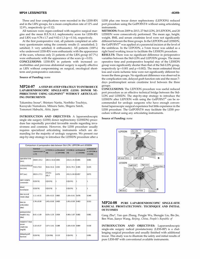
Three and four complications were recorded in the LESS-RNand in the LRN groups, for a mean complication rate of 12% and12.9%, respectively (p = 0.12).
All tumours were organ-confined with negative surgical mar-gins and the mean R.E.N.A.L nephrometry score for LESS-RNand LRN was 9.78 – 1.7 and 9.82 – 1.3 (p = 0.14), respectively.
At the first postoperative visit, all patients completed an arbi-trary questionnaire rating the cosmetic results (1: unsatisfied; 2:satisfied; 3: very satisfied; 4: enthusiastic). All patients (100%)who underwent LESS-RN were enthusiastic with the appearanceof the scars, whereas only 21 patients of the LRN group (67.7%)were enthusiastic with the appearance of the scars (p = 0.03).CONCLUSIONS: LESS-RN in patients with increased co-morbidities and previous abdominal surgery is equally effectiveas LRN without compromising on surgical, oncological short-term and postoperative outcomes.
Source of Funding: none
MP24-07 A STEP-BY-STEP STRATEGY TO INTRODUCE
LAPAROENDOSCOPIC SINGLE-SITE (LESS) DONOR NE-
PHRECTOMY USING GELPOINT� WITHOUT ARTICULAT-
ING INSTRUMENTS
Takamitsu Inoue*, Shintaro Narita, Norihiko Tsuchiya,Kazuyuki Numakura, Mitsuru Saito, Shigeru Satoh,Tomonori Habuchi, Akita, Japan
INTRODUCTION AND OBJECTIVES: A laparoendoscopicsingle site surgery (LESS) donor nephrectomy (LESSDN) proce-dure has reportedly provided favorable results regarding inva-siveness and cosmesis. However, the LESS procedure usuallyrequires specialized articulating instruments which are de-manding for the majority of urologic surgeons. We present ourstep-by-step strategy to introduce the LESSDN procedure after a
LESS plus one trocar donor nephrectomy (LEPODN) reducedport procedure using the GelPOINT� without using articulatinginstruments.METHODS: From 2009 to 2013, 27 Std-LDN, 20 LEPODN, and 20LESSDN were consecutively performed. The mean age, height,weight, BMI, and serum creatinine level were not significantlydifferent between the three groups. In the LEPODN and LESSDN,the GelPOINT� was applied at a pararectal incision at the level ofthe umbilicus. In the LEPODN, a 5 mm trocar was added as aright hand working trocar to facilitate the LESSDN procedure.RESULTS: There was no significant difference in perioperativevariables between the Std-LDN and LEPODN groups. The meanoperative time and postoperative hospital stay of the LESSDNgroup were significantly shorter than that of the Std-LDN group,respectively (p = 0.001 and p = 0.002). The mean estimated bloodloss and warm ischemic time were not significantly different be-tween the three groups. No significant difference was observed inthe complication rate, delayed graft function rate and the mean 7-days posttransplant serum creatinine level between the threegroups.CONCLUSIONS: The LEPODN procedure was useful reducedport procedure as an effective technical bridge between the Std-LDN and LESSDN. The step-by-step strategy to introduce theLESSDN after LEPODN with using the GelPOINT� can be re-commended for urologic surgeons who have enough conven-tional laparoscopic surgical experience but little experience in theLESS procedure. The GelPOINT� may facilitate the LESS pro-cedure without using any articulating instruments.
Source of Funding: none
MP24-08 PURE LAPAROENDOSCOPIC SINGLE-SITE
RADICAL PROSTATECTOMY: TECHNIQUE AND INITIAL
OUTCOMES
Gang Zhu*, Yan qun Zhang, Pengjie Wu, Shengjie Liu, Bin Jin,Ben Wan, Jianye Wang, Beijing, China, People’s Republic of
INTRODUCTION AND OBJECTIVES: Laparoendoscopicsingle-site surgery radical prostatectomy (LESS-RP) is a chal-lenging surgical procedure and usually finished with additionaltrocar. This study was to illustrate the safety and initial results ofpure LESS-RP with conventional available instruments.
MP24 LESS/NOTES -A349-

METHODS: A prospective clinical database was established sinceSept. 2010 to assess the outcome following the introduction ofLESS-RP at our institution. By Apr. 2013, 12 procedures had beenperformed. The mean patient age was 72.0 (59.0̈C76.0) years. Themean body mass index (BMI) was 24.0 (19.5̈C32.2) kg/m2. The firstsix LESS-RP cases were performed through an extra-peritonealapproach and the next six cases were performed through a trans-peritoneal approach by using single port device of QudaPort,5 mm flexible tip video-laparoscope 0o lens and conventionalstraight laparoscopic instruments. Parameters assessed were op-erative time, estimated blood loss, intra-operative complications,drainage time, post-operative pain score (VAPS, 0̈C10), patholog-ical results, and postoperative PSA levels.RESULTS: LESS-RP was completed for all 12 cases withoutadditional trocar placement or conversion to standard laparo-scopic or open radical prostatectomy. The mean operativetimes were 263.9 (190–310) min, estimated blood loss was 326.7(40–1500) ml. There were 3 transfusion cases documented asintraoperative complication. The mean wound-drainage timewas 5.1 (2.0̈C7.0) d, and the first postoperative day VAPS was1.0 (0̈C3). Final pathological staging was pT2aN0M0 in 4 cases,pT2bN0M0 in 1 case, pT2cN0M0 in 2 cases and pT3aN0M0 in5 cases. 2/12 cases had positive surgical margins. The one-month post-operative PSA was lower than 0. 2 ng/ml in allcases. The first six patients were continent without pad usageat 1 year postoperatively and the other six patients were still infollow-up.CONCLUSIONS: The technique of pure LESS-RP is feasible andsafe, and early outcomes are acceptable.
Source of Funding: none
MP24-09 TRANSVAGINAL NATURAL ORIFICE TRANS-
LUMENAL ENDOSCOPIC SURGERY (NOTES)-ASSISTED
VERSUS CONVENTIONAL LAPAROSCOPIC NEPHRECTOMY:
A COMPARISON OF PERIOPERATIVE OUTCOMES AND
SHORT-TERM MEASURES OF CONVALESCENCE
Xiaofeng Zou*, Guoxi Zhang, Yijun Xue, Yuanhu Yuan, Rihai Xiao,Gengqing Wu, Xiaoning Wang, Dazhi Long, Yuting Wu, Min Liu,Jun Yang, Zhong Xin, Ganzhou, China, People’s Republic of
INTRODUCTION AND OBJECTIVES: To present a compari-son between transvaginal natural orifice translumenal endo-scopic surgery (NOTES)-assisted laparoscopic nephrectomy(TNALN) and conventional transperitoneal laparoscopic ne-phrectomy (CTLN) with respect to perioperative outcomes andshort-term measures of convalescence.METHODS: This was a study comparing 60 TNALNs and 40CTLNs performed from September 2010 to July 2012. A singlesurgeon performed all TNALN cases using two umbilical trocarsand one transvaginal trocar. Patient characteristics, perioperativedetails, and time to return to work were recorded. Postoperativeevaluation of pain and use of analgesic medication were re-corded. In the TNALN group, sexual function was assessed withthe Female Sexual Function Index questionnaire before and aftersurgery. To investigate the satisfaction of patients in terms ofcosmetic results, the Patient Scar Assessment Questionnaire(PSAQ) was introduced after postoperative month 3.RESULTS: Median patient age was 48 years for both groups.There was no difference in median operative time (128 min vs121 min, P = 0.78), blood loss (50.71 vs 47.15 mL, P = 0.60), trans-fusion rates (0% for both), and hospitalization time (2.07 vs 2.11days, P = 0.74) between the TNALN and CTLN groups. Time toreturn to normal activities was shorter in the TNALN group
compared with the CTLN group (10.2 vs 13.6 days, P = 0.001).Both the visual analogue scale and the postoperative use of an-algesics were significantly lower during postoperative days 1, 2,and 3 in patients who underwent TNALN, compared with pa-tients who underwent CTLN. There were no intraoperative orpostoperative complications in both groups. There was no sig-nificant difference between median pre- and postoperative Fe-male Sexual Function Index (FSFI) total scores in TNALN cases(33.3 vs. 32.7, P = 0.203). The median Patient Scar AssessmentQuestionnaire (PSAQ) was less in the TNALN group comparedwith the CTLN group (33.7 vs. 62.1, P = 0.001).CONCLUSIONS: The early experience described in this studysuggests that TNALN is a safe and effective alternative to CTLNthat provides surgeons with a minimally invasive surgical op-tion and the ability to hide the surgical incision within the um-bilicus; however, a larger series is necessary to confirm thesefindings and to determine if there are any benefits in pain, re-covery, or cosmesis.
Source of Funding: This work was supported by the NationalHigh Technology Research and Development Program (863Program) of China (NO. SQ2011AAJY2801), Important Scienceand Technology Project from the Department of Science andTechnology of Jiangxi Province (20121BBG70032), and ImportantScience and Technology Project from the Department of Health ofJiangxi Province (NO. 20104008)
MP24-10 SUPRAPUBIC-ASSISTED LAPAROENDOSCO-
PIC SINGLE-SITE VERSUS CONVENTIONAL LAPARO-
SCOPIC NEPHRECTOMY
Guoxi Zhang, Xiaofeng Zou*, Xiaoning Wang, Yuanhu Yuan,Rihai Xiao, Gengqing Wu, Yijun Xue, Dazhi Long, Yuting Wu,Min Liu, Ruiquan Xu, Yuhua Zou, Ganzhou, China,People’s Republic of
INTRODUCTION AND OBJECTIVES: Laparoendoscopicsingle-site surgery (LESS) represents an evolution of laparos-copy for nephrectomy. This study aimed to present a comparisonbetween suprapubic-assisted laparoendoscopic single-site ne-phrectomy (SLSN) and conventional transperitoneal laparoscopicnephrectomy (CTLN) with respect to perioperative outcomes andshort-term measures of convalescence.METHODS: This was a study comparing 50 SLSNs and 40CTLNs performed from September 2010 to December 2012. InSLSNs, one 5- and 10-mm trocars were inserted at the medialmargin of umbilicus. A 5-mm trocar was inserted into abdominalcavity below the ipsilateral pubic hairline. The technique for theSLSN is similar to that of CTLN, with conventional instrumentsplaced in the abdominal trocars, under direct vision achieved by5-mm flexible-tip 0�laparoscope placed through the trocar belowthe pubic hairline. Patient characteristics, perioperative details,and time to return to work were recorded. Postoperative evalu-ation of pain and use of analgesic medication were recorded. Toinvestigate the satisfaction of patients in terms of cosmetic results,the Patient Scar Assessment Questionnaire (PSAQ) was intro-duced after postoperative month 3.RESULTS: Median patient age was 46.3 years for both groups.Nephrectomy was performed for benign disease in 68.6% of thecases. There was no difference in median operative time (120 minvs 115 min, P > 0.05), blood loss (80 vs 75 mL, P > 0.05), transfusionrates (0% for both), and hospitalization time (6.5 vs 7 days,P > 0.05) between the SLSN and CTLN groups. Time to return tonormal activities was shorter in the SLSN group compared withthe CTLN group (16 vs 27 days, P = 0.006). Both the visual
-A350- MP24 LESS/NOTES

analogue scale and the postoperative use of analgesics were sig-nificantly lower during postoperative days 1, 2, and 3 in patientswho underwent SLSN, compared with patients who underwentCTLN (P < 0.05). The median Patient Scar Assessment Ques-tionnaire (PSAQ) was less in the SLSN group compared with theCTLN group (40.6 vs. 63.5, P = 0.001).CONCLUSIONS: The early experience described in this studysuggests that SLSN is a safe and effective alternative to CTLN thatprovides surgeons with a minimally invasive surgical option andthe ability to hide the surgical incision within the umbilicus andthe pubic hairs.
Source of Funding: This work was supported by the NationalHigh Technology Research and Development Program (863Program) of China (NO. SQ2011AAJY2801), Important Scienceand Technology Project from the Department of Science andTechnology of Jiangxi Province (20121BBG70032), and ImportantScience and Technology Project from the Department of Health ofJiangxi Province (NO. 20104008)
MP24-11 PURE TRANSURETHRAL NATURAL OR-
IFICE TRANSLUMENAL ENDOSCOPIC SURGERY (NOTES)
FOR FENESTRATION AND DRAINAGE TREATMENT OF
RENAL CYST: REPORT OF THREE CASES
Guoxi Zhang, Hui Xu, Xiaofeng Zou*, Min Liu, Yuanhu Yuan,Rihai Xiao, Gengqing Wu, Xiaoning Wang, Yijun Xue,Dazhi Long, Bo Jiang, Ruiquan Xu, Ganzhou, China,People’s Republic of
INTRODUCTION AND OBJECTIVES: To describe the initialclinical experience of pure transurethral NOTES for fenestrationand drainage treatment of renal cyst, and evaluate its feasibilityand safety.METHODS: From May to August 2012£:two female patientsand a male patient with renal cyst underwent pure TransurethralNOTES for fenestration and drainage treatment. The median agewas 24 (range 22 to 40) years. In this group, one renal cyst was onthe right side and two on the left. All cases were confirmed by Bultrasound, CT scan and IVU. The median diameter of renal cystwas 6.2£̈range 5.2 to 7.1£ªcm. After induction of general anes-thesia, flexible ureter scope was transurethrally introduced intorenal collect system. Renal cyst was found by B ultrasound guidewhen necessary. A crisscross incision was made by Holmiumlaser to 1.5*2.0 cm, and a 7 F double J stent was positioned withthe proximal end coiled in the cyst cavity, which was removedafter 4 weeks.RESULTS: All procedures were successfully completed. One cystwas found by flexible ureter scope and the others were found by Bultrasound guide. The median operation time was 60 (range 30 to125) min. There were no intraoperative or postoperative compli-cations. The diameter of cyst decreased at least 1/2 during follow-up (3 to 6 months).CONCLUSIONS: Pure transurethral NOTES for fenestration anddrainage treatment of renal cyst is safe, feasible, minimal invasiveand cosmetic. It is worth selecting the method to treat peripheralrenal cyst.
Source of Funding: This work was supported by the NationalHigh Technology Research and Development Program (863Program) of China (NO. SQ2011AAJY2801), Important Scienceand Technology Project from the Department of Science andTechnology of Jiangxi Province (20121BBG70032), and ImportantScience and Technology Project from the Department of Health ofJiangxi Province (NO. 20104008)
MP24-12 COMPARISON OF POSTOPERATIVE STRESS
IN PATIENTS UNDERGOING TRANSVAGINAL NOTES-
ASSITED LAPAROSCOPIC AND STANDARD LAPARO-
SCOPIC NEPHRECTOMY
Li Chen, Guoxi Zhang, Xiaofeng Zou*, Yuanhu Yuan,Rihai Xiao, Gengqing Wu, Xiaoning Wang, Yijun Xue,Dazhi Long, Yuting Wu, Hui Xu, Ju Yang, Min Liu,Ganzhou, China, People’s Republic of
INTRODUCTION AND OBJECTIVES: Few studies are avail-able to compare the potential benefits of natural orifice translu-menal endoscopic surgery (NOTES) to standard laparoscopy. Weaimed to compare surgical stress between transvaginal NOTES-assisted laparoscopic and standard laparoscopic nephrectomy.METHODS: Twenty female patients who underwent transva-ginal NOTES-assited laparoscopic nephrectomy (TNLN) at theinitial stage (group N1), 20 female patients who underwentTNLN at the later stage (group N2), and two groups (group L1and group L2) of patients who underwent standard laparoscopicnephrectomy (SLN) at the same time, were selected to the study.The same general anesthesia induction, continuous anesthesiaand postoperative analgesia were used in the four groups. Bloodsamples were obtained before general anesthesia induction (T0),at the end of the surgery (T1), and at six o’clock on postoperativeday 1 (T2), postoperative day 2 (T3) and postoperative day 3 (T4).The serum levels of glucose, cortisol, IL-6, and C-reactive proteinwere detected in each blood sample.RESULTS: In group N1 and L1, the serum glucose concentrationsat T1, T2 and T3 and the serum C-reactive protein concentration atT2, T3 and T4 were all higher than that at T0 (P < 0.05), but therewere no statistically significant differences between the twogroups (P > 0.05). The serum cortisol and IL-6 concentrations atT1, T2 and T3 were all higher than that at T0 (P < 0.05). The pa-tients in group N1 had increases in serum cortisol and IL-6 con-centrations at T1 compared with group L1, but there were nostatistically significant differences at T2 and T3 between the twogroups (P > 0.05). The operative time for N1 was longer than thatof L1. In group N2 and L2, the serum glucose, cortisol and IL-6concentrations at T1, T2 and T3 and the serum C-reactive proteinconcentration at T2, T3 and T4 were all higher than that at T0(P < 0.05), but there were no statistically significant differencesbetween the two groups (P > 0.05). There was also no statisticallysignificant difference in operative time between the two groups.CONCLUSIONS: With the development of TNLN and theshortening of operative time, the postoperative stress in TNLNwill be similar to that of SLN.
Source of Funding: This work was supported by the NationalHigh Technology Research and Development Program (863Program) of China (NO. SQ2011AAJY2801), Important Scienceand Technology Project from the Department of Science andTechnology of Jiangxi Province (20121BBG70032), and ImportantScience and Technology Project from the Department of Health ofJiangxi Province (NO. 20104008)
MP24-13 LAPAROENDOSCOPIC SINGLE-SITE RADI-
CAL NEPHRECTOMY BY SINGLE-CUP OR SINGLE-RING
GLOVE TECHNIQUE
Zhang Shudong*, Ma Lulin, Qiu Min, Bi Hai, Beijing, China,People’s Republic of
INTRODUCTION AND OBJECTIVES: With the developmentof minimally invasive surgical techniques, LESS has been widely
MP24 LESS/NOTES -A351-

applied in the fields of urology. Compared with the traditionallaparoscopic technique, LESS may have better results in cosmetic,less postoperative pain, better recovery and so on. We present twonew homemade single-site devices which had been successfullyused in radical nephrectomy and other urological surgery. Aim ofthis study is to evaluate the efficacy and safety of laparoendoscopicsingle-site radical nephrectomy by homemade devices.METHODS: The clinical data of laparoscopic radical nephrectomyperformed from June 2010 to April 2013 in Peking University ThirdHospital were analyzed retrospectively. 17 cases underwent LESSradical nephrectomy and 34 cases received retroperitoneal lapa-roscopic radical nephrectomy. Data on general presentation, tumorsize, tumor location, operative time, blood loss, complications,Visual Analog Pain Scale (VAPS), postoperative hospital stay,pathological results were collected to compare between twogroups. Our homemade equipments are composed of Single-Cupdevice (inter-diameter of 2.5–5.5 cm, peripheral diameter of 4.0–6.0 cm, and 4.0 cm high) or Single-Ring device (inter-diameter of4 cm, peripheral diameter of 5 cm) and a 6 F sterile surgical glove.The thumb, middle, and ring fingers of the glove were implantedand fixed with 11 mm, 5 mm and 5 mm diameters¡�trocar respec-tively. the kidney was dissociated after cut off the renal vessel andextracted through the umbilical incision. The retroperitoneal ap-proach followed the standard surgical procedures, the specimenswere removed from the extended incision.RESULTS: All procedures were completed without conversion toopen radical nephrectomy. Compare with traditional laparo-scopic surgery, operative time (P < 0.05) and VAPS (P < 0.05) showsignificant difference in LESS group£:and no difference wasnoted in other factors (P > 0.05). There was no secondary bleeding,wound infection, intestinal obstruction, incision hernia and othersevere postoperative complication. Follow-up of 2 to 36 monthsshows no local recurrence.CONCLUSIONS: laparoendoscopic single-site radical nephrec-tomy by Single-Cup or Single-Ring glove technique is feasible,effective and safe. It gives a more mini-invasive and cosmeticoption for young or female patients. The characteristics of lower-cost, more aesthetic, repeat use of the homemade devices ap-peared o be popularized. Further experience and long termevaluation are required.
Source of Funding: Peking University Third Hospital ClinicalEssential Programme ‘‘Application of Transumbilical Lapar-oendoscopic Single-site Surgery in Urologic Oncology’’ (61446–04)
MP24-14 SIMULTANEOUS LAPAROENDOSCOPIC
SINGLE-SITE SURGERY FOR SURGICAL TREATMENT OF
INTRA-ABDOMINAL PATHOLOGIES IN TWO DIFFERENT
ORGANS
Kwang Taek Kim*, Incheon, Korea, Republic of, Yong Hyun Park,Seung-Yong Jeong, Seoul, Korea, Republic of, Sang Jin Yoon,Chang Hee Kim, Incheon, Korea, Republic of, Hyeon Hoe Kim,Seoul, Korea, Republic of
INTRODUCTION AND OBJECTIVES: To describe our earlyexperience about with simultaneous laparoendoscopic single-sitesurgery (LESS) for surgical treatment of intra-abdominal pa-thologies in two different organs.METHODS: Five patients with bilateral renal cell carcinoma(RCC) (n = 2), bilateral aldosterone-producing adrenal adenoma(n = 1), right proximal ureter stone and left non-functioning kid-ney (n = 1), and synchronous left RCC and sigmoid colon cancer(n = 1) underwent simultaneous LESS using with use of the um-bilicus as the portal of entry. Perioperative outcomes including
operative time, estimated blood loss, and complications wereanalyzed retrospectively.RESULTS: All procedures were completed successfully withoutconversion to conventional laparoscopic or open surgery andwithin a reasonable operative time. Estimated blood loss wasminimal and no perioperative complications occurred.CONCLUSIONS: In our experience, We reported the technicalfeasibility and safety of simultaneous LESS of intra-abdominalpathologies in two different organs was technically feasible andsafe. The procedure It could be performed with minimal mor-bidity and obvious cosmetic advantage.
Source of Funding: none
MP24-15 LAPAROENDOSCOPIC SINGLE-SITE SUR-
GERY (LESS) FOR TREATMENT OF DIFFERENT UROLOGIC
PATHOLOGIES IN PEDIATRICS: SINGLE-CENTER EXPERI-
ENCE
Aly Abdel-Karim*, Ashraf Saad, Moussa Ahmed,Ahmed Abolfotof, Mostafa Elmissery, Haytham Badawy,Salah Elsalmy, Alexandria, Egypt
INTRODUCTION AND OBJECTIVES: Laparoendoscopic sin-gle-site surgery (LESS) has been increasingly reported as a newless invasive approach for treatment of various urologic pathol-ogies in adults. However, the applications of LESS in the field ofpediatric urology are still limited. In this study we present ourinitial experience with LESS as an option for treatment of somepediatric urologic pathologies.METHODS: This is a retrospective study that included 14 pedi-atric patients with a mean age 6.2 – 2.9 years. Four patients hadunilateral undescended testes, 3 with bilateral undescended tes-tes, 2 with left and one with bilateral varicocele, one with rightmulticystic dysplastic kidney, one patient with non-functioningright kidney and two patients with left ureteropelvic junctionobstruction (UPJO). Ports used were R-port (10 patients) andCovieden port (4 patients) and ports were inserted through theumbilicus. We used pre-bent (with R-port), articulating (withCovieden port) as well as straight instruments. In patient with leftUPJO, one had LESS dismembered pyeloplasty and other had Y-V plasty where hand free intracorporeal suturing was done using4/0 vicryl sutures.RESULTS: In all patients there was no conversion to conven-tional laparoscopy or open surgery. We did not add any extraportin any of the patients. Mean operative time was 112 – 40 minutes.Mean blood loss was 40 – 25 c.c. No intraoperative or postoper-ative complications were reported. Mean hospital stay was1.2 – 0.7 days. Mean duration of follow-up was 18 – 6 months.Patients with unilateral undescended testes; 2 had orcheidopexy,one had first stage Fawler-Stephens, and one had orcheidectomy.
Table 1. Patients Characteristics
-A352- MP24 LESS/NOTES

Patients with bilateral undescended testicles; one had bilteralorcheidopexy, one had right orcheidopexy and left first stageFawler-Stephens and third had bilateral first stage Fawler-Stephens. All patients who had first stage Fawler-Stephensorcheidopexy subsequently had LESS second stage orcheidopexyafter 6 months. Follow-up of patients with left UPJO showedcomplete clinical cure and patent ureteropelvic junction andmarked decrease of left hydronephrosis. In all patients umbilicalscar was invisible and patients had high wound satisfaction.CONCLUSIONS: LESS is a feasible, effective and safe option fortreatment of many urologic pathologies in pediatrics. However,more patients and indications are need for further evaluation ofrole of LESS in pediatric urology.
Source of Funding: None
MP24-16 STEP-WISE ‘EXIT STRATEGIES’ DURING
LAPAROENDOSCOPIC SINGLE SITE LIVE DONOR NE-
PHRECTOMY—EXPERIENCE SHARING
Shih-Chieh Chueh*, Cleveland, OH, Bashir Sankari, Abu Dhabi,United Arab Emirates, J Stephen Jones, Cleveland, OH
INTRODUCTION AND OBJECTIVES: Laparoendoscopic sin-gle site-live donor nephrectomy (LESS-LDN) is very challengingdue to its nature of not allowing any slightest errors. We share ourexperience of LESS-LDN; and, if difficulties ever encountered, ourdesign how to step-wisely convert to conventional laparoscopicLDN or open surgery for safe execution of this highly demandingsurgery.METHODS: GelPort device was installed as the LESS platform ata 5 cm peri-umbilical midline incision. A large adhesive drapewas applied over the abdomen, covering the whole GelPort (Fig.1). Through the gel-cap of GelPort 3*4 ports were spaced apart toachieve triangulation and to avoid clashing of the instrumentsduring LESS-LDN (Fig. 1). Standard dissection of the kidney fordonation was performed by abiding by the principles to ensurethe kidney was well perfused and the renal vein was nicely dis-tended (Fig. 2). Before hilar vessels were ligated separately withEndo TA staplers, an EndoCatch-II was inserted through gel-capto bag the kidney for tenting up the vessels, so that longer pedicle
can be harvested. The kidney was removed through the LESSincision immediately after hilar transection.RESULTS: LESS-LDN was successful in all living donors withoutany multi-site laparoscopic or open conversion and without anyinstrument through extra wounds—although we would havedone so if at any point we felt that this would have providedadditional safety or facilitated the operation. No patient had peri-operative complications or received transfusions. Median oper-ative time was 271 minutes with a warm ischemia time of 3.5minutes. Hospital stay averaged 2 days with visual analog painscore 4/10 at discharge and 2/10 at 2 weeks. All recipients re-cuperated well with immediate graft function.CONCLUSIONS: LESS-LDN using the GelPort device as theaccess platform provides feasibility and safety, and most impor-tantly offers ‘exit strategies’ for an easy conversion potential incase of difficulty, to maintain the extremely high safety standardof LDN. This is especially useful for the beginners.
Source of Funding: none
MP24-17 SUBJECTIVE SCAR APPEARANCE AND
SEXUAL SATISFACTION IN TRANSVAGINAL NOTES AS-
SISTED LAPAROSCOPIC RADICAL CYSTECTOMY
Giovannalberto Pini*, Nasreldin Mohammed, Francesco Greco,Vincenzo Maria Altieri, Paolo Fornara, Halle, Germany
INTRODUCTION AND OBJECTIVES: To evaluate the sexualsatisfaction and cosmetic outcomes after transvaginal (Tv) natu-ral orifice translumenal endoscopic surgery (NOTES) assistedlaparoscopic radical cystectomy (LRC) in patients affected bymuscle-invasive urothelial carcinoma of the bladder (MITCC).METHODS: From December 2011 and February 2013, 5 femalepatients with non-metastatic MITCC underneath to Tv-LRC. Thegroup was matched for age, body mass index, number of deliv-eries, ASA score, Charlson comorbidity index, tumour charac-teristics with two groups-control including patients whounderwent open radical cystectomy (ORC: 10 patients) and lap-aroscopic (LRC: 10 patients).
Demographic and intra- and postoperative variables were an-alyzed. Moreover, all patients were evaluated for sexual activitysatisfaction (Female Sexual Function Index - FSFI) and subjectivescar appearance.RESULTS: Mean operating time, Hb drop, day of mobilization,retrieved lymphnodes and length of stay were statistically com-parable in all group. Blood lost where in favor of Tv-LRC com-pared to ORC. No major intraoperative complication occurred.No postoperative vaginal fistula or infection where notice in TV-LRC and number of complications was comparable in all groups;Clavien 3a and 3b were more frequent in LRC (33.3%) and ORC(40%).
At discharge the median VAS was 2.0 – 0.8 comparable withother group.
The median scar satisfaction in Tv-LRC (3.4 – 0.5) was signifi-cantly higher than ORC (1.8 – 0.8; p 0.012) and LRC (2.6 – 0.5; p0.032).
With different follow-up (Tv-LRC: 10.3 – 1.7; ORC: 29.2 – 15.1LRC: 21.4 – 7.1, p 0.01) all patients were alive. No vaginal or port-site metastasis where noticed in Tv-LRC and LRC. Two and onepatients of ORC and LRC respectively underneath chemotherapybecause of late lymphonodal relapse. Sexual active patientswhere 3, 4 and 5 in Tv-LRC, ORC and LRC. Postoperative sexualfunction was disturbed significantly (p < 0.03) by surgery in allgroups, with a mean decrease of FSFI index of 5.1 – 1.5, 6.7 – 1.3and 5.5 – 0.9 for Tv-LRC, ORC and LRC respectively, affecting
MP24 LESS/NOTES -A353-

specially lubrication and ability to achieve orgasm, while arousaland dyspareunia remained stable.CONCLUSIONS: The transvaginal approach could be consid-ered as viable minimally invasive option in case of LRC per-formed in middle aged-old multiparous woman. It improvescosmetic results, it is not related to vaginal complication or me-tastasis and does not affect directly sexual satisfaction, ratherworstened by cystectomy itself. Proper selection of patients iswarranted for success.
Source of Funding: none
MP24-18 IS THERE INCONGRUENCE BETWEEN THE
GOLD STANDARD APPROACH FOR CT1 RENAL MASSES -
NEPHRON SPARING SURGERY - AND DIAGNOSIS RE-
LATED GROUPS (DRG) REIMBURSEMENT? ANALYSIS OF
INTRAOPERATIVE COSTS FOR LAPAROENDOSCOPIC
SINGLE-SITE (LESS), LAPAROSCOPIC AND OPEN AP-
PROACH IN A HIGH VOLUME CENTER
Giovannalberto Pini*, Ascalone Luigi, Francesco Greco,Paolo Fornara, Halle, Germany
INTRODUCTION AND OBJECTIVES: Nephron sparing sur-gery (NSS) has become the standard treatment for small masses(SM) and laparoscopy is strongly supported in experienced andhigh volume center. Anyway, both are not adequately adopt be-cause complexity and inappropriate support by the national re-imbursement.METHODS: In a non-randomised retrospective study we se-lected 90 consecutive pat. underwent (01/2010–12/2011) to par-tial (P) or radical (R) nephrectomy for SM ( < 7 cm-cT1N0M0) anddivided in 6 groups: laparoscopic (LPN, LRN), LESS (LESS-PN,LESS-RN) or open (OPN, ORN). Patients were matched for age,sex, body mass index (BMI), ASA-score, Charlson score, tumorside (right/left) and tumor characteristics (PADUA Score). Pri-mary endpoints were evaluation of total intraoperative materialcost (disposable/reusable instruments) divided in general, lapa-roscopic, sutures and hemostatic agents; 2nd endpoints weredemographic, peri- and postoperative variables.RESULTS: Costs: Total costs where higher for laparoscopic(LRN: 2276 – 443 e; LPN: 2202 – 797) and LESS (LESS-RN:1819 – 322; LESS-PN: 1583 – 116,7) compared to Open (OPN:612 – 246.4, ORN: 168 – 82.7, P < .0001). Laparoscopic disposablematerials incised mainly in total costs in minimally invasiveprocedure (LRN 95.9 % – 3.4; LPN 85.7 % – 5.2; LESS-RN:92.9 – 4.6%; LESS-PN: 82.9 – 7.1%) and most cheap in LESS be-cause reusable single-port-trocar (LESS-PN: 1312 – 131.4 e vsLRN: 2183 – 429.7, P < .0001). Sutures incised more in open (ORN45.2 – 14.6%; OPN: 12.6 – 5.8) than all other groups (P < .0001). Asexpected general costs where comparable in all group (RangeOPN:72.7 – 35.1e - LRN: 41 – 12.6). Hemostatic agents were moreapplied in OPN (OPN:472 – 227e, P < .0001) and this correlated tohigher Padua score.CONCLUSIONS: Use of reusable instruments can reduce totalcosts of laparoscopy that anyway results still too expensive justifythe complex learning curve of LPN. The sample of patients re-ported is enough to reflect costs but not maybe to mirror the realcomplication rates. These, already provided by international lit-erature, may further increase the costs related to higher transfu-sion and surgical revision rates.
It is requested a subclassification with different reimbursementthat incentives nephronsparing and laparoscopic approach.
Source of Funding: none
MP24-19 PERIOPERATIVE NURSING OF PATIENTS
UNDERGOING PURE NATURAL ORIFICE TRANSLUME-
NAL ENDOSCOPIC SURGERY (NOTES) TRANSVAGINAL
NEPHRECTOMY
Lanying Liu, Meiying Meng, Lili Li, Xiaoying Zeng,Fangzhen Xiao, Yulan Huang, Guoxi Zhang, Xiaofeng Zou*,Ganzhou, China, People’s Republic of
INTRODUCTION AND OBJECTIVES: To investigate theperioperitive nursing during the pure natural orifice translume-nal endoscopic surgery (NOTES) transvaginal nephrectomy.METHODS: Sixteen female patients underwent the pure trans-vaginal NOTES nephrecomy. The experience in perioperativenursing during the surgeries were analyzed. The key points innursing were preoperative psychological nursing, sufficient pre-operative preparations, such as bowel preparation, vaginal prep-aration and skin preparation, close observation of vaginalbleeding, care of various drainage tubes prevention of abdominaldistension and intestinal obstruction£:comfortable nursing£:proper discharge education and follow- up services.RESULTS: All the 16 procedures were successfully accom-plished. One patient was converted to suprapubic-assisted la-paroendoscopic single-site surgery for rectum injury. The surgicalnursing during perioperative was successfully completed with nomajor error.CONCLUSIONS: Full preoperative preparations, good commu-nication with patient, close observation of vaginal bleeding, care ofvarious drainage tubes prevention of abdominal distension andintestinal obstruction, comfortable nursing, proper discharge ed-ucation and follow- up services, may improve the efficiency andoperative safety of pure transvaginal NOTES nephrectomy.
Source of Funding: This work was supported by the NationalHigh Technology Research and Development Program (863Program) of China (NO. SQ2011AAJY2801), Important Scienceand Technology Project from the Department of Science andTechnology of Jiangxi Province (20121BBG70032), and ImportantScience and Technology Project from the Department of Health ofJiangxi Province (NO. 20104008)
MP24-20 CLINICAL EXPERIENCE OF LAPAROENDO-
SCOPIC SINGLE-SITE BIOPSY FOR DIAGNOSIS OF RET-
ROPERITONEAL TUMOR OF UNKNOWN ORIGIN
Tetsuo Nozaki*, Akihiro Morii, Kenji Yasuda, Hideki Fuse,Toyama, Japan
INTRODUCTION AND OBJECTIVES: When percutaneousimage-guided biopsies are not possible for retroperitoneal tumor ofunknown origin (RTUO), surgical tissue diagnosis becomes nec-essary. Traditional open surgical exploration with deep tissue bi-opsy might lead to unnecessary perioperative morbidity andserious delay in patient recovery. In this investigation, we reportthe safety and feasibility of the laparoendoscopic single-site (LESS)biopsy for diagnosis of RTUO adjacent to critical structures.METHODS: Five patients underwent LESS biopsy for diagnosis ofRTUO, because the tumor was adjacent to important structures, suchas major blood vessels, the urinary tract, intestines, or other viscera.The EZ Access� (Hakko Medical Industry, Tokyo, Japan) was uti-lized as a single port and inserted into the peritoneal or retroperito-neal space through a 3-cm skin incision. Obtaining numerous deeptissue biopsies from multiple anatomic sites was performed.RESULTS: LESS biopsy was performed successfully and no pa-tients required conversion to laparotomy or standard laparoscopy
-A354- MP24 LESS/NOTES

because of difficulty in identifying the mass laparoscopically, in-ability to obtain an adequate tissue sample, or uncontrolledbleeding. Sufficient specimens were safely obtained not only forpathological diagnosis but also for further examinations such asimmunophenotyping or DNA analysis. The pathological diagnoseswere IgG4-related retroperitoneal fibrosis (n = 4) and diffuse B-celllymphoma (n = 1). Convalescence was satisfactory. All patientswere treated appropriately according to the pathological diagnosis.CONCLUSIONS: LESS biopsy for RTUO is a safe and effectivealternative to open surgical and standard laparoscopic biopsies.Further studies of clinical experiences are needed to confirm thebenefits of this new technique.
Source of Funding: none
MP24-21 PRELIMINARY EXPERIENCE WITH TRANS-
PERITONEAL LAPAROENDOSCOPIC SINGLE-SITE RADI-
CAL NEPHRECTOMY USING A HOME-MADE SINGLE-PORT
DEVICE IN CHINA
Bi Hai*, Ma Lu-lin, Hou Xiao-fei, Beijing, China, People’s Republic of
INTRODUCTION AND OBJECTIVES: To present our prelimi-nary experience with transperitoneal laparoendoscopic single-site(LESS) radical nephrectomy (RN) using a home-made single-portdevice in China.METHODS: From July 2010 to November 2011, eleven patientswith renal tumor not greater than T2 underwent LESS-RN by anexperienced laparoscopic surgeon. A home-made single-port de-vice was used through a 5-cm umbilical incision. A combination ofstandard and articulating laparoscopic instruments was used. Thesequence of steps of LESS-RN was similar to transperitoneal lap-aroscopic RN. Patient characteristics, perioperative variables andpostoperative outcomes were recorded and analyzed.RESULTS: Except for two transperitoneal laparoscopic conver-sions and one hand-assisted laparoscopic conversion, the otherprocedures were completed successfully, without conversion toopen surgery. The mean operative time was 224.5 (155–297)minutes, estimated blood loss 270.9 (50–900) ml, and hospital stay10.4 (5–15) days. The mean visual analogue pain scale (VAPS) onthe first postoperative day was 4.0/10. Final pathological analysisrevealed renal cell carcinoma in all cases with a stage distributionof three T1a, five T1b and three T2a tumors. With the mean fol-low-up period of 21.4 (12–28) months, all patients were alivewithout evidence of tumor recurrence or metastasis, and weresatisfied with the appearance of the scars.CONCLUSIONS: Transperitoneal LESS-RN using a home-madesingle-port device is technically feasible and safe in a selectedgroup of patients (low BMI and stage tumor) and has excellentcosmetic results. Although preliminary oncologic outcome isn¡�tcompromised, the long-term evaluation of these patients awaits.
Source of Funding: none
MP24-22 PURE LAPAROENDOSCOPIC SINGLE-SITE
RETROPERITONEOSCOPIC ADRENALECTOMY: TECHNI-
QUE AND EARLY OUTCOMES
Gang Zhu*, Shengjie Liu, Ya qun Zhang, Pengjie Wu,Jianlong Wang, Hong Ma, Ben Wan, Jianye Wang,Beijing, China, People’s Republic of
INTRODUCTION AND OBJECTIVES: To verify the safety andfeasibility of applying pure retroperitonal approach lapar-
oendoscopic single-site adrenalectomy (LESS-A) in the treatmentof adrenal gland tumors.METHODS: From October 2009 to February 2013, fifteen patientsacccepted LESS-A through retroperitonal approach in our insti-tute for the treatment of adrenal tumors. In LESS-A, a Quadportdevice was placed through a 3.0-to 3.5-cm transverse skin incisionbelow 12th rib tip into retroperitoneal space. The LESS-A waspreformed by using 0,,a lens 5 mm flexible tip video-laparoscopeand conventional laparoscopic instruments. We evaluated thistechnique in terms of operative time, estimated blood loss, peri-operative complications, drainage time, post-operative pain score(VAPS, 0¡V10) and pathological results.RESULTS: The 15 procedures in this group were completedsuccessfully with LESS-A. There was no additional trocar added,no conversion to conventional laparoscopic or open surgery.Application of Quadport reduced the instrument clash, both in-tracorporally and extracorporally. The average operative timewere 86.3 (60–155) min, average estimated blood loss were 38 (5–200) ml. There was no severe peri-operative complication. Theaverage VAPS in the first post-operative day was 1.8 (1–3). Theaverage drainage time was 1.7 (1–3) d, post-operative hospitalday was 3.8 (2–6) d. There was no secondary bleeding or woundinfection. Pathological results showed 1 case of adrenal pheo-chromocytoma, 11 cases of adrenal cortical adenoma, 1 case ofadrenal myelolipoma, 1 case of adrenal cortical nodular hyper-plasia and 1 case of adrenal cyst.CONCLUSIONS: The retroperitonal approach LESS-A is feasibleand safe. It offers a superior cosmetic outcomes and short con-valescence, albeit with a longer operative time than conventionallaparoscopic adrenalectomy.
Source of Funding: none
MP24-23 OPTIMAL INSTRUMENT LENGTH FOR TRA-
NSUMBILICAL LAPARO-ENDOSCOPIC SINGLE SITE (LESS)
SURGERY
Justin Houman*, Scott Tobis, Changyong Feng, Vikram Dogra,Guan Wu, Rochester, NY
INTRODUCTION AND OBJECTIVES: The use of traditionalmulti-port instrumentation in transumbilical LESS surgery oftenlimits surgeon’s ability to manipulate within the desired field. Inthis series, we attempt to quantify the distance between the um-bilicus and the following: xyphoid process, superior aspect ofspleen, gallbladder neck, bifurcation of descending aorta, left andright adrenal glands, left and right superior poles of kidneys,suprapubic margin, and apex of the prostate. In doing so, this willhelp in the design of robotic and laparoscopic instrumentationthat is better suited for LESS surgery.METHODS: In December 2012, abdominal and pelvic CT scansfrom 100 consecutive patients (50 males and 50 females) at ourinstitution were obtained. The distance from the umbilicus to anintra-abdominal point of interest was calculated using two tri-angles, triangle ABD (dark grey) and BCD (light grey). Point A1represents the position of umbilicus without pneumoperitonum,and Point A represents the position of the umbilicus displacedafter insufflation. At 15 mmHg the distance between these twopoints is an average of 6 cm. Point A to D is the distance betweenthe umbilicus to an intra-abdominal point of interest. We usedpatient abdominal and pelvic CT scans to measure distances be-tween A1 and B, B and C, C and D. As in Figure 1, based onPythagorean’s theorem (c2 = a2 + b2), we calculated BD based onBC and DC, then calculated AD based on BD and AB.
MP24 LESS/NOTES -A355-

RESULTS: Statistical analysis of umbilicus to organ distances islisted in Table 1.CONCLUSIONS: The described calculations for measuring in-tra-abdominal distances provide surgeons with an accuratemethod to calculate the necessary instrument length prior totransumbilical LESS surgery. In our study population, we foundthat increasing the instrument’s working length to 35 cm wouldbe sufficient for all of our patients to be able to undergo trans-umbilical LESS surgery to the measured points of interest.
Source of Funding: none
MP24-24 LAPAROENDOSCOPIC SIMULTANEOUS DRA-
INAGE OF INTRAABDOMINAL ABSCESS AND PERITONEAL
DIALYSIS CATHETER INSERTION
Thomas Y. Hsueh*, Yi-Shen Lin, Allen W. Chiu, Taipei, Taiwan
INTRODUCTION AND OBJECTIVES: A 68 years old malepresented to our clinic due to umbilical cellulitis with abscessformation he was a case of liver cirrhosis with massive ascites and
about 1500 ml ascites was drained everyday. The patient was alsoa case of chronnic renal failure under regular hemodialysis.Leukocytosis was noted and peritoneal sign was developedduring hospitalization. Abdominal ultrasound-guided tappingshowed turbid fluid with elevated white blood cell counts.METHODS: Laparoendoscopic single site surgery was conductedfor this patient. Under general anesthesia, the patietn was put insupine position and excision the umbilicus to remove original in-fectious site was done. laparoendoscopic single site drainage ofintraabdominal abscess was performed and the abdomianl cavitywas irrigated with copiuos amounts of normal saline. Simultaneousinsertion of peritoneal dialysis catheter was performed for perito-neal dialysis and drainage of massive ascites due to liver cirrhosis.RESULTS: Total operative time was 65 minutes. Tital blood losswas 30 ml. Total drained ascites with pus was 9600 ml. The pa-tient received intravenous antibiotics use for 14 days after oper-ation. Convalescence was uneventful and he was dischargedunder stable condition.CONCLUSIONS: Laparoendoscopic single site surgery was firstreported in Urology in 2007. Various procedures were reported inthe past 5 years t, ranging from benign to malignant disease andfrom destructive to reconstructive procedures. To our knowl-edge, there was no previous reports regarding the application oflaparoendoscopic single site surgery in intraabdominal abscessdrainage. In our preliminary experience, the application of la-paroendoscopic single site surgery might be one of the treatmentoptions for selected patients requiring intraabdominal abscessdrainage with simultaneous peritonela dialysis catheter insertion.
Source of Funding: None
MP24-25 LONGTERM RESULTS AFTER TWELVE
YEARS OF RETROPERITONEOSCOPIC PYELOPLASTY
FOR THE TREATMENT OF URETEROPELVIC JUNCTION
OBSTRUCTION
Svetozar Subotic, Armin Halla*, Antje Feicke, Georg Müller,Basel, Switzerland, Thomas Gasser, Liestal, Switzerland,Stephen Wyler, Alexander Bachmann, Basel, Switzerland
INTRODUCTION AND OBJECTIVES: To evaluate our resultsafter 12 years of experience with retroperitoneoscopic pyeloplasty.METHODS: From July 2001 to July 2013 retroperitoneoscopicpyeloplasty was performed in 139 patients with ureteropelvicjunction obstruction. The average age was 44 years (range 15 to86) with 84 (60.4 %) females and 55 males. An Anderson-Hynespyeloplasty was performed in 134 patients (96.4 %), a Fengertechnique was performed in 5 cases (3.6 %). The mean follow-uptime was 45 months (range 1 to 136).RESULTS: The mean operation time was 147 min (55- 360) with amean blood loss of 83 cc (30- 600). In 3 cases (2.2 %) conversion toopen surgery was necessary because of severe fibrosis or adhe-sions. The transurethral catheter was removed after 4.13 days (1-18) and the patients were discharged after 8 days (4- 39). Post-operative complications were registered in 15 patients (11.4 %)with grade I (n = 1; 0.7 %), II (n = 7; 5.0 %) and IIIb (n = 7; 5.0 %) inthe Clavien-Dindo classification. The overall success rate was93.5%. Nine patients (6.5 %) had ureteropelvic junction re-ob-struction after an average time of 23.1 months (1- 118). These weresuccessfully treated with an endopyelotomy, open pyeloplastyand retroperitoneoscopic redo-pyeloplasty.CONCLUSIONS: Retroperitoneoscopic pyeloplasty is a safe andeffective operation technique with low morbidity that providesexcellent long-term results.
Source of Funding: none
Statistical Analysis of Intra-Abdominal
Organ Distances
-A356- MP24 LESS/NOTES

MP25 PEDIATRICS
MP25-01 UTILIZATION AND OUTCOMES OF PEDIA-
TRIC PYELOPLASTY
Stacey Carter*, Lorna Kwon, Jim Hu, Los Angeles, CA
INTRODUCTION AND OBJECTIVES: Robotic assistance isgaining popularity among pediatric surgeons; however, uses,indications, and outcomes on a population level remain un-known. Our objective is to perform a comparative effectivenessstudy of commonly performed pediatric robotic surgeries using
Kid’s Inpatient Database (KID), a nationally representative in-patient sample from 44 states with public and private payors.METHODS: We identified 1,126 subjects who underwent robotic-assisted surgery during 2009 using the International Classificationfor Disease, 9th edition procedure code 17.4x. 42% were pyelo-plasties, the most commonly performed robotic procedure, while33% were miscellaneous Urologic procedures. We constructed re-gression models to determine factors associated with robotic vs.open and laparoscopic approaches and modeled factors associatedwith inpatient complications, length of stay (LoS), and costs.RESULTS: Open vs. robotic-assisted and laparoscopic pyelo-plasty was associated with younger age (p £ 0.001), female gender(p = 0.002), non-white race (p £ 0.001), fewer comorbidities(p = 0.003), lower income (p = 0.02), absence of private insurance(p £ 0.001) and teaching hospitals (p = 0.009). No pyeloplastymortalities were observed. In unadjusted analyses, open pyelo-plasty was associated with more complications compared to ro-botic and laparoscopic approaches (14% vs. 8% vs. 7%, p = 0.030).Less than 1% of pyeloplasties required transfusion, and this didnot vary by approach (p = 0.752). LoS was longest for open fol-lowed by laparoscopic and robotic approaches (Mean – standarddeviation 2.8 – 0.2 vs. 2.4 – 0.3 vs. 1.9 – 0.1, p < 0.001). In adjustedanalyses, robotic ( - 0.68 – 0.15, p < 0.01) vs open approach wasassociated with shorter LoS but higher costs (robotic PE$2112 – $782, p = 0.03). Costs were lower in non-teaching hospi-tals (PE-$1869 – $874, p = 0.03).CONCLUSIONS: Robotic assistance for pediatric surgery is usedmost commonly used for pyeloplasty, and also for various otherprocedures. Racial and socio-economic disparities exist in accessto its use compared to laparoscopic and open approaches. Whilesurgical approach did not affect complications or transfusions,children undergoing robotic and laparoscopic pyeloplasty expe-rienced shorter LoS but greater costs in excess of $1300.
Source of Funding: none
MP25-02 MINI PCNL IN PAEDIATRIC UROLITHIASIS
Jitendra Amlani*, Deepak Rajyaguru, Aswin Gami,Rajkot, India
INTRODUCTION AND OBJECTIVES: In India there is highprevalence of urolithiasis in our region. PCNL is an establishedtreatment used in children with renal calculi. This study wasundertaken to assess the safety and efficiency of minimally in-vasive percuteneous nephrolithotomy {MINI PCNL} using Min-iature Nephroscope and Pnematic lithotripter in pediatric kidneycalcili.METHODS: We studied 42 renal units in 37 patients of pediatricurolithiasis {age between 6 month to 15 years} treated by MINIPCNL were retrospectively analysed between march 2008 to may2013 who were treated at the three different departments. Theaverage stone burden was 1.9 cm [range 09 mm to 2.9 cm]. Thecommenest presenting feature were urinary trac infection, painand heamaturia.all urinary calculi were confirmed by ultraso-nography and spiral CT scan. The MINI PCNL was performedwith an X-ray guided puncture. Minimal tract dilatation wasdone upto 16 fr. Initially we used uretroscope and lithoclast lateron we used miniature nephroscope and lithoclast. We revieved
Kid’s Inpatient Database Weighted Unadjusted Rates
of Complications, Total Costs, Patient,
and Hospital Characteristics
Weight counts using KID complex survey weights.
MP25 PEDIATRICS -A357-

operative time, Hb drop, stone clearance & complications. Followup X-ray was done in all cases to check stone clearance.RESULTS: Thirty seven cases [42 renal] underwent MINI PCNL.The procedure was done primary via single puncture in 38 casesand secondary using preexisting nephrostomy tract in 4 cases.16fr access amplatz sheath was used. Stones were fragmented withpneumatic lithoclast and stones fragment were irrigated or re-moved by grasping forceps. Operative time ranged from 20 minto 70 mini. Bleeding amount range from 30 to 50 ml. None of thepatients required blood transfusion. The nephrostomy was keptinitially in all patients. now in last 18 cases tubeless MINI PCNLwas done. D.J. Stent was kept in 80% of cases. No major com-plications were found.
The midian length of hospital stay was 2.5 days. Completeclearance was achieved was 91.5 %with MINI PCNL mono-therapy in immediate postoperative period.5% patients withfragment less than 5 mm cleared at 3 month follow up. Patientswere followed up till 2 years.CONCLUSIONS: MINI PCNL is safe and effective treatment forpeadiatric urolithiasis. It can be performed in single stage. Evencomplex staghorn calculican be tackeled with this approach withgood clearance.
Source of Funding: none
MP25-03 NATURAL HISTORY OF HYDRONEPHROSIS
AFTER ROBOTIC EXTRAVESICAL URETERAL REIMPLA-
NTATION IN CHILDREN
Dennis Lee, Leo Dalag, Mukil Patil, Roger De Filippo,Andy Chang, Los Angeles, CA, Chester Koh*, Houston, TX
INTRODUCTION AND OBJECTIVES: Robotic surgery inchildren is an expanding minimally invasive alternative to opensurgery in children, such as for the surgical management of vesi-coureteral reflux (VUR) via extravesical ureteral reimplantation.While the reported incidence of hydronephrosis after open re-implantation surgery is low, the natural history of hydronephrosisafter robotic reimplantation surgery is unknown. We reviewed ourexperience to determine the natural history of hydronephrosis afterrobotic ureteral reimplantation surgery in children.METHODS: 50 pediatric patients with primary VUR (38 unilat-eral and 12 bilateral for a total of 62 refluxing units) underwentrobotic ureteral reimplantation surgery via an extravesical tech-nique. An institutional review board-approved retrospectivechart review was performed to collect patient demographic andperioperative data.RESULTS: The operative success rate, defined as complete res-olution of the VUR on the voiding cystourethrogram at the 4-month mark after surgery, was 95%, which is equivalent to thoseof historical open surgery series. No perioperative complicationswere noted. De novo mild to moderate hydronephrosis was no-ted in 18 kidney units (29%) at the 1-month mark after surgery,with a median time to resolution of 5 months (range: 3–14months). No evidence of obstruction was identified, and no in-tervention was required in any of the cases.CONCLUSIONS: Robotic ureteral reimplantion surgery in chil-dren is associated with high success rates and low complicationrates that appear to be equivalent to those of open reimplantationsurgery. De novo hydronephrosis can occur after robotic ureteralreimplantation surgery similar to open surgery; however, thehydronephrosis appears to have a temporary and self-limitednatural history without the need for intervention.
Source of Funding: none
MP25-04 A LEVEL ONE TRAUMA CENTER REVIEW
OF PEDIATRIC RENAL TRAUMA
Katherine Rotker*, Liza Aguiar, Kennon Miller,Pamela Ellsworth, Jeremy Aidlen, Anthony Caldamone,Providence, RI
INTRODUCTION AND OBJECTIVES: Little data or consensusexists on the follow-up management of pediatric renal traumapatients. Our objective was to review our renal trauma experiencein order to create a follow-up plan for pediatric renal traumapatients.METHODS: The records of 24 pediatric patients with the diag-nosis of renal trauma between the years 2009–2012 were re-viewed. All renal injuries were staged by CT with delayed IVcontrast films. All patients had ultrasound imaging at 1 monthpost-injury. Patients with persistent perinephric hematoma onultrasound had a repeat ultrasound at q3 month intervals untilresolution. Urinalyses were performed at follow-up visits.RESULTS: Twenty-four patients (ages 6 wks–17 yrs, medianage = 15 yrs) with the diagnosis of renal trauma were identified.Three were excluded due to loss of follow-up. Of the 21 patientsincluded in this study, 2 had grade 1 renal lacerations (10%), 4grade 2 (19%), 8 grade 3 (38%), 6 grade 4 (29%), and 1 grade 5(5%). Twenty renal lacerations were due to blunt injury and 1 topenetrating injury. Concurrent injuries were present in 13/21patients (62%). All patients with grade 4 and 5 injuries presentedwith gross hematuria. Thirty six percent of patients with grade 1–3 injuries had a negative urinalysis, without microscopic hema-turia. Of the patients with grade 4 and 5 injuries, 4/7 requiredintervention (57%), with 2 requiring stent placement and 2 re-quiring angio-embolization. No grade 1–3 injuries required in-tervention. Of the 14 patients with grades 1–3 injuries, 11 (79%)had no evidence of perinephric hematoma on ultrasound imagingat 1 month. The remaining 3 patients had resolution at 3 months,all of which were grade 3 injuries. All patients with grade 1–2injuries had normal ultrasound findings at 1 month. Of the 7patients with grade 4–5 injuries, 6 (86%) had persistent peri-nephric hematoma at 1 month, 5 of which resolved by 3 months.One patient had a persistent perinephric hematoma until 9 monthfollow-up. All microscopic hematuria resolved prior to or at thetime of resolution of ultrasound findings.CONCLUSIONS: No patients required surgical exploration dueto kidney injury, although a significant number of grade 4–5 in-juries (57%) required stenting or angio-embolization. We propose afollow-up plan for pediatric renal trauma patients which includes a1-month follow-up ultrasound and urinalysis for patients withgrade 3 injuries and a 3-month follow-up ultrasound and urinal-ysis for patients with grade 4–5 injuries. Considering there were nograde 1–2 injury patients with abnormalities on 1 mo follow-upultrasound, we do not find it necessary to re-image these patients.
Source of Funding: None
MP25-05 EXPERIENCE WITH ENDOSCOPIC INJEC-
TION OF NONANIMAL DEXTRANOMER/HYALURONIC
ACID WITH SMALLER DIAMETER MICROSPHERES (80–
120 MICRON) IN THE TREATMENT OF PRIMARY VUR AND
A MULTIVARIATE ANALYSIS OF FACTORS FOR FAILURE
Yigit Akin*, Erzincan, Turkey, Erol Guntekin, Mehmet Baykara,Selcuk Yucel, Antalya, Turkey
INTRODUCTION AND OBJECTIVES: We investigated theresolution rate of primary vesicoureteral reflux (VUR) following
-A358- MP25 PEDIATRICS

endoscopic injection of nonanimal dextranomer/hyaluronic acid(NA Dx/HA) with smaller diameter microspheres (80–120 micron)by using the hydrodistention test and intaureteral injection tech-nique. We also analyzed the effect of possible factors such as age,gender, laterality, dysfunctional voiding, renal scar and continua-tion of febrile urinary tract infection (fUTI) following surgery forfailure to achieve reflux resolution with endoscopic injection.METHODS: 216 children (32 males, 184 females) with primaryVUR (Grade 1–4) who underwent endoscopic injection with NADx/HA with smaller diameter microspheres (80–120 micron)between August 2006 and January 2012 were analyzed. All chil-dren were managed with hydrodistention test and intraureteralinjection technique if applicable. Control urinary ultrasound andVCUG were studied at the 3rd-6th month of surgery. Char-acteristics of patients, operative details, success rates and post-operative records including fUTI were analyzed. Univariate andmultivariate binary logistic regression analysis were performedevaluating the influence of gender, age, injected volume, dys-functional voiding, VUR grade, renal scar and postoperative fUTIon the success rate.RESULTS: 172 children (21 males, 151 females) and 280 ureters(108 bilateral, 64 unilateral) (3 Grade 1, 34 Grade 2, 214 Grade3, 29 Grade 4) were included into the study. The median age ofthe patients was 7,89 – 3,1 years. Mean injected volume perureter was 1.8 cc – 0.5 and median was 2 cc. A single injectionresolved reflux 79.6 % and second injection in 90.4% of ureters.We had 8 (4.6%) children with postop fUTI following endo-scopic correction. Multivariate analysis showed that gender,age, VUR grade, dysfunctional voiding, renal scar and injectedvolume were not effecting the success rate but only postoper-ative fUTIs were significantly associated with failures(p < 0.0001). Multivariate analysis also showed that only renalscars were significantly associated with postoperative fUTI(p = 0.006).CONCLUSIONS: Endoscopic injection of NA Dx/HA withsmaller diameter microspheres (80–120 micron) seems to be ef-fective for VUR correction. The only factor correlated with failedendoscopic correction of VUR was postoperative fUTI and thepresence of renal scar was the only significant factor for postop-erative fUTI.
Source of Funding: None
MP25-06 OUR EXPERIENCE IN HOLMIUM LASER
CYSTOLITHOTRIPSY IN CHILDREN
Waseem Aboul Ela*, Hani Morsi, Ahmed Shoman,Mohamed El Shiemy, Ahmed Shokry, Mohamed Eissa,Cairo, Egypt
INTRODUCTION AND OBJECTIVES: Management of vesicalcalculi in children poses an interesting challenge to the urologist.The treatment options currently available include open surgery,transurethral pneumatic cystolithotripsy, percutaneous supra-pubic cystolithotomy and shockwave lithotripsy (SWL). Hol-mium: YAG (Ho: YAG) laser cystolithotripsy represents a novelmodality of treatment that is minimally invasive.METHODS: From January 2010 to May 2011, we treated 33children with vesical calculi using transurethral Ho: YAG la-serlithotripsy. The mean stone size was 2.02 cm (range 1–4). Themean patient age was 3.75 (range 6 months-11) years. Accesswas obtained with a cystoscope with sheath 11 F and holmiumlaser energy (2.75 J/pulse at 11 Hz and power at 30 watt) wasapplied through a 0.73-mm end-firing fibre under video guid-ance. The calculi were fragmented to tiny fragments about 2–
3 mm in size. An 8 F silicone urinary catheter was placed for 3days in all patients. Post-operatively the children were evalu-ated at 3 and 12 months with radiological imaging and uro-flowmetry to confirm stone-free status and exclude urethralstricture formation.RESULTS: The mean duration of the endoscopic procedure was 32(range 20–50) minutes while the length of hospital stay was 24hours for all patients. All the children were rendered stone-freefollowing a single operative session. Laser-induced major compli-cations were not observed in any of the children. At the meanfollow-up of 16 (range 12–24) months none of the children devel-oped stone recurrence, urinary tract infections or urethral strictures.CONCLUSIONS: Transurethral Ho: YAG laser lithotripsy wasfound to be an efficient and safe modality for the treatment ofvesical calculi in children.
Source of Funding: none
MP25-07 HIDES TECHNIQUE TROCAR PLACEMENT
FOR PEDIATRIC ROBOTIC PYELOPLASTY: COMPARISON
TO CONVENTIONAL TROCAR PLACEMENT
Danesh Bansal, Nicholas G. Cost, Christopher M. Bean,W. Robert DeFoor, Jr., Pramod P. Reddy, Eugene A. Minevich,Brian A. VanderBrink, Paul H. Noh*, Cincinnati, OH
INTRODUCTION AND OBJECTIVES: To compare outcomesbetween Hidden Incision Endoscopic Surgery (HIdES) techniquetrocar placement and traditional port placement (TPP) for pedi-atric robotic pyeloplasty.METHODS: A retrospective review was performed of all pa-tients who underwent robotic pyeloplasty at a single pediatricinstitution. Patient demographics, perioperative details, andoutcomes were reviewed.RESULTS: Seventy-seven patients (38 male, 36 female) wereidentified. HIdES was performed in 17 patients. TPP was utilizedin 60 patients. All HIdES except 1 case was performed by a singlesurgeon. Median age at surgery was 16.3 months (range 3.7–143.5) for HIdES and 104.7 months (range 9.9–273.7) for TPP,p < 0.001. Median weight was 11 kg (range 5.8–47.5) for HIdESand 29 kg (range 6.6–96) for TPP, p < 0.001. Median operative timewas 148 minutes (range 95–234) for HIdES and 186 minutes(range 106–540) for TPP, p = 0.001. Median hospital stay was 1day (range 0–4) for HIdES and 2 days (range 1–6) for TPP,p = 0.001. Median inpatient postoperative narcotic use of mor-phine equivalent, Ketorolac, and Acetaminophen use as mea-sured in mg/kg/day was not significantly different among the 2groups, p = 0.332, 0.060, and 0.288, respectively. Median follow-up was 11.6 months (range 2.8–21.5) for HIdES and 28.8 months(range 2.6–50.6) for TPP, p < 0.001. There were 2 (12%) surgicalcomplications with HIdES (1 ileus and 1 urinary leak) and 2 (3%)surgical complications with TPP (1 urinary leak and 1 stent mi-gration), p = 0.210.CONCLUSIONS: HIdES technique for trocar placement wasobserved to be feasible and efficacious with significantly shorteroperative times and hospital stays than TPP for pediatric roboticpyeloplasty.
Source of Funding: None
MP25-08 TREATMENT OF RENAL STONES WITH FLE-
XIBLE URETEROSCOPY IN PRE-SCHOOL AGE CHILDREN
Bülent Erkurt, Turhan Caskurlu, Gokhan Atis*, Cenk Gurbuz,Sabri Pelit, Ozgur Arikan, Bulent Altay, Istanbul, Turkey
MP25 PEDIATRICS -A359-

INTRODUCTION AND OBJECTIVES: To evaluate the efficacyand safety of retrograde intrarenal surgery (RIRS) in the treat-ment of renal stones in pre-school age ( < 7 years) children.METHODS: From Semptember 2005 to May 2013, a total of 65patients with 72 renal stones were treated with RIRS. In all patients,the procedures were performed in the lithotomy position undergeneral anesthesia with a prophylactic antibiotic cover. Initially, asemirigid ureteroscopy was performed to maintain the ureteraldilation and to place the hydrophilic guidewires into the renalpelvis. When necessary, the ureteral orifice was dilated using bal-loon dilators. Under fluoroscopic guidance, an ureteral accesssheath (UAS) was placed over the hydrophilic guidewire. For pa-tients in whom the UAS failed, an f-URS was placed over theguidewire, and the procedure was performed without using theUAS. If the f-URS could not be advanced into the renal pelvis due toureteral kinking or stenosis, a Double-J stent was inserted, and theRIRS was performed two weeks later. Pre-operative, operative andpost-operative data of the patients were retrospectively analyzed.RESULTS: A total of 65 patients with a mean age 4.31 – 1.99 yearswere included in the present study. The mean stone size was14.66 – 6.12 mm. The mean operative time was 46.47 – 18.27minutes. In 5 (7.69%) patients, the initial procedure failed inreaching the renal collecting system and ended with inserting aDouble-J stent. The stone-free rates were 83.07% and 92.3% afterthe first and second procedures, respectively. A pigtail stent wasplaced in all patients. Minor complications as classified Clavien Ior II occured in 16 (24.6%) patients. Ureteral perforation wasnoted in 2 (3%) patients and managed succesfully with Double-Jinsertion.CONCLUSIONS: RIRS is an effective and safe procedure thatcan be used to manage renal stones in pre-school age children.
Source of Funding: None
MP25-09 CAVITATION-BASED FOCUSED ULTRASO-
UND FOR NONINVASIVE PUNCTURE OF URETEROCELES:
IN VITRO RESULTS
Adam Maxwell*, Ryan Hsi, Michael Bailey, Seattle, WA,Pasquale Casale, New York, NY, Thomas Lendvay, Seattle, WA
INTRODUCTION AND OBJECTIVES: Surgical approaches totreat ureteroceles include open surgery and endoscopic puncture.Further reduction in morbidity associated with treating ureterocelesmay be achievable with a noninvasive method. This study investi-gated the feasibility of using cavitation-based focused ultrasound(histotripsy) to perform noninvasive puncture of ureteroceles.METHODS: The mucosal layer of fresh bovine bladder was usedas a model for the ureterocele wall. After removing the mucosalmembrane from the underlying muscular tissue, the membranewas attached to a fixture and positioned in a bath of degassed,deionized water. A 1-MHz focused ultrasound transducer wasaligned with the sample such that the focal volume was centeredon the membrane surface. Ultrasound pulses were applied tosamples with peak positive pressures of 100–120 MPa and peaknegative pressures of 17–20 MPa. Membranes were treated untileither a visible puncture was created or 300 seconds had passed.The ultrasound exposure duration and resulting puncture di-ameter were recorded for each experiment. A Verasonics ultra-sound imaging engine with L7-4 linear probe was used duringexposures to visualize the treatment in the focal plane and eval-uate the reliability of image guidance for the procedure.RESULTS: Localized erosion and puncture of the membranewere observed after ultrasound exposure times between 50–300seconds. The puncture time was dependent on ultrasound pulse
parameters and membrane thickness. Puncture hole diameters of0.5–3 mm could be achieved and punctures were consistent be-tween identical acoustic exposures (n = 6–12). The erosion of themembrane was confined to the focal region, and no gross injurywas observed in adjacent tissue. The cavitation bubbles generatedby the therapy were visualized on B-mode imaging as an echo-genic region on the membrane surface nearest to the transducer.The bubbles were imaged flowing through the membrane oncepuncture occurred, indicating completion of treatment.CONCLUSIONS: Histotripsy can create localized and repeatablepunctures in a tissue similar to a ureterocele wall. Results suggesttargeting feedback and image guidance for the procedure can beachieved with real-time B-mode ultrasound. This therapy mayprovide a noninvasive alternative to current surgical methods totreat ureteroceles.
Source of Funding: Work supported by NIH 2T32DK007779-11A1 and 2R01EB007643-05.
MP25-10 SMALL INCOMPLETE POSTERIOR URET-
HRAL VALVES
Amlesh Seth*, Rajan Gupta, Ashish Saini, Prabhjot Singh,PN Dogra, New Delhi, India
INTRODUCTION AND OBJECTIVES: Patients with congeni-tal posterior urethral valves usually present in the neonatal pe-riod or early infancy with severe urinary tract obstruction. Unlessmanaged aggressively they usually progress to renal failure ordeath. We describe a different cohort of young children who hadsmaller valves, less severe obstruction and better prognosis de-spite presenting late.METHODS: From Jan 2007 till Jun 2012 eleven male patients inthe age range of 5–15 years presented to our unit with obstructivevoiding symptoms of long duration (at least 2 years) without anyobvious cause. They were investigated with complete hemogram,serum biochemistry, urine analysis, creatinine clearance, ab-dominal ultrasonography, uroflow-metry and micturating cysto-urethrogram. Four of these were suspected to have posteriorurethral valves. All eleven were subjected to cysto-urethroscopyunder general anesthesia. Seven of these (including the fourmentioned earlier) were found to have posterior urethral valves.The valves were fulgurated using a low setting (4–5 watts) of Ho-YAG laser. The remaining four were labelled as having non-neuropathic neuropathic bladder. The patients have been fol-lowed for a period of one to five years.RESULTS: The type of valves noted in these 7 patients weresmaller incomplete valves. A high bladder neck with a trabecu-lated bladder was seen in 6. (A short video showing the valves,the cystoscopy findings and laser fulguration of these valveswould be shown.) Subjectively all seven patients/parents agreedto an improvement in voiding. Uroflowmetry improved in five.Two patients agreed to follow up MCU and one patient to pres-sure flow study. MCU studies showed reduction in posteriorurethral dilatation in both and reduction in diverticulation in one,The follow-up pressure flow study showed a reduction in ob-struction from moderate to mild.CONCLUSIONS: The grades of obstruction due to posteriorurethral valves can vary from less severe to severe. This variationis likely to be related to the size and extent of valves. Male chil-dren in the age group of 5–15 years with significant lower urinarytract obstruction should be subjected to urethro-cystoscopy undergeneral anaesthesia to rule out smaller posterior urethral valves.
Source of Funding: None
-A360- MP25 PEDIATRICS
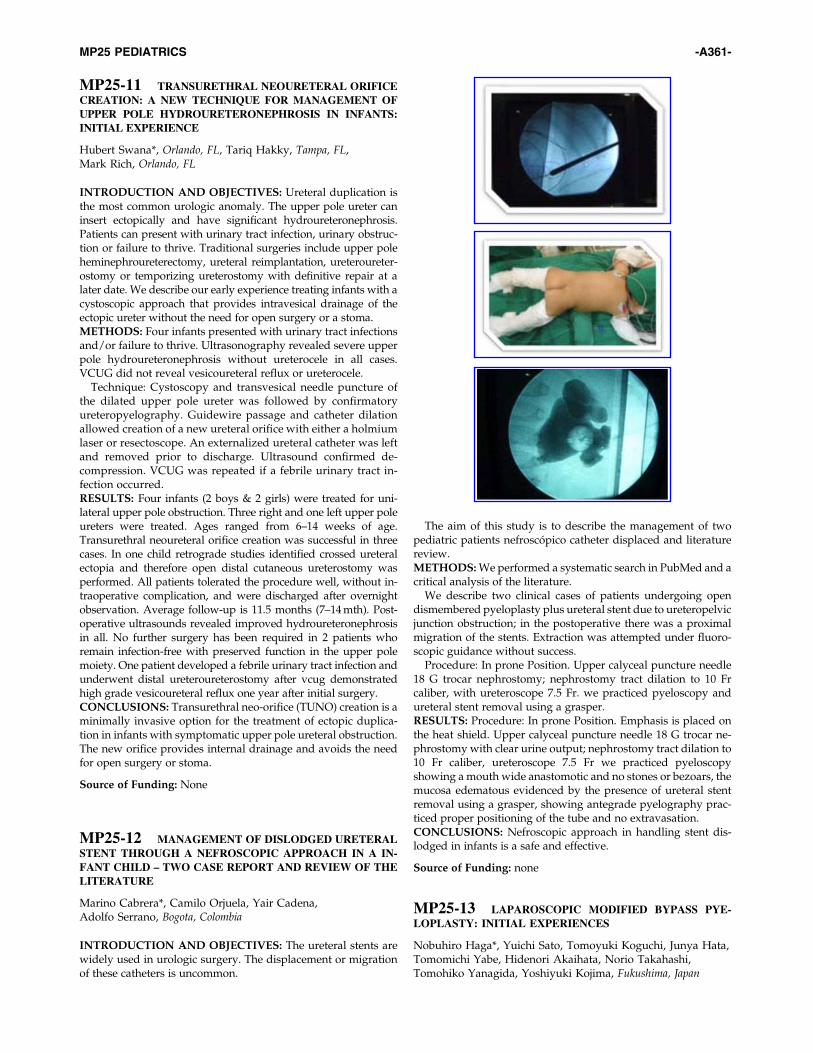
MP25-11 TRANSURETHRAL NEOURETERAL ORIFICE
CREATION: A NEW TECHNIQUE FOR MANAGEMENT OF
UPPER POLE HYDROURETERONEPHROSIS IN INFANTS:
INITIAL EXPERIENCE
Hubert Swana*, Orlando, FL, Tariq Hakky, Tampa, FL,Mark Rich, Orlando, FL
INTRODUCTION AND OBJECTIVES: Ureteral duplication isthe most common urologic anomaly. The upper pole ureter caninsert ectopically and have significant hydroureteronephrosis.Patients can present with urinary tract infection, urinary obstruc-tion or failure to thrive. Traditional surgeries include upper poleheminephroureterectomy, ureteral reimplantation, ureteroureter-ostomy or temporizing ureterostomy with definitive repair at alater date. We describe our early experience treating infants with acystoscopic approach that provides intravesical drainage of theectopic ureter without the need for open surgery or a stoma.METHODS: Four infants presented with urinary tract infectionsand/or failure to thrive. Ultrasonography revealed severe upperpole hydroureteronephrosis without ureterocele in all cases.VCUG did not reveal vesicoureteral reflux or ureterocele.
Technique: Cystoscopy and transvesical needle puncture ofthe dilated upper pole ureter was followed by confirmatoryureteropyelography. Guidewire passage and catheter dilationallowed creation of a new ureteral orifice with either a holmiumlaser or resectoscope. An externalized ureteral catheter was leftand removed prior to discharge. Ultrasound confirmed de-compression. VCUG was repeated if a febrile urinary tract in-fection occurred.RESULTS: Four infants (2 boys & 2 girls) were treated for uni-lateral upper pole obstruction. Three right and one left upper poleureters were treated. Ages ranged from 6–14 weeks of age.Transurethral neoureteral orifice creation was successful in threecases. In one child retrograde studies identified crossed ureteralectopia and therefore open distal cutaneous ureterostomy wasperformed. All patients tolerated the procedure well, without in-traoperative complication, and were discharged after overnightobservation. Average follow-up is 11.5 months (7–14 mth). Post-operative ultrasounds revealed improved hydroureteronephrosisin all. No further surgery has been required in 2 patients whoremain infection-free with preserved function in the upper polemoiety. One patient developed a febrile urinary tract infection andunderwent distal ureteroureterostomy after vcug demonstratedhigh grade vesicoureteral reflux one year after initial surgery.CONCLUSIONS: Transurethral neo-orifice (TUNO) creation is aminimally invasive option for the treatment of ectopic duplica-tion in infants with symptomatic upper pole ureteral obstruction.The new orifice provides internal drainage and avoids the needfor open surgery or stoma.
Source of Funding: None
MP25-12 MANAGEMENT OF DISLODGED URETERAL
STENT THROUGH A NEFROSCOPIC APPROACH IN A IN-
FANT CHILD – TWO CASE REPORT AND REVIEW OF THE
LITERATURE
Marino Cabrera*, Camilo Orjuela, Yair Cadena,Adolfo Serrano, Bogota, Colombia
INTRODUCTION AND OBJECTIVES: The ureteral stents arewidely used in urologic surgery. The displacement or migrationof these catheters is uncommon.
The aim of this study is to describe the management of twopediatric patients nefroscópico catheter displaced and literaturereview.METHODS: We performed a systematic search in PubMed and acritical analysis of the literature.
We describe two clinical cases of patients undergoing opendismembered pyeloplasty plus ureteral stent due to ureteropelvicjunction obstruction; in the postoperative there was a proximalmigration of the stents. Extraction was attempted under fluoro-scopic guidance without success.
Procedure: In prone Position. Upper calyceal puncture needle18 G trocar nephrostomy; nephrostomy tract dilation to 10 Frcaliber, with ureteroscope 7.5 Fr. we practiced pyeloscopy andureteral stent removal using a grasper.RESULTS: Procedure: In prone Position. Emphasis is placed onthe heat shield. Upper calyceal puncture needle 18 G trocar ne-phrostomy with clear urine output; nephrostomy tract dilation to10 Fr caliber, ureteroscope 7.5 Fr we practiced pyeloscopyshowing a mouth wide anastomotic and no stones or bezoars, themucosa edematous evidenced by the presence of ureteral stentremoval using a grasper, showing antegrade pyelography prac-ticed proper positioning of the tube and no extravasation.CONCLUSIONS: Nefroscopic approach in handling stent dis-lodged in infants is a safe and effective.
Source of Funding: none
MP25-13 LAPAROSCOPIC MODIFIED BYPASS PYE-
LOPLASTY: INITIAL EXPERIENCES
Nobuhiro Haga*, Yuichi Sato, Tomoyuki Koguchi, Junya Hata,Tomomichi Yabe, Hidenori Akaihata, Norio Takahashi,Tomohiko Yanagida, Yoshiyuki Kojima, Fukushima, Japan
MP25 PEDIATRICS -A361-

INTRODUCTION AND OBJECTIVES: Bypass pyeloplasty(BP) is a simple procedure in which the ureter and renal pelvis arenot divided. Hence, BP has been considered more suitable thanAnderson-Hynes dismembered pyeloplasty for laparoscopicsurgery. However, mobilization of the ureter is restricted becausethe ureteropelvic junction (UPJ) is not removed; this represents akey disadvantage of BP performed via the laparoscopic approach.Thus, when the side-to-side anastomosis between the ureter andrenal pelvis is performed in this procedure, it is difficult to suturethe side of the anastomosis that is far from the camera. To over-come this disadvantage, a new technique of modified BP wasdeveloped, and the initial results are presented.METHODS: Five consecutive patients underwent laparoscopicmodified BP between June 2012 and November 2012. In BP, a 1–2 cm side-to-side anastomosis is created between the dilated andelastic portion of the ureter just distal to the UPJO and the lowerand dependent portion of the hydronephrotic renal pelvis with-out disturbing the UPJ or reducing the renal pelvis. In addition,modified BP involves dividing the UPJ after the suture of thedependent portion, facilitating easy suture of the far side of theside-to-side anastomosis.RESULTS: The median age of the five patients at surgery was 3years (range, 1–16 years). Their median height and weight were106 cm (range, 72–172 cm) and 19 kg (range, 11–59 kg), respec-tively. All five patients (four males, one female) had left UPJO.Two presented with flank pain, and the remainder were detectedon prenatal maternal sonography. The average split renal func-tion of the diseased side with a diuretic renogram was 39.3%.Four patients had Society for Fetal Urology (SFU) grade 4, andone patient had SFU grade 1. The median operative duration was282 minutes (range, 248–357 minutes). The anteroposterior di-ameter of the renal pelvis was decreased postoperatively (pre-operative 31,}17 mm, postoperative 16,}6 mm).CONCLUSIONS: Laparoscopic modified BP has two advantagescompared with standard dismembered pyeloplasty. One is thatprevention of rotation and kinking of the ureter due to attachmentof the UPJ facilitates easy suturing of the dependent portion. Theother is that lifting the restrictions of the ureter by dividing the UPJafter the suture of the dependent portion makes it easy to suturethe side of the anastomosis that is far from the camera. Modified BPappears to be an efficient procedure for laparoscopic pyeloplasty.
Source of Funding: none
MP25-14 INFANT ROBOTIC PYELOPLASTY: INITIAL
EXPERIENCE AND COMPARISON WITH AN OPEN COHORT
Danesh Bansal, Nicholas G. Cost, Christopher M. Bean,Edward Riachy, Brian A. VanderBrink, Shumyle Alam,W. Robert DeFoor, Jr., Pramod P. Reddy, Eugene A. Minevich,Curtis A. Sheldon, Paul H. Noh*, Cincinnati, OH
INTRODUCTION AND OBJECTIVES: To present our experi-ence with infant pyeloplasty, comparing outcomes betweenrobotic assisted laparoscopic pyeloplasty (RALP) and open pye-loplasty (OP).METHODS: A retrospective review was performed of all chil-dren under 1 year of age who underwent unilateral dismemberedpyeloplasty at a single pediatric institution since January 2007.Patients with standard laparoscopic pyeloplasty were excluded.Data collected included age, weight, gender, operative time,length of hospital stay, analgesics, complications, and length offollow-up period.RESULTS: A total of 70 infants (51 male, 19 female) were iden-tified, with 9 RALP and 61 OP performed. Median age was 9.2 mo
(3.7–11.9) for RALP and 4.1 mo (1–11.6) for OP, p = 0.005. Medianweight was 8 kg (5.8–10.9) for RALP and 7 kg (4–14) for OP,p = 0.163. Median operative time was 115 min (95–205) for RALPand 166 min (79–300) for OP, p = 0.028. Median hospital stay was1 day (1–2) for RALP and 3 days (1–7) for OP, p < 0.001. Medianpostoperative narcotic use of morphine equivalent was< 0.01 mg/kg/day (0–0.1) for RALP and 0.05 mg/kg/day (0–2.2)for OP, p < 0.001. Median postoperative Ketorolac use was 0.5 mg/kg/day (0–1.1) for RALP and < 0.01 mg/kg/day (0–1.1) for OP,p < 0.001. More patients undergoing OP received intraoperativeregional anesthesia (RALP 0% vs. OP 65%), p < 0.001. Median fol-low-up was 12.1 mo (9.3–19.9) for RALP and 44.7 mo (5.4–76.9) forOP, p < 0.001. The success rate was 89% (8/9) for RALP and 98%(60/61) for OP. There were 22% (2/9) surgical complications withRALP (1 ileus and 1 urinary leak) and 7% (4/61) surgical compli-cations with OP (1 ileus, 1 stent migration, 1 catheter dislodgementand occlusion, and 1 urinary leak), p = 0.168.CONCLUSIONS: Infant RALP was observed to be feasible andefficacious with significantly less operative time, hospital stay,and narcotic utilization than OP.
Source of Funding: None
MP25-15 INFANT ROBOTIC ASSISTED LAPAROSC-
OPIC PYELOPLASTY: COMPARISON WITH NON-INFANT
COHORT
Danesh Bansal, Nicholas G. Cost, Christopher M. Bean,Brian A. VanderBrink, W. Robert DeFoor, Jr.,Pramod P. Reddy, Shumyle Alam, Eugene A. Minevich,Paul H. Noh*, Cincinnati, OH
INTRODUCTION AND OBJECTIVES: To compare outcomesfor robotic assisted laparoscopic pyeloplasty (RALP) betweeninfants and a non-infant patient population.METHODS: A retrospective review was performed of all chil-dren who underwent unilateral dismembered RALP at a singlepediatric institution from March 2009 to March 2013. Data col-lection included age, weight, gender, operative time, length ofhospital stay, postoperative analgesic use, length of follow-upperiod, and complication rates.RESULTS: A total of 77 patients (41 male, 36 female) wereidentified, with 9 infants and 68 non-infants. Median age was 9.2months (3.7–11.9) for infants and 103.4 months (13.7–273.7) fornon-infants, p < 0.001. Median weight was 8 kg (5.8–10.9) for in-fants and 27.5 kg (9–96) for non-infants, p < 0.001. Median oper-ative time was 115 minutes (95–205) for infants and 191 minutes(106–540) for non-infants, p < 0.001. Median hospital stay was 1day (1–2) for infants and 3 days (0–6) for non-infants, p = 0.002.There was no significant difference between patients who re-ceived intraoperative regional anesthesia (infants 0% vs. non-infants 17.6%), p = 0.34. Median postoperative narcotic use ofmorphine equivalent was < 0.01 mg/kg/day (0–0.1) for infantsand 0.07 mg/kg/day (0–0.42) for non-infants, p = 0.001. Medianpostoperative Ketorolac use was 0.5 mg/kg/day (0–1.1) for in-fants and 0.29 mg/kg/day (0–2.7) for non-infants, p = 0.437.Median postoperative Acetaminophen use was 21.7 mg/kg/day(10–55.9) for infants and 8.2 mg/kg/day (0–47.7) for non-infants,p = 0.003. Median follow-up was 12.1 months (9.3–19.9) for in-fants and 25.8 months (2.6–50.7) for non- infants, p = 0.026. Thecomplication rate was 22% (2/9) for infants (ileus and urinaryleak) and 4% (3/68) for non-infants (ileus, port site infection, andstent migration), p = 0.101. The success rate was 89% (8/9) forinfants and 100% (68/68) for non-infants, p = 0.117.
-A362- MP25 PEDIATRICS

CONCLUSIONS: Infant RALP was observed to be feasible andefficacious with significantly shorter operative times, hospitalstay, and narcotic utilization than non-infant RALP.
Source of Funding: None
MP25-16 LEARNING CURVES FOR PEDIATRIC RO-
BOTIC ASSISTED LAPAROSCOPIC PYELOPLASTY: A SIN-
GLE PEDIATRIC INSTITUTION EXPERIENCE
Danesh Bansal, Nicholas G. Cost, Christopher M. Bean,W. Robert DeFoor, Jr., Pramod P. Reddy, Paul H. Noh*,Cincinnati, OH
INTRODUCTION AND OBJECTIVES: To determine if there isa significant difference in the learning curve for pediatric urologyfellows and faculty when performing robotic assisted laparo-scopic pyeloplasty.METHODS: A retrospective review was performed of all roboticpyeloplasty procedures at a single pediatric institution from thestart of the robotic surgery program in February 2009 to De-cember 2012. No faculty had experience with robotics duringresidency or fellowship training. Two attendings (AA, AB) hadprior experience and one attending (AC) had no experience withlaparoscopic pyeloplasty. AA mentored AB and AC. Fellows (FA175 cases, FB 20 cases) had experience with robotics duringtraining prior to pediatric urology fellowship. Operative time wasused as an indicator of proficiency in performing the procedureand recorded as the start to finish based on anesthesia records.The Wilcoxon-Mann Whitney test was used to compare medianoperative times for 3 attendings and 2 fellows.RESULTS: Sixty-five cases were performed (AA 38, AB 19, AC 8,FA 12, FB 6). Median operative times (minutes) were 200 for AA,180 for AB, 288 for AC, 173 for FA, and 247 for FB. Statisticaldifference was found between FA and FB (P = 0.008), FA and AC(P = 0.009), FB and AA (P = 0.017), FB and AB (P = 0.012), AA andAC (P = 0.009), AB and AC (P = 0.016).CONCLUSIONS: Operative times were shorter for faculty withprevious experience with laparoscopic pyeloplasty. FA was moreproficient than AC and FB due to extensive prior experience withrobotic surgery.
Source of Funding: None
MP25-17 EFFECTS OF ACETYLSALICYLIC ACID ON
SYMPTOMS RELATED TO THE SYNDROME NUTCRACKER
Sidiya Chavey*, Sousse, Tunisia, Wissem Hmida, Faouzi Mallat,Sana Mosbahi, Mehdi Jaidane, Faouzi Mosbah, Sahloul, Tunisia
INTRODUCTION AND OBJECTIVES: The purpose of thisstudy was to compare the relative efficacy of acetylsalicylic acid(ASA) for the treatment of pain and hematuria and fatigue-related to syndrome Nutckraker.METHODS: comparative study colligeant 26 patients with syn-drome Nutcracher with left lumbar pain, fatigue and intermittentgross hematuria or microscopic. Patients aged between 18 and 33years were randomly assigned to two treatment groups. The firstgroup (13 patients) was the control group received paracetamolwith sitting for 4 weeks. The second group (13 patients), which isthe study group received aspirin (500 mg once daily) for 4 weeks.Patients were evaluated at the beginning and end of each phasewith the scale of severity of fatigue and pain, and on the existenceof gross or microscopic hematuria.
RESULTS: Aspirin associated with reduced effort duringsymptomatic periods Nutcracker syndrome has led to a signifi-cant improvement of the various parameters.CONCLUSIONS: this study demonstrates that aspirin signifi-cantly reduces fatigue, pain and hematuria related syndromenutcraker during symptomatically without any adverse effects.
Source of Funding: none
MP25-18 COMPARISON OF CHILDREN VERSUS
ADULTS UNDERGOING MINI-PERCUTANEOUS NEPHROLI-
THOTOMY: LARGE-SCALE ANALYSIS OF A SINGLE
INSTITUTION
Guohua Zeng*, Zhijian Zhao, Wenqi Wu, Guanghzou, China,People’s Republic of
INTRODUCTION AND OBJECTIVES: As almost any versionof percutaneous nephrolithotomy (PCNL) was safely and effi-ciently applied for adults as well as children without age being alimiting risk factor, the aim of the study was to compare thedifferent characteristics as well as the efficacy, outcome, andsafety of the pediatric and adult patients who had undergonemini-PCNL (MPCNL) in a single institution.METHODS: We retrospective reviewed 331 renal units in childrenand 8537 renal units in adults that had undergone MPCNL for upperurinary tract stones between the years of 2000–2012. The safety, ef-ficacy, and outcome were analyzed and compared.RESULTS: The children had a smaller stone size (2.3 vs. 3.1 cm)but had smilar stone distribution (number and locations). Thechildren required fewer percutaneous accesses, smaller ne-phrostomy tract, shorter operative time and less hemoglobindrop. The children also had higher initial stone free rate (SFR)(80.4% vs. 78.6%) after single session of MPCNL (p < 0.05); but nodifference was noted in the final SFR (94.7% vs. 93.5%) afterauxiliary procedures. The complication rate (15.6% vs. 16.3%) andblood transfusion rate (3.1% vs. 2.9%) were similar in both group(p > 0.05). Both groups had low rate of high grade Clavien com-plications. There was no grade III, IV, V complications and noangiographic embolization required in pediatric group. One im-portant caveat, children who required multiple percutaneousnephrostomy tracts had significant higher transfusion rate than inadults (18.8% vs. 4.5%, p = 0.007).CONCLUSIONS: This contemporary largest-scale analysis con-firms that the stone-free rate in pediatric patients is at least asgood as in adults without an increase of complication rates.However, multiple percutaneous nephrostomy tracts should bepracticed with caution in children.
Source of Funding: none
MP25-19 EFFECTIVENESS OF PENILE BLOCK WITH
BUPIVACAINE DURING CIRCUMCISION IN CHILDREN: A
COMPARATIVE STUDY
Faouzi Mallat*, Wissem Hmida, Nawel Bjaoui,Faouzi Mosbah, Sahloul, Tunisia
INTRODUCTION AND OBJECTIVES: Evaluation of the ef-fectiveness of penile block with bupivacaine to prevent pain in-traoperative and postoperative during circumcision.METHODS: 24 boys aged 2 to 6 years, who underwent circum-cision in general anesthesia were included in our work, over aperiod of 12 months spanning from June 2011 to June 2012.
MP25 PEDIATRICS -A363-

These children were randomized to have just after induction ofanesthesia, a block penis with bupivacaine 0.25% (group I: GI) isan intravenous injection of fantanyl (2lg/kg) associated withparacetamol (group II: GII).
The efficacy of intraoperative analgesia was estimated based onheart rate and Changes in blood pressure.
Postoperative pain is estimated to post anesthetic care unit, byanesthetists and parents in the next 24 hours. The quality of an-algesia was estimated on postoperative activity and mobilizationof children. The quantity and schedule of administration ofparacetamol were evaluated postoperatively.RESULTS: In the G I, it was noted cardiovascular stability andrecovery significantly faster, with duration of postoperative an-algesia without a longer statistical significance (p > 0.05).
The first analgesic request was earlier in the G I.No complications were noted in both groups.
CONCLUSIONS: The penile block with bupivacaine devoid ofserious complications allowed prolonged analgesia, reducedconsumption of analgesics, and ambulation Quick children.
Source of Funding: NONE
MP25-20 HOLMIUM LASER ENUCLEATION OF THE
PROSTATE (HOLEP) BEFORE HIGH-INTENSITY FOCUSED
ULTRASOUND (HIFU) FOR THE PROSTATE CANCER
TREATMENT: OUR EXPERIENCE
Antonio Salvaggio*, Angelo Cafarelli, Donato Dente,Abano Terme (Padua), Italy, Emanuele Cappa, Chieti, Italy,Angelo Porreca, Abano Terme (Padua), Italy
INTRODUCTION AND OBJECTIVES: Nowadays HIFU is analternative treatment for localized prostate cancer (PCa). It’s anoption for patients of advanced age, in the low or intermediate-risk groups, and with a life expectancy of 10 years.
Looking at the meta-analysis studies concerning HIFU thebladder neck obstruction (BNO) is the most frequent side effect(25–30 %). This is related to the effects of HIFU on residual ade-noma after endoscopic surgery. The rate of BNO is significantlylower compared to the greater volume of resected tissue by Transurethral prostate resection (TURP) before HIFU.
The goal of this study is to analyze the BNO rate using HOLEPpre-HIFU (H.H.)METHODS: From June 2010 to March 2013 we treated 47 pa-tients with local PCa with H.H. Between the HOLEP and HIFUtreatment we waited minimum 30 days.
We performed to all patients International Prostate SymptomScore (IPSS) test and uroflowmetry before surgery and after sixmouths. We considered the criteria listened below:
Inclusion criteria:70 years < patient age < 76 years, 40 cc < adenoma volume
< 120 cc, clinical stage (T1–T2), Gleason Score (G.S.) < 7 (3 + 4),PSA < 20.
Exclusion criteria:70 years > patient age > 76 years, 40 cc < adenoma volume
> 120 cc, clinical stage > T2, G.S. > 7 (4 + 3), PSA > 20. Anal ste-nosis, previous rectal surgery, coxofemoral anchilosis.
We tested PSA, urine analysis and culture every three monthsduring the first year after H.H. PSA every six month after one year.RESULTS: Pre-operatory medium IPPS: 15
Pre-operatory medium maximum flow (Medium Q max):13 ml/sec.
Medium catheterization time after HOLEP was 32 hours and2.4 days after HIFU.
We didn’t report mayor complications.
Minor complications after H.H.: Post-operatory Urinary infec-tions (10%), penis urethral stenosis (2,1%), urge urinary inconti-nence after three mouths (1 pad /day) 13%, low grade stressincontinence after three months (1 pad /die) 7%.
No cases of new endoscopic surgery.Medium volume of adenoma enucleated: 67 cc.IPPS medium after six months: 11Medium Q Max: 17 ml/sec
CONCLUSIONS: HIFU is a mini invasive treatment of PCa inpatient with low and intermediate tumor risk. Generally a TURPis performed before HIFU. Looking at our experience HOLEP is agood alternative to TURP because reduces the BNO rate. It’s mini-invasive although in case of big adenoma volume and, at least,improves obstructive symptoms.
Source of Funding: ‘‘none’’
MP25-21 SIMPLE TECHNIQUE FOR NON-INVASIVE
REMOVAL OF URETERAL STENT IN RENAL TRANSPLANT
RECIPIENTS; COMPARISON WITH CONVENTIONAL
CYSTOSCOPIC REMOVAL.
Taek Sang Kim*, Jung Hyun Oh, Hyun Yul Rhew,Busan, Korea, Republic of
INTRODUCTION AND OBJECTIVES: Using ureteral stent atthe time of kidney transplantation significantly decreases earlyurologic complication such as urinary tract obstruction, anasto-mosis site urine leakage. However, removal of ureteral stent re-lated complications such as urinary tract infection, pain bycystoscopic removal also can be present. Herein we have analyzedthe comparison conventional cystoscopic removal of ureteral stentwith retrieval of ureteral stent by suturing of urethral catheter.METHODS: Between March, 2008 and March, 2013, we evalu-ated 34 patient who were underwent kidney transplantation.Among these patients, 18 patients were underwent deceaseddonor kidney transplantation, and 16 patients were underwentliving donor kidney transplantation. We routinely insertedDouble J ureteral stent during extravesical ureteroneocystostomy.We removed Double J ureteral stent by conventional cystoscopicmethod in 19 patients in postoperative 7 day (group 1). For theremaining 15 patients, we used simple non-invasive removaltechnique in postoperative 7 day which is suturing the distal sidehole of Double J ureteral stent to the end side hole urethralcatheter (group 2). We can remove Double J ureteral stent bygentle traction on urethral catheter. We evaluated and comparedthe age, sex, catheter removal related pain scale (0–10 points),postoperative urine leakage/ureter obstruction, persistent he-maturia/pyuria after one week of urethral catheter removal, UTIepisode within 6 month of kidney transplantation of each groups.RESULTS: The mean age of group 1,2 is 41.9, 44.1 years old(p = 0.601), and sex ratio (% of male) of group 1,2 is 57.9%, 60.0%(p = 0.905), respectively. There was no postoperative urine leak-age or ureter obstruction within postoperative one month. Ca-theter removal related mean pain scale of each group 1,2 were 6.9and 1.9 (p = 0.000), respectively. Persistent hematuria after oneweek of urethral catheter removalof each group 1,2 were 47.4%,6.7% (p = 0.006), persistent pyuria were 26.3%, 6.7% (p = 0.122),respectively. UTI episode of each group 1,2 were 15.8%, 0%,(p = 0.083), respectively.CONCLUSIONS: Non-invasive ureteral catheter removal tech-nique with suturing to urethral catheter was simple, easy, andminimal painful. This technique also had less persistent hema-turia and showed less trend of UTI episode compared with cys-toscopic removal. However, there was no difference in persistent
-A364- MP25 PEDIATRICS

pyuria between these methods. The larger scale of analysis isrequired for statistical information.
Source of Funding: none
MP25-22 SURGEONS’ PREFERENCES AND PRACTICE
PATTERNS REGARDING INTRA-OPERATIVE FROZEN
SECTION DURING PARTIAL NEPHRECTOMY
Abhinav Sidana*, James F Donovan, Krishnanath Gaitonde,Cincinnati, OH
INTRODUCTION AND OBJECTIVES: Intra-operative frozensection (FS) evaluation for tumor margin during partial ne-phrectomy (PN) is a matter of controversy in urologic oncology.We hereby evaluate the preferences and practice patterns ofurologists about intra-operative FS during PN.METHODS: A 17-item questionnaire was designed to collectinformation on surgeons’ preferences and practice patterns aboutFS during PN. The survey was sent to the members of the Societyof Urologic Oncology and the Endourological Society.RESULTS: 197 responses were received. 46% of the respondentswere fellowship trained in Urologic Oncology. 69% and 58% ofrespondents chose to obtain FS (always or sometimes) duringOpen Partial Nephrectomy (OPN) and Laparoscopic Partial Ne-phrectomy (LPN) respectively. There was strong correlation be-tween surgeons’ preferences during OPN and LPN (p < 0.001). Ahigher proportions of surgeons younger than 50 years (37 vs 19%)chose to ‘‘never’’ obtain FS during OPN (p < 0.001). For surgeonswho did not routinely obtain FS, ‘‘confidence about complete re-section’’ was the most common reason (79%), followed by ‘‘nochange in management with positive margins’’ (35%). Majority(75%) believed the margins to be negative if surgical margin wasmicroscopically free of tumor by one cell layer. Older surgeons( > 50 years) preferred sending additional specimen from tumorbed for FS analysis (p = 0.01). Only,41% and 24% of respondentswould repeat FS for positive microscopic margins during OPN andLPN respectively. 95% of the respondents would not recommendadditional treatment for positive margins on final pathology.CONCLUSIONS: Despite recent literature pointing to low clinicalutility of FS, most surgeons still obtain FS during PN. A higherproportion of younger surgeons tend not to obtain FS. Fellowshiptraining, practice type and surgical volume did not appear to in-fluence preferences and practice patterns in regards to FS.
Source of Funding: none
MP25-23 ALL OUT: THE INGLORIOUS END OF
‘‘BUCKYBALLS’’
Lawrence Wyner*, Huntington, WV
INTRODUCTION AND OBJECTIVES: The urological commu-nity needs to be aware of recent successful efforts by the U.S. Con-sumer Product Safety Commission (CPSC) to block the continuedsale of small but powerful rare earth toy magnets manufactured inChina from an alloy of neodymium, iron, and boron, then nickelplated to look like harmless BBs about 4 millimeters in diameter.Known as ‘‘Buckyballs,’’ they are almost 10 times stronger thanordinary magnets of the same size. Unlike several companies whichcomplied immediately with the CPSC directive, New York distrib-utor Maxfield & Oberton persisted in marketing this novelty untilOctober 2012 after launching an ad campaign entitled ‘‘Save OurBalls’’ to support their cause, citing the mantras of a free market andpersonal/parental responsibility. The ads were placed in response to
a lawsuit filed by the CPSC in July 2012 to ban the sale of Buckyballsnationwide; nevertheless, almost half a billion Buckyballs are‘‘loose’’ in the U.S. since their introduction in 2008. If ingested, theyare strong enough to damage tissue and may require open surgeryto remove them; cases of bowel necrosis have been reported.METHODS: Last year at our medical center, a 17-year-old youngman presented to the emergency department shortly after in-serting several dozen Buckyballs into his urethra, presumably forself-stimulation purposes. The patient was an honors studentathlete at a local private high school who aspired to become anorthopedic surgeon. Aside from taking dexmethylphenidate forattention deficit hyperactivity disorder, his health history wasunremarkable. We attempted to remove the Buckyballs by ex-erting gentle traction on the tail end of the chain; however, theinternal balls had apparently contorted in the bulbar urethra;thus, the chain separated at the meatus, leaving about 50 ballsremaining inside his urethra.RESULTS: The patient was then taken to the cystoscopy suite,where under general anesthesia, the balls were successfully re-moved individually over the ensuing 3 hours with a 22 Frenchcystoscope sheath and flexible cold cup biopsy forceps. Retrieval ofthe spheres was aided by pushing their tangled mass up into thebladder, then teasing them apart one by one until all of them hadbeen removed. A foley catheter was placed, and following a voidingtrial the next day, he made an uneventful recovery. He was releasedto the custody of his parents, both of whom were physicians.CONCLUSIONS: This case demonstrates the relevance of thisissue for urologists in addition to general surgeons and gastro-enterologists, and the need for all of us to advocate for our pa-tients of a tender age, who may be tempted to experiment withthese dangerous magnets.
Source of Funding: None
MP25-24 INNER PREPUTIAL INLAY GRAFT URE-
THROPLASTY IN HYPOSPADIAS REPAIR: WORTH DOING?
Mamdouh Ahmed*, Abdulnaser Al Said, Kuwait, Kuwait
INTRODUCTION AND OBJECTIVES: Inner preputial inlaygraft has been proposed to reduce the risk of meatal and neourethralstenosis and in redo urethroplasty for scarred urethral plate. Weextended its indication to be the standard technique for primarydistal and midpenile hypospadias. We conduct this prospectivestudy to clarify whether the inner preputial inlay graft is the optimalprocedure to minimize the hypospadias repair complications and toget excellent cosmetic result especially for meatal position.METHODS: This prospective study included consecutive 110patients who underwent inner preputial inlay graft urethroplastyfor primary hypospadias repair from November 2011 to March2013. Age ranged between 11 months to 7 years (mean: 3.2 years).Localization of the meatus was glanular in 5 patients, coronal in42, distal penile in 52, midpenile in 10 and proximal in one. Eightpatients received hormonal therapy (Dihydrotestosterone cream/Testosterone injection) due to extremely small glanular size. In allpatients, the urethral plate was incised deeply and extendeddistally beyond the end of the plate (entering the glans by3–4 mm). The wide dorsal inner preputial vascular flap was dis-sected. The graft harvested from the inner preputial flap, inlayedand quilted in the incised urethral plate till the position of neo-meatus. Neourethra was created over urethral catheter using 7.0polyglactin sutures in 2 layers. A positional dorsal vascular dartosflap was laid over the neourethra as a second barrier.RESULTS: Follow up period ranged from 2 to 18 months. Ex-cellent cosmetic and functional results were achieved in 108 of 110
MP25 PEDIATRICS -A365-

patients (98.2%). Neither meatal stenosis nor urethral diverticu-lum has been encountered. Excellent glanular position of a wideslit like neomeatus was achieved by this technique. Only 2 pa-tients (1.8%) developed urethrocutaneous fistula that occurred inone patient very early (5 days) post operatively. Satisfactoryurinary flow pattern was detected on follow up.CONCLUSIONS: Inner preputial inlay graft urethroplasty sig-nificantly reduces the incidence of urethrocutaneous fistula as theinlay graft plays a major role in minimising the postoperativescarring of incised urethral plate that might occur in original tu-bularized incised plate urethroplasty. Furthermore, it securesoptimal glanular position of a wide slit like neomeatus due toextension of the incision beyond the end of the plate, thus opti-mising functional and cosmetic outcome.
Source of Funding: none
MP25-25 NEW THERAPEUTIC APPROACH TO TREAT
SLOWLY RISING PROSTATIC SPECIFIC ANTIGEN FOL-
LOWING ROBOTIC ASSISTED LAPAROSCOPIC PROSTA-
TECTOMY
Alaa Hamada*, Sanjay Razdan, Jolly Varki, Yubiry Morales,Miami, FL
INTRODUCTION AND OBJECTIVES: Slowly rising serumprostatic specific antigen (PSA) level following robotic assistedlaparoscopic prostatectomy (RALP) presents a dilemma in termsof clinical interpretation and subsequent management. Presenceof benign prostatic glands at the surgical margins, versus pres-ence of local and or distant recurrence are the plausible sourcesfor the rising PSA. We hypothesized that a particular subset ofmen with Gleason score < = 7, negative margins at surgery, and aslowly rising PSA (PSA doubling time (PSADT) > 15 months)detected at least one year after RALP can be treated using du-tasteride as long as their PSA is lower than 0.9 ng/ml.
The objective of this study was to evaluate the effect of du-tasteride (0.5 mg/day) on serum prostate-specific antigen (PSA)kinetics in men with slowly rising PSA after RALP who hadnegative margins at surgery and no evidence of local or distantrecurrence.METHODS: A total of 33 consented men who underwent marginnegative RALP for prostate cancer (Gleason score < = 7) over thepast 8 years were included in the study. A multidisciplinary ap-proach was formulated with the medical and radiation oncologistincluded in the study design. All subjects had a slowly rising PSAbetween 0.2 and 0.9 ng/mL and PSADT of more than 15 months,with no evidence of local recurrence on prostascint scan and orskeletal metastasis on bone scan. The men were treated with0.5 mg dutasteride and were followed for > 24 months. Primaryendpoints were a rising serum PSA level despite treatment and orpresence of local and or distant recurrence (detected by 6 monthlybone and prostascint scans).RESULTS: Of the 33 patients treated with dutasteride, 13 pa-tients (39%) had no further increase in serum PSA and 17 pa-tients (52%) showed a reduction in PSA level; whereas, only 3patients (9%) had further increase in PSA ( > 0.9 ng/ml) withoutevidence of local or distant recurrence. The 3 patients withrising PSA received radiation and or androgen deprivationtherapy without a detrimental effect because of the delay.Dutasteride was well tolerated in all patients with no signifi-cant side effects.CONCLUSIONS: In a particular subset of men with slowlyrising PSA (0.2–0.9 ng/ml) after margin negative RALP andgood prognostic factors (Gleason score < = 7, PSADT > 15months), residual benign prostatic glands rather than local ordistant recurrence may be the source of a rising PSA. Du-tasteride can provide growth inhibitory effects on residualbenign prostate glands with a decrease in PSA level and ob-viate the need for radiation or hormone therapy with theirattendant side-effects.
Source of Funding: None
V13 LESS/NOTES
V13-01 TRANSVAGINAL NATURAL ORIFICE TRANS-
LUMENAL ENDOSCOPIC SURGERY (NOTES)-ASSISTED
LAPAROSCOPIC NEPHRECTOMY
Xiaofeng Zou*, Guoxi Zhang, Yuanhu Yuan, Rihai Xiao,Gengqing Wu, Xiaoning Wang, Yijun Xue, Dazhi Long,Yuting Wu, Folin Liu, Hui Xu, Ganzhou, China,People’s Republic of
INTRODUCTION AND OBJECTIVES: To describe our experi-ence with the transvaginal natural orifice translumenal endoscopicsurgery (NOTES)-assisted laparoscopic nephrectomy (TNALN) infemale patients, and to evaluate its feasibility and safety.METHODS: Between May 2010 and April 2013, 99 TNALNswere performed in femal patients with non-function kidney (right36, left 44) or renal carcinoma (right 10, left 9) in our center. Afterinduction of general anesthesia, the patients were positioned inlithotomy with ipsilateral lumbar at 60�angle to the floor. A 5-mmtrocar and a 10-mm trocar were inserted in the umbilical edge for
conventional operating apparatus. A 5-mm or 10-mm trocar wasinserted in the posterior vaginal fornix for a 5-mm flexible-tip 0�or 10-mm 30�laparoscope. Dissection was performed accordingto the method of the standard laparoscopic simple and radicalnephrectomy. The intact specimen was extracted transvaginally.RESULTS: All the procedures were successfully accomplishedwithout additional trocars except for two patients. One was con-verted to open surgery because of uncontrolled bleeding for injuryof the inferior vena cava, and the other was performed transva-ginal NOTES-assisted laparoscopic splenectomy. The mean oper-ative time was 115 minutes (range 80 to 280). The mean estimatedblood loss was 160 mL (range 50 to 600). The mean time for patientsto resume full ambulation and oral diet was 1.3 days (range 1 to 2)and 2.6 days (range 2 to 3), respectively, and the mean postoper-ative hospitalization stay was 7.4 days (range 4 to 10). Mean fol-low-up was 16.5 (range 2 to 35) months. The umbilical scar of eachpatient was hidden and difficult to find. The incision in vaginahealed well. According to the questionnaire survey, sexual
-A366- V13 LESS/NOTES

function recovered in postoperative month 3 in each patient, andno patient experienced sexual dysfunction. Each patient was verysatisfied with the results, especially with the cosmesis result.CONCLUSIONS: TNALN is a feasible and safe surgical optionfor both benign and malignant diseases of the kidney in appro-priate female patients.
Source of Funding: This work was supported by the NationalHigh Technology Research and Development Program (863Program) of China (NO. SQ2011AAJY2801), Important Scienceand Technology Project from the Department of Science andTechnology of Jiangxi Province (20121BBG70032), and ImportantScience and Technology Project from the Department of Health ofJiangxi Province (NO. 20104008)
V13-02 SUPRAPUBIC-ASSISTED LAPAROENDOSCOPIC
SINGLE-SITE SURGERY (SA-LESS) FOR NEPHROURE-
TERECTOMY
Xiaofeng Zou*, Guoxi Zhang, Yunfeng Liao, Yuanhu Yuan,Rihai Xiao, Gengqing Wu, Xiaoning Wang, Yijun Xue,Dazhi Long, Min Liu, Hui Xu, Gang Xu, Ganzhou, China,People’s Republic of
INTRODUCTION AND OBJECTIVES: During the past years,suprapubic-assisted laparoendoscopic single-site surgery £̈SA-LESS£ª established by ourselves has been used in urology in ourcenter. This study aimed to describe our experience with SA-LESSnephroureterectomy and to evaluate its feasibility and efficacy.METHODS: Between July 2012 and April 2013, 8 consecutivemale patients (two of them with renal pelvic carcinoma, two withureteral carcinoma, one with renal pelvic carcinoma combinedwith ureteral carcinoma, two with renal tuberculosis, and one withmegaureter and empyema) were subjected to SAU-LESS ne-phroureterectomy at the authors¡�center. The patient was admin-istrated general endotracheal anesthesia and secured on theoperating table in lateral decubitus with affected side elevated 70�.Two trocars were inserted at the medial margin of umbilicus, andone was inserted into abdominal cavity below the pubic hairline.The operation was performed using conventional operating in-struments, under direct vision achieved by a 5-mm flexible-tip0�laparoscope. Firstly, the distal ureter was isolated completelyand blocked by a Hem-O-lok clip. Then, the nephrectomy wasperformed according to the method of the standard laparoscopy.Finally, the bladder cuff excision was performed and the incisionwas sutured. The specimen was removed after the incisions belowthe pubic hairline was enlarged transversely and the rectus ab-dominis muscle sheath was incised vertically.RESULTS: All the procedures were successfully performed. Themedian operative time was 165 (range 115 to 220) minutes, andthe median estimated blood loss was 140 (range 50 to 200) ml.There was no major perioperative complication occurred. All thepatients resumed ambulation on postoperative day 1. Pelvicdrainage tube was removed on postoperative day 2–3. Urethralcatheter was removed on postoperative day 6–7 (the tumor pa-tients underwent irrigation of bladder with Pirarubicin). Thepatients were discharged on postoperative day 8.CONCLUSIONS: SA-LESS nephroureterectomy appears to befeasible and effective. The placement of trocar at umbilicus andbelow the pubic hairline not only decreases the difficulty of op-eration, but can also leads to good cosmetic results.
Source of Funding: This work was supported by the NationalHigh Technology Research and Development Program (863Program) of China (NO. SQ2011AAJY2801), Important Science
and Technology Project from the Department of Science andTechnology of Jiangxi Province (20121BBG70032), and ImportantScience and Technology Project from the Department of Health ofJiangxi Province (NO. 20104008)
V13-03 PURE TRANSVAGINAL NATURAL ORIFICE
TRANSLUMENAL ENDOSCOPIC SURGERY (NOTES) FOR
NEPHRECTOMY: REPORT OF 15 CASES
Xiaofeng Zou*, Guoxi Zhang, Rihai Xiao, Yuanhu Yuan,Yijun Xue, Gengqing Wu, Xiaoning Wang, Dazhi Long,Yuting Wu, Folin Liu, Hui Xu, Min Liu, Ganzhou,China, People’s Republic of
INTRODUCTION AND OBJECTIVES: Hybrid transvaginalnatural orifice transluminal endoscopic surgery (NOTES) ne-phrectomy (HTNN) is indeed possible. But pure transvaginalNOTES nephrectomy (PTNN) has been limited to animal exper-iments with only one report of its use in humans. We describe theinitial clinical experience of PTNN, and evaluate its feasibility.METHODS: Between January 2011 and March 2013£:14 femalepatients with non-function kidney (right 10, left 4) and one withright renal cancer, underwent PTNN. The patients were positionedin lithotomy with ipsilateral lumbar at 30¡ã angle to the operatingtable. A 3-mm incision was made at the posterior vaginal fornix,and a 5 mm Trocar was introduced into the pelvic cavity guided bya 5-mm forceps. A 5-mm 0¡ã flexible laparoscope was inserted intothe pelvic cavity confirming no rectum injury. Then a Zou-port wasintroduced at the posterior vaginal fornix. The patients head endwas uppered by 25¡ã with right lumbar at 60¡ã angle to the floor.Dissection was performed according to the method of the standardlaparoscopic nephrectomy. The vaginal wound was closed underdirect vision using a 2/0 absorbable suture.RESULTS: All the procedures were successfully completed ex-cept one patient who was converted to suprapubic-assisted la-paroendoscopic single-site surgery nephrectomy for rectal injury.The median operative time was 255 mins (range 210 to 360). Themedian estimated blood loss was 250 ml (range 150 to 300). Thepatients were discharged on postoperative day 6 to 7. Duringthe 6- to 10-month follow-up period, all the patients were in goodcondition. The posterior colpotomy incision healed up well. Noscars were visible on the abdominal wall. There were no com-plication in retrograde infection of pelvic and abdominal cavity,umbilical hernia, uterine prolapse, and sexual dysfunction.CONCLUSIONS: Pure transvaginal NOTES for nephrectomyis feasible. This novel technique seeks to provide cosmeticresult even when compared to today¡�s minimally invasiveprocedures.
Source of Funding: This work was supported by the NationalHigh Technology Research and Development Program (863Program) of China (NO. SQ2011AAJY2801), Important Scienceand Technology Project from the Department of Science andTechnology of Jiangxi Province (20121BBG70032), and ImportantScience and Technology Project from the Department of Health ofJiangxi Province (NO. 20104008)
V13-04 COST-EFFECTIVE GLOVE-URETERIC CA-
THETER SINGLE PORT FOR LAPARO-ENDOSCOPIC SIN-
GLE SITE NEPHRECTOMY
Raguram Ganesamoni*, Shashikant Mishra, Arvind Ganpule,Jigish Vyas, Jitendra Jagtap, Amit Bhattu, Ravindra Sabnis,Mahesh Desai, Nadiad, India
V13 LESS/NOTES -A367-

INTRODUCTION AND OBJECTIVES: Laparo-endoscopic sin-gle site (LESS) nephrectomy is associated with lower postoperativepain and better cosmesis. The commercially available single portsare costly and hence not affordable for many of our patients. In thisvideo we describe the use of a single port made from cheap ma-terials readily available in Urology operating room.METHODS: From April to May 2013, we performed four LESSsimple nephrectomies using a single port made from a glove, twoureteric catheters and standard laparoscopic ports. Patient de-mographics, operative parameters, complications and problems,if any, due to the single port were assessed.RESULTS: The mean age of the patients was 46 years. There werethree male patients and one female patient. The mean operatingtime was 168 minutes. The mean blood loss was 130 ml. Therewas no complication in any of the procedure. The single port wassuccessfully utilized till the end of the procedure in all the patientswithout the need for any repair. Two procedures which wereperformed in patients, who had undergone open procedures,were converted to multiport laparoscopic nephrectomy due todense adhesions around the hilum.CONCLUSIONS: The use of glove-ureteric catheter single port isfeasible and effective in LESS nephrectomy. It can be consideredas a cheaper alternative to the costly commercially availablesingle ports.
Source of Funding: None
V13-05 LESS TRANSVESICAL DIVERTICULECTOMY
Lokesh Sinha, Rajendra Godbole, Dombivli, India,Prashanth Rao, Pradeep Rao*, Mumbai, India
INTRODUCTION AND OBJECTIVES: The Triport andQuadport are uniquely suited to perform transvesical proceduresbecause they can synch the bladder to the anterior abdominalwall. Combined with a pneumovesicum, this makes an adequatespace within the bladder to perform various surgeries. This ap-proach has been reported for BPH, VVF and Ca Prostate in thepast. We used the Triport Plus to do a single port LESS diverti-culectomy through the Transvesical route.METHODS: A 35 year old male presented with 3 large bladderdiverticuli which were together larger than the size of the bladderitself. He was getting repeated UTI and was unable to void on aurodynamic examination. The bladder was unable to generatepressures due to the diverticuli. A Triport plus was inserted intothe bladder under cystoscopy control and pneumovesicum ini-tiated. An Flexible tip Endoeye video laparoscope was used tofacilitate the surgery. All 3 large diverticuli were excised and thedefects closed using a barbed suture.RESULTS: Patient was able to void after surgery and he is free ofinfection 1 year after surgery. He remains a dysfunctional voiderand is on alpha blockers to prevent development of any newdiverticuli.CONCLUSIONS: Large bladder diverticuli are a difficult prob-lem to treat using conventional laparoscopy due to the difficultyof dissecting out the diverticuli from surrounding structures. Theopen transvesical route is well suited and routinely used for thissurgery.
The LESS approach using a triport distends the bladder wellwith the pneumovesicum and makes it easy to dissect the di-verticuli. Barbed sutures facilitate suturing through a single port.
The LESS approach makes for an elegant and relatively easyminimally invasive solution for bladder diverticuli.
Source of Funding: None
V13-06 BILATERAL SPERMATIC CORD DISSECTION
E LIGATION BY LAPAROENDOSCOPIC SINGLE SITE
SURGERY: OUR TECHNIQUE ‘‘STEP BY STEP’’
Salvatore Micali*, Eugenio Martorana, Alessio Zordani,Marco Rosa, Modena, Italy, Ahmed Ghaith, Tanta, Egypt,Francesco Fidanza, Giampaolo Bianchi, Modena, Italy
INTRODUCTION AND OBJECTIVES: The laparo-endoscopicsingle-site surgery (LESS) represents an important evolution ofminimally invasive surgery and aims to improve the cosmeticoutcome, reduce surgical trauma and complications associatedwith traditional laparoscopy. The following video shows ourLESS technique to perform bilateral spermatic cord dissectionand ligation.METHODS: The procedure was performed on 6 patients agedbetween 24 and 52 years (mean age 32.7 years) during a period of36 months. Five patients have the operation for bilateral varico-cele and one of them had a relapse after previous open var-icocelectomy. One of the 6 patients underwent operation forbilateral chronic testicular pain. All patients were placed in su-pine position. The access to the peritoneal cavity was performed,after creation of the pneumoperitoneum, with the aid of an 10 mmoptical trocar (TERNAMIAN Endo TIP, Storz) through infra-umbilical 2 cm incision. The subsequent positioning of SILS port(Covidien) allowed the use of laparoscopic instruments (5 and10 mm). Flexible 5 mm endo-eye optics (Olympus) allowed tominimize the clashing between the instruments. Laparoscopicscissors, a 5 mm laparoscopic grasper and a 10 mm dissector al-lowed to dissect the spermatic cord sparing the deferens vas withits vessels. Dissection of the spermatic cords were cut en bloc afterpositioning of four Hem-o-lok. Hemostasis was completed withthe application of oxidized cellulose gauze.RESULTS: The mean operative time was 49 minutes (34–73minutes). No patients reported intra, nor post-operative compli-cation. The hospitalization time was less than 24 hours. No pa-tient did not need post-operative analgesia. The scrotal echo-colorDoppler was performed after three months and showed no re-currence of varicocele.CONCLUSIONS: The procedure was successfully performed inall cases. The main difficulty is represented by clashing between theinstruments. Operative times were acceptable. The trans-umbilicalapproach offers the advantage of a better cosmetic result. Bilateralspermatic cord ligation with LESS is an alternative to conventionallaparoscopy. The LESS approach is feasible, safe, reliable (100%success) and easily applicable in all urological centers.
Source of Funding: none
V13-07 RETROPERITONEAL LAPAROENDOSCOPIC
SINGLE-SITE SURGERY: OUR TECHNIQUE IN RENAL
CYST DECORTICATION
Salvatore Micali*, Eugenio Martorana, Alessio Zordani,Riccardo Galli, Modena, Italy, Ahmed Ghaith, Tanta, Egypt,Maria Chiara Sighinolfi, Giampaolo Bianchi, Modena, Italy
INTRODUCTION AND OBJECTIVES: Retroperitoneal Lapar-oendoscopic single-site surgery (LESS) has been developed in anattempt to reduce the morbidity and scarring associated with lap-aroscopic surgery. We report our experience on 5 renal cyst de-cortication procedures were performed in a retroperitoneal LESS.METHODS: LESS renal cyst decortication was performed in 5 pa-tients during a 24-months. All procedures were performed by asingle operator. The patient was in a standard flank position. The
-A368- V13 LESS/NOTES

procedure was performed using a 2,5 cm single incision via a ret-roperitoneal access at the Petit’s triangle. In all cases retroperitonealaccess was created with a laparoscopic visual reusable trocar, Ter-namian EndoTIP 10 mm (Karl Storz�), and a 10 mm laparoscopewas used for blunt dissection of the aereolar retroperitoneal fat. In allcases we used a Single-trocar technique with a Multiport (CovidienSILS� Port) placed in the Petit’s Triangle with a 5-mm flexiblelaparoscope EndoEye camera system (Olympus Medical�). In allpatients a 5 mm reusable and disposable bent instruments wereused to dissect the retroperitoneal space and the cyst wall. Aworking pressure of 15 mmHg and exposure of the lower pole of thekidney allow to isolated the cyst. We performed the hemostasis witha bipolar probe and the application of oxidized regenerated cellulosegauze. A drainage was left in place for 12–24 hours in all patients.RESULTS: All cases were completed without conversion tostandard laparoscopic or open surgery. Mean operative timewere 55.1 minutes, blood loss was minimal and the hospital stayranged from 12 to 24 hours. None of the patients required nar-cotics or additional analgesia in the postoperative period. Nopostoperative complications occurred.CONCLUSIONS: The LESS technique is safe, reliable (100%success), easy to learn and offer subjective cosmetic benefits to thepatient. Short hospital stay following retroperitoneal LESS renalcyst decortication is an additional advantage over laparoscopicrenal cyst decortication. A comparative randomized studies arerequired between standard laparoscopy and LESS.
Source of Funding: none
V13-08 LAPAROENDOSCOPIC SINGLE-SITE (LESS)
DONOR NEPHRECTOMY USING GELPOINT� WITHOUT
ARTICULATING INSTRUMENTS
Takamitsu Inoue*, Shintaro Narita, Mitsuru Saito,Norihiko Tsuchiya, Shigeru Satoh, Tomonori Habuchi,
Akita, Japan
INTRODUCTION AND OBJECTIVES: Laparoendoscopic sin-gle site surgery (LESS)-donor nephrectomy (LESSDN) may pro-vide more favorable results regarding invasiveness and cosmesisthan conventional multi-trocar laparoscopic donor nephrectomy.
While it is very difficult and demanding to perform LESS sur-geries via a 2–3 cm single incision without employing articulatinginstruments, LESSDN may be well performed by straight in-struments alone via a 4.5 – 5.5 cm single incision using a aGelPOINT�. We present our experience of LESSDN at AkitaUniversity using the GelPOINT� without using articulatinginstruments.METHODS: From 2011 to 2013, perioperative results of a total of20 living renal transplant donors who underwent LESSDN wereretrospectively compared with those of 27 donors who underwentstandard 4-trocars laparoscopic donor nephrectomy (Std-LDN).The GelPOINT� platform with attached 4 cannules was applied ata 4–5 cm pararectal incision at the level of the umbilicus. All pro-cedures were performed using only straight instruments.RESULTS: The mean operative time and warm ischemic time of theLESSDN group were significantly shorter than that in Std-LDNgroup (p < 0.001 and p = 0.015). There were no complications ordelayed graft function in both groups. The mean hospital stay wassignificantly shorter in LESS-DN than in Std-DN (p = 0.002). Nosignificant difference in 7-days posttransplant serum creatinine levelwas observed between the LESSDN group and the Std-LDN group.CONCLUSIONS: The LESSDN using the GelPOINT� was safelyand efficiently performed using only straight instruments withbetter cosmetic outcome and less invasiveness.
Source of Funding: None
V13-09 A NEW INTERNAL RETRACTOR SYSTEM
USEFUL FOR LAPARO-ENDOSCOPIC SINGLE-SITE LIVING
DONOR NEPHRECTOMY
Kazunori Iwasaki*, Toshitaka Shin, Mutsushi Yamasaki,Yasuhiro Sumino, Takeo Nomura, Fuminori Sato,Hiromitsu Mimata, Oita, Japan
INTRODUCTION AND OBJECTIVES: Laparo-endoscopicsingle-site (LESS) surgery has gained popularity in urology overthe last few years. We present our experience of a case of LESSliving donor left nephrectomy using a new internal retractorsystem named Endograb� Port-Free Retractor (Virtual Ports,Misgav, Israel). Endograb� enables to retract organs entirelyfrom within the abdominal cavity without additional ports.METHODS: A 70-year-old woman was admitted as a living donorof kidney transplantation to her son, and we operated on her forliving donor left nephrectomy. First of all, we made a 5 cm trans-verse incision from the left border of the rectus abdominis at thelevel of the umbilicus, and setted the Gel-Point� system (AppliedMedical, CA, USA). We placed four 10 mm trockers through Gel-Point�, and used 30 degree rigid scope, straight forcipes (mono-polar and bipolar) and LigaSure� 5 mm Blunt-tip (Covidien,Mansfield, MA, USA) as an energy device. We incised the whiteline of Toldt and dissected the junction of the descending colonic
The Results of LESSDN and Standard
Laparoscopic Procedure
V13 LESS/NOTES -A369-

mesentery and Gerota’s fascia. The Endograb� was loaded intothe applier, and inserted in the abdominal cavity through one ofthe ports. The larger end of the Endograb� was used to grasp thedissected colonic mesentery, and the other end was used to graspthe contralateral abdominal wall. We enabled to displace the des-cending colon to midline and keep excellent operative view. At thelast of the operation, Endograb� was put away using the applierand no injury was confirmed in the abdominal cavity.RESULTS: The operation was completed without any troubles.Operative time was 279 minutes, blood loss was 50 ml, and warmischemic time was 296 seconds. The donated kidney was trans-planted to the recipient and is functioning well. The hospital stayafter the operation was 10 days and there was no complication.CONCLUSIONS: Patient safety, minimal morbidity and suc-cessful graft function are required for LESS living donor ne-phrectomy. The Endograb� internal retractor system is easy touse and effective for LESS living donor nephrectomy.
Source of Funding: none
V13-10 INNOVATIVE ARTICULATING INSTRUMENTS
FOR LAPAROENDOSCOPIC SINGLE-SITE SURGERY:
PRECLINICAL EVALUATION OF THE PROTOTYPE
Chang Wook Jeong*, Seoul, Korea, Republic of, Jin-Woo Jung,Byung Ki Lee, Sangchul Lee, Seongnam, Korea, Republic of,Seung Bae Lee, Seoul, Korea, Republic of, Sang Eun Lee,Seongnam, Korea, Republic of
INTRODUCTION AND OBJECTIVES: Conventional articu-lating instruments for laparoendoscopic single-site surgery(LESS) has three major drawbacks: weak articulating force, in-ternal and external crowding, and mirror imaging and motion.The RoboHandTM (Movasu, Inc., Seoul, Korea) is a recently de-veloped novel articulating instrument that has 7 degrees of free-dom. The goal of its development was to minimize theabovementioned three major drawbacks. This study aimed toevaluate the RoboHandTM with regard to its mechanical prop-erties and clinical utility through animal testing.METHODS: In an ex-vivo test, the bending forces of the jointwere compared with those of conventional articulating instru-ments, the 2nd version of the LaparoAngle� (Cambridge Endo-scopic Devices, Framingham, MA) and the SILS� handinstrument (Covidien, Mansfield, MA). The bending forces of thejoint, from a neutral position to 20� in a state of position locking,were measured by a digital push-pull gauge.
One surgeon performed 12 LESS nephrectomies with either theRoboHandTM prototype or the 2nd version of LaparoAngleTM.General laparoscopic instruments were used in combination. Theoperative time and estimated blood loss (EBL) were compared bygroup.RESULTS: The joint force of the RoboHandTM (9.0 – 0.5 N inpitch, 6.4 – 0.4 N in yaw direction) was more powerful than that ofthe LaparoAngleTM (2.7 – 0.2 N, p < 0.001 for each direction) or theSILSTM instrument (2.6 – 0.2 N, p < 0.001 for each direction). In theanimal test, the operative times in the RoboHandTM group(28.8 – 6.6 minutes) were shorter than those in the LaparoAngleTM
group (39.7 – 5.4 minutes), although not significantly (p = 0.146).There was no significant difference in EBL between the groups(6.8 – 2.8 ml vs 7.4 – 3.0 ml, p = 0.843), and no adverse events oc-curred during any of the surgeries.CONCLUSIONS: The RoboHandTM prototype seems to be su-perior to other articulating laparoscopic instruments in its me-chanical properties, such as joint force, and not to be clinicallyinferior to LaparoAngleTM for LESS. Official products will soon
be released, and the RoboHandTM may prove itself to be clinicallyuseful for overcoming the present obstacles of LESS instruments.
Source of Funding: This research was financially supported bythe Ministry of Knowledge Economy (MKE) and the Korea In-stitute for Advancement of Technology (KIAT) through the Re-search and Development for Regional Industry.
V13-11 PERCUTANEOUS ACCESS UNDER DIRECT
ENDOSCOPIC VISUALIZATION: A MODIFIED APPROACH
FOR PERCUTANEOUS NEPHROLITHOTOMY
Shubha De, Robert Brown*, Giovanni Marchini,Carl Sarkissian, Manoj Monga, Cleveland, OH
INTRODUCTION AND OBJECTIVES: Percutaneous ne-phrolithotomy has been replaced the need for open surgery in thevast majority of kidney stone cases. Evolved from retrogradecontrast approaches, we employ an endoscopic assisted proce-dure. Using flexible ureteroscopy (URS) for calyx selection withconcurrent fluoroscopy, we can be very specific with positioningour access. Comparing this method to radiologist attained fluo-roscopic access, we have noted several key benefits.METHODS: Proned cystoscopy is performed, allowing safetyand working wires to be inserted. A ureteric re-entry sheath isplaced for the duration of the case, and flexible ureteroscopy isperformed. The optimal calyx is visualized, and the scope tipis held in that position. Fluoroscopically a Chiba needle is placedin line with the angulation of the calyx, using the bulls-eyetechnique to achieve the correct trajectory. Once oriented, the C-arm is rotated 50 off axis to judge depth of penetration, and theneedle is advanced (while endoscopically observing the papilla).When the needle and URS tips meet fluoroscopically, the assistantsees the needle tip emerge from the papilla. The trocar is removedfrom the needle, and a Benston wire is passed, and pulled to themeatus using a preloaded URS basket. The Benston is exchangedfor a superstiff wire through an open ended catheter, which thenbecomes a through-and-through safety/working wire.
Balloon dilation and sheath placement are performed underURS minimizes the risk of not being in the collecting system. Iffragmentation is required, flexible nephroscopy/ureteroscopyare performed at the end of the procedure to ensure no residualstones. A double J stent and skin stitch are placed, withoutleaving any nephrostomy tubes. Post op CXR is ordered for allsupra-costal accesses.RESULTS: To date we have performed several hundred consec-utive cases using this approach. Comparing 160 patients, (fluoro-scopic PCNL vs. endoscopic assisted PCNL) we had significantlydecreased fluoroscopy time by 87.5% (20 min vs 2.5 min), decreasedthe need for secondary procedures by 80% (14 vs 3), and reducedthe number of cases terminated due to bleeding (6 vs 0 cases).CONCLUSIONS: Endoscopic guidance for percutaneous accessis an easy modification, allowing for an added degree of selec-tivity and accuracy. With benefits seen in fluoroscopy time, re-ducing the need for secondary procedures, and bleeding risk, wehave now employed this method as our standard of care.
Source of Funding: none
V13-12 ROBOTIC INGUINAL LYMPHADENECTOMY
FOR PENILE CANCER
René Sotelo*, Oswaldo Carmona, Robert De Andrade,Golena Fernández, Marino Cabrera, Oscar Martin,Juan Arriaga, Caracas, Venezuela, David Canes, Burlington, MA
-A370- V13 LESS/NOTES

INTRODUCTION AND OBJECTIVES: To present the first bi-lateral inguinal lymphadenectomy robot assisted for penile can-cer in Latin America.METHODS: fgnWe performed a robotic bilateral inguinal lym-phadenectomy secondary to squamous cell carcinoma, poorlydifferentiated with infiltration to corpora cavernosa, spongy andproximal urethra in a patient of 64 years.RESULTS: The entire procedure was performed with the robot-assisted method. The operative time was 360 minutes, esti-mated blood loss was 200 ml and hospital stay was 3 days. Thepathology reported squamous carcinoma metastases in 1 of 19lymph nodes on the right side and 2 of 14 lymph nodes on the
left side. The patient presented a lymphocele that was drainedat consult. Considering pathology results the patient under-went to a pelvic lymphadenectomy reporting no metastatcnodes.CONCLUSIONS: Robotic bilateral inguinal lymphadenectomysecondary to penile cancer is feasible, safe and provides a goodoncologic performance. Prospective studies are required to in-clude a larger number of patients and long-term monitoring toassess the results of this procedure in comparison to open andlaparoscopic techniques.
Source of Funding: None
V14 LAPAROSCOPY: UPPER TRACT II
V14-01 LAPAROSCOPIC PARTIAL NEPHRECTOMY
WITH ‘‘KIDNEY INVERSION’’
Roberto Sanseverino*, Oliver Intilla, Umberto Di Mauro,Giovanni Molisso, Carmine Cicalese, Giorgio Napodano,Nocera Inferiore, Italy
INTRODUCTION AND OBJECTIVES: Laparoscopic partialnephrectomy (LPN) remains a technically challenging procedurethat requires advanced laparoscopic skill. Bleeding is one ofthe most fearful complication of LPN. Tumours of upper pole, es-pecially when large, are difficult to handle. We report a case of aresection of upper pole tumour resection after ‘‘kidney inversion’’.METHODS: The video shows a cases of a 40 years old maleaffected by upper pole left renal tumour. A transperitoneal ap-proach is performed; medialization of colon and identification ofrenal vessels. Kidney is completely isolated using Ligasure�device. Kidney is rotated (180 degrees) inverting pole orientation.Intraoperative ultrasound with laparoscopic probe is performedto recognize limits of the tumour. Renal artery is clamped withself made tourniquet. Resection of the lesion with cold en-doshears; medullary is repaired with Vicryl� sutures arrestedwith absorbable clips. Artery is declamped. Suture of cortex iscompleted with other stitches secured with Hem-O-lok� clips.Application of Floseal� and Surgicell� on resection surface.Kidney is inverted in original position.RESULTS: Operative time was 130 minutes; warm ischemia timewas 16 minutes and mean blood loss was 450 ml Tumour size was50 mm. The patients were discharged on day 5. No postoperativecomplications occurred.CONCLUSIONS: LPN seems to combine the advantages ofnephron sparing surgery and minimal laparoscopic invasiveness;however it remains a technically challenging procedure. Kidneyrotation with inversion of poles offers an excellent exposure of upperpole allowing an easy and accurate resection of large tumours too.
Source of Funding: none
V14-02 ZERO ISCHEMIA LAPAROSCOPIC PARTIAL
NEPHRECTOMY
Roberto Sanseverino*, Oliver Intilla, Umberto Di Mauro,Giovanni Molisso, Carmine Cicalese, Giuseppe Lubrano,Giorgio Napodano, Nocera Inferiore, Italy
INTRODUCTION AND OBJECTIVES: Laparoscopic partialnephrectomy (LPN) remains a technically challenging procedurethat requires advanced laparoscopic skill. Bleeding is one of themost fearful complication of LPN. Warm ischemia time is amainstay issue that could impact on renal function. Video showsa case of ‘‘zero ischemia’’ laparoscopic partial nephrectomy.METHODS: A transperitoneal approach is performed; mediali-zation of colon and isolation of renal vessels. Renal tumour isidentified. A controlled hypotension is realized to reduce bleed-ing without clamping. The renal lesion is excised using cold en-doshears. Parenchyma is repaired with VLok� suture.Application of Floseal� covered by Surgicell� precedes perito-neum closure.RESULTS: 12 patients underwent laparoscopic partial nephrec-tomy without clamping for renal tumour (10 patients) and forrenal lithiasis (2 patients). Mean age of the patients was 50.9 years( – 18.7). Mean tumour size was 3.7 cm ( – 1.5). Operative time was175 ( – 50.4) minutes; blood loss was 435 ( – 280) ml. Two patientsrequired blood transfusion. Mean hospitalization was 7.3 (2.8)days. In one patient postoperative urine leakage required placingof ureteral stent. Histological evaluation revealed a Renal CellCarcinoma in 7 patients, an oncocytoma in 2 patient, an angio-myolipoma in 1 patient. All surgical margin were negative forcancer.CONCLUSIONS: Zero-ischemia laparoscopic partial nephrec-tomy without hilar clamping is feasible and safe. Eliminatingglobal renal ischemia now appears achievable. It allows bothpreservation from ischemic renal damage and an excellent controlof bleeding.
Source of Funding: none
V14-03 ALTERNATIVE TECHNIQUES TO PERFORM
SUPERSELECTIVE MICRODISSECTION DURING UNCLAM-
PED LAPAROSCOPIC PARTIAL NEPHRECTOMY
Giuseppe Simone*, Rocco Papalia, Mariaconsiglia Ferriero,Salvatore Guaglianone, Michele Gallucci, Rome, Italy
INTRODUCTION AND OBJECTIVES: Superselective micro-dissection was described by Gill as an effective tratment to per-form ‘‘Zero Ischemia’’ partial nephrectomy.
V14 LAPAROSCOPY: UPPER TRACT II -A371-

We demonstrate alternative techniques to perform super-selective microdissection during unclamped laparoscopic partialnephrectomy.METHODS: The video shows alternative techniques applied inthree different cases to perform superselective microdissectionduring unclamped partial nephrectomy.
In the first case, a right hilar tumor, tertiary arterial brancheswere identified, clip ligated and divided.
In the second case, a right polar tumor with a main afferentartery from the aorta, we performed a tumor enucleation andfinally the artery was clip ligated and divided.
In the last case, a left postero-medial tumor, the tertiary arterialbranches were identified during tumor enucleation and selec-tively clip ligated in touch with tumor pseudocapsule.RESULTS: Operative time was 90 minutes, 60 minutes and 100minutes, respectively. Intraoperative blood loss was 250 mL,200 mL and 350 mL, respectively. Hospital stay was 3,2 and 4days, respectively.CONCLUSIONS: Superselective microdissection can be per-formed antegradely in cases of hilar tumors or retrogradely,identifying small arterial branches in touch with pseudocapsuleduring tumor enucleation, in cases non-hilar tumors.
Source of Funding: None
V14-04 EXTENDED RADICAL LAPAROSCOPIC NE-
PHROURETERECTOMY WITH PELVIC LYMPH NODES
DISSECTION FOR LOCALLY ADVANCED URETERAL
CANCER
Toshitaka Shin*, Mayuka Shinohara, Fuminori Sato,Hiromitsu Mimata, Yufu-shi, Japan
INTRODUCTION AND OBJECTIVES: Surgical management oflocally advanced cT3 ureteral cancer is still controversial. The peri-ureteric fat, fascia and peritoneum act as barriers against invasionof the tumor. Therefore, securing a wide resection to remove thetumor with surrounding tissues is essential for radical surgery.
Herein we present our experience of cT3 ureteral cancer treatedwith extended radical laparoscopic nephroureterectomy (LNU)and pelvic lymph nodes dissection. The main concept of thissurgery is that we never see the ureter directly.METHODS: Radical LNU was performed by a transperitonealapproach. The patient was positioned in a modified flank positionat first and was moved to a head-down lithotomy position. Theperitoneum was incised along the white line of Toldt. Subse-quently, aorta was identified and dissected toward the renalpelvis along the surface of aorta. The renal vessels were dissectedand individually secured and divided in a standard manner. Theentire kidney, along with all the perinephric fat, Gerota¢s fasciaand para-aortic lymph nodes was fully mobilized. The ureteraldissection was continued distally, keeping in mind that we neversaw the ureter directly just before the bladder. Because an inva-sive lower ureteral lesion was suspected, the dissection of thelower ureter included a wide margin of peri-ureteric tissues, suchas the peri-ureteric fat and peritoneum. Complete distal ureter-ectomy with a bladder cuff was performed by an extravesicalapproach. The defect in the bladder wall was closed with a 2-0absorbable running suture. Subsequently, extended pelvic lymphnodes dissection was performed.RESULTS: The operative time was 681 minutes and blood losswas 600 ml. Radical LNU specimen was including the kidney,Gerota¢s fascia and perinephroureteric fat without the ipsilateraladrenal gland. The specimen was also resected en bloc with therenal hilar, para-aortic and peri-ureteric lymph nodes. The
pathological diagnosis was invasive urothelial carcinoma, highgrade, pT3N1. No intraoperative complications were noted. Thepatient is still without evidence of recurrence, 6 months after thesurgery.CONCLUSIONS: For malignant tumors, obtaining an adequatewide margin is essential. Laparoscopic surgery may still posetechnical challenges in the surgical management of locally ad-vanced cT3 ureteral cancer. In experienced hands, our performedtechniques might be feasible, safe, and effective in selected patients.
Source of Funding: none
V14-05 LAPAROSCOPIC URETEROVESICOSTOMY IS
A SAFE AND EFFECTIVE PROCEDURE USING INTRA-
VESICAL OR EXTRAVESICAL APPROACH IN PATIENTS
WITH MEGAURETER DUE TO URETEROVESICAL JUNC-
TION OBSTRUCTION
Dai Kouguchi*, Morihiro Nishi, Kazumasa Matsumoto,Takahiro Hirayama, Teppei Oyama, Tetsuo Fujita,Kazunari Yoshida, Masatsugu Iwamura, Sagamihara, Japan
INTRODUCTION AND OBJECTIVES: Ureterovesical junctionobstruction (UVJO) is one of the cause of hydornephrosis fol-lowing deterioration of renal function. Several laparoscopic pro-cedures of UVJO with megaureter were reported. In addition,minimum invasive surgery is suitable for this benign lesion par-ticularly in adolescent patients. We report two cases of mega-ureter due to UVJO treated with laparoscopic ureterovesicostomyutilizing different surgical approaches.METHODS: A 24-year-old female presented with left lumbago.Computer tomography demonstrated grade 2 hydronephrosisand dilation of lower ureter. She was intravesically treated withureteroplasty and Cohen procedure. A 26-year-old female suf-fered from acute pyelonephritis and right lumbago. Diagnosticimaging showed grade 4 hydronephrosis and severe dilation oflower ureter. She was extravesically treated with ureteroplastyand Lich-Gregoir procedure. Ureteral stent was inserted at thetime of plasty in both procedures.RESULTS: Operative time of the intravesical and extravesicalapproach was 227 and 295 minutes, respectively. Each procedurewas uneventful and no blood transfusion was required. Bothpatients resumed oral intake and ambulation on the next day ofsurgery. There was no complication during the hospital stay. Inaddition, clinical symptom in which they persisted preopera-tively was vanished after the surgery. There have been no signs ofrecurrence in both patients after removal of a ureteral stent duringone years,f follow-up.CONCLUSIONS: Laparoscopic ureterovesicostomy using in-travesical or extravesical approach is safe and effective in patientwith megaureter due to UVJO. To clarify the usefulness of theseprocedures, further study was warranted.
Source of Funding: None
V14-06 J-TUBE TECHNIQUE FOR DOUBLE-J STENT
INSERTION DURING LAPAROSCOPIC UPPER URINARY
TRACT SURGERY
Byung Ki Lee*, Seongnam, Korea, Republic of, Chang Wook Jeong,Seoul, Korea, Republic of, Jin-Woo Jung, Jung Keun Lee,Yong Hyun Park, Seok-Soo Byun, Sang Eun Lee,Seongnam, Korea, Republic of
-A372- V14 LAPAROSCOPY: UPPER TRACT II

INTRODUCTION AND OBJECTIVES: Double-J stent insertionduring laparoscopic upper urinary tract surgery was thought tobe time consuming and difficult procedure. To make the proce-dure easy and safe, we devised the innovative †J-tube technique†.We evaluated the feasibility and safety of this technique.METHODS: A total of 33 patients who underwent double-Jstenting with this new technique by a single surgeon from July2011 through May 2013 were assessed. We used a 25-cm-longplastic tube having bent end like †J†-shape. Its outer diameter was5 mm, inner diameter was 3 mm, respectively. Distal end of a J-tube was placed to a distal opening of ureter through an appro-priate port. A guidewire was inserted through the J-tube to thebladder. Then, using a pusher, a 6- or 7-Fr double-J stent wasadvanced through the J-tube with the assistance of the insertedguidewire. The J tube and guidewire were removed, and then theproximal part of the double-J stent was inserted to the proximalopening using laparoscopic forceps. We assessed stenting timeand complication rate.RESULTS: There were 11 pyeolplasties, 3 pyeolithotomies, 12ureterolithotomies, 3 concomitant pyeloplasties with pyelolith-tomies, and 4 concomitant pyelolithotomies with ureter-olithotomies. Among these, 8 cases (24.2%) were performed bylaparoendoscopic single-site surgery (LESS) and 1 case was arobot-assisted laparoscopic surgery. Double-J stents were suc-cessfully positioned without intraoperative complication in allcases. Mean stenting time was 4.8 – 2.7 minutes and it could beachieved less than 10 minutes except only 1 case took 15 minutes.LESS required more stenting time than conventional laparoscopicsurgery (8.2 – 3.5 vs. 3.8 – 1.2, p < 0.001).CONCLUSIONS: New J-tube technique is a safe and easystenting method during laparoscopic surgery. It can be also ap-plied during LESS, even though more time consuming thanconventional laparoscopic surgery.
Source of Funding: None
V14-07 RETROPERITONEAL ADRENALECTOMY USING
LIGASURE
Redouane Rabii*, Mohamed El Mrini, Youssef Elkattani,Rachid Aboutaieb, Fathi Meziane, Casablanca, Morocco
INTRODUCTION AND OBJECTIVES: We reported the safetyand efficacy of retroperitoneal laparoscopic adrenalectomy ap-proach using Ligasure in obese patient with Cushing’s syndrome.METHODS: A 65 year-old female with higth blood pressure anddiabete type 2, presented with Cushing’s syndrome. The CT-scanshowed a large right adrenal tumor. We performed a retroperi-toneal laparoscopic approach using Ligasure only.RESULTS: The total adrenalectomy for the mass was realized.The operative time was 40 min. The blood loss was 40 cc. Therewere no bleeding complications. The catheter was take out thefirst postoperative days, and the patient left the hospital in thesecond day.CONCLUSIONS: Retroperitoneal laparoscopic adrenalectomy isthe operation of choice for adrenal lesions. Laparoscopic adre-nalectomy as well as other laparoscopic procedures has becomeeasier with the introduction of new energy systems. Vascularcontrol and dissection of the gland by Ligasure is feasible. Itmakes the procedure easier with very low blood less and even-tually shortens the operation time.
Source of Funding: University Hospital IBN ROCHD casablancaMorocco
V14-08 LAPAROSCOPIC PYELOPLASTY WITH IN-
TRACORPOREAL FLEXIBLE URETEROSCOPY FOR MAN-
AGEMENT OF PELVIURETERIC JUNCTION OBSTRUCTION
AND MULTIPLE CALICEAL CALCULI: PRELIMINARY
EXPREIENCE
Krishanu Das*, Muscat, Oman
INTRODUCTION AND OBJECTIVES: To present our tech-nique of laparoscopic pyeloplasty (LP) & intracorporeal flexibleureteroscopy (FURS) for simultaneous addressal of pelviuretericjunction obstruction (UPJO) & multiple caliceal calculi (MCC) &enumerate our experience with this approach.METHODS: Preoperative assessment included presenting his-tory, clinical & biochemical profile & imaging (radiography (XR),ultrasound (USG), computed tomography urogram (CTU), di-uretic renogram (DR) & retrograde pyelogram (RGP)). Patientswith significant UPJO & MCC comprised the study cohort. Allprocedures were conducted via transperitoneal access. Afterdismemberment of pathological segment & pyelotomy, FURSwith access sheath was inserted through a 5 mm working port &directed into the pelvicaliceal system under direct supervision.Flexible ureteroscopy was then carried out, all calices inspected &caliceal calculi were retrieved using entrapment basket. The re-trieved calculi were immediately transferred to intraperitonealretrieval bag. Pyeloplasty was then completed, pelviureteric ap-proximation with interrupted sutures & pelvic edge approxima-tion in continuous fashion. Ureteric stent was placed in antegradefashion. Operative & postoperative parameters were noted. Stentremoval was undertaken at 6 weeks. Follow-up assessment in-cluded USG & XR for calculi clearance. Drainage was assessed byRGP & DR at 6 months postoperative.RESULTS: Between July 2012 to January 2013, 5 similar proce-dures were conducted. Mean age was 32.2 years (range 8–55 years).Mean number of caliceal calculi were 18.8 (range 5–59). Pathologyinvolved left side in 4 & right side in 1. 1 unit was malrotated. Meanpreoperative glomerular filtration rate (GFR) was 30.2 millilitres(ml)/ minute (min), range 22.2- 34.8 ml/min. Mean operative du-ration was 280 min, range 240–330 min. Mean blood loss was 75millilitres (ml), range 50–100 ml. No major intraoperative or post-operative events were noted. Mean hospital stay was 3.8 days(range 3–5 days). Complete clearance of calculi at pyeloplasty waspossible in 4 patients (80%). 1 patient required additional flexibleureteroscopy during stent removal. Till last follow-up all patientswere symptom free. Mean 6 months postoperative GFR was42.2 ml/min, range 40.4–44.6 ml/min (p < 0.001).CONCLUSIONS: Intracorporeal usage of FURS during LP en-ables simultaneous addressal of MCC & UPJO preserving thegoals of minimally invasive access. The procedure is well toler-ated & renders satisfactory outcome.
Source of Funding: None
V14-09 SINGLE SESSION LAPAROSCOPIC PARTIAL
NEPHRECTOMY AND CONTRALATERAL TOTAL NE-
PHRECTOMY: A FEASIBLE PROCEDURE THROUGH THE
SAME PORTS
Isaac Braga*, João Cabral, Nuno Louro, Avelino Fraga,José Soares, Luı́s Osório, Porto, Portugal
INTRODUCTION AND OBJECTIVES: The ideal managementof patients with bilateral renal disease is still controversial. Somecase reports describe the feasibility of simultaneous laparoscopicbilateral nephrectomies, mainly in non-oncological procedures.
V14 LAPAROSCOPY: UPPER TRACT II -A373-
Our purpose is to describe a single session laparoscopic partialnephrectomy and contralateral total nephrectomy.METHODS: We present a case of a 76-year-old male, asymptom-atic, with an incidental finding of a renal mass in the right kidney.CT-scan showed a right kidney mass with 36 mm in the middlethird and a left atrophic kidney with staghorn calculus, both kid-neys presented 2 arteries. The renal scan with Tc99-DMSA con-firmed functional exclusion of left kidney. Glomerular filtration rate(GFR) of 65 mL/min. The patient was first placed in left flank po-sition. A 4-port transperitoneal laparoscopy was made with a10 mm port at the umbilicus and 2 other ports (5 mm and 10 mm)aligned in the middle line. Our purpose was to place these 3 ports ina feasible location to easily perform later total nephrectomy. Anadditional 5 mm port was placed midway between the umbilicusand superior iliac spine. The first port was made with an open-access technique. We used a Satinsky atraumatic clamp for clamp-ing both arteries and then performed the partial nephrectomy. Theexposed collecting system and vessels were repaired with running3-0 Vicryl sutures. Second layer renorrhaphy was performed usingthe sliding Hem-o-Lock. The suture line was covered with Surgiceland Floseal. The patient was then placed in right flank position andan additional 10 mm port was placed midway between umbilicusand superior iliac spine. Left nephrectomy was performed withpedicle ligation with EndoGIA 45. Both specimens were extractedwith EndoCatch bag through the same incision port. Transper-itoneal drainage was left in place bilaterally.RESULTS: The total operative time was 210 min. The warm is-chemia time for partial nephrectomy was 17 min. Blood loss was300 mL with a drop of 5% in the hematocrit. No peri or post-operative complications were registered. Drainages were re-moved at day 3. Post operative GFR was 54 mL/min. Pathologyconfirmed a renal cell carcinoma in the right specimen (T1a) and achronic pyelonephritis at left specimen.CONCLUSIONS: Simultaneous bilateral kidney procedures aresafe and feasible. The advantages of simultaneous bilateral pro-cedure are resolution in a single procedure, shorter hospital ad-mission, less costs and fast recovery to the patients.
Source of Funding: None
V14-10 LAPAROSCOPIC PARTIAL NEPHRECTOMY:
INITIAL EXPERIENCE AND TECHNIQUE ILLUSTRATION
Diogo Gil Sousa*, Filipe Coutinho, Avelino Fraga, José Soares,Luı́s Osório, Porto, Portugal
INTRODUCTION AND OBJECTIVES: Laparoscopy is cur-rently challenging the role of open approach for nephron-sparing surgery. We describe our experience and present thetechnique illustration of laparoscopic partial nephrectomy(LPN).METHODS: Thirty-one patients submitted to LPN betweenJanuary 2010 and January 2013 were retrospectively reviewedand analyzed.RESULTS: Laparoscopic technique accounted for 32,3% of totalpartial nephrectomies performed in our department. The meanage was 55,9 years (range 25–76 years) and 64,5% of patientswere male. The main indication for nephron-sparing surgerywas renal tumours; only three patients had partial functionalexclusion (two secondary to lithiasis and one with an ectopicureter). Those presenting with renal masses, 73,3% were ma-lignant (renal cell carcinoma) while 26,7% had benign disease(angiomyolipoma). Positive margin rate was 7,1%. The meantumour size was 27,4 mm (range 17–45 mm); the majority wereexophytic (75%) and located in the middle region (45,2%) andlower pole (35,5%). Transperitoneal approach was performedin 90,3% of the patients. Mean operative time was 199 min(range 105–324 min) with mean warm ischemia time of22,8 min (range 7–46 min). The majority of cases were per-formed only with arterial clamping with 17,9% no-clampingprocedures. Double J stent was placed in 16,1% of the patients.The conversion rate was 9,7% (two patients by uncontrolledbleeding and one due to difficult pedicle dissection). One pa-tient developed a perirenal abscess that resolved with intra-venous antibiotic. Only one patient needed reintervention due toperirenal hematoma requiring percutaneous drainage. The meanhospital stay was 5,4 days (range 3–16 days). Glomerular filtra-tion rate (GFR) was evaluated at three moments (with Cockroft-Gault formula): before the surgery (93,4 ml/min), during hospitalstay (90,2 ml/min) and one month post-operatively (87,7 ml/min). No patient needed dialysis, neither presented GFR < 30 ml/min at any time during follow-up. Comparing the evolution ofthe GFR we were able to confirm the importance of this proce-dure in preserving the renal function with - 4,7% of variation ofGFR after surgery.CONCLUSIONS: Laparoscopic partial nephrectomy is a tech-nique with good oncological outcomes in stage T1 renal tumours,associated with short hospital stay, low rate of complications andexcellent functional results, while preserving renal function.
Source of Funding: None
V15 ROBOTICS: UPPER TRACT II
V15-01 OFF-CLAMP RIGHT ROBOTIC PARTIAL NE-
PHRECTOMY USING INTUITIVE ENDOWRIST� ONE�VESSEL SEALER
Derek Prabharasuth*, Valhalla, NY, Farshid Hajimirzaee,Gregory Lovallo, Mutahar Ahmed, Hackensack, NJ
INTRODUCTION AND OBJECTIVES: Partial nephrectomy hasbecome the standard of care for small cT1a renal masses. Robot-assisted laparoscopic partial nephrectomy has led to decreasedmorbidity while preserving oncologic outcomes compared to tra-ditional open surgery. Current techniques focus on minimizing
ischemic injury which has been associated with decreased renalfunction. The purpose of our abstract is to present a novel tech-nique of zero ischemia, off-clamp robot-assist laparoscopic partialnephrectomy using the Intuitive EndoWrist� One� Vessel Sealer.METHODS: We identified a 77 year old female with an exo-phytic, cT1a renal mass. The renal hilum and tumor were iden-tified and dissected in the standard fashion. After circumscribinga 5 mm to 1 cm margin around the tumor, the Intuitive EndoWrist� One� Vessel Sealer was used to excise the tumor withoutclamping the renal artery. Renal reconstruction was then com-pleted in the standard fashion.
-A374- V15 ROBOTICS: UPPER TRACT II

RESULTS: The patient experienced an uncomplicated hospitalcourse with clear liquid diet implemented on POD 0. The patientwas subsequently advanced to a regular diet on POD 1 and dis-charged on POD 2 after removing the Jackson-Pratt drain.CONCLUSIONS: The Intuitive EndoWrist� One� Vessel Sealergreatly facilitates zero-ischemia off-clamp partial nephrectomyfor small, exophytic cT1a renal masses.
Source of Funding: none
V15-02 ROBOTIC-ASSISTED LAPAROSCOPIC DISTAL
URETERAL REIMPLANTATION TECHNIQUES
Tracy Marien*, New York, NY, Darko Kropfl, Michael Musch,Lukas Hohenhorst, Essen, Germany, Matthew Bilbily,Suzannah Sorin, New York, NY, Gaurav Rao, Buffalo, NY,Ojas Shah, New York, NY, Michael Stifelman, New York, NY
INTRODUCTION AND OBJECTIVES: Robotic-assisted lapa-roscopic (RAL) techniques are being increasingly adopted byurologists for reconstruction of the upper urinary tract given thebenefits of improved dexterity, visualization, ergonomics, andability to perform intracorporeal suturing. In this video wedemonstrate three separate techniques initially described as anopen approach that can be recreated and reproduced robotically.These techniques allow for creation of tension free, watertightureterovesical anastomoses during RAL distal ureteral re-implantation (RALDUR).METHODS: Using a high definition recording system andiMovie software with narrative and annotative editing we createda video outlining our three techniques of RALDUR. In addition,we created a multi-institutional, retrospectively collected data-base in which intra-operative and post-operative outcomes wererecorded.RESULTS: Utilizing a standardized and reproducible techniquewe were able to perform refluxing and non-refluxing re-implantations using extravesical and intravesical approaches.Key steps of the procedures are outlined in the video. Data fromour series of 46 patients are presented in Table 1.CONCLUSIONS: RALDUR is safe, effective, and reproduciblefor the management of distal ureteral pathology. All steps andprincipals used in open surgery can be reproduced while utilizinga minimally invasive approach. It is our hope that this video willallow more urologists to consider this approach.
Source of Funding: none
V15-03 A NOVEL USE OF RUMMEL TOURNIQUET
FOR RENAL ARTERY OCCLUSION DURING ROBOTIC
PARTIAL NEPHRECTOMY
Gordon Fifer*, Matthew Raynor, Eric Wallen, Michael Woods,Matthew Nielsen, Chapel Hill, NC
INTRODUCTION AND OBJECTIVES: Nephron sparing sur-gery is the recommended treatment for incidentally discoveredT1a small renal masses, and strongly recommended for T1bmasses (AUA Guidelines 2009). Hilar clamping during minimallyinvasive partial nephrectomy can reduce blood loss, and improvevisualization for tumor extirpation as well as renal reconstruc-tion. In this video, we present our technique utilizing laparo-scopic Rummel tourniquets for renal artery occlusion duringrobotic partial nephrectomy. We present our standard-use case,as well as extensions into the retroperitoneal approach and
Table 1. Outcomes
V15 ROBOTICS: UPPER TRACT II -A375-

multiple artery clamping. Finally, we present our results com-pared to cases utilizing laparoscopic bulldog clamps.METHODS: Since September 2010 when we started using thistechnique, we identified 119 patients at our institution with smallrenal masses who underwent robotic-assisted partial nephrec-tomy performed by four different surgeons. A retrospective chartanalysis was performed and data extracted into a central data-base. Preoperative, intraoperative, and postoperative character-istics including operative time, warm-ischemic time, estimatedblood loss, and positive margin rate were analyzed for compar-ison between the two techniques.RESULTS: A total of 89 patients had Rummel tourniquets usedfor renal hilum vascular control, and 30 bulldog clamping. All ofour outcomes were statistically equivalent. Trends showed thatthe Rummel patients tended to have larger tumors measured onpreoperative imaging (3.7 vs 3.2 cm) and slightly more complextumors based on RENAL nephrometry score totals (7.7 vs 7.3avg). Operative time (201 vs 191 min) and estimated blood loss(150 v 138 ml) favored towards bulldogs, but shorter ischemictimes (22 vs 28 min) trended towards the Rummels. Margin statuswas equivalent as well (4.4 vs 3.3%).CONCLUSIONS: Laparoscopic Rummel tourniquets applied tothe renal artery provide excellent vascular control during robotic-assisted partial nephrectomy with similar outcomes to cases per-formed with bulldog clamps. This provides the robotic surgeonwith another valuable tool for hilar control during renal surgery.
Source of Funding: none
V15-04 ROBOTIC MICROSURGICAL MANAGEMENT
OF LARGE RENAL ARTERY ANEURYSM
Kush Patel*, Mathew Oommen, Janet Colli, Philip Dorsey,Arthur Caire, Christopher Keel, Albert Sam, Anil Paramesh,Raju Thomas, New Orleans, LA
INTRODUCTION AND OBJECTIVES: Renal artery aneurysmsand renal arterio-venous malformations present a challengingdilemma for urologists. We present a case of robotic excision of a4 cm saccular right main renal artery aneurysm in a 38 year oldfemale. Our patient presented with intermittent right flank paindue to the renal artery aneurysm causing external compression ofthe renal pelvis with resultant hydronephrosis.METHODS: CT angiography and renal artery arteriogramdemonstrated the renal artery aneurysm and AV malformationwith moderate right hydronephrosis at the level of the saccularaneurysm. The inferior aneurysm was found to be 4.1 cm and waslocated 2 cm from the aorta and 1 cm proximal to the renal pa-renchyma. We performed a tangential excision of the saccularaneurysm with primary reconstruction of the renal artery andclosure of the aneurysmal os with the daVinci robot. Caveatpoints included: a) control of artery with bulldog clamps proxi-mal and distal to the aneurysm, b) excision of saccular aneurysmand c) reconstruction of the renal artery with 4-0 prolene.RESULTS: Robotic excision of renal artery aneurysm with pri-mary artery reconstruction was performed without complica-tions. Estimated blood loss was = 200 cc. Patient was dischargedon postoperative day 2. Postoperative creatinine was stable at 0.8.CONCLUSIONS: Select renal artery aneurysms can be treatedwith surgical or endovascular approaches, and managementlargely depends on the location and type of aneurysm. Wepresent our technique for robotic excision of large saccular renalartery aneurysm.
Source of Funding: None
V15-05 ROBOTIC-ASSISTED LAPAROSCOPIC PAR-
TIAL NEPHRECTOMY: RESECTION OF MULTIPLE RENAL
MASSES AND DEMONSTRATION OF THE SEQUENTIAL
PRE-PLACED SUTURETECHNIQUE
Dinesh Samarasekera*, Emad Rizkala, Riccardo Autorino,Jihad Kaouk, Cleveland, OH
INTRODUCTION AND OBJECTIVES: The incidence of mul-tifocal RCC is increased in patients with hereditary syndromes(eg. VHL). Patients are typically managed with staged resectionor thermal ablative therapies. Simultaneous resection of multipletumors is beneficial because it obviates the need for repeatedgeneral anesthetic and hilar dissection. Because of the longerwarm ischemia time (WIT) multifocal resection has typically beencarried out by the open approach. However increasing experiencewith robotic partial nephrectomy has allowed for resection ofmore complex solitary tumors with comparable WIT. Our aimwas to demonstrate a technique for robotic partial nephrectomyfor multifocal RCC. We also demonstrate our technique for theresection of tumors with the hilum unclamped (sequential pre-placed suture).METHODS: A 28 yo female with VHL presented with 3 left sidedenhancing renal masses. Her BMI was 18.72 kg/m2, serum cre-atinine was 0.65 mg/dL, and eGFR was > 60 mL/min/1.73 m2. ACT scan with contrast showed a 2.1 cm upper pole mass(R.E.N.A.L. 5 · ), a 2.1 cm lateral inter-polar mass (R.E.N.A.L. 7p),and a 2.0 cm lateral lower pole mass (R.E.N.A.L. 7 · ). The upperand inter-polar masses were resected on clamp, and the lowerpole mass was resected off clamp, using our sequential pre-placedsuture technique.RESULTS: Operative time was 210 minutes, and estimated bloodloss (EBL) was 250 cc. WIT was 26 minutes. There were no intra-operative or post-operative complications. Serum creatinine onpost-operative day 2 was 0.62 mg/dL. The final pathology of all 3masses was grade 2 Clear Cell RCC. All margins were negative.CONCLUSIONS: Robotic-assisted partial nephrectomy formultifocal RCC is technically feasible with an acceptable WIT. Weencountered no complications with minimal blood loss. Ad-ditionally all resection margins were negative, and short-termrenal function was not affected. The use of an un-clamped se-quential pre-placed suture technique can be used to facilitate adecreased WIT. Eleven patients at our center have had a partialnephrectomy with this technique (3 with multiple tumors) andresults are comparable to our clamped technique.
Source of Funding: none
Table 1. Cleveland Clinic Sequential
Pre-Placed Suture Experience
-A376- V15 ROBOTICS: UPPER TRACT II

V15-06 ROBOTIC-ASSISTED LAPAROSCOPIC PAR-
TIAL NEPHRECTOMY FOR A COMPLEX CYSTIC TUMOR:
TIPS, TRICKS, AND TROUBLESHOOTING
Dinesh Samarasekera*, Riccardo Autorino, Ali Khalifeh,Jihad Kaouk, Cleveland, OH
INTRODUCTION AND OBJECTIVES: Increasing experiencewith robotic-assisted partial nephrectomy have allowed for theresection of more complex tumors. Newer suturing techniques(eg. knotless sliding heme-o-lock) and hemostatic agents havelead to lower warm ischemia times (WIT) and decreased bloodloss (EBL). However, resection of these masses can still be tech-nically challenging, leading to serious complications such ashemorrhage and urine leak. Our objective was to present somemaneuvers that we have found helpful when removing thesetumors. We feel that these techniques may reduce WIT, allow forthe resection larger more complex masses, improve visualizationduring resection, and minimize positive margins.METHODS: A 69 yo female was found to have a 3.6 cm BosniakIV right renal mass (R.E.N.A.L. 9x). There was an associated sub-capsular hematoma extending caudally to the mass. Ad-ditionally, the mass was seen to extend to the renal sinus, andabutted an upper pole calyx. A Tc 99m MAG3 renogram showed38% split function in the right kidney. Her baseline serum Cr was0.75 mg/dL and eGFR was > 60 mL/min/1.73 m2. We used athree-armed approach with our standard port placement for ro-botic partial nephrectomy which has previously been described.Our first tip describes the use of intra-operative Doppler ultra-sound to carefully plan the resection, which we feel is essential.Next we present troubleshooting methods for persistent arterialhemorrhage during resection. This is likely due to a missed ac-cessory artery, a bulldog placed too laterally on the renal artery(actually placed on a segmental branch), and failure of the bull-dog to completely occlude the artery. Tip#2 includes maneuversfor dealing with this problem such as further hilar dissection tofind all accessory arteries, unclamping the vein, more proximalclamping of the renal artery (to ensure the main trunk is oc-cluded), or placing an extra bulldog on the artery to ensure ade-quate closure. Tip#3 describes techniques for controlling bleedingvessels as they are encountered, during the resection. Finally,Tip#4 stresses the importance of pre-operative imaging to guidethe resection, and not solely relying on visual cues.RESULTS: OR time was 150 min, EBL was 300 cc, and WIT was33 mins. Final pathology was a benign renal cyst. We encounteredno peri-operative complications.CONCLUSIONS: We present some tips for robotic partial ne-phrectomy that may help in achieving optimal oncologic andfunctional outcomes, while minimizing complications during theresection of a complex cystic renal mass.
Source of Funding: none
V15-07 ROBOTIC-ASSISTED LAPAROSCOPIC PARTIAL
NEPHRECTOMY FOR A COMPLETELY ENDOPHYTIC HI-
LAR RENAL MASS
Dinesh Samarasekera*, Humberto Laydner, Ali Khalifeh,Luis Felipe Brandao, Riccardo Autorino, Jihad Kaouk,Cleveland, OH
INTRODUCTION AND OBJECTIVES: Partial nephrectomy iscurrently the gold standard for definitive treatment of small renalmasses. Robotic-assisted laparoscopic partial nephrectomy hasallowed for the resection of increasingly more complex tumors. In
the past, partial nephrectomy for completely endophytic tumorswas routinely done via an open approach. However, advance-ments in laparoscopic ultrasound and robotics has allowed forresection of endophytic tumors more effectively. At our center wehave been performing robotic partial nephrectomy for tumors ofincreasing complexity, and with higher nephrometry scores. Wepresent our technique in the accompanying video.METHODS: A 34 year old female with a history of a right partialnephrectomy for a renal tumor (1 cm clear cell RCC, grade 1), wasfound to have a 3.3 · 3.1 cystic mass with solid enhancement inthe mid-pole of the left kidney. She had not been diagnosed withany familial RCC syndrome. The mass was completely endo-phytic, with a R.E.N.A.L. score of 10x. Her pre-operative Creati-nine was 0.76 mg/dL and eGFR was > 60 mL/min/1.73 m2.Decision was made to proceed with a robotic-assisted laparo-scopic partial nephrectomy. Our positioning and port placementhas been previously described for a partial nephrectomy. Wedocked the robot over the patient’s left shoulder, using a 3-armedapproach. We used the laparoscopic ultrasound extensively tolocalize the tumor. A cap of normal parenchyma was first re-moved, and then the tumor was dissected off the hilum. We thenperformed our standard renorrhaphy technique.RESULTS: Warm-ischemia time (WIT) was 55 mins, and 70% ofnormal parenchyma was preserved. Estimated blood loss was200 cc. There were no intraoperative complications. Final pa-thology revealed a 3.8 · 2.7 cm tumor, clear cell RCC, Fuhrmangrade 3. The resection margins were negative. The patient’sCreatinine on discharge was 0.92 mg/dL, and eGFR was> 60 mL/min/1.73 m2. A nuclear renogram is pending.CONCLUSIONS: Robotic partial nephrectomy for a completelyendophytic hilar tumor is technically feasible. We incurred alonger WIT, however we were able to preserve 70% of functionalrenal parenchyma with good short-term renal function. For thisyoung patient, we felt that this was a better alternative thanperforming a radical nephrectomy for her endophytic hilar tu-mor, given the past history of a tumor on the contralateral kidney.
Source of Funding: none
V15-08 OFF-CLAMP ROBOTIC-ASSISTED LAPARO-
SCOPIC RETROPERITONEAL PARTIAL NEPHRECTOMY
Michael Johnson*, Jonathan Mobley, R. Sherburne Figenshau,Saint Louis, MO
INTRODUCTION AND OBJECTIVES: The length of ischemiatime is a well known predictor for the loss of renal function fol-lowing a partial nephrectomy.(1,2) The feasibility of an off-clamprobot-assisted partial nephrectomy (RAPN) has previously beendescribed.(3) The objective of this video is to display the impor-tant technical features of the off-clamp RAPN.METHODS: In March of 2013, a fifty-two year old female with asolitary functioning right kidney underwent a right, off-clampRAPN for a 2 cm posterior mass suspicious for renal cell carci-noma. The retroperitoneal approach was used due to the locationof the mass. Retroperitoneal access began at the tip of the 12th rib.The retroperitoneal space was created with a balloon dilator andfour robotic and one assistant port was placed under direct vision.The renal arteries were dissected in case emergent clamping wasrequired. The fat overlying the tumor was removed, and the masswas excised using the off-clamp technique. Hemostasis of theresection bed was obtained, and the defect was closed via thesliding-clip renorrhaphy technique.RESULTS: The off-clamp RAPN was completed in 109 minutes.Blood loss for this case was 30 ml. No intra-operative or post-
V15 ROBOTICS: UPPER TRACT II -A377-

operative complication occurred. The final pathology showedrenal cell carcinoma, Fuhrman grade 2/4, and all marginsnegative.CONCLUSIONS: This video illustrates the feasibility of the off-clamp RAPN. Further investigations will be required to deter-mine the long term effects on renal function of the off-clampRAPN compared to the traditional clamped RAPN.
References:1. Jeldres C, Bensalah K, Capitanio U, et al: Baseline renal
function, ischaemia time and blood loss predict the rate ofrenal failure after partial nephrectomy. BJU international2009; 103: 1632–5. Available at: http://www.ncbi.nlm.nih.gov/pubmed/19545272, accessed May 26, 2013.
2. Wang L, Li M, Chen W, et al: Is diameter-axial-polar scoringpredictive of renal functional damage in patients undergoingpartial nephrectomy? An evaluation using technetium Tc 99 m((99)Tc(m)) diethylene-triamine-penta-acetic acid (DTPA) glo-merular filtration rate. BJU international 2013. Available at:http://www.ncbi.nlm.nih.gov/pubmed/23651467, accessedMay 26, 2013.
3. Tanagho YS, Bhayani SB, Sandhu GS, et al: Renal functionaland perioperative outcomes of off-clamp versus clampedrobot-assisted partial nephrectomy: matched cohort study.Urology 2012; 80: 838–43. Available at: http://www.ncbi.nlm.nih.gov/pubmed/22921704, accessed May 26, 2013.
Source of Funding: none
V15-09 ROBOTIC-ASSISTED LAPAROSCOPIC CALI-
CEAL DIVERTICULECTOMY
Richard Link*, Dominic Lee, Houston, TX
INTRODUCTION AND OBJECTIVES: Symptomatic calicealdiverticula, particularly those containing stones, are simple ana-tomic problems that often require complex surgical solutions.Although relatively rare, caliceal diverticula can present withintractable pain, hematuria and recurrent urinary tract infections.Techniques ranging from extracorporeal shockwave lithotripsyto open extirpative surgery have been applied to treat divertic-ula with varying success, although percutaneous approacheshave become the primary minimally invasive therapy. However,anteriorly oriented diverticula or those that have failed percuta-neous or ureteroscopic management present a management di-lemma. We have had excellent success addressing these complexcases using a novel robotic-assisted laparoscopic diverticu-lectomy (RALD) technique.METHODS: To illustrate the technique, we selected a 22-year-oldwoman with a large, chronically symptomatic right upper poleposterior caliceal diverticulum. She had previously failed percu-taneous management despite being rendered stone free. TheRALD technique is a modification of robotic-assisted laparo-scopic partial nephrectomy in which the diverticulum is identi-fied by laparoscopic ultrasound and unroofed, stones areremoved and then the mucosa is resected in its entirety from theinside under vascular control. The ostium into the collectingsystem is then ligated and the defect is closed hemostatically.This approach eliminates the need to resect a significant volumeof renal parenchyma despite the endophytic nature of thediverticula.RESULTS: The procedure was completed without complicationsin 203 min with a total warm ischemia time of 18 minutes. Thepatient was discharged on postoperative day #3 and has beensymptom free for over eight months. Specific technical points
from our larger case experience are illustrated in detail in theaccompanying video presentation.CONCLUSIONS: RALD is an effective method for addressingcomplex symptomatic caliceal diverticula, particularly anteriorlyoriented diverticula, those containing large calculi, or those thathave failed percutaneous and ureteroscopic management. Mor-bidity appears favorable as compared to percutaneous diverti-culectomy. For surgeons facile with robotic partial nephrectomy,the learning curve for RALD is relatively short.
Source of Funding: None
V15-10 ROBOT-ASSISTED ADRENAL METASTASEC-
TOMY IN LARGER-SIZED TUMORS, REPORT OF TWO
CASES.
René Sotelo*, Oswaldo Carmona, Robert De Andrade,Marino Cabrera, Golena Fernández, Eric Saenz,Caracas, Venezuela, David Canes, Burlington, MA
INTRODUCTION AND OBJECTIVES: We present a report oftwo patients undergone Robot-assisted adrenal adrenal metas-tasectomy in the setting of large sized tumor.METHODS: Case 1. Patient male of 70 years old with a history oflocally advanced Hepatocellular Carcinoma managed withtransarterial chemoembolization, at 2 years follow-up he has leftadrenal tumor 9 · 6 cm treated with external radiotherapy andradiofrequency ablation without response. Case 2. Patient male of59 years old with a diagnosis of Adenocarcinoma of the Lung andleft lobectomy, after 12 months of surgery presents right withadrenal massof 8 · 6 cm.RESULTS: The global procedure was performed with the robot-assisted method. In case 1, the operative time was 160 minutes,estimated blood loss was 300 ml and hospital stay was 3 days. Thepathological diagnoses were metastatic pleomorphic hepatocel-lular carcinoma with anaplastic giant cells. In case 2, the operativetime was 490 min, estimated blood loss was 600 ml and hospitalstay was 4 days. The pathological diagnoses were an undiffer-entiated carcinoma positive for keratin 7.
No intraoperative complications were reported.CONCLUSIONS: The robotic metastesectomy of large adrenaltumor may consider a feasible and safe procedure for the patientproviding adequate control of resection margins. Prospectivestudies are required to include a larger number of patients andlong-term monitoring to assess the results of this procedure incomparison to open and laparoscopic techniques.
Source of Funding: None
V15-11 FEMALE ORGAN-SPARING ROBOTIC CY-
STECTOMY: A STEP-BY-STEP ANATOMIC APPROACH
Alvin Goh*, Houston, TX, Andre Abreu, Los Angeles, CA,Miguel Mercado, Houston, TX, Rene Sotelo,Golena Fernandez, Caracas, Venezuela, Monish Aron,Inderbir Gill, Mihir Desai, Los Angeles, CA
INTRODUCTION AND OBJECTIVES: Anterior pelvic exen-teration is the standard operation for muscle-invasive bladdercancer in females. However, recent data suggest involvement ofreproductive organs with bladder cancer is relatively uncommon(2.7% – 7.5%). Removal of reproductive organs can adverselyimpact sexual function and quality of life. By sparing the femalereproductive organs, support of the pelvic floor is preserved,which may improve urinary continence and reduce urinary
-A378- V15 ROBOTICS: UPPER TRACT II

retention. The incidence of neobladder-vaginal fistula may also bedecreased with preservation of the anterior vaginal wall. Herein,we demonstrate a stepwise anatomic approach to female organ-sparing robotic cystectomy.METHODS: Female patients with bladder cancer without hy-dronephrosis, a three-dimensional palpable mass, or bladderneck or posterior bladder wall tumors were considered candi-dates for female organ-sparing robotic cystectomy. We focus onidentification and preservation of critical anatomic structures,including the uterus, anterior vaginal wall, and supporting liga-ments. Indications for a female organ-sparing approach weredesire to preserve sexual function and request for neobladderreconstruction.
Following cystectomy, extended pelvic lymphadenectomy andurinary diversion were completed as previously described.
RESULTS: Female organ-sparing robotic cystectomy was com-pleted in 4 patients. Peri-operative details are summarized inTable 1. Median operative time was 396 minutes and medianestimated blood loss was 350 milliliters. Median hospital length ofstay was 12 days. All margins were negative. No patients de-veloped neobladder-vagina fistula. One patient received a bloodtransfusion. One patient required percutaneous drainage of apelvic fluid collection.CONCLUSIONS: Preservation of female reproductive organscan be readily accomplished during robotic cystectomy. Accurateclinical staging and patient selection are critical to optimize post-operative voiding function and oncologic safety. Further pro-spective study is needed to understand the impact on quality oflife and long-term cancer-specific outcomes.
Source of Funding: None
V15-12 ROBOT-ASSISTED INTRACORPOREAL ILEAL
NEOBLADDER: SIMPLIFIED STEP-BY-STEP TECHNIQUE
AND SURGICAL OUTCOMES
Idir Ouzaid*, Riccardo Autorino, Emad Rizkala,Dinesh Samarasekera, Vishnuvardhan Ganesan, Robert Stein,Jihad Kaouk, Georges-Pascal Haber, Cleveland, OH
INTRODUCTION AND OBJECTIVES: The aim of this video isto detail our simplified technique for robotic intracorporeal ilealneobladder (NB) after robot assisted radical cystectomy (RARC).METHODS: A transperitoneal five-port approach using a 4-armda Vinci� robot (Intuitive Surgical, Sunnyvale, CA, USA) andone patient-side assistant was employed. A NB was performed inall cases. The enclosed video details the surgical steps includingthe bowel segment selection and anastomosis, the ileourethralanastomosis, the detubularization and neobladder reconstruc-tion, and finally the ureteroileal anastomoses.RESULTS: Overall, 7 patients underwent RARC with intra-cor-poreal NB between February 2011 and February 2013. All pro-cedures were successfully completed. Main surgical parameterswere as follows: mean operative time 442 – 57 min, estimatedblood loss 420 – 213 ml, and hospital stay 8.6 – 3.6 days. Overallcomplication rate was 42.8% (grade 1–2: 28.5 %; grade 3–5:14.3%). After mean follow-up of 6.6 months, all patients reporteddaytime continence and two patients have nighttime inconti-nence. No patient required self-catheterization.CONCLUSIONS: Robotic intracorporeal NB is a challengingprocedure. Given the complexity of the procedure, a simplifiedstep-by-step standardization is needed to implement this ap-proach in routine. We believe the technique described herein, willallow robotic surgeons to perform intracorporeal NB in a safe andreproducible fashion.
Source of Funding: None
Table 1. Peri-operative Characteristics
V15 ROBOTICS: UPPER TRACT II -A379-

MP26 ROBOTICS/LAPAROSCOPY: UPPER TRACT V
MP26-01 TRANSPERITONEAL ROBOTIC PARTIAL
NEPHRECTOMY FOR POSTERIOR RENAL MASSES
Mark Ball*, Michael Gorin, Phillip Pierorazio, Gautam Jayram,Mohamad Allaf, Baltimore, MD
INTRODUCTION AND OBJECTIVES: Robotic partial ne-phrectomy for posterior renal masses necessitates access to theposterior surface of the kidney either via a transperitoneal orretroperitoneal approach. Advocates of retroperitoneal approachclaim reduced morbidity for posterior tumors. In our institution,transperitoneal robotic partial nephrectomy is performed, and thekidney is flipped the kidney anterior-medially to access posteriortumors. Here, we compared demographic, perioperative and re-currence outcomes between transperitoneal robotic partial ne-phrectomy for posterior tumors compared to the rest of ourtransperitoneal partial nephrectomy cohort.METHODS: Our departmental robotic PN was queried for pa-tients with posterior tumors on cross sectional imaging who un-derwent transperitoneal PN. A total of 63 cases were identified.These were compared to the transperitoneal PN for anterior andlateral tumors. Demographic, perioperative characteristics andrecurrence data were assessed and compared.RESULTS: Overall,258 transperitoneal robotic partial nephrec-tomies were identified. Of these, 63 were for posterior tumors and195 were for anterior and lateral tumors. Among preoperativecharacteristics, age, BMI and gender were similar betweengroups. Among operative characteristics, mean tumor volume,estimated blood loss, operative time, and warm ischemia timewere similar between groups. Among post-operative character-istics, complications were similar overall and when stratified byClavien grade. There were no recurrences in either cohort.CONCLUSIONS: Transperitoneal partial nephrectomy is a safe,effective treatment option for posterior renal masses, with similaroperative characteristics to anterior and lateral tumors. Retro-pertional partial nephrectomy remains an alternative treatmentoption.
Source of Funding: none
MP26-02 LONG-TERM FOLLOW-UP RESULTS OF LAP-
AROSCOPIC PYELOPLASTY
Ill Young Seo*, Tae Hoon Oh, Jea Whan Lee, Iksan/Jeonbuk,Korea, Republic of
INTRODUCTION AND OBJECTIVES: Laparoscopic pyelo-plasty has become an accepted minimally invasive alternativewith success rates reported to equal or exceed those of openpyeloplasty. However it needs to get a long-term follow-up resultto be a standard treatment for ureteroplelvic junction obstruction.METHODS: One hundred fifteen patients underwent laparo-scopic pyeloplasty including robot-assisted and single-site oper-ations from December 2001 to May 2013. Among of them, 65patients who underwent standard laparoscopic pyeloplasty withtransperitoneal approaches were enrolled into this study. Theyshould be followed-up more than 6 months. There were 35 malesand 30 females, and mean age was 43.8 years (11–76). Chiefcomplaints were flank pain in 57 patients and incidental detectedin 8. Twenty-three patients had previous abdominal surgeries,including 10 patients of open pyeloplasty and endopyelotomy.Mean length of stricture was 1.1 cm (0.2–3.7) in radiologic studies,and degree of hydronephrosis was grade 1/4 in 5 patients, grade2/4 in 10, grade 3/4 in 36, and grade 4/4 in 14. Obstructivepattern (more than 20 minutes of T1/2) in 99 mTc-MAG3 renalscan was presented in 53 patients (81.5%).RESULTS: Fifty-seven patients were treated with dismemberedAnderson-Hynes pyeloplasty and 8 patients Fenger pyeloplasty.Mean operating time was 192.6 minutes (57–480). Mean hospitalstay was 8.4 days (5–20). During the operation, crossing vesselswere found in 27 patients (41.5%). One patient had an ascendingcolon injury, which was postoperatively detected and repaired.Ant another patient had a spleen injury, which was treated con-servatively. Mean follow-up period was 36.5 months (6–111).Follow-up excretory urography and 99 mTc-MAG3 renal scanshowed improvements in 60 patients (92.3%), and flank pain wasdisappeared in 54 patients (94.7%) who had complained flankpain.CONCLUSIONS: As the long-term follow-up results, laparo-scopic pyeloplasty could be a standard treatment for uerter-opelvic junction obstruction.
Source of Funding: none
MP26-03 ROBOTIC PARTIAL NEPHRECTOMY:
WHERE DO WE STAND IN TERMS OF ONCOLOGIC OUT-
COMES?
Brandy Hood*, Spencer Krane, Theodore Manny, Ashok Hemal,Winston-Salem, NC
INTRODUCTION AND OBJECTIVES: Previous studies haveshown robot assisted partial nephrectomy (RAPN) to be a viableoption for the treatment of small renal masses. Due to recentadoption of this modality, long-term data on oncologic outcomeshave not matured. We describe our experience with intermediateterm follow up.METHODS: We performed a retrospective review of the recordsof the 253 initial consecutive patients undergoing RPN for smallrenal masses from January 2008 to July 2012. We assessed
Table 1. Demographic and Perioperative
Characteristics
-A380- MP26 ROBOTICS/LAPAROSCOPY: UPPER TRACT V

pathological characteristics, recurrence-free survival and overallsurvival rates. Patients were followed with renal ultrasound,computerized tomography or magnetic resonance imaging. Bothpatient records and the social security death index were used toassess overall survival. Descriptive statistics are used to sum-marize findings.RESULTS: Of 253 patients undergoing partial nephrectomy, 188had malignant lesions. Overall positive margin rate was 7.9%(20/253). In patients with post-operative imaging greater than 3months following their procedure, median follow-up time was14.9 months with a range of 3 to 55 months. The 4-year oncologicretreatment rate for any patient was 0%. One patient demon-strated a subtle imaging abnormality at the site of the previousresection site, which was not large enough for fine needle aspi-ration. Another patient developed a new suspicious lesion adja-cent to resection site, which was unable to be biopsied.
Both were stage T1a with negative margins. Average time toimaging abnormality was 19 months. The 2- and 4- year overallsurvival rates were 97% and 96%, respectively, for the entire co-hort. Median overall follow-up time was 14 months (range 2–57months).CONCLUSIONS: RAPN produces durable intermediate termoncologic results for patients undergoing extirpative surgery forsmall renal masses. These rates are comparable to other modali-ties, however long-term follow up will continue to be required inthis cohort.
Source of Funding: None
MP26-04 LAPAROSCOPIC PARTIAL NEPHRECTOMY
FOR HILAR TUMORS: ONCOLOGIC AND RENAL FUNC-
TIONAL OUTCOMES
Arvin George*, Amin Herati, Soroush Rais-Bahrami,Nikhil Waingankar, Zhamshid Okhunov, Louis Kavoussi,Lee Richstone, New Hyde Park, NY
INTRODUCTION AND OBJECTIVES: Hilar tumors present acomplex technical challenge to achieve adequate oncologic re-section while at the same time minimizing warm ishchemia. To
present our experience with laparoscopic partial nephrectomy(LPN) for hilar tumors and evaluate intermediate oncologic andrenal functional outcomes.METHODS: A retrospective review of LPN cases performed in488 patients was performed. Hilar lesions were defined as renalcortical tumors in direct physical contact with the renal artery,vein, or both as identified on preoperative imaging and con-firmed intraoperatively. The clinicopathologic parameters, peri-operative course, complications, and oncologic and 6-monthrenal functional outcomes were analyzed.RESULTS: A total of 488 patients underwent LPN of which 43were hilar. Perioperative characteristics are presented in Figure
Table 1. Perioperative Characteristics (N = 488)
Table 2. Tumor Characteristics (N = 488)
MP26 ROBOTICS/LAPAROSCOPY: UPPER TRACT V -A381-

1 and tumor characteristics in Figure 2. The mean tumor size forhilar and non-hilar tumors was 3.6 cm and 3.1 cm, respectively.The mean operative time was shorter for hilar as compared tonon-hilar tumors (129.1 minutes versus 141.8 minutes). Meanestimated blood loss was greater in LPN for hilar tumors(311.65 ml versus 298.4 ml). There were no statistically signifi-cant differences noted in any of the perioperative parametersinvestigated despite a higher nephrometry complexity score inthe hilar group. Change in estimated GFR at 6 months showed adecrease of 10.9 ml/min and 8.8 ml/min for hilar and non-hilartumors, respectively (p = NS). There was one recurrence de-tected in the hilar group with a median follow-up of 41.6months.CONCLUSIONS: In the hands of an experienced laparoscopist,LPN can safely be performed for hilar tumors with preservationof perioperative outcomes and durable renal functional and on-cologic outcomes.
Source of Funding: none
MP26-05 OFF-CLAMP LAPAROSCOPIC PARTIAL NE-
PHRECTOMY: LONG TERM RENAL FUNCTIONAL OUT-
COMES
Arvin George*, Paras Shah, Soroush Rais-Bahrami,Zhamshid Okhunov, Sammy El-Samra, Louis Kavoussi,New Hyde Park, NY
INTRODUCTION AND OBJECTIVES: Off-Clamp partial ne-phrectomy has been demonstrated to better preserve short-termrenal function in recent series. The objective of this study was toevaluate the evolution of renal function in patients undergo-ing off-clamp laparoscopic partial nephrectomy with extendedfollow-up.METHODS: A retrospective review was performed of 631 pa-tients undergoing laparoscopic partial nephrectomy. Patientswith a minimum 2 years of postoperative followup with docu-mented serum creatinine were included. Patient demographicsand perioperative parameters were recorded. Multivariate re-gression analysis was used to model each outcome of interest,change in eGFR and percent change in eGFR, as a function ofclamp status with tumor size included as a covariate.RESULTS: A total of 124 clamped (77.02%) and 37 unclamped(22.98%) patients met inclusion criteria with a followup of 24–66months. (Figure 1). Larger tumors were more likely to be per-formed with hilar control (3.06 cm vs 2.53 cm, p = 0.025) thoughthis did not translate into higher complexity by nephrometryscoring (7.19 vs. 6.96, p = NS). The mean warm ischemia time forthe clamped group was 26.6 minutes. There were no differencesin preoperative patient characteristics or postoperative parame-ters including OR time, blood loss, transfusion rate or complica-tions between the groups. There were no significant associationsbetween change in eGFR and clamp group or tumor size. Theadjusted mean change in eGFR was - 8.96 ml/min in the clampedgroup (95% CI: - 12.42, - 5.51) and - 15.28 in the unclampedgroup (95% CI: - 21.66, - 8.90). There were no significant asso-ciations between percent change in eGFR and clamped group ortumor size. The mean percent change in eGFR decreased in bothgroups. The adjusted mean percent change in eGFR was - 9.16%in the clamped group (95% CI: - 13.11%, - 5.21%) and - 16.62%in the unclamped group (95% CI: - 23.92%, - 9.32%).CONCLUSIONS: Off-clamp partial nephrectomy can be per-formed safely in select patients with equivalent perioperativeoutcomes. The early benefit of off-clamp surgery equilibrates
with time revealing no demonstrable differences in patients withgreater than 2 years of follow-up.
Source of Funding: none
MP26-06 ROBOTIC VS. PURE LAPAROSCOPIC PAR-
TIAL NEPHRECTOMY: COMPARISON OF OUTCOMES
AND EVALUATION OF LEARNING CURVE
Steve K Williams*, Jared Winoker, Abhishek Srivastava,Reza Ghavamian, Bronx, NY
INTRODUCTION AND OBJECTIVES: Laparoscopic partialnephrectomy (LPN) is a challenging operation requiring ad-vanced laparoscopic skills for resection and suturing. A roboticapproach may serve as a platform to the adoption of minimallyinvasive partial nephrectomy for the laparoscopically naı̈ve sur-geon. We analyze the transition from laparoscopic to roboticpartial nephrectomy for an experienced laparoscopic surgeon toassess the learning curve and possible added benefits.METHODS: We retrospectively analyzed the last 60 consecutivepatients who underwent LPN and compared them to the first 60patients who had a robot assisted partial nephrectomy (RAPN)between June 2008 and December 2012. All procedures wereperformed by a single surgeon (RG) with extensive prior lapa-roscopic partial nephrectomy and robotic prostatectomy experi-ence. The clinical, pathologic and patient characteristics werecompared with respect to estimated blood loss, surgical margins,warm ischemia time (WIT) and complications. All complicationswere classified according to the Clavien system. To evaluate theimpact of surgeon experience on perioperative outcomes, the 60robotic patients were classified into 4 subgroups, each including
Table 1. Perioperative Characteristics (N = 161)
-A382- MP26 ROBOTICS/LAPAROSCOPY: UPPER TRACT V

15 consecutive cases and their operative parameters and out-comes were analyzed.RESULTS: The robotic and laparoscopic partial nephrectomygroups were comparable in terms of age, gender, body mass in-dex, ASA score and mean radiographic tumor size (3.1 vs 3.4 cm),respectively. There were no significant differences in terms ofmean operative time (164.7 vs 150.7 minutes, WIT (23.5 vs 23.9minutes) and positive margin rates for RALPN vs LPN, respec-tively. The patients undergoing LPN had a greater decrease inGFR in the immediate post-operative period ( - 7.0 vs - 1.8 mL/min/1.73 m2, P = 0.004) and had significantly more blood loss(180 vs 103 mls, p = 0.001). Postoperative complication rates wereseen more frequently after LPN (8.6% vs 10.2%, p = 0.002). Onevaluation of the learning curve for RAPN, it was noted thatimprovements in the operative time, WIT, and EBL were noted asexperience increased. This difference was most pronounced whencomparing the first 15 RAPN patients to the three subsequentgroups (Groups 2, 3 and 4, P = 0.010).CONCLUSIONS: RAPN can easily and safely adopted for thelaparoscopically experienced surgeon. The learning curve is rel-atively short and easily overcome for the experienced laparo-scopic surgeon and the robotic approach provides equivalent andimproving outcomes with more experience with less immediateand short term complications and loss of renal function.
Source of Funding: None
MP26-07 FACTORS PREDICTIVE OF SYMPTOMATIC
PRESENTATION IN RENAL CELL CARCINOMA
Joseph Song*, Youssef Tanagho, Saint Louis, MO, Sam Bhayani,St Louis, MO, Robert Figenshau, Saint Louis, MO
INTRODUCTION AND OBJECTIVES: Up to 50–85% of casesof renal cell carcinoma (RCC) present with symptoms in the formof hematuria, pain, varicoceles, paraneoplastic symptoms, orother cancer sequelae. Here, we examine the factors which predictsymptomatic presentation of renal cell carcinoma.METHODS: A retrospective chart review was performed forpatients who underwent surgery for renal cell carcinoma at Wa-shington University between 1996 and 2010. A multivariatemodel was used to identify factors predictive of symptomaticpresentation. Local symptoms were defined as hematuria, flankpain, abdominal pain, flank mass or varicocele. Systemic symp-toms were defined as weight loss, fever, hypertension, or se-quelae of paraneoplastic syndromes. Factors investigatedincluded tumor size, histologic subtype, presence of tumor ne-crosis, Fuhrman grade, perirenal fat invasion, collecting systeminvolvement, renal vein invasion, caval thrombosis, adrenal in-volvement, pathologic stage, multifocality, and bilaterality.RESULTS: 944 patients were identified; 754 were asymptomaticat presentation, 165 had local symptoms, and 25 had systemicsymptoms. There was an association between tumor size andsymptomatic presentation (p < 0.0001), with a 1.54 and 1.14 timesincreased odds of systemic and local symptoms, respectively,with every 1 cm increase in tumor size. Bilateral disease(p = 0.0028) was also predictive of both systemic (OR 4.96) andlocal (OR 2.42) symptoms. Collecting system involvement(p = 0.002, OR 3.2) was associated with local but not systemicsymptoms. Metastatic disease was a predictor for both local(p = 0.01) and systemic symptoms (p < 0.0001); however, thepresence of metastasis increased the odds of systemic symptoms(OR 13.5) and decreased the odds of local symptoms (OR 0.12).None of the other factors involved were statistically significantpredictors of symptomatic presentation.
CONCLUSIONS: Increasing tumor size and presence of bilateraldisease were predictive of symptomatic presentation of renal cellcarcinoma. Collecting system involvement was associated withlocal symptoms upon presentation. While metastatic disease wasassociated with an increased risk for systemic symptoms, it pre-dicted decreased local symptoms.
Source of Funding: none
MP26-08 MULTI-CENTER, LONGITUDINAL OUT-
COMES OF ROBOTIC PARTIAL NEPHRECTOMY FOR
MODERATE TO HIGHLY COMPLEX RENAL MASSES:
COMPARATIVE OUTCOMES BASED ON R.E.N.A.L NEH-
PROMETRY SCORE
James Bienvenu*, Brent Hardin, Eric Heidel, Frederick Klein,Knoxville, TN, David Thiel, Jacksonville, FL, Wesley White,Knoxville, TN
INTRODUCTION AND OBJECTIVES: To evaluate the effect oftumor complexity on perioperative and postoperative outcomesof robotic partial nephrectomy (RPN).METHODS: A retrospective, multi-center study was performedto evaluate perioperative and oncologic outcomes among pa-tients who underwent RPN for suspected renal cell carcinoma.Patients underwent RPN at 2 academic centers by one of 2surgeons. Lesions were classified as minimally complex(R.E.N.A.L = 4–7, Group 1) or moderately/highly complex(R.E.N.A.L. = 8–12, Group 2). Salient demographic and perio-perative data was obtained. Patients were followed post-oper-atively for evidence of remote adverse events as well as forevidence of tumor recurrence. Statistical analysis was per-formed.RESULTS: Between January 1, 2008 and August 10, 2012, a totalof 133 patients (79 males and 54 females) underwent RPN. Meanage was 58 years (17–80 years). Mean BMI was 30.5 kg/m2. MeanR.E.N.A.L. nephrometry score was 7. Mean operative time was169 minutes (110–322 minutes) and mean warm ischemia time(WIT) was 18 minutes (0–34 minutes). Ten patients requiredconversion to radical nephrectomy (7.5%) and one patient wasconverted to open partial nephrectomy (0.75%). Mean blood losswas 421 mL (25–2000 mL). Mean pathologic tumor size was3.1 cm (range 1.0–8.8 cm). Mean WIT for Group 1 was 15.6 minand 18.5 min for Group 2, respectively (p = 0.058). Mean EBL was323 mL for Group 1 versus 459 mL for Group 2 (p = .077). Meanoperative time was 159 min for Group 1 versus 174 min for Group2 (p = 0.038). Length of stay (LOS) was 3 days for Group 1 versus3.3 days for Group 2 (p = .061). Post-operative adverse eventsoccurred in 9 patients in group 1 versus 13 patients in group 2(p = 0.19). There was no difference between the two groups inregards to conversion to radical nephrectomy.CONCLUSIONS: Based on our combined experience, roboticpartial nephrectomy for moderate and highly complex renal massesis feasible and is not associated with a higher risk of adverse eventsor conversions to radical nephrectomy. Patients with complex le-sions required significantly longer operative times and demon-strated a trend towards higher blood loss, length of stay, and WIT.
Source of Funding: none
MP26 ROBOTICS/LAPAROSCOPY: UPPER TRACT V -A383-

MP26-09 POSTERIOR ABDOMINAL AND LATERAL
ABDOMINAL WALL FAT IS CORRELATED WITH OPERA-
TIVE TIME IN ROBOTIC ASSISTED LAPAROSCOPIC PAR-
TIAL NEPHRECTOMIES
Andrew Leone*, William Loverme, Providence, RI,Sammy Elsamra, New Hyde Park, NY, Michael Maddox,Shadi Al-Ekish, Damian Dupuy, Gyan Pareek,Dragan Golijanin, Providence, RI
INTRODUCTION AND OBJECTIVES: Previous studies havereported increased complications rates in minimally invasive
partial nephrectomies with increased intrabdominal fat. Ad-ditionally, operative time has been correlated to perinephric fat inpatients undergoing laparoscopic donor nephrectomies. Wesought to evaluate our experience with robotic assisted laparo-scopic partial nephrectomies (RALPN) and correlated operativetime to both perinephric fat and intrabdominal wall fat.METHODS: Sixty six patients who underwent robotic partialnephrectomy at our academic institution from 2011 to 2013 werereviewed. Patient’s whose imaging was unavailable for reviewwere excluded from analysis (six patients). Perioperative anddemographic data were obtained from chart review. A blindedradiologist reviewed all preoperative images (CT/MRI) and cal-culated perinephric and intrabdominal fat as explained in Table 1.Operative time was correlated to all fat measurements and peri-operative data using linear univariate regression analysis andcorrelation coefficient and associated P values were calculated.RESULTS: Operative time was significantly correlated withposterior abdominal fat (correlation coefficient = 0.4, p = 0.001)and lateral abdominal wall fat (correlation coefficient = 0.36,p = 0.004). Operative time was not statistically significantly cor-related with anterior or posterior perinephric fat. BMI was acontinuous variable that was statistically significantly correlatedwith posterior perinephric fat and lateral, posterior, and anteriorabdominal wall fat but not anterior perinephric fat. In patientswho were converted to radical nephrectomy or open partial, allfat measurements were not statistically different from patientswho successfully underwent RALPN. (anterior perinephric,p = .39, posterior perinephric p = .11 ant abdominal wall, p = 0.65,posterior abdominal wall p = 0.5, lateral abdominal wall p = 0.25).CONCLUSIONS: Preoperative imaging measurements of pos-terior and lateral abdominal wall fat on cross sectional imagingcorrelate with operative time. This may reflect lack of sufficientspace for insufflation of carbon dioxide during pneumoper-itoneum. Preoperative calculation of abdominal wall fat is bene-ficial when adequately counseling patients with small renalmasses on RALPN.
Source of Funding: none
MP26-10 COMPARISON OF PERIOPERATIVE OUT-
COMES OF RETROPERITONEAL AND TRANSPER-
ITONEAL LAPAROSCOPIC PARTIAL NEPHRECTOMY
Greg Gin*, Alexandra Maschino, Massimiliano Spaliviero,Melanie Bernstein, Jonathan Coleman, New York, NY
INTRODUCTION AND OBJECTIVES: Laparoscopic partialnephrectomy (LPN) utilizes the transperitoneal (TPN) or retro-peritoneal (RPN) approach. Although technically more chal-lenging, an advantage of retroperitoneoscopy is the avoidance ofentry into the peritoneal cavity. To determine possible advan-tages related to the retroperitoneal approach while attempting tocontrol for many of the confounding features associated withretrospective multicenter studies, we analyzed outcomes in acomplete series of consecutive TPN and RPN cases performed bya single surgeon at our institution using a multivariable andpropensity statistical model.METHODS: Following Institutional Review Board approval, weretrospectively reviewed the records of 222 patients who under-went TPN (n = 138) or RPN (n = 84) by a single-surgeon from 11/2006 through 03/2012. To adjust for tumor complexity, individ-ual components of the R.E.N.A.L nephrometry score were used inmultivariable linear and logistic regression models to compareperioperative outcomes between the two groups. A propensityapproach was used to predict associations with these outcomes to
-A384- MP26 ROBOTICS/LAPAROSCOPY: UPPER TRACT V
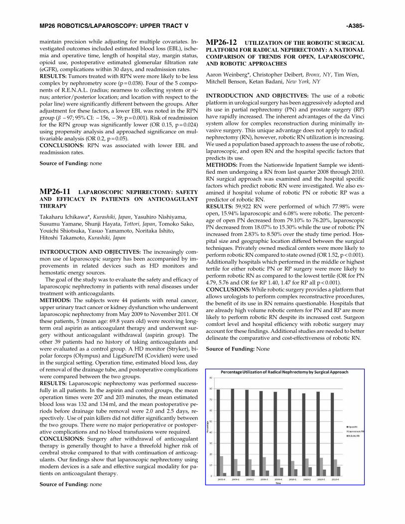
maintain precision while adjusting for multiple covariates. In-vestigated outcomes included estimated blood loss (EBL), ische-mia and operative time, length of hospital stay, margin status,opioid use, postoperative estimated glomerular filtration rate(eGFR), complications within 30 days, and readmission rates.RESULTS: Tumors treated with RPN were more likely to be lesscomplex by nephrometry score (p = 0.038). Four of the 5 compo-nents of R.E.N.A.L. (radius; nearness to collecting system or si-nus; anterior/posterior location; and location with respect to thepolar line) were significantly different between the groups. Afteradjustment for these factors, a lower EBL was noted in the RPNgroup (b - 97; 95% CI: - 156, - 39; p = 0.001). Risk of readmissionfor the RPN group was significantly lower (OR 0.15, p = 0.024)using propensity analysis and approached significance on mul-tivariable analysis (OR 0.2, p = 0.05).CONCLUSIONS: RPN was associated with lower EBL andreadmission rates.
Source of Funding: none
MP26-11 LAPAROSCOPIC NEPHRECTOMY: SAFETY
AND EFFICACY IN PATIENTS ON ANTICOAGULANT
THERAPY
Takaharu Ichikawa*, Kurashiki, Japan, Yasuhiro Nishiyama,Susumu Yamane, Shunji Hayata, Tottori, Japan, Tomoko Sako,Youichi Shiotsuka, Yasuo Yamamoto, Noritaka Ishito,Hitoshi Takamoto, Kurashiki, Japan
INTRODUCTION AND OBJECTIVES: The increasingly com-mon use of laparoscopic surgery has been accompanied by im-provements in related devices such as HD monitors andhemostatic energy sources.
The goal of the study was to evaluate the safety and efficacy oflaparoscopic nephrectomy in patients with renal diseases undertreatment with anticoagulants.METHODS: The subjects were 44 patients with renal cancer,upper urinary tract cancer or kidney dysfunction who underwentlaparoscopic nephrectomy from May 2009 to November 2011. Ofthese patients, 5 (mean age: 69.8 years old) were receiving long-term oral aspirin as anticoagulant therapy and underwent sur-gery without anticoagulant withdrawal (aspirin group). Theother 39 patients had no history of taking anticoagulants andwere evaluated as a control group. A HD monitor (Stryker), bi-polar forceps (Olympus) and LigaSureTM (Covidien) were usedin the surgical setting. Operation time, estimated blood loss, dayof removal of the drainage tube, and postoperative complicationswere compared between the two groups.RESULTS: Laparoscopic nephrectomy was performed success-fully in all patients. In the aspirin and control groups, the meanoperation times were 207 and 203 minutes, the mean estimatedblood loss was 132 and 134 ml, and the mean postoperative pe-riods before drainage tube removal were 2.0 and 2.5 days, re-spectively. Use of pain killers did not differ significantly betweenthe two groups. There were no major perioperative or postoper-ative complications and no blood transfusions were required.CONCLUSIONS: Surgery after withdrawal of anticoagulanttherapy is generally thought to have a threefold higher risk ofcerebral stroke compared to that with continuation of anticoag-ulants. Our findings show that laparoscopic nephrectomy usingmodern devices is a safe and effective surgical modality for pa-tients on anticoagulant therapy.
Source of Funding: none
MP26-12 UTILIZATION OF THE ROBOTIC SURGICAL
PLATFORM FOR RADICAL NEPHRECTOMY: A NATIONAL
COMPARISON OF TRENDS FOR OPEN, LAPAROSCOPIC,
AND ROBOTIC APPROACHES
Aaron Weinberg*, Christopher Deibert, Bronx, NY, Tim Wen,Mitchell Benson, Ketan Badani, New York, NY
INTRODUCTION AND OBJECTIVES: The use of a roboticplatform in urological surgery has been aggressively adopted andits use in partial nephrectomy (PN) and prostate surgery (RP)have rapidly increased. The inherent advantages of the da Vincisystem allow for complex reconstruction during minimally in-vasive surgery. This unique advantage does not apply to radicalnephrectomy (RN), however, robotic RN utilization is increasing.We used a population based approach to assess the use of robotic,laparoscopic, and open RN and the hospital specific factors thatpredicts its use.METHODS: From the Nationwide Inpatient Sample we identi-fied men undergoing a RN from last quarter 2008 through 2010.RN surgical approach was examined and the hospital specificfactors which predict robotic RN were investigated. We also ex-amined if hospital volume of robotic PN or robotic RP was apredictor of robotic RN.RESULTS: 59,922 RN were performed of which 77.98% wereopen, 15.94% laparoscopic and 6.08% were robotic. The percent-age of open PN decreased from 79.10% to 76.20%, laparoscopicPN decreased from 18.07% to 15.30% while the use of robotic PNincreased from 2.83% to 8.50% over the study time period. Hos-pital size and geographic location differed between the surgicaltechniques. Privately owned medical centers were more likely toperform robotic RN compared to state owned (OR 1.52, p < 0.001).Additionally hospitals which performed in the middle or highesttertile for either robotic PN or RP surgery were more likely toperform robotic RN as compared to the lowest tertile (OR for PN4.79, 5.76 and OR for RP 1.40, 1.47 for RP all p < 0.001).CONCLUSIONS: While robotic surgery provides a platform thatallows urologists to perform complex reconstructive procedures,the benefit of its use in RN remains questionable. Hospitals thatare already high volume robotic centers for PN and RP are morelikely to perform robotic RN despite its increased cost. Surgeoncomfort level and hospital efficiency with robotic surgery mayaccount for these findings. Additional studies are needed to betterdelineate the comparative and cost-effectiveness of robotic RN.
Source of Funding: None
MP26 ROBOTICS/LAPAROSCOPY: UPPER TRACT V -A385-

MP26-13 MID-TERM OUTCOMES OF LAPAROSCOPIC
RADICAL NEPHRECTOMY IN PATIENTS WITH LOCA-
LIZED RENAL TUMOR.
Luca Lunelli*, Eric Barret, Rafael Sanchez-Salas,Youness Ahallal, Laurent Mascle, Petr Macek,Camilo Giedelman, Dominique Prapotnich, Francois Rozet,Marc Galiano, Annick Mombet, Nathalie Cathala,Xavier Cathelineau, Paris, France
INTRODUCTION AND OBJECTIVES: Laparoscopic radicalnephrectomy (LRN) has become the standard technique for surgicaltreatment of renal tumors > 4 cm. There is limited long-term, pop-ulation-based evidence supporting the effectiveness of laparoscopicradical nephrectomy after its widespread adoption. The aim of thisstudy was to evaluate the results and outcomes of purely laparo-scopic radical nephrectomy among patients with kidney cancer.METHODS: A retrospective review of a series of 683 patients (1996to 2012) who underwent laparoscopic radical nephrectomy (LRN)was performed. The population was split in two match pairedgroups: Group 1 (G1) counted 341 patients treated in the period1996–2005 and Group 2 (G2) counted 342 patients treated between2005 and 2012. Groups were compared in terms of age, tumor size,intraoperative blood loss, operative time and follow-up duration.Cardiovascular events and complications, stratified by ClavienScore are reported. Percent change in serum creatinine at long-termfollow-up, overall mortality and 5-years survival rate are analyzed.RESULTS: Mean overall patient age at surgery was 62.7 years(61.9 years in G1 versus 63.4 years in G2, p = 0.1) and mean tumordiameter was 59.8 mm (G1: 60.6 versus G2: 59.6 mm, p = 0.6). TheG1 group had significantly higher mean blood loss (290 versus194 ml, p = 0.004) and operative time (135 versus 117 minutes,p < 0.001) compared with G2 group. Cardiovascular post-opera-tive events presented in 4 (0.6%) patients and 98 (14.3%) patientspresented with postoperative deterioration in renal function, asmeasured by the percent increase in serum creatinine postoper-atively (13% in G1 versus 24% in G2, p < 0.001). Median follow-uptime was 37.6 months (range 1–190 months), 183 patients had afollow up longer than 5 years. Lower complication rate was re-ported for patients who underwent LRN in the first group thanfor those operated in the second group (n = 57 vs 80, respectively).Average Clavien score was 2.2 in G1 and 2.1 in G2 (p > 0.05). Onlyone complication Clavien score 5 was registered, in G2. Overallmortality for cancer specific and non-cancer specific causes was10.2%, while overall 5 years survival rate after LRN was 90%.CONCLUSIONS: Our mid-term evaluation of LRN shows thatradical laparoscopic nephrectomy is an effective option fortreatment of localized kidney cancer. With increasing experienceit is possible to significantly reduce operative time and blood loss.Oncologic outcomes show a limited cancer related mortality.
Source of Funding: None
MP26-14 MULTI-INSTITUTIONAL ANALYSIS OF RE-
NAL FUNCTION FOLLOWING CRYOABLATION OF SMALL
RENAL MASSES IN SOLITARY KIDNEYS
Fahd Khan*, Wasim Mahmalji, Sanjeev Madaan, Ian K.Dickinson, Kent, United Kingdom, Frank X. Keeley, Bristol,United Kingdom, Brunolf Lagerveld, Amsterdam, Netherlands,Seshadri Sriprasad, Kent, United Kingdom
INTRODUCTION AND OBJECTIVES: Presence of small renalmasses ( < 4 cm) in solitary functioning kidneys represents anabsolute indication for nephron sparing surgery.
The purpose of this study was to evaluate the outcomes ofcryoablation in patients with solitary kidneys focusing on renalfunction in a large multi-centre series.METHODS: Data from 343 consecutive cases that underwentlaparoscopic cyroablation at 3 institutions was studied. Experi-enced laparoscopic surgeons performed all operations. Patientdemographics, tumour size and renal function were analysedwith specific focus on serum creatinine concentration and esti-mated glomerular filtration rate (eGFR). Data was collected pre-ablation and 3 months post-operatively.RESULTS: Fifty-two patients were identified with solitary kidney,of which two were excluded because of long-term dialysis. Of these,46 patients had solitary kidney secondary to previous malignancyand 4 had a non-functioning contralateral kidney. Cryotherapy wasperfomed laparoscopically in 46 patients and 4 were ablated per-cutaneously (CT-guided). Mean age was 65.7 years (range 44–82).Mean tumour size was 2.9 cm (range 1.5 to 5 cm). Mean pre-oper-ative creatinine was 141.8 mmol/l (range 56–404 mmol/l), 3 monthpost-operative creatinine was 148.7 mmol/l (range 66–358 mmol/l), and the percentage difference equating to 4.9%, p = 0.793. Post-operative renal function did not change significantly as measuredby eGFR. Pre-operatively it was averaged at 46.9 ml/min (range24–90 ml/min), at 3 months was recorded at 44.6 ml/min (range15–78 ml/min), percentage difference of - 5.1%, p = 0.076.CONCLUSIONS: Our findings suggest cryoablation in solitarykidneys does not alter renal function significantly, thereforecryotherapy is a viable option for small tumours.
Source of Funding: ‘‘None’’
MP26-15 LAPAROSCOPIC NEPHRECTOMY FOR
XANTHOGRANULOMATOUS PYELONEPHRITIS
Oscar Fugita*, Sao Paulo, Brazil, Paulo Kawano,Hamilto Yamamoto, Joao Amaro, Rodrigo Guerra, Botucatu,Brazil, Marcos Nogueira, Sao Paulo, Brazil, Horacio Consolmagno,São Paulo, Brazil, Americo Sakai, Sao Paulo, Brazil
INTRODUCTION AND OBJECTIVES: The laparoscopic ap-proach is currently the standard option for benign kidney diseasesrequiring nephrectomy. However, xanthogranulomatous pyelone-phritis (XGPN) sometimes is considered a relative contraindicationto laparoscopic nephrectomy, due to the severe adhesions aroundthe kidney and potential for complications. We present our expe-rience in managing XGPN through laparoscopic nephrectomy.METHODS: A retrospective chart review of 401 consecutive pa-tients that underwent laparoscopic nephrectomy (from jan/2003 todec/2012) for benign conditions was conducted, among which 41(mean age 43 years) had a histopathological diagnosis of XGPN andwere further analyzed. Transperitoneal approach was used in allcases and data regarding age, sex, preoperative symptoms, opera-tive time, bleeding, hospital stay and complications were gathered.RESULTS: Conversion rate was 10%, all of them due to no pro-gression. The mean operative time was 192 minutes and the meanestimated blood loss was 152 ml, with a mean hospital stay of 3.6days. Eight patients (20%) developed Clavien grade II complica-tions, all related to abdominal wall infection (in a previous ne-phrostomy site in one case and in the incision for specimenretrieval in another case. In six cases a trocar site infection devel-oped). There was one grade IVa complication, due to pulmonaryinfection and one grade V complication with death of the patient.CONCLUSIONS: The laparoscopic treatment of the XGPN re-mains challenging and controversial. A high complication ratecan be expected, especially from infectious causes.
Source of Funding: NONE
-A386- MP26 ROBOTICS/LAPAROSCOPY: UPPER TRACT V

MP26-16 ASSESSING THE USEFULNESS OF DIA-
METER-AXIAL-POLAR NEPHROMETRY SCORE FOR
PREDICTION OF OPERATIVE PARAMETERS IN ROBOTIC
PARTIAL NEPHRECTOMY
Young Eun Yoon*, Kyung Hwa Choi, Sang Woon Kim,Ji Yong Ha, Won Sik Ham, Koon Ho Rha, Young Deuk Choi,Woong Kyu Han, Seoul, Korea, Republic of
INTRODUCTION AND OBJECTIVES: There were some sug-gestions of nephrometry score to evaluate objective assessment ofanatomical tumor complexity. We analyzed the correlation of thebrand new diameter-axial-polar (DAP) nephrometry score withwarm ischemia time (WIT) and estimated blood loss (EBL) forpatients undergoing robotic partial nephrectomy (RPN).METHODS: A total of 101 patients underwent RPN betweenJanuary 2008 and July 2010. Preoperative computed tomography(CT) or magnetic resonance imaging (MRI) was available for 99patients, which were assigned a DAP nephrometry score. Uni-variable analysis and multivariable linear regression analysiswere carried out for factors associated with WIT and EBL duringRPN.RESULTS: Mean age was 51.9 years (62 men/37 women) and 80renal masses (80.8%) were malignant tumors. Mean tumor sizewas 3.4 – 1.7 cm and mean DAP score was 5.9 – 1.5. On univari-able analysis, a longer WIT was associated with all of the D(p < 0.001), A (p < 0.001), P (p = 0.006) components and entire DAPsum (p < 0.001). EBL was associated with larger diameter(p = 0.032), longer axial distance (p = 0.011) and entire DAP sum(p = 0.001), but not with polar distance (p = 0.078). On multivari-able linear regression analysis, all D (p = 0.002), A (p = 0.048) and P
(p = 0.004) components showed significant correlation with WIT.But, only axial distance (p = 0.028) and polar distance (p = 0.036)were significantly associated with EBL.CONCLUSIONS: As well as DAP sum, all components of DAPnephrometry predict WIT during RPN. Our data also suggest thatDAP sum is correlated with EBL of RPN. DAP nephrometry maybe used in the preoperative planning and counseling of patientsundergoing RPN.
Source of Funding: none
MP26-17 RETROPERITONEAL LAPAROSCOPIC AP-
PROACH IN UROLGY DEPARTEMENT OF IBN ROCHD
HOSPITAL UNIVERSITY: A REPORT OF 133 CASES
Redouane Rabii*, Mohamed El Mrini, Youssef Elkattani,Rachid Aboutaieb, Fathi Meziane, Casablanca, Morocco
INTRODUCTION AND OBJECTIVES: Currently the laparo-scopic extraperitoneal approach, after a difficult start has becomean attractive alternative to laparoscopic trans-peritoneal proce-dures with a lot of benefits, shorter Operative times and bettersurgical outcomes. We reported the safety and efficacy of retro-peritoneal laparoscopic approach in 133 patient in our hospital.METHODS: We reported 133 patients cases including: 57 cases ofsimple renal cyst, 30 case of nephrectomy, 5 cases of renal hydaticcyst, 5 cases of UPJ, 3 cases of renal lithiasis, 5 cases of psoas abcesand 27 cases of adrenalectomy. We performed a retroperitoneallaparoscopic approach in flank position using 3 to 5 trocars for allthe patient.RESULTS: All intervention was unrolling without incident ex-cept a case of conversion for a pyelonephretic kidney. The post-operative course was uneventful in all patients with a shorterhospital stay and faster recovery of activity.CONCLUSIONS: retroperitoneal laparoscopy is an interestingalternative to trans-peritoneal laparoscopy for a direct approachto the urinary tract without contact with the viscera with a shortoperative time and uneventful. The retroperitoneal approach of-fers significant benefits including the establishment safely trocars, especially if earlier intervention in the abdomen, the absence ofany intraperitoneal complication, decreased postoperative painand shorter laxation.
Source of Funding: university hospital IBN ROCHD casablancamorocco
MP26-18 LAPAROENDOSCOPIC SINGLE SITE PYE-
LOPLASTY AND NEPHROLITHOTOMY PERFORMED SI-
MULTANEOUSLY
Yasuyuki Naitoh*, Akihiro Kawauchi, Fumiya Hongo,Jintetsu Soh, Kazumi Kamoi, Tsuneharu Miki, Kyoto, Japan
INTRODUCTION AND OBJECTIVES: Laparoendoscopic sin-gle site surgery (LESS) for pyeloplasty was introduced to ourdepartment from 2008, and we have experienced 40 cases so far.Among them we report five cases of lithotomy performed si-multaneously through the LESS port in patients with calculus inthe distended renal pelvis.METHODS: 5 patients with PUJO and renal stones underwentpyelolithotomy performed concurrently with LESS pyeloplasty.In 4 patients, a 2.5 cm incision was made in the umbilical region.In the fifth patient, the 2.5 cm vertical incision was made at a site7 cm below the umbilical region because of an ectopic kidney inthe pelvis with renal stones. After dissection of the PUJ, an
Table 1. Association Between DAP Nephrometry
Score and WIT and EBL on Univariable Analysis
MP26 ROBOTICS/LAPAROSCOPY: UPPER TRACT V -A387-

incision of about 1 cm was made along the presumed transectionline of the renal pelvis. The pyelolithotomy was performed usinga 26Fr rigid nephroscope through a SILS port. Dismemberedpyeloplasty was performed after extraction of the renal stoneswas completed. An antegrade DJ stent was placed during theoperation in all cases.RESULTS: The mean operation time was 290 minutes (range: 225to 373) and the mean lithotomy time was 36 minutes (range: 20 to55). No intraoperative or postoperative complications were ob-served. For all 5 patients, discharge from the hospital was pos-sible after a mean of 3.4 postoperative days (range: 3 to 4). Four ofthe patients became stone-free. In the remaining 1 patient with anectopic kidney in the pelvis and 15 stones, 2 small stones re-mained. Postoperative ultrasound revealed that hydronephrosisimproved in all patients. In all patients, resolution of the symp-toms was confirmed.CONCLUSIONS: Laparoendoscopic single site surgery is a safeand effective operative procedure for pelvi-ureteric junction ste-nosis accompanied by calculus in the renal pelvis.
Source of Funding: none
MP26-19 INDUCTION OF COLD ISCHEMIA IN PA-
TIENTS WITH SOLITARY KIDNEY USING RETROGRADE
INTRARENAL COOLING: UPDATE AND 3-YEAR FUNC-
TIONAL OUTCOMES
Philip Dorsey*, New Orleans, LA, Janet Colli, Memphis, TN,Benjamin R Lee, New Orleans, LA
INTRODUCTION AND OBJECTIVES: Renal hypothermia iscommonly utilized during nephron sparing surgery to minimizeischemic tissue damage. We propose a method to induce renalhypothermia using continuous retrograde irrigation of iced salinevia dual-lumen ureteral catheter. We will report results in an exvivo porcine model followed by clinical outcomes in a series ofpatients with solitary kidney undergoing robotic-assisted lapa-roscopic partial nephrectomy (RALPN).METHODS: Following our successful use of retrograde irriga-tion of saline to achieve renal cooling in a porcine model, a seriesof patients (n = 10) undergoing RALPN with a solitary kidney hadconcomitant intra-operative retrograde renal cooling to maximizerenal preservation during surgery. A 10-Fr dual-lumen ureteralcatheter was placed at initiation of anesthesia and retrogradeinfusion of ice-cold saline via a 10-Fr dual-lumen ureteral waswas initiated prior to renal artery clamping.
Following surgery, our corhort has been prospectively fol-lowed since their surgery, with serial serum creatinine and esti-mated GFR measurements as well as surveillance imagingmaintained in our IRB approved database.RESULTS: In our porcine study, cortical temperature reached thetarget temperature ( £ 20 �C) within an average of 203 s of retro-grade irrigation. In the clinical series, patients’ mean preoperativecreatinine was 1.16 mg/dL (GFR = 60). At a median follow-up of25 months (range 7–38 months), postoperative creatinine was1.47 mg/dL (GFR = 41.94). Average clamp time was 19.4 min. Allpatients had negative surgical margins.CONCLUSIONS: The results of our long-term follow up suggestretrograde irrigation is a technically feasible method to inducecold ischemia, which may provide an additional protective effecton renal function in patients who have a solitary kidney under-going surgery via a minimally invasive approach.
Source of Funding: None
MP26-20 LAPAROSCOPIC DISMEMBERED PYELO-
PLASTY
Boris Komyakov, Bakhman Guliev*, Ruslan Aliev,Saint-Petersburg, Russian Federation
INTRODUCTION AND OBJECTIVES: Laparoscopic pyelo-plasty is minimally invasive management option for ureter-opelvic junction obstruction (UPJO). We evaluate thepostoperative and functional results of transperitoneal laparo-scopic dismembered pyeloplasty of 60 patients.METHODS: Between October 2009 and March 2013, 60 patientsunderwent laparoscopic dismembered pyeloplasty. All patientspresented a primary UPJO with dilatation of renal calyx systemwith an enlarged renal pelvis. The mean age was 32 years (range 20to 58), and of the 36 women and 24 men, 31 presented UPJO on theright side and 29 on the left side. All patients were subject to apreoperative evaluation including renal ultrasonography, diurethicrenography and intravenous urography or CT scan that revealedthe presence of severe hydronephrosis. Urinary stones were de-tected in the renal pelvis in 12 patients. we used four ports duringlaparoscopic pyeloplasty. First, 12 mm port was introduced at thelevel of the umbilicus. The peritoneal cavity was inspected by a 30-degree telescope, followed by placement of three other 5 mm ports.RESULTS: All patients were treated laparoscopically, and noopen conversions were needed. The mean operative time was 125minutes (range 90–180 min). The mean estimated blood loss wasnegligible in all patients. The mean hospital stay was 6 days (4–8 d.). No intraoperative complications have occurred. Transientanastomotic leakage occurred in three patients and was success-fully treated by conservative treatment. A persistent UPJO wasdetected at first follow-up visit in four patients and was treated byretrograde endopyelotomy in three cases and by performing anopen pyeloplasty in another case. A mean follow-up period was18 months (range 6–28) and demonstrated a 93,3% success rateafter the initial operation.CONCLUSIONS: Laparoscopically dismembered pyeloplasty isa safe and effective procedure for UPJO correction with a highoverall success rate, less morbidity and lower complication rates.
Source of Funding: None
MP26-21 RETROPERITONEAL LAPAROSCOPIC PAR-
TIAL NEPHRECTOMY: 10 YEAR SURGICAL EXPERIENCE
AND OUTCOMES
Derek Prabharasuth*, Eric Moskowitz, Andrew Fishman,Michael Grasso, Valhalla, NY
INTRODUCTION AND OBJECTIVES: Partial nephrectomyhas become a standard in the management of small renal masses.We present our surgical experience and long term outcomes inthose who underwent retroperitoneal laparoscopic partial ne-phrectomy performed over a 10 year period by a single surgeon.METHODS: We retrospectively reviewed 226 consecutive casesof retroperitoneal laparoscopic partial nephrectomy betweenFebruary 2002 and February 2012. A 3-port retroperitoneal ap-proach was used routinely. Clinical, demographic, and peri-op-eratve data were assessed. Oncological outcomes wereretrospectively analyzed with long term follow up.RESULTS: Two-hundred-eighteen patients underwent 226partial nephrectomies during this ten year period. Mean age was61.9 years and mean follow-up time was 46.4 months (0.2–126.2). Mean estimated blood loss and warm ischemia timeswere 233.2 ml (10–800) and 32.4 minutes (0–60), respectively.
-A388- MP26 ROBOTICS/LAPAROSCOPY: UPPER TRACT V

Preoperative and postoperative GFR measured by the Cock-croft-Gault equation were 90.7 mg/dL and 83.3 mg/dL, re-spectively at a mean follow-up time of 30.1 months. A total of254 tumors were removed. Mean tumor size was 2.6 cm (0.4–11.0). All pathological specimens revealed negative surgicalmargins. Final pathology revealed: clear cell (42.9%), papillarycell (19.7%), oncocytoma (13.0%), angiomyolipoma (7.5%),complex cysts (6.7%), chromophobe (5.5%), and miscellaneous(4.7%). There were 10 major complications (4.4%) including:persistent urinary leak (2.2%), pulmonary embolus (0.9%), andbleeding requiring selective embolization (1.3%). One patient(0.4%) experienced local and metastatic recurrence at a meantime of 11.9 months and 55.1 months, respectively after initialtreatment. Cancer specific survival was 100 %. Eleven patients(4.9%) were deceased from non-cancer related events at a meanfollow-up time of 94.1 month.CONCLUSIONS: Retroperitoneal laparoscopic partial nephrec-tomy has become our first line therapy for patients with smallrenal masses given its low complication rate and good surgicaloutcomes. Our long term results show durable cancer specificsurvival and preservation of renal function.
Source of Funding: none
MP26-22 LAPAROSCOPIC NEPHROURETEREKTOMY
IN A PATIENT WITH UNUSUAL STONE LOCALISATION
Bilal Firat Alp, Ankara, Turkey, Zafer Demirer, Eskisehir, Turkey,Seref Basal*, Ali Guragac, Ankara, Turkey, Ercan Malkoc, Corlu,Turkey, Hasan Cem Irkilata, Ibrahim Yidirim, Ankara, Turkey
INTRODUCTION AND OBJECTIVES: Open or laparoscopicsurgery for stones of the upper urinary tract has very few indi-cations such as failure or complications of the other techniques,stones grater than 2 cm, hard stones, anatomical abnormalitiesand complex stones. We presented a case with unusual stoneformation wich is nearly filling all upper urinary tract that man-aged by transperitoneal laparoscopic nephroureterectomy.METHODS: A 21 year old male patient presented with longstanding right flank pain and palpable hard right kidney. Kidney-ureter-bladder (KUB) radiography and then non-enhanced threedimentional computerized tomography (3 D CT) scan revealedgiant kidney Stones measuring 4,2 · 2,5 cm and 2,0 · 1,5 cm andproximal ureteral stones short axis < 1,0 cm causing ureteralobstruction and multiple ureteral stones wich are filling up uri-
nary tract to iliac vessel crosswise (figure 1–2. Transperitoneallaparoscopic right nephroureterectomy was performed and adrain was replaced. The macroscopy of the specimen was whiteand cheesy (figure 3).RESULTS: Total operating time was approximately 180 minuteswith estimated blood loss of 200 ml. Histopathological examina-tion of the specimen revealed chronic pyelonephritis and chronicureteritis however there was not any granulomateus reaction.The patient had a smooth post-operative recovery. Drain wasremoved postoperatif 2nd dayand patient discharged. After sixweaks of surgery patient controlled with urinary ultrasonogra-phy and no pathological events found.CONCLUSIONS: Since laparoscopic nephroureterectomy pro-vides less operative blood loss, earlier oral intake, less con-sumption of analgesics as well as shorter hospital stay andconvalescence, ýt can be a good choose for the treatment ofcomplex urinary stones.
Source of Funding: none
MP26-23 INITIAL OPERATIVE EXPERIENCE WITH
ROBOTIC URETERAL RECONSTRUCTION DISTAL TO THE
URETEROPELVIC JUNCTION
Gordon Fifer*, Matthew Raynor, Michael Woods, Eric Wallen,Raj Pruthi, Chapel Hill, NC
INTRODUCTION AND OBJECTIVES: The robotic platformhas become commonplace in urologic surgery, and reconstruc-tion of the ureteropelvic junction (UPJ) was one of its earliest uses.Yet despite the increased use of robotics, there are very few re-ports of robotic ureteral reconstruction for pathology distal to theUPJ. We are reporting our initial results with a relatively largeseries of patients with distal ureteral obstruction.METHODS: A retrospective chart review was performed on 44patients who underwent robot-assisted reconstruction of the ureterdistal to the UPJ for either benign or malignant obstruction betweenDecember 2009 and May 2013. The procedures performed includedsegmental ureterectomy; ureteroneocystotomy; psoas bladder hitch;Boari Flap; ureteroureterostomy; end-to-side ureterureterostomy forduplicated system; ureterolithotomy; ureterolysis; and ureteral re-implant into orthotopic neobladder. Flexible endoscopy was per-formed concurrently in 22 cases. Preoperative, intraoperative, andpostoperative characteristics were extracted and analyzed.RESULTS: The etiology was benign, idiopathic, or iatrogenic in34 patients, and 10 had a malignant cause. The average patient
MP26 ROBOTICS/LAPAROSCOPY: UPPER TRACT V -A389-

was 54 years old with a BMI of 30. Twenty three patients had anephrostomy tube in place at the time of reconstruction. Theaverage total operative time was 234 minutes, with endoscopyuse averaging an additional 25 minutes. Estimated blood lossaveraged 81 ml. For the 19 patients with a known date of injury,the median time to repair was 91 days. The average length of staywas 1.7 days, with 29 patients discharged on POD 1. There wereno intraoperative complications. Surgical margins were negativefor all malignant cases. One patient required a single bloodtransfusion postoperatively; 1 patient was transferred to the SICUfor less than 1 day for a transient desaturation event; and 1 patienthad an elevated troponin requiring no interventions. Duringshort term follow up, 1 patient required prolonged ureteralstenting, and 1 required ureteral balloon dilation.CONCLUSIONS: Robotic reconstruction of the ureter distal tothe UPJ is feasible, safe, and able to reproduce similar techniquesto open surgery. It is also extremely flexible, allowing the surgeonto choose from a large variety of procedures to best suit the dis-ease process in the patient being treated at the time of surgery.Additional studies with larger populations, extended follow-uptime, and perhaps even randomization to compare against tra-ditional open surgery will help to further determine the long termsuccess of these operations.
Source of Funding: none
MP26-24 ROBOTIC PARTIAL NEPHRECTOMY: THE
IMPACT OF WARM ISCHEMIA TIME
Mohamad Salkini*, AbdulRaouf Lamoshi, Morgantown, WV
INTRODUCTION AND OBJECTIVES: Robotic partial ne-phrectomy (RPN) is a technically challenging procedure. Pre-paring the renal hilum, tumor resection, hemostasis, andrenorrhaphy should be accomplished within reasonable time.Robotic surgery found great acceptance as it always conveyedshorter warm ischemia time (WIT) compared to traditional lap-aroscopic approach. We present our current series of RPN todemonstrate the effect of warm ischemia on post operative kid-ney function.METHODS: Seventy patients underwent RPN at our institute.We studied 42 patients whose kidney underwent RPN with WITexecuted with hilar clamping.RESULTS: Patient demographic, mean patient age was 56.4 yr(range: 34–83 yr), 17 males (48.6%) and 18 females (51.4%). MeanWIT was 20.26 min (SD 5.69) (range: 11- 34 min).
Mean estimated blood loss (EBL was 299 ml (range: 50-2000 ml). Hospital stay length range is 1–6 days (mean 2.43).Mean overall tumor size was 3.19 cm (range: 2–7 cm). All patientshad negative surgical margins except one (2.38 %).
The creatinine level of the patients with malignant pathologyhas been checked on the second post-operative day (short termreading) and within 3–6 months after surgery (long term read-ing). The mean preoperative creatinine was 1.13 mg/dL (range0.52–2.5) and the early post-operative (48 hrs) creatinine 1.86 mg/dL (range 0.54 âe‘‘ 3.4). The mean long term creatinine was1.24 mg/dl (range 0.68–2.7 ng/ml).
WIT and EBL do not show significant correlation (P = .6983),warm ischemia time and hospital stay length do not show sig-nificant correlation (P = .79), and warm ischemia time and shortterm creatinine do not show significant correlation (P = .91).CONCLUSIONS: WIT shorter than 34 min has no effect on EBL,early post-operative renal function and length of hospital stay.
Source of Funding: None
MP26-25 A SINGLE SURGEON, SINGLE CENTRE, 5
YEARS EXPERIENCE IN RETROPERITONEOSCOPY- AN
INDIAN SCENARIO
Venugopal Ganapathy, Haris C H*, Thiruvananthapuram, India,Sudin S R, Kollam, India, Navin C Angus, Ginson V George,Sandeep Krishnan, Thiruvananthapuram, India, Suchindra P Unni,Kochi, India, Jibu K Pillai, Thiruvananthapuram, India,Jithunath M R, Thrissur, India
Table 1. Mean Perioperative Patient Parameters
Table 2. Number and Cause for Conversion
in Each Surgery Group
-A390- MP26 ROBOTICS/LAPAROSCOPY: UPPER TRACT V

INTRODUCTION AND OBJECTIVES: Retroperitoneoscopy(RPS) has evolved in the recent past as an effective alternative tothe conventional Transperitoneal laparoscopy in the manage-ment of upper urinary tract pathologies (UUTP). Our Objective isto study the perioperative patient outcomes of 156 RPS carriedout for various UUTP in our instituition and to address the meritsand demerits of RPS.METHODS: In the period from October 2008-January 2013,156RPS procedures were carried out on 156 patients for various UUTP.All these patients were retrospectively analysed for perioperativepatient outcomes. Datas are expressed in mean and standard de-viation;Student’s T-test used to compare mean between 2 groups.RESULTS: The results are tabulated in table 1 and table 2. In ourseries, CROSSING VESSEL was present in 65% of PYELO-PLASTY cases. Average Stone size in PYELOLITHOTOMY cases
is 3.32 cms, in ureterolithotomy cases is 2.21 cms. Average Tumorsize in RADICAL NEPHRECTOMY cases is 6.2 cms. AverageCyst size in DEROOFING cases is 7.37 cms. The merits of RPS are-Blood loss was minimal and blood transfusion was not requiredin any of these cases;No incidence of trocar related visceral in-juries, pneumothorax, basal atelectasis and paralytic ile-us;Analgesia use was less;Early convalescence;No incidence ofPort site herniation, intestinal obstruction, internal herniation inany of these cases with a mean follow up 21.6 months. The de-merits are-Difficult in obese patients and tumor > 10 cm size.CONCLUSIONS: Thus RPS proves to be an effective alternativeto conventional Transperitoneal laparoscopy in the treatment ofUUTP.
Source of Funding: none
MP27 ROBOTICS/LAPAROSCOPY: UPPER TRACT VI
MP27-01 LAPAROSCOPIC RADICAL NEPHRECTOMY
FOR CLINICAL T1B AND T2A RENAL MASSES. DOES THE
APPROACH MAKE A DIFFERENCE? COMPARISON OF
LAPARO-ENDOSCOPIC SINGLE-SITE AND MULTIPORT
LAPAROSCOPIC SURGERY
Omer Raheem, Michael Liss, La Jolla, CA, Reza Mehrazin,Memphis, TN, Jason Woo, Sean Stroup, Ithaar Derweesh*,La Jolla, CA
INTRODUCTION AND OBJECTIVES: To compare outcomesof laparoendoscopic single-site surgery (LESS) and multiportlaparoscopic (MPL) Radical Nephrectomy (RN) for clinical (c)T1b and T2a renal masses, focusing on discharge and pain pa-rameters.METHODS: Retrospective comparison of LESS- and MPL-RNbetween 7/2005 and 11/2012. 63 patients underwent LESS-RN(44 cT1b/19 cT2a); 133 underwent MPL (83 cT1b/50 cT2a) per-formed by a single surgeon. LESS transperitoneal access wasobtained by peri-umbilical incision through which all trocarswere inserted. LESS-RN recapitulated steps of MPL-RN; all pa-tients were managed with standardized clinical care pathway.Variables were compared between MPL and LESS groups. Pri-mary outcome was length of hospital stay (LOS).RESULTS: 130/133 MPL and 62/63 LESS cases were successfullyperformed (median follow-up 24.7 months). No significant de-mographic differences were noted. For MPL and LESS groups:cT1b mean tumor diameter (cm) was 5.3 – 0.8 and 5.4 – 0.8(p = 0.689); cT2a mean tumor diameter (cm) was 8.2 – 0.8 and8.3 – 0.9 (p = 0.728); mean RENAL score was 10.0 – 1.1 and9.7 – 1.2 (p = 0.12); mean OR time (min) was 126.3 – 42.9 vs.132.7 – 38.5 (p = 0.314). No significant differences in blood loss(p = 0.49) and complications (p = 0.781) were noted. LESS-RN hadsignificant reductions in LOS (2.14 – 0.95 vs. 2.45 – 1.01 days,p = 0.043), discharge pain-score (1.3 – 1.1 vs. 2.2 – 0.9, p < 0.001),and narcotic use (5.9 – 5.4 vs. 10.7 – 4.2 morphine equivalents,p < 0.001). No significant differences were noted for disease-specific survival (MPL 94% vs. LESS 97%, p = 0.678).CONCLUSIONS: In this well-matched retrospective compari-son, LESS- was comparable to MPL-RN for cT1b/cT2a renal tu-
mors for perioperative parameters and may confer benefit in LOSand analgesic requirement. Prospective randomized evaluation isrequisite.
Source of Funding: None
MP27-02 THE PRESENCE OF GLOMERULOSCLER-
OSIS IS NOT ASSOCIATED WITH WORSENED DECLINE IN
RENAL FUNCTION FOLLOWING ROBOTIC ASSISTED
PARTIAL NEPHRECTOMY.
Michael Johnson*, Jonathan Mobley, Joel Vetter,R. Sherburne Figenshau, Sam Bhayani, Saint Louis, MO
INTRODUCTION AND OBJECTIVES: Pre-operative estimatedglomerular filtration rate (eGFR), warm ischemia time, and theresected volume of marginal healthy tissue are known indepen-dent predictors for reduced renal function following minimallyinvasive partial nephrectomy. The aim of this investigation is toevaluate whether the pathological presence of renal damagemanifested by glomerulosclerosis alters renal functional statusafter partial nephrectomy.METHODS: Thirty-three patients with post-operative GS in thenonneoplastic renal parenchyma following RAPN were com-pared to 33 patients lacking GS (controls). This was a case controlstudy with matching of cases by nephrometry score. The patientsin both cohorts possessed a pre-operative eGFR greater than60 ml/min per 1.73 m2 and were matched based on ne-phrometery scores and pre-operative eGFR. The change in cre-atinine and eGFR using the Chronic Kidney DiseaseEpidemiology Collaboration formula following RAPN werecompared for each group.
MP27 ROBOTICS/LAPAROSCOPY: UPPER TRACT VI -A391-

RESULTS: The GS and controls were similar in age, tumor size,nephrometry scores, pre-operative creatinine, pre-operativeeGFR, and warm ischemia times (Table 1.) The mean increase increatinine for the GS group was 0.11 mg/dL compared to 0.09 forthe controls. The GS group experienced a mean decrease in eGFRof 9.4 ml/min, while the control’s eGRF decreased by 8.1 ml/min.There was no significant difference in the change in creatinine(p = 0.689) or eGFR (p = 0.636) in the two groups following RAPN.CONCLUSIONS: Patients with glomerulosclerosis did not ex-perience a worsened decline in renal function following RAPNwhen compared to controls. The presence of pathological renaldamage (GS) is not associated with any change in eGFR.
Source of Funding: none
MP27-03 HAND-ASSISTED LAPAROSCOPIC LIVING
DONOR NEPHRECTOMY (HALDN): A 10-YEAR SINGLE-
CENTER EXPERIENCE IN ORIENTAL POPULATION
Yoshihide Higuchi*, Akihiro Kanematsu, Kimihiro Shimatani,Takeshi Hanasaki, Toeki Yo, Yukako Nakanishi,Yoshikazu Togo, Shuken Go, Toru Suzuki, Michio Nojima,Shingo Yamamoto, Nishinomiya, Japan
INTRODUCTION AND OBJECTIVES: Hand-assisted laparo-scopic donor nephrectomy (HALDN) has evolved as a popularprocurement method of kidney graft for its safety and minimaloperative invasion. We have performed Hand-assisted laparo-scopic donor nephrectomy (HALDN) since 2003 in our institute.The aim of this study is to evaluate the utility and safety ofHALDN, in regard of life-threatening complications such as se-vere bleeding and function of transplanted kidney.METHODS: We analyzed 90 consecutive living donors whounderwent HALDN from February 2003 to March 2013. The datahas been collected prospectively and included all consecutivedonors operated with the HALDN. In this study we investigatedthe blood loss, complication rate and allograft outcome ofHALDN in our institute.RESULTS: Mean patient age was 57 ,} 12 years old. Nineteen(21.1%) right and 71 (78.9%) left kidneys were harvested. Eightyseven cases (96.7%) underwent tranperitoneal approach, and 3cases (3.3%) underwent retroperitoneal approach. Six of 90(6.6%) cases required conversion to an open procedure. Meanlaparoscopic operative time and estimated blood loss was 242,} 66 minutes and 202 ,} 228 mL, respectively. Mean warmischemia time (WIT) was 5 ,} 2 minutes. No donor requiredblood transfusion. All kidneys had immediate onset of function.Grafts with plural vessels, (double renal arteries (N = 12), triplearteries (N = 2) and double renal vein (N = 6)), tended to result in
longer WIT, and longer Reperfusion-to-urine secretion time(RUT). There was no significant difference in laparoscopic op-eration time, blood loss and RUT without WIT according to BMI(Table 1). The incidence of complications was 20 / 90 (22.2%).According to Clavien grading, there were 15 (16.7%), 4 (4.4%),and 1 (1.1%) cases for grade 1, 2, and 3 complication, but therewas no life-threatening morbidity. Two donor was reoperateddue to ileus. Six patients experienced minor complications (liverdisafunction, n = 2; urinary retention, n = 1; gastric ulcer, n = 1;subcostal pain, n = 1 Malfunction of Endo-TA, n = 1). One re-cipient experienced urinary leakage from graft at the site of sa-crified arterial branch.CONCLUSIONS: The hand-assisted method for laparoscopicdonor nephrectomy is a safe procedure with excellent control ofbleeding and steady procurement of graft viability.
Source of Funding: none
MP27-04 R.E.N.A.L. NEPHROMETRY SCORE DOES
NOT PREDICT SURGICAL COMPLEXITY
Nikhil Waingankar*, Arvin George, Mostafa Sadek,Louis Kavoussi, New Hyde Park, NY
INTRODUCTION AND OBJECTIVES: The R.E.N.A.L. Ne-phrometry score was developed primarily to be a standardizedschema for anatomical classification of renal tumors, and sec-ondarily to classify tumor resectability. The purpose of our studywas to evaluate this scoring system’s ability to predict surgicalcomplexity for laparoscopic partial nephrectomy (LPN).METHODS: Nephrometry scores were calculated for 300 con-secutive LPNs and added to our prospectively collected operativedatabase. High complexity tumors (nephrometry sum 10–12) andlow complexity tumors (nephrometry sum 4–6) that were re-sected with hilar clamping were compared for all standard peri-operative data points using independent sample t-test forcontinuous data and Chi-square test for categorical data.RESULTS: 73 low- and 31 high complexity tumors were resectedwith hilar clamping. Age, ASA, and clamp times were equivalentbetween the two groups. For low vs. high complexity tumors, nosignificant differences were found in EBL (261.5 vs 328.8 cc), ORtime (144.7 vs 137.1 min), change in hematocrit (6.1 vs 6.9%),positive margins (1.4% vs 0%), intra- or postoperative transfu-sions (1.4 vs 0%, and 1.4 vs 6.5%), complications (15.1% vs 25.8%),or length of stay (2.3 vs 2.6 days). All p-values > 0.05. While lowcomplexity tumors had a significantly lower immediate postop-erative change in Cr compared to high complexity tumors (0.15 vs0.38, p = 0.005), this effect normalized on long term follow up.CONCLUSIONS: Our study demonstrated no differences inperioperative values between low and high complexity tumors.While R.E.N.A.L. Nephrometry score remains an excellent way toanatomically classify tumors, the sum-based classification groupsdo not predict surgical complexity.
Source of Funding: none
MP27-05 THE INCREASED POSTOPERATIVE RENAL
FUNCTION LOSS AFTER LAPAROSCOPIC PARTIAL NE-
PHRECTOMY WITH GREATER WARM ISCHEMIA TIME
TENDS TO BE RECOVERED IN MID TERM FOLLOW-UP
Selcuk Erdem*, Abubekir Boyuk, Tzevat Tefik, Feyyaz Ural,Halim Issever, Ismet Nane, Oner Sanli, Istanbul, Turkey
Corelation Between Operation Outcome
and Number of Vesssels or BMI
LOT: Laparoscopic operative time, BL: Blood loss, WIT: warmischemia time, RUT: Reperfusion-to-urine secretion time.
-A392- MP27 ROBOTICS/LAPAROSCOPY: UPPER TRACT VI

INTRODUCTION AND OBJECTIVES: The aim of this study isto investigate the effect of warm ischemia time (WIT) on earlypostoperative and follow-up renal functions in laparoscopicpartial nephrectomy (LPN).METHODS: Between February 2008 and March 2013, LPN wasperformed for 141 suspected renal tumors in our center. Afterexcluding the operations without warm ischemia application(n = 16), 125 LPNs were included in this analysis. The cases werestratified into three groups according to WIT: Group 1 (n = 42,WIT < 20 min), Group 2 (n = 66, 20 min £ WIT < 40 min) and Group3 (n = 17, 40 min £ WIT) The prospectively collected demograph-ical, perioperative and renal functional outcomes were retro-spectively compared between three groups. Glomerular filtrationrates (eGFR) were calculated using ‘Modification and Diet inRenal Disease-MDRD’ formula. Appropriate tests were used instatistical analyses.RESULTS: Between demographically similar three groups, pre-operative tumor size were significantly lower in Group 1.(p = 0.008) The median operative times (p < 0.001) and estimatedblood loss (p = 0.009) were significantly different between groups,with an increase in parallel to group order (Table 1). There was nodifference between groups in terms of complication rates(p = 0.095). The postoperative Cr increase (DeltaCr1, p < 0.001)and Cr decrease during follow-up (DeltaCr3, p = 0.005) weresignificantly lower in Group 1. While postoperative eGFR de-crease (DeltaeGFR-1) was significantly different between groups(p < 0.001), this decrease was higher in Group 3 (Median:59) andlower in Group 1 (Median:78.5) After mid-term follow-up, eGFRincrease in Group 3 (Median:12) and Group 2 (Median:6) werestatistically different from Group 1 (Median:-1), with a meaningof recovery in renal functions (Figure 1).CONCLUSIONS: In this limited laparoscopic partial nephrec-tomy experience, it is observed that warm ischemia time morethan 20 minutes significantly reduces renal functions in postop-erative period. However, renal functions tend to rise nearer topreoperative levels after mid-term follow-up. Nevertheless, theanalyses with long-term follow-up in large series will clearlydetermine the effect of warm ischemia time on renal functions
Source of Funding: None
MP27-06 PORT-SITE METASTASIS AFTER SURGERY
FOR RENAL CELL CARCINOMA: A HARBINGER OF FU-
TURE METASTASIS
Joseph Song*, Eric Kim, Jonathan Mobley, Goutham Vemana,Youssef Tanagho, Joel Vetter, Sam Bhayani, Robert Figenshau,St Louis, MO
INTRODUCTION AND OBJECTIVES: Port site metastasis(PSM) is a rare occurrence following minimally invasive treat-ment for renal cell carcinoma (RCC). However, it has remainedunclear what the prognostic implications may be as reports in theliterature are heterogeneous in detail and follow-up. We sought
Table 1: Cases of RCC PSM identified in the literature
FIG. 1: Cancer-specific survival after diagnosis of PSM.
MP27 ROBOTICS/LAPAROSCOPY: UPPER TRACT VI -A393-

to clarify the significance of PSM on cancer-specific survival andbroaden our understanding of this phenomenon.METHODS: A MEDLINE search for published studies of RCCPSM was performed. Results were mined for information oncontributing factors to PSM, stage, Fuhrman grade, pathology,PSM treatment method, follow-up protocol, and long-term out-comes. Corresponding authors of each publication were con-tacted to fill in details and provide long-term outcomes. Weadded one case from our recent experience.RESULTS: 15 cases from 11 authors were found (table 1). Eight ofthe 11 authors were available for correspondence, and 9 caseswere updated. Cancer-specific survival curves on these (fig 1)showed poor prognosis with a 22% 1-year survival rate from PSMdiagnosis. 12/15 cases involved multiple metastases in additionto the PSM. Eight of the cases had no identifiable technical reasonto explain PSM formation, such as specimen morcellation, ab-sence of entrapment, and tumor rupture. These tumors wereuniformly aggressive, Fuhrman grade of 3 or higher.CONCLUSIONS: PSM after minimally invasive surgery for RCCis a rare occurrence with poor prognosis. In the majority of cases,PSM is not an isolated metastasis but is instead a harbinger ofprogressive disease. While technical factors can play a role in PSMformation, it appears that biological factors like high tumor gradealso contribute to PSM.
Source of Funding: none
MP27-07 ROBOT-ASSISTED URETERECTOMY AND
URETERAL REIMPLANT FOR MANAGEMENT OF DISTAL
URETERAL MALIGNANCIES: SINGLE INSTITUTION EX-
PERIENCE WITH LONG FOLLOW-UP
Alonso Carrasco*, Matthew Gettman, George Chow,Matthew Tollefson, Rochester, MN
INTRODUCTION AND OBJECTIVES: Ureterectomy withureteral reimplant has been shown to have equivalent oncologicoutcomes to nephroureterectomy for management of upper tracturothelial carcinoma (UC) of distal ureter. We present our expe-rience with Robotic-Assisted Ureterectomy and Ureteral Re-implant (RAUUR) in management of UC of the distal ureter.METHODS: A retrospective chart review at Mayo Clinic wasperformed.RESULTS: We identified 15 patients with mean age of 70 year attime of surgery. Mean operative time, blood loss, and length ofhospitalization were, 249 minutes, 118 mL, and 2.7 days, respec-tively. Ten patients (67%) required reconstruction of the bladder(7 with psoas hitch and 3 with Boari flap). One patient was con-verted to open and 1 patient developed anastomotic stricture 5months postop. There were 4 recurrences at mean follow-up of 22months, all within the bladder (3) or contralateral renal pelvis (1).CONCLUSIONS: The RAUUR with or without pelvic lympha-denectomy and complex bladder reconstruction can be safelyperform with excellent intermediate term oncologic outcomesand preservation of ipsilateral renal function.
Source of Funding: None
MP27-08 COMPARATIVE ANALYSIS OF VASCULAR
BULLDOG CLAMPS USED IN ROBOTIC PARTIAL NE-
PHRECTOMY
Brian Le, Richard Matulewicz, Samuel Eaton*, Kent Perry,Robert Nadler, Chicago, IL
INTRODUCTION AND OBJECTIVES: During robotic (RPN)and laparoscopic (LPN) partial nephrectomy, clamping of thevascular pedicle prior to excision of the tumor is the key tominimize blood loss and maintain adequate visualization.Multiple options for hilar control exist, including recently in-troduced robotic specific bulldog vascular clamps. The relativeefficacy of these new clamps has not been assessed. We con-ducted a comparative analysis of robotic and laparoscopic vas-cular clamps focusing on clamp force and flow across a clampedmodel vessel.METHODS: We compared ten different vascular clamps used inRPN and LPN: the Klein robotic, Klein laparoscopic short, me-dium straight, medium curved, and long, Aesculap short straight,short curved, long straight and long curved, and a laparoscopicSatinsky clamp. Force testing was done at 1 cm and 2 cm distancesfrom the fulcrum and at the tip. To simulate a clamped vessel, aquarter inch penrose was clamped and the proximal end attachedto a pressure flow monitor. Flow across the tubing at variouspressures and leak point pressure (LPP) were recorded. Com-parative analysis was conducted using descriptive statistics and t-tests.RESULTS: The Klein robotic clamp exerted significantly lessclamp force along the length of the clamp compared to the lap-aroscopic placed Klein, Aesculap and Satinsky clamps (p < 0.001).In our vascular model, the Klein robotic clamp had a LPP of8.3 mm Hg vs. 36.3 mm Hg (Klein Lap) and > 170 mm Hg (Aes-culap) (p < 0.001). At all pressures, the robotic clamp allowedmore flow compared to laparoscopic placed clamps.CONCLUSIONS: Robotic vascular bulldog clamps producesignificantly less clamping force than laparoscopic bulldog orSatinksy clamps and thus allow more flow across the clampedsegment. This difference appears to be intrinsic to the clamp itself.This may translate into poorer hemostasis intraoperatively.
Source of Funding: none
-A394- MP27 ROBOTICS/LAPAROSCOPY: UPPER TRACT VI

MP27-09 STATIN THERAPY MAY BE ASSOCIATED
WITH INCREASED RISK OF LONG TERM RENAL DYS-
FUNCTION FOLLOWING HILAR CLAMPING IN ROBOTIC
PARTIAL NEPHRECTOMY
Louis Krane*, Victor Romero, Ashok Hemal, Winston-Salem, NC
INTRODUCTION AND OBJECTIVES: Many theories andtechniques have been described to improve long-term renalfunction in patients undergoing robotic assisted partial ne-phrectomy. Statins are known to improve endothelial functionand their continuation at the time of surgery has been associatedwith improved renal functional outcomes. We evaluated the roleof statins in long term renal function following robotic partialnephrectomy with hilar clamping.METHODS: 176 consecutive patient undergoing robotic partialnephrectomy who had hilar clamping at a single institution wereinclude in this analysis. Statin medications were evaluated fromhome medication lists and all were continued perioperatively.Chronic kidney disease stage progression was defined as in-creasing from stage I or II (GFR > 60) to stage III (GFR < 60) orhigher. Patients who progressed from Stage III to Stage IV orhigher were also included. GFR was calculated using the MDRDequation preoperatively, perioperatively (highest serum Cr attime of hospitalization) and at last follow-up.RESULTS: Fifty three patients were on statin therapy and hadhilar clamping at the time of the procedure. These patients did nothave increased body mass index (p = 0.91), or tumor size(p = 0.76), and nephrometry score distribution (p = 0.24) wassimilar between the two groups. These patients did have a highermedian age (65 vs 57 years, p < 0.001). Median follow up was 12months (IQR 3–20). Cox proportion hazards evaluating risk ofCKD progression including tumor size, ischemia time, statin us-age and age (as a continuous variable), statin usage was not sta-tistically significantly associated with improvement in renalfunction. In fact it was associated with a non-significant increasedrisk of progression of CKD stage (RR 2.65, 95%CI 0.88–8.02,p = 0.02). No other factors were statistically significantly associ-ated with progression of CKD.CONCLUSIONS: Statin therapy may not be reno-protective inpatients undergoing hilar clamping at the time of robotic assistedpartial nephrectomy. Further investigations into the role of thesemedications at time of renal ischemia is warranted.
Source of Funding: None
MP27-10 ROBOT-ASSISTED CYSTECTOMY WITH SI-
MULTANEOUS NEPHROURETERECTOMY: CASE SERIES
FROM A SINGLE INSTITUTION
Helen Levey*, Justin Houman, Anees Fazili, Hani Rashid,Guan Wu, Rochester, NY
INTRODUCTION AND OBJECTIVES: Patients with concur-rent urothelial carcinoma of the bladder and upper urinary tractmay necessitate radical cystectomy with simultaneous nephro-ureterectomy. Our institutional experience with robot-assistedcystectomy (RAC) with concomitant nephroureterectomy (NU)represents one of the largest case series to date of this combinedprocedure.METHODS: From February 2007 to January 2012, we performed8 RAC with simultaneous unilateral NU. Four of these patientsunderwent a RAC with ileal conduit diversion, and four patientsunderwent RAC with a cutaneous ureterostomy. NU was com-pleted first, with patient in the lateral decubitus position; and the
patient was then re-positioned supine for the RAC component.Patient demographics, peri-operative data as well as pathologicaland surgical outcomes were analyzed.RESULTS: All procedures were successfully performed roboti-cally without conversion to laparotomy. There were 5 male and 3female patients, with a median age of 75.5 years (range 59–89),median BMI of 26.3 (range 21–39), median pre-operative GFR of48.5 (range 20–100), median ASA of 3 (range 2–4), and medianCharleston Co-morbidity Index score of 8 (range 5–9). Intra-oper-atively, median EBL was 400 mL (range 200–1200), and medianoperative time was 431 min (range 260–480). Median lymph nodeyield was 13.5 nodes (range 4–47). Only one patient had an intra-operative complication, consisting of a recognized rectal injury thatwas primarily repaired. Median length of hospital stay was 8 days(range 5–28). Two patients had a planned immediate post-opera-tive ICU admission, and no patient had an unplanned admission tothe ICU. Five patients experienced a post-operative complication,although these were all low grade. No patients had a return to theoperating room within 30 or 90 days. One patient required re-placement of a dislodged ureteral stent in his cutaneous ureter-ostomy by Interventional Radiology at two months post-operatively. Surgical margins were ultimately negative in 7 pa-tients. With a median follow-up of 14 months, no patient experi-enced a local recurrence of tumor, and only 1 patient experienced ametastatic recurrence, occurring at 16 months post-operatively.CONCLUSIONS: In patients with concurrent urothelial carci-noma of the bladder and upper urinary tract, RAC with eitherileal conduit diversion or cutaneous ureterostomy and concomi-tant NU is a technically feasible and safe procedure.
Source of Funding: None
MP27-11 OUTCOMES IN PARTIAL NEPHRECTOMY
AT UNIVERSITY HOSPITAL COVENTRY AND WARWICK-
SHIRE, UK
Claire Webster*, Coventry, United Kingdom, Salil Umranikar,Solihull, United Kingdom, Husam Ibrahim, Anthony Blacker,Michael Wills, Coventry, United Kingdom
INTRODUCTION AND OBJECTIVES: Partial nephrectomy(PN) can be offered in many renal tumours where appropriate aspart of a nephron sparing approach. PN is associated with lessmortality, less morbidity from cardiovascular events and betterlong term renal function. There is currently no proven increase inrecurrence rates in long term follow up. The procedure can beperformed open, laproscopically or robotically. We present ourexperiences and outcomes in PN in our unit.METHODS: All PN procedures performed at our hospital wereanalysed. All patients had a standard work up and additionally apre-operative arterial phase CT. Vascular control was achieved inall cases although clamping the vessels and introperative coolingwas not routinely performed. The procedure was performed by 2experienced surgeons. Patient demographics, operative data,histology, and follow up blood results were extracted.RESULTS: There were 55 procedures performed; 30 male and 25female patients; between the ages of 24 to 84. 50 of the procedureswere performed laparoscopically, and 5 open, one of which was aconversion due to intraoperative bleeding. Two patients hadabsolute indications for PN (unilateral kidney), one relative (im-paired renal function) and the remainder had normal contralat-eral kidneys. Tumour size ranged from 1.5–18 cm, and sizes oftumours offered a PN appears to have increased over time. 13patients had positive margins, however no recurrences have beendocumented to date. There was an average haemoglobin drop of
MP27 ROBOTICS/LAPAROSCOPY: UPPER TRACT VI -A395-
2.6 g/dl. Renal function was not affected immediately post op-eratively, and at three year follow up in most patients.CONCLUSIONS: PN appears to be an effective alternative toradical total nephrectomy with regards to long-term outcomes.Partial nephrectomy in our study has preserved renal function inour patients. The overall rate of complications were comparableand positive margins have not been linked with cancer recur-rences to date. Longer term outcomes are awaited.
Source of Funding: none
MP27-12 RETROPERITONEAL LAPAROSCOPIC LIV-
ING DONOR NEPHRECTOMY: REPORT OF 151 CASES
Hongxian Zhang*, Xian Zhang, Lei Zhao, Lulin Ma, Beijing,China, People’s Republic of
INTRODUCTION AND OBJECTIVES: To summary our ex-perience of 151 retroperitoneal laparoscopic living donor ne-phrectomies.METHODS: A total of 151 donors underwent retroperitoneallaparoscopic living donor nephrectomy from Dec 2003 to Dec2012. The operation was performed through 3 lumbar ports;after the kidney was liberated fully and the ureter was severed7–8 cm under the lower pole of kidney, the renal artery andvein were blocked with Endo-cut or hem-o-lok separately andthen severed. Endo-cut was used in 3 patients and hem-o-lok in148 donors. Then the kidney was taken out quickly from thedonor and infused with 4¡æ kidney preserving fluid immedi-ately.RESULTS: The 151 operations were successful. Operation timewas 86 min (54¡«180 min) and blood loss was 50 ml (20¡«200 ml),no patient needed blood transfusion. Warm ischemia time 3.6 min(2–8 min). 3 patients had hematoma of renal fossa after operationand they required no further treatment. Hospital stay after op-eration was 5 days (3.5¡«9 days). 141 of 151 donors were followedup for 61 months (10¡«96 months).CONCLUSIONS: Retroperitoneal laparoscopic living donor ne-phrectomy is a safe and reliable method and it can replace thetraditional open surgery. The modified RLDN can lower thelearning curve of the surgery.
Source of Funding: none
MP27-13 PRONE VERSUS SUPINE LASIX RENAL
SCAN TO ASSESS SURGICAL SUCCESS FOLLOWING LA-
PAROSCOPIC AND ROBOTIC ASSISTED PYELOPLASTY
Andrea G. Lantz*, Michael Ordon, Kenneth T. Pace, R. JohnHoney, Toronto, Canada
INTRODUCTION AND OBJECTIVES: Success following lapa-roscopic pyeloplasty (LP) for ureteropelvic junction (UPJ) ob-struction is determined based on renal scan (RS) results and patientsymptoms – ultrasound. The upright or prone position during RSmay facilitate drainage. This study reports on outcomes followingLP and robotic-assisted laparoscopic pyeloplasty (RALP) and de-termines if patient position (supine vs. prone) alters the results ofthe postoperative RS and surgical ‘‘success’’.METHODS: A retrospective review of LP and RALP performedby one surgeon between 2005–2012 was performed. Follow-upconsisted of RS’s – ultrasound. The paired t-test was used to as-sess for a significant difference between mean T1/2 for supine vs.prone scans in each patient. Linear regression was used to de-termine if preoperative split renal function on the affected side or
degree of preoperative hydronephrosis predicted difference insupine vs. prone T1/2.RESULTS: 11 LP and 81 RALP were performed. 84 had follow-up data. There were 4 failures (4.3%). 38 patients had sufficientsupine and prone RSs for analysis. The difference in T1/2 be-tween supine and prone RS’s was significant (mean difference10.18 – 27.28 min, p = 0.03). Strict success increased to 65.8%from 44.7% and combined strict plus technical success increasedto 78.9% from 63.1% on prone versus supine RS. Split functionand degree of hydronephrosis were not predictors of differencein RS results.CONCLUSIONS: LP and RALP have good technical results.Prone position for RS may facilitate drainage and may be a moreaccurate representation of post-operative outcome followingpyeloplasty, particularly in equivocal cases.
Source of Funding: None
MP27-14 PURE RETROPERITONEAL LAPAROSCOPIC
COMPLETE NEPHROURETERECTOMY AND BLADDER
CUFF RESECTION FOR UPPER URINARY TRACT TRANSI-
TIONAL CELL CARCINOMA
Fang Zhenqiang*, Ye Gang, He Fan, Shen Chongxing,Wang Xiangwei, Yi Shanhong, Jia Weisheng, Chongqing, China,People’s Republic of
INTRODUCTION AND OBJECTIVES: Open nephroure-terectomy and bladder cuff resection (ONU) remains the goldstandard surgical treatment for upper urinary tranct transitionalcell carcinoma (UUT-TCC), but is currently being challenged byminimally invasive approaches. We reported a minimally inva-sive technique with a pure retroperitoneal laparoscopic completenephroureterectomy and bladder cuff resection (LNU) and thecomparative outcomes with ONU.METHODS: We retrospectively reviewed the charts of 65 pa-tients with UUT-TCC at the Second Affiliated Hospital of theThird Military Medical University between Jan 2008 and May2012, of whom 29 underwent LNU and 36 underwent ONU.ONU was performed with two incisions via a flank approachcombined with a lower abdominal incision. LNU was performedwith only four small incisions (0.5*1.5 cm) with diamond-shaped distribution. The opened bladder wall was closed withabsorbable suture threads. Data collected for this study includespatient gender, age, surgery time, estimated blood loss, histo-pathological reports, complication rates, and postoperative hos-pital stay. The follow-up was made every 3 months for the first 1year and biannually thereafter.RESULTS: All 65 patients in our study had intact extraction ofpathologically confirmed UUT-TCC. There were no significantdifferences between the groups for mean age, sex, tumor laterality,tumor grade, and tumor stage. Surgical margins were negative forcancer in both groups. All 29 patients in LNU group were suc-cessfully performed through LNU. The patients in LNU group hada significantly less operative duration (217.5 vs 245.6 minutes,p = 0.078), less blood loss (186.4 ml vs 349.6 ml, p = 0.001), morerapid resumption of ambulation (2.6 vs 5.5 days, p = 0.000), oralintake (2.6 vs 4.1 days, p = 0.000), shorter hospital stay (7.6 vs 11.7days, p = 0.000), and less analgesic requirements (31.6 mg mor-phine sulfate equivalent vs 180.1 mg, p = 0.000). There were nosignificant differences between the two groups for complications,bladder recurrence or distant metastases and mortality.CONCLUSIONS: LNU with only four small incisions, which isassociated with low morbidity and acceptable oncological safety,results in more minimally invasive, less blood loss, less
-A396- MP27 ROBOTICS/LAPAROSCOPY: UPPER TRACT VI

postoperative pain, quicker oral intake, shorter hospitalizationand a more rapid recovery than ONU and should be accepted as abetter operative choice for UUT-TCC.
Source of Funding: Supported by National Natural ScienceFoundation Project of China£̈No. 81200500£ªand NaturalScience Foundation Project of CQ CSTC£̈No. CSTC,2009BB5154£ª
MP27-15 COMPARATIVE ASSESSMENT OF PERIO-
PERATIVE OUTCOMES FOR TRANSPERITONEAL LAPARO-
SCOPIC RADICAL NEPHRECTOMIES PERFORMED ON VERY
LARGE TUMOURS ( ‡ 10 CM). DOES SIZE MATTER?
Simon Ouellet*, Michel Carmel, Sherbrooke, Canada,Arold Martel, Sherbrooke, Canada, Robert Sabbagh,Sherbrooke, Canada
INTRODUCTION AND OBJECTIVES: The role of laparoscopicradical nephrectomy (LRN) in the management of very large renalmasses has yet to be determined. Moreover, no studies have con-sidered the total size of the specimen removed. We report our ex-perience managing renal masses ‡ 10 cm with transperitoneal LRN.METHODS: We retrospectively reviewed cases of LRN per-formed in the context of renal masses from June 2006 to December2011 at our institution. LRNs were divided into two groups; tu-mours 10 cm or larger (n = 24) and tumours smaller than 10 cm(n = 124). Patient demographics, tumour characteristics, operativeand perioperative outcomes were compared. Complication ratewas assessed in relation to tumour and specimen size.RESULTS: Mean pathologic tumour size was 11.8 cm (range 10.0–17.0 cm) and 5.8 cm (range 2.1–9.5 cm) for tumours ‡ 10 cm and< 10 cm, respectively. No difference was found in demographiccharacteristics, operative and perioperative outcomes (estimatedblood loss, rate of conversion to open radical nephrectomy, lengthof postoperative stay and complication rate), between both groups,except higher surgical time in the ‡ 10 cm group (171 vs. 143 min,respectively, P = 0.005). There was no difference in tumour andtotal specimen size between patients with and without complica-tions. Due to its retrospective nature, the major limitation of thisstudy is missing data regarding specimen size.CONCLUSIONS: LRN can be performed safely with acceptableoperative and perioperative outcomes by experienced laparos-copists for very large renal masses ( ‡ 10 cm). Complication rateswere unrelated to tumour and total specimen size.
Source of Funding: None
MP27-16 CAN THE R.E.N.A.L. NEPHROMETRY SCORE
BE OPTIMIZED FOR PATIENTS UNDERGOING CRYOA-
BLATION?
Peter Clark*, S. Duke Herrell, Nashville, TN, Jaime Landman,Irvine, CA, Stephen Savage, Charleston, SC, Stephen Strup,Lexington, KY, Chad LaGrange, Omaha, NE, David Schulsinger,Stony Brook, NY
INTRODUCTION AND OBJECTIVES: The R.E.N.A.L. Ne-phrometry Score was developed to standardize measures of renalmass complexity for patients undergoing surgical resection, butits performance in the context of thermal based ablation is poorlydefined. We set out to determine if the R.E.N.A.L. score correlatedwith outcomes in patients undergoing renal cryoablation.METHODS: We queried a multi-institutional, prospective IRBapproved registry trial of 108 patients undergoing renal cryoa-
blation from July 2010 to October 2012 with one mass and allR.E.N.A.L. data elements available. Measures of case complexityincluded the number of cryoprobes utilized (# Needles), need toperform more than two freeze thaw cycles ( > 2 Freeze/Thaw), oran adverse event of any grade (AE). Variables analyzed includedR.E.N.A.L. score, and both patient and tumor related variables,including endophytic/mesophytic/exophytic (lesion class).RESULTS: Patient demographics included 67 men (62%), medianage 67 yo (IQR 61–72), and BMI 30.3 (IQR 26.4–34.8). Mediantumor size was 2.5 cm (IQR 2.0–3.15), median R.E.N.A.L. score 7(IQR 5–8) with individual component scores of 1/2/3 or A/P/Xas follows: R = 101/5/2, E = 51/48/9, N = 40/20/48, A = 30/61/17, and L = 55/22/31. 64 patients underwent a percutaneous ap-proach, 43 laparoscopic, and one open. R.E.N.A.L. total score wasnot associated with AE, > 2 Freeze/Thaw or # Needles. Onlylaparoscopic approach was associated with AE (p = 0.03). > 2Freeze/Thaw was associated with non-Caucasian race (p = 0.001)and upper pole location (p = 0.037). # Needles was associated withlarger size (p < 0.001), higher R.E.N.A.L. N score (p = 0.0027),lower pole location (p = 0.013), lesion class (p = 0.028), and higherBMI (p = 0.04). Notably, other components of the R.E.N.A.L. scoreand total score were not associated. On multiple linear regression,size, N score, lower pole location, and lesion class remained in-dependent predictors of increased # Needles. Since R score wasnot associated with # Needles, we explored alternative R scoringthresholds and found categorizing by ¡Ü 2 cm, 2–4 cm, and 4 + cmwas associated with # Needles required on univariate (p = 0.0002)and multivariate analysis (p = 0.02).CONCLUSIONS: While some components of the R.E.N.A.L.score (N score) were associated with aspects of cryoablation casecomplexity, total score was not. Others were associated with #Needles only when modified, specifically smaller size criteria forR score and a broader definition of the E score. This suggests thatadjustments to R.E.N.A.L. nephrometry may be needed to opti-mize its value for patients undergoing cryoablation.
Source of Funding: Galil Medical
MP27-17 THE EFFECT OF THE SURGICAL ROBOT
ON THE HOSPITAL-LEVEL UTILIZATION OF PARTIAL
NEPHRECTOMY
Ganesh Sivarajan*, Glen Taksler, Dawn Walter, Marc Bjurlin,New York, NY, Cary Gross, New Haven, CT, R Ernest Sosa,Danil Makarov, New York, NY
INTRODUCTION AND OBJECTIVES: The rapid diffusion ofthe surgical robot has been fraught with controversy because of thetechnology’s high costs and disputed marginal benefit. Some havesuggested that adoption of the surgical robot has facilitated partialnephrectomy (PN), an underutilized procedure considered to bemore challenging yet less morbid than radical nephrectomy. Wesought to determine whether institutional acquisition of the robotwas associated with a higher local rate of PN.METHODS: We used the 2001, 2005 and 2008 Health-care Costand Utilization Project State In-patient Databases from 7 states toidentify 21,569 surgical procedures for renal tumors. These pa-tient-level records were aggregated to the hospital-level thenmerged with the American Hospital Association Survey and withpublicly available data on timing of robot acquisition. We used adifference-in-difference model to assess whether robot acquisi-tion was associated with an increase in the PN rate, adjusting fortotal nephrectomy rate, year of surgery, year of robot acquisitionand several hospital-level factors. We performed two sensitivityanalyses to determine whether there was a time lag between robot
MP27 ROBOTICS/LAPAROSCOPY: UPPER TRACT VI -A397-

acquisition and changes in PN utilization (because of a presumednecessity to develop requisite surgical skill) and to ensure noassociation existed between robot acquisition and performance ofan unrelated procedure (the presence of which might suggest anunmeasured confounding characteristic was the actual cause forincreased surgical volume).RESULTS: In the multivariable adjusted differences-in-differ-ences model (Figure 1), hospitals acquiring a robot between 2001–2005 performed more PN in both 2005 (31% increase) and 2008(36% total increase) (p < 0.01 for both). Hospitals acquiring a robotin the time period between 2005–2008 also demonstrated higherrates of PN in 2008 (16% increase) (p = 0.02). Results of the sec-ondary lag time analysis were not substantially different. Therewas no association between robot acquisition and utilization of anunrelated surgical procedure.CONCLUSIONS: Hospital acquisition of the robot is associatedwith increased utilization of PN, a guideline-encouraged proce-dure. This is one of the few studies to suggest acquisition of thesurgical robot was associated with improvement in quality of care.
Source of Funding: none
MP27-18 ROBOTIC ASSISTED RETROPERTO-
NEOSCOPIC PARTIAL NEPHRECTOMY FOR SMALL RENAL
MASS: EXPERIENCE OF CHI MEI MEDICAL CENTER
Chien-Liang Liu*, Steven K Huan, Tainan, Taiwan, Allen W Chiu,Taipei, Taiwan
INTRODUCTION AND OBJECTIVES: Nephro-sparing sur-gery has been applied to manage appropriated selected renaltumor up to 7 cm. Robotic assistance has the potential to providepatient and physicians better access to minimally invasive ne-phro-sparing surgery.METHODS: Between April 2012 and May 2013, 8 patients withsmall renal masses underwent robotic-assisted retroperitoneallaparoscopic partial nephrectomy. The perioperative data andoncological outcome were retrospectively reviewed.RESULTS: The mean lesion diameter was 3.6 cm (range 1.8 to 6.0).The mean operative time was 205 minutes (range 120 to 262), andthe mean blood loss was 75 mL (range 0 to 250). The mean warmischemia was 24 minutes (range 19 to 28). The length of postop-erative hospital stay averaged 4.3 days (range 3 to 67). The resectedlesions included renal cell carcinoma in 6, Angiomyolipoma in 2.At 2 to 15 months of follow-up, no recurrence had been observedCONCLUSIONS: Robotic assisted retropertoneoscopic partialnephrectomy is safe and feasible. The retroperitoneal space isadequate. Peri-operative outcome is satisfying. More case num-ber and longer follow up for long term result are required.
Source of Funding: N/A
MP27-19 RENAL PARENCHYMAL NON-SUTURING
TECHNIQUE USING TACHOSIL TISSUE SEALING SHEET
IN LAPAROSCOPIC PARTIAL NEPHRECTOMY
Hidehumi Kinoshita, Motohiko Sugi*, Tadashi Matsuda,Hirakata, Japan
INTRODUCTION AND OBJECTIVES: Partial nephrectomy isan important technique for the small renal mass. To minimize theinvasiveness of surgical therapy, laparoscopic partial nephrec-tomy (LPNx) is an ideal option. However, technical difficultypossibly prolonged ischemic time, and it may compromise thepost-surgical renal function. This may be big limitation of thissurgery. We tried to perform LPNx using TachoSil tissue sealingsheet in order to shorten the ischemic time by skipping the renalparenchymal suturing procedure.METHODS: From August 2012 to December 2012, we performed10 cases of LPNx with sealing seat. After removing the tumorusing cold cut by scissors, half size of TachoSil was put on thesurgical field, and attached to the cutting surface by pushing withgauze. Gauze pushing was kept till the sealing seat attaching wasestablished. It took from 5 minute to 30 min (15 min twice).RESULTS: Mean tumor size was 2.5 cm and all tumors were T1a.Mean RENAL nephrometry score was 6.7, and the number of caseswith Low (4–6), Intermediate (7–9), and High (10–12) were 3, 7, and0 cases, respectively. Calyceal suturing was performed in 4 patientsout of 10. Mean ischemic time was 13.8 (range 7.9–22) minutes.Median blood loss was 89 ml. In all cases hemostasis was com-pleted with TachoSil tissue sealing sheet without renal parenchy-mal suturing, except 1 case in which the first sealing was failed andafter re-clamping the second try can obtain complete hemostasis.CONCLUSIONS: Renal parenchymal non-suturing techniquewith TachoSil tissue sealing sheet can skipp the challengingprocedure of parenchymal suturing and shorten the ischemictime. TachoSil tissue sealing sheet may be beneficial in a laparo-scopic renal surgery.
Source of Funding: none
MP27-20 ROBOTIC PARTIAL NEPHRECTOMY FOR
COMPLETELY ENDOPHYTIC RENAL MASSES: A SINGLE
INSTITUTION EXPERIENCE
Riccardo Autorino*, Ali Khalifeh, Humberto Laydner,Dinesh Samarasekera, Idir Ouzaid, Luis Felipe Brandao,Robert J. Stein, Georges-Pascal Haber, Jihad Kaouk, Cleveland, OH
INTRODUCTION AND OBJECTIVES: Aim of this study wasto analyze the outcomes of RPN for completely endophytic renaltumorsMETHODS: Medical records of patients who had undergone RPNfor a completely endophytic (ie. 3 points for the ‘‘E’’domain of theRENAL score) enhancing renal mass at our Center from 2006 to2012 were retrieved from our prospectively maintained RPN da-tabase and used for this analysis. Demographics, surgical and earlypostoperative outcomes were compared to those of patients withexophytic mass (ie 1 point for the ‘‘E’’ domain) and those of patientswith mesophytic mass (ie 2 points for the ‘‘E’’ domain).RESULTS: Overall, 65 patients (mean age 56 years; mean BMI29.4 kg/mq; mean CCI 3.2) were included in the study group,accounting for 16.7 % of RPN cases over the study period. Mainsurgical outcomes were the following: mean operative time175 min, mean EBL 225 ml, and mean WIT 21.7 min. Pathologyshowed a malignant histology in 48 cases (74%), mostly clear cellRCC. Two positive margins (3%) were found. Patient with a
-A398- MP27 ROBOTICS/LAPAROSCOPY: UPPER TRACT VI

completely endophytic mass had smaller tumor size on preop-erative imaging (mean 2.6 cm vs 3.3 for mesophytic vs 3.7 forexophytic; p < 0.001), and higher overall RENAL score (mean 8.7vs 7.6 vs 6.4; p < 0.001). There was a lower rate of unclamped casesin the endophytic group (3.1% vs 4.8% vs 18%; p < 0.001). Nodifferences were found in terms of intraperative complications,length of hospital stay, positive margin rate, postoperativechange in eGFR, given a similar length of follow-up (mean 12.6months vs 15.7 vs 14.5; p = 0.3).CONCLUSIONS: RPN can be safely and effectively performed incase of a completely intraparenchymal renal tumor, with surgicaloutcomes resembling those obtained in the general RPN popu-lation. The accurate use of laparoscopic ultrasound and the un-ique features of the robotic surgical platform allow theoptimization of the procedure in this challenging setting.
Source of Funding: None
MP27-21 RENAL CRYOABLATION VERSUS ROBOT-
ASSISTED PARTIAL NEPHRECTOMY: SINGLE-CENTER
EXPERIENCE
Youssef Tanagho, Eric Kim*, Sam Bhayani, Robert Figenshau,St. Louis, MO
INTRODUCTION AND OBJECTIVES: The AUA guidelinesendorse partial nephrectomy as the preferred treatment for smallrenal masses, while considering ablative therapy for patients withsignificant comorbidities. We compared perioperative, renalfunctional, and oncologic outcomes between renal cryoablationand robot-assisted partial nephrectomy (RAPN) at our institution.METHODS: A retrospective review was performed, evaluating267 patients who underwent cryoablation (149 laparoscopic and118 percutaneous) and 233 patients who underwent RAPN forenhancing small renal masses at our institution from July 2000 toSeptember 2012.RESULTS: Baseline patient and tumor characteristics betweenthe cryoablation and RAPN groups differed significantly. Pa-tients undergoing cryoablation had higher average age (69.3 vs.57.4 years) and average Charlson comorbidity index (6.5 vs. 2.1).Tumors treated with cryoablation had smaller mean tumor size(2.5 vs. 2.9 cm) and were more likely to be located in the lowerpole and posteriorly. The perioperative complication rate was8.6% following cryoablation vs. 9.4% in the RAPN group(p = 0.75). Estimated glomerular filtration rate (eGFR) at last fol-low-up was 6.0% lower as compared to preoperative eGFR in thecryoablation group and 13.0% lower in the RAPN group(p < 0.01). In patients with pathologically proven renal cell carci-noma, 5-year Kaplan-Meier disease-free survival (DFS), cancer-specific survival (CSS), and overall survival (OS) was 83.1%,96.4%, and 77.1% in the cryoablation group vs. 100%, 100%, and91.7% in the RAPN group. Mean time to recurrence for thecryoablation group was 16.2 months (range 0.03–42.0). Cryoa-blation was associated with increased risk of recurrence onmultivariate analysis (HR = 11.4, p = 0.01).CONCLUSIONS: Cryoablation and RAPN are safe alternativesfor the management of small renal masses. Both offer acceptablemorbidity and excellent renal preservation. While RAPN offersimproved DFS, cryoablation offers good long-term CSS for pa-tients who are willing to undergo careful and routine post-cryoablation surveillance and accept the need for potential re-treatment of recurrent disease.
Source of Funding: Funded in part by a grant from MidwestStone Institute.
MP27-22 DOWNWARD NEPHROPEXY IS A USEFUL
ADJUNCTIVE MANEUVER DURING ROBOT-ASSISTED
URETEROURETEROSTOMY
Ziho Lee*, Daniel Parker, Elton Llukani, Christopher Reilly,Daniel Eun, Philadelphia, PA
INTRODUCTION AND OBJECTIVES: Although concomitantdownward nephropexy (DN) during open ureteral reconstruc-tion has been described with good results in a few case series, ithas received little attention in the urological literature as an ad-junctive maneuver during robotic ureteral reconstruction. Thepurpose of our report is to describe our technique and outcomesfor DN during robot-assisted ureteroureterostomy (RUU).METHODS: Five consecutive patients underwent RUU withconcomitant DN for pathologies at the proximal and middleureter by a single surgeon (DDE) between June 2009 and October2012. Indications for RUU included two impacted stones, oneprimary transitional cell carcinoma of the ureter, one iatrogenicinjury, and one idiopathic ureteral stricture refractory to conser-vative treatment. There were two proximal and three middleureteral pathologies. Patients were placed in full flank positionwith the table in full flexion. After ureteral dissection, but prior toureteral reanastomosis, an intra-Gerotal dissection was per-formed such that the kidney was freed from its capsular attach-ments in a circumferential manner. The adrenal gland was thencompletely detached from the upper pole of the kidney. Finally,with the kidney only attached at the renal hilum and ureter, thekidney was mobilized caudally and pexed to the psoas fascia.RESULTS: We estimate that DN provided 3–4 cm of additionalureteral mobilization. Tension-free anastomosis was achieved inall five patients. Mean age of patients at time of surgery was61.4 – 5.8 years, mean body mass index was 32.0 – 3.3 kilograms/meter2, mean operative room time was 225.0 – 38.2 minutes,mean estimated blood loss was 205.0 – 71.8 milliliters, and meanlength of excised ureter on pathologic analysis was 2.6 – 0.5 cen-timeters. There was one intraoperative complication in which li-ver and gallbladder laceration occurred during trocar placement.Mean length of hospital stay was 2.0 – 0.8 days, and there were nopostoperative complications. Mean follow up was 10.6 – 5.6months. There were no ureteral stricture recurrences.CONCLUSIONS: Concomitant DN during RUU may assist inachieving a tension-free anastomosis for proximal and middleureteral repairs.
Source of Funding: None
MP27-23 INFLUENCE OF INTRAOPERATIVE FLUID
ON SURGICAL OUTCOMES FOR PARTIAL NEPHREC-
TOMIES
Eric Arnone*, Richard Ahn, Kristopher Attwood,Terry Creighton, Diana Mehedint, Eric Kauffman,Thomas Schwaab, Buffalo, NY
INTRODUCTION AND OBJECTIVES: The type and amount offluids given intra-operatively has an effect on post-operative re-nal function and outcomes in renal transplantation. Data in thecontext of partial nephrectomy are scarce. We set out to investi-gate the influence of type and amount of intraoperative fluid onsurgical outcomes for partial nephrectomies.METHODS: The prospectively maintained IRB-approved kid-ney cancer database at Roswell Park Cancer Institute was que-ried. All patients with normal contralateral kidney and normalrenal function who underwent elective open, laparoscopic or
MP27 ROBOTICS/LAPAROSCOPY: UPPER TRACT VI -A399-

robot-assisted partial nephrectomy between 1997 and 2012 wereincluded in the study. Patients were excluded if data on postoperative renal function was unavailable. Patients were separatedinto three groups: open, laparoscopic, and robotic-assisted. Pa-tient characteristics were compared between surgery groups us-ing the Wilcoxon rank sum test and Chi-Square tests. Theassociation between independent variables and response vari-ables were assessed using Spearman correlation coefficients andodds ratios. The relationship between the independent variablesand responses were then adjusted for surgery type using linearand logistic regression models.RESULTS: A total of 334 patients were identified (34 open, 165laparoscopic, 135 robotic-assisted). Demographics were equalbetween groups except pre-operative Cr which was higher inopen procedures (1.22 vs. 1.07 (lap) vs. 1.08 (robot) p < 0.001). ORtime, EBL, and warm ischemia time were all significantly greaterin open procedures. Amount of LR given intra-operatively wasgreater in open procedures (33.34 vs. 26.13 vs. 20.58 ml/kg/hrp < 0.001). Hextend administration was greatest in open surgeries(9.92 vs. 4.50 vs. 7.73 ml/kg/hr p < 0.001). Immediate change inpost-operative GFR was greatest in open cases ( - 0.20 vs. - 0.10vs. - 0.08). On univariate analysis, OR time, EBL, warm ischemia,renal score, and amount of LR given had a significant effect onpost-operative Cr. When adjusting for the amount of LR given,there was a significantly greater effect in open cases ( - 0.066 vs. -0.003 vs. - 0.010 p < 0.001). The amount of Hextend administereddid not have a significant effect on renal function.CONCLUSIONS: The amount of LR given was less in laparo-scopic and robotic surgeries. LR had a significant effect on post-operative renal function demonstrated by decreased Cr in allsurgery groups. However, this effect was greatest in open sur-geries.
Source of Funding: none
MP27-24 MINIMALLY INVASIVE HEMINEPHRECT-
OMY: A SPECIFIC DEFINITION
Joseph Klink*, Ali Khalifeh, Dinesh Samarasekera,Kamol Panumatrassamee, Jihad Kaouk, Cleveland, OH
INTRODUCTION AND OBJECTIVES: The word ‘‘hemine-phrectomy’’ originally referred to the removal of half of a kidneywith a duplicated collecting system. In the era of partial ne-phrectomies, ‘‘heminephrectomy’’ is vaguely used to describe theremoval for tumor of > 30% of the parenchyma of a non-dupli-cated kidney. We herein propose a specific definition for hemi-nephrectomy: Excision of the upper or lower pole of the kidney,removing at least 30% of the parenchymal mass (total mass ofkidney plus tumor), cutting to hilar fat, and transecting the col-lecting system.METHODS: Our institutional database of robotic and laparo-scopic partial nephrectomies was queried for those patients whounderwent a partial nephrectomy for tumor between 2002 and2011. Patients who had a heminephrectomy for a duplicatedcollecting system were excluded. Patients with renal masses whohad a heminephrectomy by our strict definition were comparedto the remainder of patients who had a partial nephrectomy.Logistic regression was used to compare outcomes between thetwo groups.
RESULTS: Sixty-one patients met our strict definition of hemi-nephrectomy out of 643 patients who underwent a laparoscopicor robotic partial nephrectomy for tumor. Heminephrectomy andnon-heminephrectomy patients were similar in age, gender, BMI,ASA score, proportion of left- versus right-sided tumors, solitarykidney status, preoperative creatinine and GFR. (all P > 0.1). Thetumors in the heminephrectomy group were larger (5.1 vs 2.8 cm,p < 0.001) and had a higher R.E.N.A.L nephrometry score (8.5 vs6.5, p < 0.001). Operative outcomes reflected the high complexityof performing a heminephrectomy. Estimated blood loss wasgreater (373 vs 267 ml, p = 0.04), operative time was longer (214 vs185 minutes, p < 0.001), warm ischemia time was longer (25 vs 20minutes, p = 0.002), and the rate of intra-operative complicationswas greater (11% vs 4%, p = 0.02) in the heminephrectomy group.On multivariable analysis adjusted for age, preoperative GFR,R.E.N.A.L. score, and warm ischemia time, heminephrectomywas a significant predictor of lower post-operative GFR(p < 0.001).CONCLUSIONS: Our strict definition of heminephrectomyperformed for tumor will allow precise clinical and researchcommunication about laparoscopic and robotic hemine-phrectomy patients and may aid in the prediction of outcomesafter partial nephrectomy. Robotic heminephrectomy is a safeand effective method to resect large renal polar masses.
Source of Funding: None
MP27-25 ANALYSIS OF OPERATIVE METHODS AND
CENTER-SPECIFIC PROPORTION IN MAJOR UROLO-
GICAL ONCOLOGY SURGERIES IN KOREA, BASED ON
THE 2010 ANNUAL REPORT OF KOREAN UROLOGICAL
ASSOCIATION.
Sunghyun Paick*, Sangrak Bae, Hyoungkeun Park,Yongsoo Lho, Hyeonggon Kim, Seoul, Korea, Republic of
INTRODUCTION AND OBJECTIVES: We analyzed operativemethods, regional distribution and center-specific proportion inmajor urological oncology in Korea using data from the 2010annual report of Korean Urological Association (KUA).METHODS: Nephrectomy, prostatectomy and cystectomy datawere collected in 2010 annual report. Each surgery was dividedinto open, laparoscopic and robotic method. Regional distribu-tion and center-specific proportion of each surgery were alsoanalyzed.RESULTS: In 2010, 4070 cases of nephrectomy, 3681 cases ofprostatectomy and 756 cases of cystectomy was performed. In theoperative method analysis, the respective proportions of open,laparoscopic and robotic methods were 46%, 44% and 10% innephrectomy, 38%, 12% and 50% in prostatectomy, 81%, 11% and8% in cystectomy. Concerning center-specific distribution, 35% ofthe nephrectomies, 43% of the prostatectomies and 34% of thecystectomies were performed at the four largest hospitals.CONCLUSIONS: The MIS method was the main operativemethod in nephrectomy and prostatectomy. Over one-third wereperformed in the four largest hospitals. Major urological oncol-ogy surgeries were largely centralized.
Source of Funding: none
-A400- MP27 ROBOTICS/LAPAROSCOPY: UPPER TRACT VI

MP28 URETEROSCOPY II
MP28-01 URETEROSCOPY FOR BENIGN ESSENTIAL
HEMATURIA: A SYSTEMATIC REVIEW
Nicholas Tadros*, Michael Conlin, Portland, OR
INTRODUCTION AND OBJECTIVES: The advent of uretero-scopy has revolutionized the treatment many urologic diseases,including benign essential hematuria (BEH). Also called later-alizing hematuria when it originates from one ureter, BEH is adiagnosis of exclusion when an obvious source cannot be deter-mined through conventional radiologic and hematologic studies.This systematic review examines the treatment of BEH with ur-eteroscopic interventions.METHODS: A review of the literature was conducted from1977 to 2012 including studies that used ureteroscopy to di-agnose or treat BEH. Demographics, follow-up, findings,treatment method and success rate were all extracted from eachidentified paper by one author and reviewed by the second.Quality analysis was performed independently by bothauthors.RESULTS: 588 articles were reviewed and 15 included in thefinal analysis. No randomized controlled trials were found. Allfifteen studies were case series. Nine studies were graded asgood, five as fair, and one as poor. Follow-up ranged from 2 to108 months. 307 patients underwent ureteroscopy for suspectedBEH. 146 (73%) were diagnosed with a discrete lesion, 34 (11%)with a diffuse lesion, and 28 (14%) had no lesions seen on ur-eteroscopy. Of those diagnosed with discrete lesions, the mostcommon was minute venous ruptures (35%), followed byhemangiomas (26%). Calculi and tumors made up 2% each.Fulguration was the most common treatment method used,followed by laser ablation. Of the 120 patients who underwenttreatment for their discrete lesions, 115 (97%) who were treatedendoscopically had resolution of their symptoms at follow-up.16/34 (47%) patients had treatment for their diffuse lesions. Ofthese, 16 patients were treated endoscopically and 10 (53%) hadresolution of hematuria. Interestingly, 10/14 (71%) of the pa-tients with no lesions seen had resolution of their hematuriaafter ureteroscopy as well.CONCLUSIONS: BEH is rare and can be difficult to diagnose,but ureteroscopic treatment can yield excellent results. In thissystematic review, 95% of patients with discrete lesions and 53%of patients with diffuse lesions had resolution of their hematuriaafter ureteroscopic interventions. Remarkably, ureteroscopyalone was therapeutic in 71% of patients where no lesion wasvisualized.
Source of Funding: none
MP28-02 HIGH DEFINITION FLEXIBLE CYSTOSCOPY:
IS IT WORTH THE COST?
Nicholas J Kuntz, Andreas Neisius, W. Neal Simmons,Muhammad W Iqbal, Richard H Shin*, Ramy Youssef,Michael N Ferrandino, Glenn M Preminger,Michael E Lipkin, Durham, NC
INTRODUCTION AND OBJECTIVES: As endocscopic tech-nology continues to advance, high definition (HD) scopes havebecome the new standard in many surgical arenas. Olympus(Center Valley, Pennsylvania) has recently introduced the firstHD flexible cystoscope. We assessed the optical characteristics ofthe Olympus HD flexible cystoscope and compared it to a stan-dard definition digital cystoscope, fiberoptic flexible cystoscopeand a rigid scope with a HD camera.METHODS: The following Olympus cystoscopes were assessedin vitro using a bench top approach and standard industrytesting targets and protocols: 1) HD, flexible cystoscope (CYF-VH); 2) digital, flexible cystoscope (CYF-V2); 3) fiber-optic (FO),flexible cystoscope (CYF-5/5A); 4) rigid cystoscope with a zerodegree lenses (OES Pro). The Olympus HD camera head (OTV-S7PROH-HD) was used for the latter two scopes. Image reso-lution was determined using the USAF-1951 test target, at30 mm distance through a saline media. Distortion was mea-sured using the multi-frequency grid distortion target, at 50 mmdistance through saline media. All subjective elements wereconfirmed by multiple observers. The Gretag Macbeth colorchecker target was used to quantify color reproducibility bycalculating a correlation coefficient to actual RGB values, suchthat 1.00 represents perfect color reproduction. Cost data wasobtained for comparative analysis.RESULTS: Image resolution for the HD cystoscope was 16 linepairs/mm (Table 1). This was nearly double the resolution ofthe non-HD digital flexible scope (8.98 line pairs/mm). Dis-tortion was 25.3% in the HD scope. This was comparable to thenon-HD scope (23.7%), but much improved from the FO scope(32.4%). Overall, the distortion for all the flexible scopes wasmuch lower than for the rigid scope. Color reproducibilitycoefficient was comparable for all the scopes. The prices are
Table 1. Comparison of Olympus Cystoscope Optical
Characteristics and Price
*Not including video tower (camera box, light source, monitor,and cart) printer or image capture. **List Price for HD camera head.
MP28 URETEROSCOPY II -A401-

listed in Table 1. The HD scope costs $4500 more than thestandard digital scope.CONCLUSIONS: The HD flexible cystoscope has excellent ob-jective optical characteristics, including image resolution, whichapproaches the quality of the rigid device. These optical char-acteristics may improve office based procedures for identifica-tion of bladder and urethral lesions, with a modest additionalcost. Future in vivo studies are needed to validate these objectivefindings.
Source of Funding: none
MP28-03 BALLOON DILATION OF THE URETER: A
CONTEMPORARY REVIEW OF OUTCOMES AND COM-
PLICATIONS
Nicholas Kuntz, Andreas Neisius, Matvey Tsivian,Momin Ghaffar, Richard Shin*, Muhammad W Iqbal,Ramy Youssef, Durham, NC, Nishant Patel, Roger Sur,San Diego, CA, Michael Ferrandino, Glenn Preminger,Michael Lipkin, Durham, NC
INTRODUCTION AND OBJECTIVES: Ureteroscopy is a well-established treatment for symptomatic ureteral and renal stones.Ureteral balloon dilation may be necessary to allow passage ofendoscopic instruments or access sheaths. Herein, we assessedthe outcomes and complications associated with ureteral balloondilation during ureterscopic procedures.METHODS: A retrospective chart review (2000–2012) wasperformed on patients who received balloon dilation of theureter prior to endoscopic treatment of upper tract stones. An 18French Uromax� balloon dilator was used in all cases. Patientswith prior ureteral strictures, radiation therapy or urothelialcancer were excluded. The primary outcomes assessed werestone-free rates, operative complications, and balloon failureand stricture rates. Stone-free was defined as no residual stonesat follow-up imaging. Complications were divided by in-traoperative (Satava classification) and post-operative (Clavien-Dindo classification). Balloon failure was defined as inability toaccess the stone despite balloon dilation and required a secondprocedure. Ureteral stricture diagnosis required new onset ofhydronephrosis on follow-up imaging without evidence of ob-structing stone and a confirmatory functional study or endo-scopic finding.RESULTS: There were 151 patients that fulfilled study criteria.Demographics are included in table 1. The average follow-upwas 12 months. The stone free rate was 72%, with a mediantime to first post-operative imaging of 2.8 months. There were 8(5%) intra-operative ureteral perforations. Of these, 7 weremanaged with a ureteral stent, 1 with a PCN tube, and 4 pa-tients required endoscopic re-treatment (Satava 2b). The post-operative complication rate was 7% (n = 11) and included theSatava 2b patients. There were only 6 (4%) failed balloon di-lations. A single ureteral stricture attributed to balloon dilationwas identified and this was seen in a poorly functioning pelvickidney.CONCLUSIONS: In this contemporary review, balloon dilationof the ureter prior to endoscopic treatment of stone disease wasassociated with a high success rate and few complications. Theuse of ureteral balloon dilation may reduce the need for sec-ondary procedures for patients undergoing ureteroscopy for thetreatment of proximal ureteral and intra-renal stones.
Source of Funding: none
Table 1. Demographics, and Outcomes of Patients
Receiving Balloon Dilation
-A402- MP28 URETEROSCOPY II

MP28-04 A NOVEL STONE MANAGEMENT DEVICE
DURING ENDOLITHOTRIPSY FOR URETERAL STONES:
FIRST MULTI-INSTITUTIONAL EXPERIENCE
Nicola Macchione, Milan, Italy, Francesco Sanguedolce*, London,United Kingdom, Stephan Hruby, Salzburg, Austria,Fabrizio Longo, Milan, Italy, Stefanos Kachrilas, Junaid Masood,Noor Buchholz, London, United Kingdom, Emanuele Montanari,Milan, Italy
INTRODUCTION AND OBJECTIVES: Stone migration duringureteroscopic lithotripsy leads to the need for additional proce-dures for residual calculi. This is the first multi-centre studyevaluating in the clinical practice the efficacy of the XenXTM(Xenolith Medical, Israel) - a device designed to combine guide-wire and stone retention functionalities.METHODS: Between March and May 2013, 16 patients recruitedacross 3 European stone centres underwent ureteroscopy andlaser fragmentation using the XenX – a 0.038 inch guidewire andretention combination device - for the treatment of 1 cm (meansize) ureteral stones. Demographics, complication rates and sur-gical outcomes were prospectively collected; similarly, subjectiveevaluation with a Likert-like 5-grade scoring system (1 = verybad; 5 = very good) was provided by the surgeons with regards toXenX properties, such as pushability satisfaction, ease of de-ployment, full expansion and cooptation to ureter walls, kinkresistance, stone retention capabilities, device retrieval, ease ofstenting, device radiopacity, and ease of basketing.
Stone-free condition was defined as the absence of residualfragments; it was evaluated intra-operatively by fluoroscopy orendoscopy (flexible ureteroroscopy), at day-1 post-op by US-KUBor X-ray KUB, and at 1-month follow-up by NCCT scan (availablein 8 patients).RESULTS: There were 12 male and 6 female, with a mean age of 48yo (range: 22–77 ys); all patients had a single, unilateral ureteralstone (8 right and 10 left sided). Mean stone size was 8.6 mm (range4–15 mm), located in 81.25% of the cases in the proximal ureter.
Mean operative time was 32 minutes (S.D. 14 mins) and meanhospital stay was 2.69 days (range 1–4 days).
No complications have been encountered. SFRs and evaluationmodalities are reported in table 1.
Median operators’ evaluations for XenX were ‘‘Good’’ for allthe domains surveyed.CONCLUSIONS: This preliminary study shows that XenX is aninnovative device which might combine effectively and safely thestone retention and the guide-wire functionalities in just one in-strument.
Larger and comparative studies are needed to confirm theseresults.
Source of Funding: none
MP28-05 URETEROSCOPY AND LASER LITHO-
TRIPSY OF LARGE RENAL STONE BURDEN > 2.0 CM
Jessica M. Yih*, Cleveland Heights, OH, Robert M. Kohut,Edward E. Cherullo, Cleveland, OH
INTRODUCTION AND OBJECTIVES: Neither PCNL (percu-taneous nephrolithotomy) nor SWL (shock wave lithotripsy),
currently the guideline recommended first and second linetreatments for large renal stones, is ideal in terms of efficacy orpotential morbidity. We assess the outcomes of treating largeureteropelvic junction (UPJ) and intrarenal stone burden usingretrograde flexible ureteroscopy (URS) and laser lithotripsy in anadult population at a single institution.METHODS: A retrospective review was conducted of patientswho underwent URS with laser lithotripsy to manage UPJ or in-trarenal stone burdens > 2.0 cm. Treatment success was defined asintraoperative visual clearance of stone bulk and no residual stonefragments > 4 mm as seen on post-operative imaging.RESULTS: 43 patients (mean age 52.7 – 14.4, 52.3% men) whowere treated between January 2005 and July 2012 were evaluated.Mean stone burden was 30.7 – 13.0 mm with mean Hounsfieldunits of 919.9 – 368.8. Mean number of treatments was 1.4 – 0.6.Treatment success was achieved in 26 (60.4%) patients with URS;another 2 patients achieved success following staged SWL. Mostremaining patients had residual stone fragments only in the lowerpole and were asymptomatic. There were no differences in meanstone burdens and Hounsfield units of stones between those whowere and were not treated successfully. Overall effectivenessquotient (EQ) was 0.245, and overall stone-free rate (SFR) was60.4%. With re-evaluation of definition of auxiliary procedures toexclude those considered routine practice, EQ was calculated at0.440. Larger stone burdens of > 3.0 cm (n = 17) and > 4.0 cm(n = 12) resulted in lower EQ (0.232 and 0.194 respectively) andlower SFR (58.8% and 50%), with re-evaluated EQ at 0.42 and0.39. There was 1 intraoperative complication of ureteral injuryand 3 minor post-operative complications.CONCLUSIONS: Acceptable treatment outcomes can beachieved using URS with laser lithotripsy on large stone burdens.Residual lower pole stone burden can be expected; however, con-sideration for further treatment should not be based solely on ac-count of residual burden, as many patients will asymptomatic. Weexpect advances in technology to further push the limits of URS.
Source of Funding: none
MP28-06 A CRITICAL COMPARISON OF THE PER-
FORMANCES AND LIMITATIONS OF THREE OF THE
LATEST MODELS OF FLEXIBLE URETEROSCOPES
Razvan Multescu*, Bogdan Geavlete, Petrisor Geavlete,Bucharest, Romania
INTRODUCTION AND OBJECTIVES: A continuous struggleto improve durability, visibility and maneuverability of the flexibleureteroscopes was undergone during the last decades. We aimedto comparatively study three of the latest models of flexible ur-eteroscopes using both subjective and objective parameters.METHODS: Three models of flexible ureteroscopes (Karl StorzFlex-Xc, Olympus URF-Vo and Wolf Cobra) were evaluatedduring 90 procedures, 20 therapeutic for pyelocaliceal lithiasisand 10 diagnostic for each one. The ease of insertion, maneu-verability and visibility during each procedure were scored from1 to 5 and compared, while the irrigation flow and maximal de-flection was measured in an ex vivo setting, with an empty
MP28 URETEROSCOPY II -A403-

working channel and with accessory instruments in place. In-struments durability was also reviewed.RESULTS: All models demonstrated good maneuverability, witha slight advantage for Flex-Xc. During diagnostic procedures,failure to access the entire pyelocaliceal system occurred in 2 casesfor URF-Vo, both due to thin caliceal infundibulum, and in 1 casefor Cobra, due to complex caliceal architecture. Regarding visi-bility, while the performances of the digital models were relativelysimilar, fiberoptic Cobra achieved a lower score. Loss of deflectionand irrigation when using various ancillary instruments wassimilar for all endoscopes, but Cobra offered supplementary flowthrough a secondary channel. Mean deflection loss was 5% forURF-Vo, 9% for Flex-Xc and 10% for Cobra. The visual quality ofthe two digital models remained unchanged during the study, butin the fiberoptic ureteroscope 58 optic fibers were broken.CONCLUSIONS: The latest models of flexible ureteroscopesprove to be effective instruments for upper urinary tract endo-scopic interventions, with an advantage regarding visibility forthe digital ones. There is still room for improvement, alreadymade possible by the technological advances.
Source of Funding: none
MP28-07 COMPARATIVE STUDY OF THE TREAT-
MENT OF RENAL STONES WITH FLEXIBLE URETEROR-
ENOSCOPY IN OBESE, MORBIDLY OBESE AND NORMAL
WEIGHT PATIENTS
Steeve Doizi*, Sixtina Gil Diez De Medina, Olivier Traxer,Paris, France
INTRODUCTION AND OBJECTIVES: According to the EAUGuidelines 2013, shock wave lithotripsy (SWL) is the first linetreatment for kidney stones < 10 mm, and for stones up to 20 mmwith flexible ureterorenoscopy (URS). For stone size more than20 mm, percutaneous nephrolithotomy (PNL) and URS are the twotherapeutic options. However, technical difficulties and success ratemay vary for SWL and PNL in obese patients. Flexible URS seemsto be a treatment of choice for kidney stones in obese patients.
The objective was to compare flexible ureterorenoscopic treat-ment of renal stones, stratified for stone size and location, be-tween Obese Patients (OP), Non-Obese Patients (NOP) andNormal Weight Patients (NWP).METHODS: This was a retrospective monocentric study thatincluded patients whose BMI was known between 2006 and 2008.The OP group (BMI ‡ 30 kg/m2) included a subgroup: the MOPgroup (BMI ‡ 40 kg/m2), was compared to the NWP controlgroup (BMI < 25 kg/m2). Overweigt patients were not included.Flexible URS was performed with Ho: YAG laser by one single
surgeon. Success was defined by the stone free status (no or£ 2 mm residual fragments) evaluated by kidney-ureter-bladderradiography, CT scan or endoscopic procedure at 3 months.RESULTS: A total of 327 procedures were performed: 97 in 79 OPincluding 14 in 13 MOP compared 230 procedures in 160 NWP.The overall success rate was 68 % for OP, 71,4 % for MOP and 67,4% for NWP, p = 0,9. The success rate decreased as the stone sizeincreased, there was no significative difference between the threegroups. Whatever the location and stone size ( < 10 mm, 10–20 mm,> 20 mm) there was no statistical difference. Post-operative mor-bidity was similar in both groups and occured in 2,4 % of cases.CONCLUSIONS: Flexible URS has similar outcomes in OP,MOP, NWP independently of stones size and location and is asafe procedure.
Source of Funding: None
MP28-08 EXTENDED OPERATIVE TIME FOR UR-
ETEROSCOPIC STONE REMOVAL: DOES IT IMPACT
MORBIDITY?
Patrick Mufarrij*, Washintong, DC, Jessica Lange,L. Spencer Krane, Kyle Wood, Winston-Salem, NC,Dean Assimos, Birmingham, AL
INTRODUCTION AND OBJECTIVES: Improved technologyand increasing surgical experience has broadened the indicationsfor ureteroscopic stone removal to now include large renal andureteral stones. Such cases typically require more operative time.We sought to determine whether this could influence perio-perative morbidity.METHODS: Consecutive cases performed by a single surgeon ata tertiary, academic medical center between January 2011 andMay 2012, registered in a prospective data base were reviewed.Subjects were divided into 2 groups based on total operativetime, £ 2 hours (group 1) and > 2 hours (group 2). Complicationswere classified using the Clavien-Dindo system. Univariate,multivariate, and Chi square analyses were performed.RESULTS: There were 70 patients in group 1 and 125 in group 2.Mean patient ages were similar (50 – 15 (s.d) years for group 1 and52 – 16 (s.d.) years for group 2, p = 0.32). Gender ratios were notdifferent. A higher percentage of group 2 had American Society ofAnesthesiologists Physical Status Classifications of 3 or 4, 64%versus 47%, p = 0.02. Group 2 patients had significantly largerstones based on largest single stone dimension, 11.1 – 8.6 (s.d)mm versus 6.6 – 2.9 (s.d.) mm, p < .0001. Stone free rates, strictlybased on official radiology reports, were significantly higher forgroup 1 irregardless of imaging modality (CT or KUB/
-A404- MP28 URETEROSCOPY II

ultrasound), 56% versus 32.3%, p = 0.004. The stones seen onpostoperative imaging in both groups were either small frag-ments or thought to be parenchymal calcifications based on cor-relations with endoscopic findings. The occurrence ofcomplications were similar in both groups, 5.7% group 1 versus5.6% group 2, p = 0.53. The spectrum of complications in eachgroup included 3 type I and 1 type IV for group 1 and 2 type I, 3type II, 1 type IV, 1 type V for group 2. There was no statisticaldifference between the two groups with regards to each specificClavien-Dindo complication classification I-V (p = 0.56 (I), p = 0.23(II), p = 0.89 (III), p = 0.89 (IV), p = 0.68 (V)).CONCLUSIONS: Lengthy ureteroscopic stone removing proce-dures can be performed with limited risk for morbidity even inhigher risk patients.
Source of Funding: none
MP28-09 HOW ACCURATE IS ENDOSCOPIC INTRA-
OPERATIVE ASSESSMENT OF KIDNEY STONE SIZE?
Nishant Patel*, San Diego, CA, Ben Chew, Vancouver, Canada,Bodo Knudsen, Columbus, OH, Michael Lipkin, Durham, NC,Roger Sur, San Diego, CA
INTRODUCTION AND OBJECTIVES: Endoscopic treatmentof renal calculi relies on surgeon assessment of residual stonefragment size for either basket removal or for the passage offragments post-operatively. We therefore sought to determine theaccuracy of endoscopic assessment of renal calculi size.METHODS: Four board certified endourologists participated inan ex vivo artificial endoscopic simulation. A total of 10 pebbleswere measured (mm) with electronic calibers and placed intoseparate labeled opaque test tubes to prevent visualization of thestones through the side of the tube. Endourologists were blindedto the actual size of the stones. A flexible digital ureteroscope witha 200- 240 micron core-sized laser fiber in the working channel asa size reference was placed into the test tube to estimate the stonesize (mm). Accuracy was determined by comparing the meanmeasurements to the true measurements.RESULTS: See tableCONCLUSIONS: This ex-vivo simulation study demonstratesthat endoscopic assessment is relatively reliable when assessingstone size. On average, there was a slight tendency to over-esti-mate stone size by 0.2 mm. These findings could be generalized to
state that endourologists are accurately able to assess residualstone fragment size intra-operatively.
Source of Funding: none
MP28-10 INITIAL CLINICAL EXPERIENCE WITH A
BALL TIPPED HOLMIUM: YAG OPTICAL FIBER FOR
FLEXIBLE URETEROSCOPY
Bodo Knudsen*, Columbus, OH, Shubha De, Cleveland, OH,Manog Monga, Celveland, OH
INTRODUCTION AND OBJECTIVES: We report our initialexperience during 98 consecutive flexible ureteroscopy proce-dures utilizing a ball–tipped fiber (Flexiva TracTip, Boston Sci-entific) for Ho: YAG laser lithotripsy.METHODS: The ball–tipped fiber was utilized in 98 consecutiveflexible ureteroscopic procedures. Pulse energy, frequency, and totalenergy was recorded for each procedure. The condition of the fiberand whether the ball–tip remained intact was recorded. The fiber tiplength was measured with a micrometer after the procedure.RESULTS: The ball–tipped fiber was utilized in 98 consecutiveflexible ureteroscopic procedures. Pulse energy, frequency, and totalenergy was recorded for each procedure. The condition of the fiberand whether the ball–tip remained intact was recorded. The fiber tiplength was measured with a micrometer after the procedure.CONCLUSIONS: The ball–tipped fiber allows for easy first passthrough a flexible ureteroscope. While no catastrophic fiber fail-ures occurred with deflection, the ball–tip burnt back in 49% ofthe procedures with higher total energy transmission and hardstone composition increasing this rate.
Source of Funding: None
MP28-11 BEST STENT LENGTH PREDICTED BY SIM-
PLE CT MEASUREMENT, RATHER THAN PATIENT
HEIGHT
Kirsten Foell*, Andrea G. Lantz, Michael Ordon, Jason Y. Lee,Kenneth T. Pace, R. John D’A. Honey, Toronto, Canada
INTRODUCTION AND OBJECTIVES: The objectives of thisstudy were to determine if endoscopically measured ureteral lengthcan predict proper stent positioning, and to determine if patient orradiological parameters correlate with measured ureteral length.METHODS: During stent placement, ureteral length was measureddirectly with the open-ended ureteral catheter used for retrogradepyelography: ureteral length was measured by noting which 1-cmmarking was emanating from the UVJ (ureterovesical junction), withthe tip of the catheter fluoroscopically positioned at the UPJ (ur-eteropelvic junction). Based on the measured ureteral length, a 22, 24or 26 cm stent was chosen to be at least the length of the ureter and,for ureters > 26 cm, a 26 cm stent was chosen. A stent was consideredideally positioned if the proximal end was fully curled in the renalpelvis with the distal end fully curled in the bladder, withoutcrossing the midline. Rates of ideal stent position were comparedbetween patients with stent lengths exactly matching their measuredureteral lengths, and those with stent lengths differing by at least1 cm. Spearman correlation was used to correlate measured ureterallength with patient height, L1-L5 height (on CT), and ureteral length(from UPJ to UVJ) measured on CT (axial and coronal).RESULTS: Of 73 ureters in 70 patients, 50 (68.5%) of ureters werein male patients and 33 (45%) were right-sided. Stent length exactlymatching ureteral length was associated with significantly higherrates of ideal stent fit [100% in ureters with stent length matching
Results
NR = Not recorded secondary to technical reasons.
MP28 URETEROSCOPY II -A405-

ureteral length (n = 18) vs. 71% in mismatched stents and ureters(n = 55), p < 0.001]. Patient height was reasonably correlated withlumbar vertebral height on CT [Spearman correlation coefficient(rho) = 0.76], though both were poorly correlated with the endo-scopically measured ureteral length (rho = 0.30 for both patient andCT lumbar heights). However, the ureteral length measured on CTcorrelated well with the directly measured ureteral length (rho =0.64 coronal and rho = 0.63 axial images). Ureteral length mea-surements on CT were well correlated between measurements onaxial and coronal images (rho = 0.89).CONCLUSIONS: Stent lengths matching directly measuredureteral lengths are associated with high rates of ideal stent po-sition. Ureteral length measured endoscopically correlated rela-tively well with CT measurements, either coronal or axial, butpoorly with patient height. Further studies are needed to pro-spectively choose stent length based on CT measurement alone,and assess the adequacy of stent position.
Source of Funding: None
MP28-12 USE OF PERCSYS ACCORDIONTM TO AVOID
URETERAL STONE FRAGMENT MIGRATION DURING
LASER ENDOLITHOTRIPSY: INITIAL RESULTS OF AN
OPEN RANDOMIZED CONTROLLED TRIAL
Christian Villeda Sandoval*, Daniel Olvera Posada,Mario Ramirez Bonilla, Francisco Rodriguez Covarrubias,Carlos Mendez Probst, Mexico City, Mexico
INTRODUCTION AND OBJECTIVES: Proximally migratedureteral stone fragments after endolithotripsy may cause ancil-lary procedures or radiographic studies for surveillance. Thisaugments total costs for patients ant institutions. PercSys Ac-cordion TM is an innovative tool designed to prevent migration.The objective of this study is to determine the utility of AccordionPercsys TM in preventing ureteral stone fragments migrationduring ureteroscopic endolithotripsy.METHODS: An open randomized controlled trial was designed.Patients with > 5 mm single uretheral stones treated with semi-rigid ureteroscopy and holmium laser endolithotripsy were in-cluded in the study. Patients with anatomical alterations wereexcluded. They were randomized to use PercSys AccordionTM ornot using it during the procedure. Stone fragment migration wasregistered and measured radiographically at the end of the pro-cedure as primary end-pointRESULTS: Twenty patients have been recruited, including 11 wo-men and 9 men. Ten patients had endolithotripsy with PercSysAccordionTM. Ureteral stones localization was 8 distal, 9 mid and 3proximal. The larger diameter of stones was 11.7 – 6.7 mm (min:7–max:30). There were 8 stone fragments migrations with PercSysAccordionTM and 7 without it. There was no statistical difference inregistered stone migrations or the longitude of migration. Only oneclinically significant migration was registered with PercSys Ac-cordion TM that required an ancillary procedure, and none withoutit. As secondary end-points, there was no difference in total irriga-tion used during the procedure and there were 2 registered prob-lems during Accordion deployment: one had the tip bended and hadto be changed and the second could not surpass an impacted stone.CONCLUSIONS: The initial experience with PercSys Ac-cordionTM did not show an advantage in terms of ureteral stonemigration. However, our sample is still small and more data mustbe gathered.
Source of Funding: PercSys AccordionTM were provided byPercsys.
MP28-13 RECURRENT SYMPTOMS FOLLOWING
SURGICAL MANAGEMENT OF URETEROPELVIC JUNC-
TION OBSTRUCTION WITH A NORMAL ENDOSCOPIC
EVALUATION: ASSESSMENT AND OUTCOMES
Dominic Lee*, Richard E. Link, Houston, TX
INTRODUCTION AND OBJECTIVES: Surgical managementof ureteropelvic junction obstruction (UPJO) is associated withvery high long-term success. However, data is lacking on patientswith recurrent symptoms presenting with normal endoscopicevaluation. We report our experience in the management of thisdifficult cohort of patients.METHODS: Following IRB approval, patients between January2008 and August 2012 who presented with recurrent symptoms(flank pain or hypertension) following definitive surgical man-agement for UPJO were evaluated. Data were collected retro-spectively from medical records. All patients had baseline nuclearmedicine (NM) renal scanning to assess differential function ofaffected renal units at the time of presentation. All cases subse-quently underwent evaluation with retrograde ureteropyelo-graphy, flexible diagnostic ureteroscopy, UPJ calibration with an18 Fr balloon catheter and provocative stenting with a 6 Fr doubleJ stent. Patients were assessed at 2 weeks postoperatively andunderwent office stent removal followed by reassessment in clinicfor symptom status before considering further operative man-agement.RESULTS: We identified 18 patients who had undergone anaverage of 1.4 UPJO procedures (range 1 to 4) including pyelo-plasty in 18 (open 10, robotic 7, laparoscopic 1), retrogradecutting balloon endopyelotomy in 6, and retrograde balloondilation in 1. The mean age of our cohort was 38.5 years (range,19–62 years) with mean follow-up of 15.8 months (range 2 to 43months). Mean time from original UPJO management tosymptom recurrence was 83 months (range 2- 452), mean LasixT1/2 and differential renal function on NM renal scan was15 mins (range 9–31 mins) and 47 % (range 24–100%), respec-tively. Of the 18 patients, 12 (67%) achieve pain free status fol-lowing endoscopic evaluation alone and 2 (11%) were renderedsymptom free following repeat robotic dismembered pyelo-plasty. Of the 4 patients (22%) with persistent pain after a neg-ative endoscopic assessment, all were referred to a painspecialist. Two (50%) ultimately requiring simple laparoscopicnephrectomy for definitive symptom control.CONCLUSIONS: Our findings support endoscopic evaluationwith retrograde ureteroscopy and balloon calibration for patientswith recurrent symptoms before embarking on revision pyelo-plasty. 2/3rd of these patients in our cohort achieve pain freestatus with an endoscopic approach, alone.
Source of Funding: None
MP28-14 IS PROLONGED OPERATION TIME A PRE-
DICTOR FOR THE OCCURRENCE OF COMPLICATIONS?
Sophie Knipper*, Christian Tiburtius, Christopher Netsch,Andreas Gross, Hamburg, Germany
INTRODUCTION AND OBJECTIVES: Ureteroscopy (URS) hasrecently become one of the most common methods for the treat-ment of nephroureterolithiasis. However, the operation requiresan anesthetic and has the potential risk for the development ofureteral strictures or stenosis. We evaluated whether the fre-quency or severity of complications depended on the duration ofthe procedure.
-A406- MP28 URETEROSCOPY II

METHODS: A retrospective analysis of our database was carriedout. In this database, preoperative status, surgical details, andimmediate outcome were recorded for each patient. The perio-perative complications were classified according to the modifiedClavien classification system. The duration of the operation andfrequency of complications were analysed. In addition, the URSprocedures were classified as uncomplicated (Clavien score 0)and complicated (Clavien score ‡ 1) procedures.RESULTS: A total of 2010 URS procedures, treated between 2006and 2011, were included in this analysis. The median duration ofURS was 35 minutes (range: 2–291 minutes). In complicatedprocedures, the median operative time was 45 minutes while inuncomplicated ureteroscopic procedures the median durationwas 34 minutes (p < 0.001). A total of 298 complications (14.83%)occurred. The majority of complications (95.8%) were minor(Clavien I-II). However, this effect was shown to be of statisticallymedium relevance (Cohens cappa coefficient, d = 0.27972).CONCLUSIONS: A longer operation time correlates signifi-cantly with a higher rate of complications. However, the statis-tical effect is shown to be minor and the overall rate ofcomplications, especially severe ones is extremely low. Thus, URScan be considered as a safe and effective procedure, even withlonger duration of the operation.
Source of Funding: none
MP28-15 THE CLINICAL RESEARCH OFFICE OF THE
ENDOUROLOGICAL SOCIETY URETEROSCOPY GLOBAL
STUDY: INDICATIONS, COMPLICATIONS, AND OUT-
COMES IN 11885 PATIENTS
Jean de la Rosette*, Amsterdam, Netherlands, John Denstedt,London, Canada, Petrisor Geavlete, Bucharest, Romania,Francis Keeley, Bristol, United Kingdom, Tadashi Matsuda,Osaka, Japan, Margaret Pearle, Dallas, TX, Glenn Preminger,Durham, NC, Olivier Traxer, Paris, France
INTRODUCTION AND OBJECTIVES: To assess the currentindications for ureteroscopy (URS) treatment, outcome in termsof stone-free rate, and intra- and post-operative complicationsusing the modified Clavien grading system.METHODS: The Clinical Research Office of the EndourologicalSociety (CROES) collected prospective data as part of the URSGlobal Study for consecutive patients treated with URS at centersaround the world for 1 year. URS was performed according tostudy protocol and local clinical practice guidelines. Stone sizeand location were recorded and postoperative outcome andcomplications, graded according to the modified Clavien gradingsystem, reported.RESULTS: Between January 2010 and October 2012, 11,885 pa-tients received URS at 114 centers in 32 countries. 1852 had onlyrenal stones, 8676 had only ureteral stones and 1145 patients hadboth types of stone. Fragmentation was performed principallyusing a laser device (49.0%) or a pneumatic device (30.3%); nodevice was used in 17.9% of the patients. A high stone free rate(85.6%) was achieved. Intra-operatively the avulsion rate was0.1% (12 cases), the conversion rate was 0.1% (11 cases) and aperforation happened in 122 (1.0%) cases. The large majority ofpatients did not receive any further treatment for renal or ureterstones (89.4%). Post-operative complication rate was low (3.5%).The most frequent complication was fever (1.8%); a blood trans-fusion was required in 0.2% of patients. The majority of compli-cations were Clavien grade I or II (2.8% of patients).CONCLUSIONS: URS is an established minimal invasive treat-ment for urinary stones with a high success rate and low mor-
bidity. Recent advances have expanded the indication for urinarystones, which now ranges from treatment of smaller sized distalureter stones by semirigid URS to larger sized renal pelvis stonestreated by flexible URS.
Source of Funding: The URS Global Study was supported by anunrestricted educational grant from Boston Scientific
MP28-16 A NEW STONE OCCLUSION DEVICE (IN-
NOVEX) PARALLEL TO NTRAP IN URETEROSCOPIC HO:
YAG LITHOTRIPSY FOR UPPER URETERAL CALCULI
Zhong Wu*, Chen-chen Feng, Peng Gao, Lu jia Wang,Qiang Ding, Shanghai, China, People’s Republic of
INTRODUCTION AND OBJECTIVES: To evaluate the efficacyand safety of the newly developed stone occlusion device (In-novex) in preventing retropulsion of upper ureteral calculi duringlaser lithotripsy.METHODS: A prospective randomized trial of 80 patients withupper ureteral calculi was conducted. Group I consisted of 41patients using Innovex. Group II consisted of 39 patients usingthe NTrap, a well studied and extensively used occluder. Allpatients underwent ureteroscopic Ho: YAG laser lithotripsy andoperational parameters were compared in-between.RESULTS: Neither group had obvious complications. There wereno difference in operation time (P > 0.05), intraoperative successrate (100%, respectively), postoperative stone-free rate (P > 0.05)between the 2 groups.CONCLUSIONS: This new device (Innovex) is clinically parallelto the effectiveness and safety of NTrap while costing merely thehalf and is clinically valid for extensive use. However more in-vestigations were warranted to confirm the efficacy and to con-tribute to device modification.
Source of Funding: none
MP28-17 COMPLETELY STANDARDIZED FLEXIBLE
URETERORENOSCOPY FOR TREATING RENAL CALCULI:
A SINGLE-CENTER EXPERIENCE
Nikolaos Karpathakis, Konstantinos Fasoulakis,Ioannis Georgopoulos, Ioannis Bolonakis,Fragiskos Sofras, Charalampos Mamoulakis*,Heraklion, Greece
INTRODUCTION AND OBJECTIVES: To report efficacy/safety of flexible ureterorenoscopy (FURS) for treating renal calculi.METHODS: Retrospective analysis of the first 41 consecutiveprocedures performed by a single surgeon over 15 months ( Jan-uary 2012-March 2013) on 36 patients with 46 stones (renal pelvis/lower-/mid-/upper calyx: 25/12/7/2; mean size 16.1 – 0.73 mm(range 8–30 mm)). Two procedures were performed on solitarykidneys, one on horseshoe-pelvic and one on kidney with ureteraltransposition. Mean operation time was 137.0 – 38.6 min (range 70–210 min). All procedures were completely standardized. Patientswere pre-stented. Semi-rigid ureteroscopy was performed overa guidewire (Zebra� or Sensor�). A 12 F Flexor� Ureteral AccessSheath was inserted over the wire. The wire was removed andpyelography was performed. An Uretero-Renoscope-FiberscopeFlex-X2� or ViperTM Flexible Ureteroscope was used. A 1.9 F ZeroTip� Nitinol Stone Retrieval Basket was used for calyceal stonerelocation. Ho: YAG laser (Calcuson) delivered via a 230/365 nmfiber was used at 4.8–10.2 W. An irrigation pump (ENDOMAT LC)was used at a maximum rate of 150 ml/min. After lithotripsy, all
MP28 URETEROSCOPY II -A407-

fragments were removed through the sheath. Stone free status wasconfirmed by inspection of all calyces intraoperatively. A wire wasplaced through the sheath. The sheath was removed with simul-taneous ureteral inspection. A 6–7 F Percuflex� Plus Ureteral Stentwas placed over the wire and an 18 F Foley catheter was inserted.All procedures were performed under radiologic imaging. Ca-theter was removed next day and patients were discharged. Stentswere removed two weeks later on outpatient basis. KUB wasperformed for final assessment.RESULTS: Stone free rate was 91.6% (33/36). Three procedureswere marked as “failures”: ureter could not be ac-cessed (n = 2); early termination of the procedure due to perfo-ration (n = 1). The latter case underwent second procedure toremove remaining stone. Four patients required second proce-dure due to large stone burden. Safety was assessed by reportingcomplications within 6 weeks postoperatively according to themodified Clavien-Dindo system. There were 3 complications.Two cases of febrile urinary tract infection required prolongedhospitalization/readmission for intravenous antibiotic treatment(grade II), and one case of immediate postoperative sepsis ne-cessitated admission to ICU (grade IV-a). Overall complicationrate was 9.7%.CONCLUSIONS: FURS is a safe and efficient method for treatingrenal calculi.
Source of Funding: None
MP28-18 URETERIC STENT INSERTION AND DOC-
UMENTATION OF REMOVAL; IS IT CONTRIBUTING TO
‘FORGOTTEN’ STENTS
Carolyn Thomas*, Zubair Al-Qassim, Zeb Khan, Kettering,United Kingdom
INTRODUCTION AND OBJECTIVES: Ureteric stent insertion isa common urological procedure. They are inserted to relieve ure-teric obstruction. However they are associated with morbidity suchas irritative lower urinary tract symptoms. The aim of the audit wasto study the reasons for insertion and documentation of removal. Itwould also identify ‘‘forgotten’’ stents that were still in-situ.METHODS: 12 months retrospective data was collected from thetheatre stent register and hospital electronic records. Patients whodied with stents in-situ were excluded (n = 17).RESULTS: In total 264 stents were inserted. The main reason forinsertion was renal calculi (66%) with neoplastic obstruction ac-counting for 21% (Fig 1). The time stents were in-situ varied, withmost removed within 1–3 months (41%), whilst 4% of stents werein for more than 6 months (Fig 2). 202 stents (77%) had a docu-mented plan such as a removal time-frame or appointment priorto removal (Fig. 3). Only 36% (n = 94) were documented as re-moved in the register.CONCLUSIONS: Ureteric stents are used for a variety of rea-sons. The majority were in for less than 3 months, however 37
stents with an unknown removal date were presumed to be‘forgotten’. This is of concern due to the increasing risk of latecomplications the longer stents are in-situ. The lack of docu-mentation could be contributing to the forgotten stents. In thisaudit documentation was poor with only 36% having the removaldate documented. The register is therefore not highlighting stentsthat need to be removed and current practice should be im-proved. There are many ways to try and improve the detection of‘forgotten’ stents. For example stents cards could be given topatients with clear instructions for follow-up and removal of theirstent. This shares the responsibility between the healthcare pro-fessional and the patient.
Source of Funding: none
MP28-19 USE OF A 1.3 F NITINOL STONE RETRIEVAL
BASKET FACILITATES EXTRACTION OF RENAL AND UR-
ETERAL CALCULI TO RENDER PATIENTS STONE FREE
Mohammad Malik*, Chris Wright, Jennifer Yates, Ravi Munver,Hackensack, NJ
INTRODUCTION AND OBJECTIVES: Advances in uretero-scope and stone basket design have resulted in highlighting ur-eteroscopic surgery as a first line therapy for surgical stonemanagement. However, retrieval of small stones and stone frag-ments continue to challenge urologists in rendering patients trulystone-free. A novel 1.3 F nitinol stone retrieval basket was de-signed to capture even the smallest calculi and facilitate stoneclearance from the ureter, renal pelvis, and calyces. We report onour experience with this basket for the treatment of urinary cal-culi, and compare the use of this device to other methods of op-timizing ureteroscopic stone management.METHODS: A prospective evaluation of 43 patients undergoingureteroscopic holmium: YAG laser lithotripsy of urinary calculiwas performed at a single institution. A 1.3 F nitinol stone re-trieval basket was used to facilitate extraction of small calculi orFIG. 1. Indication for ureteric stent insertion
FIG. 2. Chart showing length of time stents in-situ.
FIG. 3. Documented plan for removal of stent.
-A408- MP28 URETEROSCOPY II

stone fragments. Patient demographics and perioperative pa-rameters were assessed.RESULTS: 43 patients (29 male, 14 female), with a mean age of54.9 years (range 24–77 years) were treated for renal calculi(n = 20), ureteral calculi (n = 12), or a combination of renal andureteral calculi (n = 11). The mean aggregate stone diameter/burden was 1.12 cm (range 0.6–2.2 cm), mean number of stonefragments retrieved was 12 (range 3–42). A single basket wasused in 38 (88%) cases and two baskets were used in 5 (12%) cases.Baskets were used in conjunction with a flexible ureteroscope andureteral access sheath in 39 (91%) cases and with a semirigidureteroscope in 4 (9%) cases. No complications were encounteredfrom use of the basket. All patients were determined to be stonefree based on visual inspection via the ureteroscope. Ninety threepercent (40/43) of patients were rendered completely stone freebased on subsequent radiologic imaging studies. Of the threepatients with residual calculi, one patient was a renal calculus hadtwo residual < 3 mm fragments, and 2 patients with renal calculihad one residual fragment < 3 mm each.CONCLUSIONS: The use of a 1.3 F nitinol stone retrieval basketappears to be safe and efficacious in facilitating ureteroscopiclaser lithotripsy and stone extraction. This basket is most ad-vantageous for clearing lower pole small ( < 3 mm) fragments thatmay otherwise persist and potentially increase in size. While thisbasket is more fragile than larger diameter baskets, judicious usecan result in minimizing further treatment by rendering patientsstone free following a single procedure.
Source of Funding: None
MP28-20 ASSESSING DIFFICULTY OF URETERO-
SCOPIC STONE EXTRACTION USING PREOPERATIVE CT
IMAGING
Saman Moazami*, Mira Herman, Abhishek Srivastava,David Hoenig, Joshua Stern, Bronx, NY
INTRODUCTION AND OBJECTIVES: Ureteroscopy with laserlithotripsy is the main surgical modality used in current practice,for the removal of ureteral stones that cannot pass freely on theirown. Despite high success rates, many patients return with re-sidual stone fragments that may result in stone growth, recurrentsymptoms, and need for additional interventions. Failure to ad-equately treat ureteral stones may result from various factors thatcontribute to the difficulty of stone extraction. The aim of thisstudy is to use preoperative CT imaging to identify specific fac-tors that may give insight into the difficulty of stone removal, andrisk of residual stone burden.METHODS: We retrospectively reviewed the records of 141consecutive ureteroscopic procedures for upper tract calculi. Ofthese patients, 59 qualified for further analysis based on the re-quirement of preoperative CT imaging and post-operative im-aging in the form of CT, KUB, or ultrasound. Only patientsreceiving ureteroscopy as treatment were included. Using uni-variate and multivariate logistic regression analysis, we identifiedindependent predictors of residual stone burden.RESULTS: Based on our retrospective analysis of 59 patientsundergoing ureteroscopic stone extraction, the presence of renalcalculi in addition to the ureteral stone of interest was an inde-pendent predictor of residual stone burden (p value < 0.001,OR = 133.99. Female gender was the only other significant inde-pendent factor in this study (p value = 0.038, OR = 14.48). Changesin kidney/ureteral anatomy such as tortuosity, ureteral thinning,hydronephrosis, and ureteral edema identified in pre-op imagingwere combined into a single anatomy score, and were not found
to be a significant factor. Stone features such as size, location, andHU were also not significant in the case of residual stone burden.In our study, use of Rigid +/- flexible vs. only Flexible uretero-scopy technique did not show any difference in the residual stoneburden, however all the patients who received only rigid ur-eteroscopy had no residual stone burden.CONCLUSIONS: In our preliminary analysis, presence of renalcalculi and female gender was the only significant predictor ofresidual stone burden. We plan to do a prospective study with alarger cohort to further support our analysis.
Source of Funding: none
MP28-21 NEW URETERAL ACCESS SHEATHS: A
DOUBLE-STANDARD
Shubha De, Fabio Torricelli, Carl Sarkissian*, Manoj Monga,Cleveland, OH
INTRODUCTION AND OBJECTIVES: Next generation ure-teral access sheaths (UAS) have a split-tip inner dilator to facilitatesingle wire placement, which then lies outside the UAS as asafety. Our objective was to appraise UAS function, relating toease of placement, ease of use and safety.METHODS: Two traditional UAS were tested; Boston ScientificNavigator-HD (N-HD, 10/12 Fr) and Cook Flexor (CF, 9.5/11.5 Fr). Two split-tip UAS were tested: Coloplast Retrace (CR,10/12 Fr), Cook Flexor-Parallel (CF-P, 9.5/11.5 Fr).
Dimensions were measured using digital calipers (Niko01407A, China). Ease of placement was evaluated by testing thecoefficient of friction (through 2.5 mm hole in biologic tissue), tipbending and sheath buckling forces. Ease of use was evaluated bytesting the ease of inner dilator removal and the compressionforce on the outer dilator that resulted in UAS kinking. Radio-pacity was evaluated using C-arm fluoroscopy. Perforation forcesthrough aluminum foil and sheath tip shear forces in biologicaltissue measured safety. A new UAS was used for each trial. Allmeasurements were performed using a motorized sliding stageand continuous force measures recorded via digital force gauge(Mark-10, NY).
MP28 URETEROSCOPY II -A409-

RESULTS: N-HD had a larger outer diameter than manufacturerspecifications (13.02 F). The CR had the longest (51 mm) and mostflexible tip (0.942lb, p < 0.001). Tip perforation forces, were similarin magnitude between all sheaths. N-HD had the least frictionalresistance (0.14lb, p < 0.001). N-HD had the highest buckling force(1.014 lb) compared to the CF (0.827lb) and the two split-tip UAS(CR 0.41, CF-P 0.445lb) (p < 0.001). Removal of the inner dilatorwas more difficult with the split-tip sheaths (CF-P 1.39, CR 1.97lb)compared to traditional UAS (N-HD 0.190, CF 0.194 lb p < 0.001).There were no significant differences in kinking forces or radio-pacity.CONCLUSIONS: Split-tip UAS (Retrace and Flexor-P) haveunique physical characteristics compared to traditional UAS. Thissuggests that a different technique for placement and utilizationwill be required. As such, randomized clinical trials to evaluatethe relative safety and efficacy of split-tip UAS to traditional UASare warranted.
Source of Funding: none
MP28-22 MODULAR FLEXIBLE URETEROSCOPY
AND HOLMIUM LASER LITHOTRIPSY FOR TREATMENT
OF UPPER URINARY TRACT CALCULI: A SINGLE-SUR-
GEON EXPERIENCE OF 382 CASES
Cheng Yue*, Yan Zejun, Xie Guohai, Yuan Hesheng, Ningbo,China, People’s Republic of
INTRODUCTION AND OBJECTIVES: Since Marshall first re-ported the use of a flexible cystoscope to visualize a ureteric stone,the last two decades has witnessed remarkable technical ad-vancements in flexible ureteroscope, which has revolutionizedthe treatment of upper urinary tract urolithiasis. However, theconventional integrated flexible ureteroscopes are fragile andcostly, and the ongoing maintenance and repair costs are ex-pensive. The PolyScope is a disposable, modular design flexibleureteroscope, which consists of a single-use flexible multilumencatheter and a separate, reusable optical fiber. To further evaluatethe clinical value of this instrument, we present our experiences ofmodular flexible ureteroscopy with holmium laser lithotripsy fortreatment of upper urinary tract calculi.METHODS: A retrospective review of 575 procedures performedat our institution for upper urinary tract calculi from June 2009 toJune 2012 was conducted. This review identified 382 consecutive
patients with upper urinary tract calculi who underwent Poly-Scope and laser lithotripsy by a single surgeon (Yue Cheng). Theprocedure number, operative time, stone-free rate, repeat usingnumber of the multilumen catheter, and perioperative compli-cations were documented.RESULTS: The mean stone size was 11.5 mm (range: 4–28), andthe mean total stone burden was 17.5 mm (range: 15–46). 87 pa-tients (22.3%) had a stone burden > 20 mm, and 295 patients(77.7%) had a stone burden less than 20 mm. 267 patients un-derwent one procedure, 115 patients underwent two treatments.The average number of procedures was 1.3. The mean operativetime per procedure was 68.4 min (range:31–146). Of all the 497procedures, 121 multilumen catheters were used. The mean re-peat using number of a multilumen catheter was 3.8 (range:2–6).The overall stone-free rate was 86.9%. The stone-free rates afterthe first and the second procedures were 73.4% and 86.9%, re-spectively. The mean postoperative hospital stay was 1.8 d(range: 1–4). Highest clearance rates were observed for proximalureteral stones (100%) and renal pelvic stones (88.7%), while thelowest clearance rates were observed for lower calyx stones(76.7%) and multiple calyx stones (77.8%). No major in-traoperative complications such as ureteral avulsion and renalrupture were identified.CONCLUSIONS: The modular flexible ureteroscopy with hol-mium laser is an effective and safe treatment for renal calcli,meanwhile decreasing the high maintenance costs of integratedflexible ureteroscope.
Source of Funding: none
MP28-23 RISK FACTORS FOR URETERIC STRIC-
TURES FOLLOWING SEMI-RIGID URETEROSCOPIC LA-
SERTRIPSY: RESULTS FROM MULTICENTRE STUDY
Syed Ali Shahzad*, Phil Polson, Birmingham, United Kingdom,Fahad Khan, Howard Marsh, Sri Sriprasad, Kent,United Kingdom, Raghu Devarajan, Birmingham, United Kingdom
INTRODUCTION AND OBJECTIVES: Incidence of uretericstrictures is less than 2% with the use of narrow semi-rigid andflexible ureteroscopes for lasertripsy of stones. In this study weanalysed effects of various factors that may influence the devel-opment of strictures in modern day semi rigid ureteroscopy.METHODS: We analysed data from 1209 patients at 2 centresover 10 years. Appropriate OPCS codes and electronic recordswere used to obtain hospital data retrospectively. This was cross-referenced with database to identify patients who developedpost-operative ureteric strictures and underwent appropriate in-terventions. Chi-square test was used for statistical analysis.RESULTS: Between January 2001 and January 2011, 1209 ur-eteroscopic procedures were performed for ureteric stones. Meanage was 49.7 yrs (Range 21–86) and mean follow up was 2 years.Eleven ureteric strictures (0.9%) were subsequently treated. Riskfactors identified included stone in upper third of ureter, recur-rent UTI’s and impaction of stone.
There was a greater frequency of occurrence of stricture, im-paction and recurrent UTI in the upper ureter compared to themiddle ureteric groups (p = 0.0002, X2). Similarly, there was moreimpaction in the upper ureteric compared to the middle uretericgroup (p = 0.001, X2).CONCLUSIONS: Lasertripsy for treatment of stones is fairly safein this modern era. Ureteric stricture formation following laser-tripsy is rare but combination of impaction and recurrent UTIsseem to contribute towards the development of strictures espe-cially in upper ureter. Prospective auditing of ureteroscopic
-A410- MP28 URETEROSCOPY II

procedures with risk factors may help in counselling of patientsabout likelihood of ureteric strictures.
Source of Funding: None.
MP28-24 RETROGRADE INTRARENAL SURGERY
(RIRS) IN THE TREATMENT OF RENAL STONES IN SOLI-
TARY KIDNEY
Guido Giusti*, Rozzano (MI), Italy, Silvia Proietti, Rozzano,Italy, Roberto Peschechera, Davide Giraudo, Gianluigi Taverna,Pierpaolo Graziotti, Rozzano (MI), Italy
INTRODUCTION AND OBJECTIVES: With the ever-growingprogress in minimally invasive procedures and recent enthusi-asm for the ‘‘natural orificies surgery’’, retrograde intrarenalsurgery (RIRS) is used for the majority of renal calculi. As a matterof fact this procedure is considered a viable and attractive alter-native to PCNL and ESWL in patients, with solitary kidney, af-fected by renal stones. Herein we report our series of 29 RIRSperformed in patients with solitary kidney and renal stones.METHODS: In this prospective study we enrolled 29 patientswith solitary kidney affected by renal stones. 16 patients under-went a pre-procedure stent placement because developed previ-ous acute renal failure and/or urosepsis. Mean age was55,75 – 12,3 years; mean BMI was 23,8 – 2,51 kg/m2; mean stonediameter was 1.3 – 0.4 cm. 7 patients underwent previous treat-ments for renal stones. In 22 cases a 12–14 F ureteral access sheat(UAS) was placed, in 5 9.5–11.5 F UAS while in the remaining 2patients ureter was not negotiable and the procedure was carriedout sheatless. At the end of the procedure, a 6 fr JJ stent is rou-tinely placed. At baseline and 1 month after the procedure pa-tients underwent plain abdominal CT scan. End-points were:evaluation of stone free rate (SFR) and renal function 1 monthafter the treatment.RESULTS: All 15 patients with renal stones smaller than 1 cm ofdiameter have been treated with a single procedure while a sec-ond stage was necessary in 5/14 patients with renal stones largerthan 1 cm. Mean operative time was 75.2 – 12 min (including also2nd look procedures). Creatinine was not increased significantly(0.2 – 0.03 mg/dl; p = 0.06. Fever > 38�C appeared in 3/29 of pa-tients. Primary SFR was 79.3% (23/29); secondary SFR 93.1% (27/29). Of note, all patients were asymptomatic and free of upperurinary tract infection and/or obstruction regardless the presenceof significant residual fragments without renal deterioration.CONCLUSIONS: Although PCNL and ESWL remain the goldstandard in renal stones treatment in solitary kidney, in the last fewyears the progressive advancements in endourological armamen-tarium together with downsizing of flexible ureteroscopes and theadvent of digital technology has supplied RIRS as viable alternativeto these procedures also in these kind of patients, providing a highSFR together with a negligible complications’ rate. Noteworthy,according to our results, kidney function, after RIRS, is preserved ifthe procedure, obviously, is done respecting strict rules.
In conclusion, RIRS is a reasonable alternative to PCNL andESWL in solitary kidney and renal stones smaller than 2 cm ofdiameter.
Source of Funding: none
MP28-25 URETEROSCOPIC TREAMENT OF LARGE,
COMPLEX, NON-INFECTIOUS STONE BURDENS GREATER
THAN 2.0 CM
Andrew I. Fishman*, Jacob Cohen, Michael Grasso, Valhalla, NY
INTRODUCTION AND OBJECTIVES: In this study, we ad-dress complex upper urinary tract calculi with retrograde ur-eteroscopy in a select group of patients. We define the safety andefficacy of retrograde endoscopic lithotripsy in treating large,non-infectious stone burdens greater than 2 cm.METHODS: A total of 145 patients with 164 large (2 cm or greaterdiameter on standard imaging) upper urinary tract stone burdenswere chosen for retrograde ureteroscopy. Patients were treatedwith small diameter fiberoptic ureteroscopes and holmium laserlithotripsy by a single surgeon. Second-look ureteroscopy wasperformed in patients in whom there was a high index of suspi-cion of significant residual fragments. We defined stone clearanceas no fragments or a single fragment less than or equal to 4 mm indiameter on standard radiograph and sonography at 3 monthfollow-up.RESULTS: Our study included 103 male patients and 42 femalepatients with an average age of 55 years (range 16 to 86). Themean stone diameter was 29 mm (range 20 to 70 mm) and in-cluded 36 partial staghorn stone burdens (mean diameter 37 mm)and 10 bilateral simultaneous stone burdens. Overall, 266 reter-oscopies were performed on 164 stone burdens (1.6 proceduresper stone burden), clearing 143 stone burdens (87%).
Highest clearance rates were observed for proximal ureteralstones (97%), and renal pelvic stones (94%), while the lowestclearance rates were observed for lower pole renal calculi (83%),and staghorn calculi (81%). Three patients progressed to percu-taneous therapy due to infectious material encountered at thetime of ureteroscopy or inaccessible stone burdens because ofinfundibular stenosis. There were five minor post-operativecomplications, including 4 fevers and one patient with gross he-maturia and clot retention, and no major intra-operative com-plications.CONCLUSIONS: In select patients, large, complex upper uri-nary tract calculi can be treated safely and efficiently with retro-grade endoscopic techniques. Staged, retrograde, flexibleureteroscopy is an alternative to percutaneous therapy with ac-ceptable efficacy and low morbidity.
Source of Funding: none
MP28-26 PREVENTING FLEXIBLE URETERORENO-
SCOPE DAMAGE: HOW WE DID IT
Hiro Ishii*, Tamsin Drake, Bhaskar Somani, Southampton,United Kingdom
INTRODUCTION AND OBJECTIVES: Flexible ureteroreno-scopy is mainly done for diagnostic purposes and for laser stonetreatment. The scopes get damaged with regular wear-and-tear,laser damage and during URS cleaning/maintenance. We wan-ted to look at our scope damages prospectively over a period ofone year with steps taken to minimize scope damage in ourhospital.METHODS: The department has 4 Flex X2 Storz flexible ureter-orenoscopes. All patients who had flexible ureteroscopy and la-sertripsy (FURSL) for renal or ureteric stones had a rigidureteroscopy first. This was to map the ureteric anatomy anddilate the ureteric orifice and distal ureter. An access sheath wasused for all renal stones where possible with nitinol extractiondevices for stone retrieval. For lower pole stones a smaller di-ameter laser fibre was used and a disposable fibre was used forlarger stones.RESULTS: A total of 178 ureteroscopies were done over a one-yearperiod from March 2012-March 2013 and the data was collectedprospectively. Of these 162 had flexible ureteroscopy (120 for laser
MP28 URETEROSCOPY II -A411-

stone fragmentation and 42 for diagnostic URS +/- biopsy). Of120 patients who had flexible URS 59 (49%) had access sheath used.The stone location was in mid ureter (18), proximal ureter (13), PUJ(11), Lower pole (31) and upper and mid pole in 4 patients each.Additionally, thirty-six patients had multiple stone procedures (15combined ureteric and renal; 13 multiple renal location stones) and8 patients had bilateral stone procedures. The mean stone size was9 mm (3–40 mm), mean stone burden was 12.8 mm (3–84 mm), themean stone number was 1.8 (range:1–10 stones) with a mean op-erating time of 56 min (30–160 min).
During this 12 months only 2 scope repairs were needed ofwhich one was from laser damage and one from loss of deflectionwith wear-and-tear giving an average use of 89 cases per scope.CONCLUSIONS: Conclusion: A combination of the use of rigidURS before flexible URS in all cases along with the use of accesssheath, nitinol extraction devices, small diameter/disposable la-ser fibre and trained hospital staff helps in minimizing scopedamage.
Source of Funding: Nil
MP29 PERCUTANEOUS NEPHROLITHOTOMY& URETEROSCOPY
MP29-01 COMPARISON OF USING SPLITTED SIDE
AND CONVENTIONAL AMPLATZ SHEATH IN PATIENTS
UNDERGOING PERCOTANEOUS NEPHROLITHOTOMY
Nasim Irani*, Daryush Irani, Shiraz, Iran
INTRODUCTION AND OBJECTIVES: Improvement of surgi-cal methods and introduction of novel devices have made aspecial situation for percutaneous nephrolithotomy (PCNL) insurgical management of renal stones. We introduced a newsplitted side amplatz sheath which allows the surgeon to removelarger stone particles and shortens the time of intracorporeallithotripsy and operation.METHODS: In this interventional study, from February 2009 tillJanuary 2011, 100 patients were participated for whom splittedside Amplatz sheath was used during PCNL and compared to 100patients whom were operated with conventional amplatz sheath.Both groups were compared to each other in regard to operativetime, stone extraction time, stone size (global and fragmentedparticles), irrigation volume, complications, and stone free rate.RESULTS: Mean operative time, and mean stone particle extrac-tion time, and irrigation volume were significantly less in spittedside Amplatz group (p < 0.001). Also, extracted stone particles sizeswere significantly larger in this group (p < 0.001). Complicationssuch as bleeding, postoperative leakage, and postoperative feverwere not different significantly between the two groups.CONCLUSIONS: It seems that using of splitted side Amplatzsheath in PCNL, decreases operative time, stone particles ex-traction time and also irrigation volume consumption during theprocedure, which can decrease the chance of complications. Also,in hard large and staghorn stones or in multiple round renalstones between 10 _15 mm which stone fragmentation is difficultand time consumtive, it may be useful.
Source of Funding: None
MP29-02 STAGHORN CALCULI: COMPOSITION,
BACTERIOLOGY AND MANAGEMENT: AT MONKLANDS
HOSPITAL, WEST OF SCOTLAND, UK
Edward Mains*, Imran Ahmad, Sarath Nalagatla, Airdrie,United Kingdom
INTRODUCTION AND OBJECTIVES: Staghorn calculi aremost frequently comprised of calcium, magnesium, ammoniumphosphate (struvite) and/or carbonate apatite. These compoundshave a strong association with urinary tract infections by specificorganisms. Percutaneous nephrolithotomy (PCNL) is re-commended as the first line treatment in the management ofstaghorn calculi.
Our objective was to assess staghorn stone composition, den-sity, aetiology and the complications following their managementby PCNL.METHODS: A review was conducted of data collected pro-spectively on all patients undergoing PCNL for staghorn stonesby a single surgeon. This included operative details, complica-tions and follow-up requirements. Perioperative blood results,urine cultures, stone biochemistry and CT density were collectedretrospectively.RESULTS: 17 patients with a mean age of 59.9 years (36–78)underwent 19 PCNL procedures between 2009 and 2012. Of 19stones treated, 6/19 were partial staghorn configuration and 11/19 were complete. Mean density was 1054 Hounsfield units (520–1465 HU), with 6/19 stones < 900HU and 13/19 > 900HU indensity. Biochemical analysis was available on 13/19 stones. 6/19were mixed struvite/apatite, 3/19 of calcium oxalate and phos-phate, 1/19 uric acid, 1/19 apatite, 1/19 apatite/phosphate, 1/19uric acid/struvite.
95% of procedures were completed using a single access tract,all with the patient in a prone position. Flexible ureteroscopy(FURS) guidance was used in 4/19 cases. Stone clearance wasrecorded as complete or with clinically insignificant residualfragments after one procedure in 8/19 cases (42%). Retrogradeureteric stenting was performed in all cases. Further procedures(FURS and laser lithotripsy or ESWL) were required in 11/19cases.
The incidence of post-operative fever was 32% but no cases ofsepsis, bowel injury or death were recorded. Blood transfusionwas required in 2/19 procedures. Mean hospital stay was 4.7days (3–8 days). Preoperative urine cultures grew Proteus sp. in5/19, Enterococcus sp. in 3/19, Klebsiella sp in 2/19, E.coli in 1/19and mixed organisms in 3/19. 3/19 patients had no previouslyrecorded positive urine cultures.CONCLUSIONS: As the first line therapy for staghorn calculi,PCNL can be safely performed, using a single access tract. The
-A412- MP29 PERCUTANEOUS NEPHROLITHOTOMY & URETEROSCOPY

high rate of ‘infective’ stones in this series, coupled with theconcordance with the isolation of potential urease-producingurinary bacteria, illustrates the role of preoperative antibiotics forstone sterilization, as well as in the post operative period tominimize infective complications.
Source of Funding: None
MP29-03 SUPINE PCNL: AN EFFECTIVE AND SAFE
APPROACH FOR RENAL STONES MANAGEMENT – AN
EARLY EXPERIENCE
Tarek El-Leithy*, Hany Nour, Mohandessin City, Egypt
INTRODUCTION AND OBJECTIVES: Percutaneous Ne-phrolithotomy (PCNL) is traditionally performed with the patientin prone position. We report our initial experience with PCNL insupine position as regards efficacy & safety.METHODS: 54 patients with large > 3 cm renal pelvic stones didPCNL in supine position (ipsilateral table side was raised by 20degrees) from January 2010 to December 2011. Procedure startedby standard Cystoscopy followed by retrograde insertion of anopen end ureteric catheter over a guide wire and left in situ till endof procedure. At end of procedure a 22 French catheter was leftindwelling as a PCN and ureteric catheter removed on second day.RESULTS: Stone clearance rate = 95%, the remaining 5% had re-sidual stones that were not amenable for retrieval either due toescape into inaccessible sites or due to long procedure time > 2hours. Second look PCNL was done in 1 patient only 4 days post-operatively uneventfully and in the remaining patients, residualstones were considered insignificant and were cleared sponta-neously over follow-up period of 4–6 weeks. No reported cases ofadjacent organ injury or major perioperative complications.CONCLUSIONS: PCNL in supine position is an effective andsafe modality of renal stones management. Though our inclusioncriteria didn’t include more complex cases but as in our first caseswe wanted to standardize technique, we can extent indications infurther studies.
Source of Funding: None.
MP29-04 PERCUTANEOUS NEPHROLITHOTOMY:
CRITICAL ANALYSIS OF UNFAVORABLE RESULTS
Abdulqadir Alobaidy*, doha, Qatar, Abdulla Al-Naimi, doha,Qatar, Khalid Assadiq, Doha, Qatar, Haidar Alkhafaji,Abdulla Al-Ansari, Doha, Qatar, Ahmed Shokeir,Almansoura, Egypt
INTRODUCTION AND OBJECTIVES: To identify the riskfactors of unfavorable results of percutaneous nephrolithotomy(PCNL).METHODS: A total of 602 patients were subjected to 616 PCNLprocedures. Patients were dividedinto two groups according tothe results of treatment.
Group 1 with favorable results includes patients who becamestone free after a single PCNL procedure without major compli-cations. Group 2 with unfavorable results includes three sub-groups: a) Patients who developed major complications, b) Thosewho required second major intervention to complete stone re-moval, and c) Patients with residual stones > 4 mm at 3 month.Risk factors for unfavorable outcome were studied by univariateand multivariate analyses.RESULTS: Unfavorable results were documented in 176 patients(28.6%) due to major complications in 40 (6.5%), need for secondintervention in 124 (20%), and presence of residual stones > 4 mmat 3 month in 12 (1.9%). The remaining 440 patients (71.4%) wereconsidered of favorable outcome. Independent risk factors ofunfavorable results on multivariate analysis were staghornstones, multiple stones and stone largest diameter > 50 mm.CONCLUSIONS: To optimize the results of PCNL, urologistsshould consider careful patient selection. Patients with staghornstones, multiple stones or large stone burden are more susceptibleto unfavorable outcome.
Source of Funding: none.
MP29-05 PCNL RENAL TRACT ACCESS BY UROLO-
GISTS- OUR EXPERIENCE
Alexandra Zachou*, Calvin Nathaniel, Maneesh Ghei,Barry H. Maraj, London, United Kingdom
MP29 PERCUTANEOUS NEPHROLITHOTOMY & URETEROSCOPY -A413-

INTRODUCTION AND OBJECTIVES: In modern practice ofcollecting system accessing during percutaneous nephrolithotomy(PCNL), high-pressure nephrostomy tract balloon dilators havelargely replaced rigid serial dilators. Similarly, there is a shiftingtrend from radiologist-obtained to urologist-obtained PCNL tract.Since the introduction of this procedure in our practice, the tract isperformed routinely by a urologist. Our results are discussed.METHODS: Between July 2011 and May 2013, 17 patients un-derwent Prone PCNL. Their medical notes were retrospectivelyreviewed. Patientsı̈¿½ demographics, stone load characteristicsand renal tract-access complications were the recorded parameters.RESULTS: During 23 consecutive months, 11 women and 6 men(average age: F: 50, M:57) underwent PCNL for staghorn (56%),partial staghorn (28%), lower pole (11%) and pelvic-only (5.5%)calculi. In all cases the patient was in the prone position, the sub-costal route for access to the stone was chosen and nephrostomytract balloon dilators were employed. The renal tract was success-fully accessed under fluoroscopic-guidance by a urologist (89%); inone case (5.5%) the contribution of ultrasound guidance, operatedby an interventional radiologist, was required to gain access. Injuryto the renal pelvis (5.8%) occurred in one patient, while post-operative pain complicated the recovery of 4 patients (23%); painstopped being an issue in our practice since the introduction of asubcostal local anaesthetic block, at the end of the procedure.CONCLUSIONS: Our experience substantiates the evidence thaturologist-obtained access is safe and effective for PCNL, while itscost-effectiveness becomes a relevant parameter in the currentfinancial climate. The contribution of intervention radiologists inthe management of difficult cases cannot be underestimated.
Source of Funding: None
MP29-06 PERCUTANEOUS NEPHROLITHOTOMY
(PCNL) IN AN ELDERLY POPULATION. IS IT SAFE?
Ross Vint*, Sarath Nalagatla, Airdrie, United Kingdom
INTRODUCTION AND OBJECTIVES: To study the early andlate complication rates associated with percutaneous ne-phrolithotomy in the over-70 s in a single surgeon cohort.METHODS: Over a 3 year period data was collected prospec-tively on all PCNLs performed and those over 70 were selectedfor this study. Data was collected on stone characteristics andoperative events. Early complications and length to dischargewas collected and late complications assessed at follow-up clinic.RESULTS: Out of 101 total PCNLs there were 17 performed inpatients over 70 (mean age = 77, range 72–83). 4/17 Patients had acomplete staghorn calculus. Average size of non-staghorn stoneswas 24 mm (14–50). 4/17 Systems were punctured under directvision from a flexible ureteroscope. All patients had a fully proneposition from start to finish including ureteric cannulation usingflexible cystoscope. 11/17 had complete clearance with 4/17patients requiring further treatment with ESWL or FURS.
Complications post-operatively consisted of 2/17 with pyrexiarequiring antibiotics. No patients had signs of severe sepsis orseptic shock. One patient had a peri-nephric collection that didnot require radiological or surgical drainage. 1 patient withoutFURS visualization required a transfusion of 2 units. There wereno other reported intra-operative, early or late complications.Follow-up ranged from 4 months to 3 years.CONCLUSIONS: Percutaneous Nephrolithotomy in elderlypatients is both feasible and safe with both stone clearance resultsand complications comparable to younger populations.
Source of Funding: None.
MP29-07 STANDARD-TRACT COMBINED WITH MINI-
TRACT IN PERCUTANEOUS NEPHROLITHOTOMY FOR
RENAL STAGHORN CALCULI
Yanbo Wang*, Ning Xu, Min Liu, Chunxi Wang, Changchun,China, People’s Republic of
INTRODUCTION AND OBJECTIVES: To compare the safetyand efficacy of standard-tract combined with mini-tract to singlestandard-tract in percutaneous nephrolithotomy (PCNL) for re-nal staghorn calculi.METHODS: The records of 216 patients with staghorn calculi(110 [50.9%] had complete and 106 [49.1%] had partial) who wereperformed PCNL were reviewed retrospectively. 58 patients re-ceived standard-tract combined with mini-tract PCNL (group A)and 158 patients underwent single standard-tract PCNL (groupB). Both groups had comparable demographic data. Operationtime, stone free rate, blood transfusion rate, hospital stay andcomplications were analyzed.RESULTS: Postoperative Clavien score in the two groups wassimilar. The rate of blood transfusion and perioperative bleedingrequiring superselective embolisation were not statistically signifi-cant between the groups (P = 0.557, 0.463, respectively). Mean op-eration time was comparable between groups instead of longer inthe standard-tract combined with mini-tract group. The stone freerate was significantly higher (89.7% vs 78.5%, P = 0.044) in group Athan in group B. The rate of second PCNL was higher in group B.CONCLUSIONS: The standard-tract combined with mini-tractresults in higher success rates with no increase in the incidence ofcomplications, and should be the first option for renal staghorncalculi.
Source of Funding: None.
MP29-08 RENAL ACCESS BY SONOGRAPHER VERSUS
UROLOGIST DURING PERCUTANEOUS NEPHRO-
LITHOTOMY
Yanbo Wang*, Zhihua Lu, Jinghai Hu, Haifeng Zhang,Chunxi Wang, Changchun, China, People’s Republic of
INTRODUCTION AND OBJECTIVES: To evaluate the percu-taneous access outcomes and complications following percuta-neous nephrolithotomy (PCNL) that was obtained bysonographer or urologist at a single academic institution.METHODS: A retrospective chart review of 259 patients whounderwent PCNL was performed. Patients were stratified ac-cording to percutaneous access by sonographer (group I) orurologist (group II) in 174 and 85 patients, respectively. Demo-graphic, stone, operative variables, percutaneous access compli-cations and stone-free rates were compared between groups.RESULTS: The major complication rate and minor complicationrate, mean blood loss and rates of blood transfusion were com-parable between groups. Compared with urologist, sonographerpreferred to choose subcostal rib puncture instead of intercostal ribpuncture. The lower calyx was the most frequent site of target calyxpuncture in group I (165 cases, 94.8%), while the percentage oflower calyx in group II was 82.3% (72 cases) (P = 0.001). The overallstone-free rates were significantly higher in group II than that ingroup I (90.6% vs 79.9%, P = 0.03). In group I, 23 cases (13.2%)needed post-operative ESWL, while, the percentage of post-oper-ative ESWL in group II was only 4.7% (4 cases) (P = 0.035).CONCLUSIONS: PCNL can be safely and successfully obtainedby both sonographer and urologist. Infracostal and lower calyxaccess in our study has poor stone-free rates and sonographer
-A414- MP29 PERCUTANEOUS NEPHROLITHOTOMY & URETEROSCOPY

prefers infracostal and lower access. We encourage urologistsestablish renal access by themselves during PCNL.
Source of Funding: None.
MP29-09 PERCUTANEOUS NEPHROLITHOTRIPSY
(PCNL) IN CHILDREN
Mohammad Reza Darabi Mahboub*, Mashhad, Iran,Mohammad Aslzare, Alireza Ghoreifi, Mashhad, Iran
INTRODUCTION AND OBJECTIVES: Renal stone disease re-mains a significant health problem in the adult population, withthe incidence of urolithiasis estimated to be as high as 12% but thetrue incidence in childhood is not known and varies by region.The stone formation in children commonly occurs in upper uri-nary tract that needs interventional procedures such as PCNL,ESWL and open nephrolithotomy. PCNL was first applied to thepediatric population in the 1980s using adult-size instruments buthas since become easier, after Jackman et al. introduced the mini-perc technique using an instrument with only 11 F compared withthe 24- to 34-F sized instruments. Despite advances in theequipment of PCNL in children, including small sized nephro-scopes and amplatz sheets, there are limited papers in this areacompared with adults.METHODS: From march 1997 to june 2012 during 14 years, 38children aged from 18 months to 15 years, underwent PCNL totreat their urinary tract stones. The patients were 12 males and 26females with mean age of 8.6 years. CBC, BUN, Cr, FBS, U/A, U/C was done for all patients and also sonograghy and intravenousurography was done to determine their kidney function and an-atomical status. The parents of all patients were informed aboutthe type of procedure and its complication and failure rates andPCNL was done after giving informed consent. All patients un-derwent general anesthesia during surgery. Under fluoroscopicguidance a 30% concentration dye was injected through thecatheter and PCN needle was inserted into the kidney and guidewire was placed. The tract was dilated to 28 F and then 28 Famplatz sheet was placed and nephroscopy was done. Afterwashing the kidney and renewed fluoroscopy, nephrostomy wasplaced as needed. If the patients became stone free and hemor-rhage and pelvic rupture didn’t occure tubeless PCNL was done.RESULTS: The stone was in right side in 23 patients and in leftside in 15 patients. In 21 patients the stone was located in renalpelvis, 6 in inferior calyx, and 11 in both pelvis and calyx. In 12patients ESWL was done before PCNL which was failed in 9. In 3patients the stones were broken but their particles could not beenextracted. 33 patients from the all 38 became stone free and inother 5 patients only less than 6 millimeters particles remained.No major complication developed, despite urinary tract infectionand fever in 7 patients and non significant hematuria in 10 pa-tients which were all cured by medical therapy.CONCLUSIONS: PCNL is more effective with less complicationthan ESWL and open surgery for treatment of urolithiasis inchildren, and in patients that ESWL have not been indicated orfailed PCNL is the choice treatment.
Source of Funding: none
MP29-10 MASSIVE HEMORRHAGE AFTER PERCU-
TANEOUS NEPHROLITHOTOMY: HOW TO SAVE THE
KIDNEY WHEN ANGIOEMBOLIZATION HAS FAILED OR
IS UNAVAILABLE?
Alireza Aminsharifi*, Shiraz, Iran
INTRODUCTION AND OBJECTIVES: To describe the man-agement protocol in cases with massive hemorrhage afterpercutaneous nephrolithotomy (PCNL) with a failed angioem-bolization or when angioembolization is not available.METHODS: Between October 2006 and December 2012, 8 caseswith massive hemorrhage after PCNL for whom angioemboli-zation had failed, or it was unavailable or could not be affordedby the patient, underwent open surgical exploration through amidline transperitoneal or a flank retroperitoneal approach. Inboth approaches, kidney mobilization outside the Gerota’’s fas-cia, temporal renal pedicle clamping and partial nephrectomy orrenorrhaphy were done in a stepwise manner. (Figure 1)RESULTS: Median patients’ age was 31 years (range 16–59years). All patients had one access during their PCNL which wasin the lower pole in 7 of them. We did a partial nephrectomy in 2and renorrhaphy in 6 of the patients with a successful outcome.No patients needed any additional procedures to control hem-orrhage. Median operative time was 2.25 hours and medianwarm ischemia time was 26 minutes (range 24–42 minutes).Creatinine levels remained stable in the perioperative period in allpatients. After a median follow up period of 21 months, the in-volved renal unit, in all cases, remained functional in the post-operative intravenous urography.CONCLUSIONS: Massive hemorrhage after PCNL when an-gioembolization failed or was not feasible due to any reasoncould be controlled by partial nephrectomy or renorrhaphy withthe same principles as that used for surgical exploration in pa-tients with high grade renal trauma (Figure 2).
Source of Funding: none
FIG. 1. The site of ac-cess in the mid part ofthe kidney was foundand the necrotic andinflamed renal tissuewas debrided (Encircledzone). The site of accesswas circumferentiallysutured with chromic 2-0 running sutures to beprepared for renor-rhaphy.
FIG. 2. Massive hemorrhage after percutaneous nephrolithotomy-Management algorithm.
MP29 PERCUTANEOUS NEPHROLITHOTOMY & URETEROSCOPY -A415-

MP29-11 COMPARISON BETWEEN SUPINE AND
PRONE PCNL IN THE THERAPY OF STAGHORN STONES
Viorel Bucuras*, Razvan Bardan, Adrian Muresan, AlinCumpanas, Cristina Balarie, Ciprian Secasan, Timisoara, Romania
INTRODUCTION AND OBJECTIVES: The aim of our studywas to compare the intraoperative and postoperative parametersof patients who underwent percutaneous nephrolitholapaxy ineither supine and prone position, for large (staghorn) kidneystones.METHODS: Between 01 January 2010 and 31 December 2012, anumber of 46 patients underwent PCNL for staghorn stones inour Department. Twenty-four patients were operated in proneposition and twenty-two in supine position (the Valdivia orGaldakao variant). Staghorn stones were defined as involving atleast two calyceal groups and/or the pelvic area. In all cases, twopunctures of different calyx were performed, in order to extractall the stone fragments, during the same intevention. Fragmen-tation was done by using ballistic and ultrasound energy sources.RESULTS: The average total duration of the PCNL intervention(from starting the first puncture to the insertion of the last ne-phrostomy tube) was 68.3 minutes for prone position and 46.8minutes for supine position (p < 0.01). The average decrease inhemoglobin before and immediately after the PCNL was 14.3%for prone position and 13.4% in supine position. Stone-free status(assessed by ultrasonography, X-Ray and/or CT) was obtained in14 patients from the prone group (58.3%) and in 13 patients fromthe supine group (59.1%). Postoperative bleeding requiring bloodtransfusion was encountered in 3 patients in prone position(12.5%) and in 2 patients in supine position (9.1%). Four cases ofpostoperative fever were recorded in the prone group (16.7%)and two in the supine group (9.1%). No other significant post-operative complications were found. Mean postoperative hospi-tal stay was 3.8 days for patients in prone position and 3.6 daysfor patients in supine position.CONCLUSIONS: Supine PCNL is equally safe and effective inthe therapy of staghorn stones as prone PCNL, and is providingsignificantly shorter procedure times and more comfort for pa-tients with obesity and cardio-respiratory problems.
Source of Funding: none
MP29-12 EVALUATION OF SAFETY AND EFFICACY
PERCUTANEOUS NEPHROLITHOTRIPSY IN TREATMENT
OF ANTERIOR DIVERTICULAR RENAL STONES
Daryush Irani, Nasim Irani*, Reza Haghpanah, Shiraz, Iran
INTRODUCTION AND OBJECTIVES: Although in posteriordiverticular renal stones, percutaneous nephrolithotripsy (PCNL)is advised, in anterior ones, laparoscopic approach is suggested.We reviewed the outcome of patients undergoing PCNL for an-terior diverticular renal stones.METHODS: A total of 18 patients with mean age of 32 y/o un-derwent PCNL for anterior diverticular renal stones from Feb2010 till Mar 2012. Their diagnosis were confirmed preoperativelywith spiral CT scan without and with IV cotrast injection (8 wereupper pole, 6 were midpole and 4 were lower pole).
After insertion of ureteral catheter, procedure was done inprone position. Access was done with retrograde pyelography,using Bull’s eye method with fluoroscopic control in 0 and 30degree planes to guide needle tip directly to anterior diverticulathen with one shot tract dilatation, a 28 fr Amplatz sheath wasused to remove stones and dilate infandibula. Nephrostomy tube
was inserted in all cases. Stone free status was assessed by non-contrast CT scan on postoperative day 1.RESULTS: All 18 patient were discharged from hospital withoutnephrostomy tube on postoperative day 3. Mean operative timewas 41 minutes. Mean stone diameter was 1.8 cm. Stone free ratewas 94.4%. Mean postoperative Hb drop was 1.4 gr/dl withoutany need for blood transfusion.
There were no significant postoperative complications.CONCLUSIONS: In patients with symptomatic anterior calycealdiverticular renal stones, PCNL is a rapid, effective and safeprocedure with little morbidity and can be used as an alternativeto time consumptive and complicated laparoscopy for treatmentof such stones.
Source of Funding: none
MP29-13 RESULTS AND COMPLICATIONS OF SU-
PINE POSITION FOR PERCUTANEOUS NEPHROLI-
THOTOMY (PCNL)
Sadrollah Mehrabi Sisakht*, Yasuj, Iran
INTRODUCTION AND OBJECTIVES: The prone position hasbeen the traditional and most widely used position since PCNLemerged in the mid-1970s, but PCNL in supine position offersseveral advantages, including less operation time, less patienthandling, easier access to the ureter and upper calyces, facilitationof drainage of stone fragment with the Amplatz sheath. We reportour first experience regarding PCNL in supine position.METHODS: From March 2010 to September 2012 we performedPCNL in 80 consecutive patients who were candidate for PCNLunder general or spinal anesthesia in complete supine positionwith guide of fluoroscopy. Patients with uncontrolled coagulo-pathy, skeletal deformity, ectopic kidneys and morbid obesitywere excluded from study. The intra-operative and postoperativeanesthetic and surgical outcomes were evaluated.RESULTS: The mean age of the patients was 34.0 – 15.2 years andmean stone size was 30.3 – 8.4 mm. Mean access time was 6.2 – 2.1minutes and the mean operative time was 67.3 – 24.8 minutes.Three patients had staghorn calculi with mean size of 4.8 – 1.3 cmand mean operative time of 130 – 30 minutes. Five patients hadproximal ureteral stone that completely were crushed and re-moved in supine position (mean stone size 18.3 – 7.3 mm). Sevenpatients had superior calyx stone ((mean stone size 20.5 – 5.4 mm)that 4 of them crush and removed completely and one patientneed one session ESWL. Six patients (7.5%) needed blood trans-fusion. Seventy five percent of the patients had complete clear-ance of calculus or no significant residual calculi larger than 5 mmon follow-up ultrasonography. In two patients (first 10 cases)access failed and PCNL was done in prone position by standardtechnique. There was no visceral or pulmonary complication.CONCLUSIONS: Complete supine pcnl is safe, feasible withsuitable efficacy and less complication that can be used instead ofprone position.
Source of Funding: Deputy of Research, Yasuj University ofMedical Sciences, Yasuj, Iran
MP29-14 THE EVALUATION OF SAFETY AND EFFI-
CACY OF MULTIPLE TRACKS PERCUTANEOUS NE-
PHROLITHOTOMY UNDER SPINAL ANESTHESIA
Seyed Habibollah Mousavi-Bahar*,Abdolmajid Iloon Kashkouli, Babak Borzouei, Hamadan, Iran
-A416- MP29 PERCUTANEOUS NEPHROLITHOTOMY & URETEROSCOPY

INTRODUCTION AND OBJECTIVES: Percutaneous ne-phrolithotomy (PCNL) has changed a major open surgery to aminimally invasive operation with minimal patient discomfortand less complication as well as shorter hospitalization, but pa-tients are still suffering from general anesthesia and relatedcomplications. The present study came to address the outcome ofmultiple tracks PCNL under spinal anesthesia.METHODS: During a prospective study in Shahid BeheshtiHospital, the Urology & Nephrology Research Center, HamadanUniversity of Medical Sciences, Hamadan, Iran from May 2002 toJuly 2012, among more than 565 PCNL under spinal anesthesia,54 consecutive patients who underwent PCNL with multipletracks were enrolled into the study.RESULTS: Stone burden was staghorn in 13 (24.1%) and largerthan 2 cm in 36 (66.7%) patients. The mean operation time was58.7 – 8.9 minutes. Access by two tracks was performed in 45(83.3%) and three tracks in 9 (16.7%). The complete stone free ratewas 95.6%. There were no significant intraoperative problems in47 (87.4%) of the patients. Operation associated complicationsoccurred only in 4 (7.4%) and anesthesia associated in 2 (3.7%)patients. 22 (48.9 %) of cases with two tracks and 2 (22.2%) of threetracks were managed in tubeless manner without any significantcomplication. At least in 22 (48.9%) of patients with two tracksand 6 (66.7%) of three tracks, one intercostals access was per-formed without significant complications.CONCLUSIONS: Multiple tracks PCNL under spinal anesthesiais feasible, safe and well tolerated in particular for elderly andthose who cannot tolerate general anesthesia.
Source of Funding: none
MP29-15 COMPLICATIONS OF PERCUTANEOUS
NEPHROLITHOTOMY: A SINGLE SURGEON’S EXPERIENCE
Nathaly Francois*, Springfield, IL
INTRODUCTION AND OBJECTIVES: The aim of this study isto report a single surgeon¢s experience of the complication rates ofpercutaneous nephrolithotomy with regards to stone size andaccess location.METHODS: After receiving IRB approval, a prospective data-base of 264 patients from 2009–2012 who underwent percutane-ous nephrolithotomy (PCNL) for stone removal by a singlesurgeon was analyzed. Multiple data were gathered from thesepatients and included upper vs. lower pole puncture, whether ornot supracostal access was necessary and complications resultingfrom surgery. Stone free status and need for additional proce-dures were also recorded. When comparing the groups on cate-gorical variables, chi-square tests of independence or Fisher’sexact tests, as appropriate, were used. Independent groups t-testswere used to compare the groups per mean age. Wilcoxon rank-sum tests were used to compare the groups based on estimatedblood loss and the number of days in the hospital.RESULTS: There were no statistically significant differences inthe rates of renal injury (1.3% vs. 0.5%, p = 0.5), pneumothorax(4.2% vs. 3.0%, p = 0.7), bleeding (4.0% vs. 3.8%, p = 1.0), or othercomplications (6.6% vs. 9.2%, p = 0.48) between patients with totalstone burdens £ 2 cm or total stone burdens ‡ 2 cm. There was nostatistically significant difference in the rate of renal injury (1.0%vs. 0.7%, p = 1.0), pneumothorax (0.0% vs. 4.4%, p = 0.09), bleed-ing (3.1% vs. 5.1%, p = 0.532), or other complications (7.3% vs.10.2%, p = 0.532) between patients with lower pole vs. upper poleaccess.CONCLUSIONS: Percutaneous nephrolithotomy is standard fortreatment of renal calculi of > 2 cm in size, and upper pole
puncture is often necessary for adequate access. Literature hasshown this stone size and access are often accompanied by ahigher risk of complications. Our study, which assessed a singlesurgeon¢s experience in these scenarios, illustrated that whenconsidering stone size greater than or less than 2 cm and upper vs.lower pole access, the complication rates did not show statisti-cally significant differences.
Source of Funding: None.
MP29-16 COMPARATIVE STUDY OF RESULTS AND
COMPLICATIONS OF PERCUTANEOUS NEPHRO-
LITHOTOMY (PNL) WITH NEPHROSTOMY AND WITHOUT
NEPHROSTOMY (TUBELESS)
Mohmed Etafy*, Miami, FL, Gamal Hasan, Ayut, Egypt,Hazem Deef, Asyut, Egypt, Magdy Badr, Ayut, Egypt,Sayeda Ali, Mamdouh Fareed, Sayed Mousa, Cairo, Egypt,Emad Ali, Gamal Morsi, Ayut, Egypt
INTRODUCTION AND OBJECTIVES: To compare the results,complication, successes and outcome of tubeless PNL operationswith the standard PNL technique in a selected number of pa-tients.METHODS: A total of (100) patients with renal calculi. It in-cluded (50) patients with renal calculi who were scheduled forstandard PNL in which the nephrostomy tube was fixed and (50)patients with renal calculi who were scheduled for Tubeless PNL.The procedure was done at Urology Department, Al-AzharUniversity in the period between January 2009 to October 2011.Randomized comparison between tubeless and standard PNLwas done.RESULTS: Of the 100 cases, 50 patients with renal calculi un-derwent Tubeless PNL (group A) and 50 patients with renalcalculi underwent standard PNLin which the nephrostomy tubewas fixed (group B). Ureteric catheter was fixed in 48 cases (96%)and JJ-Stent in 2 cases (4%) in group A while Ureteric catheter wasfixed in 46 cases (92%) and JJ-Stent in 4 cases (8%) in group B. Thelength of hospitalization are 1–4 days in group A (mean 2 days)and 2–10 days in group B (mean 4.43 days). The Stone free ratewas 35 (70%) in group A and 31 (62%) in group B. There is ami-nimal extravasation in 5 cases (10%) in group A and 10 cases(20%) mild to moderate extravasation in group B.CONCLUSIONS: Tubeless percutaneous renal surgery is a safeand effective procedure that provides an alternative to the stan-dard percutaneous renal surgery. Tubeless PNL can be used ifthere is no significant perforation, no significant bleeding, andSingle percutaneous tract and there is no residual stone need a2nd look with the potential advantages of decreased postopera-tive pain, analgesia requirement, and hospital stay.
Source of Funding: none
MP29-17 THE HISTORY OF PERCUTANEOUS NEPHRO-
LITHOTOMY
Katherine Hall*, Priyadarshi Kumar, Moeketsi Mokete,Preston, United Kingdom
INTRODUCTION AND OBJECTIVES: Percutaneous nephro-lithotomy (PCNL) has long been regarded as the gold standardtreatment for large or complex urolithiasis. This paper aims tolook at the origin and development of PCNL and developmentsfor the future.
MP29 PERCUTANEOUS NEPHROLITHOTOMY & URETEROSCOPY -A417-

METHODS: A comprehensive review of the medical literatureand historical texts was performed.RESULTS: The first description of percutaneous access to thekidney was by Thomas Hillier of London in 1865. He repeatedlydrained an obstructed system in a four-year old child. Un-fortunately the child died a few years later and no contemporariesfollowed his lead. The next recorded venture into minimally in-vasive renal access was in 1941 by Rupel and Brown. They in-serted a paediatric cystoscope via a nephrostomy tube as part ofan open procedure, in order to extract a renal stone.
In 1955, William Goodwin again described percutaneous ac-cess, antegrade nephrostogram and nephrostomy tube insertionas a way of draining a hydronephrotic kidney. Nephrostomyremained a means for renal drainage alone until 1976, whenFernstrom and Johansson described three cases of removal of akidney stone via a nephrostomy tract, under radiological guid-ance. Alken developed modern day PCNL during the next de-cade including devising metal dilators in order to gain access tothe kidney.
In the 1990s, Valdivia described PCNL with the patient supine.Galdakao further modified this technique in 2007, with the pa-tient in the lithotomy position, allowing simultaneous retrogradeuretero-renoscopy and PCNL.
We have recently seen the development of mini and microPCNL. New innovations include the ‘‘all seeing needle’’, an op-tical puncture system allowing direct visualisation during punc-ture and robotic-assisted percutaneous access to the kidney.CONCLUSIONS: PCNL has come a long way in the last 150years from origins in draining a young boy’s kidney, to themodern, yet still evolving, gold standard treatment for large orcomplex urolithiasis.
Source of Funding: none
MP29-18 EXPERIENCE OF MINI-PERCUTANEOUS
NEPHROLITHOTOMY FOR PROXIMAL URETERAL
STONES OF PATIENTS WITH REPEATED EXTRA-
CORPOREAL SHOCK WAVE LITHOTRIPSY FAILURE
Jingjing Guan*, Yanbo Wang, Xuesong Li, Yangyang Li,Zhengyi Lv, Xiaoming Yang, Qianqian Zhao,Changchun, China, People’s Republic of
INTRODUCTION AND OBJECTIVES: To evaluate the efficacyand safety of mini- percutaneous nephrolithotomy (PCNL) forproximal ureteral stones of patients with repeated ExtracorporealShock Wave Lithotripsy (ESWL) failure.METHODS: We retrospectively reviewed the outcomes of 105patients who underwent mini-PCNL between May 2008 andMay 2013 in our department. All patients had experience morethan 3 times of ESWL failure. Mean stone size was 19.8 mm (1.5–3.2). Percutaneous access (16-Fr sheath) was established byplacement of an access needle into the intended calyx under theultrasound guidance in the prone position. The Lumenis 60 wlithothiptorwas used to fragment the stone. The ureteral stentsand nephrostomy tube were placed at the end of the procedure.Success rate, operative time, mean drop in hemoglobin, post-operative hospital stay, stone-free rate and complications wereassessed.RESULTS: Mini-PCNL operations were performed successfullyin all patients. Mean operation time was 32 min (18–55). Meanpostoperative hospital stay was 4.2 days. The stone-free rate in allpatients was 98.1%. All cases were followed up for 1 month. Nomajor complications like hemorrhage, perforation or organic in-jury occurred during the operation or postoperatively.
CONCLUSIONS: Mini-PCNL is a safe and effective therapy forproximal ureteral stones of patients with repeated ESWL failure.
Source of Funding: none
MP29-19 LOCKED DEFLECTION DURING FLEXIBLE
URETEROSCOPY: AN UNDERREPORTED COMPLICATION
Scott Hubosky*, Nir Kleinmann, Demetrius Bagley,Philadelphia, PA
INTRODUCTION AND OBJECTIVES: Flexible ureteroscopy(URS) is widely utilized with a well known safety profile. Com-plication rates are well defined and relatively low in experiencedcenters. One case report exists describing a scenario in which aflexible ureteroscope became entrapped in a patient due to lockeddeflection or inability to straighten the ureteroscope requiring anopen incision to safely remove the instrument. Although rare, thisis a potentially devastating complication which is poorly under-stood and likely underreported. The objective was to determinethe incidence and outcomes of cases involving locked deflectionduring flexible URS.METHODS: An anonymous online survey was undertaken tar-geting all active members of the endourological society. Memberswere asked if they had ever experienced a situation involvinglocked deflection of a flexible ureteroscope while in the patient. Ofthose experiencing the problem, further questions were asked,including indication for URS, method of ureteroscope removal,any adverse patient outcomes and if the incident had been re-ported to the patient, hospital, or FDA. Furthermore, respondersexperiencing locked deflection were asked what they perceivedas the mechanism for causing the locked deflection and if anyexplanations were given by the manufacture or third party repairservice.RESULTS: A total of 236 responses were noted out of a possible2424 members surveyed. Locked deflection was noted by 7/236(3%) and the indication for URS was nephrolithiasis in 6/7 andupper tract neoplasm treatment in 1/7. Successful removal wasnoted in 4/7 utilizing some form of retrograde manipulationwhile a percutaneous approach was used in 2/7. Open surgerywas required in 1 case due to ureteral avulsion, which was theonly case in which a long term adverse outcome was observed.The event was reported to the patient in 3/7, the hospital in 3/7,and the FDA 0/7. Respondents perceived the etiology of lockeddeflection to be improper ureteroscope processing in 2/7, scopedamage from wear in 3/7 while 2/7 felt improper surgical tech-nique in URS was involved.CONCLUSIONS: Locked deflection during flexible URS is rela-tively rare but indeed underreported. The mechanisms for lockeddeflection are poorly understood but clearly require the attentionof both endourologists and manufacturers given the potential forureteral avulsion. The ideal solution for those encountering thisproblem has yet to be elucidated.
Source of Funding: none
MP29-20 PREOPERATIVE STENTING ENABLE US TO
REMOVE RENAL STONES WITH FLEXIBLE URETEROR-
ENOSCOPE AT FIRST ATTEMPT
Shohei Ishida*, Toru Kimura, Takashi Kato, Yuta Sano,Fumitoshi Sakamoto, Tomonori Komatsu, Yoshikazu Tsuji,Tsuneo Kinukawa, Nagoya, Japan
-A418- MP29 PERCUTANEOUS NEPHROLITHOTOMY & URETEROSCOPY

INTRODUCTION AND OBJECTIVES: Lithotripsy with flexi-ble ureterorenoscope (fURS) and Ho: YAG laser can be performedsafely and effectively for renal stones. However, ureteral stenosissometimes make it difficult to approach renal stones at first at-tempt. Placement of a preoperative stent may enhance stoneclearance by dilating the ureter to facilitate both access and stoneremoval. The aim of this study is to evaluate effectiveness of fURSand preoperative stenting for renal stones.METHODS: From January 2010 to March 2013, we retrospec-tively reviewed the records of patients treated with fURS for renalstones at our institution, and analyzed about the age of patient,stone characteristics (size, number, operating time, presence ofureteral stent before and after fURS, and stone-free rate. Stone-free is defined as no stones or less than 2 mm fragment after fURS.RESULTS: There were 77 patients (46 men, 31 women), and 146renal stones were treated with fURS. The median age of patientswas 60 years old. The median stone size was 8 mm (2–30 mm).The mean opetating time was 108 minutes (18–214). The rate ofpresence of uretaral stent before fURS was 71.4% (55/77). Thetotally success rate of fURS was 92.2% (71/77). Preoperativestenting group tend to reduce operating time, despite totalamount of stones is larger than preoperative no stenting group.We could approach for renal stones with fURS first attempt in allpatients in preoperative stenting group. However, 5 patients,who were all in preoperative no stenting group, we could notapproach renal stones at first attempt due to ureteral stenosis andfinished with ureteral stenting.CONCLUSIONS: Flexible ureteroscopic lithotripsy for renalstones can be performed with a high success rate. Preoperativestenting is an effective and safe method in managing for renalstones with fURS.
Source of Funding: none
MP29-21 THE WALLSTENT�: 23 YEARS OF EXPERI-
ENCE IN THE TREATMENT OF BENIGN URETEROENTERIC
ANASTOMOTIC STRICTURES AFTER BRICKER DEVIATION
Thijs Campschroer*, Tycho Lock, Utrecht, Netherlands
INTRODUCTION AND OBJECTIVES: Stenosis of the uretero-ileal (UI) anastomosis following Bricker deviation is seen in 4–10% of all cases. Many minimal invasive techniques (balloon di-latation, double-J-stenting or laser endoureterotomy, cold and hotknife incision) have been described, but long-term results aredisappointing with failure rates up to 80%. Especially in unfitpatients (ASA ‡ 2) positioning of a Wallstent� under local an-aesthesia is an attractive alternative. We describe our experiencewith the Wallstent� in benign obstruction and compare resultswith data available from a literature search.METHODS: From 1989, of all patients with benign UI-stricturesand end-to-side anastomosis, we retrospectively collected data onclinical history, complications, auxiliary measures and patencyrates and compared these with available data from literature.RESULTS: A total of 47 patients underwent 54 Wallstent� pro-cedures (mean age: 63 yrs). Placement of the Wallstent was pos-sible in 100% of the patients and without auxiliary treatment thepatency remained well in 14 patients (mean follow-up time 46months). In 16 patients due to stent obstruction (hyperplasticreaction or encrustation) or migration a second treatment (laservaporization and/or balloon dilatation) was performed withsuccess. Combined (primary and secondary) patency rates weretherefore 58.8% (30/51 Wallstents�, mean follow-up time 55.4months), comparable with patency rates between 36 and 100%
described in literature with a wide variety in number of cases andmuch shorter follow-up period.CONCLUSIONS: To our knowledge this is the longest follow-upand largest series of Wallstent� stenting in benign UI obstruc-tions. We proved that in selected cases, in experienced hands, topreserve quality of life, placement of a Wallstent could lead to apermanent desobstruction in approximately 6 out of 10 patientswith UI anastomotic stricture.
Source of Funding: none
MP29-22 TUL-ASSISTED PCNL (TAP): IMPACT OF
URETEROSCOPY IN THE MODIFIED SUPINE POSITION
FOR MANAGEMENT OF UPPER URINARY TRACT STONE
DISEASE
Kae Ante-Sugihara*, Noritaka Ishito, Yumiko Yamashita,Tomoko Sako, Shohei Yokoyama, Yoichi Shiotsuka,Takaharu Ichikawa, Yasuo Yamamoto, Hitoshi Takamoto,Kurashiki, Japan
INTRODUCTION AND OBJECTIVES: Percutaneous ne-phrolithotomy (PCNL) is the gold standard treatment for largestone burdens and staghorn calculi. The modified supine positionsupports a versatile approach to the upper urinary tract, and isbeginning to be accepted by urologists. This approach allows forsimultaneous percutaneous and ureteroscopic access to facilitatestone clearance. In our hospital, we use ureteroscopy and addPCNL (TUL-assisted PCNL: TAP) in the modified supine positiononly if judged necessary intraoperatively. This study presents ourexperience with this technique.METHODS: We retrospectively reviewed the records of allpatients who were scheduled for ureteroscopy in our hospitalduring the period from 2007 to 2012. Patient demographics,stone characteristics, the number and type of procedures per-formed, and the success and complication rates were recorded.Data were analysed using descriptive statistics, Fisher,fs exactprobability test, and the t-test. A p value of < .05 was consideredsignificant.RESULTS: Thirty-three patients were scheduled for ureteroscopyand possible PCNL in the modified supine position over a 4-yearperiod. Eighteen (55%) were managed only ureteroscopically (TULgroup), whereas 15 (45%) required a percutaneous approach (TAPgroup). Mean age (61 – 12 vs. 58 – 15 years, p = .60) and BMI(25.0 – 3.8 vs. 24.3 – 4.0, p = .59) did not differ significantly betweenTUL- and TAP groups, respectively. The number of patients withurinary tract infection was higher (p = .05) in TAP group [5 (33%)]than in TUL group [1 (6%)]. There was no significant differencebetween TUL- and TAP groups in the average stone size(1.6 – 0.7 cm vs. 2.1 – 1.4 cm, p = .21) and average operative times(117 – 42 min vs. 146 – 47 min, p = .07). Overall, 15 (83%) patientswere ultimately rendered stone free following ureteroscopy,whereas 15 (100%) patients were stone free following nephrostomyplacement. There were no severe complications.CONCLUSIONS: Ureteroscopy in the modified supine positioncan be performed safely and effectively to treat calculi that mightotherwise require a more invasive percutaneous approach. It isnow our practice to offer this approach to patients with charac-teristics that place them at high risk for morbidity when usingPCNL in whom stone characteristics may dictate an ¢ on the table ¢decision. Further prospective research investigating specific pa-tient and stone characteristics will be useful for assessing whichprocedure will be most efficacious in treating stone disease.
Source of Funding: none
MP29 PERCUTANEOUS NEPHROLITHOTOMY & URETEROSCOPY -A419-

MP29-23 COMPARISON OF OPTICAL RESOLUTION
AMONG 2 DIGITAL AND STANDARD FIBEROPTIC UR-
ETERORENOSCOPES USING AN IN VITRO MODEL
Kikuo Nutahara*, Mitaka, Japan, Junichi Matsuzaki, Yokohama,Japan, Soichi Mugiya, Hamamatsu, Japan, Akito Yamaguchi,Hukuoka, Japan
INTRODUCTION AND OBJECTIVES: Olympus URF-Vhas incorporated a digital camera at the distal tip, whichallows images to be obtained without a moire. Olympus URF-Y0001 is a new digital ureterorenoscope which has smallerdistal end and insertion tube outer diameters than URF-V(Table 1). We compared two digital ureterorenoscopes with astandard fiberoptic ureterorenoscope (URF-P5) for opticalresolution.METHODS: The two digital flexible ureterorenoscopes, Olym-pus URF-Y0001 and Olympus URF-V, were compared with astandard flexible scope, Olympus URF-P5, using an in vitromodel. Each scope was positioned 2, 5, 10, 15 and 20 mm above aresolution chart. We measured the optical resolution of eachscope through three solutions with progressively increasingconcentrations of sheep blood.RESULTS: The optical resolutions of URF-Y0001, URF-V andURF-P5 were 7.94, 7.94, and 3.16 lp/mm in the saline at 10 mmdistance, respectively. At 15% concentration, the resolutions ofURF-Y0001, URF-V and URF-P5 were 7.08, 7.71, 2.82 lp/mm at10 mm distance, respectively. Two digital flexible scopes per-formed equivalently in objective distances 2, 10, 15 and 20 mm(Fig 1 and 2).CONCLUSIONS: In spite of the smaller distal end and inser-tion tube outer diameters, the URF-Y0001 offers almost samevisibility compared with that of the URF-V. The results of thisstudy suggest that digital flexible ureterorenoscopes offer im-proved resolution compared with that of a fiberoptic ureter-orenoscope.
Source of Funding: none
MP29-24 APPLICATION OF FLEXIBLE URETERO-
SCOPY IN UPPER URINARY DISEASES
Yan Zejun*, Cheng Yue, Xie Guohai, Ningbo, China,People’s Republic of
INTRODUCTION AND OBJECTIVES: To assess the clinicalapplication of flexible ureteroscopy in upper urinary diseases.METHODS: We reviewed 64 patients undergoing the flexibleureteroscopy for diseases in urinary system from February 2010to February 2012 retrospectively.37 cases of calculi (less than20 mm) in upper urinary tract were treated by retrograde flexibleureteroscopy with Holmium laser. 8 cases of calculi were in upperureter, and 29 were in renal pelvis and renal calyx. All the 37 casesreceived KUB, IVP and CT before the surgeries. Further exami-nations such as KUB, IVP and CT were received 2 weeks and 1month later to evaluate the treatment. Flexible ureteroscopy wasperformed for diagnosis of 16 cases of space-occupying lesion orhematuria. All the 16 cases received B ultrasound, KUB, IVP orCT before the surgeries. We got biopsy during surgery for theoccupying lesions, then chose a best therapeutic regimen ac-cording to the pathology result. 11 cases of multiple renal calculi(bigger than 25 mm) were selected to be treated by single channelpercutaneous nephrostolithotomy (PNL) combined with flexibleureteroscopy lithotripsy using holmium laser. All the 11 casesreceived KUB, IVP and CT routinely before the surgeries. Theevaluation of calculi evacuation was performed 3 days later byKUB or CT scan after operation.RESULTS: We succeeded in inserting the ureteroscopy in 36calculi cases. 1 was failed in inserting because of ureteral stric-ture.34 cases of calculi were successfully found and treated. Meantime for the lithotripsy was 41 min (32–72 min). The rate of lith-otripsy was 91.9%(34/37).2 cases were failed to find the calculibecause of bloody vision.30 cases had no residual calculus 2weeks later according to KUB or CT. The rate of evacuation is81.1% (30/37).33 cases had no residual calculus 1 month lateraccording to KUB or CT. The rate of evacuation is 89.2% (33/37).Successfully diagnostic flexible ureteroscopy identified occupy-ing lesions and hematuria in 15 cases: tumors (7 cases), polyp ofureter (5 cases), blood clot (1 cases), varicose vein in renal pelvis (1cases), no obvious abnormity (1 cases).1 case failed to insert theflexible ureteroscopy because of ureteral stricture. The achieve-ment ratio was 93.75% (15/16). 11 cases of multiple renal calculiwere treated by PNL combined with flexible ureteroscopy litho-tripsy and Holmium laser. The stone-free rate was 81.8% (9/11).2cases (13.3%) had a residual 6 mm and 4 mm calculi.CONCLUSIONS: Flexible ureteroscopy is an effective and min-imally traumatic diagnostic and therapeutic technique for upperurinary tract diseases.
Source of Funding: none
Table 1. Flexible Ureterorenoscopes
FIG. 1. Resolution of each scope in the saline.
FIG. 2. Resolution of each scope at 15% concentration.
-A420- MP29 PERCUTANEOUS NEPHROLITHOTOMY & URETEROSCOPY

MP29-25 OUR THREE CENTRE TREATMENT OUT-
COME OF RETROGRADE INTRARENAL SURGERY FOR
RENAL STONES AND PREDICTIVE FACTORS OF STONE
FREE
Ashvinkumar Gami*, Palanpur, India, Dipak Rajyaguru,Mahesana, India, Jitendra Amlani, Rajkot, India
INTRODUCTION AND OBJECTIVES: The aim of this studywas to evaluate the efficacy and safety of retrograde intrarenalsurgery (RIRS) for the treatment of renal stones and to analyse thepredictive factors for stone-free.METHODS: We retrospectively reviewed the records of patientswho underwent RIRS for renal stones from January 2010 to Jan-uary 2013 at our three centres. Out of 198 cases of RIRS 12 hadbilateral renal stone. Size of stones ranges from 8 mm to 22 mm. 18to 60 years of age group. All patients were given regional an-aesthesia. Ureter was dilated up to 14 FR. An access sheath 10/12was kept up to upper ureter. Stones were fragmented with 20watt Holmium laser. We collected data. Stone free and successwere defined as no visible stones or clinically insignificant re-sidual stones less than 3 mm on postoperative imaging; predictivefactors for stone-free were evaluated.RESULTS: Out of the 198 RIRS cases; 77 stones were located inthe upper pole, 78 in mid pole, 91 in lower pole, 59 in renalpelvis. 116 patients had solitary stone, 48 had two stones, 22 hadthree or more stones. 12 patients had bilateral renal stone. 140cases had primary stone and 58 cases had secondary stone due toESWL or PCNL. 6 patients were obese. Pre RIRS stenting wasrequired in 28 cases, in whom primary ureteric dilatation wasnot feasible. Stent was kept for three weeks in all the patients.Stone free rate (SFR) was 76% immediately and 80% after 1month. SFR was lower in lower pole. Over all complication ratewas 6%.CONCLUSIONS: RIRS is safe and effective treatment for renalstones. The stone free rate of RIRS was particularly high for renalstones with a small burden. Lower pole stone having low successrate. So RIRS should be considered in selective patients. RIRS isgood for obese patient in whom we can stage the procedure forlarge stones.
Source of Funding: *
MP29-26 ACUCISE� ENDOURETEROTOMY: A FOR-
GOTTEN TREATMENT OPTION IN URETERAL STRIC-
TURES
Thijs Campschroer*, Tycho Lock, Utrecht, Netherlands
INTRODUCTION AND OBJECTIVES: An increase in en-dourologic procedures and use of more invasive oncologicalsurgery has resulted in an increase in iatrogenic ureteral stric-tures. This has lead to an increase in the development of minimalinvasive treatment options for ureteral strictures to decreasemorbidity and long hospital stay. Acucise� (peripheral cuttingballoon device, Applied Medical Resources, Laguna Hills, CA,USA) is one of the endourologic therapies suitable for ureteralstrictures between ureteropelvic (UPJ) and ureterovesical junc-tion (UVJ). We describe our experience with the Acucise� andcompare success rates with available literature.METHODS: From 2004 until now, all patients treated withAcucise� in an antegrade or retrograde procedure, for ureteralstrictures or UPJ obstruction, were included. We retrospectivelyretrieved data on perioperative diagnostic imaging, performedAcucise� treatment, complications and success rates. Further-more, we performed an extensive literature search to compare ourresults with earlier described success rates.RESULTS: Sixteen patients underwent Acucise� ureterotomy forone or more ureteral strictures (mean age: 55.8 years). A total of 23strictures were treated, with the following localisations: 9 proxi-mal, 8 mid- and 6 distal ureter. In all procedures the ureterotomywas successfully performed. In all patients a double J catheter wasplaced for at least 6 weeks. Only one intraoperative bleedingcomplications occurred, treated conservatively. Initial successrate was 75% with a mean follow-up time of 37 months, compa-rable with success rates of 55–85% in literature. Restenosis wasseen in 3 patients, treated with a second Acucise procedure, anopen ureterolysis or a permanent nephrostomy.CONCLUSIONS: Acucise� is a safe, effective and rapid methodwith low associated morbidity and short hospital stays. In se-lected patients this is a suitable treatment option, and shouldalways be considered for treating ureteral strictures between UPJand UVJ.
Source of Funding: none
MP30 UROLITHIASIS III
MP30-01 EXTRACORPOREAL LITHOTRIPSY USING
IN LINE AND ON LINE FLUOROSCOPY WITH THE IN-
TEGRA LITHOTRIPTER
Dr. Ruben Munoz Montastruc*, Dr. Ricard Recasens Guijuan,Dr. C. Berbegal Colas, Dr. D. Garcia Belmonte,Dra. Merce Pascual Queralt, Lleida, Spain
INTRODUCTION AND OBJECTIVES: The objective was toevaluate the safety and effectiveness of this new electromagneticlithotripter by Direx Group. The aim was to verify whether thevertical orientation of the shockwave source and the On Line /InLine automatic fluoroscopy makes patient positioning fast andreliable.
Another aim was to verify whether the vertical short distancebetween the source and the stone, improves the treatment effi-ciency.METHODS: From December 2012 to May 2013, we performed atotal of 150 extracorporeal shockwave lithotripsy (ESWL) proce-dures on 138 patients for renal and ureteral stones, using In Line/On Line fluoroscopy and also ultrasonographic localization. Thestones were localized easily in 91%, and with difficulty in 9%.
We treated 93 men and 45 women. The mean stone size was9.4 mm (6–25 mm).
The mean treatment time was 38 minutes. Mean number ofshockwaves was 3400 (1100 to 5000) at frequencies between 85 to90 per minute and energy levels from 5 to 20. ESWL was per-formed without any local, regional or general anesthesia.
MP30 UROLITHIASIS III -A421-

Premedication in 134 patients was Ketorolac 30 mg + Paracetamol1 gr. Only 4 patients needed neuroleptoanalgesia while insertinga double J catheter, which was done on the Integra Table.
We control strictly the arterial blood pressure during the wholetreatment.RESULTS: The follow up at 2 months showed the following re-sults: we treated 92 renal stones (66.7 %) and 46 ureteral stones(33.3 %).
Renal stones: Success (stone free or fragment less than 4 mm):88%, partial fragmentation (4–10 mm) 9% and No fragmentation3%.
Ureteral stones: Success: 84%, partial fragmentation: 11% andNo fragmentation 5%. We remark specially the success of 82% inlow ureter which we perform in prone position.
Average success rate: 87%.12 patients (8.7%) underwent 2 sessions.The morbidity was low: 2% of the patients suffered ureteral
obstruction, 2% had fever, 12% renal colic and 1 patient (0.72%)had subcapsular hematoma. Most of the patients had mild he-maturia which resolved spontaneously. No patient suffered renalfailure nor changes in its laboratory test. All of the patients weretreated on an outpatient basis.CONCLUSIONS: After treating our first 138 patients with theIntegra lithotripter, we conclude that the In Line/On Line fluo-roscopy leads to fast and reliable localization and the shorterdistance to the stone may have contributed to the high successrate of 87 % with only 8,7 % retreatment.
Therefore the treatment with the Integra lithotripter seems Safeand Effective
These treatment results, performed without anesthesia andwith minimal adverse effects are very encouraging.
Source of Funding: None
MP30-02 UNDERUTILIZATION OF 24-HOUR URINE
TESTING IN KIDNEY STONE PATIENTS
E. Will Kirby, David C. Johnson, James E. Ferguson,D. Kim Chantala, Davis P. Viprakasit*, Chapel Hill, NC
INTRODUCTION AND OBJECTIVES: Clinical guidelines rec-ommend the use of metabolic testing, specifically 24-hour urinestudies, in appropriate patients to determine directed therapy forstone prevention. Recent reports, however, suggest that manypatients with risk factors do not undergo appropriate testing. Wesought to determine the utilization of 24-hour urine studies inkidney stone patients within a large claims dataset of privatelyinsured patients.METHODS: We retrospectively queried patients within the Mar-ketscan� Commercial Claims and Encounters Databases between2001 and 2010. A cohort of patients with at least one year of dataprior to kidney stone diagnosis and a minimum of five years ofcontinuous follow-up data was selected for analysis. Urine studiesincluding urinary calcium, citrate, oxalate, uric acid, creatinine, pHand sodium were evaluated individually and as a complete group.Kidney stone surgery was considered only once per six monthperiod. Patients undergoing stone surgery over multiple six monthperiods were defined as recurrent stone formers.RESULTS: Of 68,268 kidney stone patients, 16,404 (24%) patientsunderwent at least one individual urine study and 3,503 patients(5%) underwent at least one complete urine study within thestudy period. Ordering of individual urine testing included urinecreatinine (86%), calcium (47%), uric acid (44%), oxalate (42%),
citrate (40%), sodium (36%) and pH (27%). In 11,133 recurrentstone patients, 4,432 (40%) underwent at least one individualurine study and 1,530 (14%) underwent at least one completeurine study. Patients under the age of 20 and those from the Westand Northeast United States had higher rates of urine evaluation.Urine testing was significantly higher with increasing number ofstone surgery periods (p < 0.0001). However, only 51.5% of pa-tients with five or more stone surgery periods underwent anyurine studies.CONCLUSIONS: Our findings suggest that based on claims data24-hour urine testing is underutilized in kidney stone patients.Though results varied some with patient demographics, overall amajority of patients with evidence of recurrent stones within ourcohort did not undergo guideline recommended urine studies.Further analysis is required to determine the significance in ratesof stone recurrence in these patients.
Source of Funding: None
MP30-03 MANAGEMENT OF STRUVITE STONES
USING COMBINED ENDOUROLOGICAL AND MEDICAL
TREATMENT: PREDICTORS OF UNFAVORABLE CLIN-
ICAL OUTCOME
Muhammad Iqbal, Ramy Youssef*, Andreas Neisius,Nicholas Kuntz, Richard Shin, Jonathan Hanna,Michael Ferrandino, Glenn Preminger, Michael Lipkin,Durham, NC
INTRODUCTION AND OBJECTIVES: The standard of care forthe management of struvite stones is complete surgical removal.In addition, medical therapy with low dose antibiotics and/orurease inhibitors has been shown to reduce the incidence of re-sidual struvite stone growth. Historically residual struvite stoneshave been associated with significant morbidity and mortality.Yet, there has not been a report on the medical management ofstruvite stones in almost 20 years. We report on the outcomes ofthe surgical and medical management of struvite stones in acontemporary series.METHODS: A retrospective review of patients treated withpercutaneous nephrolithotomy (PNL) for struvite stones at DukeUniversity medical center between January 2005 and September2012 identified a total of 75 patients. Of these, 43 patients hadadequate follow up and were included in this analysis. Stoneactivity, defined as either stone recurrence or stone related events,and predictors of activity were evaluated after combined surgicaland medical treatment.RESULTS: The study included 43 patients with either pure (35%)or mixed (65%) struvite stones with a median age 55 – 15 y (range,21–89). The stone free rate after PNL was 42%. Stone recurrenceoccurred in 23% of patients. Postoperatively, 30% of patients hada stone related event, while 60% of residual stones remainedstable with no growth after a median follow up of 22 months(range, 6–67). Kidney function remained stable during follow up.Independent predictors of stone activity included the presence ofresidual stones > 0.4 cm2, preoperative large stone burden( > 10 cm2) and the presence of medical co-morbidities (p < 0.05).CONCLUSIONS: Struvite stones can be treated safely with PNLfollowed by medical therapy. The majority of patients with re-sidual fragments demonstrated no evidence of stone growth onmedical therapy. With careful follow up and medical manage-ment, kidney function can be maintained and stone morbidity canbe minimized. Initial large stone burden, residual stones after
-A422- MP30 UROLITHIASIS III

surgery and associated medical co-morbidities may have delete-rious effect on stone recurrence or residual stone related events.
Source of Funding: none
MP30-04 CLINICO-METABOLIC CHARACTERISTICS
OF OLDER PATIENTS WITH URINARY STONE DISEASE
Won Tae Kim*, Sang-Cheol Lee, Cheongju, Korea, Republic of,Hoon Jang, Daejeon, Korea, Republic of, Yong-June Kim,Seok-Joong Yun, Wun-Jae Kim, Cheongju, Korea, Republic of
INTRODUCTION AND OBJECTIVES: Epidemiologic studieshave revealed that the peak age of urolithiasis is third and fourthdecades of life. In the present study we investigated the clinicaland metabolic differences according to age in urolithiasis pa-tients.METHODS: We performed a retrospective analysis of 760 pa-tients with urolithiasis with complete metabolic evaluations. Pa-tients were categorized in 2 groups according to the initial age ofurinary stone development. Data from clinical and metabolicparameters for patients over 50 years old (old group, n = 273)were compared with those < 50 years (young group, n = 487).RESULTS: Mean age was 35.5 ¡3̌̌/4 0.4 years for young group and58.3 ¡3̌̌/4 0.3 years for old group. Female patients were morecommon in old group than in young group (49.8% and 31.0%,P < 0.001). In young group, previous stone history and familyhistory of urolithiasis were common (P < 0.001 and 0.014), whilethe incidence of diabetes and hypertension was significantlyprevalent in old group (both P < 0.001, respectively). In old group,mean values of serum glucose, creatinine, total glyceride (TG)and low density lipoprotein (LDL) were high (P = 0.000, 0.007,0.004, and 0.038, respectively) in both sex. The incidence of hy-percalciuria and hyperuricosuira were significantly higher inyoung male group (P = 0.031 and 0.016), while the incidence ofobesity (body mass index, BMI over 25 kg/m2) was higher in oldfemale group (P = 0.001).CONCLUSIONS: Our results revealed that urinary stone diseasein old group might be related to the chronic illness with age suchas decreased renal function, DM, HTN or metabolic syndromerather than those of certain primary urinary metabolic abnor-malities as seen from young group.
Source of Funding: none
MP30-05 PREDICTION OF URIC ACID STONE COM-
POSITION BY HOUNSFIELD UNITS: TECHNIQUE AND
STONE SIZE IMPACTS PREDICTIVE VALUES
Sangtae Park*, Glencoe, IL
INTRODUCTION AND OBJECTIVES: While uric acid stonecomposition can be well predicted by measuring its Hounsfieldunit (HU) on CT and urine pH, HU are not routinely reported byradiologists nor measured by urologists. Consequently, manyuric acid stone formers undergo unnecessary surgical treatment,and this could be prevented in many cases by correctly predictingstone composition. We hypothesize that HU of uric acid stonesdepends on the measurement technique used and the stone size.METHODS: We retrospectively reviewed 1439 consecutive stoneanalyses obtained at our medical center from Jan 2008 until Dec2010. 148 uric acid stone formers were identified and their de-mographics, stone size and locations were collected. Each pa-tients’ unenhanced CT’s HU measurements were obtained byfirst magnifying images 8-fold, and then composing standardized
elliptical areas of interest to measure HU in the original transverse(axial) axis, followed by HU measurements of the same calculusin the coronal reformatted axis.RESULTS: The average age of uric acid stone formers was 62, and68% were male. The average ureteral stone size was 5.5 · 4.5 mmwhereas renal stones were 9.9 · 7.7 mm on presentation. In thetransverse axis, the mean HU was 398 (SD 188, range 96–755), andin the coronal images, the mean HU was 455 (SD 189, range 78–903) p = 0.03. Using a literature-derived cutoff of 500HU to predictfor uric acid stones, the positive predictive value for uric acidcomposition by measuring HU in the transverse axis was 85%,whereas the positive predictive value for uric acid composition inHU measures the coronal axis was 76%. Upon stratification bystone size, stones <5 mm had significantly exaggerated HU oncoronal reformatted images versus axial images, whereas HUmeasures were highly correlated in the coronal and axial axes forstones > 10 mm (Correlation coefficient = 0.87).CONCLUSIONS: While uric acid stones represented 10% ofstone formers in our medical center, HU is not routinely reportedby radiologists. Consequently, > 50% of uric acid stones under-went surgical management between 2008 and 2010. It behoovesurologists to measure HU before recommending management ofstones, and our study indicates that the most accurate HU isobtained from the axial images, particularly in the larger stones.
Source of Funding: none
MP30-06 SURVEY OF PRACTICE PATTERNS FOR
THE TREATMENT OF UPPER URINARY TRACT STONES
IN NEW ENGLAND
Vik Uberoi*, David Wang, Boston, MA
INTRODUCTION AND OBJECTIVES: The surgical manage-ment of renal and ureteral stones is often dependent on theurologist’s preference, training background, and type of practice.A shift towards ureteroscopy with laser lithotripsy has been no-ted in academic centers, whereas extracorporeal shock wavelithotripsy (ESWL) remains a mainstay in the private sector. Wesought to determine the practice patterns of urologists in the NewEngland area with regard to surgical management of ne-phrolithiasis.METHODS: A survey was constructed regarding trainingbackground, practice description, ownership of ESWL machine,and 5 clinical scenarios with computed tomography images ofpatients with stones. The case scenarios were: 1) 1 cm calculus inthe renal pelvis, 2) 1.5 cm lower pole renal calculus, 3) 5 mm distalureteral stone without hydronephrosis, 4) 7 mm proximal ureteralstone with mild hydronephrosis, and 5) bilateral 1 cm stones(right lower pole, left renal pelvis). The survey was sent to the 445
MP30 UROLITHIASIS III -A423-

members of the New England section of the AUA with validemail addresses.RESULTS: The overall response rate was 17% (75/445). 44% ofrespondents were in academics and 12% were fellowship trainedin endourology. Ownership of ESWL machine was reported inone third of all responders (80% of private urologists and 20% ofacademic urologists). 46% believed usage of ESWL had decreasedin the last 5 years, and 46% believed usage had remained thesame. Only 8% of respondents stated that ESWL had increased inuse. 66% of respondents believed that ureteroscopy (URS) hadincreased in the last 5 years, and 31.5% believed it had remainedthe same. 2.7% believed that URS had decreased. The case sce-nario results are listed in Table 1. Interestingly, 88% of privateurologists preferred ESWL in case 1 compared to 25% of academicurologists. 72% of academic urologists preferred URS comparedto 9% of private urologists. There was no difference seen withrespect to ESWL usage based on ownership of ESWL machine.CONCLUSIONS: Most urologists in New England feel that theuse of URS is increasing and the use of ESWL is decreasing orremaining the same. The usage of each treatment modality isdependent most on the clinical scenario. In our case scenarios,private urologists were more likely to choose ESWL compared toacademic urologists. However, ownership of ESWL machine didnot affect the choice of treatment.
Source of Funding: none
MP30-07 EFFECT OF POMEGRANATE SUPPLE-
MENTATION ON SERUM AND URINE MARKERS OF IN-
FLAMMATION, OXIDATIVE STRESS AND 24 HOUR URINE
IN PATIENTS WITH RECURRENT NEPHROLITHIASIS
Jonathan Henning*, Lincoln, NE, Mark Newton,Miriam Zimmerman, Chad Tracy, Iowa City, IA
INTRODUCTION AND OBJECTIVES: There is an abundanceof literature linking oxidative stress and nephrolithiasis. Animalstudies have demonstrated that pomegranate may play a role instone prophylaxis. We sought to evaluate differences betweenrecurrent stone formers (RSF) and controls with regards to oxi-dative stress and to determine the effect of pomegranate sup-plementation on risk factors for nephrolithiasis.METHODS: Recurrent stone formers, not on any medical ther-apy, were collected prospectively from our metabolic stone clinicand matched to controls with no prior stone history. Subjectssubmitted urine & blood samples prior to and after receiving 1 gpomegranate extract daily (POM Wonderful) for 90 days. Serumand urine samples were analyzed for standard stone urinary riskfactors (Litholink) in addition to markers of oxidative stress.RESULTS: 30 subjects (23 RSFs and 7 controls) completed all threephases of the study. There were no differences in gender, age, bodymass index (BMI), co-morbidities, or smoking status between RSFsand controls. RSFs had significantly higher levels of oxidativestress at baseline by urinary 8-hydroxy-deoxyguanosine (8-OHdG;p < 0.0001), induced Lipid Peroxidase (LPO; p = 0.0002), andThiobarbituric acid reactive substances (TBARS; p = 0.002), but notby Serum paraoxonase/arylesterase 1 (PON1; p > 0.99) or highly-sensitive c-reactive protein (hsCRP; p > 0.99). At baseline, RSFs hadlower urinary pH and supersaturation (SS) of calcium phosphate(CP) and higher SS of uric acid (UA). Following supplementation,there was a 10% increase in PON1 in RSF’s (p = 0.007), but nochange in other measurements of oxidative stress. Increasing levelsof PON1 correlated with decreasing values of SS Calcium oxalate(CaOx; p = 0.05). Urinary citrate increased 15% and 20%, and SSUAdecreased 29% and 26% in RSFs and controls, respectively. One-
third of patients with a baseline urinary citrate below the normal(47% of cohort) increased into the normal range following sup-plementation. Of the 5 RSFs (22% of RSFs) with elevated SSCaOx atbaseline, 80% (4/5) declined into the normal range followingsupplementation.CONCLUSIONS: RSFs have markedly higher levels of oxidativestress than controls at baseline. Pomegranate supplementationwith 1 g extract daily may confer some modest benefit in im-proving urinary citrate and lowering SSCaOX. The correlationbetween increasing levels of PON1 with lower SSCaOX may helpexplain the reduced risk of calcium oxalate stone formation withpomegranate shown in prior animal studies.
Source of Funding: POM Wonderful Corporate OfficePOM Wonderful, LLC11444 West Olympic Blvd.Los Angeles, CA 90064310-966-5800
MP30-08 A GLOBAL UROLITHIASIS MANAGEMENT
SURVEY BY THE CLINICAL RESEARCH OFFICE OF THE
ENDOUROLOGICAL SOCIETY (CROES): DO UROLOGISTS
FOLLOW THE ‘‘GOLDEN RULE’’?
Gregory Roberts*, Darren Beiko, Kingston, Canada,Dedan Opondo, Jean de la Rosette, Amsterdam, Netherlands,Hassan Razvi, London, Canada
INTRODUCTION AND OBJECTIVES: Many clinicaldecisions—especially treatment decisions for urolithiasis—arebased on a combination of evidence, experience, patient prefer-ence and physician experience. We do not know if there is dis-sonance between recommendations experts make to theirpatients and what experts would choose for themselves if theywere the actual patient. We recorded endourologists’ attitudesand perceptions of treatment options for various clinical stonescenarios with no obvious management consensus, and deter-mined if these recommendations were similar to how they wouldchoose to be treated themselves.METHODS: In 2012, the Clinical Research Office of the En-dourological Society (CROES) sent an email invitation to allmembers of the Endourological Society to participate in this 2-part global survey study. All members who accepted the invita-tion were sent two identical surveys, six months apart, containing10 clinical stone scenarios to which there is more than one rea-sonable management option. The initial survey asked the re-spondents to select the single best initial procedure they wouldrecommend for a patient. The second survey was sent out 6months later to eliminate recall bias, and asked the respondents toselect the single best initial procedure they would choose forthemselves if they were the actual patient. Descriptive statisticswere performed on the responses from urologists who partici-pated in both surveys. All statistical analysis was done using R-statistical programming software version 2.12.2.RESULTS: Initially, 205 members of the Endourological Societyaccepted the invitation to participate in the study. The responserate for the first survey was 83% (N = 172/205). The response ratefor the second survey was 81% (N = 167). 157 (76.6%) urologistsresponded to both Round 1 and Round 2. Academic urologistsmade up 117 (74.5%) of respondents and 123 (78.3%) completed afellowship in endourology. There was a past medical history ofurolithiasis in 27 (17.5%) respondents. Absolute consensus waslacking between experts for each scenario, however there was nostatistically significant differences between what endourologists
-A424- MP30 UROLITHIASIS III

recommend for their patients and their personal choice of treat-ment; in fact, there was almost congruency.CONCLUSIONS: This positive and encouraging global CROESstudy shows that the ‘‘Golden Rule’’—Do unto others as youwould have them do unto you—is being followed by members ofthe Endourological Society.
Source of Funding: none
MP30-09 COMPARISON OF STONE COMPOSITION
AND URINARY RISK FACTORS BETWEEN ELDERLY AND
YOUNGER COHORT WITH NEPHROLITHIASIS
Yaniv Shilo*, Pittsburgh, PA, Ilia Beberashvili, Zerifin, Israel,Julie M. Riley, Jocelyn Kim, Stephen V. Jackman,Timothy D. Averch, Pittsburgh, PA
INTRODUCTION AND OBJECTIVES: Several studies havereported the relationship between age and stone composition.Our aim was to review the distribution of stones composition andurinary risk factors in elderly patients and compare it to ayounger cohort of patients.METHODS: We have reviewed the 24 hour urine analysis of 1013patients in our stone clinic and have identified 491 patients withstone composition results.
Patients were divided into two groups: older and younger than65 years.
Stones composition and urinary risk factors were comparedbetween the groups.
We excluded women and men with 24 hour urine creatininecollection of less than 600 mg and 800 mg, respectively and pa-tients younger than 18 years.
The independent contribution of age to the probability of kid-ney stone formation was assessed by multiple logistic regressionanalysis using adjustments for 24 h urine volume, BMI, genderand urinary creatinine to body weight ratio.RESULTS: Mean age in the elderly group was 70.4 y and 48.5 y inthe younger group. Gender distribution in the elderly group was134 men and 96 women compared to 131 men and 130 women inthe younger group. Mean BMI was 29.6 in the elderly and 30.6 inthe young group.
Stones composition differences between the groups are listed intable no. 1.
On 24 hour urine analysis the elderly group had statisticallysignificant lower PH, calcium, uric acid, sodium and phospho-rous compared to the younger group.
The elderly group was associated with 2.2-fold higher odds(95% CI: 1.11-4.50) for uric acid stone formation. In contrast, theodds ratio for apatite stone formation in the elderly group waslower (OR-0.37, 95% CI: 0.18-0.73) than in the younger group.
No associations were found between age and oxalate norbrushite stone formations.
CONCLUSIONS: Elderly patients have different distribution ofstones in comparison to younger patients. The age as well as thedifferent pattern of urinary risk factors in the elderly might ex-plain the lower risk for formation of apatite stones, but higher riskfor uric acid stones compared to younger patients.
Source of Funding: none
MP30-10 STONEDIAGNOSTIC IN THE URO DYNA-CT
COMPARED TO STANDARD NON-CONTRAST CT
Marie-Claire Rassweiler*, Stefan Haneder, Joachim Brade,Benjamin Meister, Maurice-Stephan Michel, Axel Haecker,Manuel Ritter, Mannheim, Germany
INTRODUCTION AND OBJECTIVES: With the introductionof the Uro Dyna-CT (Siemens Healthcare, Germany) we havethe opportunity of interventional cross sectional 3D-imagingduring endourological interventions. We developed new ex-amination protocols to optimize the image quality for hardcontrast imaging and reduce the radiation exposure in theUro Dyna-CT. It is very important to know the size andlocalization of stones in order to plan the right therapeuticalprocedure. During interventions for stone therapy (PCNL,URS, SWL) the detection of residual stones is very important.The imaging modality with the highes sensitivity for stonedepiction is the computer tomography (CT). We compared themeasurement accuracy of stones in the Non-Contrast CT to theUro Dyna-CT.METHODS: We assed 5 stones of different compositions (A:60% calcium-monohydrate + 40% calcium-dihydrate, B: 60%carbonate-apatite + 40% magnesium-ammonium C: 10% car-bonate-apatite + 20% calcium-monohydrate + 70% calcium-di-hydrate, D: 40% calcium-monohydrate + 60% calcium-dihydrate, E: uric acid). We first measured the stone sizemanually with a digital caliper. Afterwards the stones were putin small plastic jars and were scanned in the Uro Dyna-CT withthe standard stone and low dose protocols. At last a non-con-trast CT (Siemens SOMATOM Emotion) of the stones wasperformed.
An experienced uroradiologist measured the stone size in threeplanes in each imaging modality in a blinded manner. A paired t-test of the aggregated values was performed and the manueltechnique for stone measurement was compared to the standardand Uro Dyna-CT stone protocols.RESULTS: Stone size measurement with the Uro Dyna-CT ishighly accurate. Statistical analysis indicate equal quality com-pared to standard non-contrast CT. The low dose protocol stillenables good stone depiction, but size meaurement is less accu-rate.
Table 1. Stones Composition Differences
Between Elderly and Younger Patients
MP30 UROLITHIASIS III -A425-

CONCLUSIONS: Intraoperative stone depiction with the UroDyna-CT can be compared to a standard non-contrast CT re-garding the accuracy of stone size measurement.
Source of Funding: none
MP30-11 CORELATION BETWEEN GUY’S STONE
CLASSIFICATION AND PERCUTANEOUS LITHOTRIPSY
OUTCOMES
Norberto Bernardo*, Horacio Sangunetti, Buenos Aires,Argentina, Maximiliano Lopez Silva, Javier Garcia Villalba, Bu,Argentina, Osvaldo Mazza, Buenos Aires, Argentina
INTRODUCTION AND OBJECTIVES: Currently, percutane-ous lithotripsy (PL) is the gold standard treatment for kidneystones. There are multiple classifications in regard to complica-tions and outcomes but Guy’s Stone classification was made topredict the results. The objective of the investigation is to assessthe relation between guy’s stone classification and percutaneouslithotripsy outcomes, performed in a University Hospital inArgentina.METHODS: Medical histories of patients who underwent PLfrom 2008 to 2012 in thë Jose de San Martin Hospital̈, Argentina,were reviewed. The variables registered were: age, sex and Guy’sgrade, based on complementary studies and surgical findings.We definë stone freë patients those who evolve with no stones orasymptomatic ones with stones smaller than 4 mm. evaluated byCT. The need of auxiliary procedures for residual stones was alsotaken into account. Statistical analysis was performed. Chi-Squared-Test., Odds Ratios (OR) with confidence intervals (CI) of95% were calculated by logistic regression.RESULTS: An amount of 193 patient were included, 53, 89%female, aged 45, 59 – 15. We classified 56 patients as Guy I (29,04%), 46 patients as Guy II (23, 82%), 47 patients as Guy III (24,34%) and 44 patients as Guy IV (22, 80%). The Stone free rate(STR) was 96, 43% (n = 54) for Guy I; 84, 78% (n = 39) for Guy II; 76,59% (n = 36) for Guy III and 50% (n = 22) for Guy IV (p < 0, 0001).Taking as reference Guy I group, the OR to require an auxiliaryprocedure was: 4.85 for Guy II (CI = 0,95-24,60; p = 0,05),8,25 forGuy III (CI = 1,73-39,44; p = 0,008) and 27 for Guy IV (CI = 5,84-124,70; p < 0,0001). Auxiliary procedures performed were: extra-corporeal shock wave lithotripsy 12, 44%, ureteroscopy 1, 03%and percutaneous lithotripsy 13, 47%.CONCLUSIONS: A statistically significant association was ob-served between guy’s stone classification and Stone free rate.Thus Guy IV group shown 27 more risk of auxiliary proceduresthan Guy I.
Source of Funding: none
MP30-12 THE CONCORDANCE OF ULTRASOUND
AND CT SCAN IMAGES FOR KIDNEY STONE DISEASE
Dan Greene*, Shubha De, Carl Sarkissian, Manoj Monga,Cleveland, OH
INTRODUCTION AND OBJECTIVES: AUA Guidelines rec-ommend following all endoscopic procedures, ultrasound im-aging ( + x-ray) be performed. Our objective was to identifysensitivity and specificity of stone detection by US at our center,and the effects of BMI, stone size and position.METHODS: Kidney stone patients having undergone an ultra-sound followed by CT scan within 30 d were reviewed to accountfor stone passage between studies. Discrepancies between mo-
dalities were assessed, using CT as the gold standard. Sensitivityand specificity were calculated using SPSS.RESULTS: Reviewing 198 patients, stone size positively corre-lated with sensitivity ( > 4 mm 75.00%; 0 £ 2 mm 26.40%; 2 £ 4 mm26.10%). Sensitivity was lowest for upper pole stones (25.71%)compared to mid (53.85%) and lower pole (56.36%). Specificitywas found to be lowest for stones > 4 mm (89.89%) when com-pared to smaller stones sizes ranging from 0 £ 2 mm (97.56%) and2 £ 4 mm (96.39%). Specificity was greatest for intrapolar stones(95.24%) and lowest for lower pole stones (91.95%)
Increasing BMI lowered sensitivity, while increasing specificityin upper and lower pole stones, and stones > 4 mm.CONCLUSIONS: In comparison of 200 patients US and CTscans, we identified a specificity of 90–96%, decreasing withsmaller stones and lower pole placement. Sensitivity was lowestfor upper pole stones.
Source of Funding: none
MP30-13 UPPER URINARY TRACT STONES PRE-
SENTING TO THE EMERGENCY DEPARTMENT IN THE
UNITED STATES: TRENDS IN HOSPITALIZATION AND
CHARGES
Khurshid Ghani*, Detroit, MI, Florian Roghman, Montreal,Canada, Jesse Sammon, Detroit, MI, Vincent Trudeau, Montreal,Canada, Ramesh Kumar, Shyam Sukumar, James Peabody,Mani Menon, Detroit, MI, Quoc-Dien Trinh, Montreal, Canada
INTRODUCTION AND OBJECTIVES: We examined trends invisits, hospitalization and charges for patients with upper urinary
-A426- MP30 UROLITHIASIS III

tract stones presenting to the emergency department (ED) in theUnited States using the Nationwide Emergency DepartmentSample (NEDS).METHODS: All visits with a primary diagnosis (ICD-9-CMcodes) of kidney calculus (592.0), ureter calculus (592.1) and uri-nary calculus unspecified (592.9) were extracted from NEDS be-tween 2006–2009. A weighted sample was used to calculateincidence rates. Temporal trends were quantified by estimatedannual percent change (EAPC). Patient/hospital characteristicsassociated with hospitalization were evaluated using logistic re-gression models adjusted for clustering.RESULTS: Between 2006–2009 there were 3,635,054 ED visitswith upper urinary tract stones. The incidence increased from 289to 306 per 100,000 individuals. More men visited than women butonly women demonstrated significant increases in visits(EAPC = + 2.85%, p = 0.018). Total monthly ED visits ranged from5.8% (February) to 8.4% (August). Overall, 12.0% of patients werehospitalized and hospitalization rates decreased over time(EAPC = -1.02%, p = 0.634). Patients were more likely to be hos-pitalized if female, sicker, seen in an urban teaching or low-volume hospital, and if holding Medicaid or Medicare (allp < 0.001). Sepsis was associated with the highest likelihood ofadmission (OR = 69.64, p < 0.001). In 2009, charges for ED visitsincreased to $5 billion (EAPC = + 10.06%, p = 0.003).CONCLUSIONS: Women demonstrated significant annual in-creases in ED visits for upper urinary tract stones. While EDcharges have risen substantially, hospitalization rates have de-clined. Greater utilization of computed tomography and medicalexpulsive therapy could be reasons for this observation andwarrants further study.
Source of Funding: none
MP30-14 RADIATION EXPOSURE OF THE PATIENT
SUBMITTED TO URETEROSCOPY FOR URETERAL CAL-
CULI
Filippo Nigro, Paolo Ferrarese, Ciro Ammendola,Enrico Scremin, Giuseppe Benedetto, Emiliano Bratti,Andrea Tasca*, Vicenza, Italy
INTRODUCTION AND OBJECTIVES: Patients treated forureteral calculi are at risk to receive high levels of ionizing radi-ation during their clinical state.
We measured the total amount of radiation exposure for agroup of patients affected by ureteral stones, from the onset oftheir symptoms to the conclusion of their clinical fate.METHODS: Herein we report the data of a monocentric retro-spective study including 64 patients submitted in 2010 to ur-eterorenoscopy (URS) for ureteral calculi, localised at pielo-ureteral junction (13 cases, 20% - group 1), lumbar tract (22 cases,35% - group 2), pelvic tract (16 cases, 25% - group 3), ureter-ovesical junction (13 cases, 20% - group 4).
All 64 patients underwent abdominal X-ray (KUB) and ultra-sonography (US); 49 patients (77%) required an additional CTscan to improve the diagnosis.
The total amount of radiation exposure from the onset ofsymptoms, during endourologic treatment, to 3 months of followup was measured, using Gray (Gy), the unit of the absorbed dose,and Sievert (Sv), expressing the equivalent dose, i.e. the stochasticbiological effects of ionizing radiation.
The measured data were related to location and volume of thestonesRESULTS: Stone size was homogeneous in all groups (mean0,8 mm); mean fluoroscopy time during URS was 116 seconds (s)
(group 1), 172 s (group 2), 78 s (group 3), 83 s (group 4), observingless time of radiation exposure (p < 0,05) for distal (pelvic andprevesical) than proximal (pielo-ureteral and lumbar) stones.
In addition, the absorbed dose was significantly lower(p < 0,05) in groups 3 and 4 [mean for groups 1–4 expressed inmilliGray (mGy): 1) 8,915; 2) 9,451; 3) 4,878; 4) 4,744].
56 patients (88%) were evaluated 2 months after treatment withKUB and abdominal US, 8 patients (12%) required a CT scan toachieve the diagnosis.
Mean measured exposure radiation was: 19 mSv (group 1),21 msV (group 2), 15 mSv (group 3), 15 mSv (group 4).
The total radiation received by the studied population waslower than 50 mSv, the recommended dose limit in a year forworkers according to International Commission on RadiologicalProtection.CONCLUSIONS: This is the first report concerning the mea-surement of total amount of radiation a patient submitted to URSfor ureteral stone receives in his clinical fate.
The use of fluoroscopy during endourological procedures in-creases patient’s radiation exposure and adds to all radiologicaldiagnostic examinations.
It’s critical that endourologist is sensitised to this problem inorder to minimize the employ of ionising radiation during en-dourological treatment.
Source of Funding: NONE
MP30-15 GIANT URETERIC AND STAGHORN CAL-
CULI IN YOUNG ADULTS
Wissem Hmida*, Sousse, Tunisia, Faouzi Mallat,Mouna Ben Othmen, Ghassen Tlili, Mehdi Jaidane,Faouzi Mosbah, Sahloul, Tunisia
INTRODUCTION AND OBJECTIVES: Ureteric calculi areusually small and solitary. The term ‘‘giant’’ has been applied toureteric calculi that aremore than five cms in length and/or 50 gor more in weight. These are uncommon and may present withfew or no urological symptoms and might be ignored or bemissed.OBJECTIVE: To present rare caseS of a giant left ureteric calculusassociated with an ipsilateral staghorn calculus.METHODS: We report 6 patients male presented with recurrentabdominal pain, dysuria, urinary frequency, and fever which hadbeen on for 10 years. Patients were clinically evaluated. they haveplain abdominal X-rays, abdominal ultrasonography and intra-venous urography. Only one patient had to undergo ne-phrouterorectomy.RESULTS: they were fit except for a hard cylindrical mass feltarising from the pelvis. Abdomino-pelvic ultrasound scan, plainabdominal X-ray and Intravenous urogram showed a giant ure-teric calculus with an ipsilateral staghorn calculus in a non-functioning hydronephrotic left kidney in one case. There was noevidence of underlying anatomic or metabolic abnormalities. Onepatient had left nephroureterectomy, and nephrotomy associatedwith ureteromy in other patients. The ureteric calculus measured9.5 · 3 cm and weighed 22 gm on average. The kidney calculusmeasured 11 · 3 cm and weighed 185 gm on average.CONCLUSIONS: Giant ureteric calculi are rare. The associationgiant ureteric calculus with an ipsilateral staghorn renal calculuswithout underlying anatomic abnormalities appear not havebeen reported earlier.
Source of Funding: NONE
MP30 UROLITHIASIS III -A427-

MP30-16 RESULTS OF TRANSURETHRAL URETER-
OLITHOTRIPSY (TUL) AND CLINICAL CHARACTERISTICS
OF ACUTE PYELONEPHRITIS FOLLWING TUL
Yasunobu Hashimoto*, Kita-Aoyama, Minato-ku, Tokyo, Japan,Takafumi Yagisawa, Yukiko Kouchi, Kawada-cho, Shinjyuku-ku,Tokyo, Japan, Yoshiko Maeda, Kita-Aoyama, Minato-ku, Tokyo,Japan, Tomokazu Shimizu, Kazunari Tanabe, Kawada-cho,Shinyuku-ku, Tokyo, Japan
INTRODUCTION AND OBJECTIVES: Post-operative acutepyelonephritis is critical complication of transurethral ureter-olithotripsy (TUL). To improve operative result, we analyze riskfactor of post-operative acute pyelonephritis after TUL.METHODS: We analyzed consecutive 104 patients of tran-surethral ureterolithotripsy (TUL) with Holmium: yttrium-aluminum-garnet (Ho: YAG) laser From April 2011 to October2012 in our institute.RESULTS: Acute pyelonephritis following TUL occurred 6 cases(5.8%) and 1 of these 6 patients became septic shock under in-tensive- care unit (ICU) administration. The overall stone-free ratewas 72.1%. This study showed that operation time, shape ofstones and preoperative pyuria are the risk factors for acute py-elonephritis after the operation.CONCLUSIONS: We should have a close consideration in eachcase before the operation and acquire an accurate surgical tech-nique. They are crucial to perform safety operation and pre-venting postoperative acute pyelonephritis.
Source of Funding: None
MP30-17 IS URINARY LITHIASIS RELATED TO COR-
ONARY ATHEROESCLEROSIS?
Hector Sandoval Barba*, Christian Villeda Sandoval,Ricardo Castillejos Molina, Monica Chapa Ibarguengoitia,Carlos Mendez Probst, Mexico City, Mexico
INTRODUCTION AND OBJECTIVES: The association betweencoronary atherosclerosis and urinary lithiasis has been described inthe past. The physiopathological mechanisms are not known and theevidence is limited. The objective of this study is to investigate therelationship between coronary atherosclerosis and urinary lithiasis.METHODS: We did a retrospective review in the period of 2005–2012 in a tertiary reference center of patients who underwent acoronary CT scan. Those with a concurrent abdominal CT scanwere selected. We obtained the calcium score on coronaries anddetermined the presence of atheroma plaques. Urinary lithiasiswas investigated in the corresponding image study. We analyzedthe correlation between the presence of coronary atherosclerosisand urinary lithiasis.RESULTS: We included 35 adult patients of whom 14 were malesand 21 females. We identified 14 patients with coronary athero-sclerosis with an average calcium score of 150.92 – 424.92 classi-fied as 3 minimum, 4 mild, 3 moderate and 4 severe. We foundatheroma plaques in 11 cases. We identified 7 cases with urinarylithiasis with an average maximum calculi diameter of 5 – 3.9 mm.No significant correlation between calcium score or atheromaplaques presence with urinary lithiasis or stone size was found.CONCLUSIONS: Contrary of what has been published in liter-ature this study didn’t find a relationship between coronaryatherosclerosis and urinary lithiasis. Further investigation in alarger sample will be pursued.
Source of Funding: None
MP30-18 DIFFERENT ANALGESIC METHODS FOR
SHOCKWAVE LITHOTRIPSY
Farkhod Abdurakhmonov*, Samarkand, Uzbekistan
INTRODUCTION AND OBJECTIVES: Various analgesic andsedative medication has been used for shockwave lithotripsy(SWL). The aim of this study was to evaluate the efficacy of dif-ferent anesthesia modalities in these patients.METHODS: One hundred twenty patients were randomly di-vided into four groups. The first (Group A) received fentanyl1 lg/kg intravenously (IV), the second (Group B) received di-clofenac sodium 1 mg/kg intramuscularly (IM), the third (GroupC) received tramadol 1.5 mg/kg IM, and the fourth (Group D)was given 15 g of eutectic mixture local anesthetic (EMLA) creamcontaining lidocaine and prilocaine. After routine preoperativeevaluation, all patients received midazolam 2 mg IV 5 minutesbefore lithotripsy for sedative premedication. In all groups, asupplemental 25-lg bolus of fentanyl was administered IV whenpatients complained of pain, moved, or grimaced in response tothe shockwaves. Pain intensity was evaluated on a 0- to 100-mmvisual analog scale (VAS). The level of sedation was determinedusing the Observer’s Assessment of Alertness/Sedation (OAS/S).Side effects such as bradypnea, oxygen desaturation, bradycar-dia, pruritus, and nausea and vomiting were recorded.RESULTS: There were no statistically significant differencesamong the four groups with regard to VAS, OAS/S scores, or sideeffects. In Group F, the mean arterial pressure was decreasedsignificantly at 10 and 20 minutes. The patients in this group alsomanifested a decrease of oxygen saturation at the first, tenth, andtwentieth minutes and the end of SWL.CONCLUSIONS: Application of EMLA cream was as safe andeffective as fentanyl, diclofenac, and tramadol, and reduction ofthe fentanyl dose during SWL was possible.
Source of Funding: Anesthesiology and Urology.2009Urolithiasis and its treatment methods of utolithiasis
MP30-19 OUTCOMES AND SAFETY OF RETRO-
GRADE INTRA-RENAL SURGERY (RIRS) FOR RENAL
STONES : OUR EXPERIENCE
Vikas Agarwal*, Atul Goswami, Delhi, India
INTRODUCTION AND OBJECTIVES: In current era, In-cidence of small renal stone is increasing. Retrograde intrarenalsurgery (RIRS) is considered for management of stone less than2 cm. For larger ( > 2 cm) stone, PCNL is standard of care but dueto improved resolution, optics, laser machine and patient de-mand, RIRS is gaining popularity. We present our experience ofRIRS for stone management.
The objective of this study was to evaluate the outcomes andsafety of RIRS for stone management.
-A428- MP30 UROLITHIASIS III

METHODS: A retrospective analysis was performed on datafrom patients who underwent RIRS over a 3 year period (2011–2013). Stone size was measured as longest diameter on computedtomography and in cases of multiple stones, the total stone sizewas calculated as the sum of each stone size. For analysis, Patientwere divided in two category of stone size less than 2 cm (Cat I) ormore than 2 cm (Cat II). RIRS was classified as primary procedureor secondary procedure (after failed RIRS/DJ stenting). Stoneclearance was defined as a complete absence of stones orstones < 4 mm, which were deemed insignificant on ultrasonog-raphy and plain radiography at 3 month.RESULTS: Total 98 patient were analysed and total operatingrenal units were 122. The mean age of the patients was 33.3 – 6.6years. Male: female ratio was 62:36. Mean follow up period was16 – 3.2 months. The overall SFR for renal stones treated withRIRS in our study was 96.4% and the complication rate was 3.8%.CONCLUSIONS: RIRS is safe and effective treatment for renalstones however in stone size more than 2 cm, multistage RIRS isrequired in selected cases.
Source of Funding: none
MP30-20 CLINICAL EVALUATION OF DIGITAL
FLEXIBLE URETEROSCOPY WITH HOLMIUM LASER LI-
THOTRIPSY FOR COMPLICATED RENAL CALICULI
Cheng Yue, Yan Zejun*, Xie Guohai, Ningbo, China, People’sRepublic of
INTRODUCTION AND OBJECTIVES: To explore the clinicalvalue of digital flexible ureteroscope with holmium laser litho-tripsy in the treatment of complex renal calculi.METHODS: Data of 45 cases of complicated renal calculi treatedwith Olympus e-flexible ureteroscope with holmium laser litho-tripsy in our department were retrospectively analyzed. 23 of thecases were multiple stones in the renal pelvis and calyx (including10 cases of solitary kidney stones), 4 were solitary kidney infectioncalculi,11 were calyx diverticulum stones,4 were impacted caly-colithiasis, and 3 were multiple submucosal calcification. Ur-eteroscopy was conducted. Guide wires were indwelt and F12–14ureteral dilatation sheath was placed. After the flexible uretero-scope entered the pelvis, renal pelvis and upper, middle and lowercalyx were observed to locate the calculi.0.365 mm or 0.2 mm fiberwas chosen according to stone location, and the power rangedbetween 0.5–1 J and 15–30 Hz. Stones were completely smashed to2 mm. If ureteral sheath was placed, stones could be removed orpartially removed with flush drainage or stone-bag. Double-J tubewas indwelt in all patients for two weeks. The first day after theoperation catheter was removed. 4 weeks after the surgery, KUB orCT was performed to assess the stone free rate.RESULTS: 39 of the 45 cases (86.7%) had successful placement ofureteral sheaths, and all cases had ureteroscope entered suc-cessfully and located the calculi.38 cases had no stone residual orstone residuals less than 4 mm after the first operation.CONCLUSIONS: Providing clear vision, digital flexible ur-eteroscope is safe and minimal invasive. Combined with hol-mium laser, it can be used in the treatment of all types of complexrenal calculi
Source of Funding: none
MP30-21 DIABETES IN ASIAN AND CAUCASIAN
PATIENTS WITH NEPHROLITHIASIS
Raghuram Devarajan*, Solihull, United Kingdom
INTRODUCTION AND OBJECTIVES: Population-basedstudies support a genetic component to nephrolithiasis (Thor-leifsson, 2009). Evidence linking obesity and insulin resistancewith low urine pH and uric acid calculi (Maalouf, 2004) as well asan association between hyperinsulinemia and hypercalciuria(Nowicki, 1998) have been reported.
We aimed to identify whether in our population stusied diabeteswas more prevalent in the asian stone formers compared to thecaucasians. Furthermore, the 24 hour urinary excretion levels forrisk of nephrolithiasis were compared within the diabetic cohort.METHODS: Racial and diabetic status of 121 patients who un-derwent a stone retrieval procedure were identified for this caseseries. The 24 hour urine biochemical analysis of these patientswere analysed to identify any significant associations betweenrace and the diabetic status. SPSS v16 software was used for thestatistical analysis.RESULTS: The mean age of patients 49.7 years, of which 97(80.2%) were males. Forty (33%) patients were of Asian origin.Thirteen of the 40 asians were diabetic whilst only 10 out of 81caucasians were diabetic (p = 0.009). There was no significantdifference between the two races in the diabetic cohort for ex-cretory values of urinary calcium (p = 0.09), uric acid (p = 0.4),oxalate (p = 0.25), phosphate (p = 0.08) and citrate (p = 0.18).CONCLUSIONS: The incidence of diabetes is higher in asianstone formers when compared to the caucasian cohort. The factthat there is no significant difference in the urinary excretionlevels suggests that there may be other metabolic factors involvedin the increased incidence in diabetic Asians. This is the first re-port in the literature describing an association of increased stoneformation in the diabetic asians.
Source of Funding: None
MP30-22 THE CORRELATIONS BETWEEN THE
HOUNSFIELD UNIT (HU) ON NON-CONTRAST COMPU-
TERIZED TOMOGRAPHY SCANS AND THE METABOLIC
TEST RESULTS OF URINARY STONE PATIENTS
Hoon Jang*, Daejeon, Korea, Republic of, Ho wun Kang,Dong Wook Kim, Won Tae Kim, Seok Joong Yoon,Sang Cheol Lee, Cheongju, Korea, Republic of
INTRODUCTION AND OBJECTIVES: Studies on the clinicalapplication of the Hounsfield unit (HU) on non-contrast com-puterized tomography (CT) scans of urinary stone patients haverecently been conducted.
In this study, the HU level and metabolic test results of urinarystone patients were compared, and their correlation was inves-tigated.METHODS: 193 urinary stone patients who underwent a non-contrast CT scan before their treatment and a metabolic test aftertheir treatment were retrospectively analyzed.
Their HU level was measured after a region of interest (ROI)was set with an area of 2.0 – 0.5 mm2 that showed the highest
Table 1. Baseline Characteristics
MP30 UROLITHIASIS III -A429-

contrast in the largest section of the urinary stone at the bonewindow setting (window width: 2,056; window level: 250).RESULTS: Total 193 patients were composed of 124 male pa-tients and 69 female patients.
The mean size of urinary stone was 8.77 mm (1.30–21.31 mm).The mean HU level on the non-contrast CT scan was 749.86
(112–1,657).The HU level increased with the increase in the size of the
urinary stone (p < 0.001).A mean HU level of Calcium oxlate stones were 923.94 – 359.05
and a mean HU level of is uric acid stones were 502.28 – 199.15,there were significantly difference (p < 0.001).
The HU level showed a positive correlation with oxalic acid,which was one of the urinary metabolites in the 24-hour urinesample (p = 0.019).
No other correlation with the urinary metabolites in the 24-hour urine sample (urine volume, pH, calcium, uric acid, phos-phoric acid, citric acid, magnesium, and sodium) was observed(p > 0.05).CONCLUSIONS: The HU level significantly differed accordingto the component of the urinary stones.
In addition, the HU level showed a positive correlation withoxalic acid, which was one of the urinary metabolites 24-hoururine sample.
Source of Funding: none
MP30-23 IS IT POSSIBLE TO REDUCE THE COST OF
FLEXIBLE URETEROSCOPIC LITHOTRIPSY
Cenk Gurbuz*, Gokhan Atý5, Ozgur Arikan, Ozgur Efilioglu,Onur Danacýoglu, Turhan Caskurlu, Istanbul, Turkey
INTRODUCTION AND OBJECTIVES: To audit the cost offlexible ureterorenoscopic lithotripsy.METHODS: The data for 302 consecutive flexible ureteroscopiclithotripsy (FURSL) procedure undertaken in our department forrenal stone treatment were collected retrospectively. The costsassociated with performing FURSL, including the of ancillaryequipment were analysed. This includes the cost of the initialpurchase of the ureterorenoscopes and the holmium laserequipment.RESULTS: The cost of flexible ureterorenoscopy for each litho-tripsy procedure (for a total of 302 FURSL) was $ 145. Additionalcost of ancillary equipment including laser device, ureteral accessheath and stone retrival catheter were $ 131, $ 231 and $ 611respectively.CONCLUSIONS: In this series the costs of the ancillary equip-ment including laser exceeded the purchase and maintenance ofthe flexible ureteroscope. The cost of disposables rather thanflexible ureteroscope itself should be considered in planning thebudget.
Source of Funding: NONE
MP30-24 PREVENTING RETROGRADE STONE DIS-
PLACEMENT DURING PNEUMATIC LITHOTRIPSY FOR
URETERAL CALCULI USING LIDOCAINE JELLY
Mohammad Reza Darabi Mahboub*, Mashhad, Iran,Rahim Taghavi, Mohammad Aslzare, Behtash Pedramrad,Mashhad, Iran
INTRODUCTION AND OBJECTIVES: To assess the efficacy oflubricating jelly instillation proximal to the ureteral calculi duringlithotripsy on the prevention of retrograde stone displacementand the stone-free rateMETHODS: 110 patient with ureteral calculi of less than 2 cmwere randomized into 2 groups: jelly instillation (n = 55) andcontrols (n = 55). Ureteroscopy was performed using a 9.8 Fsemirigid ureteroscope. A 5 F ureteral stent was advanced be-yond the stone. Lidocaine jelly (2 mL) was instilled, and litho-tripsy was done with a Swiss Lithoclast. A 5 F ureteral catheter
Table 2. Correlations Between Hounsfield unit (HU)
and Urinary Metabolites in the 24-hour Urine Sample
Table 3. Hounsfield Unit Difference of Calcium
Oxlate Stones and Uric Acid Stones
Table 1. Cost of Flexible FURSL
Table 2. Cost for Standard FURSL Per Case
-A430- MP30 UROLITHIASIS III

was left in place for 24 hours, and patients were followed up at 24hours with radiography of the kidneys, ureters, and bladder andat 2 weeks with sonography.RESULTS: Both groups were comparable in terms of mean ageand stone size. Stone or stone fragment migration occurred in18.8% of the treatment group and 44.2 of the controls, statisticallysignificantly different (P = 0.009). The stone-free rate was 81.3 and55.8 in the treatment and control groups, respectively. The ratesdid not improve after 2 weeks, and the difference was not sta-tistically significant between the two groups (P = 0.384). The meanoperative time was also comparable between the two groups.CONCLUSIONS: Lidocaine jelly instillation proximal to theureteral calculi during lithotripsy is an effective method to pre-vent retrograde stone displacement.
Source of Funding: None
MP30-25 THE EFFECT OF STONE LOCALIZATION
ON SUCCESS RATES OF RETROGRADE INTRARENAL
SURGERY
Hakan Kilicarslan*, Burhan Coskun, Yakup Kordan,Hakan Vuruskan, Onur Kaygisiz, Omur Gunseren, Bursa, Turkey
INTRODUCTION AND OBJECTIVES: RIRS is an emergingmodality of treatment of renal stones. We aimed to investigatesuccess rates of RIRS according to stone localization in our pa-tients who undergone RIRS for treatment of renal stones.METHODS: Medical records of 188 patients who undergoneRIRS between October 2009 and May 2013 were investigatedretrospectively. Demographics of the patients, stone size, opera-tion time and presence of residual stone were recorded. Patientswere categorized according to stone localizations. A successfuloperation was defined as either complete clearance of stones orclearance with insignificant residual fragments £ 3 mm in size at3-months follow-up.RESULTS: The data of 166 patients were found eligible. A total of100 patients were male (60,2 %) and 66 patients were female (39,8 %).Mean age of the patients were 47.9 – 13.81 (1–83). The total numberof the stones at the right side was 88 (53%) and the total number ofleft side was 78 (47%). Mean operation time was 66.12 – 38.15 min-utes and mean stone size was 10.42 mm – 6.59 mm (6–35 mm). Thenumber of patients with lower pole, upper pole, middle pole andrenal pelvis stones, were 84 (50,6 %), 18 (10,8%), 36 (21,7%),28(16,9%) respectively. Total success rate was 82,5% at 3 months followup. The success rates with lower pole, upper pole, middle pole andrenal pelvis stones were 73,80%, 88,88%, 88,88% and 96,42 respec-tively (p:0.0026) DJ stent were used in 139 patients (83.7 %).CONCLUSIONS: RIRS brings successful stone clearance rates innephrolithiasis. However, the success of this method is lower inthe lower pole stones. Innovations in technology could bringmore good results.
Source of Funding: none
MP30-26 SHORT TERM RESULTS OF URETERO-
SCOPIC LASER LITHOTRIPSY AT AKDENIZ UNIVERSITY
Mehmet Baykara*, Antalya, Turkey, Yigit Akin, Erzincan, Turkey,Selcuk Yucel, Arif Kol, Ilker Fatih Sahiner, Antalya, Turkey
INTRODUCTION AND OBJECTIVES: We investigated thefactors effecting treatment results, morbidity and outcomes ofureteroscopic LASER lithotripsy cases at a single clinic to providefurther evidence for safety and benefits of semi-rigid uretero-scopy and LASER as an energy source for ureteral stone man-agement in an expert setting. This study was a part of CROES andwas performed in Department of Urology, Akdeniz UniversitySchool of Medicine, Antalya, Turkey.METHODS: Data of patients who were operated in Departmentof Urology, Akdeniz University between January 2010 and July2011, were evaluated retrospectively. Age, sex, body mass index(BMI), co-morbid diseases, ASA scores, preoperative urinarycultures, location and size of stone, utilized devices to preventstone migration, stone-free rate, prophylactic antibiotics, opera-tive time, hospital stay, and complications according to modifiedClavien classification were recorded. The energy source was aHolmium-YAG LASER.RESULTS: Mean age (92 male and 45 female) was 43,6 – 18,2 yearand mean BMI was 26,5 – 5,2 kg/m2. Thirty-six patients had co-morbid diseases. Three patients had been using anticoagulantagents. Mean ASA score was 1.2.
Fourty in proximal, 32 in middle, 65 in distal ureter stone whichmean diameter was 12,8 – 5,27 cm were operated. All of the sur-gical procedures were performed by using semi-rigid ureteror-enoscopy and guide-wire. Basket used for anti-stone cone devicein 34 procedures. In 90 procedures, double pigtail catheters wereplaced. The stone diameter and location was not associated forusing basket (p > 0.05).
Stone-free rate was 88,3% after the first ureteroscopy. Overallstone free rate was 97%. Eight patients had shock wave lithotripsy(SWL) after operation. Mean stone diameter of patients whoneeded SWL after surgery was 10,5 – 5,5 cm. Re-treatment rateand SWL was associated with stone diameter (p < 0.05).
Only 1 patient had fever (Clavien 1) even without urinary in-fection and residual fragmentations. Second day of antibio-ther-apy, fever decreased and she was discharged.
The mean hospital stay was 1 and the mean operation time was32,3 – 12 minutes. Operation time and day of hospitalization wasnot associated with stone diameter, age, sex, side and location ofstone, urinary infection statistically.CONCLUSIONS: Ureteroscopic management of ureteral stonesis safe and effective with a 7.5 Fr. semirigid ureteroscope and Ho:YAG LASER lithotriptor. The stone-free rate is related to stonesize and experience regardless the stone location. With more ex-perience, postoperative ureteral stenting can be omitted with highsuccess and low complication rates.
Source of Funding: None
MP30 UROLITHIASIS III -A431-

V16 LAPAROSCOPY: UPPER TRACT III
V16-01 LAPAROSCOPIC REPAIR OF IATROGENIC A
RENAL VEIN INJURY DURING LAPAROSCOPIC PARTIAL
NEPHRECTOMY
Bogdan Petrut*, Hogea Maximilian, Vlad Schitcu, Andrei Kozan,Tiberiu Calistru, Alb Alexandra, Vasile Buda,Cluj Napoca, Romania
INTRODUCTION AND OBJECTIVES: We present the case of apatient with bilateral renal tumors that was proposed for nephronsparing surgery on the right side for a single corticalized renaltumor and radio frequency ablation on the left side for a med-iorenal tumor. During the pedicle preparation on the right sidethere has been produced an injury to the right renal vein.METHODS: We stopped the bleeding temporarily pressure onthe renal vein defect, we isolated the vein as much as possible andwe sutured the vein with prolene 5.0.
After that we continued with the partial nephrectomy proce-dure.RESULTS: During the partial nephrectomy we only clamped theartery. The warm ischemia time was 17 minutes and there wereno other hemorrhagic incidents.
We checked the vascularisation of the kidney the next day bypower Doppler that showed normal blood circulation.CONCLUSIONS: Bleeding control and blood vessel sparing isfeasible by laparoscopic approach and it requires minimally ex-perience in the vascular surgery.
In this case of bilateral tumors we tried to reduce at minimumthe bleeding and the ischemic shock and this is why we tried firstthe laparoscopic approach and not the conversion to the opensurgery.
Source of Funding: None
V16-02 VARIOUS TECHNIQUES OF STENT PLACE-
MENT DURING LAPAROSCOPIC URETERAL RECON-
STRUCTIVE SURGERY
Seok Kwan Hong*, Yee Mun Lee, Yew Lam Chong,Keng Siang Png, Singapore, Singapore
INTRODUCTION AND OBJECTIVES: Urinary diversion withureteric stent placement is indicated in most ureteral recon-structive surgery. We collected 4 different techniques of stentplacement during laparoscopic ureteral reconstructive surgery inthis video.METHODS: Cases of laparoscopic ureteral reconstructive sur-gery within six months were reviewed. Four cases were selectedto represent the various techniques of stent placement. Patient Aunderwent left laparoscopic pyeloplasty for pelviureteric junc-tion obstruction (PUJO) from an inflammatory stricture. Patient Bunderwent right laparoscopic ureteroureterostomy for a retro-caval ureter. Patient C underwent right laparoscopic pyeloplastyfor PUJO. Patient D underwent right laparoscopic ureterour-eterostomy for a proximal 4 cm proximal ureteric stricture. Prosand cons of each technique were discussed in the video.RESULTS: For Patient A, a 6 F ureteric stent pre-inserted duringretrograde pyelogram before the laparoscopic portion of thesurgery. The anastamosis was completed over the existing stent.For Patient B, the stent was inserted antegradely over a guide-
wire. For Patient C, the stent was inserted retrogradely over a pre-placed 6 F ureteric catheter in the sterile field. In Patient D, a pre-placed 6 F ureteric catheter was used to mark the distal end of thestricture, allowing the surgeon to localize the site of the stricturelaparoscopically. Following excision of the stricture, the catheterallowed a guidewire to be passed across the anastamosis for stentplacement.CONCLUSIONS: In advanced laparoscopic reconstructive sur-gery, the surgeon should be versatile in various techniques ofstent placement to suit the pathology of the patient.
Source of Funding: None
V16-03 LAPAROSCOPIC RADICAL NEPHRECTOMY
WITH LEFT RENAL VEIN THROMBECTOMY
Worapat Attawettayanon, Sompol Permpongkosol*,Bangkok, Thailand
INTRODUCTION AND OBJECTIVES: Renal cell carcinoma(RCC) is the most lethal of the common urologic cancer. The in-cidence of RCC is 2%-3% of all adult cancer. Many opinionssuggest that RCC remains primary a surgery disease because ofthe chemorefractory tumor. One of the unique features of RCC isvenous tumor thrombosis. When patients present with RCC andvenous tumor thrombosis level 1, the choice of operation will bedoubt. We demonstrated laparoscopic radical nephrectomy withleft renal vein thrombectomy.METHODS: A 68-year-old healthy Thai man present withpainless gross hematuria for 3 months. Computer tomographyshow left large renal mass size 10*8 cm. with renal vein throm-bosis. We desire to perform laparoscopic radical nephrectomywith remove renal vein thrombus.RESULTS: The operation took 6 hours. The estimated blood losswas 1500 ml. Intra operative accidental tear tail of pancreas andcomplete remove tumor thrombus at left renal vein.CONCLUSIONS: Laparoscopic may be safe and feasible in se-lected patients with renal vein thrombi. In the future this tech-nique may be other choice of treatment of RCC with tumorthrombus.
Source of Funding: none
V16-04 LAPAROSCOPIC URETEROLITHOTOMY
WITH FIBRIN SEALANT IN THE TREATMENT OF LARGE
IMPACTED PROXIMAL URETERAL STONE
Ugur Boylu*, Cem Basatac, Ahmet Tahra, Fikret Fatih Onol,Eyup Gumus, Istanbul, Turkey
INTRODUCTION AND OBJECTIVES: Placing an indwellingcatheter into the ureter during laparoscopic ureterolithotomy ischallenging and time consuming. We evaluated the efficacy of afibrin sealant for laparoscopic ureterolithotomy.METHODS: A 32-year-old male patient presented with severeright flank pain. A KUB graphy revealed a 25 mm opacification atright upper quadrant. CT scan demonstarted a 25 mm proximalureteral stone. Transperitoneal laparoscopic ureterolithotomywas performed using with 3 ports. Right colon was mobilizedmedially and the ureter was identified. The stone was identified
-A432- V16 LAPAROSCOPY: UPPER TRACT III

using laparoscopic ultrasound. Ureterotomy was done using thehook cautery and the stone was extracted. The ureter was closedwith 4/0 Vicryl in continuous fashion without placing an in-dwelling catheter. The fibrin sealant (Tisseel, Baxter, USA) wassprayed over the suture line to prevent urinary leakage. A Jack-son-Pratt drain was placed.RESULTS: The mean operation time was 80 min. Estimatedblood loss was 30 ml and no intraoperative complication has oc-curred. There were no postoperative complication and analgesicrequirement. Drain was removed at postoperative day 1 and thepatient was discharged 24 hours after the surgery. There was noresidual stone in follow-up KUB.CONCLUSIONS: Laparoscopic ureterolithotomy can be per-formed safely using the fibrin sealant instead of placing an in-dwelling ureteral catheter. Nevertheless, prospective randomizedtrials are required in order to determine reliability and efficacy ofthis technique.
Source of Funding: None
V16-05 OPEN SURGICAL ANASTOMOSIS WITH MUS-
CLE SPLITTING TECHNIQUE DURING LAPAROSCOPIC
PYELOPLASTY IN ADULTS
Seref Basal*, Bilal Firat Alp, Ankara, Turkey, Zafer Demirer,Eskisehir, Turkey, Ali Guragac, Ankara, Turkey, Ercan Malkoc,Corlu, Turkey, Emin Aydur, Ibrahim Yidirim, Ankara, Turkey
INTRODUCTION AND OBJECTIVES: Since 1949 the openAnderson-Hynes pyeloplasty is the gold standard for primaryureteropelvic junction (UPJ) obstruction surgery with it’s 90 %success rate. Laparoscopic pyeloplasty became popular at lastdecade with comparable results to open pyeloplasty technique.As a constructive surgery laparoscopic pyelolasty requires a welltraining in carefull dissection of the structures and significantexperience with laparoscopic intracorporeal suturing and knot-ting techniques. We are reporting Anderson- Hynes pyeloplastydone through a small flank incision with muscle splitting tech-nique.METHODS: Anderson-Hynes pyeloplasty was performed inthree patients with primary ureteropelvic junction (UPJ) ob-struction. Ureter, ureteropelvic junction and renal pelvis weredissected carefully by retroperitoneal laparoscopic approach viathree ports (10,5 and 5 mm) and ureter was hanged with a type(Figure 1–2). UPJ was pulled up the skin level via 3–4 cm flankincision and dismembered pyeloplasty performed. For the anas-tomosis between ureter and the renal pelvis 4-0 Vicryl runningsuture with an atraumatic needle was used then D-J stent and adrain were replaced (Figure 3–4).RESULTS: Patients age was 18, 32, 40 years respectively andoperation periods were 96, 102 and 105 minutes. Second day after
surgery, drains were removed and patients discharged. At the 4th weeks of surgery D-J stents were removed and patients havenot any complaint at postoperative period. DTPA renal sinti-graphy performed and obstruction was not detected.CONCLUSIONS: Combination of muscle splitting techniquewith laparoscopic approach for extracorporeal anastomosis is asafe and effective and minimally invasive method of UPJ ob-struction in adults.
Source of Funding: None
V16-06 LAPAROSCOPIC PYELOLITHOTOMY USING A
FLEXIBLE URETEROSCOPE ANTEGRADLLY
Yakup Kordan, Gokhun Ozmerdiven, Burhan Coskun,Onur Kaygisiz, Hakan Vuruskan, Hakan Kilicarslan*,Ismet Yavascaoglu, Bursa, Turkey
INTRODUCTION AND OBJECTIVES: Laparoscopic pyelo-lithotomy may be considered in patients who have renal anom-alies, poor compliance, and large single renal-pelvic calculus.FIG. 1.
FIG. 2.
FIG. 3.
FIG. 4.
V16 LAPAROSCOPY: UPPER TRACT III -A433-

Endourologic techniques such as flexible ureteroscopy can becombined with laparoscopic pyelolithotomy to deal with com-plex stone disease. We present our experience with this procedurein four patients.METHODS: Four patients underwent laparoscopic pyelo-lithotomy. One patient had horseshoe kidney, one had rotationanomaly, and two patients had large renal-pelvic calculus. Allstones were solitary with a mean size of 3270 mm2 (range 1470–5100 mm2). Transperitoneal approach was performed in all pa-tients with using a passage of a flexible ureterorenoscope througha laparoscopic port to aid in stone extraction.RESULTS: All cases were completed laparoscopically. Meanoperation time was 187,5 min. (range 90–300). Only in one caseureteral rupture was observed and Ureteroureterostomy wasapplied laparoscopically. The estimated blood loss was < 50 mLin all cases. Patients were stented for 4 to 6 weeks. The stents wereremoved with cystoscopy. All patients were stone free on follow-up imaging.CONCLUSIONS: By passing flexible ureterorenoscope throughabdominal 10 mm laparoscopic port, ureterorenoscopy is easilyperformed up in renal calyces for migrated stones. Laparoscopicpyelolithotomy combined with flexible ureterorenoscope can bedone safely, effectively and efficiently with proper patient selec-tion.
Source of Funding: none
V16-07 NYU TECHNIQUE FOR ROBOTIC-ASSISTED
BOARI FLAP
Arielle Miller*, Tracey Marien, Suzzane Sorin, Marc Bjurlin,Michael Stifelman, New York, NY
INTRODUCTION AND OBJECTIVES: Robotic-assisted lapa-roscopic (RAL) techniques are being adopted by urologists forreconstruction of the upper urinary tract (UUT). Specifically thistechnique is being increasingly used to manage complex ureteralstrictures. In this video we demonstrate the use of robotic tech-nology to repair a proximal ureteral stricture utilizing a boari flap.METHODS: Utilizing all of the principles of open surgery wecreated a minimally invasive technique to perform a robotic as-sisted boari flap. Using a high definition recording system andiMovie software with narrative and annotative editing, we cre-ated a video outlining the pertinent steps of this procedure. Wereview and present our data from a retrospectively collected co-
hort of patients undergoing robotic-assisted ureteral re-implantation.RESULTS: Between 2005 and 2012 we performed a total of 23robotic- assisted ureteral reimplants of which 13% were boariflaps. Table 1 lists our results.CONCLUSIONS: Use of robotic-assisted surgery provides a safe,reproducible, and durable technique to manage even the mostcomplex upper urinary reconstructive procedures. This video il-lustrates many of the advantages provided by the use of roboticsand how the open surgical technique can be reproduced in aminimally invasive environment.
Source of Funding: None
V16-08 LAPAROSCOPIC PYELOLITHOTOMY IN AN
ECTOPIC PELVIC KIDNEY
Christopher Keel*, Arthur Caire, Benjamin Woodson,Benjamin Lee, New Orleans, LA
INTRODUCTION AND OBJECTIVES: Surgical removal of re-nal calculi in an ectopic pelvic kidney can be challenging due toissues of achieving percutaneous access. Retrograde ureteroscopicaccess can be difficult due to tortuosity of ureter. We present aninteresting case of a right ectopic pelvic kidney with a 3 cm renalcalculus and our treatment using a laparoscopic approach.
Our objective is to demonstrate the laparoscopic dissection,identification, and extraction of a large renal calculus, with sub-sequent reconstruction.METHODS: A 56 year old male presented flank pain, and wasdiagnosed with a large 3 cm right renal calculus in an ectopicpelvic kidney. A 4 trocar laparoscopic approach was used to lo-calize, extract, and reconstruct the renal pelvis. A ureteral stentwas exchanged at the beginning of the case. The patient wasplaced in Trendelenberg position.RESULTS: The stone was able to be extracted intact, and recon-struction of the renal pelvis was performed with 2-0 vicryl on anSH needle. The ureteral stent was removed after 4 weeks.CONCLUSIONS: Laparoscopic pyelolithotomy is a safe andfeasible option for treatment of ectopic renal calculi.
Source of Funding: None
V16-09 RIGHT LAPAROSCOPIC RADICAL NEPHRE-
CTOMY: STEP-BY-STEP
René Sotelo*, Oswaldo Carmona, Robert De Andrade,Frederic Birkhauser, Caracas, Venezuela, Flavio Santinelli,Buenos Aires, Argentina, Arvind Gampule, Nadiad, India,Martin Piana, Santa Fe, Argentina, Roberto Garza,Golena Fernandez, Juan Castro, Cesar Ignacio, Roberto Cisneros,Caracas, Venezuela, David Canes, Burlington, MA,Ralph Clayman, Irvine, CA
INTRODUCTION AND OBJECTIVES: Two decades after itsintroduction, laparoscopic radical nephrectomy is now a stan-dard of care for amenable tumors up to stage T3, and for cytor-eductive nephrectomy. The current challenge is appropriatelyeducating practicing urologists and urology trainees in the ap-propriate techniques to minimize complications. As such, oneobstacle is that urologists may never actually witness the mostfeared complications arising from a given step until they aresuddenly faced with the prospect of expeditiously handling them.METHODS: An international collaboration allowed us to collectmultiple videos exemplifying not only proper techniques for right
-A434- V16 LAPAROSCOPY: UPPER TRACT III

sided nephrectomy, but also complicaitons pertinent to each step.The authors compiled their collective experience to offer tips re-garding surgical planning, technical maneuvers pertinent to eachstep, warnings, potential complications, and their management.RESULTS: This educational video demonstrates the perfomanceof a conventional right laparoscopic radical nephrectomy. Perti-nent complications demonstrated include duodenal injury, go-nadal vein injury, stapler malfunction with hemorrhage, and bileduct injury.CONCLUSIONS: The gold standard treatment in certain upperurinary tract neoplasms is radical nephrectomy and the laparo-scopic approach has demonstrated advantages compare withopen approach. This unique educational video was compileddemonstrating both the proper technical maneuvers, alongsidethose leading to major complications. We hope this serves as adidactic tool to use in conjuction with simulator based educationand hands-on live training.
Source of Funding: None
V16-10 LEFT LAPAROSCOPIC RADICAL NEPHRE-
CTOMY: STEP-BY-STEP
René Sotelo*, Oswaldo Carmona, Robert De Andrade, Caracas,Venezuela, Flavio Santinelli, Buenos Aires, Argentina,David Subira, Madrid, Spain, Cesar Ignacio, Golena Fernandez,Roberto Garza, Juan Castro, Frederic Birkhauser,Roberto Cisneros, Caracas, Venezuela, Ralph Clayman, Irvine, CA
INTRODUCTION AND OBJECTIVES: Two decades after itsintroduction, laparoscopic radical nephrectomy is now a stan-dard of care for amenable tumors up to stage T3, and for cytor-eductive nephrectomy. The current challenge is appropriatelyeducating practicing urologists and urology trainees in the ap-propriate techniques to minimize complications. As such, oneobstacle is that urologists may never actually witness the mostfeared complications arising from a given step until they aresuddenly faced with the prospect of expeditiously handling them.METHODS: An international collaboration allowed us to collectmultiple videos exemplifying not only proper techniques for leftsided nephrectomy, but also complicaitons pertinent to each step.The authors compiled their collective experience to offer tips re-garding surgical planning, technical maneuvers pertinent to eachstep, warnings, potential complications, and their management.RESULTS: This educational video demonstrates the perfomanceof a conventional left laparoscopic radical nephrectomy. Pertinentcomplications demonstrated include splenic injury, diaphragminjury with pneumothorax, superior mesenteric artery ligation,stapler malfunction, and bile duct injury.CONCLUSIONS: The gold standard treatment in certain upperurinary tract neoplasms is radical nephrectomy and the laparo-scopic approach has demonstrated advantages compare withopen approach. This unique educational video was compileddemonstrating both the proper technical maneuvers, alongsidethose leading to major complications. We hope this serves as adidactic tool to use in conjuction with simulator based educationand hands-on live training.
Source of Funding: None
V16-11 ROBOTIC ASSISTED LAPAROSCOPIC PROS-
TATECTOMY IN KIDNEY TRANSPLANT RECIPIENT
Mohammed Zaher, Downer’s Grove, IL, Samuel Ohlander*,Neha Malhotra, Wendy Padilla, Christopher Yang,Leslie Deane, Chicago, IL
INTRODUCTION AND OBJECTIVES: Robotic assisted lapa-roscopic prostatectomy is a well-established modality for thetreatment of men with prostate cancer. Men who have undergonerenal transplantation with the allograft in the iliac fossa present aunique challenge. We sought to provide video demonstration ofmodifications to an established modality to treat prostate cancerin a unique population.METHODS: A 52 year-old Hispanic male with history of renaltransplantation in the right iliac fossa was diagnosed with Glea-son 3 + 4 adenocarcinoma of the prostate after having a biopsy foran elevated PSA of 11.2 ng/mL. A pre-operative 3 Tesla multi-perimetric MRI was performed to evaluate his prostate in addi-tion to delineating the specifics of the transplant renal anatomy.Patient underwent robotic assisted laparoscopic prostatectomyvia a transperitoneal approach. Traditional port placement wasused for the four robotic arms and right upper quadrant 5 mmassistant port with modified placement of the right lower quad-rant assistant port. This traditionally placed right lower quadrantassistant port was placed in the left mid clavicular line, two fin-ger-breadths below the costal margin, to avoid injury to the al-lograft. An anterior approach was used, and the peritoneum wasreflected posteriorly. The transplant kidney was mobilized pos-teriorly to allow adequate visualization of the pelvis. The trans-plant ureter was never encountered. A lymph node dissectionwas not performed.RESULTS: The procedure was concluded after 208 minutes ofconsole time with an estimated blood loss of 100 cc. The patienthad an uneventful peri-operative course and was discharged onpost-operative day 1. Creatinine had a transient increase to1.86 mg/dL with subsequent return to baseline of 1.44 mg/dL.Final pathology demonstrated Gleason 3 + 3 with tertiary pattern5. There was a small focus of extracapsular extension, but nega-tive surgical margins. To date, the PSA has remained undetect-able for 5 months of follow up.CONCLUSIONS: Robotic assisted laparoscopic prostatectomy isfeasible and can be performed safely in patients following a renaltransplantation with only slight modifications to technique. Pa-thological outcomes should not differ from the traditional pro-cedure.
Source of Funding: None
V16-12 EARLY RETURN OF CONTINENCE IN PA-
TIENTS UNDERGOING ROBOTIC ASSISTED LAPARO-
SCOPIC PROSTATECTOMY USING MODIFIED MAXIMAL
URETHRAL LENGTH PRESERVATION TECHNIQUE
Sanjay Razdan*, Alaa Hamada, Miami, FL, Shirin Razdan, Doral,FL, Mohamed Etafy, Miami, FL
INTRODUCTION AND OBJECTIVES: 1) To evaluate theimpact of maximal urethral length preservation (MULP)technique on the continence rate, time to achieve continence,rates of overall and apical positive margins and biochemicalrecurrence rates (BCR) among prostate cancer (Pca) patientsundergoing robot-assisted laparoscopic prostatectomy (RALP).2) To critically examine the influence of posterior urethral re-construction and anterior suspension (PRAS) alone or com-bined with MULP on the continence rates compared to MULPalone.METHODS: We prospectively analyzed the continence rate, timeto achieve continence, pre and post-operative PSA, rates ofoverall and apical positive margins among 3 groups of consentedcontinent men with Pca undergoing RALP by a single surgeon inthe period between 2011 and 2012. Each group consisted of 30
V16 LAPAROSCOPY: UPPER TRACT III -A435-

patients where PRAS was performed in group A, combinedMULP and PRAS in group B and MULP in group C. We per-formed retroapical dissection to preserve additional urethrallength which was measured by graded instrument. Continencewas measured by patient self-reporting of the number of pads/24 hrs. The use of no pads was defined as continent and all othersas having varying degrees of incontinence.RESULTS: No differences were detected in the age, preop PSA,BCR and overall and apical positive margins for the three groups.Men in group B and C had marked improvement in continencerates 1, 3 and 6 months after catheter removal vs. group A (50%and 70% vs. 10%, 90% and 96.66% vs. 23.3%, and 100%,100% vs.53.3%, respectively, P < 0.0001). The average and median time tocontinence were significantly shorter in group B (5.4 and 4
weeks) and C (3.8 and 3 weeks) vs. group A (27.4 and 22.5weeks), P < 0.00001. No significant differences were seen incontinence rates, time to achieve continence and means of thepreserved urethral length between groups B and C. Using coxregression analysis, only MULP and MULP + PRAS techniqueswere significantly correlated with continence outcomes 1, 3and 6 months after catheter removal (OR,15.7, 95% CI, 6.7-37,P < 0.00001).CONCLUSIONS: MULP rather than PRAS performed duringRALP confers higher postoperative continence rates and shortertime to achieve continence among Pca patients without jeo-pardizing the overall and apical positive margins.
Source of Funding: None
V17 ROBOTICS: UPPER TRACT IV
V17-01 IMMEDIATE ROBOTIC ASSISTED DISTAL UR-
ETEROURETEROSTOMY WITH OMENTAL INTERPOSI-
TION GRAFT AFTER IATROGENIC INJURY
Blake W Moore*, Ziho Lee, Adam Reese, Jack H Mydlo,Daniel D Eun, Philadelphia, PA
INTRODUCTION AND OBJECTIVES: Pelvic operations, eitheropen or endoscopic, have the potential for iatrogenic ureteralinjury. Gynecological procedures have accounted for greater than50% of these complications. Immediate repair at the time of injuryhas been shown to improve outcomes compared to delayed re-pair. The purpose of this video is to demonstrate the surgicalapproach for robotic ureteroureterostomy three days followingiatrogenic distal ureteral injury.METHODS: The patient shown in this video is a 42 year-old womenwith a history of menorrhagia who underwent robotic hysterectomywith an unrecognized left ureteral injury. Stent placement whenureteral injury was diagnosed was unsuccessful. A percutaneousnephrostomy tube was subsequently placed on post-op day onewith both retrograde and antegrade pyelograms suggesting a com-plete transection of the left ureter. The patient underwent a roboticureteroureterostomy three days after initial injury. She was placed insteep Trendelenburg position after a ureteral catheter was placed inthe distal left ureter. Port configuration was identical to that of theprior robotic hysterectomy procedure. The operation was completedwith a 12 mm camera, three robotic ports and two assistant ports. Atension-free spatulated ureteral repair was completed using 5-0 PDSsuture. Prior to completion of the anastomosis, a 7 F · 26 cm double Jstent was placed transabdominally. A right gastroepiploic omentalinterposition graft was placed between the vaginal cuff and ureteralrepair and an omental wrap was placed around the ureter.RESULTS: The operation was completed with minimal bloodloss and without complication. Total operative time on the con-sole was 110 minutes. The patient was discharged home on post-operative day two.CONCLUSIONS: As shown in this video, robotic ureterour-eterostomy with an omental interposition graft only three daysafter ureteral injury can be a safe and effective treatment option.
Source of Funding: None
V17-02 ZERO ISCHEMIA, SELECTIVE ISCHEMIA, AND
TOTAL ISCHEMIA FOR THREE MASSES IN A SOLITARY
KIDNEY: THE VERSATILITY OF ROBOTIC PARTIAL NE-
PHRECTOMY
Richard Knight*, Michael White, San Antonio, TX
INTRODUCTION AND OBJECTIVES: To demonstrate thetechnique of robotic partial nephrectomy for three renal masses ina solitary right kidney utilizing zero ischemia for the first mass,selective ischemia for the second mass, and total ischemia for thethird mass.METHODS: A 45-year-old female with a history of left ne-phrectomy for renal cell carcinoma in 2007 presented withthree enhancing solid masses in the right kidney identifiedduring routine follow up imaging in 2013. Preoperative creat-inine was 1.4 mg/dL. Magnetic resonance imaging enhancedwith gadopentetate dimeglumine demonstrated a 2.5 cm lowerpole mass, a 3.6 cm mid/lower pole mass, and a 2.2 cm upperpole mass with nephrometry scores of 4x, 8a, and 5a, respec-tively. A robotic partial nephrectomy was performed utilizingzero ischemia for the 2.5 cm lower pole mass (4x), selectiveischemia for the 2.2 cm upper pole mass (5a), and total ischemiafor the 3.6 cm mid/lower pole mass (8a). Renorrhaphy wasperformed for each defect using the sliding clip technique fol-lowed by application of human thrombin, oxidized re-generated cellulose, and fibrin sealant. A surgical video of thetechnique was created.RESULTS: The upper pole artery was clamped near the renalparenchyma for 10 minutes and the main renal arteries (two)were clamped for 22 minutes. The total surgery time was 200minutes. At six weeks following surgery, the patient’s creati-nine was 1.8 mg/dL. The surgical video demonstrates thetechnique.CONCLUSIONS: Robotic partial nephrectomy is a feasibleoption for carefully selected patients with multiple renal mas-ses, especially when zero ischemia and selective ischemiatechniques are incorporated to minimize global renal warmischemia.
Source of Funding: None
-A436- V17 ROBOTICS: UPPER TRACT IV

V17-03 ROBOTIC ANATROPHIC NEPHROLITHOTOMY:
INITIAL CASE
Sherita King*, Zachary Klaassen, Ray King, Rabii Madi,Augusta, GA
INTRODUCTION AND OBJECTIVES: We are presenting ourinitial case for robotic anatrophic nephrolithotomy (RAN).METHODS: Our initial patient was a 54 year old male with ce-rebral palsy, severe mental retardation, and recurrent urinarytract infections. He was found to have 4 cm complete staghorncalculus on CT. The decision was made to perform RAN sec-ondary to patient behavior and that he would not be able totolerate drains and stents. The procedure was performed trans-peritoneally using the standard robotic approach for kidneysurgery. The hilum was controlled with vascular clamps. Therenal parenchyma and collecting system were incised verticallyalong Brodel’s line with cold monopolar scissors. The stones wereextracted using robotic forceps. The collecting system was closedwith a running 3-0 polyglactin suture. The parenchyma wasclosed with a running horizontal mattress 2-0 barbed poly-glycolide suture. No drains or stents were placed at the conclu-sion of the case.RESULTS: There were no intra-operative complications. Warmischemia time was 41 minutes with robotic and total surgerytimes of 153 minutes and 185 minutes respectively. On post-op-erative day 7, CT urogram, showed no residual stone or urineextravasation. His postoperative creatinine was unchanged(0.8 mg/dL).CONCLUSIONS: RAN is a feasible and safe alternative to per-cutaneous nephrolithotomy in management of complex staghorncalculi. We so far have performed six RAN. Continued experiencewith this procedure will continue to improve warm ischemia timeand stone clearance.
Source of Funding: none
V17-04 ROBOTIC ANATROPHIC NEPHROLITHOTOMY
WITH RENAL HYPOTHERMIA FOR TREATING STAGHORN
CALCULI
Khurshid Ghani*, Craig Rogers, Akshay Sood, Ramesh Kumar,Michael Ehlert, Wooju Jeong, Detroit, MI, Arvind Ganpule,Nadiad, India, Mahendra Bhandari, Detroit, MI, Mahesh Desai,Nadiad, India, Mani Menon, Detroit, MI
INTRODUCTION AND OBJECTIVES: Treatment of staghorncalculi with percutaneous nephrolithotomy can be challenging,often requiring multiple tracts or sessions for complete stoneclearance. Although open anatrophic nephrolithotomy can resultin higher stone-free rates, it is rarely performed due to increasedmorbidity. The objective of this study was to develop the tech-nique of robotic anatrophic nephrolithotomy (RANL) incorpo-rating ice slush for renal hypothermia to remove staghorn calculi.METHODS: Three patients with staghorn calculi underwentRANL with iced cold ischemia. All patients underwent computedtomography with three-dimensional reconstruction for stonevolume assessment (mean total stone volume 12887.67 mm2. AGelpoint� (Applied Medical Inc., CA) port was used for ice slushinsertion.RESULTS: RANL was achieved in all cases with successful in-troduction of ice slush and stone extraction under cold ischemia.Intra-corporeal temperatures were < 9� C within 30 minutes ofcold ischemia. Mean console and cold ischemia times were 167and 56.7 minutes, respectively. Mean blood loss was 100 mL.
There were no complications. No patient required a bloodtransfusion or developed sepsis. Two patients were known intra-operatively to have incomplete stone clearance with residualfragments measuring 13 mm, and two 9 mm stones, respectively.Complete stone clearance was achieved after a mini-PCNL inboth these patients. Follow-up at one-month demonstrated nochange in renal function as estimated by the creatinine clearance.CONCLUSIONS: RANL with iced cold ischemia is a safe andfeasible option that may be considered in select patients withstaghorn calculi. Further study is needed to refine the techniqueand assess long-term functional outcomes.
Source of Funding: none
V17-05 ROBOTIC-ASSISTED RADICAL NEPHRECTOMY
WITH INFERIOR VENA CAVA TUMOR THROMBECTOMY
Mark Ball*, Gautam Jayram, Mohamad Allaf, Baltimore, MD
INTRODUCTION AND OBJECTIVES: Robotic-assisted IVCthrombectomy is an emerging treatment for renal masses withIVC tumor thrombus involvement. Careful patient selection iscritical to ensure oncological efficacy and patient safety.
In this video, we will present our experience and approach tothis technique.METHODS: After careful patient selection and informed con-sent, robotic-assisted laparoscopic radical nephrectomy with IVCthrombectomy was undertaken. A four arm approach was uti-lized. The IVC was dissected circumferentially. The contralateralvein, suprahilar and infrahilar IVC were controlled and clamped.A cavotomy was made, the thrombus was delivered, and thespecimen removed en-bloc with negative gross margins. The cavawas closed in 2 layers with 4-0 Prolene, and the repair was he-mostatic without significant caval narrowing.RESULTS: There were no intraoperative or post-operative com-plications. Blood loss was 100 ml. Length of stay was 6 days in-cluding titration of anti-coagulation for a preoperativepulmonary embolism, though discharge criteria were met by day3. Final pathology revealed a 6.2 cm Clear Cell RCC, Fuhrmangrade IV with negative margins. Lymph node yield was 5, and allnodes were negative. The adrenal gland was found to havemetastatic tumor. Final stage was pT3bN0M1.CONCLUSIONS: Robotic-assisted IVC thrombectomy is a safe,effective technique in the properly selected patient. The roboticapproach should mimic the steps of the open technique. Backupplans should be in place should bleeding, vascular injury or in-ability to tolerate caval clamping occur.
Source of Funding: none
V17-06 ROBOTIC ASSISTED PARTIAL NEPHRECTOMY
IN A PELVIC KIDNEY
Aryeh Keehn*, Reza Ghavamian, Bronx, NY
INTRODUCTION AND OBJECTIVES: Partial nephrectomyand nephron sparing surgery is becoming the gold standard forrenal lesions. We attempted a Robotic partial nephrectomy in aright pelvic kidney.METHODS: An 85 year old female with a history of hypertentionand hyperlipidemia was found to have a 3 · 2 cm exophytic en-hancing lesion in the upper pole of her right pelvic kidney. Afterinformed consent, the patient was taken to the operating room fora robotic assisted partial nephrectomy.
V17 ROBOTICS: UPPER TRACT IV -A437-

RESULTS: Length of the operation was 140 minutes. Estimatedblood loss was 25 cc. Cold ischemia time was 28 minutes. Postoperative creatinine was 1.1. The patient was discharged on postoperative day 2. There were no surgical complications.CONCLUSIONS: Robotic partial nephrectomy in a pelvic kidneyis a feasible operation with surgeon expertise. Dedicated CT an-giography is helpful to identify the aberrant vasculature of thekidney. Although not used in this case, 3D imaging and virtualsurgical planning can be helpful in defining surgical approach.
Source of Funding: None
V17-07 COMPLEX PARTIAL NEPHRECTOMY FOL-
LOWING TUMOR DOWNSIZING WITH NEOADJUVANT
THERAPY.
Luis Felipe Brandao*, Humberto Laydner, Riccardo Autorino,Ali Khalifeh, Dinesh Samarasekera, Emad Rizkala, Oliver Ko,Georges-Pascal Haber, Jihad Kaouk, Robert Stein, Cleveland, OH
INTRODUCTION AND OBJECTIVES: Partial Nephrectomyhas been established as a reasonable and reproducible treatmentfor most T1a renal carcinomas. As tumor size and stage increasesfurther, nephron sparing becomes increasingly more difficult andassociated with greater complications. Radical nephrectomy re-mains the standard therapy for increasingly complex tumors yetsome patients with chronic renal insufficiency may still greatlybenefit from an attempt at partial nephrectomy. To improve thepotential for partial nephrectomy, neoadjuvant treatment of bi-opsy proven clear cell renal carcinoma with a tyrosine kinaseinhibitor (TKI) may potentially downsize the tumor and decreasethe complexity of resection. Objective is to report a 5.4 cm, com-plex (R.E.N.A.L. score 11x) biopsy proven clear cell carcinomatreated with robotic partial nephrectomy (RPN) followingneoadjuvant treatment with sunitib.METHODS: A 75 year-old man was noted to have an enlarging5.4 cm central left renal mass. He had a history of cystoprosta-tectomy and ileal conduit 5 years prior and completed chemo-therapy for bilateral lung carcinoma 2 years earlier. Radionuclidescanning demonstrated that his left kidney was dominant andaccounted for 60% of total function. Due to the size and positionof the tumor it was felt that nephron sparing surgery would bedifficult. Ultrasound guided biopsy demonstrated clear cell pa-thology and therefore he was eligible for consideration ofneoadjuvant treatment with a TKI. The tumor decreased in sizefrom 5.4 to 3.9 cm after one cycle with 50 mg sunitinib, but after asecond cycle with half dose (due to side effects of stomal bleeding)the size increased to 4.4 cm.RESULTS: RPN was completed successfully for the left renalmass. Operative time was 180 minutes and warm ischemia timewas 28 minutes. Estimated blood loss was 300 mL and hospitalstay was 4 days. Final pathology demonstrated a 4 cm Fuhrmangrade 3 clear cell carcinoma with negative surgical margins. Onthe 12th postoperative day the patient was readmitted for backpain and a urinoma was diagnosed. A CT-guided closed suctiondrain was placed and the urine leak resolved spontaneously.CONCLUSIONS: Neoadjuvant therapy for downsizing renalmasses can be used to offer a nephron sparing option for patientswho may otherwise require nephrectomy. Neoadjuvant treat-ment with TKI is only successful and recommended for patientswith biopsy proven clear cell pathology. Nephron sparing sur-gery using a minimally invasive approach can be reasonable evenfor complex cases.
Source of Funding: none
V17-08 ROBOTIC CALYCEAL DIVERTICULECTOMY
AND NEPHROLITHOTOMY
Christopher Keel*, Kush Patel, Benjamin Woodson,Benjamin Lee, New Orleans, LA
INTRODUCTION AND OBJECTIVES: A Calyceal diverticu-lum is a cystic cavity of the kidney, which is lined with ur-othelium and has a connection to a renal calyx. Surgical goals oftreatment include removal of renal calculi within the calycealdiverticulum, ablation of the lining, and addressing the os of theconnection to the calyx. Options for ablation and removal includepercutaneous, retrograde intrarenal, or robotic/laparoscopic ap-proaches. The objective is to demonstrate the technique of ablat-ing a calyceal diverticulum with simultaneous removal ofmultiple calculi.METHODS: The patient presented with flank pain, and had ahistory of recurrent calculi formation. A non contrast CT scandemonstrated multiple right renal calculi within a large, (addsize) calyceal diverticulum. A robotic calyceal diverticulectomywas performed focusing on application of ultrasound for stonelocalization, extraction and subsequent ablation of the divertic-ulum lining.
Length of the procedure was 3 hours and 25 min, EBL was< 50 cc, patient was discharged home on POD #2, without drainor stent.RESULTS: The length of the procedure was 3 hours and 25minutes, estimated blood loss was less than 50 cc, and the patientwas discharged home on post-operative day 2 - without drain orstent.CONCLUSIONS: Robotic calyceal diverticulectomy with con-comitant nephrolithotomy is a safe and feasible option for treat-ment of Calyceal diverticulum with renal calculi.
Source of Funding: none
V17-09 ROBOTIC RADICAL NEPHRECTOMY AND IN-
FRA-HEPATIC INFERIOR VENA CAVA THROMBECTOMY
Andre Berger*, Andre Luis de Castro Abreu, Dennis J. Lee,Sheaumei Tsai, Scott Leslie, Mihir M. Desai, Monish Aron,Inderbir S. Gill, Los Angeles, CA
INTRODUCTION AND OBJECTIVES: Radical nephrectomyand inferior vena cava (IVC) thrombectomy is the gold standardtreatment for renal cell carcinoma (RCC) with an infra-hepaticvenous thrombus. The advancement of robotic surgical tech-niques allows surgeons to safely address complex renal tumorpathology, like IVC thrombus.METHODS: This video shows our technique of robotic rightnephrectomy and thrombectomy of level II IVC thrombus. Ourtechnique parallels that of the open surgical technique. The as-cending colon is mobilized and the duodenum is mobilized me-dially until the inferior vena cava is exposed. The ureter is dividedand the hilum is exposed. The vena cava is dissected from sur-rounding structures at the level of the thrombus. Vascular controlis obtained proximally and distally to the tumor thrombus and atthe contralateral renal vein. The renal artery is divided. Vasculartourniquets are placed proximally and distally to the tumorthrombus and at the contralateral renal vein. A cavotomy isperformed using scissors and the renal vein and associatedthrombus are removed. The cavotomy is repaired and a lymphnode dissection is performed.RESULTS: Total operative time was 2.5 hours and the estimatedblood loss was 100 cc. The patient was discharged from the
-A438- V17 ROBOTICS: UPPER TRACT IV

hospital on postoperative day 2. There were no perioperativecomplications. The pathology returned as Clear cell RCC, 24lymph nodes (pT3b G3 N0). The patient had no evidence of dis-ease on CT imaging at 2 months postoperatively and was doingwell at his last visit at 3 months postoperatively.CONCLUSIONS: Robotic radical nephrectomy and IVC throm-bectomy is a feasible option for the management of RCC withassociated level II IVC thrombus.
Source of Funding: none
V17-10 COMPLETELY INTRACORPOREAL ROBOTIC
RENAL AUTOTRANSPLANTATION
Daniel Gilbert*, Jordan Angel, Ronney Abaza, Dublin, OH
INTRODUCTION AND OBJECTIVES: Totally intracorporealrobotic donor nephrectomy with autotransplantation has notbeen previously described. We present a video demonstration ofthis novel procedure.METHODS: A 56 year old man who suffered extensive ureteralloss after infectious complications of a perforated ureter pre-sented for surgical management and was offered nephrectomy,ileal interposition, or renal autotransplantation with ureterour-eterostomy. As a stone former, he chose the latter. This wasperformed with a total of 5 ports, including three 8 mm roboticports for robotic instruments. The donor nephrectomy was per-formed first robotically with cold perfusion of the renal arteryintracorporeally with a catheter through the assistant port whilethe kidney was moved to the pelvis for vascular anastomosis tothe external iliac vessels. A ureteroureterostomy was then per-formed over a stent.RESULTS: Console time was 5 hours and 43 minutes, and theentire procedure was performed intracorporeally with only 5 portincisions. Venous anastomosis time was 17 minutes during whichthe kidney was cold perfused, and arterial anastomosis time was21 minutes. Blood loss was 25 cc, and the patient was dischargedon the first postoperative day with no complications. Dopplerultrasound showed excellent flow prior to discharge, and renalscan at 6 weeks confirmed excellent graft function. Ureteral pa-tency was confirmed at 3 months by IVP.CONCLUSIONS: We present the first report of a completelyintracorporeal robotic donor nephrectomy and renal auto-transplantation. This complex procedure should be offered se-lectively but with caution by highly experienced robotic renalsurgeons.
Source of Funding: None
V17-11 ILEAL CONDUIT REVISION AND URETERAL
STENOSIS REPAIR: ROBOT ASSISTED LAPAROSCOPIC
TECHNIQUE
Idir Ouzaid*, Edward Diaz, Riccardo Autorino, DineshSamarasekera, Vishnuvardhan Ganesan, Robert Stein, JihadKaouk, Georges-Pascal Haber, Cleveland, OH
INTRODUCTION AND OBJECTIVES: Applications of roboticsurgery in urology are rapidly expanding. The aim of this video isto show the feasibility and to detail the surgical technique of an
ileal conduit revision with repair of ureteral stenosis by using arobotic approach.METHODS: A 52-year old male underwent a radical cystectomyand ileal conduit (IC) urinary diversion in 2011 for bladder can-cer. Postoperatively, he developed a right hydronephrosis sub-sequent to a distal ureteral stenosis. After failure of pneumaticdilation, the patient was elected for a minimally invasive surgicalrepair. Five trocars for the 4 arms of The Si da Vinci� robot (In-tuitive Surgical, Sunnyvale, USA) in a 4-arm approach was used.One 12 mm assistant port was placed. The surgical steps are il-lustrated in accompanying video.RESULTS: The procedure was completed with pure robotic ap-proach. Overall time was 243 minutes; of which 145 min werededicated to adhesiolysis. Estimated blood loss was 50 ml. Therewere no perioperative complications. Length of stay was 4 days.Ureteral stent was removed on postoperative day 28. After 3months of follow-up, the patient remained complication-free.CONCLUSIONS: Robot assisted ileal conduit revision is a chal-lenging but feasible procedure in well-selected patients. Trocarpositioning is crucial to facilitate this surgery. The major difficultyencountered during the procedure can be represented by the lysisof extensive intraperitoneal adhesions that implies a high risk ofbowel injury. This minimally invasive procedure is facilitated bythe use of the robotic platform.
Source of Funding: None
V17-12 ROBOTIC ANATROPHIC INCISION FOR NE-
PHRON SPARING SURGERY FOR COMPLETE INTRA-
RENAL TUMOR IN THE RENAL SINUS
René Sotelo*, Oswaldo Carmona, Robert De Andrade,Carmen Rodriguez, Golena Fernandez, Rafael Clavijo,Roberto Garza, Caracas, Venezuela
INTRODUCTION AND OBJECTIVES: To present a roboticnephron-sparing approach for intrarenal sinus tumors, throughthe avascular Brodel ́s line, replicating the open conven-tional approach.METHODS: In a female patient, we performed a robotic ana-trophic nephron-sparing surgery for complete intrarenal tumor inthe renal sinus through the avascular line.RESULTS: The full procedure was performed with the robot-assisted approach. The operative time was 270 minutes, warmischemia time was 25 minutes, estimated blood loss was 200 mland hospital stay was 4 days. The pathology reported a renal clearcell carcinoma, Furhman grade 2 with negative resection marginsof 0.7 mm. There were no intraoperative or postoperative com-plications during 6 months of follow-up.CONCLUSIONS: The anatrophic incision for nephron- sparingsurgery for complete intrarenal tumor in the renal sinus may bereproduced in a robotic fashion. This allows all the benefits ofminimally invasive surgery with equal precision in selected pa-tients. Prospective studies are required including a greaternumber of patients with long-term follow-up to assess the out-comes of this procedure, including its impact on glomerularfunction.
Source of Funding: None
V17 ROBOTICS: UPPER TRACT IV -A439-

V18 PEDIATRICS & OBSTRUCTION
V18-01 ROBOTIC-ASSISTED LAPAROSCOPIC LEFT
RETROPERITONEAL LYMPH NODE DISSECTION
Jacob Jorns*, Michael Erhard, Jacksonville, FL
INTRODUCTION AND OBJECTIVES: Ipsilateral retroperito-neal lymph node dissection (RPLND) is indicated in cases ofparatesticular rhabdomyosarcoma in the pediatric populationgreater than 10 years of age. Historically, RPLND has been per-formed in an open fashion. In the adult population, minimallyinvasive techniques are being more utilized due to shortenedhospital stay and shorter convalescence period.METHODS: We performed a robotic-assisted laparoscopic leftretroperitoneal lymph node dissection in a 10 year-old pediatricpatient with left paratesticular embryonal rhabdomyosarcoma.The DaVinci SI was utilized.RESULTS: The procedure was performed safely with minimalblood loss. Thirty lymph nodes were collected. Patient was dis-charged on post-operative day one.CONCLUSIONS: Robotic RPLND can be performed safely withadequate tissue sampling in the pediatric population.
Source of Funding: none
V18-02 ENTIRELY MINIMALLY-INVASIVE MANAGE-
MENT OF AN INFECTED URACHAL CYST IN THE PEDIA-
TRIC PATIENT
Christopher Jaeger*, Candace Granberg, Rochester, MN
INTRODUCTION AND OBJECTIVES: Urachal cysts are themost common urachal anomaly in the pediatric patient popula-tion. Minimally-invasive approaches have become increasinglyprevalent in management. We present an approach to entirelyminimally-invasive management of a case of an infected urachalcyst utilizing percutaneous drainage and subsequent robot-as-sisted laparoscopic cyst excision.METHODS: After initial ultrasound-guided percutaneous drainplacement and culture-specific antibiotics, an adolescent maleunderwent delayed robot-assisted urachal cyst excision and partialcystectomy for an infected urachal cyst. The steps of this approachare demonstrated in this video, including (1) ultrasound-guidedpercutaneous drain placement; (2) placement of robotic ports; (3)identification of anatomic landmarks; (4) dissection of the urachalcyst and bladder from the anterior abdominal wall; (5) dissection ofthe cyst from the bladder; (6) removal of cyst with small cystotomy;(7) bladder closure in two layers; and (8) interrogation of closurewith bladder distension. No perivesical drain was left.RESULTS: The patient was discharged on postoperative day 1. AFoley catheter was left indwelling for three days and removedwithout need for cystogram. There were no complications andincisions were hidden within the umbilicus and at the level of thePfannenstiel line, rendering the incisions non-visible when thepatient wears a bathing suit.CONCLUSIONS: Entirely minimally-invasive management ofthe pediatric infected urachal cyst including percutaneous drainplacement and subsequent delayed robot-assisted laparoscopicurachal cyst excision with bladder cuff removal is simple, safe,and offers excellent cosmesis.
Source of Funding: None.
V18-03 ROBOTIC- ASSISTED, LAPAROSCOPIC EXCI-
SION OF BILATERAL HUTCH DIVERTICULI WITH BI-
LATERAL EXTRAVESICAL URETERAL REIMPLANTATION
Carlos Villanueva*, Michael Belsante, Patricio Gargollo,Dallas, TX
INTRODUCTION AND OBJECTIVES: Minimally invasivetechniques for the surgical treatment of bladder and ureteral ab-normalities in children are still evolving. The purpose of this vi-deo is to illustrate a robotic approach to bilateral hutch diverticulaand vesicoureteral reflux in children.METHODS: The robotic approach described in the video employsthe use of a 10 mm trocar placed in the umbilicus for the roboticcamera and two 8.5 mm trocars placed on both sides of the abdo-men 1–2 fingerbreadths bellow the umbilicus and 4–5 finger-breadths away from the camera trocar. When necessary, a 5 mmassist port can be placed in the upper abdomen. After identifyingthe ureter and diverticulum, these are separated from themselvesas well as from the bladder. The diverticulum is then entered su-periorly and completely resected. The ureter, which was left at-tached to the bladder inferiorly, is re-anastomosed to the bladder.An extravesical reimplant is performed in the usual fashion.RESULTS: No intra or postoperative complications were en-countered. At 6 weeks, cystoscopy demonstrates a normal blad-der and a cystogram showed no vesicoureteral reflux andcomplete resection of the diverticuli.CONCLUSIONS: The robotic approach to bilateral hutch di-verticulum is feasible as demonstrated in the video.
Source of Funding: None
V18-04 PEDIATRIC ROBOTIC PYELOPLASTY IN
HORSESHOE KIDNEY
Candace Granberg, Rochester, MN, Daniel DaJusta, Louisville, KY,Patricio C. Gargollo*, Dallas, TX
INTRODUCTION AND OBJECTIVES: The surgical approachto ureteropelvic junction obstruction (UPJO) in horseshoe kid-neys is challenging secondary to the inherent aberrant anatomy.Historically, some have recommended division of the isthmus,while others have advocated transection of crossing vessels, bothof which carry risk of nephron damage. With the advent ofminimally-invasive surgery, robotic pyeloplasty has becomeroutine in otherwise anatomically normal kidneys; however,there is limited data on the robotic approach to UPJO in childrenwith horseshoe kidneys. Thus, the objective of this video is todetail the surgical approach to robotic pyeloplasty in the horse-shoe kidney.METHODS: Hidden incision endoscopic surgery (HIdES) wasdeveloped at our institution, and is applied in all robotic pyelo-plasty cases. One robotic port is placed infraumbilically, while allremaining ports are hidden below the level of a Pfannensteil in-cision, thus rendering them nonvisibile when wearing a bathingsuit. A 5 mm assist port is optional. With a transmesenteric ap-proach, the UPJ is localized. In this case, a mesenteric vesseloverlying the UPJ was divided to maximize exposure of the UPJand proximal ureter. High insertion was identified as the cause ofUPJO in this case. No renal crossing vessels were found, and the
-A440- V18 PEDIATRICS & OBSTRUCTION

isthmus was not a factor, thus was left intact. A hitch stitch isplaced in the renal pelvis to elevate the UPJ. After transesctingabove the UPJ, the ureter is relocated to lie above the mesentery.The UPJ is excised and the ureter spatulated. 5-0 Vicryl is used forthe anastomosis over a stent.RESULTS: We have performed robotic pyeloplasty for UPJO in 4children with horseshoe kidneys, all of whom were male, with amean age of 4.7 years. All cases of UPJO in this cohort occurredwithin the left renal moiety. Mean operative time in these fourcases was 120 minutes. The isthmus and renal vasculature weremaintained in all cases, and there were no intra- or post-opcomplications. All patients were dismissed within 24 hours. Atmean follow-up of 9 months, all patients were radiologically andclinically improved.CONCLUSIONS: In conclusion, robotic pyeloplasty in childrenwith horseshoe kidneys is safe and efficacious. Meticulous in-traoperative examination of the anatomy is germane to ensureaberrant crossing vessels are not overlooked. Moreover, adequatedrainage can be achieved without dividing the isthmus or renalvasculature, which not only maintains blood supply to allnephrons but also avoids inherent risks associated with thesemaneuvers. Thus, robotic pyeloplasty is an excellent minimally-invasive approach for UPJO in children with horseshoe kidneys.
Source of Funding: None
V18-05 LAPAROSCOPIC EXCISION OF A LARGE
PROSTATIC UTRICLE IN A SYMPTOMATIC 1 YEAR-OLD
Mark Currin*, Todd Purves, Andrew Stec, Charleston, SC
INTRODUCTION AND OBJECTIVES: A prostatic utricle (PU)results from incomplete regression of Müllerian duct structuresand has been reported in up to 14% of proximal hypospadiascases. The majority of PUs are asymptomatic, but they may alsopresent with urinary retention, pain, post-void incontinence,urinary tract infection, recurrent hemospermia, or recurrent epi-didymitis. The definitive treatment for symptomatic PUs is sur-gical excision, for which a number of approaches have beendescribed. Laparoscopic excision is a safe and effective minimallyinvasive approach to this procedure. We report the case of a 20-month-old male presenting with recurrent epididymo-orchitis.He experienced severe swelling, induration, and pain in thescrotum, perineum, and suprapubic region following a peno-scrotal hypospadias repair. He was found to have a large PU withthe vasa inserting into the superior aspect.
We present a video demonstrating a method of laparoscopicexcision for a large PU involving the vasa.METHODS: The patient was positioned supine and the bed wasset in mild Trendelenburg. A 5 mm port through an umbilicalincision was used for the camera, and two additional 3–5 mmworking ports were used. A peritoneal incision was made on theposterior bladder surface, and the PU and vasa were dissectedaway from the bladder and rectum. The vasa were ligated withclips and sharp dissection. The patient’s parents decided to havethe vasa ligated so as to prevent the recurrence of his severesymptoms. The vascular pedicles to the PU were ligated usingmonopolar and bipolar electrocautery and a 5 mm electrocauteryligating device. A cystoscope was used to facilitate identificationand dissection of the stalk of the PU. The PU was excised and theurethral defect was closed using absorbable suture.RESULTS: The PU was successfully excised with minimal bloodloss and no intraoperative or perioperative complications. At oneyear out from his procedure, the patient is voiding well, and isasymptomatic.
CONCLUSIONS: Laparoscopic excision of a prostatic utricleusing cystoscopic assistance is an excellent surgical technique. Itprovides detailed visualization for dissection, even in cases wherethe utricle is large and involves the vasa.
Source of Funding: None.
V18-06 MINILAPAROENDOSCOPIC SINGLE-SITE
(MILESS) DISMEMBERED PYELOPLASTY: TECHNIQUE AND
SURGICAL OUTCOMES.
Francesco Greco*, Giovannalberto Pini, Paolo Fornara, Halle,Germany
INTRODUCTION AND OBJECTIVES: Laparoendoscopic sin-gle-site (LESS) surgery and Minilaparoscopy represent the evolu-tion of laparoscopy for the treatment of urologic diseases.Nevertheless, the surgical complexity of pure LESS limits its dif-fusion, whereas the use of 3 mm trocars during minilaparoscopyprohibits the use of important instruments with diameter > 3 mm.
In order to join the advantages of LESS and minilaparoscopy,we ideated the minilaparoendoscopic single-site surgery(MILESS).
Objective of this video is to describe the technique of MILESSdismembered pyeloplasty (DP).METHODS: Between November 2011 and May 2013 we per-formed 10 MILESS-DP. All patients presented an ureteropelvicjunction obstruction (UPJO) with dilatation of renal calyx systemwith an enlarged renal pelvis. Demographic data (age, gender),perioperative and postoperative parameters, including operatingtime, estimated blood loss, complications, length of hospital stay,functional outcome were collected and evaluated. Clinical suc-cessful outcome was defined as complete resolution of preopera-tive flank pain and scintigraphyc successful outcome was definedas adequate renal excretion (T1/2 _20 min) and preserved or im-proved ipsilateral renal function on MAG-3 diuretic renal scan.RESULTS: The mean operative time for MILESS-DP was 165 min(range 125–240 min) and the mean estimated blood loss wasnegligible in all patients. The mean hospital stay was 4 days (4–8).No conversion to conventional laparoscopy or open surgery oc-curred. In one patient (10%) an urinoma was identified but itsolved spontaneously.
After removing of the double-J stent, scintigraphyc anob-structed system was notice in 9 patients, who did not present anyflank pain with a median follow-up of 12 months (7–20). All pa-tients were extremely satisfied with the appearance of the scars.CONCLUSIONS: MILESS dismembered pyeloplasty is a safeand feasible surgical procedure for DP, with excellent cosmeticand functional results.
Source of Funding: none
V18-07 ROBOTIC SINGLE SITE PYELOPLASTY USING
CROSSED ARTICULATING INSTRUMENTS AND 5 MM
FLARED CANNULAS
Maurilio Garcia-Gill, Jeffrey Gahan*, Jeffrey Caddedu, Dallas, TX
INTRODUCTION AND OBJECTIVES: Previously, we havereported the ability to perform robotic single site pyeloplastysafely and effectively using crossed robotic 5 mm articulatinginstruments. However, we have found that this technique hascaused damage to the outer covering of the dissecting instru-ments rendering these unusable for subsequent procedures.
V18 PEDIATRICS & OBSTRUCTION -A441-

METHODS: We present a new system in which 5 mm flaredcannulas, as opposed to standard 5 mm beveled cannulas, areused to reduce robotic instrument damage. The flared cannulasare intended for trans-oral usage, and thus do not have an airtight seal around the robotic instrument. We demonstrate a sys-tem whereby this shortcoming can be circumvented by using anovel airlock mechanism in which TRUS probe covers are placedaround the robotic instrument and cannula.RESULTS: Currently, the estimated cost for a cautery hook andarm is $5760 for 18 uses and the estimated cost for a Marylandgrasper is $4600 per 20 uses. Damage occurring earlier in the lifeof these instruments will therefore significantly increase the costper use. We present evidence that the demonstrated system isefficacious and negates instrument damage such that in all casesperformed (n = 3) the instruments were available for subsequentuse. We further demonstrate that when using our novel air lockmechanism, we are able to perform the surgery without loss ofpneumoperitoneum.CONCLUSIONS: Robotic single site pyeloplasty remains a via-ble option using our current technique of crossed articulatinginstruments. We have demonstrated that using flared cannulasalong with our novel air lock system can improve upoun ourcurrent technique and mitigate robotic instrument damage.
Source of Funding: None
V18-08 LAPAROENDOSCOPIC SINGLE SITE (LESS)
SURGERY FOR URACHAL REMNANT IN ADULTHOOD
Kyohei Kuorse*, Takamatsu, Japan
INTRODUCTION AND OBJECTIVES: Laparoscopic manage-ment via the standard three-trocar technique is widely used forurachal remnant. Our video shows the initial experience withlaparoendoscopic transumbilical single-site surgery (LESS) sur-gery for urachal remnant.METHODS: A 28 year old male with a diagnosis of omphalitisdue to urachal remnant underwent LESS surgery. A 1.5 cm ver-tical incision above umbilicus was made and EZ access port(HAKKO, NAGANO, JAPAN) was inserted. Instrument colli-sions were minimized by using low-profile trocars, roticulatorgrasper, and a 5 mm flexible endoscope. Urachal remnant wasrecognized, completely dissected, removed via single incsion andumbilicoplasty was made.RESULTS: Between December 2011 and March 2013, 5 patientswith a diagnosis of urachal remnant underwent LESS surgery.The LESS procedure was performed successfully for all the pa-tients, and none required conversion to an open procedure or aconventional laparoscopic surgery by the addition of more entryports. The mean operating time was 85 min. and no complicationwas observed. Operative estimated blood loss was less than 50 ccfor all patients.CONCLUSIONS: LESS surgery for urachal remnant is techni-cally feasible and safe, representing a better alternative to stan-dard laparoscopic surgery in terms of cosmetic result.
Source of Funding: none
V18-09 LAPAROSCOPIC URETEROLYSIS IN RETRO-
PERITONEAL FIBROSIS
Burhan Coskun, Onur Kaygisiz, Berna Aytac, Hakan Kilicarslan,Yakup Kordan, Hakan Vuruskan*, Bursa, Turkey
INTRODUCTION AND OBJECTIVES: Laparoscopy offers aminimally invasive solution in treatment of retroperitoneal fi-brosis. We present a video exhibiting the surgical technique in apatient with retroperitoneal fibrosis.METHODS: A 49-year-old man was referred to our clinic withpathological diagnosis of right retroperitoneal fibrosis. Pre-viously he had been treated with corticosteroids, however, hissymptoms did not recover. MRI scan revealed grade 3 hydrone-phrosis. Retrograde pyelography confirmed a stricture at midureter on the right side. We introduced 6 Fr 26 cm DJ stent to rightureter and performed transperitoneal laparoscopic ureterolysis.RESULTS: Total operation time was 70 minutes. Mean blood losswas 50 ml. No complication occurred during operation. The drainwas removed 24 hours after the operation, and the patient wasdischarged on post operative second day. DJ stent extracted 1month following then surgery. Three months after the operationthe patient was asymptomatic, and the dilatation was regressed.CONCLUSIONS: Laparoscopic ureterolysis may be offered intreatment of patients with retroperitoneal fibrosis as a minimallyinvasive method.
Source of Funding: none
V18-10 ROBOT ASSISTED RADICAL PROSTATECT-
OMY IN A PATIENT WITH EPISPADIAS
Samuel Eaton*, Robert Nadler, Kent Perry, Chicago, IL
INTRODUCTION AND OBJECTIVES: Robot assisted radicalprostatectomy (RARP) is rapidly becoming the most commonsurgical technique for treatment of prostate cancer. Epispadias isa rare urologic condition usually dealt with by pediatric urology.While rare, encountering this condition in an adult with prostatecancer presents unique anatomic considerations. We aim topresent our experience with RARP in a patient with isolatedepispadias s/p surgical repair as an infant.METHODS: The patient was a 49 year-old with PSA of 3.91,Gleason score 6 prostate cancer in a 40 cc gland by trans-rectalultrasound. RARP was performed using the DaVinci surgicalsystem. Standard 5 port transperitoneal technique was used. Fulltechnique is described in the accompanying video.RESULTS: Note was made of an anteriorly displaced prostaticurethra as well as laterally and posteriorly displaced dorsal ve-nous complex. Additionally, neurovascular bundles were notedto be more lateral and discrete from prostate. There were no in-traoperative complications noted. Patient had return of erectionsat 3 months, and required only a safety pad at last follow-up of 12months. PSA remains undetectable at last follow-up.CONCLUSIONS: RARP is feasible in patients with history ofisolated epispadias. With consideration of unique anatomic var-iations, the procedure can be done safely with satisfactory tech-nical, oncologic, and functional results.
Source of Funding: none
V18-11 ROBOTIC URETERAL REIMPLANTATION
UTILIZING A BOARI FLAP
John Gannon*, Jeffery Redshaw, Salt Lake City, UT, Jeff Piacitelli,Jay Bishoff, Murray, UT
INTRODUCTION AND OBJECTIVES: Ureteral Injury is un-common in pelvic surgery, but can have devastating consequencesfor the patient. Some cases are recognized at the time of surgery;while most present in a delayed manner. In delayed presentations
-A442- V18 PEDIATRICS & OBSTRUCTION

decompression of the kidney is recommended, followed by sur-gical repair, or nephrectomy in the case where delayed diagnosishas resulted in loss of the renal function. In this video we present acase of a 36-year-old multiparous woman who underwent an ap-parently uncomplicated C-section, one month prior to presentationto the emergency room. She presented with right flank pain andurosepsis and was found to have right hydroureteronephrosis. Aright percutaneous nephrostomy tube was placed. Antegrade ne-phrostogram, demonstrated obstruction of the right distal ureter atthe pelvic brim. In this video, we demonstrate our standard.METHODS: The operation was performed utilizing low lithot-omy with Trendenlenburg positioning and trocars placed in thesame configuration as that used for robotic assisted laparoscopicradical prostatectomy. After incising the peritoneum the ob-structed ureter is dissected from the dilated proximal ureter to thearea of obstruction and divided. We spatulate the ureter on theanterior surface. The bladder is distended with 300 cc of sterilesaline. A boar flap is created with a wide base and apex, sufficientto allow implantation of the ureter without tension. A hole iscreated in the flap and the spatulated ureter bought up from theposterior aspect into the flap. Interrupted 4-0 absorbable suturesare used to re-approximate the mucosa of the ureter to the mucosaof the bladder. A ureteral stent is placed in a retrograde technique.The bladder is closed in a watertight manner with two layers andthe peritoneum was replaced over the bladder.RESULTS: The use of minimally invasive techniques allows forsuccessful ureteral reimplantation with a boari flap. The ne-phrostomy tube was removed at the time of surgery. A cystogramis obtained 10–14 days after surgery and the stent removed at sixweeks. A Lasix renal scan is obtained two weeks after stent re-moval to document patency.CONCLUSIONS: The treatment for delayed presentation of aureteral injury can be effectively managed through a minimallyinvasive approach. Our robotic assisted laparoscopic approachallows for successful reimplantation in a single session for re-pairing ureteral injuries.
Source of Funding: none
V18-12 ROBOTIC ASSISTED EXCISION OF A GIANT
SEMINAL VESICLE CYST AND ATRETIC URETER
Ramgopal Satyanarayana*, Devendar Katkoori, Ezekiel Young,Murugesan Manoharan, Miami, FL
INTRODUCTION AND OBJECTIVES: Seminal vesicle cystwith ipsilateral renal agenesis is a rare urological anomaly. Arobotic approach offers excellent minimally invasive option fortreating these patients. We describe our robotic approach in thisvideo presentation.METHODS: We describe the clinical presentation, imagingfindings, surgical steps and post-operative outcome in a 29 yr oldmale patient with giant right side seminal vesicle cyst and ipsi-lateral renal agenesis, treated by robotic approach.RESULTS: A transperitoneal robotic assisted excision of the giantright sided seminal vesicle cyst and the atretic right ureter alongwith remnant renal tissue was performed. Standard port placementas for a robotic radical prostatectomy was utilized, only the cameraport was placed slightly higher. The cyst was identified, peritoneumoverlying cyst was incised, and the cyst was completely mobilized.Anterior incision of the cyst was performed, the cyst interior in-spected and then the cyst was excised while taking care not to injurethe rectum and surrounding structures. A small area of adherentremnant wall was fulgurated. The atretic ureter and the remnantrenal tissue were dissected and removed. Drain was placed.
The patient had an uneventful postoperative course, was dis-charged on post op day 1. The histopathology result reported thecyst also contained a clear cell adenocarcinoma, immunohisto-chemistry positive for CA-125, CEA, and CK7. The patient isvoiding well, normal erections, pain free. He is currently receiv-ing chemotherapy.CONCLUSIONS: Robotic approach for excision of a giant sem-inal vesicle cyst and atretic ureter is fairly straight forward, fea-sible and offers all the advantages of a minimally invasiveapproach.
Source of Funding: None
V18 PEDIATRICS & OBSTRUCTION -A443-

Author Indexby abstract number
A
ABAZA Ronney, MP15-07, MP18-11, V17-10ABBOTT Joel E, V10-12ABBOU Claude, MP2A-20ABBOUDI Hamid, MP02-23ABDEL-KARIM Aly, MP24-15ABDUL-MUHSIN Haidar, MP02-21, MP06-09ABDURAKHMONOV Farkhod, MP30-18ABE Hideyuki, V03-02ABEL E. Jason, MP1A-11, MP10-15ABERN Michael, MP10-22ABOL-ENEIN Hassan, V11-04ABOLFOTOF Ahmed, MP24-15ABOUTAIEB Rachid, MP26-17, V14-07ABRAHAM Jose Benito, MP2A-13ABRAHAM Nitya, V03-06ABREU Andre, V15-11ABREU Diego, MP17-22ACAR Omer, MP18-24ACAR Yusuf Oguz, MP1B-12, MP1B-16ACHIM Mary, MP06-01ADACHI Takumi, MP21-15ADAMS Gregory, MP14-10ADAS Mine, MP23-22ADEBAYO Sikiru, MP20-23ADOU Leon, MP03-04, MP09-12, MP23-11, MP23-21AFSAR Firoozeh, MP11-22AGARWAL Piyush K., V01-07AGARWAL Piyush, MP05-16AGARWAL Vikas, MP30-19, V07-08AGRAWAL Madhu, MP04-23AGRAWAL Sachin, MP09-22, MP12-25, MP20-01AGRAWAL Vineet, MP15-21AGUIAR Liza, MP25-04AGUILAR-DAVIDOV Bernardo, V05-02AHALLAL Youness, MP01-06, MP10-02, MP15-17, MP26-13,
V11-03AHLERING Thomas, MP03-07, MP06-14AHMAD Imran, MP29-02AHMADI Hamed, MP18-05AHMED Kamran, MP02-07, MP02-08, MP02-10, MP02-23,
MP20-15AHMED Khalid, MP12-26AHMED Mamdouh, MP25-24AHMED Moussa, MP24-15AHMED Munir, MP12-23AHMED Mutahar, V15-01AHMED NIU, MP11-14, MP16-11AHN Hanjong, MP06-16, MP13-24AHN Hyunsoo, MP17-09, MP18-01AHN Richard, MP27-23AHN Seung Hyun, MP10-17, MP16-07AIDLEN Jeremy, MP25-04
AIMAR Roberta, MP01-13AIN Al, MP19-27, V09-08AJAYI Leye, MP22-16AKAIHATA Hidenori, MP25-13AKHTER Waseem, MP19-21AKIHAMA Susumu, MP2B-07, MP20-13AKIN Yigit, MP2A-09, MP25-05, MP30-26AKMAN Tolga, MP04-06AKPINAR Haluk, V04-11AL DURAZI Hussain, MP23-04AL EKISH Shadi, MP07-02, MP15-11, MP21-13, MP26-09AL NASSER Murad, MP21-02AL SAID Abdulnaser, MP25-24AL-AHMADIE Hikmat, V09-11ALAM Shumyle, MP25-14, MP25-15ALAMI Mohammed, V02-09, V02-10, V02-11, V02-12, V03-03,
V03-04, V03-05, V04-07, V06-04, V07-10, V09-04ALAN Cabir, MP06-02AL-ANSARI Abdulla, MP14-11, MP29-04ALAOUI Adil Slimani, V02-10, V02-11, V02-12, V03-03, V03-04,
V03-05, V04-07, V06-04, V07-10, V09-04ALASKER Ahmed, MP21-12ALBALA David M, MP06-08ALBALA David, MP2A-10, MP08-02, MP11-05, MP11-18ALBANESI Luca, MP09-02, MP09-06, V04-04ALBISINNI Simone, MP2A-19, MP2A-20AL-EKISH Shadi, MP26-09ALEKSIC Ilija, MP2A-10, MP11-05ALEMOZAFFAR Mehrdad, MP08-03ALEXANDRA Alb, MP06-17, MP21-03, V16-01ALEXANDROV Ilya, V08-06ALGABA Ferran, MP13-11ALHATHAL Naif, MP06-08, MP15-14ALI Adnan, MP19-15ALI Ahmed, MP1B-14ALI Emad, MP29-16ALI Nicole, MP19-02ALI Sayeda, MP29-16ALICI Bulent, V09-06ALI-EL-DEIN Bedeir, V07-03ALI-EL-DEIN Bedier, MP16-16ALIEV Ruslan, MP26-20ALIPANAH Reza, MP2B-09ALIPOUR Mohammad, MP17-02ALIVIZATOS Gerasimos, MP23-16ALKAN Erdal, MP1B-12, MP1B-16, V11-10ALKHAFAJI Haidar, MP29-04ALLAF Mohamad, MP01-17, MP05-13, MP17-10, MP18-16,
MP26-01, V01-10, V06-08, V17-05ALLAM Christopher, MP09-01ALLEN Bryan, MP05-02ALLEN Darrell, MP22-16ALLENDER Chris, MP1A-01
-A444-

ALLIO Bryce, MP18-17ALMASSI Nima, MP14-23AL-NAIMI Abdulla, MP14-11, MP29-04AL-NOOH Noor, MP23-04ALOBAIDY Abdulkader, MP14-11ALOBAIDY Abdulqadir, MP29-04ALOBUIA Wilson, V03-07ALOM Manaf, MP10-23, MP22-11ALOTAIBI Khalid, V10-09ALP Bilal Firat, MP07-16, MP07-17, MP07-18, MP26-22, V01-04,
V03-10, V16-05AL-QAHTANI Saeed M., MP12-20ALQAHTANI Saeed, MP16-16AL-QAOUD Talal, MP15-14AL-QASSIM Zubair, MP28-18ALSAYEGH Naimat, MP16-16ALSYOUF Muhannad M, MP04-11ALTAY Bulent, MP25-08ALTAY Canan, MP19-16ALTHOEFER Kaspar, MP08-26ALTIERI Vincenzo Maria, MP24-17ALTUNRENDE Fatih, V04-11ALVARO Rosaria, MP09-17ALZAHRANI Tarek, MP07-12, MP14-13AMARO Joao, MP26-15AMBANI Sapan, MP01-14, MP21-09AMERI Carlos, MP03-17AMEYE Filip, MP03-19AMINSHARIFI Alireza, MP11-22, MP29-10, V08-01AMLANI Jitendra, MP20-08, MP25-02, MP29-25AMMENDOLA Ciro, MP30-14, V08-03AMORES Javier, V09-05AMPARORE Daniele, MP1A-21, MP01-13ANAN Go, MP22-20ANDONIAN Sero, MP04-12ANDREONI Cassio, MP2B-15, MP11-04, MP13-05, MP13-06,
MP18-19ANDRIOLE Gerald, MP02-20, MP12-06ANDRIOLO Adalberto, MP03-01ANDRONOV Andrey, MP1A-04ANGEL Jordan, V17-10ANGELL Jordan, MP15-07ANGUS Navin C, MP26-25, V07-06ANTE-SUGIHARA Kae, MP29-22ANTONELLI Jodi, MP1A-09, MP14-24, MP16-09, V10-03ANWER Saher, MP19-18AONUMA Kayo, MP11-26APPA Ayesha, MP14-18ARAKI Kazuhiro, MP15-25ARAKI Motoo, MP06-24, MP08-23ARENAS Javier L, MP04-11ARGUN Omer Burak, V01-05, V08-11, V08-12ARIKAN Ozgur, MP25-08, MP30-23ARMAGAN Abdullah, MP14-02ARNOLD II Don C, MP04-11, MP04-14, MP05-07, MP12-12,
MP13-08ARNOLD Michelle, MP20-19ARNONE Eric, MP27-23ARON Monish, MP06-03, MP08-03, MP11-19, MP18-05, V15-11,
V17-09ARORA Vivek, MP05-13ARRIAGA Juan, V13-12ARSHANAPALLI Priayanka, MP18-07ARUM Carl-Jørgen, MP23-16
ASADPOUR Amirabbas, MP17-02ASAKURA Hirotaka, MP11-26ASHWATHAIAH Mohan Balaiah, MP15-26, V02-04ASKAROVA Fatima, MP13-05ASLAN Guven, MP19-16ASLZARE Mohammad, MP17-02, MP29-09, MP30-24ASQAROVA Fotima, MP04-15, MP05-15ASSADIQ Khalid, MP14-11, MP29-04ASSIMOS Dean, MP1A-17, MP12-04, MP14-20, MP16-24,
MP22-14, MP28-08ASTROZA Gastón M, MP22-01ASTROZA Gaston, MP07-05, MP07-06ATES Ferhat, MP07-17ATES Mutlu, MP2A-09ATE5CxI Yusuf Ziya, MP23-23ATIS Gökhan, MP15-05ATIS Gokhan, MP25-08ATTALLA Kyrollis, MP11-06ATTAWETTAYANON Worapat, V16-03ATTISANI Francesco, MP09-02, MP09-06, V04-04ATTWOOD Kristopher, MP27-23ATÝ5 Gokhan, MP30-23AU Wing Hang, V05-03AUDENET Francois, MP2A-17, MP13-16AUFDERKLAMM Stefan, MP17-13, MP20-10AUTORINO Riccardo, MP01-08, MP08-11, MP10-11, MP10-13,
MP11-06, MP13-21, MP20-18, MP21-01, MP27-20, V06-11,V07-11, V15-05, V15-06, V15-07, V15-12, V17-07, V17-11
AUTRAN-GOMEZ Ana Maria, MP2A-17, MP08-24, MP09-15AVAL Hamidreza Baghani, MP2A-04AVALLONE Anthony, MP24-04AVCÝ Egemen, MP1B-12, MP1B-16AVERCH Timothy D., MP1A-22, MP04-05, MP30-09AVERCH Timothy, MP22-15AYDIN Abdullatif, MP02-07AYDUR Emin, V16-05AYTACx Berna, MP06-02, MP06-04AYTAC Berna, V18-09AZEVEDO Nuno, V08-04, V08-05AZHAR Raed, MP06-03, MP08-03, MP11-19AZMI Mohamed, MP14-11
B
BABAEER Abdul, MP06-21BABAYAN Richard, MP12-03BABU Saraswathy Suresh, MP05-24BACH Christian, MP08-10, MP08-22, MP16-03, MP17-03,
MP20-03BACH Thorsten, MP23-02BACHMANN Alexander, MP03-19, MP23-16, MP24-25BADANI Ketan, MP02-22, MP15-13, MP20-07, MP21-06,
MP26-12, V12-09BADAWY Haytham, MP24-15BADLANI Gopal, MP23-16BADR Magdy, MP29-16BAE Jungbum, MP09-13, MP09-23BAE Sangrak, MP27-25BAGGA Herman, MP14-18, MP20-22BAGLEY Demetrius H, MP12-10BAGLEY Demetrius H., MP02-05, MP14-19BAGLEY Demetrius, MP12-21, MP29-19BAHLER C, MP21-05BAHLER Clint, MP18-07BAHLER Clinton D, MP02-16, V01-11
ABSTRACT AUTHOR INDEX -A445-

BAHLER Clinton, MP01-25BAHSOUN Ali, MP02-08, MP02-23BAILEY Michael, MP02-03, MP07-07, MP25-09, V12-11BAKER Molly, MP1A-08BAKR Mohamed, MP19-24BALARIE Cristina, MP29-11BALBAY Derya, MP1B-12, MP1B-16, V11-10BALDWIN D Duane, MP04-11, MP04-14, MP05-07, MP12-12,
MP13-08, V09-12BALL Mark, MP01-17, MP03-09, MP05-13, MP17-10, MP18-16,
MP26-01, V06-08, V17-05BALSMEYER Ulf, MP16-18BANCKWITZ Rosemarie, MP19-11BANSAL Danesh, MP25-07, MP25-14, MP25-15, MP25-16BARBA Hector Sandoval, MP30-17BARBER Neil, MP03-19BARBOGLIO-ROMO Paholo, MP04-18BARBOSA Frederico Teixeira, MP03-01, V04-01BARDAN Razvan, MP29-11BARONI Silvia, MP07-21BARRET Eric, MP2A-17, MP01-06, MP08-24, MP09-15, MP10-02,
MP13-12, MP13-16, MP15-17, MP20-09, MP26-13, V11-03BASAL Seref, MP07-18, MP26-22, V01-04, V16-05BASAR Mehmet Murad, MP1B-12, MP1B-16BASATAC Cem, MP2B-21, MP04-25, V16-04BASS Joel, MP08-02BASSI Pierfrancesco, MP07-21BAYER Thomas, MP03-18BAYKARA Mehmet, MP25-05, MP30-26BAYRAKTARLI Recep, MP17-15, MP23-22BAZINE Khalil, V02-09, V02-10, V02-11, V02-12, V03-03,
V03-04, V03-05, V04-07, V06-04, V07-10, V09-04BAZZI Wassim, V11-12BEAN Christopher M., MP25-07, MP25-14, MP25-15, MP25-16BEARDSLEY Heather, V10-03BEAZER Alex, MP11-02BEBERASHVILI Ilia, MP30-09BECCANI Marco, MP2B-02, MP2B-03BECCI Angelo Venneri, MP10-11BECHIS Seth, MP04-18, MP22-08, MP22-17BEDIR Selhattin, MP13-02BEGUN Frank, MP01-10BEIKO Darren, MP04-12, MP05-25, MP30-08BELL Rachel, MP14-09, MP16-19BELMONTE D. Garcia, MP30-01BELSANTE Michael, V18-03BENBOUZID Sabrina, MP12-20BENEDETTO Giuseppe, MP30-14, V08-03BENEJAM Joan, MP03-19BEN-SHLOMO Avi, MP1B-23BENSON Aaron, MP2B-02, MP2B-03, MP01-15, MP08-16,
MP22-10BENSON Mitchell, MP15-13, MP26-12BENTON Sally, MP19-21BENWAY Brian, MP02-02, MP02-20, MP12-06, MP12-17BERARDINELLI Francesco, MP12-19BERBEROGLU Yalcin, MP04-06BERG William, MP20-07BERGER Andre K., MP05-08BERGER Andre, MP06-03, MP08-03, MP11-19, V17-09BERGLUND Ryan, MP08-07BERGMAN Ari, MP02-22, MP15-13, MP20-07, MP21-06, V12-09BERGSTRALH Eric, MP16-13, MP16-14BERMEJO Justo Lorenzo, MP2A-11
BERNARDO Norberto, MP30-11BERNIE Aaron, MP06-07, MP19-15BERNSTEIN Melanie, MP26-10BERTO Fernanda, MP05-22BERTOLO Riccardo, MP1A-21, MP01-13, MP10-11BESIRO+LU Huseyin, MP22-02, MP22-03, MP23-22BEST Sara, MP1A-11, MP10-15, MP16-19BETSUNOH Hironori, V03-02BHALLA Rahuldev, MP19-17, V08-09BHANDARI Mahendra, V17-04BHASKAR Uday, MP15-26, V02-04BHATTU Amit, MP22-05, MP22-13, MP22-22, V07-09, V11-06,
V11-07, V13-04BHAYANI Sam, MP1A-06, MP2A-06, MP02-02, MP12-17,
MP13-13, MP26-07, MP27-02, MP27-06, MP27-21BHOJANI Naeem, MP1A-12, MP1B-01, MP03-10, MP04-01,
MP23-18BHUIYAN Mohammed, MP11-14, MP16-11BHUIYAN ZH, MP11-14, MP16-11BIANCHI Giampaolo, MP1B-22, MP2A-12, MP07-09, V02-08,
V13-06, V13-07BIBBO Marluce, MP12-10, MP12-21BIENVENU James, MP15-18, MP26-08BILBILY Matthew, MP15-24, V15-02BINBAY Murat, MP04-06, MP14-02, MP14-21BIRD Vincent, MP04-15BIRKHAUSER Frederic, V16-09, V16-10BISHOFF Jay, V18-11BISHOP Ralee, MP16-24BISSADA Nabil, MP18-23BIVALACQUA Trinity, MP17-10, V06-08BIYANI Chandra Shekhar, MP2B-20BIYANI Chandrashekhar, MP02-24BJAOUI Nawel, MP25-19BJURLIN Marc, MP18-05, MP27-17, V04-08, V16-07BLACK James, MP05-13BLACK Linda, MP02-20BLACK Peter, MP2B-06BLACKER Anthony, MP27-11BLAIR Christopher, MP1A-08BLOMEYER Michael, MP20-22BODDEN Elias, MP02-12BODDY Jane, MP06-19BODE Jörn, V07-04BOLLENS Renaud, MP2A-19, MP2A-20BOLONAKIS Ioannis, MP28-17BONILLA Mario Ramirez, MP28-12BOONJINDASUP Aaron, MP2B-11, MP01-19, MP11-11BORIN James, MP04-02, MP15-01, V10-05BORISENKO Gennady, MP10-19BORISIK Artem, MP1A-04BORZOUEI Babak, MP29-14BOSSCHE Marc Vanden, MP2A-19BOUKAIDI Omar Laghzaoui, V03-05BOURDOUMIS Andreas, MP13-17, MP16-03, MP17-03,
MP20-03, V06-05, V06-06, V10-11BOWEN Ashley, V05-12BOWEN Jenna, MP1A-01BOWENS Rudolph, MP21-05BOWENS Rudy, MP01-25BOYLU Ugur, MP2B-21, MP04-25, V16-04BOYUK Abubekir, MP27-05BOZKURT Aliseydi, MP2A-09BOZKURT Muammer, MP22-02, MP22-03
-A446- ABSTRACT AUTHOR INDEX
BOZKURT Ozan, MP19-16BRADE Joachim, MP30-10BRADY Mary Beth, MP05-13BRAGA Isaac, V08-04, V08-05, V14-09BRAHMBHATT Jamin, MP10-08, V03-11, V05-10, V10-04,
V12-10BRANCATO Tommaso, MP09-17BRANCO Frederico, V08-04BRAND Timothy, MP02-09BRANDAO Luis Felipe, MP21-01, MP27-20, V15-07, V17-07BRANDON Ashley, MP06-21BRATTI Emiliano, MP30-14BREDA Alberto, MP10-11, MP10-13BRENNEN Maire, MP12-24BROWN J. Quincy, MP19-03, MP19-04BROWN Matthew, MP18-03, MP21-07BROWN Quincy, MP1A-23BROWN Robert, MP1B-05, V13-11BRUCKER Benjamin, V04-08BRUMMEISL Wolfgang, MP1B-20, MP07-01BRUYERE Frank, MP03-19BRYANT Tim, MP19-18BUCHHOLZ Noor, MP13-17, MP16-03, MP17-03, MP20-03,
MP28-04, V06-05, V06-06, V10-11BUCUR Philip, MP2B-01, MP2B-05, MP2B-09, MP01-02,
MP05-17, MP10-24, MP10-25, MP13-05, V12-08BUCURAS Viorel, MP29-11BUDA Vasile, MP06-17, MP21-03, V16-01BUI Daniel, MP10-18BUI Don T., MP06-05BULLOCK Arnold, MP02-20BULTITUDE Matt, V01-06BURGIO Andreina, MP23-03BURNS Erin M., V12-06BUS Mieke, MP19-05BUTTYAN Ralph, MP1A-02BYLUND Jason, MP10-01BYUN Seok-Soo, MP01-01, MP01-21, MP04-19, MP14-16,
MP18-01, V14-06
C
CABRAL João, V08-04, V14-09CABRERA Marino, MP25-12, V13-12, V15-10CADDEDU Jeffrey, V18-07CADEDDU Jeffrey A., MP05-11CADEDDU Jeffrey, MP01-04, MP13-01, MP13-02, MP13-11,
MP1A-09, V10-03CADENA Yair, MP25-12CAFARELLI Angelo, MP25-20CAHILL Declan, V01-06, V11-11CAHN David, MP23-20CAIN Charles A., MP1A-03CAIRE Arthur, V10-07, V15-04, V16-08CALAWAY Adam C, MP02-15CALAWAY Adam, MP02-16CALDAMONE Anthony, MP25-04CALISTRU Tiberiu, MP06-17, MP21-03, V16-01CALLEARY John, MP12-26CALLEN Andrew, MP04-07, MP04-08CAMPBELL Patrick, MP08-02CAMPSCHROER Thijs, MP29-21, MP29-26CANES David, MP06-22, V13-12, V15-10, V16-09CAñETE Javier, V09-05CANNON Shannon, MP04-04
CANTELLANO Mauricio, MP02-06CANTER Daniel, MP23-26CAO Zhong Shuai, MP2A-02CAPITAN Carlos, MP03-19CAPPA Emanuele, MP25-20CAPUTO Joseph, MP04-22CARLSSON Stefan, MP06-26CARMEL Michel, MP27-15CARMONA Oswaldo, V05-09, V13-12, V15-10, V16-09, V16-10,
V17-12CARNE Cosimo De, MP2A-12CARRASCO Alonso, MP18-09, MP27-07CARRILLO Jorge, MP2B-17CARTER Patricia, MP02-20CARTER Stacey, MP15-06, MP25-01CARVALHO Ana, MP20-02CARVALHO-DIAS Emanuel, MP10-13CASALE Pasquale, MP25-09CASKURLU Turhan, MP25-08, MP30-23CASTANEDA Crystal, MP05-05, MP20-06CASTELLANOS Jorge Campos, MP12-22, V09-02, V09-03CASTILLA Ramiro, MP03-17CASTILLO Josefino, MP2A-13CASTILLO Octavio, MP02-12, MP17-19, MP21-18CASTLE Erik, MP06-18, V05-11CASTRO Alberto Jorge Camacho, MP02-06CASTRO Arturo, MP19-14CASTRO Juan, V05-09, V16-09, V16-10CATALANO Carlo, MP05-08CATHALA Nathalie, MP2A-17, MP01-06, MP08-24, MP09-15,
MP13-12, MP15-17, MP20-09, MP26-13,CATHELINEAU Xavier, MP01-06, MP2A-17, MP08-24,
MP09-15, MP10-02, MP13-12, MP13-16, MP15-17, MP20-09,MP26-13, V11-03
CATTANEO Giovanni, MP1A-21, MP01-13CAU Luca, V04-03CELEBI Fikret, MP2A-09CELEN Sinan, V10-10CELIA Antonio, MP10-11CELIK Serdar, MP19-16CEPERO Maria Jose Ledo, V09-05CERESOLI Francesca, V04-03CERRONE Crista, MP04-22CHA Doh, MP1B-07CHAKRAVARTI Aniruddha, MP2A-05, MP2B-12, MP17-21CHALLACOMBE Ben, MP02-10, MP18-02, MP18-03, MP21-07,
V01-06, V11-11CHAN Danny, MP13-01CHAN Sarah, V10-05CHAN Steve Wai-hee, V05-03CHAN Wai Hee Steve, V05-04CHANDRA Ashish, V11-11CHANDRASHEKAR Rangaswamy, MP06-19CHANG Andy, MP25-03CHANG David C, MP16-21CHANG In Ho, MP10-17, MP16-07CHANG Peter, MP01-18, V04-10CHANG Steven, MP08-05CHANG Yu-Hui, MP09-19CHANG-KIT Laura, MP05-09CHANTALA D. Kim, MP30-02CHAPIN Brian, MP06-01CHAUSSY Christian, MP1B-20, MP07-01, MP09-04, MP09-05,
MP10-14
ABSTRACT AUTHOR INDEX -A447-
CHAVDA Janica, MP2B-12, MP17-21CHAVEY Sidiya, MP17-11, MP17-18, MP25-17CHECCUCCI Enrico, MP01-13CHEN Hung-En, MP10-21CHEN Jun, MP19-12CHEN Kuang-Kuo, MP03-02CHEN Li, MP24-12CHEN Liang, MP10-07CHEN Po-An, MP1A-05CHEN Qi Hui, MP11-03CHEN Saint Shiou-Sheng, MP07-19CHEN Shuaiqi, MP20-21CHEN Xu, MP10-03CHENG Julie, MP07-11CHENG Meng-Ru, MP09-19CHENG Xu, MP09-01CHENG-KUANG Yang, MP2A-24CHEON Jun, MP02-13, MP06-25CHEPOVETSKY Gennady, MP1B-10, MP1B-11CHERIAN Jacob, MP12-26CHERNENKO Vladimir, MP1A-04CHERULLO Edward E., MP28-05CHEVILLE John, MP20-19CHEW Ben H., MP1A-02, MP1A-14CHEW Ben, MP28-09CHI Thomas, MP04-07, MP04-08, MP14-18, MP20-22CHIANG Huihua, MP1A-05CHIBA Yutaka, MP22-20CHILES Leah, MP13-26CHINEGWUNDOH Frank, MP19-21CHIU Allen W, MP27-18CHIU Allen W., MP2A-16, MP07-19, MP21-24, MP24-24CHIU Allen, MP1A-05, MP19-23CHIU Yichun, MP1A-05CHLOSTA Piotr, MP2A-20CHO Hee Ju, MP03-13CHO Hyuk Jin, MP13-04, MP18-14CHO Jane, MP05-17, MP10-24, MP10-25CHO Jeong Man, MP03-13CHO Seok, MP02-13CHO Sung Yong, MP22-12CHO Syuko, MP15-25CHOE Jin Ho, MP17-08CHOEN Jun, MP2A-25CHOI Eric, V01-12CHOI Hanul, V08-08CHOI Jae Hwi, MP03-12, MP23-19CHOI Jae Young, MP04-24CHOI Kyung Hwa, MP11-16, MP26-16CHOI Woo Suk, MP15-22, MP21-21CHOI Yong Sun, MP01-01, MP01-21, MP13-04, MP18-14CHOI Young Deuk, MP11-16, MP26-16CHONG Yew Lam, V16-02CHONGXING Shen, MP27-14CHOO Min Soo, MP04-19, MP22-12CHOO Minsoo, MP09-10CHOO Seol Ho, MP20-14, V06-09CHOU Kuang-Yu, MP10-21CHOW George, MP18-09, MP27-07CHOW Wai Man, MP12-26, MP23-10, V09-10CHOYKE Peter L., MP19-07CHOYKE Peter, MP05-03CHU Ringo, MP10-24, MP10-25CHUA Michael, MP2A-13
CHUEH Shih-Chieh Jeff, MP16-05CHUEH Shih-Chieh, MP24-04, MP24-16CHUNG Benjamin, MP08-05CHUNG Byung Ha, MP15-08CHUNG Christopher S, MP05-07CHUNG Hsiao-Jen, MP03-02CHUNG Jae Dong, MP10-17, MP16-07CHUNG Jae Seung, MP20-25CHUNG Kyung Jin, MP07-20CHUNG Moon Kee, MP03-22CHUNG Sung Kwang, MP04-24CICALESE Carmine, V12-02, V14-01, V14-02CICIC Arman, V10-12CICIONE Antonio, MP10-11, MP10-13CINDOLO Luca, MP12-19CISNEROS Roberto, V05-09, V16-09, V16-10CLARK Peter, MP10-01, MP10-06, MP10-12, MP27-16CLARO Joaquim F. A., MP04-16CLARO Joaquim Francisco De Almeida, MP03-01CLASS Juan, MP08-15, MP17-07, MP18-08CLAVIJO Rafael, V17-12CLAVIJO-EISELE Jorge, MP17-22CLAYMAN Ralph, MP13-11, MP16-01, V12-08, V16-09, V16-10CLEARY Kevin, MP12-15CLEARY Ryan C., MP14-19COBAN Taha Abdulkadir, MP2A-09COCKERILL Patrick, MP04-09COELHO Rafael, MP06-09COFFEY Kristen, MP06-07, MP10-26COHEN Einav, MP04-20COHEN Jacob, MP28-25COHEN Jared, MP04-02COHN Joshua A., MP15-12COHN Joshua, MP05-19, MP15-04, MP18-12COLAS C. Berbegal, MP30-01COLEMAN Jonathan, MP26-10, V09-11COLEMAN Sarah, MP14-06COLLI Janet, MP1A-23, MP2B-11, MP26-19, V05-12, V08-07,
V15-04CONE Eugene, MP07-02, MP14-07CONLIN Michael, MP14-10, MP19-13, MP28-01CONNOLLY Deirdre, MP04-22CONSOLMAGNO Horacio, MP26-15CONTRERAS Pablo, MP03-17CONTRERAS Ruben, V05-09COOKE Peter, MP06-19COOPER Amir, MP1B-23, MP07-22CORTES Omar, MP07-08COSCIONE Alberto, MP19-20, MP20-01COSKUN Burhan, MP06-02, MP06-04, MP30-25, V10-10,
V16-06, V18-09COST Nicholas G., MP25-07, MP25-14, MP25-15, MP25-16COSTA Guilherme Prada, MP13-16COSTANTINI Manuela, MP11-10, MP11-12COUTINHO Filipe, V14-10COVARRUBIAS Francisco Rodriguez, MP28-12COX Jeremy, MP1B-03CRACCO Cecilia Maria, MP04-21, V02-07CREECH Jonathan D, MP05-07, MP13-08CREIGHTON Terrance, MP01-18CREIGHTON Terry, MP27-23CRENSHAW Tom, MP1A-13CULPEPPER David J, MP05-07, MP13-08CUMPANAS Alin, MP29-11
-A448- ABSTRACT AUTHOR INDEX

CUNITZ Bryan, MP02-03, MP07-07, V12-11CURRIN Mark, V18-05
D
D’ADDESSI Alessandro, MP07-21D’ANCONA Frank, MP03-19D’ASCENZO Roberto, MP09-17D’SA Anthony, MP2A-05DAJUSTA Daniel, V18-04DALAG Leo, MP25-03DAMANHORI Reem Al, MP17-16DAMAVAND Reza Shahrokhi, MP2A-04DAMIANO Rocco, MP10-13DANACýOGLU Onur, MP30-23DANESHWAR Datesh, MP08-10, MP08-22DANIS Eyyup, MP17-15, MP22-02, MP22-03DARVES-BORNOZ Anne, V06-07DAS Krishanu, V14-08DASGUPTA Prokar, MP02-07, MP02-08, MP02-10, MP02-23,
MP08-26, MP18-02, MP20-15, V01-06, V11-11DASGUPTA Ranan, MP07-14DASGUPTA ranan, MP1B-03DASH Atreya, MP16-01, V12-08DAUW Casey A., MP14-03, MP20-04DAVALOS Julio G, V10-12DAVIES Neil, MP22-16DAVIS Carley, MP14-14DAVIS John, MP06-01DAVIS Michael, MP12-24DAVIS Rodney, MP06-18, MP18-23, V12-04, V12-12DE ALMEIDA CLARO Joaquim Francisco, V04-01DE ANDRADE Robert, V05-09, V13-12, V15-10, V16-09, V16-10,
V17-12DE BRUIN Daniel, MP19-05DE CASTRO ABREU Andre Luis, MP05-08, MP06-03, MP08-03,
MP11-19, V17-09DE COGAIN Mitra, MP16-13, MP16-14DE FILIPPO Roger, MP25-03DE LA ROSETTE Jean J, MP01-02DE LA ROSETTE Jean, MP19-05, MP22-14, MP23-16, MP28-15,
MP30-08DE LA TAILLE Alexandre, MP2A-20DE LORCA Alberto Perez-Lanzac, V09-05DE MARCO Ferdinando, MP03-18DE MEDINA Sixtina Gil Diez, MP28-07DE MEDINA Sixtina, MP12-20DE NAEYER Geert, V01-01, V11-01, V11-02DE REIJKE Theo, MP19-05DE Shubha, MP1A-16, MP1B-02, MP1B-04, MP1B-05, MP12-07,
MP12-09, MP12-11, MP13-07, MP14-23, MP14-25, MP16-10,MP22-07, MP28-10, MP28-21, MP30-12, V13-11
DE SIO Marco, MP10-11, MP10-13, MP21-01DE WAURE Chiara, MP07-21DEANE Leslie, MP06-11, V16-11DEDEKARGINOGLU Galip, MP22-02, MP22-03DEEF Hazem, MP29-16DEFOOR W. Robert, MP25-07, MP25-14, MP25-15, MP25-16DEIBERT Christopher, MP26-12DEL JUNCO Michael A, MP12-01DEL JUNCO Michael, MP2B-09DELGADO Cristina Leon, V09-05DELL’AGLIO Francesco, V02-06, V04-02DEMIR Omer, MP19-16DEMIRDAG Cetin, V09-06
DEMIRER Zafer, MP07-16, MP07-18, MP26-22, V01-04, V16-05DEMIRKESEN Oktay, V09-06DENIS Ronald, MP15-14DENSTEDT John, MP22-14, MP28-15DENTE Donato, MP25-20DEROSALIA Angelo, MP11-05DERWEESH Ithaar, MP2B-13, MP01-02, MP01-22, MP17-12,
MP24-05, MP27-01DESAI Alana, MP12-17DESAI Mahesh, MP1B-25, MP16-22, MP22-05, MP22-13,
MP22-22, V01-02, V07-09, V11-06, V11-07, V13-04, V17-04DESAI Mihir M., MP06-03, MP08-03, MP11-19, V17-09DESAI Mihir, MP05-08, MP08-01, MP18-05, V01-02, V05-09,
V11-07, V15-11DESHMUKH Sameer, MP1B-24, MP04-18, MP22-08DEVANA Sudheer, MP03-23, V12-01DEVARAJAN Raghu, MP28-23DEVARAJAN Raghuram, MP30-21DHALIWAL Harleen, MP03-07, MP06-14DI Jin-Ming, MP19-12DI MAURO Umberto, V08-02, V12-02, V14-01, V14-02DI NATALI Christian, MP2B-02, MP2B-03DI PIETRO Corradino, V02-08DIAMANT Valery, MP1A-04, MP1B-10, MP1B-11DIAS José, V10-08DIAZ Edward, V17-11DICKINSON Ian K., MP26-14DING Qiang, MP11-08, MP28-16DING Xiaobo, MP10-07DINLENC Caner, MP16-23DITROLIO Joseph V., MP19-17DIVITO Joseph, MP05-21, MP14-22, MP23-25DIXON Christopher, MP03-08, MP23-13DIXON Luke, MP19-20, MP20-01DOAK Hoyt, MP15-03DOGAN Cagatay, V09-06DOGRA PN, MP25-10DOGRA Vikram, MP24-23DOIZI Steeve, MP28-07DONG Wen, MP08-17DONGLONG Huang, MP07-23DONOVAN James F, MP25-22DORIN Ryan, MP02-17, MP08-12, MP11-15, MP13-19, MP15-03DORSEY Phil, MP11-01DORSEY Philip, MP1A-23, MP2B-11, MP02-01, MP06-18,
MP18-22, MP26-19, V05-12, V08-07, V10-07, V15-04DORSEY Phillip, MP11-11DRAKE Tamsin, MP28-26DUBSKY Sergey, MP05-18DUCHENE David, MP15-19DUDDALWAR Vinay, MP05-08DUDLEY Anne, MP14-01, MP14-04DUNMIRE Barbrina, MP02-03, V12-11DUPUY Damian, MP07-02, MP26-09DURSUN Murat, MP17-15, MP22-02, MP22-03, MP23-22DURYEA Alexander P., MP1A-03DUTY Brian, MP02-04, MP02-19, MP04-15, MP14-10, MP19-13DYER Jon, MP2B-20
E
EASTER Linda, MP14-20EATON Samuel, MP27-08, V18-10EBARA Shin, MP06-24, MP13-25EBRAHIM Zainab, MP23-04
ABSTRACT AUTHOR INDEX -A449-

EDER Matthias, MP1A-15EFILIOGLU Ozgur, MP30-23EFREMOV Vladislav, V08-06EGGENER Scott E., MP06-06EGGENER Scott, MP15-04, MP15-12, MP18-12EHLERT Michael, V17-04EICHEL Louis, MP05-14EISMA Roos, MP02-24EISNER Brian, MP1B-24, MP04-18, MP12-04, MP14-07,
MP22-08, MP22-17EISSA Mohamed, MP25-06EKISH Shadi Al, MP07-02, MP15-11, MP21-13EL LATIF Ahmed Abd, MP08-07EL MRINI Mohamed, MP26-17, V14-07ELA Waseem Aboul, MP25-06ELATRASH Gamal, MP19-01, MP19-24EL-DALIL Phillip, MP06-19EL-DARAWANY Hamed, MP07-25ELFARAMAWI Mohammed, MP18-23ELFER Kate, MP1A-23, MP19-03, MP19-04ELGIN Robert, MP01-24ELHAGE Oussama, MP18-02EL-HAKIM Assaad, MP06-08, MP15-14EL-HUSSEINY Tamer, MP2B-12, MP17-21ELIAS Homar, MP2B-15ELICEIRI Kevin, MP1A-11ELKATTANI Youssef, MP26-17, V14-07ELKOUSHY Mohamed, MP04-12EL-LEITHY Tarek, MP29-03ELLEN Joseph, V01-09ELLIS Gideon, MP22-16ELLSWORTH Pamela, MP25-04ELMISSERY Mostafa, MP24-15ELNAGAR Mahmoud, MP16-16EL-NAHAS Ahmed R., V07-03ELSALMY Salah, MP24-15ELSAMRA Sammy E, MP05-11EL-SAMRA Sammy, MP01-12, MP26-05ELSAMRA Sammy, MP02-04, MP02-19, MP04-15, MP05-15,
MP10-23, MP13-03, MP13-14, MP16-06, MP16-25, MP22-06,MP22-11, MP26-09, V10-06
ELSOBKY Emad, MP16-16ELTAHAWY Ehab, MP18-23ELWAHAB Hassan A., MP19-01ELWAHAB Hassan Abd, MP19-24ENDO Fumiyasu, MP06-12, MP09-21, MP23-07ENGBERT Alexander, MP23-02ENGEBRETSEN Steven R, MP04-11, MP04-14, MP05-07,
MP12-12, MP13-08ERAKY Ibrahim, V07-03ERCOLE Cesar, MP05-02ERDEM Selcuk, MP11-17, MP27-05EREN Ali Erhan, MP06-02ERGIN Erdal, V04-11ERHARD Michael, V18-01ERKOC Mustafa, MP17-15, MP22-02, MP22-03, MP23-22ERKURT Bülent, MP25-08EROL Huseyin Serkan, MP2A-09EROTOCRITOU Paul, MP1B-15ESEN Tarik, MP18-24ESMAEILI Samaneh, MP2A-04ESTANISLAO Joel, MP2A-13ETAFY Mohamed, MP03-11, V16-12ETAFY Mohmed, MP29-16
EUN Daniel D, V17-01EUN Daniel, MP11-13, MP18-15, MP18-18, MP27-22, V01-12EVANS Paul, MP10-22
F
FABER Dirk, MP19-05FABRIZIO Michael D., MP16-08FABRIZIO Michael, MP1B-17FADDEGON Steve, MP1A-09FAERBER Gary J., MP14-03, MP20-04FAERBER Gary, MP16-04FAHLENKAMP Dirk, MP16-18FAIREY Adrian, MP08-03FALSAPERLA Mario, MP10-11FAN He, MP27-14FAN Xinxiang, MP08-17FAREED Mamdouh, MP29-16FARRELL Liam, MP1B-14FASOULAKIS Konstantinos, MP28-17FATICA Richard, MP20-18FAZILI Anees, MP2B-17, MP05-14, MP08-04, MP18-17,
MP27-10, V01-08FAZLI Ladan, MP1A-02FEICKE Antje, MP24-25FENG Changyong, MP08-04, MP24-23FENG Chen-chen, MP11-08, MP28-16FERGANY Amr, MP01-08FERGUSON James E., MP30-02FERNANDEZ Golena, V05-09, V15-11, V16-09, V16-10, V17-12FERNáNDEZ Golena, V13-12, V15-10FERNANDEZ Jose Luis Alvarez-Ossorio, V09-05FERRANDINO Michael N, MP22-01, MP28-02FERRANDINO Michael, MP07-05, MP07-06, MP10-22,
MP28-03, MP30-03FERRARESE Paolo, MP30-14, V08-03FERRIERO Mariaconsiglia, MP05-08, MP08-01, MP11-10,
MP11-12, V01-03, V11-08, V14-03FERRONI Matthew, MP22-15FETHI Ahmed, V02-09FEUSTEL Paul, MP05-09FICARRA Vincenzo, V01-01FICHTINGER Gabor, MP05-25FIDANZA Francesco, MP2A-12, V13-06FIFER Gordon, MP11-25, MP26-23, V15-03FIGENSHAU R. Sherburne, MP27-02, V15-08FIGENSHAU Robert, MP1A-06, MP2A-06, MP12-06, MP12-17,
MP26-07, MP27-06, MP27-21FILIZ Serkan, MP01-05, V07-04FINNEGAN Kyle, MP02-17, MP08-12, MP11-15, MP13-19FIORI Christian, MP10-11FIORI Cristian, MP1A-21, MP01-13FISHMAN Andrew I., MP28-25FISHMAN Andrew, MP21-04, MP26-21FLECHNER Stuart, MP14-06FLECHTENMACHER Niclas, MP01-05, V07-04FLYNN Brian, MP05-23, V12-05FODITSCH Elena, MP1A-15FODITSCH Esra, MP1A-19, MP2B-10, MP2B-18FOELL Kirsten, MP07-12, MP22-09, MP28-11, V02-01FORNARA Paolo, MP10-11, MP24-06, MP24-17, MP24-18,
V18-06FRAGA Avelino, V08-04, V08-05, V14-09, V14-10FRANCOIS Nathaly, MP29-15FRANK Igor, MP23-06, MP23-09
-A450- ABSTRACT AUTHOR INDEX

FRAUNDORFER Mark, MP23-15FRID Ruth, MP1B-08FRIEDLANDER Justin, MP02-04, MP02-19, MP13-03, MP14-24,
MP16-06, MP16-25, V10-03, V10-06FRITSCHE Hans-Martin, MP1B-20, MP07-01FROGHI Saied, MP02-08FRYE Thomas, V05-08FUCHS Eugene, MP14-10FUGITA Oscar, MP26-15FUJII Tomohiro, V06-03FUJISAKI Akiko, MP09-21FUJITA Takashi, MP2B-08FUJITA Tetsuo, MP18-25, V14-05FUKABORI Yoshitatsu, V03-02FUKUDA Mamoru, MP08-25, V12-03FUKUMOTO Kazuhiko, V06-03FUMO David, MP1A-25FUOCO Michael, MP05-25FUSCO Ferdinando, MP10-13FUSE Hideki, MP24-20FUSE Miki, V03-02
G
GABOARDI Franco, MP2A-20GAGNé Ginette, MP15-14GAHAN Jeffrey, MP1A-09, MP01-04, MP05-11, MP13-01,
MP13-02, V10-03, V18-07GAITONDE Krishnanath, MP25-22GAITONDE Mansi, MP01-04GAIZAUSKAS Andrius, MP19-28GAIZAUSKAS Sergejus, MP19-28GAKIS Georgios, MP17-13, MP20-10GALEN Norman, MP08-15, MP17-07, MP18-08GALIANO Marc, MP2A-17, MP01-06, MP08-24, MP09-15,
MP10-02, MP13-12, MP13-16, MP15-17, MP20-09, MP26-13,V11-03
GALLEGOS Maxx, MP12-24GALLI Riccardo, MP07-09, V02-08, V13-07GALLUCCI Michele, MP05-08, MP08-01, MP11-10, MP11-12,
V01-03, V11-08, V14-03GAMI Ashvinkumar, MP29-25GAMI Ashwin, MP20-08GAMI Aswin, MP25-02GAMPULE Arvind, V16-09GAN Melanie, V01-01, V11-01, V11-02GANAPATHY Venugopal, MP26-25, V07-06GANATRA R, MP1B-06GANESAMONI Raguram, MP1B-25, MP16-22, MP22-05,
MP22-13, MP22-22, V01-02, V07-09, V11-06, V11-07, V13-04GANESAN Vishnuvardhan, MP08-11, MP20-18, V15-12, V17-11GANG Ye, MP27-14GANNON John, V18-11GANPULE Arvind, MP1B-25, MP16-22, MP22-05, MP22-13,
MP22-22, V01-02, V07-09, V11-06, V11-07, V13-04, V17-04GAO Liang, MP10-03GAO Peng, MP11-08, MP28-16GAO Xiaofeng, MP16-15GAO Xin, MP15-09, MP19-12GAPPMAIER Victor, MP06-11GARCIA Carolina Culebro, MP19-08GARCIA-BAQUERO Rodrigo, V09-05GARCIA-GIL Maurilio, MP05-11GARCIA-GILL Maurilio, V18-07GARDNER Thomas, MP18-07
GARGOLLO Patricio C., V18-04GARGOLLO Patricio, V18-03GARROU DIletta, MP01-13GARROU Diletta, MP1A-21GARZA Roberto, V05-09, V16-09, V16-10, V17-12GASPAR Sandro, V10-08GASSER Thomas, MP24-25GAUGHAN John, MP06-07, MP10-26GAYA Josep M, MP10-02GEAVLETE Bogdan, MP03-04, MP03-15, MP09-03, MP09-11,
MP09-12, MP09-14, MP23-11, MP23-21, MP28-06GEAVLETE Petrisor, MP03-04, MP03-15, MP09-03, MP09-11,
MP09-12, MP09-14, MP23-11, MP23-21, MP28-06, MP28-15GENISOGLU Koray, MP09-20GENTILE Barbara Cristina, MP09-02, MP09-06, V04-04GEORGE Arvin, MP2B-04, MP01-12, MP02-04, MP02-19,
MP04-15, MP05-15, MP10-23, MP13-03, MP13-05, MP22-06,MP22-11, MP26-04, MP26-05, MP27-04
GEORGE Ginson V, MP26-25, V07-06GEORGESCU Dragos, MP03-15, MP09-03, MP09-14GEORGIOPOULOS Ioannis, MP01-07, V02-03, V03-01, V07-05GEORGOPOULOS Ioannis, MP28-17GERHARD Steven, MP16-20GETTMAN Matthew, MP18-09, MP27-07GHAFFAR Momin, MP28-03GHAITH Ahmed, MP2A-12, V02-08, V13-06, V13-07GHANI Khurshid, MP30-13, V17-04GHAVAMIAN Reza, MP21-12, MP23-25, MP26-06, V17-06GHAZI Ahmed, MP2B-17, MP15-21GHEI Maneesh, MP29-05GHIBLAWI Salahuddin, MP09-24GHICULETE Daniela, MP07-12, MP14-13GHODOUSSIPOUR Saum, MP06-07, MP10-26, MP20-26GHONIEM Gamal, V08-08GHOREIFI Alireza, MP17-02, MP29-09GIEDELMAN Camilo, MP10-02, MP15-17, MP26-13, V11-03GILBERT Daniel, MP15-07, MP18-11, V17-10GILES Angela Conde, V09-05GILIBERTO Giovanni, V04-03GILL Inderber, MP18-05GILL Inderbir S., MP06-03, MP08-03, MP11-19, V17-09GILL Inderbir, MP05-08, MP08-01, MP13-11, V15-11GILLAN Angela, MP20-17GILLATT David, MP08-10, MP08-22GILLING Peter, MP23-15GIN Greg, MP26-10GINSBERG Philip, MP23-20GIOIA Kevin, MP04-22GIRAUDO Davide, MP04-26, MP28-24GIULIANELLI Roberto, MP09-02, MP09-06, V04-04GIUSTI Guido, MP04-26, MP28-24GLAMEANU Cosmin, MP2B-10GO Shuken, MP27-03GöCKSCHU Johannes, MP01-05, V07-04GODBOLE Rajendra, V13-05GOEZEN Ali, MP2A-11, MP21-02GOGOI Nirjan, MP03-19GOGUS Cagatay, MP16-26GOH Alvin, MP08-03, MP10-03, V15-11GOHEL Danny, MP07-04GOLDFARB David, MP14-06GOLDMAN Suzan, MP11-04GOLIJANIN Dragan, MP15-11, MP21-13, MP26-09GONSENHAUSER Iahn, MP18-11
ABSTRACT AUTHOR INDEX -A451-

GONZáLEZ Tania, MP21-14GOODE Antony, MP22-16GOR Ronak, V04-09GORBONOS Alex, MP2A-21, V05-08GORDON Adam, MP03-07, MP06-14GORE John L., MP18-10GORE John, MP01-23, MP18-04GöRGEL Sacit Nuri, MP23-23GORIN Michael, MP18-16, MP26-01GORU Vijay, MP21-06GOSWAMI Atul, MP30-19, V07-08GOTOH Momokazu, MP2B-08GOUDELOCKE Colin, MP08-15, MP17-07, MP18-08GOUMAS Ioannis Kartalas, V02-06, V04-02GOUVEA Gabriel Moulin, V04-01GOYAL Anuj, MP16-03, MP17-03, MP20-03GöZEN Ali Serdar, V05-02, V11-05GöZEN Ali, MP15-05GRAHAM John, MP20-26GRAHAM Stuart, MP03-19, MP05-06GRANATA David, MP09-02GRANBERG Candace, V18-02, V18-04GRANT Kinzya, MP05-03GRASSO Michael, MP21-04, MP26-21, MP28-25GRAVAS Stavros, MP22-14GRAVES Reid, MP02-14, MP05-04, MP06-15, MP10-16GRAZIOTTI Pierpaolo, MP04-26, MP28-24GRECO Francesco, MP10-11, MP10-13, MP24-06, MP24-17,
MP24-18, V18-06GREENE Dan, MP30-12GRIFFITH Don, MP20-24GRIMM Marc-Oliver, MP03-19GRIMSLEY Samuel, V09-09GROSS Andreas, MP1B-20, MP07-01, MP23-02, MP23-17,
MP28-14GROSS Cary, MP27-17GRUNEWALD Nathan, MP14-14GU Xiao, MP08-23GUAGLIANONE Salvatore, MP05-08, MP08-01, MP11-10,
MP11-12, V01-03, V11-08, V14-03GUAN Jingjing, MP09-08, MP29-18GUDELOGLU Ahmet, MP10-08, V03-11, V05-10, V10-04,
V12-10GUDKOV Alexander, MP1B-10, MP1B-11GUEGLIO Guillermo, MP13-11GUERRA Rodrigo, MP26-15GUIJUAN Ricard Recasens, MP30-01GULIEV Bakhman, MP26-20GULMEZ Hakan, MP2A-09GULPINAR Omer, MP16-26GUMBLETON Marc, MP1A-01GUMUS Eyup, MP2B-21, MP04-25, V16-04GüMü5 Bilal, MP23-23GüNSEREN Ömür, MP06-02, MP06-04GUNSEREN Omur, MP30-25GUNTEKIN Erol, MP25-05GUO Charles, MP12-08GUOHAI Xie, MP28-22, MP29-24, MP30-20GUPTA Amit, MP05-11GUPTA Mantu, MP1B-07, MP04-15, MP05-05, MP20-06GUPTA Mukesh, MP12-26GUPTA Piyush, MP06-10GUPTA Rajan, MP25-10GURAGAC Ali, MP07-16, MP07-17, MP26-22, V01-04, V16-05
GURBUZ Cenk, MP25-08, MP30-23GURU Khurshid, MP20-15GUVEN Ahmet, MP07-16
H
H Haris C, MP26-25, V07-06HA Ji Yong, MP26-16HA Yun-Sok, MP06-23HABER Georges, V03-06HABER Georges-Pascal, MP08-11, MP20-18, MP21-01, MP27-20,
V15-12, V17-07, V17-11HABIB Enmar, MP03-11HABUCHI Tomonori, MP2B-07, MP20-13, MP24-07,
V13-08HäCKER Axel, MP19-09HAECKER Axel, MP1B-13, MP19-11, MP30-10HAENDLER Leif, MP06-26HAFEZ Khaled, MP21-09HAGA Nobuhiro, MP25-13HAGER Martina, MP2B-18, MP03-21HAGHPANAH Reza, MP29-12HAGHRO Alireza Farshi, MP09-18HAI Bi, MP15-20, MP21-16, MP24-13, MP24-21HAI Mahmood, MP03-26HAIDAR Abdul, MP06-25HAITOV Zoya, MP07-22HAJIMIRZAEE Farshid, V15-01HAKKY Tariq, MP25-11HALICI Mesut, MP2A-09HALL Katherine, MP29-17HALL Mary K., MP05-02HALL Timothy L., MP1A-03HALL Timothy, MP09-01HALLA Armin, MP24-25HAM Won Sik, MP07-10, MP11-16, MP26-16, V11-09HAMADA Alaa, MP23-14, MP25-25, V16-12HAMADI Hamed, MP08-03HAMANN Moritz Franz, MP03-19HAMID Amr Abdel, MP10-20HAMID Arif, MP19-26HAMILTON Blake, MP13-23HAMMOUDA Atef, MP03-11HAMMOUDA M, MP03-11HAN Deok Hyun, MP20-14, V06-09HAN Jinli, MP08-17HAN Misop, V01-10HAN Woong Kyu, MP11-16, MP15-08, MP26-16HANAI Kazuya, MP08-25HANASAKI Takeshi, MP27-03HANASHIMA Fuminari, MP11-26HANEDER Stefan, MP30-10HANNA Jonathan, MP30-03HANNA Milad, MP07-14HANNICK Jessica, MP16-12HANSON Dennis, MP03-08, MP23-13HANZLY Michael, MP08-21HAO Yichang, MP21-11HAO Yuan Yuan, MP2A-07HARADA Jiro, V04-05HARBIN Andrew, V06-12HARDIN Brent, MP26-08HARFORD Antonia, MP12-24HARKANI Nina, MP13-09HARKAWAY Richard, MP23-20
-A452- ABSTRACT AUTHOR INDEX

HARKHANI Nina N., MP19-17HARKHANI Nina, MP05-26, MP13-10HARMON Justin, MP08-21, V04-09HARMON William J., MP2B-24HARPER Jonathan D., MP18-10HARPER Jonathan, MP01-23, MP02-03, MP07-07, MP18-04,
V12-11HARRAH Tim, MP1A-01HARRIS Theodore, MP10-18HARTMAN Chris, MP16-25HARTMAN Christopher, MP16-06HARUZ-WASCHITZ sigalit, MP1B-23HASAN Gamal, MP29-16HASAN Waleed, MP23-04HASEEBUDDIN Mohammed, MP12-17HASEGAWA Jun, MP03-20HASHIMOTO Takeshi, MP06-20, MP21-25HASHIMOTO Yasunobu, MP30-16HASHIZUME Makoto, MP17-24HATA Junya, MP25-13HATCHER David, MP05-19HATTORI Kazunori, MP06-12, MP09-21, MP23-07HATTORI Ryohei, MP2B-08HAWAUX Eric, MP2A-19HAY Daniel, MP02-10HAYASHI Tetsutaro, MP2B-06HAYATA Shunji, MP26-11HE Dalin, MP21-26HE Xuemei, MP02-03HEALY Kelly A, MP12-10HEALY Kelly A., MP02-05, MP14-19HEALY Kelly, MP12-21HEALY Paul, MP05-24HEFTY Thomas, MP13-15HEIDARI Emon, MP1A-07HEIDEL Eric, MP26-08HEIDER Sara, MP08-23HELMY Mohammad, MP05-17HEMAL Ashok, MP01-11, MP05-01, MP05-12, MP18-06,
MP18-20, MP18-21, MP26-03, MP27-09HENDERSON Lisa, MP20-01HENNING Jonathan, MP30-07HERATI Amin, MP22-11, MP26-04HERMAN Mira, MP28-20HERNANDEZ Adrian, MP08-07HERR Allen, MP05-09HERRELL Duke, MP10-05, MP10-12HERRELL S. Duke, MP2B-02, MP2B-03, MP01-15, MP08-16,
MP10-06, MP27-16HERRELL Stanley, MP10-01HESHENG Yuan, MP28-22HESHMAT Samy M, V12-12HIGUCHI Yoshihide, MP27-03HIGURE Taro, MP08-25HILTON William, V09-11HINCK Bryan, MP12-07HINDLEY Richard, MP03-19HIRATA Keita, V06-03HIRAYAMA Takahiro, MP18-25, V14-05HISHIKI Kohsuke, MP06-12, MP09-21HISHIKI Kosuke, MP23-07HMIDA Wissem, MP17-11, MP17-18, MP25-17, MP25-19,
MP30-15HO Kyokusin, MP15-25
HO Lap-yin, V05-03HOANG Anthony, MP05-03, MP19-07HOANG Nguyen, MP19-03HOCKENBERRY Mark, MP05-04HOENIG David M, MP05-21, MP14-22HOENIG David, MP28-20, V10-01HOFFMAN Azik, MP12-16HOFMANN Martin, MP05-15, MP2B-09HOHENFELLNER Markus, MP13-11HOHENHORST Lukas, MP15-24, V15-02HOINOIU Bogdan, MP2B-10, MP2B-18HOLLAND Ronen, MP04-20, MP07-15, MP12-16HOLLENBECK Brent K., MP14-03HOLLENBECK Brent, MP16-04HOLLINGSWORTH III John M., MP14-03, MP20-04HOLLINGSWORTH John, MP16-04HOLMES Ross, MP1A-17, MP14-20HOLT Sarah, MP01-23HOMEL Peter, MP06-10HONECK Patrick, MP03-14, MP16-18, MP17-06, MP28-11HONEY R. John D’A., MP07-26, MP22-09, V02-01HONEY R. John, MP07-12, MP14-13, MP27-13HONG Seok Kwan, V16-02HONG Sung-Hoo, MP01-01, MP01-21, MP13-04, MP18-14HONGO Fumiya, MP26-18HOOD Brandy, MP26-03HOOVER Chris, MP08-20HOPSON Betsy, MP16-24HORIE Shigeo, MP05-10HORIGUCHI Yutaka, MP06-20, MP21-25HöRMANN Carolin, MP19-09HOSHI Akio, MP08-25HOTALING James, MP16-12HOU Yu Chuan, MP1A-20, MP2A-07HOU Yuchuan, MP13-18, MP20-21, MP22-21HOULIHAN Matthew, MP1A-11HOUMAN Justin, MP08-04, MP18-17, MP24-23, MP27-10HROUDA David, MP09-22, MP12-25HRUBY Stephan, MP03-21, MP28-04HRUZA Marcel, MP2A-11, MP09-20, MP15-05, MP21-02HSI Ryan S., MP18-10HSI Ryan, MP01-23, MP02-03, MP07-07, MP18-04, MP25-09,
V12-11HSUEH Thomas Y., MP2A-16, MP24-24HSUEH Thomas, MP1A-05, MP19-23HU Jim, MP15-06, MP25-01HU Jing Hai, MP2A-07HU Jinghai, MP29-08HUAN Steven K, MP15-15, MP27-18HUAN Steven K., MP21-24HUANG Andy, MP19-23HUANG Eric Yi-Hsiu, MP03-02HUANG Gene O, MP04-11, MP04-14, MP05-07, MP12-12,
MP13-08, V09-12HUANG Hai, MP04-03, MP08-17HUANG Jian, MP04-03, MP08-17HUANG Jianlin, MP08-09HUANG Junjian, MP01-16, MP11-23HUANG William, MP18-05HUANG Yi, MP08-08, MP19-19HUANG Yulan, MP24-19HUBOSKY Scott G, MP12-10HUBOSKY Scott, MP12-21, MP29-19HUEBER Pierre-Alain, MP06-08
ABSTRACT AUTHOR INDEX -A453-

HUET Beverley, MP14-24HUMPHREYS Mitchell, MP03-06, MP09-19, MP23-12HUNT Sarah, MP08-15, MP17-07, MP18-08HUSSAIN Basharat, MP12-26, MP23-10, V09-10HUSSAIN Zahid, MP12-26HUSSEIN Yasser, V04-03HUYNH Victor, MP1A-08, V12-08HWA Jeong Seok, MP03-12, MP23-19HWANG Jonathan, V06-12HWANG Tae Kon, MP13-04HWANG Thomas I.S., MP10-21HYAMS Elias, MP12-04HYUN Jae Seog, MP03-12, MP23-19
I
IBARGUENGOITIA Monica Chapa, MP30-17IBRAHIM Husam, MP27-11ICHIKAWA Takaharu, MP26-11, MP29-22IDE Hisamitsu, MP05-10IGNACIO Cesar, V16-09, V16-10INAHARA Masahiko, MP15-25INGELS Alexandre, MP13-16INGELS Lisa, MP2A-19INGIMARSSON Johann, MP22-17INKAYA Abdurrahman, MP04-25INNOCENTI Lorenzo, V02-06, V04-02INOKUCHI Junichi, MP17-24, MP21-15INOUE Masahiro, MP05-10INOUE Takamitsu, MP2B-07, MP20-13, MP24-07, V13-08INTILLA Oliver, V08-02, V12-02, V14-01, V14-02IQBAL Muhammad W, MP22-01, MP28-02, MP28-03IQBAL Muhammad, MP07-05, MP07-06, MP30-03IRANI Daryush, MP29-01, MP29-12IRANI Nasim, MP29-01, MP29-12IRKILATA Hasan Cem, MP26-22ISEKI Ryo, MP21-25ISHIDA Shohei, MP29-20ISHII Daisuke, MP18-25ISHII Hiro, MP2B-20, MP28-26ISHITO Noritaka, MP26-11, MP29-22ISLAM MF, MP11-14, MP16-11ISOTANI Shuji, MP05-10ISSEVER Halim, MP27-05ITRI Emanuele, V02-06, V04-02IVANNIKOVA Alina, MP10-19IVES Kimberly, MP09-01IWAMURA Masatsugu, V14-05IWAMURA Masatugu, MP18-25IWASAKI Kazunori, V13-09
J
JABAJI Ramzi, MP2B-13, MP01-22JACKMAN Stephen V., MP1A-22, MP04-05, MP30-09JACKMAN Stephen, MP22-15JACKSON C Mark, V12-04, V12-12JACKSON Max, MP13-19JAEGER Christopher, MP09-09, V18-02JAEGER Wolfgang, MP1A-02, MP1A-14, MP2B-06JAFFE Jamison, MP08-21JAGTAP Jitendra, MP1B-25, MP16-22, MP22-05, MP22-13,
MP22-22, V01-02, V07-09, V11-06, V11-07, V13-04JAIDANE Mahdi, MP17-11, MP17-18JAIDANE Mehdi, MP25-17, MP30-15JAIN Arun, MP12-26
JAIN Samay, MP1A-25, MP05-20, MP05-26, V08-10JANETSCHEK Günter, MP03-21JANETSCHEK Gunter, MP1A-19, MP2B-10, MP2B-18JANG Hoon Ah, MP2A-25, MP02-13JANG Hoon, MP30-04, MP30-22JANITZKY Andreas, MP20-16JANSSEN Claudia, MP1A-02, MP1A-14, MP2B-06JAYRAM Gautam, MP17-10, MP18-16, MP22-04, MP26-01,
V01-10, V06-08, V17-05JECU Marian, MP03-04, MP03-15, MP09-03, MP09-11, MP09-12,
MP09-14, MP23-11, MP23-21JEH Seong Uk, MP03-12, MP23-19JEON Seong Soo, MP20-14, V06-09JEON Seung Hyun, MP01-21JEON Tae Gyeong, MP03-22JEONG Chang Wook, MP03-05, MP04-19, MP09-13, MP14-16,
MP18-01, MP22-12, V13-10, V14-06JEONG Hyeon, MP22-12JEONG Jin-Woo, MP18-01JEONG Seong Jin, MP04-19JEONG Seung-Yong, MP24-14JEONG Won Sik, MP07-10, V11-09JEONG Wooju, V17-04JEONG Young Beom, MP17-05JESSEN Jan-Peter, MP17-06JIANG Bo, MP24-11JIANG Chun, MP04-03, MP08-17JIANG Dongen, MP15-09JIANG Fengming, MP22-19, MP23-08JIANG Hao wen, MP11-08JIANG Pengbo, MP1A-25, MP01-09, MP13-09, MP13-10JIANG Wei, MP08-05JIANG Xin, MP2A-08JIN Bin, MP24-08JIN Gang, MP10-07JIN Jie, MP11-20, MP11-21JIRSCHELE Cameron, MP10-04JO Dae Gi, MP17-08JO Yoshimasa, V06-03JOHN Tamara, MP18-17JOHNSON David C., MP30-02JOHNSON Elizabeth, MP04-18JOHNSON Michael, MP02-02, MP27-02, V15-08JOHNSON Scott, MP01-10JOHNSTON III William, MP01-24JOHNSTON Richard, MP13-15JOJI Nikita, MP18-03JONES Elizabeth, MP02-20JONES J Stephen, MP24-16JONES Stephen, MP20-12JORNS Jacob, MP20-19, V18-01JOSEPH David, MP16-24JOSEPH Jean, MP2B-17, MP15-21, MP18-17, V01-08JOSHI Hrishi, MP1A-01, MP20-02JOSHI Nilkamal, MP16-16, MP19-27, V09-08JOUNG Jae Young, MP06-23JOYCE Adrian, MP13-11, MP22-14ANDRIOLO JR Adalberto, MP03-01, V04-01MACALALAG Michael Eufemio JR., MP07-08ROMERO Ernesto Manuel JR., MP07-08JULIANO Trisha, MP01-15, MP22-10JUMP III Roger W, V10-12JUN Min S, V09-12JUNCAL Samuel, MP05-15, MP13-05, MP13-06, MP18-19
-A454- ABSTRACT AUTHOR INDEX

DEL JUNCO Michael, MP2B-05, MP2B-09, MP12-01JUNG Han, MP07-20JUNG Jae Hyun, MP03-05, MP20-11, MP22-12JUNG Jin-Woo, MP04-19, MP14-16, V13-10, V14-06JUNG Wonho, MP20-14, V06-09JUPITER Daniel, MP13-26
K
KABAKCI Ahmet Sinan, MP14-02, V09-07KABARRITI Abdo, MP05-04KACHRILAS Stefanos, MP13-17, MP16-03, MP17-03, MP20-03,
MP28-04, V06-05, V06-06, V10-11KADIRVELARASAN Ahalya, MP18-03, MP21-07KADLEC Adam, MP16-12KADOW Brian T., MP04-05KAFFENBERGER Samuel, MP08-16KAGUYAMA Hiroshi, MP11-26KAIFU Mikako, V06-03KALBA Danjuma U, MP07-15, MP12-16KALLIDONIS Panagiotis, MP01-07, V02-03, V03-01, V07-05KALTUNGO McKalba-Batarin, MP04-20KAM Sung Chul, MP03-12, MP23-19KAMAI Takao, V03-02KAMAT Ashish, MP12-08KAMEL Mohamed, MP18-23, V12-04, V12-12KAMOI Kazumi, MP26-18KAN Chi-fai, V05-03KANE Christopher, MP2B-13, MP01-22KANEMATSU Akihiro, MP27-03KANG Dong Il, MP06-23KANG Ho wun, MP30-22KANG Jung Yoon, MP03-13KANG Seok Ho, MP2A-25, MP01-01, MP01-21, MP02-13KANG Sung Gu, MP2A-25, MP02-13, MP06-25KANSAL Jagan, MP01-25, V01-11KAOUK Jihad H, V06-11, V07-11KAOUK Jihad, MP01-08, MP08-11, MP10-15, MP11-06,
MP13-21, MP20-18, MP21-01, MP27-20, MP27-24, V15-05,V15-06, V15-07, V15-12, V17-07, V17-11
KAPLAN Adam, MP2B-09KAPOOR Sona, MP13-17KAPUR Payal, MP1A-09KARADEMIR Kenan, MP07-17KARAKöSE Ayhan, MP23-23KARAKUS Serkan, MP11-17KARDJIAN Matt, MP2A-10, MP06-08KARPATHAKIS Nikolaos, MP28-17KARTHA Ganesh, MP12-09KARUNAKARAN Tharsika, MP05-06KARUNANIDHI Sangeetha, MP17-17KASABWALA Khushabu, MP2B-23, MP13-09, MP19-02KASHKOULI Abdolmajid Iloon, MP29-14KASSAB Ahmad, MP01-08KASTURI Sanjay S., MP02-05KATES Max, MP03-09KATKOORI Devendar, V18-12KATO Masashi, MP2B-08KATO Takashi, MP29-20KATZ Aaron, V06-07KATZ Benjamin, MP16-23KATZ Mark, MP12-03KAUFFMAN Eric, MP27-23KAUFMAN Samuel R., MP14-03KAUFMAN Samuel, MP16-04
KAVOUSSI Louis R, MP01-02KAVOUSSI Louis R., MP05-11KAVOUSSI Louis, MP2B-04, MP01-12, MP05-15, MP10-23,
MP13-05, MP26-04, MP26-05, MP27-04KAWA Gen, V04-05KAWAKAMI Masayoshi, MP08-25, V12-03KAWAKITA Mutsushi, MP21-17KAWAMURA Kenji, MP09-07KAWANO Paulo, MP26-15KAWAUCHI Akihiro, MP26-18KAYGISIZ Onur, MP06-02, MP06-04, MP30-25, V10-10, V16-06,
V18-09KAZEMZADEHAZAD Babak, V02-05KEEHN Aryeh, MP23-25, V10-01, V17-06KEEL Christopher, MP2B-11, MP06-18, V05-11, V15-04, V16-08,
V17-08KEELEY Francis, MP28-15KEELEY Frank X., MP26-14KENT David, MP03-03KER Jean, MP02-23KERIMOV Seymur, MP16-26KESHAVAMURTHY Mohan, V02-04KESHAVMURTHY Mohan, MP15-26KESKIN Selcuk, V01-05, V08-11, V08-12KESLER Stuart, MP11-15, MP13-19KHADDASH Tamim, MP06-07, MP10-26KHALIFEH Ali, MP01-08, MP10-15, MP11-06, MP13-21,
MP21-01, MP27-20, MP27-24, V15-06, V15-07, V17-07KHAN Fahad, MP28-23KHAN Fahd, MP26-14KHAN Masood, MP05-24KHAN Mohammed Shamim, MP02-07, MP20-15KHAN Zeb, MP28-18KHANIPOUR Ramtin, MP2B-05, MP12-01KHARASCH Evan, MP1A-06, MP2A-06KHARE Rahul, MP12-15KHISMATULLIN Damir, MP19-03KHODR Mohammad, MP11-17KHURGIN Jacob, V04-09KILICARSLAN Hakan, MP06-04, MP30-25, V10-10, V16-06,
V18-09KIM Bum Soo, MP04-24KIM Bup Wan, MP04-24KIM Chang Hee, MP11-09, MP24-14KIM Charles, MP10-22KIM Chul-Sung, MP14-17KIM Dong Wook, MP30-22KIM Eric, MP27-06, MP27-21KIM Fernando, MP11-07KIM Hyeon Hoe, MP15-22, MP17-09, MP21-21, MP22-12,
MP24-14KIM Hyeonggon, MP27-25KIM Hyun Tae, MP04-24KIM Hyung Jin, MP17-05KIM Isaac, MP06-23, MP15-10KIM Jae Soo, MP04-24, MP21-23KIM Je Jong, MP02-13KIM Jeong Hyun, MP06-23KIM Jocelyn, MP30-09KIM Joon Woo, MP21-23KIM Ju-Sung, MP14-17KIM Kang Sup, MP01-01, MP01-21, MP13-04, MP18-14KIM Khae Hawn, MP11-09KIM Khae-Hawn, MP07-20
ABSTRACT AUTHOR INDEX -A455-

KIM Kwang Taek, MP07-20, MP11-09, MP24-14KIM Kyung Do, MP10-17, MP16-07KIM Min-Seok, MP14-17KIM Myong, V04-06KIM Sang Woon, MP08-06, MP15-08, MP26-16KIM Seong Cheol, MP06-16, MP20-25KIM Soo-dong, MP20-20KIM Sung Han, MP09-10, MP20-11, MP23-24KIM Tae Nam, MP03-22KIM Tae-Beom, MP07-20KIM Tae-Hwan, MP04-24KIM Tae-Hyoung, MP10-17, MP16-07KIM Taek Sang, MP25-21KIM Wansuk, MP13-24KIM Won Tae, MP30-04, MP30-22KIM Wun-Jae, MP06-23, MP30-04KIM Yong-June, MP01-01, MP01-21, MP30-04KIM Young Gon, MP17-05KIMBRELL Hillary, MP19-04KIMURA Toru, MP29-20KIN Seitetsu, V06-03KING Ray, V17-03KING Sherita A., MP01-16KING Sherita, MP01-03, V17-03KINOSHITA Hidehumi, MP27-19KINUKAWA Tsuneo, MP29-20KIRBY E. Will, MP30-02KITTIRATTRAKARN Pruit, MP22-23KIVLIN Dana, MP08-21KIYAWA Lindsay, MP04-17KIYOSHIMA Keijiro, MP17-24KIZILOZ Halil, MP02-17, MP11-15, MP13-19, MP15-03KIZILOZ Halili, MP08-12KLAASSEN Zachary, MP01-03, MP01-16, MP11-23, MP18-13,
MP21-19, V17-03KLATTE Tobias, MP01-02KLEIN Frederick, MP15-18, MP26-08KLEIN Jan T., MP09-20KLEIN Jan, MP2A-11KLEINMANN Nir, MP12-10, MP12-21, MP29-19KLINGLER H Christoph, MP01-02KLINK Joseph, MP27-24, V07-11KLOSS Birgit, MP03-21KNIGHT John, MP1A-17, MP14-20KNIGHT Richard, MP2B-16, V17-02KNIPPER Sophie, MP23-02, MP23-17, MP28-14KNISPEL Helmut, MP03-19KNOLL Thomas, MP1B-20, MP03-14, MP07-01, MP16-18,
MP17-06KNUDSEN Bodo, MP28-09, MP28-10KO Kyung Tae, MP22-12KO Kyungtae, MP15-22, MP21-21KO Oliver, V17-07KOBAYASHI Yasuyuki, MP06-24, MP13-25KOCHIEV David, MP10-19KOCOGLU Hasan, MP07-18KOEHLER Christoph, MP19-11KOEHRMANN Kai, MP1B-20, MP07-01KOGUCHI Tomoyuki, MP25-13KOH Chester, MP25-03KOHUT Robert M., MP28-05KOJIMA Yoshiyuki, MP25-13KOKLU Ismail, MP22-02, MP22-03KOKOROVIC Andrea, MP04-12
KOL Arif, MP30-26KOLLA Surendra, MP05-17KOMATSU Tomonori, MP29-20KOMMU Sashi, MP2B-12, MP12-23, MP17-21KOMYAKOV Boris, MP26-20KONDO Yukihiro, MP03-20KONGCHSREONSOMBAT Wisoot, V06-10KONO Yuka, MP21-17KONTOGIANNIS Stavros, MP01-07, V02-03, V07-05KOO Kyo Chul, MP08-06, MP11-16, MP15-08KOOIMAN Gordan, MP18-03, MP21-07KOPP Ryan, MP2B-13, MP01-22, MP24-05KORDAN Yakup, MP06-02, MP06-04, MP30-25, V10-10, V16-06,
V18-09KORETS Rus, MP02-22KOSLOV David, MP18-21KOUCHI Yukiko, MP30-16KOUGUCHI Dai, V14-05KOULACK Joshua, V03-08KOUPPARIS Anthony, MP08-10, MP08-22KOZAN Andrei, MP06-17, MP21-03, V16-01KOZLOWSKI Paul, MP13-15KOZMINSKI Michael, MP03-03KRAMBECK Amy, MP03-06, MP04-09, MP04-13, MP09-09,
MP12-18, MP14-05, MP16-02, MP16-13, MP16-14, MP23-06,MP23-09
KRANE L. Spencer, MP28-08KRANE Louis Spencer, MP18-21KRANE Louis, MP01-11, MP18-06, MP18-20, MP27-09KRANE Spencer, MP26-03KREIDER Wayne, MP07-07KRESHOVER Jessica, MP01-12KRISHNAN Sandeep, MP26-25, V07-06KRONENBERG Peter, V10-02KRöPFL Darko, MP15-24KROPFL Darko, V15-02KU Ja Hyeon, MP15-22KUKREJA Rajesh, MP14-26KUMAR Aditi, MP2A-05, MP2B-12, MP17-21KUMAR Prem, MP15-26, V02-04KUMAR Priya, MP05-24KUMAR Priyadarshi, MP29-17KUMAR Ramesh, MP30-13, V17-04KUMON Hiromi, MP06-24, MP13-25KUNTZ Nicholas J, MP22-01, MP28-02KUNTZ Nicholas, MP07-05, MP07-06, MP28-03, MP30-03KUO Tzu-Ting, MP03-02KUO Wei-Ting, V04-12KUORSE Kyohei, V18-08KURAL Ali Riza, V01-05, V08-11, V08-12KURBATOV Dmitriy, MP05-18KURIEN Abraham, V05-07KURT Yasemin Gulcan, MP07-17, MP07-18KURUMADA Shigenori, V04-05KURZ David, MP20-26KWAK Cheol, MP15-22KWIZERA Felix, MP2A-19KWON Jong Kyou, MP10-17, MP16-07KWON Joon Beom, MP21-23KWON Lorna, MP25-01KWON Se Yun, MP04-24KWON Tae Gyun, MP04-24KYONO Yoko, MP06-12, MP09-21, MP23-07KYRIAZIS Iason, MP01-07, V02-03, V03-01, V07-05
-A456- ABSTRACT AUTHOR INDEX

L
L’ESPERANCE CDR James O, MP16-21L’ESPERANCE James, MP01-22, MP17-12LACIVITA Robert, MP06-10LAGE Luciano Salles, MP03-01LAGERVELD Brunolf, MP26-14LAGHARI Shahzad, MP17-22LAGRANGE Chad, MP10-01, MP10-06, MP10-12, MP27-16LAGUNA M Pilar, MP01-02LAGUNA M. Pilar, MP13-11LAKIN Charles, MP04-17LALL Sandy, MP05-11LAM Po, MP2A-10, MP08-02, MP11-05, MP11-18LAMADRID Jorge Vazquez, MP19-08LAMBERT Jack, MP1B-17LAMOSHI AbdulRaouf, MP2B-25, MP26-24LANDMAN Jaime, MP1A-07, MP1A-08, MP2B-01, MP2B-05,
MP2B-09, MP01-02, MP04-15, MP05-11, MP05-15, MP05-17,MP10-01, MP10-06, MP10-12, MP10-24, MP10-25, MP12-01,MP13-05, MP13-06, MP13-11, MP18-19, MP27-16
LANE Geoffrey, MP08-26LANGE Dirk, MP1A-02, MP1A-14, MP2B-06LANGE Jessica, MP14-20, MP28-08LANGENSTROER Peter, MP01-10LANGMAN Craig, MP14-24LANNEN Amy, MP02-11, MP02-18LANTZ Andrea G, MP14-13LANTZ Andrea G., MP04-10, MP07-12, MP22-09, MP27-13,
MP28-11LARGE Michael, MP15-12LARGE Tim, MP18-07LARKE Robert, MP05-23LARNER Timothy, MP03-19LAROCQUE Suzanne, MP15-14LARSON Benjamin, MP16-10, MP22-07LARSON Thayne, MP03-08, MP23-13LARSON Todd, MP02-11, MP02-18LASK Dov, MP12-16LAUDONE Vincent, V11-12LAYDNER Humberto, MP01-08, MP11-06, MP21-01, MP27-20,
V15-07, V17-07LE Brian, MP27-08LEARY Ryan, MP1A-07LEBEIS Christopher, MP01-18, MP06-22LEE Benjamin R, MP26-19LEE Benjamin, MP1A-10, MP1A-23, MP1A-24, MP01-19,
MP01-20, MP02-01, MP06-18, MP11-01, MP11-02, MP13-11,MP18-22, V08-07, V16-08, V17-08
LEE Benjmin, MP2A-18LEE Byung Ki, MP04-19, MP14-16, MP18-01, V13-10, V14-06LEE Chang Yell, MP03-22LEE David, MP2A-15, MP02-14, MP06-15, MP10-16, MP18-15,
MP18-18LEE Dennis J., MP06-03, MP08-03, MP11-19, V17-09LEE Dennis, MP25-03LEE Dominic, MP28-13, V15-09LEE EunSik, MP20-11LEE Hahn Ey, MP09-13LEE Hahn-Ey, MP03-05, MP09-23, MP09-10, MP23-24, V04-06LEE Hak J., MP16-01LEE Hak, MP2B-13, MP01-22, MP24-05, V12-08LEE Hui Dai, MP17-08LEE Hyo Serk, MP17-08LEE Jae whan, MP10-10
LEE Jason Y, MP14-13LEE Jason Y., MP04-10, MP28-11LEE Jea Whan, MP26-02LEE Jeong Gu, MP02-13LEE Jeong Woo, MP21-21LEE Jeong Zoo, MP03-22LEE Joo Yong, MP11-16, MP15-08LEE Joong Shik, MP17-08LEE Jun Nyung, MP04-24LEE Jung Keun, MP18-01, V14-06LEE JungHoon, MP20-11LEE Kau Han, MP15-15LEE Kevin, V10-04LEE Ling-Hsien, MP21-24LEE Michael J, MP04-14LEE Sang Cheol, MP30-22LEE Sang Eun, MP04-19, MP14-16, V13-10, V14-06LEE Sang-Cheol, MP30-04LEE Sangchul, MP04-19, MP14-16, V13-10LEE Seung Bae, MP22-12, V13-10LEE Seung Ryeol, MP03-16, MP09-16LEE Seung Wook, MP14-12LEE Tchun Yong, MP07-10, V11-09LEE Tiffany, MP05-14LEE Yee Mun, V16-02LEE Yong Seung, MP15-08LEE Yoon Hyoung, MP21-23LEE Ziho, MP2A-15, MP02-14, MP06-15, MP10-16, MP11-13,
MP18-15, MP18-18, MP27-22, V01-12, V17-01LEEUWEN Ton van, MP19-05LEIBOVICH Bradley, MP20-19LEIGH Natasha, MP05-05, MP20-06LEITãO Tito, V10-08LENDVAY Thomas, MP02-03, MP25-09LENOIR Stephane, MP13-16LEONE Andrew, MP21-13, MP26-09LEOW Jeffrey, MP08-05LEPETUKHIN Alexandr, MP05-18LEPPERT John, MP10-18LERNER Marat, MP1B-10, MP1B-11LESLIE Scott, MP05-08, MP06-03, MP08-03, MP11-19,
V17-09LETENDRE Julien, MP12-20, V09-01LEVEILEE Raymond, MP02-18LEVEILLEE Raymond, MP02-11, MP19-10, MP19-14LEVEILLEE Reymond, V12-07LEVEY Helen, MP08-04, MP18-17, MP27-10LEZREK Mohammed, V02-09, V02-10, V02-11, V02-12, V03-03,
V03-04, V03-05, V04-07, V06-04, V07-10, V09-04LHO Yongsoo, MP27-25LI Chunping, MP22-19, MP23-08LI Ina, MP14-23LI Jamie, MP1A-01LI Jelin, MP15-09LI Jiangbo, MP1B-02LI Jie, MP17-25LI Ke, MP15-09LI Lili, MP24-19LI Ling, MP16-15LI Min, MP08-26LI Pengchao, MP17-25LI Qiang, MP01-16, MP11-23, MP18-13, MP21-19LI Quentin, MP05-16LI Roger, MP04-11, MP13-08, V09-12
ABSTRACT AUTHOR INDEX -A457-

LI Shi Ying, MP2A-08LI Trevor Churk Fai, V05-04LI Xuesong, MP29-18LI Xujian, MP14-05, MP16-13, MP16-14LI Yangyang, MP29-18LIAO Joseph, MP10-18LIAO Yong, MP08-09LIAO Yunfeng, MP24-03, V13-02LIATSIKOS Evangelos, MP01-07, V02-03, V03-01, V07-05LIAW Chen-Kun, MP10-21LIBERALE Fabiola, V02-07LIEHR Uwe-Bernd, MP20-16LIESER Gregory, MP07-11LIESKE John, MP14-05, MP16-13, MP16-14LIFSHITZ David, MP1B-08, MP04-20, MP07-15, MP12-16,
MP16-17LIGHTFOOT Michelle A, MP04-11, MP04-14, MP13-08LIM Dong-Hoon, MP14-17LIM Sey Kiat, MP08-06LIM Tow Poh, MP07-03LIMA Estevao, MP10-11, MP10-13LIMENI Ksenija Limeni, MP2A-19LIN Alex TL, MP03-02LIN Chih-Chieh, MP03-02LIN Jentai, MP20-05, V04-12LIN Tianxin, MP04-03, MP08-17LIN Yi-Chia, MP10-21LIN Yi-Shen, MP24-24LINEHAN W. Marston, V01-07LINGEMAN James E, MP1A-12, MP1B-01, MP03-10, MP04-01,
MP23-18LINGEMAN James, MP12-04LINK Richard E., MP28-13LINK Richard, V15-09LIPKIN Michael E, MP22-01, MP28-02LIPKIN Michael, MP07-05, MP07-06, MP10-22, MP12-04,
MP28-03, MP28-09, MP30-03LIPSITZ Stuart, MP08-05, MP15-06LISS Michael A, MP2B-01, MP01-02LISS Michael, MP2B-13, MP01-22, MP17-12, MP24-05, MP27-01LIU Bianjiang, MP17-25LIU Chien-Liang, MP15-15, MP27-18LIU Folin, MP24-01, V13-01, V13-03LIU Hao, MP04-03, MP08-17LIU Jen-Jane, MP10-18LIU Lanying, MP24-19LIU Min, MP09-08, MP24-02, MP24-09, MP24-10, MP24-11,
MP24-12, MP29-07, V13-02, V13-03LIU Min, MP24-01LIU Shengjie, MP24-08, MP24-22LIU XIaobo, MP1B-04LIU Xiao-Peng, MP19-12LIU Yuan, MP16-20LIU Zhengfan, MP10-03LIVNE Pinchas M., MP1B-08, MP04-20LIVNE Pinhas M, MP07-15, MP12-16LLUKANI Elton, MP02-14, MP06-15, MP10-16, MP11-13,
MP18-15, MP18-18, MP27-22LOBKO Igor, MP05-15LOCK Tycho, MP29-21, MP29-26LOGAN Jennifer, MP19-07LOHSE Christine, MP20-19LOIDL Wolfgang, MP03-19LOJANAPIWAT Bannakij, MP22-23
LONG Dazhi, MP24-01, MP24-02, MP24-03, MP24-09, MP24-10,MP24-11, MP24-12, V13-01, V13-02, V13-03
LONG Sheri, MP02-20LONGO Fabrizio, MP28-04LOPES Joao, V10-08LOPES Tomé, V10-08LOPEZ Francisco, MP03-17LOPEZ-VALLEJO Jorge, MP17-19LOPUSHNYAN Natalya, MP13-15LORBER Gideon, MP19-10, V12-07LOTAN Yair, MP16-09LOUIE Michael, MP06-07, MP10-26LOURO Nuno, V14-09LOVALLO Gregory, V15-01LOVERME William, MP26-09LOW Roger, MP14-08LOWRY Patrick, MP13-26LOZOVSKY Maxim, MP1B-10LU Jian Bo, MP19-22LU Li, MP15-09LU Shing-Hwa, MP1A-05LU Ye, MP07-04LU Zhi Hua, MP1A-20, MP11-03LU Zhihua, MP29-08LUBIN Marc, MP04-20LUBKO Igor, MP05-11LUBNER Meghan, MP10-15LUBRANO Giuseppe, V14-02LUIGI Ascalone, MP24-18LU-LIN Ma, MP15-20, MP21-16, MP24-21LULIN Ma, MP24-13, V05-01, V07-01, V07-02LUNELLI Luca, MP01-06, MP10-02, MP13-16, MP15-17,
MP26-13, V11-03LUO Yun, MP19-12LUSCH Achim, MP1A-07, MP1A-08, MP2B-01, MP01-02,
MP04-15, MP05-17, MP10-24, MP10-25, MP12-01, MP13-05,MP13-06, MP18-19
LUSCH del Achim, MP2B-05LUSUARDI Lukas, MP2B-18, MP03-21LV Zhengyi, MP29-18
M
MA Hong, MP24-22MA Lulin, MP08-08, MP08-14, MP19-19, MP21-08, MP21-10,
MP21-11, MP27-12MA Yunbo, MP1A-09, MP13-02MACCHIONE Nicola, MP28-04MACEK Petr, MP01-06, MP10-02, MP15-17, MP26-13, V11-03MACH Kathleen, MP10-18MACHADO Mayuri, MP21-14MACLEOD Liam C., MP18-10MACLEOD Liam, MP18-04MADAAN Sanjeev, MP26-14MADANI Ali Hamidi, MP2A-04MADDOX Michael, MP11-02, MP15-11, MP21-13, MP26-09MADI Rabii, MP01-03, MP01-16, MP11-23, MP18-13, MP21-19,
V17-03MADISSOO Andres, MP11-05MAEDA Yoshiko, MP30-16MAEHARA Ikuo, MP22-20MAHBOUB Mohammad Reza Darabi, MP29-09, MP30-24MAHBOUB Mohammadreza Darabi, MP17-02MAHMALJI Wasim, MP26-14MAI Andrew T, MP05-07
-A458- ABSTRACT AUTHOR INDEX

MAINS Edward, MP29-02MAJIDPOUR Heshmatollah sofi, MP1B-21MAJIDPOUR hooshmand sofi, MP1B-21MAKAROV Danil, MP27-17MALHOTRA Neha, V16-11MALIK Mohammad, MP17-14, MP28-19MALKOC Ercan, MP07-16, MP07-17, MP07-18, MP26-22,
V01-04, V16-05MALLAT Faouzi, MP17-11, MP17-18, MP25-17, MP25-19,
MP30-15MAMA Nadia, MP17-11, MP17-18MAMOULAKIS Charalampos, MP28-17MANDAL Arup, MP03-23, V12-01MANDAVA Sree Harsha, MP1A-24, MP2A-18, MP02-01MANDAVA Sree, MP1A-10, MP01-19, MP01-20, MP11-01,
MP11-02, MP18-22MANDEGARAN Ramin, MP22-16MANDEVILLE Jessica A, MP04-01MANDULEY Alejandro, MP02-12MANFREDI Matteo, MP1A-21, MP01-13MANNY Ted, MP05-01, MP05-12MANNY Theodore, MP18-21, MP26-03MANOHARAN Murugesan, V11-04, V18-12MANRIQUE Javier, V05-09MANSOUR Ahmed M., V11-04MANSOUR Ahmed, V07-03MANZANO Joao Padua, MP03-01, V04-01MARAJ Barry H., MP29-05MARCHINI Giovanni S., MP04-16, MP05-22MARCHINI Giovanni, MP07-11, MP12-11, MP14-25, MP16-10,
V13-11MARCINIAK Alison, MP14-14MARGEL David, MP12-21MARGHAWAL David, MP01-05, V07-04MARIADOS Neil, MP08-02MARIEN Arnaud, MP06-03MARIEN Tracey, V16-07MARIEN Tracy, MP15-24, V15-02MARLEY Ciara, MP1B-07MAROTTE Jeffrey, V03-07MARSH Howard, MP20-15, MP28-23MARSHALL Fray F., MP06-05MARTEL Arold, MP27-15MARTIN Aaron, MP12-15MARTIN Jacob A, MP04-14MARTIN Oscar, V13-12MARTı́NEZ Carlos, MP02-06MARTINEZ Daniel, MP05-02, MP12-14MARTORANA Eugenio, MP1B-22, MP2A-12, V13-06, V13-07MARTOV Alexey, MP1A-04, MP1B-11MASCHINO Alexandra, MP26-10MASCLE Laurent, MP10-02, MP15-17, MP26-13, V11-03MASOOD Junaid, MP13-17, MP16-03, MP17-03, MP20-03,
MP28-04, V06-05, V06-06, V10-11MASTAI Ytzhak, MP1B-08MASTER Viraj, MP23-26MASTERSON James, MP2B-13, MP01-22, MP17-12MASTERSON LCDR James H, MP16-21MASUDA Akinori, V03-02MASUDA Hiroshi, MP15-25MATIN Surena, MP06-01, MP12-08MATLAGA Brian, MP01-17, MP02-12, MP03-09, MP12-04,
MP22-04MATSUDA Kaori, MP11-26
MATSUDA Tadashi, MP22-14, MP27-19, MP28-15, V04-05MATSUKAWA Yoshihisa, MP2B-08MATSUMOTO Kazumasa, MP18-25, V14-05MATSUMOTO Keiyu, MP21-17MATSUOKA Takashi, MP21-17MATSUSHITA Kazuhito, MP06-12, MP09-21, MP23-07MATSUZAKI Junichi, MP29-23MATTIOLI Stefano, MP23-03MATULEWICZ Richard, MP27-08MAVILLA Luca, V04-04MAVUDURU Ravimohan, MP03-23, V12-01MAXIMILIAN Hogea, MP06-17, MP21-03, V16-01MAXWELL Adam, MP07-07, MP25-09MAZZA Osvaldo, MP03-17, MP30-11MAZZUCCHI Eduardo, MP04-16, MP05-22MCATEER James A, MP1B-01MCATEER James, MP16-17MCCASLIN Ross, MP1A-23MCCLINTOCK Tyler, MP18-05MCDANIEL Jeff, MP15-01, V10-05MCDOUGALL Elspeth M., MP2B-09MCDOUGALL Elspeth, MP10-24, MP10-25MCGILL Alice, MP02-14, MP06-15, MP10-16MCGILLEN Kathryn, MP07-02MCGREGOR Thomas, V03-08MCGUIRE Michael, MP05-19, MP06-06MCHONE Benjamin, MP11-18MCINTOSH Leslie, MP02-20MCKIERNAN James, MP15-13MEHEDINT Diana, MP01-18, MP27-23MEHRA Gautam, MP08-26MEHRABI Farhad, MP1B-09MEHRABI Sadrollah, MP1B-09MEHRAZIN Reza, MP01-22, MP27-01MEHTA Sanjeev, MP16-16MEHTA Shailja, MP03-24, MP19-02MEISTER Benjamin, MP1A-18, MP1B-13, MP30-10MELE Fabrizio, MP1A-21, MP01-13MELQUIST Jonathan, MP04-22, V08-09MENG Maxwell, MP20-22MENG Meiying, MP24-19MENHADJI Ashleigh, MP2B-01, MP05-17, MP10-24, MP10-25,
MP12-01, V12-08MENON Mani, MP30-13, V17-04MERANEY Anoop, MP08-12, MP11-15, MP13-19MERCADO Miguel, V15-11MERINO Maria, MP05-03MEZIANE Fathi, MP26-17, V14-07MICALI Salvatore, MP1B-22, MP2A-12, MP07-09, V02-08,
V13-06, V13-07MICHAEL Michael, MP02-08, MP02-23MICHAUD Jason, MP22-04MICHEL Maurice-Stephan, MP1A-18, MP1B-13, MP19-09,
MP19-11, MP30-10MICHIGAN Andrew, MP06-05MIKI Tsuneharu, MP26-18MILLER Arielle, V04-08, V16-07MILLER Christopher, MP20-26MILLER Joe, MP04-07, MP04-08, MP14-18, MP20-22MILLER Kennon, MP25-04MILLER Kurt, MP03-19MILLER Nicole L., MP16-08MILLER Nicole, MP1B-17, MP03-06, MP22-10MILOSE Jaclyn, MP16-04
ABSTRACT AUTHOR INDEX -A459-

MIMATA Hiromitsu, V13-09, V14-04MIN Qiu, MP24-13, V05-01MINEVICH Eugene A., MP25-07, MP25-14, MP25-15MIOCINOVIC Ranko, MP08-07MIR Maria C, V07-11MIRABILE Gabriella, MP09-02, MP09-06, V04-04MIRHEYDAR Hossein, MP2B-13, MP01-22MIRONE Vincenzo, MP10-13MISHIMA Takao, V04-05MISHRA Shashikant, MP1B-25, MP16-22, MP22-05, MP22-13,
MP22-22, V01-02, V07-09, V11-06, V11-07, V13-04MITCHELL Christopher, MP09-09MITCHELL Greg, MP2A-18MITCHELL Gregory, MP06-18MIYAJI Yoshiyuki, V06-03MIZUNO Tomoya, V03-02MIZUTANI Kazuo, MP2B-08MMEJE Chinedu, MP09-19, MP23-12MOAZAMI Saman, MP28-20MOBLEY Jonathan, MP1A-06, MP2A-06, MP02-02, MP12-06,
MP13-13, MP27-02, MP27-06, V15-08MOFFERDIN Alessandro, MP1B-22, MP07-09MOHAMED Mohamed, MP19-01MOHAMMED Nasreldin, MP24-17MOHARRAM Ahmed, MP17-01MOINZADEH Alireza, MP01-18, MP06-22MOKETE Moeketsi, MP29-17MOKHTARI Gholamreza, MP2A-04MOLDOVEANU Cristian, MP03-04, MP03-15, MP09-03,
MP09-11, MP09-12, MP09-14, MP23-11, MP23-21MOLINA Ricardo Castillejos, MP30-17MOLINA Ricarlo Alonso Castillejos, MP19-08MOLINA Wilson, MP11-07MOLISSO Giovanni, V08-02, V14-01, V14-02MOLLENGARDEN Daniel, MP16-09MOMBET Annick, MP2A-17, MP01-06, MP08-24, MP09-15,
MP13-12, MP15-17, MP20-09, MP26-13MONAHAN Kelly, MP02-14, MP06-15, MP10-16MONAHAN Michael, MP01-24MONGA Manog, MP28-10MONGA Manoj, MP1A-16, MP1B-02, MP1B-04, MP1B-05,
MP07-11, MP12-04, MP12-07, MP12-09, MP12-11, MP13-07,MP14-06, MP14-08, MP14-23, MP14-25, MP16-05, MP16-10,MP22-07, MP28-21, MP30-12, V13-11
MONTANARI Emanuele, MP28-04MONTASTRUC Ruben Munoz, MP30-01MONTGOMERY Jeffrey S., MP14-03MONTGOMERY Jeffrey, MP16-04MOON Hong Sang, MP07-10, V11-09MOON Kyung Chul, MP09-23MOON Young Tae, MP10-17, MP16-07MOORE Andrew, MP19-04MOORE Blake W, V17-01MOORE Blake, MP11-13, MP18-15, MP18-18, V01-12MOORE J Taylor, V12-04MORALES Blanca, MP03-07, MP06-14MORALES Edwin E., MP2B-24MORALES Gustavo, MP02-06MORALES Yubiry, MP23-14, MP25-25MORAN Michael, MP1B-18, MP09-25MOREIRA Daniel, MP02-19, MP04-15, MP05-15, MP16-06,
MP16-23, MP16-25, MP22-11MORENO Jorge, MP04-15MORGAN Monic, MP01-04
MORGAN Monica, MP13-02MORI Ryan, MP14-25MORII Akihiro, MP24-20MORLEY Joanne, MP02-20MORRISSEY Jeremiah, MP1A-06, MP2A-06MORSI Gamal, MP03-11, MP29-16MORSI Hani, MP25-06MOSBAH Faouzi, MP17-11, MP17-18, MP25-17, MP25-19,
MP30-15MOSBAHI Sana, MP25-17MOSES Kelvin A., MP01-16, MP11-23MOSKALEV Igor, MP1A-14, MP2B-06MOSKOWITZ Eric, MP21-04, MP26-21MOSLI Hisham, MP03-25MOTA Paulo, MP10-13MOTAMEDINIA Piruz, MP21-06MOTATO Hector, MP02-19, MP04-15, MP13-03, MP13-14,
MP22-06, MP22-11, V10-06MOTOLA Jay, MP13-03MOTTRIE Alexandre, V01-01, V11-01, V11-02MOURAVIEV Vladimir, MP2A-10, MP06-08, MP08-02,
MP11-05, MP11-18MOUSA Sayed, MP29-16MOUSAVI-BAHAR Seyed Habibollah, MP29-14MUCKSAVAGE Phillip, MP02-14, MP05-04, MP06-07,
MP10-26, MP20-26MUELLER-ALLISAAT Bernd, MP19-11MUFARRIJ Patrick, MP14-20, MP28-08MUFTI Uwais, MP09-22MUGIYA Soichi, MP29-23MUGURUMA Koei, MP21-17MUIR Gordon, MP02-07, MP03-19MUKHERJEE Amit, MP07-03MULAY Atul, MP02-25MULLER Berrend, MP19-05MüLLER Georg, MP24-25MULLINS Jeffrey, MP01-17MULTESCU Razvan, MP03-15, MP09-03, MP09-14, MP28-06MUNEISHI Risa, MP06-24MUNVER Ravi, MP1A-25, MP2B-23, MP03-24, MP05-26,
MP13-09, MP13-10, MP17-14, MP19-02, MP28-19MURAISHI Osamu, MP06-12, MP09-21, MP23-07MURESAN Adrian, MP29-11MURUGESAN Anandan, MP08-18, V03-09, V05-05, V05-06,
V07-07MUSAOGLU Ahmet, MP18-24MUSCH Michael, V15-02MUSCHTER Rolf, MP03-19MUSLUMANOGLU Ahmet Y., MP14-02MUSLUMANOGLU Ahmet Yaser, MP14-21, V09-07MUSLUMANOGLU Ahmet, MP04-06MUSSO Maurizio, MP1A-15MUTO Satoru, MP05-10MYDLO Jack H, V17-01MYDLO Jack, MP11-13, MP18-15, MP18-18, V01-12MYNDERSE Lance, MP03-08, MP09-09, MP23-13MYUNG Soon Chul, MP10-17, MP16-07
N
NABI Ghulam, MP02-24, MP20-17NADLER Robert, MP27-08, V18-10NAGAI Atsushi, V06-03NAITO Seiji, MP17-24, MP21-15, MP23-16NAITOH Yasuyuki, MP26-18
-A460- ABSTRACT AUTHOR INDEX

NAKADA Stephen, MP1A-13, MP04-04, MP10-15, MP12-02,MP12-05, MP12-13, MP14-09
NAKAGAMI Yoshihiro, MP21-25NAKAHIRA Yoko, MP11-26NAKAJIMA Nobuyuki, MP08-25, V12-03NAKAMON Thongchai, MP22-23NAKANISHI Yukako, MP27-03NAKASHIMA Jun, MP06-20NALAGATLA Sarath, MP29-02, MP29-06NALLASWAMI Yoheeswaran, MP17-17NAMIKI Kazunori, MP06-20, MP21-25NANE Ismet, MP11-17, MP27-05NAPODANO Giorgio, V08-02, V12-02, V14-01, V14-02NARAYAN Vikram, V01-09NARAYANAN Mohanram, MP13-26NARITA Shintaro, MP2B-07, MP20-13, MP24-07, V13-08NARYSHKIN Stanislav, MP10-19NASSEH Hamidreza, MP2A-04NASU Yasutomo, MP06-24, MP13-25NATHANIEL Calvin, MP29-05NAYA Yukio, MP15-25NAYAK Jasmir, V03-08NEGRON Edris, MP06-06, MP15-04, MP15-12, MP18-12NEHIKHARE Osayuki, MP18-03, MP21-07NEILSON Jersey, MP14-18NEISIUS Andreas, MP07-05, MP07-06, MP22-01, MP28-02,
MP28-03, MP30-03NELSEN Christopher, V01-09NELSON Jason, MP03-03NERI Fabio, MP12-19NETKE Bhagyashree, MP06-19NETSCH Christopher, MP23-02, MP23-17, MP28-14NEWTON Mark, MP30-07NG Caleb C, MP04-11, MP05-07NG Chi Fai, MP07-04NGAI Ho-yin, V05-03NGUYEN Mike, MP14-08NGUYEN Paul, MP15-06NGUYEN Thai, MP10-04NGUYEN Vien, MP10-24, MP10-25NICHOLSON Helen, MP23-01NICHOLSON Lucy, MP17-22NICKLEACH Dana, MP16-20NIEDERBERGER Craig, MP16-12NIELSEN Matthew, MP11-25, V15-03NIGRO Filippo, MP30-14, V08-03NIKOLAEV Alexey, V08-06NISHI Morihiro, MP18-25, V14-05NISHIHARA Daisaku, MP21-17NISHIYAMA Yasuhiro, MP26-11NITA Gheorghe, MP09-11NITTA Masahiro, MP08-25NIX Jeffrey, MP05-03, MP19-07, V01-07NOBLE Mark, MP12-07, MP22-07NOGUEIRA Marcos, MP26-15NOGUIERA Crisitina, MP10-13NOH Paul H., MP25-07, MP25-14, MP25-15, MP25-16NOJIMA Michio, MP27-03NOMA Yasuhiro, MP05-10NOMOTO Takeshi, MP08-25NOMURA Takeo, V13-09NOUR Hany, MP29-03NOZAKI Tetsuo, MP24-20NUHOGLU Baris, MP2A-09
NUMAKURA Kazuyuki, MP2B-07, MP20-13, MP24-07NUNEZ Rafael, MP23-12NUNEZ-NATERAS Rafael, MP09-19NUPIERI Pietro, MP09-17NUTAHARA Kikuo, MP29-23NWOYE Uzoamaka O., MP2B-24NYBERG Tommy, MP06-26
O
O’BRIEN Tim, MP18-03, MP21-07O’KEEFE Colin, MP11-05O’MALLEY Padraic, MP04-10OGAN Kenneth, MP16-20, MP23-26OGAWA Teruyuki, MP2B-08OGIHARA Koichirou, MP11-26OH Cheol Kyu, MP20-25OH Jin Kyu, MP03-05, MP09-13OH Jin-Kyu, MP07-20OH Jung Hyun, MP25-21OH Seung-June, MP03-05, MP09-13, MP09-23, V04-06OH Seung–June, MP09-10, MP23-24OH Seung-june, MP22-12OH Tae Hoon, MP10-10, MP26-02OHDAIRA Takeshi, MP17-24OHLANDER Samuel, MP06-11, MP16-12, V16-11OKADA Takuya, MP21-17OKEKE Linus, MP20-23OKEKE Zeph, MP02-04, MP02-19, MP04-15, MP13-03, MP13-14,
MP16-06, MP16-25, MP22-06, MP22-11, V10-06OKHUNOV Zhamshid, MP1A-07, MP1A-08, MP2B-01,
MP2B-05, MP01-02, MP04-15, MP05-11, MP05-15, MP05-17,MP10-12, MP12-01, MP13-05, MP13-06, MP18-19, MP22-06,MP26-04, MP26-05, V08-08
OKI Mamoru, MP03-20OKTAR Tayfun, MP11-17OKUBO Hidenori, MP21-25OLAPADE-OLAOPA Oluwabunmi, MP20-23OLGIN Gaudencio, MP04-11, MP04-14, MP05-07, MP12-12,
MP13-08OLIVEIRA-REIS Daniel, V08-05OLSEN Scott, MP14-15OLSSON Mats, MP06-26OLWENY Ephrem, MP13-01ONG Chin Hu, MP07-03ONGUN Sakir, MP19-16ONOL Fikret Fatih, MP2B-21, MP04-25, V16-04OOMMEN Mathew, MP2B-11, V05-12, V08-07, V10-04, V10-07,
V15-04OPONDO Dedan, MP22-14, MP30-08ORDON Michael, MP04-10, MP07-26, MP13-05, MP13-06,
MP18-19, MP22-09, MP27-13, MP28-11ORJUELA Camilo, MP25-12ORSOLINI Giuseppe, MP09-17ORTAC Mazhar, MP11-17OSANN Kathy, MP05-17OSANN Kathyrn, MP03-07, MP06-14, MP10-24, MP10-25OSBOURNE Audley, MP08-20OSóRIO Luı́s, V08-04, V08-05, V14-09, V14-10OSORNIO Victor, MP02-06OST Michael, MP15-23, MP22-15OTAIBI Khalid Al, MP17-16OTHMEN Mouna Ben, MP30-15OTITE Ugo, MP17-23OTUNCTEMUR Alper, MP17-15, MP22-02, MP22-03, MP23-22
ABSTRACT AUTHOR INDEX -A461-

OU Chien-Hui, MP21-22OU Yen-Chuan, MP06-13, MP15-16OUELLET Simon, MP27-15OUZAID Idir, MP08-11, MP20-18, MP21-01, MP27-20, V15-12,
V17-11OYAMA Teppei, V14-05OZBEK Emin, MP17-15, MP22-02, MP22-03, MP23-22OZCAN Ayhan, MP07-16, MP07-17, MP07-18OZCAN Cihat, MP16-26OZCAN Faruk, MP11-17OZKANLI Oguz, MP1B-12, MP1B-16, V11-10OZMERDIVEN Gokhun, V16-06OZTAS Emin, MP07-18OZU Choichiro, MP06-20OZVERI Hakan, V08-11
P
PACE Kenneth T, MP14-13PACE Kenneth T., MP07-12, MP07-26, MP22-09, MP27-13,
MP28-11PACHECO Carlos, MP02-06PACIK Dalibor, MP03-08, MP23-13PADILLA Wendy, V16-11PAGANO Matthew, MP15-13PAGLIARULO Vincenzo, MP10-11PAHERNIK Sascha, MP03-19PAI Mizar Ganapathy, MP08-18, V03-09, V05-05, V05-06,
V07-07PAICK Jae-Seung, MP03-05, MP09-10, MP09-13, MP23-24,
V04-06PAICK Sunghyun, MP27-25PAINE Susan, MP12-24PAIS Vernon, MP04-18, MP12-04, MP22-17PALANIAPPAN Nagarajan, V05-07PALAPATTU Ganesh, MP10-03PALAZZI Kerrin, MP2B-13, MP01-22, MP17-12PALERMO Giuseppe, MP07-21PALMER Kenneth, MP06-09, MP06-25PANEBIANCO Valeria, MP05-08PANER Gladell P., MP06-06PANG Jun, MP19-12PANUMATRASSAMEE Kamol, MP27-24PAONESSA Jessica E, MP1A-12, MP1B-01, MP03-10, MP04-01,
MP23-18PAPADOPOULOU Anthie, MP22-16PAPALIA Rocco, MP05-08, MP08-01, MP11-10, V01-03, V11-08,
V14-03PAPATSORIS Athanasios, MP13-17, MP16-03, MP17-03,
MP20-03PARADIS Alethea, MP02-20PARAMESH Anil, V15-04PAREEK Gyan, MP07-02, MP14-07, MP15-11, MP21-13, MP26-09PAREKATTIL Sijo, MP10-08, V03-11, V05-10, V10-04, V12-10PAREKH Ashish, MP2B-14PAREKH Narendra, MP22-22PARIHAR Jaspreet, MP15-10PARK Andrew, MP12-08PARK Dong Soo, MP03-16, MP09-16PARK Hyoungkeun, MP27-25PARK Hyung Keun, MP20-14, V06-09PARK Jae Young, MP17-09PARK Jinsung, MP06-16, MP14-12PARK Rosa, V08-09PARK Sang Hyun, MP20-25
PARK Sangtae, MP30-05PARK Se Hwan, MP17-08PARK Sejun, MP06-16PARK Seok San, MP20-25PARK Sung Yul, MP07-10, V11-09PARK Yong Hyun, MP01-01, MP01-21, MP04-19, MP14-16,
MP17-09, MP18-01, MP21-21, MP24-14, V14-06PARK Yoon Kyu, MP04-24PARKER Alexander, MP20-19PARKER Brett, MP17-14PARKER Daniel, MP27-22, V01-12PARKER Justin, MP05-02PARKER Samuel, MP12-23PARKES Linsey, V01-12PASCAL-HABER Georges, MP11-06PATEL Amar, MP23-12PATEL Amit, MP18-03, MP21-07PATEL Hiten, MP01-17PATEL Janil, MP02-11PATEL Janki, MP20-15PATEL Kush, V05-12, V08-07, V10-07, V15-04, V17-08PATEL Nishant, MP28-03, MP28-09PATEL Puja, MP05-15, MP10-24, MP10-25PATEL Sutchin, MP10-15PATEL Trushar, MP02-22, MP15-13, MP20-07,
MP21-06, V12-09PATEL Vipul, MP02-11, MP02-18, MP02-21,
MP06-09, MP06-25PATHAK Apurba, MP2B-14PATHAK Didi, MP12-03PATIL Mukil, MP25-03PATIL Preeti, MP02-25, V06-02PATNI Lokesh, MP02-25, V06-02PATTARAS John G, MP19-06PATTARAS John G., MP06-05PATTARAS John, MP16-20, MP23-26PATTNAIK Prashant, MP19-25PATUEL Blanca Madurga, V09-05PAULIS Gianni, MP09-17PAUN Marla, V12-11PAUTLER Stephen, MP15-02PEABODY James, MP30-13PEARLE Margaret, MP12-04, MP14-24, MP16-09, MP22-14,
MP28-15, V10-03PEASE Karli, MP19-10, MP19-14PEDRAMRAD Behtash, MP30-24PELIT Sabri, MP25-08PELLEGRINI Fabrizio, MP12-19PELTIER Alexandre, MP2A-19PELZER Alexandre E., MP19-09PENG Yonghan, MP16-15PENNISTON Kristina, MP1A-13, MP12-13, MP14-09, MP16-19PERCY Andrew, MP01-18PERDOMO David, MP21-14PEREZ-LANZAC Alberto, MP05-17PERLIN Dmitry, V08-06PERMPONGKOSOL Sompol, MP2A-14, V16-03PERRY Kent, MP27-08, V18-10PERSUN Michelle, MP23-20PES Maria Laguna, MP19-05PESCHECHERA Roberto, MP04-26, MP28-24PETERS Craig, MP12-15PETRUT Bogdan, MP06-17, MP21-03, V16-01PHAM Khanh, MP01-10
-A462- ABSTRACT AUTHOR INDEX

PHILLIPS Elizabeth, MP08-20PHILLIPS Jason, MP11-07PIACITELLI Jeff, V18-11PIANA Martin, V16-09PICARD Jonathan, V12-06PICHAMUTHU Joseph E., MP1A-22PICINOTTI Alessandro, MP23-03PICKENS Ryan, MP2B-02, MP2B-03, MP01-15, MP03-06,
MP08-16, MP10-05, MP22-10PIECZONKA Christopher, MP2A-10, MP08-02PIERORAZIO Phillip, MP26-01PILLAI Jibu K, MP26-25, V07-06PINHO Pedro, MP10-13PINI Giovannalberto, MP21-02, MP24-17, MP24-18, V18-06PINSKY Michael, V08-07PINTO Peter A., MP19-07PINTO Peter, MP05-03PIRINççI Necip, MP23-23PISANTI Francesco, MP09-02, MP09-06, V04-04PISIPATI Sailaja, MP08-10, MP08-22PLIEGO Bernardo Gabilondo, MP19-08PLOUMIDIS Achilles, MP06-26, MP12-20, V11-01, V11-02PNG Keng Siang, V16-02POBLETE Matias, MP17-19, MP21-18POINDEXTER John, MP14-24POLAK Nir, MP07-15POLASCIK Thomas J, MP01-02POLASCIK Thomas, MP10-22POLAT Emre Can, MP23-22POLETTO Alexander, V12-07POLLAND Allison, MP02-26POLSON Phil, MP28-23POPOV Elenko, MP17-03POPPEL Hein Van, MP13-11PORPIGLIA Francesco, MP1A-21, MP01-13,
MP10-11, MP21-01PORRECA Angelo, MP25-20PORSCH Markus, MP20-16PORTEN Sima, MP12-08POSADA Daniel Olvera, MP28-12POWELL Rebecca, MP20-01POYRAZOGLU Yavuz, MP07-18POZZONI Fabrizio, V02-06, V04-02PRABHARASUTH Derek, MP21-04, MP26-21, V15-01PRADO COSTA Guilherme de Almeida, MP2A-17PRAPOTNICH Dominique, MP2A-17, MP01-06, MP08-24,
MP09-15, MP10-02, MP13-12, MP15-17, MP20-09, MP26-13PREECE Janae, MP04-02PREISS Joshua, MP23-26PREMACHANDRA Nuwan, MP2B-12, MP17-21PREMINGER Glenn M, MP22-01, MP28-02PREMINGER Glenn, MP07-05, MP07-06, MP12-04, MP28-03,
MP28-15, MP30-03PRIDJIAN Andrew, V05-12PRIMIANO Aniello, MP07-21PROBST Carlos E Mendez, MP19-08PROBST Carlos Mendez, MP22-18, MP28-12, MP30-17PROIETTI Silvia, MP04-26, MP28-24PRUTHI Raj, MP26-23PSHAK Thomas, V12-05PULIATTI Stefano, MP2A-12PULLATT Rana C., V12-06PURVES Todd, V18-05PUSATERI Chad, MP19-07
Q
QI Peter, MP10-22QIN Chao, MP2A-22, MP2A-23, MP18-26, MP21-20QIU Jian-Guang, MP19-12QIU Min, MP11-24QIU Mingxing, MP08-09QUACKELS Thierry, MP2A-19QUARRIER Scott, V06-07QUERALT Merce Pascual, MP30-01QUEVEDO Hugo, MP2B-15
R
R Jithunath M, MP26-25, V07-06R Sudin S, MP26-25, V07-06RABII Redouane, MP26-17, V14-07RACKLEY Raymond, V03-06RAHEEM Omer, MP04-17, MP27-01RAI Bhavan, MP20-17RAIS-BAHRAMI Soroush, MP05-03, MP10-23, MP19-07,
MP26-04, MP26-05RAJMOHAN Rakesh, V05-02RAJYAGURU Deepak, MP25-02RAJYAGURU Dipak, MP20-08, MP29-25RAMALINGAM Manickam, MP08-18, V03-09, V05-05, V05-06,
V07-07RAMASAMY Ranjith, MP19-15RAO Gaurav, MP15-24, V15-02RAO Pradeep, V13-05RAO Prashanth, V13-05RAPONI Matteo, MP07-21RASHID Hani, MP08-04, MP18-17, MP27-10RASHID Islah Munjih Ab, MP22-12RASSHCHUPKINA Elena, MP10-19RASSWEILER Jens J., MP09-20, V09-07RASSWEILER Jens, MP1B-20, MP2A-11, MP2A-20, MP07-01,
MP10-11, MP10-13, MP15-05, MP21-02,V05-02, V11-05
RASSWEILER Marie-Claire, MP1A-18, MP1B-13, MP19-09,MP19-11, MP30-10
RASTINEHAD Ardeshir, MP05-03RAWANDALE Ashish, MP02-25, V06-02RAYNAL Gauthier, MP12-20RAYNOR Mathew, MP11-25RAYNOR Matthew, MP26-23, V15-03RAZDAN Sanjay, MP23-14, MP25-25, V16-12RAZDAN Shirin, V16-12RAZMARIA Aria A., MP06-06RAZVI Hassan, MP23-16, MP30-08REALFONSO Tommaso, V08-02, V12-02REDDY Pramod P., MP25-07, MP25-14, MP25-15, MP25-16REDGER Kirk, MP15-19REDHAMMER Guenther, MP1A-15REDSHAW Jeffery, V18-11REESE Adam, V17-01REESE Stephen, MP08-05REGAN Maureen, V06-07REILLY Christopher, MP11-13, MP18-15, MP18-18, MP27-22,
V01-12RENZULLI Joseph, MP15-11REUTHER Rana, MP23-15REVELLO Daniel, MP17-19, MP21-18REVENIG Louis, MP23-26RHA Koon Ho, MP08-06, MP11-16, MP15-08, MP26-16RHEINWALD Markus, MP03-18
ABSTRACT AUTHOR INDEX -A463-

RHEW Hyun Yul, MP25-21RIACHY Edward, MP25-14RIBEIRO Severino, V08-05RICH Mark, MP12-14, MP25-11RICHARDS Kyle, MP15-04, MP18-12RICHARDS Rosemarie, MP09-22RICHSTONE Lee, MP26-04RICHTER Michael, MP13-01RICKETTS Chris, V01-07RIDDICK Anthony, MP03-19RIDHA Muhammad, MP1B-19RIEKER Philip Emanuel, MP09-20RIJO-CEDANO Edwin, MP03-08, MP23-13RILEY Julie M., MP04-05, MP30-09RILEY Julie, MP14-04, MP15-23, MP22-15RIMINGTON Peter, MP2A-20RITTENBERG Daniel, MP11-11RITTER Manuel, MP1A-18, MP1B-13, MP19-09, MP19-11,
MP30-10RIVERA Marcelino, MP04-09, MP12-18, MP16-02, MP23-06,
MP23-09RIZKALA Emad, MP08-11, MP11-06, MP14-06, V15-05, V15-12,
V17-07ROBERTS Gregory, MP04-12, MP30-08ROBERTS William W., MP1A-03, MP14-03, MP20-04ROBERTS William, MP09-01ROCCO Bernardo, MP06-09ROCCO Papalia, MP11-12RODRIGUEZ Carmen, V17-12RODRIGUEZ Jesus Rosety, V09-05RODRIGUEZ Jorge David Magaña, MP19-08ROELINK Johannes Hermanus, MP03-19ROGERS Craig, V17-04ROGERS Mark, MP17-22ROGHMAN Florian, MP30-13ROH Joon, MP14-17ROIZMAN Shmuel, MP1B-23, MP07-22ROJEK Nathan, V06-12ROMERO Victor, MP01-11, MP18-06, MP18-20, MP27-09ROSA Marco, MP1B-22, MP07-09, V02-08, V13-06ROSALIO Angelo, MP2A-10, MP11-18ROSOFF James, MP10-06ROSS Curtis, MP23-20ROTKER Katherine, MP15-11, MP25-04ROUMEGUERE Thierry, MP2A-19ROUX Jacques, MP05-06ROVE Kyle, MP11-07ROWE Edward, MP08-10, MP08-22ROY Mano, MP07-15ROYCHOUDHURY Arindam, MP15-13ROZET Francois, MP2A-17, MP01-06, MP08-24, MP09-15,
MP10-02, MP13-12, MP13-16, MP15-17, MP20-09, MP26-13,V11-03
RUCKLE Herbert C, MP13-08RUIZ Leticia, MP02-12RUPAREL Raaj, MP02-11RYAZANTSEW Andrey, MP10-19RYU Byung-Ju, MP02-13
S
SAAD Ashraf, MP24-15SAADAT Seyed Mohamad Seyed, MP2A-04SABANEGH Edmund S, V06-11SABBAGH Robert, MP27-15
SABNIS Ravindra, MP1B-25, MP16-22, MP22-05, MP22-13,MP22-22, V01-02, V07-09, V11-06, V11-07, V13-04
SADAHIRA Takuya, MP13-25SADEK Mostafa, MP27-04SADIQLI Asif, MP11-17SAENZ Eric, V15-10SAFAK Mut, MP14-02SAFAK Siddik Mut, MP16-26SAGLAM Remzi, MP14-02, MP14-21, V09-07SAGY Itay, MP1B-08, MP04-20SAHIN Ahmet, V01-05, V08-11, V08-12SAHIN Suleyman, MP22-02, MP22-03SAHINER Ilker Fatih, MP30-26SAINI Ashish, MP25-10SAITA Alberto, MP10-11SAITO Keisuke, MP05-10SAITO Mitsuru, MP2B-07, MP20-13, MP24-07, V13-08SAITZ Theodore, MP1A-23SAKAI Americo, MP26-15SAKAMOTO Fumitoshi, MP29-20SAKAMOTO Hirofumi, MP11-26SAKHAEI Shahrokh, V02-05SAKO Tomoko, MP26-11, MP29-22SALABAS Emre, MP11-17SALAHUDDIN K, MP11-14, MP16-11SALAMI Simpa, MP2B-04, MP01-12, MP02-04SALAS Nelson, MP19-10, MP19-14SALEK S., MP20-02SALKINI Mohamad, MP2B-25, MP26-24SALVAGGIO Antonio, MP25-20SALZHAUER Elan, MP2A-10, MP11-05, MP11-18SAM Albert, V15-04SAMAAN Mina, V08-08SAMARASEKERA Dinesh, MP08-11, MP11-06, MP13-21, MP21-
01, MP27-20, MP27-24, V06-11, V07-11, V15-05, V15-06, V15-07, V15-12, V17-07, V17-11
SAMAVEDI Srinivas, MP06-09, MP06-25SAMMON Jesse, MP30-13SANCHEZ-SALAS Rafael, MP2A-17, MP01-06, MP08-24,
MP09-15, MP10-02, MP13-12, MP13-16, MP15-17, MP20-09,MP26-13, V11-03
SANDOVAL Christian Villeda, MP22-18, MP28-12, MP30-17SANGUEDOLCE Francesco, MP28-04SANGUNETTI Horacio, MP30-11SANKARI Bashir, MP24-04, MP24-16SANLI Oner, MP11-17, MP27-05SANO Yuta, MP29-20SANSEVERINO Roberto, V08-02, V12-02, V14-01, V14-02SANTINELLI Flavio, V16-09, V16-10SAPOZHNIKOV Oleg, MP07-07SARKISSIAN Carl, MP1B-05, MP07-11, MP12-09, MP12-11,
MP13-07, MP14-23, MP14-25, MP28-21, MP30-12, V13-11SARTOR Oliver, MP2A-18SASSA Naoto, MP2B-08SATAKE Naoya, MP21-25SATO Fuminori, V13-09, V14-04SATO Yuichi, MP25-13SATOH Shigeru, MP2B-07, MP20-13, MP24-07, V13-08SATYANARAYANA Ramgopal, V18-12SAUER Harvey, MP2A-10, MP11-05, MP11-18SAUSSINE Christian, MP03-19SAVAGE Stephen, MP10-01, MP10-06, MP10-12, MP27-16SAWADA Yohei, MP21-25SAWYER Mark D., MP16-08
-A464- ABSTRACT AUTHOR INDEX
SAWYER Mark, MP1B-17SAZYKINA Elena Elena, MP10-09SCARBERRY Kyle, MP15-13, MP20-07SCARPATO Kristen, MP11-15SCHABEL Matthias, MP14-10SCHALLHORN Craig, MP04-17SCHATLOFF Oscar, MP06-09SCHATTEMAN Peter, V11-01, V11-02SCHETTINI Manlio, MP09-02, MP09-06, V04-04SCHIțCU Vlad, MP06-17, MP21-03, V16-01SCHIPS Luigi, MP12-19SCHOSTAK Martin, MP03-19, MP20-16SCHULMAN Ariel, MP06-10, V09-11SCHULSINGER David, MP04-22, MP10-01, MP10-06, MP10-12,
MP27-16SCHULZE Michael, MP2A-11, MP09-20SCHWAAB Thomas, MP01-18, MP27-23SCHWARTZ Jon, MP10-09SCHWENTNER Christian, MP01-07, MP17-13, MP20-10, V06-01SCHYKOWSKI Tim, MP07-05, MP07-06SCOFFONE Cesare Marco, MP04-21, V02-07SCOSYREV Emelian, MP2B-17, MP15-21, MP18-17SCREMIN Enrico, MP30-14, V08-03SEA J, MP21-05SEA Jason C, MP02-15, MP02-16, V01-11SEA Jason, MP01-25, MP18-07SECASAN Ciprian, MP29-11SECIL Mustafa, MP19-16SECIN Fernando, MP08-24, MP09-15, MP13-12, MP20-09SEGAWA Takehiko, MP21-17SEHGAL Priyanka, MP1A-13, MP12-02, MP12-13SEHGAL Shailen, MP2A-15, MP02-14, MP06-15, MP10-16SEHRT David, MP11-07SEIDEMAN Casey, MP13-01SEKITA Nobuyuki, MP15-25SEKULA Jeffrey, MP2A-10, MP11-18SELAHATTIN Bedir, MP1A-09SELVAN Masilamani, MP06-19SEMERCIOZ Atilla, MP14-02SEMINS Michelle, MP12-04, MP14-01, MP14-04SENTHIL Kallappan, MP08-18, V03-09, V05-05, V05-06, V07-07SEO Ill Young, MP10-10, MP26-02SEO Ju Tae, MP17-08SEOW Chun Y., MP1A-02SERENI Paolo, MP1A-15SERRANO Adolfo, MP25-12SESSA Francesco, MP07-21SETH Amlesh, MP25-10SEYREK Mahir, MP04-06SHAH Arvind K., MP04-03SHAH Ashish, MP05-13SHAH Ojas, MP12-04, MP14-15, MP15-24, V15-02SHAH Paras, MP01-12, MP10-23, MP26-05SHAHROUR Khaled, MP1A-25, MP05-20, V08-10SHAHZAD Syed Ali, MP17-23, MP28-23SHAIKH Taufiq, MP12-25SHAKIBA Maryam, MP2A-04SHALHAV Arieh L., MP06-06SHALHAV Arieh, MP15-04, MP15-12, MP18-12SHAMAA Mostafa, MP19-01, MP19-24SHAMSUDDIN Altaf, MP09-22, MP12-25, MP20-01SHAN Hong Li, MP1A-20, MP2A-02, MP2A-07SHANHONG Yi, MP27-14SHANMUGARAJU Palaniappa, MP2B-22
SHAO Ming Ming, MP2A-03SHAO Pengfei, MP2A-22, MP2A-23, MP18-26, MP21-20SHAPIRO Edan, MP1B-07, MP02-22, MP04-15, MP15-13,
MP20-06, MP20-07, MP21-06, V12-09SHAPIRO Michael, MP19-02SHAW Eric, MP11-11, MP18-22SHELDON Curtis A., MP25-14SHEN Shujane, MP16-05SHEPARD Courtney, MP16-24SHERMAN Vitaly, MP07-22SHEVCHENKO Olga, V08-06SHI William, MP04-17SHIAU Jonathan, MP16-19SHICHMAN Steven, MP02-17, MP11-15, MP13-19SHIEMY Mohamed El, MP25-06SHIH Ya-Chen, MP15-06SHILO Yaniv, MP1A-22, MP1B-23, MP04-05, MP15-23, MP30-09SHIMATANI Kimihiro, MP27-03SHIMBO Masaki, MP06-12, MP09-21, MP23-07SHIMIZU Hiroyuki, MP03-20SHIMIZU Tomokazu, MP30-16SHIMIZU Yuuki, MP08-25SHIMOYAMA Hiroshi, MP05-10SHIN Dong Gil, MP03-22SHIN Richard H, MP22-01, MP28-02SHIN Richard, MP07-05, MP07-06, MP28-03, MP30-03SHIN Sun Choel, MP03-13SHIN Tae Young, MP08-06SHIN Toshitaka, V13-09, V14-04SHINGLETON Bruce, MP18-13SHINGLETON W. Bruce, MP01-16, MP11-23, MP21-19SHINGLETON William, MP01-03SHINOHARA Mayuka, V14-04SHIOTSUKA Yoichi, MP29-22SHIOTSUKA Youichi, MP26-11SHITTU Olayiwola, MP20-23SHOKEIR Ahmed, MP14-11, MP29-04, V07-03SHOKRY Ahmed, MP25-06SHOMAN Ahmed, MP25-06SHOSS Jeffrey, MP16-09SHREIM Samir, MP16-01, V12-08SHREWSBERRY Adam, MP16-20SHROTRI Nitin, MP03-19SHUCH Brian, MP19-07, V01-07SHUDONG Zhang, MP24-13, V02-02, V05-01, V07-01, V07-02SHUM Cheuk Fan, MP07-03SIDANA Abhinav, MP25-22SIDDIQUE Sohail, MP2A-21SIDDIQUI M. Minhaj, MP05-03, MP19-07SIDDIQUI Minhaj, V01-07SIEBERER Manuela, MP03-21SIEGEL Jordan, V08-08SIEGEL Yoram I., MP1B-23, MP07-22SIEGERT James, MP10-04SIEMENS Rob, MP05-25SIENER Roswitha, MP1A-19SIGHINOLFI Maria Chiara, MP1B-22, MP07-09, V13-07SILBERSTEIN Jonathan, V11-12SILVA Andres, MP17-19SILVA Maximiliano Lopez, MP30-11SILVA Ricardo, V10-08SILVER David, MP06-10SILVERS Robert, MP05-19SILVINO José Ricardo Cruz, MP03-01
ABSTRACT AUTHOR INDEX -A465-

SIM Kang Soo, MP21-23SIMI Massimliano, MP10-05SIMMONS W. Neal, MP07-05, MP07-06, MP28-02SIMONE Giuseppe, MP05-08, MP08-01, MP11-10, MP11-12,
V01-03, V11-08, V14-03SINGH Amar, MP08-15, MP17-07, MP18-08SINGH Paras, MP22-16SINGH Prabhjot, MP25-10SINGH Shrawan, MP03-23, V12-01SINHA Binod, MP23-05SINHA Lokesh, V13-05SINHA Sunil, MP08-13SIOMOS Vassilis, MP05-23, V12-05SIROHI Mohit, MP16-23SISAKHT Sadrollah Mehrabi, MP29-13SITKIN Ivan, MP05-18SIVALINGAM Sri, MP1A-13, MP04-04, MP12-02, MP12-05,
MP12-13SIVAN Bezalel, MP1B-08SIVARAJAN Ganesh, MP27-17SKARECKY Douglas, MP03-07, MP06-14SLAWINSKI Corinna, MP18-02SMITH Arthur, MP02-04, MP02-19, MP04-15, MP13-03,
MP13-14, MP16-06, MP16-25, MP22-06, MP22-11,V10-06
SMITH Chris, MP20-24SMITH Daron, MP1B-15SMITH Jason C, MP04-14, MP05-07SMITH Nathan, MP07-05, MP07-06SMITH Roger, MP02-21SMITH Zachary, MP05-04, MP06-07, MP19-15SMITH-BINDMAN Rebecca, MP14-18SOARES José, V08-05, V14-09, V14-10SOEBADI Doddy M, MP1B-19SOEBADI M Ayodhia, MP1B-19SOFRAS Fragiskos, MP28-17SOH Jintetsu, MP26-18SOHN William, MP16-01, V12-08SOLER Roberto, MP03-01, V04-01SOLOMON Dennis, MP1A-02SOLOMON Tania, MP16-20SOMANI Bhaskar, MP1B-14, MP2B-20, MP19-18, MP28-26SON Hwancheol, MP22-12SONG Cheryn, MP13-24SONG Joseph, MP1A-06, MP2A-06, MP12-17, MP13-13,
MP26-07, MP27-06SONG Wenbin, MP21-26SOOD Akshay, V17-04SOORIAKUMARAN Prasanna, MP06-26SORCINI Andrea, MP06-22SORENSEN Mathew, MP02-03, MP07-07, V12-11SORIN Suzannah, V04-08, V15-02SORIN Suzzane, V16-07SOROKIN Igor, MP05-09SOSA R Ernest, MP27-17SOTELO René, V05-09, V13-12, V15-10, V16-09, V16-10, V17-12SOTELO Rene, V15-11SOUNTOULIDES Petros, MP12-19SOUSA Diogo Gil, V14-10SOUTHWOOD Christopher, MP02-16SPALIVIERO Massimiliano, MP26-10SPEIR Ryan, MP02-09SPINOIT Anne-Françoise, V11-02SPJUTE Adam, V12-08
SPRINGER Christopher, MP24-06SRIPRASAD Seshadri, MP26-14SRIPRASAD Sri, MP28-23SRIVASTAVA Abhishek, MP23-25, MP26-06, MP28-20SROUGI Miguel, MP04-16, MP05-22STAMATAKIS Lambros, MP05-03, MP19-07STANESCU Florin, MP03-04, MP03-15, MP09-03, MP09-11,
MP09-12, MP09-14, MP23-11, MP23-21STARKS Christopher, V06-11STEC Andrew, V18-05STEIN Robert J., MP21-01, MP27-20STEIN Robert, MP01-08, MP08-11, MP11-06, MP20-18, V15-12,
V17-07, V17-11STEINBERG Peter, V04-10STEINBERG Zoe, MP18-12STEINECK Gunnar, MP06-26STEPHENSON Andrew J, V07-11STERN Joshua, MP10-09, MP23-25, MP28-20STERNBERG Kevan, MP04-15STIFELMAN Michael, MP15-24, MP18-05, V01-01, V04-08,
V15-02, V16-07STOLLER Marshall L, MP16-21STOLLER Marshall, MP04-07, MP04-08, MP14-18, MP20-22STOLZENBURG Jens-Uwe, MP2A-20, MP01-07, MP03-19,
V03-01STORMONT Ian, MP12-05STROTHER Marshall, MP12-06STROUP Sean, MP2B-13, MP01-22, MP17-12, MP24-05,
MP27-01STRUP Stephen, MP10-01, MP10-06, MP10-12, MP27-16STURCH Paul, MP18-03, MP21-07, V01-06, V11-11SU Li-Ming, V01-09SU Yu-Chen, MP2A-15SU Yu-Kai, MP2A-15, MP02-14, MP06-15, MP10-16SUáREZ Marı́a Elena, MP21-14SUBIRA David, V16-10SUBOTIC Svetozar, MP24-25SUBRAMONIAN Kesapavilla, V09-09SüER Evren, MP14-21SUER Evren, MP16-26SUGI Motohiko, MP27-19SUGIMURA Takayuki, MP09-21SUGIURA Masahiro, MP15-25SUKUMAR Shyam, MP30-13SUMINO Yasuhiro, V13-09SUN Shiliang, MP05-11SUN Ying Hao, MP22-14SUN Yinghao, MP16-15SUNARYO Peter, MP13-10SUNDARAM C, MP21-05SUNDARAM Chandru P, MP02-15, MP02-16, V01-11SUNDARAM Chandru, MP01-25, MP18-07SUNG Gyung Tak, MP20-20SUR Roger L, MP16-21SUR Roger, MP04-17, MP12-04, MP14-15, MP28-03,
MP28-09SURANGE Raveendra, MP12-26SURIANO Francesca, MP09-17SUZUKI Toru, MP27-03SUZUKI Toshiro, MP2B-08SUZUKI Yasutomo, MP03-20SWANA Hubert, MP12-14, MP25-11SYAN Sumeet, MP06-03, MP08-03, MP11-19SZIMA-COTTER Petra, V01-10
-A466- ABSTRACT AUTHOR INDEX

T
TABATA Kenichi, MP18-25TABIB Christian, MP01-25TABREZ Shakir, MP15-26, V02-04TACHIBANA Masaaki, MP06-20, MP21-25TADROS Nicholas, MP28-01TADROS Nick, MP19-13TAGHAVI Morteza, MP17-02TAGHAVI Rahim, MP30-24TAHRA Ahmet, V16-04TAILLY Geert, MP07-13, MP07-24TAIT Campbell, MP20-17TAKAHARA Aya, V06-03TAKAHASHI Norio, MP25-13TAKAMOTO Hitoshi, MP26-11, MP29-22TAKASAKI Hiroyasu, V06-03TAKAYASU Kenta, V04-05TAKEDA Hayato, MP03-20TAKESHITA Koji, MP05-10TAKSLER Glen, MP27-17TAKURE Augustine, MP20-23TAM Justina, MP04-22TAMADDON Kirk, MP2B-14TAMADDONI Hedieh A., MP1A-03TAN Yung, MP1B-07TAN Yungkhan, MP05-05, MP20-06, MP21-06TANABE Kazunari, MP30-16TANAGHO Youssef, MP12-17, MP26-07,
MP27-06, MP27-21TANBURRO Fabiola, MP12-19TANEJA Samir, MP18-05TANG Benjie, MP02-24TANIMOTO Ryuta, MP06-24, MP13-25TANNUS Matheus, MP11-04TARTAGLIONE Robert, MP05-09TASCA Andrea, MP30-14, V08-03TATSUGAMI Katsunori, MP17-24, MP21-15TAVERNA Gianluigi, MP04-26, MP28-24TAWHID KMH, MP11-14, MP16-11TAYLOR Abby, MP02-11, MP02-18TAYLOR Maxwell, MP23-26TAZI Hicham, V02-09, V02-10, V02-11, V02-12, V03-03, V03-04,
V04-07, V06-04, V07-10, V09-04TE Alexis, MP23-16TEBER Dogu, MP2A-20TEFIK Tzevat, MP11-17, MP27-05TEJURA Tapas, MP05-08TEKELIOGLU Mehmet Hakan, V04-11TENGGARDJAJA Christopher, V03-06TEO Chang Peng Colin, MP07-03TEODOROVICH Oleg, MP10-19TEPELER Abdülkadir, MP14-21TERACHI Toshiro, MP08-25, V12-03TERIM Kubra Asena Kapakin, MP2A-09TERRIS Martha K., MP01-16, MP11-23TERRONE Carlo, MP10-11TERTZAKIAN Garo, MP20-12TEWARI Ashutosh, MP19-15THECKUMPARAMPIL Nithin, MP05-11, MP05-15, MP13-03,
V10-06THIEL David, MP02-11, MP02-18, MP15-18,
MP20-19, MP26-08THOMAS Carolyn, MP28-18THOMAS James Andrew, MP03-19
THOMAS Raju, MP2A-18, MP2B-11, MP01-19, MP02-01,MP06-18, MP11-11, V05-11, V05-12, V08-07, V10-07, V15-04
THORPE Andrew, MP03-19THRALL Michael, MP10-03THUEROFF Stefan, MP09-04, MP09-05, MP10-14THUMMAR Haresh, MP1B-06TIAN Devin, MP14-25TIAN Xiaojun, MP08-08, MP19-19TIAN Yu-Feng, MP21-24TIBURTIUS Christian, MP23-17, MP28-14TIEN Ina, MP1A-16TIEU Thomas, MP2A-21TILKI Derya, MP09-05, MP10-14TLIGUI Mohamed, MP12-20TLILI Ghassen, MP17-11, MP17-18, MP30-15TOBIS Scott, MP18-17, MP24-23, V01-08TOBISU Kenichi, MP06-12, MP09-21TODENHöFER Tilman, MP17-13, MP20-10TOGO Yoshikazu, MP27-03TOKAS Theodore, MP21-02TOKAS Theodoros, MP15-05, V05-02, V11-05TOKATLI Zafer, MP14-02, MP14-21TOKIWA Shino, MP05-10TOLLEFSON Matthew, MP18-09, MP27-07TOR Ruth, MP16-17TORRICELLI Fabio C. M., MP16-05TORRICELLI Fabio, MP1A-16, MP1B-02, MP1B-04, MP12-07,
MP12-09, MP13-07, MP14-23, MP22-07, MP28-21TOTONCHI Samer, MP01-24TOUBAJI Antoun, V09-11TRACEY Andrew, MP2B-23, MP03-24, MP05-26TRACY Chad R., MP05-11TRACY Chad, MP04-15, MP30-07TRAN Christine, MP14-06TRAN Stephanie, MP12-24TRAN Timothy, MP07-02TRAXER Olivier, MP12-20, MP28-07, MP28-15,
V09-01, V10-02TRIMMER Clayton, MP01-04TRINH Quoc-Dien, MP15-06, MP30-13TRIVEDI Deep, MP18-17TROST Landon, MP10-08TRUDEAU Vincent, MP30-13TRUONG Hong, MP05-03, MP19-07TSAI Sheaumei, MP06-03, MP08-03, MP11-19, V17-09TSAI Te-Fu, MP10-21TSAI Yao-chou, MP2B-19TSCHOBOTKO Ben, MP07-14TSENG Shih-Che, V04-12TSIVIAN Matvey, MP2A-10, MP01-02, MP10-22, MP22-01,
MP28-03TSUCHIYA Norihiko, MP2B-07, MP20-13, MP24-07, V13-08TSUJI Yoshikazu, MP29-20TSUNEMORI Hiroyuki, MP21-17TSURUTA Hiroshi, MP2B-07, MP20-13TSUZUKI Toyonori, MP2B-08TUBARO Andrea, MP03-19TUERK Ingolf, MP06-21, MP08-20TUFEK Ilter, V01-05, V08-11, V08-12TUNC Murat, MP11-17TURAN Guven, MP2B-21TURAN Mirac, MP1B-12, MP1B-16TURK Thomas, MP16-12TURKBEY Baris, MP05-03, MP19-07
ABSTRACT AUTHOR INDEX -A467-

U
UBEE Sarvpreet Singh, MP02-24UBEE Sarvpreet, MP06-19UBEROI Vik, MP08-20, MP30-06UGUZ Sami, MP07-17, MP07-18, V01-04ULLAL Ajayram, V06-07UMRANIKAR Salil, MP09-24, MP27-11UNGI Tamas, MP05-25UNNI Suchindra P, MP26-25, V07-06UNNIKRISHNAN Raman, MP12-11, MP14-23URAL Feyyaz, MP11-17, MP27-05URSINY Michal, MP14-07USMAN Kalba, MP16-17UTSUNOMIYA Noriaki, MP21-17UYSAL Bulent, MP07-17UZ Gorkem, MP19-16
V
VALDASTRI Pietro, MP2B-02, MP2B-03, MP10-05VALIDIRE Pierre, MP13-16VANDERBRINK Brian A., MP25-07, MP25-14, MP25-15VARGA Gabriele, MP03-08, MP23-13VARKI Jolly, MP25-25VASSANTACHART Janna M, MP04-14VAVASSORI Ivano, V04-03VELTHOVEN Roland Van, MP2A-19, MP2A-20VEMANA Goutham, MP12-06, MP13-13, MP27-06VENDIVIL Rikki, V11-11VENIGALLA Sriram, MP05-14VENKATESWARAN Nandini, MP15-21VENNAM Ramesh, MP12-26VERGHESE Mohan, V06-12VERHAGEN saskia, MP1B-03VERMA Ashish, V05-07VETTER Joel, MP1A-06, MP2A-06, MP02-02, MP12-06,
MP27-02, MP27-06VICENTINI Fabio C., MP04-16, MP05-22VIDAL-MORA Ivar, MP17-19, MP21-18VIERS Boyd, MP04-13, MP14-05VIJAYAN Ranjit, V05-07VILLALBA Javier Garcia, MP30-11VILLALVA Juan Solo, V09-05VILLANUEVA Carlos, V18-03VILLEDA Christian Isaac, MP19-08VINT Ross, MP29-06VIOLETTE Philippe, MP15-02VIPRAKASIT Davis P., MP30-02VIPRAKASIT Davis, MP01-15VIT Vitislav, MP03-08, MP23-13VITAGLIANO Gonzalo, MP03-17VITTORI Matteo, MP07-21VOLPE Alessandro, MP10-11, V01-01, V11-01, V11-02VORP David A., MP1A-22VOURGANTI Srinivas, MP05-03, MP05-16, MP19-07, V01-07VRTISKA Terri, MP14-05VURAL Metin, MP18-24VURUSKAN Hakan, MP06-04, MP30-25, V10-10, V16-06, V18-09VURU5KAN Hakan, MP06-02VYAS Jigish, MP1B-25, MP16-22, MP22-05, MP22-13, MP22-22,
V01-02, V07-09, V11-06, V11-07, V13-04
W
WAGNER Andrew, MP01-18, V04-10WAGNER Joseph, MP08-12, MP11-15, MP15-03
WAHIB Isac, MP14-06WAINE Elizabeth, MP08-10, MP08-22WAINGANKAR Nikhil, MP2B-04, MP10-23, MP22-06,
MP26-04, MP27-04WALKDEN Miles, MP1B-15WALKER Nicholas Faure, MP18-02WALKER Paul W., MP2B-24WALLEN Eric, MP11-25, MP26-23, V15-03WALLNER Caroline L, MP04-11, MP05-07, MP13-08WALLNER Markus, MP1A-19WALTER Dawn, MP27-17WALTON-DIAZ Annerleim, MP05-03, MP19-07WAN Ben, MP24-08, MP24-22WANG Chun Xi, MP1A-20, MP2A-07, MP17-20WANG Chunxi, MP09-08, MP29-07, MP29-08WANG David, MP12-03, MP30-06WANG De-Juan, MP19-12WANG Gongxian, MP08-19WANG Hai Ming, MP2A-01, MP2A-03, MP17-20WANG Jhih Cheng, MP21-24WANG Jianlong, MP24-22WANG Jianye, MP24-08, MP24-22WANG Jiping, MP10-07WANG Liang, MP1A-10, MP1A-24 MP01-20,WANG Lu jia, MP11-08, MP28-16WANG Mei, MP19-03WANG Ralph, MP14-18WANG Xi, MP10-03WANG Xiangling, MP14-05WANG Xiao Qing, MP1A-20, MP2A-07, MP11-03, MP17-20,
MP19-22WANG Xiaoning, MP24-01, MP24-02, MP24-03, MP24-09,
MP24-10, MP24-11, MP24-12, V13-01, V13-02, V13-03WANG Yan, MP17-20, MP22-21WANG Yanbo, MP09-08, MP22-19, MP22-21, MP23-08,
MP29-07, MP29-08, MP29-18WANG Zengjun, MP17-25WARD John, MP20-12WARDAK Shafi, MP13-17WASON Shaun, MP16-08WATANABE Toyohiko, MP06-24WATERS Bedford, MP15-18WATSON Justin M., MP16-08WATSON Justin, MP1B-17WATTS Kara L, MP05-21, MP14-22WEAVER Mathew, MP13-01WEBB Clairese, MP18-23WEBBER Larry, MP2A-18WEBSTER Claire, MP27-11WEI John, MP03-03WEINBERG Aaron, MP26-12WEINTRAUB Michael, MP05-16, V01-07WEISHENG Jia, MP27-14WEISS Aaron D, MP19-06WEIß Christel, MP1B-13WEIZER Alon Z., MP14-03WEIZER Alon, MP16-04, MP21-09WELCHONS Daniel, V04-10WELD Kyle, MP2B-16WEN Tim, MP26-12WENDT-NORDAHL Gunnar, MP03-14, MP16-18, MP17-06WENG Wei-Chun, MP15-16WENZLER David, MP04-17WHEELOCK Argil, MP08-15, MP17-07, MP18-08
-A468- ABSTRACT AUTHOR INDEX

WHITE Michael A., MP2B-24WHITE Michael, V17-02WHITE Wesley, MP15-18, MP26-08WIELAND Wolf, MP1B-20, MP07-01WIKENHEISER Jamie, MP1A-07WIKLUND Peter, MP06-26WILKINSON J. Erby, MP09-01WILLIAMS Adrienne, MP20-19WILLIAMS Howard, MP08-02WILLIAMS James C, MP1A-12, MP1B-01, MP04-01WILLIAMS James, MP16-17WILLIAMS Nicholas, MP1A-01WILLIAMS Stephen, MP20-12WILLIAMS Steve K, MP26-06WILLIAMS Steve, MP21-12WILLS Michael, MP27-11WILSON Iain, MP19-18WINDSPERGER Andrew, MP15-19WINKLER Alfred, MP02-26WINKLER Mathias, MP19-20WINOKER Jared, MP26-06WITZSCH Ulrich, MP03-19WOLF J. Stuart, MP01-14, MP14-03, MP16-04, MP20-04,
MP21-09WOLKIN Lisa, MP2B-23WONG Carson, MP08-23, MP23-16WONG Stephen, MP10-03WOO Henry, MP23-01WOO Jason R, MP16-21WOO Jason, MP2B-13, MP24-05, MP27-01WOO Seung Hyo, MP14-12WOOD Bradford J., MP19-07WOOD Bradford, MP05-03WOOD Kyle, MP28-08WOODS Michael, MP11-25, MP26-23, V15-03WOODSON Ben, MP1A-10, MP01-20, MP11-01, MP11-02,
V10-07WOODSON Benjamin, MP1A-24, MP01-19, MP02-01, V05-11,
V16-08, V17-08WOODWARD Nick, MP22-16WRIGHT Chris, MP03-24, MP19-02, MP28-19WRIGHT Jonathan L., MP18-10WRIGHT Jonathan, MP18-04WU Dapeng, MP21-26WU Gengqing, MP24-01, MP24-02, MP24-03, MP24-09,
MP24-10, MP24-11, MP24-12, V13-01, V13-02, V13-03WU Guan, MP08-04, MP18-17, MP24-23, MP27-10, V01-08WU Kyle, MP12-15WU Pengjie, MP24-08, MP24-22WU Tony, MP20-05WU Wenqi, MP22-24, MP22-25, MP25-18WU Yuting, MP24-01, MP24-03, MP24-09, MP24-10, MP24-12,
V13-01, V13-03WU Zhong, MP11-08, MP28-16WüLFING Christian, MP01-05, V07-04WYLER Stephen, MP24-25WYNER Lawrence, MP25-23WYSOCK James, MP18-05
X
XIANGWEI Wang, MP27-14XIAO Fangzhen, MP24-19XIAO Heng-Jun, MP19-12XIAO Lianchun, MP12-08
XIAO Rihai, MP24-01, MP24-02, MP24-03, MP24-09, MP24-10,MP24-11, MP24-12, V13-01, V13-02, V13-03
XIAO-FEI Hou, MP15-20, MP21-16, MP24-21XIN Zhong, MP24-09XU Ben, MP11-20, MP11-21XU Gang, MP24-02, MP24-03, V13-02XU Hui, MP24-01, MP24-02, MP24-03, MP24-11, MP24-12,
V13-01, V13-02, V13-03XU Kewei, MP04-03, MP08-17XU Ning, MP2A-01, MP2A-03, MP29-07XU Ruiquan, MP24-10, MP24-11XU Xiaoyun, MP10-03XUE Yijun, MP24-01, MP24-02, MP24-03, MP24-09, MP24-10,
MP24-11, MP24-12, V13-01, V13-02, V13-03
Y
YABE Tomomichi, MP25-13YAGISAWAY Takafumi, MP30-16YALCIN Serdar, V03-10YAMAGUCHI Akito, MP29-23YAMAGUCHI Raizo, MP05-10YAMAMOTO Hamilto, MP26-15YAMAMOTO Shingo, MP27-03YAMAMOTO Tokunori, MP2B-08YAMAMOTO Yasuo, MP26-11, MP29-22YAMANE Susumu, MP26-11YAMANISHI Tomonori, V03-02YAMASAKI Mutsushi, V13-09YAMASHITA Yumiko, MP29-22YANAGIDA Tomohiko, MP25-13YANAIHARA Hitoshi, MP11-26YANG Christopher, V16-11YANG Chun-Kuang, MP15-16YANG CK, MP06-13YANG Ju, MP24-12YANG Jun, MP24-09YANG Kyung Sook, MP02-13YANG Lingbo, MP13-18YANG Tao, MP20-21YANG Wen-Horng, MP21-22YANG Xiaoming, MP29-18YANG Zhishang, MP21-26YANO Toshifumi, MP21-17YAP Michael, MP12-24YASAR Tahsin, MP17-15YASHI Masahiro, V03-02YASUDA Kenji, MP24-20YATES Jennifer, MP2B-23, MP05-26, MP13-09, MP13-10,
MP17-14, MP19-02, MP28-19YAVASCAOGLU Ismet, MP06-04, V16-06YE Jianfei, MP08-14, MP21-08, MP21-10YEN-CHUAN Ou, MP2A-24YIDIRIM Ibrahim, MP26-22, V01-04, V16-05YIH Jessica M., MP28-05YILDIRIM Ibrahim, MP07-16, MP07-17, MP07-18, V03-10YILMAZ Merve, V11-10YILMAZ Sercan, V03-10YIN Changjun, MP2A-22, MP2A-23, MP17-04, MP18-26,
MP21-20YO Toeki, MP27-03YOKOMIZO Akira, MP17-24YOKOYAMA Shohei, MP29-22YOKOYAMA Teruhiko, V06-03YOO Dae-Seon, MP06-16
ABSTRACT AUTHOR INDEX -A469-

YOO Dong-Hoon, MP14-17YOO Eun Sang, MP04-24YOO John, MP10-22YOO Koo Han, MP02-13YOO Tag Keun, MP03-13YOON Renai, MP2B-05, MP2B-09, MP12-01, MP16-01, V12-08YOON Sang Jin, MP11-09, MP24-14YOON Sang-Jin, MP07-20YOON Seok Joong, MP30-22YOON Seong Guk, MP20-20YOON Young Eun, MP08-06, MP11-16, MP15-08, MP26-16YOSHI Takashi, MP05-10YOSHIDA Kazunari, MP18-25, V14-05YOSHIMIZU Atsushi, V04-05YOSHINO Yasushi, MP2B-08YOSHIOKA Kunihiko, MP06-20, MP21-25YOSSEPOWITCH Ofer, MP12-16YOUNG Ezekiel, V18-12YOUNG Marcos, MP02-12YOUSSEF Ramy, MP07-05, MP07-06, MP22-01, MP28-02,
MP28-03, MP30-03YU Chia-Cheng, MP20-05YU Dominic, MP22-16YU Hao, MP08-17YU Ji Hyeong, MP06-23YUAN Yuanhu, MP24-01, MP24-02, MP24-03, MP24-09,
MP24-10, MP24-11, MP24-12, V13-01, V13-02, V13-03YUCEL Baris, MP11-17YUCEL Selcuk, MP25-05, MP30-26YUDELEVICH David, MP1B-23YUE Cheng, MP28-22, MP29-24, MP30-20YUEN Wai Man, MP07-04YUKI Hideo, V03-02YüKSEL Mehmet Bilgehan, MP23-23YUN Seok-Joong, MP30-04YURUK Emrah, MP04-06, MP14-02
Z
ZACCHERO Monica, MP10-11ZACHOU Alexandra, MP29-05ZAGAJA Gregory P., MP06-06ZAGAJA Gregory, MP15-04, MP15-12ZAHER Mohammed, V16-11ZAINFELD Daniel, MP15-19
ZAKIKHANI Paimaun, MP20-17ZAMAN Faruquz, MP13-17, MP16-03, MP17-03, MP20-03ZAMPELLA Bailey, MP05-05ZANETTI Gianpaolo, V02-06, V04-02ZEJUN Yan, MP28-22, MP29-24, MP30-20ZENG Guohua, MP22-24, MP22-25, MP25-18ZENG Xiaoying, MP24-19ZHANG Guoxi, MP24-01, MP24-02, MP24-03, MP24-09,
MP24-10, MP24-11, MP24-12, MP24-19, V13-01, V13-02,V13-03
ZHANG Hai Feng, MP17-20ZHANG Hai Tao, MP2A-01ZHANG Haifeng, MP09-08, MP29-08ZHANG Hongxian, MP27-12ZHANG Jiexiu, MP17-25ZHANG Linan, MP12-15ZHANG Linlin, MP21-26ZHANG Qian, MP11-20, MP11-21ZHANG Xian, MP27-12ZHANG Ya qun, MP24-22ZHANG Yan qun, MP24-08ZHANG Yan, MP19-12ZHAO Lei, MP27-12ZHAO Qianqian, MP29-18ZHAO Zhijian, MP22-24, MP22-25, MP25-18ZHENQIANG Fang, MP27-14ZHONG Pei, MP07-05, MP07-06ZHOU Haijun, MP10-03ZHOU Tian C, MP05-21, MP14-22ZHOU Xiang-Fu, MP19-12ZHU Gang, MP24-08, MP24-22ZHU Guodong, MP21-26ZI Xiaolin, MP1A-08ZIMMERMAN Miriam, MP30-07ZIMMERMANN David, MP03-14ZIMMERMANN Reinhold, MP1A-15, MP1A-19, MP2B-10,
MP2B-18ZISMAN Amnon, MP1B-23, MP07-22ZORDANI Alessio, MP1B-22, MP07-09, V02-08, V13-06, V13-07ZORN Kevin C, MP06-08, MP15-14ZOU Xiaofeng, MP24-01, MP24-02, MP24-03, MP24-09,
MP24-10, MP24-11, MP24-12, MP24-19, V13-01, V13-02,V13-03
ZOU Yuhua, MP24-10
-A470- ABSTRACT AUTHOR INDEX