The olive knot disease as a model to study the role of interspecies bacterial communities in plant...
12
REVIEW published: 10 June 2015 doi: 10.3389/fpls.2015.00434 Edited by: Brigitte Mauch-Mani, Université de Neuchâtel, Switzerland Reviewed by: Robert W. Jackson, University of Reading, UK Melanie J. Filiatrault, United States Department of Agriculture-Agricultural Research Service, USA *Correspondence: Roberto Buonaurio, Dipartimento di Scienze Agrarie, Alimentari e Ambientali, Università degli Studi di Perugia, Via Borgo XX Giugno, 74 06121 Perugia, Italy [email protected] Specialty section: This article was submitted to Plant-Microbe Interaction, a section of the journal Frontiers in Plant Science Received: 10 April 2015 Accepted: 27 May 2015 Published: 10 June 2015 Citation: Buonaurio R, Moretti C, Passos da Silva D, Cortese C, Ramos C and Venturi V (2015) The olive knot disease as a model to study the role of interspecies bacterial communities in plant disease. Front. Plant Sci. 6:434. doi: 10.3389/fpls.2015.00434 The olive knot disease as a model to study the role of interspecies bacterial communities in plant disease Roberto Buonaurio 1 *, Chiaraluce Moretti 1 , Daniel Passos da Silva 2 , Chiara Cortese 1 , Cayo Ramos 3 and Vittorio Venturi 4 1 Dipartimento di Scienze Agrarie, Alimentari e Ambientali, Università degli Studi di Perugia, Perugia, Italy, 2 Department of Microbiology, University of Washington, Seattle, WA, USA, 3 Instituto de Hortofruticultura Subtropical y Mediterránea La Mayora, Universidad de Málaga-Consejo Superior de Investigaciones Científicas, Málaga, Spain, 4 Bacteriology Group, International Centre for Genetic Engineering and Biotechnology, Trieste, Italy There is an increasing interest in studying interspecies bacterial interactions in diseases of animals and plants as it is believed that the great majority of bacteria found in nature live in complex communities. Plant pathologists have thus far mainly focused on studies involving single species or on their interactions with antagonistic competitors. A bacterial disease used as model to study multispecies interactions is the olive knot disease, caused by Pseudomonas savastanoi pv. savastanoi (Psv). Knots caused by Psv in branches and other aerial parts of the olive trees are an ideal niche not only for the pathogen but also for many other plant-associated bacterial species, mainly belonging to the genera Pantoea, Pectobacterium, Erwinia, and Curtobacterium. The non-pathogenic bacterial species Erwinia toletana, Pantoea agglomerans, and Erwinia oleae, which are frequently isolated inside the olive knots, cooperate with Psv in modulating the disease severity. Co-inoculations of these species with Psv result in bigger knots and better bacterial colonization when compared to single inoculations. Moreover, harmless bacteria co- localize with the pathogen inside the knots, indicating the formation of stable bacterial consortia that may facilitate the exchange of quorum sensing signals and metabolites. Here we discuss the possible role of bacterial communities in the establishment and development of olive knot disease, which we believe could be taking place in many other bacterial plant diseases. Keywords: olive knot disease, Pseudomonas syringae, Pantoea agglomerans, Erwinia toletana, Erwinia oleae, microbiome, biofilm, plant endophytes Introduction In natural environments, bacterial species are rarely found alone and most often live in communities where they commonly form multispecies biofilms, which are composed of bacterial cells attached to a surface and to each other and are embedded in a self-produced matrix of extracellular polymeric substances (De Beer and Stoodley, 2006). Bacteria in biofilms are sessile and are well protected against various stresses including host defenses; this protective effect can be further enhanced in a synergistic manner in a multispecies biofilm in comparison to a monospecies one (Jefferson, 2004; Burmølle et al., 2006, 2014). The complex microbial community present Frontiers in Plant Science | www.frontiersin.org June 2015 | Volume 6 | Article 434 1
Transcript of The olive knot disease as a model to study the role of interspecies bacterial communities in plant...
REVIEWpublished: 10 June 2015
doi: 10.3389/fpls.2015.00434
Edited by:Brigitte Mauch-Mani,
Université de Neuchâtel, Switzerland
Reviewed by:Robert W. Jackson,
University of Reading, UKMelanie J. Filiatrault,
United States Departmentof Agriculture-Agricultural Research
Service, USA
*Correspondence:Roberto Buonaurio,
Dipartimento di Scienze Agrarie,Alimentari e Ambientali, Universitàdegli Studi di Perugia, Via Borgo
XX Giugno, 74 06121 Perugia, [email protected]
Specialty section:This article was submitted to
Plant-Microbe Interaction,a section of the journal
Frontiers in Plant Science
Received: 10 April 2015Accepted: 27 May 2015Published: 10 June 2015
Citation:Buonaurio R, Moretti C, Passos daSilva D, Cortese C, Ramos C and
Venturi V (2015) The olive knotdisease as a model to study the roleof interspecies bacterial communities
in plant disease.Front. Plant Sci. 6:434.
doi: 10.3389/fpls.2015.00434
The olive knot disease as a model tostudy the role of interspeciesbacterial communities in plantdiseaseRoberto Buonaurio 1*, Chiaraluce Moretti 1, Daniel Passos da Silva 2, Chiara Cortese 1,Cayo Ramos 3 and Vittorio Venturi 4
1 Dipartimento di Scienze Agrarie, Alimentari e Ambientali, Università degli Studi di Perugia, Perugia, Italy, 2 Departmentof Microbiology, University of Washington, Seattle, WA, USA, 3 Instituto de Hortofruticultura Subtropical y MediterráneaLa Mayora, Universidad de Málaga-Consejo Superior de Investigaciones Científicas, Málaga, Spain, 4 Bacteriology Group,International Centre for Genetic Engineering and Biotechnology, Trieste, Italy
There is an increasing interest in studying interspecies bacterial interactions in diseasesof animals and plants as it is believed that the great majority of bacteria found in naturelive in complex communities. Plant pathologists have thus far mainly focused on studiesinvolving single species or on their interactions with antagonistic competitors. A bacterialdisease used asmodel to studymultispecies interactions is the olive knot disease, causedby Pseudomonas savastanoi pv. savastanoi (Psv). Knots caused by Psv in branchesand other aerial parts of the olive trees are an ideal niche not only for the pathogen butalso for many other plant-associated bacterial species, mainly belonging to the generaPantoea, Pectobacterium, Erwinia, and Curtobacterium. The non-pathogenic bacterialspecies Erwinia toletana, Pantoea agglomerans, and Erwinia oleae, which are frequentlyisolated inside the olive knots, cooperate with Psv in modulating the disease severity.Co-inoculations of these species with Psv result in bigger knots and better bacterialcolonization when compared to single inoculations. Moreover, harmless bacteria co-localize with the pathogen inside the knots, indicating the formation of stable bacterialconsortia that may facilitate the exchange of quorum sensing signals and metabolites.Here we discuss the possible role of bacterial communities in the establishment anddevelopment of olive knot disease, which we believe could be taking place in many otherbacterial plant diseases.
Keywords: olive knot disease, Pseudomonas syringae, Pantoea agglomerans, Erwinia toletana, Erwinia oleae,microbiome, biofilm, plant endophytes
Introduction
In natural environments, bacterial species are rarely found alone and most often live in communitieswhere they commonly form multispecies biofilms, which are composed of bacterial cells attachedto a surface and to each other and are embedded in a self-produced matrix of extracellularpolymeric substances (De Beer and Stoodley, 2006). Bacteria in biofilms are sessile and are wellprotected against various stresses including host defenses; this protective effect can be furtherenhanced in a synergistic manner in a multispecies biofilm in comparison to a monospeciesone (Jefferson, 2004; Burmølle et al., 2006, 2014). The complex microbial community present
Frontiers in Plant Science | www.frontiersin.org June 2015 | Volume 6 | Article 4341
Buonaurio et al. Bacterial communities in olive knot disease
in the human oral cavity, which contains more than 700 microbialspecies (mainly bacteria), is one of the best-described and hasbecome the paradigm of multispecies biofilms (Kolenbranderet al., 2010; Guo et al., 2014). In this system, individual bacterialspecies compete and collaborate with other neighboring onesthrough metabolic interactions, which not only modify the localmicroenvironment such as pH and the amount of oxygen, makingit more suitable for the growth of other species, but also providea metabolic framework for the participating microorganismsby maximizing their potential to extract energy from limitedsubstrates (Kolenbrander et al., 2010; Guo et al., 2014). However,the interplay between microbial species and the host in oralcavity can be seen as a fragile equilibrium, and disruption of thisbalance can have detrimental effects on health. Dental carries andperiodontitis are just a few examples of polymicrobial oral diseases(Ramsey et al., 2011; Demuyser et al., 2014).
Multispecies infections have also beenwell described in chronicmammalian infections, such as the ones occurring in the lungsof cystic fibrosis patients (Burmølle et al., 2010). Multispeciesbiofilms also occur in bacteria residing in non-host environmentsincluding soil, seawater, and artificial habitats such as drinkingwater distribution systems, boat hulls, and dairy productionprocesses (Stewart, 2002; Burmølle et al., 2006; Hangler et al.,2009; Kolenbrander et al., 2010; Schwering et al., 2013).
Olive knot disease caused by Pseudomonas savastanoi pv.savastanoi (hereafter Psv) is considered one of the mostserious diseases affecting olive trees (Olea europaea L.) in mostolive growing regions worldwide and mainly in Mediterraneancountries, which can lead to severe damage in olive groves, causingserious losses in terms of production (Quesada et al., 2012). Recentstudies have shown that the tumors (knots) caused in olive treesby this bacterium contain a multispecies bacterial communityand that bacterial species of this microbiome collaborate with thepathogen in increasing the disease severity (Hosni et al., 2011;Passos da Silva et al., 2014). Recent sequencing of the genomeof Psv strains isolated in France (Rodríguez-Palenzuela et al.,2010; Bardaji et al., 2011) or Italy (Moretti et al., 2014c) as wellas sequencing of the genome of three non-pathogenic bacterialspecies frequently associated with the olive-knot microbiome,namely Pantoea agglomerans (hereafter Pa; Moretti et al., 2014d),Erwinia toletana (hereafter Et; Passos da Silva et al., 2013), andErwinia oleae (hereafter Eo; Moretti et al., 2014b), have shed lighton possible interspecies interactions making this niche a model tostudy the establishment of a multispecies community in a plantdisease.
In this review, after briefly introducing the olive knot disease,we focus on the olive-knot microbiome, reporting the currentknowledge on the role of the harmless/beneficial endophytes Pa,Et, and Eo in disease development. Finally, we discuss possiblefuture directions in the study of this bacterial multispeciesinteraction and their role in the disease.
The Olive Knot Disease
The olive knot disease, caused by the Gram-negativephytopathogenic bacterium Psv, is characterized by theovergrowth formation (tumors, galls, or knots) in the aerial
part of the olive trees, mainly on stems and branches and,occasionally, on leaves and fruits (Iacobellis, 2001; Young, 2004;Quesada et al., 2012; Ramos et al., 2012). It can be considereda chronic disease as its symptoms persist and recur for manyyears in olive trees. Psv is able to survive as an epiphyte onolive phyllospere (Ercolani, 1971; Quesada et al., 2010), whereits population is subjected to seasonal fluctuation: higher inspring and fall than in winter and summer (Ercolani, 1971, 1978;Varvaro and Surico, 1978; Quesada et al., 2007). Wounds causedby harvesting, pruning, hail, frost, and leaf scars, permit Psv toenter in the plant tissues and to generate knots. Analogouslyto the tumor-inducing bacterium, Agrobacterium tumefaciens(Sheng and Citovsky, 1996), Psv may need plant-released signalsfrom the wounds to activate the tumor formation, as when itenters through stomata it is not able to generate any visiblesymptoms (Surico, 1993). In fresh wounds of olive trees, thepathogen initially colonizes the tissues around the infectionpoint and, through pectolytic and hemicellulolytic enzymes,disrupts the integrity of the host cells, producing cavities thatare filled with the bacterium; alternatively, it can directly invadethe xylem vessels (Marchi et al., 2009; Rodríguez-Morenoet al., 2009; Maldonado-González et al., 2013). Successively,bacterial virulence factors, mainly indol-3-acetic acid (IAA) andcytokinins, provoke an increase in plant cell size (hypertrophy)followed by an abnormal cell division (hyperplasia; Rodríguez-Moreno et al., 2008; Quesada et al., 2012). It is worth notingthat bacterial IAA is synthesized from tryptophan in a pathwaydifferent from those present in plants, in which the genesiaaM (tryptophan monooxygenase) and iaaH (indoleacetamidehydrolase) are involved (Surico and Iacobellis, 1992; Aragónet al., 2014). Besides the phytohormones, which play a pivotalrole, other virulence factors are involved in disease development.Analogously to many Gram-negative phytopathogenic bacteria,Psv also expresses its pathogenicity/virulence through theproduction of a type III secretion system (T3SS), by which itinjects into plant cells more than 30 effectors involved in itsvirulence (Pérez-Martínez et al., 2010; Ramos et al., 2012; Mataset al., 2014). T3SS mutants were not able to multiply in olivetissues and induce the formation of knots in woody olive plants(Sisto et al., 2004; Pérez-Martínez et al., 2010; Ramos et al.,2012). However, when young micropropagated olive plantswere inoculated with T3SS mutants, tumors were generatedwithout the formation of necrosis and internal open cavities,which are generated by the wild-type strain (Pérez-Martínezet al., 2010). A recent signature-tagged mutagenesis survey ofthe Psv NCPPB 3335 genes required for full fitness in oliveplants identified a total of 58 genes, most of which are requiredfor the expression of the highest disease severity in woodyolive plants. Metabolic-related genes disrupted in these strainsincluded genes encoding enzymes involved in the biosynthesis ofnine amino acids (arginine, glutamic acid, histidine, isoleucine,leucine, methionine, proline, tryptophan, and valine) and threevitamins (biotin, cobalamin, and thiamine), as well as genesencoding putative sulfate, citrate, and amino acid transporters.Furthermore, this study unravels novel factors involved in thevirulence of this pathogen, such as the Sec pathway, the typeIV secretion system, a suite of genes involved in detoxification
Frontiers in Plant Science | www.frontiersin.org June 2015 | Volume 6 | Article 4342
Buonaurio et al. Bacterial communities in olive knot disease
or tolerance to reactive oxygen species (ROS), peptidoglycan-related genes and factors involved in the metabolism of cyclicdi-GMP (c-di-GMP; Matas et al., 2012). Quorum sensing (QS)intercellular regulation and communication system, mediatedby N-acyl homoserine lactones (AHLs) also plays a major rolein Psv virulence (Hosni et al., 2011). AHLs are produced by anAHL synthase belonging to the LuxI-protein family, while atranscriptional sensor/regulator, belonging to the LuxR family,then forms a complex with the cognate AHL at threshold(“quorum”) concentrations thereby affecting the transcriptionof target genes (Fuqua et al., 2001). It has been demonstratedthat knockout mutants of the luxR and luxI homolog genes ofPsv showed a reduced virulence when inoculated in olive plants(Hosni et al., 2011).
Olive-Knot Microbiome
The presence inside the olive knots of bacterial species otherthan Psv has been documented for over one century. Savastano(1886) isolated from olive knots a yellow-pigmented bacteriumthat was initially wrongly considered the causal agent of thedisease. The error was highlighted by Petri (1907), who, on thebasis of Savastano’s description, identified the yellow bacteriumas Ascobacterium luteum [ = Bacterium (Erwinia) herbicola, nowPa (Gavini et al., 1989)]. Savastano was able to fulfill Koch’spostulates as he used amixed inoculum containing prevalentlyPsv(Marchi et al., 2006). Besides Pa, which has often been reportedto be associated with olive knots (Surico and Lavermicocca, 1989;Fernandes and Marcelo, 2002; Ouzari et al., 2008; Hosni et al.,2011), other endophytes have been reported to live inside thisecological niche, such as the characterized species Et (Rojas et al.,2004) and Eo (Moretti et al., 2011). According to Hallmannet al. (1997), we use hereafter the term endophyte to indicateany bacterium that can be isolated from surface-disinfected planttissue or extracted from inside the plant (i.e., inside the oliveknots), and that does not visibly harm the plant.
A culture independent metagenomic approach, carried out inknot samples coming from different Italian regions and basedon the amplification and sequencing of the hypervariable 16SrRNA regions, revealed that the gammaproteobacteria classwas by far the most represented, accounting for up to 90% ofthe total bacterial population, with the most commonly foundorders Pseudomonadales (mainly Psv which was estimated torepresent approximately 50% of the total bacterial load in theknot) and Enterobacteriales (Passos da Silva et al., 2014). Withinthe Enterobacteriales, the most abundant bacteria belong tothe Pantoea genera, while a common core of other bacterialgenera was evident composed of Clavibacter, Curtobacterium,Enterobacter, Erwinia, Hymenobacter, Kineococcus,Pectobacterium, and Sphingomonas (Passos da Silva et al., 2014).
Current Knowledge on the Role of theEndophytes
Among the endophytes living in the olive knots, the bacterialspecies Pa, is the most investigated. It is widespread in many
diverse natural and agricultural habitats and is associated withmany plants as a common epiphyte and endophyte (Kobayashiand Palumbo, 2000; Lindow and Brandl, 2003). Adaptationof Pa to diverse microenvironments most probably alloweda rapid evolution even as a plant pathogen inducing gallformation on gypsophila (Cooksey, 1986), beet (Burr et al., 1991),Douglas fir (DeYoung et al., 1998), wisteria (Opgenorth et al.,1994), and cranberry (Vasanthakumar and McManus, 2004). Thepathogenicity of Pa on gypsophila and beet plants, the strainsbelonging to the pvs. gypsophilae and betae respectively, is due tothe presence in their genomes of plasmids presumably acquiredthrough horizontal gene transfer (HGT; Manulis and Barash,2003). The pathogenicity islands present in these plasmids mainlyharbor the hrp gene cluster encoding type III effector proteins andenzymes for IAA and cytokinin biosynthesis (Manulis and Barash,2003; Barash and Manulis-Sasson, 2009). Pamay be also involvedindirectly in pathogenesis, by modifying the predisposition ofplants to infection and/or by modifying the virulence and theactivity of the true phytopathogen (Gibbins, 1978). The fact thatthe isolated colonies of Pa from olive knots were in average 15times more numerous than those of Psv prompted Fernandes andMarcelo (2002) to investigate on the possible interaction betweenthe two bacterial species. They observed that knots induced byPsv alone were smaller than those obtained in olive plants co-inoculated with Psv and Pa. Similar results were obtained byMarchi et al. (2006) and Hosni (2010). Pa strains isolated fromolive knots are not pathogenic on olive trees, though the majorityare able to induce hypersensitive reaction (HR) in tobacco leaves(Marchi et al., 2006; Hosni, 2010), a characteristic shared bymany phytopathogenic bacteria (Buonaurio, 2008). The recentgenome sequencing of the HR-inducing strain DAPP-PG 734 ofPa, isolated from an olive knot (Moretti et al., 2014d), revealedthe presence of a complete hrc/hrp gene cluster, the sequenceshaving high similarity with those of the pear pathogens, Erwiniaamylovora and Erwinia pyrifoliae (Moretti et al., 2014a). On thebasis of this information, its pathogenicity on immature andmature pear fruits was evaluated (Moretti et al., 2014a). This straininduced a weak browning only on mature pear fruits (Morettiet al., 2014a). The production of IAA by Pa strains, that in somecases reaches values similar to those produced by Psv (Marchiet al., 2006; Hosni, 2010), is one of the hypotheses explaining thehigher knot size observed in the co-inoculation experiments.
Et and Eo are also IAA-producing olive knot endophytes,which also caused an increase in disease severity when co-inoculated with Psv in olive plants (Hosni, 2010; Hosni et al.,2011). Interestingly, the growth in olive plants of Pa, Et, and Eoincreases significantly in the presence of Psv (Marchi et al., 2006;Hosni, 2010). Recently the genomes of one strain of Et and oneof Eo have been determined (Passos da Silva et al., 2013; Morettiet al., 2014b); analysis of these genomes might provide clues totheir life inside the olive knot.
Members of multispecies bacterial communities communicateand cooperate via the exchange of public goods and chemicalsignalingmolecules, while also competing for space and resources(Kerényi et al., 2013; Venturi and Fuqua, 2013). Accumulatingevidence suggests that Et, Pa, and Eo form stable interspeciescommunity with Psv and they communicate through QS N-AHL
Frontiers in Plant Science | www.frontiersin.org June 2015 | Volume 6 | Article 4343
Buonaurio et al. Bacterial communities in olive knot disease
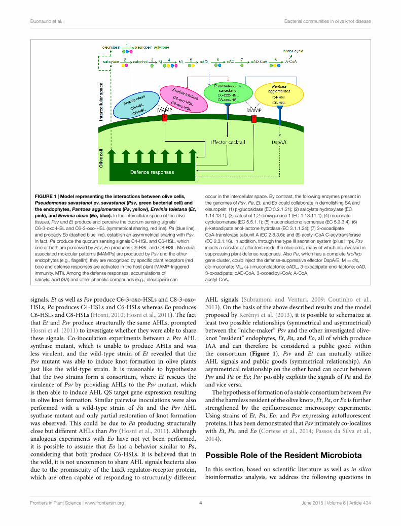
FIGURE 1 | Model representing the interactions between olive cells,Pseudomonas savastanoi pv. savastanoi (Psv, green bacterial cell) andthe endophytes, Pantoea agglomerans (Pa, yellow), Erwinia toletana (Et,pink), and Erwinia oleae (Eo, blue). In the intercellular space of the olivetissues, Psv and Et produce and perceive the quorum sensing signalsC6-3-oxo-HSL and C6-3-oxo-HSL (symmetrical sharing, red line). Pa (blue line),and probably Eo (dashed blue line), establish an asymmetrical sharing with Psv.In fact, Pa produce the quorum sensing signals C4-HSL and C6-HSL, whichone or both are perceived by Psv; Eo produces C6-HSL and C8-HSL. Microbialassociated molecular patterns (MAMPs) are produced by Psv and the otherendophytes (e.g., flagellin); they are recognized by specific plant receptors (redbox) and defense responses are activated in the host plant (MAMP-triggeredimmunity, MTI). Among the defense responses, accumulations ofsalicylic acid (SA) and other phenolic compounds (e.g., oleuropein) can
occur in the intercellular space. By contrast, the following enzymes present inthe genomes of Psv, Pa, Et, and Eo could collaborate in demolishing SA andoleuropein: (1) β-glucosidase (EC 3.2.1.21); (2) salicylate hydroxylase (EC1.14.13.1); (3) catechol 1,2-dioxygenase 1 (EC 1.13.11.1); (4) muconatecycloisomerase (EC 5.5.1.1); (5) muconolactone isomerase (EC 5.3.3.4); (6)β-ketoadipate enol-lactone hydrolase (EC 3.1.1.24); (7) 3-oxoadipateCoA-transferase subunit A (EC 2.8.3.6); and (8) acetyl-CoA C-acyltransferase(EC 2.3.1.16). In addition, through the type III secretion system (pilus Hrp), Psvinjects a cocktail of effectors inside the olive cells, many of which are involved insuppressing plant defense responses. Also Pa, which has a complete hrc/hrpgene cluster, could inject the defense-suppressive effector DspA/E. M = cis,cis-muconate; ML, (+)-muconolactone; oADL, 3-oxoadipate-enol-lactone; oAD,3-oxoadipate; oAD-CoA, 3-oxoadipyl-CoA; A-CoA,acetyl-CoA.
signals. Et as well as Psv produce C6-3-oxo-HSLs and C8-3-oxo-HSLs, Pa produces C4-HSLs and C6-HSLs whereas Eo producesC6-HSLs and C8-HSLs (Hosni, 2010; Hosni et al., 2011). The factthat Et and Psv produce structurally the same AHLs, promptedHosni et al. (2011) to investigate whether they were able to sharethese signals. Co-inoculation experiments between a Psv AHLsynthase mutant, which is unable to produce AHLs and wasless virulent, and the wild-type strain of Et revealed that thePsv mutant was able to induce knot formation in olive plantsjust like the wild-type strain. It is reasonable to hypothesizethat the two strains form a consortium, where Et rescues thevirulence of Psv by providing AHLs to the Psv mutant, whichis then able to induce AHL QS target gene expression resultingin olive knot formation. Similar pairwise inoculations were alsoperformed with a wild-type strain of Pa and the Psv AHLsynthase mutant and only partial restoration of knot formationwas observed. This could be due to Pa producing structurallyclose but different AHLs than Psv (Hosni et al., 2011). Althoughanalogous experiments with Eo have not yet been performed,it is possible to assume that Eo has a behavior similar to Pa,considering that both produce C6-HSLs. It is believed that inthe wild, it is not uncommon to share AHL signals bacteria alsodue to the promiscuity of the LuxR regulator-receptor protein,which are often capable of responding to structurally different
AHL signals (Subramoni and Venturi, 2009; Coutinho et al.,2013). On the basis of the above described results and the modelproposed by Kerényi et al. (2013), it is possible to schematize atleast two possible relationships (symmetrical and asymmetrical)between the “niche-maker” Psv and the other investigated olive-knot “resident” endophytes, Et, Pa, and Eo, all of which produceIAA and can therefore be considered a public good withinthe consortium (Figure 1). Psv and Et can mutually utilizeAHL signals and public goods (symmetrical relationship). Anasymmetrical relationship on the other hand can occur betweenPsv and Pa or Eo; Psv possibly exploits the signals of Pa and Eoand vice versa.
The hypothesis of formation of a stable consortiumbetweenPsvand the harmless resident of the olive knots, Et, Pa, or Eo is furtherstrengthened by the epifluorescence microscopy experiments.Using strains of Et, Pa, Eo, and Psv expressing autofluorescentproteins, it has been demonstrated that Psv intimately co-localizeswith Et, Pa, and Eo (Cortese et al., 2014; Passos da Silva et al.,2014).
Possible Role of the Resident Microbiota
In this section, based on scientific literature as well as in silicobioinformatics analysis, we address the following questions in
Frontiers in Plant Science | www.frontiersin.org June 2015 | Volume 6 | Article 4344
Buonaurio et al. Bacterial communities in olive knot disease
order to begin to better understand the role of the residentmicrobiota, Et, Eo, and Pa, in the olive knot disease development.
(1) Does the resident microbiota possess the genetic traitscharacterizing an endophytic life style?
(2) Can resident microbiota produce biofilms?(3) Do resident microbiota suppress plant defense responses?
Endophytic LifestyleEndophytic bacteria colonize inner host tissues without damagingthe plant host or eliciting strong defense responses. Themolecular basis of this interaction is currently generally unknown.In addition, it is not clear whether these interactions areshared/common among different endophytes; however, there isevidence that several endophytic bacteria result in an array ofbeneficial effects for the plant (Ryan et al., 2008; Reinhold-Hurekand Hurek, 2011). Evidence of a rich and diverse endophyticbacterial population in several plant hosts has been determined byculture independent omic methodologies (Lundberg et al., 2012;Sessitsch et al., 2012). Currently we have very little knowledge onhow bacteria adapt, colonize, and live as endophytes in plants.
Endophytes colonize the intercellular spaces in the epidermaland cortical regions and in some cases, at lower cell densities, canalso invade the vascular tissue therefore allowing the spreadinginto shoots. Most endophytes are believed to enter inside theplant at the roots via the rhizosphere; this relies in part onmotility, root penetration, and adaptation to the plant intercellularenvironment (Compant et al., 2010; Reinhold-Hurek and Hurek,2011). This adaptation to a new environment also requires thatthe endophyte overcomes plant defense responses and the wholeprocess can be viewed as similar to the one encountered byincoming phytopathogens with the major difference being thatfor endophytes, the plant–bacteria interaction is mutualistic. Thisis an important open question, i.e., how can endophytes colonizein high numbers a plant tissue without eliciting significant plantresponse and plant damage? Because they are able to colonizea niche similar to that of plant pathogens, they can becomepossible biocontrol agents to control incoming phytopathogens.Future studies will need to clarify the molecular mechanisms ofadaptation, recognition, and communication between harmless orbeneficial endophytes and the plant. In addition, which specificfeatures belong to endophytes in comparison to soil, rhizosphere,and pathogenic microorganism?
In order to obtain insights into strategies of endophyticlifestyle, scientists are beginning to sequence the genomes ofseveral endophytes and perform comparative genome studies. Theinitial bioinformatic analyses have not yet deduced genetic traitscharacterizing an endophyte (Mitter et al., 2013). Awide spectrumof traits necessary for endophytic lifestyle is likely to existincluding motility, chemotaxis, adhesion, signaling, membranetransport, secretion, and degradation of plant polymers andorganic compounds as a recent study on an endophyte responseat the molecular level to the plant environment has recentlyevidenced (Reinhold-Hurek and Hurek, 2011; Coutinho et al.,2015). As more genomes of endophytes are sequenced as wellas genetic and molecular studies performed, it is expected thatthe main traits necessary for endophytic colonization will be
revealed. This research will probably pick up quickly as it isbelieved that endophytes, as well as other plant-associated bacteria(collectively called plant microbiome), contribute significantly toplant health.
Similarly to Psv, it is possible that many of the bacterialiving as endophytes in olive knots are also resident in the olivephyllosphere and enter in the plant tissues mainly through thewounds and not via the rhizosphere. In fact Ercolani (1978)isolated many bacterial species from olive phylloplane and Pais one of the most represented. It cannot be excluded howeverthat a part of the olive-knot endophytic residents originate fromthe rhizosphere of plant roots. In this sense and related witholive knot disease, Pseudomonas fluorescens strain PICF7, a nativeendophyte of olive roots and an effective biocontrol agent againstthe soil-borne fungus Verticillium dahliae (Mercado-Blanco et al.,2004), has been reported to establish and persist in olive stemtissues upon artificial inoculation. Although this strain is not ableto suppress olive knot disease, its presence decreases pathogenpopulation size and confine Psv at inner regions of the tumors(Maldonado-González et al., 2013). Genome analysis of thisstrain recently revealed genes that might be related to biocontroland endophytic traits, such as encoding for secretion systems,siderophores, detoxifying compounds, or volatile components(Martínez-García et al., 2015). Moreover, and as mentionedabove, a recent metagenomic study on microbiome of olive knotindicated the presence of approximately 50% of the bacterial loadbeing Psv and the remaining 50% being many different bacterialspecies belonging to different genera present in the olive knot(Passos da Silva et al., 2014). Many of the bacterial genera arealso present in the rhizosphere thus it is likely that they areendophytes entering the olive plant via the roots. The rich bacterialendophytic population in the olive knot is likely to involveseveral mechanisms of molecular interaction including QS signalsharing andmetabolic complementarity. This niche is therefore anexcellent model to study all the different interactions taking placeamong an endophytic community, particularly among harmlessbacteria as well as harmless bacteria and Psv.
Biofilm ProductionSuccessful plant colonization is often linked to the ability ofbacteria to form adherent microbial populations (Danhorn andFuqua, 2007). This sessile form of bacterial life, also called biofilm,is extensively studied and very complex. Bacterial attachmentto plant surfaces involves several proteins including adhesins,amyloid curli fimbriae, type I fimbriae, type IV pili, and flagella(Jefferson, 2004; Barken et al., 2008; Bahar et al., 2009; Wanget al., 2013; Heindl et al., 2014; Yaron and Römling, 2014)as well as production of exopolysaccharides (EPS), capsularpolysaccharides (CPS), and extracellular DNA (eDNA). Amongthe adhesins, filamentous hemagglutinin-like (FHA) proteinsplay a fundamental role in plant–bacteria interactions. FHAproteins of Xanthomonas axonopodis pv. citri is required fortissue colonization being mainly involved in surface attachmentand biofilm formation (Gottig et al., 2009). In Pseudomonason the other hand, CdrA (an FHA-like protein) was shown tobe associated with the Psl polysaccharide, which is a biofilmcomponent (Borlee et al., 2010). Since FHA genes are present
Frontiers in Plant Science | www.frontiersin.org June 2015 | Volume 6 | Article 4345
Buonaurio et al. Bacterial communities in olive knot disease
in a number of bacterial endophytes, it has been hypothesizedthat they play a crucial role in the invasion of plant tissues andin biofilm formation (Mitter et al., 2013). BlastP and analysis ofneighboring genes demonstrated that Eo and Et possess FHA-like genes that could aid the formation of a stable biofilmstructure. Further in silico analysis and experimental studiescould demonstrate the importance of these highmolecular weightproteins in biofilm formation as well as bacteria–bacteria andplant–bacteria interactions.
Psv strains are able to form air–liquid biofilms over abioticsurfaces (Ude et al., 2006; Pérez-Mendoza et al., 2014).Furthermore, visualization of olive knot sections by scanningconfocal laser microscopy (SCLM) and electron microscopyhas allowed visualization of the organization of Psv cell clusters(microcolonies and biofilms) inside the host tissues. In fact, Psvis visualized inside olive knots forming biofilms composed of amultilayer of bacterial cells colonizing the surface and interiorof plasmolysed host cells (Rodríguez-Moreno et al., 2009).Moreover, calcofluor epifluorescent microscopy has identifiedcellulose as the EPS component in biofilms produced by Psvstrains (Ude et al., 2006). Thus, cellulose production by Psv cellsmight play a relevant role both in the virulence of Psv and in theestablishment of mixed-species community biofilms (Psv, Pa, Et,and Eo) inside olive knots. In relation to virulence determinants,the size of the knots induced by Psv in olive plants has beenshown to be dependent on the activity of several enzymesinvolved in the metabolism of the second messenger c-di-GMP,which in turn, regulate EPS production and biofilm formation(Pérez-Mendoza et al., 2014; Aragón et al., 2015). Further studiesaddressing the composition of the EPS matrix surroundingmixed-species bacterial biofilms established inside olive knotswould be necessary to shed light in this hypothesis. Microscopicvisualization of olive knots induced on in vitro micropropagatedolive plants after mixed inoculation of Et and Psv revealed thatthe distribution of Et cells closely matched the position of Psvcells (Passos da Silva et al., 2014). Preliminary and similar resultswere obtained by Cortese et al. (2014) when Eo and Pa wereassayed instead of Et. This close proximity possibly suggests thatmixed biofilms occur inside olive knots; however, this hypothesisneeds to be experimentally tested.
It was recently discovered that onemechanism bywhich Gram-negative bacteria that are in close proximity to each other caninteract is by contact-dependent transport of effector proteinsfrom a donor cell to a recipient cell via the activity of an apparatusknown as the type VI secretion system (T6SS; Russell et al.,2014). Initially found to deliver effector proteins into eukaryoticcells, T6SS seems mainly involved in interbacterial interactions.Killing of bacteria (non-cooperators) that weaken the stability of abacterial community and the contribution to maintain the three-dimensional architecture of bacterial communities is one of thepotential roles of T6SS (Russell et al., 2014). Interestingly, Eo andPa genomes harbor regions of 6.3 and 7.1 kb, respectively, flankedby mobile element proteins, which contain genes belongingto the T6SS, namely the loci vgrG and rhs which encode forvaline/glycine-repeat protein G (Chang et al., 2014; Hachani et al.,2014) and rearrangement HotSpot elements (Koskiniemi et al.,2013), respectively. The presence of the flanking mobile elements,
the higher G + C content of this region with respect to theG + C content of the whole genomes of Pa and Eo, and the highsimilarity (99%) with Psv homolog genes, suggests an acquisitionof this region via HGT possibly from Psv. We can hypothesize thatstrains of Pa and Eo have evolved together with Psv and that theycollaborate with Psv in the maintenance the stability of the olive-knot consortium through T6SS. In addition, the biofilms, whichare probably formed by multispecies communities in the oliveknots provide an excellent environment for DNA exchange viaHGT since cells are in close proximity for a long time andDNAcanbe trapped within the extracellular matrix (Madsen et al., 2012).It is therefore tempting to speculate that these harmless residentsin the knot can in the future evolve to become pathogenic byacquiring the loci from neighboring pathogenic cells.
Suppression of Plant DefensesPlants are exposed to a vast number of potential pathogens, againstwhich they try to defend themselves through a multilayeredsystem of preformed and induced defenses, both structural andbiochemical. Few investigations have been carried out on thedefense responses of olive plants against Psv, mostly becauseresistant olive cultivars to olive knot are not available (Penyalveret al., 2006). Important information on olive defense responses areprovided by the studies on the resistance induced by the biocontrolagent P. fluorescens PICF7 (Schilirò et al., 2012; Gomez-LamaCabanas et al., 2014). Unfortunately, this resistance is effectiveagainst Verticillium wilt (Prieto et al., 2009; Schilirò et al.,2012) but not on olive knot disease (Maldonado-González et al.,2013). Thus, defense responses against Psv are essentially limitedto studying weakly resistant or susceptible olive plants to Psvinfections.
Among the structural induced defenses, Temsah et al. (2008)observed lignin deposits on cell walls of parenchyma olive cellsaround cavities and injured tissues caused by Psv. They alsoreported that new periderm was produced at the surface of theolive knots, which constitutes a barrier impairing the entrance ofother pathogens and saprophytes.
Regarding the biochemical induced defenses, a numberof histological and biochemical studies reported a markedaccumulation of phenolic compounds in olive knots (Roussoset al., 2002; Cayuela et al., 2006; Marchi et al., 2009), a resistancemechanism well documented in many other bacterial diseases(Buonaurio, 2008). More precisely the following two compoundsare involved, the secoiridoids oleuropein and verbascoside.The phenolic glucoside oleuropein, which is an ester ofelenolic acid with hydroxytyrosol linked to glucose througha 1,4-beta bond, showed its toxicity against Psv in the 0.1–1mM concentration range, while oleuropein aglycone andhydroxytyrosol in the ranges 0.1–10 and 1–10 mM, respectively(Capasso et al., 1995). Verbascoside is a heterosidic ester ofcaffeic acid and hydroxytyrosol linked to rhamnose and glucose,which has antimicrobial activity (Pardo et al., 1993), thoughnot documented for Psv. Since phenolic compounds accumulateinside olive cells, especially in the vacuoles (Uccella, 2001), we canassume their antibacterial activity is manifested when plant celldisruption occurs. Therefore it is reasonable to speculate that thishypothetical defense response is effective during the early stage
Frontiers in Plant Science | www.frontiersin.org June 2015 | Volume 6 | Article 4346
Buonaurio et al. Bacterial communities in olive knot disease
of the infection process, when Psv induces cavity formation in theplant tissues through pectolytic and hemicellulolytic enzymes,as well as during the later stages, when large cavities formedafter the collapse of intercellular plant cells. By contrast, Psv andthe endophytes discussed here may provoke the degradation ofphenolic glucoside compounds through enzymatic activities.Genes for beta-glucosidases are present in Psv, Et, and Eo.These enzymes may cleave the glucoside moieties and allow themolecule to be further mineralized.
The phytohormone salicylic acid (SA) could also be involvedin the defense against Psv. Besides acting as a signaling moleculein plant defense against biotrophic and hemibiotrophic plantpathogens (Boatwright and Pajerowska-Mukhtar, 2013), SAmay function as an antimicrobial agent. Cameron and Zaton(2004) reported an accumulation of SA in the intercellular spaceof old Arabidopsis plants and correlated it with the age-relatedresistance to Pseudomonas syringae pv. tomato DC 3000. In fact,they also demonstrated that SA has antimicrobial activity againstthe bacterium and that the destruction of SA in the intercellularspace by salicylate hydroxylase reduced the resistance level inArabidopsis plants. If SA builds up in the olive knot apoplastit could result in antimicrobial activity against Psv (which islikely since Psv cannot convert SA to catechol as evidenced byPassos da Silva et al., 2014); this can be potentially alleviated byEt and Pa since these bacteria possess the salicylate hydroxylasesgene, which converts SA to catechol. Catechol is one of themost important central intermediates in the aerobic bacterialmetabolism of aromatic compounds (Harwood and Parales,1996) and it is toxic to Psv (Capasso et al., 1997). This phenolmay be further degraded and detoxified by the collaborativeaction of Psv, Et, Eo, and Pa enzymes by ortho cleavage via the3-oxoadipate pathway, which lead in to the Krebs cycle (Harwoodand Parales, 1996; Figure 1). This is a possible example of howharmless endophyte residents can aid the growth of Psv pathogenvia metabolic complementarity. Interestingly, in the genomesof several Psv strains there is a region of about 15 kb, namedVR8 which is absent in all sequenced P. syringae strains infectingherbaceous plants. This region is however present in P. syringaepathovars infecting woody hosts, such as aesculi, morsprunorum,and actinidiae (Ramos et al., 2012; Moretti et al., 2014c). Amongother genes encoded in this region there is also the catBCA operonwhich is involved in the degradation of the catechol possiblyallowing a selective advantage for growth in woody hosts.
There is a growing body of evidence that the T3Es injectedinside plant cells by phytopathogenic bacteria suppress hostdefenses, i.e., PAMP-triggered immunity (PTI) and effector-triggered immunity (ETI; Jones and Dangl, 2006). This occursvia different routes such as interference with immune receptorsignaling, blocking of RNA pathways, and vesicle traffickingand alteration of organelle function (Block and Alfano, 2011).Matas et al. (2014) demonstrated that seven Psv T3Es (HopA1,AvrRpm2, HopAA1, HopAZ1, HopBK1, HopBL1, and HopBL2)suppress the PTI and two of them (HopAZ1 and HopBL1) alsosuppress ETI. We cannot exclude that T3Es are produced bysome endophytic bacteria of the olive-knot microbiome, whichcollaborate with Psv in mitigating host defenses. In fact, the HR-inducing strain of Pa, isolated from the olive microbiome and
characterized by Moretti et al. (2014d), harbors the dspA/E anddspB/F genes adjacent to its complete hrc/hrp gene cluster, whichencode for the homologs Erwinia amylovora effector DspA/Eand its chaperone DspB/F, which in turn is essential for DspA/Esecretion and stability (Gaudriault et al., 2002; Boureau et al.,2006). Among the E. amylovora T3Es, DspA/E plays a pivotal rolein disease development as mutants defective for this effector arenon-pathogenic and are unable to grow in host plants (Barny et al.,1990; Gaudriault et al., 1997) and non-host tobacco (Oh et al.,2007). DspA/E belong to the AvrE effector superfamily, which iswidespread among type III-dependent phytopathogenic bacteria,playing a crucial role during bacterial pathogenesis by suppressingPTI and are also present in the genome of non-pathogenic plantassociated bacteria (Degrave et al., 2013). AvrE-T3Es inhibit SA-mediated plant defenses, interfere with vesicular trafficking andpromote bacterial growth in planta. We can hypothesize that HR-inducing strains of Pa are able to deliver the effector DspA/Ein olive cells, which, in turn, suppresses the host defenses andtherefore facilitate the growth of microbiota inside the olive-knot (Figure 1). Interestingly, Eo genome harbors the dspA/Egene encoding proteins showing very high identity (99%) withthose of Pa, suggesting a possible evolution in the olive-knotniche. However, differently from Pa, Eo does not possess a T3SS,therefore we do not know if it is able to secrete DspA/E effector.
Future directions and Concluding RemarksIt is now evident that a variety of bacterial species coexist in thenatural environment building a network of interactions; this hasled to the emergence of a new discipline which has been coinedas sociomicrobiology (Parsek and Greenberg, 2005). The mostcommon form of social behavior in bacteria is the production ofpublic goods (e.g., siderophores, extracellular enzymes, secondarymetabolites), which benefit all individuals of the community,both cooperators, and defectors or cheaters (West et al., 2006).From an evolutionary point of view, it is known that microbialbiodiversity is an important driver of ecological processes andthe evolutionary history predicts the stability of cooperation inmultispecies bacterial communities (Jousset et al., 2013). Thecomplex social behaviors of bacterial communities have beenextensively investigated in Pseudomonas aeruginosa as the mainactor, now considered a reference model to study fundamentalquestions about the costs and benefits of cooperation. Theseinclude the selective pressures that lead to cooperative behaviorand the advantages of controlling cooperative behaviors bycommunication (for more detailed information, see De VargasRoditi et al., 2013; Mitri and Foster, 2013; Tashiro et al., 2013).In the olive knot model of multispecies bacterial community, itappears that Psv is a social bacterium, which has not evolvedto cause disease independently or to live alone inside the oliveknot, in spite of its capability to induce alone knot formationin olive trees. In fact, as documented in a number of olive-growing countries and in different and distant locations of thesame country, bacterial endophytes, mainly belonging to Pantoeaand Erwinia genera, are constantly associated with Psv inside oliveknots (Fernandes and Marcelo, 2002; Ouzari et al., 2008; Hosniet al., 2011; Passos da Silva et al., 2014). In addition, the presenceof endophytes in the knots is not only observed in nature but also
Frontiers in Plant Science | www.frontiersin.org June 2015 | Volume 6 | Article 4347
Buonaurio et al. Bacterial communities in olive knot disease
in plants artificially inoculated with Psv. Therefore, Psv is likelyto find benefit in living in a multispecies community, possiblybecause it can better exploit plant nutrients as well as contrastingplant defenses.
Several possible contributions to virulence by the residentolive-knot bacteria have already beenmentioned in the previouslysections including production of phytohormones, biofilmformation, degradation of plant phenolic compounds, c-di-GMPsignaling, production of T3 effectors, production of secretedcellulolytic enzymes, and QS signal sharing. An importantaspect which merits attention in addition to the possiblecontribution of virulence is that, resident bacteria could beinvolved in the formation of a stable multispecies communitywhich allows the pathogen and other bacteria to be most fit toattack plant cells and consequently grow in the olive-knot. Thisstability therefore needs to avoid diverse growth incompatibilitiessuch as contact dependent mechanisms (Blango and Mulvey,2009), production of antibacterial compounds and nutrientcompetition. Common co-isolation of bacterial species froma given niche does not automatically imply cooperation orsynergism between them, but is an indication that some typeof interaction is taking place. Importantly further studies areneeded to investigate these interactions, for example: (i) invitro analysis can focus on growth conditions trying to mimicwhat occurs in the olive knot; (ii) secondly in silico analysiscan generate evidence for further in vitro studies especially inrelation to metabolic complementarily and signaling and (iii) inplanta studies involving microscopy and “omic” analysis mightthen validate the interactions model(s) extrapolated from invitro and in silico investigations. For example, initial studiesalong this line have begun to show via genome sequencingthat residents commonly isolated from olive knots possessputative metabolic pathways that are not complete in Psvwhich might lead to complementarity in the metabolism ofcompounds (e.g., for themineralizing of -rhamnose, -galactose,-arabinose, SA, and aminoethylphosphonic acid) found in theolive knot.
Many aspects merit further studies such as: (i) the effect oflocation, olive cultivar, environmental conditions on the relativeabundance of the main endophytes inside the knots, (ii) thegenetic variability of some endophytes, and (iii) the eventual
translocation in the xylem of the endophytic bacteria as well astheir fate away from the olive knots.
In conclusion, studies on bacterial plant diseases have thusfar historically focused on single species (the pathogen), whilelittle attention has been given on the many other microorganismsmost likely present in the infection sites such as soft rottedtissues (Sturz and Matheson, 1996), cankers (Moretti et al., 2007),and tumors (Nautiyal and Dion, 1990). Microbiota living in theinfection sites can possibly modulate the severity of the disease,establishing mutual, commensal, and antagonistic interactions aswell as interacting with plant defense systems. These potentiallycomplex interactions are largely unknown and in our view meritattention.
In this review, we report the results achieved to date onthe multispecies aspect of the olive knot disease. These aswell as the hypotheses drawn from the scientific literature inaddition to in silico analysis indicate that the tumors causedby Psv in olive trees represent an ideal niche and an excellentmodel to study interspecies interactions, sociomicrobiology,evolution, cooperation, and competition of bacteria and couldserve as a paradigm in plant pathology. These interactionscould result in the evasion of the immune response, chemicalsignaling, metabolic exchange, and resident-to-pathogenswitch (Venturi and Passos da Silva, 2012). The sequencingof all the microbial members as well as the olive plant(http://genomes.cribi.unipd.it/olive/wordpress/welcome/) will beof importance in order to set up future experiments designed tounderstand interactions. It is likely that these kinds of studies willlead the way for other scientists investigating the potential roleof other microbes present in the infection sites of plant diseases.Understanding these interactions could be of crucial importancein designing new and effective strategies for disease control.
Acknowledgments
This research was supported by “Fondazione Cassa di Risparmiodi Perugia,” Italy, project: “Indagini sul ruolo dei fenoli dell’olivonello sviluppo della rogna, per individuare nuove strategie dilotta alla malattia” to CM. CR was supported by the SpanishPlan Nacional de I+D+I grants AGL2011-30343-C02-01 andAGL2014-53242-C2-1-R.
References
Aragón, I. M., Pérez-Martínez, I., Moreno-Pérez, A., Cerezo, M., and Ramos, C.(2014). New insights into the role of indole-3-acetic acid in the virulence ofPseudomonas savastanoi pv. savastanoi. FEMSMicrobiol. Lett. 356, 184–192. doi:10.1111/1574-6968.12413
Aragón, I. M., Pérez-Mendoza, D., Gallegos, M. T., and Ramos, C. (2015). Thec-di-GMP phosphodiesterase BifA is involved in the virulence of bacteriafrom the Pseudomonas syringae complex. Mol. Plant Pathol. doi: 10.1111/mpp.12218
Bahar, O., Goffer, T., and Burdman, S. (2009). Type IV pili are required forvirulence, twitching motility, and biofilm formation of Acidovorax avenaesubsp citrulli. Mol. Plant Microbe Interact. 22, 909–920. doi: 10.1094/mpmi-22-8-0909
Barash, I., and Manulis-Sasson, S. (2009). Recent evolution of bacterial pathogens:the gall-formingPantoea agglomerans case.Annu. Rev. Phytopathol. 47, 133–152.doi: 10.1146/annurev-phyto-080508-081803
Bardaji, L., Pérez-Martínez, I., Rodríguez-Moreno, L., Rodríguez-Palenzuela, P.,Sundin, G. W., Ramos, C., et al. (2011). Sequence and role in virulenceof the three plasmid complement of the model tumor-inducing bacteriumPseudomonas savastanoi pv. savastanoi NCPPB 3335. PLoS ONE 6:e25705. doi:10.1371/journal.pone.0025705
Barken, K. B., Pamp, S. J., Yang, L., Gjermansen, M., Bertrand, J. J., and Klausen, M.,et al. (2008). Roles of type IV pili, flagellum-mediated motility and extracellularDNA in the formation of mature multicellular structures in Pseudomonasaeruginosa biofilms. Environ. Microbiol. 10, 2331–2343. doi: 10.1111/j.1462-2920.2008.01658.x
Barny, M. A., Guinebretiere, M. H., Marcais, B., Coissac, E., Paulin, J. P.,and Laurent, J. (1990). Cloning of a large gene-cluster involved in ErwiniaamylovoraCFBP1430 virulence.Mol.Microbiol. 4, 777–786. doi: 10.1111/j.1365-2958.1990.tb00648.x
Blango, M. G., and Mulvey, M. A. (2009). Bacterial landlines: contact-dependentsignaling in bacterial populations. Curr. Opin. Microbiol. 12, 177–181. doi:10.1016/j.mib.2009.01.011
Frontiers in Plant Science | www.frontiersin.org June 2015 | Volume 6 | Article 4348
Buonaurio et al. Bacterial communities in olive knot disease
Block, A., and Alfano, J. R. (2011). Plant targets for Pseudomonas syringae type IIIeffectors: virulence targets or guarded decoys? Curr. Opin. Microbiol. 14, 39–46.doi: 10.1016/j.mib.2010.12.011
Boatwright, J. L., and Pajerowska-Mukhtar, K. (2013). Salicylic acid: an oldhormone up to new tricks. Mol. Plant Pathol. 14, 623–634. doi: 10.1111/mpp.12035
Borlee, B. R., Goldman, A. D., Murakami, K., Samudrala, R., Wozniak, D. J.,and Parsek, M. R. (2010). Pseudomonas aeruginosa uses a cyclic-di-GMP-regulated adhesin to reinforce the biofilm extracellular matrix. Mol. Microbiol.75, 827–842. doi: 10.1111/j.1365-2958.2009.06991.x
Boureau, T., Elmaarouf-Bouteau, H., Garnier, A., Brisset, M.N., Perino, C., Pucheu,I., et al. (2006). DspA/E, a type III effector essential for Erwinia amylovorapathogenicity and growth in planta, induces cell death in host apple and nonhosttobacco plants. Mol. Plant Microbe Interact. 19, 16–24. doi: 10.1094/mpmi-19-0016
Buonaurio, R. (2008). “Infection and plant defense responses during plant–bacterialinteraction,” in Plant–Microbe Interactions, eds E. A. Barka and C. Clement(Kerala, India: Research Signpost), 169–197.
Burmølle, M., Ren, D., Bjarnsholt, T., and Sørensen, S. J. (2014). Interactions inmultispecies biofilms: do they actually matter? Trends Microbiol. 22, 84–91. doi:10.1016/j.tim.2013.12.004
Burmølle, M., Thomsen, T. R., Fazli, M., Dige, I., Christensen, L., Homøe,P., et al. (2010). Biofilms in chronic infections - a matter of opportunity- monospecies biofilms in multispecies infections. FEMS Immunol. Med.Microbiol. 59, 324–336. doi: 10.1111/j.1574-695X.2010.00714.x
Burmølle, M., Webb, J. S., Rao, D., Hansen, L. H., Sørensen, S. J., andKjelleberg, S. (2006). Enhanced biofilm formation and increased resistanceto antimicrobial agents and bacterial invasion are caused by synergisticinteractions in multispecies biofilms. Appl. Environ. Microbiol. 72, 3916–3923.doi: 10.1128/aem.03022-05
Burr, T. J., Katz, B. H., Abawi, G. S., and Crosier, D. C. (1991). Comparison oftumorigenic strains of Erwinia herbicola isolated from table beet with Erwima’sh. gypsophilae. Plant Dis. 75, 855–858.
Cameron, R. K., and Zaton, K. (2004). Intercellular salicylic acid accumulationis important for age-related resistance in Arabidopsis to Pseudomonassyringae. Physiol. Mol. Plant Pathol. 65, 197–209. doi: 10.1016/j.pmpp.2005.02.002
Capasso, R., Cristinzio, G., Evidente, A., Visca, C., and Iannini, C. (1997).“Oleuropein and other polyphenols fromolive (Olea europaea) for protecting theplant against Pseudomonas syringae subsp. savastanoi,” in Pseudomonas syringaePathovars and Related Pathogens, eds K. Rudolph, T. J. Burr, J. W. Mansfield, D.Stead, A. Vivian, and J. von Kietzell (Dordrecht: Kluwer Academic Publishers),133–137.
Capasso, R., Evidente, A., Schivo, L., Orru, G., Marcialis, M. A., and Cristinzio,G. (1995). Antibacterial polyphenols from olive oil mill waste-waters. J. Appl.Bacteriol. 79, 393–398. doi: 10.1111/j.1365-2672.1995.tb03153.x
Cayuela, J. A., Rada, M., Rios, J. J., Albi, T., and Guinda, A. (2006). Changesin phenolic composition induced by Pseudomonas savastanoi pv. savastanoiinfection in olive tree: presence of large amounts of verbascoside in nodulesof tuberculosis disease. J. Agr. Food Chem. 54, 5363–5368. doi: 10.1021/jf060807w
Chang, J. H., Desveaux, D., and Creason, A. L. (2014). The ABCs and 123s ofbacterial secretion systems in plant pathogenesis. Annu. Rev. Phytopathol. 52,317–345. doi: 10.1146/annurev-phyto-011014-015624
Compant, S., Clement, C., and Sessitsch, A. (2010). Plant growth-promotingbacteria in the rhizo- and endosphere of plants: their role, colonization,mechanisms involved and prospects for utilization. Soil Biol. Biochem. 42,669–678. doi: 10.1016/j.soilbio.2009.11.024
Cooksey, D. A. (1986). Galls of Gypsophila paniculata caused by Erwinia herbicola.Plant Dis. 70, 464–468. doi: 10.1094/pd-70-464
Cortese, C., Pérez-Martínez, I., Ramos, C., Buonaurio, R., and Moretti, C. (2014).“The endophytes Pantoea agglomerans and Erwinia oleae colocalize withPseudomonas savastanoi pv. savastanoi in the olive knots” in XX ConvegnoNazionale Società di Patologia Vegetale, Environmentally loyal plant protection:from nano- to field-scale, 31.
Coutinho, B. G., Licastro, D., Mendonca-Previato, L., Camara, M., and Venturi,V. (2015). Plant-influenced gene expression in the rice endophyte Burkholderiakururiensis M130. Mol. Plant Microbe Interact. 28, 10–21. doi: 10.1094/mpmi-07-14-0225-r
Coutinho, B. G., Mitter, B., Talbi, C., Sessitsch, A., Bedmar, E. J., Halliday, N., etal. (2013). Regulon studies and in planta role of the BraI/R quorum-sensingsystem in the plant-beneficial Burkholderia cluster.Appl. Environ. Microbiol. 79,4421–4432. doi: 10.1128/AEM.00635-13
Danhorn, T., and Fuqua, C. (2007). Biofilm formation by plant-associated bacteria.Annu. Rev. Microbiol. 61, 401–422. doi: 10.1146/annurev.micro.61.080706.093316
De Beer, D., and Stoodley, P. (2006). “Microbial biofilms,” in The Prokaryotes, edsM. Dworkin, S. Falkow, E. Rosenberg, K. Schleifer, and E. Stackebrandt (NewYork: Springer), 904–937.
Degrave, A., Moreau, M., Launay, A., Barny, M.-A., Brisset, M.-N., Patrit, O., etal. (2013). The bacterial effector DspA/E is toxic in Arabidopsis thaliana andis required for multiplication and survival of fire blight pathogen. Mol. PlantPathol. 14, 506–517. doi: 10.1111/mpp.12022
Demuyser, L., Jabra-Rizk, M. A., and Van Dijck, P. (2014). Microbial cell surfaceproteins and secreted metabolites involved in multispecies biofilms. Pathog. Dis.70, 219–230. doi: 10.1111/2049-632x.12123
De Vargas Roditi, L., Boyle, K. E., and Xavier, J. B. (2013). Multilevel selectionanalysis of a microbial social trait. Mol. Syst. Biol. 9. doi: 10.1038/msb.2013.42
DeYoung, R. M., Copeman, R. J., and Hunt, R. S. (1998). Two strains in the genusErwinia cause galls on douglas-fir in southwestern British Columbia. Can. J.Plant Pathol. 20, 194–200.
Ercolani, G. L. (1971). Presenza epifitica di Pseudomonas savastanoi (E. F. Smith)Stevens sull’Olivo, in Puglia. Phytopathol. Mediterr. 10, 130–132.
Ercolani, G. L. (1978). Pseudomonas savastanoi and other bacteria colonizing thesurface of olive leaves in the field. J. Gen. Microbiol. 109, 245–257.
Fernandes, A., and Marcelo, M. (2002). “A possible synergistic effect of Erwinia sp.on the development of olive knot symptoms caused by Pseudomonas syringaepv. savastanoi in Olea europaea” in Proceedings of the Fourth InternationalSymposium on Olive Growing, Vols. 1 and 2, eds C. Vitagliano and G. P. Martelli(Valenzano: ISHS Acta Horticulturae), 729–731.
Fuqua, C., Parsek, M. R., and Greenberg, E. P. (2001). Regulation of geneexpression by cell-to-cell communication: acyl-homoserine lactone quorumsensing. Annu. Rev. Genet. 35, 439–468. doi: 10.1146/annurev.genet.35.102401.090913
Gaudriault, S., Malandrin, L., Paulin, J. P., and Barny, M. A. (1997). DspA, anessential pathogenicity factor of Erwinia amylovora showing homology withAvrE of Pseudomonas syringae, is secreted via the Hrp secretion pathway ina DspB-dependent way. Mol. Microbiol. 26, 1057–1069. doi: 10.1046/j.1365-2958.1997.6442015.x
Gaudriault, S., Paulin, J. P., and Barny, M. A. (2002). The DspB/F protein of Erwiniaamylovora is a type III secretion chaperone ensuring efficient intrabacterialproduction of the Hrp-secreted DspA/E pathogenicity factor.Mol. Plant Pathol.3, 313–320. doi: 10.1046/j.1364-3703.2002.00124.x
Gavini, F., Mergaert, J., Beji, A., Mielcarek, C., Izard, D., Kersters, K., et al. (1989).Transfer of Enterobacter agglomerans (Beijerinck 1888) Ewing and Fife 1972 toPantoea gen. nov. as Pantoea agglomerans comb. nov. and description of Pantoeadispersa sp. nov. Int. J. Syst. Bacteriol. 39, 337–345.
Gibbins, L. N. (1978). “Erwinia herbicola: a review and perspective”, in FourthInternational Conference on Plant Pathogenic Bacteria, Angers, France, 403–431.
Gomez-Lama Cabanas, C., Schilirò, E., Valverde-Corredor, A., and Mercado-Blanco, J. (2014). The biocontrol endophytic bacterium Pseudomonasfluorescens PICF7 induces systemic defense responses in aerial tissues uponcolonization of olive roots. Front. Microbiol. 5:427. doi: 10.3389/fmicb.2014.00427
Gottig, N., Garavaglia, B. S., Garofalo, C. G., Orellano, E. G., and Ottado,J. (2009). A filamentous hemagglutinin-like protein of Xanthomonasaxonopodis pv. citri, the phytopathogen responsible for citrus canker, isinvolved in bacterial virulence. PLoS ONE 4:e4358. doi: 10.1371/journal.pone.0004358
Guo, L., He, X., and Shi, W. (2014). Intercellular communications in multispeciesoral microbial communities. Front. Microbiol. 5:328. doi: 10.3389/fmicb.2014.00328
Hachani, A., Allsopp, L. P., Oduko, Y., and Filloux, A. (2014). The VgrG proteinsare “a la Carte” delivery systems for bacterial type VI effectors. J. Biol. Chem.289, 17872–17884. doi: 10.1074/jbc.M114.563429
Hallmann, J., Quadthallmann, A., Mahaffee, W. F., and Kloepper, J. W. (1997).Bacterial endophytes in agricultural crops. Can. J. Microbiol. 43, 895–914.
Frontiers in Plant Science | www.frontiersin.org June 2015 | Volume 6 | Article 4349
Buonaurio et al. Bacterial communities in olive knot disease
Hangler, M., Burmølle, M., Schneider, I., Allermann, K., and Jensen, B. (2009). Theserine protease esperase HPF inhibits the formation of multispecies biofilm.Biofouling 25, 667–674. doi: 10.1080/08927010903096008
Harwood, C. S., and Parales, R. E. (1996). The beta-ketoadipate pathwayand the biology of self-identity. Annu. Rev. Microbiol. 50, 553–590. doi:10.1146/annurev.micro.50.1.553
Heindl, J. E., Wang, Y., Heckel, B. C., Mohari, B., Feirer, N., and Fuqua,C. (2014). Mechanisms and regulation of surface interactions and biofilmformation in Agrobacterium. Front. Plant Sci. 5:176. doi: 10.3389/fpls.2014.00176
Hosni, T. (2010). Interaction Between Pseudomonas savastanoi pv. savastanoi, theCausal Agent of Olive Knot, and the Endophytic Bacterial Species Associated Withthe Knot. Ph.D. thesis. University of Perugia, Perugia, Italy.
Hosni, T., Moretti, C., Devescovi, G., Suarez-Moreno, Z. R., Barek Fatmi, M.,Guarnaccia, C., et al. (2011). Sharing of quorum-sensing signals and role ofinterspecies communities in a bacterial plant disease. ISME J. 5, 1857–1870. doi:10.1038/ismej.2011.65
Iacobellis, N. S. (2001). “Olive knot,” in Encyclopedia of Plant Pathology, eds O. C.Maloy and T. D. Murray (New York: John Wiley and Sons), 713–715.
Jefferson, K. K. (2004). What drives bacteria to produce a biofilm? FEMSMicrobiol.Lett. 236, 163–173. doi: 10.1016/j.femsle.2004.06.005
Jones, J. D. G., and Dangl, J. L. (2006). The plant immune system. Nature 444,323–329. doi: 10.1038/nature05286
Jousset, A., Eisenhauer, N., Materne, E., and Scheu, S. (2013). Evolutionary historypredicts the stability of cooperation in microbial communities. Nat. Commun.4, 2573. doi: 10.1038/ncomms3573
Kerényi, A., Bihary, D., Venturi, V., and Pongor, S. (2013). Stability of multispeciesbacterial communities: signaling networks may stabilize microbiomes. PLoSONE 8:e57947. doi: 10.1371/journal.pone.0057947
Kobayashi, D. Y., and Palumbo, J. D. (2000). “Bacterial endophytes and their effectson plants and uses in agriculture,” inMicrobial Endophytes, eds C. W. Bacon andJ. F. White (New York: Marcel Dekker), 199–233.
Kolenbrander, P. E., Palmer, R. J. Jr., Periasamy, S., and Jakubovics, N. S. (2010). Oralmultispecies biofilm development and the key role of cell–cell distance.Nat. Rev.Microbiol. 8, 471–480. doi: 10.1038/nrmicro2381
Koskiniemi, S., Lamoureux, J. G., Nikolakakis, K. C., De Roodenbeke, C. T. K.,Kaplan, M. D., Low, D. A., et al. (2013). Rhs proteins from diverse bacteriamediate intercellular competition. Proc. Natl. Acad. Sci. U.S.A. 110, 7032–7037.doi: 10.1073/pnas.1300627110
Lindow, S. E., and Brandl, M. T. (2003). Microbiology of the phyllosphere. Appl.Environ. Microbiol. 69, 1875–1883. doi: 10.1128/aem.69.4.1875-1883.2003
Lundberg, D. S., Lebeis, S. L., Paredes, S. H., Yourstone, S., Gehring, J., Malfatti, S., etal. (2012). Defining the core Arabidopsis thaliana root microbiome. Nature 488,86–90. doi: 10.1038/nature11237
Madsen, J. S., Burmølle, M., Hansen, L. H., and Sørensen, S. J. (2012). Theinterconnection between biofilm formation and horizontal gene transfer. FEMSImmunol. Med. Microbiol. 65, 183–195. doi: 10.1111/j.1574-695X.2012.00960.x
Maldonado-González, M., Prieto, P., Ramos, C., and Mercado-Blanco, J. (2013).From the root to the stem: interaction between the biocontrol root endophytePseudomonas fluorescens PICF7 and the pathogen Pseudomonas savastanoiNCPPB 3335 in olive knots. Microb. Biotechnol. 6, 275–287. doi: 10.1111/1751-7915.12036
Manulis, S., and Barash, I. (2003). Pantoea agglomerans pvs. gypsophilae and betae,recently evolved pathogens? Mol. Plant Pathol. 4, 307–314. doi: 10.1046/J.1364-3703.2003.00178.X
Marchi, G., Mori, B., Pollacci, P., Mencuccini, M., and Surico, G. (2009). Systemicspread of Pseudomonas savastanoi pv. savastanoi in olive explants. Plant Pathol.58, 152–158. doi: 10.1111/j.1365-3059.2008.01935.x
Marchi, G., Sisto, A., Cimmino, A., Andolfi, A., Cipriani, M. G., Evidente, A., et al.(2006). Interaction between Pseudomonas savastanoi pv. savastanoi and Pantoeaagglomerans in olive knots. Plant Pathol. 55, 614–624. doi: 10.1111/j.1365-3059.2006.01449.x
Martínez-García, P. M., Ruano-Rosa, D., Schiliro, E., Prieto, P., Ramos, C.,Rodríguez-Palenzuela, P., et al. (2015). Complete genome sequence ofPseudomonas fluorescens strain PICF7, an indigenous root endophyte from olive(Olea europaea L.) and effective biocontrol agent against Verticillium dahliae.Stand. Genomic Sci. 10, 10–10. doi: 10.1186/1944-3277-10-10
Matas, I. M., Lambertsen, L., Rodríguez-Moreno, L., and Ramos, C. (2012).Identification of novel virulence genes and metabolic pathways required for full
fitness of Pseudomonas savastanoi pv. savastanoi in olive (Olea europaea) knots.New Phytol. 196, 1182–1196. doi: 10.1111/j.1469-8137.2012.04357.x
Matas, I. M., Pilar Castaneda-Ojeda, M., Aragon, I. M., Antunez-Lamas, M.,Murillo, J., Rodríguez-Palenzuela, P., et al. (2014). Translocation and functionalanalysis of Pseudomonas savastanoi pv. savastanoi NCPPB 3335 type IIIsecretion system effectors reveals two novel effector families of the Pseudomonassyringae complex.Mol. Plant Microbe Interact. 27, 424–436. doi: 10.1094/mpmi-07-13-0206-r
Mercado-Blanco, J., Rodriguez-Jurado, D., Hervas, A., and Jimenez-Diaz, R. M.(2004). Suppression of Verticillium wilt in olive planting stocks by root-associated fluorescent Pseudomonas spp. Biol. Control 30, 474–486. doi:10.1016/j.biocontrol.2004.02.002
Mitri, S., and Foster, K. R. (2013). The genotypic view of social interactions inmicrobial communities. Annu. Rev. Genet. 47, 247–273. doi: 10.1146/annurev-genet-111212-133307
Mitter, B., Petric, A., Shin, M. W., Chain, P. S. G., Hauberg-Lotte, L., Reinhold-Hurek, B., et al. (2013). Comparative genome analysis of Burkholderiaphytofirmans PsJN reveals a wide spectrum of endophytic lifestyles basedon interaction strategies with host plants. Front. Plant Sci. 4:120. doi:10.3389/fpls.2013.00120
Moretti, C., Cortese, C., Passos da Silva, D., Devescovi, G., Torelli, E., Venturi, V.,et al. (2014a). “Draft genome sequence of a hypersensitive reaction-inducingPantoea agglomerans strain isolated from olive knots caused by Pseudomonassavastanoi pv. Savastanoi,” in 13th International Conference on Plant PathogenicBacteria (Shanghai, China), S1–P2.
Moretti, C., Cortese, C., Passos da Silva, D., Venturi, V., Firrao, G., and Buonaurio,R. (2014b). Draft genome sequence ofErwinia oleae, a bacterium associatedwitholive knots caused by Pseudomonas savastanoi pv. savastanoi. Genome Announc.2, e01308-14. doi: 10.1128/genomeA.01308-14
Moretti, C., Cortese, C., Passos da Silva, D., Venturi, V., Ramos, C., Firrao, G., etal. (2014c). Draft genome sequence of Pseudomonas savastanoi pv. savastanoistrainDAPP-PG 722, isolated in Italy fromanolive plant affected by knot disease.Genome Announc. 2, e00864-14. doi: 10.1128/genomeA.00864-14
Moretti, C., Cortese, C., Passos da Silva, D., Venturi, V., Torelli, E., Firrao, G., et al.(2014d). Draft genome sequence of a hypersensitive reaction-inducing Pantoeaagglomerans strain isolated from olive knots caused by Pseudomonas savastanoipv. savastanoi. Genome Announc. 2, e00774-14. doi: 10.1128/genomeA.00774-14
Moretti, C., Hosni, T., Vandemeulebroecke, K., Brady, C., De Vos, P., Buonaurio,R., et al. (2011). Erwinia oleae sp nov., isolated from olive knots causedby Pseudomonas savastanoi pv. savastanoi. Int. J. Syst. Evol. Microbiol. 61,2745–2752. doi: 10.1099/ijs.0.026336-0
Moretti, C., Silvestri, F. M., Rossini, E., Natalini, G., and Buonaurio, R. (2007). Aprotocol for rapid identification of Brenneria nigrifluens among bacteria isolatedfrom bark cankers in Persian walnut plants. J. Plant Pathol. 89, 211–218.
Nautiyal, C. S., and Dion, P. (1990). Characterization of the opine-utilizingmicroflora associated with samples of soil and plants. Appl. Environ. Microbiol.56, 2576–2579.
Oh, C.-S., Martin, G. B., and Beer, S. V. (2007). DspA/E, a type III effector ofErwinia amylovora, is required for early rapid growth inNicotiana benthamianaand causes NbSGT1-dependent cell death. Mol. Plant Pathol. 8, 255–265. doi:10.1111/j.1364-3703.2007.00390.X
Opgenorth, D. C., Takikawa, Y., Hendson, M., and Clark, E. (1994). First reportof bacterial gall of Wisteria sinensis caused by Erwinia herbicola pv milletiae inCalifornia. Plant Dis. 78, 1217–1217.
Ouzari, H., Khsairi, A., Raddadi, N., Jaoua, L., Hassen, A., Zarrouk,M., et al. (2008).Diversity of auxin-producing bacteria associated to Pseudomonas savastanoi-induced olive knots. J. Basic Microb. 48, 370–377. doi: 10.1002/jobm.200800036
Pardo, F., Perich, F., Villarroel, L., and Torres, R. (1993). Isolation of verbascoside,an antimicrobial constituent of Buddleja globosa leaves. J. Ethnopharmacol. 39,221–222.
Parsek, M. R., and Greenberg, E. P. (2005). Sociomicrobiology: the connectionsbetween quorum sensing and biofilms. Trends Microbiol. 13, 27–33. doi:10.1016/j.tim.2004.11.007
Passos da Silva, D., Devescovi, G., Paszkiewicz, K., Moretti, C., Buonaurio, R.,Studholme, D. J., et al. (2013). Draft genome sequence of Erwinia toletana,a bacterium associated with olive knots caused by Pseudomonas savastanoipv. savastanoi. Genome Announc. 1, e00205–13. doi: 10.1128/genomeA.00205-13
Frontiers in Plant Science | www.frontiersin.org June 2015 | Volume 6 | Article 43410
Buonaurio et al. Bacterial communities in olive knot disease
Passos da Silva, D., Pilar Castaneda-Ojeda, M., Moretti, C., Buonaurio, R., Ramos,C., and Venturi, V. (2014). Bacterial multispecies studies and microbiomeanalysis of a plant disease. Microbiology 160, 556–566. doi: 10.1099/mic.0.074468-0
Penyalver, R., Garcia, A., Ferrer, A., Bertolini, E., Quesada, J. M., Salcedo,C. I., et al. (2006). Factors affecting Pseudomonas savastanoi pv. savastanoiplant inoculations and their use for evaluation of olive cultivar susceptibility.Phytopathology 96, 313–319. doi: 10.1094/phyto-96-0313
Pérez-Martínez, I., Rodríguez-Moreno, L., Lambertsen, L., Matas, I. M., Murillo,J., Tegli, S., et al. (2010). Fate of a Pseudomonas savastanoi pv. savastanoi typeIII secretion system mutant in olive plants (Olea europaea L.). Appl. Environ.Microbiol. 76, 3611–3619. doi: 10.1128/aem.00133-10
Pérez-Mendoza, D., Aragon, I. M., Prada-Ramirez, H. A., Romero-Jimenez,L., Ramos, C., et al. (2014). Responses to elevated c-di-GMP levels inmutualistic and pathogenic plant-interacting bacteria. PLoS ONE 9:e91645. doi:10.1371/journal.pone.0091645
Petri, L. (1907). Untersuchungen über die Identität des Rotzbacillus des Oelbaumes.Zentrallblatt für Bakteriologie, Parasitenkunde, Infektionskrankheiten undHygiene XIX, 531–538.
Prieto, P., Navarro-Raya, C., Valverde-Corredor, A., Amyotte, S. G., Dobinson,K. F., and Mercado-Blanco, J. (2009). Colonization process of olive tissuesby Verticillium dahliae and its in planta interaction with the biocontrol rootendophyte Pseudomonas fluorescens PICF7. Microb. Biotechnol. 2, 499–511. doi:10.1111/j.1751-7915.2009.00105.x
Quesada, J. M., Garcia, A., Bertolini, E., Lopez, M. M., and Penyalver, R.(2007). Recovery of Pseudomonas savastanoi pv. savastanoi from symptomlessshoots of naturally infected olive trees. Int. Microbiol. 10, 77–84. doi:10.2436/20.1501.01.11
Quesada, J. M., Penyalver, R., and López, M. M. (2012). “Epidemiology and controlof plant diseases caused by phytopathogenic bacteria: the case of olive knotdisease caused by Pseudomonas savastanoi pv. savastanoi,” in Plant Pathology,ed. C. J. Cumagun (INTECH Open Access Publisher), 299–326.
Quesada, J. M., Penyalver, R., Pérez-Panades, J., Salcedo, C. I., Carbonell, E. A., andLopez, M. M. (2010). Dissemination of Pseudomonas savastanoi pv. savastanoipopulations and subsequent appearance of olive knot disease. Plant Pathol. 59,262–269. doi: 10.1111/j.1365-3059.2009.02200.x
Ramos, C., Matas, I. M., Bardaji, L., Aragon, I. M., and Murillo, J. (2012).Pseudomonas savastanoi pv. savastanoi: some like it knot. Mol. Plant Pathol. 13,998–1009. doi: 10.1111/j.1364-3703.2012.00816.x
Ramsey, M. M., Rumbaugh, K. P., and Whiteley, M. (2011). Metabolitecross-feeding enhances virulence in a model polymicrobialinfection. PLoS Pathog. 7:e1002012. doi: 10.1371/journal.ppat.1002012
Reinhold-Hurek, B., and Hurek, T. (2011). Living inside plants: bacterialendophytes. Curr. Opin. Plant Biol. 14, 435–443. doi: 10.1016/j.pbi.2011.04.004
Rodríguez-Moreno, L., Barcelo-Munoz, A., and Ramos, C. (2008). In vitro analysisof the interaction of Pseudomonas savastanoi pvs. savastanoi and nerii withmicropropagated olive plants. Phytopathology 98, 815–822. doi: 10.1094/phyto-98-7-0815
Rodríguez-Moreno, L., Jimenez, A. J., and Ramos, C. (2009). Endopathogeniclifestyle of Pseudomonas savastanoi pv. savastanoi in olive knots. Microb.Biotechnol. 2, 476–488. doi: 10.1111/j.1751-7915.2009.00101.x
Rodríguez-Palenzuela, P., Matas, I. M., Murillo, J., Lopez-Solanilla, E., Bardaji, L.,Pérez-Martínez, I., et al. (2010). Annotation and overview of the Pseudomonassavastanoi pv. savastanoi NCPPB 3335 draft genome reveals the virulence genecomplement of a tumour-inducing pathogen of woody hosts. Environ.Microbiol.12, 1604–1620. doi: 10.1111/j.1462-2920.2010.02207.x
Rojas, A. M., de los Rios, J. E. G. D. L., Saux, M. F.-L., Jimenez, P., Reche, P.,Bonneau, S., et al. (2004). Erwinia toletana sp. nov., associatedwithPseudomonassavastanoi-induced tree knots. Int. J. Syst. Evol. Microbiol. 54, 2217–2222. doi:10.1099/ijs.0.02924-0
Roussos, P. A., Pontikis, C. A., and Tsantili, E. (2002). Root promoting compoundsdetected in olive knot extract in high quantities as a response to infection by thebacterium Pseudomonas savastanoi pv. savastanoi. Plant Sci. 163, 533–541. doi:10.1016/S0168-9452(02)00157-7
Russell, A. B., Peterson, S. B., and Mougous, J. D. (2014). Type VI secretionsystem effectors: poisons with a purpose. Nat. Rev. Microbiol. 12, 137–148. doi:10.1038/nrmicro3185
Ryan, R. P., Germaine, K., Franks, A., Ryan, D. J., and Dowling, D. N. (2008).Bacterial endophytes: recent developments and applications. FEMS Microbiol.Lett. 278, 1–9. doi: 10.1111/j.1574-6968.2007.00918.x
Savastano, L. (1886). Les maladies de l’olivier, et la tuberculose en particulier. C. R.Séance. Acad. Agric. Fr. 103, 1144.
Schilirò, E., Ferrara, M., Nigro, F., and Mercado-Blanco, J. (2012). Geneticresponses induced in olive roots upon colonization by the biocontrolendophytic bacteriumPseudomonas fluorescensPICF7.PLoSONE 7:e48646. doi:10.1371/journal.pone.0048646
Schwering, M., Song, J., Louie, M., Turner, R. J., and Ceri, H. (2013).Multi-species biofilms defined from drinking water microorganisms provideincreased protection against chlorine disinfection. Biofouling 29, 917–928. doi:10.1080/08927014.2013.816298
Sessitsch, A., Hardoim, P., Doering, J., Weilharter, A., Krause, A., Woyke, T., etal. (2012). Functional characteristics of an endophyte community colonizingrice roots as revealed by metagenomic analysis. Mol. Plant Microbe Interact. 25,28–36. doi: 10.1094/mpmi-08-11-0204
Sheng, J. S., and Citovsky, V. (1996). Agrobacterium plant cell DNA transport: havevirulence proteins, will travel. Plant Cell 8, 1699–1710.
Sisto, A., Cipriani, M. G., and Morea, M. (2004). Knot formation caused byPseudomonas syringae subsp. savastanoi on olive plants is hrp-dependent.Phytopathology 94, 484–489. doi: 10.1094/phyto.2004.94.5.484
Stewart, P. S. (2002). Mechanisms of antibiotic resistance in bacterialbiofilms. Int. J. Med. Microbiol. 292, 107–113. doi: 10.1078/1438-4221-00196
Sturz, A. V., and Matheson, B. G. (1996). Populations of endophytic bacteria whichinfluence host-resistance to Erwinia-induced bacterial soft rot in potato tubers.Plant Soil 184, 265–271. doi: 10.1007/bf00010455
Subramoni, S., and Venturi, V. (2009). LuxR-family ‘solos’: bachelorsensors/regulators of signalling molecules. Microbiology 155, 1377–1385.doi: 10.1099/mic.0.026849-0
Surico, G. (1993). Scanning electron microscopy of olive and oleander leavescolonized by Pseudomonas syringae subsp. savastanoi. J. Phytopathol. 138, 31–40.
Surico, G., and Iacobellis, N. S. (1992). “Phytohormones and olive knot disease,”in Molecular Signals in Plant–Microbe Communications, ed. D. P. Verma (BocaRaton, FL: CRC Press), 209–227.
Surico, G., and Lavermicocca, P. (1989). A semiselective medium for the isolationof Pseudomonas syringae pv. savastanoi. Phytopathology 79, 185–190. doi:10.1094/phyto-79-185
Tashiro, Y., Yawata, Y., Toyofuku, M., Uchiyama, H., and Nomura, N.(2013). Interspecies interaction between Pseudomonas aeruginosa andother microorganisms. Microbes Environ. 28, 13–24. doi: 10.1264/jsme2.ME12167
Temsah, M., Hanna, L., and Saad, A. T. (2008). Anatomical pathogenesis ofPseudomonas savastanoi on olive and genesis of knots. J. Plant Pathol. 90, 225–232.
Uccella, N. (2001). Olive biophenols: biomolecular characterization,distribution and phytoalexin histochemical localization in the drupes.Trends Food Sci. Technol. 11, 315–327. doi: 10.1016/s0924-2244(01)00029-2
Ude, S., Arnold, D. L., Moon, C. D., Timms-Wilson, T., and Spiers, A. J. (2006).Biofilm formation and cellulose expression among diverse environmentalPseudomonas isolates. Environ. Microbiol. 8, 1997–2011. doi: 10.1111/j.1462-2920.2006.01080.x
Varvaro, L., and Surico, G. (1978). Comportamento di diverse cultivars di olivoOleaeuropaea L. alla inoculazione artificiale con Pseudomonas savastanoi E. F. SmithStevens. Phytopathol. Mediterr. 17, 174–178.
Vasanthakumar, A., and McManus, P. S. (2004). Indole-3-acetic acid-producingbacteria are associated with cranberry stem gall. Phytopathology 94, 1164–1171.doi: 10.1094/phyto.2004.94.11.1164
Venturi, V., and Fuqua, C. (2013). Chemical signaling between plants and plant-pathogenic bacteria. Annu. Rev. Phytopathol. 51, 17–37. doi: 10.1146/annurev-phyto-082712-102239
Venturi, V., and Passos da Silva, D. (2012). Incoming pathogens team upwith harmless ‘resident’ bacteria. Trends Microbiol. 20, 160–164. doi:10.1016/j.tim.2012.02.003
Wang, S., Parsek, M. R., Wozniak, D. J., and Ma, L. Z. (2013). Aspider web strategy of type IV pili-mediated migration to builda fibre-like Psl polysaccharide matrix in Pseudomonas aeruginosa
Frontiers in Plant Science | www.frontiersin.org June 2015 | Volume 6 | Article 43411

Buonaurio et al. Bacterial communities in olive knot disease
biofilms. Environ. Microbiol. 15, 2238–2253. doi: 10.1111/1462-2920.12095
West, S. A., Griffin, A. S., Gardner, A., and Diggle, S. P. (2006). Socialevolution theory for microorganisms. Nat. Rev. Microbiol. 4, 597–607. doi:10.1038/nrmicro1461
Yaron, S., and Römling, U. (2014). Biofilm formation by enteric pathogens and itsrole in plant colonization and persistence. Microb. Biotechnol. 7, 496–516. doi:10.1111/1751-7915.12186
Young, J.M. (2004). Olive knot and its pathogens.Australas. Plant Pathol. 33, 33–39.doi: 10.1071/AP03074
Conflict of Interest Statement: The authors declare that the research wasconducted in the absence of any commercial or financial relationships that couldbe construed as a potential conflict of interest.
Copyright © 2015 Buonaurio, Moretti, Passos da Silva, Cortese, Ramos and Venturi.This is an open-access article distributed under the terms of the Creative CommonsAttribution License (CC BY). The use, distribution or reproduction in other forums ispermitted, provided the original author(s) or licensor are credited and that the originalpublication in this journal is cited, in accordance with accepted academic practice. Nouse, distribution or reproduction is permitted which does not comply with these terms.
Frontiers in Plant Science | www.frontiersin.org June 2015 | Volume 6 | Article 43412