Collective judgment predicts disease-associated single nucleotide variants

Voue1Iubr1
The nucleotide sequences of the 5 S rRNAs of seven molds and a yeast and their use in studyingascomycete phylogeny
Min-Wei Chen, Jozef Anne+, Guido Volckaert+, Erik Huysmans, Antoon Vandenberghe andRupert De Wachter*
Departement Biochemie, Universiteit Antwerpen, Universiteitsplein 1, B-2610 Wilrijk, and +RegaInstituut, Katholieke Universiteit Leuven, Minderbroedersstraat 10, B-3000 Leuven, Belgium
Received 13 April 1984; Accepted 24 May 1984
ABSTRACTThe sequences of the 5 S rRNAs isolated from 8 ascomycete species be-
longing to the genera Aspergillus, Penicillium, Acremonium and Candida arereported. Two of the examined strains each yielded a mixture of 3 slightlydifferent 5 S RNAs, which were individually sequenced after fractionation.A previously published sequence for Aspergillus nidulans 5 S RNA was foundto contain errors. Reconstruction of an evolutionary tree based on 5 S RNAsequences showed that the 16 presently examined ascomycetes form three clus-ters. The same threefold partition can be observed in the secondary struc-ture pattern, each cluster showing a slightly different variant of the gen-eral 5-helix model for 5 S rRNA (De Wachter, Chen and Vandenberghe (1982)Biochimie 64, 311-329), and different sets of secondary structure equilibri-um forms i-nhelices C and E of the aforementioned model.
INTRODUCTIONThe last collection of 5 S rRNA sequences published in this journal (1)
mentions 175 sequences belonging to 158 different species. The higherfungi are represented in this collection by the relatively modest number of27 species, among which 21 basidiomycetes, 3 ascomycetes and 3 ascomycetousyeasts. Three more yeast sequences have been derived from oligonucleotidecatalogs (2). With the present paper, we want to complete our knowledge onthe structure of 5 S RNA and on the phylogeny of ascomycetes by reportingthe nucleotide sequence of 5 S RNAs found in seven molds belonging to the
genera Aspergillus, Penicillium, and Acremonium, and in the yeast Candidaalbicans. A list of the species examined with definition of the strain num-ber appears in Table 1.
Many fungi, especially ascomycetes, are known only in an asexual state.
These so-called fungi imperfecti are classified in an artificial taxonomy.The relationship of imperfect fungi to natural taxa of the perfect fungican be inferred by reconstructing a phylogeny on the basis of 5 S RNA se-
quences. An additional reason why we chose to examine Aspergillus 5 S RNAs
is that the previously reported Aspergillus nidulans 5 S RNA sequence (3) is
© IR L Press Limited, Oxford, England.
Nucleic Acids ResearchVolume 12 Number 12 1984
4881

Nucleic Acids Research
Table 1. Examined species and strain numbers.
Species Collection a and strain number
Aspergillus nidulans MCRI A007
Aspergillus flavus MCRI A003
Aspergillus niger Beecham Pharmaceuticals M 8115
Penicillium chrysogenum ATCC 10002
Penicillium patulum MCRI P031
Acremonium chrysogenum b ATCC 14553
Acremonium persicinum c CBS 169.65
Candida albicans MCRI COO1
(a) Abbreviations: ATCC, American Type Culture Collection (Rockville, Md, USA);CBS, Centraal Bureau voor Schimmelcultures (Baarn, Holland); MCRI, Microbi-ological Collection of the Rega Institute (University of Leuven, Belgium).
(b) Formerly named Cephalosporium acremonium.(c) Formerly named Paecilomyces persicinus.
one of the exceptional cases that do not fit in the universal 5 S RNA secondary
structure model (4). If this sequence were correct a bulge would be present in
the 5'-proximal strand, rather than in the 3-proximal strand of helix C (seeFig. 2 and 3 for secondary structure nomenclature) as in nearly all 5 S RNAs.
As in the case of other reexamined 5 S RNA structures (5,6,7) we found the
anomaly to be due to a sequencing error rather than to an exceptional secondary
structure.
MATERIALS AND METHODSMicrobiological work
Seed cultures of each of the strains mentioned in Table 1 were obtained by
inoculating 50 ml of growth medium contained in a 250 ml Erlenmeyer flask with
approximately 107 colony-forming units and incubating on a rotary shaker at 27°
for 20 h (40 h in the case of Acremonium chrysogenum). The medium contained0.5 g KCl, 0.5 9 MgSO4.7H20, 1 g KH2PO4, 0.01 g FeSO4:7H20, 2 g yeast extract
(Difco, Detroit, U.S.A.), 2 g casitone (Difco), and 10 g corn steep liquor per
liter and was adjusted to pH 6. Candida albicans was grown in a different me-
dium consisting of trypticase soy broth (B.D. Merieux, Baltimore, U.S.A.) sup-
plemented with 5% beef extract (Difco). The mycelium of the seed cultures was
disrupted in a Potter-Elvehjem homogenizer, aliquots were diluted 20-fold in
the same medium and grown further for 24 h. Mycelial mass was collected by
filtration over Whatman No.1 paper and washed 3 times with a 9 g/l NaCl solu-
4882

Nucleic Acids Research
tion. In the case of Candida albicans. cell mass was collected and washed by
centri fugation.Isolation and sequencing of 5 S RNAs
The procedure for extracting the RNA fraction from mycelial or cell mass
was similar to that described by Bartnik et al. (8) except that alumina (Sigma)instead of sand was used for grinding in the presence of phenol and buffer. The
ethanol-precipitated and redissolved RNA was fractionated on a preparativepolyacrylamide gel (9) to isolate 5 S RNA. Batches of 7 to 10 q nmycelium were
processed to yield about 250 0D260 units of RNA, from which 0.3 to 2 0D260units of 5 S RNA was obtained, depending on the species.
The 5 S RNA of each species was ligated to [5'-32P]pCp at the 3'-terminusand subjected to Peattie's (10) chemical degradation method. For most species,the entire sequence except for the 5'-end group could be read from a set ofgels of adequate concentration (6%, 8%, 12% and 20%). Gels run at 650C (11)were required to resolve the sequence of the 5'-proximal strand of helix E (seeFig. 3). The 5'-terminal nucleotides of all examined 5 S RNAs were determined
by dephosphorylation and kinase labeling with [Y-32P]ATP under previously de-
scribed conditions (12) followed by P1 nuclease hydrolysis (13) and identifica-tion of the labeled pN by TLC (14). In all cases except Acremonium persicinumand Candida albicans 5 S RNAs the 5'-terminus and the adjacent sequence area
were confirmed by one or more of the following methods: RNAse degradation of5 S RNA labeled at the 5'-terminus after dephosphorylation (as described above)or after ligation to (Ap)4A (6), and HPLC identification of the pNp produced byalkaline hydrolysis (15).
In the case of Aspergillus nidulans and Acremonium persicinum 5 S RNAs,certain sequence areas consistently showed ambiguous degradation patterns,
pointing to heterogeneity of the preparations. We succeeded in fractionatingeach of these 5 S RNA mixtures into 3 components of equal chain length but
different sequence by subjecting them to electrophoresis on 5% polyacrylamidegel containing 4 M urea (8) after labeling. Fig. 1 shows an autoradiogram of a
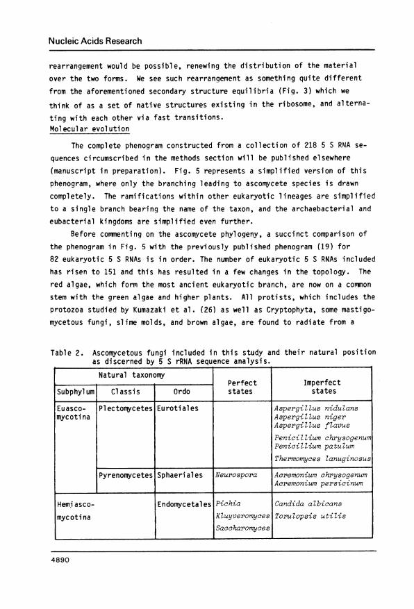
fractionation of 3-labeled 5 S RNAs. Each component was eluted and sequencedseparately.Construction of evolutionary trees
The phenogram appearing in Fig. 5 is a partial representation of a tree
constructed-from 218 5 S RNA sequences, comprising the 175 sequences of this
year's review (1), the 12 sequences reported here, 19 sequences based on oli-
gonucleotide catalogs or published recently, and 12 unpublished sequencesfrom our research group. The sequence alignment (1) comprised 148 positions.
4883

Nucleic Acids Research
Acremonium
Acremonium persicinum s
chrysogenum nidulans
2 _1 -1 ..
2 _ _ _ 1~~~~~~~-3Fig. 1. Fractionation of heterogeneous 5 S RNA preparations.In each case, 4 pg 5 S RNA labeled in the presence of 60 x 106 dpm -[5'-32P]pCpwas subjected to electrophoresis on a 5X gel containing 4 M urea, pH 8.3.Acremonium persicinum 5 S RNA yielded components 1, 2 and 3 in the ratio27 55 18, Aspergillus nidulans 5 S RNA components 1,2 and 3 in the ratio38: 45: 17. Acremonium chrysogenum 5 S RNA, as that of the other examinedspecies, was found homogeneous. Good separations required long runs at lowtension (17 h, 10 V/cm, mean migration distance 45 cm).
Clustering was performed by weighted pairwise grouping (16) starting from adissimilarity matrix. The dissimilarity DAB between two sequences A and Bwas calculated by the expression
DAB= 3 ln [1 - 4A( S )1I + S + G[1]4 L 3 I +SJ N N
where N, S, I, and G are defined as follows:I the number of alignment positions where sequences A and B contain an
identical nucleotide.S the number of positions where A is substituted with respect to B.G the number of positions where A contains a gap and B a nucleotide or
vice versa.N the number of positions where at least one of the two sequences con-
tains a nucleotide (N = I + S + G).The first term of equation [1] comprises the correction of Jukes and Cantor(cited in reference 17) for multiple mutational events.
RESULTS AND DISCUSSIONPrimary structure of the examined 5 S RNAs
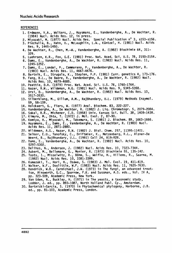
Fig. 2 shows an alignment of the 12 sequences determined in this studywith 7 sequences of ascomycete 5 S RNAs determined by other workers (listed in
4884

Nucleic Acids Research
A
* ASPEROILLUS HIDULANS (1)* ASPERGOLLOS NIDALANO (2)* ASPEROILLUS NIDULA0S (3)* ASPEROILLUS FLAVUS* ASPERILLOSNIERNC* PEHNICILLIU CHRYSOGENUO* PENICILLIUN PATULUMTHERROHYCES LASSAIAOSUS (0)THERHONYCES LANIJGINOSUS (2)
* ACRENONIUM PERSICINUR (1)* ACREMONIUJ( PERSICINUH (2)* ACREAONIUH PERSICINON (3)* ACREHONIUN CHRYSOGEAUMNEUROSPORA CR0000 (ALPHA)NEUROSPORA CRASSA (BETA)
* CANDIDA ALBICANSTORLLOPSIS UTILISSACCHAROMYCES CEREVISIAEPICHIA IEHBRANAEFACIEHS
B C C't0 20 30 40 50
vAC AUACG AC CAUA GGGUGU6AAGAAC AGGCU CCCGUCCGUCCC GCU CA OCCGUA C U A AGAC AUACGAC C AUA GGGUUGG AAAAC AGGGCU UCCCGUCCGCUC A GCC GUA CU U AA GACAUACGAC CAUA GGGUGUGG AGAAC AGGGCU UCCCGUCCGCUC AGCC GUACU UAA6ACAUACGAC CAUA GGGUGUGG AGAAC AGGGCU UCCCGUCCGCUC AGCC GUA CU UAAGA C AUAC ACC AC AGGGU 0AACA G G G C U U C C C G U CC U C A G C C G U A C U U A A GAC AUACGAC C AUAGGGUOUOAAAACAO GGGCU UCCO CCGCOCOU C C OUA CUU A AGAC AUACGAC CAUA GGGUUGU AAAAC AGGCG UCC CGCC
0COCU CC GUA CU 0A0AACAUGCGAC C AUAGGUUAAAACAGGGCUGCC CO CCCCGfCiCA OCCUAC U UA0AA C A0U C A C C A0U A G0U G C G0 A A A C A0 C U C C C U C C C U C A0 C C U0A CU0U A A0
*ASPERGILWSNIDULANS(1)~ ~ ~ A I I
A C A U A C G A CA C A U A C G A CACAUACGACACAUACGACACAUACGACACAUACGAC
G G U U G C G G CGGUUGCGGCGGUUGCGGCGGUUGCGGC
COAUCOAUCAU)C AUCOAUCACI
A
CAO)CAU)CAU)CAOU
.... .... 1010
GC G(C)U C A GGACGCUAO
CCCACGUGC C C A C U GGG A U G G U AILAAAA AL
UCUAGCAOU C U A G C AGU C U A C C AGUCUAGCAO. ...2. .
20
V IAOAAC
AAAACAAAAC
A A AA A AA A AA A A
v
ACGGGAACGGGAACGGGAUCGGGAA C G G G A
VU C C C 6 U C U G C U C
U C C C G U C C G C UU C C C G U C C G C UCUCCCGUCCGCUCUCCCGUCCGCUCOC C COuC CO6COCOC C COuC CO6COC
VUCCUUCCCOCCCOCCCOCCCOCCCU CC C
VU A
AUAAU0AU0AU0000000
V000000000000
V
CAAG
CAAGUAAGUAAG
IG C A C CGUUCCCC UUC AUCAACC UA UUAA4GC ACCGUC C C COGU CCGAU C AAC U GUA U UAAGiG C A C C G U U U C C C U C C G A U C A A C U G OAG U U A A GG C A CCC G CC C U0CC G A U C A A C lU_OUA G U U A A G
30 40 50
B' D60 70
TVC C A C A C G C C G G CUAG G U U -A G U A
CC ACAC CC GGCUGG UU-AGU0CC ACAC G CC GGCGG UU-A GU0CCACAC G CC GGGAGG UU-AG0UCCACACOGCCO 000000U-000)CCACAC G CC GGUGAG UU-0A0UCCACAC G CC GGUGA-GUU0AGUCCACAC G CC GGCUGG UU A-GU0C C G C A C G C C G G C U G 0UU 0AG U0
Av v v
C U G A(CA A G C G G C C G A U U - A G U ACUGGCG A UC GGCGG AUU-AGU0CUGGCG A CC GGCUG AUU -AGU0CCGCUA A GG GGCUG AUU -AGU0CCAG UG A GG GCC AGACU-AGUPCCUCA A U C GCCUG A CU -AG0UA AAAAA A
CU GCUA A CAC0AAC CCGAGU)CUGCUAAAG CCU6A UCGAGUACUGGUAA A GA GCCUGA CCGAGUC U G C U A G A G C C U G A C C G A G U P
E El90 10(
. . . .. . .. .I
3U0AU30UA0U30UA0U30UUGE30UUG30UU0G30UUG3UUG030UUG
3U0UG3U0UG3UUG03U0UG3UUG00UUGI
UGGUGGUG0U0 GU0 GU0 GU0U0U0 G
UCEUCOGUCOGUCEUCE.UC 6
aU G U A0G1 EU U A G1U 6U G U A GUG GU G U A ,AG
36U0GG3U0G3GU0G3G0 A3U0G0UGA0UGAU0G
3GU0G
3U0G3GU0G3U0G3GU0G3GU0G3U00
30A6036U0030G0036CGA
C C A C A U G CCCACAUGCCCACAU6CC C A C C A 6 CCCACCA6CCCACCA6CCCACCAGCC C A C C A 6 CC C A C C A 6 C
C G A C C A 6C6ACCCACC6ACCA6CC 6 A C C A 6C G A C C A C
C G A C C AG
CCAUAC0CC C A U A C G CCCCUACGCC C U A C C
..I ....I.... I....I.... I....I....I....I....160 70 80 90 too
D' A'110 120
A3A0UC C COO CO 00000000UAUG
A3000C C CAOGCOUGO 0000000UG
A A0UC C CAUUC U GU UGOUA 000
30A U C C U C A C U
0U
0 00U A
0G0GA AUC C CUAGC U GU UGUCA UG UGAAUCCCAGCUOGUUCAUG00
3AAUC CCACU GUUGUAUGUGA AUAC CCU GCCU GU UG UA UG UGA AUAC CCUUCCU GU UG UA UG U6 A AUAC CUGGCACU GU UG UA UG UGA A UC C CUCACGU GU UG UA UG UUGA A UC C CA GCGU G UU GCUA U G U
GA AUACUCGGCU GUUG8CU0CAA UGCUGA AUACUCUAGCGU GUCUGUCAA UGCUGA AUACUCAG GCU GUCUGUCAA UGCU
GAAU0CCOCUGGU 0000000UGAAU0CCOOCAGGU UUGUUGU
OGAAACCUAUG 6CUGCAAUC000
3GAAACOUAGGU GCUGCAAUC 0
3A0AA C UCAGGU GCGC 0AAU C
110 120
Fiq. 2. Primary structure of ascomycete 5 S RNAs.The sequences preceded by an asterisk,were determined in this study. Trianglesabove or below a set of sequences found in the same species indicate thepoints of heterogeneity. Boxes and their lettering correspond to the double-stranded areas in the secondary structure models of Fig. 3. Nested boxescorrespond with bulges, bracketed nucleotides with non-standard base pairs.The scale on top numbers the residues of the two upper sequence clusters, thescale at the bottom those of the lower cluster consisting of yeast sequences,which have an extra nucleotide between areas D and E. The sequences of Saccha-romyces carlsbergensis and Kluyveromyces lactis (2) are not listed since theyare identical to that of Saccharomyces cerevisiae.
4885
* A. MID. (1)* A. NID. (2)* A. NID. (3)*A. FLA.*A. 0IG.*P. CHR.*P. PAT.
T. LAN. (1)T. LAN. (2)
*A. PER. (1)* A. PER. (2)*A. PER. (3)*A. CHR.
H. CRA. (A)N. CRA. (B)
*C. ALD.T. UTI.S. CER.P. MEN.
80. . .. . .

Nucleic Acids Research
a. .lsplrl/u/s1n7dulaos
UAA
b. J,cremon/2/,mpelrslclnulm
A
A M B 1, C H1
C A U A G A A C U C CpACAUACGAC GGGUGUGG AGGGCU u
. . . . .
UGUAUGUUG CCCACACC UCCCGA C
CUG GAAU AG U C GD AU A
C C . G U CCGU -G AA GGCUAA uU CCG12 A G G A C
HAVG C.GAG*UU A
,- A *Ui,A-C.G
C*GA*UG GU GG
GC.G AiG*UU * A
E A-UC G-G_)A:.UC GC G
H2 A GG U
AGA A-G-G AC6 GCU-C UGCCGAG AA C -AUU
CAUA AG A A UC C CGpACAUACGAC GCU UCAG UCGGGA U
UGUAUGUUG CGA6AGUC AGUCCU C
CUG A GAAUA CUCGG*C
C C-GA CCGA A G A Au A u -G CUCGGGAA U A -C GAGUC C U
G CGAG AuGGGAGC.G CAU AAUGG-U G-U G cAU A-UAGA
C.G C-G -G ACGGGA,
G-C-G A-UG -C UGUCCUA.-U G-C G AG-C C-G A GU G A G A UAG GU
C Canda'da c A UA A A G C C C CG
a/6/cans PGGUUGCGGC UCUAGCAG ACCGUU UCUAACGUCG AGAUCGUC UG CAA U
U UG 'A'
G. C
U: A
U A
A UC A C GAC U GACC CcA GAC- G
G
A-GA GC A A A AA-U A UA *A AA
GC .G GUGG-U G.U C-CG CGCGG C-G C-AGU G'U'AA-U A-U.. U:A..G C G.
A-U-A U:A..G.) A .U A.U
&C.G AiUr CG AU.GA.U C*G C.G AC-GG G CG0 A G C-GA G A G GA A AG GA G
GAAU A CU GC
-G^AAAGCAC GUU
GA6 G a i
GAA_G AAA G C <~q~-G AGCACCGCU-C UUGUG CAA
AA 'g/
Fig. 3. Secondary structure variants in asconmycete 5 S RNAs.Helices are labeled A to E, loops are labeled M (multibranched), I (interior)or H (hairpin). The labeling, corresponding with that in Fig. 2, is marked instructure a only. The base pairs G-C, A-U, G-U are indicated by a dot, non-standard base pairs by a losenge. Each of the three models a, b, and c isapplicable to all the 5 S RNAs of the same cluster (Fig. 2). Alternative base-pairing schemes in area I1-C (4) and I2-E (De Wachter et al., submitted) arethought to be in dynamic equilibrium with each other. Additional alternativebase pairinq opportunities in loop I2 are indicated by thin lines. Bulgesthat can migrate along a helix are indicated by curved arrows, each arrowheadpointing to a possible position.
4886
)

Nucleic Acids Research
references 1 and 2). Schizosaccharomyces pombe (18) is not included because itwas found (19) evolutionarily related to protists rather than to ascomycetes.The 19 sequences are arranged in 3 groups based on homology and the existenceof distinctive base pairinq patterns. Two of the 8 species examined by us, As-pergillus nidulans and Acremonium persicinum, each contain 3 different 5 SRNAs of equal chain length, the separation of which is documented in the meth-
ods section. A similar phenomenon has been observed in the case of Thermomyceslanuginosus (20) and Neurospora crassa (21). The positions where heterogeneity
occurs are indicated in Fig. 2 by triangles above or below the sequences.The sequence previously reported by Piechulla et al. (3) for Aspergillus
nidulans 5 S RNA bears most resemblance to our component 2, but it differsfrom it by the substitutions G1, A53 and C1l9 that we suspect to be sequencinqerrors. The Neurospora crassa sequence reported in the same paper has alsobeen corrected later (21). The fact that a U is present rather than an A in
position 53 of the Aspergillus nidulans 5 S RNA makes it possible to draw thesequence in the familiar secondary structure model with a bulge in the 3'-proximal strand of helix C (see Fig. 3 discussed in the next section).Secondary structure of ascomycete 5 S RNAs
Fig. 3 shows the sequences of Aspergillus nidulans (variant 1), Acremo-
nium persicinum (variant 1) and Candida albicans 5 S RNAs, all drawn in the
previously proposed (4) secondary structure model. Although this model is ap-plicable to all hitherto published 5 S RNA structures (1), there is some vari-
ability in helix lengths, loop sizes and bulge positions. The precise shape of
the model is characteristic for the major taxon to which the considered spe-
cies belongs (22,23). Three slightly different shapes are observed among theascomycete 5 S RNAs. The structure found in Aspergillus, Penicillium andThermomyces is exemplified in Fig. 3a. Acremonium and Neurospora possess 5 S
RNAs with a slightly shorter D helix as shown in Fig. 3b. Finally the yeast
Fig. 4. Behaviour of Acremoniumchrysogenum 5 S RNA on denaturing gels.The 3'-terminally labeled 5 S RNA,loaded on an 8% polyacrylamide gel
1 2 containina 7 M urea, pH 8.3, and runfor 3 h at 32 V/cm, yielded twobands (left). When the RNA was ex-
.. ...tracted (10) from each band, precip-2. t Iwitated with ethanol, redissolved and
rerun on a similar gel, each extractyielded the same two-band pattern(middle and right). Migration dis-tance was about 25 cm in all cases.
4887

Nucleic Acids Research
40
d D
IC
i_
40
0
0
C}
d [
4888

Nucleic Acids Research
5 S RNAs assume a shape with an asymmetric interior loop in helix C replacingthe usual bulge, exemplified by the Candida albicans structure of Fig. 3c. Itwill be demonstrated below that these three secondary structure shapes coin-cide with three clusters in the evolutionary tree, in other words that it is
possible to track secondary structure mutations in evolution. A distinctivefeature shared by all ascomycete 5 S RNAs is the large size of the bulge inhelix C, which in nearly all other species consists of two bases. It is note-worthy that this feature is missing in the 5 S RNA of Schizosaccharomyces pom-be, which does not turn up among the ascomycetes in an evolutionary tree (19).
We have previously pointed out that it is possible to draw two alterna-tive base-pairing schemes for area I1-C of the secondary structure model inall examined 5 S RNAs (4,6,7,9,19,22). More recently we observed (De Wachteret al., submitted) that in eukaryotic 5 S RNAs at least three different struc-tures are possible in area I1-C and two to five alternative structures inarea E, and we discussed the possible meaning of base-paring equilibria in aflexible 5 S RNA structure. The sets of alternative shapes observable inareas C and E are also taxon-specific, and the sets encountered in the threetypes of ascomycete 5 S RNAs are indicated in Fig. 3.
A curious phenomenon was observed when the 5 S RNA of some species, suchas Acremonium chrysogenum, was purified after 3'-terminal labeling on gelscontaining 7 M urea. Autoradiography revealed the presence of two bands, a
sharp one followed by a more diffuse slower moving one, as illustrated inFig. 4. Sequence analysis showed that both components had the same primarystructure, and each component, reloaded on a similar gel after extraction,yielded the same two-band pattern again. This phenomenon is different fromthose observed with E. coli 5 S RNA (24) and rat liver 5 S RNA (25) whichyield bands with different mobility only on non-denaturing gels. A possibleexplanation of our observation is that the 5 S RNA can assume two conforma-tions, possibly a native and an artefactual one. Each conformation would bestable during electrophoresis but in some step of the extraction procedure
Fig. 5. Ascomycete phylogeny as derived from 5 S RNA sequences.Only the ascomycete branch is drawn in full detail. Ramifications corres-sponding to other eukaryotic taxa are reduced to a single bar bearing the nameof the taxon followed by the number of known sequences in brackets. Thepointed end of the bar is situated at the earliest branching within the taxon.The taxon Traustochytridiaceae comprises Thraustochytrium visurgense and Schi-zochytrium aggregatum, the taxon Monadophyceae comprises 3 Chlamydomonas se-quences. The archaebacterial and eubacterial primary kingdoms are each reducedto a single branch. Chloroplast and plant mitochondrial 5 S RNAs cluster inthe eubacterial kingdom.
4889
Nucleic Acids Research
rearrangement would be possible, renewing the distribution of the material
over the two forms. We see such rearrangement as something quite different
from the aforementioned secondary structure equilibria (Fig. 3) which we
think of as a set of native structures existing in the ribosome, and alterna-
ting with each other via fast transitions.Molecular evol ution
The complete phenogram constructed from a collection of 218 5 S RNA se-
quences circumscribed in the methods section will be published elsewhere
(manuscript in preparation). Fig. 5 represents a simplified version of this
phenogram, where only the branching leading to ascomycete species is drawn
completely. The ramifications within other eukaryotic lineages are simplifiedto a single branch bearing the name of the taxon, and the archaebacterial and
eubacterial kingdoms are simplified even further.Before commenting on the ascomycete phylogeny, a succinct comparison of
the phenogram in Fig. 5 with the previously published phenogram (19) for82 eukaryotic 5 S RNAs is in order. The number of eukaryotic 5 S RNAs included
has risen to 151 and this has resulted in a few changes in the topology. The
red algae, which form the most ancient eukaryotic branch, are now on a common
stem with the green algae and higher plants. All protists, which includes the
protozoa studied by Kumazaki et al. (26) as well as Cryptophyta, some mastigo-
mycetous fungi, slime molds, and brown algae, are found to radiate from a
Table 2. Ascomycetous fungi included in this study and their natural positionas discerned by 5 S rRNA sequence analysis.
Natural taxonomyPerfect Imperfect
Subphylum Classis Ordo states states
Euasco- Plectomycetes Eurotiales AspergilZZus niduZansmycoti na AspergiZZus niger
Aspergi Z lus fZavus
Penici ZZium chrysogenumPeniciZZium patuZumThermomyces Zanuginosus
Pyrenomycetes Sphaeriales Neurospora Acremonium chrysogenumAcremonium persicinum
Hemiasco- Endomycetales Pichia Candida albicans
mycotina KZuyveromyces ToruZopsis utilZisSaccharomyces
4890

Nucleic Acids Research
single branch. Also the addition of supplementary basidiorycete sequences (27)resulted in this taxon forming a single cluster whereas in our previous pheno-gram (19) the Teliomycetes were on a branch separate from the Holo- and Phraa-mobasi diomycetes.
All the fungi whose 5 S RNAs were sequenced in this study are imperfectstates of ascomycetes. Aspergillus and Penicillium are conidial states corre-sponding to asexual states of organisms belonging to the order Eurotiales(class Plectomycetes) (28). They can therefore be used to delineate the posi-tion of this taxon in the phenogram. For the other species examined here,classical taxonomists have had more difficulties in establishing a correspond-ence with the taxonomy for perfect states. Some species of the genus Acremo-nium have been assigned (28) to the Eurotiales, others to the Sphaeriales(class Pyrenomycetes). The genus Candida comprises strictly asporogenousyeasts which can be assigned (29) alternatively to the Endomycetales (Hemias-comycetes) or to basidiosporogenous yeasts. The taxonomic conclusions thatcan be drawn from our phenogram are summarized in Table 2. Our analysis in-dicates a much closer relationship of Acremonium chrysogenum and A. persici-num to Neurospora, which belongs to the Sphaeriales, than to Aspergillus or
Penicillium. Thus we can assign the two species to the Pyrenomycetes, proba-bly the Sphaeriales. Thermomyces lanuginosus, also an imperfect state, can beassigned to the Eurotiales. Candida albicans and Torulopsis utilis clearlybelong to the Endomycetales.
It can be observed in the phenogram (Fig. 5) that the Ascomycota haveundergone an early splitting into a lineage leading to the Hemiascomycotina(represented only by the Endomycetales in Fig. 5) and one leading to the Eu-ascomycotina (represented by Pyrenomycetes and Plectomycetes). A similarphylogeny based on cell wall composition and additional biochemical markersis also discerned by Bartnicki-Garcia (30). A common origin of Basidiomycotaand Ascomycota is not seen in our phenogram. It is possible, however, thatthe topology of ancient fungal relationships be ified if sequences fromProtomycetales, Taphrinales (Hemiascomycotina) and Uredinales (Basidiomycota)are investigated and included in the analysis.
ACKNOWLEDGMENTSOur research was supported in part by an N.F.W.O. grant to R. De Wachter
and A. Vandenberghe. G.V. is an N.F.W.O. research associate.
*To whom correspondence should be addressed
4891
Nucleic Acids Research
REFERENCES1. Erdmann, V.A., Wolters, J., Huysmans, E., Vandenberghe, A., De Wachter, R.
(1984) Nucl. Acids Res. 12, in press.2. Miyazaki, M. (1977) Nucl. Acids Res. Special Publication no 3, s153-s156.3. Piechulla, B., Hahn, U., McLaughlin, L.W., KUntzel, H. (1981) Nucl. Acids
Res. 9, 1445-1450.4. De Wachter, R., Chen, M.-W., Vandenberghe, A. (1982) Biochimie 64, 311-
329.5. Luehrsen, K.R., Fox, G.E. (1981) Proc. Nat. Acad. Sci. U.S. 78, 2150-2154.6. Dams, E., Vandenberghe, A., De Wachter, R. (1983) Nucl. Acids Res. 11,
1245-1252.7. Dams, E., Londei, P., Cammarano, P., Vandenberghe, A., De Wachter, R.
(1983) Nucl. Acids Res. 11, 4667-4676.8. Bartnik, E., Strugala, K., Stepien, P.P. (1981) Curr. genetics 4, 173-176.9. Fang, B.L., De Baere, R., Vandenberghe, A., De Wachter, R. (1982) Nuci.
Acids Res. 10, 4679-4685.10. Peattie, D.A. (1979) Proc. Nat. Acad. Sci. U.S. 76, 1760-1764.11. Nazar, R.N., Wildeman, A.G. (1981) Nucl. Acids Res. 9, 5345-5358.12. Ursi, D., Vandenberghe, A., De Wachter, R. (1982) Nucl. Acids Res. 10,
3517-3530.13. Silberklang, M., Gillum, A.M., RajBhandary, U.L. (1979) Methods Enzymol.
59, 58-109.14. Volckaert, G., Fiers, W. (1977) Anal. Biochem. 83, 222-227.15. Vandenberghe, A., De Wachter, R. (1982) J. Liq. Chromatogr. 5, 2079-2084.16. Sokal, R.R., Michener, C.D. (1958) Univ. Kansas Sci. Bull. 38, 1409-1438.17. Kimura, M., Ohta, T. (1972) J. Mol. Evol. 2, 87-90.18. Komiya, H., Miyazaki, M., Takemura, S. (1981) J. Biochem. 89, 1663-1666.19. Huysmans, E., Dams, E., Vandenberghe, A., De Wachter, R. (1983) Nucl.
Acids Res. 11, 2871-2880.20. Wildeman, A.G., Nazar, R.N. (1982) J. Biol. Chem. 257, 11395-11401.21. Selker, E.U., Yanofsky, C., Driftmier, K., Metzenberg, R.L., Alzner-De
Weerd, B., RajBhandary, U.L. (1981) Cell 24, 819-828.22. Dams, E., Vandenberghe, A., De Wachter, R. (1982) Nucl. Acids Res. 10,
5297-5302.23. Delihas, N., Andersen, J. (1982) Nucl. Acids Res. 10, 7323-7344.24. Aubert, M., Bellemare, G., Monier, R. (1973) Biochimie 55, 135-142.25. Toots, I., Misselwitz, R., Bohm, S., Welfle, H., Villems, R., Saarma, M.
(1982) Nucl. Acids Res. 10, 3381-3389.26. Kumazaki, T., Hori, H., Osawa, S. (1983) J. Mol. Evol. 19, 411-419.27. Walker, W.F., Doolittle, W.F. (1983) Nucl. Acids Res. 11, 7625-7630.28. Kendrick, W.B., Carmichael, J.W. (1973) in The fungi, an advanced treat-
ise, Ainsworth, G.C., Sparrow, F.K. and Sussman, A.S. eds., Vol. IV A,pp. 323-509, Academic Press, New York.
29. Van Uden, N., Buckley, H. (1971) in The yeasts, a taxonomic study,Lodder, J. ed., pp. 893-1087, North Holland Publ. Cy., Amsterdam.
30. Bartnicki-Garcia, S. (1970) in Phytochemical phylogeny, Harborne, J.B.ed., pp. 81-103, Academic Press, London.
4892
Copyright © 2022 FDOKUMEN