Children's early ability to solve perspective-taking problems
Upload
khangminh22Category
view
2download
0
The NeuroCognitive Research Institute 1829 N. Sheffield Ave, Suite 2, Chicago, Illinois 60614
Phone: (877) 963-8763 Email: [email protected]
Quantitative EEG/swLORETA Analyses
PATIENT INFORMATION RECORDING
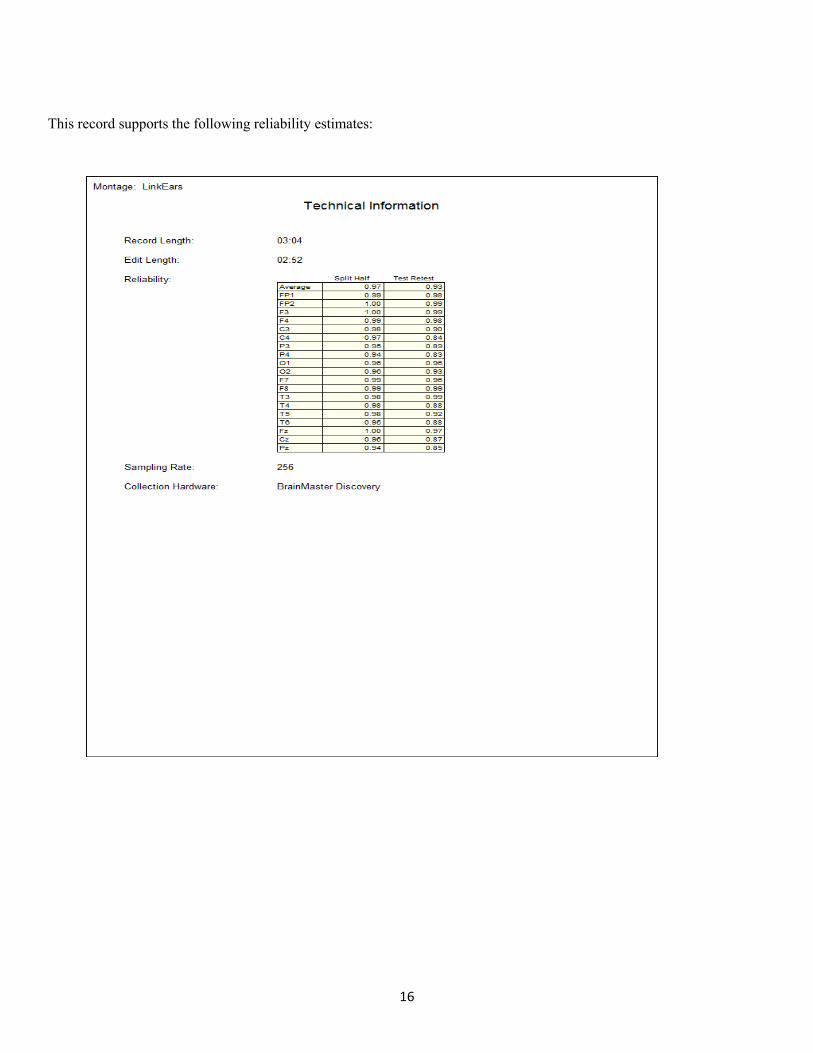
Name: ME/CFS Patient Date: 12/21/2018 Age: 51.37 Test Site: Chicago, IL Gender: F Analysis Length: 02:52 Handedness: R Ave. SH Reliability: 0.97 Eyes: Closed Ave. TRT Reliability: 0.93
MEDICATIONS: Acyclovir, Sulfasalazine
• Acyclovir stops viral replication but does not kill the virus(es)\ • Sulfasalazine is a DMARD (Disease-Modifying Anti-rheumatic Drug) which decreases neural inflammation.
HISTORY: This 51 year-old ME/CFS patient was diagnosed with a brain infection in 2007. This infection became chronic, which then became Myalgic Encephalitis.
SUMMARY: The quantitative EEG (qEEG) analyses were deviant from normal, indicating dysregulation in the bilateral frontal lobes, bilateral temporal lobes especially in the right temporal lobe, bilateral parietal lobes especially in the right parietal lobe and bilateral occipital lobes, especially in the right occipital lobe. Standardized weighted low- resolution electromagnetic tomography (swLORETA) analyses revealed dysregulation in the left posterior insula, the left retrosplenial cortex and right parahippocampal gyrus.
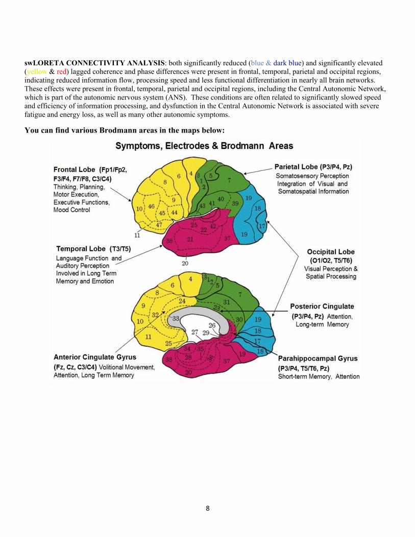
The frontal lobes are involved in executive functioning, abstract thinking, expressive language, sequential planning, mood control and social skills. The temporal lobes are involved in auditory information processing, short-term memory, receptive language on the left and face recognition on the right. The parietal lobes are involved in visual-spatial information processing, short-term memory, executive attention, receptive language on the left and empathy control and awareness of emotional expression in others on the right (e.g., prosody). The occipital lobes are involved in the visual processing of color, form, movement, visual perception and spatial processing. The posterior insular cortex is involved in autonomic system regulation and interoceptive representation of the physiological condition of the body. The parahippocampal gyrus is involved in the creation of new memories, retrieval of short-term memory and attention control, and the retrosplenial cortex is involved with spatial navigation, episodic memory, nagivation, imaging future (expected) events and overall spatial working memory. To the extent there is deviation from normal electrical patterns in these structures, then sub-optimal functioning is expected.
There was significant deviation in the retrosplenial cortex (6-8 standard deviations from normal), which indicates increased errors of omission. The retrosplenial cortex is especially involved with response conflict, such as Stroop items. It supports cognition through extensive hippocampal connections and frontal lobe connections.

2
swLORETA connectivity analyses revealed significant deviations from normal across all frequency bands, involving most Brodmann areas of the brain. At the inception of EEG in 1929, Hans Berger (Finger, 2000) observed the oscillatory nature of the human EEG and subsequent research has found that different rhythmic attributes (frequency bands) have dissimilar physiological significances and mental states as well as different temporal patterns related to brain function.
Delta activity (~1 – 3 Hz), for example, is produced by cortico-cortical and cortico-thalamic networks and is involved in basic homeostatic processing, restorative sleep, salience recognition, and language. 1-3 Hz is also involved in slowing of EEG background activity, which has been associated with many neuroinflammatory conditions and neurotropic virus infections. Increases in delta activity have been implicated in studies of Alzheimer’s disease and may demonstrate a link between brain states, arousal, and efficiency, with decrements in information processing speed, which is typically found in ME/CFS.
The theta frequency band (~4 – 7 Hz) originates in the thalamus and limbic system and is associated with a variety of cognitive functions, especially memory retrieval and new learning. Theta is known to arise from synchronized neurons (pacemakers) in the limbic system, including the cingulate gyrus and the parahippocampal cortex. It is considered important for a variety of cognitive functions including memory consolidation, spatial navigation, working memory and memory encoding.
Alpha rhythms are divided into two parts, low alpha (8 – 10 Hz) and high alpha (10 – 12 Hz). There is a large body of evidence indicating that alpha amplitude is associated with the level of cortical activation, especially in perception and attention. High alpha is strongly associated with accuracy in forming cognitive representations of various physiological states, which is known as “being in the zone.” Encouraging this state is usually a core element in optimal performance training.
Beta rhythms (13-30 Hz) are hypothesized to be generated in the cortex and appear to be related to movement activity, both planned and actual movement. Beta activity is often subdivided into several bands. Beta-1 (13 – 18 Hz), is associated with problem-solving and forming accurate mental representations. Physiologically, it is also associated with tense muscles and tends to occur when sensory-motor loops are interrupted. Beta-2 (19 – 21 Hz) is also associated with problem-solving and representations but more associated with attention and higher cognitive functions. Beta-3 (22 – 30 Hz) is strongly related to worry and rumination, including other cognitive distortions and general feelings of being “stressed.”
Dr. Mark Zinn and Dr. Marcie Zinn
Reminder: when we say, "significant at the .05 level," we are saying hypothetically if the network analysis were performed an infinite number of times in the same person, the same results would happen at least 95% of the time. The only network connections that appear in this analysis are the ones that are significantly hypo-active (blue and dark blue) or significantly hyper-active (yellow-red).
3
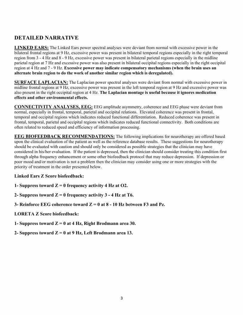
DETAILED NARRATIVE
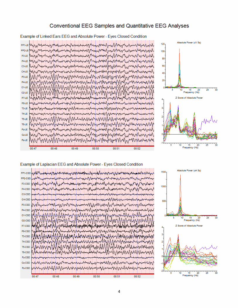
LINKED EARS: The Linked Ears power spectral analyses were deviant from normal with excessive power in the bilateral frontal regions at 9 Hz, excessive power was present in bilateral temporal regions especially in the right temporal region from 3 - 4 Hz and 8 - 9 Hz, excessive power was present in bilateral parietal regions especially in the midline parietal region at 7 Hz and excessive power was also present in bilateral occipital regions especially in the right occipital region at 4 Hz and 7 - 9 Hz. Excessive power may indicate compensatory mechanisms (when the brain uses an alternate brain region to do the work of another similar region which is deregulated).
SURFACE LAPLACIAN: The Laplacian power spectral analyses were deviant from normal with excessive power in midline frontal regions at 9 Hz, excessive power was present in the left temporal region at 9 Hz and excessive power was also present in the right occipital region at 4 Hz. The Laplacian montage is useful because it ignores medication effects and other environmental effects.
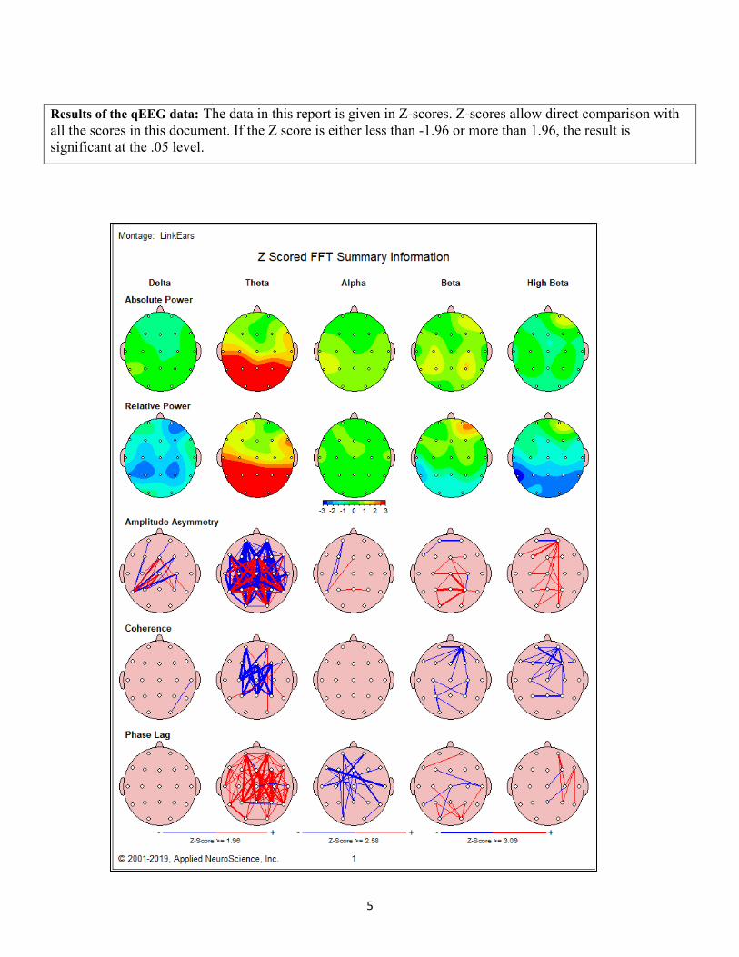
CONNECTIVITY ANALYSES, EEG: EEG amplitude asymmetry, coherence and EEG phase were deviant from normal, especially in frontal, temporal, parietal and occipital relations. Elevated coherence was present in frontal, temporal and occipital regions which indicates reduced functional differentiation. Reduced coherence was present in frontal, temporal, parietal and occipital regions which indicates reduced functional connectivity. Both conditions are often related to reduced speed and efficiency of information processing.
EEG BIOFEEDBACK RECOMMENDATIONS: The following implications for neurotherapy are offered based upon the clinical evaluation of the patient as well as the reference database results. These suggestions for neurotherapy should be evaluated with caution and should only be considered as possible strategies that the clinician may have considered in his/her evaluation. If the patient is depressed, then the clinician should consider treating this condition first through alpha frequency enhancement or some other biofeedback protocol that may reduce depression. If depression or poor mood and/or motivation is not a problem then the clinician may consider using one or more strategies with the priority of treatment in the order presented below.
Linked Ears Z Score biofeedback:
1- Suppress toward Z = 0 frequency activity 4 Hz at O2.
2- Suppress toward Z = 0 frequency activity 3 - 4 Hz at T6.
3- Reinforce EEG coherence toward Z = 0 at 8 - 10 Hz between F3 and Pz.
LORETA Z Score biofeedback:
1- Suppress toward Z = 0 at 4 Hz, Right Brodmann area 30.
2- Suppress toward Z = 0 at 9 Hz, Left Brodmann area 13.
4
5
Results of the qEEG data: The data in this report is given in Z-scores. Z-scores allow direct comparison with all the scores in this document. If the Z score is either less than -1.96 or more than 1.96, the result is significant at the .05 level.

6
7
Electrical Neuroimaging SwLORETA Connectivity Analyses
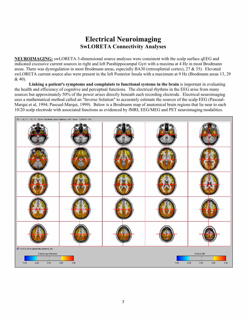
NEUROIMAGING: swLORETA 3-dimensional source analyses were consistent with the scalp surface qEEG and indicated excessive current sources in right and left Parahippocampal Gyri with a maxima at 4 Hz in most Brodmann areas. There was dysregulation in most Brodmann areas, especially BA30 (retrosplenial cortex), 27 & 35). Elevated swLORETA current source also were present in the left Posterior Insula with a maximum at 9 Hz (Brodmann areas 13, 29 & 40). Linking a patient's symptoms and complaints to functional systems in the brain is important in evaluating the health and efficiency of cognitive and perceptual functions. The electrical rhythms in the EEG arise from many sources but approximately 50% of the power arises directly beneath each recording electrode. Electrical neuroimaging uses a mathematical method called an "Inverse Solution" to accurately estimate the sources of the scalp EEG (Pascual-Marqui et al, 1994; Pascual-Marqui, 1999). Below is a Brodmann map of anatomical brain regions that lie near to each 10/20 scalp electrode with associated functions as evidenced by fMRI, EEG/MEG and PET neuroimaging modalities.
8
swLORETA CONNECTIVITY ANALYSIS: both significantly reduced (blue & dark blue) and significantly elevated (yellow & red) lagged coherence and phase differences were present in frontal, temporal, parietal and occipital regions, indicating reduced information flow, processing speed and less functional differentiation in nearly all brain networks. These effects were present in frontal, temporal, parietal and occipital regions, including the Central Autonomic Network, which is part of the autonomic nervous system (ANS). These conditions are often related to significantly slowed speed and efficiency of information processing, and dysfunction in the Central Autonomic Network is associated with severe fatigue and energy loss, as well as many other autonomic symptoms.
You can find various Brodmann areas in the maps below:

9
BRAIN BRODMANN REGIONS
10
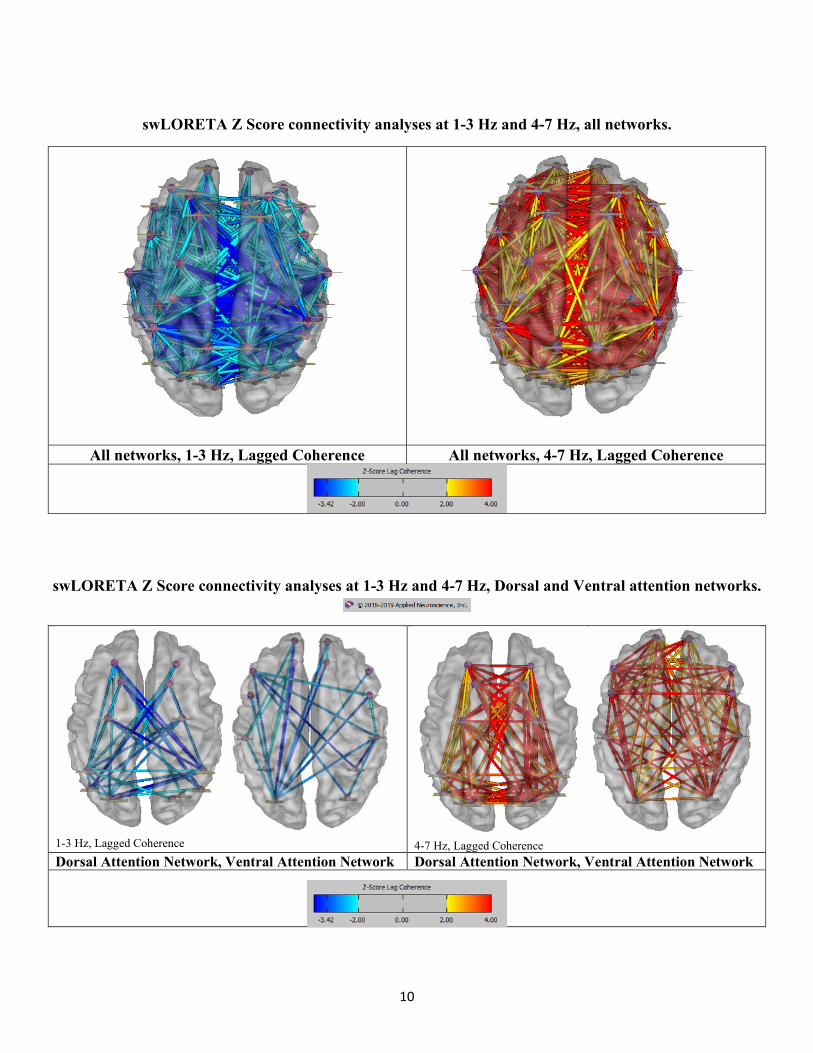
swLORETA Z Score connectivity analyses at 1-3 Hz and 4-7 Hz, all networks.
All networks, 1-3 Hz, Lagged Coherence All networks, 4-7 Hz, Lagged Coherence
swLORETA Z Score connectivity analyses at 1-3 Hz and 4-7 Hz, Dorsal and Ventral attention networks.
1-3 Hz, Lagged Coherence 4-7 Hz, Lagged Coherence Dorsal Attention Network, Ventral Attention Network Dorsal Attention Network, Ventral Attention Network
11
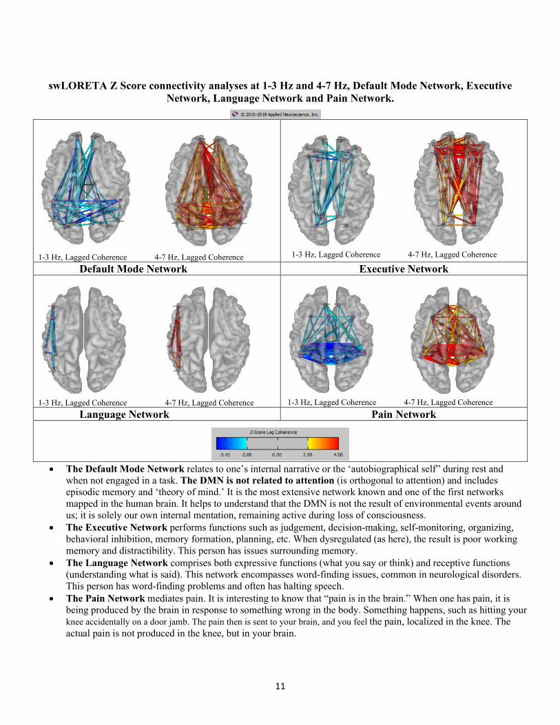
swLORETA Z Score connectivity analyses at 1-3 Hz and 4-7 Hz, Default Mode Network, Executive Network, Language Network and Pain Network.
1-3 Hz, Lagged Coherence 4-7 Hz, Lagged Coherence 1-3 Hz, Lagged Coherence 4-7 Hz, Lagged Coherence Default Mode Network Executive Network
1-3 Hz, Lagged Coherence 4-7 Hz, Lagged Coherence 1-3 Hz, Lagged Coherence 4-7 Hz, Lagged Coherence Language Network Pain Network
• The Default Mode Network relates to one’s internal narrative or the ‘autobiographical self” during rest and when not engaged in a task. The DMN is not related to attention (is orthogonal to attention) and includes episodic memory and ‘theory of mind.’ It is the most extensive network known and one of the first networks mapped in the human brain. It helps to understand that the DMN is not the result of environmental events around us; it is solely our own internal mentation, remaining active during loss of consciousness.
• The Executive Network performs functions such as judgement, decision-making, self-monitoring, organizing, behavioral inhibition, memory formation, planning, etc. When dysregulated (as here), the result is poor working memory and distractibility. This person has issues surrounding memory.
• The Language Network comprises both expressive functions (what you say or think) and receptive functions (understanding what is said). This network encompasses word-finding issues, common in neurological disorders. This person has word-finding problems and often has halting speech.
• The Pain Network mediates pain. It is interesting to know that “pain is in the brain.” When one has pain, it is being produced by the brain in response to something wrong in the body. Something happens, such as hitting your knee accidentally on a door jamb. The pain then is sent to your brain, and you feel the pain, localized in the knee. The actual pain is not produced in the knee, but in your brain.
12
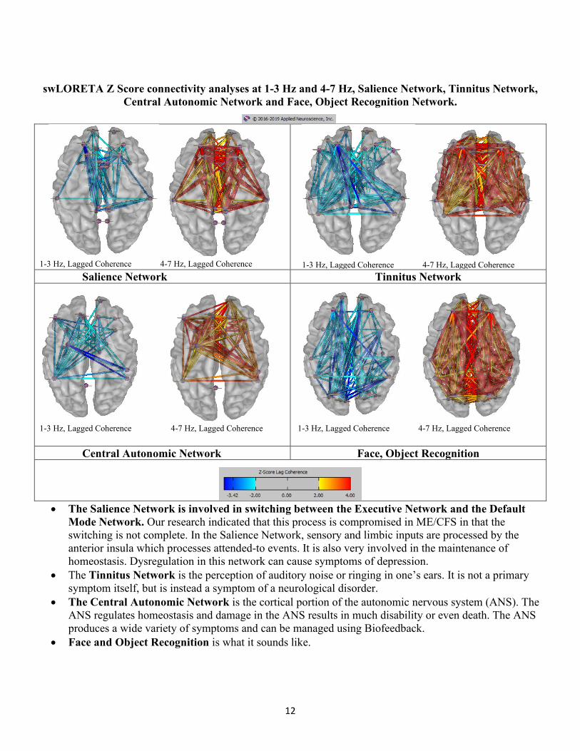
swLORETA Z Score connectivity analyses at 1-3 Hz and 4-7 Hz, Salience Network, Tinnitus Network, Central Autonomic Network and Face, Object Recognition Network.
1-3 Hz, Lagged Coherence 4-7 Hz, Lagged Coherence 1-3 Hz, Lagged Coherence 4-7 Hz, Lagged Coherence Salience Network Tinnitus Network
1-3 Hz, Lagged Coherence 4-7 Hz, Lagged Coherence 1-3 Hz, Lagged Coherence 4-7 Hz, Lagged Coherence
Central Autonomic Network Face, Object Recognition
• The Salience Network is involved in switching between the Executive Network and the Default Mode Network. Our research indicated that this process is compromised in ME/CFS in that the switching is not complete. In the Salience Network, sensory and limbic inputs are processed by the anterior insula which processes attended-to events. It is also very involved in the maintenance of homeostasis. Dysregulation in this network can cause symptoms of depression.
• The Tinnitus Network is the perception of auditory noise or ringing in one’s ears. It is not a primary symptom itself, but is instead a symptom of a neurological disorder.
• The Central Autonomic Network is the cortical portion of the autonomic nervous system (ANS). The ANS regulates homeostasis and damage in the ANS results in much disability or even death. The ANS produces a wide variety of symptoms and can be managed using Biofeedback.
• Face and Object Recognition is what it sounds like.
13
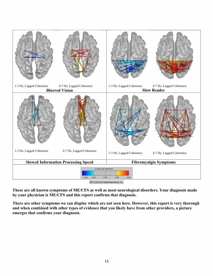
1-3 Hz, Lagged Coherence 4-7 Hz, Lagged Coherence
1-3 Hz, Lagged Coherence 4-7 Hz, Lagged Coherence
Slow Reader Blurred Vision
1-3 Hz, Lagged Coherence 4-7 Hz, Lagged Coherence
1-3 Hz, Lagged Coherence 4-7 Hz, Lagged Coherence
Slowed Information Processing Speed Fibromyalgia Symptoms
These are all known symptoms of ME/CFS as well as most neurological disorders. Your diagnosis made by your physician is ME/CFS and this report confirms that diagnosis.
There are other symptoms we can display which are not seen here. However, this report is very thorough and when combined with other types of evidence that you likely have from other providers, a picture emerges that confirms your diagnosis.
14
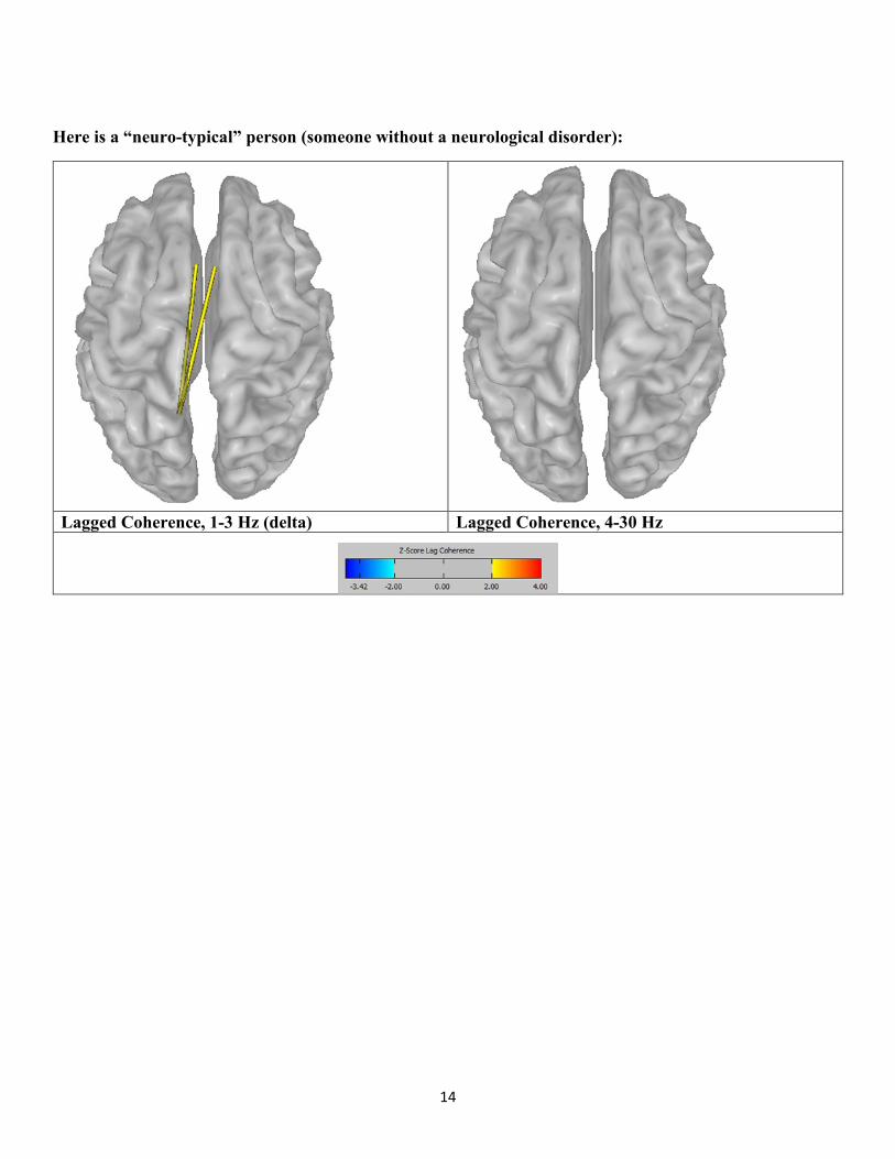
Here is a “neuro-typical” person (someone without a neurological disorder):
Lagged Coherence, 1-3 Hz (delta) Lagged Coherence, 4-30 Hz
15
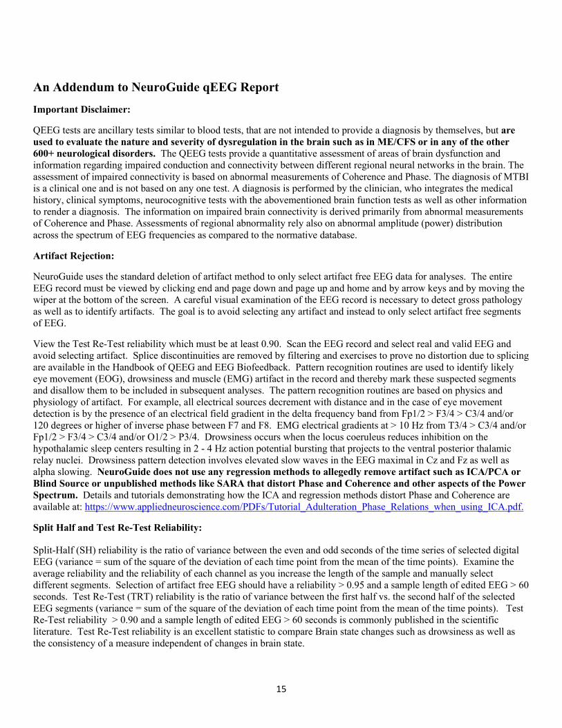
An Addendum to NeuroGuide qEEG Report
Important Disclaimer:
QEEG tests are ancillary tests similar to blood tests, that are not intended to provide a diagnosis by themselves, but are used to evaluate the nature and severity of dysregulation in the brain such as in ME/CFS or in any of the other 600+ neurological disorders. The QEEG tests provide a quantitative assessment of areas of brain dysfunction and information regarding impaired conduction and connectivity between different regional neural networks in the brain. The assessment of impaired connectivity is based on abnormal measurements of Coherence and Phase. The diagnosis of MTBI is a clinical one and is not based on any one test. A diagnosis is performed by the clinician, who integrates the medical history, clinical symptoms, neurocognitive tests with the abovementioned brain function tests as well as other information to render a diagnosis. The information on impaired brain connectivity is derived primarily from abnormal measurements of Coherence and Phase. Assessments of regional abnormality rely also on abnormal amplitude (power) distribution across the spectrum of EEG frequencies as compared to the normative database.
Artifact Rejection:
NeuroGuide uses the standard deletion of artifact method to only select artifact free EEG data for analyses. The entire EEG record must be viewed by clicking end and page down and page up and home and by arrow keys and by moving the wiper at the bottom of the screen. A careful visual examination of the EEG record is necessary to detect gross pathology as well as to identify artifacts. The goal is to avoid selecting any artifact and instead to only select artifact free segments of EEG.
View the Test Re-Test reliability which must be at least 0.90. Scan the EEG record and select real and valid EEG and avoid selecting artifact. Splice discontinuities are removed by filtering and exercises to prove no distortion due to splicing are available in the Handbook of QEEG and EEG Biofeedback. Pattern recognition routines are used to identify likely eye movement (EOG), drowsiness and muscle (EMG) artifact in the record and thereby mark these suspected segments and disallow them to be included in subsequent analyses. The pattern recognition routines are based on physics and physiology of artifact. For example, all electrical sources decrement with distance and in the case of eye movement detection is by the presence of an electrical field gradient in the delta frequency band from Fp1/2 > F3/4 > C3/4 and/or 120 degrees or higher of inverse phase between F7 and F8. EMG electrical gradients at > 10 Hz from T3/4 > C3/4 and/or Fp1/2 > F3/4 > C3/4 and/or O1/2 > P3/4. Drowsiness occurs when the locus coeruleus reduces inhibition on the hypothalamic sleep centers resulting in 2 - 4 Hz action potential bursting that projects to the ventral posterior thalamic relay nuclei. Drowsiness pattern detection involves elevated slow waves in the EEG maximal in Cz and Fz as well as alpha slowing. NeuroGuide does not use any regression methods to allegedly remove artifact such as ICA/PCA or Blind Source or unpublished methods like SARA that distort Phase and Coherence and other aspects of the Power Spectrum. Details and tutorials demonstrating how the ICA and regression methods distort Phase and Coherence are available at: https://www.appliedneuroscience.com/PDFs/Tutorial_Adulteration_Phase_Relations_when_using_ICA.pdf.
Split Half and Test Re-Test Reliability:
Split-Half (SH) reliability is the ratio of variance between the even and odd seconds of the time series of selected digital EEG (variance = sum of the square of the deviation of each time point from the mean of the time points). Examine the average reliability and the reliability of each channel as you increase the length of the sample and manually select different segments. Selection of artifact free EEG should have a reliability > 0.95 and a sample length of edited EEG > 60 seconds. Test Re-Test (TRT) reliability is the ratio of variance between the first half vs. the second half of the selected EEG segments (variance = sum of the square of the deviation of each time point from the mean of the time points). Test Re-Test reliability > 0.90 and a sample length of edited EEG > 60 seconds is commonly published in the scientific literature. Test Re-Test reliability is an excellent statistic to compare Brain state changes such as drowsiness as well as the consistency of a measure independent of changes in brain state.
16
This record supports the following reliability estimates:
17
Description of the NeuroGuide Normative Database:
The NeuroGuide normative database in versions 1.0 to 2.4.6 included a total of 678 carefully screened individual subjects ranging in age from 2 months to 82 years. NG 2.6.8 involved the addition of 49 adult subjects ranging in age from 18.3 years to 72.6 years resulting in a normative database of 727 subjects. The inclusion/exclusion criteria, demographics, neuropsychological tests, Gaussian distribution tests and cross- validation tests are described in several peer reviewed publications (Thatcher et al, 1983; 1987; 2003). Two year means were computed using a sliding average with 6 month overlap of subjects. This produced a stable and higher age resolution normative database with a total of 21 different age groups. The 21 age groups and age ranges and number of subjects per age group is shown in the bar graph in Appendix F figure 2 in the NeuroGuide Manual (click Help > NeuroGuide Help).
The individuals used to create the normative database met specific clinical standards of no history of neurological disorders, no history of behavioral disorders, performed at grade level in school, etc. Most of the subjects in the normative database were given extensive neuropsychological tests. Details of the normative database are published at: Thatcher, R.W., Walker, R.A. and Guidice, S. Human cerebral hemispheres develop at different rates and ages. Science, 236: 1110-1113, 1987 and Thatcher R.W., Biver, C.L., North, D., Curtin, R. and Walker, R.W. Quantitative EEG Normative Databases: Validation and Clinical Correlation. Journal of Neurotherapy, 2003, 7(3-4): 87-121. You can download a description of the normative database by going to https://appliedneuroscience.com/scientific-articles/ and clicking on Article #5.
Is there a normative database for different montages including bipolar montages?
Yes. The raw digital data from the same group of normal subjects is analyzed using different montages such as Average Reference, Laplacian current source density, a common reference based on all 19 channels of the 10/20 system and standard clinical bipolar montages (e.g., longitudinal, circular, transverse). Users can create any montage that they wish and there will be a normative reference database comparison available for both eyes closed and eyes open conditions.
Age range of the swLORETA Current Density and Source Correlation Normative Databases
The swLORETA current density and source correlation norms use the same subjects as are used for the surface EEG norms and the age range is 2 months to 82 years. The computational details of the LORETA current density norms are published at: Thatcher, R.W., North, D., Biver, C. EEG inverse solutions and parametric vs. non-parametric statistics of Low Resolution Electromagnetic Tomography (LORETA). Clin. EEG and Neuroscience, 36(1): 1-9, 2005 and Thatcher, R.W., North, D., Biver, C. Evaluation and Validity of a LORETA normative EEG database. Clin. EEG and Neuroscience, 2005, 36(2): 116-122. Copies of these publications are available to download from https://appliedneuroscience.com/scientific-articles/ by clicking on article nos. 11 and 12.
Implementation of swLORETA measurement in NeuroGuide
The neuro-navigator swLORETA viewer (Applied Neuroscience, Inc., 2019) can be launched by a single mouse click in the NeuroGuide window. NeuroGuide exports frequency domain and time domain edits of 19 channel x 256 point digital EEG in microvolts (or uv^2) in the Lexicor electrode order as the standard input to the Key Institute T-Matrix. Rows are 256 microvolt time points and the columns are 19 channels at a sample rate of 128 thus producing 0.5 Hz resolution from 1 to 30 Hz. 1 Hz increments in the swLORETA viewer are computed as the sum of adjacent 0.5 Hz bins and thus the ‘Time Frame’ control in the LORETA Viewer is frequency from 1 to 30 Hz. (see Pascual-Marqui RD, Michel CM, Lehmann D., 1994. Low resolution electromagnetic tomography: a new method for localizing electrical activity in the brain. International J. of Psychophysiology, 18:49-65. For computational details see: Pascual-Marqui. R.D., 1999. Review of Methods for Solving the EEG Inverse Problem. International J. of Bioelectromagnetism, 1(1): 75-86. Pascual-Margui, R.D., 2004.
18
Amplifier Matching is Necessary
This stems from the fact that amplifiers have different frequency gain characteristics. The matching of amplifiers to the NeuroGuide database amplifier was done by injecting microvolt calibration signals of different amplitudes and frequencies into the input of the respective EEG machines and then computing correction curves to exactly match the amplifier characteristics of the norms and discriminant functions. The units of comparison are in microvolts and a match within 3% is generally achieved. The NeuroGuide research team double checked the amplifier match by computing FFT and digital spectral analyses on calibration signals used to acquire the norms with the calibration signals used to evaluate a given manufacturers amplifiers.
History of the Scientific Standards of QEEG Normative Databases
A review of the history of QEEG normative databases was published in Thatcher, R.W. and Lubar, J.F. History of the scientific standards of QEEG normative databases. In: Introduction to QEEG and Neurofeedback: Advanced Theory and Applications, T. Budzinsky, H. Budzinsky, J. Evans and A. Abarbanel (eds)., Academic Press, San Diego, CA, 2008. A copy of the publication can be downloaded at: https://www.appliedneuroscience.com/PDFs/History_of_QEEG_Databases.pdf.
References
Bosch-Bayard J, Valdes-Sosa P, Virues-Alba T, Aubert-Vazquez E, John ER, Harmony T, Riera-Diaz J, Trujillo-Barreto N.(2001). 3D statistical parametric mapping of EEG source spectra by means of variable resolution electromagnetic tomography (VARETA). Clin Electroencephalogr., 32(2):47-61.
Coburn, K.L., Lauterback, E.C., Boutros, N.N., Black, K.J., Arciniegas, D.B. and Coffey, C.E. (2006). The value of quantitative electroencephalography in clinical psychiatry: A report by the committee on research of the American Neuropsychiatric Association. J. Neuropsychiat. and Clin. Neurosci. 18: 460-500.
Congedo M, John RE, De Ridder D, Prichep L. (2010). Group independent component analysis of resting state EEG in large normative samples. Int J Psychophysiol. 78(2):89-99.
Congedo M, John RE, De Ridder D, Prichep L, Isenhart R. (2010). On the "dependence" of "independent" group EEG sources; an EEG study on two large databases. Brain Topogr., 23(2):134-138.
Hernandez-Gonzalez G, Bringas-Vega ML, Galán-Garcia L, Bosch-Bayard J, Lorenzo-Ceballos Y, Melie-Garcia L, Valdes-Urrutia L, Cobas-Ruiz M, Valdes-Sosa PA; Cuban Human Brain Mapping Project (CHBMP). (2011). Multimodal quantitative neuroimaging databases and methods: the Cuban Human Brain Mapping Project. Clin EEG Neurosci., 42(3):149-59.
Duffy, F., Hughes, J. R., Miranda, F., Bernad, P. & Cook, P. (1994). Status of quantitative EEG (QEEG) in clinical practice. Clinical. Electroencephalography, 25(4), VI - XXII.
Gasser, T., Verleger, R., Bacher, P., & Sroka, L. (1988a). Development of the EEG of school-age children and adolescents. I. Analysis of band power. Electroencephalography and Clinical Neurophysiology, 69(2), 91-99.
Gasser, T., Jennen-Steinmetz, C., Sroka, L., Verleger, R., & Mocks, J. (1988b). Development of the EEG of school- age children and adolescents. II: Topography. Electroencephalography and Clinical Neurophysiology, 69(2),100-109.
Gordon, E., Cooper, N., Rennie, C., Hermens, D. and Williams, L.M. (2005). Integrative neuroscience: The role of a standardized database. Clin. EEG and Neurosci., 36(2): 64-75.
Hughes, J. R. & John, E. R. (1999). Conventional and quantitative electroencephalography in psychiatry. Neuropsychiatry, 11, 190-208.
19
John, E.R. (1977) Functional Neuroscience, Vol. II: Neurometrics: Quantitative Electrophysiological Analyses. E.R. John and R.W. Thatcher, Editors. L. Erlbaum Assoc., N.J.
John, E.R. Karmel, B., Corning, W. Easton, P., Brown, D., Ahn, H., John, M., Harmony, T., Prichep, L., Toro, A., Gerson, I., Bartlett, F., Thatcher, R., Kaye, H., Valdes, P., Schwartz, E. (1977). Neurometrics: Numerical taxonomy identifies different profiles of brain functions within groups of behaviorally similar people. Science, 196:1393 1410.
John, E. R., Prichep, L. S. & Easton, P. (1987). Normative data banks and neurometrics: Basic concepts, methods and results of norm construction. In A. Remond (Ed.), Handbook of electroencephalography and clinical neurophysiology: Vol. III. Computer analysis of the EEG and other neurophysiological signals (pp. 449-495). Amsterdam: Elsevier.
John, E.R., Ahn, H., Prichep, L.S., Trepetin, M., Brown, D. and Kaye, H. (1980) Developmental equations for the electroencephalogram. Science, 210: 1255-1258.
John, E. R., Prichep, L. S., Fridman, J. & Easton, P. (1988). Neurometrics: Computer assisted differential diagnosis of brain dysfunctions. Science, 293: 162-169.
John, E.R. (1990). Machinery of the Mind: Data, theory, and speculations about higher brain function. Birkhauser, Boston.
Galán, L., Biscay, R., and Valdés P., (1994). Multivariate statistical brain electromagnetic mapping. Brain Topgr., 7(1):17-28.
Koenig T, Prichep L, Lehmann D, Sosa PV, Braeker E, Kleinlogel H, Isenhart R, John ER. (2002). Millisecond by millisecond, year by year: normative EEG microstates and developmental stages. Neuroimage, 16(1):41-48.
Matousek, M. & Petersen, I. (1973a). Automatic evaluation of background activity by means of age-dependent EEG quotients. EEG & Clin. Neurophysiol., 35: 603-612.
Matousek, M. & Petersen, I. (1973b). Frequency analysis of the EEG background activity by means of age dependent EEG quotients. In Automation of clinical electroencephalography, Kellaway & I. Petersen (Eds.), (pp. 75-102). New York: Raven Press.
Prichep, L.S. (2005). Use of normative databases and statistical methods in demonstrating clinical utility of QEEG: Importance and cautions. Clin. EEG and Neurosci., 36(2): 82-87.
Soler, E.P. (2010). Functional Imaging based on swLORETA and phase synchronization. Eemagine-Medical Imaging Solutions.
Thatcher, R.W., Walker, R.A., Biver, C., North, D., Curtin, R., (2003). Quantitative EEG Normative databases: Validation and Clinical Correlation, J. Neurotherapy, 7(3-4): 87-121.
Thatcher, R. W. (1998). EEG normative databases and EEG biofeedback. Journal of Neurotherapy, 2(4): 8-39.
Thatcher, R.W., North, D., and Biver, C. (2005a) EEG inverse solutions and parametric vs. non-parametric statistics of Low Resolution Electromagnetic Tomography (LORETA). Clin. EEG and Neuroscience, 36(1):1-8.
Thatcher, R.W., North, D., and Biver, C. (2005b) Evaluation and Validity of a LORETA normative EEG database. Clin. EEG and Neuroscience, 36(2): 116-122.
Thatcher, R.W., McAlaster, R., Lester, M.L., Horst, R.L. and Cantor, D.S. (1983). Hemispheric EEG Asymmetries Related to Cognitive Functioning in Children. In: Cognitive Processing in the Right Hemisphere, A. Perecuman (Ed.), New York: Academic Press.
Thatcher, R.W. (1992). Cyclic cortical reorganization during early childhood. Brain and Cognition, 20: 24-50.

20
Thatcher, R.W. and Lubar, J.F. History of the scientific standards of QEEG normative databases. (2008) In: Introduction to QEEG and Neurofeedback: Advanced Theory and Applications, T. Budzinsky, H. Budzinsky, J. Evans and A. Abarbanel (eds)., Academic Press, San Diego, CA.
Thatcher, R.W. (2010) Reliability and validity of quantitative electroencephalography (qEEG). J. of Neurotherapy, 14:122-152.
Adebimpe, A., Aarabi, A., Bourel-Ponchel, E., Mahmoudzadeh, M., & Wallois, F. (2016). EEG Resting State Functional Connectivity Analysis in Children with Benign Epilepsy with Centrotemporal Spikes. Frontiers in Neuroscience, 10, 143. https://doi.org/10.3389/fnins.2016.00143
Alessio, A., Pereira, F. R. S., Sercheli, M. S., Rondina, J. M., Ozelo, H. B., Bilevicius, E., … Cendes, F. (2013). Brain plasticity for verbal and visual memories in patients with mesial temporal lobe epilepsy and hippocampal sclerosis: An fMRI study. Human Brain Mapping, 34(1), 186–199. https://doi.org/10.1002/hbm.21432
Allan, L. M., Ballard, C. G., Allen, J., Murray, A., Davidson, A. W., McKeith, I. G., & Kenny, R. A. (2007). Autonomic dysfunction in dementia. J Neurol Neurosurg Psychiatry, 78(7), 671–677. https://doi.org/10.1136/jnnp.2006.102343
Azevedo, F. A. C., Carvalho, L. R. B., Grinberg, L. T., Farfel, J. M., Ferretti, R. E. L., Leite, R. E. P., … Herculano-Houzel, S. (2009). Equal numbers of neuronal and nonneuronal cells make the human brain an isometrically scaled-up primate brain. The Journal of Comparative Neurology, 513(5), 532–541. https://doi.org/10.1002/cne.21974
Babiloni, C., De Pandis, M. F., Vecchio, F., Buffo, P., Sorpresi, F., Frisoni, G. B., & Rossini, P. M. (2011). Cortical sources of resting state electroencephalographic rhythms in Parkinson’s disease related dementia and Alzheimer’s disease. Clin Neurophysiol, 122(12), 2355–2364. https://doi.org/10.1016/j.clinph.2011.03.029
Babiloni, C., Frisoni, G., Steriade, M., Bresciani, L., Binetti, G., Del Percio, C., … Rossini, P. M. (2006). Frontal white matter volume and delta EEG sources negatively correlate in awake subjects with mild cognitive impairment and Alzheimer’s disease. Clin Neurophysiol, 117(5), 1113–1129. https://doi.org/10.1016/j.clinph.2006.01.020
Babiloni, C., Visser, P. J., Frisoni, G., De Deyn, P. P., Bresciani, L., Jelic, V., … Nobili, F. (2010). Cortical sources of resting EEG rhythms in mild cognitive impairment and subjective memory complaint. Neurobiol Aging, 31(10), 1787–1798. https://doi.org/10.1016/j.neurobiolaging.2008.09.020
Baraniuk, J. N., Adewuyi, O., Merck, S. J., Ali, M., Ravindran, M. K., Timbol, C. R., … Petrie, K. N. (2013). A Chronic Fatigue Syndrome (CFS) severity score based on case designation criteria. Am J Transl Res, 5(1), 53–68. (23390566).
Barbey, A. K. (2018). Network Neuroscience Theory of Human Intelligence. Trends in Cognitive Sciences, 22(1), 8–20. https://doi.org/10.1016/j.tics.2017.10.001
Barnden, L. R., Crouch, B., Kwiatek, R., Burnet, R., & Del Fante, P. (2015). Evidence in chronic fatigue syndrome for severity-dependent upregulation of prefrontal myelination that is independent of anxiety and depression. NMR Biomed, 28(3), 404–413. https://doi.org/10.1002/nbm.3261
Barnden, L. R., Crouch, B., Kwiatek, R., Burnet, R., Mernone, A., Chryssidis, S., … Del Fante, P. (2011). A brain MRI study of chronic fatigue syndrome: Evidence of brainstem dysfunction and altered homeostasis. NMR Biomed, 24(10), 1302–1312. https://doi.org/10.1002/nbm.1692
Barnden, L. R., Kwiatek, R., Crouch, B., Burnet, R., & Del Fante, P. (2016). Autonomic correlations with MRI are abnormal in the brainstem vasomotor centre in Chronic Fatigue Syndrome. Neuroimage Clin, 11, 530–537. https://doi.org/10.1016/j.nicl.2016.03.017
21
Bassett, D. S., & Sporns, O. (2017). Network neuroscience. Nature Neuroscience, 20(3), 353–364. https://doi.org/10.1038/nn.4502
Bassi, A., & Bozzali, M. (2015). Potential Interactions between the Autonomic Nervous System and Higher Level Functions in Neurological and Neuropsychiatric Conditions. Frontiers in Neurology, 6. https://doi.org/10.3389/fneur.2015.00182
Beaumont, A., Burton, A. R., Lemon, J., Bennett, B. K., Lloyd, A., & Vollmer-Conna, U. (2012). Reduced cardiac vagal modulation impacts on cognitive performance in chronic fatigue syndrome. PLoS One, 7(11), e49518. https://doi.org/10.1371/journal.pone.0049518
Beissner, F., Meissner, K., Bar, K. J., & Napadow, V. (2013). The autonomic brain: An activation likelihood estimation meta-analysis for central processing of autonomic function. J Neurosci, 33(25), 10503–10511. https://doi.org/10.1523/jneurosci.1103-13.2013
Benarroch, E. (2019). Autonomic nervous system and neuroimmune interactions. Neurology. https://doi.org/10.1212/WNL.0000000000006942
Benarroch, E. E. (1993). The central autonomic network: Functional organization, dysfunction, and perspective. Mayo Clinic Proceedings, 68(10), 988–1001.
Benarroch, E. E. (2012). Central Autonomic Control. In D. Robertson, I. Biaggioni, G. Burnstock, P. A. Low, & J. F. R. Paton (Eds.), Primer on the autonomic nervous system (3rd ed., pp. 9–12). Oxford: Elsevier.
Benarroch, E. E., Freeman, R., & Kaufmann, H. (2007). Autonomic Nervous System. Textbook of Clinical Neurology, 383–404. https://doi.org/10.1016/B978-141603618-0.10021-9
Bentea, G., Sculier, C., Grigoriu, B., Meert, A.-P., Durieux, V., Berghmans, T., & Sculier, J.-P. (2017). Autoimmune paraneoplastic syndromes associated to lung cancer: A systematic review of the literature: Part 3: Neurological paraneoplastic syndromes, involving the central nervous system. Lung Cancer, 106, 83–92. https://doi.org/10.1016/j.lungcan.2017.01.017
Billiot, K. M., Budzynski, T. H., & Andrasik, F. (1997). EEG Patterns and CFS. Journal of Neurotherapy, 2–2(4).
Biswal, B., Kunwar, P., & Natelson, B. H. (2011). Cerebral blood flow is reduced in chronic fatigue syndrome as assessed by arterial spin labeling. J Neurol Sci, 301(1–2), 9–11. https://doi.org/10.1016/j.jns.2010.11.018
Bozzini, S., Albergati, A., Capelli, E., Lorusso, L., Gazzaruso, C., Pelissero, G., & Falcone, C. (2018). Cardiovascular characteristics of chronic fatigue syndrome. Biomedical Reports, 8(1), 26–30. https://doi.org/10.3892/br.2017.1024
Braun, U., Plichta, M. M., Esslinger, C., Sauer, C., Haddad, L., Grimm, O., … Meyer-Lindenberg, A. (2012). Test-retest reliability of resting-state connectivity network characteristics using fMRI and graph theoretical measures. Neuroimage, 59(2), 1404–1412. https://doi.org/10.1016/j.neuroimage.2011.08.044
Brenu, E. W., Huth, T. K., Hardcastle, S. L., Fuller, K., Kaur, M., Johnston, S., … Marshall-Gradisnik, S. M. (2014). Role of adaptive and innate immune cells in chronic fatigue syndrome/myalgic encephalomyelitis. Int Immunol, 26(4), 233–242. https://doi.org/10.1093/intimm/dxt068
Bressler, S. L., & Menon, V. (2010). Large-scale brain networks in cognition: Emerging methods and principles. Trends Cogn Sci, 14(6), 277–290. https://doi.org/10.1016/j.tics.2010.04.004
Brodal, P. (2010). The Central Nervous System: Structure and Function (4th ed.). New York: Oxford University Press.
22
Brovkin, A., Waller, L., Dorfschmidt, L., Bzdok, D., Walter, H., & Kruschwitz, J. D. (2018). GraphVar 2.0: A user-friendly toolbox for machine learning on functional connectivity measures. ArXiv:1803.00082 [Stat]. Retrieved from http://arxiv.org/abs/1803.00082
Buijs, R. M. (2013). The autonomic nervous system: A balancing act. Handb Clin Neurol, 117, 1–11. https://doi.org/10.1016/b978-0-444-53491-0.00001-8
Bullmore, E., & Sporns, O. (2012). The economy of brain network organization. Nat Rev Neurosci, 13(5), 336–349. https://doi.org/10.1038/nrn3214
Burnstock, G. (2013). Purinergic signalling in the lower urinary tract. Acta Physiologica, 207(1), 40–52. https://doi.org/10.1111/apha.12012
Buzsaki, G. (2006). Rhythms of the Brain. Oxford University Press.
Buzsaki, G., & Watson, B. O. (2012). Brain rhythms and neural syntax: Implications for efficient coding of cognitive content and neuropsychiatric disease. Dialogues Clin Neurosci, 14(4), 345–367. (23393413).
Caliandro, P., Vecchio, F., Miraglia, F., Reale, G., Della Marca, G., La Torre, G., … Rossini, P. M. (2017). Small-World Characteristics of Cortical Connectivity Changes in Acute Stroke. Neurorehabil Neural Repair, 31(1), 81–94. https://doi.org/10.1177/1545968316662525
Cannon, R. L., Baldwin, D. R., Shaw, T. L., Diloreto, D. J., Phillips, S. M., Scruggs, A. M., & Riehl, T. C. (2012). Reliability of quantitative EEG (qEEG) measures and LORETA current source density at 30 days. Neuroscience Letters, 518(1), 27–31. https://doi.org/10.1016/j.neulet.2012.04.035
Cantor, F. (2010). Central and Peripheral Fatigue: Exemplified by Multiple Sclerosis and Myasthenia Gravis. PM&R, 2(5), 399–405. https://doi.org/10.1016/j.pmrj.2010.04.012
Carruthers, B. M., Jain, A. K., de Meirleir, K., Peterson, D. L., Klimas, N. G., Lerner, A. M., … Van de Sande, M. I. (2003). Myalgic encephalomyelitits/chronic fatigue syndrome: Clinical working case definition, diagnostic, and treatment protocols. Journal of Chronic Fatigue Syndrome, 11(1).
Carruthers, B. M., van de Sande, M. I., De Meirleir, K. L., Klimas, N. G., Broderick, G., Mitchell, T., … Stevens, S. (2011). Myalgic Encephalomyelitis: International Consensus Criteria. Journal of Internal Medicine. https://doi.org/10.1111/j.1365-
Catani, M., Dell’acqua, F., Bizzi, A., Forkel, S. J., Williams, S. C., Simmons, A., … Thiebaut de Schotten, M. (2012). Beyond cortical localization in clinico-anatomical correlation. Cortex, 48(10), 1262–1287. https://doi.org/10.1016/j.cortex.2012.07.001
Cersosimo, M. G., & Benarroch, E. E. (2013). Chapter 5—Central control of autonomic function and involvement in neurodegenerative disorders. In Ruud M. Buijs & D. F. Swaab (Eds.), Handbook of Clinical Neurology (pp. 45–57). https://doi.org/10.1016/B978-0-444-53491-0.00005-5
Chaudhuri, A., & Behan, P. O. (2000). Fatigue and basal ganglia. J Neurol Sci, 179(S 1-2), 34–42. (11054483).
Chen, C. C., Hsu, C. Y., Chiu, H. W., Hu, C. J., & Lee, T. C. (2015). Frequency power and coherence of electroencephalography are correlated with the severity of Alzheimer’s disease: A multicenter analysis in Taiwan. J Formos Med Assoc, 114(8), 729–735. https://doi.org/10.1016/j.jfma.2013.07.008
Chen, P.-S., Chen, L. S., Fishbein, M. C., Lin, S.-F., & Nattel, S. (2014). Role of the Autonomic Nervous System in Atrial Fibrillation: Pathophysiology and Therapy. Circulation Research, 114(9), 1500–1515. https://doi.org/10.1161/CIRCRESAHA.114.303772

23
Chung, C., & Caplan, L. R. (2007). Stroke and Other Neurovascular Disorders. In C. G. Goetz (Ed.), Textbook of Clinical Neurology (3rd ed., pp. 1019–1052). Philadelphia, PA: Saunders.
Cleare, A. J. (2004). The HPA axis and the genesis of chronic fatigue syndrome. Trends Endocrinol Metab, 15(2), 55–59. https://doi.org/10.1016/j.tem.2003.12.002
Cockshell, S. J., & Mathias, J. L. (2010). Cognitive functioning in chronic fatigue syndrome: A meta-analysis. Psychol Med, 40(8), 1253–1267. https://doi.org/10.1017/s0033291709992054
Collins, H. L., Rodenbaugh, D. W., & DiCarlo, S. E. (2006). Spinal cord injury alters cardiac electrophysiology and increases the susceptibility to ventricular arrhythmias. In L. C. Weaver & C. Polosa (Eds.), Progress in Brain Research (pp. 275–288). https://doi.org/10.1016/S0079-6123(05)52018-1
Collins, O., Dillon, S., Finucane, C., Lawlor, B., & Kenny, R. A. (2012). Parasympathetic autonomic dysfunction is common in mild cognitive impairment. Neurobiol Aging, 33(10), 2324–2333. https://doi.org/10.1016/j.neurobiolaging.2011.11.017
Cook, D. B., O’Connor, P. J., Lange, G., & Steffener, J. (2007). Functional neuroimaging correlates of mental fatigue induced by cognition among chronic fatigue syndrome patients and controls. Neuroimage, 36(1), 108–122. https://doi.org/10.1016/j.neuroimage.2007.02.033
Costa, D. C., Tannock, C., & Brostoff, J. (1995). Brainstem perfusion is impaired in chronic fatigue syndrome. QJM, 88(11), 767–773. (8542261).
Craig, A. D. (2018). Central neural substrates involved in temperature discrimination, thermal pain, thermal comfort, and thermoregulatory behavior. In A. A. Romanovsky (Ed.), Handbook of Clinical Neurology (pp. 317–338). https://doi.org/10.1016/B978-0-444-63912-7.00019-9
Crossley, N. A., Mechelli, A., Scott, J., Carletti, F., Fox, P. T., McGuire, P., & Bullmore, E. T. (2014). The hubs of the human connectome are generally implicated in the anatomy of brain disorders. Brain, 137(Pt 8), 2382–2395. https://doi.org/10.1093/brain/awu132
Cvejic, E., Sandler, C. X., Keech, A., Barry, B. K., Lloyd, A. R., & Vollmer-Conna, U. (2017). Autonomic nervous system function, activity patterns, and sleep after physical or cognitive challenge in people with chronic fatigue syndrome. Journal of Psychosomatic Research, 103, 91–94. https://doi.org/10.1016/j.jpsychores.2017.10.010
de Haan, W., Mott, K., van Straaten, E. C. W., Scheltens, P., & Stam, C. J. (2012). Activity Dependent Degeneration Explains Hub Vulnerability in Alzheimer’s Disease. PLoS Computational Biology, 8(8). https://doi.org/10.1371/journal.pcbi.1002582
DeBiasi, R. L., & Tyler, K. L. (2004). Molecular Methods for Diagnosis of Viral Encephalitis. Clinical Microbiology Reviews, 17(4), 903–925. https://doi.org/10.1128/CMR.17.4.903-925.2004
Decade of the Brain Home Page (Library of Congress). (2000). Retrieved February 17, 2019, from https://www.loc.gov/loc/brain/
Deco, G., Jirsa, V., & Friston, K. J. (2012). The dynamical structural basis of brain activity. In M. I. Rabinovich, K. Friston, & P. Varona (Eds.), Principles of Brain Dynamics: Global State Interactions (pp. 1–23). Cambridge, MA: MIT Press.
DeLuca, J., Johnson, S. K., & Natelson, B. H. (1993). Information processing efficiency in chronic fatigue syndrome and multiple sclerosis. Arch Neurol, 50(3), 301–304. (8442710).
Demitrack, M. A. (1994). Chronic fatigue syndrome: A disease of the hypothalamic-pituitary-adrenal axis? Annals of Medicine, 26(1), 1–5.

24
Diserens, K., Vuadens, P., Michel, P., Reichhart, M., Herrmann, F. R., Arnold, P., … Ghika, J. (2006). Acute autonomic dysfunction contralateral to acute strokes: A prospective study of 100 consecutive cases. European Journal of Neurology, 13(11), 1245–1250. https://doi.org/10.1111/j.1468-1331.2006.01488.x
Dodick, D. W. (2018). Migraine. The Lancet, 391(10127), 1315–1330. https://doi.org/10.1016/S0140-6736(18)30478-1
Drakesmith, M., Caeyenberghs, K., Dutt, A., Lewis, G., David, A. S., & Jones, D. K. (2015). Overcoming the effects of false positives and threshold bias in graph theoretical analyses of neuroimaging data. Neuroimage, 118, 313–333. https://doi.org/10.1016/j.neuroimage.2015.05.011
Edlow, B. L., McNab, J. A., Witzel, T., & Kinney, H. C. (2015). The Structural Connectome of the Human Central Homeostatic Network. Brain Connectivity, 6(3), 187–200. https://doi.org/10.1089/brain.2015.0378
Elenkov, I. J., Wilder, R. L., Chrousos, G. P., & Vizi, E. S. (2000). The sympathetic nerve—An integrative interface between two supersystems: The brain and the immune system. Pharmacol Rev, 52(4), 595–638. (11121511).
Engel, J. (2019). Epileptogenesis, traumatic brain injury, and biomarkers. Neurobiology of Disease, 123, 3–7. https://doi.org/10.1016/j.nbd.2018.04.002
Evans, R. W., Wilberger, J. E., & Bhatia, S. (2007). Traumatic Disorders. In C. G. Goetz (Ed.), Textbook of clinical neurology (3rd ed., pp. 1185–1212). Philadelphia, PA: Saunders.
Ferreira, A. C., & Marchena, E. de. (2002). Grading autonomic dysfunction in chronic fatigue syndrome. Seminars in Arthritis and Rheumatism, 32(3), 137–138. https://doi.org/10.1053/sarh.2002.33462
Feske, S. K., & Cochran, T. I. (2007). Degenerative and compressive structural disorders. In C. G. Goetz (Ed.), Textbook of Clinical Neurology (3rd ed., pp. 593–611). Philadelphia, PA: Saunders.
Finger, S. (2000). Minds Behind the Brain: A History of the Pioneers and Their Discoveries. New York: Oxford Univeristy Press.
Finn, E. S., Shen, X., Scheinost, D., Rosenberg, M. D., Huang, J., Chun, M. M., … Constable, R. T. (2015). Functional connectome fingerprinting: Identifying individuals based on patterns of brain connectivity. Nature Neuroscience, 18(11), 1664–1671. https://doi.org/10.1038/nn.4135
Flor-Henry, P., Lind, J. C., & Koles, Z. J. (2010). EEG source analysis of chronic fatigue syndrome. Psychiatry Res, 181(2), 155–164. https://doi.org/10.1016/j.pscychresns.2009.10.007
Foldvary-Schaefer, N., & Wyllie, E. (2007). Epilepsy. In C. G. Goetz (Ed.), Textbook of Clinical Neurology (3rd ed., pp. 1213–1244). Philadelphia, PA: Saunders.
Fonseca, L. C., Tedrus, G. M., Carvas, P. N., & Machado, E. C. (2013). Comparison of quantitative EEG between patients with Alzheimer’s disease and those with Parkinson’s disease dementia. Clin Neurophysiol, 124(10), 1970–1974. https://doi.org/10.1016/j.clinph.2013.05.001
Fornito, A., Harrison, B. J., Zalesky, A., & Simons, J. S. (2012). Competitive and cooperative dynamics of large-scale brain functional networks supporting recollection. Proceedings of the National Academy of Sciences, 109(31), 12788–12793. https://doi.org/10.1073/pnas.1204185109
Fox‐Wasylyshyn, S. M., & El‐Masri, M. M. (2005). Handling missing data in self-report measures. Research in Nursing & Health, 28(6), 488–495. https://doi.org/10.1002/nur.20100
Freeman, K., Goldstein, D. S., & Thompson, C. R. (2015). The Dysautonomia Project: Understanding Autonomic Nervous System Disorders for Physicians and Patients. Sarasota, FL: Bardolf & Company.
25
Freeman, R., & Komaroff, A. L. (1997). Does the chronic fatigue syndrome involve the autonomic nervous system? Am J Med, 102(4), 357–364. (9217617).
Friston, K. J., Kahan, J., Razi, A., Stephan, K. E., & Sporns, O. (2014). On nodes and modes in resting state fMRI. Neuroimage, 99(100), 533–547. https://doi.org/10.1016/j.neuroimage.2014.05.056
Fuchs, M., Kastner, J., Wagner, M., Hawes, S., & Ebersole, J. S. (2002). A standardized boundary element method volume conductor model. Clin Neurophysiol, 113(5), 702–712. (11976050).
Fukuda, K., Straus, S. E., Hickie, I., Sharpe, M. C., Dobbins, J. G., & Komaroff, A. (1994). The chronic fatigue syndrome: A comprehensive approach to its definition and study. Ann Intern Med, 121(12), 953–959. (7978722).
Gazerani, P., & Cairns, B. E. (2018). Dysautonomia in the pathogenesis of migraine. Expert Review of Neurotherapeutics, 18(2), 153–165. https://doi.org/10.1080/14737175.2018.1414601
Goadsby, P. J. (2013). Chapter 16—Autonomic nervous system control of the cerebral circulation. In Ruud M. Buijs & D. F. Swaab (Eds.), Handbook of Clinical Neurology (pp. 193–201). https://doi.org/10.1016/B978-0-444-53491-0.00016-X
Goldstein, D. S., & Kopin, I. J. (2017). Homeostatic systems, biocybernetics, and autonomic neuroscience. Autonomic Neuroscience: Basic & Clinical, 208, 15–28. https://doi.org/10.1016/j.autneu.2017.09.001
Grech, R., Cassar, T., Muscat, J., Camilleri, K. P., Fabri, S. G., Zervakis, M., … Vanrumste, B. (2008). Review on solving the inverse problem in EEG source analysis. J Neuroeng Rehabil, 5, 25. https://doi.org/10.1186/1743-0003-5-25
Greicius, M. D., Kiviniemi, V., Tervonen, O., Vainionpaa, V., Alahuhta, S., Reiss, A. L., & Menon, V. (2008). Persistent default-mode network connectivity during light sedation. Hum Brain Mapp, 29(7), 839–847. https://doi.org/10.1002/hbm.20537
Gu, S., Pasqualetti, F., Cieslak, M., Telesford, Q. K., Yu, A. B., Kahn, A. E., … Bassett, D. S. (2015). Controllability of structural brain networks. Nat Commun, 6, 8414. https://doi.org/10.1038/ncomms9414
Guimerà, R., & Nunes Amaral, L. A. (2005). Functional cartography of complex metabolic networks. Nature, 433(7028), 895–900. https://doi.org/10.1038/nature03288
Hagmann, P., Cammoun, L., Gigandet, X., Meuli, R., Honey, C. J., Wedeen, V. J., & Sporns, O. (2008). Mapping the structural core of human cerebral cortex. PLoS Biol, 6(7), e159. https://doi.org/10.1371/journal.pbio.0060159
Harris, P. A., Taylor, R., Thielke, R., Payne, J., Gonzalez, N., & Conde, J. G. (2009). Research electronic data capture (REDCap)—A metadata-driven methodology and workflow process for providing translational research informatics support. J Biomed Inform, 42(2), 377–381. https://doi.org/10.1016/j.jbi.2008.08.010
Hart, M. G., Price, S. J., & Suckling, J. (2016). Connectome analysis for pre-operative brain mapping in neurosurgery. British Journal of Neurosurgery, 30(5), 506–517. https://doi.org/10.1080/02688697.2016.1208809
Hata, M., Kazui, H., Tanaka, T., Ishii, R., Canuet, L., Pascual-Marqui, R. D., … Takeda, M. (2016). Functional connectivity assessed by resting state EEG correlates with cognitive decline of Alzheimer’s disease: An eLORETA study. Clin Neurophysiol, 127(2), 1269–1278. https://doi.org/10.1016/j.clinph.2015.10.030
Hatziagelaki, E., Adamaki, M., Tsilioni, I., Dimitriadis, G., & Theoharides, T. C. (2018). Myalgic Encephalomyelitis/Chronic Fatigue Syndrome—Metabolic Disease or Disturbed Homeostasis due to Focal Inflammation in the Hypothalamus? Journal of Pharmacology and Experimental Therapeutics, 367(1), 155–167. https://doi.org/10.1124/jpet.118.250845
26
Hegerl, U., & Ulke, C. (2016). Fatigue with up- vs downregulated brain arousal should not be confused. Prog Brain Res, 229, 239–254. https://doi.org/10.1016/bs.pbr.2016.06.001
Hellyer, P. J., Scott, G., Shanahan, M., Sharp, D. J., & Leech, R. (2015). Cognitive Flexibility through Metastable Neural Dynamics Is Disrupted by Damage to the Structural Connectome. The Journal of Neuroscience, 35(24), 9050–9063. https://doi.org/10.1523/JNEUROSCI.4648-14.2015
Hillary, F. G., Roman, C. A., Venkatesan, U., Rajtmajer, S. M., Bajo, R., & Castellanos, N. D. (2015). Hyperconnectivity is a fundamental response to neurological disruption. Neuropsychology, 29(1), 59–75. https://doi.org/10.1037/neu0000110
Ho, L., Legere, M., Li, T., Levine, S., Hao, K., Valcarcel, B., & Pasinetti, G. M. (2017). Autonomic Nervous System Dysfunctions as a Basis for a Predictive Model of Risk of Neurological Disorders in Subjects with Prior History of Traumatic Brain Injury: Implications in Alzheimer’s Disease. Journal of Alzheimer’s Disease, 56(1), 305–315. https://doi.org/10.3233/JAD-160948
Holford, N. H. G. (2012). Pharmocokinetics & pharmacodynamics: Rational dosing & the time course of drug action. In B. G. Katzung, S. B. Masters, & A. J. Trevor (Eds.), Basic & Clinical Pharmacology (12th ed., pp. 37–51). New York: McGrall Hill.
Hosseini, S. M. H., Hoeft, F., & Kesler, S. R. (2012). GAT: A Graph-Theoretical Analysis Toolbox for Analyzing Between-Group Differences in Large-Scale Structural and Functional Brain Networks. PLOS ONE, 7(7), e40709. https://doi.org/10.1371/journal.pone.0040709
Hughes, S. W., Lorincz, M., Cope, D. W., Blethyn, K. L., Kekesi, K. A., Parri, H. R., … Crunelli, V. (2004). Synchronized oscillations at alpha and theta frequencies in the lateral geniculate nucleus. Neuron, 42(2), 253–268. (15091341).
Institute of Medicine. (2015). Beyond Myalgic Encephalomyelitis/Chronic Fatigue Syndrome: Redefining an Illness. https://doi.org/10.17226/19012
Janus, T. J., & Yung, W. K. (2007). Primary Neurological Tumors. In C. G. Goetz (Ed.), Textbook of Clinical Neurology (3rd ed., pp. 1053–1080). Philadelphia, PA: Saunders.
Jason, L. A., Brown, M., Evans, M., Anderson, V., Lerch, A., Brown, A., … Porter, N. (2011). Measuring substantial reductions in functioning in patients with chronic fatigue syndrome. Disability and Rehabilitation, 33(7), 589–598. https://doi.org/10.3109/09638288.2010.503256
Jason, L. A., Evans, M., Porter, N., Brown, A., Brown, M., Hunnell, J., … Friedberg, F. (2010). The development of a revised Canadian myalgic encephalomyelitis-chronic fatigue syndrome case definition. Am J Biochem Biotechnol, 6(2), 120-135.
Jason, L. A., Richman, J. A., Rademaker, A. W., Jordan, K. M., Plioplys, A. V., Taylor, R. R., … Plioplys, S. (1999). A community-based study of chronic fatigue syndrome. Arch Intern Med, 159(18), 2129–2137. (10527290).
Jason, Leonard A., So, S., Brown, A. A., Sunnquist, M., & Evans, M. (2015). Test–retest reliability of the DePaul Symptom Questionnaire. In Fatigue: Biomedicine, Health & Behavior (Vol. 3, pp. 16–32). https://doi.org/10.1080/21641846.2014.978110
Jolles, D. D., Buchem, M. A. van, Crone, E. A., & Rombouts, S. A. R. B. (2013). Functional brain connectivity at rest changes after working memory training. Human Brain Mapping, 34(2), 396–406. https://doi.org/10.1002/hbm.21444
27
Jordan, B., Kösling, S., Emmer, A., Koch, A., Müller, T., & Kornhuber, M. (2016). A study on viral CNS inflammation beyond herpes encephalitis. Journal of Neurovirology, 22(6), 763–773. https://doi.org/10.1007/s13365-016-0452-5
Joustra, S. D., Reijntjes, R. H., Pereira, A. M., Lammers, G. J., Biermasz, N. R., & Thijs, R. D. (2016). The Role of the Suprachiasmatic Nucleus in Cardiac Autonomic Control during Sleep. PloS One, 11(3), e0152390. https://doi.org/10.1371/journal.pone.0152390
Jurcak, V., Tsuzuki, D., & Dan, I. (2007). 10/20, 10/10, and 10/5 systems revisited: Their validity as relative head-surface-based positioning systems. Neuroimage, 34(4), 1600–1611. https://doi.org/10.1016/j.neuroimage.2006.09.024
Kamarajan, C., Pandey, A. K., Chorlian, D. B., & Porjesz, B. (2015). The use of current source density as electrophysiological correlates in neuropsychiatric disorders: A review of human studies. International Journal of Psychophysiology, 97(3), 310–322. https://doi.org/10.1016/j.ijpsycho.2014.10.013
Kanjwal, K., Karabin, B., Kanjwal, Y., Saeed, B., & Grubb, B. P. (2010). Autonomic dysfunction presenting as orthostatic intolerance in patients suffering from mitochondrial cytopathy. Clinical Cardiology, 33(10), 626–629. https://doi.org/10.1002/clc.20805
Karemaker, J. M. (2017). An introduction into autonomic nervous function. Physiological Measurement, 38(5), R89–R118. https://doi.org/10.1088/1361-6579/aa6782
Kaur, J., Young, B. E., & Fadel, P. J. (2017). Sympathetic Overactivity in Chronic Kidney Disease: Consequences and Mechanisms. International Journal of Molecular Sciences, 18(8). https://doi.org/10.3390/ijms18081682
Keech, A., Sandler, C. X., Vollmer-Conna, U., Cvejic, E., Lloyd, A. R., & Barry, B. K. (2015). Capturing the post-exertional exacerbation of fatigue following physical and cognitive challenge in patients with chronic fatigue syndrome. Journal of Psychosomatic Research, 79(6), 537–549. https://doi.org/10.1016/j.jpsychores.2015.08.008
Khalsa, S., Mayhew, S. D., Chechlacz, M., Bagary, M., & Bagshaw, A. P. (2014). The structural and functional connectivity of the posterior cingulate cortex: Comparison between deterministic and probabilistic tractography for the investigation of structure–function relationships. NeuroImage, 102, 118–127. https://doi.org/10.1016/j.neuroimage.2013.12.022
Kikumoto, M., Nagai, M., Ohshita, T., Toko, M., Kato, M., Dote, K., & Yamashita, H. (2018). Insular cortex lesion and autonomic instability in a herpes simplex virus encephalitis patient. Journal of NeuroVirology, 24(5), 649–651. https://doi.org/10.1007/s13365-018-0652-2
Kirk, R. E. (2013). Experimental design: Procedures for the behavioral sciences (4th ed.). Los Angeles: Sage.
Klimesch, W. (2012). Alpha-band oscillations, attention, and controlled access to stored information. Trends in Cognitive Sciences, 16(12), 606–617. https://doi.org/10.1016/j.tics.2012.10.007
Klimesch, W., Doppelmayr, M., Pachinger, T., & Ripper, B. (1997). Brain oscillations and human memory: EEG correlates in the upper alpha and theta band. Neurosci Lett, 238(1–2), 9–12. (9464642).
Klimesch, W., Freunberger, R., Sauseng, P., & Gruber, W. (2008). A short review of slow phase synchronization and memory: Evidence for control processes in different memory systems? Brain Res, 1235, 31–44. https://doi.org/10.1016/j.brainres.2008.06.049
Knyazev, G. G. (2012). EEG delta oscillations as a correlate of basic homeostatic and motivational processes. Neurosci Biobehav Rev, 36(1), 677–695. https://doi.org/10.1016/j.neubiorev.2011.10.002
Komaroff, A. L. (2006). Is human herpesvirus-6 a trigger for chronic fatigue syndrome? Journal of Clinical Virology, 37, S39–S46. https://doi.org/10.1016/S1386-6532(06)70010-5
28
Komaroff, A. L., & Cho, T. A. (2011). Role of infection and neurologic dysfunction in chronic fatigue syndrome. Seminars in Neurology, 31(3), 325–337. https://doi.org/10.1055/s-0031-1287654
Koo, P. C., Thome, J., Berger, C., Foley, P., & Hoeppner, J. (2017). Current source density analysis of resting state EEG in depression: A review. Journal of Neural Transmission, 124(S1), 109–118. https://doi.org/10.1007/s00702-015-1432-2
Korpelainen, J. T., Sotaniemi, K. A., & Myllylä, V. V. (1999). Autonomic nervous system disorders in stroke. Clinical Autonomic Research: Official Journal of the Clinical Autonomic Research Society, 9(6), 325–333.
Krishnan, A., & Soldati-Favre, D. (2016). Parasite pathogenesis: Breaching the wall for brain access. Nature Microbiology, 1, 16014. https://doi.org/10.1038/nmicrobiol.2016.14
Kropotov, J. D. (2016). Chapter 6.3 - Testing Working Hypotheses: Spontaneous EEG. In J. D. Kropotov (Ed.), Functional Neuromarkers for Psychiatry (pp. 377–385). https://doi.org/10.1016/B978-0-12-410513-3.00026-7
Kruschwitz, J. D., List, D., Waller, L., Rubinov, M., & Walter, H. (2015). GraphVar: A user-friendly toolbox for comprehensive graph analyses of functional brain connectivity. Journal of Neuroscience Methods, 245(Supplement C), 107–115. https://doi.org/10.1016/j.jneumeth.2015.02.021
Laird, A. R., Fox, P. M., Eickhoff, S. B., Turner, J. A., Ray, K. L., McKay, D. R., … Fox, P. T. (2011). Behavioral interpretations of intrinsic connectivity networks. J Cogn Neurosci, 23(12), 4022–4037. https://doi.org/10.1162/jocn_a_00077
LaManca, J. J., Sisto, S. A., DeLuca, J., Johnson, S. K., Lange, G., Pareja, J., … Natelson, B. H. (1998). Influence of exhaustive treadmill exercise on cognitive functioning in chronic fatigue syndrome. Am J Med, 105(3a), 59s–65s. (9790484).
Lancaster, J. L., Woldorff, M. G., Parsons, L. M., Liotti, M., Freitas, C. S., Rainey, L., … Fox, P. T. (2000). Automated Talairach atlas labels for functional brain mapping. Hum Brain Mapp, 10(3), 120–131. (10912591).
Langer, N., Pedroni, A., & Jäncke, L. (2013). The Problem of Thresholding in Small-World Network Analysis. PLOS ONE, 8(1), e53199. https://doi.org/10.1371/journal.pone.0053199
Lawrence, N. S., Ross, T. J., Hoffmann, R., Garavan, H., & Stein, E. A. (2003). Multiple Neuronal Networks Mediate Sustained Attention. Journal of Cognitive Neuroscience, 15(7), 1028–1038. https://doi.org/10.1162/089892903770007416
Leech, R., & Sharp, D. J. (2014). The role of the posterior cingulate cortex in cognition and disease. Brain, 137(1), 12–32. https://doi.org/10.1093/brain/awt162
Lengert, N., & Drossel, B. (2015). In silico analysis of exercise intolerance in myalgic encephalomyelitis/chronic fatigue syndrome. Biophys Chem, 202, 21–31. https://doi.org/10.1016/j.bpc.2015.03.009
Leypoldt, F., & Wandinger, K.-P. (2014). Paraneoplastic neurological syndromes. Clinical and Experimental Immunology, 175(3), 336–348. https://doi.org/10.1111/cei.12185
Light, A. R., Bateman, L., Jo, D., Hughen, R. W., Vanhaitsma, T. A., White, A. T., & Light, K. C. (2012). Gene expression alterations at baseline and following moderate exercise in patients with Chronic Fatigue Syndrome and Fibromyalgia Syndrome. J Intern Med, 271(1), 64–81. https://doi.org/10.1111/j.1365-2796.2011.02405.x
Light, A. R., White, A. T., Hughen, R. W., & Light, K. C. (2009). Moderate exercise increases expression for sensory adrenergic and immune genes in chronic fatigue syndrome patients but not in normal subjects. The Journal of Pain, 10(10), 1099–1112. https://doi.org/10.1016/j.pain.2009.06.003

29
Lin, W.-C., Chen, P.-C., Huang, C.-C., Tsai, N.-W., Chen, H.-L., Wang, H.-C., … Lu, C.-H. (2017). Autonomic Function Impairment and Brain Perfusion Deficit in Parkinson’s Disease. Frontiers in Neurology, 8. https://doi.org/10.3389/fneur.2017.00246
Lisman, J., Buzsáki, G., Eichenbaum, H., Nadel, L., Ranganath, C., & Redish, A. D. (2017). Viewpoints: How the hippocampus contributes to memory, navigation and cognition. Nature Neuroscience, 20(11), 1434–1447. https://doi.org/10.1038/nn.4661
Lowe, M. J., Sakaie, K. E., Beall, E. B., Calhoun, V. D., Bridwell, D. A., Rubinov, M., & Rao, S. M. (2016). Modern Methods for Interrogating the Human Connectome. J Int Neuropsychol Soc, 22(2), 105–119. https://doi.org/10.1017/s1355617716000060
Ma, Y., Hamilton, C., & Zhang, N. (2017). Dynamic Connectivity Patterns in Conscious and Unconscious Brain. Brain Connectivity, 7(1), 1–12. https://doi.org/10.1089/brain.2016.0464
Maes, M., Ringel, K., Kubera, M., Anderson, G., Morris, G., Galecki, P., & Geffard, M. (2013). In myalgic encephalomyelitis/chronic fatigue syndrome, increased autoimmune activity against 5-HT is associated with immuno-inflammatory pathways and bacterial translocation. J Affect Disord. https://doi.org/10.1016/j.jad.2013.03.029
Martinez-Martinez, L. A., Mora, T., Vargas, A., Fuentes-Iniestra, M., & Martinez-Lavin, M. (2014). Sympathetic nervous system dysfunction in fibromyalgia, chronic fatigue syndrome, irritable bowel syndrome, and interstitial cystitis: A review of case-control studies. J Clin Rheumatol, 20(3), 146–150. https://doi.org/10.1097/rhu.0000000000000089
Mathias, C. J., & Bannister, R. (Eds.). (2013). Autonomic Failure: A Textbook of Clinical Disorders of the Autonomic Nervous System (5th ed.). Oxford: Oxford Univeristy Press.
McAlaster, R. (1992). Postnatal cerebral maturation in Down’s syndrome children: A developmental EEG coherence study. Int J Neurosci, 65(1–4), 221–237. (1341685).
McEwen, B. S. (2016). Chapter 5 - Central Role of the Brain in Stress and Adaptation: Allostasis, Biological Embedding, and Cumulative Change. In G. Fink (Ed.), Stress: Concepts, Cognition, Emotion, and Behavior (pp. 39–55). https://doi.org/10.1016/B978-0-12-800951-2.00005-4
Mears, D., & Pollard, H. B. (2016). Network science and the human brain: Using graph theory to understand the brain and one of its hubs, the amygdala, in health and disease. Journal of Neuroscience Research, 94(6), 590–605. https://doi.org/10.1002/jnr.23705
Menon, V. (2011). Large-scale brain networks and psychopathology: A unifying triple network model. Trends Cogn Sci, 15(10), 483–506. https://doi.org/10.1016/j.tics.2011.08.003
Menon, V. (2012). Functional connectivity, neurocognitive networks, and brain dynamics. In M. I. Rabinovich, K. J. Friston, & P. Varona (Eds.), Principles of Brain Dynamics: Global State Interactions (pp. 27–47). Cambridge, MA: MIT Press.
Michel, C. M., & Murray, M. M. (2012). Towards the utilization of EEG as a brain imaging tool. Neuroimage, 61(2), 371–385. https://doi.org/10.1016/j.neuroimage.2011.12.039
Mijalkov, M., Kakaei, E., Pereira, J. B., Westman, E., & Volpe, G. (2017). BRAPH: A Graph Theory Software for the Analysis of Brain Connectivity. BioRxiv, 106625. https://doi.org/10.1101/106625
Minati, L., Varotto, G., D’Incerti, L., Panzica, F., & Chan, D. (2013). From brain topography to brain topology: Relevance of graph theory to functional neuroscience. Neuroreport, 24(10), 536–543. https://doi.org/10.1097/WNR.0b013e3283621234
30
Miranda, N. A., Boris, J. R., Kouvel, K. M., & Stiles, L. (2018). Activity and Exercise Intolerance After Concussion: Identification and Management of Postural Orthostatic Tachycardia Syndrome. Journal of Neurologic Physical Therapy: JNPT, 42(3), 163–171. https://doi.org/10.1097/NPT.0000000000000231
Miwa, K. (2014). Cardiac dysfunction and orthostatic intolerance in patients with myalgic encephalomyelitis and a small left ventricle. Heart Vessels. https://doi.org/10.1007/s00380-014-0510-y
Mo, J., Huang, L., Peng, J., Ocak, U., Zhang, J., & Zhang, J. H. (2019). Autonomic Disturbances in Acute Cerebrovascular Disease. Neuroscience Bulletin, 35(1), 133–144. https://doi.org/10.1007/s12264-018-0299-2
Montoya, J. G., Neely, M. N., Gupta, S., Lunn, M. R., Loomis, K. S., Pritchett, J. C., … Medveczky, P. G. (2012). Antiviral therapy of two patients with chromosomally-integrated human herpesvirus-6A presenting with cognitive dysfunction. J Clin Virol, 55(1), 40–45. https://doi.org/10.1016/j.jcv.2012.05.016
Montoya, Jose G., Holmes, T. H., Anderson, J. N., Maecker, H. T., Rosenberg-Hasson, Y., Valencia, I. J., … Davis, M. M. (2017). Cytokine signature associated with disease severity in chronic fatigue syndrome patients. Proceedings of the National Academy of Sciences of the United States of America, 114(34), E7150–E7158. https://doi.org/10.1073/pnas.1710519114
Morris, G., Anderson, G., & Maes, M. (2017). Hypothalamic-Pituitary-Adrenal Hypofunction in Myalgic Encephalomyelitis (ME)/Chronic Fatigue Syndrome (CFS) as a Consequence of Activated Immune-Inflammatory and Oxidative and Nitrosative Pathways. Molecular Neurobiology, 54(9), 6806–6819. https://doi.org/10.1007/s12035-016-0170-2
Morrison, S. F., & Nakamura, K. (2019). Central Mechanisms for Thermoregulation. Annual Review of Physiology, 81(1), null. https://doi.org/10.1146/annurev-physiol-020518-114546
Myers, B., Scheimann, J. R., Franco-Villanueva, A., & Herman, J. P. (2017). Ascending mechanisms of stress integration: Implications for brainstem regulation of neuroendocrine and behavioral stress responses. Neuroscience and Biobehavioral Reviews, 74(Pt B), 366–375. https://doi.org/10.1016/j.neubiorev.2016.05.011
Napadow, V., Dhond, R., Conti, G., Makris, N., Brown, E. N., & Barbieri, R. (2008). Brain correlates of autonomic modulation: Combining heart rate variability with fMRI. NeuroImage, 42(1), 169–177. https://doi.org/10.1016/j.neuroimage.2008.04.238
Navarro, X. (2002). [Physiology of the autonomic nervous system]. Revista De Neurologia, 35(6), 553–562.
Naviaux, R. K., Naviaux, J. C., Li, K., Bright, A. T., Alaynick, W. A., Wang, L., … Gordon, E. (2016). Metabolic features of chronic fatigue syndrome. Proceedings of the National Academy of Sciences of the United States of America, 113(37), E5472–E5480. https://doi.org/10.1073/pnas.1607571113
Nichols, T. E., & Holmes, A. P. (2002). Nonparametric permutation tests for functional neuroimaging: A primer with examples. Hum Brain Mapp, 15(1), 1–25. (11747097).
Nicolaides, N. C., Kyratzi, E., Lamprokostopoulou, A., Chrousos, G. P., & Charmandari, E. (2015). Stress, the Stress System and the Role of Glucocorticoids. Neuroimmunomodulation, 22(1–2), 6–19. https://doi.org/10.1159/000362736
Nijs, J., & Thielemans, A. (2008). Kinesiophobia and symptomatology in chronic fatigue syndrome: A psychometric study of two questionnaires. Psychology and Psychotherapy, 81(Pt 3), 273–283. https://doi.org/10.1348/147608308X306888
Nishida, K., Yoshimura, M., Isotani, T., Yoshida, T., Kitaura, Y., Saito, A., … Kinoshita, T. (2011). Differences in quantitative EEG between frontotemporal dementia and Alzheimer’s disease as revealed by LORETA. Clin Neurophysiol, 122(9), 1718–1725. https://doi.org/10.1016/j.clinph.2011.02.011
31
Nunez, P. L., & Srinivasan, R. (2014). Neocortical dynamics due to axon propagation delays in cortico-cortical fibers: EEG traveling and standing waves with implications for top-down influences on local networks and white matter disease. Brain Res, 1542, 138–166. https://doi.org/10.1016/j.brainres.2013.10.036
Nunez, P. L., Srinivasan, R., & Fields, R. D. (2014). EEG functional connectivity, axon delays and white matter disease. Clinical Neurophysiology, (0). https://doi.org/10.1016/j.clinph.2014.04.003
Oosterwijck, J. V., Marusic, U., De Wandele, I., Paul, L., Meeus, M., Moorkens, G., … Nijs, J. (2017). The Role of Autonomic Function in Exercise-induced Endogenous Analgesia: A Case-control Study in Myalgic Encephalomyelitis/Chronic Fatigue Syndrome and Healthy People. Pain Physician, 20(3), E389–E399.
Pagani, M., & Lucini, D. (1999). Chronic fatigue syndrome: A hypothesis focusing on the autonomic nervous system. Clinical Science (London, England: 1979), 96(1), 117–125.
Pajevic, S., Basser, P. J., & Fields, R. D. (2014). Role of myelin plasticity in oscillations and synchrony of neuronal activity. Neuroscience, 276, 135–147. https://doi.org/10.1016/j.neuroscience.2013.11.007
Pascual-Marqui, R. D. (2002). Standardized low-resolution brain electromagnetic tomography (sLORETA): Technical details. Methods Find Exp Clin Pharmacol, 24 Suppl D, 5–12. (12575463).
Pascual-Marqui, R. D. (2007). Discrete, 3D distributed linear imaging methods of electric neuronal activity. Part 1: Exact, zero error localization (No. arXiv:0710.3341 [math-ph]; p. 16). Retrieved from Cornell University website: https://arxiv.org/ftp/arxiv/papers/0710/0710.3341.pdf
Pascual-Marqui, R. D., Lehmann, D., Koukkou, M., Kochi, K., Anderer, P., Saletu, B., … Kinoshita, T. (2011). Assessing interactions in the brain with exact low-resolution electromagnetic tomography. Philos Trans A Math Phys Eng Sci, 369(1952), 3768–3784. https://doi.org/10.1098/rsta.2011.0081
Pascual-Marqui, R. D., Michel, C. M., & Lehmann, D. (1994). Low resolution electromagnetic tomography: A new method for localizing electrical activity in the brain. Int J Psychophysiol, 18(1), 49–65. (7876038).
Passero, S., Rocchi, R., Vatti, G., Burgalassi, L., & Battistini, N. (1995). Quantitative EEG Mapping, Regional Cerebral Blood Flow, and Neuropsychological Function in Alzheimer’s Disease. Dementia and Geriatric Cognitive Disorders, 6(3), 148–156. https://doi.org/10.1159/000106938
Pavlov, V. A., & Tracey, K. J. (2012). The vagus nerve and the inflammatory reflex—Linking immunity and metabolism. Nature Reviews. Endocrinology, 8(12), 743–754. https://doi.org/10.1038/nrendo.2012.189
Pearce, J. M. (1994). Von Monakow and diaschisis. Journal of Neurology, Neurosurgery, and Psychiatry, 57(2), 197.
Pendergrast, T., Brown, A., Sunnquist, M., Jantke, R., Newton, J. L., Strand, E. B., & Jason, L. A. (2016). Housebound versus nonhousebound patients with myalgic encephalomyelitis and chronic fatigue syndrome. Chronic Illness, 12(4), 292–307. https://doi.org/10.1177/1742395316644770
Pertab, J. L., Merkley, T. L., Cramond, A. J., Cramond, K., Paxton, H., & Wu, T. (2018). Concussion and the autonomic nervous system: An introduction to the field and the results of a systematic review. NeuroRehabilitation, 42(4), 397–427. https://doi.org/10.3233/NRE-172298
Pfaff, D. W., Kieffer, B. L., & Swanson, L. W. (2008). Mechanisms for the Regulation of State Changes in the Central Nervous System. Annals of the New York Academy of Sciences, 1129(1), 1–7. https://doi.org/10.1196/annals.1417.031
Pinotsis, D. A., Hansen, E., Friston, K. J., & Jirsa, V. K. (2013). Anatomical connectivity and the resting state activity of large cortical networks. Neuroimage, 65(4), 127–138. https://doi.org/10.1016/j.neuroimage.2012.10.016
32
Poldrack, R. A., Mumford, J. A., & Nichols, T. E. (2012). Handbook of Functional MRI Data Analysis. Cambridge: Cambridge University Press.
Polli, A., Van Oosterwijck, J., Nijs, J., Marusic, U., De Wandele, I., Paul, L., … Ickmans, K. (2019). Relationship Between Exercise-induced Oxidative Stress Changes and Parasympathetic Activity in Chronic Fatigue Syndrome: An Observational Study in Patients and Healthy Subjects. Clinical Therapeutics. https://doi.org/10.1016/j.clinthera.2018.12.012
Porges, S. W. (1992). Vagal tone: A physiologic marker of stress vulnerability. Pediatrics, 90(3 Pt 2), 498–504. (1513615).
Prinz, M., & Priller, J. (2017). The role of peripheral immune cells in the CNS in steady state and disease. Nature Neuroscience, 20(2), 136–144. https://doi.org/10.1038/nn.4475
Proal, A., & Marshall, T. (2018). Myalgic Encephalomyelitis/Chronic Fatigue Syndrome in the Era of the Human Microbiome: Persistent Pathogens Drive Chronic Symptoms by Interfering With Host Metabolism, Gene Expression, and Immunity. Frontiers in Pediatrics, 6. https://doi.org/10.3389/fped.2018.00373
Rabinovich, M. I., Friston, K., & Varona, P. (Eds.). (2012). Principles of Brain Dynamics: Global State Interactions. Cambridge, MA: The MIT Press.
Raichle, M. E. (2010). Two views of brain function. Trends Cogn Sci, 14(4), 180–190. https://doi.org/10.1016/j.tics.2010.01.008
Raichle, M. E. (2011). The restless brain. Brain Connect, 1(1), 3–12. https://doi.org/10.1089/brain.2011.0019
Raichle, M. E., & Mintun, M. A. (2006). Brain work and brain imaging. Annu Rev Neurosci, 29, 449–476. https://doi.org/10.1146/annurev.neuro.29.051605.112819
Ramachandran, P. S., & Wilson, M. R. (2018). Diagnostic Testing of Neurologic Infections. Neurologic Clinics, 36(4), 687–703. https://doi.org/10.1016/j.ncl.2018.07.004
Rea, N. A., Campbell, C. L., & Cortez, M. M. (2017). Quantitative assessment of autonomic symptom burden in Postural tachycardia syndrome (POTS). Journal of the Neurological Sciences, 377, 35–41. https://doi.org/10.1016/j.jns.2017.03.032
Reynolds, G. K., Lewis, D. P., Richardson, A. M., & Lidbury, B. A. (2014). Comorbidity of postural orthostatic tachycardia syndrome and chronic fatigue syndrome in an Australian cohort. J Intern Med, 275(4), 409–417. https://doi.org/10.1111/joim.12161
Reynolds, K. J., Vernon, S. D., Bouchery, E., & Reeves, W. C. (2004). The economic impact of chronic fatigue syndrome. Cost Eff Resour Alloc, 2(1), 4. https://doi.org/10.1186/1478-7547-2-4
Rizzoli, P., & Mullally, W. J. (2018). Headache. The American Journal of Medicine, 131(1), 17–24. https://doi.org/10.1016/j.amjmed.2017.09.005
Roos, K. L. (2007). Viral Infections. In C. G. Goetz (Ed.), Textbook of Clinical Neurology (3rd ed., pp. 919–942). Philadelphia, PA: Saunders.
Ropper, A., & Samuels, M. (2009). Principles of Neurology (9th ed.). New York: McGrall Hill.
Rossini, P. M., Rossi, S., Babiloni, C., & Polich, J. (2007). Clinical neurophysiology of aging brain: From normal aging to neurodegeneration. Prog Neurobiol, 83(6), 375–400. https://doi.org/10.1016/j.pneurobio.2007.07.010
Rubin, D. B., Batra, A., Vaitkevicius, H., & Vodopivec, I. (2018). Autoimmune Neurologic Disorders. The American Journal of Medicine, 131(3), 226–236. https://doi.org/10.1016/j.amjmed.2017.10.033
33
Rubinov, M., & Sporns, O. (2010). Complex network measures of brain connectivity: Uses and interpretations. NeuroImage, 52(3), 1059–1069. https://doi.org/10.1016/j.neuroimage.2009.10.003
Sandilyan, M. B., & Dening, T. (2015). Brain function, disease and dementia. Nursing Standard (Royal College of Nursing (Great Britain): 1987), 29(39), 36–42. https://doi.org/10.7748/ns.29.39.36.e9425
Sandroni, P. (2012). Clinical Evaluation of Autonomic Disorders. In I. Biaggioni, G. Burnstock, P. A. Low, & J. F. R. Paton (Eds.), Primer on the Autonomic Nervous System (3rd ed., pp. 377–382). Wltham, MA: Academic Press.
Sandroni, P., & Low, P. A. (2015). Autonomic disorders: A case-based approach. Cambridge, UK: Cambridge University Press.
Santangelo, V., & Macaluso, E. (2013). Visual Salience Improves Spatial Working Memory via Enhanced Parieto-Temporal Functional Connectivity. The Journal of Neuroscience, 33(9), 4110–4117. https://doi.org/10.1523/JNEUROSCI.4138-12.2013
Saper, C. B., Cano, G., & Scammell, T. E. (2005). Homeostatic, circadian, and emotional regulation of sleep. J Comp Neurol, 493(1), 92–98. https://doi.org/10.1002/cne.20770
Sauseng, P., & Klimesch, W. (2008). What does phase information of oscillatory brain activity tell us about cognitive processes? Neurosci Biobehav Rev, 32(5), 1001–1013. https://doi.org/10.1016/j.neubiorev.2008.03.014
Scheld, W. M., Whitley, R. J., & Marra, C. M. (2014). Infections of the Central Nervous System (4th ed.). Philadelphia: Lippincott Williams and Wilkins.
Schwartz, M. S., Collura, T. F., Kamiya, J., & Schwartz, N. M. (2016). The History and Defintions of Biofeedback and Applied Psychophysiology. In Biofeedback: A Practitioner’s Guide (4th ed., pp. 3–23). New York: The Guilford Press.
Schwartz, R. B., Komaroff, A. L., Garada, B. M., Gleit, M., Doolittle, T. H., Bates, D. W., … Holman, B. L. (1994). SPECT imaging of the brain: Comparison of findings in patients with chronic fatigue syndrome, AIDS dementia complex, and major unipolar depression. AJR Am J Roentgenol, 162(4), 943–951. https://doi.org/10.2214/ajr.162.4.8141022
Seeley, W. W., Crawford, R. K., Zhou, J., Miller, B. L., & Greicius, M. D. (2009). Neurodegenerative diseases target large-scale human brain networks. Neuron, 62(1), 42–52. https://doi.org/10.1016/j.neuron.2009.03.024
Sepulcre, J. (2014). Functional Streams and Cortical Integration in the Human Brain. Neuroscientist, 20(5), 499–508. https://doi.org/10.1177/1073858414531657
Shan, Z. Y., Kwiatek, R., Burnet, R., Del Fante, P., Staines, D. R., Marshall‐Gradisnik, S. M., & Barnden, L. R. (2016). Progressive brain changes in patients with chronic fatigue syndrome: A longitudinal MRI study. Journal of Magnetic Resonance Imaging, 44(5), 1301–1311. https://doi.org/10.1002/jmri.25283
Sharif, K., Watad, A., Bragazzi, N. L., Lichtbroun, M., Martini, M., Perricone, C., … Shoenfeld, Y. (2018). On chronic fatigue syndrome and nosological categories. Clinical Rheumatology, 37(5), 1161–1170. https://doi.org/10.1007/s10067-018-4009-2
Shepherd, C., & Chaudhuri, A. (2018). ME/CFS/PVFS: An exploration of the key clinical issues (10th ed.). Gawcott, Bucks, UK: The ME Association.
Sherlin, L., Budzynski, T., Kogan Budzynski, H., Congedo, M., Fischer, M. E., & Buchwald, D. (2007). Low-resolution electromagnetic brain tomography (LORETA) of monozygotic twins discordant for chronic fatigue syndrome. Neuroimage, 34(4), 1438–1442. https://doi.org/10.1016/j.neuroimage.2006.11.007
34
Shu, Q., Wang, H., Litscher, D., Wu, S., Chen, L., Gaischek, I., … Liang, F. (2016). Acupuncture and Moxibustion have Different Effects on Fatigue by Regulating the Autonomic Nervous System: A Pilot Controlled Clinical Trial. Scientific Reports, 6, 37846. https://doi.org/10.1038/srep37846
Shulman, R. G., Rothman, D. L., Behar, K. L., & Hyder, F. (2004). Energetic basis of brain activity: Implications for neuroimaging. Trends Neurosci, 27(8), 489–495. https://doi.org/10.1016/j.tins.2004.06.005
Siemionow, V., Fang, Y., Calabrese, L., Sahgal, V., & Yue, G. H. (2004). Altered central nervous system signal during motor performance in chronic fatigue syndrome. Clin Neurophysiol, 115(10), 2372–2381. https://doi.org/10.1016/j.clinph.2004.05.012
Sklerov, M., Dayan, E., & Browner, N. (2018). Functional neuroimaging of the central autonomic network: Recent developments and clinical implications. Clinical Autonomic Research: Official Journal of the Clinical Autonomic Research Society. https://doi.org/10.1007/s10286-018-0577-0
Sotzny, F., Blanco, J., Capelli, E., Castro-Marrero, J., Steiner, S., Murovska, M., & Scheibenbogen, C. (2018). Myalgic Encephalomyelitis/Chronic Fatigue Syndrome – Evidence for an autoimmune disease. Autoimmunity Reviews, 17(6), 601–609. https://doi.org/10.1016/j.autrev.2018.01.009
Spallone, V. (2019). Update on the Impact, Diagnosis and Management of Cardiovascular Autonomic Neuropathy in Diabetes: What Is Defined, What Is New, and What Is Unmet. Diabetes & Metabolism Journal, 43(1), 3–30. https://doi.org/10.4093/dmj.2018.0259
Sporns, O. (2011a). Networks of the Brain. Cambridge, MA: The MIT Press.
Sporns, O. (2011b). The human connectome: A complex network. Annals of the New York Academy of Sciences, 1224(1), 109–125. https://doi.org/10.1111/j.1749-6632.2010.05888.x
Sporns, O. (2013). Structure and function of complex brain networks. Dialogues Clin Neurosci, 15(3), 247–262. (24174898).
Sporns, O., & Betzel, R. F. (2016). Modular Brain Networks. Annual Review of Psychology, 67, 613–640. https://doi.org/10.1146/annurev-psych-122414-033634
Stam, C. J. (2010). Characterization of anatomical and functional connectivity in the brain: A complex networks perspective. Int J Psychophysiol, 77(3), 186–194. https://doi.org/10.1016/j.ijpsycho.2010.06.024
Stam, C. J. (2014). Modern network science of neurological disorders. Nat Rev Neurosci, 15(10), 683–695. https://doi.org/10.1038/nrn3801
Steriade, M., & McCarley, R. W. (2005). Brain control of wakefulness and sleep. New York: Springer.
Sterman, M. B., & Kaiser, D. A. (2000). Automatic artifact detection, overlapping windows, and state transitions. Journal of Neurotherapy, 4(3), 85–92.
Stevens, J. P. (2009). Applied multivariate statistics for the social sciences (5th ed.). New York: Routledge.
Tabachnick, B. G., & Fidell, L. S. (2013). Using Multivariate Statistics (6th ed.). Boston: Pearson.
Talairach, J., & Tournoux, P. (1988). Co-polanar stereotaxic atlas of the human brain. New York: Thieme.
Tamura, N. (1996). [Orthostatic hypotension and postprandial hypotension]. Clinical Neurology, 36(12), 1349–1351.
35
Tanaka, M., Sadato, N., Okada, T., Mizuno, K., Sasabe, T., Tanabe, H. C., … Watanabe, Y. (2006). Reduced responsiveness is an essential feature of chronic fatigue syndrome: A fMRI study. BMC Neurol, 6, 9. https://doi.org/10.1186/1471-2377-6-9
Tatsuoka, M. M., & Lohnes, P. R. (1988). Multivariate analysis: Techniques for educational and psychological research, 2nd ed. New York, NY, England: Macmillan Publishing Co, Inc.
Telesford, Q. K., Simpson, S. L., Burdette, J. H., Hayasaka, S., & Laurienti, P. J. (2011). The brain as a complex system: Using network science as a tool for understanding the brain. Brain Connect, 1(4), 295–308. https://doi.org/10.1089/brain.2011.0055
Tessa, C., Toschi, N., Orsolini, S., Valenza, G., Lucetti, C., Barbieri, R., & Diciotti, S. (2019). Central modulation of parasympathetic outflow is impaired in de novo Parkinson’s disease patients. PloS One, 14(1), e0210324. https://doi.org/10.1371/journal.pone.0210324
Thatcher, R. W. (2016). Handbook of Quantitative Electroencephalography and EEG Biofeedback. St. Petersburg, FL: ANI Publishing.
Thatcher, R. W., Hallett, M., Zeffiro, T., John, E. R., & Huerta, M. (Eds.). (1994). Functional Neuroimaging Technical Foundations. San Diego: Academic Press.
Thatcher, R. W., North, D. M., & Biver, C. J. (2014). LORETA EEG phase reset of the default mode network. Front Hum Neurosci, 8, 529. https://doi.org/10.3389/fnhum.2014.00529
The impact of the NIH BRAIN Initiative. (2018). Nature Methods, 15(11), 839. https://doi.org/10.1038/s41592-018-0210-0
Thomas, M., & Smith, A. (2009). An investigation into the cognitive deficits associated with chronic fatigue syndrome. Open Neurol J, 3, 13–23. https://doi.org/10.2174/1874205x00903010013
Thompson, M., & Thompson, L. (2003). The Neurofeedback Book: An Introduction to Basic Concepts in Applied Psychophysiology. Wheat Ridge, CO: Association for Applied Psychophysiology and Biofeedback.
Tobaldini, E., Costantino, G., Solbiati, M., Cogliati, C., Kara, T., Nobili, L., & Montano, N. (2017). Sleep, sleep deprivation, autonomic nervous system and cardiovascular diseases. Neuroscience and Biobehavioral Reviews, 74(Pt B), 321–329. https://doi.org/10.1016/j.neubiorev.2016.07.004
Tognoli, E., & Kelso, J. A. S. (2014). The Metastable Brain. Neuron, 81(1), 35–48. https://doi.org/10.1016/j.neuron.2013.12.022
Tsigos, C., & Chrousos, G. P. (2002). Hypothalamic–pituitary–adrenal axis, neuroendocrine factors and stress. Journal of Psychosomatic Research, 53(4), 865–871. https://doi.org/10.1016/S0022-3999(02)00429-4
Tsigos, C., Kyrou, I., Kassi, E., & Chrousos, G. P. (2000). Stress, Endocrine Physiology and Pathophysiology. In K. R. Feingold, B. Anawalt, A. Boyce, G. Chrousos, K. Dungan, A. Grossman, … D. P. Wilson (Eds.), Endotext. Retrieved from http://www.ncbi.nlm.nih.gov/books/NBK278995/
Turkeltaub, P. E., Eden, G. F., Jones, K. M., & Zeffiro, T. A. (2002). Meta-analysis of the functional neuroanatomy of single-word reading: Method and validation. NeuroImage, 16(3 Pt 1), 765–780.
Ulrich-Lai, Y. M., & Herman, J. P. (2009). Neural Regulation of Endocrine and Autonomic Stress Responses. Nature Reviews. Neuroscience, 10(6), 397–409. https://doi.org/10.1038/nrn2647
Underhill, R. A. (2015). Myalgic encephalomyelitis, chronic fatigue syndrome: An infectious disease. Medical Hypotheses, 85(6), 765–773. https://doi.org/10.1016/j.mehy.2015.10.011
36
van Baal, G. C., Boomsma, D. I., & de Geus, E. J. (2001). Longitudinal genetic analysis of EEG coherence in young twins. Behav Genet, 31(6), 637–651. (11838540).
Van Cauwenbergh, D., Nijs, J., Kos, D., Van Weijnen, L., Struyf, F., & Meeus, M. (2014). Malfunctioning of the autonomic nervous system in patients with chronic fatigue syndrome: A systematic literature review. Eur J Clin Invest, 44(5), 516–526. https://doi.org/10.1111/eci.12256
Van de Steen, F., Almgren, H., Razi, A., Friston, K., & Marinazzo, D. (2019). Dynamic causal modelling of fluctuating connectivity in resting-state EEG. NeuroImage, 189, 476–484. https://doi.org/10.1016/j.neuroimage.2019.01.055
Van Den Eede, F., Moorkens, G., Hulstijn, W., Maas, Y., Schrijvers, D., Stevens, S. R., … Sabbe, B. G. (2011). Psychomotor function and response inhibition in chronic fatigue syndrome. Psychiatry Res, 186(2–3), 367–372. https://doi.org/10.1016/j.psychres.2010.07.022
van den Heuvel, M. P., & Sporns, O. (2013). An anatomical substrate for integration among functional networks in human cortex. J Neurosci, 33(36), 14489–14500. https://doi.org/10.1523/jneurosci.2128-13.2013
Van Houdenhove, B., Van Den Eede, F., & Luyten, P. (2009). Does hypothalamic-pituitary-adrenal axis hypofunction in chronic fatigue syndrome reflect a “crash” in the stress system? Med Hypotheses, 72(6), 701–705. https://doi.org/10.1016/j.mehy.2008.11.044
van Straaten, E. C., & Stam, C. J. (2013). Structure out of chaos: Functional brain network analysis with EEG, MEG, and functional MRI. Eur Neuropsychopharmacol, 23(1), 7–18. https://doi.org/10.1016/j.euroneuro.2012.10.010
VanElzakker, M. B., Brumfield, S. A., & Lara Mejia, P. S. (2018). Neuroinflammation and Cytokines in Myalgic Encephalomyelitis/Chronic Fatigue Syndrome (ME/CFS): A Critical Review of Research Methods. Frontiers in Neurology, 9, 1033. https://doi.org/10.3389/fneur.2018.01033
VanNess, J. M., Stevens, S. R., Bateman, L., Stiles, T. L., & Snell, C. R. (2010). Postexertional malaise in women with chronic fatigue syndrome. J Womens Health (Larchmt), 19(2), 239–244. https://doi.org/10.1089/jwh.2009.1507
Vecchio, F., Babiloni, C., Lizio, R., Fallani Fde, V., Blinowska, K., Verrienti, G., … Rossini, P. M. (2013). Resting state cortical EEG rhythms in Alzheimer’s disease: Toward EEG markers for clinical applications: A review. Suppl Clin Neurophysiol, 62, 223–236. (24053043).
Vecchio, F., Miraglia, F., Curcio, G., Altavilla, R., Scrascia, F., Giambattistelli, F., … Rossini, P. M. (2015). Cortical brain connectivity evaluated by graph theory in dementia: A correlation study between functional and structural data. J Alzheimers Dis, 45(3), 745–756. https://doi.org/10.3233/jad-142484
Vecchio, F., Miraglia, F., Piludu, F., Granata, G., Romanello, R., Caulo, M., … Rossini, P. M. (2016). “Small World” architecture in brain connectivity and hippocampal volume in Alzheimer’s disease: A study via graph theory from EEG data. Brain Imaging Behav. https://doi.org/10.1007/s11682-016-9528-3
Vecchio, F., Miraglia, F., Porcaro, C., Cottone, C., Cancelli, A., Rossini, P. M., & Tecchio, F. (2017). Electroencephalography-Derived Sensory and Motor Network Topology in Multiple Sclerosis Fatigue. Neurorehabil Neural Repair, 31(1), 56–64. https://doi.org/10.1177/1545968316656055
Vecchio, F., Miraglia, F., Quaranta, D., Granata, G., Romanello, R., Marra, C., … Rossini, P. M. (2016). Cortical connectivity and memory performance in cognitive decline: A study via graph theory from EEG data. Neuroscience, 316, 143–150. https://doi.org/10.1016/j.neuroscience.2015.12.036
Vecchio, F., Miraglia, F., Valeriani, L., Scarpellini, M. G., Bramanti, P., Mecarelli, O., & Rossini, P. M. (2015). Cortical Brain Connectivity and B-Type Natriuretic Peptide in Patients With Congestive Heart Failure. Clin EEG Neurosci, 46(3), 224–229. https://doi.org/10.1177/1550059414529765
37
Vysata, O., Kukal, J., Prochazka, A., Pazdera, L., Simko, J., & Valis, M. (2014). Age-related changes in EEG coherence. Neurol Neurochir Pol, 48(1), 35–38. https://doi.org/10.1016/j.pjnns.2013.09.001
Ware, J. E., Jr., & Sherbourne, C. D. (1992). The MOS 36-item short-form health survey (SF-36). I. Conceptual framework and item selection. Med Care, 30(6), 473–483. (1593914).
Ware, J. E., Kosinski, M., Bayliss, M. S., McHorney, C. A., Rogers, W. H., & Raczek, A. (1995). Comparison of methods for the scoring and statistical analysis of SF-36 health profile and summary measures: Summary of results from the Medical Outcomes Study. Medical Care, 33(4 Suppl), AS264-279.
Ware, J. E., Snow, K. K., Kosinski, M., & Gandek, B. (1993). SF-36 health survey: Manual and interpretation guide. Boston, MA: New England Medical Center, Health Institute.
Warren, D. E., Power, J. D., Bruss, J., Denburg, N. L., Waldron, E. J., Sun, H., … Tranel, D. (2014). Network measures predict neuropsychological outcome after brain injury. Proceedings of the National Academy of Sciences of the United States of America, 111(39), 14247–14252. https://doi.org/10.1073/pnas.1322173111
Watts, D. J., & Strogatz, S. H. (1998). Collective dynamics of “small-world” networks. Nature, 393(6684), 440–442. https://doi.org/10.1038/30918
Weaver, L. C., Fleming, J. C., Mathias, C. J., & Krassioukov, A. V. (2012). Chapter 13—Disordered cardiovascular control after spinal cord injury. In J. Verhaagen & J. W. McDonald (Eds.), Handbook of Clinical Neurology (pp. 213–233). https://doi.org/10.1016/B978-0-444-52137-8.00013-9
Wehrwein, E. A., Orer, H. S., & Barman, S. M. (2016). Overview of the Anatomy, Physiology, and Pharmacology of the Autonomic Nervous System. In Comprehensive Physiology (Vol. 6, pp. 1239–1278). https://doi.org/10.1002/cphy.c150037
Wei, L., Chen, H., & Wu, G.-R. (2018). Structural Covariance of the Prefrontal-Amygdala Pathways Associated with Heart Rate Variability. Frontiers in Human Neuroscience, 12. https://doi.org/10.3389/fnhum.2018.00002
Westmoreland, B. (2005). The EEG in Cerebral Inflammatory Processes. In E. Niedermeyer & F. Lopez da Silva (Eds.), Electroencephalography: Basic principles, clinical applications and related fields (5th ed., pp. 323–337). Philadelphia: Lippincott Williams and Wilkins.
Whiteford, H. A., Ferrari, A. J., Degenhardt, L., Feigin, V., & Vos, T. (2015). The Global Burden of Mental, Neurological and Substance Use Disorders: An Analysis from the Global Burden of Disease Study 2010. PLoS ONE, 10(2). https://doi.org/10.1371/journal.pone.0116820
Wig, G. S., Schlaggar, B. L., & Petersen, S. E. (2011). Concepts and principles in the analysis of brain networks. Ann N Y Acad Sci, 1224, 126–146. https://doi.org/10.1111/j.1749-6632.2010.05947.x
World Health Organization. (1992). The ICD-10 classification of mental and behavioral disorders: Clinical descriptions and diagnostic guidelines. World Health Organization.
World Health Organization. (2006). Neurological disorders: Public health challenges. Geneva: World Health Organization.
Wu, X., Li, R., Fleisher, A. S., Reiman, E. M., Guan, X., Zhang, Y., … Yao, L. (2011). Altered default mode network connectivity in Alzheimer’s disease—A resting functional MRI and Bayesian network study. Hum Brain Mapp, 32(11), 1868–1881. https://doi.org/10.1002/hbm.21153
Xia, M., Wang, J., & He, Y. (2013). BrainNet Viewer: A network visualization tool for human brain connectomics. PLOS ONE, 8(7), 1–15. https://doi.org/10.1371/journal.pone.0068910
38
Yao, K., Crawford, J. R., Komaroff, A. L., Ablashi, D. V., & Jacobson, S. (2010). Review Part 2: Human Herpesvirus-6 in Central Nervous System Diseases. Journal of Medical Virology, 82(10), 1669–1678. https://doi.org/10.1002/jmv.21861
Yaribeygi, H., Panahi, Y., Sahraei, H., Johnston, T. P., & Sahebkar, A. (2017). The impact of stress on body function: A review. EXCLI Journal, 16, 1057–1072. https://doi.org/10.17179/excli2017-480
Yoshiuchi, K., Cook, D. B., Ohashi, K., Kumano, H., Kuboki, T., Yamamoto, Y., & Natelson, B. H. (2007). A real-time assessment of the effect of exercise in chronic fatigue syndrome. Physiol Behav, 92(5), 963–968. https://doi.org/10.1016/j.physbeh.2007.07.001
Zamunér, A. R., Porta, A., Andrade, C. P., Forti, M., Marchi, A., Furlan, R., … Silva, E. (2017). The degree of cardiac baroreflex involvement during active standing is associated with the quality of life in fibromyalgia patients. PloS One, 12(6), e0179500. https://doi.org/10.1371/journal.pone.0179500
Zhang, D., & Raichle, M. E. (2010). Disease and the brain’s dark energy. Nat Rev Neurol, 6(1), 15–28. https://doi.org/10.1038/nrneurol.2009.198
Zhao, X., Liu, Y., Wang, X., Liu, B., Xi, Q., Guo, Q., … Wang, P. (2012). Disrupted small-world brain networks in moderate Alzheimer’s disease: A resting-state FMRI study. PLoS One, 7(3), e33540. https://doi.org/10.1371/journal.pone.0033540
Ziemssen, T., & Reichmann, H. (2010). Cardiovascular autonomic dysfunction in Parkinson’s disease. Journal of the Neurological Sciences, 289(1), 74–80. https://doi.org/10.1016/j.jns.2009.08.031
Zinn, M. A. (2019). The Central Autonomic Network in Myalgic Encephalomyelitis Syndrome / Chronic Fatigue Syndrome (ME/CFS).
Zinn, M. A., Zinn, M. L., & Jason, L. A. (2017). Small-world network analysis of cortical connectivity in chronic fatigue syndrome using quantitative EEG. NeuroRegulation, 4(3–4), 125. https://doi.org/10.15540/nr.4.3-4.125
Zinn, M. A., Zinn, M. L., Valencia, I., Jason, L. A., & Montoya, J. G. (2018). Cortical hypoactivation during resting EEG suggests central nervous system pathology in patients with chronic fatigue syndrome. Biological Psychology, 136, 87–99. https://doi.org/10.1016/j.biopsycho.2018.05.016
Zinn, M. L., Zinn, M. A., & Jason, L. A. (2016). Intrinsic functional hypoconnectivity in core neurocognitive networks suggests central nervous system pathology in patients with Myalgic Encephalomyelitis: A pilot study. Appl Psychophysiol Biofeedback, 41(3), 283–300. https://doi.org/10.1007/s10484-016-9331-3.
Copyright © 2022 FDOKUMEN