Protective Efficacy of Serially Up-Ranked Subdominant CD8+ T Cell Epitopes against Virus Challenges
Upload
independentCategory
view
1download
0
The multiple roles of the CD8 coreceptor inT cell biology: opportunities for the selectivemodulation of self-reactive cytotoxic T cells
Bruno Laugel,1 David K. Cole, Mathew Clement, Linda Wooldridge, David A. Price,and Andrew K. Sewell
School of Medicine, Cardiff University, Cardiff, Wales, United Kingdom
RECEIVED JUNE 28, 2011; REVISED AUGUST 18, 2011; ACCEPTED AUGUST 23, 2011. DOI: 10.1189/jlb.0611316
ABSTRACTShort peptide fragments generated by intracellular pro-tein cleavage are presented on the surface of most nu-cleated cells bound to highly polymorphic MHCI mole-cules. These pMHCI complexes constitute an interfacethat allows the immune system to identify and eradi-cate anomalous cells, such as those that harbor infec-tious agents, through the activation of CTLs. Molecularrecognition of pMHCI complexes is mediated primarilyby clonally distributed TCRs expressed on the surfaceof CTLs. The coreceptor CD8 contributes to this anti-gen-recognition process by binding to a largely invari-ant region of the MHCI molecule and by promoting in-tracellular signaling, the effects of which serve to en-hance TCR stimuli triggered by cognate ligands. Recentinvestigations have shed light on the role of CD8 in theactivation of MHCI-restricted, antigen-experienced Tcells and in the processes of T cell selection and lin-eage commitment in the thymus. Here, we reviewthese data and discuss their implications for the devel-opment of potential therapeutic strategies that selec-tively target pathogenic CTL responses erroneously di-rected against self-derived antigens. J. Leukoc. Biol.90: 000–000; 2011.
IntroductionThe CD4 and CD8 molecules were identified initially as phe-notypic markers on T lymphocytes restricted by MHC class IIand class I proteins, respectively [1]. Subsequent data showingthat CD4 and CD8 were functional components of the T cellantigen recognition machinery—most notably, the key findingsthat CD4 and CD8 physically engage the same ligand as theTCR and facilitate downstream proximal signaling events trig-gered by TCR ligation through interaction with the p56lck ty-rosine kinase—led to the concept of the T cell “coreceptor”
[2]. The present review addresses particular aspects of pMHCIrecognition and will focus accordingly on the CD8 coreceptor.CD8 is a transmembrane, disulfide-linked glycoprotein thatexists on the cell surface in �� homodimeric or �� heterodi-meric form [3–5]. Each chain consists of a short cytoplasmicregion, a single transmembrane domain, a long glycosylatedstalk region, and a globular variable Ig-like domain. The ��
isoform of CD8 is expressed exclusively by conventional MHCI-restricted �� T cells. In contrast, the CD8�� homodimer has amore promiscuous expression pattern in humans and rodents;distinct lymphoid cells, such as intraepithelial lymphocytes, ��
T cells, and NK cells, and also certain myeloid cell types, allexpress the �� isoform of CD8 [6, 7]. Although CD8�� is ableto engage MHCI molecules and associates with p56lck, its roleis less well-understood. Indeed, the evidence gathered to datepoints to a regulatory role for CD8�� through the engage-ment of nonclassical MHC molecules and/or via inhibition ofthe coreceptor activity of the �� isoform in CD8� CTLs [6, 8].It is therefore believed that only CD8�� is able to act as abona fide coreceptor in the activation of developing and ma-ture T cells that express a pMHCI-specific TCR.
MECHANISTIC ASPECTS OF CD8CORECEPTOR FUNCTION IN THEPROCESS OF ANTIGEN RECOGNITION
The involvement of CD8 in the recognition of target cells byCTLs was appreciated prior to the identification of the TCR.Early reports showed that antibodies recognizing the � or � sub-unit of CD8 were able to block the killing of target cells by CTLsin vitro [3, 9], thereby hinting at the involvement of CD8 in themolecular processes of antigen recognition. Subsequent charac-terization of the TCR subunits [10] and their coding genes [11,12] led to the understanding that CTL activation was mainly me-diated by interaction of the TCR with the polymorphic residuesof MHCI molecules combined with peptide fragments derivedfrom intracytosolic proteins [13–15]. Nevertheless, the discovery
1. Correspondence: Henry Wellcome Bldg., School of Medicine, Cardiff Uni-versity, Heath Park, Cardiff, CF14 4XN Wales, UK. E-mail: [email protected]
Abbreviations: �-CPM��-connecting peptide motif, CDR�complementarity-determining region, DP�double-positive, MS�multiple sclerosis, pMHCI�peptide MHC class I, SP�single-positive, SPR�surface plasmon resonance, T1D�type 1 diabetes, TIL�tumor-infiltratinglymphocyte
Review
0741-5400/11/0090-0001 © Society for Leukocyte Biology Volume 90, December 2011 Journal of Leukocyte Biology 1

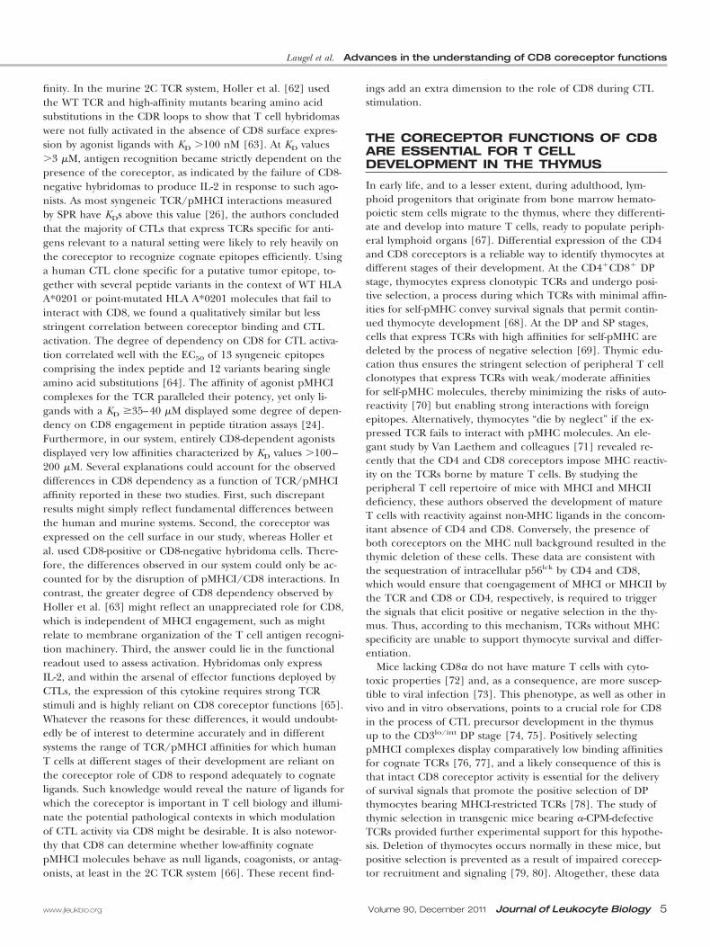
that CD8 coprecipitates with p56lck tyrosine kinase [16], the en-zyme that catalyzes the phosphorylation of CD3� ITAM residues(reviewed in ref. [17]), pointed to an important role for CD8 asa conveyor of proximal T cell activation signals. This notion wasreinforced by the demonstration that CD8 binds MHCI mole-cules [18] via interactions with largely nonpolymorphic aminoacid residues situated in the �3, and to a lesser extent, the �2domain of the heavy-chain and �2-microbulin [19, 20] (Fig. 1).The MHCI/CD8 interaction is characterized by a particularlyweak affinity and rapid kinetics [22]. Despite these seemingly un-favorable binding characteristics, however, the engagement ofMHCI molecules by CD8 at the cell surface enhances the associa-tion rate of pMHCI complexes with TCRs and increases the half-life of cognate TCR/pMHCI interactions [23–25]. These datasuggest that CD8 binding helps to increase the degree of TCRoccupancy at the T cell surface and stabilize the interaction be-
tween the TCR and pMHCI, two parameters that critically deter-mine the potency of TCR agonist ligands [26]. Thus, the promo-tion of intracellular signaling events and TCR/pMHCI interac-tions by the CD8 coreceptor likely combine to enhance cognateantigen recognition. As a result of the importance of p56lck inthe initiation of proximal phosphorylation events, however, themain contribution of CD8 is believed to result from the promo-tion of signaling and second messenger pathways downstream ofTCR triggering [27, 28].
The existence of a physical association between the TCR andCD8 on the T cell surface was first suggested by studies using co-modulation [29], coprecipitation [30, 31], and affinity chroma-tography [32]. Subsequently, the nature of this interaction and itsfunctional importance were revealed by the finding that palmi-toylation of the CD8�-chain enables the coreceptor to interactdirectly with CD3� and recruit TCR/CD3 complexes to mem-brane microdomains that promote signaling through the exclu-sion of inhibitory phosphatase proteins [33–35]. Furthermore,the conserved �-CPM, located on the membrane proximal do-main of the TCR �-chain, facilitates the recruitment of CD8 inclose proximity to the TCR/CD3 complex [36, 37] (Fig. 2). Inview of the apparent importance of CD8 in the recruitment ofp56lck tyrosine kinase to the TCR signaling complex, the moststraightforward interpretation of the biochemical data document-ing the cis association between TCR and CD8 and the trans inter-action between TCR and pMHCI was that the TCR and its core-ceptor coordinately engage the same cognate pMHCI ligand [2].In this TCR/CD8 heterodimeric configuration, binding of thecoreceptor to MHCI drives the recruitment of CD8-associatedp56lck to the vicinity of an engaged TCR/CD3 signaling complex,resulting in phosphorylation of the CD3� ITAMs (Fig. 2A). How-ever, this scenario was challenged by observations that CTLs frommice lacking both coreceptors can be activated by cognate viralor alloantigens [38], which suggests that free p56lck can initiatesignaling, and by structural data describing a binding angle be-tween CD8 and pMHCI, seemingly incompatible with the forma-tion of a tripartite TCR/pMHCI/CD8 molecular complex involv-ing direct contacts between the TCR and CD8 [26].
In addition to spatial considerations, the timing of coreceptoractivity during antigen recognition is crucial for a full mechanisticunderstanding of this process. A large proportion of CD8 andTCR molecules is constitutively associated on primary CTLs inthe absence of TCR engagement by agonist ligands, which sug-gests that the TCR and CD8 pre-exist as bispecific receptors thatcan engage pMHCI agonist ligands in a coordinate manner [30,31, 35, 39]. However, studies of the dynamic interactions betweenCD8 and TCR/CD3, based on the use of fluorescence resonanceenergy transfer, have shown that TCR binding to pMHCI occursfirst, thereby satisfying the antigen-specific component of the in-teraction, and that the recruitment of CD8 occurs subsequently[40, 41]. These data, obtained in T cell hybridomas, point to achronological sequence according to which the pivotal antigen-specific proofreading event provided by the TCR occurs prior tothe association of CD8 with the TCR/CD3 complex. Such amechanism would ensure that CD8 coreceptor functions, result-ing in the amplification of downstream signaling cascades, areonly elaborated for ligands that engage the TCR with favorableaffinities and kinetics. In this scenario, the activity of the TCR
Figure 1. Overview of the molecular interactions between CD8 andMHCI molecules. Ribbon representation of the cocrystal complex be-tween CD8�� and pMHCI. CD8�� is shown in red (�2) and purple(�1), binding mainly to the �3 domain of MHCI (green). The CDR-like loops of the CD8 molecule and residues 223–227 of the MHCI �3domain comprise the main binding interface. The enlargement of themain binding interface between CD8 and MHCI shows the two CDR-like loops of the CD8 molecule forming a "clamp"-like topologyaround the MHCI loop encompassing residues 223–227 of the �3 do-main. The most important contacts are made between the CD8 �1-chain Thr31/Ser100 and the MHCI �-chain Thr225/Gln226 and be-tween the CD8 �2-chain Ser34/Tyr51 and the MHCI �-chain Gln226/Asp227. Determination of the crystal structure of murine CD8�� incomplex with H2-Dd [21] has confirmed the general coreceptor bind-ing topology inferred from the pMHCI/CD8�� complexes and hasshown that the CD8�-chain occupies the T cell proximal position inthe heterodimer (in place of the CD8�2-chain in this picture).
2 Journal of Leukocyte Biology Volume 90, December 2011 www.jleukbio.org

could also reinforce the definition of a sharp TCR/pMHCIaffinity/dwell-time threshold that serves to discriminate agonistand nonagonist pMHCI complexes, with coreceptor recruitmentbeing enabled only in the context of agonist-ligand interactionswith the TCR. Notably, a recent study addressing the effect ofCD8 on the kinetics of pMHCI engagement by primary mouseCD8� T cells in two dimensions reported that the coreceptorcooperatively enhanced cell-cell adhesion mediated by the TCR[42]. This CD8-mediated effect required prior TCR engagementby agonist ligands and the tyrosine kinase activity of p56lck. Thus,in addition to the necessity for a prerequisite TCR/pMHCI ago-nist-ligand interaction, these results suggest that the exertion ofCD8 coreceptor functions requires the initiation of signaling byfree p56lck and identifies coreceptor-bound p56lck as the media-tor/adaptor molecule that recruits CD8 to the TCR/CD3 com-plex, a scenario proposed previously by Thome et al. [43] (Fig.2B). Importantly, this notion also reconciles the mechanistic sce-nario of coreceptor activity with structural studies, as recruitmentof CD8 to the TCR complex via p56lck, rather than by direct in-teractions with MHCI molecules, seems compatible with the over-all binding configuration of the multimolecular complex [44]. Inaddition, Jiang et al. [42] proposed a cooperative binding effectfor CD8 following its recruitment via p56lck, which may establish
interactions with agonist as well as nonagonist pMHCI moleculeson the APC surface. According to this model, the induced re-cruitment of CD8 serves to stabilize molecular contact betweenthe CTL and the APC, thereby unveiling another potential rolefor the coreceptor. This phenomenon may also help to explainprevious observations, indicating that the ability of CD8 to inter-act with nonstimulatory pMHCI complexes lowers T cell activa-tion thresholds and enables CTL to respond to low copy num-bers of specific pMHCI molecules [40, 45].
EXPERIMENTAL SYSTEMS FOR THEASSESSMENT OF CD8 FUNCTIONDURING CTL ACTIVATION
Preventing the association between CD8 and MHCI moleculesin cellular assays, often using anti-CD8 blocking antibodies,has been the most common experimental approach used toaddress the role of CD8 in CTL activation. Although it hadbeen appreciated for a long time that a single anti-CD8 anti-body paratope could have weak or strong inhibitory effects onthe activation of different CTL clones [46, 47], the reasonsunderlying these discrepancies remained elusive until recently.It has since become clear that anti-CD8 antibodies can have
α β
α CD8pMHCI
TCR
CD3
T cell activation
P
ZAP70 P
P
α1
α2 α3
β2m
P
P
P LCK
ζζ
P
P
P
P
P
P
γε
Palm.
β
εδ
Rafts and associated molecules
Direct molecular interaction
Kinase activity
Step 1
Step 2
LCK P
P
P
CD8 pMHCI
TCR
CD3
T cell activation
P
P
ZAP70 P
P
1
2 3
2m
P
P
P LCK
P
P
P
P
P
P
Palm.
Rafts and associated molecules
Direct molecular interaction
Kinase activity
Step 3
Step 2 Step 1
A B
Figure 2. Schematic representation of CD8 coreceptor functions in early T cell activation events. The extracellular portion of CD8�� interacts directly with theMHCI �-chain and �2-microglobulin, facilitating TCR/pMHCI interactions. Direct molecular cis-interactions between CD8 and the TCR complex also take placeon the T cell surface. These involve direct contacts between the CD3�- and CD8�-chain and require the native �-CPM sequence of the TCR �-chain (repre-sented by a dark-blue oval shape). Palmitoylation (Palm.) of CD8� facilitates these contacts and also permits partitioning of the TCR/CD8 complex in mem-brane lipid rafts, which provide an enzymatic environment that favors phosphorylation events via the sequestration of phosphatases. (A) The classical view of Tcell activation is that CD8 is recruited to the TCR complex before phosphorylation takes place and that p56lck bound to the CD8� cytoplasmic tail catalyzes theinitial CD3� ITAM phosphorylation events, which then allow for the recruitment of additional p56lck molecules and signal amplification. (B) Alternatively, recentexperimental data favor a model whereby free p56lck is responsible for the initial phosphorylation events. Phosphorylated CD3� ITAMs then allow the recruit-ment of p56lck bound to CD8� in close proximity to CD3. In this scenario, the interaction between the TCR and CD8 occurs after the initial phosphorylationevents and is driven intracellularly by interactions between CD3- and CD8�-bound p56lck.
Laugel et al. Advances in the understanding of CD8 coreceptor functions
www.jleukbio.org Volume 90, December 2011 Journal of Leukocyte Biology 3

very distinct or even opposing effects on TCR/pMHCI interac-tions and the recognition of agonist epitopes by CTLs. Fur-thermore, these effects can be largely unrelated to disruptionof the pMHCI/CD8 interaction. Indeed, some anti-CD8 anti-bodies can completely or partially inhibit TCR binding byidentical pMHCI ligands, and others can enhance pMHCI en-gagement and antigen recognition or even stimulate CTLs intheir own right [48–54] (summarized in Tables 1 and 2).These heterogeneous effects, which are inherent to the bind-ing mode and impacted by steric hindrance resulting from theuse of macromolecules to block the engagement of MHCImolecules by CD8, impose important caveats on the interpreta-tion of data generated with such approaches. Indeed, giventhat the TCR complex and CD8 become very closely associatedon the surface of CTLs during antigen-induced TCR trigger-ing, it is not surprising that the inhibitory effects of large mol-ecules bound to CD8 can be independent from the pMHCI/CD8 interaction itself. More elaborate systems that circumventthese experimental problems have been provided by the de-sign of point-mutated MHCI molecules that fail to interactwith the coreceptor [28, 55]. These molecules, expressed onthe surface of APCs or in the form of soluble recombinantproteins, have allowed in-depth investigations into the impor-tance of the pMHCI/CD8 interaction in the process of CTLactivation. Hybridomas expressing monoclonal TCRs deliveredby viral vectors, with or without the cotransduction of CD8 �-and �-chains, have also provided suitable systems for the studyof coreceptor function [56, 57].
UNRAVELING THE ROLE OF CD8:ENHANCEMENT OF CTL ACTIVATION BYLOW-AFFINITY ANTIGENIC LIGANDS
The experimental approaches described above have been usedto characterize many monoclonal CTLs or hybridomas thatrecognize epitopes in a CD8-dependent or CD8-independentmanner [48, 56, 58–61]. From these studies, it appeared thatthe degree of dependency on CD8 for efficient recognitionroughly correlated with the potency of the considered ligand,as defined by the concentration of peptide required to elicithalf-maximum activation in dose-response titrations (Fig. 3).As biophysical investigations using SPR had determined thatthe affinity and/or the dwell time of TCR/pMHCI interactionscorrelated well with the potency of agonist ligands [26], it wasinferred that CD8 dependency was probably linked with theseparameters. Two studies tested this possibility directly by sys-tematically assessing the CD8 dependency of several agonistligands with a wide range of defined KD values and half-livesfor their respective cognate TCR interactions. In both cases,the experimental results established a tight correlation be-tween the requirement for CD8 activity and TCR/pMHCI af-
TABLE 1. The Heterogeneity of Antihuman CD8 Antibodies
Ab clone � or � Tetramer binding MIP-1� MIP-1� RANTES IFN-� TNF-� IL-2 Cytotoxicity
OKT8 � Enhance Yes Yes Yes No No No YesSK1 � Inhibit No No No No No No NoMCD8 � Neutral No No No No No No No32/M4 � Neutral No No No No No No NoC8/144B � Neutral No No No No No No NoDK25 � Inhibit No No No No No No No2ST8.5H7 � Inhibit No No No No No No No
Summary of the effects exerted by antihuman CD8 antibodies on pMHCI tetramer binding and CD8� T cell activation in the absence of TCRengagement. Table reproduced from ref. [54] (Copyright 2011, The American Association of Immunologists).
TABLE 2. The Heterogeneity of Antimouse CD8 Antibodies
Ab clone � or �Tetramerbinding MIP-1� IFN-� IL-2
CT-CD8a � Inhibit Yes No No53.6.7 � Enhance Weak No NoCT-CD8b � Enhance Yes No NoKT112 � Enhance Weak NT NT
Summary of the effects exerted by antimouse CD8 antibodies onpMHCI tetramer binding and CD8� T cell activation in the absence ofTCR engagement. NT, Not tested. Table reproduced from ref. [54](Copyright 2011, The American Association of Immunologists).
Antigen potency
T-ce
ll ac
tivat
ion
None
Max
StrongWeak
WithoutCD8
With CD8
Figure 3. Model illustrating the degree of CD8 coreceptor dependencyas a function of antigen sensitivity and TCR/pMHCI affinity in theprocess of CTL activation.
4 Journal of Leukocyte Biology Volume 90, December 2011 www.jleukbio.org
finity. In the murine 2C TCR system, Holler et al. [62] usedthe WT TCR and high-affinity mutants bearing amino acidsubstitutions in the CDR loops to show that T cell hybridomaswere not fully activated in the absence of CD8 surface expres-sion by agonist ligands with KD �100 nM [63]. At KD values�3 �M, antigen recognition became strictly dependent on thepresence of the coreceptor, as indicated by the failure of CD8-negative hybridomas to produce IL-2 in response to such ago-nists. As most syngeneic TCR/pMHCI interactions measuredby SPR have KDs above this value [26], the authors concludedthat the majority of CTLs that express TCRs specific for anti-gens relevant to a natural setting were likely to rely heavily onthe coreceptor to recognize cognate epitopes efficiently. Usinga human CTL clone specific for a putative tumor epitope, to-gether with several peptide variants in the context of WT HLAA*0201 or point-mutated HLA A*0201 molecules that fail tointeract with CD8, we found a qualitatively similar but lessstringent correlation between coreceptor binding and CTLactivation. The degree of dependency on CD8 for CTL activa-tion correlated well with the EC50 of 13 syngeneic epitopescomprising the index peptide and 12 variants bearing singleamino acid substitutions [64]. The affinity of agonist pMHCIcomplexes for the TCR paralleled their potency, yet only li-gands with a KD �35–40 �M displayed some degree of depen-dency on CD8 engagement in peptide titration assays [24].Furthermore, in our system, entirely CD8-dependent agonistsdisplayed very low affinities characterized by KD values �100–200 �M. Several explanations could account for the observeddifferences in CD8 dependency as a function of TCR/pMHCIaffinity reported in these two studies. First, such discrepantresults might simply reflect fundamental differences betweenthe human and murine systems. Second, the coreceptor wasexpressed on the cell surface in our study, whereas Holler etal. used CD8-positive or CD8-negative hybridoma cells. There-fore, the differences observed in our system could only be ac-counted for by the disruption of pMHCI/CD8 interactions. Incontrast, the greater degree of CD8 dependency observed byHoller et al. [63] might reflect an unappreciated role for CD8,which is independent of MHCI engagement, such as mightrelate to membrane organization of the T cell antigen recogni-tion machinery. Third, the answer could lie in the functionalreadout used to assess activation. Hybridomas only expressIL-2, and within the arsenal of effector functions deployed byCTLs, the expression of this cytokine requires strong TCRstimuli and is highly reliant on CD8 coreceptor functions [65].Whatever the reasons for these differences, it would undoubt-edly be of interest to determine accurately and in differentsystems the range of TCR/pMHCI affinities for which humanT cells at different stages of their development are reliant onthe coreceptor role of CD8 to respond adequately to cognateligands. Such knowledge would reveal the nature of ligands forwhich the coreceptor is important in T cell biology and illumi-nate the potential pathological contexts in which modulationof CTL activity via CD8 might be desirable. It is also notewor-thy that CD8 can determine whether low-affinity cognatepMHCI molecules behave as null ligands, coagonists, or antag-onists, at least in the 2C TCR system [66]. These recent find-
ings add an extra dimension to the role of CD8 during CTLstimulation.
THE CORECEPTOR FUNCTIONS OF CD8ARE ESSENTIAL FOR T CELLDEVELOPMENT IN THE THYMUS
In early life, and to a lesser extent, during adulthood, lym-phoid progenitors that originate from bone marrow hemato-poietic stem cells migrate to the thymus, where they differenti-ate and develop into mature T cells, ready to populate periph-eral lymphoid organs [67]. Differential expression of the CD4and CD8 coreceptors is a reliable way to identify thymocytes atdifferent stages of their development. At the CD4�CD8� DPstage, thymocytes express clonotypic TCRs and undergo posi-tive selection, a process during which TCRs with minimal affin-ities for self-pMHC convey survival signals that permit contin-ued thymocyte development [68]. At the DP and SP stages,cells that express TCRs with high affinities for self-pMHC aredeleted by the process of negative selection [69]. Thymic edu-cation thus ensures the stringent selection of peripheral T cellclonotypes that express TCRs with weak/moderate affinitiesfor self-pMHC molecules, thereby minimizing the risks of auto-reactivity [70] but enabling strong interactions with foreignepitopes. Alternatively, thymocytes “die by neglect” if the ex-pressed TCR fails to interact with pMHC molecules. An ele-gant study by Van Laethem and colleagues [71] revealed re-cently that the CD4 and CD8 coreceptors impose MHC reactiv-ity on the TCRs borne by mature T cells. By studying theperipheral T cell repertoire of mice with MHCI and MHCIIdeficiency, these authors observed the development of matureT cells with reactivity against non-MHC ligands in the concom-itant absence of CD4 and CD8. Conversely, the presence ofboth coreceptors on the MHC null background resulted in thethymic deletion of these cells. These data are consistent withthe sequestration of intracellular p56lck by CD4 and CD8,which would ensure that coengagement of MHCI or MHCII bythe TCR and CD8 or CD4, respectively, is required to triggerthe signals that elicit positive or negative selection in the thy-mus. Thus, according to this mechanism, TCRs without MHCspecificity are unable to support thymocyte survival and differ-entiation.
Mice lacking CD8� do not have mature T cells with cyto-toxic properties [72] and, as a consequence, are more suscep-tible to viral infection [73]. This phenotype, as well as other invivo and in vitro observations, points to a crucial role for CD8in the process of CTL precursor development in the thymusup to the CD3lo/int DP stage [74, 75]. Positively selectingpMHCI complexes display comparatively low binding affinitiesfor cognate TCRs [76, 77], and a likely consequence of this isthat intact CD8 coreceptor activity is essential for the deliveryof survival signals that promote the positive selection of DPthymocytes bearing MHCI-restricted TCRs [78]. The study ofthymic selection in transgenic mice bearing �-CPM-defectiveTCRs provided further experimental support for this hypothe-sis. Deletion of thymocytes occurs normally in these mice, butpositive selection is prevented as a result of impaired corecep-tor recruitment and signaling [79, 80]. Altogether, these data
Laugel et al. Advances in the understanding of CD8 coreceptor functions
www.jleukbio.org Volume 90, December 2011 Journal of Leukocyte Biology 5

support a model, termed the "coreceptor zipper" by the au-thors, whereby the coreceptor is crucial for the elicitation ofsurvival signals by increasing the apparent affinity of the inter-action between an individual TCR and a positively selectingligand, as well as by directly promoting consecutive, proximalintracellular signaling events [77, 79].
In addition, several reports have suggested an importantrole for CD8 in the process of DP thymocyte progression to-ward the more mature SP CD4� or CD8� stage. The kineticsignaling model of T cell lineage decision, which accommo-dates most experimental results that address lineage commit-ment, postulates that the key parameter in the DP thymocytecommitment to Th or CTL differentiation, is the duration ofTCR-mediated signaling elicited by positively selecting ligands(reviewed in ref. [81]). Sustained signaling is associated withCD4� Th cell-lineage commitment, whereas short-lived signalspromote differentiation into CD8� CTLs [82, 83]. As the initi-ation of TCR signaling in DP thymocytes is concomitant withthe down-regulation of CD8 gene expression, independently ofMHC restriction and later lineage commitment [84, 85], it isin a CD4�CD8low state of differentiation that signal durationdetermines lineage fate. Cessation of TCR signals elicited bypMHCI interactions subsequent to CD8 down-regulation speci-fies commitment to the CTL lineage. Conversely, CD8-inde-
pendent signals, elicited by the engagement of pMHCII, re-main unaffected by CD8 down-regulation and thus, promotedifferentiation into the CD4� Th cell lineage. Therefore, oneof the fundamental postulates underlying the kinetic signalingmodel of thymic selection, as well as other nonstochastic mod-els not discussed here, relies on the premise that signals elic-ited by low-affinity, self-pMHCI ligands supporting the positiveselection of DP thymocytes and their commitment to theCD8� CTL lineage are heavily dependent on CD8 coreceptorfunctions in the thymus.
PERIPHERAL REGULATION OF CD8CORECEPTOR FUNCTIONS AS APOTENTIAL MECHANISM TO MODULATEANTIGEN SENSITIVITY IN VIVO
Several lines of evidence have recently given credence to thehypothesis that efficient regulation of murine CTL activity inthe periphery can be achieved through the modification ofCD8 functions (Fig. 4). Indeed, TCR-dependent activation ofnaıve CD8� T cells in the periphery appears to be a majorcheckpoint for such modulatory pathways. The mechanismsthat underlie the modulation of CD8 functions include: (i)expression-switching to the CD8�� homodimer, which is
Common γ-chain cytokines(IL-2, (IL-4?), IL-7, IL-15)IFN-γ
(IL-4?)
CD8ACD8
+
-
High CD8 levelsIncreased pMHCIresponsiveness
Low CD8 levelsDecreased pMHCIresponsiveness
Naïve CD8+ T cell Activated effector CD8+ T cell
T1 IFN
Common γ-chain cytokines(IL-2, (IL-4?), IL-7, IL-15)IFN-γ
(IL-4?)
CD8ACD88ACD8
+
-
TCR triggering
� CD8 glycosylationSequestration of TCRand CD8Switch to CD8αα
Decreased pMHCIresponsiveness
+ -
A B
Figure 4. Cues regulating CD8 expression levels and coreceptor functions in relation to the cellular activation status of peripheral CD8� T cells.(A) In naıve CD8� T cells, modulation of CD8 functions is mostly known to occur via the regulation of CD8A transcription, which tunes corecep-tor expression levels on the cell surface and thereby alters pMHCI recognition. Physiologically, CD8A transcriptional regulation in naıve T cells isthought to modulate survival signals elicited by self-pMHCI and regulate homeostatic expansion. (B) In addition to cytokine signals, the functionsof CD8 are regulated at the transcriptional and post-translational levels following T cell activation by antigen. For instance, TCR stimuli result inthe inhibition of IL-7R signals that up-regulate CD8A expression, thereby resulting in lower coreceptor expression levels on the cell surface. Alto-gether, these mechanisms down-regulate the functions of CD8, probably to prevent excessive and deleterious CTL activation by self-pMHCIligands.
6 Journal of Leukocyte Biology Volume 90, December 2011 www.jleukbio.org

known to be a poor coreceptor compared with the �� isoformas a result of an inability to recruit TCR complexes to mem-brane microdomains and suboptimal engagement of pMHCImolecules [6, 86]; (ii) post-translational modifications ofCD8�� glycosylation [87, 88]; and (iii) modulation of corecep-tor expression levels on the cell surface [60, 89, 90]. In addi-tion to activation-dependent cues, the activity of several cyto-kines can tune CD8 expression levels on the surface of CTLsthrough the regulation of CD8A gene transcription. Park andcolleagues [91] reported that the common �-chain cytokinesIL-2, IL-4, IL-7, and IL-15 enhance CD8A transcription andcoreceptor expression on the cell surface. It is known that insteady-state, weak nonimmunogenic TCR signals elicited byself-pMHCI complexes are required to mediate the survival ofnaıve CD8� T cells (reviewed in ref. [92]). Accordingly, theauthors’ interpretation of their data was that under these con-ditions, signaling via homeostatic cytokines such as IL-7 andIL-15 would promote CD8� T cell survival by favoring theirresponsiveness to weak, CD8-dependent, nonstimulatory self-pMHCI complexes. Somewhat in contrast with these findings,another group linked the down-regulation of CD8 expressionand subsequent inhibition of antigen recognition by CTLs tothe direct effects of IL-4 [93, 94], an activity that they laterreported was antagonized by autocrine IFN-� [95]. These au-thors also suggested that, in addition to the altered functionalphenotype induced by IL-4, down-regulation of CD8 expres-sion on CTLs could contribute to their nonresponsiveness toantigen and that this mechanism could be relevant to the im-munosuppressive action of IL-4-secreting tumors [95]. In addi-tion, Xiao and colleagues [96] found that type I IFN alsodown-regulated the expression of CD8 via nontranscriptionalmechanisms. The significance of this latter observation re-mains unclear.
Perhaps the most compelling evidence that modulation ofCTL activity can be achieved through CD8 in vivo comes fromthe study of human CD8� TILs. Demotte and colleagues [39]proposed that the physical dissociation between CD8 and theTCR mediated by galectin-3 could be responsible for a form ofnonresponsiveness to antigen in the cancer setting. This hy-pothesis stemmed from the characterization of freshly isolatedCD8� TILs that were unable to kill target tumor cells or pep-tide-pulsed APCs. The authors found that CD8 and TCR wereseparated in distinct membrane locations on the TIL surfaceand that CD8/TCR interactions were restored by treatment ofthese cells with galectin-3 disaccharide ligands. This treatmentalso reversed the coreceptor/TCR dissociation observed oncells cultured with tumor ascite supernatants and correlatedwith reversion of the anergic state [39, 97]. Although they didnot suggest a causal effect, these authors also reported thatgalectin-3 ligands induce superior in vivo antitumor adaptive-immune responses in mice [97].
In summary, there is substantial evidence for the existenceof multiple transcriptional and post-translational mechanismsthat act transiently to down-regulate or increase coreceptorfunctions. These pathways can be activated as a direct conse-quence of TCR triggering and CD8� T cell activation in a cell-autonomous or autocrine manner, or locally through the para-crine action of cytokines and soluble factors, such as occurs in
the tumor microenvironment. It is conceivable that TCR-medi-ated regulatory pathways act to decrease CTL responsivenessto antigen in the context of an ongoing immune response invivo, thereby ensuring the nonresponsiveness of effector CTLsto self-determinants and contributing to the maintenance ofperipheral tolerance. Conversely, in steady-state conditions,homeostatic cytokines might act to promote nonactivating self-pMHCI-dependent survival cues through the transcriptionalenhancement of CD8 expression, thereby indirectly promotingnaıve T cell persistence and expansion in the periphery.
THE POTENTIAL PHYSIOLOGICALIMPORTANCE OF CD8 IN CTL BIOLOGY
Although the importance of CD8 for CTL activation in responseto low-affinity ligands in vitro is well-established, the actual role ofthe coreceptor during an immune response to microbial, tu-moral, or self-derived antigens remains to be formally establishedin vivo. Indeed, despite the wealth of experimental data that doc-ument the enhancement of mature CTL activation and thedetailed biochemical characterization of the mechanisms thatpertain to this activity, the only firmly proven physiological rolefor the coreceptor concerns its importance in the events that gov-ern T cell selection in the thymus. However, based on currentevidence, it seems likely that CD8 helps to drive the priming andexpansion of CTL clonotypes with low functional avidities forcognate antigen. Such a role could enhance the clonotypic diver-sity of CTL responses to microbial determinants and, more obvi-ously, might contribute to the onset of a response directedagainst self-determinants in an autoimmune context. Support forthe former scenario was provided by the finding that ex vivo acti-vation of subdominant CTL clonotypes specific for epitopes de-rived from EBV and human CMV relied more heavily on CD8engagement compared with numerically dominant clonotypeswith the same antigen specificity, thereby indicating that CD8likely augments clonotypic diversity within the antigen-specificCTL pool and prevents avidity-based selection proceeding tounity during chronic viral infections [98]. Perhaps an even moreimportant putative role for CD8 could relate to maintenance ofthe naıve CD8� T cell pool in the steady-state. As discussed previ-ously, several reports have demonstrated the importance of sub-optimal TCR engagement by self-ligands, resulting in low-levelsignaling without concomitant activation, for the survival of naıveCD8� T cells in the periphery [92]. In contrast, memory CD8� Tcell persistence only requires the presence of homeostatic cyto-kines and does not rely on suboptimal TCR stimuli [92]. It istherefore likely that coreceptor functions are required for thesurvival of naıve, but not memory, CD8� T cells. Rigorous testingof these predictions would require an approach that allows thedeletion of CD8 or disruption of the MHCI/CD8 interaction atdifferent stages of T cell differentiation in the periphery. Condi-tional ablation of CD8� in transgenic mice with floxed CD8Aalleles is one such option. The best-suited existing system for con-ditional expression of the Cre recombinase is probably the distalLck promoter, which is switched on during the late stages of thy-mic development; this model has been used by Killeen and col-leagues [99] to assess the role of CD4 in mature T cells. How-ever, detailed investigations of this promoter system with the
Laugel et al. Advances in the understanding of CD8 coreceptor functions
www.jleukbio.org Volume 90, December 2011 Journal of Leukocyte Biology 7

ROSA26 reporter mouse strain revealed an incomplete pen-etrance in T cells, as well as variations in the timing of Cre ex-pression with respect to thymocyte development [100]. Conse-quently, a novel conditional or inducible system, strictly limited tomature T cells of the cytotoxic lineage, is required to investigatethe functions of CD8 in the periphery.
PHARMACOLOGICAL INHIBITION OFCORECEPTOR FUNCTION TO DAMPENAUTOIMMUNE CD8� T CELL-MEDIATEDPATHOLOGY
Inappropriate T cell responses are likely involved in the eti-ology of many autoimmune and chronic inflammatory pa-thologies. Modulation of T cell functions therefore repre-sents an attractive therapeutic strategy to mitigate patho-genic immune responses. However, the design of efficientand safe therapies that target T cell activity faces majorchallenges. On the one hand, targeting the entire T cellcompartment threatens harmful immunosuppression. Onthe other hand, specific T cell targeting approaches requiredetailed knowledge of the putative self-antigens that driveautoimmune pathogenesis, which is presently an unrealisticgoal considering the tremendous diversity of potential Tcell antigens and the vast allelic variability within the hu-man MHC locus.
It was thought previously that CD8 involvement was criticalfor the majority of CTL responses and that targeting thepMHCI/CD8 interaction using soluble CD8 or variants thereof[101, 102], antibodies, or various small molecules [103, 104]represented a generic way of disrupting CD8� T cell activity.However, as discussed above, recent data have refined our un-derstanding of the role of the coreceptor in the process ofCTL activation by showing that the degree of CD8 depen-dence is inversely related to the strength of the TCR/pMHCIinteraction. Accordingly, CD8 blockade should enable the se-lective targeting of weak TCR/pMHCI interactions withoutaffecting high-affinity cognate TCRs, such as those expressedby CTLs specific for microbial epitopes. In support of thisidea, accumulating data suggest that CTLs specific for autoim-mune and tumoral self-determinants bear TCRs with lower af-finities for their cognate ligands compared with microbe-spe-cific and alloreactive CTLs [26, 105–110] (unpublished re-sults). Thus, CD8 may be a valid therapeutic target in thesetting of autoimmune diseases with etiologies that are linkedto the activity of CTLs. CD8� T cells are known to infiltratedamaged tissues in high numbers in many autoimmune condi-tions, but their roles in disease initiation and progression areuncertain. However, recent investigations have produced sev-eral lines of evidence that implicate CD8� T cells in thepathogenesis of autoimmunity. First, genetic association andgenome-wide analysis studies have linked several HLA I alleleswith disease susceptibility or protection, independently of HLAII alleles [111, 112]. In addition, the identification of HLA Iepitopes presented by target tissues and recognized bypatients' CD8� T cells, combined with the development oftransgenic animal models of T1D [113–115] and MS [116],which are entirely dependent on CD8� T cells, provides com-
pelling evidence for a role of CD8� T cells in effecting tissuedamage in the context of certain autoimmune pathologies. Inaddition to T1D and MS, such conditions may include vitiligo[117], neurodegenerative diseases, such as certain paraneo-plastic syndromes [118], Hashimoto’s thyroiditis, autoimmunemyocarditis, and autoimmune hepatitis [119].
CONCLUDING REMARKS
Recent developments have refined our understanding ofCD8� T cell activation and the role of the coreceptor in theprocess of antigen recognition. Detailed biochemical and cel-lular investigations have established that CD8 coreceptor activ-ity is essential for the recognition of weak, low-affinity ligandsbut dispensable for potent, high-affinity ligands. This knowl-edge holds translational promise, especially in the setting ofautoimmunity. Compounds that are currently approved for thetreatment of autoimmune and inflammatory diseases or thosein the later stages of development usually rely on the induc-tion of profound and/or long-lasting immunosuppression.This is often achieved by global depletion of entire cell typesor lineages in the periphery, by the prevention of peripheralleukocyte trafficking and egress from lymphoid organs, or bytargeting the systemic activity of cytokines involved in diseasepathogenesis [120–122]. Although these treatments oftenshow good efficacy, their adverse effects can outweigh theirbenefits. Potentially severe opportunistic infections or reactiva-tion of latent viral infections with clinically disastrous sequelaeare unacceptable side-effects in the treatment of autoimmunediseases with less aggressive profiles. More targeted and lessdisruptive treatments are therefore required for certain pathol-ogies. Based on accumulated knowledge of CD8 biology, wepropose that specific targeting of coreceptor functions mightpreferentially inhibit pathogenic CTL responses directedagainst self-determinants and leave cellular immunity to micro-bial pathogens largely intact. This hypothesis must still be vali-dated experimentally in terms of efficacy and innocuity, partic-ularly with respect to T cell development and homeostasis.Nevertheless, the possibility of selective therapeutic interven-tion without a priori knowledge of precise antigenic targets isan exciting and realistic goal.
ACKNOWLEDGMENTS
B.L., L.W., D.A.P., and A.K.S. are funded by Biotechnologyand Biological Sciences Research Council (UK) grant BB/H001085/1. D.A.P. is a Medical Research Council (UK) SeniorClinical Fellow. D.K.C. is a Wellcome Trust Research CareerDevelopment Fellow (RCDF). L.W. is a Wellcome Trust Clini-cal Intermediate Fellow.
REFERENCES
1. Swain, S. L. (1983) T cell subsets and the recognition of MHC class. Im-munol. Rev. 74, 129–142.
2. Janeway Jr., C. A. (1992) The T cell receptor as a multicomponent sig-nalling machine: CD4/CD8 coreceptors and CD45 in T cell activation.Annu. Rev. Immunol. 10, 645–674.
3. Ledbetter, J. A., Seaman, W. E., Tsu, T. T., Herzenberg, L. A. (1981)Lyt-2 and Lyt-3 antigens are on two different polypeptide subunits
8 Journal of Leukocyte Biology Volume 90, December 2011 www.jleukbio.org

linked by disulfide bonds. Relationship of subunits to T cell cytolyticactivity. J. Exp. Med. 153, 1503–1516.
4. Norment, A. M., Littman, D. R. (1988) A second subunit of CD8 is ex-pressed in human T cells. EMBO J. 7, 3433–3439.
5. Terry, L. A., DiSanto, J. P., Small, T. N., Flomenberg, N. (1990) Differ-ential expression and regulation of the human CD8 � and CD8 �chains. Tissue Antigens 35, 82–91.
6. Gangadharan, D., Cheroutre, H. (2004) The CD8 isoform CD8�� is nota functional homologue of the TCR co-receptor CD8��. Curr. Opin. Im-munol. 16, 264–270.
7. Gibbings, D., Befus, A. D. (2009) CD4 and CD8: an inside-out corecep-tor model for innate immune cells. J. Leukoc. Biol. 86, 251–259.
8. Leishman, A. J., Naidenko, O. V., Attinger, A., Koning, F., Lena, C. J.,Xiong, Y., Chang, H. C., Reinherz, E., Kronenberg, M., Cheroutre, H.(2001) T cell responses modulated through interaction between CD8��and the nonclassical MHC class I molecule, TL. Science 294, 1936–1939.
9. Nakayama, E., Shiku, H., Stockert, E., Oettgen, H. F., Old, L. J. (1979)Cytotoxic T cells: Lyt phenotype and blocking of killing activity by Lytantisera. Proc. Natl. Acad. Sci. USA 76, 1977–1981.
10. Kappler, J., Kubo, R., Haskins, K., Hannum, C., Marrack, P., Pigeon, M.,McIntyre, B., Allison, J., Trowbridge, I. (1983) The major histocompati-bility complex-restricted antigen receptor on T cells in mouse and man:identification of constant and variable peptides. Cell 35, 295–302.
11. Hedrick, S. M., Cohen, D. I., Nielsen, E. A., Davis, M. M. (1984) Isola-tion of cDNA clones encoding T cell-specific membrane-associated pro-teins. Nature 308, 149–153.
12. Hedrick, S. M., Nielsen, E. A., Kavaler, J., Cohen, D. I., Davis, M. M.(1984) Sequence relationships between putative T-cell receptor polypep-tides and immunoglobulins. Nature 308, 153–158.
13. Bjorkman, P. J., Saper, M. A., Samraoui, B., Bennett, W. S., Strominger,J. L., Wiley, D. C. (1987) Structure of the human class I histocompatibil-ity antigen, HLA-A2. Nature 329, 506–512.
14. Bjorkman, P. J., Saper, M. A., Samraoui, B., Bennett, W. S., Strominger, J. L.,Wiley, D. C. (1987) The foreign antigen binding site and T cell recognitionregions of class I histocompatibility antigens. Nature 329, 512–518.
15. Townsend, A. R., Rothbard, J., Gotch, F. M., Bahadur, G., Wraith, D.,McMichael, A. J. (1986) The epitopes of influenza nucleoprotein recog-nized by cytotoxic T lymphocytes can be defined with short syntheticpeptides. Cell 44, 959–968.
16. Veillette, A., Bookman, M. A., Horak, E. M., Bolen, J. B. (1988) TheCD4 and CD8 T cell surface antigens are associated with the internalmembrane tyrosine-protein kinase p56lck. Cell 55, 301–308.
17. Smith-Garvin, J. E., Koretzky, G. A., Jordan, M. S. (2009) T cell activa-tion. Annu. Rev. Immunol. 27, 591–619.
18. Norment, A. M., Salter, R. D., Parham, P., Engelhard, V. H., Littman,D. R. (1988) Cell-cell adhesion mediated by CD8 and MHC class I mole-cules. Nature 336, 79–81.
19. Salter, R. D., Benjamin, R. J., Wesley, P. K., Buxton, S. E., Garrett, T. P.,Clayberger, C., Krensky, A. M., Norment, A. M., Littman, D. R., Parham,P. (1990) A binding site for the T-cell co-receptor CD8 on the � 3 do-main of HLA-A2. Nature 345, 41–46.
20. Salter, R. D., Norment, A. M., Chen, B. P., Clayberger, C., Krensky,A. M., Littman, D. R., Parham, P. (1989) Polymorphism in the � 3 do-main of HLA-A molecules affects binding to CD8. Nature 338, 345–347.
21. Wang, R., Natarajan, K., Margulies, D. H. (2009) Structural basis of theCD8 � �/MHC class I interaction: focused recognition orients CD8 � toa T cell proximal position. J. Immunol. 183, 2554–2564.
22. Wyer, J. R., Willcox, B. E., Gao, G. F., Gerth, U. C., Davis, S. J., Bell,J. I., van der Merwe, P. A., Jakobsen, B. K. (1999) T cell receptor andcoreceptor CD8 �� bind peptide-MHC independently and with distinctkinetics. Immunity 10, 219–225.
23. Gakamsky, D. M., Luescher, I. F., Pramanik, A., Kopito, R. B., Lemon-nier, F., Vogel, H., Rigler, R., Pecht, I. (2005) CD8 kinetically promotesligand binding to the T-cell antigen receptor. Biophys. J. 89, 2121–2133.
24. Laugel, B., van den Berg, H. A., Gostick, E., Cole, D. K., Wooldridge, L.,Boulter, J., Milicic, A., Price, D. A., Sewell, A. K. (2007) Different T cellreceptor affinity thresholds and CD8 coreceptor dependence governcytotoxic T lymphocyte activation and tetramer binding properties.J. Biol. Chem. 282, 23799–23810.
25. Wooldridge, L., van den Berg, H. A., Glick, M., Gostick, E., Laugel, B.,Hutchinson, S. L., Milicic, A., Brenchley, J. M., Douek, D. C., Price,D. A., Sewell, A. K. (2005) Interaction between the CD8 coreceptor andmajor histocompatibility complex class I stabilizes T cell receptor-anti-gen complexes at the cell surface. J. Biol. Chem. 280, 27491–27501.
26. Van der Merwe, P. A., Davis, S. J. (2003) Molecular interactions mediat-ing T cell antigen recognition. Annu. Rev. Immunol. 21, 659–684.
27. Artyomov, M. N., Lis, M., Devadas, S., Davis, M. M., Chakraborty, A. K.(2010) CD4 and CD8 binding to MHC molecules primarily acts to en-hance Lck delivery. Proc. Natl. Acad. Sci. USA 107, 16916–16921.
28. Purbhoo, M. A., Boulter, J. M., Price, D. A., Vuidepot, A. L., Hourigan,C. S., Dunbar, P. R., Olson, K., Dawson, S. J., Phillips, R. E., Jakobsen,B. K., Bell, J. I., Sewell, A. K. (2001) The human CD8 coreceptor effectscytotoxic T cell activation and antigen sensitivity primarily by mediatingcomplete phosphorylation of the T cell receptor � chain. J. Biol. Chem.276, 32786–32792.
29. Takada, S., Engleman, E. G. (1987) Evidence for an association betweenCD8 molecules and the T cell receptor complex on cytotoxic T cells.J. Immunol. 139, 3231–3235.
30. Beyers, A. D., Spruyt, L. L., Williams, A. F. (1992) Molecular associations be-tween the T-lymphocyte antigen receptor complex and the surface antigensCD2, CD4, or CD8 and CD5. Proc. Natl. Acad. Sci. USA 89, 2945–2949.
31. Suzuki, S., Kupsch, J., Eichmann, K., Saizawa, M. K. (1992) Biochemicalevidence of the physical association of the majority of CD3 � chains withthe accessory/co-receptor molecules CD4 and CD8 on nonactivated Tlymphocytes. Eur. J. Immunol. 22, 2475–2479.
32. Gallagher, P. F., Fazekas de St Groth, B., Miller, J. F. (1989) CD4 andCD8 molecules can physically associate with the same T-cell receptor.Proc. Natl. Acad. Sci. USA 86, 10044–10048.
33. Arcaro, A., Gregoire, C., Boucheron, N., Stotz, S., Palmer, E., Malissen,B., Luescher, I. F. (2000) Essential role of CD8 palmitoylation in CD8coreceptor function. J. Immunol. 165, 2068–2076.
34. Arcaro, A., Gregoire, C., Bakker, T. R., Baldi, L., Jordan, M., Goffin, L.,Boucheron, N., Wurm, F., van der Merwe, P. A., Malissen, B., Luescher,I. F. (2001) CD8� endows CD8 with efficient coreceptor function bycoupling T cell receptor/CD3 to raft-associated CD8/p56(lck) com-plexes. J. Exp. Med. 194, 1485–1495.
35. Doucey, M. A., Goffin, L., Naeher, D., Michielin, O., Baumgartner, P., Guil-laume, P., Palmer, E., Luescher, I. F. (2003) CD3 � establishes a functional linkbetween the T cell receptor and CD8. J. Biol. Chem. 278, 3257–3264.
36. Mallaun, M., Naeher, D., Daniels, M. A., Yachi, P. P., Hausmann, B., Lu-escher, I. F., Gascoigne, N. R., Palmer, E. (2008) The T cell receptor's�-chain connecting peptide motif promotes close approximation of theCD8 coreceptor allowing efficient signal initiation. J. Immunol. 180,8211–8221.
37. Naeher, D., Luescher, I. F., Palmer, E. (2002) A role for the �-chainconnecting peptide motif in mediating TCR-CD8 cooperation. J. Immu-nol. 169, 2964–2970.
38. Schilham, M. W., Fung-Leung, W. P., Rahemtulla, A., Kuendig, T.,Zhang, L., Potter, J., Miller, R. G., Hengartner, H., Mak, T. W. (1993)Alloreactive cytotoxic T cells can develop and function in mice lackingboth CD4 and CD8. Eur. J. Immunol. 23, 1299–1304.
39. Demotte, N., Stroobant, V., Courtoy, P. J., Van Der Smissen, P., Colau,D., Luescher, I. F., Hivroz, C., Nicaise, J., Squifflet, J. L., Mourad, M.,Godelaine, D., Boon, T., van der Bruggen, P. (2008) Restoring the asso-ciation of the T cell receptor with CD8 reverses anergy in human tu-mor-infiltrating lymphocytes. Immunity 28, 414–424.
40. Yachi, P. P., Ampudia, J., Gascoigne, N. R., Zal, T. (2005) Nonstimula-tory peptides contribute to antigen-induced CD8-T cell receptor interac-tion at the immunological synapse. Nat. Immunol. 6, 785–792.
41. Yachi, P. P., Ampudia, J., Zal, T., Gascoigne, N. R. (2006) Altered pep-tide ligands induce delayed CD8-T cell receptor interaction—a role forCD8 in distinguishing antigen quality. Immunity 25, 203–211.
42. Jiang, N., Huang, J., Edwards, L. J., Liu, B., Zhang, Y., Beal, C. D.,Evavold, B. D., Zhu, C. (2011) Two-stage cooperative T cell receptor-peptide major histocompatibility complex-CD8 trimolecular interactionsamplify antigen discrimination. Immunity 34, 13–23.
43. Thome, M., Germain, V., DiSanto, J. P., Acuto, O. (1996) The p56lckSH2 domain mediates recruitment of CD8/p56lck to the activated Tcell receptor/CD3/� complex. Eur. J. Immunol. 26, 2093–2100.
44. van der Merwe, P. A., Cordoba, S. P. (2011) Late arrival: recruiting co-receptors to the T cell receptor complex. Immunity 34, 1–3.
45. Anikeeva, N., Lebedeva, T., Clapp, A. R., Goldman, E. R., Dustin, M. L., Mat-toussi, H., Sykulev, Y. (2006) Quantum dot/peptide-MHC biosensors revealstrong CD8-dependent cooperation between self and viral antigens that aug-ment the T cell response. Proc. Natl. Acad. Sci. USA 103, 16846–16851.
46. MacDonald, H. R., Thiernesse, N., Cerottini, J. C. (1981) Inhibition of T cell-mediated cytolysis by monoclonal antibodies directed against Lyt-2: heterogene-ity of inhibition at the clonal level. J. Immunol. 126, 1671–1675.
47. MacDonald, H. R., Glasebrook, A. L., Cerottini, J. C. (1982) Clonal het-erogeneity in the functional requirement for Lyt-2/3 molecules on cyto-lytic T lymphocytes: analysis by antibody blocking and selectivetrypsinization. J. Exp. Med. 156, 1711–1722.
48. Daniels, M. A., Jameson, S. C. (2000) Critical role for CD8 in T cell re-ceptor binding and activation by peptide/major histocompatibility com-plex multimers. J. Exp. Med. 191, 335–346.
49. Denkberg, G., Cohen, C. J., Reiter, Y. (2001) Critical role for CD8 inbinding of MHC tetramers to TCR: CD8 antibodies block specific bind-ing of human tumor-specific MHC-peptide tetramers to TCR. J. Immu-nol. 167, 270–276.
50. Holman, P. O., Walsh, E. R., Jameson, S. C. (2005) Characterizing theimpact of CD8 antibodies on class I MHC multimer binding. J. Immunol.174, 3986–3991.
51. Wooldridge, L., Hutchinson, S. L., Choi, E. M., Lissina, A., Jones, E.,Mirza, F., Dunbar, P. R., Price, D. A., Cerundolo, V., Sewell, A. K.(2003) Anti-CD8 antibodies can inhibit or enhance peptide-MHC class I(pMHCI) multimer binding: this is paralleled by their effects on CTLactivation and occurs in the absence of an interaction between pMHCIand CD8 on the cell surface. J. Immunol. 171, 6650–6660.
52. Wooldridge, L., Scriba, T. J., Milicic, A., Laugel, B., Gostick, E., Price,D. A., Phillips, R. E., Sewell, A. K. (2006) Anti-coreceptor antibodies
Laugel et al. Advances in the understanding of CD8 coreceptor functions
www.jleukbio.org Volume 90, December 2011 Journal of Leukocyte Biology 9

profoundly affect staining with peptide-MHC class I and class II tetram-ers. Eur. J. Immunol. 36, 1847–1855.
53. Tomonari, K., Spencer, S. (1990) Epitope-specific binding of CD8 regulatesactivation of T cells and induction of cytotoxicity. Int. Immunol. 2, 1189–1194.
54. Clement, M., Ladell, K., Ekeruche-Makinde, J., Miles, J. J., Edwards,E. S., Dolton, G., Williams, T., Schauenburg, A. J., Cole, D. K., Lauder,S. N., Gallimore, A. M., Godkin, A. J., Burrows, S. R., Price, D. A.,Sewell, A. K., Wooldridge, L. (2011) Anti-CD8 antibodies can triggerCD8� T cell effector function in the absence of TCR engagement andimprove peptide-MHCI tetramer staining. J. Immunol. 187, 654–663.
55. Potter, T. A., Rajan, T. V., Dick II, R. F., Bluestone, J. A. (1989) Substi-tution at residue 227 of H-2 class I molecules abrogates recognition byCD8-dependent, but not CD8-independent, cytotoxic T lymphocytes.Nature 337, 73–75.
56. Cho, B. K., Lian, K. C., Lee, P., Brunmark, A., McKinley, C., Chen, J.,Kranz, D. M., Eisen, H. N. (2001) Differences in antigen recognitionand cytolytic activity of CD8(�) and CD8(–) T cells that express thesame antigen-specific receptor. Proc. Natl. Acad. Sci. USA 98, 1723–1727.
57. Renard, V., Romero, P., Vivier, E., Malissen, B., Luescher, I. F. (1996)CD8 � increases CD8 coreceptor function and participation in TCR-ligand binding. J. Exp. Med. 184, 2439–2444.
58. Maroun, C. R., Julius, M. (1994) Distinct roles for CD4 and CD8 as co-receptors in T cell receptor signalling. Eur. J. Immunol. 24, 959–966.
59. al-Ramadi, B. K., Jelonek, M. T., Boyd, L. F., Margulies, D. H., Bothwell,A. L. (1995) Lack of strict correlation of functional sensitization withthe apparent affinity of MHC/peptide complexes for the TCR. J. Immu-nol. 155, 662–673.
60. Couedel, C., Bodinier, M., Peyrat, M. A., Bonneville, M., Davodeau, F.,Lang, F. (1999) Selection and long-term persistence of reactive CTLclones during an EBV chronic response are determined by avidity, CD8variable contribution compensating for differences in TCR affinities.J. Immunol. 162, 6351–6358.
61. Kerry, S. E., Buslepp, J., Cramer, L. A., Maile, R., Hensley, L. L.,Nielsen, A. I., Kavathas, P., Vilen, B. J., Collins, E. J., Frelinger, J. A.(2003) Interplay between TCR affinity and necessity of coreceptor liga-tion: high-affinity peptide-MHC/TCR interaction overcomes lack of CD8engagement. J. Immunol. 171, 4493–4503.
62. Holler, P. D., Holman, P. O., Shusta, E. V., O'Herrin, S., Wittrup, K. D.,Kranz, D. M. (2000) In vitro evolution of a T cell receptor with highaffinity for peptide/MHC. Proc. Natl. Acad. Sci. USA 97, 5387–5392.
63. Holler, P. D., Kranz, D. M. (2003) Quantitative analysis of the contribution ofTCR/pepMHC affinity and CD8 to T cell activation. Immunity 18, 255–264.
64. Wooldridge, L., Laugel, B., Ekeruche, J., Clement, M., van den Berg,H. A., Price, D. A., Sewell, A. K. (2010) CD8 controls T cell cross-reactiv-ity. J. Immunol. 185, 4625–4632.
65. Laugel, B., Price, D. A., Milicic, A., Sewell, A. K. (2007) CD8 exerts dif-ferential effects on the deployment of cytotoxic T lymphocyte effectorfunctions. Eur. J. Immunol. 37, 905–913.
66. Stone, J. D., Aggen, D. H., Chervin, A. S., Narayanan, S., Schmitt, T. M.,Greenberg, P. D., Kranz, D. M. (2011) Opposite effects of endogenouspeptide-MHC class I on T cell activity in the presence and absence ofCD8. J. Immunol. 186, 5193–5200.
67. Hakim, F. T., Gress, R. E. (2007) Immunosenescence: deficits in adap-tive immunity in the elderly. Tissue Antigens 70, 179–189.
68. Huesmann, M., Scott, B., Kisielow, P., von Boehmer, H. (1991) Kineticsand efficacy of positive selection in the thymus of normal and T cell re-ceptor transgenic mice. Cell 66, 533–540.
69. Kappler, J. W., Roehm, N., Marrack, P. (1987) T cell tolerance by clonalelimination in the thymus. Cell 49, 273–280.
70. Werlen, G., Hausmann, B., Naeher, D., Palmer, E. (2003) Signaling lifeand death in the thymus: timing is everything. Science 299, 1859–1863.
71. Van Laethem, F., Sarafova, S. D., Park, J. H., Tai, X., Pobezinsky, L.,Guinter, T. I., Adoro, S., Adams, A., Sharrow, S. O., Feigenbaum, L.,Singer, A. (2007) Deletion of CD4 and CD8 coreceptors permits genera-tion of �� T cells that recognize antigens independently of the MHC.Immunity 27, 735–750.
72. Fung-Leung, W. P., Schilham, M. W., Rahemtulla, A., Kundig, T. M.,Vollenweider, M., Potter, J., van Ewijk, W., Mak, T. W. (1991) CD8 isneeded for development of cytotoxic T cells but not helper T cells. Cell65, 443–449.
73. Fung-Leung, W. P., Kundig, T. M., Zinkernagel, R. M., Mak, T. W.(1991) Immune response against lymphocytic choriomeningitis virusinfection in mice without CD8 expression. J. Exp. Med. 174, 1425–1429.
74. Zhang, X. L., Zhao, S., Borenstein, S. H., Liu, Y., Jayabalasingham, B.,Chamberlain, J. W. (2001) CD8 expression up to the double-positiveCD3(low/intermediate) stage of thymic differentiation is sufficient fordevelopment of peripheral functional cytotoxic T lymphocytes. J. Exp.Med. 194, 685–693.
75. Zuniga-Pflucker, J. C., Jones, L. A., Longo, D. L., Kruisbeek, A. M.(1990) CD8 is required during positive selection of CD4–/CD8� Tcells. J. Exp. Med. 171, 427–437.
76. Alam, S. M., Travers, P. J., Wung, J. L., Nasholds, W., Redpath, S., Jame-son, S. C., Gascoigne, N. R. (1996) T-cell-receptor affinity and thymo-cyte positive selection. Nature 381, 616–620.
77. Naeher, D., Daniels, M. A., Hausmann, B., Guillaume, P., Luescher, I.,Palmer, E. (2007) A constant affinity threshold for T cell tolerance. J.Exp. Med. 204, 2553–2559.
78. Bosselut, R., Kubo, S., Guinter, T., Kopacz, J. L., Altman, J. D., Feigen-baum, L., Singer, A. (2000) Role of CD8� domains in CD8 coreceptorfunction: importance for MHC I binding, signaling, and positive selec-tion of CD8� T cells in the thymus. Immunity 12, 409–418.
79. Palmer, E., Naeher, D. (2009) Affinity threshold for thymic selection through aT-cell receptor-co-receptor zipper. Nat. Rev. Immunol. 9, 207–213.
80. Backstrom, B. T., Muller, U., Hausmann, B., Palmer, E. (1998) Positiveselection through a motif in the �� T cell receptor. Science 281, 835–838.
81. Singer, A., Adoro, S., Park, J. H. (2008) Lineage fate and intense de-bate: myths, models and mechanisms of CD4- versus CD8-lineagechoice. Nat. Rev. Immunol. 8, 788–801.
82. Wilkinson, B., Kaye, J. (2001) Requirement for sustained MAPK signal-ing in both CD4 and CD8 lineage commitment: a threshold model. Cell.Immunol. 211, 86–95.
83. Yasutomo, K., Doyle, C., Miele, L., Fuchs, C., Germain, R. N. (2000)The duration of antigen receptor signalling determines CD4� versusCD8� T-cell lineage fate. Nature 404, 506–510.
84. Bosselut, R., Guinter, T. I., Sharrow, S. O., Singer, A. (2003) Unravelinga revealing paradox: why major histocompatibility complex I-signaledthymocytes "paradoxically" appear as CD4�8lo transitional cells duringpositive selection of CD8� T cells. J. Exp. Med. 197, 1709–1719.
85. Brugnera, E., Bhandoola, A., Cibotti, R., Yu, Q., Guinter, T. I., Ya-mashita, Y., Sharrow, S. O., Singer, A. (2000) Coreceptor reversal in thethymus: signaled CD4�8� thymocytes initially terminate CD8 transcrip-tion even when differentiating into CD8� T cells. Immunity 13, 59–71.
86. Cawthon, A. G., Alexander-Miller, M. A. (2002) Optimal colocalizationof TCR and CD8 as a novel mechanism for the control of functionalavidity. J. Immunol. 169, 3492–3498.
87. Daniels, M. A., Devine, L., Miller, J. D., Moser, J. M., Lukacher, A. E.,Altman, J. D., Kavathas, P., Hogquist, K. A., Jameson, S. C. (2001) CD8binding to MHC class I molecules is influenced by T cell maturationand glycosylation. Immunity 15, 1051–1061.
88. Moody, A. M., Chui, D., Reche, P. A., Priatel, J. J., Marth, J. D., Rein-herz, E. L. (2001) Developmentally regulated glycosylation of theCD8�� coreceptor stalk modulates ligand binding. Cell 107, 501–512.
89. Demotte, N., Colau, D., Ottaviani, S., Godelaine, D., Van Pel, A., Boon,T., van der Bruggen, P. (2002) A reversible functional defect of CD8�T lymphocytes involving loss of tetramer labeling. Eur. J. Immunol. 32,1688–1697.
90. Zhang, L., Fung-Leung, W., Miller, R. G. (1995) Down-regulation ofCD8 on mature antigen-reactive T cells as a mechanism of peripheraltolerance. J. Immunol. 155, 3464–3471.
91. Park, J. H., Adoro, S., Lucas, P. J., Sarafova, S. D., Alag, A. S., Doan,L. L., Erman, B., Liu, X., Ellmeier, W., Bosselut, R., Feigenbaum, L.,Singer, A. (2007) "Coreceptor tuning": cytokine signals transcriptionallytailor CD8 coreceptor expression to the self-specificity of the TCR. Nat.Immunol. 8, 1049–1059.
92. Surh, C. D., Sprent, J. (2008) Homeostasis of naive and memory T cells.Immunity 29, 848–862.
93. Kienzle, N., Buttigieg, K., Groves, P., Kawula, T., Kelso, A. (2002) Aclonal culture system demonstrates that IL-4 induces a subpopulation ofnoncytolytic T cells with low CD8, perforin, and granzyme expression.J. Immunol. 168, 1672–1681.
94. Kienzle, N., Olver, S., Buttigieg, K., Groves, P., Janas, M. L., Baz, A.,Kelso, A. (2005) Progressive differentiation and commitment of CD8�T cells to a poorly cytolytic CD8low phenotype in the presence of IL-4.J. Immunol. 174, 2021–2029.
95. Apte, S. H., Baz, A., Groves, P., Kelso, A., Kienzle, N. (2008) Interfer-on-� and interleukin-4 reciprocally regulate CD8 expression in CD8� Tcells. Proc. Natl. Acad. Sci. USA 105, 17475–17480.
96. Xiao, Z., Mescher, M. F., Jameson, S. C. (2007) Detuning CD8 T cells:down-regulation of CD8 expression, tetramer binding, and responseduring CTL activation. J. Exp. Med. 204, 2667–2677.
97. Demotte, N., Wieers, G., Van Der Smissen, P., Moser, M., Schmidt,C. W., Thielemans, K., Squifflet, J. L., Weynand, B., Carrasco, J.,Lurquin, C., Courtoy, P. J., van der Bruggen, P. (2010) A galectin-3 li-gand corrects the impaired function of human CD4 and CD8 tumor-infiltrating lymphocytes and favors tumor rejection in mice. Cancer Res.70, 7476–7488.
98. Price, D. A., Brenchley, J. M., Ruff, L. E., Betts, M. R., Hill, B. J., Roede-rer, M., Koup, R. A., Migueles, S. A., Gostick, E., Wooldridge, L., Sewell,A. K., Connors, M., Douek, D. C. (2005) Avidity for antigen shapesclonal dominance in CD8� T cell populations specific for persistentDNA viruses. J. Exp. Med. 202, 1349–1361.
99. Wang, Q., Strong, J., Killeen, N. (2001) Homeostatic competitionamong T cells revealed by conditional inactivation of the mouse Cd4gene. J. Exp. Med. 194, 1721–1730.
100. Zhang, D. J., Wang, Q., Wei, J., Baimukanova, G., Buchholz, F., Stewart,A. F., Mao, X., Killeen, N. (2005) Selective expression of the Cre recom-binase in late-stage thymocytes using the distal promoter of the Lckgene. J. Immunol. 174, 6725–6731.
10 Journal of Leukocyte Biology Volume 90, December 2011 www.jleukbio.org

101. Cole, D. K., Rizkallah, P. J., Boulter, J. M., Sami, M., Vuidepot, A. L.,Glick, M., Gao, F., Bell, J. I., Jakobsen, B. K., Gao, G. F. (2007) Compu-tational design and crystal structure of an enhanced affinity mutant hu-man CD8 �� coreceptor. Proteins 67, 65–74.
102. Sewell, A. K., Gerth, U. C., Price, D. A., Purbhoo, M. A., Boulter, J. M.,Gao, G. F., Bell, J. I., Phillips, R. E., Jakobsen, B. K. (1999) Antagonismof cytotoxic T-lymphocyte activation by soluble CD8. Nat. Med. 5, 399–404.
103. Choksi, S., Jameson, B. A., Korngold, R. (1998) A structure-based ap-proach to designing synthetic CD8� peptides that can inhibit cytotoxicT-lymphocyte responses. Nat. Med. 4, 309–314.
104. Kern, P., Hussey, R. E., Spoerl, R., Reinherz, E. L., Chang, H. C. (1999)Expression, purification, and functional analysis of murine ectodomainfragments of CD8�� and CD8�� dimers. J. Biol. Chem. 274, 27237–27243.
105. Cole, D. K., Edwards, E. S., Wynn, K. K., Clement, M., Miles, J. J.,Ladell, K., Ekeruche, J., Gostick, E., Adams, K. J., Skowera, A., Peakman,M., Wooldridge, L., Price, D. A., Sewell, A. K. (2010) Modification ofMHC anchor residues generates heteroclitic peptides that alter TCRbinding and T cell recognition. J. Immunol. 185, 2600–2610.
106. Cole, D. K., Pumphrey, N. J., Boulter, J. M., Sami, M., Bell, J. I., Gostick,E., Price, D. A., Gao, G. F., Sewell, A. K., Jakobsen, B. K. (2007) HumanTCR-binding affinity is governed by MHC class restriction. J. Immunol.178, 5727–5734.
107. Chen, J. L., Stewart-Jones, G., Bossi, G., Lissin, N. M., Wooldridge, L.,Choi, E. M., Held, G., Dunbar, P. R., Esnouf, R. M., Sami, M., Boulter,J. M., Rizkallah, P., Renner, C., Sewell, A., van der Merwe, P. A., Jakob-sen, B. K., Griffiths, G., Jones, E. Y., Cerundolo, V. (2005) Structuraland kinetic basis for heightened immunogenicity of T cell vaccines. J.Exp. Med. 201, 1243–1255.
108. Davis-Harrison, R. L., Armstrong, K. M., Baker, B. M. (2005) Two differ-ent T cell receptors use different thermodynamic strategies to recognizethe same peptide/MHC ligand. J. Mol. Biol. 346, 533–550.
109. Ding, Y. H., Baker, B. M., Garboczi, D. N., Biddison, W. E., Wiley, D. C.(1999) Four A6-TCR/peptide/HLA-A2 structures that generate very dif-ferent T cell signals are nearly identical. Immunity 11, 45–56.
110. Laugel, B., Boulter, J. M., Lissin, N., Vuidepot, A., Li, Y., Gostick, E.,Crotty, L. E., Douek, D. C., Hemelaar, J., Price, D. A., Jakobsen, B. K.,Sewell, A. K. (2005) Design of soluble recombinant T cell receptors forantigen targeting and T cell inhibition. J. Biol. Chem. 280, 1882–1892.
111. Nejentsev, S., Howson, J. M., Walker, N. M., Szeszko, J., Field, S. F., Ste-vens, H. E., Reynolds, P., Hardy, M., King, E., Masters, J., Hulme, J.,Maier, L.M., Smyth, D., Bailey, R., Cooper, J. D., Ribas, G., Campbell,R. D., Clayton, D. G., Todd, J. A. (2007) Localization of type 1 diabetes
susceptibility to the MHC class I genes HLA-B and HLA-A. Nature 450,887–892.
112. Harbo, H. F., Lie, B. A., Sawcer, S., Celius, E. G., Dai, K. Z., Oturai, A.,Hillert, J., Lorentzen, A. R., Laaksonen, M., Myhr, K. M., Ryder, L. P.,Fredrikson, S., Nyland, H., Sorensen, P. S., Sandberg-Wollheim, M., An-dersen, O., Svejgaard, A., Edland, A., Mellgren, S. I., Compston, A.,Vartdal, F., Spurkland, A. (2004) Genes in the HLA class I region maycontribute to the HLA class II-associated genetic susceptibility to multi-ple sclerosis. Tissue Antigens 63, 237–247.
113. Faustman, D. L., Davis, M. (2009) The primacy of CD8 T lymphocytesin type 1 diabetes and implications for therapies. J. Mol. Med. 87, 1173–1178.
114. Wong, F. S., Siew, L. K., Wen, L. (2008) CD8� T-cells and their interac-tion with other cells in damage to islet �-cells. Biochem. Soc. Trans. 36,316–320.
115. Skowera, A., Ellis, R. J., Varela-Calvino, R., Arif, S., Huang, G. C., Van-Krinks, C., Zaremba, A., Rackham, C., Allen, J. S., Tree, T. I., Zhao, M.,Dayan, C. M., Sewell, A. K., Unger, W. W., Drijfhout, J. W., Ossendorp,F., Roep, B. O., Peakman, M. (2008) CTLs are targeted to kill � cells inpatients with type 1 diabetes through recognition of a glucose-regulatedpreproinsulin epitope. J. Clin. Invest. 118, 3390–3402.
116. Friese, M. A., Fugger, L. (2009) Pathogenic CD8(�) T cells in multiplesclerosis. Ann. Neurol. 66, 132–141.
117. van den Boorn, J. G., Konijnenberg, D., Dellemijn, T. A., van der Veen,J. P., Bos, J. D., Melief, C. J., Vyth-Dreese, F. A., Luiten, R. M. (2009)Autoimmune destruction of skin melanocytes by perilesional T cellsfrom vitiligo patients. J. Invest. Dermatol. 129, 2220–2232.
118. Darnell, R. B., Posner, J. B. (2003) Paraneoplastic syndromes involvingthe nervous system. N. Engl. J. Med. 349, 1543–1554.
119. Walter, U., Santamaria, P. (2005) CD8� T cells in autoimmunity. Curr.Opin. Immunol. 17, 624–631.
120. Dinarello, C. A. (2010) Anti-inflammatory agents: present and future.Cell 140, 935–950.
121. Rammohan, K. W., Shoemaker, J. (2010) Emerging multiple sclerosisoral therapies. Neurology 74 (Suppl. 1), S47–S53.
122. Steward-Tharp, S. M., Song, Y. J., Siegel, R. M., O'Shea, J. J. (2010) Newinsights into T cell biology and T cell-directed therapy for autoimmu-nity, inflammation, and immunosuppression. Ann. N. Y. Acad. Sci. 1183,123–148.
KEY WORDS:tolerance � antigenic response � autoimmunity
Laugel et al. Advances in the understanding of CD8 coreceptor functions
www.jleukbio.org Volume 90, December 2011 Journal of Leukocyte Biology 11
Copyright © 2022 FDOKUMEN