The melanocortin system in Fugu : Determination of POMC/AGRP/MCR gene repertoire and synteny, as...
48
1 The melanocortin system in Fugu: Determination of POMC/AGRP/MCR gene repertoire and synteny, as well as pharmacology and anatomical distribution of the MCRs Janis Klovins 1,2 , Tatjana Haitina 1 , Davids Fridmanis 1,2 , Zuzana Kilianova 3 , Ivo Kapa 1,2 Robert Fredriksson 1 , Nicole Gallo-Payet 3 and Helgi B. Schiöth 1 1 Department of Neuroscience, Uppsala University, BMC, Box 593, SE751 24, Uppsala, Sweden. 2 Biomedical Research and Study Centre, University of Latvia, Ratsupites 1, LV1067 Riga, Latvia. 3 Service of Endocrinology, Department of Medicine, University of Sherbrooke, Sherbrooke, Quebec, Canada. _________________________ Author for correspondence: Helgi B. Schiöth, Department of Neuroscience, Biomedical Center, Box 593, SE75 124 Uppsala, Sweden, Fax: + 46 18 51 15 40, e-mail; [email protected] Keywords: GPCR, ACTH, AGRP, MSH, melanocortin receptor. Running title: Melanocortin system in Fugu MBE Advance Access published December 23, 2003 Copyright (c) 2003 Society for Molecular Biology and Evolution by guest on July 27, 2015 http://mbe.oxfordjournals.org/ Downloaded from
-
Upload
usherbrooke -
Category
Documents
-
view
1 -
download
0
Transcript of The melanocortin system in Fugu : Determination of POMC/AGRP/MCR gene repertoire and synteny, as...

1
The melanocortin system in Fugu: Determination of POMC/AGRP/MCR gene repertoire andsynteny, as well as pharmacology and anatomical distribution of the MCRs
Janis Klovins1,2, Tatjana Haitina1, Davids Fridmanis1,2, Zuzana Kilianova3, Ivo Kapa1,2 Robert Fredriksson1, Nicole Gallo-Payet3 and Helgi B.Schiöth1
1Department of Neuroscience, Uppsala University, BMC, Box 593, SE751 24, Uppsala, Sweden.2Biomedical Research and Study Centre, University of Latvia, Ratsupites 1, LV1067Riga, Latvia.3Service of Endocrinology, Department of Medicine, University of Sherbrooke, Sherbrooke, Quebec, Canada.
_________________________Author for correspondence: Helgi B. Schiöth, Department of Neuroscience, Biomedical Center, Box 593, SE75 124 Uppsala, Sweden, Fax: +46 18 51 15 40, e-mail; [email protected]
Keywords: GPCR, ACTH, AGRP, MSH, melanocortin receptor.
Running title: Melanocortin system in Fugu
MBE Advance Access published December 23, 2003
Copyright (c) 2003 Society for Molecular Biology and Evolution by guest on July 27, 2015 http://mbe.oxfordjournals.org/ Downloaded from

2
ABSTRACT
The G-protein coupled melanocortin receptors (MCRs) play an important role in a variety of essential functions such as the regulation ofpigmentation, energy homeostasis and steroid production. We performed a comprehensive characterization of the MC system in Fugu(Takifugu rubripes). We show that Fugu has an AGRP gene with high degree of conservation in the C-terminal region in addition to a POMCgene lacking γ-MSH. The Fugu genome contains single copies of four MCRs, while the MC3R is missing. The MC2R and MC5R are found intandem and remarkably contain one and two introns, respectively. We suggest that these introns were inserted through a reverse splicingmechanism into the DRY motif that is widely conserved through GPCRs. We were able to assemble large blocks around the MCRs in Fugushowing remarkable synteny with human chromosomes 16 and 18. Detailed pharmacological characterization showed that ACTH hadsurprisingly high affinity for the Fugu MC1R and MC4R, while α-MSH had lower affinity. We also showed that the MC2R gene in Fugucodes for an ACTH receptor, which did not respond to α-MSH. All the Fugu receptors were able to couple functionally to cAMP production inline with the mammalian orthologues. The anatomical characterization shows that the MC2R is expressed in the brain in addition to the head-kidney, while the MC4R and MC5R are found in both brain regions and peripheral tissues. This is the first comprehensive genomic andfunctional characterization of a GPCR family within the Fugu genome. The study shows that some parts of the MC system are highlyconserved through vertebrate evolution, such as regions in POMC coding for ACTH, α-, and β-MSH, the C-terminal region of AGRP, keybinding units within the MC1R, MC2R, MC4R and MC5R, synteny blocks around the MCRs, pharmacology of the MC2R, while other parts inthe system are either missing, such as the MC3R and γ-MSH, or different as compared to mammals, such as the affinity of ACTH and MSHpeptides to MC1R and MC4R and the anatomical expression pattern of the MCRs.
by guest on July 27, 2015 http://mbe.oxfordjournals.org/ Downloaded from

3
INTRODUCTION
The Japanese pufferfish (Takifugu rubripes) or Fugu has become subject of increasing interest for diversity of biological research during thelast few years as the Fugu was the second vertebrate species whose entire genome was sequenced (Aparicio et al. 2002). Although there are~400 million years since the linage leading to teleosts split from the lineage leading to mammals (Carroll 1988), it is clear, that human and fishhave large similarities in their genome and proteome organization. Fugu can thus serve as a good model organism for better understanding ofthe function and formation of the human genome. The genome of Takifugu rubripes is only 365 Mb (one eighth of that of human) and about33000 genes (Genscan) have been predicted so far (which is close to that has been estimated for human). Although rough analysis of the Fugugenome data indicates that 75% of the human proteome have a Fugu counterpart, more precise analysis is needed and information onfunctional characteristics of the protein families in Fugu is very limited (Aparicio et al. 2002).
The G-protein-coupled receptors (GPCRs) are one of the largest protein families in vertebrate genomes and are the single most pursued groupof proteins in drug discovery. There exist over 800 GPCR genes in the human genome and about 40-50% of modern drugs are targeted at thesereceptors. Despite this and the mounting genetic information, only few GPCR families from lower vertebrates have been pharmacologicallycharacterized. The melanocortin (MC) receptors are GPCRs and belong to the rhodopsin group (Fredriksson et al. 2003). MCRs respond to thepro-opiomelanocortin (POMC) cleavage products α-, β- and γ-melanocyte stimulating hormones (MSH) and adrenocorticotropic hormone(ACTH), all of them possessing agonistic properties on MCRs. The MC system is unique in sense that it also has two endogenous antagonistsnamed agouti (ASIP) and agouti related peptide (AGRP). There are five subtypes of MCRs in mammals and aves named MC1- MC5Rs (forreview see Schiöth 2001; Gantz and Fong 2003). In mammals, MC1R is expressed in melanocytes and has a main role in determination of skinand hair pigmentation by regulation of the dark eumelanin synthesis, as a response to α-MSH (Rana et al. 1999). Binding of the antagonist,ASIP, however, inactivates the signalling pathway and leads to synthesis of yellow phaeomelanin (Lu et al. 1994). Expression of the MC1R isalso detected in other cell types of skin and in a number of peripheral tissues and cells (Chhajlani 1996) including leukocytes, where itmediates the broad anti-inflammatory actions prompting interest from the pharmaceutical industry to find agonist at the MC1R. The MC2R is
by guest on July 27, 2015 http://mbe.oxfordjournals.org/ Downloaded from

4
expressed in adrenal cortex, where it mediates the effects of ACTH on steroid secretion. The mammalian MC2R differs pharmacologicallyfrom other MCRs in that it is activated only by ACTH and has no affinity for MSH peptides (Schiöth et al. 1996). Some expression of MC2Rhas been found in human adipose tissue, but its role in this tissue is not clear. The MC3R and MC4R are expressed in several brain regions,particularly in the hypothalamus. These receptors have gained great attention during last years due to their involvement in regulation of energyhomeostasis. The MC4R is one of the best-characterized monogenic factors of obesity. A number of mutations in this receptor lead to obesephenotypes in humans (Vaisse et al. 1998; Farooqi et al. 2000; Miraglia Del Giudice et al. 2002). Although both receptors are involved inregulation of the energy balance, mice deficient in one of these receptors display different phenotypes (Huszar et al. 1997, Chen et al. 2000).They also differ in pharmacology, as the MC3R has unique preference to γ-MSH among different subtypes of MCRs. MC5R is expressed in anumber of human peripheral tissues, including adrenal gland, adipocytes, leukocytes and others (Chhajlani, 1996). The functional properties ofMC5R are, however, still not well understood, with the exception of its participation in exocrine function, regulating sebaceous gland secretionin mice (Chen et al. 1997).
The MSH peptides have been intensively studied in lower vertebrates and it seems that POMC gene has arisen early in chordate evolution.Two copies of this gene are found in several ray-finned fish species (Okuta et al. 1996; Danielson et al. 1999) and lamprey (Takahashi et al.1995). It is more likely, however, that the duplications in ray-finned fishes are late evolutionary event and do not represent an ancestryorganization of this gene (Danielson et al. 1999). All fish POMC genes contain α-, β-MSH and ACTH. These sequences, especially that of α-MSH, are very conserved among different vertebrate species. Notable is that the γ-MSH region in ray- finned fishes is quite variable anddegenerate, either missing the γ-MSH core motif (Amemiya et al. 1997; Dores et al. 1997) or even the complete γ-MSH sequence (Kitahara etal. 1988; Lee et al. 1999). AGRP has been found in chicken (Takeuchi et al. 2000) but the evolutionary origin of AGRP or ASIP is obscure.
Our group has recently cloned the MC4R from zebrafish, goldfish and dogfish and the MC5R from zebrafish (Ringholm et al. 2002, Cerda-Reverter et al. 2003, Ringholm et al. 2003) indicating high conservation in structure and pharmacology of these two receptors. We also foundthat MC4R is involved in central regulation of food intake in goldfish (Cerda-Reverter et al. 2003). No functional information is however
by guest on July 27, 2015 http://mbe.oxfordjournals.org/ Downloaded from

5
available about the other MCR subtypes in non-mammalian species. Comparative pharmacological analysis of the entire MCR repertoire hasnot been previously performed beyond the mammalian lineage.
In this paper we report the identification of full repertoire of Fugu MCR genes and their ligands. We describe genomic structure, synteny withthe human genome, and determine the pharmacological profile and anatomical distribution of the receptors, including the first characterizationof MC1R and MC2R from any non-mammalian species and delineate the vertebrate evolution of the MCR system.
MATERIALS AND METHODS
1. Identification of MCR genes.
Two closely related pufferfishes Takifugu rubripes and Tetraodon nigroviridis as well as zebrafish Danio rerio MCR and AGRP genes wereidentified from the whole genome shotgun databases found at http://www.ncbi.nlm.nih.gov and other genomic web pages:http://fugu.hgmp.mrc.ac.uk; http://genome.jgi-psf.org containing Fugu sequences, http://www.genoscope.cns.fr containing Tetraodonsequences and http://www.sanger.ac.uk/Projects/D_rerio/ for zebrafish sequences. TBLASTN searches were carried out on the databases usinga number of MCR, AGRP and ASIP sequences, including these of human, mouse, chicken and known receptor sequences from fish species.BioEdit software (Hall 1999) was used for local blast searches. The following contigs were identified and retrieved from databases. Takifugurubripes scaffold numbers (only scaffolds from assembly release 3 are shown): MC1R (scaffold_431), MC2R and MC5R (scaffold_1144),MC4R (scaffold_662), AGRP (scaffold_3097) AGRP like motifs A1 and A2 (scaffold_26029 and scaffold_305 respectively). Tetraodonnigroviridis contig numbers (version 6 assembly at http://www.genoscope.cns.fr): MC1R (1071_4), MC2R (36902_1), MC4R (4178_2),MC5R (6538_1) and AGRP (227_2). Danio rerio contig numbers (assembly 06/assembly Zv1 at http://www.sanger.ac.uk/Projects/D_rerio/):MC1R (z06s08391), MC2R and MC5bR (z06s005606), MC3R (z06s010438). In order to identify the full length genes, contigs were then
by guest on July 27, 2015 http://mbe.oxfordjournals.org/ Downloaded from
6
analysed with Genscan (http://genes.mit.edu/GENSCAN.html) or with SpliceView (http://l25.itba.mi.cnr.it/~webgene/wwwspliceview.html)followed by manual correction.
2. Comparative synteny analysis between Fugu and human.
Sequence from MCR and AGRP containing Fugu scaffolds were used in repeated BLASTN search against different releases of genomeshotgun sequence datasets. Identified scaffolds were downloaded and assembled in order to extend the sequence around the genes of interest.Sequences were assembled manually, based on results of pairwise blast. Four contigs were obtained containing the following scaffolds(scaffolds starting with M represent assembly release 3; starting with S represent assembly release 2): MC1R (M000431, S000194), MC2R andMC5R (M001144, M003114, M000533, S003468, S001069, S002629), MC4 (M000683, M000622, S000593, S000417, S002052) and AGRP(M 003097, S000686). Assembled contigs were subjected to Genscan analysis. Obtained protein sequences were individually compared tohuman genome assembly at NCBI using BLASTP. Sequence similarity was evaluated based on quality and length of the match including“score” and “Expect” values. Human genome map positions were identified for selected sequences from matching reads athttp://www.ncbi.nlm.nih.gov/mapview/. Identified human sequences were blasted back to Fugu genome databases using TBLASTN to ensurethe correctness of similarity and search for putative duplicate genes. Additional human genes within and from the surrounding of theestablished synteny regions were used to search against Fugu databases using TBLASTN to identify Fugu genes not predicted by Genscan.This search however did not produce any results on scaffolds included in assembled contigs.
3. Cloning and sequencing
The MCR sequences were amplified with Pfu Turbo DNA polymerase (Stratagene) from genomic DNA or cDNA. Fugu genomic DNA and awhole animal for tissue analysis were provided by Dr. Greg Elgar from Fugu Genomics Group (UK). PCR products were purified from 1%agarose gel using Gel Extraction Kit (Qiagen), following by incubation with Taq polymerase (Invitrogen) in a reaction volume of 20 µl,
by guest on July 27, 2015 http://mbe.oxfordjournals.org/ Downloaded from

7
containing 250 µM dATP, 20 mM Tris-HCl (pH 8.4), 50 mM KCl, 2 mM MgCl2 and were cloned into pCRII vector and transformed intoTOP10 cells (TOPO TA- cloning kit, Invitrogen). Where possible the entire coding sequences including flanking regions were cloned intoTOPO cloning vector for sequencing. Sequencing reactions were performed using the ABI PRIZM Big Dye Terminator cycle sequencing kitaccording to the manufactures recommendations and analyzed on ABI PRIZM-310 or 3100 Automated Sequencers (Applied Biosystems).Sequences were compiled and aligned using Seqman program from DNAstar package (Lasergene). Sequences were compared against databaseassemblies using BLASTN and BLASTX. For expression, full length coding sequences were amplified by means of PCR from receptor genecontaining TOPO plasmids with Pfu polymerase using HindIII and XhoI restriction sites containing primers for N and C terminus respectively.Obtained fragments were then digested with both restriction enzymes and gel purified prior to ligation into modified pCEP expression vector(Lundell et al. 2001). All constructs were sequenced to ensure identity of sequence to the original.
4. Alignments and phylogenetic analysis
Alignment of predicted full-length amino acid sequences for identified genes with other known MCR and ligand sequences were made usingClustalW 1.8 software (Thompson et al. 1994). The following sequences (with their accession codes) were retrieved from GenBank for thisanalysis: Homo sapiens (Hsa) MC1R (NM_002386), MC2R (NM_000529), MC3R (XM_009545), MC4R (NM_005912), MC5R(XM_008685), AGRP (NM_001138), ASIP (NM_001672), Mus musculus (Mmu) AGRP (NM_007427), ASIP (NM_015770), POMC(NM_000939.1) Gallus gallus (Gga) MC1R (D78272), MC2R (AB009605), MC3R (AB017137), MC4R (AB012211), MC5R (AB012868),AGRP (AB029443), POMC (AB019555), Xenopus laevis (Xla), POMC (M11346), Oncorhynchus mykiss (Omy), POMC B (X69809), Daniorerio (Dre) MC1R (NM_180970), MC2R (NM_180971), MC3R (NM_180972), MC4R (AY078989), MC5aR (AY078990) and MC5bR(AY078991), POMC (NM_181438), Takifugu rubripes (Tru) POMC (AAL11984). The identified genes have the following accessionnumbers: Takifugu rubripes MC1R (AY227791), MC2R (AY227793), MC4R (AY227794) and MC5R (AY227796) AGRP (BK001439);Tetraodon nigroviridis (Tni) MC1R (AY332238), MC2R (AY332239), MC4R (AY332240), MC5R (AY332241). Phylogenetic trees wereconstructed by MEGA v.2.2. (Kumar et al. 2001) using maximum parsimony and distance neighbor joining methods. Human cannabinoid 2
by guest on July 27, 2015 http://mbe.oxfordjournals.org/ Downloaded from

8
receptor hCB2 (S36750) was used to root the receptor tree. The outgroup sequence that was used for AGRP/ASIP phylogeny was madeartificially from alignment consensus sequence randomising all positions except the fully conserved ones. Bootstrapping was performed with1000 random replicates.
5. Cell culture and transfection
HEK 293-EBNA cells were transfected with 2 to 5 µg of the constructs using FuGENE Transfection Reagent (Roche) according to themanufacturer’s instruction. The cells were grown in Dulbecco’s modified Eagle’s medium (DMEM)/ Nut Mix F-12 with 10% fetal calf serumcontaining 0.2 mM L-glutamine, 250 µg/ml G-418, 100 u/ml penicillin, 100 µg/ml streptomycin and 2.5 ug/ml amphotericin B (Gibco BRL).Semi-stable cell lines were obtained for every construct expressing Fugu MCR by selecting cells for growth on medium containing 100 µg/mlhygromycin B (Invitrogen). Hygromycin was first added to medium 24 hours after transfection. The MC2R was expressed in M3 cells. M3cells were purchased by ATCC and were cultured in Kaighn’s Modification of Ham’s F-12 Medium (F-12K) supplemented with 15% HorseSerum (ATCC), 2.5% Fetal Bovin Serum and 1% GlutaMAX (Invitrogen). The cells were kept in humidified atmosphere of 95% air and 5%CO2 at 37°C. One day before transfection, 200 000 cells were plated in 35 mm culture dishes. Transfections were carried out with 1.5 µg DNAusing Lipofectamine Reagent PLUS (Invitrogen) according to the manufacturer’s instructions. Transiently transfected cells were used 72 hafter transfection.
6. Radioligand binding
HEK 293-EBNA cells expressing Fugu receptors were harvested from plate and re-suspended in binding buffer composed of 25 mM HEPES(pH 7.4) containing 2.5 mM CaCl2, 1 mM MgCl2 and 0.2 % bacitracin. To obtain the membrane preparation cells were homogenized usingUltraTurrax. The cell suspension was centrifuged for 3 min at 1300 g and membranes were collected from the supernatant by centrifugation for15 min at 14000 g. The pellet was resuspended in binding buffer. The binding was performed in a final volume of 100 µl for 3 h at room
by guest on July 27, 2015 http://mbe.oxfordjournals.org/ Downloaded from

9
temperature. Saturation experiments were carried out with serial dilutions of [125I] (Nle4,D-Phe7) α-MSH (NDP-MSH). Non-specific bindingwas determined in presence of 1 µM unlabeled NDP-MSH. Competition experiments were performed with constant 0.6 nM concentration of[125I]NDP-MSH and serial dilutions of competing unlabelled ligands: NDP-MSH, α-MSH, β-MSH, γ-MSH, ACTH(1-24), MTII and HS024(Neosystem). The membranes were collected by filtration on Glass Fibre filters, Filtermat A (Wallac), using a TOMTEC Mach III cellharvester (Orange, CT). The filters were washed with 5 ml per well of 50 mM TrisHCl (pH 7.4) and dried. MeltiLex A scintillator sheets(Wallac) were melted on dried filters and radioactivity was counted with automatic Microbeta counter 1450 (Wallac). Binding assay wasperformed in duplicates from at least three independent experiments. Non-transfected cells did not show any specific binding with [125I]NDP-MSH. The results were analysed with Prism 3.0 software package (Graphpad).
7. cAMP assay in HEK 293 EBNA cells
Cyclic adenosine 3’:5’ – cyclic monophosphate production was determined on semi-stable HEK 293 EBNA cells expressing appropriate theMCR. Confluent layer of semi-stable cells was incubated for 3 h with 2.5 µCi of [3H]ATP (specific activity 29 Ci/mmol, Amersham) per ml ofmedium. Cells were collected, washed with PBS and incubated for 10 min in PBS containing 0.5 mM isobutylmethylxanthine (IBMX)(Sigma). Stimulation reaction was performed for 10 min in a final volume of 150 µl of PBS supplemented with 1mM IBMX containingapproximately 2 x 10⁵ cells and various concentrations of α-MSH peptide. After incubation cells were centrifuged and 200 µl of 0.33Mperchloric acid (PCA) was added to pellets to lyse the cells. After centrifugation 200 µl of lysate was added to Dowex 50W-X4 resin (Bio-Rad)column (Bio-Rad, poly - prep colums), previously washed with 2x 10 ml H2O. As an internal standard 750 µl 0.33 M PCA containing 0.5nCi/ml [14C]cAMP (Amersham) was added to column. Columns were washed with 2 ml H2O to remove ATP, which was collected inscintillation vials to estimate the amount of unconverted [3H]ATP. 4 ml of Ready Safe scintillation cocktail (Perkin Elmer) was added to thevials before counting. Dowex columns were then placed over aluminia (Sigma) columns (pre-washed with 8 ml 0.1 M imidazole) and thecAMP was transferred onto the aluminia column using 10 ml H2O. cAMP was eluted from aluminia column using 4 ml 0.1 M imidazole andcollected into scintillation vials to which 7 ml of scintillation fluid was added. 3H and 14C were counted on Tri-carb liquid scintillation beta
by guest on July 27, 2015 http://mbe.oxfordjournals.org/ Downloaded from

10
counter. The amount of obtained [14C]cAMP was expressed as a fraction of total [14C]cAMP ([14C]cAMP/total [14C]cAMP) and was used tostandardize [3H]cAMP to column efficiency. Results were calculated as the percent of total [3H]ATP (obtained as a sum of [3H]ATP from firstcolumn and [3H]cAMP from second column) to [3H]cAMP and used to determine EC50 values by non-linear regression using the Prism 3.0software. All experiments were made in duplicates and repeated three times.
8. cAMP assay in M3 cells:
Intracellular cAMP production induced by MC2R expressed in M3 cells was measured by sequential chromatography on Dowex and aluminacolumns as described for HEK 293 EBNA cells with following modifications (Gallo-Payet and Payet, 1989). 72 hours after transfection,MC2R transiently transfected M3 cells plated on 35 mm culture dishes were incubated 1h at 37°C with complete culture medium containing 2µCi/ ml [3H]-adenine (NEN). The cells were then washed twice with Hank’s buffer saline and equilibrated in the same buffer containing 1 mMisobutyl methylxanthine (IBMX) for 15 min at 37°C. ACTH was added to the incubation medium for further 15 min at 37°C. The reaction wasended by aspiration and adding 1 ml ice-cold 5% (w/v) trichloroacetic acid. Cells were harvested and 100 µM ice-cold 5 mM ATP plus 5 mMcAMP solution was added to the mixture. Cellular membranes were pelleted at 5000 g for 15 min at 4oC and the supernatants sequentiallychromatographed on Dowex and alumina columns according to the method of Salomon (Salomon et al. 1974) allowing the elution of [3H]-ATPand [3H]-cAMP respectively. cAMP formation was calculated as follows: percent conversion = [[3H]-cAMP/ ([3H]-cAMP + [3H]-ATP)] x 100and expressed as fold stimulation over basal cAMP level.
9. RT-PCR and Southern analysis.
The total RNA was isolated from number of peripheral tissues (head-kidney, eye, skin, muscle, intestine, gut) and several brain regions(telencephalon, optic tectum, hypothalamus, cerebellum and brain stem). Tissues were dissected from frozen animal and kept in RNAlater(Ambion) before RNA extraction. Tissues were homogenized and total RNA was isolated using Rneasy Mini Kit (Qiagen) including
by guest on July 27, 2015 http://mbe.oxfordjournals.org/ Downloaded from

11
processing with DNA shredder and DNAse treatment (Qiagen) as recommended by manufactures protocol. Since some of the samples retainedgenomic DNA, total RNA was exposed for another treatment with 1u/µl RNase-free DnaseI (Roche) for 10 min, followed by heat inactivationof DNase for 5 min at 70oC. Absence of genomic DNA in RNA preparations was confirmed in PCR reactions with primers specific to β-actingene using 10-100 ng of total RNA as a template and genomic DNA as a positive control. Messenger RNA was reverse transcribed using the1st Strand cDNA Synthesis kit (Amersham). The produced cDNA was used as a template for PCR with the specific primers for the receptorgenes (see below). The quantity of cDNA was roughly estimated with PCR reactions on β-actin gene by Ethidium Bromide stained agarose gelinspection of PCR product taken after cycles 20; 25; 30; 35 and 40 of PCR reaction. The conditions for the PCR on receptor genes were: 1 mininitial denaturation , 20 s at 95oC , 30 s at appropriate annealing temperature, 60 s at 72oC for 30 or 40 cycles and finished by 5 min at 72oC,using Taq polymerase (Invitrogen). The following primers were used: 5’ – TCT CCT TCT GCA TCC TGT CG – 3’ and 5’ – CAT CAA GGAGAA GCT GTG CT – 3’ for β-actin gene (expected size 317bp); 5’-TCA CGG GCC AAA GCA CCA GG- 3’ and 5’ – TCA TCG TCT ACCACA CTG AT – 3’ for MC1R gene (expected size 430bp); 5’ – TTA GCG CCA CCT CCA GTT TA – 3’ and 5’ – ACG TGG TGG ACT CGCTGC – 3’ for MC2R gene (expected size for cDNA 626 bp); 5’ – TCA CAT GCA CAC CAG CAT TT 3’ and 5’ – TGC TGG CGC GCC TGCACA TG 3’ for MC4R gene (expected size 332bp); 5’ – TCA GTA CTT ACC TGG AAG AG – 3’ and 5’ – ACC TCC TTA ACA ACA AGCAGC – 3’ for MC5R gene (expected size of cDNA 709 bp). The PCR products were analysed on 1% agarose gel. DNA from the gel wastransferred onto nylon filters overnight using 0.4 M NaOH. The filters were hybridized with a random-primed 32P- labeled, receptor specificprobe. Probes were generated by Megaprime kit (Amersham Biosciences) using sequence verified PCR products amplified from plasmidscontaining Fugu MCR genes. Hybridization was carried out at 65oC in 25% formamide, 6x SSC, 10% dextran sulfate, 5x Denhardt’s solutionand 0.1% SDS over night. The filters were washed three times in 0.2x SSC, 0.1% SDS for 1h at 65oC and exposed to autoradiography film(Amersham). As positive controls in the Southern blots the PCR products obtained from genomic DNA was used. A number of MC2R andMC5R RT-PCR bands were cut out from agarose gel, purified and sequenced to ensure their identity and correct splicing, since these RT-PCRproducts were of different size as compared with genomic DNA due to presence of introns in these genes. The RT-PCR reactions with 30 and40 cycles and respective Southern blots were performed at least two times each.
by guest on July 27, 2015 http://mbe.oxfordjournals.org/ Downloaded from

12
RESULTS
1. Gene identification and sequence analysis:1.1. Melanocortin receptors: The shotgun sequence databases of two closely related pufferfishes Fugu (Takifugu rubripes) and Tetraodon(Tetraodon nigroviridis), as well as zebrafish (Danio rerio) were screened using blast search in order to identify orthologues of the MCR genefamily. This search revealed MC1R, MC2R, MC4R and MC5R gene sequences from Takifugu rubripes databases and correspondingsequences from Tetraodon nigroviridis database (see Materials and Methods). In addition to previously known MC4R, MC5aR and MC5bRgenes obtained by our group (Ringholm et al. 2002), MC1R, MC2R and MC3R gene sequences were found in zebrafish databases. Apparently,Fugu lacks a gene for the MC3R. The new genes were designated according to their sequence similarity to known MCRs from human andother species and phylogeny (see accession numbers in Materials and Methods). Alignment of the protein sequences of the receptors with thepreviously cloned human, chicken and zebrafish receptors is shown in Fig. 1. Maximum parsimony analysis of this group of receptors is shownin Fig. 2. representing consensus tree, using human cannabinoid 2 (hCB2) receptor as an out-group. Both neighbor joining and maximumparsimony analysis resulted in the same topology of the trees. It is interesting to note that the MC5R in Fugu is an orthologue of the MC5a inzebrafish. The sequences of identified Fugu MCR genes and flanking regions were confirmed by sequencing of genomic PCR products. Someminor differences were found in MC1R receptor gene sequence as compared to the released genome sequence data. When analyzing the genestructure of the Fugu MCR, we surprisingly found that Fugu contains one intron in MC2R (position 353 from CDS start) gene and threeintrons in MC5R gene (positions 156, 263 and 452). None of the previous available sequences of MCRs contains introns (with an exception ofhuman MC1R, where extension of coding sequence has been detected in mRNA due to an alternative splicing (Tan et al. 1999)). TetraodonMC2R and MC5R genes had the same genomic structure, indicating that introns arose before the split of Fugu and Tetraodon in the pufferfishlineage, which is roughly estimated to be 20 million years ago (Crnogorac-Jurcevic et al. 1997). The coding sequences of the orthologue MCRgenes are highly conserved between Fugu and Tetraodon (84 to 93% similarity). Moreover, there is remarkable similarity in both length andsequence of the introns in the MC2R gene (60% similarity). The introns of MC5R gene have, however, diverged considerably in length, but
by guest on July 27, 2015 http://mbe.oxfordjournals.org/ Downloaded from

13
still retain sequence similarity. In order to test the correctness of predicted splice products for Fugu MC2R and MC5R genes, they wereverified by sequencing of RT-PCR products obtained using total RNA from Fugu brain. We were able to obtain and sequence full length genecDNA sequence for MC5R, while in case of the MC2R RT-PCR products lacked 200 bp from 5’ end. In both cases, the splicing occurred at thepredicted positions in these brain cDNAs.
1.2. POMC: We also searched for the ligand peptides to obtain a better picture on evolution of the MC system. Fig. 3A displays the alignmentof POMC genes from several relevant species. It should be noted that the overall similarity for POMC gene sequence is quite low or about40%, but the parts coding for specific active peptides have remarkable high homology. Comparing MSH peptides from human and Fugu wecan see that α-MSH differs only in one position (98% similarity), ACTH peptides have 72% similarity, being almost identical (differs in oneposition) for ACTH(1-24), while β-MSH has 81% similarity. As observed for other teleosts (Takahashi et al. 2001), the γ-MSH region is notpresent in Fugu POMC.
1. 3. AGRP: We have also searched for AGRP/ASIP related genes. Although we were not able to identify such genes in zebrafish, the Fugusearch revealed three cystein rich motifs with high similarity to the C-terminal part of AGRP/ASIP family. Fig. 3B shows amino acidalignment of these sequences compared to peptides from other vertebrate species. We were able to identify and confirm the full-lengthstructure of one such gene. Phylogenetic analysis placed this gene with the AGRP peptides and the gene was accordingly named Fugu AGRP.For the other two motifs (A1 and A2), full-length genes were not identified due to incompleteness of sequence data in one case andunconformable Genscan predictions in the other case. The difficulties in analysing the N-terminal part are related to the lack of sequencehomology for this part of the peptide. Fig. 3C represents the results of phylogenetic analysis using maximum parsimony method. We alsofound that Fugu AGRP gene structure resembles that of other vertebrates as seen in Fig. 3B and 3D, indicating conservative intron positions,even though the length of the introns is variable. The Fugu, however, has one additional 5’ intron in the AGRP sequence.
2. Comparative synteny analysis:
by guest on July 27, 2015 http://mbe.oxfordjournals.org/ Downloaded from

14
To gain more information about organization and synteny of the genome regions in Fugu containing MCR genes, we assembled from 170 kb to400 kb large contigs using sequence data from different releases of Fugu genome consortium. First we found that MC2R and MC5R arelocated in close proximity in both Fugu (separated by 2,6 kb) and zebrafish (6,1 kb) (see Fig. 4b). In both cases, the MC2R and MC5R genesface each other in sense of direction of transcription. This localization and orientation of the two genes appears to be very conserved amongvertebrates. Thus the human MC2R and MC5R orthologues located on chromosome 18 (18p11.2) are separated by 57,8 kb while the mousehas 67,7 kb "insert" between these genes. The MC2R and MC5R loci have also been mapped on the same region of chicken chromosome 2(Schiöth et. al 2003).
In order to estimate the level of synteny shared between the human and Fugu genomes, we compared the content and order of genes fromassembled Fugu contigs with available human genome maps at NCBI map search. Fig. 4a shows the result of this analysis. Genes identified onFugu contigs were named according to the orthologous human gene names. Where official names for human genes did not exist, we usedinterim names from UniGene cluster or gene model names from LocusLink. In total 20 genes were identified to share synteny withcorresponding human genome regions. Thus nine genes from “MC1R contig” have their orthologues on two close loci of HSA16q with thesame gene order. The segment of three genes including MC1R however has been inverted either in humans or Fugu. “MC2R, MC5R contig”share eight genes with the HSA18p in locus containing both the MC2R and MC5R genes. The order and co-linearity of these genes seems notto be disrupted between the two species. And finally three genes from “MC4R contig” including MC4R have orthologoues on HSA18q. Thiscontig in contrast to the previous two has 10 genes mapping to other human chromosomes. These genes, with one exception, are organized inthree clusters, which correspond to HSA5p, HSA 14q11.2 and HSA3q, sharing the order of genes with their human counterparts. Apparentlythere have been more translocations in this region and genes were either inserted into this locus in Fugu lineage or moved from this region inhuman. Since it is believed that Fugu has undergone additional genome duplication early in teleosts lineage, we searched for copies of theidentified genes elsewhere in Fugu genome. However this search did not produce any positive results, with exception of genes from familieswith many copies and high sequence similarity (for example, cadherin (CDH) gene family).
by guest on July 27, 2015 http://mbe.oxfordjournals.org/ Downloaded from
15
3. Pharmacological properties of Fugu MCRs: For pharmacological testing of the Fugu MCR repertoire, the coding regions of the receptors were cloned into an expression vector containingcytomegalovirus (CMV) promoter. All receptors were expressed in HEK-293 EBNA cells except for the MC2R that was tested separately inM3 cells (see below). Semi-stable cell lines were used for radioligand binding and cAMP assays. The endogenous peptides we used were ofthe human origin. α-MSH is fully conserved between the species while there are minor differences in the sequence of β-MSH and ACTH.These differences are not within the predicted core binding region of the peptides but it is not known if these may affect the pharmacology.
3. 1. Radioligand binding: We performed saturation analysis using high affinity ligand [125I] NDP-αMSH and competition binding analysisusing NDP-MSH, α-MSH, β-MSH, γ-MSH, ACTH(1-24), MTII and HS024 as competitors. MTII and HS024 are synthetic ligands widelyused for physiological studies. The results of binding experiments are shown in Fig. 5. Table I. shows Kd and Ki values obtained fromsaturation and competition experiments, respectively for the MSH binding Fugu receptors. Table I also includes previously published resultsfor the human MCRs for comparison, tested with the same methodological approach. It should be mentioned that these human MCR bindingvalues have been tested repeatedly for over a decade with very consistent results. The results show that NDP-MSH binds to a single saturablesite for all Fugu receptors except for the MC2R that was tested separately in M3 cells (see below). The affinity for labelled NDP-MSH washigh, indicating that this radioligand is appropriate for characterization of the Fugu MCRs except the MC2R. The affinity of NDP-MSH wassimilar for both the labelled (saturation) and cold (competition) experiments and also similar between the Fugu and human orthologues. Theendogenous ligands α-, β-, and γ-MSH had clearly lower affinity (64-, 7-, 94-fold, respectively) for the Fugu MC1R than the human MC1R.The ACTH(1-24), which is generally considered to be equipotent to the full length ACTH(1-39), had however only 4.7-fold higher affinity forthe Fugu MC1R as compared with the human MC1R. The synthetic ligand MTII had about 10-fold lower affinity for the Fugu MC1R whileHS024 had similar affinity for the Fugu and human orthologues. In contrast to the Fugu MC1R, the Fugu MC4R had 23-, 7-, 277-fold higheraffinity for α-, β-, and γ-MSH, respectively, as compared with the human MC4R. MTII had higher affinity while HS024 had lower affinity forthe Fugu MC4R as compared with the human MC4R. In similar fashion as the MC4R, the Fugu MC5R had higher affinity for the natural
by guest on July 27, 2015 http://mbe.oxfordjournals.org/ Downloaded from

16
peptides (α-MSH (68-fold), β-MSH (8-fold), and γ-MSH (12-fold)) than the human MC5R. MTII had also higher affinity for the Fugu MC5R,while HS024 had lower affinity than the human MC4R.
3. 2. Cyclic AMP assay: We also tested cells expressing the Fugu receptors in a cAMP assay in order to test the ability of these receptors tocouple to G-proteins and induce accumulation of cAMP upon exposure to natural ligands. The results are shown in Fig. 6. All the MCRs,except the MC2R, reached maximal levels of response, when stimulated with α-MSH. As α-MSH had very low potency to the Fugu MC1R wealso tested ACTH(1-24) at this receptor. The Fugu MC1R showed clearly higher potency for ACTH(1-24) α-MSH than in agreement with thebinding data (Table I). In contrast to the MC1R, the MC4R and MC5R responded with high potency to α-MSH.
There are general difficulties of expressing MC2R in common cell lines as observed by many researchers due to failed transport of MC2R tothe membrane (Noon et al. 2002). Expression of the MC2R has only been achieved in some adrenocortical and melanoma cell lines (Schimmeret al. 1995; Penhoat et al. 2000). We therefore used M3 melanoma cell line to functionally express Fugu MC2R and tested it in cAMP assay.These cells possess some basal activity in response to ACTH due to endogenous MC1R. But expression of human MC2R has proven to besuccessful showing a response to ACTH at much lower doses and with higher maximum response than the endogenous MC1R response. Ourresults in Fig. 7 show the response of ACTH in M3 cells transfected with the human MC2R, which is in good agreement with previous data(unpublished data, manuscript under preparation). Moreover, we also received reproducible ACTH dose dependent stimulation of at least 2-fold higher cAMP production in M3 cells transiently transfected with the Fugu MC2R. The results from the Fugu MC2R, although it seems tohave slightly lower affinity (EC50=9.7x10⁻⁹M) are comparable with human MC2R (EC50=2.0x10⁻⁹M). No activity was observed in the samecells stimulated with NDP-MSH.
4. Tissue distribution of Fugu MCRs:The tissue distribution of the four Fugu MCRs was determined by RT-PCR. Total RNA from a number of tissues including different brainregions was isolated from an adult animal and cDNA was obtained. We tested the cDNA by PCR using β-actin primers and confirmed the
by guest on July 27, 2015 http://mbe.oxfordjournals.org/ Downloaded from

17
integrity of the mRNA. We determined the approximate amounts of mRNA in preparations by estimating the kinetics of the PCR reaction.Results of these experiments on actin gene are shown in Supplementary Figure. Even though the expression levels of β-actin varied slightlyamong the different types of tissues, this experiment gave a rough estimation of quality and amounts of RNA and helped to interpret the resultsof receptor specific RT-PCR in more quantitative terms. For example, due to elastic structure of the Fugu skin, we were not able to extractRNA of sufficient amount and quality. Even though β-actin RT-PCR product is observed after cycle 40 on cDNA of this tissue, the kinetics ofPCR reaction indicates, that it would not be possible to detect genes whose expression levels are lower than β-actin. On the other hand, we seethat β-actin mRNA in brain is highly expressed compared with other tissues. This fact must be considered when analyzing the expression ofthe MCRs, since presence of transcripts in tissues with highly expressed mRNA may be due to unspecific transcription events and can bedetectable in RT-PCR after high number of cycles. We therefore used different cycle numbers for RT- PCR to better estimate the quantity andspecificity of the expression.
The results of the RT-PCR for each of the receptor gene are shown in Fig. 8. Each pair of PCR primers was designed to be specific for onereceptor subtype. Specificity of the PCR reactions was estimated by Southern blot with the Fugu MCR subtype specific probes. No cross-hybridization was detected in our samples (data not shown). To get an impression of the expression levels for Fugu MCRs, we analyzed thePCR products taken from cycles 30 and 40 of the PCR. Average results from at least three independent experiments are displayed in Table II.The MC1R showed only weak signals and could only be seen in three brain parts on the Southern blot, indicating very low levels ofexpression. Expression of MC2R (seen with EtBr staining) was found in telencephalon and hypothalamus, but could be detected withhybridization assay in head-kidney (corresponding to the adrenal gland) at the cycle 40. RT-PCR of MC4R was detected in high levels (seenon EtBr gel after cycle 30) in all brain parts except the brain stem, and slightly lower levels in head-kidney and gut. A signal for the MC5R wasfound in brain, head-kidney and eye.
by guest on July 27, 2015 http://mbe.oxfordjournals.org/ Downloaded from

18
DISCUSSION
There are two remarkable genomic features of the MCRs in Fugu. Firstly, there exist introns in two of the subtypes that are not found in anyMCR in any other species (see discussion below). Secondly, it seems almost certain that the Fugu does not have any MC3R subtype and thatwe have identified the full set of the MCR genes in Fugu, based on blast search results of the nearly completed genomes, Takifugu rubripeswith 95% genome coverage and Tetraodon nigroviridis with 83% coverage. It is unlikely (probability of 0.008) that MC3R gene is not foundin these fishes due to an incomplete dataset. Considering the other subtypes, the Fugu, Tetraodon and zebrafish MCRs share high amino acididentity with the respective human orthologues (53%-69% identity), which is higher than the identity of MCRs between the subtypes (38%-60%). The phylogenetic analysis indicates clearly that newly identified receptors are orthologues of human MCR family (Fig. 2). The TM2,TM3, TM6 and TM7 in the Fugu MCRs display the highest degree of conservation with MCRs from mammalian species, while they showalmost no similarity in the N– and C–terminal regions. This is in line with results that suggest involvement of TM2, TM3, TM6 and TM7 inthe recognition and binding of MSH peptides (Schiöth et al. 2001).
We showed that there exist functionally spliced introns in two of the MCR genes in Fugu. This is intriguing considering that none of the MCRscloned so far, including the MCRs in zebrafish, have introns in their coding sequences. Moreover, our group has cloned additional MCR genesfrom the teleost linage, but we have not found any introns in these genes (unpublished results). In general, positions and the number of intronsare very conserved between different species of vertebrates. Nevertheless several reports exists about recent gain or differences in introncomposition in vertebrates (Logsdon et. al, 1998; Stoltzfus et al. 1997; O'Neill et al. 1998) and fish species in particular (Venkatesh et al.1999; Figueroa et al. 1995; Sandford et al. 1997). It is intriguing that the MC2R intron is located at the same position as one of the MC5Rintrons. Moreover, introns in this very same amino acid sequence (DRY) can be found in many GPCRs. This is one of the most importantmotifs that distinguishes the large rhodopsin group of GPCRs and is believed to be crucial in keeping the receptors in an inactivated form inabsence of a ligand. The question arises if this is an ancient intron, which has been lost in all other members of MCR family, but remainedintact in two Fugu genes as suggested by Logan et al. (2003), or if these introns have been inserted into genome later in the evolution of Fugu.
by guest on July 27, 2015 http://mbe.oxfordjournals.org/ Downloaded from

19
We tend to believe that the introns found in MCR genes in Fugu are the result of recent insertions. Firstly, MC2R and MC5R are the mostdivergent members in the MCR family, and may therefore have split from a common ancestor earlier than other MCR genes, where no tracesof introns have been found. It seems unlikely that two genes could retain introns in Fugu whereas many other MCR genes from related teleostand more ancestral species are intronless (unpublished results). Secondly, the genetic code of the DRY motif can form a proto-splice site C/A,A, G, R, which is believed to be target site for insertion of spliceosomal introns by a process called reverse splicing (Dibb and Newman 1989;Dibb 1991, 1993). Although this theory is still debated, recent gene findings in "lower" animals and plants confirm the preferable insertion ofspliceosomal introns into proto-splice sites (Di Maro et al. 2002; Funke et al. 1999). Thirdly, the introns in the MC2R in the two pufferfishspecies (Fugu and Tetraodon) show considerable similarities indicating that they occurred late and have not yet mutated to reach fulldissimilarity. We believe thus, that many examples of intron position in DRY motif of GPCR can rather be explained by intron insertion, thatin some cases may have occurred late in the vertebrate evolution like in the MCRs in Fugu, while others, such as the ones in galanine orneurokinin (NK) receptors, have occurred much earlier, giving rise to conserved introns in mammals and "lower" vertebrates (unpublisheddata). Moreover, the fact that rhodopsin GPCRs that have the DRY intron are not phylogenetically clustered, as far as we can determine, alsoindicates that the suggestion of common DRY intron in the GPCRs may be wrong. For example, the MC, NK- and galanine receptors allbelong to different clusters of rhodopsin GPCRs or the α-, β-, and γ-subgroups of the rhodopsin family, respectively (Fredriksson et al. 2003).It is possible that intron insertion process in pufferfish is more frequent taking advantage of the proto-splice site compared to other species andit is possible that it has played some role in large genomic rearrangements resulting in the compactness of Fugu genome.
It seems evident that not only the four MCRs are conserved in Fugu, but also their ligands show high degree of conservation. It is wellestablished that there exist a high level of similarity between the α-MSH peptide sequence in POMC among vertebrate species. As seen fromFig. 3, the MSH peptides contain amino acid sequence HFRW known to be the core sequence in ligand-receptor recognition. It has also beenknown that a number of teleost fishes lost the region in the POMC gene coding for γ-MSH. This fact becomes particularly interesting in Fugu,which is missing MC3R, since MC3R in mammals has selectivity for γ-MSH. The role and importance of MC3R in teleost fish speciesremains, however, obscure considering that the zebrafish has a MC3R.
by guest on July 27, 2015 http://mbe.oxfordjournals.org/ Downloaded from

20
Fairly recently, it was established that MCRs have not only POMC products as ligands, but also AGRP and ASIP. AGRP has previously beenshown to exist in chicken and our data show that there exists an Agrp peptide in fish as well. It is thus conceivable that the dual ligand systemconsisting of both MCR agonist and antagonist arose early in vertebrate evolution. The Cys rich part in the C-terminal of the Fugu AGRPseems to be very well conserved, while the other part is much less conserved. This is in agreement with data showing that this part of thepeptide is important for the interaction with MCRs (Tota et al. 1999). We also found partial genes that may account for additional genes thatare likely to represent additional members of the AGRP/ASIP group of peptides. The low similarity in the N-terminal region and inconsistencyof several gene prediction programmes made it impossible with certainty to predict the full length of these genes, but blasts and phylogenyexperiments (data not shown) indicate that these are more similar to AGRP than the agouti peptides.
The availability of the complete repertoire of MCR clones from an non-mammalian vertebrate species made it in particular interesting toinvestigate the functionality of these receptors and we performed a thorough characterization of the pharmacological properties. One of theimportant characteristics of the mammalian MC1R is that it has very high affinity to α-MSH and similar or slightly lower affinity for ACTH.Surprisingly, we found that the Fugu MC1R had much lower affinity for α-MSH as compared with the human MC1R. In similar fashion theFugu had also lower affinity for the β- and γ-MSH while the potency order of these endogenous peptides was the same as for the mammalianMC1Rs. Remarkably, also found that ACTH had more than 10-fold higher affinity than α-MSH to the Fugu MC1R. Our new data may suggestthat the early vertebrate MCR had preference to ACTH peptides while the sensitivity for the shorter POMC products, like α-, β- and γ-MSHhas appeared as the MCR subtypes gained more specified functions. This could also fit to the notion that the short MSH peptide sequences arecopies of the original ACTH peptide sequence in the POMC gene. Previous studies have also indicated the mouse MC1R has much loweraffinity for ACTH than α-MSH (Mountjoy, 1994) suggesting that the mouse MC1R (that is perhaps evolving faster than the human oneconsidering that the mouse genome evolves about twice as fast as the human one), is losing its affinity for ACTH peptides. Taken together thismay support the hypothesis that the MC1R has evolved from being an ACTH preferring receptor to being a specific α-MSH receptor inmammals.
by guest on July 27, 2015 http://mbe.oxfordjournals.org/ Downloaded from

21
One of the remarkable properties of the MCR family in mammalian species is that one of the MCRs does not bind MSH peptides at all. TheMC2R is not able to bind MSH peptides, only recognizing ACTH peptides. It is not known how this unique property has evolved, and theMC2R in Fugu is the most "distant" receptor of this type that has been characterized. The pharmacological data show that the Fugu MC2Rresponds to ACTH while it is not activated by α-MSH, showing similar characteristics as the mammalian ACTH receptors. This clearlyindicates that the specific ACTH selectivity and perhaps functionality in mediating steroidoneogenesis arose early in evolution of vertebratesand that this property is likely to be crucial for its function in most vertebrates.
The MC4R is one of the most pursued GPCR for drug development, yet it is surprising how low affinity it has for the natural MSH peptides.The Fugu MC4R shows about 10-fold higher affinity for the α- and β-MSH as compared with the human MC4R while, in similar fashion asthe MC1R, the affinity is much higher for ACTH. The high potency of ACTH at the MC1R, MC2R and MC4R in Fugu provides furthersupport to the possibility that it was indeed the ACTH that was the "original" ligand for the early MCRs, that apparently was created very earlyduring vertebrate evolution. One of the most distinct pharmacological characteristics of the MC4R in mammals is a particularly low affinity forγ-MSH. The MC3R has preference to γ-MSH and together with the fact that these two MCRs are the most abundantly expressed in brain of theMCR repertoire in mammals has made this property very valuable in physiological studies aiming to delineate the specific central effects of theMCRs. Remarkable, γ-MSH has several hundred fold higher affinity for the Fugu MC4R than it has for the human MC4R. This is veryinteresting, as it is conceivable that the MC4R has evolved in such manner that it loses its binding ability to γ-MSH. The role of γ-MSH is stillquite obscure, but these data clearly indicate the importance of γ-MSH to not interfere with the MC4R and suggest that "this property" hasbecome specifically important in "higher" vertebrates. It is also possible that this inability to bind γ-MSH has been lost in the Fugu lineage as itdoes not have the γ-MSH sequence in POMC. The inability of the human MC4R to bind γ-MSH has been linked to Tyr268 on the top of theTM6 (Oosterom et al. 2001). It is interesting to note that the Fugu MC4R is lacking the corresponding Tyr, as can be seen on Fig. 1. It is alsointeresting that we do not observe preference of the Fugu MC4R to β-MSH in a similar way as we have previously shown for both the rat andhuman MC4R (Schiöth et al. 2002). There are increasing evidence that β-MSH may have a specific role for the feeding response through
by guest on July 27, 2015 http://mbe.oxfordjournals.org/ Downloaded from

22
MC4R in mammals (Harrold et. al, 2003). The lack of specificity for the β-MSH at the Fugu MC4R may suggest that this is a property that hasevolved at later stages of vertebrate evolution. The MC5R in Fugu has in general higher affinity for the natural MSH peptides as comparedwith the human orthologue. This is in particular evident for α-MSH that has about 70-fold higher affinity for the Fugu receptor indicating thatthe mammalian MC5Rs may have lost their affinity to the α-, β-, and γ-MSH, but as so little is known about the functional role of this receptorthat it is difficult to speculate about the importance of this. We have also shown that all the MCRs can functionally couple to the Gs linkedsignalling pathway in response to α-MSH and/or ACTH in line with previous results on the mammalian MCRs.
Although the RT-PCR is not well suited for quantitative analysis of gene expression, this approach is very effective to detect expression ofgenes sets in wide range of tissues. The expression of the MC2R in the brain is intriguing. The MC2R was found in four of the five brainregions analysed (see Fig. 8 and Table II) and sequencing of the RT-PCR products confirmed the predicted splicing of this gene in the brain.After the first cloning of the MC2R and the subsequent demonstration that this was indeed an ACTH receptor in mammals, several groupspreformed an extensive search for this receptor in the brain (Xia and Wikberg 1996). This was due to the physiological textbook perceptionthat ACTH played a role in a central negative feedback (Motta et al. 1965). The MC2R has not been found in brain of any mammal and thecentral ACTH binding sites that were found early on, can probably be accounted to the MC3R (Schiöth et al. 1997a). It is possible that thepresence of the MC2R in the Fugu brain may suggest that there exists a negative feedback loop involving this receptor that may have beenlater taken over by for example the MC3R in mammals. Expression of mammalian MC4Rs has only been found in brain tissue. The fact thatwe have detected MC4R transcripts in telencephalon, optic tectum, hypothalamus, cerebellum, head kidney and gut of Fugu is in agreementwith previously found observations from zebrafish (Ringholm et al. 2002), where MC4R was in addition to brain found to be expressed in theeye, GI tract and ovaries. In chicken, the MC4R is expressed in a wide variety of peripheral tissues, including the heart, adrenal glands,ovaries, testes, spleen, adipose tissue and eye, as well as the brain (Takeuchi and Takahashi, 1998). It is therefore possible that restriction ofMC4R expression to the brain is a feature of mammalian species and has developed due to needs for more specific CNS driven regulation ofmetabolic and behavioral processes. It has recently been demonstrated that MC4R participates in central regulation of food intake in teleostfish (Cerda-Reverter et al. 2003) but the presence of this receptor in peripheral tissues may suggest that it may also have additional functions in
by guest on July 27, 2015 http://mbe.oxfordjournals.org/ Downloaded from

23
fishes. The MC5R is expressed in a wide range of tissues not only in mammals, but also in chicken (Takeuchi and Takahashi, 1998) andzebrafish. Our results show that also the MC5R in Fugu has both central and peripheral distribution, as we were able to detect the MC5Rtranscripts in brain, head kidney and eye, even though the expression pattern seems to be less widely spread than in many other species. Itseems thus evident that the MC5R took on both central and peripheral functions early in vertebrate evolution.
Gene duplications occur by both individual and/or block duplications. The large-scale duplications, including polyploidizations, are believed tobe an important for shaping vertebrate genomes. Two rounds of large-scale duplications are proposed to have occurred in early vertebrateancestry, resulting in up to four copies of each gene in mammals, which originate from a common ancestral gene in cephalochordates. This isnow known as the "2R hypothesis" or the “one to four model” (Lundin 1993, Holland et al. 1994). When analyzing the neighbouring regions ofMCR genes we found remarkable synteny in gene content and order between Fugu and corresponding human genome regions on chromosomes16 and 18. Although the Fugu, like most teleost fishes, most probably underwent a lineage specific genome duplication (the third round)approximately 250 million years ago (Postlethwait et al. 1998), we were not able to detect duplicated genes in the three Fugu regions studied.Nevertheless, the finding of such extent of synteny is remarkable taking into consideration more than 400 million years since split of thelineages leading to teleosts and mammals. The highly conserved linkage of MC2R and MC5R genes is found in all representatives of differentvertebrate classes (see Fig. 4) (Schiöth et al. 2003). The functional relevance of this tandem remains a mystery. This conserved linkagesuggests that these receptors arose from a common ancestor by a local gene duplication event. It is intriguing that the MC2R and MC5Rsubtypes are evolutionary the most distant among the MCRs as seen from the phylogenetic analysis (Fig. 2). This indicates that a localduplication responsible for this linkage occurred very early, most probably before the two putative tetraploidizations, which according to 2Rhypothesis took place before the origin of gnathostomes (Holland et al. 1994; Sidow 1996). The phylogenetic analysis clearly shows thatMC4R and MC3R are more closely related to each other, than to other MCRs. This suggest that if the MC2R and MC5R linkage have arisenfrom common ancestor trough a local duplication, the MC3R might have arisen later from the common MC5R related ancestor gene perhapsthrough tetraploidization event. MC4R is, however, most likely result of local duplication from MC5R that occurred after the last commontetraploidization in vertebrate lineage. Alternatively, MC1R and MC2R might have common ancestor gene and appeared trough the genome
by guest on July 27, 2015 http://mbe.oxfordjournals.org/ Downloaded from

24
duplication events. Although there is less similarity between these two receptors, than in MC3/4/5R group, this scenario might be explained bydifferent evolution rates, perhaps as MC2R has the most divergent pharmacological/physiological characteristics. An important question iswhen these duplications took place. According to our findings of MCR genes in lamprey (preliminary unpublished results), the appearance ofMC1/2R and MC3/4/5R ancestor gene linkage can be dated before the appearance of gnathostomes (jawed vertebrates), but after the firsttetraploidization. Taking into account the high similarity between MC3R, MC4R and MC5R and presence of all these receptors in zebrafish,MC4R or MC3R might appeared through local duplication of MC5R soon after the last genome duplication after the split of gnathostomes.Fig. 9 summarizes above-mentioned observations and provides a putative scheme for evolution of MCR genes through the duplication events.The order of appearance for MC3R and MC4R remains unclear. However, MC4R is located on the same chromosome in human, mouse andchicken, thus indicating that MC4R might be a duplicate of a common ancestor to the MC5R branch. It is intriguing that the Fugu MC5R genewhich is linked with the MC2R gene is, according to phylogenetic analysis, an orthologue of the zebrafish MC5aR gene. In zebrafish howeverit is the other one, or the MC5bR gene that is linked with the MC2R gene. This would mean that different copies of MC2R gene have been lostin Fugu and zebrafish providing further strong evidence for the ancient origin of the remarkable MC2R and MC5R gene linkage. It alsoindicates that two copies of MC2R gene existed in the teleost lineage before the split of Fugu and zebrafish. This is in agreement with ourprevious molecular clock investigation of the two zebrafish MC5Rs, dating them to the predicted teleost specific genome duplication 250million years ago.
In summary, we have characterized the gene repertoire of the MC system in Fugu. We show that there exists an AGRP peptide gene in additionto a POMC gene. The receptor gene repertoire does not include a MC3R gene, but the other receptors are present in single copies. The MC2Rand MC5R are found in tandem and both of them have introns that we suggest may have been inserted in a proto-splice site in the Fugu lineagethrough a reverse splicing mechanism. We show that there exist remarkable synteny of both HSA16 and HSA18 with large contigs within theFugu genome that include all four Fugu MCRs. The pharmacological characterization is the first that has been performed on a complete set ofMCRs from any non-mammalian species. The results show that ACTH has remarkable high affinity for the MC1R and MC4R, while α-MSHhad lower affinity. The MC2R in Fugu is an ACTH receptor that does not respond to α-MSH. The anatomical characterization indicates that
by guest on July 27, 2015 http://mbe.oxfordjournals.org/ Downloaded from

25
the MC2R is expressed in the brain in addition to the head kidney, while the MC4R and MC5R are found in both central and peripheral tissues.Overall, this is the first comprehensive genomic and functional characterization of a GPCR family within the Fugu genome. The studiesprovide insight in how genes that code for a system of peptides binding to GPCRs, that like many other such systems are involved in multipleand diverse functions in both central and peripheral tissues, may have gained and lost its functions through the evolution of vertebrates.
ACKNOWLEDGEMENTS
We thank Dr. Earl T. Larsson, Uppsala University for expert assistance during dissection of Fugu fish. Dr. Janis Klovins was supported by theWenner-Gren foundation and the European Union Marie Curie Fellowship programme. Dr. Nicole Gallo-Payet is a holder of a CanadaResearch Chair in Endocrinology of the Adrenal Gland. The studies were supported by the Swedish Research Council (VR, medicine),Canadian Institute for Health Research (MOP 10998), the Swedish Society for Medical Research (SSMF), Svenska Läkaresällskapet, ÅkeWikberg Foundation, The Novo Nordisk Foundation, Magnus Bergwall Foundation and Melacure Therapeutics AB, Uppsala.
by guest on July 27, 2015 http://mbe.oxfordjournals.org/ Downloaded from

26
References:
Amemiya, Y., A. Takahashi, R. M. Dores, and H. Kawauchi. 1997. Sturgeon proopiomelanocortin has a remnant of gamma-melanotropin.Biochem. Biophys. Res. Commun. 230:452-456.
Aparicio, S., J. Chapman, E. Stupka et al. (38 co-authors). 2002. Whole-genome shotgun assembly and analysis of the genome of Fugurubripes. Science 297 (5585):1301-1310.
Carroll, R. L. 1988. Vertebrate Paelontology and Evolution. New York: W.H. Freeman and Co.
Cerda-Reverter, J. M., A. Ringholm, H. B. Schiöth, and R. E. Peter. 2003. Molecular cloning, pharmacological characterization, and brainmapping of the melanocortin 4 receptor in the goldfish: involvement in the control of food intake. Endocrinology 144:2336-2349.
Chen, A. S., D. J. Marsh, M. E. Trumbauer et al. (18 co-authors). 2000. Inactivation of the mouse melanocortin-3 receptor results in increasedfat mass and reduced lean body mass. Nat. Genet. 26:97-102.
Chen, W., M. A. Kelly, X. Opitz-Araya, R. E. Thomas, M. J. Low, R. D. Cone. 1997. Exocrine gland dysfunction in MC5-R-deficient mice:evidence for coordinated regulation of exocrine gland function by melanocortin peptides. Cell 91:789-798.
Chhajlani, V. 1996. Distribution of cDNA for melanocortin receptor subtypes in human tissues. Biochem. Mol. Biol. Int. 38:73-80
Crnogorac-Jurcevic, T., J. R. Brown, H. Lehrach, L. C. Schalkwyk, 1997 Tetraodon fluviatilis, a new puffer fish model for genome studies.Genomics 41:177-184.
by guest on July 27, 2015 http://mbe.oxfordjournals.org/ Downloaded from

27
Danielson, P. B., J. Alrubaian, M. Muller, J. M. Redding, and R. M. Dores. 1999. Duplication of the POMC gene in the paddlefish (Polyodonspathula): analysis of gamma-MSH, ACTH, and beta-endorphin regions of ray-finned fish POMC. Gen. Comp. Endocrinol. 116:164-177.
Di Maro, A., E. Pizzo, M. V. Cubellis, and G. D'Alessio. 2002. An intron-less betagamma-crystallin-type gene from the sponge Geodiacydonium. Gene 299:79-82.
Dibb, N. J. 1991. Proto-splice site model of intron origin. J. Theor. Biol. 151:405-416.
Dibb, N. J. 1993. Why do genes have introns? FEBS Lett. 325:135-139.
Dibb, N. J., and A. J. Newman. 1989. Evidence that introns arose at proto-splice sites. EMBO J. 8:2015-2021.
Dores, R. M., T. R. Smith, D. A. Rubin, P. Danielson, L. E. Marra, and J. H. Youson. 1997. Deciphering posttranslational processing events inthe pituitary of a neopterygian fish: cloning of a gar proopiomelanocortin cDNA. Gen. Comp. Endocrinol. 107:401-413.
Farooqi, I. S., G. S. Yeo, J. M. Keogh, S. Aminian, S. A. Jebb, G. Butler, T. Cheetham, S. O'Rahilly. 2000. Dominant and recessive inheritanceof morbid obesity associated with melanocortin 4 receptor deficiency. J. Clin. Invest. 106:271-279.
Figueroa, F., H. Ono, H. Tichy, C. O'Huigin, and J. Klein. 1995. Evidence for insertion of a new intron into an Mhc gene of perch-like fish.Proc. R. Soc. Lond. B. Biol. Sci. 259:325-330.
Force, A., M. Lynch, F. B. Pickett, A. Amores, Y. L. Yan, and J. Postlethwait. 1999. Preservation of duplicate genes by complementary,degenerative mutations. Genetics 151:1531-1545.
by guest on July 27, 2015 http://mbe.oxfordjournals.org/ Downloaded from

28
Fredriksson, R, M. C. Lagerström, L-G. Lundin, and H. B. Schiöth. 2003. The G-protein coupled receptors in the human genome form fivemain families. Phylogenetic analysis, paralogon groups and fingerprints Mol. Pharmacol. 63:1256-1272.
Funke, R. P., J. L. Kovar, J. M. Jr. Logsdon, J. C. Corrette-Bennett, D. R. Straus, and D.P. Weeks. 1999. Nucleus-encoded, plastid-targetedacetolactate synthase genes in two closely related chlorophytes, Chlamydomonas reihardtii and Volvox carteri: phylogenetic origins and recentinsertion of introns. Mol. Gen. Genet. 262:12-21.
Gallo-Payet N, and Payet M D. 1989. Excitation-secretion coupling: involvement of potassium channels in ACTH-stimulated ratadrenocortical cells. J Endocrinol. 120: 409-421
Gantz, I., and T. M. Fong. 2003. The melanocortin system. Am. J. Physiol. Endocrinol. Metab. 284:E468-E474.
Hall, T. A. 1999. BioEdit: a user-friendly biological sequence alignment editor and analysis program for Windows 95/98/NT. Nucl. Acids.Symp. Ser. 41:95-98.
Harrold, J. A., P. S. Widdowson, G. Williams. 2003. Beta-MSH: a functional ligand that regulated energy homeostasis via hypothalamic MC4-R? Peptides 24:397-405.
Holland, P. W., J. Garcia-Fernandez, N. A. Williams, and A. Sidow. 1994. Gene duplications and the origins of vertebrate development. Dev.Suppl. 1994:125-33.
by guest on July 27, 2015 http://mbe.oxfordjournals.org/ Downloaded from

29
Huszar, D., C. A. Lynch, V. Fairchild-Huntress, J. H. Dunmore, Q. Fang, L. R. Berkemeier, W. Gu, R. A. Kesterson, B. A. Boston, R. D. Cone,F. J. Smith, L. A. Campfield, P. Burn, F. Lee. 1997. Targeted disruption of the melanocortin-4 receptor results in obesity in mice. Cell 88:131-141.
Kask, A., F. Mutulis, R. Muceniece, R. Pahkla, I. Mutule, J. E. Wikberg, L. Rago, and H.B. Schiöth. 1998. Discovery of a novel superpotentand selective melanocortin-4 receptor antagonist (HS024): evaluation in vitro and in vivo. Endocrinology 139:5006-5014.
Kitahara, N., T. Nishizawa, K. Iida, H. Okazaki, T. Andoh, and G. I. Soma. 1988. Absence of a gamma-melanocyte-stimulating hormonesequence in proopiomelanocortin mRNA of chum salmon Oncorhynchus keta. Comp. Biochem. Physiol. B. 91:365-370.
Kumar, S., K. Tamura, I. B. Jakobsen, and M. Nei. 2001. MEGA2: Molecular Evolutionary Genetics Analysis software, Arizona StateUniversity, Tempe, Arizona, USA.
Lee, J., P. Danielson, C. Sollars, J. Alrubaian, P. Balm, and R. M. Dores. 1999. Cloning of a neoteleost (Oreochromis mossambicus) pro-opiomelanocortin (POMC) cDNA reveals a deletion of the gamma-melanotropin region and most of the joining peptide region: implicationsfor POMC processing. Peptides 20:1391-1399.
Logan, D. W., R. J. Bryson-Richardson, K. E. Pagan, M. S. Taylor, P. D. Currie, and I. J. Jackson. 2003. The structure and evolution of themelanocortin and MCH receptors in fish and mammals. Genomics 81:184-191.
Logsdon, J. M. Jr., A. Stoltzfus, and W. F. Doolittle. 1998. Molecular evolution: recent cases of spliceosomal intron gain? Curr. Biol. 8:R560-563.
by guest on July 27, 2015 http://mbe.oxfordjournals.org/ Downloaded from

30
Lu, D., D. Willard, I. R. Patel, S. Kadwell, L. Overton, T. Kost, M. Luther, W. Chen,R. P. Woychik, W. O. Wilkison, et al. 1994. Agouti protein is an antagonist of the melanocyte-stimulating-hormone receptor. Nature 371:799-802.
Lundell, I., H. Eriksson, U. Marklund, and D. Larhammar. 2001. Cloning and characterization of the guinea pig neuropeptide Y receptor Y5.Peptides 22:357-363.
Lundin, L. G. (1993). Evolution of the vertebrate genome as reflected in paralogous chromosomal regions in man and the house mouse.Genomics 16: 1-19.
Miraglia Del Giudice, E., G. Cirillo, V. Nigro, N. Santoro, L. D'Urso, P. Raimondo, D. Cozzolino, D. Scafato, and L. Perrone. 2002. Lowfrequency of melanocortin-4 receptor (MC4R) mutations in a Mediterranean population with early-onset obesity. Int. J. Obes. Relat. Metab.Disord. 26:647-651.
Motta, M., G. Mangili, and L. Martini. 1965. A "short" feedback loop in the control of ACTH secretion. Endocrinology 77:392-395.
Mountjoy, K. G. 1994. The human melanocyte stimulating hormone receptor has evolved to become "super-sensitive" to melanocortinpeptides. Mol. Cell. Endocrinol. 102:R7-11
Noon, L. A., J. M. Franklin, P. J. King, N. J. Goulding, L. Hunyady, and A. J. Clark. 2002. Failed export of the adrenocorticotrophin receptorfrom the endoplasmic reticulum in non-adrenal cells: evidence in support of a requirement for a specific adrenal accessory factor. J.Endocrinol. 174:17-25.
by guest on July 27, 2015 http://mbe.oxfordjournals.org/ Downloaded from

31
Ohno, S. 1970. Evolution by Gene Duplication. Springer-Verlag, Berlin.
Okuta, A., H. Ando, H. Ueda, and A. Urano. 1996. Two types of cDNAs encoding proopiomelanocortin of sockeye salmon, Oncorhynchusnerka. Zoolog. Sci. 13:421-427.
O'Neill, R. J., F. E. Brennan, M. L. Delbridge, R. H. Crozier, and J. A. Graves. 1998. De novo insertion of an intron into the mammalian sexdetermining gene, SRY. Proc. Natl. Acad. Sci. USA 95:1653-1657.
Oosterom, J., K. M. Garner, W. K. den Dekker, W. A. Nijenhuis, W. H. Gispen, J. P. Burbach, G. S. Barsh, and R. A. Adan. 2001. Commonrequirements for melanocortin-4 receptor selectivity of structurally unrelated melanocortin agonist and endogenous antagonist, Agouti protein.J. Biol. Chem. 276:931-936.
Penhoat, A., D. Naville, H. El Mourabit, A. Buronfosse, P. Durand, and M. Begeot. 2000. Functional expression of the human ACTH receptorgene. Endocr. Res. 26:549-557.
Postlethwait, J. H., Y. L. Yan, M. A. Gates et al. (>50 co-authors). 1998. Vertebrate genome evolution and the zebrafish gene map. Nat. Genet.18:345-349.
Rana, B. K., D. Hewett-Emmett, L. Jin, B. H. Chang, N. Sambuughin, M. Lin, S. Watkins, M. Bamshad, L. B. Jorde, M. Ramsay, T. Jenkins,and W. H. Li. 1999. High polymorphism at the human melanocortin 1 receptor locus. Genetics 151:1547-1557.
Ringholm, A., R. Fredriksson, N. Poliakova, Y. L. Yan, J. H. Postlethwait, D. Larhammar, and H. B. Schiöth. 2002. One melanocortin 4 andtwo melanocortin 5 receptors from zebrafish show remarkable conservation in structure and pharmacology. J. Neurochem. 82:6-18.
by guest on July 27, 2015 http://mbe.oxfordjournals.org/ Downloaded from

32
Ringholm, A., J. Klovins, R. Fredriksson, N. Poliakova, E. T. Larson, J. P. Kukkonen, D. Larhammar, and H. B. Schiöth. 2003. Presence ofmelanocortin (MC4) receptor in spiny dogfish suggests an ancient vertebrate origin of central melanocortin system. Eur. J. Biochem. 270:213-221.
Salomon, Y., Londos, C. and Rodebell, M. 1974. A highly sensitive adenylate cyclase assay. Analytical Biochemistry. 58: 541-548.
Sandford, R., B. Sgotto, S. Aparicio, S. Brenner, M. Vaudin, R. K. Wilson, S. Chissoe,K. Pepin, A. Bateman, C. Chothia, J. Hughes, and P. Harris. 1997. Comparative analysis of the polycystic kidney disease 1 (PKD1) genereveals an integral membrane glycoprotein with multiple evolutionary conserved domains. Hum. Mol. Genet. 6:1483-1489.
Schimmer, B. P., W. K. Kwan, J. Tsao, and R. Qiu. 1995. Adrenocorticotropin-resistant mutants of the Y1 adrenal cell line fail to express theadrenocorticotropin receptor. J. Cell. Physiol. 163:164-171.
Schiöth, H. B. 2001. The physiological role of melanocortin receptors. Vitam. Horm. 63:195-232.
Schiöth, H. B., A. A. Bouifrouri, R. Rudzish, R. Muceniece, H. Watanobe, J. E. Wikberg, and D. Larhammar. 2002. Pharmacologicalcomparison of rat and human melanocortin 3 and 4 receptors in vitro. Regul Pept 106:7-12.
Schiöth, H. B., V. Chhajlani, R. Muceniece, V. Klusa, and J. E. Wikberg. 1996. Major pharmacological distinction of the ACTH receptor fromother melanocortin receptors. Life Sci. 59:797-801.
Schiöth, H. B., R. Muceniece, M. Larsson, and J. E. Wikberg. 1997a. The melanocortin 1, 3, 4 or 5 receptors do not have a binding epitope forACTH beyond the sequence of alpha-MSH. J. Endocrinol. 155:73-78.
by guest on July 27, 2015 http://mbe.oxfordjournals.org/ Downloaded from

33
Schioth, H. B., R. Muceniece, F. Mutulis, P. Prusis, G. Lindeberg, S. D. Sharma, V. J. Hruby, J. E. Wikberg. 1997b. Selectivity of cyclic [D-Nal7] and [D-Phe7] substituted MSH analogues for the melanocortin receptor subtypes. Peptides 18(7):1009-1013.
Schiöth, H. B., R. Muceniece, J. E. Wikberg, and V. Chhajlani. 1995. Characterisation of melanocortin receptor subtypes by radioligandbinding analysis. Eur. J. Pharmacol. 288:311-317.
Schiöth, H. B., S. Petersson, R. Muceniece, M. Szardenings, and J. E. Wikberg. 1997c. Deletions of the N-terminal regions of the humanmelanocortin receptors. FEBS Lett. 410:223-228.
Schiöth, H. B., T. Raudsepp, A. Ringholm, R. Fredriksson, S. Takeuchi, D. Larhammar, and B. P. Chowdhary. 2003. Remarkable syntenyconservation of melanocortin receptors in chicken, human, and other vertebrates. Genomics 81:504-509.
Sidow, A. 1996. Gen(om)e duplications in the evolution of early vertebrates. Curr. Opin. Genet. Dev. 6:715-722.
Stoltzfus, A., J. M. Jr. Logsdon, J. D. Palmer, and W. F. Doolittle. 1997. Intron "sliding" and the diversity of intron positions. Proc. Natl. Acad.Sci. USA 94:10739-10744.
Takahashi, A., Y. Amemiya, M. Nozaki, S. A. Sower, and H. Kawauchi. 2001. Evolutionary significance of proopiomelanoocortin in agnathanand chondrichthyes. Comp. Biochem. Physiol. B. Biochem. Mol. Biol. 129: 283-289.
Takahashi, A., Y. Amemiya, M. Sarashi, S. A. Sower, H. Kawauchi. 1995. Melanotropin and corticotropin are encoded on two distinct genesin the lamprey, the earliest evolved extant vertebrate. Biochem. Biophys. Res. Commun. 213:490-498.
by guest on July 27, 2015 http://mbe.oxfordjournals.org/ Downloaded from

34
Takeuchi, S., and S. Takahashi. 1998. Melanocortin receptor genes in the chicken--tissue distributions. Gen. Comp. Endocrinol. 112:220-231.
Takeuchi, S., K. Teshigawara, and S. Takahashi. 2000. Widespread expression of Agouti-related protein (AGRP) in the chicken: a possibleinvolvement of AGRP in regulating peripheral melanocortin systems in the chicken. Biochim. Biophys. Acta. 1496:261-269.
Tan, C. P., K. K. McKee, D. H. Weinberg, T. MacNeil, O. C. Palyha, S. D. Feighner, D. L. Hreniuk, L. H. Van Der Ploeg, D. J. MacNeil, A. D.Howard. 1999. Molecular analysis of a new splice variant of the human melanocortin-1 receptor. FEBS Lett. 451:137-141.
Thompson, J. D., D. G. Higgins, and T. J. Gibson. 1994. CLUSTAL W: improving the sensitivity of progressive multiple sequence alignmentthrough sequence weighting, position-specific gap penalties and weight matrix choice. Nucleic Acids Res. 22:4673-4680.
Tota, M. R., Smith, T.S., Mao, C., MacNeil, T., Mosley, R. T., Van der Ploeg, L. H., Fong, T. M. 1999. Molecular interaction of Agoutiprotein and Agouti-related protein with human melanocortin receptors. Biochemistry. 38:897-904.
Vaisse, C., K. Clement, B. Guy-Grand, P. Froguel. 1998. A frameshift mutation in human MC4R is associated with a dominant form ofobesity. Nat. Genet. 20:113-114.
Venkatesh, B., Y. Ning, and S. Brenner. 1999. Late changes in spliceosomal introns define clades in vertebrate evolution. Proc. Natl. Acad.Sci. USA 96:10267-10271.
Xia, Y., and J. E. Wikberg. 1996. Localization of ACTH receptor mRNA by in situ hybridization in mouse adrenal gland. Cell Tissue Res.286:63-8.
by guest on July 27, 2015 http://mbe.oxfordjournals.org/ Downloaded from

35
Legends
Figure 1. Amino acid sequence alignment of MCRs made using ClustalW 1.8. Putative transmembrane (TM) regions are marked with lines.Grey boxes show conserved amino acid positions. Abbreviations used: human (Hsa), zebrafish, (Dre), Fugu (Tru). The accession numbers andLatin names are listed in "Materials and Methods".
Figure 2. Phylogenetic analysis of MCRs using full length amino acid sequences. The consensus tree was generated by maximum parsimonyanalysis (Mega 2.0) The human cannabinoid 2 receptor (hCB2) sequence was used to root the tree. The numbers above nodes indicatepercentage of bootstrap replicates. The abbreviations are as follows: human (Hsa), chicken (Gga), zebrafish (Dre), Fugu (Tru).
Figure 3. Alignment of the amino acid sequences of the proopiomelanocortin (POMC) propeptide (A) and the C-terminal part of the agoutisignalling peptide (ASIP)/agouti related peptide (AGRP) peptides (B) from different vertebrate species. The grey boxes show conserved aminoacid positions. The sequences corresponding to the MSH peptides are marked with black boxes. The intron positions in the AGRP/ASIPalignment are shown by vertical lines. Two cystein rich motifs were found in Fugu and named Tru A1 and Tru A2 and added to the AGRPalignment. The phylogenetic analysis of the C-terminal part of AGRP/ASIP is shown in C. The tree was generated by maximum parsimonyanalysis (Mega 2.0). The numbers above the nodes indicate the percentage of the bootstrap replicates. Part D shows the genomic structure ofthe exon-introns of the full-length genes in this study. The abbreviations are as follows: human (Hsa), mouse (Mmu), chicken (Gga), frog(Xla), rainbow trout (Omy), zebrafish (Dre), Fugu (Tru).
Figure 4. Part A shows the Fugu and HSA16, HSA18 comparative gene maps. The genes were named according to the human orthologues.The syntenic genes are displayed as black boxes and linked by lines. The Fugu genes with orthologues elsewhere in the human genome aredenoted as grey boxes. The grey boxes with no name display the genes whose human orthologue could not be identified. The human genes thatdo not share synteny with the Fugu contigs are indicated with white boxes. Part B shows the gene linkage for MC2R and MC5R in several
by guest on July 27, 2015 http://mbe.oxfordjournals.org/ Downloaded from

36
vertebrate species. The distances between the stop codons of the MC2R and MC5R are obtained from the genome assembly data. The directionof the transcripts is shown with arrows. The chicken MC2R and MC5R genes were mapped on the same locus of chromosome 2 (Schiöth et al.2003). Other genes that share synteny between the species are listed in parenthesis below the species name. The abbreviations used: human(HSA), zebrafish, (DRE), Fugu (Tru), mouse (MMU), chicken (GGA).
Figure 5. Saturation and competition curves for the Fugu MC1R, MC4R and MC5R expressed in intact transfected HEK-293 cells. Thesaturation curves (left) were obtained with
125I-labeled NDP-MSH and the figure shows total binding (filled square) and binding in the
presence of 2 µM cold NDP-MSH (filled triangle). The lines represent the computer-modeled best fit of the data assuming that ligands boundto one site. The competition curves (right) for NDP-MSH (open circle), α-MSH (filled triangle), β-MSH (open square), γ-MSH (filled circle),ACTH (filled square), MTII (star) and HS024 (filled rhomb) were obtained by using a fixed concentration of �0.6 nM
125I-labeled NDP-MSH
and varying concentrations of the non-labeled competing peptide.
Figure 6. Generation of cAMP in response to α-MSH for the MC1R (open triangle), MC4R (filled square), MC5R (open square) and to ACTHfor MC1R (filled circle) in intact transfected HEK-293 cells. Untransfected HEK-293 cells showed no adenylate cyclase activity in response toα-MSH (filled triangle), ACTH (open circle). The cAMP assay was performed in duplicates and repeated three times for each receptor.
Figure 7. ACTH dose-response curve in native (open square), human MC2R (filled circle) and Fugu MC2R (filled triangle) transfected M3cells. Since M3 cells endogenously express the MC1R at low levels, the basal level of cAMP production was estimated with every experiment.Accumulated [3H]-cAMP concentrations were determined and expressed as a percentage of total [3H]-ATP, calculated as described inMaterials and Methods. Results are the means ± SE of triplicate determinations in three different experiments.
Figure 8. Expression of Fugu MC1R (a), MC2R (b), MC4R (c) and MC5R (d) mRNA as determined by RT-PCR. The tissues, controls andexpected sizes of the PCR products are denoted at the top of each figure. Ethidium bromide stained agarose gels are presented on top and
by guest on July 27, 2015 http://mbe.oxfordjournals.org/ Downloaded from
37
autoradiographs of Southern blots, hybridised with gene specific probes are shown below. The names of the brain parts are abbreviated asfollows: telencephalon (Tel), optic tectum (OT), hypothalamus (HT), cerebellum (CB), brain stem (BS).
Figure 9. A hypothetical duplication scheme for the MCRs during vertebrate evolution according to the 2R theory. The dashed lines representthe putative events of total genome duplication in the vertebrate lineage. The genes that were lost during evolution are X-marked. The possiblelocal duplication events are shown with arrows. Note that different copies of MC2R genes have been lost in Fugu and zebrafish.
by guest on July 27, 2015 http://mbe.oxfordjournals.org/ Downloaded from

38
Table I. Kd and Ki values (mean ± SEM) obtained from saturation and competition curves, respectively, for melanocortin peptide analogues onMCRs transfected HEK 293 and COS cells.
Tru MC1 Hsa MC1a Hsa MC3e Tru MC4 Hsa MC4e Tru MC5 Hsa MC5b
Ligand (nmol⋅L-1) (nmol⋅L-1) (nmol⋅L-1) (nmol⋅L-1) (nmol⋅L-1) (nmol⋅L-1) (nmol⋅L-1)
[125I] NDP-MSH (Kd) 0.116 ± 0.003 0.109 ± 0.062 0.412 ± 0.121 1.52 ± 0.01 1.78 ± 0.36 1.47 ± 0.45 5.05 ± 1.00
NDP-MSH (Ki) 0.266 ± 0.097 0.046 ± 0.011 0.319 ± 0.064 1.24 ± 0.02 1.96 ± 0.39 1.65 ± 0.42 2.39 ± 0.10
α-MSH (Ki) 13.4 ± 2.4 0.210 ± 0.089 21.2 ± 5.3 22.8 ± 0.3 522 ± 122 120 ± 36 8240 ± 1670
β-MSH (Ki) 17.1 ± 0.8 2.53 ± 0.93 15.1 ± 3.4 52.7 ± 0.6 387 ± 208 1790 ± 470 14400 ± 1670
γ1-MSH (Ki) 253 ± 14 2.68 ± 0.35b 7.45 ± 2.55 187 ± 45 51800 ± 12000 3440 ± 670 42600 ± 6600
ACTH (1-24) (Ki) 0.983 ± 0.194 0.209 ± 0.052c 32.8 ± 6.7 1.14 ± 0.15 755 ± 151 ND 2760 ± 780c
MTII (Ki) 7.62 ± 1.12 0.686 ± 0.109d 52.6 ± 7.9 1.00 ± 0.14 6.60 ± 0.82f 24.4 ± 7.4 46.1 ± 7.9d
HS024 (Ki) 22.4 ± 1.1 18.6 ± 3.3d 15.1 ± 3.0 5.36 ± 0.64 0.341 ± 0.089 27.4 ± 8.1 3.29 ± 1.15d
Data taken from a Schiöth et al. 1997c; b Schiöth et al. 1995; c Schiöth et al. 1997a; d Kask et al. 1998; e Schiöth et al. 2002; f Schiöth et al. 1997b. ND = not determined.
by guest on July 27, 2015 http://mbe.oxfordjournals.org/ Downloaded from
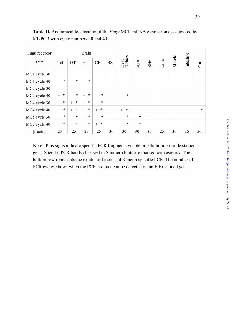
39
Table II. Anatomical localisation of the Fugu MCR mRNA expression as estimated byRT-PCR with cycle numbers 30 and 40.
BrainFugu receptorgene Tel OT HT CB BS
Hea
dK
idne
y
Eye
Skin
Live
r
Mus
cle
Inte
stin
e
Gut
MC1 cycle 30
MC1 cycle 40 * * *
MC2 cycle 30
MC2 cycle 40 + * * + * * *
MC4 cycle 30 + * + * + * + *
MC4 cycle 40 + * + * + * + * + * *
MC5 cycle 30 * * * * * *
MC5 cycle 40 + * * + * + * * *
β-actin 25 25 25 25 30 30 30 35 25 30 35 30
Note: Plus signs indicate specific PCR fragments visible on ethidium bromide stainedgels. Specific PCR bands observed in Southern blots are marked with asterisk. Thebottom row represents the results of kinetics of β- actin specific PCR. The number ofPCR cycles shows when the PCR product can be detected on an EtBr stained gel.
by guest on July 27, 2015http://m
be.oxfordjournals.org/D
ownloaded from

by guest on July 27, 2015http://m
be.oxfordjournals.org/D
ownloaded from

by guest on July 27, 2015http://m
be.oxfordjournals.org/D
ownloaded from

by guest on July 27, 2015http://m
be.oxfordjournals.org/D
ownloaded from

by guest on July 27, 2015http://m
be.oxfordjournals.org/D
ownloaded from

by guest on July 27, 2015http://m
be.oxfordjournals.org/D
ownloaded from

by guest on July 27, 2015http://m
be.oxfordjournals.org/D
ownloaded from

by guest on July 27, 2015http://m
be.oxfordjournals.org/D
ownloaded from

by guest on July 27, 2015http://m
be.oxfordjournals.org/D
ownloaded from

by guest on July 27, 2015http://m
be.oxfordjournals.org/D
ownloaded from