The mandibles of some adult ground beetles: structure, function, and the evolution of herbivory...
13
The mandibles of some adult ground beetles: structure, function, and the evolution of herbivory (Coleoptera: Carabidae) JOHN H. ACORN 15714 86 Avenue, Edmonton, Alta., Canada T5R 4C4 AND GEORGE E. BALL' Department of Entomology, University of Alberta, Edmonton, Alta., Canada T6G 2E3 Received April 17, 1990 ACORN, J. H., and BALL, G. E. 199 1. The mandibles of some adult ground beetles: structure, function, and the evolution of herbivory (Coleoptera: Carabidae) . Can. J . 2001. 69: 638-650. The structure of carabid and other adephagan mandibles can provide information about the mechanics of feeding in these beetles and about the major features of carabid and adephagan evolution. Homologies among mandibular features are proposed, based on a transformation series for adult mandibles from plesiotypic cupedid-like structure to derived conditions characteristic of herbivorous carabids in the subtribe Harpalina. Terms for mandibular parts are revised so that all ridges and teeth are referred to as such. The following terms are introduced: superior and inferior terebral ridges, superior and inferior retinacular ridges, supraterebral ridges, anterior and posterior occlusal grooves, and basal face. Masticatory function is elucidated for the mandibles of adult Euryderus grossus (Say) in which the mandibles shear food matter in both a horizontal and a vertical plane, as well as compacting food into a bolus that is forced posteriorly into the buccal cavity. Many of the features of E. grossus mandibles are characteristic of other herbivorous carabids, but not all such taxa show such derived mandibular structure. ACORN, J. H., et BALL, G. E. 1991. The mandibles of some adult ground beetles: structure, function, and the evolution of herbivory (Coleoptera: Carabidae). Can. J. 2001. 69 : 638-650. Les structure des mandibules des carabides et d'autres adephages peut jeter de la lumiitre sur les mecanismes d'alimentation de ces coleoptkres et sur les principales etapes de l'evolution des carabides et des adephages. L'examen d'une serie evolutive de mandibules adu'ltes, partant de mandibules de structure plesiotypique de type cupedide a des mandibules evoluees caracteris- tiques de carabides herbivores de la sous-tribu des Harpalina, nous permet de proposer des homologies. Les termes relatifs aux structures mandibulaires sont redefinis de fason a ce que toutes les cretes et toutes les dents aient leur nom. Les termes suivants sont nouveaux : cretes terebrales superieure et inferieure, crCtes retinaculaires superieure et inferieure, cretes supraterebrales, sillons d'occlusion anterieur et popsterieur et face basale. Le mecanisme de la mastication a ete compris chez l'adulte d'Euryderus grossus (Say); les mandibules dechirent les ailments a la fois dans un plan vertical et dans un plan horizontal, et elles entassent les particules alimentaires en un bolus qui est pousse vers I'arrikre dans la cavite buccale. Plusieurs caracteristiques des mandibules d'E. grossus se retrouvent chez d'autres carabides herbivores, mais tous ces taxons n'ont pas necessairement une structure mandibulaire aussi evoluee. [Traduit par la redaction] Introduction establish the classification of many mammalian groups, both Specialists on carabid beetles have shown interest in both the functional aspects of mandibular structure in adult carabid beetles (Forsythe 1982; Evans and Forsythe 1985) and the use of mandibular features as character systems in systematic work (for example, Ball 1959, 1978). In this context, it is of great benefit to students of the Carabidae to have ,the work of mammalogists to look to for inspiration, since mammalogists have a sophisticated understanding of dental structure, function, and evolution in the animals they study. In mammals, tooth structure is virtually invariant within species, and among mammals one sees many examples of structural specialization for eating particular sorts of food. For example, many carnivorous mammals possess a scissor-like pair of shearing teeth used to slice through flesh. Many grazing ungulate mammals have broad, flattened cheek teeth used to grind tough plant material. In contrast, some lemuroid primates have their front teeth developed into a comb-like structure used in grooming. For a review of the structure, occlusal relation- ships, and early evolution of the dentition of therian mammals, see Crompton (1971). For a review of dental structure and evolution in the extinct multituberculate mammals. see Krause fossil and extant. Carabid (and most other arthropod) mandibles and mammal- ian dentitions are similar in that they both involve complex, hardened surfaces that occlude in a precise way and that do not grow once they have begun their functional lives. However, wear facets, which are of great importance to mammalogists, are not well defined on carabid mandibles, possibly because the functional lives of the latter are generally shorter than in mammals, and hence carabid mandibles wear deeply and do not develop shallow, polished facets. Since the work of Forbes (1883), it has been recognized that carabid mandibular form is related to feeding habits. The general mode of action of each of the mouthparts in adult carabids has also been elucidated (Evans 1965 a, 1965 b; Forsythe 1982). However, the details of how various ridges and projections on the mandibles of these beetles function in the processing of food have not been examined carefully. Certain systematists, begin- ning with Jeanne1 (1926) and Ball (1959), have provided detailed descriptions of the structure of carabid mandibles in order to use mandibular features as character systems, but without attempting to elucidate the functions of the characters (1981). The precise functional relationships between the cusps, used. Here, structural and functional aspects of the mandibles of crests, and basins of mammal teeth have been investigated, and these features have been used, exclusively or with others, to adult carabid beetles are described and a hypothesis is proposed about their evolutionary history. Many of the beetles chosen for '~uthor to whom correspondence should be addressed. this study are herbivorous (mainly feeding on seeds) and are Can. J. Zool. Downloaded from www.nrcresearchpress.com by University of Alberta on 10/05/12 For personal use only.
Transcript of The mandibles of some adult ground beetles: structure, function, and the evolution of herbivory...

The mandibles of some adult ground beetles: structure, function, and the evolution of herbivory (Coleoptera: Carabidae)
JOHN H. ACORN 15714 86 Avenue, Edmonton, Alta., Canada T5R 4C4
A N D
GEORGE E. BALL'
Department of Entomology, University of Alberta, Edmonton, Alta., Canada T6G 2E3
Received April 17, 1990
ACORN, J . H., and BALL, G . E. 199 1. The mandibles of some adult ground beetles: structure, function, and the evolution of herbivory (Coleoptera: Carabidae) . Can. J . 2001. 69: 638-650.
The structure of carabid and other adephagan mandibles can provide information about the mechanics of feeding in these beetles and about the major features of carabid and adephagan evolution. Homologies among mandibular features are proposed, based on a transformation series for adult mandibles from plesiotypic cupedid-like structure to derived conditions characteristic of herbivorous carabids in the subtribe Harpalina. Terms for mandibular parts are revised so that all ridges and teeth are referred to as such. The following terms are introduced: superior and inferior terebral ridges, superior and inferior retinacular ridges, supraterebral ridges, anterior and posterior occlusal grooves, and basal face. Masticatory function is elucidated for the mandibles of adult Euryderus grossus (Say) in which the mandibles shear food matter in both a horizontal and a vertical plane, as well as compacting food into a bolus that is forced posteriorly into the buccal cavity. Many of the features of E. grossus mandibles are characteristic of other herbivorous carabids, but not all such taxa show such derived mandibular structure.
ACORN, J. H., et BALL, G . E. 1991. The mandibles of some adult ground beetles: structure, function, and the evolution of herbivory (Coleoptera: Carabidae). Can. J. 2001. 69 : 638-650.
Les structure des mandibules des carabides et d'autres adephages peut jeter de la lumiitre sur les mecanismes d'alimentation de ces coleoptkres et sur les principales etapes de l'evolution des carabides et des adephages. L'examen d'une serie evolutive de mandibules adu'ltes, partant de mandibules de structure plesiotypique de type cupedide a des mandibules evoluees caracteris- tiques de carabides herbivores de la sous-tribu des Harpalina, nous permet de proposer des homologies. Les termes relatifs aux structures mandibulaires sont redefinis de fason a ce que toutes les cretes et toutes les dents aient leur nom. Les termes suivants sont nouveaux : cretes terebrales superieure et inferieure, crCtes retinaculaires superieure et inferieure, cretes supraterebrales, sillons d'occlusion anterieur et popsterieur et face basale. Le mecanisme de la mastication a ete compris chez l'adulte d'Euryderus grossus (Say); les mandibules dechirent les ailments a la fois dans un plan vertical et dans un plan horizontal, et elles entassent les particules alimentaires en un bolus qui est pousse vers I'arrikre dans la cavite buccale. Plusieurs caracteristiques des mandibules d'E. grossus se retrouvent chez d'autres carabides herbivores, mais tous ces taxons n'ont pas necessairement une structure mandibulaire aussi evoluee.
[Traduit par la redaction]
Introduction establish the classification of many mammalian groups, both
Specialists on carabid beetles have shown interest in both the functional aspects of mandibular structure in adult carabid beetles (Forsythe 1982; Evans and Forsythe 1985) and the use of mandibular features as character systems in systematic work (for example, Ball 1959, 1978). In this context, it is of great benefit to students of the Carabidae to have ,the work of mammalogists to look to for inspiration, since mammalogists have a sophisticated understanding of dental structure, function, and evolution in the animals they study.
In mammals, tooth structure is virtually invariant within species, and among mammals one sees many examples of structural specialization for eating particular sorts of food. For example, many carnivorous mammals possess a scissor-like pair of shearing teeth used to slice through flesh. Many grazing ungulate mammals have broad, flattened cheek teeth used to grind tough plant material. In contrast, some lemuroid primates have their front teeth developed into a comb-like structure used in grooming. For a review of the structure, occlusal relation- ships, and early evolution of the dentition of therian mammals, see Crompton (1971). For a review of dental structure and evolution in the extinct multituberculate mammals. see Krause
fossil and extant. Carabid (and most other arthropod) mandibles and mammal-
ian dentitions are similar in that they both involve complex, hardened surfaces that occlude in a precise way and that do not grow once they have begun their functional lives. However, wear facets, which are of great importance to mammalogists, are not well defined on carabid mandibles, possibly because the functional lives of the latter are generally shorter than in mammals, and hence carabid mandibles wear deeply and do not develop shallow, polished facets.
Since the work of Forbes (1 883), it has been recognized that carabid mandibular form is related to feeding habits. The general mode of action of each of the mouthparts in adult carabids has also been elucidated (Evans 1965 a , 1965 b; Forsythe 1982). However, the details of how various ridges and projections on the mandibles of these beetles function in the processing of food have not been examined carefully. Certain systematists, begin- ning with Jeanne1 (1926) and Ball (1959), have provided detailed descriptions of the structure of carabid mandibles in order to use mandibular features as character systems, but without attempting to elucidate the functions of the characters
(198 1). The precise functional relationships between the cusps, used. Here, structural and functional aspects of the mandibles of crests, and basins of mammal teeth have been investigated, and
these features have been used, exclusively or with others, to adult carabid beetles are described and a hypothesis is proposed about their evolutionary history. Many of the beetles chosen for
' ~ u t h o r to whom correspondence should be addressed. this study are herbivorous (mainly feeding on seeds) and are
Can
. J. Z
ool.
Dow
nloa
ded
from
ww
w.n
rcre
sear
chpr
ess.
com
by
Uni
vers
ity o
f A
lber
ta o
n 10
/05/
12Fo
r pe
rson
al u
se o
nly.

ACORN AND BALL 639
thus in a minority among the mainly carnivorous adults of the Carabidae. Taxa with herbivorous members are found primarily in the tribes Harpalini and Zabrini, and a review of the species involved has been prepared by Johnson and Cameron (1969). Structural modification of the mandibles for seed eating was recognized by Forsythe (1982) who noted that "if the mandibles are short, quadrate, blunt at the tips and provided with either strong basal projections or broad opposed surfaces, vegetable food predominates. Anisodactylus [a member of the Harpalini] is an example of this class."
There are three objectives to this paper. The first is to review the present system of nomenclature for mandibular features and provide new or additional names where needed. The second is to describe the function of various structural features of the mandibles of adult herbivorous carabids during the mastication of food. The third is to use this information to propose a hypothesis about the evolution of mandibular form in the Carabidae, augmented by a general survey of mandibular form in the suborder Adephaga as a whole.
Materials and methods Scanning electron micrographs were taken of the mandibles of adults
of various adephagan beetles. For each mandible, photographs were taken of three views: occlusal (lateral, facing the masticating surface), dorsal, and ventral. Species were chosen to represent (i) the tribe Harpalini and the genus Amara Bonelli (tribe Zabrini), (ii) groups whose characteristics are generally cynsidered similar to the basal lineages of carabids, (iii) Agonum errans (Say), of the tribe Platynini, considered generally to be related to the Zabrini but not expected to show adaptations for herbivory, and (iv) representatives of the non-carabid adephagan families. For each species, one set of man- dibles was used, with the exception of the sexually dimoiphic species Cratacanthus dubius Palisot des Beauvois, for which mandibles from one male and one female were used.
Cleared specimens were examined of the mandibles of Euryderus grossus (Say), Harpalus paratus Casey, Trachypachus holmbergi Mannerheim, and a species of Notiophilus Dumkril.
Numerous specimens of Euryderus grossus and Harpalellus basil- aris (Kirby) were used for dissections of the head, from which the labrum, clypeus, maxillae, and labium were removed to observe more closely the pattern of occlusion of the mandibles. Camera lucida drawings were made of various stages of mandibular adduction from both dorsal and ventral views.
As well, on similarly dissected specimens of Euryderus grossus, the mandibles were made to occlude on a piece of plasticine to determine whether a food pellet was formed between the two main ridges of the mandibular face.
Comparisons of mandibular structures were made primarily from the scanning electron micrographs and to a lesser extent from examination of dissected specimens under a dissecting microscope.
To arrive at an evolutionary analysis, a transformation series was constructed, using both the scanning electron microscope photographs and various authors' previous work on adephagan and carabid phylogeny as guides. Homologies of mandibular parts were determined both by similarity of function and by comparison of mandibular structures in the context of the transformation series.
The nomenclature of mandibular features Numerous terms have been proposed for the parts of carabid
mandibles, and some new terms are proposed here. These names are presented below, and justification for this system of names is provided elsewhere in the text.
Currently, the names of carabid mandibular parts are based on the idea that the occluding face of the mandible consists of a series of parallel, oblique ridges produced or not into teeth along their margins. From anterodorsal to posteroventral these ridges
are the terebral margin, the retinacular ridge, the premolar ridge, and the molar ridge (Jeannel 1926; Ball 1959, 1978). The terebral margin may be produced into a terebral tooth near its posterior end, the retinacular ridge may bear an anterior and (or) a posterior retinacular tooth, and the premolar and molar ridges may bear premolar and molar teeth, respectively, or be reduced to these teeth.
In adults of Trachypachus Motschulsky , both the terebral ridge and the retinacular ridge are crescentic, consisting of elongate dorsal and short ventral ridges connected at their apices (Figs. 1, 4D, and 9). Hence, we distinguish the "superior terebral ridge," "inferior terebral ridge," "superior retinacular ridge," and "inferior retinacular ridge." In hydradephagans, the inferior ridges are developed prominently, although it is the superior ridges that become most prominent in the Carabidae. We refer to the ridges and their associated projections by the composite terms "terebra" and "retinaculum," respectively.
One improvement to the current system of nomenclature that immediately suggests itself is that all of the ridges be termed "ridges" and that all of the tooth-like projections be termed "teeth." Accordingly, the terebral margin is renamed the terebral ridge. This system necessitates the terms "premolar tooth" and "molar tooth," since it is useful to distinguish between teeth and ridges when these are not one and the same, and this cannot be done with the simple terms "premolar" and "molar." A possible objection to this system might be that since the so-called teeth are simply projections and not discrete structures, they should be termed cusps instead. However, if one refers to the mandible of a beetle and the mandible of a vertebrate, then it would seem desirable to continue this analogy and to refer to the major masticatory projections of the mandible as teeth. If any of these teeth were found to possess projections of their own, these could in turn be called cusps.
The area posterior and ventral to the base of the retinaculum is here named the "basal face." In adults of most groups it is flattened occlusally and consists of unthickened cuticle with no projections.
The "supraterebral ridges" are two short ridges that extend from the base of the terebral tooth onto the dorsal face of the mandible. Ball (1 978), in his study of the Neotropical harpaline genus Trichopselaphus Chaudoir, has interpreted these as the true terebral margin of the left mandible, the main cutting edge of the mandible being the retinacular ridge. However, it is clear that these are simply superficial features of the terebral ridge, as seen in Euryderus, where the anterior supraterebral ridge is pronounced (Figs. 2, 4N, and 17).
The anterior and posterior occlusal grooves separate the posterior retinacular tooth from the premolar and the premolar from the molar, respectively (for example, mandibles of Amara torrida Panzer, Fig. 14). When only one groove is present it separates the posterior retinacular tooth from the molar.
Table 1 summarizes the nomenclature of mandibular features and allows comparison of the system proposed here with that of Ball (1959 and subsequent papers) and Jeannel (1926). Most of the feature listed here can be identified on mandibles of Trachypachus holmbergi Mannerheim (Fig. 1) and Euryderus grossus (Fig. 2).
Although various non-carabid adephagans were also exam- ined, no attempt was made to review the nomenclature of mandibular features for these beetles.
Functional significance of mandibular features To appreciate more fully the structure of carabid mandibles it
Can
. J. Z
ool.
Dow
nloa
ded
from
ww
w.n
rcre
sear
chpr
ess.
com
by
Uni
vers
ity o
f A
lber
ta o
n 10
/05/
12Fo
r pe
rson
al u
se o
nly.

CAN. J . ZOOL. VOL. 69, 1991
irr
"g
FIG. 1 . Mandibles of adult Trachypachus gibbsi LeConte, showing named mandibular features and areas of contact with opposite mandible - (cross-hatched): (A) right dorsal; (B) right occlusal; (C) left occlusal; (D) left dorsal. Abbreviations: art, anterior retinacular tooth; asr, anterior
supraterebral ridge; bb, basal brush; bf, basal face; dc, dorsal crenulations; ir, incisor ridge; itr, inferior terebral ridge; m, molar tooth; pm, premolar tooth; pog, posterior occlusal groove; prt, posterior retinacular tooth; rr, retinacular ridge; srr, superior retinacular ridge; str, superior terebral ridge; tt, terebral tooth; vg, ventral groove. FIG. 2. Mandibles of adult Euryderus grossus (Say), showing named mandibular features and areas of contact with opposite mandible (cross-hatched): (A) right dorsal; (B) right occlusal; (C) left occlusal; (D) left dorsal. Abbreviations are the same as in Fig. 1.
is instructive to examine the function of the various mandibular parts, as illustrated by Euryderus grossus, a harpaline whose robust adults live on sandy areas in the Great Plains of North America. These beetles are large and therefore easy to dissect, and they show most of the occlusal surfaces, except the premolar, characteristic of mandibles of herbivorous carabids. Therefore, all other carabids examined differ from E. grossus by the lack or modification of one or more features seen in this species.
In carabids, the mandibles are the primary means of masticat- ing food. They are articulated in typical beetle fashion by means of two ball and socket joints, one dorsal and one ventral, near the base of each mandible. Therefore, each mandible moves only in one plane, which is more or less horizontal. Each mandible is controlled by two muscles. The largest of these is an adductor, which pulls the mandible toward the midline and toward the other mandible. The other muscle is an abductor, which swings the mandible outward, thus opening a space between the jaws. Each mandible can therefore move indepen- dently. In summary, this simple system consists of two mandibles, each having only one axis of rotation and each controlled by two muscles. As an anonymous reviewer has
noted, however, this allows occlusion in the mid-sagittal line or, if one mandible is adducted more than the other, at an angle to the mid-sagittal line.
These limitations to the range of movement of the mandibles allows diversification of the masticatory system to occur only with respect to the shape of the mandibles themselves. In general, processing of food can be achieved in a number of ways, all of which involve occlusion of two mandibles. These include (i) cutting, where two ridges shear past one another (analogous to scissors), (ii) grinding, where a projection occludes into a basin (analogous to a mortar and pestle), (iii) compressing, where two basins occlude together (analogous to making snowballs with the hands), and ( iv) crushing, where two more or less flat surfaces are pressed together (analogous to accidentally stepping on a fine carabid beetle while wearing dress shoes on a smooth sidewalk).
In E. grossus, the function of the mandibles appears to be both cutting and compressing. The main cutting surface is extended along the dorsal inside margin of each mandible. These terebral ridges are characteristic of all carabids. As the mandibles are brought together, these two ridges shear past each other, first meeting near the apices of the mandibles. Food is
Can
. J. Z
ool.
Dow
nloa
ded
from
ww
w.n
rcre
sear
chpr
ess.
com
by
Uni
vers
ity o
f A
lber
ta o
n 10
/05/
12Fo
r pe
rson
al u
se o
nly.

ACORN AND BALL 64 1
TABLE 1 . Comparison of nomenclature used in this paper and that used by Ball (1959) and Jeannel (1926)
This paper Ball ( 1959) and Jeanne1 ( 1926)
Terebra Incisor region Superior terebral ridge Inferior terebral ridge Supraterabral ridges Terebral tooth
Retinaculum Superior retinacular ridge Inferior retinacular ridge Anterior retinacular tooth Posterior retinacular tooth
Anterior occlusal groove Posterior occlusal groove Premolar ridge Premolar tooth Molar ridge Molar tooth Dorsal crenulations* Basal brush Ventral groove Basal face
No change Terebral margin Not recognized Not recognized No change
Retinacular ridge Not recognized No change No change Not recognized Not recognized No change No change No change No change Not recognized No change No change Not recognized
*Dorsal crenulations were recognized and named by Noonan ( 1973).
L
then cut in a scissor-like fashion. This action is also likely to push material that is difficult to shear back into the mouth. How- ever, the basal portion of each terebral ridge, especially the left, juts out to form a terebral tooth that prevents food from being forced back along the terebral ridges any farther during mandi- bular adduction. The terebral teeth are positioned far enough back on the mandibles that they are covered by the labrum, and therefore are probably not used to grip food, but rather are used only to prevent tough material from sliding back into the upper part of the buccal cavity. The function of the terebral ridges is illustrated by sequential views of mandibular adduction (Fig. 3). In E. grossus, the right mandible occludes under the left and this makes the shearing surfaces the upper edge of the right terebral ridge and the lower edge of the left terebral ridge, although in some carabids the right mandible is uppermost (Wheater 1988).
There is another area of shear on the mandibles of this species. The apex of each mandible is turned inward and forms a chisel-shaped incisor region in which the axis of the shearing ridge is roughly vertical. The outer face of the incisor region of the right mandible cuts against the inner face of the incisor region of the left. Again, the function of this area of the mandibles is quite obvious from sequential views of mandibular adduction. As well, when the gape of the mandibles is wide, the incisor teeth may prevent seeds and large food items from popping out of the mandibular grasp during adduction.
Thus, there are two areas of shear, one in a horizontal plane (the terebral ridges) and one in a vertical plane (the incisor regions). Presumably, the beetle is thus able to effectively chop its food into small particles prior to swallowing it.
Ventral to the terebral ridges on both mandibles, the retinacular ridge is separated from the terebral ridge by a deep basin. The left retinacular ridge is somewhat crescentic in occlusal view (Fig. 2B) and thus is closer to the terebral ridge at its anterior and posterior ends. During mandibular adduction, these ridges do not seem to shear past each other to any great
extent (although the retinacula may form a secondary shearing surface in such taxa as Harpalus Fabricius). The function of the retinacular ridge seems to be to form the ventral closure of the compacting basin. As the mandibles come together, food is trapped between the terebral ridges above and the retinacular ridges below and compressed while being forced posteriorly into the buccal cavity. This was easily seen by manipulation of the mandibles of a dead specimen so as to occlude on a piece of plasticine. When excess plasticine was moved from the outsides of the mandibles, as food would be by the action of the terebral ridges, labrum, and maxillae, a small pellet was formed in the compacting basin.
However, it is apparent that this compacting basin may be restricted to a few harpalines (Euryderus, Piosoma Leconte and possibly Cratacanthus) and is not a general feature of herbiv- orous carabids.
The retinacular ridge serves another function by preventing the mandibles from adducting past a certain point. The right retinacular ridge abuts below and against the left during closure of the compacting basin and prevents further adduction of the mandibles.
The ventral groove contains a row of short microtrichia and is situated lateral to the retinacular ridge and parallel to the long axis of the mandible. The function of this line of microtrichia is probably to prevent food from slipping between the mandibles and the maxillae (Evans 1965a, 19656). It could also function to draw digestive fluids distally, or fluids to be ingested proximally, along the mandibles. The microtrichia of the ventral groove and basal brush are not socketed and therefore are not setae (D. R. Maddison, personal communication), contrary to Forsythe ( 1982). An anonymous reviewer suggested that they are most likely seta-like sculpticells.
Posterior to the terebral margin, the basal brush consists of a row of fairly elongate microtrichia that are directed somewhat posteriorly toward the buccal cavity. The function of this structure may be to push food farther into the mouth during mandibular adduction. In other insect groups, this structure is often called the prostheca.
Parallel to the retinacular ridges and posterior to them is a groove, the occlusal groove, extended obliquely across the occlusal face of the mandible. Beetles possessing a premolar tooth or ridge that is distinct from the posterior retinacular tooth have an anterior occlusal groove, which in most adult carabids is less pronounced than the posterior one. These grooves may function to allow excess liquids produced by mastication of food to be released from the confines of the compacting basin. It is also possible that liquids may be trapped between the dorsal surfaces of the mandibles and the ventral surface of the labrum, and the occlusal grooves would allow these liquids to enter the buccal cavity. In species in which extra-intestinal digestion occurs, such large volumes of liquid and semi-liquid food would constitute the normal sort of material being processed by the mouthparts.
The three small teeth at the posterior end of the mandible, the posterior retinacular tooth, the premolar (not present in E. grossus), and the molar, all seem not to occlude with any- thing. They may serve to push food into the mouth and hold it during adduction of the jaws.
Posterior to the molar, the occlusal face of the mandible is flat and featureless. In cleared specimens, it is apparent that the cuticle of this region is very thin and probably could not hold up under any occlusal pressures. Thus, it is likely that this area,
Can
. J. Z
ool.
Dow
nloa
ded
from
ww
w.n
rcre
sear
chpr
ess.
com
by
Uni
vers
ity o
f A
lber
ta o
n 10
/05/
12Fo
r pe
rson
al u
se o
nly.

CAN. J . ZOOL. VOL. 69, 1991
FIG. 3 . Sequential views of mandibular occlusion in adult Euryderus grossus (Say): left, dorsal views; right, ventral views.
Can
. J. Z
ool.
Dow
nloa
ded
from
ww
w.n
rcre
sear
chpr
ess.
com
by
Uni
vers
ity o
f A
lber
ta o
n 10
/05/
12Fo
r pe
rson
al u
se o
nly.

ACORN AND BALL
FIG. 4. Hypothetical transformation series of mandibular forms: (A) Priacma; (B) Clinidium; ( C ) Elaphrus; ( D ) Trachypachus; ( E ) Haliplus; ( F ) Amphizoa; (G) Dineutes; (H) Agonum; ( I ) Stenolophus; ( J ) Amara; ( K ) Geopinus; ( L ) Lecanomerus; ( M ) Harpalellus; ( N ) Euryderus. The terebrum is black and the retinaculum is cross-hatched.
named here the basal face, does not function in the processing of food.
Homologies of mandibular features and a hypothetical transformation series
With a consistent system of nomenclature and an understand- ing of the functions of mandibular features, it becomes possible to consider homologies among them. Homology is postulated when a structure bears a distinct resemblance to another and the two are located similarly with respect to other features. In carabid mandibles there ,are three types of homologies to consider. The first is homology of structures between taxa, the second is homology of structures between right and left mandibles, which is a kind of serial homology, and the third is serial homology of structures within a single mandible.
Homologies of parts between the right and left mandibles are generally easy to discern, and especially so in occlusal view, since the two mandibles form near mirror images of one another. Hence, in the following discussion all references apply equally to right and left mandibles unless otherwise noted. In general, since the right mandible occludes below the left and
therefore does not have to accommodate its shape to that of the left mandible as much as vice versa, its structure is less derived.
To illustrate the evolution of carabid mandibles, a hypotheti- cal transformation series was constructed. At first, we assumed that the mandibles of Trachypachus could serve as a ground plan for those of the Carabidae, since Trachypachus is generally considered to be the most primitive living carabid or the most primitive living member of the sister group of the Carabidae (Bell 1982), but it soon became apparent that Trachypachus shows mandibular similarities with the Hydradephaga not shared with carabids. Thus, we examined mandibles of one member of each of a number of families in the suborder Adephaga, as well as one cupedid. As a result, the presumed plesiotypic condition seen in Priacma LeConte (a cupedid, and a member of the primitive suborder Archostemata) was used as a starting point for the transformation series (Fig. 4). The transformation series itself should not be mistaken for a reconstructed phylogeny; each taxon illustrated is representative of a hypothesized stage in the evolution of mandibles, not the ancestry of the next most derived taxon in the series. A summary of mandibular features of all taxa examined in this study is provided in Table 2.
Can
. J. Z
ool.
Dow
nloa
ded
from
ww
w.n
rcre
sear
chpr
ess.
com
by
Uni
vers
ity o
f A
lber
ta o
n 10
/05/
12Fo
r pe
rson
al u
se o
nly.

CAN. 1. ZOOL. VOL. 69. 1991
TABLE 2. Mandibular features of all taxa examined in this study - - -
Taxon tr rr vg 1s sls vs pm m art prt tt cd str
Priacma serrata LeC . * Clinidium sp. * Din. americanus (Say)* Amph. lecontei Matt. * Agab. antennatus Leech Haliplus sp. Trach. holmbergi Mnh. Trach. gibbsi LeC. * Elaph. lecontei Crotch* Notiophilus sp. * Omoph. americanum Dej . Nebria sp. Agonum errans (Say)* Amara scitula Zimm . * Amara torrida Panz. * Sten. lineola (Fab.)* Sten. comma (Fab.) Lecan . niger (Darl . ) * Pelmat. obtusus Bates Polp. capitata (Chd.) Eury. grossus (Say)* Piosoma setosum LeC . Geo. incrassatus (Dej . ) * Harpal. basilaris (Kby . ) * Harp. paratus Csy . H a g . leiroides (Motsch.) Harp. caliginosus Fab. Harp. amputatus Say Allendia chilensis (Sol.) Crat. dubius (P. d'B.-)*
m a a p a a a a a a p ? a a s a a a p a a a a a p a a d v a p a a a a a a a p a a d v i e p a p a a p ? a p a a d v i e p a p a a p ? a p a a m s e p a a a a p ? a p a a m r p a a a a p p a p a a m r p a a a a p p a p a a s s P a P a P P P P P ? ' a s s p a p a a a p p p a a s s P a P a a P P P P P a s s p a p a a a p p p a a s s p a a a p p p p p a a s s P a a a P P P P P P P s s p a a a p P P P P P P s s P a a a a P P P P a P s s p a a a a P P P P a P s s p a a a a p p a p p a s s p a a a a p p p p a a s s p a a a a p p p p a a m c p a a a a P P P P P P m c p a a a a P P P P P P s s p a a a a p p a p p a s s p a a a a p p a p p a s s p a a a a P P P P a P s s p a a a a P P P P P P s s p a a a a P P P P a P s s p a a a a P P P P P P s s p a a a a P P P P a P s s p a a a a P P P P P P
N O T E : t r , terebral ridge (s, simple and more or less straight; m , with moderate dorsoventral extension of incisor region; dv , entire ridge oriented dorsoventrally rather than anteroposteriorly); rr , retinacular ridge ( a , absent; s , simple, consisting of the superior ridge; r , consisting of both inferior and superior ridges; i, consisting of inferior ridge only; c , forming a compacting basin); vg, ventral groove ( a , absent; p, present; e , elongate with very dense microtrichia); Is, setae on lateral mandibular face; sls, single large seta on lateral face of mandible; vs, ventral setal patch; pm, premolar tooth; m, molar tooth; art , anterior retinacular tooth; prt, posterior retinacular tooth; t t , terebral tooth; dc , dorsal crenulations; str, supraterebral ridges: a , absent; p, present.
*Scanning electron micrographs are provided.
The mandibles of Priacrna (Figs. 4A and 5 ) are structurally simple, with a single, undulating shearing surface (the terebral ridge). Unlike rhysodids and carabids, but like amphizoids and dytiscids, they have a dense field of setae on the lateral face of each mandible; otherwise it is possible to derive from them the mandibular forms seen in both rhysodids and primitive carabids.
The rhysodid Clinidurn Kirby (Figs. 4B and 6 ) possesses mandibles that at first glance appear quite unlike those of other adephagans. In occlusal view, however, it becomes apparent that they possess only one shearing surface, which we presume to be the terebral ridge. The unusual lateral projections of these mandibles are unlike anything seen in the rest of the Adephaga. These do not look like the mandibles of primarily insectivorous carabids, and they probably do not function similarly either.
The mandibles of adult Elaphrus Fabricius (Figs. 4C and 10) are considered' to represent a structural ground plan for the Carabidae, and their form can be derived easily from that of Priacrna. This stage in the transformation series involves the development of a retinacular ridge ventral to the terebral ridge. The mandibles of Ornophron Latreille, Notiophilus, and Opis- thius Kirby are similar to those of Elaphrus.
In Elaphrus, there are two posterior teeth and one anterior tooth on the retinacular ridge. The anterior tooth, by its position, is clearly the anterior retinacular tooth. The posterior teeth are the posterior retinacular tooth and the premolar or molar. If the
premolar and molar arise phylogenetically as part of the retinacular ridge, then they should be referred to as teeth rather than ridges. However, there is another small tooth between the terebral tooth and retinacular ridge in Elaphrus lecontei (this tooth is not found in all members of the genus) and the identity of this tooth is uncertain. Hence, this problem is not solved by examination of representatives of this genus.
From the mandibular form seen in Elaphrus to that of Trachypachus, the main change is one of reflexion of the terebral and retinacular ridges into J - and U-shaped structures, respectively, thus increasing the dorsoventral extent of the occlusal face. In Trachypachus mandibles, the basal and apical mandibular features are strongly similar and may thus be serially homologous. This is most apparent in the right man- dible. In occlusal view the terebra and retinaculum markedly resemble one another. If the retinaculum and terebra are serially homologous, then the incisor region of the terebra is homo- logous with the anterior retinacular tooth, and the terebral tooth is serially homologous with the posterior retinacular tooth. This is best seen in occlusal view of the right mandible of Trachypachus (Fig. 9F).
Both Trachypachus and Elaphrus have the superior terebral ridge produced into a terebral tooth near its basal end. Compared with Trachypachus, the general shape of Elaphrus mandibles shows the same pattern in serial repetition of
Can
. J. Z
ool.
Dow
nloa
ded
from
ww
w.n
rcre
sear
chpr
ess.
com
by
Uni
vers
ity o
f A
lber
ta o
n 10
/05/
12Fo
r pe
rson
al u
se o
nly.

ACORN AND BALL 645
structures, and the homologies of the retinaculum and terebra between Elaphrus and Trachypachus seems clear. However, the increasing dorsoventral extent of Trachypachus mandibles is reminiscent of that seen in most hydradephagans and quite unlike anything in the Carabidae sensu stricto. Hence, adult Trachypachus mandibles may represent a model for the primi- tive condition for mandibles within the Hydradephaga.
The terebral ridge of Trachypachus is inclined anteroven- trally to posterodorsally; this arrangement is also seen in Haliplus Latreille (Fig. 4E). The latter taxon, however, possesses the simplest of retinacula, which is untoothed and straight. The condition seen in Haliplus could be derived from that of Trachypachus, but it is more parsimonious to suggest that it evolved from an Elaphrus-like form. The latter possibility would involve less structural change and moderate rather than extreme reduction of the retinaculum.
From Trachypachus, we can easily derive the structure of the mandibles of Amphizoa (Fig. 4F), which were found to be similar to those of Agabus, a dytiscid. It appears that the retinaculum is reduced to the inferior retinacular ridge, and that the terebral ridge has become almost vertical in orientation in both genera. The function of this arrangement is unknown and may be related to underwater feeding. The mandibles of Dineutes MacLeay, a gyrinid, are generally similar to those of Amphizoa but lack a retinaculum (Figs. 4G and 7).
If the lack of retinaculum is indeed a derived condition in the Gyrinidae, then the suggestion that this family constitutes the sister group to the rest of the Adephaga, including rhysodids but not cupedids (Beutel and Roughley 1988), can be called into question. The general similarity of mandibular form suggests strongly that the gyrinids belong in a clade with the dytiscids and amphizoids and probably the trachypachids as well. Obviously though, this issue cannot be settled on any one sort of evidence and our purpose here is simply to bring the mandibular character set to the attention of workers on the higher classification of the Hydradephaga.
Returning to the Carabidae, the pattern of mandibular structure characteristic of Elaphrus is little modified in Agonum errans (Say). The structure of Agonum mandibles is essentially very simple (Figs. 4H and 12). The terebral and retinacular ridges are straight, almost parallel structures. The mandible is elongate and pointed, and a small molar or premolar projection is situated posterior to the retinacular ridge.
From the condition seen in Agonum to that in Stenolophus Dejean (Figs. 41 and 15), there is again little change. Steno- lophus is thus considered here to be a structurally primitive harpaline with respect to mandibular features. Stenolophus mandibles are only slightly more robust than those of Agonum, although Stenolophus comma Fabricius is a well-known seed eater that in some areas of the United States is known to feed on corn.
The mandibles of adult Amara show a number of modifica- tions from this basic pattern, and presumably these modifica- tions are adaptations for 'herbivory. The left terebral ridge, in occlusal view, is only slightly sinuate in Stenolophus but is more so in Amara. This may serve to strengthen the mandible and is seen in many other harpalines besides Stenolophus. The mandibles are more robust and show a strengthening of the incisor region to form a nipping surface, allowing vertical shear. The apical half of the terebral ridge of the left mandible is sinuate rather than straight in occlusal aspect. The anterior and posterior retinacular teeth are enlarged and bulbous, abut at their bases, and are not connected by an intervening ridge. The dorsal
surface of the mandibles possesses an area of crenulated cuticle. As well, there is both a premolar and molar projection. In Amara scitula Zimmerman (Fig. 13), the premolar is not as clearly delineated as it is in A. torrida Panzer (Figs. 45 and 14) and appears to be more or less part of the retinacular ridge, separated from it by a shallow anterior occlusal groove. This is also so in some members of the subgenus Chrysobracteon Netolitzky (Bembidiini: Bembidion) in which the premolar is varied in size among individuals of the same species (D. R. Maddison, personal communication). Thus, the posterior tooth of Agonum mandibles is likely the homologue of the molar in Amara, and the premolar is most parsimoniously considered a derived character arising from the posterior part of the retinacular ridge. It may be that the premolar has been independently derived many times in the Carabidae. As well, there still remains the question of what the homologue of the molar of Agonum is in more primitive carabids such as Elaphrus .
From the structure characteristic of Stenolophus mandibles, one can derive that of other harpaline genera. In adult female Cratacanthus dubius, in the subtribe Stenolophina, the man- dibles are more robust and the incisor region is somewhat trun- cate and chisel-shaped (Fig. 20). There appear to be two ridges extended posteriorly from the anterior retinacular tooth in the left mandible of C. dubius. It is tempting to suggest that these are homologous to the superior and inferior retinacular ridges exhibited by Trachypachus, but it is equally likely that the inferior ridge in Cratacanthus is neomorphic. The superior ridge in Cratacanthus extends to the posterior tooth and is presumably the homologue of the retinacular ridge in other harpalines. The other is angled ventrally and is reminiscent of the retinacular ridge in Euryderus and Piosoma, in which the retinacular ridge forms the ventral closure of a compacting basin.
Geopinus LeConte, a member of the Anisodactylina, repre- sents another modification of the harpaline mandible plan (Figs. 4K and 18). Here, the mandibles are more robust, inwardly hooked at the apex, and possess pronounced dorsal crenula- tions. The anterior retinacular tooth is large, pointed, and conical in both mandibles and the incisor region of the terebral ridge is strikingly curved ventrally. The form of the mandibles in this genus may be related to feeding habits or to burrowing in sand by using the mouthparts.
Adults of Lecanomerus niger (Darlington), a member of the Pelmatellina, have structurally simple mandibles that are much more elongated and sharply pointed than those of most other harpalines (Figs. 4L and 16). They are, however, easily derivable from a Stenolophus-like condition.
The mandibles of Harplus (including Harpalobrachys Motschulsky) and the related genus Harpalellus (Figs. 4M and 19), in the Harpalina, show similar significant modifica- tions from the pattern characteristic of Stenolophus. The mandibles are more robust and the incisor region is more medially directed. The apex of the terebral ridge is somewhat ventrally angled to form a chisel-shaped area of vertical shear between the mandibles. This feature is developed to some extent in many harpaline groups, and in Trichopselaphus the chisel- shaped incisor region is present in some but not all species (Ball 1978) indicating the potential for this character to have developed independently in different lineages.
In occlusal view of cleared mandibles, the hardened mastica- ting surfaces of the right mandibles of Harpalus and related genera are lined up along a J-shaped band of cuticle. In the left
Can
. J. Z
ool.
Dow
nloa
ded
from
ww
w.n
rcre
sear
chpr
ess.
com
by
Uni
vers
ity o
f A
lber
ta o
n 10
/05/
12Fo
r pe
rson
al u
se o
nly.

CAN. J . ZOOL. VOL. 69, 1991
NOTE: Figures 5-20 are scanning electron micrographs of the mandibles of adephagan beetles: (A) left dorsal; (B) right dorsal; (C) left ventral; (D) right ventral; (E) left occlusal; (F) right occlusal. Scale bars: Figs. 5-9,200 km; Fig. 10, 10 km; Fig. 11,200 km; Fig. 12,lO km; Figs. 13-20, 200 km. Abbreviations: aog, anterior occlusal groove; bf, basal face; i , incisor ridge; pog, posterior occlusal groove; r , retinaculum; s , ventral setal patch; t , terebra. FIG. 5 . Priacma serrata LeConte. FIG. 6 . Clinidium sp. FIG. 7 . Dineutesamericanus (Say). FIG. 8. Amphizoa lecontei Matthews.
Can
. J. Z
ool.
Dow
nloa
ded
from
ww
w.n
rcre
sear
chpr
ess.
com
by
Uni
vers
ity o
f A
lber
ta o
n 10
/05/
12Fo
r pe
rson
al u
se o
nly.

ACORN AND BALL 647
FIG. 9. Trachypachus gibbsi LeConte. FIG. 10. Elaphrus lecontei Crotch. FIG. 1 1 . Notiophilus sp. FIG. 12. Agonum errans (Say).
Can
. J. Z
ool.
Dow
nloa
ded
from
ww
w.n
rcre
sear
chpr
ess.
com
by
Uni
vers
ity o
f A
lber
ta o
n 10
/05/
12Fo
r pe
rson
al u
se o
nly.

CAN. J . ZOOL. VOL. 69, 1991
FIG. 13. Amara scitula Zirnrnerman. FIG. 14. Amara torrida Panzer. FIG. 15. Stenolophus lineola (Fabricius). FIG. 16. Lecanomerus r (Darlington).
Can
. J. Z
ool.
Dow
nloa
ded
from
ww
w.n
rcre
sear
chpr
ess.
com
by
Uni
vers
ity o
f A
lber
ta o
n 10
/05/
12Fo
r pe
rson
al u
se o
nly.

ACORN AND BALL
FIG. 17. Euryderus grossus (Say). FIG. 18. Geopinus incrassatus (Dejean). FIG. 19. Harpalellus basilaris (Kirby). FIG. 20. Cratacanthus dubius (Palisot des Beauvois).
Can
. J. Z
ool.
Dow
nloa
ded
from
ww
w.n
rcre
sear
chpr
ess.
com
by
Uni
vers
ity o
f A
lber
ta o
n 10
/05/
12Fo
r pe
rson
al u
se o
nly.
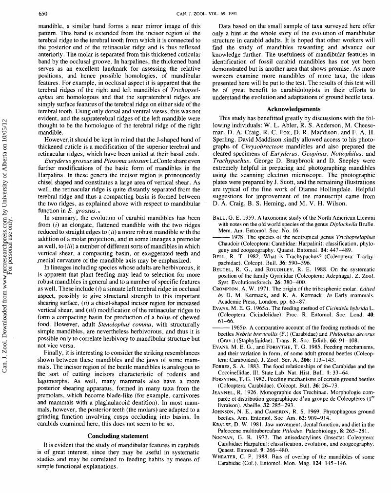
650 CAN. J. ZOOL. VOL. 69, 1991
mandible, a similar band forms a near mirror image of this Data based on the small sample of taxa surveyed here offer pattern. This band is extended from the incisor region of the terebral ridge to the terebral tooth from which it is connected to the posterior end of the retinacular ridge and is thus reflexed anteriorly. The molar is separated from this thickened cuticular band by the occlusal groove. In harpalines, the thickened band serves as an excellent landmark for assessing the relative positions, and hence possible homologies, of mandibular features. For example, in occlusal aspect it is apparent that the terebral ridges of the right and left mandibles of Trichopsel- aphus are homologous and that the supraterebral ridges are simply surface features of the terebral ridge on either side of the terebral tooth. Using only dorsal and ventral views, this was not evident, and the supraterebral ridges of the left mandible were thought to be the homologue of the terebral ridge of the right mandible.
However$ should be kept in mind that the J-shaped band of thickened cuticle is a modification of the superior terebral and retinacular ridges, which have been united at their basal ends.
Euryderus grossus and Piosoma setosum LeConte share even further modifications of the basic form of mandibles in the Harpalina. In these genera the incisor region is pronouncedly chisel shaped and constitutes a large area of vertical shear. As well, the retinacular ridge is quite distantly separated from the terebral ridge and thus a compacting basin is formed between the two ridges, as explained above with respect to mandibular function in E. grossus. ,
In summary, the evolution of carabid mandibles has been from (i) an elongate, flattened mandible with the two ridges reduced to straight edges to (ii) a more robust mandible with the addition of a molar projection, and in same lineages a premolar as well, to (iii) a number of different sorts of mandibles in which vertical shear, a compacting basin, or exaggerated teeth and .medial curvature of the mandible axis may be emphasized.
In lineages including species whose adults are herbivorous, it is apparent that plant feeding may lead to selection for more robust mandibles in general and to a number of specific features as well. These include (i) a sinuate left terebral ridge in occlusal aspect, possibly to give structural strength to this important shearing surface, (ii) a chisel-shaped incisor region for increased vertical shear, and (iii) modification of the retinacular ridges to form a compacting basin for production of a bolus of chewed food. However, adult Stenolophus comma, with structurally simple mandibles, are nevertheless herbivorous, and thus it is possible only to correlate herbivory to mandibular structure but not vice versa.
Finally, it is interesting to consider the striking resemblances shown between these mandibles and the jaws of some mam- mals. The incisor region of the beetle mandibles is analogous to the sort of cutting incisors characteristic of rodents and lagomorphs. As well, many mammals also have a more posterior shearing apparatus, formed in many taxa from the premolars, which become blade-like (for example, carnivores and mammals with a plagiaulacoid dentition). In most mam- mals, however, the posterior teeth (the molars) are adapted to a grinding function involving cusps occluding into basins. In carabids examined here, this does not seem to be so.
Concluding statement It is evident that the study of mandibular features in carabids
is of great interest, since they may be useful in systematic studies and may be correlated to feeding habits by means of simple functional explanations.
only a hint at the whole story of the evolution of mandibular structure in carabid adults. It is hoped that other workers will find the study of mandibles rewarding and advance our knowledge further. The usefulness of mandibular features in identification of fossil carabid mandibles has not yet been demonstrated but is another area that shows promise. As more workers examine more mandibles of more taxa, the ideas presented here will be put to the test. The results of this test will be of great benefit to carabidologists in their efforts to understand the evolution and adaptations of ground beetle taxa.
Acknowledgements This study has benefitted greatly by discussions with the fol-
lowing individuals: W. L. Abler, R. S . Anderson, M. Cheese- man, D. A. Craig, R. C. Fox, D. R. Maddison, and F. A. H. Sperling. David Maddison kindly allowed access to his photo- graphs of Chrysobracteon mandibles and also prepared the cleared specimens of Euryderus, Geopinus, Notiophilus, and Trachypachus. George D. Braybrook and D. Shepley were extremely helpful in preparing and photographing mandibles using the scanning electron microscope. The photographic plates were prepared by J. Scott, and the remaining illustrations are typical of the fine work of Dianne Holl'ingdale. Helpful suggestions for improvement of the manuscript came from D. A. Craig, B. S . Heming, and M. V. H. Wilson.
BALL, G. E. 1959. A taxonomic study of the North American Licinini with notes on the old world species of the genus Diplocheila Brulle. Mem. Am. Entomol. Soc. No. 16.
1978. The species of the neotropical genus Trichopselaphus Chaudoir (Coleoptera: Carabidae: Harpalini): classification, phylo- geny and zoogeography. Quaest . Entomol. 14: 447-489.
BELL, R. T. 1982. What is Trachypachus? (Coleoptera: Trachy- pachidae). Coleopt. Bull. 36: 590-596.
BEUTEL, R. G., and ROUGHLEY, R. E. 1988. On the systematic position of the family Gyrinidae (Coleoptera: Adephaga). Z. Zool. Syst. Evolutionsforsch. 26: 380-400.
CROMPTON, A. W. 197 1 . The origin of the tribosphenic molar. Edited by D. M. Kermack, and K. A. Kermack. In Early mammals. Academic Press, London. pp. 65-87.
EVANS, M. E. G. 1965a. The feeding method of Cicindela hybrida L. (Coleoptera: Cicindelidae). Proc. R. Entomol . Soc. Lond. 40: 61 -66.
1965b. A comparative account of the feeding methods of the beetles Nebria brevicollis (F.) (Carabidae) and Philonthus decorus (Grav.) (Staphylinidae). Trans. R. Soc. Edinb. 66: 9 1 - 108.
EVANS, M. E. G., and FORSYTHE, T. G. 1985. Feeding mechanisms, and their variation in form, of some adult ground beetles (Coleop- tera: Caraboidea). J. Zool. Ser. A, 206: 1 13- 143.
FORBES, S. A. 1883. The food relationships of the Carabidae and the Coccinellidae. 111. State Lab. Nat. Hist. Bull. 1: 33-64.
FORSYTHE, T. G. 1982. Feeding mechanisms of certain ground beetles (Coleoptera: Carabidae). Coleopt. Bull. 36: 26-73.
JEANNEL, R. 1926. Monographie des Trechinae. Morphologie corn- parCe et distribution geographique d'un groupe de Coleoptkres ( l re livraison). Abeille, 32: 285-293.
JOHNSON, N. E., and CAMERON, R. S. 1969. Phytophagous ground beetles. Ann. Entomol. Soc. Am. 62: 909-914.
KRAUSE, D. W. 198 1 . Jaw movement, dental function, and diet in the Paleocene multituberculate Ptilodus. Paleobiology, 8: 265-281.
NOONAN, G. R. 1973. The anisodactylines (Insecta: Coleoptera: Carabidae: Harpalini): classification, evolution, and zoogeography. Quaest. Entomol. 9: 266-480.
WHEATER, C. P. 1988. Bias of overlap of the mandibles of some Carabidae (Col. ). Entomol. Mon. Mag. 124: 145- 146.
Can
. J. Z
ool.
Dow
nloa
ded
from
ww
w.n
rcre
sear
chpr
ess.
com
by
Uni
vers
ity o
f A
lber
ta o
n 10
/05/
12Fo
r pe
rson
al u
se o
nly.