
the jefferson medicine forum
44
THE J EFFERSON MEDICINE F ORUM The Journal of Thomas Jefferson University Hospital Department of Internal Medicine Volume 7, 2005
-
Upload
khangminh22 -
Category
Documents
-
view
1 -
download
0
Transcript of the jefferson medicine forum

THEJEFFERSONMEDICINEFORUM
The Journal of Thomas Jefferson University Hospital Department of Internal MedicineVolume 7, 2005

Welcome to the 7th Edition of the Jefferson MedicineForum! In this edition we have highlighted some of themany interesting and diverse cases encountered by ourInternal Medicine students, residents, and fellows. Everycase is an opportunity to take what a small number ofpeople have learned and share it with the rest of theJefferson community. From the pedestrian Wolf-Parkinson-White syndrome to the esoteric Milk Alkali Syndrome, our
residents see it all. Every contributing author represented inthis edition has left a touch of their enthusiasm, curiosity,and analytical skills in each report. We extend ourappreciation to all our colleagues who captured theirexperiences in images and prose for us to share.
Betty Lim, MDAndrew Rose, MDKarl Kwok, MD
A Community of Scholars
The ideal of a University is that we live and work together as acommunity of scholars dedicating ourselves to theadvancement of Medicine. Our community is at its best whenwe share ideas, knowledge, perspective, and a passion forMedicine. As a routine, this can occur at Morning Report orNoon Conference, but also on rounds or hopefully at thebedside when dealing with a particularly challenging case.
This journal serves as an outlet for observations or studies ofparticular merit. Its publication is an important element of ourscholarly focus in the Department. Indeed, nearly a score ofcontributors from all three classes of residents haveparticipated; making this issue quite representative of ourcommunity. As a clinician and educator in the Department, Ihave been regularly challenged by the spirit of inquiry amongour residents. This serves as a true reflection of the vitality ofour community.
Rounding in the MRICU during early November, I was alsostruck by the incredible gift that we have been granted asphysicians in our society. During the span of one week, our
team which included Drs. Aleyas, Mehrotra, Callahan, Moon,Cheskis, Nandi, and Levin faced a challenging array ofscholarly questions. These included identifying the cause forinterstitial pneumonitis in a pregnant woman, dealing with anactive TB patient from New Orleans who was refusing care,and facilitating diagnosis for a patient with diffuselymphadenopathy who was only willing to accept minimallyinvasive diagnostic and therapeutic interventions. Thiscommunity of scholars in the ICU found answers rapidly,defended their differential diagnoses, and pushed the limits ofmedical knowledge to provide outstanding care. They did sowith compassion and respect. Their work, together with thededicated nurses and therapists of the MRICU defined aUniversity as no words could express.
The editors of this journal have sought to capture theexcitement and vitality of our work in the Department. Theydeserve our gratitude for doing this while managing theroutine expectations of the residency. I thank them forcontributing to our community of scholarship and elevatingour work within this University.
Gregory C. Kane MDAssociate Professor of MedicineResidency Program DirectorDepartment of Medicine
FROM THE RESIDENCY PROGRAM DIRECTOR
FROM THE EDITORS

i
THE JEFFERSON MEDICINE FORUMThe Journal of Thomas Jefferson University Hospital
Department of Internal MedicineVolume 7, 2005
Case ReportsMan with Fevers and Headache . . . . . . . . . . . . . . . . . . . . . . . . . . . . . . . . . . . . . . . . . . . . . . . . . . . . . . . . . . . . . . . . . . . . . . . . . . . . . . . . . . . . .1
Amy Baranoski, MDAsymptomatic and Symptomatic Septic Emboli . . . . . . . . . . . . . . . . . . . . . . . . . . . . . . . . . . . . . . . . . . . . . . .3
Jason T. Bradley, MD and Nicole M. Orr, MDMan with Loss of Consciousness . . . . . . . . . . . . . . . . . . . . . . . . . . . . . . . . . . . . . . . . . . . . . . . . . . . . . . . . . . . . . . . . . . . . . . . . . . . . . . . . . . . .4
Dana Critchell, MDWoman with Fevers and Shortness of Breath . . . . . . . . . . . . . . . . . . . . . . . . . . . . . . . . . . . . . . . . . . . . . . . . . .6
Jonathan Jun, MDPost-partum Woman with Dyspnea . . . . . . . . . . . . . . . . . . . . . . . . . . . . . . . . . . . . . . . . . . . . . . . . . . . . . . . . .7
Saaron Laighold, MDWoman with Thrombocytopenia and Weakness . . . . . . . . . . . . . . . . . . . . . . . . . . . . . . . . . . . . . . . . . . . . . .10
M. Paula Martinez, MD and Ubaldo E. Martinez, MDLiver Transplant Recipient with Bilateral Leg Swelling . . . . . . . . . . . . . . . . . . . . . . . . . . . . . . . . . . . . . . . . . .11
Maya Spodik, MDWoman with Pneumonia . . . . . . . . . . . . . . . . . . . . . . . . . . . . . . . . . . . . . . . . . . . . . . . . . . . . . . . . . . . . . . . .12
Andra Popescu, MDMan with Vertigo and Hypercalcemia . . . . . . . . . . . . . . . . . . . . . . . . . . . . . . . . . . . . . . . . . . . . . . . . . . . . . .14
Veronica Rivera and Andrew Rose, MDWoman with Left Shoulder Pain . . . . . . . . . . . . . . . . . . . . . . . . . . . . . . . . . . . . . . . . . . . . . . . . . . . . . . . . . .16
Jane V. Mayrin, MDRupture Recurrence after Surgical Repair of a Postinfarction Ventricular Septal Rupture in the Reperfusion Era . . . . . . . . . . . . . . . . . . . . . . . . . . . . . . . . . . . . . . . . . . . . .19
Apurva D. Shah, MDAdenovirus of the Colon . . . . . . . . . . . . . . . . . . . . . . . . . . . . . . . . . . . . . . . . . . . . . . . . . . . . . . . . . . . . . . . .22
Bryan Kavanaugh, MDSarcoid Cardiomyopathy . . . . . . . . . . . . . . . . . . . . . . . . . . . . . . . . . . . . . . . . . . . . . . . . . . . . . . . . . . . . . . . .23
Sivakumar Srinivasan, MD and Emanuel Chryssos, MDImage in Medicine . . . . . . . . . . . . . . . . . . . . . . . . . . . . . . . . . . . . . . . . . . . . . . . . . . . . . . . . . . . . . . . . . . . . .23
Amy Baranoski, MDVaricella Pneumonia . . . . . . . . . . . . . . . . . . . . . . . . . . . . . . . . . . . . . . . . . . . . . . . . . . . . . . . . . . . . . . . . . . .25
Sivakumar Srinivasan, MDRecurrent Spontaneous Coronary Artery Dissections . . . . . . . . . . . . . . . . . . . . . . . . . . . . . . . . . . . . . . . . . . .26
Sivakumar Srinivasan, MDPulseWellens Syndrome . . . . . . . . . . . . . . . . . . . . . . . . . . . . . . . . . . . . . . . . . . . . . . . . . . . . . . . . . . . . . . . . . . . . .27
Nicole Farmer, MD and Michael Cohen, MDGirl with Lightheadedness and Palpitations . . . . . . . . . . . . . . . . . . . . . . . . . . . . . . . . . . . . . . . . . . . . . . . . . .29
Andrew Rose, MDAbstracts & ArticlesRisk of Ischemic Stroke Associated with Low-Dose Oral Contraceptive Use: A Meta-Analysis . . . . . . . . . . . . . . . . . . . . . . . . . . . . . . . . . . . . . . . . . . . . . . . . . . .31
Dae Hyun Kim, MD, MPH
The Spectrum of Advanced Liver Disease in a Tertiary Care Institution . . . . . . . . . . . . . . . . . . . . . . . . . . . .39Maya Spondik, MD

An 18-year-old man was transferred from an outside hospital forevaluation of recent headache, fevers, and laboratory abnormalities.
The patient had a past medical history significant for attentiondeficit hyperactivity disorder (ADHD) and had presented to anoutside hospital with complaints of fatigue, fever, and headache.He described the headache as frontal, severe, and stabbing in naturewithout associated neck stiffness, photophobia, or phonophobia.He also reported fevers to 105ºF. He was evaluated one day afterthe onset of symptoms at a local emergency room where a lumbarpuncture was performed. By report of the patient and his mother,the lumbar puncture was negative and he was given a prescriptionfor ketorolac for his headache. The patient went home andcontinued to have fevers and headache for which he was takingacetaminophen and ketorolac. Two days after his initial dischargefrom the emergency room he presented to another hospitalcomplaining of sharp epigastric pain without radiation in additionto the headache and fevers. He denied nausea, vomiting, or melena.Laboratory data at that time revealed a creatinine of 4.6 and mildlyelevated transaminases and the patient was transferred to thishospital for further evaluation and management.
The patient denied chest pain, cough, dyspnea, sore throat, ordysuria. He reported that he had noticed a “sweat rash” in hisgroin a few weeks prior to his symptoms that went away afterpersisting for several days. His past medical history was onlysignificant for ADHD. He denied surgical history. Family historywas significant for hypertension in both parents. He reportedoccasional alcohol use. He had recently graduated from highschool and lived in a suburban neighborhood. He had beenworking as a groundskeeper at an outdoor chlorinated pool. Hedenied tick bites, recent travel, or sexual activity.
Prior to transfer to this hospital, he was given empiric antibiotictreatment with doxycycline and ceftriaxone and was placed onmaintenance intravenous fluids, pantoprazole, and prophylacticdose subcutaneous heparin. His only out-patient medication priorto the onset of his symptoms was methylphenidate (Ritalin®).
The temperature was 97.4ºF, the pulse rate was 71, the bloodpressure was105/59, the respiratory rate was 14 and the oxygensaturation was 98% on room air. On physical examination, thepatient appeared well. His pupils were equal and reactive. Hissclera were anicteric in contrast to a previous documentedexamination. He had no oral lesions. The heart and lungexamination was unremarkable. He had a left subclavian centralvenous catheter in place without erythema or fluctuance at theinsertion site. He had normal bowel sounds and his abdomen wassoft, mildly distended and non-tender to palpation with noappreciated organomegaly or masses. He had no costovertebraltenderness, lower extremity edema, or neck stiffness. Neurologicalexam revealed no focal deficits with intact cranial nerves.
The patient’s initial lumbar puncture results were a glucose of 69,protein of 29, no red blood cells, and one white blood cell.Urinalysis was positive for protein, blood, and bilirubin, with 10-25 WBC per high-powered field (HPF), 5 RBC per HPF,occasional WBC and granular casts. Arterial blood gas was7.37/24/66/14/95% on 3 liters oxygen via nasal canula. Otherlaboratory data collected after his transfer to this institution werenegative and included HIV, EBV antibody and PCR, CMV,HSV, toxoplasmosis antibodies, lyme antigen, and hepatitisserologies. Multiple sets of blood cultures were negative.Complement levels were normal. Leptospirosis antibody waspending during his hospitalization.
The patient’s electrocardiogram was normal except for diffuse Twave flattening. His chest x-ray showed no infiltrate or effusion.An echocardiogram was significant for mild mitral, tricuspid, andpulmonary valvular regurgitation with an ejection fraction of50%. A right upper quadrant ultrasound showed that his spleenand liver were top normal in size, but was otherwise notremarkable. A CT scan of his abdomen was only significant for adistended stomach and multiple sub-centimeter lymph nodes.
Table 1. Intial Laboratory Data
Initial Tests ResultWhite blood cells 6.7WBC 11.6 B/LNeutrophils (segs) 92%Bands 5%PT 15.3 secs INR 1.4PTT 35.0 secs Sodium 134 meq/LPotassium 4.3 meq/LChloride 99 meq/LCO2 21 meq/LBUN 41 mg/dLCreatinine 4.6 mg/dLGlucose 97 mg/dLAST 48 U/L (nl 0-37)ALT 70 U/L (nl 0-40)Alk phos 193 U/LTotal Bilirubin 8.3 mg/dL (nl 0.1-1.2)Direct Bilirubin 4.7 mg/dLAmylase 159 U/L (nl 19-68)Lipase 77U/L (nl 16-63) Troponin I 0.10 (nl <0.05)Urine Drug Screen Negative
Case Reports
AN 18-YEAR OLD MAN WITH FEVER AND HEADACHEAmy Baranoski, MD
1

2
Case Reports
Doxycycline and ceftriaxone were continued during his hospital-ization and he was given supportive treatment with intravenousfluids. His creatinine and liver function tests improved uponadmission and returned to baseline values in several days. Thepatient was discharged home on doxycycline with the presumptivediagnosis of leptospirosis.
After discharge to home, the patient’s leptospirosis antibody levelcame back at 12,800 (Negative <50, borderline 50-100, positive>100). This patient was seen for follow up one month afterdischarge and was feeling well.
DiscussionLeptospirosis is an illness caused by the organism Leptospirainterrogans. This is an aerobic spirochete with 18 or more coilsper cell. This organism is usually found in tropical environments,but has a world-wide distribution. The highest incidence of illnessis after large precipitation events. The organism is found inmammals; most commonly rodents, cattle, goats, horses, or dogs.Animals can become asymptomatic reservoirs with bacterialshedding in the urine. The organism can survive weeks to monthsin soil and water. The most common mode of transmission inhumans is secondary to exposure to contaminated urine via mucusmembranes, conjunctiva, or damaged skin. The leptospires thentravel in the bloodstream and are carried to multiple organs, wherethere is endothelial cell disruption leading to vasculitis andultimately, organ dysfunction.
The largest risk for exposure to leptospirosis is occupational andincludes farmers, ranchers, sewer workers, and military personnel.There is also a risk through recreational exposure such asfreshwater swimming and kayaking. In 1998, an outbreakoccurred in Illinois among triathlon participants. People can alsobe infected through pets, rodent exposure, and living incontaminated rainwater catchment areas.
Leptospirosis can generally be divided into two groups. 90% ofpatients have a mild, self-limited illness. The severe form ofleptospirosis is also known as Weil’s Disease. It is characterized byhepatic and renal failure. These patients can also have pulmonaryhemorrhage, ARDS, and cardiac arrhythmias. EKG changes canvary from frequent premature ventricular contractions to atrialfibrillation or flutter, or even ventricular tachycardia. Themortality rate in Weil’s Disease ranges from 5-40%.
The illness is characterized by abrupt onset of fevers, headache,and myalgias in the majority of patients. There is usually an acutephase which is a one to three day period of constitutionalsymptoms. The immune phase then follows. There is a two tothirty day incubation period before onset of symptoms.Approximately 50% of patients have nausea, vomiting, anddiarrhea. Less than a third of infected patients have develop a non-productive cough. Conjunctival suffusion is present in >80% ofpatient with leptospirosis and should cause a high suspicion forthe disease if present. Less common symptoms include arthralgias,
bone pain, sore throat, and abdominal pain. Muscle tenderness,especially in the calf and lumbar regions, may be present. Lessthan 10% of patients develop a pretibial maculo-papular rash.Other less specific signs such as lymphadenopathy, hepatomegaly,or splenomegaly may be present. Aseptic meningitis is usually notseen in acute illness but may occur in up to 80% of patients laterin the course of illness. The aseptic meningitis is secondary to animmune reaction and not caused by the organism itself.
The laboratory data in leptospirosis is non-specific. The WBCcount is generally less than 10,000 B/L. Patient may have mildlyelevated transaminases, which are usually less than 200 U/L.Urinalysis reveals mild proteinuria and more than 5 WBC perHPF. Patient may have hematuria or hyaline casts. Cerebrospinalfluid is also non-specific with normal glucose, protein between 50-100 and cell counts less than 500. It is possible to grow leptospiresin CSF, blood, or urine, especially if cultured during the first sevento ten days of infection. This is a fastidious organism which usuallytakes one to two weeks to grow, but can take up to three monthsin some cases. The organism requires Fletcher’s, Ellinghausen’s, orpolysorbate 80 media in order to grow.
Darkfield microscopy will show spirochetes and can be used tomake a presumptive diagnosis. PCR can also be used to diagnoseleptospirosis. In addition, there are indirect methods of detectionincluding microscopic agglutination test (MAT), which is a highlysensitive and specific test for leptospirosis antibodies. MAT alsoprovides serotype specific information. There are also ELISA andindirect hemaglutinin assays available. These tests do not provideserotype information.
Mild disease can be treated with doxycycline 100 mg PO bid,ampicillin 500-750 mg PO every six hours, or amoxicillin 500mg PO every six hours. Moderate to severe disease requiresintravenous treatment with penicillin G 1.5 million units every 6hours or ampicillin 0.5-1 gm every 6 hours.
Leptospirosis can be prevented by immunization of livestock andpets. High-risk workers should wear protective clothing includingimpermeable boots and gloves. Rodent control is also very importantin preventing outbreaks, especially after heavy rains. The CDCrecommends chemoprophylaxis for people at increased risk withdoxycycline 200 mg weekly beginning 1-2 days before exposure. ■
References1. Everett DE. Leptospirosis. UptoDate version 13.2. Available at www.uptodate.com.
Accessed October 2005.
2. Farr RW. Leptospirosis. CID 1995; 21: 1-8.
3. Katz AR et al. Assessment of the Clinical Presentation and Treatment of 353 Cases ofLaboratory Confirmed Leptospirosis in Hawaii, 1974-1998. CID 2001; 33:1834-41.
4. Levett PN. Leptospirosis. Clinical Microbiology Reviews. 2001; 14:296-326.
5. Mandell: Principles and Practice of Infectious Diseases, 5th ed., 2000, ChurchillLivingstone, Inc.
6. Morgan J. et al. Outbreak of Leptospirosis among Triathlon Participants andCommunity Residents in Springfield, Illinois, 1998. CID 2002; 34:1593-9.
5. www.cdc.org

A 25-year-old male intravenous heroin user with Hepatitis Cpresented with a syncopal episode and left sided weakness.Examination revealed sub-conjunctival hemorrhages and diffusebilateral petechiae on his lower extremities. He had multiple irregular,erythematous, painless macules on his hands and feet, and linear, red,subungal lesions bilaterally. These findings were consistent withJaneway lesions and splinter hemorrhages, respectively. His vital signswere stable on admission. Cardiac auscultation revealed a grade 2/6holosystolic apical murmur radiating to the axilla. A brain MRIrevealed multiple areas of hyperdensity, particularly in the rightparietal lobe (Figure 1). Blood tests revealed leukocytosis and six ofsix blood cultures yielded methicillin sensitive Staphylococcus Aureus.The patient was treated with intravenous nafcillin and gentamicin.
One week later the patient’s neurologic status improved. Atransesophageal echocardiogram was performed and revealed both avegetation on the posterior leaflet of an otherwise normal mitral valveand moderate mitral regurgitation. The following day, the patientdeveloped diarrhea that was positive for both heme and Clostridiumdifficile toxin. Metronidazole was started and an abdominal CT scanwas ordered (Figure 2). Although there was no corresponding clinicalpresentation, imaging revealed bilateral renal infarcts, a splenicinfarct, and sigmoid thickening. The patient regained full neurologicfunctioning within two weeks of initial presentation and wasdischarged after a 6 week course of intravenous nafcillin. He iscurrently awaiting a mitral valve replacement.
In the setting of presumed septic emboli to both the skin and brainsecondary to bacterial endocarditis, we were concerned that the hemepositive stools were the result of embolization to the mesentericarteries. Although the abdominal CT showed no evidence of bowelinfarction and sigmoid thickening is non-specific, the renal and splenicinfarcts were determined to be secondary to septic emboli. A previouscase of bacterial endocarditis with simultaneous multiple organinvolvement including the kidney, spleen, brain, skin, and intestineshas not been documented. This case demonstrates that in the settingof diffuse symptomatic septic emboli, involvement of additional organsystems is likely but may be missed due to absence of clinical findings.
Concomitant symptomatic emboli to more than one organ systemare rare according to the literature. It is interesting to note thatpetechiae occur in only 20-40%, Osler nodes in only 10-25 % ofpatients, and Janeway lesions in less than 10% of patients withbacterial endocarditis. Splenic septic emboli in infective endocarditisare a common finding and incidental splenic infarcts were found in38% of 29 asymptomatic patients in one study.
Majumdar et al. found that 18% of 354 patients with infectiveendocarditis had renal involvement and 45% of that subpopulationwas found to have localized renal infarction, making it the mostcommon renal lesion. The authors of that study acknowledge thisrenal subpopulation does not represent all patients with endocarditisbut a prospective study to assess symptomatic and asymptomatic renalinvolvement has not yet been done.
The incidence of septic emboli to the brain has been well documented.Whether or not embolization is a result of vegetation size is currently
disputed, but current data suggest that vegetations greater than onecentimeter in length have increased risk for cerebral embolism4. Brainembolization has been previously described in a case report and severalstudies5,6, but not with concomitant lesions to other organ systems.Eighteen percent of patients with mitral valve endocarditis developstroke and in over one-half of these cases, stroke was the initialpresenting symptom5. The authors concluded that all patients withendocarditis and neurologic symptoms should undergo neurologicimaging to determine the exact location of the lesions. Bakshi et al.even concluded that neuroimaging should be used to diagnosebacterial endocarditis in patients presenting with neurologic symptomsand nonspecific constitutional symptoms. In addition, that study citesthat 50% of patients with neurologic sequelae of bacterial endocarditisdo not have clinical evidence of additional peripheral emboli.
In conclusion, although the patient was admitted for symptomaticemboli to the skin and brain, non-symptomatic emboli to the kidney,spleen, and intestine were incidentally found. It is well known thatinfective endocarditis can infect the spleen, kidney, and brain, butnormally such emboli are symptomatic and isolated. This casedemonstrates that asymptomatic lesions may also be present in theface of already existing symptomatic emboli and careful investigationof critical organ systems should be done empirically. ■
The authors encountered this case during their time with the Departmentof Pulmonary and Critical Care at Hahnemann University Hospital,Philadelphia, PA.
References1. Saccente, M. & Cobbs, C.G. (1996). Clinical approach to infective endocarditis. Cardiology
Clinics ,14 (3), 351-62.2. Ting, W., Silverman, N.A., Arzouman, D.A., & Levitsky, S. (1990). Splenic septic emboli in
endocarditis, Circulation, 82 (5),105-93. Majumdar,A., Chowdhary, S., Ferreira, M.A.., Hammond, L.A., Howie, A.J., Lipkin, G.W.,
& Littler, W.A. (2000). Renal pathologic findings in infective endocarditis. Nephrol DialTransplant, 15 (11), 1782-7.
4. Krapf, H., Skalej, M., & Voigt, K. (1999). Subarachnoid hemorrhage due to septic emboliinfarction in infective endocarditis. Cerebrovascular Disease 9 (3), 182-4.
5. Sexton, D.J. & Spelman, D. (2003). Current best practices and guidelines. Assessment andmanagement of complications in infective endocarditis. Cardiology Clinics, 21 (2), 273-82.
6. Bakshi, R., Wright, P.D., Kinkel, P.R., Bates, V.E., Mechtler, L.L., Kamran, S. Pullicino, P.M.,Sirotkin, I., & Kinkel W.R. (1999). Cranial magnetic resonance imaging findings in bacterialendocarditis: the nueroimaging spectrum of septic brain embolization demonstrated in twelvepatients. Journal of Neuroimaging, 9 (2), 78-84.
3
Case Reports
PRESENCE OF CONCOMITANT ASYMPTOMATIC AND SYMPTOMATIC EMBOLIRESULTING FROM ACUTE STAPHYLOCOCCUS AUREUS ENDOCARDITISJason T. Bradley, MD, Nicole M. Orr, MD, and Siva Ramachandran, MD
Figure 1. MRI of thebrain identifying multipleareas of septic emboli.
Figure 2. CT of the Abdomen identifyingmultiple embolic infarcts of the spleen (S)and bilateral kidneys (Rk, Lk).
Case Reports
4
A 32-year-old man with no past medical history presented to theemergency department after losing consciousness at the end of ahalf-marathon. The patient was in his usual state of good healthuntil the last mile of the marathon when he began to feel unsteadyon his feet and short of breath. At the end of the race, he collapsedand was found unresponsive by EMS. The patient had no memoryof the subsequent events and could only recall waking up in theemergency department feeling “numb” and “confused” severalhours later. The patient denied any palpitations, chest pain,nausea, blurry vision, dizziness, urinary incontinence, or headachebefore or after the event. Of note, he was running outsideapproximately 3 to 4 times a week prior to this episode.
The temperature was 99.1ºF (37.3 ºC), the pulse rate was 154,and the blood pressure was 94/30.
On physical examination, the patient had regained consciousness,but remained confused and disoriented with a GCS score of 13.The patient was pale and diaphoretic, however he had moistmucous membranes and no evidence of skin tenting. His pupilswere equal and reactive and the sclerae were anicteric. The heart,lungs, abdomen, and extremity examination were unremarkable.He had no costovertebral tenderness, lower extremity edema, orneck stiffness. Neurological exam revealed no focal deficits withintact cranial nerves. Several hours later, the patient was noted tobe alert and oriented to person, place, and time. In the interim,the patient had several episodes of watery vomit.
Laboratory results on admission were notable for a normal CBCwith a platelet count of 225 B/L and a normal coagulation profile.The creatinine was 2.4 mg/dL, the myoglobin was 303 ng/mL,the CK was 157 IU, and the troponin was 0.15 ng/mL.Electrocardiogram on admission revealed sinus tachycardia withan isolated ST elevation in AVR and ST depressions in II and V4-V6. A repeat electrocardiogram showed resolution of theseabnormalities. A chest x-ray and head CT scan showed noabnormalities. The patient was fluid resuscitated with normalsaline at 200 cc/hr and admitted to the hospital with the diagnosisof heat exhaustion and rhabdomyolysis with acute renal failure.
During his hospital stay, the troponin peaked at 0.54 ng/mL andhis creatinine dropped to 1.6 mg/dL with fluids resuscitation, andplans were made to discharge the patient the following morning.However, a CBC the following morning revealed markedthrombocytopenia with a platelet count of 64 B/L and the CKlevels were now significantly elevated at 7105 IU. The patientremained in the hospital to monitor his platelet count, which roseto 97 B/L the following day. The patient was then discharged withinstructions for a follow-up CBC with his primary care providerin the near future. Interestingly, another participant in the half-marathon presented with acute renal failure followed by thrombo-cytopenia to 80 B/L the day after presentation.
DiscussionHeat exhaustion represents only one part of a continuum of heat-related illnesses, ranging from heat stress to heat exhaustion to heatstroke. While heat stress is simply perceived discomfort in a hotenvironment, typically during physical exertion, heat exhaustion isa more severe form of heat stress secondary to water or salt depletionfrom excessive heat. Patients start becoming symptomatic at thispoint, with many experiencing dizziness, weakness, extreme thirst,headache or syncope. More serious manifestations, such as deliriumand seizures, are notably absent. Body temperatures can varyanywhere from 37 to 40ºC. Once core body temperatures rise aboveapproximately 40ºC, patients begin to develop heat stroke, the mostvirulent form of the heat-related illnesses.
Heat stroke requires two findings for its diagnosis, namely a corebody temperature greater than approximately 40 ºC and centralnervous system dysfunction, whether it be convulsions, coma,delirium, or simple confusion.1 While heat stress and exhaustionare typically considered relatively benign conditions, heat strokeis a true medical emergency with mortality rates ranging from20% to 70% depending on patient age. Its pathogenesis andpotential lethality are thought to be due to a combination of thedirect cytotoxic effects of heat, the physiological demands thisheat places on the body, and the host’s inflammatory andcoagulation response.1
Two types of heat stroke are described, one being classic heatstroke and the other, exertional heat stroke. Classic heat stroke istypically seen in those at the extremes of age and also in thosewith underlying medical illnesses, including certain cardio-vascular, neurological and psychiatric disorders. In addition,individuals on certain medications such as diuretics oranticholinergic agents are at an increased risk. Exertional heatstroke, on the other hand, often affects young, healthy individualsengaged in strenuous activity during times of high heat andhumidity. Interestingly, testing on some of these patients reveals asusceptibility to malignant hyperthermia which may predisposethem to the development of heat stroke.
Despite this patient’s modest temperature elevation and moderatephysical stressor given his history of regular physical exercise, thispatient appeared to suffer from many of the more serious sequelaeclassically seen only with heat stroke, including a change in mentalstatus, myocardial stress, rhabdomyolysis, acute renal failure andthrombocytopenia. Neurologic dysfunction, whether secondaryto metabolic derangements, edema, infarction, or hypernatremia,is, by definition, seen in all cases of heat stroke. While somepatients have gross neurological disturbances such as coma orencephalopathy, other patients present with more subtle signssuch as inappropriate behavior or impairment in judgment.1Unfortunately, patients often do not recover fully from theseneurological insults. One study found that 33% of patients whopresented with heat stroke and survived during the 1995 Chicago
A 32 YEAR OLD MAN WITH LOSS OF CONSCIOUSNESSDana Critchell, MD

Case Reports
5
heat wave suffered long-term moderate to severe functionalneurological impairment.
Other potential complications of heat stroke involve virtually anyorgan system and can rapidly lead to multi-organ dysfunction anddeath even with proper supportive measures. Among thesecomplications are acute respiratory distress syndrome, myocardialor hepatocellular injury, rhabdomyolysis, and acute renal failure.Also commonly seen in heat stroke are bleeding diatheses, mostnotably disseminated intravascular coagulation and isolatedthrombocytopenia.
One proposed theory behind the phenomenon of DIC in heatstroke involves the splanchnic vasoconstriction and peripheralvasodilation that occurs as part of the body’s compensatorymechanisms after exposure to extreme heat. This vasoconstrictioncan lead to intestinal ischemia and oxidative stress, resulting inloss of the protective gastrointestinal barrier and leakage ofendotoxins into the systemic circulation. In addition, heat shockitself induces an acute-phase response to help protect the bodyfrom tissue injury. Inflammatory and anti-inflammatorycytokines, most notably interleukin-1 and 6, TNF-alpha andinterferon-alpha, are elevated in heat shock, which contributes tothe thermoregulatory failure and circulatory shock not onlydirectly but also via increased production of nitric oxide whichfurther promotes intestinal permeability.1
Researchers have found that thrombocytopenia can also develop inheat stroke in the absence of DIC. Furthermore, one case reportdetailed a patient very similar to our patient with heat exhaustionwho presented with thrombocytopenia as well as leukopenia, bothpresumably secondary to thermolysis. Thus, the hemostatic changesin heat stroke are likely multifactorial in nature, a combination ofDIC, thrombocytopenia independent of DIC, and failure ofcoagulation factor production secondary to hepatic injury.7
Treatment of heat stroke focuses on mainly on rapid cooling andsupport of organ systems. Various cooling techniques can beemployed since no specific method has been shown to be superior.Common methods include whole body immersion or the use offans to aid in conductive heat losses with cold water or ice to theskin. Regardless, achieving a rectal temperature of 39.4ºC or less
within thirty minutes of presentation has been shown to improvesurvival. As of yet, there appears to be no role for medications suchas dantrolene, and the use of anti-pyretics has not been evaluatedthus far. Unfortunately, prompt cooling may not prevent thedevelopment of tissue injury, patients should continue to beobserved even after stabilization and resolution of hyperthermia.1
This case is unique in that despite his diagnosis of heat exhaustion,this patient developed several complications that typically occurwith the more deadly form of heat-related illnesses, heat stroke.In addition, the dramatic fall in his platelet count highlights notonly the potential virulence of heat stroke but also, itspathogenesis. In summary, due to a combination of heat toxicityitself and a breakdown of the gastrointestinal barrier withproduction of inflammatory cytokines, patients with heat strokeand even heat exhaustion are vulnerable to thrombocytopeniaisolated from or in conjunction with DIC. As such, even afterhyperthermia has resolved, one should remain vigilant of thispotential complication, as well as many others, in all patients whopresent along the continuum of heat-related illnesses. ■
References1. Bouchama A, Knochel J. Heat stroke. N Engl J Med June 2002; 246 (25): 1978-88.
2. Yeo, Theresa. Heat Stroke: A comprehensive review. AACN Clinical Issues, 2003;15(2): 280-93.
3. Mechem, C Crawford. Severe Hyperthermia: heat stroke, neuroleptic malignantsyndrome, and malignant hyperthermia. UpToDate online, April 2005.
4. Bendahan D, Kozak-Ribbens G, Confort-Gouny S. A noninvasive investigation ofmuscle energetics supports similarities between exertional heat stroke and malignanthyperthermia. Anesthesia & Analgesia Sept 2001; 93(3): 683-689.
5. Dematte J, O’Mara K et al. Near-fatal heat stroke during the 1995 heat wave inChicago. Ann Int Med Aug 1998; 129(3): 173-81.
6. Lambert, Patrick. Role of gastrointestinal permeability in exertional heat stroke.Exercise and Sports Sciences Review, Oct 2004; 32 (4): 185-90.
7. Mustafa K, Omer O, Khogali m et al. Blood coagulation and fibrinolysis in heatstroke. British J of Hematology 1985; 61: 517-23.
8. Lohiya S, Lohiya G, Humerez R. Heat exhaustion with thrombocytopenia andleukopenia in a car wash attendant. Am J of Hematology 1996; 52: 120-21.
9. Grogan H, Hopkins P. Heat Stroke: Implications for critical care and anesthesia.British Journal of Anaesthesia 2002; 88(5): 700-07.
10. Dematte J, O’Mara K et al. Near-fatal heat stroke during the 1995 heat wave inChicago. Ann Int Med Aug 1998; 129(3): 173-81.
11. Bouchama A, Cafege A, Devol EB et al. Ineffectiveness of dantrolene sodium in thetreatment of heatstroke. Crit Care Med Feb 1991: 19(2): 176-80.

Case Reports
6
A 41 year-old African American femalewith a history of asthma, hypertension, andschizophrenia presented to the ER inFebruary 2005 with shortness of breath,wheezing, and a “wet-sounding” but non-productive cough. She reported chills,subjective fever, rhinorrhea, and malaise forfive days prior. Her only medication wasalbuterol, which she normally used twiceweekly as needed. She was never intubatedfor asthma. On review of systems, she hadprogressive dyspnea on exertion of less thantwo blocks for the last year. She deniednight sweats, weight loss, or sick contacts,HIV exposure, alcohol or drug abuse. Sheadmitted to smoking 1 pack per day for more than 15 years.
On physical exam, her temperature was 97.0 F, pulse 96 beats perminute, respirations 22 breaths per minute, blood pressure 130/100mmHg. She had an oxygen saturation of 77% on room air whichimproved to 93% on 4 liters oxygen. She was an obese female inmoderate respiratory distress. Her orophaynx was clear. Her neck wassupple without thyromegaly or lymphadenopathy. Jugular venousdistension measured at 10 cm above the Angle of Louis. Heart examrevealed tachycardia without murmur, rub or gallop. Lungs hadscattered rhonchi and wheezes bilaterally. Faint crackles were audible atthe bases. She had trace symmetrical ankle edema.
Laboratory data on admission showed a WBC of 5900/L, (mostlyneutrophils and 30% lymphocytes) hemoglobin 14.7 g/dL, platelets142k/L. Chemistries were grossly normal except for a bicarbonate of31 mmol/L. ABG on 4 L O2 showed pH 7.32, PCO2 of 31, PaO271. LDH level was elevated at 687. Her coagulation indices, liverfunction, cardiac enzymes, and urinalysis were within normal limits.Chest X-ray showed a right lower-lobe consolidation and diffuseinfiltrates bilaterally. A chest CT further revealed hilar adenopathy anda small right pleural effusion while excluding pulmonary embolism(Figure 1). Blood cultures were sent, ceftriaxone and azithromycin wereadministered, and serial nebulizer treatments were given for her asthma.
The patient eventually became more hypoxic and was moved to theICU where she was intubated. Pressor agents were required forhypotension, and her degree of hypoxia required an FiO2 of 100%with +15 of PEEP. On her subsequent chest radiographs, shedeveloped diffuse alveolar infiltrates in multiple areas consistent withARDS. Bronchoscopy and thoracentesis did not elicit any bacterial,fungal, or neoplastic etiology for her respiratory failure. Silver stainfor PCP was negative. HIV serology was negative.
She remained intubated and on pressors for over 1 week. Duringthis time, admission influenza titers came back elevated for bothinfluenza A and B (Each was 1.6 with a reference range <0.20) Viralculture from BAL later grew influenza A.
DiscussionAt first glance this case appeared to be one of bacterial pneumonia, sepsisand ARDS. The lobar consolidation on her X-ray was more consistentwith bacterial pneumonia. Her septic hemodynamics also suggested abacterial infection (although influenza mimicking septic shock has beenreported in very young patients) Therefore, it is not clear if her criticalillness was due solely to influenza. More likely, she suffered from influenza
complicated by secondary bacterialpneumonia. Regardless, her case illustrateshow quickly and severely a patient withunderlying lung disease (which probably wasmore severe than she reported, given findingsof cor pulmonale and compensatedrespiratory acidosis) can deteriorate wheninfected with influenza. Clinical awareness isparamount to timely diagnosis, prophylaxis,and effective treatment.
In the northern hemisphere, influenzaepidemics occur yearly from December toApril. Normally the illness is most severein the elderly, very young, or those withchronic medical problems. Uncomplicated
illness typically resolves within 1 week, but can progress to primaryinfluenza viral pneumonia or secondary bacterial pneumonia insusceptible persons. The latter occurs as the virus denudes ciliatedepithelium, allowing bacterial entry into the lower respiratory tract.Streptococcus pneumoniae, staphylococcus aureus, and Haemophilusinfluenza are the most common pathogens isolated in this setting.
As was demonstrated in this case, serologic influenza tests, whilerelatively sensitive and specific, do not generally yield data within aclinically relevant time period. A diagnostic test should produce afourfold or greater rise in antibody titers between acute illness andthe convalescent phase approximately 10 days later. Our patient didnot have such a follow-up titer drawn, and diagnosis was based uponviral culture. Cultures could also be obtained by throat swabs, nasalwashes, or expectorated sputum. It takes 48 to 72 hours for thecytopathic effects of virus to appear in tissue culture. Rapid diagnostictests are also available. These include immunofluorescence (IF) assays,enzyme immunoassays (EIA), and polymerase chain reaction (PCR)and yield results as early as 15 minutes to 2 days.
In the United States, four medications are currently available fortreatment: amantadine, oseltamivir, rimantadine, and zanamivir.Amantadine and rimantadine have activity against influenza type A,but not type B. Oseltamivir and zanamivir are neuraminidaseinhibitors with activity against both influenza A and B. If given within48 hours of the onset of symptoms, these medications can reducesymptoms, fever, and viral shedding by about 1 day. However, noneof these medications is effective in preventing serious complicationsof influenza such as those seen in the case of this patient.
Current treatments for influenza can only benefit a select group ofpatients who present early with uncomplicated disease. Therefore,prophylaxic immunization in appropriate patients remains the mosteffective means of controlling infection. ■
References1. Anderson L, Besser R, Bridges C, Hajjeh R, Tablan, O. Guidelines for Preventing Health-
Care-Associated Pneumonia. Available at www.CDC.gov. Accessed October 11, 2005.
2. Dolin, R. Influenza. In: Harrison’s Principles of Internal Medicine, 15th ed, Braunwald, E,Fauci, AS, Kasper, DL, et al (Eds), McGraw Hill, New York, 2001, p. 1125.
3. Dolin, R. Clinical Manifestations and Diagnosis of Influenza in Adults. UpToDate version13.2. Available at www.uptodate.com. Accessed October 11, 2005.
4. Harper, Scott A et al. Prevention and Control of Influenza: Recommendations of theAdvisory Committee on Immunization Practices (ACIP) MMWR 29 July 2005;54[RR08]:1-40
5. Treanor, JJ. Influenza virus. In: Principles and Practice of Infectious Diseases, 6th ed. Mandell,GL, Bennett, JE, Dolin, R, (Eds), Churchill Livingstone, Philadelphia, PA 2005, p.2060.
41 YEAR OLD WOMAN WITH FEVERS AND SHORTNESS OF BREATHJonathan Jun, MD
Figure 1. CT chest revealing hilar adenopathyand right pleural effusion as well as consolidation.

Case Reports
7
A G1P1 40 year-old Caucasian female without significant pastmedical history presented to an outside hospital with worseningshortness of breath and hemoptysis. The patient was 10 daysstatus post caesarian section for preeclampsia. Her course afterdelivery was complicated by a stay in the ICU for shortness ofbreath. She was discharged home 7 days post-delivery and thenreturned to the hospital 3 days later. The patient was intubated atthe outside hospital for hypoxia (O2 sats of 50% on room air)and transferred to TJUH for further management.
The patient’s family history was significant for her father beingdeceased at age 40 from an MI and her mother being alive andwell. The patient lives at home with her husband and her childwho is healthy and well. She has no history of drug, alcohol, orcigarette smoking.
Prior to delivery her medications included multivitamin, folate,and ferrous sulfate. After delivery, the patient was dischargedhome on metoprolol extended release 25 mg, furosemide, andhydralazine. Upon transfer to TJUH she was on a dobutaminedrip, propofol drip, nitroglycerin drip, and intravenous enalapril.
The patient was transferred to the TJUH CCU already intubatedand sedated. Vitals signs were stable with temperature of 97.8F,pulse of 105, respiratory rate of 19 and blood pressure of 143/78.The physical exam was significant for jugular venous distensionto 15cm, presence of a third heart sound (S3), soft systolic ejectionmurmur, bilateral coarse breath sounds, and 2+ lower extremityedema to her knees.
Laboratory values including CBC, chemistry panel, cardiacenzymes and electrolytes were normal at time of transfer. BNPwas 158, protein was 4.7 g/L, and albumin 2.9 g/L. Urinalysisrevealed 3+ blood and trace ketones while serum cortisol was 17.5and TSH was 1.95 mIU/mL.
Studies at the outside hospital showed no pulmonary embolus onCT scan of the chest and no deep vein thrombosis by lowerextremity dopplers. Echocardiogram revealed left ventricularejection fraction of 25-30% and moderate to severe mitralregurgitation. Chest x-ray on arrival to TJUH was consistent withpulmonary edema as shown below (Figure 1).
Differential diagnosis included peripartum cardiomyopathy,accelerated hypertension, diastolic dysfunction, systemicinfection, pulmonary embolism, preeclampsia, amniotic fluidembolus, or cardiomyopathy due to infection; ischemia; ortoxicity. The time course, clinical presentation, and studies helpedto narrow in on the etiology of her symptoms.
The patient presented 10 days post-partum with signs of left-sidedcongestive heart failure with a decreased ejection fraction on herechocardiogram. The fact that the patient presented in the post-partum period rather than earlier in her pregnancy would seem toeliminate an underlying cardiomyopathy. Her echocardiogram
revealed systolic dysfunction making diastolic dysfunctionunlikely to be the cause for this woman’s symptoms. She was ruledout for a pulmonary embolus and did not have any signs ofsystemic infections or indications of an underlying ischemia toaccount for the new onset cardiomyopathy.
Amniotic fluid embolism can cause hypoxia in the post-partumperiod. However, the onset of symptoms typically presents duringlabor and delivery or within the first 48 hours after delivery asopposed to symptoms of severe hypoxia 10 days after delivery, aswas the case with this patient. Also, patients with amniotic fluidembolism will typically present with disseminated intravascularcoagulation and symptoms similar patients with anaphylactic orseptic shock.
Preeclampsia and accelerated hypertension were though to beimplausible causes in this case because the patient did not presentwith severe hypertension at the time of her severe hypoxia. Thepatient’s symptoms of preeclampsia should have improved afterdelivery instead of causing her to become hypoxic 10 days post -delivery.
The patient’s presentation was thought to be consistent withperipartum cardiomyopathy. She was treated for congestive heartfailure with intravenous furosemide and ACE inhibitor along withthe dobutamine and nitrates. After several days of diuresis, a repeatechocardiogram revealed mild to moderate mitral regurgitation,moderate left atrium enlargement, and an ejection fraction of 40-45% with mild global LV dysfunction. The patient was extubatedafter several days and transferred to a telemetry floor. Shecontinued to improve with medical treatment for her heart failure.The patient was discharged home on an ACE inhibitor, beta-blocker and diuretics with the recommendation that she avoidfuture pregnancies.
The patient had several risk factors for the condition includingolder maternal age and the development of preeclampsia duringher pregnancy. The improvement in symptoms and theimprovement in LVEF with supportive therapy were consistentwith a peripartum cardiomyopathy as opposed to other causes forher presentation.
DiscussionPeripartum cardiomyopathy (PPCM) was first described in the1700s and first defined in 1937. It is a rare form of dilatedcardiomyopathy of unknown etiology which affects women inlate pregnancy or early post-partum. The true incidence of thiscondition is unknown but it is thought to be 1 per 3000 to 1 per4000 live births or between 1000 and 1300 women each year inthe US. For unknown reasons, the incidence of PPCM is higherin certain regions of the world. In one area of Haiti the incidenceis estimated to be 1 in 300 pregnancies.
A POST-PARTUM WOMAN WITH DYSPNEASaaron Laighold, MD

Case Reports
8
Peripartum cardiomyopathy is defined by cardiac failure withinthe last month of pregnancy or within 5 months postpartum. Thecondition is characterized by the absence of other causes ofcongestive heart failure and the lack of heart disease prior to thelast month of pregnancy. Most patients with PPCM present withsymptoms of congestive heart failure within the first 4 monthspost-partum (78%). 9% of patients present in the last monthbefore delivery while 13% present more than 1 month antepartumor more than 4 months postpartum. The time course forperipartum cardiomyopathy contrasts with the time forpresentation of congestive heart failure due to other etiologies.The 2nd trimester of pregnancy is the greatest hemodynamicburden of gravid state and most patients with underlying cardiacdysfunction tend to develop symptoms of heart failure at thistime. Normal pregnancy causes increased blood volume, increasedcardiac output and decreased afterload so that patients withunderlying cardiac disease present earlier during pregnancy thanpatients with PPCM.
Patients are judged to have PPCM after they present in congestiveheart failure within the last month of pregnancy or within thefirst 5 months postpartum. By echo criteria, women with PPCMhave LVEF<45%, decreased fractional shortening<30%, andend/diastolic dimension>2.7 cm/m3. Symptoms for congestiveheart failure in patients with peripartum cardiomyopathy aresimilar to heart failure symptoms for patients with other etiologiesof CHF. Symptoms include dyspnea, orthopnea, PND, MRmurmur. Physical exam signs include cardiomegaly, S3, JVD,peripepheral edema, arrhythmias, and pulmonary rales.
While the cause of peripartum cardiomyopathy remains unknowna number of risk factors are associated with the development of thecondition. Risk factors include age greater than 30 years,multiparity, women of African descent, pregnancy with multiplefetuses, history of preeclampsia, eclampsia, postpartum HTN,
cocaine abese, nutritional deficiencies including Selenium, and longterm oral tocolytic therapy with Beta-agonists such as terbutaline.
While the etiology of PPCM remains elusive, investigation ofwomen with PPCM and associated risk factors has led to identifi-cation of several possible etiologies. Viral myocarditis is onesuspected cause of PPCM. Studies have reported the incidence ofmyocarditis among PPCM patients to be from 9% to 78%. Somestudies have estimated the incidence of myocarditis to be similar tocontrols with idiopathic dilated cardiomyopathy while other studieshave estimated the incidence of myocarditis to be much higheramong patients with PPCM. The variation among study results isthought to be due differences in the timing of myocardial biopsiesand due to variations in the definition of myocarditis used bypathologists. Investigators have speculated that pregnancy mayincrease patient susceptibility to viral myocarditis leading to PPCM.
It has been postulated that PPCM may be caused by anautoimmune reaction. Supporting evidence for this theoryincludes the finding that high titers of autoantibodies againstcardiac tissue proteins have been identified in women withPPCM. The suspected autoimmune mechanism is that fetal cellsescape into the maternal circulation and take up residence incardiac tissue triggering an autoimmune reaction against themyocardium. After delivery, it is theorized, the fetal cells in themyocardium are recognized as nonself when the autoimmuno-suppresion of pregnancy subsides and the patient’s immunesystem attacks the cardiac myocytes causing cardiomyopathy.
Other theories for the etiology of PPCM include systemicinflammation, hormonal involvement and nutritionaldeficiencies. Inflammation is believed to be involved in thepathogenesis of PPCM as it does in other etiologies of CHF.TNF-alpha, IL-6, Fas/APO-1 are significantly elevated in patientswith PPCM compared to controls. FAS: apoptosis-signalingreceptor is significantly elevated in patients who died from PPCMas compared to controls. Relaxin is an ovarian hormone producedduring pregnancy which is thought to play a role in PPCM.Relaxin has positive inotropic and chonotropic properties andabnormalities of this hormone are speculated to participate in thepathogenesis of PPCM. Nutritional deficiencies, especiallyselenium deficiency have been found among women with PPCM.In one area of Haiti where PPCM is exceptionally commonpregnant women commonly suffer from nutritional deficiencies.Selenium deficiency may play a role in the pathogenesis of PPCMbecause it may make the heart more susceptible to injury fromviruses, hypertension, or hypocalcemia.
Therapy for PPCM involves standard heart failure managementincluding diuretics, inotropes, and blood pressure control.Management of PPCM in the antepartum period must accountfor possible teratogenecity of certain medications typically usedfor heart failure patients. ACE inhibitors are contraindicated in
Figure 1. CXR showing pulmonary edema.
Case Reports
9
pregnancy and so they should be avoided. Hydralazine andnitrates are safer to use for blood pressure therapy. Similarly,anticoagulation to prevent the risk of embolism formation shouldavoid coumadin in the antepartum period because of its possibleteratogenecity. Both heparin and coumadin should be safe to usepostpartum because neither are excreted into breast milk. If thediagnosis of PPCM is made in the antepartum period, deliveryshould be strongly considered because of the risk of continuingthe pregnancy to both the fetus and the mother.
PPCM has a high incidence of serious morbidity and mortalitywith listed mortality rates between 18% and 56%. Complicationsfrom PPCM include arrhythmias, progressive heart failurenecessitating BiV pacemaker and defibrillator implantation orheart transplant, and thromboembolism. Clot formation is animportant complication with an incidence which has been reportedto be as high as 50%. The high frequency of thromboembolism isdue to the hypercoaguable state of late pregnancy, stasis andturbulent blood flow in dilated cardiomyopathy and the commonpractice of placing patients in late pregnancy on bed rest.
The outcome for women with PPCM is extremely varied withthe serious consequences of the condition usually occurring inthe months following the diagnosis. In some patients with PPCM,clinical and echocardiographic status improves quickly and returnsto normal. Other patients deteriorate rapidly necessitating deviceimplantation or transplantion. Still others, will respond to therapyand have persistent signs of cardiac dysfunction. US mortalityranges from 25% to 50% with close to 50% of deaths occurringin the first 3 months postpartum.
Women who do recover usually do so within 6 months of thediagnosis and a substantial number of affected women will havesignificant improvement within the first 6 months. In one study,50% of patients with PPCM had resolution of cardiomegaly andLV dysfunction at 6 months after delivery without reportedcardiac mortality. Patients with persistent cardiac abnormalities at6 months after delivery had a mean survival of 4.7 years and an85% mortality at 5 years.
Left ventricular size and severity of left ventricular dysfunction arerelated to worsening outcomes in the acute and subacute phasefollowing the development of PPCM. Poor outcome is associatedwith worsening LV size and dysfunction, higher parity, being anolder patient, and later onset of symptoms following the pregnancy.
Future pregnancies for women with PPCM carry substantial risk.Small studies have shown that women with PPCM have worseoutcomes in future pregnancies compared to normal patients evenif their left ventricular size and function has returned to normal.Studies have shown worse outcomes for mother and fetus inwomen with persistent LV dysfunction as compared to womenwith PPCM who have regained normal function. In one NEJMstudy, in women with PPCM who regained normal function 25%of future pregnancies were associated with cardiac dysfunction. Inpatients with persistent left ventricular dysfunction, 50% ofsubsequent pregnancies were associated with worsening cardiacdysfunction. Table 1 shows that women with PPCM had a highincidence of maternal complications during subsequent pregnancywith a higher incidence among those whose LV function had notrecovered from the initial pregnancy.
Fetal health has also been found to be worse among women withPPCM in future pregnancies. In the NEJM study, of the womenwho had regained normal function 13% of women deliveredprematurely. In women with continued left ventriculardysfunction, 50% of pregnancies ended in premature births and25% of the patients required therapeutic abortions. ■
References1. de Beus, et al.“Peripartum Cardiomyopathy: a condition intensivists should be aware
of.” Intensive Care Medicine. (2003) 29:167-174.
2. Elkayam, etal. “Maternal and Fetal Outcomes of Subsequent Pregnancies in Womenwith Peripartum Cardiomyopathy.” NEJM. May 24, 2001, Number 21, Volume344:1567-71.
3. Hibbard, JU, Lindheimer, M, Lang, RM. “A modified definition for peripartumcardiomyopathy and prognosis based on echocardiography.” Obstetrics &Gynecology. 1999;94:311-316.
4. Pearson GD, Veille, JC, Rahimtoola, S, et al “Peripartum cardiomyopathy” NationalHeart, Lung, and Blood Institute and Office of Rare Diseases (National Institutes ofHealth) workshop recommendations and review.. JAMA 2000 Mar 1;283(9):1183-8.
5. UptoDate.com
6. Reimold, Sharon, “Peripartum Cardiomyopathy.” NEJM. May 24, 2001, Number21, Volume 344:1629-1630.

Case Reports
10
34 YEAR OLD WOMAN WITH THROMBOCYTOPENIA AND WEAKNESSM. Paula Martinez, MD and Ubaldo E. Martinez, MD
Patient is a 34 year old Liberian Female withpast medical history significant for SystemicLupus Erythematosus (SLE) that wasrecently admitted to the hospital forweakness and was diagnosed withencephalomyelitis due to SLE. At that timethe patient was also noted to have lowplatelets felt secondary to ImmuneThrombocytopenic Purpura (ITP). She hashistory of thrombocytopenia with her fourpregnancies. During this past admission thepatient was treated with hydroxychloroquineand high dose steroids for the CNSinvolvement of SLE but no platelet responsewas noted with administration of steroids.She was discharged to a rehabilitation facility, but returned to thehospital with a new episode of worsening weakness.
On admission the patient was again noted to be thrombocy-topenic and anemic. She denied epistaxis, easy bruising, rash,fatigue, fevers, chills, shortness of breath, or symptoms related toupper respiratory infection. Her only complaint on presentationwas weakness in the upper and lower extremities that worsenedover the last week.
Medications she was taking included prednisone, hydroxy-chloroquine, alendronate, calcium carbonate and pantoprazole. Shedenied any drug allergies. Past medical history was significant forSLE complicated with encephalomyelitis and questionable historyof ITP with exacerbations during pregnancy. Past psychiatric historyincluded possible conversion disorder. No previous surgeries werenoted. She denied any alcohol, tobacco, non-prescriptionmedications, herbal substances or illegal substance use. Patient hadpreviously worked with her husband in their restaurant, but wascurrently on disability. She is married with 4 children.
Physical exam revealed she was afebrile with a blood pressure120/66 mmHg, heart rate 79 beats per minute, respirations 12breaths per minute, and 98% oxygen saturation on room air. Shewas alert awake and oriented times three, pupils were equal, roundand reactive to light and extraocular muscle movements wereintact. Sclera was anicteric and no petechiae were noted on buccalmucosa. No lymphadenopathy in cervical, axillary or inguinalregions was appreciated. Lungs were clear and heart was regularwithout murmurs to auscultation. Abdominal exam was benignand there was no edema in the lower extremities. On neurologicalexam, cranial nerves and cerebellar exam were within normallimits. The patient had decreased strength in the upper and lowerextremities bilaterally, graded as 4/5 and 1/5 respectively. Deeptendon reflexes were symmetrical and graded as 2/4 and plantarreflex was down-going. On examination of the skin no rash,petechiae or purpura were noted.
Her labs and studies were remarkable for hemoglobin of 10.8g/dL, platelets 57000/microL, MCV 70fl, RDW 21.7%,
reticulocyte 2.2%, LDH 188 IU/L,haptoglobin 115 mg/dL, total bilirrubin 0.5mg/dL, creatinine 0.5 mg/dL. INR 1.21, PT15.8 sec, PTT 27 sec. Fibrinogen 480mg/dL, D-Dimer 6.49 mcg/ml. Peripheralsmear showed decreased platelets withoutclumping and scarce schistocytes.
The patient’s blood smear was notconsistent with TTP. ITP was felt to be themost likely cause of her thrombocytopeniaand she underwent treatment initially withhigh dose steroids and subsequently withIVIG, both without significant response.Her bone marrow biopsy showedhyperplastic megakaryocytosis consistent
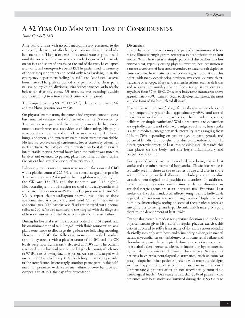
with peripheral destruction. She also underwent treatment withrituximab for encephalomyelitis without platelet response. A CTAngiogram of chest was ordered during her hospitalization for anepisode of shortness of breath. There was no evidence ofpulmonary embolism but a large heterogeneous splenic mass wasvisualized (Figure 1). This mass upon further evaluation with MRIwas hypervascular and the differential diagnosis includedangiosarcoma and hemangioma.
The patient underwent splenectomy and pathology revealed alarge splenic hemangioma with necrosis. Platelet counts steadilyrose from the day of splenectomy and remained stable during theremainder of her hospitalization.
DiscussionThis patient had thrombocytopenia from splenic sequestrationdue to a large hemangioma. Hemangiomas are the most commonprimary tumor of the spleen with a prevalence of 0.03-0.14%.This is a congenital lesion arising from sinusoidal epitheliumresulting from proliferation of vascular channels lined by a singlelayer of endothelium, most often resulting in a cavernous lesion.
Hemagiomas are variable in size ranging from a few millimetersto several centimeters. The majority of these tumors are less than4 cm, but there are reports of lesions up to 17 cm in diameter.Hemangiomas may be single or multiple as in Klippel-Trenauney-Weber Syndrome.
The majority of hemangiomas are asymptomatic and incidentallydiscovered. However, larger lesions may enlarge the spleen leadingto fullness and left upper quadrant discomfort, spontaneous splenicrupture or Kasabach-Merritt phenomenon (thrombocytopeniaand/or coagulopathy, now called disseminated intravascularcoagulation or DIC, that results from platelet trapping within avascular tumor). Thrombocytopenia results from shortened plateletsurvival caused by sequestration of platelets in the vascularmalformation. Episodes of acute DIC have been reported inpregnant women with congenital hemangiomata and in onewoman during two successive pregnancies. The hormonal
Figure 1. CT angiogram of chest withfinding of large splenic mass.
Case Reports
11
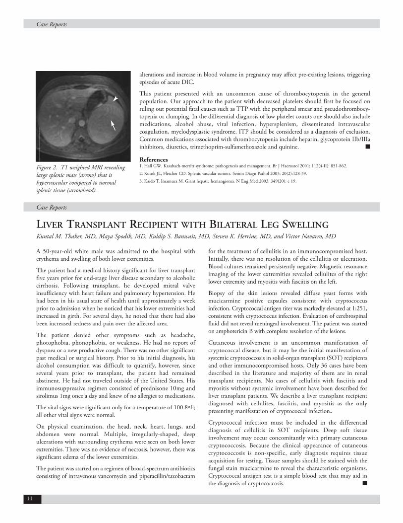
Figure 2. T1 weighted MRI revealinglarge splenic mass (arrow) that ishypervascular compared to normalsplenic tissue (arrowhead).
A 50-year-old white male was admitted to the hospital witherythema and swelling of both lower extremities.
The patient had a medical history significant for liver transplantfive years prior for end-stage liver disease secondary to alcoholiccirrhosis. Following transplant, he developed mitral valveinsufficiency with heart failure and pulmonary hypertension. Hehad been in his usual state of health until approximately a weekprior to admission when he noticed that his lower extremities hadincreased in girth. For several days, he noted that there had alsobeen increased redness and pain over the affected area.
The patient denied other symptoms such as headache,photophobia, phonophobia, or weakness. He had no report ofdyspnea or a new productive cough. There was no other significantpast medical or surgical history. Prior to his initial diagnosis, hisalcohol consumption was difficult to quantify, however, sinceseveral years prior to transplant, the patient had remainedabstinent. He had not traveled outside of the United States. Hisimmunosuppressive regimen consisted of prednisone 10mg andsirolimus 1mg once a day and knew of no allergies to medications.
The vital signs were significant only for a temperature of 100.8ºF;all other vital signs were normal.
On physical examination, the head, neck, heart, lungs, andabdomen were normal. Multiple, irregularly-shaped, deepulcerations with surrounding erythema were seen on both lowerextremities. There was no evidence of necrosis, however, there wassignificant edema of the lower extremities.
The patient was started on a regimen of broad-spectrum antibioticsconsisting of intravenous vancomycin and piperacillin/tazobactam
for the treatment of cellulitis in an immunocompromised host.Initially, there was no resolution of the cellulitis or ulceration.Blood cultures remained persistently negative. Magnetic resonanceimaging of the lower extremities revealed cellulites of the rightlower extremity and myositis with fasciitis on the left.
Biopsy of the skin lesions revealed diffuse yeast forms withmucicarmine positive capsules consistent with cryptococcusinfection. Cryptococcal antigen titer was markedly elevated at 1:251,consistent with cryptococcus infection. Evaluation of cerebrospinalfluid did not reveal meningeal involvement. The patient was startedon amphotericin B with complete resolution of the lesions.
Cutaneous involvement is an uncommon manifestation ofcryptococcal disease, but it may be the initial manifestation ofsystemic cryptococcosis in solid-organ transplant (SOT) recipientsand other immunocompromised hosts. Only 36 cases have beendescribed in the literature and majority of them are in renaltransplant recipients. No cases of cellulitis with fasciitis andmyositis without systemic involvement have been described forliver transplant patients. We describe a liver transplant recipientdiagnosed with cellulites, fasciitis, and myositis as the onlypresenting manifestation of cryptococcal infection..
Cryptococcal infection must be included in the differentialdiagnosis of cellulitis in SOT recipients. Deep soft tissueinvolvement may occur concomitantly with primary cutaneouscryptococcosis. Because the clinical appearance of cutaneouscryptococcosis is non-specific, early diagnosis requires tissueacquisition for testing. Tissue samples should be stained with thefungal stain mucicarmine to reveal the characteristic organisms.Cryptococcal antigen test is a simple blood test that may aid inthe diagnosis of cryptococcosis. ■
LIVER TRANSPLANT RECIPIENT WITH BILATERAL LEG SWELLINGKuntal M. Thaker, MD, Maya Spodik, MD, Kuldip S. Banwait, MD, Steven K. Herrine, MD, and Victor Navarro, MD
alterations and increase in blood volume in pregnancy may affect pre-existing lesions, triggeringepisodes of acute DIC.
This patient presented with an uncommon cause of thrombocytopenia in the generalpopulation. Our approach to the patient with decreased platelets should first be focused onruling out potential fatal causes such as TTP with the peripheral smear and pseudothrombocy-topenia or clumping. In the differential diagnosis of low platelet counts one should also includemedications, alcohol abuse, viral infection, hypersplenism, disseminated intravascularcoagulation, myelodysplastic syndrome. ITP should be considered as a diagnosis of exclusion.Common medications associated with thrombocytopenia include heparin, glycoprotein IIb/IIIainhibitors, diuretics, trimethoprim-sulfamethoxazole and quinine. ■
References1. Hall GW. Kasabach-merritt syndrome: pathogenesis and management. Br J Haematol 2001; 112(4-II): 851-862.
2. Kutok JL, Fletcher CD. Splenic vascular tumors. Semin Diagn Pathol 2003; 20(2):128-39.
3. Kaido T, Imamura M. Giant hepatic hemangioma. N Eng Med 2003; 349(20): e 19.
Case Reports

Case Reports
12
A 55 year old Caucasian female smoker, with past medical historysignificant for hypertension and peptic ulcer disease presented tothe emergency department with a four day complaint of malaise,fever, and myalgias. She was seen by her primary care physicianwho started her on amoxicillin clavulanate for possible strepthroat infection. Her family reported a productive cough withgreen sputum, pleuritic chest pain, nausea, vomiting and diarrheafor one week. They also felt she was getting more confused overthe past several hours.
The patient was found to be confused, hypotensive, and hypoxic.Temp 98.1F, heart rate 107 beats per minute, respiratory rate 16breaths per minute, blood pressure 73/51 mmHg, and oxygenatingat 89% on room air. Physical exam revealed bilateral wheezing thatwas more prominent in the right lung field. She was tachycardicwith no murmurs or rubs. The abdomen was soft and non-tenderwith no organomegaly and she had no evidence of a rash.
Labs on admission: WBC 2,600/mm3, Hemoglobin 13.3 g/dL,platelets 255,000/mm3, Na+ 134, K+ 4.5, HCO3- 19, BUN/Cr49/2.8, AST 85, ALT 53. ABG: pH 7.2, PaCO2 49, PaO2 75,O2 Sat 92%
The differential diagnoses at the time of presentation includedcommunity acquired pneumonia with Streptococcuspneumoniae, Haemophilus influenzae, and Moraxella catharalisbeing high on the list of pathogens along with the atypicalorganisms such as Legionella, Mycoplasma and Chlamydia.Other infectious pathogens such as anaerobes and fungi are alsoincluded. Non-infectious etiologies including pulmonaryembolism, vasculitis, hypersensitivity pneumonitis and neoplasticlymphangitis spread were also considered.
The patient was admitted to the ICU with presumptiveadmission diagnosis of septic shock secondary to pneumonia andwas intubated within 1 hour of admission due to hypoxicrespiratory failure requiring FIO2 100% and 16+ PEEP tomaintain PO2 of 70 mmHg. Stress dose steroids were startedempirically, as well as empiric antibiotic coverage withazythromicin, pipericillin/tazobactam, and vancomycin.Recombinant human activated protein C was also administered12 hours after admission. Two days later urine legionella antigenwas found positive and antibiotics were switched to levofloxacin.The patient required pressure support for ten days and wasextubated on the fifteenth day of hospitalization.
Epidemiology and clinical manifestationsFirst identified in 1976 during an outbreak at the annual meetingof the American Legion in Philadelphia (where out of 221 peopleinfected, 34 died), Legionella is a facultative intracellular aerobicgram negative rod producing beta-lactamase, hardly visible onGram stain due to its diminutive size. There are two majorpresentations of Legionella infection; the first being Legionnaires’Disease which is a community acquired pneumonia whose coursecan become very dramatic and the second being Pontiac fever
which is a more benign and self limited disease manifested throughheadaches, low grade fevers, malaise and chills without respiratorycomplaints and no radiological x-ray findings. Pneumonia is themost common clinical manifestation and Legionella has beenreported as the third or fourth most commonly identifiedpathogen in community acquired pneumonias.1-2 However, it didaccount for only 4.7% of the CAP cases in one large study.3 Itsprevalence is highlighted by the fact that it is also mentioned asone of the most common pathogens identified in nosocomialpneumonia.
Legionnaires’ DiseaseWith an incubation period of two to ten days, this pathogen isconsidered transmitted through to the air conditioning and watercooling systems, rather then transmitted from person to person.Although first thought to manifest only as severe pneumonia(second only to S. pneumoniae in organisms identified in ICUadmissions for pneumonia) accompanied by gastrointestinalsymptoms and high fevers4-6, current diagnostic testing provedthat presentation of Legionella infection may vary widely and thatsymptoms may be nonspecific.
Among the clinical clues for diagnosis of Legionnaires’ disease arethe gastrointestinal symptoms, especially diarrhea; neurologicalfindings such as confusion, headache and lethargy; and fever >390 C. Cough is usually mild and only slightly productive,hemoptysis is rarely encountered, and chest pain is infrequent.Physical exam is nonspecific with rales and subsequent signs ofconsolidation, fever as high as 40° C, bradycardia, lethargy, andstupor. Sputum gram stain shows WBC abundance but scant orno microorganisms. Hyponatremia (Na+ often less than 130)1,5-6,8,hepatic and renal dysfunction, hematuria, thrombocytopenia andfailure to respond to classic ß lactam antibiotic treatment are oftenspecifically seen in patients with Legionella. One should alwaysmaintain a higher index of suspicion for Legionella infection inpatients considered at risk including smokers, those with chronic
55 YEAR OLD WOMAN WITH PNEUMONIAAndra Popescu, MD
Figure 1. Chest X-ray revealing dense alveolar opacity at the rightlung base with air bronchograms. There are left mid lung atelectaticchanges and significant central vascular pulmonary congestion.

lung disease, and immunosupressed patients such as those receivingsteroids or anti-rejection drugs for post-organ transplant.
Radiological findings are usually already present by the third day ofillness. Most commonly seen are patches consistent with unilobarinfiltrates which may progress to consolidation. Pleural effusionmay also be seen on x-ray, although infrequently9. In the immuno-supressed host, densities may appear as round opacities at the lungbase, often progressing into cavitary lesions. Of note, radiologicalchanges usually lag behind the clinical course, with completeresolution of the infiltrates within 4 weeks to several months fromthe debut of the illness.
Although some clinical manifestions are distinctive for Legionellainfection, none of them are pathognomonic or highly specific.Early detection with prompt and appropriate antibiotic treatmenthave been proven to save lives10,11. Culturing for Legionella spp. isthe single most important laboratory test, but is time consumingand requires adequate respiratory specimen and a special culturemedia. Other tests have proved to be more beneficial: urinaryantigen testing is rapid, sensitive, specific, and not costly.However, it is only useful for the diagnosis of L. pneumophilatype 1 infection (accounting for 90 percent of community-acquired Legionella infections in the United States). Thecombination of culture of an appropriate respiratory specimenand urinary antigen testing are optimal as a diagnostic approach.Serologic tests, although available, are generally far less useful forthe diagnosis of an individual patient but very useful for largeepidemiological studies. While PCR-based tests exist, to date theydo not exceed the sensitivity of culturing the organism. Given theseverity and the high incidence of Legionella infection it isreasonable to perform specific diagnostic tests for all patientsrequiring hospitalization for community acquired pneumonia.
TreatmentThe mortality of untreated or inadequately treated communityacquired Legionnaires’ disease is 16-30%, while the mortality forthe nosocomial Legionnaire’s disease may be as high as 50%12.Timely use of the current diagnostic tests as well as promptantibiotic treatment with active drugs have decreased mortality toless than 10%.
Although frequently used in the past, erythromycin has now beensupplanted by newer macrolides as well as respiratory tract
quinolones. Two studies13,14 showed the higher efficacy of levofloxacincompared to macrolides as well as fewer complications and shorterhospitalizations. Current recommendations are a 7-10 day course ofazithromycin or 10-14 day course of levofloxacin, with a longer 21day course for immunosupressed patients and patients requiringICU admission. Given the high incidence and the severity of thisdisease, current recommendation for community acquiredpneumonia requiring hospitalization is azithromycin either asmonotherapy or along with ß-lactame antibiotics15,16. Monotherapywith an active quinolone is also acceptable. For nosocomialpneumonia, a quinolone (ciprofloxacin or levofloxacin) is theempiric drug of choice since the nosocomial infections are frequentlyassociated with gram negative bacilli. For endocarditis and otherextrapulmonary infections, a combination therapy is recommendedwith levofloxacin or azithromycin plus rifampin. Despite asignificant decrease in mortality with prompt and proper antibiotictreatment, patients often have residual symptoms 17 such as chronicfatigue syndrome (75%) and residual neurological deficits (63%).
Our case patient finished 21-day course treatment withlevofloxacin for ARDS secondary to Legionnaires’ disease.Hospital coursee was complicated by development ofquadriparesis thought to be secondary to ICU neuropathy. Ofnote, Legionella Pneumophilla has also been linked independentlywith quadraparetic complications. The patient did achieve furtherimprovement in her neurological status and was able to betransferred to a rehabilitation facility. ■
References1. Fang, GD, Fine, M, Orloff, J, et al. New and emerging etiologies for community-acquired
pneumonia with implications for therapy: a prospective multicenter study of 359 cases.Medicine 1990; 69:307.
2. Stout, JE, Yu, VL. Legionellosis. N Engl J Med 1997; 337:682.3. Yu, VL, Greenberg, RN, Zadeikis, N, et al. Levofloxacin efficacy in the treatment of
community-acquired legionellosis. Chest 2004; 125:2135.4. Fraser, DW, Tsai, T, Ornstein, W, et al. Legionnaires’ disease: description of an epidemic of
pneumonia. N Engl J Med 1977; 297:1189.5. Kirby, BD, Snyder, KM, Meyer, RD, Finegold, SM. Legionnaires’ disease: report of sixty-five
nosocomially acquired cases of review of the literature. Medicine (Baltimore) 1980; 59:188.6. Mulazimoglu, L, Yu, VL. Can Legionnaires disease be diagnosed by clinical criteria? A
critical review. Chest 2001; 120:1049.7. Roig, J, Aguilar, X, Ruiz, J, et al. Comparative study of Legionella pneumophila and other
nosocomial-acquired pneumonias. Chest 1991; 99:344.8. Yu, VL, Kroboth, FJ, Shonnard, J, et al. Legionnaires’ disease: new clinical perspective from
a prospective pneumonia study. Am J Med 1982; 73:357.9. Tan, MJ, Tan, JS, Hamor, RH, et al. The radiologic manifestations of Legionnaire’s disease.
The Ohio Community-Based Pneumonia Incidence Study Group. Chest 2000; 117:398.10. Lepine, LA, Jernigan, DB, Butler, JC, et al. A recurrent outbreak of nosocomial
legionnaires’ disease detected by urinary antigen testing: evidence for long-termcolonization of a hospital plumbing system. Infect Control Hosp Epidemiol 1998; 19:905.
11. Heath, CH, Grove, DI, Looke, DFM. Delay in appropriate therapy of Legionella pneumoniaassociated with increased mortality. Eur J Clin Microbiol Infect Dis 1996; 15:286.
12. Fraser, DW, Tsai, TR, Orenstein, W, et al. Legionnaires’ disease: Description of anepidemic of pneumonia. N Engl J Med 1977; 297:1189.
13. Blazquez Garrido, RM, Espinosa Parra, FJ, Alemany Frances, L, et al. Antimicrobial chemotherapyfor legionnaires disease: levofloxacin versus macrolides. Clin Infect Dis 2005; 40:800.
14. Mykietiuk, A, Carratala, J, Fernandez-Sabe, N, et al. Clinical outcomes for hospitalizedpatients with Legionella pneumonia in the antigenuria era: the influence of levofloxacintherapy. Clin Infect Dis 2005; 40:794.
15. Bartlett, JG, Dowell, SF, Mandell, LA, et al. Practice guidelines for the management ofcommunity-acquired pneumonia in adults. Clin Infect Dis 2000; 31:347.
16. Niederman, MS, Mandell, LA, Anzueto, A, et al. Guidelines for the Management ofAdults with Community-acquired Pneumonia. Diagnosis, assessment of severity,antimicrobial therapy, and prevention. Am J Respir Crit Care Med 2001; 163:1730.
Figure 2. CT of thechest showing denseright lower lobeconsolidation, scatteredareas of groundglassopacity and scatteredfocal nodular densities.
Case Reports
13
Case Reports
14
A 56-year-old Man with a past medical history significant forlower back pain and GERD was admitted to the hospital fordizziness and laboratory abnormalities.
Five days prior to admission, the patient noted that he felt dizzyand unsure while walking. These symptoms persisted withoutchange and he went to see his primary care doctor the day priorto admission. The patient was given meclizine for his dizzinessand routine labs were drawn. He received a call from his doctoron the day of admission instructing him to be evaluated for anelevated serum calcium.
He described his primary complaint as if his surrounding weremoving. He did not complain of lightheadedness or syncope andreported that his symptoms did not change with position. Thesymptoms did not change if he closed his eyes or when he sneezed,coughed, or strained. There was no associated nausea or vomiting.
His medical history was significant for severe GERD for whichhe took ten to fifteen over-the-counter calcium carbonate tabletsdaily with moderate symptom relief however had not had amedical evaluation for these symptoms. He also reported severelower back pain that he attributed to his strenuous occupationwhich began approximately nine months prior to admission. Atfirst, his back pain was alleviated by occasional ibuprofen use,however at present, the patient was taking six to eight 200 mgover-the-counter tablets of ibuprofen daily. He had not beenevaluated medically for his back pain. He also reported unilateralglaucoma as a child and had required enucleation of his right eye.Recently, he had arthroscopic exploration of his left knee and wasaware that he would eventually require knee replacement.
He reported that he knew of no allergies to medications and wasonly taking the over-the-counter medications as above. His familyhistory was notable only for multiple members of his family withcoronary artery disease at a young age. He knew of no familymembers with cancer or kidney disease. He occasionally drankalcohol at social functions and had never been a tobacco user. Heworks in maintenance and lives with his wife and daughter.
His review of systems was generally unremarkable. He reportedthat he had decreasing urinary stream and volume and increasingfrequency during the past month. He reported no weight loss orconstitutional symptoms.
The temperature was 97.3ºF, the pulse was 73 and regular, theblood pressure was 159/70 andd he was breathing comfortable at16 respirations per minute. His oxygen saturation was 94% onroom air.
On physical examination, he was a pleasant and mildly obese manin no distress. He was normocephalic, his left pupil reacted brisklyto light; he had a prosthetic in his right enucleated eye. There wasno notable nystagmus. The heart, lung, abdominal, and extremityexaminations were all unremarkable. His rectal examination
revealed normal sphincter tone and an asymmetric nodule on theleft lobe approximately 1 cm in diameter. His neurologic examwas non-focal and his cranial nerves were intact.
On admission, laboratory values of note were a serum calcium of15.6 mg/dL and a creatinine of 3.2 mg.dL. The remainder of hischemistry and hematologic values were within normal limits. Aninitial troponin drawn in the emergency department was normal.
He was admitted with a diagnosis of hypercalcemia and for athorough evaluation of an underlying oncologic or endocrineprocess. His initial treatment with 4 mg of zoledronic acid, 400international units of salmon-calcitonin, as well as aggressiveintravenous hydration. He was placed on 40 mg intravenouspantoprazole for relief of his reflux symptoms and he was providedwith acetaminophen for his back pain. On hospital day 2, the serumcalcium level was 8.3 mg/dL and his creatinine was 1.8 mg/dL.
A CT scan of his head was performed which was read to have nointracranial pathology. His initial lumbar x-ray was read asspondylothiasis, likely compression fractures at L4 and L5. Afollow-up MRI of lumbar spine showed multilevel disc bulging,protrusions, and facet arthropathy with moderate bilateralforaminal narrowing, however there was no evidence ofcompression fracture as previously suggested.
A CT of the chest and abdomen were performed and showed noacute pathology. The radiologist noted that severe gastroesophagealreflux was evident by contrast refluxing into the esophagus as wellas faint nonobstructing calculi in the lower pole of the right kidney.A renal ultrasound revealed no hydronephrosis or renal calcifi-cations, and there was clear cortical medullary differentiation.
The results of his serum parathyroid hormone, vitamin D level,and prostate specific antigen were all within normal limits. Urineelectrolytes were evaluated and the calculated fractional excretionof sodium was 7.2%, suggestive of acute tubular necrosis. Of note,his total cholesterol was 291 mg/dL and his low densitylipoprotein was 200 mg/dL.
The consulting urologist felt that his exam findings may beconsistent with asymmetric benign prostatic hypertrophy andplanned a transrectal biopsy as an outpatient. The consultinggastroenterologist performed esophagogastroduodenoscopy andthe results were consistent with Barrett’s esophagus secondary to ahiatal hernia. The patient was continued on pantoprazole.
Without evidence of an oncologic process or elevated parathyroidhormone, the diagnosis of milk-alkali syndrome was made. Highdose ibuprofen likely resulted in acute tubular necrosis and adecreased ability to excrete the high doses of calcium that thepatient was ingesting. The hypercalemia and acute renal failureresolved and the patient was discharged on pantoprazole,atorvastatin, and acetaminophen as needed for pain. He wasinstructed to stop taking calcium carbonate and non-steroidal anti-
A 56-YEAR-OLD MAN WITH VERTIGO AND HYPERCALCEMIAVeronica Rivera and Andrew Rose, MD

inflammatory medications. Appointments with gastrointestinal,urologic, and cardiovascular specialists as well as for a stress testwere made for the patient.
DiscussionThere is an extensive list for the differential diagnosis ofhypercalcemia. Hyperparathyroidism is the most common causein ambulatory patients and malignancy is the most common causein hospitalized patients. The differential of hypercalcemia can bedivided into three major categories. Increased bone resorption canbe caused by hyperparathyroidism, cancer, hyperthyroidism, andPaget’s disease. Increased calcium absorption can be caused byincreased calcium uptake and hypervitaminosis D. Miscellaneouscauses of hypercalcemia include drug toxicity and conditions suchas pheochromocytoma.
The presentation of this patient with milk-alkali syndrome andunderlying renal insufficiency has become rather rare since theintroduction of modern ulcer therapy. It has, however, increasedin frequency with more use of calcium carbonate for osteoporosisprophylaxis. Subsequently, this syndrome has experienced anincreased prevalence in females, whereas before it was morecommon in men . Prior to 1990, the milk-alkali syndromeaccounted for less that 2% of admissions related to hypercalcemiawith a small reported rise during the 1990s. Since calcium hasvarious roles in the body including cardiac and smooth musclecontraction, and in platelet aggregation and function, propertreatment and management of hypercalcemia is imperative.
In this patient, milk-alkali syndrome was suspected as the causeof hypercalcemia after ruling out other causes of hypercalcemia.In one case series, five patients who had milk-alkali syndromepatients were ingesting large quantities of calcium and absorbablealkali and presented with the triad of hypercalcemia, metabolicacidosis, and renal failure. In two of these patients, renal failureresulted in the need for dialysis. Kapnser, et al. reported on 297heart and heart-lung transplant recipients who were being treatedwith calcium carbonate after cardiac transplantation. Thistreatment resulted in sixty-five patients who developed serioushypercalcemia after transplantation. Thirty-one of these patientsexperienced alkalosis and thirty-seven of had renal impairment.
The use of calcium in patients with preexisting renal insufficiencyis an important consideration. Four patients in a case report with
mild, asymptomatic chronic renal failure took daily over-the-counter antacids and required hospital admission forhypercalcemia. It is also imperative to monitor calcium use inpregnant patients. Maternal prolonged hypercalcemia secondaryto primary hyperparathyroidism can affect the fetal circulationand lead to suppression of fetal parathyroid function and lead toneonatal hypocalcemia and tetany or may result in spontaneousabortion and stillbirth. Therefore, monitoring calcium intake inpregnant patients is also very important.
In the setting of hypercalcemia, initial medical managementincludes intravenous fluids that promote the renal excretion ofcalcium. In the setting of hypervitaminosis D such as those ingranulomatous disease, steroids are effective by inhibiting theeffects of vitamin D.2 In the setting of hypercalcemia ofmalignancy, treatment of the underlying cancer promotes a returnto normal calcium levels. Bisphosphonates may also be used asmedical management and potentially may inhibit osteoclastactivity for up to one month. Calcitonin directly inhibitsosteoclastic bone resorption and promotes the renal excretion ofcalcium by decreasing tubular reabsorption.
The milk-alkali syndrome is increasing in prevalence and shouldbe considered in any presentation of hypercalcemia. Patientsprophylactically taking calcium supplements or using over-the-counter antacids should be carefully monitored given theirincreased risk for hypercalcemia. ■
References1. Taniergra, ED. Hyperparathyroidism. Am Fam Physician 2004; 69:333-9.
2. Fiorino AS. Hypercalcemia and alkalosis due to the milk-alkali syndrome: a casereport and review. Yale J Biol Med 1996; 69(6):517-23.
3. Beall, DP, Scofield, RH. Milk-alkali syndrome associated with calcium carbonateconsumption. Medicine 1995; 74:89-96.
4. Ariyan CE, Sosa JA. Assessment and management of patients with abnormal calcium.Critical Care Medicine 2004 (4 Suppl); 32:S146-54.
5. Abreo K., et al. The milk-alkali syndrome. A reversible form of acute renal failure.Arch Int Med 1993; 153(8):1005-1010.
6. Kapsner, P. et al. Milk-alkali syndrome in patients treated with calcium carbonateafter cardiac transplantation. Arch Int Med 1986; 146(10): 1965-8.
7. French, JK, Holdaway, IM, Williams LC. Milk alkali syndrome following over-the-counter antacid self-medication. N Z Med J 1986; 99(801): 322-3.
8. Picolos, MK, Sims, CR, Mastrobattista, JM, et al. Milk-alkali syndrome in pregnancy.Obstet Gynecol 2004; 104:1201-4.
9. Orwoll, ES. The milk-alkali syndrome: current concepts. Ann Intern Med 1982;97(2): 242-8.
Case Reports
15

Case Reports
16
A 74 YEAR OLD WOMAN WITH LEFT SHOULDER PAINJane V. Mayrin, MD
A 74 year old woman presented to the emergency departmentcomplaining of two weeks of left shoulder pain. The pain wassharp, 8 out of 10 in intensity, located in the left shoulder, madeworse with breathing, and associated with mild chest discomfort,diaphoresis, and neck pain. There was no radiation and the painwas not worsened with exertion. The patient has a history ofWarthin tumor of the left parotid gland and had undergonelaryngoscopy and polypectomy two weeks prior to presentation.Around this time she developed the shoulder pain along withoverall weakness. At the time of presentation she reported the painwas only present with deep breathing. She denied any shortness ofbreath, headache, lightheadedness, dizziness, or cough.
In addition to the Warthin tumor, the patient has a history ofcolon cancer with resection 25 years ago. She has a 50 pack-yearsmoking history but denied alcohol or substance abuse. She was aretired nurse and lived at home with her husband. Her familyhistory included a father who died in his eighties from amyocardial infarction and a mother who died in her eighties fromcancer. She only took zolpidem 10mg at night as needed for sleepand reported no allergies to medications.
On physical exam she was afebrile at 97.8 F, heart rate 87 beatsper minute, respiration rate 18 breaths per minute, blood pressure117/79, and had an oxygen saturation of 98% on room air. Herpupils were equal round and reactive to light with normalextraocular movements. The left parotid gland was enlarged. Theneck was supple without lymphadenopathy and a jugular venousdistension of about 10 cm was noted. Her heart had a regular rateand rhythm with a II/IV systolic murmur heard best at the leftsternal border with no radiation and no third heart sound.Auscultation of the lungs revealed fine crackles at bilateral baseswithout any wheezing. Her abdominal exam was unremarkableand she had 2+ pulses in the lower extremities bilaterally withoutany edema. The left upper extremity had full range of motionwithout any pinpoint tenderness or effusion.
Laboratory data included hemoglobin of 11.4 g/dL, WBC countof 8,300/mm3, platelets of 451,000/ mm3, BNP of 365 pg/mLand a chemistry panel that was within normal limits. Her first setof Troponin I was 0.23 ng/mL, with the trend of 0.22, 0.20, 0.15(Reference range <0.05 to 0.5 ). The EKG showed normal sinusrhythm at 85 bmp, with Q waves in leads II, III, and inverted T-wave in lead III. Chest x-ray showed bibasilar atelectasis withprominent right paratracheal stripe likely due to vascularcongestion without any interstitial edema. Due to the nature ofthe patient’s pain, she had a chest CT performed while in theEmergency Department which showed no pulmonary embolusbut was notable for a 3 cm left ventricular aneurysm of theposterolateral wall at the base of the heart along with a smallamount of adjacent pericardial fluid and thickening (Figure 1).
The patient was admitted to the medicine service for further workup of her pain and to rule out myocardial infarction. Even thoughher cardiac enzymes were not in a negative range, she ruled outfor myocardial infarction with three sets of enzymes within thenormal reference range. The night following admission the patientdeveloped rapid atrial fibrillation. Given her baseline hypotension,her rate was controlled with amiodarone, and she was fully antico-agulated with enoxaparin.
The following morning a persantine stress test demonstrated alarge inferolateral perfusion defect with partial reversibility at thebasal segments indicative of persantine-induced ischemia. Theleft ventricular ejection fraction was measured as 56%. She alsohad a transthoracic echocardiogram performed, which wasnotable for a region of the mid posterior wall with very markedthinning and aneurysmal dilatation. Her left ventricle internaldimensions and wall thickness was normal with overall moderateLV systolic dysfunction with inferior, posterior, and basal-midlateral akinesis, mild mitral and tricuspid regurgitation andborderline pulmonary hypertension.
Given the findings of the above diagnostic studies, the patientwas scheduled to have coronary catheterization the followingmorning. However, that night the patient was found unresponsiveand asystole noted on the cardiac monitor. Despite resuscitativeefforts during the PEA code, she never regained a pulse or a bloodpressure and died that evening.
Autopsy revealed 500 cc of hemopericardium (Figure 2a and 2b),atherosclerotic cardiovascular disease with thrombotic occlusionof circumflex artery, 50% occlusion of LAD and RCA, andposterior wall recent myocardial infarction with aneurysmaldilatation.
Figure 1. CT chest showing 3cm left ventricular aneurysm of the posterolateral wall at the base of the heart.

Case Reports
ventricle compared to the right ventricle and rarely involves theatria. The infarct commonly affects the anterior and lateral wallsof the left ventricle and the rupture typically occurs near thejunction of the infarcted and normal myocardium
Morphologic types of free wall rupture1 - normal wall thickness with through and through rupture2 - expansion of soft necrotic zone with wall rupture3 - numerous small perforations in the area of myomalacia
(multicanalicular rupture)4 - normal wall thickness with rupture of the outer layer of
infarcted area5 - large epicardial hematoma under pressure6 - hemorrhagic infarction with grossly intact but abraded and
leaking epicardial surface (bleeding infarct)
Evolution of ruptureWithin the first week after the myocardial infarction, neutrophilspopulate the infarcted area between the necrotic myocytes andproduce a zone of coagulative necrosis which is very soft andpredisposed to rupture. A myocardial rupture usually occurs atthe interface between necrotic and spared myocardium. Oncerupture occurs, the pericardium rapidly fills with blood(hemopericardium) and as a result the right ventricle cannot filland cardiac output rapidly declines.
Pathology of healingAs mentioned above, the initial phase of acute myocardialinfarction is characterized by neutrophil infiltration and myocytenecrosis. The healing phase (after the first week), however, istypified by mononuclear cell and fibroblast infiltration and theabsence of polymorphonuclear cells. Resorption of necroticmyocytes often precedes scar formation with accumulation offibrillar collagen into an increasingly organized network. Thecomplete healing process takes 5- 6 weeks and results in a stable
DiscussionThere are 3 major mechanical complications of acute myocardialinfarction:• Rupture of the LV free wall; • Rupture of the interventricular septum; • Development of mitral regurgitation.This discussion will focus mostly on the rupture of LV free wall.
IncidenceMyocardial free wall rupture is the third most common cause ofdeath after myocardial infarction and is 10 times more commonthan rupture of ventricular septum or papillary muscles. It is arelatively common finding in patients who died of acutemyocardial infarction, with some large studies showing cardiacrupture in 14-26% of these patients. However, of all patients withacute myocardial infarction, the incidence of cardiac rupture isonly 1-3%. If thrombolytics are used, death from rupture occursearlier among these patients and often within 24 hours of drugadministration. However, the use of thrombolytics does notincrease the incidence of myocardial rupture.
Risk factors for myocardial rupture include the use ofthrombolytics, lack of prior history of angina or myocardialinfarction, ST-segment elevation or Q wave development on theinitial ECG, peak CK-MB above 150 IU/L, anterior location ofthe infarction, age greater than 70, and female sex. Theseassociations suggest that the absence of collateral blood flow, assuggested by the lack of previous ischemic symptoms, and the sizeof the infarct are important determinants of the likelihood ofmyocardial rupture.
PathologyMyocardial rupture occurs within the first 5 days after acutemyocardial infarction in about 50% of cases and within 2 weeksin more than 90% of cases. It more frequently involves the left
17

Case Reports
18
scar absent of cellular infiltration. The thinned infarct region isstable and not very vulnerable to mechanical disruption.
New insights into pathology of free wall ruptureThere is a suggestion that defective cardiac remodeling maypredispose the heart for rupture. The matrix metalloproteinase(MMP) has been shown to play an important role in cardiacextracellular matrix (ECM) remodeling and cardiac rupture.Over-expression and activities of MMPs induced by TNF (frominflammatory response) at the site of myocardial infarction mayincrease the degradation of existing collagen in the early stage ofinfarction, thus contributing to cardiac rupture.
Effects of reperfusionIt has been shown that rupture is less common in patients with apatent infarct- related coronary artery. Rupture typically occurs inan area that has been infarcted without successful reperfusion. Anumber of studies has shown that thrombolytic therapy early aftermyocardial infarction improves survival and decreases risk ofmyocardial rupture. Late administration of thrombolytics (6-24hrafter onset of symptoms) does not increase the risk of cardiacrupture, but appears to accelerate the time of onset of ruptureevents (within 24 hrs of treatment).
Clinical presentationMyocardial rupture may present as sudden death in an undetectedor silent myocardial infarction. Complete rupture of LV free wallleads to hemopericardium, cardiac tamponade, and subsequentdeath. Presence of rupture is first suggested by sudden acute rightheart failure and shock, which then progresses rapidly to Pulseless-Electrical-Activity (PEA) codes. During the resuscitative efforts,emergent pericardiocentesis will confirm the diagnosis andtransiently relieve the tamponade.
Incomplete rupture occurs when organized thrombus and thepericardium seal the ventricular perforation. This may progress to
frank rupture with tamponade and death, formation of a falseaneurysm walled off by pericardial tissue and communicating withthe left ventricle through the perforation, or formation of leftventricular diverticulum. Incomplete rupture may present asrecurrent chest pain, nausea, restlessness, abrupt and transienthypotension. It can also cause ECG features mimicking regionalpericarditis.
ManagementThe survival of patients with free wall myocardial rupture largelydepends on rapid recognition and administration of immediatetherapy. Bedside echocardiogram and echocardiogram-guidedpericardiocentesis should be obtained if the diagnosis of ruptureis suspected. If the pericardial fluid includes blood, the patientshould immediately proceed to surgery. Fluids, inotropic agents,pressors, and intra-aortic balloon pumps can be used forhemodynamic stabilization while the patient is being transportedto the operating room. ■
References1. Laham, RJ. Mechanical Complications of Acute Myocardial Infarction. UpToDate.
2. Exadaktylos, N. et al. Left Ventricular Free Wall Rupture During Acute MI. EarlyDiagnosis and Treatment. Hellenic J Cardiol. 43: 246-252. 2002
3. Figueras J. Diagnosis, Treatment and Prognosis of Cardiac Rupture in Acute MI.From: http://fac.org.ar/scvc/llave/coronary/figueras
4. Oliva BP, Hammil SC. Cardiac Rupture, a Clinically Predictable Complication ofAcute MI: Report of 70 Cases with Clinicopathologic Correlations. J Amer ColCardiol. 22(3): 720-6, Sep 1993.
5. Solomon, S. Renin-Angiotensin System and Cardiac Rupture After MyocardialInfarction. Circulation. 106(17): 2167-2169, Oct 2002
6. Sun, M et al. Excessive Tumor Necrosis Factor Activation After InfarctionContributes to Susceptibility of Myocardial Rupture and Left VentricularDysfunction. Circulation. 110(20): 3221-3228, Nov 2004
7. REEDER, G. Identification and Treatment of Complications of MyocardialInfarction. Mayo Clin Proced. 70(9): 880-884, Sep 1995
8. Wehrens XH, Doevendans PA. Cardiac rupture complicating myocardial infarction.Internat J Cardiol. 95 (2-3): 285-92, Jun 2004.
9. UpToDate.
Figure 2a. Posterior wall ventricular aneurysm Figure 2b. Hemopericardium on autopsy.

Case Reports
19
A 63-year-old male is admitted to the CCU for chest pain,shortness of breath, and an elevated troponin.
The patient is a 63-year-old male with a past medical historysignificant for hypertension and hypercholesterolemia whopresented to the emergency room with a chief complaint ofshortness of breath and chest pain. The chest pain began two daysprior and was associated with nausea. The patient proceeded tohave increasing shortness of breath over the subsequent two dayswhich brought him to the emergency room for evaluation.
In the emergency room, the patient was noted to be in atrialfibrillation with rapid ventricular response and hypotension. Hewas cardioverted into sinus rhythm. The ECG showed sinusrhythm, RBBB and inferior myocardial infarction ageindeterminate. The cardiac enzymes drawn in the emergencyroom showed a troponin of 30 ng/mL. The patient was transferredto the Coronary Care Unit. An echocardiogram performed in theCoronary Care Unit demonstrated a ventriculoseptal defect. Thepatient was emergently taken to the catheterization lab.
The right heart catheterization demonstrated a mean right atrialpressure of 18 mmHg, right ventricular pressure of 42/18 mmHg,pulmonary pressure of 40/24 mmHg, and a pulmonary capillarywedge pressure of 23 mmHg. The right atrial oxygen saturationwas 31% and the right ventricular oxygen saturation was 81%.
The left ventriculogram demonstrated severe anterolateralhypokinesis and apical hypokinesis. There was evidence of dyeexstravasation just superior to the anterolateral segment with latefilling of the pulmonary artery suggesting a left to right shunt.Coronary arteriography demonstrated a 100% proximal occlusionof the left anterior descending artery after the first diagonal. Theleft circumflex artery had diffuse disease in the marginal system.The remaining vessels were angiographically normal.
The patient was taken emergently to the operating room.Intraoperative transesophageal echocardiogram revealed a largeventricular septal defect in the apical portion of the septumlocated primarily anteriorly. The patient underwent coronaryartery bypass grafting of the left internal mammary to the leftanterior descending artery and the left radial artery to the firstdiagonal artery. The patient also received a bovine pericardialpatch repair of the anterior apical VSD.
The remainder of his hospital stay was unremarkable and he wasdischarged, however soon after discharge the patient began todevelop progressively worsening shortness of breath requiringreadmission to the hospital.
Echocardiogram showed a large color flow turbulence movingacross the septum consistent with a VSD. This was confirmed bycardiac MRI which showed a VSD between the left ventricle and
RUPTURE RECURRENCE AFTER SURGICAL REPAIR OF A POSTINFARCTIONVENTRICULAR SEPTAL RUPTURE IN THE REPERFUSION ERAApurva D. Shah, MD1 and Alan K. Tannenbaum, MD2
1 Apurva Shah, MD was a resident ofThomas Jefferson University Hospital andpresently is a cardiology fellow atUMDNJ/Robert Wood Johnson MedicalSchool in the Department of Medicine,Division of Cardiovascular Diseases
2 Alan Tannenbaum, MD is an assistantprofessor of medicine at UMDNJ/RobertWood Johnson Medical School in theDepartment of Medicine, Division ofCardiovascular Diseases
Figure 1. 100% occlusion of the midLeft Anterior Descending Artery
Figure 2. Irregularities of the RightCoronary Artery

20
right ventricular outflow tract with a Qp:Qs ratio of 1:2.5. Thisappeared secondary to a torn patch.
The patient underwent a repeat cardiac catheterization. The leftanterior descending artery was 100% occluded (Figure 1). TheRight coronary artery demonstrated eccentric lesions (Figure 2).The LIMA to LAD graft was patent (Figure 3). The Radial graftwas 100% occluded proximally (Figure 4). The left ventricu-logram demonstrated dye extravasation into the right ventricleconsistent with a VSD (Figure 5 ) and akinesis of the anterolateraland septal segments.
The patient was taken to the operating room where inspection ofthe ventricular cavity revealed a 2 cm diameter hole in the anteriorleft ventricle extending into the right ventricular outflow tract.This area was closed using the patient’s own ventricular musclewith a sandwich technique employing felt.
The patient tolerated the operation and was discharged from thehospital after 2 weeks. Currently, the patient is doing well withno complaints of chest discomfort, shortness of breath, orthopnea.
DiscussionVentricular septal rupture (VSR) is a potentially fatal mechanicalcomplication of an acute myocardial infarction. Prior to the useof reperfusion therapy, ventricular septal ruptures complicatedthree percent of acute myocardial infarctions.1 With reperfusiontherapy, this has decreased to 0.2%.2 Early surgical treatment
remains the treatment of choice. The operation on weak andfragile myocardium may lead to postoperative recurrence of theventricular septal rupture. We present the case of a 63-year-oldmale who developed a ventricular septal rupture after an acuteanterior myocardial infarction treated with percutaneous coronaryintervention. This VSR required surgical repair. His course wascomplicated by a recurrence of the VSR and redo-surgery.
Ventricular rupture complicating acute myocardial infarctionleads to a high mortality. Reperfusion therapy demarcates a changein the incidence and demographics of septal rupture complicatingacute myocardial infarctions. These differences have beendemonstrated by the Global Utilization of Streptokinase andTissue Plasminogen Activator for Occluded Coronary Arteries(GUSTO-I) trial.
Ventricular septal rupture is the result of a full thickness infarctionof the interventricular septum. This is followed by necrosis thatresults in septal rupture. This necrosis can produce two types ofpathologic ruptures, either a simple or complex rupture. Thesimple rupture represents a direct connection between the twoventricles. A complex rupture has a convoluted communicationbetween the two ventricles. Complex morphology is more oftenassociated with inferior wall myocardial infarctions, whereassimple morphology is seen after anterior wall myocardialinfarctions.4
Figure 3. LIMA graft to Left AnteriorDescending Artery
Figure 4. 100% occluded Radial graftproximally
Figure 5. Left Ventriculogramdemonstrating dye extravasation intothe right ventricle
Case Reports

Right heart catheterization is the standard for diagnosingventricular septal rupture. Sampling of the oxygen saturationsdemonstrate a step up between the right atrium and pulmonaryartery. Transthoracic echocardiogram with color flow doppler canalso be used to diagnose. Left ventriculography has also been usedin diagnosing; however, the contrast load may be detrimental tothe ventricular and renal function.
There exists a significant difference in outcomes and patientdemographics in relation to the development of ventricular septalruptures when comparing the pre and post-thrombolytic era. TheGUSTO-I trial established the incidence of VSD after acute MIin patients treated with thrombolytic therapy and to identify theenrollment characteristics and angiographic patterns associatedwith its occurrence2. Patients enrolled in the GUSTO-I trialpresented to and emergency room within six hours of an ST-segment elevation myocardial infarction. Patients wererandomized to streptokinase with either subcutaneous orintravenous heparin, accelerated alteplase with intravenousheparin or the combination of alteplase and streptokinase withintravenous heparin. Patients who developed VSDs wereidentified by review of case reports.
Review of the GUSTO-I data demonstrated an incidence of 0.2%of patients who developed VSDs after acute myocardialinfarctions. The median time from MI symptom onset was 1 day.This differs from the 3-5% and 3-5 days without thrombolysis.Risk factors for the development of VSDs in the post-throbolyticera included: elderly, female and hypertensive, no history ofsmoking, anterior infarction, tachycardia and worse Killip classifi-cation on admission.
Patients who developed VSDs were found to have TIMI grade 0or 1 flow, and 57% had total occlusion of the infarct artery. Theleft anterior descending artery was more often the infarct relatedartery. This is consistent with findings in patients not receivingreperfusion5.
Despite the institution of thrombolytic therapy, the mortalityfrom VSD remains high. From the GUSTO-I data there is nodifference in mortality at 30 days as compared with 1 year. It canbe surmised that if a patients survives the initial insult hisprognosis is good.
Surgical treatment remains the standard of care for patients withVSDs. The mortality from medical management is 94%. Earlysurgical repair is technically demanding. Early surgery is associatedwith a 40% incidence of residual shunt6. Two additionaldifficulties in the operation are identifying the margins of theinfarct and suturing the patch. A pericardium sutured to the
myocardium is used to decrease the amount of residual shunt.The pericardium also reduces the amount of stress on the suturesdue to its compliance.
Despite the improved surgical methods, rupture reoccurrenceremains a feared complication. Review of the literaturedemonstrates a reoccurrence of the VSR between 5 and 25% ofoperated patients7,8. Early thrombolysis is the main determinant ofrecurrence of the VSR. There is a higher incidence of VSR afterthrombolysis9. There are two possible reasons for this. The first maybe that there is a viable appearance to the infarcted tissue afterthrombolysis. This may cause an underestimation of the infractedarea and lead to a mismatch in reconstruction of the repair. Thesecond may be the edema that is caused to the myocardium byreperfusion injury. Once the edema resolves there may be addedstress on the sutures leading to rupture of the sutures.
Reperfusion therapy, in particular thrombolysis, has had an impacton the mechanical complication of acute myocardial infarctionknown as ventricular septal rupture. Thrombolysis has influencedthe incidence, timing and patient demographics. Although theincidence has decreased the onset from symptoms of myocardialinfarction is now 24 hours. The elderly, female and hypertensiveare predisposed to this complication. Once treated surgically, thereis a higher incidence of recurrence in those patients treated withthrombolytics. The effect of percutaneous coronary interventionon the outcome of ventricular septal rupture remains to be seen. ■
References:1. Birnbaum Y, et al. Ventricular Septal Rupture after Acute Myocardial Infarction. New
Eng J Med 2002; 347(18):1426-32.
2. GUSTO Trial Investigators. Risk Factors, Angiographic Patterns, and Outcomes inPatients with Ventricular Septal Defect Complicating Acute Myocardial Infarction.Circ 2000; 100(1):27-32.
3. Radford MJ, Johnson RA, Daggett WM Jr, et al. Ventricular Septal Rupture: a reviewof clinical and physiologic features and an analysis of survival. Circ 1981; 64:545-53.
4. Edwards BS, Edwards WD, Edwards JE. Ventricular septal rupture complicatingacute myocardial infarction: identification of simple and complex types in 53autopsied hearts. Am J Cardiol 1984; 54:1201-4.
5. Skehan JD, Carey C, Norell MS, DeBelder M, Balcon R, Mills PG. Patterns ofcoronary artery disease in post-infarction ventricular septal rupture. Br Heart J. 1989;117:809-16.
6. Deja MA, Szostek J, Widenka K, Szafron B, Spyt TJ, Hickey MS, Sosnowski AW.Post infarction ventricular septal defect-can we do better? Eur J Cardiothorcic Surg2000;18:194-201.
7. Jones MT, Schofield PM, Dark JP, Moussalli H, Deiraniya AK, Lawson RAM, WardC. Bray CL. Surgical repair of acquired ventricular septal defect: determinants of earlyand late outcome. J Thorac Cardiovasc Surg 1987; 93:680-6.
8. Loisance DY, Lordez JM, Deleuze PH, Dubois-Rande J-L. Acute postinfarction septalrupture: long term results. Ann Throcaic Surg 1991; 52:474-8.
9. Cox F, Morshuis W, Plokker H.W., Langemeijer H, Vermeulen F. Rupturerecurrence after surgical repair of postinfarction ventricular septal rupture. Influenceof early thrombolysis. Eur J Cardio-thorac Surg 1996; 10:748-53.
21
Case Reports

Case Reports
22
A 53-year-old white female was admitted to the Bone MarrowTransplant Unit with a worsening pruritic rash.
The patient had a history of IgG lambda multiple myeloma status postautologous stem cell transplant one year prior and more recently, matchedsibling allogenic transplant. Since transplantation, she had a history ofrash, which was diagnosed as graft-versus-host disease (GVHD) by skinbiopsy. During that same hospitalization, she had a transjugular liverbiopsy to rule out GVHD which was complicated by a large intrahepaticand subcapsular hematoma. A catheter to drain the hematoma was placedby interventional radiology, which was still in place. The patient wasdischarged following her allogenic transplant with successful engraftmentof donor stem cells on bone marrow biopsy, without evidence of residualmultiple myeloma. She was on GVHD therapy with tacrolimus,prednisone, and Cellcept. Recently, her rash had returned and wasbecoming increasingly pruritic over several weeks prior to admission.
The patient did not report headache, sore throat, chest or abdominalpain, dyspnea, diarrhea, or dysuria. She had no other significant pastmedical or surgical history. She reported occasional alcoholconsumption and a remote, but limited smoking history. Her activitieshad been limited recently by her illness and she denied any travel. Shehad no knowledge of starting any new medication, lotions, detergents,or soaps in the recent past.
On this admission, the vital signs were normal and she appearedchronically ill. The head, neck, lungs, and heart were unremarkable.Her abdominal exam was significant for a mildly tender right upperquadrant without rebound or guarding. She had normoactive bowelsounds. She had a right upper quadrant drainage catheter with serosan-guinous drainage. Her dermatologic exam was notable for amaculopapular rash on her back, abdomen and thighs noted, whichwas non-blanching. There were no vesicles or bullae.
Upon admission, skin biopsy revealed interface dermatitis consistentwith GVHD and the patient was placed on intravenous steroids. Inaddition, patient had an Adenovirus PCR of 4,600 copies. On hospitalday 7, the patient developed acute diarrhea. Stool sample was positivefor white blood cells; stool culture and C. difficile were negative.Adenovirus was isolated from the stool. Due to concern for GVHD ofthe colon, patient had a colonoscopy performed, revealing multipleulcerations, colitis in the sigmoid colon, and diverticulosis (Figures 1and 2). Biopsies were obtained from the ulcer and the sigmoid colon.Culture of the ulcer was positive for adenovirus and negative for CMV,HSV, and Enterovirus. Pathology of ulcer biopsy revealed glandularepithelial cells with acute inflammation and intranuclear inclusions,morphologically consistent with adenovirus. Immunohistochemicalstaining of the colon biopsy was negative for HSV and CMV and waseventually was positive for adenovirus.
Initially on steroids, her GVHD dermatitis resolved and she was treatedwith Cidofovir and IVIG for her adenovirus infection. Her diarrheaand adenovirus PCR titer both responded appropriately to treatment.
DiscussionAdenovirus is a common infectant causing mild and self-limitedinfections in immunocompetent individuals. In an immunocom-promised population, such as bone marrow transplant recipients, it canhave severe consequences. Patients who have undergone allogenic stemcell transplant are at highest risk for adenovirus disease. Other risk
factors are age (children more often than adults), T-cell depleted grafts,presence of GVHD, and the use of mismatched and unrelated donors.It can manifest as pneumonia, hepatitis, hemorrhagic cystitis, interstitialnephritis, gastroenteritis/colitis, and encephalitis. In one-half of thepatients with invasive adenoviral disease, the outcome is death.Adenovirus can be acquired via a primary infection (person to person),or via reactivation of latent infection/donor organ infection. Adenoviruscolitis is not well studied, as most studies rely on detection of virus instool in symptomatic patients without tissue confirmation. Oftenadenoviral infections of the bowel coexist with GVHD.
Adenovirus can be diagnosed by a number of means. Viral culture it themost sensitive and specific. Cultures from stool or urine may be positivefor months after an acute infection in an immunocompromised patient.Viral antigens detected by enzyme-linked immunosorbent assay (ELISA)are not as sensitive as culture, but can give rapid diagnosis. PCR can beused to detect adenovirus in body fluids and tissues and is highly sensitiveand specific. Histopathology can also be used to identify the characteristicintranuclear inclusions seen in adenovirus infection. These can be similarto CMV; however, adenovirus does not cause intracytoplasmic inclusionsor multinucleated cells like CMV. Immunohistochemical stains can beused to identify to identify HSV, CMV, or adenovirus. Electronmicroscopy can identify the characteristic icosahedral virus seen inadenoviral infections present in the nuclei of infected cells.
Since this infection is associated with significant mortality in the post-transplant population, it is often treated. Antiviral agents are the mainstayof treatment, although limited evidence is available for efficacy.ganciclovir, ribavirin, and vidarabine have shown some activity in vitro,and there are a number of case reports associating Ribavirin with asuccessful outcome. Cidofovir, a nucleotide analog effective against anumber of viruses, has been shown to be active against adenovirus. Noprospective, controlled studies have been performed to date. IntravenousImmunoglobulin (IVIG) theoretically contains neutralizing antibodiesagainst adenovirus; it has been used in conjunction with antiviral therapyin a number of reports. Another strategy used is reduction in immunesuppression as this allows clearance of virus in some patients. ■
References1. Flomenberg et al. Diagnosis and Treatment of Adenovirus Infection in Adults. UpToDate
version 13.2. Available at www.uptodate.com. Accessed October 2005.
2. Ljungman P. Treatment of Adenovirus Infections in the Immunocompromised Host. Eur JClin Microbiol Infect Dis 2004; 23:583-588.
3. Janoff EN. Adenovirus Colitis in the Acquired Immunodeficiency Syndrome.Gastroenterology 1991; 100(4):976-9.
4. Strickler JG et al. Adenovirus in the Gastrointestinal Tracts of Immunosuppressed Patients.Am J Clin Pathol 1992; 97(4):555-8.
A 53-YEAR-OLD WOMAN WITH WORSENING RASH AND DIARRHEABryan Kavanaugh, MD
Figure 1. Area of ulcerationin sigmoid colon.
Figure 2. Punctate erythema ofsigmoid colon.

The patient is a 40 year old African-American female with nosignificant past medical history who presented to the ED with a 2week history of lower extremity edema and shortness of breath.She also noticed intermittent palpitations, dry cough, easy fatigueand a 30 pound weight loss. On examination, the patient’stemperature was 98.9F orally, pulse was 155-170 beats perminute, and blood pressure was 133/93 mmHg. EKG revealedlong P-R tachycardia with heart rate of 170. Chest X- ray wasdone and showed large right pleural effusion with compressiveatelectasis and small left pleural effusion. Transthoracic echocar-diogram at the time of admission was consistent with severe globalLV dysfunction with EF-20% and moderate pulmonaryhypertension. CT Chest, which showed bilateral hilar adenopathyand subsequent biopsy was consistent with non-caseatinggranulomatous lesion. Her initial basic laboratory values,including TSH level, were normal except for BNP which was 569.She underwent left and right heart catheterization, which revealednormal coronary arteries and decreased cardiac index. During thehospital stay, telemetry monitors revealed rhythms consistent withvarious degrees of AV conduction abnormalities. She was startedon steroids and intravenous milrinone for possible sarcoidcardiomyopathy and listed for heart transplant. Her symptomswere gradually improved and she was discharged home withsteroids, beta blockers, diuretics, ace inhibitors and an implantabledefibrillator. She is currently followed as an outpatient and doingextremely well without any cardiac symptoms.
Myocardial sarcoidosis may have restrictive as well as congestivefeatures because cardiac infiltration by sarcoid granulomas resultsnot only in increased stiffness of the ventricular wall but also indiminished systolic contractile function. Occasionally patientswith extensive involvement of the left ventricular myocardiumdevelop left ventricular aneurysms; it has been suggested thatcorticosteroid treatment may convert granulomas to scar tissueand contribute to the development of aneurysmal dilatation. First-degree heart block due to disease of AV Node or bundle of His,and various types of intraventricular conduction defects, arecommon among patients with cardiac sarcoidosis. Complete heartblock is the most common finding in patients with clinicallyevident cardiac sarcoidosis, and occurs at a younger age in patientswith sarcoidosis than in individuals with complete heart blockdue to other etiologies. Corticosteriods can be used to haltprogression of disease, but it has little to no benefit once EF is<30%. Heart transplantation for sarcoidosis with advanced heartfailure has been performed less commonly than lung transplan-tation. Survival has been similar to that of patients undergoingheart transplantion for other indications. Despite the possibilityof recurrence of sarcoidosis in the graft or the appearance ofprogressive extracardiac disease, available evidence supportstransplantation for cardiac sarcoidosis in patients with extensivecardiac involvement and advanced heart failure as long as there isminimal extracardiac involvement. ■
Credits: H&E stains prepared by Bernadette Wildermore MD
Case Reports
23
SARCOID CARDIOMYOPATHYSivakumar Srinivasan, MD and Emanuel Chryssos, MD
Figure 1. Hematoxylin andEosin stain of lymph node atlow power magnificationshowing multiple non-caseating granulomas.(arrows)

Case Reports
24
Figure 2. Higher powermagnification reveals lack ofnecrotic material within thegranuloma. Also seen is peri -granulomatous connective tissue(arrow) consistent with fibrosiswhich can sometimes be a markerof long-standing sarcoid.
Pictured here is the back of a man with leprosy who had beentreated at Acworth Leprosy Hospital in Mumbai, India. Note thediffuse, macular, hypopigmented rash. Leprosy is a chronic diseasecaused by infection with Mycobacterium leprae. The disease canaffect a variety of organs including peripheral nerves, skin, muscle,eye, bone, and testes. Leprosy is diagnosed clinically by thepresence of at least one of the following: hypopigmented patches,loss of cutaneous sensation, thickened nerves, and acid-fast bacilliin nasal or skin smears.
The prevalence of leprosy has decreased worldwide due to multi-drug therapy and public health programs focused on educationand detection of the illness. Multi-drug therapy consists of thebactericidal drugs: rifampin, dapsone, clofazimine, ethionamideand protionamide. Quinolones, minocycline, and clarithromycinare not part of the normal regimen, but can be combined withrifampin, primarily in single lesion leprosy. ■
IMAGE IN MEDICINEAmy Baranoski, MD
Case Reports
In November 2005 Dr. Baranoskispent several weeks observingvarious clinics and hospitals inMumbai, India where shecaptured the following image.

44 year old Hispanic male with no significant past medical history,recently discharged from prison, presented to the hospital with rashall over the body with itching. The rash started on the face, at theback of the ear and then spread to the trunk and legs. He deniedfever, chills, cough, shortness of breath or sick contacts. Physicalexamination revealed a young, comfortable looking male with stablevital signs except for a temperature of 102 F. Skin examination wassignificant for maculo-papular rash (Figures 1 and 2) with vesiclesand pustules, some of which were crusted, found diffusely over face,chest, abdomen, and extremities yet sparing the palms and soles.Oral cavity also showed mucosal lesions. The laboratory valuesincluded WBC count of 7,700/mm3 with 78% segmentedneutrophils, Hgb of 15.4 g/dL, ESR of 13, creatinine of 0.8 mg/dL,albumin of 3.3 g/dL, aspartate transaminase of 73 IU/L, alanine
transaminase of 70 IU/L, and lactate dehydrogenase of 924 IU/L.Arterial blood gas on room air showed a PaO2 of 70. Chest x-ray(Figure 3) showed diffuse bilateral alveolar infiltrates. Chest CT(Figure 4) was significant for wide spread interstitial pneumonia.Serology was positive for varicella zoster IgM antibodies. Thepatient was treated with intravenous acyclovir 800 mg every 8 hoursfor 7 days with excellent clinical improvement and without anyadverse sequelae. ■
References1. Mer M Richards GA. Corticosteroids in life threatening varicella pneumonia Chest
114:426-431,1998
2.Emerson L Gasparetto Varicella pneumonia on immunocompetent adults: report oftwo cases, with emphasis on high resolution computed tomography findings
3. Braz J Infect Dis. 2005 Jun;9(3):262-5.
Case Reports
25
VARICELLA PNEUMONIASivakumar Srinivasan, MD, Rangadham Nagarakanti, MD and Sandhya Nalmas, MD
Figure 1. Maculopapular rash with vesicles and pustules on torso.
Figure 3. Chest X-ray showing Diffuse Bilateral alveolar infiltrates.
Figure 2. Maculopapular rash on extremities.
Figure 4. Chest CT showing interstitial pneumonia.

Case Reports
26
A 41-year-old Caucasian female with a history of hypertensionpresented with exertional chest pain. Her vital signs were stableon admission and physical examination was unremarkable. AnElectrocardiogram (EKG) revealed inferior ST segment elevationsconsistent with acute myocardial infarction and cardiac enzymeswere elevated. She was started on heparin, and underwentimmediate cardiac catheterization. Cardiac catheterizationrevealed an occluded distal right posterior descending artery(RPDA), suggestive of intracoronary thrombus (Figure 1).However close inspection of the RPDA revealed subtle evidenceof a dissection. Ventriculography showed preserved left ventricularfunction and mild hypokinesis of the distal inferior segment. TheRPDA was non- intervenable and she was medically managed.
The hypercoagulable work up was negative. The patient remainedstable for the next two days and was discharged home. Six dayslater, she returned to the emergency room with recurrent chestpain and at that time the EKG showed ST segment elevations inboth the inferior and lateral leads. She was started on intravenousnitroglycerin and heparin and was taken to the cardiac catheteri-zation lab. Angiography showed spontaneous dissection in the leftcircumflex artery (Figure 2) and another dissection in theposterior descending branch of right coronary artery (Figure 3).Both the circumflex and RPDA vessels were deemed too small forpercutaneous intervention. There was no recent history of trauma,cocaine use, estrogen use or pregnancy. The patient was dischargedhome with medical management. ■
RECURRENT SPONTANEOUS CORONARY ARTERY DISSECTIONSSivakumar Srinivasan, MD and Rangadham Nagarakanti, MD
Figure 1. Dissection in the right posteriordescending artery (RPDA).
Figure 2. Spontaneous dissection of theleft circumflex coronary.
Figure 3. Spontaneous dissection of theposterior descending branch of rightcoronary artery.

A 76 year-old African-American female with a history ofhypertension, type II Diabetes Mellitus, hyperlipidemia, andgatroesphageal reflux presented to emergency room complainingof epigastric pain radiating to left substernum which started thenight before presentation. The onset of her sharp pain was at rest,lasted for two hours, and was aggravated by eating and drinking.Her chest pain was followed by gagging and regurgitating of liquidcontents. Her chest pain was non-exertional, and not associatedwith shortness of breath, diaphoresis, nausea, or vomiting. In theEmergency Department, her pain subsided with two sublingualnitroglycerins and 2 milligrams of intravenous morphine. Onfurther questioning the patient stated that the pain had beenintermittent for two months. She had seen her outpatient doctorone week prior to admission, who recommended rantidine andan outpatient stress test.
Past cardiac history was significant for a Persantine stress test twoyears prior that showed normal myocardial perfusion. An echocar-diogram done 3 months prior showed asymmetric septalhypertrophy, impaired relaxation of left ventricle, and a low-normalleft ventricular systolic function with ejection fraction of 55-60%.
Physical exam on admission revealed an afebrile, elderly womanwith stable vital signs who showed no signs of discomfort. Shehad normal heart sounds with no murmurs and no reproduciblechest pain. Her lung, abdomen and extremity examinations werelikewise unremarkable. At the time of admission, her blood workwas remarkable for hemoglobin of 12 g/dL, creatinine of 1.3mg/dL, amylase of 97 U/L, lipase of 20 U/L, myoglobin of 143ng/mL, and troponin I of 0.50 ng/mL (reference range 0.05 -0.50). A chest x-ray performed in Emergency Department showedno acute disease. The admission EKG is shown below (Figure 1).
Figure 1 shows the EKG on initial presentation when the patientwas pain-free. There were new changes when compared to priorEKG. The major finding was biphasic T waves in V2-V3 withconvex morphology of QRS complex. Troponin drawn on arrivalwas 0.50 (reference range <0.05 to 0.50). Eight hours after initialpresentation, her second troponin returned at 4.09. A subsequentECG (Figure 2) done, again the patient was pain-free, revealedsymmetrical inversion of T waves in V2 and V3.
The patient was taken to cardiac catheterization on the next dayand was found to have a 99% occlusion of left main stem, 99%occlusion of LAD with active thrombus, and 50% occlusion ofRCA. Patient was emergently taken for CABG, where sheunderwent grafting of the LAD, obtuse marginal, and PDA.
DiscussionIn retrospect, the patient’s history was consistent with unstableangina, although she did manifest atypical symptomatology.Because of her past medical history including GERD and diabeticneuropathy, such comorbidities may have masked true cardiacchest pain. Unfortunately, our patient went on to develop anteriormyocardial infarction.
The clinical history and EKG findings are consistent with Wellenssyndrome (WS). The syndrome is also referred to as LADcoronary T-wave syndrome. Syndrome criteria include T-wavechanges plus a history of anginal chest pain without serum markerabnormalities: no pathological Q waves, no ST segment elevation,and no loss of precordial (V1-V6) R waves. EKG abnormalities ofWS appear in one of two ways. The more common variantpresents with symmetric and deeply inverted T waves in theprecordial leads, usually V2 and V3 (Figure 2). The less commonvariant presents with biphasic T waves in the precordial leads,again, usually in V2 and V3 (Figure 1). This case highlights the
Pulse
27
WELLENS SYNDROMENicole Farmer, MD and Michael Cohen, MD
Figure 1. Biphasic T waves in V2 and V3 could easily be mistaken for u waves.This represents an uncommon variant of Wellens Syndrome.

fact that EKG abnormalities are present in patients even whenthey do not have the typical chest pain. Moreover, the EKGfindings tend to disappear during bouts of angina withnormalization of ST segment or T wave abnormalities.
WS was originally described in 1982 by Wellens in his paper withde Zwann and Bar. In this landmark study, 75% of all patientswith WS EKG pattern went on to develop extensive anteriormyocardial infarctions within a few days of hospitalization despitemanagement of pain with medical management. In a follow upretrospective study where urgent PCI was performed on all 180patients meeting WS criteria, 100% were found to have at least a50% obstruction in their LAD artery
It is essential that clinicians recognize that patients admitted tothe hospital because of unstable angina who show the charac-teristic EKG findings described have a high likelihood of having acritical proximal LAD lesion. Such patients are at high risk forthe development of an extensive anterior wall MI as was the casefor the patient described. In this subgroup of patients, urgentcoronary angiography and, when possible, coronary revascular-ization should be undertaken. Furthermore, exercise stress testsare contraindicated due to presence of extensive lesions and severalcase reports of death following treadmill stress testing. ■
References1. de Zwaan C, Bar FW, Wellens HJ. “Characteristic electrocardiographic pattern
indicating a critical stenosis high in left anterior descending coronary artery inpatients admitted because of impending myocardial infarction.” American HeartJournal 1982;103(4 pt 2):730-6.
2 de Zwann C, Bar FW, Janssen JH, Cheriex EC, Dassen WR, Brugada P, et al.“Angiographic and clinical characteristics of patients with unstable angina showing anEKG pattern indicating critical narrowing of the proximal LAD coronary artery.”American Heart Journal 1989;117:657-65.
3. Haines DE, Raabe DS, Gundel WD, Wackers FJ. “Anatomic and prognosticsignificance of new T-wave inversion in unstable angina.” American Journal ofCardiology 1983;52:14-8.
4. Tandy TK, Bottomy DP, Lewis JG. “Wellens’ syndrome.” Annals of EmergencyMedicine 1999;33:347-51.
Pulse
28
Figure 2. Symmetric inversion of T waves in V2 and V3 is the common variant of Wellens Syndrome.

A 19 year old patient with a past medical history significant only forrecurrent syncope presented to the emergency department after 90minutes of palpitations, chest tightness, lightheadedness and shortnessof breath. Electrocardiogram on admission is shown in Figure 1.
The patient was immediately treated with infusions ofprocainamide and amiodarone and the tachycardia broke to reveala sinus rhythm. An electrocardiogram from several hours later isshown in Figure 2.
The patient underwent electrophysiologic study and a trans-septalablation of a left lateral accessory pathway. The post-procedure
electrocardiogram is shown in Figure 3. Interestingly, the deltawaves on her initial electrocardiogram (Figure 2) are no longerpresent, an indication of the success of the procedure. Shetolerated the procedure well and was discharged the following day.
In 1930, Louis Wolff, Sir John Parkinson, and Paul Dudley Whitepublished a review of 11 patients who suffered from bouts oftachycardia similar to this patient. To this day, their names arestill well associated with the condition. ■
References1. Wolff, L, Parkinson, J, White, PD. Bundle branch block with a short P-R interval inhealthy young people prone to paroxysmal tachycardia. Am Heart J 1930; 5:685.
Pulse
29
A 19 YEAR OLD GIRL WITH LIGHTHEADEDNESS AND PALPITATIONSAndrew Rose, MD
Figure 1. Wide complex tachycardia with a ventricular rate of approximately 250.
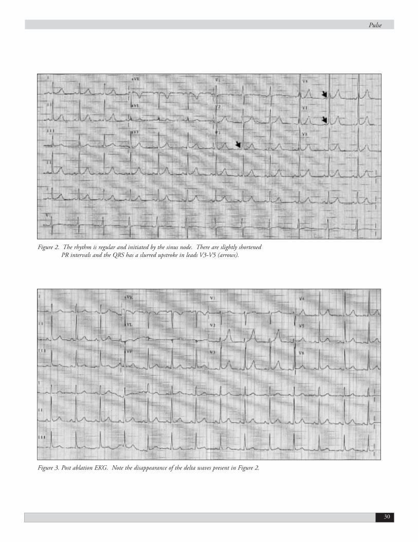
Pulse
30
Figure 2. The rhythm is regular and initiated by the sinus node. There are slightly shortenedPR intervals and the QRS has a slurred upstroke in leads V3-V5 (arrows).
Figure 3. Post ablation EKG. Note the disappearance of the delta waves present in Figure 2.

AbstractContext: Oral contraceptive (OC) use has been shown to beassociated with increased risk of ischemic stroke. However, moststudies were conducted before low-dose OC became available.
Objective: To determine whether low-dose OC use is associatedwith increased risk of ischemic stroke
Data Sources: All relevant human-subject and English-languagestudies in MEDLINE, EMBASE, and Science Citation Indexfrom January 1970 through February 2005
Study Selection and Data Extraction: All published studies of OCor estrogen with any type of stroke as an outcome were included.Studies were excluded if they included populations at high riskfor ischemic stroke or did not include low-dose OC use, ischemicstroke, or a control group.
Data Synthesis: A meta-analysis of the nine included studiesshowed positive association between current low-dose OC useand ischemic stroke (Overall pooled odds ratio [OR], 2.1; 95%confidence interval [CI], 1.7 - 2.7). Compared to never users,current users had slightly higher risk (OR, 1.2; 95% CI, 0.4 -3.2) and former users had significantly lower risk (OR, 0.6; 95%CI, 0.5 - 0.7). Current users of second and third generation OChad 2.4 times (95% CI, 2.0 - 3.0) and 2.0 times (95% CI, 1.3 -3.0) increased risk of ischemic stroke, respectively, compared toformer or never users. The independent effect of low-dose OCuse on the risk of ischemic stroke was stronger among women lessthan 35 years, nonsmokers, and normotensive women.
Conclusion: Current use of low-dose OC compared to former ornever use is associated with about two times increased risk ofischemic stroke. However, considering its health benefits andeffectiveness of birth control and very low incidence of ischemicstroke in young women, use of low-dose OC is generally safe.
BackgroundSince the introduction of oral contraceptive (OC) in the late1950s, a number of epidemiologic studies have shown that OCuse is associated with increased risk of ischemic stroke.1,2 However,most of the studies conducted before 1980s were based on OCscontaining high-dose estrogen (80 or 100 _g) that are no longerused. Oral contraceptives containing low-dose estrogen (less than50 _g) became available in the early 1970s. The formulationcurrently in widespread use in the Unites States contains 30 or 35_g of estrogen.3 Thus, it is not reasonable to generalize about therisk of ischemic stroke associated to the use of low-dose OC fromthe findings based on high-dose formulations.
There are not many studies that examined the risk of ischemicstroke among low-dose OC users. Although it seems thatreduction in estrogen content decreases the thrombogenicity ofOCs and lowers the risk of ischemic stroke, there is aheterogeneity in the reported risk among studies.4-6 Some studiesreported small increase,5-8 whereas others reported no increase inthe risk of ischemic stroke.9,10 Considering the lack of feasibilityof conducting randomized controlled trials and only limitednumber of observational studies showing inconsistent results, it isimportant to summarize the findings from observation studiesand investigate possible reasons for this inconsistency. This willprovide useful information on potential health risk of OCs thatare currently used by over 76 million women worldwide.11
Here, we conducted a meta-analysis to evaluate existing evidencesto determine whether low-dose OC use is associated with increasedrisk of ischemic stroke and to quantify this risk among subgroupswith different characteristics. Specifically, we included onlypremenopausal women who comprise the majority of OC users.The exposure was confined to the use of OCs containing less than50 _g of estrogen that are currently in widespread use. Theoutcome was limited to ischemic stroke, because the strength ofassociation between OC use and hemorrhagic stroke is not strong.12
We reviewed all relevant cohort studies and case-control studiesthat were conducted in the Unites States as well as internationally.
Methods
Study SelectionWe conducted a literature search using MEDLINE, EMBASE, andScience Citation Index (January 1970 through February 2005).The following key words and subject terms were used: (intracerebralhemorrhage OR venous sinus thrombosis OR cerebral infarct ORcerebral ischemia OR cerebrovascular accident OR cerebrovasculardisease OR stroke) AND (oral contraceptive OR birth control pillOR estrogen). The search was restricted to English and humans.The original search yielded 1183 citations from MEDLINE, 730from Science Citation Index, and 132 from EMBASE (excludingduplicates from MEDLINE), for a total of 2045 citations.
Articles
31
RISK OF ISCHEMIC STROKE ASSOCIATEDWITH LOW-DOSE ORAL CONTRACEPTIVE USE: A META-ANALYSISDae Hyun Kim, MD, MPH1, Joachim Bleys, MD, MPH2, Marlis Gonzalez-Fernandez, MD3, Rebecca Gottesman, MD4 and Michelle Hudspeth, MD5
1. Department of Medicine, Thomas Jefferson University,Philadelphia, PA
2. Welch Center for Prevention, Epidemiology, and ClinicalResearch, Department of Epidemiology, Johns HopkinsBloomberg School of Public Health, Baltimore, MD
3. Department of Physical Medicine and Rehabilitation, JohnsHopkins University, Baltimore, MD
4. Department of Neurology and Neurosurgery, Johns HopkinsUniversity, Baltimore, MD
5. Sidney Kimmel Comprehensive Cancer Center, JohnsHopkins University, Baltimore, MD

A preliminary title review was done by five investigators and 377potentially eligible articles were selected for abstract review. Eachabstract was reviewed independently by two investigators, withthe following exclusion criteria:
1. hormone replacement therapy, 2. inclusion of postmenopausal women without stratification, 3. subarachnoid or subdural hemorrhage as the only primary
outcome, 4. estrogen dose exclusively more than 50 mg (first generation
OCs) or dose not specified or progesterone-only contraceptives, 5. stroke not as a primary outcome, 6. women with coagulation disorders, 7. case series without controls, and 8. recurrent stroke.
With these exclusion criteria, we excluded 277 articles.Considering the time when high-dose OCs were withdrawn fromthe market and low-dose OCs became widely available, we further
restricted our analysis to the articles published since 1985, whichleft 55 articles for full-text review. The 55 articles identifiedduring the literature search were reviewed by an investigator (whoinitially had not reviewed the abstract of the article in question)who reassessed the article and discussed the reasons for inclusionor exclusion with the rest of the investigators. From the 55 articles,we excluded 6 studies because of the inclusion of postmenopausalwomen without stratification, 4 because of fatal stroke orhemorrhagic stroke as the only outcome, 5 because of duplicatepublications using the same data, and 10 because of the lack ofcomparison group (case series only). Ten articles were excluded,because they were published as abstracts only, review articles,letters to editor, or editorials and did not contain enoughinformation for the analysis. We also exclude 11 studies that wereconducted before 1990 and did not specify estrogen dose. Finally,data from nine remaining studies were abstracted for our analysis(See Figure 1). There were no randomized controlled trials orcohort studies that satisfied our criteria.
Data AbstractionData were abstracted by the reviewers of each study and verifiedby the rest of the investigators. Any disagreements were resolvedthrough full discussion. Information on study quality as well asdata was collected. Years of study conducted, study region, typeof control (hospital or population control), age distribution ofparticipants, and numbers of case and control were abstracted.Outpatient control was considered as hospital control. Definitionof current OC users varied among studies and we followed thedefinition used by individual studies. Non-current users weredefined as both former and never users. Estrogen dose was definedas low-dose, either when an OC contained less than 50 _g ofestrogen or when it belonged to second or third generation. Thegeneration of OCs was determined by estrogen dose and types ofprogesterone7. Although the definition of generation was notconsistent among studies, estrogen dose in each generation wasconsistent among studies (50 _g or more for first generation, 30-40 _g for second generation, and 20-30 _g for third generation)and we followed the classification used by each study. Estrogendose or generation of OCs was abstracted from each study whenit was mentioned in the article. When it was not mentioned, itwas estimated from the prevailing use pattern in the study areaduring the study period. Generally, the estrogen dose was assumedto be less than 50 _g if a study was conducted after 1990. Whena study was conducted before 1990 and did not specify theestrogen dose or generation of OCs, it was excluded from ouranalysis. Information on exposure to OCs (current, past, or neveruse) was obtained. Information on definition of stroke anddiagnostic methods, such as use of clinical or radiographicdiagnosis or both, was gathered. We excluded hemorrhagic stroke,such as subarachnoid, intracranial, and intraparenchymalhemorrhage. Refusal rate was calculated separately for cases andcontrols as the percentage of eligible individuals who declined toparticipate in a study.
Articles
32
Figure 1. Retrieval of Eligible Studies

Risk estimates used in our meta-analysis were determined a priori.For the primary analysis, the odds ratio (OR) comparing currentwith non-current users after adjusting for potential confounderswas chosen and pooled. Standard errors were calculated fromadjusted odds ratios and their 95% confidence intervals (CIs).Information about potential confounders that were adjusted forin individual studies was also collected. When a study did notprovide the adjusted odds ratio or its 95% CI comparing currentwith non-current users, we excluded the study from the analysis4.In subgroup analyses, we dichotomized age as 35 years or morevs. less than 35 years, smoking as current vs. non-current smokers,and hypertension as present vs. absent. Then we collected the datarelevant to ischemic stroke from each stratified analysis.
Statistical AnalysisWe combined the odds ratios (ORs) comparing current with non-current users from each study using both fixed-effects model andDerSimonian and Laird random-effects model13. However, resultsfrom the random-effects model are presented in this report becausesignificant heterogeneity was identified among studies. In the fourstudies5-8 that provided adjusted ORs for each generation of OCs,we combined the adjusted ORs for second and third generationsusing the random effects model. In a study14 that had stratification
by presence or absence of migraine, overall adjusted OR wascalculated in a similar fashion. A forest plot was created usingadjusted ORs and 95% CIs from each study. One study4 that didnot provide the adjusted OR of ischemic stroke comparing currentwith non-current users was not included when calculating overallOR, but included in other analyses. Heterogeneity among studieswas calculated using chi-square test and studies were defined asheterogeneous when p-value was less than 0.05.
Preplanned subgroup analyses were performed according toexposure classification (current vs. never users and former vs.never users), progesterone type (second generation, thirdgeneration, and second vs. third generation), and presence orabsence of other risk factors for ischemic stroke (≥ 35 years oldvs. < 35 years old, smokers vs. non-smokers, and hypertensives vs.normotensives). To evaluate which study characteristicscontributed to the heterogeneity, we examined the influence ofstudy characteristics on overall OR by applying different inclusioncriteria. In addition, the influence of each study was evaluated byeliminating each study at a time and calculating the resultantoverall OR after the study was excluded. To examine the presenceof publication bias, Egger’s test and Begg’s test were performedand a funnel plot was plotted. All statistical analyses wereperformed using STATA 8.2.
Articles
33
Table 1. Characteristics of Nine Eligible Case-Control Studies for Data Abstraction
Source Region Years Control Age Case/ Estrogen Exposure Stroke Stroke Case/Control(Year) of Study Type (Years) Control# Dose (_g) Assessment Type Diagnosis Refusal Rate (%)Lidegaard Denmark 1985- P 15 - 44 320 / ≥50, Questionnaire Ischemic Clinical 2.2% / 0.1%et al. (1993) 1989 1197 30-40, 20Petitti et al. US 1991- P 15 - 44 295 / <50 Interview Ischemic Clinical 9.6% / N(1996) 1994 774 HemorrhagicWHO International 1989- H 15 - 49 697 / ≥50, <50 Interview Ischemic Clinical 13.7% / NSCollaborative 1993 1952(1996)Barinagarre- Mexico 1987- H 11 - 40 130 / NS NS Ischemic Clinical NS / NSmenteria et al. 1998 122 Radiologic(1998)Heinemann Europe 1993- P, H 16 - 45 220 / ≥50, <50 Interview Ischemic Clincial NS / NSet al. (1998) 1996 775 RadiologicChang et al. Europe 1990- H 20 - 44 291 / ≥50, <50 Interview Ischemic Hemorrhagic NS / NS(1999) 1994 736 ClinicalKemmeren Netherlands 1990- P 19 - 49 203 / ≥50, <50 Questionnaire Ischemic Clinical 31% / 27%et al. (2002) 1995 916 RadiologicLidegaard Denmark 1994- P 15 - 44 609 / ≥50, Questionnaire Ischemic Clinical 2.2% / 0.5%et al. (2002) 1998 3946 30-40, 20Nightingale UK 1992- P 15 - 49 187 / NS UK GPRD Ischemic Clinical NS / NSet al.* (2004) 1998 1129 Radiologic
Abbreviations: WHO, World Health Organization; P, population controls; H, hospital controls; NS, not specified; HTN, hypertension; DM,diabetes mellitus; BMI, body mass index; RHD, rheumatic heart disease; FH, family history; OC, oral contraceptive; UK GPRD, UK GeneralPractice Research Database
* Nested case-control study using the UK General Practice Research Database`

Results
Qualitative AnalysisThe characteristics of nine case-control studies that satisfied ourcriteria were summarized (Table 1). Among the selected studies,six4, 6-8,14,15 were conducted in Europe, one9 in the US, one16 inMexico, and one5 in Europe and developing countries in Asia,Africa, and Latin America. Three studies5,7,14 involved participantsfrom more than one country. All the studies were conducted since1985 and published since 1993. Five studies4,6,8,9,15 used populationcontrol, three5,14,16 used hospital control, and one7 used both. Agedistribution of study subjects ranged from 11 to 49 years old. Thenumber of cases varied from 130 in the study byBarinagarrementeria et al16 to 697 in the study by World HealthOrganization (WHO) collaborative.5 There was only one study9 inwhich estrogen dose in OCs was exclusively less than 50 _g. Sixstudies4-8,14 reported both estrogen dose less than 50 _g and greaterthan 50 _g. Two studies15,16 did not state estrogen dose explicitly. Asfor the study outcome, seven studies4-8,15,16 reported ischemic strokeand two studies9,14 reported both ischemic and hemorrhagic stroke.
Several methodological issues were examined. In most studies,exposure to OCs was measured by mailing questionnaires4,6,8 orpersonal interviews.5,7,9,14 One study16 did not report how theyobtained the information on OC exposure and one study15 usedGeneral Practice Research Database which is an automatednational database collected for research purpose and containsmedical and prescribing records for more than 8.5 million peoplethroughout the UK. With regard to outcome ascertainment, fourstudies7,8,15,16 used a combination of clinical and radiologicaldiagnosis, whereas five studies4-6,9,14 used only clinical diagnosis.
Cases and controls were matched for age in eight studies.4-7,9,14-16
Age was adjusted in three studies6-8 and socioeconomic status wasadjusted in two studies.6,14 Smoking was adjusted in eight studies4-
9,14,15 and hypertension was adjusted in seven studies.5,7-9,14,15 Fourstudies7-9,15 adjusted for diabetes. The study by Lidegaard et al6
controlled for hypertension and diabetes in their initial model,but excluded them from the final model because of lack ofconfounding influence by the variables. The study byBarinagarrementeria et al16 did not provide any information onpotential confounders that they adjusted for (Figure 2).
All of the studies except for one study4 reported adjusted ORcomparing current with non-current users. Adjusted OR comparingcurrent with never users was provided in two studies4,9 and adjustedOR comparing ever vs. never users was provided in one study.9
There were three studies7-9 reporting OR by smoking, two studies8,9
reporting OR by age, four studies5-8 reporting OR by thegeneration of OCs, three studies7-9 reporting OR by history ofhypertension, and one study14 reporting OR by history of migraine.
The definition of exposure to OCs was inconsistent amongstudies. Current OC use was defined as the use of OCs within 1month up to 1 year.
Quantitative AnalysisThe overall pooled OR for ischemic stroke comparing currentwith non-current users calculated by the random-effects modelwas 2.14 (95% CI, 1.69 - 2.72) (Figure 2). Significantheterogeneity was observed among studies (P = 0.02).
In the subgroup analysis by exposure classification, there was weakevidence that current users were associated with increased risk ofischemic stroke compared to never users (OR, 1.18; 95% CI, 0.44- 3.16) (See Table 2). In contrast, former users were associatedwith decreased risk compared to never users (OR, 0.58; 95% CI,0.48 - 0.71). By progesterone type, second generation wasassociated with 2.41 times increased risk of ischemic stroke (OR,2.41; 95% CI, 1.96 - 2.96) and third generation was associatedwith 1.98 times increased risk (OR, 1.98; 95% CI, 1.29 - 3.03).Third generation OCs seemed to be associated with less increasein risk compared to second generation, but there was littledifference between the two formulations in direct comparison.Test of heterogeneity was not significant among studies pooledfor current vs. never users and for third generation OCs.
Current use of low-dose OCs was associated with 1.2 timesincrease in risk of ischemic stroke among women of 35 years orolder (OR, 1.20; 95% CI, 0.19 - 7.56) and 1.48 times increaseamong women less than 35 years (OR, 1.48; 95% CI, 0.77 -2.83). Non-smokers who currently use OCs (OR, 2.60; 95% CI,1.49 - 4.54) seemed to have higher risk of ischemic stroke thansmokers (OR, 2.09; 95% CI, 1.68 - 2.59). Similarly, increase inrisk of ischemic stroke related to OC use was greater amongnormotensive women (OR, 2.55; 95% CI, 1.50 - 4.32) thanamong hypertensive women (OR, 0.71; 95% CI, 0.22 - 2.31).
The findings from the analysis examining the influence of studycharacteristics on overall pooled OR were presented (Table 3).When we included four studies that used radiographic imagingfor the diagnosis of stroke, the pooled OR changed from 2.14 to2.45. The combined OR was 2.81 when the studies withpopulation control were all excluded, whereas it was 1.89 whenthe studies with hospital control were all excluded. Significantheterogeneity was observed only when the studies that did notreport estrogen dose were excluded.
Sensitivity AnalysisThe influence of each study on overall OR was examined (Figure3). The study published in 2002 by Lidegaard et al6 had moderateinfluence on overall OR. Overall OR after omitting the study byLidegaard et al6 was 2.42 (95% CI, 1.98 - 2.94). Omission ofother studies made little difference.
Publication BiasThe possibility of publication bias was evaluated by using the Egger’stest (weighted regression) and Begg’s test (rank correlation method).P-value for bias in the Egger’s test was 0.62 and p-value in Begg’stest was 0.75. A funnel plot showed symmetry around the overallpooled OR, indicating the absence of publication bias (Figure 4).
Articles
34

Articles
35
Table 2. Pooled Odds Ratios and 95% Confidence Intervals (CIs) of Ischemic Stroke from Stratified Meta-analyses*
Data Stratifications Number of Studies Pooled Pooled Test for Heterogeneity(References)† Odds Ratio (95% CI) P-value
All studies 9 (4, 5, 6, 7, 8, 9, 14, 15, 16) 2.14 (1.69 - 2.72) 0.02Exposure classificationCurrent vs. Never users 2 (4, 9) 1.18 (0.44 - 3.16) 0.06Former vs. Never users 2 (4, 9) 0.58 (0.48 - 0.71) 0.55Progesterone type2nd generation 5 (5, 6, 7, 8) 2.41 (1.96 - 2.96) 0.333rd generation 5 (5, 6, 7, 8) 1.98 (1.29 - 3.03) 0.073rd vs. 2nd generation 2 (7, 8) 1.12 (0.76 - 1.64) 0.63Independent effect of oral contraceptive use among:≥ 35 years old 2 (8, 9) 1.20 (0.19 - 7.56) 0.06< 35 years old 2 (8, 9) 1.48 (0.77 - 2.83) 0.71Smokers 3 (7, 8, 9) 2.09 (1.68 - 2.59) 0.33Non-smokers 3 (7, 8, 9) 2.60 (1.49 - 4.54) 0.14HTN present 2 (7, 8) 0.71 (0.22 - 2.31) 0.06HTN absent 3 (7, 8, 9) 2.55 (1.50 - 4.32) 0.08
Abbreviation: HTN, hypertension* Pooled odds ratios are comparing current vs. non-current oral contraceptive users, except for those under exposure classification.† The study by WHO collaborative5 was counted as two separate studies (Europe and developing countries).
Figure 2. Overall Odds Ratio Comparing Current vs. Non-current Users for Ischemic Stroke and 95% Confidence Intervals (CIs)*
Study (Year) Overall Odds Ratio Controlled for(95% CI) Age Race SES Smk HTN DM
Pettiti (1996) 1.18 (0.54 - 2.58) - + - + + +WHO-Europe (1996) 1.53 (0.71 - 3.28) - - - + + -WHO-Developing (1996) 3.26 (2.20 - 4.82) - - - + + -Barinagarrementeria (1998) 2.50 (1.12 - 5.58) NS NS NS NS NS NSHeinemann (1998) 2.77 (2.02 - 3.79) + - - + + +Chang (1999) 2.25 (0.44 - 11.45) - - + + + -Kemmeren (2002) 2.17 (1.52 - 3.09) + - - + + +Lidegaard (2002) 1.61 (1.32 - 1.96) + - + + - -Nightingale (2004) 2.30 (1.16 - 4.57) - - - + + +Overall Odds Ratio 2.14 (1.69 - 2.72)Test for Heterogeneity, P-value: 0.02
Abbreviations: WHO, World Health Organization; SES, social economic status; Smk, smoking; HTN, hypertension; DM, diabetes mellitus, NS, non specified
* Lidegaard et al. (1993) was not included in calculating overall odds ratio, because it did not provide the odds ratio comparing current vs.non-current users. Two components of WHO collaborative (1996) - Europe vs. Developing countries (Asia, Africa, and Latin America) wereconsidered separately.

DiscussionIn our meta-analysis, all the nine studies showed positiveassociation between current low-dose OC use and ischemic strokeand six of them had statistically significant association. The overallpooled OR of ischemic stroke among current low-dose OC users,compared with non-current users, was 2.14 (95% CI, 1.69 - 2.72)(Figure 2). Therefore, our meta-analysis suggests that low-doseOC use is a risk factor for ischemic stroke.
Among the nine included studies, the study published in 1993 byLidegaard et al4 was not included when calculating overall OR,because the authors did not report the standard error or 95% CIassociated with the adjusted OR of 3.46 comparing current withnon-current users. The study by WHO collaborative5 had twodistinct components - Europe and developing countries includingAsia, Africa, and Latin America - showing different results and itwas considered as two different studies. The studies byBarinagarrementeria16 and Nightingale15 were included despite thelack of information on estrogen dose, because the dose wasestimated to be less than 50 _g considering the region and yearsof study. During the literature search, we found three cohortstudies,17-19 but they did not meet our criteria. One cohort studyby Stampfer et al17 was conducted from 1976 through 1984 anddid not have information on estrogen dose. Considering the timeperiod when it was conducted, we suspected that it had includeda significant proportion of high-dose OC users. The other twocohort studies that were conducted from 1980 through 1982 byPorter et al18 and from 1968 through 1994 by Mant et al19
reported only proportion of low-dose OC users without stratifiedresults. High-dose OC users consisted of 59% in the study byPorter et al18 and over 32% in the study by Mant et al.19 We wereunable to extract the data related to low-dose OC use from thethree cohort studies. They were not included in the analysis,because including significant proportion of high-dose OC userswill overestimate the risk of ischemic stroke.
Significant heterogeneity was observed when we combined all thestudies to obtain the overall pooled OR (P = 0.02). It is because allthe included studies are case-control studies of varying qualitiesand characteristics. They are particularly susceptible toconfounding and bias, compared to cohort studies and randomizedcontrolled trials.20 Each study recruited participants at variable riskby applying different inclusion and exclusion criteria and useddifferent methods for assessing exposure and ascertaining outcome.Thus, we used the DerSimonian and Laird random-effects modelto encompass non-random variation between study results andallow more conservative CI for the overall OR.13 Residualconfounding always exists in a meta-analysis of observationalstudies. Some studies did not measure or control for importantrisk factors for ischemic stroke, such as age, smoking, hypertension,and diabetes (Figure 2). In addition, there are several potentialsources for bias in case-control studies. Information on past
exposure to OCs relies on participants’ recall and the extent ofrecall between cases and controls tends to be different, leading tobiased results. The studies by Lidegaard et al4, 6 and Kemmerenet al8 mailed questionnaires to participants, whereas the studies byPetitti et al9, WHO collaborative5, Heinemann et al7, and Changet al14 used personal interviews based on standardized question-naires. The accuracy of information collected by these methodsvaries among studies and is a source of information bias. OnlyNightingale et al15 used a reliable database that has medical andprescribing records collected for research purpose. Classification ofexposure to OCs was also inconsistent between studies. Differentialrefusal to participate in the study between cases and controls mayindicate the presence of selection bias.
We performed subgroup analyses to examine how the associationbetween low-dose OC use and risk of ischemic stroke changesaccording to exposure classification, progesterone types, andwomen with different risk factors for ischemic stroke (Table 2).Compared to never users, current users seemed to have slightlyhigher risk (OR, 1.18; 95% CI, 0.44 - 3.16) and former usershad significantly lower risk (OR, 0.58; 95% CI, 0.48 - 0.71).Decreased risk in former users was consistent between studies.Possible explanation is that never users may have multiple riskfactors and are at higher risk for ischemic stroke than users.Women who stopped taking OCs may be more health-consciousand have a reduced risk due to healthier lifestyle. Past use doesnot seem to have a long term “hangover” effect. Differentprogesterone formulation in OCs was also associated withdifferent risk of ischemic stroke. This may be due to the effect ofdifferent progesterone components, different risk profiles betweenusers of second generation and users of third generation, orpreference of prescribing physicians between studies in differentcountries. Thus, it is possible to underestimate or overestimatethe risk of ischemic stroke associated with third generation OCuse. The independent effect of low-dose OC use on the risk ofischemic stroke was stronger among women less than 35 yearscompared to women of 35 years or older, nonsmokers comparedto smokers, and normotensive women compared to hypertensivewomen. These results are not consistent with recent meta-analysesby Gillum et al21 and Chan et al12 and some studies7, 19, 22 thatsuggested greater risk among OC users with additional risk factorsfor ischemic stroke. Because only small number of the selectedstudies reported stratified information on risk factors, the pooledestimates from our subgroup analyses are less precise, which mayexplain the inconsistency with previous reports.
To find out which study characteristics contributed to theheterogeneity, we examined the influence of certain study charac-teristics on the pooled OR (Table 3). When we only included thestudies that used combination of clinical and radiologicaldiagnosis, the overall OR was 2.45 (95% CI, 1.99 - 3.06) andtest of heterogeneity was insignificant. Exclusion of two studies
Articles
36

that did not report estrogen dose made very little change in theoverall OR and heterogeneity. Studies with population control hadmuch lower pooled OR than studies with hospital control,meaning that control subjects from hospital underreported or lessfrequently used OCs for some reasons. We also evaluated theinfluence of each study on the overall OR by eliminating eachstudy at a time (Figure 3). Although the study by Lidegaard et al6
appeared to have moderate influence, there was not one particularstudy that has significant influence on the overall OR.
A meta-analysis by Gillum et al21 on this topic reported insubgroup analyses that low-dose OC use is associated with abouttwo times increased risk of ischemic stroke (Relative risk, 2.04;95% CI, 1.51 - 2.76). Another recently published meta-analysisby Chan et al12 also reported 2.7 times increase in risk forthrombotic stroke associated with low-dose OC use (OR, 2.74;95% CI, 2.24 - 3.35). However, Chan et al12 included the studiesthat did not have information on estrogen dose and their primaryoutcome was not confined to ischemic stroke. Our finding of 2.1times increase (OR, 2.14; 95% CI, 1.69 - 2.72) is consistent withtheir findings.
This meta-analysis has several limitations. As mentioned above, ameta-analysis of observational studies has methodologicallimitations and susceptibility to bias and confounding, leading to
imprecise estimate of true association. We limited our analysis tothe studies in which the exposure and outcome were clearly statedas low-dose OC and ischemic stroke, respectively. Therefore, onlynine studies were included. This small number substantiallylimited our subgroup analyses and yielded less precise 95% CIs.Due to the small number, we could not conduct a meta-regressionto assess risk of ischemic stroke related to low-dose OC usecontrolling for several study-level covariates. In addition, we couldnot investigate the dose-response relationship between low-doseOC use and ischemic stroke, because most of studies did notreport the duration of OC use. As a result, potential biases andconfounders were not adequately addressed and reasons forheterogeneity between studies were not sufficiently examined.
Presently, more than 10 million women in the United States andmore than 76 million worldwide use OCs.11 Our meta-analysisfound that current use of low-dose OC increases the risk ofischemic stroke approximately by two times. However, when weconsider its health benefits and effectiveness of birth control andvery low baseline incidence of ischemic stroke in young womenof less than 10 per 100,00023, low-dose OC use inpremenopausal women is generally acceptable and safe. Futureresearch is warranted to identify high-risk groups for ischemicstroke associated with low-dose OC use and to investigate thedose-response relationship between the exposure and outcome. ■
Articles
37
Figure 3. Influence of Each Study on Overall Odds Ratio
Meta-analysis Random-effects Estimates (exponential form)
Pettiti (1996)WHO-Europe (1996)
WHO-Developing (1996)Barinagarrementeria (1998)
Heinemann (1998)Chang (1999)
Kemmeren (2002)Lidegaard (2002)
Nightingale (2004)

References1. Masi AT, Dugdale M. Cerebrovascular diseases associated with the use of oral contra-
ceptives. A review of the English-language literature. Ann Intern Med. Jan1970;72(1):111-121.
2. Vessey MP, Doll R. Investigation of relation between use of oral contraceptives andthromboembolic disease. Br Med J. Apr 27 1968;2(599):199-205.
3. Speroff L, DeCherney A. Evaluation of a new generation of oral contraceptives. TheAdvisory Board for the New Progestins. Obstet Gynecol. Jun 1993;81(6):1034-1047.
4. Lidegaard O. Oral contraception and risk of a cerebral thromboembolic attack:results of a case-control study. Bmj. Apr 10 1993;306(6883):956-963.
5. Ischaemic stroke and combined oral contraceptives: results of an international,multicentre, case-control study. WHO Collaborative Study of Cardiovascular Diseaseand Steroid Hormone Contraception. Lancet. Aug 24 1996;348(9026):498-505.
6. Lidegaard O, Kreiner S. Contraceptives and cerebral thrombosis: a five-year nationalcase-control study. Contraception. Mar 2002;65(3):197-205.
7. Heinemann LA, Lewis MA, Spitzer WO, Thorogood M, Guggenmoos-Holzmann I,Bruppacher R. Thromboembolic stroke in young women. A European case-controlstudy on oral contraceptives. Transnational Research Group on Oral Contraceptivesand the Health of Young Women. Contraception. Jan 1998;57(1):29-37.
8. Kemmeren JM, Tanis BC, van den Bosch MA, et al. Risk of Arterial Thrombosis inRelation to Oral Contraceptives (RATIO) study: oral contraceptives and the risk ofischemic stroke. Stroke. May 2002;33(5):1202-1208.
9. Petitti DB, Sidney S, Bernstein A, Wolf S, Quesenberry C, Ziel HK. Stroke in usersof low-dose oral contraceptives. N Engl J Med. Jul 4 1996;335(1):8-15.
10. Schwartz SM, Petitti DB, Siscovick DS, et al. Stroke and use of low-dose oralcontraceptives in young women: a pooled analysis of two US studies. Stroke. Nov1998;29(11):2277-2284.
11. United Nations. World Contraceptive Use 2003. New York: United Nations; 2003.
12. Chan WS, Ray J, Wai EK, et al. Risk of stroke in women exposed to low-dose oralcontraceptives: a critical evaluation of the evidence. Arch Intern Med. Apr 122004;164(7):741-747.
13. Egger M SG, Altman DG. Systematic Reviews in Health Care; Meta-analysis incontext. Second edition ed: BMJ Books; 2001.
14. Chang CL, Donaghy M, Poulter N. Migraine and stroke in young women: case-control study. The World Health Organisation Collaborative Study ofCardiovascular Disease and Steroid Hormone Contraception. Bmj. Jan 21999;318(7175):13-18.
15. Nightingale AL, Farmer RD. Ischemic stroke in young women: a nested case-controlstudy using the UK General Practice Research Database. Stroke. Jul2004;35(7):1574-1578.
16. Barinagarrementeria F, Gonzalez-Duarte A, Miranda L, Cantu C. Cerebralinfarction in young women: analysis of 130 cases. Eur Neurol. Nov 1998;40(4):228-233.
17. Stampfer MJ, Willett WC, Colditz GA, Speizer FE, Hennekens CH. A prospectivestudy of past use of oral contraceptive agents and risk of cardiovascular diseases. NEngl J Med. Nov 17 1988;319(20):1313-1317.
18. Porter JB, Hunter JR, Jick H, Stergachis A. Oral contraceptives and nonfatalvascular disease. Obstet Gynecol. Jul 1985;66(1):1-4.
19. Mant J, Painter R, Vessey M. Risk of myocardial infarction, angina and stroke inusers of oral contraceptives: an updated analysis of a cohort study. Br J ObstetGynaecol. Aug 1998;105(8):890-896.
20. Rothman KJ GS. Modern Epidemiology. Second ed: Lippincott Williams &Wilkins; 1998.
21. Gillum LA, Mamidipudi SK, Johnston SC. Ischemic stroke risk with oral contra-ceptives: A meta-analysis. Jama. Jul 5 2000;284(1):72-78.
22. Oral contraceptives and stroke in young women. Associated risk factors. Jama. Feb 171975;231(7):718-722.
23. Petitti DB, Sidney S, Quesenberry CP, Jr., Bernstein A. Incidence of stroke andmyocardial infarction in women of reproductive age. Stroke. Feb 1997;28(2):280-283.
Articles
38
Table 3. Influence of Study Characteristics on Overall Odds Ratios and 95% Confidence Intervals (CIs) of Ischemic Stroke*
Data Stratifications Number of Studies Pooled Pooled Test for Heterogeneity(References)† Odds Ratio (95% CI) P-value
All studies 9 (5, 6, 7, 8, 9, 14, 15, 16) 2.14 (1.69 - 2.72) 0.02Studies that used radiologic 4 (7, 8, 15, 16) 2.45 (1.99 - 3.06) 0.78imaging for diagnosis of strokeStudies that reported estrogen dose 7 (5, 6, 7, 8, 9, 14) 2.10 (1.59 - 2.78) 0.01Studies that usedHospital control 5 (5, 7, 14, 16) 2.81 (1.83 - 4.30) 0.22Population control 5 (6, 7, 8, 9, 15) 1.89 (1.52 - 2.35) 0.20
* Pooled odds ratios are comparing current vs. non-current oral contraceptive users, except for those under exposure classification.† The study by WHO collaborative5 was counted as two separate studies (Europe and developing countries).

PurposeIn population based studies, the rise of HCV infection hassurpassed alcoholic liver disease (ALD) as the most common causeof chronic liver disease. However, it is unknown if HCV isbecoming the dominant cause of advanced liver disease. The aimof this study was to determine the distribution of etiologies in acohort of patients presenting with advanced liver disease.
MethodsA retrospective review of patients presenting with advanced liverdisease defined as the presence of endoscopically identifiedesophageal or gastric varices for the period between January 1999and December 2002. Etiologies of hepatic injury were identifiedfrom the clinical record and laboratory database. Patients wereexcluded if their disease could be attributed to more than onecause (i.e. alcohol and HCV) or had a non-hepatic cause of portalhypertension. Patients were grouped according to etiology: 1)HCV, 2) ALD, 3) hepatitis B (HBV), or 4) other (cryptogeniccirrhosis, non-alcoholic fatty liver disease, autoimmune hepatitis,portal vein thrombosis).
ResultsA total of 411 patients were identified as having either esophagealand/or gastric varices. The mean age was 56.1 years (range 19-89). There were 275 males (66.9%) and 136 females (33.1%).The proportions of females in those with HCV and ALD weresimilar although women were predominant in non-viral, non-alcohol related disease group compared to other diagnoses (59.4%vs. 24.3%, p<0.0001). Etiology of liver disease differed in thoseolder than 65 years of age compared to younger patients for alldiagnostic categories (p<0.0001) with ALD being more prevalentthan HCV in older patients (p=0.0120). Race was evenlydistributed amongst groups. Hepatitis C represented the singlemost common etiology (42.1%) followed by alcohol (29.9%),other predominantly non-viral, non-alcohol diagnoses (20.0%),and hepatitis B (8.0%).
ConclusionEtiology of liver disease drastically differs in those older than 65years of age compared to younger patients. Higher prevalence ofALD rather than HCV in elderly population alludes to differentrisk behavior pattern between the two groups (i.e., intravenousdrug use more common in younger patients). For patientsyounger than 65 years of age, HCV is the major cause of cirrhosisand burden of complications of cirrhosis attributable to HCVinfection should be expected to rise as this population ages. ■
Abstracts
39
THE SPECTRUM OF ADVANCED LIVERDISEASE IN A TERTIARY CARE INSTITUTIONKuntal M. Thaker, MD, Kuldip S. Banwait, MD, Maya Spodik, MD, Steven K. Herrine, MD and Victor Navarro, MD

Cover Art : “Fruit Mystique”, Pastels on Paper by Stanley Hsu, 2005 Stanley Hsu, MD is currently a Third Year Internal Medicine Resident with a hobbyin sketches and paintings. “Fruit Mystique” is Dr. Hsu’s first publication debut.
JG 05.2317

A d v a n c e d M e d i c i n e . S u p e r i o r C a r e . ™