The impact of gender on fractional flow reserve measurements
7
n 1 CLINICAL RESEARCH © Europa Digital & Publishing 2013. All rights reserved. EuroIntervention 2013;8:0-0 DOI: 10.4244/EIJV8I10A00 *Corresponding author: Department of Cardiology, University Medical Center Mainz, Room 2.228, Geb. 605, Langenbeckstr. 1, 55131 Mainz, Germany. E-mail: [email protected] The impact of gender on fractional flow reserve measurements Massimo Fineschi 1 , MD; Giuseppe Guerrieri 1 , MD; Dörte Orphal 2 ; Elisabetta Palmerini 1 , MD; Thomas Münzel 2 , MD; Ascan Warnholtz 2 , MD; Carlo Pierli 1 , MD; Tommaso Gori 2 *, MD, PhD 1. Department of Cardiology, University Medical Hospital of Siena, Siena, Italy; 2. Medical Clinic, University Medical Center Mainz, Mainz, Germany Abstract Aims: Fractional flow reserve (FFR) allows accurate investigation of the functional significance of interme- diate coronary stenoses. The present study set out to study the impact of gender on FFR measurements. Methods and results: Three hundred and seventeen intermediate (40-70% at angiography) stenoses were assessed with FFR in 270 patients (mean age 65.8±10.3 years, 84 females). Resting Pd/Pa (the ratio of mean blood pressure measured distal to the stenosis to mean aortic blood pressure in resting conditions), FFR (Pd/ Pa during adenosine-induced hyperaemia) and the ΔPd/Pa (calculated as the change in Pd/Pa during hyper- aemia) were measured. There was no difference in the location and degree of stenoses between genders (p>0.5). Similarly, there was no difference in age and in the prevalence of cardiovascular risk factors (all p>0.2). Resting Pd/Pa also did not differ between genders (0.92±0.08 vs. 0.93±0.05, p=0.23). In response to adenosine, however, a significantly larger ΔPd/Pa (0.14±0.07 vs. 0.11±0.07, p=0.001) and a significantly lower FFR (0.79±0.12 vs. 0.82±0.10, p=0.008) were observed in males. This difference was maintained in a multivariate regression analysis. Conclusions: We observed gender-based differences in FFR data in daily routine. Further studies are necessary to test the mechanism of this observation and how these differences impact on the assessment of haemody- namically relevant stenoses. KEYWORDS • fractional flow reserve • gender • microvascular function
Transcript of The impact of gender on fractional flow reserve measurements
n
1
C L I N I C A L R E S E A R C H
© Europa Digital & Publishing 2013. All rights reserved.
EuroIntervention 20
13
;8:0-0
DO
I: 10.42
44
/EIJV8
I10A
00
*Corresponding author: Department of Cardiology, University Medical Center Mainz, Room 2.228, Geb. 605, Langenbeckstr. 1, 55131 Mainz, Germany. E-mail: [email protected]
The impact of gender on fractional flow reserve measurementsMassimo Fineschi1, MD; Giuseppe Guerrieri1, MD; Dörte Orphal2; Elisabetta Palmerini1, MD; Thomas Münzel2, MD; Ascan Warnholtz2, MD; Carlo Pierli1, MD; Tommaso Gori2*, MD, PhD
1. Department of Cardiology, University Medical Hospital of Siena, Siena, Italy; 2. Medical Clinic, University Medical Center Mainz, Mainz, Germany
AbstractAims: Fractional flow reserve (FFR) allows accurate investigation of the functional significance of interme-diate coronary stenoses. The present study set out to study the impact of gender on FFR measurements.
Methods and results: Three hundred and seventeen intermediate (40-70% at angiography) stenoses were assessed with FFR in 270 patients (mean age 65.8±10.3 years, 84 females). Resting Pd/Pa (the ratio of mean blood pressure measured distal to the stenosis to mean aortic blood pressure in resting conditions), FFR (Pd/Pa during adenosine-induced hyperaemia) and the ΔPd/Pa (calculated as the change in Pd/Pa during hyper-aemia) were measured. There was no difference in the location and degree of stenoses between genders (p>0.5). Similarly, there was no difference in age and in the prevalence of cardiovascular risk factors (all p>0.2). Resting Pd/Pa also did not differ between genders (0.92±0.08 vs. 0.93±0.05, p=0.23). In response to adenosine, however, a significantly larger ΔPd/Pa (0.14±0.07 vs. 0.11±0.07, p=0.001) and a significantly lower FFR (0.79±0.12 vs. 0.82±0.10, p=0.008) were observed in males. This difference was maintained in a multivariate regression analysis.
Conclusions: We observed gender-based differences in FFR data in daily routine. Further studies are necessary to test the mechanism of this observation and how these differences impact on the assessment of haemody-namically relevant stenoses.
KEYWORDS
• fractional flow reserve
•gender• microvascular
function
n
2
EuroIntervention 20
12;8
:0-0
IntroductionAlthough coronary angiography remains the cornerstone for the assessment of the presence of epicardial coronary lesions in the catheterisation laboratory, there are several well-known limitations to the accuracy of this technique1. The development of fractional flow reserve (FFR) as a tool for the assessment of the haemody-namic relevance of angiographically intermediate stenoses has brought a dramatic improvement in our diagnostic capacities, facil-itating decision making in the catheterisation laboratory and lead-ing to a substantial improvement in patients’ prognosis2-7.
FFR expresses the ratio of the maximum blood flow achievable in the presence of a stenosis to the maximum blood flow achievable in its absence, and is calculated as the ratio of the mean blood pres-sure distal to the stenosis divided by the mean blood pressure proxi-mal to the stenosis during hyperaemia. FFR only provides information on the epicardial circulation but, since the procedure requires that maximal hyperaemia is achieved, the routine applica-tion of this method is based on the assumption that blood flow responses to fixed doses of vasodilatators invariably reproduce exercise-induced hyperaemia in all patients, and that this hyperae-mia remains constant over time. A number of studies, however, pro-vide evidence that the regulation of coronary blood flow is complex, varies across individuals and over time, and is affected by a number of factors and by the presence of comorbidities such as diabetes, hyperlipidaemia and hypertension8-11. Further, some authors have suggested that drug-induced hyperaemia might not reproduce “physiological” exercise-induced coronary flow reserve12.
While the use of FFR has been validated in the presence of diabetes and hypertension13, a number of other factors might have an impact on this parameter. Among these factors, the existence of gender-based differences in the mechanism, extent, and sequelae of ischaemia is well accepted14. To date, data on the impact of gender on FFR responses are limited. For instance, studies aimed at validating the use of FFR and setting the best cut-off values for the discrimination of haemodynamically rele-vant from non-relevant stenoses included a majority of male subjects15-18, and no gender-specific analysis was performed. This study was designed to investigate retrospectively the exist-ence of gender differences in FFR data obtained in patients with intermediate focal coronary stenoses.
Materials and methodsFFR measurements performed in our institutions between May 2008 and May 2011 were reviewed retrospectively. Patients under-going catheterisation for any reason other than stable (suspected) coronary artery disease (e.g., for hypertensive crisis associated with troponin elevation, or acute coronary syndromes) and those with known chronic inflammatory diseases, severe valvular disease, dialysis, myocardial disease or decompensated heart failure were excluded. Additional exclusion criteria comprised previous myo-cardial infarction in the coronary distribution under study, diffuse atherosclerosis or lesions longer than 5 mm, or the presence of more than one focal stenosis in the target vessel.
Coronary angiography and FFR measurements were performed using the standard Judkins approach. Briefly, all patients received unfractionated heparin. A 6 Fr guiding catheter was positioned at the ostium of the coronary artery, and a previously calibrated 0.014” XT Certus pressure-monitoring guidewire (St Jude Medical, Inc., St Paul, MN, USA) was advanced after equalisation of catheter and wire pres-sures in the periphery of the target vessel. Systolic, diastolic and mean blood pressures were measured proximal and distal to the stenosis at rest after administration of 200 μg of nitroglycerine i.c. After baseline measurements, adenosine was administered using one of two previ-ously validated19 methods: intracoronary bolus (100 μg for the left and 60 μg for the right coronary) or intravenous infusion (via a 4 Fr sheath in the vena femoralis) at 140 or 170 μg/kg*min. The ratio of the mean distal intracoronary pressure (Pd) to the mean arterial pressure (Pa) was calculated at rest (Pd/PaRest) and during maximal hyperaemia (frac-tional flow reserve, FFR). Typically 60-90 seconds (i.e., until stable FFR values for at least 15 seconds were observed) are allowed before FFR measurements in the case of intravenous infusions. In the case of intracoronary bolus, FFR measurements were performed immediately. The change in FFR (ΔPd/Pa) was calculated by subtracting the FFR value after adenosine injection from the Pd/Pa value (FFR before aden-osine injection) and as a % change in the Pd/Pa from baseline. Pullback was used to exclude the presence of diffuse coronary atherosclerosis in all cases (except those who received an adenosine bolus) and tracings with progressive normalisation of the FFR were excluded from the study. The adenosine infusion was maintained until the coronary wire had reached the ostium of the vessel under study. Angiographic lesion severity was determined off-line in randomised order by an investiga-tor blinded to FFR data using quantitative coronary angiography (Xcelera; Philips, Eindhoven, The Netherlands).
CORONARY RISK FACTORSThe body mass index was calculated as the ratio between weight in kilogrammes and the square of the height in meters. Coronary risk factors were defined as: smoking (or history of previous smoking); hyperlipidaemia (total serum cholesterol >220 mg/dL and/or serum triglycerides >200 mg/dL); hypertension (systolic blood pressure >140 mmHg or diastolic blood pressure >90 mmHg on two consecu-tive seated measurements or therapy with antihypertensive medica-tion); family history (first degree relatives with cardiovascular disease); diabetes mellitus (fasting serum glucose levels >126 mg/dL or therapy with oral hypoglycaemic agents or insulin).
STATISTICAL ANALYSISData are presented as mean±SD or number (%). The t-test, the Fisher’s test or analysis of variance were used to compare continuous or nom-inal variables across genders as appropriate. The t-test or analysis of variance for unpaired data was used to assess the effect of nominal variables on FFR and ΔPd/Pa. Linear regression analysis was used to assess the association between single continuous variables, and a stepwise multivariate regression analysis was used to identify parameters independently associated with ΔPd/Pa and FFR (cut-off for entry: 0.05, for removal: 0.10). Variables included in the original
n
3
Gender and FFR assessmentEuroIntervention 2
012
;8:0-0
equation comprised gender, age, body mass index, diabetes, hyper-tension, hyperlipidaemia, smoking, site of the stenosis (vessel involved as well as location in the proximal versus mid or distal third), quantitative coronary angiography parameters, type of hyper-aemic stimulus and the sequence of the vessels studied in cases where more than one vessel was studied in the same patient. Coefficients are presented. All tests of significance were two-tailed. Statistical signifi-cance for the primary endpoint (the impact of gender on ΔPd/Pa and FFR upon multivariate analysis) was defined as a p-value of less than 0.05; all other p-values are reported but are formally to be considered exploratory. The statistical software MedCalc (MedCalc Software, Mariakerke, Belgium) was used for all statistical calculations.
ResultsA total of 270 patients (mean age 65.8±10.3 years, 84 females) undergoing coronary angiography for suspected coronary artery disease (typical angina pectoris and/or positive provocative test and/or ECG suggestive of ischaemia) were included in the data-base. FFR data were available for a total of 317 intermediate (40-70% at angiography) focal (<5 mm) stenoses.
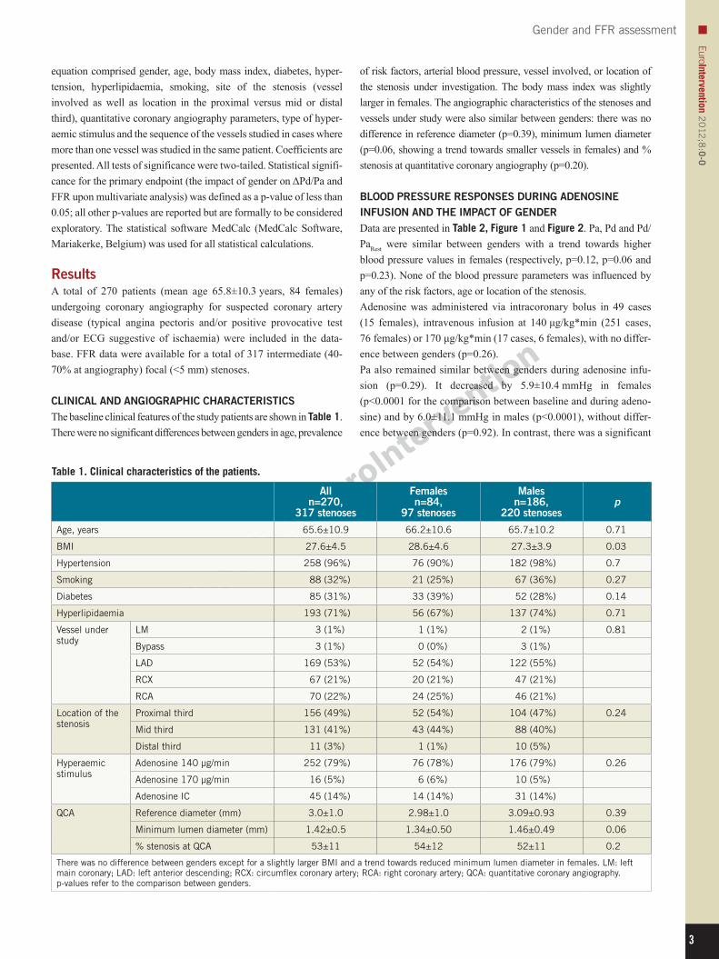
CLINICAL AND ANGIOGRAPHIC CHARACTERISTICS The baseline clinical features of the study patients are shown in Table 1. There were no significant differences between genders in age, prevalence
Table 1. Clinical characteristics of the patients.
Alln=270,
317 stenoses
Femalesn=84,
97 stenoses
Malesn=186,
220 stenosesp
Age, years 65.6±10.9 66.2±10.6 65.7±10.2 0.71
BMI 27.6±4.5 28.6±4.6 27.3±3.9 0.03
Hypertension 258 (96%) 76 (90%) 182 (98%) 0.7
Smoking 88 (32%) 21 (25%) 67 (36%) 0.27
Diabetes 85 (31%) 33 (39%) 52 (28%) 0.14
Hyperlipidaemia 193 (71%) 56 (67%) 137 (74%) 0.71
Vessel under study
LM 3 (1%) 1 (1%) 2 (1%) 0.81
Bypass 3 (1%) 0 (0%) 3 (1%)
LAD 169 (53%) 52 (54%) 122 (55%)
RCX 67 (21%) 20 (21%) 47 (21%)
RCA 70 (22%) 24 (25%) 46 (21%)
Location of the stenosis
Proximal third 156 (49%) 52 (54%) 104 (47%) 0.24
Mid third 131 (41%) 43 (44%) 88 (40%)
Distal third 11 (3%) 1 (1%) 10 (5%)
Hyperaemic stimulus
Adenosine 140 μg/min 252 (79%) 76 (78%) 176 (79%) 0.26
Adenosine 170 μg/min 16 (5%) 6 (6%) 10 (5%)
Adenosine IC 45 (14%) 14 (14%) 31 (14%)
QCA Reference diameter (mm) 3.0±1.0 2.98±1.0 3.09±0.93 0.39
Minimum lumen diameter (mm) 1.42±0.5 1.34±0.50 1.46±0.49 0.06
% stenosis at QCA 53±11 54±12 52±11 0.2
There was no difference between genders except for a slightly larger BMI and a trend towards reduced minimum lumen diameter in females. LM: left main coronary; LAD: left anterior descending; RCX: circumflex coronary artery; RCA: right coronary artery; QCA: quantitative coronary angiography. p-values refer to the comparison between genders.
of risk factors, arterial blood pressure, vessel involved, or location of the stenosis under investigation. The body mass index was slightly larger in females. The angiographic characteristics of the stenoses and vessels under study were also similar between genders: there was no difference in reference diameter (p=0.39), minimum lumen diameter (p=0.06, showing a trend towards smaller vessels in females) and % stenosis at quantitative coronary angiography (p=0.20).
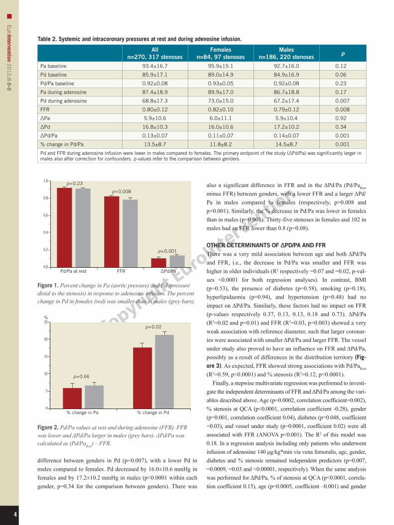
BLOOD PRESSURE RESPONSES DURING ADENOSINE INFUSION AND THE IMPACT OF GENDERData are presented in Table 2, Figure 1 and Figure 2. Pa, Pd and Pd/PaRest were similar between genders with a trend towards higher blood pressure values in females (respectively, p=0.12, p=0.06 and p=0.23). None of the blood pressure parameters was influenced by any of the risk factors, age or location of the stenosis. Adenosine was administered via intracoronary bolus in 49 cases (15 females), intravenous infusion at 140 μg/kg*min (251 cases, 76 females) or 170 μg/kg*min (17 cases, 6 females), with no differ-ence between genders (p=0.26). Pa also remained similar between genders during adenosine infu-sion (p=0.29). It decreased by 5.9±10.4 mmHg in females (p<0.0001 for the comparison between baseline and during adeno-sine) and by 6.0±11.1 mmHg in males (p<0.0001), without differ-ence between genders (p=0.92). In contrast, there was a significant
n
4
EuroIntervention 20
12;8
:0-0
difference between genders in Pd (p=0.007), with a lower Pd in males compared to females. Pd decreased by 16.0±10.6 mmHg in females and by 17.2±10.2 mmHg in males (p<0.0001 within each gender, p=0.34 for the comparison between genders). There was
Pd/Pa at rest FFR
p = 0.23
p = 0.008
p = 0.001
∆Pd/Pa
1.0
0.8
0.6
0.4
0.2
0.0
Figure 1. Percent change in Pa (aortic pressure) and Pd (pressure distal to the stenosis) in response to adenosine infusion. The percent change in Pd in females (red) was smaller than in males (grey bars).
% change in Pa % change in Pd
p = 0.66
p = 0.0225
20
15
10
5
0
%
Figure 2. Pd/Pa values at rest and during adenosine (FFR). FFR was lower and ΔPd/Pa larger in males (grey bars). ΔPd/Pa was calculated as (Pd/PaRest) – FFR.
Table 2. Systemic and intracoronary pressures at rest and during adenosine infusion.
Alln=270, 317 stenoses
Femalesn=84, 97 stenoses
Malesn=186, 220 stenoses
p
Pa baseline 93.4±16.7 95.9±15.1 92.7±16.0 0.12
Pd baseline 85.9±17.1 89.0±14.9 84.9±16.9 0.06
Pd/Pa baseline 0.92±0.08 0.93±0.05 0.92±0.08 0.23
Pa during adenosine 87.4±18.9 89.9±17.0 86.7±18.8 0.17
Pd during adenosine 68.8±17.3 73.0±15.0 67.2±17.4 0.007
FFR 0.80±0.12 0.82±0.10 0.79±0.12 0.008
ΔPa 5.9±10.6 6.0±11.1 5.9±10.4 0.92
ΔPd 16.8±10.3 16.0±10.6 17.2±10.2 0.34
ΔPd/Pa 0.13±0.07 0.11±0.07 0.14±0.07 0.001
% change in Pd/Pa 13.5±8.7 11.8±8.2 14.5±8.7 0.001
Pd and FFR during adenosine infusion were lower in males compared to females. The primary endpoint of the study (ΔPd/Pa) was significantly larger in males also after correction for confounders. p-values refer to the comparison between genders.
also a significant difference in FFR and in the ΔPd/Pa (Pd/PaRest minus FFR) between genders, with a lower FFR and a larger ΔPd/Pa in males compared to females (respectively, p=0.008 and p=0.001). Similarly, the % decrease in Pd/Pa was lower in females than in males (p=0.001). Thirty-five stenoses in females and 102 in males had an FFR lower than 0.8 (p=0.08).
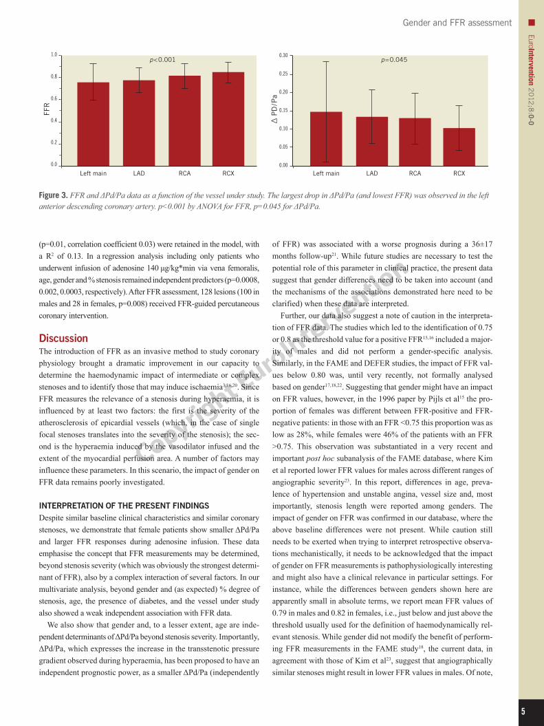
OTHER DETERMINANTS OF ΔPD/PA AND FFRThere was a very mild association between age and both ΔPd/Pa and FFR, i.e., the decrease in Pd/Pa was smaller and FFR was higher in older individuals (R2 respectively =0.07 and =0.02, p-val-ues <0.0001 for both regression analyses). In contrast, BMI (p=0.53), the presence of diabetes (p=0.58), smoking (p=0.18), hyperlipidaemia (p=0.94), and hypertension (p=0.48) had no impact on ΔPd/Pa. Similarly, these factors had no impact on FFR (p-values respectively 0.37, 0.13, 0.13, 0.18 and 0.73). ΔPd/Pa (R2=0.02 and p=0.01) and FFR (R2=0.03, p=0.003) showed a very weak association with reference diameter, such that larger coronar-ies were associated with smaller ΔPd/Pa and larger FFR. The vessel under study also proved to have an influence on FFR and ΔPd/Pa, possibly as a result of differences in the distribution territory (Fig-ure 3). As expected, FFR showed strong associations with Pd/PaRest (R2=0.59, p<0.0001) and % stenosis (R2=0.12, p<0.0001).
Finally, a stepwise multivariate regression was performed to investi-gate the independent determinants of FFR and ΔPd/Pa among the vari-ables described above. Age (p=0.0002, correlation coefficient=0.002), % stenosis at QCA (p<0.0001, correlation coefficient -0.28), gender (p=0.001, correlation coefficient 0.04), diabetes (p=0.048, coefficient =0.03), and vessel under study (p=0.0001, coefficient 0.02) were all associated with FFR (ANOVA p<0.001). The R2 of this model was 0.18. In a regression analysis including only patients who underwent infusion of adenosine 140 μg/kg*min via vena femoralis, age, gender, diabetes and % stenosis remained independent predictors (p=0.007, =0.0009, =0.03 and <0.00001, respectively). When the same analysis was performed for ΔPd/Pa, % of stenosis at QCA (p<0.0001, correla-tion coefficient 0.15), age (p=0.0005, coefficient –0.001) and gender
n
5
Gender and FFR assessmentEuroIntervention 2
012
;8:0-0
(p=0.01, correlation coefficient 0.03) were retained in the model, with a R2 of 0.13. In a regression analysis including only patients who underwent infusion of adenosine 140 μg/kg*min via vena femoralis, age, gender and % stenosis remained independent predictors (p=0.0008, 0.002, 0.0003, respectively). After FFR assessment, 128 lesions (100 in males and 28 in females, p=0.008) received FFR-guided percutaneous coronary intervention.
DiscussionThe introduction of FFR as an invasive method to study coronary physiology brought a dramatic improvement in our capacity to determine the haemodynamic impact of intermediate or complex stenoses and to identify those that may induce ischaemia3,16,20. Since FFR measures the relevance of a stenosis during hyperaemia, it is influenced by at least two factors: the first is the severity of the atherosclerosis of epicardial vessels (which, in the case of single focal stenoses translates into the severity of the stenosis); the sec-ond is the hyperaemia induced by the vasodilator infused and the extent of the myocardial perfusion area. A number of factors may influence these parameters. In this scenario, the impact of gender on FFR data remains poorly investigated.
INTERPRETATION OF THE PRESENT FINDINGSDespite similar baseline clinical characteristics and similar coronary stenoses, we demonstrate that female patients show smaller ΔPd/Pa and larger FFR responses during adenosine infusion. These data emphasise the concept that FFR measurements may be determined, beyond stenosis severity (which was obviously the strongest determi-nant of FFR), also by a complex interaction of several factors. In our multivariate analysis, beyond gender and (as expected) % degree of stenosis, age, the presence of diabetes, and the vessel under study also showed a weak independent association with FFR data.
We also show that gender and, to a lesser extent, age are inde-pendent determinants of ΔPd/Pa beyond stenosis severity. Importantly, ΔPd/Pa, which expresses the increase in the transstenotic pressure gradient observed during hyperaemia, has been proposed to have an independent prognostic power, as a smaller ΔPd/Pa (independently
of FFR) was associated with a worse prognosis during a 36±17 months follow-up21. While future studies are necessary to test the potential role of this parameter in clinical practice, the present data suggest that gender differences need to be taken into account (and the mechanisms of the associations demonstrated here need to be clarified) when these data are interpreted.
Further, our data also suggest a note of caution in the interpreta-tion of FFR data. The studies which led to the identification of 0.75 or 0.8 as the threshold value for a positive FFR15,16 included a major-ity of males and did not perform a gender-specific analysis. Similarly, in the FAME and DEFER studies, the impact of FFR val-ues below 0.80 was, until very recently, not formally analysed based on gender17,18,22. Suggesting that gender might have an impact on FFR values, however, in the 1996 paper by Pijls et al15 the pro-portion of females was different between FFR-positive and FFR-negative patients: in those with an FFR <0.75 this proportion was as low as 28%, while females were 46% of the patients with an FFR >0.75. This observation was substantiated in a very recent and important post hoc subanalysis of the FAME database, where Kim et al reported lower FFR values for males across different ranges of angiographic severity23. In this report, differences in age, preva-lence of hypertension and unstable angina, vessel size and, most importantly, stenosis length were reported among genders. The impact of gender on FFR was confirmed in our database, where the above baseline differences were not present. While caution still needs to be exerted when trying to interpret retrospective observa-tions mechanistically, it needs to be acknowledged that the impact of gender on FFR measurements is pathophysiologically interesting and might also have a clinical relevance in particular settings. For instance, while the differences between genders shown here are apparently small in absolute terms, we report mean FFR values of 0.79 in males and 0.82 in females, i.e., just below and just above the threshold usually used for the definition of haemodynamically rel-evant stenosis. While gender did not modify the benefit of perform-ing FFR measurements in the FAME study18, the current data, in agreement with those of Kim et al23, suggest that angiographically similar stenoses might result in lower FFR values in males. Of note,
Left main
FFR
LAD RCA RCX Left main LAD RCA RCX
p < 0.0011.0
0.8
0.6
0.4
0.2
0.0
∆ P
D/P
a
p = 0.0450.30
0.25
0.20
0.15
0.10
0.05
0.00
Figure 3. FFR and ΔPd/Pa data as a function of the vessel under study. The largest drop in ΔPd/Pa (and lowest FFR) was observed in the left anterior descending coronary artery. p<0.001 by ANOVA for FFR, p=0.045 for ΔPd/Pa.

n
6
EuroIntervention 20
12;8
:0-0
weaker associations were also shown in our database with age, the presence of diabetes and the vessel under study. Differences in microvascular reactivity and in myocardial mass might also help to explain these observations, but these hypotheses remain speculative and further studies are needed.
Limitations of the studyThere are a number of limitations to this study. The first, and most important, is that the cross-sectional, retrospective nature of this data-set makes it unfit to provide mechanistic explanations, and future studies are necessary to test the mechanisms of the observed differ-ences (for instance differences in plaque composition, vessel size, clinical presentation, distribution area, intrinsic differences in the responsiveness to adenosine). Also, the clinical impact of gender-based differences in the parameters under study needs to be studied further. Second, adenosine was administered in different dosages and using different pathways. Previous studies have, however, shown that the different formulations of adenosine have no impact on FFR data19. Also, the statistical analysis was corrected for the formulation of adenosine used showing no impact on ΔPd/Pa and FFR, and a sepa-rate analysis was performed including only those patients who received adenosine via the femoral vein in the dosage of 140 μg/kg*min. Thirdly, we do not provide direct information on the role of microvascular responses (for instance using mean transit time, index of microvascular resistances and/or coronary flow reserve), capillary density, myocardial mass or contractility, and a variety of other potential mechanisms for the present observations, which remain to be investigated. Whatever the mechanism, however, gender-based differences in ΔPd/Pa and FFR are important given the role of these parameters in clinical practice. Further, stenosis length and severity also have an obvious role, and in order to limit the impact of these confounding variables we limited our study to focal lesions and inter-mediate stenoses (40-70% lumen diameter), which are in any case the usual target of FFR studies. Finally, one cannot theoretically exclude that classical angiography might have overestimated stenoses in females compared to males, such that the two groups might have dif-fered in their baseline characteristics, explaining the difference in FFR. However, QCA analysis performed in a randomised blinded order showed that the trend was rather in the opposite direction (more severe stenoses in females compared to males, p=0.06, Table 1).
Conclusions We report on the existence of gender-based differences in FFR data. Mechanistic studies are now important to determine the pathophys-iology of this observation, and prospective studies will need to test whether these observations have clinical implications.
AcknowledgementsPart of the data presented here is included in the MD thesis of D. Orphal.
Conflict of interest statementThe authors have no conflicts of interest to declare.
References 1. Topol EJ, Nissen SE. Our preoccupation with coronary lumi-nology. The dissociation between clinical and angiographic find-ings in ischemic heart disease. Circulation. 1995;92:2333-42. 2. De Bruyne B, Pijls NH, Heyndrickx GR, Hodeige D, Kirkeeide R, Gould KL. Pressure-derived fractional flow reserve to assess serial epicardial stenoses: theoretical basis and animal validation. Circulation. 2000;101:1840-7. 3. Barbato E, De Bruyne B, MacCarthy P, Pijls NH, De Luca G, Volpe M. Functional assessment of coronary atherosclerosis in the catheterization laboratory: the key role of fractional flow reserve. Ital Heart J. 2005;6:549-56. 4. Pijls NH. Is it time to measure fractional flow reserve in all patients? J Am Coll Cardiol. 2003;41:1122-4. 5. Pijls NH, De Bruyne B. Coronary pressure measurement and fractional flow reserve. Heart. 1998;80:539-42. 6. Myat A, Asrress KN, McConkey H, Redwood SR. Physiological assessment of coronary haemodynamics: fractional flow reserve and beyond. EuroIntervention. 2012;7:1236-7. 7. Leesar MA, Satran A, Yalamanchili V, Helmy T, Abdul-Waheed M, Wongpraparut N. The impact of fractional flow reserve measurement on clinical outcomes after transradial coronary stent-ing. EuroIntervention. 2011;7:917-23. 8. Erdogan D, Yildirim I, Ciftci O, Ozer I, Caliskan M, Gullu H, Muderrisoglu H. Effects of normal blood pressure, prehypertension, and hypertension on coronary microvascular function. Circulation. 2007;115:593-9. 9. Picchi A, Limbruno U, Focardi M, Cortese B, Micheli A, Boschi L, Severi S, De Caterina R. Increased basal coronary blood flow as a cause of reduced coronary flow reserve in diabetic patients. Am J Physiol Heart Circ Physiol. 2011;301:H2279-84. 10. Nahser PJ Jr, Brown RE, Oskarsson H, Winniford MD, Rossen JD. Maximal coronary flow reserve and metabolic coronary vasodilation in patients with diabetes mellitus. Circulation. 1995;91:635-40. 11. Melikian N, Vercauteren S, Fearon WF, Cuisset T, MacCarthy PA, Davidavicius G, Aarnoudse W, Bartunek J, Vanderheyden M, Wyffels E, Wijns W, Heyndrickx GR, Pijls NH, de Bruyne B. Quantitative assessment of coronary microvascular function in patients with and without epicardial atherosclerosis. EuroIntervention. 2010;5:939-45. 12. Rossen JD, Nahser PJ Jr, Oskarsson H, Brown RE, Winniford MD. Does pharmacologic coronary flow reserve reflect vasodilator responsiveness to increased myocardial demand in humans? Coron Artery Dis. 1996;7:479-84. 13. Yanagisawa H, Chikamori T, Tanaka N, Usui Y, Takazawa K, Yamashina A. Application of pressure-derived myocardial fractional flow reserve in assessing the functional severity of coro-nary artery stenosis in patients with diabetes mellitus. Circ J. 2004;68:993-8. 14. Vaccarino V, Badimon L, Corti R, de Wit C, Dorobantu M, Hall A, Koller A, Marzilli M, Pries A, Bugiardini R. Ischaemic heart disease in women: are there sex differences in pathophysiol-ogy and risk factors? Position paper from the working group on

n
7
Gender and FFR assessmentEuroIntervention 2
012
;8:0-0
coronary pathophysiology and microcirculation of the European Society of Cardiology. Cardiovasc Res. 2011;90:9-17. 15. Pijls NH, De Bruyne B, Peels K, Van Der Voort PH, Bonnier HJ, Bartunek J, Koolen JJ. Measurement of fractional flow reserve to assess the functional severity of coronary-artery sten-oses. N Engl J Med. 1996;334:1703-8. 16. Pijls NH, Van Gelder B, Van der Voort P, Peels K, Bracke FA, Bonnier HJ, el Gamal MI. Fractional flow reserve. A useful index to evaluate the influence of an epicardial coronary stenosis on myo-cardial blood flow. Circulation. 1995;92:3183-93. 17. Pijls NH, Fearon WF, Tonino PA, Siebert U, Ikeno F, Bornschein B, van’t Veer M, Klauss V, Manoharan G, Engstrom T, Oldroyd KG, Ver Lee PN, MacCarthy PA, De Bruyne B; FAME Study Investigators. Fractional flow reserve versus angiography for guiding percutaneous coronary intervention in patients with multi-vessel coronary artery disease: 2-year follow-up of the FAME (Fractional Flow Reserve Versus Angiography for Multivessel Evaluation) study. J Am Coll Cardiol. 2010;56:177-84. 18. Tonino PA, De Bruyne B, Pijls NH, Siebert U, Ikeno F, van’ t Veer M, Klauss V, Manoharan G, Engstrom T, Oldroyd KG, Ver Lee PN, MacCarthy PA, Fearon WF. Fractional flow reserve versus angiography for guiding percutaneous coronary intervention. N Engl J Med. 2009;360:213-24.
19. De Bruyne B, Pijls NH, Barbato E, Bartunek J, Bech JW, Wijns W, Heyndrickx GR. Intracoronary and intravenous adeno-sine 5’-triphosphate, adenosine, papaverine, and contrast medium to assess fractional flow reserve in humans. Circulation. 2003;107: 1877-83. 20. Bech GJ, De Bruyne B, Pijls NH, de Muinck ED, Hoorntje JC, Escaned J, Stella PR, Boersma E, Bartunek J, Koolen JJ, Wijns W. Fractional flow reserve to determine the appropriateness of angio-plasty in moderate coronary stenosis: a randomized trial. Circulation. 2001;103:2928-34. 21. Kocaman SA, Sahinarslan A, Arslan U, Timurkaynak T. The delta fractional flow reserve can predict lesion severity and long-term prognosis. Atherosclerosis. 2009;203:178-84. 22. Sant’Anna FM, Silva EE, Batista LA, Ventura FM, Barrozo CA, Pijls NH. Influence of routine assessment of fractional flow reserve on decision making during coronary interventions. Am J Cardiol. 2007;99:504-8. 23. Kim SH, Tonino PAL, De Bruyne B, Yong ASC, Tremmel JA, Pijls NHJ, Fearon WF. The Impact of Sex Differences on Fractional Flow Reserve-Guided Percutaneous Coronary Intervention: A FAME (Fractional Flow Reserve Versus Angiography for Multivessel Evaluation) Substudy. JACC Cardiovasc Interv. 2012;5:1037-42.