The hind limb skeleton and cursorial adaptations of the Plio ...
30
The hind limb skeleton and cursorial adaptations of the Plio−Pleistocene rabbit Hypolagus beremendensis ŁUCJA FOSTOWICZ−FRELIK Fostowicz−Frelik, Ł. 2007. The hind limb skeleton and cursorial adaptations of the Plio−Pleistocene rabbit Hypolagus beremendensis. Acta Palaeontologica Polonica 52 (3): 447–476. Hypolagus beremendensis, a representative of the Archaeolaginae, was one of the most abundant and widespread leporids in the Plio−Pleistocene of Europe. The vast accumulations of skeletal remains from the Polish Pliocene sites (Węże 1, Rębielice Królewskie 1 and 2, and Kadzielnia 1) yielded thousands of bones representing almost all skeletal regions. The detailed hind limb morphology of Hypolagus beremendensis is presented in comparison with five extant leporids (Lepus europaeus, Oryctolagus cuniculus, Pentalagus furnessi, Sylvilagus floridanus, and S. brasiliensis), which represent a wide range of locomotor adaptations. The UPGMA analysis of 98 metric characters places Hypolagus beremendensis next to the leporine rabbits. Hypolagus beremendensis has the os coxae, femur, and talus most similar to P. furnessi, tibia and calcaneus to the leporine rabbits, and the structure of foot to Lepus. The elongation of the foot and tibiofibular seg− ment in relation to the femur indicates an advanced cursorial adaptation and a relatively steep jump. The similarities in the proximal segments (os coxae and femur) between Hypolagus and Pentalagus highlight the conservative morphology of this region in the Leporidae. Key words: Lagomorpha, Leporidae, Hypolagus beremendensis, hind limb, functional morphology, cursorial adapta− tions, Neogene. Łucja Fostowicz−Frelik [[email protected]], Institute of Paleobiology Polish Academy of Sciences, ul. Twarda 51/55, PL−00−818 Warszawa, Poland. Introduction Hypolagus Dice, 1917, one of the most speciose genera within Archaeolaginae, currently includes 24 species of rab− bits and is known from the early Miocene to early Pleisto− cene in North America and from the latest Miocene (Turo− lian) to middle Pleistocene in Eurasia. The majority of spe− cies, however, are known from dental and cranial remains (Dawson 1958; White 1984; Voorhies and Timperley 1997; Fostowicz−Frelik 2003). The European record includes at present four species, with Hypolagus beremendensis (Kor− mos, 1930) being the most abundant and widespread. The species is known from the early Pliocene (MN 15) to early Pleistocene (earliest Biharian) period in Central Europe (Kormos 1934; Sych 1965; Fladerer and Reiner 1996; Fosto− wicz−Frelik 2003, 2007). The richest findings of this species come from Poland in the form of the vast accumulations of calcified bone breccia in Węże 1 site, clay−bone deposits in Rębielice Królewskie 1 and 2 (all three sites located in the Cracow−Wieluń Upland), and Kadzielnia 1 in the Holy Cross Mountains (Sulimski 1964; Sych 1965; Nadachowski 1990). The extremely abundant collections that include well− preserved fragments of virtually all bones enable detailed study of the skeleton. The subject is relatively poorly re− searched and the European Hypolagus has not been exam− ined in terms of functional morphology (apart from the forelimb, covered in some aspects by Fladerer 1984). Cur− sorial adptations of the appendicular skeleton of some North American extinct leporids (Hypolagus aff. vetus (Kellogg, 1910) and Pratilepus Hibbard, 1939) were analysed by Campbell (1969). The initial work of Sych (1965) based on the Polish material included only general description of the bone morphology. Sych (1965) and other authors (Fladerer 1984, Fladerer and Fiore 2003) suggested that Hypolagus beremendensis was intermediate in morphology between fossorial Oryctolagus cuniculus and highly cursorial Lepus. Fostowicz−Frelik (2001) noticed that it rather resembled some species of Sylvilagus, like the North American repre− sentatives of Hypolagus mentioned by White (1984). In this paper the comprehensive morphology of the hind limb skeleton of Hypolagus beremendensis is provided, along with quantitative and qualitative comparisons (includ− ing phenetic analysis) with four species of rabbits: Ory− ctolagus cuniculus (Linnaeus, 1758), Pentalagus furnessi (Stone, 1900), Sylvilagus brasiliensis (Linnaeus, 1758), and S. floridanus (Allen, 1890), and the European hare, Lepus europaeus Pallas, 1778. The general leporid morphotype (distinguished from the ochotonid type by the significant elongation of hind legs, particularly shank and foot) is very uniform, conservative, and recognisable already in the Oligocene Palaeolagus hay− deni Leidy, 1856 (Wood 1940). As a result of this shared skeletal morphology the motion and biomechanics is gener− ally similar in all representatives of the group (López−Marti− http://app.pan.pl/acta52/app52−447.pdf Acta Palaeontol. Pol. 52 (3): 447–476, 2007
-
Upload
khangminh22 -
Category
Documents
-
view
4 -
download
0
Transcript of The hind limb skeleton and cursorial adaptations of the Plio ...

The hind limb skeleton and cursorial adaptations of thePlio−Pleistocene rabbit Hypolagus beremendensis
ŁUCJA FOSTOWICZ−FRELIK
Fostowicz−Frelik, Ł. 2007. The hind limb skeleton and cursorial adaptations of the Plio−Pleistocene rabbit Hypolagusberemendensis. Acta Palaeontologica Polonica 52 (3): 447–476.
Hypolagus beremendensis, a representative of the Archaeolaginae, was one of the most abundant and widespread leporidsin the Plio−Pleistocene of Europe. The vast accumulations of skeletal remains from the Polish Pliocene sites (Węże 1,Rębielice Królewskie 1 and 2, and Kadzielnia 1) yielded thousands of bones representing almost all skeletal regions. Thedetailed hind limb morphology of Hypolagus beremendensis is presented in comparison with five extant leporids (Lepuseuropaeus, Oryctolagus cuniculus, Pentalagus furnessi, Sylvilagus floridanus, and S. brasiliensis), which represent awide range of locomotor adaptations. The UPGMA analysis of 98 metric characters places Hypolagus beremendensisnext to the leporine rabbits. Hypolagus beremendensis has the os coxae, femur, and talus most similar to P. furnessi, tibiaand calcaneus to the leporine rabbits, and the structure of foot to Lepus. The elongation of the foot and tibiofibular seg−ment in relation to the femur indicates an advanced cursorial adaptation and a relatively steep jump. The similarities in theproximal segments (os coxae and femur) between Hypolagus and Pentalagus highlight the conservative morphology ofthis region in the Leporidae.
Key words: Lagomorpha, Leporidae, Hypolagus beremendensis, hind limb, functional morphology, cursorial adapta−tions, Neogene.
Łucja Fostowicz−Frelik [[email protected]], Institute of Paleobiology Polish Academy of Sciences, ul. Twarda 51/55,PL−00−818 Warszawa, Poland.
Introduction
Hypolagus Dice, 1917, one of the most speciose generawithin Archaeolaginae, currently includes 24 species of rab−bits and is known from the early Miocene to early Pleisto−cene in North America and from the latest Miocene (Turo−lian) to middle Pleistocene in Eurasia. The majority of spe−cies, however, are known from dental and cranial remains(Dawson 1958; White 1984; Voorhies and Timperley 1997;Fostowicz−Frelik 2003). The European record includes atpresent four species, with Hypolagus beremendensis (Kor−mos, 1930) being the most abundant and widespread. Thespecies is known from the early Pliocene (MN 15) to earlyPleistocene (earliest Biharian) period in Central Europe(Kormos 1934; Sych 1965; Fladerer and Reiner 1996; Fosto−wicz−Frelik 2003, 2007). The richest findings of this speciescome from Poland in the form of the vast accumulations ofcalcified bone breccia in Węże 1 site, clay−bone deposits inRębielice Królewskie 1 and 2 (all three sites located in theCracow−Wieluń Upland), and Kadzielnia 1 in the Holy CrossMountains (Sulimski 1964; Sych 1965; Nadachowski 1990).
The extremely abundant collections that include well−preserved fragments of virtually all bones enable detailedstudy of the skeleton. The subject is relatively poorly re−searched and the European Hypolagus has not been exam−ined in terms of functional morphology (apart from theforelimb, covered in some aspects by Fladerer 1984). Cur−
sorial adptations of the appendicular skeleton of some NorthAmerican extinct leporids (Hypolagus aff. vetus (Kellogg,1910) and Pratilepus Hibbard, 1939) were analysed byCampbell (1969). The initial work of Sych (1965) based onthe Polish material included only general description of thebone morphology. Sych (1965) and other authors (Fladerer1984, Fladerer and Fiore 2003) suggested that Hypolagusberemendensis was intermediate in morphology betweenfossorial Oryctolagus cuniculus and highly cursorial Lepus.Fostowicz−Frelik (2001) noticed that it rather resembledsome species of Sylvilagus, like the North American repre−sentatives of Hypolagus mentioned by White (1984).
In this paper the comprehensive morphology of the hindlimb skeleton of Hypolagus beremendensis is provided,along with quantitative and qualitative comparisons (includ−ing phenetic analysis) with four species of rabbits: Ory−ctolagus cuniculus (Linnaeus, 1758), Pentalagus furnessi(Stone, 1900), Sylvilagus brasiliensis (Linnaeus, 1758), andS. floridanus (Allen, 1890), and the European hare, Lepuseuropaeus Pallas, 1778.
The general leporid morphotype (distinguished from theochotonid type by the significant elongation of hind legs,particularly shank and foot) is very uniform, conservative,and recognisable already in the Oligocene Palaeolagus hay−deni Leidy, 1856 (Wood 1940). As a result of this sharedskeletal morphology the motion and biomechanics is gener−ally similar in all representatives of the group (López−Marti−
http://app.pan.pl/acta52/app52−447.pdfActa Palaeontol. Pol. 52 (3): 447–476, 2007
nez 1985). All leporids display cursorial adaptation but thereis some variation in running ability within the group. Thetype of locomotion observed in leporids is referred to as a“leaping gallop” because of the extended phase when thewhole body is in the air and no limb touches the ground(Maynard Smith and Savage 1956). In contrast to a so−called“horse gallop”, the leaping gallop (observed in some othermammals such as cheetahs and small antelopes) involvesflexion and extension of the backbone. In leporids the mainbending point is at the eleventh (known as anticlinal) tho−racic vertebra (Craigie 1948).
The basic morphological hallmarks of fast runners are theelongation of the distal segments of limbs (forearm andshank) as well as the proximal and medial phalanges, and si−multaneously shortening of the proximal segments (humerusand femur; Gambaryan 1974; Hildebrand 1974). The bonesare slim with delicate and elongated shafts. The main mass ofmusculature concentrates around the proximal part of thelimb, with elongated ligaments animating distal parts of thelimb (Hildebrand 1974).
The whole locomotor apparatus in mammals performinga leaping gallop is specialised to fast limb movements butwith relatively small forces (Maynard Smith and Savage1956). However, running endurance varies in different taxaand is correlated with their top speed. According to Campand Borel (1937), in hares the muscles are adapted to endur−ance and their muscle attachments are positioned to producemaximal speed. In rabbits and pikas the muscles supportquicker movements, but their endurance is much lower andthe muscle attachments are not especially adjusted to maxi−mize top speed.
In the study of cursorial adaptations, the hind limb bonemorphology, proportions, and muscle attachments are the mostimportant features, because in the type of locomotion em−ployed by leporids they provide most of the propulsive force(Camp and Borel 1937; Gambaryan 1974). The forelegs play asupportive role on landing, while the momentum translates thebody forward. They also absorb the shock of hitting the ground(Gambaryan 1974). Thus, the flexors and extensors of the hindlimb, especially of the hip and femur are the primary generatorsof horizontal movement (Alexander 2002).
Closer consideration of this “leaping gallop” type of loco−motion reveals that it is possible to split it into two sub−types.The first of them is less cursorial, sometimes associated withsome degree of fossorial adaptation. It is found in the major−ity of fossil and modern genera, described under commonname of rabbits and cottontails and in this contribution it isreferred to as the “rabbit type”. The second type is highlycursorial, adapted to fast running and long leaps. It seems tobe restricted to the members of the genus Lepus, which in−cludes hares and jackrabbits, so it will be named the “haretype”. However, there is at least one species in this genus,Lepus americanus, which employs the rabbit type of loco−motion (Averianov 1995).
In this study, the structure of hind limb in Hypolagusberemendensis was compared with extant leporids, exhibit−
ing the entire range of rabbit and hare locomotor adaptations.On the one hand, the European hare (Lepus europaeus) ex−hibits typical hare type locomotion, on the other, cottontails(Sylvilagus floridanus and S. brasiliensis) and the wild rabbit(Oryctolagus cuniculus) are typical rabbits of the New andOld Worlds, respectively. Moreover, Oryctolagus is the mostfossorially adapted of all leporids. The Amami or Ryukyurabbit (Pentalagus furnessi) is a stocky species with ratherlow cursorial ability, inhabiting dense subtropical forests.This rabbit is occasionally a digger. Its phylogenetic positionwithin Leporidae is close to Caprolagus and Bunolagus(Robinson and Mathee 2005). The Amami rabbit, althoughmore specialised than Nesolagus (Alexander O. Averianov,personal communication 2007; Robinson and Mathee 2005),has a relatively long evolutionary history after the middleMiocene leporid radiation (Yamada et al. 2002) and is one ofthe least cursorial species within Leporidae (Gureev 1964).
Institutional abbreviations.—BMNH, Natural History Mu−seum, London, United Kingdom; FM, Finnish Museum ofNatural History, Helsinki, Finland; HNHM, Hungarian Nat−ural History Museum, Department of Zoology, Budapest,Hungary; ISEZ, Institute of Systematics and Evolution ofAnimals, Polish Academy of Sciences, Cracow, Poland;NSM, National Science Museum, Tokyo, Japan; SGGW,Warsaw Agricultural University, Warsaw, Poland; SM,Senckenberg Museum, Frankfurt am Main, Germany; ZBS,Mammal Research Institute, Polish Academy of Sciences,Białowieża, Poland; ZMB, Natural History Museum ofHumboldt University, Berlin, Germany.
Material and methodsSpecimens examined.—The fossil material of Hypolagusberemendensis studied here is housed at ISEZ. The speci−mens of extant leporids used for comparison: Lepus euro−paeus (HNHM 3258/a, 4432, 58.15.13, 83.90.1, 83.91.1,83.92.1, 83.93.1, 83.94.1, 83.95.1, 83.96.1; ZBS 1102 sad.),Oryctolagus cuniculus (SGGW 2090, 2103; ZBS 88480/27184; ZMB 3241, 82102; SM 44042, 55787, 42234, 1420,1421, 1422), Pentalagus furnessi (NSM 31591, NSM−PO133, BMNH 76.1366, 76.1367), Sylvilagus brasiliensis(SM 41104), and Sylvilagus floridanus (SM 13381, 13448,15091; FM 1499, 1501). Additionally, for purposes of thisstudy the hind limbs of Lepus europaeus and Oryctolaguscuniculus were dissected to examine the morphology and to−pography of soft tissues.
Measurements.—The measurements were taken with a Sylvacelectronic caliper with an accuracy of 0.1 mm. The 111 mea−surements (Fig. 1, Table 1) and 49 indices (Table 2) are eitherstandard ones, based on maximal dimensions of bones andparticular structures or believed to be of functional relevanceto the study. The detailed measurements of the bones forHypolagus beremendensis and comparative taxa are placed inTable 3 (Appendix 1). All quantitative data were subjected to
448 ACTA PALAEONTOLOGICA POLONICA 52 (3), 2007
http://app.pan.pl/acta52/app52−447.pdf
FOSTOWICZ−FRELIK—HYPOLAGUS PELVIC LIMB 449
Loc
Wpu
LacWischWilb
Wilw
Hac
Lil
Lisch
Wfesh
Lta
Wta
Ltan
Lca
Lcat
Wcat
Lcab
Wca
Wtipr
DtiprTtipr
Wtish
WtidisTtidis
Lti
Htitu
Lfe
Wfedis
LMt
WshMt
Lpa
WdisMt
Lcn
Wcn
Tcn
Lcu
Wcu
Tcu
LnabLnatWna
Tna
Wpag
Wfepr
Wintf
Wfen
Tfesh
Whe
Hhe
Hdis
Lmedc
Llatc
WprMt
TprMt
Wpa
Tpa
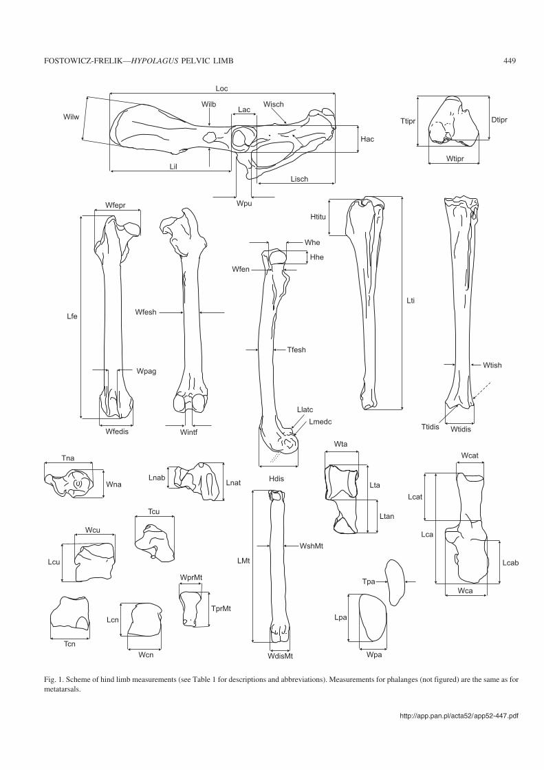
Fig. 1. Scheme of hind limb measurements (see Table 1 for descriptions and abbreviations). Measurements for phalanges (not figured) are the same as formetatarsals.

450 ACTA PALAEONTOLOGICA POLONICA 52 (3), 2007
Table 1. Meaurements of hind limb (see Fig. 1 for illustration).
Measurement Description
Os coxae Length (Loc)Length of ilium (Lil)Length of ischium (Lisch)Width of wing of ilium (Wilw)Width of body of ilium (Wilb)Length of acetabulum (Lac)Height of acetabulum (Hac)Width of ischium (Wisch)Width of pubis (Wpu)
maximal cranio−caudal length of os coxaefrom most cranial part of wing of ilium to most cranial point of acetabulumfrom most caudal point of ischial tuberosity to most caudal point of acetabulumdorsal−most point of tuber sacrale to ventral−most point of tuber coxaedorso−ventral dimension at attachment of rectus femoris musclecranio−caudal dimension of acetabulumdorso−ventral dimension of acetabulummedio−lateral dimension of body of ischiumwidth of body of pubis near acetabulum
Femur Length (Lfe)Proximal width (Wfepr)Distal width (Wfedis)Width of intercond fossa (Wintf)Width of patellar groove (Wpag)Diameter of distal extremity (Hdis)Width of the neck of femur (Wfen)Width of the head (Whe)Height of the head of femur (Hhe)Width of the shaft (Wfesh)Thickness of the shaft (Tfesh)Length of the med. cond. (Lmedc)Length of the lat. cond. (Llatc)
maximal length of femurdistance from medial−most point of head to lateral−most point of third trochantermaximal width of distal end of femurmedio−lateral dimension of intercondyloid fossa at its widest partmedio−lateral dimension of patellar groovefrom cranial point of medial ridge of trochlea to caudal point of medial condylecranial−caudal width of femoral neckcranial−caudal width of femoral headproximal−distal dimension of femoral headmedio−lateral dimension at distal point of trochanter minorcranial−caudal thickness of shaftcranial−caudal dimension of medial condylecranial−caudal dimension of lateral condyle
Patella Length (Lpa)Width (Wpa)Thickness (Tpa)
maximal length of patellamaximal medio−lateral dimension of patelladistance from facies articularis to facies cranialis, in mid−shaft of femur
Tibia Length (Lti)Proximal width (Wtipr)Proximal thickness (Dtipr)Proximal diameter(Ttipr)Distal width (Wtidis)Distal thickness (Ttidis)Height of tibial tuberosity (Htitu)Width of shaft (Wtish)
maximal length of tibiamaximal medio−lateral dimension of proximal enddistance from cranial point of tibial tuberosity to caudal point of popliteal notchmaximal cranio−caudal dimension of proximal endmaximal medio−lateral dimension of distal endcranio−caudal dimension measured along lateral groove of cochlea tibiaeproximal−distal dimension of tibial tuberositymedio−lateral dimension at the thinnest point near distal end
Talus Length (Lta)Width of trochlea tali (Wta)Length of neck (Ltan)
maximal length of talusmedio−lateral dimension of trochleadistance from the distal−most point of neck to its contact with trochlea tali
Calcaneus Length (Lca)Width (Wca)Length of calcaneal tuber (Lcat)Length of calcaneal body (Lcab)Width of calcaneal tuber (Wcat)
maximal length of calcaneusdistance from medial point of sustentaculum tali to lateral point of body of calcaneusdistance from proximal point of trochlea peronealis to proximal tip of calcaneal tuberdistance from distal point of trochlea peronealis to distal−most point of bodymedio−lateral dimension of proximal end of calcaneal tuber
Naviculare Length of navicular body (Lnab)Width of navicular body (Wna)Thickness of naviculare (Tna)Length of the tuberosity (Lnat)
length along dorsal wall of navicular bodymaximal medio−lateral dimensionfrom dorsal−most point of navicular body to plantar−most point of the tuberositydistance from the posterior−most point of the tuberosity to most distal point
Cuboid Length (Lcu)Width (Wcu)Thickness (Tcu)
maximal length of bonemaximal medio−lateral dimension of cuboidmaximal dimension from dorsal wall to plantar−most point of tuberosity
Cuneiforme laterale Length (Lcn)Width (Wcn)Thickness (Tcn)
maximal length along dorsal marginmedio−lateral dimension at widest pointmaximal dorso−plantar dimension
Metatarsals (MtII–V),phalanges proximal (PhpII–V),medial (PhmII–V)
Length (L)Proximal width (Wpr)Proximal thickness (Tpr)Width of shaft (Wsh)Distal width (Wdis)
maximal length of bonemaximal medio−lateral dimensionmaximal dorso−plantar dimensionmedio−lateral dimension of shaft in its mid−widthmaximal medio−lateral dimension of the distal extremity

statistical analyses using StatView 5.0.1. The homogeneity(monospecificity) of material of Hypolagus beremendensisfrom the Polish localities was confirmed by ANOVA andPLSD Fisher’s tests, which have shown no statistically signifi−cant differences between the samples (Fostowicz−Frelik 2006).In this study, some of the statistical tests (parametric tests)could not be used because of the disparities between the sam−ple size of Hypolagus and the other species. To estimate theinterspecific differences, box plots were used instead. Eachbox plot displays the 10th, 25th, 50th (the median), 75th and
90th percentiles of variable. The shaded box covers the central50% of the data (the interquartile range). The box plots are es−pecially useful for displaying outliers, as all values above the90th and below the 10th percentile are plotted separately. Inthe presence of outliers, the median and interquartile range areappropriate measures of central tendency and variability, re−spectively.
Cluster analyses (UPGMA) based on measured variablesof all skeletal elements except the patella, naviculare, cuboidand cuneiforme laterale (because of scarcity of comparativematerial) were performed using the PHYLIP package version3.65 (Felsenstein 1989). The results are presented as trees withEuclidean distances. The anatomical nomenclature followsNomina Anatomica Veterinaria (I.C.V.G.A.N. 2005).
Osteological descriptionOs coxae.—The complete os coxae of Hypolagus beremen−densis was not found. Thus, the reconstruction was per−formed on the basis of the ischial and ilial parts preserving atleast part of the acetabulum. The total length of os coxae wasestimated at ca. 78 mm, with possible range of 73–86 mm.This value places H. beremendensis between the ranges ofsize typical for representatives of Oryctolagus cuniculus andPentalagus furnessi (Table 3 in Appendix 1).
The relative length of the ilium does not differ signifi−cantly among leporids. The coxal index 1 (Ico1) is lowest forHypolagus (reconstructed value 45.5%) and Lepus (46%)which possess the shortest ilium in proportion to the length ofpelvis. In other rabbits, it varies ca. 50%.
The relative length of ischium (Ico4) in Hypolagus (42%)does not differ from that in other leporines (Oryctolagus 41%,Sylvilagus 41%, and Lepus 43%), except from Pentalaguswhich displays a slightly shorter ischium (38.5%).
The exact shape of the wing of the ilium in H. bere−mendensis is not known; however, a few fragments may indi−cate that the cranial part of the wing had a more extendedflare than Oryctolagus. It is also more robust than in Sylvi−lagus and Oryctolagus, with a significant caudo−dorsal por−tion of the iliac spine, forming a strong iliac tuberosity on themedial side. The auricular surface on the medial side in H.beremendensis is distinct and wide, forming an irregularsemi−lunar shape of relatively high thickness resembling thatin Lepus. In Oryctolagus and Sylvilagus, the auricular sur−face is shaped like a thin, strongly curved semicircle.
The os coxae of H. beremendensis is relatively more ro−bustly built in comparison with those of Sylvilagus andOryctolagus. The width of the body of the ilium in compari−son with the length of the acetabulum (Ico5) is relatively high(Fig. 3B), exceeding the average value for a wild rabbit andapproaching the values observed in Pentalagus furnessi.
The shape of the acetabulum in leporids is generally uni−form and the length to height ratio (Ico3) is very similar in allstudied genera (Fig. 3A). In rabbits it is slightly more roundthan in Lepus and the acetabular notch (incisura acetabuli) is
http://app.pan.pl/acta52/app52−447.pdf
FOSTOWICZ−FRELIK—HYPOLAGUS PELVIC LIMB 451
Table 2. Indices.
Index Description
General Ico−fe (coxo−femoral) – Loc/LfeIcru (crural) – Lfe/LtiIfoot1 (foot index 1) – Lfoot /Lti,where: Lfoot = Lca+LMtIII+LPhpIII+LPhmIIIIfoot2 (foot index 2) – Lca/Lti
Coxal Ico1 – Lil/LocIco2 – Lil/WilwIco3 – Lac/HacIco4 – Lisch/LocIco5 – Lac/WilbIco6 – Wpu/Wilb
Femoral Ife1 – Wfepr/LfeIfe2 – Wfedis/LfeIfe3 – Wintf/WfedisIfe4 – Wpag/WfedisIfe5 – Wfedis/HdisIfe6 – Wfen/Whe
Tibial Iti1 – Ttipr/WtiprIti2 – Wtipr/LtiIti3 – Htitu/LtiIti4 – Ttidis/WtidisIti5 – Wtidis/LtiIti6 – Wtish/Lti
Tarsal Calcaneal indicesIca1 – Wca/LcaIca2 – Lcat/LcaIca3 – Lcab/LcaIca4 – Wcat/Lcat
Talar indicesIta1 – Wta/LtaIta2 – Ltan/Lta
Navicular indicesIna1 – Lnab/LnatIna2 – Wna/TnaIna3 – Lnab/Wna
Cuboid indicesIcu1 – Lcu/WcuIcu2 – Wcu/Tcu
Metatarsalandphalangeal
MtII–MtVIprox (index for proximal articular surface) – Wpr/Tpr
MtII–PhmVIs (index of slenderness) – Wprox + Wdis/L

very narrow or almost fused. As a result, the facies lunataseems to be almost continuous (especially in Oryctolagus andSylvilagus brasiliensis). In Lepus the cranial and caudal partsof facies lunata are more extended than in rabbits, and theacetabular notch in Lepus is always well−developed and wide.In Hypolagus beremendensis the acetabulum is more roundthan in Lepus but less than in Oryctolagus (Fig. 3A) and theacetabular notch is always present and wide (Fig. 2B, L).
The ischial tuberosity (tuber ischiadicum), one of themost characteristic structures of the leporid coxal bone, ex−presses significant intergeneric differences. In Hypolagusberemendensis its caudal surface is notably wider than inOryctolagus and Sylvilagus and narrower than in Lepus, witha less marked crest and a generally more oblong outline andsmooth surface, more closely resembling the condition foundin Pentalagus (Fig. 2F–J). The lateral process of the ischial
452 ACTA PALAEONTOLOGICA POLONICA 52 (3), 2007
fossa
acetabuli
facies lunata
incisura
acetabuli
processus lateralis
tuberis ischiadici
eminentia iliopubica
area
m. recti femoris
crista
iliaca
linea
m. glutei medii
10
mm
10 mm
Fig. 2. Morphology of os coxae of Hypolagus beremendensis (Kormos, 1930) (A–F, K, L) from the Pliocene of Poland and extant Leporidae (G–J, M–P).A. Anterior part of left ilium, ISEZ MF/2220/oc/147, Węże 1, in lateral view. B. Right ischium with acetabulum, ISEZ MF/2224/oc/9, Rębielice Kró−lewskie 1, in lateral view (mirror image). C. Lateral process of right ischial tuberosity, ISEZ MF/2220/oc/74, Węże 1, in ventro−lateral view. D. Left iliumwith part of acetabulum, ISEZ MF/2220/oc/61, Węże 1, in lateral view. E. Left ischium with fragment of acetabulum, ISEZ MF/2220/oc/131, Węże 1, inlateral view. F. Ischial tuberosity, ISEZ MF/2225/oc/28, Rębielice Królewskie 2, in caudal view. G–J. Outlines of ischial tuberosity in caudal views:Oryctolagus cuniculus (Linnaeus, 1758) (G), Pentalagus furnessi (Stone, 1900) (H), Sylvilagus floridanus (Allen, 1890) (I), and Lepus europaeus Pallas,1778 (J). K. Cranial part of left ilium, ISEZ MF/2220/oc/158, Węże 1, in lateral view. L. Acetabulum with fragments of bodies of ilium, ischium and pubis,ISEZ MF/2220/oc/20, Węże 1, in lateral view. M–P. Outlines of os coxae: Sylvilagus floridanus (M), Pentalagus furnessi (N), Oryctolagus cuniculus (O),and Lepus europaeus (P).

tuberosity (processus lateralis tuberis ischiadici) in Hypo−lagus beremendensis is blunter and more massive than inOryctolagus. It is hooked cranially more strongly than inSylvilagus floridanus, S. brasiliensis, and Pentalagus fur−nessi and sharper than in Lepus.
The os pubis of H. beremendensis is not preserved in thematerial except for its most dorsal part, the body of pubis. It isrelatively wide and strong (Figs. 2, 3C) with a significantiliopubic eminence (eminentia iliopubica, see Fig. 2L) resem−bling in this respect the bones of P. furnessi and L. europaeus.
Femur.—The shape and proportions of H. beremendensis fe−mur show an overall rabbit−like type of morphology; howeverit is slenderer than the femora of both Oryctolagus cuniculusand S. floridanus, with a shaft that is more round in cross−sec−tion. The approximate length for the femur of H. beremen−densis was estimated at ca. 94 mm, on the basis of preservedcomplete proximal and distal extremities with large fragmentsof the shaft. The dimensions of the femur in H. beremendensisare generally larger than in Oryctolagus and Sylvilagus, butsmaller than in Lepus. They partially overlap with values forP. furnessi, but the femur in that species is much more robustand has higher values for the width of the proximal and distalextremities, and the shaft (Table 3 in Appendix 1).
The proximal extremity of the femur in H. beremendensisis relatively less medio−laterally expanded than in Lepuseuropaeus. The trochanters (minor and third) are groupedcloser to the proximal end of the bone than in O. cuniculus, S.brasiliensis, and P. furnessi, resembling in that respect thebones of L. europaeus and S. floridanus (Fig. 4G, H). Themost cranial part of the greater trochanter (trochanter major)is not as strongly medially bent as in Lepus. Its conditionclosely resembles that found in S. floridanus (Fig. 4G). Thesurface of the first trochanter for the attachment of the glutealmuscles is inclined more medially in L. europaeus and S.floridanus. The third trochanter (trochanter tertius) is notsignificantly extended and shaped like an isosceles trianglewith its upper tip pointing laterally as in L. europaeus, P.furnessi, and S. floridanus, but not caudo−laterally as inOryctolagus and S. brasiliensis. The second trochanter (tro−chanter minor) in H. beremendensis is strongly marked and
forms an eminent tubercle, placed closer to the femoral headthan in Oryctolagus and S. brasiliensis.
The neck of the femur in H. beremendensis is a littleshorter and wider than in S. brasiliensis and P. furnessi, simi−lar to the condition found in L. europaeus, O. cuniculus, andS. floridanus. The incision between the first trochanter andthe neck is not as deep as in Oryctolagus and S. brasiliensis.Thus, the neck of the femur in H. beremendensis is more ro−bust and less distinct, similar to the condition observed in L.europaeus and S. floridanus (Fig. 4G, H).
On the caudal side distally, from the trochanteric fossaand the lesser trochanter, the oval surface surrounded on themedio−caudal side with a roughened margin creates the at−tachment for the quadratus femoris muscle (Fig. 4C). Thissurface in H. beremendesis is round and prominent, resem−bling the condition found in Lepus. The deep imprints of thevastus medialis and pectineus muscles are located distally tothis structure, at the medial side of the shaft.
The distal extremity of the femur in H. beremendensis re−sembles in proportions that of Oryctolagus and Sylvilagusbut is slightly more extended medio−laterally in relation tothe width of the shaft and the total bone length than in thesetwo rabbit genera (Fig. 5B, Table 3 in Appendix 1). Also, H.beremendensis has a significantly wider intercondylar fossa(fossa intercondylaris), resembling that found in L. euro−paeus (Fig. 5C). On the other hand, the patellar groove (fa−cies patellaris) is relatively narrower in H. beremendensis, incomparison to the width of distal extremity, expressing thelowest values for all compared taxa (Fig. 5D). The diameterof the distal epiphysis of the femur (Hdis) is distinctly high,exceeding the width of the distal extremity (Wfedis) in H.beremendensis. This feature distinguishes the species fromall other studied taxa. The femoral index Ife5 is the lowest forH. beremendensis, whereas for other leporids it is uniformand close to 100% (Fig. 5E).
The structure of the condyles in Hypolagus does not differsignificantly from that in other leporids. The lateral condyle atthe anterior side has deep fossa extensoria for the extensordigitorum longus muscle and significant fossa musculi pop−litei, strongly marked in Hypolagus beremendensis (Fig. 4B).
http://app.pan.pl/acta52/app52−447.pdf
FOSTOWICZ−FRELIK—HYPOLAGUS PELVIC LIMB 453
0.95
1.00
1.05
1.10
1.15
1.20
1.25
1.30
1.35
0.85
0.88
0.90
0.93
0.95
0.98
1.00
1.02
1.05
1.08
0.50
0.55
0.60
0.65
0.70
0.75
0.80
Hber Ocun SfloPfur Leur
Ico
3
Ico
5
Hber Ocun SfloPfur Leur Hber Ocun SfloPfur Leur
Ico
6
Fig. 3. Box plots of selected coxal indices. Median, range and 50%−segment of values are given. Values above the 90th and below the 10th percentile areplotted as points. Species abbreviations: Hber, Hypolagus beremendensis; Ocun, Oryctolagus cuniculus; Pfur, Pentalagus furnessi; Sflo, Sylvilagusfloridanus; Leur, Lepus europaeus.

454 ACTA PALAEONTOLOGICA POLONICA 52 (3), 2007
Fig. 4. Morphology of femur and patella of Hypolagus beremendensis (Kormos, 1930) from the Pliocene of Poland (A–D) and femur of extant Leporidae(E–H). A. Proximal extremity of left femur, ISEZ MF/2220/fe/69, Węże 1, in caudal (A1) cranial (A2), lateral (A3), medial (A4) views. B. Distal extremityof right femur, ISEZ MF/2220/fe/15, Węże 1, in caudal (B1), cranial (B2), lateral (B3), medial (B4), distal (B5) views. C. Shaft of right femur, ISEZMF/2224/fe/9, Rębielice Królewskie 1, in caudal view. D. Left patella, ISEZ MF/2220/pa/1, Węże 1, in cranial (D1), lateral (D2), caudal (D3), and medial(D4) views. Outline of left femur for Oryctolagus cuniculus (E), Pentalagus furnessi (F), Sylvilagus floridanus (G), and Lepus europaeus (H). Note mor−phology of the first trochanter (arrow a) and femoral neck (arrow b).

Patella.—The patella in leporids is tear−shaped with the cra−nial external surface (facies libera) rough and convex and thecaudal surface (facies articularis) forming a smooth planefor the femoral articulation at the patellar groove. The patellain Hypolagus beremendensis is more slender and elongatedthan that of Lepus europaeus (Table 3 in Appendix 1).
The base of the patella (basis patellae), the point of at−tachment for the tendon of the quadriceps femoris muscle, isgently flattened and its proximal edge is inclined laterally(Fig. 4D). The tendon of the quadriceps femoris continues onthe distal side of the patella (apex) as a patellar ligament con−necting to the tuberosity of the tibia.
The pointy, elongated apex is directed distally, creatingthe place of attachment for the middle and lateral patellar lig−aments.
Tibiofibula.—The zeugopodial part of the lagomorph hindlimb consists of two distally fused bones, the tibia and thefibula, forming one bony element. The two bones are joinedfor roughly half of their length, forming a common distal ar−ticular surface for the tarsal elements. The extent of this fu−sion varies among leporid genera. The highest degree of fu−sion occurs in L. europaeus with 62% of the length of tibiabeing fused. The lowest proportion of fusion can be found inPentalagus furnessi, with 48% of the tibial length fused tothe fibula. In H. beremendensis this ratio is 59%, higher thanin all other included rabbit species (Sylvilagus brasiliensis52%, S. floridanus 55%, and Oryctolagus cuniculus 57%).
The tibia of H. beremendensis is slender and delicate witha relatively slender shaft. Its total length, assessed from thewell−preserved proximal and distal extremities with largeportions of the shaft (Fig. 6), is 111.4 mm. The slenderness ofthe bone is expressed by the relatively low width of the proxi−mal and distal extremities and the shaft in relation to the totalbone length (Figs. 6, 7B, E, F). The ends of the tibiofibula aresignificantly narrower than in O. cuniculus and P. furnessi,resembling in the proportions the tibiae of S. floridanus andL. europaeus (Fig. 7D). The tuberosity of the tibia in H.beremendensis is significantly shorter in relation to total tibialength (Iti3) than in O. cuniculus, both species of Sylvilagus,and P. furnessi. Iti3 in H. beremendensis most closely resem−bles the proportions observed in L. europaeus (Fig. 7C). Thetuberosity of the tibia has a well−developed crest, which ex−tends parallel to the shaft and terminates in a prominent tu−bercle, the attachment for the semitendinous muscle. This tu−bercle is slightly smaller in H. beremendensis than in O.cuniculus and S. brasiliensis, but more distinct than in S.floridanus. The ratio of the length−to−width of the tibial head(Iti1) is closest to that of S. floridanus and O. cuniculus, be−ing slightly smaller than in Lepus europaeus (Fig. 7). At theplantar side of the shaft, just below the proximal extremity,an s−shaped linear muscle attachment (linea musculi poplitei)for the popliteus muscle is marked (Fig. 6A). This line is wellpronounced, significantly longer and more distinct in H.beremendensis than in Oryctolagus and Sylvilagus. Its most
http://app.pan.pl/acta52/app52−447.pdf
FOSTOWICZ−FRELIK—HYPOLAGUS PELVIC LIMB 455
0.20
0.22
0.24
0.26
0.28
0.30
0.32
0.34
0.36
0.38
0.40
0.42
0.28
0.30
0.32
0.34
0.36
0.38
0.40
0.42
0.75
0.80
0.85
0.90
0.95
1.00
1.05
1.10
1.15
0.50
0.60
0.70
0.80
0.90
1.00
1.10
0.20
0.21
0.22
0.23
0.24
0.25
0.14
0.15
0.16
0.17
0.18
0.19
0.20
0.21
Ife1
Ife2
Ife3
Ife4
Ife5
Ife6
Hber Ocun SfloPfur Leur Hber Ocun SfloPfur Leur Hber Ocun SfloPfur Leur
Hber Ocun SfloPfur Leur Hber Ocun SfloPfur Leur Hber Ocun SfloPfur Leur
Fig. 5. Box plots of femoral indices. Note the low value of Ife2 (A), Ife5 (E), and Ife6 (F) for Hypolagus beremendensis. Median, range and 50%−segment ofvalues are given. Values above the 90th and below the 10th percentile are plotted as points. Species abbreviations: Hber, Hypolagus beremendensis; Ocun,Oryctolagus cuniculus; Pfur, Pentalagus furnessi; Sflo, Sylvilagus floridanus; Leur, Lepus europaeus.

noticeable part is placed more distally in contrast with thecondition found in Lepus, where the proximal part of thelinea m. poplitei is more twisted.
The distal extremity of the tibiofibula in Hypolagus bere−mendensis is relatively thicker than in Oryctolagus and Penta−lagus, and resembles the shape of the distal end of the tibio−fibula in Lepus and Sylvilagus (Fig. 7D). The lateral malleolus(malleolus lateralis) that forms the fibular part of the articularsurface for the calcaneus is significantly more prominent dis−tally in H. beremendensis than in any of the four studied rabbit
species. It resembles the condition found in Lepus. The sulcusmalleolaris lateralis for the tendon of the peroneus longusmuscle, is significantly deeper and directed more laterally thanin the studied rabbits, similar to that of Lepus.
The medial malleolus (malleolus medialis) shows a large,deep sulcus malleolaris medialis for the tibialis posteriormuscle and a shallower, more medially placed sulcus for theextensor hallucis longus muscle. The depth of sulcus malleo−laris medialis is comparable with that found in Lepus, deeperthan in all rabbit species.
The relative thickness of the articular surface of the tibio−fibula (Iti4) is relatively high in H. beremendensis, exceed−ing that in Oryctolagus and Pentalagus and lower than inLepus (Fig. 7D).
On the cranial side of the tibiofibula, near the distal ex−tremity, two small ridges are marked, being the points of at−tachment of the proximal extensor retinaculum, the suspen−sory ligament framing the ligaments of the tibialis cranialismuscle and of the extensor digitorum longus muscle. The firstof these ridges is placed close to the distal end of the tibio−fibula on the lateral side, just above lateral malleolus. The sec−ond ridge is positioned higher on the shaft on the medial side.Both are strongly developed in Hypolagus beremendensis.
The shaft of the tibiofibula of H. beremendensis is moreflattened cranio−caudally in its distal portion than that of Lepusbut the compression of the shaft is less than in Oryctolagus, re−sembling that of S. floridanus. Moreover, the minimal widthof the shaft in relation to total bone length (Iti6), resembles inthis respect the bones of leporine rabbits (Fig. 7F).
Skeleton of the ankle joint.—The tarsus of leporids consistsof six elements (Fig. 8), the anklebones, arranged in two rows:proximal and distal. The only exception is the navicular bone(os naviculare) which is placed centrally. The proximal rowcontains two elements, the talus, the medial element, and thecalcaneus, the lateral element. The talus is the tibial tarsalbone, while the calcaneus creates the articular surface for thefibular portion of the tibiofibula, a unique feature within mam−mals, observed only in ungulates and lagomorphs (LópezMartinez 1985). The distal ankle row contains the second (oscuneiforme intermedium), third (os cuneiforme laterale), andfourth (os cuboideum) tarsal bones (Fig. 8). The first tarsalbone (os cuneiforme mediale) in lagomorphs is fused with thesecond metatarsal bone (os metatarsale II) and totally mergedwith the metatarsal segment.
Talus.—The talus of H. beremendensis is longer than in otherrabbits, but shorter than in the hare (Table 3 in Appendix 1).The bone is also relatively more slender in H. beremendensis,as expressed by the greater length of the bone in relation to thewidth of the trochlea (trochlea tali). The ratio of trochlearwidth to the length of the talus (Ita1) is relatively low inHypolagus beremendensis and closest to the value for Lepus(Fig. 9A).
The length of the neck of the talus (collum tali) in com−parison with entire bone length (Ita2) is generally similar inall examined species (Fig. 9B).
456 ACTA PALAEONTOLOGICA POLONICA 52 (3), 2007
Fig. 6. Morphology of tibia of Hypolagus beremendensis (Kormos, 1930)from Kadzielnia 1, late Pliocene of Poland. A. Proximal extremity with shaftof right tibia, ISEZ MF/2226/ti/1, in medial (A1), lateral (A2), cranial (A3),and caudal (A4) views. B. Distal extremity with shaft of right tibia, ISEZMF/2226/ti/5, in medial (B1), lateral (B2), and caudal (B3) views. Note pointof fusion with fibula (arrow a).

The trochlea of the talus in Hypolagus beremendensis hasa smaller radius of curvature than Lepus. The talus contactsthe calcaneus on three surfaces (Fig. 10). The first of them,the posterior one (facies articularis calcanea posterior) iscomposed of two rounded irregular spots located in the con−cavity at the plantar side of the trochlea. The medial articularsurface (facies articularis media) is located at the plantar sideof the trochlea/collum border. It is a flat, elongated oval, con−necting with the articular surface of the talus head by a thinisthmus. The third, distal−most and the smallest articular sur−face (facies articularis calcanea anterior) lies at the medialside of the talus head. Its shape varies in H. beremendensisfrom planto−dorsally elongated to nearly round.
The surface of articulation with the navicular bone (faciesarticularis navicularis) occupies the head of the talus. In H.beremendensis its outline varies from oval to nearly round,frequently being wider than in Lepus europaeus.
Calcaneus.—The calcaneus of H. beremendensis is longerthan in Oryctolagus and Sylvilagus. It is almost equal inlength to that of P. furnessi but more slender and gracile (Ta−ble 3 in Appendix 1). The maximal width of calcaneus in re−lation to the total length of the bone (Ica1) is smaller in H.beremendensis than in Pentalagus furnessi (Fig. 11A). Thelength of the tuber calcanei in relation to the total bone length(Ica2) is slightly lower than in Pentalagus, Oryctolagus, andLepus but greater than in Sylvilagus (Fig. 11B).
On the other hand, the body of the calcaneus in H. bere−mendensis is longer in relation to the total bone length (Ica3)than in Oryctolagus, Pentalagus, and even Lepus (Fig. 11C).
The trochlea (trochlea peronealis) for the articulationwith the fibular part of the tibiofibula in H. beremendensisdoes not differ noticeably from the morphology in otherleporids, but the two crests are of similar size, in contrastwith those observed in Lepus, in which the lateral crest ismore prominent. The crests are also more parallel to eachother and to the sagittal axis of the bone than these of Lepus.The curvature of the trochlea is greater and in lateral view itis more similar to the trochlea of Lepus than to that ofPentalagus, which has a more flattened, massive trochlea.
On the medial side of the calcaneal body there are threeoval surfaces for the connection with talus (Fig. 12). The pos−terior one (facies articularis talaris posterior) is placed at themedio−proximal side of the trochlea and is isolated from an−terior talus surface (facies articularis talaris media) by theprominent ridge of the medial trochlear crest. The anteriorface is more rounded and extended in Lepus than in Hypo−lagus. The third, surface for the attachment with the talus isformed by the sustentaculum tali, the most medial extensionof the bone. It is a planar, round bony shelf inclined plantarly,slightly concave on the dorsal side where the surface for thetalus is located. In Hypolagus beremendensis this surface isless inclined than in Lepus, where the angle between hori−zontal plane and the surface of attachments reaches ca 40�.
http://app.pan.pl/acta52/app52−447.pdf
FOSTOWICZ−FRELIK—HYPOLAGUS PELVIC LIMB 457
0.70
0.80
0.90
1.00
1.10
1.20
1.30
1.40
1.50
Iti1
0.13
0.14
0.15
0.16
0.17
0.18
0.19
0.20
0.21
0.22
Iti2
0.25
0.30
0.35
0.40
0.45
0.50
0.55
0.60
Iti4
0.10
0.11
0.12
0.13
0.14
0.15
0.16
0.17
0.18
0.19
0.20
0.21
Iti5
0.05
0.06
0.07
0.08
0.09
Iti6
0.30
Iti3
0.28
0.26
0.24
0.22
0.20
0.18
0.16
0.14
Hber Ocun SfloPfur Leur Hber Ocun SfloPfur Leur Hber Ocun SfloPfur Leur
Hber Ocun SfloPfur Leur Hber Ocun SfloPfur Leur Hber Ocun SfloPfur Leur
Fig. 7. Box plots of tibial indices. Median, range and 50%−segment of values are given. Values above the 90th and below the 10th percentile are plotted aspoints. Species abbreviations: Hber, Hypolagus beremendensis; Ocun, Oryctolagus cuniculus; Pfur, Pentalagus furnessi; Sflo, Sylvilagus floridanus; Leur,Lepus europaeus.

At the medial side anteriorward from the sustentaculum talithere is a small surface, facies articularis talaris anterior, forthe attachment of the head of the talus. It is the small roundedsurface positioned on the narrow, obliquely elongated emi−nence and connected by it with the slightly larger area for thenavicular bone (Fig. 12). These two surfaces could be eithermerged or separated in leporids. In Hypolagus beremen−densis they are frequently joined, whereas in Lepus they areclearly separated. The distal surface of the calcaneus isobliquely oriented with its longer edge placed dorsally. Itslateral part serves as a surface for the articulation of thecuboid. It is a semilunar surface, occupying the dorso−lateralpart of the distal surface of calcaneus. It is slightly narrowerand less extended dorso−plantary in H. beremendensis thanin Lepus europaeus. However, in the outline, the proximalarticular surface of the cuboid in H. beremendensis resem−bles more that of L. europaeus than that of Pentalagusfurnessi. In the latter species it is more square with medialmargin almost as long as the dorsal one, while in bothHypolagus and Lepus the dorsal margin is distinctly longer.
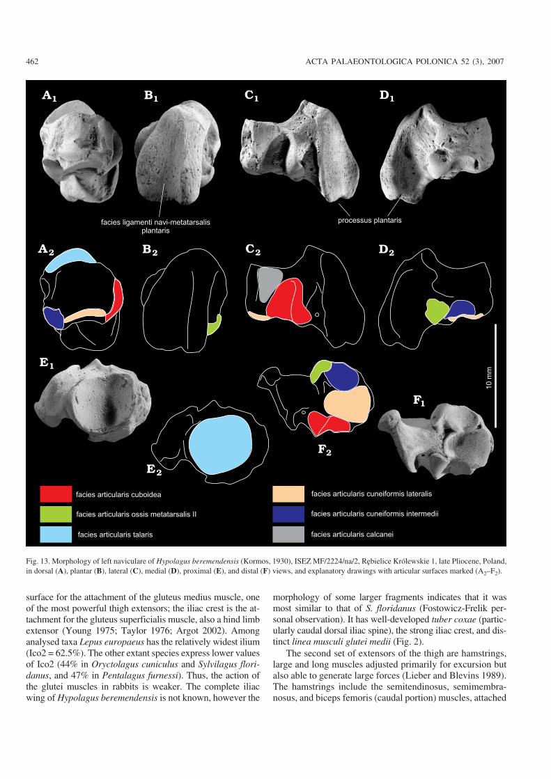
Naviculare.—The navicular bone is situated medially be−tween the talus on the proximal side and the cuneiform bones
on the distal side (Fig. 8). It consists of the body and extendedplantar process (processus plantaris ossis navicularis). Thebody is an irregular cube, flattened proximo−distally anddeeply concave on the proximal face where the single, ovalsurface for the articulation of the head of the talus occurs(Fig. 13). This surface is almost circular in H. beremendensisand P. furnessi, while it is significantly constricted medio−laterally in L. europaeus and Sylvilagus floridanus.
The plantar process is flattened on the plantar side and hasa shallow longitudinal groove oriented planto−medially. InH. beremendensis the plantar process is shorter in relation tobody length (Ina1) than in P. furnessi, Oryctolagus cuni−culus, and L. europaeus (Fig.14A). In H. beremendensis itbroadens distally, forming a shovel−like structure (Fig. 13B),while in each of the comparative species it is much narrower.It is also somewhat plantarly directed, while in Lepus andSylvilagus it is oriented more along the longitudinal axis andparallel to the dorsal surface of the body.
In H. beremendensis the body of the naviculare is signifi−cantly shorter in relation to the body width (Ina3) than in L.europaeus and S. floridanus, but it is longer than in O.cuniculus and P. furnessi (Fig. 14C).
The navicular bone contacts with the three cuneiformbones at its distal side (Fig. 13). The most medial bone (oscuneiforme mediale) is a part of the second metatarsal boneand contacts the navicular bone in a minute oval area at themedio−distal side of the navicular body, near the base of theplantar process.
458 ACTA PALAEONTOLOGICA POLONICA 52 (3), 2007
calcaneus
talus
os cuboideumos naviculare
os cuneiforme intermedium
os cuneiforme mediale
os metatarsale II
os metatarsale III
os metatarsale IV
phalanges
mediales
phalanges
proximales
os metatarsale V
os cuneiforme laterale
Fig. 8. Schematic drawing of pes structure in leporids, showing the arrange−ment of tarsal bones. Distal phalanges not shown.
Hber Ocun SfloPfur Leur
Hber Ocun SfloPfur Leur
0.40
0.45
0.50
0.55
0.60
0.65
0.70
Ita1
0.38
0.40
0.42
0.44
0.46
0.48
0.50
0.52
0.54
0.56
Ita2
Fig. 9. Box plots of talar indices. Median, range and 50%−segment of valuesare given. Values above the 90th and below the 10th percentile are plottedas points. Species abbreviations: Hber, Hypolagus beremendensis; Ocun,Oryctolagus cuniculus; Pfur, Pentalagus furnessi; Sflo, Sylvilagus flori−danus; Leur, Lepus europaeus.

The second tarsal bone (os cuneiforme intermedium) isplaced lateral to the first tarsal bone, and contacts the navicularbone in a small area positioned planto−medially at the distalsurface of the navicular body. The third tarsal bone (os cunei−forme laterale) is largest and contacts the navicular bone in arounded and partly convex articular surface that occupies themajority of the distal surface of the navicular body.
The navicular bone contacts the cuboid laterally (Figs. 8,13) at the deeply incised articular surface. In Hypolagus bere−mendensis it forms a deep groove opening toward the distalsurface of the navicular body. The smaller surface for the con−nection with the calcaneus is located near the dorsal side of thearticulation with the cuboid. In Lepus it is relatively small andoval or rounded in shape, while in H. beremendensis it forms atriangle with its apex directed proximally.
Cuboid.—The cuboid bone is the most lateral element of thedistal ankle row (Fig. 8). The cuboid in leporids actually re−sembles a rhombohedron. It has a distinct plantar tuberosity,forming transverse ridge crossing the plantar side of the bone(Fig. 15). In H. beremendensis the elongation (Icu1) of thebone resembles that of Lepus, while it is less elongate than inSylvilagus, and more than in Oryctolagus (Fig. 16; the mea−surements for Pentalagus were unavailable).
The proximal surface of the cuboid forms a large and con−vex surface for the calcaneus, which extends onto the dorsalside of the bone. The surface is almost square in Lepus euro−paeus, while in rabbits it is generally oval and gently bent,forming two rounded horns that project plantarly. In H.beremendensis the bending is more pronounced and thewhole surface, in proximal view, is more triangular, with aflat medial margin and a curved dorsal margin. It is also morestrongly sloped and extends more dorsally than in Lepus,Sylvilagus, and Oryctolagus.
The surface for the contact with navicular bone (faciesarticularis navicularis) is tripartite (Fig. 15C). In H. bere−mendensis its shape resembles that of Sylvilagus floridanus,forming a more consistent triangle in medial view and adeeper depression above the plantar tuberosity.
The cuboid connects distally with the fourth and fifthmetatarsal bones. The articular surface for the metatarsals ispear−shaped with its broader part, the articulation with thefourth metatarsal bone, placed medially. It has relativelymore rounded margins and is slightly narrower than the artic−ular surface in Lepus. The distinct elongated prominence ofthe cuboid tuberosity (tuberositas ossis cuboidei) is situatedon the plantar side of the cuboid. In H. beremendensis thetuberosity is thinner and has uniform thickness. Its plantarsurface is wavy in some specimens. At its lateral edge thetuberosity in H. beremendensis is thickened, producing asmall tubercle, which is significantly enlarged in L. euro−paeus and S. floridanus.
Third tarsal bone.—The second and third tarsal bones or oscuneiforme intermedium and os cuneiforme laterale are small,pyramid−like bones rarely found in the fossil deposit becauseof their size and inconspicuous shape. The second tarsal bone
http://app.pan.pl/acta52/app52−447.pdf
FOSTOWICZ−FRELIK—HYPOLAGUS PELVIC LIMB 459
facies articularis fibularis
facies articularis navicularis
facies articulares calcaneae:
a - anterior, m - media, p - posterior
Fig. 10. Morphology of left talus of Hypolagus beremendensis (Kormos,1930), ISEZ MF/2224/ta/12, Rębielice Królewskie 1, late Pliocene, Poland,in dorsal (A), plantar (B), lateral (C), and medial (D) views, and explana−tory drawings with articular surfaces marked (A2–D2).

was not found within the studied fossil material, while sevenossa cuneiformia laterales were yielded by the material fromWęże 1.
The third tarsal bone is shorter in Hypolagus than inLepus europaeus, and less extended dorso−plantarly (Table 3in Appendix 1). Thus, its articular surfaces for the naviculare(proximal) and metatarsal bone III (distal) are relativelybroader medio−laterally and shorter dorso−plantarly. Theproximal articular surface of the third tarsal bone of Hypo−lagus beremendensis is slightly concave and almost round,the distal surface is flat and triangular in outline, with the tip,formed by plantar tuberosity (Fig. 17). The dorso−plantar ex−tension of the third tarsal bone and the relative breadth of thedistal surface, for attachment with metatarsal bones, differbetween the studied genera. The third tarsal bone in H.beremendensis is smaller than in L. europaeus and largerthan in Sylvilagus floridanus. The third tarsal bone of H.beremendensis is also significantly broader at its dorsal mar−gin and has an enlarged medial tubercle, forming the articula−tion for metatarsal bone II. In all studied leporins the thirdtarsal bone has a narrower dorsal margin and the tuberosity isnot so extended.
Metatarsals.—The metatarsal bones (ossa metatarsaliaII–V) are built according to the same plan. Their proximalextremities create a base (basis) which articulates with thetarsals (the proximal articular surfaces) and join the metatar−sals together in a row (medial and lateral articular surfaces).The shaft or body (corpus) is elongated and the distal extrem−ities of metatarsals form a head (caput), creating trochlea forthe connection with phalanges (Figs. 8, 18, 19).
The metatarsals of H. beremendensis are relatively longand slender (Figs. 20, 21). Their general morphology doesnot differ significantly from that of other species, with the ex−ception for P. furnessi which has distinctively short, robustbones (Fig. 21). The indices of slenderness (Fig. 20) calcu−lated for metatarsal bones of all studied species reveal that H.beremendensis has similar proportions to O. cuniculus (Fig.20). Interestingly enough, the second metatarsal bone of H.beremendensis shows more similarity to Lepus than to Ory−ctolagus in overall proportions, being more slender than inOryctolagus and both Sylvilagus species (Fig. 20). The prox−imal articular surfaces of metatarsal II and metatarsal III forthe tarsal bones are somewhat deeper (dorso−plantarly) inHypolagus than in Sylvilagus and Oryctolagus, resemblingthe proportions of Lepus (Table 4 in Appendix 1). The fourthand fifth metatarsals of H. beremendensis are relativelywider, approaching the proportions found in rabbits (Orycto−lagus and Sylvilagus). Moreover, the fifth metatarsal in H.beremendensis possesses a relatively large, laterally−projec−ting tubercle, where the tendon of the peroneus brevis muscleis attached (Fig. 19E–H). The tubercle for the peroneusbrevis tendon in H. beremendensis is more laterally extendedthan in Sylvilagus and Oryctolagus but less so than in Lepus.It is also positioned perpendicular to the long axis of the cor−pus of metatarsal V (as in Lepus), while in Oryctolagus andSylvilagus it is gently inclined proximally.
Phalanges.—The proximal and medial phalanges in leporidsare delicate and elongated, differing between the taxa in theirslenderness indices and length (Tables 3, 4 in Appendix 1).They consist of a gently broadened and proximally concave
460 ACTA PALAEONTOLOGICA POLONICA 52 (3), 2007
Hber Ocun SfloPfur Leur
Hber Ocun SfloPfur Leur
0.28
0.30
0.32
0.34
0.36
0.38
0.40
0.42
0.44
0.46
0.48
0.50
Ica1
0.38
0.40
0.42
0.44
0.46
0.48
0.50
0.52
0.54
0.56
0.58
Ica2
0.32
0.34
0.36
0.38
0.40
0.42
0.44
Ica3
Ica4
0.43
0.45
0.48
0.50
0.53
0.55
0.57
0.60
0.63
0.65
0.68
Hber Ocun SfloPfur Leur
Hber Ocun SfloPfur Leur
Fig. 11. Box plots of calcaneal indices. Median, range and 50%−segment of values are given. Values above the 90th and below the 10th percentile are plottedas points. Species abbreviations: Hber, Hypolagus beremendensis; Ocun, Oryctolagus cuniculus; Pfur, Pentalagus furnessi; Sflo, Sylvilagus floridanus;Leur, Lepus europaeus.

base, an elongated body that is round in cross−section, and ahead that forms a crestless trochlea (Fig. 8).
The distal phalanges form the bony cores for claws andhave only a proximal articular surface, which is a simpleshallow concavity. Their distal ends taper, forming sharp,highly porous tips.
The proximal phalanges in Hypolagus beremendensis aresignificantly longer in relation to the metatarsal segment thanin leporines. The medial phalanges show similar elongationbut to a lesser extent (Fig. 21, Tables 3, 4 in Appendix 1).
The general proportions of the proximal and medial pha−langes in H. beremendensis are as in Oryctolagus, Sylvilagus,and Lepus, which share a similar, gracile pattern of phalanxmorphology. The slenderness of the proximal phalanges in H.beremendensis is in most cases greater than or at least equal tothat of Lepus (Table 4 in Appendix 1). The slenderness of themedial phalanges shows the highest value of the slenderness
index for phalanges III and IV. The medial phalanges of thesecond and fifth toes in H. beremendensis are more slenderthan those of Oryctolagus cuniculus, Sylvilagus floridanus,and L. europaeus (Table 4 in Appendix 1).
The intra−membral indices and proportions of the foot.—Proportions of the hind limb bones in H. beremendensisshow relative elongation of the tibial and metatarsal seg−ments. The ratio of os coxae length to the femur length(Ico−fe) is lower in H. beremendensis than in Oryctolagusand Sylvilagus, approaching the value for Lepus (Fig. 22A).The low index value in H. beremendensis is caused by a nota−bly longer femur in relation to the innominate bone. A similarpattern is expressed in the crural ratio (Icru), which in H.beremendensis is close to Oryctolagus, Sylvilagus, andLepus, but lower than in Pentalagus (Fig. 22B). On the otherhand, H. beremendensis has a relatively longer foot (Ifoot1).This index has a significantly lower value only in P. furnessi,possessing a short and robust feet (Fig. 22C). The proportionof the calcaneus to the tibiofibula length (Ifoot2) is similar inH. beremendensis, L. europaeus, and S. floridanus, that havethe relatively longer tibiofibulae than P. furnessi (Fig. 22D).
Results of phenetic analysisThe cluster analysis employing UPGMA method of distanceexamination was performed for 98 of the bone measurements(Table 1). The UPGMA analyses of the proximal segments in−cluding the pelvis, femur (Fig. 23A, B), and talus (Fig. 23E)group H. beremendensis with P. furnessi. On the other hand,the analysis of the tibiofibula places it just outside the O. cuni−culus–S. floridanus (Fig. 23C) cluster and that of the calcaneusjoins links Hypolagus with Oryctolagus (Fig. 23D). Further−more, the analyses of the metatarsal and phalangeal segmentslink H. beremendensis to Lepus (Fig. 23F, G) because of theelongation of toes in both species (Fig. 21).
The general cluster for the hind limb places H. bere−mendensis just outside the cluster of typical leporine rabbitsformed by Oryctolagus and Sylvilagus (Fig. 23H). Outsidethe branch with H. beremendensis is P. furnessi, while L.europaeus is the most divergent species. The position of H.beremendensis between leporine rabbits and Pentalagus,points to the specific and unique adaptations possibly typicalfor Archaeolaginae. The clustering of Hypolagus and Penta−lagus in the case of the os coxae and femur parameters maysuggest the conservative features retained in these structuresin Leporidae.
Functional interpretationHip.—The relative increase in width of the iliac wing is one ofthe hallmarks of cursoriality or the ability to performing pow−erful leaps, observed in many terrestrial mammals (Taylor1975; Sargis 2002). The wide iliac wing creates the expanded
http://app.pan.pl/acta52/app52−447.pdf
FOSTOWICZ−FRELIK—HYPOLAGUS PELVIC LIMB 461
10
mm
facies articularis tibialis
facies articulares talares: a - anterior,
m - media, p - posterior
facies articularis cuboidea
facies articularis navicularis
10
mm
m
p
a
canalis
calcanei
sustentaculum
tali
Fig. 12. Morphology of right calcaneus of Hypolagus beremendensis (Kor−mos, 1930), ISEZ MF/2220/ca/77, Węże 1, Pliocene, Poland, in medial (A),dorsal (B), and lateral (C) views, and explanatory drawings highlighting ar−ticular surfaces (A2, B2, C2).
surface for the attachment of the gluteus medius muscle, oneof the most powerful thigh extensors; the iliac crest is the at−tachment for the gluteus superficialis muscle, also a hind limbextensor (Young 1975; Taylor 1976; Argot 2002). Amonganalysed taxa Lepus europaeus has the relatively widest ilium(Ico2 = 62.5%). The other extant species express lower valuesof Ico2 (44% in Oryctolagus cuniculus and Sylvilagus flori−danus, and 47% in Pentalagus furnessi). Thus, the action ofthe glutei muscles in rabbits is weaker. The complete iliacwing of Hypolagus beremendensis is not known, however the
morphology of some larger fragments indicates that it wasmost similar to that of S. floridanus (Fostowicz−Frelik per−sonal observation). It has well−developed tuber coxae (partic−ularly caudal dorsal iliac spine), the strong iliac crest, and dis−tinct linea musculi glutei medii (Fig. 2).
The second set of extensors of the thigh are hamstrings,large and long muscles adjusted primarily for excursion butalso able to generate large forces (Lieber and Blevins 1989).The hamstrings include the semitendinosus, semimembra−nosus, and biceps femoris (caudal portion) muscles, attached
462 ACTA PALAEONTOLOGICA POLONICA 52 (3), 2007
facies articularis talaris
facies articularis ossis metatarsalis II
facies articularis cuneiformis lateralis
facies articularis cuneiformis intermedii
facies articularis cuboidea1
0m
m
processus plantarisfacies ligamenti navi-metatarsalis
plantaris
facies articularis calcanei
Fig. 13. Morphology of left naviculare of Hypolagus beremendensis (Kormos, 1930), ISEZ MF/2224/na/2, Rębielice Królewskie 1, late Pliocene, Poland,in dorsal (A), plantar (B), lateral (C), medial (D), proximal (E), and distal (F) views, and explanatory drawings with articular surfaces marked (A2–F2).

http://app.pan.pl/acta52/app52−447.pdf
FOSTOWICZ−FRELIK—HYPOLAGUS PELVIC LIMB 463
Hber Ocun SfloPfur Leur Hber Ocun SfloPfur Leur Hber Ocun SfloPfur Leur
0.38
0.40
0.42
0.44
0.46
0.48
0.50
0.52
0.54
0.56
0.58
Ina
1
0.55
0.60
0.65
0.70
0.75
0.80
0.85
Ina
2
0.50
0.55
0.60
0.65
0.70
0.75
0.80
0.85
0.90
Ina
3
Fig. 14. Box plots of navicular indices. Median, range and 50%−segment of values are given. Values above the 90th and below the 10th percentile are plottedas points. Species abbreviations: Hber, Hypolagus beremendensis; Ocun, Oryctolagus cuniculus; Pfur, Pentalagus furnessi; Sflo, Sylvilagus floridanus;Leur, Lepus europaeus.
10
mm
tuberositas
cuboidei sulcus tendinis
m. peronei longi
facies articularis navicularis
facies articularis cuneiformis lateralis
facies articularis calcanea facies articularis ossis metatarsalis IV
facies articularis ossis metatarsalis V
facies ligamenti calcaneo-cuboidei plantaris
Fig. 15. Morphology of left cuboid of Hypolagus beremendensis (Kormos, 1930), ISEZ MF/2220/cu/1, Węże 1, Pliocene, Poland, in dorsal (A), plantar (B),medial (C), lateral (D), proximal (E), and distal (F) views, and explanatory drawings with articular surfaces marked (A2–F2).

to the ischial tuberosity and the lateral process of the ischium(Camp and Borell 1937; Klebanova et al. 1971). The size andshape of the ischial tuberosity is variable in Leporidae. InLepus the ischial tuberosity is very large and robust. The sur−face for the muscle attachments is more expanded and moreisometric, while in rabbits its surface is much more limited,compressed medio−laterally, and crescent−like in caudalview (Fig. 2F–J). The more isometric areas of muscle attach−
ments indicate the relatively greater area of the cross−sectionof the muscles which results in relatively greater strength ofcontraction. Thus, the muscles attached to the ischial tubero−sity in Lepus are the strongest among all leporid taxa. In thatrespect Hypolagus beremendensis expresses general rabbitmorphotype, although it is among the more cursorial species.On the other hand, the lateral process in Oryctolagus and H.beremendensis is distinctly enlarged dorso−laterally, creatinga larger space between its cranio−dorsal margin and the bodyof the ischium. This, in all probability, reflects the strong ac−tion of the sacrotuberous ligament, which attaches to the tipof the ischium and is the insertion for the biceps femoris andabductor cruris caudalis muscles. It also can indicate a pow−erful quadratus femoris muscle (attaching cranially from thelateral process of ischium to the body of ischium), that ex−tends the thigh and rotates the femur outward at the hip joint(Young 1975; Evans 1993). The rotation and abduction func−tions of the quadratus femoris, as well as the strong action ofsacrotuberous ligament, can be related in Oryctolagus withdigging. While burrowing, a wild rabbit suspends the mainbody mass on hind limbs and shifts its weight back and forthlike a pendulum, digging with the forelegs. This positiongenerates the stretching forces applied to the muscles of thecaudal side of the thigh and demands the strong action of themuscles of the cranial side of thigh suspending the body (e. g.biceps femoris muscle). Also, during burrowing there isflexion and extension of the vertebral spine, which enables astrong sacrotuberous ligament to act as a spring, giving addi−tional momentum to the pendulum−like movements. Themorphology of the lateral process of the ischial tuberosity inHypolagus beremendensis could, to some extent, indicateadaptation to digging. On the other hand, the hook−like ex−tension of the lateral process is not observed in Pentalagusfurnessi.
464 ACTA PALAEONTOLOGICA POLONICA 52 (3), 2007
0.75
0.80
0.85
0.90
0.95
1.00
1.05
1.10
Icu
1
Hber Ocun Sflo Leur
Icu
2
1.15
1.20
1.25
1.30
1.35
1.40
1.45
Hber Ocun Sflo LeurPfur
Fig. 16. Box plots of cuboidal indices. Median, range and 50%−segment ofvalues are given. Values above the 90th and below the 10th percentile areplotted as points. Species abbreviations: Hber, Hypolagus beremendensis;Ocun, Oryctolagus cuniculus; Pfur, Pentalagus furnessi; Sflo, Sylvilagusfloridanus; Leur, Lepus europaeus.
facies articularis ossis metatarsalis II
facies articularis ossis metatarsalis III facies articularis navicularis
facies articularis cuboidea
10 mm
Fig. 17. Morphology of right lateral cuneiform of Hypolagus beremendensis (Kormos, 1930), ISEZ MF/2220/cn/1, Węże 1, Pliocene, Poland, in dorsal (A),plantar (B), medial (C), lateral (D), proximal (E), and distal (F) views, and explanatory drawings with articular surfaces marked (A2–F2).

http://app.pan.pl/acta52/app52−447.pdf
FOSTOWICZ−FRELIK—HYPOLAGUS PELVIC LIMB 465
10
mm
caput
corpus
facies articularis cuneiformis lateralis
facies articularis ossis metatarsalis II
facies articularis ossis metatarsalis IV
caput
10
mm
Fig. 18. Morphology of right metatarsal II (A–D) and left metatarsal III (E–H) of Hypolagus beremendensis (Kormos, 1930), ISEZ MF/2220/mt2/9, Węże 1,ISEZ MF/2224/mt3/10, Rębielice Królewskie 1, late Pliocene, Poland, in plantar (A, E), dorsal (B, F), lateral (C, H), and medial (D, G) views, and explanatorydrawings with articular surfaces marked (A2–H2).

466 ACTA PALAEONTOLOGICA POLONICA 52 (3), 2007
facies articularis cuboidea
facies articularis ossis metatarsalis Vfacies articularis ossis metatarsalis IV1
0m
m
facies articularis ossis metatarsalis IVfacies articularis cuboidea
10
mm
tuberculum
ossis
metatarsalis V
Fig. 19. Morphology of right metatarsal IV (A–D) and left metatarsal V (E–H) of Hypolagus beremendensis (Kormos, 1930), ISEZ MF/2224/mt4/1,Rębielice Królewskie 1 and ISEZ MF/2225/mt5/12, Rębielice Królewskie 2, late Pliocene, Poland, in plantar (A, E), dorsal (B, F), medial (C, G), and lat−eral (D, H) views, and explanatory drawings with articular surfaces marked (A2–H2).

Thigh.—In highly cursorial and saltatorial mammals, the fe−mur is generally slim and the trochanters are concentratedcloser to the proximal end of the bone. It shortens the leverarms over which muscle forces acts and produces swifter re−sponses to the contraction of muscles, yielding quicker butless powerful flexion (the iliopsoas muscle) and extension(the gluteus superficialis, tensor fasciae femoris and gluteusmedius muscles) of the thigh at the hip joint (Hildebrand1974; Anemone and Covert 2000). This particular morphol−ogy is best−developed in Lepus. Hypolagus beremendensisexpresses an intermediate development of this feature be−tween Lepus and Oryctolagus, implying a relatively high de−gree of cursoriality.
The proximally extended greater trochanter is interpretedin small carnivores as an adaptation to the terrestrial locomo−tion, because it restricts the action of the gluteus medius inabduction of the femur at the hip joint and makes the para−sagittal action of the limb more efficient (Taylor 1976). Inleporids however, which all have the greater trochanters rela−tively extended posteriorly, the most erect trochanters are ob−served in slower runners. The medial bending of the greatertrochanter increases the area of attachment for the gluteusmedius muscle (see discussion of the pelvis, above), thusstrengthening the force with which it extends the hip joint.
The strongest bending of the greater trochanter is observed inLepus. In Oryctolagus and Pentalagus the structure is gener−ally erected proximally and pointy (Fig. 4). In Sylvilagusfloridanus and H. beremendensis the bending of the greatertrochanter is markedly weaker than in Lepus.
In leporids the articular surface of the head of the femur in−trudes into the dorsal part of the femoral neck. This feature isfrequently observed in arboreal marsupials and some carni−vorans, which stand with the thighs in a more abducted posi−tion (Argot 2002; Heinrich and Houde 2006). The extensionof this surface is quite significant in leporids and it is directedslightly caudally. It seems to be connected with the orientationof the pelvis, which is inclined backward and the caudal mar−gin of the semilunar surface braces the caudo−dorsal part of thefemoral head. Thus, the greater abduction of the thigh can berelated with digging behaviour. The abduction of the hindlimbs allows an animal pushing the soil backward with theforelegs between the hind legs (Taylor 1976) and easily shiftsthe position of the trunk during burrowing. Among the studiedtaxa, Oryctolagus cuniculus shows the most extended and de−fined articular surface of the femoral head with sharp, pro−nounced margins. The femoral neck in O. cuniculus seems tobe also the longest and best developed, allowing for greatermobility in a transverse plane. In H. beremendensis the shape
http://app.pan.pl/acta52/app52−447.pdf
FOSTOWICZ−FRELIK—HYPOLAGUS PELVIC LIMB 467
0.15
0.20
0.25
0.30
0.35
0.40
0.45Is
MtII
Hber Ocun Pfur Sflo Leur
0.17
0.20
0.22
0.25
0.28
0.30
0.32
0.35
0.38
0.40
0.43
0.45
Hber Ocun Pfur Sflo Leur
IsM
tIV
0.25
0.30
0.35
0.40
0.45
0.50
0.55
0.60
Hber Ocun Pfur Sflo Leur
IsM
tV
0.17
0.20
0.22
0.25
0.28
0.30
0.32
0.35
0.38
0.40
0.43
0.45
IsM
tIII
Hber Ocun Pfur Sflo Leur
Fig. 20. Box plots of the indices of slenderness for metatarsal bones. Note the low index of slenderness of metatarsal II for Hypolagus beremendensis (A).Median, range and 50%−segment of values are given. Values above the 90th and below the 10th percentile are plotted as points. Species abbreviations: Hber,Hypolagus beremendensis; Ocun, Oryctolagus cuniculus; Pfur, Pentalagus furnessi; Sflo, Sylvilagus floridanus; Leur, Lepus europaeus.

and extent of the articular surface of the head are more similarto those observed in Lepus. Therefore Hypolagus was proba−bly not able to abduct the hip joint to such an extent as O.cuniculus. It may indicate weaker burrowing ability, if H.beremendensis was at all capable of burrowing.
The relatively high cursorial ability of H. beremendensis isemphasised also by the relatively high cranio−caudal thickness(Hdis) of the distal extremity of the femur, even exceeding itswidth (Wfedis), and a distinctly narrow patellar groove (Fig.5D, E). The deeper condyles, one of hallmarks of cursoriality,allow more powerful extension of the knee by the action of thequadriceps femoris (Anemone and Covert 2000).
Shank.—The tibiofibula, the sole bone of the zeugopodialsegment in the hind limb of leporids is the main bone responsi−ble for the elongation of the leg in highly cursorial species(Gambaryan 1974; Hildebrand 1974), apart from the metatar−sal segment. It is also the most distal bone having large muscleattachments in leporids. The most powerful hind limb muscu−lature occupies the upper regions of the leg, reaching aboutone third of the proximal part of the tibiofibula, while the distalend of the bone and the foot forms the attachments for liga−ments. This general plan is expressed more strongly in cur−sorial taxa, as the shortening of the muscle arms quickens theresponses of the limb bones to their action (Camp and Borell1937; Hildebrand 1974). The most prominent and importantstructure within the tibiofibula is the tibial tuberosity. It servesas the attachment area for shank flexors such as the bicepsfemoris and semitendinosus muscles and the patellar ligamentof the quadriceps femoris complex. The sartorius muscle,which acts as an adductor and rotator of the thigh and extensor
of the shank, and the gracilis muscle, which adducts the shank,are attached to the medial condyle of the tibia (Camp andBorell 1937; Craigie 1948; Evans 1993; Wingerd 1985). Therelative length of the tibial tuberosity to the overall tibiofibularlength (Iti3) in studied taxa is lowest for L. europaeus and H.beremendensis (Fig. 7C). This index implies that the mostcursorial leporids, such as Lepus, possess the relatively short−est tibial tuberosity. This agrees with the tendency to group themuscle attachments near the bone ends in highly cursorialmammals (Hildebrand 1974) as is also observed in the femur.The proportions of the proximal and distal extremities of thetibiofibula in the studied taxa reflect the tibial aspect of “deep−ening of the knee” (observed also in the distal end of the fe−mur) by increasing relative cranio−caudal depth of the proxi−mal extremity. In Lepus, Sylvilagus, and Oryctolagus, as wellas in H. beremendensis, the maximum cranio−caudal dimen−sion of the proximal end of the tibiofibula exceeds its maxi−mum width (Fig. 7A), while Pentalagus furnessi displays sig−nificantly greater width. The distal end deepens similarly tothe proximal one in Lepus (Fig. 7D) as well as in Sylvilagusfloridanus and H. beremendensis, differentiating them fromO. cuniculus and P. furnessi. These last two taxa have tibio−fibulae with more medio−laterally extended distal ends.
In general, the slenderness of the tibiofibula of Hypo−lagus beremendensis and the relative deepening of the sur−face of the stifle joint, the shortening of the tuberosity and themore square distal articular surface show similarities to thetibiofibula of Lepus.
Pes.—The foot in terrestrial mammals is another anatomicalregion (along with the shank) of the hind limb in which signifi−
468 ACTA PALAEONTOLOGICA POLONICA 52 (3), 2007
� ���������������������������������
� ���������������������������������
� ���������������������������������
� ���������������������������������
� �����������������������������������������
� �����������������������������������������
� �����������������������������������������
� �����������������������������������������
� ���������������������������������� �
� ���������������������������������� �
� ���������������������������������� �
� ���������������������������������� �
� ���������������������������������
� ���������������������������������
� ���������������������������������
� ���������������������������������
������������������������������������
������������������������������������
��������������������������������������
��������������������������������������
����������������������������������������������������
����������������������������������������������������
����������������������������������
����������������������������������
��������������������������������
��������������������������������
aaaaaaaaa
aaaaaaaaaaa
aaaaaaaaaaa
aaaaaaaaaa
� �����������������������������
� �����������������������������
� �����������������������������
� �����������������������������
aaaaaaaaaaaaaaaaaaaaaaaaaaaaaaaaaaaaaaaaaaaaaaaaaaaa
aaaaaaaaaaaaaaaaaaaaaaaaaaaaaaaaaaaaaaaaaa
aaaaaaaaaaaaaaaaaaaaaaaaaaaaaaaaaaaaaaaaaa
aaaaaaaaaaaaaaaaaaaaaaaaaaaaaaaaaaaaaa
aaaaaaaaaaaaaaaaaaaaaaaaaaaaaaaaaaaaaaaaaa
aaaaaaaa
���������������
���������������
aaaaaa� ������������ � �
� ������������ � �
� ������������ � �
� ������������ � �
aaaaaaaaaa
Fig. 21. Proportions of bones of the foot of Leporidae, metatarsal bone III (black bar), used as 100% for each species. Species abbreviations: Hber,Hypolagus beremendensis; Ocun, Oryctolagus cuniculus; Pfur, Pentalagus furnessi; Sflo, Sylvilagus floridanus; Leur, Lepus europaeus.

cant cursorial and saltatorial adaptations are apparent (Gamba−ryan 1974; Hildebrand 1974). The metatarsals are the bonesthat most fully reflect cursoriality, being relatively elongatedin fast−running mammals (Hildebrand 1974). On the otherhand, the tarsal elements in cursorial animals serve to stabilisethe ankle joint by creating large and deep surfaces for the artic−ulation with the tibia and fibula. Therefore they generally arenot elongated. However, in some small arboreal primates,such as tarsiers and galagos, probably the best leapers amongthe mammals, they can be lengthened significantly, reflectingsome particular saltatorial adaptations (Hildebrand 1974; Ane−mone and Covert 2000).
In leporids the basic ankle joint structure is very uniform(Lyon 1904; Dawson 1958; Averianov 1995), although somedifferences in the proportions of the tarsal bones can be re−cognised. The main differences in the foot morphology canbe seen in elongation of the metatarsals and phalanges
In more cursorial genera, the talus is slender and the in−dex value (Ita1) for Hypolagus is closest to that of Lepus(Fig. 9A). However, the length of the neck of the talus (Ita2)varies only slightly among all studied genera and is rela−tively longer in Lepus than in the other species (Fig. 9B).The head of the talus is placed posteriorly in relation to thedistal end of the calcaneus (Fig. 8), strengthening and stabi−lising the ankle joint and limiting the mobility of the footonly to flexion and extension (Taylor 1976). This feature isenhanced by the elongation of the neck of the talus inleporids.
The most powerful muscle attachment in the distal part ofthe hind limb (the gastrocnemius muscle, the ankle plantarflexor; Lieber and Blevins 1989) is located on the tuber ofcalcaneus. The analysis of the proportions of the calcaneusrevealed that the values of Ica2 are within a narrow range ex−cept for Sylvilagus, which has the shortest calcaneal tuber(Fig. 11B).
On the other hand, the elongation of the body of thecalcaneus (Fig. 11C) seems to be closely related to saltatorialadaptation. The lowest values of Ica3 are found in Pentala−gus furnessi, which is one of the stockiest and most heavily−built rabbits and has weakly developed cursoriality. Theother studied species exhibit higher ratios (Fig. 11C) withSylvilagus floridanus attaining the highest value. Remark−ably, the value for Lepus is second lowest. It suggests that thefeature is affected by body mass (S. floridanus ca. 1.3 kg,Pentalagus and Oryctolagus ca 2.0 kg and Lepus ca. 5.0 kg;Chapman and Flux 1990). This could be expected, as thestrength of the gastrocnemius muscle must act against theforce of gravity (Hildebrand 1974; Alexander 2003).
The cuboid and navicular bones are relatively elongatedin species with greater cursorial adaptations (Fig. 16). On theother hand, the length of the plantar tuberosity of the navi−cular in relation to the length of its body (Ina1) is highest inless cursorial species, O. cuniculus and P. furnessi (Fig. 14).
The relative proportions of particular bones within thefoot in the studied species (Fig. 21) are generally similar inall studied species save P. furnessi, which has significantly
http://app.pan.pl/acta52/app52−447.pdf
FOSTOWICZ−FRELIK—HYPOLAGUS PELVIC LIMB 469
Hber Ocun SfloPfur Leur Hber Ocun SfloPfur Leur0.75
0.80
0.85
0.90
0.95
1.00
1.05
1.10
1.15
Ico-f
e
0.20
0.21
0.22
0.23
0.24
0.25
0.26
0.27
0.28
0.29
0.30
0.31
Ifo
ot2
0.78
0.80
0.82
0.84
0.86
0.88
0.90
0.92
0.94
Icru
Ifo
ot1
0.70
0.75
0.80
0.85
0.90
0.95
1.00
1.05
Hber Ocun SfloPfur Leur Hber Ocun SfloPfur Leur
Fig. 22. Box plots of the intramembral indices. Note the relatively low values of coxo−femoral index (A) and Ifoot2 index (D), as well as the relatively highIfoot1 index (C) for Hypolagus beremendensis. Median, range and 50%−segment of values are given. Values above the 90th and below the 10th percentileare plotted as points. Species abbreviations: Hber, Hypolagus beremendensis; Ocun, Oryctolagus cuniculus; Pfur, Pentalagus furnessi; Sflo, Sylvilagusfloridanus; Leur, Lepus europaeus.

shorter metatarsal and phalangeal segments. The characteris−tic feature found in Hypolagus beremendensis is the greatestelongation of the proximal and medial phalanges relative tothe metatarsal segment (Fig. 21). This feature, observed alsoin H. brachygnathus (Petényi, 1864) probably contributes tothe increase in cursoriality of both species (Fostowicz−Frelik2007).
The bones of the ankle joint in all species apart from P.furnessi are relatively short in comparison with the thirdmetatarsal bone used there as an internal reference (100%)for the foot bones (Fig. 21). The relative length of the anklebones is lowest in Lepus europaeus, implying that it pos−sesses the most elongated metatarsals of all included species.
The elongation of the foot relative to the rest of the limb isobserved in all efficient runners (Gambaryan 1974; Hilde−brand 1974). Hildebrand (1974) uses a “gear ratio” conceptto illustrate, among others, the dependence of the speed andthe general foot proportions higher gear ratios are indicativeof greater running ability. The gear ratio for the gastrocne−mius was calculated herein. It is defined as the proportion ofthe length of the portion of the foot placed prior to the pointof support for tibia (trochlea peronealis) to the length of theportion of the foot skeleton placed caudally to the tibia junc−tion (here identical with the length of the calcaneal tuber).
Among all studied species the gear ratio is lowest forPentalagus furnessi (5.3), while it varies slightly among therest of the taxa, being higher for Sylvilagus floridanus (8.6)
and H. beremendensis (8.2), and lower for Oryctolagus cuni−culus (7.05) and L. europaeus (7.0). The low gear ratio forLepus is most probably the result of noticeably greater weight.Thus, the gear ratio is most informative when it is used for agroup of species exhibiting similar locomotor adaptations (asin the case of leporids) and body weight, or when the com−pared taxa differ greatly in the structure, that is to say in themode of locomotion (e.g., kangaroo and wombat).
Paleobiological implications
The measurements of the bones of Hypolagus beremen−densis point to a size intermediate between Oryctolaguscuniculus and Pentalagus furnessi. The latter species, how−ever, is robustly built, what is not observed in the skeleton ofH. beremendensis. This is in contrast with the interpretationof Sych (1965) made on the basis of specimens from Kamyk(early Pleistocene of Poland), which were also assigned to H.beremendensis by that author. These specimens are now re−garded as H. brachygnathus, a significantly larger and moreheavily−built species (Fostowicz−Frelik 2007).
The morphological analysis of the skeleton of H. bere−mendensis allowed recognition of particular adaptations andestimation of the possible style of locomotion. The increasein width of the ischiac tuberosity, strongly built ilium with amoderately wide iliac wing and the marked closer grouping
470 ACTA PALAEONTOLOGICA POLONICA 52 (3), 2007
00.100.27 00.22 0.10 0.20 0.10 0 00.17 0.10
00.050.11 00.100.15 00.20 0.100.3500.10 0.050.14
Hber
Pfur
Ocun
Sflo
Leur
Hber
Pfur
Ocun
Sflo
Leur
Hber
Pfur
Ocun
Sflo
Leur
Hber
Pfur
Ocun
Sflo
Leur
Hber
Leur
Ocun
Sflo
Pfur
Hber
Leur
Ocun
Sflo
Pfur
Hber
Leur
Pfur
Sflo
Ocun
Hber
Pfur
Ocun
Sflo
Leur
Fig. 23. Cluster analyses of 98 variables measured on leporid hind limb. Cluster for os coxae (A), femur (B), tibia (C), calcaneus (D), talus (E), metatarsals(F), pes, tarsus excluded (G) and general cluster for all variables used in the analyses (H). Species abbreviations: Hber, Hypolagus beremendensis; Ocun,Oryctolagus cuniculus; Pfur, Pentalagus furnessi; Sflo, Sylvilagus floridanus; Leur, Lepus europaeus.

of the femoral trochanters near the proximal extremity of thefemur imply that the animal possessed the relatively well−de−veloped flexor−extensors muscle set adapted to relativelyquick movements (reaction). These features along with therelative elongation of the shank and foot (the metatarsal re−gion in particular) indicate that Hypolagus beremendensisexhibited well−developed cursorial ability with high jumpingpotential. According to Gambaryan (1974) the foot propor−tions are influenced by habitat, and the relatively longer feetare found in forest species (e.g., in Lepus timidus contra L.europaeus). That condition is to enhance the jumping poten−tial i.e., the ability to perform powerful jumps of high steep−ness. Animals, living in more closed, bushy habitat, are char−acterised by the greater angle of jump departure (Gambaryan1974). However, the problem needs further investigation, be−cause, for example, some highly saltatorial Pliocene group ofmarsupials (Argyrolagidae) from Argentina developed inmore open savannah−like environments (Christine Argot,personal communication 2007).
The plausibility of good jumping ability in H. beremen−densis is strengthened by the morphology of the calcaneus,which has an elongated body, a feature connected with highjumping performance in some small mammals (Hildebrand1974; Anemone and Covert 2000).
The paleoecological analysis of fossil assemblages fromWęże 1, which yielded the majority of H. beremendensis ma−terial, indicates a mixed habitat with typical forest inhabit−ants such as cervids, flying and tree squirrels, glirids, andsome species closer related with more open habitats, such asextinct representatives of Seleviniidae and Spalacidae (Su−limski 1964; Nadachowski 1990). Thus, the environmenttype, as inferred from the fossil assemblage and floristicstudies, is the forest−steppe habitat, with more or less densepatches of vegetation (Sulimski 1964). According to White(1984) the North American Hypolagus was paleoecomorphof extant Sylvilagus, while Campbell (1969) analysing thecursorial ability of Hypolagus aff. vetus based on crural andinter−membral indices stated its intermediate position be−tween Lepus californicus Gray, 1837 (one of the most cur−sorially−adapted leporids) and Sylvilagus floridanus. More−over, the ratios calculated for Hypolagus aff. vetus are closeto those of L. europaeus (Campbell 1969). On the other hand,the recently described Hypolagus peregrinus Fladerer andFiore, 2003 from Sicily seems to be a relatively slow−runningrabbit adapted to an insular habitat with diversified land−scape morphology and scarce predatory threat (Fladerer andFiore 2003). Thus, the genus Hypolagus exhibits a certainextent of variability in cursorial adaptations among the repre−sentatives.
AcknowledgementsThe project was supported by grant P04 C 090 26 from the Ministry ofEducation and Sciences (Poland). The access to the collections of theMuseum für Naturkunde was possible by the European Commission’s
Research Infrastructure Action via the SYNTHESYS (Project at DE−TAF). Thanks are addressed to Daphne Hills (Natural History Museum,London, UK), Robert Asher (University of Cambridge, UK), RenateAngermann and Irene Thomas (Museum für Naturkunde, Berlin, Ger−many), Gerhard Storch and Katrin Krohman (Senckenberg Forschung−institut, Frankfurt a. Main, Germany), Naoki Kohno and Shin−ichiroKawada (National Science Museum, Tokyo, Japan) for access to thespecimens and valuable discussions. The author would like to expressthe warmest gratitude to Christine Argot (Muséum national d’Histoirenaturelle, Paris, France) and Alexander O. Averianov (Zoological Insti−tute of Russian Academy of Sciences, St. Petersburg, Russia) for criti−cally reading the manuscript and many valuable comments. Also, theauthor is indebted to Magdalena Borsuk−Białynicka (Institute of Paleo−biology, Warsaw, Poland) for helpful suggestions at the earlier stage ofthis work and to Samantha Hopkins (National Evolutionary SynthesisCenter, Durham, USA) for the careful linguistic corrections and inci−sive remarks. Thanks are also directed to Grzegorz Frelik (Universityof Oxford, UK) for help with UPGMA analysis. The photos were madeby Grażyna Dziewińska, whereas photo editing was performed byAleksandra Hołda−Michalska and Agnieszka Kapuścińska to whom theauthor would like to express her gratitude.
ReferencesAlexander, R.McN. 2003. Principles of Animal Locomotion. 371 pp. Princeton
University Press, Princeton.Anemone, R.L. and Covert, H.H. 2000. New skeletal remains of Omomys
(Primates, Omomyidae): functional morphology of the hindlimb and lo−comotor behavior of a Middle Eocene primate. Journal of Human Evo−lution 38: 607–633.
Argot, C. 2002. Functional−adaptive analysis of the hindlimb anatomy of ex−tant marsupials ans the paleobiology of the Paleocene marsupialsMayulestes ferox and Pucadelphys andius. Journal of Morphology 253:76–108.
Averianov, A.O. 1995. Osteology and adaptations of the early Pliocene rab−bit Trischizolagus dumitrescuae (Lagomorpha: Leporidae). Journal ofVertebrate Paleontology 15: 375–386.
Camp, C.L. and Borel, A.E. 1937. Skeletal and muscular differences in thehind limbs of Lepus, Sylvilagus, and Ochotona. Journal of Mammalogy18: 315–326.
Chapman, J.A. and Flux, J.E.C. 1990. Rabbits, Hares and Pikas. Status Sur−vey and Conservation Action Plan. 168 pp. IUCN, Gland, Swizterland.
Craigie, E.H. 1948. Bensley’s Practical Anatomy of the Rabbit. xiv + 391pp. University of Toronto Press, Toronto.
Dawson, M.R. 1958. Later Tertiary Leporidae of North America. Universityof Kansas Paleontological Contributions, Vertebrata 6: 1–75.
Dice, L.R. 1917. Systematic position of several American Tertiary lago−morphs. University of California Publications, Bulletin of the Depart−ment of Geology 10 (12): 179–183.
Evans, H.E. 1993. Miller’s Anatomy of the Dog. Third edition. 1113 pp.Saunders, Philadelphia.
Felsenstein, J. 1989. PHYLIP—Phylogeny Inference Package (Version3.2). Cladistics 5: 164–166.
Fladerer, F.A. 1984. Das Vordergliedmaßenskelett von Hypolagus bere−mendensis und von Lepus sp. (Lagomorpha, Mammalia) aus demAltpleistozän von Deutsch−Altenburg. Beiträge zur Paläontologie vonÖsterreich 11: 71–148.
Fladerer, F.A. and Fiore, M. 2003. The Early Pleistocene insular hareHypolagus peregrinus sp. nov. from Northern Sicily. PalaeontographiaItalica 89: 37–63.
Fladerer, F.A. and Reiner, G. 1996. Evolutionary shifts in the first premolarpattern of Hypolagus beremendensis (Petényi, 1864) (Lagomorpha,Mammalia) in the Plio−Pleistocene of Central Europe. Acta zoologicacracoviensia 39: 147–160.
http://app.pan.pl/acta52/app52−447.pdf
FOSTOWICZ−FRELIK—HYPOLAGUS PELVIC LIMB 471
Fostowicz−Frelik, Ł. 2001. Limb biomechanics of the Plio−Pleistocenelagomorph Hypolagus beremendensis and its behavioral implications.In: Abstracts of Sixth International Congress of Vertebrate Morphol−ogy—Jena, Germany, July 21–26, 2001. Journal of Morphology 248:230.
Fostowicz−Frelik, Ł. 2003. Species distribution and differentiation of Eur−asian Hypolagus (Lagomorpha: Leporidae). In: J.W.F. Reumer and W.Wessels (eds.), Distribution and Migration of Tertiary Mammals in Eur−asia. A volume in Honour of Hans de Bruijn. Deinsea 10: 197–216.
Fostowicz−Frelik, Ł. 2006. Fossil lagomorphs of Poland. 525 pp. Unpub−lished Ph.D. thesis. Institute of Paleobiology, Polish Academy of Sci−ences, Warsaw.
Fostowicz−Frelik, Ł. 2007. Revision of Hypolagus (Mammalia: Lago−morpha) from the Plio−Pleistocene of Poland: qualitative and quantita−tive study. Annales Zoologici 57 (3): 541–590.
Gambaryan, P.P. 1974. How Mammals Run. Anatomical Adaptations. xiv +368 pp. Keter Publishing House, Jerusalem.
Gray, J.E. 1837. Description of some new or little known Mammalia, princi−pally in the British Museum Collection. Annals of Natural History 1:577–587.
Gureev, A.A. 1964. Zajceobraznye (Lagomorpha). Fauna SSSR, III (10).276 pp. Nauka, Moskva.
Heinrich, R.E. and Houde, P. 2006. Postcranial anatomy of Viverravus(Mammalia, Carnivora) and implications for substrate use in basalCarnivora. Journal of Vertebrate Paleontology 26: 422–435.
Hibbard, C.W. 1939. Four new rabbits from the upper Pliocene of Kansas.American Midland Naturalist 21: 506–513.
Hildebrand, M. 1974. Analysis of Vertebrate Structure. 710 pp. John Wileyand Sons, New York.
I.C.V.G.A.N. 2005. Nomina Anatomica Veterinaria. Fifth Edition. EditorialCommittee, Hannover. Available online at: http://www.wava−amav.org/nav_nev.htm.
Kellogg, L. 1910. Rodent fauna of the late Tertiary beds at Virgin Valley andThousand Creek, Nevada. University California Publications, Bulletinof the Department of Geology 5 (29): 421–437.
Klebanova, E.A., Polakova [Polâkova] , R.S., and Sokolov, A.S. 1971.Morfo−functional characteristics of organs of movement in lagomorphs [inRussian]. In: E.A. Klebanova (ed.), Morfo−funkcional'nye osobennostiorganov opory i dviženiâ zajceobraznyh. Morfologiâ i ekologiâ pozvo−nočnyh. Trudy Zoologičeskogo Instituta 48: 58–120.
Kormos, T. 1930. Beiträge zur Präglazialfauna des Somlyóberges beiPüspökfürdo. Allatani Közlemények 27: 40–62.
Kormos, T. 1934. Zur Frage der Abstammung eurasiatischer Hasen. AllattaniKözlemények 31: 65–78.
Leidy, J. 1856. Notices of remains of extinct Mammalia, discovered by Dr.F.V. Hayden in Nebraska Territory. Proceedings of the Academy ofNatural Sciences 8: 88–90.
Lieber, R.L. and Blevins, F.T. 1989. Skeletal muscle architecture of the rab−bit hindlimb: functional implications of muscle design. Journal of Mor−phology 199: 93–101.
López Martínez, N. 1985. Reconstruction of ancestral cranioskeletal featuresin the order Lagomorpha. In: W.P. Luckett and J.−L. Hartenberger (eds.),Evolutionary Relationships Among Rodents: A Multidisciplinary Analy−sis. NATO ASI Series A 92: 227–276. Plenum Press, New York.
Lyon, M.W. 1904. Classification of the hares and their allies. SmithsonianMiscellaneous Collections 45: 321–447.
Maynard Smith, J. and Savage, J.G. 1956. Some locomotory adaptations inmammals. Journal of the Linnean Society (Zoology) 42: 603–622.
Nadachowski, A. 1990. Review of fossil Rodentia from Poland. Sencken−bergiana Biologica 70 (for 1989): 229–250.
Petényi, S.J. 1864. A Beremendi mészkőbánya természetrajz−és őslénytanilagPetényi Salamon által leírva. In: F. Kubinyi (ed.). Petényi S. János Hátra−hagyott munkái, 35−81. Magyar Tudományos Akademia, Pest.
Robinson, T.J. and Mathee, C.A. 2005. Phylogeny and evolutionary originsof the Leporidae: a review of cytogenetics, molecular analyses andsupermatrix analysis. Mammal Review 35: 231–247.
Sargis, E.J. 2002. Functional morphology of the hindlimb of tupaiids(Mammalia, Scandentia) and its phylogenetic implications. Journal ofMorphology 254: 149–185.
Stat View 5.0.1. for Macintosh and Windows. 2001. SAS Institute Inc.,Cary, USA.
Sulimski, A. 1964. Pliocene Lagomorpha and Rodentia from Węże 1 (Po−land). Acta Palaeontologica Polonica 9: 149–244.
Sych, L. 1965. Fossil Leporidae from the Pliocene and Pleistocene of Po−land. Acta zoologica cracoviensia 10: 1–88.
Taylor, M.E. 1976. Functional anatomy of the hindlimb of some AfricanViverridae (Carnivora). Journal of Morphology 148: 227–254.
Virchow, H. von 1928. Anatomie und Mechanik des Hasenfußes. Zeitschriftfür Säugetierkunde 3: 98–171.
Voorhies, M.R. and Timperley, C.L. 1997. A new Pronotolagus (Lago−morpha: Leporidae) and other leporids from the Valentine Railway Quar−ries (Barstovian, Nebraska), and the Archaeolaginae–Leporinae transi−tion. Journal of Vertebrate Paleontology 17: 725–737.
White, J.A. 1984. Late Cenozoic Leporidae (Mammalia, Lagomorpha) fromthe Anza−Borrego Desert, Southern California. In: R.M. Mengel (ed.),Papers in Vertebrate Paleontology Honoring Robert Warren Wilson.Carnegie Museum of Natural History Special Publication 9: 41–57.
Wingerd, B.D. 1985. Rabbit Dissection Manual. 70 + X pp. The JohnsHopkins University Press, Baltimore.
Wood, A.E. 1940. The mammalian fauna of the White River Oligocene. PartIII, Lagomorpha. Transactions of the American Philosophical Society,New Series 28: 269–362.
Yamada, F., Takaki, M., and Suzuki, H. 2002. Molecular phylogeny of Jap−anese Leporidae, the Amami rabbit Pentalagus furnessi, the Japanesehare Lepus brachyurus, and the mountain hare Lepus timidus, inferredfrom mitochondrial DNA sequences. Genes and Genetic Systems 77:107–116.
Young, J.Z. 1975. The Life of Mammals. Their Anatomy and Physiology.Second Edition. 528 pp. Oxford University Press, Oxford.
472 ACTA PALAEONTOLOGICA POLONICA 52 (3), 2007

http://app.pan.pl/acta52/app52−447.pdf
FOSTOWICZ−FRELIK—HYPOLAGUS PELVIC LIMB 473
Appendix 1Table 3. Measurements of hind limb. N, number of specimens, M, arithmetic mean, SD, standard deviation, reconstructed values italicised.
Measurement Hypolagus beremendensis Oryctolagus cuniculus Pentalagus furnessi Sylvilagus floridanus Lepus europaeus
Os coxae
Loc NM±SD 78.0
974.2±3.2
486.4
569.7±8.0
10101.1±2.5
Lil NM±SD
135.5
135.9
240.9
138.6
246.7
Lisch NM±SD
432.6
129.2
231.5
131.5
243.0
Wilw NM±SD
1115.7±0.8
916.4±1.4
421.5
516.5±2.2
1027.5±1.8
Wilb NM±SD
658.5±0.4
97.6±0.6
48.9
57.4±0.8
1011.8±0.5
Lac NM±SD
419.7±0.5
98.3±0.3
410.7
57.8±0.7
1012.4±0.5
Hac NM±SD
419.1±0.4
97.9±0.3
49.9
57.2±0.5
1011.9±0.4
Wisch NM±SD
117.2±0.6
96.9±0.5
47.5
55.7±1.7
109.9±0.3
Wpu NM±SD
45.8
94.6±0.3
46.6
54.5±0.4
107.1±0.7
Femur
Lfe NM±SD 94.0
882.6±3.4
489.5
481.8
10132.4±2.2
Wfepr NM±SD
518.5±1.2
818.9±0.6
421.8
518.3±2.2
1030.4±1.5
Wfedis NM±SD
6015.6±0.8
814.0±0.5
417.5
513.4±1.4
1020.4±0.8
Wintf NM±SD
624.0±0.5
74.0±0.4
35.0
53.8±0.7
106.1±0.5
Wpag NM±SD
275.0±0.4
84.8±0.2
46.8
54.5±0.5
107.3±0.7
Hdis NM±SD
1317.9±1.2
714.7±0.5
417.2
513.6±1.4
1020.6±1.2
Wfen NM±SD
425.7±0.7
65.0±0.4
44.8
45.0
107.7±0.6
Whe NM±SD
337.6±0.4
36.6
48.6
16.3
1010.3±0.4
Hhe NM±SD
407.0±0.7
76.3±0.5
48.2
45.8
108.9±0.4
Wfesh NM±SD
207.7±0.6
87.0±0.5
48.0
57.0±0.7
109.7±0.6
Tfesh NM±SD
207.1±0.5
86.7±0.25
47.6
56.5±0.2
108.9±0.5
Lmedc NM±SD
5710.2±0.5
69.0±0.3
311.4
48.5
1013.2±0.4
Llatc NM±SD
5610.2±0.5
69.2±0.2
310.5
49.3
1013.6±0.5
Patella
Lpa NM±SD
199.0±0.5
111.9
110.6 – 1
12.39
Wpa NM±SD
195.4±0.3
17.13
16.8 – 1
6.87
Tpa NM±SD
193.5±0.3 – 1
4.0 – 14.26
Tibia
Lti NM±SD 111.4
790.4±3.8
391.5
492.7
10148.2±4.4
Wtipr NM±SD
2715.5±1.3
814.7±0.8
317.9
514.4±1.6
1021.0±1.4
Dtipr NM±SD
2814.4±1.0
813.0±0.5
312.8
512.7±1.1
1019.7±0.8
Ttipr NM±SD
2516.0±1.2
815.1±0.5
314.2
514.5±1.3
1022.3±1.0
Wtidis NM±SD
7013.1±0.6
812.4±0.6
315.9
411.2
1016.4±0.5

474 ACTA PALAEONTOLOGICA POLONICA 52 (3), 2007
Measurement Hypolagus beremendensis Oryctolagus cuniculus Pentalagus furnessi Sylvilagus floridanus Lepus europaeus
Ttidis NM±SD
706.6±0.4
85.3±0.8
36.8
45.5
108.8±0.4
Htitu NM±SD
519.1±1.4
817.2±0.7
325.0
318.2
1024.8±1.5
Wtish NM±SD
136.8±0.5
85.9±0.3
37.6
55.3±0.3
107.6±0.2
Talus
Lta NM±SD
8014.1±0.6
411.9
213.7
310.8
1017.3±0.5
Wta NM±SD
806.4±0.3
46.0
28.1
35.3
108.1±0.4
Ltan NM±SD
807.0±0.4
45.8
26.8
35.1
108.9±0.4
Calcaneus
Lca NM±SD
6926.4±1.0
423.0
327.0
220.4
1034.5±0.8
Wca NM±SD
639.5±0.6
48.1
311.8
27.2
1011.7±0.8
Lcat NM±SD
6912.4±0.8
411.2
313.0
28.8
1017.4±1.0
Lcab NM±SD
6910.2±0.5
48.5
39.0
28.2
1012.6±0.5
Wcat NM±SD
666.3±0.3
46.2
37.6
25.1
108.3±0.4
Naviculare
Lnab NM±SD
414.6±0.3
34.9
23.8
14.5
106.4±0.2
Wna NM±SD
416.8±0.4
38.1
26.6
15.4
108.5±0.5
Tna NM±SD
4210.2±0.6
310.2
210.2
18.0
1012.5±0.4
Lnat NM±SD
429.1±0.6
311.5
28.8
19.1
1013.7±0.9
Cuboid
Lcu NM±SD
277.8±0.5
27.4 – 1
7.110
9.3±0.7
Wcu NM±SD
288.0±0.3
28.9
18.7
16.7
1010.0±0.3
Tcu NM±SD
296.4±0.3
27.4
17.0
15.2
108.3±0.2
Third tarsal bone (lateral cuneiforme)
Lcn NM±SD
74.4±0.2 – – 1
4.11
6.2
Wcn NM±SD
74.4±0.3 – – 1
3.81
5.0
Tcn NM±SD
65.3±0.3 – – 1
4.21
6.6
Metatarsal II
LMtII NM±SD
643.9±1.7
734.8±1.0
329.6
433.4
1055.6±1.8
WprMtII NM±SD
624.4±0.2
54.7±1.0
35.6
24.6
105.2±0.2
TprMtII NM±SD
636.9±0.4
55.2±0.8
26.8
24.1
108.7±0.4
WshMtII NM±SD
413.8±0.2
53.5±0.3
34.4
12.8
104.35±0.2
WdisMtII NM±SD
105.5±0.3
74.6±0.2
36.2
24.6
106.3±0.3
Metatarsal III
LMtIII NM±SD
542.1±2.6
735.0±2.8
327.9
434.4
1056.6±2.8
WprMtIII NM±SD
744.2±0.2
64.0±0.4
35.4
14.5
105.7±0.2
TprMtIII NM±SD
737.0±0.3
25.1
17.2
16.0
109.1±0.3
WshMtIII NM±SD
433.5±0.2
73.2±0.3
33.7
32.9
104.1±0.2
WdisMtIII NM±SD
115.2±0.3
74.4±0.3
35.3
34.2
106.2±0.2

FOSTOWICZ−FRELIK—HYPOLAGUS PELVIC LIMB 475
Measurement Hypolagus beremendensis Oryctolagus cuniculus Pentalagus furnessi Sylvilagus floridanus Lepus europaeus
Metatarsal IV
LMtIV NM±SD
142.8
732.5±1.4
326.5
333.3
1055.1±1.8
WprMtIV NM±SD
225.1±0.3
44.1
34.9
23.6
105.9±0.3
TprMtIV NM±SD
226.2±0.3
25.5
16.9
15.5
108.1±0.4
WshMtIV NM±SD
213.3±0.3
63.1±0.5
33.7
23.0
103.9±0.2
WdisMtIV NM±SD
124.6±0.5
54.2±0.1
35.5
24.9
105.9±0.2
Metatarsal V
LMtV NM±SD
1037.6±0.8
628.1±1.6
321.9
328.1
1048.6±1.5
WprMtV NM±SD
357.4±0.4
65.6±0.5
36.7
15.2
108.1±0.3
TprMtV NM±SD
365.4±0.3
54.0±0.9
36.3
14.3
107.4±0.1
WshMtV NM±SD
233.3±0.2
52.6±0.3
33.5
12.4
103.5±0.2
WdisMtV NM±SD
155.0±0.3
53.9±0.3
35.1
13.5
105.3±0.2
Proximal phalanx II
LPhpII NM±SD
1219.74±1.6
715.1±0.6
312.8
414.7
1021.2±0.7
WprPhpII NM±SD
125.0±0.2
74.6±0.3
36.2
14.8
105.9±0.1
TprPhpII NM±SD
124.1±0.1
73.6±0.1
14.8
13.5
105.1±0.1
WshPhpII NM±SD
123.0±0.1
72.8±0.3
34.0
32.4
103.4±0.1
WdisPhpII NM±SD
123.7±0.1
73.4±0.2
34.3
43.1
104.5±0.1
Proximal phalanx III
LPhpIII NM±SD
1123.7±0.8
715.4±0.9
312.7
414.5
1025.2±1.0
WprPhpIII NM±SD
115.3±0.2
74.5±0.4
35.7
24.2
106.2±0.2
TprPhpIII NM±SD
114.1±0.1
63.5±0.2
14.2
13.2
105.1±0.2
WshPhpIII NM±SD
113.0±0.1
72.7±0.3
33.8
32.5
103.4±0.2
WdisPhpIII NM±SD
113.8±0.1
73.3±0.3
34.3
43.0
104.4±0.2
Proximal phalanx IV
LPhpIV NM±SD
922.7±1.3
715.7±0.5
312.4
415.2
1025.2±1.0
WprPhpIV NM±SD
95.0±0.1
74.3±0.3
35.3
24.0
105.8±0.2
TprPhpIV NM±SD
94.0±0.2
63.5±0.3
14.3
14.1
104.9±0.2
WshPhpIV NM±SD
92.7±0.2
72.7±0.1
33.6
32.4
103.3±0.1
WdisPhpIV NM±SD
83.5±0.1
73.2±0.5
34.1
33.1
104.3±0.2
Proximal phalanx V
LPhpV NM±SD
816.7±0.4
712.2±0.5
39.7
410.9
1019.0±0.9
WprPhpV NM±SD
84.5±0.1
73.8±0.2
35.1
13.3
105.0±0.2
TprPhpV NM±SD
83.6±0.1
63.3±0.4
13.9
12.8
104.5±0.2
WshPhpV NM±SD
82.7±0.1
72.2±0.2
33.3
31.9
102.8±0.1
WdisPhpV NM±SD
83.4±0.1
73.0±0.1
33.4
32.6
104.0±0.2
Medial phalanx II
LPhmII NM±SD
2010.3±0.4
68.5±0.6
37.0
48.7
910.8±0.7

476 ACTA PALAEONTOLOGICA POLONICA 52 (3), 2007
Measurement Hypolagus beremendensis Oryctolagus cuniculus Pentalagus furnessi Sylvilagus floridanus Lepus europaeus
WprPhmII NM±SD
204.9±0.5
63.7±0.3
34.7
23.5
94.9±0.2
TprPhmII NM±SD
203.9±0.2
63.6±0.3
34.4
23.3
94.4±0.2
WshPhmII NM±SD
202.8±0.1
62.7±0.2
23.9
32.5
93.7±0.3
WdisPhmII NM±SD
203.1±0.1
62.9±0.2
33.8
42.7
93.8±0.2
Medial phalanx III
LPhmIII NM±SD
2113.6±0.4
79.7±0.6
37.3
48.5
914.0±0.7
WprPhmIII NM±SD
214.1±0.1
73.5±0.2
34.7
33.4
95.0±0.3
TprPhmIII NM±SD
214.1±0.2
73.5±0.3
24.2
23.5
94.7±0.2
WshPhmIII NM±SD
212.7±0.1
72.5±0.2
33.8
32.5
93.5±0.3
WdisPhmIII NM±SD
213.0±0.1
72.7±0.2
33.8
42.7
103.7±0.2
Medial phalanx IV
LPhmIV NM±SD
2212.8±0.6
610.0±0.5
37.4
49.3
1013.5±0.8
WprPhmIV NM±SD
223.9±0.2
63.5±0.3
34.6
33.3
104.7±0.2
TprPhmIV NM±SD
223.8±0.2
63.4±0.2
24.3
23.4
104.5±0.2
WshPhmIV NM±SD
222.5±0.1
62.5±0.2
23.9
32.4
103.2±0.2
WdisPhmIV NM±SD
222.9±0.1
62.7±0.2
33.7
42.7
103.6±0.3
Medial phalanx V
LPhmV NM±SD
229.1±0.6
66.9±0.5
35.1
45.8
69.1±0.7
WprPhmV NM±SD
223.8±0.2
63.1±0.2
34.4
12.9
64.3±0.1
TprPhmV NM±SD
223.6±0.2
63.1±0.3
24.0
12.5
63.7±0.1
WshPhmV NM±SD
222.7±0.1
62.3±0.2
13.6
12.3
63.3±0.2
WdisPhmV NM±SD
223.0±0.2
62.5±0.2
33.5
42.4
63.4±0.1
Table 4. Indices of metatarsal bones and phalanges in percentages. N, number of specimens, M, arithmetic mean, SD, standard deviation, recon−structed values italicised.
Index Hypolagus beremendensis Oryctolagus cuniculus Pentalagus furnessi Sylvilagus floridanus Lepus europaeus
IproxMtII NM±SD
6263.6±5.4
498.1
282.1
166.9
1059.5±2.1
IproxMtIII NM±SD
7359.5±2.8
273.6
173.4
174.4
1062.2±2.4
IproxMtIV NM±SD
2281.7±4.0
275.2
172.6
184.1
1072.9±3.8
IproxMtV NM±SD
35137.4±9.0
5144.7±34.2
398.6
1121.1
10109±5.4
IsPhpII NM±SD
1244.4±2.4
753.3±3.1
382.6
156.1
1049.5±1.9
IsPhpIII NM±SD
1138.2±1.2
751.3±5.1
379.0
250.2
1042.2±1.4
IsPhpIV NM±SD
837.8±2.2
748.2±4.6
376.3
247.8
1040.4±1.5
IsPhpV NM±SD
847.6±1.5
756.0±3.8
388.6
152.4
1047.1±1.7
IsPhmII NM±SD
2069.1±2.8
677.3±5.1
3122.1
271.3
981.5±7.0
IsPhmIII NM±SD
2152.9±1.9
764.4±6.4
3117.5
373.0
1062.3±2.5
IsPhmIV NM±SD
2653.7±3.0
661.9±3.6
3113.6
365.8
1062.1±4.3
IsPhmV NM±SD
2275.3±4.7
681.7±9.1
3155.5
2101.3
685.0±7.3