A review of Plio-Pleistocene hominin foot evolution - Sites at ...
78
YEARBOOK OF PHYSICAL ANTHROPOLOGY ARTICLE One small step: A review of Plio-Pleistocene hominin foot evolution Jeremy DeSilva 1,2 | Ellison McNutt 1 | Julien Benoit 2 | Bernhard Zipfel 2 1 Department of Anthropology, Dartmouth College, Hanover, New Hampshire 2 Evolutionary Studies Institute and School of Geosciences, University of the Witwatersrand, Johannesburg, South Africa Correspondence Jeremy M. DeSilva, Department of Anthropology, Dartmouth College, Hanover, NH 03755, USA. Email: [email protected] Funding information African Origins Platform; Dartmouth College; Leakey Foundation; National Research Foundation Abstract Bipedalism is a hallmark of being human and the human foot is modified to reflect this unique form of locomotion. Leonardo da Vinci is credited with calling the human foot “a masterpiece of engineering and a work of art.” However, a scientific approach to human origins has revealed that our feet are products of a long, evolutionary history in which a mobile, grasping organ has been converted into a propulsive structure adapted for the rigors of bipedal locomotion. Reconstructing the evolutionary history of foot anatomy benefits from a fossil record; yet, prior to 1960, the only hominin foot bones recovered were from Neandertals. Even into the 1990s, the human foot fossil record consisted mostly of fragmentary remains. However, in the last two decades, the human foot fossil record has quadrupled, and these new discoveries have fostered fresh new perspec- tives on how our feet evolved. In this review, we document anatomical differences between extant ape and human foot bones, and comprehensively examine the hominin foot fossil record. Additionally, we take a novel approach and conduct a cladistics analysis on foot fossils (n = 19 taxa; n = 80 characters), and find strong evidence for mosaic evolution of the foot, and a variety of anatomically and functionally distinct foot forms as bipedal locomotion evolved. KEYWORDS Ardipithecus, Australopithecus, bipedalism, hominin, Homo 1 | INTRODUCTION A mere four years after Charles Darwin promised that “light will be thrown on the origin of man and his history” (Darwin, 1859), Huxley (1863) got to work providing, at the time, the most comprehensive postcranial comparison between humans and other primates through an evolutionary lens. Among other observations, Huxley (1863) remarked on the striking resemblances between the musculoskeletal anatomy of a gorilla foot, and the foot of a human. Surely, fossils would be found that would indeed throw light on the manner by which the human foot evolved. However, for the next 100 years, the only foot fossils recovered were from European Neandertals. Boule (1911, 1912, 1913) mistak- enly endowed Neandertals with an ape-like 1 foot, complete with a divergent hallux, but Morton (1926a) challenged some of these initial claims and most now regard the Neandertal foot as essentially modern human-like (e.g., Trinkaus, 1983a). Still, many early 20th century scholars hypothesized that Pleistocene hominins were equipped with ape-like feet. Even a sculpture of Pithecanthropus (Homo) erectus made by Eugene Dubois for the 1900 Paris World's Fair stood on an ape- like foot, complete with an abducent big toe (Shipman, 2002). Without fossils to test these inferences about early hominin foot form, researchers instead turned to comparative anatomy of living pri- mates to create models of foot evolution (Midlo, 1934; Schultz, 1930). Weidenreich (1923) presented evidence from which he inferred that the chimpanzee foot is anatomically most similar to a human foot. Keith (1928) countered that the chimpanzee foot was the most generalized of the apes and likely the one from which all apes evolved; but that the gorilla foot was the most human-like. Elftman and Manter (1935) com- promised and regarded it as equally likely that the human foot could have evolved from a chimpanzee or gorilla one, an idea that appeared to be endorsed by Schultz (1930) as well. Gregory (1928) drew atten- tion to similarities between gibbons and humans. Straus Jr (1949) found the human foot to most resemble that of monkeys, rather than apes. 1 Throughout this review, we use the word “hominoid” as a term to describe all extant apes, including humans. The term “ape” is used as a shorthand paraphy- letic term that includes chimpanzees (and bonobos), gorillas, orangutans, and gibbons, but not humans. Received: 3 July 2018 Revised: 1 October 2018 Accepted: 5 October 2018 DOI: 10.1002/ajpa.23750 Am J Phys Anthropol. 2018;1–78. wileyonlinelibrary.com/journal/ajpa © 2018 American Association of Physical Anthropologists 1
-
Upload
khangminh22 -
Category
Documents
-
view
1 -
download
0
Transcript of A review of Plio-Pleistocene hominin foot evolution - Sites at ...

Y E A R BOOK OF PHY S I C A L AN THROPO LOGY AR T I C L E
One small step: A review of Plio-Pleistocene homininfoot evolution
Jeremy DeSilva1,2 | Ellison McNutt1 | Julien Benoit2 | Bernhard Zipfel2
1Department of Anthropology, Dartmouth
College, Hanover, New Hampshire
2Evolutionary Studies Institute and School of
Geosciences, University of the Witwatersrand,
Johannesburg, South Africa
Correspondence
Jeremy M. DeSilva, Department of
Anthropology, Dartmouth College, Hanover,
NH 03755, USA.
Email: [email protected]
Funding information
African Origins Platform; Dartmouth College;
Leakey Foundation; National Research
Foundation
AbstractBipedalism is a hallmark of being human and the human foot is modified to reflect this unique
form of locomotion. Leonardo da Vinci is credited with calling the human foot “a masterpiece of
engineering and a work of art.” However, a scientific approach to human origins has revealed that
our feet are products of a long, evolutionary history in which a mobile, grasping organ has been
converted into a propulsive structure adapted for the rigors of bipedal locomotion. Reconstructing
the evolutionary history of foot anatomy benefits from a fossil record; yet, prior to 1960, the only
hominin foot bones recovered were from Neandertals. Even into the 1990s, the human foot fossil
record consisted mostly of fragmentary remains. However, in the last two decades, the human
foot fossil record has quadrupled, and these new discoveries have fostered fresh new perspec-
tives on how our feet evolved. In this review, we document anatomical differences between
extant ape and human foot bones, and comprehensively examine the hominin foot fossil record.
Additionally, we take a novel approach and conduct a cladistics analysis on foot fossils (n = 19
taxa; n = 80 characters), and find strong evidence for mosaic evolution of the foot, and a variety
of anatomically and functionally distinct foot forms as bipedal locomotion evolved.
KEYWORDS
Ardipithecus, Australopithecus, bipedalism, hominin, Homo
1 | INTRODUCTION
A mere four years after Charles Darwin promised that “light will be
thrown on the origin of man and his history” (Darwin, 1859), Huxley
(1863) got to work providing, at the time, the most comprehensive
postcranial comparison between humans and other primates through
an evolutionary lens. Among other observations, Huxley (1863)
remarked on the striking resemblances between the musculoskeletal
anatomy of a gorilla foot, and the foot of a human. Surely, fossils
would be found that would indeed throw light on the manner by
which the human foot evolved.
However, for the next 100 years, the only foot fossils recovered
were from European Neandertals. Boule (1911, 1912, 1913) mistak-
enly endowed Neandertals with an ape-like1 foot, complete with a
divergent hallux, but Morton (1926a) challenged some of these initial
claims and most now regard the Neandertal foot as essentially modern
human-like (e.g., Trinkaus, 1983a). Still, many early 20th century
scholars hypothesized that Pleistocene hominins were equipped with
ape-like feet. Even a sculpture of Pithecanthropus (Homo) erectus made
by Eugene Dubois for the 1900 Paris World's Fair stood on an ape-
like foot, complete with an abducent big toe (Shipman, 2002).
Without fossils to test these inferences about early hominin foot
form, researchers instead turned to comparative anatomy of living pri-
mates to create models of foot evolution (Midlo, 1934; Schultz, 1930).
Weidenreich (1923) presented evidence from which he inferred that
the chimpanzee foot is anatomically most similar to a human foot. Keith
(1928) countered that the chimpanzee foot was the most generalized
of the apes and likely the one from which all apes evolved; but that the
gorilla foot was the most human-like. Elftman and Manter (1935) com-
promised and regarded it as equally likely that the human foot could
have evolved from a chimpanzee or gorilla one, an idea that appeared
to be endorsed by Schultz (1930) as well. Gregory (1928) drew atten-
tion to similarities between gibbons and humans. Straus Jr (1949) found
the human foot to most resemble that of monkeys, rather than apes.
1Throughout this review, we use the word “hominoid” as a term to describe all
extant apes, including humans. The term “ape” is used as a shorthand paraphy-
letic term that includes chimpanzees (and bonobos), gorillas, orangutans, and
gibbons, but not humans.
Received: 3 July 2018 Revised: 1 October 2018 Accepted: 5 October 2018
DOI: 10.1002/ajpa.23750
Am J Phys Anthropol. 2018;1–78. wileyonlinelibrary.com/journal/ajpa © 2018 American Association of Physical Anthropologists 1

Wood Jones (1916) went even deeper into the primate tree and drew
comparisons between the often-upright tarsier and humans.
Using these modern models, early 20th century scholars of the
foot hypothesized what changes were critical for the evolution of the
human foot. Morton (1935) most notably argued that a “Dryopithe-
cine” foot, which combined the elongated midtarsus of gibbons with
proportions found in the chimpanzee foot, evolved into a gorilla-like
“prehuman foot”. Morton (1924, 1935) drew attention to changes in
metatarsal torsion that would convert an inverted ape foot into an
everted human one. Elftman and Manter (1935) focused on the trans-
verse tarsal joint and suggested that skeletal modifications that pro-
mote midtarsal plantarflexion and adduction were central to the
evolution of the human foot, and its unique longitudinal arch. A
decade earlier, Weidenreich (1923) proposed that expansion of the
calcaneal tuber and changes to the ankle joint that positioned the foot
in an everted set, rather than an inverted one, were central to the evo-
lution of the human foot. Again, these efforts were made despite the
almost complete absence of a foot fossil record.
However, that all changed in 1960 with the discovery of the
remarkably complete OH 8 foot (Day & Napier, 1964; Leakey, 1960).
The following decades would witness the discovery of Lucy, the Lae-
toli footprints, and the remains from the 333 site at Hadar, which pro-
vided a window into the geologically older foot of Australopithecus
afarensis (Latimer, Lovejoy, Johanson, & Coppens, 1982). With new
fossil data points, one could connect these evolutionary dots and
begin to hypothesize how an ape foot could evolve into a human one
via these fossil intermediates (Susman, 1983). With these fossil
remains, new models for foot evolution emerged. Instead of the
abducted hallux becoming adducted in humans, Lewis (1980a, 1980b,
1980c, 1989) envisioned a realignment of the subtalar axis of the foot,
so that the forefoot became medially shifted toward a stable hallux.
Years earlier, Keith (1928) similarly proposed that the lateral forefoot
was redirected toward the hallux during human evolution. Kidd (1999)
and Kidd, O'Higgins, and Oxnard (1996) proposed that evolutionary
changes to the lateral forefoot occurred early in human evolution and
that the medial forefoot remained ape-like well into the Pleistocene.
Though this hypothesis was based on interpretations of the OH
8 foot that have been questioned (e.g., Harcourt-Smith, 2002;
Harcourt-Smith & Aiello, 2004; this study), more recent discoveries
and analyses support this general framework of human foot evolution
in which the lateral forefoot was the target of natural selection in the
earliest known bipedal hominins (Fernández et al., 2018; Fernández,
Holowka, Demes, & Jungers, 2016; Lovejoy, Latimer, Suwa, Asfaw, &
White, 2009; McNutt, Zipfel, & DeSilva, 2018).
During the last 20 years, however, the human foot fossil record
has more than quadrupled (Table 1), including a large number of foot
skeletons associated with craniodental remains (e.g., Ardipithecus rami-
dus, A. afarensis, A. sediba, H. naledi, and H. floresiensis). Simple linear
models for foot evolution are no longer tenable (Haile-Selassie et al.,
2012), and some suggest that extant ape feet are too derived to serve
as placeholders for the last common ancestor (Lovejoy et al., 2009).
Some of the taxonomic allocations of important fossil specimens pre-
sented in this article are likely to provoke debate. For instance, we
recognize we are in a minority position in placing OH 8 into P. boisei
rather than its more conventional allocation to H. habilis. To facilitate
this discussion, we outline our preferred hypodigms for the taxa dis-
cussed in this article in Table 1. Justifications for these allocations are
presented throughout the text.
In this review, we proceed bone-by-bone through the foot
skeleton,2 noting anatomical differences between the human foot and
those from apes and how those differences are hypothesized to be
functionally relevant. While joint-based studies (e.g., Inman, 1976;
Prang, 2016a) may be functionally more informative than bone-based
ones, paleoanthropology remains a science of skeletal fragments and we
can maximize sample size by grouping fossils by skeletal element. We
are well aware that many of the form: function assumptions presented
in this article have yet to be rigorously evaluated, and we envision
decades of research ahead that test these hypotheses (see Holowka &
Lieberman, 2018). Work examining the relationship between intraspe-
cific skeletal variation and foot function (e.g., DeSilva et al., 2015), com-
bined with more sophisticated X-ray Reconstruction of Moving
Morphology (XROMM) studies examining in real-time precisely how
bony anatomy correlates with joint motion in primates (e.g., Granatosky,
Laird, Kuo, Alemseged, & Ross, 2018) will be needed to more accurately
assess foot function in early hominins. Furthermore, even if the form:
function hypotheses are upheld, it remains unclear in many cases
whether these differences actually matter from a performance stand-
point. More studies are needed which test how variation in skeletal form
(and resulting function) impact locomotor energetics and/or injury risk
(e.g., Raichlen, Armstrong, & Lieberman, 2011; Rolian, Lieberman, Hamill,
Scott, & Werbel, 2009). Finally, even if there are performance differ-
ences, it remains unclear whether these anatomical regions have actually
been the target of selection and future work should examine what
regions of the foot have been under selection during human evolution,
as scholars have done for other regions of the body (e.g., Ackermann &
Cheverud, 2004; Grabowski & Roseman, 2015).
This review is composed of three sections. The first takes a bone-
by-bone tour through the foot comparing human skeletal anatomy to
that of our primate relatives. Fossils are included in this
section knowing that students interested in a single element (e.g., talus)
would benefit from a review of both modern comparative anatomy,
and contributions made by paleoanthropology. The second
section describes foot functional anatomy in known hominin taxa, start-
ing with the earliest hominin foot fossils (e.g., Ardipithecus) and pro-
ceeding through fossil H. sapiens. Whereas we discuss the foot of
Miocene apes in places, this review focuses specifically on Plio-
Pleistocene hominins; Langdon (1986) is a good starting point for those
interested in Miocene foot evolution. Finally, the third section of the
article presents the first cladistic analysis of the human foot fossil
record. Here, we present additional evidence for mosaic evolution of
the foot and taxonomic diversity in foot forms throughout human evo-
lution (e.g., Haile-Selassie et al., 2012; Harcourt-Smith & Aiello, 2004).
2For reviews examining the function of the foot as a whole, see Aiello and Dean
(1990), Martin (2011), Kelikian and Sarrafian (2011), and Holowka and Lieber-
man (2018). Also see Sobczak et al. (2008) and Vereecke et al. (2008) for
detailed comparisons of human and ape foot bones. For reviews that broaden
the taxonomic lens and take a “from fins to feet” approach, see Klenerman and
Wood (2006) and D'Août and Aerts (2008).
2 DESILVA ET AL.

2 | COMPARATIVE ANATOMY OFTHE FOOT
Below, the general anatomy of each foot element is described, includ-
ing salient differences between the human and ape foot, and our gen-
eral understanding of this element in the human fossil record. On the
whole, human feet have proportionately short metatarsals and phalan-
ges, and a long midtarsus (Figure 1). Ape feet are characterized by
their long and internally facing lateral metatarsals and phalanges, and
abducent hallux.
2.1 | Calcaneus
The calcaneus is the largest tarsal. It articulates dorsally with the talus
and distally with the cuboid. Proximally, the tuberosity is the insertion
for the Achilles tendon of the M. triceps surae. Several intrinsic foot
muscles, including the M. abductor hallucis and the M. flexor digitorum
brevis, originate on the calcaneus. The medial head of M. quadratus
plantae (also called flexor accessorius) is unique to the human foot
(Oishi et al., 2018; Schroeder, Rosser, & Kim, 2014; Susman, 1983)
and helps balance the oblique pull of the tendons of M. flexor
digitorum longus. Deriving from the calcaneus as well are the fibers of
the long and short plantar ligaments. The short plantar ligament is
found in all apes and stabilizes the lateral calcaneocuboid joint
(Gomberg, 1981; Lewis, 1980a, 1980b, 1980c). The long plantar liga-
ment, which inserts onto the cuboid and bases of the lateral metatar-
sals and stabilizes the tarsometatarsal joint, is unique to humans
(Langdon, 1985). A palpable tubercle on the plantar calcaneus demon-
strates that this ligament was present in Australopithecus. Superficially,
the plantar aponeurosis originates from the medial plantar process of
the calcaneus.
The proximal surface of the calcaneus is composed of three distinct
regions—a dorsal facet that contacts the subcalcaneal bursa, a middle
facet that terminates inferiorly in Sharpey fibers for insertion of the
Achilles tendon, and a plantar region (Kachlik et al., 2008). The relative
size of the dorsal facet corresponds with Achilles tendon length across
primates and the relative size of this facet in Australopithecus calcanei
suggests that they too had an elongated tendon, though one slightly
shorter than in modern humans (McNutt & DeSilva, 2016). Modern
apes, in contrast, possess a short tendon and larger muscle belly (Kuo,
DeSilva, Devlin, McDonald, & Morgan, 2013), which permits greater
ankle joint excursion (Myatt, Schilling, & Thorpe, 2011).
TABLE 1 Hominin foot fossils
Accession number Taxon Age (Ma) Elements preserved
ARA-VP-6/500 Ardipithecus ramidus 4.4 Left: Cuboid, all cuneiforms, all Mts, PP2-5, IP5Right: Partial calcaneus, talus, int. cun., cuboid, Mt 1–2, PP2-5, IP 4–5,
DPUnsided: PP1, IP 3, DP (x3), hallucal sesamoid, os peroneum
GWM67/P2 Ardipithecus ramidus 4.3–4.6 Talus, partial calcaneus, lat. cun., distal Mt1, proximal Mt3-4, pedalphalanges
StW 573 Australopithecus prometheus? 3.67? Talus, navicular, all 3 cuneiforms, Mt1, Mt2
BRT-VP-2/73 Hominin sp. 3.4 Mt 1,2,4; Mt 3 head; PP 1,2,4; IP 2
DIK-1-1f Australopithecus afarensis 3.3 All tarsals and bases of all Mts
A.L. 333-115 Australopithecus afarensis 3.2 Forefoot: all Mt heads, all PPs, IP4, IP5, DP5
A.L. 288-1 Australopithecus afarensis 3.18 Talus, PP, IP
StW 595 Australopithecus africanus? 2.0–2.6 Mt 1–3; PP1 assumed to be associated based on shared accessionnumber
MH 1 (U.W. 88-16, 22, 113) Australopithecus sediba 1.98 Mt 4 and 5, calcaneal apophysis
MH 2 (U.W. 88-98, 99; 33) Australopithecus sediba 1.98 Talus, calcaneus, Mt 5 base
OH 8 Paranthropus boisei? 1.85 All tarsals and bases & shafts of all Mts
KNM-ER 64062 Homo sp. 1.84 All tarsals but medial cuneiform; Mt1-Mt3; Mt4 head; Mt5 head andbase; PP1
Dmanisi Homo erectus 1.77 Talus; Mt3-5
KNM-ER 803 Homo erectus 1.53 Partial talus; Mt3,5, PP1, IP (x2), DP
Atapuerca Middle Pleistocene Homo 0.43 Associations not yet published; many partial (or complete) feet likelypresent.
Dinaledi foot 1 Homo naledi 0.24–0.34 All tarsals but medial cuneiform; all Mts; PP1. Four additional partial feetfrom Dinaledi.
Jinniushan Middle Pleistocene Homo 0.26 Left: all tarsals except navicular and int. cun. Mt1-2; PP(x4); IP(x3);DP(x2)
Right: all tarsals except med. & int. cun., PP(x2); IP; DP1
Omo-Kibish Homo sapiens 0.195 Talus, navicular, med. cun., cuboid, Mt1, fragmentary Mt2-5; PP1, DP1
Many Neandertal 0.2–0.03 Amud, La Chapelle, La Ferrassie, Kiik-Koba, Krapina, Regourdou,Shanidar, Tabun partial feet
LB1 Homo floresiensis 0.06 Left: all tarsals except calcaneus. Mt 1–5 (some fragmentary); PPs, IPs,DPs
Right: talus, navicular, cuboid, lat. cun., Mt1-5 (some fragmentary)
Foot defined as having 3 or more associated pedal elements. Mt, metatarsal; PP, proximal phalanx; IP, intermediate phalanx; DP, distal phalanx.
DESILVA ET AL. 3

Plantigrade apes have a relatively larger calcaneal tuber than
other primates (Gebo, 1992); in humans, the tuber is even more volu-
minous (Latimer & Lovejoy, 1989). While variable across populations
(Hatala, Dingwall, Wunderlich, & Richmond, 2013a, 2013b; Lieberman
et al., 2015; Pontzer et al., 2014), unshod humans tend to contact the
midfoot or forefoot on the substrate during running bouts (Lieberman
et al., 2010). While walking, however, humans consistently heel-strike,
which functionally helps increase the effective limb length and thus
reduce energetic costs (Webber & Raichlen, 2016). However, heel-
striking also increases the impact force on the calcaneus and thus it
has been proposed that the proximal tuber is enlarged in humans to
dissipate these high forces (Latimer & Lovejoy, 1989), though this
hypothesis has never been experimentally demonstrated (Holowka &
Lieberman, 2018). This relatively enlarged tuber is also present in the
Hadar calcanei (A.L. 333–8, −37, −55) from A. afarensis (Latimer &
Lovejoy, 1989; Zipfel et al., 2011; Prang, 2015a; Table 2). Interest-
ingly, though, the juvenile A. afarensis from Dikika has an ape-like
gracile calcaneus (DeSilva, Gill, et al., 2018). These data imply that the
enlargement of the calcaneal tuber in A. afarensis happened develop-
mentally. In contrast, modern human infants are already born with
enlarged proximal tubers—an anatomy adaptive for heel-striking
bipedalism that has become developmentally canalized. Not all austra-
lopiths had robust tubers, however. The proximal tuber is more gracile
in those from South African australopiths (StW 352—A. africanus?;
U.W. 88-99—A. sediba; Zipfel et al., 2011; Prang, 2015a). Furthermore,
the general geometry of the proximal calcaneus differs between
humans and apes (Latimer & Lovejoy, 1989). In humans, a unique
structure called the lateral plantar process (LPP) is plantarly positioned
(Weidenreich, 1923) and is hypothesized to broaden the area of bony
support as the heel contacts the substrate. It may also serve to
increase the volume of the calcaneus, though this remains to be
tested. In contrast, apes have a dorsally positioned homologous struc-
ture to the human LPP, palpable in adults and detectable as an
apophyseal flange in juveniles. A plantarly positioned LPP is present in
the Hadar calcanei from A. afarensis (Latimer & Lovejoy, 1989) and is
already present in a human-like position in the juvenile A. afarensis
from Dikika (DeSilva, Gill, et al., 2018). However, the LPP is more dor-
sally located in both the adult and juvenile A. sediba calcanei from
Malapa, South Africa (Boyle et al., 2018; Zipfel et al., 2011). Whether
these differences represent normal variation in australopiths, or func-
tionally (and even phylogenetically) relevant differences in different
bipedal lineages remains unclear.
Laterally, the ape calcaneus possesses a large projection called the
peroneal tubercle (or trochlea); the corresponding structure in humans is
considerably smaller and more distally positioned. The peroneal trochlea
remains large and ape-like in all known Australopithecus calcanei
(Gebo & Schwartz, 2006; Stern & Susman, 1983). It is small and human-
like in Homo naledi (Harcourt-Smith et al., 2015); its relative size remains
unknown in earlier Homo fossils. Along the lateral body of the calcaneus,
apes possess a pit for the insertion of the calcaneofibular ligament. In
humans, the insertion is generally a small rugosity. A pit insertion for this
ligament is present in the Hadar calcanei and in the Dikika juvenile.
Plantarly, the human calcaneus is broad and the two plantar
tubercles (lateral plantar and medial plantar) are roughly subequal. In
apes, the plantar surface is dominated by a large, beak-like medial
plantar tubercle. This anatomy has been suggested to improve the
mechanical advantage of the superficial head of M. flexor digitorum
brevis during pedal grasping bouts in apes (Sarmiento, 1983). The
Hadar calcanei are human-like and lack this ape-like anatomy. How-
ever, some hominin calcanei (e.g., A. sediba) retain prominent plantar
beaks (Zipfel et al., 2011; Figure 2).
Dorsally, humans have a relatively flat proximal talar facet, reflect-
ing substantially less motion at the subtalar joint than in apes. The
radius of curvature of this joint is low and human-like in the Hadar cal-
canei of A. afarensis (Latimer & Lovejoy, 1989) and in the taxonomi-
cally unassigned Omo calcaneus (Gebo & Schwartz, 2006; Prang,
FIGURE 1 Dorsal view of articulated feet from H. sapiens, P. troglodytes, G. gorilla, and P. pygmaeus. They are all from the right side and scaled so
that they are the same length. The human foot has a robust calcaneus, proximodistally elongated midtarsal region, and short phalanges (especiallyintermediate phalanges). Notice on the ape feet: the divergent hallux, the foreshortened midtarsus, long metatarsals, and phalanges (especially inPongo). Notice too that the human forefoot is in an everted position, whereas the ape lateral forefoot is inverted putting the lateral digits inopposition to the hallux, which is large in the African apes, but reduced in Pongo
4 DESILVA ET AL.

TABLE
2Compa
rative
mea
suremen
tsofthecalcan
eusin
fossilho
minins
Spec
imen
Tax
on
Age
(Ma)
Leng
thof
calcan
eus
(mm)(M1)
Posterior
calcan
eallen
gth
(mm)(M5)
Max
imum
dorsoplan
tar
height
of
calcan
eal
tube
rosity
(mm)(M7)
Max
imum
med
iolateral
brea
dthof
calcan
eal
tube
rosity
(mm)
Max
imum
med
iolateral
brea
dthof
posterior
talarface
t(m
m)(M10)
Max
imum
proximodistal
lengthof
posterior
talarface
t(m
m)(M9)
Subtended
angleof
posterior
talarface
t(�)
Volumeof
posterior
calcan
eus(cm
3)
A.L.3
33-8
A.afarensis
3.2
64.5
a46.8
38.1
22.7
a24.4
a29.8
a82�
28.7
A.L.3
33-37
A.afarensis
3.2
––
33.5
a24.6
––
––
A.L.3
33-55
A.afarensis
3.2
–46.4
38.8
23.6
24.5
a–
–29.8
StW
352
A.african
us2.0–2
.6–
––
–14.5
22
120�
–
Omo-33-74-896
Paranthrop
us?
2.36
69.4
51.9
––
19.4
28.4
87�
–
U.W
.88-99
A.sediba
1.98
56.9
43.7
33
21.8
17.3
19.8
118�
13.4
OH
8P.
boisei?
1.85
––
––
––
––
Simade
losHue
sos
(Pab
loset
al.,2012)
Hom
o0.43
78.5
�5.1
(n=13)
58.3
�4.3
(n=13)
44.5
�2.1
(n=12)
34.2
�3.1
(n=12)
21.9
�1.9
(n=15)
31.7
�2.0
(n=14)
––
U.W
.101-724
H.n
aledi
0.24–0
.34
––
––
15.4
a–
––
U.W
.101-1322
H.n
aledi
0.24–0
.34
57.0
40.7
––
14.4
20.8
89�
15.4
Jinn
iushan
(Lu,
Meldrum
,Hua
ng,H
e,&Sa
rmiento,
2011)
Hom
o0.26
75.6
–42.4
33.0
23.7
30
––
Nea
ndertals
Hom
o0.2–0
.03
79.7
�6.5
(n=16)
60.2
�5.7
(n=15)
46.2
�4.9
(n=13)
31.7
�3.8
(n=9)
24.6
�1.9
(n=15)
30.8
�3.8
(n=18)
––
Note.KNM-ER1500tan
dKB3297weretentativelyiden
tified
asho
minin
4calcan
ei,b
utareno
t.The
hominin
footStW
573po
ssessesafragmen
tary
calcan
eus,butitisprobab
lyfrom
acercopithecoid.O
nlyad
ultcalca-
neiareshownin
thistable.
Juve
nile
specim
ensDIK-1-1f(A.afarensis)an
dU.W
.88-113(A.sediba
)arede
scribe
delsewhe
re(see
DeS
ilvaet
al.,2018,b).Juve
nile
fossils
from
H.na
ledi
(U.W
.101–9
07an
dU.W
.101-
1662)a
rede
scribe
din
Harco
urt-Sm
ithet
al.,(2015).ATD6-117isapieceofthepo
steriortalarfacetfrom
GranDolin
a(Pab
loset
al.,2012).Itistoofragmen
tary
toyieldan
yofthemea
suremen
tsreported
inthistable.
Calcane
ifrom
Ardipithecus(ARA-V
P-6/5
00–0
96;W
hite
etal.,2009)a
ndGona
(Sim
pson,
Levin,
Qua
de,R
oge
rs,&
Semaw
,2018),along
withStW
643(Clarke,
2013)a
ndKNM-ER64062(Junge
rset
al.,2015)h
avebee
nmen
tione
din
printbu
tareno
tye
tform
ally
describe
d.aEstim
atethat
approximates
theactualvalue.
DESILVA ET AL. 5

2016b), but is high and ape-like in calcanei assigned to A. africanus
(StW 352) and A. sediba (U.W. 88-99; Zipfel et al., 2011; Prang, 2016b).
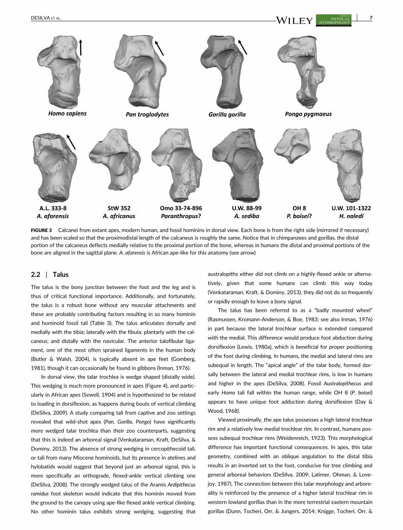
Furthermore, in dorsal view, the human calcaneus lies on a relatively
continuous proximodistal (PD) axis (Figure 3). In contrast, the African
ape calcaneus possesses a medial deflection of the distal body relative
to the proximal, with the deflection point positioned at the peroneal
trochlea (Deloison, 1985; Gebo, 1992). Given that the calcaneus forms
developmentally from two distinct regions (Čihák, 1972), we hypothe-
size that the ape calcaneus forms such that the distal portion is medially
deflected relative to the proximal. This has the effect of adducting the
forefoot relative to the hindfoot. In humans, the proximal calcaneus has
reoriented laterally (relative to the distal calcaneus), which corresponds
to a final change in the evolution of the foot according to Lewis (1981),
and aligns the long axis of the foot with the axis of the subtalar joint.
Interestingly and importantly, the Hadar calcanei, while human-like in
many respects, retain the ape-like medial deflection of the distal calca-
neus. In contrast, chronologically later calcanei from Omo, OH 8, and
even A. sediba have the human-like geometry in which the body of the
calcaneus is aligned in the sagittal plane (Figure 3).
Distally, the calcaneocuboid joint is shaped quite differently in
humans and apes. In humans, the joint is flat and spills onto the medial
side of the bone for articulation with the cuboid beak (Bojsen-Møller,
1979). Dorsally, there is a bony overhang (anterolateral process) that
restricts rotation at the calcaneocuboid joint (Elftman & Manter,
1935). In contrast, the African ape cuboid is more concave with a
medially positioned pit around which the beak of the cuboid can pivot
(Lewis, 1980b). In orangutans, the cuboid facet is convex and abruptly
spills medially into a groove for the cuboid beak. The Hadar calcanei
are damaged distally; however, A.L. 333-8 has a preserved pit, sug-
gesting more calcaneocuboid mobility than in modern humans. The
joint is more human-like in the Omo calcaneus and in OH 8. Damage
precludes assessment of this joint in the South African australopiths.
In lateral view, the cuboid facet is vertically oriented in the apes, but is
diagonally positioned dorsodistally to plantoproximally in humans.
This tilt to the calcaneocuboid joint in humans is correlated with the
medial and lateral longitudinal arching of the foot (Berillon, 2003;
Heard-Booth, 2017; Morton, 1935; Weidenreich, 1923). This angula-
tion is present in the Omo calcaneus and OH 8, but also in the pre-
served portion of the A. africanus and A. sediba calcanei suggesting
the presence of at least an incipient arch in these early hominins.
A geometric morphometrics analysis of hominin fossil calcanei
(McNutt, Zipfel, & DeSilva, 2018; McNutt & DeSilva, 2018; Figure 2)
found the A. afarensis calcanei to cluster within, or just outside, the
modern human range. In contrast, calcanei from A. sediba, and the
Omo calcaneus are positioned between the human and ape (mostly
gorilla) shape-space.
FIGURE 2 (a) Calcanei from extant apes, modern human, and fossil hominins in lateral view. Each bone is from the right side (mirrored if
necessary) and has been scaled so that the proximodistal length of the calcaneus is roughly the same. Notice the gracile tuber, projecting medialplantar process, and large peroneal trochlea in the ape calcanei. (b) PCA of a geometric morphometrics analysis performed on the most completehominin calcanei (A.L. 333-8, A.L. 333-55, and U.W. 88-99). PC1 explains 43.6% of the variation; PC2 14%. Notice that the A. afarensis calcaneiplot either within or just outside the human distribution, whereas A. sediba falls between the human and ape scatter. (c) PCA of a geometricmorphometrics analysis performed using a smaller set of landmarks allows inclusion of the Omo 33-74-896 calcaneus and H. naledi. PC1 explains34.4% of the variation; PC2 14.8%. Notice that the Omo calcaneus nears the Gorilla distribution, whereas both A. sediba and H. naledi fall outsidethe modern human range. Details of landmarks and PCA analysis in McNutt, Zipfel, and DeSilva (2018) and DeSilva, Carlson, et al. (2018)
6 DESILVA ET AL.
2.2 | Talus
The talus is the bony junction between the foot and the leg and is
thus of critical functional importance. Additionally, and fortunately,
the talus is a robust bone without any muscular attachments and
these are probably contributing factors resulting in so many hominin
and hominoid fossil tali (Table 3). The talus articulates dorsally and
medially with the tibia; laterally with the fibula; plantarly with the cal-
caneus; and distally with the navicular. The anterior talofibular liga-
ment, one of the most often sprained ligaments in the human body
(Butler & Walsh, 2004), is typically absent in ape feet (Gomberg,
1981), though it can occasionally be found in gibbons (Inman, 1976).
In dorsal view, the talar trochlea is wedge shaped (distally wide).
This wedging is much more pronounced in apes (Figure 4), and partic-
ularly in African apes (Sewell, 1904) and is hypothesized to be related
to loading in dorsiflexion, as happens during bouts of vertical climbing
(DeSilva, 2009). A study comparing tali from captive and zoo settings
revealed that wild-shot apes (Pan, Gorilla, Pongo) have significantly
more wedged talar trochlea than their zoo counterparts, suggesting
that this is indeed an arboreal signal (Venkataraman, Kraft, DeSilva, &
Dominy, 2013). The absence of strong wedging in cercopithecoid tali,
or tali from many Miocene hominoids, but its presence in atelines and
hylobatids would suggest that beyond just an arboreal signal, this is
more specifically an orthograde, flexed-ankle vertical climbing one
(DeSilva, 2008). The strongly wedged talus of the Aramis Ardipithecus
ramidus foot skeleton would indicate that this hominin moved from
the ground to the canopy using ape-like flexed ankle vertical climbing.
No other hominin talus exhibits strong wedging, suggesting that
australopiths either did not climb on a highly-flexed ankle or alterna-
tively, given that some humans can climb this way today
(Venkataraman, Kraft, & Dominy, 2013), they did not do so frequently
or rapidly enough to leave a bony signal.
The talus has been referred to as a “badly mounted wheel”
(Rasmussen, Kromann-Anderson, & Boe, 1983; see also Inman, 1976)
in part because the lateral trochlear surface is extended compared
with the medial. This difference would produce foot abduction during
dorsiflexion (Lewis, 1980a), which is beneficial for proper positioning
of the foot during climbing. In humans, the medial and lateral rims are
subequal in length. The “apical angle” of the talar body, formed dor-
sally between the lateral and medial trochlear rims, is low in humans
and higher in the apes (DeSilva, 2008). Fossil Australopithecus and
early Homo tali fall within the human range, while OH 8 (P. boisei)
appears to have unique foot adduction during dorsiflexion (Day &
Wood, 1968).
Viewed proximally, the ape talus possesses a high lateral trochlear
rim and a relatively low medial trochlear rim. In contrast, humans pos-
sess subequal trochlear rims (Weidenreich, 1923). This morphological
difference has important functional consequences. In apes, this talar
geometry, combined with an oblique angulation to the distal tibia
results in an inverted set to the foot, conducive for tree climbing and
general arboreal behaviors (DeSilva, 2009; Latimer, Ohman, & Love-
joy, 1987). The connection between this talar morphology and arbore-
ality is reinforced by the presence of a higher lateral trochlear rim in
western lowland gorillas than in the more terrestrial eastern mountain
gorillas (Dunn, Tocheri, Orr, & Jungers, 2014; Knigge, Tocheri, Orr, &
FIGURE 3 Calcanei from extant apes, modern human, and fossil hominins in dorsal view. Each bone is from the right side (mirrored if necessary)
and has been scaled so that the proximodistal length of the calcaneus is roughly the same. Notice that in chimpanzees and gorillas, the distalportion of the calcaneus deflects medially relative to the proximal portion of the bone, whereas in humans the distal and proximal portions of thebone are aligned in the sagittal plane. A. afarensis is African ape-like for this anatomy (see arrow)
DESILVA ET AL. 7

McNulty, 2015). Humans have an everted foot upon which an orthog-
onal tibia is positioned (Latimer et al., 1987). This geometry orients
the foot directly under the knee and is one feature that minimizes
mediolateral sway during bipedal locomotion (Saunders, Inman, &
Eberhart, 1953). The talar axis angle is a measure of this inverted or
everted “set” to the foot. Ardipithecus ramidus from the Aramis locality
possesses a high, ape-like talar axis angle (Lovejoy et al., 2009), indi-
cating an inverted set to this foot. In contast, the Ardipithecus talus
from Gona, Ethiopia has a lower, more human-like talar axis angle, in
part because of a higher medial trochlear rim (Simpson et al., 2018).
Bearing in mind the small sample sizes currently available, these differ-
ences could indicate that the Gona Ardipithecus possessed a more
everted, human-like foot better adapted for bipedal locomotion than
the Aramis Ardipithecus. All chronologically subsequent hominin tali
from Australopithecus and fossil Homo have low human-like talar axis
angles (DeSilva, 2009). In fact, the angles tend to be lower than typi-
cally found in modern humans because only recently have modern
humans dorsoplantarly expanded the body of the talar trochlea (which
would have the effect of raising the talar axis angle; Boyle & DeSilva,
2015). Related to this morphology is the orientation of the groove
between the medial and lateral tubercles for the tendon of M. flexor
hallucis longus, which is vertically oriented in human and hominin tali,
and obliquely oriented in ape tali (Latimer et al., 1987).
These data together allow one to construct a general evolutionary
history of the trochlear body. Based on tali from Miocene apes and
Ardipithecus, the talus of the human–chimpanzee last common ances-
tor (LCA) and that of the earliest hominins had a high lateral trochlear
body, a high talar axis angle (in the coronal plane), and an inverted set
TABLE 3 Comparative measurements of the talus in fossil hominins
Fossil talus Taxon Age (Ma)
Trochlearbody width(mm) (M5)
Head MLwidth(mm)(M9)
Head DPheight (mm)(M10)
Torsionangle (�)
Declinationangle (�)
Horizontalangle (�)
StW 573 A. prometheus? 3.67? – 24.4 17.1 26.1 31.3
A.L. 288-1as A. afarensis 3.2 18.0 20.7 14.5 29 10.9 33.4
A.L. 333-75 A. afarensis 3.2 – 25.0 16.1 – – –
A.L. 333–147 A. afarensis 3.2 24.1 – – – 12a –
StW 88 A. africanus 2.0–2.6 19.1 24.7 17.9 17 9.3 32.0
StW 102 A. africanus 2.0–2.6 19.4 – – – – –
StW 347 A. africanus 2.0–2.6 – 21.1 17.1 41 – –
StW 363 A. africanus 2.0–2.6 19.0 – – – 12a 32a
StW 486 A. africanus 2.0–2.6 21.0 24.6 – – 10a 27a
Omo 323-76-898 Homo? 2.2 23.2 27.5 18.1 31 20.1
U.W. 88-98 A. sediba 1.98 18.1 23.5 19.5 22 28 31.3
TM 1517 P. robustus 1.9 18.9 27.7 – 24 – 32
SKX 42695 Homo? 1.5–2.0 23.1 – – – – –
KNM-ER 1476 P. boisei? 1.88 20.4 25.9 – 31 17.6 30
KNM-ER 813 Homo? 1.85 24.7 27.8a – 47 23.9 14
OH 8 P. boisei? 1.85 19.5 24.4 17.5 26 17.0 33.5
D4110 H. erectus 1.77 – 27.0 – – – 26.0
KNM-ER 1464 P. boisei? 1.7 25.3 28.4 20.1 24 22 20
KNM-ER 5428 H. erectus 1.6 33.9 36.6 24.0 39 41 20
ATD6-95 H. antecessor 0.77–0.95 30.7 – – – – –
Sima de los Huesos Homo 0.43 29.1 � 2.3(n = 20)
30.4 � 2.2(n = 18)
21.8 � 1.9(n = 18)
– – –
U.W. 101-148 H. naledi 0.24–0.34 20.6 23.9a 18.4 45 14 25
U.W. 101-520 H. naledi 0.24–0.34 18.3a 21.3a – 35a 18a 20a
U.W. 101-1417 H. naledi 0.24–0.34 18.4 20.7a 14.3 37 10 26
Jinniushan Homo 0.26 30.3 31.3 21.5 46 – 30
Neandertals Homo 0.03–0.2 28.6 � 2.1(n = 22)
34.7 � 3.4(n = 23)
22.5 � 2.2(n = 24)
38.7 � 5.2(n = 14)
– 25.5 � 4.2(n = 14)
KHS 1–59 H. sapiens 0.195 25.3a 30.1a 19.8a 30a 20a 17a
LB1/15 H. floresiensis 0.06 19.5 22.6 16.2 26 23
Note. Data in this table from original fossils and measurements reported in Day & Wood, 1968; Leakey & Wood, 1973; Day, Leakey, Walker, & Wood,1976; Trinkaus, 1983; DeSilva, 2008; Jungers et al., 2009; Lu et al., 2011; Zipfel et al., 2011; Pablos et al., 2012; Boyle & DeSilva, 2015; Prang, 2016b. Onlyadult tali are shown in this table. Juvenile talus DIK-1-1f (A. afarensis) is described elsewhere (DeSilva, Gill, et al., 2018) as are juvenile fossils from H. naledi(U.W. 101-080, U.W. 101-910, U.W. 101-1623; Harcourt-Smith et al., 2015). Fossils too fragmentary to yield any of the measurements reported in thistable include KNM-ER 803, U.W. 101-1031, U.W. 101-1215, and LB1/54. Tali from Ardipithecus (ARA-VP-6/500-023; White et al., 2009; Lovejoy et al.,2009) and Gona (Simpson et al., 2018), along with BOU-VP-2/95 (Asfaw and Gilbert, 2008) and KNM-ER 64062 (Jungers et al., 2015) have been discussedin print but are not yet formally described.a Estimate that approximates the actual value.
8 DESILVA ET AL.

to the ankle joint, functionally consistent with frequent arboreality.
Boyle and DeSilva (2015) hypothesized that the first change to the
talus was a lowering of the lateral trochlear body. However, the Gona
Ardipithecus talus suggests that we were in error and instead the eve-
ning of the trochlear rims was not solely a product of a lowering of
the lateral rim, but an elevation of the medial rim as proposed long
ago (Elftman & Manter, 1935). Additionally, in Boyle and DeSilva
(2015), we missed a key change in which the mediolateral width of
the talar trochlea must have increased to accommodate the functional
demands of bipedality. This had not yet happened in the Gona Ardi-
pithecus and instead is first seen in fossil Australopithecus from Hadar
(A.L. 333-147 and A.L. 288-1) and Sterkfontein Member 2 (StW 573;
McNutt, Zipfel, & DeSilva, 2018). Stasis in this morphology can be
seen through the human fossil record and even into early members of
our own species. The Omo-Kibish talus from the 195 ka H. sapiens
skeleton is dorsoplantarly short, similar to the 1.6 Ma H. erectus talus
KNM-ER 5428 (McNutt, Zipfel, & DeSilva, 2018). Only recently did
the dorsoplantar height of the human talus increase, for reasons that
remain unclear (Boyle & DeSilva, 2015).
One final observation on the talar trochlea is warranted: the
human talar trochlea, in proximal or distal view, is relatively flat, with
only a weak groove dividing the medial and lateral aspects of the
trochlea. Apes, in contrast, have a deeper keel, accentuated by the
raised lateral rim of the trochlea and low medial rim (Figure 5). The
Aramis Ardipithecus talus is ape-like in this morphology (Lovejoy et al.,
2009; Supporting Information Figure S1). However, with the elevation
of the medial rim in hominins, a deep groove is produced. This is evi-
dent in the Gona Ardipithecus and may have helped to stabilize the
ankle joint. However, in some hominins, the talar trochlea became
remarkably flat, perhaps to more effectively dissipate high forces
across a broad surface, though this remains to be tested. This flat mor-
phology of the trochlear surface is present in tali assigned to
A. afarensis (A.L. 288-1, A.L. 333-147), A. sediba (U.W. 88-98), early
Homo (Dmanisi), H. erectus (KNM-ER 5428), H. floresiensis, and
H. naledi. The deeply keeled talar trochlea is variably retained in fossils
assigned to A. africanus and the robust australopiths. It is most
strongly expressed in OH 8, considered here to be from P. boisei and
not, as is standard in our field, H. habilis (e.g., Leakey, Tobias, & Napier,
1964).
Human tali differ from ape tali in three angular measures reflect-
ing how the head and neck are positioned relative to the talar troch-
lea. In the transverse plane, apes possess a larger horizontal angle in
which the head and neck are medially deflected compared with the
trochlear body (Figure 4). Some have suggested that this anatomy is
related to a grasping hallux (e.g., Kidd et al., 1996), but others disagree
(e.g., Barnett, 1955; Lovejoy, 1975, 1978) and an ape-like horizontal
angle in a fossil foot known to have an adducted hallux (OH 8) effec-
tively refutes this idea. Nevertheless, an ape-like horizontal angle is
found in early Australopithecus tali and in OH 8. In the coronal plane,
the head and neck exhibit strong torsion compared with the trochlear
body in humans, but not in apes. Elftman and Manter (1935) suggest
that the head and neck of human and ape tali are the same and that it
is the talar trochlea that is oriented differently in humans. Neverthe-
less, Australopithecus tali generally possess the low, ape-like condition
(but see StW 347; Prang, 2016b), whereas tali assigned to Homo
(e.g., KNM-ER 813, KNM-ER 5428) have more human-like head/neck
torsion. Manter (1941), Elftman (1960), and Langdon, Bruckner, and
Baker (1991) proposed that head/neck torsion is related to “locking”
and “unlocking” of the transverse tarsal (Chopart's) joint during foot
pronation and supination. In apes, the low torsion would align the
transverse tarsal joint and permit midfoot flexibility, whereas high tor-
sion would prevent joint alignment and limit joint motion—what Kidd
(1999) calls the “midtarsal restraining mechanism”. However, it is
worth noting that head/neck torsion is highly variable in human tali
(Lovejoy, 1978). In sagittal view, the head/neck of the ape talus is
roughly in the same plane as the body of the talus. However, in human
FIGURE 4 Tali from a modern human, extant apes, and fossil hominins in dorsal view. Each bone is from the right side (mirrored if necessary) and
has been scaled so that the proximodistal length of the talus is roughly the same. Notice that the distal aspect of the talar trochlea ismediolaterally expanded (wedged) in apes (especially in African apes) and Ardipithecus (ARA-VP-6/500), compared with humans and most fossilhominins. Right: Boxplot displaying mediolateral width of talar head to the width of the trochlear body. Boxes span the interquartile ranges, withthe horizontal center lines indicating median values. Whiskers indicate sample range, excluding outliers (dots). Relative to the width of thetrochlear body, fossils from Australopithecus and Paranthropus (including OH 8) have a mediolaterally broad head (data in Table 3). The head is MLshorter (or the body is ML wider) in fossils attributed to Homo, especially in the Sima de los Huesos fossils, which have a uniquely small talar head.Image of Ardipithecus ramidus talus courtesy of Tim White and Gen Suwa
DESILVA ET AL. 9

tali, the head and neck of the talus are plantarly directed relative to
the plane of the ankle joint, giving the human talus a strong plantar
declination [originally called inclination by Day & Wood, 1968]. A CT-
based study of modern humans has shown this anatomy to vary with
medial longitudinal arch height (Peeters et al., 2013). In australopiths,
OH 8, H. floresiensis and H. naledi the head/neck declination angle is
low and ape-like. It is slightly higher in the A. sediba talus (but see
Prang, 2015b), and has human-like declination in H. erectus (KNM-
ER 5428).
There are additional differences between the talus of humans and
apes. Relative to human tali, African ape tali tend to possess a laterally
flaring fibular facet (Gebo, 1992) and a concavely excavated medial
cotylar fossa for contact with the medial malleolus. These anatomies
vary considerably in hominin tali. Tali suggested to belong to robust
australopiths (KNM-ER 1464, ER 1476, and TM 1517) and OH 8 have
strongly flared fibular facets. Additionally, in proximal view the fibular
and tibial facets are roughly parallel in humans and markedly angled in
apes. Tali from South African localities (StW 88, U.W. 88-98, H. naledi)
and OH 8 possess more chimpanzee-like, angled malleolar facets,
whereas the Hadar and Koobi Fora tali have more human-like parallel
facets (Harcourt-Smith et al., 2015).
The talar head is generally more curved both mediolaterally and
dorsoplantarly in apes compared with the human talus (Prang, 2016a).
In particular, the ape head extends more posterolaterally (Elftman &
Manter, 1935), which may be a skeletal correlate of the elevated talo-
navicular motion that occurs in the ape foot (Thompson, Holowka,
O'Neill, & Larson, 2014). Tali assigned to early Australopithecus (A.L.
288-1 and StW 88) retain the ape-like mediolateral curvature of the
talar head, though this may also be related to their small size. Fossil
tali from Australopithecus and early Homo have human-like dorsoplan-
tar (DP) curvature of the head, and in some cases (A. sediba and
H. floresiensis) are even flatter than is common in modern humans
(Prang, 2016a). Similarly, the calcaneal facets are flatter in human tali
than in ape tali (Prang, 2016a). The South African australopiths
(A. africanus and A. sediba) are more ape-like, suggesting elevated
mobility at the subtalar joint (Prang, 2016b). Other hominins, including
A. afarensis, are more human-like (Prang, 2016b). Plantarly, at the junc-
tion of the talar head and the anterior calcaneal facet, is a smooth tri-
angular impression caused by the plantar calcaneonavicular (spring)
ligament. This ligament is present in the African ape foot (Gebo, 1992;
Gomberg, 1981), but does not leave a mark on the plantar talar head
(Lamy, 1986). This impression can be found in tali of A. afarensis and
other early hominins, suggesting the presence of at least an incipient
medial longitudinal arch (Lamy, 1986).
2.3 | Navicular
The navicular articulates with the talus proximally and with the three
cuneiforms distally. Laterally, the ape navicular contacts the cuboid, a
condition that is variably present in humans. A significant portion of
the tendon of M. tibialis posterior inserts on the navicular tuberosity.
In humans, the cuneiform facets are in roughly the same coronal
plane. In apes, the lateral cuneiform facet faces laterally and is more
concave than the corresponding facet in humans. African ape navicu-
lars possess a large, projecting tuberosity. The tuberosity is smaller in
humans, and smaller still in Pongo (Figure 6). The human navicular,
compared with extant apes, is proximodistally (PD) elongated
FIGURE 5 Tali from a modern human, extant apes, and fossil hominins in distal view. Each bone is from the right side (mirrored if necessary) and
has been scaled so that the dorsoplantar height of the talus is roughly the same. Note that the talar trochlea in humans is flat whereas apes andArdipithecus have a dorsolateral to plantomedial tilt to the talar trochlea, which helps positions the foot in an inverted set. Note as well that thegrooved trochlear surface and large talar head in OH 8 is similar to that found in KNM-ER 1464 (Paranthropus) and quite distinct from fossil Homo(Omo 323, KNM-ER 813, KNM-ER 5428). The human talar head and neck are twisted relative to the trochlear body, whereas the ape head andneck are more horizontally positioned. To the right, this head/neck torsion is plotted against sagittal plane declination of the head and neck inhumans (black dots; gray outline) and fossil hominins. Notice that early hominins tend to have lower (ape-like) head/neck torsion and declination,whereas fossil Homo tali are just outside the human range. H. naledi occupies a unique space with low, ape-like declination and high, human-liketorsion. Image of Ardipithecus ramidus talus courtesy of Tim White and Gen Suwa
10 DESILVA ET AL.

(Elftman & Manter, 1935), consistent with the generally PD expanded
tarsal row in humans. In contrast, the ape navicular body is PD narrow
and thins laterally. The human navicular also has rugose attachments
for the plantar calcaneonavicular (spring) and cubonavicular ligaments.
Hominin fossils (Table 4) have been difficult to interpret and there
are conflicting findings. Lovejoy et al. (2009) suggested that an elon-
gated tarsal row is the primitive condition from which humans have
further experienced tarsal elongation, including in the navicular body,
and apes independently evolved a foreshortened navicular; a fragmen-
tary navicular body from Ardipithecus appears to be PD thick. Lovejoy
et al. (2009) further contended that the navicular tuberosity enlarge-
ment in African apes is illusory and that the PD enlargement of the
navicular body in humans masks a still-large tuberosity. Harcourt-
Smith (2002) results are in conflict with this interpretation; he used a
geometric morphometrics analysis to show that African apes have
both an enlarged tuberosity and a laterally tapered navicular body.
The Hadar naviculars (A.L. 333-36 and A.L. 333-47) from
A. afarensis also possess a large navicular tuberosity and laterally nar-
row PD navicular body, and are thus African ape-like in certain
respects (Berillon, 2003; Harcourt-Smith, 2002; Sarmiento & Marcus,
2000). Harcourt-Smith (2002) interprets the large navicular tuberosity
as evidence for medial weight bearing in a foot that lacks a human-like
longitudinal arch. Prang’s (2016a, 2016b, 2016c) reanalysis of the
Hadar naviculars finds them to be more human-like than African
ape-like.
StW 573 was originally interpreted as being more primitive and
ape-like (Clarke & Tobias, 1995; Deloison, 2003; Kidd & Oxnard,
2005). However, a reanalysis by Harcourt-Smith (2002) found instead
that the shape of this bone is more human-like, with a relatively smal-
ler navicular tuberosity and more PD elongated navicular body than
the Hadar naviculars (Figure 6). Similarly, he found the OH 8 navicular
to cluster with humans, in contrast to a study that found the OH
8 navicular to be more ape-like (Kidd et al., 1996). Prang’s (2016a,
2016b, 2016c) reanalysis found the OH 8 navicular to be human-like,
consistent with other studies on this foot (Stern & Susman, 1983).
Surprisingly though, the new Homo foot from Ileret and the late Pleis-
tocene foot of H. floresiensis are more primitive in possessing enlarged
navicular tuberosities and laterally pinched navicular bodies (Jungers
et al., 2009; Jungers et al., 2015; Figure 6). These data might suggest
lower medial longitudinal arches in these hominins, and a less efficient
medial weight transfer mechanism. Naviculars from H. naledi are frag-
mentary, though a preliminary look at a recently recovered complete
bone (U.W. 101-1758) also finds a large navicular tuberosity and later-
ally tapering navicular body (Figure 6).
2.4 | Cuboid
The cuboid articulates proximally with the calcaneus and distally with
the fourth and fifth metatarsals (Mt4 and Mt5). Medially, the cuboid
articulates with the lateral cuneiform and in apes (but only occasion-
ally in humans) proximomedially with the navicular. Laterally, a sesa-
moid (os peroneum) can be found in the cuboidal groove of monkey
and gibbon cuboids, or proximolaterally positioned outside of the
groove in human cuboids. The os peroneum is typically absent in great
apes. Lovejoy et al. (2009) document an os peroneum facet in the
cuboidal groove in Ardipithecus ramidus and proposed that this
monkey-like anatomy was evidence for the independent loss of the os
peroneum in Pongo, Gorilla, and Pan. They further suggested that the
proximolateral repositioning of the os peroneum in humans (and OH
8) lifted the M. peroneus longus (PL) tendon out of the cuboidal groove
and obliquely across the foot (Lovejoy et al., 2009). This reorientation
of the PL tendon is hypothesized to help stiffen the midfoot and sup-
port the arch in a foot that had lost a grasping hallux. The importance
of the os peroneum for foot evolution will require additional
FIGURE 6 Naviculars from extant apes, modern human, and fossil hominins in dorsal view. Each bone is from the right side (mirrored if
necessary) and has been scaled so that the mediolateral width of the navicular is roughly the same. Notice that relative to the ape naviculars,humans have a proximodistally elongated lateral body and relative to the African ape naviculars, a reduced, nonprojecting tuberosity. Thetuberosity is essentially nonexistant in Pongo. The tuberosity remains large and the lateral body is pinched in fossil naviculars fromAustralopithecus, H. naledi, and H. floresiensis. Right: relative to the navicular width, the square root of the product of the dorsoplantar andproximodistal thickness of the tuberosity, and the proximodistal thickness of the lateral navicular body are plotted in humans (n = 15) and fossilhominins. Notice that the Omo-Kibish navicular is close to the modern human distribution whereas all other hominins, including H. naledi andH. floresiensis, possess larger tuberosities and laterally pinched navicular bodies
DESILVA ET AL. 11

comparative work as well as experimental studies that measure foot
function as it pertains to an os peroneum and M. peroneus longus
(Holowka & Lieberman, 2018).
The human cuboid differs from that of the apes in two function-
ally meaningful ways. First, the calcaneal process, or plantar beak, is
larger and more medioplantarly (often called eccentrically) oriented
(Harcourt-Smith, 2002). This positioning of the plantar beak is thought
to stabilize the calcaneocuboid joint during bipedalism (Bojsen-Møller,
1979; Elftman & Manter, 1935). The Ardipithecus ramidus cuboid pos-
sesses the more primitive, ape-like position of the cuboid beak
(Lovejoy et al., 2009); the OH 8 cuboid is more human-like (Day &
Napier, 1964; Harcourt-Smith, 2002).
Additionally, the human cuboid is PD elongated relative to the
mediolateral (ML) width of the bone. This change is thought to reflect
a general elongation of the tarsal region in humans (Elftman & Manter,
1935; Keith, 1928; Schultz, 1963). And as with the other tarsal ele-
ments, Lovejoy et al. (2009) proposed that tarsal elongation might
actually be the primitive form and that the apes independently fore-
shortened their cuboid. Interestingly, relative to the ML width of the
bone, the PD length in Pongo is not substantially different from that in
Homo (Figure 7). Furthermore, while there is some lateral pinching
(PD narrowing) of the cuboid in Pongo compared with Homo, it is not
nearly to the same extreme as that found in the African apes. We
agree with Lovejoy et al. (2009) that the primitive form was a more
generalized tarsal row, which became PD elongated in the human
lineage (Table 5), and PD foreshortened in the individual African ape
lineages over the course of their evolutionary histories (see also Mor-
ton, 1935).
Finally, the facets for the lateral metatarsals tend to be relatively
flat on the human cuboid, but are—especially Mt4—quite concave in
other primates. This anatomy is functionally related to the midtarsal
break, which occurs primarily at the lateral tarsometatarsal joints
(DeSilva, 2010). Stabilization of the lateral tarsometatarsal joint is a
key bipedal innovation in early hominins (McNutt, Zipfel, & DeSilva,
2018) and thus the flattening of this joint in Ardipithecus (Lovejoy
et al., 2009) supports its identification as a bipedal hominin.
2.5 | Medial cuneiform
The medial cuneiform articulates with the navicular proximally; the
first metatarsal (Mt1) distally, and the intermediate cuneiform and sec-
ond metatarsal (Mt2) laterally. It serves as the attachment for slips of
M. tibialis anterior and in humans, but not in other apes, M. peroneus
longus.
The proximal facet for the navicular is strongly concave in apes
and is flatter (but still generally concave) in the human medial cunei-
form. Though this curvature has not been quantified in any studies of
hominin medial cuneiforms, it qualitatively appears to remain quite
concave in StW 573 and A.L. 333-28, while flattening more in OH
8 and the medial cuneiforms from H. naledi. Plantolaterally, the human
medial cuneiform possesses a rugosity for the insertion of M. peroneus
TABLE 4 Comparative measurements of the navicular in fossil hominins
Accession number Taxon Age (Ma)MaximumML width
MaximumDP height(mm)
PD thicknessof tuberosity(mm)
DP height oftuberosity(mm)
Maximumlength oftalar facet(mm)
Maximumheight oftalar facet(mm)
Lateral PDthicknessof body(mm)
StW 573 A. prometheus? 3.67? 33.6 18.5 16.8 10.1 22.2 – 10.8
A.L. 333-36 A. afarensis 3.2 37.0 20.2 22.5 11.5 26.1 16.5 11.3
A.L. 333-47 A. afarensis 3.2 36.3 21.5 18.5 10.9 24.1 16.5 9.6
OH 8 P. boisei? 1.85 31.4 18.6 15.5 9.2 23.8 15.8 10.1
Sima de los Huesos Homo 0.43 44.2 � 4.0(n = 9)
– – – – – –
U.W. 101-623 H. naledi 0.24–0.34 – – – – – – 8.5a
U.W. 101-811 H. naledi 0.24–0.34 – – – – – – 7.9
U.W. 101-1030 H. naledi 0.24–0.34 – – – – – 13.5 8.9
U.W. 101-1562 H. naledi 0.24–0.34 – 14.7a – – 18.9 12.3 6.5
U.W. 101–1758 H. naledi 0.24–0.34 34.5 20.8 17.8 12.2 24.1 16.3 9.4
Jinniushan Homo 0.26 40.3 24.3 18.6 13.1 – – –
Neandertals Homo 0.03–0.2 43.7 � 3.1(n = 14)
26.4 � 2.5(n = 5)
– – 29.1 � 2.8(n = 7)
22.0 � 4.3(n = 7)
–
KHS 1–45 H. sapiens 0.195 37.8 28.1 17.1 12.7 28.5 21.5 13.1
LB1/16 H. floresiensis 0.06 29.7 – – – – – –
LB1/26 H. floresiensis 0.06 29.2 – – – – – –
Note. ML, mediolateral; DP, dorsoplantar; PD, proximodistal. Data in this table from original fossils and measurements reported in Trinkaus, 1975; Latimeret al., 1982; Pearson, Royer, Grine, & Fleagle, 2008; Jungers, Larson, et al., 2009; Lu et al., 2011; Mersey, Jabbour, Brudvik, & Defleur, 2013; Harcourt-Smith et al., 2015; Pablos, Pantoja-Pérez, Martínez, Lorenzo, & Arsuaga, 2017. Only adult naviculars are shown in this table. Juvenile navicular DIK-1-1f(A. afarensis) is described elsewhere (DeSilva, Gill, et al., 2018) as are juvenile fossils from H. naledi (U.W. 101-910, U.W. 101-997; Harcourt-Smith et al.,2015). Naviculars from Ardipithecus (ARA-VP-6/503: White et al., 2009; Lovejoy et al., 2009), StW 623 (Clarke, 2013) and KNM-ER 64062 (Jungers et al.,2015) have been discussed in print but are not yet formally described. SWT/UNE-2 is an undated navicular from Swartkrans preliminarily announced byThrockmorton et al. (2015) and remains undescribed.a Estimate that approximates the actual value.
12 DESILVA ET AL.

longus, which only inserts into the base of Mt1 in the apes. This inser-
tion is present in StW 573, A.L. 333-28 (but see Susman, Stern, & Jun-
gers, 1984), OH 8 and the medial cuneiforms from H. naledi. Laterally,
the human medial cuneiform possesses an “L” shaped facet for contin-
uous contact with the intermediate cuneiform, and viewed dorsally,
this lateral facet is angled proximomedially to distolaterally, allowing
the intermediate cuneiform to tuck into the medial cuneiform. In apes,
there is a double facet for the intermediate cuneiform, which is more
laterally directed and parallel to the long axis of the bone. Miocene
ape and Ardipithecus medial cuneiforms are ape-like, consistent with a
grasping hallux. Australopithecus and fossil Homo possess the angled,
single “L” shaped facet for the intermediate cuneiform.
Functionally, one important aspect of the medial cuneiform is the
distal facet for the Mt1 (Figure 8). This facet is strongly convex and
medially oriented (relative to the navicular facet) in ape medial cunei-
forms, though it is less medially directed and flatter in more terrestrial
eastern gorillas compared with western gorillas (Schultz, 1930;
Tocheri et al., 2011). Developmentally, the facet for the Mt1 is convex
in both juvenile humans and apes; it flattens with age in humans, but
remains convex in ape medial cuneiforms as they grow (Gill et al.,
FIGURE 7 Cuboids from extant apes, modern human, and fossil hominins in dorsal view. Each bone is from the right side (mirrored if necessary)
and has been scaled so that the mediolateral width of the dorsal cuboid is roughly the same. Notice that the African apes have a proximodistallysquat cuboid that is particularly pinched laterally. Humans have a proximodistally elongated cuboid, with a more eccentrically oriented calcanealbeak. The cuboid is exceptionally rare in the hominin fossil record, and there is considerable variation in known cuboid morphology. Right:boxplot displaying the proximodistal length of the cuboid body relative to its mediolateral width in extant apes, modern humans, and two fossilhominins. Boxes span the interquartile ranges, with the horizontal center lines indicating median values. Whiskers indicate sample range,excluding outliers (dots). Notice that humans have a PD elongated cuboid body as do fossils from A. afarensis (juvenile DIK-1-1f ) and OH8. However, Pongo also possesses a proximodistally elongated cuboid, suggesting that the African apes have independently foreshortened thecuboid, especially laterally (Lovejoy et al., 2009). Image of Ardipithecus ramidus cuboid courtesy of Tim White and Gen Suwa
TABLE 5 Comparative measurements of the cuboid in fossil hominins
Accession number Taxon Age (Ma)MaximumML width (mm)
Maximum PDmedial length (mm)
Maximum PDlateral length (mm)
DIK-1-1f A. afarensis 3.3 10.5 11.3 7.0
OH 8 P. boisei 1.85 20.4 22.8 11.6
Sima de los Huesos Homo 0.43 27.2 � 2.5 (n = 9) – –
U.W. 101-1023 H. naledi 0.24–0.34 – 23.1 –
U.W. 101-1418 H. naledi 0.24–0.34 – 24.4 12.3a
Neandertals Homo 0.03-0.2 27.8 � 3.8 (n = 9) 28.6 � 5.0 (n = 9) 14.0 � 2.6 (n = 8)
KHS 1–27 H. sapiens 0.195 25.9 26.1 14.9
LB1/17 H. floresiensis 0.06 – 18.5 –
LB1/27 H. floresiensis 0.06 – 18.4 –
Note. ML, mediolateral; DP, dorsoplantar; PD, proximodistal. Data in this table from original fossils and measurements reported in Trinkaus, 1975; Lu et al.,2011; Pablos et al., 2012; Harcourt-Smith et al., 2015; DeSilva, Gill, et al., 2018. Fossils too fragmentary to yield any of the measurements reported in thistable include SKX 31899. Cuboids from Ardipithecus (ARA-VP-6/500–016, –081; White et al., 2009; Lovejoy et al., 2009), StW 638 (Clarke, 2013), andKNM-ER 64062 (Jungers et al., 2015) have been discussed in print but are not yet formally described. It is unclear to us what measurements are beingreported for the Jinniushan specimen (Lu et al., 2011) and thus they are not included in this table. KB 3133 was identified as a hominin cuboid, but is not.a Estimate that approximates the actual value.
DESILVA ET AL. 13

2015). The convex Mt1 facet can also be found in Miocene hominoids
and in the Ardipithecus medial cuneiform, reflecting a divergent hallux
capable of opposing the other pedal digits during grasping bouts. The
flatter human facet faces more distally, reflecting an adducted, non-
grasping, hallux. Interpreting this anatomy in fossil hominins has been
challenging. The medial cuneiform Mt1 facet of juvenile A. afarensis
(DIK-1-1f ) is convex (Figure 8) and this curvature is retained in the
adults (A.L. 333-28), falling on the low end of the modern ape range
of Mt1 facet convexity (DeSilva, Gill, et al., 2018). We interpret this to
reflect multidirectional loading and probably some degree of hallucal
mobility in A. afarensis particularly in the juveniles. However, the dis-
tally directed Mt1 facet in A. afarensis demonstrates that the hallux,
while more mobile than in humans today, was generally in a human-
like adducted position (Latimer & Lovejoy, 1990a, 1990b). Claims that
StW 573 possessed a divergent, grasping hallux (Clarke & Tobias,
1995) are not supported with additional analysis (Harcourt-Smith,
2002; McHenry & Jones, 2006; Gill et al., 2015; Figure 8). Interest-
ingly, however, StW 573 and OH 8 possess more medially directed
Mt1 facets than is typically the case in modern humans. However,
articulation of the StW 573 and OH 8 foot bones unambiguously
reveal a fully adducted hallux. The curvature of the Mt1 facets is
reduced in StW 573 and OH 8 compared with A.L. 333-28 (Table 6),
suggesting that these hominins may have had more restricted mobility
of the hallux than did A. afarensis.
Latimer and Lovejoy (1990a) noted that the bursa for the M. tibialis
anterior tendon inserted more dorsally in the human medial cuneiform
and more plantarly in the ape one. This bursa position, they argued,
would necessarily restrict any medial deviation of the Mt1 over the
convex facet in A. afarensis and is further evidence for nongrasping in
this taxon. However, the shape and convexity of the Mt1 facet on the
medial cuneiform is not uniform along its dorsoplantar length (Dudas &
Harcourt-Smith, 2017). In fact, the dorsal aspect of the facet is gener-
ally more convex than the plantar aspect and in some (Pongo, some
gorillas, and Ekembo), the plantar aspect of the facet tapers and is not
medially directed. Given that the all-important dorsal aspect of the
A.L. 333-28 medial cuneiform is not preserved, we hesitate to agree
that the dorsally positioned tibialis anterior bursa on A.L. 333-28 is
definitive evidence for a nongrasping hallux in A. afarensis, and contend
instead that while fully adducted, the hallux of A. afarensis possessed
more mobility than that found in modern humans.
2.6 | Intermediate cuneiform
The intermediate cuneiform contacts the navicular proximally, the
Mt2 distally, and the other two cuneiforms medially and laterally. It
tends to be recessed into the tarsal row and to a greater degree in ter-
restrial primates, such as mountain gorillas and humans (Schultz,
1930), providing medial stability during terrestrial push-off. The plan-
tar surface tapers to a narrow ridge and is the attachment for plantar
ligaments, and a slip of the M. tibialis posterior tendon.
Ape and monkey intermediate cuneiforms have a strongly con-
cave facet for the navicular, terminating in a proximally projecting lip
of bone (Figure 9). This joint shape likely facilitates some mobility
between the intermediate cuneiform and navicular as part of the
FIGURE 8 Medial cuneiforms from modern human, extant apes, and fossil hominins in medial view. Each bone is from the right side (mirrored if
necessary) and has been scaled so that in medial view the bones are roughly the same in dorsoplantar height. Notice that the facet for the Mt1spills onto the medial side and is quite convex in the extant apes and in Ardipithecus ramidus (ARA-VP-6/500-088). In Australopithecus (A.L.333-28 and StW 573), OH 8 and Homo, the Mt1 facet is more human-like. Right: the curvature of the Mt1 facet is plotted against the angulationof the facet relative to the navicular facet. Data from Gill, Bredella, and DeSilva (2015) and DeSilva, Gill, et al. (2018). Notice that the DIK-1-1fjuvenile is within the ape range for facet curvature and angulation; the adult A. afarensis specimen (A.L. 333-28) maintains a convex facet, but isangled distally as in human medial cuneiforms. StW 573 and OH 8 are within the human distribution for curvature, but overlap with apes inangulation. Image of Ardipithecus courtesy of T. White and G. Suwa
14 DESILVA ET AL.

medial component of the midtarsal break, though this remains to be
tested. In humans, this joint is slightly concave to flat. Miocene ape
intermediate cuneiforms all possess a concave joint surface and to a
lesser degree so does StW 573. In OH 8, the facet is flatter and more
human-like. In humans, the Mt2 facet is rather flat; whereas in apes
and monkeys the lateral aspect of the facet spills laterally, comple-
menting the Mt2 proximal surface which is often “V”-shaped and
proximally projecting along the lateral side of the proximal facet.
Medially, the human intermediate cuneiform possesses a continu-
ous, “L”-shaped facet for contact with the medial cuneiform (Figure 9).
TABLE 6 Comparative measurements of the medial cuneiform in fossil hominins
Accession number Taxon Age (Ma)Radius ofcurvature (cm)
Navicularangle (�)
PD length(mm)
DP height(mm)
StW 573 A. prometheus? 3.67? 1.51 103.7 17.6 25.0
DIK-1-1f A. afarensis 3.3 0.42 104.4 – 10.5
A.L. 333-28 A. afarensis 3.2 1.01 94.0 21.4 29.5a
OH 8 P. boisei? 1.85 1.50 104.8 17.0 24.4
D4111 Homo 1.77 – – 20.1 28.8
Sima de los Huesos Homo 0.43 – – 26.2 � 1.2(n = 7)
–
U.W. 101-1039 H. naledi 0.24–0.34 – – – 22.7a
U.W. 101-1062 H. naledi 0.24–0.34 – – 17.3a 24.5
U.W. 101-1535 H. naledi 0.24–0.34 – – 20.2 24.2
Jinniushan Homo 0.26 – – – 32.3
Neandertals Homo 0.03–0.2 – – 25.0 � 1.9(n = 11)
–
KHS 1–46 H. sapiens 0.195 – – 23.7 34.9
LB1/18 H. floresiensis 0.06 – – 15.4 –
Note. PD, proximodistal; DP, dorsoplantar. Data in this table from original fossils and measurements reported in Latimer et al., 1982; Pearson et al., 2008;Jungers, Larson, et al., 2009; Jashashvili, Ponce de León, Lordkipanidze, & Zollikofer, 2010; Gill et al., 2015; Harcourt-Smith et al., 2015; DeSilva, Gill, et al.,2018. Medial cuneiform from Ardipithecus (ARA-VP-6/500-088: White et al., 2009; Lovejoy et al., 2009) has been discussed in print and is illustrated in Fig-ure 8, but is not yet formally described. SKX 31117 was identified as a hominin medial cuneiform, but is not.a Estimate that approximates the actual value.
FIGURE 9 Intermediate cuneiforms from modern human, extant apes, and fossil hominins in dorsal view (top) and medial view (bottom). Each
bone is from the right side (mirrored if necessary) and has been scaled so that in dorsal view the bones are roughly the same in mediolateral widthand in medial view the same dorsoplantar length. Notice that the African apes have proximodistally foreshortened intermediate cuneiforms. Thepresence of the long intermediate cuneiform in Pongo may reduce Mt2 nesting into the tarsal row and make the medial forefoot more mobile.Notice that in comparison to the extant apes, humans have a dorsoplantarly flat navicular facet, and a single facet for the medial cuneiform (apeshave a double facet). Right: boxplot of maximum intermediate cuneiform proximodistal length relative to mediolateral width. Boxes span theinterquartile ranges, with the horizontal center lines indicating median values. Whiskers indicate sample range, excluding outliers (dots). Fossilintermediate cuneiforms are generally human-like in relative proximodistal elongation though StW 573 and some H. naledi specimens also fallwithin the African ape range
DESILVA ET AL. 15

In apes and monkeys, this facet is divided into a distal facet and a
proximal one that runs the dorsoplantar length of the bone. These
double facets are present on the Ardipithecus ramidus intermediate
cuneiforms (ARA-VP-6/500-45 and -75). StW 573 and OH 8 both
possess the derived form of merged facets.
Perhaps most functionally relevant is the relative PD length of the
intermediate cuneiform. In humans, the bone is PD elongated, which is
thought to be part of a broader elongation of the tarsal row to convert
the foot into a rigid lever without incurring bending strains across the
metatarsal row (Keith, 1928; Elftman & Manter, 1935; Schultz, 1963;
but see Holowka, O'Neill, Thompson, & Demes, 2017). However, the
Pongo intermediate cuneiform is also PD elongated relative to the ML
width of the bone, suggesting that this explanation is incomplete. In
fact, monkeys, gibbons, Pongo, and humans all have longer intermediate
cuneiforms relative to the width of the bone; only African apes do not.
This observation is consistent with Lovejoy et al. (2009) who proposed
that an elongated tarsal region represents the ancestral condition. If this
evolutionary scenario is correct, then Gorillas and Pan have indepen-
dently foreshortened the intermediate cuneiform, perhaps to increase
medial midtarsal flexibility. Pongo's retained PD elongated intermediate
cuneiform may serve to increase medial foot mobility as it eliminates
any recessing of the Mt2 into the tarsal row relative to the Mt3. Based
on the roughly square-shaped intermediate cuneiforms in Ardipithecus
ramidus (i.e., roughly equal ML and PD dimensions) and, to a lesser
degree, the same condition in StW 573 and even H. naledi (Table 7), it
is possible that the human-African ape last common ancestor (LCA)
possessed a square-shaped intermediate cuneiform from which homi-
nins evolved a slightly PD elongated bone, and African apes (indepen-
dently) a foreshortened one.
2.7 | Lateral cuneiform
The lateral cuneiform contacts the navicular proximally, third metatar-
sal (Mt3) distally, cuboid laterally and intermediate cuneiform medially.
In humans, the fourth metatarsal (Mt4) is often recessed into the tar-
sal row and contacts the distolateral corner of the lateral cuneiform.
Plantarly, the lateral cuneiform tapers and provides insertion for a slip
of the M. tibialis posterior, and is the origin of the lateral arm of
M. flexor hallucis brevis.
Plantarly, the monkey and ape lateral cuneiform is dominated by
a large and projecting hamulus. The hamulus receives a slip of the
M. tibialis posterior tendon, and a distally (toward the Mt3) positioned
plantar groove guides the tendon of M. peroneus longus across the
plantar foot to the base of the Mt1 (Figure 10). The hamulus and distal
groove is well-developed in most monkey and ape lateral cuneiforms,
but only weakly developed in Homo, Pongo, and Ateles—primates that
do not rely heavily on hallucal grasping, or have a relatively short hal-
lux (e.g., Tuttle & Rogers, 1966). Interestingly, Pongo and Ateles have
small navicular tuberosities (Prang, 2016c) and the reduction of both
the navicular tuberosity and lateral cuneiform hamulus may be related
to the relatively small size of M. tibialis posterior (Payne et al., 2006;
Prang, 2016c; Zihlman, Mcfarland, & Underwood, 2011). In Australo-
pithecus (A.L. 333-79 and StW 573), the hamulus is present, but the
groove does not appear to be,3 suggesting the presence of an os pero-
neum that would lift the M. peroneus longus out of the peroneal
groove and orient it more obliquely across the foot. While Lovejoy
et al. (2009) hypothesized that this reconfiguration of the M. peroneus
TABLE 7 Comparative measurements of the intermediate cuneiform in fossil hominins
Accession number Taxon Age (Ma)MaximumML width (mm)
Maximum PDlength (mm)
Maximum DPheight (mm)
ARA-VP-6/500-45 Ar. ramidus 4.4 13.9 – 20.0
ARA-VP-6/500-75 Ar. ramidus 4.4 13.9 – 20.0
StW 573 A. prometheus? 3.67? 12.3 12.6 15.4
DIK-1-1f A. afarensis 3.3 4.9 6.3 –
OH 8 P. boisei? 1.85 11.2 12.4 15.1
Sima de los Huesos Homo 0.43 – 15.4 � 0.9(n = 12)
–
U.W. 101-1242 H. naledi 0.24–0.34 10.7 10.8 13.8
U.W. 101-1457 H. naledi 0.24–0.34 12.0 12.1 14.0
U.W. 101-1534 H. naledi 0.24–0.34 12.3 12.4 18.3
U.W. 101-1618 H. naledi 0.24–0.34 12.2 12.0 15.0
U.W. 101-1682 H. naledi 0.24–0.34 8.7 8.8 10.2
U.W. 101-1695 H. naledi 0.24–0.34 12.1 13.0 13.3
Jinniushan Homo 0.26 17.2 – 24.5
Neandertals Homo 0.03–0.2 14.9 � 1.4 (n = 7) 16.9 � 1.3(n = 16)
20.5 � 2.2(n = 5)
LB1/20 H. floresiensis 0.06 – 11.6 –
Note. ML, mediolateral; DP, dorsoplantar; PD, proximodistal. Data in this table from original fossils and measurements reported in Trinkaus, 1975; Lu et al.,2011; Pablos et al., 2012; Harcourt-Smith et al., 2015. Measurements of Ardipithecus intermediate cuneiforms courtesy of T. White and G. Suwa. Neander-tal intermediate cuneiform width (ML) calculated as “navicular articular breadth” and height calculated as “metatarsal articular height” in Trinkaus, 1975.The intermediate cuneiform from KNM-ER 64062 (Jungers et al., 2015) is not yet formally described.
3There is damage to the A.L. 333-79 lateral cuneiform in this region though the
overall anatomy in this region of the bone is not consistent with there having
been a prominent groove for the M. peroneus longus tendon.
16 DESILVA ET AL.

longus tendon happened at some point in human evolution (and cer-
tainly by the time of OH 8), our analysis of the lateral cuneiform
reveals that this reorientation of the M. peroneus longus occurred in
early Australopithecus.
The distal facet for the Mt3 is flat in humans and African great
apes. In Pongo, it possesses some medial convexity to complement a
more concave Mt3 base (though not as concave as the Mt2). However,
in gibbons and monkeys, the Mt3 facet is dorsoplantarly concave corre-
sponding to a convex Mt3 base in these primates (Proctor, 2010a). This
anatomy is also found in Ekembo lateral cuneiforms and is inferred for
Afropithecus based on a convex Mt3 base. This concave/convex joint
configuration at the lateral cuneiform/Mt3 junction in monkeys, gib-
bons, and early Miocene apes implies a degree of (and unique anatomi-
cal location for) midtarsal laxity not found in modern great apes or
humans. Proximally, the square-shaped facet for the navicular is rela-
tively flat in humans, but more convex in the African apes. In Pongo the
facet is extremely convex and spills onto the medial side of the bone
forming in some individuals a near ball and socket joint between the lat-
eral cuneiform and the navicular. The proximal lateral cuneiform is flat
in A.L. 333-79 (A. afarensis), StW 573, and OH 8.
As with the intermediate cuneiform, a PD elongated lateral cunei-
form is part of general midfoot elongation that converts the human
foot into a more effective lever for bipedal propulsion (Elftman &
Manter, 1935; Keith, 1928; Schultz, 1963). The lateral cuneiform is
PD short and squat in the extant apes (including Pongo), but this anat-
omy is hypothesized to be independently derived in the different apes
to facilitate foot conformity around tree branches (Lovejoy et al.,
2009). Additional lateral cuneiforms from the Middle to Late Miocene
are needed to test this hypothesis. Notably, the newly announced
lateral cuneiform from the Gona Ardipithecus partial skeleton is
already human-like in dorsoplantar elongation (Simpson et al., 2018),
as are other adult hominin lateral cuneiforms (Table 8).
2.8 | First metatarsal
The Mt1 articulates proximally with the medial cuneiform, distally
with the hallucal proximal phalanx, and plantarly with medial and lat-
eral sesamoids. In humans, the lateral base of the adducted Mt1 often
contacts the proximolateral shaft of the Mt2 and can form a small
facet (Singh, 1960). The plantolateral base is the insertion for the ten-
don of M. peroneus longus (Keith, 1928) and M. tibialis anterior. The
Mt1 is relatively long and slender in the African apes; short and slen-
der in Pongo; and robust in humans (Morton, 1926b), especially dorso-
plantarly (Figure 11). The internal cortical shell is distributed to resist
dorsoplantar bending forces (Jashashvili, Dowdeswell, Lebrun, & Carl-
son, 2015). The Mt1 is slender and ape-like in the Burtele foot (Haile-
Selassie et al., 2012), and in StW 595 from Sterkfontein Member
4, but is more human-like and robust in a different Sterkfontein M4
specimen StW 562. Interestingly, the Mt1 is short and hyper-robust in
the two Mt1s from Swartkrans (SKX 5017 and SK 1813), often
assigned to P. robustus, and in OH 8 (Figure 11). Relative to the other
digits, the Mt1 is also very short in H. floresiensis (Jungers, Harcourt-
Smith, et al., 2009; Table 9).
Relative to the base, the shaft and head of the human Mt1 are
neutrally positioned. Apes, in contrast, have an externally (laterally)
torqued Mt1, which put the hallux in opposition with the lateral digits
(Morton, 1922). In the transverse plane, the head of the human Mt1
has a slight valgus deviation, a laterally directed bony correction to
medial weight transfer through the foot (Barnett, 1962).
FIGURE 10 Left: A Gorilla foot shown plantarly so that the lateral cuneiform hamulus (asterisk) is shown as is the groove (gray line) for the
tendon of M. peroneus longus in the cuboidal groove and a groove in the lateral cuneiform just distal to the hamulus. Middle: Lateral cuneiformsfrom modern human, extant apes, and fossil hominins in dorsal view (top) and lateral view (bottom). Each bone is from the right side (mirrored ifnecessary) and has been scaled so that in dorsal view the bones are roughly the same in mediolateral width and in lateral view the samedorsoplantar length. Notice that the African apes have a plantarly projecting hamulus and a distally positioned groove (arrow) for the tendon ofM. peroneus longus. StW 573, A.L. 333-79, and OH 8 all possess a hamulus, but lack the prominent groove for the tendon of M. peroneus longus.Right: boxplot comparing the lateral cuneiform proximodistal length to mediolateral width. Boxes span the interquartile ranges, with thehorizontal center lines indicating median values. Whiskers indicate sample range, excluding outliers (dots). Compared with extant great apes,humans and all known fossil hominins have a proximodistally elongated lateral cuneiform relative to the mediolateral width of the bone
DESILVA ET AL. 17

In humans, the Mt1 head rises dorsally above the plane of the
shaft, creating a domed appearance, which has been suggested to be
functionally related to the dorsiflexion of the hallucal proximal phalanx
during bipedal push-off (Latimer & Lovejoy, 1990a, 1990b). Ardipithe-
cus ramidus does not have a domed Mt1 (Lovejoy et al., 2009), nor
does the Mt1 from the Burtele foot (Haile-Selassie et al., 2012), or
StW 595 from Sterkfontein Member 4 (Figure 11). The Mt1 is dorsally
domed in A. afarensis (A.L. 333-115), Mt1s assigned to P. robustus
(SK 1813, SKX 5017), and one Mt1 assigned to A. africanus (StW 562).
However, in these hominins, the dorsal articular surface is rounded
and is not mediolaterally expanded and flat as is found in modern
humans (e.g., Susman et al., 1984). This same anatomy—domed Mt1,
but not mediolaterally expanded—is even present in the Mt1s from
early Homo at Dmanisi (Pontzer et al., 2010). The dorsum is mediolat-
erally expanded in later H. erectus from Baringo, Kenya (BK 63) and
from H. naledi (Harcourt-Smith et al., 2015).
The base of the Mt1 in humans is mediolaterally flat relative to
the deeply concave, sigmoidal base shape (called “spiral concavity” by
Lovejoy et al., 2009) in the ape Mt1. Functionally, the ape Mt1 base
facilitates abduction and adduction of the Mt1 on the medial cunei-
form in addition to some axial (lateral and medial) rotation at the hallu-
cal tarsometatarsal joint. The flat Mt1 base in humans reflects the loss
TABLE 8 Comparative measurements of the lateral cuneiform in fossil hominins
Accession number Taxon Age (Ma)Minimum MLwidth (mm)
Maximum PDlength (mm)
Maximum DPheight (mm)
StW 573 A. prometheus? 3.67? 11.7 16.7 20.1
DIK-1-1f A. afarensis 3.3 7.1 9.4 –
A.L. 333-79 A. afarensis 3.2 14.8 18.7 24.2
OH 8 P. boisei? 1.85 11.2 15.6 22.7
Sima de los Huesos Homo 0.43 – 18.2 � 1.7(n = 9)
–
U.W. 101-683 H. naledi 0.24–0.34 10.7 16.0 15.7
U.W. 101-1698 H. naledi 0.24–0.34 11.5 16.5 14.6
U.W. 101-1734 H. naledi 0.24–0.34 9.5a 13.9a –
Neandertals Homo 0.03–0.2 – 19.9 � 2.3(n = 12)
28.7 � 3.9(n = 4)
LB1/19 H. floresiensis 0.06 – 16.9 –
LB1/28 H. floresiensis 0.06 – 16.8 –
Note. ML, mediolateral; DP, dorsoplantar; PD, proximodistal. Lateral cuneiforms from Ardipithecus (ARA-VP-6/500–100; White et al., 2009) and Gona(Simpson et al., 2018), along with KNM-ER 64062 (Jungers et al., 2015) have been discussed in print but are not yet formally described. The Jinniushanfoot (Lu et al., 2011) possesses a lateral cuneiform, but no measurements are reported. Data in this table from original fossils and measurements reportedin Trinkaus, 1975; Latimer et al., 1982; Harcourt-Smith et al., 2015; Pablos et al., 2017.a Estimate that approximates the actual value.
FIGURE 11 First metatarsal (Mt1) from modern human, extant apes, and fossil hominins in lateral view. Each bone is from the right side (mirrored
if necessary) and has been scaled so that the bones are roughly the same proximodistal length. Notice that in humans, the Mt1 head is domed andthe articular surface extends dorsally. This same anatomy can be found in most fossil hominin Mt1s, but not in BRT-VP-2/73c or StW 595. Right:boxplot depicting the dorsoplantar height of the Mt1 shaft relative to the length of the bone. Boxes span the interquartile ranges, with thehorizontal center lines indicating median values. Whiskers indicate sample range, excluding outliers (dots). Human Mt1 are robust, whereas theMt1 of the African apes is more gracile. The BRT Mt1 and StW 595 are ape-like; the Mt1 from Ardipithecus, StW 562, and fossil Homo (but notBK 63) are human-like. The robust australopith Mt1s (including OH 8) have quite short Mt1 shafts relative to the DP midshaft height
18 DESILVA ET AL.

TABLE
9Compa
rative
mea
suremen
tsofthefirstmetatarsalinfossilhominins
Accessionnu
mbe
rTax
on
Age
(Ma)
Leng
th(m
m)
Torsion(�)
BaseML
width
(mm)
BaseDPhe
ight
(mm)
Midshaft
ML(m
m)
Midshaft
DP(m
m)
Hea
dML
width
(mm)
Hea
dDP
heigh
t(m
m)
ARA-V
P-6/5
00-089
Ar.ramidus
4.4
55.6
a–
–23–2
510.5–1
1.5
11.5–1
2.5
18.3
a14.5-15.5
StW
573
A.p
rometheus?
3.67?
––
12.5
20.4
9.4
10.2
––
BRT-V
P-2/7
3c
Hominin
sp.
3.4
50.3
–14.6
22.7
9.1
9.0
16.7
14.5
A.L.3
33-115a
A.a
farensis
3.2
––
––
––
17.5
17.3
A.L.3
33-54
A.a
farensis
3.2
––
17.4
23.5
––
––
A.L.3
33-21
A.a
farensis
3.2
––
––
––
16.0
16.4
a
StW
595
A.a
frican
us?
2.0–2
.645.7
−5.9
13.0
18.1
7.6
8.5
15.0
12.2
StW
562
A.a
frican
us?
2.0–2
.652.4
4.7
16.0
24.2
10.4
11.4
19.0
15.2
OH
8P.
boisei?
1.85
43.9
a10
15.9
21.3
10.6
10.5
––
D2671
Hom
o1.77
47.2
3–
––
10.6
––
D3442
Hom
o1.77
–15
––
–9.6
––
SK1813
P.robu
stus
1.1–2
.041.4
7.5
13.3
a17.4
a10.3
10.6
10.8
14.6
SKX5017
P.robu
stus
1.8–2
.043.5
8.5
16.0
21.7
10.4
11.6
16.5
13.8
BK63
H.erectus
0.5
60.0
a–
15.6
a22.5
a12.4
10.3
18.2
19.4
Simade
losHue
sos
Hom
o0.43
––
23.1
�2.4
(n=4)
––
––
–
U.W
.101-496
H.n
aledi
0.24–0
.34
––
––
11.9
10.6
14.2
–
U.W
.101-1019
H.n
aledi
0.24–0
.34
55.1
a−5.2
––
10.9
10.3
13.8
a15.6
a
U.W
.101-1443
H.n
aledi
0.24–0
.34
50.0
−3.9
13.5
21.1
10.5
10.0
13.4
13.0
U.W
.101-1530
H.n
aledi
0.24–0
.34
––
13.3
22.2
11.1
a10.3
a–
–
Jinn
iushan
Hom
o0.26
55.7
––
––
–20.5
21.1
Nea
ndertals
Hom
o0.03–0
.255.4
�4.6
(n=11)
17.1
�11.6
(n=9)
19.9
�3.0
(n=16)
29.5
�5.2
(n=9)
14.9
�1.8
(n=12)
12.4
�1.6
(n=12)
24.1
�2.7
(n=8)
21.1
�2.5
(n=10)
KHS1–2
1H.sap
iens
0.195
68.6
–21.5
26.0
14.8
14.2
18.4
19.4
LB1/2
1H.floresiensis
0.06
47.0
––
––
––
–
Note.
ML,
med
iolateral;DP,d
orsoplan
tar.Datain
thistablefrom
origina
lfossils
andmea
suremen
tsrepo
rted
inTrink
aus,1975;Latimer
etal.,1982;W
hitean
dSu
waet
al.,1987;Su
sman
&deRuiter,2004;Lo
vejoy
etal.,2009;Jung
ers,Larson,
etal.,2009;Pontzeret
al.,2010;Lu
etal.,2011;Haile-Selassieet
al.,2012;Drape
au&
Harmon,
2013;Harco
urt-Sm
ithet
al.,2015;Pab
loset
al.,2017.O
nly
adultMt1sareshownin
this
table.
Juve
nile
Mt1sfrom
A.a
farensis(D
IK-1-1fan
dA.L.3
33-174)a
rede
scribe
delsewhe
re(D
eSilva,
Gill,e
tal.,2018;H
illen
bran
d,2009)a
sarejuve
nile
fossils
from
H.n
aledi(U.W
.101-244;U.W
.101-1499;Harco
urt-
Smithet
al.,2015).Juve
nile
KNM-W
T15000includ
edaskeletalfragmen
ttentativelyiden
tified
asapo
ssible
Mt1.Itisproba
blyno
t.Mea
suremen
tsin
thetable
havenotbee
nRep
orted
forLB
1/2
9.M
t1sfrom
Ardipithe-
cus(ARA-V
P-6/5
00–0
11;ARA-V
P-1/1
700:W
hite
etal.,2009)an
dGona
(Sim
psonet
al.,2018),DNH
115(Verno
n,2013),an
dKNM-ER
64062(Jun
gers
etal.,2015)havebee
ndiscu
ssed
inprintbutarenotye
tde
scribe
din
detail.
aEstim
atethat
approximates
theactualvalue.
DESILVA ET AL. 19

of a grasping big toe. The Ardipithecus ramidus Mt1 base is sigmoidal
(Lovejoy et al., 2009), as is the base of the Mt1 in the Burtele foot
(Haile-Selassie et al., 2012). This anatomy is consistent with mobility
of the great toe in these early hominins. This sigmoidal shape is also
moderately present in the Mt1s assigned to P. robustus SKX 5017 and
SK 1813, raising the possibility that there was some grasping ability
present in this robust lineage (Proctor, 2010b). The base of the Mt1
from A. afarensis (A.L. 333-54), which is also quite concave, is more
difficult to interpret. In a geometric morphometrics study, it plots
between human and ape (Proctor, 2010b), suggesting some mobility
at this joint. Others regard the presence of a hilum and a nonarticular
separation between the dorsal and plantar facets as evidence that
there was no axial rotation possible at this joint in A. afarensis
(Latimer & Lovejoy, 1990a, 1990b) and therefore little grasping poten-
tial. The base of the Mt1 from Dmanisi (Pontzer et al., 2010) and from
OH 8 is flat and human-like. Related to the presence or absence of an
abducent hallux is a contact area (and sometimes a facet) for the Mt2,
which is unique to the human foot (Le Minor & Winter, 2003). This
contact area is absent along the Mt1 base in Ardipithecus and the Bur-
tele foot, but is present in some other australopiths and fossils
assigned to early Homo. There is a particularly large facet on OH
8, effectively refuting any hypothesis for a divergent hallux in
this foot.
2.9 | Second metatarsal
The Mt2 articulates medially and laterally with the first and third
metatarsals respectively and with the intermediate cuneiform proxi-
mally. Distally, the Mt2 contacts the second proximal phalanx. Medi-
ally, the Mt2 also contacts the distolateral aspect of the medial
cuneiform, and in humans, the adducted Mt1 often contacts the proxi-
momedial shaft of the Mt2. Laterally, there is contact with the lateral
cuneiform in humans and African apes, but typically not in orangutans
where elongated intermediate cuneiforms force the Mt2 out of the
tarsal row.
Human Mt2s are relatively short and straight; apes possess rela-
tively longer Mt2s with greater longitudinal (proximodistal) curvature
and some transverse plane (mediolateral) curvature. While some fossil
Mt2s have longitudinal curvature (e.g., Ardipithecus, and StW 89),
most are relatively straight (Figure 12). Only the Burtele Mt2 pos-
sesses the transverse plane curvature found in apes (Haile-Selassie
et al., 2012).
Perhaps most notably, the human Mt2 head is neutrally oriented
in the sagittal plane. Apes and monkeys possess strong internal
(medial) torsion of the Mt2 in which the head of the metatarsal faces
medially relative to the dorsoplantar orientation of the base. This mor-
phology brings the head into opposition with the Mt1 and is function-
ally related to hallucal grasping (Drapeau & Harmon, 2013; Morton,
1922). Ardipithecus qualitatively possesses internal torsion (Lovejoy
et al., 2009). The Burtele foot and StW 89 (Sterkfontein Member 4)
both possess internal torsion as well (DeSilva et al., 2012; DeSilva,
Proctor, & Zipfel, 2012; Haile-Selassie et al., 2012) though of magni-
tude slightly less than modern ape Mt2s. While these observations
are consistent with hallucal grasping in Ardipithecus and the Burtele
foot, the presence of high torsion in StW 89 is unexpected and
discussed later in this review. Other hominin Mt2s (OH 8 and StW
377) have slight internal torsion (Drapeau & Harmon, 2013); in
H. naledi the torsion is human-like (Harcourt-Smith et al., 2015).
The Mt2 head is DP tall in humans and the articular surface rises
above the shaft, producing a domed appearance. There is often a sul-
cus between the head and the nonarticular dorsal surface of the Mt2,
where the base of the proximal phalanx reaches expected, but not
experimentally confirmed, maximum dorsiflexion during push-off. This
anatomy is functionally correlated with dorsally canted proximal pha-
langes which dorsiflex during the terminal stance phase (i.e., heel up
to toe-off ) of walking when the forefoot becomes a propulsive lever
(Latimer & Lovejoy, 1990b; but see Duncan, Kappelman, & Shapiro,
1994). In contrast, the ape Mt2 head lacks this dorsal doming and
instead has an articular surface that is prolonged plantarly, functionally
coincident with pedal grasping. The Mt2 head is unknown in Ardipithe-
cus. In the Burtele foot, the head is slightly domed and there is a dor-
sal sulcus (Haile-Selassie et al., 2012), similar to the anatomy found in
StW 89, which also possesses a more ape-like prolonged plantar sur-
face (DeSilva, Proctor, & Zipfel, 2012). The Mt2 head in A. afarensis
(A.L. 333-115 and A.L. 333-72) is quite human-like, with a dorsally
domed surface and prominent sulcus (Fernández et al., 2016; Lati-
mer & Lovejoy, 1990b).
In humans, the base of the metatarsal is DP tall and terminates in
a strong ridge of bone for plantar ligaments, a bony anatomy hypothe-
sized to be functionally correlated with a stiffening of the medial mid-
foot. Apes, in contrast, possess a DP short base. Relative to the total
length of the bone, Ardipithecus has a tall Mt2 base (Lovejoy et al.,
2009) whereas the Burtele foot and StW 89 from Sterkfontein M4
are both DP short and more ape-like (DeSilva, Proctor, & Zipfel, 2012;
Haile-Selassie et al., 2012). By comparing the DP height of the base to
its ML width (Table 10), more specimens can be included in an analysis
of base height (Figure 12). Using this approach, Ardipithecus, the Bur-
tele foot, and Australopithecus from South Africa (StW 573, and Sterk-
fontein M4) all have ape-like DP short bases. However, Mt2 fossils
from Hadar, OH 8, and fossil Homo have tall, more human-like, bases
(McNutt, Zipfel, & DeSilva, 2018).
In dorsal view, the base of the human Mt2 angles distomedially to
proximolaterally, and the intermediate cuneiform facet is only mildly
concave. African apes, in contrast, tend to have a less angled base,
and more mediolateral concavity to the intermediate cuneiform facet.
Additionally, the facet for the medial cuneiform is aligned with the
long axis of the Mt2 in humans, but is angled proximolaterally to disto-
medially in the apes, which is consistent with medial divergence of the
hallux. Most hominin fossils are human-like for these characters
except Ardipithecus, which possesses the ape-like angulation for the
medial cuneiform (Lovejoy et al., 2009; Figure 2).
Along the proximomedial shaft, there is often a small impression
on the Mt2 of humans for contact with the Mt1. This contact facet is
present in the proximal Mt2s from both South and Eastern African
Australopithecus, including StW 573. However, it is neither detectable
in the Burtele foot nor in Ardipithecus, consistent with a more diver-
gent hallux.
Finally, Lovejoy et al. (2009) call attention to “paired chondral
invaginations” on the dorsal surface of the base of the Ardipithecus
Mt2 and propose that these impressions are the by-product of the
20 DESILVA ET AL.

dorsal capsule pressing into the surface of the bone during climbing
(medial impression) and oblique axis bipedal propulsion (lateral impres-
sion). We note here that similar chondral invaginations are present in
the second and third metatarsals of Ekembo and Afropithecus, and can
occasionally be found in cercopithecoid Mt2s and Mt3s as well, dem-
onstrating that they are not uniquely related to bipedalism per se, but
as suggested by Lovejoy et al. (2009) relate to the joint capsule put-
ting dorsal pressure on the bone during development when the foot
rotates internally (for grasping) and externally (for some form of pro-
pulsion), though this hypothesis has yet to be experimentally
confirmed.
2.10 | Third metatarsal
The third metatarsal (Mt3) articulates medially and laterally with the
second and Mt4s respectively and with the lateral cuneiform proxi-
mally and third proximal phalanx distally. As with the Mt2, humans
have short and straight Mt3s; whereas apes tend to have longer and
more curved (longitudinally) Mt3s. The Ardipithecus ramidus Mt3
(ARA-VP-6/505) is short and slightly curved longitudinally (Lovejoy
et al., 2009).
The Mt3 has internal (medial) torsion in apes, to assist with lateral
toe opposability with the hallux (Morton, 1922). In monkeys, the Mt3
head torsion is more neutral; however, modern humans are unique
amongst primates in exhibiting external (lateral) torsion of the Mt3
(Drapeau & Harmon, 2013). Ardipithecus has slight external torsion
(Lovejoy et al., 2009) whereas OH 8 torsion falls within the human
range (Pontzer et al., 2010). Interestingly, the Dmanisi Mt3s and those
from H. naledi have external torsion considerably higher than is typi-
cally found in humans today (Harcourt-Smith et al., 2015; Pontzer
et al., 2010). The functional explanation for this is unclear.
As with the Mt2, the Mt3 head is dorsally tall and domed in
humans. Distal to the head is often a small sulcus where contact with
the proximal phalanx is expected (though not yet experimentally con-
firmed) to occur during the push-off phase of bipedal walking. The
Ardipithecus ramidus Mt3 (ARA-VP-6/505) possesses a human-like
dorsally domed Mt3 head (Fernández et al., 2018; Lovejoy et al.,
2009), as does the Burtele foot (BRT-VP-2/73; Haile-Selassie et al.,
2012). In fact, the Mt3 is the most domed of any of the metatarsal
heads in the perplexing Burtele foot. The Mt3 head of the
A.L. 333-115 A. afarensis forefoot is not as domed as the other Mt
heads and some analyses suggest it is more ape-like (Fernández et al.,
2016), though there is extensive erosion around the dorsum of this
fossil. Though there are six Mt3s from Sterkfontein Member
4 deposits, none preserve the metatarsal head, nor is it present in the
OH 8 foot.
The Mt3 base is dorsoplantarly (DP) and mediolaterally (ML) flat
in humans, and in African apes. In orangutans, the Mt3 base can be
ML concave (as found in the Mt2 of apes). Interestingly, in gibbons
(Proctor, 2010a) and in some cercopithecoids, the Mt3 base is DP
convex. Furthermore, the contact facet between the Mt3 and Mt4 is
flat in the apes. However, in gibbons and cercopithecoids, the Mt3
facet is concave, even cupped, for contact with a strongly convex Mt4
facet. These anatomies indicate considerable mobility of the Mt3 at
both the tarsometatarsal and the lateral intermetatarsal joints in gib-
bons and monkeys that is absent in the great apes. Mt3 fossils of
Ekembo and Afropithecus are gibbon and cercopithecoid-like for these
anatomies. This anatomy may therefore be an important overlooked
character distinguishing stem and crown great apes.
The base height of the Mt3 differs between hominins and the
great apes (Figure 13). The base of the ape Mt3 is DP short relative
either to the length of the metatarsal or to the ML width of the base.
However, in all hominins (Table 11), from Ardipithecus through Austra-
lopithecus to Homo, the Mt3 base is DP tall (McNutt, Zipfel, & DeSilva,
2018; Figure 13). Functionally, the increase in Mt3 base height may
be related to stiffening the lateral midfoot for bipedal propulsion,
FIGURE 12 Second metatarsal (Mt2) from modern human, extant apes, and fossil hominins in medial view. Each bone is from the right side
(mirrored if necessary) and has been scaled so that the bones are roughly the same proximodistal length. Notice that the human Mt2 is straight,with a dorsoplantarly tall base and a domed head. The ape Mt2 base is dorsoplantarly short and the head is internally rotated to face the Mt1.The Burtele Mt2 (BRT-VP-2/73b) and StW 89 both possess internally twisted heads and dorsoplantarly short bases relative to the total length ofthe Mt2. Right: boxplot displaying the mediolateral width of the Mt2 base relative to the dorspoplantar height. Boxes span the interquartileranges, with the horizontal center lines indicating median values. Whiskers indicate sample range, excluding outliers (dots). Humans have a DP tallbase, as does A. afarensis and OH 8. Mt2s from Ardipithecus, BRT, StW 573 and Sterkfontein member 4 (n = 3) are more ape-like. Reportedmeasurements and image of ARA-VP-6/1000 courtesy of Tim White and Gen Suwa
DESILVA ET AL. 21

TABLE
10
Compa
rative
mea
suremen
tsoftheseco
ndmetatarsalinfossilho
minins
Accessionnu
mbe
rTax
on
Age
(Ma)
Leng
th(m
m)
Torsion(�)
BaseMLwidth
(mm)
BaseDP
height
(mm)
Midshaft
ML(m
m)
Midshaft
DP(m
m)
Hea
dML
width
(mm)
Hea
dDP
heigh
t(m
m)
ARA-V
P-6/1
000
Ar.ramidus
4.4
––
18.4–1
8.8
19.5–2
1.6
––
––
StW
573
A.p
rometheus?
3.67?
––
13.5
15.1
7.5
8.5
––
BRT-V
P-2/7
3b
Hominin
sp.
3.4
66.9
−23
12.8
14.2
6.1
7.4
9.8
11.2
DIK-1-1f
A.a
farensis
3.3
––
5.7
7.9
––
––
A.L.3
33-72
A.a
farensis
3.2
––
––
––
9.7
12.8
A.L.3
33-115b
A.a
farensis
3.2
––
––
––
9.9
11.8
A.L.3
33-133
A.a
farensis
3.2
––
12.1
15.9
––
––
StW
89
A.a
frican
us?
2.0–2
.661.1
−19.7
11.0
12.8
5.6
7.7
8.3
11.9
StW
377
A.a
frican
us2.0–2
.654.9
a−11
13.2
14.9
6.1
7.2
––
StW
595c
A.a
frican
us?
2.0–2
.658.0
a–
11.7
13.2
6.3
6.8
––
Omo323-1976-2117
P.bo
isei?
2.12
73.6
−12
16.7
19.8
8.1
10.5
10.6
15.2
OH
8P.
boisei?
1.85
55.6
a−7
11.2
14.4
6.3
7.2
––
ATD6-25
H.a
ntecessor
0.77–0
.95
––
14.6
19.2
––
––
ATD6-70+107
H.a
ntecessor
0.77–0
.95
79.0
a–
––
7.3
10.0
11.7
17.1
Arago
XLIII
Hom
o0.5
––
12.5
–8.3
9.4
––
Simade
losHue
sos
Hom
o0.43
––
15.2
�0.9
(n=4)
––
––
–
U.W
.101-459/4
61
H.n
aledi
0.24–0
.34
58.0
a10.1
–6.6
6.9
––
U.W
.101-1022
H.n
aledi
0.24–0
.34
67.5
−8.7
––
7.6
7.2
––
U.W
.101-1458
H.n
aledi
0.24–0
.34
63.4
6.7
11.5
–6.3
6.8
–11.5
U.W
.101-1499
H.n
aledi
0.24–0
.34
––
9.7
11.3
––
––
Jinn
iushan
Hom
o0.26
71.0
–14.9
––
––
–
Nea
ndertals
Hom
o0.03–0
.272.5
�5.5
(n=14)
7.5
�4.0
(n=4)
16.7
�2.4
(n=18)
22.2
�2.1
(n=12)
8.2
�0.9
(n=18)
8.9
�1.0
(n=18)
11.4
�1.7
(n=6)
15.6
�2.3
(n=9)
KHS1–5
H.sap
iens
0.195
––
15.3
21.1
7.5
8.8
––
LB1/2
2H.floresiensis
0.06
63.0
––
––
––
–
Note.ML,med
iolateral;DP,d
orsoplan
tar.Datain
thistablefrom
origina
lfossils
andmea
suremen
tsrepo
rted
inTrink
aus,1975;L
atim
eret
al.,1982;L
amy,1982;W
hite&Su
wa,1987;P
earsonet
al.,2008;P
ontzer
etal.,
2010;Lu
etal.,2011;DeS
ilva,
Proctor,&
Zipfel,2012;DeS
ilva,
Zipfel,et
al.,2012;Haile-Selassieet
al.,2012;Pab
loset
al.,2012;Drape
au&
Harmon,
2013;Pab
loset
al.,2017;Daver
etal.,2018.Mea
suremen
tsrepo
rted
forARA-V
P-6/1
000co
urtesy
ofT.W
hite
andG.S
uwa.
Mea
suremen
tsin
thetableha
veno
tbe
enrepo
rted
LB1/3
0.M
t2sfrom
Ardipithecus(ARA-V
P-6/5
00-042,-048;W
hiteet
al.,2009),ASI-V
P-2/1
(White
etal.,2006),an
dKNM-ER64062(Jun
gers
etal.,2015)h
avebe
endiscussedin
printbu
tareno
tye
tde
scribe
din
detail.
aEstim
atethat
approximates
theactualvalue.
22 DESILVA ET AL.

though this hypothesis remains to be experimentally tested. Never-
theless, we suggest that the Mt3 base height is a hominin synapomor-
phy potentially useful for identifying isolated pedal remains.
Furthermore, Lovejoy et al. (2009) noted that ape Mt3 bases tend to
have quite noticeable medial and lateral indentations for the transmis-
sion of intermetatarsal ligaments. These indentations are subtle in
human Mt3s and in fossil hominins, including Ardipithecus.
Finally, along the distolateral shaft of many human Mt3s, there is
the development of a prominent lateral tubercle, which is absent in the
Mt3 of apes. This tubercle nestles into a concavity along the Mt4 shaft
and gives rises to the M3–4 interosseous metatarsal ligament (Kelikian &
Sarrafian, 2011). Functionally, it is proposed here (but remains
untested) that this rugosity could be a bony correlate of the Mt3 and
Mt4 moving as a functional unit during the propulsive phase of bipedal
walking. This rugosity is not present in the Ardipithecus Mt3, but is vari-
ably present in Australopithecus (e.g., A.L. 333-157, StW 496).
2.11 | Fourth metatarsal
The Mt4 articulates with the Mt3 and Mt5 medially and laterally
respectively and with the cuboid proximally and fourth proximal pha-
lanx distally. In humans, but not in apes, the Mt4 is nestled into the
tarsal row and often contacts the distolateral edge of the lateral cunei-
form (Ward, Kimbel, & Johanson, 2011). As with the other metatar-
sals, the human Mt4 is relatively short and straight compared with the
long and more curved (sagitally) Mt4 in the apes. The oldest known
complete hominin Mt4s are from the Burtele foot (3.4 Ma) and from
Hadar, Ethiopia (3.2 Ma), and sample quite different morphologies
(Figure 14). The Burtele Mt4 is long and slender (Haile-Selassie et al.,
2012); whereas the Hadar specimen (A.L. 333-160) is more human-
like in being short and robust (Ward et al., 2011).
As with the other metatarsals, there are torsional differences
between the ape and human Mt4 (Morton, 1922). In apes, the Mt4
torsion is roughly neutral. However, humans possess external (lateral)
torsion which positions the head flush on the ground in a foot with a
high transverse arch. While external torsion is certainly related to the
presence of a transverse arch, the relationship between external tor-
sion and a lateral longitudinal arch is unclear given that cercopithecoid
monkeys also possess external torsion of the Mt4 (Drapeau & Har-
mon, 2013). Later hominins from OH 8, Dmanisi Homo, and later Pleis-
tocene Homo have external torsion within the range of modern
humans (Harcourt-Smith et al., 2015; Pontzer et al., 2010).
The human Mt4 head is dorsoplantarly (DP) tall and domed, with
the articular surface rising above the dorsal surface of the shaft; a dor-
sal sulcus is typically present to facilitate metatarsophalangeal dorsi-
flexion during toe-off (Latimer & Lovejoy, 1990b). While the
Australopithecus Mt4 head (known from A.L. 333-115 and
A.L. 333-160) is domed (Ward et al., 2011), the shape may still retain
some ape-like characteristics (Fernández et al., 2016, 2018).
As with other metatarsals, the human Mt4 base is DP tall relative
both to the overall length of the bone, and to the ML width of the
base. Apes, in contrast, have DP short bases. All known hominin Mt4s
(Table 12) have tall DP bases (McNutt, Zipfel, & DeSilva, 2018), with
the exception of the Burtele foot (Haile-Selassie et al., 2012). How-
ever, damage to the plantar base of the Mt4 of this specimen makes it
difficult to assess this anatomy. Perhaps more important from a func-
tional standpoint is the DP shape of the base (Figure 14). In humans,
the base is typically either flat, or sinusoidal, reflecting stability at the
lateral tarsometatarsal joint. Apes and monkeys, in contrast, have
strongly convex bases, which are the result of tarsometatarsal dorsi-
flexion that occurs during the midtarsal break (DeSilva, 2010). The
form: function link between the midtarsal break and Mt4 base shape
is supported by an MRI study which found convex Mt4 bases in the
occasional humans with a midtarsal break (DeSilva et al., 2015). Ardi-
pithecus ramidus (ARA-VP-6/500-103) and the Burtele foot (BRT-VP-
FIGURE 13 Third metatarsal (Mt3) from modern human, extant apes, and fossil hominins in lateral view. Each bone is from the right side
(mirrored if necessary) and has been scaled so that the bones are roughly the same proximodistal length. Notice that the human Mt3 is straight,with a dorsoplantarly tall base and a domed head. Humans also have externally rotated heads; ape heads are more neutral. Right: boxplot showingthe DP height of the Mt3 base relative to the ML width of the base. Boxes span the interquartile ranges, with the horizontal center linesindicating median values. Whiskers indicate sample range, excluding outliers (dots). All hominins, including Ardipithecus, have relatively tall Mt3bases, a hominin synapomorphy. Reported measurements and image of ARA-VP-6/505 courtesy of Tim White and Gen Suwa
DESILVA ET AL. 23

TABLE
11
Compa
rative
mea
suremen
tsofthethirdmetatarsalinfossilho
minins
Accessionnu
mbe
rTax
on
Age
(Ma)
Leng
th(m
m)
Torsion(�)
BaseML
width
(mm)
BaseDP
height
(mm)
Midshaft
ML(m
m)
Midshaft
DP(m
m)
Hea
dML
width
(mm)
Hea
dDP
heigh
t(m
m)
ARA-V
P-6/5
05
Ar.ramidus
4.4
––
11.5–1
3.0
20.5–2
0.7
––
––
BRT-V
P-2/7
3f
Hominin
sp.
3.4
––
––
––
8.6
a13.2
a
A.L.3
33-115c
A.afarensis
3.2
––
––
––
9.2
13.2
A.L.3
33-133
A.afarensis
3.2
––
11.0
a15.8
––
––
A.L.3
33-157
A.afarensis
3.2
––
13.6
18.7
––
––
StW
238
A.african
us2.0–2
.6–
–12.1
16.3
6.3
a8.5
a–
–
StW
387
A.african
us2.0–2
.6–
–10.9
16.7
6.1
a7.9
a–
–
StW
388
A.african
us2.0–2
.6–
14.7
a12.6
18.5
5.9
a8.9
a–
–
StW
435
A.african
us2.0–2
.6–
–13.3
20.1
6.2
8.7
––
StW
477
A.african
us2.0–2
.6–
28.8
a12.0
16.5
5.8
a7.4
a–
–
StW
496
A.african
us2.0–2
.6–
18.7
a10.9
16.8
5.7
a7.5
a–
–
SKX38529
P.robu
stus?
1.8–2
.0–
–12.1
––
––
–
KNM-ER1500
P.bo
isei?
1.89
––
12
17.7
––
––
OH
8P.
boisei?
1.85
53.1
a24
11.9
16.0
6.6
8.8
––
OH
43
Paranthrop
us?Hom
o?1.8
––
10.0
––
––
–
KNM-ER997
P.bo
isei?
1.85
––
10.1
16.2
––
––
D3479
H.erectus
1.77
–35.0
––
–6.7
––
D2021
H.erectus
1.77
61a
38.0
––
–9.0
––
OmoF.511-16
Hom
o?1.7
––
14.6
20.7
7.6
10.2
––
KNM-ER1823
P.bo
isei?
1.59
–17
12.8
––
––
–
KNM-ER803j
H.erectus
1.53
–26
12.7
20.5
6.1
10.4
SKX247
P.robu
stus?
1.1–1
.7–
–12.2
16.3
––
––
Simade
losHue
sos
Hom
o0.43
––
15.6
�1.0
(n=6)
––
––
–
U.W
.101-552
H.n
aledi
0.24–0
.34
60.0
a–
11.9
–7.0
6.8
––
U.W
.101-1035
H.n
aledi
0.24–0
.34
––
10.6
14.3
5.9
a6.9
a–
–
U.W
.101-1457
H.n
aledi
0.24–0
.34
60.4
33.0
11.1
13.9
5.8
6.2
––
Nea
ndertals
Hom
o0.03–0
.270.0
�3.3
(n=5)
25.0
�7.8
(n=3)
16.1
�2.0
(n=18)
19.7
�1.7
(n=8)
8.1
�0.9
(n=10)
9.4
�1.7
(n=11)
12.4
�1.4
(n=3)
17.0
�2.7
(n=4)
KHS1–5
2H.sap
iens
0.195
––
14.5
22.3
––
––
LB1/2
3Hom
oflo
resiensis
0.06
60.4
––
––
––
–
Note.
ML,
med
iolateral;DP,do
rsoplan
tar.Datain
this
tablefrom
original
fossils
andmea
suremen
tsrepo
rted
inDay
&Le
akey
,1974;Trink
aus,1975;W
hite&
Suwa,
1987;Junge
rset
al.,2009;Pontzer
etal.,2010;
Haile-Selassieet
al.,2012;W
ard,
Kim
bel,Harmon,
&Joha
nson,
2012;Merseyet
al.,2013;Harco
urt-Sm
ithet
al.,2015;Mea
suremen
tsrepo
rted
forARA-V
P-6/5
05co
urtesyofT.W
hitean
dG.S
uwa.
Only
adultMt3s
areshownin
thistable.
Juve
nile
Mt3
from
A.a
farensis(D
IK-1-1f)isde
scribed
elsewhe
re(D
eSilva,Gill,e
tal.,2018)as
isthejuve
nile
Mt3
from
H.n
aledi(U.W
.101-1500;Harco
urt-Smithet
al.,2015).Mea
suremen
tsin
thetableha
veno
tbe
enrepo
rted
LB1/3
1.M
t3sfrom
AramisArdipithecus(ARA-V
P-6/5
00–0
66,W
hite
etal.,2009)a
ndGona
Ardipithecus(Sim
psonet
al.,2018),StW
595d(Clarke,
2013),an
dKNM-ER64062(Junge
rset
al.,2015)h
avebe
endiscussedin
printbu
tareno
tye
tde
scribe
din
detail.
aEstim
atethat
approximates
theactualvalue.
24 DESILVA ET AL.

2/73a) both possess human-like dorsoplantarly sinusoidal or flat
bases, demonstrating that the earliest known hominins had a stiff lat-
eral midfoot for bipedal propulsion. This anatomy is found in early
australopiths from Eastern Africa (A.L. 333-160) and South Africa
(StW 485), and into genus Homo. Surprisingly, however, the base of
the Mt4 from A. sediba is ape-like in its DP convexity (DeSilva et al.,
2013), currently explained as a function of a relatively low arch
(Prang, 2015b) and excessive medial weight transfer—two risk factors
for a hypermobile midfoot in humans today (DeSilva et al., 2015).
In lateral view, humans tend to have Mt4s with a proximodorsal
to distoplantarly angled base relative to the long axis of the shaft. This
anatomy may be related to the presence of a longitudinal arch in
humans and A. afarensis (Ward et al., 2011). This base angulation is
more neutral or ape-like in A. africanus (StW 485) and A. sediba (U.W.
88-22). Finally, apes tend to have rounder midshaft dimensions;
whereas the human Mt4 midshaft is DP taller and ML narrower.
Hominin Mt4 midshafts are human-like with the exception of
A. sediba, which has a round midshaft (DeSilva, Carlson, et al., 2018).
2.12 | Fifth metatarsal
The Mt5 articulates with the Mt4 medially, cuboid proximally, and fifth
proximal phalanx distally. In apes, the Mt5 is long, slender and longitudi-
nally curved in the sagittal plane and straight in the transverse plane.
The human Mt5 is short and stout and often laterally deviates in the
transverse plane. The only complete Mt5 in the early hominin fossil
record (StW 114/115) from Sterkfontein Member 4 has a slight ape-like
curve in the sagittal plane, but human-like transverse plane curvature
(Zipfel, DeSilva, & Kidd, 2009; Figure 15). An earlier Mt5 from Hadar
(A.L. 333-13) also displays human-like transverse plane curvature.
In both humans and apes, there is external torsion of the Mt5,
with humans possessing a greater average value (Drapeau & Harmon,
2013). The Mt5 from A. africanus (StW 114/115) has human-like head
torsion (Zipfel et al., 2009); whereas the Mt5 from Dmanisi Homo
(D4058) is more chimpanzee-like (Pontzer et al., 2010; Table 13),
though given the overlap in values between apes and humans, we do
not regard these differences as functionally meaningful.
As with other metatarsal heads, the Mt5 of humans is dorsally domed
and possesses a sulcus just proximal to the dorsal edge of the head, pre-
sumably for the rim of the proximal phalanx during metatarsophalangeal
dorsiflexion (Latimer & Lovejoy, 1990b). Australopithecus Mt5 heads are
human-like (Fernández et al., 2016; Zipfel et al., 2009). The cuboid and
Mt4 facets of the Mt5 differ little in shape between humans and the apes
(DeSilva, 2010; Proctor, 2013).
The human Mt5 midshaft is mediolaterally expanded compared to
the more rounded shape of the Mt5 midshaft in apes. The diaphyseal
strength relative to the articular area of the Mt5 head is greater in
humans than in apes (Marchi, 2010). A CT-based study revealed that
apes have more cortical bone in their metatarsals than humans (Marchi,
2005), and a more recent study found that in human Mt5s, the cortical
bone is distributed in a manner that would resist dorsoplantarly bending
forces, especially proximally (Dowdeswell et al., 2016).
Finally, compared with the human Mt5, apes tend to have a more
projecting tuberosity, possibly reflecting an important role of
M. peroneus brevis and the variably present M. peroneus tertius. Austra-
lopithecus Mt5s have a more human-like tuberosity, with the largest
(most ape-like) tubersosity belonging to A.L. 333-13 (A. afarensis).
2.13 | Proximal phalanges
The human hallucal proximal phalanx (Table 14) is short and robust
compared to that of African apes, which tend to be longer and more
slender. Orangutans typically do not possess a hallucal proximal pha-
lanx at all (Tuttle & Rogers, 1966). Human lateral proximal phalanges
(PP) are relatively short and straight; whereas the lateral proximal pha-
langes of the apes are long and longitudinally curved (Trinkaus & Patel,
2016). Ontogenetic studies demonstrate that the curvature of the
phalanges is related to grasping during development (Congdon, 2012).
The shortening of the phalanges in the human lineage confers benefits
during running (Rolian et al., 2009). The pedal phalanges of
FIGURE 14 Fourth metatarsal (Mt4) from modern human, extant apes, and fossil hominins in medial view. Each bone is from the right side
(mirrored if necessary) and has been scaled so that the bones are roughly the same proximodistal length. The base of the human Mt4 tends to beeither flat or sinusoidal. Nonhuman anthropoids have convex Mt4 bases, a skeletal correlate of the midtarsal break. Right: boxplot quantifying thecurvature of the base of the Mt4 (after DeSilva, 2010; DeSilva et al., 2013). Boxes span the interquartile ranges, with the horizontal center linesindicating median values. Whiskers indicate sample range, excluding outliers (dots). The Mt4 base is flat in all hominins (including Ardipithecus andBurtele—not shown in graph). However, the Mt4 base is uniquely convex in A. sediba—a function of a unique gait in this taxon
DESILVA ET AL. 25

Ardipithecus are long and curved (Lovejoy et al., 2009). Relative to
Ardipithecus, the pedal phalanges of Australopithecus are shorter and
less curved (Table 15; Figure 16). In Homo, the simple narrative of
pedal phalanges becoming shorter and less curved is complicated by
H. floresiensis and H. naledi, which possess long (H. floresiensis) or
curved (H. naledi) proximal pedal phalanges (Harcourt-Smith et al.,
2015; Jungers, Harcourt-Smith, et al., 2009).
Perhaps most notably, the base of human proximal phalanges is
canted dorsodistally to plantoproximally; whereas great ape phalanges
have opposite, plantar cants to the base of their proximal phalanges
(Latimer & Lovejoy, 1990b; Figure 16). Dorsal canting is hypothesized
to be part of a functional complex that includes dorsal doming of the
metatarsal head functionally related to dorsiflexion of the toes during
bipedal locomotion (Latimer & Lovejoy, 1990b). Fossil hominins from
Ardipithecus through Australopithecus to Homo possess dorsally canted
proximal phalanges (Haile-Selassie, 2001; Latimer & Lovejoy, 1990b),
though the magnitude of dorsal canting may not be as high as found
in modern humans (Duncan et al., 1994). However, dorsal canting
alone does not indicate bipedalism: the proximal phalanges from Pro-
consul (Ekembo) heseloni possess dorsal canting as occasionally do lat-
eral phalanges from gibbons and cercopithecoids (Begun, Teaford, &
Walker, 1991; Griffin & Richmond, 2010; McNutt, Zipfel, &
DeSilva, 2018).
Two other anatomies are worth noting: in apes, the base of the
phalanx is mediolaterally broad whereas in humans, the base is dorso-
plantarly tall and is curved along the dorsal rim (Stern & Susman,
1983). Preuschoft (1970) has suggested that the dorsoplantarly
expanded base of the proximal phalanges may be related to the pres-
ence of the plantar aponeurosis and a windlass mechanism in the
human foot (Hicks, 1953). Additionally, the plantar shaft of the ape
phalanx possesses strong flexor ridges, which help keep the flexor
tendons from pulling away from the plantar surface of the bone. In
humans, the flexor ridges are not as well developed.
2.14 | Intermediate phalanges
As with the proximal phalanx, the ape intermediate phalanx is longer,
more curved, and has a flatter base with more developed flexor ridges
than the human intermediate phalanx. Similarly, as with the proximal
phalanges, early hominin intermediate phalanges from Ardipithecus
and Australopithecus are more ape-like in their length (McNutt, Zip-
fel, & DeSilva, 2018; Figure 17) and curvature; whereas those from
later Homo tend to be shorter and less curved, though an exception to
this trend is H. naledi, which has quite curved intermediate phalanges
(Harcourt-Smith et al., 2015). Perhaps most notably, however, is the
size differential in human intermediate phalanges. Unlike the proximal
phalanges, which are only subtly decreasing in size from PP2 to PP5.
The human intermediate phalanges (IP) reduce considerably in size
from IP2 to IP5 (Elftman & Manter, 1935). Australopithecus afarensis
(A.L. 333-115) retains long lateral intermediate phalanges (Stern &
Susman, 1983), which would have been functionally beneficial for
some degree of pedal grasping during bouts of climbing and infant
clinging.
2.15 | Distal phalanges
Human distal phalanges are short and stout compared with ape distal
phalanges, which tend to be longer and more slender. The most sub-
stantial functional differences can be found in the hallucal distal pha-
lanx. In humans, the hallucal distal phalanx possesses, in the
transverse plane, a lateral deflection of the long axis of the bone rela-
tive to the base (Barnett, 1962). This anatomy is present already at
birth (Wilkinson, 1954). In apes, the long axis of the bone is perpen-
dicular to the base. Furthermore, in the coronal plane, the human hal-
lucal distal phalanx displays axial torsion (Figure 17), which is argued
to be functionally related to hallucal push off (Barnett, 1962; Day &
Napier, 1966). The OH 10 distal hallucal phalanx and H. naledi (U.W.
101-1551) are human-like in these characters, suggesting human-like
bipedal propulsion (Day & Napier, 1966).
3 | HOMININ FOOT EVOLUTION
3.1 | Miocene
Although this review focuses on Plio-Pleistocene hominin foot evolu-
tion, genetic studies convincingly demonstrate that the human–
chimpanzee divergence occurred in the Miocene (e.g., Moorjani,
Amorim, Arndt, & Przeworski, 2016) and thus the earliest hominin
foot fossils would derive from that time period. Two hominins from
the Miocene—Sahelanthropus tchadensis (Brunet et al., 2002) and
Orrorin tugenensis (Senut et al., 2001)—do not yet preserve any pedal
remains. Therefore, although this review covers only hominin foot fos-
sils from the Plio-Pleistocene, we are well aware that questions about
the foot of the last common ancestor will be best addressed with
pedal fossils from the Miocene, a time period currently devoid of con-
firmed hominin foot remains.
While it has been suggested that the Miocene ape Oreopithecus had
a bipedal foot (Köhler & Moyà-Solà, 1997), our preliminary observations
on these fossils along with the work of others (Susman, 2005) are skep-
tical of these claims and regard Oreopithecus as an arboreal hominoid
(e.g., Harrison, 1991). Additionally, 5.7 Ma fossilized footprints from
Crete may be from a bipedal hominin (Gierli�nski et al., 2017) or even a
bipedal hominoid (Crompton, 2017). A biped from the Miocene of Crete
truly would be paradigm altering and we look forward to the publication
of the necessary information from these footprints (e.g., Falkingham
et al., 2018) that can be used to either support or refute these extraordi-
nary claims. Below, we detail what is currently known (and remains
unknown) about the foot of Plio-Pleistocene hominins from species of
Ardipithecus through Australopithecus and Paranthropus to Homo.
3.2 | Ardipithecus kadabba (Middle Awash,Ethiopia; 5.2 Ma)
Although much of the Ar. kadabba hypodigm derives from Late Mio-
cene sediments (Haile-Selassie, 2001), the foot is represented by an
early Pliocene (5.2 Ma) left fourth proximal pedal phalanx (AME-VP-
1/71) from the Kuseralee Member (Sangantole Formation) of Amba
East in the Middle Awash study area of Ethiopia (Haile-Selassie, 2001;
Haile-Selassie, Suwa, & White, 2009). The anatomy discussed below is
26 DESILVA ET AL.
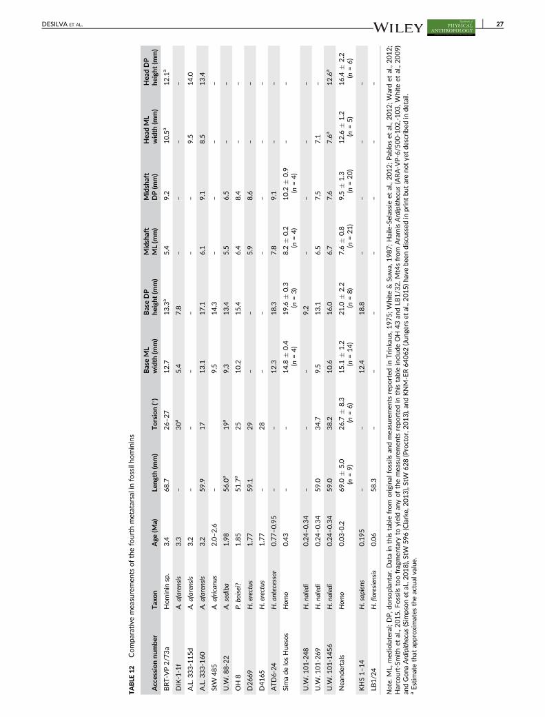
TABLE
12
Compa
rative
mea
suremen
tsofthefourth
metatarsalinfossilho
minins
Accessionnu
mbe
rTax
on
Age
(Ma)
Leng
th(m
m)
Torsion(�)
BaseML
width
(mm)
BaseDP
height
(mm)
Midshaft
ML(m
m)
Midshaft
DP(m
m)
Hea
dML
width
(mm)
Hea
dDP
heigh
t(m
m)
BRT-V
P2/7
3a
Hominin
sp.
3.4
68.7
26–2
712.7
13.3
a5.4
9.2
10.5
a12.1
a
DIK-1-1f
A.a
farensis
3.3
–30a
5.4
7.8
––
––
A.L.3
33-115d
A.a
farensis
3.2
––
––
––
9.5
14.0
A.L.3
33-160
A.a
farensis
3.2
59.9
17
13.1
17.1
6.1
9.1
8.5
13.4
StW
485
A.a
frican
us2.0–2
.6–
9.5
14.3
––
––
U.W
.88-22
A.sediba
1.98
56.0
a19a
9.3
13.4
5.5
6.5
––
OH
8P.
boisei?
1.85
51.7
a25
10.2
15.4
6.4
8.4
––
D2669
H.erectus
1.77
59.1
29
––
5.9
8.6
––
D4165
H.erectus
1.77
–28
––
––
––
ATD6-24
H.a
ntecessor
0.77–0
.95
––
12.3
18.3
7.8
9.1
––
Simade
losHue
sos
Hom
o0.43
––
14.8
�0.4
(n=4)
19.6
�0.3
(n=3)
8.2
�0.2
(n=4)
10.2
�0.9
(n=4)
––
U.W
.101-248
H.n
aledi
0.24–0
.34
––
–9.2
––
––
U.W
.101-269
H.n
aledi
0.24–0
.34
59.0
34.7
9.5
13.1
6.5
7.5
7.1
–
U.W
.101-1456
H.n
aledi
0.24–0
.34
59.0
38.2
10.6
16.0
6.7
7.6
7.6
a12.6
a
Nea
ndertals
Hom
o0.03-0.2
69.0
�5.0
(n=9)
26.7
�8.3
(n=6)
15.1
�1.2
(n=14)
21.0
�2.2
(n=8)
7.6
�0.8
(n=21)
9.5
�1.3
(n=20)
12.6
�1.2
(n=5)
16.4
�2.2
(n=6)
KHS1–1
4H.sap
iens
0.195
––
12.4
18.8
––
––
LB1/2
4H.floresiensis
0.06
58.3
––
––
––
–
Note.
ML,
med
iolateral;DP,d
orsoplan
tar.Datain
thistablefrom
origina
lfossils
andmea
suremen
tsrepo
rted
inTrink
aus,1975;W
hite
&Su
wa,
1987;Haile-Selassieet
al.,2012;Pab
loset
al.,2012;W
ardet
al.,2012;
Harco
urt-Sm
ithet
al.,2015.Fossils
toofragmen
tary
toyieldan
yofthemea
suremen
tsrepo
rted
inthistableinclud
eOH
43an
dLB
1/3
2.Mt4sfrom
AramisArdipithecus(ARA-V
P-6/5
00-102,-103,W
hiteet
al.,2009)
andGona
Ardipithecus(Sim
psonet
al.,2018),StW
596(Clarke,
2013),StW
628(Proctor,2013),an
dKNM-ER64062(Jun
gers
etal.,2015)h
avebe
endiscussed
inprintbutarenotye
tdescribed
indetail.
aEstim
atethat
approximates
theactualvalue.
DESILVA ET AL. 27

based primarily on Haile-Selassie (2001) and Haile-Selassie
et al. (2009), and observations we made on the original fossil.
The phalanx is long, though it is relatively shorter than the PP4 of
modern apes (Haile-Selassie et al., 2009). The longitudinal curvature
falls within the range of modern chimpanzees (Deane & Begun, 2008).
A most salient observation was the presence of a dorsally canted base
(Haile-Selassie, 2001), which is found in hominin—but not African
ape—pedal proximal phalanges and is functionally correlated with dor-
siflexion during the toe-off phase of bipedal gait (Latimer & Lovejoy,
1990a, 1990b). This toe bone, therefore, is the earliest purported
pedal evidence for upright walking on the African continent.
Begun (2004) suggested that dorsal canting also occurs in the
phalanges of nonbipedal hominoids, including Sivapithecus, and cited
Rose (1986). Haile-Selassie et al. (2009) responded that the Sivapithe-
cus phalanges described in Rose (1986) are manual phalanges, render-
ing them irrelevant to a discussion about the foot. While an
intermediate pedal phalanx from Sivapithecus (GSP 47583) is
described as having a “ventrally inclined articular surface” (Madar,
Rose, Kelley, MacLatchy, & Pilbeam, 2002), Begun (pers. comm.)
informed us that he was also thinking about his own study of pedal
phalanges in Proconsul (now Ekembo [McNulty, Begun, Kelley, Man-
thi, & Mbua, 2015]) heseloni (Begun et al., 1991) in which he docu-
mented dorsal canting in the lateral proximal phalanges of the KPS
foot skeletons. We have since examined the KPS foot fossils and
agree that the lateral proximal phalanges of Ekembo are dorsally
canted (McNutt, Zipfel, & DeSilva, 2018). We are not suggesting that
the Amba East Ar. kadabba proximal phalanx is not hominin, nor are
we suggesting that this morphology precludes it from being bipedal. In
fact, dorsally canted proximal phalanges and domed metatarsal heads
form a functional complex found together in bipeds (Fernández et al.,
2016, 2018; Haile-Selassie et al., 2009, 2012; Latimer & Lovejoy,
1990a, 1990b). However, given that dorsal canting can also be found
in nonbipedal hominoids like Ekembo (Begun et al., 1991; McNutt, Zip-
fel, & DeSilva, 2018), we agree with the cautionary note in Haile-
Selassie et al. (2009) that additional postcranial fossils will be neces-
sary to assess the hypothesis that Ar. kadabba was bipedal.
3.3 | Ardipithecus ramidus (Aramis and Gona,Ethiopia; 4.4 Ma)
The pre-Australopithecus hominin foot is best known from 4.4 Ma
pedal remains from Aramis and Gona, Ethiopia assigned to Ardipithe-
cus ramidus (Lovejoy et al., 2009; Semaw et al., 2005; Simpson et al.,
2018; White et al., 2009; White, Lovejoy, Asfaw, Carlson, & Suwa,
2015). Many of the foot fossils derive from a single skeleton—ARA-
VP-6/500 (“Ardi”; Figure 18), though some key functional interpreta-
tions of the Ardipithecus foot come from isolated metatarsals ARA-
VP-6/1000 (Mt2) and ARA-VP-6/505 (Mt3; Lovejoy et al., 2009). The
Aramis foot skeleton is discussed below based on Lovejoy
et al. (2009), White et al. (2015), and qualitative observations we
made on the original fossil material. We end this section with some
comments on the recently announced partial skeleton from Gona,
Ethiopia (Simpson et al., 2018).
Unfortunately, the calcaneus is known only from a small fragment
of the subtalar joint (White et al., 2009), which yields little functional
information. However, the talus of the ARA-VP-6/500 skeleton is
complete and exceptionally well preserved. In functionally meaningful
ways, the talus appears quite African ape-like. The talar axis angle is
high (14.5�; Lovejoy et al., 2009), indicating that, unlike in Australo-
pithecus, the Aramis Ardipithecus foot had an inverted set to the ankle
joint, adaptive for climbing. Qualitatively, the talar trochlea is quite
wedged (Lovejoy et al., 2009 SOM Figure 1), indicating that Ardipithe-
cus habitually loaded its ankle joint in dorsiflexion. A strongly wedged
talar trochlea is found in wild hominoids and ateline monkeys that
engage in flexed ankle vertical climbing (DeSilva, 2008, 2009), but not
FIGURE 15 Fifth metatarsal (Mt5) from modern human, extant apes, and fossil hominins in dorsal view. Each bone is from the right side (mirrored
if necessary) and has been scaled so that the bones are roughly the same proximodistal length. Notice that the human (and in general hominin)Mt5 curves laterally. Ape Mt5 tend to have a quite robust and projecting Mt5 tuberosity. Right: multivariate analysis of Mt5 of humans and apes.Notice that the complete Australopithecus Mt5 from Sterktontein M4 (StW 114/115) falls squarely within the human distribution (from Zipfelet al., 2009)
28 DESILVA ET AL.

TABLE
13
Compa
rative
mea
suremen
tsofthefifthmetatarsalinfossilho
minins
Accessionnu
mbe
rTax
on
Age
(Ma)
Leng
thinclud
ing
tube
rosity
(mm)
Torsion(�)
BaseML
width
(mm)
BaseDP
height
(mm)
Midshaft
ML(m
m)
Midshaft
DP(m
m)
Hea
dML
width
(mm)
Hea
dDP
heigh
t(m
m)
ARA-V
P-6/5
00-067
Ar.ramidus
4.4
63.7
a–
––
––
––
A.L.3
33-13
A.afarensis
3.2
––
18.1
12.1
9.4
7.3
––
A.L.3
33-78
A.afarensis
3.2
––
15.7
11.0
7.0
6.4
––
A.L.3
33-115e
A.afarensis
3.2
––
––
––
8.7
12.5
StW
114/1
15
A.african
us2.0–2
.660.7
12
16.5
11.0
8.7
7.4
8.7
12.6
U.W
.88-16
A.sediba
1.98
57.0
a–
––
6.9
4.8
––
U.W
.88-33
A.sediba
1.98
––
16.0
12.0
––
––
OH
8P.
boisei?
1.85
––
12.2
9.6
7.2
5.8
––
D4058
H.erectus
1.77
62.0
a7
––
–6.8
––
KNM-ER803f
H.erectus
1.53
––
20.3
12.0
––
––
SKX33380
P.robu
stus?
1.0
––
––
8.9
7.3
8.1
11.2
Simade
losHue
sos
Hom
o0.43
––
23.4
�1.6
(n=5)
––
––
–
U.W
.101-518
H.n
aledi
0.24–0
.34
––
15.0
10.0
8.4
5.3
––
U.W
.101-1439
H.n
aledi
0.24–0
.34
60.2
22.4
13.1
8.3
8.1
5.4
––
Nea
ndertals
Hom
o0.03–0
.267.3
�4.2
(n=7)
18.0
�8.2
(n=5)
23.2
�2.3
(n=17)
16.3
�1.6
(n=11)
11.2
�1.3
(n=12)
7.7
�1.1
(n=12)
13.7
�1.8
(n=5)
14.8
�1.8
(n=5)
Note.ML,
med
iolateral;DP,d
orsoplan
tar.Datain
thistablefrom
origina
lfossils
andmea
suremen
tsrepo
rted
inTrink
aus,1975;Latimer
etal.,1982;Zipfele
tal.,2009;Lo
vejoyet
al.,2009;Pontzer
etal.,2010;Pab
los
etal.,2012;Harco
urt-Sm
ithet
al.,2015.O
nlyad
ultMt5sareshownin
thistable.
Juve
nile
Mt5
from
A.a
farensis(D
IK-1-1f)isde
scribe
delsewhe
re(D
eSilva,
Gill,e
tal.,2018).Mea
suremen
tsin
thetable
havenotbee
nrepo
rted
forLB
1/2
5an
dLB
1/3
3.M
t5sfrom
Ardipithecus(ARA-V
P-6/9
60,W
hite
etal.,2009),StW
634(Clarke,
2013),an
dKNM-ER64062(Jun
gers
etal.,2015)h
avebee
ndiscu
ssed
inprintbutarenotye
tdescribed
inde
tail.
aEstim
atethat
approximates
theactualvalue.
DESILVA ET AL. 29

TABLE
14
Compa
rative
mea
suremen
tsoftheha
llucalp
roximalph
alan
xin
fossilho
minins
Accessionnu
mbe
rElemen
tTax
on
Age
(Ma)
Leng
th(m
m)
BaseML
BaseDP
MidshaftML
MidshaftDP
Hea
dML
Hea
dDP
BRT-V
P-2/7
3g
PP1
Hominin
sp.
3.4
25.2
13.1
9.7
8.5
6.1
12.2
6.5
A.L.3
33-158
PP1
A.a
farensis
3.2
–14.5
12.7
––
––
A.L.3
33-115f
PP1
A.a
farensis
3.2
–15.8
13.2
––
––
StW
470
PP1
A.a
frican
us2.0–2
.623.2
14.9
11.6
10.6
7.8
13.0
7.3
SKX45690
PP1
P.robu
stus?
1.8–2
.023.3
14.4
12.1
9.7
6.4
11.7
6.9
KNM-ER803q
PP1
H.erectus
1.53
––
–9.9
8.5
––
ATD6-30
PP1
H.a
ntecessor
0.77–0
.95
36.6
18.0
16.7
11.8
10.5
15.0
10.7
ATD6-31
PP1
H.a
ntecessor
0.77–0
.95
34.3
18.3
17.0
11.0
9.9
16.1
10.8
Simade
losHue
sos
PP1
Hom
o0.43
––
––
–17.6
�1.2
(n=7)
–
U.W
.101-082
PP1
H.n
aledi
0.24–0
.34
28.6
––
9.8
7.7
–6.6
U.W
.101-1024
PP1
H.n
aledi
0.24–0
.34
–15.4
––
––
–
U.W
.101-1419
PP1
H.n
aledi
0.24–0
.34
–12.5
10.2
8.8
5.2
––
U.W
.101-1442
PP1
H.n
aledi
0.24–0
.34
–13.3
12.1
9.9
7.0
––
U.W
.101-1452
PP1
H.n
aledi
0.24–0
.34
24.7
10.5
8.8
8.0
5.7
10.9
7.0
Jinn
iushan
PP1
Hom
o0.26
29.1
––
––
––
Nea
ndertals
PP1
Hom
o0.03–0
.226.2
�2.7
( n=11)
19.9
�2.4
(n=11)
16.5
�2.1
(n=9)
12.9
�1.7
(n=12)
9.8
�1.1
(n=11)
17.5
�1.7
(n=17)
9.5
�1.4
(n=11)
KHS1–2
5PP1
H.sap
iens
0.195
37.7
20.5
14.7
13.7
9.3
20.0
11.8
LB10
PP1
H.floresiensis
0.06
24.4
12.6
10.0
8.0
6.8
9.7
7.2
Note.
ML,
med
iolateral;DP,d
orsoplan
tar.Datain
thistablefrom
origina
lfossils
andmea
suremen
tsrepo
rted
inTrink
aus,1975;Latimer
etal.,1982;Lo
renzo
,Arsuaga,
&Carretero,1
999;Pea
rsonet
al.,2008;Junge
rset
al.,2009;Lu
etal.,2011;Haile-Selassieet
al.,2012;Harco
urt-Sm
ithet
al.,2015;Pab
loset
al.,2017.PP1sfrom
Ardipithecus(ARA-V
P-6/5
00-035,W
hiteet
al.,2009),StW
595b(Clarke,
2013),KNM-ER64062
(Jun
gers
etal.,2015)h
avebe
endiscussedin
printbu
tareno
tye
tde
scribe
din
detail.
30 DESILVA ET AL.

TABLE
15
Compa
rative
mea
suremen
tsofthelateralp
roximalph
alan
gesin
fossilho
minins
Accessionnu
mbe
rElemen
tTax
on
Age
(Ma)
Leng
th(m
m)
BaseML
BaseDP
MidshaftML
MidshaftDP
Hea
dML
Hea
dDP
AME-V
P-1/7
1PP4
Ar.kada
bba
5.2
31.3
9.8
8.9
6.2
5.2
7.3
5.3
ARA-V
P-6/5
00-094
PP4
Ar.ramidus
4.4
35.4
10.7
8.6
8.1
6.0
8.0
6.3
ARA-V
P-6/5
00-017
PP4
Ar.ramidus
4.4
––
–8.9
a5.9
a8.3
6.2
ARA-V
P-6/5
00-047
PP2
Ar.ramidus
4.4
––
–8.8
7.2
––
ARA-V
P-6/5
00-047
PP2
Ar.ramidus
4.4
––
–8.9
a7.4
a9.7
7.0
ARA-V
P-6/5
00-008
PP3
Ar.ramidus
4.4
36.5
a12.1
10.9
––
––
ARA-V
P-6/5
00-009
PP3
Ar.ramidus
4.4
––
–9.3
6.6
––
ARA-V
P-6/5
00-007
PP5
Ar.ramidus
4.4
–10.9
9.3
––
––
ARA-V
P-6/5
00-044
PP5
Ar.ramidus
4.4
30.9
10.9–1
2.1
9.8
––
8.2
5.7
GW
M1/P
37
PP
Ar.ramidus
4.3–4
.5–
9.2
7.8
––
––
KNM-ST22944K
PP
Australop
ithecussp.
3.5
–9.7
9.0
––
––
BRT-V
P-2/7
3e
PP2
Hominin
sp.
3.4
29.7
10.9
9.6
6.4
6.0
8.0
5.3
BRT-V
P-2/7
3d
PP4
Hominin
sp.
3.4
28.7
10.3
8.6
5.3
5.2
7.9
5.4
A.L.2
88-1y
PP
A.a
farensis
3.2
22.7
9.2
7.4
5.5
4.3
6.3
4.3
A.L.3
33w-25
PP
A.a
farensis
3.2
–11.2
9.6
8.4
6.4
––
A.L.3
33w-51
PP
A.a
farensis
3.2
–11.8
10.1
9.2
6.5
––
A.L.3
33-22
PP
A.a
farensis
3.2
––
–6.7
6.3
8.4
5.7
A.L.3
33-102
PP
A.a
farensis
3.2
30.5
10.6
10.2
6.8
5.7
9.3
5.7
A.L.3
33-26
PP
A.a
farensis
3.2
30.9
11.1
9.5
8.0
5.4
8.7
6.1
A.L.3
33-60
PP
A.a
farensis
3.2
27.9
10.9
10.0
6.2
4.8
8.2
6.0
A.L.3
33-71
PP
A.a
farensis
3.2
32.5
10.0
9.3
6.6
5.8
8.0
5.7
A.L.3
33-168
PP
A.a
farensis
3.2
32.9
10.7
9.8
6.2
5.4
7.6
5.6
A.L.3
33-145
PP2
A.a
farensis
3.2
29.6
10.6
9.8
6.1
5.8
7.2
5.2
A.L.3
33-154
PP4
A.a
farensis
3.2
27.4
9.9
8.9
5.4
5.2
6.8
5.3
A.L.3
33-167
PP2
A.a
farensis
3.2
29.6
10.3
10.2
5.8
5.8
7.5
5.6
A.L.3
33-115g
PP2
A.a
farensis
3.2
32.2
11.5
9.4
7.0
6.7
9.4
6.6
A.L.3
33-115h
PP3
A.a
farensis
3.2
34.5
13.6
10.6
8.0
6.1
9.0
5.7
A.L.3
33-115i
PP4
A.a
farensis
3.2
32.8
11.7
10.0
7.8
6.1
9.0
6.0
A.L.3
33-115j
PP5
A.a
farensis
3.2
28.6
10.1
8.9
6.0
6.0
8.0
5.4
StW
355
PP
A.a
frican
us2.0–2
.624.6
9.1
7.8
5.9
5.0
7.1
4.5
DNH
117
PP
P.robu
stus
1.4– 2
.024.0
10.3
9.2
6.6
4.9
7.5
5.1
SKX16699
PP
Hom
o?~1.8
20.3
8.9
8.6
4.7
4.5
6.9
4.8
KNM-ER803r
PP
H.erectus
1.53
––
–6.9
6.1
9.0
6.3
ATD6-32
PP
H.a
ntecessor
0.77–0
.95
––
–5.3
5.0
7.8
5.6
U.W
.101-504
PP
H.n
aledi
0.24–0
.34
25.5
–9.7
6.5
6.3
8.1
– (Con
tinu
es)
DESILVA ET AL. 31

TABLE
15
(Continue
d)
Accessionnu
mbe
rElemen
tTax
on
Age
(Ma)
Leng
th(m
m)
BaseML
BaseDP
MidshaftML
MidshaftDP
Hea
dML
Hea
dDP
U.W
.101-725
PP
H.n
aledi
0.24–0
.34
–6.8
a8.3
a5.8
5.1
7.4
–
U.W
.101-976
PP
H.n
aledi
0.24–0
.34
23.6
a–
9.9
5.9
5.3
––
U.W
.101-1013
PP
H.n
aledi
0.24–0
.34
22.4
–8.4
5.5
5.1
6.4
4.9
U.W
.101-1034
PP
H.n
aledi
0.24–0
.34
19.4
––
4.3
4.3
5.9
3.5
U.W
.101-1148
PP
H.n
aledi
0.24–0
.34
21.5
7.9
8.9
4.9
5.5
6.3
4.5
U.W
.101-1395
PP
H.n
aledi
0.24–0
.34
21.6
8.4
8.6
5.2
5.6
6.8
4.6
U.W
.101-1395b
PP
H.n
aledi
0.24–0
.34
––
8.1
a4.9
a4.4
a–
–
U.W
.101-1441
PP
H.n
aledi
0.24–0
.34
23.3
8.5
9.5
5.4
5.9
7.2
4.7
U.W
.101-1557
PP
H.n
aledi
0.24–0
.34
––
––
–5.1
3.3
U.W
.101-1657
PP
H.n
aledi
0.24–0
.34
––
–4.5
4.0
3.0
3.3
Jinn
iushan
PP3
Hom
o0.26
26.4
13.3
––
––
–
Nea
ndertals
PP
Hom
o0.03–0
.222.5
�2.2
(n=31)
13.2
�2.1
(n=37)
12.2
�1.6
(n=37)
7.2
�1.3
(n=38)
6.3
�1.0
(n=36)
10.0
�1.3
(n=35)
6.6
�1.0
(n=36)
Den
isova
PP
Hom
o0.05
26.0
a12.0
a10.5
7.8
6.3
–6.0
a
LB1/3
8PP
H.floresiensis
0.06
26.8
––
6.5
––
–
LB1/3
6PP
H.floresiensis
0.06
26.2
––
6.8
––
–
LB1/3
7PP
H.floresiensis
0.06
24.5
––
6.9
––
–
LB1/3
5PP
H.floresiensis
0.06
23.9
––
6.4
––
–
LB1/3
4PP
H.floresiensis
0.06
21.7
––
6.6
––
–
LB6/1
5PP
H.floresiensis
0.06
20.0
a8.3
7.6
––
––
Note.Datain
thistablefrom
origina
lfossils
andmea
suremen
tsrepo
rted
inDay
&Le
akey
,1974;L
atim
eret
al.,1982;W
ardet
al.,1999;S
emaw
,Sim
pson,
Quad
e,&Ren
ne,
2005;M
ednikova,2
011;H
aile-Selassieet
al.,
2012;M
erseyet
al.,2013;V
erno
n,2013;H
arco
urt-Sm
ithet
al.,2015;T
rink
ausan
dPatel,2
017.P
Psfrom
Ardipithecus(ARA-V
P-1/2
677,6
/1006,6
/1005:W
hiteet
al.,2009),an
dfrom
Gona(Sim
psonet
al.,2018),an
dASI-V
P-2/2
15(W
hite
etal.,2006)h
avebe
endiscussedin
printbu
tarenotye
tde
scribe
din
detailoraretoofragmen
tary
toyieldthemea
suremen
tslistedab
ove
.Additionally,P
abloset
al.(2017)referen
ced78proximal
peda
lpha
lang
esfrom
Simade
losHue
sosbu
tmea
suremen
tsareno
tye
tavailable.
PP,p
roximalph
alan
x;ML,med
iolateral;DP,d
orsoplan
tar.
aEstim
atethat
approximates
theactualvalue.
32 DESILVA ET AL.

in zoo apes that may have been discouraged from climbing
(Venkataraman, Kraft, DeSilva, & Dominy, 2013). We regard this as
evidence for dorsiflexed ankle vertical climbing in Ardipithecus
ramidus, an interpretation about Ardipithecus climbing behavior that
differs from that originally proposed (Lovejoy et al., 2009; White
et al., 2009, 2015). We agree with these authors that Ardipithecus
FIGURE 16 Proximal pedal phalanges from a modern human, extant apes, and fossil hominins (all PP4 except U.W. 101-1013 which is from an
unknown digit). Note in lateral view that the human proximal pedal phalanx has a dorsally canted base and is relatively straight. While the fossilproximal pedal phalanges also possess dorsally canted bases, they display greater phalangeal curvature. Right: boxplot quantifying phalangealcurvature (from Harcourt-Smith et al., 2015). Boxes span the interquartile ranges, with the horizontal center lines indicating median values.Whiskers indicate sample range. AME-VP-1/71 from Ar. kadabba, and A.L. 333-115k are chimpanzee-like in their curvature, whereas thephalanges from H. naledi are gorilla-like
FIGURE 17 (a) Boxplot showing absolute length of intermediate phalanges in different hominins. Boxes span the interquartile ranges, with the
horizontal center lines indicating median values. Whiskers indicate sample range. Intermediate phalanges have decreased in both relative andabsolute length over evolutionary time (scale bar = 1 cm). This is particularly the case for the lateral digits, which were long in Ardipithecus,Burtele, and early Australopithecus and have since reduced to square-shape knobs of bone in H. sapiens. (b) Distal hallucal phalanges from ahuman, chimpanzee, and H. naledi (scaled to roughly the same mediolateral width). The distal hallucal phalanx is laterally angled (left) in humanscompared to the apes, and possesses axial torsion (right)—both anatomies are thought to be associated with medial weight transfer andpropulsion from the transverse axis of the foot. Note that U.W. 101-1551 (H. naledi) is human-like for both anatomies
DESILVA ET AL. 33

TABLE
16
Compa
rative
mea
suremen
tsoftheinterm
ediate
proximalph
alan
gesin
fossilho
minins
Accessionnu
mbe
rElemen
tTax
on
Age
(Ma)
Leng
th(m
m)
BaseML
BaseDP
MidshaftML
MidshaftDP
Hea
dML
Hea
dDP
ARA-V
P-6/5
00-021
IP4
Ar.ramidus
4.4
20.5
10.9
8.8
7.5
5.4
8.2
5.5
ARA-V
P-6/5
00-004
IP5
Ar.ramidus
4.4
17.3
9.1
7.6
5.4
4.4
7.0
3.7
ARA-V
P-6/5
00-128
IP3
Ar.ramidus
4.4
21.5–2
2.5
––
––
8.3
4.9
BRT-V
P-2/7
3h
IP2
Hominin
sp.
3.4
18.5
9.3
7.6
5.1
3.9
7.3
4.4
A.L.3
33-21a
IPA.a
farensis
3.2
21.4
8.2
–5.6
4.7
7.5
4.7
A.L.3
33-21b
IPA.a
farensis
3.2
–7.9
5.3
5.3
4.0
––
A.L.3
33-w
34
IPA.a
farensis
3.2
–7.4
5.3
5.2
4.0
––
A.L.3
33-115k
IPA.a
farensis
3.2
18.9
9.5
7.7
6.5
4.2
8.2
5.1
a
A.L.3
33-115l
IPA.a
farensis
3.2
16.5
7.7
7.0
5.2
4.2
7.7
4.5
A.L.2
88-1z
IPA.a
farensis
3.2
12.5
6.7
5.5
4.6
3.4
5.6
3.6
SKX344
IPHom
o?1.1–1
.713.2
8.5
6.7
7.4
4.3
7.5
4.8
SKX1261
IPHom
o?1.1–1
.712.3
6.9
5.8
5.5
3.6
6.0
3.3
KNM-ER803k
IPH.erectus
1.53
13.5
9.0
7.9
5.3
4.5
8.5
6.2
KNM-ER803l
IPH.erectus
1.53
15.0
8.8
5.1
5.7
4.5
10.2
8.2
ATD6-33
IPH.a
ntecessor
0.77–0
.95
14.9
9.6
8.5
7.3
5.6
9.5
5.1
ATD6-34
IPH.a
ntecessor
0.77–0
.95
12.7
8.3
7.4
4.6
3.9
8.0
4.6
ATD6-35
IPH.a
ntecessor
0.77–0
.95
10.6
7.4
6.6
5.7
4.2
7.7
3.9
U.W
.101-550
IPH.n
aledi
0.24–0
.34
12.7
–5.6
5.5
3.7
7.1
4.2
U.W
.101-661
IPH.n
aledi
0.24–0
.34
10.6
6.8
4.6
4.5
3.1
5.5
3.9
U.W
.101-988
IPH.n
aledi
0.24–0
.34
11.5
6.8
–5.1
3.5
6.6
3.5
U.W
.101-1042
IPH.n
aledi
0.24–0
.34
11.9
6.6
5.9
4.4
3.6
6.1
3.8
U.W
.101-1399
IPH.n
aledi
0.24–0
.34
16.7
9.3
6.9
6.0
4.4
7.1
4.3
U.W
.101-1438
IPH.n
aledi
0.24–0
.34
10.8
7.5
6.1
6.0
3.6
6.8
4.1
U.W
.101-1484
IPH.n
aledi
0.24–0
.34
8.6
5.9
4.7
4.7
3.7
5.8
3.5
U.W
.101-1549
IPH.n
aledi
0.24–0
.34
8.3
6.8
5.9
5.0
3.0
6.0
3.8
U.W
.101-1575
IPH.n
aledi
0.24–0
.34
––
––
–6.0
3.6
U.W
.101-1587
IPH.n
aledi
0.24–0
.34
5.5
8.0
6.2
6.3
2.9
6.2
3.2
U.W
.101-1591
IPH.n
aledi
0.24–0
.34
–7.0
6.1
––
––
U.W
.101-1594
IPH.n
aledi
0.24–0
.34
6.3
4.4
a3.1
a3.8
2.5
4.3
a2.1
a
U.W
.101-1625
IPH.n
aledi
0.24–0
.34
––
–5.1
3.2
5.2
2.9
Nea
ndertals
IPHom
o0.03–0
.28.8
�1.5
(n=5)
10.2
�0.4
(n=6)
8.5
�0.6
(n=6)
7.9
�0.7
(n=6)
4.7
�0.4
(n=6)
9.6
�0.4
(n=6)
5.6
�0.9
(n=6)
LB1/3
9IP
H.floresiensis
0.06
14.7
––
5.9
––
–
LB1/5
6IP
H.floresiensis
0.06
8.7
––
7.2
––
–
LB6/1
5IP
H.floresiensis
0.06
11.1
7.2
–5.1
–7.0
–
Note.
IP,intermed
iate
phalan
x;ML,
med
iolateral;DP,d
orsoplan
tar.Datain
thistablefrom
origina
lfossils
andmea
suremen
tsrepo
rted
inDay
&Le
akey
,1974;Latimer
etal.,1982;Su
sman
,1989;Lo
renzo
etal.,1999;
Jung
ers,Larson,
etal.,2009;L
ove
joyet
al.,2009;H
aile-Selassieet
al.,2012;M
erseyet
al.,2013;H
arco
urt-Sm
ithet
al.,2015.IPsfrom
Ardipithecus(ARA-V
P-6/1
002,6
/1004,6
/500-012:W
hiteet
al.,2009),Jinniushan
(Luet
al.,2011),an
d71interm
ediate
phalan
gesfrom
Simade
losHue
sos(Pab
loset
al.,2017)ha
vebe
endiscussedin
printbu
tareno
tye
tde
scribe
din
detailoraretoofragmen
tary
toyieldthemea
suremen
tslisted
above
.aEstim
atethat
approximates
theactualvalue.
34 DESILVA ET AL.

walked bipedally on the ground, and that Ardipithecus also could
adeptly navigate an arboreal environment. The question then is how
Ardipithecus moved from one substrate to the other. Given that both
modern humans (Venkataraman, Kraft, & Dominy, 2013) and African
apes (DeSilva, 2009) utilized flexed ankle vertical climbing to move
between the forest floor and the canopy, we regard it as reasonable
and probable that Ardipithecus did the same. Otherwise, it is unclear
to us how Ardipithecus biomechanically moved from a terrestrial envi-
ronment to an arboreal one.
The midtarsals of Ardipithecus ramidus are intermediate in relative
proximodistal length between ape and human. White et al. (2009,
2015) and Lovejoy et al. (2009) interpret these anatomies as evidence
that the foot of the last common ancestor was more generalized and
that chimpanzees, gorillas, and orangutans have independently fore-
shortened their midtarsus for foot compliance during arboreal bouts.
Others (Holowka & Lieberman, 2018; Pilbeam & Lieberman, 2018;
Prang, 2018) interpret these anatomies in Ardipithecus as entirely con-
sistent with an intermediate morphology between a chimpanzee-like
ancestor and Australopithecus.
Qualitatively, the navicular body is Australopithecus-like and proxi-
modistally thicker than the navicular body in the apes, but thinner
than in modern humans (Lovejoy et al., 2009; Supporting Information
Figure S3). Similarly, the cuboid is monkey-like in its relative elonga-
tion (Lovejoy et al., 2009; Supporting Information Figure S2; but see
Prang, 2018); longer than that found in apes, but shorter than in
humans. Lovejoy et al. (2009) note that the cuboid facet for the Mt4
is dorsoplantarly flat and therefore the lateral midfoot is rigid in Ardi-
pithecus during bipedal propulsion, to which the authors credit the
presence of the os peroneum. The os peroneum is a sesamoid found
in the tendon of M. peroneus longus (Lovejoy et al., 2009), and in Ardi-
pithecus this facet is located in the cuboidal groove (or offset laterally
or posterolaterally), as it is in the feet of gibbons and cercopithecoid
monkeys. Great apes typically do not have an os peroneum, and Love-
joy et al. (2009) proposed that they independently lost this sesamoid
as chimpanzees, gorillas, and orangutans developed more flexible,
arboreally adapted, feet. In humans (and OH 8), the os peroneum facet
is perched proximolaterally to the cuboidal groove, which lifts the ten-
don of M. peroneus longus out of the cuboidal groove and directs it
more obliquely across the midfoot. Lovejoy et al. (2009) described this
anatomy in detail and proposed that this change in M. peroneus longus
tendon orientation was related both to the loss of the divergent hallux
and the evolution of the longitudinal arch. Furthermore, Lovejoy
et al. (2009) proposed that the repositioning of the os peroneum out
of the cuboidal groove was accompanied by the division of the plantar
fascia into two components, which became the short plantar ligament
and the derived long plantar ligament. Thus, considerable importance
was subscribed to the evolution of this sesamoid (Lovejoy
et al., 2009).
We agree that the lateral midfoot of Ardipithecus was stiff and
adapted for bipedal propulsion. The base of the Mt4 (ARA-VP-
6/500-103) is dorsoplantarly sinusoidal and does not have the convex
shape found in primates (including some humans) with a midtarsal
break (DeSilva, 2010; DeSilva et al., 2015). However, we do not think
that the os peroneum is solely responsible, nor do we agree that a stiff
lateral midfoot is the primitive condition. Monkeys and gibbons have
an os peroneum in the cuboidal groove, but as pointed out elsewhere
recently (Holowka & Lieberman, 2018; McNutt, Zipfel, & DeSilva,
2018; Pilbeam & Lieberman, 2018) monkeys, and most certainly gib-
bons (Vereecke & Aerts, 2008), do not have a stiff lateral midfoot and
instead both possess a highly mobile tarsometatarsal joint during a
midtarsal break (DeSilva, 2010). The os peroneum, by itself, is there-
fore not sufficient for stiffening the primate lateral midfoot. Further-
more, the base of the Mt4 and the Mt4 facet on the cuboid of
Miocene apes (Ekembo, Afropithecus, and Nacholapithecus) is strongly
convex/concave (unlike the sinusoidal anatomy found in Ardipithecus)
and similar instead to that found in all Old World monkeys and apes.
This observation is inconsistent with the idea that a stiff lateral mid-
foot is primitive. Instead, we regard the rigidity in the lateral midfoot
of Ardipithecus as decidedly derived, and some of the strongest evi-
dence for bipedal locomotion in this early hominin. This raises an
FIGURE 18 Foot of Ardipithecus ramidus (ARA-VP-6/500). Notice the
ape-like wedging to the talar trochlea, divergent hallux, and long,curved phalanges. However, as in later hominins, the midfoot (cuboid)is proximodistally long. Image courtesy of Tim White and Gen Suwa
DESILVA ET AL. 35

TABLE
17
Compa
rative
mea
suremen
tsofthedistalph
alan
gesin
fossilho
minins
Accessionnu
mbe
rElemen
tTax
on
Age
(Ma)
Max
imum
leng
thBaseML
BaseDP
MidshaftML
MidshaftDP
Hea
dML
Hea
dDP
ARA-V
P-6/5
00-037
DiP
Ar.ramidus
4.4
13.3
9.3
6.6
––
––
A.L.3
33-115m
DiP5
A.afarensis
3.2
–7.8
7.2
––
––
StW
617
DiP1
A.african
us2.0–2
.615.4
13.8
6.4
9.6
4.4
9.5
5.1
OH
10
DiP1
H.h
abilis
1.8
17.5
18.3
––
––
–
KNM-ER803m
DiP
H.erectus
1.53
9.4
8.6
a7.0
a4.2
4.3
6.8
3.0
ATD6-36
DiP
H.antecessor
0.77–0
.95
––
––
–8.0
a5.4
a
ATD6-68
DiP
H.antecessor
0.77–0
.95
11.1
9.1
6.3
5.3
5.0
6.6
5.0
Simade
losHue
sos
DiP
Hom
o0.43
––
––
–14.6
�2.1
(n=8)
–
U.W
.101-988
DiP
H.n
aledi
0.24–0
.34
8.2
5.7
4.4
4.0
3.2
4.4
2.6
U.W
.101-1010
DiP
H.n
aledi
0.24–0
.34
8.6
6.5
5.0
4.1
3.2
4.6
2.8
U.W
.101-1526
DiP
H.n
aledi
0.24–0
.34
7.0
5.7
5.2
3.5
3.0
4.1
2.3
U.W
.101-1550
DiP
H.n
aledi
0.24–0
.34
7.9
6.8
4.7
3.5
3.6
4.1
2.4
U.W
.101-1551
DiP1
H.n
aledi
0.24–0
.34
15.5
––
9.5
6.5
–5.3
U.W
.101-1576
DiP
H.n
aledi
0.24–0
.34
4.9
6.5
5.8
6.1
4.3
5.9
3.5
Nea
ndertals
DiP1
Hom
o0.03–0
.223.4
�1.3
(n=6)
22.4
�4.7
(n=2)
10.7
�2.1
(n=5)
13.2
�2.2
(n=n=5)
7.5
�0.6
(n=5)
16.0
�3.2
(n=5)
–
Nea
ndertals
DiP2-5
Hom
o0.03–0
.211.7
�1.0
( n=12)
10.7
�0.9
(n=10)
8.6
�1.3
(n=11)
6.8
�0.8
(n=10)
6.3
�1.0
(n=12)
9.4
�1.2
(n=11)
–
KHS1–2
6DiP1
H.sap
iens
0.195
–11.9
6.6
––
––
LB1/4
3DiP
H.floresiensis
0.06
8.5
8.5
––
––
–
LB1/5
7DiP
H.floresiensis
0.06
9.8
9.8
––
––
–
Note.DiP,d
istalp
halanx
;ML,
med
iolateral;DP,d
orsoplan
tar.Datain
thistablefrom
origina
lfossils
andmea
suremen
tsrepo
rted
inDay
&Le
akey
,1974;Trinkaus,1975;Latimer
etal.,1982;Day,1
986;Lo
renzo
etal.,
1999;Pea
rsonet
al.,2008;Jung
ers,Larson,
etal.,2009;Lo
vejoyet
al.,2009;Merseyet
al.,2013;Harco
urt-Sm
ithet
al.,2015.DiPsfrom
Ardipithecus(ARA-V
P-6/5
00-033,-052,-099,6
/1008,6
/1009:W
hiteet
al.,
2009),Dman
isi(D2670an
dD3877;L
ordkipa
nidzeet
al.,2006;P
atel
etal.,2015),Jinn
iushan
(Luet
al.,2011)h
avebe
endiscussedin
printbu
tareno
tye
tdescribed
indetailo
raretoofragmen
tary
toyieldthemea
sure-
men
tslistedab
ove
.aEstim
atethat
approximates
theactualvalue.
36 DESILVA ET AL.

important question: if the os peroneum is not responsible for stiffen-
ing the lateral midfoot, what is? Holowka and Lieberman (2018)
hypothesize that the Ardipithecus foot stiffened during push-off by
contracting intrinsic foot muscles. We hypothesize (McNutt, Zipfel, &
DeSilva, 2018) that the evolution of a long plantar ligament, which
spans from the distal calcaneus to the lateral metatarsal bases and is
absent in the ape foot (Gomberg, 1985), may have been one of the
first anatomical innovations in the earliest upright walking hominins,
and was central for stiffening the lateral midfoot during early hominin
bipedal propulsion.
The medial cuneiform of ARA-VP-6/500 is crushed, but preserves
enough of the distal articular surface to show that Ardipithecus had an
ape-like grasping hallux (Figure 8). This anatomy of the medial cunei-
form complements the deeply concave and sigmoidal proximal facet
of the Mt1 (Lovejoy et al., 2009). The distal head lacks the dorsal dom-
ing found in some Australopithecus Mt1s (e.g., A.L. 333-115 and StW
562), and is similar instead to StW 595 and the Mt1 from the Burtele
foot (Haile-Selassie et al., 2012). The Ardipithecus Mt1 head is divided
dorsally by a nonsubchondral isthmus, which functionally delineates
internal rotation of the hallucal proximal phalanx during arboreal
grasping and external rotation during terrestrial propulsion (Lovejoy
et al., 2009). In general, many aspects of the medial foot of Ardipithe-
cus (talus, medial cuneiform, and Mt1) are quite ape-like and reflect
the importance of arboreality, including vertical climbing
(e.g., Wunderlich & Ischinger, 2017) to this early hominin.
The Mt2 (ARA-VP-6/1000) has a dorsoplantarly tall base relative
to the length of the bone (Lovejoy et al., 2009), making its proportions
even more human-like than the Mt2 from Burtele (Haile-Selassie
et al., 2012) or StW 89 from Sterkfontein Member 4 (DeSilva, Proc-
tor, & Zipfel, 2012). However, relative to the mediolateral width of
the base, the dorsoplantar base of Ardipithecus, Burtele, and the
South African australopiths are all ape-like (McNutt, Zipfel, & DeSilva,
2018). There is internal torsion of the Mt2 shaft, consistent with hallu-
cal grasping in Ardipithecus (Lovejoy et al., 2009). Additionally, the dor-
sum of the base of the Mt2 possesses paired chondral invaginations
hypothesized to be caused by pressure from the joint capsule during
foot inversion (during climbing) and eversion (during walking; Lovejoy
et al., 2009). The explanation is reasonable to us; however, as we
noted earlier, these paired chondral invaginations are also present on
the base of the Mt2 and Mt3 in Ekembo and Afropithecus, indicating
that they alone are not indicators of bipedality. Though there is no
Mt2 head yet known from Ardipithecus, Lovejoy et al. (2009) hypothe-
sized that the Mt2 is the primary axis of propulsion during
bipedal gait.
Laterally, the foot of Ardipithecus becomes more and more
derived and Australopithecus-like. The Mt3 (ARA-VP-6/505) has a
human-like, dorsoplantarly tall base relative to the length of the bone
(Lovejoy et al., 2009) and relative to the mediolateral width of the
base (McNutt, Zipfel, & DeSilva, 2018). A dorsoplantarly tall base is
found in all hominin Mt3s and is probably a hominin synapomorphy
(McNutt, Zipfel, & DeSilva, 2018; Figure 13). Distally, the head is dor-
sally domed, signifying phalangeal dorsiflexion during bipedal propul-
sion along the oblique axis of the foot (Lovejoy et al., 2009). However,
unlike in later hominins, the Mt3 head exhibits only weak external tor-
sion (Lovejoy et al., 2009), which would be consistent with the
absence of a longitudinal arch in the Ardipithecus foot (Drapeau &
Harmon, 2013; Lovejoy et al., 2009). Additionally, the Mt3 lacks any
tubercle or rugosity along the lateral shaft that binds the Mt3 and lat-
eral metatarsals into a rigid unit as is found in later hominins. As dis-
cussed earlier, the base of the cuboid facet of the fragmentary Mt4 is
sinusoidal/flat and consistent with the absence of a midtarsal break in
Ardipithecus.
Ardipithecus proximal and intermediate phalanges are long and
Gorilla-like (Lovejoy et al., 2009). Phalangeal curvature is described as
“moderate to large” (Lovejoy et al., 2009), reflecting the importance of
arboreality to this early hominin. However, the bases of the phalanges
are dorsally canted, which is functionally consistent with domed meta-
tarsal heads and terrestrial bipedality (Lovejoy et al., 2009).
The 4.4 Ma Ardipithecus ramidus foot from Aramis, Ethiopia pre-
sents the paleoanthropological community with its first glimse of a
pre-Australopithecus foot. In general, the medial foot is primitive, with
a grasping hallux and ape-like talus. However, laterally, the foot is
more derived, with evidence for a rigid tarsometarsal joint and bipedal
propulsion along the oblique axis of the foot. Thus, the anatomy of
the Ardipithecus foot suggests that, in general, the lateral aspect of the
foot evolved bipedal adaptations before the medial (Fernández et al.,
2016, 2018; Kidd, 1999; Lovejoy et al., 2009; Zipfel et al., 2009), a
model that we also currently endorse (McNutt, Zipfel, &
DeSilva, 2018).
However, recent discoveries have complicated this presentation
of Ardipithecus ramidus foot anatomy. Simpson et al. (2018)
announced a 4.3–4.6 Ma partial skeleton (GWM67/P2) of Ardipithe-
cus ramidus from the Gona study area of Ethiopia. The foot of the
Gona Ardipithecus includes a talus, fragmentary calcaneus, lateral
cuneiform, distal Mt1, proximal Mts3–4, and pedal phalanges. As with
the Aramis Ardipithecus foot skeleton, the Mt3 base is dorsoplantarly
tall and the Mt4 base is dorsoplantarly tall and flat (Simpson et al.,
2018), consistent with midfoot rigidity and the absence of a midtarsal
break. Additionally, as in Australopithecus, the Mt4 is recessed into the
tarsal row and the lateral cuneiform is proximodistally elongated
(Simpson et al., 2018). However, the Mt1 head of the Gona foot is
dorsoplantarly taller than the Aramis Ardipithecus or Burtele Mt1
(Simpson et al., 2018). Additionally, the Gona talus has a human-like
talar axis angle, indicating that the foot was positioned in an everted
set, and not as inverted as in the Aramis Ardipithecus individual
(Simpson et al., 2018). The more human-like talar axis angle is a result
of an elevated medial trochlear rim, which is distinctly different from
the talar morphology in ARA-VP-6/500-023 (Simpson et al., 2018).
While these differences could be interpreted as reflecting normal
intraspecific variation, we are struck by differences in the Aramis and
Gona Ardipithecus foot skeletons that have been hypothesized to have
functional importance. It appears to us, therefore, that the Aramis
Ardipithecus and Gona Ardipithecus would have walked in quite differ-
ent ways, on quite different feet. The Aramis Ardipithecus would have
walked on a more inverted foot and pushed off the oblique axis, utiliz-
ing the Mt1 primarily for grasping. The Gona Ardipithecus probably
would have been a much better bipedal walker—landing on a more
human-like, everted foot, and possibly even transferring weight medi-
ally toward the hallux during bipedal propulsion in a more
australopith-type manner. It is difficult to know how to interpret this
DESILVA ET AL. 37

variation at the current time. A future study should closely compare
the similarities and differences in functional anatomy between not
only the Aramis Ardipithecus and Gona Ardipithecus, but also the Bur-
tele foot and Hadar material assigned to A. afarensis.
3.4 | Australopithecus anamensis (Asa Issie, Ethiopia;Kanapoi, Kenya; 4.1–4.2 Ma)
It is probable that key pedal adaptations occurred during the transition
of Ardipithecus (or an Ardipithecus-like hominin) to Australopithecus.
Unfortunately, frustratingly little is known about the foot anatomy of
the earliest known Australopithecus, A. anamensis. There is an Mt2
shaft (ASI-VP-2/1) and a broken proximal foot phalanx (ASI-VP-
2/215) from Asa Issie, Ethiopia, but both fossils are too fragmentary
to yield much functional information (White et al., 2006). Therefore,
the only fossil that currently yields any functional information about
the A. anamensis foot is not even from the foot. KNM-KP 29285 is a
tibia recovered in two pieces (proximal and distal) from 4.12 Ma
deposits at Kanapoi, Kenya (Leakey, Feibel, McDougall, & Walker,
1995; Ward, Leakey, & Walker, 2001). Because the articular surface
of the distal tibia mirrors the talar trochlea, and the A. anamensis talar
articular surface is keeled, we can infer that the talus would possess a
deep groove along the midline of the trochlea. Additionally, the talar
articular suface is square-shaped, and lacks the mediolateral expansion
of the distal (anterior) rim found in apes. It is probable, therefore, that
the talus was not wedged in A. anamensis making it distinct from the
talus in Ardipithecus and more like the talus in A. afarensis (DeSilva,
2009). Functionally, the absence of talar wedging implies that
A. anamensis did not habitually load its ankle in dorsiflexion, as apes
do and Ardipithecus probably did during vertical climbing bouts.
Finally, the plane of the ankle joint is perpendicular to the long axis of
the shaft, as is found in modern humans and in other Australopithecus
fossils (Ward et al., 2001). This anatomy is correlated with a low talar
axis angle, and functionally positions the foot in a human-like everted
position directly under a valgus knee (DeSilva, 2009). In contrast,
modern apes and the Aramis Ardipithecus foot possessed a high talar
axis angle (Lovejoy et al., 2009) and an inverted set to the ankle joint.
Many questions remain about the foot of A. anamensis and in gen-
eral about the foot of the earliest Australopithecus. Did they still pos-
sess an Ardipithecus or Burtele-like divergent hallux, or a more
Australopithecus-like adducted hallux? Was the calcaneus adapted for
heel-striking bipedalism as found in later A. afarensis, or was it more
gracile and A. sediba-like? Additional fossil discoveries will be required
to characterize this important transition from a more primitive Ardi-
pithecus-like foot to a more derived Australopithecus-like one.
3.5 | Burtele foot (Woronso-Mille, Ethiopia; 3.4 Ma)
The Burtele foot (BRT-VP-2/73) was found in 3.4 Ma deposits in the
Woronso-Mille study area of Ethiopia (Haile-Selassie et al., 2012). The
partial foot skeleton consists of a complete Mt1, Mt2, and Mt4, an
Mt3 head, and four phalanges (PP1, PP2, PP4, and IP2). While BRT-
VP-2/73 was found in close proximity to craniodental fossils assigned
to the newly named Pliocene hominin A. deyiremeda (Haile-Selassie
et al., 2015), great care was taken to not directly attribute this foot to
that taxon (Haile-Selassie et al., 2015). We thus review the functional
anatomy of the Burtele foot without a taxonomic assignment. It is
possible that this foot belongs to an early australopith like
A. deyiremeda, but it is also possible that this foot is sampling a late
occurring Ardipithecus. It is clearly not from A. afarensis (Figure 19),
indicating that there were at least two forms of bipedal walking coex-
isting in the Pliocene of Eastern Africa (Haile-Selassie et al., 2012).
Anatomies discussed below are based on Haile-Selassie et al. (2012)
and observations we made on the original Burtele foot fossil.
Although the medial cuneiform was not discovered, the base of
the Mt1 is concave and sigmoidal, as it is in apes and in Ardipithecus
ramidus (Haile-Selassie et al., 2012). Additionally, the medial cunei-
form facet is angled relative to the long axis of the Mt1 shaft, caus-
ing the Mt1 to deviate medially relative to the proximal facet. These
anatomies together are evidence that the Burtele foot possessed at
least a moderately abducent hallux and some grasping ability with
the big toe. Relative to the other complete metatarsals (Mt2 and
Mt4), the Mt1 is quite short and ape like in metatarsal proportions
(Haile-Selassie et al., 2012). Additionally, the Mt1 shaft is gracile,
lacking the dorsoplantarly expanded shaft found in humans and
other hominins (McNutt, Zipfel, & DeSilva, 2018; Figure 11). This
anatomy, combined with the absence of a dorsally domed Mt1 head
indicate that the Burtele individual did not use the first digit during
bipedal propulsion and instead pushed off the oblique axis of the
foot (Haile-Selassie et al., 2012). The shape of the Burtele Mt1 head
is strikingly different from the Mt1 of A. afarensis. The A. afarensis
Mt1 head is tall, domed and mediolaterally tapers dorsally. The Bur-
tele Mt1 head is short and not domed, but remains mediolaterally
expanded dorsally (Figure 19).
The Mt2 (BRT-VP-2/73b) is curved in both the sagittal plane
and the transverse plane. Internal head torsion (23�) and transverse
plane curvature help position the Mt2 head in opposition to the
Mt1—adaptations for pedal grasping. The base of the Mt2 is dorso-
plantarly short relative to the ML width of the base, making it ape-
like and similar to what is found in the Mt2 bases from Sterkfontein,
South Africa, but not A. afarensis (Haile-Selassie et al., 2012; McNutt,
Zipfel, & DeSilva, 2018; Figure 12). Relative to the overall length of
the bone, the Mt2 base is dorsoplantarly shallow, making it more
ape-like than even the Ardipithecus ramidus Mt2 (Lovejoy
et al., 2009).
The Mt4 (BRT-VP-2/73a) is the longest of the metatarsals, which
is unlike the anatomy found in apes or humans and instead recalls the
condition in early Miocene hominoids like Ekembo (Haile-Selassie
et al., 2012). The Mt4 base is damaged, but is dorsoplantarly flat, indi-
cating that the Burtele foot was stiffened laterally and probably did
not have a midtarsal break. As in other hominins (e.g., Ward et al.,
2011), but also cercopithecoids (Drapeau & Harmon, 2013), the Mt4
exhibits strong external torsion of the head relative to the shaft
(Haile-Selassie et al., 2012).
All three lateral metatarsal heads (Mts2–4) are dorsally domed
and possess a sulcus proximal to the head, consistent with lateral digit
dorsiflexion during bipedal propulsion using the oblique axis of the
foot (Haile-Selassie et al., 2012). The linear dimensions of the lateral
metatarsal heads of the Burtele foot are quite similar to the dimen-
sions of the A. afarensis forefoot (A.L. 333-115; Tables 9–11). What
38 DESILVA ET AL.

differ between these two feet are the relative size of the Mt1 head
(dorsoplantarly much taller in A. afarensis) and the dimensions of the
Mt bases, which are dorsoplantarly tall in A. afarensis and short and
more ape-like in the Burtele foot.
The phalanges of the Burtele foot are similar to those found in Ar.
ramidus and A. afarensis (Figures 16 and 17). They are relatively long
(though shorter than chimpanzee pedal phalanges) and their curvature
values are similar to those calculated for A. afarensis and within the
lower range of modern chimpanzees (Haile-Selassie et al., 2012).
Unlike in apes, the bases are dorsally canted which, combined with
the domed metatarsal heads, form a functional complex consistent
with dorsiflexion of the toes on the fixed metatarsal heads during the
push-off phase of bipedal locomotion (Haile-Selassie et al., 2012; Lati-
mer & Lovejoy, 1990a, 1990b).
The Burtele foot thus presents evidence for a hominin utilizing
the medial forefoot for pedal grasping in an arboreal context, and the
lateral forefoot for bipedal propulsion. In this way, the foot appears
functionally similar to that described for the geologically older Ar.
ramidus (Lovejoy et al., 2009). It is possible that the Burtele foot is a
late occurring Ardipithecus—it will be important to systematically com-
pare the Burtele foot to the pedal remains from both Aramis (Lovejoy
et al., 2009) and Gona (Simpson et al., 2018). However, while the Bur-
tele foot is broadly similar to Ardipithecus, there are important differ-
ences as well. While the Mt2 is proposed as the main axis of
propulsion for the Ardipithecus foot (Lovejoy et al., 2009), in Burtele it
is the Mt4 (Haile-Selassie et al., 2012). Anatomically related to this
functional interpretation is the height of the base of the Mt2, which is
dorsoplantarly tall relative to the estimated length of the bone in Ardi-
pithecus and within the modern human distribution (Lovejoy et al.,
2009), but is quite short in the Burtele foot, outside the human range
and instead is chimpanzee-like (Haile-Selassie et al., 2012). The Bur-
tele foot lacks the nonsubchondral isthmus separating the Mt1 head
into distinct facets for walking and climbing in Ardipithecus (Lovejoy
et al., 2009), a curious absence given that the Burtele foot is recon-
structed as one performing both functions. Similarly, the paired chon-
dral invaginations found on the dorsum of the base of the Mt2 in
Ardipithecus and suggested to form as a result of pressure from the
joint capsule during internal rotation (climbing) and external (walking;
Lovejoy et al., 2009) are absent from the Burtele Mt2 (Haile-Selassie
et al., 2012). Finally, the Mt1 head of the Gona Ardipithecus is dorso-
plantarly taller than even the Burtele Mt1 (Simpson et al., 2018), mak-
ing that Ardipithecus specimen more Australopithecus-like than the
Burtele foot in this regard.
To further complicate matters, Haile-Selassie et al. (2012) calls
attention to similarities between the Burtele foot and pedal fossils
from South African australopiths—specifically the Mt1s StW 595 and
the Mt2 StW 89 (discussed later in this review). We agree that these
bones are remarkably similar to one another and suggest that future
work continue to explore the uncanny resemblances between these
foot fossils found 6,000 km apart in space and ~1 Ma apart in time
from one another. Ultimately, the Burtele foot is a stunning discov-
ery that reveals pedal diversity in the Pliocene and raises important
questions about foot variation and function early in hominin
evolution.
FIGURE 19 BRT-VP-2/73: a 3.4 Ma partial forefoot preserving Mt1-4, PP1-2,4, and IP2. While overlapping in time with A. afarensis, this foot is
morphologically (and functionally) quite different. Notice the slightly divergent hallux (left). Right: compared with A. afarensis (A.L. 333-115[mirrored to reflect right side]), the BRT Mt1 head is dorsoplantarly squat and is not dorsally domed. The Mt4 from BRT is long, gracile, and hasweak dorsal doming of the head. The Mt4 from A. afarensis (A.L. 333-160 [mirrored to reflect right side]) is short, robust, and possesses both adorsoplantarly tall base and a dorsally domed head. Scale bar = 1 cm
DESILVA ET AL. 39

3.6 | Australopithecus afarensis (Hadar and Dikika,Ethiopia; Laetoli, Tanzania; 3.0–3.6 Ma)
The Australopithecus afarensis foot is the best-known early hominin
foot (DeSilva, Gill, et al., 2018; Latimer et al., 1982; Ward et al., 2012).
In fact, foot elements are not as well represented in a hominin taxon
until the 430 kyr material from Atapuerca, Spain. All of the adult tar-
sals are represented except for the cuboid and the intermediate cunei-
form; though these are known from the juvenile Dikika foot (DeSilva,
Gill, et al., 2018). All metatarsals are represented, as are the phalanges,
except for a few intermediate and distal phalanges. Both juvenile and
adult elements are known, making it possible to characterize ontoge-
netic development of the foot in this taxon (DeSilva, Gill, et al., 2018).
Futhermore, the G and S trails from Laetoli are generally attributed to
A. afarensis (Day & Wickens, 1980; Masao et al., 2016; White & Suwa,
1987), giving researchers another tool for connecting foot form to
function in this taxon (Figure 20).
However, interpretations of the A. afarensis pedal remains have
been quite contentious, primarily pitting Stony Brook University anato-
mists Jack Stern and Randy Susman (Stern, 2000; Stern & Susman,
1983, 1991; Susman et al., 1984; Susman, Stern, & Jungers, 1985)
against Cleveland Museum of Natural History and Kent State University
anthropologists Owen Lovejoy and Bruce Latimer (Latimer, 1991; Lati-
mer et al., 1987; Latimer & Lovejoy, 1989, 1990a, 1990b). Interpreta-
tions of the Hadar fossils and Laetoli footprints became quite polarized
with the Stony Brook group emphasizing primitive, ape-like anatomies
and the Ohio-based team highlighting the derived, human-like anato-
mies in A. afarensis. Ward (2002) noted that these teams were asking
different questions of the fossils—Stern and Susman using anatomy to
reconstruct the daily life of A. afarensis and Latimer and Lovejoy using
this same anatomy to infer the direction of selection on the early homi-
nin skeleton. Yet, these teams also have produced quite different recon-
structions of arboreal tendencies and gait mechanics in A. afarensis.
Anatomies discussed below are based on Latimer et al. (1982), Ward
et al. (2012), and our studies of the original fossil material.
Although many of the foot bones from A. afarensis were found in iso-
lation, some associated foot elements exist. A.L. 288-1 (Lucy) preserves a
talus, proximal phalanx, and intermediate phalanx (Johanson et al., 1982).
The A.L. 333-115 forefoot preserves the distal heads of all five metatar-
sals, all five proximal phalanges, intermediate phalanges 4 and 5, and a
fragmentary distal phalanx 5 (Latimer et al., 1982). The juvenile Dikika
foot preserves all 7 tarsals and the bases of all 5 metatarsals (Alemseged
et al., 2006; DeSilva, Gill, et al., 2018). Based on congruent joint surfaces
and a shared patina, the following associations are also proposed:
• A.L. 333-75 (talar head) and A.L. 333-47 (navicular)
• A.L. 333-36 (navicular) and A.L. 333-28 (medial cuneiform)
• A.L. 333-79 (lateral cuneiform) and A.L. 333-133 (Mts 2 and 3)
The calcaneus of A. afarensis is known from three adult fossils
(A.L. 333-8, -37, -55) from Hadar, Ethiopia (Latimer et al., 1982;
FIGURE 20 Foot of Australopithecus afarensis. Left: 3.32 Ma juvenile foot of DIK-1-1f (DeSilva, Gill, et al., 2018; mirrored to reflect right side)
superimposed on a similarly sized footprint of a young modern H. sapiens. Middle: Composite foot skeleton from the Hadar 333 locality; anybones from the left side are reflected so that they all appear from a right foot. The bones of the forefoot (A.L. 333-115) are all associated andappear together except the Mt4, which has been replaced with the more complete A.L. 333-160, size-scaled to match Mt4 head dimensions ofA.L. 333-115d. Other size-scaled elements are: calcaneus (A.L. 333-8), talus (A.L. 333-147), navicular (A.L. 333-36), medial cuneiform (A.L.333-28), lateral cuneiform (A.L. 333-79), Mt1 base (A.L. 333-54), Mt2 base (A.L. 333-133), and Mt3 base (A.L. 333-157). Some of these elementsprobably belong to the same individual (see text). Notice the human-like rearfoot and midtarsal region, but long, lateral phalanges. Far right is afootprint often attributed to A. afarensis from the Laetoli G site. Photo by John Reader, courtesy of Science Photo Library
40 DESILVA ET AL.

Latimer & Lovejoy, 1989) and the juvenile DIK-1-1f (DeSilva, Gill,
et al., 2018). It is possible that A.L. 333-8 and A.L. 333-55 are anti-
meres (Boyle et al., 2018). The general geometry of the proximal tuber
is human-like, with a plantarly positioned lateral plantar process
(Boyle et al., 2018; Latimer & Lovejoy, 1989). The lateral plantar pro-
cess itself is relatively small (Stern & Susman, 1983), though this is
probably a product of the small size of A. afarensis since the lateral
plantar process size correlates with body weight in modern humans
(Gill, Taneja, Bredella, Torriani, & DeSilva, 2014). Of presumed func-
tional importance is the volume of the proximal tuber of the calcaneus
in A. afarensis, which is human-like (Latimer & Lovejoy, 1989; Prang,
2015a; Zipfel et al., 2011) and evidence for heel-striking adaptations
in this taxon. However, the calcaneus of the juvenile from Dikika is
gracile and chimpanzee-like, indicating that the human-like robusticity
happened developmentally in A. afarensis (DeSilva, Gill, et al., 2018).
This change in calcaneal robusticity is in stark contrast to the growth
of the calcaneus in humans, which is already robust at birth and main-
tained during development (DeSilva, Gill, et al., 2018). Thus, although
both humans and A. afarensis have robust calcaneal tubers, they are
acquired in developmentally different ways. This finding could imply
canalization of this feature in later hominins, or homoplasy in calca-
neal robusticity in Homo and A. afarensis.
The calcaneus of A. afarensis is human-like in other respects as
well. The medial plantar process is flat in both the adults and the juve-
nile fossil, and lacks the breaking found in apes, atelines, A. sediba and
the Omo calcaneus. The insertion for the Achilles tendon appears
chimpanzee-like (Bramble & Lieberman, 2004); however, the large
facet for the retrocalcaneal bursa makes it possible that A. afarensis
instead had a long Achilles tendon, more like that of humans than that
of apes (McNutt & DeSilva, 2016). The anterior talar facet of the
A. afarensis calcaneus is relatively flat and human-like (Latimer & Love-
joy, 1989; Prang, 2016b; Zipfel et al., 2011), indicating reduced mobil-
ity at the subtalar joint compared with apes and with the
South African australopiths (Prang, 2016b). A 3D geometrics morpho-
metrics analysis of the hominoid calcaneus found that the most com-
plete A. afarensis specimens (A.L. 333-8, -55) fall within the range of
modern humans in general morphology (McNutt, Zipfel, & DeSilva,
2018; Figure 2).
However, the A. afarensis calcaneus also possesses some primi-
tive, ape-like features as well (Deloison, 1985). The peroneal trochlea
is large and projecting in both the juvenile foot and the adult calcanei
(Figure 2), suggesting an important role for the peroneal musculature
(Stern & Susman, 1983). The insertion for the calcaneofibular ligament
manifests as a large, circular pit, as it does in apes. The distal and prox-
imal portions of the bone are offset, with the distal portion angled
medially relative to the proximal; this inflection happening at the pero-
neal trochlea (Deloison, 1985; Figure 3). This ape-like bend in the cal-
caneus is present in the juvenile from Dikika as well (DeSilva, Gill,
et al., 2018). Finally, although the calcaneocuboid joint is not pre-
served in the adult calcanei (Latimer et al., 1982) and remains
obscured in the Dikika foot (DeSilva, Gill, et al., 2018), enough of this
region is preserved in A.L. 333-8 to infer a more ape-like pivot for the
beak of the cuboid and more calcaneocuboid mobility than in modern
humans, as suggested elsewhere (Gomberg & Latimer, 1984).
There are three adult tali from A. afarensis: the relatively complete
A.L. 288-1, A.L. 333-147, and the fragmentary talar head A.L. 333-75
(Johanson et al., 1982; Latimer et al., 1982, 1987; Ward et al., 2012).
These bones are generally found to be quite human-like: 3D geomet-
ric morphometric analyses position the A. afarensis tali within the
shape space of modern humans (DeSilva, Carlson, et al., 2018;
Harcourt-Smith, 2002). However, in a study of articular facet orienta-
tions, Parr et al. (2014) found the A.L. 288-1 fossil to fall outside the
modern human distribution, implying different talar (and subsequently
foot) geometry in A. afarensis. The talar axis angle is human-like
(DeSilva, 2009) and indicates that A. afarensis possessed an everted
foot and a tibia that moved vertically over a fixed foot during bipedal
locomotion, as it does in humans (Latimer et al., 1987). The talar body
lacks the extreme wedging and distal mediolateral expansion of the
trochlea found in Ardipithecus and in modern apes, indicating that
A. afarensis did not load its ankle in habitual dorsiflexion (DeSilva,
2009; Figure 4). Stern and Susman (1983) noted that the talar trochlea
is distally prolonged in A.L. 288-1 suggesting that A. afarensis had ape-
like ankle joint mobility; however the much larger talus A.L. 333-147
demonstrates that the subtended angle of the talar trochlea is a func-
tion of size (Ward, 2002; Ward et al., 2012). Nevertheless, the hori-
zontal angle of the talar head and neck, torsion angle, and declination
angles are all low and more ape-like (Prang, 2016b; Zipfel et al., 2011;
Parr et al., 2014; Figure 5; Table 3), suggesting that the transverse tar-
sal joint may have had more mobility and the longitudinal arch may
have been lower in A. afarensis than in modern humans. However,
Prang (2015a) found that when articulated together, the facet angula-
tion of the calcaneus and talus of A. afarensis angled in a more human-
like way, and suggested that A. afarensis had at least a partially arched
foot. The plantar portion of the talar heads all possess a smooth, trian-
gular impression marking the plantar calcaneonavicular (spring) liga-
ment, which supports the talus in an arched foot (Lamy, 1986; Latimer
et al., 1982).
There are two naviculars known from the Hadar A. afarensis
assemblage: A.L. 333-36 and A.L. 333-47 (Latimer et al., 1982). Both
fossils possess a large, ape-like navicular tuberosity (Harcourt-Smith &
Aiello, 2004; Figure 6). The body of the navicular is proximodistally
thicker than in chimpanzees, but thinner than in modern humans
(Harcourt-Smith, 2002), similar to the anatomy in Ardipithecus
(Lovejoy et al., 2009). Susman (1983) noted that the A. afarensis navic-
ulars are plantarly expanded where the plantar calcaneonavicular and
cubonavicular ligaments insert, suggesting a well-supported medial
midfoot. Additionally, these naviculars both possess large cuboid
facets, which are typical in apes, and can occur in the human foot.
However, this facet is typically much smaller in humans (and fossil
Homo) than in the A. afarensis naviculars (Trinkaus, Wojtal, Wilczy�nski,
Sázelová, & Svoboda, 2017). Sarmiento and Marcus (2000) proposed
that the large cuboid facet on the navicular is evidence for foot inver-
sion during vertical climbing in A. afarensis. A geometric morphomet-
rics analysis of the Hadar naviculars found them to be intermediate
between African ape and human, with the large navicular tuberosity
evidence for medial weight bearing and therefore the absence of a
longitudinal arch (Harcourt-Smith, 2002; Harcourt-Smith & Aiello,
2004). Berillon (2003, 2004) reached a similar conclusion based on
DESILVA ET AL. 41

the ape-like angulation of the cuneiform facets on the Hadar navicu-
lars. However, Prang (2016a, 2016b, 2016c) found the Hadar fossils
to be more human-like, and questioned the relationship between
navicular tuberosity size and arch development, suggesting instead
that navicular tuberosity size was related to the M. tibialis posterior.
The cuboid and intermediate cuneiform are unknown in adult
A. afarensis. A fragmentary cuboid (Gomberg & Latimer, 1984) has
since been identified as nonhominin (Latimer, pers. comm.); we were
unable to locate the original specimen in the National Museum of
Ethiopia. These elements are therefore only known from the juvenile
Dikika foot (DeSilva, Gill, et al., 2018). Both the cuboid and the inter-
mediate cuneiform are dorsoplantarly long relative to their mediolat-
eral width, as found in juvenile humans (DeSilva, Gill, et al., 2018) and
indicative of an elongated midtarsus. The juvenile lateral cuneiform,
however, is shorter dorsoplantarly, closer to the chimpanzee condition
(DeSilva, Gill, et al., 2018). Yet, the adult lateral cuneiform (A.L.
333-79) is human-like in its proportions (Latimer et al., 1982; DeSilva,
Gill, et al., 2018; Figure 10). Plantarly, the lateral cuneiform has a well-
developed hamulus. Several scholars have noted that A.L. 333-79 has
a plantar groove for the tendon of M. peroneus longus, which would
indicate the absence of a human-like transverse arch (Sarmiento &
Marcus, 2000; Ward et al., 2011). However, our observation of the
original fossil reveals that although the hamulus is present and pro-
nounced, there is damage precluding an assessment of the
M. peroneus longus tendon groove.
Of great importance for interpreting the functional anatomy of
the A. afarensis foot is a fragmentary medial cuneiform, A.L. 333-28.
The specimen preserves the plantar two-thirds of the bone, but unfor-
tunately the dorsal third has sheared away and there is erosion along
the lateral rim of the Mt1 facet. The Mt1 facet was originally
described as “markedly convex” (Latimer et al., 1982), which led to
suggestions that A. afarensis had a mobile, perhaps even grasping, hal-
lux (Stern & Susman, 1983; Susman et al., 1984). Quantification of the
Mt1 facet reveals it to be more convex than modern human medial
cuneiforms and slightly less convex than modern apes (Gill et al.,
2015). The medial cuneiform of the Dikika juvenile is similarly convex
(DeSilva, Gill, et al., 2018). However, the angulation of the Mt1 facet
in A.L. 333-28 is more human-like, distally oriented, rather than medi-
ally oriented as it is in the apes (Berillon, 1999; Gill et al., 2015; Lati-
mer & Lovejoy, 1990a); the facet angulation in the Dikika juvenile is
more intermediate between apes and humans (DeSilva, Gill, et al.,
2018). A.L. 333-28 is quite robust plantarly, which suggested to Sar-
miento and Marcus (2000) that the medial cuneiform was weight-
bearing and that the A. afarensis foot lacked a longitudinal arch. Plan-
tarly, there is a large insertion area on A.L. 333-28 for the tendon of
M. peroneus longus (but see Susman et al., 1984). The significance of
this observation is that according to Lovejoy et al. (2009), insertion of
this tendon into both the Mt1 base and the medial cuneiform would
limit mobility of the Mt1 on a fixed medial cuneiform. Additionally, a
well-developed bursa for the tendon of M. tibialis anterior abuts the
Mt1 facet along the distoplantar corner of A.L. 333-28. Latimer and
Lovejoy (1990a) argue that this bursa would preclude any medial devi-
ation of the Mt1 on the medial cuneiform, though there is consider-
able variation in both humans and apes for this feature (Deloison,
1992). As mentioned earlier, the missing dorsal surface of the medial
cuneiform would be more informative for addressing hallucal mobility
in A. afarensis—a more complete adult specimen is sorely needed.
Complementing the morphology of the medial cuneiform is the
Mt1, known from a proximal fragment (A.L. 333-54) and two distal
heads (A.L. 333-115a, -21). Latimer and Lovejoy (1990a) note that an
invagination of the proximal articular margin and raised hilus separat-
ing the proximal articular facet into dorsal and plantar regions would
restrict axial rotation of the Mt1 on the medial cuneiform, thus pre-
venting hallucal grasping in A. afarensis. Others note that despite this
anatomy, the base of the Mt1 is strongly concave and ape-like
(Deloison, 1991), and set at an angle that is consistent with some hal-
lucal abducence (Berillon, 1999, 2004). Using a 3D geometrics mor-
phometrics analysis, Proctor (2010a, 2010b) found A.L. 333–54 to be
intermediate in shape between modern apes and humans. Latimer
et al. (1982) wrote that there is “no indication of a facet for the Mt2”,
though Tuttle (1981) suggested that the base of the Mt1 probably
contacted the Mt2. The Mt1 heads are dorsally domed indicating that
the proximal hallucal phalanx dorsiflexed on the Mt1 head during
bipedal push-off (Latimer & Lovejoy, 1990a; Figure 11). However, the
dorsum of the Mt1 head mediolaterally narrows (Susman, 1983; Sus-
man et al., 1984; Figure 19), and a geometric morphometrics study of
the Mt1 head of A. afarensis found it to be more ape-like than human-
like (Fernández et al., 2016) indicating that the bipedal push-off mech-
anism was less refined and less efficient than it is in modern humans.
Wunderlich and Ischinger (2017) further regard the dorsally narrow
(plantarly wide) Mt1 head to be consistent with vertical climbing in
A. afarensis. Two juvenile Mt1s are known, and are generally described
as human-like: A.L. 333-174 (Hillenbrand, 2009) and the proximal
epiphysis from the Dikika foot (DeSilva, Gill, et al., 2018).
The Mt2 is known from a proximal base (A.L. 333-133) and two
distal heads with partial shafts (A.L. 333-72, A.L. 333-115b).
A.L. 333-72 was originally proposed as either an Mt2 or Mt3 (Latimer
et al., 1982), but comparisons with A.L. 333-115, which preserves all
five Mt heads, show it to be an Mt2. The Mt2 proximal base is dorso-
plantarly expanded relative to its mediolateral width, making it quite
human-like and unlike the more ape-like base in Ardipithecus and the
Mt2s from Australopithecus at Sterkfontein Members 2 and 4 (McNutt,
Zipfel, & DeSilva, 2018; Figure 12). Furthermore, there is a small con-
tact facet on the proximomedial shaft of A.L. 333-133 for contact
with the Mt1 (DeSilva, McNutt, Zipfel, & Kimbel, 2018). Distally, the
Mt2 head is dorsally domed (Latimer & Lovejoy, 1990a, 1990b) and
quite human-like in overall geometry (Fernández et al., 2016). Both
Mt2s appear to have slight internal torsion of the head, though a more
complete Mt2 will be necessary to quantify this anatomy in
A. afarensis. The Mt3 is known from two proximal bases (A.L.
333-133, -157), and from a distal head (A.L. 333-115c). The bases are
dorsoplantarly tall and human-like (Ward et al., 2012; McNutt, Zip-
fel, & DeSilva, 2018; Figure 13). The Mt3 head is intermediate in
shape between apes and humans (Fernández et al., 2016).
The adult Mt4 is known from the only complete Hadar metatar-
sal: A.L. 333-160 (Ward et al., 2011), and from the distal head from
the A.L. 333-115 forefoot (Latimer et al., 1982). The A.L. 333-160
metatarsal has a dorsoplantarly tall and relatively flat base, indicating
human-like rigidity of the tarsometatarsal joint in A. afarensis (Ward
et al., 2011; Figures 13 and 19). A contact facet between the base of
42 DESILVA ET AL.

the Mt4 and the lateral cuneiform indicates that the Mt4 was nested
into the tarsal row as it is in humans, but not apes (Ward et al., 2011,
2012). The Mt4 shaft is dorsoplantarly tall and quite mediolaterally
narrow. The head exhibits external torsion, which was presented by
Ward et al. (2011) as evidence for a lateral longitudinal arch in
A. afarensis. This torsion is present already in the juvenile from Dikika
(DeSilva, Gill, et al., 2018). However, external torsion of the Mt4 can
be found in monkeys as well (Drapeau & Harmon, 2013), limiting its
utility for characterizing the lateral longitudinal arch. However, addi-
tional evidence for an arch is the plantar declination of the shaft and
head relative to the Mt base (Ward et al., 2011). The head is domed
and there is a prominent gutter behind the head, indicating that the
proximal phalanx dorsiflexed during bipedal propulsion (Ward et al.,
2011, 2012). Fernández et al. (2016, 2018) found the Mt4 head of
A.L. 333-115 and A.L. 333-160 to be more ape-like. Mitchell, Sar-
miento, and Meldrum (2012) found aspects of the A.L. 333-160 fossil
to be eastern gorilla-like; though a follow-up 3D analysis of
A.L. 333-160 found it to group solidly with modern humans (Kuo,
Ward, Kimbel, Congdon, & Johanson, 2016).
The A. afarensis Mt5 is represented by A.L. 333-13 and
A.L. 333-78 (proximal bases and shafts), and the Mt5 head from
A.L. 333-115. The bases are relatively flat dorsoplantarly and human-
like (DeSilva, 2010). In the transverse plane, the A.L. 333-78 Mt5
exhibits a lateral bend typical of the human foot; A.L. 333-13 appears
straighter but is not complete enough to accurately assess this anat-
omy. The Mt5 head (A.L. 333-115) is domed and human-like
(Fernández et al., 2016; Latimer & Lovejoy, 1990b).
There are 18 proximal pedal phalanges and four intermediate
pedal phalanges collected from Hadar (Johanson et al., 1982; Latimer
et al., 1982; Ward et al., 2012). The proximal phalanges have a dorsal
cant to the base, which is functionally related to dorsiflexion during
the push-off phase of bipedal gait (Latimer & Lovejoy, 1990b). How-
ever, both Duncan et al. (1994) and Griffin and Richmond (2010)
found that canting values of the Hadar proximal phalanges were inter-
mediate between modern humans and African apes, especially gorillas.
The proximal phalanges are relatively long compared to a modern
human, but short compared with modern apes (Latimer & Lovejoy,
1990b; Susman et al., 1984). Additionally, the curvature of the pedal
phalanges is found to be African ape-like when measuring either the
included angle (Stern & Susman, 1983) or a polynomial curve fitting
technique (Deane & Begun, 2008). The intermediate phalanges are
similarly long and curved (Stern & Susman, 1983). Lovejoy
et al. (2009) proposed that questions about the shortening of pedal
digits should be focused on the intermediate, rather than the proximal
phalanges since shorter intermediate phalanges would limit “tenting”
of the interphalangeal joints during bipedal propulsion. Rolian
et al. (2009) found that shorter toes had little impact on walking ener-
getics, but helped reduce costs during running bouts. We note here,
as others have done (e.g., Susman et al., 1984; Lovejoy et al., 2009;
Supporting Information Table S1) that A. afarensis retained long inter-
mediate phalanges. Most notable are the lateral intermediate phalan-
ges (IP4–5), which are reduced to small, square-shaped bones in
humans, but remain long in the apes (Elftman & Manter, 1935) and in
A. afarensis. A fragmentary DP5 (A.L. 333-115) is the only distal pha-
lanx known from A. afarensis and it yields little information.
Fortunately, the physical remains of A. afarensis can be measured
against 3.66 Ma fossilized footprints preserved at Laetoli, Tanzania
(Leakey & Harris, 1987). The first footprints discovered and suggested
to be from a bipedal hominin were Laetoli “A”, a set of 5 consecutive
bipedal prints that were never fully excavated (Leakey, 1978; Leakey &
Hay, 1979; White & Suwa, 1987). These prints were eventually
hypothesized to either be from a hominin, or an ursid (Tuttle, 1987,
1990; Tuttle, Webb, Tuttle, & Baksh, 1992). McNutt et al. (2018)
regards Laetoli “A” as more likely to be from a hominin than an ursid
on the basis of data collected on semi-wild bipedal black bears, the
narrow stride width of Laetoli “A”, and more recently unearthed evi-
dence for diversity in Pliocene hominin foot morphology.
The Laetoli “G” prints have been the primary focus of study
(e.g., Clarke, 1979; Crompton et al., 2012; Day & Wickens, 1980; Lea-
key, 1979; Leakey & Hay, 1979; Tuttle, 1987, 1990; White & Suwa,
1987). The prints were produced by at least 3 hominins: one small
individual (G1) and a second trail left by two individuals (G2/3), one
walking in the prints of another. Musiba, Matthews, Noble, Kim, and
Dominguez-Rodrigo (2011) have proposed that the G2/3 prints con-
tain the prints of yet another individual, making them a composite of
three feet. One of these prints has recently been digitally separated
from the other(s) (Bennett, Reynolds, Morse, & Budka, 2016). Two
other hominin bipedal trackways (Laetoli S1 and S2) have recently
been discovered (Masao et al., 2016) and S1 is consistent with the “G”
prints in inferred gait kinematics (Raichlen & Gordon, 2017) and foot
form (Pelissero, 2017). The preservation of S2 impedes functional
inferences.
The Laetoli G prints generally provide evidence for a hominin with
a human-like gait (Raichlen, Gordon, Harcourt-Smith, Foster, & Haas
Jr, 2010) and foot function, including a prominent heel-strike, a stiff
lateral midfoot, incipient medial longitudinal arch, and adducted hallux
(Crompton et al., 2012; Day & Wickens, 1980; White, 1980; White &
Suwa, 1987). However, these superficial similarities belie some of the
more interesting, albeit subtle, differences between modern human
footprints and those at Laetoli. First, the Laetoli footprints were made
by a hominin with a longitudinal arch that was intermediate in height
between a human and ape, and would be considered “flat-footed” by
modern standards (Bennett et al., 2009; Crompton et al., 2012; Hatala
et al., 2016; Hatala, Demes, & Richmond, 2016). Furthermore, the
prints reveal that there is less medial weight transfer than is typically
found in modern humans (Crompton et al., 2012; Stern & Susman,
1983), though variation exists in humans (Hicks, 1953)
While many studies have highlighted the human-like, derived
nature of the Laetoli footprints, others have pointed out the presence
of primitive, ape-like features as well. Bennett et al. (2009) suggest
that the Laetoli printmakers had a more divergent hallux than modern
humans. Meldrum (2004, 2007) and Meldrum, Lockley, Lucas, and
Musiba (2011) sees evidence for a midtarsal break in the footprints
and the absence of both midfoot rigidity and a longitudinal arch. Stern
and Susman (1983) noted that the lateral digits may be curled under
the foot as can happen in apes walking bipedally, a hypothesis coun-
tered by both Tuttle (1985) and Berge, Penin, and Pellé (2006). How-
ever, Tuttle, Webb, Weidl, and Baksh (1990); Tuttle, Webb, and Baksh
(1991) also regard the Laetoli footprints to be too human-like to have
been made by A. afarensis, and instead proposes that they were made
DESILVA ET AL. 43

by an as-of-yet undiscovered hominin with a more human-like foot, a
hypothesis countered by White and Suwa (1987). Harcourt-Smith and
Hilton (2005) have agreed with Tuttle et al. (1990, 1991) that the foot
bones from Hadar do not match the footprints made at Laetoli.
To further complicate matters, recent work has challenged previ-
ous attempts to draw any direct connection between footprints and
foot function. Numerous studies have revealed that substrate type
and sediment erosion can impact footprint morphology and preserva-
tion, impacting any simple relationship between skeletal form, foot
function, and footprint formation (Bates et al., 2013; D'Août, Meert,
Van Gheluwe, De Clercq, & Aerts, 2010; Hatala et al., 2013a, 2013b;
Hatala, Perry, & Gatesy, 2018; Morse et al., 2013; Wiseman & De
Groote, 2018). These recent studies should give us pause interpreting
fine details of foot function from the Laetoli prints, as extraordinary as
they are.
Nevertheless, the wealth of data from the Hadar foot bones and
the Laetoli footprints form a general picture of the A. afarensis foot
(assuming of course that the Laetoli “G” and “S” prints were made by
A. afarensis). The large robust heel of the calcanei from Hadar
(Latimer & Lovejoy, 1990a, 1990b) is consistent with a prominent
heel-strike during bipedal gait (as would the Laetoli trackways). The
anatomy of the ankle joint demonstrates that a vertically oriented tibia
swung over the foot as it does in modern humans, and that ankle dor-
siflexion was limited, perhaps by an elongated Achilles tendon. Based
on the relatively dorsoplantarly flat base of the Mt4 it is probable that
at heel lift the foot would dorsiflex at the metatarsophalangeal joint,
not at the tarsometatarsal joint as it does in other primates. Dorso-
plantarly tall Mt2 and Mt3 bases would suggest that well developed
deep plantar ligaments (e.g., long plantar ligament) helped stiffen the
midfoot as the heel was lifting. In these respects, the A. afarensis foot
was quite human-like.
However, there is evidence from the forefoot (Fernández et al.,
2016; Susman et al., 1984) and from the Laetoli footprints that
A. afarensis did not consistently push off the transverse axis of the
foot and lacked the medial weight transfer onto the big toe that typ-
ifies modern human foot function. The doming of the Mt1 head
implies foot versatility and occasional use of the transverse axis, but
even then, the lack of a mediolaterally expanded dorsum of the head
and the concave base of the Mt1/convex Mt1 facet on the medial
cuneiform implies a push-off mechanism that is not as refined as in
humans today. This could be a result of a thinner plantar aponeurosis
and a weakened windlass mechanism (Griffin, Miller, Schmitt, &
D'Août, 2015; Hicks, 1953). In this respect, we find great consistency
between the pedal fossils from Hadar and the Laetoli footprints,
which show less medial weight transfer and therefore less human-like
push-off from the transverse axis of the foot. Furthermore, while the
plantar arch has been dichotomized and scholars have argued that
A. afarensis either had arches, or did not, we suggest that both the
bones from Hadar and the Laetoli prints indicate that arches in this
hominin were quite variable (DeSilva & Throckmorton, 2010) and nei-
ther human-like nor ape-like. We suggest that the current evidence
best describes the A. afarensis longitudinal arch as present and better
developed than in modern apes, but more flat and less developed than
in modern humans (DeSilva, Gill, et al., 2018; Prang, 2015b).
We similarly suggest that arguments about arboreality in
A. afarensis have become unnecessarily polarized, and regard the foot
anatomy of A. afarensis to be consistent with more arboreality than
accepted by some (e.g., Latimer, 1991), but less than proposed by
others (e.g., Susman et al., 1984). The large peroneal trochlea indicates
strong peroneal musculature in A. afarensis. Latimer and Lovejoy
(1989) propose that the peroneals (Mm. peroneus longus and brevis)
would operate as plantarflexors in the absence of a human-like
M. triceps surae; however, we suspect that A. afarensis did possess an
elongated Achilles tendon (McNutt & DeSilva, 2016). Furthermore,
since the peroneals are foot everters in addition to secondary plantar-
flexors, if they contracted during heel-lift, one would expect more
medial weight transfer during heel-lift (Jones, 1941), rather than less
as is evidenced by the bones and footprints. Although we do note that
because the proximal calcaneus is medially angled relative to the distal
body of the calcaneus, the insertion of the M. triceps surae would be
medial to the subtalar joint and could promote more inversion during
contraction than in modern humans; it could be that simultaneous
contraction of the M. peroneus longus could counterbalance the action
of the M. triceps surae and produce a neutral foot position during heel
lift (see Reeser, Susman, & Stern Jr, 1983). Nevertheless, even if the
peroneals contracted during terrestrial gait in A. afarensis, we do find
Stern and Susman's (1983) argument compelling that the strong pero-
neals would have also helped during bouts of arboreality (Langdon,
1985). Furthermore, the long(ish) and curved pedal phalanges—
particularly the lateral IPs—are consistent with a reliance on the trees
for food and/or survival, especially at night. However, these anato-
mies should not overshadow the strikingly human-like nature of the
A. afarensis foot, which is—in our view—better adapted for terrestrial
bipedalism than the foot of any other known australopith. Later in this
review, we use a cladistics framework to demonstrate that the
A. afarensis foot is the most human-like of any australopith.
Latimer and Lovejoy (1989, 1990a) have explained the presence
of primitive features in the foot skeleton of A. afarensis as evidence
for an arboreal past; their retention merely phylogenetic inertia.
Recently, analysis of the Dikika foot skeleton provides a different per-
spective on the adult foot of A. afarensis. The Mt1 facet on the medial
cuneiform is more medially directed in the juvenile than in the adult
(DeSilva, Gill, et al., 2018). Perhaps more importantly, unlike in
humans, the convexity of the Mt1 facet on the medial cuneiform was
retained from the juvenile years through adulthood in A. afarensis,
indicating multidirectional loading throughout life in A. afarensis.
DeSilva, Gill, et al. (2018) hypothesized that juvenile A. afarensis was
more arboreal than the adults and that developmentally plastic fea-
tures of the foot skeleton (i.e., shape of the hallucal tarsometatarsal
joint, phalangeal curvature) appear more ape-like in adult A. afarensis
because of the arboreality of the juveniles and pedal grasping during
infant carrying. Developmentally, the foot of A. afarensis became more
human-like (e.g., calcaneal robusticity) illustrating the importance of
terrestrial bipedality in this hominin. However, given the absence of
controlled fire, A. afarensis almost certainly slept in trees at night and
those long, curved toes would help grasp the substrate; Stern (2000)
noted that the toes of A.L. 333-115 were the length of the fingers of
9- to 10-year-old humans, primates that also are known to occasion-
ally climb. We envision A. afarensis climbing most often as youngsters,
44 DESILVA ET AL.

and as adults less frequently, but kinematically like that observed in
modern humans (Venkataraman, Kraft, & Dominy, 2013)—slowly, and
carefully, without the acrobatics of a modern ape, but with slightly
more hallucal mobility, longer lateral digits, and stronger peroneal
musculature than in later, committed terrestrial hominins.
3.7 | Australopithecus prometheus (SterkfonteinMember 2; 3.67 Ma?)
Australopithecus prometheus was originally named based on cranioden-
tal material from Makapansgat, South Africa (Dart, 1948), but was sub-
sequently sunk in to A. africanus (Robinson, 1954). However, Clarke
(1988) has long argued that there was a mixed assemblage at Sterkfon-
tein Member 4, and based on craniodental similarities between the
Makapansgat material, some material from Sterkfontein Member 4 (dis-
cussed below under Australopithecus africanus), and the Member 2 par-
tial skeleton “Little Foot” StW 573, this taxon has been resurrected
(Clarke, 2008; Granger et al., 2015). Whether it remains one accepted
by the scientific community will have to await a full publication and
analysis of the StW 573 skeleton. For now, we utilize this nomenclature
to describe the functional anatomy of the StW 573 “Little Foot” pedal
remains, announced in 1995 (Clarke & Tobias, 1995) and fully described
(Deloison, 2003). Additionally, the dating of these remains is conten-
tious, with ranges from over 4 million (Partridge, Granger, Caffee, &
Clarke, 2003) to ~2 million (Walker, Cliff, & Latham, 2006). In this
review, we use the 3.67 Ma date currently proposed for the StW
573 remains (Granger et al., 2015), but note that this date is controver-
sial and subject to alternative, geologically younger, interpretations
(Kramers & Dirks, 2017). Observations below are based on Clarke and
Tobias (1995), Deloison (2003) and primarily based on observations we
have made on the original StW 573 foot fossil.
The circumstances of discovery of the StW 573 partial foot skele-
ton are discussed elsewhere (Clarke, 1998; Tobias, 1998). In short,
Clarke identified several primate foot bones (talus, navicular, medial
cuneiform, and Mt1) from Sterkfontein Member 2 in a faunal storage
box at the Sterkfontein field laboratory. Preliminary analysis of these
four bones suggested to Clarke and Tobias (1995) that this foot pos-
sessed the combination of a divergent hallux with a more human-like
ankle joint. This interpretation led Clarke and Tobias (1995) to pro-
pose an evolutionary scenario for the foot in which the rearfoot
(e.g., talus) evolved human-like anatomies before the forefoot. After
publication, the intermediate cuneiform, lateral cuneiform (L and R),
Mt2, and a fragmentary calcaneus were recovered and described
(Deloison, 2003). While we do not agree that the partial calcaneus
belongs with StW 573 and identify it instead as monkey based on the
markedly mediolaterally concave cuboid facet typical of cercopithe-
coids, the other recovered fossils help form one of the most complete
feet in the early hominin fossil record (Figure 21).
The talus of StW 573 was initially interpreted as being quite
human-like (Clarke & Tobias, 1995; measurements in Deloison, 2003).
It has roughly parallel trochlear rims and lacks the mediolateral expan-
sion of the distal trochlear surface found in African ape tali (Figure 4).
Damage to the fibular facet prevents an accurate assessment of the
talar axis angle. However, both the horizontal and the head/neck tor-
sion angles of the StW 573 talus are ape-like, suggesting mobility at
the transverse tarsal joint (Kidd & Oxnard, 2005). A geometric mor-
phometrics analysis found the StW 573 talus to cluster within the ape
distribution and noted the deeply grooved and sloped trochlear sur-
face (Harcourt-Smith, 2002).
The navicular of StW 573 has similarly yielded mixed interpreta-
tions. Clarke and Tobias (1995) noted the ape-like convex facet for
the medial cuneiform and an ape-like angle between the facets for the
cuneiforms. Deloison (2003) suggested that the navicular was
chimpanzee-like and from an arboreally-adapted foot. Kidd and
Oxnard (2005) used a multivariate analysis of linear metrics and found
StW 573 to be generally African ape-like, with a relatively large tuber-
osity and proximodistally thin navicular body. However, Harcourt-
Smith (2002) and Harcourt-Smith and Aiello (2004) used a geometric
morphometrics analysis to conclude that the navicular was more
human-like than these other scholars propose. Harcourt-Smith (2002)
acknowledged that the tuberosity was larger and the body was more
laterally pinched than is typical in modern humans (Figure 6), but that
the overall geometry of the bone was more human-like than ape-like.
Of particular interest is the proposition that StW 573 possessed a
divergent, grasping hallux. Clarke and Tobias (1995) noted that the
medial cuneiform possessed a convex facet for the Mt1 that spilled
onto the medial aspect of the bone. Many authors have challenged
these preliminary assessments of the StW 573 medial cuneiform.
Using a 3D geometric morphometrics approach to quantify the shape
of the medial cuneiform, Harcourt-Smith (2002) and Harcourt-Smith
and Aiello (2004) concluded that the medial cuneiform of Little Foot
was human-like and was from a foot that had lost hallucal grasping
potential. A canonical variates analysis of linear metrics also found the
StW 573 medial cuneiform to be human-like (Kidd & Oxnard, 2005).
FIGURE 21 Left foot skeleton StW 573, attributed by some to A.
prometheus (see text). The calcaneus may be from a cercopithecoidand is not included in this image. To the left is a surface scanrendering of articulated casts of StW 573; to the right is an enhancedimage of the original fossil cuneiforms and preserved metatarsals (Mt1and Mt2) in articulation. Note that the hallux is not divergent andinstead is aligned with the Mt2, making StW 573 (at 3.67 Ma) theoldest known hominin foot fossil with an adducted hallux
DESILVA ET AL. 45

McHenry and Jones (2006) concurred in a study that quantified the
degree to which the Mt1 facet spilled onto the medial aspect of the
bone—they found that StW 573 was human-like. Lovejoy et al. (2009)
noted that StW 573 did not have a divergent hallux, both because of
a distally oriented Mt1 facet and because of the human-like attach-
ment of M. peroneus longus to the plantar aspect of the medial cunei-
form. Gill et al. (2015) quantified the curvature of the Mt1 facet and
angulation between the navicular facet and the Mt1 facet on the StW
573 medial cuneiform and found that it fell well within the human
range and was similar to the OH 8 medial cuneiform (Figure 8).
Furthermore, we note that when the StW 573 ft was comprised
of a talus, navicular, medial cuneiform, and Mt1 (Clarke & Tobias,
1995) the arrangement of these bones could produce the illusion of a
divergent hallux. However, with the recovery of the other cuneiforms
and the Mt2 (Clarke, 1998; Deloison, 2003), the articulation of these
elements produces a foot incapable of hallucal divergence (Figure 21).
The intermediate cuneiform nestles into an angled medial cuneiform
facet, as it does in humans, and the Mt2 is not only parallel to the Mt1
base, but has a small contact facet on its medial shaft for the adducted
Mt1 (Singh, 1960; pattern 1). In sum, the hypothesis that StW
573 had a divergent hallux has been refuted. If the age of this fossil—
3.67 Ma (Granger et al., 2015)—is accepted, the importance of this
fossil is not that it had a divergent hallux (it did not), but that it would
be the oldest known hominin foot fossil with an adducted great toe.
However, there still are primitive aspects to the medial forefoot.
A geometric morphometric assessment of the base of the Mt1 found
it to be intermediate in shape between apes and humans (Proctor,
2010b). Additionally, the base of the Mt2 is dorsoplantarly short, simi-
lar to that found in apes, the Burtele foot, and other Mt2 bases from
Sterkfontein Member 4 (McNutt, Zipfel, & DeSilva, 2018; Figure 12).
The proximal facet of the intermediate cuneiform for the navicular is
quite concave (Figure 9), perhaps indicating some laxity of the medial
midfoot. However, laterally, the foot is more human-like. The lateral
cuneiform of StW 573 (metrics in Deloison, 2003) is proximodistally
elongated (Figure 10), as is found in humans. Additionally, while there
is a prominent plantar hamulus, there is no groove for the M. peroneus
longus tendon, as can be found in ape lateral cuneiforms. This anat-
omy, in combination with smaller navicular tuberosity, would support
the presence of at least an incipient longitudinal arch in the foot of
StW 573 (Harcourt-Smith, 2002; Harcourt-Smith & Aiello, 2004).
With the unveiling of Little Foot, it appears that no additional foot
bones were recovered from StW 573 and thus, the morphology of the
calcaneus, cuboid, lateral metatarsals, and phalanges remain unknown.
Assuming the 3.67 Ma age is correct (Granger et al., 2015; but see
Kramers & Dirks, 2017), the StW 573 ft is the oldest on record with
an adducted hallux, making it a critically important specimen for
understanding the antiquity of human-like foot function. However,
there remain primitive aspects of the foot (e.g., ape-like talar head tor-
sion, grooved trochlea, short Mt2 base) that probably have functional
and phylogenetic implications worthy of future study. To that end, it
will be important to directly compare the StW 573 foot bones to the
Sterkfontein Member 4 assemblage, which is quite heterogeneous
and may sample two functionally distinct foot forms (see below). StW
573 will thus serve as an important template for correctly categorizing
isolated pedal remains from Sterkfontein Member 4, and other locali-
ties in the Cradle of Humankind, South Africa.
3.8 | Australopithecus africanus (SterkfonteinMember 4; 2.0–2.6 Ma)
Most of what is known about the foot of A. africanus is based on iso-
lated, unassociated fossils recovered from Member 4 deposits at
Sterkfontein, South Africa. These include a calcaneus (StW 352); five
tali (StW 88, 102, 347, 363, 486); two Mt1s (StW 562, 595); three
Mt2s (StW 89, 377, 595); six Mt3s (StW 238, 387, 388, 435,
477, 496); three Mt4s (StW 485, 596, 628); one Mt5 (StW 114/115);
one hallucal proximal phalanx (StW 470); one proximal phalanx (StW
355), and one distal hallucal phalanx (StW 617). There are no Australo-
pithecus africanus skeletons definitively associated with any foot
bones. The only possible exception to that statement is an Mt3 (StW
435), which may be associated with the StW 431 partial skeleton
(Toussaint, Macho, Tobias, Partridge, & Hughes, 2003). StW
595 (Mt1-3; PP1) is assumed to be a partial foot based on a shared
accession number (Clarke, 2013) though some of these bones remain
undescribed. StW 377 (Mt2) and StW 485 (Mt4) may belong to the
same foot, as might StW 388 (Mt3), StW 596 (Mt4), and StW
114/115 (Mt5; Deloison, 2004). As mentioned in the section on Aus-
tralopithecus prometheus (StW 573) and discussed in more detail
below, we suspect that the foot fossils from Sterkfontein Member
4 constitute a mixed assemblage and probably should not all be
assigned to A. africanus (Figure 22). Anatomies discussed below are
based on published sources and observations made on the original
fossil material.
The calcaneus StW 352 (Deloison, 2003) preserves the talar
facets and the distal two-thirds of the bone. The proximal tuber has
been sheared away, exposing internal spongy bone. The peroneal
trochlea appears quite large, but it has been artificially expanded by
matrix infill and is affixed to the fossil too far distally (McNutt, Clax-
ton, & Carlson, 2017). Although the proximal calcaneus is missing, the
preserved cross-section is relatively small, suggesting that StW
352 had a gracile tuber, similar to U.W. 88-99 (A. sediba) and apes
(Prang, 2015a), although the shape of the cross-section qualitatively
appears more human-like (Zipfel et al., 2011). The anterior subtalar
facet is strongly convex and ape-like (Prang, 2016b; Zipfel et al.,
2011), suggesting considerable mobility at the subtalar joint in
A. africanus. This anatomy is similar to that found in A. sediba (DeSilva
et al., 2013; Zipfel et al., 2011), but contrasts with the more human-
like flat subtalar joint of A. afarensis (Prang, 2016b). Internally, how-
ever, the trabecular bone underlying the subtalar joint is more human-
like and consistent with stereotypic loading (Zeininger, Patel, Zipfel, &
Carlson, 2016). Distally, the calcaneocuboid joint is damaged, but
what is preserved is plantarly angled and appears human-like in exter-
nal morphology. Internally, however, the orientation and density of
the trabecular bone is more ape-like, suggesting multidirectional load-
ing at the calcaneocuboid joint in A. africanus (Zeininger et al., 2016).
It is unclear how to reconcile these contradictory signals from the
external and internal anatomy of StW 352.
StW 88 and 363 are the best-preserved Sterkfontein Member
4 tali. The others have damage (StW 102—head missing; 347 and
46 DESILVA ET AL.

486—proximal body sheared away). StW 363 was found with a por-
tion of the calcaneus adhered to the talus; one study suggests that
this individual suffered a talar compression fracture in life (Fisk &
Macho, 1992). In general, the talar trochlea are human-like; they have
a low, human-like talar axis angle, which would position the foot in an
everted set under the leg (DeSilva, 2009). Additionally, the tali are
only moderately wedged, as is typical in fossil australopiths and
humans (Figure 4), which would suggest that A. africanus did not load
the ankle joint in extreme dorsiflexion as apes do (DeSilva, 2009).
However, a geometric morphometrics study of the best-preserved
Sterkfontein Member 4 talus (StW 88) found it to be considerably
more ape-like in overall shape (Harcourt-Smith, 2002). StW 88 has a
very low, ape-like head neck torsion angle and declination angle
(Figure 5; Table 3), which may suggest that this hominin had a rela-
tively low arch (declination angle) and some transverse tarsal joint
mobility (low torsion angle). However, StW 347 has a high, human-like
head/neck torsion (Prang, 2016b; Figure 22). Thus, the positioning of
the head and neck relative to the trochlear body in A. africanus tali
varies considerably, with some more primitive and ape-like (StW 88),
and others more human-like (StW 347). Furthermore, studies of the
internal trabecular architecture find that A. africanus tali differ from
modern humans (DeSilva & Devlin, 2012; Su & Carlson, 2017). Su and
Carlson (2017) argue that the trabecular microarchitecture of the
A. africanus tali (StW 102, 363, 486) are consistent with loading of the
lateral foot and the absence of human-like medial weight transfer dur-
ing gait. A study of the internal trabecular bone of A. africanus distal
tibiae suggests a more human-like loading of the ankle joint (Barak
et al., 2013).
There are no other published tarsals from Sterkfontein Member
4 though Clarke (2013) lists an unpublished calcaneus (StW 643),
navicular (StW 623), and cuboid (StW 638). There is no adult cuboid
known from the genus Australopithecus making StW 638 of particular
importance, though each of these bones promises to assist in our
understanding the functional anatomy of the A. africanus foot.
There are two Mt1s known from Sterkfontein Member 4: StW
562 and StW 595. They are strikingly different from one another,
strongly suggesting that they derive from different hominins with dif-
ferent foot mechanics (Zipfel, Kidd, & Clarke, 2010; Clarke, 2013;
McNutt, Zipfel, & DeSilva, 2018; but see Deloison, 2003). StW 562 is
generally human-like. It has a large head, which is dorsally domed with
an articular surface that continues onto the dorsum of the bone
(Figure 11; Figure 22), reflecting hallucal dorsiflexion during bipedal
push-off. Unlike in modern humans, however, the dorsum is not med-
iolaterally expanded. Additionally, the Mt1 shaft has human-like
robusticity (Figure 11). There is a well-developed contact facet for the
Mt2 on the lateral rim of the base. The medial cuneiform facet, how-
ever, is quite convex and a geometric morphometrics analysis posi-
tions the base shape intermediate between human and ape (Proctor,
2010b). In contrast to StW 562 is the more ape-like StW 595, which
is quite similar to the Mt1 from the Burtele foot (Haile-Selassie et al.,
2012). The head of StW 595 is not dorsally domed, which reflects lim-
ited hallucal dorsiflexion (Clarke, 2013; Figure 22). Additionally, the
FIGURE 22 Left: Composite foot skeleton from the Sterkfontein Member 4 locality; any bones from the left side are reflected so that they all
appear from a right foot. Elements shown here are: calcaneus (StW 352), talus (StW 88), Mt1 (StW 562), Mt2 (StW 377), Mt3 (StW 435), Mt4(StW 485), Mt5 (StW 114/115), PP1 (StW 470). Shown to the immediate right of the composite foot are the same elements but with the Mt1(StW 595), Mt2 (StW 89), and PP2 (StW 355) replaced and shown in yellow. These medial elements differ anatomically (and functionally) andprobably derive from different taxa. However, it is unclear which rearfoot or lateral metatarsal elements are associated with these differentmedial forefoot skeletons. To the right are elements from Sterkfontein Member 4 that illustrate significant (and functionally important)differences that reflect taxonomic diversity in this assemblage. Talus StW 88 has an ape-like, low head/neck torsion angle whereas StW 347 hashuman-like torsion. Mt1 StW 562 is robust and has a domed head, whereas StW 595 is gracile and lacks human-like dorsal doming. Mt2 StW377 is short, straight, and robust, whereas StW 89 is long, curved, gracile, and has internal torsion of the head. Scale bar = 1 cm
DESILVA ET AL. 47

shaft is relatively long and gracile, similar to that found in the Burtele
Mt1 and to modern ape Mt1 shafts (Figure 11) and suggests that the
StW 595 hominin did not push off the hallux during bipedal gait,
instead relying on the oblique axis of the foot for propulsion. We
therefore agree with other findings (Clarke, 2013; Zipfel et al., 2010)
that these anatomical differences are not just normal variation, but
reflect functional differences in how the foot worked in StW 562 and
StW 595, and therefore reflect two different hominins in the Sterk-
fontein Member 4 assemblage.
Similar differences are sampled in two Mt2s from Sterkfontein
Member 4: StW 89 and StW 377 (DeSilva, Proctor, & Zipfel, 2012).
While both bones have an ape-like dorsoplantarly short base relative
to the mediolateral width (McNutt, Zipfel, & DeSilva, 2018;
Figure 12), StW 377 displays a dorsoplantarly tall base relative to the
estimated length of the bone, whereas StW 89 retains ape-like base
dimensions found only in the Burtele foot amongst hominins (DeSilva,
Proctor, & Zipfel, 2012; Haile-Selassie et al., 2012). Additionally, while
both bones have internal torsion of the metatarsal head, torsion
values in StW 89 are considerably higher and are similar to apes and
to the Burtele foot, whereas StW 377 is more human-like (DeSilva,
Proctor, & Zipfel, 2012; Drapeau & Harmon, 2013). An analysis com-
bining the divergence angle (i.e., base angle relative to the shaft in the
transverse plane), inclination angle (i.e., base angle relative to the shaft
in the sagittal plane), and torsion angle positions StW 377 at the low
end of the human range and StW 89 closer to the gorilla range with
the Burtele Mt2 (Daver et al., 2018). StW 89 also possesses a more
longitudinally curved shaft than StW 377, which is straighter and
more human-like (DeSilva, Proctor, & Zipfel, 2012; Figure 22). The
Mt2 from the StW 595 foot is missing a head, but the base is qualita-
tively similar to StW 89, suggesting StW 595 and StW 89 belong to
feet that were functionally similar to one another (and different from
the foot of StW 562 and StW 377; Figure 22).
There are six fragmentary proximal Mt3s from Sterkfontein Mem-
ber 4. Little comparative work has been done on them (though metrics
can be found in Deloison, 2003). We find these bones to be quite
human-like and homogeneous, though Proctor (2010a) found the
bases of StW 435 and 477 to be intermediate between human and
ape, while the others were more human-like. All six bones preserve
the base and part of the metatarsal shaft; none preserve the Mt3
head. The bases are all dorsoplantarly tall relative to the mediolateral
width, which is a human-like characteristic found in the earliest homi-
nins, including Ardipithecus and all known Australopithecus (McNutt,
Zipfel, & DeSilva, 2018; Figure 13). The bones exhibit very little longi-
tudinal curvature, but they do have lateral tubercles for contact with
the Mt4 (these are particularly well developed in StW 238, 435, and
496), perhaps to stabilize the lateral column of the foot.
Three proximal Mt4s (StW 485, 596, 628) and a complete Mt5
(StW 114/115) represent the lateral metatarsals of A. africanus. We
were only granted permission to study StW 485 and StW 114/115.
The Mt4 (StW 485) is human-like in possessing a dorsoplantarly tall
and flat (or sinusoidal) base, indicative of a stiff lateral midfoot and the
absence of a midtarsal break (DeSilva, 2010; Proctor, 2013; DeSilva
et al., 2015; Figure 14). There is an angulated contact facet medially
for the lateral cuneiform, indicating that the Mt4 was nested into the
tarsal row in A. africanus as it was in A. afarensis and in modern
humans (Ward et al., 2011). Even though just the proximal portion of
StW 485 is preserved, the shaft already exhibits external torsion typi-
cal of hominin Mt4s (Drapeau & Harmon, 2013; Pontzer et al., 2010;
Ward et al., 2011). The complete StW 114/115 Mt5 is human-like in
most respects (Zipfel et al., 2009; Figure 15). It is laterally curved in
the transverse plane, has a small, human-like tuberosity, dorsoplan-
tarly flat base, mediolaterally wide shaft, and a dorsally domed head.
The only primitive aspect of the bone is some longitudinal curvature
of the shaft. A multivariate analysis positions StW 114/115 well
within the human range of variation (Zipfel et al., 2009; Figure 15). A
study of the internal cortical structure found that StW 114/115 pos-
sessed thick, ape-like cortex distributed around the bone in a human-
like manner (Dowdeswell et al., 2016). Together, the lateral metatar-
sals (Mts3–5) from Sterkfontein Member 4 are quite human-like and
suggest that A. africanus had a human-like, stiff lateral midfoot that
converted the foot into a lever during the propulsive phase of bipedal
gait. Undescribed metatarsals (Mt4 StW 596, 628, and Mt5 StW 634)
will help to test the functional hypothesis described here and reveal if
any variation exists in the assemblage as we found in the medial
forefoot.
There is a single proximal phalanx (StW 355) that is relatively long
(Deloison, 2003; McNutt, Zipfel, & DeSilva, 2018) and the most
curved of any known hominin pedal phalanx (Harcourt-Smith et al.,
2015; Figure 16), similar to values in Pongo and Symphalangus. It may
be associated with the Mt2 StW 89 (Kuman & Clarke, 2000), in which
case there is ape-like plantarflexion possible at the metatarsophalan-
geal joint combined with the potential for human-like dorsiflexion
(DeSilva, Proctor, & Zipfel, 2012). There is a hallucal proximal phalanx
(StW 470) and distal phalanx (StW 617). StW 470 is quite robust and
a ridge on the dorsal surface could be the result of dorsiflexion during
toe-off. Deloison (2003), however, found StW 470 also to contain ele-
ments consistent with arboreality. StW 617 possesses slight lateral
deviation, as found in human distal hallucal phalanges (Aiello & Dean,
1990), but does not possess the axial torsion the human DP1 has,
consistent with push-off from the oblique axis of the foot.
It is difficult to synthesize the above information into a function-
ally cohesive narrative. The functionally relevant variation in the
Sterkfontein Member 4 assemblage make it probable that the foot
bones described above are not from a single taxon and it should also
be noted that these sediments sample a large time range from 2.6 to
2.0 million years ago (Pickering et al., 2011). Based on the Mts3–5, we
are confident that a hominin (and perhaps multiple hominins) at Sterk-
fontein had a stiff lateral midfoot. However, the medial midfoot
appears to differ between hominins at Sterkfontein. One (represented
by StW 595 and StW 89/355) possessed a more ape-like medial foot,
with no dorsal doming of the Mt1, internal torsion of the Mt2, and
ape-like phalangeal curvature. Together, these data would support the
presence of an arboreal hominin with foot morphologies similar to
those found in the Burtele foot (Haile-Selassie et al., 2012). Also pre-
sent at Sterkfontein Member 4 is a foot (e.g., StW 562, StW 377) with
a more human-like medial midfoot consistent with medial weight
transfer and perhaps push-off from the transverse axis of the foot.
Sterkfontein tali differ from one another in interesting ways (StW
88 has exceptionally low head/neck torsion; StW 347 has high tor-
sion), but they are not as easily sorted as the medial metatarsals are.
48 DESILVA ET AL.

The question of course is which foot belongs to A. africanus? And
what hominin does the other foot represent? Associated foot bones,
and a careful comparison between these remains and StW 573 (Little
Foot) will be required to sort out the Sterkfontein Member 4 foot
bones.
3.9 | Australopithecus sediba (Malapa, South Africa;1.98 Ma)
Australopithecus sediba is currently known from the 1.98 Ma locality
of Malapa, South Africa (Berger et al., 2010; Pickering et al., 2011).
There are two partial skeletons (MH1 and MH2) that preserve pedal
remains (DeSilva, Carlson, et al., 2018; Zipfel et al., 2011). MH1 pre-
serves a calcaneal apophysis (U.W. 88-113), Mt4 (U.W. 88-22) and
Mt5 (U.W. 88-16). MH2 preserves the talus (U.W. 88-98) and calca-
neus (U.W. 88-97)—the only associated complete adult talus and cal-
caneus known from the genus Australopithecus (Figure 23). There is an
isolated Mt5 (U.W. 88-33) that may be associated with MH2. A navic-
ular and lateral cuneiform preliminarily reported to be hominin
(DeSilva, Zipfel, et al., 2012) are not (DeSilva, Carlson, et al., 2018).
Anatomies discussed below are based on published sources and
observations we made on the original fossil material.
The Malapa calcaneus (U.W. 88-99) is surprisingly primitive
(Zipfel et al., 2011; DeSilva et al., 2013; DeSilva, Carlson, et al., 2018;
Figure 2). The lateral plantar process is dorsally positioned (Zipfel et al.,
2011), most similar to that found in bonobos (Boyle et al., 2018),
potentially making it ill-equipped for heel-striking bipedalism (DeSilva
et al., 2015; Swanson, DeSilva, Boyle, Joseph, & McNutt, 2016; but
see Holowka & Lieberman, 2018). This ape-like calcaneal tuber mor-
phology found in A. sediba is quite distinct from the plantarly posi-
tioned, human-like, lateral plantar process of the geologically older
A. afarensis calcanei (Boyle et al., 2018;Latimer & Lovejoy, 1989 ; Zip-
fel et al., 2011). Importantly, there is a calcaneal apophysis (U.W.
88-113) from a second individual (MH1), which preserves bony
flanges for the plantar processes and is chimpanzee-like in its gracility
and dorsally positioned flange for the lateral plantar process (Boyle
et al., 2018; DeSilva, Carlson, et al., 2018; Zipfel et al., 2011). Further-
more, the overall tuber volume—hypothesized to be important for
attenuating high forces incurred on the heel during bipedal walking
(Latimer & Lovejoy, 1989) is quite low in the A. sediba calcaneus
(Zipfel et al., 2011), and well within the ape range (Prang, 2015a).
However, unlike in apes, the lateral body of the calcaneal tuber is dor-
soplantarly tall and similar to the anatomy found in human and Austra-
lopithecus calcanei (Boyle et al., 2018).
As in modern apes, the U.W. 88-99 A. sediba calcaneus possesses
a beaked medial process of the calcaneal tuberosity, perhaps to
improve the mechanical advantage of the superficial head of M. flexor
digitorum brevis (Sarmiento, 1983), and a large peroneal tubercle
(Zipfel et al., 2011), which may indicate an important role for
M. peroneus longus (Stern & Susman, 1983). Additionally, the subtalar
joint is strongly convex and possesses a radius of curvature similar to
that found in chimpanzees (DeSilva et al., 2013; Prang, 2016b; Zipfel
et al., 2011). The calcaneocuboid joint is damaged, but the preserved
facet is slightly plantarly angled, suggestive of an incipient lateral lon-
gitudinal arch (Heard-Booth, 2017; Zipfel et al., 2011), though Prang
(2015a, 2015b) found that the orientation of the facets of the calca-
neus and talus of A. sediba instead are consistent with a flat foot in
this taxon. While primitive, ape-like anatomies abound in this calca-
neus, A. sediba was a bipedal hominin and more human-like anatomies
can be found as well. Plantarly, there is a palpable attachment for the
long plantar ligament, which helps stabilize the midfoot during bipedal
walking. Additionally, the attachment for the Achilles tendon is
human-like in its orientation and in possessing a large retrocalcaneal
bursa, suggesting that A. sediba is more likely to have possessed a long
Achilles tendon than a short, ape-like, one (McNutt & DeSilva, 2016;
Zipfel et al., 2011). A multivariate analysis found that the adult
A. sediba calcaneus (U.W. 88-99) was intermediate in shape between
modern apes and humans (Zipfel et al., 2011). A more detailed geo-
metric morphometrics analysis yielded similar results in finding
U.W. 88-99 positioned between modern apes and modern humans
(McNutt, Zipfel, & DeSilva, 2018; DeSilva, Carlson, et al., 2018;
Figure 2).
The talus of A. sediba (U.W. 88-98) similarly possesses a combina-
tion of human-like anatomies and those found in the apes.
U.W. 88-98 has a relatively flat talar trochlea, unlike the grooved
trochlea found in many Sterkfontein Member 4 tali (with the notable
exception of StW 88, which is quite mediolaterally flat). The trochlea
of U.W. 88-98 is human-like in lacking the wedged, mediolaterally
expanded distal aspect (Figure 4) and therefore the ankle of A. sediba
was not habitually loaded in dorsiflexion (DeSilva, 2009; Zipfel et al.,
2011). This is consistent with a human-like Achilles tendon insertion,
FIGURE 23 Elements currently known from the foot of
Australopithecus sediba. These include the associated talus (U.W.88-98) and calcaneus (U.W. 88-99) from the MH2 adult femaleskeleton, along with an isolated and currently unattributed Mt5 (U.W.88-33) and an Mt4 (U.W. 88-22) from the MH1 juvenile maleskeleton
DESILVA ET AL. 49

which would limit ankle joint excursion (Myatt et al., 2011). The talar
head and neck have one of the lowest torsion values of any known
hominin (Table 3), well within the range of modern apes (DeSilva, Carl-
son, et al., 2018; Prang, 2016b; Zipfel et al., 2011). The horizontal
angle is also high and ape-like, whereas the talar head and neck
exhibits a more human-like declination angle (Zipfel et al., 2011;
DeSilva, Carlson, et al., 2018; Figure 5; but see Prang, 2015b). This
combination of having a rather high declination angle but low torsion
angle is unknown in any other hominin talus; the functional implica-
tions remain unclear. The talar head of U.W. 88-99 is quite large rela-
tive to the trochlear body (Zipfel et al., 2011). DeSilva et al. (2013)
hypothesized that this large talar head might facilitate motion along
the medial column of the foot. However, Prang (2016a, 2016b,
2016c) found that the dorsoplantar curvature of the head was excep-
tionally low, suggesting that instead of mobility, the large talar head
indicated a more rigid medial column of the foot in A. sediba—we have
no objections to this interpretation. A multivariate analysis of talar
dimensions found that the U.W. 88-98 talus was intermediate
between apes and humans (Zipfel et al., 2011), and a geometric mor-
phometrics study of the A. sediba talus positioned it just outside gorilla
shape space (DeSilva, Carlson, et al., 2018). Prang (2016a, 2016b,
2016c) also found the A. sediba talus to be quite gorilla-like in multi-
variate shape space. Interestingly, in those same studies, the geologi-
cally older A. afarensis tali (A.L. 288-1 and A.L. 333-147) cluster with
modern humans.
The base of the Mt4 (U.W. 88-22) is dorsoplantarly tall, as found
in humans, but has ape-like convexity (Zipfel et al., 2011; DeSilva
et al., 2013; Figure 14). Humans typically have a dorsoplantarly flat
Mt4 base, as does every other early hominin Mt4 discussed in this
review (DeSilva, 2010; Ward et al., 2011; Figure 14). A dorsoplantarly
convex Mt4 is present in monkey and ape feet and is a skeletal corre-
late of the lateral tarsometatarsal dorsiflexion of a primate midtarsal
break (DeSilva, 2010). While humans have long been thought to lack a
midtarsal break (Elftman & Manter, 1935), more recent work finds that
some humans can occasionally produce a midtarsal break (Bates et al.,
2013; Crompton et al., 2012; DeSilva & Gill, 2013). Those humans
that do produce a midtarsal break possess a more dorsoplantarly con-
vex Mt4 than those with a more rigid lateral midfoot (DeSilva et al.,
2015), indicating that A. sediba almost certainly possessed a midtarsal
break.
While many foot elements of A. sediba remain undiscovered, what
has been recovered is consistent with a foot well-adapted for arbore-
ality, and functionally consistent with a unique form of bipedal walk-
ing. While the ankle is orthogonal to the long axis of the tibia, and the
foot is everted, as it is in humans, the exceptionally thick medial mal-
leoli of the distal tibia are evidence that A. sediba routinely loaded its
foot in inversion (DeSilva et al., 2013; DeSilva, Carlson, et al., 2018;
Zipfel et al., 2011), perhaps during climbing bouts. The high convexity
of the subtalar joint is consistent with ape-like eversion and inversion;
anatomies of the calcaneus and Mt5 suggest that A. sediba had well-
developed M. peroneus longus, M. flexor digitorum brevis, M. flexor digiti
minimi, and M. abductor digiti minimi, muscles that would assist a
climbing A. sediba (DeSilva, Carlson, et al., 2018; Zipfel et al., 2011).
Furthermore, a midtarsal break would help the foot mold around
branches during arboreal travel (Meldrum, 1991). These foot
anatomies are consistent with other anatomies of the upper limb that
indicate climbing was an important part of the locomotor repertoire of
A. sediba (Churchill et al., 2013; Rein, Harrison, Carlson, & Har-
vati, 2017).
However, adaptations for frequent and skilled climbing would
necessarily impact terrestrial, bipedal gait mechanics. DeSilva
et al. (2013) proposed that A. sediba hyperpronated when it walked.
This model of walking hypothesizes that A. sediba inverted its foot
during the swing phase of gait and landed on the outside edge of an
inverted foot, maximizing substrate contact with the base of a gracile
calcaneus and with the lateral midfoot. Ground reaction forces would
rapidly pronate the foot, resulting in excessive medial weight transfer
(toward the big toe). Hyperpronation relaxes the ligaments of the mid-
foot and is a predictor (along with a flat foot) of a midtarsal break in
modern humans (DeSilva & Gill, 2013), perhaps helping explain the
presence of the convex Mt4 base in A. sediba. In humans, a midtarsal
break increases hallucal metatarsophalangeal dorsiflexion (i.e., the foot
is “floppy”), making push-off less effective (Bates et al., 2013; DeSilva
et al., 2015). A digital reconstruction of the proposed mechanism of
gait in A. sediba can be visualized in Zhang and DeSilva (2018).
The A. sediba foot fossils are perplexing. Along with the Burtele
foot (Haile-Selassie et al., 2012), they demonstrate that there were
multiple foot forms, and biomechanically different modes of walking
in the Plio-Pleistocene. While the hyperpronation hypothesis (DeSilva
et al., 2013) appears internally consistent with the remains that have
been recovered to date, it is subject to revision or refutation as new
fossils are recovered and analyzed, and further experimental studies
inform the functional implications of the A. sediba foot anatomy.
3.10 | Paranthropus robustus (Kromdraai, Swartkrans,Drimolen; 1.0–2.0 Ma)
The foot of P. robustus is known from a talus (TM 1517d), cuboid (SKX
31899), Mt1 (SK 5017, SK 1813, and DNH 115), Mt3 (SKX 247 and
SKX 38529), Mt5 (SKX 33380), and pedal phalanges (SKX 45690,
DNH 117, and TM 1517k). These remains are derived from the
1.0–2.0 Ma Kromdraai, Swartkrans, and Drimolen localities in
South Africa. The talus and a possible hallucal distal phalanx (see
below) were found in association with the type specimen (TM 1517)
of P. robustus at Kromdraai, South Africa (Broom, 1938; Broom,
1943a, 1943b). The other fosils, however, were found in isolation and
referral to P. robustus and not to Homo (also present at Swartkrans
and Drimolen) is subject to scrutiny and reassessment as associated
material is recovered. Anatomies discussed below are based on pub-
lished sources and observations we made on the original fossil mate-
rial. Several pedal remains originally hypothesized to be hominin are
not. These include the following:
• TM 1517h,i,j,l,m,n,o—manual and pedal remains are cercopithe-
coid (Day & Thornton, 1986). Furthermore, the TM 1517k distal
phalanx is treated by some as hallucal (e.g., Day & Thornton,
1986), but as a pollical by Kivell et al. (2015). We are unconvinced
this is a hallucal distal phalanx.
50 DESILVA ET AL.

• Cuboid KB 3133 (Thackeray, de Ruiter, Berger, & van de Merwe,
2001) is from a cercopithecoid (Harcourt-Smith et al., 2014; Skin-
ner, Kivell, Potze, & Hublin, 2013).
• Calcaneus KB 3297 (Thackeray et al., 2001) is from a cercopithe-
coid (Kuo et al., 2013; Skinner et al., 2013).
• Medial cuneiform SKX 31117 (Susman, 1989) is not a hominin
medial cuneiform (see Jashashvili et al., 2010 and detailed notes
in Ditsong Museum by W. Harcourt-Smith).
Attempts to infer the functional morphology of the P. robustus
foot are complicated by relatively few confidently attributed pedal
remains (Figure 24). Our descriptions below are based only on the
Kromdraai and Swartkrans fossils; requests to access the Drimolen
fossils were denied.
The talus of P. robustus (TM 1517d) is generally human-like
(Le Gros Clark, 1947; but see Lisowski, Albrecht, & Oxnard, 1974),
and possesses a low talar axis angle (DeSilva, 2009), which would
position the foot in an everted set. The trochlear body is not wedged
(DeSilva, 2008; Figure 4). However, the head/neck torsion angle is
low and the horizontal angle high (ape-like; Table 3). Additionally,
there is a well-developed trochlear keel (proximodistally oriented
groove along the midline of the trochlear body) and the fibular facet
flares laterally, similar to anatomies found in Koobi Fora, Kenya tali
assigned to P. boisei and to OH 8 (Gebo & Schwartz, 2006). Christie
(1990) also found the curvature of the talar trochlea to resemble OH
8. These anatomies differ from the SKX 42695 talus (Susman, de
Ruiter, & Brain, 2001) making it probable that the Swartkrans talus
belongs to Homo. The most notable anatomy of TM 1517 is the
exceptionally large talar head, which is especially mediolaterally wide
relative to the talar trochlear width (Figure 5). The functional implica-
tions of this anatomy remain unknown. Internal analysis of the trabec-
ular bone of TM 1517 is generally ape-like, though highly oriented
struts in the anteromedial portion of the talar body are consistent
with human-like medial weight transfer toward the hallux and perhaps
push-off from the transverse axis of the foot (Su & Carlson, 2017).
However, the Mt1—known from complete specimens SKX 5017
(Susman & Brain, 1988) and SK 1813 (Susman & de Ruiter, 2004)—
complicates this human-like picture of gait in P. robustus. A multivari-
ate analysis (Zipfel & Kidd, 2006) found the morphology of Mt1 fossils
SK 1813 and SKX 5017 to be intermediate between apes and
humans. While the Mt1 heads of SK 1813 and SKX 5017 are domed,
the dorsum is mediolaterally narrow, implying a reduced role of the
hallux in propulsion (Susman & Brain, 1988; Susman & de Ruiter,
2004), or at least a less developed windlass mechanism. Furthermore,
a study of the trabecular architecture in the Mt1 heads of P. robustus
(Komza, 2017) finds a pattern unlike that of modern humans and sug-
gestive of a unique form of walking with a less stereotypical loading
and possibly a more diverse locomotor repertoire. The Mt1 bases are
also unlike that found in modern humans. A geometric morphometrics
study (Proctor, 2010b; Proctor, Broadfield, & Proctor, 2008) found
the bases of SK 1813 and SKX 5017 to be the most ape-like amongst
fossil australopiths in curvature and oblique orientation. Vernon
(2013) also found the base of the Mt1 from Drimolen DNH 115 to be
gorilla-like. Proctor (2010a, 2010b) proposed that the hallux of
P. robustus may have been moderately divergent and used for grasp-
ing, though we note the presence of a facet for the Mt2 on the base
of SKX 5017, indicating that hallucal divergence, if present, would
have been minimal. However, Haile-Selassie et al. (2012) also noted
similarities between the quite primitive Mt1 from the Burtele foot and
those assigned to P. robustus. Finally, we note that both SK 1813 and
SKX 5017 are remarkably short and stout (Figure 11). While apes tend
to have more slender Mt1s, these fossils are even more extreme in
the human direction in measures of shaft robusticity (Pontzer et al.,
2010; Figure 11). Thus, the Mt1 anatomy of P. robustus is unique
amongst hominins, with an ape-like base, short stout shaft, and
australopith-like domed (but ML narrow) head.
The lateral foot is generally human-like and derived in P. robustus.
A damaged (burned; Pickering, Heaton, Throckmorton, Prang, & Brain,
2017) cuboid (SKX 31899) is human-like in possessing a dorsoplan-
tarly flat Mt4 base, indicative of a stiff lateral midfoot. SKX 247 was
originally identified as an Mt3 (Susman, 1989), but then proposed to
be an Mt2 (Proctor, 2010a; Susman et al., 2001), and if so, it is quite
ape-like (Proctor, 2010a). However, there is a single dorsal facet later-
ally and paired facets medially, a morphology more consistent with an
Mt3 (Deloison, 2003). As an Mt3, SKX 247 possesses a relatively tall
base (Figure 13). The Mt5 (SKX 33380) has a human-like laterally
deflected shaft and a dorsally domed head behind which is a promi-
nent sulcus. The shaft has thick, ape-like cortex, but human-like bend-
ing properties (Dowdeswell et al., 2016). Suggestions that this Mt5
has an insertion for M. peroneus tertius (Sarmiento, 1998; Susman,
1988) have been questioned (Eliot & Jungers, 2000).
FIGURE 24 Composite foot skeleton (mirrored when necessary to
reflect right side) constructed with pedal remains often attributed toParanthropus robustus. Little is known about the foot skeleton ofP. robustus and the attribution of many of these remains (e.g., cuboidand Mt5) is tentative. Shown here are the talus (TM 1517d), cuboid(SKX 31899), Mt1 (SKX 5017), PP1 (SKX 42650), DP1 (TM 1517k),Mt3 (SKX 247), Mt5 (SKX 33380)
DESILVA ET AL. 51

Many questions remain about the foot of P. robustus, and these
will require additional fossils to address (e.g., little is known about the
tarsals). However, even based on the fossils currently assigned to this
taxon, there are questions. Did P. robustus possess a grasping hallux
(Proctor, 2010b) and what are we to make of the similarities between
the Burtele Mt1 and those from P. robustus (Haile-Selassie et al.,
2012)? In regards to medial weight transfer, why are mixed functional
signals coming from the ankle joint and the Mt1? And perhaps most
pressing is the question of whether all of these fossils have been cor-
rectly assigned to P. robustus. Associated material is sorely needed.
3.11 | Paranthropus aethiopicus/boisei (Omo, KoobiFora, Olduvai; 1.6–2.3 Ma)
It is possible that P. aethiopicus and P. boisei are a single evolving line-
age of Eastern African robust australopiths (e.g., Alemseged, Cop-
pens, & Geraads, 2002) and are treated together here. As with
P. robustus, it is difficult to confidently attribute any isolated pedal
remains to an Eastern African robust hominin given spatial and tempo-
ral overlap with fossil Homo. Perhaps the most certain P. boisei pedal
fossil is an Mt3 associated with the female robust skeleton KNM-ER
1500 (Grausz, Leakey, Walker, & Ward, 1988) though caution is urged
in even assigning this partial skeleton to P. boisei (Wood &
Constantino, 2007). After granting the uncertainty of assigning any of
these fossils to P. boisei, we regard the following as probable pedal
elements from the Eastern African robust hominin lineage: calcaneus
(Omo-33-74-896); talus (KNM-ER 1464 and KNM-ER 1476); Mt2
(Omo 323-1976-2,117); Mt3 (KNM-ER 1500, KNM-ER 997, KNM-ER
1823, OH 43, Omo F.511-16); and Mt4 (OH 43). KNM-ER 1500t, a
“calcaneus” (Leakey & Leakey, 1978) associated with the possible
P. boisei partial skeleton is not a hominin calcaneus (pers. obs.; note
from M. Hausler in KNM). Additionally, as we discuss below, we agree
with others (Gebo & Schwartz, 2006; Wood, 1974a) that the OH 8 ft
is more likely to be from a P. boisei than from H. habilis, as is tradition-
ally suggested (e.g., Leakey et al., 1964; Figure 25). Anatomies dis-
cussed below are based on published sources and observations we
made on the original fossil material.
Omo-33-74-896 is a 2.36 Ma calcaneus from the Omo-Shunguru
Formation in Ethiopia (Figure 2; Table 2), suggested by some to belong
to Paranthropus (Deloison, 1986; Howell, Haesaerts, & de Heinzelin,
1987). At this age, it would be attributed to P. aethiopicus (Suwa,
White, & Howell, 1996). Others suggest it may belong to Homo, how-
ever (Gebo & Schwartz, 2006). The calcaneus is well preserved dis-
tally, but the proximolateral portion of the tuber is sheared away,
preventing any characterization of the position or size of the lateral
FIGURE 25 Foot fossils we attribute to Paranthropus aethiopicus/boisei. Left: composite foot (mirrored when necessary to reflect right side)
constructed using size-scaled calcaneus (Omo 33-74-896), talus (KNM-ER 1464), and Mt3 (KNM-ER 1823). To the right is the OH 8 foot, whichis from the left side. As described in the text, this foot, often attributed to Homo habilis, has affinities with Paranthropus and in our view is morelikely to be from P. boisei. These affinities are found primarily in the talus and the Mt1. Photo of OH 8 courtesy of Jackson Njau
52 DESILVA ET AL.
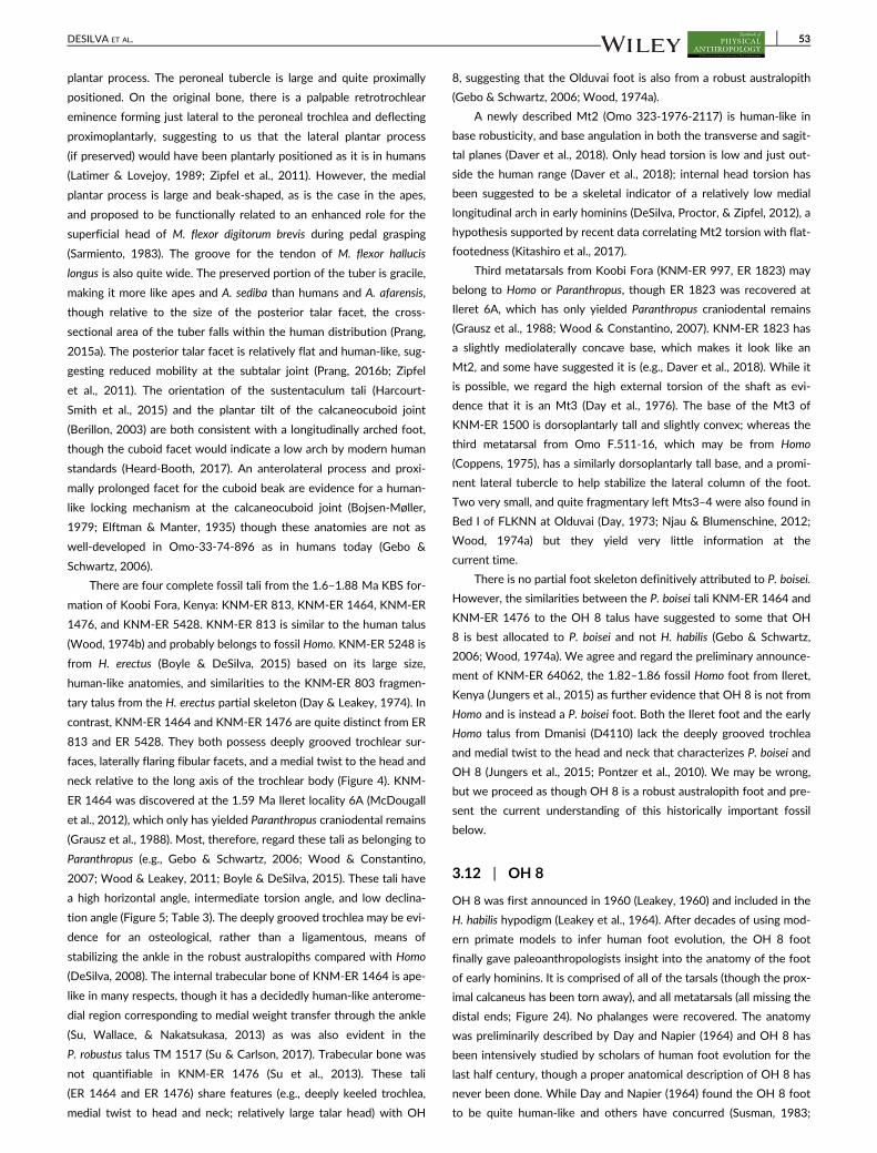
plantar process. The peroneal tubercle is large and quite proximally
positioned. On the original bone, there is a palpable retrotrochlear
eminence forming just lateral to the peroneal trochlea and deflecting
proximoplantarly, suggesting to us that the lateral plantar process
(if preserved) would have been plantarly positioned as it is in humans
(Latimer & Lovejoy, 1989; Zipfel et al., 2011). However, the medial
plantar process is large and beak-shaped, as is the case in the apes,
and proposed to be functionally related to an enhanced role for the
superficial head of M. flexor digitorum brevis during pedal grasping
(Sarmiento, 1983). The groove for the tendon of M. flexor hallucis
longus is also quite wide. The preserved portion of the tuber is gracile,
making it more like apes and A. sediba than humans and A. afarensis,
though relative to the size of the posterior talar facet, the cross-
sectional area of the tuber falls within the human distribution (Prang,
2015a). The posterior talar facet is relatively flat and human-like, sug-
gesting reduced mobility at the subtalar joint (Prang, 2016b; Zipfel
et al., 2011). The orientation of the sustentaculum tali (Harcourt-
Smith et al., 2015) and the plantar tilt of the calcaneocuboid joint
(Berillon, 2003) are both consistent with a longitudinally arched foot,
though the cuboid facet would indicate a low arch by modern human
standards (Heard-Booth, 2017). An anterolateral process and proxi-
mally prolonged facet for the cuboid beak are evidence for a human-
like locking mechanism at the calcaneocuboid joint (Bojsen-Møller,
1979; Elftman & Manter, 1935) though these anatomies are not as
well-developed in Omo-33-74-896 as in humans today (Gebo &
Schwartz, 2006).
There are four complete fossil tali from the 1.6–1.88 Ma KBS for-
mation of Koobi Fora, Kenya: KNM-ER 813, KNM-ER 1464, KNM-ER
1476, and KNM-ER 5428. KNM-ER 813 is similar to the human talus
(Wood, 1974b) and probably belongs to fossil Homo. KNM-ER 5248 is
from H. erectus (Boyle & DeSilva, 2015) based on its large size,
human-like anatomies, and similarities to the KNM-ER 803 fragmen-
tary talus from the H. erectus partial skeleton (Day & Leakey, 1974). In
contrast, KNM-ER 1464 and KNM-ER 1476 are quite distinct from ER
813 and ER 5428. They both possess deeply grooved trochlear sur-
faces, laterally flaring fibular facets, and a medial twist to the head and
neck relative to the long axis of the trochlear body (Figure 4). KNM-
ER 1464 was discovered at the 1.59 Ma Ileret locality 6A (McDougall
et al., 2012), which only has yielded Paranthropus craniodental remains
(Grausz et al., 1988). Most, therefore, regard these tali as belonging to
Paranthropus (e.g., Gebo & Schwartz, 2006; Wood & Constantino,
2007; Wood & Leakey, 2011; Boyle & DeSilva, 2015). These tali have
a high horizontal angle, intermediate torsion angle, and low declina-
tion angle (Figure 5; Table 3). The deeply grooved trochlea may be evi-
dence for an osteological, rather than a ligamentous, means of
stabilizing the ankle in the robust australopiths compared with Homo
(DeSilva, 2008). The internal trabecular bone of KNM-ER 1464 is ape-
like in many respects, though it has a decidedly human-like anterome-
dial region corresponding to medial weight transfer through the ankle
(Su, Wallace, & Nakatsukasa, 2013) as was also evident in the
P. robustus talus TM 1517 (Su & Carlson, 2017). Trabecular bone was
not quantifiable in KNM-ER 1476 (Su et al., 2013). These tali
(ER 1464 and ER 1476) share features (e.g., deeply keeled trochlea,
medial twist to head and neck; relatively large talar head) with OH
8, suggesting that the Olduvai foot is also from a robust australopith
(Gebo & Schwartz, 2006; Wood, 1974a).
A newly described Mt2 (Omo 323-1976-2117) is human-like in
base robusticity, and base angulation in both the transverse and sagit-
tal planes (Daver et al., 2018). Only head torsion is low and just out-
side the human range (Daver et al., 2018); internal head torsion has
been suggested to be a skeletal indicator of a relatively low medial
longitudinal arch in early hominins (DeSilva, Proctor, & Zipfel, 2012), a
hypothesis supported by recent data correlating Mt2 torsion with flat-
footedness (Kitashiro et al., 2017).
Third metatarsals from Koobi Fora (KNM-ER 997, ER 1823) may
belong to Homo or Paranthropus, though ER 1823 was recovered at
Ileret 6A, which has only yielded Paranthropus craniodental remains
(Grausz et al., 1988; Wood & Constantino, 2007). KNM-ER 1823 has
a slightly mediolaterally concave base, which makes it look like an
Mt2, and some have suggested it is (e.g., Daver et al., 2018). While it
is possible, we regard the high external torsion of the shaft as evi-
dence that it is an Mt3 (Day et al., 1976). The base of the Mt3 of
KNM-ER 1500 is dorsoplantarly tall and slightly convex; whereas the
third metatarsal from Omo F.511-16, which may be from Homo
(Coppens, 1975), has a similarly dorsoplantarly tall base, and a promi-
nent lateral tubercle to help stabilize the lateral column of the foot.
Two very small, and quite fragmentary left Mts3–4 were also found in
Bed I of FLKNN at Olduvai (Day, 1973; Njau & Blumenschine, 2012;
Wood, 1974a) but they yield very little information at the
current time.
There is no partial foot skeleton definitively attributed to P. boisei.
However, the similarities between the P. boisei tali KNM-ER 1464 and
KNM-ER 1476 to the OH 8 talus have suggested to some that OH
8 is best allocated to P. boisei and not H. habilis (Gebo & Schwartz,
2006; Wood, 1974a). We agree and regard the preliminary announce-
ment of KNM-ER 64062, the 1.82–1.86 fossil Homo foot from Ileret,
Kenya (Jungers et al., 2015) as further evidence that OH 8 is not from
Homo and is instead a P. boisei foot. Both the Ileret foot and the early
Homo talus from Dmanisi (D4110) lack the deeply grooved trochlea
and medial twist to the head and neck that characterizes P. boisei and
OH 8 (Jungers et al., 2015; Pontzer et al., 2010). We may be wrong,
but we proceed as though OH 8 is a robust australopith foot and pre-
sent the current understanding of this historically important fossil
below.
3.12 | OH 8
OH 8 was first announced in 1960 (Leakey, 1960) and included in the
H. habilis hypodigm (Leakey et al., 1964). After decades of using mod-
ern primate models to infer human foot evolution, the OH 8 foot
finally gave paleoanthropologists insight into the anatomy of the foot
of early hominins. It is comprised of all of the tarsals (though the prox-
imal calcaneus has been torn away), and all metatarsals (all missing the
distal ends; Figure 24). No phalanges were recovered. The anatomy
was preliminarily described by Day and Napier (1964) and OH 8 has
been intensively studied by scholars of human foot evolution for the
last half century, though a proper anatomical description of OH 8 has
never been done. While Day and Napier (1964) found the OH 8 foot
to be quite human-like and others have concurred (Susman, 1983;
DESILVA ET AL. 53

Susman & Stern, 1982), Oxnard and Lisowski (1980) claimed to find
errors in the original reconstruction and regard the foot as more ape-
like, as did Lewis (1980c). While most scholars find the Day and
Napier (1964) interpretation of OH 8 to be compelling, there is an
opportunity to use modern scanning and reconstruction techniques to
assess these two competing OH 8 models. The anatomy described
below is based on published scholarship and on our observation of
the original fossil.
OH 8 is regarded by many to belong to H. habilis (Leakey et al.,
1964; Susman, 2008; Susman & Stern, 1982); we regard it as more
likely to be the foot of P. boisei, as others have suggested (Gebo &
Schwartz, 2006; Wood, 1974a). The anatomical basis for this assigna-
tion is discussed in more detail below, but it is based primarily on the
talus and the Mt1. There is also a related disagreement regarding the
chronological age of the OH 8 individual. Some have proposed that
OH 8 is a juvenile and that it is associated with the subadult H. habilis
type OH 7, and with OH 35 (Susman, 2008; Susman, Patel, Francis, &
Cardoso, 2011; Susman & Stern, 1982). While superficially the OH
8 foot and the OH 35 tibia appear to articulate well, the joint surfaces
are not congruent (Aiello, Wood, Key, & Wood, 1998; Wood, Aiello,
Wood, & Key, 1998) and geologically, they were found in different
horizons, 300 yards from one another (Day & Napier, 1964; Njau &
Blumenschine, 2012).
Nevetheless, the thrust of the argument is that OH 8 is associated
with OH 7, and is therefore a juvenile H. habilis. This interpretation of
the material is based on what are perceived to be unfused metatarsal
heads of the Mt2 and Mt3 (Susman, 2008). However, these metatar-
sals have carnivore tooth marks on them, just millimeters from the
missing metatarsal heads (Njau & Blumenschine, 2012; Figure 24),
making it probable that the metatarsal heads are missing not because
OH 8 was a juvenile, but because the heads (and toes) were torn off
and consumed by a predator. Furthermore, while variation surely
exists (Susman et al., 2011), at the developmental age of OH 7, the
base of the OH 8 Mt1 should be either unfused or in the process of
fusing, but instead the proximal epiphysis is fully fused and the epiph-
yseal line is obliterated (DeSilva et al., 2010; Weiss, DeSilva, & Zipfel,
2012). Additionally, there are osteophytic growths along the cotylar
fossa, the dorsum of the lateral cuneiform, and along the lateral inter-
metatarsal facets, consistent with age-related osteoarthritis (Weiss,
2012). We therefore are of the opinion that OH 8 is an adult foot
(Day, 1986; Leakey, 1971), though others strongly disagree (Susman
et al., 2011). The importance of characterizing the age of this individ-
ual lies not only in accurately interpreting the functional anatomy, but
also in assessing the likelihood that the OH 8 foot is associated with
OH 7, the type of H. habilis. Morphology aside (but see below) if OH
8 was a juvenile, given the proximity of these bones to the OH 7 hand
and craniodental material, we agree that it would most reasonably be
assigned to that individual (and that taxon). However, as an adult, and
therefore a different individual from the juvenile OH 7, it is no more
likely to be from H. habilis, than from P. boisei, also known from Oldu-
vai FLKNN Bed I.
The OH 8 calcaneus is badly damaged proximally and plantarly,
but preserves most of the subtalar joint and all of the calcaneocuboid
joint. A multivariate analysis of the subtalar joint complex found OH
8 to be human-like (Prang, 2016b). The cuboid facet is plantarly
angled as in the arched human foot (Berillon, 2003; Heard-Booth,
2017), has a dorsolateral overhang and a proximomedially prolonged
facet for the beak of the cuboid, which would lock the calcaneocuboid
joint during bipedal propulsion (Bojsen-Møller, 1979; Day & Napier,
1964; Kidd et al., 1996; Lewis, 1980c; Susman & Stern, 1982). The
cuboid is human-like in 3D shape space (Harcourt-Smith, 2002); it is
proximodistally elongated (Figure 7), has an eccentrically positioned
beak, flat, human-like facets for the lateral metatarsals, and a human-
like os peroneum facet (Lovejoy et al., 2009).
However, the talus is quite different from both the modern
human talus, and Early Pleistocene tali assigned to Homo (see below),
including Omo 323-76-898 (Gebo & Schwartz, 2006), KNM-ER
813 (Wood, 1974b), KNM-ER 5428 (Boyle & DeSilva, 2015), KNM-ER
803 (Leakey, 1973), and D4110 (Pontzer et al., 2010). Instead, the OH
8 talus is similar to two tali typically assigned to P. boisei (KNM-ER
1464 and ER 1476), and to TM 1517 from P. robustus. The trochlear
body is deeply grooved and has a high lateral rim, as does the KNM-
ER 1464 talus (Boyle & DeSilva, 2015; Gebo & Schwartz, 2006; Pont-
zer et al., 2010). Additionally, the head and neck are medially twisted
relative to the trochlear body, as in KNM-ER 1464 and KNM-ER 1476
(Boyle & DeSilva, 2015). Day and Wood (1968) noted that the hori-
zontal and declination angles of OH 8 were more ape-like, and that
unlike either humans or apes, the curvature of the lateral and medial
rims would lead to foot adduction, rather than abduction, during dorsi-
flexion. In the first multivariate study of the OH 8 talus, Lisowski
et al. (1974) found it grouped with TM 1517 and amongst modern
taxa, closer to orangutans than to modern humans. More recent work
clusters the OH 8 talus closer to humans, but still outside the range of
modern variation and near KNM-ER 1464 (Harcourt-Smith, 2002);
while a study of the angulation of the facets found OH 8 to be within
the human distribution (Parr et al., 2014). However, Lewis (1980c) and
Kidd et al. (1996) regarded the horizontal angle to be so ape-like as to
indicate a semi-divergent hallux, a suggestion that is not widely
accepted (see below).
The OH 8 navicular is generally human-like (Harcourt-Smith,
2002; Aiello and Harcourt-Smith, 2004, but see Kidd et al., 1996). The
facets for the cuneiforms angle in a manner that is consistent with a
longitudinal arch of the foot (Berillon, 2003), and the plantar portion
of the navicular is well-developed for the insertion of the spring liga-
ment (Susman & Stern, 1982). Prang (2016b) found the talonavicular
joint to be human-like in OH 8 and later (Prang, 2016c) applied a geo-
metric morphometrics analysis on the OH 8 navicular and found it to
be quite modern human-like as well. The lateral cuneiform is proximo-
distally expanded (Susman & Stern, 1982; Figure 10), as is the inter-
mediate cuneiform (Figure 9). The medial cuneiform possesses a
distally oriented and mildly convex facet for the Mt1 (Gill et al., 2015),
consistent with an adducted hallux (Day & Napier, 1964; Preuschoft,
1971; Susman & Stern, 1982; Figure 8; Table 6; but see Lewis, 1972;
Lewis, 1980c).
Day and Napier (1964) reported that the OH 8 ft had an unusual
pattern of metatarsal robusticity: 1 > 5 > 3 > 4 > 2, and used these
data to suggest that the modern human striding gait “had not yet been
achieved.” It is typical in humans for the lateral digits (Mt4 and Mt5)
to be more robust than Mt2 or Mt3. However, Archibald, Lovejoy,
and Heiple (1972) described greater variation in human metatarsal
54 DESILVA ET AL.

robusticity patterns and found the OH 8 ft to be fully consistent with
a human-like gait. More recently, Patel et al. (2018) have revisited this
problem with a larger sample and a μCT approach that permitted the
quantification of cross-sectional properties and found the OH 8 pat-
tern to most commonly be 1 > 3 > 5 > 4 > 2, which is different from
the typical human condition (1 > 4/5 > 2/3) or ape one, suggesting
that OH 8 loaded its foot in a biomechanically unique manner.
The Mt1 of OH 8 has a human-like base (Proctor, 2010b) and a
large contact facet for the Mt2 (Le Minor & Winter, 2003). However,
the reconstructed length of the Mt1 (White & Suwa, 1987) is quite
short and dorsoplantarly robust (Pontzer et al., 2010), most similar to
SK 1813 and SKX 5017 (McNutt, Zipfel, & DeSilva, 2018; Figure 11),
Mt1s assigned to Paranthropus robustus. The Mt2 and Mt3 have dor-
soplantarly tall bases, similar to the modern human condition (McNutt,
Zipfel, & DeSilva, 2018; Figures 12 and 13), and torsion values close
to that of modern humans (Pontzer et al., 2010). The Mt4 has a dorso-
plantarly tall and flat base, consistent with tarsometatarsal rigidity
(DeSilva, 2010). The Mt5 is damaged laterally and medially possesses
a large osteophytic growth (DeSilva et al., 2010), which has been
hypothesized to be the result of trauma (Susman & Stern, 1982).
For a foot that has been known for over a half century (Day &
Napier, 1964), it is remarkable how much disagreement swirls around
OH 8. For some, OH 8 is a subadult H. habilis (e.g., Susman, 2008); for
others, an adult P. boisei (DeSilva et al., 2010; this article). Neverthe-
less, most agree that OH 8 was human-like in many respects, and had
an adducted hallux, locking calcaneocuboid joint, and a longitudinal
arch (but see Lewis, 1980c). However, the large talar head (Figure 4),
deeply grooved talar trochlea (Gebo & Schwartz, 2006; Figure 5),
short Mt1 (McNutt, Zipfel, & DeSilva, 2018; Figure 11), and unique
robusticity pattern (Patel et al., 2018) may suggest a unique loading
regime and perhaps different gait kinematics. The relationship
between these anatomies and foot function should be examined
regardless of whether OH 8 is early Homo or Paranthropus. The Ileret
foot (KNM-ER 64062) will make an important functional and taxo-
nomic comparison to OH 8, given that they have both been attributed
to early Homo and are roughly contemporaneous. Yet, given the possi-
bility of multiple early Homo species (Leakey et al., 2012), our failure
to securely identify the OH 8 foot demonstrates how valuable a
P. boisei skeleton will be to our science.
3.13 | Early Homo
The origins of our genus remain quite a contentious topic; cranioden-
tal evidence suggests that Homo may have originated in the late Plio-
cene (Villmoare et al., 2015). However, there is relatively little known
about the foot of the earliest members of our genus prior to ~2 Ma.
Furthermore, in the absence of partial skeletons, there is the difficulty
of assigning isolated pedal remains to either Paranthropus or Homo as
best illustrated by OH 8 (see above). And even if isolated remains can
be reasonably assigned to Homo, the increasing likelihood of taxo-
nomic diversity within early Homo (e.g., Leakey et al., 2012) makes it
even more difficult to assign pedal remains to H. habilis, H. rudolfensis,
or early H. erectus (=ergaster). Unfortunately, the best represented
early Homo skeleton—KNM-WT 15000—does not preserve any foot
bones (a fragmentary and admittedly dubious “Mt1” [Walker &
Leakey, 1993] is probably not). Below, we review what is known
about the foot of early Homo from three partial foot skeletons/assem-
blages: KNM-ER 64062, Dmanisi, and KNM-ER 803, and then con-
clude the section by discussing isolated fossils that we suspect belong
to the genus Homo. Anatomies discussed below are based on pub-
lished sources and observations we made on the original fossil mate-
rial, with the exception of KNM-ER 64062, which is still under study,
and the fossils from Dmanisi.
KNM-ER 64062 is a partial foot skeleton from 1.84 Ma deposits
at Ileret, Kenya (Jungers et al., 2015). Although the fossil has not yet
been fully published, it promises to help resolve important questions
about the foot of early members of genus Homo. Preserved are all of
the tarsals except for the medial cuneiform, all of the metatarsals, and
the proximal hallucal phalanx (Figure 26). A preliminary announcement
of the fossil (Jungers et al., 2015) reveals that the calcaneus is derived
and human-like in possessing a large, robust tuber and a plantarly
positioned lateral plantar process. The cuboid is also described as
human-like. The talus and Mt1 are described as being similar to the
Dmanisi fossils (Jungers et al., 2015). Jungers (pers. comm.) informs us
that while the Mt1 is adducted and has a domed head, it lacks the
mediolaterally expanded dorsum typical of modern humans and
instead is dorsally narrow, like the Dmanisi Mt1s and those from aus-
tralopiths. Furthermore, the navicular is quite primitive and possesses
an enlarged navicular tuberosity and has a dorsoplantarly narrow lat-
eral navicular body (Jungers et al., 2015), even more so than that
found in StW 573 and LB1 (Jungers, pers. comm.). A geometric mor-
phometrics study of the metatarsal heads finds the Mt2 and Mt3 to
be human-like, while the Mt1 is just outside the modern human range
(Fernández et al., 2018). Comparisons between this Ileret foot and
partial foot skeletons from H. floresiensis, H. naledi, and OH 8 (histori-
cally called H. habilis, but here hypothesized to be from P. boisei) will
yield important insights into the foot of the earliest members of our
genus, and diversity in foot forms throughout the Pleistocene.
If, as we suggest above, OH 8 is not from early Homo, then until
the Ileret foot is published (see above), our best understanding of the
foot of early Homo is from the 1.77 Ma site of Dmanisi, Georgia
(Lordkipanidze et al., 2007; Pontzer et al., 2010). A partial adult foot is
comprised of a talus (D4110), Mts3–5 (D2021, D4165, D4058) and
distal phalanx (D3877; Lordkipanidze et al., 2007; Pontzer et al.,
2010). Subadult foot bones (Mt1 D2671; Mt4 D2669; DP1 D2670)
are associated with the D2700 juvenile skull (Lordkipanidze et al.,
2007; Pontzer et al., 2010). A medial cuneiform (D4111) and Mt1
(D3442) are associated (Jashashvili et al., 2010; Lordkipanidze et al.,
2007; Pontzer et al., 2010). There is finally an isolated Mt3 (D3479;
Gabunia, Vekua, & Lordkipanidze, 1999; Lordkipanidze et al., 2007;
Figure 26).
The talus (D4110) is human-like in most respects (Lordkipanidze
et al., 2007; Pontzer et al., 2010). The talar trochlea is mediolaterally
flat, as is common in fossils from Homo, but differs from the trochlear
surface in OH 8 and KNM-ER 1464 (Pontzer et al., 2010). The head/
neck horizontal angle is similar to that found in other fossil Homo
(26�), on the high end of the modern human range (Lordkipanidze
et al., 2007). The talus has a human-like declination angle, consistent
with a longitudinal arch (Pontzer et al., 2010). The head/neck torsion
angle has not been reported. There is some moderate flaring of the
DESILVA ET AL. 55

fibular facet (Pontzer et al., 2010), and well-developed proximoplantar
tubercles at the base of an obliquely oriented groove for M. flexor hal-
lucis longus (Lordkipanidze et al., 2007), though this feature is highly
variable in human tali (Pontzer et al., 2010).
The medial cuneiform (D4111) is bipartite, which happens, but is
unusual, in modern human medial cuneiforms (Jashashvili et al., 2010).
Although it is consistent with an adducted hallux, D4111 is said to dif-
fer in shape from modern human medial cuneiforms (Jashashvili et al.,
2010). The base of the Dmanisi Mt1 is divided into plantar and dorsal
facets similar to the morphology of A.L. 333-54 (Pontzer et al., 2010);
though the Dmanisi Mt1 base is much flatter. The base is dorsoplan-
tarly tall, indicating the presence of strong plantar ligaments
(Lordkipanidze et al., 2007). However, the Mt1s are short compared
with the lateral metatarsals (Pontzer et al., 2010). Furthermore,
although the shafts have human-like robusticity, the heads are dor-
sally narrow (Lordkipanidze et al., 2007; Pontzer et al., 2010), similar
to the anatomy found in australopith Mt1s (e.g., Susman & Brain,
1988) and quite different from later Homo (BK 63), Mt1s from
H. naledi (Harcourt-Smith et al., 2015) and H. floresiensis (Jungers,
Harcourt-Smith, et al., 2009; Figure 11).
The lateral metatarsals (Mt3 and Mt4 especially) possess human-
like external torsion (Lordkipanidze et al., 2007; Pontzer et al., 2010),
demonstrating that the Dmanisi hominins had strongly developed
transverse arches of the foot, and according to Pontzer et al. (2010)
longitudinal arches as well. However, the shaft robusticity of these lat-
eral metatarsals is quite different from that found in modern humans.
The Mt3 has relatively high robusticity (Gabunia et al., 1999), in the
range of modern chimpanzees (Pontzer et al., 2010); the Mt4 is simi-
larly robust, unlike chimpanzees or humans (Pontzer et al., 2010); the
Mt5 has quite low shaft robusticity, making it ape-like as well (Pontzer
et al., 2010). A more detailed examination of the cortical structure of
the Mt5 (D4058) found that the thickness and distribution of cortex
was ape-like, though the cortex tapered distally as it does in humans
(Dowdeswell et al., 2016). No detailed analyses of the pedal phalanges
(D3877, D2670) have been published though Patel, Jashashvili, Trin-
kaus, Susman, and Lordkipanidze (2015) noted that D2670, a hallucal
distal phalanx, possesses moderate axial torsion, but lacks human-like
lateral deviation.
The early Homo foot as represented by the Dmanisi material is
generally human-like. The morphology of the ankle joint is consistent
with a human-like tibia moving primarily in the sagittal plane over a
foot with an adducted hallux and a well-developed longitudinal arch.
However, the head of the Mt1 suggests that early Homo from Dmanisi
may not have possessed human-like push off capabilities, perhaps
because the windlass mechanism was not fully developed. Further-
more, the lesser metatarsal shaft robusticity indicates that the foot
was loaded in a different manner than in humans today. The low Mt5
(but high Mt3 and Mt4) robusticity may be related to the placement
of the foot relative to the direction of travel, though this remains
speculative and requires additional testing (Pontzer et al., 2010). Nev-
ertheless, the Dmanisi fossils are important additions to our under-
standing of early Homo foot evolution. They raise important questions
FIGURE 26 Foot of early Homo. The foot skeleton of the earliest members of the Homo genus are known from KNM-ER 64062, the 1.77 Ma
Dmanisi fossil locality, and KNM-ER 803. The fossil feet have been scaled so that they are roughly the same size and reversed (if necessary) topresent the right side. To the far right is a single right footprint from the 1.55 Ma Ileret track way, thought to have been made by H. erectus.KNM-ER 64062 image thanks to W. Jungers and the Turkana Basin Institute. Dmanisi image courtesy of C. Zollikofer and M. Ponce de León. TheIleret footprint photo was provided by K. Hatala
56 DESILVA ET AL.

about the relationship between metatarsal torsion and plantar
arches—a correlation that still remains unclear. Additionally, direct
comparisons between similarly aged foot material from Ileret (Jungers
et al., 2015), and chronologically later material from H. floresiensis
(Jungers, Harcourt-Smith, et al., 2009) and H. naledi (Harcourt-Smith
et al., 2015) will help test functional and even phylogenetic hypothe-
ses about foot evolution in the earliest members of our genus.
KNM-ER 803 (Figure 26) is a partial skeleton from 1.53 Ma
deposits at Koobi Fora, Kenya regarded by most (based on associated
craniodental remains) to be from H. erectus (e.g., Antón, 2003). The
partial foot skeleton includes a fragmentary talus, Mt3 (without the
head), proximal Mt5, and five pedal phalanges (Day & Leakey, 1974;
Leakey, 1973; Leakey & Leakey, 1978). The talus preserves only the
lateral (fibular) half, but is quite human-like. The trochlear surface is
mediolaterally flat (Boyle & DeSilva, 2015), and the fibular facet is ver-
tically inclined. The base of the Mt3 is dorsoplantarly tall relative to
the mediolateral width (Figure 13). The Mt5 preserves only the base;
the cuboid facet is dorsoplantarly flat and the tuberosity is only
weakly developed, both morphologies found in modern human Mt5s.
Perhaps most interesting about this foot are the phalanges. The inter-
mediate phalanges are relatively (Trinkaus & Patel, 2016) and abso-
lutely (McNutt, Zipfel, & DeSilva, 2018; Figure 17) very short, and
represent some of the earliest known evidence for shortening of the
intermediate phalanges from the more primitive australopith condi-
tion. Rolian et al. (2009) has collected experimental evidence that pha-
langeal shortening reduces energy output during running (but not
walking); the toes of KNM-ER 803 would thus support hypotheses for
running adaptations in H. erectus (Bramble & Lieberman, 2004;
Holowka & Lieberman, 2018).
In addition to these three assemblages (KNM-ER 64062, Dmanisi,
and KNM-ER 803) there are a number of isolated fossils that we sus-
pect belong to the genus Homo. These include four tali, and four pha-
langes from sites in both Eastern and South Africa.
The oldest foot fossil purported to belong to the genus Homo is a
2.2 Ma talus (Omo 323-76-898) from the Omo Shungura formation,
Ethiopia (Gebo & Schwartz, 2006). This catalogue name (Omo
323-76-898) has also mistakenly been used for a Paranthropus cra-
nium (Falk, 1986; Kimbel, 1984; Kimbel & Rak, 1985), which is Omo
323-76-896 (Wood & Leakey, 2011). Gebo and Schwartz (2006) find
the Omo 323-76-898 talus to be quite human-like and we agree and
regard it as most likely belonging to the genus Homo. The trochlea is
mediolaterally flat with roughly equal medial and lateral margins, giv-
ing it a human-like talar axis angle (Figure 5). The head has human-like
torsion, but the Omo talus lacks the declination angle found in modern
human tali, suggesting that this individual had a low arch (Peeters
et al., 2013).
Similarly, the 1.85 Ma talus KNM-ER 813 is typically assigned to
Homo (Leakey, 1973; Wood, 1974b), and more specifically
H. rudolfensis (McHenry, 1994; Wood, 1992). A multivariate analysis
of the specimen finds it securely within the modern human distribu-
tion (Wood, 1974b), making it more human-like than the similarly aged
OH 8 talus (Day & Wood, 1968). The talar trochlea is human-like in its
general curvature (Christie, 1990), though we note that the mediolat-
eral surface is more grooved than in most humans. The trochlea has
mild wedging (Figure 4), and roughly equal medial and lateral sides.
Both the horizonatal angle and the head/neck torsion values are mod-
ern human-like (Figure 5; Table 3). The declination angle is low for a
modern human, but higher than most australopiths. The internal tra-
becular bone is not preserved well enough for study at the current
time (Su & Carlson, 2017).
A large, chronologically later (1.6 Ma) talus (KNM-ER 5428) is also
from Homo, and more specifically H. erectus (Boyle & DeSilva, 2015).
KNM-ER 5428 is human-like in almost all respects, clustering in a prin-
cipal components analysis with modern humans (Boyle & DeSilva,
2015). It posseses a human-like trochlea that is mildly wedged and
mediolaterally flat (Figure 4), making it more like KNM-ER 803 (H. erec-
tus) and unlike the deeply grooved tali of KNM-ER 1464 and OH
8 (presumed P. boisei). The head has human-like torsion and declina-
tion, the latter anatomy suggesting a human-like longitudinal arch
(Figure 5). The talus is also quite large, estimated to be from a nearly
90 kg individual (Boyle & DeSilva, 2015; McHenry, 1994). KNM-ER
5428 differs from a modern human talus in having a dorsoplantarly
low body—a talar anatomy that persists right through early H. sapiens
(Boyle & DeSilva, 2015; McNutt, Zipfel, & DeSilva, 2018). Unfortu-
nately, the internal trabecular bone could not be isolated (Su & Carl-
son, 2017).
One final talus that is probably from Homo is SKX 42695, a frag-
mentary talar body without a head from the lower bank of Member
1 Swartkrans, South Africa. SKX 42695 differs from the TM 1517
talus (P. robustus) in its larger size, mediolaterally flatter trochlea and
less flared fibular facet (Susman et al., 2001). Furthermore, the internal
trabecular architecture is similar to that found in humans (DeSilva &
Devlin, 2012).
In addition to these tali, there are three phalanges from Swartk-
rans, South Africa that are short and human-like. SKX 16699 is a prox-
imal phalanx that is both relatively and absolutely short and straight
(Trinkaus & Patel, 2016). Additionally, it has a dorsoplantarly
expanded base, which may signifiy a thick plantar aponeurosis and
modern human-like windlass mechanism (Trinkaus & Patel, 2016).
SKX 16699 differs in these respects from DNH 117, which is longer
and more curved, making it reasonable that the Swartkrans specimen
is from Homo and the Drimolen phalanx from P. robustus. Additionally,
intermediate phalanges SKX 344 and 1261 from Swartkrans Member
2 are very short—shorter (absolutely) than australopith intermediate
phalanges, and even the H. erectus intermediate phalanges from
KNM-ER 803 (McNutt, Zipfel, & DeSilva, 2018; Figure 17), making it
reasonable to propose that they are from Homo. Finally, there is a dis-
tal hallucal phalanx from Olduvai (OH 10). Historically, this fossil is of
importance since it harkens back to the days when early hominin fos-
sils were so rare that an isolated distal hallucal phalanx could be pub-
lished in Nature (Day & Napier, 1966), and more importantly, it was
the first fossil subjected to a multivariate analysis (Day, 1967). OH
10 possesses both the lateral (valgus) angulation in the transverse
plane of 2�, and the axial torsion (13�) associated with hallucal propul-
sion found in modern humans (Day, 1986; Day & Napier, 1966), mak-
ing it quite human-like (Wood, 1974a). In his multivariate analysis,
Day (1967) found OH 10 to cluster distinctly with modern humans,
though Oxnard (1972) found OH 10 to be distinct from both humans
and apes. Little additional work has been done on this fossil since.
DESILVA ET AL. 57

Fossilized footprints have been recovered in 1.4–1.5 Ma deposits
at two localities near the town of Ileret in northern Kenya: GaJi10
(Behrensmeyer & Laporte, 1981) and FwJj14E (Bennett et al., 2009).
Analyses of these prints (Figure 26), attributed by most to H. erectus,
are consistent with the functional anatomy of the pedal remains—they
reveal a foot with a fully adducted hallux and longitudinal arch, walk-
ing with human-like medial weight transfer (Bennett et al., 2009;
Hatala et al., 2017; Hatala, Demes, & Richmond, 2016; Hatala, Roach,
et al., 2016).
3.14 | Late early/middle Pleistocene Homo
To the best of our knowledge, there are only three pedal fossils from
Africa known from the roughly 1.2 million year span separating the
1.53 Ma foot bones of the KNM-ER 803 H. erectus skeleton and the
numerous pedal remains from the 236- to 335-kyr-old South African
hominin H. naledi (described below). A fragmentary, unprepared and
undescribed talus (BOU-VP-2/95) has been recovered from the
~1.0 Ma sediments at Daka, Ethiopia (Gilbert & Asfaw, 2008). Another
talus (LZH-01) was recovered from undated (possibly Middle Pleisto-
cene?) sediments from Luangwa, Zambia and found to resemble mod-
ern humans in a geometric morphometrics analysis (Bishop et al.,
2016). Additionally, from ~0.5 Ma Kapthurin Formation deposits at
Baringo, Kenya (Deino & McBrearty, 2002), there is an Mt1 (BK 63)
which has never been formally described.
The base of BK 63 is weathered around the edges, but appears
generally human-like. Distally, the head is domed (Figure 11), and our
observations on the original are that the head is mediolaterally wide
dorsally, as it is in modern humans. However, Lu et al. (2011) find the
contours of the head to be unlike modern humans and similar to both
the chronologically later Jinniushan Mt1, and much earlier Paranthro-
pus fossils from Swartkrans. We regard BK 63 as possessing a much
more human-like Mt1 head than SKX 5017 or SK 1813. Nevertheless,
Fisher and McBrearty (2002) and Meldrum (2007) have commented
on the gracility of the shaft of BK 63 and we concur. Relative to the
length of the bone, the shaft is surprisingly dorsoplantarly shallow
(Figure 11), seemingly poorly constructed to resist the high bending
forces on the Mt1 during hallucal propulsion. Meldrum (2007) and Lu
et al. (2011) regard this as evidence that Middle Pleistocene hominins
still had not acquired a modern human-like gait. Significantly more evi-
dence will be needed from this poorly sampled time period to better
understand Middle Pleistocene foot evolution in Africa. Despite the
near absence of foot fossils in Africa during this time period, there are
nearly a dozen footprints at the 700-kyr-old site of Gombore II-2,
Melka Kunture, Ethiopia (Altamura, Bennett, D'Août, et al., 2018). The
best-preserved are described by the authors as generally similar to the
presumed H. erectus prints from Ileret, Kenya, and perhaps most inter-
estingly, there are prints preserved from both adults and children
thought to be as young as 12 months old (Altamura et al., 2018).
In Europe, at least, there are over a dozen foot bones from the
772 to 949 ka (Duval et al., 2018) site of Gran Dolina, Spain
(Figure 27), attributed to the taxon H. antecessor (De Castro et al.,
1997). We were unable to study the original fossils and base our com-
ments on published scholarship. There is a talus (ATD6-95), fragmen-
tary calcaneus (ATD6-117), two Mt2s (ATD6-25 and ATD
6-70 + 107), Mt4 (ATD6-124), and eight phalanges (ATD6-30-36,68).
In general, the bones are human-like and according to Pablos
FIGURE 27 Left: Foot fossils assigned to Homo antecessor from the Gran Dolina locality at Atapuerca, Spain (Image courtesy of A. Pablos). Upper
right: right partial foot skeleton from Jinniushan, China. Bottom right: articulated foot skeleton from Sima de los Huesos, Atapuerca, Spain (Imagetaken by J. Trueba, courtesy of Science Photo Library)
58 DESILVA ET AL.

et al. (2012) would be in a foot employing a human-like gait. The pha-
langes are short, straight and robust (Lorenzo et al., 1999); the meta-
tarsals are similarly human-like in proportion (Lorenzo et al., 1999;
Pablos et al., 2012). The Mt4 base is mediolaterally wide, as found in
fossils from Sima de los Huesos and Neandertals (Pablos et al., 2012).
Additionally, the Mt4 (ATD6-124) preserves evidence for a healed
stress fracture (Martin-Francés, Martinon-Torres, Gracia-Téllez, &
Bermúdez de Castro, 2015). Unfortunately, the calcaneus only pre-
serves the anterior talar facet and little can be gleaned from it. The
talus (ATD6-95), while eroded, contains valuable information. It is a
very large talus—one of the largest known from the Pleistocene
(Pablos et al., 2012). The trochlear body is quite narrow mediolaterally,
a trait the ATD6-95 specimen shares in common with Neandertals,
but not with the Sima de los Huesos tali, which possess wider troch-
lear surfaces. The trochlear body is long; the talar neck quite short—an
inverse relationship previously hypothesized to exist in Neandertal tali
(Rhoads & Trinkaus, 1977). In a principal components analysis, the
Gran Dolina talus plots outside the shape space occupied by humans,
Neandertals, and the Sima de los Huesos tali (Pablos et al., 2012).
Functionally, whether these different talar dimensions are of any con-
sequence is unclear and may just reflect population-level differences
in Pleistocene Homo. Nearly 50 fossilized footprints from Happis-
burgh, UK were (they have since eroded) the oldest known from
Europe (~0.78–1.0 Ma) and preserved the movements of a group
attrituted to H. antecessor (Ashton et al., 2014). They were not ade-
quately preserved to draw functional inferences from internal
morphology.
3.15 | Eurasian Middle Pleistocene Homo
Prior to 1997, the only Middle Pleistocene Homo foot fossil known
was an Mt2 (Arago XLIII) from France (Day, 1986; Lamy, 1982). There
are now close to 600 fossils as a result of discoveries on the western
and eastern edges of the Eurasian landmass: Sima de los Huesos in
the Atapuerca Mountains, Spain has yielded over 500 foot fossils
from 16 individuals (Pablos et al., 2017; Figure 27). The fossils are
~430 kyr old (Arsuaga et al., 2014) and represent a population closely
related to, or even ancestral to, Neandertals (Meyer et al., 2014). We
were unable to study the original material and base our report below
on published studies. Sima de los Huesos is, by far, the largest collec-
tion of foot fossils known from any one locality (Pablos et al., 2017;
Figure 28). To the east, the Jinniushan, China locality has yielded two
(left and right) partial foot skeletons of a ~260 kyr old female
(Lu et al., 2011; Figure 27). Furthermore, there are two known foot-
print sites from this time. At Terra Amata, France (~400 kyr old), there
is a single footprint (De Lumley, 1969), and at Roccamonfina, Italy,
there are three 325–385 kyr old trackways preserving quite human-
like prints (Mietto, Avanzini, & Rolandi, 2003). These foot bones and
footprints are generally human-like, and thus attention is paid below
to those anatomies that deviate from the modern human or Neander-
tal condition.
The Sima de los Huesos foot bones have been partially described
(Pablos et al., 2017), with the most detailed descriptions and analyses
done thus far on the talus (Pablos et al., 2013) and calcaneus (Pablos
et al., 2014). There are 29 calcanei known from 15 different individ-
uals from Sima de los Huesos (Pablos et al., 2014). The fossils are
modern human-like, though they are more robust, with large articular
surfaces, recalling the Neandertal calcaneus (Pablos et al., 2014). Addi-
tionally, the tubers are relatively long, though not as long as those
found in Neandertals (Pablos et al., 2014, 2017), an anatomy that
would compromise running efficiency (Raichlen et al., 2011). Unlike in
any other Pleistocene calcanei, the Sima fossils have an unusually pro-
jected sustentaculum tali (Pablos et al., 2014), a feature already pre-
sent in the juveniles (Pablos et al., 2017). It is possible that this
morphology indicates that the Sima individuals had a greater range of
motion at the subtalar joint (Pablos et al., 2014).
The Sima de los Huesos fossil assemblage has also produced an
astounding 25 tali from 14 different individuals (Pablos et al., 2013).
These tali are generally human like, though a multivariate analysis
groups the Sima tali in a different shape space than either Neandertals
or modern humans (Pablos et al., 2013). The Sima tali have vertically
short bodies (unlike the vertically tall talar body of Late Pleistocene
FIGURE 28 Absolute number of foot fossils (values across the top of the graph) attributed to members of the genus Homo. Note that there are
only a small number of pedal remains for localities older than Sima de los Huesos. However, the Sima de los Huesos, Spain collection containsmore than 500 foot fossils, far more than what is known for any other Pleistocene Homo group except for Neandertals and Homo sapiens. Image
courtesy of A. Pablos; modified from “The foot in the Homo fossil record” by Pablos, 2015, Mitteilungen der Gesellschaft für Urgeschichte, 24, 11
DESILVA ET AL. 59

humans), unwedged talar trochlea, broad fibular facets, short talar
necks, and short middle calcaneal facets—similar to the tali of Nean-
dertals. However, the Sima specimens have exceptionally broad fibu-
lar facets, and a uniquely mediolaterally short talar head (Pablos et al.,
2013; Figure 4). This mediolateral width of the head also scales differ-
ently in the Sima tali compared with Neandertals—as the talus gets
larger, the head gets proportionally narrower in Sima compared with
Neandertals (Pablos et al., 2017). The combination of a uniquely short
talar head with projecting sustentaculum tali may be worth additional
functional consideration.
The remaining foot bones are partially described in Pablos
et al. (2017). There are an extraordinary number of tarsal bones: navic-
ular (n = 25), cuboid (n = 22), medial cuneiform (n = 14), intermediate
cuneiform (n = 15), and lateral cuneiform (n = 12). The navicular is
robust, and the cuboid narrow, anatomies similar to those found in
Neandertals (Pablos et al., 2017). Unique to the Sima population is a
proximodistally short intermediate cuneiform, which in conjunction
with the other cuneiforms, would result in a more recessed and pre-
sumably more stable Mt2 (Pablos et al., 2017). The metatarsals and
phalanges are generally robust, similar to the anatomy of the Nean-
dertal foot. The Mt1 (n = 18), Mt3 (n = 16), Mt4 (n = 12), and Mt5
(n = 11) all possess proximal bases slightly broader than Neandertal
fossils (Pablos et al., 2017), indicating a wider midfoot in the Sima
population. The Mt2 (n = 13) is relatively narrow in the Sima fossils,
perhaps related to the recessed, stable medial midfoot discussed
above. The phalanges (hallucal proximal n = 28; hallucal distal n = 21;
nonhallucal proximal n = 78; intermediate n = 71; nonhallucal distal
n = 63) are broad and robust, with distal breadths similar to those
found in Neandertal feet (Pablos et al., 2017). The distal hallucal pha-
langes have slight valgus deviation (Pablos et al., 2017).
A female partial skeleton recovered from the ~260 kyr old site of
Jinniushan, China (Rosenberg, Lü, & Ruff, 2006), includes most of both
feet (Lu et al., 2011). The left foot is better preserved with all of the
tarsals present, minus the navicular (which is preserved from the right
side; Figure 27). There is an Mt1 and Mt2, in addition to numerous
phalanges (Lu et al., 2011). The Jinniushan foot is generally human-
like, though Lu et al. (2011) noted several differences that they regard
as functionally significant and evidence for a gait that differs from that
of modern humans. All of the anatomy described below is from Lu
et al. (2011). Only one of us (BZ) was able to see the Jinniushan fossil,
but only through a glass case.
The calcaneus is large and robust with a plantarly positioned lat-
eral plantar process. Lu et al. (2011) noted the extreme distal position
of the LPP, though this is not uncommon in modern human calcanei.
The calcaneocuboid joint is quite human-like, with a plantomedial
cuboid facet and dorsal overhang (Elftman & Manter, 1935), which
locks the eccentrically positioned cuboid beak into a stable calcaneo-
cuboid joint (Bojsen-Møller, 1979). The talus is human-like with
medial and lateral rims of equal height, a mediolaterally ungrooved
trochlear surface, a facet on the plantar head for the spring ligament
and high head neck torsion (46�; Lu et al., 2011). The horizontal head
neck angle is also quite high (30�; Lu et al., 2011). The navicular tuber-
osity is large and projecting, which is interpreted by Lu et al. (2011) as
evidence for weight-bearing in a flat foot. The robust plantar base of
the medial cuneiform is interpreted the same way. Lu et al. (2011)
draw attention to a groove on the plantar lateral cuneiform they
describe as “proximally” positioned for the tendon of M. peroneus
longus, which would indicate a rather low transverse and longitudinal
arch. We note that this groove for M. peroneus longus is distally posi-
tioned. The Mt1 facet on the medial cuneiform is distally oriented and
only slightly convex mediolaterally. The Mt1 head itself is dorsally
domed and is mediolaterally wide dorsally. However, a shape analysis
clusters this bone outside the human range and with the Middle Pleis-
tocene Kenyan fossil BK 63, suggesting that the Jinniushan individual
did not possess a modern human toe-off mechanism (Lu et al., 2011).
The pedal phalanges are short, straight, and robust. The distal hallucal
phalanx has minimal valgus deviation or axial torsion (Lu et al., 2011).
While generally human-like, Lu et al. (2011) emphasize differ-
ences in the Jinniushan foot compared with modern humans and
argue that this individual would not have possessed a human-like gait.
The foot would have been rigid, but would have lacked a longitudinal
arch, which may have compromised endurance (Lu et al., 2011). The
hallucal metatarsophalangeal joint was also not entirely human-like,
suggesting that Jinniushan did not toe-off as humans do today
(Lu et al., 2011).
3.16 | Homo naledi
Homo naledi is known from 236 to 335 kyr old sediments (Dirks et al.,
2017) recovered from the Dinaledi (Berger et al., 2015) and Lesedi
(Hawks et al., 2017) chambers of the Rising Star cave system in
South Africa. The foot is known only from the Dinaledi chamber and
consists of 107 fully published pedal elements (Figure 28), including
five partial foot skeletons (Harcourt-Smith et al., 2015; Figure 29).
The anatomy discussed below is based primarily on Harcourt-Smith
et al. (2015) and observations we have made on the original fossil
material.
The foot of H. naledi is generally human-like, but differs in having
curved pedal phalanges and in morphologies hypothesized to be
related to the arch of the foot (Harcourt-Smith et al., 2015). The calca-
neus is human-like in having a flat subtalar joint, a medially positioned
facet for the beak of the cuboid, a human-like angulation of the retro-
trochlear eminence (and inferred position of the lateral plantar pro-
cess), and a small peroneal trochlea. However, the tuber is quite
gracile, outside of the range of modern humans (Figure 2; Table 2).
Additionally, the angulation of the sustentaculum tali is more ape-like,
a morphology that has been correlated with arch development
(Harcourt-Smith et al., 2015; Morton, 1935). The talus of H. naledi is
generally human-like as well (Figure 4). The trochlea is mediolaterally
flat and possesses subequal medial and lateral rims, as is found in the
human talus. There is pronounced human-like head/neck torsion
(Figure 5), indicative of midtarsal stability (Kidd, 1999). The horizontal
angle of the head and neck is intermediate between human and ape;
however, the declination angle of the head and neck is decidedly ape-
like, a morphology that has been hypothesized to be related to arch
height (Day & Wood, 1968) and has more recently been found to cor-
relate with flatfootedness in modern humans (Peeters et al., 2013). A
geometric morphometrics analysis of the tali from H. naledi position
them just on the edge of the modern human distribution (Harcourt-
Smith et al., 2015).
60 DESILVA ET AL.

The midtarsus (cuboid, lateral cuneiform, intermediate cuneiform)
is proximodistally elongated, and a facet on the lateral cuneiform indi-
cates that the Mt4 was recessed into the tarsal row as in modern
humans (Harcourt-Smith et al., 2015). The published naviculars are
poorly preserved (Harcourt-Smith et al., 2015); however, there is a
newly recovered specimen (U.W. 101-1758) that possesses a large
navicular tuberosity and pinching of the lateral body similar to that
found in australopiths and H. floresiensis (Figure 6). This will be an
important specimen for understanding navicular evolution.
The medial cuneiform is human-like, with a relatively flat and dis-
tally directed facet for the Mt1. Additionally, the intermediate cunei-
form facet is “L-shaped” as it is in humans. These anatomies of the
medial cuneiform are consistent with an adducted hallux in H. naledi
(Figure 8). A geometric morphometrics analysis finds Homo naledi to
be quite similar to OH 8, just on the edge of the modern human distri-
bution (Harcourt-Smith et al., 2015).
Metatarsal robusticity in H. naledi is 1 > 5 > 4 > 3 > 2, which is
quite human-like (Archibald et al., 1972; Harcourt-Smith et al., 2015;
Patel et al., 2018). Additionally, the relative lengths of the metatarsals
are within the human range (Harcourt-Smith et al., 2015) in contrast
to the condition found in H. floresiensis (Jungers, Harcourt-Smith,
et al., 2009; Jungers, Larson, et al., 2009). Unlike the condition found
in fossils assigned to early Homo (Jungers et al., 2015; Pontzer et al.,
2010), the H. naledi Mt1 head is mediolaterally expanded dorsally,
consistent with a human-like toe-off and fully developed windlass
mechanism (Harcourt-Smith et al., 2015; Figure 11). The metatarsal
bases are dorsoplantarly tall (though the Mt3 is on the low end of the
human and fossil hominin range; Figure 13), and metatarsal torsion is
within the range of modern humans (Harcourt-Smith et al., 2015).
Human-like torsion of the Mt3 (Drapeau & Harmon, 2013; Pontzer
et al., 2010) and Mt4 (Pontzer et al., 2010; Ward et al., 2011) have
been proposed as skeletal correlates of a longitudinally arched foot in
early hominins. The talus and calcaneus of H. naledi would indicate a
hominin with a flat foot, while the metatarsals would suggest one with
an arched foot; clearly more work needs to be done to understand the
skeletal correlates of arch height. The Mt5 is externally quite human-
like (Figure 15), and internally possesses human-like cortical gracility
(Dowdeswell et al., 2016). However, the distribution of cortex indi-
cates more ape-like resistance to bending forces and the Mt5 cortical
structure lacks the distal tapering that is present in OH 8 and SKX
33380 (Dowdeswell et al., 2016).
The lateral metatarsal heads are dorsally domed and possess
human-like dorsal canting of the proximal phalangeal bases (Harcourt-
Smith et al., 2015; Trinkaus & Patel, 2016), consistent with human-like
toe-off during bipedal gait. Furthermore, the phalanges are both abso-
lutely (McNutt, Zipfel, & DeSilva, 2018) and relatively (Trinkaus &
Patel, 2016) short. However, the proximal pedal phalanges are Gorilla-
like in curvature, suggesting some retained grasping ability in this
taxon (Harcourt-Smith et al., 2015; Figure 17). Throckmorton
et al. (2017) has documented healed fractures in two H. naledi proxi-
mal pedal phalanges.
The H. naledi foot functioned in much the same manner as a mod-
ern human foot. After initial contact with the ground, the tibia would
swing in the sagittal plane over a fixed ankle joint. The midfoot is elon-
gated and stiff; head/neck torsion of the talus and a locking calcaneo-
cuboid joint would render the foot rigid during heel lift. The human-
like metatarsal head is consistent with medial weight transfer and a
human-like windlass mechanism. However, there are some anatomies
of the H. naledi foot that are unlike those found in most modern
human feet: a relatively gracile calcaneus, evidence for a low longitu-
dinal arch, and curved pedal phalanges. Given the small lower limb
joints of H. naledi (Walker et al., 2015) and a reduced longitudinal
pedal arch (Harcourt-Smith et al., 2015), it is possible that H. naledi
would not have had the endurance to travel (walk or run) over long
distances without exhaustion or risk of injury and may have had rather
small home ranges. Future work should address how these more prim-
itive anatomies, in an otherwise quite human-like foot, may impact
gait mechanics or efficiency in H. naledi.
Given the extraordinary number of juvenile elements in the
H. naledi assemblage (Harcourt-Smith et al., 2015), there is an oppor-
tunity to examine foot ontogeny in this taxon—an approach that has
already yielded important insights into the functional anatomy of the
hominin foot (e.g., DeSilva, Gill, et al., 2018). Finally, H. naledi is yet
another example of Pleistocene hominin foot mosaicism, with
FIGURE 29 Feet from Homo naledi (from Harcourt-Smith et al., 2015). To the far left are two partial juvenile feet. To the right are more complete
adult H. naledi foot skeletons, including (far right) the almost complete Foot 1
DESILVA ET AL. 61

potential insights into the foot of early Homo. To understand both the
functional and phylogenetic significance, it will be critical to directly
compare the foot fossils of H. naledi to the early Homo foot fossils
from Dmanisi (Lordkipanidze et al., 2007), Ileret (Jungers et al., 2015),
and later Pleistocene—but anatomically more primitive—foot fossils
from H. floresiensis (Jungers, Harcourt-Smith, et al., 2009; Jungers, Lar-
son, et al., 2009).
3.17 | Homo floresiensis
Homo floresiensis is an insular species from the Indonesian island of
Flores (Brown et al., 2004) known from 60 to 100 kyr old deposits in
the Liang Bua cave (Sutikna et al., 2016). The foot is known primarily
from the LB1 skeleton (Jungers, Harcourt-Smith, et al., 2009;
Figure 30), though additional pedal remains not associated with LB1
have been recovered and described (Jungers, Larson, et al., 2009). The
anatomy discussed below is based on published descriptions and anal-
ysis (Jungers, Harcourt-Smith, et al., 2009; Jungers, Larson, et al.,
2009) combined with observations we have made on 3D printouts of
high-resolution scans of the original material made available by
M. Tocheri.
All of the tarsals were recovered except for the calcaneus; all five
metatarsals are represented, as are many of the pedal phalanges
(Jungers, Harcourt-Smith, et al., 2009; Jungers, Larson, et al., 2009).
Relative to the length of the lower limb, the foot of H. floresiensis is
remarkably long (i.e., 70% the length of the femur), making it more
ape-like than human-like (Jungers, Harcourt-Smith, et al., 2009).
The talus has a human-like trochlear surface—it is only moder-
ately wedged, has even medial and lateral margins, and possesses a
mediolaterally flat articular surface with the tibia (Jungers, Harcourt-
Smith, et al., 2009; Figure 4). The head and neck form a 23� horizontal
angle with the body, which is similar to other tali of fossil Homo. How-
ever, the head/neck torsion angle (26�) is quite low and outside the
range of humans (Jungers, Harcourt-Smith, et al., 2009), suggesting
some mobility at the transverse tarsal joint (Figure 5). However, this
observation contradicts Prang’s (2016a, 2016b, 2016c) finding that
the head of the LB1 talus was dorsoplantarly flat, and less mobile in
the sagittal plane than in modern humans. It is unclear how to recon-
cile these observations, which notably are also found in the A. sediba
talus. There is some damage to the plantar aspect of the talar head,
but there appears to be a small facet for the spring ligament (Jungers,
Larson, et al., 2009). A geometric morphometrics study found the LB1
talus to be intermediate in shape space between human and African
ape tali (Jungers, Harcourt-Smith, et al., 2009).
The LB1 naviculars (left and right) are surprisingly primitive. They
possess a large tuberosity suggestive of a flat foot and the lateral body
is proximodistally reduced, producing a wedged appearance (Jungers,
Harcourt-Smith, et al., 2009; Figure 6). A geometric morphometrics
study found them to be decidedly ape-like (Jungers, Harcourt-Smith,
et al., 2009). Plantarly, there are well-developed insertions for the
spring ligament (Jungers, Larson, et al., 2009); this may imply that the
H. floresiensis foot was functionally rigid, even though it was structur-
ally flat. The medial cuneiform has a relatively flat and distally directed
facet for the adducted Mt1, and a human-like “L-shaped” facet for the
intermediate cuneiform (Jungers, Larson, et al., 2009).
The cuboid is proximodistally elongated, as in other hominins
(Figure 7). However, it has only a weakly developed calcaneal process
with a rather flat articular surface (Jungers, Harcourt-Smith, et al.,
2009). This morphology, in combination with the low talar head/neck
torsion, might imply that the transverse tarsal joint was more mobile
in H. floresiensis than in modern humans. However, the facets for the
lateral metatarsals are dorsoplantarly flat (Jungers, Larson, et al.,
2009), indicating that H. floresiensis did not possess a midtarsal break.
The forefoot of H. floresiensis possesses an unusual combination
of features. The relative robusticity of the metatarsal row is human
like (1 > 5 > 4 > 3 > 2). However, their relative lengths are decidedly
not (Jungers, Harcourt-Smith, et al., 2009). The LB 1 foot possesses a
short Mt1 and relatively long Mt2 and Mt3—proportions that match
the foot of chimpanzees (Jungers, Harcourt-Smith, et al., 2009). The
Mt1 is 74.6% the length of the Mt2 in H. floresiensis, an ape-like ratio
matched among hominin fossils only by the Burtele foot (Mt1 75.2%
the length of Mt2). While the shortness of the Mt1 contributes to this
ratio, the length of the bone (47.0 mm) matches that of the Dmanisi
Mt1s (47.0 for D3442 and 47.2 for D2671). H. floresiensis, therefore,
possessed extraordinarily long lateral metatarsals, ape-like in their
length relative to the midtarsus of LB1 (Jungers, Harcourt-Smith,
et al., 2009).
The Mt1 possesses a flat base, consistent with an adducted hal-
lux. Oddly, there are no plantar grooves for the sesamoids of M. flexor
hallucis brevis (Jungers, Larson, et al., 2009). However, the Mt1 head is
human-like (Figure 11) and possesses a mediolaterally wide dorsum
similar to the anatomy of H. naledi and modern humans, but different
from that found in early Homo (Dmanisi and Ileret) or australopiths
FIGURE 30 Foot of Homo floresiensis. Notice the short Mt1, long
lateral metatarsals and long phalanges relative to the midfoot. Imagecourtesy of W. Jungers
62 DESILVA ET AL.

(Jungers, Harcourt-Smith, et al., 2009). The lateral metatarsal heads
have some damage, but are dorsally domed.
Relative to the length of the metatarsals (which themselves are
relatively long), the proximal phalanges are long, within the low end of
the modern ape range (Jungers, Harcourt-Smith, et al., 2009). How-
ever, they have human-like robust bases and shafts, and dorsal canting
consistent with dorsiflexion at the metatarsophalangeal joint (Jungers,
Harcourt-Smith, et al., 2009). Phalangeal curvature is varied and over-
laps with the human range, though one specimen (LB1/38) is
australopith-like in its curvature (Jungers, Larson, et al., 2009).
Homo floresiensis had long toes on the ends of long metatarsals in
a foot that was long compared with its leg. Thus, toe clearance might
have been problematic during the swing phase of walking and
H. floresiensis walked with altered gait kinematics involving either
more flexion at the hip, knee, and/or ankle or less hip extension during
the terminal part of stance phase (Jungers, Harcourt-Smith, et al.,
2009). Additionally, there is skeletal evidence for a flattened longitudi-
nal plantar arch (Jungers, Harcourt-Smith, et al., 2009), which could
imply that H. floresiensis was ill equipped for endurance walking or
running, and subsequently may have had a small home range. Never-
theless, the Mt1 head would suggest that H. floresiensis transferred
weight to the hallux and had a well-developed plantar aponeurosis to
engage the windlass mechanism during the push-off phase of walking.
The dorsoplantarly large and flat talar head (Prang, 2016a) implies
some midtarsal rigidity, while the lower torsion head and weakly
developed cuboid beak suggests the opposite—a fascinating contra-
diction within the foot of H. floresiensis that requires additional study.
Ultimately, a remaining question about the foot (and indeed the
entire body) of H. floresiensis is whether it represents a dwarfed ver-
sion of H. erectus, or instead whether it better represents an early
form of Homo (Jungers, Larson, et al., 2009). As mentioned previously,
the LB1 foot needs to be systematically compared with that of early
Homo (Dmanisi, Ileret), and H. naledi to better understand Pleistocene
Homo foot evolution.
3.18 | Late Pleistocene Homo: Denisovans andNeandertals
Besides H. floresiensis (already discussed above), there are three
known populations of human from the Late Pleistocene: Denisovans,
Neandertals, and modern humans. Denisovans are known primarily
from their genetics (e.g., Reich et al., 2010) and little is actually known
about their anatomy, including the foot. There is one ~50 kyr old quite
robust proximal pedal phalanx (Mednikova, 2011) attributed to the
Denisovans. The majority of fossils from the Late Pleistocene are
either Neandertal or anatomically modern human.
All of the bones of the foot are represented in the Neandertal fos-
sil record, and there have been several associated partial foot skele-
tons discovered, including from La Ferrassie, Kiik-Koba, Tabun, and
Shanidar (Trinkaus, 1983b; Pablos, 2015; Pablos et al., 2017;
Figure 31). In general, the Neandertal foot strongly resembles that of
modern humans, through subtle differences can be found (Trinkaus,
1983b). The foot proportions are modern human-like, though Nean-
dertal feet are more robust and have larger joint surfaces—
morphologies thought to be reflective of increased biomechanical
loads. Both Shanidar 1 and 3 exhibit extensive osteoarthritis
(Trinkaus, 1983b; Trinkaus & Zimmerman, 1982).
The Neandertal calcaneus is robust, with a mediolaterally wide
tuber, broad sustentaculum tali, large talar facets and a human-like,
locking, calcaneocuboid joint (Pablos, 2015; Pablos et al., 2014; Trin-
kaus, 1983a, b). Compared with modern humans, the proximal calca-
neal body is elongated, which would increase the moment arm for
FIGURE 31 Foot remains from middle to late Pleistocene Homo. Far left: the Denisovan proximal pedal phalanx (image courtesy of B. Viola);
scale bar = 1 cm. Middle: Kiik-Koba Neandertal foot. Right: La Ferrassie Neandertal foot. Note the short, robust phalanges on the Neandertalfeet. Kiik-Koba and La Ferrassie images courtesy of E. Trinkaus
DESILVA ET AL. 63

M. triceps surae (Pablos et al., 2014; Trinkaus, 1983a, 1983b). While
this anatomical arrangement may make walking more energetically
efficient, it would reduce the elastic energy storage capacity of the
Achilles tendon and compromise endurance running (Raichlen et al.,
2011). Plantarly, the medial plantar process is hypertrophied, perhaps
as a result of a strong plantar aponeurosis in Neandertal feet
(Trinkaus, 1983a).
Neandertal tali are robust, with a large trochlea, and tibial and fib-
ular facets (Rhoads & Trinkaus, 1977; Rosas et al., 2017; Trinkaus,
1983a). The talar body tends to be quite square-shaped (Pablos et al.,
2012; Pomeroy et al., 2017; Rosas et al., 2017). There is an inverse
relationship between the length of the talar body and the talar neck,
with Neandertals possessing long talar bodies and short talar necks
(Pablos et al., 2012; Pomeroy et al., 2017; Rhoads & Trinkaus, 1977),
but Rosas et al. (2017) found that Neandertal talar necks are short
independent of talar body length. Squatting facets, which form on the
talar neck as a result of habitual dorsiflexion, are common on the talar
neck (Mersey et al., 2013; Trinkaus, 1975a, 1975b). There is human-
like head torsion, which would stiffen the medial midfoot (Trinkaus,
1983a, after Elftman, 1960), and the head is mediolaterally broad
compared with modern human tali (Pablos et al., 2012; Pomeroy et al.,
2017; Rosas et al., 2017). Early reconstructions of Neandertals (Boule,
1911, 1912, 1913) equipped them with a slightly divergent hallux
based on what was perceived as more ape-like horizontal angles of
the head and neck of the talus. However, more recent work shows
that these angles are within the normal human distribution (Trinkaus,
1983a). Hallucal divergence proposed by Boule (1911, 1912, 1913)
was also based on the mediolaterally convex surface of the medial
cuneiform and the corresponding concavity of the Mt1, though the
convex/concave surface of the Neandertal hallucal tarsometatarsal
joint does not approach that found in apes and is also within the nor-
mal human range of variation (Trinkaus, 1983a, 1983b).
The naviculars of Neandertals are robust, with large tuberosities
for the insertions of the plantar calcaneonavicular (spring) ligament
and the tendon of M. tibialis posterior (Harvati et al., 2013; Heim,
1982; McCown & Keith, 1939; Mersey et al., 2013; Pomeroy et al.,
2017; Trinkaus, 1983a, b), important musculoligamentous supporters
of the longitudinal arch. There are typically small, ovoid contact facets
between the navicular and the cuboid (Trinkaus, 1983b). Large inser-
tions for plantar ligaments and slips of the M. tibialis posterior can be
found on the plantar surface of the lateral cuneiforms.
Neandertal metatarsals are robust compared with modern human
metatarsals (Mersey et al., 2013; Trinkaus, 1983a). The Mt1 and Mt5
are the most robust (as is typical in modern humans; Trinkaus, 1983b),
and they possess human-like torsion (Trinkaus, 1983a). The Mt1 has a
large insertion for the M. peroneus longus tendon (Trinkaus, 1983a).
However, compared with the modern human foot, the Neandertal
proximal phalanges are relatively short and robust (i.e., they are medio-
laterally wide; Trinkaus, 1983a; Trinkaus & Hilton, 1996; Mersey et al.,
2013), whereas the intermediate and distal phalanges are long
(Trinkaus, 1983a), such that the relative toe length of Neandertals is
the same as in modern humans, but the proportions of the phalanges
within a ray differ. The wide proximal phalanges have been proposed as
a means by which Neandertal feet could resist mediolateral directed
forces on the toes, perhaps during movement over rugged terrain
(Trinkaus, 1983b). The base of the proximal phalanges is enlarged,
FIGURE 32 Foot of early Homo sapiens. Shown is the left side of each foot and footprint. Far left: ~115 ka foot from the juvenile Skhul VIII. (©
2019 President and Fellows of Harvard College, Peabody Museum of Archaeology and Ethnology, PM# 46-49-60/N7448.0). Middle: the oldestfoot fossil assigned to H. sapiens from the 195 ka Omo-Kibish partial skeleton (mirrored to be from the same [left] side at the Skhul juvenile footand the footprint). The Omo foot is indistinguishable from a modern human foot with the one notable exception of the talus being dorsoplantarlysquat. Far right: One of the oldest known footprints attributed to H. sapiens from Langebaan Lagoon, South Africa (Image courtesy of W. Black©Iziko Museums of South Africa)
64 DESILVA ET AL.

perhaps a result of a well-developed plantar aponeurosis in Neandertal
feet (Trinkaus, 1983a).
A single human-like footprint from 62 kyr old deposits in Vârtop
Cave, Romania has been attributed to a Neandertal (Onac et al., 2005).
3.19 | Homo sapiens
The oldest foot fossil attributed to Homo sapiens is the 195 kyr old
partial foot skeleton from Omo-Kibish, Ethiopia (Pearson et al., 2008;
Figure 32). This foot consists of a talus, navicular, medial cuneiform,
cuboid, metatarsals 1–4, the hallucal proximal phalanx, and a fragment
of the distal hallucal phalanx (Day, Twist, & Ward, 1991; Pearson
et al., 2008). The foot is almost identical to that found in modern
humans today. However, there is one difference that we could detect:
the talar body is dorsoplantarly short, unlike the dorsoplantarly tall
talar body found in modern H. sapiens (Boyle & DeSilva, 2015;
McNutt, Zipfel, & DeSilva, 2018; Pablos et al., 2012; Rosas et al.,
2017). The functional reason (if any) for the increase in talar trochlear
body height in Late Pleistocene H. sapiens remains unclear.
The ~100 ka site of Klasies River Mouth, South Africa has yielded
three metatarsals: an Mt1 (KRM 20272[B]), Mt2 (SAM-AP 6386), and
Mt5 (SAM-AP 6385). These have been described as human-like in all
respects (Grine, Wurz, & Marean, 2017; Rightmire, Deacon,
Schwartz, & Tattersall, 2006; Zipfel & Kidd, 2008). The oldest foot
fossils assigned to H. sapiens that are not on the African continent are
from Qafzeh skeletons 3, 8, and 9, and Skhul skeletons 3–9
(McCown & Keith, 1939; Van der Meersch, 1981). Other pedal fossils
of note are the 67 ka Mt3 from Callao Cave, Phillipines (Mijares et al.,
2010), and the quite small phalanges (IP and DP 5) from the 45–65 ka
Diepkloof Rock Shelter in South Africa (Verna, Texier, Rigaud, Poggen-
poel, & Parkington, 2013). For a more complete list of Upper Paleo-
lithic human foot remains, see Pablos et al. (2017).
Footprints attributed to H. sapiens abound. The oldest are
between 90 and 125 ka and are all from South Africa: Nahoon
(~125 ka; Mountain, 1966; Jacobs & Roberts, 2009), Langebaan
Lagoon (~117 ka; Roberts & Berger, 1997; Figure 32) and along the
Cape south coast (~90 ka; Helm et al., 2018). Given the likelihood that
H. naledi continued to live in South Africa during the late Middle Pleis-
tocene, it should not be a given that these all belong to H. sapiens,
however. An extraordinary collection of hundreds of prints is pre-
served at the ~20 ka site of Engare Sero, Tanzania (Liutkus-Pierce
et al., 2016). Other footprint localities of note are ~36 ka site of Ciur-
Izbuc Cave in Romania (Webb et al., 2014) and the oldest H. sapiens
prints on the continents of Asia (Jeju Island, Korea: 19–25 ka; Kim,
Kim, Kim, Lee, & Lim, 2009; Kim, Kim, Kim, & Lim, 2010); Australia
(Willandra Lakes: ~20 ka; Webb, Cupper, & Robins, 2006) and North
America (Calvert Island, Canada: ~13 ka; McLaren et al., 2018).
The human foot had achieved its current form by the Late Pleisto-
cene; however, the use of footware has had an impact on the recent
human foot skeleton. The oldest direct evidence for footware comes
from sites in North America that date to ~7,500–9,000 years old
(Kuttruff, DeHart, & O'Brien, 1998). However, there is more ancient
skeletal evidence for shoes. Trinkaus (2005) found that the intermedi-
ate phalanges become more gracile in Middle Upper Paleolithic
(~30 ka) human remains compared with earlier fossils. In fact, the
oldest evidence of footware (based on phalangeal gracility) comes
from the ~40 ka Tianyuan site in China (Trinkaus & Shang, 2008).
In addition to phalangeal gracility, footware is also associated with
an increase in forefoot pathologies (Zipfel & Berger, 2007). Compared
with shod populations, some unshod humans tend to have a wider
forefoot (D'Août, Pataky, De Clercq, & Aerts, 2009, but see Thomp-
son & Zipfel, 2005), and a relatively narrow stride width (Tuttle et al.,
1990). Unshod humans also heel strike with less force (Wallace, Koch,
Holowka, & Lieberman, 2018) and have more evenly distributed pres-
sure throughout the foot (D'Août et al., 2009). Many studies have
found an association between childhood footware and the prevalence
of flatfootedness (e.g., Rao & Joseph, 1992; Sachithanandam &
Joseph, 1995). A more recent study revealed that the unshod Tarahu-
mara population possessed stronger plantar musculature and stiffer
longitudinal arches (Holowka, Wallace, & Lieberman, 2018), a finding
consistent with Miller, Whitcome, Lieberman, Norton, and Dyer
(2014) who found that shod humans can strengthen their instrinsic
foot musculature and stiffen the longitudinal arch by converting to
minimal footware. Extreme cases of the impact of shoes on the foot
are clear from foot-binding practices (Cummings, Ling, & Stone, 1997).
4 | CLADISTICS ANALYSIS
In order to help understand the evolution of foot skeletal morphology
in hominins, a cladistic analysis was run on the fossils discussed in this
review using anatomical characters described throughout and listed in
Supporting Information Table S1. Taxa included in the cladistic analysis
represent three genera of modern great apes (Pan, Gorilla, and Pongo)
as well as most hominins discussed in this review. Recently announced
material from the 4.3 to 4.6 Ma As Duma locality at Gona, Ethiopia was
preliminarily characterized based on Simpson et al. (2018). The Sterk-
fontein Member 4 fossils were treated as two distinct groups, with one
possessing more primitive characters and one more derived—this will
require reanalysis as more associated fossils are recovered. Fossil Homo
included material from Dmanisi, isolated foot bones from Omo-
Shungura and Koobi Fora (see review of early Homo for detail), and
what has been preliminarily announced about the Ileret foot (Jungers
et al., 2015). Fossil Homo does not include chronologically later material
from the Middle Pleistocene, or Neandertals.
The analysis was rooted with Ekembo based on observations of
original material. The tree will need to be re-rooted should new mate-
rial from the Miocene be recovered. The data matrix (80 characters;
19 taxa) was treated under the assumption of the minimal model of
unweighted parsimony, using PAUP.4b1 (Swofford, 2002), with a
Branch and Bound search (exhaustive search; addition sequence =
furthest). The results were then visualized using Win Clada (Nixon,
2002). All characters were treated as unordered and equally weighted.
The branch support was assessed by calculating the Bremer index
(Bremer, 1994) with the same software. In addition, a principal coordi-
nate analysis (PCoA) using the Bray–Curtis similarity index was run on
the same dataset using the software PAST (Hammer, Harper, & Ryan,
2001), removing P. boisei from the sample as some critical missing data
were creating pairs of rows with no common values, though given the
results of the cladistic analysis, OH 8 is probably a P. boisei. Though
DESILVA ET AL. 65

FIGURE 33 Cladistics analysis performed on morphologies of the foot (n = 80) from 19 taxa. The numbers positioned along the branches
correspond to traits listed in Supporting Information Table S1, and reflect evolutionary changes to the foot in those lineages. Note the following:(1) the modern apes cluster together, possibly reflecting homoplasy in the evolution of the ape foot; (2) the earliest hominin foot fossils(Ardipithecus, Gona Ardipithecus, Burtele, StW 573) do not cluster together and instead represent distinct branches reflecting mosaic footevolution and perhaps bipedal experimentation early in the hominin lineage; (3) a mixed assemblage at Sterkfontein member 4 is supported here,with some fossils (Sterk M4a) aligned with StW 573; (4) OH 8 is positioned within the Paranthropus clade, indicating that this foot probablybelongs to P. boisei; (5) the A. afarensis foot is quite derived and clusters with Homo, which may indicate homoplasy in the evolution of the foot ofA. afarensis and Homo
66 DESILVA ET AL.

PCoA deals with the presence–absence data, the results read like
those of a principal component analysis.
The cladistic analysis (Figure 33) resulted in four equally parsimo-
nious trees (tree length = 159). The homoplasy index is low (0.34), the
consistency and rescaled consistency indices are 0.66 and 0.52,
respectively, and the retention index is 0.78, which indicates an over-
all reliable result. The robusticity is also supported by the fact that
almost every branch is suported by at least one unambiguous synapo-
morphy, although all clades are supported by a Bremer index of 1 only,
except the clade gathering Pan, Gorilla and Pongo which has a Bremer
support of 4. The strict consensus tree displays three irresolutions.
The first is the position of OH 8, which clusters either with P. boisei or
with P. robustus (but not with Homo). The second irresolution occurs
at the base of the genus Homo, with either the Hadar A. afarensis foot
sister to all Homo or H. floresiensis sister to the clade consisting of
A. afarensis and Homo (Figure 33). The third unresolved relationship
within the Homo clade is the result of the two different positions of
H. naledi which alternatively cluster either with H. sapiens or more
basal as the sister clade to H. sapiens and fossil Homo.
We find Homo and Paranthropus to be sister clades, a result also
supported using craniodental characters (Dembo et al., 2016; Strait,
Grine, & Fleagle, 2015; Villmoare, 2018). The australopiths branch in a
pectinate manner at the base of the Homo-Paranthropus clade,
A. sediba being the sister group of the latter. Ardipithecus is the sister
group of all other hominins. The basal branching of Ardipithecus and
the derived position of A. sediba have been supported in other phylo-
genetic analyses using cranial and dental characters (e.g., Berger et al.,
2010; Dembo et al., 2016; Dembo, Matzke, Mooers, & Collard, 2015;
Strait et al., 2015). A noticeable result that does not conform to the
current phylogenetic consensus is the gathering of Pan, Gorilla and
Pongo into a single clade. This creates a dichotomy within the tree
between animals adapted to an “arboreal” lifestyle (i.e., nonhominin
apes) and those adapted to “bipedal” locomotion (i.e., hominins). This
strongly suggests that this dichotomy is more likely based on
characters reflecting adaptations to two different locomotory reper-
toires than actual phylogenetically shared derived traits. It is notewor-
thy that the first axis of the PCoA (which explains 48% of the
variance) also appears to reflect this dichotomy between “bipedal”
and “arboreal” taxa as the nonhominin apes and Ekembo and Ardipithe-
cus are found on one side of the scatter plot whereas representatives
of the genus Homo are found on the other. Australopiths, Paranthro-
pus and Ardipithecus (Gona) are spread in-between. Axis two accounts
for 14% of the variance (Figure 34).
There are five salient observations to make about the results of
this analysis:
1. The two characters that define the root of the hominin lineage
are dorsal doming of the Mt3 and dorsoplantar flattening of the
Mt4 base. These traits functionally correlate with a stiff lateral
midfoot and bipedal propulsion off the oblique axis of the foot.
We regard this as strong evidence that these early hominins were
at least occasionally bipedal and that the lateral midfoot was the
initial target of selection on the foot of the earliest bipeds
(e.g., Fernández et al., 2018; Fernández, Mongle, Patel, Tocheri, &
Jungers, 2017; Kidd, 1999; Lovejoy et al., 2009; McNutt, Zipfel, &
DeSilva, 2018; Morton, 1935).
2. The base of the hominin tree is pectinate, with the Aramis Ardipithe-
cus foot, the Gona Ardipithecus, the Burtele foot, and the earliest
known australopith foot (StW 573) occupying four distinct, nonclus-
tering, branches. We interpret this result as evidence for variation at
the base of the lineage, with different hominins evolving different
foot morphologies and practicing different forms of bipedal walking
(e.g., Haile-Selassie et al., 2012), and/or different amounts of
arboreality. This diversity in walking biomechanics continued into
the Plio-Pleistocene (e.g., DeSilva et al., 2013; Harcourt-Smith &
Aiello, 2004; Napier, 1964; Robinson, 1972), but this is the first
study to demonstrate that experiments in bipedal walking probably
characterized the beginnings of the lineage as well.
FIGURE 34 Principal components analysis (PCoA) performed on the data matrix (n = 80 characters) from fossil hominin foot fossils, extant apes,
and Ekembo (outgroup). PC1 explains 48% of the variation and sorts taxa generally along a continuum thought to reflect adaptations forarboreality, with the extant great apes on the far right next to Ekembo and the Aramis Ardipithecus foot skeleton. The Burtele foot (BRT), GonaArdipithecus, and feet from Australopithecus and Paranthropus are in the middle of PC1 and to the far left are members of the genus Homo and OH8. PC 2 explains 14% of the variance
DESILVA ET AL. 67

3. We admittedly arbitrarily divided the Sterkfontein Member
4 material into two clusters: one that contained more primitive
foot bones (e.g., StW 89 and StW 595) and another that con-
tained more derived material (e.g., StW 563 and StW 377). Inter-
estingly, they did not cluster together (as they would if they
represented a single taxon: A. africanus). Instead, the more primi-
tive group clustered with StW 573. These results support Clarke
(2008, 2013) who has hypothesized that Sterkfontein Member
4 consists of two distinct hominins and that one is A. prometheus
(e.g., StW 573). Whether that taxonomic assignment stands
remains to be seen; but we are in agreement with Clarke (2008)
that Sterkfontein Member 4 samples two different foot forms and
one resembles StW 573.
4. OH 8 does not cluster with fossil Homo, and instead clearly groups
with the robust australopiths, making it a foot of Paranthropus boi-
sei. Others have suggested this (e.g., Gebo & Schwartz, 2006;
Wood, 1974a), but here we present a more thorough analysis that
clusters OH 8 clearly with P. boisei. No longer should the OH 8 foot
be used to characterize foot anatomy in the earliest members of
our genus. We look instead to the forthcoming description of the
KNM-ER 64062 Ileret foot for that information.
5. Despite possessing some primitive anatomies (e.g., distally
deflected calcaneal body, some Mt1 mobility; long lateral toes) the
Hadar foot of A. afarensis is remarkably derived, clustering with
Homo. This finding is driven primarily by a quite human-like calca-
neus, talus, and midfoot. There are two ways to interpret these
findings. The first is that the shared derived foot anatomies in
A. afarensis and Homo descend from a common ancestor with a
similarly derived foot. Given recent craniodental cladistics work
arguing against an A. afarensis-Homo clade (e.g., Dembo et al.,
2016; Strait et al., 2015), an alternative interpretation is homoplasy
in the foot bones of A. afarensis and fossil Homo, including the inde-
pendent evolution of a large calcaneal tuber and mediolaterally flat
talar trochlea (e.g., Prang, 2015a). We note in this context that the
large calcaneal tuber is obtained in developmentally different ways
in A. afarensis and in modern humans (DeSilva, Gill, et al., 2018).
5 | CONCLUSION
Paleoanthropology is a young science. Only 50 years ago, the human
foot fossil record consisted of the OH 8 foot (thought then to be from
H. habilis) and foot bones from Neandertals. A decade later, the Hadar
A. afarensis remains were recovered (Latimer et al., 1982) and foot
evolution could be characterized as a linear progression from the
chimpanzee foot through A. afarensis, OH 8 and Neandertals to mod-
ern humans (Susman, 1983). The discoveries of the last 20 years have
shown that foot evolution was more complicated, and more interest-
ing. However, we should expect more surprises from the fossil record.
It could be that new fossils will show that the burgeoning variation of
the hominin foot fossil record is indeed evidence for hominin diversity
and experimentation in bipedalism. We currently support that view.
However, we are aware that new fossils may also increase intraspe-
cific variation in ways that render some interspecific morphological
differences presented in this review as functionally irrelevant noise.
We certainly need more fossils. However, we also need a better
understanding of what these morphological differences mean func-
tionally, and to better evaluate whether these features have actually
been the target of natural selection (Grabowski & Roseman, 2015).
Nevertheless, based on the current evidence, we are impressed
with the morphological variation of the hominin foot fossil record. We
interpret this variation as others have done (Harcourt-Smith & Aiello,
2004) and regard both the detailed examination of the functional anat-
omy of the hominin foot fossil record presented in this review and the
cladistical analysis presented above as strong evidence that hominins
had different foot forms throughout the Plio-Pleistocene. Early in homi-
nin evolution (e.g., Aramis Ardipithecus, Gona Ardipithecus, Burtele),
there were distinct foot morphologies, with different combinations of
bony anatomies that—in our opinion—would have resulted in quite dis-
tinct manners (and frequency) of walking, and differential use of arbo-
real substrates. Within the australopiths themselves, there are
considerable differences in foot morphologies, resulting in A. afarensis,
A. prometheus, fossils allocated to A. africanus, A. sediba and Paranthro-
pus failing to cluster in the foot cladogram and supporting the view that
there was locomotor diversity—both in terms of walking kinematics,
and use of arboreal substrates—throughout the Plio-Pleistocene
(DeSilva et al., 2013; Harcourt-Smith & Aiello, 2004; Robinson, 1972).
The same probably continued in Homo (e.g., Harcourt-Smith, 2016;
Harcourt-Smith et al., 2015; Jungers, Harcourt-Smith, et al., 2009).
However, while the hominin foot fossil record has grown consider-
ably in the last 50 years, we also recognize that these findings are still
based on a limited number of remains from a limited number of individ-
uals and that future work may (and should) retest and revise our results.
Furthermore, it is difficult to incorporate normal intraspecific variation
into a cladistical analysis and we are well aware that considerable varia-
tion probably existed for many of these features and that without large
samples, we simply cannot yet quantify that variation. Finally, given that
each foot consists of 26 bones, we cannot discount the possibility that
although individual bony elements may differ between different homi-
nins, the overall function of the foot may still have retained equivalency
in its basic kinematics (e.g., Bennett et al., 2016; Throckmorton, 2013).
More work is clearly needed to better understand how (and whether)
intraspecific differences in foot anatomy corresponded to actual kine-
matic differences (e.g., DeSilva et al., 2015), and differences in gait per-
formance (e.g., Raichlen et al., 2011; Rolian et al., 2009), throughout
human evolution.
ACKNOWLEDGEMENTS
The authors are grateful to curators and colleagues who provided
access to all of the hominin foot fossils discussed in this article. The
authors thank A. Kwekason and P. Msemwa, and the Tanzanian Com-
mission for Science and Technology for the opportunity to study the
OH 8 ft at the National Museum of Tanzania. The authors thank
F. Kyalo Manthi, J. Kibii, E. Mbua, and the National Museum of Kenya.
The authors are grateful to D. Abebaw, Z. Alemseged, Y. Assefa,
M. Endalamaw, T. Getachew, Y. Haile-Selassie, B. Asfaw, T. White,
B. Kimbel, and C. Ward for the opportunity to study fossil pedal mate-
rial at the National Museum of Ethiopia. The authors thank the Evolu-
tionary Studies Institute and University of the Witwatersrand Fossil
68 DESILVA ET AL.
Access Advisory Panel for permitting our study of fossil material
housed at the ESI in Johannesburg, South Africa. The authors thank
L. Costeur and the Naturhistorisches Museum Basel for the opportu-
nity to study the Oreopithecus material. The authors thank S. Potze for
her assistance at the Ditsong Museum of Natural History (Pretoria,
South Africa). For access to skeletal material of extant apes and mod-
ern humans, we are grateful to B. Billings (University of the Witwa-
tersrand), E. Westwig (AMNH), L. Jellema (CMNH), O. Lovejoy
(Libben; Kent State), J. Chupasko and M. Omura (MCZ), and
D. Pilbeam, M. Morgan, O. Herschensohn (Harvard Peabody),
L. Gordon (NMNH), K. Zyskowski (Yale Peabody), B. Stanley, and
M. Schulenberg (Chicago Field Museum), and S. Hinshaw (UofM
Museum of Zoology). The authors thank G. Voegele for her assistance
with the dissection of a Pan troglodytes foot. In the last decade, we
have benefitted enormously from discussions about foot evolution
with W. Harcourt-Smith, R. Kidd, B. Latimer, L. MacLatchy, C. Ward,
S. Gill, W. Jungers, Z. Throckmorton, D. Proctor, C. Prang, N. Griffin,
R. Wunderlich, M. Drapeau, O. Lovejoy, Y. Haile-Selassie, S. Simpson,
T. White, G. Suwa, F. Thackeray, L. Berger, M. Wolpoff, R. Susman,
D. Gebo, E. Boyle, K. Carlson, D. Lieberman, C. Gill, Z. Alemseged,
B. Patel, J. Meldrum, R. Clarke, K. Hatala, A. Pablos, K. Congdon, and
A. Deane. The authors are grateful to P. Tobias for insightful conver-
sations about foot anatomy and evolution and are lucky to have
learned in his presence. The authors thank J. Reader, J. Trueba, and
the Science Photo Library for images appearing in Figures 20 and 27.
G. Suwa and T. White generously provided the Ardipithecus foot
image for Figures 8 and 18. The authors thank J. Njau for the OH
8 image used in Figure 24. The authors thank C. Zollikofer, W. Jungers
and the Turkana Basin Institute, and K. Hatala for images in Figure 26.
The authors thank A. Pablos for permission to use foot images in
Figure 27 and for providing permission to replicate Figure 28. B. Viola
and E. Trinkaus provided images in Figure 31. W. Black (Iziko
Museum) provided the footprint image in Figure 32. T. White and
G. Suwa provided measurements on the Ardipithecus foot graphed in
Figures 12 and 13. Thanks to B. DeSilva for providing a footprint for
Figure 20. Funding for this study was provided by the National
Research Foundation, African Origins Platform, Leakey Foundation,
and Dartmouth College. T. White and G. Suwa provided useful com-
ments on this manuscript. This article benefitted from the careful read
and constructive comments of two anonymous reviewers, and the
associate editor. We dedicate this article to the memory of Michael
Day: his are the giant shoulders we humbly stand upon.
ORCID
Jeremy DeSilva https://orcid.org/0000-0001-7010-1155
REFERENCES
Ackermann, R. R., & Cheverud, J. M. (2004). Detecting genetic drift versusselection in human evolution. Proceedings of the National Academy ofSciences of the United States of America, 101, 17946–17951.
Aiello, L., & Dean, C. (1990). An introduction to human evolutionary anat-omy. San Diego, CA: Academic Press.
Aiello, L., Wood, B., Key, C., & Wood, C. (1998). Laser scanning and paleo-anthropology. An example from Olduvai Gorge, Tanzania. InE. Strasser, J. Fleagle, A. Rosenberger, & H. McHenry (Eds.), Primatelocomotion. Recent advances. New York, NY: Plenum Press.
Alemseged, Z., Coppens, Y., & Geraads, D. (2002). Hominid cranium fromOmo: Description and taxonomy of Omo-323-1976-896. AmericanJournal of Physical Anthropology, 117, 103–112.
Alemseged, Z., Spoor, F., Kimbel, W. H., Bobe, R., Geraads, D., Reed, D., &Wynn, J. G. (2006). A juvenile early hominin skeleton from Dikika, Ethi-opia. Nature, 443, 296–301.
Altamura, F., Bennett, M. R., D'Août, K., Gaudzinski-Windheuser, S., Melis,R. T., Reynolds, S. C., & Mussi, M. (2018). Archaeology and ichnologyat Gombore II-2, Melka Kunture, Ethiopia: Everyday life of a mixed-agehominin group 700,000 years ago. Scientific Reports, 8, 2815.
Antón, S. C. (2003). Natural history of Homo erectus. Yearbook of PhysicalAnthropology, 122, 126–170.
Archibald, J. D., Lovejoy, C. O., & Heiple, K. G. (1972). Implications of rela-tive robusticity in the Olduvai metatarsus. American Journal of PhysicalAnthropology, 37, 93–95.
Arsuaga, J. L., Martínez, I., Arnold, L. J., Aranburu, A., Gracia-Téllez, A.,Sharp, W. D., … Carbonell, E. (2014). Neandertal roots: Cranial andchronological evidence from Sima de los Huesos. Science, 344,1358–1363.
Ashton, N., Lewis, S. G., De Groote, I., Duffy, S. M., Bates, M., Bates, R., …Stringer, C. (2014). Hominin footprints from early Pleistocene depositsat Happisburgh, UK. PLoS One, 9, e88329.
Barak, M. M., Lieberman, D. E., Raichlen, D., Pontzer, H.,Warrener, A. G., & Hublin, J. J. (2013). Trabecular evidence for ahuman-like gait in Australopithecus africanus. PLoS One, 8, e77687.
Barnett, C. H. (1955). Some factors influencing the angulation of the neckof the mammalian talus. Journal of Anatomy, 89, 225–230.
Barnett, C. H. (1962). Valgus deviation of the distal phalanx of the greattoe. Journal of Anatomy, 96, 171.
Bates, K. T., Collins, D., Savage, R., McClymont, J., Webster, E.,Pataky, T. C., … Crompton, R. H. (2013). The evolution of compliancein the human lateral mid-foot. Proceedings of the Royal Society B: Biolog-ical Sciences, 280, 20131818.
Begun, D. R. (2004). The earliest hominins--is less more? Science, 303,1478–1480.
Begun, D. R., Teaford, M. F., & Walker, A. (1991). Comparative and func-tional anatomy of Proconsul phalanges from the Kaswanga primate site,Rusinga Island, Kenya. Journal of Human Evolution, 26, 89–165.
Behrensmeyer, A. K., & Laporte, L. F. (1981). Footprints of a Pleistocenehominid in northern Kenya. Nature, 289, 167–169.
Bennett, M. R., Harris, J. W., Richmond, B. G., Braun, D. R., Mbua, E.,Kiura, P., … Huddart, D. (2009). Early hominin foot morphology basedon 1.5 million-year-old footprints from Ileret, Kenya. Science,323(5918), 1197–1201.
Bennett, M. R., Reynolds, S. C., Morse, S. A., & Budka, M. (2016). Laetoli'slost tracks: 3D generated mean shape and missing footprints. ScientificReports, 6, 21916.
Berge, C., Penin, X., & Pellé, E. (2006). New interpretations of Laetoli foot-prints using an experimental approach and Procrustes analysis: Prelimi-nary results. Comptes Rendus Palevol, 5, 561–569.
Berger, L. R., de Ruiter, D. J., Churchill, S. E., Schmid, P., Carlson, K. J.,Dirks, P. H. G. M., & Kibii, J. M. (2010). Australopithecus sediba: A newspecies of Homo-like Australopith from South Africa. Science, 328,195–204.
Berger, L. R., Hawks, J., de Ruiter, D. J., Churchill, S. E., Schmid, P.,Delezene, L., … Zipfel, B. (2015). Homo naledi, a new species of thegenus Homo from the Dinaledi chamber, South Africa. eLife, 4, e09560.
Berillon, G. (1999). Geometric pattern of the hominoid hallucal tarsometa-tarsal complex. Quantifying the degree of hallux abduction in earlyhominids. Comptes Rendus de l'Académie des Sciences-Series IIA, 328,627–633.
Berillon, G. (2003). Assessing the longitudinal structure of the early homi-nid foot: A two-dimensional architectural analysis. Human Evolution,18, 113–122.
Berillon, G. (2004). In what manner did they walk on two legs? InD. J. Meldrum & C. E. Hilton (Eds.), From biped to strider (pp. 85–100).Boston, MA: Springer.
Bishop, L. C., Barham, L., Ditchfield, P. W., Elton, S., Harcourt-Smith, W. E., & Dawkins, P. (2016). Quaternary fossil fauna from theLuangwa Valley, Zambia. Journal of Quaternary Science, 31, 178–190.
DESILVA ET AL. 69

Bojsen-Møller, F. (1979). Calcaneocuboid joint and stability of the longitu-
dinal arch of the foot at high and low gear push off. Journal of Anatomy,
129, 165.Boule, M. (1911). L'homme fossile de la Chapelle-aux-Saints. Annales de
Paléontologie, 6, 111–172.Boule, M. (1912). L'homme fossile de la Chapelle-aux-Saints. Annales de
Paléontologie, 7, 21–56.Boule, M. (1913). L'homme fossile de la Chapelle-aux-Saints. Annales de
Paléontologie, 8, 1–70.Boyle, E., & DeSilva, J. M. (2015). A large Homo erectus talus from Koobi
fora, Kenya (KNM-ER 5428), and Pleistocene hominin talar evolution.
Paleoanthropology, 2015, 1–13.Boyle, E. K., McNutt, E. J., Sasaki, T., Suwa, G., Zipfel, B., & DeSilva, J. M.
(2018). A quantification of calcaneal lateral plantar process position
with implications for locomotion in Australopithecus. Journal of Human
Evolution, 123, 24–34.Bramble, D. M., & Lieberman, D. E. (2004). Endurance running and the
evolution of Homo. Nature, 432, 345–352.Bremer, K. (1994). Branch support and tree stability. Cladistics, 10,
295–304.Broom, R. (1938). Further evidence on the structure of the South African
Pleistocene anthropoids. Nature, 142, 897–899.Broom, R. (1943a). Further evidence on the structure of the south African
Pleistocene anthropoids. Nature, 142, 897–899.Broom, R. (1943b). An ankle-bone of the ape-man Paranthropus robustus.
Nature, 152, 689–690.Brown, P., Sutikna, T., Morwood, M. J., Soejono, R. P., JatmikoWayhu
Saptomo, E., & Due, R. A. (2004). A new small-bodied hominin from
the late Pleistocene of Flores, Indonesia. Nature, 431, 1055–1061.Brunet, M., Guy, F., Pilbeam, D., Mackaye, H. T., Likius, A., Ahounta, D., …
De Bonis, L. (2002). A new hominid from the upper Miocene of Chad,
Central Africa. Nature, 418, 145–151.Butler, A. M., & Walsh, W. R. (2004). Mechanical responses of ankle liga-
ments at low loads. Foot and Ankle International, 25, 8–12.Christie, P.W. (1990). Mathematical representation and analysis of articular
surfaces: Application to the functional anatomy and palaeoanthropology
of the ankle joint. Ph.D. Thesis, University of Witwatersrand, Johannes-
burg, South Africa.Churchill, S. E., Holliday, T. W., Carlson, K. J., Jashashvili, T., Macias, M. E.,
Mathews, S., … Berger, L. R. (2013). The upper limb of Australopithecus
sediba. Science, 340, 1233477.Čihák, R. (1972). Ontogenesis of the skeleton and intrinsic muscles of the
human hand and foot. Ergebnisse der Anatomie und Entwicklungs-
geschichte, 46, 1–194.Clarke, R. J. (1979). Early hominid footprints from Tanzania. South African
Journal of Science, 75, 148–149.Clarke, R. J. (1988). A new Australopithecus cranium from Sterkfontein
and its bearing on the ancestry of Paranthropus. In F. E. Grine (Ed.),
Evolutionary history of the “robust” australopithecines (pp. 285–292).New York: Aldine de Gruyter.
Clarke, R. J. (1998). First ever discovery of a well-preserved skull and asso-
ciated skeleton of Australopithecus. South African Journal of Science, 94,
460–463.Clarke, R. J. (2008). Latest information on Sterkfontein's Australopithecus
skeleton and a new look at Australopithecus. South African Journal of
Science, 104, 443–449.Clarke, R. J. (2013). Australopithecus from Sterkfontein caves, South Africa.
In K. E. Reed, J. G. Fleagle, & R. E. Leakey (Eds.), The Paleobiology of
Australopithecus (pp. 105–124). New York: Springer.Clarke, R. J., & Tobias, P. V. (1995). Sterkfontein member 2 foot bones of
the oldest south African hominid. Science, 269, 521–524.Congdon, K. A. (2012). Interspecific and ontogenetic variation in proximal
pedal phalangeal curvature of great apes (Gorilla gorilla, Pan troglodytes,
and Pongo pygmaeus). International Journal of Primatology, 33,
418–427.Coppens, Y. (1975). Évolution des hominidés et de leur environment au
cours du Plio-Pléistocène dans la basse vallèe de l'Omo en Éthiopie.
Comptes Rendus de l'Académie des Sciences - Series D - Sciences Natur-
elles, 281, 1693–1696.
Crompton, R. H. (2017). Making the case for possible hominin footprintsfrom the late Miocene (c. 5.7 ma) of Crete? Proceedings of the Geolo-gists' Association, 128, 692–693.
Crompton, R. H., Pataky, T. C., Savage, R., D'Août, K., Bennett, M. R.,Day, M. H., … Sellers, W. I. (2012). Human-like external function of thefoot, and fully upright gait, confirmed in the 3.66 million year old Lae-toli hominin footprints by topographic statistics, experimentalfootprint-formation and computer simulation. Journal of the Royal Soci-ety Interface, 9, 707–719.
Cummings, S. R., Ling, X., & Stone, K. (1997). Consequences of foot bind-ing among older women in Beijing, China. American Journal of PublicHealth, 87, 1677–1679.
D'Août, K., & Aerts, P. (2008). The evolutionary history of the human foot.In Advances in plantar pressure measurements in clinical and scientificresearch (pp. 44–68). Maastricht, Shaker Publishing.
D'Août, K., Meert, L., Van Gheluwe, B., De Clercq, D., & Aerts, P. (2010).Experimentally generated footprints in sand: Analysis and conse-quences for the interpretation of fossil and forensic footprints. Ameri-can Journal of Physical Anthropology, 141, 515–525.
D'Août, K., Pataky, T. C., De Clercq, D., & Aerts, P. (2009). The effects ofhabitual footwear use: Foot shape and function in native barefootwalkers. Footwear Science, 1, 81–94.
Dart, R. A. (1948). The Makapansgat proto-human australo-pithecus pro-metheus. American Journal of Physical Anthropology, 6, 259–284.
Darwin, C. (1859). On the origin of species by means of natural selection.London: Murray.
Daver, G., Berillon, G., Jacquier, C., Ardagna, Y., Yadeta, M., Maurin, T., …Boisserie, J. R. (2018). New hominin postcranial remains from localityOMO 323, Shungura formation, lower Omo Valley, southwestern Ethi-opia. Journal of Human Evolution, 122, 23–32.
Day, M. H. (1967). Olduvai Hominid 10: A multivariate analysis. Nature,215, 323–324.
Day, M. H. (1973). Locomotor features of the lower limb in hominids. Sym-posia of the Zoological Society of London, 33, 29–51.
Day, M. H. (1986). Guide to fossil man. Chicago, IL: University of ChicagoPress.
Day, M. H., & Leakey, R. E. F. (1974). New evidence of the genus Homofrom east Rudolf, Kenya (III). American Journal of Physical Anthropology,41, 367–380.
Day, M. H., Leakey, R. E. F., Walker, A. C., & Wood, B. A. (1976). Newhominids from East Turkana, Kenya. American Journal of PhysicalAnthropology, 45, 369–435.
Day, M. H., & Napier, J. R. (1964). Hominid fossils from bed 1, OlduvaiGeorge, Tanganyika: Fossil foot bones. Nature, 201, 969–970.
Day, M. H., & Napier, J. R. (1966). A hominid toe bone from bed 1, OlduvaiGorge, Tanzania. Nature, 211, 929–930.
Day, M. H., & Thornton, C. M. B. (1986). The extremity bones of Para-nthropus robustus from Kromdraai B, east formation member 3, Repub-lic of South Africa—A reappraisal. Anthropos (Brno), 23, 91–99.
Day, M. H., Twist, M. H. C., & Ward, S. (1991). Les vestiges post-crâniensd'Omo I (Kibish). L'Anthropologie, 95, 595–610.
Day, M. H., & Wickens, E. H. (1980). Laetoli Pliocene hominid footprintsand bipedalism. Nature, 286, 385–387.
Day, M. H., & Wood, B. A. (1968). Functional affinities of the Olduvai hom-inid 8 talus. Man, 3, 440–455.
De Castro, J. B., Arsuaga, J. L., Carbonell, E., Rosas, A., Martınez, I., &Mosquera, M. (1997). A hominid from the lower Pleistocene of Ata-puerca, Spain: Possible ancestor to Neandertals and modern humans.Science, 276(5317), 1392–1395.
De Lumley, H. (1969). A Paleolithic camp at Nice. Scientific American,220(5), 42–51.
Deane, A. S., & Begun, D. R. (2008). Broken fingers: Retesting locomotorhypotheses for fossil hominoids using fragmentary proximal phalangesand high-resolution polynomial curve fitting (HR-PCF). Journal ofHuman Evolution, 55(4), 691–701.
Deino, A. L., & McBrearty, S. (2002). 40Ar/39Ar dating of the Kapthurinformation, Baringo, Kenya. Journal of Human Evolution, 42(1–2),185–210.
Deloison, Y. (1985). Comparative study of calcanei of primates and pan-Australopithecus-Homo relationship. In P. V. Tobias (Ed.), Hominid evo-lution: Past, present and future (pp. 143–147). New York: Liss.
70 DESILVA ET AL.

Deloison, Y. (1986). Description d'un calcanéum fossile de Primate et sa
comparison avec des calcanéums de Pongidés, d'Australopithèques et
d'Homo. Comptes Rendus des Séances de l'Académie des Sciences Paris,
Série III, 302, 257–262.Deloison, Y. (1991). Les australopithe`ques marchaient-ils comme nous? In
B. Senut & Y. Coppens (Eds.), Origine(s) de la bipe´die chez les Hominide's
(pp. 177–186). Paris, France: CNRS.Deloison, Y. (1992). Empreintes de pas à Laetoli (Tanzanie). Leur apport à
une meilleure connaissance de la locomotion des Hominidés fossiles.
Comptes Rendus des Séances de l'Académie des Sciences Paris, Série I,
315, 103.Deloison, Y. (2003). Anatomie des os fossils de pieds des hominids d'Afri-
que du sud dates entre 2,4 et 3,5 millions d'annés. Interprétation quant
à leur mode de locomotion. Biométrie Humaine et Anthropologie, 21,
189–230.Deloison, Y. (2004). A new hypothesis on the origin of hominoid locomo-
tion. In D. J. Meldrum & C. E. Hilton (Eds.), From biped to strider
(pp. 35–47). Boston, MA: Springer.Dembo, M., Matzke, N. J., Mooers, A. �., & Collard, M. (2015). Bayesian
analysis of a morphological supermatrix sheds light on controversial
fossil hominin relationships. Proceedings of the Royal Society B, 282,
20150943.Dembo, M., Radov�ci�c, D., Garvin, H. M., Laird, M. F., Schroeder, L.,
Scott, J. E., … Collard, M. (2016). The evolutionary relationships and
age of Homo Naledi: An assessment using dated Bayesian phyloge-
netic methods. Journal of Human Evolution, 97, 17–26.DeSilva, J.M. (2008). Vertical climbing adaptations in the anthropoid ankle
and midfoot: Implications for locomotion in Miocene catarrhines and Plio-
Pleistocene hominins. Doctoral Dissertation, University of Michigan,
Michigan, MI.DeSilva, J. M. (2009). Functional morphology of the ankle and the likeli-
hood of climbing in early hominins. Proceedings of the National Acad-
emy of Sciences of the United States of America, 106, 6567–6572.DeSilva, J. M. (2010). Revisiting the “midtarsal break”. American Journal of
Physical Anthropology, 141, 245–258.DeSilva, J. M., Bonne-Annee, R., Swanson, Z., Gill, C. M., Sobel, M.,
Uy, J., & Gill, S. V. (2015). Midtarsal break variation in modern humans:
Functional causes, skeletal correlates, and paleontological implications.
American Journal of Physical Anthropology, 156, 543–552.DeSilva, J. M., Carlson, K. J., Claxton, A., Harcourt-Smith, W. E. H.,
McNutt, E., Sylvester, A. D., … Berger, L. R. (2018). The anatomy of the
lower limb skeleton of Australopithecus sediba. PaleoAnthropology (in
press).DeSilva, J. M., & Devlin, M. (2012). A comparative study of the trabecular
bony architecture of the talus in humans, non-human primates, and
Australopithecus. Journal of Human Evolution, 63, 536–551.DeSilva, J. M., Gill, C., Prang, C., Bredella, M., & Alemseged, Z. (2018). A
nearly complete foot from Dikika, Ethiopia and its implications for the
ontogeny and function of Australopithecus afarensis. Science Advances,
4, eaar7723.DeSilva, J. M., & Gill, S. (2013). Brief communication: A midtarsal (midfoot)
break in the human foot. American Journal of Physical Anthropology,
151, 495–499.DeSilva, J. M., Holt, K. G., Churchill, S. E., Carlson, K. J., Walker, C. S.,
Zipfel, B., & Berger, L. R. (2013). The lower limb and mechanics of
walking in Australopithecus sediba. Science, 340, 1232999.DeSilva, J.M., McNutt, E., Zipfel, B., & Kimbel, W. (2018). Associated Aus-
tralopithecus afarensis second and third metatarsals (A.L. 333–133) fromHadar, Ethiopia. Paper presented at the meeting of the American Asso-
ciation of Physical Anthropologists, Austin, TX.DeSilva, J. M., Proctor, D. J., & Zipfel, B. (2012). A complete second meta-
tarsal (StW 89) from Sterkfontein member 4, South Africa. Journal of
Human Evolution, 63, 497–496.DeSilva, J. M., & Throckmorton, Z. J. (2010). Lucy's flat feet: The relation-
ship between the ankle and rearfoot arching in early hominins. PLoS
One, 5, e14432.DeSilva, J. M., Zipfel, B., Kidd, R., Carlson, K., Churchill, S., & Berger, L.
(2012). The primitive aspects of the foot and ankle of Australopithecus
sediba. American Journal of Physical Anthropology, S54, 129.
DeSilva, J. M., Zipfel, B., Van Arsdale, A. P., & Tocheri, M. W. (2010). The
Olduvai hominid 8 foot: Adult or subadult? Journal of Human Evolution,
58, 418–423.Dirks, P. H., Roberts, E. M., Hilbert-Wolf, H., Kramers, J. D., Hawks, J.,
Dosseto, A., … Berger, L. R. (2017). The age of Homo naledi and associ-
ated sediments in the rising star cave, South Africa. eLife, 6, e24231.Dowdeswell, M. R., Jashashvili, T., Patel, B. A., Lebrun, R., Susman, R. L.,
Lordkipanidze, D., & Carlson, K. J. (2016). Adaptation to bipedal gait
and fifth metatarsal structural properties in Australopithecus, Para-
nthropus, and Homo. Comptes Rendus Palevol, 16(5–6), 585–599.Drapeau, M. S. M., & Harmon, E. H. (2013). Metatarsal torsion in monkeys,
humans and australopiths. Journal of Human Evolution, 64, 93–108.Dudas, M., & Harcourt-Smith, W. E. H. (2017). Using 4th order polynomial
curve fitting to assess curvature and allometry of the hallucal facet in
extant hominoids and fossil hominins. American Journal of Physical
Anthropology, 162, 167.Duncan, A. S., Kappelman, J., & Shapiro, L. J. (1994). Metatarsophalangeal
joint function and positional behavior in Australopithecus afarensis.
American Journal of Physical Anthropology, 93(1), 67–81.Dunn, R. H., Tocheri, M. W., Orr, C. M., & Jungers, W. L. (2014). Ecological
divergence and talar morphology in gorillas. American Journal of Physi-
cal Anthropology, 153, 526–541.Duval, M., Grün, R., Parés, J. M., Martín-Francés, L., Campaña, I., Rosell, J.,
… de Castro, J. M. B. (2018). The first direct ESR analysis of a hominin
tooth from Atapuerca gran Dolina TD-6 (Spain) supports the antiquity
of Homo antecessor. Quaternary Geochronology., 47, 120–137.Elftman, H. (1960). The transverse tarsal joint and its control. Clinical
Orthopaedics, 16, 41–45.Elftman, H., & Manter, J. (1935). Chimpanzee and human feet in bipedal
walking. American Journal of Physical Anthropology, 20, 69–79.Eliot, D. J., & Jungers, W. L. (2000). Fifth metatarsal morphology does not
predict presence or absence of fibularis tertius muscle in hominids.
Journal of Human Evolution, 38(2), 333–342.Falk, D. (1986). Evolution of cranial blood drainage in hominids: Enlarged
occipital/marginal sinuses and emissary foramina. American Journal of
Physical Anthropology, 703, 311–324.Falkingham, P. L., Bates, K. T., Avanzini, M., Bennett, M., Bordy, E. M.,
Breithaupt, B. H., … Belevedere, M. (2018). A standard protocol for
documenting modern and fossil ichnological data. Palaeontology, 61,
469–480.Fernández, P. J., Holowka, N. B., Demes, B., & Jungers, W. L. (2016). Form
and function of the human and chimpanzee rearfoot: Implications for
early hominin bipedalism. Scientific Reports, 30532, 1–10.Fernández, P. J., Mongle, C. S., Leakey, L., Proctor, D. J., Orr, C. M.,
Patel, B. A., … Jungers, W. L. (2018). Evolution and function of the
hominin forefoot. Proceedings of the National Academy of Sciences of
the United States of America, 115, 8746–8751.Fernández, P. J., Mongle, C. S., Patel, B. A., Tocheri, M. W., &
Jungers, W. L. (2017). Functional morphology and evolution of the
early hominin forefoot. American Journal of Physical Anthropology,
162, 179.Fisher, R., & McBrearty, S. (2002). The comparative morphology of homi-
nin postcranial remains from the Kapthurin formation, Baringo District,
Kenya. American Journal of Physical Anthropology, 117, 70.Fisk, G. R., & Macho, G. A. (1992). Evidence of a healed compression frac-
ture in a Plio-Pleistocene hominid talus from Sterkfontein,
South Africa. International Journal of Osteoarchaeology, 2, 325–332.Gabunia, L., Vekua, A., & Lordkipanidze, D. (1999). A hominid metatarsal
from Dmanisi (eastern Georgia). Anthropologie (1962), 37, 163–166.Gebo, D. L. (1992). Plantigrady and foot adaptation in African apes: Impli-
cations for hominin origins. American Journal of Physical Anthropology,
89, 29–58.Gebo, D. L., & Schwartz, G. T. (2006). Foot bones from Omo: Implications
for hominid evolution. American Journal of Physical Anthropology, 129,
499–511.Gierli�nski, G. D., Niedźwiedzki, G., Lockley, M. G., Athanassiou, A.,
Fassoulas, C., Dubicka, Z., … Ahlberg, P. E. (2017). Possible hominin
footprints from the late Miocene (c. 5.7 ma) of Crete? Proceedings of
the Geologists' Association, 128(5), 697–710.
DESILVA ET AL. 71

Gilbert, W. H., & Asfaw, B. (2008). Homo erectus: Pleistocene evidence fromthe Middle Awash, Ethiopia (Vol. 1). Berkeley, CA: University of Califor-nia Press.
Gill, C., Taneja, A., Bredella, M., Torriani, M., & DeSilva, J. (2014). Osteo-genic relationship between the lateral plantar process and the peronealtubercle in the human calcaneus. Journal of Anatomy, 224, 173–179.
Gill, C. M., Bredella, M. A., & DeSilva, J. M. (2015). Skeletal development ofhallucal tarsometatarsal joint curvature and angulation in extant apesand modern humans. Journal of Human Evolution, 88, 137–145.
Gomberg, D.N. (1981). Form and function of the hominoid foot. Ph.D. The-sis. University of Massachusetts, Amherst.
Gomberg, D. N. (1985). Functional differences of three ligaments of thetransverse tarsal joint in hominoids. Journal of Human Evolution, 14,553–562.
Gomberg, D. N., & Latimer, B. (1984). Observations on the transverse tar-sal joint of Australopithecus afarensis and some comments on the inter-pretation of behavior from morphology. American Journal of PhysicalAnthropology, 63, 164.
Grabowski, M., & Roseman, C. C. (2015). Complex and changing patternsof natural selection explain the evolution of the human hip. Journal ofHuman Evolution, 85, 94–110.
Granatosky, M. C., Laird, M. R., Kuo, S., Alemseged, Z., & Ross, C. F.(2018). An XROMM analysis of midfoot mobility in non-human pri-mates. American Journal of Physical Anthropology, 165, 104.
Granger, D. E., Gibbon, R. J., Kuman, K., Clarke, R. J., Bruxelles, L., &Caffee, M. W. (2015). New cosmogenic burial ages for Sterkfonteinmember 2 Australopithecus and member 5 Oldowan. Nature, 522,85–88.
Grausz, H. M., Leakey, R. E., Walker, A. C., & Ward, C. V. (1988). Associ-ated cranial and postcranial bones of Australopithecus boisei. InF. E. Grine (Ed.), Evolutionary history of the “robust” australopithecines(pp. 127–132). New Brunswick: Aldine.
Gregory, W. K. (1928). The upright posture of man: A review of its originand evolution. Proceedings of the American Philosophical Society, 67,339–377.
Griffin, N. L., Miller, C. E., Schmitt, D., & D'Août, K. (2015). Understandingthe evolution of the windlass mechanism of the human foot from com-parative anatomy: Insights, obstacles, and future directions. AmericanJournal of Physical Anthropology, 156, 1–10.
Griffin, N. L., & Richmond, B. G. (2010). Joint orientation and function ingreat ape and human proximal pedal phalanges. American Journal ofPhysical Anthropology, 141, 116–123.
Grine, F. E., Wurz, S., & Marean, C. W. (2017). The middle stone agehuman fossil record from Klasies River Main site. Journal of Human Evo-lution, 103, 53–78.
Haile-Selassie, Y. (2001). Late Miocene hominids from the middle awash,Ethiopia. Nature, 412, 178–181.
Haile-Selassie, Y., Gibert, L., Melillo, S. M., Ryan, T. M., Alene, M.,Deino, A., … Saylor, B. Z. (2015). New species from Ethiopia furtherexpands middle Pliocene hominin diversity. Nature, 521, 483–488.
Haile-Selassie, Y., Saylor, B. Z., Deino, A., Levin, N. E., Alene, M., &Latimer, B. M. (2012). A new hominin foot from Ethiopia shows multi-ple Pliocene bipedal adaptations. Nature, 483, 565–569.
Haile-Selassie, Y., Suwa, G., & White, T. D. (2009). Hominidae. In Y. Haile-Selassie & G. WoldeGabriel (Eds.), Ardipithecus kadabba. Late Mioceneevidence from the middle awash, Ethiopia (pp. 159–236). Berkeley, CA:University of California Press.
Hammer, �., Harper, D. A. T., & Ryan, P. D. (2001). Past: Paleontologicalstatistics software package for education and data analysis. Palaeonto-logia Electronica, 4(1), 9.
Harcourt-Smith, W., Thomas, O., DeSilva, J., Frost, S., Patel, B., & Orr, C.(2014). The Kromdraai “hominin” cuboid KB 3133. A new assignationbased on comparative anatomical techniques and 3D geometric mor-phometrics. American Journal of Physical Anthropology, S58, 136.
Harcourt-Smith, W.E.H. (2002). Form and function in the hominoid tarsalskeletal structure. Ph.D. Dissertation, University College London,London.
Harcourt-Smith, W. E. H. (2016). Early hominin diversity and the emer-gence of the genus Homo. Journal of Anthropological Sciences, 94,19–27.
Harcourt-Smith, W. E. H., & Aiello, L. C. (2004). Fossils, feet, and the evolu-tion of human bipedal locomotion. Journal of Anatomy, 204, 403–416.
Harcourt-Smith, W. E. H., & Hilton, C. (2005). Did Australopithecus afaren-sis make the Laetoli footprint trail? New insights into an old problem.American Journal of Physical Anthropology, 126(S40), 112.
Harcourt-Smith, W. E. H., Throckmorton, Z., Congdon, K. A., Zipfel, B.,Deana, A. S., Drapeau, M. S. M., … DeSilva, J. M. (2015). The foot ofHomo naledi. Nature Communications, 6, 8432.
Harrison, T. (1991). The implications of Oreopithecus bambolii for the ori-gins of bipedalism. In Y. Coppens & B. Senut (Eds.), Origine (s) de labipédie chez les hominidés, Cahiers de Paléoanthropologie (pp. 235–244).Paris, France: CNRS.
Harvati, K., Dalas, A., Bailey, S., Rein, T. R., El Zaatari, S., Fiorenza, L., …Psathi, E. (2013). New Neanderthal remains from Mani peninsula,southern Greece: The Kalamakia middle Paleolithic cave site. Journal ofHuman Evolution, 64, 486–499.
Hatala, K. G., Demes, B., & Richmond, B. G. (2016). Laetoli footprintsreveal bipedal gait biomechanics different from those of modernhumans and chimpanzees. Proceedings of the Royal Society B: BiologicalSciences, 283, 2016020235.
Hatala, K. G., Dingwall, H. L., Wunderlich, R. E., & Richmond, B. G. (2013a).Variation in foot strike patterns during running among habitually bare-foot populations. PLoS One, 8, e52548.
Hatala, K. G., Dingwall, H. L., Wunderlich, R. E., & Richmond, B. G. (2013b).The relationship between plantar pressure and footprint shape. Journalof Human Evolution, 65, 21–28.
Hatala, K. G., Perry, D. A., & Gatesy, S. M. (2018). A biplanar X-rayapproach for studying the 3D dynamics of human track formation.Journal of Human Evolution, 121, 104–118. https://doi.org/10.1016/j.jhevol.2018.03.006
Hatala, K. G., Roach, N. T., Ostrofsky, K. R., Wunderlich, R. E.,Dingwall, H. L., Villmoare, B. A., … Richmond, B. G. (2017). Hominintrack assemblages from Okote member deposits near Ileret, Kenya,and their implications for understanding fossil hominin paleobiology at1– 5 Ma. Journal of Human Evolution, 85, 75–93.
Hatala, K. G., Roach, N. T., Ostrofsky, K. R., Wunderlich, R. E.,Dingwall, H. L., Villmoare, B. A., … Richmond, B. G. (2016). Footprintsreveal direct evidence of group behavior and locomotion in Homo erec-tus. Scientific Reports, 28766, 1–9.
Hawks, J., Elliot, M., Schmid, P., Churchill, S. E., de Ruiter, D. J., Roberts, E.,… Berger, L. R. (2017). New fossil remains of Homo naledi from theLesedi chamber, South Africa. eLife, 6, e24232.
Heard-Booth, A.N. (2017). Morphological and functional correlates of varia-tion in the human longitudinal arch. Doctoral Dissertation, Departmentof Anthropology, University of Texas, Austin, TX.
Heim, J. L. (1982). Les hommes fossiles de La Ferrassie II. Archives de l'Insti-tut de Paléontologie Humaine, 38, 1–272.
Helm, C. W., McCrea, R. T., Cawthra, H. C., Lockley, M. G., Cowling, R. M.,Marean, C. W., … Hattingh, S. (2018). A new Pleistocene homininTracksite from the cape south coast, South Africa. Scientific Reports,8(1), 3772.
Hicks, J. H. (1953). The mechanics of the foot. I. The joints. Journal of Anat-omy, 83, 345–357.
Hillenbrand, H.A. (2009). An Australopithecus afarensis infant first metatar-sal from Hadar, Ethiopia. Honors Thesis, Miami University, Oxford, OH.
Holowka, N. B., & Lieberman, D. E. (2018). Rethinking the evolution of thehuman foot: Insights from experimental research. Journal of Experimen-tal Biology, 221, jeb174425.
Holowka, N. B., O'Neill, M. C., Thompson, N. E., & Demes, B. (2017). Chim-panzee and human midfoot motion during bipedal walking and theevolution of the longitudinal arch of the foot. Journal of Human Evolu-tion, 104, 23–31.
Holowka, N. B., Wallace, I. J., & Lieberman, D. E. (2018). Foot strength andstiffness are related to footwear use in a comparison of minimally-vs. conventionally-shod populations. Scientific Reports, 8(1), 3679.
Howell, F. C., Haesaerts, P., & de Heinzelin, J. (1987). Depositional envi-ronments, archaeological occurrences and hominids from members Eand F of the Shungura formation (Omo basin, Ethiopia). Journal ofHuman Evolution, 16, 665–700.
Huxley, T. H. (1863). Man's place in nature. London: Routledge.Inman, V. T. (1976). The joints of the ankle. Baltimore: Williams & Wilkins.
72 DESILVA ET AL.

Jacobs, Z., & Roberts, D. L. (2009). Last interglacial age for aeolian andmarine deposits and the Nahoon fossil human footprints, southeastcoast of South Africa. Quaternary Geochronology, 4(2), 160–169.
Jashashvili, T., Dowdeswell, M. R., Lebrun, R., & Carlson, K. J. (2015). Corti-cal structure of hallucal metatarsals and locomotor adaptations in hom-inoids. PLoS One, 10(1), e0117905.
Jashashvili, T., Ponce de León, M. S., Lordkipanidze, D., & Zollikofer, C. P.(2010). First evidence of a bipartite medial cuneiform in the homininfossil record: A case report from the early Pleistocene site of Dmanisi.Journal of Anatomy, 216(6), 705–716.
Johanson, D. C., Lovejoy, C. O., Kimbel, W. H., White, T. D., Ward, S. C.,Bush, M. E., … Coppens, Y. (1982). Morphology of the Pliocene partialhominid skeleton (AL 288-1) from the Hadar formation, Ethiopia. Amer-ican Journal of Physical Anthropology, 57, 403–451.
Jones, F. W. (1916). Arboreal man. London: Longmans.Jones, R. L. (1941). The human foot. An experimental study of its mechan-
ics, and the role of its muscles and ligaments in the support of the arch.Developmental Dynamics, 68(1), 1–39.
Jungers, W. L., Grine, F. E., Leakey, M. G., Leakey, L., Brown, F., Yang, D., &Tocheri, M. W. (2015). New hominin fossils from Ileret (Kolom Odiet),Kenya. American Journal of Physical Anthropology, 156, 181.
Jungers, W. L., Harcourt-Smith, W. E. H., Wunderlich, R. E.,Tocheri, M. W., Larson, S. G., Sutikna, T., … Morwood, M. J. (2009).The foot of Homo floresiensis. Nature, 459, 81–84.
Jungers, W. L., Larson, S. G., Harcourt-Smith, W. E. H., Morwood, M. J.,Sutikna, T., Awe, R. D., & Djubiantono, T. (2009). Descriptions of thelower limb skeleton of Homo floresiensis. Journal of Human Evolution,57, 538–554.
Kachlik, D., Baca, V., Cepelik, M., Hajek, P., Mandys, V., & Musil, V. (2008).Clinical anatomy of the calcaneal tuberosity. Annals of Anatomy – Ana-tomischer Anzeiger, 190, 284–291.
Keith, A. (1928). The history of the human foot and its bearing on ortho-paedic practice. Journal of Bone and Joint Surgery, 11, 10–32.
Kelikian, A. S., & Sarrafian, S. K. (2011). Sarrafian's anatomy of the foot andankle: Descriptive, topographic, functional. New York: Lippincott Wil-liams & Wilkins.
Kidd, R. (1999). Evolution of the rearfoot. A model of adaptation with evi-dence from the fossil record. Journal of the American Podiatric MedicalAssociation, 89, 2–17.
Kidd, R. S., O'Higgins, P., & Oxnard, C. E. (1996). The OH 8 foot: A reap-praisal of the functional morphology of the hindfoot utilizing a multi-variate analysis. Journal of Human Evolution, 31, 269–291.
Kidd, R. S., & Oxnard, C. (2005). Little foot and big thoughts—A re-evaluation of the StW 573 foot from Sterkfontein, South Africa. Jour-nal of Comparative Human Biology, 55, 189–212.
Kim, C. B., Kim, J. Y., Kim, K. S., & Lim, H. S. (2010). New age constraintsfor hominid footprints found on Jeju Island, South Korea. Journal ofArchaeological Science, 37, 3338–3343.
Kim, K. S., Kim, J. Y., Kim, S. H., Lee, C. Z., & Lim, J. D. (2009). Preliminaryreport on homind and other vertebrate footprints from the late quater-nary strata of Jeju Island, Korea. Ichnos, 16, 1–11.
Kimbel, W. H. (1984). Variation in the pattern of cranial venous sinusesand hominid phylogeny. American Journal of Physical Anthropology,63(3), 243–263.
Kimbel, W. H., & Rak, Y. (1985). Functional morphology of the asterionicregion in extant hominoids and fossil hominids. American Journal ofPhysical Anthropology, 66(1), 31–54.
Kitashiro, M., Ogihara, N., Kokubo, T., Matsumoto, M., Nakamura, M., &Nagura, T. (2017). Age-and sex-associated morphological variations ofmetatarsal torsional patterns in humans. Clinical Anatomy, 30,1058–1063.
Kivell, T. L., Deane, A. S., Tocheri, M. W., Orr, C. M., Schmid, P., Hawks, J.,… Churchill, S. E. (2015). The hand of Homo naledi. Nature Communica-tions, 6, 8431.
Klenerman, L., & Wood, B. (2006). The human foot. London: Springer.Knigge, R. P., Tocheri, M. W., Orr, C. M., & McNulty, K. P. (2015). Three-
dimensional geometric morphometric analysis of talar morphology inextant gorilla taxa from highland and lowland habitats. The AnatomicalRecord, 298, 277–290.
Köhler, M., & Moyà-Solà, S. (1997). Ape-like or hominid-like? The posi-tional behavior of Oreopithecus bambolii reconsidered. Proceedings of
the National Academy of Sciences of the United States of America,94(21), 11747–11750.
Komza, K. (2017). A unique form of locomotion in Swartkrans hominins: Ananalysis of the trabecular structure of the first metatarsal. MScRes thesis.University of Kent, Canterbury, England.
Kramers, J. D., & Dirks, P. H. (2017). The age of fossil StW573 (‘little foot’):An alternative interpretation of 26Al/10Be burial data. South AfricanJournal of Science, 113, 1–8.
Kuman, K., & Clarke, R. J. (2000). Stratigraphy, artifact industries and homi-nin associations for Sterkfontein, member 5. Journal of Human Evolu-tion, 38, 827–847.
Kuo, S., DeSilva, J. M., Devlin, M., McDonald, G., & Morgan, E. (2013). Theeffect of the Achilles tendon on trabecular structure in the primate cal-caneus. The Anatomical Record, 296, 1509–1517.
Kuo, S., Ward, C. V., Kimbel, W. H., Congdon, K. A., & Johanson, D. C.(2016). Functional morphology of the fourth metatarsal in monkeys,apes, and Australopithecus afarensis. American Journal of PhysicalAnthropology, 159, 198–199.
Kuttruff, J. T., DeHart, S. G., & O'Brien, M. J. (1998). 7500 years of prehis-toric footward from Arnold research cave, Missouri. Science, 281,72–75.
Lamy, P. (1982). Le metatarsien Arago XLIII. L'Homo erectus et la place del'homme de Tautavel parmi les hominidés fossils: Congrès internationalde paléontologie humaine, 1er Congrès. Nice, 1–2, 319–336.
Lamy, P. (1986). The settlement of the longitudinal plantar arch of someAfrican Plio-Pleistocene hominins: A morphological study. Journal ofHuman Evolution, 15, 31–46.
Langdon, J. H. (1985). Fossils and the origin of bipedalism. Journal ofHuman Evolution, 14, 615–635.
Langdon, J. H. (1986). Functional morphology of the Miocene hominoidfoot. Contribution to Primatology, 22, 1–225.
Langdon, J. H., Bruckner, J., & Baker, H. H. (1991). Pedal mechanics andbipedalism in early hominins. In Y. Coppens & B. Senut (Eds.), Origine(s) de la Bipédie chez les Homininés (pp. 159–167). Paris: CentreNational de la Recherche Scientifique.
Latimer, B. (1991). Locomotor adaptations in Australopithecus afarensis:The issue of arboreality. In Y. Coppens & B. Senut (Eds.), Origine(s) de laBipédie chez les Hominidés (pp. 169–176). Paris: Centre National de laRecherche Scientifique.
Latimer, B. M., & Lovejoy, C. O. (1989). The calcaneus of Australopithecusafarensis and its implications for the evolution of bipedality. AmericanJournal of Physical Anthropology, 78, 369–386.
Latimer, B. M., & Lovejoy, C. O. (1990a). Metatarsophalangeal joints ofAustralopithecus afarensis. American Journal of Physical Anthropology,83, 13–23.
Latimer, B. M., & Lovejoy, C. O. (1990b). Hallucal tarsometatarsal joint inAustralopithecus afarensis. American Journal of Physical Anthropology,82, 125–133.
Latimer, B. M., Lovejoy, C. O., Johanson, D. C., & Coppens, Y. (1982). Hom-inid tarsal, metatarsal, and phalangeal bones recovered from the Hadarformation: 1974-1977 collections. American Journal of Physical Anthro-pology, 57, 701–719.
Latimer, B. M., Ohman, J. C., & Lovejoy, C. O. (1987). Talocrural joint inAfrican hominoids: Implications for Australopithecus afarensis. AmericanJournal of Physical Anthropology, 74, 155–175.
Le Gros Clark, W. E. (1947). Observations on the anatomy of the fossilAustralopithecinae. Journal of Anatomy, 81, 300–333.
Le Minor, J. M., & Winter, M. (2003). The intermetatarsal articular facet ofthe first metatarsal bone in humans: A derived trait unique within pri-mates. Annals of Anatomy, 185, 359–365.
Leakey, L. S. B. (1960). Recent discoveries at Olduvai gorge. Nature,188(4755), 1050–1052.
Leakey, L. S. B., Tobias, P. V., & Napier, J. R. (1964). A new species ofgenus Homo from Olduvai Gorge. Nature, 202, 7–9.
Leakey, M. D. (1971). Olduvai Gorge. Excavations in beds I andII. 1960–1963. Cambridge: Cambridge University Press.
Leakey, M. D. (1978). Pliocene footprints at Laetolil, northern Tanzania.Antiquity, 52(205), 133.
Leakey, M. D. (1979). Footprints in the ashes of time. National Geographic,155, 446–457.
DESILVA ET AL. 73

Leakey, M. D., & Hay, R. L. (1979). Pliocene footprints in the Laetolil bedsat Laetoli, northern Tanzania. Nature, 278(5702), 317–323.
Leakey, M. G., & Harris, J. M. (1987). Laetoli: A Pliocene Site in NorthernTanzania. Oxford: Clarendon Press.
Leakey, M. G., Feibel, C. S., McDougall, I., & Walker, A. C. (1995). Newfour-million-year-old hominid species from Kanapoi and Allia bay,Kenya. Nature, 376, 565–571.
Leakey, M. G., & Leakey, R. E. (1978). Koobi Fora research project, volume01: The fossil hominids and an introduction to their context, 1968–1974.Oxford, UK: Clarendon Press.
Leakey, M. G., Spoor, F., Dean, M. C., Feibel, C. S., Antón, S. C.,Kiarie, C., & Leakey, L. N. (2012). New fossils from Koobi fora in north-ern Kenya confirm taxonomic diversity in early Homo. Nature,488(7410), 201–204.
Leakey, R. E. F. (1973). Further evidence of lower Pleistocene hominidsfrom east Rudolf, North Kenya, 1972. Nature, 242, 170–173.
Leakey, R. E. F., & Wood, B. A. (1973). New evidence of the genus Homofrom east Rudolf, Kenya. II. American Journal of Physical Anthropology,39(3), 355–368.
Lewis, O. J. (1972). The evolution of the hallucial tarsometatarsal joint inthe anthropoidea. American Journal of Physical Anthropology, 37,13–33.
Lewis, O. J. (1980a). The joints of the evolving foot. Part I. the ankle joint.Journal of Anatomy, 130, 527–543.
Lewis, O. J. (1980b). The joints of the evolving foot. Part II. The intrinsicjoints. Journal of Anatomy, 130, 833–857.
Lewis, O. J. (1980c). The joints of the evolving foot. Part III. The fossil evi-dence. Journal of Anatomy, 131, 275–298.
Lewis, O. J. (1981). Functional morphology of the joints of the evolvingfoot. Symposia of the Zoological Society of London, 46, 169–188.
Lewis, O. J. (1989). Functional morphology of the evolving hand and foot.Oxford: Clarendon Press.
Lieberman, D. E., Castillo, E. R., Otarola-Castillo, E., Sang, M. K., Sigei, T. K.,Ojiambo, R., … Pitsiladis, Y. (2015). Variation in foot strike patternsamong habitually barefoot and shod runners in Kenya. PLoS One, 10,e0131354.
Lieberman, D. E., Venkadesan, M., Werbel, W. A., Daoud, A. I.,D'Andrea, S., Davis, I. S., … Pitsiladis, Y. (2010). Foot strike patternsand collision forces in habitually barefoot versus shod runners. Nature,463, 531–535.
Lisowski, F. P., Albrecht, G. H., & Oxnard, C. E. (1974). The form of thetalus in some higher primates: A multivariate study. American Journal ofPhysical Anthropology, 41, 191–216.
Liutkus-Pierce, C. M., Zimmer, B. W., Carmichael, S. K., McIntosh, W.,Deino, A., Hewitt, S. M., … Rossi, V. (2016). Radioisotopic age, forma-tion, and preservation of late Pleistocene human footprints at EngareSero, Tanzania. Palaeogeography, Palaeoclimatology, Palaeoecology,463(1), 68–82.
Lordkipanidze, D., Jashashville, T., Vekua, A., Ponce de Léon, M. S.,Zollikofer, C. P. E., Rightmire, G. P., … Rook, L. (2007). Postcranial evi-dence from early Homo from Dmanisi, Georgia. Nature, 449, 305–310.
Lorenzo, C., Arsuaga, J. L., & Carretero, J. M. (1999). Hand and footremains from the gran Dolina early Pleistocene site (sierra de Ata-puerca, Spain). Journal of Human Evolution, 37, 501–522.
Lovejoy, C. O. (1975). Biomechanical perspectives on the lower limb ofearly hominids. In R. Tuttle (Ed.), Primate functional morphology and evo-lution (pp. 291–326). The Hague: Mouton Publishers.
Lovejoy, C. O. (1978). A biomechanical review of the locomotor review ofearly hominids. In C. J. Jolly (Ed.), Early hominids of Africa(pp. 403–429). New York: St. Martins Press.
Lovejoy, C. O., Latimer, B. M., Suwa, G., Asfaw, B., & White, T. D. (2009).Combining prehension and propulsion: The foot of Ardipithecus rami-dus. Science, 326, 72e1–72e8.
Lu, Z., Meldrum, D. J., Huang, Y., He, J., & Sarmiento, E. E. (2011). The Jin-niushan hominin pedal skeleton from the late middle Pleistocene ofChina. HOMO - Journal of Comparative Human Biology, 62(6), 389–401.
Madar, S. I., Rose, M. D., Kelley, J., MacLatchy, L., & Pilbeam, D. (2002).New Sivapithecus postcranial specimens from the Siwaliks of Pakistan.Journal of Human Evolution, 42, 705–752.
Manter, J. T. (1941). Movements of the subtalar and transverse tarsaljoints. The Anatomical Record, 80, 397–410.
Marchi, D. (2005). The cross-sectional geometry of the hand and footbones of the Hominoidea and its relationship to locomotor behavior.Journal of Human Evolution, 49, 743–761.
Marchi, D. (2010). Articular to diaphyseal proportions of human and greatape metatarsals. American Journal of Physical Anthropology, 143,198–207.
Martin, R. L. (2011). The ankle and foot complex. In P. K. Levangie &C. C. Norkin (Eds.), Joint structure and function (5th ed., pp. 440–481).New York, NY: McGraw-Hill.
Martin-Francés, L., Martinon-Torres, M., Gracia-Téllez, A., & Bermúdez deCastro, J. M. (2015). Evidence of stress fracture in a Homo antecessormetatarsal from gran Dolina site (Atapuerca, Spain). Int J Osteoarch.,25, 564–573.
Masao, F. T., Ichumbaki, E. B., Cherin, M., Barili, A., Boschian, G., Iurino, D.,… Manzi, G. (2016). New footprints from Laetoli (Tanzania) provide evi-dence for marked body size variation in early hominins. eLife, 5,e19568.
McCown, T., & Keith, A. (1939). The stone age of Mt. Carmel. Oxford: Clar-endon Press.
McDougall, I., Brown, F. H., Vasconcelos, P. M., Cohen, B. E.,Theide, D. S., & Buchanan, M. J. (2012). New single crystal 40Ar/39Arages improve time scale for deposition of the Omo group, Omo–Turkana Basin, East Africa. Journal of the Geological Society of London,169, 213–226.
McHenry, H. M. (1994). Early hominid postcrania. In R. S. Corruccini &R. L. Ciochon (Eds.), Integrative paths to the past (pp. 251–268). Engle-wood Cliffs, NJ: Prentice Hall.
McHenry, H. M., & Jones, A. L. (2006). Hallucial convergence in early hom-inids. Journal of Human Evolution, 50, 534–539.
McLaren, D., Fedje, D., Dyck, A., Mackie, Q., Gauvreau, A., & Cohen, J.(2018). Terminal Pleistocene epoch human footprints from the Pacificcoast of Canada. PLoS One, 13, e0193522.
McNulty, K. P., Begun, D. R., Kelley, J., Manthi, F., & Mbua, E. N. (2015). Asystematic revision of Proconsul with the description of a new genus ofearly Miocene hominoid. Journal of Human Evolution, 84, 42–61.
McNutt, E. J., Claxton, A. G., & Carlson, K. J. (2017). Revisiting the pero-neal trochlea of the StW 352 calcaneus. South African Journal of Sci-ence, 113, 1–5.
McNutt, E. J., & DeSilva, J. M. (2016). The relationship between superiorcalcaneal facet area and Achilles tendon length in primates. AmericanJournal of Physical Anthropology, 159, 226.
McNutt, E. J., & DeSilva, J. M. (2018). Geometric morphometric analysis ofthe hominin calcaneus. American Journal of Physical Anthropology,165, 173.
McNutt, E.J., Kilham, B., Casana, J., Hatala, K., Hill, A.C., Johnson, C.,Kilham, P., Reader, J., Thompson, N., & DeSilva, J.M. (2018). Reasses-sing the Ursid hypothesis for the Laetoli “a” bipedal trackway. AnnualMeeting of the Paleoanthropological Society, Austin, TX.
McNutt, E. J., Zipfel, B., & DeSilva, J. M. (2018). Evolution of the humanfoot. Evolutionary Anthropology, 27, 197–217.
Mednikova, M. B. (2011). A proximal pedal phalanx of a Paleolithic homi-nin from denisova cave, Altai. Archaeology, Ethnology and Anthropologyof Eurasia, 39(1), 129–138.
Meldrum, D. J. (1991). Kinematics of the cercopithecine foot on arborealand terrestrial substrates with implications for the interpretation ofhominin terrestrial adaptations. American Journal of Physical Anthropol-ogy, 84, 273–289.
Meldrum, D. J. (2004). Fossilized Hawaiian footprints compared with Lae-toli hominid footprints. In D. J. Meldrum & C. E. Hilton (Eds.), Frombiped to strider (pp. 63–83). Boston, MA: Springer.
Meldrum, D. J. (2007). Letter to the editor: Hyperbipeds-or- from biped tostrider. American Journal of Physical Anthropology, 134, 292–294.
Meldrum, D. J., Lockley, M. G., Lucas, S. G., & Musiba, C. (2011). Ichnotax-onomy of the Laetoli trackways: The earliest hominin footprints. Jour-nal of African Earth Sciences, 60(1–2), 1–12.
Mersey, B., Jabbour, R. S., Brudvik, K., & Defleur, A. (2013). Neanderthalhand and foot remains from Moula-Guercy, Ardèche, France. AmericanJournal of Physical Anthropology, 152(4), 516–529.
Meyer, M., Fu, Q., Aximu-Petri, A., Glocke, I., Nickel, B., Arsuaga, J. L., …Pääbo, S. (2014). A mitochondrial genome sequence of a hominin fromSima de los Huesos. Nature, 505(7483), 403–406.
74 DESILVA ET AL.

Midlo, C. (1934). Form of hand and foot in primates. American Journal ofPhysical Anthropology, 19, 337–389.
Mietto, P., Avanzini, M., & Rolandi, G. (2003). Palaeontology: Human foot-prints in Pleistocene volcanic ash. Nature, 422(6928), 133.
Mijares, A. S., Détroit, F., Piper, P., Grün, R., Bellwood, P., Aubert, M., …Dizon, E. (2010). New evidence for a 67,000-year-old human presenceat Callao cave, Luzon, Philippines. Journal of Human Evolution, 59(1),123–132.
Miller, E. E., Whitcome, K. K., Lieberman, D. E., Norton, H. L., & Dyer, R. E.(2014). The effect of minimal shoes on arch structure and instrinsicfoot muscle strength. Journal of Sport and Health Science, 3, 74–85.
Mitchell, P. J., Sarmiento, E. E., & Meldrum, D. J. (2012). The AL 333-160fourth metatarsal from Hadar compared to that of humans, great apes,baboons and proboscis monkeys: Non-conclusive evidence for pedalarches or obligate bipedality in Hadar hominins. HOMO-Journal of Com-parative Human Biology, 63(5), 336–367.
Moorjani, P., Amorim, C. E. G., Arndt, P. F., & Przeworski, M. (2016). Varia-tion in the molecular clock of primates. Proceedings of the NationalAcademy of Sciences of the United States of America, 113,10607–10612.
Morse, S. A., Bennett, M. R., Liutkus-Pierce, C., Thackeray, F.,McClymont, J., Savage, R., & Crompton, R. H. (2013). Holocene foot-prints in Namibia: The influence of substrate on footprint variability.American Journal of Physical Anthropology, 151(2), 265–279.
Morton, D. J. (1922). Evolution of the human foot. American Journal ofPhysical Anthropology, 5, 305–336.
Morton, D. J. (1924). Evolution of the human foot II. American Journal ofPhysical Anthropology, 7, 1–52.
Morton, D. J. (1926a). The significant characteristics of the Neanderthalfoot. Natural History, 26, 310–314.
Morton, D. J. (1926b). Evolution of man's erect posture (preliminaryreport). Journal of Morphology, 43(1), 147–179.
Morton, D. J. (1935). The human foot: Its evolution, physiology, and func-tional disorders. New York: Columbia University Press.
Mountain, E. D. (1966). Footprints in calcareous sandstone at Nahoonpoint. South African Journal of Science, 62(4), 103.
Musiba, C., Matthews, N., Noble, T., Kim, J., & Dominguez-Rodrigo, M.(2011). How many individuals left their footprints at Laetoli? Reinter-pretation of the trackways based on recently acquired 3d data. Journalof Vertebrate Paleontology, 31, 164.
Myatt, J. P., Schilling, N., & Thorpe, S. K. (2011). Distribution patterns offibre types in the triceps surae muscle group of chimpanzees andorangutans. Journal of Anatomy, 218(4), 402–412.
Napier, J. R. (1964). The evolution of bipedal walking in the hominids.Archives de Biology, 75, 673–708.
Nixon, K. C. (2002). WinClada ver. 1.00. 08. Published by the author, Ith-aca, NY, 2002. 628, 2002. On simultaneous analysis. KC Nixon, JMcarpenter. Cladistics, 12(3), 221–241.
Njau, J. K., & Blumenschine, R. J. (2012). Crocodylian and mammalian car-nivore feeding traces on hominid fossils from FLK 22 and FLK NN3, Plio-Pleistocene, Olduvai Gorge, Tanzania. Journal of Human Evolu-tion, 63, 408–417.
Oishi, M., Ogihara, N., Shimizu, D., Kikuchi, Y., Endo, H., Une, Y., …Ichihara, N. (2018). Multivariate analysis of variations in intrinsic footmusculature among hominoids. Journal of Anatomy, 232, 812–823.
Onac, B. P., Viehmann, I., Lundberg, J., Lauritzen, S. E., Stringer, C., &Popiţ�a, V. (2005). U–Th ages constraining the Neanderthal footprint atVârtop cave, Romania. Quaternary Science Reviews, 24(10–11),1151–1157.
Oxnard, C. E. (1972). Some African fossil foot bones: A note on the inter-polation of fossils into a matrix of extant species. American Journal ofPhysical Anthropology, 37(1), 3–12.
Oxnard, C. E., & Lisowski, F. P. (1980). Functional articulation of somehominoid foot bones: Implications for the Olduvai (hominin 8) foot.American Journal of Physical Anthropology, 52, 107–117.
Pablos, A. (2015). The foot in the Homo fossil record. Mitteilungen derGesellschaft für Urgeschichte, 24, 11.
Pablos, A., Lorenzo, C., Martínez, I., Bermúdez de Castro, J. M., Martinón-Torres, M., Carbonell, E., & Arsuaga, J. L. (2012). New foot remainsfrom the gran Dolina-TD6 early Pleistocene site (sierra de Atapuerca,Burgos, Spain). Journal of Human Evolution, 63, 610–623.
Pablos, A., Martínez, I., Lorenzo, C., Gracia, A., Sala, N., & Arsuaga, J. L.(2013). Human talus bones from the middle Pleistocene site of Sima delos Huesos (sierra de Atapuerca, Burgos, Spain). Journal of Human Evo-lution, 65(1), 79–92.
Pablos, A., Martínez, I., Lorenzo, C., Sala, N., Gracia-Téllez, A., &Arsuaga, J. L. (2014). Human calcanei from the middle Pleistocene siteof Sima de los Huesos (sierra de Atapuerca, Burgos, Spain). Journal ofHuman Evolution, 76, 63–76.
Pablos, A., Pantoja-Pérez, A., Martínez, I., Lorenzo, C., & Arsuaga, J. L.(2017). Metric and morphological analysis of the foot in the middlePleistocene sample of Sima de los Huesos (sierra de Atapuerca, Burgos,Spain). Quaternary International, 433, 103–113.
Parr, W. C. H., Soligo, C., Smaers, J., Chatterjee, H. J., Ruto, A.,Cornish, L., & Wroe, S. (2014). Three-dimensional shape variation oftalar surface morphology in hominoid primates. Journal of Anatomy,225(1), 42–59.
Partridge, T. C., Granger, D. E., Caffee, M. W., & Clarke, R. J. (2003). LowerPliocene hominid remains from Sterkfontein. Science, 300, 607–612.
Patel, B., Jashashvili, T., Bui, S. H., Carlson, K. J., Griffin, N. L., Wallace, I. J.,… Susman, R. L. (2018). Inter-ray variation in metatarsal strength prop-erties in humans and Africa apes: Implications for inferring bipedal bio-mechanics in the Olduvai hominid 8 foot. Journal of Human Evolution,121, 147–165. https://doi.org/10.1016/j.jhevol.2018.02.013
Patel, B. A., Jashashvili, T., Trinkaus, E., Susman, R. L., & Lordkipanidze, D.(2015). A hallucal distal phalanx from Dmanisi, Georgia: Implicationsfor early Homo foot biomechanics and evolution. American Journal ofPhysical Anthropology, 156, 247.
Payne, R. C., Crompton, R. H., Isler, K., Savage, R., Vereecke, E. E.,Günther, M. M., … D'Août, K. (2006). Morphological analysis of the hin-dlimb in apes and humans. II. Moment arms. Journal of Anatomy, 208,725–742.
Pearson, O. M., Royer, D. F., Grine, F. E., & Fleagle, J. G. (2008). A descrip-tion of the Omo 1 postcranial skeleton, including newly discoveredfossils. Journal of Human Evolution, 55(3), 421–437.
Peeters, K., Schreuer, J., Burg, F., Behets, C., Van Bouwel, S.,Dereymaeker, G., … Jonkers, I. (2013). Alterated talar and navicularbone morphology is associated with pes planus deformity: A CT-scanstudy. Journal of Orthopaedic Research, 31(2), 282–287.
Pelissero, A. J. (2017). A comparative analysis of newly discovered Pliocenehominin footprints from Laetoli, Tanzania. Denver, CO: University ofColorado at Denver.
Pickering, R., Dirks, P. H., Jinnah, Z., De Ruiter, D. J., Churchill, S. E.,Herries, A. I., … Berger, L. R. (2011). Australopithecus sediba at 1.977ma and implications for the origins of the genus Homo. Science,333(6048), 1421–1423.
Pickering, T. R., Heaton, J. L., Throckmorton, Z. J., Prang, T. C., &Brain, C. K. (2017). A burned primate cuboid from Swartkrans cave,South Africa. Annals of the Ditsong National Museum of Natural History,7, 1–7.
Pilbeam, D. R., & Lieberman, D. E. (2018). Reconstructing the last commonancestor of chimpanzees and humans. In M. N. Muller,R. W. Wrangham, & D. R. Pilbeam (Eds.), Chimpanzees and human evolu-tion (pp. 22–142). Cambridge, MA: Belknap Press.
Pomeroy, E., Lahr, M. M., Crivellaro, F., Farr, L., Reynolds, T., Hunt, C. O., &Barker, G. (2017). Newly discovered Neanderthal remains from Shani-dar cave, Iraqi Kurdistan, and their attribution to Shanidar 5. Journal ofHuman Evolution, 111, 102–118.
Pontzer, H., Rolian, C., Rightmire, G. P., Jashavili, T., Ponce de León, M. S.,Lordkipanidze, D., & Zollikofer, C. P. E. (2010). Locomotor anatomyand biomechanics of the Dmanisi hominins. Journal of Human Evolution,58(6), 492–504.
Pontzer, H., Suchman, K., Raichlen, D. A., Wood, B. M., Mabulla, A. Z., &Marlowe, F. W. (2014). Foot strike patterns and hind limb joint anglesduring running in Hadza hunter-gatherers. Journal of Sport and HealthScience, 3, 95–101.
Prang, T. C. (2015a). Calcaneal robusticity in Plio-Pleistocene hominins:Implications for locomotor diversity and phylogeny. Journal of HumanEvolution, 80, 135–146.
Prang, T. C. (2015b). Rearfoot posture of Australopithecus sediba and theevolution of the hominin longitudinal arch. Scientific Reports, 5, 17677.
DESILVA ET AL. 75

Prang, T. C. (2016a). Conarticular congruence of the hominoid subtalarjoint complex with implications for joint function in Plio-Pleistocenehominins. American Journal of Physical Anthropology, 160, 446–457.
Prang, T. C. (2016b). The subtalar joint complex of Australopithecus sediba.Journal of Human Evolution, 90, 105–119.
Prang, T. C. (2016c). Reevaluating the functional implications of Australo-pithecus afarensis navicular morphology. Journal of Human Evolution,97, 73–85.
Prang, T. C. (2018). Multivariate analysis of foot proportions in Ardipithecusramidus. American Journal of Physical Anthropology, 159, 314.
Preuschoft, H. (1970). Functional anatomy of the lower extremity. InG. H. Bourne (Ed.), The chimpanzee (Vol. 3, pp. 221–294). Basel: Karger.
Preuschoft, H. (1971). Body posture and mode of locomotion in earlyPleistocene hominins. Folia Primatologica, 14, 209–240.
Proctor, D. J. (2010a). Three-dimensional morphometrics of the proximalmetatarsal articular surfaces of gorilla, pan, Hylobates, and shod andunshod humans. Iowa City, IA: The University of Iowa.
Proctor, D. J. (2010b). Brief communication: Shape analysis of the Mt1 proximal articular surface in fossil hominins and shod and unshodHomo. American Journal of Physical Anthropology, 143, 631–637.
Proctor, D. J. (2013). Proximal metatarsal articular surface shape and theevolution of a rigid lateral foot in hominins. Journal of Human Evolution,65(6), 761–769.
Proctor, D. J., Broadfield, D., & Proctor, K. (2008). Quantitative three-dimensional shape analysis of the proximal hallucial metatarsal articularsurface in Homo, pan, gorilla, and Hylobates. American Journal of Physi-cal Anthropology, 135(2), 216–224.
Raichlen, D. A., Armstrong, H., & Lieberman, D. E. (2011). Calcaneus lengthdetermines running economy: Implications for endurance running per-formance in modern humans and Neandertals. Journal of Human Evolu-tion, 60(3), 299–308.
Raichlen, D. A., & Gordon, A. D. (2017). Interpretations of footprints fromsite S confirms human-like bipedal biomechanics in Laetoli hominins.Journal of Human Evolution, 107, 134–138.
Raichlen, D. A., Gordon, A. D., Harcourt-Smith, W. E. H., Foster, A. D., &Haas, W. R., Jr. (2010). Laetoli footprints preserve earliest direct evi-dence of human-like bipedal biomechanics. PLoS One, 5, e9769.
Rao, U. B., & Joseph, B. (1992). The influence of footwear on the preva-lence of flat foot. A survey of 2300 children. Bone & Joint Journal, 74,525–527.
Rasmussen, O., Kromann-Anderson, C., & Boe, S. (1983). Deltoid ligament.Functional analysis of the medial collateral ligamentous apparatus ofthe ankle joint. Acta Orthopaedica Scandinavica, 54, 36–44.
Reeser, L. A., Susman, R. L., & Stern, J. T., Jr. (1983). Electromyographicstudies of the human foot: Experimental approaches to hominid evolu-tion. Foot & Ankle, 3, 391–407.
Reich, D., Green, R. E., Kircher, M., Krause, J., Patterson, N., Durand, E. Y.,… Pääbo, S. (2010). Genetic history of an archaic hominin group fromDenisova cave in Siberia. Nature, 486, 1053.
Rein, T. R., Harrison, T., Carlson, K. J., & Harvati, K. (2017). Adaptation tosuspensory locomotion in Australopithecus sediba. Journal of HumanEvolution, 104, 1–12.
Rhoads, J. G., & Trinkaus, E. (1977). Morphometrics of the Neandertaltalus. American Journal of Physical Anthropology, 46(1), 29–43.
Rightmire, G. P., Deacon, H. J., Schwartz, J. H., & Tattersall, I. (2006).Human foot bones from Klasies River main site, South Africa. Journalof Human Evolution, 50(1), 96–103.
Roberts, D., & Berger, L. R. (1997). Last interglacial (c. 117 kyr) humanfootprints from South Africa. South African Journal of Science, 93(8),349–350.
Robinson, J. T. (1954). The genera and species of the Australopithecinae.American Journal of Physical Anthropology, 12(2), 181–200.
Robinson, J. T. (1972). Early hominid posture and locomotion. Chicago: Uni-versity of Chicago Press.
Rolian, C., Lieberman, D. E., Hamill, J., Scott, J. W., & Werbel, W. (2009).Walking, running and the evolution of short toes in humans. Journal ofExperimental Biology, 212(5), 713–721.
Rosas, A., Ferrando, A., Bastir, M., García-Tabernero, A., Estalrrich, A.,Huguet, R., … de la Rasilla, M. (2017). Neandertal talus bones from ElSidrón site (Asturias, Spain): A 3D geometric morphometrics analysis.American Journal of Physical Anthropology, 164(2), 394–415.
Rose, M. D. (1986). Further hominoid postcranial specimens from the lateMiocene Nagri formation of Pakistan. Journal of Human Evolution,15(5), 333–367.
Rosenberg, K. R., Lü, Z., & Ruff, C. B. (2006). Body size, body proportions,and encephalization in a middle Pleistocene archaic human from north-ern China. Proceedings of the National Academy of Sciences of the UnitedStates of America, 103(10), 3552–3556.
Sachithanandam, V., & Joseph, B. (1995). The influence of footwear on theprevalence of flat foot. A survey of 1846 skeletally mature persons.Bone & Joint Journal, 77(2), 254–257.
Sarmiento, E. E. (1983). The significance of the heel process in anthro-poids. International Journal of Primatology, 4, 127–152.
Sarmiento, E. E. (1998). Generalized quadrupeds, committed bipeds, andthe shift to open habitats: An evolutionary model of hominid diver-gence. American Museum Novitates, 3250, 1–78.
Sarmiento, E. E., & Marcus, L. F. (2000). The os navicular of humans, greatapes, OH 8, Hadar, and Oreopithecus: Function, phylogeny, and multi-variate analyses. American Museum Novitates, 3288, 1–38.
Saunders, J. B. M., Inman, V. T., & Eberhart, H. D. (1953). The major deter-minants in normal and pathological gait. Journal of Bone and Joint Sur-gery (Am.), 35(3), 543–558.
Schroeder, K. L., Rosser, B. W., & Kim, S. Y. (2014). Fiber type compositionof the human quadratus plantae muscle: A comparison of the lateraland medial heads. Journal of Foot and Ankle Research, 7, 54.
Schultz, A. H. (1930). The skeleton of the trunk and limbs of higher pri-mates. Human Biology, 2, 303–438.
Schultz, A. H. (1963). Relations between the lengths of the main parts ofthe foot skeleton in primates. Folia Primatol (Basel), 1, 150–171.
Semaw, S., Simpson, S. W., Quade, J., & Renne, P. R. (2005). Early Pliocenehominids from Gona, Ethiopia. Nature, 433, 301–305.
Senut, B., Pickford, M., Gommery, D., Mein, P., Cheboi, K., & Coppens, Y.(2001). First hominid from the Miocene (Lukeino formation, Kenya).Comptes Rendus de l'Académie des Sciences-Series IIA-Earth and Plane-tary Science, 332(2), 137–144.
Sewell, R. (1904). A study of the astragalus. Journal of Anatomy, 38,233–247.
Shipman, P. (2002). The man who found the missing link: Eugene Dubois andhis lifelong quest to prove Darwin right. Cambridge, MA: Harvard Univer-sity Press.
Simpson, S. W., Levin, N. E., Quade, J., Rogers, M. J., & Semaw, S. (2018).Postcranial fossils of Ardipithecus ramidus from Gona, Ethiopia. Ameri-can Journal of Physical Anthropology, 165, 253.
Singh, I. (1960). Variations in the metatarsal bones. Journal of Anatomy,94, 345.
Skinner, M. M., Kivell, T. L., Potze, S., & Hublin, J. J. (2013). Microtomo-graphic archive of fossil hominin specimens from Kromdraai B,South Africa. Journal of Human Evolution, 64, 434–447.
Sobczak, S., Vereecke, E., Rooze, M., Van Sint Jan, S., D'Août, K.,Lescrenier, K., … De Clercq, D. (2008). Functional anatomy of the foot.Part a: The modern human foot. In K. D'Août, K. Lescrenier, B. VanGheluwe, & D. De Clercq (Eds.), Advances in Plantar Pressure Measure-ments in Clinical and Scientific Research (pp. 69–91). Maastricht: ShakerPublishing.
Stern, J. T. (2000). Climbing to the top: A personal memoir of Australo-pithecus afarensis. Evolutionary Anthropology, 9, 113–133.
Stern, J. T., & Susman, R. L. (1983). The locomotor anatomy of Australo-pithecus afarensis. American Journal of Physical Anthropology, 60,279–317.
Stern, J. T., & Susman, R. L. (1991). Total morphological pattern versus the“magic trait”: Conflicting approaches to the study of early hominidbipedalism. In Y. Coppens & B. Senut (Eds.), Origine(s) de la Bipédie chezles Homininés (pp. 99–112). Paris: Centre National de la RechercheScientifique.
Strait, D., Grine, F. E., & Fleagle, J. G. (2015). Analyzing hominin homininphylogeny: Cladistic approach. In W. Henke & I. Tattersall (Eds.), Hand-book of paleoanthropology (pp. 1989–2014). Berlin: Springer.
Straus, W. L., Jr. (1949). The riddle of man's ancestry. The Quarterly Reviewof Biology, 24, 200–223.
Su, A., & Carlson, K. J. (2017). Comparative analysis of trabecular bonestructure and orientation in south African hominin tali. Journal ofHuman Evolution, 106, 1–18.
76 DESILVA ET AL.

Su, A., Wallace, I. J., & Nakatsukasa, M. (2013). Trabecular bone anisotropyand orientation in an early Pleistocene hominin talus from East Tur-kana, Kenya. Journal of Human Evolution, 64, 667–677.
Susman, R. L. (1983). Evolution of the human foot: Evidence from Plio-Pleistocene hominids. Foot & Ankle, 3, 365–376.
Susman, R. L. (1988). New postcranial remains from Swartkrans and theirbearing on the functional morphology and behavior of Paranthropusrobustus. In F. E. Grine (Ed.), Evolutionary history of the “robust” australo-pithecines (pp. 149–172). New York: Aldine de Gruyter.
Susman, R. L. (1989). New hominid fossils from the Swartkrans formation(1979-1986 excavations): Postcranial specimens. American Journal ofPhysical Anthropology, 79, 451–474.
Susman, R. L. (2005). Oreopithecus: Still apelike after all these years. Journalof Human Evolution, 49(3), 405–411.
Susman, R. L. (2008). Brief communication: Evidence bearing on the statusof Homo habilis at Olduvai Gorge. American Journal of Physical Anthro-pology, 137, 356–361.
Susman, R. L., & Brain, T. M. (1988). New first metatarsal (SKX 5017) fromSwartkrans and the gait of Paranthropus robustus. American Journal ofPhysical Anthropology, 77, 7–15.
Susman, R. L., & de Ruiter, D. J. (2004). New hominin first metatarsal(SK 1813) from Swartkrans. Journal of Human Evolution, 47, 171–181.
Susman, R. L., de Ruiter, D. J., & Brain, C. K. (2001). Recently identifiedpostcranial remains of Paranthropus and early Homo from Swartkranscave, South Africa. Journal of Human Evolution, 41, 607–629.
Susman, R. L., Patel, B. A., Francis, M. J., & Cardoso, H. F. (2011). Metatar-sal fusion pattern and developmental morphology of the Olduvai homi-nid 8 foot: Evidence of adolescence. Journal of Human Evolution, 60(1),58–69.
Susman, R. L., & Stern, J. T. (1982). Functional morphology of Homo habilis.Science, 217, 931–934.
Susman, R. L., Stern, J. T., & Jungers, W. L. (1984). Arboreality and bipedal-ity in the Hadar hominids. Folia Primatologica, 43, 113–156.
Susman, R. L., Stern, J. T., & Jungers, W. L. (1985). Locomotor adaptationsin the Hadar hominids. In E. Delson (Ed.), Ancestors: The hard evidence(pp. 184–192). New York: Alan R. Liss, Inc..
Sutikna, T., Tocheri, M. W., Morwood, M. J., Saptomo, E. W., Awe, R. D.,Wasisto, S., … Roberts, B. G. (2016). Revised stratigraphy and chronol-ogy for Homo floresiensis at Liang Bua in Indonesia. Nature, 532(7599),366–369.
Suwa, G., White, T. D., & Howell, F. C. (1996). Mandibular postcanine den-tition from the Shungura formation, Ethiopia: Crown morphology, tax-onomic allocations, and Plio-Pleistocene hominid evolution. AmericanJournal of Physical Anthropology, 101, 247–282.
Swanson, Z. S., DeSilva, J. M., Boyle, E. K., Joseph, K. M., & McNutt, E. J.(2016). Variation in lateral plantar process position and functionalimplications in living humans. American Journal of Physical Anthropology,159, 308.
Swofford, D. L. (2002). PAUP*. Phylogenetic analysis using parsimony (andother methods). Version 4. Sunderland, MA: Sinauer Associates.
Thackeray, J. F., de Ruiter, D. J., Berger, L. R., & van de Merwe, N. (2001).Hominid fossils from Kromdraai: A revised list of specimens discoveredsince 1938. Annals of the Transvaal Museum, 38, 158–169.
Thompson, A. L. T., & Zipfel, B. (2005). The unshod child intowomanhood—Forefoot morphology in two populations. The Foot, 15,22–28.
Thompson, N. E., Holowka, N. B., O'Neill, M. C., & Larson, S. G. (2014).Brief communication: Cineradiographic analysis of the chimpanzee(pan troglodytes) talonavicular and calcaneocuboid joints. AmericanJournal of Physical Anthropology, 154(4), 604–608.
Throckmorton, Z. (2013). Variation and non-adaptive evolution of the homi-nin foot and ankle. Ph.D. thesis. University of Wisconsin, Madison, MI.
Throckmorton, Z., Pickering, T., Heaton, J., Sutton, M., Senjem, J.,Prang, C., & C. Brain (2015). New hominin tarsals from Swartkrans,South Africa. 24th Annual Paleoanthropology Society Meeting. SanFrancisco, California.
Throckmorton, Z., Zipfel, B., Randolph-Quinney, P., Odes, E., Congdon, K.,DeSilva, J., Harcourt-Smith, W., & Berger, L. (2017). Homo naledi'spedal pathologies. American Journal of Physical Anthropology, 162, 381.
Tobias, P. V. (1998). History of the discovery of a fossilised little foot atSterkfontein, South Africa, and the light that it sheds on the origins of
hominin bipedalism. Mitteilungen der Berliner Geselschaft für Anthropolo-gische, Ethnologische und Urgeschichte, 19, 47–56.
Tocheri, M. W., Solhan, C. R., Orr, C. M., Femiani, J., Frohlich, B.,Groves, C. P., … Jungers, W. L. (2011). Ecological divergence andmedial cuneiform morphology in gorillas. Journal of Human Evolution,60, 171–184.
Toussaint, M., Macho, G. A., Tobias, P. V., Partridge, T. C., & Hughes, A. R.(2003). The third partial skeleton of a late Pliocene hominin (Stw 431)from Sterkfontein, South Africa. South African Journal of Science,99(5–6), 215–223.
Trinkaus, E. (1975a). Squatting among the Neandertals: A problem in thebehavioral interpretation of skeletal morphology. Journal of Archaeolog-ical Science, 2(4), 327–351.
Trinkaus, E. (1975b). A functional analysis of the Neanderthal foot. Ph.D.thesis. University of Pennsylvania, Philadelphia, PA.
Trinkaus, E. (1983a). Functional aspects of Neandertal pedal remains.Foot & Ankle, 3, 377–390.
Trinkaus, E. (1983b). The shanidar neandertals. New York: Academic Press.Trinkaus, E. (2005). Anatomical evidence for the antiquity of human foot-
wear use. Journal of Archaeological Science, 32, 1515–1526.Trinkaus, E., & Hilton, C. E. (1996). Neandertal pedal proximal phalanges:
Diaphyseal loading patterns. Journal of Human Evolution, 30(5),399–425.
Trinkaus, E., & Patel, B. A. (2016). An early Pleistocene human pedal pha-lanx from Swartkrans, SKX 16699, and the antiquity of the human lat-eral forefoot. Comptes Rendus Palevol, 15, 978–987.
Trinkaus, E., & Shang, H. (2008). Anatomical evidence for the antiquity ofhuman footwear: Tianyuan and Sunghir. Journal of Archaeological Sci-ence, 35, 1928–1933.
Trinkaus, E., Wojtal, P., Wilczy�nski, J., Sázelová, S., & Svoboda, J. (2017).Palmar, patellar, and pedal human remains from Pavlov. PaleoAnthro-pology, 15, 73–101.
Trinkaus, E., & Zimmerman, M. R. (1982). Trauma among the ShanidarNeandertals. American Journal of Physical Anthropology, 57(1), 61–76.
Tuttle, R., Webb, D., Weidl, E., & Baksh, M. (1990). Further progress onthe Laetoli trails. Journal of Archaeological Science, 17(3), 347–362.
Tuttle, R. H. (1981). Evolution of hominid bipedalism and prehensile capa-bilities. Philosophical Transactions of the Royal Society of London B: Bio-logical Sciences, 292(1057), 89–94.
Tuttle, R. H. (1985). Ape footprints and Laetoli impressions: A response tothe SUNY claims. In P. V. Tobias (Ed.), Hominid evolution: Past, present,and future (pp. 129–133). New York: Alan R. Liss.
Tuttle, R. H. (1987). Kinesiological inferences and evolutionary implicationsfrom Laetoli bipedal trails G-1, G-2/3, and A. In M. D. Leakey &J. Harris (Eds.), Laetoli: A Pliocene site in northern Tanzania(pp. 503–523). Oxford: Clarendon Press.
Tuttle, R. H. (1990). The pitted pattern of Laetoli feet. Natural History,99(3), 64.
Tuttle, R. H., & Rogers, C. M. (1966). Genetic and selective factors inreduction of the hallux in Pongo pygmaeus. American Journal of Physi-cal Anthropology, 24, 191–198.
Tuttle, R. H., Webb, D. M., & Baksh, M. (1991). Laetoli toes and Australo-pithecus afarensis. Human Evolution, 6, 193–200.
Tuttle, R. H., Webb, D. M., Tuttle, N. I., & Baksh, M. (1992). Footprints andgaits of bipedal apes, bears, and barefoot people: Perspectives on Plio-cene tracks. Topics in Primatology, 3, 221–242.
Van der Meersch, B. (1981). Les hommes fossiles de Gafzeh (Israël). Paris,France: Centre National de la Recherche Scientifique.
Venkataraman, V. V., Kraft, T. S., DeSilva, J. M., & Dominy, N. J. (2013).Phenotypic plasticity of climbing-related traits in the ankle joint ofgreat apes and rainforest hunter-gatherers. Human Biology, 85,309–328.
Venkataraman, V. V., Kraft, T. S., & Dominy, N. J. (2013). Tree climbing andhuman evolution. Proceedings of the National Academy of Sciences ofthe United States of America, 110(4), 1237–1242.
Vereecke, E., Van Sint Jan, S., D'Août, K., Lescrenier, K., VanGheluwe, B., & De Clercq, D. (2008). Functional anatomy of the foot.Part B: The ape foot. In K. D'Août, K. Lescrenier, B. Van Gheluwe, & D.De Clercq (Eds.), Advances in Plantar Pressure Measurements in Clinicaland Scientific Research (pp. 92–109). Maastricht: Shaker Publishing.
DESILVA ET AL. 77

Vereecke, E. E., & Aerts, P. (2008). The mechanics of the gibbon foot andits potential for elastic energy storage during bipedalism. Journal ofExperimental Biology, 211(23), 3661–3670.
Verna, C., Texier, P. J., Rigaud, J. P., Poggenpoel, C., & Parkington, J.(2013). The middle stone age human remains from Diepkloof rock shel-ter (Western cape, South Africa). Journal of Archaeological Science,40(9), 3532–3541.
Vernon, D.S. (2013). A morphometric analysis of the phalanges and a frag-mentary first metatarsal from the Drimolen hominin site, South Africa. Ph.D. Dissertation, University of Johannesburg, Johannesburg.
Villmoare, B. (2018). Early Homo and the role of the genus inpaleoanthropology. American Journal of Physical Anthropology, 165,72–89.
Villmoare, B., Kimbel, W. H., Seyoum, C., Campisano, C. J., DiMaggio, E. N.,Rowan, J., … Reed, K. E. (2015). Early Homo at 2.8 ma from Ledi-Ger-aru, Afar, Ethiopia. Science, 347, 1352–1355.
Walker, A. C., & Leakey, R. E. F. (Eds.). (1993). The Nariokotome Homo erec-tus skeleton. Cambridge, MA: Harvard University Press.
Walker, C.S., DeSilva, J., Holliday, T.W., Marchi, D., Garvin, H.M.,Cofran, Z., Hawks, J., Berger, L.R., & Churchill, S.E. (2015). Relativelength of the immature Homo naledi tibia U.W. 101–1070: evidence forelongation of the leg. Paper presented at the meeting of the AmericanAssociation of Physical Anthropologists, Atlanta.
Walker, J., Cliff, R. A., & Latham, A. G. (2006). U-Pb isotopic age of theStW 573 hominid from Sterkfontein, South Africa. Science, 314,1592–1594.
Wallace, I. J., Koch, E., Holowka, N. B., & Lieberman, D. E. (2018). Heelimpact forces during barefoot versus minimally shod walking amongTarahumara subsistence farmers and urban Americans. Royal SocietyOpen Science, 5, 180044.
Ward, C. V. (2002). Interpreting the posture and locomotion of Australo-pithecus afarensis: Where do we stand? American Journal of PhysicalAnthropology, 119, 185–215.
Ward, C. V., Kimbel, W. H., Harmon, E. H., & Johanson, D. C. (2012). Newpostcranial fossils of Australopithecus afarensis from Hadar, Ethiopia(1990-2007). Journal of Human Evolution, 63, 1–51.
Ward, C. V., Kimbel, W. H., & Johanson, D. C. (2011). Complete fourthmetatarsal and arches in the foot of Australopithecus afarensis. Science,318, 750–753.
Ward, C. V., Leakey, M. G., Brown, B., Brown, F., Harris, J., & Walker, A. C.(1999). South Turkwel: A new Pliocene hominid site in Kenya. Journalof Human Evolution, 36, 69–95.
Ward, C. V., Leakey, M. G., & Walker, A. C. (2001). Morphology of Austra-lopithecus anamensis from Kanapoi and Allia bay, Kenya. Journal ofHuman Evolution, 41, 255–368.
Webb, D., Robu, M., Moldovan, O., Constantin, S., Tomus, B., & Neag, I.(2014). Ancient human footprints in Ciur-Izbuc cave, Romania. Ameri-can Journal of Physical Anthropology, 155(1), 128–135.
Webb, S., Cupper, M. L., & Robins, R. (2006). Pleistocene human footprintsfrom the Willandra Lakes, southeastern Australia. Journal of HumanEvolution, 50, 405–413.
Webber, J. T., & Raichlen, D. A. (2016). The role of plantigrady and heel-strike in the mechanics and energetics of human walking with implica-tions for the evolution of the human foot. Journal of Experimental Biol-ogy, 219(23), 3729–3737.
Weidenreich, F. (1923). Evolution of the human foot. American Journal ofPhysical Anthropology, 6, 1–10.
Weiss, E. (2012). Olduvai hominin 8 foot pathology: A comparative studyattempting a differential diagnosis. HOMO-Journal of Comparative Biol-ogy, 63, 1–11.
Weiss, E., DeSilva, J., & Zipfel, B. (2012). Brief communication. Radio-graphic study of metatarsal one basal epiphyseal fusion: A note of cau-tion on age determination. American Journal of Physical Anthropology,147, 489–492.
White, T. D. (1980). Evolutionary implications of Pliocene hominid foot-prints. Science, 208, 175–176.
White, T. D., Asfaw, B., Beyene, Y., Haile-Selassie, Y., Lovejoy, C. O.,Suwa, G., & WoldeGabriel, G. (2009). Ardipithecus ramidus and thepaleobiology of early hominids. Science, 326, 64–86.
White, T. D., Lovejoy, C. O., Asfaw, B., Carlson, J. P., & Suwa, G. (2015).Neither chimpanzee nor human, Ardipithecus reveals the surprising
ancestry of both. Proceedings of the National Academy of Sciences of theUnited States of America, 112, 4877–4884.
White, T. D., & Suwa, G. (1987). Hominid footprints at Laetoli: Facts andinterpretations. American Journal of Physical Anthropology, 72, 485–514.
White, T. D., WoldeGabriel, G., Asfaw, B., Ambrose, S., Beyene, Y.,Bernor, R. L., … Hart, W. K. (2006). Asa Issie, Aramis and the origin ofAustralopithecus. Nature, 440, 883–889.
Wilkinson, J. L. (1954). The terminal phalanx of the great toe. Journal ofAnatomy, 88, 537.
Wiseman, A. L., & De Groote, I. (2018). A three-dimensional geometricmorphometric study of the effects of erosion on the morphologies ofmodern and prehistoric footprints. Journal of Archaeological Science:Reports, 17, 93–102.
Wood, B. A. (1974a). Olduvai bed 1 post-cranial fossils: A reassessment.Journal of Human Evolution, 3, 373–378.
Wood, B. A. (1974b). Evidence on the locomotor pattern of Homo fromearly Pleistocene of Kenya. Nature, 251, 135–136.
Wood, B. A. (1992). Origin and evolution of the genus Homo. Nature, 355,783–790.
Wood, B. A., Aiello, L., Wood, C., & Key, C. (1998). A technique for estab-lishing the identity of ‘isolated'fossil hominin limb bones. The Journal ofAnatomy, 193(1), 61–72.
Wood, B. A., & Constantino, P. (2007). Paranthropus boisei: Fifty years ofevidence and analysis. American Journal of Physical Anthropology, 134,106–132.
Wood, B. A., & Leakey, M. (2011). The Omo-Turkana Basin fossil homininsand their contribution to our understanding of human evolution inAfrica. Evolutionary Anthropology: Issues, News, and Reviews, 20(6),264–292.
Wunderlich, R. E., & Ischinger, S. B. (2017). Foot use during vertical climbingin chimpanzees (pan troglodytes). Journal of Human Evolution, 109, 1–10.
Zeininger, A., Patel, B. A., Zipfel, B., & Carlson, K. J. (2016). Trabeculararchitecture in the StW 352 fossil hominin calcaneus. Journal of HumanEvolution, 97, 145–158.
Zhang, A., & DeSilva, J. M. (2018). Computer animation of walkingmechanics in Australopithecus sediba. Paleoanthropology (in press).
Zihlman, A. L., Mcfarland, R. K., & Underwood, C. E. (2011). Functionalanatomy and adaptation of male gorillas (Gorilla gorilla gorilla) withcomparison to male orangutans (Pongo pygmaeus). The AnatomicalRecord, 294, 1842–1855.
Zipfel, B., & Berger, L. R. (2007). Shod versus unshod: The emergence offorefoot pathology in modern humans? The Foot, 17, 205–213.
Zipfel, B., DeSilva, J. M., & Kidd, R. S. (2009). Earliest complete homininfifth metatarsal—Implications for the evolution of the lateral column ofthe foot. American Journal of Physical Anthropology, 140, 532–545.
Zipfel, B., DeSilva, J. M., Kidd, R. S., Carlson, K. J., Churchill, S. E., &Berger, L. R. (2011). The foot and ankle of Australopithecus sediba. Sci-ence, 333, 1417–1420.
Zipfel, B., & Kidd, R. (2006). Hominin first metatarsals (SKX 5017 and SK1813) from Swartkrans: A morphometric analysis. Journal of Compara-tive Human Biology, 57, 117–131.
Zipfel, B., & Kidd, R. S. (2008). Size and shape of a human foot bone fromKlasies River main site, South Africa. Paleontological Africana, 43, 51–56.
Zipfel, B., Kidd, R.S., & Clarke, R.J. (2010). The ‘second australopithecine spe-cies hypothesis’ in Sterkfontein member 4: The post-cranial evidence. Pro-ceedings of the 16th Conference of the Palaeontological Society ofSouthern Africa. Howick, 5–8 August, 2010, Interpak Books, Pieter-maritzburg, pp. 124–125.
SUPPORTING INFORMATION
Additional supporting information may be found online in the Sup-
porting Information section at the end of the article.
How to cite this article: DeSilva J, McNutt E, Benoit J,
Zipfel B. One small step: A review of Plio-Pleistocene hominin
foot evolution. Am J Phys Anthropol. 2018;1–78. https://doi.
org/10.1002/ajpa.23750
78 DESILVA ET AL.