New Insights Into Mid-Late Pleistocene Fossil Hominin Paranasal Sinus Morphology
11
New Insights Into Mid-Late Pleistocene Fossil Hominin Paranasal Sinus Morphology CHRISTOPH P.E. ZOLLIKOFER, 1 * MARCIA S. PONCE DE LEO ´ N, 1 RALF W. SCHMITZ, 2 AND CHRISTOPHER B. STRINGER 3 1 Anthropological Institute, University of Zurich, Zurich, Switzerland 2 Universita ¨t Tu ¨ bingen, Institut fu ¨ r Ur- und Fru ¨ hgeschichte, Tu ¨ bingen, Germany 3 Department of Palaeontology, The Natural History Museum, London, England ABSTRACT Mid-late Pleistocene fossil hominins such as Homo neanderthalensis and H. heidelbergensis are often described as having extensively pneuma- tized crania compared with modern humans. However, the significance of pneumatization in recognizing patterns of phyletic diversification and/or functional specialization has remained controversial. Here, we test the null hypothesis that the paranasal sinuses of fossil and extant humans and great apes can be understood as biological spandrels, i.e., their mor- phology reflects evolutionary, developmental, and functional constraints imposed onto the surrounding bones. Morphological description of well- preserved mid-late Pleistocene hominin specimens are contrasted with our comparative sample of modern humans and great apes. Results from a geometric morphometric analysis of the correlation between paranasal sinus and cranial dimensions show that the spandrel hypothesis cannot be refuted. However, visualizing specific features of the paranasal sinus system with methods of biomedical imaging and computer graphics reveals new aspects of patterns of growth and development of fossil homi- nins. Anat Rec, 291:1506–1516, 2008. Ó 2008 Wiley-Liss, Inc. Key words: paranasal sinus; fossil; hominins During the past years, a growing number of studies have been focusing on the structure and function of par- anasal sinuses in great apes, living humans, and fossil hominins. Patterns of presence/absence of sinuses in dif- ferent taxonomic groups, as well as patterns of morpho- logical variability within groups, are now documented at a high level of comprehensiveness and detail and give interesting insights into the complex evolutionary his- tory of these structures. Intriguingly, however, basic questions relating to the why and wherefore of pneuma- tization remain largely unanswered. The fact that sinus morphology is difficult to interpret, and that sinus function remains elusive, has various reasons. One major reason is the morphological complex- ity and remarkable diversity displayed by one and the same structure within any given taxon, and even between left and right sides of any given individual. This makes it difficult to quantify and compare sinus morphology beyond measurements of volume and overall extension. Another reason is the remarkable diversity of functional hypotheses proposed with regard to pneuma- tization. Relating sinus function to otic and respiratory physiology, biomechanics, and climate is often plausible, but empirical evidence for or against specific hypotheses remains equivocal, such that alternative hypotheses can- not be corroborated with convincing evidence. Structure, development, function, and evolution of sinuses are currently investigated with various methods. Structure is best investigated using computed tomogra- phy (CT) as a noninvasive imaging tool to document and quantify within and between taxon variability, as well *Correspondence to: Christoph P.E. Zollikofer, Anthropologi- cal Institute, University of Zurich, Winterthurerstrasse 190, CH-8057 Zurich, Switzerland. Tel.: 141-44-635-5427. Fax: 141- 44-635-6886. E-mail: [email protected] Received 22 April 2008; Accepted 23 April 2008 DOI 10.1002/ar.20779 Published online in Wiley InterScience (www.interscience.wiley. com). Ó 2008 WILEY-LISS, INC. THE ANATOMICAL RECORD 291:1506–1516 (2008)
Transcript of New Insights Into Mid-Late Pleistocene Fossil Hominin Paranasal Sinus Morphology

New Insights Into Mid-Late PleistoceneFossil Hominin Paranasal Sinus
MorphologyCHRISTOPH P.E. ZOLLIKOFER,1* MARCIA S. PONCE DE LEON,1
RALF W. SCHMITZ,2 AND CHRISTOPHER B. STRINGER3
1Anthropological Institute, University of Zurich, Zurich, Switzerland2Universitat Tubingen, Institut fur Ur- und Fruhgeschichte, Tubingen, Germany3Department of Palaeontology, The Natural History Museum, London, England
ABSTRACTMid-late Pleistocene fossil hominins such as Homo neanderthalensis
and H. heidelbergensis are often described as having extensively pneuma-tized crania compared with modern humans. However, the significance ofpneumatization in recognizing patterns of phyletic diversification and/orfunctional specialization has remained controversial. Here, we test thenull hypothesis that the paranasal sinuses of fossil and extant humansand great apes can be understood as biological spandrels, i.e., their mor-phology reflects evolutionary, developmental, and functional constraintsimposed onto the surrounding bones. Morphological description of well-preserved mid-late Pleistocene hominin specimens are contrasted withour comparative sample of modern humans and great apes. Results froma geometric morphometric analysis of the correlation between paranasalsinus and cranial dimensions show that the spandrel hypothesis cannotbe refuted. However, visualizing specific features of the paranasal sinussystem with methods of biomedical imaging and computer graphicsreveals new aspects of patterns of growth and development of fossil homi-nins. Anat Rec, 291:1506–1516, 2008. � 2008 Wiley-Liss, Inc.
Key words: paranasal sinus; fossil; hominins
During the past years, a growing number of studieshave been focusing on the structure and function of par-anasal sinuses in great apes, living humans, and fossilhominins. Patterns of presence/absence of sinuses in dif-ferent taxonomic groups, as well as patterns of morpho-logical variability within groups, are now documented ata high level of comprehensiveness and detail and giveinteresting insights into the complex evolutionary his-tory of these structures. Intriguingly, however, basicquestions relating to the why and wherefore of pneuma-tization remain largely unanswered.The fact that sinus morphology is difficult to interpret,
and that sinus function remains elusive, has variousreasons. One major reason is the morphological complex-ity and remarkable diversity displayed by one and thesame structure within any given taxon, and evenbetween left and right sides of any given individual.This makes it difficult to quantify and compare sinusmorphology beyond measurements of volume and overallextension. Another reason is the remarkable diversity of
functional hypotheses proposed with regard to pneuma-tization. Relating sinus function to otic and respiratoryphysiology, biomechanics, and climate is often plausible,but empirical evidence for or against specific hypothesesremains equivocal, such that alternative hypotheses can-not be corroborated with convincing evidence.Structure, development, function, and evolution of
sinuses are currently investigated with various methods.Structure is best investigated using computed tomogra-phy (CT) as a noninvasive imaging tool to document andquantify within and between taxon variability, as well
*Correspondence to: Christoph P.E. Zollikofer, Anthropologi-cal Institute, University of Zurich, Winterthurerstrasse 190,CH-8057 Zurich, Switzerland. Tel.: 141-44-635-5427. Fax: 141-44-635-6886. E-mail: [email protected]
Received 22 April 2008; Accepted 23 April 2008
DOI 10.1002/ar.20779Published online in Wiley InterScience (www.interscience.wiley.com).
� 2008 WILEY-LISS, INC.
THE ANATOMICAL RECORD 291:1506–1516 (2008)

as developmental change (Wind, 1984; Koppe andOhkawa, 1999; Chaiyasate et al., 2007). Moreover, com-puter simulations of sinus morphogenesis can be used asmodel systems to better understand the basic principlesof pneumatization (Zollikofer et al., 2008). Functionalhypotheses, on the other side, are best tested against ageneralized null hypothesis, stating that pneumatizationis most parsimoniously seen as a biological spandrelsensu Gould and Lewontin (1979). From this perspec-tive, air-filled spaces represent a spatial compromise atthe interface between different functional/developmentalcompartments of the cranium rather than structuresadapted to a specific function. Following this line ofargument, mucous tissue of the nasopharyngeal cavityopportunistically invades adjacent intraosseous compart-ments, occupying the available space while obeying bio-mechanical minimum conditions (Sherwood, 1999).Finally, questions relating to the evolution of air-filled
spaces are best addressed with comparative studies ofextant and fossil specimens (Rae, 1999; Witmer, 1999;Rossie et al., 2002). In the hominin clade, only relativelyfew fossil specimens are available, such that assessmentof intra- versus interspecific variability and phyleticinterpretation are challenging tasks. Even worse, mostspecimens are fragmentary, such that it is often impossi-ble to relate paranasal sinus morphology to overall cra-nial morphology. Interestingly, since the very beginningsof human paleontology, the morphology of paranasalsinuses was used as an argument for or against the spe-cific taxonomic status of fossil hominins. For example, ina discussion of the frontal sinus of the Neanderthal typespecimen, Davis (1865) questions the taxonomic rele-vance of this feature because, in modern humans,sinuses are ‘‘liable to variation,’’ and no apparent corre-lation exists between frontal sinus morphology and theexternal aspect of the glabellar region.Neanderthals have always been the cornerstone dur-
ing the discussion of paranasal air spaces in fossil homi-nins. While cranial pneumatization is typically believedto have been reduced during the evolution of the genusHomo (Sherwood et al., 2002), Neanderthal crania areoften described as heavily pneumatized (Heim, 1974,1997). In fact, their remarkable facial and nasal mor-phology exhibits a suite of traits, which have beenconsidered autapomorphic, and associated with cold adap-tation (Coon, 1962), special respiratory functions(Schwartz and Tattersall, 1996), and with biomechanicalconstraints during mastication and paramasticatoryfunctions (Rak, 1986; Demes, 1987).Studies focusing explicitly on the paranasal sinus mor-
phology of fossil hominins essentially adopt the same setof baseline arguments to identify specific morphologicaltraits and possible functional contexts. For example, inan analysis of cranial pneumatization in mid-PleistoceneHomo specimens, various features of the sinus systemare described as unique (Seidler et al., 1997), and a geo-metric–morphometric analyses of internal and externalsagittal vault profiles establishes potential relationshipsbetween shape and biomechanical constraints (Pros-singer et al., 2000; Ravosa et al., 2000). On the otherside, a recent study of temporal pneumatization in AsianH. erectus supports the notion that patterns of pneuma-tization are highly variable and constrained only bythe morphology of the temporal bone (Balzeau andGrimaud-Herve, 2006).
The aim of the present study is to add to the empiricalevidence of paranasal sinus morphology in fossil homi-nins and in a comparative sample of extant humans andgreat apes. Rather than seeking evidence for potentiallyapomorphic traits and specific functional contexts, welook at sinus morphology from the perspective of the‘‘spandrel’’ null hypothesis. To test this hypothesis, wefollow a double approach. First, we provide detaileddescriptions of sinus morphology in a sample of well-pre-served mid-late Pleistocene hominin specimens and askwhether paranasal sinus morphology in this sample dif-fers substantially from sinus morphology in our compar-ative sample of modern humans and great apes. Second,using methods of geometric morphometrics, we askwhether sinus form can be explained statistically as afunction of cranial form. In both cases, the null hypothe-sis can be rejected if the fossil hominin sample standsout of general patterns of covariation between sinusmorphology and cranial morphology. Finally, we providea tentative interpretation of our findings in terms of pos-sible developmental and/or functional factors influencingthe morphology of the paranasal sinus system.
MATERIALS AND METHODSFossil Sample
The fossil hominin sample consists of six adult Nean-derthal specimens (Gibraltar 1 [Forbes’ Quarry], Tabun1, La Ferrassie 1, Amud 1, Neanderthal-type specimen[Kleine Feldhofer Grotte], and Spy1), and the BrokenHill specimen, which is typically attributed to Homo hei-delbergensis or archaic H. sapiens. In the Forbes’ Quarryspecimen, the entire paranasal sinus system is pre-served, but partially filled with coarse-grained limestonebreccia (as was the nasal cavity prior to preparation).Sediments are still present in the right maxillary sinus,in many of the ethmoid air cells, and along the medialwall of the left maxillary sinus. The right maxilla exhib-its some damage on its lateral and posterior sides, andthe right orbital floor is eroded. Most of the left maxil-lary sinus, as well as the frontal and sphenoid sinuses,are free of sediment and in good state of preservation.To stabilize the thin bony walls of the left maxilla, thefrontal, and the sphenoid, the respective sinuses werepartially filled with plaster. During preparation and con-servation of the specimen, the nasal cavity was partiallyfreed from sediment. Close inspection of the lateralnasal walls shows breakage of lamellar bone, and therelative contribution of sediment and bone to the currentsurface structures remains unclear.The cranial vault of the Neanderthal type specimen is
almost complete but exhibits an anteroposterior crack,which was filled with plaster, probably shortly after thespecimen’s discovery. During this process, the frontalsinus was partially filled with plaster. Otherwise, thefrontal sinus is almost completely preserved and exhibitsonly minor deterioration at the frontoethmoidal suture.The well-preserved left zygomatic bone contains a recessof the maxillary sinus. This bone, which clearly belongsto the Neanderthal type specimen, has a remarkable his-tory. During the 1997 survey excavations in the NeanderValley, the original fillings of the Feldhofer Cave couldbe identified, and numerous stone artifacts, faunalremains, and hominin bones were recovered from theplace where the fillings had been dumped in 1856. Fur-
1507FOSSIL HOMININ SINUS MORPHOLOGY

ther excavations yielded a total of 73 hominin bone frag-ments, three of which matched the 1856 finds: the leftzygomatic bone, a right temporal fragment, and a frag-ment of the left lateral femoral condyle (Schmitz et al.,2002; Smith et al., 2006).Following CT data acquisition, the Tabun 1, Amud 1,
and Spy 1 crania were decomposed electronically intoisolated fragments and then reassembled on the com-puter screen following standard procedures (Zollikoferand Ponce de Leon, 2005). The basic aim of virtualreconstruction was to correct existing deformations, andto readjust the position and orientation of the facialparts relative to the neurocranium. These specimensexhibit similar patterns of deterioration: each of themsuffered breakage in the area of the frontal sinuses, butbasic sinus dimensions can be recovered with fair preci-sion. The midfacial skeleton is fragmentary, such thatinformation on the maxillary, ethmoid, and sphenoidsinuses is not available. The La Ferrassie 1 specimenwas available as a cast, and frontal sinus dimensionswere derived from the literature (Vlcek, 1967).The Broken Hill cranium preserves the complete sys-
tem of paranasal sinuses, with only minor damage tothe orbital walls of the ethmoid, and to the right sphe-noid sinus (which contains minor plaster fillings).
Comparative Sample of Extant Species
Modern humans are represented by a mixed-popula-tion sample (N 5 10), comprising European, African,Patagonian, Inuit, and Australian crania, plus the cra-nium derived from the Visible Male data set. This sam-ple was composed to represent wide diversity in para-nasal sinus morphology and cranial shape. The greatape sample consists of mixed-population subsamples ofPan troglodytes, Gorilla gorilla, and Pongo pygmaeus.Each species is represented by N 5 8 specimens (fourfemales, four males). Specimens are from the collectionsof the Anthropological Institute, University of Zurich;the Royal Africa Museum, Tervuren (Belgium); and theBayrische Staatssammlungen, Munchen.
Data Acquisition and Processing
Digital volume data of all specimens were acquiredwith medical CT. The average spatial resolution of thereconstructed images is 0.3 mm per voxel in x,y,z direc-tions. Because of the high X-ray density of the BrokenHill cranium, the extended Hounsfield scale option wasapplied to produce cross-sectional images resolving thefull range of density variation in this specimen.CT data acquisition of the paranasal sinuses requires
special tuning of imaging parameters, because bonylamellae between air-filled cells and lobes are typicallythinner than the minimum spatial resolution of medicalCT scanners. Because of the resulting partial volumeeffects, these structures are imaged at lower than actualX-ray densities. This effect was taken into account dur-ing data segmentation by prefiltering images with ahigh-pass filter, thus enhancing the contrast betweensmall-scale structures and the surrounding air. Toextract the paranasal sinus volumes from the surround-ing bone, we used interactive flood-fill segmentationalgorithms implemented in the software package Amira(Konrad-Zuse-Zentrum fur Informationstechnik, Berlin).
In the fossil specimens, sediment and plaster fillingswere removed electronically prior to segmentation of thesinus volume.
Data Visualization
Traditionally, sinuses are visualized as solid volumeswithin transparent bone. Alternatively, for better visibil-ity, a virtual window is cut into the bone to see thesinuses contained in the bone (Koppe and Ohkawa,1999). However, because bony surfaces are visualized infront of the sinuses or removed entirely, it is often diffi-cult to assess the exact spatial relationships betweenair-filled spaces and the surrounding bone. We thus usean alternative visualization paradigm: taking intoaccount the fact that sinuses represent intraosseouscompartments, it is most informative to visualize themas solid objects contained within bony walls, whileremoving the wall directed toward the observer. Thiscan be achieved by digitally removing all surfaces ori-ented toward the observer (Fig. 1). This procedure ren-ders sinuses as filled spaces seen from outside, and bonysurfaces as seen from inside.
Morphometric Analysis
Here, we concentrate on correlations of frontal sinusdimensions with cranial size and shape. Frontal sinusform was quantified by its maximum extensions in thethree main anatomical directions: mediolateral (width),caudocranial (height), and anteroposterior (depth), asdefined by Vlcek (1967). As a proxy for frontal sinus sizeSf, we used the geometric mean of these three measure-ments (P. pygmaeus does not exhibit a frontal sinusproper, but the respective interorbital space is occupiedby superior recesses of the maxillary sinuses (Koppe andOhkawa, 1999), which are present in all specimens ofour sample). Although direct sinus volume measure-ments are technically feasible, the volume stronglydepends on the development of individual fingering pat-terns, while Sf is a good indicator of the maximumdimensions of the sinus in all three directions of space.Cranial form was quantified by 80 three-dimensional
external and internal cranial landmarks, defining loca-tions of homology between all crania in the sample.Form variability in the sample was analyzed with princi-pal components analysis (PCA) of shape (Dryden andMardia, 1998). Following calculation of cranial centroidsize Sc for each specimen, specimens are normalized tocentroid size S 5 1 and superimposed with generalizedleast-squares fitting (Rohlf and Slice, 1990). Shape vari-ation in the sample can then be decomposed into a suiteof statistically independent factors of shape variation(i.e., principal components, PCs) accounting for the larg-est, second largest, etc. proportions of the total shapevariability in the sample. Typically, the first few PCsaccount for a high proportion of the total variability,such that higher-order PCs can be omitted without lossof significant morphometric information.To evaluate the relationship between sinus size and
cranial size, log(Sf) was regressed on log(Sc). The resid-uals of this allometric regression function quantify vari-ation in sinus size independent of variation in cranialsize and can thus be used as a measure of sinus shape
1508 ZOLLIKOFER ET AL.

Sf. In the following step, Sf was regressed against cra-nial shape. This was done for each PC separately(because of the inhomogeneous distribution of speciessubsamples in shape space, multivariate regression ofsinus shape on all cranial shape PCs cannot be usedhere). Significant patterns of covariation between cranialand sinus shapes were visualized by means of cranialshape transformation, using methods described else-where (Zollikofer and Ponce de Leon, 2002).
RESULTS
Morphological Observations
Forbes’ Quarry. Compared with other Neander-thals, such as the type specimen and the Amud cranium,the Forbes’ Quarry cranium is relatively lightly built,with only moderate facial superstructures. In congru-ence with its external morphology, the pneumatizationof the face is also relatively moderate (Fig. 2A). The sys-tem of ethmoid air cells is confined to the interorbitalspace and does not invade the supra- and postorbitalareas, as in other Neanderthal specimens (see later), butthe crista galli contains a well-developed air cell. As awhole, the ethmoid air cells occupy a relatively low butanteroposteriorly extended space in the cranium, in cor-respondence with the elongated orbital cavities typicalfor the Neanderthals. The sphenoid sinus consists ofthree compartments, all confined to its body and notextending toward the greater wing. A relatively narrowcentral air cell, which originates from the left sphenoidconcha, occupies the space between the sellar and the in-ferior faces of the sphenoid body, and partially invadesthe basioccipital through the spheno-occipital synchond-rosis. Two lateral cells, originating from the left andright conchae, respectively, extend toward the infratem-poral fossae. The frontal sinus exhibits pronouncedasymmetry. On its right side, it extends until above thesupraorbital notch and ramifies into several lobes. Onits left side, it is essentially confined to the glabellarregion. Because the left supraorbital region is damaged,sediment filling of the lateral parts of the left frontalsinus must be taken into consideration. However, CT-based analysis of the bone structure in this region showsno evidence of sinus walls, which typically appear asdense rims on CT images, exhibiting higher X-ray
absorption than cancellous bone. The well-preserved leftmaxillary sinus exhibits formation of lobes around theinfraorbital canal; likewise, the zygomatic recesses arewell developed and extend beyond the maxillozygomaticsuture into the zygomatic bone. In lateral view, thissinus has an elongated shape, reflecting the anteriorlyextended shape of the Neanderthal midface.
Neanderthal type specimen. Compared to therobust external appearance and well-developed supraor-bital torus and glabellar eminence of this cranium, thefrontal sinus exhibits only moderate development inboth lateral and superior directions (Fig. 2B). The leftzygomatic bone contains two deep recesses of the maxil-lary sinus, which intrude into the maxillary process ofthis bone well beyond the maxillozygomatic suture andreaching the level of the zygomatico-orbital foramina.
Spy 1, Tabun 1, Amud 1. Although the supraor-bital region in these specimens is heavily damaged, it ispossible to reconstruct the overall dimensions (width,height, depth) of the frontal sinus. Like in the Neander-thal type specimen, the frontal sinus in these threespecimens is wider than high, extending laterally towardthe highest points of the orbits.
Broken Hill. The sinus system of this robust spec-imen is well developed (Fig. 2C). The ethmoid sinus iswide mediolaterally, corresponding to the wide interorbi-tal distance. It extends laterally into the supraorbitalarea of the frontal bone, forming sheet-like lobes thatintrude into the thin diploic region between the medialorbital roofs and the floor of the anterior cranial fossa.The frontal sinus also exhibits two sheet-like extensionsinto the superior orbital region. The squamous portionof the frontal sinus is conspicuous. It consists of finger-like lobes extending into the frontal squamae whileclosely following the shape of the internal table. Thisfan-like structure has additional, mushroom-like exten-sions protruding anteriorly into the supraorbital torus,and notably into the glabellar eminence. Like in Forbes’Quarry, the frontal sinus exhibits considerable asymme-try. Here, the left side extends until the lateral orbitalrim, while the right side does not extend beyond thesupraorbital notch. The sphenoid sinus compartment is
Fig. 1. Visualization paradigm for endocranial cavities. A: During traditional rendering, only those surfa-ces are visualized whose normals point toward the observer (white). During cavity rendering, only thosesurfaces are visualized, whose normals point away from the observer (black). B: traditional rendering. C:Cavity rendering. Note that sinuses appear as filled spaces, whereas the surrounding bone appears as acavity. This facilitates assessment of the spatial relationships between sinuses and bony boundary struc-tures.
1509FOSSIL HOMININ SINUS MORPHOLOGY
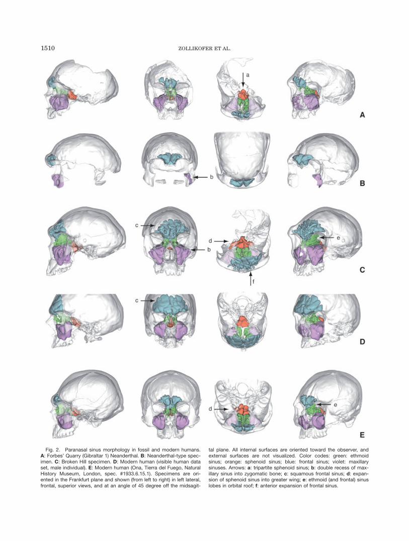
Fig. 2. Paranasal sinus morphology in fossil and modern humans.A: Forbes’ Quarry (Gibraltar 1) Neanderthal. B: Neanderthal-type spec-imen. C: Broken Hill specimen. D: Modern human (visible human dataset, male individual). E: Modern human (Ona, Tierra del Fuego, NaturalHistory Museum, London, spec. #1933.6.15.1). Specimens are ori-ented in the Frankfurt plane and shown (from left to right) in left lateral,frontal, superior views, and at an angle of 45 degree off the midsagit-
tal plane. All internal surfaces are oriented toward the observer, andexternal surfaces are not visualized. Color codes: green: ethmoidsinus; orange: sphenoid sinus; blue: frontal sinus; violet: maxillarysinuses. Arrows: a: tripartite sphenoid sinus; b: double recess of max-illary sinus into zygomatic bone; c: squamous frontal sinus; d: expan-sion of sphenoid sinus into greater wing; e: ethmoid (and frontal) sinuslobes in orbital roof; f: anterior expansion of frontal sinus.
1510 ZOLLIKOFER ET AL.

comparatively short anteroposteriorly, but exhibits lat-eral lobes that extend around the posterior region of theorbital cavities, and intrude into the greater wings ofthe sphenoid. This latter condition was described asunique (Seidler et al., 1997), but it is also found in mod-ern humans (Terra et al., 2006). The maxillary sinusesexhibit a lobed structure, wrapping around various in-ternal bony crests, which correspond to the canal for theanterior superior alveolar nerve and vessels and its ram-ifications, as well as to the canal of the zygomaticofacialnerve and vessels.
Modern humans. Figure 2D,E shows the para-nasal sinus morphology of two human crania, both ofwhich are comparatively large and robust. These speci-mens were chosen to illustrate within-species variabilityin the relationship between superstructures and endo-cranial cavities. Interestingly, the comparatively robustOna (Tierra del Fuego) specimen (Fig. 2E) has a less-developed frontal sinus than the visible human (Fig.2D). As a qualitative observation, it may be stated thatthe presence of a strong glabellar prominence and asso-ciated supraglabellar depression is often correlated withabsence of a frontal squamous sinus in modern humans.Conversely, formation of a squamous sinus is accompa-nied by anterior bulging of the external table of the fron-tal bone, which levels out the supraglabellar depression.
Correlation of Frontal Sinus Form
With Cranial Form
According to our null hypothesis, it can be expectedthat the form of ethmoid, sphenoid, and maxillarysinuses essentially reflects the form of the available spa-tial compartments that are formed as a consequence ofdifferential developmental trajectories of the neurocra-nium and viscerocranium. The frontal sinus assumes asomewhat special position. Development of the outer ta-ble of the frontal squama is less constrained—both func-tionally and developmentally—than that of other cranialregions containing sinuses. Assuming that sinuses aremore than spandrels, it can thus be expected that thefrontal sinus is the most likely candidate to show corre-lation between form and specific sinus function, andthat its form is only weakly related to developmentaland functional constraints of the cranium as a whole.Analyzing correlations between frontal sinus form andcranial form is thus the most sensitive test of the span-drel null hypothesis.The results of correlation analyses between frontal
sinus dimensions and cranial dimensions are shown inFigs. 3 and 4. Sinus size Sf is related to cranial size Sc
with a positive allometric coefficient (2.473), and theslope of the log-linear regression function is differentfrom zero (Fig. 3B). The residuals of this regressionfunction, which serve as a proxy for sinus shape Sf,were then tested for correlation with all PCs resultingfrom PCA of shape. Significant correlations could onlybe found with PC2 (Fig. 3C). Because PC2 itself is notcorrelated with cranial size, it represents a mode ofshape variation, which influences frontal sinus shape in-dependent of cranial size, and independent of taxon.Figure 4 shows actual spatial patterns of shape varia-
tion corresponding to PC1 and PC2. PC1 basicallyexpresses shape transformation from an average great
ape cranium to an average modern human cranium (Fig.4A). Beside the obvious pattern of neurocranial expan-sion and viscerocranial reduction characteristic of mod-ern humans compared to the great apes, Fig. 4A revealswhy the great ape-to-human transformation is not corre-lated with changes in Sf: facial shape change is bestcharacterized as a ‘‘rotation’’ of the midface around gla-bella. Since the position of glabella remains fixed rela-tive to the endocranial cavity, frontal sinus shape is notaffected. Shape transformation corresponding to PC2(Fig. 4B) can be characterized as a ‘‘rotation’’ of theupper face away from the neurocranium, with prosthion
Fig. 3. Correlation between frontal sinus shape and cranial shapeI: A: Cranial shape variability in a sample of hominins and Great Apes(filled circles: H. sapiens; open circles: Neanderthals; square: BrokenHill specimen; x: P. troglodytes; 1: G. gorilla; triangles: P. pygmaeus;large/small symbols: males/females). B: Double logarithmic regressionof frontal sinus size Sf against cranial centroid size Sc (log Sf 525.514 1 2.473 log Sc; r
2 5 0.43; P < 0.0001). C: Double-logarithmicregression of frontal sinus shape Sf (the residuals of the regressionfunction in B) against cranial shape component PC2 (Sf 5 20.0088 13.0890 PC2; r2 5 0.56; P < 0.0001).
1511FOSSIL HOMININ SINUS MORPHOLOGY

as a fixed point. As a result, glabella advances toward amore anterior position, giving rise to an intrafrontalspace, which is occupied by the frontal sinus. Simultane-ously, the interorbital space is widened, giving addi-tional space for sinus development. This pattern essen-tially describes transition from an Pongo-like to an Afri-can ape-like cranial morphology, but it is worth notinghere that hominins exhibit considerably more variationalong PC2 (i.e., in facial relative to neurocranial orienta-tion) and, correspondingly, in sinus shape, than each ofthe great ape species. This point will be discussed inmore detail later. Overall, shape transformation corre-sponding to PC2 indicates that frontal sinus shape isprimarily determined by the orientation and shape ofthe upper face (specifically the orbital cavities) relativeto the neurocranium.Interestingly, modes of shape variation corresponding
to PC1 and PC2 are both associated with variation in fa-cial relative to neurocranial orientation, which is typi-cally described in terms of facial kyphosis. PC1 depictstransition from the more airorhynch great apes(upward-oriented face) to the more klinorhynch homi-
nins (downward-oriented face), while PC2 depicts transi-tion from airorhynch Pongo to more klinorhynch Gorillaand Pan morphologies. Our results show that facialkyphosis is a descriptive category, which is not suffi-ciently precise to account for the multifactorial phyleticand developmental basis of variation in facial relative toneurocranial orientation and position (Bastir and Rosas,2004).
DISCUSSION
Hominin fossils have often been described as havingextended paranasal sinuses (Weidenreich, 1943; Seidleret al., 1997), but since the very beginnings of paleoan-thropology, the idea that air-filled spaces may havetaxon-specific morphologies was received with scepticism(Davis, 1865). The basic question is whether sinus formresults from the general scaling laws of the hominoidcranium, or whether other explanatory factors, such asphyletic grade shifts in sinus morphology (Rae, 1999), orspecific sinus function, need to be advocated as explana-tory factors. Empirical evidence from studies of higher
Fig. 4. Correlation between frontal sinus shape and cranial shapeII: visualization of modes of shape variability. A: shape variability alongPC1 (Fig. 3A) corresponds to transformation of an average great apecranium (left; PC1 5 20.2) into an average great human cranium(right, PC1 5 0.2). B: shape variability along PC2 (Fig. 3A,C) corre-sponds to transformation of an average human/ape specimen with asmall frontal sinus (left, PC2 5 20.075) into an average human/apespecimen with a large frontal sinus (right, PC2 5 0.075). Stippled lines
indicate orientation of orbital plane. Line graphs summarize majorshape differences (ellipse: braincase; quadrangle: face; black triangle:orbital cavity; gray blob: frontal sinus): In A, the midface is shortenedby ‘‘rotation’’ of the facial complex around glabella (black circle); sinussize remains constant. In B, the upper face is advanced by ‘‘rotation’’of the facial complex around prosthion (black circle). Note reorienta-tion and elongation of orbital cavities, as well as widening of interorbi-tal space (see top graph of B).
1512 ZOLLIKOFER ET AL.

primates and humans tends to support the former view.For example, in anthropoid primates, the size of themaxillary sinus appears to be primarily determined byfacial dimensions (Koppe et al., 1999; Rae and Koppe,2000), and this is true even when sinus size demonstra-bly depends on external factors: A comparison of differ-ent populations of the Japanese macaque, Macaca fus-cata, showed that sinus volume decreases with decreas-ing mean annual temperature, but that this is a sideeffect of expansion of the nasal cavity (Rae et al., 2003).The analyses presented here support the notion that
sinus morphology in fossil hominins follows the generaltrends found in hominoid primates, and that potentialpeculiarities of their sinus system basically reflecttaxon-specific features of their craniofacial morphology.However, because the craniofacial morphology of fossilhominins is clearly distinct from that of modernhumans, examination of their sinus morphology providesnew insights into processes of phyletic diversificationand of cranial growth and development.Comparison of the paranasal sinus morphology in the
fossil and modern human sample of this study showsthat variability in the fossil sample is considerable, bothqualitatively and quantitatively, but not greater than inmodern humans (Figs. 2 and 3). Most importantly, allpneumatic variants seen in the fossil hominin samplecan also be found in modern humans, although theytend to occur at lower frequencies (Weiglein, 1999). Forexample, pneumatization of the orbital roof throughrecesses of the ethmoid and/or frontal sinus is commonin fossil hominins, but also in modern humans (Wei-glein, 1999), while pneumatization of the sphenoid wingis a relatively rare but nonsymptomatic condition inmodern humans (Terra et al., 2006). Overall, clinicalcase studies show that (pathological) pneumatizationcan, in principle, affect all cranial bones, including theparietals, occipital, and nasal conchae (Littrell et al.,2004; Rebol et al., 2004).Neanderthals assume an important place in the dis-
cussion of sinus morphology, because the notion thatNeanderthals are heavily pneumatized has almostreached textbook status of knowledge (Laitman et al.,1996; Rae and Koppe, 2004). However, as pointed out byVlcek in his comprehensive comparative study of Nean-derthal frontal pneumatization, this hypothesis cannotbe upheld (Vlcek, 1967). This author characterizes Nean-derthal frontal sinuses as cauliflower-shaped cavitiesoccupying large parts of the supraorbital region, whichare relatively uniform in structure, compared to thehighly variable frontal sinuses of modern humans. Heconcludes that variation in sinus morphology closely fol-lows evolutionary reorganization of the supraorbitalarea: while the frontal sinus of Neanderthals is confinedto the region behind the browridges and does not extendinto the moderately sloping frontal squama, pneumatiza-tion in modern humans tends to invade the steeply rais-ing frontal squama.Our data also confirm that, when related to craniofa-
cial anatomy, Neanderthal paranasal pneumatization isnot above human standards (Figs. 2 and 3). However,there are subtle differences in sinus shape. Compared tomodern humans, the maxillary and ethmoid sinuses ofthe Gibraltar 1 specimen have an elongated shape, andthe maxillary sinus a bulging anterior surface. This pe-culiar morphology reflects the elongated midfacial archi-
tecture of Neanderthals (note the long orbital cavities inFig. 2A), and matches the shape of the facial surface ofthe Neanderthal maxilla. The morphology of the latterarea is often described as ‘‘inflated,’’ such that the ques-tion arises whether the bulging maxillae are formedthrough ‘‘hyperpneumatization,’’ or whether their spe-cific shape entails hyperpneumatization. Examination ofthe midfacial morphology of immature Neanderthalspecimens shows that the latter scenario is the morelikely explanation. Even in the youngest well-preservedNeanderthal specimen, the Mezmaiskaya neonate (Golo-vanova et al., 1999), the facial surface of the maxilla islarge compared to modern human neonates (Ponce deLeon et al., 2008). The size of this surface is constrainedby the position of the alveolar process of the maxillarelative to the orbits rather than by the size of thedeveloping maxillary sinus. Similarly, expansion ofthe maxillary sinus into the zygomatic bone, as seen inthe Forbes’ Quarry, Neanderthal, and Broken Hill speci-mens (Fig. 2A–C), seems to be a consequence of thecomparatively large zygomaticomaxillary interface.Zygomatic recesses are well developed in the great apes,such that we assume that this morphology representsthe plesiomorphic condition in the hominoids.The morphology of the frontal sinus of the Broken
Hill specimen conserves valuable clues about the cra-niofacial development and the ontogeny of pneumati-zation in this individual. Left–right asymmetry of thefrontal sinus (Fig. 2C) indicates that invasion of thefrontal squama from the respective left and right ante-rior ethmoid air cells represented two independentgrowth processes. The fact that similar spatial con-straints and physiological conditions of growth led tostrikingly different morphologies in one and the sameindividual shows that sinus formation has an impor-tant stochastic component.The fan-like extension of the sinus into the frontal
squama contrasts with finger-like anterior extensionsinto the glabellar region. We hypothesize that formationof the squamous and glabellar portions of the frontalsinus corresponds to two successive growth processes ofthe face. As can be seen in modern humans, pneumati-zation of the frontal squama presupposes a steep orien-tation of the frontal squama relative to the orbits (Vlcek,1967) (Fig. 2D vs. 2E). We suppose that, in Broken Hill,a similar spatial relationship between the orbits and thefrontal squama existed during childhood and allowed itspneumatization. On the other hand, anterior growth ofthe sinus into the glabellar region is indicative of a latermorphogenetic process associated with substantial facialadvancement relative to the neurocranium, and forma-tion of a prominent browridge. We suppose that this pro-cess took place relatively late during development, andthat it might have been related to the development ofsexual dimorphism. Similar considerations might applyto the Petralona cranium, which also exhibits a large,fan-like squamous frontal sinus, and pneumatization ofthe glabellar region by finger-like protrusions (Seidleret al., 1997).The spandrel null hypothesis predicts, in its quantita-
tive form, that pneumatization is largely an effect of cra-nial developmental allometry. Because the face and thebraincase grow with positive and negative allometriccoefficients, respectively (Ponce de Leon and Zollikofer,2001), nonfunctional spaces between various cranial
1513FOSSIL HOMININ SINUS MORPHOLOGY

compartments must be bridged in some way to guaran-tee biomechanical and structural coherence. But evennonfunctional spandrels are submitted to evolutionaryoptimization, such that formation of intraosseous air-filled volumes can be seen as a strategy to minimize ma-terial investment while maximizing biomechanical sta-bility, similar to the formation of cortical, cancellous andmarrow-filled compartments in long bones. Overall,thus, sinuses can be expected to reflect the position, ori-entation, and size of cranial compartments relative toeach other rather than specific functions.From this perspective, absence of sphenoid wing pneu-
matization in modern humans is an effect of the rela-tively large temporal and frontal lobes of the brain,which fill the respective regions between the temporalfossa and the orbits. Likewise, the bulging shape of theNeanderthal maxillary sinus reflects absence of a caninefossa, and presence of a large sinus expanding into thefrontal squama of modern humans is related to theirbulging forehead.Combining our null hypothesis with the functional
matrix hypothesis (Moss, 1986) led us to the propositionthat frontal sinuses are less constrained by surroundingcranial compartments than other paranasal sinuses, andthat they represent the best candidates to detect poten-tial structure–function relationships. Figures 3 and 4show that variation in frontal sinus size and shape canbe explained statistically by variation in cranial size andshape. Similar results were obtained for the relationshipbetween maxillary sinus dimensions and craniofacialdimensions in hominoid primates (Rae and Koppe,2000). Both maxillary and frontal sinus sizes scale tocranial dimensions with a positive allometric coefficient.However, while maxillary sinus size scales isometricallywith facial size (Rae and Koppe, 2000), our data suggestthat frontal sinus size scales isometrically with neuro-cranial size, and allometrically with facial size (Table 1).Correlation between cranial shape and sinus shape
follows a more complex pattern. Transformation of anaverage great ape cranium into an average hominin cra-nium does not affect frontal sinus shape (Fig. 4A, PC1),because it mainly affects size, position, and orientationof the maxillae relative to the rest of cranium. The onlycranial shape component exhibiting correlation withsinus shape was PC2, which captures major differencesin facial orientation between Asian and African greatapes, and also within modern humans (Fig. 4B). PC2reflects change in facial orientation through rotation ofthe upper face around a fixed point near prosthion. Thisgives rise to an expanded supraorbital space, which,according to our data, is a precondition for extensivefrontal pneumatization. As shown in Fig. 3C, modernhumans exhibit greater variability along PC2 than
Neanderthals or any of the great ape taxa and, as a con-sequence, frontal sinus dimensions are more variablethan in the other hominoids. It is worth noting that PC2not only reflects variation in facial orientation, but alsoin neurocranial shape, which changes from a more glob-ular to a more elongated appearance (Fig. 4B). This pat-tern of correlated shape change corresponds to the tran-sition from a brachycephalic to a dolichocephalic humancranium (Fig. 5). As shown earlier, this mode of varia-tion is independent of age-related change in cranialshape. Rather, it seems to represent differences betweenindividual morphotypes that are already formed at birth(Zollikofer and Ponce de Leon, 2002). It remains to beclarified why humans, compared to the Neanderthalsand the great apes, exhibit more interindividual varia-tion in the way in which the face is hafted to the brain-case. A possible clue comes from the fact that the datascatter along PC2 not only characterizes a human modeof shape variation, but also captures major differencesbetween Asian and African great ape morphologies. Wehypothesize that this pattern of shape variation reflectsa phyletically old mode of ‘‘developmental wobbling’’ incranial shape, which governs both intra- and interspe-cific diversity.Interestingly, the presence of a squamous frontal sinus
has not been reported in Neanderthals, but it seems tobe a characteristic of H. heidelbergensis (as representedby the Broken Hill, Petralona and Steinheim specimens;(Prossinger et al., 2003)). It remains open whether co-
TABLE 1. Allometry of frontal sinus size
Exponent SE R2 P
Cranium 2.47 0.46 0.43 <0.0001Face 1.08 0.32 0.23 <0.0017Neurocranium 1.37 0.34 0.30 <0.0003
Values represent allometric exponent SE, coefficient ofdetermination, and levels of significance of regression equa-tions relating log10 frontal sinus size Sf to log10 centroidsize of the cranium (Sc), the face, and the neurocranium.
Fig. 5. Correlation between frontal sinus shape and cranial shapein hominins. Transformation of an average hominin cranium with asmall frontal sinus (left; PC1 5 20.2, PC2 5 20.075) into an averagehominin cranium with a large frontal sinus (right, PC1 5 20.2, PC2 50.075).
1514 ZOLLIKOFER ET AL.

occurrence of this feature in H. heidelbergensis and inmodern humans represents homoplasy or homology. Inthe latter case, it could indicate evolutionary continuitybetween ‘‘archaic’’ and modern H. sapiens.Taken as a whole, our data indicate that no specific
functional hypotheses need to be advocated to explainvariation in frontal sinus size and shape (note that thereis no taxon-specific deviation from the regression line inFig. 3C). Nevertheless, it is worth noting that only 56%of the total variation in sinus shape can be explainedstatistically by variation along cranial shape componentPC2. We suppose that most of the remaining variationin sinus morphology can be understood as resulting fromthe essentially stochastic nature of sinus morphogenesis(Zollikofer and Weissman, 2008).The basic developmental mechanism of paranasal
sinus formation is expansion of nasal mucous epitheliuminto all surrounding bones ‘‘in an opportunistic mannerwithin the constraints of a particular biomechanicalloading regime’’ (Witmer, 1999). What might be calledthe ‘‘expansive tissue hypothesis’’ (Zollikofer and Weis-mann, 2008) may serve here as a point of view fromwhich sinus morphology in fossil hominins can be ana-lyzed. One question that arises from this hypothesis ishow cancellous diploic bone is replaced by mucous tis-sue. As can be seen in the frontal sinuses of the BrokenHill and Forbes’ Quarry specimens, the less-developedsides of the sinuses extend toward regions of low trabec-ular density in the diploic layer. If this condition repre-sents an incipient stage of sinus formation on this side,the following scenario can be hypothesized: rapid exten-sion of the external table of the frontal bone combinedwith a low rate of deposition of extracellular bone matrixleads to thinning of the trabecular web, which in turnfacilitates invasion of mucous epithelium and replace-ment of cancellous bone by air sacs. Furthermore, largeintra- and interindividual variability between homolo-gous sinuses indicates that the size and shape of air-filled regions is a function of the rate and duration ofmucous tissue growth, and of the available space, whichin turn depends on differential growth characteristics ofthe surrounding cranial compartments.Further work will be needed to test these hypotheses
in more detail. Nevertheless, while air-filled spaces inthe cranium of fossil and extant hominoids might ulti-mately be best described as biological spandrels, it mustbe acknowledged that even spandrels reflect the archi-tectural design codes imposed by evolution and develop-ment and thus constitute a rich source of informationabout differential modes of growth and development infossil and extant hominins.
ACKNOWLEDGMENTS
We thank Sam Marquez for his invitation to contrib-ute to this volume and for proofreading the manuscript.We thank Yoel Rak for granting access to fossil speci-mens, and Robert Krusynski (Natural History MuseumLondon) and Isuf Hoxha for assistance with scanning.
LITERATURE CITED
Balzeau A, Grimaud-Herve D. 2006. Cranial base morphology andtemporal bone pneumatization in Asian Homo erectus. J HumEvol 51:350–359.
Bastir M, Rosas A. 2004. Facial heights: Evolutionary relevance ofpostnatal ontogeny for facial orientation and skull morphology inhumans and chimpanzees. J Hum Evol:359–381.
Chaiyasate S, Baron I, Clement P. 2007. Analysis of paranasal sinusdevelopment and anatomical variations: a CT genetic study intwins. Clin Otolaryngol 32:93–97.
Coon CS. 1962. The origin of races. New York: Knopf.Davis B. 1865. [Comments on]: The Neanderthal skull: its formation
considered anatomically. J Anthropol Soc Lond 3:15–19.Demes B. 1987. Another look at an old face: biomechanics of the
Neandertal facial skeleton reconsidered. J Hum Evol 16:297–303.Dryden IL, Mardia K. 1998. Statistical shape analysis. New York:
Wiley.Golovanova LV, Hoffecker JF, Kharitonov VM, Romanova GP. 1999.
Mezmaiskaya cave: A Neanderthal occupation in the NorthernCaucasus. Curr Anthropol 40:77–86.
Gould SJ, Lewontin RC. 1979. The spandrels of San Marco and thePanglossian paradigm: a critique of the adaptationist programme.Proc R Soc Lond B 205:581–598.
Heim JL. 1974. Les hommes fossiles de La Ferrassie (Dordogne) etle probleme de la definition des Neanderthaliens classiques.L’Anthropologie 78:312–378.
Heim JL. 1997. Ce que nous dit le nez du neanderthalien. LaRecherche 294:66–70.
Koppe T, Ohkawa Y. 1999. Pneumatization of the facial skeleton incatarrhine primates. In: Koppe T, Nagai H, Alt KW, editors. Theparanasal sinuses of higher primates. Chicago: Quintessence.p 77–119.
Koppe T, Rae TC, Swindler DR. 1999. Influence of craniofacial mor-phology on primate paranasal pneumatization. Ann Anat 181:77–80.
Laitman JT, Reidenberg JS, Marquez S, Gannon PJ. 1996. Whatthe nose knows: new understandings of Neanderthal upper respi-ratory tract specializations. Proc Natl Acad Sci USA 93:10543–10545.
Littrell LA, Leutmer PH, Lane JI, Driscoll CL. 2004. Progressivecalvarial and upper cervical pneumatization associated with ha-bitual valsalva maneuver in a 70-year-old man. AJNR Am J Neu-roradiol 25:491–493.
Moss ML. 1986. Newer analytical models of craniofacial growth.Nova Acta Leopoldina NF 58:17–25.
Ponce de Leon MS, Golovanova L, Doronichev V, Romanova G, Aka-zawa T, Kondo O, Ishida H, Zollikofer CPE. 2008. Neanderthalbrain size at birth provides insights into human life history evolu-tion. Proc Natl Acad Sci USA 105:13764–13768.
Ponce de Leon MS, Zollikofer CPE. 2001. Neanderthal cranial on-togeny and its implications for late hominid diversity. Nature412:534–538.
Prossinger H, Bookstein F, Schafer K, Seidler H. 2000. Reemergingstress: supraorbital torus morphology in the mid-sagittal plane?Anat Rec 261:170–172.
Prossinger H, Seidler H, Wicke L, Weaver D, Recheis W, StringerC, Muller GB. 2003. Electronic removal of encrustations insidethe Steinheim cranium reveals paranasal sinus features anddeformations, and provides a revised endocranial volume esti-mate. Anat Rec B (New Anatomist) 237:132–142.
Rae TC. 1999. The maxillary sinus in primate paleontology and sys-tematics. In: Koppe T, Nagai H, Alt KW, editors. The paranasalsinuses of higher primates. Chicago: Quintessence. p 177–189.
Rae TC, Hill RA, Hamada Y, Koppe T. 2003. Clinal variation ofmaxillary sinus volume in Japanese macaques (Macaca fuscata).Am J Primatol 59:153–158.
Rae TC, Koppe T. 2000. Isometric scaling of maxillary sinus volumein hominoids. J Hum Evol 38:411–423.
Rae TC, Koppe T. 2004. Holes in the head: evolutionary interpreta-tions of the paranasal sinuses in catarrhines. Evol Anthropol 13:211–223.
Rak Y. 1986. The Neanderthal: a new look at an old face. J HumEvol 15:151–164.
Ravosa MJ, Vinyard CJ, Hylander WL. 2000. Stressed out: mastica-tory forces and primate circumorbital form. Anat Rec 261:173–175.
1515FOSSIL HOMININ SINUS MORPHOLOGY
Rebol J, Munda A, Tos M. 2004. Hyperpneumatization of the tempo-ral, occipital and parietal bones. Eur Arch Otorhinolaryngol 261:445–448.
Rohlf FJ, Slice D. 1990. Extensions of the Procrustes method forthe optimal superimposition of landmarks. Syst Zool 39:40–59.
Rossie JB, Simons EL, Gauld SC, Rasmussen DT. 2002. Paranasalsinus anatomy of Aegyptopithecus: implications for hominoid ori-gins. Proc Natl Acad Sci USA 99:8454–8456.
Schmitz RW, Serre D, Bonani G, Feine S, Hillgruber F, KrainitzkiH, Paabo S, Smith FH. 2002. The Neandertal type site revisited:interdisciplinary investigations of skeletal remains from the Nean-der Valley, Germany. Proc Natl Acad Sci USA 99:13342–13347.
Schwartz JH, Tattersall I. 1996. Significance of some previouslyunrecognized apomorphies in the nasal region of Homo neander-thalensis. Proc Natl Acad Sci USA 93:10852–10854.
Seidler H, Falk D, Stringer C, Wilfing H, Muller GB, zur NeddenD, Weber GW, Reicheis W, Arsuaga JL. 1997. A comparativestudy of stereolithographically modelled skulls of Petralona andBroken Hill: implications for future studies of middle Pleistocenehominid evolution. J Hum Evol 33:691–703.
Sherwood RJ. 1999. Pneumatic processes in the temporal bone ofchimpanzee (Pan troglodytes) and gorilla (Gorilla gorilla). J Mor-phol 241:127–137.
Sherwood RJ, Ward SC, Hill A. 2002. The taxonomic status of theChemeron temporal (KNM-BC 1). J Hum Evol 42:153–184.
Smith FH, Smith MO, Schmitz RW. 2006. Human skeletal remainsfrom the 1997 and 2000 excavations of cave deposits derived from
Kleine Feldhofer Grotte in the Neander Valley, Germany. In:Schmitz RW, editor. Neanderthal 1856–2006. Mainz: Zabern.p 187–246.
Terra ER, Guedes FR, Manzi FR, Boscolo FN. 2006. Pneumatizationof the sphenoid sinus. Dentomaxillofac Radiol 35:47–49.
Vlcek E. 1967. Die Sinus frontales bei europaischen Neandertalern.Anthropologischer Anzeiger 30:166–189.
Weidenreich F. 1943. The skull of Sinanthropus pekinensis: a com-parative study of a primitive hominid skull. Palaeontol Sin Ser D7:1–162.
Weiglein AH. 1999. Development of paranasal sinuses in humans.In: Koppe T, Nagai H, Alt KW, editors. The paranasal sinuses ofhigher primates. Chicago: Quintessence. p 35–50.
Wind J. 1984. Computerized X-ray tomography of fossil hominidskulls. Am J Phys Anthropol 63:265–282.
Witmer LM. 1999. The phylogenetic history of paranasal airsinuses. In: Koppe T, Nagai H, Alt KW, editors. The paranasalsinuses of higher primates. Chicago: Quintessence. p 21–34.
Zollikofer CPE, Ponce de Leon MS. 2002. Visualizing patterns ofcraniofacial shape variation in Homo sapiens. Proc Roy Soc B269:801–807.
Zollikofer CPE, Ponce de Leon MS. 2005. Virtual reconstruction: Aprimer in computer-assisted paleontology and biomedicine. NewYork: Wiley.
Zollikofer CPE, Weissmann JD. 2008. A morphogenetic model ofcranial pneumatization based on the invasive tissue hypothesis.Anat Rec 291:1446–1454.
1516 ZOLLIKOFER ET AL.