Facial skeleton, - Anatomický ústav 1. LF UK
106
Facial skeleton, jaws, sinuses, TMJ joint, mastication muscles, gum, periodontium, tongue, pharynx, tonsills By Ivo Klepáček orofa cialni
-
Upload
khangminh22 -
Category
Documents
-
view
7 -
download
0
Transcript of Facial skeleton, - Anatomický ústav 1. LF UK
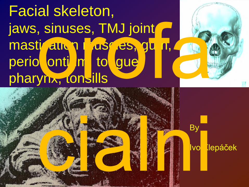
Facial skeleton,jaws, sinuses, TMJ joint, mastication muscles, gum, periodontium, tongue, pharynx, tonsills
By
Ivo Klepáček
orofa cialni

JAWSγνάθος (gnathos)
PremaxillaMaxilla
Mandibula
orofa cialni

Maxilla
Sinus maxillaris (antrum Highmori) – open to nasal cavity as a hiatus maxillaris
Fossa lacrimalis
CorpusProc. frontalisProc. zygomaticusProc. alveolarisProcessus palatinusorofa
cialni
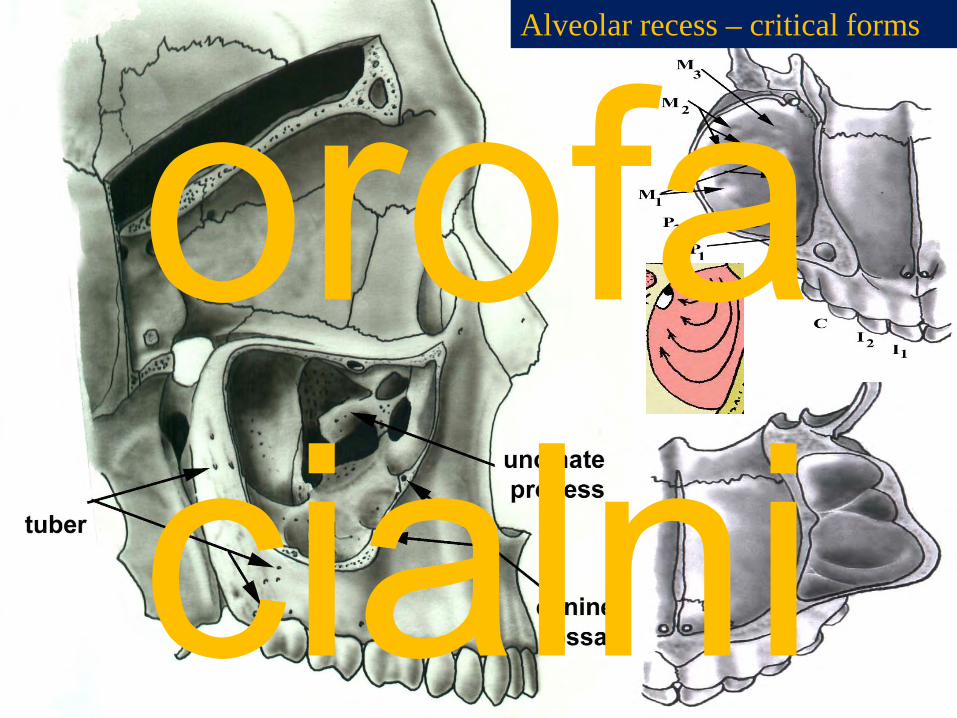
Alveolar recess – critical forms
orofa cialni

Maxillary duct„Ductus maxilaris“
Level of hard palate (palatal line) - pink
The wall of recessus frontalis is extremely thin
- first sign: 65 day gestation- birth: 7x4x4 mm- RTG appearance: 4-5 month- bifasic growth:
0-3 year7-12 year (permanent teeth)
- 18 year: 34x33x23 mm15 ml
orofa cialni
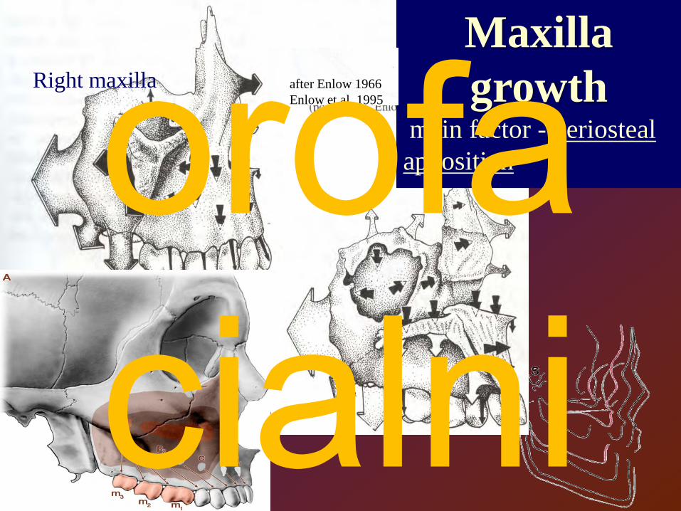
Maxillagrowth
main factor - periostealapposition
Right maxilla after Enlow 1966 Enlow et al. 1995orofa
cialni
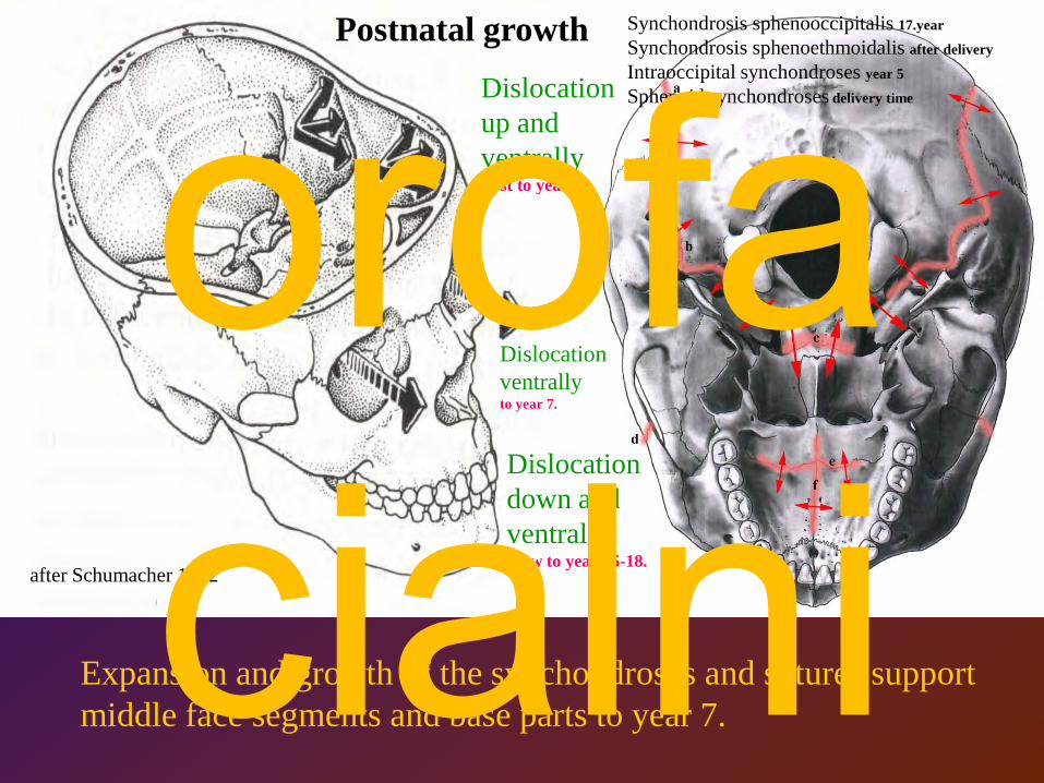
Expansion and growth of the synchondroses and sutures support middle face segments and base parts to year 7.
Postnatal growth
Dislocation down and ventrallySlow to year 15-18.
Dislocation up and ventrallyFast to year 12.
Synchondrosis sphenooccipitalis 17.year
Synchondrosis sphenoethmoidalis after delivery
Intraoccipital synchondroses year 5
Sphenoid synchondroses delivery time
after Schumacher 1992
Dislocation ventrallyto year 7.
orofa cialni

7 / 5A 7 / 5B
Ostiomeatal unit
senile
adult
senile
adult
orofa cialni

orofa cialni

7 / 7BSE – sinus ethmoidealis, SF – sinus frontalis, SM –sinus maxillaris, SS – sinus sphenoidealis, HS – hilus maxillaris (canalis semilunaris); * - ductus nasolacrimalis
orofa cialni

Vyústění paranasalních dutinParanasal cavities - openingsorofa
cialni

Odříznutá concha nasalis mediaRemoved middle nasal concha
orofa cialni

Processus uncinatus a hiatus
maxillarisUncinate process
and maxillary opening
orofa cialni

orofa cialni

Lowe jaw - profile. a – fovea pterygoidea, b – processus coronoideus seu muscularis, c –fossa retromandibularis, d - linea (crista) obliqua, e – crista temporalis, f -trigonum retromolare , g – linea mylohyoidea, h – foramen mentaleLower jaw - from below.a - fovea pterygoidea, b – angulus mandibulae et tuberositas pterygoidea, c – spina mentalis, d – linea mylohyoidea, e – foramen nutricium (canaliculus supramentalis or foramen linguae), f – fossa digastrica, g –fovea sublingualis, h – fovea submandibularisInner side of mandible. a - caput mandibulae, b – margo anterior, c – fossa retromandibularis, d –crista temporalis, e – trigonum retromolare, f – area where mucosa develops small tubercle (typical for gummy people - tuberculum retromolare, g – linea mylohyoidea, h – fovea sublingualis, i – spina mentalis, j – fovea submandibularis, k – sulcus mylohyoideus, l – fossa colli mandibulae, m – linea (crista) colli mandibulae, n – lingula, o –incisura mandibulae (semilunaris)
orofa cialni
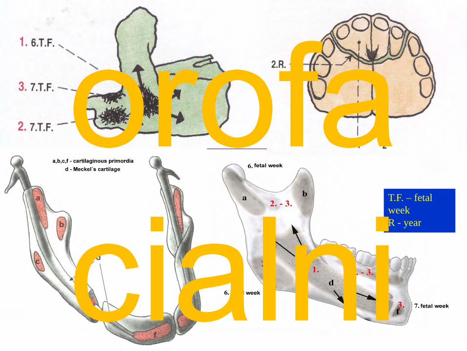
T.F. – fetal weekR - year
orofa cialni
orofa cialni

Cévní zásobení bradové krajiny a dolních řezáků
orofa cialni

Occlusal plane as is determined (following clinical demands), like line crossing lower lip and top of lower caninus.A – incomplete dentureB – senile denture. Mucosa of the retromolar tubercle is not removed.pink.
orofa cialni

Formation of canalis mentalis
newborn
adult
Mandible growth
120°
year 3
year 20orofa cialni
0.1-0.4 mm
orofa cialni

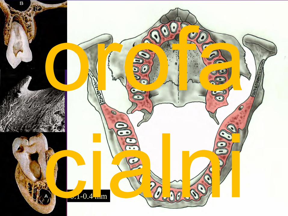
Topographic relations between spongy and compact bone seen in sections. (McMillen 1924, modified )Red arrows – maxillary sinus; red areas – mandibular canal
orofa cialni

X-ray photo(Waters projection)
Sinus frontalis
Sinus maxillaris
Sinus sphenoidalis
orofa cialni

Canalis mandibularis
Recessus alveolaris (sinus maxillaris)
Panoramatický snímek
orofa cialni

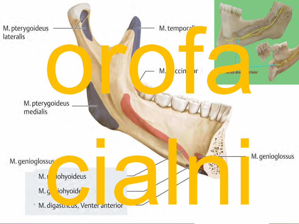
Žvýkací svaly
Musculi masticatoriiMuscles of mastication
V3 – MANDIBULARISderiváty 1. žaberního oblouku
orofa cialni

orofa cialni

M. temporalis et fascia temporalis
Spatium interfasciale
Fyziologický průměrPhysiologic diameter cca 3,6 cm2
orofa cialni

orofa cialni
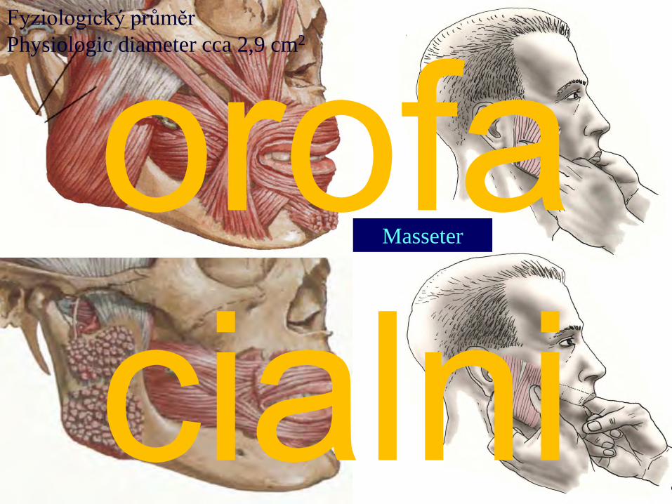
Masseter
Fyziologický průměrPhysiologic diameter cca 2,9 cm2
orofa cialni

M. masseterorofa cialni

orofa cialni

Fascia parotideomassetericaorofa
cialni

Pterygoideus lateralisPterygoideus medialis
Fyziologický průměr cca 1,7 cm2
Physiologic diameter cca 1,8 cm2
orofa cialni

Lingua, glossatongue
Pohyblivý svalověepitelový orgánMobile musculoepithelial organ;
Located in the oral cavity and pharynx
Žvýkání Mastication,Polykání Deglutition,
Řeč Speech
Formuje soustoSqueezing food into pharynx
during swallowing;
Pomáhá artikulovartt slovaForming words during
speaking
orofa cialni

Mm. pterygoidei et variationes
a – m. pterygoideus lateralisb – m. pterygoideus medialis
f,g – pars superior et inferior m. pterygoideus lateralisk,l– pars lateralis et medialis m. pterygoideus medialish,i,j – nadpočetné variety (supernumerary varieties)
PterygospinalisPterygoideus propriusPterygomandibularis
orofa cialni

g- lig. stylomandibulare fascia parotideomasseterica
a- lig. pterygospinosum h- lig. sphenomandibulare e- lig. pterygomandibulare fascia interpterygoidea
orofa cialni

It is supposed that contractile power is 10 kg/1 cm2; bilateral contraction - even 200 kg (crown
masticatory surface is about 1 cm2). Woman -about one quarter lesser value.
Even normal masticating exhibit pressure about 30 - 100 kg. Sensory receptors (inside suspensory
systems, tendons and muscles) control hyperelongations.
orofa cialni

orofa cialni
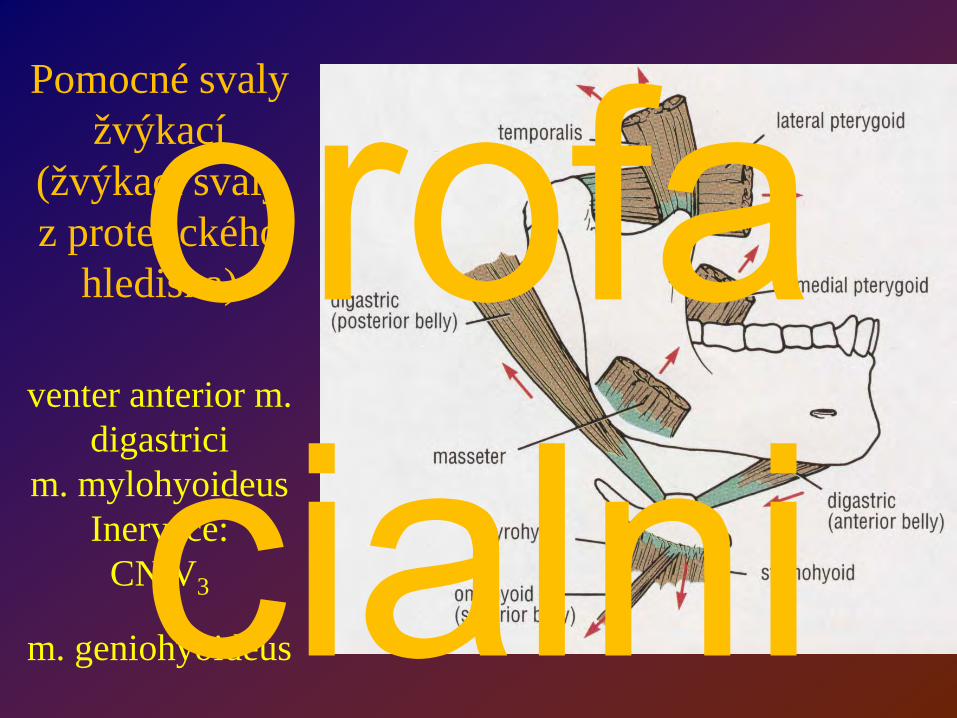
Pomocné svaly žvýkací
(žvýkací svaly z protetického
hlediska)
venter anterior m. digastrici
m. mylohyoideusInervace:
CN V3
m. geniohyoideus
orofa cialni

Functional arrangement of the masticatory muscles
orofa cialni

Compound jointSimilar to hinge joint type
Temporomandibular(craniomandibular) jointArticulatio
temporo-mandibularis
ATM lat.
TM , TMJ engl.
Morphological findings:
• The great variability of all the articular structures• The absence of hyaline cartilage•The two separate compartments, allowing a wider range of mandibular movements• The mared weakness of the articular ligaments, allowing hypertranslation and dislocation without tearing the capsule
orofa cialni
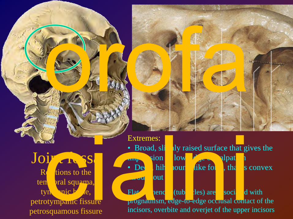
Joint fossaRelations to the
temporal squama, tympanic bone,
petrotympanic fissurepetrosquamous fissure
Extremes:• Broad, slighly raised surface that gives the impression of low ridge of palpation• Deep, hih mound-like form, that is convex throughout
Flat eminences (tubercles) are associated with prognathism, edge-to-edge occlusal contact of the incisors, overbite and overjet of the upper incisors
orofa cialni
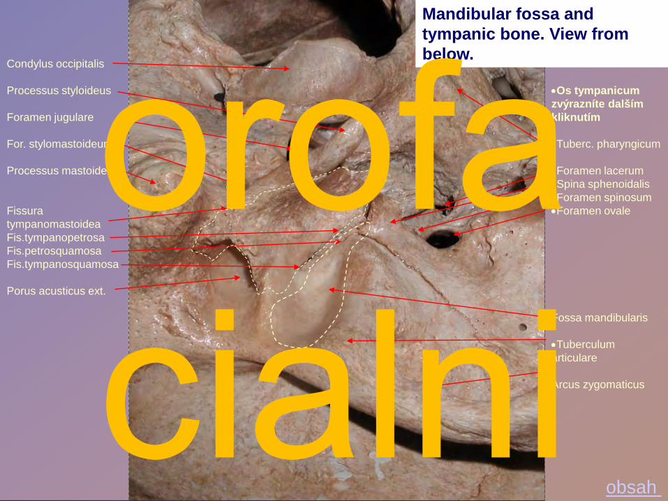
•Os tympanicumzvýrazníte dalším kliknutím
•Tuberc. pharyngicum
•Foramen lacerum•Spina sphenoidalis•Foramen spinosum•Foramen ovale
Fossa mandibularis
•Tuberculum articulare
Arcus zygomaticus
Condylus occipitalis
Processus styloideus
Foramen jugulare
For. stylomastoideum
Processus mastoideus
Fissura tympanomastoideaFis.tympanopetrosaFis.petrosquamosaFis.tympanosquamosa
Porus acusticus ext.
Mandibular fossa and tympanic bone. View from below.
obsah
orofa cialni

Demiaxial projection of Albers-Schönberg (Parma) exhibits different congruenties of the both articular surfacesorofa
cialni

Width : 20.5 mmSagittal diameter : 8.7 mm
orofa cialni

Anteroposterior section through TMJ joint. a – meatus acusticus externus, b – cartilago on surface of the fossa mandibularis, c –perforations in the disc (variety), d – bone layer inside of tuberculum articulare, e -fissura discotemporalis (discotemporal fissure), f – insertio of m. pterygoideus lateralis, g – fissura discomandibularis (discomandibular fissure), h - caput mandibulae, i –Zenker´s retroarticular cushion
Section through intraarticular disc
Retroarticularcushion of Zenker(containing veins)
orofa cialni

Upper joint space – 581 mm2
Lower joint space – 396 mm2
orofa cialni

Atrophy of the madibular condyle depending ageorofa
cialni

orofa cialni

Condyle movement phases through mouth opening . a - basic position (jaws are in central occlusal position), b – rotation, c – translational motion, d – dorsal part of the Zenker cushion is compressed), e – dorsal part of the Zenker cushion is dilated – mouth is open)
orofa cialni

Condyle patha Transverseb Longitudinal
Rest position
Centralposition
Ventralposition
orofa cialni

Basic condyle positions :
A – habitual (high central) position, B – central (zenith) position, C low ventral (relax) position;
1 – inside position, 2 - extrusalposition, 3 – retrusal position, 4 - protrusal position
orofa cialni

Mandible movement
through chewing
Posselt cone-like space –(after Posselt 1961; modified).
Ii – incisale inferiusorofa cialni

Vessels and nerves supplying joint capsule and condyle. Anterior view. Diagram. a – m. mylohyoideus, b – nervus alveolaris inferior, , c – ramus articularis anterior (for masseter m.), d – ramus articularis anterior (from facial nerve), e – rami articulares posteriores (from auriculotemporal n.), g - ramus articularis anterior, h – n. alveolaris inferior, i – branches of ramus articularis anterior, j –gl. submandibularis
Auriculotemporalis nerve gives off four branches:• From lateral limb of the nerve loop• From the medial limb of the nerve loop• From the midsegment• From the area where nerve converges with the superficial temporal artery
The posterior deep temporal nerve:• supplies the rostromedial zone of the disc and capsule
Masseteric nerve gives off four branches:• From the nerve part below foramen ovale• From the first extracranial segment of this nerve• The last two arise from the part below the zygomatic process
The otic ganglion:• supplies the discosquamal part of the capsule
The facial nerve:• supplies the lateral surface of the capsule
orofa cialni

Nerves closely to TMJ:
n. auriculotemporalis
Chorda tympaniorofa cialni

TMJ examinationorofa cialni

Xorofa cialni

Motor cortex Sensory cortex
orofa cialni

ParodontiumClinical unit (cementum, corticalis,
periodontal ligg., gum)
Structure and
development Its changes through eruption
orofa cialni

Tooth fixation, elasticity, (hydroelastic cushion)nutrition, asistance during eruption
Function of periodontiumorofa
cialni

12 / 6
Arrangement of the intraalveolar ligamentsa - ligamenta marginalia, b - ligamenta dentalia superiora, c - ligamenta dentalia media, d - ligamenta dentalia inferiora (apicalia)
Interdental circumdental, dentoalveolar, intraalveolar ligaments
0.3-0.5mm
0.1-0.2mm
asi 0.2mm
orofa cialni

orofa cialni

Gingival sulcus (pocket) Sulcus
gingivae
Free gingival groove Paramarginal sulcus
Gingiva = relation to the teeth – “cuff (collar) attachment“
Free: Interdental; embrasured; circumdentalAttached: Adjacent,fixed
orofa cialni

Interdental papillaeorofa
cialni

orofa cialni

Ligamentous slings and circles help to tight attachment between gingiva and tooth
orofa cialni

Epithelial Malassez´ islets (remnants of Hertwig´s sheet)
orofa cialni

Free gingival groove changes
intoperiodontal
pocketorofa cialni
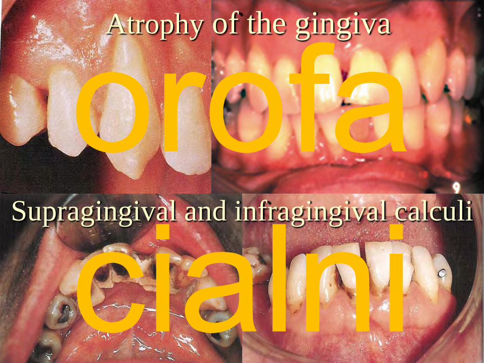
Atrophy of the gingiva
Supragingival and infragingival calculi
orofa cialni
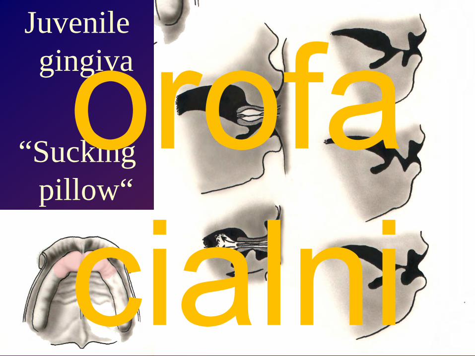
Juvenile gingiva
“Sucking pillow“orofa cialni

Hard palate
Soft palateorofa cialni

Newborn palate Senile palate
orofa cialni

Palate – surface features
Lacey
Incisive papilla
Palatine rugae
Median raphe
orofa cialni

Palatal relief with rugae, foveolae and incisal papila
orofa cialni

Palate – surface features
Lacey
Hard palate
Soft palate
Median raphe
Fatty zone
Openings of palatine glands
A
Horofa cialni

Distances and lines in palate and lower jaw.SI – summa incisivorum.H – linie H (line between bone and palate, A – linie A (line between movable and relative stable part of soft palate), a – distance between premolars, b – distance between fissures of permanent molars, En-En – palate width, Or–Sta – palate length, Co-Ii-Co-Co – Bonwill´s triangle, Co – condylion, En –endomolare, Ii – incisale inferius, Or – orale, Sta – staphylion.
orofa cialni

Soft palate
– dorsal view
– ventral vieworofa cialni

Tongue(LinguaGlossa)
orofa cialni
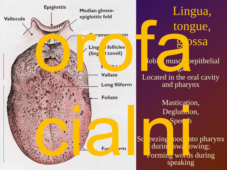
Lingua, tongue, glossa
Mobile musculoepithelial organ;
Located in the oral cavity and pharynx
Mastication,Deglutition,
Speech
Squeezing food into pharynx during swallowing;
Forming words during speaking
orofa cialni

Palatoglossal archPalatopharyngeal arch
Triangular fold (plica) (there is r. tonsillaris)
orofa cialni

Floor of the oral cavitylingual frenulum, sublingual folds,
carunculae
Paralingual canal =between hyoglossus and genioglossus
orofa cialni

bitter
salty
sour
sweet
orofa cialni

StyloglossusPalatoglossus
HyoglossusGenioglossus
Extrinsic lingual musclesalter the
position of the tongue
orofa cialni

Intrinsic lingual musclesalter lingual shape
Superior and inferior longitudinal,transverse, vertical musclesorofa
cialni

Musculi genioglossi are separated
by the lingual septumBetween hyoglossus
and genioglossus muscles there
is lingual canal
Lingual septum can be defibered;after this abscess cavity appears orofa
cialni
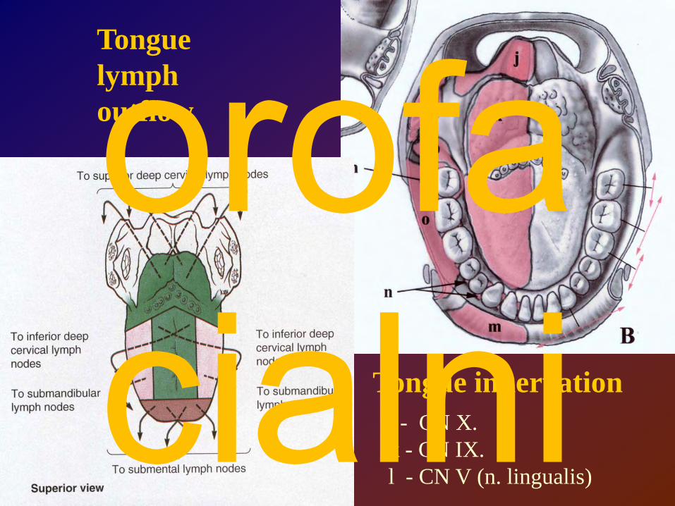
Tongue innervation
Tongue lymph outflow
j - CN X.k - CN IX.l - CN V (n. lingualis)
orofa cialni
Salivary glandsorofa cialni
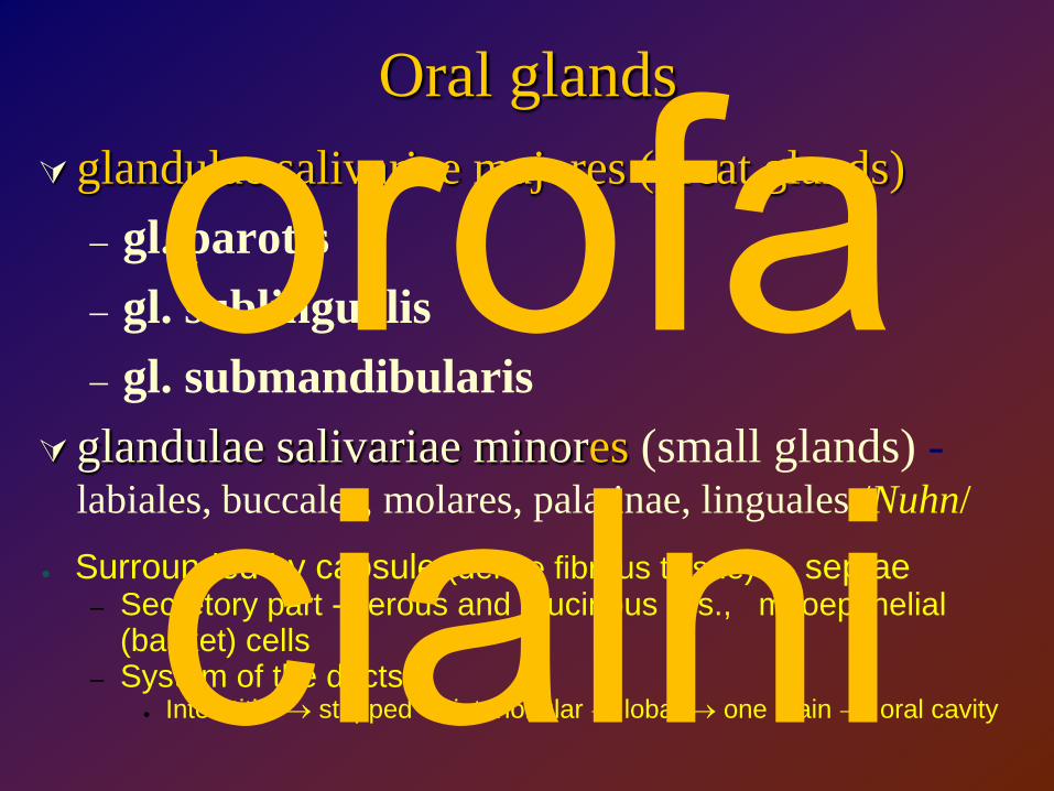
Oral glands glandulae salivariae majores (great glands)
– gl. parotis– gl. sublingualis– gl. submandibularis
glandulae salivariae minores (small glands) -labiales, buccales, molares, palatinae, linguales /Nuhn/
● Surrounded by capsule (dense fibrous tissue) → septae– Secretory part - serous and mucinous clls., myoepithelial
(basket) cells– Systém of the ducts
● Interstitial → stripped → interlobular → lobar → one main → oral cavity
orofa cialni

Parts of gland: Acini, lobes, septae, and capsule
orofa cialni
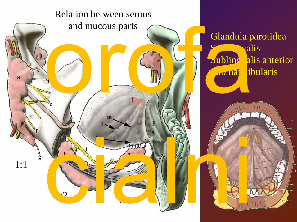
Glandula parotideaSublingualisSublingualis anteriorSubmandibularis
1:1
3:2 1:3
Relation between serous and mucous parts
orofa cialni

Panoramatický snímek
panoramic X – ray photo
orofa cialni

a Plica sublingualisb Caruncula
sublingualisc Frenulum labii
inferiusd Plica buccogingivalise Frennulum linguaef Plica fimbriata
Plicae gingivolabiales* Area sublingualis** Area submandibularis
Canalis paralingualis Paralingual canal =between hyoglossus and genioglossus
Spodina dutiny ústní cavum oris bottom
orofa cialni
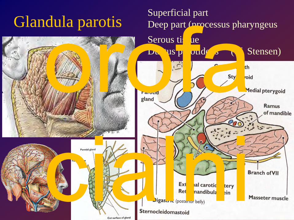
Glandula parotisSuperficial partDeep part (processus pharyngeusSerous tissueDuctus parotideus (of Stensen)orofa
cialni

A parotid tumour compresses thefacial nerve weakening the facial
muscles ipsilaterally (Bell´s palsy).
The corner of the mouth and eye may drop.
orofa cialni

Glandula submandibularis
mukoserousDuctus submandibularis (Wharton)15 gr
15 gramů15 gramů
orofa cialni
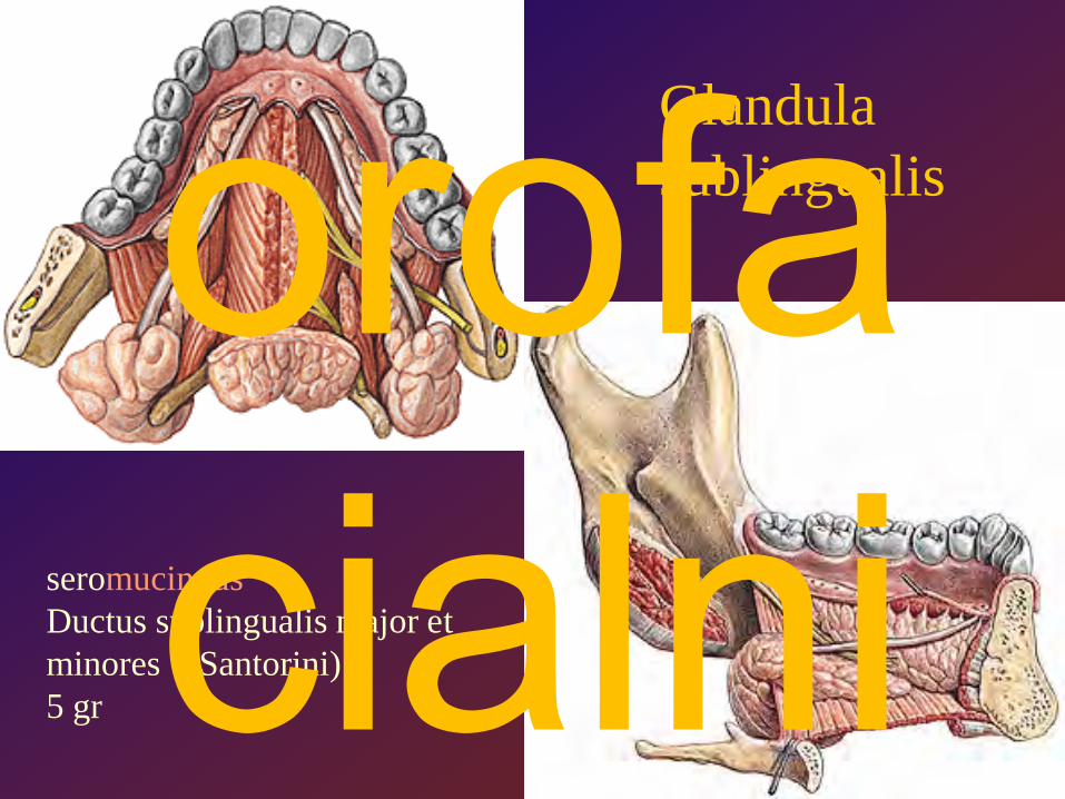
Glandula sublingualis
seromucinousDuctus sublingualis major et minores (Santorini)5 gr
orofa cialni

orofa cialni
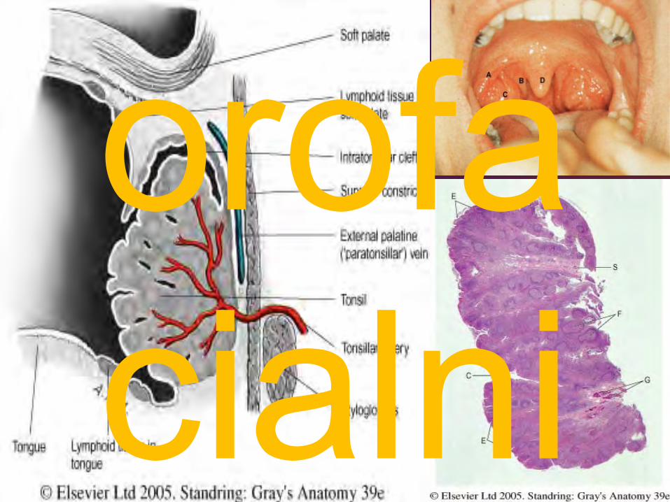
Tonsilary system
Tonsills + lymph nodesorofa cialni

Tonsillar lymph circle of Waldeyer
Heinrich Wilhelm Gottfried Waldeyer
1836-1921
orofa cialni

Waldeyer lymph circle
t.pharyngea
t.tubaria
t.palatina
t.lingualis
ln.retropharyngei
Wood node
ln.jugulodigastricus(Küttner node)
ln.cervicales profundi
- Lower group
3 protective barriers
ln.submentales &submandibulares
ln.juguloomohyoideus
tongue
orofa cialni

Jugulodigastricjuguloomohyoid
deep cervical lymph nodes
orofa cialni
orofa cialni

Incomplete capsuleOnly efferent lymph vesselsModified epithelium in crypts (lymphoepithel or FAE follicle-associated epithelium)Intraepithelial vascularization
Free, T, B lymphocytes, active immunocompetitive cells, macrophages, Langerhans cells, fibrous stroma
orofa cialni

tonsillectomy
orofa cialni

literatureR. Čihák: Anatomie 1, 2, 3Grada Publishing 2003
M.Grim, R.Druga et al.: Základy anatomie 5. Anatomie krajin těla Galén 2008
M. Dykes : Anatomy2th edition, Mosby 2002
S.Snell: Clinical anatomy for Medical Students6th edition, Lippincott, Williams & Wilkins
I.Klepáček, J.Mazánek et al.: Klinická anatomie ve stomatologii Grada Publishing 2001
G.J.Tortora : Principles of Human Anatomy4th edition, Williams & Wilkins
K.L.Moore, A.F.Dalley: Clinically Oriented Anatomy4th edition, Williams & Wilkins
F.H.Netter: anatomický atlas člověkaVlastní archív
orofa cialni