4.2 Disarticulated skeleton of frog
34
Comparative osteology: Lets Learn about bones Compiled by Dr. Namita Nayyar Diagram credits: Ms. Shalini Panwar (B.Sc. (H) Zoology 2018-21)
-
Upload
khangminh22 -
Category
Documents
-
view
1 -
download
0
Transcript of 4.2 Disarticulated skeleton of frog

Comparative osteology:Lets Learn about bones
Compiled by Dr. Namita Nayyar
Diagram credits: Ms. Shalini Panwar
(B.Sc. (H) Zoology 2018-21)

For better Results:
Attend the Class… ☺
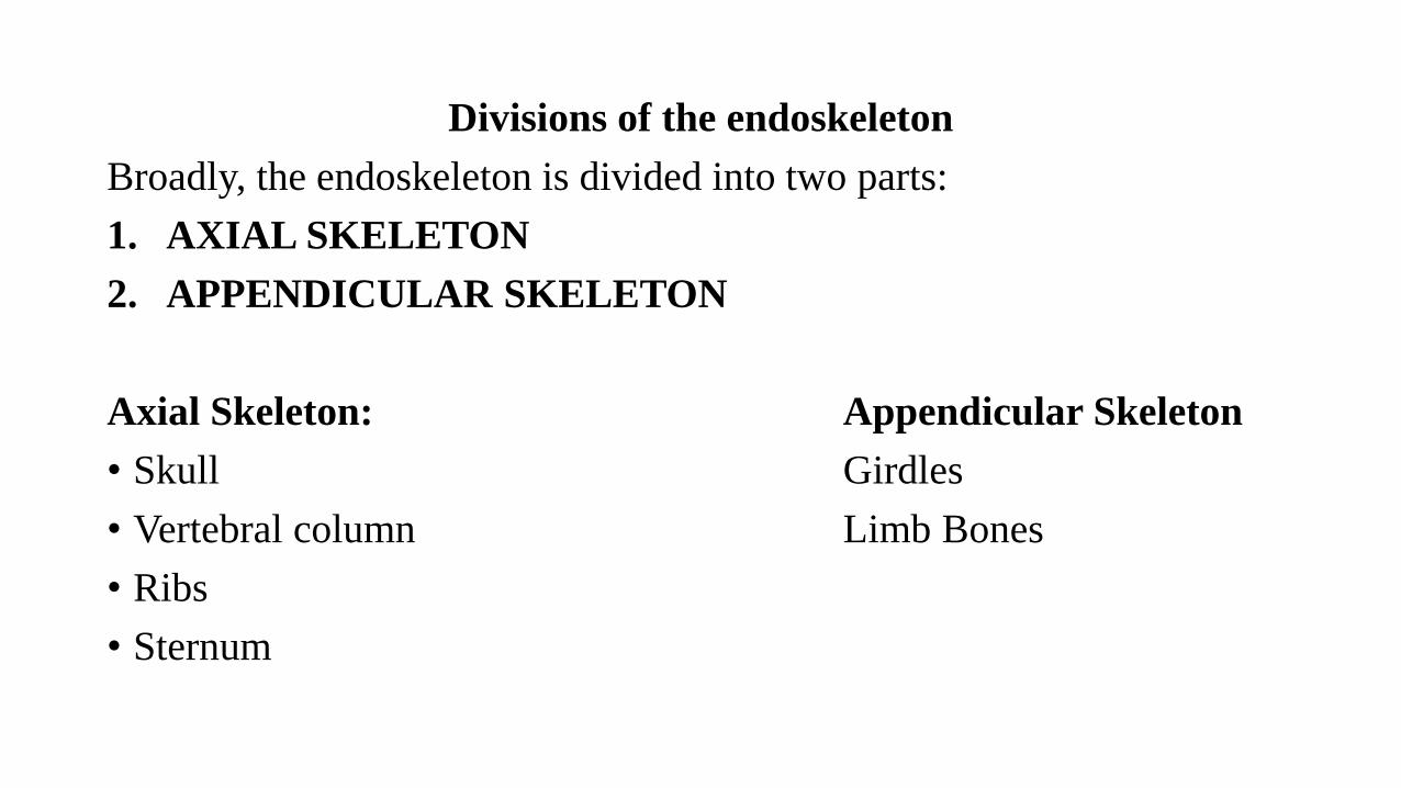
Divisions of the endoskeleton
Broadly, the endoskeleton is divided into two parts:
1. AXIAL SKELETON
2. APPENDICULAR SKELETON
Axial Skeleton: Appendicular Skeleton
• Skull Girdles
• Vertebral column Limb Bones
• Ribs
• Sternum
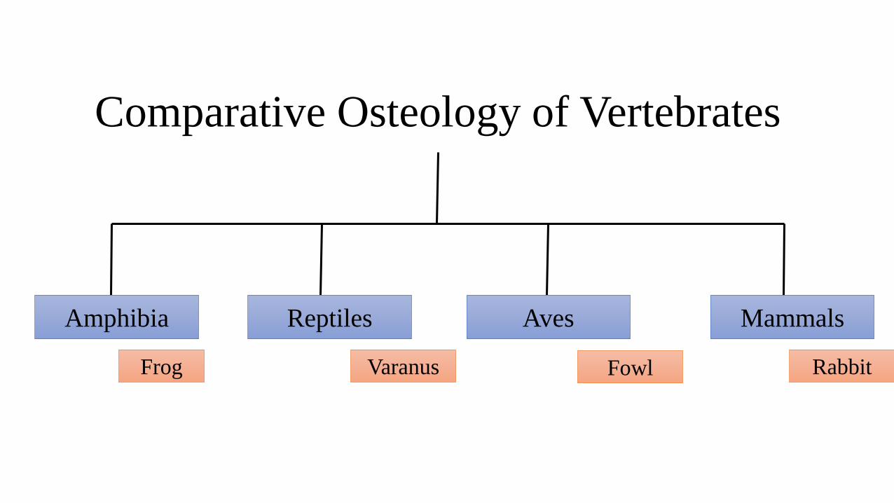
Comparative Osteology of Vertebrates
Amphibia Reptiles Aves Mammals
Frog Varanus Fowl Rabbit
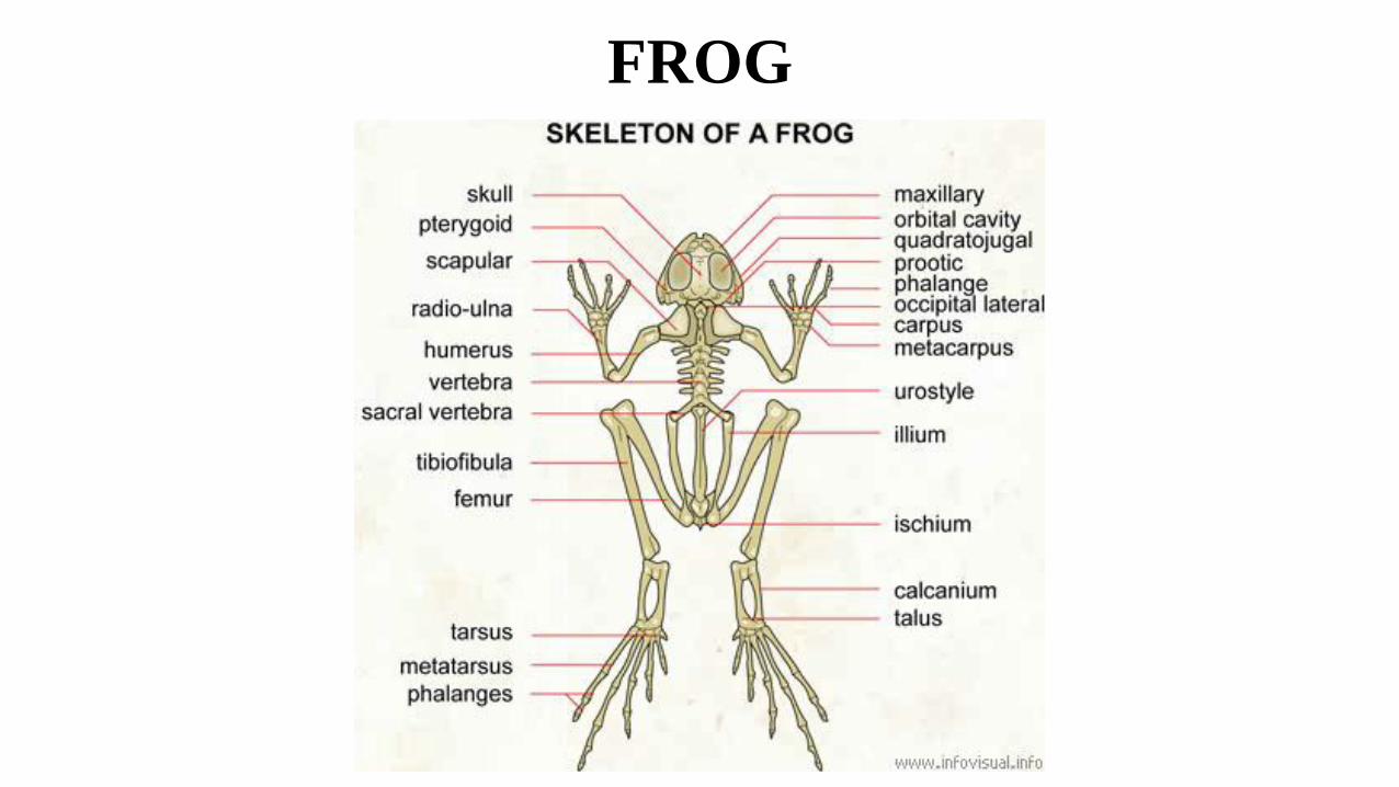
FROG
FROG SKULL• Dichondylic: At the posterior end of the cranium is a foramen
magnum surrounded by two exoccipitals. Each exoccipital bears at its posterior end a convexity, the occipital condyle which articulates with the concavity of the atlas vertebra.
Olfactory Capsules:
• The olfactory capsules have two nasals dorsally and two vomersventrally, the vomers bear vomerine teeth.
• A pair of special bones called septomaxillary (ethmoids) form the boundary of nostrils. They are associated with and surround the Jacobson’s organ.
Optic Capsules:
• They enclose the eyes and are not fused with the skull.

• Upper Jaw:
• The upper jaw has two halves, each half has an anterior premaxilla followed by a long maxilla, both bear teeth.
• The posterior part of the upper jaw has a small quadratojugal. Its broad posterior end unites with quadrate cartilage, which is a small thin rod forming the suspensorium.
• The mandible articulates with the quadrate cartilage.
• Ventral anterior to the orbit is a slender, rod-like palatine.
• At the posterior lateral end of cranium is present a large 3-rayed or Y-shaped pterygoid.
• It articulates anteriorly with the maxilla and palatine and on the inner side with the parasphenoid and auditory capsule, and posteriorly with the quadratojugal and quadrate cartilage.
• At the posterior dorsolateral end of cranium is the hammer-shaped bone, the squamosal.
• It lies above the pteryoid. Its anterior limb or head is free and the short posterior limb articulates with the auditory capsule and prootic. Its handle joins with the quadrate cartilage.
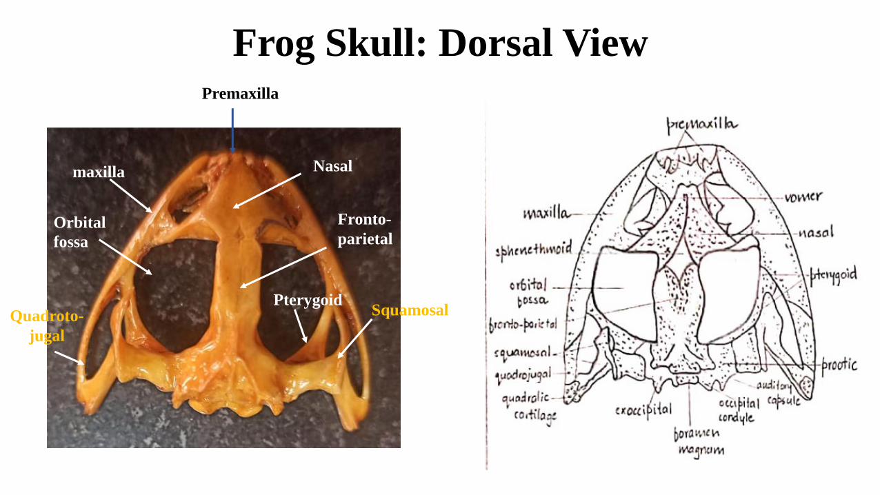
Frog Skull: Dorsal ViewPremaxilla
Nasal
Orbital
fossa
maxilla
Fronto-
parietal
Pterygoid SquamosalQuadroto-
jugal
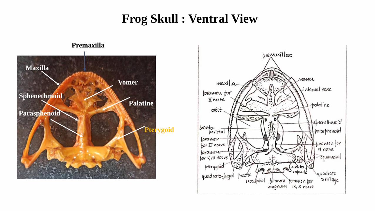
Frog Skull : Ventral View
Premaxilla
Maxilla
Vomer
Palatine
Pterygoid
Parasphenoid
Sphenethmoid
Frog: Lower Jaw
• Two rami joined in front by elastic ligament.
• Each half has a core of Meckel’s cartilage covered over by an angulosplenial forming the inner and posterior portion of each ramus.
• Just anterior to the condyle is present the coronary process.
• Anterior outer surface of Meckel’s cartilage is covered by a small, flat, dogger-like dentary.

Frog Lower JawDentary
Coronary
process
Articular
Junction
Angulosplenial

Frog Vertebrae
• Atlas Vertebra:
• The first vertebra
• It is ring-like in form.
• Centrum and neural spine are reduced.
• Transverse processes and prezygapophysis are absent.
• The neural arch is large.
• The anterior face of centrum possesses a pair of concave facets for the articulation with the occipital condyles of the skull. (dichondylic)
• The posterior margin of the neural arch bears a pair of postzygapophyses.
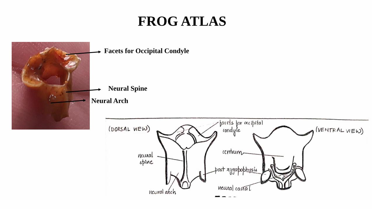
FROG ATLAS
Facets for Occipital Condyle
Neural Spine
Neural Arch

Frog: Typical Vertebra
Transverse Process
Neural Spine
Neural Canal
Neural Spine
Pre Zygapophysis
Post Zygapophysis
Centrum
• Typical Vertebra:
• The centrum is procoelous, i.e., it is concave in front and convex behind.
• On the dorsal side, the centrum bears a neural arch which encloses the neural canal.
• The neural arch possesses a backwardly directed spinous process or neural spine.
• The lateral sides of the neural arch carry transverse processes.
• The neural arch possesses two articulating processes. The prezygapophyses and the postzygapophyses.
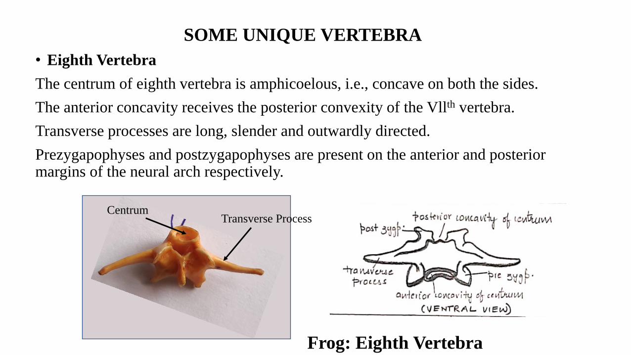
SOME UNIQUE VERTEBRA
• Eighth Vertebra
The centrum of eighth vertebra is amphicoelous, i.e., concave on both the sides.
The anterior concavity receives the posterior convexity of the Vllth vertebra.
Transverse processes are long, slender and outwardly directed.
Prezygapophyses and postzygapophyses are present on the anterior and posterior margins of the neural arch respectively.
Frog: Eighth Vertebra
Transverse ProcessCentrum
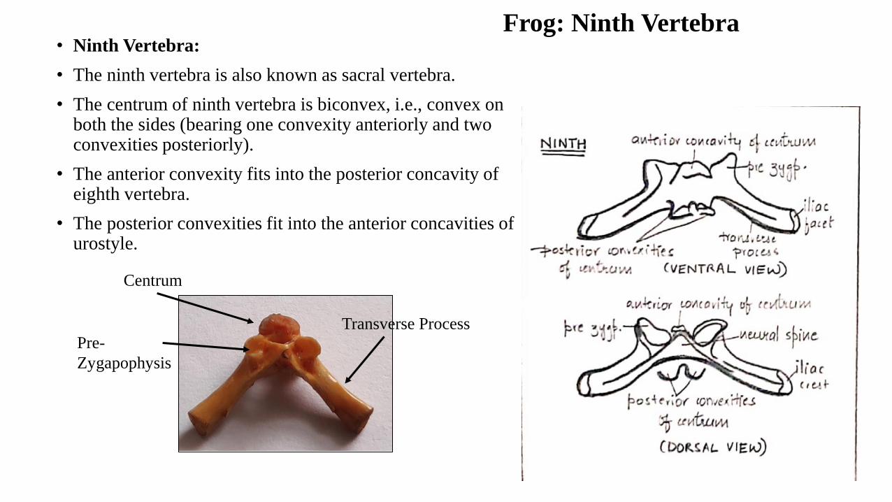
• Ninth Vertebra:
• The ninth vertebra is also known as sacral vertebra.
• The centrum of ninth vertebra is biconvex, i.e., convex on both the sides (bearing one convexity anteriorly and two convexities posteriorly).
• The anterior convexity fits into the posterior concavity of eighth vertebra.
• The posterior convexities fit into the anterior concavities of urostyle.
Frog: Ninth Vertebra
Centrum
Transverse Process
Pre-
Zygapophysis
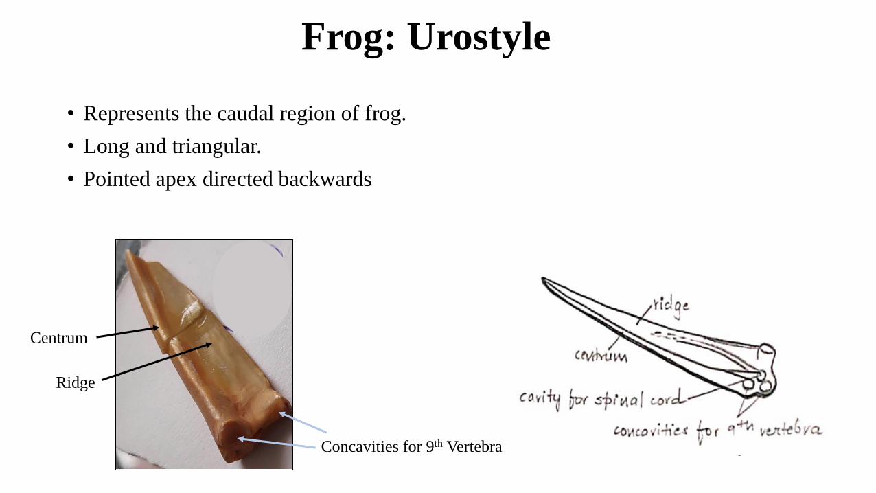
Frog: Urostyle
• Represents the caudal region of frog.
• Long and triangular.
• Pointed apex directed backwards
Centrum
Ridge
Concavities for 9th Vertebra
FROG PECTORAL GIRDLE
• Present in the thoracic region.
• Provides attachment to the forelimbs and their muscles.
• It consists of two similar halves permanently attached with sternum.
• Each half is divided into a dorsal scapular portion and a ventral coracoid portion.
• The scapular portion comprises the suprascapula and scapula.
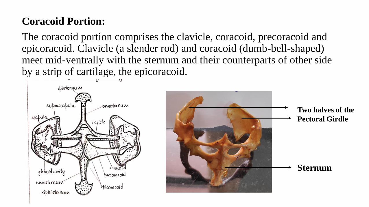
Coracoid Portion:
The coracoid portion comprises the clavicle, coracoid, precoracoid and epicoracoid. Clavicle (a slender rod) and coracoid (dumb-bell-shaped) meet mid-ventrally with the sternum and their counterparts of other side by a strip of cartilage, the epicoracoid.
Sternum
Two halves of the
Pectoral Girdle
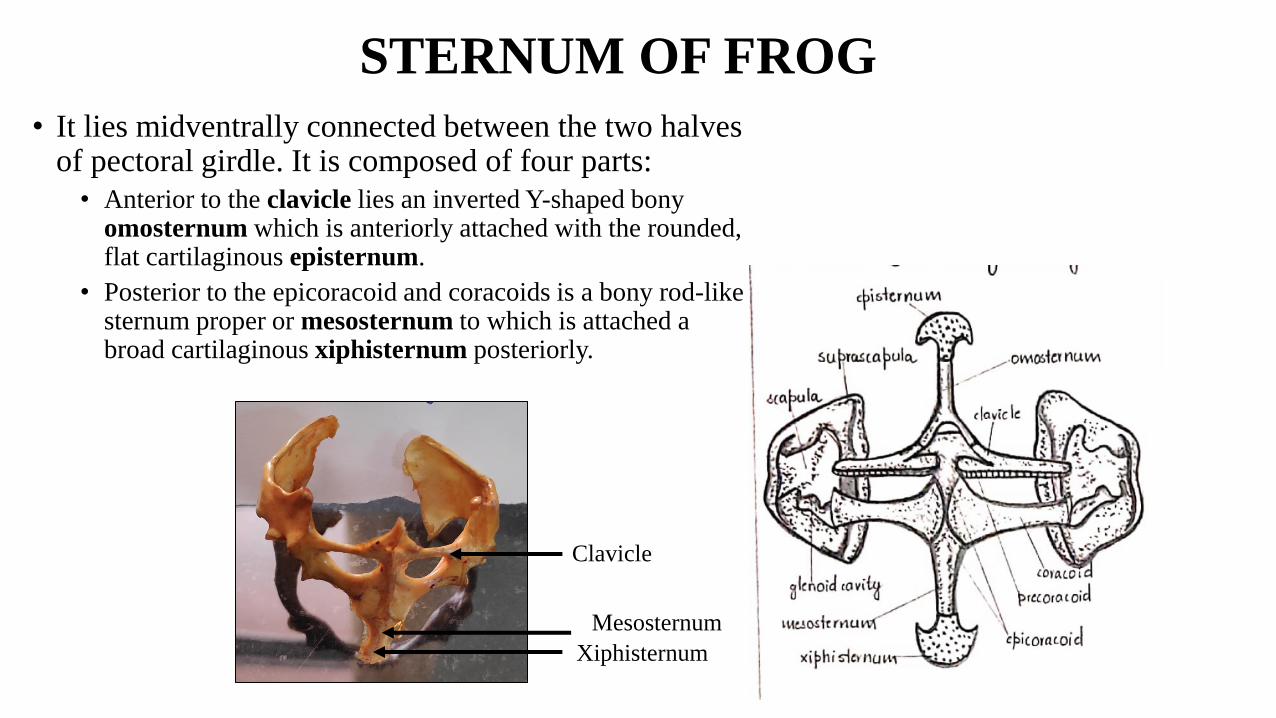
STERNUM OF FROG
• It lies midventrally connected between the two halves of pectoral girdle. It is composed of four parts:
• Anterior to the clavicle lies an inverted Y-shaped bony omosternum which is anteriorly attached with the rounded, flat cartilaginous episternum.
• Posterior to the epicoracoid and coracoids is a bony rod-like sternum proper or mesosternum to which is attached a broad cartilaginous xiphisternum posteriorly.
Xiphisternum
Clavicle
Mesosternum
PELVIC GIRDLE FROG
• It is V-shaped and composed of two similar halves, each of which is known as os-innominatum.
• Each os-innominatum is composed of three bones, ilium, pubis and ischium, which form the disc and the acetabulum.
• Ilium is greatly elongated and forms the major part of each os-innominatum. It runs forwards to meet the transverse process of the ninth vertebra.
• Pubis is much reduced. It is a triangular piece of calcified cartilage, forming the central part of the disc and a small part of the acetabulum. Both the pubes are also fused.
• Ischium is larger and slightly oval bone and both the ischia fused in the middle and form one- third part of the disc and acetabulum.

PELVIC GIRDLE FROG
Illiac Crest
Illium
Pubis
Ischium
Acetabulum
IlliumIllium
Articular Surface
for Urostyle
Acetabulum
Appendicular Skeleton: Limb Bones
Fore Limbs of Frog
1. HUMEROUS:
• short, cylindrical, slightly curved bone of upper arm.
• Proximal end fits into glenoid cavity of pectoral girdle. Swollen: forming the head, covered by calcified cartilage.
• Below head: deltoid ridge for muscle attachment.
• Distal end has a prominent trochlea or capitulum and condylar ridge for articulation with radio-ulna.
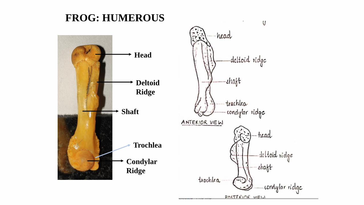
FROG: HUMEROUS
Head
Deltoid
Ridge
Shaft
Condylar
Ridge
Trochlea
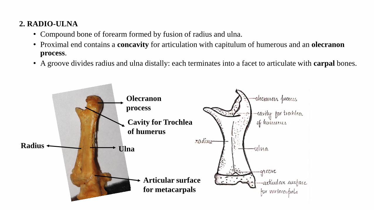
2. RADIO-ULNA
• Compound bone of forearm formed by fusion of radius and ulna.
• Proximal end contains a concavity for articulation with capitulum of humerous and an olecranon process.
• A groove divides radius and ulna distally: each terminates into a facet to articulate with carpal bones.
Olecranon
process
Cavity for Trochlea
of humerus
Ulna
Articular surface
for metacarpals
Radius

3. BONES OF HAND
• Wrist Bones: Carpals. 6 in number and arranged in two rows
of three each.
• Bones of proximal row: RADIALE, INTERMEDIUM,
ULNARE. Articulate with the radio-ulna
• Bones of Distal Row: TRAPEZIUM, TRAPEZOID and
CAPITOHAMATUM articulate with metacarpals.
• First metacarpal is rudimentary and without a digit and
phalanges.
• Digits are internally supported by short bony rods: phalanges.
• First and second digits contain two phalanges each, third and
fourth digits have 3 phalanges each.
• Claws are absent.
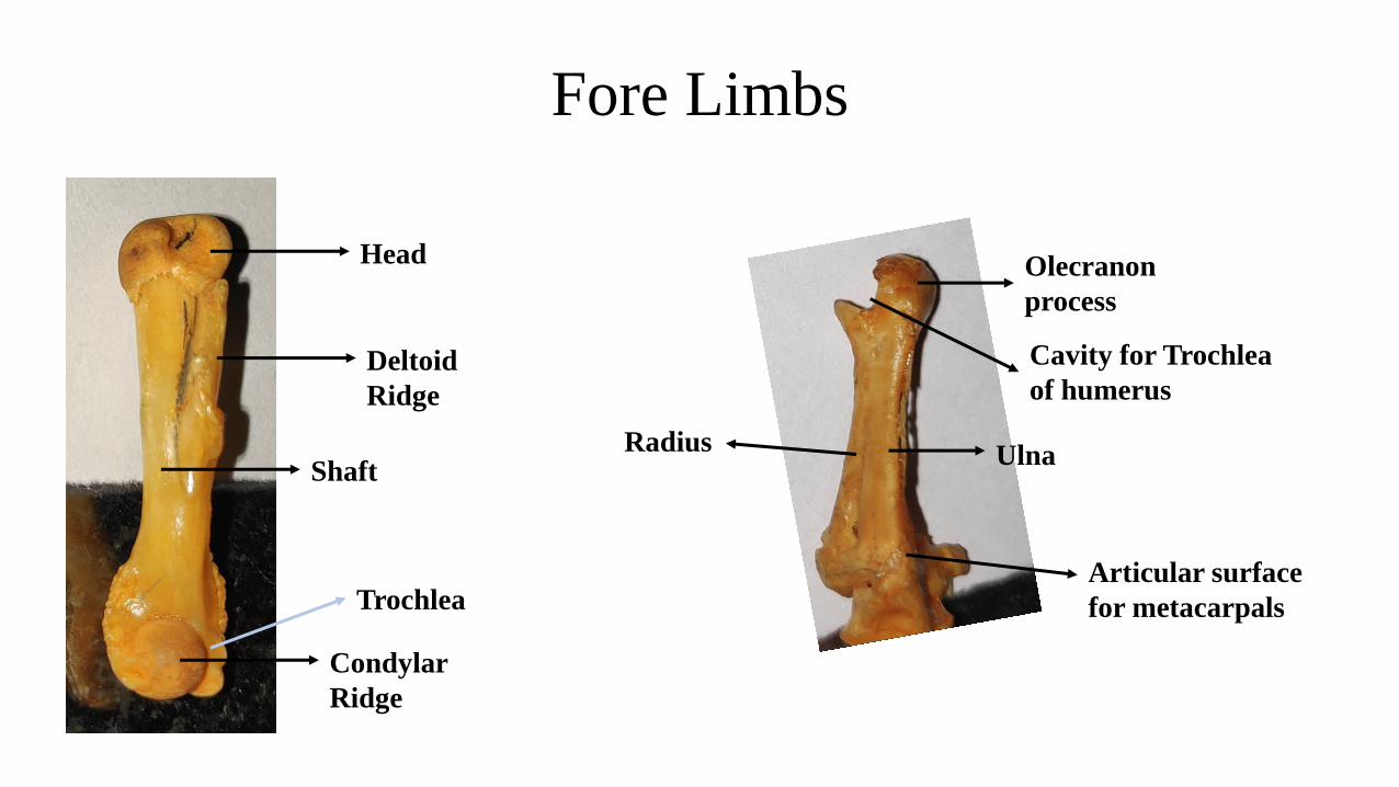
Fore Limbs
Head
Deltoid
Ridge
Shaft
Condylar
Ridge
Trochlea
Olecranon
process
Cavity for Trochlea
of humerus
Ulna
Articular surface
for metacarpals
Radius

FROG: HIND LIMBS
1. Femur:
• It is long and slender having a slightly curved shaft.
• The proximal swollen end is called the head. Headfits into the acetabulum of pelvic girdle forming aball and socket joint.
• The distal end forms a condyle which articulates with the tibio-fibula.
• Both proximal and distal ends have calcified cartilage.
Head
Shaft
Condyle
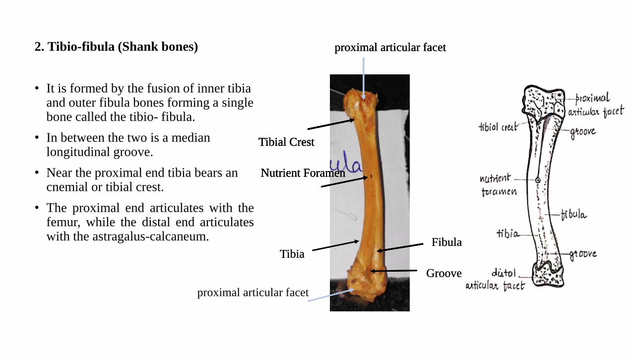
2. Tibio-fibula (Shank bones)
• It is formed by the fusion of inner tibia and outer fibula bones forming a single bone called the tibio- fibula.
• In between the two is a median longitudinal groove.
• Near the proximal end tibia bears an cnemial or tibial crest.
• The proximal end articulates with thefemur, while the distal end articulateswith the astragalus-calcaneum.
proximal articular facet
Tibial Crest
Nutrient Foramen
proximal articular facet
TibiaFibula
Groove
proximal articular facet
Tibial Crest
Nutrient Foramen
TibiaFibula
proximal articular facet
Tibial Crest
Nutrient Foramen
Tibial Crest
Groove
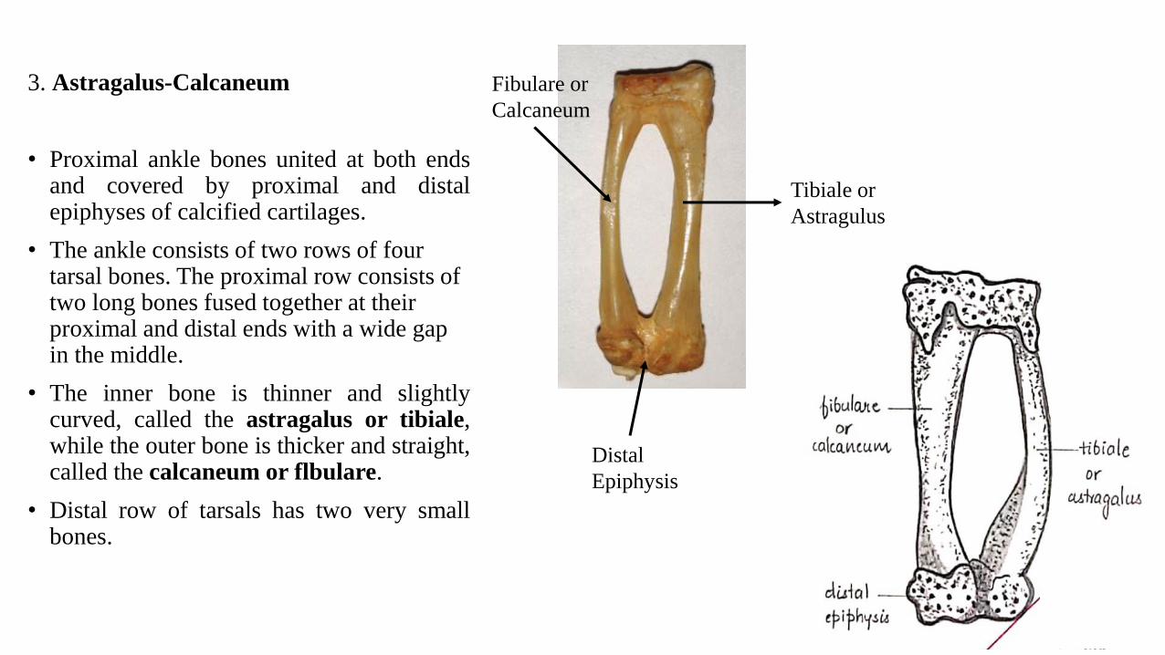
3. Astragalus-Calcaneum
• Proximal ankle bones united at both endsand covered by proximal and distalepiphyses of calcified cartilages.
• The ankle consists of two rows of four tarsal bones. The proximal row consists of two long bones fused together at their proximal and distal ends with a wide gap in the middle.
• The inner bone is thinner and slightlycurved, called the astragalus or tibiale,while the outer bone is thicker and straight,called the calcaneum or flbulare.
• Distal row of tarsals has two very smallbones.
Tibiale or
Astragulus
Fibulare or
Calcaneum
Distal
Epiphysis
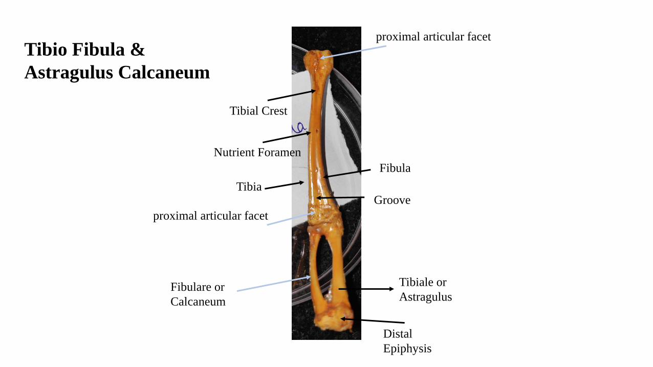
Tibio Fibula &
Astragulus Calcaneum
Tibial Crest
proximal articular facet
Nutrient Foramen
proximal articular facet
Fibula
GrooveTibia
Fibulare or
Calcaneum
Distal
Epiphysis
Tibiale or
Astragulus
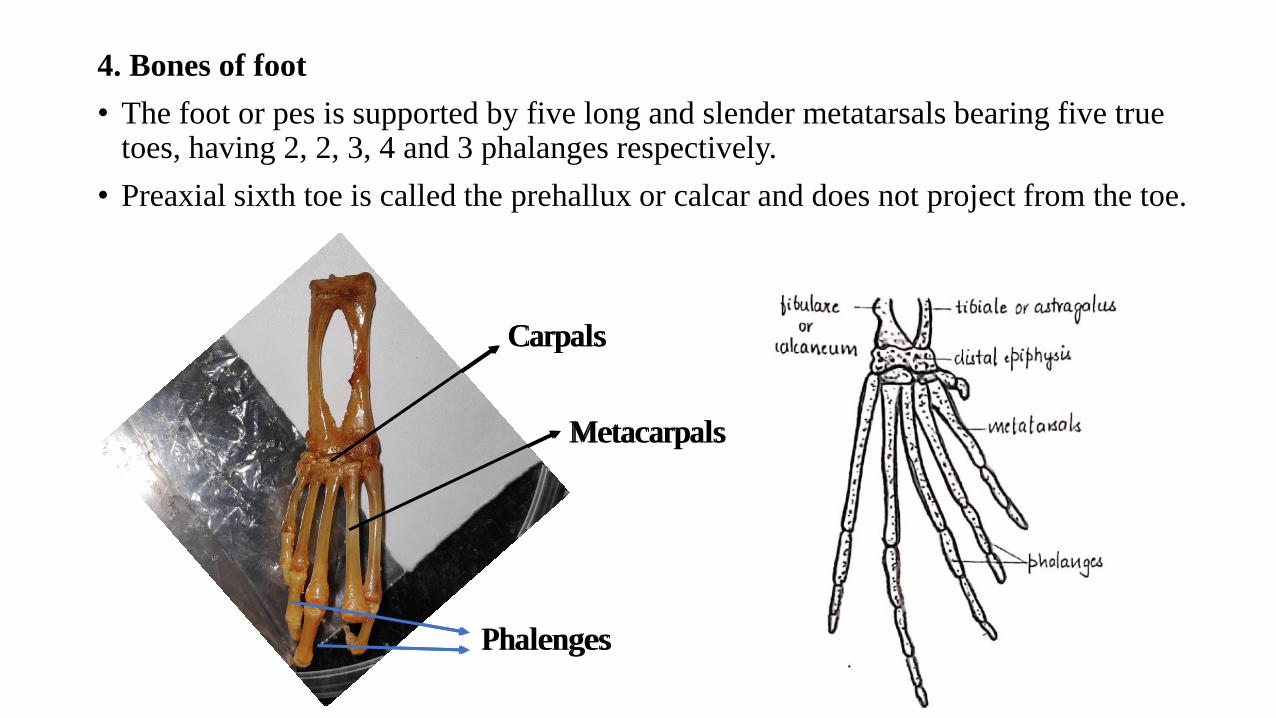
4. Bones of foot
• The foot or pes is supported by five long and slender metatarsals bearing five true toes, having 2, 2, 3, 4 and 3 phalanges respectively.
• Preaxial sixth toe is called the prehallux or calcar and does not project from the toe.
Carpals
Metacarpals
Phalenges
Carpals
Metacarpals
Phalenges
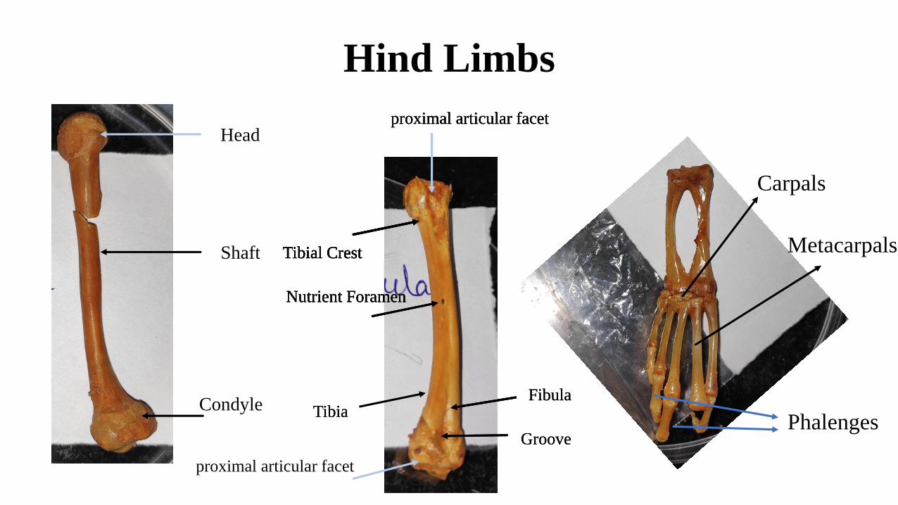
Hind Limbs
Head
Shaft
Condyle
proximal articular facet
Tibial Crest
Nutrient Foramen
proximal articular facet
TibiaFibula
Groove
proximal articular facet
Tibial Crest
Nutrient Foramen
TibiaFibula
proximal articular facet
Tibial Crest
Nutrient Foramen
Tibial Crest
Groove
Carpals
Metacarpals
Phalenges