Hind limb unloading, a model of spaceflight conditions, leads to decreased B lymphopoiesis similar...
10
The FASEB Journal • Research Communication Hind limb unloading, a model of spaceflight conditions, leads to decreased B lymphopoiesis similar to aging Chlo´ e Lescale,* , †,1 V´ eronique Schenten, ‡,1 Dounia Djeghloul,* , † Meriem Bennabi,* , † Fanny Gaignier, ‡ Katleen Vandamme, § Catherine Strazielle, ‡ Isabelle Kuzniak,* ,† Herv´ e Petite, § Christine Dosquet,* , † Jean-Pol Frippiat, ‡,2,3 and Michele Goodhardt* , †,2,3 *Institut Universitaire d’H´ ematologie, Universit´ e Paris 7 Denis Diderot, Paris, France; † Institut National de la Sant´ e et de la Recherche M´ edicale (INSERM) UMRS-940, Paris, France; ‡ EA7300, Stress Immunity Pathogens Laboratory, Lorraine University, Vandœuvre-l` es-Nancy, France; and § UMR CNRS 7052, Biom´ ecanique et Biomat´ eriaux Ost´ eo-articulaires, Facult´ e de M´ edecine Lariboisi` ere, Paris, France ABSTRACT Within the bone marrow, the endosteal niche plays a crucial role in B-cell differentiation. Because spaceflight is associated with osteoporosis, we investigated whether changes in bone microstructure induced by a ground-based model of spaceflight, hind limb unloading (HU), could affect B lymphopoiesis. To this end, we ana- lyzed both bone parameters and the frequency of early hematopoietic precursors and cells of the B lineage after 3, 6, 13, and 21 d of HU. We found that limb disuse leads to a decrease in both bone microstructure and the frequency of B-cell progenitors in the bone marrow. Although multi- potent hematopoietic progenitors were not affected by HU, a decrease in B lymphopoiesis was observed as of the common lymphoid progenitor (CLP) stage with a major block at the progenitor B (pro-B) to precursor B (pre-B) cell transition (5- to 10-fold decrease). The modifications in B lymphopoiesis were similar to those observed in aged mice and, as with aging, decreased B-cell generation in HU mice was associated with reduced expression of B-cell transcription factors, early B-cell factor (EBF) and Pax5, and an alteration in STAT5-mediated IL-7 signaling. These findings demonstrate that mechanical unloading of hind limbs results in a decrease in early B-cell differentiation re- sembling age-related modi fications in B lymphopoiesis.— Lescale, C., Schenten, V., Djeghloul, D., Bennabi, M., Gaignier, F., Vandamme, K., Strazielle, C., Kuzniak, I., Petite, H., Dosquet, C., Frippiat, J.-P., Goodhardt, M. Hind limb unloading, a model of spaceflight conditions, leads to decreased B lymphopoiesis similar to aging. FASEB J. 29, 000–000 (2015). www.fasebj.org Key Words: bone remodeling • B-cell differentiation • gravity • space conditions • immunosenescence HUMAN BIOASTRONAUTIC PROGRAMS have grown during the last 50 yr. Medical and physiologic findings from these missions have demonstrated that spaceflight impacts almost all physiologic systems, including muscle atrophy, bone demineralization, cardiovascular and metabolic dys- functions, impaired cognitive processes, and reduced im- munologic competence. These adaptive responses can affect crew health and performance both in space and upon return to Earth. Indeed, 15 of the 29 Apollo astro- nauts contracted bacterial or viral infections either during the mission or within a week of returning (1–3). Prolonged exposure to microgravity induces osteopenia, with de- creased bone formation and mineralization and increased bone resorption (4, 5), and there are presently no effective countermeasures to mitigate these problems. As a conse- quence, expanding our knowledge on the effects of long- duration spaceflight on crew health and performance is clearly a prerequisite for long-term spaceflight. Immune-competent B and T lymphocytes are derived from hematopoietic stem cells (HSCs) that reside in the bone marrow in specialized niches made up of bone and vascular structures, including bone-forming osteoblasts and bone-resorbing osteoclasts (6). Interactions between HSC and bone marrow niches control the balance between quiescence, self-renewal, and differentiation of HSC (7–9). Given the essential role of HSC-niche interactions in he- matopoietic regulation, it is likely that changes in bone microstructure during spaceflights may also lead to changes in the function and composition of mature blood cells. Because of the limitations both in the availability and the experimental protocols that can be carried out with samples from astronauts following space missions, HU in rodents has been developed as a ground-based model to study the effects of spaceflight. This model includes situational and confine- ment stress, cephalic fluid shifts, and non–load-bearing sta- tus of the hind limbs (10). Notably, HU induces a reduction in bone mass, associated with increased generation of adi- pocytes and decreased osteoblast differentiation, and mimics Abbreviations: BV/TV, bone volume/trabecular volume; CFU, colony-forming unit; CLP, common lymphoid pro- genitor; EBF, early B-cell factor; HSC, hematopoietic stem cell; HU, hind limb unloading; LMPP, lymphoid-primed multipotent progenitor; MPP, multipotent progenitor; pre-B, precursor B; pro-B, progenitor B; Tb.N, trabecular number; VH, heavy chain variable gene segment 1 These authors contributed equally to this study. 2 These senior authors contributed equally to this work. 3 Correspondence: M.G., UMRS1126, IUH, Hˆ opital Saint Louis, Paris, France. E-mail: michele.goodhardt@univ-paris- diderot.fr; and J.-P.F., EA7300, Stress Immunity Pathogens Laboratory, Lorraine University, Faculty of Medicine, 9 Avenue de la For ˆ et de Haye, F-54500 Vandœuvre-l` es-Nancy, France. E-mail: [email protected] doi: 10.1096/fj.14-259770 0892-6638/15/0029-0001 © FASEB 1 The FASEB Journal article fj.14-259770. Published online November 5, 2014.
-
Upload
independent -
Category
Documents
-
view
0 -
download
0
Transcript of Hind limb unloading, a model of spaceflight conditions, leads to decreased B lymphopoiesis similar...
The FASEB Journal • Research Communication
Hind limb unloading, a model of spaceflight conditions,leads to decreased B lymphopoiesis similar to aging
Chloe Lescale,*,†,1 Veronique Schenten,‡,1 Dounia Djeghloul,*,† Meriem Bennabi,*,†
Fanny Gaignier,‡ Katleen Vandamme,§ Catherine Strazielle,‡ Isabelle Kuzniak,*,†
Herve Petite,§ Christine Dosquet,*,† Jean-Pol Frippiat,‡,2,3 and Michele Goodhardt*,†,2,3
*Institut Universitaire d’Hematologie, Universite Paris 7 Denis Diderot, Paris, France; †Institut Nationalde la Sante et de la Recherche Medicale (INSERM) UMRS-940, Paris, France; ‡EA7300, Stress ImmunityPathogens Laboratory, Lorraine University, Vandœuvre-les-Nancy, France; and §UMR CNRS 7052,Biomecanique et Biomateriaux Osteo-articulaires, Faculte de Medecine Lariboisiere, Paris, France
ABSTRACT Within the bone marrow, the endostealniche plays a crucial role in B-cell differentiation. Becausespaceflight is associated with osteoporosis, we investigatedwhether changes in bone microstructure induced bya ground-based model of spaceflight, hind limb unloading(HU), could affect B lymphopoiesis. To this end, we ana-lyzed both bone parameters and the frequency of earlyhematopoietic precursors and cells of theB lineageafter 3,6, 13, and 21 d of HU. We found that limb disuse leads toa decrease in both bone microstructure and the frequencyof B-cell progenitors in the bone marrow. Although multi-potent hematopoietic progenitors were not affected byHU, a decrease in B lymphopoiesis was observed as of thecommon lymphoid progenitor (CLP) stage with a majorblock at the progenitor B (pro-B) to precursor B (pre-B)cell transition (5- to10-folddecrease).Themodifications inB lymphopoiesis were similar to those observed in agedmice and, as with aging, decreased B-cell generation inHUmice was associated with reduced expression of B-celltranscription factors, early B-cell factor (EBF) and Pax5,and an alteration in STAT5-mediated IL-7 signaling. Thesefindings demonstrate that mechanical unloading of hindlimbs results in a decrease in early B-cell differentiation re-sembling age-related modifications in B lymphopoiesis.—Lescale, C., Schenten, V., Djeghloul, D., Bennabi, M.,Gaignier, F., Vandamme, K., Strazielle, C., Kuzniak, I.,Petite, H., Dosquet, C., Frippiat, J.-P., Goodhardt, M. Hindlimb unloading, a model of spaceflight conditions, leadsto decreased B lymphopoiesis similar to aging. FASEB J.29, 000–000 (2015). www.fasebj.org
Key Words: bone remodeling • B-cell differentiation • gravity •
space conditions • immunosenescence
HUMAN BIOASTRONAUTIC PROGRAMS have grown during thelast 50 yr. Medical and physiologic findings from thesemissions have demonstrated that spaceflight impacts
almost all physiologic systems, including muscle atrophy,bone demineralization, cardiovascular and metabolic dys-functions, impaired cognitive processes, and reduced im-munologic competence. These adaptive responses canaffect crew health and performance both in space andupon return to Earth. Indeed, 15 of the 29 Apollo astro-nauts contracted bacterial or viral infections either duringthemissionorwithin aweekof returning (1–3). Prolongedexposure to microgravity induces osteopenia, with de-creased bone formation andmineralization and increasedbone resorption (4, 5), and there are presently no effectivecountermeasures to mitigate these problems. As a conse-quence, expanding our knowledge on the effects of long-duration spaceflight on crew health and performance isclearly a prerequisite for long-term spaceflight.
Immune-competent B and T lymphocytes are derivedfrom hematopoietic stem cells (HSCs) that reside in thebone marrow in specialized niches made up of bone andvascular structures, including bone-forming osteoblastsand bone-resorbing osteoclasts (6). Interactions betweenHSCandbonemarrownichescontrol thebalancebetweenquiescence, self-renewal, anddifferentiationofHSC(7–9).Given the essential role of HSC-niche interactions in he-matopoietic regulation, it is likely that changes in bonemicrostructure during spaceflightsmay also lead to changesin the function and composition of mature blood cells.
Because of the limitations both in the availability and theexperimental protocols that can be carried out with samplesfromastronauts following spacemissions,HU in rodents hasbeendevelopedas a ground-basedmodel to study theeffectsof spaceflight. This model includes situational and confine-ment stress, cephalic fluid shifts, and non–load-bearing sta-tus of the hind limbs (10). Notably, HU induces a reductionin bone mass, associated with increased generation of adi-pocytes and decreased osteoblast differentiation, andmimics
Abbreviations: BV/TV, bone volume/trabecular volume;CFU, colony-forming unit; CLP, common lymphoid pro-genitor; EBF, early B-cell factor; HSC, hematopoietic stemcell; HU, hind limb unloading; LMPP, lymphoid-primedmultipotent progenitor; MPP, multipotent progenitor; pre-B,precursor B; pro-B, progenitor B; Tb.N, trabecular number;VH, heavy chain variable gene segment
1 These authors contributed equally to this study.2 These senior authors contributed equally to this work.3 Correspondence: M.G., UMRS1126, IUH, Hopital Saint
Louis, Paris, France. E-mail: [email protected]; and J.-P.F., EA7300, Stress Immunity PathogensLaboratory, Lorraine University, Faculty of Medicine, 9 Avenuede la Foret de Haye, F-54500 Vandœuvre-les-Nancy, France.E-mail: [email protected]: 10.1096/fj.14-259770
0892-6638/15/0029-0001 © FASEB 1
The FASEB Journal article fj.14-259770. Published online November 5, 2014.
spaceflight-induced immunologic changes (11–14). Thus,exposure to HU induces profound alterations in skeletalmetabolism that could in turn affect hematopoiesis.
A number of previous observations support this hypoth-esis. Indeed, a decrease in the number of bone marrowmyeloid progenitors, as measured by colony-forming assays,was observed both in spaceflight and followingHU (15–19).In vitro culture of human CD34+ bone marrow progenitorsduring spaceflight confirmed the inhibitory effect of mi-crogravity on erythropoiesis and myelopoiesis (20), andmore recently, Ortega et al. (21) reported changes in thematuration/activation of granulocytic cells in the bonemarrow of C57BL/6 mice after a 13 d spaceflight. Despitewell-documented spaceflight-induced immune dysfunction,relatively few studies have investigated the effect of micro-gravity on humoral responses and B-cell differentiation. Wehave recently shown that the expression of Ig heavy chainandof the lymphoid-determining transcription factor Ikarosare modified when embryos of the urodele amphibianPleurodeles waltl are subjected to gravity changes, suggestinga modification in B lymphopoiesis (22). Spaceflight con-ditions further affect antibody production in response toantigenic stimulation in adult amphibians. Indeed, the ex-pression of IgY (the counterpart of mammalian IgA) wasincreased in flown P. waltl (23) as previously observed incosmonauts (24). Furthermore, the utilization of heavychain variable gene segment (VH) gene subgroups, the ex-pression of individual VH genes, and the frequency of Igsomatic hypermutation were found to be modified underspaceflight conditions (23, 25, 26).
Given the scarcity of data regarding the impact ofspaceflight on B-cell development, we investigated theeffects of HU on B-cell differentiation. The results of thisstudy reveal thatHUleads to adecrease inB lymphopoiesisstarting at the stage of early lymphoid-committed progen-itors. Interestingly, HU-induced modifications presentmany similarities with the changes observed in aging micebothwith regard to trabecular bonemicroarchitecture andto the cellular and molecular changes accompanying re-duced B cell differentiation.
MATERIALS AND METHODS
Animals
Experiments were conducted using young (3 mo) or old (18–22 mo) C57BL/6J male mice purchased from Janvier Labora-tories (Le Genest Saint Isle, France). Animals were housed invented animal cabinets (Noroit, Bouaye, France) under con-trolled temperature (22°C) and 12 h light-dark cycle. Prior tothe start of the experiments, mice were allowed to rest for 1 wkfollowing shipmentby groupsof 4mice in standard cages.Animalswere treated in accordance with the National Legislation, andexperimental protocols were approved by the local ethics com-mittee (Comite d’ethique en experimentation animale ParisLariboisiere-Villemin, permit number: CEEALV/2010-03-06;Comite d’Ethique Lorrain en Matiere d’ExperimentationAnimale, permit number: CELMEA-2012-0008).
Hind limb unloading
Mice (3 mo old) were isolated 5 d before the beginning of theexperiments in standardorHUcages (312mmin length, 197mm
in width, and 260 mm in height). Mice were suspended by usinga dressing retention sheet wrapped around the tail and a wirehooked on a swivel pulley system as described elsewhere (10, 12).The swivel pulley glides along 2 stainless steel rods that run thelength of the cage providing the mouse with a full 360° range ofmovement. The angle of suspension forHUmice was adjusted to25–30° such that only the forelimbs touch the grid at the bottomof the cage. Throughout the suspension period, mice were pro-vided with food and water ad libitum and weighed daily. Unsus-pended control mice were housed individually in cages of thesame size.Groupsof 3mice (HUandcontrols) were killed after 3,6, 13, and 21 d of suspension. Experiments were repeated 2 to 5times for each time point.
Immunoglobulin, corticosterone, andcytokine quantifications
Serum IgA, IgG, IgM, and corticosterone concentrations weredetermined using commercial ELISA kits (Bethyl LaboratoriesIncorporated, Montgomery, TX, USA, for Ig and Arbor Assays,Ann Arbor, MI, USA, for corticosterone) according to manu-facturer’s instructions. In each case, concentrations were calcu-lated using a 4-parameter curve. Cytokines (granulocytemacrophage-CSF, IFN-g, IL-1a, IL-2, IL-4, IL-5, IL-6, IL-10, IL-17, and TNF-a) were quantified using the Mouse Th1/Th2 10plex Flowcytomix Kit (eBioscience, Montrouge, France) and a2-laserFC500flowcytometer(BeckmanCoulter,Marseille,France).
Flow cytometry and cell sorting
Cell populations from the femur or the tibia were identifiedbased on the expression of the following markers: HSC (Lin2
IL-7Ra2 c-Kithi Sca-1hi Flt32), multipotent progenitor (MPP;Lin2 IL-7Ra2 c-Kithi Sca-1hi Flt3lo), lymphoid-primed multi-potent progenitor (LMPP; Lin2 IL-7Ra2 c-Kithi Sca-1hi Flt3hi),CLP (Lin2 IL-7Ra+ c-Kitlo Sca-1lo Flt3+), pro-B cells (B220lo
CD43+ CD19+ IgM2), pre-B cells (B220lo CD432 CD19+ IgM2),immature B cells (B220lo CD432 CD19+ IgM+), mature B cells(B220hi CD432 CD19+ IgM+). Freshly isolated bone marrowcells were treated with Fc-block (CD16⁄CD32). For the analysisof HSC, MPP, LMPP, and CLP, cells were first stained withbiotin-conjugated lineages antibodies [B220 (RA3-6B2), CD19(1D3 or 6D5), NK1.1 (PK136), CD11b (M1⁄70), Gr-1 (RB6-8C5), TER-119 (TER-119)], and then stained with anti–Sca-1-FITC (E13-161.7), anti–c-Kit-APC (2B8), anti–IL-7R-PECy7(A7R34), and anti–Flt3-PE (A2F10). Labeling with biotin-conjugatedantibodies was revealed using streptavidin-eFluor450. For theanalysis of pro-B, pre-B, immature, and mature B cells, bonemarrow cells were stained with anti–B220-A488 (RA3-6B2),anti–CD43-PE (S7), anti–CD19-APC (1D3), anti–IgM-biotin(R6-60.2), and anti-IL-7R-PECy7 (A7R34). Labeling with biotinanti-IgM was revealed using streptavidin-PerCPCy5.5. Cellpopulations from the thymus were identified based on theexpression of the followingmarkers: total T cells (CD3+), helperT cells (CD3+ CD4+), and cytotoxic T cells (CD3+ CD8+).Thymocytes were stained with anti–CD3«-APC (17A2),anti–CD4-PE (RM4-5), and anti–CD8a-PE-Cy7 (53-6.7). Cellswere analyzed using a FACS Canto II (BD Biosciences, Le Pontde Claix, France) or a FC500 (Beckman Coulter) flow cytom-eter and sorted with a MoFlo cell sorter (DAKO Cytomation,Glostrup, Denmark) or a FACSAria cell sorter (BD Bio-sciences). For cell sorting, 4 mice were pooled in each experi-ment and lineage-positive (Lin+) cells were first depleted bymagnetic-activated cell separation using streptavidin microbe-ads (Miltenyi Biotec, Paris, France) according to the manu-facturer’s instructions. All antibodies were purchased from BDBiosciences except anti–IL-7R-PECy7 (A7R34), anti–CD3«-APC
2 Vol. 29 February 2015 LESCALE ET AL.The FASEB Journal x www.fasebj.org

(17A2), anti–CD4-PE (RM4-5), and anti–CD8a-PE-Cy7 (53-6.7),which were from eBioscience and anti-CD19 (6D5) that wasfrom Beckman Coulter.
Colony forming unit assay
Bloodwas collected by a ventricular punctureprior to euthanasia.Red blood cells were lysed with a solution of ammoniumchloride(Stem Cell Technologies, Grenoble, France). Cells were washedin Iscove’s modifiedDulbecco’s medium containing 2% fetal calfserum. Cells (23 105) were seeded in 1mlmethylcellulose-basedmedium containing recombinant cytokines (stem cell factor,IL-3, IL-6, and Epo-GF M3434; Stem Cell Technologies). Eachsample was analyzed in duplicate. The total number of colonieswas determined after 12 d of culture at 37°C.
Intracellular staining of phospho-STAT5
Freshly isolated bone marrow cells from the femur or tibia ofcontrol and HU mice were depleted of IgM+ cells by magneticseparation using biotin-conjugated IgM antibody (R6-60.2) andstreptavidin microbeads. IgM2 cells were then incubated withCD19 microbeads and enriched for CD19+ cells (.90% CD19+
IgM2 cells after purification). Cells were starved in Opti-MEMwith0.1%serumfor1hat37°Candthenstimulatedwith100ng/mlmouse recombinant IL-7 (R&D Systems Europe, Lille, France)for 15 min at 37°C. Immediately after stimulation, cells werefixed in BD Phosflow Lyse⁄Fix Buffer for 10 min at 37°C. Cellsthen were washed and resuspended in BD Phosflow Perm IIIBuffer for 30 min at 4°C. After washing, cells were treated withFc-block (CD16⁄CD32) and stained simultaneously with anti-bodies for phospho-STAT5 (clone 47) and cell surface markersB220 (RA3-6B2) and CD43 (S7). Purified CD19+ IgM2 cellswere gated on B220loCD43+ population and the percentage ofphospho-STAT5+ cells was determined. All antibodies werepurchased from BD Biosciences.
Quantitative RT-PCR
RNA was extracted from sorted cells using the RNeasy Micro kit(Qiagen, Courtaboeuf, France) and reverse transcribed usingSuperScript III (Invitrogen, Cergy Pontoise, France) and randomhexamers. Quantitative RT-PCRs were performed using TaqManGene Expression Assays (Applied Biosystems, Courtaboeuf,France): Hprt1, Mm00-446968_m1; early B-cell factor (Ebf1),Mm00395519_m1; Pax5 (custom assay), forward primer:AGCCATGGTTGTGTCAGCAA, reverse primer: CCCGGCTT-GATGCTTCCT; FAM dye-labeled TaqManMGB probe, TCATA-ATACCTGCCAAGAATT; Gata3, Mm00484683_m1; Sfpi1(PU.1), Mm00488142_m1; Il-7ra, Mm00434295_m1; Tcf3(E2A), Mm01175588_m1; Hprt1, Mm00446968_m1; ActB,Mm01205647_g1. The cycling protocol was 2 min at 50°C,10 min at 95°C, and then 45 cycles of 15 s at 95°C and 1 min at60°C. Detection was performed using the ABI Prism 7000 Se-quence Detection System (Applied Biosystems). Each quan-titative RT-PCR was performed in triplicate, and the relativeexpression of each transcript was calculated with ABI Prism7000 Sequence Detection System software and normalized tothe amounts of Hprt1 and ActB mRNAs.
Micro-X-ray computed tomography
Tibias were fixed in 4%paraformaldehyde andpreserved in 70%ethanol until analysis. To evaluate bone architecture, proximalparts of the tibiaeof control,HU,andagedmicewere subjected to
a Skyscan 1172 desktopMicroCT (Skyscan, Aartselaar, Belgium).TheSkyscan1172machine is equippedwithan80kVX-ray sourcewith a camera pixel size of 9 mm. Micro-computed tomography(m-CT) images were made within the midsagittal planes in themetaphyseal region of the bones. During the scan, tibias wereimmobilized in a customized polystyrene holder. Scans resultedin reconstructed3-dimensional data setswithapixel sizeof 6.71mm(power setting at 60 kV to 100 mA). Data were subsequentlyquantified using the CT-Analyzer automated image analysissystem (Skyscan). The growth plate was used as reference todetermine the trabecular volume of interest (VOI) in the axialdirection. The region of interest was set with its closest edgeat 500 mm distally from the growth plate and over a length of1500mm(=223 slices). The distance of 500mmwas chosen suchthat no primary spongiosa was included in trabecular VOI. Foreach transverse slice, VOIwas establishedmanually in an area oftrabecular bone as large as possible. The registered gray-valueimages were segmentedusing aGaussianfilter into binary images,using a low-pass filter to remove noise and a fixed threshold(23.6% of maximal gray-scale value) to extract the mineralizedbone phase. Bone volume/trabecular volume fraction (BV/TV)and trabecularnumber (Tb.N)were calculated3-dimensionally asmeasures of trabecular bone mass and distribution.
Statistical analysis
Comparisons between 2 groups were performed using the Stu-dent’s t test when normality and homogeneity of variances wereascertained. When this was not the case, comparison between 2groups were performed using the Mann-Whitney test. P values,0.05 indicate significant difference.
RESULTS
Kinetics of bone microarchitectural alterations andevaluation of stress during HU
As previously reported, mechanical unloading led to sub-stantial changes in tibia bone mass and microarchitecture(Fig. 1A). A significant decrease in BV/TV and Tb.N wasobserved as of day 6, and bone morphometric parameterswere progressively reduced up to day 21 of HU (Fig. 1A).We also studied the evolution of body weight during theexperimental period. As noted by others (12, 27, 28), HUmice lost approximately 10% body weight in the first2dand theirweight remainedbelow thatof controls duringthe 21 d of HU (Fig. 1B). To determine whether in ourhands HU induces a stress response, we quantified corti-costerone, the major stress hormone in rodents, as well ascytokines regulating immuneresponses in the serumofHUmice. Our results show that serum corticosterone concen-trations were constant throughout the 21 d of suspensionand close to levels observed in unstressedmice (50 ng/ml)(Fig. 1C). Furthermore, cytokine concentrationsmeasuredat days 3, 6, 13, and 21 remained below detection level inbothHU and controlmice (data not shown). These data,togetherwithbehaviorexamination(C.Strazielle,unpublishedobservations), indicatethatmiceadaptedtoHUinless than3d.
Increased egress of hematopoietic stem/progenitorcells following HU
HSC and early progenitors interact with specialized nichecells located close to the bone endosteum.Given that bone
DECREASED B LYMPHOPOIESIS UPON HIND LIMB UNLOADING 3

parameters are strongly affected byHU, we considered thepossibility that these changes may modify interactions be-tween hematopoietic progenitors and endosteal stromal
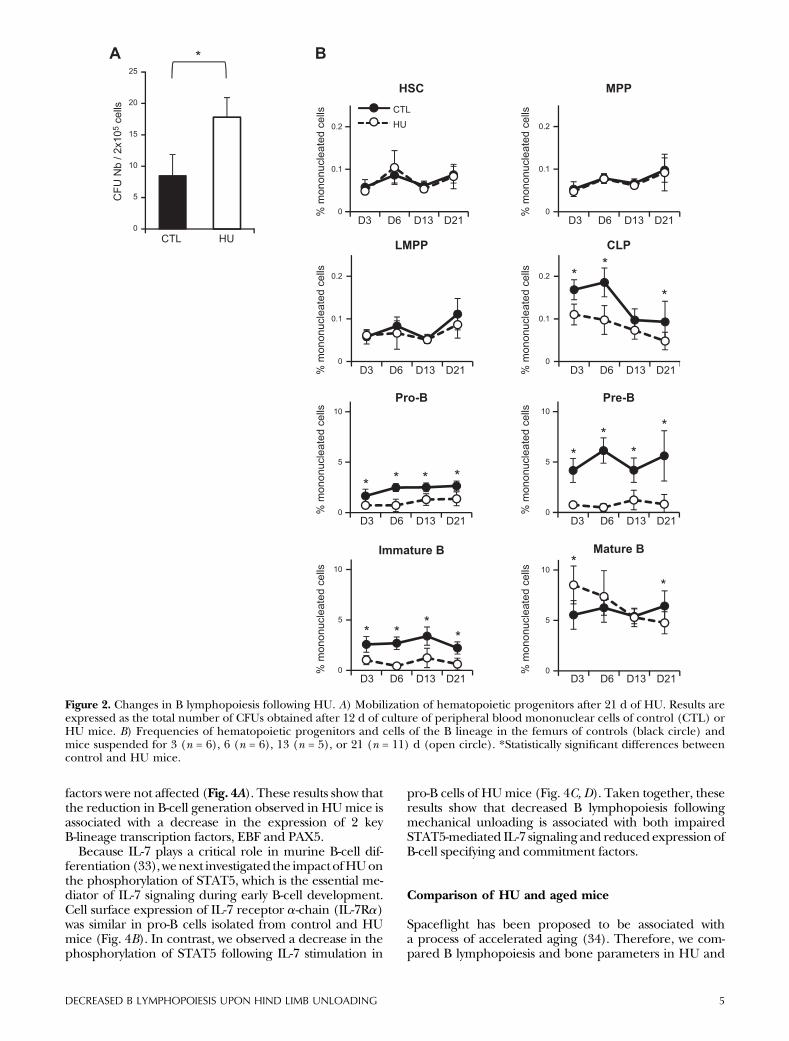
cells leading to enhanced egress of hematopoietic cells outof the bone marrow. To investigate the effect of mechani-cal unloadingonHSCmobilization, we compared the levelof circulating hematopoietic stem and progenitor cells inperipheral blood of control and HU mice by colony-forming unit (CFU) assays. As shown in Fig. 2A, there wasa significant increase in the number of colony-formingunit-granulocyte/macrophage in the peripheral blood ofmice subjected toHU.These results indicate thatHU leadsto increased release of hematopoietic progenitors to thecirculation, suggesting that interactions with the endostealniche may be impaired.
HU leads to decreased B lymphopoiesis
To investigate whether HU-associated alterations in tra-becular bone microarchitecture are associated withchanges in hematopoiesis and B-cell generation, we firstquantified the frequency of early hematopoietic pro-genitors and B-lineage committed cells in the bonemarrow of HU and control mice by flow cytometry.During B cell differentiation, HSC give rise to MPPs andthen to LMPPs and CLPSs, which in turn can give rise toB-committed pro-B and pre-B cell precursors and finallyto immature and mature B cells (29–31). There was nochange in the frequency ofmultipotentHSCs,MPPs, andLMPPs in the bone marrow following HU (Fig. 2B).However, we found a significant decrease in the fre-quency of early lymphoid (CLP) and B-committed pro-genitors as of day 3 of HU, and levels of these cellsremained low throughout the 21 d of mechanicalunloading (Fig. 2B). CLP and pro-B cells were decreasedapproximately 2-fold in the bone marrow of HU mice,and the largest decrease was observed within the pre-Bcell compartment, which was reduced 5- to 10-fold(Fig. 2B). Newly formed B220lo IgM+ B cells and matureB220hi IgM+ cells were also significantly decreased fol-lowing HU, although to a lesser extent than pre-B cells,probably due to homeostatic compensatorymechanisms.No change in serum Ig was observed during the21 d period of HU investigated (Fig. 3). Taken together,these data indicate that HU leads to impaired bonemarrow B lymphopoiesis.
HU-induced decreased B lymphopoiesis is associatedwith reduced Ebf1 and Pax5 expression and alteredIL-7 signaling
Lineage commitment and subsequent differentiation ofhematopoietic progenitors is controlled by a network oftranscription factors (32). PU.1 is a regulator of earlyhematopoietic progenitor lymphoid/myeloid lineagechoice. The lymphoid transcription factors E2A, EBF, andPAX5 are essential for B-lineage specification and com-mitment, and GATA3 is required for T-cell commitmentand differentiation. To investigate the molecular mecha-nisms underlying the decline in B lymphopoiesis observedfollowing mechanical unloading, we first compared theexpression of these transcription factors in CLP isolatedfrom control and 6 d HU mice. We found a 2- to 3-folddecrease in the level of Ebf1 and Pax5 transcripts in CLP ofHU mice, although expression of the other transcription
-40
-30
-20
-10
0
10
20
* * **
*
A
C
24
26
28
30
32
34
HUCTL
Wei
ght (
g) * * * * * * * * * * * ** ** * * * *
B
0
10
20
30
40
50
60
70
80
90
100
D3
Cor
ticos
tero
ne (n
g/L)
CTLHU
D0 D1 D2 D3 D4 D5 D6 D7 D8 D9D10 D11D12 D13 D14 D15 D16D17 D18 D19 D20 D21
% d
iffer
ence
from
Con
trol
D6 D13 D21
Bonevolume(BV/TV)
Trabecularnumber(Tb.N)
HU D3
HU D6
HU D13
HU D21
Figure 1. Kinetics of bone parameters, body weight, and serumcorticosterone levels during HU. A) Evolution of BV/TV andTb.N in the tibia of mice subjected to 3 (n = 6), 6 (n = 12), 13(n = 10), or 21 d (n = 6) of HU. Values are expressed as percentdifference relative to controls (CTLs). *Statistically significantdifferences compared with CTLs after statistical treatment asdescribed under Materials and Methods. B) Evolution of micebody weight during HU. Day (D) 0 corresponds to thebeginning of HU. Values represent means 6 SEM with n = 24from D0 to D3, n = 18 from D4 to D6, n = 12 from D7 to D13,and n = 6 from D14 to D21. *Statistically significant differencesbetween CTL and HU mice at each time point. C) Serumcorticosterone concentration after 3, 6, 13, and 21 d of HU. Thecorticosterone kinetic was repeated twice with n = 3 for eachtime point. Histograms represent mean 6 SEM. No statisticallysignificant difference was observed at any time point.
4 Vol. 29 February 2015 LESCALE ET AL.The FASEB Journal x www.fasebj.org
factors were not affected (Fig. 4A). These results show thatthe reduction in B-cell generation observed in HUmice isassociated with a decrease in the expression of 2 keyB-lineage transcription factors, EBF and PAX5.
Because IL-7 plays a critical role in murine B-cell dif-ferentiation(33),wenext investigated the impactofHUonthe phosphorylation of STAT5, which is the essential me-diator of IL-7 signaling during early B-cell development.Cell surface expression of IL-7 receptor a-chain (IL-7Ra)was similar in pro-B cells isolated from control and HUmice (Fig. 4B). In contrast, we observed a decrease in thephosphorylation of STAT5 following IL-7 stimulation in
pro-B cells of HUmice (Fig. 4C, D). Taken together, theseresults show that decreased B lymphopoiesis followingmechanical unloading is associated with both impairedSTAT5-mediated IL-7 signaling and reduced expression ofB-cell specifying and commitment factors.
Comparison of HU and aged mice
Spaceflight has been proposed to be associated witha process of accelerated aging (34). Therefore, we com-pared B lymphopoiesis and bone parameters in HU and
0
0.1
0.2
HSC
0
0.1
0.2
CLP
0
0.1
0.2
MPP
0
0.1
0.2
LMPP
0
5
10
0
5
10
0
5
10
0
5
10
**
****
*
**
*****
CTLHU
0
5
10
15
20
25
CTL HU
BA
**
*
*
CFU
Nb
/ 2x1
05 c
ells
Pro-B Pre-B
Immature B Mature B
D3 D6 D13 D21 D3 D6 D13 D21
D3 D6 D13 D21 D3 D6 D13 D21
D3 D6 D13 D21 D3 D6 D13 D21
D3 D6 D13 D21 D3 D6 D13 D21
% m
onon
ucle
ated
cel
ls
% m
onon
ucle
ated
cel
ls
% m
onon
ucle
ated
cel
ls
% m
onon
ucle
ated
cel
ls
% m
onon
ucle
ated
cel
ls
% m
onon
ucle
ated
cel
ls
% m
onon
ucle
ated
cel
ls
% m
onon
ucle
ated
cel
ls
Figure 2. Changes in B lymphopoiesis following HU. A) Mobilization of hematopoietic progenitors after 21 d of HU. Results areexpressed as the total number of CFUs obtained after 12 d of culture of peripheral blood mononuclear cells of control (CTL) orHU mice. B) Frequencies of hematopoietic progenitors and cells of the B lineage in the femurs of controls (black circle) andmice suspended for 3 (n = 6), 6 (n = 6), 13 (n = 5), or 21 (n = 11) d (open circle). *Statistically significant differences betweencontrol and HU mice.
DECREASED B LYMPHOPOIESIS UPON HIND LIMB UNLOADING 5

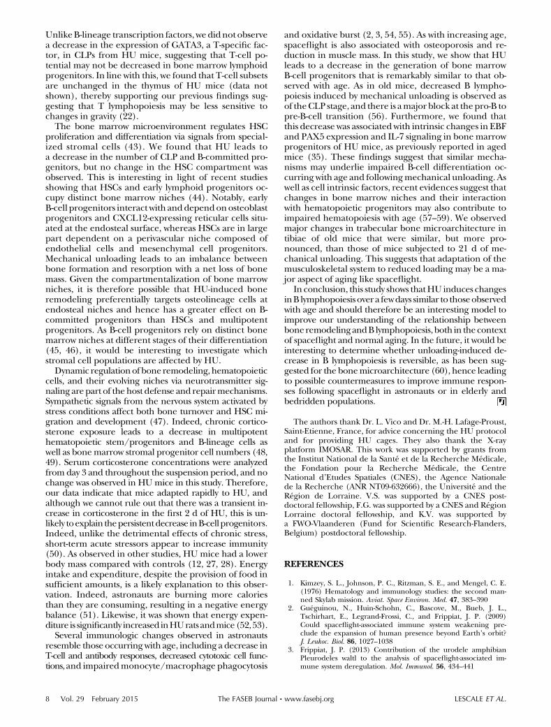
aged mice. Our analysis of hematopoietic progenitorsshowed that thewell-documented increase inHSCwithagewas not reproduced following HU (Fig. 5A). However, weobserved a strikingly similar profile of B-lineage cells in the
bonemarrowofHUand agedmice (Fig. 5B).As previouslyreported (35, 36), there is a decrease in B-cell differentia-tion starting at the CLP cell stage and a more pronouncedreduction at the pre-B cell transition in oldmice (.18mo)
IgG
0
200
400
600
800IgM
0
100
200
300IgA
0
500
1000
1500
2000
2500
IgM
(μg/
mL)
IgG
(μg/
mL)
IgA
(μg/
mL)
HUCT
HUCT
HUCT
D3 D6 D13 D21 D3 D6 D13 D21 D3 D6 D13 D21
Figure 3. Serum immunoglobulin concentrations after 3, 6, 13, or 21 d of HU. n = 6 at each time point for control and HU mice.Histograms represent mean 6 SEM. HU mice were compared with controls at each time point. No statistically significantdifference was observed at any time point.
STAT5-P CTL HU
% S
TAT5
-P+
% o
f Max
C D
% o
f Max
BIsotype CTL
HU
A
OLD
*
0
10
20
30
40
50
60
70
80
90
100
0
0.2
0.4
0.6
0.8
1
1.2
Gata3
CTL
HU
OLD
PU.1 E2a Ebf1 Pax5 ActB
IL-7Rα
HU– IL-7
CTL– IL-7
CTL+ IL-7
HU+ IL-7
100
80
60
40
20
0
100
80
60
40
20
00 103 104 105 0 103 104 105
Figure 4. Changes in transcription factor expression and IL-7 signaling in HU and aged mice. A) Expression of lymphoidtranscripts in CLP purified from the bone marrow of mice subjected to 6 d of HU, corresponding young controls (CTLs) andaged mice (.18 mo). mRNAs levels were determined by quantitative RT-PCR and normalized with respect to Hprt1. Results areexpressed relative to CLP of young CTLs (set to 1). Four mice per group were pooled for each experiment, and 2 independentexperiments were performed in triplicate. B) Expression of IL-7Ra on pro-B cells of CTL (solid line) or 6 d HU (dashed line)mice. C) Phosphorylation of STAT5 in response to IL-7 in pro-B cells of CTL (solid line) or 6 d HU (dashed line) mice. D)Percentage of STAT5-P positive pro-B cells after stimulation with IL-7 in CTL (black circle), 6 d HU (open circle), and aged(open triangle) mice. Each point corresponds to 1 mouse. Horizontal lines represent the median. *P , 0.05.
6 Vol. 29 February 2015 LESCALE ET AL.The FASEB Journal x www.fasebj.org

as in mice subjected to HU. Notably, in both HU and agingmice, the decrease in B lymphopoiesis is associated withdecreasedexpressionofEBFandPax5 inCLPand impairedIL7-mediated STAT-5phosphorylation inB-cell progenitors[Fig. 4; Lescale et al. (35)]. We also compared the effects ofage and mechanical unloading on trabecular bone archi-tecture using m-CT analysis. Our results revealed a sub-stantial decreaseboth in theBV/TVand inTb.Nofoldmice(.18mo of age) that was very similar to that observed after21 d of HU. However, trabeculae of old mice were thickerand more dispersed than in HU mice (Fig. 5C, D). Theseresults indicate that HU-induced changes in trabecularbone microarchitecture are similar but less pronouncedthan those occurring with age and are associated witha comparable impairment in B lymphopoiesis.
DISCUSSION
In this study, we show that mechanical unloading causesboth bone remodeling and a concomitant decrease inthe generation of B lymphocytes in the bone marrow.Decreased B lymphopoiesis was observed rapidly after HU
and B-cell progenitor populations remained low during the21 d of unloading. In contrast, no change was observed inHSC ormultipotent hematopoietic progenitor populations.
The differentiation of B cells fromHSC is dependent oninteractions with bone marrow stromal cells and is regu-lated by IL7 signaling and by the coordinated action ofa network of transcription factors. We found that the de-crease in B lymphopoiesis following HU was associatedwith reduced expression of EBF and PAX5 in early lym-phoid progenitors (CLPs) and impaired STAT5-mediatedIL-7 signaling in B cell precursors of HU mice. The tran-scription factors EBF and PAX5 are key regulators of B-celldifferentiation (32). EBF plays a crucial role in B-lineagespecification and, together with PAX5, in subsequentcommitment and differentiation steps (37–41), and IL7-receptor signaling and activation of STAT5 have beenshown to promote the survival of pro-B cells and facili-tate pre-B-cell expansion (42). Therefore, reduced IL7-signaling and expression of EBF and PAX5 followingmechanical unloadingmayunderlie thedecrease in earlyB-cell progenitors and impaired pro-B to pre-B-cell dif-ferentiation observed in the bone marrow of these mice.
C
-60
-40
-20
0
20
40
**
**
*
* *
% d
iffer
ence
from
Con
trol
HU miceOld mice
D
% m
onon
ucle
ated
cel
ls
0
0.05
0.1
0.15
0.2
0.25
0.3
YO
UN
G
HU
OLD
YO
UN
G
HU
OLD
YO
UN
G
HU
OLD
YO
UN
G
HU
OLD
HSC
0
1
2
3
4
5
6
7
8
9
10
YO
UN
G
HU
OLD
YO
UN
G
HU
OLD
YO
UN
G
HU
OLD
YO
UN
G
HU
OLD
Pro-B
B%
mon
onuc
leat
ed c
ells
YOUNG OLD
A *
**
***
****
-80
MPP LMPP CLP Pre-B Immature B Mature B
Bonevolume(BV/TV)
Trabecularnumber(Tb.N)
Trabecularthickness(Tb.Th)
Trabecularseparation
(Tb.Sp)
HU D21
Figure 5. Comparison of hematopoietic progenitors and bone parameters in HU and aged mice. A) Frequencies of early hematopoieticprogenitors in the femurs of controls (n = 10), 21 d HU (n = 11), and aged (n = 7) mice. B) Frequencies of B-lineage cells in the femursof controls (n = 13), 21 d HU (n = 11), and aged (n = 5) mice. A, B) Results are expressed as a percentage of mononuclear cells. *P ,0.05, **P , 0.01. C) Bone parameters as assessed by microtomography in the tibia of 21 d HU (n = 10) and 19-mo-old (n = 8) mice.Asterisks indicate statistically significant differences compared to 3-mo-old controls (n = 10), * P , 0.05. D) Segmented cross-sectionalimages of the proximal tibia (top) and 3-dimensional reconstruction of metaphyseal trabecular bone separated from the cortex(bottom) for control, 21 d HU and old mice.
DECREASED B LYMPHOPOIESIS UPON HIND LIMB UNLOADING 7
UnlikeB-lineage transcription factors, we didnot observea decrease in the expression of GATA3, a T-specific fac-tor, in CLPs from HU mice, suggesting that T-cell po-tential may not be decreased in bone marrow lymphoidprogenitors. In line with this, we found that T-cell subsetsare unchanged in the thymus of HU mice (data notshown), thereby supporting our previous findings sug-gesting that T lymphopoiesis may be less sensitive tochanges in gravity (22).
The bone marrow microenvironment regulates HSCproliferation and differentiation via signals from special-ized stromal cells (43). We found that HU leads toa decrease in the number of CLP and B-committed pro-genitors, but no change in the HSC compartment wasobserved. This is interesting in light of recent studiesshowing that HSCs and early lymphoid progenitors oc-cupy distinct bone marrow niches (44). Notably, earlyB-cell progenitors interact with anddependonosteoblastprogenitors and CXCL12-expressing reticular cells situ-ated at the endosteal surface, whereas HSCs are in largepart dependent on a perivascular niche composed ofendothelial cells and mesenchymal cell progenitors.Mechanical unloading leads to an imbalance betweenbone formation and resorption with a net loss of bonemass. Given the compartmentalization of bone marrowniches, it is therefore possible that HU-induced boneremodeling preferentially targets osteolineage cells atendosteal niches and hence has a greater effect on B-committed progenitors than HSCs and multipotentprogenitors. As B-cell progenitors rely on distinct bonemarrow niches at different stages of their differentiation(45, 46), it would be interesting to investigate whichstromal cell populations are affected by HU.
Dynamic regulationof bone remodeling, hematopoieticcells, and their evolving niches via neurotransmitter sig-naling are part of thehost defense and repairmechanisms.Sympathetic signals from the nervous system activated bystress conditions affect both bone turnover and HSC mi-gration and development (47). Indeed, chronic cortico-sterone exposure leads to a decrease in multipotenthematopoietic stem/progenitors and B-lineage cells aswell as bonemarrow stromal progenitor cell numbers (48,49). Serum corticosterone concentrations were analyzedfrom day 3 and throughout the suspension period, and nochange was observed in HUmice in this study. Therefore,our data indicate that mice adapted rapidly to HU, andalthough we cannot rule out that there was a transient in-crease in corticosterone in the first 2 d of HU, this is un-likely toexplain thepersistentdecrease inB-cellprogenitors.Indeed, unlike the detrimental effects of chronic stress,short-term acute stressors appear to increase immunity(50). As observed in other studies, HU mice had a lowerbody mass compared with controls (12, 27, 28). Energyintake and expenditure, despite the provision of food insufficient amounts, is a likely explanation to this obser-vation. Indeed, astronauts are burning more caloriesthan they are consuming, resulting in a negative energybalance (51). Likewise, it was shown that energy expen-diture is significantly increased inHUrats andmice(52,53).
Several immunologic changes observed in astronautsresemble those occurring with age, including a decrease inT-cell and antibody responses, decreased cytotoxic cell func-tions, and impairedmonocyte/macrophage phagocytosis
and oxidative burst (2, 3, 54, 55). As with increasing age,spaceflight is also associated with osteoporosis and re-duction in muscle mass. In this study, we show that HUleads to a decrease in the generation of bone marrowB-cell progenitors that is remarkably similar to that ob-served with age. As in old mice, decreased B lympho-poiesis induced by mechanical unloading is observed asof theCLP stage, and there is amajor block at the pro-B topre-B-cell transition (56). Furthermore, we found thatthis decrease was associated with intrinsic changes in EBFand PAX5 expression and IL-7 signaling in bonemarrowprogenitors of HU mice, as previously reported in agedmice (35). These findings suggest that similar mecha-nisms may underlie impaired B-cell differentiation oc-curring with age and followingmechanical unloading. Aswell as cell intrinsic factors, recent evidences suggest thatchanges in bone marrow niches and their interactionwith hematopoietic progenitors may also contribute toimpaired hematopoiesis with age (57–59). We observedmajor changes in trabecular bone microarchitecture intibiae of old mice that were similar, but more pro-nounced, than those of mice subjected to 21 d of me-chanical unloading. This suggests that adaptation of themusculoskeletal system to reduced loading may be a ma-jor aspect of aging like spaceflight.
In conclusion, this study shows thatHU induces changesinB lymphopoiesis over a fewdays similar to thoseobservedwith age and should therefore be an interesting model toimprove our understanding of the relationship betweenbone remodelingandB lymphopoiesis, both in thecontextof spaceflight and normal aging. In the future, it would beinteresting to determine whether unloading-induced de-crease in B lymphopoiesis is reversible, as has been sug-gested for the bonemicroarchitecture (60), hence leadingto possible countermeasures to improve immune respon-ses following spaceflight in astronauts or in elderly andbedridden populations.
The authors thank Dr. L. Vico and Dr. M.-H. Lafage-Proust,Saint-Etienne, France, for advice concerning the HU protocoland for providing HU cages. They also thank the X-rayplatform IMOSAR. This work was supported by grants fromthe Institut National de la Sante et de la Recherche Medicale,the Fondation pour la Recherche Medicale, the CentreNational d’Etudes Spatiales (CNES), the Agence Nationalede la Recherche (ANR NT09-632666), the Universite and theRegion de Lorraine. V.S. was supported by a CNES post-doctoral fellowship, F.G. was supported by a CNES and RegionLorraine doctoral fellowship, and K.V. was supported bya FWO-Vlaanderen (Fund for Scientific Research-Flanders,Belgium) postdoctoral fellowship.
REFERENCES
1. Kimzey, S. L., Johnson, P. C., Ritzman, S. E., and Mengel, C. E.(1976) Hematology and immunology studies: the second man-ned Skylab mission. Aviat. Space Environ. Med. 47, 383–390
2. Gueguinou, N., Huin-Schohn, C., Bascove, M., Bueb, J. L.,Tschirhart, E., Legrand-Frossi, C., and Frippiat, J. P. (2009)Could spaceflight-associated immune system weakening pre-clude the expansion of human presence beyond Earth’s orbit?J. Leukoc. Biol. 86, 1027–1038
3. Frippiat, J. P. (2013) Contribution of the urodele amphibianPleurodeles waltl to the analysis of spaceflight-associated im-mune system deregulation. Mol. Immunol. 56, 434–441
8 Vol. 29 February 2015 LESCALE ET AL.The FASEB Journal x www.fasebj.org
4. Sievanen, H. (2010) Immobilization and bone structure in humans.Arch. Biochem. Biophys. 503, 146–152
5. Saxena, R., Pan, G., Dohm, E. D., and McDonald, J. M. (2011)Modeled microgravity and hindlimb unloading sensitize osteo-clast precursors to RANKL-mediated osteoclastogenesis. J. BoneMiner. Metab. 29, 111–122
6. Mercier, F. E., Ragu, C., and Scadden, D. T. (2011) The bonemarrow at the crossroads of blood and immunity. Nat. Rev.Immunol. 12, 49–60
7. Wang, L. D., and Wagers, A. J. (2011) Dynamic niches in theorigination and differentiation of haematopoietic stem cells. Nat.Rev. Mol. Cell Biol. 12, 643–655
8. Calvi, L. M., Adams, G. B., Weibrecht, K. W., Weber, J. M., Olson,D. P., Knight, M. C., Martin, R. P., Schipani, E., Divieti, P.,Bringhurst, F. R., Milner, L. A., Kronenberg, H. M., and Scadden,D. T. (2003) Osteoblastic cells regulate the haematopoietic stemcell niche. Nature 425, 841–846
9. Xie, Y., Yin, T., Wiegraebe, W., He, X. C., Miller, D., Stark, D.,Perko, K., Alexander, R., Schwartz, J., Grindley, J. C., Park, J., Haug,J. S., Wunderlich, J. P., Li, H., Zhang, S., Johnson, T., Feldman,R. A., and Li, L. (2009) Detection of functional haematopoieticstem cell niche using real-time imaging. Nature 457, 97–101
10. Morey-Holton, E., Globus, R. K., Kaplansky, A., and Durnova, G.(2005) The hindlimb unloading rat model: literature overview,technique update and comparison with space flight data. Adv.Space Biol. Med. 10, 7–40
11. Ahdjoudj, S., Lasmoles, F., Holy, X., Zerath, E., and Marie, P. J.(2002) Transforming growth factor beta2 inhibits adipocyte dif-ferentiation induced by skeletal unloading in rat bone marrowstroma. J. Bone Miner. Res. 17, 668–677
12. Amblard, D., Lafage-Proust, M. H., Laib, A., Thomas, T.,Ruegsegger, P., Alexandre, C., and Vico, L. (2003) Tail suspensioninduces bone loss in skeletally mature mice in the C57BL/6J strainbut not in the C3H/HeJ strain. J. Bone Miner. Res. 18, 561–569
13. Sonnenfeld, G. (2003) Animal models for the study of the effectsof spaceflight on the immune system. Adv. Space Res. 32,1473–1476
14. Sonnenfeld, G. (2005) Use of animal models for space flightphysiology studies, with special focus on the immune system.Gravit. Space Biol. Bull. 18, 31–35
15. Ichiki, A. T., Gibson, L. A., Jago, T. L., Strickland, K. M., Johnson,D. L., Lange, R. D., and Allebban, Z. (1996) Effects of spaceflighton rat peripheral blood leukocytes and bone marrow progenitorcells. J. Leukoc. Biol. 60, 37–43
16. Sonnenfeld, G., Mandel, A. D., Konstantinova, I. V., Berry, W. D.,Taylor, G. R., Lesnyak, A. T., Fuchs, B. B., and Rakhmilevich,A. L. (1992) Spaceflight alters immune cell function and distri-bution. J. Appl. Physiol. 73, 191S–195S
17. Armstrong, J. W., Kirby-Dobbels, K., and Chapes, S. K. (1995)The effects of rM-CSF and rIL-6 therapy on immunosuppressedantiorthostatically suspended mice. J. Appl. Physiol. 78, 968–975
18. Dunn, C. D., Johnson, P. C., Lange, R. D., Perez, L., and Nessel,R. (1985) Regulation of hematopoiesis in rats exposed to anti-orthostatic, hypokinetic/hypodynamia: I. Model description.Aviat. Space Environ. Med. 56, 419–426
19. Vacek, A., Michurina, T. V., Serova, L. V., Rotkovska, D., andBartonıckova, A. (1991) Decrease in the number of progenitorsof erythrocytes (BFUe, CFUe), granulocytes and macrophages(GM-CFC) in bone marrow of rats after a 14-day flight onboardthe Cosmos-2044 Biosatellite. Folia Biol. (Praha) 37, 35–41
20. Davis, T. A., Wiesmann, W., Kidwell, W., Cannon, T., Kerns, L.,Serke, C., Delaplaine, T., Pranger, A., and Lee, K. P. (1996)Effect of spaceflight on human stem cell hematopoiesis: sup-pression of erythropoiesis and myelopoiesis. J. Leukoc. Biol. 60,69–76
21. Ortega, M. T., Pecaut, M. J., Gridley, D. S., Stodieck, L. S.,Ferguson, V., and Chapes, S. K. (2009) Shifts in bone marrow cellphenotypes caused by spaceflight. J. Appl. Physiol. 106, 548–555
22. Huin-Schohn, C., Gueguinou, N., Schenten, V., Bascove, M.,Koch, G. G., Baatout, S., Tschirhart, E., and Frippiat, J. P. (2013)Gravity changes during animal development affect IgM heavy-chain transcription and probably lymphopoiesis. FASEB J. 27,333–341
23. Boxio, R., Dournon, C., and Frippiat, J. P. (2005) Effects ofa long-term spaceflight on immunoglobulin heavy chains of theurodele amphibian Pleurodeles waltl. J. Appl. Physiol. 98, 905–910
24. Konstantinova, I. V., Rykova, M. P., Lesnyak, A. T., andAntropova, E. A. (1993) Immune changes during long-durationmissions. J. Leukoc. Biol. 54, 189–201
25. Bascove, M., Huin-Schohn, C., Gueguinou, N., Tschirhart, E.,and Frippiat, J. P. (2009) Spaceflight-associated changes in im-munoglobulin VH gene expression in the amphibian Pleuro-deles waltl. FASEB J. 23, 1607–1615
26. Bascove, M., Gueguinou, N., Schaerlinger, B., Gauquelin-Koch,G., and Frippiat, J. P. (2011) Decrease in antibody somatichypermutation frequency under extreme, extended spaceflightconditions. FASEB J. 25, 2947–2955
27. Wang, K. X., Shi, Y., and Denhardt, D. T. (2007) Osteopontinregulates hindlimb-unloading-induced lymphoid organ atrophyand weight loss by modulating corticosteroid production. Proc.Natl. Acad. Sci. USA 104, 14777–14782
28. Gaignier, F., Schenten, V., De Carvalho Bittencourt, M.,Gauquelin-Koch, G., Frippiat, J. P., and Legrand-Frossi, C. (2014)Three weeks of murine hindlimb unloading induces shifts fromB to T and from th to tc splenic lymphocytes in absence of stressand differentially reduces cell-specific mitogenic responses. PLoSONE 9, e92664
29. Adolfsson, J., Borge, O. J., Bryder, D., Theilgaard-Monch, K.,Astrand-Grundstrom, I., Sitnicka, E., Sasaki, Y., and Jacobsen,S. E. (2001) Upregulation of Flt3 expression within the bonemarrow Lin(2)Sca1(+)c-kit(+) stem cell compartment is ac-companied by loss of self-renewal capacity. Immunity 15, 659–669
30. Adolfsson, J., Mansson, R., Buza-Vidas, N., Hultquist, A., Liuba,K., Jensen, C. T., Bryder, D., Yang, L., Borge, O. J., Thoren, L. A.,Anderson, K., Sitnicka, E., Sasaki, Y., Sigvardsson, M., andJacobsen, S. E. (2005) Identification of Flt3+ lympho-myeloidstem cells lacking erythro-megakaryocytic potential a revisedroad map for adult blood lineage commitment. Cell 121, 295–306
31. Lai, A. Y., Lin, S. M., and Kondo, M. (2005) Heterogeneity ofFlt3-expressing multipotent progenitors in mouse bone marrow.J. Immunol. 175, 5016–5023
32. Rothenberg, E. V. (2014) Transcriptional control of early Tand B cell developmental choices. Annu. Rev. Immunol. 32,283–321
33. Clark, M. R., Mandal, M., Ochiai, K., and Singh, H. (2014) Or-chestrating B cell lymphopoiesis through interplay of IL-7 re-ceptor and pre-B cell receptor signalling. Nat. Rev. Immunol. 14,69–80
34. Vernikos, J., and Schneider, V. S. (2010) Space, gravity and thephysiology of aging: parallel or convergent disciplines? A mini-review. Gerontology 56, 157–166
35. Lescale, C., Dias, S., Maes, J., Cumano, A., Szabo, P., Charron, D.,Weksler, M. E., Dosquet, C., Vieira, P., and Goodhardt, M. (2010)Reduced EBF expression underlies loss of B-cell potential ofhematopoietic progenitors with age. Aging Cell 9, 410–419
36. Miller, J. P., and Allman, D. (2003) The decline in B lympho-poiesis in aged mice reflects loss of very early B-lineage pre-cursors. J. Immunol. 171, 2326–2330
37. Dias, S., Silva, H., Jr., Cumano, A., and Vieira, P. (2005)Interleukin-7 is necessary to maintain the B cell potential incommon lymphoid progenitors. J. Exp. Med. 201, 971–979
38. Hagman, J., and Lukin, K. (2005) Early B-cell factor ‘pioneers’the way for B-cell development. Trends Immunol. 26, 455–461
39. Lin, H., and Grosschedl, R. (1995) Failure of B-cell differentia-tion in mice lacking the transcription factor EBF. Nature 376,263–267
40. Nutt, S. L., Heavey, B., Rolink, A. G., and Busslinger, M. (1999)Commitment to the B-lymphoid lineage depends on the tran-scription factor Pax5. Nature 401, 556–562
41. Nechanitzky, R., Akbas, D., Scherer, S., Gyory, I., Hoyler, T.,Ramamoorthy, S., Diefenbach, A., and Grosschedl, R. (2013)Transcription factor EBF1 is essential for the maintenance ofB cell identity and prevention of alternative fates in committedcells. Nat. Immunol. 14, 867–875
42. Malin, S., McManus, S., and Busslinger, M. (2010) STAT5 inB cell development and leukemia. Curr. Opin. Immunol. 22,168–176
43. Morrison, S. J., and Scadden, D. T. (2014) The bone marrowniche for haematopoietic stem cells. Nature 505, 327–334
44. Ding, L., and Morrison, S. J. (2013) Haematopoietic stem cellsand early lymphoid progenitors occupy distinct bone marrowniches. Nature 495, 231–235
DECREASED B LYMPHOPOIESIS UPON HIND LIMB UNLOADING 9
45. Mourcin, F., Breton, C., Tellier, J., Narang, P., Chasson, L., Jorquera,A., Coles, M., Schiff, C., and Mancini, S. J. (2011) Galectin-1-expressing stromal cells constitute a specific niche for pre-BII celldevelopment in mouse bone marrow. Blood 117, 6552–6561
46. Sugiyama, T., Kohara, H., Noda, M., and Nagasawa, T. (2006)Maintenance of the hematopoietic stem cell pool by CXCL12-CXCR4 chemokine signaling in bone marrow stromal cellniches. Immunity 25, 977–988
47. Kollet, O., Canaani, J., Kalinkovich, A., and Lapidot, T. (2012)Regulatory cross talks of bone cells, hematopoietic stem cells andthe nervous system maintain hematopoiesis. Inflamm. Allergy DrugTargets 11, 170–180
48. Garvy, B. A., King, L. E., Telford, W. G., Morford, L. A., and Fraker,P. J. (1993) Chronic elevation of plasma corticosterone causesreductions in the number of cycling cells of the B lineage in murinebone marrow and induces apoptosis. Immunology 80, 587–592
49. Kollet, O., Vagima, Y., D’Uva, G., Golan, K., Canaani, J., Itkin, T.,Gur-Cohen, S., Kalinkovich, A., Caglio, G., Medaglia, C., Ludin,A., Lapid, K., Shezen, E., Neufeld-Cohen, A., Varol, D., Chen, A.,and Lapidot, T. (2013) Physiologic corticosterone oscillationsregulate murine hematopoietic stem/progenitor cell pro-liferation and CXCL12 expression by bone marrow stromalprogenitors. Leukemia 27, 2006–2015
50. Glaser, R., and Kiecolt-Glaser, J. K. (2005) Stress-induced im-mune dysfunction: implications for health. Nat. Rev. Immunol. 5,243–251
51. Baek, K., Barlow, A. A., Allen, M. R., and Bloomfield, S. A. (2008)Food restriction and simulated microgravity: effects on bone andserum leptin. J. Appl. Physiol. 104, 1086–1093
52. Lew, P. S., Wong, D., Yamaguchi, T., Leckstrom, A., Schwartz, J.,Dodd, J. G., and Mizuno, T. M. (2009) Tail suspension increasesenergy expenditure independently of the melanocortin system inmice. Can. J. Physiol. Pharmacol. 87, 839–849
53. Momken, I., Stevens, L., Bergouignan, A., Desplanches, D.,Rudwill, F., Chery, I., Zahariev, A., Zahn, S., Stein, T. P., Sebedio,J. L., Pujos-Guillot, E., Falempin, M., Simon, C., Coxam, V.,Andrianjafiniony, T., Gauquelin-Koch, G., Picquet, F., and Blanc,S. (2011) Resveratrol prevents the wasting disorders of me-chanical unloading by acting as a physical exercise mimetic inthe rat. FASEB J. 25, 3646–3660
54. Weiskopf, D., Weinberger, B., and Grubeck-Loebenstein, B.(2009) The aging of the immune system. Transpl. Int. 22,1041–1050
55. Cancro, M. P., Hao, Y., Scholz, J. L., Riley, R. L., Frasca, D., Dunn-Walters, D. K., and Blomberg, B. B. (2009) B cells and aging:molecules and mechanisms. Trends Immunol. 30, 313–318
56. Allman, D., and Miller, J. P. (2005) The aging of early B-cellprecursors. Immunol. Rev. 205, 18–29
57. Kohler, A., Schmithorst, V., Filippi, M. D., Ryan, M. A., Daria, D.,Gunzer, M., and Geiger, H. (2009) Altered cellular dynamics andendosteal location of aged early hematopoietic progenitor cellsrevealed by time-lapse intravital imaging in long bones. Blood114, 290–298
58. Ergen, A. V., Boles, N. C., and Goodell, M. A. (2012) Rantes/Ccl5 influences hematopoietic stem cell subtypes and causesmyeloid skewing. Blood 119, 2500–2509
59. Geiger, H., de Haan, G., and Florian, M. C. (2013) The ageinghaematopoietic stem cell compartment. Nat. Rev. Immunol. 13,376–389
60. Sakata, T., Sakai, A., Tsurukami, H., Okimoto, N., Okazaki, Y.,Ikeda, S., Norimura, T., and Nakamura, T. (1999) Trabecularbone turnover and bone marrow cell development in tail-suspended mice. J. Bone Miner. Res. 14, 1596–1604
Received for publication July 10, 2014.Accepted for publication September 18, 2014.
10 Vol. 29 February 2015 LESCALE ET AL.The FASEB Journal x www.fasebj.org