
Testing and Evaluation of Three Liquid Cooling Garments for Use During Spaceflight
138
TESTING AND EVALUATION OF THREE LIQUID COOLING GARMENTS FOR USE DURING SPACEFLIGHT A DISSERTATION SUBMITTED TO THE FACULTY OF THE GRADUATE SCHOOL OF THE UNIVERSITY OF MINNESOTA BY Joseph M. Warpeha IN PARTIAL FULFILLMENT OF THE REQUIREMENTS FOR THE DEGREE OF DOCTOR OF PHILOSOPHY Victor S. Koscheyev, M.D., Ph.D, Sc.D. & Robert C. Serfass, Ph.D. SEPTEMBER, 2010
-
Upload
independent -
Category
Documents
-
view
1 -
download
0
Transcript of Testing and Evaluation of Three Liquid Cooling Garments for Use During Spaceflight
i
TESTING AND EVALUATION OF THREE LIQUID COOLING
GARMENTS FOR USE DURING SPACEFLIGHT
A DISSERTATION
SUBMITTED TO THE FACULTY OF THE GRADUATE SCHOOL
OF THE UNIVERSITY OF MINNESOTA
BY
Joseph M. Warpeha
IN PARTIAL FULFILLMENT OF THE REQUIREMENTS
FOR THE DEGREE OF
DOCTOR OF PHILOSOPHY
Victor S. Koscheyev, M.D., Ph.D, Sc.D. & Robert C. Serfass, Ph.D.
SEPTEMBER, 2010

ii
© Joseph Warpeha 2010

i
ACKNOWLEDGEMENTS
This has indeed been a long journey filled with many ups and downs and twists and turns.
The common theme throughout was a very large supporting cast that helped me get
through it . . . not because they had to, but because they chose to.
I would like to first thank my fellow graduate students - we all supported each other
through the trials and tribulations of graduate school and we all made it; thank you for
waiting for me at the finish line as I opted for the “scenic” route. To the participants of
my dissertation research - this project would not have been possible without your
dedication, hard work, and willingness to do the difficult things. To Debra Haessly, Carol
Nielsen, and Marta Fahrenz - you are foundation of the School of Kinesiology and made
my journey enjoyable; I have never seen you without a smile on your face or without
your hand extended to offer assistance, no matter how busy you were. To Dr. Serfass, Dr.
Stovitz, and Dr. Petit - thank you for your guidance and patience as I navigated the
sometimes foggy waters of a Ph.D. program. I always knew my bearing and my
destination but I am sure others were not so convinced; you took a leap of faith in
believing that my compass was true. To Joo-Young, my fellow INTJ - thank you for your
kindness and your generosity in taking the time to teach me about the field of
thermoregulatory research which was new to me when I joined the LHHPEE; I admire
your dedication, work ethic, and unwavering objectivity; you truly are the INTJ scientist.
To Dr. Gloria Leon, Kenny, Birgit, and Eric - thank you for your assistance with the data
collection and analysis of what was a much larger effort than a mere 6-participant study
might suggest. To Dr. John Bielinski - thank you for your assistance with my statistical
ii
analyses; you made the difficult concepts easy to understand and I felt confident that the
right analyses were being performed. To Brett and Cal - you are two of my very best
friends and I am fortunate that fate brought us together at the University of Minnesota; I
look forward to many good times in the future.
I would like to extend a special thanks to Dr. Koscheyev. You took a wandering nomad
into your lab and treated me as one of your own. I will always cherish our conversations
about life, society, politics, science, and history. I could sit and listen to your stories
forever and never be bored. You took the time to get to know me as a person and I am
glad to be able to call you my friend and mentor. I hope you feel the same about that
Slavic hooligan who you referred to as yabida or Polski. I will always remember the
hundreds of hours we spent during the summer of 2008 as we performed our 60
experiments. We did it right and I enjoyed the journey as much as I enjoyed arriving at
the destination.
If I have accomplished anything in this life, then I owe it to my parents Ray and Ivy. You
let me grow up, make my own choices, and chart my own course. Just when I thought I
knew everything, I would find out how little I actually knew and you were always there
to reassure me and help me get up when I fell. The greatest things I have learned in this
life I have learned through the examples you have set. At the age of 34, I find I am still
continually learning from you and your examples and those lessons have served me well.
Thank you for fostering an environment that allowed me to become the person I believe I
was meant to be. I truly believe I am the luckiest person alive. I love you.

iii
DEDICATION
This dissertation is dedicated to my wife, Rachel. When I started this Ph.D. journey I did
not even know you. I can still remember the first time I ever saw you in one of those
LPHES lab meetings - I was a graduate student and you were an undergraduate doing
your honors program which involved you helping in our lab. I still remember what I said
to Dr. Draheim and Brett after I saw you for the first time: “Now that is the kind of girl
that a guy could settle down with.” They remember it too so I know I am not crazy. The
rest, as they say, is history. You are my inspiration and my hero. With you, I feel
anything is possible. Thank you for your support during this long journey of mine
through the Ph.D. program. It is finally over and it is fitting that one chapter of my life
closes as another one begins. Our marriage only a few months ago represents what I
believe will be the greatest chapter in my life. I am so grateful that I get to spend the rest
of my life with you. Thank you for everything. I love you.
iv
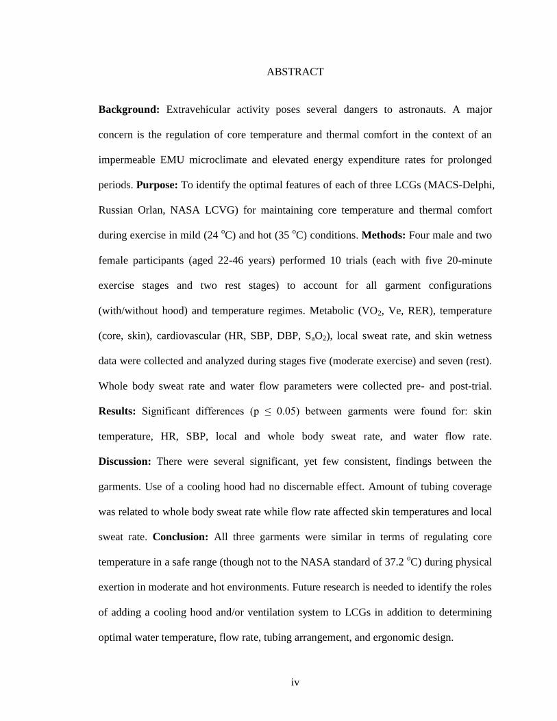
ABSTRACT
Background: Extravehicular activity poses several dangers to astronauts. A major
concern is the regulation of core temperature and thermal comfort in the context of an
impermeable EMU microclimate and elevated energy expenditure rates for prolonged
periods. Purpose: To identify the optimal features of each of three LCGs (MACS-Delphi,
Russian Orlan, NASA LCVG) for maintaining core temperature and thermal comfort
during exercise in mild (24 oC) and hot (35
oC) conditions. Methods: Four male and two
female participants (aged 22-46 years) performed 10 trials (each with five 20-minute
exercise stages and two rest stages) to account for all garment configurations
(with/without hood) and temperature regimes. Metabolic (VO2, Ve, RER), temperature
(core, skin), cardiovascular (HR, SBP, DBP, SaO2), local sweat rate, and skin wetness
data were collected and analyzed during stages five (moderate exercise) and seven (rest).
Whole body sweat rate and water flow parameters were collected pre- and post-trial.
Results: Significant differences (p ≤ 0.05) between garments were found for: skin
temperature, HR, SBP, local and whole body sweat rate, and water flow rate.
Discussion: There were several significant, yet few consistent, findings between the
garments. Use of a cooling hood had no discernable effect. Amount of tubing coverage
was related to whole body sweat rate while flow rate affected skin temperatures and local
sweat rate. Conclusion: All three garments were similar in terms of regulating core
temperature in a safe range (though not to the NASA standard of 37.2 oC) during physical
exertion in moderate and hot environments. Future research is needed to identify the roles
of adding a cooling hood and/or ventilation system to LCGs in addition to determining
optimal water temperature, flow rate, tubing arrangement, and ergonomic design.

v
TABLE OF CONTENTS
ACKNOWLEDGEMENTS....................................................................................... i
DEDICATION........................................................................................................... iii
ABSTRACT............................................................................................................... iv
TABLE OF CONTENTS........................................................................................... v
LIST OF TABLES..................................................................................................... vi
LIST OF FIGURES................................................................................................... ix
CHAPTER 1: INTRODUCTION.............................................................................. 1
CHAPTER 2: REVIEW OF LITERATURE............................................................. 7
CHAPTER 3: METHODS AND PROCEDURES.................................................... 30
CHAPTER 4: RESULTS........................................................................................... 47
CHAPTER 5: DISCUSSION..................................................................................... 54
CHAPTER 6: CONCLUSION.................................................................................. 88
REFERENCES.......................................................................................................... 91
APPENDIX A............................................................................................................ 102
APPENDIX B............................................................................................................ 111
APPENDIX C............................................................................................................ 124
vi
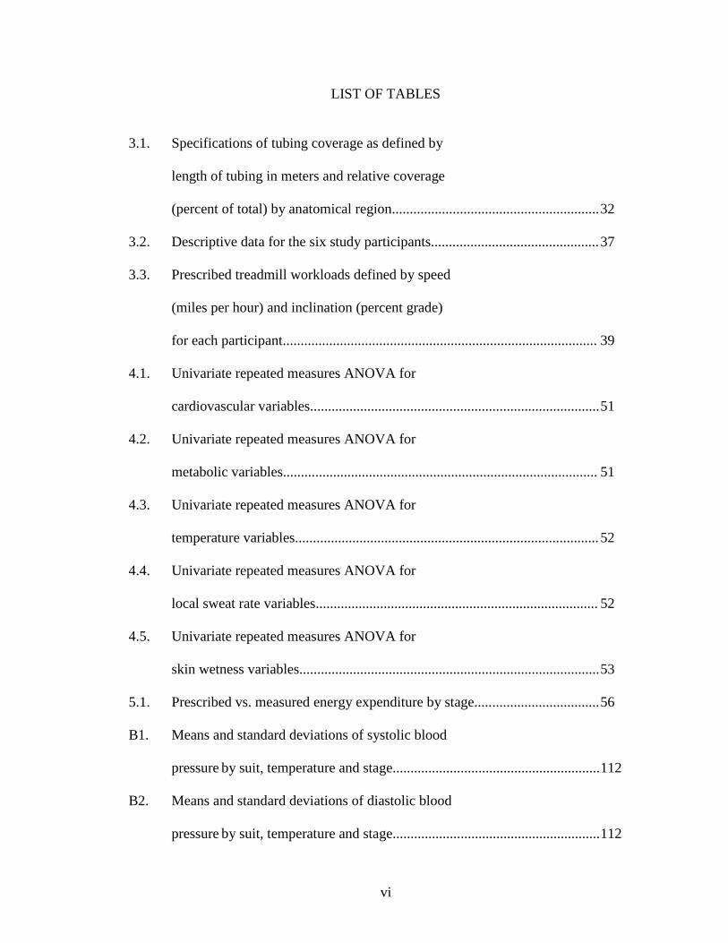
LIST OF TABLES
3.1. Specifications of tubing coverage as defined by
length of tubing in meters and relative coverage
(percent of total) by anatomical region.......................................................... 32
3.2. Descriptive data for the six study participants............................................... 37
3.3. Prescribed treadmill workloads defined by speed
(miles per hour) and inclination (percent grade)
for each participant........................................................................................ 39
4.1. Univariate repeated measures ANOVA for
cardiovascular variables................................................................................. 51
4.2. Univariate repeated measures ANOVA for
metabolic variables........................................................................................ 51
4.3. Univariate repeated measures ANOVA for
temperature variables..................................................................................... 52
4.4. Univariate repeated measures ANOVA for
local sweat rate variables............................................................................... 52
4.5. Univariate repeated measures ANOVA for
skin wetness variables.................................................................................... 53
5.1. Prescribed vs. measured energy expenditure by stage................................... 56
B1. Means and standard deviations of systolic blood
pressure by suit, temperature and stage.......................................................... 112
B2. Means and standard deviations of diastolic blood
pressure by suit, temperature and stage.......................................................... 112
vii
B3. Means and standard deviations of arterial oxygen
saturation by suit, temperature and stage....................................................... 113
B4. Means and standard deviations of heart rate
by suit, temperature and stage........................................................................ 113
B5. Means and standard deviations of relative oxygen
consumption by suit, temperature and stage.................................................. 114
B6. Means and standard deviations of absolute oxygen
consumption by suit, temperature and stage.................................................. 114
B7. Means and standard deviations of respiratory exchange
ratio by suit, temperature and stage................................................................ 115
B8. Means and standard deviations of ventilation
by suit, temperature and stage........................................................................ 115
B9. Means and standard deviations of core (rectal)
temperature by suit, temperature and stage.................................................... 116
B10. Means and standard deviations of skin (femoralis)
temperature by suit, temperature and stage.................................................... 116
B11. Means and standard deviations of skin (chest)
temperature by suit, temperature and stage.................................................... 117
B12. Means and standard deviations of skin (finger)
temperature by suit, temperature and stage.................................................... 117
B13. Means and standard deviations of skin (ear)
temperature by suit, temperature and stage.................................................... 118
viii
B14. Means and standard deviations of skin (calf)
temperature by suit, temperature and stage.................................................... 118
B15. Means and standard deviations of local sweat rate
(groin) by suit, temperature and stage............................................................ 119
B16. Means and standard deviations of local sweat rate
(back) by suit, temperature and stage............................................................. 119
B17. Means and standard deviations of local sweat rate
(chest) by suit, temperature and stage............................................................ 120
B18. Means and standard deviations of skin wetness
(hand) by suit, temperature and stage.............................................................120
B19. Means and standard deviations of skin wetness
(ankle) by suit, temperature and stage............................................................ 121
B20. Means and standard deviations of skin wetness
(forehead) by suit, temperature and stage...................................................... 121
B21. Means and standard deviations of skin wetness
(palm) by suit, temperature and stage............................................................ 122
B22. Means and standard deviations of water flow
rate by suit, temperature and stage................................................................. 122
B23. Means and standard deviations of whole body
sweat rate by suit, temperature and stage....................................................... 123
ix
LIST OF FIGURES
1.1. Parameters of liquid cooling garments that require
further investigation in terms of maximizing safety,
comfort, and efficiency.................................................................................. 4
3.1. The three liquid cooling garments used in this study.................................... 32
3.2. Exercise protocol designed to simulate the demands
of EVA........................................................................................................... 38
5.1. Average core (rectal) temperature profiles of the
MACS-Delphi, Orlan, and LCVG across all stages
during the 24 oC temperature regime............................................................. 59
5.2. Average core (rectal) temperature profiles of the
MACS-Delphi, Orlan, and LCVG across all stages
during the 35 oC temperature regime............................................................. 59
A.1. Recruitment advertisement for solicitation of study
participants..................................................................................................... 103
A.2. Informed consent document (page 1 of 4)..................................................... 104
A.3. Informed consent document (page 2 of 4)..................................................... 105
A.4. Informed consent document (page 3 of 4)..................................................... 106
A.5. Informed consent document (page 4 of 4)..................................................... 107
A.6. Medical history questionnaire........................................................................ 108
A.7. Rectal probe used for core temperature measurement................................... 109
A.8. Sensors used for skin temperature measurement........................................... 109
A.9. Filter paper used for measurement of local sweat rates................................. 110

x
A.10. Chest strap transmitter for heart rate monitor, alcohol
(for sterilization), and lubricant (for rectal probe)......................................... 110
C.1. Limitation of forward flexion in the Orlan due to
vertical tubing arrangement........................................................................... 125
C.2. Results of wear-and-tear in the crotch region of the
Orlan.............................................................................................................. 125
C.3. Skin abrasions in the upper thigh and crotch area
of two different participants following trials with
the Orlan........................................................................................................ 126
1
CHAPTER 1: INTRODUCTION
It is estimated that 20-50% of the workforce in industrialized countries is subjected to
hostile environments (Flouris & Cheung, 2006). Such hostile environments are
characterized by exposure to contaminants (radiation, toxins, and corrosive substances),
hypo- and hyperbaric conditions, and thermal extremes. Excess heat load is a situation
that many humans find themselves in as a result of high environmental temperatures,
protective clothing, and/or high energy expenditures. It is estimated that 5-10 million
workers in the United States are exposed to heat stress annually (Furtado, Craig, Chard,
Zaloom, & Chu, 2007). The human body is truly a marvel of evolutionary design in terms
of its ability to tolerate (acutely) and adapt to (chronically) temperature extremes.
Evidence for the thermal resiliency of humans is provided not only by the myriad
climates that humans inhabit (from equatorial to polar regions), but also the case studies
in the medical literature that document the extremes of human survival following
accidental hypothermia and hyperthermia with recorded core temperatures of 14.4 oC and
47.0 oC, respectively (Taylor, 2006). In general, core body temperature is maintained
within a very narrow range of 36.7 ± 0.3 oC (Taylor, 2006) even in the context of extreme
temperatures due to behavioral and physiological temperature regulation (Pandolf, Sawka,
& Gonzalez, 2001). It should be noted that many medical sources report “normal” core
body temperature as 37.0 oC with a regular range of plus or minus up to one degree
Celsius (Rhoades & Tanner, 2003).
Despite the ability of humans to adequately regulate heat balance in a range of thermal
conditions, there are numerous situations in which thermal stress exceeds the body’s
2
coping ability. Manned spaceflight represents one such situation. Astronauts and
cosmonauts (for consistency, the term astronaut will be used from this point forward)
represent a unique, albeit small, group that must endure numerous challenges to
physiological homeostasis and physical health including microgravity, radiation exposure,
biorhythm disturbances, and exposure to temperature and pressure extremes. To many, it
may not be obvious that increased heat load is a problem that astronauts regularly face. In
particular, extravehicular activity (EVA) represents a serious threat to health and safety
due to thermoregulation impairments. The term EVA refers to activity performed outside
of the confines of the safe haven that is the space capsule/station (e.g., International
Space Station) or transport vehicle (e.g., space shuttle). Extravehicular activity takes
place on planetary surfaces (e.g., walking on the moon) as well as in the vacuum of space
that surrounds a space capsule or vehicle (e.g., spacewalk). While there are important
differences between the two types of EVA, a common problem is an increased heat load
due to the effects of elevated metabolic rates in the context of a protective (and
impermeable) space suit that leads to a rise in the microclimate temperature in which the
astronaut must operate. Extravehicular sorties often involve increased energy expenditure
levels for prolonged periods of time (often measured in hours).
While the first EVAs performed in the 1960s may have been more for “show” as a result
of the “space race” between the United States and the Soviet Union, in the year 2010 they
are a necessity, not a luxury. Space station construction and maintenance require routine
EVAs while in-flight external inspection, and possibly repair, of a transport vehicle like
the space shuttle is now commonplace following the Columbia space shuttle disaster.
3
Despite what was essentially a termination of the Constellation Program by President
Barack Obama on February 1, 2010 (this would have returned the United States to the
moon by 2020), there seems to still be sufficient political and scientific inertia for a long-
range goal of manned spaceflight to Mars. Exploration, and potential colonization, of
extraterrestrial planetary surfaces will require, among other things, a significant amount
of EVA. If past human ingenuity, curiosity, and endeavors are any indication of future
behavior, then it would seem that it is not a matter of if humans will travel to Mars, but
when. If that is the case, then the optimization of human safety and comfort during EVA
will be a significant area of research and design in the coming years since the margin for
error in spaceflight is already small and will presumably decrease as the missions take
astronauts further from terra firma.
Thermal countermeasures in the form of ventilation- and liquid-perfused garments have
been in existence since the 1940s and 1960s, respectively, and were first designed for
pilots who routinely experienced thermal extremes (Nunneley, 1970). Agencies
overseeing the early manned spaceflights of the 1960s saw the need for cooling
countermeasures and incorporated ventilation-cooled garments. However, the cooling
ability of ventilation alone was limited and it was not long before liquid cooling garments
(LCGs) became standard equipment for spaceflight. The underlying principle of LCGs is
rudimentary, yet effective, and simply involves the circulation of cool/cold water that
perfuses the garment via a network of tubing. Direct contact between the skin and tubing
promotes conductive heat transfer and exploits three important properties: 1) temperature
gradients and the second law of thermodynamics, 2) fluidized flow of water, and 3) the
4
jjjj
1. Water
a. Temperature
b. Flow rate
2. Tubing
a. Spatial configuration and total amount (length)
b. Material properties, diameter, shape, and flexibility
3. Garment
a. Material (fluid retention, durability, comfort)
b. Ergonomics (mobility, flexibility, sizing)
4. Cooling regime
a. Automatic or manual control
b. Constant or intermittent
5. Augmentation of heat transfer
a. Addition of cooling hood
b. Incorporation of ventilation system
6. Equipment
a. Hardware and power requirements
b. Equipment reliability, breakdown, and repair
Figure 1.1. Parameters of liquid cooling garments that require further
investigation in terms of maximizing safety, comfort, and efficiency.
specific heat capacity of water. It should be noted that LCGs have numerous applications
beyond aviation and spaceflight including occupational, athletic, and medical settings.
While the basic principles of LCGs have not changed in the nearly 50 years since the
prototype was developed, the evolution of LCGs has focused on optimization of design.
Many questions still surround the development of effective and efficient LCGs for use as
a countermeasure to heat stress. The six categories listed in Figure 1.1 represent
important questions regarding the development of LCGs that have yet to be adequately
answered. As space agencies like NASA move forward in the planning stages for
potential future missions to Mars, the optimization of LCG design has been identified as
5
a significant research focus. Proper management of thermal comfort and physiologic
regulation (via LCGs) translate to enhanced subjective perception and increased safety,
respectively, for astronauts and, as a result, these are areas of great interest as the research
and design process moves forward.
The current study involves the testing and evaluation of three different LCGs, two of
which are currently being used by the American and Russian space agencies. The
Minnesota Advanced Cooling System (MACS-Delphi) was developed by researchers at
the University of Minnesota (Twin Cities) and incorporates a physiologic design in which
the tubing is strategically oriented to correspond to high-density tissue areas (i.e., bone).
The Russian Orlan (Orlan) is currently used by Russian cosmonauts during spaceflight
and also employs a physiologic design but instead focuses the tubing coverage on large
areas of skeletal muscle. The third garment is the NASA Liquid Cooling Ventilation
Garment (LCVG); the LCVG is currently used by NASA during spaceflight and is
characterized not by selective tubing orientation, but by a focus on quantity of body
coverage. The LCVG also has a ventilation component that is designed to augment heat
transfer via convective and evaporative means (this option was not used during the
current study for standardization purposes). It should be noted that the MACS-Delphi and
Orlan have a cooling hood option (the LCVG does not) that covers the head for added
heat extraction. The effectiveness of incorporating a cooling hood was also evaluated in
this study. Testing and evaluation of the garments took place during simulated EVA
(walking on a treadmill) in thermoneutral and hot environments.
6
Purpose
The purpose of this study is to identify the advanced features of each of three LCGs
(MACS-Delphi, Orlan, LCVG) for maintaining core temperature and thermal comfort
during intensive exercise in mild and hot conditions.
Hypothesis
The research hypothesis is that human heat balance can be supported during intensive
physical exertion in mild and hot conditions by means of thermal regulation through
different approaches to spacesuit design, with specific emphasis on the LCG component.
Funding
This research was supported by NASA Cooperative Agreement NNX07AI90A between
the Johnson Space Center EVA Physiology Systems and Performance Project (EPSP) and
the University of Minnesota.
7
CHAPTER 2: REVIEW OF LITERATURE
Since the purpose of this study is to evaluate the performance of three different liquid
cooling garments (LCGs) in the context of simulated extravehicular activity (EVA) in
thermoneutral and hot environments, it seems necessary to review several important topic
areas. The topics that will be reviewed include: thermoregulation, the physiology of
spaceflight and EVA, and cooling modalities. A recurring theme throughout the
subsequent review will be the introduction of the topic of spaceflight and microgravity
and how it relates to the corresponding subject matter.
Thermoregulation
Heat Balance
The human body has evolved to survive, and in many cases thrive, in a wide range of
thermal environments. In modern times, the formal study of human thermoregulation
dates back to at least 1775 where Charles Blagden’s experiments involved baking himself
and others in a heated room at temperatures reaching nearly 130 oC for 45 minutes. These
early experiments yielded observations related to heart rate, circulation, and sweating
(Blagden, 1775); this represents some of the earliest published work related to human
thermoregulation. An understanding of the human body and the effects of various thermal
conditions have come a long way in the past couple of centuries but much has yet to be
learned about the complicated thermal relationship between the internal environment of
humans and the external environment in which they must live. The following passage
(Pisacane, Kuznetz, Logan, Clark, & Wissler, 2007) sums up the multifaceted nature of
human thermoregulation:
8
Because of the complexity of the human thermoregulatory system,
contributing components should be taken into account comprehensively in
modeling. These include: 1) environment - atmospheric constituents,
pressure, temperature, humidity, and [air or water] flow rate; 2) clothing -
thermal conductivity and permeability; 3) anatomical characteristics -
surface area, mass, body composition, heat capacity, gender, and age; 4)
activity - workload, metabolism, oxygen uptake, respiratory heat loss, and
acclimation; and 5) physiological response - shivering, sweating,
vasoconstriction, vasodilation, blood flow rate, and heart rate. (p. A52)
The “modeling” referred to in the previous quote represents a technique used by some
scientists to quantitatively assess various thermal parameters by using human body
thermoregulatory simulators or so-called thermal manikins. While the pros and cons of
thermal modeling are beyond the scope of this discussion, it suffices to say that there are
myriad factors (including many not previously mentioned) that contribute to
thermoregulation and heat balance that can make for precarious interpretations and
erroneous assumptions.
An appropriate place to begin this discussion is at the level of thermal regulation and the
means by which humans achieve thermal balance, even in extreme thermal conditions.
The two primary means by which deep body temperature (i.e., core temperature) is
maintained are behavioral and physiological regulation. Behavioral regulation has been
described as involving “the conscious, willed use of whatever means are available to
minimize thermal discomfort...[which]...appears to be proportional to core and skin
temperatures” (Pandolf, Sawka, & Gonzalez, 2001). Examples of behavioral regulation in
cold environments include wearing more clothing, increasing metabolic heat production
by voluntarily increasing energy expenditure levels, or seeking shelter from the cold.
Behavioral regulation in hot environments is often characterized by wearing less (or
9
light-colored) clothing, decreasing the intensity levels of physical work or exercise, and
increasing fluid consumption. Behavioral regulation is not unique to humans and can be
witnessed throughout the animal kingdom, though it is instinct (not conscious thought)
that triggers the behavioral response in animals. Classic examples range from the
huddling of groups of penguins during the long, cold, dark arctic winters in an attempt to
maximize heat sharing and minimize exposed body surface area to hippopotamuses
seeking refuge from the equatorial heat by bathing in watery mud holes to not only reap
the cooling benefit of the water, but also to use the mud as an insulatory mechanism.
Conversely, physiological regulation describes how the body responds (independent of a
conscious thought process or volition) to various thermal stimuli in an attempt to
maintain core temperature within a small range. In cold environments, a peripheral
vasoconstrictor response is triggered that serves to decrease blood flow to the periphery,
thus shunting warm blood to the deep veins reducing heat loss to the external
environment. Other mechanisms of heat conservation in the cold include piloerection (i.e.,
goose bumps) and the initiation of shivering and chemical thermogenesis which serve to
increase metabolic heat production which aids in maintaining core temperature. Hot
environments elicit a decrease in metabolic rate (to decrease internal heat load) and, more
notably, initiate cutaneous vasodilation and the sweating response in an attempt to
transfer heat from the core to the periphery (via the circulating blood) at which point
numerous heat transfer mechanisms are engaged (Pisacane et al., 2007). The four primary
means by which heat transfer occurs are: 1) radiation - transfer of energy waves that are
emitted by one object and absorbed by another, 2) convection - heat exchange that occurs
between a solid medium and one that moves in a fluidized/flowlike manner (as in the air),
10
3) conduction - transfer of heat from a warm object to a cooler object, and
4) evaporation - heat transfer from the vaporization of water (Armstrong, 2000). As it
relates to evaporation, it should be noted that it is the only one of the four previously
mentioned heat exchange mechanisms that only serves to transfer heat away from the
body (i.e., convection, conduction, and radiation can transfer heat to the body or away
from it). In terms of evaporation, each vaporized liter of water extracts 580 kcal from the
body and transfers it to the environment; this water comes from the approximately two to
four million sweat (eccrine) glands within the average person (McArdle, Katch, & Katch,
2010). Central to human thermoregulation is the heat balance equation which describes
the thermal exchange between an individual’s body and the environment (Fregly &
Blatteis, 1996):
S = M - (±Wk) ± (R) ± (C) ± (K) - E [W ∙ m-2
]
S = rate of storage of body heat (+ for net gain)
M = rate of metabolic energy production (always +)
Wk = rate of work
(+ for work against external forces)
(- for eccentric/negative work)
R = rate of radiant heat exchange (+ for a gain)
C = rate of convective heat transfer (+ for a gain)
K = rate of conductive heat transfer (+ for a gain)
E = rate of evaporative heat transfer (- for net loss)
While a more detailed description of the complex nature of thermoregulation is beyond
the scope of this discussion, such descriptions do exist elsewhere (Fregly & Blatteis,
1996; Pandolf et al., 2001). Additionally, a thorough review article has been written on
the challenges to temperature regulation when working in hot environments (Taylor,
2006). The topic of acclimatization is particularly important for those subjected to
chronically high heat loads and physical exertion; this topic is discussed in detail
11
elsewhere (Armstrong, 2000). It is important to note that prolonged exposure to
microgravity (i.e., spaceflight) and simulated microgravity (i.e., bed rest) have been
shown to negatively impact thermoregulation (Fortney et al., 1998; Greenleaf, 1989);
however, more research is needed in this area to determine the mechanisms of such
alterations in thermoregulation.
Temperature Measurement
Core body temperature is one of the three traditional “vital measures” in medicine (along
with blood pressure and heart rate); skin temperature, though not used as frequently in the
clinical setting as core temperature, does provide valuable insight into the thermal status
of an individual. While measures of core and skin temperature are important in assessing
overall thermal status for various medical and occupational applications, the issue of
temperature measurement is fraught with controversy and lacks a standardized approach.
Such unresolved issues relate to which site for core temperature measurement provides
the most accurate reflection of overall deep body temperature, which skin sites should be
measured and what is the best method for calculating an overall average skin temperature
(using various mathematical weighting techniques), and what is the most accurate means
by which to quantify overall average body temperature (i.e., as a combined function of
various compartments) (Fregly & Blatteis, 1996; Pandolf et al., 2001; Shiraki & Yousef,
1987).
The measurement of rectal temperature can be logistically difficult for a multitude of
reasons and may not be practical in all situations. While it is typically higher than
12
temperatures at other sites of core temperature measurement (i.e., esophageal, oral, and
tympanic) and responds more slowly to rapid changes in core temperature, it is generally
regarded as the standard for core temperature measurement (particularly in steady-state
conditions) owing to its validity and stability (Fregly & Blatteis, 1996; Gagnon, Dorman,
Ollie, Hardcastle, & Kenny, 2009). Insertion depth of the rectal probe is a frequent
concern that a recent study sought to investigate. The study (Lee, Wakabayashi,
Wijayanto, & Tochihara, 2010) involved simultaneous rectal temperature measurement at
seven separate depths ranging from 4-19 cm beyond the external sphincter during bicycle
exercise and subsequent recovery. The authors concluded that there were no systematic
differences at depths greater than 10 cm which is in agreement with the common
convention of requiring a minimum insertion depth of 10-13 cm but at odds with the
conclusions of others that claim there is uniformity in terms of rectal temperature at
depths ranging from 5-27 cm (Pandolf et al., 2001) and 5-20 cm (Fregly & Blatteis, 1996).
It would seem that a conservative recommendation for rectal probe insertion depth is a
minimum of 10 cm.
In terms of skin temperature measurement, Pandolf et al. (2001) cite three primary
reasons for the measurement of skin temperature: 1) calculation of mean body
temperature (in combination with other compartments) for heat storage determinations,
2) calculating radiative and convective heat exchange and skin conductance, and
3) determining the relationship between skin temperature and central thermoregulatory
control mechanisms. Skin temperature measurement has several advantages and
disadvantages. Skin is easily accessible for measurement and offers an almost real-time
13
indication of the current thermal conditions at the interface between the human body and
the environment. However, individual skin sites alone are seldom useful for much beyond
the regional level that is being measured. There are exceptions to this, one of which being
the finger region. The hand has one of the most extensive blood vessel networks in the
body which makes this area (specifically the finger) a candidate for skin temperature
monitoring due to the thermal sensitivity and reactivity. The finger perhaps can be
likened to the “canary in the coal mine” analogy as it relates to the status of the thermal
environment. There exists experimental evidence that the thermal status of the fingers is
highly correlated with heat content in the body and that the first indication of a forming
heat deficit/surplus is manifested in this area (Coca, 2005; Koscheyev, Coca, & Leon,
2007). Automatic regulation of microclimate thermal control systems is a growing area of
research due to the fact that manual control is often found to be sub-optimal. As Webb et
al. (Webb, Troutman, & Annis, 1970) eloquently put it: “Man is a poor judge of his own
thermal state.” However, effective automatic regulation first requires the identification of
a reliable, valid, and easily measured indicator to act as a “thermostat.”
While individual skin sites can be of interest to some, mean skin temperature is generally
considered a more utilitarian metric. However, there is no singly agreed-upon
mathematical technique for arriving at an index that is representative of overall skin
temperature. What is common to most averaging techniques is a weighting system based
upon the percentage of body surface area that is represented by the skin region being
measured. Perhaps the most well-known skin temperature weighting system for
calculating mean skin temperature is the one created by Hardy and DuBois (Hardy &
14
DuBois, 1938) which divides the body into 12 regions with the following weighted
proportions: head: 7%, hand: 4%, upper back: 9.5%, chest: 9.5%, lower back: 9.5%,
abdomen: 9.5% , biceps: 9%, forearm: 7%, quadriceps: 9.5%, hamstring: 9.5%, front calf:
8.5%, and back calf: 7.5%. It should be noted that several weighting strategies exist
ranging from a 15-site weighting formula based on partitional calorimetry (Winslow,
Herrington, & Gagge, 1936) that has been validated (Mitchell & Wyndham, 1969) to a
4-site formula (Ramanathan, 1964). There are also approaches that deviate from the
traditional body surface area percentage technique like that of basing regional weighting
on the thermal sensitivity of the skin due to the observation that thermal receptors are not
evenly distributed over the skin’s surface (Nadel, Mitchell, & Stolwijk, 1973).
Finally, due to the fact that overall mean body temperature is a valuable source of
information related to total-body heat balance and thermal status, there exist several
weighting techniques that attempt to combine average skin (shell) temperature with
average deep body (core) temperature. The traditional two-compartment model of
core/shell weightings for hot, warm-temperate, and cool conditions are 0.9/0.1, 0.79/0.21,
and 0.67/0.33, respectively (Sawka & Castellani, 2007). However, there is evidence that,
even with a correction factor, estimating changes in mean body temperature based only
on two compartments (shell and core) is inaccurate (Jay, Reardon, et al., 2007). Jay and
colleagues (Jay, Gariepy, et al., 2007) have also investigated the incorporation of muscle
temperature which represents a third compartment (in addition to shell and core). Their
research indicates that the three compartment thermometry model used was superior to
that of the traditional two-compartment model. It should be noted that they are not the
15
first to propose a three-compartment model using shell, core, and muscle temperatures
(Nadel, Bergh, & Saltin, 1972; Snellen, 2000; Webb, 1998).
Physiology of Spaceflight and EVA
Spaceflight
The so-called Space Age was launched on October 4, 1957 with the Soviet Union’s
successful flight of Sputnik. That was followed shortly thereafter by the first human (Yuri
Gagarin) to travel to outer space on April 12, 1961 and the rest, as they say, is history.
While the ability to travel beyond the reaches of planet earth is nothing less than amazing,
the most difficult part still lies ahead. It is an unfortunate irony that humans, by their
mental constitution, are so very driven to explore space and yet, by their physical nature,
are so ill-equipped to survive in it. Assuming that humans will be protected from such
lethalities of outer space as ionizing radiation, space projectiles, and lack of oxygen, one
of the greatest obstacles will be surviving in an environment devoid of gravity. Gravity is
an obvious environmental parameter that humans are well-equipped to deal with.
Microgravity, on the other hand, is not something that any bipedal mammal would be
expected to have adaptive responses against on a chronic scale. Most humans do,
however, experience effects similar to those induced by microgravity on a daily basis
when lying in the horizontal position, as when sleeping. There are key differences,
however, the most notable being that these bouts, while regularly occurring, are relatively
short in duration and are interspersed with long durations of continuously fighting the
force of gravity. Interestingly, patients with illnesses/ailments that cause them to be
chronically bedridden suffer many of the same health sequelae as astronauts who spend
16
prolonged periods of time in space like the development of kidney stones (Cheung, 2010)
and a decrease in bone mineral density (Smith & Heer, 2002). As a result, one the most
valuable models for studying the effects of microgravity on humans is a modified bed rest
condition (typically a negative 6-12o head-tilt position to account for fluid shifts). Some
of the longest bed rest studies last 6-12 months; NASA continually conducts 90-day bed
rest protocols at its facility in Galveston, Texas (Cheung, 2010).
In the case of spaceflight, a gravity-free environment creates several problems for
humans. While many of these problems were predicted before manned spaceflight came
to fruition, they were not of emergent concern in the early days of space travel because:
1) the focus of early research and design was, rightfully, on the technology to safely
transport humans to and from space and 2) the duration of early spaceflights was, by and
large, not thought to be sufficient to induce significant health-related complications.
Problems that emerge as the result of prolonged exposure to microgravity typically can
be traced to two physiological systems: cardiovascular and musculoskeletal. In the case
of the cardiovascular system, a major deleterious effect is an impairment of the
circulation whereas the lack of mechanical stresses on the bones, musculature, and
connective tissues results in decreased strength, integrity, and function of the
musculoskeletal system. It is important to note that the two systems are not independent
of each other, on earth or in space, and that disruptions in any one physiological system
typically have repercussions within another system or systems. For example, in
microgravity, the forces generated by skeletal muscles are very low simply because there
are no (or minimal) gravitational forces to overcome. As a result, the skeletal muscles
17
atrophy, particularly those that are used most regularly on earth like the large postural
and locomotor groups of the lower extremity (Buckey, 2006). However, this also has an
effect on the cardiovascular system (i.e., the heart) due to the fact that not as much
oxygen is required by the musculature because the energy requirements are significantly
reduced. A reduced oxygen demand will necessarily lead to a reduced cardiac output
which ultimately results in deconditioning of the cardiac muscle tissue which may result
in decreased pumping capacity and arterial pressure (Perhonen et al., 2001). Altered
circulation (due to microgravity) resulting in reduced blood flow to certain areas is a
second example that illustrates the interconnectedness of physiological systems.
Specifically, in microgravity, blood tends to pool in the central regions of the body, not in
the dependent areas like the lower limbs as evidenced on earth as a result of gravity
(Rowell, 1993). For a thorough and detailed analysis of the effects of microgravity on the
cardiovascular and musculoskeletal systems, the reader is directed to Fregly and Blatteis
(1996). Additionally, there exist in-depth explanations of human circulation and its
control (Rowell, 1993; Rowell & Shepherd, 1996). Another issue that warrants attention
is the effects that an impaired circulation exerts on thermoregulation. Since much of the
human body’s capability to transfer heat lies within the fluidized flow of blood, it stands
to reason that an impaired circulation will result in less-than-optimal thermoregulation.
EVA
The Soviet Union’s Alexey Leonov conducted the first-ever EVA from the Voskhod 2
spacecraft on March 18, 1965. He was outside of the craft for 12 minutes and nine
seconds, connected by a 5.35 m tether (Abramov & Skoog, 2003). On March 11, 2001,
18
Americans Susan Helms and James Voss performed the longest EVA to date at nearly
nine hours (8 hr 56 min). As a clarifying note, the Russian space program defines EVA as
occurring when a cosmonaut is in a vacuum whereas NASA considers EVA to
commence when the astronaut switches the extravehicular mobility suit (EMU) to battery
power. When EVA durations are measured in hours, serious consideration must be given
to issues related to thermal comfort and temperature regulation. Three elements act
synergistically to create a situation for astronauts during EVA that can lead to heat
overload, thermoregulatory collapse, and possibly death. Those elements are increased
energy expenditure rates, high temperatures, and a microclimate that is impermeable
within the EMU. In this day and age of space shuttles, space stations, and proposed space
flights to the moon and beyond, EVA is a necessity. An important distinction must be
made between the two primary types of EVA (this is not referring to the differences in
how Russians and Americans define EVA). The lunar missions that made for television
rating bonanzas in the 1970s were characterized by planetary EVA that involved walking
around on the moon surface like a Louis and Clark expedition. Today’s EVAs are less
sensational and are characterized by long durations of remaining in the same spot and
assuming the role of mundane mechanic.
Lighthearted analogies aside, there are very real differences in the two types of EVA.
Surface EVA on an extraterrestrial body requires locomotion and dynamic movement
whereas an EVA sortie to repair a broken solar array or inspect the heat shield on the
space shuttle involves a large static component in the lower body and a sustained aerobic
output in the upper body. All of the EVA experience on extraterrestrial surfaces comes
19
from the Apollo missions which began with Neil Armstrong’s “giant leap for mankind”
on July 20, 1969 and ended on December 14, 1972 with the departure from the lunar
surface of astronauts Eugene Cernan and Harrison Schmitt. At the time, it was expected
that American astronauts would return to the moon by the late 1980s (Portree & Trevino,
1997), but as of 2010 there still has not been a human on the moon or any other
extraterrestrial surface and there is no firm plan in place to do so in the immediate future.
There were 14 EVAs performed on the lunar surface for a total time of 80 hours and 43
minutes (Cintala, 2005). Although there were a relatively few number of lunar EVAs
performed, valuable information was obtained related to metabolic rate during planetary
EVA. The average energy expenditure during lunar surface EVA was 234 kcal/h which
was much lower than the anticipated upper limits of 400-500 kcal/h. It was noted that
metabolic rates during discrete activities were as high as 450 kcal/h for brief periods
(Waligora & Kumar, 1995). This is very important data that aids in simulating the
metabolic demands of EVA on earth to test space suit design (including the microclimate
cooling system). Waligora and Kumar (1995) also determined that the average energy
expenditure rate during the first 29 space shuttle EVA missions (1983-1995) was
194 kcal/h. The average metabolic rate for the Skylab EVAs (1973-1974) was 230 kcal/h
(Cowell, Stocks, Evan, Simonson, & Greenleaf, 2002).
While the absolute rates of metabolism during EVA are important to know, the type of
work being done must also be considered. Cowell et al. (2002) characterize present-day
EVA “...mainly as upper body aerobic plus isometric work, and lower-body isometric
work.” Oxygen consumption rates between 0.65 and 0.8 l/min have been reported during
20
EVA by the Soviet and American programs (Cowell et al., 2002). It is important to
consider that sustained isometric skeletal muscle contractions and prolonged upper body
aerobic activity are very different than that to which most individuals (astronauts or
otherwise) are accustomed. In particular, it is well-known that aerobic capacity is lower
for upper body aerobic exercise than lower body aerobic exercise (with the exception of
certain athletic and clinical populations). In arm exercise, the maximal oxygen uptake is
about 70% of what is attained in leg exercise (Astrand, Rodahl, Dahl, & Stromme 2003).
Additionally, arterial blood pressures and heart rates are higher during arm exercise
compared to leg exercise at a given oxygen uptake or cardiac output (Astrand, Ekblom,
Messin, Saltin, & Stenberg, 1965). Miles et al. and Pendergast provide two nice reviews
of the cardiovascular responses to upper body exercise (Miles, Cox, & Bomze, 1989;
Pendergast, 1989). This increased cardiac strain could have consequences on individuals
who are already somewhat compromised by the deleterious effects of microgravity on the
cardiovascular system. However, it has been shown that the rise in core temperature with
exercise is not dependent on the skeletal muscle mass employed but, rather, it is
dependent on the metabolic rate during exercise (Sawka, Latzka, & Pandolf, 1989).
Water Cooling
Optimal Water Temperature
In the world of thermoregulatory research, particularly heat stress, perhaps one of the
most hotly debated (pun intended) topics is that of optimal water temperature for cooling.
Water has been used for the purpose of cooling since antiquity and is obvious throughout
the animal kingdom. The fluidized properties of water are particularly valuable in terms
21
of the numerous ways in which they can be utilized in the artificial management of the
microclimate. Due to its specific heat capacity (~4.2 J/(g∙K)), which is the second highest
of any known substance (after ammonia which is ~4.7 J/(g∙K)), water may be the most
efficient means by which to regulate heat transfer (insertion or extraction). Across the
physiologically relevant spectrum of temperatures, the volume-specific heat capacity of
water is 3400 times greater than that of air (Taylor, Caldwell, Van Den Heuvel, &
Patterson, 2008). The potent effects that water exerts on the human body is illustrated by
the fact that cool/cold water has 25 times the cooling effect on the human body than does
air of a comparable temperature (Pandolf et al., 2001) which is why hypothermia is a
concern in water temperatures that may seem mild. For example, the shivering threshold
(also called critical temperature) normally occurs at a water temperature between
30-34 oC compared to an air temperature between 20-27
oC (Pandolf et al., 2001). Not
only does water have a greater capacity to accept thermal energy, but this energy transfer
will proceed at a much greater rate (Taylor et al., 2008) owing to the fluidity of water.
The enhancement of heat transfer potential as a result of water’s fluid nature is evidenced
by higher convective heat losses and heat transfer coefficients during swimming
compared with being motionless in the water (Nadel, Holmer, Bergh, Astrand, &
Stolwijk, 1974).
The majority of the thermoregulatory research involving optimal water temperatures for
cooling resides in the medical realm, specifically emergency medicine, as it relates to
acute heat illness. There are two primary schools of thought regarding recommendations
for water temperature in the context of reducing a core temperature that is indicative of
22
hyperthermia and portends thermoregulatory collapse. A summary of the opposing
viewpoints was recently published (Taylor, Casa, & Kenny, 2010) and presents the
contrasting views of the two sides of the debate. If the goal is to remove heat in the
context of an elevated body temperature due to physical activity or heat illness, there are
two theories. The first supports the notion that “colder is better.” This is evidenced by the
ice bath regimens used in cases of heat illness. The idea is simple: the colder the water,
the greater the thermal gradient between the water and the human body. The second law
of thermodynamics states that “the entropy of an isolated system not in equilibrium will
tend to increase over time, approaching a maximum value at equilibrium” which means
that heat can spontaneously flow from a region of higher temperature to a region of lower
temperature but not the other way around. The second law of thermodynamics also
speaks to the nature of thermal gradients and that heat transfer will increase as the
temperature gradient increases. The ice water bath technique is designed to maximize
surface area coverage and the thermal gradient between the human body and the water in
which it is immersed. Other techniques like ice-pack application, showering the body
with water, fanning, and wet-towel application have been reviewed elsewhere
(McDermott et al., 2009) as well as ice vest cooling (Lopez, Cleary, Jones, & Zuri, 2008).
Ice water immersion remains the current recommendation for acutely treating exercise-
induced hyperthermia (Casa et al., 2007; McDermott et al., 2009; Proulx, Ducharme, &
Kenny, 2003).
While cold water immersion is still an accepted medical technique (if not “the” accepted
technique) for managing acute hyperthermia, it is not without its critics. In terms of
23
optimally cooling the human body, there are some in the thermoregulatory community
who believe cold water temperatures may be counterproductive, despite the large thermal
gradients they produce (Taylor et al., 2010). One of the most potent physiologic self-
defense mechanisms against cold that humans possess is a reflexive vasoconstriction of
the peripheral vasculature. The primary means by which heat is transferred from the body
“core” to the “shell” (or periphery), which is then dissipated to the environment, is via
circulating blood. The peripheral vasoconstriction response during exposure to cold is
simply the body’s way of minimizing heat loss; as the diameter of the conduits (i.e.,
arterioles) decreases, so too does the volume of blood flowing through them which
decreases heat dissipation to the external environment and preferentially shunts the warm
blood to the core region. Those who oppose the use of extreme cold temperatures for heat
extraction base their objections on the idea that the increased thermal gradient produced
by techniques like cold water immersion is not optimal due to the resulting
vasoconstriction which reduces blood flow and, ultimately, heat transfer. The resulting
contention is that cool or temperate water (as opposed to cold) would not be as likely to
elicit the vasoconstriction response.
There is experimental evidence to support the idea of temperate water immersion in the
management of acute heat illness. In a study designed to compare the amount of time it
takes to lower core temperature from a hyperthermic state (induced in a controlled
laboratory setting by exercise, heat, and a warm water-perfused garment) with different
cooling regimes, it was demonstrated that temperate water was almost as effective as cold
water. Specifically, all of the participants’ core temperatures were raised to 39.5 oC
24
followed immediately by one of three different cooling methods: air (20-22 oC), cold
water immersion (14 oC), and temperate water immersion (26
oC). The times to reach a
core temperature of 37.5 oC for air, cold water, and temperate water were 22.81, 2.16, and
2.91 minutes, respectively. While the authors conceded that there were statistical
differences for each of the between-trial comparisons, they call into question the practical
and clinical significance of a 45-second discrepancy between cold and temperate water
immersion (Taylor et al., 2008). Critics contend that the study of Taylor et al. (2008), and
its conclusions, are flawed for two reasons (Taylor et al., 2010). First, core temperature
was assessed via esophageal temperature, not rectal temperature which has gained
recognition as the best overall means (in terms of stability and validity) by which to
measure deep body temperature (Gagnon, Lemire, Jay, & Kenny, 2010). It should be
noted that some believe rectal temperature may not be appropriate in situations involving
rapidly changing body temperatures such as during the treatment of hyperthermia (due to
the lag in rectal temperature response time) and that esophageal temperature might be
more appropriate because it is a good measure of the temperature of the blood that
perfuses the hypothalamus and it reacts quickly to changes in the temperature of blood
(Proulx, Ducharme, & Kenny, 2006). Secondly, very cold water (2 oC) has been shown to
be superior in terms of cooling rates for high rectal temperatures as evidenced by cooling
rates of 0.35 oC per minute for very cold water (2
oC) (Proulx et al., 2003) as compared to
rates of 0.18 oC per minute and 0.10
oC per minute with water temperatures of 14
oC and
26 oC, respectively (Taylor et al., 2008). Taylor counters Kenny’s criticism by suggesting
that, in addition to the vasoconstriction that is likely elicited by cold water, potentially
dangerous sequelae exist when humans are exposed to the shock of cold or very cold
25
water immersion, most notably supraventricular ectopic arrhythmias and reduced
cerebral blood flow (Taylor et al., 2008).
The debate about a lack of vasoconstriction with temperate water versus a greater thermal
gradient with cold water continues with contentions that the human body is not like a
bowling ball (in terms of a known density and consistent overall material properties) and
therefore should not be thought of as such (implying a blinding single-mindedness
regarding the supremacy of maximizing thermal gradients for heat transfer). This is then
countered with evidence supporting the theory that the cold-induced vasoconstrictor
response is blunted in hyperthermic individuals (Taylor et al., 2010). The argument
between the opposing sides and their respective viewpoints is ongoing so it would seem
that the debate remains unsettled.
The challenge for researchers in the thermoregulation field is to elucidate the relationship
between vasoconstriction, thermal gradient and heat exchange in terms of reconciling an
appropriate/optimal balance between the three. This is a particularly important topic
within the realm of microclimate cooling systems like LCGs.
LCGs
It is estimated that 20-50% of the workforce in industrialized countries is subjected to
hostile environments (WHO, 1995). These environments are characterized by toxins,
contaminants, and pollutants as well as thermal extremes. Many occupations are
associated with high heat loads including the power, metal and ceramics industries;
26
firefighting; the military; etc. It is estimated that approximately 5-10 million workers in
the United States are exposed to heat stress annually (Furtado, Craig, Chard, Zaloom, &
Chu, 2007). Heat stress is interpreted the same by the body’s thermoregulatory centers
regardless of the source. Three potent contributors to heat stress are high environmental
temperatures, high rates of energy expenditure, and increased thermal insulation. Any one
of these factors acting alone can impart serious heat stress to the human body. For
example, Roberts reports on a recent case study in which a middle-aged and well-trained
man suffered from exertional heat stroke (measured rectal temperature of 40.7 oC) during
a cool weather marathon in which the temperature ranged from 6 oC at the start to 9.5
oC
at the time of the man’s collapse just 10 meters from the finish line (Roberts, 2006). This
is a testament to the heat generating capabilities of skeletal muscle, particularly when an
individual is able to override the central governor that would normally cause that person
to decrease the intensity of effort. In the aforementioned case, the man was attempting to
qualify for the Boston Marathon (an elite event) and needed a finish time of three hours
and 15 minutes or better (he was on pace to achieve that goal with no more than a couple
of seconds to spare). The consequences of high ambient temperatures on thermal strain
should be obvious and do not require further explanation. One of the common themes of
occupations that take place in the hostile environments mentioned at the beginning of this
section is protective equipment to minimize exposure to dangerous elements. The
equipment, if well-designed, serves its functional purpose but comes at the cost of
creating a microclimate defined by high temperatures and a limited ability to dissipate
heat. This increased thermal insulation can be complicated by concomitantly high
environmental temperatures and energy expenditure rates.
27
A microclimate management system was first developed in 1940 by Dr. Brian McArdle
for use by World War II pilots who experienced cold stress. These first systems used gas
ventilation as the primary means of heat transfer via convection. Water was proposed as a
potential heat transfer medium in microclimate cooling systems in 1958 by Billingham
(Billingham, 1959) and the prototype LCG was developed in 1962 at the Royal Aircraft
Establishment in England (Burton & Collier, 1964). The underlying principle of LCGs is
quite rudimentary and simply involves the circulation of cool/cold water through a
network of tubing that perfuses the garment. The contact of the tubes with the skin
promotes conductive heat transfer and exploits the second law of thermodynamics while
also taking advantage of the unique properties of water (i.e., fluidized flow and specific
heat capacity). Over the years, the fundamental principle upon which LCGs is based has
not changed. However, the evolution of LCGs has sought to improve the garment design
by optimizing heat transfer from the body.
The first cooling garment used during spaceflight incorporated a ventilation cooling
system and was part of the Gemini spacesuit as well as the prototype Apollo suit. The
limitations of ventilation as the sole mediator of heat exchange during EVA were quickly
realized during the Gemini IX and XI missions. These limitations were manifested by
fogged face plates, high heart rates (170-180 bpm), and astronaut fatigue that resulted in
premature termination of EVA. The estimated heat production of 550 kcal/h (with spikes
up to 900 kcal/h) was extremely high for the relatively mild activities being performed
and the cooling garment quickly became overloaded and incapable of supporting thermal
comfort (Nunneley, 1970). The subsequent EVAs performed with the ventilation garment
28
were significantly compromised in terms of the work performed as the astronauts had to
work within the limits of the garment. By 1969, NASA had settled on a LCG design
composed of 40 tubes with a water flow rate of 1.8 l/min and heat extraction capabilities
of 400-500 kcal/h for use with the Apollo program. Later, NASA decided to revisit the
ventilation cooling system but this time opted to incorporate it with the water-cooling
component; this liquid cooling and ventilation garment (LCVG) design is still in use
today. It should be noted that the NASA LCVG is one the three garments investigated in
the present study.
More recent designs of LCGs have experimented with different design features so as to
maximize the efficiency of heat transfer. For example, Koscheyev et al. have designed a
LCG (MACS-Delphi) based on physiological principles (Koscheyev et al., 2007).
Specifically, tubing is strategically oriented over high density tissue areas like the region
of the upper back. Fat, muscle (high circulation), muscle (low circulation), and bone have
the following thermal conductivities: 0.187, 0.533, 0.628, and 0.782 W/K/m, respectively.
It is thought that efficiency can be maximized by minimizing the amount of tubing
required without sacrificing heat transfer capabilities. The MACS-Delphi LCG is also
being evaluated in the current study. A feature of the MACS-Delphi (and some other
LCGs) is the option of a cooling hood that is theorized to exploit not only the large
potential heat sink that is the cranium, but also the unusual vascular properties in the head
that are highlighted by reduced (or absent) cold-mediated vasoconstrictor reflexes (Blair,
Glover, & Roddie, 1961; Froese & Burton, 1957; Hertzman & Roth, 1942; Rasch,
Samson, Cote, & Cabanac, 1991). Specific questions regarding cooling hoods relate to
29
water temperature, spatial configuration of tubing, and the potential negative
consequences of the insulatory properties of hair.
There are numerous innovative design features currently being investigated like
automatic thermal control based on physiologic responses and cues (Nyberg, Diller, &
Wissler, 2000) and differences between intermittent versus regional cooling (Cheuvront,
Kolka, Cadarette, Montain, & Sawka, 2003). This study seeks to evaluate the
performance of different LCGs for the express purpose of providing recommendations
for future designs to be implemented with the next generation of planetary EVA.
However, LCGs have also found unique applications in sports in terms of performance
enhancement (Booth et al., 2001; Cheung & Robinson, 2004; Wilson et al., 2002), in
occupational settings (Furtado et al., 2007), and in medicine. For example, there have
been encouraging results from research looking at the benefit of LCGs for clinical
populations like multiple sclerosis patients (Meyer-Heim et al., 2007). Two excellent
reviews have been written on the topic of LCGs that present detailed histories (Nunneley,
1970) and a clear vision of what direction the research needs to go in the future to
optimize the design and performance of LCGs (Flouris & Cheung, 2006).
It is hoped that the results of the current study will contribute to the body of scientific
knowledge on the topic of cooling strategies with specific emphasis on LCGs in a manner
similar to that which the reviewed literature in this chapter has contributed to the
understanding of important topics related to thermoregulation, the physiology of
spaceflight and EVA, and cooling modalities.
30
CHAPTER 3: METHODS AND PROCEDURES
Study Design
The purpose of this study was to identify the advanced features of each of three LCGs for
maintaining core temperature and thermal comfort during intensive exercise in mild
(24 oC) and hot (35
oC) conditions. The three garments evaluated were: 1) Minnesota
Advanced Cooling Suit (MACS-Delphi), 2) Russian Orlan (Orlan), and 3) NASA Liquid
Cooling Ventilation Garment (LCVG). It is important to note that two out of the three
LCG designs (MACS-Delphi and Orlan) had the option of a cooling hood (which covered
the head and neck) in addition to the full body coverage that was common to all three
designs. Since it was deemed essential to evaluate all possible LCG configurations, five
possibilities existed:
1) MACS-Delphi with hood
2) MACS-Delphi without hood
3) Orlan with hood
4) Orlan without hood
5) LCVG without hood
As the purpose of the study was not only to evaluate the LCGs in terms of exercise (i.e.,
simulated EVA) and garment design, but also to evaluate the LCGs under two different
environmental conditions (hot and thermoneutral), 10 experimental conditions existed:
1) Thermoneutral, MACS-Delphi with hood
2) Thermoneutral, MACS-Delphi without hood
3) Thermoneutral, Orlan with hood
4) Thermoneutral, Orlan without hood
5) Thermoneutral, LCVG without hood
6) Hot, MACS-Delphi with hood
7) Hot, MACS-Delphi without hood
8) Hot, Orlan with hood
9) Hot, Orlan without hood
10) Hot, LCVG without hood
31
Using a crossover design, participants were each required to complete 10 trials in order to
account for all possible experimental conditions. The order of the 10 experimental
conditions for each participant was randomized and the start time of the trials ranged
from early morning to early afternoon (6 a.m. - 2 p.m.). All testing occurred in the
environmental chamber within the Laboratory for Health and Human Performance in
Extreme Environments (LHHPEE) at the University of Minnesota. A medical doctor and
an exercise physiologist were present for all experimental trials. All experimental testing
took place between March 27, 2008 and July 15, 2008.
Specifications of LCGs
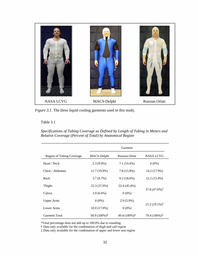
Each of the three LCGs (Figure 3.1) was made of spandex-type material. Specifically, the
MACS-Delphi was made of spandex polyester (with the exception of the hood
component which was made of spandex mesh) while the Orlan and LCVG were both
made of spandex mesh (LCVG had a double layer of material). The masses of the
garments (tubing filled with water) were as follows: MACS-Delphi with hood (1.43 kg),
Orlan with hood (2.45 kg), and LCVG without hood (1.79 kg). The tubing used for the
circulation of water was sewn or woven into the garment material (on the inside of the
garment) so that the tubing surface was in direct contact with the skin. Table 3.1
summarizes the specifications of each garment used in this study as it relates to tubing
coverage. It should be noted that the information in the table represents the MACS-
Delphi with the hood option, the Orlan with the hood option, and the LCVG without the
hood as no hood option exists with this garment.
32
NASA LCVG MACS-Delphi Russian Orlan
Figure 3.1. The three liquid cooling garments used in this study.
Table 3.1
Specifications of Tubing Coverage as Defined by Length of Tubing in Meters and
Relative Coverage (Percent of Total) by Anatomical Region
Garment
Region of Tubing Coverage MACS-Delphi Russian Orlan NASA LCVG
Head / Neck 5.3 (9.0%) 7.1 (14.4%) 0 (0%)
Chest / Abdomen 11.7 (19.9%) 7.8 (15.8%) 14.2 (17.9%)
Back 5.7 (9.7%) 9.2 (18.6%) 12.2 (15.4%)
Thighs 22.3 (37.9%) 22.4 (45.4%)
37.8 (47.6%)†
Calves 3.9 (6.6%) 0 (0%)
Upper Arms 0 (0%) 2.9 (5.9%)
15.2 (19.1%)‡
Lower Arms 10.0 (17.0%) 0 (0%)
Garment Total 58.9 (100%)* 49.4 (100%)* 79.4 (100%)*
*Total percentage does not add up to 100.0% due to rounding
† Data only available for the combination of thigh and calf region
‡ Data only available for the combination of upper and lower arm region
33
Participants
Recruitment
Male and female participants were recruited from the University of Minnesota (Twin
Cities) and the surrounding area of Minneapolis and St. Paul, Minnesota via posted fliers,
classroom advertisements, and word-of-mouth solicitation (see Figure A1). Inclusion
criteria were as follows: 1) aged 20-50 years, 2) an aerobic fitness level sufficient to meet
the experimental demands, and 3) a body size/type that could be accommodated by the
available LCG sizes. Potential participants were excluded if they: 1) had any medically
diagnosed disease/illness that could either be made worse by the experimental conditions
(i.e., exercise and/or environmental conditions) or cause abnormal thermoregulatory
responses that could confound experimental interpretations, 2) had any positive responses
on a pre-screening exercise stress test, and/or 3) had reservations or doubts about being
able to fulfill the experimental requirements. Participants signed an informed consent
prior to the study (see Figures A2-A5). This study was approved by the Institutional
Review Board of the University of Minnesota (Twin Cities). The number of participants
(six) was determined based on the resources allocated from the granting agency.
Pre-Screening Procedures
Potential participants were instructed to report to the Laboratory of Physiological
Hygiene and Exercise Science (LPHES) at the University of Minnesota for a pre-
screening assessment. Participants first completed a medical history questionnaire (Figure
A6) to identify possible risks of participating in the study (or the pre-screening
assessment). Upon satisfactory completion of the medical history form, demographic
34
(date of birth, sex, and race) and anthropometric data were collected. Height was
determined (without shoes) to the nearest 0.1 cm using a standard wall-mounted
stadiometer and body mass was determined (to the nearest 0.1 kg) by having the
participant stand on an electronic Tanita BWB-800S scale (Tanita Corporation of
America Inc., Arlington Heights, IL) wearing only a T-shirt, shorts, and socks. Body
composition data was collected using the sum of skinfolds method. While this method
has been described in detail elsewhere (Maud & Foster, 2006; McArdle, Katch, & Katch,
2010; Thompson, 2010), a brief description of the process follows. A calibrated (length
and tension) Lange skinfold caliper was used by the same trained exercise physiologist
(JMW) to collect triplicate measures of the double thickness of subcutaneous fat (using a
thumb-index finger pinch) at seven anatomically referenced points (abdomen, iliac crest,
triceps, pectoralis, subscapularis, midaxilla, and thigh). The seven representative skinfold
measurements (in millimeters) were then summed and factored into a sex- and age-
specific regression equation (Jackson, Pollock, & Ward, 1978, 1980) to determine body
density which was then used to determine percent body fat via appropriate race- and sex-
specific regression equations (Maud & Foster, 2006; Siri, 1961). Body composition data
was collected due to the nature of the relationship between subcutaneous fat and
thermoregulatory dynamics (Fregly & Blatteis, 1996; Lemire et al., 2008).
Following the collection of demographic and anthropometric data, participants completed
an exercise stress test to determine: 1) any risks associated with performing exercise at
the intensity required for the experimental protocol and 2) aerobic fitness which was
operationally defined as peak relative oxygen consumption. Prior to the commencement
35
of the stress test, participants signed an informed consent document specifically for the
graded exercise test (per LPHES protocol). This was followed by the collection of resting
data including blood pressure (stethoscope and sphygmomanometer) and heart
rate/rhythm via a 12-lead Quinton 2000 ECG system (Cardiac Science Inc., Bothell, WA).
Stress testing was administered by an exercise physiologist (JMW) and involved
walking/jogging on a Trackmaster TMX22 treadmill (Full Vision Inc., Newton, KS) as
the mode of exercise. Specifically, the Bruce treadmill protocol (Bruce, 1971) was used
in conjunction with the continual monitoring of physiologic data including heart rate and
rhythm, blood pressure, and oxygen consumption. These measures were recorded once
per three-minute exercise stage in addition to a subjective interpretation of effort by the
participant using the rating of perceived exertion scale designed by Borg (Borg, 1982).
Heart rate and rhythm data were collected via a 12-lead ECG and blood pressure was
manually assessed using a stethoscope and sphygmomanometer. Oxygen consumption
was collected utilizing open-circuit spirometry via a computerized Medgraphics Ultima
metabolic analyzing system (Medical Graphics Corp., St. Paul, MN). Breath-by-breath
inspired and expired gases (volume and concentration) were collected via a Hans
Rudolph 7900 Series face mask and headgear assembly (Hans Rudolph Inc., Shawnee,
KS) fitted with a Medgraphics preVent® pneumotach (Medical Graphics Corp., St. Paul,
MN) and delivered to the metabolic analyzer which was calibrated prior to each test.
Specifically, the flow meter was calibrated with a known-volume standard (3-liter
syringe) and the gas analyzer was calibrated with known concentrations of gases in the
form of a “reference” gas (21% oxygen and 79% nitrogen) and a “calibration” gas (12%
oxygen, 5% carbon dioxide, and 83% nitrogen).
36
Prior to the test, participants were informed that the workload would be continually
increased every three minutes in the form of speed and grade/inclination and that the goal
was to reach a state of maximal effort. Each participant was told what to expect over the
course of the stress test in terms effort/exertion levels as well as the data collection
process and was informed that he or she could terminate the test at any time for any
reason as could the exercise physiologist administering the test. All stress tests were
terminated at or near maximal exertion levels based upon the desire of the participants to
stop. Following the termination of the stress test, participants were allowed to “cool
down” which entailed walking at a slow speed with no inclination. Physiologic
parameters (heart rate, heart rhythm, and blood pressure) were monitored during the
recovery stage to ensure a normal post-exercise response (the recovery stage typically
lasted five minutes). Participants were allowed to leave the laboratory once their
physiologic values where at or near pre-exercise levels. The data collected during the
stress test was forwarded to the cardiologist (ASL) who served as the medical director of
the LPHES; he granted the final medical clearance required for participation in the study.
Selection
Four males and two females were selected to participate in the study. Descriptive data for
the six participants are presented in Table 3.2. Participants were asked to refrain from
vigorous exercise 24 hours prior to an experimental trial and were instructed to eat
nothing larger than a “light meal” two hours prior to a trial.
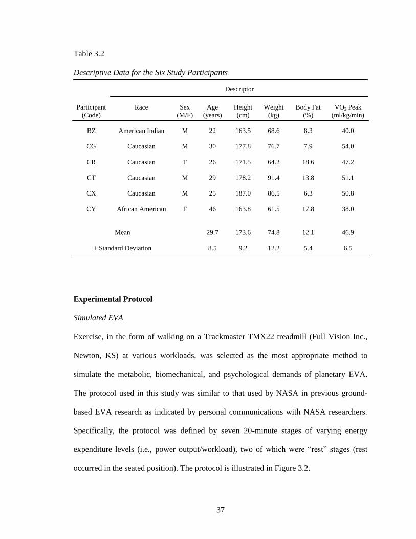
37
Table 3.2
Descriptive Data for the Six Study Participants
Descriptor
Participant
(Code)
Race Sex
(M/F)
Age
(years)
Height
(cm)
Weight
(kg)
Body Fat
(%)
VO2 Peak
(ml/kg/min)
BZ American Indian M 22 163.5 68.6 8.3 40.0
CG Caucasian M 30 177.8 76.7 7.9 54.0
CR Caucasian F 26 171.5 64.2 18.6 47.2
CT Caucasian M 29 178.2 91.4 13.8 51.1
CX Caucasian M 25 187.0 86.5 6.3 50.8
CY African American F 46 163.8 61.5 17.8 38.0
Mean 29.7 173.6 74.8 12.1 46.9
± Standard Deviation 8.5 9.2 12.2 5.4 6.5
Experimental Protocol
Simulated EVA
Exercise, in the form of walking on a Trackmaster TMX22 treadmill (Full Vision Inc.,
Newton, KS) at various workloads, was selected as the most appropriate method to
simulate the metabolic, biomechanical, and psychological demands of planetary EVA.
The protocol used in this study was similar to that used by NASA in previous ground-
based EVA research as indicated by personal communications with NASA researchers.
Specifically, the protocol was defined by seven 20-minute stages of varying energy
expenditure levels (i.e., power output/workload), two of which were “rest” stages (rest
occurred in the seated position). The protocol is illustrated in Figure 3.2.
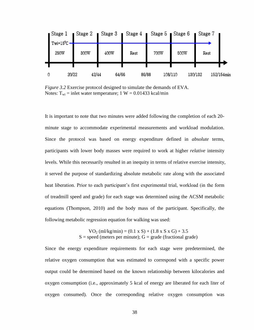
38
Figure 3.2 Exercise protocol designed to simulate the demands of EVA.
Notes: Twi = inlet water temperature; 1 W = 0.01433 kcal/min
It is important to note that two minutes were added following the completion of each 20-
minute stage to accommodate experimental measurements and workload modulation.
Since the protocol was based on energy expenditure defined in absolute terms,
participants with lower body masses were required to work at higher relative intensity
levels. While this necessarily resulted in an inequity in terms of relative exercise intensity,
it served the purpose of standardizing absolute metabolic rate along with the associated
heat liberation. Prior to each participant’s first experimental trial, workload (in the form
of treadmill speed and grade) for each stage was determined using the ACSM metabolic
equations (Thompson, 2010) and the body mass of the participant. Specifically, the
following metabolic regression equation for walking was used:
VO2 (ml/kg/min) = (0.1 x S) + (1.8 x S x G) + 3.5
S = speed (meters per minute); G = grade (fractional grade)
Since the energy expenditure requirements for each stage were predetermined, the
relative oxygen consumption that was estimated to correspond with a specific power
output could be determined based on the known relationship between kilocalories and
oxygen consumption (i.e., approximately 5 kcal of energy are liberated for each liter of
oxygen consumed). Once the corresponding relative oxygen consumption was
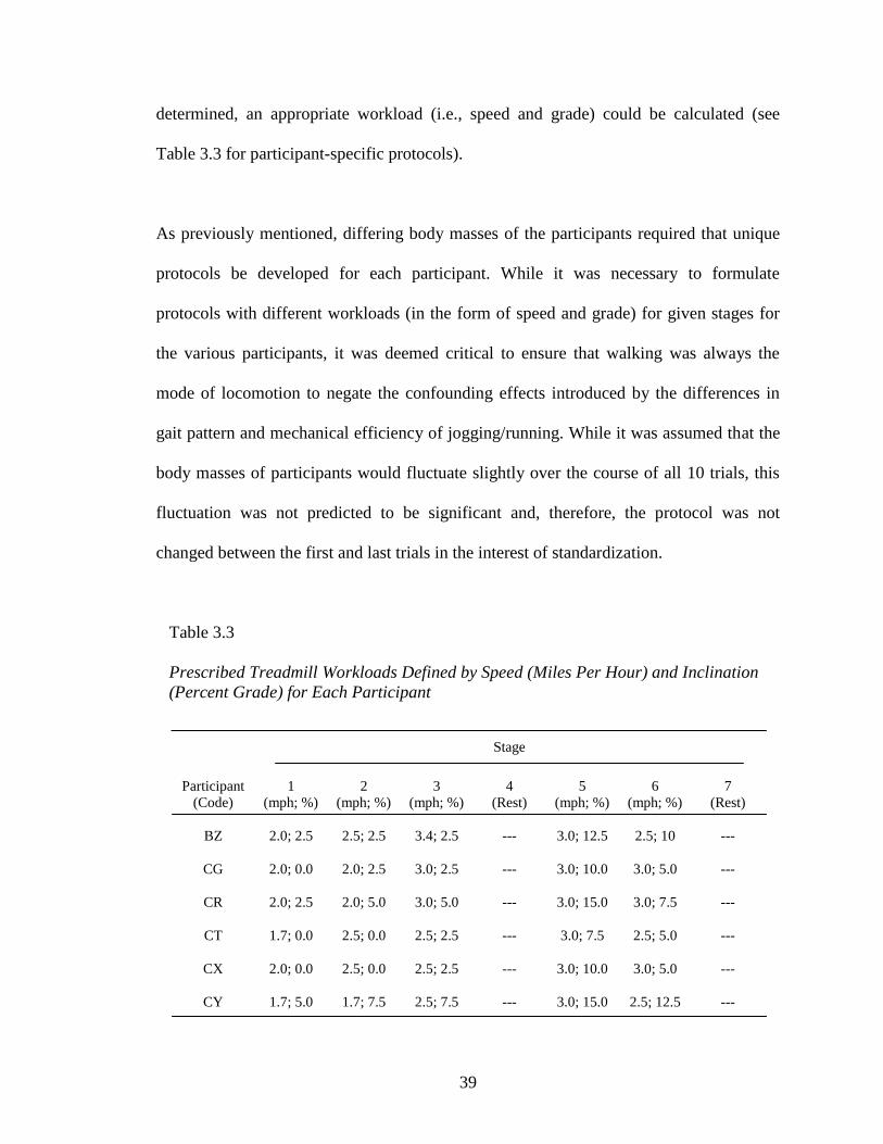
39
determined, an appropriate workload (i.e., speed and grade) could be calculated (see
Table 3.3 for participant-specific protocols).
As previously mentioned, differing body masses of the participants required that unique
protocols be developed for each participant. While it was necessary to formulate
protocols with different workloads (in the form of speed and grade) for given stages for
the various participants, it was deemed critical to ensure that walking was always the
mode of locomotion to negate the confounding effects introduced by the differences in
gait pattern and mechanical efficiency of jogging/running. While it was assumed that the
body masses of participants would fluctuate slightly over the course of all 10 trials, this
fluctuation was not predicted to be significant and, therefore, the protocol was not
changed between the first and last trials in the interest of standardization.
Table 3.3
Prescribed Treadmill Workloads Defined by Speed (Miles Per Hour) and Inclination
(Percent Grade) for Each Participant
Stage
Participant
(Code)
1
(mph; %)
2
(mph; %)
3
(mph; %)
4
(Rest)
5
(mph; %)
6
(mph; %)
7
(Rest)
BZ 2.0; 2.5 2.5; 2.5 3.4; 2.5 --- 3.0; 12.5 2.5; 10 ---
CG 2.0; 0.0 2.0; 2.5 3.0; 2.5 --- 3.0; 10.0 3.0; 5.0 ---
CR 2.0; 2.5 2.0; 5.0 3.0; 5.0 --- 3.0; 15.0 3.0; 7.5 ---
CT 1.7; 0.0 2.5; 0.0 2.5; 2.5 --- 3.0; 7.5 2.5; 5.0 ---
CX 2.0; 0.0 2.5; 0.0 2.5; 2.5 --- 3.0; 10.0 3.0; 5.0 ---
CY 1.7; 5.0 1.7; 7.5 2.5; 7.5 --- 3.0; 15.0 2.5; 12.5 ---
40
Environmental Conditions
All testing occurred in an environmental chamber that was a custom design involving the
conversion of a walk-in refrigerator and the associated heating/cooling system. For the
thermoneutral and hot conditions, the chamber temperature was set at approximately
24 oC and 35
oC, respectively. While humidity is a significant contributor to heat stress,
the ability to control the humidity of the chamber was limited. The temperature and
humidity of the chamber during the experimental trials were monitored and recorded with
a calibrated Extech model RH390 digital psychrometer (Extech Corp., Waltham, MA).
Data Collection
Metabolic Data
Metabolic data was collected continuously over the course of each 154-minute trial.
Specifically, indirect calorimetry was used to analyze, on a breath-by-breath basis, the
inspired and expired gases of the participant via open-circuit spirometry. A Medgraphics
CPX/D metabolic analyzing system with Breeze Ex version 3.06 software (Medical
Graphics Corp., St. Paul, MN) was used to analyze the volume and concentration of
inspired and expired air which was collected utilizing a Hans Rudolph 7900 Series
facemask (Hans Rudolph Inc., Shawnee, KS) fitted with a Medgraphics preVent®
pneumotach (Medical Graphics Corp., St. Paul, MN) which was connected to the
metabolic unit by way of a tubing assembly. The metabolic analyzer was calibrated prior
to each test similarly to that described previously in the “Pre-Screening Procedures”
section. The inspired and expired air yielded the following variables of interest: relative
41
oxygen consumption (ml/kg/min), absolute oxygen consumption (l/min), respiratory
exchange ratio (no units), energy expenditure (kcal/min), and ventilation (l/min).
The metabolic data collected during the middle 10 minutes of each 20-minute stage (i.e.,
minutes 5-15) were averaged and statistically analyzed. The first five minutes of
metabolic data was not used due to the fact that steady-state exercise typically takes
between three and four minutes to achieve (McArdle et al., 2010) and the dynamics of
oxygen uptake kinetics during the initial minutes following the onset of a new workload
is variable between individuals. It was therefore assumed that steady-state exercise was
achieved by the fifth minute into a new stage. The last five minutes of data for each stage
was similarly omitted from analysis for two reasons. First, participants were being asked
questions (e.g., rating of perceived exertion, temperature perception, etc.) during the last
five minutes of each stage which could have been a sufficient stimulus to alter
metabolism via a psychophysiological response. Second, due to the nature of the exercise
and environmental conditions, cardiac drift (Rowell, 1993) could potentially have begun
to alter physiological responses (particularly in the hot conditions) after 15 minutes at a
given exercise intensity.
It should be noted that metabolic data was not collected for the middle 10 minutes of the
first rest period (stage four) which was done for two reasons. First, it was determined
during pilot testing that the nature of the facemask and headgear assembly caused
significant discomfort (particularly in the form of head pain) when worn continuously for
154 minutes. Second, due to the large amount of data collected over the course of 154
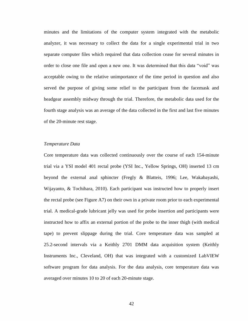
42
minutes and the limitations of the computer system integrated with the metabolic
analyzer, it was necessary to collect the data for a single experimental trial in two
separate computer files which required that data collection cease for several minutes in
order to close one file and open a new one. It was determined that this data “void” was
acceptable owing to the relative unimportance of the time period in question and also
served the purpose of giving some relief to the participant from the facemask and
headgear assembly midway through the trial. Therefore, the metabolic data used for the
fourth stage analysis was an average of the data collected in the first and last five minutes
of the 20-minute rest stage.
Temperature Data
Core temperature data was collected continuously over the course of each 154-minute
trial via a YSI model 401 rectal probe (YSI Inc., Yellow Springs, OH) inserted 13 cm
beyond the external anal sphincter (Fregly & Blatteis, 1996; Lee, Wakabayashi,
Wijayanto, & Tochihara, 2010). Each participant was instructed how to properly insert
the rectal probe (see Figure A7) on their own in a private room prior to each experimental
trial. A medical-grade lubricant jelly was used for probe insertion and participants were
instructed how to affix an external portion of the probe to the inner thigh (with medical
tape) to prevent slippage during the trial. Core temperature data was sampled at
25.2-second intervals via a Keithly 2701 DMM data acquisition system (Keithly
Instruments Inc., Cleveland, OH) that was integrated with a customized LabVIEW
software program for data analysis. For the data analysis, core temperature data was
averaged over minutes 10 to 20 of each 20-minute stage.
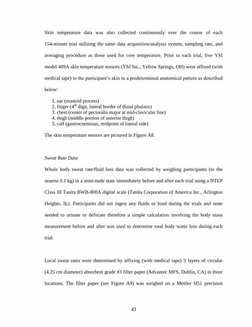
43
Skin temperature data was also collected continuously over the course of each
154-minute trial utilizing the same data acquisition/analysis system, sampling rate, and
averaging procedure as those used for core temperature. Prior to each trial, five YSI
model 409A skin temperature sensors (YSI Inc., Yellow Springs, OH) were affixed (with
medical tape) to the participant’s skin in a predetermined anatomical pattern as described
below:
1. ear (mastoid process)
2. finger (4th
digit, lateral border of distal phalanx)
3. chest (center of pectoralis major at mid-clavicular line)
4. thigh (middle portion of anterior thigh)
5. calf (gastrocnemious, midpoint of lateral side)
The skin temperature sensors are pictured in Figure A8.
Sweat Rate Data
Whole body sweat rate/fluid loss data was collected by weighing participants (to the
nearest 0.1 kg) in a semi-nude state immediately before and after each trial using a NTEP
Class III Tanita BWB-800A digital scale (Tanita Corporation of America Inc., Arlington
Heights, IL). Participants did not ingest any fluids or food during the trials and none
needed to urinate or defecate therefore a simple calculation involving the body mass
measurement before and after was used to determine total body water loss during each
trial.
Local sweat rates were determined by affixing (with medical tape) 3 layers of circular
(4.25 cm diameter) absorbent grade #3 filter paper (Advantec MFS, Dublin, CA) in three
locations. The filter paper (see Figure A9) was weighed on a Mettler H51 precision
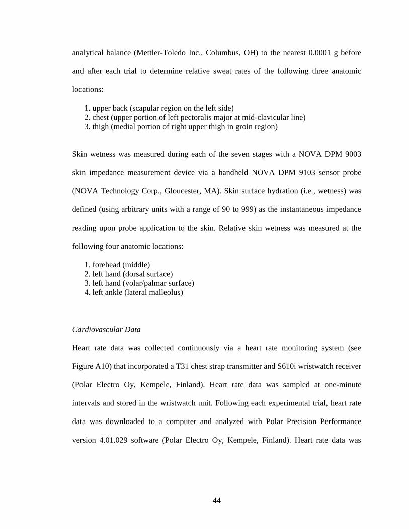
44
analytical balance (Mettler-Toledo Inc., Columbus, OH) to the nearest 0.0001 g before
and after each trial to determine relative sweat rates of the following three anatomic
locations:
1. upper back (scapular region on the left side)
2. chest (upper portion of left pectoralis major at mid-clavicular line)
3. thigh (medial portion of right upper thigh in groin region)
Skin wetness was measured during each of the seven stages with a NOVA DPM 9003
skin impedance measurement device via a handheld NOVA DPM 9103 sensor probe
(NOVA Technology Corp., Gloucester, MA). Skin surface hydration (i.e., wetness) was
defined (using arbitrary units with a range of 90 to 999) as the instantaneous impedance
reading upon probe application to the skin. Relative skin wetness was measured at the
following four anatomic locations:
1. forehead (middle)
2. left hand (dorsal surface)
3. left hand (volar/palmar surface)
4. left ankle (lateral malleolus)
Cardiovascular Data
Heart rate data was collected continuously via a heart rate monitoring system (see
Figure A10) that incorporated a T31 chest strap transmitter and S610i wristwatch receiver
(Polar Electro Oy, Kempele, Finland). Heart rate data was sampled at one-minute
intervals and stored in the wristwatch unit. Following each experimental trial, heart rate
data was downloaded to a computer and analyzed with Polar Precision Performance
version 4.01.029 software (Polar Electro Oy, Kempele, Finland). Heart rate data was
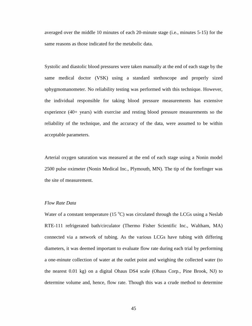
45
averaged over the middle 10 minutes of each 20-minute stage (i.e., minutes 5-15) for the
same reasons as those indicated for the metabolic data.
Systolic and diastolic blood pressures were taken manually at the end of each stage by the
same medical doctor (VSK) using a standard stethoscope and properly sized
sphygmomanometer. No reliability testing was performed with this technique. However,
the individual responsible for taking blood pressure measurements has extensive
experience (40+ years) with exercise and resting blood pressure measurements so the
reliability of the technique, and the accuracy of the data, were assumed to be within
acceptable parameters.
Arterial oxygen saturation was measured at the end of each stage using a Nonin model
2500 pulse oximeter (Nonin Medical Inc., Plymouth, MN). The tip of the forefinger was
the site of measurement.
Flow Rate Data
Water of a constant temperature (15 oC) was circulated through the LCGs using a Neslab
RTE-111 refrigerated bath/circulator (Thermo Fisher Scientific Inc., Waltham, MA)
connected via a network of tubing. As the various LCGs have tubing with differing
diameters, it was deemed important to evaluate flow rate during each trial by performing
a one-minute collection of water at the outlet point and weighing the collected water (to
the nearest 0.01 kg) on a digital Ohaus DS4 scale (Ohaus Corp., Pine Brook, NJ) to
determine volume and, hence, flow rate. Though this was a crude method to determine
46
flow rate, the error with such a technique was deemed insignificant for the purposes of
this study.
Statistical Analysis
A three-factor (temperature x stage x garment) within subjects repeated measures
ANOVA was used to identify differences in cardiovascular, temperature, metabolic, local
sweat rate, and skin wetness values. As it relates to the aforementioned parameters, only
the data collected during stages five (highest-intensity exercise) and seven (rest) were
analyzed making “stage” a two-level factor along with temperature (i.e., 24 oC and 35
oC).
Where significant main effects for garment were found by ANOVA, post-hoc contrasts
were performed using independent two-tailed t-tests. In order to avoid the confounding
effects of a hood (which was not an option for the LCVG), three post-hoc contrasts were
performed on the three garments without the hood (i.e., MACS-Delphi-no hood vs.
Orlan-no hood, MACS-Delphi-no hood vs. LCVG-no hood, and LCVG-no hood vs.
Orlan-no hood) in addition to a hood/no hood contrast (i.e., MACS-Delphi-hood and
Orlan-hood vs. MACS-Delphi-no hood and Orlan-no hood). All data undergoing
ANOVA were tested for assumptions of sphericity using Mauchly’s sphericity test;
Greenhouse-Geisser and Huynh-Feldt corrections were used in cases of departure from
the assumption of sphericity. The ANOVA and post-hoc tests were performed using the
statistical program R, version 2.9 (R: A Language and Environment for Statistical
Computing, Vienna, Austria). Total body sweat rate and water flow rate were analyzed
using independent 2-tailed t-tests with Microsoft Excel 2003 (Microsoft Inc., Redmond,
WA). Statistical significance for all analyses was set at an alpha level of p ≤ 0.05.
47
CHAPTER 4 - RESULTS
Cardiovascular
Table 4.1 summarizes the statistical analysis of the cardiovascular parameters. Means and
standard deviations for the cardiovascular data are presented in Tables B1-B4. When
comparing the three garments without a hood across stages five and seven and across the
24 oC and 35
oC conditions, systolic blood pressure was lower for the MACS-Delphi
when compared to both the LCVG (123.29 vs. 126.92 mmHg; p = 0.023) and the Orlan
(123.29 vs. 128.17 mmHg; p = 0.004). When comparing the MACS-Delphi and LCVG
(both without a hood) across stages five and seven and across the 24 oC and 35
oC
conditions, heart rate was lower for the MACS-Delphi when compared to the LCVG
(103.36 vs. 106.66 bpm; p = 0.039). It should be noted that when a direct comparison was
made between the hood and no-hood conditions for the MACS-Delphi and Orlan across
both stages and temperature conditions (as a means to evaluate the effects of using a
hood), heart rate was lower without the hood (103.67 vs. 106.36 bpm); however, this
difference failed to reach statistical significance (p = 0.059). There were no statistically
significant differences between the garments with regard to diastolic blood pressure and
arterial oxygen saturation.
Metabolic
Table 4.2 summarizes the statistical analysis of the metabolic parameters. Means and
standard deviations for the metabolic data are presented in Tables B5-B8. There were no
statistically significant differences between the garments with regard to relative oxygen
consumption, absolute oxygen consumption, respiratory exchange ratio and ventilation.

48
Core and Skin Temperature
Table 4.3 summarizes the statistical analysis of the core and skin temperature parameters.
Means and standard deviations for the core and skin temperature data are presented in
Tables B9-B14. When comparing the three garments without a hood across stages five
and seven and across the 24 oC and 35
oC conditions, calf skin temperature was lower for
the LCVG when compared to both the MACS-Delphi (28.71 vs. 30.80 oC; p = 0.005) and
the Orlan (28.71 vs. 31.68 oC; p = 0.008). When comparing the MACS-Delphi and Orlan
(both without a hood) across stages five and seven and across the 24 oC and 35
oC
conditions, chest skin temperature was lower for the Orlan when compared to the MACS-
Delphi (27.94 vs. 30.01 oC; p = 0.025). When comparing the three garments without a
hood across stages five and seven and across the 24 oC and 35
oC conditions, femoralis
skin temperature was lower for the LCVG when compared to both the MACS-Delphi
(28.29 vs. 31.72 oC; p = 0.001) and the Orlan (28.29 vs. 31.09
oC; p = 0.011). There were
no statistically significant differences between the garments with regard to core
temperature or skin temperature for the ear or finger.
Local Sweat Rate
Table 4.4 summarizes the statistical analysis of the local sweat rate parameters. Means
and standard deviations for the local sweat rate data are presented in Tables B15-B17.
When comparing the three garments without a hood across stages five and seven and
across the 24 oC and 35
oC conditions, local sweat rate in the groin was lower for the
LCVG when compared to both the MACS-Delphi (0.14 vs. 0.29 g; p = 0.017) and the
Orlan (0.14 vs. 0.25 g; p = 0.015). When comparing the three garments without a hood
49
across stages five and seven and across the 24 oC and 35
oC conditions, ANOVA for local
sweat rate in the back indicated a statistically insignificant effect (p = 0.066) but post-hoc
testing was suggestive (due to reduced statistical power caused by a small sample size) of
the following: local sweat rate was higher for the MACS-Delphi when compared to both
the LCVG (0.95 vs. 0.59 g; p = 0.046) and the Orlan (0.95 vs. 0.67 g; p = 0.006). There
were no statistically significant differences between the garments with regard to local
sweat rate in the chest.
Skin Wetness
Table 4.5 summarizes the statistical analysis of the skin wetness parameters. Means and
standard deviations for the skin wetness data are presented in Tables B18-B21. There
were no statistically significant differences between the garments with regard to skin
wetness in the forehead, hand, palm, or ankle regions.
Water Flow Rate
Means and standard deviations for the water flow rate data are presented in Table B22. In
terms of water flow rate through the garments, the air temperature (24 vs. 35 oC) did not
significantly influence garment performance. While the MACS-Delphi (without hood)
did exhibit a seemingly large difference in flow rates between the 24 oC and 35
oC
conditions (333.40 and 236.33 ml/min, respectively), this difference was not statistically
significant (p = 0.085). As it relates to flow rate between the various garments (without
the hood option), there were significant differences in the absolute flow rate. When the
three garments were compared (averaged across both temperature conditions), the
50
MACS-Delphi had a significantly lower flow rate than the LCVG (280.45 vs.
1985.91 ml/min; p < 0.001) and the Orlan (280.45 vs. 2385.75 ml/min; p < 0.001);
additionally, the LCVG had a lower flow rate than the Orlan (1985.91 vs.
2385.75 ml/min; p < 0.001).
Whole Body Sweat Rate
Means and standard deviations for the whole body sweat rate data are presented in
Table B23. In the 24 oC regime, whole body sweat rate was lower in the LCVG (no hood)
condition compared to the Orlan (no hood) condition (0.45 vs. 0.63 kg; p = 0.007). Also
in the 24 oC regime, it should be noted that, while there was a trend for whole body sweat
rate to be lower in the LCVG (no hood) condition compared to the Orlan (hood)
condition (0.45 vs. 0.75 kg), this difference was not statistically significant (p = 0.055).
In the 35 oC regime, whole body sweat rate was lower in the LCVG (no hood) condition
compared to the Orlan (no hood) condition (0.72 vs. 0.93 kg; p = 0.004), the MACS-
Delphi (no hood) condition (0.72 vs. 0.95 kg; p = 0.001), and the MACS-Delphi (hood)
condition (0.72 vs. 0.93 kg; p = 0.002). When the hooded conditions were compared to
the non-hooded conditions (MACS-Delphi and Orlan) in both temperature regimes to
determine if there was an effect for the hood, the statistical analysis revealed no
significant differences.
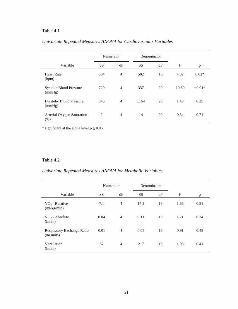
51
Table 4.1
Univariate Repeated Measures ANOVA for Cardiovascular Variables
Numerator Denominator
Variable SS dF SS dF F p
Heart Rate 504 4 502 16 4.02 0.02*
(bpm)
Systolic Blood Pressure 720 4 337 20 10.69 <0.01*
(mmHg)
Diastolic Blood Pressure 345 4 1164 20 1.48 0.25
(mmHg)
Arterial Oxygen Saturation 2 4 14 20 0.54 0.71
(%)
* significant at the alpha level p ≤ 0.05
Table 4.2
Univariate Repeated Measures ANOVA for Metabolic Variables
Numerator Denominator
Variable SS dF SS dF F p
VO2 - Relative 7.1 4 17.2 16 1.66 0.21
(ml/kg/min)
VO2 - Absolute 0.04 4 0.11 16 1.21 0.34
(l/min)
Respiratory Exchange Ratio 0.01 4 0.05 16 0.91 0.48
(no units)
Ventilation 57 4 217 16 1.05 0.41
(l/min)
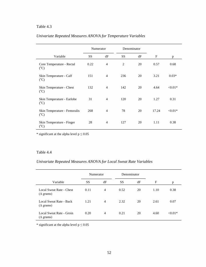
52
Table 4.3
Univariate Repeated Measures ANOVA for Temperature Variables
Numerator Denominator
Variable SS dF SS dF F p
Core Temperature - Rectal 0.22 4 2 20 0.57 0.68
(oC)
Skin Temperature - Calf 151 4 236 20 3.21 0.03*
(oC)
Skin Temperature - Chest 132 4 142 20 4.64 <0.01*
(oC)
Skin Temperature - Earlobe 31 4 120 20 1.27 0.31
(oC)
Skin Temperature - Femoralis 268 4 78 20 17.24 <0.01*
(oC)
Skin Temperature - Finger 28 4 127 20 1.11 0.38
(oC)
* significant at the alpha level p ≤ 0.05
Table 4.4
Univariate Repeated Measures ANOVA for Local Sweat Rate Variables
Numerator Denominator
Variable SS dF SS dF F p
Local Sweat Rate - Chest 0.11 4 0.52 20 1.10 0.38
(∆ grams)
Local Sweat Rate - Back 1.21 4 2.32 20 2.61 0.07
(∆ grams)
Local Sweat Rate - Groin 0.20 4 0.21 20 4.60 <0.01*
(∆ grams)
* significant at the alpha level p ≤ 0.05
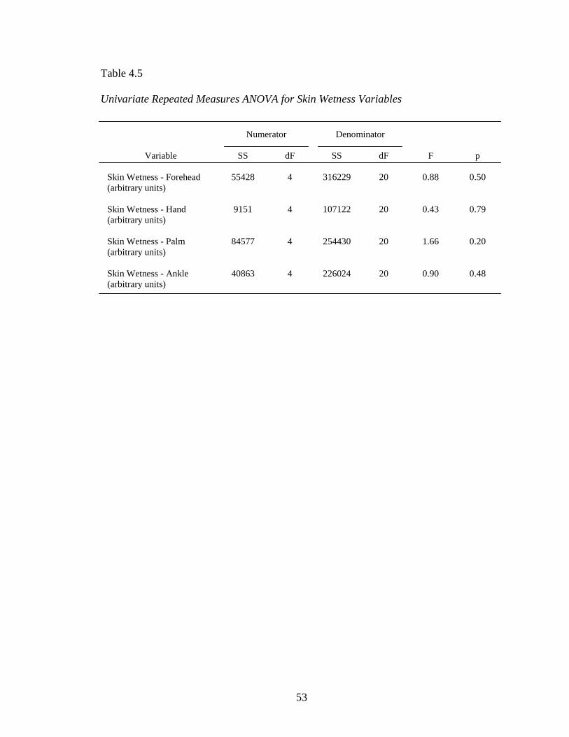
53
Table 4.5
Univariate Repeated Measures ANOVA for Skin Wetness Variables
Numerator Denominator
Variable SS dF SS dF F p
Skin Wetness - Forehead 55428 4 316229 20 0.88 0.50
(arbitrary units)
Skin Wetness - Hand 9151 4 107122 20 0.43 0.79
(arbitrary units)
Skin Wetness - Palm 84577 4 254430 20 1.66 0.20
(arbitrary units)
Skin Wetness - Ankle 40863 4 226024 20 0.90 0.48
(arbitrary units)
54
CHAPTER 5 - DISCUSSION
The purpose of this study was to identify the advanced features of three different liquid
cooling garments in the context of simulated extravehicular activity (i.e., walking on a
treadmill) in two separate temperature regimes (24 oC and 35
oC). Since two garments
(MACS-Delphi and Orlan) allowed for the addition of a cooling hood, there were 10
potential experimental conditions. Each participant performed 10 trials (one for each
potential garment and temperature combination). In terms of the ambient air temperature,
the goal was to create two distinctly different environmental conditions, one
thermoneutral (24 oC) and the other hot (35
oC). The environmental chamber in which all
testing took place was a converted walk-in refrigeration unit that was customized for the
express purpose of human testing in various temperature regimes. The crude nature of the
testing chamber did not allow for precise control of air temperature and was not capable
of modulating humidity; collectively, these two variables represent a potentially
significant environmental heat stressor to humans (Armstrong, 2000). Despite the
limitations of the chamber design, the overall averages of the 30 thermoneutral trials and
the 30 hot trials were very close to the prescribed regimes at 25.0 oC (± 1.3
oC) and
35.6 oC (± 0.5
oC), respectively. For the 30 thermoneutral and 30 hot trials, the
temperature ranged from 22.5-27.4 oC and 34.0-36.5
oC, respectively. The air humidity
likely did not contribute to a significantly increased heat stress due to the fact that
humidity was generally low in both the thermoneutral and hot conditions (28.5 and
18.4%, respectively). Although there were slight variations in ambient temperature
between experimental trials, these were deemed inconsequential and the goal of creating
two separate temperature regimes (i.e., thermoneutral and hot) was achieved.
55
The purpose of the exercise protocol was to model (using a treadmill) the energy
expenditure profile used by NASA to simulate the metabolic demands of extravehicular
activity (EVA). In particular, EVA on a planetary surface (e.g., the Moon, Mars, etc.)
involves a significant increase in energy expenditure (Waligora & Kumar, 1995). As
metabolic heat load increases in the context of increased energy expenditure in an
enclosed and impermeable microclimate (i.e., space suit), the management of the
resulting psychological and physiological milieu is of the utmost importance. The
consequences of failing to properly regulate body temperature during EVA go far beyond
discomfort, and can have catastrophic results. In order to estimate the treadmill speed and
grade that would elicit the prescribed energy expenditure during various stages of
exercise, a regression equation (Thompson, 2010) was used. A metabolic analyzing
system was used to measure energy expenditure over the course of each experimental
trial in order to record the actual energy expenditure levels across the various stages. The
actual energy expenditure profile was quite close to the prescribed profile as seen in
Table 5.1. Estimating energy expenditure from regression equations is often imperfect
owing to the fact that there can be significant variability between and within individuals.
However, the actual energy expenditures measured within this study were remarkably
close to the predicted values which demonstrates a certain degree of utility to an equation
that some have questioned in terms of validity and reliability. Despite the small
variability in actual levels of energy expenditure for a given exercise stage, the goal of
creating distinctly different energy expenditure rates (for different exercise stages) was
achieved.
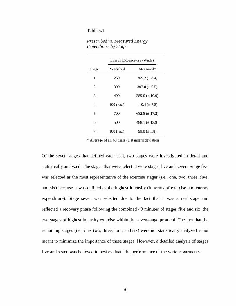
56
Table 5.1
Prescribed vs. Measured Energy
Expenditure by Stage
Energy Expenditure (Watts)
Stage Prescribed Measured*
1 250 269.2 (± 8.4)
2 300 307.8 (± 6.5)
3 400 389.0 (± 10.9)
4 100 (rest) 110.4 (± 7.8)
5 700 682.8 (± 17.2)
6 500 488.1 (± 13.9)
7 100 (rest) 99.0 (± 5.8)
* Average of all 60 trials (± standard deviation)
Of the seven stages that defined each trial, two stages were investigated in detail and
statistically analyzed. The stages that were selected were stages five and seven. Stage five
was selected as the most representative of the exercise stages (i.e., one, two, three, five,
and six) because it was defined as the highest intensity (in terms of exercise and energy
expenditure). Stage seven was selected due to the fact that it was a rest stage and
reflected a recovery phase following the combined 40 minutes of stages five and six, the
two stages of highest intensity exercise within the seven-stage protocol. The fact that the
remaining stages (i.e., one, two, three, four, and six) were not statistically analyzed is not
meant to minimize the importance of these stages. However, a detailed analysis of stages
five and seven was believed to best evaluate the performance of the various garments.
57
Physiological Findings
Metabolism
Metabolism was assessed using a metabolic analyzing system and open-circuit
spirometry. There were no differences in the four variables that were analyzed (i.e.,
relative oxygen consumption, absolute oxygen consumption, ventilation, and respiratory
exchange ratio). The oxygen consumption data indicate that the participants were
exercising at similar intensities during stage five over the course of the 10 trials. The goal
of the study design was to standardize the exercise intensity for each individual and stage
so as to minimize the confounding influence that different exercise intensities (for a given
participant and stage) would have on thermoregulation. It is therefore concluded that any
differences in the analyzed variables are not attributable to differing levels of energy
expenditure. It is assumed that any observed differences are the result of the garment
being used. However, there is the possibility that other factors confounded the results;
this important topic is discussed later in the chapter. The similar respiratory exchange
ratios indicate that substrate utilization (i.e., carbohydrate vs. fat) was the same for each
participant throughout the 10 trials. This was the expected result for two reasons: 1) it
would be unlikely to see significant differences in the respiratory exchange ratio without
concomitant changes in oxygen consumption as both reflect exercise intensity (Brooks,
Fahey, & Baldwin, 2005) and 2) most of the evidence for temperature-mediated
alterations in substrate utilization is relegated to the other side of the thermal spectrum,
namely in cold environments when carbohydrate usage is increased (Brooks et al., 2005;
Pandolf, Sawka, & Gonzalez, 2001).
58
Skin and Core Temperature
There were no differences between the garments in terms of core/rectal temperature. This
illustrates the collective effectiveness of the three garment types in terms of maintaining
core temperature, particularly in the context of the increasing heat load that occurred
during stage five (exercise). The average core temperatures for all five garment
combinations were maintained in a very narrow range during stage five (highest intensity
exercise) for both the thermoneutral and hot conditions (37.33 - 37.58 oC and
37.52 - 37.75 oC, respectively). The core temperature profiles for the different garments
over the course of the entire 154-minute protocol were also quite similar in both the 24 oC
(Figure 5.1) and 35 oC (Figure 5.2) regimes. Considering that normal core body
temperature is 37.0 oC, the results suggest that the garments evaluated in this study were
effective at attenuating the heat load generated by exercise and heat exposure. This is an
important finding considering the fact that core temperature is traditionally one of the
three vital measures (i.e., temperature, blood pressure, and heart rate) and its regulation is
crucial for future space missions and the safety of astronauts performing EVA. As it
relates to core temperature regulation, all three garment types performed similarly in both
the thermoneutral and hot environments. This is perhaps the most salient finding of the
current study because it provides evidence that water flow rate (through a garment) is not
a critical factor. This finding is in contrast to the findings of Cadarette et al. in which the
authors speculate that low flow rates (i.e., 0.4 l/min) hinder heat transfer due to an
increased presence of air bubbles that block the coolant flow and reduce heat extraction
(Cadarette, DeCristofano, Speckman, & Sawka, 1990). While the same air bubble
phenomenon
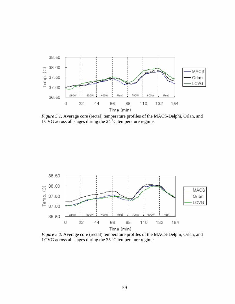
59
Figure 5.1. Average core (rectal) temperature profiles of the MACS-Delphi, Orlan, and
LCVG across all stages during the 24 oC temperature regime.
Figure 5.2. Average core (rectal) temperature profiles of the MACS-Delphi, Orlan, and
LCVG across all stages during the 35 oC temperature regime.
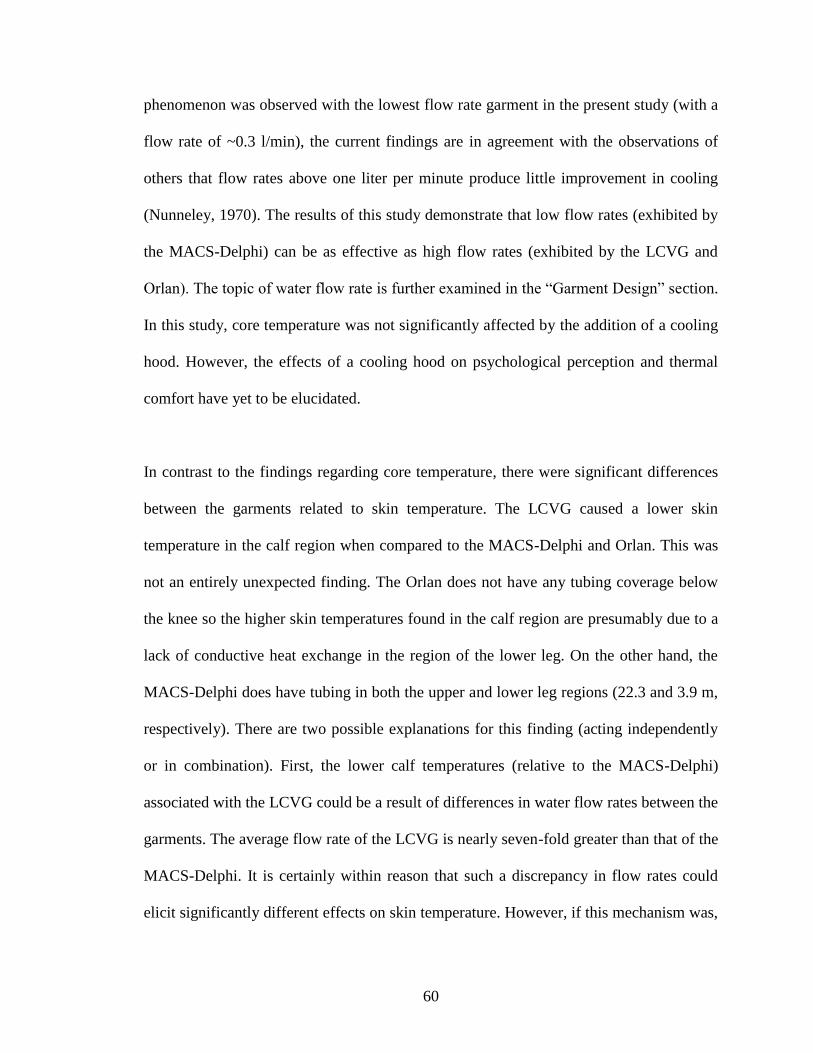
60
phenomenon was observed with the lowest flow rate garment in the present study (with a
flow rate of ~0.3 l/min), the current findings are in agreement with the observations of
others that flow rates above one liter per minute produce little improvement in cooling
(Nunneley, 1970). The results of this study demonstrate that low flow rates (exhibited by
the MACS-Delphi) can be as effective as high flow rates (exhibited by the LCVG and
Orlan). The topic of water flow rate is further examined in the “Garment Design” section.
In this study, core temperature was not significantly affected by the addition of a cooling
hood. However, the effects of a cooling hood on psychological perception and thermal
comfort have yet to be elucidated.
In contrast to the findings regarding core temperature, there were significant differences
between the garments related to skin temperature. The LCVG caused a lower skin
temperature in the calf region when compared to the MACS-Delphi and Orlan. This was
not an entirely unexpected finding. The Orlan does not have any tubing coverage below
the knee so the higher skin temperatures found in the calf region are presumably due to a
lack of conductive heat exchange in the region of the lower leg. On the other hand, the
MACS-Delphi does have tubing in both the upper and lower leg regions (22.3 and 3.9 m,
respectively). There are two possible explanations for this finding (acting independently
or in combination). First, the lower calf temperatures (relative to the MACS-Delphi)
associated with the LCVG could be a result of differences in water flow rates between the
garments. The average flow rate of the LCVG is nearly seven-fold greater than that of the
MACS-Delphi. It is certainly within reason that such a discrepancy in flow rates could
elicit significantly different effects on skin temperature. However, if this mechanism was,
61
in fact, responsible for the differences in skin temperatures between garments, then
similar differences would be expected in all of the skin regions for which temperature
was measured (similar differences were not found at all skin measurement sites). Second,
the differences could be a result of the amount of tubing in the lower leg segment of the
garments. The MACS-Delphi has a collective tubing length of 26.2 m in the lower
extremity (i.e., thighs and calves) compared to a total length of 37.8 m in the same
anatomic region of the LCVG. While the exact specifications for lower leg (i.e., below
the knee) coverage of the LCVG garment are not available, it is reasonable to assume that
the LCVG design covers a greater area of the lower leg region than that of the MACS-
Delphi (i.e., 3.9 m) owing to the greater overall length of tubing in the collective lower
extremity region. If there is, in fact, a greater amount of tubing coverage in the lower leg
region of the LCVG, the lower calf skin temperatures in the LCVG could simply be the
result of increased heat transfer (via conduction) due to increased skin surface area in
contact with the water-cooled tubing network.
In terms of chest skin temperature, the Orlan elicited significantly lower temperatures
than the MACS-Delphi. As discussed previously, a difference in skin temperatures
between garments could be due to different water flow rates. In this case, there were vast
differences between the Orlan and MACS-Delphi garments (2385.75 and 284.87 ml/min,
respectively). Interestingly, and in contrast with the findings regarding the calf region, the
garment with lesser absolute tubing coverage in a defined area elicited lower skin
temperatures than the garment with greater coverage. The Orlan has 7.79 m of tubing
length in the chest/abdomen region compared to 11.7 m for the same region of the

62
MACS-Delphi. If differences in water flow rate do not account for the differences in
chest skin temperature, the explanation is less clear. One possible reason could be related
to the tightness of the garments. Although not quantitatively measured, it was observed
that the Orlan garment was generally the tightest fitting garment of the three, particularly
in the chest/upper back region. It is possible that a tighter fit resulted in a greater surface
area of tubing in direct contact with the skin. Obviously, a tighter fit would result in
greater surface area contact than a looser fit if everything else was equal. This potentially
could have resulted in greater heat transfer, despite less overall tubing length in the
region. Another possible explanation has to do with the diameter of the tubing. Of the
three garments, the MACS-Delphi has the smallest diameter tubing (inner and outer
diameters of 2.4 and 4.0 mm, respectively) whereas the Orlan has the largest diameter
tubing. If all else is equal, a larger diameter tube will have a greater surface area in
contact with the skin than a smaller diameter tube. Perhaps the Orlan actually had a
greater amount of tubing surface area in contact with the skin (by virtue of a greater tube
diameter) than the MACS-Delphi and this increase in heat transfer potential outweighed
the decrease in heat transfer potential associated with a lesser absolute length of tubing.
The final significant finding related to skin temperature occurred in the femoralis (thigh)
region. The femoralis skin temperature was significantly lower with the LCVG than both
the MACS-Delphi and Orlan. Water flow rate does not explain the observed difference
because, while the LCVG had a significantly greater flow rate than the MACS-Delphi,
the Orlan had a significantly greater flow rate than the LCVG. Tubing diameter does not
explain the difference either because the tubing diameter of the Orlan is greater than that
63
of the LCVG. The most likely explanation has to do with amount (i.e., length) of tubing
coverage since the MACS-Delphi and Orlan have virtually identical lengths of tubing in
the thigh area (22.3 and 22.4 m, respectively). As was mentioned previously, the total
amount of tubing in the lower extremity of the LCVG is 37.8 m. However, no distinction
is made in the garment specifications for the length of tubing in the upper versus lower
legs of the LCVG. The fact that the LCVG contains a much greater absolute length of
tubing in the lower extremity compared to the MACS-Delphi and Orlan (37.8 vs. 26.2 m
and 37.8 vs. 22.4 m, respectively) makes it reasonable to conclude that there is a much
greater length of tubing in the thigh area of the LCVG compared to the MACS-Delphi
and Orlan. If the LCVG does, in fact, have a greater length of tubing than the other two
garments, then an increased surface area coverage (and accompanying increased potential
for conductive heat exchange) would seem to be the most logical explanation for the
significantly lower femoralis temperatures observed in the LCVG.
Cardiovascular Parameters
There were two very interesting findings related to systolic blood pressure and heart rate.
The MACS-Delphi elicited a significantly lower systolic blood pressure response than
both the LCVG and Orlan. While this finding is noteworthy, the mechanism of such a
blood pressure response is unclear. Certainly, the regulation of systemic blood pressure is
complicated, involves the interactions of numerous complex systems, and is not fully
understood. It is accepted that, in a healthy individual, systolic blood pressure increases
as energy expenditure (i.e., workload) increases and it does so in a roughly linear fashion.
This increase in blood pressure with physical exertion is caused by a disproportionate
64
increase in cardiac output relative to the decrease in peripheral vascular resistance. It is
not out of the realm of possibility that there is an interaction between blood pressure and
temperature regulation, as both have a significant vascular component (particularly the
smaller arterioles) in common. Perhaps the most significant contributor to heat balance
within the human body lies in the reflex peripheral vasoconstriction and vasodilation
associated with exposure to cool/cold and warm/hot environmental stressors, respectively.
Exercise in hot environments presents an interesting physiological paradox as circulatory
regulation (maintenance of blood pressure in the context of a reduced plasma volume)
competes with temperature regulation (peripheral vasodilation to facilitate heat transfer).
It should be noted, however, that muscle blood flow and circulatory regulation take
precedence over temperature regulation during exercise in warm/hot environments
(McArdle, Katch, & Katch, 2010). The findings associated with systolic blood pressure in
this study may also be related to water flow rates and thermal gradients. In this study, all
circulating water entered the garments at 15 oC. However, due to the significantly lower
flow rates characteristic of the MACS-Delphi, the overall average water temperature
within the garment was warmer, presumably due to the longer transit time. Conversely,
the water within the garments with the high flow rates was, on average, cooler owing to
more rapid transit times. This was substantiated quantitatively by continuous
measurement of inlet versus outlet water temperatures during each trial. It is possible that
systolic blood pressure was higher in the LCVG and Orlan due to a systemic peripheral
vasoconstriction mediated by the greater thermal gradient between the skin and
circulating water. This effect would be expected to be reflected in lower systemic skin
temperatures in the LCVG and Orlan, but the data do not consistently support this.
65
However, the subjective thermal perception (data not presented) of participants show that,
as a whole, participants felt more dramatic thermal changes (towards the cooler/colder
end of the continuum) with the higher flow rate garments (i.e., LCVG and Orlan). While
the objective (skin temperature) and subjective (thermal perception) data are conflicting,
a peripheral vasoconstrictor response (due to a large thermal gradient) can not be ruled
out as a possible mechanism for the difference in systolic blood pressure observed.
In addition to the systolic blood pressure response, there was also a significant difference
in heart rates between the MACS-Delphi and LCVG. Heart rate was, on average, lower in
the MACS-Delphi compared to the LCVG. As with the systolic blood pressure findings,
an explanation for such a difference is not obvious. Heart rate is modulated by the
autonomic nervous system and, like systolic blood pressure, it rises linearly with
increasing workload intensity. The onset of physical exertion is characterized by an
increasing sympathetic involvement accompanied by a withdrawal of the
parasympathetic dominance that is present at rest. There is, however, one particular
phenomenon that is exclusively related to heart rate modulation and exercise in the heat.
Cardiac drift is a term that describes the common observation that heart rate gradually
increases over time (tens of minutes to hours depending on exercise intensity) despite a
constant, or even decreasing, intensity level. In theory, heart rate should remain relatively
constant over time in the context of steady-state exercise at a given intensity. This is not
the case when exercise is performed in hot, or even warm, environments for a prolonged
period of time (i.e., greater than 30-60 minutes). The typical profile of prolonged exercise
in the heat is one of a gradually, yet progressively, increasing heart rate. This is due to a
66
redistribution (and pooling) of blood as well as a decreased plasma volume which is the
result of fluid loss, primarily in the form of sweat. On average, heart rate increases eight
beats per minute for each liter of sweat-loss dehydration (McArdle et al., 2010). This
phenomenon is illustrated by the relationship between cardiac output, heart rate, and
stroke volume which is defined as: [cardiac output = heart rate x stroke volume]. As
plasma volume decreases, venous return to the heart decreases which causes a decrease in
preload and myocardial contractility (i.e., Frank-Starling law of the heart) which
ultimately results in a decreased stroke volume. If cardiac output remains unchanged (in
theory cardiac output should not change if workload remains constant) and stroke volume
decreases, the only option for maintaining cardiac output is an increase in heart rate
(Rowell, 1993; Rowell & Shepherd, 1996). Certainly, by stage five (the exercise stage
analyzed in detail), it would seem there was an adequate duration of exercise in the heat
to potentially cause fluid losses in excess of the threshold required for cardiac drift.
However, the fact that the findings indicate a significant difference in heart rate between
the MACS-Delphi and LCVG without a concomitant difference in sweat-loss dehydration
(the LCVG actually elicited lower whole body fluid losses than the MACS-Delphi) leads
to the distinct possibility that another mechanism is responsible for the difference in heart
rate between the two garments. The difference in garment weight is another possible
factor to consider when attempting to explain the heart rate findings since the LCVG was
heavier than the MACS-Delphi (1.79 vs. 1.43 kg, respectively). A heavier garment would
necessarily require a greater energy expenditure during locomotion (in a gravity-
dependent environment) and greater energy expenditures are usually reflected in higher
heart rates. However, there are two factors that do not support the idea that the MACS-
67
Delphi resulted in a lower heart rate than the LCVG simply due to garment weight. First,
there was no difference in metabolic variables (e.g., oxygen consumption) which would
be expected to mirror the differences in heart rate. Second, the Orlan (2.45 kg) was
heavier than both the LCVG and MACS-Delphi yet the heart rates between the MACS-
Dephi and Orlan were not significantly different.
Local Sweat Rate
Local sweat rate was indirectly estimated by affixing absorbent filter paper to three
regions of the body: the chest, upper back, and groin. Sweat rate was assessed on a
relative scale by weighing the filter paper before and after each trial. The unit of measure
(∆ grams) was used to indirectly assess sweat rate (i.e., a greater change in weight
represents a greater sweat rate). This metric differs from the metabolic, cardiovascular,
skin wetness, and temperature parameters in that it was a simple before-and-after
measurement and represented the seven stages collectively as opposed to the selective
analysis of stages five and seven. The reason for the measurement technique of placing
absorbent patches on the body surface for the entirety of the trial, compared to that of
skin wetness which was measured each stage, was due to the fact that all three regions of
interest were covered by the garment which made it difficult to access on a regular basis
throughout a trial. There were significant differences observed in the groin and back
region. The local sweat rate in the groin was lower in the LCVG compared to the MACS-
Delphi and Orlan. This result is in agreement with the finding that the femoralis skin
temperature was also lower in the LCVG compared to the MACS-Delphi and Orlan
garments. It is likely that the two findings are related due to the proximity of the two
68
anatomic sites of measurement (i.e., femoralis and groin) and the presumed link between
skin temperature and sweat rate. As was discussed with regard to skin temperature, the
most likely explanation is the greater length of tubing in the upper leg region of the
LCVG compared to the MACS-Delphi and Orlan. It seems reasonable to conclude that a
more extensive network of tubing in a given anatomic region results in lower sweating
rates in that region.
In the upper back, the local sweat rate for the MACS-Delphi was greater than that of both
the LCVG and Orlan. The upper back is known to be a region of high sweat rates (Cotter,
Patterson, & Taylor, 1995; Machado-Moreira, Smith, Van Den Heuvel, Mekjavic, &
Taylor, 2008) which is likely attributable to the combination of the several large skeletal
muscle groups (e.g., latissimus dorsi, trapezius, rhomboideus major, etc.) and large bone
structures (i.e., clavicles, ribs, and vertebrae). This collectively represents a large mass of
dense tissue that acts as a heat sink during times of increasing heat load due to the density
of the involved tissues. It is interesting that the MACS-Delphi had a higher sweat rate in
this region because the MACS-Delphi was specifically designed with physiology in mind
as evidenced by the selective focus of tube coverage on high density tissue areas like that
found in the upper back (Koscheyev, Coca, & Leon, 2007; Koscheyev, Coca, Leon, &
Dancisak, 2002). However, an inspection of the garment specifications reveals one
possible explanation for the current findings. The MACS-Delphi has 5.69 m of tubing
length in the back region whereas the Orlan and LCVG have 9.19 and 12.2 m of tubing
length, respectively, in the same region. So, it is possible that the lower surface area
coverage is the cause of the higher sweat rate. Another possibility for the higher sweat
69
rate in the back with the MACS-Delphi is the lower flow rate associated with that
garment which has already been discussed. Finally, the MACS-Delphi has the smallest
diameter tubing which not only affects flow rate but also dictates surface area coverage
independent of length. Any of the aforementioned factors (i.e., tubing length, flow rate,
and/or diameter) could explain the higher sweat rate in the back region with the MACS-
Delphi.
Skin Wetness
Skin wetness was measured using a skin impedance probe. The device used arbitrary
units so the analysis focused on relative differences between the four anatomic sites:
forehead, hand (dorsal), hand (volar), and ankle. This measure was different from local
sweat rate in that all four sites represented skin regions not covered by any garment.
Differences in skin wetness would represent a more systemic effect of a garment (or
garments) as the garment, or its tubing network, was not in direct contact with the skin
site. There were no differences in any skin wetness variables during stages five and seven
indicating that all of the garments performed similarly. This suggests that either: 1) there
were no systemic effects, at least as they apply to the four measured sites, for any of the
garments related to skin wetness; or 2) there was a systemic effect but it was equal
between the different garments.
Whole Body Sweat Rate
Whole body sweat rate was determined simply by weighing in the semi-nude state before
and after each experimental trial. Participants did not consume any solids or liquids
70
during the experimental trials so the assumption that body mass change reflected whole
body sweat rate was straightforward with the exception of not accounting for fluid lost in
the form of water vapor during exhalation (this component was deemed inconsequential
for the purposes of this study). Some of the most consistent findings of the present study
relate to whole body sweat rate with the LCVG demonstrating a favorable effect
compared the MACS-Dephi and Orlan as illustrated by the attenuation of whole body
water loss via sweating. This effect can most likely be explained by the fact that the
LCVG had a greater total amount of tubing (79.4 m) than the MACS-Delphi (58.9 m) or
Orlan (49.4 m). Flow rate may have also exerted an influence but overall tubing length
would seem to be the predominant factor as the flow rate for the Orlan was significantly
greater than that of the LCVG. It can be assumed that the garment with the greatest
overall length of tubing will be in contact with the greatest body surface area. This
greater body cooling (in terms of area) would presumably exert the greatest overall skin
cooling and would certainly have an effect on the sweating response. This finding is
particularly important in the context of heat stress since dehydration beyond certain
thresholds exacerbates, or even perpetuates, heat-related dysfunction (Armstrong, 2000).
Further, this idea is of specific relevance to astronauts during extended EVA since the
current protocol only provides 947 ml of water per EVA sortie (Cowell, Stocks, Evan,
Simonson, & Greenleaf, 2002) despite the fact that astronauts require about one to three
liters of water for every five to six hours of EVA (Greenleaf, 1989). It seems logical that
every effort should be made to conserve water in a situation like EVA that is highlighted
by a growing whole body water deficit over a period of several hours.
71
Garment Design
Two primary characteristics that differentiate liquid cooling garments are: 1) the diameter
of the tubing and 2) the coverage of the tubing network across the body. It is important to
note that both absolute coverage (amount of tubing in direct contact with the skin) and
location of coverage (regions of the body in which tubing is in contact with the skin) are
considered important questions regarding garment design that have yet to be fully
answered (Flouris & Cheung, 2006). The rate of water flow through the garment is an
important consideration. Flow rate is determined, in large part, by the diameter of the
tubing (i.e., larger diameter tubes can accommodate a greater flow rate than smaller
diameter tubes). Common sense may indicate that, if cooling a heated body via
conduction using cool/cold water circulated through tubing in contact with the skin, a
higher flow rate is more effective at transferring heat from the body and, hence, results in
a lower core temperature. In other words, the greater the volume of water being circulated
through the garment per unit time, the greater the heat transfer. The results of the present
study do indicate that heat flow (i.e., heat extraction) was lowest in the garment with the
lowest flow rate (i.e., MACS-Delphi). However, it was not the garment with the greatest
flow rate (i.e., Orlan) that resulted in the greatest heat extraction; the greatest heat
extraction occurred in the garment with a flow rate in-between the highest and lowest
flow rates (i.e., LCVG).
The results suggest that overall heat extraction is not the sole determinant of core
temperature since all three garments elicited similar core temperatures despite different
flow rates and heat extraction rates. As has already been discussed, the different flow
72
rates may have exerted an influence on skin temperatures at various locations but this was
neither consistent nor systemic. This is an important finding since core temperature is
arguably the most important physiological variable in terms of comfort, health, and safety
in the context of EVA. This supports the analogy posed by Taylor in which he asserts that
the human body is not a “bowling ball” of uniform density, known material properties,
and easily quantifiable proportions which lends itself exclusively to common engineering
assumptions and first-principles thermodynamics (Taylor, Casa, & Kenny, 2010). The
fact that the human body does not have homogenous composition and uniform thermal
conductivity highlights the difficulty in attempting to model and predict heat transfer
within a dynamic body that is highlighted by multiple interconnected systems, different
tissue types, and heat transfer facilitated by both conductive and convective (mass) flow
(Taylor et al., 2010).
The coverage of the tubing is an important consideration in the design of a cooling
garment. This study investigated three garments, each of which has unique tubing
arrangements. The LCVG design focuses on absolute surface area and is indiscriminate
in coverage as it relates to the potential for heat transfer via the underlying tissues. The
focus on sheer coverage is evidenced by the total amount of tubing incorporated in the
LCVG (79.4 m) compared to that of the MACS-Delphi and Orlan (58.9 and 49.4 m,
respectively). The MACS-Delphi and Orlan designs have more strategic physiologic
orientations of tubing based on specific types of tissue. The Orlan design focuses tube
distribution primarily in areas of large amounts of muscle mass, presumably to more
effectively remove heat from areas of significant heat generation during physical exertion
73
(i.e., skeletal muscles). The MACS-Delphi is also physiologically designed, with primary
emphasis on high density tissue (i.e., bone). The tubing arrangements of the three
garments evaluated in this study range from seemingly random to very strategic. The
results of this study indicate that there may be some merit to increased quantity of
coverage (at the expense of quality of coverage) if the goal is lowering skin temperature
as is seen in the lower calf and femoralis skin temperatures with the LCVG. However,
these findings must be balanced with the need to preserve thermal comfort (subjective
perception) as well as regulate core temperature. Core temperature was regulated
similarly between all three garments despite the fact that the LCVG had the greatest heat
extraction (which was accompanied by lower perceived thermal comfort).
The addition of a cooling hood is another design option for cooling garments. In this
study, two of the garments had a hood option (i.e., MACS-Delphi and Orlan). The
rationale behind incorporating a hood is to augment conductive heat exchange. Use of the
head for heat exchange is thought to be a potentially effective strategy owing to the large
amount of heat released from this region as well as its unique vasculature. The head can
be thought of as the body’s “chimney” because it is capable of transferring large amounts
of heat due to the dense tissue (i.e., cranium) and the large blood vessels that supply it. A
large proportion of the body’s heat loss occurs via the head (Froese & Burton, 1957;
Rasch, Samson, Cote, & Cabanac, 1991), particularly in extreme thermal environments
like the cold, making it an attractive site for purposeful heat removal by means of a
cooling hood. Anecdotal observations certainly reveal a tendency for many people to
focus first on the head when it comes to cooling off with a wet towel or water in hot
74
environments. This instinctual reflex should not be overlooked. The head also has the
distinction of containing a dense network of blood vessels that are less prone to cold-
induced vasoconstriction (Blair, Glover, & Roddie, 1961; Froese & Burton, 1957;
Hertzman & Roth, 1942; Rasch et al., 1991). There currently exists a great debate as to
the optimal temperature of water for the purpose of lowering core temperature by means
of whole body cooling (e.g., emergency medicine for heat illness) (Taylor et al., 2010).
Some believe the greater the thermal gradient, the greater the conductive/convective heat
transfer so “the colder the better” is the mantra. Others caution against this approach
citing the reflex vasoconstriction that accompanies rapid application of cold to the body’s
outer shell. The belief is that a large thermal gradient is of limited usefulness in the
context of reduced skin blood flow due to vasoconstriction. The vasculature of the head
may allow for the application of cold to very cold water and, hence, a large thermal
gradient and potential for substantial heat transfer from a large heat sink without concern
for a marked vasoconstrictor effect. The results of this study did not show any added
effect of the hood despite the reports of participants that overall thermal comfort was
higher with the hood which is in agreement with previous findings (Simmons, Mundel, &
Jones, 2008). The use of a hood is also a contentious debate with astronauts because the
addition of a hood is seen by many as an unnecessary discomfort from an ergonomic
standpoint. Caution is urged when making conclusions based on the current cooling hood
findings because: 1) the participants all had hair which certainly acted as an insulator and
barrier to heat transfer and 2) the temperature of the water was set at 15 oC and not
decreased to lower temperatures in the head region so it is not possible to determine if the
hood would have been effective at lower temperatures.
75
Limiting Factors
There were several limiting factors in this study, many of which were unavoidable due to
the experimental design. The first limitation is the small sample size (i.e., six
participants). The major disadvantage of such a small sample size is the reduced
statistical power to detect differences between groups. However, this research was meant
to serve more as a pilot study and, from the beginning, the funding agency dictated that
there be a small sample size owing to budgetary constraints. The detailed data collection
of numerous variables and the fact that each participant performed 10 separate
experimental trials allowed for the achievement of the study’s primary goal which was to
evaluate the performance of three different liquid cooling garments and identify the
advanced features of each.
Another limitation was the population used. The study population consisted of volunteers
recruited from the Twin Cities area of Minnesota and, more specifically, the campus of
the University of Minnesota. Not unexpectedly, the majority of participants were students.
The primary concern with the population used is the similarity to the typical profile of
present-day astronauts (for whom this research is based). In particular, the average age of
the participants in this study was 29.7 years whereas the average age of active-duty
astronauts is between 42-43 years (Harm et al., 2001). This discrepancy in age is not
deemed to be a significant factor as there are only subtle differences in thermoregulatory
physiology between the third and fifth decades of life for healthy, fit individuals
(Havenith, Yoshimitsu, & Kenney, 1995; Kenny, Gagnon, Dorman, Hardcastle, & Jay,
2010). Additionally, men and women were recruited for the study since females represent
76
more than one-fifth (22.3% as of August 2001) of the active astronaut population in the
United States (Harm et al., 2001). Men and women were pooled together as a single
group in terms of data analysis (i.e., separate comparisons of men and women were not
performed) due to the methodological and statistical power limitations imposed by small
sub-group analysis (i.e., four males and two females). While differences in core
temperature between sexes has been suggested to be related to differences in physical
characteristics (not due to differences in whole-body thermoregulatory responses)
(Gagnon, Dorman, Ollie, Hardcastle, & Kenny, 2009), it has been noted that systemic
differences in thermoregualation do exist in terms of sweat rate (Gagnon, Jay, Lemire, &
Kenny, 2008) and sweat distribution (Havenith, Fogarty, Bartlett, Smith, & Ventenat,
2008) which could confound the interpretation of the present results.
The issue of physical fitness of the study participants was of paramount concern due to:
1) the strenuous physical requirements of the experimental trials, 2) the relationship
between thermoregulation and physical fitness, and 3) the above-average physical fitness
profile of astronauts. The average peak oxygen consumption for men and women in this
study was 42.6 and 49.0 ml/kg/min, respectively. This index of physical/aerobic fitness
for the study population is higher than the reported average peak oxygen consumption for
active-duty male and female astronauts of 36.5 and 44.2 ml/kg/min, respectively (Harm
et al., 2001). However, when adjusted for age, both groups scored in a similar range for
peak oxygen consumption between the 70th
and 75th
percentile on population-based
normative data for American adults (Thompson, 2010).
77
Body composition was also an important factor due to the relationship between adipose
tissue and heat transfer dynamics (i.e., the insulative properties of adipose tissue). The
study participants were, on average, leaner than the typical astronaut. Females in this
study had an average body fat percentage of 18.2% while males averaged 9.1%. This
compares to the average astronaut profiles for women and men of 20.8 and 17.6% body
fat, respectively (Harm et al., 2001). This difference between the body composition
profiles of the study population and the typical astronaut is not deemed a practical
difference as it relates to thermoregulation and heat transfer dynamics. This conclusion
was reached after a review of the findings of Lemire et al. in which it was found that
body adiposity did not affect core cooling rates in active individuals under 25% body fat
rendered hyperthermic by exercise (Lemire et al., 2008).
An obvious limitation of the current study is the fact that all testing occurred in an Earth-
based, gravity-dependent environment (i.e., not in microgravity). There was no way to
circumvent this limitation, barring performing the experiments in outer space which was
not practical. This is a very important point owing to the significant effects that
microgravity exerts on the circulation. In microgravity, blood tends to pool in the central
regions of the body as opposed to the dependent regions (i.e., lower extremities) which is
the hallmark of an upright posture on the Earth’s surface (Rowell, 1993). The importance
of this is underscored by the fact that circulating blood is the predominant facilitator of
heat transfer in the context of an elevated core temperature. However, the circulation
profiles between gravity-dependent and gravity-independent environments are greatest at
rest. In microgravity, the increase in cardiac output with physical exertion and the

78
accompanying muscle actions (and vascular muscle pump) leads to a circulation profile
that more closely resembles that of a gravity-dependent environment. In fact, it has been
shown that circulation can be enhanced to the lower limbs during head-down tilt simply
by warming the upper body and thighs (Koscheyev, Leon, Coca, & List, 2004). Therefore,
while caution must be used when interpreting ground-based experiments with spaceflight
applications, it seems likely that the results of this study can be reasonably applied to
spaceflight, particularly as it relates to physical activity.
Participants in this study performed treadmill exercise in order to simulate extravehicular
activity (EVA). The primary aim was to approximate the overall power output of EVA
and reproduce the associated energy expenditure by walking at various speeds and
inclinations on a treadmill. There are two important implications of this experimental
design. First, astronauts performing EVA must wear a protective space suit that
significantly alters the biomechanics of movement for any given task (e.g., walking on
the lunar surface) (Carr & Newman, 2007a, 2007b). However, since the experimental
protocol used in this study was designed to replicate the overall energy expenditure of
EVA (which takes into account such factors as altered biomechanics), the issue of
performing the experimental protocols without donning a suit/garment that approximates
the biomechanical effects created by a space suit is not seen as a major limitation. Of
greater importance is the nature of the work performed during EVA. In particular, all
present-day EVA involves so-called “spacewalks” which are characterized by free
movement outside of the protective confines of a space station or transport vehicle
(e.g., space shuttle) for the purposes of equipment repair or construction. The physical
79
requirements for EVA differ significantly based on the specific type of activity. For
example, walking on the lunar surface involves locomotion and primarily uses the larger
muscle mass of the lower body. This is in stark contrast to the less efficient physical
requirements of a spacewalk. The longest spacewalks last between eight and nine hours
in duration and the vast majority of this time is not spent moving from one place to the
next, but rather involves maintaining a stationary position in which the musculature of the
upper body is significantly engaged. The disproportionately small muscle mass of the
upper body, combined with its fatigability and long periods in static positions
(i.e., isometric muscle actions), makes this type of physical exertion much different than
that of locomotion. So, while the overall energy expenditure (on an absolute scale) may
be similar between a spacewalk and a moonwalk, the muscular requirements are quite
different. This beckons the question as to whether or not thermoregulatory mechanisms
respond differently based on the type of physical exertion (dynamic lower body activity
versus static upper body activity) if the absolute energy expenditure is the same between
the two types. Evidence has been provided that suggests that the rise in core temperature
with earth-based exercise is not dependent on the skeletal muscle mass utilized but, rather,
it is dependent on the metabolic rate during exercise (Sawka, Latzka, & Pandolf, 1989).
However, no known research has addressed this question in the context of microgravity.
If there is, in fact, a difference in the thermoregulatory profile between the two types of
EVA, then caution must be used when applying the findings of the current research
(employing dynamic lower body exercise) to present-day EVA (characterized by static
upper body activity).
80
In terms of the spacing of the 10 experimental trials for each participant, there were
several instances of less-than-ideal conditions. The envisioned study design involved
participants coming to the laboratory at seven-day intervals, primarily to avoid fatigue
(psychological and physiological) and remain within the individuals’ circaseptan
biorhythms. This would have meant each participant complete the required 10 trials in 10
weeks. However, due to time constraints on behalf of the participants, most completed
the required 10 trials in a much shorter period of time than originally planned (the
shortest time to complete all 10 trials was 32 days in the case of participant CX). It was
deemed more important to get each participant through all 10 trials (even if it meant
shorter intervals between trials) than not completing all 10 trials. As an extreme example
of less-than-ideal spacing between trials, participant CX performed four trials on four
consecutive days and, on two other occasions, performed two trials on consecutive days.
This was unavoidable as this particular individual needed to complete all 10 trials before
leaving for military obligations for an extended period of time. While ensuring that each
participant completed all 10 trials allowed for a more complete data set, it does have
potential consequences. First, those participants that had short intervals between trials
(some as short as 24 hours) were more likely to have suffered from fatigue which could
have had wide-ranging effects from cardiovascular to thermoregulatory to psychological.
The potential effects of fatigue are too numerous to list, but it suffices to say that fatigue
could have confounded the results of those experimental trials that were performed by
participants who were not fully recovered from the previous trial. Along those lines,
participants were instructed to abstain from vigorous exercise and physical activity for at
81
least 24 hours prior to a scheduled trial. The degree of compliance with this directive is
unknown and remains a potential confounding factor.
The second concern with multiple trials performed in close proximity to one another is
the potential for a training effect and/or heat acclimatization. The participants in this
study were subjected to long periods of time performing exercise in a hot environment.
The experimental portion of this study took place during the end of spring and beginning
of summer in what can be considered a mid-latitude city (i.e., ~45o latitude) which meant
it was unlikely that participants had already been heat acclimatized to any significant
degree. Those participants that had a combination of short intervals between trials and
were randomly assigned to more than one “hot” trial during that time were more likely to
have undergone some degree of heat acclimatization. The timeline of heat acclimatization
differs depending on the variable. Heart rate (decrease), rectal temperature (decrease),
and sweat rate (increase) adaptations are 95% complete after 3-6, 5-8, and 8-14 days,
respectively (Armstrong, 2000). It is possible that some physiological variables could
have changed over time due to heat acclimatization but the magnitude of these changes
likely did not systematically change the results of this study due to the fact that numerous
consecutive days of exercise in the heat is required for significant changes to occur. Of
additional concern is the issue of a training effect. It is conceivable that certain
parameters changed over the course of the 10 trials due to a training effect. Since all
participants were physically fit, it is unlikely that any major training effect occurred for
what was light to moderate exercise (for most). However, the possibility does exist that a
training effect did occur for some participants over the course of the 10 trials due to the
82
fact that there was no familiarization period (a common practice in exercise studies to
minimize training effects) prior to the study. Additionally, if walking was not a common
activity for a participant, it is possible that there was a training effect. If a training effect
occurred, it would most likely have affected cardiovascular and metabolic parameters as
these systems can exhibit rapid adaptation (e.g., a decrease in exercise heart rate can
occur in a matter of days).
Finally, garment sizing was an issue with some participants. As only one size of each
garment was available, great pains were taken to select participants that would properly
fit into the garments. However, a perfect fit was not always achieved. This was most
notable in the two female participants for which the garment did not always have a tight
fit in all body regions, particularly in the torso. This problem was addressed by fastening
an elastic belt around the torso in order to maximize garment contact with the skin.
Having a properly fitted garment (properly fitted to all body regions) is crucial to
maximizing tubing contact with the body and being able to fully exploit the heat transfer
capabilities of the garment; this issue has been noted as one of the challenges that needs
to be addressed in future LCG design (Flouris & Cheung, 2006). The opposite problem
occurred with some of the larger participants in that the garment was quite tight in some
instances. While this most likely did not affect heat transfer, it could have had negative
ergonomic and biomechanical consequences. It is therefore recommended that each
garment used in spaceflight be specifically tailored to the individual that will be wearing
the garment.
83
Garment Evaluation
MACS-Delphi
The MACS-Delphi is characterized by an exceptionally low water flow rate. This rate is
seven to eight times lower than the LCVG and Orlan. This low flow rate is presumably
compensated for by the strategic orientation of tubing over high-density tissue (i.e., bone)
and, thus, core temperature is maintained at levels similar to those of the LCVG and
Orlan. It is possible that replacing the current tubing with tubing of a greater diameter
may enhance the effectiveness of the garment. The fact that local sweat rates in the upper
back were higher with the MACS-Delphi (compared to both the Orlan and LCVG)
indicate that higher flow rates may be necessary for adequate heat extraction in large
areas of high-density tissue that serve as significant heat sinks. On the other hand, a
systematic analysis of skin temperatures across both temperature regimes and all seven
experimental stages revealed less fluctuation and greater stability of skin temperatures
with the MACS-Delphi which may offer benefits in terms of subjective thermal comfort.
The spandex polyester material that makes up the MACS-Delphi allows for free
movement and less restriction than the other two garments evaluated. The hood option
makes the MACS-Delphi adaptable to different thermal conditions and/or personal
preferences. Of the three garments evaluated, the MACS-Delphi had the greatest number
of mechanical problems illustrated by occasional water leaks at manifold joints and
tubing connections during testing. An equipment failure like this within an EMU during
an EVA could have serious repercussions. However, the source of this sort of
malfunction is easily identifiable and the design flaw can readily be fixed prior to use
during spaceflight.
84
Orlan
The Orlan had the greatest water flow rate of the three garments as a result of the large-
diameter tubing. This may seem like an advantage but it might represent a case of the
garment being too effective at removing heat which can lead to the perception of thermal
discomfort and elicitation of the vasoconstrictor response, thus attenuating heat transfer
from the core to the periphery. The tubing arrangement in the back is of a vertical design
which has deleterious ergonomic consequences highlighted by a hindrance in forward
flexion of the spine (see Figure C1). The Orlan, when filled with water, is the heaviest
(2.45 kg) of the three garments evaluated which may or may not have negative
consequences in a microgravity environment. Of the three garments, the Orlan exhibited
the greatest wear-and-tear, particularly in the groin region (see Figure C2). The tubing of
the Orlan is intertwined into a thick textile mesh that can damage the skin as a result of
chronic rubbing of polymer fibers (see Figure C3) and did so in the cases of two different
participants during testing. A bacterial infection (e.g., staph infection) caused by a skin
abrasion can have potentially disastrous consequences during spaceflight in which
medical treatment is rudimentary and the chances of spreading a contagious agent are
markedly increased. The hood, while seen as a beneficial option, is limited by the fact
that the tubing only covers the crown of the head which may limit its effectiveness.
LCVG
The LCVG has no vertical expansion during forward and backward inclination which
illustrates a potential impediment to optimal biomechanics and free movement. The
LCVG had the most extensive tubing network in terms of length, but the arrangement
85
seems random with the designers perhaps opting for quantity of coverage over quality of
coverage. However, it seems likely that it was this emphasis on quantity of coverage that
led to the current findings that the LCVG was superior to the MACS-Delphi and Orlan in
terms of reducing whole body fluid loss via a decreased sweating response. The high
flow rate of the LCVG has the same potential negative consequences as the Orlan related
to a cooling rate that decreases thermal comfort and potentially triggers a vasoconstrictor
response. This was most obvious when going from an exercise condition to a rest
condition for both the LCVG and Orlan. It is difficult to ascertain exactly what the
independent and combined effects of a high flow rate and expansive tubing are with
regard to thermal comfort and temperature regulation. It may represent a double-edged
sword in that the extensive tubing network and high flow rate that serve to attenuate
whole body and local sweating rates (particularly in the lower extremity) might also
promote a reduction in skin temperatures (also evidenced in the lower extremity) beyond
the thresholds of thermal comfort and critical temperature required to elicit a
vasoconstrictive reflex. This is an interesting dilemma and one that requires further
investigation. The LCVG was unique among the three garments studied in that it had a
double layer of textile that was designed to serve the purpose of absorbing sweat.
Participants reported that this was not effective and that this lack of a wicking ability
caused negative subjective perceptions, most notably that of a feeling like “swimming in
sweat.” Additionally, it was noted by investigators that donning and doffing the LCVG
was made more difficult due to the second layer of material.
86
Practical Applications
The use of LCGs is not a new practice with regard to spaceflight and, in fact, LCGs have
been utilized during spaceflight for almost as long as humans have been traveling to
space. The need for such a thermal countermeasure may not be obvious to those outside
of the space community but a cursory examination of the threats to heat balance during
spaceflight (with specific emphasis on EVA) reveals a potentially deadly situation in the
absence of thermal regulation beyond that which humans innately possess
(i.e., physiologic thermoregulation). However, the implications of maintaining thermal
balance and comfort transcend that of the relatively unique and small group of space
traveling humans. The results of the current study have applications to the occupational,
athletic, and medical settings including worker comfort and safety, athlete performance
and recovery, and patient health and rehabilitation, respectively.
Future Research
Future research is needed to answer many of the questions that still exist regarding the
optimization of LCG design (see Figure 1.1) nearly 50 years after the prototype LCG was
created. A major topic of future research should focus on spatial configuration of the
tubing network. The results of the present study indicate that selective placement of
tubing over high-density tissues may be an efficient means by which to extract heat from
the body. Advanced imaging technologies like infrared thermography should be
employed to identify the regions with the highest heat content and used in conjunction
with other factors (e.g., local sweat rate, local vascular supply, etc.) to determine the most
effective tubing placement for efficient heat removal.
87
The investigation of supplemental cooling strategies like a cooling hood and ventilation
system also seems like a logical next step. Certainly, the unique vascular properties in the
blood vessels of the head, combined with the significant amount of high-density tissue
and heat flow to the region, provide a potentially significant means by which to extract
heat. Incorporating a ventilation system within a LCG is not a new idea but it has yet to
be explored with many of the current LCG designs.
The relationship between temperature and comfort is not well-understood. Increased
temperature is commonly associated with the perception that work is harder (Maw,
Boutcher, & Taylor, 1993) but there are a wide variety of responses in terms of subjective
perception to a given thermal stimulus indicating that more research is needed to better
understand the psychophysiological mechanisms regulating human comfort. Along those
same lines, the issue of whether to automatically or manually regulate temperature and/or
flow through a LCG is complicated by the fact that no physiological metric has been
identified that accurately and consistently reflects overall body temperature (necessary
for automatic regulation) in addition to the common observation that humans are not
always the best judge of their thermal status (necessary for manual regulation).
Finally, it would seem that valuable data could be obtained from pilot studies (or case
studies) of garment performance during actual EVA. This sort of data collection has
probably already taken place (perhaps not on a large scale) and, if such information exists,
it should be pooled together for systematic analysis by engineers, physiologists,
psychologists, ergonomists, and apparel designers.
88
CHAPTER 6 - CONCLUSION
Cooling garments that use water as the facilitator of heat transfer have been in existence
for several decades. Two of the garments examined in this study (LCVG and Orlan) are
currently being used by astronauts in outer space for the purposes of maintaining thermal
comfort and temperature regulation. Clearly, the underlying principles of water-cooled
garments are sound and have been demonstrated to be effective at achieving the stated
thermal regulation goals. It would seem that, in the interest of both efficiency and safety,
the next logical step in the evolution of cooling garments should be in the direction of
determining such critical parameters as optimal water temperature, flow rate, and tubing
arrangement. This study did not address the issue of water temperature so no conclusions
can be made with regard to that topic. However, it can be gleaned from the results of this
study that a low flow rate (e.g., MACS-Delphi) can be as effective at maintaining core
temperature in the context of an increasing heat load as higher flow rates (e.g., Orlan and
LCVG). A lower flow rate also offers the potential benefit of greater thermal comfort due
to a less harsh cooling regime and a decreased likelihood of eliciting a vasoconstrictor
reflex. Further research into the optimal tubing arrangement seems warranted due to the
obvious efficiency-related benefits of less tubing, less water, and less weight as well as
the physiological and psychological benefits of determining the optimal means by which
to regulate core temperature (for safety) and skin temperature (for comfort).
While each of the three garments evaluated was able to regulate core temperature such
that thermal comfort and elevated energy expenditures could be sustained for a prolonged
period in thermoneutral and hot environments, the cooling ability of the garments fell
89
short of the goal of maintaining core temperature at or below 37.2 oC (a major goal of
NASA). The results of this study indicate that additional means of cooling may need to
be incorporated within the extravehicular mobility unit (EMU) if serious consideration is
to be given to future missions involving planetary EVA. A cooling hood and air
ventilation system are two such countermeasures that could augment heat transfer in the
context of an increased heat load. The results of this study do not support the notion of
incorporating a cooling hood but caution must be used when interpreting these results.
First of all, the fact that all of the study participants had hair (some with long, thick hair)
could confound the results due to the obvious insulating effect of hair and concomitant
decrease in conductive heat transfer. Secondly, the lack of a significant cold-induced
vasoconstrictor response of the head vasculature suggests that cold or very cold water
could be circulated through the hood portion of a LCG which would increase the thermal
gradient between the skin and water/tubing, thus increasing heat extraction capability.
Although the topic of gas ventilation was not explored in this study, the benefits of added
heat transfer due to evaporative and convective heat exchange mechanisms could have a
synergistic effect (in terms of thermal comfort and temperature regulation) when
combined with the conductive exchange of heat that is characteristic of LCGs. A primary
recommendation is to repeat the current study, but on a larger scale, so as to confirm or
refute the present findings as well as to enable researchers to draw out the more subtle
aspects that may have been masked by a small sample size and, hence, reduced statistical
power. One such illusive question deals with understanding conductive and convective
mass heat flow through a [human] body that is not uniform in material properties,
composition, or density making first-principles thermodynamic analysis problematic.
90
Each garment evaluated has certain advantages which need to be exploited as space
programs plan for longer-duration missions involving EVA on planetary surfaces like
those of the Moon and Mars. Temperature regulation and thermal comfort are two
obvious areas of critical importance upon which this study was heavily focused. However,
concerns related to human factors and ergonomics have been raised by the findings in this
study as they relate to skin abrasion from chronic rubbing of certain areas, restriction of
movement that negatively influences biomechanical profiles, and overall fit and
durability of the garments. Ergonomic efficiency of cooling garments should be a major
area of further study owing to the fact that these garments represent the first of many
layers that must be donned by astronauts culminating in the EMU. It is logical to assume
that a less-than-optimal biomechanical/ergonomic profile created by the first layer will
only be compounded by the requisite donning of additional protective equipment.
Future research should focus on the above recommendations as well as exploring new
and novel technologies and strategies for maximizing physiologic/thermal regulation and
minimizing inefficiency in the interest of human health, safety, and comfort. It is not a
matter of if humans will return to the Moon or walk on Mars, it is a matter of when. The
protection of astronauts working in the harshest of environmental conditions so far away
from terra firma requires that they be outfitted with nothing less than the best equipment,
substantiated by empirical research performed by a multidisciplinary team
(i.e., physiologists, engineers, apparel designers, psychologists, ergonomists, etc.). This
can only be achieved by more trial-and-error research and out-of-the-box thinking similar
to that which freed humans from their earthly bonds a generation ago.
91
REFERENCES
Abramov, I. P., & Skoog, I. A. (2003). Russian spacesuits. London, England: Springer.
Armstrong, L. E. (2000). Performing in extreme environments. Champaign, IL: Human
Kinetics.
Astrand, P.-O., Rodahl, K., Dahl, H.A., & Stromme, S.B. (2003). Textbook of work
physiology. Champaign, IL: Human Kinetics.
Astrand, P.-O., Ekblom, B., Messin, R., Saltin, B., & Stenberg, J. (1965). Intra-arterial
blood pressure during exercise with different muscle groups. Journal of Applied
Physiology, 20(2), 253-256.
Billingham, J. (1959). Heat exchange between man and his environment on the surface of
the moon. Journal of the British Interplanetary Society, 17, 297-300.
Blagden, C. (1775). Experiments and observations in a heated room. Philosophical
Transactions of the Royal Society of London, 65, 111-123.
Blair, D. A., Glover, W. E., & Roddie, I. C. (1961). Cutaneous vasomotor nerves to the
head and trunk. Journal of Applied Physiology, 16(1), 119-122.
Booth, J., Wilsmore, B. R., Macdonald, A. D., Zeyl, A., Storlien, L. H., &
Taylor, N. A. S. (2001). Whole-body pre-cooling does not alter human muscle
metabolism during sub-maximal exercise in the heat. European Journal of
Applied Physiology, 84, 587-590.
Borg, G. A. (1982). Psychological bases of perceived exertion. Medicine and Science in
Sports and Exercise, 14, 377-381.
92
Brooks, G. A., Fahey, T. D., & Baldwin, K. M. (2005). Exercise physiology: Human
bioenergetics and its applications (4th ed.). Boston, MA: McGraw Hill.
Bruce, R. A. (1971). Exercise testing of patients with coronary heart disease. Annals of
Clinical Research, 3, 323-332.
Buckey, J. C. (2006). Space physiology. New York, NY: Oxford University Press.
Burton, D. R., & Collier, L. (1964). The development of water conditioned suits.
Technical note ME-400, Royal Aircraft Establishment, Farnborough.
Cadarette, B. S., DeCristofano, M. S., Speckman, K. L., & Sawka, M. N. (1990).
Evaluation of three commercial microclimate cooling systems. Aviation, Space,
and Environmental Medicine, 61, 71-76.
Carr, C. E., & Newman, D. J. (2007a). Space suit bioenergetics: Cost of transport during
walking and running. Aviation, Space, and Environmental Medicine, 78(12),
1093-1102.
Carr, C. E., & Newman, D. J. (2007b). Space suit bioenergetics: Framework and analysis
of unsuited and suited activity. Aviation, Space, and Environmental Medicine,
78(11), 1013-1022.
Casa, D. J., McDermott, B. P., Lee, E. C., Yeargin, S. W., Armstrong, L. E., &
Maresh, C. M. (2007). Cold water immersion: The gold standard for exertional
heatstroke treatment. Exercise and Sports Sciences Reviews, 35(3), 141-149.
Cheung, S. S. (2010). Advanced environmental exercise physiology. Champaign, IL:
Human Kinetics.
93
Cheung, S. S., & Robinson, A. M. (2004). The influence of upper-body pre-cooling on
repeated sprint performance in moderate ambient temperatures. Journal of Sport
Sciences, 22, 605-612.
Cheuvront, S. N., Kolka, M., Cadarette, B. S., Montain, S. J., & Sawka, M. N. (2003).
Efficacy of intermittent, regional microclimate cooling. Journal of Applied
Physiology, 94, 1841-1848.
Cintala, M. J. (2005, November). Science on the moon: EVA considerations. Overview
paper presented at the Exploration EVA Conference, Houston, TX.
Coca, A. (2005). Control and prediction of body thermal status through the dynamic of
finger heat flux/temperature (Doctoral dissertation, University of Minnesota).
Cotter, J. D., Patterson, M. J., & Taylor, N. A. S. (1995). The topography of eccrine
sweating in humans during exercise. European Journal of Applied Physiology, 71,
549-554.
Cowell, S. A., Stocks, J. M., Evan, D. G., Simonson, S. R., & Greenleaf, J. E. (2002).
The exercise and environmental physiology of extravehicular activity. Aviation,
Space, and Environmental Medicine, 73(1), 54-67.
Flouris, A. D., & Cheung, S. S. (2006). Design and control optimization of microclimate
liquid cooling systems underneath protective clothing. Annals of Biomedical
Engineering, 34(3), 359-372.
Fortney, S. M., Mikhaylov, V., Lee, S. M. C., Kobzev, Y., Gonzalez, R. R., &
Greenleaf, J. E. (1998). Body temperature and thermoregulation during
submaximal exercise after 115-day spaceflight. Aviation, Space, and
Environmental Medicine, 69(1), 1-5.
94
Fregly, M. J., & Blatteis, C. M. (1996). Environmental physiology. New York, NY:
Oxford University Press.
Froese, G., & Burton, A. C. (1957). Heat losses from the human head. Journal of
Applied Physiology, 10(2), 235-241.
Furtado, A. L., Craig, B. N., Chard, J. T., Zaloom, V. A., & Chu, H.-W. (2007). Cooling
suits, physiological response, and task performance in hot environments for the
power industry. International Journal of Occupational Safety and Ergonomics,
13(3), 227-239.
Gagnon, D., Jay, O., Lemire, B., & Kenny, G. P. (2008). Sex-related differences in
evaporative heat loss: The importance of metabolic heat production. European
Journal of Applied Physiology, 104, 821-829.
Gagnon, D., Dorman, L. E., Ollie, J., Hardcastle, S., & Kenny, G. P. (2009). Core
temperature differences between males and females during intermittent exercise:
Physical considerations. European Journal of Applied Physiology, 105, 453-461.
Gagnon, D., Lemire, B., Jay, O., & Kenny, G. P. (2010). Aural canal, esophageal, and
rectal temperatures during exertional heat stress and the subsequent recovery
period. Journal of Athletic Training, 45(2), 157-163.
Greenleaf, J. E. (1989). Energy and thermal regulation during bed rest and spaceflight.
Journal of Applied Physiology, 67(2), 507-516.
Hardy, J. D., & DuBois, E. F. (1938). The technique of measuring radiation and
convection. Journal of Nutrition, 15, 461-475.
95
Harm, D. L., Jennings, R. T., Meck, J. V., Powell, M. R., Putcha, L., Sams, C. P., et al.
(2001). Invited review: Gender issues related to spaceflight: A NASA perspective.
Journal of Applied Physiology, 91, 2374-2383.
Havenith, G., Fogarty, A., Bartlett, R., Smith, C. J., & Ventenat, V. (2008). Male and
female upper body sweat distribution during running measured with technical
absorbents. European Journal of Applied Physiology, 104, 245-255.
Havenith, G., Yoshimitsu, I., Luttikholt, V., & Kenney, W. L. (1995). Age predicts
cardiovascular, but not thermoregulatory, responses to humid heat stress.
European Journal of Applied Physiology, 70, 88-96.
Hertzman, A. B., & Roth, L. W. (1942). The absence of vasoconstrictor reflexes in the
forehead circulation. American Journal of Physiology, 136, 692-697.
Jackson, A., Pollock, M., & Ward, A. (1978). Generalized equations for predicting body
density of men. British Journal of Nutrition, 40, 497-504.
Jackson, A., Pollock, M., & Ward, A. (1980). Generalized equations for predicting body
density of women. Medicine and Science in Sports and Exercise, 16, 606-613.
Jay, O., Reardon, F. D., Webb, P., Ducharme, M. B., Ramsay, T., Nettlefold, L., et al.
(2007). Estimating changes in mean body temperature for humans during exercise
using core and skin temperatures is inaccurate even with a correction factor.
Journal of Applied Physiology, 103, 443-451.
Jay, O., Gariepy, L.M., Reardon, F. D., Webb, P., Ducharme, M. B., Ramsay, T., et al.
(2007). A three-compartment thermometry model for the improved estimation of
changes in body heat content. American Journal of Physiology - Regulatory,
Integrative and Comparative Physiology, 292, R167-R175.
96
Kenny, G. P., Gagnon, D., Dorman, L. E., Hardcastle, S., & Jay, O. (2010). Heat balance
and cumulative heat storage during exercise performed in the heat in physically
active younger and middle-aged men. European Journal of Applied Physiology,
109, 81-92.
Koscheyev, V. S., Coca, A., & Leon, G. R. (2007). Overview of physiological principles
to support thermal balance and comfort of astronauts in open space and on
planetary surfaces. Acta Astronautica, 60, 479-487.
Koscheyev, V. S., Coca, A., Leon, G. R., & Dancisak, M. J. (2002). Individual thermal
profiles as a basis for comfort improvement in space and other environments.
Aviation, Space, and Environmental Medicine, 73(12), 1195-1202.
Koscheyev, V. S., Leon, G. R., Coca, A., & List, N. (2004). Enhancing circulation to
lower limbs during head-down tilt by warming upper body and thighs. Aviation,
Space, and Environmental Medicine, 75(7), 596-602.
Lee, J.-Y., Wakabayashi, H., Wijayanto, T., & Tochihara, Y. (2010). Differences in rectal
temperatures measured at depths of 4-19 cm from the anal sphincter during
exercise and rest. European Journal of Applied Physiology, 109, 73-80.
Lemire, B., Gagnon, D., Jay, O., Dorman, L. E., Ducharme, M. B., & Kenny, G. P.
(2008). Influence of adiposity on cooling efficiency in hyperthermic individuals.
European Journal of Applied Physiology, 104, 67-74.
Lopez, R. M., Cleary, M. A., Jones, L. C., & Zuri, R. E. (2008). Thermoregulatory
influence of a cooling vest on hyperthermic athletes. Journal of Athletic Training,
43(1), 55-61.

97
Machado-Moreira, C. A., Smith, F. M., Van Den Heuvel, A. M. J., Mekjavic, I. B., &
Taylor, N. A. S. (2008). Sweat secretion from the torso during passively-induced
and exercise-related hyperthermia. European Journal of Applied Physiology, 104,
265-270.
Maud, P. J., & Foster, C. (2006). Physiological assessment of human fitness (2nd ed.).
Champaign, IL: Human Kinetics.
Maw, G. J., Boutcher, S. H., & Taylor, N. A. S. (1993). Ratings of perceived exertion and
affect in hot and cool environments. European Journal of Applied Physiology, 67,
174-179.
McArdle, W. D., Katch, F. I., & Katch, V. L. (2010). Exercise physiology: Nutrition,
energy, and human performance (7th ed.). Philadelphia, PA: Lippincott Williams
and Wilkins.
McDermott, B. P., Casa, D. J., Ganio, M. S., Lopez, R. M., Yeargin, S. W.,
Armstrong, L. E., et al. (2009). Acute whole-body cooling for exercise-induced
hyperthermia: A systematic review. Journal of Athletic Training, 44(1), 84-93.
Meyer-Heim, A., Rothmaier, M., Weder, M., Kool, J., Schenk, P., & Kesselring, J.
(2007). Advanced lightweight cooling-garment technology: Functional
improvements in thermosensitive patients with multiple sclerosis. Multiple
Sclerosis, 13, 232-237.
Miles, D. S., Cox, M. H., & Bomze, J. P. (1989). Cardiovascular responses to upper body
exercise in normals and cardiac patients. Medicine and Science in Sports and
Exercise, 21(5), S126-S131.
98
Mitchell, D., & Wyndham, C. H. (1969). Comparison of weighting formulas for
calculating mean skin temperature. Journal of Applied Physiology, 26(5), 616-622.
Nadel, E. R., Bergh, U., & Saltin, B. (1972). Body temperatures during negative work
exercise. Journal of Applied Physiology, 33, 553-558.
Nadel, E. R., Holmer, I., Bergh, U., Astrand, P.-O., & Stolwijk, J. A. (1974). Energy
exchanges of swimming man. Journal of Applied Physiology, 36, 465-471.
Nadel, E. R., Mitchell, J. W., & Stolwijk, J. A. (1973). Differential thermal sensitivity in
the human skin. Pflugers Archives. 340, 71-76.
Nunneley, S. A. (1970). Water cooled garments: A review. Space Life Sciences, 2,
335-360.
Nyberg, K. L., Diller, K. R., & Wissler, E. H. (2000). Automatic control of thermal
neutrality for space suit applications using a liquid cooling garment. Aviation,
Space, and Environmental Medicine, 71(9), 904-913.
Pandolf, K. B., Sawka, M. N., & Gonzalez, R. R. (2001). Human performance physiology
and environmental medicine at terrestrial extremes. Traverse City, MI: Cooper
Publishing Group.
Pendergast, D. R. (1989). Cardiovascular, respiratory, and metabolic responses to upper
body exercise. Medicine and Science in Sports and Exercise, 21(5), S121-S125.
Perhonen, M. A., Franco, F., Lane, L. D., Buckey, J. C., Blomqvist, C. G., Zerwekh, J. E.,
et al. (2001). Cardiac atrophy after bed rest and spaceflight. Journal of Applied
Physiology, 91, 645-653.
99
Pisacane, V. L., Kuznetz, L. H., Logan, J. S., Clark, J. B., & Wissler, E. H. (2007).
Thermoregulatory models of space shuttle and space station activities. Aviation,
Space, and Environmental Medicine, 78(4), A48-A55.
Portree, D. S. F., & Trevino, R. C. (1997). Walking to Olympus: An EVA chronology.
Monographs in Aerospace History Series #7. Washington D.C.: NASA
Headquarters.
Proulx, C. I., Ducharme, M. B., & Kenny, G. P. (2003). Effect of water temperature on
cooling efficiency during hyperthermia in humans. Journal of Applied Physiology,
94, 1317-1323.
Proulx, C. I., Ducharme, M. B., & Kenny, G. P. (2006). Safe cooling limits from
exercise-induced hyperthermia. European Journal of Applied Physiology, 96,
434-445.
Ramanathan, N. L. (1964). A new weighting system for mean surface temperature of the
human body. Journal of Applied Physiology, 19, 531-533.
Rasch, W., Samson, P., Cote, J., & Cabanac, M. (1991). Heat loss from the human head
during exercise. Journal of Applied Physiology, 71(2), 590-595.
Rhoades, R. A., & Tanner, G. A. (2003). Medical physiology (2nd ed.). Philadelphia, PA:
Lippincott Williams and Wilkins.
Roberts, W. O. (2006). Exertional heat stroke during a cool weather marathon: A case
study. Medicine and Science in Sports and Exercise. 38(7), 1197-1203.
Rowell, L. B. (1993). Human cardiovascular control. New York, NY: Oxford University
Press.
100
Rowell, L. B., & Shepherd, J. T. (1996). Exercise: Regulation and integration of multiple
systems. New York, NY: Oxford University Press.
Sawka, M. N., & Castellani, J. W. (2007). How hot is the human body? Journal of
Applied Physiology, 103, 419-420.
Sawka, M. N., Latzka, W. A., & Pandolf, K. B. (1989). Temperature regulation during
upper body exercise: Able-bodied and spinal cord injured. Medicine and Science
in Sports and Exercise, 21(5), S132-S140.
Shiraki, K., & Yousef, M. K. (1987). Man in a stressful environment: Thermal and work
physiology. Springfield, IL: C.C. Thomas.
Simmons, S. E., Mundel, T., & Jones, D. A. (2008). The effects of passive heating and
head-cooling on perception of exercise in the heat. European Journal of Applied
Physiology, 104, 281-288.
Siri, W. (1961). Body composition from fluid spaces and density: Analysis of methods.
In J. Brozek & A. Henschel (Eds.), Techniques for measuring body composition
(pp. 223-244). Washington D.C.: National Academy of Science.
Smith, S. M., & Heer, M. (2002). Calcium and bone metabolism during spaceflight.
Nutrition, 18, 849-852.
Snellen, J. W. (2000). An improved estimation of mean body temperature using
combined direct calorimetry and thermometry. European Journal of Applied
Physiology, 82, 188-196.
Taylor, N. A. S. (2006). Challenges to temperature regulation when working in hot
environments. Industrial Health, 44, 331-344.
101
Taylor, N. A. S., Caldwell, J. N., Van Den Heuvel, A. M. J., & Patterson, M. J. (2008).
To cool, but not too cool: That is the question - immersion cooling for
hyperthermia. Medicine and Science in Sports and Exercise, 40(11), 1962-1969.
Taylor, N. A. S., Casa, D. J., & Kenny, G. P. (2010). Immersion treatment for exertional
hyperthermia: Cold or temperate water? Medicine and Science in Sports and
Exercise, 42(7), 1246-1252.
Thompson, W. R. (2010). ACSM's guidelines for exercise testing and prescription
(8th ed.). Philadelphia, PA: Lippincott Williams and Wilkins.
Waligora, J. M., & Kumar, K. V. (1995). Energy utilization rates during shuttle
extravehicular activities. Acta Astronautica, 36(8-12), 595-599.
Webb, P. (1998). Heat storage during exercise, especially in muscle. Paper presented at
the 8th International Conference on Environmental Ergonomics, San Diego, CA.
Webb, P., Troutman, S. J., & Annis, J. F. (1970). Automatic cooling in water cooled
space suits. Aerospace Medicine, 41, 269-277.
Wilson, T. E., Johnson, S. C., Petajan, J. H., Davis, S. L., Gappmaier, E., Luetkemeier, M.
J., et al. (2002). Thermal regulatory responses to submaximal cycling following
lower-body cooling in humans. European Journal of Applied Physiology, 88,
67-75.
Winslow, C. E. A., Herrington, L. P., & Gagge, A. P. (1936). A new method of
partitional calorimetry. American Journal of Physiology 116, 641-655.
World Health Organization. (1995). Global strategy on occupational health for all: The
way to health at work. Geneva, Switzerland: World Health Organization.

102
APPENDIX A
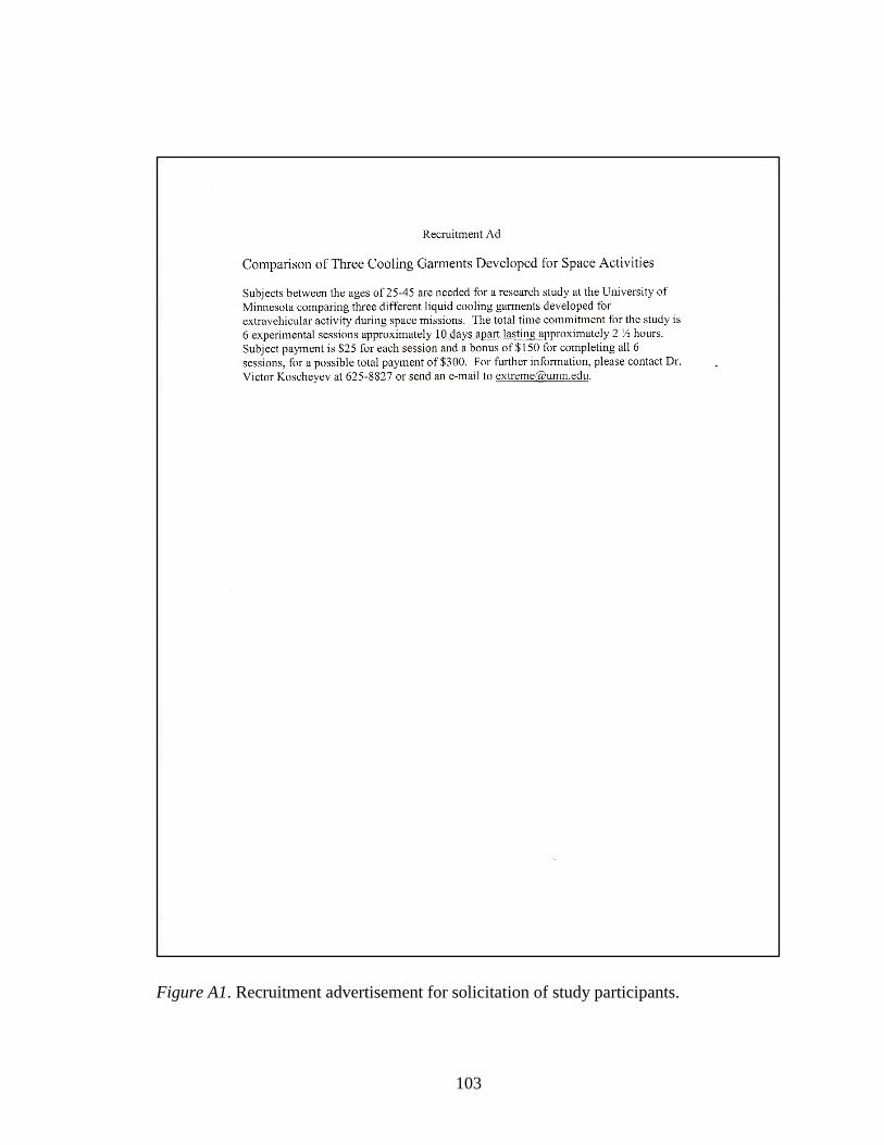
103
Figure A1. Recruitment advertisement for solicitation of study participants.
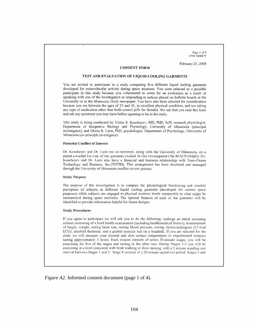
104
Figure A2. Informed consent document (page 1 of 4).
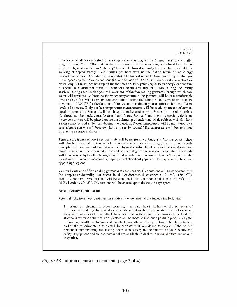
105
Figure A3. Informed consent document (page 2 of 4).
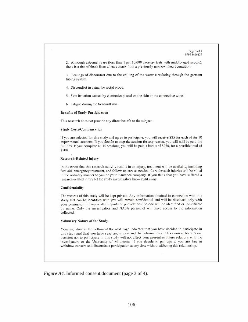
106
Figure A4. Informed consent document (page 3 of 4).
107
Figure A5. Informed consent document (page 4 of 4).
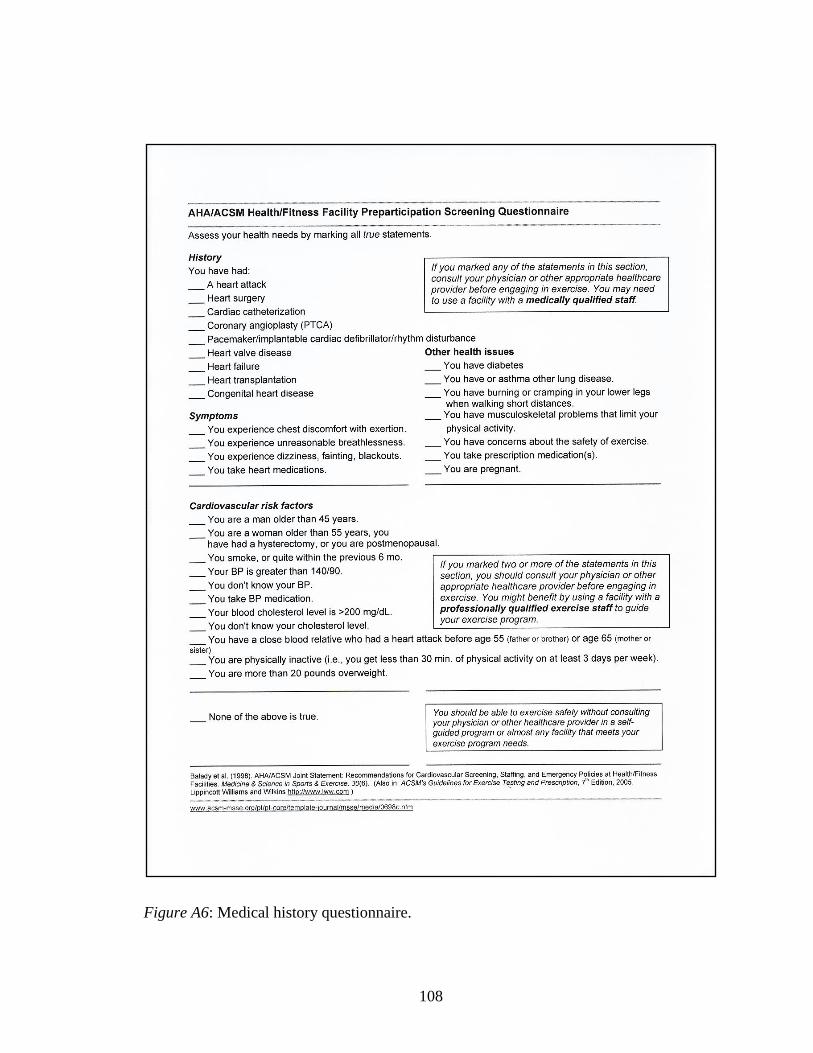
108
Figure A6: Medical history questionnaire.
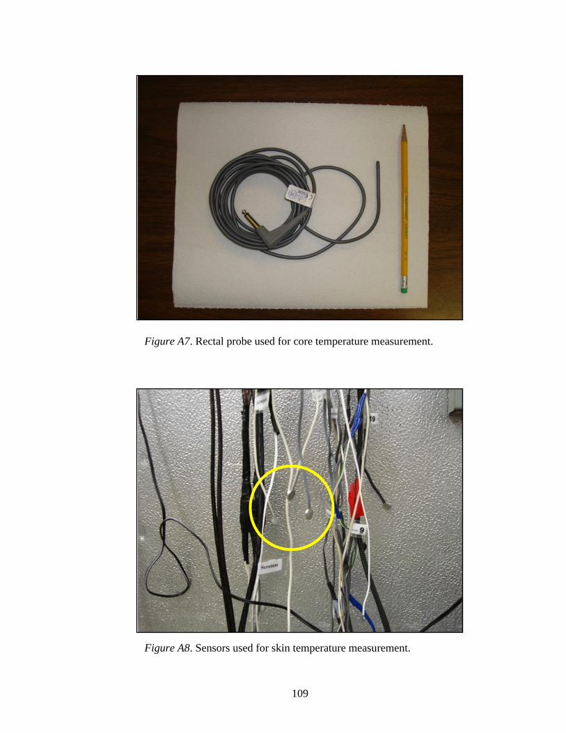
109
Figure A7. Rectal probe used for core temperature measurement.
Figure A8. Sensors used for skin temperature measurement.
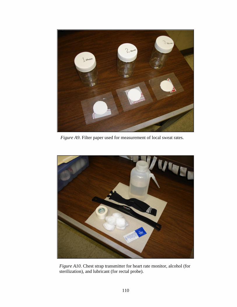
110
Figure A9. Filter paper used for measurement of local sweat rates.
Figure A10. Chest strap transmitter for heart rate monitor, alcohol (for
sterilization), and lubricant (for rectal probe).

111
APPENDIX B
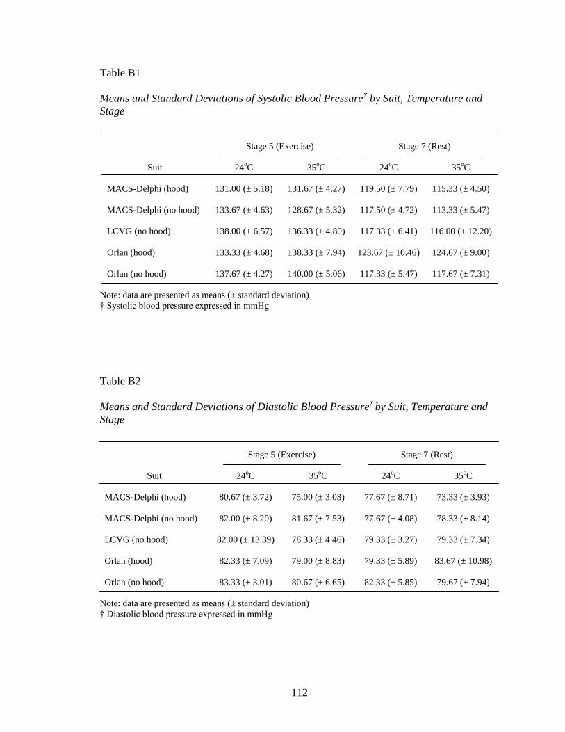
112
Table B1
Means and Standard Deviations of Systolic Blood Pressure† by Suit, Temperature and
Stage
Stage 5 (Exercise) Stage 7 (Rest)
Suit 24oC 35
oC 24
oC 35
oC
MACS-Delphi (hood) 131.00 (± 5.18) 131.67 (± 4.27) 119.50 (± 7.79) 115.33 (± 4.50)
MACS-Delphi (no hood) 133.67 (± 4.63) 128.67 (± 5.32) 117.50 (± 4.72) 113.33 (± 5.47)
LCVG (no hood) 138.00 (± 6.57) 136.33 (± 4.80) 117.33 (± 6.41) 116.00 (± 12.20)
Orlan (hood) 133.33 (± 4.68) 138.33 (± 7.94) 123.67 (± 10.46) 124.67 (± 9.00)
Orlan (no hood) 137.67 (± 4.27) 140.00 (± 5.06) 117.33 (± 5.47) 117.67 (± 7.31)
Note: data are presented as means (± standard deviation)
† Systolic blood pressure expressed in mmHg
Table B2
Means and Standard Deviations of Diastolic Blood Pressure†
by Suit, Temperature and
Stage
Stage 5 (Exercise) Stage 7 (Rest)
Suit 24oC 35
oC 24
oC 35
oC
MACS-Delphi (hood) 80.67 (± 3.72) 75.00 (± 3.03) 77.67 (± 8.71) 73.33 (± 3.93)
MACS-Delphi (no hood) 82.00 (± 8.20) 81.67 (± 7.53) 77.67 (± 4.08) 78.33 (± 8.14)
LCVG (no hood) 82.00 (± 13.39) 78.33 (± 4.46) 79.33 (± 3.27) 79.33 (± 7.34)
Orlan (hood) 82.33 (± 7.09) 79.00 (± 8.83) 79.33 (± 5.89) 83.67 (± 10.98)
Orlan (no hood) 83.33 (± 3.01) 80.67 (± 6.65) 82.33 (± 5.85) 79.67 (± 7.94)
Note: data are presented as means (± standard deviation)
† Diastolic blood pressure expressed in mmHg

113
Table B3
Means and Standard Deviations of Arterial Oxygen Saturation† by Suit, Temperature and
Stage
Stage 5 (Exercise) Stage 7 (Rest)
Suit 24oC 35
oC 24
oC
§ 35
oC
MACS-Delphi (hood) 95.83 (± 0.41) 95.50 (± 1.05) 97.83 (± 0.41) 97.00 (± 1.10)
MACS-Delphi (no hood) 96.50 (± 1.38) 95.83 (± 1.17) 97.83 (± 1.03) 96.17 (± 1.17)
LCVG (no hood) 96.67 (± 0.82) 95.50 (± 0.84) 97.83 (± 0.98) 96.83 (± 1.33)
Orlan (hood) 95.83 (± 1.17) 96.17 (± 0.75) 97.83 (± 0.75) 96.83 (± 1.33)
Orlan (no hood) 95.50 (± 0.55) 95.83 (± 0.75) 97.83 (± 1.63) 97.00 (± 0.89)
Note: data are presented as means (± standard deviation)
† Arterial oxygen saturation expressed as a percent
§ All means in the Stage 7 (24oC) column are identical (i.e., reported values are not errors)
Table B4
Means and Standard Deviations of Heart Rate† by Suit, Temperature and Stage
Stage 5 (Exercise) Stage 7 (Rest)
Suit 24oC 35
oC 24
oC 35
oC
MACS-Delphi (hood) 129.28 (± 22.17) 136.60 (± 22.76) 70.66 (± 13.61) 77.72 (± 15.67)
MACS-Delphi (no hood) 131.38 (± 28.79) 135.56 (± 21.38) 69.14 (± 21.93) 77.38 (± 17.55)
LCVG (no hood) 134.82 (± 23.58) 140.08 (± 24.86) 73.28 (± 19.18) 78.48 (± 18.42)
Orlan (hood) 137.88 (± 21.51) 143.90 (± 22.81) 74.62 (± 15.41) 80.22 (± 17.14)
Orlan (no hood) 131.32 (± 18.88) 137.12 (± 19.67) 69.84 (± 13.83) 77.62 (± 17.47)
Note: data are presented as means (± standard deviation)
† Heart rate expressed in beats per minute
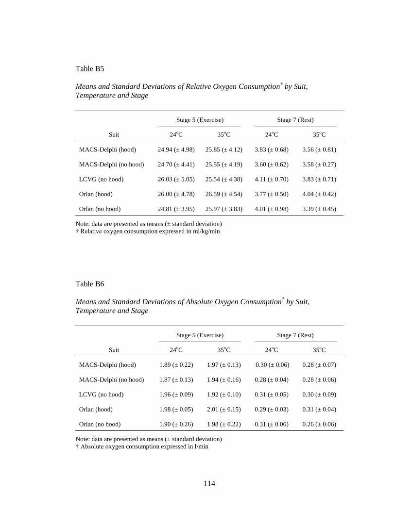
114
Table B5
Means and Standard Deviations of Relative Oxygen Consumption† by Suit,
Temperature and Stage
Stage 5 (Exercise) Stage 7 (Rest)
Suit 24oC 35
oC 24
oC 35
oC
MACS-Delphi (hood) 24.94 (± 4.98) 25.85 (± 4.12) 3.83 (± 0.68) 3.56 (± 0.81)
MACS-Delphi (no hood) 24.70 (± 4.41) 25.55 (± 4.19) 3.60 (± 0.62) 3.58 (± 0.27)
LCVG (no hood) 26.03 (± 5.05) 25.54 (± 4.38) 4.11 (± 0.70) 3.83 (± 0.71)
Orlan (hood) 26.00 (± 4.78) 26.59 (± 4.54) 3.77 (± 0.50) 4.04 (± 0.42)
Orlan (no hood) 24.81 (± 3.95) 25.97 (± 3.83) 4.01 (± 0.98) 3.39 (± 0.45)
Note: data are presented as means (± standard deviation)
† Relative oxygen consumption expressed in ml/kg/min
Table B6
Means and Standard Deviations of Absolute Oxygen Consumption† by Suit,
Temperature and Stage
Stage 5 (Exercise) Stage 7 (Rest)
Suit 24oC 35
oC 24
oC 35
oC
MACS-Delphi (hood) 1.89 (± 0.22) 1.97 (± 0.13) 0.30 (± 0.06) 0.28 (± 0.07)
MACS-Delphi (no hood) 1.87 (± 0.13) 1.94 (± 0.16) 0.28 (± 0.04) 0.28 (± 0.06)
LCVG (no hood) 1.96 (± 0.09) 1.92 (± 0.10) 0.31 (± 0.05) 0.30 (± 0.09)
Orlan (hood) 1.98 (± 0.05) 2.01 (± 0.15) 0.29 (± 0.03) 0.31 (± 0.04)
Orlan (no hood) 1.90 (± 0.26) 1.98 (± 0.22) 0.31 (± 0.06) 0.26 (± 0.06)
Note: data are presented as means (± standard deviation)
† Absolute oxygen consumption expressed in l/min
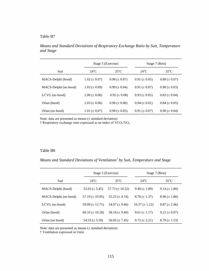
115
Table B7
Means and Standard Deviations of Respiratory Exchange Ratio by Suit, Temperature
and Stage
Stage 5 (Exercise) Stage 7 (Rest)
Suit 24oC 35
oC 24
oC 35
oC
MACS-Delphi (hood) 1.02 (± 0.07) 0.98 (± 0.07) 0.91 (± 0.05) 0.89 (± 0.07)
MACS-Delphi (no hood) 1.03 (± 0.09) 0.99 (± 0.04) 0.91 (± 0.07) 0.90 (± 0.03)
LCVG (no hood) 1.00 (± 0.06) 0.95 (± 0.08) 0.93 (± 0.05) 0.83 (± 0.04)
Orlan (hood) 1.03 (± 0.06) 0.96 (± 0.06) 0.94 (± 0.01) 0.84 (± 0.05)
Orlan (no hood) 1.01 (± 0.07) 0.98 (± 0.05) 0.91 (± 0.07) 0.90 (± 0.04)
Note: data are presented as means (± standard deviation)
† Respiratory exchange ratio expressed as an index of VCO2/VO2
Table B8
Means and Standard Deviations of Ventilation† by Suit, Temperature and Stage
Stage 5 (Exercise) Stage 7 (Rest)
Suit 24oC 35
oC 24
oC 35
oC
MACS-Delphi (hood) 53.63 (± 5.45) 57.73 (± 10.52) 9.49 (± 1.89) 9.14 (± 1.80)
MACS-Delphi (no hood) 57.19 (± 10.95) 55.25 (± 4.74) 8.78 (± 1.37) 8.96 (± 1.88)
LCVG (no hood) 59.09 (± 12.71) 54.97 (± 9.46) 10.37 (± 1.22) 8.87 (± 2.46)
Orlan (hood) 60.33 (± 10.28) 58.18 (± 9.40) 9.61 (± 1.17) 9.21 (± 0.97)
Orlan (no hood) 54.53 (± 5.59) 56.05 (± 7.45) 9.72 (± 2.21) 8.79 (± 1.53)
Note: data are presented as means (± standard deviation)
† Ventilation expressed in l/min
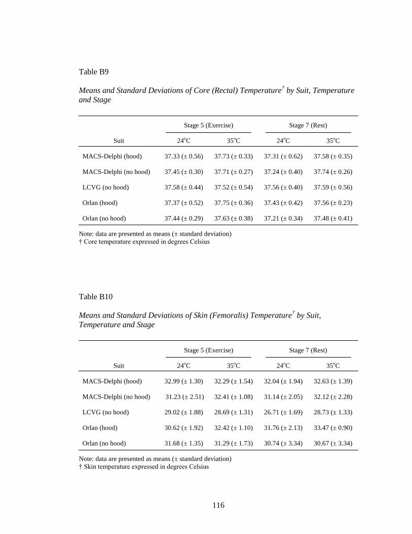
116
Table B9
Means and Standard Deviations of Core (Rectal) Temperature† by Suit, Temperature
and Stage
Stage 5 (Exercise) Stage 7 (Rest)
Suit 24oC 35
oC 24
oC 35
oC
MACS-Delphi (hood) 37.33 (± 0.56) 37.73 (± 0.33) 37.31 (± 0.62) 37.58 (± 0.35)
MACS-Delphi (no hood) 37.45 (± 0.30) 37.71 (± 0.27) 37.24 (± 0.40) 37.74 (± 0.26)
LCVG (no hood) 37.58 (± 0.44) 37.52 (± 0.54) 37.56 (± 0.40) 37.59 (± 0.56)
Orlan (hood) 37.37 (± 0.52) 37.75 (± 0.36) 37.43 (± 0.42) 37.56 (± 0.23)
Orlan (no hood) 37.44 (± 0.29) 37.63 (± 0.38) 37.21 (± 0.34) 37.48 (± 0.41)
Note: data are presented as means (± standard deviation)
† Core temperature expressed in degrees Celsius
Table B10
Means and Standard Deviations of Skin (Femoralis) Temperature† by Suit,
Temperature and Stage
Stage 5 (Exercise) Stage 7 (Rest)
Suit 24oC 35
oC 24
oC 35
oC
MACS-Delphi (hood) 32.99 (± 1.30) 32.29 (± 1.54) 32.04 (± 1.94) 32.63 (± 1.39)
MACS-Delphi (no hood) 31.23 (± 2.51) 32.41 (± 1.08) 31.14 (± 2.05) 32.12 (± 2.28)
LCVG (no hood) 29.02 (± 1.88) 28.69 (± 1.31) 26.71 (± 1.69) 28.73 (± 1.33)
Orlan (hood) 30.62 (± 1.92) 32.42 (± 1.10) 31.76 (± 2.13) 33.47 (± 0.90)
Orlan (no hood) 31.68 (± 1.35) 31.29 (± 1.73) 30.74 (± 3.34) 30.67 (± 3.34)
Note: data are presented as means (± standard deviation)
† Skin temperature expressed in degrees Celsius
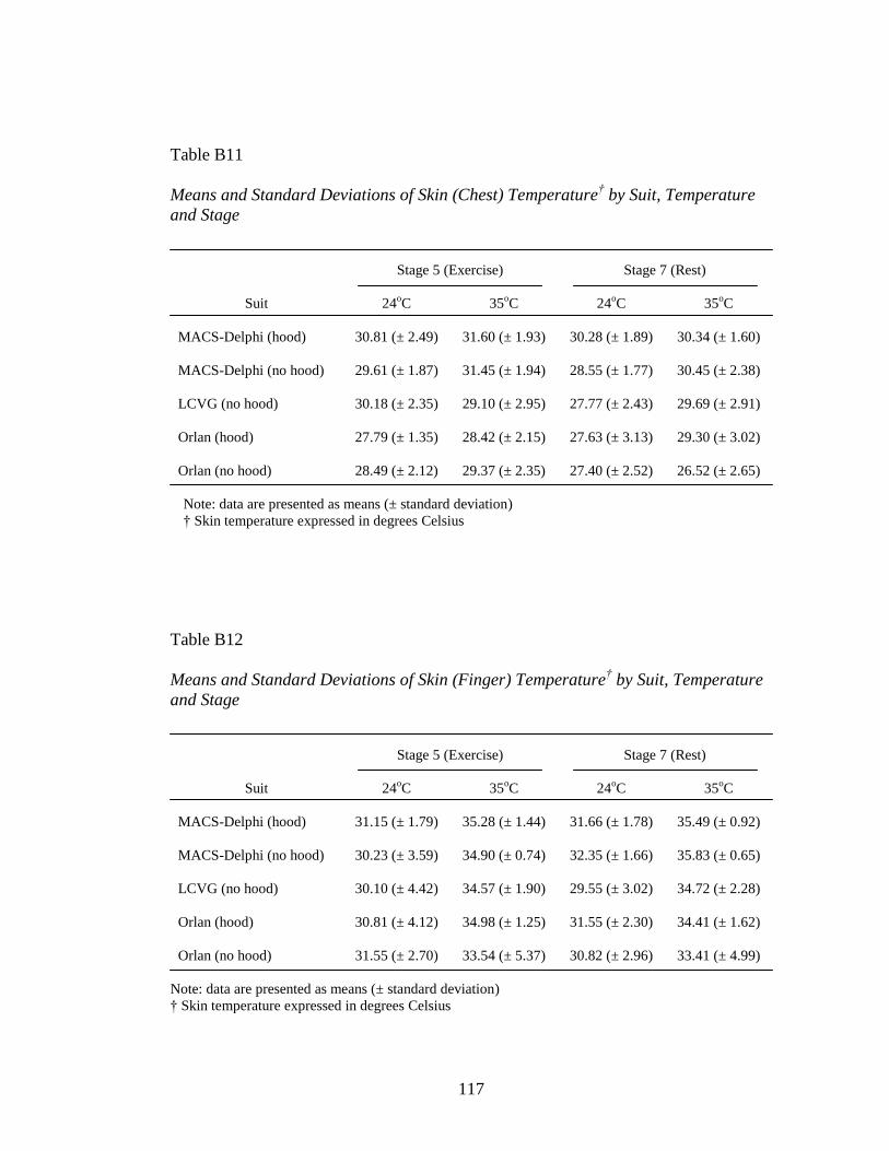
117
Table B11
Means and Standard Deviations of Skin (Chest) Temperature† by Suit, Temperature
and Stage
Stage 5 (Exercise) Stage 7 (Rest)
Suit 24oC 35
oC 24
oC 35
oC
MACS-Delphi (hood) 30.81 (± 2.49) 31.60 (± 1.93) 30.28 (± 1.89) 30.34 (± 1.60)
MACS-Delphi (no hood) 29.61 (± 1.87) 31.45 (± 1.94) 28.55 (± 1.77) 30.45 (± 2.38)
LCVG (no hood) 30.18 (± 2.35) 29.10 (± 2.95) 27.77 (± 2.43) 29.69 (± 2.91)
Orlan (hood) 27.79 (± 1.35) 28.42 (± 2.15) 27.63 (± 3.13) 29.30 (± 3.02)
Orlan (no hood) 28.49 (± 2.12) 29.37 (± 2.35) 27.40 (± 2.52) 26.52 (± 2.65)
Note: data are presented as means (± standard deviation)
† Skin temperature expressed in degrees Celsius
Table B12
Means and Standard Deviations of Skin (Finger) Temperature† by Suit, Temperature
and Stage
Stage 5 (Exercise) Stage 7 (Rest)
Suit 24oC 35
oC 24
oC 35
oC
MACS-Delphi (hood) 31.15 (± 1.79) 35.28 (± 1.44) 31.66 (± 1.78) 35.49 (± 0.92)
MACS-Delphi (no hood) 30.23 (± 3.59) 34.90 (± 0.74) 32.35 (± 1.66) 35.83 (± 0.65)
LCVG (no hood) 30.10 (± 4.42) 34.57 (± 1.90) 29.55 (± 3.02) 34.72 (± 2.28)
Orlan (hood) 30.81 (± 4.12) 34.98 (± 1.25) 31.55 (± 2.30) 34.41 (± 1.62)
Orlan (no hood) 31.55 (± 2.70) 33.54 (± 5.37) 30.82 (± 2.96) 33.41 (± 4.99)
Note: data are presented as means (± standard deviation)
† Skin temperature expressed in degrees Celsius
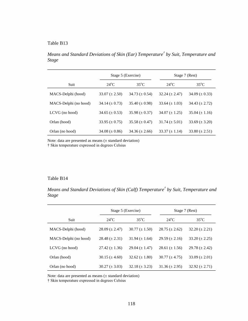
118
Table B13
Means and Standard Deviations of Skin (Ear) Temperature† by Suit, Temperature and
Stage
Stage 5 (Exercise) Stage 7 (Rest)
Suit 24oC 35
oC 24
oC 35
oC
MACS-Delphi (hood) 33.07 (± 2.50) 34.73 (± 0.54) 32.24 (± 2.47) 34.09 (± 0.33)
MACS-Delphi (no hood) 34.14 (± 0.73) 35.40 (± 0.98) 33.64 (± 1.03) 34.43 (± 2.72)
LCVG (no hood) 34.65 (± 0.53) 35.98 (± 0.37) 34.07 (± 1.25) 35.04 (± 1.16)
Orlan (hood) 33.95 (± 0.75) 35.58 (± 0.47) 31.74 (± 5.01) 33.69 (± 3.20)
Orlan (no hood) 34.08 (± 0.86) 34.36 (± 2.66) 33.37 (± 1.14) 33.80 (± 2.51)
Note: data are presented as means (± standard deviation)
† Skin temperature expressed in degrees Celsius
Table B14
Means and Standard Deviations of Skin (Calf) Temperature† by Suit, Temperature and
Stage
Stage 5 (Exercise) Stage 7 (Rest)
Suit 24oC 35
oC 24
oC 35
oC
MACS-Delphi (hood) 28.09 (± 2.47) 30.77 (± 1.50) 28.75 (± 2.62) 32.20 (± 2.21)
MACS-Delphi (no hood) 28.48 (± 2.31) 31.94 (± 1.64) 29.59 (± 2.16) 33.20 (± 2.25)
LCVG (no hood) 27.42 (± 1.36) 29.04 (± 1.47) 28.61 (± 1.56) 29.78 (± 2.42)
Orlan (hood) 30.15 (± 4.60) 32.62 (± 1.80) 30.77 (± 4.75) 33.09 (± 2.01)
Orlan (no hood) 30.27 (± 3.03) 32.18 (± 3.23) 31.36 (± 2.95) 32.92 (± 2.71)
Note: data are presented as means (± standard deviation)
† Skin temperature expressed in degrees Celsius
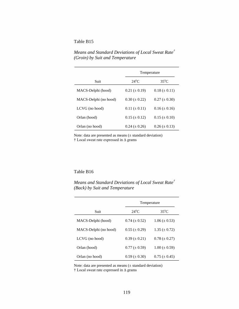
119
Table B15
Means and Standard Deviations of Local Sweat Rate†
(Groin) by Suit and Temperature
Temperature
Suit 24oC 35
oC
MACS-Delphi (hood) 0.21 (± 0.19) 0.18 (± 0.11)
MACS-Delphi (no hood) 0.30 (± 0.22) 0.27 (± 0.30)
LCVG (no hood) 0.11 (± 0.11) 0.16 (± 0.16)
Orlan (hood) 0.15 (± 0.12) 0.15 (± 0.10)
Orlan (no hood) 0.24 (± 0.26) 0.26 (± 0.13)
Note: data are presented as means (± standard deviation)
† Local sweat rate expressed in ∆ grams
Table B16
Means and Standard Deviations of Local Sweat Rate†
(Back) by Suit and Temperature
Temperature
Suit 24oC 35
oC
MACS-Delphi (hood) 0.74 (± 0.52) 1.06 (± 0.53)
MACS-Delphi (no hood) 0.55 (± 0.29) 1.35 (± 0.72)
LCVG (no hood) 0.39 (± 0.21) 0.78 (± 0.27)
Orlan (hood) 0.77 (± 0.59) 1.00 (± 0.59)
Orlan (no hood) 0.59 (± 0.30) 0.75 (± 0.45)
Note: data are presented as means (± standard deviation)
† Local sweat rate expressed in ∆ grams
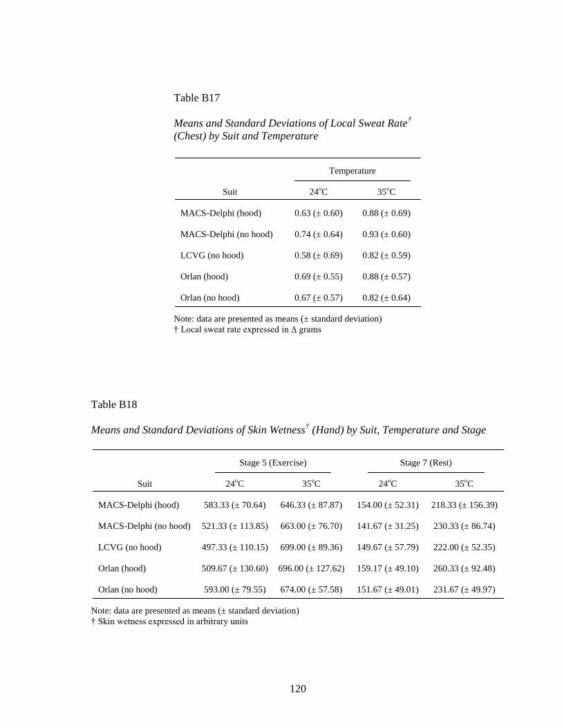
120
Table B17
Means and Standard Deviations of Local Sweat Rate†
(Chest) by Suit and Temperature
Temperature
Suit 24oC 35
oC
MACS-Delphi (hood) 0.63 (± 0.60) 0.88 (± 0.69)
MACS-Delphi (no hood) 0.74 (± 0.64) 0.93 (± 0.60)
LCVG (no hood) 0.58 (± 0.69) 0.82 (± 0.59)
Orlan (hood) 0.69 (± 0.55) 0.88 (± 0.57)
Orlan (no hood) 0.67 (± 0.57) 0.82 (± 0.64)
Note: data are presented as means (± standard deviation)
† Local sweat rate expressed in ∆ grams
Table B18
Means and Standard Deviations of Skin Wetness† (Hand) by Suit, Temperature and Stage
Stage 5 (Exercise) Stage 7 (Rest)
Suit 24oC 35
oC 24
oC 35
oC
MACS-Delphi (hood) 583.33 (± 70.64) 646.33 (± 87.87) 154.00 (± 52.31) 218.33 (± 156.39)
MACS-Delphi (no hood) 521.33 (± 113.85) 663.00 (± 76.70) 141.67 (± 31.25) 230.33 (± 86.74)
LCVG (no hood) 497.33 (± 110.15) 699.00 (± 89.36) 149.67 (± 57.79) 222.00 (± 52.35)
Orlan (hood) 509.67 (± 130.60) 696.00 (± 127.62) 159.17 (± 49.10) 260.33 (± 92.48)
Orlan (no hood) 593.00 (± 79.55) 674.00 (± 57.58) 151.67 (± 49.01) 231.67 (± 49.97)
Note: data are presented as means (± standard deviation)
† Skin wetness expressed in arbitrary units
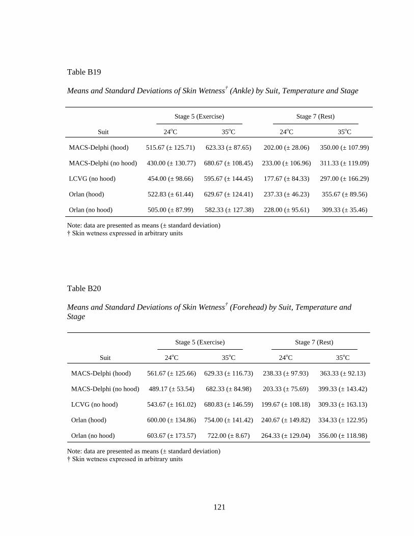
121
Table B19
Means and Standard Deviations of Skin Wetness† (Ankle) by Suit, Temperature and Stage
Stage 5 (Exercise) Stage 7 (Rest)
Suit 24oC 35
oC 24
oC 35
oC
MACS-Delphi (hood) 515.67 (± 125.71) 623.33 (± 87.65) 202.00 (± 28.06) 350.00 (± 107.99)
MACS-Delphi (no hood) 430.00 (± 130.77) 680.67 (± 108.45) 233.00 (± 106.96) 311.33 (± 119.09)
LCVG (no hood) 454.00 (± 98.66) 595.67 (± 144.45) 177.67 (± 84.33) 297.00 (± 166.29)
Orlan (hood) 522.83 (± 61.44) 629.67 (± 124.41) 237.33 (± 46.23) 355.67 (± 89.56)
Orlan (no hood) 505.00 (± 87.99) 582.33 (± 127.38) 228.00 (± 95.61) 309.33 (± 35.46)
Note: data are presented as means (± standard deviation)
† Skin wetness expressed in arbitrary units
Table B20
Means and Standard Deviations of Skin Wetness† (Forehead) by Suit, Temperature and
Stage
Stage 5 (Exercise) Stage 7 (Rest)
Suit 24oC 35
oC 24
oC 35
oC
MACS-Delphi (hood) 561.67 (± 125.66) 629.33 (± 116.73) 238.33 (± 97.93) 363.33 (± 92.13)
MACS-Delphi (no hood) 489.17 (± 53.54) 682.33 (± 84.98) 203.33 (± 75.69) 399.33 (± 143.42)
LCVG (no hood) 543.67 (± 161.02) 680.83 (± 146.59) 199.67 (± 108.18) 309.33 (± 163.13)
Orlan (hood) 600.00 (± 134.86) 754.00 (± 141.42) 240.67 (± 149.82) 334.33 (± 122.95)
Orlan (no hood) 603.67 (± 173.57) 722.00 (± 8.67) 264.33 (± 129.04) 356.00 (± 118.98)
Note: data are presented as means (± standard deviation)
† Skin wetness expressed in arbitrary units
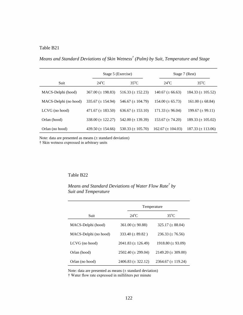
122
Table B21
Means and Standard Deviations of Skin Wetness† (Palm) by Suit, Temperature and Stage
Stage 5 (Exercise) Stage 7 (Rest)
Suit 24oC 35
oC 24
oC 35
oC
MACS-Delphi (hood) 367.00 (± 198.83) 516.33 (± 152.23) 140.67 (± 66.63) 184.33 (± 105.52)
MACS-Delphi (no hood) 335.67 (± 154.94) 546.67 (± 104.79) 154.00 (± 65.73) 161.00 (± 68.84)
LCVG (no hood) 471.67 (± 183.50) 636.67 (± 153.10) 171.33 (± 96.04) 199.67 (± 99.11)
Orlan (hood) 338.00 (± 122.27) 542.00 (± 139.39) 153.67 (± 74.20) 189.33 (± 105.02)
Orlan (no hood) 439.50 (± 154.66) 530.33 (± 105.70) 162.67 (± 104.03) 187.33 (± 113.06)
Note: data are presented as means (± standard deviation)
† Skin wetness expressed in arbitrary units
Table B22
Means and Standard Deviations of Water Flow Rate† by
Suit and Temperature
Temperature
Suit 24oC 35
oC
MACS-Delphi (hood) 361.00 (± 90.88) 325.17 (± 88.04)
MACS-Delphi (no hood) 333.40 (± 89.82 ) 236.33 (± 76.56)
LCVG (no hood) 2041.83 (± 126.49) 1918.80 (± 93.09)
Orlan (hood) 2502.40 (± 299.04) 2149.20 (± 309.00)
Orlan (no hood) 2406.83 (± 322.12) 2364.67 (± 119.24)
Note: data are presented as means (± standard deviation)
† Water flow rate expressed in milliliters per minute
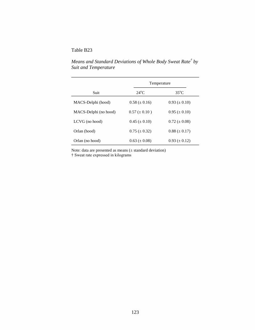
123
Table B23
Means and Standard Deviations of Whole Body Sweat Rate† by
Suit and Temperature
Temperature
Suit 24oC 35
oC
MACS-Delphi (hood) 0.58 (± 0.16) 0.93 (± 0.10)
MACS-Delphi (no hood) 0.57 (± 0.10 ) 0.95 (± 0.10)
LCVG (no hood) 0.45 (± 0.10) 0.72 (± 0.08)
Orlan (hood) 0.75 (± 0.32) 0.88 (± 0.17)
Orlan (no hood) 0.63 (± 0.08) 0.93 (± 0.12)
Note: data are presented as means (± standard deviation)
† Sweat rate expressed in kilograms

124
APPENDIX C

125
Figure C1. Limitation of forward flexion in the Orlan due to vertical tubing arrangement.
Figure C2. Results of wear-and-tear in the crotch region of the Orlan.
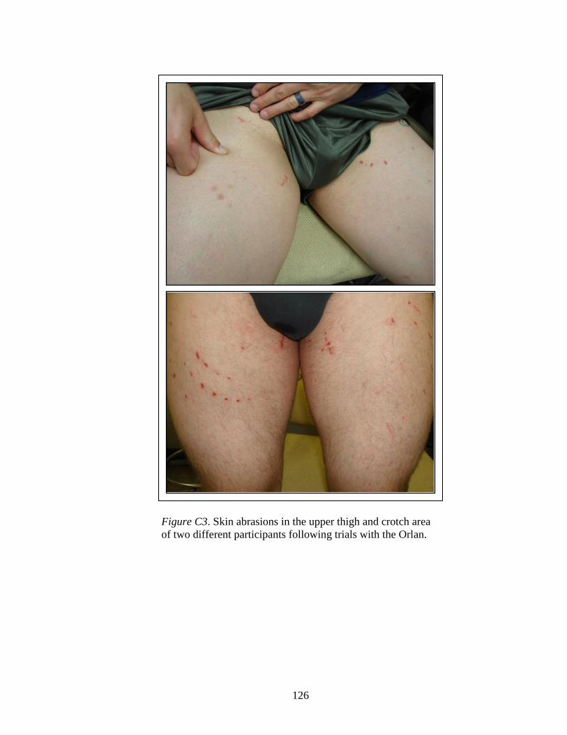
126
Figure C3. Skin abrasions in the upper thigh and crotch area
of two different participants following trials with the Orlan.