Nitric oxide differentially regulates renal ATP-binding cassette transporters during endotoxemia
Upload
independentCategory
view
4download
0
Endotoxemia down-regulates bone marrow lymphopoiesis butstimulates myelopoiesis: the effect of G6PD deficiency
Rachna Chandra, Erika Villanueva, Eleonora Feketova, George W. Machiedo, Gyorgy Hasko,Edwin A. Deitch, and Zoltan Spolarics1
Department of Surgery, University of Medicine and Dentistry of New Jersey-New Jersey Medical School, Newark,New Jersey, USA
Abstract: Bone marrow (BM) dysfunction is animportant component of immunomodulation.This study investigated alterations in cell con-tent, apoptotic responses, and cell proliferationin BM, blood, and spleen in endotoxemic mice(LPS from Escherichia coli). As the decreasedantioxidant status associated with glucose-6-phosphate dehydrogenase (G6PD) deficiency hasbeen shown to modulate the innate immune re-sponse, we also tested whether a G6PD mutation(80% decrease in cellular enzyme activity) altersBM responses during endotoxemia. LPS de-creased BM myeloid (CD45�CD11b�) and Blymphoid (CD45�CD19�CD11b–) cell contentcompared with controls. In contrast, LPS in-creased CD11b� myeloid but decreased T and Bcell counts in the circulation. Endotoxemia in-hibited spontaneous, heat shock, and H2O2-in-duced apoptosis as well as proliferative activity inBM lymphoid cells. In contrast, BM myeloid cellapoptosis was not altered, and their proliferativeactivity was increased during endotoxemia. Fol-lowing LPS, splenic myeloid cell content was in-creased, and T and B cell content was un-changed; furthermore, splenocytes showed in-creased apoptosis compared with controls. BMcell content, including lymphoid and myeloidcells, was greater in G6PD mutant than wild-type(WT) mice, and LPS decreased BM cell counts toa greater degree in mutant than WT mice. Endo-toxemia caused widespread inhibition of BM cy-tokine and chemokine production; however,IL-6 production was increased compared withcontrols. LPS-induced IL-6 production was de-creased in G6PD mutant animals compared withWT. This study indicates that endotoxin in-versely affects BM myeloid and lymphoid cellproduction. LPS-induced down-regulation of Bcell production contributes to the generalizedlymphopenia and lymphocyte dysfunction ob-served following nonspecific immune challenges.J. Leukoc. Biol. 83: 1541–1550; 2008.
Key Words: lipopolysaccharide � B cells � erythroid � apoptosis� proliferation � glucose-6-phosphate dehydrogenase � cytokines
INTRODUCTION
Sepsis or endotoxemia results in marked changes in the num-ber of circulating polymorphonuclear phagocytes (PMN). De-pendent on the dose and duration of the challenge, circulatingPMN numbers tend to increase after endotoxin administration,whereas PMN blood counts are depleted following experimen-tal polymicrobial sepsis [1, 2]. However, in contrast to thedynamic changes in circulating PMN numbers, it has beenobserved in endotoxemic and septic experimental rodent mod-els that lymphocytes, including B and T cells, are markedlydepleted following endotoxin as well as septic challenges[1–4]. Whereas lymphocyte apoptosis developing at the laterstages of sepsis is a well-investigated area [4], the pathophys-iological mechanism responsible for the acute and generalizedlymphocytopenia manifesting early after infections has notbeen elucidated. Whereas it is well known that chronic infec-tion results in anemia and decreased bone marrow (BM) activ-ity [5, 6], and furthermore, severe injuries may modulatemyelopoiesis [7, 8], the potential role of altered BM functionand its relationship to the accompanying changes in blood andtissue cell composition during the acute phase of an innateimmune response have not been thoroughly investigated. Thus,the studies tested the question of whether acute alterations inBM function contribute to the changes in peripheral myeloidand lymphoid cell dynamics during the early phase of endo-toxemia.
The second aim of the investigations was to compare theendotoxin-induced BM responses as well as the accompanyingchanges at the periphery between glucose-6-phoshate dehydro-genase (G6PD) mutant and wild-type (WT) animals. G6PD isthe rate-limiting enzyme of the hexose monophosphate shuntand a key component of the cellular antioxidant defense ma-chinery [9–12]. The X-linked human G6PD deficiency is oneof the most common genetic polymorphisms [11, 12]. G6PDpolymorphism is associated with malaria protection, and thepopulation frequency of the mutated alleles may reach 10–25% in malaria-endemic regions. The most common polymor-phic forms are the Mediterranean with less than 10% residual
1 Correspondence: Department of Surgery, UMDNJ-New Jersey MedicalSchool, 185 South Orange Ave., MSB G-626, Newark, NJ 07103, USA. E-mail:[email protected]
Received December 17, 2007; revised February 4, 2008; accepted February25, 2008.
doi: 10.1189/jlb.1207838
0741-5400/08/0083-1541 © Society for Leukocyte Biology Journal of Leukocyte Biology Volume 83, June 2008 1541

erythrocyte G6PD activity and the African variants with 10–20% residual erythrocyte G6PD activity [11, 12]. In thesehuman deficiencies, the G6PD activity decrease in nucleatedwhite blood cells (WBC) is less pronounced with 20–30%residual activity in the Mediterranean forms, whereas nearnormal levels in the African variants.
Oxidative stress can modulate signaling pathways [13, 14]and could also contribute to organ dysfunction during theinnate immune response [2, 3, 15, 16]. Furthermore, indepen-dent observations suggested increased incidence of sepsis andaltered inflammatory course in G6PD deficiency [16, 17].Therefore, we also tested the effect of a hypomorphic G6PDmutation on BM function and associated changes in the pe-riphery. We used a G6PD mutant mouse strain that displays a15–20% residual G6PD activity in erythrocytes, as well as inWBC. Thus, the residual cellular G6PD activity in this mutantmouse model represents a defect that is similar to that observedin the erythrocytes of the African type and in the WBC of theMediterranean human variants. Cell composition changes, ap-optosis, and cell proliferative activities were compared in dif-ferent BM cell lineages together with the accompanying cellresponses in blood and spleen.
MATERIALS AND METHODS
ReagentsEndotoxin-free, cell culture-grade buffers, media, and reagents were used inthe experiments. FBS was purchased from Irvine Scientific (Santa Ana, CA,USA) and the protein assay kit from Pierce (Rockford, IL, USA). Flouro-chrome-conjugated antibodies, assay diluents, lysing, and permeabilizing flowcytometry solutions, Annexin-V, and FITC-BrdU kits were purchased from BDBiosiences (San Jose, CA, USA). For the in vitro cytokine studies, palmitoyl-3-cysteine-serine-lysine-4 (Pam3CSK4) and ultra-pure LPS (from Escherichiacoli, 0111:B4) were purchased from InvivoGen (San Diego, CA, USA). Allother reagents and chemicals of the highest grade available were purchasedfrom Sigma-Aldrich Co. (St. Louis, MO, USA).
Animals and endotoxin treatmentMale G6PD-deficient mice (y/–) and their normal (WT, y/�) littermates (8–12weeks old) derived from the same colony were used in the experiments asdescribed in detail earlier [2, 18]. The inbred line from a mixed background[18] is similar to commonly used mouse strains in growth rate and size, and theG6PD mutant animals are phenotypically indistinguishable from WT. Originalbreeding pairs of G6PD mutant mice were purchased from the MedicalResearch Council (MRC) of England (Frozen Embryo and Sperm ArchiveMammalian Genetics Unit, MRC, Chilton, UK). Animals were housed under12 h light/dark cycles and fed standard rodent chow.
In the in vivo endotoxemic studies, we used sequential LPS (from E. coli,026:B6, Sigma-Aldrich), with the initial i.p. injection of 10 mg/kg body weight(bw), followed by 25 mg/kg bw 3 h later [3]. This LPS protocol results in40–70% mortality; however, only a negligible number of animals expire duringthe first 24 h of endotoxemia. Thus, survival bias does not skew the observa-tions [3]. This LPS treatment caused increased mortality in G6PD mutantanimals compared with WT as published previously [3]. The studies wereperformed in accordance with the Guide for the Care and Use of LaboratoryAnimals {Department of Health and Human Services, Publication No. [Na-tional Institutes of Health (NIH)] 85–23, Revised 1985, Office of Science andHealth Reports, Data Reliability Review/NIH, Bethesda, MD, USA} and wereapproved by the Institutional Animal Care and Use Committee of the NewJersey Medical School (Newark, NJ, USA).
GenotypingThe G6PD gene in the mutant mouse shows a single base (A–T) difference fromthe published mouse G6PD sequences (BALB/c, C3H, C57BL/6) in the 5�
splice site consensus sequence at the 3� end of exon 1, part of the untranslatedregion [18]. The mutation causes an 80% decrease in the cellular protein levelof G6PD as a result of decreased mRNA stability or translation rate. Thedegree of G6PD activity decrease is similar in erythrocytes and nucleatedWBC, as the structure and the stability of the mature protein are not affectedby the mutation [18]. Animals were genotyped by using our allele-specific PCRmethod as we described in detail earlier [3]. Briefly, total genomic DNA wasisolated from tail clippings using the REDExtract-N-Amp Tissue PCR kit(Sigma-Aldrich). DNA was subjected to amplification using primer sets thatspan the mutation site and uniformly amplify DNA from deficient and WTanimals (control reaction, forward primer 5�-GGAAACTGGCTGTGCGCTAC,reverse primer 5�-TCAGCTCCGGCTCTCTTCTG). In parallel reactions, thesame DNA was incubated in the presence of the same reverse primer togetherwith the forward primer (5�-TGGCAGCGGCAACTAAACTCA), which reactswith the WT, or (5�-TGGCAGCGGCAACTAAACTCT), which reacts with thedeficient G6PD allele, respectively. PCR reaction was carried out in thepresence of 2 mM MgCl2 with the following cycling: 94°C, 15 s; 68°C, 1 min;28 cycles.
Blood, splenocyte, and BM cell isolationand incubations
Blood was collected into heparinized tubes via cardiac puncture from fullyanesthetized animals. Following the exsanguination, femurs were collectedfrom the same animals. Femurs were cut at the diaphyses and BM cells flushedout by repeated injections of PBS containing 10% FBS through the bonechannel. BM cells were sedimented and washed by centrifugation and sus-pended in a final volume to obtain 10 million/ml cells in the same PBS/FBSbuffer. Next, the spleen was removed and placed into DMEM containing 10%FBS and penicillin streptomycin solution. Hypodermic needles were used topull apart the splenic capsule, releasing spleen cells into suspension. The cellsuspension together with the remaining splenic capsule was squeezed througha 70-�m nylon mesh cell strainer. Isolated BM cells or splenocytes wereresuspended in DMEM containing 1% FBS for subsequent analyses or in vitroincubations.
Spontaneous, heat shock, and H2O2-induced apoptosis was measured usingthe Annexin-V apoptosis assay kit according to the manufacturer’s protocol(BD Biosciences): Splenocytes and BM cells from control or endotoxemicanimals were isolated and incubated at 37°C in a cell culture incubator for 4 hin the absence (spontaneous apoptosis) or in the presence of prevailing H2O2
concentrations. An additional set of cell aliquots was exposed to 43°C for 30min (heat-shock) followed by a 3.5-h incubation at 37°C. At the end of theincubations, cells were collected, then washed, and resuspended in Annexin-V-binding buffer and incubated with Annexin-V and propidium iodide for 15min at room temperature in the dark prior to flow cytometry analyses.
CFUs were determined as described earlier. Briefly, 106 BM cells in 0.1 mlculture media were mixed with 0.4 ml methylcellulose (Methocult) and 0.5 mlIscove’s media containing 30% FBS, 2% BSA, 2.5 U/m rhGM-CSF, 0.2 mM�-mercaptoethanol and antiobotic/antimycotic solution. The cell suspensionwas placed in a culture dish and incubated in a humidified CO2 incubator at37°C. Media were refreshed at every 48 h, and after 2 weeks of incubation, thenumber of individual colonies was counted.
RBC deformability was analyzed with a laser-assisted ectacytometer(LORCA) as described previously [19]. Briefly, an aliquot containing �30million RBC was suspended in 1 ml 5% polyvinylpyrrolidone (m.w. 360,000;Sigma-Aldrich) in PBS at a final viscosity of 30 mPa at 37°C. Cell suspensionswere exposed to increasing shear stress, and the elongation index (EI) wasdetermined. KEI is calculated from the response curves and indicates the shearstress value in Pascal that is required to cause half-maximal erythrocyteelongation as described in details previously [19].
Flow cytometry
The number of PMNs and lymphocyte subsets in blood and spleen wascalculated by the number of total cell counts and the percent distribution ofCD3�CD4�, CD3�CD8� T cells, CD19� B cells, and CD11b� myeloidcells using antibodies against CD markers conjugated with FITC, allophyco-cyanin (APC), PerCP, or PE (BD Biosciences) in three- or four-color incuba-tions. BM cell composition was determined by the cell distribution ofCD45�CD19�CD11b– (B cells), CD1b�CD45�CD19– (myeloid cells), andTer119�CD45–CD11b– (erythroid cells). As expected, CD3CD8- and
1542 Journal of Leukocyte Biology Volume 83, June 2008 http://www.jleukbio.org

CD3CD4-positive T cell content was negligible in BM from control as well asendotoxemic animals; thus, BM T cell content was not followed in the assays.Aliquots of 0.1 ml whole blood, splenocyte, or BM cell suspension wereincubated with the respective markers for 15 min followed by incubation withBD FACS lysing solution (BD Biosciences) for 7 min at 37°C. Cells werewashed twice with BD FACS wash buffer and then fixed with 1% methanol-freeformaldehyde. FACS acquisitions were performed in a centralized flow cytom-etry facility. At least 30,000 events were collected for each analysis.
In vivo DNA synthesis rates by different BM cell lineages were assessed byusing the FITC-BrdU flow kit (BD PharMingen, San Diego, CA, USA). BrdU(0.06 mg/kg bw in 0.4 ml vol) was injected i.p. into control or endotoxemicmice 2 h before harvesting the BM (i.e., at 22 h of endotoxemia). Followinganesthesia and tissue collections, BM cell suspensions were prepared, and 106
BM cells were incubated with fluorochrome-conjugated surface marker CD45,CD19, CD11b, and Ter119 antibodies in three-color incubations for 20 min at4°C. Cells were washed with BD stain buffer then fixed and permeabilized byincubating with BD Cytofix/Cytoperm buffer for 30 min at 4°C. This wasfollowed by incubation with BD Cytoperm-plus buffer for 10 min and subse-quent incubation with the BD Cytofix/Cytoperm buffer for 5 min at 4°C. Cellswere washed and treated with DNase at 37°C for 1 h. Finally, washed cellsuspensions were incubated with the FITC-conjugated anti-BrdU antibody for20 min at room temperature, washed with BD perm/wash buffer, and analyzedby flow cytometry.
Cytokine antibody arrays and ELISAs
BM cell suspensions (106 cells/ml) were incubated in the absence or presenceof 100 ng/ml LPS for 14 h at 37°C. Media were collected, and cytokine contentwas determined by using the Ray Bio Mouse Cytokine Antibody Array (Ray-Biotech, Inc., Norcross, GA, USA). Following the cell incubations, mediasamples were incubated with the membranes precoated with arrayed antibod-ies. Membranes were washed and processed according to the manufacturer’sprotocol, and the signals were detected by using a chemiluminescence imagingsystem at our centralized facility. Signals determined at individual spots werenormalized to standards present on the same membrane. Normalized valuesdetected on individual membranes obtained from endotoxemic and controlanimals or after the in vitro treatments were compared and expressed as
relative change between samples in arbitrary units. Media content for selectedcytokines was also determined using ELISAs (BD Biosciences) in a separateset of experiments.
Statistical analysis
Statistical calculations were performed using JMP software (SAS Institute Inc.,Cary, NC, USA). Results were analyzed using ANOVA, followed by t-test forpair-wise comparisons or Tukey-Kramer’s test for multiple comparisons. Dif-ferent study components were performed on six to eight different animals fromeach of the in vivo treatment groups, unless indicated otherwise. A statisticallysignificant difference was concluded at P � 0.05.
RESULTS
Endotoxin-induced cell composition changes inthe BM and periphery
In the first series of experiments, we determined the effect ofendotoxemia on the WBC composition changes in blood,spleen, and BM. Endotoxemia decreased the number of circu-lating, total WBC counts in G6PD mutant animals, whereas thechange in WBC count was not statistically significant in WTmice (Fig. 1A). Analysis of WBC subsets indicated an in-crease in circulating CD11b� cells (myeloid cells), whereasthe number of B cells (CD19�) as well as CD4� and CD8� Tcells markedly decreased in endotoxemic animals comparedwith controls (Fig. 1, C–F). Circulating platelet numbers alsodecreased following LPS in WT and G6PD mutant animalscompared with controls (Fig. 1B).
Figure 2 indicates cell composition changes in the spleensfrom the same animals. Total splenocyte yield was similar in
Fig. 1. Endotoxemia increases the number of circulating neutrophilsbut depletes T and B lymphocytes. Twenty-four hours after LPS orsaline injection (controls), blood was collected, and total WBC andplatelet counts were determined (A and B). (C–F) Numbers of circu-lating CD11b� (mostly neutrophils) and different T cell (CD4� andCD8�) and B cell (CD19�) subsets as indicated. Numbers within thebars depict the mean values of percent cell distributions. Flow cytom-etry panels on the right show typical findings from a WT animal. *,Statistically significant difference as compared with control within thesame genotype. Mean � SEM; n � 7–8 animals in each group. SSC,Side-scatter.
Chandra et al. Lymphopoiesis in endotoxemia 1543

WT and G6PD mutant mice, and LPS injection caused nostatistically significant increase in total splenic cell content(Fig. 2A). Analysis of WBC subsets indicated that the numbersof splenic neutrophils and macrophages were markedly in-creased 24 h after LPS compared with controls (Fig. 2, B andC). In contrast, the number of splenic B cells and T cell subsetswas not altered significantly by LPS treatment (Fig. 2, D–F).
Cell composition analyses of BM from the same animals areshown in Figure 3. Myeloid cells were strongly positive forCD45 and CD11b but were negative for CD19. B lymphoidcells stained positive for CD45 and CD19 but were weak/negative for CD11b. As CD45 expression increases during Bcell ontogeny [20, 21], and CD19� B cells clearly separatedinto two distinct subpopulations by the CD45-staining inten-sity, we analyzed mature (Fig. 3, flow panel R1 gate) andimmature B cell contents (R2 gate) separately.
Total BM cell content was greater in G6PD mutant than WTanimals under unchallenged, control conditions (Fig. 3A). LPStreatment markedly decreased BM cell content in WT andG6PD mutant animals (Fig. 3A). Under control conditions,G6PD mutant animals showed increased content of B lymphoid(Fig. 3, B and D) and myeloid (Fig. 3C) cells as compared withWT. Following LPS, myeloid content was depleted similarly inWT and G6PD mutant animals (Fig. 3C). Endotoxin depletedthe mature B cell population only in G6PD mutant animals(Fig. 3B), whereas the less-mature B cell population was de-pleted similarly in WT and G6PD mutant mice (Fig. 3D). BMcontent of the remaining cells (R4), which contained a mixtureof erythroid, other lymphoid, mesenchymal, and precursorcells, was also greater in G6PD than WT animals, and their BMcontent decreased following LPS treatment (Fig. 3E).
In a separate set of animals, we also tested the effect ofendotoxin on circulating and BM erythroid cells. Twenty-four-hour endotoxemia did not alter the number of circulatingerythrocytes in WT or G6PD-deficient animals (Fig. 4A).Erythrocyte dysfunction was assessed by determination oferythrocyte deformability. Whole blood was exposed to in-creasing shear stress, and from the response curve of erythro-cyte elongation response, KEI was calculated. KEI representsthe shear stress value (Pascal) required to cause half of themaximal erythrocyte elongation as we described earlier [19].Figure 4B indicates that the LPS-induced increase in KEI wasgreater in G6PD mutant than WT animals, indicating thepresence of more rigid erythrocytes in endotoxemic G6PDmutant animals (Fig. 4B). A BM erythroid line content identi-fied by the marker set used (Ter119�,CD45–,CD11b–) showedsimilar values in G6PD mutant and WT animals, and 24 hendotoxemia caused no significant decrease in BM erythroidcell content (Fig. 4, C and D).
BM cell apoptosis and cell proliferationafter endotoxemia
In the next series of experiments, we tested how the LPS-induced cell composition changes correlate with the apoptoticresponses in BM and spleen (Figs. 5 and 6). Spontaneousapoptosis of BM lymphoid as well as myeloid lineages wassimilar (�7%) in control WT and G6PD mutant animals.Following endotoxemia, BM lymphoid cells showed decreasedlevels of spontaneous apoptosis compared with controls,whereas the number of spontaneously apoptotic myeloid cellswas not affected significantly by endotoxin treatment (Fig. 5, Aand B). Heat shock increased the number of apoptotic B cells
Fig. 2. Endotoxemia increases neutrophil and macrophage infiltration into spleen. Twenty-four hours after LPS or saline injection, splenocyte suspensions wereprepared, and cell counts were determined (A). Based on CD11b� staining and light-scatter properties, neutrophil and macrophage numbers as well as CD4� orCD8� T cells and CD19� B cell counts were calculated from total cell yields and the percent distribution of these cell populations (B–F). *, Statistically significantdifference as compared with control within the same genotype. Mean � SEM; n � 7–8 animals in each group.
1544 Journal of Leukocyte Biology Volume 83, June 2008 http://www.jleukbio.org

and myeloid cells two- to threefold (Fig. 5, C and D; comparey-axes of Panels A and B). The heat shock-induced B cellapoptotic response remained decreased following endotoxemiaas compared with controls, whereas the heat-shock response bymyeloid cells was similar in control and endotoxemic animals.H2O2 administration increased B cell apoptosis in a concen-tration-dependent manner; however, the overall number of
apoptotic B cells remained lower in endotoxemic than controlanimals (Fig. 5E). G6PD mutant B cells showed an increasedtendency of the apoptotic response to H2O2 as compared withWT, reaching a statistically significant difference at 2 �MH2O2 treatment (Fig. 5E). BM myeloid cells showed no con-sistent increase in oxidant-induced apoptosis in WT or G6PDmutant animals (Fig. 5F).
Fig. 3. Endotoxemia down-regulates BM he-matopoiesis. Twenty-four hours after LPS orsaline injection, BM cell suspensions wereprepared, and cell counts were determined(A). Following the incubations with a set ofsurface markers as described in Materials andMethods, BM content for different cellularsubsets was determined by flow cytometry. Blymphoid cells were CD45�CD19�/CD11b–
(R1 and R2, B and D), whereas myeloid cellswere CD45�CD11b�CD19– (R3, C). Cellswith scattered staining for these markers arealso shown (R4, E). *, Statistically significantdifference as compared with control within thesame genotype; &, compared with all othergroups. Mean � SEM; n � 7–8 animals ineach group.
Fig. 4. Erythroid cell responses following endotoxemia. Twenty-four hours after LPS orsaline injection, circulating erythrocyte counts were determined (A). RBC deformabilitywas measured by LORCA under prevailing shear stress, and KEI (shear stress causinghalf-maximal erythrocyte deformability) was calculated and compared (B). BM cellsuspensions were also prepared, and erythroid content was determined using Ter199,CD45, and CD11b markers (C). Flow cytometry panels show a typical finding from a WTanimal. #, Statistically significant difference as compared with control within the samegenotype. Mean � SEM; n � 6 animals in each group.
Chandra et al. Lymphopoiesis in endotoxemia 1545
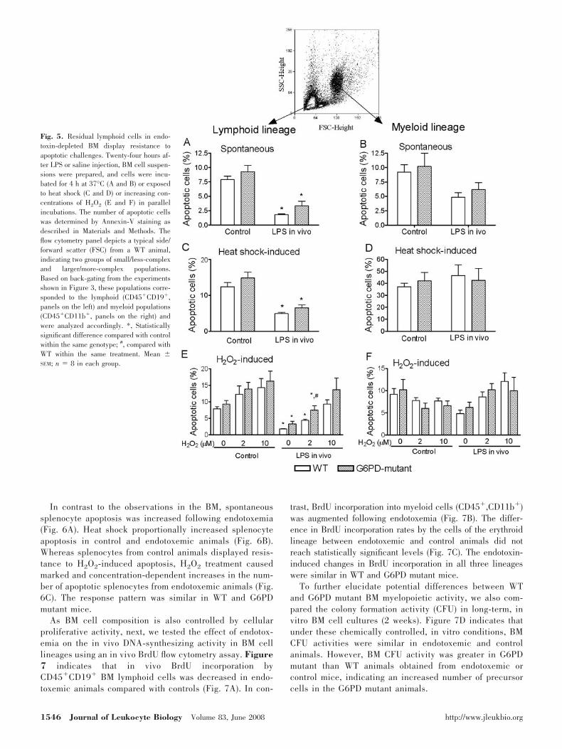
In contrast to the observations in the BM, spontaneoussplenocyte apoptosis was increased following endotoxemia(Fig. 6A). Heat shock proportionally increased splenocyteapoptosis in control and endotoxemic animals (Fig. 6B).Whereas splenocytes from control animals displayed resis-tance to H2O2-induced apoptosis, H2O2 treatment causedmarked and concentration-dependent increases in the num-ber of apoptotic splenocytes from endotoxemic animals (Fig.6C). The response pattern was similar in WT and G6PDmutant mice.
As BM cell composition is also controlled by cellularproliferative activity, next, we tested the effect of endotox-emia on the in vivo DNA-synthesizing activity in BM celllineages using an in vivo BrdU flow cytometry assay. Figure7 indicates that in vivo BrdU incorporation byCD45�CD19� BM lymphoid cells was decreased in endo-toxemic animals compared with controls (Fig. 7A). In con-
trast, BrdU incorporation into myeloid cells (CD45�,CD11b�)was augmented following endotoxemia (Fig. 7B). The differ-ence in BrdU incorporation rates by the cells of the erythroidlineage between endotoxemic and control animals did notreach statistically significant levels (Fig. 7C). The endotoxin-induced changes in BrdU incorporation in all three lineageswere similar in WT and G6PD mutant mice.
To further elucidate potential differences between WTand G6PD mutant BM myelopoietic activity, we also com-pared the colony formation activity (CFU) in long-term, invitro BM cell cultures (2 weeks). Figure 7D indicates thatunder these chemically controlled, in vitro conditions, BMCFU activities were similar in endotoxemic and controlanimals. However, BM CFU activity was greater in G6PDmutant than WT animals obtained from endotoxemic orcontrol mice, indicating an increased number of precursorcells in the G6PD mutant animals.
Fig. 5. Residual lymphoid cells in endo-toxin-depleted BM display resistance toapoptotic challenges. Twenty-four hours af-ter LPS or saline injection, BM cell suspen-sions were prepared, and cells were incu-bated for 4 h at 37°C (A and B) or exposedto heat shock (C and D) or increasing con-centrations of H2O2 (E and F) in parallelincubations. The number of apoptotic cellswas determined by Annexin-V staining asdescribed in Materials and Methods. Theflow cytometry panel depicts a typical side/forward scatter (FSC) from a WT animal,indicating two groups of small/less-complexand larger/more-complex populations.Based on back-gating from the experimentsshown in Figure 3, these populations corre-sponded to the lymphoid (CD45�CD19�,panels on the left) and myeloid populations(CD45�CD11b�, panels on the right) andwere analyzed accordingly. *, Statisticallysignificant difference compared with controlwithin the same genotype; #, compared withWT within the same treatment. Mean �SEM; n � 8 in each group.
1546 Journal of Leukocyte Biology Volume 83, June 2008 http://www.jleukbio.org

Endotoxemia and BM cytokine production
Figure 8, A and B, summarizes observations about the cyto-kine responses from WT animals. The majority of the testedcytokines, chemokines, and inflammatory mediators showeddecreased cytokine production from endotoxemic animals com-pared with controls (Fig. 8A). A few additional mediators alsoshowed small but statistically nonsignificant decreases afterendotoxemia (Fig. 8B). However, in striking contrast with thisdown-regulated general pattern, IL-6 production was increasedfollowing endotoxemia (Fig. 8B). We also determined IL-6responses by the more quantitative ELISA assay using cellsfrom a different set of experimental animals. Secondary in vitrostimulation by LPS or the TLR2 agonist Pam3CSK4 markedly
and similarly stimulated BM IL-6 production from control WTand G6PD mutant animals (Fig. 8C, left panel). Endotoxemiaincreased IL-6 production, and the TLR2 agonist-induced IL-6response was greater in endotoxemic than control animals.However, BM cells from endotoxemic G6PD mutant animalsdisplayed attenuated TLR4- or TLR2-induced IL-6 productionas compared with WT (Fig. 8C, right panel).
DISCUSSION
This study demonstrates for the first time that BM myelo- andlymphopoiesis are inversely affected during the acute stage of
Fig. 6. Increased splenocyte apoptosis following endotoxemia. Twenty-four hours after LPS or saline injection, splenocyte suspensions were prepared, and cellswere incubated for 4 h at 37°C (A) or exposed to heat shock (B) or increasing concentrations of H2O2 (C) in parallel incubations. The number of apoptotic cellswas determined by Annexin-V staining. *, Statistically significant difference compared with control within the same genotype. Mean � SEM; n � 8 in each group.
Fig. 7. De novo DNA synthesis is inhibitedin B cells but increased in myeloid cells inendotoxin-depleted BM. Twenty-two hours af-ter LPS or saline injection, in vivo BrdU in-corporation was determined, as described inMaterials and Methods. BM cells were iso-lated, and BrdU staining in combination withthe surface markers used was determined in Bcells (A), myeloid cells (B), and erythroid cells(C). *, Statistically significant difference com-pared with control within the same genotype.(D) CFUs in BM from endotoxemic or controlanimals after culture in GM-CSF-containingmedia for 2 weeks. Mean � SEM; n � 6–8animals for the BrdU studies, or n � 3 animalsfor the CFU activity studies in each group.
Chandra et al. Lymphopoiesis in endotoxemia 1547

endotoxemia. Whereas the biological importance of phagocytemobilization early after nonspecific immune challenges is quiteevident, the physiological role or the pathophysiological con-sequence of the accompanying acute lymphocytopenia involv-ing B as well as T cells is less apparent. Our current observa-tion indicating an endotoxin-induced depression in BM lym-phopoiesis provides a possible underlying mechanism that isresponsible for the depletion of circulating B cells early afternonspecific immune challenges. Decreased BM lymphopoiesismay be an important contributing factor to the generalizedimmunosuppression frequently observed in septic conditions.
Despite the fact that endotoxemia decreased myeloid andlymphoid cell content similarly in the BM, it is apparent thatdifferent mechanisms are responsible for these cell composi-tion changes. Myeloid cell depletion is most likely the result ofprompt release of myeloid cells from the marrow and theirsubsequent infiltration into immune-competent organs. Thisconclusion is supported by the observations that the decreasein BM myeloid content is accompanied by a simultaneousincrease in the number of circulating neutrophils as well as anaugmented neutrophil infiltration into the spleen and presum-ably other immune-competent organs. Additionally, followingendotoxin, the proliferative activity of BM myeloid cells (BrdUincorporation) is increased; meanwhile, they show largely un-altered, apoptotic responses. These observations together areconsistent with an elevated myelopoiesis and immediate re-lease of myeloid cells from the BM during the acute phase ofendotoxemia.
In contrast to this LPS-induced response pattern of myeloidcells, BM resident B cells showed decreased de novo DNAsynthesis accompanied by depleted B cell numbers in thecirculation. The lack of increase in splenic B cell contentmakes it unlikely that 24 h endotoxemia caused elevated B cell
migration into lymph and organs that would account for de-creased numbers of circulating B cells. Thus, decreased BMlymphopoiesis is most likely an important component of theLPS-induced marked and fast-developing B cell depletion inthe blood. The finding that LPS caused widespread BM celldepletion including cells negative for the markers used isconsistent with a generalized inhibition of lymphopoiesis.However, further investigations using T cell progenitor markerswill be needed to elucidate whether endotoxin also down-regulates the production of T lymphocytes.
Recent in vitro studies using isolated, naıve B cells andlong-term (4–6 days) in vitro cell proliferation assays havedemonstrated that B cell TLR activation is an important co-stimulus for BCR and Th cell-induced B cell proliferation [22,23], as well as during the terminal phase of plasma celldifferentiation [24]. Our observation, however, suggests that anacute in vivo TLR4 stimulus, in the absence of accompanyingBCR activation and other costimulatory signals, is inhibitoryon B cell proliferation and function. The more pronouncedLPS-induced decrease in the number of low CD45-expressingB cells in WT BM suggests that the LPS sensitivity is differentbetween the immature and mature B cell subpopulations.
It has been well documented in humans as well as animalmodels that sepsis results in lymphocyte apoptosis and gener-alized lymphopenia, which are believed to be major underlyingcauses of immune paralysis in septic patients [4, 25]. Theobserved, contrasting effects of endotoxemia on B cell apopto-sis in the BM and spleen, with decreased apoptosis in BMversus increased apoptosis in the spleen, suggest a differentsensitivity of differentiating and tissue-resident lymphocytes toapoptotic signals. These findings also suggest that the de-creased BM lymphopoiesis and the elevated lymphocyte apo-ptosis at the periphery contribute to the generalized lympho-
Fig. 8. Endotoxemia down-regulates BM cytokine and chemo-kine production, except IL-6. (A and B) BM cells were preparedfrom control and endotoxemic WT animals, and suspensioncultures of equal cell numbers were exposed to secondary LPSstimulus (100 ng/ml, 14 h). Media were collected and analyzedfor cytokine content using a cytokine antibody array. Baseline(dotted line) represents values obtained in controls in the ab-sence of in vitro LPS. (C) BM cells were prepared from anindependent set of control and endotoxemic WT as well asG6PD mutant animals, and suspension cultures of equal cellnumbers were exposed to LPS (100 ng/ml) or Pam3CSK4 (200
ng/ml) for 14 h. Cytokine content from conditioned media was measured by ELISA. *, Statistically significant difference compared with control within the samegenotype; #, compared with endotoxemic WT in the corresponding in vitro treatment. Mean � SEM; n � 4 in each group for cytokine arrays, or n � 8 in eachgroup for the ELISA. sTNFR, Soluble TNF receptor; SCF, stem cell factor; VEGF, vascular endothelial growth factor; THROM, thrombopoietin.
1548 Journal of Leukocyte Biology Volume 83, June 2008 http://www.jleukbio.org

cytopenia and associated lymphocyte dysfunction during en-dotoxemia. However, despite the fact that B cells remaining inthe BM 24 h after endotoxin showed decreased apoptosis, thisobservation does not rule out the possibility that BM lympho-cyte apoptosis occurred during preceding stages of endotox-emia, thereby also contributing to B cell depletion.
The widespread decrease in BM inflammatory mediator pro-duction together with the unchanged responsiveness of IL-4and IL-10 following endotoxemia suggests complex alterationsin the pro/anti-inflammatory balance, consistent with a de-pressed, functional status of the BM. However, in contrast tothe general trend of down-regulated or unchanged mediatorproduction, endotoxemia up-regulated BM IL-6 production,suggesting an important function of IL-6 in the observed re-sponses in agreement with independent observations [5, 6].IL-6 has been shown to stimulate early hematopoiesis [26], Bcell terminal differentiation, and malignant transformation[27–29] as well as to promote progenitor commitment towardthe myeloid line [30, 31]. Therefore, an LPS-induced, elevatedIL-6 production by the BM may represent a compensatoryfeedback response aimed at restoring depressed B cell produc-tion, or it may also promote myelopoiesis at the expense oflymphopoiesis.
Whereas the overall response pattern was similar in G6PD-deficient and WT animals, the increased BM cellularity inG6PD-deficient animals suggests that the decreased cellularantioxidant capacity associated with this mutation stimulatesBM hematopoiesis [32]. The fact that myeloid colony formationin long-term, chemically controlled BM cultures also showed agreater activity in G6PD mutant than WT animals indicates anincreased number of resident progenitor cells in G6PD-defi-cient animals. Previous studies indicated elevated blood IL-6levels in endotoxemic G6PD mutant animals compared withWT, presumably as a result of augmented resident macrophageactivation in the periphery [3, 33]. Blood concentration of IL-6is markedly elevated following endotoxemia or sepsis, andincreasing IL-6 levels have been shown to correlate with wors-ened outcome [34, 35]. The down-regulated IL-6 response inendotoxin-stimulated BM from G6PD mutant animals is con-sistent with an altered BM response under the G6PD-deficientconditions. It remains to be determined whether the down-regulated IL-6 production in G6PD mutant animals is associ-ated with altered cell activation, or it is the reflection ofdifferent lineage composition between WT and mutant animals.
Although the LPS-induced circulating erythrocyte dysfunc-tion, as reflected in decreased cell deformability, was greater inG6PD mutant than WT animals, endotoxemia had no markedeffects on BM erythroid cell content or erythroid BrdU incor-poration in these animals. This finding suggests that as a resultof the longer lifespan of erythrocytes, an extended period ofendotoxin exposure or a longer postendotoxin incubation timeis required for a potential manifestation of anemia and theassociated alterations in erythropoiesis.
Taken together, these investigations demonstrate that endo-toxemia results in inverse effects on the BM’s myelopoietic andlymphopoietic activities causing elevated myeloid proliferationbut depressed B cell production. Parallel with these changes,BM cytokine production is down-regulated with the exceptionof IL-6, indicating a distinct, regulatory role of this cytokine in
endotoxin-induced BM responses. Down-regulation of BM lym-phopoiesis during the acute phase of an innate immune re-sponse may represent an important component of immunedysfunction contributing to the immune paralysis observedduring prolonged septic conditions.
ACKNOWLEDGMENTS
This study was supported by NIH-National Institute of GeneralMedical Sciences grant GM-69861.
REFERENCES
1. Remick, D. G., Newcomb, D. E., Bolgos, G. L., Call, D. R. (2000)Comparison of the mortality and inflammatory response of two models ofsepsis: lipopolysaccharide vs. cecal ligation and puncture. Shock 13,110–116.
2. Spolarics, Z., Condon, M. R., Siddiqi, M., Machiedo, G. W., Deitch, E. A.(2004) Red blood cell dysfunction in septic glucose-6-phosphate dehy-drogenase-deficient mice. Am. J. Physiol. Heart Circ. Physiol. 286,H2118–H2126.
3. Wilmanski, J., Villanueva, E., Deitch, E. A., Spolarics, Z. (2007) Glucose-6-phosphate dehydrogenase deficiency and the inflammatory response toendotoxin and polymicrobial sepsis. Crit. Care Med. 35, 510–518.
4. Hotchkiss, R. S., Coopersmith, C. M., Karl, I. E. (2005) Prevention oflymphocyte apoptosis—a potential treatment of sepsis? Clin. Infect. Dis.41 (Suppl. 7), S465–S469.
5. Fonseca, R. B., Mohr, A. M., Wang, L., Sifri, Z. C., Rameshwar, P.,Livingston, D. H. (2005) The impact of a hypercatecholamine state onerythropoiesis following severe injury and the role of IL-6. J. Trauma 59,884–889.
6. Livingston, D. H., Anjaria, D., Wu, J., Hauser, C. J., Chang, V., Deitch,E. A., Rameshwar, P. (2003) Bone marrow failure following severe injuryin humans. Ann. Surg. 238, 748–753.
7. Santangelo, S., Gamelli, R. L., Shankar, R. (2001) Myeloid commitmentshifts toward monocytopoiesis after thermal injury and sepsis. Ann. Surg.233, 97–106.
8. Cohen, M. J., Carroll, C., He, L. K., Muthu, K., Gamelli, R. L., Jones,S. B., Shankar, R. (2007) Severity of burn injury and sepsis determines thecytokine responses of bone marrow progenitor-derived macrophages.J. Trauma 62, 858–867.
9. Pandolfi, P. P., Sonati, F., Rivi, R., Mason, P., Grosveld, F., Luzzatto, L.(1995) Targeted disruption of the housekeeping gene encoding glucose6-phosphate dehydrogenase (G6PD): G6PD is dispensable for pentosesynthesis but essential for defense against oxidative stress. EMBO J. 14,5209–5215.
10. Kletzien, R. F. (1994) Glucose-6-phosphate dehydrogenase: a housekeep-ing enzyme subject to tissue-specific regulation by hormones, nutrients,and oxidant stress. FASEB J. 8, 174–181.
11. Beutler, E. (1994) G6PD deficiency. Blood 84, 3613–3636.12. Luzzatto, L., Mehta, A. (1995) Glucose 6phosphate dehydrogenase defi-
ciency. In The Metabolic and Molecular Bases of Inherited Disease (C. R.Scriver, A. L. Beaudet, W. S. Sly, D. Valle, eds.), New York, NY, USA,McGrawHill, 3367–3398.
13. Finkel, T. (1999) Signal transduction by reactive oxygen species innon-phagocytic cells. J. Leukoc. Biol. 65, 337–340.
14. Jones, D. P. (2006) Redefining oxidative stress. Antioxid. Redox Signal. 8,1865–1879.
15. Efferth, T., Fabry, U., Glatte, P., Osieka, R. (1995) Increased induction ofapoptosis in mononuclear cells of a glucose-6-phosphate dehydrogenasedeficient patient. J. Mol. Med. 73, 47–49.
16. Spolarics, Z., Siddiqi, M., Siegel, J. H., Garcia, Z. C., Stein, D. S., Ong, H.,Livingston, D. H., Denny, T., Deitch, E. A. (2001) Increased incidence ofsepsis and altered monocyte functions in severely injured type A-glucose-6-phosphate dehydrogenase-deficient African American trauma patients.Crit. Care Med. 29, 728–736.
17. Abu-Osba, Y. K., Mallouh, A. A., Hann, R. W. (1989) Incidence andcauses of sepsis in glucose-6-phosphate dehydrogenase-deficient newborninfants. J. Pediatr. 114, 748–752.
18. Sanders, S., Smith, D. P., Thomas, G. A., Williams, E. D. (1997) Aglucose-6-phosphate dehydrogenase (G6PD) splice site consensus se-
Chandra et al. Lymphopoiesis in endotoxemia 1549

quence mutation associated with G6PD enzyme deficiency. Mutat. Res.374, 79–87.
19. Condon, M. R., Kim, J. E., Deitch, E. A., Machiedo, G. W., Spolarics, Z.(2003) Appearance of an erythrocyte population with decreased deform-ability and hemoglobin content following sepsis. Am. J. Physiol. HeartCirc. Physiol. 284, H2177–H2184.
20. McKenna, R. W., Washington, L. T., Aquino, D. B., Picker, L. J., Kroft,S. H. (2001) Immunophenotypic analysis of hematogones (B-lymphocyteprecursors) in 662 consecutive bone marrow specimens by 4-color flowcytometry. Blood 98, 2498–2507.
21. Carulli, G., Cannizzo, E., Zucca, A., Buda, G., Orciuolo, E., Marini, A.,Petrini, M. (2008) CD45 expression in low-grade B-cell non-Hodgkin’slymphomas. Leuk. Res. 32, 263–267.
22. Ruprecht, C. R., Lanzavecchia, A. (2006) Toll-like receptor stimulation asa third signal required for activation of human naive B cells. Eur. J. Im-munol. 36, 810–816.
23. Dye, J. R., Palvanov, A., Guo, B., Rothstein, T. L. (2007) B cell receptorcross-talk: exposure to lipopolysaccharide induces an alternate pathwayfor B cell receptor-induced ERK phosphorylation and NF- B activation.J. Immunol. 179, 229–235.
24. Genestier, L., Taillardet, M., Mondiere, P., Gheit, H., Bella, C., Defrance,T. (2007) TLR agonists selectively promote terminal plasma cell differ-entiation of B cell subsets specialized in thymus-independent responses.J. Immunol. 178, 7779–7786.
25. Hotchkiss, R. S., Tinsley, K. W., Karl, I. E. (2003) Role of apoptotic celldeath in sepsis. Scand. J. Infect. Dis. 35, 585–592.
26. Kopf, M., Ramsay, A., Brombacher, F., Baumann, H., Freer, G., Galanos,C., Gutierrez-Ramos, J. C., Kohler, G. (1995) Pleiotropic defects ofIL-6-deficient mice including early hematopoiesis, T and B cell function,and acute phase responses. Ann. N. Y. Acad. Sci. 762, 308–318.
27. Altmeyer, A., Simmons, R. C., Krajewski, S., Reed, J. C., Bornkamm,G. W., Chen-Kiang, S. (1997) Reversal of EBV immortalization precedes
apoptosis in IL-6-induced human B cell terminal differentiation. Immunity7, 667–677.
28. Morse, L., Chen, D., Franklin, D., Xiong, Y., Chen-Kiang, S. (1997)Induction of cell cycle arrest and B cell terminal differentiation by CDKinhibitor p18(INK4c) and IL-6. Immunity 6, 47–56.
29. Cozen, W., Gill, P. S., Ingles, S. A., Masood, R., Martinez-Maza, O.,Cockburn, M. G., Gauderman, W. J., Pike, M. C., Bernstein, L., Nathwani,B. N., Salam, M. T., Danley, K. L., Wang, W., Gage, J., Gundell-Miller, S.,Mack, T. M. (2004) IL-6 levels and genotype are associated with risk ofyoung adult Hodgkin lymphoma. Blood 103, 3216–3221.
30. Bernad, A., Kopf, M., Kulbacki, R., Weich, N., Koehler, G., Gutierrez-Ramos, J. C. (1994) Interleukin-6 is required in vivo for the regulation ofstem cells and committed progenitors of the hematopoietic system. Immu-nity 1, 725–731.
31. Zhu, J., Garrett, R., Jung, Y., Zhang, Y., Kim, N., Wang, J., Joe, G. J.,Hexner, E., Choi, Y., Taichman, R. S., Emerson, S. G. (2007) Osteoblastssupport B-lymphocyte commitment and differentiation from hematopoieticstem cells. Blood 109, 3706–3712.
32. Tothova, Z., Kollipara, R., Huntly, B. J., Lee, B. H., Castrillon, D. H.,Cullen, D. E., McDowell, E. P., Lazo-Kallanian, S., Williams, I. R., Sears,C., Armstrong, S. A., Passegue, E., DePinho, R. A., Gilliland, D. G. (2007)FoxOs are critical mediators of hematopoietic stem cell resistance tophysiologic oxidative stress. Cell 128, 325–339.
33. Wilmanski, J., Siddiqi, M., Deitch, E. A., Spolarics, Z. (2005) AugmentedIL-10 production and redox-dependent signaling pathways in glucose-6-phosphate dehydrogenase-deficient mouse peritoneal macrophages.J. Leukoc. Biol. 78, 85–94.
34. Ebong, S., Call, D., Nemzek, J., Bolgos, G., Newcomb, D., Remick, D.(1999) Immunopathologic alterations in murine models of sepsis of in-creasing severity. Infect. Immun. 67, 6603–6610.
35. Remick, D. G., Bolgos, G. R., Siddiqui, J., Shin, J., Nemzek, J. A. (2002)Six at six: interleukin-6 measured 6 h after the initiation of sepsis predictsmortality over 3 days. Shock 17, 463–467.
1550 Journal of Leukocyte Biology Volume 83, June 2008 http://www.jleukbio.org
Copyright © 2022 FDOKUMEN