Endodontic-Endotoxemia: Our Current Dilemma
22
(c) R.S.Carlson, D.D.S. / Agent Syntro-Research, Inc. A Nevada Corporation. 1 May 4, 2015 Commissioned by Syntro-Research, Inc. • Presented 2005 International Association of Bio-energetic Medicine, Honolulu, Hawai’i. (First Version) • Presented 2013 International Academy of Biological Dentistry & Medicine, Houston, Texas. (Current Version) • Presented 2014 IABDM Meeting in Las Vegas, NV • Posted on IABDM Web Journal 2014 Dr. Ronald S. Carlson 4211 Waianae Avenue Suite 400 Honolulu, Hawaii 96816 808-735-0282 Endodontic-Endotoxemia: Our Current Dilemma OUR CURRENT DILEMMA IN DENTAL-MEDICINE For about 180 years, or more, there have been attempts to retain teeth whose nutrient canals have been destroyed by placing gutta-percha or other sealants within the canals. 1 Numerous innovative mechanical techniques have been developed over the years to render the canals sterile and uninhabitable for microorganisms. Virtually all advertisements within the industry trade journals emphasize “mechanical obturation” and not the bio- chemical aspects of sterility of the canal system, or if the canals can be sterilized at all in the short or long term. As reported recently, overemphasis on this aspect of simply filling the empty spaces within the root canal system and its importance has misled the field of endodontology. 2 What is most critical, is that root canal teeth become nesting sites for microbes who, due to the loss of outward hydraulic pressure from within the body of the dental organ since the pulp no longer is living, migrate though the dentin into the peri-radicular tissues, thence into the circulatory system and other tissues. In Dental Clinics of North America, Vol. 18, No. 2, April 1974, Kaare Langeland, D.D.S., Ph.D. reviewed “Root Canal Sealants and Pastes” in which he states: “The fact that sealers and pastes will contact vital or
-
Upload
independent -
Category
Documents
-
view
1 -
download
0
Transcript of Endodontic-Endotoxemia: Our Current Dilemma

(c) R.S.Carlson, D.D.S. / Agent Syntro-Research, Inc. A Nevada Corporation. 1
May 4, 2015 Commissioned by Syntro-Research, Inc.
• Presented 2005 International Association of Bio-energetic Medicine, Honolulu, Hawai’i. (First Version)
• Presented 2013 International Academy of Biological Dentistry & Medicine, Houston, Texas. (Current Version)
• Presented 2014 IABDM Meeting in Las Vegas, NV • Posted on IABDM Web Journal 2014
Dr. Ronald S. Carlson 4211 Waianae Avenue Suite 400 Honolulu, Hawaii 96816 808-735-0282
Endodontic-Endotoxemia: Our Current Dilemma
OUR CURRENT DILEMMA IN DENTAL-MEDICINE
For about 180 years, or more, there have been attempts to retain teeth
whose nutrient canals have been destroyed by placing gutta-percha or other sealants within the canals.1 Numerous innovative mechanical techniques have been developed over the years to render the canals sterile and uninhabitable for microorganisms. Virtually all advertisements within the industry trade journals emphasize “mechanical obturation” and not the bio-chemical aspects of sterility of the canal system, or if the canals can be sterilized at all in the short or long term. As reported recently, overemphasis on this aspect of simply filling the empty spaces within the root canal system and its importance has misled the field of endodontology.2 What is most critical, is that root canal teeth become nesting sites for microbes who, due to the loss of outward hydraulic pressure from within the body of the dental organ since the pulp no longer is living, migrate though the dentin into the peri-radicular tissues, thence into the circulatory system and other tissues. In Dental Clinics of North America, Vol. 18, No. 2, April 1974, Kaare Langeland, D.D.S., Ph.D. reviewed “Root Canal Sealants and Pastes” in which he states: “The fact that sealers and pastes will contact vital or

(c) R.S.Carlson, D.D.S. / Agent Syntro-Research, Inc. A Nevada Corporation. 2
disintegrating tissue connected with the general circulation allows for generalized distribution by blood and lymph…The resorbability of sealers and pastes has been abundantly demonstrated by gross radiographic evidence. There is, therefore, no doubt that material is transported away from the site where it was originally applied.”
What has been shown today as of equal or more significant importance is the relative sterility of the intra-radicular-canal system.3,4,5 Current research shows that the significant presence of bacteria in the canal systems lead to the pathogenesis of “apical periodontitis” (AP)—now known as “endodontic disease.” This condition (AP) is a chronic/acute inflammation of the tissue about the roots of root filled teeth also designated as “apical granuloma.”21
Equally significant in consideration for successful endodontic procedures is the presence of bacteria within the microtubules of the dentin itself.5 These concerns were reported in lectures in Chicago and other major cities in the 1930s and 1940s. Infected dentine was demonstrated to be of great influence on the lack of success of root filled teeth by Weston Price, D.D.S.6 Recent studies show that dentine can be infected and that controversy exists regarding how to disinfect it clinically.7 Rather than sterility of the root canal system as an objective of modern endodontics, one can at best employ the term “relative sterility” or disinfecting.
Systemic implications are obvious from more recent work showing that apical periodontitis, an inflammatory process around the apex of a tooth root, is primarily a sequel to microbial infection of the pulp space of teeth and is a remarkably widespread problem.8
It is calculated that apical priodontitis is so prevalent that by the age of 50, one in two (50%) experience the disease.9 By age 62 the statistic rises to 62%. In the U.S. it is estimated that 420 million teeth have received root filings, or in the common vernacular “root canalled.”10On this basis the average number of root fillings is 2.2 per adult.11 Readily it can be understood that the magnitude of the problem is not fully comprehended nor appreciated as to its impact upon the general health of the individuals with this condition.12
Inflammation has been described as a common reaction of the body to damage of its cells and vascularized tissues. The discovered detailed process of inflammation has revealed its relationship to immune response as being one in the same fundamentally. The five basic symptoms of inflammation, 1) redness (rubor), 2) swelling (tumour), 3) heat (calor), 4) pain (dolor), and 5) loss of function (functio laesa) have been know since ancient times.

(c) R.S.Carlson, D.D.S. / Agent Syntro-Research, Inc. A Nevada Corporation. 3
In a recent work, a close correlation to periodontal pathology and rheumatoid arthritis was found.13 In 62.5% of a study population of 1,412 people with rheumatoid arthritis had advanced periodontal disease. The authors went as far as stating there are certain pathogenic similarities between the two clinical entities.
Further, in an article found in the International Endodontic Journal, Vol. 33 Issue 1 Page 1, year 2000, titled “Root canal treatment and general health: a review of the literature” the authors state: “There has been an increase in the number of case reports published in the medical literature citing dental infection as an associated factor in several systemic illnesses including; uveitis (Sela & Sharav 1979), intracranial abscesses (Holin et al. 1967, Henig et al. 1978, Ingham et al. 1978, Churton & Green 1980, Aldous et al. 1987, Marks et al. 1988, Saal et al. 1988), childhood hemiplegia (Hamlyn 1978), cerebral infarction (Syrjanen et al. 1989), bacteriospermia and subfertility (Bieniek & Riedel 1993), necrotizing fasciitis ( Gallia & Johnson 1981, Steel 1987, Stoykewych et al. 1978), mediastinitis (Hendler & Quinn 1978, Zachariades et al. 1988, Musgrove & Malden 1989), fatal endocarditis (Kralovic et al. 1995), toxic shock syndrome (Egbert et al. 1987, Navazesh et al. 1994), and septicemia (Lee 1984).”
Infections in the mouth can develop into chronic localized infections or systemic infectious disabilities—conditions we refer to as disease or illness. In the pathogenesis and evolution of glomerular nephropathies, these localized infections play an important role.14 Renal patients appear to be subject to a variety of dental problems such as periodontal disease, narrowing of the pulp chamber, enamel abnormalities, premature tooth loss and xerostomia. Renal difficulties also often arise after a specific stomatologic (oral) treatment of dental foci. The dental infection can localize in the pulp of the dental organ (tooth), developing acute gangrenous pulpitis. On the other hand, a chronic evolution from gangrenous pulpitis may lead to the formation of dental granulomas, cysts, osteomyelitis, other bone conditions and abscesses in the bone surrounding the root of the tooth. In the evolution of these infections, acute phases may occur, despite the fact that the human host tries to isolate these foci by forming an external layer of fibrin and connective tissue about these lesions, much like the skin protects the hands by forming blisters and then subsequent callous’ after working vigorously in the backyard vegetable garden. In these phases, germs or parts of them—toxins—could get into the circulation. They could produce various lesions in other tissues—this process being known as “effective localization”—either directly or indirectly by forming immune complexes. These are deposited in different tissues where they can activate different
(c) R.S.Carlson, D.D.S. / Agent Syntro-Research, Inc. A Nevada Corporation. 4
factors like compliments—c reactive proteins for example in the liver. The kidney is often the target organ, since it is the master filter for the vascular system.
Scientific America, May 2001, presented an article titled Taken to Heart. Reporting that in a recent examination of 50 plaques scraped out of human arteries, 72 % contained periodontal pathogens. Two other pathogens that are hot candidates for atherosclerosis were also present: cytomegalovirus, which infected 38% of plaques, and Chlamydia pneumoniae, which appeared in 18%. Studies have also found antibodies against oral bacteria in the blood, and animal tests have shown that mouth microbes injected into the blood lead to atherosclerosis. Links between periodontal infection (apical periodontitis included) and other conditions such as diabetes, chronic respiratory infection, stroke and low birth weight have also emerged. They support the theory that many chronic ill conditions stem from systemic low-grade infections.
A marker known as C-reactive protein or CRP can now identify the systemic low-grade infections causing systemic inflammation. It is most always elevated in patients with periodontitis. Chronic and/or acute inflammation of the peri-radicular ligament may lead to heart disease because it might cause this low-level systemic inflammation since the chemical by-products of the degradation itself along with the autoimmune response may spill over into the blood stream and lymphatic system triggering the liver to make other proteins that inflame the arterial walls and clot blood. Atherosclerosis and, ultimately, heart attack may result.15
The American Heart Association’s (AHA) web site states: “A growing number of studies have examined whether hs-CRP can predict recurrent cardiovascular disease and stroke and death in different settings. High levels of hs-CRP consistently predict new coronary events in patients with unstable angina and acute myocardial infarction (heart attack). Higher hs-CRP levels also are associated with lower survival rate of these people. Many studies suggest that after adjusting for other prognostic factors, hs-CRP was still useful as a predictor.” However, the AHA does not proffer a possible cause of the low-grade systemic infections. This, I believe, will be left to those studying the subtle Oral-Sytemic Linkages as outlined in recent editorials of the Triple O Journal.2, 16
CLINICAL EVIDENCE OF ORAL-SYSTEMIC DENTAL INFECTION With these facts in mind, let us examine the background information
derived from a study reported in ODONTOLOGISK REVY, VOLUME 18,
(c) R.S.Carlson, D.D.S. / Agent Syntro-Research, Inc. A Nevada Corporation. 5
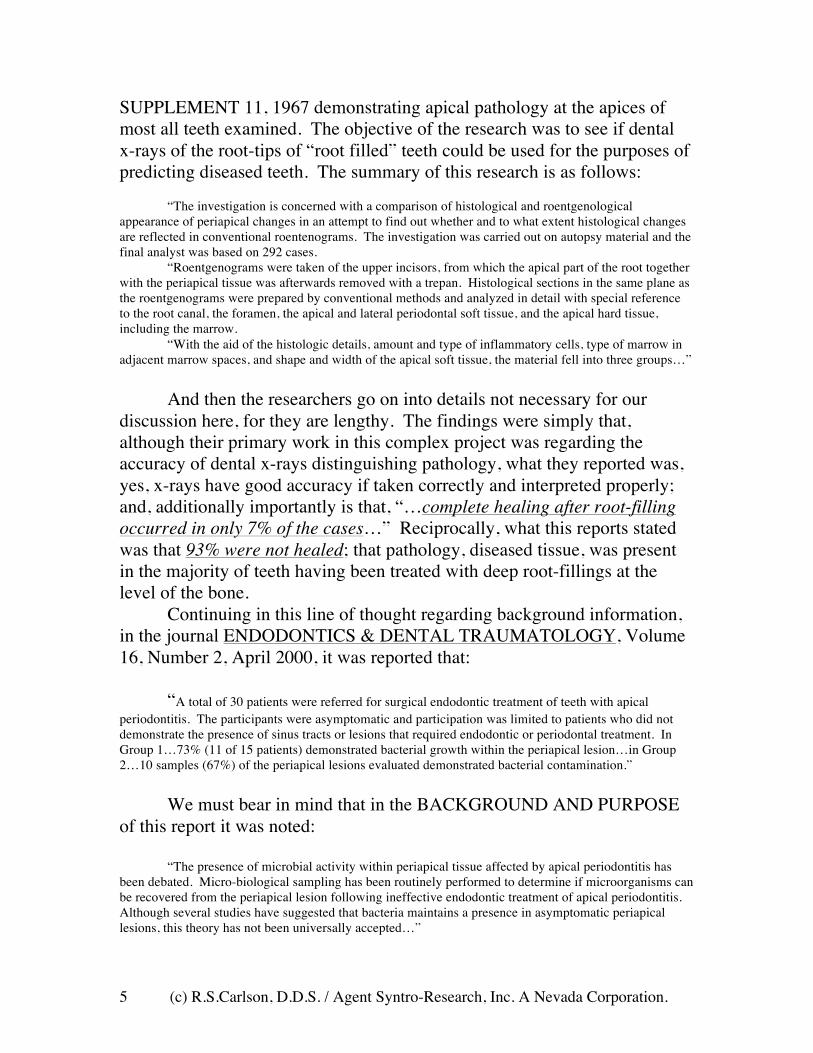
SUPPLEMENT 11, 1967 demonstrating apical pathology at the apices of most all teeth examined. The objective of the research was to see if dental x-rays of the root-tips of “root filled” teeth could be used for the purposes of predicting diseased teeth. The summary of this research is as follows:
“The investigation is concerned with a comparison of histological and roentgenological
appearance of periapical changes in an attempt to find out whether and to what extent histological changes are reflected in conventional roentenograms. The investigation was carried out on autopsy material and the final analyst was based on 292 cases.
“Roentgenograms were taken of the upper incisors, from which the apical part of the root together with the periapical tissue was afterwards removed with a trepan. Histological sections in the same plane as the roentgenograms were prepared by conventional methods and analyzed in detail with special reference to the root canal, the foramen, the apical and lateral periodontal soft tissue, and the apical hard tissue, including the marrow.
“With the aid of the histologic details, amount and type of inflammatory cells, type of marrow in adjacent marrow spaces, and shape and width of the apical soft tissue, the material fell into three groups…”
And then the researchers go on into details not necessary for our
discussion here, for they are lengthy. The findings were simply that, although their primary work in this complex project was regarding the accuracy of dental x-rays distinguishing pathology, what they reported was, yes, x-rays have good accuracy if taken correctly and interpreted properly; and, additionally importantly is that, “…complete healing after root-filling occurred in only 7% of the cases…” Reciprocally, what this reports stated was that 93% were not healed; that pathology, diseased tissue, was present in the majority of teeth having been treated with deep root-fillings at the level of the bone.
Continuing in this line of thought regarding background information, in the journal ENDODONTICS & DENTAL TRAUMATOLOGY, Volume 16, Number 2, April 2000, it was reported that:
“A total of 30 patients were referred for surgical endodontic treatment of teeth with apical
periodontitis. The participants were asymptomatic and participation was limited to patients who did not demonstrate the presence of sinus tracts or lesions that required endodontic or periodontal treatment. In Group 1…73% (11 of 15 patients) demonstrated bacterial growth within the periapical lesion…in Group 2…10 samples (67%) of the periapical lesions evaluated demonstrated bacterial contamination.”
We must bear in mind that in the BACKGROUND AND PURPOSE
of this report it was noted: “The presence of microbial activity within periapical tissue affected by apical periodontitis has
been debated. Micro-biological sampling has been routinely performed to determine if microorganisms can be recovered from the periapical lesion following ineffective endodontic treatment of apical periodontitis. Although several studies have suggested that bacteria maintains a presence in asymptomatic periapical lesions, this theory has not been universally accepted…”
(c) R.S.Carlson, D.D.S. / Agent Syntro-Research, Inc. A Nevada Corporation. 6
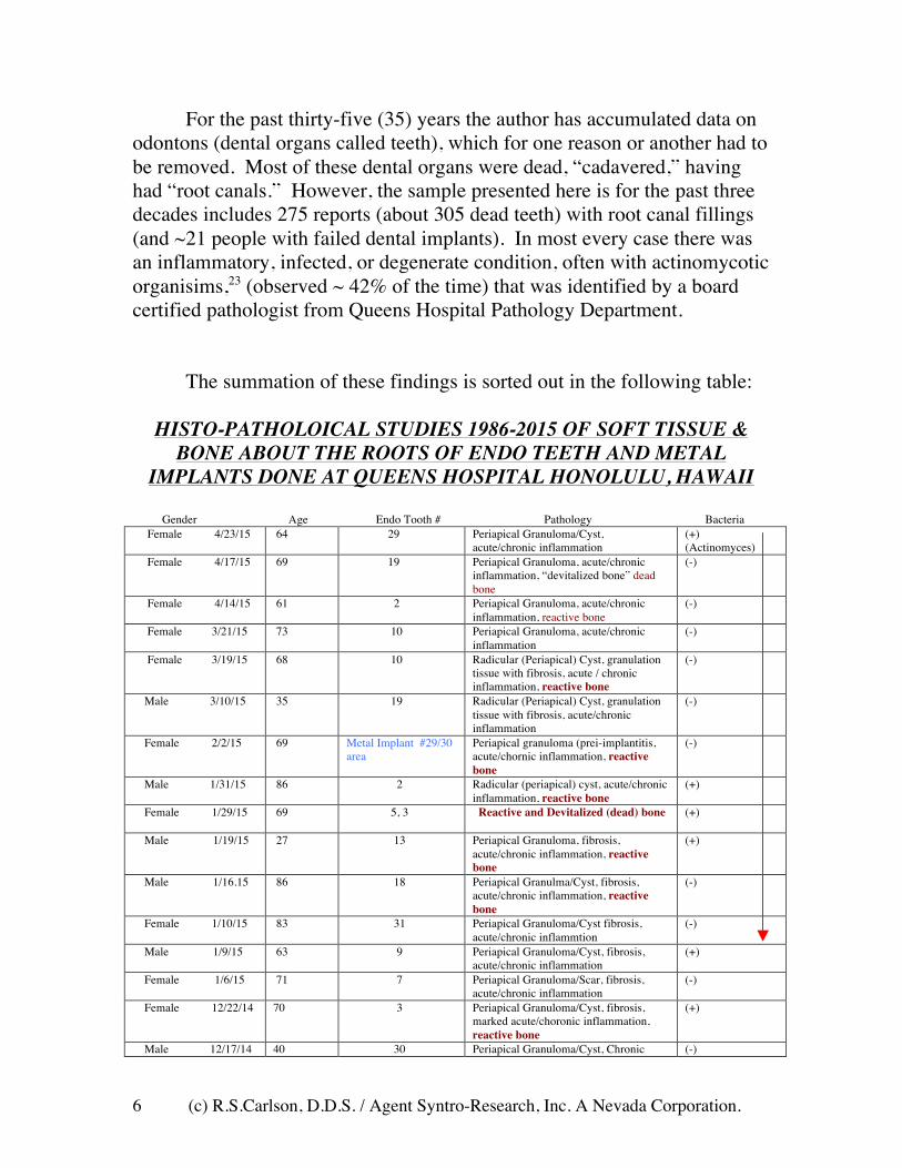
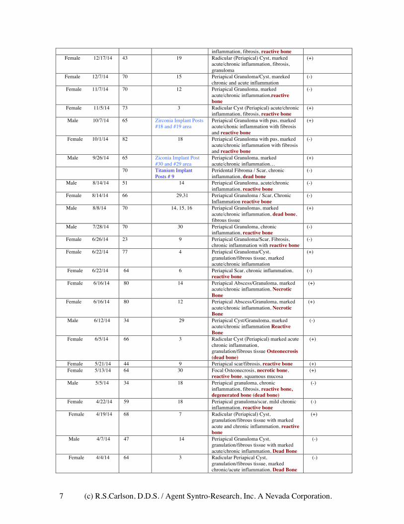
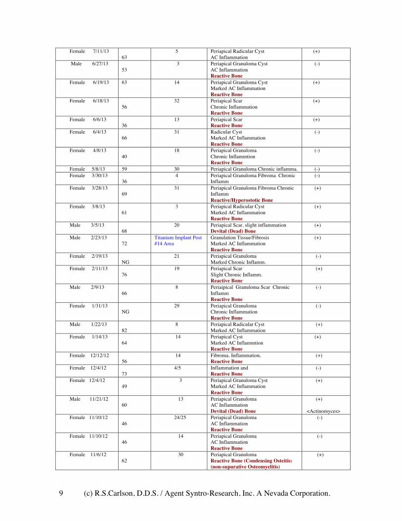
For the past thirty-five (35) years the author has accumulated data on odontons (dental organs called teeth), which for one reason or another had to be removed. Most of these dental organs were dead, “cadavered,” having had “root canals.” However, the sample presented here is for the past three decades includes 275 reports (about 305 dead teeth) with root canal fillings (and ~21 people with failed dental implants). In most every case there was an inflammatory, infected, or degenerate condition, often with actinomycotic organisims,23 (observed ~ 42% of the time) that was identified by a board certified pathologist from Queens Hospital Pathology Department.
The summation of these findings is sorted out in the following table:
HISTO-PATHOLOICAL STUDIES 1986-2015 OF SOFT TISSUE & BONE ABOUT THE ROOTS OF ENDO TEETH AND METAL
IMPLANTS DONE AT QUEENS HOSPITAL HONOLULU, HAWAII
Gender Age Endo Tooth # Pathology Bacteria Female 4/23/15 64 29 Periapical Granuloma/Cyst,
acute/chronic inflammation (+) (Actinomyces)
Female 4/17/15 69 19 Periapical Granuloma, acute/chronic inflammation, “devitalized bone” dead bone
(-)
Female 4/14/15 61 2 Periapical Granuloma, acute/chronic inflammation, reactive bone
(-)
Female 3/21/15 73 10 Periapical Granuloma, acute/chronic inflammation
(-)
Female 3/19/15 68 10 Radicular (Periapical) Cyst, granulation tissue with fibrosis, acute / chronic inflammation, reactive bone
(-)
Male 3/10/15 35 19 Radicular (Periapical) Cyst, granulation tissue with fibrosis, acute/chronic inflammation
(-)
Female 2/2/15 69 Metal Implant #29/30 area
Periapical granuloma (prei-implantitis, acute/chornic inflammation, reactive bone
(-)
Male 1/31/15 86 2 Radicular (periapical) cyst, acute/chronic inflammation, reactive bone
(+)
Female 1/29/15 69 5, 3 Reactive and Devitalized (dead) bone (+)
Male 1/19/15 27 13 Periapical Granuloma, fibrosis, acute/chronic inflammation, reactive bone
(+)
Male 1/16.15 86 18 Periapical Granulma/Cyst, fibrosis, acute/chronic inflammation, reactive bone
(-)
Female 1/10/15 83 31 Periapical Granuloma/Cyst fibrosis, acute/chronic inflammtion
(-)
Male 1/9/15 63 9 Periapical Granuloma/Cyst, fibrosis, acute/chronic inflammation
(+)
Female 1/6/15 71 7 Periapical Granuloma/Scar, fibrosis, acute/chronic inflammation
(-)
Female 12/22/14 70 3 Periapical Granuloma/Cyst, fibrosis, marked acute/choronic inflammation, reactive bone
(+)
Male 12/17/14 40 30 Periapical Granuloma/Cyst, Chronic (-)
(c) R.S.Carlson, D.D.S. / Agent Syntro-Research, Inc. A Nevada Corporation. 7
inflammation, fibrosis, reactive bone Female 12/17/14 43 19 Radicular (Periapical) Cyst, marked
acute/chronic inflammation, fibrosis, granuloma
(+)
Female 12/7/14 70 15 Periapical Granuloma/Cyst, mareked chronic and acute inflammation
(-)
Female 11/7/14 70 12 Periapical Granuloma, marked acute/chronic inflammation,reactive bone
(-)
Female 11/5/14 73 3 Radicular Cyst (Periapical) acute/chronic inflammation, fibrosis, reactive bone
(+)
Male 10/7/14 65 Zirconia Implant Posts #18 and #19 area
Periapical Granuloma with pus, marked acute/chonic inflammation with fibrosis and reactive bone
(+)
Female 10/1/14 82 18 Periapical Granuloma with pus, marked acute/chronic inflammation with fibrosis and reactive bone
(-)
Male 9/26/14 65 Ziconia Implant Post #30 and #29 area
Periapical Granuloma, marked acute/chronic inflammation…
(+)
70 Titanium Implant Posts # 9
Peridental Fibroma / Scar, chronic inflammation, dead bone
(-)
Male 8/14/14 51 14 Periapical Granuloma, acute/chronic inflammation, reactive bone
(-)
Female 8/14/14 66 29,31 Periapical Granuloma / Scar, Chronic Inflammation reactive bone
(-)
Male 8/8/14 70 14, 15, 16 Periapical Granulomas, marked acute/chronic inflammation, dead bone, fibrous tissue
(+)
Male 7/28/14 70 30 Periapical Granuloma, chronic inflammation, reactive bone
(-)
Female 6/26/14 23 9 Periapical Granuloma/Scar, Fibrosis, chronic inflammation with reactive bone
(-)
Female 6/22/14 77 4 Periapical Granuloma/Cyst, granulation/fibrous tissue, marked acute/chronic inflammation
(+)
Female 6/22/14 64 6 Periapical Scar, chronic inflammation, reactive bone
(-)
Female 6/16/14 80 14 Periapical Abscess/Granuloma, marked acute/chronic inflammation, Necrotic Bone
(+)
Female 6/16/14 80 12 Periapical Abscess/Granuloma, marked acute/chronic inflammation, Necrotic Bone
(+)
Male 6/12/14 34 29 Periapical Cyst/Granuloma, marked acute/chronic inflammation Reactive Bone
(-)
Female 6/5/14 66 3 Radicular Cyst (Periapical) marked acute chronic inflammation, granulation/fibrous tissue Osteonecrosis (dead bone)
(+)
Female 5/21/14 44 9 Periapical scar/fibrosis, reactive bone (+) Female 5/13/14 64 30 Focal Osteonecrosis, necrotic bone,
reactive bone, squamous mucosa (+)
Male 5/5/14 34 18 Periapical granuloma, chronic inflammation, fibrosis, reactive bone, degenerated bone (dead bone)
(-)
Female 4/22/14 59 18 Periapical granuloma/scar, mild chronic inflammation, reactive bone
(-)
Female 4/19/14 68 7 Radicular (Periapical) Cyst, granulation/fibrous tissue with marked acute and chronic inflammation, reactive bone
(+)
Male 4/7/14 47 14 Periapical Granuloma Cyst, granulation/fibrous tissue with marked acute/chronic inflammation, Dead Bone
(-)
Female 4/4/14 64 3 Radicular Periapical Cyst, granulation/fibrous tissue, marked chronic/acute inflammation, Dead Bone
(-)

(c) R.S.Carlson, D.D.S. / Agent Syntro-Research, Inc. A Nevada Corporation. 8
Female 4/4/14 49 14 Periapical Granuloma/Cyst, granulation/fibrous tissue with chronic inflammation
(-)
Female 3/21/14 42 10 Periapical Granuloma/Abscess, marked acute/chronic inflammation with fibrosis and granulation tissue, Dead bone, reactive bone consistent with “osteomyelitis”
(-)
Male 3/11/14 57 14, 15 Periapical Cyst/Granuloma, marked acute/chronic inflammation, Reactive Bone
(+)
Female 2/22/14 66 6 Periapical abscess/granuloma, marked a/c inflammation, Partially dead and reactive bone
(+)
Male 2/4/14 70 18,19 Cystic Granuloma/marked acute-chronic inflammation, Reactive Bone
(-)
Female 1/24/14 43 10 Cystic Granuloma/ tissue with marked inflammation, Chronic and Acute Reactive Bone
(-)
Male 1/14/14 61 4 Periapical scar/fibrosis, Osteonecrosis (Dead Bone) Patient has bowl cancer—this tooth #4 and two others #12 & #19 on the same meridian were root cadavers. CA dx 6mo ago
(+)
Male 1/13/14 63 29 Periapical granuloma/scar, AC Inflam, dead dentine/cementum, numerous Actinmycese/Candida
(+) Candida 1st (Actinomyces)
Male 1/11/14 34 30 Periapical granuloma, AC inflammation, Reactive bone, fibrosis
(-)
Female 12/7/13 44 2 Periapical abscess/scar Reactive/Necrotic(dead) bone and fibrous tissue
(+)
Female 11/21/13 66 28 Periapical granuloma/Scar Chronic inflammation, Reactive Bone
(-)
Male 11/11/13 26 10 Periapical granuloma (Fistula with pus) Marked AC inflammation Reactive Bone
(-)
Female 10/7/13 53 19 Periapical granuloma/scar AC Inflammation underlying fibrosis Reactive bone
(-)
Female 9/27/13 76 Titanium Implant Posts 18, 19, 20 with uA 26 and mV -265
Peri-implantitis / Osteosis Marked AC Inflammation Reactive Bone
(+)
Female 9/24/13 53 18/19 Periapical Granuloma Scar Chronic Inflammation Devitalized (Dead) Bone
(-)
Female 9/9/13 37 30 Periapical Scar Chronic Inflammation Reactive Bone
(-)
Female 9/5/13 16 26 Periapical Granuloma Cyst Chronic Inflammation Devitalized (Dead) Bone Reactive Bone
(-)
Male 8/27/13 63 30 Periapical Abscess Granuloma Marked AC Inflammation Devitalized (Dead) Bone Reactive Bone
(-)
Female 8/19/13 80 2 Periapical Granuloma Chronic Inflammation Reactive Bone
(-)
Female 8/3/13 63
Implant Post #4 #6 (Titanium Metal) with 36 uA Current Density
Peri-implantitis / osteitis Chronic Inflammation Devitalized (Dead) Bone Reactive Bone
(+)
Female 7/31/13 58
30 Apical Osteitis Devitalized (Dead) Bone Reactive Bone
(-)
(c) R.S.Carlson, D.D.S. / Agent Syntro-Research, Inc. A Nevada Corporation. 9
Female 7/11/13 63
5 Periapical Radicular Cyst AC Inflammation
(+)
Male 6/27/13 53
3 Periapical Granuloma Cyst AC Inflammation Reactive Bone
(-)
Female 6/19/13
63 14 Periapical Granuloma Cyst Marked AC Inflammation Reactive Bone
(+)
Female 6/18/13 56
32 Periapical Scar Chronic Inflammation Reactive Bone
(+)
Female 6/6/13 36
13 Periapical Scar Reactive Bone
(+)
Female 6/4/13 66
31 Radicular Cyst Marked AC Inflammation Reactive Bone
(-)
Female 4/8/13 40
18 Periapical Granuloma Chronic Inflammtion Reactive Bone
(-)
Female 5/8/13 59 30 Periapical Granuloma Chronic inflamma. (-) Female 3/30/13
36 4 Periapical Granuloma Fibroma Chronic
Inflamm (-)
Female 3/28/13 69
31 Periapical Granuloma Fibroma Chronic Inflamm Reactive/Hyperostotic Bone
(+)
Female 3/8/13 61
3 Periapical Radicular Cyst Marked AC Inflammation Reactive Bone
(+)
Male 3/5/13 68
20 Periapical Scar, slight inflammation Devital (Dead) Bone
(+)
Male 2/23/13 72
Titanium Implant Post #14 Area
Granulation Tissue/Fibrosis Marked AC Inflammation Reactive Bone
(+)
Female 2/19/13 NG
21 Periapical Granuloma Marked Chronic Inflamm.
(-)
Female 2/11/13 76
19 Periapical Scar Slight Chronic Inflamm. Reactive Bone
(+)
Male 2/9/13 66
8 Periaipical Granuloma Scar Chronic Inflamm Reactive Bone
(-)
Female 1/31/13 NG
29 Periapical Granuloma Chronic Inflammation Reactive Bone
(-)
Male 1/22/13 82
8 Periapical Radicular Cyst Marked AC Inflammation
(+)
Female 1/14/13 64
14 Periapical Cyst Marked AC Inflammtion Reactive Bone
(+)
Female 12/12/12 56
14 Fibroma, Inflammation, Reactive Bone
(+)
Female 12/4/12 73
4/5 Inflammation and Reactive Bone
(-)
Female 12/4/12 49
3 Periapical Granuloma Cyst Marked AC Inflammation Reactive Bone
(+)
Male 11/21/12 60
13 Periapical Granuloma AC Inflammation Devital (Dead) Bone
(+) <Actinomyces>
Female 11/10/12 46
24/25 Periapical Granuloma AC Inflammation Reactive Bone
(-)
Female 11/10/12 46
14 Periapical Granuloma AC Inflammation Reactive Bone
(-)
Female 11/6/12 62
30 Periapical Granuloma Reactive Bone (Condensing Osteitis: (non-supurative Osteomyelitis)
(+)

(c) R.S.Carlson, D.D.S. / Agent Syntro-Research, Inc. A Nevada Corporation. 10
Male 10/27/12 56
8 Periapical Granuloma Cyst Marked AC Inflammation
(+)
Female 10/4/12 57
19 Periapical Cemento-osseous Dysplasia Immature bone/cementum fibrous tissue
(-)
Male 9/27/12 66 5 Periapical Granuloma Marked AC Inflammtion Devitalized (Dead) Bone
(-)
Female 9/11/12 55
4 Partially Devitalized Bone (Dead Bone) Marked Chronic Inflammation
(+)
Male 8/30/12 51
9 Periapical Granuloma Cyst Marked AC Inflammation
(-)
Female 8/6/12 60
19 Chronic Osteomyelitis Dead & Reactive Bone Chronic Inflammation
(-)
Male 7/20/12 58
30 Periapical Granuloma AC Inflammation Reactive Bone
(-)
Male 7/20/12 58
7/8 Periapical Cyst AC Inflammation
(-)
Female 5/16/12 77
30 Periapical Granuloma Marked AC Inflammation Reactive Bone
(+)
Male 5/17/12 60
19 Periapical Granuloma Abscess/ Marked AC Inflammation Reactive Bone
(-)
Male 5/11/12 31
30 Periapical Granuloma Marked AC Inflammation
(-)
Male 5/2/12 35
18 Periapical Granuloma Scar Chronic Inflammation Reactive Bone
(-)
Female 4/10/12 51
20 Periapical Granuloma Marked AC Inflammation
(-)
Female 3/17/12 55
12 Periapical Scar AC Inflammation
(-)
Female 2/28/12 60
30 Periapical Scar Chronic Inflammation Reactive Bone
(+)
Female 2/28/12 60
3 Periapical Granuloma Chronic Inflammation Reactive Bone
(-)
Female 1/3/12 42
2 Periapical Radicular Cyst Marked AC Inflammation Reactive Bone
(-)
Male 8/17/11 40
8/9 Periapical Granuloma Marked Chronic Inflammation
(+)
Female 7/20/11 29
Vital #29 (Pl Test 7.5) Periapical Grauloma Scar (+)
Female 6/30/11 64
8/10 Periapical Granuloma Cyst Chronic Inflammation Reactive Bone
(-)
Female 5/4/11 31
4 Periapical Granuloma Scar Chronic Inflammation
(-)
Female 3/2/11 31
19 Periapical Granuloma Scar Granulation/Fibrous Tissue Chronic Inflammation
(-)
Male 11/2/10 68
3 Periapical Granuloma AC Inflammation Reactive Bone
(+)
Male 8/13/10 75
30 Periapical Granuloma Marked AC Inflammation
(+)
Female 8/6/10 60 Titanium Implant #12 and #13
Periapical Granuloma (Fibrous Tissue) with marked acute/chronic inflammation. Dead Bone (bone graft?)reactive bone.
(-)
Female 8/4/10 66
28/29 Periapical Granuloma Marked AC Inflammation Reactive Bone
(-)
Female 4/23/10 3 Periapical Granuloma (-)

(c) R.S.Carlson, D.D.S. / Agent Syntro-Research, Inc. A Nevada Corporation. 11
51 AC Inflammation Male 4/15/10
56 20 Periapical Granuloma Scar
Chronic Inflammation (-)
Male 2/24/10 48
21 Periapical Scar Chronic Inflammation
(-)
Male 1/19/10 48
29/30 Periapical Granuloma marked AC Inflammation Reactive Bone
(+)
Female 1/4/10 55 14/15 Periapical Granuloma Chronic Inflammation Reactive Bone
(-)
Female 1/4/10 61
4 Periapical Granuloma Scar Chronic Inflammation
(-)
Female 7/3/09 46
7 Periapical Marked AC Inflammation (+)
Female 7/16/09 60
10 Periapical Granuloma Cyst Chronic inflammation Reactive Bone
(-)
Female 7/7/09 33
3 Periapical Granuloma Scar Reactive Bone
(-)
Male 7/2/09 58
9 Radicular Periapical Cyst AC Inflammation
(+)
Female 6/19/09 75 31 Periapical Granuloma Cyst AC Inflam. (+) Male 6/9/09
69 8,9,10, 11 Periapical Granuloma Cyst (+)
Male 5/30/09 50
8 Periapical Granuloma, Marked AC Inflmmation Reactive Bone
(-)
Female 5/27/09 46
15 Periapical Granuloma/Scar Reactive Bone
(-)
Male 5/20/09 55
3 Periapical Granuloma Moderate C-Inflammation
(+)
Female 5/13/09 64
20 Periapical Granuloma Marked AC Inflammation
(-)
Female 4/10/09 38
18 Periapical Granuloma-Abscess Marked AC Inflmmation
(-)
Female 2/26/09 60
18/19 Periapical Granuloma c Osteonecrosis(dead bone)
(+) (Fossamax Person)
Female 2/13/09 63
3 Periapical Granuloma Marked AC Inflammation
(-)
Female 12/13/08 56
9/11 Periapical Granuloma/Scar Chronic Inflammation
(-)
Female 12/9/08 70
5 Radicular Cyst Periapical Marked AC Inflammation
(-)
Male 12/9/08 63
5 Periapical Granuloma/Cyst Marked AC Inflammation
(-)
Female 10/6/08 34
3 Periapical Granuloma/Cyst Chronic Inflammation
(+)
Female 10/1/08 51
30 Periapical Granuloma/Scar Reactive Bone
(-)
Female 9/13/08 60
29/30 Thickened partially devitalized bone and granulation tissue
(-)
Male 8/11/08 29
28 Chronic inflammation/ Periapical Granuloma
(+)
Male 8/11/08 29
8,9 Chronic inflammation/ Periapical Granuloma/Scar
(-)
Female 7/14/08 56
3 Acute and Chronic inflammation with Fibrosis and Granulation Tissue
(-)
Female 7/8/08 54
19 Cystic Ameloblastoma Fibrosis and Inflammation
(-)
Female 6/21/08 66
9 Fibrosis, Periapical Scar (+)
Female 6/18/08 49
7/9 Periapical Granuloma/Scar Chronic inflammation
(+)
Female 6/2/08 55
4 Periapical Granuloma
(-)
Male 4/22/08 9 Apical Fibrosis / Scar (-)

(c) R.S.Carlson, D.D.S. / Agent Syntro-Research, Inc. A Nevada Corporation. 12
58 Female 3/19/08
63 8, 6 Periapical granuloma/cyst
marked chronic inflammation (+)
Female 1/22/08 63
14 Periapical Scar (Fibrosis) with focal bacteria
(+)
Female 1/9/08 50
19 Periapicl Granuloma/Cyst, acute/chronic inflammation
(-)
Male 12/5/07 45
5 Granulation tissue/Cyst, chronic inflammation
(-)
Female 12/1/07 34 8/9 Granualtion tissue, marked inflammation, fibrosis
(-)
Female 12/1/07 56
12/13 Granulation tissue, partially devitalized bone, chronic inflammation
(+)
Female 11/9/07 68
13 Periapical Scar/Granuloma with chronic inflammation
(+)
Female 11/1/07 70
28/29 Fragments of reactive focally necrotic bone, fibrosis, chronic inflammation
(-)
Male 12/5/07 53
31 Periapical Granuloma/Cyst marked acute/chronic inflammation
(-)
Male 9/27/07 64 14 Radicular Cyst (Periapical) marked acute / chronic inflammation
(+)
Male 9/18/07 64 19 Periapical Abscess/Granuloma (+) Male 8/24/07
42
2
Periapical Granuloma/ Cyst chronic inflammation and reactive bone changes
(-)
Male 8/13/07 74
19 Periapical Granuloma marked acute chronic inflammation
(+)
Male 8/13/07 74
2 Periapical Granuloma marked acute chronic inflammation
(-)
Male 8/13/07 74
30 Periapical Granuloma marked acute chronic inflammation
(-)
Female 5/23/07 51
4 Periapical Granuloma/Cyst marked acute/chronic inflammation
(+)
Male 5/12/07 62
18 Periapical Granuloma marked acute/chronic inflammation
(--)
Female 5/16/07 62 Titan Implant 6 Peri-implantitis, Granulation and Fibrosis, Chronic inflammation, Reactive Bone
(+)
Female 4/10/07 32
14 Periapical Granuloma/Scar chronic inflammation
(+)
Female 3/31/07 59
2 Periapicl Granuloma, marked acute/chronic inflammation
(+)
Female 2/1/07 49
29 Periapical Granuloma, marked acute/chronic inflammaton
(+)
Female 7/3/06 59 Titan Imp #8 Granulation tissue, fibrosis, Chronic inflammation
(-)
Female 5/19/06 62
4 Periapical Granuloma/Chronic infalm (+)
Female 5/15/06 30
9&10 Periapical Abscess/Granuloma (+)
Female 4/11/06 52
24 Apical Periodontitis/ Granulation Tissue with Chronic Inflammation.
(--)
Male 4/1/06 57
31 Periapical Granuloma/Scar—Reactive Bone Changes, Chronic Inflammation.
(-)
Female 2/28/06 42
31, 32 Periapical Granuloma & Cyst—Reactive Bone Changes—Marked Acute/Chronic Inflamm.
(-)
Female 2/23/06 42
18, 20 Periapical Granuloma/Scar Marked Chronic/Acute Inflamm—Reactive BC.
(-)
Female 2/15/06 42
15 Periapical Granuloma/ Scar—Reactive Bone Changes
(-)
Female 1/26/06 42
2 Periapical Granuloma with Chronic Inflamm. And Reactive Bone Changes.
(-)
Male 1/25/06 60
19 Periapical Abscess/ Granuloma & Reactive Bone Changes
(+)Actinomyces

(c) R.S.Carlson, D.D.S. / Agent Syntro-Research, Inc. A Nevada Corporation. 13
Female 12/19/05 88
24,25 Granulation Tissue/ Fibrosis with acute/chronic inflamm.
(+)Actinomyces
Female 12/15/05 45
12 Radicular Cyst (Periapical) (-)
Male 11/8/05 83
19 Periapical Scar (+)Actinomyces
Female 10/11/05 57
5 Periapical Granuloma/Cyst (-)
Female 10/10/05 62 9,10 Periapical Granuloma/Cyst (-) Female 8/9/05 71 Titan Implant 11,12 Perimplant mucositis, chronic acute
inflammation, inflamed granulation tissue(granuloma) Reactive Bone Changes
(-)
Female 7/31/05 54 15 Periapical Granuloma Reactive Bone changes
(-)
Female 7/26/05 64 10 Periapical Granuloma/Cyst (-) Female 7/24/05
62 19 Periapical Granuloma/Cyst (-)
Male 7/16/05 81
5 Granuloma/Scar (+)Actinomyces
Female 7/06/05 52
9,10 Periapical Granuloma/Cyst (-)
Male 6/27/05 74 Titan Implant 17, 18 Peri-implantitis, Chronic inflammation, Granulation tissue Reactive Bone
(-)
Female 6/16/05 57
4, 5, 3 Fibrosis & Chronic Inflammtion (-)
Male 5/18/05 67 4 Periapical Granuloma/Cyst (-) Male 5/10/05 73 19 Fibro-Granuloma (+)Actinomyces Male 4/4/05 73 29 Periapical Granuloma Fibrosis,
Chronic Inflammation (+)Actionmyces
Male 4/4/05 74 Titan Implant 30, 31 Peri-implantitis, Chronic inflammation and fibrosis, hyperostotic bone
(+)Actionmyces
Male 3/21/05 54 Titan Implant 20 Peri-implantitis, Chronic acute inflammation Reactive Bone
(+)Actionmyces
Male 3/16/05 56
4 Periapical Granuloma (-)
Female 2/15/05 41
3 Radicular Cyst (+)Actinomyces
Female 2/12/05 61
8,10 Periapical Granulomas (+)Actinomyces
Male 2/07/05 56
6 Periapical Granuloma (-)
Female 1/26/05 69
4 Periapical Granuloma (+)Actinomyces
Female 1/24/05 42
14 Periapical Granuloma (+)Actinomyces
Female 1/20/05 56
18 Radicular Cyst (+)Actinomyces
Male 11/27/04 54
9 Chronic Inflammation, Fibrosis, Granulation
(-)
Male 11/20/04 56
5 Apical Periodontitis (-)
Female 8/31/04 51 9 Periapical Granuloma (+)Actinomyces Female 8/16/04 54 3 Periapical Granuloma (-) Female 8/16/04 54 30 Periapical Granuloma (-) Female 8/04/04 48 19 Periapicl Abscess (+)Actinomyces
Male 7/22/04 45 3 Radicular Cyst (+)Actinomyces
Female 7/17/04 19 19 Periapical Granuloma (-) Female 7/12/04 55 30 Periapical Granuloma (-) Male 5/03/04 50 10 Periapical Scar/Fibroma (-) Female 4/12/04 65 7 Racicular Cyst
(+)Actinomyces Female 1/02/04 61 30 Apical Periodontitis
(+)Actinomyces Male 12/23/03 55 2 Periapical Granuloma (-)

(c) R.S.Carlson, D.D.S. / Agent Syntro-Research, Inc. A Nevada Corporation. 14
Female 10/31/03 32 12 Periapical Granuloma/Cyst (-) Female 10/18/03 53 20 Periapical Granuloma/Scar (-)
Biocalex Filled Endo
Female 9/30/03 62 13 Periapical Granuloma/Cyst (-) Female 9/19/03 39 14 Apical Granuloma
Periapical periodontitis (-)
Female 8/18/03 69 9 Periapical Granuloma (-) Male 7/30/03 44 9 Periapical Granuloma (-) Female 7/11/03 42 7, 8, 9, 10 Periapical Granulomas
(+)Actinomyces Male 6/06/03 38 25 Periapical Granuloma (Chron. Api.
Periodon.) (-)
Female 6/06/03 66 18 Radicular(Periapical) Cyst (+)Actinomyces
Male 6/04/03 75 7 Devitalized Bone and Fibrosis (-) Female 4/01/03 61 20 Apical Fibrosis
(+)Actiinomyces Female 3/18/03 37 20 Periapical Granuloma/Cyst
(+)Actinomyces Male 3/17/03 66 9 Periapical Granuloma scar
(+)Actinomyces Female 2/18/03 61 4 Periapical Granuloma
(+)Actinomyces Female 2/04/03 80 9 Radicular (Periapical) Cyst
(+)Actinomyces Male 9/11/02 50 18 Radicular (Periapical) Cyst (-) Female 8/08/02 73 19 Periapical Abscess/Cyst (-) Female 6/22/02 69 28, 29, 30 Periapical Granulomas (-) Female 6/17/02 53 28 Periapical Granuloma with Foreign body
mat. (-)
Male 6/15/02 75 13, 15 Radicular (Periapical) Cyst (+)Actinomyces
Female 6/04/02 73 11 Periapical Granuloma/Cyst (-) Female 6/04/02 75 19 Periapical Granuloma/Scar (-) Female 5/22/02 65 20 Periapical Granuloma/Cyst (RBC) (-)
Female 5/07/02
54 15 Radicular (Periapical) Cyst
(+)Actinomyces Male 5/06/02 43 14, 30 Periapical Granulomas (Devitalized—
Dead—Bone) (+)Actinomyces
Female 2/02/02 55 18 Periapical Granuloma (-) Male 12/03/01 61 2 Fibrosis, Granulation and Chronic
Inflamm. (-)
Male 10/30/01 43 2 Radicular Cyst and Necrotic Bone (-) Female 9/29/01 55 Titan Imp #30 #31 Periapical Granuloma, Dead Bone,
Chronic inflammation (-)
Male 9/04/01 62 18 Periapical Granuloma (RBChanges) (+)Actinomyces
Female 8/31/01 54 14 Periapical Granuloma & Dead Bone (-) Male 8/27/01 51 3 Periapical Granuloma & RBChanges
(+)Actinomyces Female 8/01/01 31 10 Periapical Granuloma/Cyst
(+)Actinomyces Male 7/30/01 58 18 Periapical Abscess/Granuloma
(RBChanges) (-)
Female 6/27/01 58 7, 9 Periapical Granuloma/Cyst with Necrotic Bone
(+)Actinomyces
Female 5/16/01 46 9 Periapical Granuloma and Devitalized (Dead) Bone
(+)Actinomyces
Male 4/30/01 51 4 Periapical Granuloma/Cyst (+)Actinomyces
Female 4/19/01 31 14 Periapical Granuloma with RB changes. (-)
Female 3/20/01 47
5 Periapical Granuloma (-)
Female 3/14/01 19 Periapical Granuloma/Scar (-)
(c) R.S.Carlson, D.D.S. / Agent Syntro-Research, Inc. A Nevada Corporation. 15
54 Female 3/13/01
50 14 Radicular (Periapical) Cyst
(+)Actinomyces Female 3/12/01
53 13 Periapical Granuloma/Cyst
(+)Actinomyces Female 3/10/01 78 4 Periapical Granuloma Cyst
Chronic inflammation Devital (Dead) Bone
(+)Actinomyces
Female 3/10/01 78 Titanium Post #3 Peri-implantitis Osteitis, Chronic Inflammation granulation tissue/fibrosis Devital (Dead) Bone
(+) Actinomyces
Female 2/26/01 53
4 Periapical Abscess/Granuloma (+)Actinomyces
Female 2/26/01 50
29 Necrotic Bone Inflam. Granulation tissue/fibrosis
(-)
Female 2/17/01 50
3,4 Periapical Granuloma/Cyst (+)Actinomyces
Female 6/8/99 22 Titanium Implant # 7 Peri-implantitis, Chronic inflammation, Reactive epithelial changes parakaratosis and acanthosis
(-)
Female 4/7/99 53 10 Periapical Granuloma/Scar with chronic inflammation
(-)
Female 3/1/99 33 14 Periapical Abscess & Granuloma marked acute/chronic inflammation with fibrosis
(-)
Male 3/6/96 37
19 Chronic Osteomyelitis with focal Osteonecrosis
(-)
Male 5/30/95 56
7,8,9,10 Marked chronic acute inflammation, Reactive bone
(-)
Female 5/23/95 35
8 Periapical Abscess & Granulomam marked inflammation
(-)
Male 4/29/89 31
8 Chronic Periodonal abscess Chronic inflammation and Fibrosis
(-)
Male 10/8/87 40 9 Periapical granuloma/ Chronic inflam cells infil
(-)
Female 8/1/87 32
19 Chronic Periapical Abs. Chronic Osteomyelitis
(-)
Male 5/2/87 37
24 Periodontal fibrosis, Chronic Osteomyelitis
(-)
Male 1/31/87 42 4 Periapical granuloma, Chronic osteitis (-)
Female 12/3/86 ? 14 Chronic Osteitis, dental Fibroma (-)
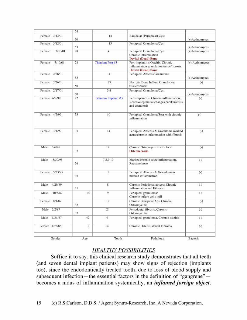
Gender Age Tooth Pathology Bacteria
HEALTHY POSSIBILITIES
Suffice it to say, this clinical research study demonstrates that all teeth (and seven dental implant patients) may show signs of rejection (implants too), since the endodontically treated tooth, due to loss of blood supply and subsequent infection—the essential factors in the definition of “gangrene”—becomes a nidus of inflammation systemically, an inflamed foreign object.

(c) R.S.Carlson, D.D.S. / Agent Syntro-Research, Inc. A Nevada Corporation. 16
The healthy dental organ, odonton, has an external and internal nerve, energetic, and blood supply as well as intricate lymphatic drainage. Once this is compromised, the internal loss of blood supply renders the shell of the dental organ nothing more than a vessel harboring necrotic debris and micro-vermin.17
As one may recall, the death of a tissue (in the instant case the dental organ) due to loss of blood supply is termed gangrene. In texts of the early 20th Century the condition expressing as a “dying tooth” was called gangrenous pulpitis. This term gave over to the euphemistic term “devitalized tooth.” Generally, this term is more acceptable to the patient.
A healthy dental organ has a general out-pressure, overcoming atmospheric pressure and releasing metabolites from the inside of the tooth.18, Thus, when the dental organ becomes gangrenous, looses its blood supply, the tooth becomes a “black hole” so-to-speak, a sink of sorts, in another sense a reservoir and allows the infusion of chemicals and microbes from the oral cavity.19 The subsequent tissue response at the level of the root / bone interface of the dental organ will either be a granuloma, cyst, or cyst-like abscess. More exotic manifestations of the “Natural Protection” of the human body in an attempt to dissolve, “cleanse,” the corrupted tooth or so-called “modern dental implants” from the human body can be seen in Acute or Chronic Cavitation-Osteonecrosis, large voids either around the dental organ or where it once was. Another moniker for this is the Giant Cell Granuloma of yore.20
Again, the theory is that once the tooth looses its out pressure hydraulics by being “cadavered,” that is, looses its capacity to “sweat” due to loss of its “internal circulatory system,” it becomes a hydraulic sink or septic tank where fluids from the oral cavity can easily seep into the dead tooth infecting the peri-radicular tissues, lymphatic’s, venous tissues, and invisible channels of energy flow we know as “meridians.”
In this regard I offer, that in a question asked of Endodontics Associates (five prominent endodontist) in 1994 in Honolulu, and that they answered in their NEWSLETTER of Spring 1994 whereby it was asked..."How much time is needed for the bacteria in natural saliva in the oral cavity to contaminate the entire length of root canals sealed by laterally and vertically condensed gutta percha?" The answer was, "...about 25 to 29 days if crowns were properly sealed," and in "...Positive controls leaked within two days." See: Khayat, et al University of Iran reported in Jour. Endo. (19:458, 1993). (Bacteria being present or not in a specific form may not be the issue. The toxic by-products of
(c) R.S.Carlson, D.D.S. / Agent Syntro-Research, Inc. A Nevada Corporation. 17
"disassociation of dead tissue," held within living tissue and its toxic reaction might be the real question.)
ORAL-SYSTEMIC LINKAGES TO GOOD AND ENDURING HEALTH
In review, historically, dentistry has been and is aware of the intimate connections between the condition of the dental organ (teeth), supporting structures of the bone, mucosa, the neuro-vascular-muscular system; and more recently, through the work of Dr. Voll, the inter-connections to the Channels of Energy Flow (acupuncture meridians) found in Chinese Health Practice to general systemic health. As reported in “Lancet” in 1911, physician Dr. Hunter of the London Fever Clinic identified and clarified the oral-systemic axis, causing severe and debilitating diseases such as colitis, chronic upper respiratory infections, chronic urinary tract infections, etc., and the resultant general human physiologic toxemia, which he termed ORAL SEPSIS. 22 From his groundbreaking work and lectures, notably at McGill University Medical College commencement in 1912, he demonstrated this relationship in a compelling manner. In fact, it is said that by his work alone the dental profession within the United States reversed its policy from retaining infected root stubs and root filled (root canalled) teeth supporting partial or full dentures to the mass extraction of teeth, identified as the “extractionist movement” in the early 1920s through the 1940s. Dr. Weston Price, the most notable but overlooked dental researcher of the 20th Century corroborated this understanding during the 1930s, showing in his work for the first time the inclusion of microorganisms within the hard living tissues of the dental organ, the dentine. His work further demonstrated the arthritic and toxic systemic affects of gangrenous teeth upon the health of rabbits by inserting the teeth under their tissues. He spoke actively against the root canal therapy, “deep root fillings,” for his work, like Dr. Hunter’s, linked the oral infections to general debility. A third researcher well known in Europe but not in the United States, was German physician Dr. Reinhold Voll who demonstrated with electro-technology, called the “Dermatron” device, the ill effects of infected and malposed teeth, odontons, on systemic health. His personal treatment policy was that until the person addressed the dental condition, his systemic treatments would not effectively help in reducing symptoms. But with the advent of “modern endodontics” in the early 1950s and the commercialization of the dental profession, dental schools taught and encouraged the retention of the dead dental organ’s shell—root canalled
(c) R.S.Carlson, D.D.S. / Agent Syntro-Research, Inc. A Nevada Corporation. 18
teeth—leading to the “save the tooth at all cost” era of dentistry for about 50 years, until year 2000. As mentioned previously, in the May 2001 issue of “Scientific American” a report was made on the oral systemic linkage of oral bacteria found in the plaques of atherosclerotic patients, patients with hardening of the arteries—atherosclerosis.15 In 72% of the patient samples it was found that bacteria only found in the mouth had made their way through the vascular system and included themselves within the matrix of the vascular plaques. Moreover, the level of CRP—C-Reactive Proteins—were in some cases extremely high and might be a marker for the risk of heart disease. Implicated in these patients were those with acute and chronic periodontitis, an infected inflammatory condition of the roots and supporting structures of the teeth. The author’s research on the oral systemic linkage to good and enduring health reveals the potential deleterious effects of “modern root canal therapy” and “implant dentistry.” Apical periodontitis is fundamentally equivalent to periodontal disease, implant mucositic-granuloma (dental implant failure), and may be termed, in fact, foreign body tissue reactions—“cleansing phenomenon,” the natural physiologic pushing out of dead and dying tissues from within the human body.
With this in mind, one may be wise to avoid unclean foods, water, air and medical procedures.
Thank you for your kind attention, listening and consideration.
REFERENCES
1) Figdor, David. Guest Editorial Apical periodontitis: a very prevalent problem; Oral Surgery, Oral Medicine, Oral Pathology, Oral Radiology & Endodontics, Vol. 94 No. 6, December 2002.
2) Ibid. 3) Sjorgen U, Figdor D. Persson S. Sundquist G. Influence of infection at
the time of root filling on the outcome of endodontic treatment of teeth with apical periodontitis. Int Endod J 1997; 30:297-306.
4) Katebzadeh N, Hupp J, Trope M. Histological periapical repair after obturation of infected root canals in dogs. J Endod 1999; 25: 364-8.
5) Katebzadeh N, Sigurdsson A, Troope M. Radiographic evaluation of periapical healing after obturation of infected root canals: an in vivo study. Int Endod J 2003:33:60-6.
6) Price, W. Dental Infections and Degenerative Diseases Vol. I. & Vol. II. Penton Pub. Co., 1923 Cleveland, Ohio.

(c) R.S.Carlson, D.D.S. / Agent Syntro-Research, Inc. A Nevada Corporation. 19
7) Zehnder M. Grawehr M. Gasselgren G. Waltimo T. Tissue-dissolution capacity and dentin-disinfecting potential of calcium hydroxide mixed with irrigating solutions; Oral Surgery Oral Medicine Oral Pathology Oral Radiology & Endodontics; Vol. 96, Iss. 5, November 2003, pages 608-613.
8) Figdor D. Microbial aeteology of endodontic failure and pathogenic properties of selected species. Umea University Odontological Dissertations No. 79. Umea (Sweden): Umea University; 200.
9) Eriksen HM. Epidemology of apical periodontitis. In: D Orstavik, TR Pitt Ford, editors. Prevention and treatment of apical periodontitis. Essential Endodontology. London: Blackwell Science 1998. p. 179-91.
10) Figdor, David. Guest Editorial Apical periodontitis: A very prevalent problem; Oral Surgery, Oral Medicine, Oral Pathology, Oral Radiology & Endodontics December 2002, page 651.
11) Ibid. 12) Doran M. Radtke P. A review of endodontic medicaments.
General Dentistry, September- October 1998, pages 485-487. 13) Relationship Between Periodontal Disease and Rheumatoid
Arthritis Explored: Year Book of Dentistry 2001. 14) Gluhovschi, V. Trandafirescu, A. Schiller, L. Petrica, S.
Velciov, G. Gozdog. F. Bob. The Significance of Dental Foci in Glomerular Nephropathies; Facta Universitatis series: Medicine and Biology Vol. 10, No. 2, 2003, pp. 57-61.
15) Karow, J. Taken to Heart; Scientific American, May 2001, page 20.
16) Spangberg, L. Endodontics in the era of evidence-based practice; Oral Surgery, Oral Medicine, Oral Pathology, Oral Radiology & Endodontics; Vol. 96, Issue 5, Nov. 2003, pages 517-518.
17) Murray, C. Saunders, W. Root canal treatment and general health: a review of the literature; International Endodontic Journal, Vol. 33 Issue 1 Page 2-January 2000 doi: 10.1046/j. 1365-2591.2000.00293.x.
18) Steinman, R. Pharmacologic Control of Dentinal Fluid Movement and Dental Caries in Rats; Journal of Dental Research, Vol.47, No.5, Step. -Oct. 1968.
19) Larmas, M. Dental Caries Seen from the Pulpal Side: a Non-traditional Approach; J Dent Res 82(4):253-256, 2003.

(c) R.S.Carlson, D.D.S. / Agent Syntro-Research, Inc. A Nevada Corporation. 20
20) Hirshberg A, Kozlovsky A, Schwartz-Arad D, Mardinger O, Kaplan I. Peripheral giant cell granuloma associated with dental implants. J Periodontol. 2003 Sep;74(9):1381-4.
21) Regezi, J. Periapical Diseases: Spectrum and Differentiating Features; Journal of the California Dental Association, April 1999.
22) Hunter, W. The Role of Sepsis and of Antisepsis in Medicine; The Lancet, January 14, 1911.
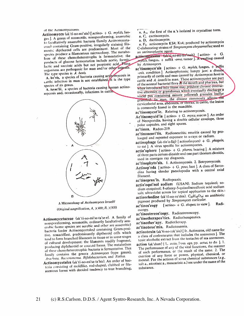
23) Steadmans Medical Dictionary 1971 on Actinomyces:
(c) R.S.Carlson, D.D.S. / Agent Syntro-Research, Inc. A Nevada Corporation. 21
(c) R.S.Carlson, D.D.S. / Agent Syntro-Research, Inc. A Nevada Corporation. 22
Pathophysiology[edit] Wikipedia Actinomyces species that cause human disease do not exist freely in nature, but are normal flora of the oropharynx, gastrointestinal (GI) tract, and female genital tract. This is not an exogenous infection; therefore, no person-to-person spread of the pathogen occurs. In general, Actinomyces species, being members of the normal flora, are agents of low pathogenicity and require disruption of the mucosal barrier to cause disease. Oral and cervicofacial diseases commonly are associated with dental procedures, trauma, oral surgery, or dental sepsis. Pulmonary infections usually arise after aspiration of oropharyngeal or GI secretions. GI infection frequently follows loss of mucosal integrity, such as with surgery, appendicitis, diverticulitis, trauma, or foreign bodies. Numerous reports have linked the use of intrauterine contraceptive devices to the development of actinomycosis of the female genital tract. The presence of a foreign body in this setting appears to trigger infection. Other bacterial species that often are co-pathogens to Actinomyces species may aid spread of infection by inhibiting host defenses and reducing local oxygen tension. Once the organism is established locally, it spreads to surrounding tissues in a progressive manner, leading to a chronic, indurated, suppurative infection often with draining sinuses and fibrosis. In tissues, Actinomyces species grow in microscopic or macroscopic clusters of tangled filaments surrounded by neutrophils. When visible, these clusters are pale yellow and exude through sinus tracts; they are called sulfur granules. This is not an exclusive finding of actinomycosis, and its absence does not rule out the diagnosis. Other conditions, such as eumycetoma and nocardiosis, have been linked to the production of sulfur granules.
References[edit] 1. Jump up ^ eMedicine - Actinomycosis : Article Excerpt by Jorge M Quinonez External links[edit] • Actinomyces naeslundii MG1 Genome Page <img src="//en.wikipedia.org/wiki/Special:CentralAutoLogin/start?type=1x1" alt="" title="" width="1" height="1" style="border: none; position: absolute;" /> Categories: Actinomycineae