the graafian follicle of the sheep: relationships between ...
10
HAL Id: hal-00896859 https://hal.archives-ouvertes.fr/hal-00896859 Submitted on 1 Jan 1973 HAL is a multi-disciplinary open access archive for the deposit and dissemination of sci- entific research documents, whether they are pub- lished or not. The documents may come from teaching and research institutions in France or abroad, or from public or private research centers. L’archive ouverte pluridisciplinaire HAL, est destinée au dépôt et à la diffusion de documents scientifiques de niveau recherche, publiés ou non, émanant des établissements d’enseignement et de recherche français ou étrangers, des laboratoires publics ou privés. THE GRAAFIAN FOLLICLE OF THE SHEEP : RELATIONSHIPS BETWEEN GONADOTROPHINS, STEROID PRODUCTION, MORPHOLOGY AND OOCYTE MATURATION Mary F. Hay, R. M. Moor To cite this version: Mary F. Hay, R. M. Moor. THE GRAAFIAN FOLLICLE OF THE SHEEP: RELATIONSHIPS BETWEEN GONADOTROPHINS, STEROID PRODUCTION, MORPHOLOGY AND OOCYTE MATURATION. Annales de biologie animale, biochimie, biophysique, 1973, 13 (hs), pp.241-249. hal-00896859
-
Upload
khangminh22 -
Category
Documents
-
view
1 -
download
0
Transcript of the graafian follicle of the sheep: relationships between ...

HAL Id: hal-00896859https://hal.archives-ouvertes.fr/hal-00896859
Submitted on 1 Jan 1973
HAL is a multi-disciplinary open accessarchive for the deposit and dissemination of sci-entific research documents, whether they are pub-lished or not. The documents may come fromteaching and research institutions in France orabroad, or from public or private research centers.
L’archive ouverte pluridisciplinaire HAL, estdestinée au dépôt et à la diffusion de documentsscientifiques de niveau recherche, publiés ou non,émanant des établissements d’enseignement et derecherche français ou étrangers, des laboratoirespublics ou privés.
THE GRAAFIAN FOLLICLE OF THE SHEEP :RELATIONSHIPS BETWEEN GONADOTROPHINS,
STEROID PRODUCTION, MORPHOLOGY ANDOOCYTE MATURATION
Mary F. Hay, R. M. Moor
To cite this version:Mary F. Hay, R. M. Moor. THE GRAAFIAN FOLLICLE OF THE SHEEP : RELATIONSHIPSBETWEEN GONADOTROPHINS, STEROID PRODUCTION, MORPHOLOGY AND OOCYTEMATURATION. Annales de biologie animale, biochimie, biophysique, 1973, 13 (hs), pp.241-249.�hal-00896859�
THE GRAAFIAN FOLLICLE OF THE SHEEP :
RELATIONSHIPS BETWEEN GONADOTROPHINS,STEROID PRODUCTION,
MORPHOLOGY AND OOCYTE MATURATION
Mary F. HAY R. M. MOOR
Agricultural Research Council,Unit of Reproductive Physiology and Biochemistry,
307 Huntingdon Road, Cambridge (England)
SUMMARY
i. The Graafian follicle of sheep has been studied experimentally both in vivo and in organculture.
2. In vivo only one or two follicles possess the capacity to secrete significant amounts ofoestrogen during the oestrous cycle. However, about 20 p. i o0 of the non-active follicles were shownto be capable of secreting oestrogen in culture after they had been explanted from sheep that hadbeen injected with PMSG 5 minutes previously.
3. (Estrogen secretion by active follicles was arrested both in vivo and in vitro by an amountof luteinizing hormone corresponding with that released at oestrus.
4. In organ culture, the morphological integrity and steroidogenic capacity of the folliclewas well preserved and 85 p. 100 of the oocytes resumed meiosis, but many of the meiotic figureswere abnormal. In the cultured follicles (a) degeneration of the oocyte did not necessarily leadto luteinization of the granulosa cells, and (b) luteinization sometimes occurred in the presenceof a vesicular oocyte.
INTRODUCTION
Although it is generally accepted that follicular development is under thecontrol of gonadotrophins, recent studies have suggested that the different cell
types within the follicle may have a regulatory effect on each other (FOOTE and
TFIIBAULT, i969 ; IVA);BANDOV, 1972).We have recently been studying various factors that regulate the production of
steroids by the ovarian follicle as well as the maturation of the oocyte in sheep. Someof our findings are summarized and discussed in this paper.

1. - EXPERIMENTAL MODELS
Two experimental models have been used, one for in vitro, and the other for in vivo studies.For the in vitro studies, isolated sheep follicles were maintained in organ culture ; details
of the methods used have already been described (MooR, 1973 ; Moox et al., i9!3). The morpho-logical integrity and steroid-secreting capacity of the follicles were well preserved in culture for7-14 days. The cultures were used to study the effect of hormones on the morphology and ste-roidogenic capacity of individual follicles ; the maturation of the oocyte and its regulatory rolewithin the follicle have also been examined under these controlled conditions.
For the in vivo experiments described in sections 3 and 4, it was desirable to have animalsin which the endocrine changes leading to ovulation could be predicted with some degree of accu-racy. It was found that when the corpus luteum was removed on the 5th day of the cycle, theensuing hormonal changes showed a consistent pattern. Progesterone levels fell rapidly andremained low. Oestrogen levels rose between 16 and 24 hours after removal of the corpus luteum,remained high for a further iz-i6 hours, and then fell to reach base-line levels at 48 hours. LHlevels were low in all sheep up to 32 hours after corpus luteum removal. During the followingi2 hours, LH levels rose to a peak lasting 6-io hours. Ovulation occurred about 24 hours afterthe LH peak.
II. - STEROID SECRETION BY OVARIAN FOLLICLES
A detailed study of the contribution made by follicles of different sizes to thetotal output of oestrogen from the ovary at selected stages of the oestrous cycle hasbeen made (MooR, 1973). In that study all the follicles measuring over 2 mm in dia-meter were explanted separately and the daily secretion of oestrogen by each folliclewas measured. It was found that only the largest one or two follicles from each sheepsecreted significant amounts of oestrogen into the culture medium. When the largefollicles were explanted during the first four days of the cycle, oestrogen secretionin culture declined rapidly; this is probably a reflection of the atresia that occursin such follicles at this stage (BRAND, ig7o). However, when the large follicles wereexplanted between the 4th and the i2th days of the cycle, they secreted oestrogenat relatively high and constant levels (about 300 ng/day) throughout the whole 7-dayculture period. The pattern of oestrogen secretion of large follicles explanted duringthe last three days of the cycle varied, and was characteristic for the day of explan-tation. This is illustrated in figure i, in which is also shown the mean oestrogen pro-duction of all small follicles in culture. The small follicles secreted consistently lowlevels of oestrogen regardless of the stage of the cycle at which they were explanted.
Of particular interest is the sequence of steroid changes that occurs immediatelybefore maturation of the oocyte. This sequence was studied by analyzing, in moredetail, the medium in which large follicles explanted around oestrus had been cul-tured (R. F. SEAMARK, R. M. MOOR et J. IS. A. MCIrrTOSx, unpublished). As oestro-gen levels dropped, there was a sharp but transitory rise in testosterone and
17-hydroxy-A5-pregnenolone, followed by a rapid and large increase in the pro-duction of progesterone and 2oa-dihydroprogesterone. The role of these steroidsin the process of luteinisation and oocyte maturation is being studied.

A histochemical study has been made of the distribution and cellular localiza-tion of the enzyme Ll5-3!-hydroxysteroid dehydrogenase (HSD) which plays a keyrole in steroid biosynthesis. Its presence in the ovarian follicles at various stages ofthe cestrous cycle has been related to the levels of oestrogen in the venous effluentfrom the appropriate ovary. By this method it has been possible to identify thosefollicles which possess the potential for secreting oestrogen. From the results, itbecame clear that only one or two of the large follicles in each sheep exhibit HSDactivity, and that this activity is confined to the theca interna. From the 15th dayof the cycle up to the time of ovulation the follicle that exhibits HSD activity isthe largest one and is invariably found in the ovary whose venous blood has thehighest oestrogen concentration. These findings support the conclusions drawn fromthe culture experiments that only one or two follicles in each sheep secrete signifi-cant levels of oestrogen.

III. - EFFECTS OF GONADOTROPHINS
ON OESTROGEN SECRETION BY THE FOLLICLE
It has been pointed out that apart from the largest follicle none of the otherfollicles in the ovary is capable of secreting significant amounts of oestrogen in vivoor in vitro. In this section, the ability of small follicles to secrete oestrogen under theinfluence of a single injection of pregnant mare serum gonadotrophin (PMSG) willbe discussed. The gonadotrophin was injected during the luteal phase of the cycle(between days 4 and 12) when the output of oestrogen by the ovaries was low. Within12-24 hours of the injection, the concentration of oestrogen in the ovarian venousblood rose markedly, and a number of small follicles increased in size and acquiredHSD activity.
By explanting follicles at selected times after the injection of PMSG it waspossible to determine the interval of time during which a follicle had to be exposedto the influence of PMSG in vivo in order to become capable of secreting oestrogen.It was found that even after an exposure period as short as 5 minutes, about 20 p. ioo
of the small follicles became active and subsequently secreted significant amountsof oestrogen in culture. Neither the percentage of follicles stimulated nor the amountof oestrogen produced by each follicle was increased by extending the interval betweenPMSG injection and explantation of the follicle from 5 minutes to 12 hours. Since itis already known that only about 20 p. 100 of the follicles measuring more than 2 mmin diameter show no signs of atresia in normal cyclic sheep (BRAND, rg7o), it seemslikely that the 20 p. 100 of follicles activated by exposure to PMSG for times up to12 hours must have been derived from the non-atretic population. When the intervalbetween PMSG injection and explantation was extended to 24 hours about 80 p. 100of the follicles became active in vitro : the mean daily production of oestrogen bythese follicles was about three times that of follicles exposed to PMSG for 12 hoursor less.
In addition to the experiments in which PMSG was injected parenterally, anattempt has been made to determine the effect of various gonadotrophins admi-nistered to small follicles directly. No stimulation of oestrogen secretion was obtainedby the microinjection in vivo of either 5 i.u. PMSG, or 20-50 {l.g FSH (NIH-FSH-S9)into the lumen of the follicle. Infusion of 50-100 i.u. PMSG into the ovarian artery,however, stimulated the small follicles to the same degree as i o0o i.u. PMSGadministered systemically. Several attempts have also been made to stimulatesmall follicles in vitro by adding PMSG, FSH, prolactin or I,H alone or in combina-tion to the culture medium. However, none of the treatments has stimulated smallfollicles to secrete significant amounts of oestrogen.
Since a number of hormones including gonadotrophins are found in follicularfluid, it was of interest to determine whether this reservoir of hormones exerts adirect action on the steroidogenic capacity of follicles. Follicular fluid was thereforeremoved from a number of active follicles before they were placed in culture. Thedaily output of oestrogen and progesterone from these follicles over the 7-day cultureqeriod was not significantly different from that of intact controls. Thus, steroid pro-

duction in culture does not appear to be significantly influenced by the gonadotro-phins in the follicular fluid.
In normal cyclic sheep, oestrogen secretion from the ovary with the largestfollicles rises from low levels on day 13 of the cycle to a peak on day 15 and thendeclines sharply to base-line levels at oestrus. The mechanism that controls thisdecline is unknown, but a temporal relationship exists between the decline in oestro-gen production and the pre-ovulatory rise in I,H secretion. Experiments have beencarried out to determine whether these two hormonal events are causally related.
In order to study the effect of high circulating levels of I,H on the secretion ofoestrogen, a total of I mg I,H (NIH-LH-SI7) was infused over 6 hours into the jugu-lar vein of sheep from which the corpus luteum had been removed i8 hours previously.The amount of I,H infused and the duration of the infusion corresponded with therelease of LH that occurs naturally in sheep at oestrus. The infusion of I,H preven-ted the ovary from secreting the large amounts of oestrogen it would otherwise havedone 24-40 hours after removal of the corpus luteum (see section i).
The action of I,H on oestrogen production was investigated further by addingI,H in vitro to follicles taken from sheep treated with PMSG 24 hours previously.The addition of LH to the culture medium (5 [Lg NIH-I,H/ml), 24 hours after
explantation of the follicles, caused a sharp drop in oestrogen production, a transi-tory rise in testosterone and 17-hydroxy-A5-pregnenolone and then a marked risein progesterone secretion; this sequence of hormonal changes is similar to the oneobserved in follicles explanted at oestrus (see section 2). On the other hand, whenoestrogen-secreting follicles were cultured in the absence of LH (control medium),oestrogen output declined only very slowly and there was no significant productionof progesterone during the 5-day culture period (fig. 2).
The effect of LH on the steroidogenic capacity of the cultured follicles wasreflected in their morphology and histochemistry. Within two days of the additionof LH to the culture medium, the granulosa cells underwent luteinization, theyshowed HSD activity, and the output of progesterone was high. In the controlcultures, on the other hand, HSD activity in the granulosa cells was not observedbefore the fifth day and there was very little luteinization.
From the results obtained in vivo and in vitro it can be concluded that at highlevels I,H acts directly on the follicles in such a way as to inhibit oestrogen secretionand at the same time to stimulate luteinization of the granulosa cells and thesecretion of progesterone.
IV. - INTRAFOLLICULAR RELATIONSHIPS
Maturation of the oocyte in vivo and in vitro
In the normal sheep the nucleus of the oocyte in the preovulatory follicle wasfound in the vesicular stage until 8-12 hours after the onset of oestrus, at whichtime it entered metaphase I. The metaphase II configuration was generally seen18-24 hours after the onset of oestrus.
In sheep in which the corpus luteum had been removed and IrH subsequently

infused (see section i), the oocytes in the preovulatory follicles resumed meiosiswithin 8 hours of the end of the infusion, and 16 hours later were found in meta-phase II.
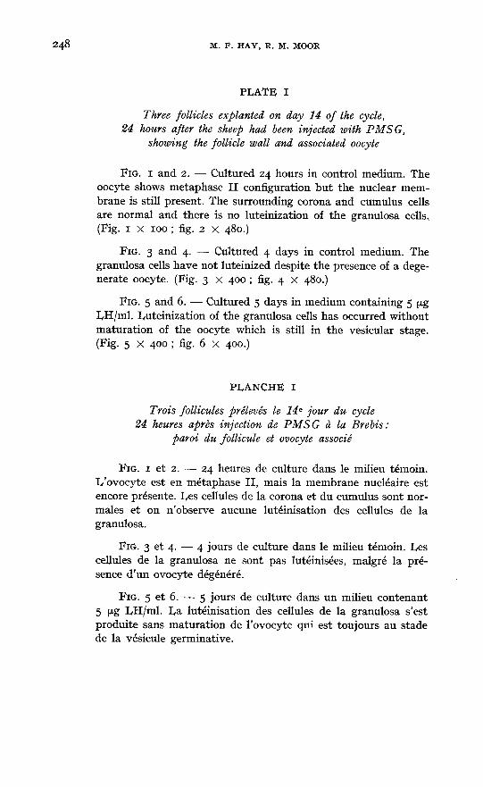
About 85 p. 100 of follicles over 2 mm in diameter cultured under our conditionsshowed maturation changes within 24 hours. The majority of these changes, however,were abnormal ; the most common abnormalities were retention of the nuclearmembrane in metaphase I, abnormal arrangement of the chromosomes and excessivevacuolisation of the cytoplasm (see plate t). The other 15 p. 100 of cultured folliclesremained in the vesicular stage for up to 5 days. It is possible that the large numberof abnormal oocytes was due to the high oxygen concentration (45 p. 100) whichwas necessary to maintain good structural and functional integrity of the theca andgranulosa cells.
Interactions between the oocyte and the granulosa cells
The results of our experiments designed to study the cellular relationshipswithin the follicle are equivocal because of the high percentage of oocytes thatunderwent abnormal changes in culture. Some of our results are of interest, however,when compared with the findings of other workers. It has for example been shownby FOOTE and Txisnur,T (ig6g) that the resumption of meiosis in bovine oocytesis inhibited by the presence of viable corona and granulosa cells. Under our cultureconditions maturation changes in sheep oocytes frequently occurred within 24 hoursof explantation despite the maintenance of a normal topographical relationship bet-ween healthy granulosa, cumulus and corona cells (Pl. I, fig. I and 2) ; however,it is possible that maturation changes of an entirely normal nature might havebeen prevented by the presence of viable cumulus and granulosa cells. The reciprocalrelationship, namely that the oocyte exerts an inhibitory influence on the processof granulosa cell luteinisation has been postulated by NALBANDOW and his co-workers
(see NAI,BANDOV, i972). It would appear from our results, however, that i) degene-ration of the oocyte does not of itself necessarily lead to luteinization of the granulosacells and the production of progesterone. In the follicles that contained degenerateoocytes, oestrogen production was often high and the output of progesterone negli-gible (as in fig. 2) ; the granulosa cells retained their morphological characteristicsand showed no sign of luteinization (Pl. i, fig. 3 and 4). (2) I,uteinization and proges-terone secretion occur in vitro in I,H-treated follicles in the presence of apparentlynormal oocytes in the vesicular stage. Plate i, figure 6 shows such an oocyte onday 5 ; the granulosa cells were clearly luteinized in this follicle which had beenproducing high levels of progesterone since the second day in culture (pl. i, fig. 5).
ACKNOWLEDGEMENTS
We wish to acknowledge with appreciation the skilled technical assistance of Mr D. GREENand Mrs H. K. LEVINSON. We are grateful to Dr C. E. ADAMS, Dr H. M. DOTT and ProfessorT. R. R. MANN for reading and discussing the manuscript. Our thanks are due to the Endocrino-logy Study Section, National Institutes of Health, for the generous gift of LH, FSH and pro-lactin.
RÉSUMÉ
LE FOLLICULE DE GRAAF DE LA BR!BIS :
RELATIONS ENTRE LES GONADOTROPINES, LA PRODUCTION DE STÉROÏDES,LA MORPHOLOGIE DU FOLLICULE ET LA MATURATION DE L’OVOCYTE
Le follicule de Graaf de la Brebis a été étudié expérimentalement in vivo et en culture d’or-gane.
In vivo, au cours d’un cycle oestrien, un ou deux follicules seulement sont capables de sécréterdes quantités notables d’oestrogènes. Mais en culture, environ 20 p. ioo des follicules inactifs sont
capables de sécréter des cestrogènes quand on les prélève sur des Brebis ayant reçu, même seule-ment 5 minutes avant l’abattage, une injection de PMSG.
Aussi bien in vivo qu’in vitro, une quantité de LH correspondant à celle qui est libérée àl’oestrus, arrête la sécrétion d’cestrogènes par les follicules actifs.
En culture d’organe, la morphologie et l’activité stéroïdogène des follicules sont bien préser-vées et 85 p. 100 des ovocytes achèvent leur méiose, mais la plupart des images de méiose sontanormales. Dans les follicules cultivés, d’une part, la dégénérescence de l’ovocyte n’est pasnécessairement accompagnée de la lutéinisation des cellules de la granulosa, et d’autre part, lalutéinisation se produit parfois en présence d’un ovocyte présentant encore une vésicule germi-native.
REFERENCES
BRAND A., 1970. Some micromor¢hological and biochemical aspects of the Texel sheep during the oestrousseason. Thesis, University of Utrecht.FOOTE W. D., THIBAULT C., 1969. Recherches expérimentales sur la maturation in vitro des ovocytes
de truie et de veau. Ann. Biol. anim. Bioch. Biophys., 9, 329-349.MooR R. M., r973. Oestrogen production by individual follicles explanted from ovaries of sheep. J.
Reprod. Fert., 32, 545-548.MooR R. M., HAY M. F., McIrrTOSx J. E. A., CALDWELL B. V., i973· Effect of gonadotrophins on
the production of steroids by sheep ovarian follicles cultivated in vitro. J. Endocr. (In press).NALBANDOV A. V., 1972. Interaction between oocytes and follicular cells. In Oogenesis, p. 513-522.Eds J. D. Biggers and A. W. Schuetz, Baltimore : University Park Press and London : Butterworths.