Evolutionary Interrogation of Human Biology in Well-Annotated Genomic Framework of Rhesus Macaque

THE ANATOMICAL RECORD 293:1477–1491 (2010)
The Global Impact of Sutures Assessedin a Finite Element Model of a
Macaque CraniumQIAN WANG,1* AMANDA L. SMITH,2 DAVID S. STRAIT,2
BARTH W. WRIGHT,3 BRIAN G. RICHMOND,4,5 IAN R. GROSSE,6
CRAIG D. BYRON,7 AND URIEL ZAPATA1,8
1Division of Basic Medical Sciences, Mercer University School of Medicine,Macon, Georgia
2Department of Anthropology, University at Albany, Albany, New York3Department of Anatomy, Kansas City University of Medicine and Biosciences,
Kansas City, Missouri4Department of Anthropology, Center for the Advanced Study of Hominid Paleobiology,
The George Washington University, Washington DC5Human Origins Program, National Museum of Natural History, Smithsonian Institution,
Washington DC6Department of Mechanical & Industrial Engineering, University of Massachusetts,
Amherst, Massachusetts7Department of Biology, Mercer University, Macon, Georgia8Department of Mechanical Engineering, EAFIT University,
Medellın, Colombia
ABSTRACTThe biomechanical significance of cranial sutures in primates is an
open question because their global impact is unclear, and their materialproperties are difficult to measure. In this study, eight suture-bone func-tional units representing eight facial sutures were created in a finite ele-ment model of a monkey cranium. All the sutures were assumed to haveidentical isotropic linear elastic material behavior that varied in differentmodeling experiments, representing either fused or unfused sutures. Thevalues of elastic moduli employed in these trials ranged over several ordersof magnitude. Each model was evaluated under incisor, premolar, andmolar biting conditions. Results demonstrate that skulls with unfusedsutures permitted more deformations and experienced higher total strainenergy. However, strain patterns remained relatively unaffected away fromthe suture sites, and bite reaction force was likewise barely affected. Thesefindings suggest that suture elasticity does not substantially alter loadpaths through the macaque skull or its underlying rigid body kinematics.An implication is that, for the purposes of finite element analysis, omittingor fusing sutures is a reasonable modeling approximation for skulls withsmall suture volume fraction if the research objective is to observe generalpatterns of craniofacial biomechanics under static loading conditions. Themanner in which suture morphology and ossification affect the mechanicalintegrity of skulls and their ontogeny and evolution awaits further investi-
Grant sponsor: National Science Foundation HOMINID;Grant numbers: 0725183, 0725126, 0725136, 0725078, 0725122.
*Correspondence to: Qian Wang, Division of Basic MedicalSciences, Mercer University School of Medicine, 1550 CollegeStreet, Macon, GA 31207. Fax: 478-301-5487. E-mail: [email protected]
Received 16 September 2009; Accepted 8 April 2010
DOI 10.1002/ar.21203Published online 24 July 2010 in Wiley Online Library(wileyonlinelibrary.com).
VVC 2010 WILEY-LISS, INC.

gation, and their viscoelastic properties call for dynamic simulations. AnatRec, 293:1477–1491, 2010. VVC 2010 Wiley-Liss, Inc.
Keywords: elastic properties; structural stiffness; staticloading; suture fusion; bone biomechanics
INTRODUCTION
Sutures are loci of craniofacial growth but potentiallyare also relevant to craniofacial mechanics. Numerousin vivo and in vitro strain gage experiments have identi-fied localized elevated strains over sutures (Behrentset al., 1978; Oudhof and van Doorenmaalen, 1983; Smithand Hylander, 1985; Herring and Mucci, 1991; Herring,1993; Jaslow and Biewener, 1995; Rafferty and Herring,1999; Herring and Rafferty, 2000; Herring and Teng,2000; Rafferty et al., 2003; Sun et al., 2004; Liebermanet al., 2004; Shibazaki et al., 2007; Wang et al., 2008a)suggesting that skulls with unfused sutures do notbehave mechanically as rigid bodies. Although it is clearthat sutures disturb local strain flow, their global impacton skull mechanics and their mechanical propertiesremain poorly defined. For example, the ontogeny ofsuture biomechanics has been investigated in localizedareas, such as the zygomatic arch in pigs (Herring et al.,2005), but no direct experiments have yet been con-ducted to examine the overall impact of patent sutureson the skull, primarily due to methodological limitationspreventing researchers from examining global skull me-chanical behavior during ontogeny. Factors complicatingsuch efforts include variation in bite forces and bitepoints, limited access to bone surfaces (logistical consid-erations preclude the attachment of strain gages to someareas, such as the hard palate), and limited knowledgeconcerning the degree of fusion at individual sutures(Wang et al., 2008a). However, recent applications of fi-nite element analysis (FEA) to vertebrate skull biome-chanics provide a potential solution, because thisapproach allows the modeling of sutures in the contextof an assessment of synchronous global strain patterns(Wroe et al., 2007; Dumont et al., 2009).
FEA is an engineering technique used to examine howstructures of complex design respond to external loads(e.g., Huiskes and Chao, 1983). In FEA the structure ofinterest (e.g., a skull) is modeled as a mesh of simplebricks and tetrahedra (finite elements) joined at nodes,the elements are assigned material properties, certainnodes are constrained against motion, forces are applied,and displacements, stresses and strains at each nodeand within each element are calculated. Recent advan-ces in computer software and imaging technology havemade it possible to capture and digitally reconstructskeletal geometry with great precision, thereby facilitat-ing the generation of detailed finite element models(FEMs) of bony structures, including non-human verte-brate crania (Rayfield et al., 2001; Castano et al., 2002;Rayfield, 2004, 2005a,b, 2007; Richmond et al., 2005;Strait et al., 2005, 2007, 2008, 2009; Dumont et al.,2005; Wroe 2007; Wroe et al., 2007, Wroe and coworkers2008; McHenry et al., 2007; Kupczik et al, 2007; 2009;Farke, 2008; Pierce et al., 2008; Rayfield and Milner,2008; Bourke et al., 2008; Moreno and coworkers, 2008;
Moazen et al., 2008, 2009). However, the incorporationof realistic muscle forces, bone material properties, mod-eling constraints, and experimental bone strain data areequally important components of FEA that are necessaryto ensure biologically meaningful results (e.g., Richmondet al., 2005; Strait et al., 2005; Ross et al., 2005; Ray-field, 2007).
Recently, the impact of sutures has been more vigo-rously discussed and investigated using FEA in variousliving and fossil species (Rayfield, 2004, 2005; Kupcziket al., 2007, 2009; Wang et al., 2007b, 2008a,b; Farke,2008; Moazen et al., 2009; Fitton et al., 2009; Jasinoskiet al., 2010) and various conclusions as to their signifi-cance on local and global skull biomechanics have beenreached. Some finite element models of vertebrate craniathat did not include sutures have obtained reasonableresults in relation to in vivo bone strain studies (e.g.,Strait et al., 2005), but others have not, as was the casein the less refined alligator models (Metzger et al.,2005). FEA of a theropod dinosaur skull (Rayfield,2005b) suggested the important role sutures play in alocalized functional context. Kupczik et al. (2007) demon-strated the localized impact of sutures in monkey skulls,and Moazen et al. (2009) have observed a significantimpact on strain energy density (SED) patterns in smallanimals such as lizards. Fitton et al. (2009) performed asensitivity analysis using the same specimens as wereexamined by Kupczik et al. (2007, 2009) and found thatthe inclusion of patent sutures in their FEMs had mod-est effects on strain in the immediate vicinity of thesutures but as one moved away from the sutures theeffects were negligible. These results are corroborated byour initial sensitivity analyses (Wang et al., 2007b,2008b). However, the manner in which sutural fusionaffects the global biomechanics of the craniofacial skele-ton has not yet been systematically studied.
This study tests the hypotheses that: (1) sutures havea significant impact on global skull mechanics, and (2)the mechanical behavior and significance of suturesdepends on their material properties and positions onthe cranium. A macaque FE model was analyzed usingfour different sets of suture material properties underthree different loading cases that simulate incisor, pre-molar, and molar biting. Mechanical data extracted fromthese 12 FEAs allowed an assessment of the hypotheses.
MATERIALS AND METHODSModel Creation
The finite element model used here was built from amale subadult Macaca fascicularis specimen (with thirdmolars erupting) obtained from a biological supply com-pany after the specimen had been sacrificed for researchpurposes unrelated to this study (Fig. 1). This modelwas created based on the protocol used previously by
1478 WANG ET AL.

this research group that has successfully generated real-istic FEMs of monkey crania (Richmond et al., 2005;Strait et al., 2005, 2007, 2008, 2009). A tessellated sur-face model was constructed by first determining the opti-mal threshold value of the CT scans by averaging thethreshold values across multiple volumes sampledthroughout the facial skeleton using Quant3D (High-Re-solution X-Ray CT Lab, University of Texas, Austin,http://www.ctlab.geo.utexas.edu/software/index.php), thenapplying the threshold to create a tessellated model ofthe skull using AMIRA (Visage Imaging). The tessellatedsurface was converted to a smooth non-uniform rationalB-spline (NURBS) surface using surface editing software(Geomagic Studio 11, Geomagic, NC), which was thentransformed into a solid model by using computer-assisted-design software (SolidWorks 2005, Dassault Sys-temes SolidWorks). The solid was broken into 146 parts(including parts designated as sutures), each of whichcould potentially be assigned its own set of elastic proper-ties (Fig. 1). The model was imported into FEA software(Algor 19, Autodesk, Pittsburg, PA) and converted into amesh of 1,051,877 brick and tetrahedral elements thatexhibited greater mesh density in the face than in theneuro- and basicranium. Suture morphology, materialmechanical properties, muscle forces, constraints, andreaction forces were simulated within the three dimen-sional model as described later.
Parts Representing Sutures
The precise manner in which to model sutures is a se-rious technical challenge. Should sutures be modeledusing their complex, often interdigitating morphology, oras a simplified linear corridor? Surface investigation andmicro-CT image analysis have demonstrated complicatedspatial variations of sutural morphology (Byron et al.,2004; Byron, 2006, 2009; Herring, 2008; Reinholt et al.,2009). Due to its pliant nature, the suture connective tis-sue is assumed to be able to resist mainly tensile ratherthan compressive loads. Therefore, bony interdigitationswithin sutures play a critical role in allowing externalcompressive loads to be absorbed as tensile forcesthrough sutural fibers running between the inosculatedsutural surfaces (Herring, 2008). Thus, sutures are bet-
ter modeled as homogeneous suture-bone functionalunits in order to simplify this phenomenon (Farke, 2008;Wang et al., 2008b).
Four parts representing the premaxillomaxillarysuture (PMM), zygomaticomaxillary suture (ZMM), zygo-maticofrontal suture (ZMF), and zygomaticotemporalsuture (ZMT) (Fig. 1) were created on both the left andright sides of the cranium, because these four suturesremain mostly unfused in adult monkeys (Wang et al.,2006c). As a simplifying assumption, these suture-boneunits were given a uniform thickness of 1 mm, which isa coarse approximation of the breadth of the interdigita-tions typical in these sutures. The inferior one third ofthe PMM was not modeled, as it is normally closed inyoung adult males, even before the full eruption of thecanines (Wang et al., 2006c). An explanation for suchearly fusion could be that forceful loading on the incisorsmight otherwise break off the premaxillary bone, as hasbeen observed during in vitro experiments (Wang et al.,2008a). All eight sutures comprised only 0.24% of thetotal skull volume.
Elastic Properties of Bone and Sutures
Material properties (measures of a material’s ability toresist deformation) including elastic modulus (E), andPoisson’s ratio (m), were assigned to different functionalregions of the skull. Each region of the face correspond-ing to cortical bone was assigned its own set of isotropicelastic properties calculated from previous orthotropicelastic properties collected from rhesus macaque skulls(Wang and Dechow, 2006). Cortical regions in the neuro-and basi-cranium were assigned isotropic elastic proper-ties based on an average of values obtained from allparts of the skull (E ¼ 17.3 GPa, m ¼ 0.28; Wang andDechow, 2006; Wang et al., 2006b). Trabecular bone inthe supraorbital torus, postorbital bar, zygomatic bodyand zygomatic arch was also modeled isotropically (E ¼0.64 GPa, m ¼ 0.28; Ashman et al., 1989). The inclusionof homogeneous parts representing trabecular bone inthe finite element model appears to increase the accu-racy of FEA (Castano et al., 2002; Panagiotopoulouet al., 2010). Teeth were modeled as bone parts and wereassigned the same material properties as those in
Fig. 1. Three dimensional solid model of a M. fascicularis skull. (A)Whole model with sutures highlighted. (B) Eight parts representingfour facial sutures on left and right sides. Zygomaticotemporal suture(ZMT)– Parts 1 and 5; Zygomaticofrontal suture (ZMF)– Parts 2 and 6;Zygomaticomaxillary suture (ZMM)– Parts 3 and 7; Premaxillomaxillary
suture (PMM)– Parts 4 and 8. All sutures were created to match thecortical bones that normally formed a shell over trabecular bones. Theparts inside trabecular bone were omitted in order to simplify themodeling process. (C) Parts representing trabecular bone in the facialskeleton. They were enclosed by cortical bone.
MECHANICAL IMPACT OF SUTURES 1479

surrounding alveolar bone; hence periodontal ligamentswere not modeled here.
The material properties to be assigned to the eightsuture-bone units modeled here cannot be easily dis-cerned. There is no consensus on the elastic modulus ofsutural tissue, as indicated by widely varying publisheddata. For example, 1.2 MPa (Kupczik et al., 2009), 10MPa (Moazen et al., 2009), 50 MPa (Odame et al., 2005),0.4 GPa (Farke, 2008), and 0.5-2.5 GPa (Wang et al.,2008b) have each been proposed as proper elastic moduli[1 MPa (MegaPascal) ¼ 106 N/m2; 1 GPa (GigaPascal) ¼109 N/m2]. Direct mechanical experiments on mamma-lian cadaver material of varying sizes have yielded com-parably low magnitudes of elastic stiffness for suturaltissues. Elastic moduli from facial sutures in rabbits arein the range of 1.2–2.1 MPa (Radhakrishnan and Mao,2004) while internasal sutures in ewes are 0.9 MPa(Meunier et al., 2009). However, based on strain gageexperiments, these values might understate the stiffnessof sutural structure. In vivo and in vitro experiments inpigs, rabbits, and monkey skulls show that averagestrain values in patent sutures are an order of magni-tude higher than in adjacent regions of cortical bone(Herring and Mucci, 1991; Herring and Teng, 2000;Lieberman et al., 2004; Wang et al., 2008a). Thus, theo-retically, the suture along with its interaction with bonefronts as a structure could exhibit elastic stiffness aboutone tenth that of the adjacent bones (Wang et al.,2008b). Farke (2008) proposed that the suture with thesurrounding bone be considered a single functional unitwhich helps avoid modeling internal sutural complexity.According to this logic, the elastic moduli of sutures inmonkey skulls might be expected to be around 0.5-2.5GPa, about 200 to 500 times stiffer than resultsobtained from mechanical tests that evaluated only thecraniofacial suture connective tissue. Thus, in the pres-ent study, suture material properties were varied acrossa wide range of values.
Sutures were assigned isotropic material propertiesthat varied between modeling experiments. Four differ-ent sets of mechanical properties were used for suture-bone functional units: (1) no suture or fused suture withE ¼ 17.3 GPa and m ¼ 0.28; (2) stiff suture/bone struc-ture with E ¼ 1.0 GPa; (3) less stiff suture/bone struc-ture with E ¼ 50.0 MPa, (4) least stiff suture/bonestructure with E ¼ 1.0 MPa. In all suture modelingexperiments, Poisson’s ratio was set to 0.4.
Muscles Forces and Constraint Designs
A constraint regime, or reaction loading procedure,was applied to the model. To examine the strain pat-terns of the skull incurred from biting on differentteeth, nodes at the right and left articular eminencesin the temporomandibular joint (TMJ) and at a bitepoint (incisors, premolars, or molars) were fixed to pre-vent movement of the model. When muscle forces areapplied to a model with these constraints, the model ispulled inferiorly onto the fixed points, thus generatinga reaction force at the bite point and the articulareminences.
In molar loading experiments, the bite point was setat all of the left (working-side) upper molar tooth crownsurfaces. In premolar loading, the bite point was definedby constraining nodes on the surfaces of the left P3 andP4 tooth crowns. In incisor loading experiment, the bitepoint was defined by simultaneously constraining nodeson the surfaces of the I1 and I2 tooth crowns.
Eight muscle forces, generalized from previous stud-ies (Strait et al., 2005, 2007, 2008, 2009), were appliedto the model, representing the right and left anteriortemporalis, superficial masseter, deep masseter andmedial pterygoid (Table 1). These muscles are princi-pally responsible for jaw elevation during masticationand they reflected their relative magnitudes when bit-ing on different teeth. When biting at a parasagittalbite-point such as at the premolars or molars, workingand balancing side muscle forces differ to eliminate dis-traction of the mandibular condyle at the working-sideTMJ (Greaves, 1978; Spencer, 1998). When loading wasconfined to the incisors, the working side muscle forcesmagnitudes were assigned to the working and balanc-ing side equally. In premolar loadings, muscle forces ofthe balancing side were in the middle of the balancingside muscle forces for molar and incisor loadings. Thus,overall applied muscle forces increased as bite pointsmoved mesially (anteriorly). In all experiments, regard-less the magnitude of total loading forces, individualloading forces were distributed on the same 1280nodes.
Modeling Experiments
It has been suggested that different loading positionswill induce different strain patterns in facial skeletons
TABLE 1. Muscle forces (Newton)
Molar Premolar Incisor
Orientation vector
x y z
Working-side superficial masseter 70.6 70.6 70.6 �0.2 1.0 0.2Balancing-side superficial masseter 34.7 52.7 70.6 0.2 1.0 0.2Working-side deep masseter 22.6 22.6 22.6 �0.6 1.0 0.0Balancing-side deep masseter 8.2 15.4 22.6 0.6 1.0 0.0Working-side medial pterygoid 34.8 34.8 34.8 0.75 1.0 0.0Balancing-side medial pterygoid 6.9 20.8 34.8 �0.75 1.0 0.0Working-side anterior temporalis 36.6 36.6 36.6 0.1 1.0 0.1Balancing-side anterior temporalis 15.1 25.9 36.6 �0.1 1.0 0.1Sum 229.6 279.4 329.2
Magnitude of muscle forces for molar biting was adapted from Strait et al. (2005). While loading was confined to the inci-sors, the working side muscle forces of working sides were assigned to at both the working and balancing sides. In premo-lar loadings, muscle forces of the balancing side were in the middle of the balancing muscles forces for molar and incisor.
1480 WANG ET AL.

(Strait et al., 2008; Kupczik et al., 2009; Wang et al.,2010b). Thus facial sutures might have different func-tional effects during different loading scenarios. Conse-quently, sutures were evaluated under varying loadingconditions corresponding to bites on different sets ofteeth. Experiments simulating three types of bites (inci-sor, premolar, molar) were performed to examine thestrain patterns of bone and sutures in a monkey modelwith four different sets of suture elastic properties. Ineach type of bite, four analyses were conducted thatincluded: no sutures, sutures with an elastic modulus of1 GPa, 50 MPa, and 1 MPa. Thus, in total, 12 modelingexperiments were performed.
For each experiment, several types of data were col-lected or calculated. For overall behavior of the skull,total strain energy (the total work done on the skull bythe applied forces calculated as half of the sum of forcemultiplied by displacement: W¼ 1=2\
PNi¼1 Fi \di. In this
study: N ¼ 1280). It is noteworthy that this was anapproximation of the total work, since nodal force mag-nitudes were multiplied by nodal displacement magni-tudes, instead of taking the vector product betweennodal force vectors and nodal displacements. The ap-proximate method will equal the exact work done in the
case when the displacements at the nodes are exactlyparallel to the forces applied to the nodes. At twenty an-thropometric landmarks, nodal mean shear strain (e1 þ|e2|) were collected, and strain mode (ratio of the maxi-mum principal or tensile and minimum principal or com-pressive strains: e1 / |e2|) were calculated. Further, dataon nodal maximum principal strain orientation (e1�) andreaction forces at the left and right TMJs and bite pointswere collected for the premolar loading experiments.Finally, ratios of strain over sutures to those over bonesurfaces were calculated by dividing the maximum shearstrain values on sutures by the mean of maximum shearstrain values over four adjacent bone surfaces. Thisallows an assessment of whether the suture-bone unit isbehaving realistically; previously, a suture versus boneratio of 7.3 was observed during in vitro loading experi-ments of a monkey skull (Wang et al., 2008a).
Because FEA is a deterministic process that demon-strates the influence of different variables on the model,strain energy and other data derived from FEA violatethe assumption of random sampling implicit in suchtests. As a result, statistical tests were not conducted.Rather, a series of paired comparisons among the datacollected from different experiments at different
Fig. 2. Von Mises strain (unit: mm/mm) in four sutures models under three different loadings. In eachloading regime, all four suture models displayed similar strain patterns, though strains over statures werehigher in three models with sutures (1 GPa, 50 MPa, and 1 MPa) than in no suture or fused suture models(17.3 GPa).
MECHANICAL IMPACT OF SUTURES 1481
locations on the skull were conducted to assess quantita-tive differences in strain magnitude, mode, and strainorientations. Results of the no-suture model were usedas the reference datum for all comparisons. These com-parisons were supplemented with visual comparisons ofstrain and SED across the entire model.
RESULTSOverall Strain Patterns: Magnitude, Mode,and Orientations
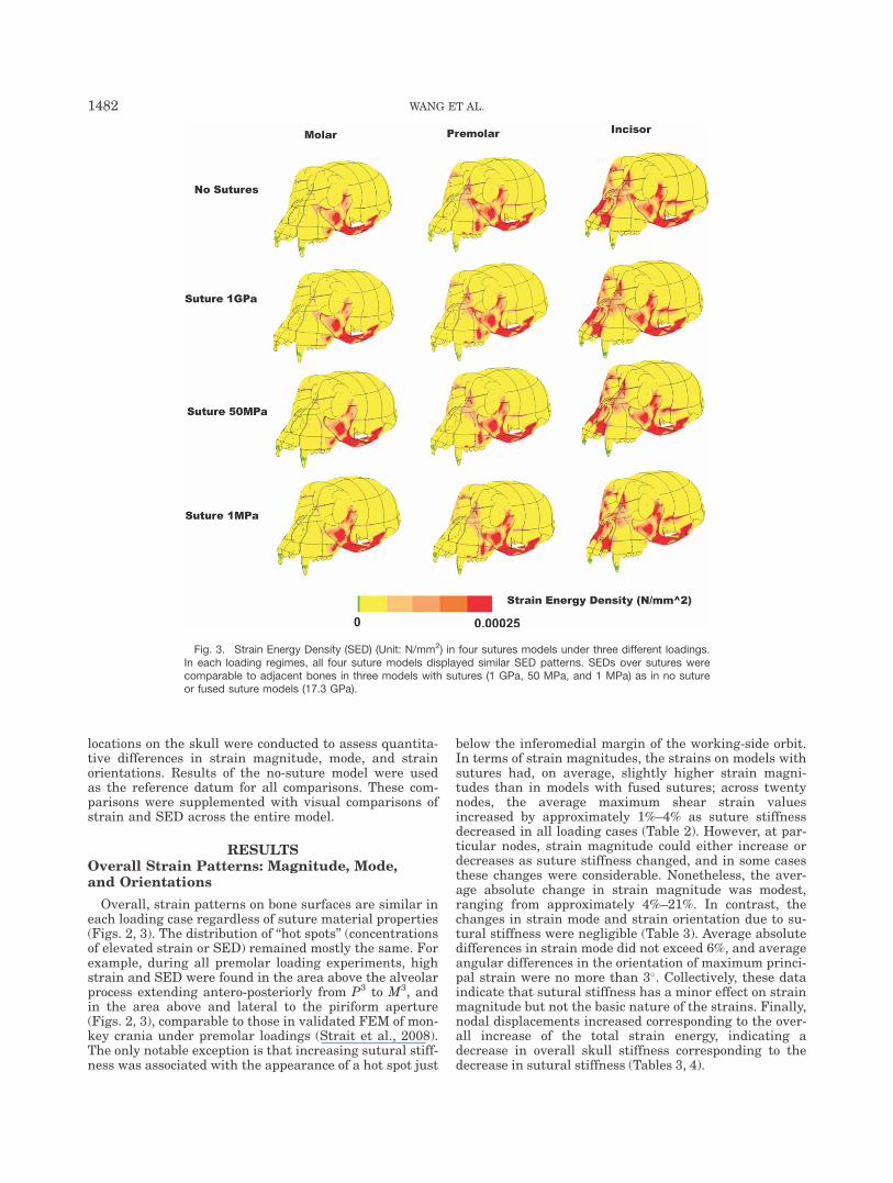
Overall, strain patterns on bone surfaces are similar ineach loading case regardless of suture material properties(Figs. 2, 3). The distribution of ‘‘hot spots’’ (concentrationsof elevated strain or SED) remained mostly the same. Forexample, during all premolar loading experiments, highstrain and SED were found in the area above the alveolarprocess extending antero-posteriorly from P3 to M3, andin the area above and lateral to the piriform aperture(Figs. 2, 3), comparable to those in validated FEM of mon-key crania under premolar loadings (Strait et al., 2008).The only notable exception is that increasing sutural stiff-ness was associated with the appearance of a hot spot just
below the inferomedial margin of the working-side orbit.In terms of strain magnitudes, the strains on models withsutures had, on average, slightly higher strain magni-tudes than in models with fused sutures; across twentynodes, the average maximum shear strain valuesincreased by approximately 1%–4% as suture stiffnessdecreased in all loading cases (Table 2). However, at par-ticular nodes, strain magnitude could either increase ordecreases as suture stiffness changed, and in some casesthese changes were considerable. Nonetheless, the aver-age absolute change in strain magnitude was modest,ranging from approximately 4%–21%. In contrast, thechanges in strain mode and strain orientation due to su-tural stiffness were negligible (Table 3). Average absolutedifferences in strain mode did not exceed 6%, and averageangular differences in the orientation of maximum princi-pal strain were no more than 3�. Collectively, these dataindicate that sutural stiffness has a minor effect on strainmagnitude but not the basic nature of the strains. Finally,nodal displacements increased corresponding to the over-all increase of the total strain energy, indicating adecrease in overall skull stiffness corresponding to thedecrease in sutural stiffness (Tables 3, 4).
Fig. 3. Strain Energy Density (SED) (Unit: N/mm2) in four sutures models under three different loadings.In each loading regimes, all four suture models displayed similar SED patterns. SEDs over sutures werecomparable to adjacent bones in three models with sutures (1 GPa, 50 MPa, and 1 MPa) as in no sutureor fused suture models (17.3 GPa).
1482 WANG ET AL.

Total Strain Energy and Strain EnergyAbsorbed by Suturess
Total strain energy increased as sutural stiffnessdecreased under all loading conditions, and these differ-ences were accentuated as the bite point moved frommesial to distal (i.e., incisor vs. premolar vs. molar; Ta-ble 4; Fig. 4).
Due to their small volume as a percentage of skull vol-ume (0.24%), sutures only absorbed a small amount ofenergy. For example, in the 50 MPA model under premo-lar loading, only 2.47% of total strain energy (0.08 N*mmin total) was absorbed by sutures. The rest of the facialskeleton, including cortical and trabecular bone lumped to-gether, absorbed 93.96% of the model’s total strain energy(Table 5). However, sutures did absorb more energy whenthe sutural stiffness decreased. For example, in premolarloading analyses, all eight facial sutures absorb 0.02N*mm in total in the no-suture model, 0.05 N*mm (2.5times higher than in the no suture model) in the 1 GPAmodel, 0.08 N*mm in total (4 times higher) in the 50 MPamodel, and 0.16 N*mm in total (8 times higher) in the 1MPA model. Yet the increase of energy absorption is notproportional to the extent of the decrease in sutural stiff-ness. For example, in premolar loadings, facial sutures inthe 1MPa suture model absorbed 3.2 times as muchenergy as they did in the 1 GPa suture model, eventhough their stiffness was three orders of magnitude less.
Reaction Forces and Biting Efficiency
The reaction force at the TMJs increased as suturestiffness decreased, while bite forces (reaction forces at
the bite points) slightly decreased. For example, in pre-molar loadings (Table 6), at the right TMJ the reactionforce increased 10.75% in the 50 MPa model, and14.38% in the 1 MPa model relative to the no-suturemodel, while bite force was 1.29% lower in the 50 MPamodel, and 2.05% in the 1 MPa model. Taken together,these findings indicate a slight decrease in biting effi-ciency as suture stiffness decreases. A premolar biteforce of about 75N was generated from a total appliedmuscular force of 279.4N. In terms of total muscularforce, the premolar biting efficiency, defined as the rela-tionship between the tooth reaction forces and the totalforce exerted by the muscles, was 26.85% in the nosuture model, and it was 26.30% in the 1 MPa model(Table 7). This value was smaller than theoretical values(around 38%, Dechow and Carlson, 1990), possiblybecause the TMJs may have been overconstrained in ourmodel and did not allow sufficient rotation around amedio-lateral axis.
Strain Patterns Over Suture and AdjacentBone Surfaces
Strains at the sutures were higher overall in modelswith sutures than in the model without sutures in allthree loading scenarios, and higher in the 1 MPa and 50MPa models than in 1 GPA models (Fig. 2; Table 8).However, the bone surfaces close to four suture sites typ-ically exhibited only slight increases in magnitude fromno suture model to 1MPa suture model (Table 8). Thestates of strain mode (i.e., primarily tensile strains vs.primarily compressive strains) normally did not change
TABLE 2. Proportional change of mean nodal shear strain magnitudes (e1 1 |e2|) comparedto the no suture model
Site Node
Molar loading (%) Premolar loading (%) Incisor loading (%)
1 GPa 50 MPa 1 MPa 1 GPa 50 MPa 1 MPa 1 GPa 50 MPa 1 MPa
Opisthokranion (op) 56,316 �7.1 �21.4 �21.4 �3.6 �10.7 �14.3 �2.0 �5.9 �5.9Basion (ba) 19,043 0.2 �1.8 �3.1 0.2 �1.1 �1.9 �0.1 �1.2 �1.8Staphylion (sta) 28,607 �0.6 �8.7 �13.8 0.6 �5.9 �10.9 1.6 �3.2 �7.8Glabella (g) 40,158 �0.9 1.8 4.4 �1.1 �0.7 0.0 �1.2 �1.7 �1.2Left midsupraorbit 5,397 1.9 13.4 21.7 1.0 8.9 16.0 3.3 11.4 16.6Right midsupraorbit 83,904 4.9 18.9 25.9 5.0 17.1 23.2 3.4 13.0 18.1Left infraorbital rim 33,437 �7.3 �30.9 �40.4 �6.9 �22.6 �28.0 �4.2 �17.7 �22.6Right infraorbital rim 77,458 �7.7 �28.9 �37.8 �6.3 �24.0 �31.5 �5.8 �18.4 �23.2Rhinion (rhi) 63,226 0.9 �12.8 �18.5 2.3 �7.4 �11.7 �2.2 �14.1 �18.5Left center of zygoma 24,696 3.2 8.2 11.4 3.2 8.0 11.5 4.7 11.2 15.3Right center of zygoma 13,276 7.1 14.8 16.0 8.1 18.1 20.3 8.2 19.4 22.1Left alveolar above P3 88,338 �1.0 �0.6 �1.3 �0.4 �2.3 �4.4 2.6 1.1 0.0Right alveolar above P3 10,195 �11.8 �31.4 �35.3 �12.6 �24.4 �26.7 �1.2 �6.6 �9.6Prosthion (pr) 68,003 0.0 �6.6 �11.7 �0.2 �5.7 �8.8 �0.4 �0.6 �0.5Left post zygomatic arch 139,760 18.3 59.7 76.6 15.7 50.3 64.2 12.4 36.8 45.9Right post zygomatic arch 7,007 6.2 22.3 28.6 6.0 21.0 27.0 6.1 20.7 26.5Left Zygomatic arch midupper border
44,394 1.0 �2.5 �5.7 0.7 �3.7 �7.1 0.6 �3.9 �7.2
Right Zygomatic arch midupper border
106,154 �0.2 0.7 4.1 �0.6 0.1 3.5 �1.6 �2.0 1.5
Left Zygomatic arch midlower border
103,026 5.9 15.2 18.3 6.1 15.2 18.0 6.5 15.2 17.7
Right Zygomatic arch midlower border
50,520 12.0 25.3 29.4 12.9 27.1 31.3 13.4 28.3 32.2
Mean 1.2 1.7 2.4 1.5 2.9 3.5 2.2 4.1 4.9SD 6.9 22.0 27.9 6.5 18.5 23.4 5.1 14.9 18.7Mean of absolute values 4.9 16.3 21.3 4.7 13.7 18.0 4.1 11.6 14.7SD of absolute values 4.9 14.5 17.6 4.7 12.3 14.7 3.7 9.9 12.1
MECHANICAL IMPACT OF SUTURES 1483

at either sutural sites or bone surfaces, except in one ofthe nodes sampled anterior to the left ZMT suture. At afew sites, substantial differences in the values of strainmode could be observed (e.g., nodes near the ZMT andPMM sutures in premolar loadings) (Table 8), althoughthese changes did not cause the basic mode of strainexperienced by the node to change. The orientation ofmaximum principal strain (e1�) could change remarkablywithin sutures (e.g., ZMF, ZMM, and PMM), but in mostcases the angular change at adjacent bone surfaces wasless pronounced (Tables 3, 8).
Ratios of Strain Over Suture to Strain OverAdjacent Bone Surfaces
The ratios of sutural strain magnitude relative tomagnitudes at adjacent bone surfaces varied as the stiff-ness of the sutures increased (Table 9). For example, in
TABLE 3. Change of nodal strain mode (e1/|e2|) (%), maximum nodal strain orientation (e1�), and nodaldisplacement under premolar loading compared to the no suture model (%)
Site
States of strainmode in no
suture modela
Change in magnitude ofstrain mode (%)
Change in maximumstrain orientationb
Change in displacementmagnitude (%)
1 GPa 50 MPa 1 MPa 1 GPa 50 MPa 1 MPa 1 GPa 50 MPa 1 MPa
Opisthokranion (op) C 0.0 3.0 3.0 0.0� 1.2� 1.8� 2.6 13.2 15.8Basion (ba) T 0.0 �0.5 �1.1 0.0� 0.0� 0.0� 0.0 0.0 0.0Staphylion (sta) T 0.4 0.8 1.7 0.0� 0.0� 2.8� 2.2 2.2 4.3Glabella (g) C 0.0 �1.4 �2.9 0.0� 0.2� 0.3� 1.5 5.9 8.8Left midsupraorbit T �0.5 �4.3 �4.6 1.9� 1.9� 1.9� 2.4 9.4 12.9Right midsupraorbit T �7.1 �11.5 �11.8 0.0� 0.0� 0.0� 2.4 6.1 8.5Left infraorbital rim T 1.0 3.6 6.8 0.0� 0.0� 0.0� 0.0 �10.0 �13.3Right infraorbital rim T �0.4 �1.8 �2.5 2.7� 2.7� 2.7� 3.3 8.3 10.0Rhinion (rhi) T �2.5 �4.0 �4.3 8.1� 8.1� 8.1� 4.3 10.6 12.8Left center of zygoma T 1.1 2.2 0.6 0.0� 5.1� 5.1� 6.2 14.4 17.5Right center of zygoma T �1.7 �12.5 �17.6 0.0� 0.0� 0.0� 5.1 11.2 14.3Left alveolar above P3 C 0.0 0.0 3.6 0.0� 0.0� 0.0� 0.0 0.0 0.0Right alveolar above P3 C �23.1 �26.9 �23.1 4.0� 14.8� 24.2� 3.8 8.9 10.1Prosthion (pr) T �0.7 1.8 3.9 0.0� 0.4� 0.2� 6.3 15.6 18.8Left post zygomatic arch T 0.4 0.9 1.3 0.0� 4.5� 4.5� 7.1 28.6 35.7Right post zygomatic arch T 0.0 0.7 1.3 0.0� 1.1� 1.1� 11.1 22.2 22.2Left Zygomatic arch midupper border
C 0.0 0.0 0.0 0.0� 4.0� 4.0� 7.3 20.2 25.0
Right Zygomatic arch midupper border
C 6.5 19.4 22.6 1.0� 2.7� 3.7� 8.4 26.2 33.8
Left Zygomatic arch midlower border
T 0.3 1.9 2.9 0.8� 0.4� 0.0� 6.9 20.6 26.0
Right Zygomatic arch midlower border
T 1.2 2.8 4.3 0.8� 1.7� 1.7� 9.5 26.0 31.4
Mean of absolute valuesc 2.3 5.0 6.0 1.0� 2.4� 3.1� 4.5 13.0 16.1SD of absolute valuesc 5.3 7.1 7.1 2.0� 3.5� 5.3� 3.2 8.6 10.3aThe states of strain mode remained the same in the same loading regimes regardless of the stiffness of sutures (C ¼ Com-pressive, T ¼ Tensile).bFor the method to find the angle between two vectors, see http://www.wikihow.com/Find-the-Angle-Between-Two-Vectors.cFor change in strain orientation, it was angular values and angular SD respectively, calculated with the Oriana CircularStatistical Analysis Program 2.02 (Kovach Computing Services, Wales, UK) (Fig. 5).
TABLE 4. Comparison of total strain energy(total work) to the no suture model under different
loading regimes. With the decrease of suturalstiffness, the overall total strain energy (an
approximation of total work) in the whole skullincreased, indicating a decrease of structural
stiffness of the skull
Loading regime
Increase of total strain energycompared to no suture model
1 GPa 50 MPa 1 MPa
Molar 8% 25% 33%Premolar 7% 20% 28%Incisor 6% 16% 23%Mean 7% 20% 28%SD 1% 5% 5%
Fig. 4. Scaled total work (total strain energy) (Unit: N*mm) in foursuture models under molar, premolar, and incisor loadings. The totalwork of the no suture model was scaled to 1. Models with suturesunderwent a greater increase in total work under molar loadings,which might be related to unbalanced loading forces between theworking and balancing sides.
1484 WANG ET AL.

all 12 analyses, the ZMF suture had the lowest overallratio of maximum shear strains recorded at suturesagainst those over the adjacent bone surfaces; in the 50MPa model, the suture/bone ratio was 1.5 at ZMF, com-pared to 8.0-9.5 at other suture areas.
When all sutures of the left side of the facial skeleton in12 analyses were lumped together, the overall ratio of maxi-mum shear strains recorded at suture against those overthe adjacent bone surfaces were 3.7 in the 1GPa model, 7.3in the 50 MPa model, and 8.1 in the 1 MPa model (Table 9).The latter two, especially in 50MPa, were close or identicalto a value of 7.3 observed during in vitro loading experi-ments of a monkey skull (Wang et al., 2008a).
Variation of Strain Patterns Among and WithinSutural Parts
Sutures on the midface had different strain environ-ments on the working and balancing sides. For example,in the 50 MPa suture model undergoing premolar load-ing (Table 10), the zygomaticomaxillary suture on theleft or working side exhibited a compressive dominantstrain regime, while on the right or balancing side, atensile regime was found.
Within the sutures, different sections endured differ-ent strain. For example, the midpoints of four surfaceson the left ZMT suture had different strain values andmodes: (1) upper and lower surfaces had higher strainthan medial and lateral surfaces; (2) while the upperand medial surfaces were under compression, the lateraland lower surfaces were under tension (Fig. 6, Table 10).
DISCUSSIONGlobal Impact of Sutures on SkullBiomechanics
Skulls with unfused sutures have less mechanical in-tegrity and structural rigidity than skulls with fusedsutures. This is because sutures experience elevatedstrain and permit greater deformation of the skull.
Thus, with unfused sutures, applied forces of a givenmagnitude produce greater strain energy compared to askull without sutures. However, the absorption of thisincrease in strain energy occurs mostly in the facialskeleton. Although sutures in macaques exhibit highstrains and elevated strain energy density, they do nothave a high capacity, at least under static loading condi-tions, to absorb energy due to their limited volume. Incontrast, Jaslow (1990) stated that cranial sutures ingoats were able to absorb 16%–100% of energy duringimpact. Whether or not sutures in primates provide asignificant strain-dampening or shock absorbing functionunder dynamic loading conditions, as suggested by Jas-low and Biewener (1995), remains to be seen. In vivoanalyses (suture strain gage studies in Cebus monkeys)are currently underway and will inform our understand-ing of global impacts of sutures under authentic andnon-static conditions (Dhabliwala et al., 2010).
Unlike strain energy, other strain patterns in the fa-cial skeleton remained largely or moderately unchangedregardless of sutural stiffness or patency. This indicatesthat the presence of sutures mostly affects the structuralstiffness and toughness of the facial skeleton, but doesnot change its general functional configuration in staticloading cases. This finding likely explains why FEA ofsome monkey models without sutures produced generalstrain patterns comparable to in vivo experimentalresults (Strait et al., 2005, 2007, 2008, 2009). This sug-gests that primate skull models produce realistic strainresults under static analysis even without incorporatingsutures.
Among mammals, skulls of the Order Primates havecomparatively few craniofacial sutures. Throughout evo-lution, many vertebrate groups have exhibited a reduc-tion in the number of cranial bones (and associatedsutures); (Rubidge and Sidor, 2001), suggesting a higherincidence of early sutural fusion. Thus, the mechanicalimpact of sutures may be different in animals in whicheither there are more sutures or sutures comprise agreater percentage of skull volume [e.g., lizards (Moazen
TABLE 5. Strain energy distribution in FEA of the 50 MPa suture model under premolar loading
Analysis: 50 MPaPremolar loading Volume (mm3)
Percentage of totalskull volume
Mean strain energydensity (N/mm2)
Total strainenergy (N* mm)
Percentage of energyabsorption
Face 72322.9 61.01% 4.01E-05 2.898 93.96%Teeth 2008.2 1.69% 1.23E-06 0.002 0.08%Cranium 43922.1 37.05% 2.45E-06 0.108 3.49%Suture 282.8 0.24% 2.69E-04 0.076 2.47%Sum or grand mean 118536.0 100% 2.60E-05 3.084 100%
TABLE 6. Reaction forces (Newton) and their respective vectors in principal axes attemporomandibular joints (TMJs) and left premolars in FEA of four suture models under premolar loading
(Unit: Newton). Ideally, there shouldn’t be any differences for a given loading condition, and differences heremight be indicative of overconstraining the TMJ joint. In reality, the TMJ should be constrained so the skull
can rotate freely about the TMJ if unconstrained by a bite point
No suture 1 GPa 50 MPa 1 MPa
Left TMJ 146.70(12.77, 82.55, 22.71)
148.69(12.91, 82.68, 23.01)
155.27(13.19, 82.87, 24.01)
158.15(13.27, 82.90, 24.69)
Right TMJ 163.85(�13.44, 80.37, 38.66)
169.08(�14.00, 80.65, 38.17)
181.47(�15.86, 81.64, 36.79)
187.41(�16.90, 82.13, 36.21)
LeftPremolars
75.03(6.41, 60.88, 7.68)
74.78(6.69, 60.49, �7.57)
74.06(7.36, 59.77, �7.57)
73.49(7.64, 59.31, �7.78)
MECHANICAL IMPACT OF SUTURES 1485

et al., 2009) and alligators (Metzger et al., 2005)]. In thislight, a comparison between primates and pigs [on whichmany important suture experiments have been per-formed (i.e., Rafferty and Herring, 1999; Herring andRafferty, 2000; Herring and Teng, 2000; Rafferty et al.,2003; Sun et al., 2004)] seems warranted. Certainly, themechanical consequences of intertaxon variation in su-tural size and morphology need careful examination.
Anterior tooth loading is associated with higher facialstrain magnitudes than posterior tooth loading (Straitet al., 2008, 2009; Wang et al., 2010b), yet here the pres-ence of sutures had a higher proportional impact duringmolar loadings. This might be related to increased mus-cle force asymmetry, which is pronounced during molarbites.
Local Impacts of Sutures and the Necessity ofModeling Sutures
Though the general strain flow and strain gradientswere not changed in our analyses, strain flow could be
disturbed at areas close to the sutures, as observed byother researchers (Rayfield, 2005; Fitton et al., 2009).Comparisons between different suture models revealedthat the presence or absence of patent sutures has onlya subtle effect on strain patterns over the whole skull,but some sizeable shifts were observed in localizedareas, such as the posterior zygomatic arch and anteriormidface. This suggests that the mechanical significanceof sutures depends on the scale of the research questionsbeing asked.
While the skull deflects significantly more with theflexible versus fused sutures, strain (and stress) patternsremain relatively unaffected away from the suture sitesfor mammalian skulls whose sutures constitute a smallvolume fraction. Further, suture elasticity does not sub-stantially alter load paths through the skull, and defor-mations even for the most elastic sutures are still smallcompared to moment arms of a rigid body kinematicmodel of the skull. This means that the flexibility of thesutures does not affect the underlying rigid body kine-matics of the structure. Otherwise, one would see
TABLE 7. Premolar biting force efficiency in FEA of four models underpremolar loading
No suture 1 GPa 50 MPa 1 MPa
Premolar total reaction force vs.Total muscle force assignment
26.85% 26.76% 26.51% 26.30%
Premolar total reaction force vs.Total FEA sum of force
28.93% 28.84% 28.56% 28.34%
Premolar total reaction force inY axis vs. Total sum of force in Y axis
23.64% 23.48% 23.21% 23.03%
TABLE 8. Change of nodal shear strain magnitude (e11 |e2|), nodal strain mode (e1/|e2|), and nodalmaximum strain orientation (e1�) compared to the no suture model at suture sites and over adjacent bone
surfaces in premolar loading experiments
Sitea Node
Change in shear strain (%) Change in strain mode (%)Change in maximum strain
orientation
1 GPa 50 MPa 1 MPa 1 GPa 50 MPa 1 MPa 1 GPa 50 MPa 1 MPa
Posterior 2 106,361 3 11 17 20 50 80 3.4� 5.6� 5.7�Posterior 1 106,132 �19 �39 �41 8 8 17 1.1� 4.7� 5.1�Left ZMT suture 64,485 339 667 704 100 192 225 1.3� 8.4� 10.6�Anterior 1 103,374 30 36 37 �40b �67b �67b 2.4� 7.8� 5.5�Anterior 2 102,717 �8 �3 4 11 22 39 8.6� 8.6� 5.1�Lower 2 143,871 �3 0 7 �8 �15 �8 0� 0� 0�Lower 1 143,986 �3 �3 3 �11 �28 �22 1.6� 3� 3�Left ZMF suture 4,072 �26 16 36 50 25 13 23.1� 61.5� 64.4�Upper 1 3,914 �3 �4 2 7 13 33 0� 4.5� 4.5�Upper 2 4,951 �2 0 6 0 0 7 1.8� 1.8� 1.8�Posterior 2 22,988 �2 �6 �2 6 6 �12 3.8� 9.6� 9.6�Posterior 1 25,195 1 �9 �9 8 31 31 0� 3.3� 4.1�Left ZMM suture 20,072 256 678 796 �50 �50 �33 �21.8� �28.8� �29�Anterior 1 37,979 4 15 19 0 11 11 0� 0� 0�Anterior 2 28,805 6 17 17 8 25 8 0� 0� 0�Posterior 2 108,665 �2 �7 �9 4 12 �46 4� 6.9� 10.2�Posterior 1 108,669 �4 �14 �17 6 0 �6 2.5� 5.4� 14�Left PMM suture 52,608 433 1199 1444 �15 75 0 3� 11.3� 16.2�Anterior 1 126,134 5 25 36 139 448 �22 2.8� �2.9� �0.1�Anterior 2 126,278 �4 �7 �8 �25 �25 �25 12.1� 47.5� 55.3�
aFour bone surface nodes were selected bracketing a node on the suture, normally in the midsuture in the facial view or inthe lateral view, two at each side, either upper and lower, or anterior (ant) and posterior (post) . #1 node was two elementsaway from the suture, and #2 was three elements further away from the suture.bThe states of strain mode remained the same in the same loading regimes regardless of the stiffness of sutures, except atthe node anterior to the left ZMT suture (anterior 1), where it changed from tensile mode (1.5 in no suture models) to acompressive mode in suture models (0.9 in 1 GPa suture model, 0.5 in 50 MPa and 1 MPa model).
1486 WANG ET AL.

different patterns of stress and strain away from thesuture site due to changes in how applied forces aretransmitted. Thus, it is suggested that omitting suturesin FEA is a reasonable modeling approximation forskulls with small suture volume fraction if the objectiveof the analysis are to obtain patterns of stress and strainof the craniofacial skeleton in general, in particularregions away from suture sites, or predict bite force.However, if a research objective is to understand finescale strain patterns in a small bony region very close toa suture, it is clearly important to incorporate suturesinto the analysis. If the objective is to understand globalstrain patterns across large swaths of the skull, then aconsideration of sutures becomes less important.
When sutures are to be included, a suture-bone func-tional unit could be applied to simplifying the modelingprocedure. Though the exact material properties of thisartificial structure is not known yet, in contrast to cer-tain expectations (see previous), in FEA, those with elas-tic moduli of 50 MPa and 1 MPa produced the sutureversus adjacent bone surface strain ratios most compara-ble to the ratio measured in in vitro experiments onmonkey skulls (Wang et al., 2008a). However, the 1 MPavalue reflects the stiffness of sutural tissue, yet thesuture-bone units modeled here are 1 mm thick, whichexceeds the length of sutural fibers in primate sutures.Thus, we infer that the most reasonable modelingapproach in primates (and, perhaps, comparable verte-brates) is to employ an isotropic elastic modulus forsuture-bone units that is approximately 50 MPa.
Complexity in Modeling Sutures
It is worth noting that there are significant variationsamong and within sutures in terms of morphologicalcomplexity and internal structural configurations (Byronet al., 2004; Byron, 2006, 2009; Herring, 2008; Reinholtet al., 2009; Jasinoski et al., 2010). This variation maybe due to variation in growth potentials (Massler andSchour, 1951; Ozaki et al., 1998; Opperman, 2000; Sunet al., 2004; Carmody et al., 2008; Wang et al., 2007a),dietary adaptations (Byron, 2009), genetic patterning(Wang et al., 2006a), aging processes (Gross, 1961;
Milch, 1966; Miroue and Rosenberg, 1975; Kokich,1976), fusion patterns (Wang et al., 2006c), and ontoge-netic changes in material properties along with corticalbones (Wang et al., 2010a). How these variables mightaffect the biomechanical behavior of sutures warrantsfurther investigation.
Role of Sutures: Growth VersusBiomechanical Integrity
The development of calvarial bones is tightly coordi-nated with the growth of the brain and requires interac-tions between different tissues within the calvarialsutures (Kim et al., 1998; Kreiborg, 2000), and is modi-fied under the influence of masticatory hypofunction(Ulgen et al., 1997; Katsaros et al., 2002) as well hyper-function (Byron et al., 2004). Every suture-bone inter-face might have its own mechanical and developmentalcontext (Shibazaki et al., 2007; Holton et al., 2010) andfusion patterns (Wang et al., 2006c). How this diversityin sutural morphology and structure might affect and be
TABLE 10. Variations in state of strain modeswithin sutures in the 50 MPa suture model under
premolar loading
Suture Section
State of strainmode (e1 /|e2|)
Left Right
ZMT Upper midsuture C CLateral midsuture T CMedial midsuture C TLower midsuture T T
ZMF Facial view median part T TFacial view middle part T TFacial view lateral part T T
ZMM Facial view upper part C TFacial view middle part C TFacial view lower part C T
PMM Facial view upper part C CFacial view middle part T TFacial view lower part T C
C, compressive; T, tensile.
TABLE 9. Ratio of nodal mean shear strain value over the suture and over theadjacent bone surfaces on the left side of the skull
Loading regime Suture No suture 1 GPa 50 MPa 1 MPa
Molar ZMT 1.0 4.2 7.8 8.0ZMF 1.2 0.9 1.4 1.6ZMM 1.5 6.2 9.9 10.8PMM 0.7 3.7 9.3 11.2
Premolar ZMT 1.0 4.4 8.0 8.2ZMF 1.2 0.9 1.5 1.6ZMM 1.3 4.4 9.5 10.8PMM 0.6 3.5 8.6 10.2
Incisor ZMT 1.0 4.7 8.4 8.6ZMF 1.2 1.0 1.7 1.8ZMM 1.5 5.6 10.8 11.9PMM 1.2 5.1 11.1 12.5
Mean 1.1 3.7 7.3 8.1SD 0.3 1.8 3.7 4.1
Ratio of strain over sutures to those over bone surfaces were calculated by dividing mean nodalshear strain values at sutural sites by the grand mean of mean nodal shear strain values atfour adjacent bone surfaces. For example, Ratio at ZMT ¼ ZMT/[(posterior 2 þ posterior 1 þ an-terior 1 þ anterior 2)/4] (see Table 8).
MECHANICAL IMPACT OF SUTURES 1487

Fig. 5. Change of the orientation of principal maximum strain (e1�) compared to the no suture model at20 nodes in craniofacial skeletons. The change was very small in all suture models, though the differencewas slightly bigger between no suture model and 1 MPa suture model than between no suture modeland other suture models.
Fig. 6. Variation within the left zygomaticotemporal suture (ZMT) inthe 50 MPa suture model under premolar loading. (A) A simplified partdesigned as left ZMT suture. Parts of cortical (anterior and posterior toit) and trabecular (inside its loop) bone were hidden. (B–D) Variationsof strain and strain energy density throughout the ZMT suture. Themidpoints of four surfaces at the left ZMT suture had different strain
values and modes. The upper and lower sections had higher strainsthan medial and lateral sections. The upper and medial sections wereunder compression, while the lateral and lower sections were undertension. Part of the posterior zygomatic arch was present to demon-strate that sutures had relatively high strain, yet their strain energydensity magnitudes were comparable to that in cortical bones.
1488 WANG ET AL.
affected by skull biomechanics also needs further study.Moreover, the manner in which they respond to theever-changing tensile or compressive loading environ-ments as observed in this study also awaits more inves-tigation. Overall, one might hypothesize that a delicatebalance is maintained to determine the patency andfusion of sutures during the growth of the skull, whichcalls for ontogenetic studies at a global level. Similarly,sutural fusion may have affected and been affected byfeeding mechanics during the evolution of sutural fusionpatterns in vertebrates. For example, in reptiles, overtime, mammal –like reptiles exhibited a reduction in thenumber of cranial bones (suggesting a higher incidenceof early sutural fusion), which could be associated withthe evolution of mastication (Sidor, 2001; Sidor et al.,2004). Thus, the effect of patent sutures on evolution ofcraniofacial skeletons awaits further studies on skullbiomechanics in a phylogenetic context (Wang et al.,2006c).
CONCLUSION
1. Skulls with unfused sutures had less mechanical in-tegrity and experienced greater deformation. Sutureshad the highest impact in simulations of molar load-ings. As suture stiffness decreased, the total strainenergy absorbed by the skull increased. Reactionforces at the temporomandibular joints (TMJ)increased in models with sutures, but the bite forcedecreased slightly when sutures were more compliant.
2. For simplifying suture-bone integration patterns,sutures can be modeled as suture-bone functionalunits. Sutures with an elastic modulus of 1–50 MPaproduced the suture versus adjacent bone surfacestrain ratios most comparable to those measured in invitro experiments on monkey skulls. There was re-markable variation among and within sutures.Sutures at opposite sides and different sections of asuture could have different strain patterns, as whenone section of a suture experienced primarily tensionwhile another section simultaneously experienced pri-marily compression. This is consistent with the com-plexity of real suture morphology, and the question ofhow to better model the suture-bone interface needsfurther investigation.
3. Overall strain patterns were similar regardless of su-tural stiffness, suggesting that the flexibility of thesutures did not affect the underlying rigid body kine-matics of the craniofacial skeleton. Deformations foreven the most elastic sutures were still small com-pared to moment arms of a rigid body kinematicmodel of the skull. Further, elasticity of the suturedid not substantially alter load paths through theskull. The shift of strain patterns on bone surfaces ad-jacent to sutures was generally small, except in thezygomactic arch and anterior face. The presence ofzygomaticofrontal sutures had little impact on theface. Due to their small volume, the energy absorbingcapacity of sutures was limited. Although high strainswere observed in sutures, most of the strain energywas absorbed by the facial skeleton. These findingssuggest that omitting sutures or fusing sutures instatic analysis is a reasonable modeling approxima-tion for skulls with small suture volume fraction ifthe objective of the analysis is to either observe global
stress and strain patterns, obtain patterns of stressand strain in regions far from suture sites, or toinvestigate bite efficiency.
ACKNOWLEDGMENTS
The authors thank Dr. Michael Horst, Dr. DanielHagan, Dr. Sandra Leeper-Woodford, Dr. Pad Rengas-amy, Mrs. Denise Collins, Mrs. Ernestine Waters, andMrs. Li Sun for their various help. They also thank theeditors and reviewers for providing valuable advice forimproving the manuscript.
LITERATURE CITED
Ashman RB, Rho JY, Turner CH. 1989. Anatomical variation oforthotropic elastic moduli of the proximal human tibia. J Biomech22:895–900.
Behrents RG, Carlson DS, Abdelnour T. 1978. In vivo analysis ofbone strain about the sagittal suture in Macaca mulatta duringmasticatory movements. J Dent Res 57:904–908.
Bourke J, Wroe S, Moreno K, McHenry C, Clausen P. 2008. Effectsof gape and tooth position on bite force and skull stress in thedingo (Canis lupus dingo) using a 3-dimensional finite elementapproach. PLoS ONE 3:1–5.
Byron CD, Borke J, Yu J, Pashley D, Wingard CJ, Hamrick M.2004. Effects of increased muscle mass on mouse sagittal suturemorphology and mechanics. Anat Rec A 279:676–684.
Byron CD. 2006. The role of the osteoclast in cranial suture wave-form patterning. Anat Rec A 288:552–563.
Byron CD. 2009. Cranial suture morphology and its relationship todiet in Cebus. J Hum Evol 57:649–655.
Carmody KA, Mooney MP, Cooper GM, Bonar CJ, Siegel MI,Dumont ER, Smith TD. 2008. Relationship of premaxillary boneand its sutures to deciduous dentition in nonhuman primates.Cleft Palate-Cran J 45:93–100.
Castano MC, Zapata U, Pedroza A, Jaramillo JD, Roldan S. 2002.Creation of a three-dimensional model of the mandible and theTMJ in vivo by means of the finite element method. Int J ComputDent 5:87–99.
Dehabliwala J, Byron C, Ross C, Reed D, Wang Q. 2010. Sagittalsuture mechanics in apelloid vs. non apelloid Cebus during hard-object feeding. Am J Phys Anthropol Suppl 50:94.
Dechow PC, Carlson DS. 1990. Occlusal force and craniofacial bio-mechanics during growth in rhesus monkeys. Am J Phys Anthro-pol 83:219–237.
Dumont ER, Grosse IR, Slater GJ. 2009. Requirements for compar-ing the performance of finite element models of biological struc-tures. J Theoret Biol 256:96–103.
Dumont ER, Piccirillo J, Grosse IR. 2005. Finite-element analysis ofbiting behavior and bone stress in the facial skeletons of bats.Anat Rec A 283:319–330.
Farke AA. 2008. Frontal sinuses and head-butting in goats: a finiteelement analysis. 2008. J Exp Biol 211:3085–3094.
Fitton LC, Kupczik K, Milne N, Fagan MJ, O’Higgins P. 2009. Therole of sutures in modulating strain distribution within the skullof Macaca fascicularis. Am J Phys Anthropol Suppl 48:189.
Greaves WS. 1978. The jaw lever system in ungulates: a new model.J Zool Lond 184:271–285.
Gross J. 1961. Aging of connective tissue, the extracellular compo-nents. In: Bourne GH, editor. Structural aspects of aging. NewYork: Hafner Publishing. p 179–192.
Herring SW. 1993. Epigenetic and functional influence on skullgrowth. In: Hanken J, Hall BK, editors. The vertebrate skull. Vol1. Chicago: University of Chicago Press, p 153–206.
Herring SW. 2008. Mechanical influences on suture developmentand patency. In: Rice DP, editor, Craniofacial sutures. Develop-ment, disease and treatment. Vol. 12. Front Oral Biol. Basel,Karger. p 41–56.
MECHANICAL IMPACT OF SUTURES 1489

Herring SW, Mucci RJ. 1991. In vivo strain in cranial sutures: thezygomatic arch. J Morphol 207:225–239.
Herring SW, Pedersen SC, Huang X. 2005. Ontogeny of bone strain:the zygomatic arch in pigs. J Exp Biol 208:4509–4521.
Herring SW, Rafferty KL. 2000. Cranial and facial sutures: func-tional loading in relation to growth and morphology. In: Davido-vitch Z, Mah J, editors. Biological mechanisms of tooth eruption,resorption and replacement by implants. Boston: Harvard Societyfor Advanced Orthodontics. p 269–276.
Herring SW, Teng S. 2000. Strain in the braincase and its suturesduring function. Am J Phys Anthropol 112:575–593.
Holton NE, Franciscus RG, Nieves MA, Marshall SD, Reimer SB,Southard TE, Keller JC, Maddux SD. 2010. Sutural growthrestriction and modern human facial evolution: an experimentalstudy in a pig model. J Anat 216:48–61.
Huiskes R, Chao EYS. 1983. A survey of finite element analysis inorthopedic biomechanics: the first decade. J Biomech 16:385–409.
Jasinoski SC, Rayfield EJ, Chinsamy A. 2010. Functional implica-tions of dicynodont cranial suture morphology. J Morphol271:705–728.
Jaslow CR. 1990. Mechanical properties of cranial sutures. J Bio-mech 23:313–321.
Jaslow CR, Biewener AA. 1995. Strain patterns in the horncores,cranial bones and sutures of goats (Capra hircus) during impactloading. J Zool 235:193–210.
Katsaros C, Berg R, Kiliaridis S. 2002. Influence of masticatorymuscle function on transverse skull dimensions in the growingrat. J Orofac Orthop 63:5–13.
Kreiborg S. 2000. Postnatal growth and development of the cranio-facial complex in premature craniosynostosis. In: Cohen MM, Jr,editor. Craniosynostosis, diagnosis, evaluation and management.New York: Oxford University Press. p. 81–103.
Kim HJ, Rice DPC, Kettunen PJ, Thesleff I. 1998. FGF-, BMP- andShh-mediated signalling pathways in the regulation of cranialsuture morphogenesis and calvarial bone development. Develop-ment 125:1241–1251.
Kokich VG. 1976. Age changes in the human frontozygomaticsuture from 20 to 95 years. Am J Orthod 69:411–430.
Kupczik K, Dobson CA, Fagan MJ, Crompton RH, Oxnard CE, P.O’Higgins. 2007. Assessing mechanical function of the zygomaticregion in macaques: validation and sensitivity testing of finite ele-ment models. J Anat 210:41–53.
Kupczik K, Dobson CA, Crompton RH, Phillips R, Oxnard CE,Fagan MJ, O’Higgins P. 2009. Masticatory loading and bone adap-tation in the supraorbital torus of developing macaques. Am JPhys Anthropol 139:193–203.
Lieberman DE, Krovitz GE, Yates FW, Devlin M, Claire MS. 2004.Effects of food processing on masticatory strain and craniofacialgrowth in a retrognathic face. J Hum Evol 46:655–677.
Massler M, Schour I, 1951. The growth pattern of the cranial vaultin the albino rat as measured by vital staining with alizarine redS. Anat Rec 110:83–101.
McHenry CR, Wroe S, Clausen PD, Moreno K, Cunningham E.2007. Supermodeled sabercat, predatory behavior in Smilodonfatalis revealed by high-resolution 3D computer simulation. ProcNatl Acad Sci USA 104:16010–16015.
Metzger KA, Daniel WJT, Ross CF. 2005. Comparison of beamtheory and finite-element analysis with in vivo bone strain datafrom the alligator cranium. Anat Rec A 283:331–348.
Meunier E, Gilbert C, Byron CD, Wang Q. 2009. Effect of tempera-ture on tensile structural elasticity of facial sutures in ewes(Arius gigas): a preliminary comparison between room tempera-ture and body temperature. In: Proceedings of the Mercer Univer-sity Undergraduate Research Symposium, Macon, GA; April 17,2009; Book of Abstracts; pp 31–32.
Milch RA. 1966. Aging of connective tissues. In: Schock NW, editor.Perspectives in experimental gerontology. Springfield: Charles CThomas. p 109–124.
Miroue M, Rosenberg L. 1975. The human facial sutures: a mor-phological and histological study of age changes from 20 to 95years. M.S.D. thesis. University of Washington, Washington.
Moazen M, Curtis N, Evans SE, O’Higgins P, Fagan MJ. 2008.Combined finite element and multibody dynamics analysis of bit-ing in a Uromastyx hardwickii lizard skull. J Anat 2135:499–508.
Moazen M, Curtis N, O’Higgins P, Jones ME, Evans SE, Fagan MJ.2009. Assessment of the role of sutures in a lizard skull: a com-puter modelling study. Proc Biol Sci 276:39–46.
Odame P, Yu JC, Zhang G. 2005. 3D Finite Element study of stressdistribution at the cranial bone/suture interface. Trans Soc Bio-mater 26:556.
Opperman LA. 2000. Cranial sutures as intramembranous bonegrowth sites. Dev Dyn 219:472–485.
Ozaki W, Buchman SR, Muraszko KM, Coleman D. 1998. Investiga-tion of the influences of biomechanical force on the ultrastructureof human sagittal craniosynostosis. Plast Reconstructive Surg102:1385–1394.
Oudhof HA, Van Doorenmaalen WJ. 1983. Skull morphogenesis andgrowth: hemodynamic influence. Acta Anat (Basel) 117:181–186.
Panagiotopoulou O, Curtis N, Higgins PO, Cobb SN. 2010. Model-ling subcortical bone in finite element analyses: a validation andsensitivity study in the macaque mandible. J Biomech 43:1603–1611.
Pierce SE, Angielczyk KD, Rayfield EJ. 2008. Patterns of morpho-space occupation and mechanical performance in extant crocodi-lian skulls: a combined geometric morphometric and finiteelement modeling approach. J Morphol 269:840–864.
Radhakrishnan P, Mao JJ. 2004. Nanomechanical properties of fa-cial sutures and sutural mineralization front. J Dent Res 83:470–475.
Rafferty KL, Herring SW. 1999. Craniofacial sutures: morphology,growth, and in vivo masticatory strains. J Morphol 242:167–179.
Rafferty KL, Herring SW, Marshall C. 2003. The biomechanics ofthe rostrum and the role of facial sutures. J Morph 257:33–44.
Rayfield EJ, Norman DB, Horner CC, Horner JR, Smith PM, Tho-mason JJ, Upchurch P. 2001. Cranial design and function in alarge theropod dinosaur. Nature 409:1033–1037.
Rayfield EJ. 2004. Cranial mechanics and feeding in Tyrannosaurusrex. Proc R Soc Lond (Biol) 271:1451–1459.
Rayfield EJ. 2005a. Aspects of comparative cranial mechanics inthe theropod dinosaurs Coelophysis, Allosaurus and Tyrannosau-rus. Zool J Linn Soc 144:309–316.
Rayfield EJ. 2005b. Using finite-element analysis to investigatesuture morphology: a case study using large carnivorous dino-saurs. Anat Rec 283:349–365.
Rayfield EJ. 2007. Finite element analysis and understanding thebiomechanics and evolution of living and fossil organisms. AnnuRev Earth Planet Sci 35:541–576.
Rayfield EJ, Milner AC. 2008. Establishing a framework for archo-saur cranial mechanics. Paleobiology 34:494–515.
Reinholt LE, Burrows AM, Eiting TP, Dumont ER, Smith TD. 2009.Histology and Micro CT as methods for assessment of facialsuture patency. Am J Phys Anthropol 138:499–506.
Richmond BG, Wright BW, Grosse I, Dechow PC, Ross CF, SpencerMA, Strait DS. 2005. Finite element analysis in functional mor-phology. Anat Rec A 283:259–274.
Ross CF, Patel BA, Slice DE, Strait DS, Dechow PC, Richmond BG,Spencer MA. 2005. Modeling masticatory muscle force in finiteelement analysis: sensitivity analysis using principal coordinatesanalysis. Anat Rec A 283:288–299.
Rubidge BS, Sidor CA. 2001. Evolutionary patterns among Permo-Triassic therapsids. Annu Rev Ecol Syst 32:449–480.
Shibazaki R, Dechow PC, Maki K, Opperman LA. 2007. Biomechan-ical strain and morphologic changes with age in rat calvarialbone and sutures. Plast Reconstr Surg 119:2167–2178.
Sidor CA. 2001. Simplication as a trend in synapsid cranial evolu-tion. Evolution 58:1419–1442.
Sidor CA, Hopson JA, Keyser AW. 2004. A new burnetiamorph ther-apsid from the Teekloof formation Permian of South Africa. J VertPaleontol 24:938–950.
Smith KK, Hylander WL. 1985. Strain gauge measurement of meso-kinetic movement in the lizard Varanus exanthematicus. J ExpBiol 114:53–70.
1490 WANG ET AL.

Spencer MA. 1998. Force production in the primate masticatory sys-tem: electromyographic tests of biomechanical hypotheses. J HumEvol 34:25–54.
Strait DS, Richmond BG, Spencer MA, Ross CF, Dechow PC, WoodBA. 2007. Masticatory biomechanics and its relevance to earlyhominid phylogeny: an examination of palatal thickness using fi-nite-element analysis. J Hum Evol 52:585–599.
Strait DS, Wang Q, Dechow PC, Ross CF, Richmond BG, SpencerMA, Patel BA. 2005. Modeling elastic properties in finite elementanalysis: how much precision is needed to produce an accuratemodel? Anat Rec A 283:275–287.
Strait DS, Weber GW, Neubauer S, Chalk J, Richmond BG, LucasPW, Spencer MA, Schrein C, Dechow PC, Ross CF, Grosse IR,Wright BW, Constantino P, Wood BA, Lawn B, Hylander WL,Wang Q, Byron CD, Slice DE, Smith AL. 2009. The feeding bio-mechanics and dietary ecology of Australopithecus africanus. ProcNatl Acad Sci USA 106:2124–2129.
Strait DS, Wright BW, Richmond BG, Ross CF, Dechow PC, SpencerMA, Wang Q. 2008. Craniofacial strain patterns during premolarloading: implications for human evolution. In: Vinyard CJ, RavosaMJ, Wall CE, editors. Primate craniofacial function and biology, De-velopment in primatology series. New York: Springer. p 173–198.
Sun Z, Lee E, Herring SW. 2004. Cranial sutures and bones: growth andfusion in relation to masticatory strain. Anat Rec A 276:150–161.
Ulgen M, Baran S, Kaya H, Karadede I. 1997. The influence of mas-ticatory hypofunction on craniofacial growth and development inrats. Am J Orthod Dentofac Orthop 111:189–198.
Wang Q, Ashley DW, Dechow PC. 2010a. Regional, ontogenetic, andsex-related variations in elastic properties of cortical bone in ba-boon mandibles. Am J Phys Anthropol 141:526–549.
Wang Q, Dechow PC. 2006. Elastic properties of external corticalbone in the craniofacial skeleton of the rhesus macaque. Am JPhys Anthropol 131:402–415.
Wang Q, Dechow PC, Hens SM. 2007a. Ontogeny and diachronicchanges in sexual dimorphism in the craniofacial skeleton of rhe-
sus macaques from Cayo Santiago, Puerto Rico. J Hum Evol53:350–361.
Wang Q, Dechow PC, Wright BW, Ross CF, Strait DS, RichmondBG, Spencer MA, Byron CD. 2008a. Surface strain on bone andsutures in a monkey facial skeleton: an in vitro method and itsrelevance to finite element analysis. In: Vinyard CJ, Ravosa MJand Wall CE, editors. Primate craniofacial function and biology.Developments in primatology series. New York: Springer. p 149–172.
Wang Q, Opperman LA, Havill LM, Carlson DS, Dechow PC.2006a. Inheritance of sutural pattern at the pterion in rhesusmonkeys skulls. Anat Rec A 288:1042–1049.
Wang Q, Strait DS, Dechow PC, 2006b. A comparison of corticalelastic properties in the craniofacial skeletons of three primatespecies and its relevance to the study of human evolution. J HumEvol 51:375–382.
Wang Q, Strait DS, Dechow PC. 2006c. Fusion patterns of craniofa-cial sutures in rhesus monkey skulls of known age and sex fromCayo Santiago. Am J Phys Anthropol 131:469–485.
Wang Q, Strait DS, Smith AL, Chalk J, Wright BW, Dechow PC,Richmond BG, Ross CF, Spencer MA. 2007b. Crossing the lines:suture biomechanics in the primate craniofacial skeleton exam-ined using finite element analysis. Am J Phys Anthropol Suppl44:243.
Wang Q, Strait DS, Smith AL, Chalk J, Wright BW, Dechow PC,Richmond BG, Ross CF, Spencer MA, Byron CD, Lucas P, GrosseI, Slice DE, Weber G. 2008b. Modeling the elastic properties ofsutures in finite element analysis. Am J Phys Anthropol Suppl46:217–218.
Wang Q, Wright BW, Smith A, Chalk J, Byron CD. 2010b. Mechani-cal impact of incisor loading on the primate midfacial skeletonand its relevance to human evolution. Anat Rec 293:607–617.
Wroe S, Moreno K, Clausen P, McHenry C, Curnoe D. 2007. High-resolution three-dimensional computer simulation of hominid cra-nial mechanics. Anat Rec 290:1248–1255.
MECHANICAL IMPACT OF SUTURES 1491
Copyright © 2022 FDOKUMEN