Characterization of Macaque Pulmonary Fluid Proteome during Monkeypox Infection DYNAMICS OF HOST...
12
Characterization of Macaque Pulmonary Fluid Proteome during Monkeypox Infection DYNAMICS OF HOST RESPONSE* □ S Joseph N. Brown‡, Ryan D. Estep§, Daniel Lopez-Ferrer‡, Heather M. Brewer‡, Theresa R. Clauss‡, Nathan P. Manes‡, Megan O’Connor§, Helen Li§, Joshua N. Adkins‡, Scott W. Wong§, and Richard D. Smith‡¶ Understanding viral pathogenesis is challenging because of confounding factors, including nonabrasive access to infected tissues and high abundance of inflammatory me- diators that may mask mechanistic details. In diseases such as influenza and smallpox where the primary cause of mortality results from complications in the lung, the characterization of lung fluid offers a unique opportunity to study host-pathogen interactions with minimal effect on infected animals. This investigation characterizes the global proteome response in the pulmonary fluid, bron- choalveolar lavage fluid, of macaques during upper res- piratory infection by monkeypox virus (MPXV), a close relative of the causative agent of smallpox, variola virus. These results are compared and contrasted against infec- tions by vaccinia virus (VV), a low pathogenic relative of MPXV, and with extracellular fluid from MPXV-infected HeLa cells. To identify changes in the pulmonary protein compartment, macaque lung fluid was sampled twice prior to infection, serving as base line, and up to six times following intrabronchial infection with either MPXV or VV. Increased expression of inflammatory proteins was ob- served in response to both viruses. Although the in- creased expression resolved for a subset of proteins, such as C-reactive protein, S100A8, and S100A9, high expression levels persisted for other proteins, including vitamin D-binding protein and fibrinogen . Structural and metabolic proteins were substantially decreased in lung fluid exclusively during MPXV and not VV infection. De- creases in structural and metabolic proteins were simi- larly observed in the extracellular fluid of MPXV-infected HeLa cells. Results from this study suggest that the host inflammatory response may not be the only facilitator of viral pathogenesis, but rather maintaining pulmonary structural integrity could be a key factor influencing dis- ease progression and mortality. Molecular & Cellular Proteomics 9:2760 –2771, 2010. Monkeypox virus (MPXV) 1 is a member of the Orthopoxviri- dae family and is closely related to variola virus, the causative agent of smallpox (1). Endemic to the rain forests of Central and West Africa, MPXV can cause potentially fatal smallpox- like disease in both human and non-human primates (NHPs) (2). Human infection typically stems from zoonotic sources such as rodents and NHPs, although occasional human-to- human transmission occurs (3). Awareness of the potential impact of this pathogen on global populations increased fol- lowing the first incidence of human MPXV infection in the United States in 2003 (4, 5). An attenuated strain of vaccinia virus (VV), a closely related orthopoxvirus that serves as an effective vaccine for small- pox, can prevent disease caused by MPXV and variola. How- ever, worldwide eradication of smallpox led to the discontin- uation of routine vaccination for variola in the 1970s. As a result, there is growing concern regarding the reemergence of pathogenic orthopoxviruses such as MPXV introduced from either a zoonotic source or from bioterrorism (2). The replication cycle of all poxviruses occurs exclusively within the cytoplasm of an infected cell in which the virus hijacks cellular transcriptional and translational machinery to assemble viral factories, referred to as viroplasm (7), resulting in a general shift away from cellular protein expression toward viral mRNA and protein synthesis (8, 9). Importantly, the ability of poxviruses to replicate within a particular cell type is be- lieved to typically be governed through postentry events, such as the proper recruitment of host cell machinery required for viral processes and attenuation of the innate immune re- sponse (10). Furthermore, the ability to regulate both the innate and adaptive immune responses is an extremely im- portant aspect of poxvirus biology that governs viral replica- tion and disease development in vivo. Some mechanisms From the ‡Fundamental Science Division and Environmental Mo- lecular Science Laboratory, Pacific Northwest National Laboratory, Richland, Washington 99352 and §Vaccine and Gene Therapy In- stitute, Oregon Health and Science University, Beaverton, Or- egon 97006 Received, June 23, 2010, and in revised form, August 13, 2010 Published, MCP Papers in Press, August 24, 2010, DOI 10.1074/ mcp.M110.001875 1 The abbreviations used are: MPXV, monkeypox virus; VV, vac- cinia virus; NHP, nonhuman primate; BALF, bronchoalveolar lavage fluid; AMT, accurate mass and elution time; NCBI, National Center for Biotechnology Information; DAVID, Database for Annotation, Visual- ization, and Integrated Discovery; ACTB, -actin; COL1A1, collagen type I 1; TMSB4X, thymosin -4; ENO1, enolase 1; AKR1C1, aldo- keto reductase family 1 member C1; TXN, thioredoxin; AGP1, 1 -acid glycoprotein 1 precursor; TUBB, tubulin ; SERPIN, serine protease inhibitor. Research 2760 Molecular & Cellular Proteomics 9.12 This paper is available on line at http://www.mcponline.org
-
Upload
independent -
Category
Documents
-
view
3 -
download
0
Transcript of Characterization of Macaque Pulmonary Fluid Proteome during Monkeypox Infection DYNAMICS OF HOST...

Characterization of Macaque Pulmonary FluidProteome during Monkeypox InfectionDYNAMICS OF HOST RESPONSE*□S
Joseph N. Brown‡, Ryan D. Estep§, Daniel Lopez-Ferrer‡, Heather M. Brewer‡,Theresa R. Clauss‡, Nathan P. Manes‡, Megan O’Connor§, Helen Li§, Joshua N. Adkins‡,Scott W. Wong§, and Richard D. Smith‡¶
Understanding viral pathogenesis is challenging becauseof confounding factors, including nonabrasive access toinfected tissues and high abundance of inflammatory me-diators that may mask mechanistic details. In diseasessuch as influenza and smallpox where the primary causeof mortality results from complications in the lung, thecharacterization of lung fluid offers a unique opportunityto study host-pathogen interactions with minimal effecton infected animals. This investigation characterizes theglobal proteome response in the pulmonary fluid, bron-choalveolar lavage fluid, of macaques during upper res-piratory infection by monkeypox virus (MPXV), a closerelative of the causative agent of smallpox, variola virus.These results are compared and contrasted against infec-tions by vaccinia virus (VV), a low pathogenic relative ofMPXV, and with extracellular fluid from MPXV-infectedHeLa cells. To identify changes in the pulmonary proteincompartment, macaque lung fluid was sampled twiceprior to infection, serving as base line, and up to six timesfollowing intrabronchial infection with either MPXV or VV.Increased expression of inflammatory proteins was ob-served in response to both viruses. Although the in-creased expression resolved for a subset of proteins,such as C-reactive protein, S100A8, and S100A9, highexpression levels persisted for other proteins, includingvitamin D-binding protein and fibrinogen �. Structural andmetabolic proteins were substantially decreased in lungfluid exclusively during MPXV and not VV infection. De-creases in structural and metabolic proteins were simi-larly observed in the extracellular fluid of MPXV-infectedHeLa cells. Results from this study suggest that the hostinflammatory response may not be the only facilitator ofviral pathogenesis, but rather maintaining pulmonarystructural integrity could be a key factor influencing dis-ease progression and mortality. Molecular & CellularProteomics 9:2760–2771, 2010.
Monkeypox virus (MPXV)1 is a member of the Orthopoxviri-dae family and is closely related to variola virus, the causativeagent of smallpox (1). Endemic to the rain forests of Centraland West Africa, MPXV can cause potentially fatal smallpox-like disease in both human and non-human primates (NHPs)(2). Human infection typically stems from zoonotic sourcessuch as rodents and NHPs, although occasional human-to-human transmission occurs (3). Awareness of the potentialimpact of this pathogen on global populations increased fol-lowing the first incidence of human MPXV infection in theUnited States in 2003 (4, 5).
An attenuated strain of vaccinia virus (VV), a closely relatedorthopoxvirus that serves as an effective vaccine for small-pox, can prevent disease caused by MPXV and variola. How-ever, worldwide eradication of smallpox led to the discontin-uation of routine vaccination for variola in the 1970s. As aresult, there is growing concern regarding the reemergence ofpathogenic orthopoxviruses such as MPXV introduced fromeither a zoonotic source or from bioterrorism (2).
The replication cycle of all poxviruses occurs exclusivelywithin the cytoplasm of an infected cell in which the virushijacks cellular transcriptional and translational machinery toassemble viral factories, referred to as viroplasm (7), resultingin a general shift away from cellular protein expression towardviral mRNA and protein synthesis (8, 9). Importantly, the abilityof poxviruses to replicate within a particular cell type is be-lieved to typically be governed through postentry events, suchas the proper recruitment of host cell machinery required forviral processes and attenuation of the innate immune re-sponse (10). Furthermore, the ability to regulate both theinnate and adaptive immune responses is an extremely im-portant aspect of poxvirus biology that governs viral replica-tion and disease development in vivo. Some mechanisms
From the ‡Fundamental Science Division and Environmental Mo-lecular Science Laboratory, Pacific Northwest National Laboratory,Richland, Washington 99352 and §Vaccine and Gene Therapy In-stitute, Oregon Health and Science University, Beaverton, Or-egon 97006
Received, June 23, 2010, and in revised form, August 13, 2010Published, MCP Papers in Press, August 24, 2010, DOI 10.1074/
mcp.M110.001875
1 The abbreviations used are: MPXV, monkeypox virus; VV, vac-cinia virus; NHP, nonhuman primate; BALF, bronchoalveolar lavagefluid; AMT, accurate mass and elution time; NCBI, National Center forBiotechnology Information; DAVID, Database for Annotation, Visual-ization, and Integrated Discovery; ACTB, �-actin; COL1A1, collagentype I �1; TMSB4X, thymosin �-4; ENO1, enolase 1; AKR1C1, aldo-keto reductase family 1 member C1; TXN, thioredoxin; AGP1, �1-acidglycoprotein 1 precursor; TUBB, tubulin �; SERPIN, serine proteaseinhibitor.
Research
2760 Molecular & Cellular Proteomics 9.12 This paper is available on line at http://www.mcponline.org

utilized by poxviruses to inhibit the host immune response toinfection include preventing major histocompatibility complex(MHC) class I expression on the cell surface, blocking inflam-matory cytokine signaling, inhibition of apoptosis, and regu-lation of the complement system (2, 11). In humans, theprimary route of monkeypox infection is through direct cuta-neous or mucosal exposure, including the respiratory path-way, to infected animals (12–16). Aerosol dispersion is themost likely route of exposure if MPXV were to be used as abiological weapon (17, 18). Macaques have served as theprominent NHP model for studying MPXV infection for many
years, and the clinical course of disease in this model hasbeen well described (16, 19–22). Studies performed in NHPsutilizing an aerosol route of MPXV infection have demon-strated that the virus can spread to numerous organs withinthe infected animal and cause systemic tissue damage (e.g. toskin, oral mucosa, lymphoid tissue, gastrointestinal tract,liver, and reproductive system). The lungs are highly affected,and the MPXV antigen has been observed in multiple pulmo-nary cell types during infection (19).
In this study, we integrated observed changes in dynamicprotein expression profiles of HeLa cells and macaque lungfluid during MPXV infection to gain insights into virus-hostinteractions and host response pathways. HeLa cell culturestudies provided important information about virus-host cellinteractions (8, 23), whereas the in vivo macaque studiesprovided new information about the highly complex nature oforthopoxvirus infection (10).
EXPERIMENTAL PROCEDURES
Viruses—Human MPXV (Zaire strain V79-I-005) was obtained fromthe Centers for Disease Control and Prevention (Atlanta, GA). Thisviral strain was originally isolated from a human victim who died ofMPXV. A plaque-purified isolate of VV-WR (vaccinia virus, WesternReserve strain) was acquired from the American Type Culture Collec-tion (catalogue number VR-1354, ATCC, Manassas, VA). Stocks ofMPXV and VV were grown in BSC40 monkey kidney cells and purifiedby sucrose gradient ultracentrifugation using a protocol similar to onereported previously (24). To determine titers of viral stocks, standardplaque assays were performed utilizing BSC40 cells.
Cell Cultures and in Vitro Infections—HeLa cells (ATCC) weregrown in DMEM � 10% FBS and adapted to growth in serum-freemedia (HyClone HEK293, etc., HyClone, Logan, UT). BSC40 monkeykidney cells (ATCC) were grown in Dulbecco’s modified Eagle’s me-
FIG. 1. MPXV replication kinetics in HeLa cell culture over 24 h.Viral replication kinetics was assessed by using quantitative real-timePCR analysis to determine the number of viral genome copies per 100ng of total DNA. Error bars represent standard deviations.
FIG. 2. HeLa protein response tomonkeypox infection over 24 h. Wholecell lysate proteins (A), supernatant pro-teins (B), viral proteins observed in thelysate (C), and viral proteins observed inthe supernatant (D) are shown. Each rowrepresents an individual protein, andeach column represents an individualLC-MS analysis. Relative protein abun-dance is color-coded relative to thescale at the bottom.
Macaque Pulmonary Response to Orthopoxvirus
Molecular & Cellular Proteomics 9.12 2761

dium with 10% fetal bovine serum. For HeLa infection studies, cellswere infected at a multiplicity of infection of 0.1, and supernatantsand cells were collected at the indicated times postinfection.
Experimental Infection and Evaluation of Animals—All aspects ofthe animal studies were performed in a dedicated animal biosafetylevel 3 facility at the Oregon National Primate Research Center ac-cording to institutional guidelines for biosafety and animal care anduse. Age- and sex-matched rhesus macaques, Macaca mulatta, wereselected for the studies. Animals 23218 and 23358 were 4-year-oldmales infected with MPXV, and animals 22193 and 22588 were7-year-old ovariectomized females infected with VV. Animals wereinfected with 2 � 105 plaque-forming units of the appropriate virusintrabronchially in 1 ml of phosphate-buffered saline (PBS), and asample of all viral inocula was back-titered to confirm the delivery ofthe correct dosage during infection. Bronchoalveolar lavage fluid(BALF) samples were obtained by flushing lungs of sedated animalswith PBS and collecting the resulting fluid.
Protein Sample Preparation and Trypsin Digestion—For infectedHeLa cell samples, supernatants were first removed from the flasksafter which the cells were washed with PBS and collected by scrap-ing. Cells were pelleted by centrifugation for 5 min at 2000 rpm in aGH-3.8 rotor (Beckman Coulter, Fullerton, CA), and supernatantswere clarified in a similar fashion to remove cellular debris. Sampleswere collected at 6 and 24 h postinfection. Cell pellets were lysed in0.1% (w/v) RapiGest in 100 mM NH4HCO3 (pH 8.4), incubated at100 °C for 5 min, and then cooled on ice. Clarified supernatants andBALF samples were precipitated overnight at �80 °C with 100%acetone at a ratio of 3:1, and the resulting precipitate was pelleted at3000 rpm in a GS-6KR centrifuge (Beckman Coulter), air-dried, andresuspended in 0.1% RapiGest. Protein concentrations were thendetermined using BCA protein assays, and samples were digested for1 h at 37 °C with trypsin at a ratio of 1 �g of trypsin to 50 �g of protein.Next, RapiGest was hydrolyzed by the addition of 2% TFA to achievea pH of 2.0 and incubated for 1 h at 37 °C. Samples were flash frozen
FIG. 3. Representative mass spectra of y and b product ions for viral peptides mapping exclusively to O2L (A), B12R (B), A29L (C),or G4L (D).
Macaque Pulmonary Response to Orthopoxvirus
2762 Molecular & Cellular Proteomics 9.12
with liquid nitrogen and then thawed at room temperature to assistthe precipitation of the RapiGest tail group. The samples were micro-centrifuged at 16,000 � g for 10 min to pellet the RapiGest tail group,and supernatants were retained. Sample pH was restored to 7 using30% NH4OH as the titrant. Peptide masses were determined by BCAassays.
DNA Isolation and Real-Time PCR—Parallel in vitro infections wereperformed in serum-free adapted HeLa cells to collect DNA for analysisof viral genome copies in infected cells. DNA was isolated from infectedcells using a Puregene DNA isolation kit (Qiagen, Gaithersburg, MD),and 100 ng of each sample was subjected to real-time PCR analysisusing an ABI Prism 7700 DNA sequence detector (Applied Biosystems,Foster City, CA) and primers specific for MPXV ORF F3L.
LC-MS(/MS) Proteome Analyses—Samples were blocked relativeto technical replicates and randomized according to treatment tominimize the effects of systematic biases and to ensure the evendistribution of known and unknown confounding factors across theentire experimental data set (25). Peptides were separated using anautomated in-house-designed reverse-phase capillary nano-HPLCsystem as described previously (26). Eluate from the HPLC wascoupled directly to an LTQ-Orbitrap mass spectrometer (ThermoFisher Scientific, San Jose, CA) using electrospray ionization (27) andan electrospray ionization interface modified in house with an elec-trodynamic ion funnel (28, 29).
Peptides were analyzed using the accurate mass and elution time(AMT) tag approach (30). Portions of peptide samples were pooled,separated through off-line exhaustive strong cation exchange HPLCfractionation, and analyzed by reverse-phase nanocapillary HPLC-nanoelectrospray ionization-MS/MS (31). The theoretical mass andthe observed normalized elution time of each peptide were used toconstruct a reference database of AMT tags that served as two-dimensional markers for identifying peptides in subsequent high res-olution and high mass accuracy LC-MS analyses. LC-MS featureswere deconvoluted by Decon2Ls (version 1.0.2; using default param-eters) and aligned to the mass and time tag database using VIPER(version 3.45; using default parameters). This approach to proteomicsresearch is enabled by a number of published and unpublished in-group-developed tools that are freely available for download (32–36)at http://omics.pnl.gov.
MS/MS spectra were searched against two dual organism (MPXV-macaque, MPXV-human, or VV-macaque) concatenated proteinFASTA data files using SEQUEST (TurboSEQUEST (cluster) version27 (revision 12), Thermo Electron Corp.). The MPXV (Zaire strain) andVV (Western Reserve strain) FASTA data files of proteins translatedfrom genetic code contained 191 and 218 protein sequences, respec-tively, and were acquired from the Poxvirus Bioinformatics ResourceCenter (http://www.poxvirus.org/; July 4, 2005; University of Ala-bama, Birmingham, AL). A macaque protein list was generated bycompiling Macaca sp. protein sequences from NCBI, Swiss-Prot, andTrEMBL into a single, nonredundant FASTA protein data list thatcontained 46,344 sequences (December 2, 2008) using Protein Di-gestion Simulator (http://omics.pnl.gov). The human FASTA data fileof proteins translated from genetic code was obtained from theInternational Protein Index (75,419 protein sequences; http://www.ebi.ac.uk/IPI/; January 1, 2009; European Bioinformatics Institute,Cambridge, UK) (37). A standard parameter file without modificationsto amino acid residues and without restriction to possible peptidetermini (i.e. not limited to only tryptic termini) was used in the analysis.Tandem mass spectral peaks were extracted with ExtractMSn (ver-sion 5.0, Thermo Scientific) for subsequent analysis using 3.0 and 1.0Da for parent and fragment ion tolerances, respectively. Reversedatabase SEQUEST search analyses were performed to estimate theerror rates associated with the peptide identifications, resulting in�98% confidence in peptide identifications.
For each SEQUEST analysis of an MS/MS spectrum (at a givenparent ion charge state), only the peptide identification with the high-est XCorr (the cross-correlation score between the actual and pre-dicted MS/MS spectrum; i.e. the “top ranked hit”) was retained. Inaddition, the SEQUEST peptide identifications met the following cri-teria (38): �Cn (the relative difference between the correlation scorefor the top matching peptide and the second best matching peptide)was �0.1 for each parent ion charge state, and XCorr was �1.9 (1�),�2.2 (2�), and �3.75 (3�). An additional discriminant score wascalculated from SEQUEST results based on the differences betweenpredicted (35) and observed (39) normalized elution time values andfrom other factors (40).
Relative peptide abundance measurements in technical repli-cates were scaled and normalized to the data set with the leastinformation using linear regression in DAnTE (41). Normalized peptideabundance values were then rolled up to proteins using RRollup; aminimum of five peptides was required for the Grubb’s test with a pvalue cutoff of 0.05. Peptides not detected by both LC-MS andLC-MS/MS data analysis methods and proteins represented by �2
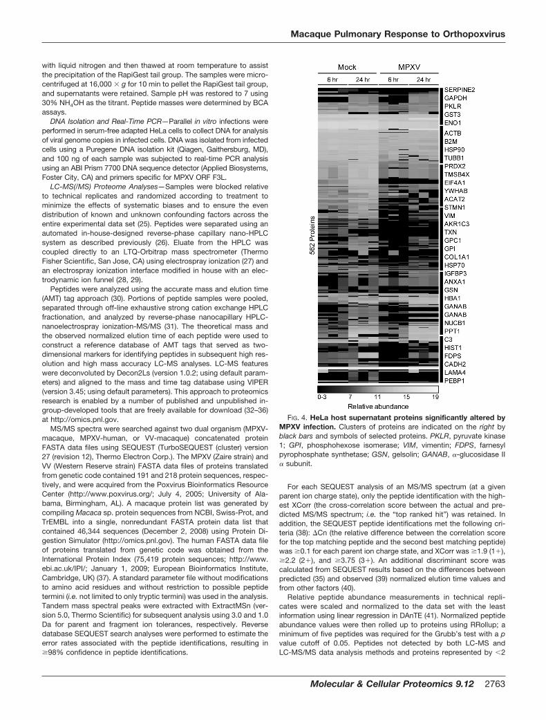
FIG. 4. HeLa host supernatant proteins significantly altered byMPXV infection. Clusters of proteins are indicated on the right byblack bars and symbols of selected proteins. PKLR, pyruvate kinase1; GPI, phosphohexose isomerase; VIM, vimentin; FDPS, farnesylpyrophosphate synthetase; GSN, gelsolin; GANAB, �-glucosidase II� subunit.
Macaque Pulmonary Response to Orthopoxvirus
Molecular & Cellular Proteomics 9.12 2763

unique peptides were removed. Minimum values within data setswere imputed for missing values, and Kruskal-Wallis analyses wereapplied to protein abundance data sets (p value �0.05 and q value�0.01) to identify statistically significant differences in protein expres-sion levels.
Because peptides typically map to multiple homologous proteins,i.e. protein isoforms, it is generally impossible to unambiguouslyassign such peptides to a specific protein (42). Signature peptides,defined as peptides unique to a single isoform, provide a differenti-ating identification (43) that can be used to filter ambiguous peptideassignments. However, the use of signature peptides falsely assumesthat other isoforms, whose sequence may not code for identifiablesignature peptides, do not exist in the sample. To address this,identified peptides were mapped to proteins and then manuallygrouped into protein families. Raw data and collated peptide identi-fication information are available to the community as supplementaldata at http://omics.pnl.gov/view/publication_1024.html.
Pathway Analysis—Human homologs were identified for differen-tially expressed macaque proteins by using InParanoid (44, 45) andScalaBLAST (46) to compare them against the human InternationProtein Index 2009 protein collection list. For pathway analysis,human Internation Protein Index protein accession numbers wereconverted to NCBI Entrez gene identifiers using the BioinformaticsResource Manager (47). Human NCBI Entrez gene identifiers cor-responding to differentially expressed macaque proteins were im-ported into DAVID (48, 49), and the Kyoto Encyclopedia of Genesand Genomes was used to identify significantly altered functionalpathways (50–52).
RESULTS
Monkeypox Infection Resulted in Distinct Effects on HeLaProteome Partitions—The cellular response to MPXV infectionwas characterized in HeLa cells in part to determine whetherextracellular MPXV proteins could be detected in a classical cellculture model system. Total protein content was quantified formock- and virus-infected cellular and extracellular partitions
at 6 and 24 h. After infection, MPXV genomic DNA was detect-able within 6 h of infection by real-time PCR analysis anddisplayed a greater than 100-fold increase in genome copiesbetween 6 and 24 h (Fig. 1) due to increased virus replicationand spread. Mock-infected cultures contained no detectableviral DNA. Protein contents were similar with regard to infectionstate and time point. However, the cellular partition contained�10-fold more protein mass than the extracellular partition.Therefore, a precipitation step was necessary to concentrateproteins in the extracellular partition prior to global analysis.Both cellular and extracellular partitions were analyzed, and asubstantially larger number of unique peptides were observed inthe cellular partition than in the extracellular partition, i.e. �7600and �1200 peptides, mapping to �3300 and �800 proteins,respectively (Fig. 2 and supplemental Tables S01–S04).
Viral proteins were detected exclusively in MPXV-infectedsamples within 24 h and were found in both the intracellularand extracellular partitions. Six viral proteins were observed inthe intracellular partition (E8L, M4R, F3L, H5R, I1L, and A5L),and three were identified in the extracellular partition (J1L,B9R, and B20R). Although all of these viral proteins wereidentified through multiple peptides, another subset includedthose identified by only a single peptide that passed a falsediscovery rate cutoff of 1%. Spectra in this subset weremanually annotated to improve confidence of the peptideidentifications. Four representative spectra of peptides map-ping to O2L, B12R, A29L, and G4L validated the expression ofthese proteins exclusively during MPXV infection (Fig. 3 andsupplemental Table S05).
In the cellular partition, no HeLa-derived proteins displayedstatistically significant differences (p � 0.05, q � 0.01) be-
FIG. 5. Significantly altered proteinsin macaque lung fluid upon inocula-tion with MPXV (A) or VV (B). Timepoints above heat maps refer to prior toviral infection (PRE), �16 days (EARLY),or �16 days (LATE).
Macaque Pulmonary Response to Orthopoxvirus
2764 Molecular & Cellular Proteomics 9.12

tween mock and MPXV treatments. This is further supportedby the heat map representation (Fig. 2A and supple-mental Table S01) in which the samples appear to be nearlyindistinguishable. In sharp contrast to the cellular partition,614 proteins displayed statistically significant differencesbetween mock- and MPXV-treated extracellular samples.During MPXV infection, an increase in abundance was ob-served for 30 unique peptides that mapped to 52 MHC-Iisoforms, a large polymorphic family of related proteins thatare commonly over-represented in proteome experimentsbecause of peptide redundancy. Therefore, MHC-I proteinswere manually curated to a single group and excluded fromthis analysis of the extracellular partition, resulting in 562proteins (Fig. 4). Surprisingly, the greatest differences oc-curred at the early infection time point where the majority of
proteins decreased in expression in many cases below thelevel of detection.
MPXV infection resulted in reduced abundances of severalcellular structural and metabolic proteins. Among the struc-tural proteins that decreased during MPXV infection were�-actin (ACTB), collagen type I �1 (COL1A1), gelsolin (GSN),stathmin 1 (STMN1), thymosin �-4 (TMSB4X), tubulin �1(TUBB1), and vimentin (VIM). Additionally, the expression lev-els of the following metabolic proteins decreased: acetyl-CoAacetyltransferase 2 (ACAT2), enolase 1 (ENO1), farnesyl py-rophosphate synthetase (FDPS), �-glucosidase II � subunit(GANAB), aldo-keto reductase family 1 member C1 (AKR1C1),glyceraldehyde-3-phosphate dehydrogenase (GAPDH),phosphohexose isomerase (GPI), pyruvate kinase 1 (PKLR),peroxiredoxin 2 (PRDX2), and thioredoxin (TXN).
FIG. 6. Selected proteins from ma-caque lung fluid observed in bothMPXV (A) and VV (B) infections. Viralproteins are designated by a blue bar onthe left, proteins that increased duringinfection are designated by a red bar,and proteins that decreased are desig-nated by a green bar. Persistent andtransient increasers are indicated to theright of the heat maps. Averaged andnormalized protein abundances aregraphed (C) relative to pretreatment.Solid lines with diamonds representMPXV infection, and dotted lines withsquares represent VV infection. Red rep-resents proteins that increased duringinfection, green represents proteins thatdecreased, and blue represents the viralproteins. The gray box designates onestandard deviation from the mean. As-terisks (*) indicate viral proteins that wereonly detected in the MPXV infection dur-ing the early phase of infection and weregiven an arbitrary score of �3. GC, vita-min D-binding protein; FGG, fibrinogen�; CRP, C-reactive protein; CFH, com-plement factor H; AFP, �-fetoprotein.
Macaque Pulmonary Response to Orthopoxvirus
Molecular & Cellular Proteomics 9.12 2765

Macaques Display a Temporal and Dynamic Response toOrthopoxvirus—Results from the viral infections of HeLacultures suggested that significant changes occur to theprotein composition of extracellular fluid early upon MPXVinfection. To determine whether similar observations alsooccur during in vivo infection, similar proteome measure-ments were extended to an NHP model system of MPXVinfection. BALF samples were collected from the lungs ofrhesus macaques at various time points preinfection(healthy, uninfected controls) and postinfection followingintrabronchial inoculation with either MPXV or VV. Globalproteome analysis of BALF samples identified �4200unique peptides (�2600 from the MPXV-infected animalsand �3100 from the VV-infected animals) that map to�2100 proteins (�1500 from the MPXV-infected animalsand �1800 from the VV-infected animals). Approximately1200 proteins (�80%) from the MPXV samples were ob-served in common with the VV samples.
Protein expression profiles derived from MPXV- and VV-infected lung fluid exhibited three major temporal expressionpatterns that correlate roughly to pre-, early (�16 days postin-fection), and late infection (�16 days postinfection). Kruskal-Wallis analysis identified 996 proteins (555 proteins fromMPXV and 747 proteins from VV) with significant changes inexpression (p � 0.05 and q � 0.01) during pre-, early, andlate stages of infection (Fig. 5 and supplemental TablesS06 and S07). Three MPXV proteins (J1L, B9R, and H5R), allof which were detected earlier in the extracellular partition ofinfected HeLa cells, were statistically altered in expressionand observed exclusively during the early stages of MPXVinfection. In contrast, no viral proteins were detected through-out the course of VV infection.
The subset of proteins that significantly increased in re-sponse to MPXV fell into two major categories, i.e. transientand persistent “increasers,” depending on whether the in-crease in abundance was resolved by the late stage infec-tion or not, respectively (Fig. 6). Transiently increasing pro-teins included �1-acid glycoprotein 1 precursor (AGP1),C-reactive protein (CRP), complement factor H (CFH), se-rum amyloid A4 (SAA4), and S100 calcium-binding proteinA8 (S100A8). Persistently increasing proteins included �1-B
glycoprotein (A1BG), vitamin D-binding protein (GC), fibrin-ogen � (FGG), apolipoprotein A-I (APOA1), and S100 calci-um-binding protein A9 (S100A9). Similar expression dynam-ics were observed during VV infection (Fig. 6) with theexception of AGP1. The increased abundance level of AGP1during viral infection persisted throughout the late phase ofVV infection. Although trends were similar for both MPXVand VV infections, a significantly larger number of peptidescorresponding to the immunoglobulin family were observedduring VV infection (1624 peptides) compared with MPXVinfection (428 peptides).
Relative to the proteins that exhibited increases in abun-dance, a much larger group of proteins displayed dramatic
decreases in expression upon MPXV infection, some of whichfell below the level of detection. In contrast, only moderate, ifany, decreases were observed in these proteins upon VVinfection. Similar to the HeLa cell supernatant, structural andmetabolic proteins in lung fluid decreased in abundance dur-ing the early phases of MPXV infection. The structural proteinsincluded periostin osteoblast-specific factor (POSTN), tropo-myosin 1 (TPM1), and COL1A1. Metabolic proteins that de-creased included AKR1C1, ENO1, aldehyde dehydrogenasefamily 1 subfamily A1 (ALDH1A1), and tripartite motif-contain-ing 5 (TRIM5). Similarly, the transport protein �-fetoprotein(AFP), which was present prior to MPXV inoculation, was notdetected during the early phase of infection.
Integrating Interspecies Response Profiles to Poxvirus In-fection—To highlight common virus-host response profilescharacteristic of orthopoxvirus infections in general (i.e. inde-pendent of host species or cell type), statistically alteredhomologs across macaques and humans were selectedbased on the most dramatic changes in expression duringinfection and assembled relative to virus and host (Fig. 7 andsupplemental Tables S08–S10). Several structural proteins
FIG. 7. Common interspecies response profile in MPXV- andVV-infected macaque lung fluid (A) and supernatant from MPXV-infected HeLa cells (B).
Macaque Pulmonary Response to Orthopoxvirus
2766 Molecular & Cellular Proteomics 9.12
Macaque Pulmonary Response to Orthopoxvirus
Molecular & Cellular Proteomics 9.12 2767
(e.g. tyrosine 3-monooxygenase/tryptophan 5-monooxygen-ase activation protein, beta polypeptide (YWHAB), TPM3,TUBB4, ACTB, and TMSB4X) and metabolic proteins (e.g.TXN, PKM2, and AKR1C3) displayed decreased expressionduring viral infection. Although expression of HSP90 in bothmacaque lung fluid and HeLa cell supernatant decreasedduring MPXV infection, the suppressive effect was less ap-parent during VV infection. In contrast, increased expressionof host serine protease inhibitors (SERPINs) is a commonresponse between species and is a general response to or-thopoxvirus infection. MPXV also encodes multiple SERPINs,including B19R, C2L, and B12R. SERPINs are used by severalviruses to subvert the host immune response and inflamma-tion (53, 54). The observed interspecies increase from bothhost- and virus-encoded SERPINs highlights the importantrole this class of proteins plays in orthopoxvirus infection.
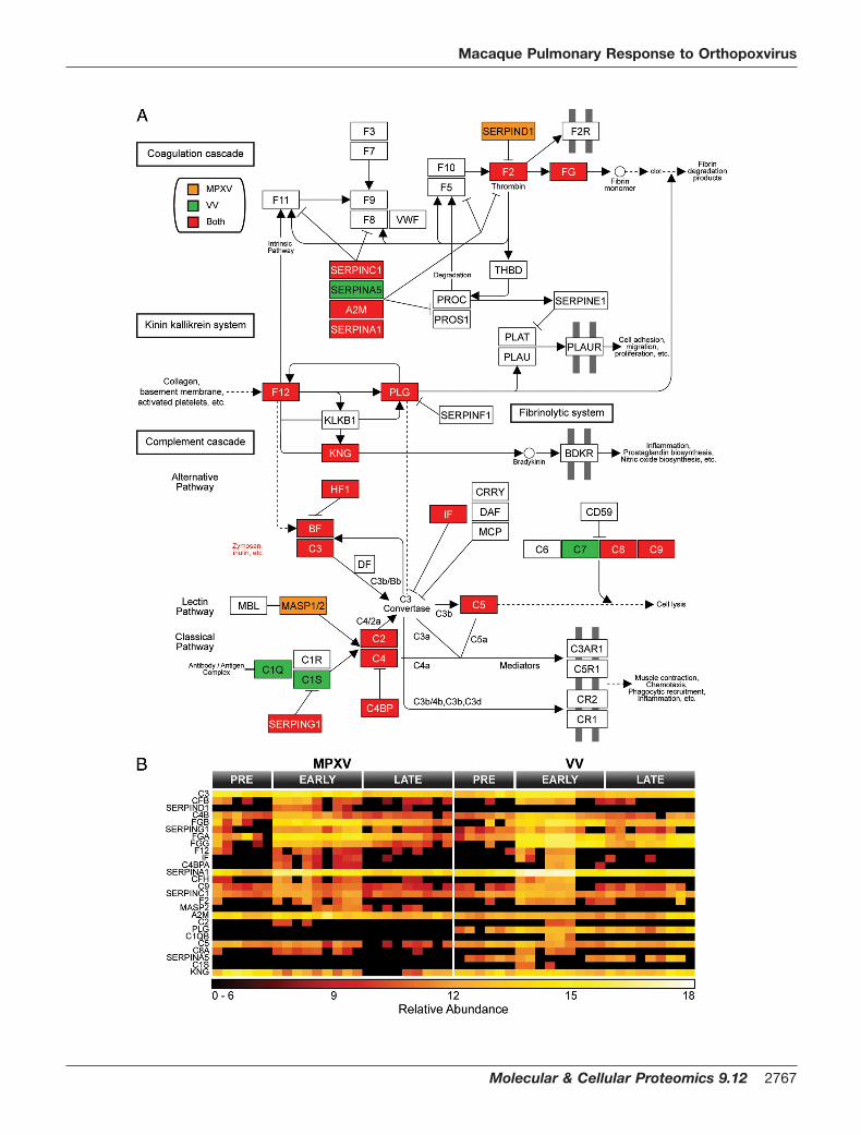
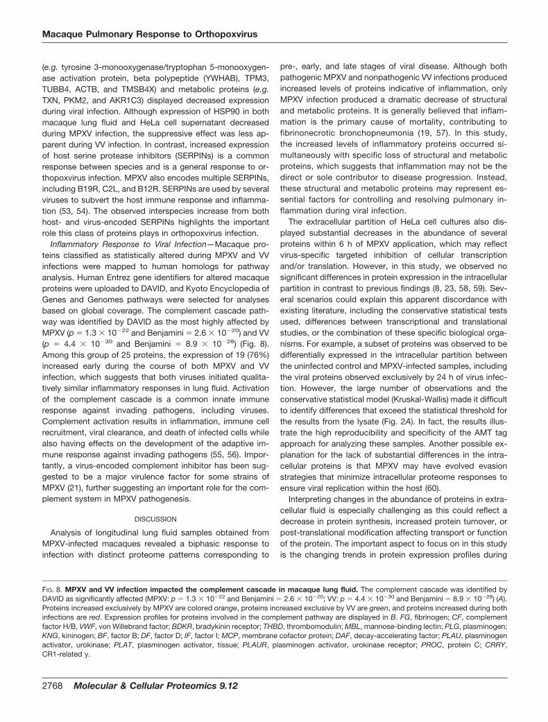
Inflammatory Response to Viral Infection—Macaque pro-teins classified as statistically altered during MPXV and VVinfections were mapped to human homologs for pathwayanalysis. Human Entrez gene identifiers for altered macaqueproteins were uploaded to DAVID, and Kyoto Encyclopedia ofGenes and Genomes pathways were selected for analysesbased on global coverage. The complement cascade path-way was identified by DAVID as the most highly affected byMPXV (p � 1.3 � 10�22 and Benjamini � 2.6 � 10�20) and VV(p � 4.4 � 10�30 and Benjamini � 8.9 � 10�28) (Fig. 8).Among this group of 25 proteins, the expression of 19 (76%)increased early during the course of both MPXV and VVinfection, which suggests that both viruses initiated qualita-tively similar inflammatory responses in lung fluid. Activationof the complement cascade is a common innate immuneresponse against invading pathogens, including viruses.Complement activation results in inflammation, immune cellrecruitment, viral clearance, and death of infected cells whilealso having effects on the development of the adaptive im-mune response against invading pathogens (55, 56). Impor-tantly, a virus-encoded complement inhibitor has been sug-gested to be a major virulence factor for some strains ofMPXV (21), further suggesting an important role for the com-plement system in MPXV pathogenesis.
DISCUSSION
Analysis of longitudinal lung fluid samples obtained fromMPXV-infected macaques revealed a biphasic response toinfection with distinct proteome patterns corresponding to
pre-, early, and late stages of viral disease. Although bothpathogenic MPXV and nonpathogenic VV infections producedincreased levels of proteins indicative of inflammation, onlyMPXV infection produced a dramatic decrease of structuraland metabolic proteins. It is generally believed that inflam-mation is the primary cause of mortality, contributing tofibrinonecrotic bronchopneumonia (19, 57). In this study,the increased levels of inflammatory proteins occurred si-multaneously with specific loss of structural and metabolicproteins, which suggests that inflammation may not be thedirect or sole contributor to disease progression. Instead,these structural and metabolic proteins may represent es-sential factors for controlling and resolving pulmonary in-flammation during viral infection.
The extracellular partition of HeLa cell cultures also dis-played substantial decreases in the abundance of severalproteins within 6 h of MPXV application, which may reflectvirus-specific targeted inhibition of cellular transcriptionand/or translation. However, in this study, we observed nosignificant differences in protein expression in the intracellularpartition in contrast to previous findings (8, 23, 58, 59). Sev-eral scenarios could explain this apparent discordance withexisting literature, including the conservative statistical testsused, differences between transcriptional and translationalstudies, or the combination of these specific biological orga-nisms. For example, a subset of proteins was observed to bedifferentially expressed in the intracellular partition betweenthe uninfected control and MPXV-infected samples, includingthe viral proteins observed exclusively by 24 h of virus infec-tion. However, the large number of observations and theconservative statistical model (Kruskal-Wallis) made it difficultto identify differences that exceed the statistical threshold forthe results from the lysate (Fig. 2A). In fact, the results illus-trate the high reproducibility and specificity of the AMT tagapproach for analyzing these samples. Another possible ex-planation for the lack of substantial differences in the intra-cellular proteins is that MPXV may have evolved evasionstrategies that minimize intracellular proteome responses toensure viral replication within the host (60).
Interpreting changes in the abundance of proteins in extra-cellular fluid is especially challenging as this could reflect adecrease in protein synthesis, increased protein turnover, orpost-translational modification affecting transport or functionof the protein. The important aspect to focus on in this studyis the changing trends in protein expression profiles during
FIG. 8. MPXV and VV infection impacted the complement cascade in macaque lung fluid. The complement cascade was identified byDAVID as significantly affected (MPXV: p � 1.3 � 10�22 and Benjamini � 2.6 � 10�20; VV: p � 4.4 � 10�30 and Benjamini � 8.9 � 10�28) (A).Proteins increased exclusively by MPXV are colored orange, proteins increased exclusive by VV are green, and proteins increased during bothinfections are red. Expression profiles for proteins involved in the complement pathway are displayed in B. FG, fibrinogen; CF, complementfactor H/B, VWF, von Willebrand factor; BDKR, bradykinin receptor; THBD, thrombomodulin; MBL, mannose-binding lectin; PLG, plasminogen;KNG, kininogen; BF, factor B; DF, factor D; IF, factor I; MCP, membrane cofactor protein; DAF, decay-accelerating factor; PLAU, plasminogenactivator, urokinase; PLAT, plasminogen activator, tissue; PLAUR, plasminogen activator, urokinase receptor; PROC, protein C; CRRY,CR1-related y.
Macaque Pulmonary Response to Orthopoxvirus
2768 Molecular & Cellular Proteomics 9.12

the course of viral infection. Overall, the trend in extracellularprotein abundance observed during VV infection was verysimilar to that of MPXV infection, which is expected as theseare closely related viruses and likely reflects the general re-sponse to orthopoxvirus infection. The major differences ob-served between MPXV and VV were that responses to VVwere more delayed, short lived, and reduced in magnitude ascompared with those responses observed for MPXV, whichappears to correspond to the difference in virulence betweenthe two viral strains. In contrast, the response to MPXV wasobserved in the lung fluid at the earliest time point, suggestingthat the most important impact of the virus occurs early duringinfection through perturbation of cell-cell communicationsand/or disruption of extracellular structures.
A critical observation was that although viral proteins wereobserved exclusively during infection as expected clearly dis-tinct viral proteins were identified between HeLa cell lysateand supernatant samples. Six MPXV proteins were identifiedfrom the complex intracellular samples: M4R, F3L, E8L, A5L,I1L, and H5R. Three of the intracellular viral proteins areknown structural components of the virion: M4R and A5L areviral core proteins, and E8L is a surface membrane protein ofcell-associated viral particles, all of which would be expectedto be relatively abundant. H5R and I1L are virosomal proteinsthat are essential for viral replication. F3L inhibits intracellularinnate immune pattern recognition molecules, such as dou-ble-stranded RNA protein kinase and oligoadenylate synthe-tase. Three viral proteins were detected in the supernatant ofinfected HeLa cells within 24 h: B20R, J1L, and B9R. Theseproteins were also observed in the lung fluid of infected ma-caques, demonstrating similarity between the in vitro HeLacell and the in vivo rhesus macaque infection models. J1L andB9R are known secreted receptor mimics for CC chemokinesand IFN-�, respectively, which promote viral replication in thehost by blocking immune response. In contrast, B20R is de-rived from a predicted protein-coding gene within the MPXVgenome and is functionally uncharacterized. All observed viralproteins have previously been identified at the RNA level bymicroarray analysis during MPXV infection of HeLa cells, pri-mary human monocytes, and fibroblasts (6).
Overall, the insights gained by applying the AMT tag pro-teomics approach to studying both the HeLa cell and ma-caque lung fluid improve our understanding of disease byhuman MPXV. In particular, the reduced abundance of a largenumber of proteins in the extracellular fluid and lung fluidsuggests the possibility that increased mortality as a result ofMPXV infection may largely be due to reduction in expressionof proteins important to host integrity.
Acknowledgments—We gratefully acknowledge the contributionsand assistance of Penny Colton. Human MPXV (Zaire strain V79-I-005) was graciously provided by Dr. Inger Damon (Centers for Dis-ease Control and Prevention, Atlanta, GA). Portions of the work wereperformed in the Environmental Molecular Science Laboratory, aUnited States Department of Energy (DOE) Office of Biological and
Environmental Research national scientific user facility at PacificNorthwest National Laboratory (PNNL) in Richland, WA. PNNL is amultiprogram national laboratory operated by Battelle Memorial Insti-tute for the DOE under Contract DE-AC05-76RLO-1830.
* This work was supported, in whole or in part, by National Insti-tutes of Health Grants RR00163 and RR18522 from the NationalCenter for Research Resources and Interagency Agreements Y1-AI-4894–01 and Y1-A1–8401 from the NIAID/United States Departmentof Health and Human Services. This work was also supported byUnited States Department of Defense Grant W81XWH-05-1-0046)and Battelle Internal Research and Development.
□S This article contains supplemental Tables S01–S10.¶ To whom correspondence should be addressed: Biological Sys-
tems Analysis and Mass Spectrometry Division, Pacific NorthwestNational Laboratory, P. O. Box 999/MS K8-98, Richland, WA 99352.E-mail: [email protected].
REFERENCES
1. Di Giulio, D. B., and Eckburg, P. B. (2004) Human monkeypox: an emergingzoonosis. Lancet Infect. Dis. 4, 15–25
2. Weaver, J. R., and Isaacs, S. N. (2008) Monkeypox virus and insights intoits immunomodulatory proteins. Immunol. Rev. 225, 96–113
3. Henderson, D. A., Inglesby, T. V., Bartlett, J. G., Ascher, M. S., Eitzen, E.,Jahrling, P. B., Hauer, J., Layton, M., McDade, J., Osterholm, M. T.,O’Toole, T., Parker, G., Perl, T., Russell, P. K., and Tonat, K. (1999)Smallpox as a biological weapon: medical and public health manage-ment. Working Group on Civilian Biodefense. JAMA 281, 2127–2137
4. Reed, K. D., Melski, J. W., Graham, M. B., Regnery, R. L., Sotir, M. J.,Wegner, M. V., Kazmierczak, J. J., Stratman, E. J., Li, Y., Fairley, J. A.,Swain, G. R., Olson, V. A., Sargent, E. K., Kehl, S. C., Frace, M. A., Kline,R., Foldy, S. L., Davis, J. P., and Damon, I. K. (2004) The detection ofmonkeypox in humans in the Western Hemisphere. N. Engl. J. Med. 350,342–350
5. Centers for Disease Control and Prevention (CDC) (2003) Multistate out-break of monkeypox—Illinois, Indiana, and Wisconsin, 2003. MMWRMorb. Mortal. Wkly. Rep. 52, 537–540
6. Rubins, K. H., Hensley, L. E., Bell, G. W., Wang, C., Lefkowitz, E. J., Brown,P. O., and Relman, D. A. (2008) Comparative analysis of viral geneexpression programs during poxvirus infection: a transcriptional map ofthe vaccinia and monkeypox genomes. PLoS One 3, e2628
7. Katsafanas, G. C., and Moss, B. (2007) Colocalization of transcription andtranslation within cytoplasmic poxvirus factories coordinates viral ex-pression and subjugates host functions. Cell Host Microbe 2, 221–228
8. Brum, L. M., Lopez, M. C., Varela, J. C., Baker, H. V., and Moyer, R. W.(2003) Microarray analysis of A549 cells infected with rabbitpox virus(RPV): a comparison of wild-type RPV and RPV deleted for the hostrange gene, SPI-1. Virology 315, 322–334
9. Guerra, S., Lopez-Fernandez, L. A., Pascual-Montano, A., Munoz, M.,Harshman, K., and Esteban, M. (2003) Cellular gene expression survey ofvaccinia virus infection of human HeLa cells. J. Virol. 77, 6493–6506
10. McFadden, G. (2005) Poxvirus tropism. Nat. Rev. Microbiol. 3, 201–21311. Seet, B. T., Johnston, J. B., Brunetti, C. R., Barrett, J. W., Everett, H.,
Cameron, C., Sypula, J., Nazarian, S. H., Lucas, A., and McFadden, G.(2003) Poxviruses and immune evasion. Annu Rev. Immunol. 21,377–423
12. Arita, I., Jezek, Z., Khodakevich, L., and Ruti, K. (1985) Human monkeypox:a newly emerged orthopoxvirus zoonosis in the tropical rain forests ofAfrica. Am. J. Trop. Med. Hyg. 34, 781–789
13. Breman, J. G., Kalisa-Ruti Steniowski, M. V., Zanotto, E., Gromyko, A. I.,and Arita, I. (1980) Human monkeypox, 1970–79. Bull. World HealthOrgan. 58, 165–182
14. Jezek, Z., Grab, B., Szczeniowski, M., Paluku, K. M., and Mutombo, M. (1988)Clinico-epidemiological features of monkeypox patients with an animal orhuman source of infection. Bull. World Health Organ. 66, 459–464
15. Lourie, B., Bingham, P. G., Evans, H. H., Foster, S. O., Nakano, J. H., andHerrmann, K. L. (1972) Human infection with monkeypox virus: labora-tory investigation of six cases in West Africa. Bull. World Health Organ.46, 633–639
Macaque Pulmonary Response to Orthopoxvirus
Molecular & Cellular Proteomics 9.12 2769

16. Cho, C. T., and Wenner, H. A. (1973) Monkeypox virus. Bacteriol. Rev. 37,1–18
17. Ryan, C. P. (2008) Zoonoses likely to be used in bioterrorism. Public HealthRep. 123, 276–281
18. Garnier, L., Gaudin, J. C., Bensadoun, P., Rebillat, I., and Morel, Y. (2009)Real-time PCR assay for detection of a new simulant for poxvirus bio-threat agents. Appl. Environ. Microbiol. 75, 1614–1620
19. Zaucha, G. M., Jahrling, P. B., Geisbert, T. W., Swearengen, J. R., andHensley, L. (2001) The pathology of experimental aerosolized monkey-pox virus infection in cynomolgus monkeys (Macaca fascicularis). Lab.Invest. 81, 1581–1600
20. Saijo, M., Ami, Y., Suzaki, Y., Nagata, N., Iwata, N., Hasegawa, H., Iizuka,I., Shiota, T., Sakai, K., Ogata, M., Fukushi, S., Mizutani, T., Sata, T.,Kurata, T., Kurane, I., and Morikawa, S. (2009) Virulence and pathophys-iology of the Congo Basin and West African strains of monkeypox virusin non-human primates. J. Gen. Virol. 90, 2266–2271
21. Chen, N., Li, G., Liszewski, M. K., Atkinson, J. P., Jahrling, P. B., Feng, Z.,Schriewer, J., Buck, C., Wang, C., Lefkowitz, E. J., Esposito, J. J.,Harms, T., Damon, I. K., Roper, R. L., Upton, C., and Buller, R. M. (2005)Virulence differences between monkeypox virus isolates from West Af-rica and the Congo basin. Virology 340, 46–63
22. Rubins, K. H., Hensley, L. E., Jahrling, P. B., Whitney, A. R., Geisbert, T. W.,Huggins, J. W., Owen, A., Leduc, J. W., Brown, P. O., and Relman, D. A.(2004) The host response to smallpox: analysis of the gene expressionprogram in peripheral blood cells in a nonhuman primate model. Proc.Natl. Acad. Sci. U.S.A. 101, 15190–15195
23. Moss, B. (1968) Inhibition of HeLa cell protein synthesis by the vacciniavirion. J. Virol. 2, 1028–1037
24. Doms, R. W., Blumenthal, R., and Moss, B. (1990) Fusion of intra- andextracellular forms of vaccinia virus with the cell membrane. J. Virol. 64,4884–4892
25. Oberg, A. L., and Vitek, O. (2009) Statistical design of quantitative massspectrometry-based proteomic experiments. J. Proteome Res. 8,2144–2156
26. Livesay, E. A., Tang, K., Taylor, B. K., Buschbach, M. A., Hopkins, D. F.,LaMarche, B. L., Zhao, R., Shen, Y., Orton, D. J., Moore, R. J., Kelly,R. T., Udseth, H. R., and Smith, R. D. (2008) Fully automated four-columncapillary LC-MS system for maximizing throughput in proteomic analy-ses. Anal. Chem. 80, 294–302
27. Kelly, R. T., Page, J. S., Tang, K., and Smith, R. D. (2007) Array of chem-ically etched fused-silica emitters for improving the sensitivity and quan-titation of electrospray ionization mass spectrometry. Anal. Chem. 79,4192–4198
28. Manes, N. P., Estep, R. D., Mottaz, H. M., Moore, R. J., Clauss, T. R.,Monroe, M. E., Du, X., Adkins, J. N., Wong, S. W., and Smith, R. D.(2008) Comparative proteomics of human monkeypox and vacciniaintracellular mature and extracellular enveloped virions. J. ProteomeRes. 7, 960–968
29. Page, J. S., Tolmachev, A. V., Tang, K., and Smith, R. D. (2006) Theoreticaland experimental evaluation of the low m/z transmission of an electro-dynamic ion funnel. J. Am. Soc. Mass Spectrom. 17, 586–592
30. Zimmer, J. S., Monroe, M. E., Qian, W. J., and Smith, R. D. (2006) Advancesin proteomics data analysis and display using an accurate mass and timetag approach. Mass Spectrom. Rev. 25, 450–482
31. Adkins, J. N., Mottaz, H. M., Norbeck, A. D., Gustin, J. K., Rue, J., Clauss,T. R., Purvine, S. O., Rodland, K. D., Heffron, F., and Smith, R. D. (2006)Analysis of the Salmonella typhimurium proteome through environmentalresponse toward infectious conditions. Mol. Cell. Proteomics 5,1450–1461
32. Jaitly, N., Monroe, M. E., Petyuk, V. A., Clauss, T. R., Adkins, J. N., andSmith, R. D. (2006) Robust algorithm for alignment of liquid chromatog-raphy-mass spectrometry analyses in an accurate mass and time tagdata analysis pipeline. Anal. Chem. 78, 7397–7409
33. Kiebel, G. R., Auberry, K. J., Jaitly, N., Clark, D. A., Monroe, M. E., Peterson,E. S., Toliæ, N., Anderson, G. A., and Smith, R. D. (2006) PRISM: a datamanagement system for high-throughput proteomics. Proteomics 6,1783–1790
34. Monroe, M. E., Toliæ, N., Jaitly, N., Shaw, J. L., Adkins, J. N., and Smith,R. D. (2007) VIPER: an advanced software package to support high-throughput LC-MS peptide identification. Bioinformatics 23, 2021–2023
35. Petritis, K., Kangas, L. J., Yan, B., Monroe, M. E., Strittmatter, E. F., Qian,
W. J., Adkins, J. N., Moore, R. J., Xu, Y., Lipton, M. S., Camp, D. G., 2nd,and Smith, R. D. (2006) Improved peptide elution time prediction forreversed-phase liquid chromatography-MS by incorporating peptide se-quence information. Anal. Chem. 78, 5026–5039
36. Monroe, M. E., Shaw, J. L., Daly, D. S., Adkins, J. N., and Smith, R. D.(2008) MASIC: a software program for fast quantitation and flexiblevisualization of chromatographic profiles from detected LC-MS(/MS)features. Comput. Biol. Chem. 32, 215–217
37. Kersey, P. J., Duarte, J., Williams, A., Karavidopoulou, Y., Birney, E., andApweiler, R. (2004) The International Protein Index: an integrated data-base for proteomics experiments. Proteomics 4, 1985–1988
38. Washburn, M. P., Wolters, D., and Yates, J. R., 3rd (2001) Large-scaleanalysis of the yeast proteome by multidimensional protein identificationtechnology. Nat. Biotechnol. 19, 242–247
39. Liu, T., Belov, M. E., Jaitly, N., Qian, W. J., and Smith, R. D. (2007) Accuratemass measurements in proteomics. Chem. Rev. 107, 3621–3653
40. Strittmatter, E. F., Kangas, L. J., Petritis, K., Mottaz, H. M., Anderson, G. A.,Shen, Y., Jacobs, J. M., Camp, D. G., 2nd, and Smith, R. D. (2004)Application of peptide LC retention time information in a discriminantfunction for peptide identification by tandem mass spectrometry. J.Proteome Res. 3, 760–769
41. Polpitiya, A. D., Qian, W. J., Jaitly, N., Petyuk, V. A., Adkins, J. N., Camp,D. G., 2nd, Anderson, G. A., and Smith, R. D. (2008) DAnTE: a statisticaltool for quantitative analysis of -omics data. Bioinformatics 24,1556–1558
42. Adkins, J. N., Monroe, M. E., Auberry, K. J., Shen, Y., Jacobs, J. M., Camp,D. G., 2nd, Vitzthum, F., Rodland, K. D., Zangar, R. C., Smith, R. D., andPounds, J. G. (2005) A proteomic study of the HUPO Plasma ProteomeProject’s pilot samples using an accurate mass and time tag strategy.Proteomics 5, 3454–3466
43. Keshishian, H., Addona, T., Burgess, M., Kuhn, E., and Carr, S. A. (2007)Quantitative, multiplexed assays for low abundance proteins in plasmaby targeted mass spectrometry and stable isotope dilution. Mol. Cell.Proteomics 6, 2212–2229
44. Berglund, A. C., Sjolund, E., Ostlund, G., and Sonnhammer, E. L. (2008)InParanoid 6: eukaryotic ortholog clusters with inparalogs. Nucleic AcidsRes. 36, D263–D266
45. O’Brien, K. P., Remm, M., and Sonnhammer, E. L. (2005) Inparanoid: acomprehensive database of eukaryotic orthologs. Nucleic Acids Res. 33,D476–D480
46. Oehmen, C., and Nieplocha, J. (2006) ScalaBLAST: a scalable implemen-tation of BLAST for high-performance data-intensive bioinformatics anal-ysis. IEEE Trans. Parallel Distrib. Syst. 17, 740–749
47. Shah, A. R., Singhal, M., Klicker, K. R., Stephan, E. G., Wiley, H. S., andWaters, K. M. (2007) Enabling high-throughput data management forsystems biology: the Bioinformatics Resource Manager. Bioinformatics23, 906–909
48. Huang da, W., Sherman, B. T., and Lempicki, R. A. (2009) Systematic andintegrative analysis of large gene lists using DAVID bioinformatics re-sources. Nat. Protoc. 4, 44–57
49. Dennis, G., Jr., Sherman, B. T., Hosack, D. A., Yang, J., Gao, W., Lane,H. C., and Lempicki, R. A. (2003) DAVID: Database for Annotation,Visualization, and Integrated Discovery. Genome Biol. 4, P3
50. Kanehisa, M., Araki, M., Goto, S., Hattori, M., Hirakawa, M., Itoh, M.,Katayama, T., Kawashima, S., Okuda, S., Tokimatsu, T., and Yamanishi,Y. (2008) KEGG for linking genomes to life and the environment. NucleicAcids Res. 36, D480–D484
51. Kanehisa, M., and Goto, S. (2000) KEGG: Kyoto encyclopedia of genes andgenomes. Nucleic Acids Res. 28, 27–30
52. Kanehisa, M., Goto, S., Hattori, M., Aoki-Kinoshita, K. F., Itoh, M., Ka-washima, S., Katayama, T., Araki, M., and Hirakawa, M. (2006) Fromgenomics to chemical genomics: new developments in KEGG. NucleicAcids Res. 34, D354–D357
53. Lucas, A., Liu, L., Dai, E., Bot, I., Viswanathan, K., Munuswamy-Ramunu-jam, G., Davids, J. A., Bartee, M. Y., Richardson, J., Christov, A., Wang,H., Macaulay, C., Poznansky, M., Zhong, R., Miller, L., Biessen, E.,Richardson, M., Sullivan, C., Moyer, R., Hatton, M., Lomas, D. A., andMcFadden, G. (2009) The serpin saga; development of a new class ofvirus derived anti-inflammatory protein immunotherapeutics. Adv. Exp.Med. Biol. 666, 132–156
54. Amati, L., Passeri, M. E., Lippolis, A., Lio, D., Caruso, C., Jirillo, E., and
Macaque Pulmonary Response to Orthopoxvirus
2770 Molecular & Cellular Proteomics 9.12

Covelli, V. (2006) Taking advantage of viral immune evasion: virus-de-rived proteins represent novel biopharmaceuticals. Curr. Med. Chem. 13,325–333
55. Lambris, J. D., Ricklin, D., and Geisbrecht, B. V. (2008) Complementevasion by human pathogens. Nat. Rev. Microbiol. 6, 132–142
56. Carroll, M. C. (2004) The complement system in regulation of adaptiveimmunity. Nat. Immunol. 5, 981–986
57. Stanford, M. M., McFadden, G., Karupiah, G., and Chaudhri, G. (2007)
Immunopathogenesis of poxvirus infections: forecasting the impendingstorm. Immunol. Cell Biol. 85, 93–102
58. Moss, B., and Salzman, N. P. (1968) Sequential protein synthesis followingvaccinia virus infection. J. Virol. 2, 1016–1027
59. Rice, A. P., and Roberts, B. E. (1983) Vaccinia virus induces cellular mRNAdegradation. J. Virol. 47, 529–539
60. Alcami, A. (2007) New insights into the subversion of the chemokine systemby poxviruses. Eur. J. Immunol. 37, 880–883
Macaque Pulmonary Response to Orthopoxvirus
Molecular & Cellular Proteomics 9.12 2771