Monkeypox Disease Transmission in an Experimental Setting: Prairie Dog Animal Model
12
Monkeypox Disease Transmission in an Experimental Setting: Prairie Dog Animal Model Christina L. Hutson 1 *, Darin S. Carroll 1 , Nadia Gallardo-Romero 1 , Sonja Weiss 1 , Cody Clemmons 1 , Christine M. Hughes 1 , Johanna S. Salzer 1,2 , Victoria A. Olson 1 , Jason Abel 1 , Kevin L. Karem 1 , Inger K. Damon 1 1 Poxvirus and Rabies Branch, Division of High-Consequence Pathogens and Pathology, National Center for Emerging and Zoonotic Disease, Centers for Disease Control and Prevention, Atlanta, Georgia, United States of America, 2 Program in Population Biology, Ecology and Evolution, Emory University, Atlanta, Georgia, United States of America Abstract Monkeypox virus (MPXV) is considered the most significant human public health threat in the genus Orthopoxvirus since the eradication of variola virus (the causative agent of smallpox). MPXV is a zoonotic agent endemic to forested areas of Central and Western Africa. In 2003, MPXV caused an outbreak in the United States due to the importation of infected African rodents, and subsequent sequential infection of North American prairie dogs (Cynomys ludovicianus) and humans. In previous studies, the prairie dog MPXV model has successfully shown to be very useful for understanding MPXV since the model emulates key characteristics of human monkeypox disease. In humans, percutaneous exposure to animals has been documented but the primary method of human-to-human MPXV transmission is postulated to be by respiratory route. Only a few animal model studies of MPXV transmission have been reported. Herein, we show that MPXV infected prairie dogs are able to transmit the virus to naive animals through multiple transmission routes. All secondarily exposed animals were infected with MPXV during the course of the study. Notably, animals secondarily exposed appeared to manifest more severe disease; however, the disease course was very similar to those of experimentally challenged animals including inappetence leading to weight loss, development of lesions, production of orthopoxvirus antibodies and shedding of similar levels or in some instances higher levels of MPXV from the oral cavity. Disease was transmitted via exposure to contaminated bedding, co-housing, or respiratory secretions/nasal mucous (we could not definitively say that transmission occurred via respiratory route exclusively). Future use of the model will allow us to evaluate infection control measures, vaccines and antiviral strategies to decrease disease transmission. Citation: Hutson CL, Carroll DS, Gallardo-Romero N, Weiss S, Clemmons C, et al. (2011) Monkeypox Disease Transmission in an Experimental Setting: Prairie Dog Animal Model. PLoS ONE 6(12): e28295. doi:10.1371/journal.pone.0028295 Editor: Lark L. Coffey, Blood Systems Research Institute, United States of America Received September 6, 2011; Accepted November 4, 2011; Published December 2, 2011 This is an open-access article, free of all copyright, and may be freely reproduced, distributed, transmitted, modified, built upon, or otherwise used by anyone for any lawful purpose. The work is made available under the Creative Commons CC0 public domain dedication. Funding: The authors have no support or funding to report. Competing Interests: The authors have declared that no competing interests exist. * E-mail: [email protected] Introduction Monkeypox virus (MPXV) and variola virus (the causative agent of smallpox) are members of the genus Orthopoxvirus. While smallpox has been eradicated from the human population and viral isolates only remain in secure Biosafety Level 4 laboratories; MPXV is a zoonotic pathogen endemic to Central and Western Africa where it can cause human infection and even mortality, and is maintained in the wild by undetermined rodent reservoir(s) [1–3]. In 2003 MPXV caused the first outbreak of human disease outside of Africa within the United States [4]. The virus was introduced due to importation of infected rodents; human disease resulted from the subsequent infection of North American black- tailed prairie dogs (Cynomys ludovicianus) which in turn efficiently transmitted disease to humans [5,6]. Anecdotally, it appeared prairie dogs transmitted MPXV within households, pet store, or other settings. Previous studies have defined two distinct MPXV clades, West African and Congo Basin [7,8]; the U.S. outbreak was due to an importation of the West African clade MPXV. In humans, West African MPXV causes a milder disease, ,1% mortality and is rarely associated with person to person transmission [9,10]. However, Congo Basin MPXV causes approximately 10% mortality and human to human transmission has been observed; up to six sequential interhuman transmission events have been laboratory-documented [11]. Both MPXV and smallpox are believed to be transmitted between humans primarily via respiratory secretions. As evidenced by the 2003 US outbreak, as well as the ongoing outbreaks of MPXV within Africa, there is a continued need to study and understand the details of viral transmission among hosts. Additionally, MPXV provides a surrogate for the study of related orthopoxviruses including variola virus. An ideal animal model is one that would emulate key features of human disease including: utilizing a route of infection that mimics the natural transmission of the pathogen in humans; the ability to obtain disease with an infectious dose equivalent to that causing disease in humans; as well as having a disease course, morbidity and mortality similar to what is observed with human disease. Previous studies of the prairie dog MPXV model showed after intranasal or scarification challenge with a reasonable challenge dose, animals developed disease that closely resembled human monkeypox, includ- ing a protracted incubation period before the development of PLoS ONE | www.plosone.org 1 December 2011 | Volume 6 | Issue 12 | e28295
Transcript of Monkeypox Disease Transmission in an Experimental Setting: Prairie Dog Animal Model
Monkeypox Disease Transmission in an ExperimentalSetting: Prairie Dog Animal ModelChristina L. Hutson1*, Darin S. Carroll1, Nadia Gallardo-Romero1, Sonja Weiss1, Cody Clemmons1,
Christine M. Hughes1, Johanna S. Salzer1,2, Victoria A. Olson1, Jason Abel1, Kevin L. Karem1, Inger K.
Damon1
1 Poxvirus and Rabies Branch, Division of High-Consequence Pathogens and Pathology, National Center for Emerging and Zoonotic Disease, Centers for Disease Control
and Prevention, Atlanta, Georgia, United States of America, 2 Program in Population Biology, Ecology and Evolution, Emory University, Atlanta, Georgia, United States of
America
Abstract
Monkeypox virus (MPXV) is considered the most significant human public health threat in the genus Orthopoxvirus since theeradication of variola virus (the causative agent of smallpox). MPXV is a zoonotic agent endemic to forested areas of Centraland Western Africa. In 2003, MPXV caused an outbreak in the United States due to the importation of infected Africanrodents, and subsequent sequential infection of North American prairie dogs (Cynomys ludovicianus) and humans. Inprevious studies, the prairie dog MPXV model has successfully shown to be very useful for understanding MPXV since themodel emulates key characteristics of human monkeypox disease. In humans, percutaneous exposure to animals has beendocumented but the primary method of human-to-human MPXV transmission is postulated to be by respiratory route. Onlya few animal model studies of MPXV transmission have been reported. Herein, we show that MPXV infected prairie dogs areable to transmit the virus to naive animals through multiple transmission routes. All secondarily exposed animals wereinfected with MPXV during the course of the study. Notably, animals secondarily exposed appeared to manifest more severedisease; however, the disease course was very similar to those of experimentally challenged animals including inappetenceleading to weight loss, development of lesions, production of orthopoxvirus antibodies and shedding of similar levels or insome instances higher levels of MPXV from the oral cavity. Disease was transmitted via exposure to contaminated bedding,co-housing, or respiratory secretions/nasal mucous (we could not definitively say that transmission occurred via respiratoryroute exclusively). Future use of the model will allow us to evaluate infection control measures, vaccines and antiviralstrategies to decrease disease transmission.
Citation: Hutson CL, Carroll DS, Gallardo-Romero N, Weiss S, Clemmons C, et al. (2011) Monkeypox Disease Transmission in an Experimental Setting: Prairie DogAnimal Model. PLoS ONE 6(12): e28295. doi:10.1371/journal.pone.0028295
Editor: Lark L. Coffey, Blood Systems Research Institute, United States of America
Received September 6, 2011; Accepted November 4, 2011; Published December 2, 2011
This is an open-access article, free of all copyright, and may be freely reproduced, distributed, transmitted, modified, built upon, or otherwise used by anyone forany lawful purpose. The work is made available under the Creative Commons CC0 public domain dedication.
Funding: The authors have no support or funding to report.
Competing Interests: The authors have declared that no competing interests exist.
* E-mail: [email protected]
Introduction
Monkeypox virus (MPXV) and variola virus (the causative agent
of smallpox) are members of the genus Orthopoxvirus. While
smallpox has been eradicated from the human population and
viral isolates only remain in secure Biosafety Level 4 laboratories;
MPXV is a zoonotic pathogen endemic to Central and Western
Africa where it can cause human infection and even mortality, and
is maintained in the wild by undetermined rodent reservoir(s)
[1–3]. In 2003 MPXV caused the first outbreak of human disease
outside of Africa within the United States [4]. The virus was
introduced due to importation of infected rodents; human disease
resulted from the subsequent infection of North American black-
tailed prairie dogs (Cynomys ludovicianus) which in turn efficiently
transmitted disease to humans [5,6]. Anecdotally, it appeared
prairie dogs transmitted MPXV within households, pet store, or
other settings. Previous studies have defined two distinct MPXV
clades, West African and Congo Basin [7,8]; the U.S. outbreak
was due to an importation of the West African clade MPXV.
In humans, West African MPXV causes a milder disease,
,1% mortality and is rarely associated with person to person
transmission [9,10]. However, Congo Basin MPXV causes
approximately 10% mortality and human to human transmission
has been observed; up to six sequential interhuman transmission
events have been laboratory-documented [11]. Both MPXV and
smallpox are believed to be transmitted between humans primarily
via respiratory secretions.
As evidenced by the 2003 US outbreak, as well as the ongoing
outbreaks of MPXV within Africa, there is a continued need to
study and understand the details of viral transmission among hosts.
Additionally, MPXV provides a surrogate for the study of related
orthopoxviruses including variola virus. An ideal animal model is
one that would emulate key features of human disease including:
utilizing a route of infection that mimics the natural transmission
of the pathogen in humans; the ability to obtain disease with an
infectious dose equivalent to that causing disease in humans; as
well as having a disease course, morbidity and mortality similar to
what is observed with human disease. Previous studies of the
prairie dog MPXV model showed after intranasal or scarification
challenge with a reasonable challenge dose, animals developed
disease that closely resembled human monkeypox, includ-
ing a protracted incubation period before the development of
PLoS ONE | www.plosone.org 1 December 2011 | Volume 6 | Issue 12 | e28295
generalized lesions [12,13]. Additionally, these studies have shown
that the prairie dog model is valuable for the comparison of disease
attributable to the two MPXV clades, primarily by differences in
mortality and level of morbidity.
Thus far, only two published studies have experimentally
investigated MPXV transmission; these studies have used baboons
and tropical squirrels [14,15]. Although infected animals were able
to transmit the virus to naive animals in these studies, baboons are
not practical to use in laboratory studies and the tropical squirrels
were highly susceptible to MPXV without key features of human
disease including the development of disseminated lesions. Herein
we show that prairie dogs are able to transmit the virus to naive
prairie dogs. Four primary challenged animals were inoculated via
intranasal route with 96103 pfu (0.07XLD50) of West African
MPXV and subsequently placed into one of three groups designed
to evaluate fomites, contact and respiratory routes of disease
transmission. This challenge inoculum was chosen because
previous studies with a similar dosage resulted in low mortality
and the development of morbidity including disseminated lesions
as well as viral shedding [13].
In the current study, we sought to explore the hypothesis that
infected prairie dogs are able to transmit the virus to naive
animals. These studies would allow us to better understand the
transmission events inferred through epidemiologic studies during
the 2003 outbreak, and would allow us to better define the
potential significance of respiratory routes of infection. The
experimental groups that were utilized ascertained the ability of
MPXV to be transmitted via fomite, direct contact, and re-
spiratory secretions.
Results
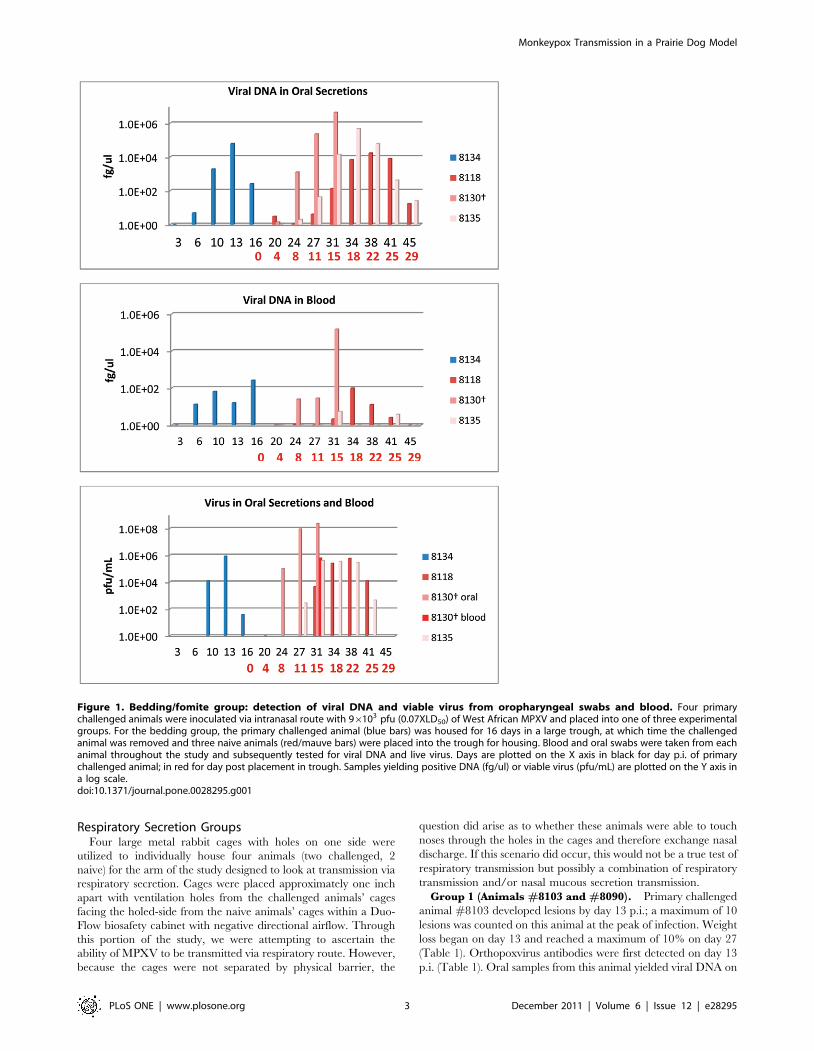
Bedding/Fomite GroupFor the bedding/fomite group, one animal was challenged with
virus and housed for 16 days in a large trough. After day 6 the
bedding was not changed and the challenged animal was left in the
trough until day 16 at which time it was removed and humanely
euthanized. Three naive animals were then placed into the large
trough for housing with the contaminated bedding and this
bedding was not changed for 39 days. The primary animal
(#8134) experimentally challenged i.n. with MPXV in the
bedding group developed inappetence and labored breathing on
day nine post-infection (p.i.). Infectious virus was initially
demonstrated in oral cavity samples on day 10 (Fig. 1, Table 1).
In prior studies, virus was detected in oral samples, on day 6 or 10
with an equivalent challenge dose [13]; in another study with a
slightly higher challenge inoculum, viral shedding from oral, nasal,
ocular and fecal samples began on or before day 12 [12]. By day
13, six MPXV pustules were present on the inner legs and
abdomen of this animal. On day 16, this animal was removed
from the trough so that three naive animals could be subsequently
housed in the contaminated trough. At this time, #8134 manifest
11 pustules, most of which were forming crusts. As inappetence
had subsided, lesions were crusting, and it was recovering from
infection, this animal was humanely euthanized on day 16. Testing
of serum samples demonstrated ELISA-measurable orthopoxvirus
antibodies beginning on day 13 p.i. (Table 1). Blood and oral
samples were positive for viral DNA beginning on day six;
infectious viral shedding from the oral samples was characterized
initially on day 10 (Fig. 1, Table 1). Blood from this experimentally
infected animal did not yield viable virus. Weight loss (greater than
5%) began on day 10 p.i. for this animal and progressed to 10% of
body weight by day 16 (Table 1).
Of the three naive, secondarily exposed animals, one manifest
disease which presented earlier and progressed more rapidly than
the other two animals in this group. Animal #8130 in the
bedding group developed 14 lesions 11 days after placement in
the trough. This animal was euthanized after 15 days in the
contaminated trough due to extreme morbidity (including
diarrhea and extensive oral lesions). The other two naives
(#8118 and #8135) developed lesions 18 days after placement in
the trough. Each of these animals developed a total of
approximately 10 lesions. Orthopoxvirus antibodies were first
detected from naive animal #8130 15 days after placement in the
trough; and22 days from the other two naive animals (Table 1).
Viral DNA from oral cavity samples collected from the naive
animals were first detected four and eight days after placement in
the trough; viral DNA in the blood was first detected on days
eight (#8130) and 15 days after placement in the trough (Fig. 1,
Table 1). Although all PCR positive blood samples taken
throughout the course of this study were evaluated for viable
virus, virus could be titrated from only two blood samples. One of
these samples was from animal #8130 at time of euthanasia
(Fig. 1). Viral shedding in oral secretions from each of the naive
animals began on day eight (#8130), 11, and 15 post placement
in the trough (Fig. 1, Table 1). Naive animal #8130 had a sharp
drop in weight (.10% loss) occur between days 11–15; the other
two naives had lost .5% of their starting weight by days 18 and
22 (Table 1). 39 days after placement in the trough, naive animals
#8118 and #8135 had fully recovered from infection. No
evidence of bites or scratches on any of these co-housed naive
animals was observed during the course of infection or at time of
necropsy.
Co-Housed GroupFor the co-housed portion of the study, one animal was
challenged with virus and housed in a large trough with three
naive animals for 38 days; bedding was changed weekly. After
intranasal infection with MPXV, primary challenged animal
(#8127) was co-housed with animal #s 8133, 8136 and 8154
approximately 15 minutes p.i. The primary challenged prairie dog
had a typical MPXV disease presentation, similar to the primary
challenged animal in the bedding group. Animal 8127 developed
four lesions by day 13 p.i. During the course of infection the
animal developed a total of approximately 15 lesions and had
moderate weight loss (,10%) (Table 1). Orthopoxvirus antibodies
were first detected on day 13 p.i. (Table 1). Viral DNA from blood
samples was detected beginning on day six; viral DNA and viral
shedding from oral cavity samples began on day 10 (Fig. 2). This
primary challenged animal fully recovered from infection.
One naive animal (#8136) developed lesions 20 days p.i. of the
primary challenged animal; the other two naive animals by day 24
p.i. (#s 8154 and 8133). Orthopoxvirus antibodies were first
detected on days 24 (#s 8136 and 8154) and day 27 (#8133)
(Table 1). Shedding of viral DNA and viable virus from oral cavity
samples of all three secondarily infected animals began on day 13;
viral DNA from blood samples was positive beginning on day 17
for all three animals (Fig. 2, Table 1). Two naive animals began
losing weight on days 24 (#8133) and 27 (#8154); one naive never
lost weight (#8136) (Table 1). On day 27, naive animal #8133
had developed too many lesions to count and due to extreme
morbidity (diarrhea and number of lesions), this animal was
humanely euthanized. Naive animals #8136 and #8154 survived
MPXV infection. No evidence of bites or scratches on any of the
co-housed animals was observed during the course of infection or
at time of necropsy.
Monkeypox Transmission in a Prairie Dog Model
PLoS ONE | www.plosone.org 2 December 2011 | Volume 6 | Issue 12 | e28295
Respiratory Secretion GroupsFour large metal rabbit cages with holes on one side were
utilized to individually house four animals (two challenged, 2
naive) for the arm of the study designed to look at transmission via
respiratory secretion. Cages were placed approximately one inch
apart with ventilation holes from the challenged animals’ cages
facing the holed-side from the naive animals’ cages within a Duo-
Flow biosafety cabinet with negative directional airflow. Through
this portion of the study, we were attempting to ascertain the
ability of MPXV to be transmitted via respiratory route. However,
because the cages were not separated by physical barrier, the
question did arise as to whether these animals were able to touch
noses through the holes in the cages and therefore exchange nasal
discharge. If this scenario did occur, this would not be a true test of
respiratory transmission but possibly a combination of respiratory
transmission and/or nasal mucous secretion transmission.
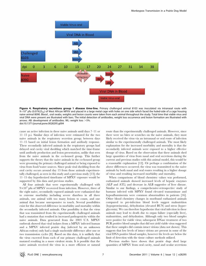
Group 1 (Animals #8103 and #8090). Primary challenged
animal #8103 developed lesions by day 13 p.i.; a maximum of 10
lesions was counted on this animal at the peak of infection. Weight
loss began on day 13 and reached a maximum of 10% on day 27
(Table 1). Orthopoxvirus antibodies were first detected on day 13
p.i. (Table 1). Oral samples from this animal yielded viral DNA on
Figure 1. Bedding/fomite group: detection of viral DNA and viable virus from oropharyngeal swabs and blood. Four primarychallenged animals were inoculated via intranasal route with 96103 pfu (0.07XLD50) of West African MPXV and placed into one of three experimentalgroups. For the bedding group, the primary challenged animal (blue bars) was housed for 16 days in a large trough, at which time the challengedanimal was removed and three naive animals (red/mauve bars) were placed into the trough for housing. Blood and oral swabs were taken from eachanimal throughout the study and subsequently tested for viral DNA and live virus. Days are plotted on the X axis in black for day p.i. of primarychallenged animal; in red for day post placement in trough. Samples yielding positive DNA (fg/ul) or viable virus (pfu/mL) are plotted on the Y axis ina log scale.doi:10.1371/journal.pone.0028295.g001
Monkeypox Transmission in a Prairie Dog Model
PLoS ONE | www.plosone.org 3 December 2011 | Volume 6 | Issue 12 | e28295
day three, viral shedding from this animal began on day six (Fig. 3,
Table 1). Viral DNA in blood from this animal was detected on
day six (Fig. 3, Table 1). This primary challenged animal (#8103)
survived infection. Animal #8090 was the naive animal housed
across from #8103. 20 days after MPXV challenge of #8103,
animal #8090 developed an eye infection. A swab of the ocular
surface yielded viral DNA and viable virus (data not shown). 24
days p.i. of the primary challenged animal, the naive animal
#8090 had five lesions on the abdomen and legs. This animal
developed a total of 10 lesions; weight loss for animal #8090
began on day 20 of the study and reached a maximum of 15% by
day 27 (Table 1) and detection of orthopoxvirus antibodies
occurred beginning on day 24 (Table 1). Viral DNA in the blood
from #8090 was detected beginning on day 17; shedding of viral
DNA and viable virus from oral cavity samples began on day 20
(Fig. 3, Table 1). Naive animal #8090 survived MPXV infection.
The disease course for these two animals (experimentally
challenged and secondarily infected) is illustrated in Figure 4
and shows that the secondarily infected animal’s disease onset is
delayed compared to the experimentally challenged animal, but
otherwise the disease progression is very similar.
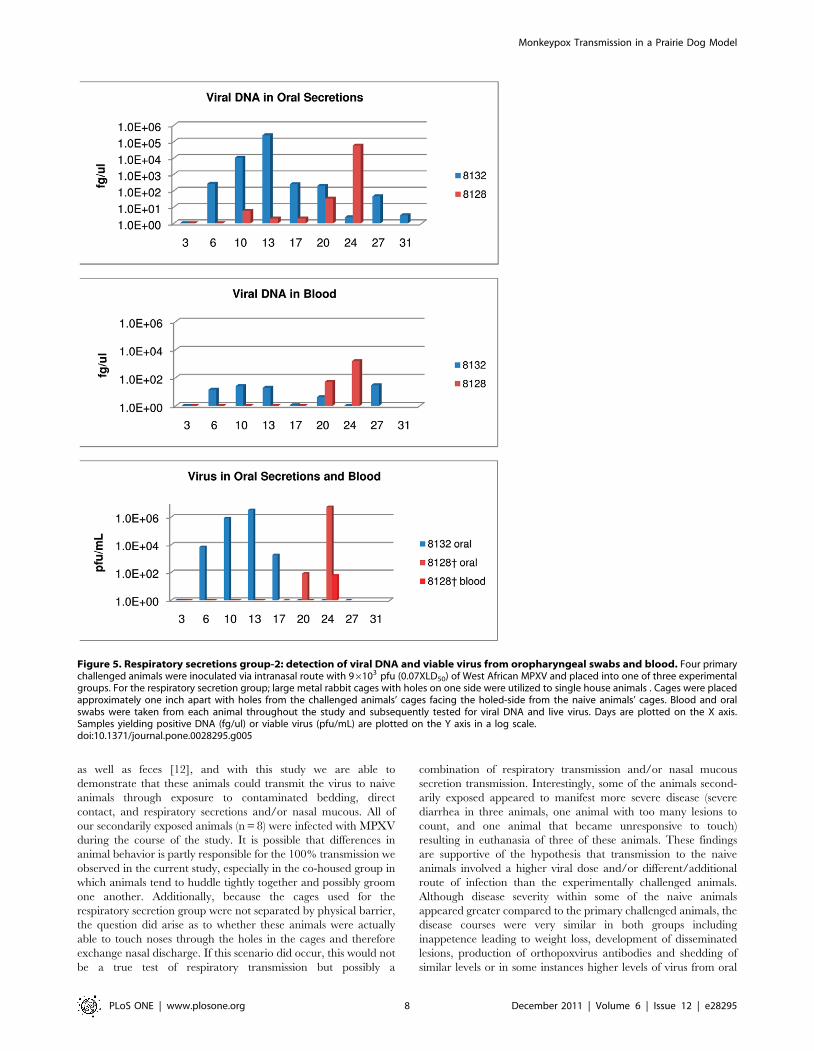
Group 2 (Animals #8132 and #8128). Primary challenged
animal #8132 was challenged with MPXV and developed nine
lesions by day 10 p.i. At the peak of infection, approximately 30
lesions developed on this animal. Weight loss began on day 17 and
reached 10% (Table 1). As was seen for the other three animals
experimentally challenged with MPXV during this study,
orthopoxvirus antibodies were detected on day 13 p.i. (Table 1).
Viral DNA in blood and oral samples, as well as viral shedding
from oral cavity samples occurred on day six (Fig. 5, Table 1). Also
as observed in the other transmission groups, the primary
challenged animal survived MPXV infection. Animal #8128
was the naive animal housed across from #8132. Viral DNA from
oral secretions was detected on day 10; however viral shedding
from oral cavity samples did not occur until day 20 as did
detection of viral DNA from blood samples (Fig. 5, Table 1)
Detectable levels of orthopoxvirus antibodies began on day 24
(Table 1); which was also observed with the naive animal (#9080)
in the other respiratory group. On day 24, #8128 had four lesions
and approximately 9% weight loss (Table 1); but because of the
extreme morbidity of this animal (unresponsive to touch and
diarrhea) it was humanely euthanized. Animal #8128’s day 24
blood sample (day of euthanasia) was one of the two blood samples
collected during this study that yielded viable virus, (Fig. 5).
Viral Load in Tissues and Blood Chemistry ValuesAs we have seen in previous studies, all animals that survived
MPXV infection in the current study had detectable levels of
MPXV DNA in some of the harvested tissues (Figs S1, S2),
without the presence of viable virus. The exception to this was a
lesion sample from primary challenged animal #8132 (respiratory
group 2) and four samples (tongue, brain, skin and lesion) from the
primary challenged animal #8134 (bedding group) which was
euthanized while still recovering from infection (Fig. S3); all of
which yielded viable virus via tissue culture methods. All three
moribund secondarily-infected animals that had to be euthanized
had high levels of virus in the majority of tissues tested, with
animal #8130 having viable virus present in 100% of those tissues
harvested. The tongue, spleen, skin, and lesion yielded viable virus
from all three of the euthanized animals. A liver sample was only
taken from two of the moribund secondarily infected animals
during the study, #8130 and #8128 and this tissue was positive
for live virus from both of these animals (Fig. S3).
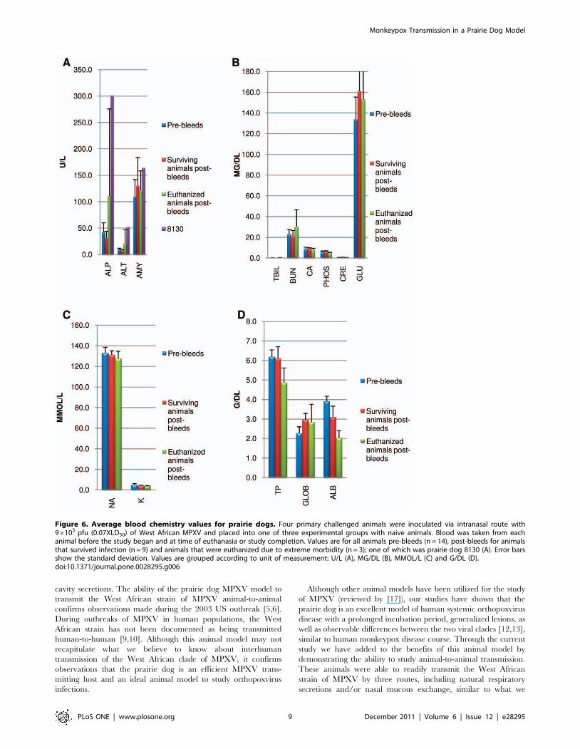
The values for blood chemistries were averaged for three
groups: pre-infection for all animals (n = 14); study end for infected
animals that survived (n = 9); and at euthanasia for moribund
animals (n = 3). When compared, hepatic enzyme values (ALP and
ALT) in the euthanized animals were elevated 2–3 times higher
than the average value prior to infection, indicating severe
hepatocellular necrosis and liver disease (Fig. 6a). In particular,
animal #8130 (bedding group naive), which manifest an
aggressive disease time course and had to be euthanized on day
15 had abnormal hepatic enzyme levels suggestive of severe liver
disease as well as increased blood amylase levels and decreased
albumin (ALB) levels indicating potential hepatobiliary disease
(Fig. 6a). Also suggestive of liver failure, average ALB values were
significantly decreased when comparing the pre-infection values to
post values for both surviving and euthanized animals (p = 0.0003
and 0.0045 respectively) (Fig. 6d). Glucose and globulin levels were
significantly elevated in surviving animals’ post bleed values
compared to the pre-infection averages (p = 0.0402 and 0.0004
respectively) (Fig. 6b, 6d). In euthanized animals’ blood samples,
glucose and globulin levels were also elevated; however no
statistical significance was seen when compared to pre-infected
Table 1. Disease/molecular findings in primary MPXV challenged prairie dogs (shaded) and secondarily infected prairie dogs.
Group Bedding Co-housed Respiratory Secretions
Animal Number 8134 8118 8130{ 8135 8127 8133{ 8136 8154 8103 8090 8132 8128{
Day of Lesion Onset 13 34(18)
27 (11) 34(18)
13 24 20 24 13 24 10 24
Maximum Weight Loss 10% 14% 12% 8% 9% 10% 1% 10% 10% 15% 10% 9%
Viral DNA in Blood(days)
6-.16 31–41(15–25)
24–31(8–15)
31–41(15–25)
6–24 17–27 17–31 17–27 6–24 17–34 6–27 20–24
Virus in Oral Secretions(days)
10-.16 31–41(15–25)
24–31(8–15)
27–41(11–25)
10–27 13–27 13–31 13–20 6–17 20–27 6–17 20–24
Day of Initial AntibodyDetection
13 38(22)
31 (15) 38(22)
13 27 24 24 13 24 13 24
Four primary challenged animals (highlighted in grey) were inoculated via intranasal route with 96103 pfu (0.07XLD50) of West African MPXV and placed into one ofthree experimental groups with naive animals. The time-line shown is for day post infection of the primary challenged animal; for the naive bedding group numbers inparentheses are days post placement in the trough. Two times a week animals were anesthetized for sample collection, lesion count and weight measurements. Threeof the naive animals (indicated by crosses) were euthanized due to extreme morbidity during the study.doi:10.1371/journal.pone.0028295.t001
Monkeypox Transmission in a Prairie Dog Model
PLoS ONE | www.plosone.org 4 December 2011 | Volume 6 | Issue 12 | e28295
values. A trend in elevated BUN levels was observed in euthanized
animals’ levels compared to pre-infection averages, suggestive of
dehydration, which was also clinically noted (Fig. 6b, 6d). No other
trends or significant differences were observed in the other blood
chemistry values.
Discussion
In the current study, all four animals experimentally challenged
with MPXV developed orthopoxvirus antibodies on day 13 p.i. In
the naive animals which were secondarily exposed, we used the
date of initial orthopoxvirus antibody production to estimate the
date of MPXV infection as a result of disease transmission.
Additionally, the four primary challenged animals developed
lesions between days 10–13, as did 3/4 challenged animals in a
previous study with a similar challenge dose [13]. Another study
which utilized a slightly higher inoculation dose, found animals
develop lesions at a similar time-frame regardless of the difference
in challenge inoculum (days 9–12) [12]. If it is assumed based on
antibody response of challenged animals that the development of
orthopoxvirus antibodies occurs 13 days after infection with
MPXV and that disseminated lesions develop 9–13 days after
MPXV infection, the three naive animals in the bedding group
were most likely infected with MPXV 0–2 days (#8130) or 5–9
(#s8135 and 8118) days after placement in the trough based on
antibody production and initial lesion presentation. Because of the
gap in estimated exposure days, it is possible that the first naive
animal that became infected subsequently infected the other two
secondarily infected animals in this group; but not likely given that
animals typically do not shed viable virus until 6–10 days after
Figure 2. Co-house group: detection of viral DNA and viable virus from oropharyngeal swabs and blood. Four primary challengedanimals were inoculated via intranasal route with 96103 pfu (0.07XLD50) of West African MPXV and placed into one of three experimental groups. Forthe co-housed group; primary challenged animal was challenged with virus and housed in a large trough with 3 naive animals. Blood and oral swabswere taken from each animal throughout the study and subsequently tested for viral DNA and live virus. Days are plotted on the X axis. Samplesyielding positive DNA (fg/ul) or viable virus (pfu/mL) are plotted on the Y axis in a log scale.doi:10.1371/journal.pone.0028295.g002
Monkeypox Transmission in a Prairie Dog Model
PLoS ONE | www.plosone.org 5 December 2011 | Volume 6 | Issue 12 | e28295
exposure. As well, measurable infectious virus was not found in
animal #8130’s oral secretions until day eight. The three naive
animals co-housed with an infected animal were most likely
infected 7–11 days (# 8136) and 11–15 days (#s8133 and 8154)
after the start of the study based on antibody production and lesion
presentation. It is noteworthy that samples from the co-housed
naive animals were positive for viable virus in oral secretions
earlier than detectable levels of viral DNA in blood samples, which
is not consistent with what is typically seen during an experimental
challenge with MPXV. Additionally, the oral samples from all
three naives were positive for viable MPXV as early as day 13,
which would suggest these animals were infected between days 3–7
(based on the observation that primary challenged animals begin
shedding from the oral cavity 6–10 days after initial infection).
However, the primary challenged animal in this group did not
begin shedding virus until day 10 p.i., making the likelihood of
infection between days 3–7 impossible. The most likely explana-
tion is that the naive animals were either grooming the primary
challenged animal or being exposed to the virus through the
shared food, water, or living areas. The grooming scenario is most
likely as infected animals tend to eat and drink infrequently when
they are sick and the co-housed animals were observed to huddle
tightly together. Based on antibody production and lesion
presentation, the level of virus exposure was not high enough to
Figure 3. Respiratory secretions group-1: detection of viral DNA and viable virus from oropharyngeal swabs and blood. Four primarychallenged animals were inoculated via intranasal route with 96103 pfu (0.07XLD50) of West African MPXV and placed into one of three experimentalgroups. For the respiratory secretion group; large metal rabbit cages with holes on one side were utilized to single house animals. Cages were placedapproximately one inch apart with holes from the challenged animals’ cages facing the holed-side from the naive animals’ cages. Blood and oralswabs were taken from each animal throughout the study and subsequently tested for viral DNA and live virus. Days are plotted on the X axis.Samples yielding positive DNA (fg/ul) or viable virus (pfu/mL) are plotted on the Y axis in a log scale.doi:10.1371/journal.pone.0028295.g003
Monkeypox Transmission in a Prairie Dog Model
PLoS ONE | www.plosone.org 6 December 2011 | Volume 6 | Issue 12 | e28295
cause an active infection in these naive animals until days 7–11 or
11–15 p.i. Similar days of infection were estimated for the two
naive animals in the respiratory secretion group; between days
11–15 based on initial lesion formation and antibody response.
These secondarily infected animals in the respiratory groups had
delayed oral cavity viral shedding which matched the time-frame
until antibody production and lesion presentation, unlike that seen
from the naive animals in the co-housed group. This further
supports the theory that the naive animals in the co-housed group
were grooming the primary challenged animal or being exposed to
virus from food/water sources. Since peak viral shedding from the
oral cavity occurs around day 13 from those animals experimen-
tally challenged, as seen in this study and a previous study [13]; the
11–15 day hypothesized timeframe of MPXV exposure would be
supported by this data and previous studies.
All four animals that were experimentally challenged with
96103 pfu of MPXV recovered from infection. However, three of
the eight naive, secondarily exposed animals were euthanized due
to extreme morbidity including severe diarrhea in all three
animals, one animal with too many lesions to count, and one
animal that became unresponsive to touch. Several possibilities
exist for this observed difference in morbidity and mortality within
the secondarily infected animals. One possibility is that the virus
that was transmitted from the experimentally challenged animals
had a mutation that resulted in increased pathogenicity within the
naive animals. Data generated from the 2003 U.S. MPXV
outbreak showed that viral isolates from a MPXV infected human
and a MPXV infected prairie dog (infected by an unknown
African rodent) only had a single nucleotide difference after one or
two transmission cycles [8]. Based on this data, it is unlikely that
the virus excreted from each of our primary challenged animals
mutated resulting in a more virulent strain. It is possible that the
naive animals received the virus in a more efficient or natural
route than the experimentally challenged animals. However, since
there were no bites or scratches on the naive animals, they most
likely received the virus via an intranasal or oral route of infection
similar to the experimentally challenged animals. The most likely
explanation for the increased morbidity and mortality is that the
secondarily infected animals were exposed to a higher effective
dosage of virus. Based on the observation that these animals shed
large quantities of virus from nasal and oral secretions during the
current and previous studies with this animal model, this would be
a reasonable explanation [12]. Or perhaps a combination of the
above differences occurred; the virus was transmitted to the naive
animals by both nasal and oral routes resulting in a higher dosage
of virus and resulting increased morbidity and mortality.
When comparisons of blood chemistry values was performed,
euthanized animals showed increased levels of hepatic enzymes
(ALP and ATL) and decrease in ALB suggestive of liver disease.
Similar to our findings, a comprehensive-retrospective study of
humans infected with MPXV found elevated transaminase and
hypoalbuminemia were common abnormalities in patients [16].
Other blood chemistry changes in moribund euthanized animals
compared to pre-infection blood levels suggest malnutrition
(hypoproteinemia), dehydration (elevated BUN) and stress (hyper-
glycemia). We can therefore hypothesize that viral infection in these
animals may lead to death due to organ failure (especially liver),
malnutrition, and dehydration. Although only two blood samples
were positive for viable virus; subsequent DNase treatment of two
PCR positive blood samples that did not yield viable virus suggested
that these samples did contain intact virions (data not shown). This
suggests that low levels of intact virions are present in some of the
viral DNA positive blood samples, however the levels are below the
limit of detection with the tissue culture methods utilized.
Previous studies have shown that prairie dogs shed large
quantities of MPXV from oral cavity, nasal and ocular secretions
Figure 4. Respiratory secretions group 1 disease time-line. Primary challenged animal 8103 was inoculated via intranasal route with96103 pfu (0.07XLD50) of West African MPXV and placed in a large metal cage with holes on one side which faced the holed-side of a cage housingnaive animal 8090. Blood , oral swabs, weights and lesion counts were taken from each animal throughout the study. Total time that viable virus andviral DNA were present are illustrated with bars. The initial detection of antibodies, weight loss occurrence and lesion formation are illustrated witharrows. AB: development of antibodies. WL: weight loss .5%.doi:10.1371/journal.pone.0028295.g004
Monkeypox Transmission in a Prairie Dog Model
PLoS ONE | www.plosone.org 7 December 2011 | Volume 6 | Issue 12 | e28295
as well as feces [12], and with this study we are able to
demonstrate that these animals could transmit the virus to naive
animals through exposure to contaminated bedding, direct
contact, and respiratory secretions and/or nasal mucous. All of
our secondarily exposed animals (n = 8) were infected with MPXV
during the course of the study. It is possible that differences in
animal behavior is partly responsible for the 100% transmission we
observed in the current study, especially in the co-housed group in
which animals tend to huddle tightly together and possibly groom
one another. Additionally, because the cages used for the
respiratory secretion group were not separated by physical barrier,
the question did arise as to whether these animals were actually
able to touch noses through the holes in the cages and therefore
exchange nasal discharge. If this scenario did occur, this would not
be a true test of respiratory transmission but possibly a
combination of respiratory transmission and/or nasal mucous
secretion transmission. Interestingly, some of the animals second-
arily exposed appeared to manifest more severe disease (severe
diarrhea in three animals, one animal with too many lesions to
count, and one animal that became unresponsive to touch)
resulting in euthanasia of three of these animals. These findings
are supportive of the hypothesis that transmission to the naive
animals involved a higher viral dose and/or different/additional
route of infection than the experimentally challenged animals.
Although disease severity within some of the naive animals
appeared greater compared to the primary challenged animals, the
disease courses were very similar in both groups including
inappetence leading to weight loss, development of disseminated
lesions, production of orthopoxvirus antibodies and shedding of
similar levels or in some instances higher levels of virus from oral
Figure 5. Respiratory secretions group-2: detection of viral DNA and viable virus from oropharyngeal swabs and blood. Four primarychallenged animals were inoculated via intranasal route with 96103 pfu (0.07XLD50) of West African MPXV and placed into one of three experimentalgroups. For the respiratory secretion group; large metal rabbit cages with holes on one side were utilized to single house animals . Cages were placedapproximately one inch apart with holes from the challenged animals’ cages facing the holed-side from the naive animals’ cages. Blood and oralswabs were taken from each animal throughout the study and subsequently tested for viral DNA and live virus. Days are plotted on the X axis.Samples yielding positive DNA (fg/ul) or viable virus (pfu/mL) are plotted on the Y axis in a log scale.doi:10.1371/journal.pone.0028295.g005
Monkeypox Transmission in a Prairie Dog Model
PLoS ONE | www.plosone.org 8 December 2011 | Volume 6 | Issue 12 | e28295
cavity secretions. The ability of the prairie dog MPXV model to
transmit the West African strain of MPXV animal-to-animal
confirms observations made during the 2003 US outbreak [5,6].
During outbreaks of MPXV in human populations, the West
African strain has not been documented as being transmitted
human-to-human [9,10]. Although this animal model may not
recapitulate what we believe to know about interhuman
transmission of the West African clade of MPXV, it confirms
observations that the prairie dog is an efficient MPXV trans-
mitting host and an ideal animal model to study orthopoxvirus
infections.
Although other animal models have been utilized for the study
of MPXV (reviewed by [17]), our studies have shown that the
prairie dog is an excellent model of human systemic orthopoxvirus
disease with a prolonged incubation period, generalized lesions, as
well as observable differences between the two viral clades [12,13],
similar to human monkeypox disease course. Through the current
study we have added to the benefits of this animal model by
demonstrating the ability to study animal-to-animal transmission.
These animals were able to readily transmit the West African
strain of MPXV by three routes, including natural respiratory
secretions and/or nasal mucous exchange, similar to what we
Figure 6. Average blood chemistry values for prairie dogs. Four primary challenged animals were inoculated via intranasal route with96103 pfu (0.07XLD50) of West African MPXV and placed into one of three experimental groups with naive animals. Blood was taken from eachanimal before the study began and at time of euthanasia or study completion. Values are for all animals pre-bleeds (n = 14), post-bleeds for animalsthat survived infection (n = 9) and animals that were euthanized due to extreme morbidity (n = 3); one of which was prairie dog 8130 (A). Error barsshow the standard deviation. Values are grouped according to unit of measurement: U/L (A), MG/DL (B), MMOL/L (C) and G/DL (D).doi:10.1371/journal.pone.0028295.g006
Monkeypox Transmission in a Prairie Dog Model
PLoS ONE | www.plosone.org 9 December 2011 | Volume 6 | Issue 12 | e28295
believe to occur in human-to-human MPXV and variola virus
transmission. We can use these studies to extrapolate to possible
human exposure and make recommendation for healthcare
workers and those people who live in areas where MPXV is
endemic: fomite exposure is similar to healthcare workers handling
infected bedding or bandages from sick patients; direct contact is
similar to families living in a house together; and respiratory
secretion groups mimic possible human exposure to MPXV from
a sneeze or cough from an infected individual in close proximity.
Future studies with this model could be very useful in evaluating
infection control measures during outbreaks, testing of anti-viral
therapies to diminish transmission rates, as well as helping to
understand differences in rates of transmission between the two
strains of MPXV within human populations.
Materials and Methods
Ethics statementAll animals were handled in strict accordance with good animal
practice as defined by the relevant national and/or local animal
welfare bodies, and all animal work was approved by the CDC
Institutional Animal Care and Use Committee (IACUC) under
an approved protocol (2115-211DAMPRAC) issued by CDC
IACUC specifically for this study.
AnimalsWild-caught, juvenile black-tailed prairie dogs (Cynomys ludovi-
cianus) were obtained from Berthoud Colorado. The animals
involved were purchased from a vendor which utilized humane
live-trapping techniques (wire cage traps) to capture free-living
healthy young animals (,1year old). Only animals free from any
signs of illness (as determined by a veterinarian) were transported
to the Centers for Disease Control and Prevention (CDC) in
Atlanta, GA. Once the animals arrived at the CDC, they were
quarantined and housed appropriately under an approved CDC
IACUC protocol (1718CARPRAC) until the start of the study. At
time of the start of this study animals were approximately 20
months old and had been prescreened by a veterinarian and
determined to be in good health status and found negative for the
presence of anti-orthopoxvirus antibodies. 14 animals were used in
this study. The average starting weight for primary challenged
animals was 932 grams (range 904–982), and the average for naive
animals was 994 grams (range 584–1265). A sterile PIT tag was
injected subcutaneously at the base of the neck for animal
identification and non-invasive recording of body temperature.
Transmission strategiesAll animals were housed in an animal Biological Safety Level-3
(ABSL-3) animal room. Animals were divided into three ex-
perimental groups to evaluate routes of transmission: 1) bedding/
fomite, 2) co-housed, or 3) respiratory. For the bedding/fomite
group, one animal was challenged with virus and housed for 16
days in a large trough (approximately 2.5 feet wide by 5 feet long)
with a dust-free moisture absorbent pellet made from recycled
newspapers (Paperchip regular texture pellets, Shepherd Specialty
Papers). After day six the bedding was not changed due to findings
from previous studies in which shedding from excrement most
often began on day six [12,13]. Previous prairie dog MPXV
studies have demonstrated that peak viral shedding occurs
between days 9–13. Therefore, the challenged animal was left in
the trough until day 16 at which time it was removed and
humanely euthanized. Three naive animals were placed into the
large trough for housing with the contaminated bedding and this
bedding was not changed for 39 days. For the co-housed group,
one animal was challenged with virus and housed in a large trough
(approximately 2.5 feet wide by 5 feet long) with 3 naive animals
for 38 days; bedding was changed weekly. Finally, for the
respiratory secretion group; four large metal rabbit cages with
holes on one side (3 sq. feet cage area; 16.4380 high619.1250
wide625.8750 long) were utilized to individually house four
animals (two challenged, 2 naive). Cages were placed approxi-
mately one inch apart with ventilation holes (1 inch in diameter)
from the challenged animals’ cages facing the holed-side from the
naive animals’ cages within a Duo-Flow biosafety cabinet with
negative directional airflow, in a similar design as illustrated in
another study [18]. Primary challenged animals were monitored
for the subsequent 31 days while the naive animals were
monitored for 38 days. Additionally, two phosphate-buffered
saline (PBS) infected negative control animals were housed
individually in large (12.130623.3806209.000) rat cages with
aerosol filter tops for 38 days. PBS animals, bedding/fomite group
animals, and the respiratory secretion grouped animals were kept
in two Duo-Flow biosafety cabinets with negative directional
airflow to allow air to be HEPA filtered before leaving the unit.
The trough containing the co-housed group of animals was in the
ABSL-3 animal room. Animals were cared for in accordance with
CDC Institutional Animal Care and Use Committee (IACUC)
guidelines under an approved protocol (2115-211DAMPRAC).
In addition to prairie dog chow and water, animals were pro-
vided with monkey biscuits and mixed nuts for added dietary
enrichment.
VirusesThe West African MPXV strain (MPXV-USA-2003-044),
isolated during the 2003 U.S. outbreak [6,8], [DQ011153], was
used in this study. The virus underwent two passages in African
green monkey kidney cells (BSC-40) prior to seed pool production;
preparations used for animal challenge inoculums were purified
via a sucrose cushion.
Animal inoculationThe challenge dose (96103 pfu) was calculated based on the
morbidity and mortality rates observed in the authors’ previous
studies. Briefly, a challenge dose of 66103 pfu resulted in 25%
mortality and disease morbidity included rash lesions and viral
shedding identified in oral cavity samples in 100% of animals
[12,13]. Stocks of virus were diluted in PBS. Inocula titers were
immediately re-confirmed, post- challenge, by standard plaque
assay (as described below). Animals were infected via an intranasal
(i.n.) route of inoculation while under general anesthesia using 3–
5% isoflurane administered through a veterinary vaporizer.
Animals were inoculated with a total volume of 10 ul i.n. (5 ul
in each nostril). Additionally, 2 animals were mock infected with
PBS.
Observations and samplingPost-inoculation, individual animals were observed daily for
signs of morbidity, or malaise (inappetence, decreased activity,
recumbancy with reluctance to move, etc.) and clinical lesions or
rash for the study duration. Two times a week animals were
anesthetized with isoflurane for sampling purposes. Samples and
measurements collected included an oral swab (area swabbed
included inner cheeks, tongue and the hard palate); blood, weight,
temperature and lesion count (if applicable). Strict euthanasia
criteria were adhered to throughout the study as follows: any
animal that became unresponsive to touch, lost 25% or more
starting body weight, or accrued a total score of 10 on the
following scale was humanely euthanized: decreased activity
Monkeypox Transmission in a Prairie Dog Model
PLoS ONE | www.plosone.org 10 December 2011 | Volume 6 | Issue 12 | e28295
(2 points); lethargy, unsteady gait, inappetence (3 points each);
labored breathing and recumbency (5 points each).
Necropsy and tissue specimen collectionNecropsies on all animals were performed according to CDC
IACUC-approved standards in an ABSL-3 laboratory and
utilizing full ABSL-3 PPE. Samples taken during necropsy
included: submandibular lymph nodes/salivary glands, spleen,
lung, liver (for two animals), tongue, skin, lesion (if present), brain,
oral swab, and blood. Instruments were decontaminated with 5%
Microchem and 70% ethanol and allowed to dry between
collections of each tissue. Tissue samples were frozen at 270uCprior to further processing. Oral samples were collected with sterile
individual swabs and stored frozen without diluent. After animals
were sampled each day, serum was immediately separated from
whole blood and stored at 220uC to be processed for serology and
clinical chemistry levels (see below). Tissues and samples were
subsequently processed and further prepared for DNA analysis,
and virus isolation (see below).
Sample preparation for PCR and viral growthSample processing was performed under BSL-2 conditions with
BSL-3 work practices. For whole blood samples, 100 ul of EDTA
treated blood was used for DNA extraction and the remaining
untreated blood was used for tissue culture propagation. 400 ul of
sterile PBS was added to each swab collected. The swab extraction
tube systems (SETS) (Roche) protocol was used to recover sample
from the swab. DNA was extracted from 100 ul of the swab lysate.
The remaining swab eluate was used for virus isolation. For tissue
preparation, 1 ml aliquots of PBS and SPEX bead (SPEX Sample
Prep) were prepared. The PBS/bead aliquot was then poured into
a 1 ml tube containing the individually weighed tissue sample. The
GenoGrinder 2000 (SPEX Sample Prep) was then used following
the manufacturer’s instructions to create a tissue homogenate.
100 ul of the homogenate was then used for DNA extraction. The
remaining homogenate was used for virus isolation. The BioRobot
EZ-1 Workstation (Qiagen) was used for DNA extraction from all
blood, swab and tissue samples using the Tissue Kit. Samples were
incubated at 55uC in lysis buffer for an hour to inactivate viable
virus particles prior to DNA extraction.
Real-time PCR analysisSamples were tested by real-time PCR using forward and
reverse primers and probes complimentary to the conserved
Orthopoxvirus (OPXV) E9L (DNA polymerase) gene [19]. MPXV
DNA (10 fg–1 ng) was used as positive controls. A positive sample
produced CT values (in duplicate) of 37 or below. A weakly
positive sample displayed CT values 38–39 (duplicates).
To determine whether a subset of PCR positive EDTA blood
samples contained intact virions, a DNase assay (Li et al. personal
communication) was used. In brief, the assay allows for the
determination of how much of the viral DNA was protected from
DNase treatment as a measure of how much of the DNA was
incorporated within intact virions.
Virus-tissue infectivityAll samples were stored at 270uC until virus isolation was
attempted. Previous analyses demonstrated that real-time PCR
detection of MPXV DNA is an assay which can detect trace
amounts of MPXV DNA in samples which do not contain viable
virus [5]. Therefore, specimens were first tested for presence of
OPXV DNA by PCR and, if positive, were subsequently evaluated
for viable virus by tissue culture propagation. Each positive sample
was titrated in duplicate using 10 fold dilutions of swab eluate,
whole blood or tissue slurry on BSC-40 cell monolayers, incubated
at 35.5uC and 6% CO2 for 72 hours, and subsequently stained
with crystal violet and 10% formalin to visualize plaques. Titers
were expressed as pfu per milliliter (pfu/ml) of blood or swab
eluate; or pfu per gram (pfu/g) of tissues.
Serologic analysisSerum was separated from whole blood and transferred to a
clean tube and stored at 220C prior to analysis. A modified
ELISA was used for analysis of anti-OPXV immunoglobulin types
A and G in separated serum as previously described in detail [12].
Blood chemistry valuesSerum was separated from whole blood and transferred to a
clean tube and stored at 220C prior to analysis. The Piccolo
blood chemistry analyzer (Abaxis) was utilized to determine the
following blood chemistry profiles: sodium (NA), potassium (K),
phosphorus (PHOS), glucose (GLU), calcium (CA), blood urea
nitrogen (BUN), creatinine (CRE), alkaline phosphatase (ALP),
alanine aminotransferase (ALT), amylase (AMY), total bilirubin
(TBIL), albumin (ALB), total protein (TP) and globulin (GLOB).
Statistical analysesA paired t-test was utilized to compare chemistry values and a
p-value of #0.05 was considered statistically significant.
Supporting Information
Figure S1 Detection of viral DNA within necropsysamples harvested from the bedding group and co-housed group of animals. Four primary challenged animals
were inoculated via intranasal route with 96103 pfu (0.07XLD50)
of West African MPXV and placed into one of three experimental
groups with naive animals. Animals that were euthanized due to
extreme morbidity are indicated by crosses. At time of death or at
study completion, full necropsies were completed and tissues were
tested for viral DNA (fg/ul) and viable virus (pfu/g) in a log scale.
Results for animals within the bedding group (A) and co-housed
group (B) are shown.
(TIF)
Figure S2 Detection of viral DNA within necropsysamples harvested from the respiratory groups ofanimals. Four primary challenged animals were inoculated via
intranasal route with 96103 pfu (0.07XLD50) of West African
MPXV and placed into one of three experimental groups with
naive animals. Animals that were euthanized due to extreme
morbidity are indicated by crosses. At time of death or at study
completion, full necropsies were completed and tissues were tested
for viral DNA (fg/ul) and viable virus (pfu/g) in a log scale. Results
for animals within respiratory group 1 (A) and respiratory group 2
(B) are shown.
(TIF)
Figure S3 Levels of viable virus within necropsy tissues.Four primary challenged animals were inoculated via intranasal
route with 96103 pfu (0.07XLD50) of West African MPXV and
placed into one of three experimental groups with naive animals.
Three of the naive animals (indicated by crosses) were euthanized
due to extreme morbidity during the study. At time of death or at
study completion, full necropsies were completed and tissues were
tested for viral DNA (fg/ul) and viable virus (pfu/g) on a log scale.
Only those animals testing positive for viable virus are shown.
(TIF)
Monkeypox Transmission in a Prairie Dog Model
PLoS ONE | www.plosone.org 11 December 2011 | Volume 6 | Issue 12 | e28295
Acknowledgments
The authors would like to thank Erin McDowell for technical support for
immune assays.
Disclaimer: The findings and conclusions in this report are those of
the author(s) and do not necessarily represent the official position of the
Centers for Disease Control and Prevention.
Author Contributions
Conceived and designed the experiments: CLH DSC NG-R VAO KLK
IKD. Performed the experiments: CLH DSC NG-R SW CC JSS JA.
Analyzed the data: CLH SW CC CMH VAO KLK IKD. Wrote the
paper: CLH DSC NG-R SW CC CMH JSS VAO KLK IKD.
References
1. Breman JG, Nakano JH, Coffi E, Godfrey H, Gautun JC (1977) Human
poxvirus disease after smallpox eradication. Am J Trop Med Hyg 26: 273–281.2. Hutin YJ, Williams RJ, Malfait P, Pebody R, Loparev VN, et al. (2001)
Outbreak of human monkeypox, Democratic Republic of Congo, 1996 to 1997.Emerg Infect Dis 7: 434–438.
3. Khodakevich L, Szczeniowski M, Manbu mD, Jezek Z, Marennikova S, et al.
(1987) The role of squirrels in sustaining monkeypox virus transmission. TropGeogr Med 39: 115–122.
4. Centers for Disease Control and Prevention (2003) Update: multistate outbreakof monkeypox–Illinois, Indiana, Kansas, Missouri, Ohio, and Wisconsin, 2003.
MMWR Morb Mortal Wkly Rep 52: 561–564.
5. Hutson CL, Lee KN, Abel J, Carroll DS, Montgomery JM, et al. (2007)Monkeypox zoonotic associations: insights from laboratory evaluation of animals
associated with the multi-state US outbreak. Am J Trop Med Hyg 76: 757–768.6. Reed KD, Melski JW, Graham MB, Regnery RL, Sotir MJ, et al. (2004) The
detection of monkeypox in humans in the Western Hemisphere. N Engl J Med
350: 342–350.7. Chen N, Li G, Liszewski MK, Atkinson JP, Jahrling PB, et al. (2005) Virulence
differences between monkeypox virus isolates from West Africa and the Congobasin. Virology 340: 46–63.
8. Likos AM, Sammons SA, Olson VA, Frace AM, Li Y, et al. (2005) A tale of twoclades: monkeypox viruses. J Gen Virol 86: 2661–2672.
9. Breman JG, Kalisa R, Steniowski MV, Zanotto E, Gromyko AI, et al. (1980)
Human monkeypox, 1970–79. Bull World Health Organ 58: 165–182.10. Foster SO, Brink EW, Hutchins DL, Pifer JM, Lourie B, et al. (1972) Human
monkeypox. Bull World Health Organ 46: 569–576.
11. Learned LA, Reynolds MG, Wassa DW, Li Y, Olson VA, et al. (2005) Extended
interhuman transmission of monkeypox in a hospital community in the Republic
of the Congo, 2003. Am J Trop Med Hyg 73: 428–434.
12. Hutson CL, Olson VA, Carroll DS, Abel JA, Hughes CM, et al. (2009) A prairie
dog animal model of systemic orthopoxvirus disease using West African and
Congo Basin strains of monkeypox virus. J Gen Virol 90: 323–333.
13. Hutson CL, Carroll DS, Self J, Weiss S, Hughes CM, et al. (2010) Dosage
comparison of Congo Basin and West African strains of monkeypox virus using a
prairie dog animal model of systemic orthopoxvirus disease. Virology 402:
72–82.
14. Heberling RL, Kalter SS (1971) Induction, course, and transmissibility of
monkeypox in the baboon (Papio cynocephalus). J Infect Dis 124: 33–38.
15. Marennikova SS, Shchelkunov SN (2005) Laboratory Diagnostics of Human
Orthopoxvirus Infections. In: Orthopoxviruses Pathogenic for Humans. New
York, NY: Springer. pp 303–324.
16. Huhn GD, Bauer AM, Yorita K, Graham MB, Sejvar J, et al. (2005) Clinical
characteristics of human monkeypox, and risk factors for severe disease. Clin
Infect Dis 41: 1742–1751.
17. Hutson CL, Damon IK (2010) Monkeypox Virus Infections in Small Animal
Models for Evaluation of Anti-Poxvirus Agents. Viruses 2: 2763–2776.
18. Sorrell EM, Wan H, Araya Y, Song H, Perez DR (2009) Minimal molecular
constraints for respiratory droplet transmission of an avian-human H9N2
influenza A virus. Proc Natl Acad Sci U S A 106: 7565–7570.
19. Li Y, Olson VA, Laue T, Laker MT, Damon IK (2006) Detection of monkeypox
virus with real-time PCR assays. J Clin Virol 36: 194–203.
Monkeypox Transmission in a Prairie Dog Model
PLoS ONE | www.plosone.org 12 December 2011 | Volume 6 | Issue 12 | e28295