The gender-specific face aftereffect is based in retinotopic not spatiotopic coordinates across...
17
The gender-specific face aftereffect is based in retinotopic not spatiotopic coordinates across several natural image transformations Department of Psychology, Harvard University, Cambridge, MA, USA Arash Afraz Department of Psychology, Harvard University, Cambridge, MA, USA,& Laboratoire Psychologie de la Perception, Université Paris Descartes, Paris, France Patrick Cavanagh In four experiments, we measured the gender-specific face-aftereffect following subject’s eye movement, head rotation, or head movement toward the display and following movement of the adapting stimulus itself to a new test location. In all experiments, the face aftereffect was strongest at the retinal position, orientation, and size of the adaptor. There was no advantage for the spatiotopic location in any experiment nor was there an advantage for the location newly occupied by the adapting face after it moved in the final experiment. Nevertheless, the aftereffect showed a broad gradient of transfer across location, orientation and size that, although centered on the retinotopic values of the adapting stimulus, covered ranges far exceeding the tuning bandwidths of neurons in early visual cortices. These results are consistent with a high-level site of adaptation (e.g. FFA) where units of face analysis have modest coverage of visual field, centered in retinotopic coordinates, but relatively broad tolerance for variations in size and orientation. Keywords: eye movements, face recognition, object recognition, spatial vision Citation: Afraz, A., & Cavanagh, P. (2009). The gender-specific face aftereffect is based in retinotopic not spatiotopic coordinates across several natural image transformations. Journal of Vision, 9(10):10, 1–17, http://journalofvision.org/9/10/10/, doi:10.1167/9.10.10. Introduction David Marr (Marr, 1982) characterized vision as “knowing what is where.” For much of the visual cortex, “knowing where” appears to be based in a retinotopic coordinate system. With each eye, head and body move- ment, the image of the world shifts on the retina and across the retinotopic cortical areas. Despite these displacements, our perception of the world is for the most part reassuringly stationary and stable. Evidently, these displacements in retinal coordinates are corrected at some higher level to generate world-based coordinates. Whether these coordi- nates are explicitly represented in a spatiotopic map (d’Avossa, Shulman, Snyder, & Corbetta, 2006; Melcher & Morrone, 2003) or are only derived for individual objects when needed (Colby, 1998; Klier & Angelaki, 2008; Wolbers, Hegarty, Buchel, & Loomis, 2008) is a topic of intense debate. Clearly, a retinotopic map can be made to track object locations in the world if retinotopic target positions are shifted to compensate for eye movements (Colby & Duhamel, 1996; Goldberg & Colby, 1992; Wurtz, 2008). One approach to examine the coordinate frame of visual coding is to test for aftereffects of adaptation when an eye movement intervenes between adaptation and test. There have been reports of spatiotopic aftereffectsVthe presence of an aftereffect at the same spatial location as the adaptation but different retinal locationVfor tilt, shape and face adaptation (Melcher, 2005) as well as for motion (Ezzati, Golzar, & Afraz, 2008). As pointed out by Melcher (2005), faces are among the stimuli thought to require the highest level of visual analysis (Afraz, Kiani, & Esteky, 2006; Kanwisher & Yovel, 2006; Leopold, O’Toole, Vetter, & Blanz, 2001; Leopold, Rhodes, Muller, & Jeffery, 2005; Moradi, Koch, & Shimojo, 2005) and therefore more likely to tap spatiotopic representations. For that reason, we focus here on several types of retinotopic versus spatio- topic coordinate frames (head centered and body centered) for the face aftereffect (FAE). There are a number of different face aftereffects in the literature (identity, gender, race and general shape distortion, etc). We chose the gender-specific aftereffect as it is well documented (Ng, Boynton, & Fine, 2008; Rhodes et al., 2004; Webster, Kaping, Mizokami, & Duhamel, 2004) (also (Melcher, 2005)Vgender adapt but identity test) and more robust in the periphery where we must test in order to compare retinotopy versus translational spatiotopy. Martelli et al, for example, has shown that face identification drops dramatically once fixation is outside the face itself (for frontal face views) (Martelli, Majaj, & Pelli, 2005). Journal of Vision (2009) 9(10):10, 1–17 http://journalofvision.org/9/10/10/ 1 doi: 10.1167/9.10.10 Received March 30, 2009; published September 10, 2009 ISSN 1534-7362 * ARVO
-
Upload
independent -
Category
Documents
-
view
1 -
download
0
Transcript of The gender-specific face aftereffect is based in retinotopic not spatiotopic coordinates across...
The gender-specific face aftereffect is basedin retinotopic not spatiotopic coordinatesacross several natural image transformations
Department of Psychology, Harvard University,Cambridge, MA, USAArash Afraz
Department of Psychology, Harvard University,Cambridge, MA, USA, &
Laboratoire Psychologie de la Perception,Université Paris Descartes, Paris, FrancePatrick Cavanagh
In four experiments, we measured the gender-specific face-aftereffect following subject’s eye movement, head rotation, orhead movement toward the display and following movement of the adapting stimulus itself to a new test location. In allexperiments, the face aftereffect was strongest at the retinal position, orientation, and size of the adaptor. There was noadvantage for the spatiotopic location in any experiment nor was there an advantage for the location newly occupied by theadapting face after it moved in the final experiment. Nevertheless, the aftereffect showed a broad gradient of transfer acrosslocation, orientation and size that, although centered on the retinotopic values of the adapting stimulus, covered ranges farexceeding the tuning bandwidths of neurons in early visual cortices. These results are consistent with a high-level site ofadaptation (e.g. FFA) where units of face analysis have modest coverage of visual field, centered in retinotopic coordinates,but relatively broad tolerance for variations in size and orientation.
Keywords: eye movements, face recognition, object recognition, spatial vision
Citation: Afraz, A., & Cavanagh, P. (2009). The gender-specific face aftereffect is based in retinotopic not spatiotopiccoordinates across several natural image transformations. Journal of Vision, 9(10):10, 1–17,http://journalofvision.org/9/10/10/, doi:10.1167/9.10.10.
Introduction
David Marr (Marr, 1982) characterized vision as“knowing what is where.” For much of the visual cortex,“knowing where” appears to be based in a retinotopiccoordinate system. With each eye, head and body move-ment, the image of the world shifts on the retina and acrossthe retinotopic cortical areas. Despite these displacements,our perception of the world is for the most part reassuringlystationary and stable. Evidently, these displacements inretinal coordinates are corrected at some higher level togenerate world-based coordinates. Whether these coordi-nates are explicitly represented in a spatiotopic map(d’Avossa, Shulman, Snyder, & Corbetta, 2006; Melcher& Morrone, 2003) or are only derived for individual objectswhen needed (Colby, 1998; Klier & Angelaki, 2008;Wolbers, Hegarty, Buchel, & Loomis, 2008) is a topic ofintense debate. Clearly, a retinotopic map can be made totrack object locations in the world if retinotopic targetpositions are shifted to compensate for eye movements(Colby & Duhamel, 1996; Goldberg & Colby, 1992; Wurtz,2008). One approach to examine the coordinate frame ofvisual coding is to test for aftereffects of adaptation whenan eye movement intervenes between adaptation and test.
There have been reports of spatiotopic aftereffectsVthepresence of an aftereffect at the same spatial location as theadaptation but different retinal locationVfor tilt, shape andface adaptation (Melcher, 2005) as well as for motion(Ezzati, Golzar, & Afraz, 2008). As pointed out by Melcher(2005), faces are among the stimuli thought to require thehighest level of visual analysis (Afraz, Kiani, & Esteky,2006; Kanwisher & Yovel, 2006; Leopold, O’Toole, Vetter,& Blanz, 2001; Leopold, Rhodes, Muller, & Jeffery, 2005;Moradi, Koch, & Shimojo, 2005) and therefore more likelyto tap spatiotopic representations. For that reason, wefocus here on several types of retinotopic versus spatio-topic coordinate frames (head centered and body centered)for the face aftereffect (FAE). There are a number ofdifferent face aftereffects in the literature (identity, gender,race and general shape distortion, etc). We chose thegender-specific aftereffect as it is well documented(Ng, Boynton, & Fine, 2008; Rhodes et al., 2004; Webster,Kaping, Mizokami, & Duhamel, 2004) (also (Melcher,2005)Vgender adapt but identity test) and more robust inthe periphery where we must test in order to compareretinotopy versus translational spatiotopy. Martelli et al,for example, has shown that face identification dropsdramatically once fixation is outside the face itself (forfrontal face views) (Martelli, Majaj, & Pelli, 2005).
Journal of Vision (2009) 9(10):10, 1–17 http://journalofvision.org/9/10/10/ 1
doi: 10 .1167 /9 .10 .10 Received March 30, 2009; published September 10, 2009 ISSN 1534-7362 * ARVO

The current study contains four experiments, investigatingvarious types of retinotopy and spatiotopy for the FAE. Thefirst experiment studies FAE across eye movements. This issimilar to Melcher’s (Melcher, 2005) original study with asimpler design. The second and third experiments of thecurrent study are concerned with FAE across head move-ments. Following a translatory head movement, a comple-mentary eye movement can maintain the position of atarget on the retina (Kirschfeld, 1997) but no such correctionis possible following a large head tilt. Nevertheless, despitethe rotation of the image on the retina, our impression isstill one of a stable world. If there is a spatiotopicrepresentation, it needs to deal with rotations caused byhead tilts as well as simple displacements caused by eyemovements or translatory head movements. The secondexperiment examines this possibility, comparing the size ofthe FAE for test stimuli that match the adapting orientationin space versus stimuli with orientations matched on theretina. The retinal size of the visual objects can also changewithout any physical change in the actual size of objects asthe head moves toward or away from them. No eyemovements can correct for this transformation either. Thethird experiment contrasts the transfer of FAE acrossdifferent retinal sizes for two conditions; when the changein retinal size is induced by head movement and when thesize change is the result of an actual size change in thephysical stimulus. Again, we compare the size of the FAEfor test stimuli that match the adapting size in space versusstimuli with sizes matched on the retina.A critical goal of the visual system is to encode where
objects are. In this regard, a spatiotopic representation thatexplicitly codes object location in world coordinatesmight be only a special case. A more general solution isto keep the track of selected objects and their properties,including location, as they move (Gordon & Irwin, 1996;Kahneman, Treisman, & Gibbs, 1992). One paper hasreported that the tilt aftereffect can be found at the endlocation of an object’s trajectory when that object (thathas an oriented texture) shifts position between adaptationand test (Melcher, 2008). In the paper, Melcher suggeststhat the spatiotopic aftereffects that he has reported are theconsequence of object-specific effects that rely on theattentive tracking of visual objects. The fourth experimentinvestigates the question of a FAE that follows theadapting object as it shifts to new locations.
Experiment 1
Introduction
In the first experiment the spatial arrangement of theFAE is examined across eye movements. The main goalof this experiment is to examine the extent of retinotopotyversus spatiotopy of the FAE.
MethodsSubjects and procedure
Subjects were trained to identify the gender of faces thatwere chosen randomly from a morphing spectrumbetween male and female prototypes generated by acomputer graphics program (FaceGen) based on 3D scansof real faces. The face stimuli were made with SingularInversion’s FaceGen which produces facial prototypesbased on 3D scans of numerous faces (similar to themethods used by O’Toole AJ et al. (O’Toole, Vetter, &Blanz, 1999)). The facial structure varied across morphinglevel but the identical texture was mapped onto thestructure for all faces. Experimental sessions started aftersubjects reached 85% performance level on the genderidentification task. The initial training task included thewhole range of morphing values to familiarize subjectswith the main task. Subjects were given feedback for theircorrect and incorrect key presses at this stage. They couldnever reach 100% performance because there weredifficult (near average face) stimuli in the set as well asfaces with strong gender signal (far from the average).Experiments were conducted in a dim lit room with the
subjects’ head resting on a chin and forehead rest 57 cmaway from the screen. Stimulus presentation procedureswere controlled by a PC processor using MATLABpsychtoolbox (version 2.54) and displayed on a 60 Hz17 in. monitor. Face stimuli used for the test phasespanned seven levels of morphing (including the averageface and four levels in each direction) between 75% maleand 75% female prototypes. The female prototype (100%female face) was used as the adapting stimulus. The sizeof the face stimuli was 6- of visual angle (the average ofthe vertical and horizontal diameters of the face) and theywere displayed on a uniform gray background.Five subjects including one of the authors participated
in Experiment 1. All subjects had normal or corrected-to-normal vision. There were four conditions in this experi-ment: spatiotopic test, control test (test was at neither thespatiotopic nor retinotopic location), retinotopic test andnon-adapted test. In the first two conditions (spatiotopicand control) a small red fixation target appeared randomlyat one of the two left and right lateral positions on themonitor, 10- away from the center of the display. Subjectswere instructed to keep their gaze on the red targetthroughout the trial. Then the female prototype face waspresented for 5 adaptation seconds at the lateral fixationspot. Then the adapting face disappeared and the fixationtarget moved to the center of the display (subjects had tofollow that with their gaze). After 800 ms, the teststimulus was presented for 100 ms at either the samespatial location as the adaptor (spatiotopic test) or theopposite location on the screen (control location test)(Figure 1). The fixation point turned green then andsubjects had to report the gender of the test face bypressing one of the two keyboard buttons. In theretinotopic adaptation condition, subjects maintained their
Journal of Vision (2009) 9(10):10, 1–17 Afraz & Cavanagh 2
fixation on a central fixation point during the trial whilethe adapting face (the same as the other two conditions)was presented for 5 seconds at one of the lateral positionsrandomly. The test stimulus was presented at the samelocation following a 800 ms blank period. The non-adapted condition was the same as the retinotopiccondition with the exception that there was no adaptingstimulus. Eye-movement conditions (spatiotopic and con-trol) were blocked together and no-eye-movement con-ditions (retinotopic and non-retinotopic) were run inseparate blocks. The order of trial types within each blockwas randomized. There was a mandatory resting period ofat least 2 minutes after each 28 trials. 280 trials werecollected from each of the subjects. Eye movements weremonitored at 250 Hz from the right eye using an EyeLink Iinfrared eye tracker (SR research).
Data analysis
The behavioral performance was plotted separately foreach of the four conditions (for each subject) as theproportion of ‘female’ choices against the stimulus whichvaried from male to female (Figure 2). Data points werefitted with the following logistic function to calculate the
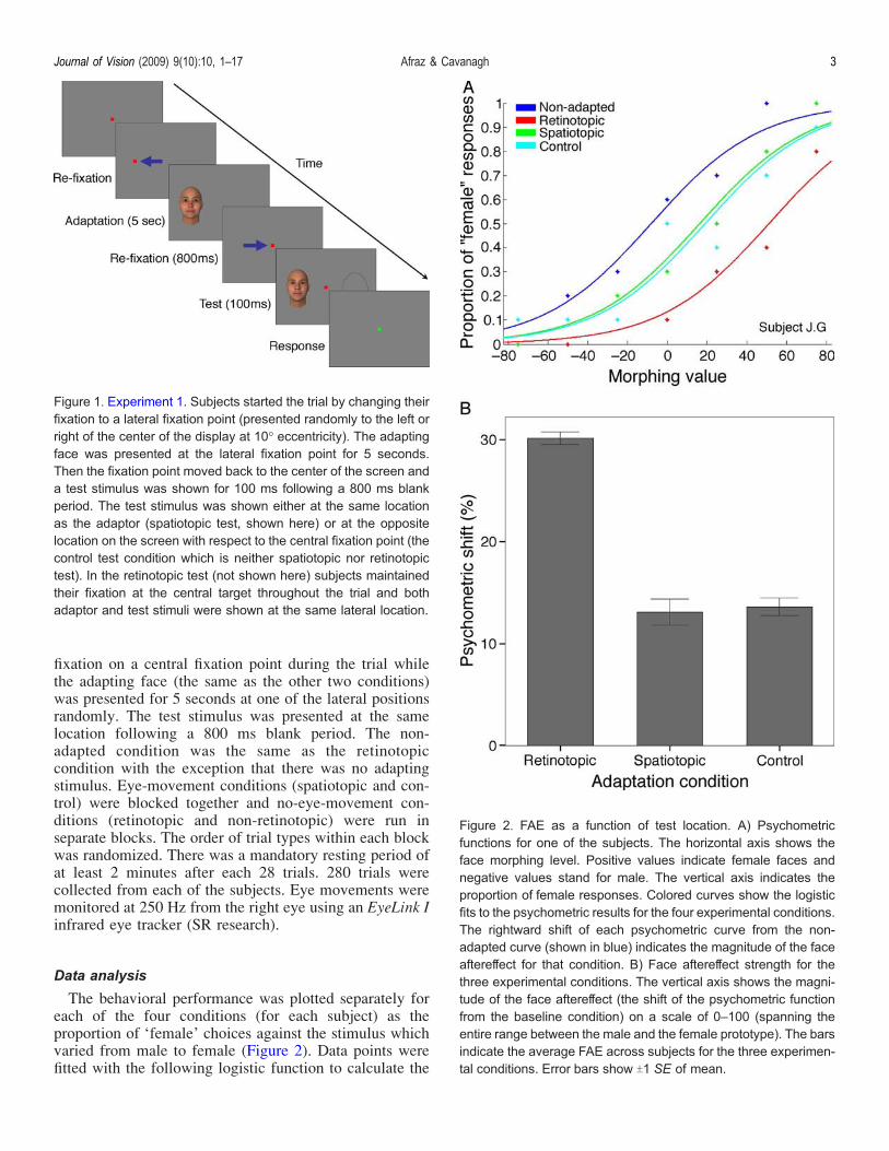
Figure 1. Experiment 1. Subjects started the trial by changing theirfixation to a lateral fixation point (presented randomly to the left orright of the center of the display at 10- eccentricity). The adaptingface was presented at the lateral fixation point for 5 seconds.Then the fixation point moved back to the center of the screen anda test stimulus was shown for 100 ms following a 800 ms blankperiod. The test stimulus was shown either at the same locationas the adaptor (spatiotopic test, shown here) or at the oppositelocation on the screen with respect to the central fixation point (thecontrol test condition which is neither spatiotopic nor retinotopictest). In the retinotopic test (not shown here) subjects maintainedtheir fixation at the central target throughout the trial and bothadaptor and test stimuli were shown at the same lateral location.
Figure 2. FAE as a function of test location. A) Psychometricfunctions for one of the subjects. The horizontal axis shows theface morphing level. Positive values indicate female faces andnegative values stand for male. The vertical axis indicates theproportion of female responses. Colored curves show the logisticfits to the psychometric results for the four experimental conditions.The rightward shift of each psychometric curve from the non-adapted curve (shown in blue) indicates the magnitude of the faceaftereffect for that condition. B) Face aftereffect strength for thethree experimental conditions. The vertical axis shows the magni-tude of the face aftereffect (the shift of the psychometric functionfrom the baseline condition) on a scale of 0–100 (spanning theentire range between the male and the female prototype). The barsindicate the average FAE across subjects for the three experimen-tal conditions. Error bars show T1 SE of mean.
Journal of Vision (2009) 9(10):10, 1–17 Afraz & Cavanagh 3
PSE (point of subject equality) where the face lookedequally male and female for each curve.
p xð Þ ¼ 1
1þ ejð!þ"xþ11I1þ12I2þ13I3Þ ; ð1Þ
where x is the morphing percent and P(x) is theprobability of female response. I is a binary variable, setto either 1 or 0 to indicate the presence or absence of eachadaptation condition. !, " and 1 are free parameters thatwere fit using the maximum likelihood fitting procedure(Meeker & Escobar, 1995).The shift of the PSE for each of the adaptation
conditions from the non-adapted condition was definedas the FAE magnitude.
Results
Figure 2A shows the results of Experiment 1 for one ofthe subjects. The largest shift of the psychometric functionwas observed for the retinotopic condition in all of thesubjects. FAE was also significant for both spatiotopic andcontrol conditions (logistic regression, p G 0.05 for allsubjects). FAE was significantly larger for the retinotopiccondition in contrast to both spatiotopic and controlconditions (logistic regression, p G 0.05 for all subjects).No significant difference was observed between thespatiotopic and control conditions (logistic regression,p 9 0.6 for all subjects). Figure 2B demonstrates theaverage shift of the psychometric function (from the non-adapted curve) for all of the subjects across differentexperimental conditions. Repeated measures ANOVArevealed the significant effect of experimental conditionon the FAE magnitude (F(2, 8) = 370.9, p G 0.001). FAEwas significantly smaller for both spatiotopic tests (pairedsample t-test, t(4) = 24.35, p G 0.01, Bonferroni corrected)and the neither spatiotopic nor retinotopic control tests(paired sample t-test, t(4) = 28.33, p G 0.01, Bonferronicorrected) conditions in contrast to the retinotopic tests.No significant difference was observed between the shiftvalues of the spatiotopic location and the neither spatio-topic nor retinotopic control test locations (paired samplet-test, t(4) = j0.64, p = 0.56).
Discussion
The FAE was strongest when the test and adaptinglocations matched in retinotopic coordinates. The FAEwas smaller but still significant for both the spatiotopicand the control location with no privileged adaptation forthe spatiotopic location. The results clearly show thatadaptable face analyses units are based in a retinotopiccoordinate frame. No evidence was found in support of
spatiotopy for the FAE. Our results show that spatiotopy isnot a general property of face aftereffect. In the experi-ments that follow this one, we will expand the range ofimage transforms over which we test spatiotopy versusretinotopy to see if this first result is a general one orlimited to translational changes in viewpoint.Although we did not find spatiotopy, two other experi-
ments have (Melcher, 2005; van Boxtel, Alais, & van Ee,2008). Our experiment is most closely related to the FAEexperiment of Melcher (2005). He showed a significantface aftereffect following a change of fixation when thetest stimulus was presented at the same spatial location asthe adapting stimulus. He also showed that the magnitudeof the FAE for the spatiotopic location was approximatelyequal to a retinotopic FAE measured when adaptation andtest stimuli were both presented at the same peripherallocation on the retina and there were no eye movements.In contrast, we find that the FAE at the spatiotopiclocation is no different in strength from the control FAEseen at a location equally distant from the adapting sitebut which does not correspond to the adapted location inspatiotopic (or retinotopic) coordinates. In other words,we do not find any additional adaptation at the spatiotopiclocation that cannot be explained by general transfer ofthe FAE over distance.How did Melcher’s (2005) study differ from ours? First,
his procedure did not correspond to the classic paradigmthat we used (Fang & He, 2005; Jiang, Blanz, & O’Toole,2006, 2009). Specifically, his subjects were adapted to afemale face. They then had to identify a test face as one ofthree target male faces. The test face was chosen from oneof three morphs, each of which varied from one of thethree target male faces along a morphing path to theadapting female face. The percentage of correct discrim-ination naturally increased as the percentage of maletarget increased in the test morph. In Melcher’s procedure,the percentage of correct decisions at a given level ofmorph was taken as the measure the strength ofadaptation. In other words, adapting to the female face istaken as equivalent to adding more male face (or lessfemale) in the test morph and so should increase perfor-mance. However, this “percent correct” measure suffersfrom a general performance confound as anything thatimproves performance (such as higher level factors likeattention) will be taken as an increase in the FAE strength.For instance, depending on the task instructions or thesubject’s natural strategy for attention allocation, if thespatiotopic location drew more attention (in contrast tothe control condition location), it would have higherperformance and this would be considered an aftereffectin Melcher’s analysis. To avoid this possible confound,other studies that used “percent correct” to determine theFAE always measured the percent correct for a non-adaptedface as well (Anderson & Wilson, 2005; Leopold et al.,2001). This crucial non-adapted face control condition forthe general performance is absent in Melcher’s paradigm,
Journal of Vision (2009) 9(10):10, 1–17 Afraz & Cavanagh 4
leaving open the possibility that general performancefactors affected the results.In our experiment, by measuring the shift of the
psychometric function along the male–female morphdimension in a classical paradigm, we avoided possiblegeneral confounds and found no significant spatiotopicFAE, at least for the case of gender-specific FAE. In anearlier study, we investigated retinotopy vs. spatiotopy forthe identity-specific FAE (Afraz, S-R., & Cavanagh, P.(2006). Is the “face aftereffect” retinotopic or spatiotopic?[Abstract]. Journal of Vision, http://journalofvision.org/6/6/882/). There we found no evidence to support thespatiotopy of the FAE although there were still methodo-logical differences between our procedure and Melcher’s(2005) that might explain the difference in outcome.The second relevant paper (van Boxtel et al., 2008)
found a spatiotopic bias in binocular rivalry following faceadaptation. In that study, prolonged exposure to face orhouse stimuli biased subsequent rivalry in favor of theopposite category at both retinotopic and spatiotopicadaptation loci. However, their results show that in contrastto the strong and immediate retinotopic effect, the spatio-topic bias does not appear until after the first 10 seconds oftesting and is much smaller than the bias at the retinotopiclocation. It is not yet clear what might underlie their resultbut higher level factors like spatial working memory(Horaguchi & Sugino, 2006; Irwin, Zacks, & Brown, 1990;Prime, Tsotsos, Keith, & Crawford, 2007) and attention(Golomb, Chun, & Mazer, 2008) cannot be ruled out.As mentioned above, our goal is not to track down the
differences between these two studies and our own but toextend our tests of retinotopy vs. spatiotopy to a widerrange of image transforms. We have found an absence ofgeneral spatiotopy for simple eye movements but we nowwant to see if spatiotopy might nevertheless be found withother natural image transforms such as head translation orrotation or object movements.
Experiment 2
Introduction
The human anatomy allows a relatively wide range ofhead tilt toward the shoulders (planar rotation). Planarrotation of the skull induces fronto-parallel rotation of theimage on the retina that needs to be discriminated fromactual rotation of visual scenes in the external world. Thiscreates a different version of the problem of spatiotopy.Experiment 2 is designed to examine rotation spatiotopyfor the FAE. FAE transfers across large degrees ofrotation (Rhodes, Jeffery, Watson, Clifford, & Nakayama,2003; Watson & Clifford, 2003). Here we want to knowif there is any difference in FAE following a head tilt of90- for tests whose orientation matches the adapting
orientation on the screen and whose orientation matchesthe original retinal orientation.One challenge for appropriate measurement of the FAE
across head tilts is to control the confounding effect of theretinal cyclotorsion. Muscles of the orbit rotate the eyes inthe direction opposite to that of the head, trying tostabilize the retinal image across head tilts (Balliet &Nakayama, 1978; Morrison, 1984). Although retinalcyclotorsion is possible only in a limited range (about10-), it can bias the results in favor of a spatiotopic effect.To avoid this confounding factor, we measured andcorrected for the exact amount of the retinal cyclotorsionfor each individual.
MethodsSubjects and procedure
Four subjects including one of the authors participatedin Experiment 2. To control the angle of the head tilteffectively a special V-shaped head-rest was used (seeFigure 3). The head-rest had two (left and right) restingplates, each deviated 45- from the vertical midline line. Ahead band held two rectangular plastic plates on the left andright temporal bones of the subjects’ head tightly. Subjectsheld their head between the two resting plates. They weretrained to tilt their head (to the left or right depending on thetrial instruction) and hold the solid plastic surface of thehead band attached to the resting plate of the head-rest.Head band plates helped the subjects to keep their headaligned with the resting plates throughout the trial.90- tilt of the skull does not transfer to 90- planar rotation
of the retina because retinal cyclotorsion compensates partof the head rotation. Prior to the main experiment, therange of the retinal cyclotorsion across a 90- head tilt wasmeasured for each individual separately. To measure theretinal cyclotorsion we used iconic aftereffect (Balliet &Nakayama, 1978). Subjects tilted their head to the left orright and fixated a small red target. A vertical bright linewas then presented on a dark background for 10 seconds.Then the line disappeared and subjects tilted their head90- to the opposite direction and fixated the red targetagain. Holding their head tilted, they adjusted the angle ofa fade gray test line to match the after image of theadapted line. Each subject performed 10 adjustments.Similar to the previous experiment there were four
conditions in Experiment 2: spatiotopic, control (neitherspatiotopic nor retinotopic), retinotopic and non-adapted.In the first two conditions (spatiotopic and control) a smallred fixation target appeared in the center of the screen.Subjects were instructed to keep their gaze on the redtarget throughout the trial. An arrow indicated thedirection of the head tilt at the beginning of each trial.The subject initiated the trial by pressing the space barwhen she/he placed her/his head on the head-restappropriately. A tilted adapting face was presented thenat the fixation point for 6 seconds. The planar angle of the
Journal of Vision (2009) 9(10):10, 1–17 Afraz & Cavanagh 5

adapting face was chosen in a way to match the verticalmeridian of the retina (based on the measurement of theretinal cyclotorsion). In other words, the adapting facewas presented upright and vertical on the retina. Thistranslates to a È45- tilt away from the vertical midline ofthe screen (the exact angle varies slightly from subject tosubject depending on their cyclotorsion factor, and isÈ45- left or È45- right depending on the trial). Followingthe adaptation, the adapting face disappeared and an arrowindicated that the subject has to tilt her/his head to theopposite direction from that held during adaptation. Thesubject pressed the space bar again after completion ofthe head movement. The time delay between disappear-ance of the adaptor and the second space bar press wasrecorded for future use. The test stimulus was presentedthen at either the spatiotopic angle (matching the adaptingangle on the screen) or the control angle, rotated È180 degfrom the adapting stimulus on the screen. The spatiotopicangle was the same angle as the adapting stimulus (about45 deg, half way between upright and horizontal on thescreen, and oriented either to the left or right depending onthe trial). Note that although the adapting and test stimuliare shown with the same angle on the screen in thisspatiotopic condition, their angle has changed by almost90 deg in retinotopic coordinates. The adapting stimulus isshown at the vertical meridian of the retina and the teststimulus is deviated from the vertical meridian to adirection opposite to the head-tilt direction producing atotal shift on the retinal of almost 90 deg. The controlangle was chosen to produce mirror symmetric deviation
of the test face from the vertical meridian relative to thespatiotopic angle (È135- tilted away from the verticalorientation on the screen, about half way betweenhorizontal and upside down) and so be about 90 degshifted on the retina, in the opposite direction from thespatiotopic test, and therefore about 180 degrees rotatedfrom the orientation of the spatiotopic test on the retina.Thus, the retinal angular distance between the adaptor andthe test was identical and opposite in the two conditions(see Figure 4). The actual deviation of the test stimulifrom the vertical meridian of the retina in the spatiotopicand control conditions varied across subject (between84.4- and 75.9-) depending on their retinal cyclotorsionmeasurements.Head-movement conditions (spatiotopic and control)
were blocked together and no-head-movement conditions(retinotopic and non-adapted) were run in separate blocks.336 trials were collected from each of the subjects. Theinter-stimulus interval (the delay between the adaptor andthe test stimulus) varied from trial to trial in the head-movement conditions (depending on how fast the subjectscould make the movement). The inter-stimulus intervalsfor the retinotopic condition for each subject were selectedpseudorandomly (without repetition) from the recordedinter-stimulus interval profile of the same subject for thehead-movement conditions. Thus, the average and thedistribution of inter-stimulus intervals were the same forthe retinotopic condition and the two head-movementconditions. In the retinotopic adaptation condition, sub-jects maintained their head position to the left or right
Figure 3. The V-shaped head-rest. Rectangular plastic plates were attached tightly to the temporal bone of the subjects with a head band.Subjects were asked to tilt their head and keep the plastic plate attached to the resting plates on the V-shaped head rest according to thetrial instruction.
Journal of Vision (2009) 9(10):10, 1–17 Afraz & Cavanagh 6
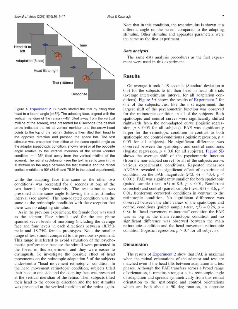
while the adapting face (the same as the other twoconditions) was presented for 6 seconds at one of thetwo lateral angles randomly. The test stimulus waspresented at the same angle following the inter-stimulusinterval (see above). The non-adapted condition was thesame as the retinotopic condition with the exception thatthere was no adapting stimulus.As in the previous experiment, the female face was used
as the adaptor. Face stimuli used for the test phasespanned seven levels of morphing (including the averageface and four levels in each direction) between 18.75%male and 18.75% female prototypes. Note the smallerrange of test stimuli compared to the previous experiment.This range is selected to avoid saturation of the psycho-metric performance because the stimuli were presented inthe fovea in this experiment and they were easier todistinguish. To investigate the possible effect of headmovements on the retinotopic adaptation 3 of the subjectsunderwent a “head movement retinotopic” condition. Inthe head movement retinotopic condition, subjects titledtheir head to one side and the adapting face was presentedat the vertical meridian of the retina. Then subjects tiltedtheir head to the opposite direction and the test stimuluswas presented at the vertical meridian of the retina again.
Note that in this condition, the test stimulus is shown at adifferent angle on the screen compared to the adaptingstimulus. Other stimulus and apparatus parameters werethe same as the first experiment.
Data analysis
The same data analysis procedures as the first experi-ment were used in this experiment.
Results
On average it took 1.19 seconds (Standard deviation =0.3) for the subjects to tilt their head in head tilt trials(average inters-stimulus interval for all adaptation con-ditions). Figure 5A shows the results of Experiment 2 forone of the subjects. Just like the first experiment, thelargest shift of the psychometric function was observedfor the retinotopic condition in all of the subjects. Bothspatiotopic and control curves were significantly shiftedrightwards from the non-adapted curve (logistic regres-sion, p G 0.05 for all subjects). FAE was significantlylarger for the retinotopic condition in contrast to bothspatiotopic and control conditions (logistic regression, p G0.05 for all subjects). No significant difference wasobserved between the spatiotopic and control conditions(logistic regression, p 9 0.6 for all subjects). Figure 5Bshows the average shift of the psychometric function(from the non-adapted curve) for all of the subjects acrossvarious experimental conditions. Repeated measuresANOVA revealed the significant effect of experimentalcondition on the FAE magnitude (F(2, 6) = 43.4, p G0.001). FAE was significantly smaller for both spatiotopic(paired sample t-test, t(3) = 8.5, p G 0.01, Bonferronicorrected) and control (paired sample t-test, t(3) = 8.6, p G0.01, Bonferroni corrected) conditions in contrast to theretinotopic condition. No significant difference wasobserved between the shift values of the spatiotopic andcontrol conditions (paired sample t-test, t(3) = 0.26, p =0.8). In “head movement retinotopic” condition the FAEwas as big as the main retinotopic condition and nosignificant difference was observed between the mainretinotopic condition and the head movement retinotopiccondition (logistic regression, p 9 0.7 for all subjects).
Discussion
The results of Experiment 2 show that FAE is maximalwhen the retinal orientations of the adaptor and test arematched even if the head tilts between adaptation and testphases. Although the FAE transfers across a broad rangeof orientation, it remains strongest at its retinotopic angleof adaptation and spreads symmetrically from this retinalorientation to the spatiotopic and control orientationswhich are both about a 90 deg rotation, in opposite
Figure 4. Experiment 2. Subjects started the trial by tilting theirhead to a lateral angle (T45-). The adapting face, aligned with thevertical meridian of the retina (È45- tilted away from the verticalmidline of the screen), was presented for 6 seconds (the dashedarrow indicates the retinal vertical meridian and the arrow headpoints to the top of the retina). Subjects then tilted their head tothe opposite direction and pressed the space bar. The teststimulus was presented then either at the same spatial angle asthe adaptor (spatiotopic condition, shown here) or at the oppositeangle relative to the vertical meridian of the retina (controlcondition jÈ135- tilted away from the vertical midline of thescreen). The retinal cyclotorsion (see the text) is set to zero in thisillustration so the angle between the test stimulus and the retinalvertical meridian is 90- (84.4- and 75.9- in the actual experiment).
Journal of Vision (2009) 9(10):10, 1–17 Afraz & Cavanagh 7
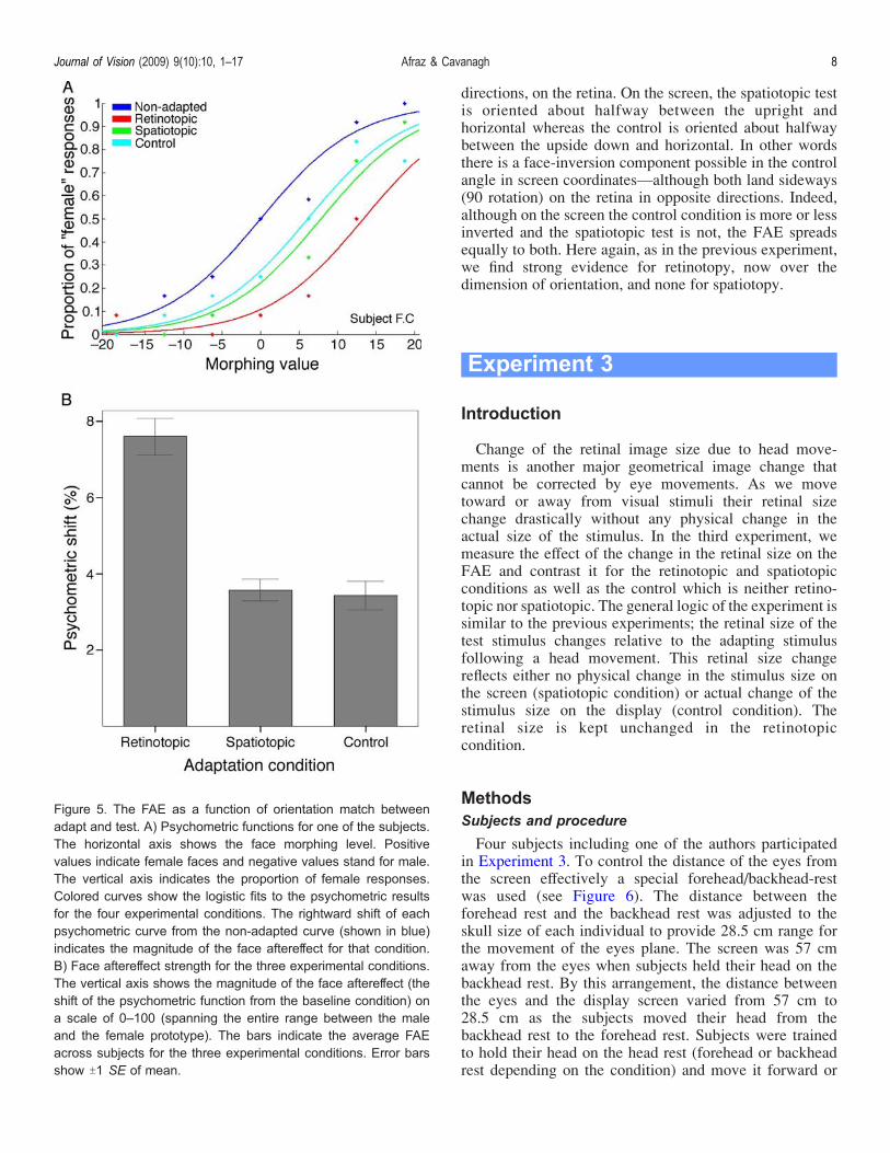
directions, on the retina. On the screen, the spatiotopic testis oriented about halfway between the upright andhorizontal whereas the control is oriented about halfwaybetween the upside down and horizontal. In other wordsthere is a face-inversion component possible in the controlangle in screen coordinatesValthough both land sideways(90 rotation) on the retina in opposite directions. Indeed,although on the screen the control condition is more or lessinverted and the spatiotopic test is not, the FAE spreadsequally to both. Here again, as in the previous experiment,we find strong evidence for retinotopy, now over thedimension of orientation, and none for spatiotopy.
Experiment 3
Introduction
Change of the retinal image size due to head move-ments is another major geometrical image change thatcannot be corrected by eye movements. As we movetoward or away from visual stimuli their retinal sizechange drastically without any physical change in theactual size of the stimulus. In the third experiment, wemeasure the effect of the change in the retinal size on theFAE and contrast it for the retinotopic and spatiotopicconditions as well as the control which is neither retino-topic nor spatiotopic. The general logic of the experiment issimilar to the previous experiments; the retinal size of thetest stimulus changes relative to the adapting stimulusfollowing a head movement. This retinal size changereflects either no physical change in the stimulus size onthe screen (spatiotopic condition) or actual change of thestimulus size on the display (control condition). Theretinal size is kept unchanged in the retinotopiccondition.
MethodsSubjects and procedure
Four subjects including one of the authors participatedin Experiment 3. To control the distance of the eyes fromthe screen effectively a special forehead/backhead-restwas used (see Figure 6). The distance between theforehead rest and the backhead rest was adjusted to theskull size of each individual to provide 28.5 cm range forthe movement of the eyes plane. The screen was 57 cmaway from the eyes when subjects held their head on thebackhead rest. By this arrangement, the distance betweenthe eyes and the display screen varied from 57 cm to28.5 cm as the subjects moved their head from thebackhead rest to the forehead rest. Subjects were trainedto hold their head on the head rest (forehead or backheadrest depending on the condition) and move it forward or
Figure 5. The FAE as a function of orientation match betweenadapt and test. A) Psychometric functions for one of the subjects.The horizontal axis shows the face morphing level. Positivevalues indicate female faces and negative values stand for male.The vertical axis indicates the proportion of female responses.Colored curves show the logistic fits to the psychometric resultsfor the four experimental conditions. The rightward shift of eachpsychometric curve from the non-adapted curve (shown in blue)indicates the magnitude of the face aftereffect for that condition.B) Face aftereffect strength for the three experimental conditions.The vertical axis shows the magnitude of the face aftereffect (theshift of the psychometric function from the baseline condition) ona scale of 0–100 (spanning the entire range between the maleand the female prototype). The bars indicate the average FAEacross subjects for the three experimental conditions. Error barsshow T1 SE of mean.
Journal of Vision (2009) 9(10):10, 1–17 Afraz & Cavanagh 8

backward (upon the instruction) as fast as possible wheninstructed.Similar to the previous experiments there were four
conditions in Experiment 3: spatiotopic, control (neitherspatiotopic nor retinotopic), retinotopic and non-adapted.In the first two conditions (spatiotopic and control) a smallred fixation target appeared in the center of the screen.Subjects were instructed to keep their gaze on the redtarget throughout the trial. The instructions about headposition were presented on the screen when necessary (seeFigure 7). The subject initiated the trial by pressing thespace bar when she/he placed her/his head on the head rest(forehead or backhead rest, upon the instruction) appro-priately. The adapting face of 6- of visual angle waspresented at the fixation point for 6 seconds. Followingthe adaptation, the adapting face disappeared and a “Go”signal on the screen indicated that the subject has to moveher/his head forward or backward (depending on thecondition). The subject pressed the space bar again aftercompletion of the head movement. The time delaybetween disappearance of the adaptor and the secondspace bar press was recorded for future use. The teststimulus was presented then at either 11.9- or 3- sizes. Ifthe subject was adapted at the far location but was testedat the near location, the 11.9- stimulus has the samescreen size as the 6- adaptor (spatiotopic condition). The3- stimulus matches neither the retinal nor the screen size(it is the neither-retinotopic-nor-spatiotopic control con-dition). Alternatively, if the subject was adapted at thenear location (28.5 cm distance from the screen), thenmoved to the far location for the test, the 3- stimulus hasthe same screen size as the adaptor (spatiotopic) and the
11.9- stimulus matched neither the size of the adaptor onthe retina nor on the screen (control).In the first retinotopic adaptation condition, subjects
held their head on the backhead-rest or the forehead rest(depending on the trial) throughout the trial. The teststimulus was presented at 6- of visual angle following theinter-stimulus interval. The inter-stimulus interval wasdetermined from the results of the head movement trials(see the Methods section of Experiment 2 for moredetails). The non-adapted condition was the same as theretinotopic condition with the exception that there was noadapting stimulus. In the second retinotopic adaptationcondition, 3 of the 4 subjects underwent a “head move-ment retinotopic” condition. In the head movementretinotopic condition, subjects moved their head (forwardof backward depending on the condition) after adaptationto a 6- adaptor. They were presented then with a 6- teststimulus. Note that in this condition, the test stimulus isshown at a different size on the screen compared to theadapting stimulus. Other stimulus and apparatus parame-ters were the same as the first experiment.
Figure 6. The forehead/backhead rest. The head rest wasdesigned to control the distance of the eyes from the displayscreen. Subjects were asked to rest their head on the backheadrest and move it forward to the forehead rest when instructed. Orvice versa.
Figure 7. Experiment 3. Subjects started the trial by resting theirhead on the forehead or the backhead rest upon the instruction.The adapting face was presented for 6 seconds at the center ofthe display. Subjects moved their head (toward or away from thescreen upon the instruction) and then pressed the space bar. Thetest stimulus was presented then either at a different (half ordouble depending on the condition) retinal size that correspondedto the same screen size as the adaptor (spatiotopic condition,shown here) or a different (half or double) retinal size that doesnot correspond to the screen size of the adaptor (controlcondition). In the retinotopic condition, the adapting and teststimuli were of the same retinal size. Note that in the controlcondition, the adapting and test stimuli matched neither in screensize not in retinal size but differed in retinal size by the same factoras did the spatiotopic adapt and test size (1 octave).
Journal of Vision (2009) 9(10):10, 1–17 Afraz & Cavanagh 9
Data analysis
The same data analysis procedures as the previousexperiments were used in this experiment.
Results
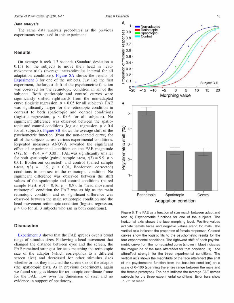
On average it took 1.3 seconds (Standard deviation =0.15) for the subjects to move their head in head-movement trials (average inters-stimulus interval for alladaptation conditions). Figure 8A shows the results ofExperiment 3 for one of the subjects. Just like the firstexperiment, the largest shift of the psychometric functionwas observed for the retinotopic condition in all of thesubjects. Both spatiotopic and control curves weresignificantly shifted rightwards from the non-adaptedcurve (logistic regression, p G 0.05 for all subjects). FAEwas significantly larger for the retinotopic condition incontrast to both spatiotopic and control conditions(logistic regression, p G 0.05 for all subjects). Nosignificant difference was observed between the spatio-topic and control conditions (logistic regression, p 9 0.4for all subjects). Figure 8B shows the average shift of thepsychometric function (from the non-adapted curve) forall of the subjects across various experimental conditions.Repeated measures ANOVA revealed the significanteffect of experimental condition on the FAE magnitude(F(2, 6) = 49.4, p G 0.001). FAE was significantly smallerfor both spatiotopic (paired sample t-test, t(3) = 9.9, p G0.01, Bonferroni corrected) and control (paired samplet-test, t(3) = 11.9, p G 0.01, Bonferroni corrected)conditions in contrast to the retinotopic condition. Nosignificant difference was observed between the shiftvalues of the spatiotopic and control conditions (pairedsample t-test, t(3) = 0.16, p = 0.9). In “head movementretinotopic” condition the FAE was as big as the mainretinotopic condition and no significant difference wasobserved between the main retinotopic condition and thehead movement retinotopic condition (logistic regression,p 9 0.6 for all 3 subjects who ran in both conditions).
Discussion
Experiment 3 shows that the FAE spreads over a broadrange of stimulus sizes. Following a head movement thatchanged the distance between eyes and the screen, theFAE remained strongest for tests matching the retinotopicsize of the adaptor (which corresponds to a differentscreen size) and decreased for other stimulus sizeswhether or not they matched the screen size of the adaptor(the spatiotopic test). As in previous experiments, againwe found strong evidence for retinotopic coordinate framefor the FAE, now over the dimension of size, and noevidence in support of spatiotopy.
Figure 8. The FAE as a function of size match between adapt andtest. A) Psychometric functions for one of the subjects. Thehorizontal axis shows the face morphing level. Positive valuesindicate female faces and negative values stand for male. Thevertical axis indicates the proportion of female responses. Coloredcurves show the logistic fits to the psychometric results for thefour experimental conditions. The rightward shift of each psycho-metric curve from the non-adapted curve (shown in blue) indicatesthe magnitude of the face aftereffect for that condition. B) Faceaftereffect strength for the three experimental conditions. Thevertical axis shows the magnitude of the face aftereffect (the shiftof the psychometric function from the baseline condition) on ascale of 0–100 (spanning the entire range between the male andthe female prototype). The bars indicate the average FAE acrosssubjects for the three experimental conditions. Error bars showT1 SE of mean.
Journal of Vision (2009) 9(10):10, 1–17 Afraz & Cavanagh 10
Experiment 4
Introduction
The idea of object-based remapping of visual featureshas been recently suggested as a possible mechanism forspatial constancy (Melcher, 2008). It has been claimedthat following adaptation to features of a visual object at agiven location on the screen, if that object moves to a newlocation, its aftereffect transfers to the new location on thescreen even if the visual features of the object have beenoccluded during the displacement (Melcher, 2008).Experiment 4 put this idea to test for the FAE. It alsoincluded a condition with an eye movement during theinterval between adaptation and test as suggested byMelcher (2008) to strengthen the object-specific adaptation.
MethodsSubjects and procedure
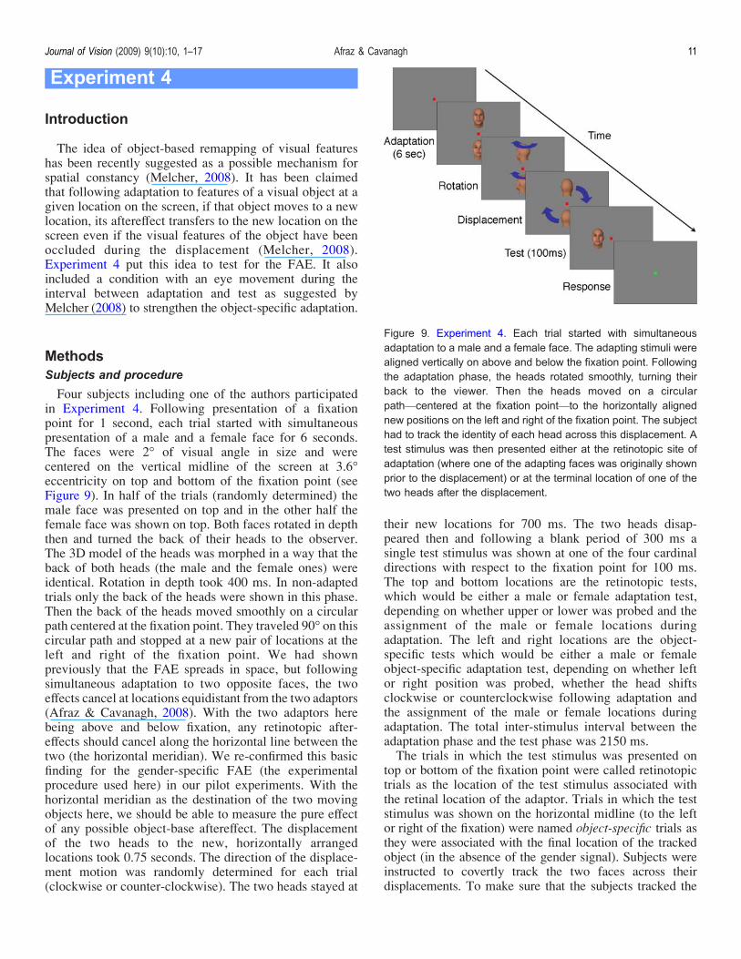
Four subjects including one of the authors participatedin Experiment 4. Following presentation of a fixationpoint for 1 second, each trial started with simultaneouspresentation of a male and a female face for 6 seconds.The faces were 2- of visual angle in size and werecentered on the vertical midline of the screen at 3.6-eccentricity on top and bottom of the fixation point (seeFigure 9). In half of the trials (randomly determined) themale face was presented on top and in the other half thefemale face was shown on top. Both faces rotated in depththen and turned the back of their heads to the observer.The 3D model of the heads was morphed in a way that theback of both heads (the male and the female ones) wereidentical. Rotation in depth took 400 ms. In non-adaptedtrials only the back of the heads were shown in this phase.Then the back of the heads moved smoothly on a circularpath centered at the fixation point. They traveled 90- on thiscircular path and stopped at a new pair of locations at theleft and right of the fixation point. We had shownpreviously that the FAE spreads in space, but followingsimultaneous adaptation to two opposite faces, the twoeffects cancel at locations equidistant from the two adaptors(Afraz & Cavanagh, 2008). With the two adaptors herebeing above and below fixation, any retinotopic after-effects should cancel along the horizontal line between thetwo (the horizontal meridian). We re-confirmed this basicfinding for the gender-specific FAE (the experimentalprocedure used here) in our pilot experiments. With thehorizontal meridian as the destination of the two movingobjects here, we should be able to measure the pure effectof any possible object-base aftereffect. The displacementof the two heads to the new, horizontally arrangedlocations took 0.75 seconds. The direction of the displace-ment motion was randomly determined for each trial(clockwise or counter-clockwise). The two heads stayed at
their new locations for 700 ms. The two heads disap-peared then and following a blank period of 300 ms asingle test stimulus was shown at one of the four cardinaldirections with respect to the fixation point for 100 ms.The top and bottom locations are the retinotopic tests,which would be either a male or female adaptation test,depending on whether upper or lower was probed and theassignment of the male or female locations duringadaptation. The left and right locations are the object-specific tests which would be either a male or femaleobject-specific adaptation test, depending on whether leftor right position was probed, whether the head shiftsclockwise or counterclockwise following adaptation andthe assignment of the male or female locations duringadaptation. The total inter-stimulus interval between theadaptation phase and the test phase was 2150 ms.The trials in which the test stimulus was presented on
top or bottom of the fixation point were called retinotopictrials as the location of the test stimulus associated withthe retinal location of the adaptor. Trials in which the teststimulus was shown on the horizontal midline (to the leftor right of the fixation) were named object-specific trials asthey were associated with the final location of the trackedobject (in the absence of the gender signal). Subjects wereinstructed to covertly track the two faces across theirdisplacements. To make sure that the subjects tracked the
Figure 9. Experiment 4. Each trial started with simultaneousadaptation to a male and a female face. The adapting stimuli werealigned vertically on above and below the fixation point. Followingthe adaptation phase, the heads rotated smoothly, turning theirback to the viewer. Then the heads moved on a circularpathVcentered at the fixation pointVto the horizontally alignednew positions on the left and right of the fixation point. The subjecthad to track the identity of each head across this displacement. Atest stimulus was then presented either at the retinotopic site ofadaptation (where one of the adapting faces was originally shownprior to the displacement) or at the terminal location of one of thetwo heads after the displacement.
Journal of Vision (2009) 9(10):10, 1–17 Afraz & Cavanagh 11
stimuli properly, instead of directly reporting the gender ofthe test stimulus, they were asked to report if the teststimulus had the same gender as its adaptor by pressing oneof the two keys on the keyboard with their right hand. Notethat each test location was associated with one of the twoadaptors as its original or terminal location. For non-adapted trials the subjects had to directly determine thegender of the test face by pressing keys with their left hand.Each subject completed 420 experimental trials. There
were 5 conditions in this experiment: male adaptedretinotopic (when the test was presented at the retinotopicadaptation locus of the male adaptor), female adaptedretinotopic (when the test was presented at the retinotopicadaptation locus of the female adaptor), male adaptedobject-specific (when the test was presented at the finalposition of the male adaptor after its displacement),female adapted object-specific (when the test was pre-sented at the final position of the female adaptor after itsdisplacement) and non-adapted. Different conditions werenot blocked together and the order of various trial typeswas randomized. Other stimulus and apparatus parameterswere the same as previous experiments.Melcher (2008) claimed that making a saccadic eye
movement during the object movement phase strengthensthat object-based aftereffect. To investigate this possibilityfor the FAE, two of the subjects participated in acomplementary experiment. All of the parameters were thesame as the main experiment except that during the headdisplacement phase the fixation target moved to a peripherallocation on the diagonal of the display screen, 6- of visualfield away from the center for 400 ms. Subjects wereinstructed to saccade to the target when it moves to a newposition. Then the fixation target moved back to its originalcentral position and stayed there for the rest of the trial.
Data analysis
The same data analysis procedures as the previousexperiments were used in this experiment.
Results
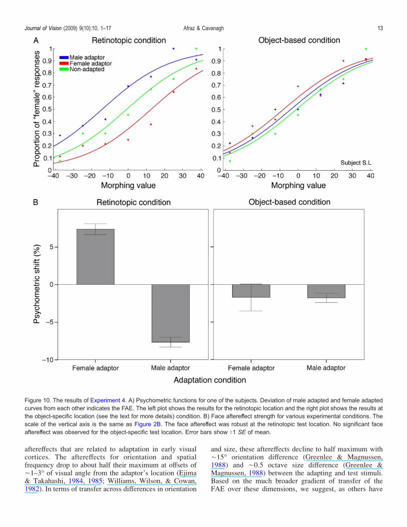
Figure 10 demonstrates the results of Experiment 4.Deviation of the psychometric curves of male and femaleadaptation from each other indicates the FAE. As demon-strated for a typical subject in Figure 10A also the averageof all subjects in Figure 10B, the psychometric curvesof male and female adaptation were significantly deviatedfrom each other in all cases (logistic regression, p G 0.01,Bonferroni corrected) for the retinotopic condition. Nosignificant effect was observed for the objectotopic con-dition (logistic regression, p 9 0.5, Bonferroni corrected).The average of psychometric shift values (shifts from thebaseline non-adapted condition) for all of the subjects alsoshowed the significant difference between male adaptation
and female adaptation for the retinotopic condition (pairedsample t-test, t(3) = j22.3, p G 0.001) but not for theobjectotopic condition (paired sample t-test, t(3) = j0.02,p = 0.9). The results of the complementary experimentwere the same as the main experiment and the applicationa saccade in the middle of the trial did not change the results.More specifically, no significant difference was observedbetween the male adapted and female adapted curves forthe objectotopic condition in the complementary experi-ment (logistic regression, p 9 0.4).
Discussion
The results of Experiment 4 show that the FAE remainsstrongest at the retinotopic location of the adaptor even ifthe adaptor moves to a different position after theadaptation. The FAE did not follow the object to its newlocation. We have previously shown that the FAE trans-fers over space (Afraz & Cavanagh, 2008). We have alsoshown that simultaneous adaptation to two opposite facesproduces a cancellation of the two FAEs halfway betweenthe two adaptors. An object-specific FAE should thereforebe the only FAE evident at locations equidistant from bothadaptors (along the horizontal meridian in this case), sofollowing adaptation, we displaced the adapting stimuli tothis neutral zone to increase the chance of observing thehypothesized object-specific component of the FAE. How-ever we failed to observe any such effect. It has beensuggested that introducing an eye movement between theadaptation and test phases increases the strength of an object-specific aftereffect, at least in the case of the tilt (Melcher,2008); nevertheless, our results were not influenced by asaccade introduced between adapt and test. One mightclaim that the aftereffect was reduced by stimulus motion orthe longer delays between the adaptation and test phases,but the existence and strength of the FAE at the retinotopictest location makes this unlikely. The magnitude of the FAEat the retinotopic location here is comparable to ourprevious measurements of the FAE with simultaneousadaptation to two opposite faces using the same stimulussize and eccentricity (Afraz & Cavanagh, 2008).
Discussion
The results of all four experiments confirm that FAE isat maximum strength for tests that match the originalretinal values of position, orientation and size. The effectdoes spread to other test locations around the originalretinotopic ones, falling to a value about half its maximumat a spatial offset of 10-, an orientation difference of È80-and a size difference of 1 octave. The range of spread isonly approximately determined here but they show atuning for the FAE that is far broader than that seen in the
Journal of Vision (2009) 9(10):10, 1–17 Afraz & Cavanagh 12
aftereffects that are related to adaptation in early visualcortices. The aftereffects for orientation and spatialfrequency drop to about half their maximum at offsets ofÈ1–3- of visual angle from the adaptor’s location (Ejima& Takahashi, 1984, 1985; Williams, Wilson, & Cowan,1982). In terms of transfer across differences in orientation
and size, these aftereffects decline to half maximum withÈ15- orientation difference (Greenlee & Magnussen,1988) and È0.5 octave size difference (Greenlee &Magnussen, 1988) between the adapting and test stimuli.Based on the much broader gradient of transfer of theFAE over these dimensions, we suggest, as others have
Figure 10. The results of Experiment 4. A) Psychometric functions for one of the subjects. Deviation of male adapted and female adaptedcurves from each other indicates the FAE. The left plot shows the results for the retinotopic location and the right plot shows the results atthe object-specific location (see the text for more details) condition. B) Face aftereffect strength for various experimental conditions. Thescale of the vertical axis is the same as Figure 2B. The face aftereffect was robust at the retinotopic test location. No significant faceaftereffect was observed for the object-specific test location. Error bars show T1 SE of mean.
Journal of Vision (2009) 9(10):10, 1–17 Afraz & Cavanagh 13

(Leopold et al., 2001; Moradi et al., 2005; Webster &MacLin, 1999; Yamashita, Hardy, De Valois, & Webster,2005; Zhao & Chubb, 2001) that the FAE is based inhigh-level visual cortex such as the FFA or LOC, humanequivalents of the inferotemporal face areas in monkey asthese areas show evidence of similar range of tuning(Bulthoff, Edelman, & Tarr, 1995; Lawson, 1999; Op DeBeeck & Vogels, 2000).Despite this broad gradient of transfer, there was no
evidence of any privileged transfer to locations, orienta-tions, or sizes that maintained a spatiotopic matchbetween the adapt and the test stimulus (i.e. that kept thetest stimulus fixed at the adapting location on the displayscreen while the subject moves his or her eyes or headbetween adaptation and test). Nor did we find any transferof the FAE to a displaced location of the adapting headthat turned around following adaptation and moved to anew location. Thus we have failed to replicate thespatiotopic FAE claimed by Melcher (2005) and we donot find any evidence of an object-specific FAE thatwould be equivalent to Melcher’s (2008) report of anobject-specific tilt aftereffect. We suggested earlier thathigher level factors (such as the subject’s strategy forattention allocation) might be responsible for Melcher’sresults but investigating the source of the difference isbeyond the scope of the current paper. Here we haveattempted to study spatiotopy for the gender-specific faceaftereffect using the basic face aftereffect paradigm acrossa wide range of retinal transformations, including size andorientation as well as position. We found that spatiotopy isnot a general property of face aftereffects although wecannot rule it out for his specific conditions.Based on the results of Experiment 1, we estimate that
FAE magnitude drops to half its maximum about 9.2-away from the adaptor. This number was 5.4- in ourpreviously published estimates where much smaller (È2-of visual angle) faces were used. The larger range oftransfer here might be a result solely of the larger stimuli.Specifically, the stimuli are about 4- larger here than inthe previous study and the area of analysis also 4- larger.The larger faces may have fallen on more and so recruitedmore face analysis areas that are nevertheless of the samesize as those previously measured.Whatever the source of this difference in estimated size,
the results here clearly demonstrate robust retinotopy forthe FAE. As mentioned previously, our method herecorresponds to the stand method of measuring aftereffectswhereas the method used by Melcher (2005) has somepotential flaws. Our current results support robust reti-notopy, without spatiotopy, for the FAE across variousdimensions and suggest that adaptable face analysis in thevisual system is based in a retinotopic coordinate frame.The strength of the retinotopic FAE measured in our
4 experiments varies widely from È30% in Experiment 1to between È7 and È5% in Experiments 2 and 3.However, the eccentricity of the adapting and test facesalso varied across the four experiments. For example in
Experiment 1, the stimuli were 6- in size and shown at 10-in the periphery, producing in the retinotopic condition aneffect of about 30%. In Experiments 2 and 3, the faceswere also 6- in size but presented in the fovea and theFAE was 5–7%. To pursue the possible effect ofeccentricity on the size of the face aftereffect we mea-sured the retinotopic face aftereffect in three subjects attwo eccentricities (3- and 10-) and two sizes (stimulussizes 4- and 1.2-). The face aftereffect was largest forthe 4-stimulus centered at 10- eccentricity (median ofthe three subjects: 40%), the FAE was smallest for the4- stimulus centered at 3- eccentricity (median: 7.5%).Interestingly, the effect was relatively large for the 1.2-stimulus at 3- eccentricity (median: 26%). These resultssuggest that the strength of the FAE increases as thestimulus size gets smaller, relative to the resolution level atthe test eccentricity. Possibly a stimulus that activates alarger number of face analysis regions (large stimuli and/orsmall eccentricity) is less susceptible to adaptation. Wechose the sizes of the smaller stimulus at the near positionand the larger stimulus at the far position to be approx-imately matched in cortical size when corrected for corticalmagnification factor of V1 (Rovamo & Virsu, 1979).Nevertheless, the aftereffect was still substantially largerfor the far stimulus. This might reflect the increase infoveal bias beyond V1 (Hasson, Harel, Levy, & Malach,2003). The effect of eccentricity on the face aftereffect isnot the central topic of the current study, however, andfurther studies are required to investigate this interestingeffect. The other possible explanation for the observedvariation of the magnitude of the retinotopic FAE acrossour experiments is the variation of the delay betweenadaptation and test across these experiments. The delay isthe shortest for Experiment 1 where the biggest retino-topic FAE is observed. However, the inter-stimulusinterval is much longer in other experiments wheresmaller aftereffects are observed.The question of spatiotopy has been studied in the
vision science literature for a long time, classically as thecontrast between head-centered or retinotopic coordinatesand gravitational coordinates. Although some studiesshowed a contribution from gravitational coordinates(Nicholls, Smith, Mattingley, & Bradshaw, 2006) mostof them showed stronger dependence of perception andaftereffects on retinotopic coordinates (Friederici &Levelt, 1990; Mast, Ganis, Christie, & Kosslyn, 2003;Wenderoth & Hickey, 1993). The face inversion effectalso generally follows the retinotopic coordinate system(Lobmaier & Mast, 2007). Many years ago, Gestaltpsychologist Gaetano Kanizsa (1913–1993) demonstratedthe dependence of face perception on retinotopic orienta-tion (as opposed to the gravitational orientation) in asimple way (Kanizsa, 1979). Figure 11 shows a typicalambiguous face cartoon (not the one used by Kanizsa).The identity of the face changes if you invert the page. Ifinstead of rotating the page, you invert your head, or benddown and look at the image from between your legs, the
Journal of Vision (2009) 9(10):10, 1–17 Afraz & Cavanagh 14

image remains upright in spatiotopic coordinates, but isinverted on your retina. As you can observe, the identityof the face depends on its orientation in the retinotopiccoordinates.This observation and other evidence in addition to our
adaptation results here indicate that face selective neuronsanalyze the image based on afferent visual signals that areorganized retinotopically. They do not have access to thevisual world in spatiotopic coordinates. We neverthelessconfirm a remarkably broad tuning of the FAE in position,orientation, and size. This broad tuning indicates that thisstrong and easily demonstrated aftereffect must be tappinghigh levels of visual processing well beyond that of areaV1. Despite this high-level adapting site, the effect isretinotopic along all the dimensions we tested. Finally,even though the spread of the FAE is broad across thedimensions we tested, it is nevertheless tuned in each,showing a maximum at the retinotopic match. This tuningrules out global face analysis covering the entire visualfield, all orientations, and all sizes.
Acknowledgments
Research supported by NIH EY09258 (PC), a Chaired’Excellence, ANR (PC) and Harvard University GSASDissertation Completion Fellowship (SRA).
Commercial relationships: none.Corresponding author: Arash Afraz.Email: [email protected]: Room 710, William James Hall, 33 Kirkland St.,Cambridge, MA 02138, USA.
References
Afraz, S. R., & Cavanagh, P. (2008). Retinotopy of theface aftereffect. Vision Research, 48, 42–54.[PubMed] [Article]
Afraz, S. R., Kiani, R., & Esteky, H. (2006). Micro-stimulation of inferotemporal cortex influences facecategorization. Nature, 442, 692–695. [PubMed]
Anderson,N.D.,&Wilson,H.R. (2005). The nature of syntheticface adaptation. Vision Research, 45, 1815–1828.[PubMed]
Balliet, R., & Nakayama, K. (1978). Egocentric orienta-tion is influenced by trained voluntary cyclorotary eyemovements. Nature, 275, 214–216. [PubMed]
Bulthoff, H. H., Edelman, S. Y., & Tarr, M. J. (1995).How are three-dimensional objects represented in thebrain? Cerebral Cortex, 5, 247–260. [PubMed]
Colby, C. L. (1998). Action-oriented spatial referenceframes in cortex. Neuron, 20, 15–24. [PubMed]
Colby, C. L., & Duhamel, J. R. (1996). Brain research.Cognitive Brain Research, 5, 105–115. [PubMed]
d’Avossa, G., Shulman, G. L., Snyder, A. Z., & Corbetta, M.(2006). Attentional selection of moving objects by a serialprocess. Vision Research, 46, 3403–3412. [PubMed]
Ejima, Y., & Takahashi, S. (1984). Facilitatory andinhibitory after-effect of spatially localized gratingadaptation. Vision Research, 24, 979–985. [PubMed]
Ejima, Y., & Takahashi, S. (1985). Effect of localizedgrating adaptation as a function of separation alongthe length axis between test and adaptation areas.Vision Research, 25, 1701–1707. [PubMed]
Ezzati, A., Golzar, A., & Afraz, A. S. (2008). Topography ofthe motion aftereffect with and without eye move-ments. Journal of Vision, 8(14):23, 1–16, http://journalofvision.org/8/14/23/, doi:10.1167/8.14.23.[PubMed] [Article]
Fang, F., & He, S. (2005). Viewer-centered object repre-sentation in the human visual system revealed byviewpoint aftereffects.Neuron, 45, 793–800. [PubMed]
Figure 11. Ambiguous face illusion and spatiotopy (a simpleobservation from Gaetano Kanizsa). If you invert the page, theidentity of this face will switch. Instead of inverting the page, turnyour head about 180- or bend down and look at the image frombetween your legs. The spatiotopic arrangement of the stimuluswill not change, so a spatiotopic face analysis would predict nochange in the identity of the face. However, the change inverts theimage on your retina and if the analysis is based in retinotopiccoordinates, the identity of the stimulus should switch.
Journal of Vision (2009) 9(10):10, 1–17 Afraz & Cavanagh 15

Friederici, A. D., & Levelt, W. J. (1990). Spatial referencein weightlessness: Perceptual factors and mentalrepresentations. Perception & Psychophysics, 47,253–266. [PubMed]
Goldberg, M. E., & Colby, C. L. (1992). Oculomotorcontrol and spatial processing. Current Opinion inNeurobiology, 2, 198–202. [PubMed]
Golomb, J. D., Chun, M. M., & Mazer, J. A. (2008). Thenative coordinate system of spatial attention is reti-notopic. Journal of Neuroscience, 28, 10654–10662.[PubMed] [Article]
Gordon, R. D., & Irwin, D. E. (1996). What’s in an objectfile? Evidence from priming studies. Perception &Psychophysics, 58, 1260–1277. [PubMed]
Greenlee, M. W., & Magnussen, S. (1988). Interactionsamong spatial frequency and orientation channelsadapted concurrently. Vision Research, 28, 1303–1310.[PubMed]
Hasson, U., Harel, M., Levy, I., & Malach, R. (2003).Large-scale mirror-symmetry organization of humanoccipito-temporal object areas.Neuron, 37, 1027–1041.[PubMed]
Horaguchi, T., & Sugino, K. (2006). Different memorytypes for generating saccades at different stages oflearning. Neuroscience Research, 55, 271–284.[PubMed]
Irwin, D. E., Zacks, J. L., & Brown, J. S. (1990). Visualmemory and the perception of a stable visual environ-ment. Perception & Psychophysics, 47, 35–46.[PubMed]
Jiang, F., Blanz, V., & O’Toole, A. J. (2006). Probing thevisual representation of faces with adaptation: A viewfrom the other side of the mean. PsychologicalScience, 17, 493–500. [PubMed]
Jiang, F., Blanz, V., & O’Toole, A. J. (2009). Three-dimensional information in face representationsrevealed by identity aftereffects. PsychologicalScience, 20, 318–325. [PubMed]
Kahneman, D., Treisman, A., & Gibbs, B. J. (1992). Thereviewing of object files: Object-specific integrationof information. Cognitive Psychology, 24, 175–219.[PubMed]
Kanizsa, G. (1979). Organization in vision: Essays ongestalt perception. New York, NY: Praeger.
Kanwisher, N., & Yovel, G. (2006). The fusiform facearea: A cortical region specialized for the perceptionof faces. Philosophical Transactions of the RoyalSociety of London B: Biological Sciences, 361,2109–2128. [PubMed] [Article]
Kirschfeld, K. (1997). Course control and tracking:Orientation through image stabilization. EXS, 84,67–93. [PubMed]
Klier, E. M., & Angelaki, D. E. (2008). Spatial updatingand the maintenance of visual constancy. Neuro-science, 156, 801–818.
Lawson, R. (1999). Achieving visual object constancyacross plane rotation and depth rotation. Acta Psy-chologica (Amst), 102, 221–245. [PubMed]
Leopold, D. A., O’Toole, A. J., Vetter, T., & Blanz, V.(2001). Prototype-referenced shape encoding revealedby high-level aftereffects. Nature Neuroscience, 4,89–94. [PubMed]
Leopold, D. A., Rhodes, G., Muller, K. M., & Jeffery, L.(2005). The dynamics of visual adaptation to faces.Proceedings, Biological Science, 272, 897–904.[PubMed] [Article]
Lobmaier, J. S., & Mast, F. W. (2007). The Thatcherillusion: Rotating the viewer instead of the picture.Perception, 36, 537–546. [PubMed]
Marr, D. (1982). A computational investigation into thehuman representation and processing of visualinformation. New York: W. H. Freeman.
Martelli, M., Majaj, N. J., & Pelli, D. G. (2005). Are facesprocessed like words? A diagnostic test for recog-nition by parts. Journal of Vision, 5(1):6, 58–70,http://journalofvision.org/5/1/6/, doi:10.1167/5.1.6.[PubMed] [Article]
Mast, F. W., Ganis, G., Christie, S., & Kosslyn, S. M.(2003). Four types of visual mental imagery process-ing in upright and tilted observers. Brain Research.Cognitive Brain Research, 17, 238–247. [PubMed]
Meeker, W. Q., & Escobar, L. A. (1995). Teachingabout approximate confidence-regions based onmaximum-likelihood-estimation. American Statisti-cian, 49, 48–53.
Melcher, D. (2005). Spatiotopic transfer of visual-formadaptation across saccadic eye movements. CurrentBiology, 15, 1745–1748. [PubMed]
Melcher, D. (2008). Dynamic, object-based remapping ofvisual features in trans-saccadic perception. Journalof Vision, 8(14):2, 1–17, http://journalofvision.org/8/14/2/, doi:10.1167/8.14.2. [PubMed] [Article]
Melcher, D., &Morrone,M. C. (2003). Spatiotopic temporalintegration of visual motion across saccadic eye move-ments. Nature Neuroscience, 6, 877–881. [PubMed]
Moradi, F., Koch, C., & Shimojo, S. (2005). Faceadaptation depends on seeing the face. Neuron, 45,169–175. [PubMed] [Article]
Morrison, L. C. (1984). Visual localization with eyemovements: A review. Ophthalmic & PhysiologicalOptics, 4, 339–353. [PubMed]
Ng, M., Boynton, G. M., & Fine, I. (2008). Faceadaptation does not improve performance on search ordiscrimination tasks. Journal of Vision, 8(1):1, 1–20,
Journal of Vision (2009) 9(10):10, 1–17 Afraz & Cavanagh 16
http://journalofvision.org/8/1/1/, doi:10.1167/8.1.1.[PubMed] [Article]
Nicholls, M. E., Smith, A., Mattingley, J. B., & Bradshaw,J. L. (2006). The effect of body and environment-centred coordinates on free-viewing perceptual asym-metries for vertical and horizontal stimuli. Cortex, 42,336–346. [PubMed]
O’Toole, A. J., Vetter, T., & Blanz, V. (1999). Three-dimensional shape and two-dimensional surfacereflectance contributions to face recognition: Anapplication of three-dimensional morphing. VisionResearch, 39, 3145–3155. [PubMed]
Op De Beeck, H., & Vogels, R. (2000). Spatial sensitivityof macaque inferior temporal neurons. Journal ofComparative Neurology, 426, 505–518. [PubMed]
Prime, S. L., Tsotsos, L., Keith, G. P., & Crawford, J. D.(2007). Visual memory capacity in transsaccadic inte-gration. Experimental Brain Research, 180, 609–628.[PubMed]
Rhodes, G., Jeffery, L., Watson, T. L., Clifford, C. W., &Nakayama, K. (2003). Fitting the mind to the world:Face adaptation and attractiveness aftereffects. Psycho-logical Science, 14, 558–566. [PubMed]
Rhodes, G., Jeffery, L., Watson, T. L., Jaquet, E., Winkler, C.,& Clifford, C. W. (2004). Orientation-contingent faceaftereffects and implications for face-coding mecha-nisms. Current Biology, 14, 2119–2123. [PubMed]
Rovamo, J., & Virsu, V. (1979). An estimation andapplication of the human cortical magnificationfactor. Experimental Brain Research, 37, 495–510.[PubMed]
van Boxtel, J. J., Alais, D., & van Ee, R. (2008).Retinotopic and non-retinotopic stimulus encodingin binocular rivalry and the involvement of feedback.
Journal of Vision, 8(5):17, 1–10, http://journalofvision.org/8/5/17/, doi:10.1167/8.5.17. [PubMed] [Article]
Watson, T. L., & Clifford, C. W. (2003). Pulling faces:An investigation of the face-distortion aftereffect.Perception, 32, 1109–1116. [PubMed]
Webster, M. A., Kaping, D., Mizokami, Y., & Duhamel, P.(2004). Adaptation to natural facial categories. Nature,428, 557–561. [PubMed]
Webster, M. A., & MacLin, O. H. (1999). Figuralaftereffects in the perception of faces. PsychonomicBulletin & Review, 6, 647–653. [PubMed]
Wenderoth, P., & Hickey, N. (1993). Object and headorientation effects on symmetry perception defined byshape from shading. Perception, 22, 1121–1130.[PubMed]
Williams, D. W., Wilson, H. R., & Cowan, J. D. (1982).Localized effects of spatial frequency adaptation. Jour-nal of the Optical Society of America, 72, 878–887.[PubMed]
Wolbers, T., Hegarty, M., Buchel, C., & Loomis, J. M.(2008). Spatial updating: How the brain keeps trackof changing object locations during observer motion.Nature Neuroscience, 11, 1223–1230. [PubMed]
Wurtz, R. H. (2008). Neuronal mechanisms of visualstability. Vision Research, 48, 2070–2089. [PubMed]
Yamashita, J. A., Hardy, J. L., De Valois, K. K., &Webster, M. A. (2005). Stimulus selectivity of figuralaftereffects for faces. Journal of Experimental Psychol-ogy. Human Perception Performance, 31, 420–437.[PubMed]
Zhao, L., & Chubb, C. (2001). The size-tuning of theface-distortion after-effect. Vision Research, 41,2979–2994. [PubMed]
Journal of Vision (2009) 9(10):10, 1–17 Afraz & Cavanagh 17