The favourable effects of community-based, mixed-intensity ... - CORE
208
The favourable effects of community-based, mixed-intensity aerobic interval training on arterial stiffness and structure Shivani Sethi MPhil A thesis submitted to Auckland University of Technology in fulfilment of the requirements for the degree of Master of Philosophy January 2016 School of Sport and Recreation
-
Upload
khangminh22 -
Category
Documents
-
view
3 -
download
0
Transcript of The favourable effects of community-based, mixed-intensity ... - CORE
The favourable effects of community-based, mixed-intensity aerobic interval training on
arterial stiffness and structure
Shivani Sethi MPhil
A thesis submitted to Auckland University of Technology in fulfilment of the requirements for the degree of Master of Philosophy
January 2016
School of Sport and Recreation
i
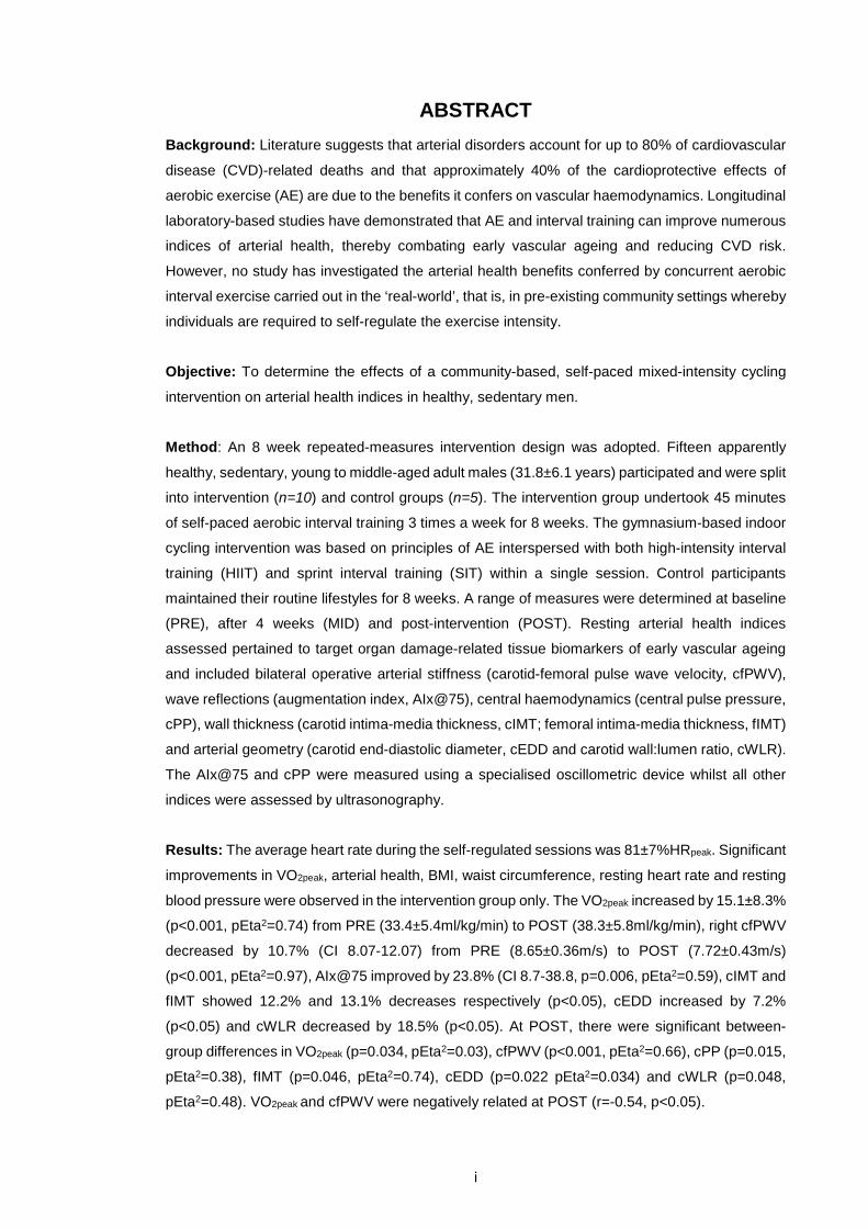
ABSTRACT Background: Literature suggests that arterial disorders account for up to 80% of cardiovascular
disease (CVD)-related deaths and that approximately 40% of the cardioprotective effects of
aerobic exercise (AE) are due to the benefits it confers on vascular haemodynamics. Longitudinal
laboratory-based studies have demonstrated that AE and interval training can improve numerous
indices of arterial health, thereby combating early vascular ageing and reducing CVD risk.
However, no study has investigated the arterial health benefits conferred by concurrent aerobic
interval exercise carried out in the ‘real-world’, that is, in pre-existing community settings whereby
individuals are required to self-regulate the exercise intensity.
Objective: To determine the effects of a community-based, self-paced mixed-intensity cycling
intervention on arterial health indices in healthy, sedentary men.
Method: An 8 week repeated-measures intervention design was adopted. Fifteen apparently
healthy, sedentary, young to middle-aged adult males (31.8±6.1 years) participated and were split
into intervention (n=10) and control groups (n=5). The intervention group undertook 45 minutes
of self-paced aerobic interval training 3 times a week for 8 weeks. The gymnasium-based indoor
cycling intervention was based on principles of AE interspersed with both high-intensity interval
training (HIIT) and sprint interval training (SIT) within a single session. Control participants
maintained their routine lifestyles for 8 weeks. A range of measures were determined at baseline
(PRE), after 4 weeks (MID) and post-intervention (POST). Resting arterial health indices
assessed pertained to target organ damage-related tissue biomarkers of early vascular ageing
and included bilateral operative arterial stiffness (carotid-femoral pulse wave velocity, cfPWV),
wave reflections (augmentation index, AIx@75), central haemodynamics (central pulse pressure,
cPP), wall thickness (carotid intima-media thickness, cIMT; femoral intima-media thickness, fIMT)
and arterial geometry (carotid end-diastolic diameter, cEDD and carotid wall:lumen ratio, cWLR).
The AIx@75 and cPP were measured using a specialised oscillometric device whilst all other
indices were assessed by ultrasonography. Results: The average heart rate during the self-regulated sessions was 81±7%HRpeak. Significant
improvements in VO2peak, arterial health, BMI, waist circumference, resting heart rate and resting
blood pressure were observed in the intervention group only. The VO2peak increased by 15.1±8.3%
(p<0.001, pEta2=0.74) from PRE (33.4±5.4ml/kg/min) to POST (38.3±5.8ml/kg/min), right cfPWV
decreased by 10.7% (CI 8.07-12.07) from PRE (8.65±0.36m/s) to POST (7.72±0.43m/s)
(p<0.001, pEta2=0.97), AIx@75 improved by 23.8% (CI 8.7-38.8, p=0.006, pEta2=0.59), cIMT and
fIMT showed 12.2% and 13.1% decreases respectively (p<0.05), cEDD increased by 7.2%
(p<0.05) and cWLR decreased by 18.5% (p<0.05). At POST, there were significant between-
group differences in VO2peak (p=0.034, pEta2=0.03), cfPWV (p<0.001, pEta2=0.66), cPP (p=0.015,
pEta2=0.38), fIMT (p=0.046, pEta2=0.74), cEDD (p=0.022 pEta2=0.034) and cWLR (p=0.048,
pEta2=0.48). VO2peak and cfPWV were negatively related at POST (r=-0.54, p<0.05).

ii
Conclusions and perspectives: In healthy, previously sedentary, young to middle-aged male
adults, self-paced cycling incorporating different modalities of interval training significantly
improves cardiorespiratory fitness and tissue biomarkers of early vascular ageing in addition to
causing systemic outward arterial remodelling. The adaptations observed are associated with an
improved CV risk profile, indicating the high responsiveness of this population to concurrent
aerobic interval training. The present results are consistent with those of previous controlled
laboratory-based studies and demonstrate the feasibility and effectiveness of a ‘real-world’
community-based exercise approach to enhance arterial health.
iii
TABLE OF CONTENTS ABSTRACT i
LIST OF FIGURES vii
LIST OF TABLES ix
ATTESTATION OF AUTHORSHIP x
ACKNOWLEDGEMENTS xi
ETHICAL APPROVAL xii
NOMENCLATURE xiii
CHAPTER 1: INTRODUCTION 1
1.1 Project context 1
1.2 Study rationale: What is known and gaps in knowledge 7
1.3 Project aims and objectives 8
1.4 Hypotheses 9
1.5 Methodological outline 9
1.6 Assumptions 9
1.7 Delimitations 10
1.8 Thesis structure 10
CHAPTER 2: THE PHYSIOLOGY, RELEVANCE AND ASSESSMENT OF ARTERIAL HEALTH 11
2.1 Cardiovascular disease (CVD) 11
2.1.1 Cardiovascular risk factors (CV RFs) and risk prediction 11
2.2 The arterial system and cardiovascular health 12
2.2.1 Normal arterial wall physiology and anatomy 13
2.3 Arterial health and early vascular ageing 14
2.3.1 The concepts of compliance and distensibility 15
2.4 Overview of arterial stiffness 16
2.4.1 The pathophysiology of arterial stiffening 17
2.4.2 The relevance and consequences of increased large artery stiffness 18
2.4.3 Clinical applications of AS assessment 19
2.5 Monitoring the status of arterial health 20
2.5.1 The assessment of AS: evaluation of pulse wave velocity, the pulse pressure waveform and wave reflections 20
2.6 Pulse wave velocity (PWV) 21
2.6.1 Clinical applications of PWV 21
2.6.2 The assessment of PWV 22
2.7 Pulse wave analysis (PWA) and the pulse pressure waveform 22
2.7.1 Pulse pressure 24
2.7.2 Central haemodynamics and pressure amplification 24
iv
2.7.3 The Augmentation index (AIx) 25
2.7.4 Non-invasive assessment of the AIx and central pressures 27
2.8 Flexibility and arterial stiffness 28
2.9 Intima-media thickness (IMT) 29
2.9.1 The physiology and speculated reasons behind intima-media thickening 30
2.9.2 The use of carotid IMT in CV risk stratification 30
2.9.3 The assessment of carotid IMT 32
2.10 The resistivity index 33
2.11 Arterial geometry and remodelling – arterial diameter and arteriogenesis 33
2.11.1 Carotid wall: lumen ratio (cWLR) 34
2.12 Associations between arterial stiffness, structural modifications and traditional CV risk
factors 34
2.12.1 Arterial stiffness and traditional CV risk factors 34
2.12.1.1 Abdominal obesity and vascular health 35
2.12.2 The association between atherosclerosis and arterial stiffness 35
CHAPTER 3:-THERAPEUTIC STRATEGIES AIMED AT OPTIMISING ARTERIAL HEALTH: THE VASCULOPROTECTIVE ROLE OF EXERCISE 37
3.1 Goals of pharmacological therapies 37
3.2 Non-pharmacologic therapies used to improve arterial health 37
3.3 Physical activity status and arterial health 38
3.3.1 The effects of physical inactivity and deconditioning on arterial structure and function 38
3.4 Exercise and vascular health 39
3.4.1 Exercise modes considered in arterial health research 39
3.5 SUMMARY OF STUDIES INVESTIGATING RELATIONSHIPS BETWEEN PHYSICAL
ACTIVITY AND ARTERIAL HEALTH 40
3.6 The acute effects of exercise on arterial stiffness and structure 44
3.6.1 Summary: the acute effects of exercise on arterial health 46
3.7 Habitual exercise and arterial stiffness 51
3.7.1 Mechanisms underlying the effects of exercise on arterial stiffness 52
3.7.2 Summary: exercise and arterial stiffness 54
3.8 Structural adaptations of arteries to exercise - IMT 59
3.8.1 Mechanisms underlying the effects of exercise on IMT 60
3.8.2 Summary: exercise and IMT 60
3.9 Structural adaptations of arteries to exercise - Arteriogenesis 65
3.9.1 Proposed mechanisms underlying exercise-induced arterial remodelling 66
3.10 A novel hypothesis 67
3.11 Group-fitness classes 67
v
3.12 Summary: exercise and arterial health 67
3.13. CONCLUSION 68
CHAPTER 4: PROJECT METHODOLOGY AND EQUIPMENT 69
4.1 Experimental design and cohort description 69
4.2 Definitions used in the current study 69
4.3 Exclusion criteria: as indicated in the advert and re-assessed during the initial session 71
4.4 Assessment schedule for outcome measures 72
4.5 Questionnaires and anthropometric, clinical, fitness and psychological measures 73
4.6 Measures of arterial health derived using an automated oscillometric device 74
4.7 Ultrasound assessments 75
4.7.1 Resting carotid-femoral pulse wave velocity (cfPWV) 75
4.7.1.1 The evaluation of cfPWV 76
4.7.2 Resting intima-media thickness (IMT) 77
4.7.2.1 Resting common carotid artery intima-media thickness (cIMT) assessment 77
4.7.2.2 Resting common femoral artery intima-media thickness (fIMT) assessment 78
4.7.3 Resting common carotid artery end-diastolic intra-luminal diameter (cEDD) 79
4.7.4 Calculations of resting common carotid artery wall:lumen ratio (cWLR) 79
4.8 Structure of study 79
4.8.1 Recruitment and group allocation 79
4.8.2 Control group 80
4.8.3 Intervention group and nature of exercise intervention 80
4.9 Statistical analysis 82
CHAPTER 5: RESULTS 83
5.1 Intervention compliance, training intensity and daily training loads 83
5.2: Between and within-group effects 88
5.3: Exploration of relationships between selected physiological variables 97
CHAPTER 6: DISCUSSION 106
6.1 Intervention characteristics 106
6.2 Primary measures 107
6.2.1 Main findings 107
6.2.1.1 Cardiorespiratory fitness (CRF) 107
6.2.1.2 Carotid-femoral pulse wave velocity (cfPWV) 107
6.2.1.3 Augmentation index (AIx, AIx@75) 108
6.2.1.4 Central haemodynamics 109
6.2.2 Relationships between arterial stiffness indices 110
6.2.3 RELATIONSHIPS BETWEEN Cardiorespiratory fitness, arterial stiffness and wave reflections 111
vi
6.2.4 Proposed mechanisms underlying the relationship between aerobic exercise and arterial stiffness 111
6.3 Secondary outcome measures: Arterial structural remodelling parameters 112
6.3.1 Possible reasons behind the systemic arterial structural adaptations observed 113
6.3.2 Potential mechanisms underlying the observed arterial remodelling 115
6.3.2.1 Relationships between and amongst primary and secondary outcome measures 116
6.3.2.2 Could RPM™ have a resistance exercise component? 117
6.4 Tertiary outcome measures 118
CHAPTER 7: CONCLUSIONS, IMPLICATIONS AND FUTURE DIRECTIONS 120
7.1 Perspectives and contribution to knowledge: What this study adds 120
7.2 Practical implications 121
7.3 Recommendations for further work 122
7.4 Limitations 124
7.5 Conclusions 126
REFERENCES 127
APPENDIX A: Ethics approval letter 156
APPENDIX B: Indices used to assess arterial stiffness and wall thickness 157
APPENDIX C: Techniques and equipment used to assess the arterial health indices assessed in the present study 162
APPENDIX D: International Physical Activity Questionnaire – Long form 166
APPENDIX E: Justification of ‘sedentary’ definition as used in the present study 173
APPENDIX F: Physical Activity Readiness Questionnaire 174
APPENDIX G: Modified sit-and-reach trunk flexibility test 175
APPENDIX H: Study advertisement flyer 176
APPENDIX I: Participant information sheet 177
APPENDIX J: Consent form 187
APPENDIX K: Example of activity log for intervention group 188
APPENDIX L: Official description of an RPM™ class 191
APPENDIX M: Example of raw heart rate data from Polar Team2 192
APPENDIX N: Average weekly intensity data for each subject over all 8 weeks 193
vii
LIST OF FIGURES
Figure 1.1: Assessment of vascular age status using traditional cardiovascular risk factors and
novel biomarkers 2
Figure 1.2: The two waves of the observed arterial pulse pressure waveform 3
Figure 1.3: Effects of large artery stiffening on central pulse pressure waveform 4
Figure 1.4: Ability of aerobic exercise to attenuate the natural age-associated arterial
dysfunction and prevent CVD 6
Figure 2.1: The age-associated distribution of risk factors in adults with metabolic syndrome 12
Figure 2.2: Major characteristics of early vascular ageing Image adapted from (86) 14
Figure 2.3: Arterial components of left ventricular afterload 15
Figure 2.4: Arterial stiffness as a reflection of the integrated and cumulative influence of
traditional cardiovascular risk factors on arterial walls 16
Figure 2.5: The consequences of arterial stiffening 19
Figure 2.6: Additive predictive value of arterial stiffness and traditional cardiovascular risk
factors 20
Figure 2.7: Foot-to-foot velocity method used for the determination of PWV 22
Figure 2.8: Central arterial pulse pressure waveform 23
Figure 2.9: Pulse pressure amplification from the aorta to the periphery in a young adult 25
Figure 2.10: The augmentation index 26
Figure 2.11: Influence of arterial stiffening on wave reflections and central haemodynamics 27
Figure 2.12: The BP+ device used in the present study 28
Figure 2.13: Relationship between intima-media thickness and atherosclerosis in the carotid
artery 29
Figure 2.14: Common carotid artery IMT assessment using B-mode ultrasound 32
Figure 2.15: Effects of metabolic syndrome on arterial health indices 36
Figure 3.1: Mechanism by which aerobic exercise attenuates age-associated arterial stiffening
39
Figure 3.2: Schematic illustrations of aerobic interval training and aerobic continuous training 41
Figure 3.3: Acute responses of PWV in upper and lower limbs following maximal treadmill
exercise in healthy adults 45
Figure 3.4: Relationship between cardiorespiratory fitness, arterial stiffness and myocardial work
capacity 51
viii
Figure 3.5: The effects of ageing and habitual aerobic exercise on aortic pulse wave velocity 52
Figure 3.6: Systemic arterial structural adaptations in healthy young males in response to 8
weeks of vigorous (80%HRmax) cycling 61
Figure 3.7: Upper body systemic arterial structural adaptations after lower-body aerobic training
62
Figure 3.8: Comparisons between brachial artery wall thickness and baseline diameter in
dominant and non-dominant limbs in elite squash players and controls 63
Figure 3.9: Brachial and carotid artery diameters and wall thickness in chronic recreational
(normal) versus intense exercise 65
Figure 4.1: General plan of the study indicating timeline of assessments 72
Figure 4.2: Calculation of carotid-femoral pulse wave velocity 76
Figure 4.3: The determination of carotid-femoral pulse wave velocity 76
Figure 5.1: Example of raw HR data collected during a typical RPM™ session 85
Figure 5.2: Average weekly intensity of exercise sessions across the intervention period 87
Figure 5.3: Arterial stiffness indices for both groups across time 91
Figure 5.4: Arterial geometrical parameters as a function of time 95
Figure 5.5: Trends in the peripheral AIx@75 across the 8 weeks in both groups 96
Figure 5.6: Negative correlation between VO2peak and cfPWV at POST 102
Figure 5.7: Correlations in PRE-POST adaptation magnitudes between variables 103
ix
LIST OF TABLES
Table 2.1: Normative data of common carotid IMT provided by the European Society of
Cardiology 31
Table 3.1: Findings from cross-sectional studies investigating the relationships between
physical activity levels/training status and AS indices 42
Table 3.2: Findings from cross-sectional studies investigating relationships between physical
activity levels/ training status and arterial structure 43
Table 3.3 Studies investigating the acute effects of exercise on arterial health indices (continued
on next page) 47
Table 3.3 continued: Studies investigating the acute effects of exercise on AS indices 50
Table 3.4: The effects of exercise interventions on arterial stiffness indices (continued on next
page) 55
Table 3.4 continued: The effects of various exercise intervention on arterial stiffness indices
(continued on next page) 56
Table 3.4 continued: The effects of various exercise intervention on arterial stiffness indices
(continued on next page) 57
Table 3.5 Summary of the main intervention studies investigating the effects of exercise training
on arterial structure 64
Table 4.1: Summary of subject and intervention characteristics 70
Table 5.1: Breakdown of class intensity fluctuations 86
Table 5.2 Average daily exertion score of all participants on non-RPM™ days 87
Table 5.3: Anthropometric, clinical, fitness and quality of life measures at each time point 90
Table 5.4: Arterial remodelling parameters at each of the three time points 94
Table 5.5a: Significant relationships between outcome measures at POST 99
Table 5.5b: Significant relationships between resting heart rate and outcome measures at
POST 100
Table 5.6: Correlations between bilaterally measured ultrasound-derived indices of arterial
stiffness and remodelling 100
Table 5.7: Significant relationships in improvement magnitudes between outcome measures 101
Table 5.8: Improvements in the main anthropometric, fitness, clinical and arterial health
measures in the intervention group from PRE to POST 104
Table 5.9: Framingham Risk Score-based 10–year CV risk factor profiles of participants in the
intervention group 105

x
ATTESTATION OF AUTHORSHIP I hereby declare that this submission is my own work and that, to the best of my knowledge and
belief, it contains no material previously published or written by another person, (except where
explicitly defined in the acknowledgements), nor material which to a substantial extent has been
accepted for the award of any other degree or diploma of a university or other institution of higher
learning.
Shivani Sethi
January 2016

xi
ACKNOWLEDGEMENTS First and foremost, I would like to thank Auckland University of Technology and Les Mills International for giving me this wonderful opportunity. Despite being an incredibly challenging year, it has been a remarkable experience doing what I love every day and I feel honoured and blessed to have worked on this project.
I would like to extend my deepest gratitude to my two supervisors, Dr. Andrew Kilding and Dr. Andrew Lowe for always being willing to help and guide me in the right direction whilst giving me considerable independence. Thank you for your invaluable support and advice and for entertaining my less-than-short emails!
I am extremely grateful to all the participants who made this study possible – Thank you very much for all your time and effort. It was a pleasure meeting you all and following you around the gym with my ‘purple device’!
Bryce – thanks for answering all my queries and thanks for your continued input and suggestions. Your stories are indeed fascinating and you have great wit!
Susan and Katty – Thank you very much for all your patience. I know Vic Street can get crazy at times but you are both so efficient. I really appreciate all your help with organising and coordinating the sessions. Thanks for making all the participants feel extremely welcome and comfortable in an unfamiliar setting.
Peter and Giles thanks for being so efficient at organising the RPM™ sessions. The participants loved the LM Taka experience!
I would like to say a huge thank you to Allan and the SPRINZ family – Thanks for your technical and emotional support throughout the challenging journey. Your friendly faces always brightened up my day and made me feel at home! The passion in all of you never ceases to amaze and motivate me.
I am so blessed to have a loving and caring family who support me in everything I do. Mum – If I start writing how much I appreciate and love you, the word count might exceed that of my whole thesis! Thanks for your unconditional love and for being my pillar of support during the toughest, busiest period of my life. I know I can always fall back on mummy in hard times!
Purvi and Vikas – Thanks for being right by my side ever since I came to NZ. Thanks for all your encouragement and love..I’ll always remain your little sister. You believed in me throughout this time and paid attention to the minute details that would make my day!
Rishaan – although you won’t quite understand this yet, you’ve made every academia-filled day so much better! During the stressful periods, imagining you pulling your ‘duck face’ and screaming ‘TIA’ always made me smile!
A very special mention to my big brother Bobby – Thanks for all your guidance over the past year (and always)! I would never have thought of doing research if it was not for you. Your wisdom is always so inspiring. No matter how old I get, I will always love hearing all your stories and aerospace engine analogies! Thanks, Alima, for putting up with our nerd humour!
I would like to say a huge thank you to all my friends for encouraging me, checking up on me and being there for me over the past year..not to mention, for bearing with my lack of social interaction over the past few months!
Thanks Melo – Now, more than ever, I miss you and I know that you are watching over me, my second mum!
And finally, thanks Freddy – my best friend and mentor. Thanks for giving me a confidence boost at the very start of this journey and for believing in me! It was hard to bear your passing in the middle of the project and I wish you were here to witness the outcome. I dedicate this thesis to you!

xii
ETHICAL APPROVAL Ethical Approval for this research was granted by the Auckland University of Technology Ethics
Committee (AUTEC). The AUTEC reference was 14/242 (Appendix A), with approval granted
originally on 26TH August 2014.
xiii
NOMENCLATURE Δdelta (% improvement)
AE aerobic exercise
AGEs advanced glycation end-products
AIT aerobic interval training
AIx augmentation index (%)
AIx@75 augmentation index standardised to a heart rate of 75 beats per minute (%)
ANS autonomic nervous system
AP augmentation pressure (mmHg)
AS arterial stiffness
BMI body mass index (kg/m2)
BP blood pressure (mmHg)
BRS baroreflex sensitivity
cBP central blood pressure (mmHg)
CCA common carotid artery
cEDD carotid end-diastolic diameter (mm)
CFA common femoral artery
cfPWV carotid-femoral pulse wave velocity (m/s)
cIMT carotid intima-media thickness (mm)
cPP central pulse pressure (mmHg)
CRF cardiorespiratory fitness
cRI carotid resistivity index
CV cardiovascular
CVD cardiovascular disease
cWLR carotid wall:lumen ratio
DBP diastolic blood pressure (mmHg)
ECM extracellular matrix
eNOS endothelial-derived nitric oxide synthase
ET-1 endothelin-1
EVA early vascular ageing
fIMT femoral intima-media thickness (mm)
FMD flow-mediated dilation
fRI femoral resistivity index
FRS Framingham Risk Score
HDL-C high-density lipoprotein cholesterol
HIIT high-intensity interval training
HR heart rate (beats per minute, bpm)
xiv
HRpeak peak individually-determined heart rate (beats per minute)
IMT intima-media thickness (mm)
IPAQ International Physical Activity Questionnaire
LDL-C low-density lipoprotein cholesterol
LMI Les Mills International
MA/O middle-aged and older adults
MAP mean arterial pressure (mmHg)
MCAE moderate continuous aerobic exercise
MS metabolic syndrome
NO nitric oxide
PAR-Q Physical Activity Readiness Questionnaire
pBP peripheral blood pressure (mmHg)
PP pulse pressure (mmHg)
pPP peripheral pulse pressure (mmHg)
PWV pulse wave velocity
RAAS renin-angiotensin-aldosterone system
RE resistance exercise
RFs risk factors
RI resistivity index
RPE Rating of perceived exertion
RPM™ Raw Power in Motion (intervention exercise)
SBP systolic blood pressure (mmHg)
SIT sprint interval training
SNA sympathetic nerve activity
TC total cholesterol
TC/HDL total cholesterol:high-density lipoprotein cholesterol ratio
TGs triglycerides
TPR total peripheral resistance
US ultrasound
VO2peak peak oxygen consumption (ml/kg/min)
VSMCs vascular smooth muscle cells
WC waist circumference (cm)
WLR wall:lumen ratio
1
CHAPTER 1: INTRODUCTION
1.1 PROJECT CONTEXT
Cardiovascular disease (CVD) and arterial health
Inadequate risk stratification obstructs the prevention of cardiovascular disease (CVD) (1), the
leading cause of mortality in modern societies (2). Traditional risk factors (RFs) such as increasing
age, hypertension, dyslipidaemia, obesity, physical inactivity, type II diabetes and tobacco use
account for 75–90% of the risk of developing CVD (3). Many of these RFs alter arterial structure,
properties and function (4), which is significant given that arterial disorders account for up to 80%
of all CVD-related deaths (5). Arterial structure and function are modified during the normal
vascular ageing process (6), but in some individuals, this process is accelerated (7) and
compounded by atherosclerosis-based pathological vascular ageing (8). This is known as
premature or early vascular ageing (EVA), and it increases predisposition to cardiovascular (CV)
events (7). Direct damage to arterial walls probably explains the remaining CVD risk which cannot
be attributed to the aforementioned traditional risk factors (9) and therefore, their optimal
functioning is necessary to maintain the integrity of the arterial system.
Fortunately, most CV disorders progress gradually (10) and many of the specific surrogate indices
of arterial health can be measured non-invasively during the sub-clinical stages of CVD, before
overt clinical manifestations (11). As most CVD-related disorders are associated with the natural
ageing process, risk stratification is improved by the determination of EVA via the assessment of
tissue biomarkers associated with target organ damage (12). These biomarkers include arterial
stiffness, arterial wall thickness, endothelial function and central pulse pressure (systolic-diastolic
blood pressure). The importance of assessing these facets lies in the fact that they are hallmark
features of CVD and related conditions which have significant global morbidity and mortality rates.
Assessment of circulating biomarkers (traditional CV risk factors) alone only gives a snapshot of
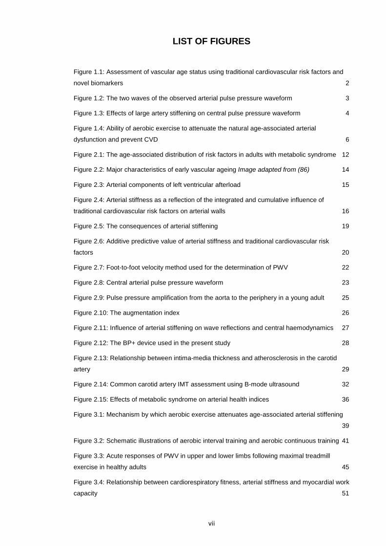
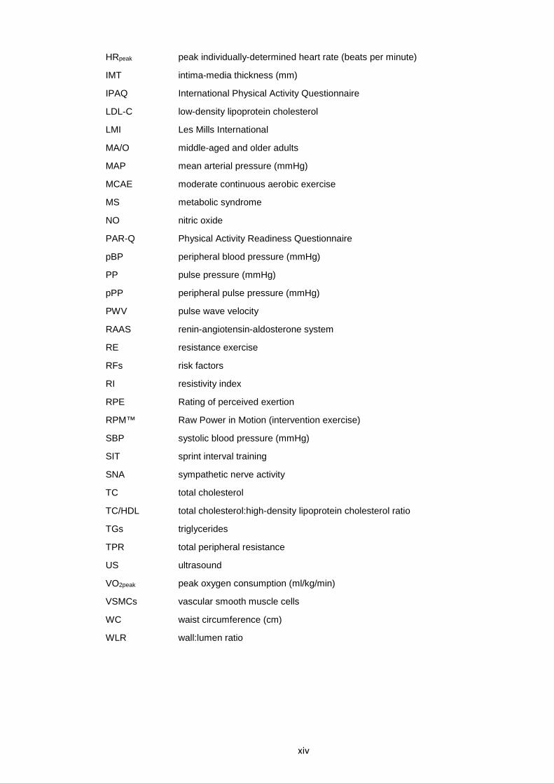
arterial wall damage (12). Figure 1.1 depicts the relationships between traditional CV risk factors,
biomarkers and EVA status. Ideally, EVA should be prevented by lifestyle interventions which can
decrease both traditional CV risk factors and tissue biomarkers.
The composition of each of the three layers of the arterial wall (tunica intima, media and
adventitia) varies along the heterogenous arterial tree with large, central, conduit arteries being
more elastic and peripheral ones more muscular (13). Progressive and passive damage to the
elastic arteries predisposes individuals to CVD (14). Arterial health status is ultimately monitored
to enable CVD risk stratification and CV event prediction, to diagnose early stages of CVD-related
disorders and to track the progress of pharmacological or non-pharmacological (lifestyle)
therapies (12). Due to this increasing awareness of arterial health in the pathogenesis of CVD,
there is continuing research into reliable and valid techniques to measure and quantify it (15).
2
Figure 1.1: Assessment of vascular age status using traditional cardiovascular risk factors and novel biomarkers
Cardiovascular risk stratification and prevention opportunities are optimised if traditional CV risk factors and tissue biomarkers are assessed simultaneously in young adulthood.
EVA, early vascular ageing
The umbrella term ‘arterial health’, refers to both structural and functional aspects of the arterial
system. In the present study, the term ‘arterial health’ refers to the operative stiffness of arterial
walls (pulse wave velocity), wave reflections (augmentation index), central haemodynamics
(central pulse pressure), structural arterial wall modifications (intima-media thickness) and arterial
geometry (end-diastolic diameter and wall:lumen ratio). Arterial stiffness is the primary
determinant of left ventricular afterload (16) and is influenced by both structural and functional
aspects but it is important to note that arterial stiffness, structural modifications, pulse wave
reflections and vascular geometry are all inter-dependent and act in synergy to determine arterial
health.
The concept of arterial stiffness
Blood ejected during ventricular systole is accommodated via vascular expansion (17). To avoid
being ruptured, arteries must be compliant enough to undergo a large change in volume for a
given pressure change. Arterial stiffness (AS), or wall rigidity, refers to the impaired ability of
arteries to constrict and expand in response to pressure changes (18). It occurs heterogeneously
in the arterial tree (with an increase in AS peripherally), generating a stiffness gradient and limiting
pulsatile flow to the microcirculation (19). This measure, which has a genetic component (20), is
associated with CV risk factors and can be used as a marker of potential vascular disease (6).
3
Early ageing of elastic arteries occurs either passively (related to heart rate and modified wall
composition) or actively (related to endothelial function) (21). During natural ageing, the main
change in the tunica media is a decreased elastin:collagen ratio, which increases AS, whilst the
main change in the intima is thickening, a process associated with atherosclerosis (13). An
increase in AS, prematurely or during the natural ageing process, has important implications for
overall CV performance as it affects blood pressure, blood flow and diameter change during the
cardiac cycle (21).
The assessment of arterial stiffness, wave reflections and central haemodynamics
Several methods and devices are used in the assessment of AS which can be measured either
invasively or using non-invasive yet reproducible and relatively inexpensive techniques (22). The
main indices of AS other than ultrasound-derived measures of compliance and distensibility are
central pulse pressure (cPP), the augmentation index (AIx), (both derived via pressure waveform
analysis, PWA) and pulse wave velocity (PWV), the gold-standard measure of AS.
Pulse wave velocity, which describes the speed of blood propagation along the arterial tree is a
surrogate marker for AS and independently predicts CV morbidity and mortality when measured
over the aorta (18). It is a measure of operative stiffness and is a determinant of ventricular
afterload (16). As the most widely used method to determine regional AS, aortic PWV is assessed
using Doppler guided by 2D ultrasound to record the time taken for the incident wave to travel
between two predetermined points a measured distance apart (23).
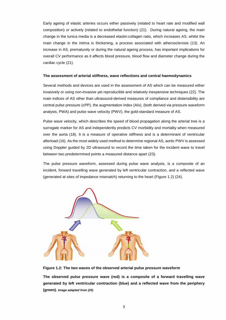
The pulse pressure waveform, assessed during pulse wave analysis, is a composite of an
incident, forward travelling wave generated by left ventricular contraction, and a reflected wave
(generated at sites of impedance mismatch) returning to the heart (Figure 1.2) (24).
Figure 1.2: The two waves of the observed arterial pulse pressure waveform
The observed pulse pressure wave (red) is a composite of a forward travelling wave generated by left ventricular contraction (blue) and a reflected wave from the periphery (green). Image adapted from (25)
4
Central blood pressures (cBPs) reflect the pressure at the root of the heart and have been more
closely linked to CVD than peripheral (brachial) ones (22). Central, but not brachial BP predicts
cardiovascular mortality independent of traditional CV risk factors: ‘We don’t die from the arm’
(Pr. D. Chemla). Central pulse pressure (systolic-diastolic BP) has the highest predictive value
amongst all BP indices and can be assessed non-invasively via analysis of the pulse pressure
waveform (6).
The augmentation index (AIx) is a partly hereditable, indirect index of AS that is independently
associated with CVD (26). It is a measure of pulse wave reflections and the resulting impact they
have on central pressures and consequently, ventricular afterload as well as the pressure
waveform shape and amplitude (27). Non-invasive assessment of the AIx can be carried out using
automated, specialised oscillometric devices (28).
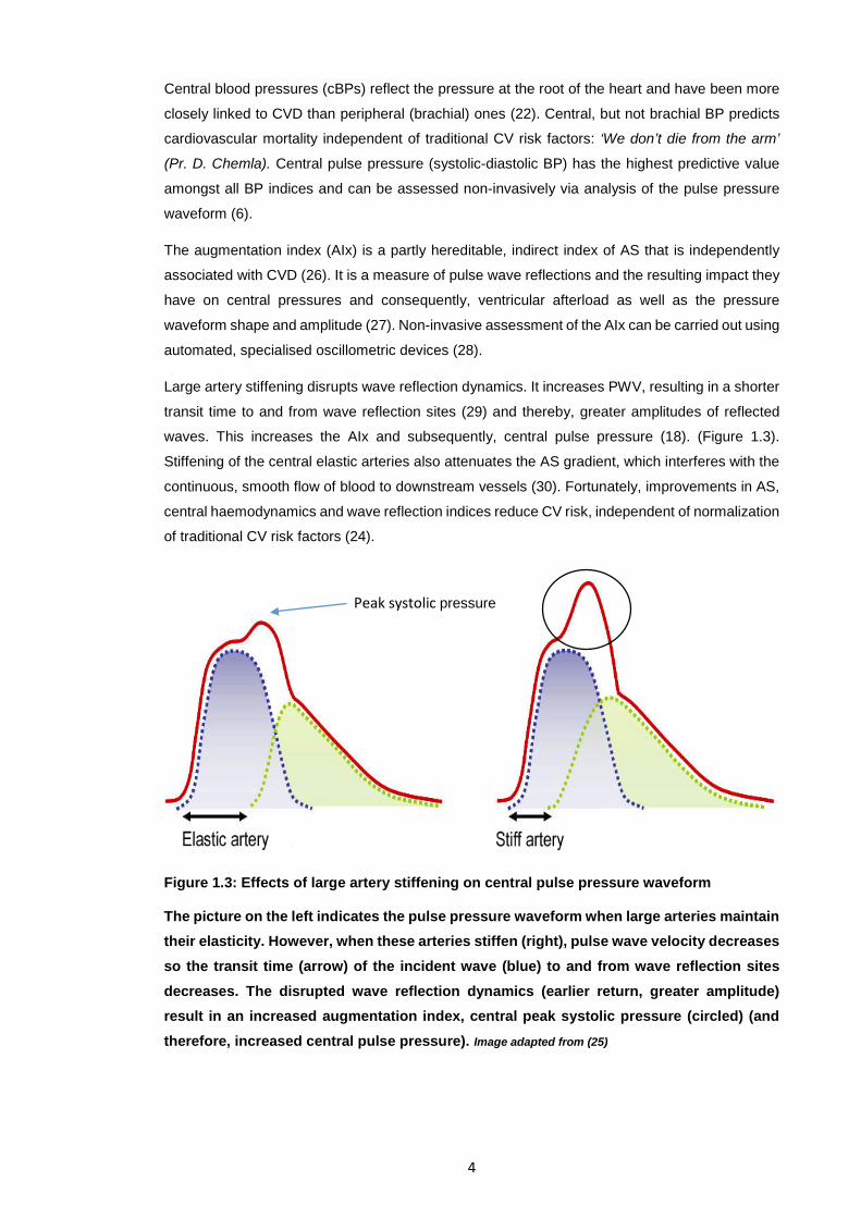
Large artery stiffening disrupts wave reflection dynamics. It increases PWV, resulting in a shorter
transit time to and from wave reflection sites (29) and thereby, greater amplitudes of reflected
waves. This increases the AIx and subsequently, central pulse pressure (18). (Figure 1.3).
Stiffening of the central elastic arteries also attenuates the AS gradient, which interferes with the
continuous, smooth flow of blood to downstream vessels (30). Fortunately, improvements in AS,
central haemodynamics and wave reflection indices reduce CV risk, independent of normalization
of traditional CV risk factors (24).
Figure 1.3: Effects of large artery stiffening on central pulse pressure waveform
The picture on the left indicates the pulse pressure waveform when large arteries maintain their elasticity. However, when these arteries stiffen (right), pulse wave velocity decreases so the transit time (arrow) of the incident wave (blue) to and from wave reflection sites decreases. The disrupted wave reflection dynamics (earlier return, greater amplitude) result in an increased augmentation index, central peak systolic pressure (circled) (and therefore, increased central pulse pressure). Image adapted from (25)
Peak systolic pressure
5
Arterial structural modifications and geometrical parameters
Pulse wave velocity, the gold-standard index of AS depends on both intrinsic wall properties in
addition to geometrical parameters such as wall thickness and lumen diameter (31). Due to the
influence of these remodelling indices on AS, they will be investigated as secondary measures in
the current study.
Advanced atherosclerosis is one of the first overt manifestations of CVD whereas structural
arterial wall modifications, including intimal thickening, occur over a prolonged subclinical phase
(32). Intima-media thickness (IMT), an ‘intermediate phenotype for early (local/generalised)
atherosclerosis’ (33), is a measure of the thickness of the tunica intima and media and is currently
the most widely studied and accepted sonographic marker for early atherosclerotic lesions (34).
The use of B-mode ultrasonography to measure in-vivo common carotid IMT (cIMT) is a
reproducible method for determining the degree of atherosclerosis and CV risk (35). The
assessment of arterial calibre can provide information regarding both autonomic functions as well
as arterial remodelling induced by external stimuli (36). End-diastolic intra-luminal diameter (EDD)
is assessed via B-mode ultrasonography and wall:lumen ratio (WLR) is calculated from the IMT
and EDD.
Exercise and arterial health indices
Whilst pharmacological therapies are capable of improving arterial health indices, non-
pharmacological lifestyle interventions, centred on diet and exercise, have shown considerable
success in normalizing these markers and thereby reducing CV risk. The deleterious effects of
physical inactivity are concerning given the increasingly sedentary lifestyle being adopted globally
(37). Regular physical activity reduces CVD mortality by up to 35% and all-cause mortality by up
to 33% (38) in a dose-dependent manner (39). Only 59% of the cardioprotective effects that
exercise has consistently been shown to confer can be attributed to favourable alterations in
traditional CV RFs (40, 41), with the rest being largely explained by haemodynamic alterations
and direct effects on the structure and functions of vascular walls both acutely and in the long-
term (42, 43).
Aerobic exercise (AE) of various modes can be effectively used to combat arterial ageing in
healthy adults (44). Amongst healthy, sedentary, normotensive young adults, 8 weeks of
moderate-intensity AE (cycling) for 30 minutes, 3 times a week reduces carotid artery stiffness
(45). Even 4 weeks of moderate-vigorous AE of this volume and frequency can decrease the AIx
(46) and central as well as peripheral PWV (47) and simultaneously increase whole-body arterial
compliance in a way that is linearly related to changes in VO2max (48). Several cross-sectional
studies have observed greater arterial compliance (49) and lower aortic PWV (50-52) and AIx
(53) in endurance-trained athletes compared to less active (recreationally-active/sedentary) age-
matched counterparts. Regular low-intensity AE also results in a reduction in aortic PWV (54).
Cross-sectional studies have generally reported lower peripheral IMT values in endurance-trained
athletes relative to sedentary age-matched counterparts (55, 56). It is uncommon, but not
6
impossible, to observe a reduction in cIMT in exercise interventions lasting shorter than 3 months
(57). Furthermore, AE training has been shown to cause systemic arterial remodelling, that is,
structural adaptations in conduit arteries of inactive arterial beds. Eight weeks of moderate to
high-intensity AE reduced carotid pulse pressure and wall thickness in healthy, sedentary young
adults (58) whilst 8 weeks of high-intensity cycling reduced carotid (in addition to peripheral) wall
thickness and WLR (59). Aerobic exercise is also known to increase arterial diameter in a
phenomenon known as outward remodelling (37) and 3 months of moderate to high-intensity
lower limb AE increased the femoral diameter by 9% in healthy, sedentary middle-aged men (56).
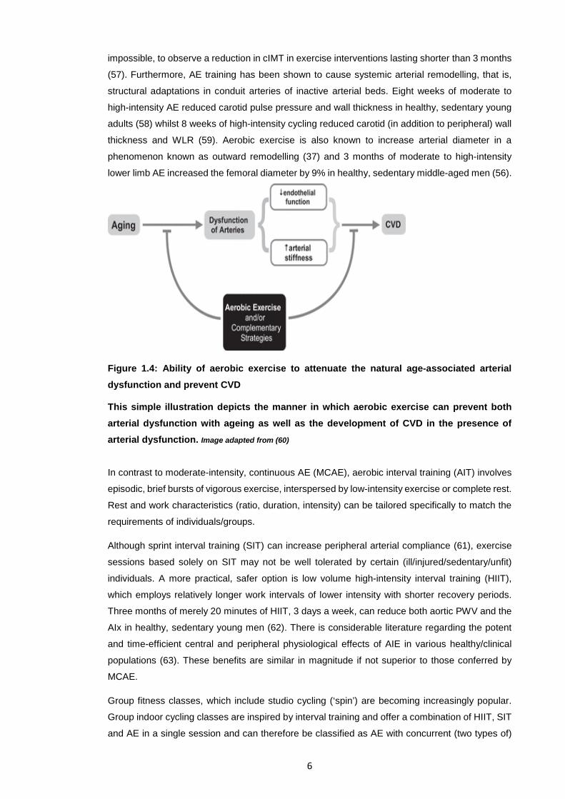
Figure 1.4: Ability of aerobic exercise to attenuate the natural age-associated arterial dysfunction and prevent CVD
This simple illustration depicts the manner in which aerobic exercise can prevent both arterial dysfunction with ageing as well as the development of CVD in the presence of arterial dysfunction. Image adapted from (60)
In contrast to moderate-intensity, continuous AE (MCAE), aerobic interval training (AIT) involves
episodic, brief bursts of vigorous exercise, interspersed by low-intensity exercise or complete rest.
Rest and work characteristics (ratio, duration, intensity) can be tailored specifically to match the
requirements of individuals/groups.
Although sprint interval training (SIT) can increase peripheral arterial compliance (61), exercise
sessions based solely on SIT may not be well tolerated by certain (ill/injured/sedentary/unfit)
individuals. A more practical, safer option is low volume high-intensity interval training (HIIT),
which employs relatively longer work intervals of lower intensity with shorter recovery periods.
Three months of merely 20 minutes of HIIT, 3 days a week, can reduce both aortic PWV and the
AIx in healthy, sedentary young men (62). There is considerable literature regarding the potent
and time-efficient central and peripheral physiological effects of AIE in various healthy/clinical
populations (63). These benefits are similar in magnitude if not superior to those conferred by
MCAE.
Group fitness classes, which include studio cycling (‘spin’) are becoming increasingly popular.
Group indoor cycling classes are inspired by interval training and offer a combination of HIIT, SIT
and AE in a single session and can therefore be classified as AE with concurrent (two types of)
7
interval training. Furthermore, these classes are self-paced (individuals select their own
intensities). Self-paced AIT likely has practical significance because exercise intensities have
been controlled such that they have been maintained within a certain range in most previous
intervention studies investigating the effects of exercise on arterial health,
While the benefits of AE on AS are well-documented, the effects of resistance exercise (RE) on
arterial health are less clear. Resistance exercise triggers different muscular adaptations to AE
and is thus associated with different haemodynamic, cardiovascular and muscular responses.
Four months of moderate-intensity RE can decrease arterial compliance (64, 65) and only 4
weeks of moderate-intensity RE can increase aortic and peripheral PWV (47) in healthy,
sedentary, young adult men. Furthermore, resistance-trained individuals have lower whole-body
arterial compliance than sedentary age-matched counterparts (66, 67). Findings regarding the
effects of RE and concurrent exercise (AE+RE) are equivocal and further research is needed to
draw definite conclusions about their population-specific effects.
1.2 STUDY RATIONALE: WHAT IS KNOWN AND GAPS IN KNOWLEDGE
What is currently known:
Arterial health indices are modifiable tissue biomarkers of early vascular ageing
Extensive cross-sectional and controlled, laboratory-based interventions have
demonstrated the vasculoprotective effects of both AE and several types of interval
training
Aerobic interval training (AIT) confers similar if not superior health benefits relative to
MCAE in a more time-efficient manner; HIIT may be more suitable than SIT for sedentary
(exercise beginners) or clinical populations as the former has relatively longer work
intervals of lower intensity and shorter rest periods
There are equivocal results regarding systemic arterial remodelling, especially in conduit
arteries
Persistence of the acute effects of repetitive bouts of exercise can partly explain chronic
changes in arterial health with exercise training
Discrepancies in findings between studies relate primarily to arterial health measurement
methods and subject characteristics at baseline
Mechanisms underlying the benefits of exercise on arterial health warrant further
investigation; potential mechanisms relate to training-induced modifications in vascular
smooth muscle cell proliferation/hypertrophy, alterations in arterial wall composition; anti-
oxidant and anti-inflammatory properties of exercise and changes in endothelial and
autonomic function (eg. nitric oxide upregulation)
Gaps in knowledge addressed in the present study
In the past, longitudinal studies investigating the effects of exercise on arterial health have been
carried out in laboratory settings where intensities are individually tailored and environmental
factors are controlled. However, it is important to determine the feasibility of these training stimuli
8
in pre-existing community environments where individuals are responsible for monitoring the
intensity of their own exercise. In the current study, the self-paced exercise, undertaken in a group
training environment outside the laboratory, prevents the need to speculate whether laboratory-
based findings have practical application to settings where neither training characteristics nor
environmental conditions (eg. humidity, temperature) are consistent, as these factors may
influence arterial adaptations to exercise.
Secondly, several studies have documented the beneficial effects of pure AE or concurrent AE
and RE on various arterial health indices. Fewer studies have done the same with solely HIIT and
SIT, or AE with a single mode of interval exercise. However, no study to our knowledge has
investigated the effect of concurrent AE, HIIT and SIT on these indices. In the present study,
aerobic interval training refers to a moderate to high-intensity exercise session based primarily
on AE of fluctuating moderate intensities, interspersed with regular intervals of HIIT and SIT bouts
(thus, the concurrent AE, HIIT and SIT nature in a single exercise session).
There is much debate as to whether specific muscle group training confers systemic benefits (that
is, on arterial beds supplying inactive muscles. The current study proposes that systemic arterial
adaptations which occur with supposedly isolated muscle training may be due to contractions of
secondary musculature not directly involved in the exercise and therefore considered to be
inactive.
1.3 PROJECT AIMS AND OBJECTIVES
1. To determine the effects of an 8 week concurrent aerobic interval community-based, self-paced
cycling intervention on arterial health indices related to early vascular ageing and remodelling
amongst apparently healthy, sedentary young and middle-aged adult males.
2. To describe an average aerobic interval exercise session and study changes in intensities
achieved across 8 weeks
3. To investigate the effects of the intervention on:
Cardiorespiratory fitness
Resting indices of EVA related to stiffness and wave reflections:
cfPWV
Peripheral AIx (and peripheral AIx standardised to a heart rate of 75
bpm)
Central haemodynamics (SBP, DBP and PP)
Resting arterial structure and geometry/remodelling parameters
Common carotid and common femoral IMT and resistivity index
Common carotid end-diastolic diameter
Common carotid wall:lumen ratio
General anthropometric and clinical measures
4. To explore:
Relationships between selected outcome measures
Time courses of adaptations
9
Changes in CV risk based on Framingham Risk Scores
5. To use the data collected to determine whether community-based group-fitness mixed-intensity
concurrent aerobic, HIIT and SIT exercise classes, attended and conducted by choice, would
beneficially impact the arterial health and overall CV risk status in healthy, sedentary young-to-
middle-aged adult males
NOTES TO READER: the aim is to quantify the impact of the exercise intervention on some traditional CV risk factors accounted for in the Framingham Risk Scoring system. It is outside the scope of this study to estimate or quantify cardiovascular risk reduction. Furthermore, it is not in the scope of this study to investigate or discuss endothelial function. However, it will be referred to in instances when deemed relevant to explain structural adaptations.
1.4 HYPOTHESES
In this cohort, 8 weeks of thrice weekly moderate-high intensity indoor cycling group-fitness
classes will improve tissue biomarkers of early vascular ageing related to AS (AIx, cfPWV, cPP)
and cIMT in addition to bringing about adaptive outward arterial remodelling and improving
cardiorespiratory fitness and traditional CV risk factors.
1.5 METHODOLOGICAL OUTLINE
All participants were community-dwelling, apparently healthy sedentary males between the ages
of 20 and 45 years. Ten participants took part in three 45-minute group-centred indoor (gym-
based) cycling classes each week for 8 weeks whilst 5 participants served as controls and
refrained from physical activity. Ultrasound-based measures of arterial health were taken at
baseline, midway and at the end of the intervention period. Basic anthropometric and arterial
stiffness measures were obtained on a weekly basis whilst aerobic capacity and biochemistry
were assessed at the start and end of the intervention period. Participants in the intervention
group wore heart rate monitors to determine class intensities.
1.6 ASSUMPTIONS
The sample size was large enough to obtain sufficient power (a priori analysis indicated
that a sample size of n=10 would be adequate)
Absence of subject bias when completing the International Physical Activity
Questionnaire to determine physical activity status prior to the intervention
There was no major dietary manipulation over the 8 weeks; Physical activity logs were
maintained honestly and any moderate to high-intensity activity outside the planned
intervention exercise was reported on all occasions
Participants did not have any underlying undiagnosed chronic illness
Good reliability and validity of the automated oscillometric device and ultrasound
equipment
10
1.7 DELIMITATIONS
This study was limited to 20-45 year old non-smoking males who were previously
sedentary (<2 hours moderate aerobic exercise per week for the past 6 months) and
currently apparently healthy (absence of chronic disease or prescription medication)
Arterial function was not investigated due to time constraints placed on the investigator
and time demands already placed on participants To specify results to the specific cohort used in this study, literature pertaining to females,
smokers, clinical populations and elderly individuals was considered but neither
elaborated upon nor presented due to their unique responsiveness and vascular
adaptations which may not apply to healthy individuals
Arterial health parameters were obtained by ultrasonography or a specialised (validated
and reliable) automated oscillometric device. Other techniques (such as invasive
methods, applanation tonometry and MRI) were not carried out Measurements presented are based on published guidelines and consensuses
The exercise intervention was RPM™, an indoor cycling group-fitness class trademarked
by Les Mills International. Substitute engagement in alternative indoor cycling
programmes (‘spin’ classes) were not permitted
1.8 THESIS STRUCTURE
This thesis comprises seven chapters, each regarding a specific aspect of this single
interventional study. An extensive literature review will be carried out in chapters 2 (Part A) and 3
(Part B). Chapter 2 concerns the physiology and assessment of arterial health indices. The
concepts, relevance, clinical applications and assessment methods of outcome measures in the
current study will be discussed. Chapter 3 focuses on the applied physiology and use of exercise
as a non-pharmacological therapeutic tool to improve arterial health. In this part of the literature
review, findings from key cross-sectional, acute and longitudinal intervention studies investigating
relationships between exercise and arterial health indices will be summarised and potential
underlying mechanisms pertaining to the vasculoprotective effects of exercise will be presented.
Chapter 4 describes the methods and methodologies used to address the research questions of
the present study and chapter 5 is a presentation of the findings.
Chapter 6 is a detailed discussion and interpretation of key findings and relationships between
the different outcome measures such that an integrated approach to addressing the topic is
adopted. Finally, chapter 7 summarises the contribution to knowledge of the present study and
explains the practical implications, significance and limitations of the current study and proposes
further work ideas.
11
CHAPTER 2: THE PHYSIOLOGY, RELEVANCE AND ASSESSMENT OF ARTERIAL HEALTH
2.1 CARDIOVASCULAR DISEASE (CVD)
Cardiovascular disease is a general term used to describe any condition that affects the heart
and/or blood vessels. The six main types of CVD are ischaemic heart disease, cerebrovascular
disease (stroke), peripheral vascular disease, heart failure, rheumatic heart disease and
congenital heart disease (68). Cardiovascular disease has a gradual onset (10) and it is vital to
track its progression using simple, reliable and valid techniques (69). This disease is the leading
cause of mortality in modern societies (2) and in 2012, the prevalence of CVD was higher than
any other health condition in New Zealand, especially in Maori, Pacific and European ethnic
groups (70). As the leading cause of death in New Zealand, CVD accounts for approximately 40%
of deaths annually (70), many of which are premature and can be prevented.
2.1.1 CARDIOVASCULAR RISK FACTORS (CV RFS) AND RISK PREDICTION
Cardiovascular disease is a multifactorial condition with no single cause. The presence or
absence of traditional cardiovascular risk factors (CV RFs) accounts for more than 75% of the
risk of developing CVD (3). Several CV RFs have been proposed to help identify individuals who
would benefit from preventive measures and to track the effectiveness of prevention programmes.
In asymptomatic individuals, risk prediction is currently based on scoring equations which
consolidate several CV RFs (71) but most of these scoring system algorithms do not incorporate
preclinical atherosclerosis measures, which would be useful to include (72) given that this
condition underlies numerous CV events and has a prolonged, progressive course (73). The
traditional CV RFs fall into several categories, with primarily genetic, physiological, behavioural
and socioeconomic characteristics. Non-modifiable RFs include increasing age, a family history
of CVD, ethnicity (African/Asian ancestry) and gender (74). Modifiable physiological/genetic RFs
include hypertension, type II diabetes, an abnormal blood lipid profile and left ventricular
hypertrophy. Physical inactivity, obesity, smoking/tobacco use, excessive alcohol consumption
and certain medications (such as the contraceptive pill and hormone replacement therapy) are
amongst the behavioural/lifestyle RFs whilst poverty, social isolation, anxiety and depression are
primary examples of socioeconomic RFs (74).
Metabolic syndrome (MS) is a multiplex CV RF (75) and is typically defined by the presence of at
least three out of abdominal obesity (increased waist circumference), atherogenic dyslipidaemia
(raised triglyceride and low HDL levels), elevated blood pressure (BP), insulin resistance (or
glucose intolerance), a proinflammatory state or a prothrombotic state (75). In 2007 in Auckland,
the prevalence of MS was Maori 32%, Pacific people 39% and other ethnic groups 16% (76). This
emphasises the need for practical lifestyle changes within communities to reduce the numbers of
12
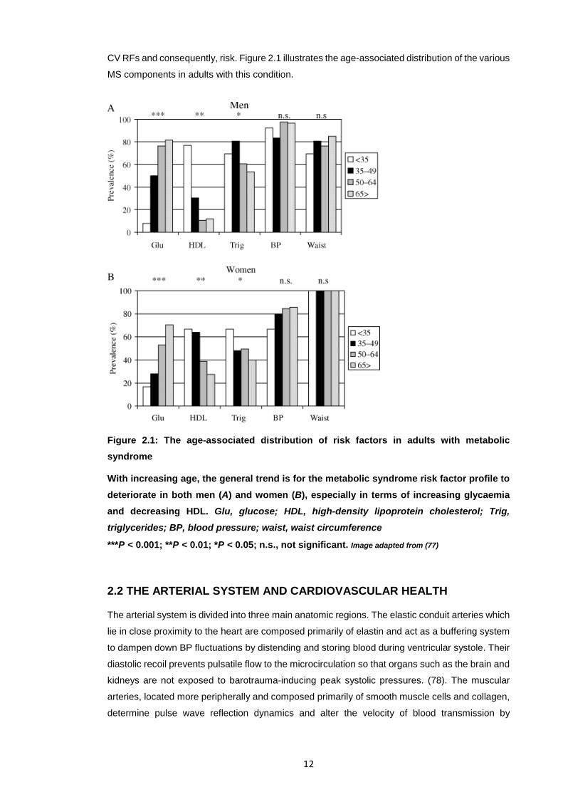
CV RFs and consequently, risk. Figure 2.1 illustrates the age-associated distribution of the various
MS components in adults with this condition.
Figure 2.1: The age-associated distribution of risk factors in adults with metabolic syndrome
With increasing age, the general trend is for the metabolic syndrome risk factor profile to deteriorate in both men (A) and women (B), especially in terms of increasing glycaemia and decreasing HDL. Glu, glucose; HDL, high-density lipoprotein cholesterol; Trig, triglycerides; BP, blood pressure; waist, waist circumference
***P < 0.001; **P < 0.01; *P < 0.05; n.s., not significant. Image adapted from (77)
2.2 THE ARTERIAL SYSTEM AND CARDIOVASCULAR HEALTH
The arterial system is divided into three main anatomic regions. The elastic conduit arteries which
lie in close proximity to the heart are composed primarily of elastin and act as a buffering system
to dampen down BP fluctuations by distending and storing blood during ventricular systole. Their
diastolic recoil prevents pulsatile flow to the microcirculation so that organs such as the brain and
kidneys are not exposed to barotrauma-inducing peak systolic pressures. (78). The muscular
arteries, located more peripherally and composed primarily of smooth muscle cells and collagen,
determine pulse wave reflection dynamics and alter the velocity of blood transmission by
13
modifying smooth muscle tone. Finally, the arterioles determine peripheral vascular resistance
and maintenance of mean arterial pressure (MAP) (78).
Arterial structure and function are modified during the normal vascular ageing process (6) but in
some individuals, this process is accelerated (7) and compounded by atherosclerosis-based
pathological vascular ageing (8). This is known as premature, or early vascular ageing (EVA) and
it increases predisposition to cardiovascular (CV) events (7). Direct damage to arterial walls
probably explains the remaining CVD risk which cannot be attributed to traditional CV risk factors
(9) and therefore, optimal functioning of arterial walls is necessary to maintain the integrity of the
arterial system.
Major changes in elastic arteries occur passively over long time periods whilst changes in
muscular arteries and arterioles occur acutely and actively (79, 80). Damage to the large elastic
arteries predisposes to CVD (14) with diseases of conduit arteries accounting for the majority of
the global heart disease and stroke burden (81). Primary adverse changes to arteries with ageing
are increased stiffness, changes in wave reflection patterns and endothelial dysfunction (82). The
early evaluation of arterial structure and function when assessing CV RFs is thus important to
reduce the age-associated CVD burden (83) as is the implementation of strategies to delay, slow
or prevent these changes.
2.2.1 NORMAL ARTERIAL WALL PHYSIOLOGY AND ANATOMY
Optimal functioning of the arterial wall is vital to the integrity of the arterial tree, as evidenced by
the potentially fatal effects of arterial wall dysfunction, disease or ageing. The cells and
extracellular matrix (ECM) are organised into three distinct concentric cylindrical layers, known
as the tunica intima (innermost layer), media (middle layer) and adventitia (outer layer). The
proportion of elastic fibres and the thickness of the smooth muscle layer varies between arteries,
giving rise to a heterogenous arterial tree. From the proximal to distal (peripheral) portion of the
arterial system, the diameter of the vessels and the proportion of the elastic fibres decrease whilst
the amount of smooth muscle increases (13). Due to the non-linear viscoelastic properties as well
as the anisotropic and highly adaptive nature of arteries (84, 85), conclusions drawn regarding a
specific segment should not be generalised and extrapolated to different parts of the
heterogenous arterial tree (13) unless there is substantial evidence of systemic effects and
generalisations are permitted by current standards.
As the demands on the vascular system are dynamic and constantly changing, the cellular
components of the wall need to carry out efficient remodelling by altering the quantity, distribution
and/or orientation of collagen fibres (17). Equilibrium between the production and degradation of
collagen and elastin, the two main arterial wall scaffolding proteins, is paramount to ensure
optimal arterial wall function and vascular integrity.
14
2.3 ARTERIAL HEALTH AND EARLY VASCULAR AGEING
In addition to the traditional CV RFs, programming from intrauterine growth retardation contributes
towards CVD and this is known as the ‘mismatch hypothesis’ (86). Vascular structure and function
are partly determined by early programming in life and impaired foetal growth is associated with
various changes in arterial health which can eventually cause CVD (86, 87). Some individuals are
prone to exhibiting early ageing of arteries (a condition termed early vascular ageing, EVA, or
EVA syndrome), the main consequence of which is target organ damage (8). However, there is
controversy regarding the definition of EVA (86). Given that most CVD-related events are
associated with ageing (the most important CV risk factor), determination of EVA has been
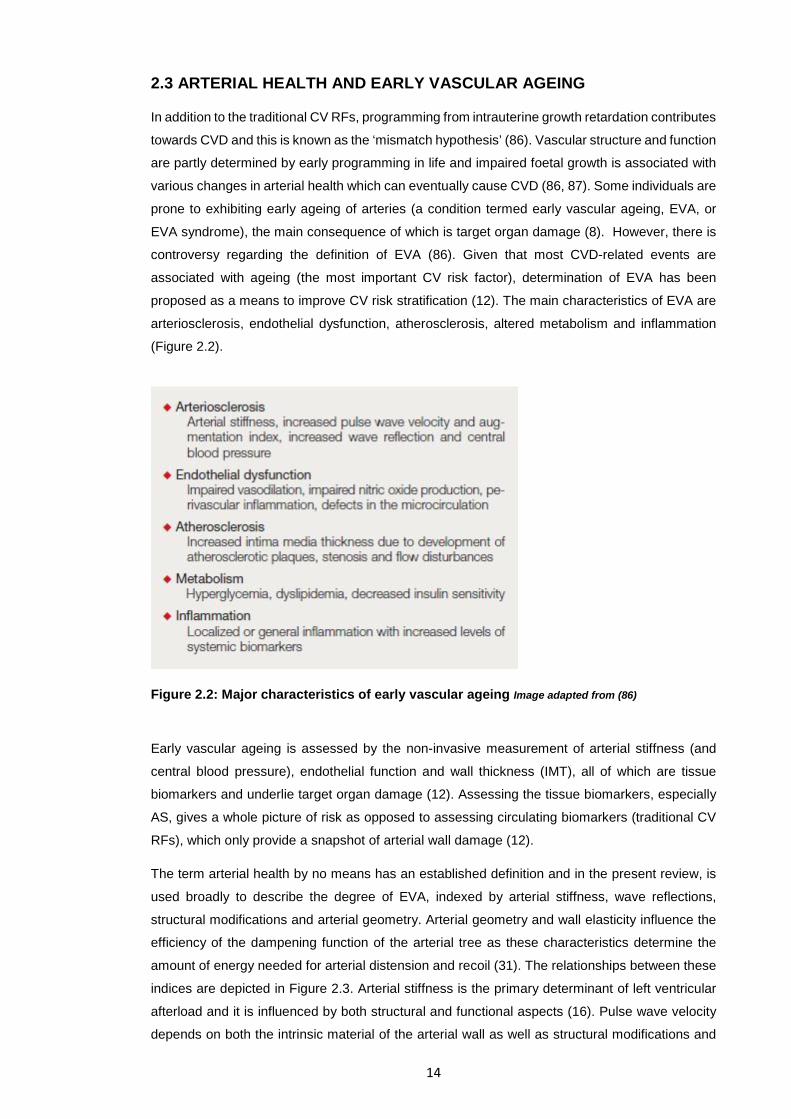
proposed as a means to improve CV risk stratification (12). The main characteristics of EVA are
arteriosclerosis, endothelial dysfunction, atherosclerosis, altered metabolism and inflammation
(Figure 2.2).
Figure 2.2: Major characteristics of early vascular ageing Image adapted from (86)
Early vascular ageing is assessed by the non-invasive measurement of arterial stiffness (and
central blood pressure), endothelial function and wall thickness (IMT), all of which are tissue
biomarkers and underlie target organ damage (12). Assessing the tissue biomarkers, especially
AS, gives a whole picture of risk as opposed to assessing circulating biomarkers (traditional CV
RFs), which only provide a snapshot of arterial wall damage (12).
The term arterial health by no means has an established definition and in the present review, is
used broadly to describe the degree of EVA, indexed by arterial stiffness, wave reflections,
structural modifications and arterial geometry. Arterial geometry and wall elasticity influence the
efficiency of the dampening function of the arterial tree as these characteristics determine the
amount of energy needed for arterial distension and recoil (31). The relationships between these
indices are depicted in Figure 2.3. Arterial stiffness is the primary determinant of left ventricular
afterload and it is influenced by both structural and functional aspects (16). Pulse wave velocity
depends on both the intrinsic material of the arterial wall as well as structural modifications and

15
arterial geometry. Geometrical parameters include lumen diameter, cross-sectional area and
wall:lumen ratio whilst structural factors include wall thickness and atherosclerosis (Figure 2.3).
These properties are also associated with arterial remodelling and will, therefore, be discussed
presently. It is important to note that arterial stiffness, structural modifications, pulse wave
reflections and vascular geometry are all inter-dependent and act in synergy to determine arterial
health.
Figure 2.3: Arterial components of left ventricular afterload
This figure depicts the arterial components of left ventricular afterload (blue boxes) as well as their structural and functional determinants. Y, Young’s elastic modulus; Ep; Peterson’s elastic modulus; PWV, pulse wave velocity; Intima-media thickness, Ea, effective elastance. Image adapted from (16)
2.3.1 THE CONCEPTS OF COMPLIANCE AND DISTENSIBILITY
During ventricular systole, incompressible blood is pumped out of the heart and must be
accommodated via vascular expansion (17). To avoid being ruptured, it is necessary that vessels,
especially arteries, are compliant. Compliance, a dynamic and fluctuating parameter, describes a
volume (or diameter) change in response to a change in blood pressure (ΔP). Distensibility is
simply compliance relative to initial volume (or diameter) (18). The distensibility of a specific
arterial segment reflects the mechanical stress the wall undergoes during the cardiac cycle (88).
Compliance and distensibility describe how stiff the vessel is and in general, distensibility is
greater in the proximal portion of the arterial tree (6). Ageing and several pathological conditions
affect the interaction between the cellular and structural components of the arterial wall and lead
to an increase in arterial wall rigidity (arterial stiffening) (13).
s
16
2.4 OVERVIEW OF ARTERIAL STIFFNESS
Arterial stiffness (AS), or arterial wall rigidity, the reciprocal of distensibility (69), has not yet been
well defined (28), but it generally refers to the impaired ability of arteries to constrict and expand
in response to pressure changes (18). It is a primary determinant of compliance associated with
subclinical systemic target organ damage and is a reflection of the integrated, cumulative
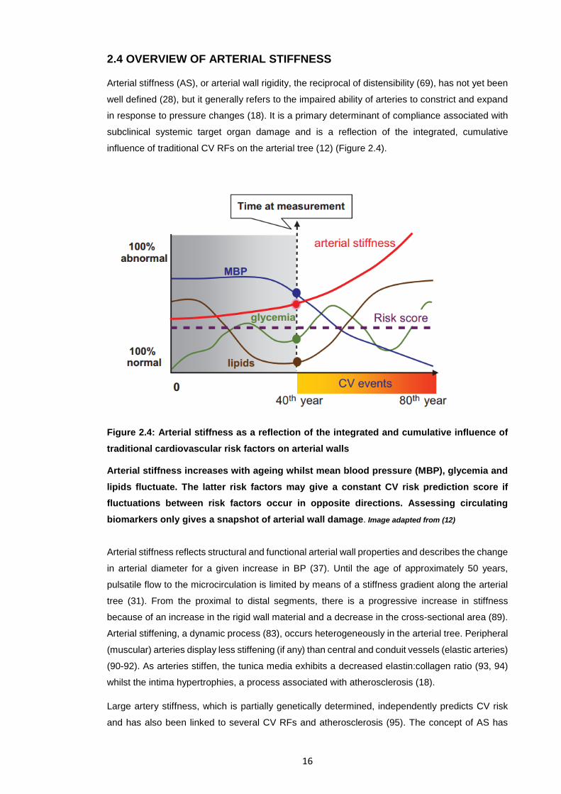
influence of traditional CV RFs on the arterial tree (12) (Figure 2.4).
Figure 2.4: Arterial stiffness as a reflection of the integrated and cumulative influence of traditional cardiovascular risk factors on arterial walls
Arterial stiffness increases with ageing whilst mean blood pressure (MBP), glycemia and lipids fluctuate. The latter risk factors may give a constant CV risk prediction score if fluctuations between risk factors occur in opposite directions. Assessing circulating biomarkers only gives a snapshot of arterial wall damage. Image adapted from (12)
Arterial stiffness reflects structural and functional arterial wall properties and describes the change
in arterial diameter for a given increase in BP (37). Until the age of approximately 50 years,
pulsatile flow to the microcirculation is limited by means of a stiffness gradient along the arterial
tree (31). From the proximal to distal segments, there is a progressive increase in stiffness
because of an increase in the rigid wall material and a decrease in the cross-sectional area (89).
Arterial stiffening, a dynamic process (83), occurs heterogeneously in the arterial tree. Peripheral
(muscular) arteries display less stiffening (if any) than central and conduit vessels (elastic arteries)
(90-92). As arteries stiffen, the tunica media exhibits a decreased elastin:collagen ratio (93, 94)
whilst the intima hypertrophies, a process associated with atherosclerosis (18).
Large artery stiffness, which is partially genetically determined, independently predicts CV risk
and has also been linked to several CV RFs and atherosclerosis (95). The concept of AS has

17
gained increased interest over the past decade due to its association with CVD (including its use
as a biomarker for CVD risk and potential vascular disease). An increase in arterial wall rigidity
has important implications for arterial and overall CV performance (6, 84, 96-98). Despite the use
of various specific AS indices, the pulse pressure wave characteristics associated with central
haemodynamics and wave reflections as well as the travel velocity of the pulse wave are integral
to the evaluation of AS using non-invasive techniques.
2.4.1 THE PATHOPHYSIOLOGY OF ARTERIAL STIFFENING
The two main arterial wall scaffolding proteins, collagen and elastin, determine the structural
integrity of the wall (98). With ageing, the major determinant of arterial stiffening (89), histological
changes, (independent of those associated with atherosclerosis) are observed in the walls of
elastic arteries. Elastic fibres in the TM of central arteries (92) display fraying, splitting,
fragmentation and thinning (99) and the elastic laminae become disoriented (100), probably due
to repetitive cyclic stress (101). Histological examination shows disorganised endothelial cells,
infiltration of vascular smooth muscle cells (VSMCs), macrophages and mononuclear cells and
greater amounts of cytokines and cell adhesion molecules (82), which indicate the presence of
atherosclerosis, a ‘pathological condition of the intima’ (18). Between the ages of 20 to 70 years,
the stiff collagen content more than doubles (78). Furthermore, up-regulation of the tissue renin-
angiotensin-aldosterone system (RAAS) is associated with tunica intima thickening and arterial
wall remodelling (102-104). Equilibrium between the synthesis and degradation of wall scaffolding material Several pathological events at the molecular level have been implicated in increased AS by
causing an imbalance between the synthesis and degradation of structural proteins of the ECM.
Equilibrium must be maintained between elastin and collagen synthesis and degradation to
maintain normal arterial distensibility (105). Disruption of elastin and collagen cross-links (106-
108) as well as changes in elastin production and repair mechanisms also increase AS (106-108).
Catabolic (elastinolytic and collagenolytic) matrix metalloproteinases (MMPs) (18), produced by
vascular and inflammatory cells (109), fray intimal and medial elastin molecules (110, 111) and
uncoil collagen thereby degrading the ECM (98).
Inflammatory changes due to nuclear factor kappa β intracellular signalling lead to production and
deposition of excessive abnormal collagen and decreased amounts of normal elastin (93, 112).
Non-enzymatic glycation of ECM proteins forms advanced glycation end-products (AGEs) (93)
which make collagen stiffer and less prone to turnover by MMPs, resulting in the accumulation of
abnormal collagen in arterial walls (113). Advanced glycation end-products also increase the
production of reactive oxygen species (ROS), thereby reducing nitric oxide (NO) activity and
impairing endothelial cell function (114). Advanced glycation end-products are pro-inflammatory
(115, 116) and may contribute to arterial stiffening and atherosclerotic plaque formation (117-
119). Finally, increased luminal pressure can stimulate excessive abnormal collagen production
(120) and can explain both hypertrophy of the smooth muscle layer as well as the increased
18
thickness of the intima and media by two to three times between the ages of 20 and 90 years
(121, 122).
2.4.2 THE RELEVANCE AND CONSEQUENCES OF INCREASED LARGE ARTERY STIFFNESS
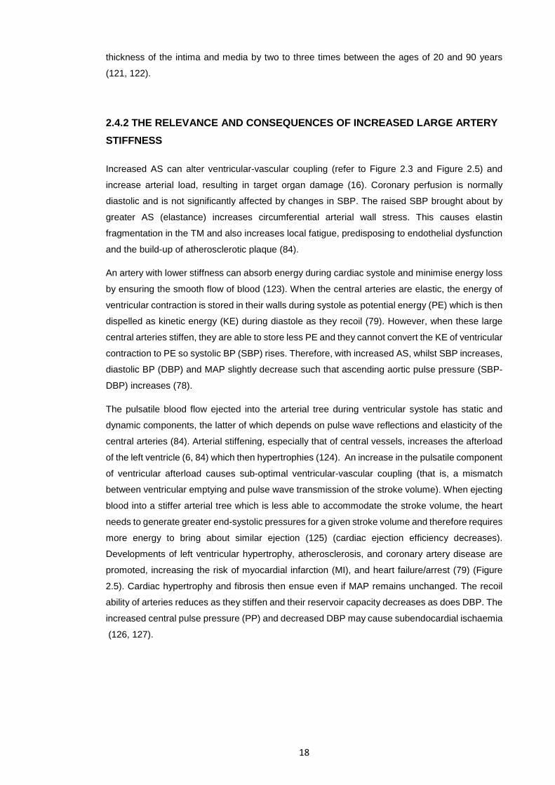
Increased AS can alter ventricular-vascular coupling (refer to Figure 2.3 and Figure 2.5) and
increase arterial load, resulting in target organ damage (16). Coronary perfusion is normally
diastolic and is not significantly affected by changes in SBP. The raised SBP brought about by
greater AS (elastance) increases circumferential arterial wall stress. This causes elastin
fragmentation in the TM and also increases local fatigue, predisposing to endothelial dysfunction
and the build-up of atherosclerotic plaque (84).
An artery with lower stiffness can absorb energy during cardiac systole and minimise energy loss
by ensuring the smooth flow of blood (123). When the central arteries are elastic, the energy of
ventricular contraction is stored in their walls during systole as potential energy (PE) which is then
dispelled as kinetic energy (KE) during diastole as they recoil (79). However, when these large
central arteries stiffen, they are able to store less PE and they cannot convert the KE of ventricular
contraction to PE so systolic BP (SBP) rises. Therefore, with increased AS, whilst SBP increases,
diastolic BP (DBP) and MAP slightly decrease such that ascending aortic pulse pressure (SBP-
DBP) increases (78).
The pulsatile blood flow ejected into the arterial tree during ventricular systole has static and
dynamic components, the latter of which depends on pulse wave reflections and elasticity of the
central arteries (84). Arterial stiffening, especially that of central vessels, increases the afterload
of the left ventricle (6, 84) which then hypertrophies (124). An increase in the pulsatile component
of ventricular afterload causes sub-optimal ventricular-vascular coupling (that is, a mismatch
between ventricular emptying and pulse wave transmission of the stroke volume). When ejecting
blood into a stiffer arterial tree which is less able to accommodate the stroke volume, the heart
needs to generate greater end-systolic pressures for a given stroke volume and therefore requires
more energy to bring about similar ejection (125) (cardiac ejection efficiency decreases).
Developments of left ventricular hypertrophy, atherosclerosis, and coronary artery disease are
promoted, increasing the risk of myocardial infarction (MI), and heart failure/arrest (79) (Figure
2.5). Cardiac hypertrophy and fibrosis then ensue even if MAP remains unchanged. The recoil
ability of arteries reduces as they stiffen and their reservoir capacity decreases as does DBP. The
increased central pulse pressure (PP) and decreased DBP may cause subendocardial ischaemia
(126, 127).
19
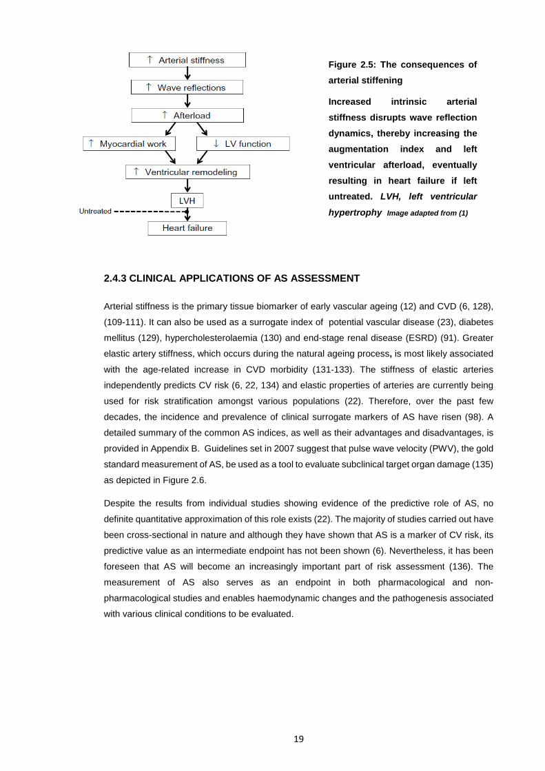
2.4.3 CLINICAL APPLICATIONS OF AS ASSESSMENT
Arterial stiffness is the primary tissue biomarker of early vascular ageing (12) and CVD (6, 128),
(109-111). It can also be used as a surrogate index of potential vascular disease (23), diabetes
mellitus (129), hypercholesterolaemia (130) and end-stage renal disease (ESRD) (91). Greater
elastic artery stiffness, which occurs during the natural ageing process, is most likely associated
with the age-related increase in CVD morbidity (131-133). The stiffness of elastic arteries
independently predicts CV risk (6, 22, 134) and elastic properties of arteries are currently being
used for risk stratification amongst various populations (22). Therefore, over the past few
decades, the incidence and prevalence of clinical surrogate markers of AS have risen (98). A
detailed summary of the common AS indices, as well as their advantages and disadvantages, is
provided in Appendix B. Guidelines set in 2007 suggest that pulse wave velocity (PWV), the gold
standard measurement of AS, be used as a tool to evaluate subclinical target organ damage (135)
as depicted in Figure 2.6.
Despite the results from individual studies showing evidence of the predictive role of AS, no
definite quantitative approximation of this role exists (22). The majority of studies carried out have
been cross-sectional in nature and although they have shown that AS is a marker of CV risk, its
predictive value as an intermediate endpoint has not been shown (6). Nevertheless, it has been
foreseen that AS will become an increasingly important part of risk assessment (136). The
measurement of AS also serves as an endpoint in both pharmacological and non-
pharmacological studies and enables haemodynamic changes and the pathogenesis associated
with various clinical conditions to be evaluated.
Figure 2.5: The consequences of arterial stiffening
Increased intrinsic arterial stiffness disrupts wave reflection dynamics, thereby increasing the augmentation index and left ventricular afterload, eventually resulting in heart failure if left untreated. LVH, left ventricular
hypertrophy Image adapted from (1)
20
Figure 2.6: Additive predictive value of arterial stiffness and traditional cardiovascular risk factors
In low-to-moderate risk hypertensives with a 5.9-year follow-up, predictive values of PWV and FRS for coronary heart disease were assessed from the area under the curve. PWV and FRS had similar predictive values. When PWV and FRS were combined, the predictive value significantly increased. In elderly participants, PWV significantly improved the predictive value of combined criteria for future CV events. PWV, pulse wave velocity; FRS,
Framingham Risk Score; CV RF, cardiovascular risk factors Image and legend adapted from (12)
2.5 MONITORING THE STATUS OF ARTERIAL HEALTH
The main CV complications associated with EVA gradually progress on a continuum presenting
as subclinical target organ damage (refer to Figure 2.2). Prior to overt clinical manifestation, these
events can be identified using validated surrogate markers which are all independent CV RFs.
Structural/morphological changes are evaluated by measuring the thickness of the intima-media
complex (intima-media thickness, IMT) (13). Non-invasive techniques used to monitor CVD
progression primarily assess atherosclerotic status and regional/local/systemic AS. The methods
(including the benefits and drawbacks of each) used to determine the degrees of arterial stiffening
and remodelling parameters related to atherosclerosis are described in Appendix C.
2.5.1 THE ASSESSMENT OF AS: EVALUATION OF PULSE WAVE VELOCITY, THE PULSE PRESSURE WAVEFORM AND WAVE REFLECTIONS
The main indices of AS other than ultrasound-derived measures of compliance and distensibility
are based on central haemodynamics and wave reflections assessed via pulse wave analysis
(PWA) and evaluation of pulse wave velocity (PWV), the gold standard measurement of AS.
Attention is increasingly being directed toward PWV, central pulse pressure and wave reflections
as independent CV risk factors in various populations because they are examples of target organ
21
damage, which is a mediator between traditional CV RFs and CV events (12). Each AS index can
be measured in several different ways using specific equipment and protocols (refer to
Appendices B and C) Simple, non-invasive, reproducible, accurate and relatively inexpensive
methods are ideal for regional/local/systemic AS evaluation in large-scale studies (83).
Unfortunately, normative data is equivocal due to the different assessment methods and
populations so values will not be reported here.
Note on Appendices B and C: Appendix B summarises the advantages and disadvantages of the
main AS indices and Appendix C summarises the equipment and procedures used to assess the
arterial health parameters relevant to the present study. These indices have been compiled after
considerable literature reviews. The main points have been covered in references (137), (138),
(13), (18), (83, 139) and (15).
2.6 PULSE WAVE VELOCITY (PWV)
The pressure pulse generated by left ventricular ejection propagates along the arterial system
with a speed determined by arterial wall elasticity and geometry (140). Therefore, arterial wall
elasticity determines pulse wave velocity (PWV), a parameter which describes the speed of blood
propagation along the arterial tree (18). Pulse wave velocity is a simple, non-invasive and
reproducible method used to assess operative AS (23) and is considered the gold standard
surrogate marker for AS (18). Aortic (central) PWV usually tends to increase during the healthy
ageing process (30, 141). Aortic PWV is related to mechanical properties of arterial walls (142),
wall thickness and luminal diameter (83) and to a lesser extent, characteristics of the blood. Pulse
wave propagation and arterial distensibility are inversely related such that the greater the AS, the
higher the speed of both the forward and retrograde waves (143).
2.6.1 CLINICAL APPLICATIONS OF PWV
Arterial stiffness measured via PWV independently predicts all-cause and CV mortality and
morbidity (18), coronary events and stroke in hypertensives (144), type II diabetics, elderly
individuals and in those with ESRD (6). Although PWV is used to evaluate the status of central
arteries, there is a poor correlation between PWV and established CV RFs other than age and
BP (145). Individuals classified as low-to-intermediate risk using the Framingham Risk Score
(FRS) who having no current target organ damage may benefit most from risk assessment using
PWV (6). As an EVA tissue biomarker related to systemic target organ damage, cfPWV can be
validly added to the FRS to improve risk classification and predict CVD-related death and risk in
addition to traditional risk factors (12). Age, SBP and gender explain >50% of the variability of
aortic PWV with the former two parameters being positively correlated to aortic PWV. Less than
1% of PWV variance is explained by clustering of MS RFs (146). Cecelja et al. suggest that central
PWV is a better measure of BP than those currently used in clinical settings (145). A 5m/s
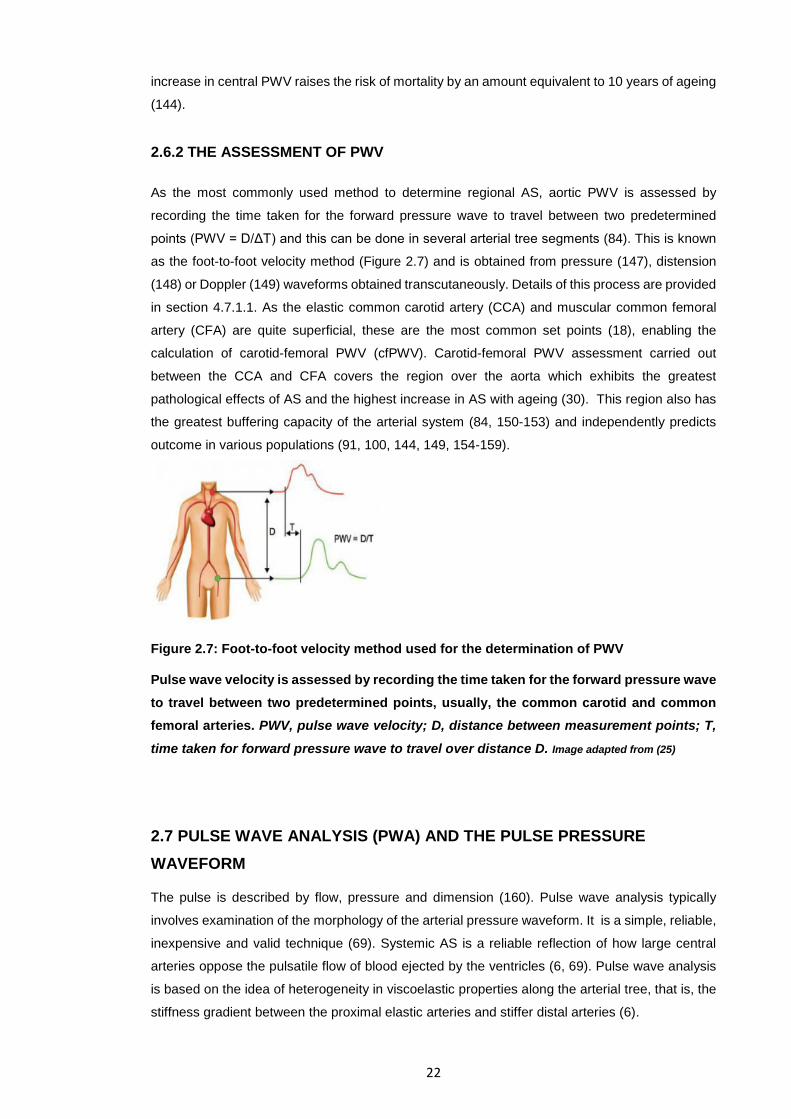
22
increase in central PWV raises the risk of mortality by an amount equivalent to 10 years of ageing
(144).
2.6.2 THE ASSESSMENT OF PWV
As the most commonly used method to determine regional AS, aortic PWV is assessed by
recording the time taken for the forward pressure wave to travel between two predetermined
points (PWV = D/ΔT) and this can be done in several arterial tree segments (84). This is known
as the foot-to-foot velocity method (Figure 2.7) and is obtained from pressure (147), distension
(148) or Doppler (149) waveforms obtained transcutaneously. Details of this process are provided
in section 4.7.1.1. As the elastic common carotid artery (CCA) and muscular common femoral
artery (CFA) are quite superficial, these are the most common set points (18), enabling the
calculation of carotid-femoral PWV (cfPWV). Carotid-femoral PWV assessment carried out
between the CCA and CFA covers the region over the aorta which exhibits the greatest
pathological effects of AS and the highest increase in AS with ageing (30). This region also has
the greatest buffering capacity of the arterial system (84, 150-153) and independently predicts
outcome in various populations (91, 100, 144, 149, 154-159).
Figure 2.7: Foot-to-foot velocity method used for the determination of PWV
Pulse wave velocity is assessed by recording the time taken for the forward pressure wave to travel between two predetermined points, usually, the common carotid and common femoral arteries. PWV, pulse wave velocity; D, distance between measurement points; T,
time taken for forward pressure wave to travel over distance D. Image adapted from (25)
2.7 PULSE WAVE ANALYSIS (PWA) AND THE PULSE PRESSURE WAVEFORM
The pulse is described by flow, pressure and dimension (160). Pulse wave analysis typically
involves examination of the morphology of the arterial pressure waveform. It is a simple, reliable,
inexpensive and valid technique (69). Systemic AS is a reliable reflection of how large central
arteries oppose the pulsatile flow of blood ejected by the ventricles (6, 69). Pulse wave analysis
is based on the idea of heterogeneity in viscoelastic properties along the arterial tree, that is, the
stiffness gradient between the proximal elastic arteries and stiffer distal arteries (6).
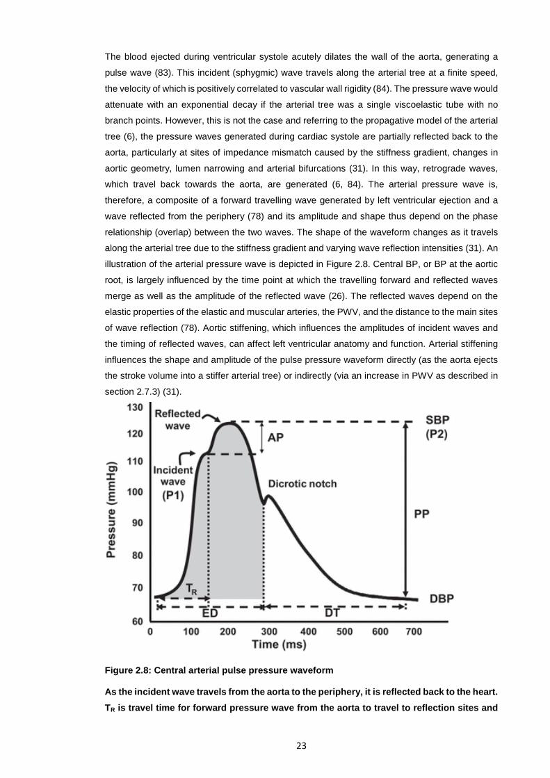
23
The blood ejected during ventricular systole acutely dilates the wall of the aorta, generating a
pulse wave (83). This incident (sphygmic) wave travels along the arterial tree at a finite speed,
the velocity of which is positively correlated to vascular wall rigidity (84). The pressure wave would
attenuate with an exponential decay if the arterial tree was a single viscoelastic tube with no
branch points. However, this is not the case and referring to the propagative model of the arterial
tree (6), the pressure waves generated during cardiac systole are partially reflected back to the
aorta, particularly at sites of impedance mismatch caused by the stiffness gradient, changes in
aortic geometry, lumen narrowing and arterial bifurcations (31). In this way, retrograde waves,
which travel back towards the aorta, are generated (6, 84). The arterial pressure wave is,
therefore, a composite of a forward travelling wave generated by left ventricular ejection and a
wave reflected from the periphery (78) and its amplitude and shape thus depend on the phase
relationship (overlap) between the two waves. The shape of the waveform changes as it travels
along the arterial tree due to the stiffness gradient and varying wave reflection intensities (31). An
illustration of the arterial pressure wave is depicted in Figure 2.8. Central BP, or BP at the aortic
root, is largely influenced by the time point at which the travelling forward and reflected waves
merge as well as the amplitude of the reflected wave (26). The reflected waves depend on the
elastic properties of the elastic and muscular arteries, the PWV, and the distance to the main sites
of wave reflection (78). Aortic stiffening, which influences the amplitudes of incident waves and
the timing of reflected waves, can affect left ventricular anatomy and function. Arterial stiffening
influences the shape and amplitude of the pulse pressure waveform directly (as the aorta ejects
the stroke volume into a stiffer arterial tree) or indirectly (via an increase in PWV as described in
section 2.7.3) (31).
Figure 2.8: Central arterial pulse pressure waveform
As the incident wave travels from the aorta to the periphery, it is reflected back to the heart. TR is travel time for forward pressure wave from the aorta to travel to reflection sites and
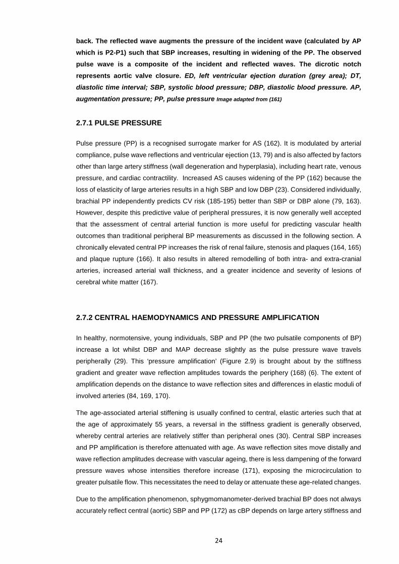
24
back. The reflected wave augments the pressure of the incident wave (calculated by AP which is P2-P1) such that SBP increases, resulting in widening of the PP. The observed pulse wave is a composite of the incident and reflected waves. The dicrotic notch represents aortic valve closure. ED, left ventricular ejection duration (grey area); DT, diastolic time interval; SBP, systolic blood pressure; DBP, diastolic blood pressure. AP, augmentation pressure; PP, pulse pressure Image adapted from (161)
2.7.1 PULSE PRESSURE
Pulse pressure (PP) is a recognised surrogate marker for AS (162). It is modulated by arterial
compliance, pulse wave reflections and ventricular ejection (13, 79) and is also affected by factors
other than large artery stiffness (wall degeneration and hyperplasia), including heart rate, venous
pressure, and cardiac contractility. Increased AS causes widening of the PP (162) because the
loss of elasticity of large arteries results in a high SBP and low DBP (23). Considered individually,
brachial PP independently predicts CV risk (185-195) better than SBP or DBP alone (79, 163).
However, despite this predictive value of peripheral pressures, it is now generally well accepted
that the assessment of central arterial function is more useful for predicting vascular health
outcomes than traditional peripheral BP measurements as discussed in the following section. A
chronically elevated central PP increases the risk of renal failure, stenosis and plaques (164, 165)
and plaque rupture (166). It also results in altered remodelling of both intra- and extra-cranial
arteries, increased arterial wall thickness, and a greater incidence and severity of lesions of
cerebral white matter (167).
2.7.2 CENTRAL HAEMODYNAMICS AND PRESSURE AMPLIFICATION
In healthy, normotensive, young individuals, SBP and PP (the two pulsatile components of BP)
increase a lot whilst DBP and MAP decrease slightly as the pulse pressure wave travels
peripherally (29). This ‘pressure amplification’ (Figure 2.9) is brought about by the stiffness
gradient and greater wave reflection amplitudes towards the periphery (168) (6). The extent of
amplification depends on the distance to wave reflection sites and differences in elastic moduli of
involved arteries (84, 169, 170).
The age-associated arterial stiffening is usually confined to central, elastic arteries such that at
the age of approximately 55 years, a reversal in the stiffness gradient is generally observed,
whereby central arteries are relatively stiffer than peripheral ones (30). Central SBP increases
and PP amplification is therefore attenuated with age. As wave reflection sites move distally and
wave reflection amplitudes decrease with vascular ageing, there is less dampening of the forward
pressure waves whose intensities therefore increase (171), exposing the microcirculation to
greater pulsatile flow. This necessitates the need to delay or attenuate these age-related changes.
Due to the amplification phenomenon, sphygmomanometer-derived brachial BP does not always
accurately reflect central (aortic) SBP and PP (172) as cBP depends on large artery stiffness and
25
pulse wave reflections (173). It has been proposed that central BP (cBP) may be more accurate
at predicting risk than peripheral (brachial) pressures (22, 174-178) and at predicting CV events,
diabetes and all-cause mortality, independent of peripheral BP (22, 178, 179) because the major
organs which are the main sites of CV events are directly exposed to the former. In addition, the
detrimental effects of hypertension and atherosclerosis do not significantly affect the brachial
artery (160) and left ventricle afterload and mass are influenced by the pressure in the ascending
aorta rather than that in the brachial artery. Furthermore, although general AS parameters can be
obtained from both peripheral and central arterial pressure waveforms, only the latter can provide
absolute values of wave reflection characteristics to better predict CV outcomes.
Figure 2.9: Pulse pressure amplification from the aorta to the periphery in a young adult
As systolic blood pressure and pulse pressure increase from the aorta to the periphery (alongside minor changes in mean arterial pressure and diastolic blood pressure), the shape of the blood pressure curve changes as well. Image adapted from (173)
2.7.3 THE AUGMENTATION INDEX (AIx)
The augmentation index (AIx) is a partly heritable (180) measure of wave reflections and the
resulting impact they have on ascending aortic (central) pressure and consequently, the pressure
waveform shape and amplitude. In the peripheral pressure waveform, there is an uninterrupted
upstroke during systole followed by a small systolic ‘shoulder’ during the downstroke. In the
central aorta (refer to Figure 2.8), the initial early pressure rise (PI) is ‘augmented’ by a second
pressure rise (P2) during late systole. This brings about a systolic ‘shoulder’ during the upstroke.
P1 coincides with left ventricular ejection and is the point of the greatest PWV.
Augmentation pressure (AP) is P2-P1 (ie. second systolic peak – first systolic peak) and is brought
about due to reflection of the forward wave. This index defines the absolute increase in BP during
the early phase of ventricular systole which is brought about by a reflected wave at the aorta (13).
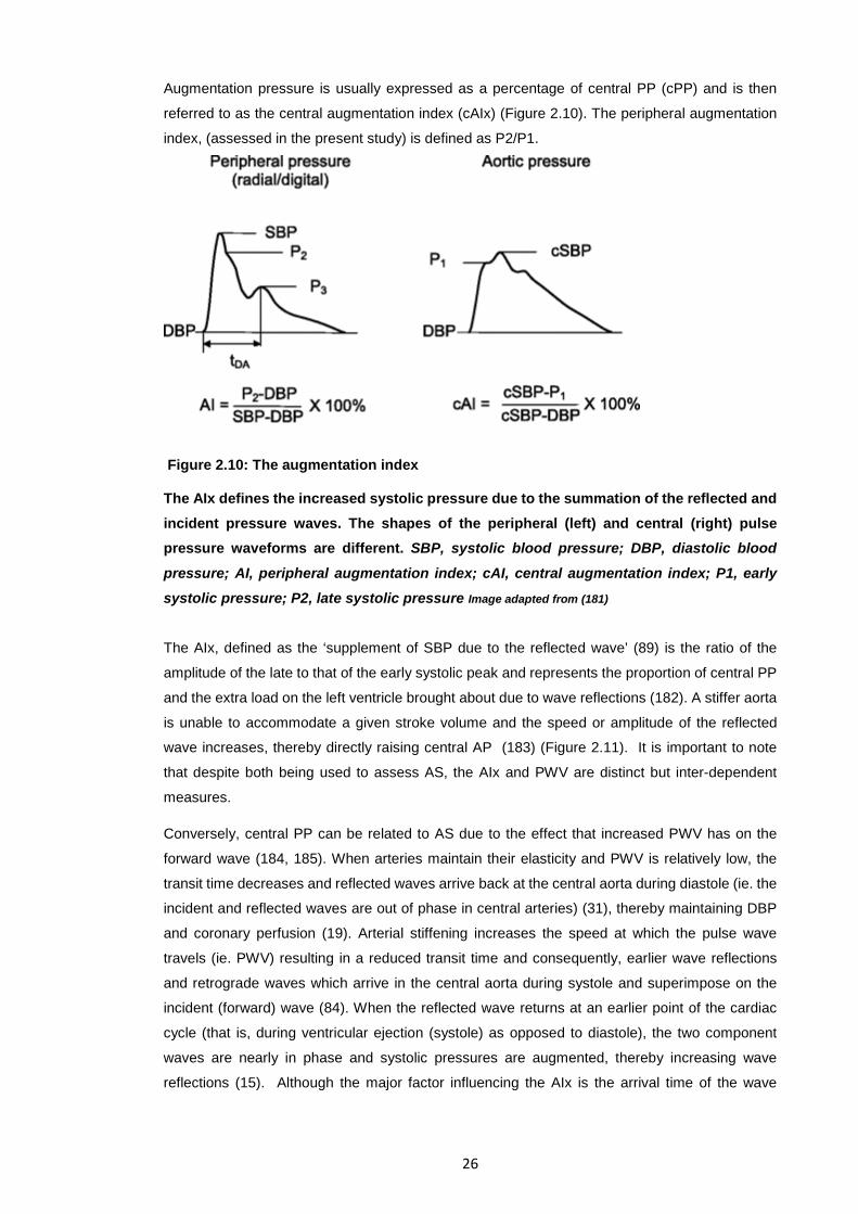
26
Augmentation pressure is usually expressed as a percentage of central PP (cPP) and is then
referred to as the central augmentation index (cAIx) (Figure 2.10). The peripheral augmentation
index, (assessed in the present study) is defined as P2/P1.
Figure 2.10: The augmentation index
The AIx defines the increased systolic pressure due to the summation of the reflected and incident pressure waves. The shapes of the peripheral (left) and central (right) pulse pressure waveforms are different. SBP, systolic blood pressure; DBP, diastolic blood
pressure; AI, peripheral augmentation index; cAI, central augmentation index; P1, early systolic pressure; P2, late systolic pressure Image adapted from (181)
The AIx, defined as the ‘supplement of SBP due to the reflected wave’ (89) is the ratio of the
amplitude of the late to that of the early systolic peak and represents the proportion of central PP
and the extra load on the left ventricle brought about due to wave reflections (182). A stiffer aorta
is unable to accommodate a given stroke volume and the speed or amplitude of the reflected
wave increases, thereby directly raising central AP (183) (Figure 2.11). It is important to note
that despite both being used to assess AS, the AIx and PWV are distinct but inter-dependent
measures.
Conversely, central PP can be related to AS due to the effect that increased PWV has on the
forward wave (184, 185). When arteries maintain their elasticity and PWV is relatively low, the
transit time decreases and reflected waves arrive back at the central aorta during diastole (ie. the
incident and reflected waves are out of phase in central arteries) (31), thereby maintaining DBP
and coronary perfusion (19). Arterial stiffening increases the speed at which the pulse wave
travels (ie. PWV) resulting in a reduced transit time and consequently, earlier wave reflections
and retrograde waves which arrive in the central aorta during systole and superimpose on the
incident (forward) wave (84). When the reflected wave returns at an earlier point of the cardiac
cycle (that is, during ventricular ejection (systole) as opposed to diastole), the two component
waves are nearly in phase and systolic pressures are augmented, thereby increasing wave
reflections (15). Although the major factor influencing the AIx is the arrival time of the wave
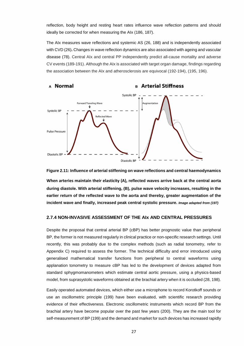
27
reflection, body height and resting heart rates influence wave reflection patterns and should
ideally be corrected for when measuring the AIx (186, 187).
The AIx measures wave reflections and systemic AS (26, 188) and is independently associated
with CVD (26). Changes in wave reflection dynamics are also associated with ageing and vascular
disease (78). Central AIx and central PP independently predict all-cause mortality and adverse
CV events (189-191). Although the AIx is associated with target organ damage, findings regarding
the association between the AIx and atherosclerosis are equivocal (192-194), (195, 196).
Figure 2.11: Influence of arterial stiffening on wave reflections and central haemodynamics
When arteries maintain their elasticity (A), reflected waves arrive back at the central aorta
during diastole. With arterial stiffening, (B), pulse wave velocity increases, resulting in the earlier return of the reflected wave to the aorta and thereby, greater augmentation of the incident wave and finally, increased peak central systolic pressure. Image adapted from (197)
2.7.4 NON-INVASIVE ASSESSMENT OF THE AIx AND CENTRAL PRESSURES
Despite the proposal that central arterial BP (cBP) has better prognostic value than peripheral
BP, the former is not measured regularly in clinical practice or non-specific research settings. Until
recently, this was probably due to the complex methods (such as radial tonometry, refer to
Appendix C) required to assess the former. The technical difficulty and error introduced using
generalised mathematical transfer functions from peripheral to central waveforms using
applanation tonometry to measure cBP has led to the development of devices adapted from
standard sphygmomanometers which estimate central aortic pressure, using a physics-based
model, from suprasystolic waveforms obtained at the brachial artery when it is occluded (28, 198).
Easily operated automated devices, which either use a microphone to record Korotkoff sounds or
use an oscillometric principle (199) have been evaluated, with scientific research providing
evidence of their effectiveness. Electronic oscillometric instruments which record BP from the
brachial artery have become popular over the past few years (200). They are the main tool for
self-measurement of BP (199) and the demand and market for such devices has increased rapidly
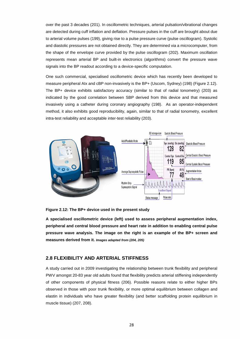
28
over the past 3 decades (201). In oscillometric techniques, arterial pulsation/vibrational changes
are detected during cuff inflation and deflation. Pressure pulses in the cuff are brought about due
to arterial volume pulses (199), giving rise to a pulse pressure curve (pulse oscillogram). Systolic
and diastolic pressures are not obtained directly. They are determined via a microcomputer, from
the shape of the envelope curve provided by the pulse oscillogram (202). Maximum oscillation
represents mean arterial BP and built-in electronics (algorithms) convert the pressure wave
signals into the BP readout according to a device-specific computation.
One such commercial, specialised oscillometric device which has recently been developed to
measure peripheral AIx and cBP non-invasively is the BP+ (Uscom, Sydney) (198) (Figure 2.12).
The BP+ device exhibits satisfactory accuracy (similar to that of radial tonometry) (203) as
indicated by the good correlation between SBP derived from this device and that measured
invasively using a catheter during coronary angiography (198). As an operator-independent
method, it also exhibits good reproducibility, again, similar to that of radial tonometry, excellent
intra-test reliability and acceptable inter-test reliability (203).
Figure 2.12: The BP+ device used in the present study
A specialised oscillometric device (left) used to assess peripheral augmentation index, peripheral and central blood pressure and heart rate in addition to enabling central pulse pressure wave analysis. The image on the right is an example of the BP+ screen and measures derived from it. Images adapted from (204, 205)
2.8 FLEXIBILITY AND ARTERIAL STIFFNESS
A study carried out in 2009 investigating the relationship between trunk flexibility and peripheral
PWV amongst 20-83 year old adults found that flexibility predicts arterial stiffening independently
of other components of physical fitness (206). Possible reasons relate to either higher BPs
observed in those with poor trunk flexibility, or more optimal equilibrium between collagen and
elastin in individuals who have greater flexibility (and better scaffolding protein equilibrium in
muscle tissue) (207, 208).
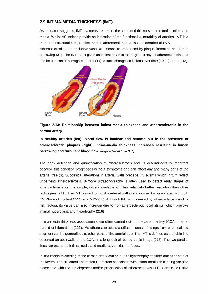
29
2.9 INTIMA-MEDIA THICKNESS (IMT)
As the name suggests, IMT is a measurement of the combined thickness of the tunica intima and
media. Whilst AS indices provide an indication of the functional vulnerability of arteries, IMT is a
marker of structural compromise, and as aforementioned, a tissue biomarker of EVA.
Atherosclerosis is an occlusive vascular disease characterised by plaque formation and lumen
narrowing (31). The IMT index gives an indication as to the degree, if any, of atherosclerosis, and
can be used as its surrogate marker (11) to track changes in lesions over time (209) (Figure 2.13).
Figure 2.13: Relationship between intima-media thickness and atherosclerosis in the carotid artery
In healthy arteries (left), blood flow is laminar and smooth but in the presence of atherosclerotic plaques (right), intima-media thickness increases resulting in lumen narrowing and turbulent blood flow. Image adapted from (210)
The early detection and quantification of atherosclerosis and its determinants is important
because this condition progresses without symptoms and can affect any and many parts of the
arterial tree (3). Subclinical alterations in arterial walls precede CV events which in turn reflect
underlying atherosclerosis. B-mode ultrasonography is often used to detect early stages of
atherosclerosis as it is simple, widely available and has relatively better resolution than other
techniques (211). The IMT is used to monitor arterial wall alterations as it is associated with both
CV RFs and incident CVD (209, 212-215). Although IMT is influenced by atherosclerosis and its
risk factors, its value can also increase due to non-atherosclerotic local stimuli which provoke
intimal hyperplasia and hypertrophy (216)
Intima-media thickness assessments are often carried out on the carotid artery (CCA, internal
carotid or bifurcation) (121). As atherosclerosis is a diffuse disease, findings from one localised
segment can be generalised to other parts of the arterial tree. The IMT is defined as a double line
observed on both walls of the CCAs in a longitudinal, echographic image (216). The two parallel
lines represent the intima-media and media-adventitia interfaces.
Intima-media thickening of the carotid artery can be due to hypertrophy of either one of or both of
the layers. The structural and molecular factors associated with intima-medial thickening are also
associated with the development and/or progression of atherosclerosis (11). Carotid IMT also
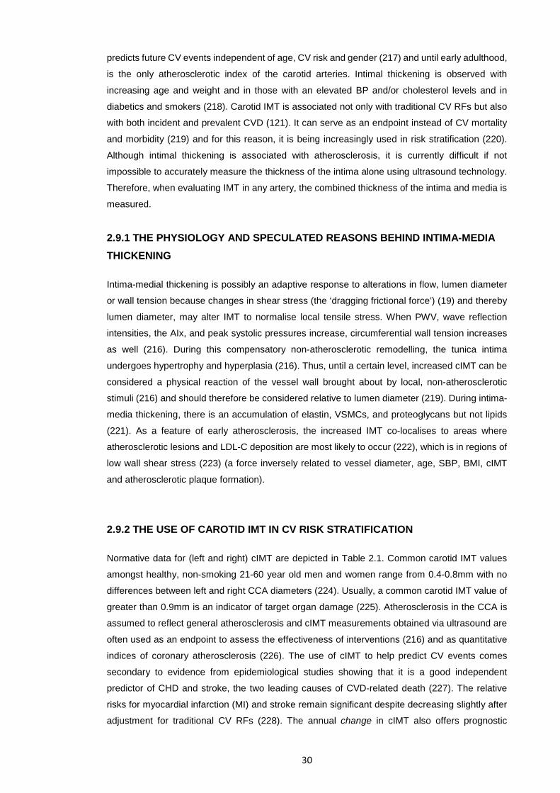
30
predicts future CV events independent of age, CV risk and gender (217) and until early adulthood,
is the only atherosclerotic index of the carotid arteries. Intimal thickening is observed with
increasing age and weight and in those with an elevated BP and/or cholesterol levels and in
diabetics and smokers (218). Carotid IMT is associated not only with traditional CV RFs but also
with both incident and prevalent CVD (121). It can serve as an endpoint instead of CV mortality
and morbidity (219) and for this reason, it is being increasingly used in risk stratification (220).
Although intimal thickening is associated with atherosclerosis, it is currently difficult if not
impossible to accurately measure the thickness of the intima alone using ultrasound technology.
Therefore, when evaluating IMT in any artery, the combined thickness of the intima and media is
measured.
2.9.1 THE PHYSIOLOGY AND SPECULATED REASONS BEHIND INTIMA-MEDIA THICKENING
Intima-medial thickening is possibly an adaptive response to alterations in flow, lumen diameter
or wall tension because changes in shear stress (the ‘dragging frictional force’) (19) and thereby
lumen diameter, may alter IMT to normalise local tensile stress. When PWV, wave reflection
intensities, the AIx, and peak systolic pressures increase, circumferential wall tension increases
as well (216). During this compensatory non-atherosclerotic remodelling, the tunica intima
undergoes hypertrophy and hyperplasia (216). Thus, until a certain level, increased cIMT can be
considered a physical reaction of the vessel wall brought about by local, non-atherosclerotic
stimuli (216) and should therefore be considered relative to lumen diameter (219). During intima-
media thickening, there is an accumulation of elastin, VSMCs, and proteoglycans but not lipids
(221). As a feature of early atherosclerosis, the increased IMT co-localises to areas where
atherosclerotic lesions and LDL-C deposition are most likely to occur (222), which is in regions of
low wall shear stress (223) (a force inversely related to vessel diameter, age, SBP, BMI, cIMT
and atherosclerotic plaque formation).
2.9.2 THE USE OF CAROTID IMT IN CV RISK STRATIFICATION
Normative data for (left and right) cIMT are depicted in Table 2.1. Common carotid IMT values
amongst healthy, non-smoking 21-60 year old men and women range from 0.4-0.8mm with no
differences between left and right CCA diameters (224). Usually, a common carotid IMT value of
greater than 0.9mm is an indicator of target organ damage (225). Atherosclerosis in the CCA is
assumed to reflect general atherosclerosis and cIMT measurements obtained via ultrasound are
often used as an endpoint to assess the effectiveness of interventions (216) and as quantitative
indices of coronary atherosclerosis (226). The use of cIMT to help predict CV events comes
secondary to evidence from epidemiological studies showing that it is a good independent
predictor of CHD and stroke, the two leading causes of CVD-related death (227). The relative
risks for myocardial infarction (MI) and stroke remain significant despite decreasing slightly after
adjustment for traditional CV RFs (228). The annual change in cIMT also offers prognostic
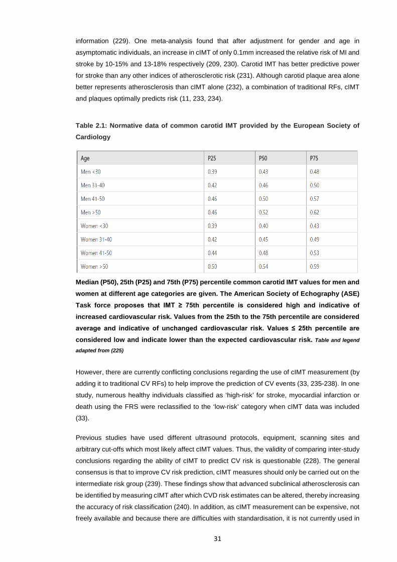
31
information (229). One meta-analysis found that after adjustment for gender and age in
asymptomatic individuals, an increase in cIMT of only 0.1mm increased the relative risk of MI and
stroke by 10-15% and 13-18% respectively (209, 230). Carotid IMT has better predictive power
for stroke than any other indices of atherosclerotic risk (231). Although carotid plaque area alone
better represents atherosclerosis than cIMT alone (232), a combination of traditional RFs, cIMT
and plaques optimally predicts risk (11, 233, 234).
Table 2.1: Normative data of common carotid IMT provided by the European Society of Cardiology
Median (P50), 25th (P25) and 75th (P75) percentile common carotid IMT values for men and women at different age categories are given. The American Society of Echography (ASE) Task force proposes that IMT ≥ 75th percentile is considered high and indicative of increased cardiovascular risk. Values from the 25th to the 75th percentile are considered average and indicative of unchanged cardiovascular risk. Values ≤ 25th percentile are considered low and indicate lower than the expected cardiovascular risk. Table and legend
adapted from (225)
However, there are currently conflicting conclusions regarding the use of cIMT measurement (by
adding it to traditional CV RFs) to help improve the prediction of CV events (33, 235-238). In one
study, numerous healthy individuals classified as ‘high-risk’ for stroke, myocardial infarction or
death using the FRS were reclassified to the ‘low-risk’ category when cIMT data was included
(33).
Previous studies have used different ultrasound protocols, equipment, scanning sites and
arbitrary cut-offs which most likely affect cIMT values. Thus, the validity of comparing inter-study
conclusions regarding the ability of cIMT to predict CV risk is questionable (228). The general
consensus is that to improve CV risk prediction, cIMT measures should only be carried out on the
intermediate risk group (239). These findings show that advanced subclinical atherosclerosis can
be identified by measuring cIMT after which CVD risk estimates can be altered, thereby increasing
the accuracy of risk classification (240). In addition, as cIMT measurement can be expensive, not
freely available and because there are difficulties with standardisation, it is not currently used in
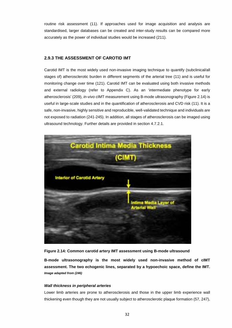
32
routine risk assessment (11). If approaches used for image acquisition and analysis are
standardised, larger databases can be created and inter-study results can be compared more
accurately as the power of individual studies would be increased (211).
2.9.3 THE ASSESSMENT OF CAROTID IMT
Carotid IMT is the most widely used non-invasive imaging technique to quantify (subclinical/all
stages of) atherosclerotic burden in different segments of the arterial tree (11) and is useful for
monitoring change over time (121). Carotid IMT can be evaluated using both invasive methods
and external radiology (refer to Appendix C). As an ‘intermediate phenotype for early
atherosclerosis’ (209), in-vivo cIMT measurement using B-mode ultrasonography (Figure 2.14) is
useful in large-scale studies and in the quantification of atherosclerosis and CVD risk (11). It is a
safe, non-invasive, highly sensitive and reproducible, well-validated technique and individuals are
not exposed to radiation (241-245). In addition, all stages of atherosclerosis can be imaged using
ultrasound technology. Further details are provided in section 4.7.2.1.
Figure 2.14: Common carotid artery IMT assessment using B-mode ultrasound
B-mode ultrasonography is the most widely used non-invasive method of cIMT assessment. The two echogenic lines, separated by a hypoechoic space, define the IMT. Image adapted from (246)
Wall thickness in peripheral arteries Lower limb arteries are prone to atherosclerosis and those in the upper limb experience wall
thickening even though they are not usually subject to atherosclerotic plaque formation (57, 247),
33
implying that arterial wall thickening occurs systemically (248). Several studies have carried out
IMT measurements on the common femoral artery (CFA) as it too, is superficial. Differences in
the degrees of atherosclerotic lesions are often observed between the CCA and CFA because
the latter is exposed to greater hydrostatic pressure and flow variations and more advanced
atherosclerosis can be studied in the CFA (226). Increased femoral IMT (fIMT), is associated with
traditional CV RFs (249-251), peripheral atherosclerosis indices (252), the FRS (250) and the
severity and extent of coronary artery disease (253).
2.10 THE RESISTIVITY INDEX
The haemodynamic RI (also known as the Pourcelot index) is an indirect marker of the severity
of atherosclerosis (254) and is an important parameter to be studied when examining the arterial
waveform using Doppler ultrasonography. The CCA RI has been shown to be positively correlated
to the cIMT, even after correcting for vascular risk factors (216). At a fixed compliance, the renal
RI increases with increasing local resistance and the higher the compliance, the more the RI is
influenced by local vascular resistance (255). The RI, which increases with natural ageing, is
calculated from blood flow velocity curves obtained by pulsed wave Doppler ultrasonography
(256). However, very few studies have assessed the common carotid RI (especially in relation to
exercise).
2.11 ARTERIAL GEOMETRY AND REMODELLING – ARTERIAL DIAMETER AND ARTERIOGENESIS
Central and peripheral factors determine blood flow to muscles. The large conduit vessels deliver
blood to small resistance vessels at high pressure (257). Blood flow to downstream vessels is
optimised by arteriogenesis, which describes the enlargement of existing vessels. Arteriogenesis,
which involves remodelling of endothelial cells, fibroblasts and VSMCs, occurs because of an
increase in wall calibre (diameter) and dimensions as opposed to simply being brought about by
wall stretch due to increased luminal pressure or compliance (257).
In hypertension and with ageing, chronically raised intraluminal pressure escalates radial wall
stress (258), which, according to the law of Laplace, increases wall mass. However, the case is
quite different when luminal pressure surges during episodic bursts such as during exercise.
Muscle activity determines the size of the arteries which supply it and large increases in flow
through conduit arteries result in their significant enlargement due to greater shear stress on the
endothelial surface (259).
Increased flow velocity through a vessel stimulates arteriogenesis even if luminal pressure does
not change because the former directly increases shear stress, the main stimulus for arterial
enlargement (259). Arterial remodelling is primarily endothelium-dependent and upregulation of
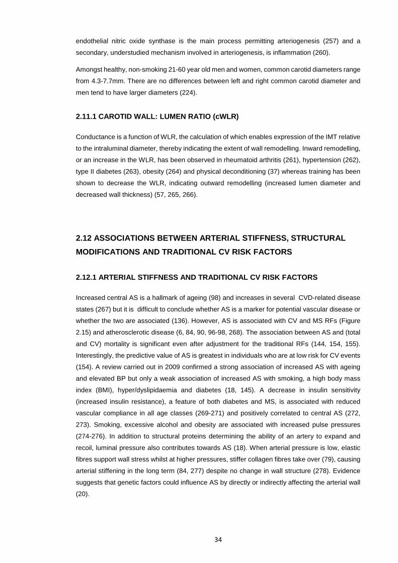
34
endothelial nitric oxide synthase is the main process permitting arteriogenesis (257) and a
secondary, understudied mechanism involved in arteriogenesis, is inflammation (260).
Amongst healthy, non-smoking 21-60 year old men and women, common carotid diameters range
from 4.3-7.7mm. There are no differences between left and right common carotid diameter and
men tend to have larger diameters (224).
2.11.1 CAROTID WALL: LUMEN RATIO (cWLR)
Conductance is a function of WLR, the calculation of which enables expression of the IMT relative
to the intraluminal diameter, thereby indicating the extent of wall remodelling. Inward remodelling,
or an increase in the WLR, has been observed in rheumatoid arthritis (261), hypertension (262),
type II diabetes (263), obesity (264) and physical deconditioning (37) whereas training has been
shown to decrease the WLR, indicating outward remodelling (increased lumen diameter and
decreased wall thickness) (57, 265, 266).
2.12 ASSOCIATIONS BETWEEN ARTERIAL STIFFNESS, STRUCTURAL MODIFICATIONS AND TRADITIONAL CV RISK FACTORS
2.12.1 ARTERIAL STIFFNESS AND TRADITIONAL CV RISK FACTORS
Increased central AS is a hallmark of ageing (98) and increases in several CVD-related disease
states (267) but it is difficult to conclude whether AS is a marker for potential vascular disease or
whether the two are associated (136). However, AS is associated with CV and MS RFs (Figure
2.15) and atherosclerotic disease (6, 84, 90, 96-98, 268). The association between AS and (total
and CV) mortality is significant even after adjustment for the traditional RFs (144, 154, 155).
Interestingly, the predictive value of AS is greatest in individuals who are at low risk for CV events
(154). A review carried out in 2009 confirmed a strong association of increased AS with ageing
and elevated BP but only a weak association of increased AS with smoking, a high body mass
index (BMI), hyper/dyslipidaemia and diabetes (18, 145). A decrease in insulin sensitivity
(increased insulin resistance), a feature of both diabetes and MS, is associated with reduced
vascular compliance in all age classes (269-271) and positively correlated to central AS (272,
273). Smoking, excessive alcohol and obesity are associated with increased pulse pressures
(274-276). In addition to structural proteins determining the ability of an artery to expand and
recoil, luminal pressure also contributes towards AS (18). When arterial pressure is low, elastic
fibres support wall stress whilst at higher pressures, stiffer collagen fibres take over (79), causing
arterial stiffening in the long term (84, 277) despite no change in wall structure (278). Evidence
suggests that genetic factors could influence AS by directly or indirectly affecting the arterial wall
(20).
35
2.12.1.1 ABDOMINAL OBESITY AND VASCULAR HEALTH
Although cIMT is an accepted surrogate marker for subclinical vascular disease which is an early
marker of atherosclerosis, dyslipidaemia and lipid abnormalities also play a role in the
development of this condition (279). Higher central fat distribution (abdominal obesity), as
opposed to total body fatness, is associated with higher AS at 36 years of age (280). Waist
circumference (WC) is a traditional marker of abdominal obesity, with BMI and WC being more
strongly related to MS than body fat percentage indices (281). Waist circumference is also better
than the BMI and body fat percentage indices at predicting dyslipidaemia and carotid
atherosclerosis (281). Central obesity can alter arterial wall properties and increase AS, directly
resulting in early arterial damage and premature vascular ageing (256). Although the WC is a
preferable index to use routinely due to its sensitivity and simplicity (281), an elevated triglyceride
level (≥ 2.0 mmol/L) in the presence of increased WC (for sex, height and ethnicity) better
indicates excess visceral adiposity (282). Individuals with this ‘hypertriglyceridaemic waist
phenotype’ are at increased CVD risk (283) and can therefore benefit the most from lifestyle
changes to reduce their visceral fat and triglyceride levels.
2.12.2 THE ASSOCIATION BETWEEN ATHEROSCLEROSIS AND ARTERIAL STIFFNESS
Atherosclerosis, a chronic, systemic inflammatory disease, is brought about in response to
physical injury of the vascular endothelium (284-286). Intimal-medial hypertrophy, typically
observed in the common carotid arteries, usually occurs during the early stages of this disease
whilst plaque formation, typically observed in the carotid bulbs or internal carotid arteries, occurs
later (220). The relationship between plaque formation and AS is not fully understood but
generally, larger artery stiffness is increased in the presence of atherosclerosis (18). Arterial
stiffness, in addition to IMT, can therefore be used as a marker of the extent of this condition in
the aorta (287). A 2009 review stated that in the early stages of atherosclerosis, increased AS
may occur independently from the development of this condition (145). However, atherosclerosis
involves several events and factors which can influence vascular remodelling and structure but
since AS and atherosclerosis usually coexist, it is difficult to establish causation (98). The
consequences of increased AS can accelerate the development of atherosclerotic lesions given
that AS may either serve as a marker for potential atherosclerotic disease or it may be involved
in the process of atherosclerosis directly (136). The risk factors for the development of
atherosclerosis are similar to those of CVD (and MS) and include elevated BP, high cholesterol
levels, high fasting blood glucose levels, obesity, smoking/tobacco use, physical inactivity and a
family history of early heart disease.
36
Figure 2.15: Effects of metabolic syndrome on arterial health indices
Gender-specific effects of MS on aortic pulse wave velocity (A and B) and common carotid IMT (C and D) across age groups. There are age-associated increases in aortic PWV and CCA IMT. Individuals with MS have higher (worse) PWV and CCA IMT than healthy individuals of the same age and sex. C, healthy controls; MS+, presence of metabolic
syndrome; PWV, pulse wave velocity; CCA IMT, common carotid artery intima –media thickness) *P < 0.05; **P < 0.01; &P < 0.001 vs. control of the same age group. Image adapted from (77) Although increased AS is not always linked atherosclerosis in a particular region, PWV has been
associated with coronary atherosclerosis (288, 289). One study reported that PWV decreases in
the initial stages of atherosclerosis and then quickly increases during the advancement of
atherosclerosis (140). A second study found no significant differences in distensibility between
regions with and without plaques (290) and a third study found that PWV is only increased during
stages of advanced plaque formation (291). Initial stages of arterial stiffening are determined by
non-atherosclerotic mechanisms which involve BP-related mechanical stretch which transfers
loading from elastin to the stiffer collagen fibres (145). The correlation between AS and
atherosclerosis (140, 292) is probably due to the effects that cyclic stress has on arterial wall
thickness (293). Only weak, if any, positive correlations between IMT and PWV have been found
(294, 295), which is possibly because an increase in IMT normalises the shear stress brought
about by an increase in BP (296). In essential hypertension, there is a sustained increase in BP
and this causes hypertrophy of arterial walls which normalises circumferential wall stress (20).
This adaptive response only occurs until a certain level after which increased IMT is associated
more with atherosclerosis (219). Contrary to what might be expected, studies on individuals with
essential hypertension and rat models of hypertension have shown that wall thickening induced
by the sustained elevated BP is not associated with increased AS (151, 297-301).

37
CHAPTER 3:-THERAPEUTIC STRATEGIES AIMED AT OPTIMISING ARTERIAL HEALTH: THE VASCULOPROTECTIVE
ROLE OF EXERCISE
The strategies aimed at improving arterial health fall into two broad categories, namely,
pharmacologic and non-pharmacological ones.
3.1 GOALS OF PHARMACOLOGICAL THERAPIES
Ideally, pharmacologic therapy should normalise AS completely by reducing BP and bringing
about structural changes in the vasculature (302). The aim is to target the events that cause AS
and endothelial dysfunction, that is, oxidative stress and inflammation. Both elastic and muscular
arteries have been targeted because whilst compliance of the former determines left ventricular
afterload, compliance of the latter determines wave reflections (84). Long-term (but not acute)
drug administration is capable of changing arterial wall components and their spatial organisation,
independent of BP reductions (302). These modifications include an increased elastin:collagen
ratio, decreased collagen content, a reduced IMT and lumen diameter and changes in
connections between VSMCs and the ECM (80, 303). The fact that lowering local PP and not
MAP reduces wall hypertrophy and lumen diameter means that by decreasing AS, pulsatile stress
is reduced, bringing about arterial remodelling (80).
On the other hand, acute drug administration causes a BP-independent decrease in the AS of
muscular arteries, implying that smooth muscle relaxation affects the proportions of the structural
components of arterial walls (84). In normotensive adults with CV RFs, it may take much longer
(relative to clinical populations) to improve AS.
3.2 NON-PHARMACOLOGIC THERAPIES USED TO IMPROVE ARTERIAL HEALTH Non-pharmacological treatments usually revolve around physical activity (exercise) and dietary
manipulation, but much is left to be discerned due to small sample sizes in relevant studies to
date. Blood pressure-dependent large artery compliance increased in healthy, obese men after
weight loss associated with reduced MAP (304). Caloric restriction (CR) improves systemic
atherosclerosis RFs, reduces BP and has direct anti-ageing effects on the vasculature (305) by
up-regulating endothelial nitric oxide synthase (eNOS), therefore improving nitric oxide
bioavailability and endothelial function (306). In addition, CR reduces reactive oxygen species
(ROS) production (307), oxidative stress (308, 309) and vascular nuclear factor kappa-B (NF-ĸB)
activation (310). Whilst long-term CR reduces chronic low-grade inflammation by decreasing
circulating TNF-α and C-reactive protein levels, (311), even short-term CR can have anti-
inflammatory effects (217). Other studies have demonstrated the beneficial impact of α-linolenic
38
acid, isoflavones (derived from red clover or soy products) (312), fish oil therapy (313), >300mg
garlic powder daily and a reduced sodium intake on arterial compliance and PWV (314).
3.3 PHYSICAL ACTIVITY STATUS AND ARTERIAL HEALTH
The possibility of a future CVD epidemic is mounting given that advancing age is the primary risk
factor for CVD and that most individuals with this condition are middle-aged and older (MA/O)
adults whose numbers are rapidly increasing (315). Sedentary lifestyles account for a staggering
one-third of all deaths (316) which is an additional concern, given the low daily energy expenditure
in modern life (317). The effects of physical inactivity and activity on traditional CV RFs are not
profound and do not explain the entire magnitude of the impact that physical activity status has
on risk (318). Therefore, it has been proposed that physical activity and inactivity can both
influence CV risk by affecting vascular structure and function directly (318) (ie. vascular
deconditioning and conditioning). However, the effects of exercise and deconditioning on arteries
are not simply opposite to one another. Endothelial dysfunction and the stiffening of large elastic
arteries are the two primary components of arterial dysfunction, the main intermediary event
associated with age-related CVD (82). Despite being impaired in high-risk individuals, vascular
structure and function are susceptible and responsive to change.
3.3.1 THE EFFECTS OF PHYSICAL INACTIVITY AND DECONDITIONING ON ARTERIAL STRUCTURE AND FUNCTION
Physical inactivity and low cardiorespiratory fitness (CRF) independently predict
atherosclerosis/CVD and all-cause mortality respectively (319, 320). Alterations in
haemodynamic stimuli which directly impact blood vessels explain part of the association between
physical inactivity and the increased CV risk (319) which cannot be attributed to changes in
traditional CV RFs. Accelerated arterial stiffening of conduit arteries occurs with physical inactivity
(321, 322), which results in dose-dependent inward arterial remodelling and a pro-atherogenic
phenotype (318).
Future CV events are strongly predicted by resting conduit artery diameter (323) and wall
thickness (73). Deconditioning rapidly reduces conduit artery diameter (end-diastolic diameter,
EDD) in a dose- and time-dependent fashion and this may be due to physical inactivity-induced
muscle atrophy and reduced metabolic demands (37). Deconditioning also increases conduit
artery wall thickness (IMT) but these changes are probably reversible adaptations as opposed to
a reflection of atherosclerotic changes (37). The final result is an increased WLR. In addition,
physical inactivity brings about the remodelling of resistance vessels, whose cross-sectional area
decreases in a dose- and intensity-dependent way in response to deconditioning, possible due to
muscular atrophy (37). The increased resistance is explained primarily by vasoconstrictor (324)
as opposed to dilator pathways, the latter being involved in the cardioprotective effects of
exercise. Although changes in conduit arteries occur earlier than those in resistance vessels
(325), there is no definitive conclusion as to whether structural or functional deconditioning-
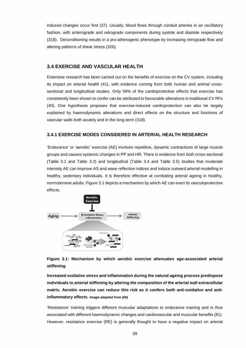
39
induced changes occur first (37). Usually, blood flows through conduit arteries in an oscillatory
fashion, with anterograde and retrograde components during systole and diastole respectively
(318). Deconditioning results in a pro-atherogenic phenotype by increasing retrograde flow and
altering patterns of shear stress (326).
3.4 EXERCISE AND VASCULAR HEALTH
Extensive research has been carried out on the benefits of exercise on the CV system, including
its impact on arterial health (41), with evidence coming from both human and animal cross-
sectional and longitudinal studies. Only 59% of the cardioprotective effects that exercise has
consistently been shown to confer can be attributed to favourable alterations in traditional CV RFs
(40). One hypothesis proposes that exercise-induced cardioprotection can also be largely
explained by haemodynamic alterations and direct effects on the structure and functions of
vascular walls both acutely and in the long-term (318).
3.4.1 EXERCISE MODES CONSIDERED IN ARTERIAL HEALTH RESEARCH
‘Endurance’ or ‘aerobic’ exercise (AE) involves repetitive, dynamic contractions of large muscle
groups and causes systemic changes in PP and HR. There is evidence from both cross-sectional
(Table 3.1 and Table 3.2) and longitudinal (Table 3.4 and Table 3.5) studies that moderate
intensity AE can improve AS and wave reflection indices and induce outward arterial modelling in
healthy, sedentary individuals. It is therefore effective at combating arterial ageing in healthy,
normotensive adults. Figure 3.1 depicts a mechanism by which AE can exert its vasculoprotective
effects.
Figure 3.1: Mechanism by which aerobic exercise attenuates age-associated arterial stiffening
Increased oxidative stress and inflammation during the natural ageing process predispose individuals to arterial stiffening by altering the composition of the arterial wall extracellular matrix. Aerobic exercise can reduce this risk as it confers both anti-oxidative and anti-inflammatory effects. Image adapted from (60)
‘Resistance’ training triggers different muscular adaptations to endurance training and is thus
associated with different haemodynamic changes and cardiovascular and muscular benefits (81).
However, resistance exercise (RE) is generally thought to have a negative impact on arterial
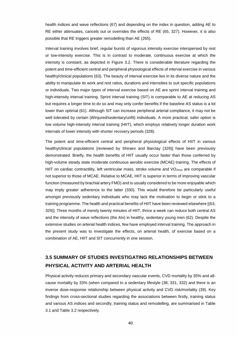
40
health indices and wave reflections (67) and depending on the index in question, adding AE to
RE either attenuates, cancels out or overrides the effects of RE (65, 327). However, it is also
possible that RE triggers greater remodelling than AE (265).
Interval training involves brief, regular bursts of vigorous intensity exercise interspersed by rest
or low-intensity exercise. This is in contrast to moderate, continuous exercise at which the
intensity is constant, as depicted in Figure 3.2. There is considerable literature regarding the
potent and time-efficient central and peripheral physiological effects of interval exercise in various
healthy/clinical populations (63). The beauty of interval exercise lies in its diverse nature and the
ability to manipulate its work and rest ratios, durations and intensities to suit specific populations
or individuals. Two major types of interval exercise based on AE are sprint interval training and
high-intensity interval training. Sprint interval training (SIT) is comparable to AE at reducing AS
but requires a longer time to do so and may only confer benefits if the baseline AS status is a lot
lower than optimal (61). Although SIT can increase peripheral arterial compliance, it may not be
well tolerated by certain (ill/injured/sedentary/unfit) individuals. A more practical, safer option is
low volume high-intensity interval training (HIIT), which employs relatively longer duration work
intervals of lower intensity with shorter recovery periods (328).
The potent and time-efficient central and peripheral physiological effects of HIIT in various
healthy/clinical populations [reviewed by Shiraev and Barclay (329)] have been previously
demonstrated. Briefly, the health benefits of HIIT usually occur faster than those conferred by
high-volume steady state moderate continuous aerobic exercise (MCAE) training. The effects of
HIIT on cardiac contractility, left ventricular mass, stroke volume and VO2max are comparable if
not superior to those of MCAE. Relative to MCAE, HIIT is superior in terms of improving vascular
function (measured by brachial artery FMD) and is usually considered to be more enjoyable which
may imply greater adherence to the latter (330). This would therefore be particularly useful
amongst previously sedentary individuals who may lack the motivation to begin or stick to a
training programme. The health and practical benefits of HIIT have been reviewed elsewhere [(63,
329)]. Three months of merely twenty minutes of HIIT, thrice a week can reduce both central AS
and the intensity of wave reflections (the AIx) in healthy, sedentary young men (62). Despite the
extensive studies on arterial health indices, few have employed interval training. The approach in
the present study was to investigate the effects, on arterial health, of exercise based on a
combination of AE, HIIT and SIT concurrently in one session.
3.5 SUMMARY OF STUDIES INVESTIGATING RELATIONSHIPS BETWEEN PHYSICAL ACTIVITY AND ARTERIAL HEALTH
Physical activity reduces primary and secondary vascular events, CVD mortality by 35% and all-
cause mortality by 33% (when compared to a sedentary lifestyle (38, 331, 332) and there is an
inverse dose-response relationship between physical activity and CVD risk/mortality (39). Key
findings from cross-sectional studies regarding the associations between firstly, training status
and various AS indices and secondly, training status and remodelling, are summarised in Table
3.1 and Table 3.2 respectively.
41
Figure 3.2: Schematic illustrations of aerobic interval training and aerobic continuous training
AIT (A) involves brief periods of vigorous exercise interspersed by low-intensity exercise or rest. During ACT (B), a sustainable intensity (a level between the peaks and troughs of AIT) is maintained for the duration of the entire session. AIT, aerobic interval training; ACT, aerobic continuous training (moderate continuous aerobic exercise) a = 50–60% VO2peak; 60–70% HRpeak; 11–13 Borg scale, no shortness of breath. b = 85–90% VO2peak; 90–95% HRpeak; 15–17 Borg scale, shortness of breath. c = 50–70% HRpeak. d = at least 60–70% VO2peak; ≥ 65–75% HRpeak Image and legend adapted from (333)
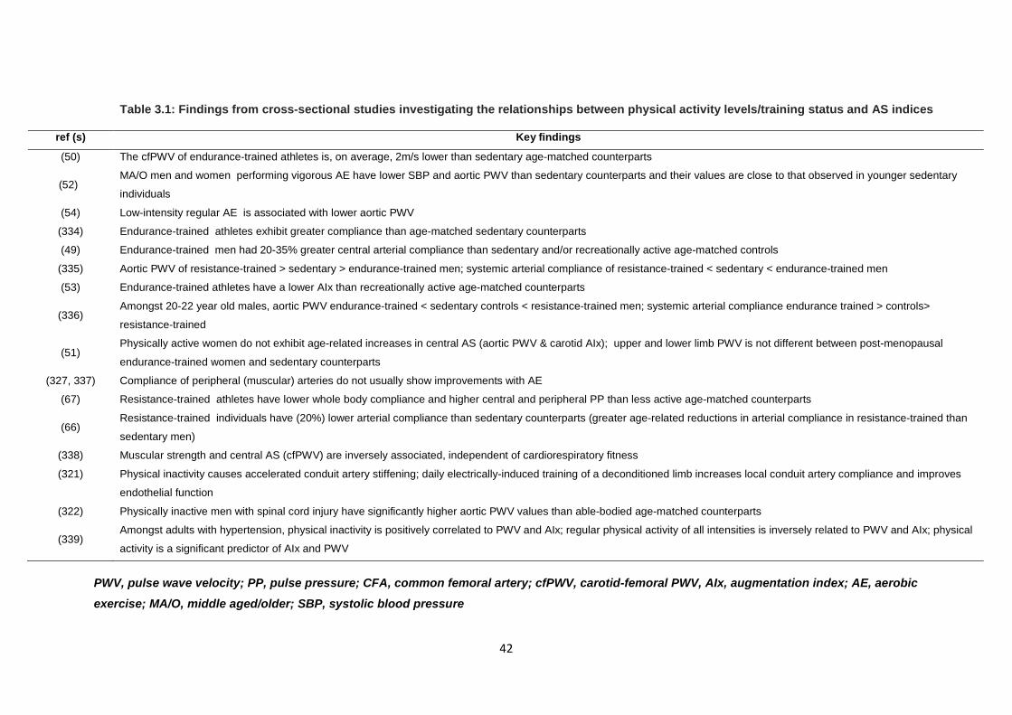
42
PWV, pulse wave velocity; PP, pulse pressure; CFA, common femoral artery; cfPWV, carotid-femoral PWV, AIx, augmentation index; AE, aerobic exercise; MA/O, middle aged/older; SBP, systolic blood pressure
Table 3.1: Findings from cross-sectional studies investigating the relationships between physical activity levels/training status and AS indices
ref (s) Key findings
(50) The cfPWV of endurance-trained athletes is, on average, 2m/s lower than sedentary age-matched counterparts
(52) MA/O men and women performing vigorous AE have lower SBP and aortic PWV than sedentary counterparts and their values are close to that observed in younger sedentary
individuals
(54) Low-intensity regular AE is associated with lower aortic PWV
(334) Endurance-trained athletes exhibit greater compliance than age-matched sedentary counterparts
(49) Endurance-trained men had 20-35% greater central arterial compliance than sedentary and/or recreationally active age-matched controls
(335) Aortic PWV of resistance-trained > sedentary > endurance-trained men; systemic arterial compliance of resistance-trained < sedentary < endurance-trained men
(53) Endurance-trained athletes have a lower AIx than recreationally active age-matched counterparts
(336) Amongst 20-22 year old males, aortic PWV endurance-trained < sedentary controls < resistance-trained men; systemic arterial compliance endurance trained > controls>
resistance-trained
(51) Physically active women do not exhibit age-related increases in central AS (aortic PWV & carotid AIx); upper and lower limb PWV is not different between post-menopausal
endurance-trained women and sedentary counterparts
(327, 337) Compliance of peripheral (muscular) arteries do not usually show improvements with AE
(67) Resistance-trained athletes have lower whole body compliance and higher central and peripheral PP than less active age-matched counterparts
(66) Resistance-trained individuals have (20%) lower arterial compliance than sedentary counterparts (greater age-related reductions in arterial compliance in resistance-trained than
sedentary men)
(338) Muscular strength and central AS (cfPWV) are inversely associated, independent of cardiorespiratory fitness
(321)
Physical inactivity causes accelerated conduit artery stiffening; daily electrically-induced training of a deconditioned limb increases local conduit artery compliance and improves
endothelial function
(322) Physically inactive men with spinal cord injury have significantly higher aortic PWV values than able-bodied age-matched counterparts
(339) Amongst adults with hypertension, physical inactivity is positively correlated to PWV and AIx; regular physical activity of all intensities is inversely related to PWV and AIx; physical
activity is a significant predictor of AIx and PWV
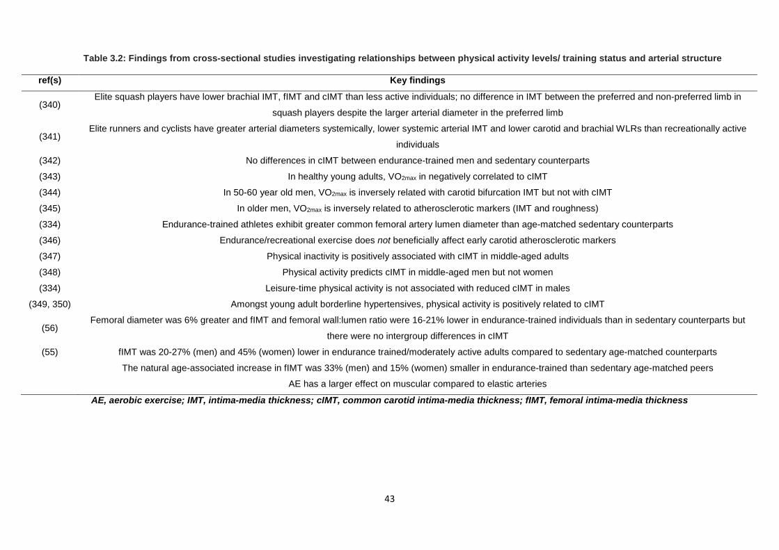
43
AE, aerobic exercise; IMT, intima-media thickness; cIMT, common carotid intima-media thickness; fIMT, femoral intima-media thickness
Table 3.2: Findings from cross-sectional studies investigating relationships between physical activity levels/ training status and arterial structure
ref(s) Key findings
(340) Elite squash players have lower brachial IMT, fIMT and cIMT than less active individuals; no difference in IMT between the preferred and non-preferred limb in
squash players despite the larger arterial diameter in the preferred limb
(341) Elite runners and cyclists have greater arterial diameters systemically, lower systemic arterial IMT and lower carotid and brachial WLRs than recreationally active
individuals
(342) No differences in cIMT between endurance-trained men and sedentary counterparts
(343) In healthy young adults, VO2max in negatively correlated to cIMT
(344) In 50-60 year old men, VO2max is inversely related with carotid bifurcation IMT but not with cIMT
(345) In older men, VO2max is inversely related to atherosclerotic markers (IMT and roughness)
(334) Endurance-trained athletes exhibit greater common femoral artery lumen diameter than age-matched sedentary counterparts
(346) Endurance/recreational exercise does not beneficially affect early carotid atherosclerotic markers
(347) Physical inactivity is positively associated with cIMT in middle-aged adults
(348) Physical activity predicts cIMT in middle-aged men but not women
(334) Leisure-time physical activity is not associated with reduced cIMT in males
(349, 350) Amongst young adult borderline hypertensives, physical activity is positively related to cIMT
(56) Femoral diameter was 6% greater and fIMT and femoral wall:lumen ratio were 16-21% lower in endurance-trained individuals than in sedentary counterparts but
there were no intergroup differences in cIMT
(55)
fIMT was 20-27% (men) and 45% (women) lower in endurance trained/moderately active adults compared to sedentary age-matched counterparts
The natural age-associated increase in fIMT was 33% (men) and 15% (women) smaller in endurance-trained than sedentary age-matched peers
AE has a larger effect on muscular compared to elastic arteries
44
3.6 THE ACUTE EFFECTS OF EXERCISE ON ARTERIAL STIFFNESS AND STRUCTURE
Although it was generally believed that elastic properties of arteries change over long time
periods, studies are now showing that arterial compliance can change over shorter periods and
even acutely. Table 3.3 summarises the major studies investigating the acute effects of various
forms of exercise on AS, wave reflection indices and structural/geometrical parameters. The acute
decrease in (central and peripheral) AS brought about by either moderate (351) or maximal (352)
AE has been attributed to changes in vasomotor tone as opposed to changes in arterial wall
properties (123). A decrease in the AIx after a single bout of MCAE (353) or HIIT (354) is due to
increased intrinsic arterial compliance (353) (indexed by Young’s elastic modulus, refer to Figure
2.3). Acute AE can also cause vasodilation of peripheral muscular arteries and thereby, reduce
the intensity of wave reflections from the lower body in a way that ventricular-vascular coupling is
improved (181) (refer to Figure 2.3). The magnitude of these changes is positively correlated to
exercise intensity and persist for up to an hour post-exercise (181). One study compared the
acute effects of HIIT and MCAE on the AIx@75 and found a reduction of relatively greater
magnitude and longer duration after the former, implying that HIIT improves pulse wave reflection
dynamics to a greater extent than MCAE (354).
Acute decelerations in PWV following maximal AE (355) may be a result of rapid alterations in
vasomotor tone (endothelium-dependent NO-induced relaxation of vascular smooth muscle)
(356) due to increased shear stress brought about by hyperaemia-induced increased blood flow
(351, 357). In the exercised limb, PWV decreases 2 minutes after low-intensity AE, possibly to
counter the effects of the increase in BP (123). However, high-intensity exercise may be required
to increase NO production in non-exercised limbs (358). The decrease in total peripheral
resistance (TPR) may also contribute to the increase in whole-body arterial compliance after 30
minutes of moderate-intensity AE (351).
After sedentary, healthy young to middle-aged adults engaged in a single bout of maximal
treadmill exercise, PWV in the exercising lower limb (‘second-order system’) showed a biphasic
response by decreasing from 3 mins to 10 mins post-exercise and then increasing until 60 mins
post-exercise at which point it was 10% below baseline (352) (Figure 3.3B). The duration of the
increased aortic PWV may depend on the exact exercise intensity and duration (355). Conversely,
upper limb (‘first-order system’) PWV decreased continuously for 1 hour post-exercise to a level
that was 10% below baseline at 60 mins (Figure 3.3A). The authors ruled out potential influences
of changes in post-exercise HR, BP and blood viscosity on these findings which they therefore
explained using the two possible and opposing consequences of vasodilation as follows: arterial
wall distensibility is related to its radius which depends on both vascular tone and elastic
properties of the wall. Increased arterial diameter during vasodilation stretches structural
components of the wall and decreases distensibility (thereby increasing PWV). On the other hand,
vasodilation can attenuate the influence that vasomotor tone has on AS and therefore it can
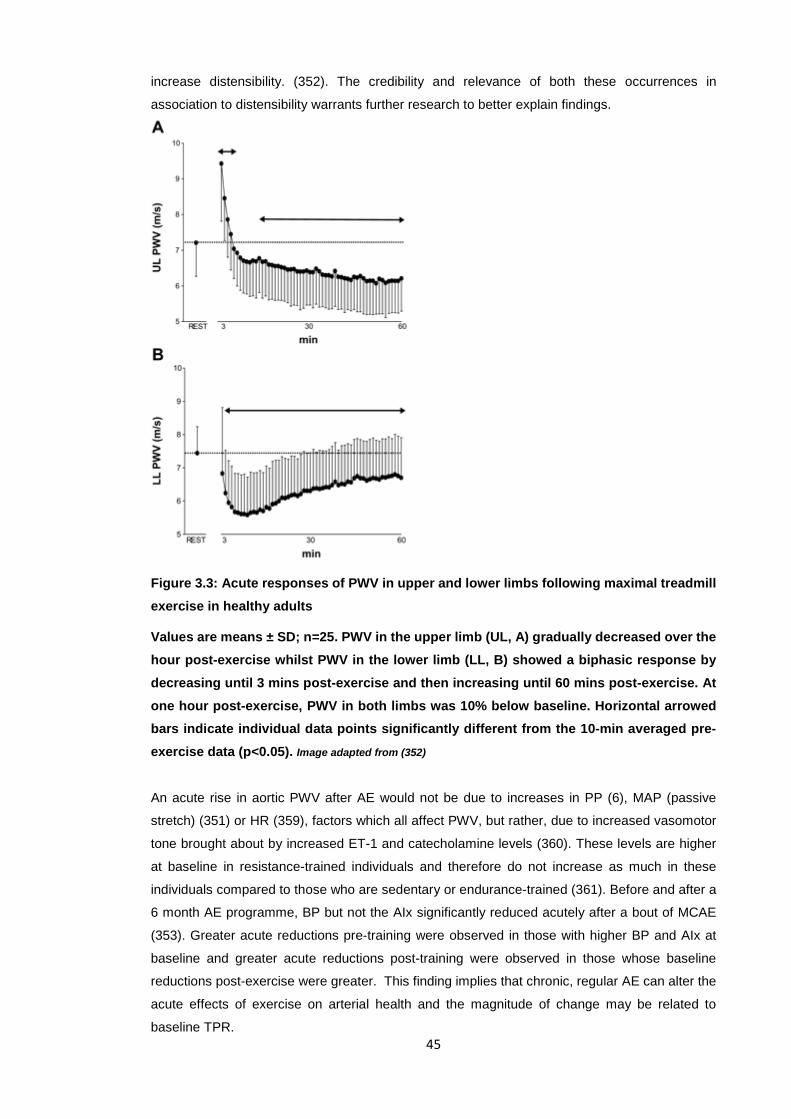
45
increase distensibility. (352). The credibility and relevance of both these occurrences in
association to distensibility warrants further research to better explain findings.
Figure 3.3: Acute responses of PWV in upper and lower limbs following maximal treadmill exercise in healthy adults
Values are means ± SD; n=25. PWV in the upper limb (UL, A) gradually decreased over the hour post-exercise whilst PWV in the lower limb (LL, B) showed a biphasic response by decreasing until 3 mins post-exercise and then increasing until 60 mins post-exercise. At one hour post-exercise, PWV in both limbs was 10% below baseline. Horizontal arrowed bars indicate individual data points significantly different from the 10-min averaged pre-exercise data (p<0.05). Image adapted from (352)
An acute rise in aortic PWV after AE would not be due to increases in PP (6), MAP (passive
stretch) (351) or HR (359), factors which all affect PWV, but rather, due to increased vasomotor
tone brought about by increased ET-1 and catecholamine levels (360). These levels are higher
at baseline in resistance-trained individuals and therefore do not increase as much in these
individuals compared to those who are sedentary or endurance-trained (361). Before and after a
6 month AE programme, BP but not the AIx significantly reduced acutely after a bout of MCAE
(353). Greater acute reductions pre-training were observed in those with higher BP and AIx at
baseline and greater acute reductions post-training were observed in those whose baseline
reductions post-exercise were greater. This finding implies that chronic, regular AE can alter the
acute effects of exercise on arterial health and the magnitude of change may be related to
baseline TPR.
46
Shear forces increase anterograde flow which explains how exercise positively modifies
endothelial function acutely. Retrograde (backward) shear, however, may attenuate these
benefits (362). When these effects of increased shear stress occur repeatedly during acute
exercise bouts, they provide a stimulus for vasodilator pathways and vascular adaptation to long-
term exercise training (81). Regional factors other than NO (eg. prostacyclin, endothelium-derived
hyperpolarizing factor and interstitial metabolites such as lactate, adenosine, phosphate and H+)
may also play a role in the acute effects of AE on AS (123).
On the other hand, the pressor response during RE can alter the load-bearing properties of
collagen and elastin (363) to overcome the acute, repetitive increases in BP associated with RE
(65, 66). This increases left ventricular afterload, thereby contributing to its hypertrophy (364),
and coupled with increased SNA and vasomotor tone, this is associated with reduced arterial
compliance (64). Furthermore, RE increases the formation of cross-links and AGEs in arterial
walls (365). However, vasodilation of the vasa vasorum supplying the aorta might explain the
acute decrease in aortic PWV following exercise in resistance-trained athletes (360).
3.6.1 SUMMARY: THE ACUTE EFFECTS OF EXERCISE ON ARTERIAL HEALTH
Altered stiffness and wave-reflection indices after exercise are influenced primarily by vasomotor
tone and shear forces. Acute decreases in PWV and the AIx following exercise are attributed to
vasodilation, which counters the effects of vasoconstriction on AS. Acute increases in PWV and
the AIx following maximal AE are usually due to arteriogenesis-induced wall stretch and
decreased distensibility as well as increased catecholamine release during exercise. Beneficial
effects usually occur in a dose-dependent manner (with high-intensity exercise eliciting greater
NO release) and the effects can persist for up to an hour post-exercise. Increased SNA to
overcome the pressor response elicited during RE may explain the acute decrease in arterial
compliance following this type of exercise. The magnitude of changes generally depends on
baseline values. Acute effects of exercise on arterial health may explain long-term adaptations
whilst chronic exercise training may alter the acute effects of exercise on arteries.
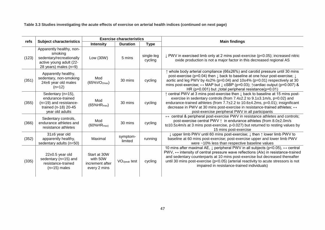
47
Table 3.3 Studies investigating the acute effects of exercise on arterial health indices (continued on next page)
refs Subject characteristics Exercise characteristics Main findings Intensity Duration Type
(123)
Apparently healthy, non-smoking
sedentary/recreationally active young adult (22-28 years) males (n=9)
Low (30W) 5 mins single-leg cycling
↓ PWV in exercised limb only at 2 mins post-exercise (p<0.05); increased nitric oxide production is not a major factor in this decreased regional AS
(351)
Apparently healthy, sedentary, non-smoking
24±6 year old males (n=12)
Mod (65%VO2max) 30 mins cycling
↑ whole body arterial compliance (66±26%) and carotid pressure until 30 mins post-exercise (p=0.04) then ↓ back to baseline at one hour post-exercise; ↓
aortic and leg PWV by 4±2% (p=0.04) and 10±4% (p=0.01) respectively at 30 mins post-exercise; ↔ MAP but ↓ cSBP (p=0.03); ↑cardiac output (p=0.007) &
HR (p=0.001) but ↓total peripheral resistance(p=0.01)
(355)
Sedentary (n=15), endurance-trained
(n=19) and resistance-trained (n-18) 20-45
year old adults
Mod (65%HRmax) 30 mins cycling
↑ central PWV at 3 mins post-exercise then ↓ back to baseline at 15 mins post-exercise in sedentary controls (from 7.4±2.2 to 9.1±3.1m/s, p=0.02) and
endurance-trained athletes (from 7.7±2.2 to 10.6±4.2ms, p=0.01); insignificant decrease in PWV at 30 mins post-exercise in resistance-trained athletes; ↔
post-exercise peripheral PWV in all participants
(366) Sedentary controls,
endurance athletes and resistance athletes
Mod (60%HRmax) 30 mins cycling
↔ central & peripheral post-exercise PWV in resistance athletes and controls; post-exercise central PWV ↑ in endurance athletes (from 8.0±2.0m/s
to10.5±4m/s at 3 mins post-exercise, p-0.027) but returned to resting values by 15 mins post-exercise
(352) 31±6 year old
apparently healthy, sedentary adults (n=50)
Maximal symptom-limited running
↓ upper limb PWV until 60 mins post-exercise; ↓ then ↑ lower limb PWV to baseline at 60 mins post-exercise; post-exercise upper and lower limb PWV
were ~10% less than respective baseline values
(335)
22±0.5 year old sedentary (n=15) and
resistance-trained (n=15) males
Start at 30W with 50W
increment after every 2 mins
VO2peak test cycling
10 mins after maximal AE, ↓ peripheral PWV in all subjects (p<0.05), ↔ central PWV, ↔ intensity of central pressure wave reflections (AIx) in resistance-trained and sedentary counterparts at 10 mins post-exercise but decreased thereafter until 30 mins post-exercise (p<0.05) (arterial reactivity to acute stressors is not
impaired in resistance-trained individuals)
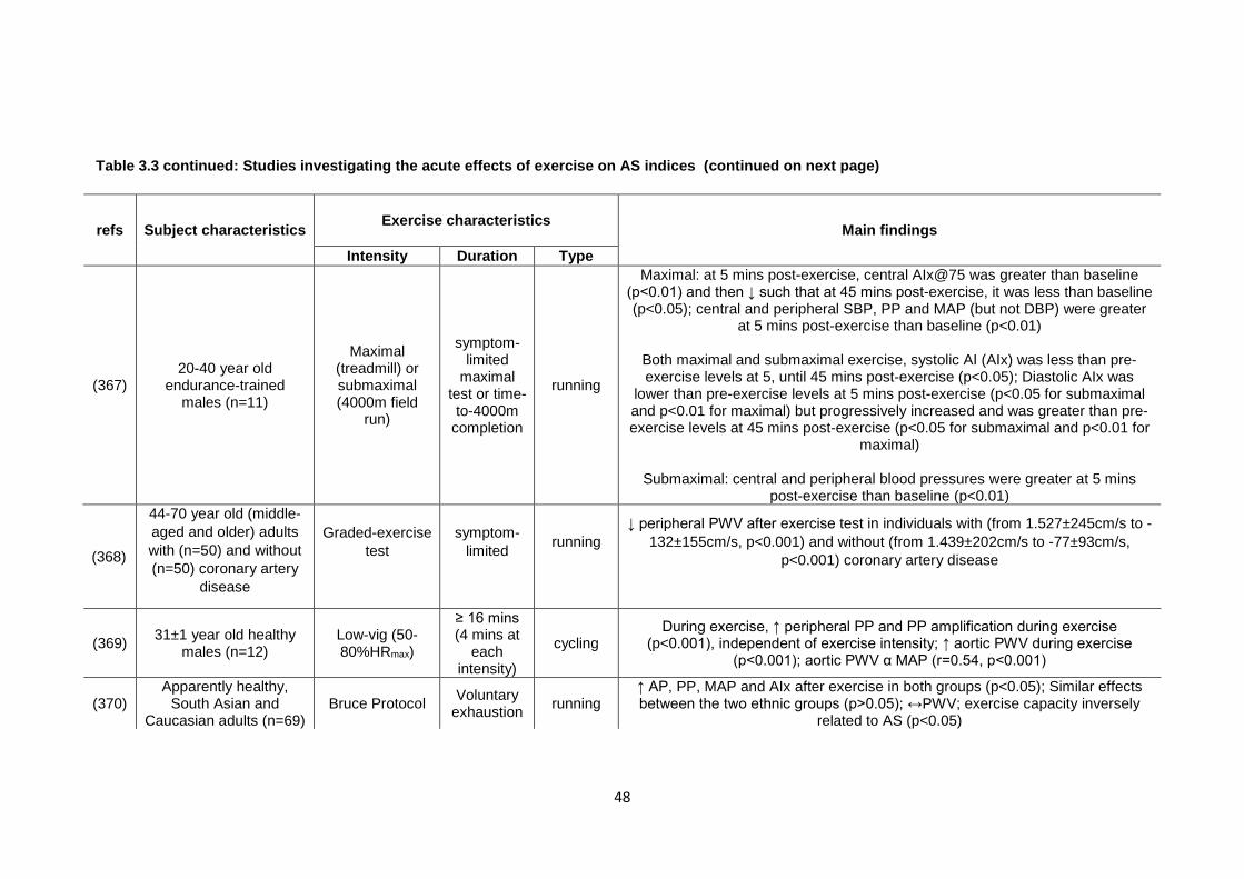
48
(368)
44-70 year old (middle-aged and older) adults with (n=50) and without (n=50) coronary artery
disease
Graded-exercise test
symptom-limited
running
↓ peripheral PWV after exercise test in individuals with (from 1.527±245cm/s to -132±155cm/s, p<0.001) and without (from 1.439±202cm/s to -77±93cm/s,
p<0.001) coronary artery disease
(369) 31±1 year old healthy males (n=12)
Low-vig (50-80%HRmax)
≥ 16 mins (4 mins at
each intensity)
cycling During exercise, ↑ peripheral PP and PP amplification during exercise
(p<0.001), independent of exercise intensity; ↑ aortic PWV during exercise (p<0.001); aortic PWV α MAP (r=0.54, p<0.001)
(370) Apparently healthy,
South Asian and Caucasian adults (n=69)
Bruce Protocol Voluntary exhaustion running
↑ AP, PP, MAP and AIx after exercise in both groups (p<0.05); Similar effects between the two ethnic groups (p>0.05); ↔PWV; exercise capacity inversely
related to AS (p<0.05)
Table 3.3 continued: Studies investigating the acute effects of exercise on AS indices (continued on next page)
refs Subject characteristics
Exercise characteristics
Main findings
Intensity Duration Type
(367) 20-40 year old
endurance-trained males (n=11)
Maximal (treadmill) or submaximal (4000m field
run)
symptom-limited
maximal test or time-to-4000m
completion
running
Maximal: at 5 mins post-exercise, central AIx@75 was greater than baseline (p<0.01) and then ↓ such that at 45 mins post-exercise, it was less than baseline (p<0.05); central and peripheral SBP, PP and MAP (but not DBP) were greater
at 5 mins post-exercise than baseline (p<0.01)
Both maximal and submaximal exercise, systolic AI (AIx) was less than pre-exercise levels at 5, until 45 mins post-exercise (p<0.05); Diastolic AIx was
lower than pre-exercise levels at 5 mins post-exercise (p<0.05 for submaximal and p<0.01 for maximal) but progressively increased and was greater than pre-exercise levels at 45 mins post-exercise (p<0.05 for submaximal and p<0.01 for
maximal)
Submaximal: central and peripheral blood pressures were greater at 5 mins post-exercise than baseline (p<0.01)
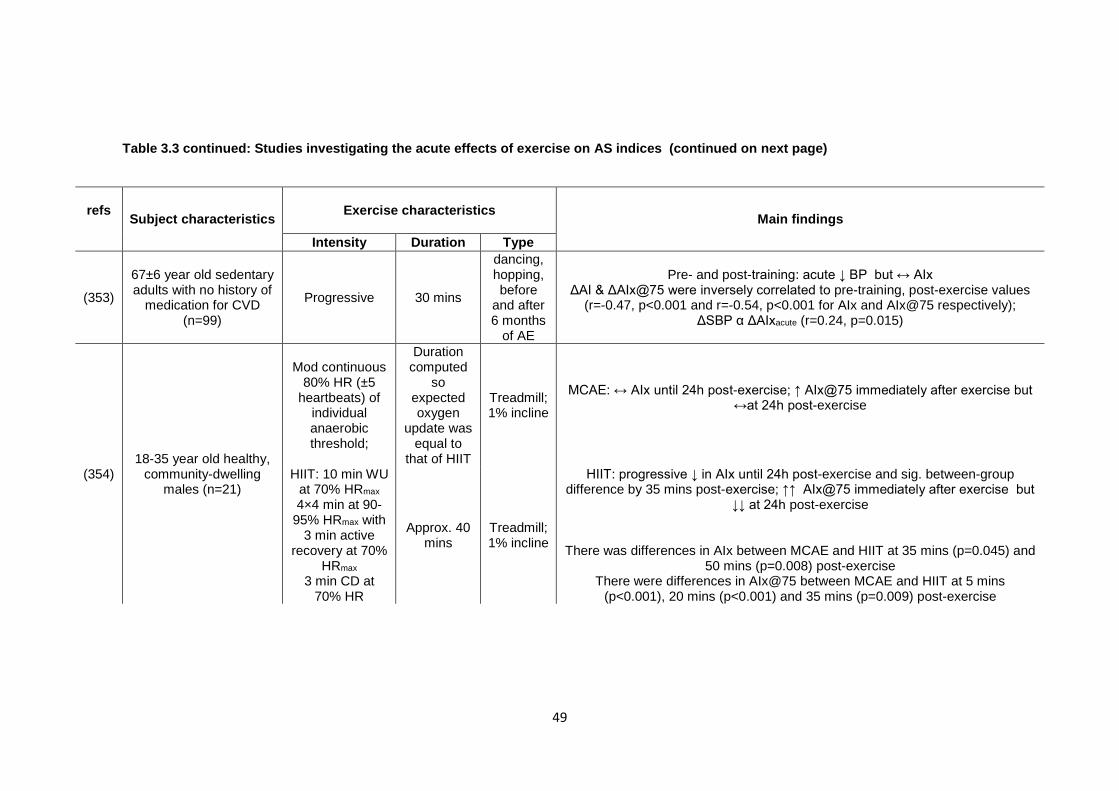
49
Table 3.3 continued: Studies investigating the acute effects of exercise on AS indices (continued on next page)
refs Subject characteristics
Exercise characteristics
Main findings
Intensity Duration Type
(353)
67±6 year old sedentary adults with no history of
medication for CVD (n=99)
Progressive 30 mins
dancing, hopping, before
and after 6 months
of AE
Pre- and post-training: acute ↓ BP but ↔ AIx ΔAI & ΔAIx@75 were inversely correlated to pre-training, post-exercise values
(r=-0.47, p<0.001 and r=-0.54, p<0.001 for AIx and AIx@75 respectively); ΔSBP α ΔAIxacute (r=0.24, p=0.015)
(354) 18-35 year old healthy,
community-dwelling males (n=21)
Mod continuous 80% HR (±5
heartbeats) of individual anaerobic threshold;
Duration computed
so expected oxygen
update was equal to
that of HIIT
Treadmill; 1% incline
MCAE: ↔ AIx until 24h post-exercise; ↑ AIx@75 immediately after exercise but ↔at 24h post-exercise
HIIT: 10 min WU at 70% HRmax 4×4 min at 90-
95% HRmax with 3 min active
recovery at 70% HRmax
3 min CD at 70% HR
Approx. 40 mins
Treadmill; 1% incline
HIIT: progressive ↓ in AIx until 24h post-exercise and sig. between-group difference by 35 mins post-exercise; ↑↑ AIx@75 immediately after exercise but
↓↓ at 24h post-exercise
There was differences in AIx between MCAE and HIIT at 35 mins (p=0.045) and 50 mins (p=0.008) post-exercise
There were differences in AIx@75 between MCAE and HIIT at 5 mins (p<0.001), 20 mins (p<0.001) and 35 mins (p=0.009) post-exercise
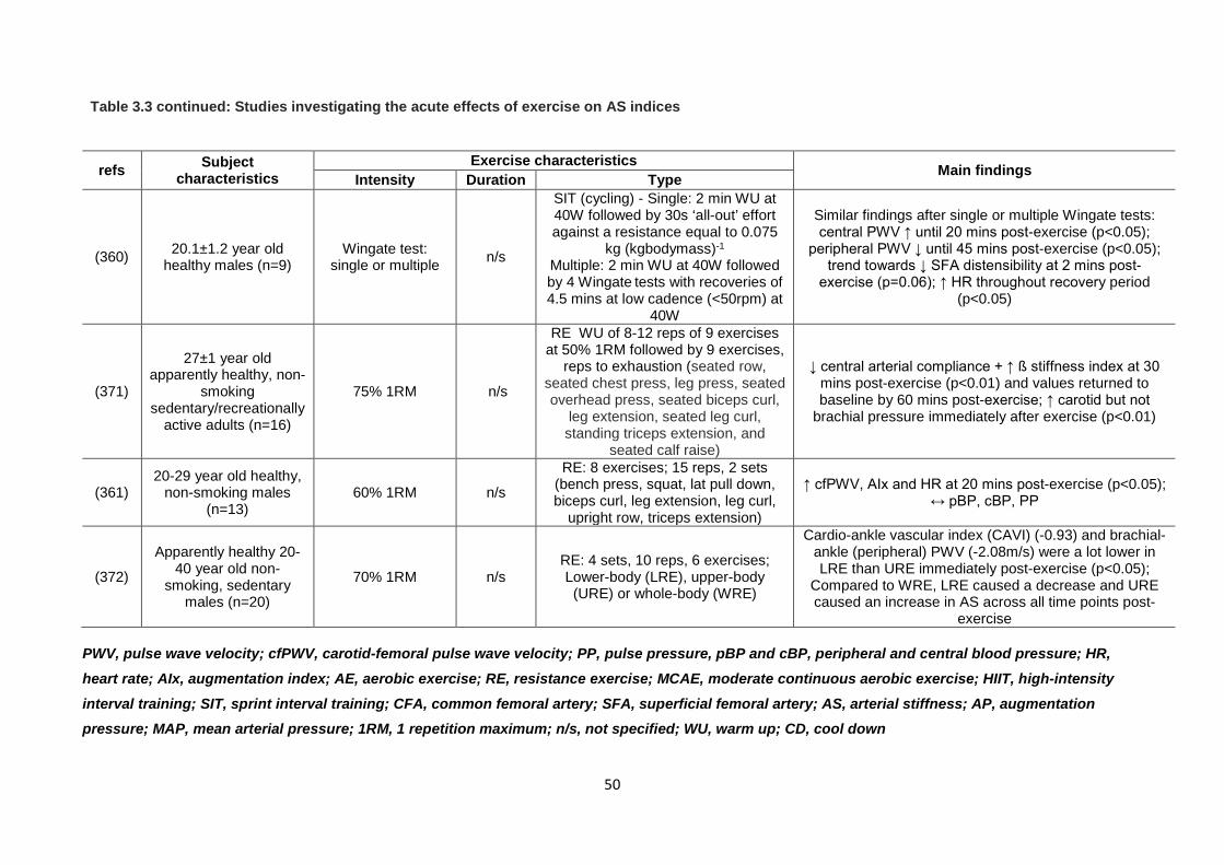
50
PWV, pulse wave velocity; cfPWV, carotid-femoral pulse wave velocity; PP, pulse pressure, pBP and cBP, peripheral and central blood pressure; HR, heart rate; AIx, augmentation index; AE, aerobic exercise; RE, resistance exercise; MCAE, moderate continuous aerobic exercise; HIIT, high-intensity interval training; SIT, sprint interval training; CFA, common femoral artery; SFA, superficial femoral artery; AS, arterial stiffness; AP, augmentation pressure; MAP, mean arterial pressure; 1RM, 1 repetition maximum; n/s, not specified; WU, warm up; CD, cool down
Table 3.3 continued: Studies investigating the acute effects of exercise on AS indices
refs Subject characteristics
Exercise characteristics Main findings Intensity Duration Type
(360) 20.1±1.2 year old healthy males (n=9)
Wingate test: single or multiple n/s
SIT (cycling) - Single: 2 min WU at 40W followed by 30s ‘all-out’ effort against a resistance equal to 0.075
kg (kgbodymass)-1
Multiple: 2 min WU at 40W followed by 4 Wingate tests with recoveries of 4.5 mins at low cadence (<50rpm) at
40W
Similar findings after single or multiple Wingate tests: central PWV ↑ until 20 mins post-exercise (p<0.05);
peripheral PWV ↓ until 45 mins post-exercise (p<0.05); trend towards ↓ SFA distensibility at 2 mins post-
exercise (p=0.06); ↑ HR throughout recovery period (p<0.05)
(371)
27±1 year old apparently healthy, non-
smoking sedentary/recreationally
active adults (n=16)
75% 1RM n/s
RE WU of 8-12 reps of 9 exercises at 50% 1RM followed by 9 exercises,
reps to exhaustion (seated row, seated chest press, leg press, seated overhead press, seated biceps curl,
leg extension, seated leg curl, standing triceps extension, and
seated calf raise)
↓ central arterial compliance + ↑ ß stiffness index at 30 mins post-exercise (p<0.01) and values returned to baseline by 60 mins post-exercise; ↑ carotid but not
brachial pressure immediately after exercise (p<0.01)
(361) 20-29 year old healthy,
non-smoking males (n=13)
60% 1RM n/s
RE: 8 exercises; 15 reps, 2 sets (bench press, squat, lat pull down, biceps curl, leg extension, leg curl,
upright row, triceps extension)
↑ cfPWV, AIx and HR at 20 mins post-exercise (p<0.05); ↔ pBP, cBP, PP
(372)
Apparently healthy 20-40 year old non-
smoking, sedentary males (n=20)
70% 1RM n/s RE: 4 sets, 10 reps, 6 exercises; Lower-body (LRE), upper-body (URE) or whole-body (WRE)
Cardio-ankle vascular index (CAVI) (-0.93) and brachial-ankle (peripheral) PWV (-2.08m/s) were a lot lower in LRE than URE immediately post-exercise (p<0.05);
Compared to WRE, LRE caused a decrease and URE caused an increase in AS across all time points post-
exercise
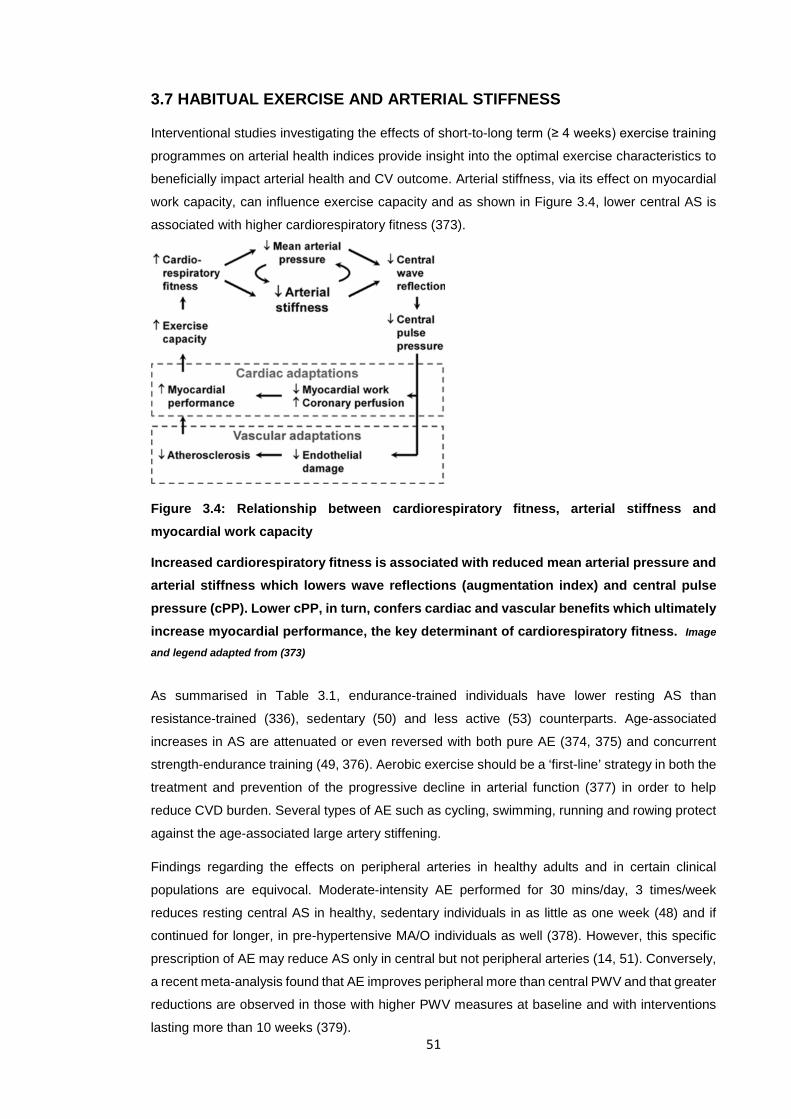
51
3.7 HABITUAL EXERCISE AND ARTERIAL STIFFNESS
Interventional studies investigating the effects of short-to-long term (≥ 4 weeks) exercise training
programmes on arterial health indices provide insight into the optimal exercise characteristics to
beneficially impact arterial health and CV outcome. Arterial stiffness, via its effect on myocardial
work capacity, can influence exercise capacity and as shown in Figure 3.4, lower central AS is
associated with higher cardiorespiratory fitness (373).
Figure 3.4: Relationship between cardiorespiratory fitness, arterial stiffness and myocardial work capacity
Increased cardiorespiratory fitness is associated with reduced mean arterial pressure and arterial stiffness which lowers wave reflections (augmentation index) and central pulse pressure (cPP). Lower cPP, in turn, confers cardiac and vascular benefits which ultimately increase myocardial performance, the key determinant of cardiorespiratory fitness. Image
and legend adapted from (373)
As summarised in Table 3.1, endurance-trained individuals have lower resting AS than
resistance-trained (336), sedentary (50) and less active (53) counterparts. Age-associated
increases in AS are attenuated or even reversed with both pure AE (374, 375) and concurrent
strength-endurance training (49, 376). Aerobic exercise should be a ‘first-line’ strategy in both the
treatment and prevention of the progressive decline in arterial function (377) in order to help
reduce CVD burden. Several types of AE such as cycling, swimming, running and rowing protect
against the age-associated large artery stiffening.
Findings regarding the effects on peripheral arteries in healthy adults and in certain clinical
populations are equivocal. Moderate-intensity AE performed for 30 mins/day, 3 times/week
reduces resting central AS in healthy, sedentary individuals in as little as one week (48) and if
continued for longer, in pre-hypertensive MA/O individuals as well (378). However, this specific
prescription of AE may reduce AS only in central but not peripheral arteries (14, 51). Conversely,
a recent meta-analysis found that AE improves peripheral more than central PWV and that greater
reductions are observed in those with higher PWV measures at baseline and with interventions
lasting more than 10 weeks (379).
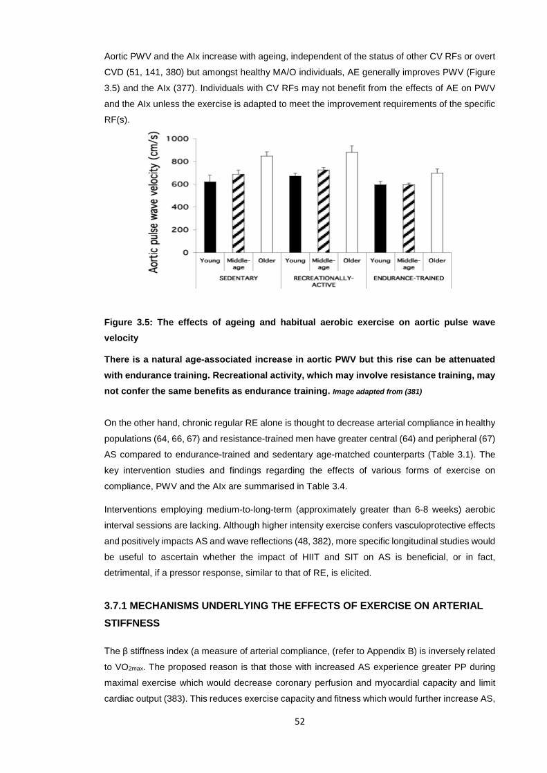
52
Aortic PWV and the AIx increase with ageing, independent of the status of other CV RFs or overt
CVD (51, 141, 380) but amongst healthy MA/O individuals, AE generally improves PWV (Figure
3.5) and the AIx (377). Individuals with CV RFs may not benefit from the effects of AE on PWV
and the AIx unless the exercise is adapted to meet the improvement requirements of the specific
RF(s).
Figure 3.5: The effects of ageing and habitual aerobic exercise on aortic pulse wave velocity
There is a natural age-associated increase in aortic PWV but this rise can be attenuated with endurance training. Recreational activity, which may involve resistance training, may not confer the same benefits as endurance training. Image adapted from (381)
On the other hand, chronic regular RE alone is thought to decrease arterial compliance in healthy
populations (64, 66, 67) and resistance-trained men have greater central (64) and peripheral (67)
AS compared to endurance-trained and sedentary age-matched counterparts (Table 3.1). The
key intervention studies and findings regarding the effects of various forms of exercise on
compliance, PWV and the AIx are summarised in Table 3.4.
Interventions employing medium-to-long-term (approximately greater than 6-8 weeks) aerobic
interval sessions are lacking. Although higher intensity exercise confers vasculoprotective effects
and positively impacts AS and wave reflections (48, 382), more specific longitudinal studies would
be useful to ascertain whether the impact of HIIT and SIT on AS is beneficial, or in fact,
detrimental, if a pressor response, similar to that of RE, is elicited.
3.7.1 MECHANISMS UNDERLYING THE EFFECTS OF EXERCISE ON ARTERIAL STIFFNESS
The β stiffness index (a measure of arterial compliance, (refer to Appendix B) is inversely related
to VO2max. The proposed reason is that those with increased AS experience greater PP during
maximal exercise which would decrease coronary perfusion and myocardial capacity and limit
cardiac output (383). This reduces exercise capacity and fitness which would further increase AS,
53
thereby setting up a vicious cycle (Figure 3.4). Carotid artery compliance decreases during
healthy ageing, possibly due to hypertrophy of VSMCs, replacement of functional cells with
connective tissue and increased cross-linking of collagen (42). Aerobic exercise, especially of a
vigorous nature, can mitigate ageing-associated large artery stiffening in healthy individuals (48,
49, 374). Habitual exercise is thought to remodel arteries in a way that wall stress, BP and
atherosclerotic thrombus formation risks are reduced (81). Since the AIx is inversely related to
both BP and TPR (384), higher-intensity exercise, which causes greater shear stress and NO
release (relative to low-intensity exercise), results in a larger drop in the AIx and TPR (385). This
positive dose-response relationship between exercise intensity and improvements in the AIx (386)
imply that high-intensity exercise may actually be more beneficial than low-intensity exercise for
cardiovascular health (387).
Although carotid and aortic compliance improved in most intervention studies employing AE,
peripheral (muscular) arteries do not usually show these improvements (refer to Table 3.4). Since
central arteries have relatively more elastin in their walls than peripheral arteries do, the
processes that underlie age-associated arterial stiffening (elastin fragmentation, abnormal
collagen deposits) may occur more rapidly in the former (14). Thus, AE-induced benefits on AS
in central arteries may also occur to a greater extent and more rapidly than in peripheral arteries
(14), but the latter may be because the common femoral artery, a relatively stiff artery with not
much potential for adaptability, has been investigated in most of these studies. As the (aortic and
peripheral) AIx partially depends on PWV (353) (refer to Figure 2.3), AE-induced long-term
changes in the former are likely due to reductions in the latter. When arterial compliance increases
due to internal factors, PWV and the intensity of reflected waves decrease, which would
eventually reduce augmentation of central peak systolic pressures (and central PP) (24).
The dilation of elastic arteries during ageing is accompanied by increased wall tension and
changes in gene expression which result in vascular remodelling, oxidative stress and a pro-
atherogenic phenotype in the arterial walls (306). The mechanisms by which regular AE improves
arterial function are largely unknown and still under investigation but existing evidence suggests
that reductions in oxidative stress (including decreased fibrosis and AGEs), inhibition of chronic
vascular inflammation, promotion of mitochondrial health, biogenesis, antioxidant systems and
NO bioavailability may explain most of the benefits (306, 377). The shear stress created by AE
plays a pivotal role in mediating the benefits to vascular function in both humans (388) and
animals (389).
Exercise-associated increases in PP and haemodynamic modifications in peripheral arteries also
trigger vascular structural (arterial wall) and functional (vasomotor tone) adaptations, even in beds
far away from the site of the working muscles (81). Endurance training does not reduce AS in pre-
hypertensive and elderly individuals unless the training duration is prolonged or SBP drops
significantly (378, 390). In this population, the impaired baroreflex predisposes their arterial walls
to greater stress during exercise and therefore, smooth muscle content in the walls may increase
as a compensatory mechanism which in turn restricts compliance (378).
54
Many studies providing mechanistic insights into AE and arterial health have been carried out on
animals due to the inaccessible, central nature of the large arteries in humans (377). Peripheral
arteries cannot be used as surrogates as they do not significantly stiffen with ageing. Increased
large artery stiffness with ageing has been attributed primarily to altered expression, bioactivity
and/or configuration of the structural proteins in their walls, especially in the tunica media (391).
Collagen and elastin contents increase and decrease respectively with ageing in sedentary
animals but these changes were less obvious when animals swam for only 4-5 months (392).
However, findings from experimental studies carried out on animals show no association between
AE-related reductions in AS and modifications of structural proteins (392). In the carotid arteries
of sedentary animals, there is increased calcification, increased levels of MMP-2 and the pro-
fibrotic cytokine transforming growth factor-β1 (TGF-β1) as well as decreased levels of lysyl
oxidase, a pro-synthetic elastin enzyme (393). Voluntary wheel running for only 10-14 weeks
reversed these changes in old animals possibly by reducing oxidative stress, collagen I
deposition, TGF-β1 levels, calcification and AGE formation. In addition, constant, cyclic transient
deformation of vessels could alter collagen/elastin ratios and reduce the cross-linking of
connective tissue which increases AS (377).
Chronic RE likely decreases arterial compliance in healthy individuals and may contribute to
greater cardiac afterload, with highly resistance-trained athletes exhibiting greater large AS and
greater peripheral and central PPs compared to less active age-mates (67). This may be an
adaptive response to protect the aorta from too much expansion during acute lifts and this would
limit aerobic, but not lifting capacity. However, when prescribed correctly, the CV (and other)
benefits of RE outweigh the adverse impact it may have on AS (64). The mechanisms underlying
the increased AS with RE are still largely unexplored but the current hypotheses relate to the
cumulative effects of single bouts of RE and the effects that acute sessions have on BP (64).
Concurrent AE and RE exercise does not usually improve AS measures significantly (386) but
Cook et al. reported greater carotid compliance in rowers (who engage in AE and RE) than
sedentary counterparts, indicating that AE may override the detrimental effects of RE on AS (327).
3.7.2 SUMMARY: EXERCISE AND ARTERIAL STIFFNESS Cross-sectional studies generally report lower PWV and AIx indices in endurance-trained athletes
than less active/resistance-trained counterparts. Furthermore, these indices are usually inversely
related to cardiorespiratory fitness. Interventional studies employing low, moderate and high-
intensity MCAE as well as HIIT have shown improved arterial compliance, stiffness and wave
reflection indices even within one month. Aerobic exercise can mitigate ageing-associated large
artery stiffening in healthy individuals. The mechanisms underlying this are not fully understood
but are likely related to the anti-oxidative and anti-inflammatory effects of AE as well as increased
NO bioavailability. Resistance exercise most probably has detrimental effects on arterial health
indices related to stiffness.
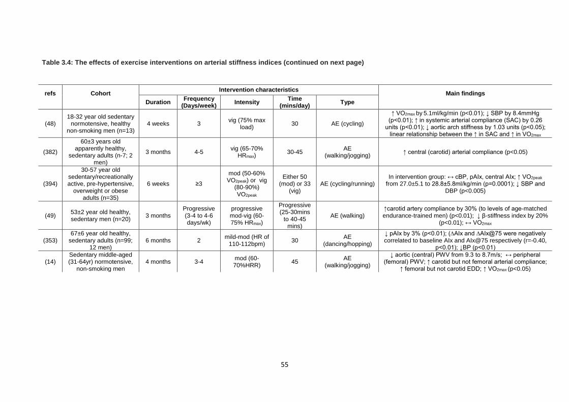
55
Table 3.4: The effects of exercise interventions on arterial stiffness indices (continued on next page)
refs
Cohort
Intervention characteristics Main findings Duration Frequency
(Days/week) Intensity Time (mins/day) Type
(48) 18-32 year old sedentary
normotensive, healthy non-smoking men (n=13)
4 weeks 3 vig (75% max load) 30 AE (cycling)
↑ VO2max by 5.1ml/kg/min (p<0.01); ↓ SBP by 8.4mmHg (p<0.01); ↑ in systemic arterial compliance (SAC) by 0.26
units (p<0.01); ↓ aortic arch stiffness by 1.03 units (p<0.05); linear relationship between the ↑ in SAC and ↑ in VO2max
(382)
60±3 years old apparently healthy,
sedentary adults (n-7; 2 men)
3 months 4-5 vig (65-70% HRmax) 30-45 AE
(walking/jogging) ↑ central (carotid) arterial compliance (p<0.05)
(394)
30-57 year old sedentary/recreationally active, pre-hypertensive,
overweight or obese adults (n=35)
6 weeks ≥3
mod (50-60% VO2peak) or vig
(80-90%) VO2peak
Either 50 (mod) or 33
(vig) AE (cycling/running)
In intervention group: ↔ cBP, pAIx, central AIx; ↑ VO2peak from 27.0±5.1 to 28.8±5.8ml/kg/min (p=0.0001); ↓ SBP and
DBP (p<0.005)
(49) 53±2 year old healthy, sedentary men (n=20) 3 months
Progressive (3-4 to 4-6 days/wk)
progressive mod-vig (60-75% HRmax)
Progressive (25-30mins
to 40-45 mins)
AE (walking) ↑carotid artery compliance by 30% (to levels of age-matched endurance-trained men) (p<0.01); ↓ β-stiffness index by 20%
(p<0.01); ↔ VO2max
(353) 67±6 year old healthy,
sedentary adults (n=99; 12 men)
6 months 2 mild-mod (HR of 110-112bpm) 30 AE
(dancing/hopping)
↓ pAIx by 3% (p<0.01); (∆AIx and ∆AIx@75 were negatively correlated to baseline AIx and AIx@75 respectively (r=-0.40,
p<0.01); ↓BP (p<0.01)
(14) Sedentary middle-aged (31-64yr) normotensive,
non-smoking men 4 months 3-4 mod (60-
70%HRR) 45 AE (walking/jogging)
↓ aortic (central) PWV from 9.3 to 8.7m/s; ↔ peripheral (femoral) PWV; ↑ carotid but not femoral arterial compliance;
↑ femoral but not carotid EDD; ↑ VO2max (p<0.05)
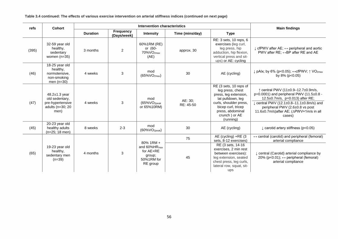
56
Table 3.4 continued: The effects of various exercise intervention on arterial stiffness indices (continued on next page)
refs
Cohort
Intervention characteristics Main findings Duration Frequency
(Days/week) Intensity Time (mins/day) Type
(395)
32-59 year old healthy,
sedentary women (n=35)
3 months 2
60%1RM (RE) or (60-
70%VO2max (AE)
approx. 30
RE: 3 sets, 10 reps, 6 exercises (leg curl,
leg press, hip adduction, hip flexion, vertical press and sit-
ups) or AE: cycling
↓ cfPWV after AE; ↔ peripheral and aortic PWV after RE; ↔BP after RE and AE
(46)
18-25 year old healthy,
normotensive, non-smoking men (n=30)
4 weeks 3 mod (65%VO2max) 30 AE (cycling) ↓ pAIx; by 6% (p<0.05); ↔cfPWV; ↑ VO2max
by 8% (p<0.05)
(47)
48.2±1.3 year old sedentary,
pre-hypertensive adults (n=30; 20
men)
4 weeks 3 mod
(65%VO2peak or 65%10RM)
AE: 30; RE: 45-50
RE (3 sets, 10 reps of leg press, chest
press, leg extension, lat pulldown, leg
curls, shoulder press, bicep curl, tricep press, abdominal
crunch ) or AE (running)
↑ central PWV (11±0.9–12.7±0.9m/s, p=0.0001) and peripheral PWV (11.5±0.8 -
12.5±0.7m/s, p=0.013) after RE; ↓ central PWV (12.1±0.8–11.1±0.8m/s) and
peripheral PWV (2.6±0.8 vs post 11.6±0.7m/s)after AE; (∆PWV=1m/s in all
cases)
(45) 20-23 year old healthy adults
(n=25; 18 men) 8 weeks 2-3 mod
(60%VO2peak) 30 AE (cycling) ↓ carotid artery stiffness (p<0.05)
(65)
19-23 year old healthy,
sedentary men (n=39)
4 months 3
80% 1RM + and 60%HRmax
for AE+RE group;
50%1RM for RE group
75 AE (cycling) +RE (3 sets, 8-12 exercises)
↔ central (carotid) and peripheral (femoral) arterial compliance
45
RE (3 sets, 14-16 exercises, 2 min rest between exercises):
leg extension, seated chest press, leg curls, lateral row, squat, sit-
ups
↓ central (Carotid) arterial compliance by 20% (p<0.01); ↔ peripheral (femoral)
arterial compliance
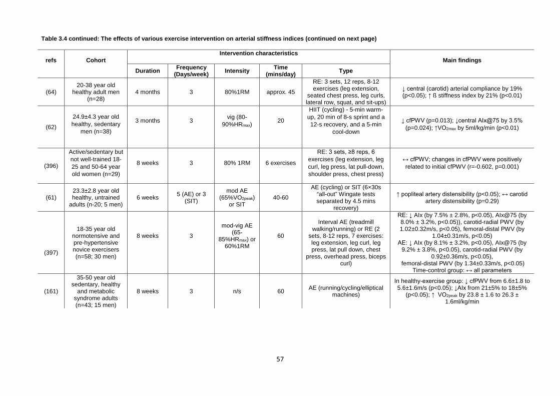
57
Table 3.4 continued: The effects of various exercise intervention on arterial stiffness indices (continued on next page)
refs
Cohort
Intervention characteristics Main findings
Duration Frequency (Days/week) Intensity Time
(mins/day) Type
(64) 20-38 year old
healthy adult men (n=28)
4 months 3 80%1RM approx. 45
RE: 3 sets, 12 reps, 8-12 exercises (leg extension,
seated chest press, leg curls, lateral row, squat, and sit-ups)
↓ central (carotid) arterial compliance by 19% (p<0.05); ↑ ß stiffness index by 21% (p<0.01)
(62)
24.9±4.3 year old healthy, sedentary
men (n=38)
3 months
3
vig (80-90%HRmax)
20
HIIT (cycling) - 5-min warm-up, 20 min of 8-s sprint and a 12-s recovery, and a 5-min
cool-down
↓ cfPWV (p=0.013); ↓central AIx@75 by 3.5% (p=0.024); ↑VO2max by 5ml/kg/min (p<0.01)
(396)
Active/sedentary but not well-trained 18-25 and 50-64 year old women (n=29)
8 weeks 3 80% 1RM 6 exercises
RE: 3 sets, ≥8 reps, 6 exercises (leg extension, leg curl, leg press, lat pull-down, shoulder press, chest press)
↔ cfPWV; changes in cfPWV were positively related to initial cfPWV (r=-0.602, p=0.001)
(61) 23.3±2.8 year old healthy, untrained
adults (n-20; 5 men) 6 weeks 5 (AE) or 3
(SIT)
mod AE (65%VO2peak)
or SIT 40-60
AE (cycling) or SIT (6×30s “all-out” Wingate tests separated by 4.5 mins
recovery)
↑ popliteal artery distensibility (p<0.05); ↔ carotid artery distensibility (p=0.29)
18-35 year old
normotensive and pre-hypertensive novice exercisers (n=58; 30 men)
8 weeks
3
mod-vig AE (65-
85%HRmax) or 60%1RM
60
Interval AE (treadmill walking/running) or RE (2
sets, 8-12 reps, 7 exercises: leg extension, leg curl, leg press, lat pull down, chest
press, overhead press, biceps curl)
RE: ↓ AIx (by 7.5% ± 2.8%, p<0.05), AIx@75 (by 8.0% ± 3.2%, p<0.05)), carotid-radial PWV (by 1.02±0.32m/s, p<0.05), femoral-distal PWV (by
1.04±0.31m/s, p<0.05) AE: ↓ AIx (by 8.1% ± 3.2%, p<0.05), AIx@75 (by
9.2% ± 3.8%, p<0.05), carotid-radial PWV (by 0.92±0.36m/s, p<0.05),
femoral-distal PWV (by 1.34±0.33m/s, p<0.05) Time-control group: ↔ all parameters
(397)
(161)
35-50 year old sedentary, healthy
and metabolic syndrome adults (n=43; 15 men)
8 weeks 3 n/s 60 AE (running/cycling/elliptical machines)
In healthy-exercise group: ↓ cfPWV from 6.6±1.8 to 5.6±1.6m/s (p<0.05); ↓AIx from 21±5% to 18±5%
(p<0.05); ↑ VO2peak by 23.8 ± 1.6 to 26.3 ± 1.6ml/kg/min
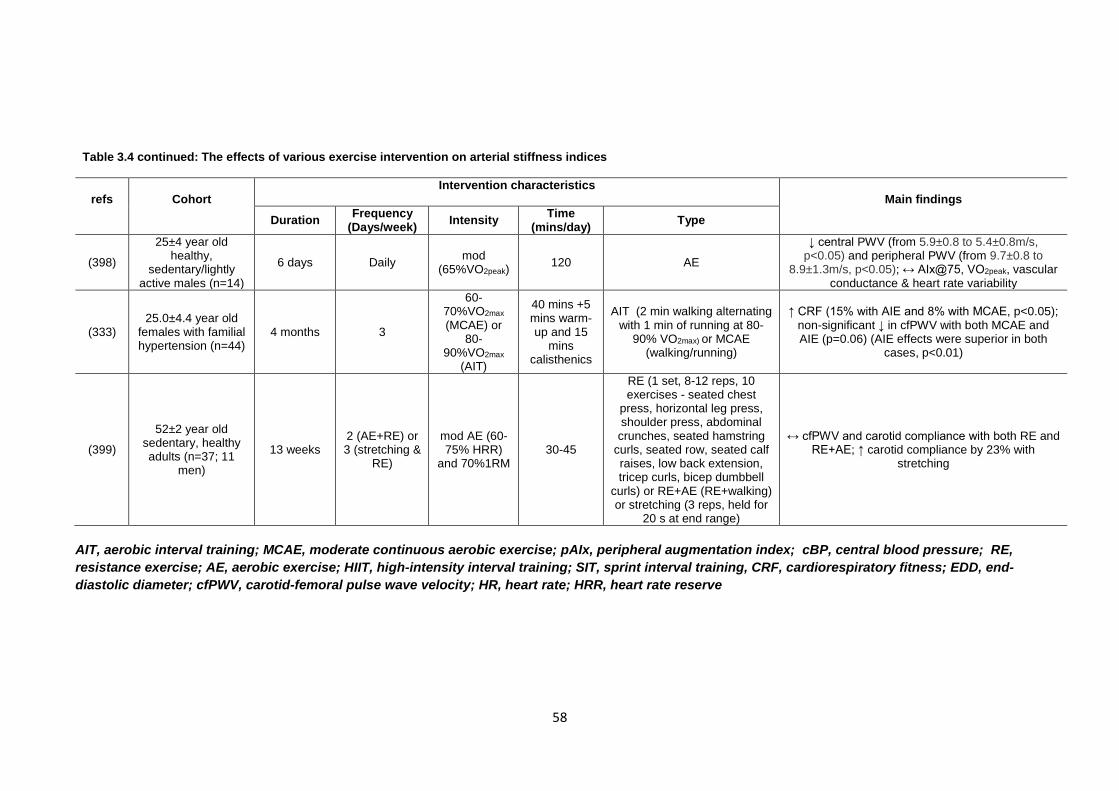
58
AIT, aerobic interval training; MCAE, moderate continuous aerobic exercise; pAIx, peripheral augmentation index; cBP, central blood pressure; RE, resistance exercise; AE, aerobic exercise; HIIT, high-intensity interval training; SIT, sprint interval training, CRF, cardiorespiratory fitness; EDD, end-diastolic diameter; cfPWV, carotid-femoral pulse wave velocity; HR, heart rate; HRR, heart rate reserve
Table 3.4 continued: The effects of various exercise intervention on arterial stiffness indices
refs
Cohort
Intervention characteristics Main findings
Duration Frequency (Days/week) Intensity Time
(mins/day) Type
(398)
25±4 year old healthy,
sedentary/lightly active males (n=14)
6 days Daily mod (65%VO2peak) 120 AE
↓ central PWV (from 5.9±0.8 to 5.4±0.8m/s, p<0.05) and peripheral PWV (from 9.7±0.8 to
8.9±1.3m/s, p<0.05); ↔ AIx@75, VO2peak, vascular conductance & heart rate variability
(333) 25.0±4.4 year old
females with familial hypertension (n=44)
4 months 3
60-70%VO2max (MCAE) or
80-90%VO2max
(AIT)
40 mins +5 mins warm-up and 15
mins calisthenics
AIT (2 min walking alternating with 1 min of running at 80-
90% VO2max) or MCAE (walking/running)
↑ CRF (15% with AIE and 8% with MCAE, p<0.05); non-significant ↓ in cfPWV with both MCAE and AIE (p=0.06) (AIE effects were superior in both
cases, p<0.01)
(399)
52±2 year old sedentary, healthy adults (n=37; 11
men)
13 weeks 2 (AE+RE) or 3 (stretching &
RE)
mod AE (60-75% HRR)
and 70%1RM 30-45
RE (1 set, 8-12 reps, 10 exercises - seated chest
press, horizontal leg press, shoulder press, abdominal crunches, seated hamstring
curls, seated row, seated calf raises, low back extension, tricep curls, bicep dumbbell
curls) or RE+AE (RE+walking) or stretching (3 reps, held for
20 s at end range)
↔ cfPWV and carotid compliance with both RE and RE+AE; ↑ carotid compliance by 23% with
stretching
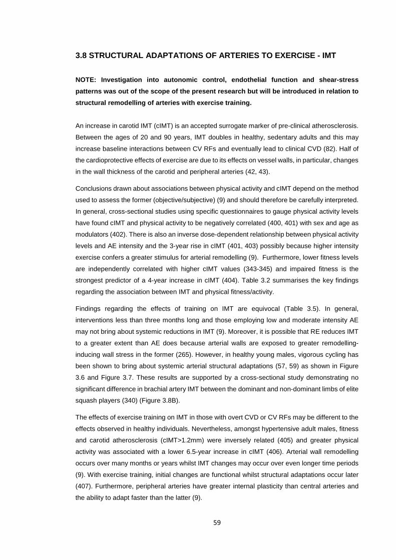
59
3.8 STRUCTURAL ADAPTATIONS OF ARTERIES TO EXERCISE - IMT NOTE: Investigation into autonomic control, endothelial function and shear-stress patterns was out of the scope of the present research but will be introduced in relation to structural remodelling of arteries with exercise training.
An increase in carotid IMT (cIMT) is an accepted surrogate marker of pre-clinical atherosclerosis.
Between the ages of 20 and 90 years, IMT doubles in healthy, sedentary adults and this may
increase baseline interactions between CV RFs and eventually lead to clinical CVD (82). Half of
the cardioprotective effects of exercise are due to its effects on vessel walls, in particular, changes
in the wall thickness of the carotid and peripheral arteries (42, 43).
Conclusions drawn about associations between physical activity and cIMT depend on the method
used to assess the former (objective/subjective) (9) and should therefore be carefully interpreted.
In general, cross-sectional studies using specific questionnaires to gauge physical activity levels
have found cIMT and physical activity to be negatively correlated (400, 401) with sex and age as
modulators (402). There is also an inverse dose-dependent relationship between physical activity
levels and AE intensity and the 3-year rise in cIMT (401, 403) possibly because higher intensity
exercise confers a greater stimulus for arterial remodelling (9). Furthermore, lower fitness levels
are independently correlated with higher cIMT values (343-345) and impaired fitness is the
strongest predictor of a 4-year increase in cIMT (404). Table 3.2 summarises the key findings
regarding the association between IMT and physical fitness/activity.
Findings regarding the effects of training on IMT are equivocal (Table 3.5). In general,
interventions less than three months long and those employing low and moderate intensity AE
may not bring about systemic reductions in IMT (9). Moreover, it is possible that RE reduces IMT
to a greater extent than AE does because arterial walls are exposed to greater remodelling-
inducing wall stress in the former (265). However, in healthy young males, vigorous cycling has
been shown to bring about systemic arterial structural adaptations (57, 59) as shown in Figure
3.6 and Figure 3.7. These results are supported by a cross-sectional study demonstrating no
significant difference in brachial artery IMT between the dominant and non-dominant limbs of elite
squash players (340) (Figure 3.8B).
The effects of exercise training on IMT in those with overt CVD or CV RFs may be different to the
effects observed in healthy individuals. Nevertheless, amongst hypertensive adult males, fitness
and carotid atherosclerosis (cIMT>1.2mm) were inversely related (405) and greater physical
activity was associated with a lower 6.5-year increase in cIMT (406). Arterial wall remodelling
occurs over many months or years whilst IMT changes may occur over even longer time periods
(9). With exercise training, initial changes are functional whilst structural adaptations occur later
(407). Furthermore, peripheral arteries have greater internal plasticity than central arteries and
the ability to adapt faster than the latter (9).
60
3.8.1 MECHANISMS UNDERLYING THE EFFECTS OF EXERCISE ON IMT
As is the case with AS and exercise, the relationship between physical activity and IMT is not
entirely due to the effects on traditional CV RFs (9). Plaque formation has been linked to low
mean shear rates (408) and oscillatory shear (409). In the latter case, retrograde shear stress
gives rise to pro-atherogenic vascular endothelial cell phenotypes (410). Systemic and not
localised shear stress is important when it comes to arterial wall remodelling (326). Given that BP
fluctuates during the cardiac cycle and stretches arterial walls, chronic BP elevations produce
similar endothelial cell phenotypes as do low and oscillatory shear patterns (389). Chronic
increases in (local) distending pressures can eventually result in increased cIMT (411). Exercise
also increases arterial pressure and arterial wall stretch but in the long-term, is associated with
lower IMT (anti-atherogenic changes in arterial walls) (326), giving rise to the speculation that
anti-and pro-atherogenic gene regulation depends on whether the pressure stimulus is evoked
by chronic increases in pressure or by the cyclic, transient and episodic increases that occur
during exercise (326, 389).
Exercise training can decrease IMT by altering vasomotor tone possibly by decreasing resting
SNA (81), thereby preventing smooth muscle hypertrophy brought about by chronically elevated
SNA (412). However, it is unknown whether a correlation exists between exercise-induced
changes in SNA and IMT. Surprisingly, two studies reported positive relations between physical
activity and cIMT in young adult borderline hypertensives (349). The authors suggested that this
could be due to autoregulation to counteract the exercise-induced increases in wall stress or due
to repetitive RAAS activation. They speculated that in a similar way to the exercise-induced left
ventricular hypertrophy, mechanisms underlying the increased cIMT in athletes could relate to
medial thickening. Oxidative stress can cause endothelial dysfunction and atherosclerosis as well
as arterial wall thickening (413, 414) whilst inflammation is associated with atherosclerotic plaque
formation and stability. Aerobic exercise may confer vasculoprotection by its anti-oxidative and
anti-inflammatory effects (refer to Figure 3.1).
3.8.2 SUMMARY: EXERCISE AND IMT
Chronic, moderate to high-intensity AE can reduce IMT in the long-term in healthy, asymptomatic
individuals as well as in those with CVD or CV RFs, with greater effects being observed in
peripheral than central arteries as the latter may require a longer and more intense exercise
stimulus. Findings regarding the systemic effects of localised exercise on IMT are equivocal. The
main mechanisms underlying the impact that exercise has on IMT are related to local (shear
stress patterns) and systemic (arterial pressure) haemodynamic factors as well as non-
haemodynamic factors like vascular tone, SNA, oxidative stress and inflammation. Further
research is required to determine the best exercise prescription to maximise the benefits on
arterial wall thickness because different exercise modes spur unique and diverse haemodynamic
and shear stress patterns, leading to benefits of differing magnitudes in arteries.
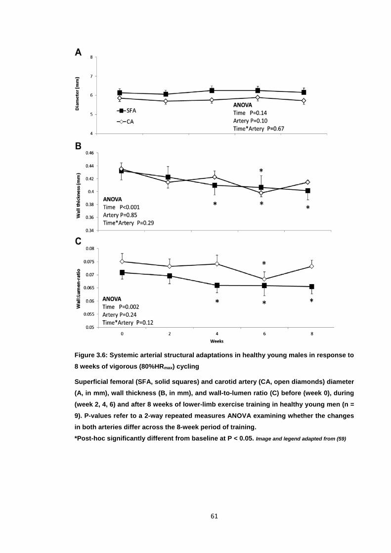
61
Figure 3.6: Systemic arterial structural adaptations in healthy young males in response to 8 weeks of vigorous (80%HRmax) cycling
Superficial femoral (SFA, solid squares) and carotid artery (CA, open diamonds) diameter (A, in mm), wall thickness (B, in mm), and wall-to-lumen ratio (C) before (week 0), during (week 2, 4, 6) and after 8 weeks of lower-limb exercise training in healthy young men (n = 9). P-values refer to a 2-way repeated measures ANOVA examining whether the changes in both arteries differ across the 8-week period of training. *Post-hoc significantly different from baseline at P < 0.05. Image and legend adapted from (59)
62
Figure 3.7: Upper body systemic arterial structural adaptations after lower-body aerobic training
Brachial artery wall thickness (A), lumen diameter (B), and wall:lumen ratio (C) at baseline, 12 and 24wk of aerobic exercise training (walking/cycling) (n = 11). Data is presented for the entire group (n = 11, black bars), men (n = 6, white bars) and women (n = 5, grey bars). *Significant from week 0 at P < 0.05, † P < 0.01. Image and legend adapted from (57)
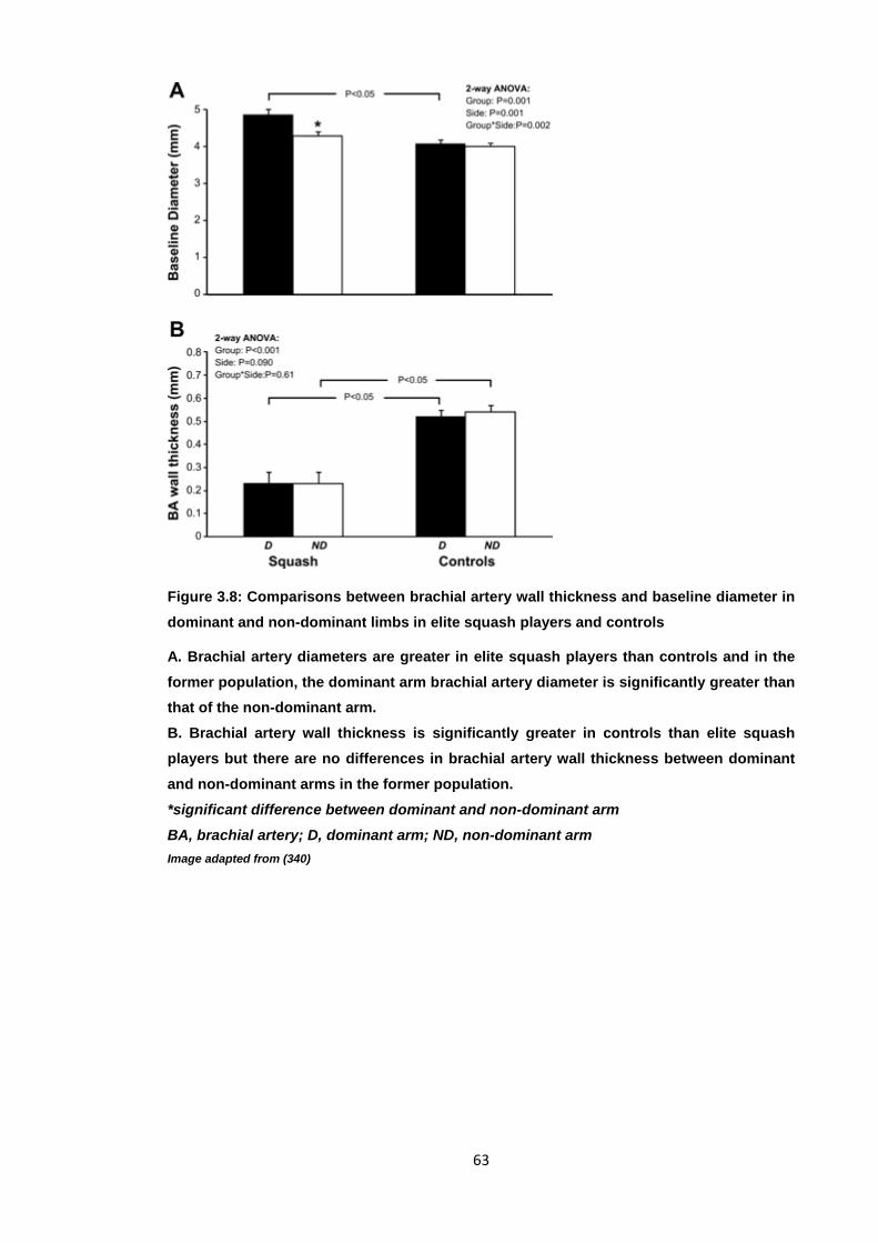
63
Figure 3.8: Comparisons between brachial artery wall thickness and baseline diameter in dominant and non-dominant limbs in elite squash players and controls
A. Brachial artery diameters are greater in elite squash players than controls and in the former population, the dominant arm brachial artery diameter is significantly greater than that of the non-dominant arm. B. Brachial artery wall thickness is significantly greater in controls than elite squash players but there are no differences in brachial artery wall thickness between dominant and non-dominant arms in the former population. *significant difference between dominant and non-dominant arm BA, brachial artery; D, dominant arm; ND, non-dominant arm Image adapted from (340)

64
IMT, intima-media thickness; cIMT, carotid intima-media thickness; WLR, wall:lumen ratio; cWLR, carotid wall:lumen ratio; cCSA, carotid cross-sectional area; AE, aerobic exercise; RE, resistance exercise; SIT, sprint interval training; BP, blood pressure; 1RM, 1 repetition maximum; n/s, not specified; yrs, years
Table 3.5 Summary of the main intervention studies investigating the effects of exercise training on arterial structure
refs
Cohort
Intervention characteristics Main findings Duration Frequency
(Days/week) Intensity Time (mins/day) Type
(57) 58-60 year old healthy, sedentary adults (n=16; 8 men) 6 months 3 mild 30 AE
↓ IMT in upper and lower limb conduit arteries (p<0.01); ↓ WLR in brachial (p<0.01) and popliteal
(p<0.05) arteries
(342) 54±2 year old healthy, sedentary men (n=18) 3 months progressive
to max of 6 mild-vig (60-75% HRmax)
progressive to max of 45 AE (walking/jogging) ↔ cIMT, cWLR and carotid systolic BP
(415) 70±3 year old normotensive, non-smoking, healthy sedentary men
(n=8) 8 weeks 3 mod-vig 30 AE ↑ compliance and diameter of arteries in trained limbs
only (p<0.05); ↔IMT
(416) 53.4±6.2 year old healthy(n=26; 5 men) 6 months 3 progressive
mild to mod progressive
20-40 AE ↓cIMT by 6.4% (p<0.05); ↔ BP
(59) Healthy, young adult males (n=14) 8 weeks 3 vig (80%HRmax)
30 AE (cycling)
↓ superficial femoral and common carotid IMT (p<0.002 and p<0.001 respectively); ↓ superficial
femoral and common carotid WLR; ↔ femoral and carotid resting diameter (p=0.14)
(417) 20.0±2.8 year old healthy adults (n=21; 11 men)
8 weeks training + 4 weeks
detraining
3 80% 1RM n/s
RE: leg exercises - 3 sets, 10 reps, 6 exercises (squats,
Bulgarian split squats, knee extensions, leg press, lunges,
static wall sit)
Training ↑ superficial femoral and carotid artery diameters by 27% and 13% respectively (p<0.001);
detraining ↓ femoral and carotid diameters (46% and 10% respectively, p<0 .001)
(265) 55-68 year old untrained middle-aged adults with chronic heart
failure (n=36; 32 men) 12 weeks 3
AE: progressive
50-70%VO2peak
RE: progressive 50-70%1RM
approx. 45 AE (cycling+walking) or RE (3
sets, 9 exercises with 2:1 work:rest ratio)
↓ brachial IMT (from 475±10 to 443±13μm, p<0.01) and WLR (from 0.121±0.004 to 0.107±0.004, p<0.01) with RE only; ↑ brachial diameter with both AE (5%)
and RE (6%) (p<0.01)
(56) 51 ± 2 year old sedentary men 3 months 5-7 mod-vig (65-80%HRmax)
40-50 AE (walking/jogging)
↑ femoral diameter by 9% ( from 8.82±0.18 to 9.60±0.20 mm, p<0.001); ↓ femoral IMT (from 0.65±0.05 to 0.56±0.05 mm) and WLR by 15-20% (p<0.001); changes in femoral structure and
atherosclerotic risk factors were unrelated
(161) 35-50 year old sedentary, healthy and metabolic syndrome adults
(n=43; 15 men) 8 weeks 3 n/s 60 AE (running/cycling/elliptical
machines) ↔ cIMT, cCSA, cWLR
(64) 20-38 year old healthy, sedentary men (n=28) 4 months 3 80%1RM n/s
RE: 3 sets, 12 reps, 8-12 exercises ( leg extension, seated
chest press, leg curls, lateral row, squat, and sit-ups)
↔ cIMT and carotid diameter
(61) 23.3±2.8 year old healthy, untrained adults (n-20; 5 men) 6 weeks 5 (AE) or 3
(SIT)
mod AE (65%VO2peak)
or SIT 40-60
AE (cycling) or SIT (6×30s “all-out” Wingate tests separated by
4.5 mins recovery) ↔ carotid and popliteal diameter and IMT
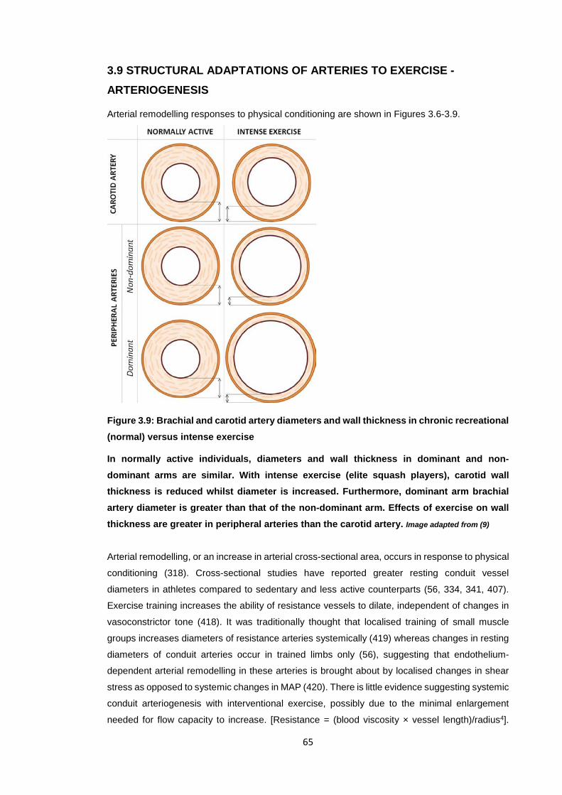
65
3.9 STRUCTURAL ADAPTATIONS OF ARTERIES TO EXERCISE - ARTERIOGENESIS
Arterial remodelling responses to physical conditioning are shown in Figures 3.6-3.9.
Figure 3.9: Brachial and carotid artery diameters and wall thickness in chronic recreational (normal) versus intense exercise
In normally active individuals, diameters and wall thickness in dominant and non-dominant arms are similar. With intense exercise (elite squash players), carotid wall thickness is reduced whilst diameter is increased. Furthermore, dominant arm brachial artery diameter is greater than that of the non-dominant arm. Effects of exercise on wall thickness are greater in peripheral arteries than the carotid artery. Image adapted from (9)
Arterial remodelling, or an increase in arterial cross-sectional area, occurs in response to physical
conditioning (318). Cross-sectional studies have reported greater resting conduit vessel
diameters in athletes compared to sedentary and less active counterparts (56, 334, 341, 407).
Exercise training increases the ability of resistance vessels to dilate, independent of changes in
vasoconstrictor tone (418). It was traditionally thought that localised training of small muscle
groups increases diameters of resistance arteries systemically (419) whereas changes in resting
diameters of conduit arteries occur in trained limbs only (56), suggesting that endothelium-
dependent arterial remodelling in these arteries is brought about by localised changes in shear
stress as opposed to systemic changes in MAP (420). There is little evidence suggesting systemic
conduit arteriogenesis with interventional exercise, possibly due to the minimal enlargement
needed for flow capacity to increase. [Resistance = (blood viscosity × vessel length)/radius4].
66
Nevertheless, both moderate and high-intensity dynamic (but not static) AE can decrease
peripheral AS by increasing arterial diameter (421) and some forms of RE can cause remodelling
of both resistance and conduit vessels (refer to Table 3.5 for a summary of these findings). Eight
weeks of combined AE and RE reduced brachial and superficial femoral but not carotid artery
wall thickness and WLR in type II diabetics (266) whereas 12 and 24 weeks of AE decreased wall
thickness and WLR in the conduit brachial and popliteal arteries (57) (Figure 3.7B and C) and 8
weeks of lower limb training reduced superficial femoral and common carotid WLR (59) (Figure
3.6C).
3.9.1 PROPOSED MECHANISMS UNDERLYING EXERCISE-INDUCED ARTERIAL REMODELLING
Upregulation of eNOS activity due to the increased shear stress brought about by exercise is
associated with vessel enlargement. Repetitive increases in shear stress due to acute bouts of
exercise causes endothelium-dependent remodelling (422), enabling the arteries to contain the
increased cardiac output. Furthermore, the exercise-induced inflammatory response, especially
during intense and/or novel activity, can contribute towards arteriogenesis (260).
Longitudinal training studies have provided evidence that AE influences various vasoconstrictor
pathways in healthy humans. Habitual exercise improves heart rate variability (HRV), a measure
of autonomic balance and a predictor of CV mortality (423) and reduces vasoconstrictor tone by
decreasing muscle SNA. Repeated bouts of exercise may alter input to brainstem regions
regulating sympathetic output and vasoconstriction (424, 425). Aerobic exercise can suppress
the tonic vasoconstriction observed with ageing in sedentary MA/O adults (426). Endothelin-1
(ET-1) is a potent vasoconstrictor derived from the vascular endothelium and its expression
increases during sedentary, healthy ageing in men (427). In healthy middle-aged and older adults,
3 months of vigorous AE suppresses plasma levels of ET-1 whilst increasing arterial compliance
(382). One meta-analysis reported that long-term AE increases vagal input to the circulatory
system and therefore lowers resting heart rate (428). However, longer, high-intensity exercise
would be required to observe significant bradycardia, especially amongst elderly individuals (429).
During ventricular systole, distension of the carotid artery or aortic arch causes firing of afferent
nerves which send signals to the brain stem to inhibit sympathetic outflow and enhance vagal
tone to the heart. Stiff vessels result in less afferent firing. Studies investigating the acute effects
of dynamic large muscle-group exercise on arterial remodelling are lacking (most studies have
employed static small muscle-group exercise). However, carotid diameter increases during a
graded cycling test and immediately constricts upon exercise completion, and these changes are
associated with alterations in baroreceptor firing (430). However, there is also evidence that
amongst healthy participants, SNA and basal vasoconstrictor tone do not change (431) and may
even increase (432) with moderate-intensity AE. The fact that basal blood flow remains
unchanged whether vasoconstrictor tone stays the same or increases with AE implies that
modifications in sympathetic tone may cancel out the positive effects of exercise on vasodilatory
67
capacity and arterial remodelling (81). Whilst athletes have larger arteries than less active
counterparts, the arterial diameter of the former may not be greater because of increased SNA
(vasoconstrictor tone) to maintain BP (432).
3.10 A NOVEL HYPOTHESIS
Recent work shows that AE may reduce CV risk by directly protecting arteries from potentially
harmful factors. Endurance training lowers the burden of traditional CV RFs (such as BP, plasma
lipids, blood glucose, body mass, and composition) and non-conventional CV RFs (377, 391).
However, these influences only account for approximately 50-60% of the ability of AE to reduce
CV risk (40). An emerging theory is that regular AE may confer increased intrinsic resistance
against the adverse effects of existing factors (391, 433) and this resistance decreases with
sedentary ageing. For example, habitual AE protects against the effects of atherogenic
phenotypes and the effects of raised blood glucose levels. (433, 434). Therefore, regular AE
protects arteries from circulating stressors as they age. Western diets impair endothelial function
by increasing oxidative stress associated with excessive superoxide production. Voluntary wheel
running in old mice prevents the exacerbation in oxidative stress and resulting decrease in NO
bioavailability and can improve overall mitochondrial homeostasis and arterial function (435).
This evidence supports the use of regular AE as a first-line strategy for preventing and treating
arterial ageing to reduce CVD risk. Healthy adults engaging in habitual AE demonstrate lower AS
(than sedentary age-mates) and preserved endothelial function. The beneficial effect of AE on
arterial health is attributed to how it reduces vascular oxidative stress and inflammation to
favourably modify traditional CV RFs and to provide resistance against existing adverse factors
to which ageing arteries are chronically exposed.
3.11 GROUP-FITNESS CLASSES
Self-paced exercise, where individuals select their own intensities promotes motivation,
enjoyment and adherence (436) and this likely has important consequences for community
practice. Group fitness classes, which include studio cycling (‘spin’) are becoming increasingly
popular and can reduce CV risk factors in healthy adults (437). Group indoor cycling classes are
inspired by interval training and offer a combination of HIIT, SIT and moderate intensity AE within
a single session. They are one of the few fitness classes that are mainly aerobic in nature but
incorporate several intervals of high-intensity peaks. This type of group studio cycling carried out
3 times a week for 8 weeks has recently been shown to improve cardiometabolic health in
overweight, sedentary individuals (438). The current study employs tri-weekly indoor group
cycling classes as the intervention.
3.12 SUMMARY: EXERCISE AND ARTERIAL HEALTH
When studying the effects of exercise on blood vessels (irrespective of the study design), it is
important to carefully consider both the cohort (health status, gender, age, baseline fitness) and
68
exercise characteristics (mode, volume, frequency, intensity, whole-body/local, peripheral muscle
group exercise). Short-term exercise training or even a single exercise bout can alter arterial
health indices acutely and the unique changes depend largely on the exercise characteristics and
training status of the individual. It has been proposed that the acute changes may compound each
other to bring about the long-term benefits of exercise (particularly endurance exercise) on arterial
health and wave reflections. Habitual exercise, especially AE, has consistently been shown to
improve arterial health aspects such as intrinsic arterial wall stiffness, wave reflections, central
haemodynamics, wall thickness and calibre. High-intensity interval training is particularly suitable
for those who lack time to exercise and for who SIT may not be suitable. Resistance training
usually has adverse effects of arterial health measures but its overall benefits, (coupled with
specific low-moderate intensity, individualised prescription), means that it should not be
eliminated from a training programme.
3.13. CONCLUSION
The need to monitor arterial health is vital given that arterial disorders account for a staggering
proportion of cardiovascular events. Deteriorations in arterial health are associated with the
natural ageing process and can occur prematurely. In order to combat this early vascular ageing,
it would be ideal to monitor tissue as opposed to circulating biomarkers as the former are more
associated with target organ damage. To this effect, the most relevant arterial health indices
pertain to stiffness, wall thickness, wave reflections, central haemodynamics and endothelial
function. However, other structural and geometrical parameters such as arterial diameter should
not be omitted from evaluations due to the interdependence between and synergistic effects of
all the arterial health aspects. Despite the continuous development of novel equipment to assess
these indices, there is an urgent need to standardise assessment protocols to avoid discrepancies
in normative data and intervention study results.
Physical inactivity is associated with arterial health deterioration whilst cross-sectional and
longitudinal studies have consistently demonstrated the vasculoprotection conferred by exercise
of various modalities and intensities amongst both healthy and clinical populations. Exercise can
prevent, attenuate and reverse (premature and natural) vascular ageing but it is important to
consider exercise prescription. Generally, aerobic, aerobic interval and concurrent aerobic-
resistance exercise have beneficial effects on arterial health whilst resistance exercise may
worsen it. Although exercise intensity may be positively correlated to the magnitude and extent
of benefits, there remains much controversy as to whether systemic improvements occur and
whether peripheral or central artery health improves more.
Following on from previous studies reporting the benefits of aerobic and aerobic interval exercise
on arterial health, the purpose of this study was to determine whether a community-based, self-
paced aerobic interval training intervention utilising both SIT and HIIT components, would confer
similar vasculoprotection as previously observed in controlled laboratory environments amongst
healthy, sedentary adult males.
69
CHAPTER 4: PROJECT METHODOLOGY AND EQUIPMENT
4.1 EXPERIMENTAL DESIGN AND COHORT DESCRIPTION
A between-subjects, repeated-measures intervention study was adopted. Ten participants
underwent an 8 week high-intensity aerobic interval programme, requiring attendance to three 45
minute indoor cycling group-fitness classes each week whilst 5 participants served as controls
and maintained their sedentary and routine lifestyles for the 8 week period. A range of fitness and
health related measures were taken pre, mid- and post-intervention as well as on a weekly basis
for all participants. Participants were apparently healthy 20-45 year old community-dwelling,
sedentary, non-smoking males. Although the participants were healthy, it was necessary that for
at least 6 months prior to commencement, they lead relatively sedentary lifestyles, as defined
specifically in the present study. However, they needed to be healthy and fit enough to carry out
the fitness test and engage in the exercise intervention (if allocated to that group). It was also
necessary that they resided in Auckland and would be available for the entire intervention and
testing period. Table 4.1 is a summary of participant and intervention characteristics.
4.2 DEFINITIONS USED IN THE CURRENT STUDY
‘Sedentary lifestyle’: determined via administration of the International Physical Activity
Questionnaire (IPAQ) – long form (Appendix D), prior to recruitment
• Had been accumulating, on average <2 hours of moderately intense physical activity per
week for ≥ 6 months prior to the commencement of the study (not accumulating ≥ 30
minutes moderate physical activity on most days of the week at the time of recruitment)
• Not participating in a regular exercise programme at the time of recruitment
Individuals accumulating less exercise than 120 minutes per week would likely be ‘unhealthy’
and considered ‘high-risk’ of future CV events and it may have been unsafe for them to
exercise and harder to recruit. On the other hand, individuals classified as ‘sedentary’
according to ACSM guidelines (accumulating <150 minutes of moderate-intensity aerobic
exercise or <75 minutes of vigorous exercise per week) may have been misclassified due to
inaccurate recall, misinterpretation of questions or dishonesty when completing the IPAQ.
This volume of exercise (<2 hours of moderately intense physical activity per week) was
considered an appropriate compromise in the definition of sedentariness.
Moderate intensity physical activity was defined as an intensity comfortably sustainable for
between approximately 30-60 minutes, or physical activity at 40%-60% of maximal oxygen
uptake. Vigorous intensity physical activity was defined as physical activity which can last for only
up to 30 minutes, or physical activity at greater than 60% of maximal oxygen uptake (439). The
term ‘apparently healthy’ referred to the absence of (a history of/current) chronic illnesses and/or
injury and no long-term use of prescription medication.
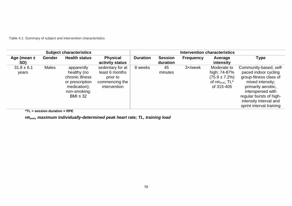
70
*TL = session duration × RPE
HRpeak, maximum individually-determined peak heart rate; TL, training load
Table 4.1: Summary of subject and intervention characteristics
Subject characteristics Intervention characteristics
Age (mean ± SD)
Gender Health status Physical activity status
Duration Session duration
Frequency Average intensity
Type
31.8 ± 6.1 years
Males apparently healthy (no
chronic illness or prescription medication);
non-smoking; BMI ≤ 32
sedentary for at least 6 months
prior to commencing the
intervention
8 weeks 45 minutes
3×/week Moderate to high; 74-87% (75.9 ± 7.2%) of HRpeak; TL* of 315-405
Community-based, self-paced indoor cycling group-fitness class of
mixed intensity; primarily aerobic, interspersed with
regular bursts of high-intensity interval and sprint interval training
71
4.3 EXCLUSION CRITERIA: as indicated in the advert and re-assessed during the initial
session
Females
Individuals <20 years or >45 years old
Those who were not classified as sedentary (according to IPAQ responses)
A history of or current chronic illnesses: in particular, cardiovascular-related disease
(eg. coronary artery disease), cerebrovascular disease (eg. stroke) or kidney disease
(or on dialysis), morbid obesity, diabetes type I or II, thyroid disease, dyslipidaemia,
known hypercholesterolaemia and/or stage 2 hypertension or worse (BP ≥
160/100mmHg)
Long-term medication use for cardiovascular-related, metabolic, hormonal or
neurological chronic illnesses
Current injury or illness which may have hindered participation and their ability to
exercise
Long-term tobacco use or smoking
Regular antioxidant supplementation
Individuals who were unable to communicate fluently in written and spoken English
Individuals who indicated that they may not be available throughout the intervention
period or may not be able to attend weekly exercise/assessment sessions
Personal, cultural or religious sensitivities regarding any of the assessment
procedures
4.3.1 Justification of choice of cohort
Sedentary people were ideal participants as any significant effects on the parameters of interest
could be more definitively attributed to the intervention. Sedentary people are relatively more
willing (than active counterparts) to refrain from physical activity outside the formal exercise
sessions and therefore potentially confounding activities would be easier to control in the former
population. (For justification of the definition of ‘sedentary’ used in the present study, please refer
to Appendix E).
Females were excluded due to the quantitative effects that the circamensal rhythm may have on
both maximal and submaximal exercise performance (440) and physiological
responses/adaptations to exercise such as the heart rate response (441). In addition, 17-
βestradiol is associated with increased vasodilation (442) and local blood flow (443) which could
potentially influence arterial geometry and structural remodelling . Cyclic fluctuations in the levels
of this hormone would have important implications when measuring AIx on a weekly basis and to
a lesser extent, the monthly ultrasound-derived measures.
The 20-45 year age range was appropriate because in healthy adults, free of CVD, ageing
progressively increases arterial stiffness and wave reflection indices and the effects of
progressive atherosclerotic build-up start to become more evident. Individuals >45 years were
excluded to eliminate age as a confounder. Younger people were excluded from this study so that
findings could be specific for young to middle-aged adults. (Teenagers, who would probably have

72
better baseline arterial health indices, may not respond positively/similarly to exercise
interventions). Participants were required to be apparently healthy non-smokers who were not on
prescribed medication or antioxidant supplementation because these factors could act as
confounders by independently affecting dynamics of AS indices and influencing exercise
adaptations. As the aim of this study was to investigate the effects of this exercise amongst the
general population, community-dwelling adults were ideal for practical reasons as they would
need to participate in the classes and structure them around their daily lives.
4.4 ASSESSMENT SCHEDULE FOR OUTCOME MEASURES
All assessments were carried out at similar time points for participants in both the intervention
and control groups and all assessments were identical between groups. Figure 4.1 illustrates the
time points over the course of the 10 weeks that assessments were carried out.
Figure 4.1: General plan of the study indicating timeline of assessments
The assessment protocol was identical between the control and intervention groups. PRE,
baseline (pre-intervention, week 0); MID, mid-intervention (end of week 4); POST, post-
intervention (end of week 8). HR, heart rate; pBP, peripheral blood pressure; WC, waist
circumference; BMI, body mass index; BM, body mass; Flexi, flexibility; BL, bloods; CRA,
cardiovascular risk assessment; IPAQ, International Physical Activity Questionnaire; PAR-Q,
Physical Activity Readiness Questionnaire; FFQ, Food Frequency Questionnaire; PWA, BP+-
derived measures of arterial health: central blood pressure & augmentation index’ cfPWV,
carotid-femoral pulse wave velocity; IMT, common carotid and common femoral intima-media
thickness; RI, common carotid and common femoral resistivity index; cEDD, common carotid end-
diastolic diameter; cWLR, common carotid wall:lumen ratio
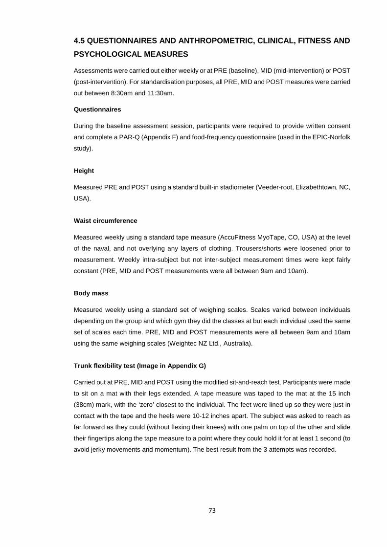
73
4.5 QUESTIONNAIRES AND ANTHROPOMETRIC, CLINICAL, FITNESS AND PSYCHOLOGICAL MEASURES
Assessments were carried out either weekly or at PRE (baseline), MID (mid-intervention) or POST
(post-intervention). For standardisation purposes, all PRE, MID and POST measures were carried
out between 8:30am and 11:30am.
Questionnaires
During the baseline assessment session, participants were required to provide written consent
and complete a PAR-Q (Appendix F) and food-frequency questionnaire (used in the EPIC-Norfolk
study).
Height
Measured PRE and POST using a standard built-in stadiometer (Veeder-root, Elizabethtown, NC,
USA).
Waist circumference
Measured weekly using a standard tape measure (AccuFitness MyoTape, CO, USA) at the level
of the naval, and not overlying any layers of clothing. Trousers/shorts were loosened prior to
measurement. Weekly intra-subject but not inter-subject measurement times were kept fairly
constant (PRE, MID and POST measurements were all between 9am and 10am).
Body mass
Measured weekly using a standard set of weighing scales. Scales varied between individuals
depending on the group and which gym they did the classes at but each individual used the same
set of scales each time. PRE, MID and POST measurements were all between 9am and 10am
using the same weighing scales (Weightec NZ Ltd., Australia).
Trunk flexibility test (Image in Appendix G)
Carried out at PRE, MID and POST using the modified sit-and-reach test. Participants were made
to sit on a mat with their legs extended. A tape measure was taped to the mat at the 15 inch
(38cm) mark, with the ‘zero’ closest to the individual. The feet were lined up so they were just in
contact with the tape and the heels were 10-12 inches apart. The subject was asked to reach as
far forward as they could (without flexing their knees) with one palm on top of the other and slide
their fingertips along the tape measure to a point where they could hold it for at least 1 second (to
avoid jerky movements and momentum). The best result from the 3 attempts was recorded.

74
Blood lipid profile and fasting blood glucose (FBG)
These were assessed at PRE and POST after a 9-11 hour overnight fast using a simple finger-
prick test. Sterile, single-use lancets (ACCU-CHEK Safe-T-Pro Plus, Mannheim Germany) were
used to prick the side of the right index or middle fingertip and blood was drawn into 40μL capillary
tubes. Total cholesterol (TC), HDL-cholesterol (HDL-C), total triglycerides (TGs), the TC:HDL
ratio and LDL-cholesterol (LDL-C) levels were assessed using the CardioChek® P•A Point-of-
Care Test System and PTS Lipid Panels® Test Strips (Polymer Tech Systems Inc., IN, USA).
Fasting blood glucose (FBG) was assessed using the FreeStyle Optium Xceed blood glucose
metre and FreeStyle Optium glucose strips (Oxon, UK).
Cardiorespiratory fitness test
A graded incremental exercise test to exhaustion was performed on an electronically-braked cycle
ergometer (Lode Sports Excalibur®, Groningen, Netherlands) to determine VO2peak at PRE and
POST. After a brief warm-up, participants started pedalling at 25W for 1 minute, with a self-
selected cadence between 65 and 85 rpm. After the first minute, the load was increased by 15W
every minute until the point of volitional fatigue. Expired gases were analysed using a calibrated
metabolic cart (TrueOne 2400, ParvoMedics, Sandy, UT, USA). VO2peak was determined from
breath-by-breath samples averaged over 15s. The average of 3 HR measures during the last 15
seconds of each stage and the RPE at the end of each stage (minute) were recorded.
Cardiovascular risk assessment
At PRE and POST, the Framingham Risk Score was calculated, along with the estimated
Framingham vascular age (444), to establish the general 10-year CV risk. This score was based
on age, sex, pSBP, smoking, diabetes, treatment for hypertension, HDL-C and TC.
4.6 MEASURES OF ARTERIAL HEALTH DERIVED USING AN AUTOMATED OSCILLOMETRIC DEVICE
This device measured peripheral and central BP (pBP and cBP respectively), HR and the
peripheral AIx. Resting cBP, pBP, HR and AIx measures were determined weekly in a quiet,
temperature-controlled room, using an automated vascular monitor (Uscom R6.5B, BP+, Sydney,
Australia) and algorithm (version MR1517). The BP+ device has an in-built brachial BP monitor.
First, brachial BP is measured oscillometrically. Within 3s of cuff deflation, the cuff re-inflates and
holds the pressure for 10 seconds at 30mmHg above the measured SBP. During this period, a
suprasystolic waveform is obtained, from which the cBP is derived from the relationship between
aortic oscillatory pressure and total oscillatory pressure under the cuff. During the procedure,
participants were made to sit quietly on a chair in an upright position, feet flat on the floor, elbow
resting on the table with the palm prone. For standardisation purposes, an appropriately sized
cuff was positioned over the left upper arm. Placement of the cuff over clothing was avoided as
was rolling up of sleeves which could have caused proximal arterial occlusion. A total of 3
75
measures were taken each time, with a one minute rest between each measure. The first was
taken after the subject had sat quietly for 2-3 minutes. The average of the 3 results each week
were used for analysis. The weekly measures for those in the intervention group were taken
approximately 10-15 minutes prior to the last exercise session of the week. All data collection
using the BP+ device was conducted at least 24 hours after an exercise session to avoid the
immediate effects of a single bout of exercise. Weekly intra-subject but not inter-subject
measurement times were kept constant.
Note on BP and PP measures: Although central pulse pressure is the index most relevant to
AS, peripheral BP is more widely understood, acknowledged and measured. Therefore, results
presented will be those for peripheral SBP and DBP and central PP.
4.7 ULTRASOUND ASSESSMENTS
The ultrasound system (GE Healthcare Vivid S6 Cardiovascular Ultrasound System, WI, USA)
used for carotid and femoral examinations was equipped with 2D imaging, colour and spectral
Doppler, an ECG monitor and a high-frequency vascular transducer as recommended. A 12MHz
linear array transducer was used to acquire high-resolution images. Since most of the indices
assessed in the present study may vary quite considerably across a single cardiac cycle,
standardisation of the frame relative to the cardiac cycle was necessary. The ultrasound
integrated with the ECG facilitated the assessment of these parameters according to the cardiac
cycle (R wave).
Ultrasonic assessments were carried out at PRE, MID and POST, between 9 and 11am (at least
12 hours after an exercise session to avoid the immediate effects of a single bout of exercise).
Both inter- and intra-subject measurement times were kept constant due to diurnal variation in
cfPWV. Participants were requested to arrive in a fasted state and avoid caffeine and high-fat
meals for at least 12 hours before testing. (Although avoiding the above for only 3 hours are
required for US assessments, the blood tests needed to be carried out in the fasted state). Once
the (3 lead) ECG was attached, the subject rested lying down in the supine position for 10 minutes
in a quiet, dimly-lit, temperature-controlled (24°C) room. Individuals were requested to avoid
falling asleep during the assessments.
4.7.1 RESTING CAROTID-FEMORAL PULSE WAVE VELOCITY (cfPWV)
Doppler guided by 2D ultrasound, with simultaneous ECG gating was used to evaluate cfPWV as
previously described (83) (Figure 4.3). As the elastic common carotid artery (CCA) and muscular
common femoral artery (CFA) are quite superficial, these are the most common set points (18),
enabling the calculation of cfPWV, which covers the region over the aorta which exhibits the
greatest pathological effects of AS and the highest increase in AS with ageing (30).
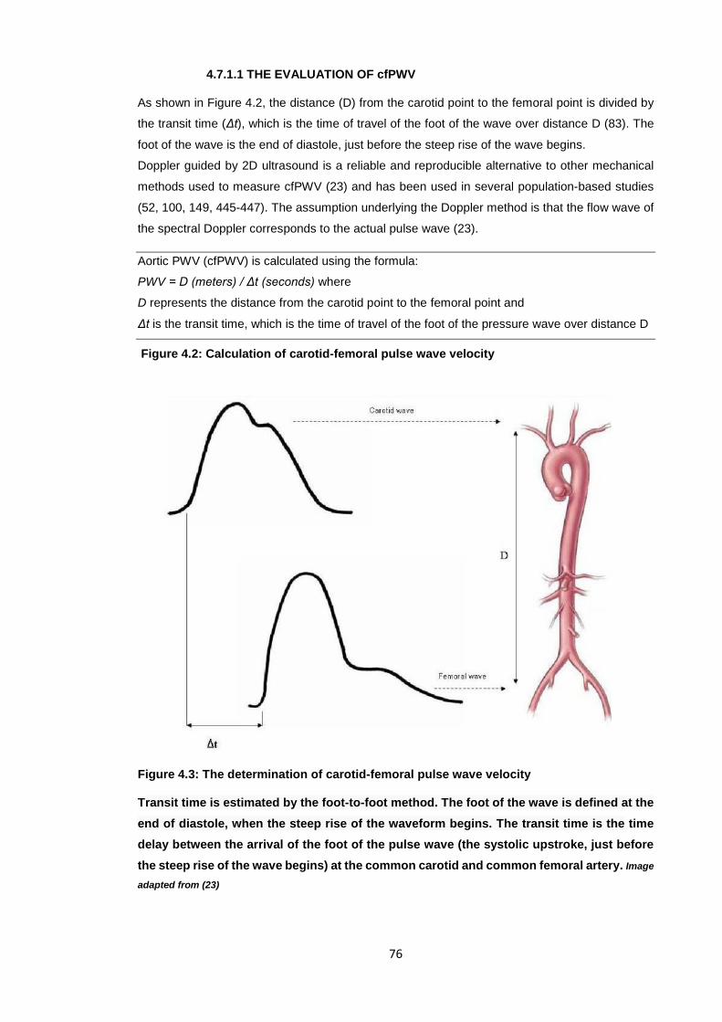
76
4.7.1.1 THE EVALUATION OF cfPWV
As shown in Figure 4.2, the distance (D) from the carotid point to the femoral point is divided by
the transit time (Δt), which is the time of travel of the foot of the wave over distance D (83). The
foot of the wave is the end of diastole, just before the steep rise of the wave begins.
Doppler guided by 2D ultrasound is a reliable and reproducible alternative to other mechanical
methods used to measure cfPWV (23) and has been used in several population-based studies
(52, 100, 149, 445-447). The assumption underlying the Doppler method is that the flow wave of
the spectral Doppler corresponds to the actual pulse wave (23).
Aortic PWV (cfPWV) is calculated using the formula:
PWV = D (meters) / Δt (seconds) where
D represents the distance from the carotid point to the femoral point and
Δt is the transit time, which is the time of travel of the foot of the pressure wave over distance D
Figure 4.2: Calculation of carotid-femoral pulse wave velocity
Figure 4.3: The determination of carotid-femoral pulse wave velocity
Transit time is estimated by the foot-to-foot method. The foot of the wave is defined at the end of diastole, when the steep rise of the waveform begins. The transit time is the time delay between the arrival of the foot of the pulse wave (the systolic upstroke, just before the steep rise of the wave begins) at the common carotid and common femoral artery. Image
adapted from (23)
77
In this method, waves are obtained successively at the common femoral and common carotid
arteries (CFA and CCA respectively). Once the probe had been positioned 1.5-2cm proximal to
the carotid bifurcation, wave Doppler flow and ECG were simultaneously identified and a
recording was taken over 2-3 cardiac cycles (1 respiratory cycle; 5-6 breaths). After the above
step was repeated for the unilateral femoral artery, the transit time was measured by subtracting
the time delay of the foot of the wave (top of the R-wave on the ECG equivalent to end-diastolic
diameter) to the CCA from the time delay between the R-wave and CFA. Only invasive methods
would give an accurate value for the distance between the CCA and CFA. A consensus published
by van Bortel et al. in 2011 suggested that 80% of the direct carotid-femoral distance (CCA-CFA
× 0.8) would provide the most accurate estimate of the actual distance (139). Therefore, in the
present study, 80% of the measured surface carotid-femoral distance was used for the purpose
of PWV calculation. For each recording, the sample volume (gate) was manually adjusted to
encompass two-thirds of the vessel lumen and an insonation angle (angle between the ultrasound
beam and longitudinal vessel axis) of 50-60° was maintained.
4.7.2 RESTING INTIMA-MEDIA THICKNESS (IMT)
4.7.2.1 RESTING COMMON CAROTID ARTERY INTIMA-MEDIA THICKNESS (cIMT) ASSESSMENT
Carotid IMT as measured by ultrasound is defined as the combined thickness of the intima and
media because current technology is not sensitive enough to measure only intimal thickness (11).
Two echogenic lines, shown to be the lumen-intima and media-adventitia interface, separated by
a hypoechoic space, are observed on the far wall of the common carotid artery (CCA) (35, 448,
449). The distance between the two echogenic lines was not significantly different from the IMT
measured under both gross and microscopic examination (448).
B-mode images of the CCAs were obtained bilaterally using Doppler ultrasound and the 12MHz
linear array transducer. The CCAs were imaged in the longitudinal plane using the preferred
lateral probe approach (single insonation angle). The head position was standardised as much
as possible, with the neck extended to an angle of 45-50° and excessive pressure on the CCA
with the probe was avoided. Images were acquired 2cm proximal to the carotid bifurcation and
IMT measurements were carried out over a 1cm straight arterial segment, using automated edge-
detection software. Intima-media thickness was defined as the viewable distance between the
two parallel lines corresponding to the lumen-intima and the media-adventitia interfaces. As cIMT
may vary between 5-10% during a single heartbeat, IMT measurements were standardised to the
R-wave of the ECG which represents the end-diastolic moment (also the moment of the thickest
cIMT). Gain settings, focus depth (30-40mm) and frame rates (>15-25Hz) were adjusted manually
to optimise edge detection and avoid slice thickness artefacts (11).
Carotid IMT data was collected for both the near and far walls of the right and left CCAs. For
each wall assessed, averages of 6 mean, 6 minimum and 6 maximum values were used in data
78
analysis. Therefore, for each wall of each artery, single values for the mean of means, mean of
minima and mean of maxima IMTs were calculated. (Readings were only taken from images in
which arteries were horizontal and in which the carotid bifurcation could be clearly identified).
Justification of protocol used for carotid artery IMT assessment
A distal segment of the CCA was assessed in the present study because it has been previously
shown that imaging of the near and far walls of the CCA from multiple angles is superior to
measuring the carotid bifurcation and internal carotid artery, and that the former is more
reproducible and nearly always complete (450). A combination of all segments was unnecessary
in the present study as the aim was not to assess plaque formation in the carotid artery. The
IMPROVE study found that for both mean and maximum cIMT measures at the CCA, bifurcation
and internal carotid artery, cIMT is positively correlated to CV risk (239). The IMPROVE study
concluded that when relating increased cIMT to increased CV risk, CCA measures are just as
useful as more elaborate measures. However, for risk classification, the more elaborate cIMT
measures are more accurate than cIMT measured only at the CCA. Common carotid IMT values
are more strongly correlated to adiposity measures than are IMT measurement at other sites of
the carotid tree (281). In light of all the previous literature, the mean of means of the right and left
cIMT were reported as recommended by the 2008 ASE Consensus statement (11). Although both
near and far walls were imaged, only results of the far walls will be presented because only far
wall cIMT values are accurate reflections of wall thickness whereas near wall IMT values are
merely approximations (450) (usually underestimations of true wall thickness (451).
4.7.2.2 RESTING COMMON FEMORAL ARTERY INTIMA-MEDIA THICKNESS (fIMT) ASSESSMENT
Femoral IMT was determined from longitudinal 2D B-mode images using a high-resolution 12MHz
linear array transducer. Images were acquired 2cm proximal to the CFA bifurcation and IMT
measurements were carried out over a distance of 1.5cm using automated edge-detection
software. All IMT measurements were standardised to the R-wave of the ECG.
Right and left fIMT data were collected for the far walls only. For each wall, averages of 6 mean,
6 minimum and 6 maximum values were used in data analysis. Therefore, for the far wall of each
artery, single values for the mean of means, mean of minima and mean of maxima IMTs were
obtained (but only the far wall results will be presented). For each vessel examined, Peak systolic
velocity (PSV), End diastolic velocity (EDV) and the RI [(PSV-EDV)/PSV] were automatically
calculated for each cardiac cycle. The average of 6 values for the RI was used in data analysis.
79
4.7.3 RESTING COMMON CAROTID ARTERY END-DIASTOLIC INTRA-LUMINAL DIAMETER (cEDD)
The right and left resting (baseline) cEDDs were measured using grey scale imaging. The
boundaries were defined as the visible lumen-intima interfaces of the near and far walls, identified
using the automatic edge-detection software. The EDD corresponds to the R wave on the ECG
and was measured approximately 1-2cm proximal to the carotid bifurcation. Although the R wave
on the ECG represents end diastole, it is also the point of minimum diameter. However, the point
of maximum diameter (after the T wave, before the start of the following P wave) is difficult to
standardise and therefore, measured diameters in this study were synchronised to the R-wave
peaks on the ECG. The averages of 6 measures were used for the right and left carotid diameters
each. NB: Resting common femoral diameters were not assessed as it was not possible to clearly
discern the necessary interfaces in the majority of images.
4.7.4 CALCULATIONS OF RESTING COMMON CAROTID ARTERY WALL:LUMEN RATIO (cWLR)
cWLR was calculated as:
2 × cIMT/lumen diameter in diastole (452)
4.8 STRUCTURE OF STUDY
4.8.1 RECRUITMENT AND GROUP ALLOCATION
Ethical approval for this study was granted from the Auckland University of Technology Ethics
Committee (Appendix A). Interested individuals who responded to the advertisement flyer
(Appendix H) were sent an information sheet (Appendix I) and were required to complete the
IPAQ to confirm their sedentary status. After providing written consent to participate (Appendix
J), participants were allocated to either the intervention group (n=10) or the control group (n=5).
Both groups were requested to maintain consistent lifestyles throughout the intervention period,
in particular, maintain their normal diets but avoid antioxidant supplements for the 8 weeks. They
were also required to keep a physical activity log over the 8 weeks (Appendix K). In the log, they
were required to write down any activity they carried out, the duration and perceived intensity,
RPE (according to the modified Borg scale). From these logs, average exertion scores for each
activity were calculated as: Exertion score (no units) = activity duration (mins) × RPE
The sum of the individual exertion scores during a single day gave an indication as to daily
exertion scores. Physical activity logs were collected once a week and were to be completed on
any three days of the week (including one weekend day). In this way, it was confirmed that the
average weekly inter-group physical activity loads were fairly similar and that only the intervention-
based exercise would explain any arterial health adaptations.
80
4.8.2 CONTROL GROUP
The control group was simply asked to maintain and record their normal physical activity routine
and diet for 8 weeks. Assessments were carried out as depicted in Figure 4.1.
4.8.3 INTERVENTION GROUP AND NATURE OF EXERCISE INTERVENTION
Individuals in the intervention group attended 3 supervised indoor cycling group-fitness classes
per week for 8 weeks at one of two gyms in Auckland. Les Mills International Limited (LMI) has
registered the trademark ‘RPM’ (Raw Power in Motion) for the popular indoor (studio) cycling
group-centred activity known as ‘spinning’. LMI describes RPM™ as a moderate-to-high intensity
interval programme with aerobic endurance, anaerobic and sprint components with the aim of
building muscular endurance, power, strength and speed. In the present intervention, ‘regular
bursts of HIIT and SIT’ implies that after approximately a 10 minute AE warm-up, these bouts
alternated such that the structure of the workout was: warm-up, AE, HIIT, SIT, HIIT, SIT, HIIT, cool-down (each section approximately 5 mins). Each HIIT and SIT bout, has, on average, three ‘peaks’ interspersed with active (mild/moderate) cycling.
Appendix L is an official outline provided by LMI describing the structure of a typical RPM™ class.
Each session (class) lasts for 45-50 minutes (including a ~5 min warm-up and ~5 min cool-down)
and instructions given during class are pre-choreographed to specific music. It is a self-paced
class where individuals can choose their own level of intensity by means of a dial on the bike.
However, it is possible that the quality and quantity of content (commentary, instruction and
feedback) provided by the class instructor could serve as a source of extrinsic motivation, thereby
influencing self-selected intensities. For the purpose of the present study, it was assumed that
direction provided by instructors were similar across all classes. The temperature and humidity of
the cycling studio are unknown and were not controlled.
The first week of the intervention was used for participants to familiarise themselves with the
nature of an RPM™ class. The sessions were carried out at any time of day but participants were
asked to separate each class by at least 36 hours. The intervention group participants, like the
controls, were requested to abstain from any other type of novel physical activity involving major
muscle groups for the duration of the intervention (as aforementioned, the physical activity logs
ensured that participants did not engage in significant quantities of physical activity outside the
intervention-based exercise).
Before the intervention commenced, it was decided that participants would only be included in
the analysis if they completed all 24 classes within a period of 9 weeks, with at least one class
carried out each week. (An extra week leeway was permitted on account of the community-based
setting and subsequent acknowledgement of practical aspects based on self-regulated
attendance). Assessments were carried out as depicted in Figure 4.1 with at least 24 hours
between a training session and any assessment session (to eliminate the effects of an acute
exercise bout). Weekly assessments were carried out approximately 10-15 mins before the last
81
class of every week so intra-individual but not inter-individual assessment times were maintained
throughout the intervention period.
Obtaining RPM™ objective and subjective intensities
Heart rate during the exercise classes was monitored using the Polar Team2 system (Polar Electro
Oy, Professorintie 5, Kempele, Finland). Data was collected on a weekly basis and averaged out
to obtain OBJECTIVE information regarding the exercise intensity which was gauged on an
individual basis as a percentage of the maximum HR (%HRpeak) attained during the VO2peak test.
In addition, participants were required to rate the intensity of each session according to the
modified Borg scale (Appendix L) so that session RPE (also known as training load) could be
calculated to assess the SUBJECTIVE intensity:
Training load (no units) = duration of RPM class (ie. 45 mins) × RPE*
The RPE is one of the most popular measures used to assess internal TL (453). It allows
individuals to monitor their effort both during classes and retrospectively and has previously been
shown to correlate with HR during steady-state exercise and HIIT (454).
Calculating the session RPE, that is, combining the RPE with the session duration, provides a
better quantification of the internal TL and the validity and reliability of this method have been
previously shown (455).
Gauging RPM™ intensities over the whole class
The raw data of the Polar Team2 system observed on the screen is a graph of HR as a % of
individually-determined HRpeak (an example is provided in Appendix M). Average and maximal
%HRpeak values throughout a single session are obtained directly and the amount of time spent
in each training zone are also clearly illustrated. When the order and duration of bouts of the
different training components are known, average and maximal %HRpeak values from the
corresponding section of the graph can be calculated. As not all durations are identical, these
values are approximations but nevertheless, they can be used to gauge the intensities of different
components of the session.
Summary of exercise nature
i) It was a supervised group-centred, self-paced, indoor cycling programme,
undertaken in a pre-existing, community-based setting (gymnasium)
ii) It was a moderate-to-high intensity aerobic interval session with SIT and HIIT
components and specific foci on aerobic endurance, anaerobic power and strength.
* Participants were required to provide the RPE of the session as a whole, including all the highs and lows.
82
4.9 STATISTICAL ANALYSIS
All data was included as no participant dropped out and all measures were satisfactorily obtained.
The sample size calculation for cfPWV, the main outcome measure, assumed a power of 90%
and an α-error probability of 0.05. Based on this a priori analysis, a sample size of at least 10
participants in the intervention group was required to detect a clinically significant effect of the
exercise intervention on this index of AS.
All statistical analyses were performed with the statistical package SPSS version 22 (SPSS,
Chicago, IL). Descriptive statistics are presented as mean ± standard deviation. All comparisons
were based on a 95% confidence limit with p<0.05 considered statistically significant. Effect sizes
were calculated using partial eta squared (pEta2) with values of 0.1, 0.3, and above 0.5 considered
to be a small, medium, and large effects respectively. Data collected was thoroughly checked for
errors and outliers and normality was evaluated by the Shapiro-Wilk test. Both Mauchly’s tests of
sphericity and Levene’s homogeneity of variance tests were considered for ANOVAs.
One-way between-group analyses of variance (ANOVAs) with Bonferroni adjustments and
Tukey’s post-hoc tests were performed to determine the impact of the exercise intervention on
outcome measures at pre-, mid- and post-intervention (PRE, MID and POST respectively).
One-way repeated measures ANOVAs were conducted separately for the control and intervention
groups to examine the results of the intervention.
Mixed between-within participants ANOVAs were conducted to determine main and interaction
effects. After inspection the distribution of data points of preliminary scatterplots to ensure no
violations of the assumptions of linearity, outliers and homoscedasticity (variability in scores for
one variable similar at all values of the second variable), multiple univariate correlations and
regressions were performed to determine relationships between absolute values as well as
percentage changes in selected physiological variables. Where necessary, partial correlations
were conducted to explore relationships between outcome measures whilst controlling for age
and blood pressures.
Significant correlations with cfPWV, AIx@75, cPP and cIMT were used in stepwise multiple linear
regression analyses to identify independent determinants of these indices.
For outcome measures that exhibited both between-subject differences at POST and linear
relationships between PRE and POST values, ANCOVAs were performed (provided ANCOVA
assumptions were not violated), to determine whether significant inter-group relationships existed
at POST, with PRE values as covariates.
83
CHAPTER 5: RESULTS Due to the extensive data analysis and for ease of understanding, the results presented below
have been systematically organised into four sections based on the research questions as follows:
5.1 Intervention compliance, training intensity and daily training loads
5.2 Between- and within-group effects
i) Comparisons between the two groups on all outcome measures across the three time
points
ii) Time course of adaptations
5.3 Exploration of relationships between outcome measures:
i) Correlations between CRF and arterial health, anthropometric and clinical measures
ii) Comparisons of absolute values and percentage changes of outcome measures:
Where percent changes in values are presented, the term ‘improvement’ depicts an impact in the beneficial direction. Therefore, even when ‘improvements’ for some measures signify that absolute values decreased, negative percentage values are not considered.
iii) Relationships between bilateral ultrasound-derived measures of AS and
atherosclerosis across all time points
5.4 Focus on intervention group
i) In-depth exploration of metabolic syndrome risk factors together with calculated CVD
risk and vascular age at pre- and post-intervention
ii) Summary of the main adaptations in the intervention group
5.1 INTERVENTION COMPLIANCE, TRAINING INTENSITY AND DAILY TRAINING LOADS
Adherence: There was 100% compliance in terms of class participation and completion (ie. 10
participants carried out all 24 RPM™ classes). However, despite the request to avoid classes on
consecutive days, this was not always possible due to subject inconvenience.
Objective intervention intensity: Table 5.1 provides a breakdown of intensities throughout an
entire RPM™ class. Figure 5.1 is an example of HR data collected for a typical RPM™ class. The
average HR intensity during an RPM™ class ranged from 74-87% (81.3 ± 7.2%) of the
individually-determined HRpeak. The average maximum intensity during a class was 93.6 ± 7%
(range: 88-104%) HRpeak. Figure 5.2 depicts weekly HR averages during RPM™ classes over the
intervention period. Despite the weekly fluctuations, the general trend was for the average
%HRpeak to gradually and slowly increase such that the average HRpeak in the eighth week was
10% greater than that in the first week (p<0.05). Appendix M provides an example of raw HR data
collected over an entire RPM™ session for the same individual in weeks 1 and 8 whilst Appendix
84
N provides a snapshot of the average weekly intensity data for each subject individually for all 8
weeks.
Subjective intervention intensity: The average RPE for all 10 participants over all 24 classes
was 8.5 (out of 10 on the Borg scale). The session RPEs (training loads, TLs)* ranged from 315-
405 (7-9 on the Borg scale).* RPM™ training loads and HR intensities were not associated (ie.
subjective and objective data did not significantly concur).
Daily exertion scores** excluding RPM™ classes: The physical activity logs ensured that
participants did not engage in significant quantities of physical activity outside the intervention-
based exercise (RPM™ classes). Table 5.2 summarises the average number of hours that (all
15) participants spent doing different physical activities (excluding RPM™ classes) and activities
of daily living. The table includes the average session RPE for each activity and the calculated
exertion score (the habitual activity equivalent of training load). The intervention and control
groups spent, on average, 7 hours sleeping, 12.5 hours sitting, 1.5 hours standing, 1 hour walking
and 1 hour doing ‘other’ light activities (eg. household chores, gardening). The average exertion
score range over a whole day was 210-780 (compared to a training load of 315-405 for a single
45 minute RPM™ session). Habitual (non-RPM™) activity between the intervention and control
participants did not significantly differ over the intervention period (p>0.05). Furthermore, average
daily exertion scores did not significantly change across the 8 weeks in either group. In the
intervention group, there was no significant difference in extra-intervention training loads on
RPM™ and non-RPM™ days. It should be mentioned that on occasion, participants in both
groups engaged in physical activities of moderate intensities (eg. playing with kids, a round of
golf, slightly harder domestic chores, planned walking, short game of basketball). These activities
were extremely rare (assuming they were honestly reported) because participants had been
informed at the start of the study that it was required that they did not engage in intense physical
activities more than twice over the 8 weeks.
*Training load = subjective RPE during session × session duration
**Habitual activity exertion score = subjective RPE of activity × duration of activity (mins)
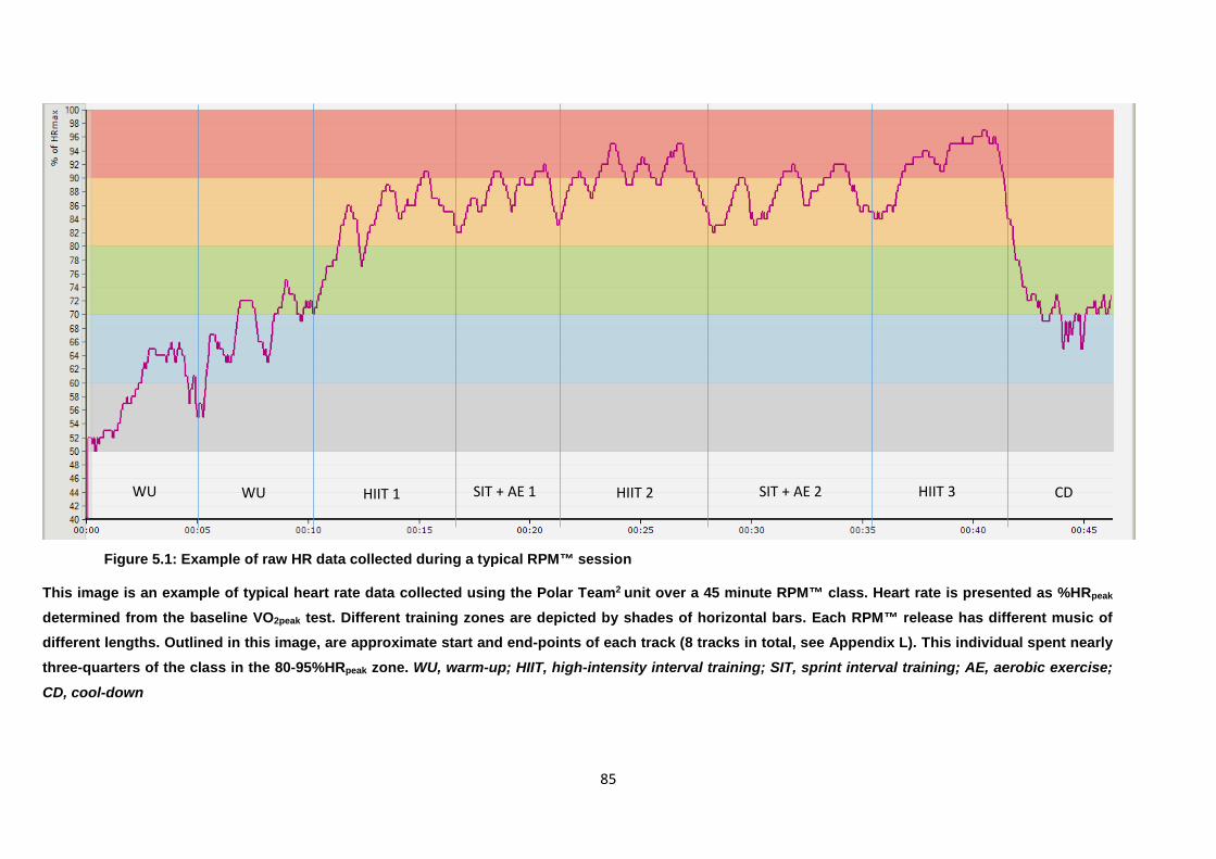
85
Figure 5.1: Example of raw HR data collected during a typical RPM™ session
This image is an example of typical heart rate data collected using the Polar Team2 unit over a 45 minute RPM™ class. Heart rate is presented as %HRpeak determined from the baseline VO2peak test. Different training zones are depicted by shades of horizontal bars. Each RPM™ release has different music of different lengths. Outlined in this image, are approximate start and end-points of each track (8 tracks in total, see Appendix L). This individual spent nearly three-quarters of the class in the 80-95%HRpeak zone. WU, warm-up; HIIT, high-intensity interval training; SIT, sprint interval training; AE, aerobic exercise;
CD, cool-down
WU SIT + AE 2 HIIT 2 SIT + AE 1 HIIT 1 WU CD HIIT 3
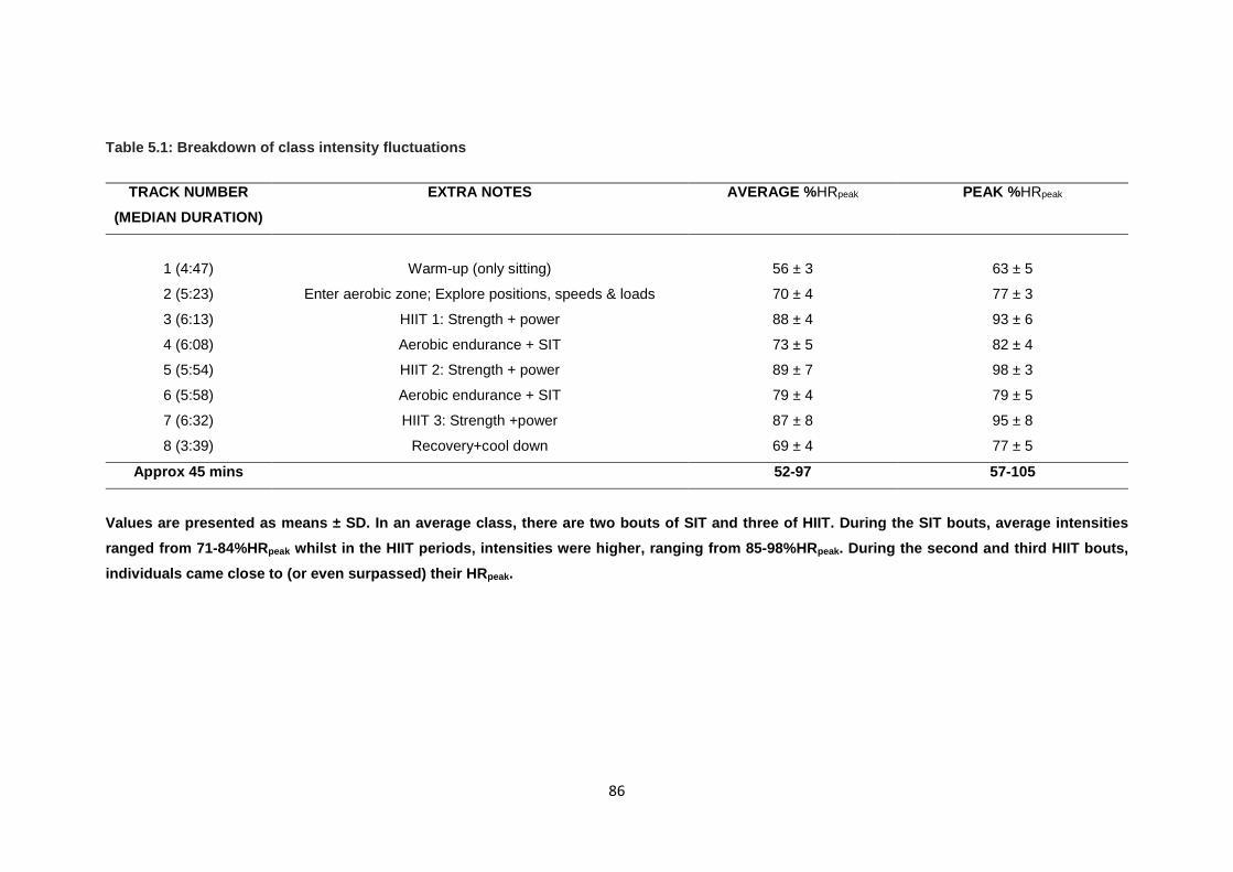
86
Table 5.1: Breakdown of class intensity fluctuations
Values are presented as means ± SD. In an average class, there are two bouts of SIT and three of HIIT. During the SIT bouts, average intensities ranged from 71-84%HRpeak whilst in the HIIT periods, intensities were higher, ranging from 85-98%HRpeak. During the second and third HIIT bouts, individuals came close to (or even surpassed) their HRpeak.
TRACK NUMBER (MEDIAN DURATION)
EXTRA NOTES AVERAGE %HRpeak PEAK %HRpeak
1 (4:47)
Warm-up (only sitting)
56 ± 3
63 ± 5
2 (5:23) Enter aerobic zone; Explore positions, speeds & loads 70 ± 4 77 ± 3
3 (6:13) HIIT 1: Strength + power 88 ± 4 93 ± 6
4 (6:08) Aerobic endurance + SIT 73 ± 5 82 ± 4
5 (5:54) HIIT 2: Strength + power 89 ± 7 98 ± 3
6 (5:58) Aerobic endurance + SIT 79 ± 4 79 ± 5
7 (6:32) HIIT 3: Strength +power 87 ± 8 95 ± 8
8 (3:39) Recovery+cool down 69 ± 4 77 ± 5
Approx 45 mins 52-97 57-105
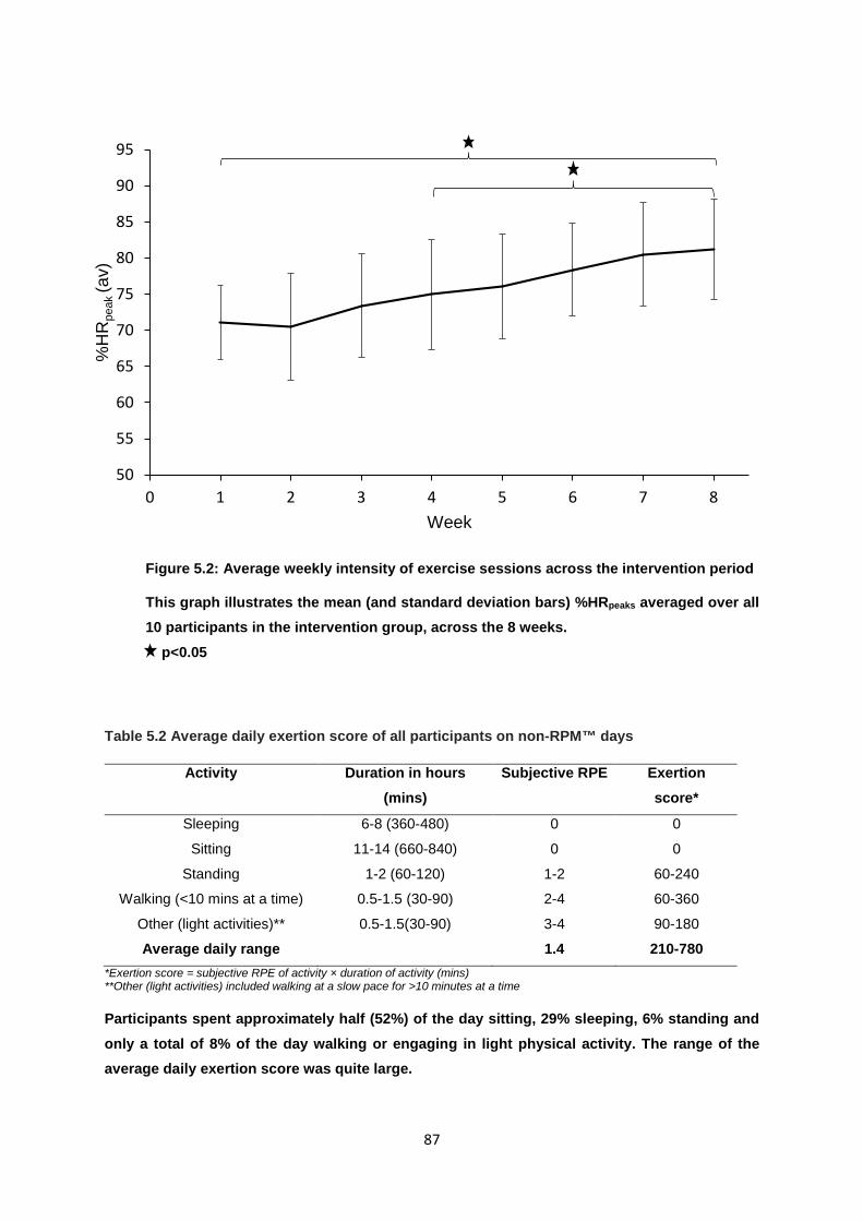
87
Figure 5.2: Average weekly intensity of exercise sessions across the intervention period
This graph illustrates the mean (and standard deviation bars) %HRpeaks averaged over all 10 participants in the intervention group, across the 8 weeks. p<0.05
Table 5.2 Average daily exertion score of all participants on non-RPM™ days
*Exertion score = subjective RPE of activity × duration of activity (mins) **Other (light activities) included walking at a slow pace for >10 minutes at a time
Participants spent approximately half (52%) of the day sitting, 29% sleeping, 6% standing and only a total of 8% of the day walking or engaging in light physical activity. The range of the average daily exertion score was quite large.
50
55
60
65
70
75
80
85
90
95
0 1 2 3 4 5 6 7 8
%H
Rpe
ak (a
v)
Week
Activity Duration in hours (mins)
Subjective RPE Exertion score*
Sleeping 6-8 (360-480) 0 0
Sitting 11-14 (660-840) 0 0
Standing 1-2 (60-120) 1-2 60-240
Walking (<10 mins at a time) 0.5-1.5 (30-90) 2-4 60-360
Other (light activities)** 0.5-1.5(30-90) 3-4 90-180
Average daily range 1.4 210-780
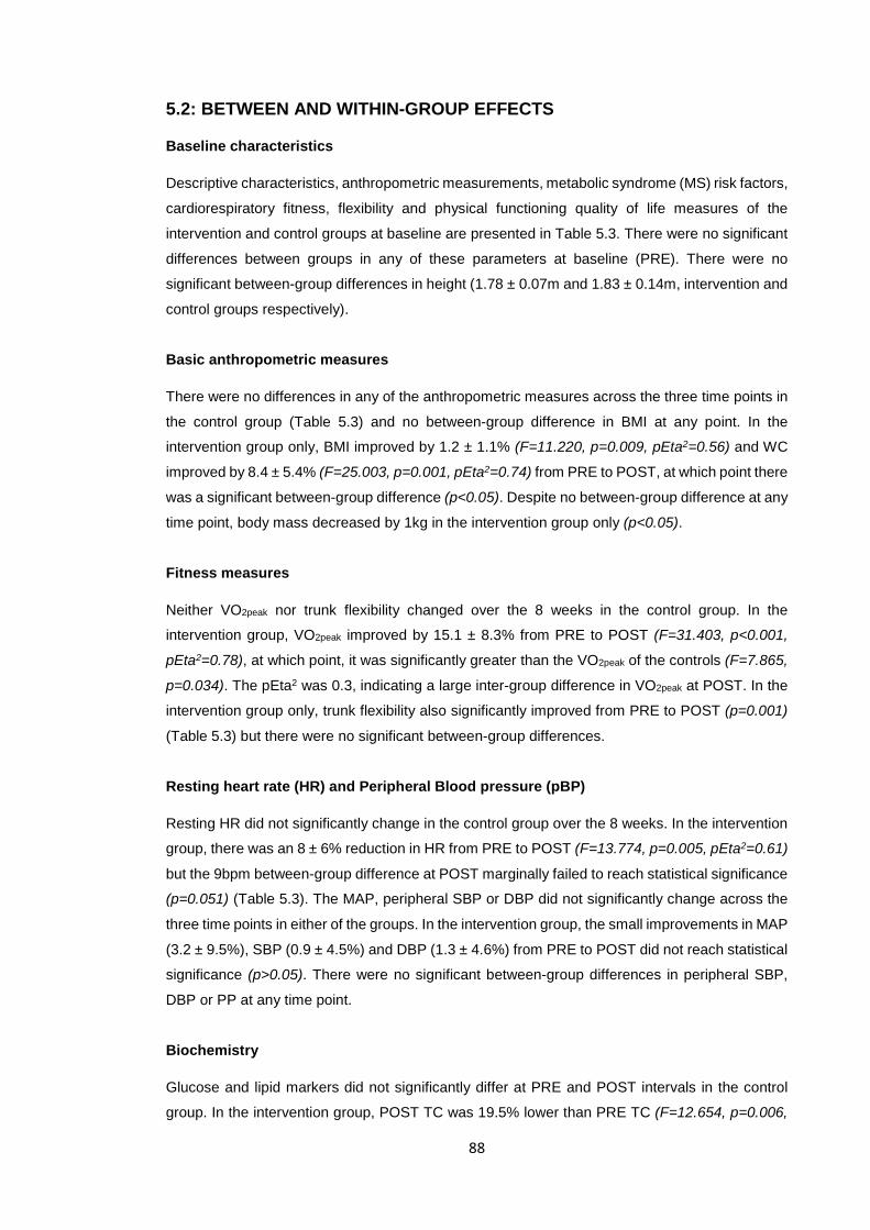
88
5.2: BETWEEN AND WITHIN-GROUP EFFECTS
Baseline characteristics
Descriptive characteristics, anthropometric measurements, metabolic syndrome (MS) risk factors,
cardiorespiratory fitness, flexibility and physical functioning quality of life measures of the
intervention and control groups at baseline are presented in Table 5.3. There were no significant
differences between groups in any of these parameters at baseline (PRE). There were no
significant between-group differences in height (1.78 ± 0.07m and 1.83 ± 0.14m, intervention and
control groups respectively).
Basic anthropometric measures
There were no differences in any of the anthropometric measures across the three time points in
the control group (Table 5.3) and no between-group difference in BMI at any point. In the
intervention group only, BMI improved by 1.2 ± 1.1% (F=11.220, p=0.009, pEta2=0.56) and WC
improved by 8.4 ± 5.4% (F=25.003, p=0.001, pEta2=0.74) from PRE to POST, at which point there
was a significant between-group difference (p<0.05). Despite no between-group difference at any
time point, body mass decreased by 1kg in the intervention group only (p<0.05). Fitness measures
Neither VO2peak nor trunk flexibility changed over the 8 weeks in the control group. In the
intervention group, VO2peak improved by 15.1 ± 8.3% from PRE to POST (F=31.403, p<0.001,
pEta2=0.78), at which point, it was significantly greater than the VO2peak of the controls (F=7.865,
p=0.034). The pEta2 was 0.3, indicating a large inter-group difference in VO2peak at POST. In the
intervention group only, trunk flexibility also significantly improved from PRE to POST (p=0.001)
(Table 5.3) but there were no significant between-group differences.
Resting heart rate (HR) and Peripheral Blood pressure (pBP)
Resting HR did not significantly change in the control group over the 8 weeks. In the intervention
group, there was an 8 ± 6% reduction in HR from PRE to POST (F=13.774, p=0.005, pEta2=0.61)
but the 9bpm between-group difference at POST marginally failed to reach statistical significance
(p=0.051) (Table 5.3). The MAP, peripheral SBP or DBP did not significantly change across the
three time points in either of the groups. In the intervention group, the small improvements in MAP
(3.2 ± 9.5%), SBP (0.9 ± 4.5%) and DBP (1.3 ± 4.6%) from PRE to POST did not reach statistical
significance (p>0.05). There were no significant between-group differences in peripheral SBP,
DBP or PP at any time point.
Biochemistry
Glucose and lipid markers did not significantly differ at PRE and POST intervals in the control
group. In the intervention group, POST TC was 19.5% lower than PRE TC (F=12.654, p=0.006,
89
pEta2=0.58) and POST LDL-C was 17% lower than PRE LDL-C (F=7.961, p=0.02, pEta2=0.47)
(Table 5.3). There were no between-group differences at POST. RESTING ARTERIAL HEALTH MEASURES Resting Arterial stiffness, wave reflections and central haemodynamics
There were no significant differences in AS indices (cfPWV, AIx@75 and cPP) between groups
at baseline (Figure 5.3).
Carotid-femoral pulse wave velocity (cfPWV)
In the control group, both right and left cfPWV respectively did not differ significantly across the
three time points (Figure 5.3c-d). In the intervention group, the right cfPWV was significantly
greater at PRE (8.65 ± 0.36m/s) than MID (8.22 ± 0.44m/s) (F=45.087, p<0.001, pEta2=0.83) and
POST (7.72 ± 0.43m/s) (F=314.270, p<0.001, pEta2=0.97) and also significantly greater at MID
than POST (F=53.981, p<0.001, pEta2=0.86). Similarly, left cfPWV was significantly greater at
PRE (8.62 ± 0.33m/s) than MID (8.32 ± 0.28m/s) (F=105.485, p=0, pEta2=0.92) and POST (7.97
± 0.32m/s) (F=85.039, p<0.001, pEta2=0.90) and also significantly greater at MID than POST
(F=36.904, p<0.001, pEta2=0.80). In the intervention group, total average improvements in right
and left cfPWV (ie. ∆cfPWV) from PRE to POST were 10.7% and 7.6% respectively (p<0.001) (Table 5.8). Inter-group differences in both right cfPWV (F=25.029, p<0.001) and left cfPWV
(F=21.058, p<0.001) occurred at POST. Between-participants pEta2 values for right and left
cfPWV were 0.66 and 0.62 respectively, indicating large effects of the intervention on cfPWV.
Peripheral augmentation index standardised to a heart rate of 75bpm (AIx@75)
For the sake of completion, it should be mentioned that the AIx showed similar trends to the
AIx@75. No significant changes in this value were observed in the control group over the 8 weeks.
In the intervention group, it improved by 23.3% from PRE to POST (F=14.131, p=0.004,
pEta2=0.61) (Table 5.8). There were no between-group differences at any time point in either the
AIx or the AIx@75. At each of the three time points, the AIx and AIx@75 showed strong positive
correlation (r=0.987, p<0.001) and therefore, results for AIx@75 will henceforth be presented
unless stated otherwise. As illustrated in Figure 5.3a, the AIx@75 did not change across the 8
weeks in the control group but showed a 23.8 ± 21.1% improvement from PRE (48 ± 21%) to
POST (38 ± 22%) (F=13.111, p=0.006, pEta2=0.59) in the intervention group.
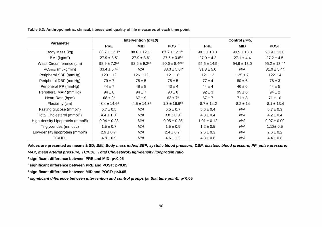
90
Table 5.3: Anthropometric, clinical, fitness and quality of life measures at each time point
Values are presented as means ± SD; BMI, Body mass index; SBP, systolic blood pressure; DBP, diastolic blood pressure; PP, pulse pressure;
MAP, mean arterial pressure; TC/HDL, Total Cholesterol:High-density lipoprotein ratio a significant difference between PRE and MID: p<0.05 b significant difference between PRE and POST: p<0.05 c significant difference between MID and POST: p<0.05 * significant difference between intervention and control groups (at that time point): p<0.05
Parameter Intervention (n=10) Control (n=5)
PRE MID POST PRE MID POST Body Mass (kg) 88.7 ± 12.1b 88.6 ± 12.1c 87.7 ± 12.1bc 90.1 ± 13.3 90.5 ± 13.3 90.9 ± 13.0
BMI (kg/m2) 27.9 ± 3.5b 27.9 ± 3.6c 27.6 ± 3.6bc 27.0 ± 4.2 27.1 ± 4.4 27.2 ± 4.5 Waist Circumference (cm) 98.9 ± 7.2ab 92.6 ± 9.2ac 90.6 ± 8.4bc* 95.5 ± 14.5 94.9 ± 13.0 95.2 ± 13.4*
VO2peak (ml/kg/min) 33.4 ± 5.4b N/A 38.3 ± 5.8b* 31.3 ± 5.0 N/A 31.0 ± 5.4* Peripheral SBP (mmHg) 123 ± 12 126 ± 12 121 ± 8 121 ± 2 125 ± 7 122 ± 4 Peripheral DBP (mmHg) 79 ± 7 78 ± 5 78 ± 5 77 ± 4 80 ± 6 78 ± 3 Peripheral PP (mmHg) 44 ± 7 48 ± 8 43 ± 4 44 ± 4 46 ± 6 44 ± 5
Peripheral MAP (mmHg) 94 ± 8 94 ± 7 90 ± 8 92 ± 3 95 ± 6 94 ± 2 Heart Rate (bpm) 68 ± 9b 67 ± 9 62 ± 7b 67 ± 7 71 ± 8 71 ± 10
Flexibility (cm) -8.4 ± 14.6c -4.5 ± 14.8c 1.3 ± 16.6bc -8.7 ± 14.2 -8.2 ± 14 -8.1 ± 13.4 Fasting glucose (mmol/l) 5.7 ± 0.5 N/A 5.5 ± 0.7 5.6 ± 0.4 N/A 5.7 ± 0.3 Total Cholesterol (mmol/l) 4.4 ± 1.0b N/A 3.8 ± 0.9b 4.3 ± 0.4 N/A 4.2 ± 0.4
High-density Lipoprotein (mmol/l) 0.94 ± 0.23 N/A 0.95 ± 0.25 1.01 ± 0.12 N/A 0.97 ± 0.09 Triglycerides (mmol/L) 1.5 ± 0.7 N/A 1.5 ± 0.9 1.2 ± 0.5 N/A 1.12± 0.5
Low-density lipoprotein (mmol/l) 2.9 ± 0.7b N/A 2.4 ± 0.7b 2.6 ± 0.3 N/A 2.6 ± 0.2 TC/HDL 4.8 ± 0.9 N/A 4.6 ± 1.2 4.3 ± 0.8 N/A 4.4 ± 0.8
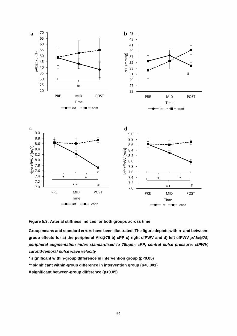
91
Figure 5.3: Arterial stiffness indices for both groups across time
Group means and standard errors have been illustrated. The figure depicts within- and between-group effects for a) the peripheral AIx@75 b) cPP c) right cfPWV and d) left cfPWV pAIx@75,
peripheral augmentation index standardised to 75bpm; cPP, central pulse pressure; cfPWV, carotid-femoral pulse wave velocity * significant within-group difference in intervention group (p<0.05) ** significant within-group difference in intervention group (p<0.001) # significant between-group difference (p<0.05)
2025303540455055606570
PRE MID POST
pAIx
@75
(%)
Timeint cont
*2527293133353739414345
PRE MID POST
cPP
(mm
Hg)
Timeint cont
#
7.07.27.47.67.88.08.28.48.68.89.0
PRE MID POST
right
cfP
WV
(m/s
)
Timeint cont
#7.07.27.47.67.88.08.28.48.68.89.0
PRE MID POST
left
cfP
WV
(m/s
)
Timeint cont
a b
c d
**
* * * **
* #
92
Central Pulse pressure (cPP)
Central PP did not significantly change across the 8 weeks in either of the groups (Figure
5.2b). There was a 1.98% improvement, albeit non-significant in cPP from PRE (35.5 ±
6.7mmHg) to POST (33.9 ± 3.2mmHg) in the intervention group (Table 5.8). However, there
was a group*time effect for cPP (Figure 5.3b). At POST, cPP was significantly greater in the
intervention than in the control group (F=7.865, p=0.015, pEta2=0.38). In addition, ∆cPP
depended on PRE cPP [F(1,13)=26.292, r=0.81, p<0.001]. There were no significant between
or within-group differences in either the difference or the ratio between central and peripheral
pulse pressures at any time point.
RESTING ARTERIAL ATHEROSCLEROTIC, REMODELLING AND GEOMETRICAL PARAMETERS
There were no significant between-group differences in either right or left cIMT, fIMT, cRI, fRI,
cEDD, or cWLR at baseline (Table 5.4).
Intima-media thickness (IMT)
Carotid IMT (cIMT): Mean of means of far wall
The cIMT did not change from PRE to POST in the control group (Table 5.4). Conversely, in
the intervention group, both right and left cIMT decreased across the 8 weeks. In this group,
significant differences were found between PRE-POST and MID-POST for the right cIMT and
significant differences in the left cIMT occurred from PRE-MID and MID-POST. From PRE to
POST, right cIMT and left cIMT showed significant decreases of 11.4 ± 9.5% (F=13.115,
p=0.006, pEta2=0.59) and 13.0 ± 10.8% (F=11.534, p=0.008, pEta2=0.56) respectively (Table
5.8). However, there were no significant between-group differences in right or left cIMT at any
time point (Table 5.4).
Femoral IMT (fIMT): Mean of means of far wall
There was no change in fMT changed from PRE to POST in the control group (Table 5.4). In
the intervention group, both right and left fIMT decreased across the 8 weeks but the total
decrease in right fIMT of 0.077mm (12.6 ± 15.3%) was not significant (Table 5.8). However,
the decreases in left fIMT by 0.0322mm from PRE to MID and by 0.073mm from PRE to POST
were significant, resulting in an overall decrease in left fIMT by 13.1 ± 12.1% (F=11.29,
p=0.005, pEta2=0.74) (Table 5.8). Inter-group differences in left fIMT occurred at POST only
(F=4.862, p=0.046, pEta2=0.27) (Table 5.4).
Resistivity Index (RI) Carotid RI (cRI) The cRI remained unchanged in the control group. In the intervention group, the left cRI
decreased significantly from PRE to MID and PRE to POST, resulting in an overall decrease
93
of 0.753 from PRE to POST (F=14.026, p=0.005, pEta2=0.61), at which point there was a
significant inter-group difference in this measure (F=0.714, p=0.022, pEta2=0.34) (Table 5.4).
Femoral RI (fRI)
Neither the right nor left fRI changed in the control group. In the intervention group, the left fRI
decreased significantly from PRE to POST, resulting in an overall decrease of 0.013
(F=19.286, p=0.002, pEta2=0.68), with significant inter-group differences occurring at both
MID (F=6.434, p=0.025, pEta2=0.27) and POST (F=11.143, p=0.005, pEta2=0.46) (Table
5.4).
Common carotid end-diastolic diameter (cEDD)
No inter-group differences in either right or left cEDD were observed at baseline (Figure 5.3).
Right and left cEDD values at PRE, MID and POST for both groups are illustrated in Figure
5.4a and 5.4b respectively. In the control group, neither right nor left cEDD changed
significantly over the three time points. In the intervention group, significant increases
occurred from PRE to POST in both right (5.62 ± 0.7mm vs 6.01 ± 0.59mm) and left (5.72 ±
0.36mm vs 6.15 ± 0.55mm) cEDD and from MID to POST as well, resulting in overall
increases of 0.39mm (6.9%) (F=12.570, p=0.006, pEta2=0.58) and 0.43mm (7.5%) (F=25.961, p<0.001, pEta2=0.74) respectively. Left cEDD was greater in the intervention than
in the control group at POST (F=6.804, p=0.022, pEta2=0.34) (Figure 5.4b).
Common carotid wall:lumen ratio (cWLR)
Neither right nor left cWLR significantly changed over the course of the 8 weeks in the control
group. In the intervention group, right cWLR decreased by 17% from PRE (0.1681 ± 0.0323)
to POST (0.1383 ± 0.0277) (F=23.269, p<0.001, pEta2=0.72). Likewise, left cWLR decreased
by 19.2% from PRE (0.19 ± 0.03) to POST (0.15 ± 0.03) (F=24.729, p<0.001, pEta2=0.73)
(Figure 5.4c-d). There was a small but significant between-group difference in right cWLR at
POST (F=9.620, p=0.035, pEta2=0.48) (Table 5.4).
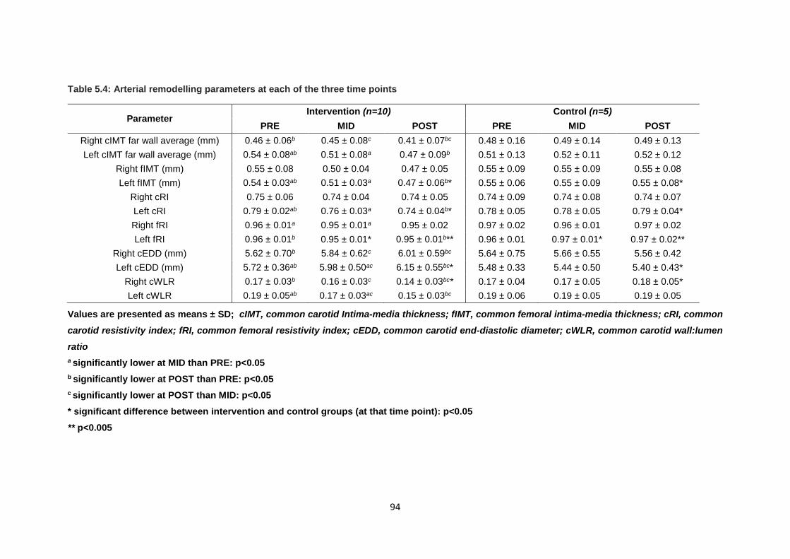
94
Table 5.4: Arterial remodelling parameters at each of the three time points
Values are presented as means ± SD; cIMT, common carotid Intima-media thickness; fIMT, common femoral intima-media thickness; cRI, common carotid resistivity index; fRI, common femoral resistivity index; cEDD, common carotid end-diastolic diameter; cWLR, common carotid wall:lumen ratio a significantly lower at MID than PRE: p<0.05 b significantly lower at POST than PRE: p<0.05 c significantly lower at POST than MID: p<0.05 * significant difference between intervention and control groups (at that time point): p<0.05 ** p<0.005
Parameter Intervention (n=10) Control (n=5)
PRE MID POST PRE MID POST Right cIMT far wall average (mm) 0.46 ± 0.06b 0.45 ± 0.08c 0.41 ± 0.07bc 0.48 ± 0.16 0.49 ± 0.14 0.49 ± 0.13 Left cIMT far wall average (mm) 0.54 ± 0.08ab 0.51 ± 0.08a 0.47 ± 0.09b 0.51 ± 0.13 0.52 ± 0.11 0.52 ± 0.12
Right fIMT (mm) 0.55 ± 0.08 0.50 ± 0.04 0.47 ± 0.05 0.55 ± 0.09 0.55 ± 0.09 0.55 ± 0.08 Left fIMT (mm) 0.54 ± 0.03ab 0.51 ± 0.03a 0.47 ± 0.06b* 0.55 ± 0.06 0.55 ± 0.09 0.55 ± 0.08*
Right cRI 0.75 ± 0.06 0.74 ± 0.04 0.74 ± 0.05 0.74 ± 0.09 0.74 ± 0.08 0.74 ± 0.07 Left cRI 0.79 ± 0.02ab 0.76 ± 0.03a 0.74 ± 0.04b* 0.78 ± 0.05 0.78 ± 0.05 0.79 ± 0.04* Right fRI 0.96 ± 0.01a 0.95 ± 0.01a 0.95 ± 0.02 0.97 ± 0.02 0.96 ± 0.01 0.97 ± 0.02 Left fRI 0.96 ± 0.01b 0.95 ± 0.01* 0.95 ± 0.01b** 0.96 ± 0.01 0.97 ± 0.01* 0.97 ± 0.02**
Right cEDD (mm) 5.62 ± 0.70b 5.84 ± 0.62c 6.01 ± 0.59bc 5.64 ± 0.75 5.66 ± 0.55 5.56 ± 0.42 Left cEDD (mm) 5.72 ± 0.36ab 5.98 ± 0.50ac 6.15 ± 0.55bc* 5.48 ± 0.33 5.44 ± 0.50 5.40 ± 0.43*
Right cWLR 0.17 ± 0.03b 0.16 ± 0.03c 0.14 ± 0.03bc* 0.17 ± 0.04 0.17 ± 0.05 0.18 ± 0.05* Left cWLR 0.19 ± 0.05ab 0.17 ± 0.03ac 0.15 ± 0.03bc 0.19 ± 0.06 0.19 ± 0.05 0.19 ± 0.05
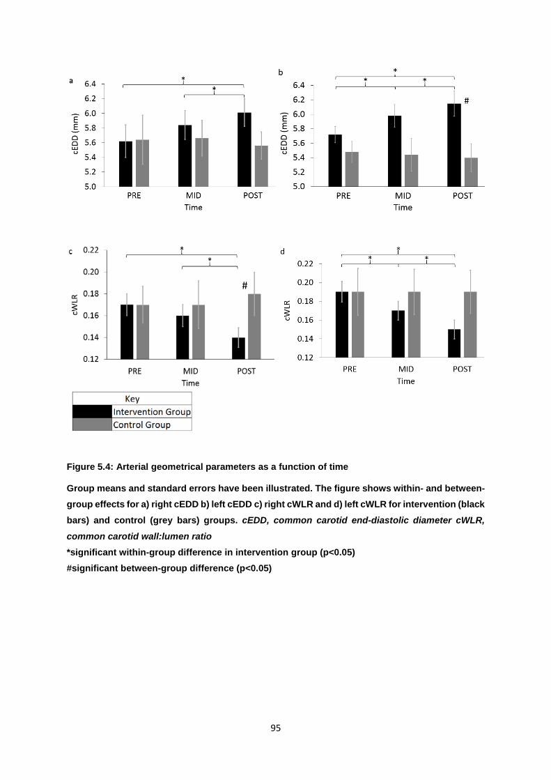
95
Figure 5.4: Arterial geometrical parameters as a function of time
Group means and standard errors have been illustrated. The figure shows within- and between-group effects for a) right cEDD b) left cEDD c) right cWLR and d) left cWLR for intervention (black bars) and control (grey bars) groups. cEDD, common carotid end-diastolic diameter cWLR,
common carotid wall:lumen ratio *significant within-group difference in intervention group (p<0.05) #significant between-group difference (p<0.05)
#
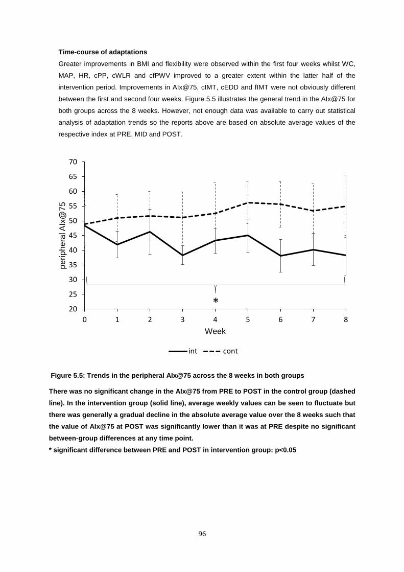
96
Time-course of adaptations
Greater improvements in BMI and flexibility were observed within the first four weeks whilst WC,
MAP, HR, cPP, cWLR and cfPWV improved to a greater extent within the latter half of the
intervention period. Improvements in AIx@75, cIMT, cEDD and fIMT were not obviously different
between the first and second four weeks. Figure 5.5 illustrates the general trend in the AIx@75 for
both groups across the 8 weeks. However, not enough data was available to carry out statistical
analysis of adaptation trends so the reports above are based on absolute average values of the
respective index at PRE, MID and POST.
Figure 5.5: Trends in the peripheral AIx@75 across the 8 weeks in both groups
There was no significant change in the AIx@75 from PRE to POST in the control group (dashed line). In the intervention group (solid line), average weekly values can be seen to fluctuate but there was generally a gradual decline in the absolute average value over the 8 weeks such that the value of AIx@75 at POST was significantly lower than it was at PRE despite no significant between-group differences at any time point. * significant difference between PRE and POST in intervention group: p<0.05
20
25
30
35
40
45
50
55
60
65
70
0 1 2 3 4 5 6 7 8
perip
hera
l AIx
@75
Week
int cont
*
97
5.3: EXPLORATION OF RELATIONSHIPS BETWEEN SELECTED PHYSIOLOGICAL VARIABLES
Correlations of note amongst all participants: in most cases, given the highly significant
bilateral correlations in absolute values and %changes, relationships will be presented using
values from the RIGHT (unless otherwise specified).
The central and peripheral values for pulse pressure were highly correlated at PRE, MID and
POST (r2=0.56, 0.93, 0.53 respectively) (p<0.05).
Age was positively correlated to baseline VO2peak (r=0.55, p=0.035) but the former did not
predict ∆VO2peak. Baseline VO2peak and BMI were negatively correlated (r=-0.720, p=0.002)
and baseline BMI partly predicted ∆VO2peak (r=0.66, p=0.037). Despite no significant relation
between VO2peak and either right or left cfPWV at PRE, VO2peak was negatively related to both
right cfPWV (r2=0.29, β=-0.535, p=0.040) and left cfPWV (r2=0.38, β=-0.613, p=0.015) at
POST (Figure 5.6). Despite an association between right cfPWV and MAP, inter-group
difference at POST as well as trends in right PWV remained significant after correcting for
MAP.
The AIx@75 and cfPWV were not correlated at any time point. Furthermore, the correlation
between the two indices decreased over the course of the 8 weeks in the intervention group
(r=0.16, 0.07, 0.05 at PRE, MID and POST respectively) (p>0.05). Neither cfPWV nor the
AIx@75 were related to cPP, pPP, ∆PP or pPP/cPP at any time point. However, at MID,
peripheral SBP and right and left cfPWV were positively related (r=0.60 and r=0.56
respectively) (p<0.05).
Only 23.6% and 4.7% of the variance in right and left cfPWV respectively was explained by
SBP, WC and MAP. The exercise intervention significantly decreased cfPWV bilaterally even
after adjusting for these factors.
Heart rate was positively correlated to cWLR at both PRE (r=0.59, p=0.019) and POST
(r=0.67, p=0.006). Heart rate at POST was also positively correlated to WC, cfPWV, cIMT and
fIMT and negatively correlated to age and VO2peak (Table 5.5b).
At PRE, MID and POST, cIMT was positively correlated to BMI (r=0.73, 0.55, 0.55) and WC
(r=0.55 at all the time points) (p<0.05). At PRE, MID and POST, left fIMT was positively related
to WC (r=0.7, 0.57, 0.5 respectively, p<0.05). No relationships were observed between carotid
and femoral IMT or RI or even between IMT and RI.
Table 5.5a depicts significant relationships between outcome measures at POST. This table
emphasises five important findings. Firstly, cIMT was positively related to lipid markers (TC
and LDL-C), BMI and WC. Secondly, indices of wave reflection (AIx and AIx@75) and central
haemodynamics (cPP) were positively related whereas the AIx@75 was negatively related to
height. Thirdly, cfPWV was consistently related to BMI. Fourth, cWLR, an arterial remodelling
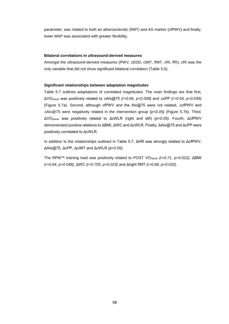
98
parameter, was related to both an atherosclerotic (fIMT) and AS marker (cfPWV) and finally,
lower MAP was associated with greater flexibility.
Bilateral correlations in ultrasound-derived measures Amongst the ultrasound-derived measures (PWV, cEDD, cIMT, fIMT, cRI, fRI), cRI was the
only variable that did not show significant bilateral correlation (Table 5.6).
Significant relationships between adaptation magnitudes
Table 5.7 outlines adaptations of correlated magnitudes. The main findings are that first,
ΔVO2peak was positively related to ∆AIx@75 (r=0.66, p=0.008) and ∆cPP (r=0.54, p=0.039)
(Figure 5.7a). Second, although cfPWV and the AIx@75 were not related, ∆cfPWV and
∆AIx@75 were negatively related in the intervention group (p<0.05) (Figure 5.7b). Third,
ΔVO2peak was positively related to ΔcWLR (right and left) (p<0.05). Fourth, ΔcfPWV
demonstrated positive relations to ΔBMI, ΔWC and ΔcWLR. Finally, ΔAIx@75 and ΔcPP were
positively correlated to ΔcWLR.
In addition to the relationships outlined in Table 5.7, ΔHR was strongly related to ΔcfPWV,
ΔAIx@75, ΔcPP, ΔcIMT and ΔcWLR (p<0.05).
The RPM™ training load was positively related to POST VO2peak (r=0.71, p=0.022), ΔBMI
(r=0.64, p=0.048), ΔWC (r=0.705, p=0.023) and Δright fIMT (r=0.68, p=0.032).
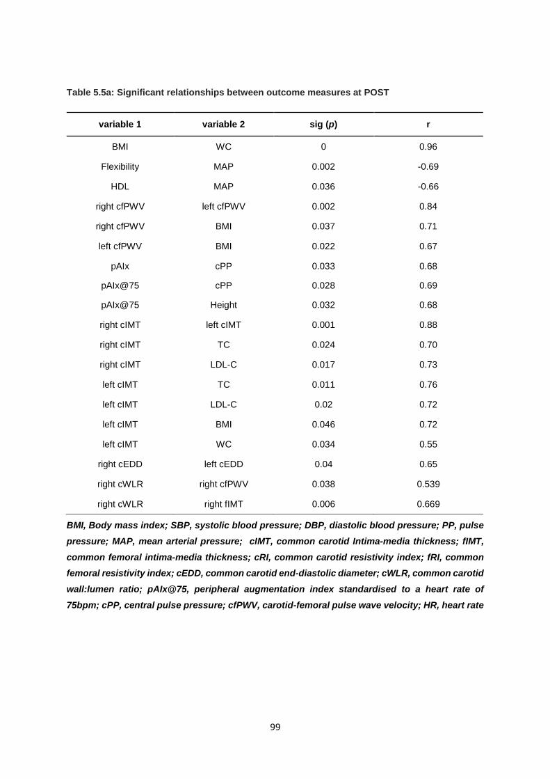
99
Table 5.5a: Significant relationships between outcome measures at POST
BMI, Body mass index; SBP, systolic blood pressure; DBP, diastolic blood pressure; PP, pulse pressure; MAP, mean arterial pressure; cIMT, common carotid Intima-media thickness; fIMT, common femoral intima-media thickness; cRI, common carotid resistivity index; fRI, common femoral resistivity index; cEDD, common carotid end-diastolic diameter; cWLR, common carotid wall:lumen ratio; pAIx@75, peripheral augmentation index standardised to a heart rate of 75bpm; cPP, central pulse pressure; cfPWV, carotid-femoral pulse wave velocity; HR, heart rate
variable 1 variable 2 sig (p) r
BMI WC 0 0.96
Flexibility MAP 0.002 -0.69
HDL MAP 0.036 -0.66
right cfPWV left cfPWV 0.002 0.84
right cfPWV BMI 0.037 0.71
left cfPWV BMI 0.022 0.67
pAIx cPP 0.033 0.68
pAIx@75 cPP 0.028 0.69
pAIx@75 Height 0.032 0.68
right cIMT left cIMT 0.001 0.88
right cIMT TC 0.024 0.70
right cIMT LDL-C 0.017 0.73
left cIMT TC 0.011 0.76
left cIMT LDL-C 0.02 0.72
left cIMT BMI 0.046 0.72
left cIMT WC 0.034 0.55
right cEDD left cEDD 0.04 0.65
right cWLR right cfPWV 0.038 0.539
right cWLR right fIMT 0.006 0.669
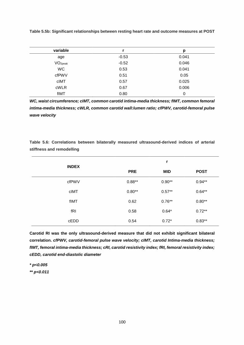
100
Table 5.5b: Significant relationships between resting heart rate and outcome measures at POST
WC, waist circumference; cIMT, common carotid intima-media thickness; fIMT, common femoral intima-media thickness; cWLR, common carotid wall:lumen ratio; cfPWV, carotid-femoral pulse wave velocity
Table 5.6: Correlations between bilaterally measured ultrasound-derived indices of arterial stiffness and remodelling
INDEX r
PRE MID POST
cfPWV 0.88** 0.90** 0.94**
cIMT 0.80** 0.57** 0.64**
fIMT 0.62 0.76** 0.80**
fRI 0.58 0.64* 0.72**
cEDD 0.54 0.72* 0.83**
Carotid RI was the only ultrasound-derived measure that did not exhibit significant bilateral correlation. cfPWV, carotid-femoral pulse wave velocity; cIMT, carotid Intima-media thickness;
fIMT, femoral intima-media thickness; cRI, carotid resistivity index; fRI, femoral resistivity index; cEDD, carotid end-diastolic diameter
* p<0.005 ** p<0.011
variable r p age -0.53 0.041
VO2peak -0.52 0.046 WC 0.53 0.041
cfPWV 0.51 0.05 cIMT 0.57 0.025 cWLR 0.67 0.006 fIMT 0.80 0
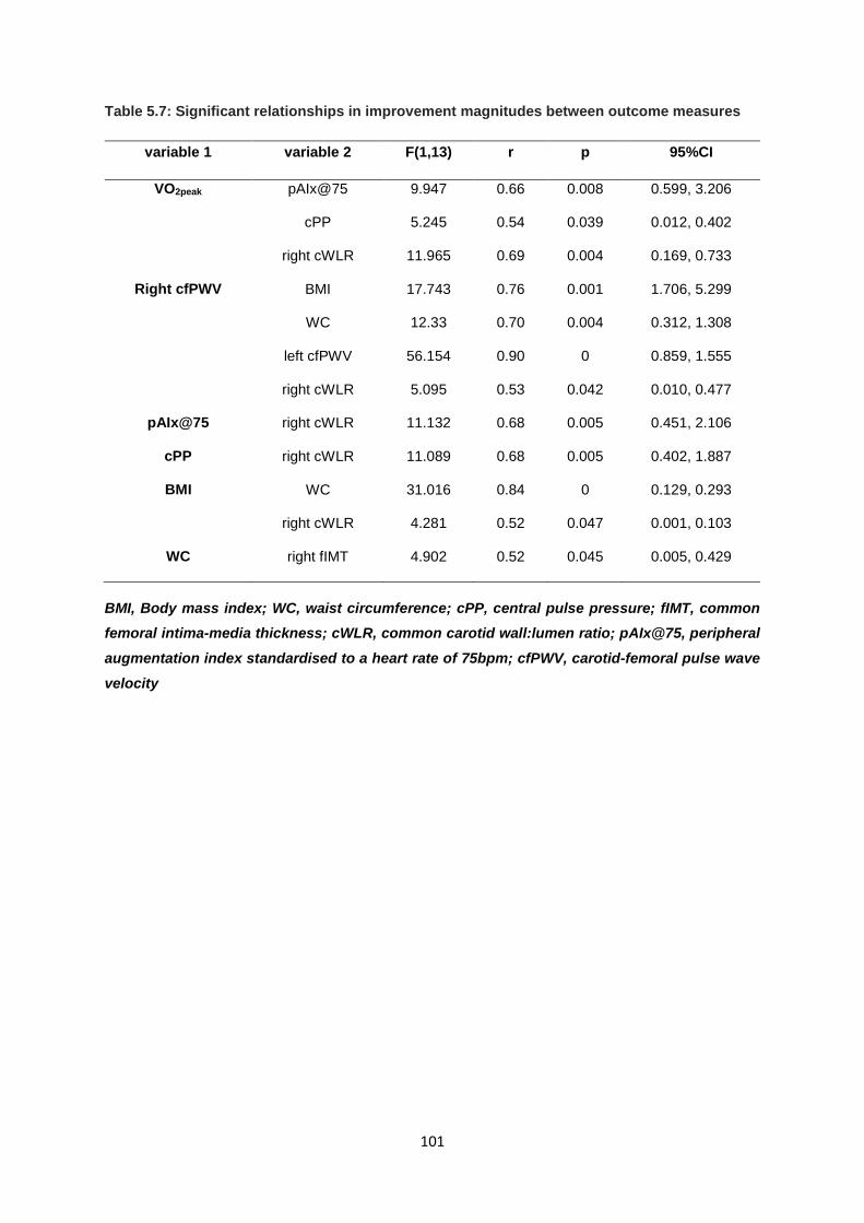
101
Table 5.7: Significant relationships in improvement magnitudes between outcome measures
BMI, Body mass index; WC, waist circumference; cPP, central pulse pressure; fIMT, common femoral intima-media thickness; cWLR, common carotid wall:lumen ratio; pAIx@75, peripheral augmentation index standardised to a heart rate of 75bpm; cfPWV, carotid-femoral pulse wave velocity
variable 1 variable 2 F(1,13) r p 95%CI
VO2peak pAIx@75 9.947 0.66 0.008 0.599, 3.206
cPP 5.245 0.54 0.039 0.012, 0.402
right cWLR 11.965 0.69 0.004 0.169, 0.733
Right cfPWV BMI 17.743 0.76 0.001 1.706, 5.299
WC 12.33 0.70 0.004 0.312, 1.308
left cfPWV 56.154 0.90 0 0.859, 1.555
right cWLR 5.095 0.53 0.042 0.010, 0.477
pAIx@75 right cWLR 11.132 0.68 0.005 0.451, 2.106
cPP right cWLR 11.089 0.68 0.005 0.402, 1.887
BMI WC 31.016 0.84 0 0.129, 0.293
right cWLR 4.281 0.52 0.047 0.001, 0.103
WC right fIMT 4.902 0.52 0.045 0.005, 0.429
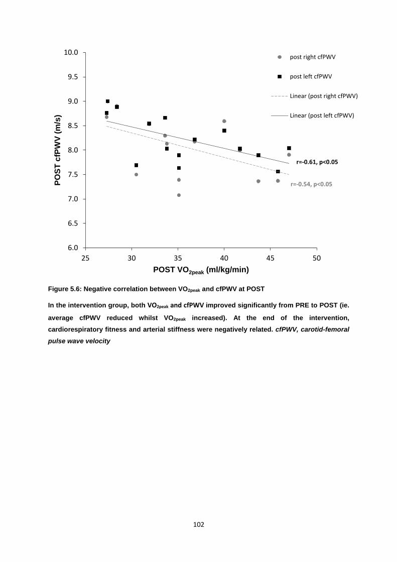
102
Figure 5.6: Negative correlation between VO2peak and cfPWV at POST
In the intervention group, both VO2peak and cfPWV improved significantly from PRE to POST (ie.
average cfPWV reduced whilst VO2peak increased). At the end of the intervention, cardiorespiratory fitness and arterial stiffness were negatively related. cfPWV, carotid-femoral
pulse wave velocity
6.0
6.5
7.0
7.5
8.0
8.5
9.0
9.5
10.0
25 30 35 40 45 50
POST
cfP
WV
(m/s
)
POST VO2peak (ml/kg/min)
post right cfPWV
post left cfPWV
Linear (post right cfPWV)
Linear (post left cfPWV)
r=-0.61, p<0.05
r=-0.54, p<0.05
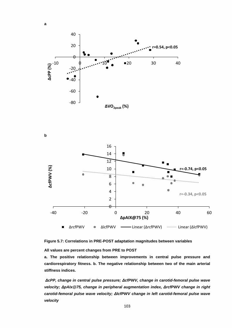
103
a
b
Figure 5.7: Correlations in PRE-POST adaptation magnitudes between variables
All values are percent changes from PRE to POST a. The positive relationship between improvements in central pulse pressure and cardiorespiratory fitness. b. The negative relationship between two of the main arterial stiffness indices.
ΔcPP, change in central pulse pressure; ΔcfPWV, change in carotid-femoral pulse wave velocity; ΔpAIx@75, change in peripheral augmentation index, ΔrcfPWV change in right carotid-femoral pulse wave velocity; ΔlcfPWV change in left carotid-femoral pulse wave velocity
-80
-60
-40
-20
0
20
40
-10 0 10 20 30 40
ΔcPP
(%)
ΔVO2peak (%)
r=0.54, p<0.05
0
2
4
6
8
10
12
14
16
-40 -20 0 20 40 60
ΔcfP
WV
(%)
ΔpAIX@75 (%)
ΔrcfPWV ΔlcfPWV Linear (ΔrcfPWV) Linear (ΔlcfPWV)
r=-0.74, p<0.05
r=-0.34, p<0.05

104
5.4: Specific focus on intervention group results
Improvement magnitudes in the main outcome measures are summarised in table 5.8.
Metabolic Syndrome Risk factors
Table 5.9 outlines the specific MS RF that each individual in the intervention group had at PRE
and POST and shows that the number of RFs decreased in 70% of the individuals and remained
unchanged in 30%. The risk factor profile did not worsen for any individual. Furthermore, three
individuals who would have been classified as having MS at PRE would not have been classified
as having it at POST (ie. the number of their risk factors decreased from ≥ 3 to <3). The number
of PRE and POST MS RFs were not significantly correlated to absolute values of AS measures
at PRE and POST respectively and the % decrease in risk was not related to arterial health
indices.
Table 5.8: Improvements in the main anthropometric, fitness, clinical and arterial health measures in the intervention group from PRE to POST
BMI, Body mass index; WC, waist circumference; MAP, mean arterial pressure; cIMT, common carotid Intima-media thickness; fIMT, common femoral intima-media thickness; cEDD, common carotid end-diastolic diameter; cWLR, common carotid wall:lumen ratio; pAIx@75, peripheral augmentation index standardised to a heart rate of 75bpm; cPP, central pulse pressure; cfPWV, carotid-femoral pulse wave velocity
* significant improvement from PRE to POST: p<0.05 ** significant improvement from PRE to POST: p<0.001
variable PRE to POST % IMPROVEMENTS (95% CI) BMI* 1.2 (0.4, 1.9) WC* 8.4 (4.5, 12.3)
VO2peak* 15.1 (9.1, 20.9) cPP 1.98 (-9.57, 13.52) MAP 3.2 (-3.6, 10.0) HR 8 (3.71, 12.29) AIx* 23.3 (9.0, 37.7)
AIx@75* 23.8 (8.7, 38.8) right cfPWV** 10.7 (8.07, 12.07) left cfPWV** 7.6 (5.53, 9.67) right cIMT* 11.35 (4.55, 18.15) left cIMT* 12.99 (4.49, 21.74) right fIMT 12.64 (1.68, 23.61) left fIMT* 13.11 (4.49, 21.74)
right cEDD* 6.9 (2.26, 11.54) left cEDD* 7.5 (4.43, 10.57)
right cWLR* 17.7 (10.7, 24.7) left cWLR* 19.2 (12.2, 26.2)
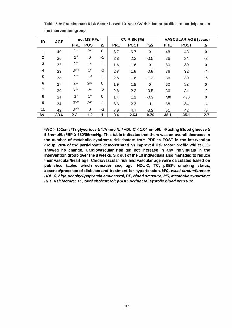
105
Table 5.9: Framingham Risk Score-based 10–year CV risk factor profiles of participants in the intervention group
aWC > 102cm; bTriglycerides ≥ 1.7mmol/L; cHDL-C < 1.04mmol/L; dFasting Blood glucose ≥ 5.6mmol/L; eBP ≥ 130/85mmHg. This table indicates that there was an overall decrease in the number of metabolic syndrome risk factors from PRE to POST in the intervention group. 70% of the participants demonstrated an improved risk factor profile whilst 30% showed no change. Cardiovascular risk did not increase in any individuals in the intervention group over the 8 weeks. Six out of the 10 individuals also managed to reduce their vascular/heart age. Cardiovascular risk and vascular age were calculated based on published tables which consider sex, age, HDL-C, TC, pSBP, smoking status, absence/presence of diabetes and treatment for hypertension. WC, waist circumference; HDL-C, high-density lipoprotein cholesterol, BP, blood pressure; MS, metabolic syndrome; RFs, risk factors; TC, total cholesterol; pSBP, peripheral systolic blood pressure
ID AGE no. MS RFs CV RISK (%) VASCULAR AGE (years) PRE POST Δ PRE POST %Δ PRE POST Δ
1 40 2bc 2bc 0 6.7 6.7 0 48 48 0 2 36 1d 0 -1 2.8 2.3 -0.5 36 34 -2 3 32 2cd 1c -1 1.6 1.6 0 30 30 0 4 23 3ace 1c -2 2.8 1.9 -0.9 36 32 -4 5 38 2cd 1d -1 2.8 1.6 -1.2 36 30 -6 6 37 2bc 2bc 0 1.9 1.9 0 32 32 0 7 30 3abc 2c -2 2.8 2.3 -0.5 36 34 -2 8 24 1c 1c 0 1.4 1.1 -0.3 <30 <30 0 9 34 3ade 2de -1 3.3 2.3 -1 38 34 -4 10 42 3cde 0 -3 7.9 4.7 -3.2 51 42 -9
Av 33.6 2-3 1-2 1 3.4 2.64 -0.76 38.1 35.1 -2.7
106
CHAPTER 6: DISCUSSION Arterial disorders account for 80% of all CVD-related deaths, necessitating the need for their early
detection and therapies to optimise arterial health. Aerobic exercise (AE) laboratory-based
interventions employing various exercise modalities and intensities have shown remarkable
success at improving tissue biomarkers of early vascular ageing (EVA) as well as arterial
geometry in healthy and clinical populations (14, 46-48, 56, 57, 59, 62, 161, 265, 333, 353, 395,
397, 398, 415, 416). This is the first study to determine the effects of a community-based mixed
intensity aerobic interval indoor cycling group-fitness class on cardiorespiratory fitness and
indices of arterial health related to EVA. The cohort was limited to community-dwelling, healthy,
sedentary males aged 20-45 years in order to eliminate potential confounding factors associated
with old age, gender, chronic illnesses and training status. The current findings support the
hypothesis that community-based self-paced combined aerobic interval exercise can significantly
improve arterial health.
6.1 INTERVENTION CHARACTERISTICS
The concurrent nature of the class was such that overall, it was based primarily on principles of
AE, interspersed with regular bouts of both HIIT and SIT. Heart rates during exercise sessions
fluctuated from 50-105% individually-determined HRpeak. In laboratory-based studies, exercise
prescription is usually specified, individually-tailored and monitored so that individuals are
required to maintain these pre-determined intensities (absolute/range) for a whole session
throughout the intervention, or, progressively increase the volume/intensity in a controlled
manner.
Despite the self-paced nature of the exercise, individuals worked at an average of 81±7%HRpeak.
The current intervention can be classified as ‘vigorous’ based on previous studies (investigating
AE effects on arterial health in healthy, sedentary middle-aged men) which refer to 80-90%HRmax
(56, 59, 62) and even 75%HRmax (49, 342) as ‘vigorous’. Amongst these studies, exercise
volumes ranged from 20-50 mins per day, 3-7 days per week, with inverse correlations between
intensity and volume in trials during which either volume or intensity progressively increased (56,
342). The two studies employing similar exercise prescriptions to the current one were those by
Thijssen et al. (59) (80%HRmax, 30 mins, 3 times/wk AE) and Heydari et al. (62) (80-90%HRmax,
20 mins, 3 times/week HIIT). As discussed later, findings in both these studies were similar to the
ones presented here. Interestingly enough, maximal HRs (94%±7%HRpeak, range 88-
104%HRpeak) often exceeded those of the baseline VO2peak test. In the current study, this could
be due to the previously sedentary individuals exerting increasingly more effort as they grew
accustomed to exercising. This is further justified by the lack of a plateau effect, whereby average
weekly HRs during the course of the intervention gradually increased. When beginning exercise
with an activity not carried out on a daily basis (cycling vs walking/jogging/work-related activity),
individuals may initially maintain caution, avoiding maximal effort, especially in self-paced
activities. Upon increasing familiarity with the exercise and heightened awareness of individual
physical limits, the willingness to challenge oneself is likely. In previous controlled-intensity trials,
107
differences between pre- and post-intervention intensities are not usually relevant and have not
been presented.
It is possible that the increasing HR trend observed would not apply to clinical populations or
exercising individuals unfamiliar with this type of exercise. On occasion, the training load from just
one RPM™ class was greater than the exertion load over the rest of the day indicating that
devoting 3% of the day to this sort of exercise, could significantly benefit arterial and overall health
and CV risk. It was also interesting to note the positive correlation between RPM™ training load
and ΔBMI, ΔWC and ΔfIMT. The more effort an individual exerted, the greater the benefits,
especially in the lower limb atherosclerotic marker.
6.2 PRIMARY MEASURES
6.2.1 MAIN FINDINGS
6.2.1.1 CARDIORESPIRATORY FITNESS (CRF)
Future fatal CV events are better predicted by VO2peak than many traditional CV risk factors and
an increase in CRF by one MET can decrease non-fatal and fatal cardiac events by 17-29% and
28-51% respectively (217). The improvement in CRF by 15% (1.5METs) in the intervention group
is similar to that observed by Ciolac et al. (after a 16 week aerobic interval training intervention)
(333) and is greater than most MCAE (60-65%VO2max) interventions in which approximately 8%
increases in CRF are observed after 4 (46) or 16 (333) weeks.
In the present study, this 15% improvement in CRF reflects a potentially reduced CV risk in only
two months even though neither post-intervention VO2peak nor ∆VO2peak was related to post-
intervention FRS-based CV risk or ∆CV risk (ie. CRF increased independently of traditional CV
risk factors). Taking individual trainability into consideration, greater improvements in CRF are
generally observed after HIIT than MCAE interventions (456) and the absolute increase in VO2peak
by 5ml/kg/min in the present study is similar to previous vigorous-intensity endurance training
studies (62) but less than the average increase in CRF in purely HIIT programmes (457). This
reiterates the description of the current intervention as non-continuous AE with concurrent interval
training and sheds light on its potentially superior effectiveness, relative to regular MCAE
programmes, at improving CRF.
6.2.1.2 CAROTID-FEMORAL PULSE WAVE VELOCITY (cfPWV)
The observed 10.7% (0.93m/s) and 7.6% (0.65m/s) decreases in right and left cfPWV respectively
are similar in magnitude to previous studies using moderate to high-intensity AE interventions
(397). The greater improvement in right cfPWV may be due to the higher average baseline cfPWV
on the right than the left side and therefore, greater possibility for adaptability and response in the
former. Bilateral asymmetry in arterial stiffness and geometry have been attributed to differing
anatomy-dependent blood flow and shear stress patterns (458). However, very high positive
correlations between bilateral ultrasound-derived indices of arterial health were observed in the
present study. A 2015 review reported that AE interventions bring about an average of -0.63m/s
108
(386) to -0.67m/s (379) improvements in PWV and that greater reductions are observed in
peripheral than central AS measures, with interventions lasting longer than 10 weeks (379) and
with exercise of at least moderate to high intensity (333). However, reductions in central PWV
(0.5m/s) and peripheral PWV (0.8m/s) have even been observed after only 6 days of daily
moderate AE for 2 hours each day (398). The effectiveness of the current intervention on the gold
standard measure of AS can be considered to be favourable and the intensity robust enough to
reduce central AS.
Taken alone, the improvements in cfPWV may independently reduce CV risk given that a
decrease in cfPWV by 1m/s reduces CV events and mortality by 14% (22) and that the adjusted
relative risk for all-cause mortality is 0.71 (459). Although risk prediction based on traditional CV
risk factors is valuable in populations, it may be less so in individuals as the scales used are
influenced by non-modifiable risk factors (460).
In agreement with a 2014 meta-analysis (386), the improvements in cfPWV in the intervention
group occurred independent of resting HR or MAP as well as anthropometric changes, indicating
a primary effect. In young individuals, AE does not significantly alter arterial compliance but
amongst older individuals, vigorous AE can modulate age-associated decreases in arterial
compliance (49). Unfortunately, this observation could not be investigated further in the present
study because ‘older’ individuals (>50 years) were excluded and no associations between age
and cfPWV were observed.
Despite the encouraging results obtained in the present study, there is evidence to suggest that
such benefits of exercise on cfPWV may attenuate in training periods of longer durations (greater
than 3-6 months) (461, 462). It would be interesting to determine the patterns and magnitudes of
operative stiffness adaptations with longer duration (>12 weeks) aerobic interval interventions.
6.2.1.3 AUGMENTATION INDEX (AIx, AIx@75)
It has been suggested that the AIx is a sensitive index for a reduction in AS prior to BP reductions
(353), which makes it a useful tool in both public healthcare and clinical settings. In this regard,
Donley et al. (161) found a 21% reduction in the AIx@75 in healthy and MS adults after 8 weeks
of AE, which is similar to the 24% reduction in the AIx@75 (and 23% reduction in the AIx) in the
exercise group observed in the present study. These improvements are greater than most of
those reported when similar populations have undergone purely MCAE (6%, 21%, 3% and 9.2%
improvements in peripheral AIx) (46, 161, 353, 397) as well as HIIT (3.5% improvement in central
AIx) (62). A small sample size is the most likely reason for the absence of post-intervention
between group differences in the AIx@75. The findings reported above pertain to healthy
individuals and it is possible that exercise-induced improvements in this index may not be
observed in overweight or obese individuals, even with vigorous (80-90%VO2max exercise (67).
The average reduction in the AIx with MCAE is -2.63% (386). Furthermore, the means and
standard deviations of the AIx@75 reported in the current study were noticeably greater than
those of previous studies (62, 161, 394).
109
Differences in absolute values of the AIx and in ΔAIx between studies whose participants have
similar baseline characteristics can only be explained by differences in assessment modes.
Previous studies have used principles of applanation tonometry to derive this index, but in the
present study, the BP+ device generated the peripheral AIx using an oscillometric cuff-based
technique in the absence of a generalised transfer function. Whilst the BP+ device produces
reliable and valid results for cBP (203, 463), BP+ AIx validation studies are lacking. Nevertheless,
the present absolute values agree with published applanation tonometry-derived normative data
of the peripheral AIx amongst healthy populations of different ethnicities (464, 465). One study
reported that the correlation between AIx values obtained using different applanation tonometry
devices is much weaker than the correlation between inter-device SBP (466). The authors of this
study suggested that even slight changes in the pulse waveform could result in large differences
in AIx and cBP values so results should be interpreted carefully. Therefore, the present values
cannot be deemed inaccurate. The diurnal variation (the gradual decrease over the course of the
day) in the AIx (467) must be considered given that weekly inter-subject assessment times could
not be standardised in the present study for practical reasons. Depending on RPM™ class times
(intervention group) and subject availability (control group), weekly measurements were taken
anytime between 5:30am and 6:30pm. However, the issue of diurnal variation would not have
affected the PRE, MID and POST values as assessment times for all individuals were the same
(8:30-9:00am).
Although a linear positive relation between the AIx and age have been reported, (84), numerous
studies have suggested that the AIx changes more with age in younger than older individuals and
is therefore a more sensitive index of vascular age in the former population (90). Based on this
notion, the large improvement in the AIx@75 observed in the present study can partly be
explained by the fact that the current participants were not ‘old’ (>50 years).
Training prescription is an important consideration when aiming to ensure beneficial and not
adverse impacts. The large improvements in the AIx@75 could be due to the high-intensity nature
and effective frequency of the exercise. The finding that maximum HRs often exceeded
100%HRpeak implies that the high-intensity peaks during the classes were robust enough to
substantially improve wave reflection dynamics because greater reductions in the AIx are
generally observed with lower volume but high-intensity AE (386). This is in line with greater acute
reductions in the AIx@75 within the first 12 hours after a HIIT session compared to an MCAE
session (354). The dose of three 45 minute classes per week was potentially optimal as exercise
volume is associated with worsening of the AIx (386) so it would be advisable to avoid daily
vigorous exercise.
6.2.1.4 CENTRAL HAEMODYNAMICS
Although the 2% reduction in cPP in the intervention group failed to reach statistical significance,
the post-intervention between-group difference illustrates that important information regarding left
ventricular load can be missed with peripheral BP measurements. The unaltered peripheral PP
shows that peripheral BP cannot accurately predict central BP, a better risk predictor. The
reduction in cPP in the absence of significant changes in cBP can also be considered relevant in
110
terms of risk reduction as PP independently predicts risk better than either SBP or DBP alone
(79). The positive correlation between post-intervention AIx@75 and cPP illustrates how changes
in central haemodynamics can be attributed to modified wave reflections (468). The AIx can act
as an intermediate between AS and PP because an increase in AS raises PP via an increase in
the AIx. This reflects the independent yet interconnected nature of the three main AS indices as
discussed in the following sections.
6.2.2 RELATIONSHIPS BETWEEN ARTERIAL STIFFNESS INDICES
It is important that cfPWV and the AIx are neither used interchangeably nor considered to reflect
identical changes in arterial health (469). They are distinct entities which, when considered in
conjunction, make up the arterial factors which influence aortic systolic pressure.
The AIx depends on intrinsic AS in addition to the magnitude of reflected waves and the distance
to wave reflection sites. An increase in PWV disrupts only the timing of reflected waves, potentially
explaining the lack of correlation between the AIx and cfPWV in the present study, even though
the two indices have previously been shown to be highly correlated (470). The discrepancy
between the current findings and those of Yasmin & Brown (which indicated a positive correlation
between the AIx and cfPWV) could be because, in their study, individuals had familial
hypertension and applanation tonometry was used to assess AS. In agreement with the current
findings, it has been reported that PWV and the AIx are not related in multivariate analysis and
that the two indices may be more correlated in stiffer aortas because, in elastic aortas, the AIx is
influenced more by wave reflections than AS (471). Indeed, in the present study, the relation
between AIx@75 and bilateral cfPWV decreased over the course of the 8 weeks in the
intervention group.
The negative relationship between ΔcfPWV and ΔAIx@75 could be due to the small sample size
or, alternatively, it can reflect individual responsiveness and what aspects of their vascular health
they are more susceptible to alter in response to a training stimulus, which once again relates to
the notion of genetically-determined individual ‘trainability’. This can lead one to question the
plausibility that improvements in one index can occur at the expense of improvements in another.
There is evidence to suggest that wave reflection sites move distally with age because as elastic
arteries stiffen during ageing, the AS gradient reduces and points of impedance mismatch, which
are wave reflection sites, therefore move toward the periphery (171). Based on this, it could be
hypothesised that over the course of the intervention, the opposite of this ageing-associated
phenomenon occurred. As cfPWV (and presumably elastic artery stiffness) decreased, the AS
gradient became steeper such that impedance sites moved closer to the aorta which would
counter the direct effects of PWV on wave transit time to and from the aorta, a determinant of the
AIx. Further studies using larger samples and varied AS assessment methods are warranted to
help determine whether exercise is positively impacting wave reflection timing and amplitude or
intrinsic wall stiffness more.
The non-significant reduction and post-intervention between-group differences in cPP could be
attributed directly to repetitive carotid and aortic distension during the exercise bouts which would
111
decrease SNA. This would be attributed to altered baroreflex sensitivity (BRS) because
decreased AS increases afferent firing for a given pressure change (42). Therefore, in
consonance with the parallel decreases in the other two AS indices, cPP could also have
improved due to arterial destiffening-induced reductions in SNA (42).
6.2.3 RELATIONSHIPS BETWEEN CARDIORESPIRATORY FITNESS, ARTERIAL STIFFNESS AND WAVE REFLECTIONS
Over two decades ago, Vaitkevicius et al. reported that AS (indexed by the AIx and cfPWV) was
negatively associated with CRF (indexed by VO2max) and that interventions which increase CRF
can attenuate the arterial stiffening that occurs during normal ageing (52). The negative post-
intervention age-independent correlations between VO2peak and cfPWV and between the
magnitudes of changes of ΔVO2peak with ΔAIx@75 and ΔcPP, reflect cross-sectional data
demonstrating the ability of AE to reverse age-associated arterial stiffening (51, 52). Interestingly,
sport or fitness-related but not leisure-time-associated physical activity is inversely related to AS
because the latter does not tend to increase aerobic capacity (472) which indicates that the
increase in CRF is paramount in explaining the beneficial impact of AE on AS, a hypothesis
supported by the positive relationship between resistance training and AS (47, 65-67, 335).
Aortic stiffness, myocardial performance and functional capacity are believed to be interrelated in
a cyclic fashion whereby an increase in aortic stiffness increases cPP which reduces coronary
perfusion, myocardial work and performance and thence, exercise capacity (351). As a result,
cardiac risk increases whilst fitness decreases which would exaggerate the initial arterial
stiffening, thereby initiating a vicious cycle (473). Therefore, it is not surprising that simultaneous
decreases in cfPWV, cPP and increased CRF were observed in the present study, with the post-
intervention negative correlation between cfPWV and VO2peak. Potential mechanisms (obtained
on animal models) explaining the negative correlation between CRF and AS observed in the
present study pertain to greater aortic elastin content, no changes in collagen content and a
reduced elastin calcium content with greater fitness (this is yet to be confirmed in humans) (392).
6.2.4 PROPOSED MECHANISMS UNDERLYING THE RELATIONSHIP BETWEEN AEROBIC EXERCISE AND ARTERIAL STIFFNESS
Potential mechanisms explaining the decrease in AS with AE and HIIT relate not only to the
improved elastin:collagen equilibrium (474) and enhanced NO production (coupled with reduced
vasoconstrictor production) (475) but also to the anti-oxidative effects (through superoxide
dismutase up-regulation and NADPH down-regulation (476) and anti-inflammatory effects (via
increases in anti-inflammatory cytokines IL-4 & 10 and decreases in pro-inflammatory cytokines
IL-1 & 6 and TNF-α) (477) of AE. Greater shear-stress-mediated release of NO and a subsequent
greater reduction in peripheral vascular resistance with harder exercise has previously been one
of the hypotheses used to explain the positive relation between AE intensity and AIx
improvements (385). Since MAP remained unchanged, the effects of the present intervention on
112
the AIx@75 could be explained by alterations in structural arterial wall properties instead.
However, altered extracellular matrix composition is unlikely because changes in the composition
of wall scaffolding proteins occur over many years. The most feasible theories would, therefore,
pertain to either decreased IMT or acute increases in PP causing mechanical distension, thereby
stretching collagen fibres and altering collagen cross-links (resulting in less uncoiled collagen)
(478). Although the acute effects of the exercise on arterial health indices and their determinants
(eg. MAP, HR, BRS and post-exercise hypotension) were not assessed in the present study, they
may explain longer-term adaptations due to the cumulative effects of repetitive sessions.
The incorporation of various music styles in the RPM™ classes could have potentially directly
contributed toward the improved cfPWV and AIx@75 (479). Mental stress is associated with CVD
(480) and can cause arterial health deterioration (481, 482) whilst positive psychological stimuli
can decrease both PWV and AIx (482). Music is being increasingly used as a therapeutic tool in
individuals with CVD (483) and a recent study on healthy individuals showed that music can
improve arterial function (stiffness and wave reflections) (479). The benefits lasted for as long as
the music was being listened to and effects persisted with some types of music. As changes in
the AIx and PWV were not correlated in that study, the authors suggest that decreased wave
reflections with music are primarily due to reduced wave amplitudes as opposed to reduced transit
times. Possible mechanisms underlying this relate to increased beta-endorphin-mediated NO
release (484). Since endothelial function influences AS, improvements in the former can
favourably alter the latter. The second mechanism relates to the increased aortic and systemic
stiffness brought about by noradrenaline and the fact that music therapy in patients with CVD
reduces plasma adrenaline and noradrenaline levels (485). Music-induced reductions in SNA and
subsequent peripheral artery dilation may also attenuate wave reflection amplitudes (479).
Finally, conclusive findings regarding the relationship between AS and BP would determine
whether or not the improvements in AS with music can be explained by the music-induced BP
reductions (483, 485).
6.3 SECONDARY OUTCOME MEASURES: ARTERIAL STRUCTURAL REMODELLING PARAMETERS
In agreement with studies suggesting extensive outward arterial remodelling with exercise,
significant bilateral decreases in cIMT (average 12.2%), cWLR (average 18.5%) and left fIMT
(13.1%) as well as increases in bilateral resting cEDD (average 7.2%) were observed in the
intervention group only. It has been reported that arterial remodelling is preceded by functional
changes (407) but because flow-mediated dilation was not measured, no comment can be made
about whether functional or structural changes occurred first. However, exploration of the time
course of adaptations revealed no significant difference in the magnitude of structural adaptations
within the first and second four weeks. This was in line with a study by Thijssen et al., who
assessed carotid and superficial femoral WLR and IMT every two weeks over an 8 week cycling
intervention, and showed that improvements in arterial structure occur in a dose-dependent
manner (59). More regular (eg. weekly) ultrasonographic assessments might have provided a
better understanding of trends in adaptations.
113
Decreases in fIMT as observed in the current study, coupled with increases in femoral diameter
(presently unassessed), are commonly reported following moderate-to-vigorous lower limb
exercise (56) and could be justified by reports that firstly, peripheral arteries, such as the femoral
and brachial arteries, are relatively more responsive to training stimuli (than central arteries),
showing greater adaptability (55), and secondly, such responses appear to be localised, in this
case, to the active femoral bed of the lower limb (56). The interesting observation, however, was
the systemic remodelling with lower-limb exercise.
6.3.1 POSSIBLE REASONS BEHIND THE SYSTEMIC ARTERIAL STRUCTURAL ADAPTATIONS OBSERVED
If deconditioning results in detrimental arterial adaptations due to muscle atrophy and decreased
metabolic demand, it would be rational to believe that beneficial arterial remodelling induced by
exercise training must be due to muscular adaptations and increased metabolic demands.
Systemic arterial remodelling was observed in the present study despite the range of participant
ethnicities, a factor which determines carotid diameter and stiffness (486). This finding was in line
with reports that runners and cyclists have greater arterial diameters globally as well as lower
systemic arterial wall thickness and WLRs than recreationally active individuals (341).
Furthermore, elite squash players have lower brachial, femoral and carotid IMT values than less
active individuals and no difference in IMT between the preferred and non-preferred limb despite
the larger arterial diameter in the former (340). In agreement with these cross-sectional studies,
a 2015 meta-analysis reported that AE interventions involving vigorous exercise do improve cIMT
(379). Ranadive et al. observed decreased carotid pulse pressure and cIMT after 8 weeks of
moderate to high-intensity AE amongst healthy, sedentary young African-American and
Caucasian adults (58). Thijssen et al. reported decreased IMT and WLR in both the carotid and
superficial femoral arteries of healthy young males after 8 weeks of high-intensity cycling (59).
After 12 and 24 weeks of lower limb AE, decreases in brachial artery (upper limb) wall thickness
and WLR were observed amongst young and old sedentary adults (57). Carotid diameter
increased by 13% after 8 weeks of RE (417) whilst brachial diameter increased by 5% after 12
weeks of MCAE (265). The improvement of 7.2% in the current study therefore falls between that
conferred by MCAE and RE. These reports of systemic arterial remodelling, as in the current
study, are supported by the fact that age-associated increases in the carotid and peripheral
(brachial, femoral, superficial femoral, popliteal) artery IMT occur to a similar extent and are highly
correlated in healthy individuals (487). However, findings regarding the effects of exercise training
on carotid remodelling are equivocal (265) and systemic arterial structural modifications have not
commonly been observed during AE interventions lasting less than 3 months. There was no
change in cIMT after 6 weeks of low-volume SIT and high-volume AE (61) or after 3 months of
AE (49).
Although the acute vasodilatory effects of the last exercise bout can last up to one hour post-
exercise (488) and thereby explain systemic changes (489), this could not have been the reason
114
for the present findings given that assessments were carried out at least 24 hours after the last
exercise session. There is no definitive conclusion as to whether shear stress is (340, 490) or is
not (491) a key stimulus for localised conduit artery remodelling, (340, 490) but since this force
was not measured in the present study, no comment can be made regarding these postulations.
The traditional school of thought was that during exercise, blood flow does not increase in inactive
arterial beds and may, in fact, decrease due to redistribution to active beds (43). However, there
is now substantial evidence showing that arterial function improves systemically with exercise
training (43, 326). Possible reasons for the carotid remodelling observed relate to functional
changes (systemic haemodynamic stimuli, SNA, vasoconstrictor tone, the inflammatory and anti-
oxidative effects of exercise).
Briefly, transmural BP changes during exercise increase circumferential wall strain which is
associated with anti-atherogenic remodelling (59). Generalised changes in artery health can be
due to the systemic effect that lower limb training has on blood flow and shear stress patterns in
inactive arterial beds (318). Exercise-associated increases in pulse pressure and haemodynamic
modifications in peripheral arteries also trigger vascular structural (arterial wall) and functional
(vasomotor tone) adaptations, even in beds far away from the site of the working muscles (81).
However, unlike with resistance arteries, reports of AE-induced changes in diameters of conduit
arteries occurring in the trained limbs only (56, 419) suggest that endothelium-dependent arterial
remodelling in these arteries is brought about by localised changes in shear stress as opposed
to global changes in MAP (318, 492).
Although chronic BP elevations (associated with hypertension) can cause conduit and peripheral
arterial wall thickening (487), episodic increases in BP induced by repetitive, acute exercise bouts,
likely exert different effects which would explain reports that RE, which generally induces a greater
pressor response than AE, results in relatively greater reductions in wall thickness than the former
(265). Participants in the present study, who continued to show arterial adaptations throughout
the 8 week intervention, likely have normal endothelial function, based on the contention that
arteriogenesis is self-limiting and depends on normal NO production (493).
An interesting factor which could explain altered vascular adaptation in studies such as the
present one is that the more perceived control over training (intensity, duration, time, type) that
one has, the lower the exercise-induced increase in SNA (RAAS activation, catecholamine
release, HR and BP) (494). The self-paced nature of the current exercise (excluding instructor-
driven motivation), may affect arterial adaptations differently to laboratory settings where exercise
prescription is controlled externally.
The proposed reason for the inability of short-term interventions to reduce cIMT is that exercise
cannot affect the age-related increase in carotid distending pressure which maintains wall stress
(342). However, carotid pressure has been used as a surrogate for aortic pressure so although
the former was not directly assessed in this study, it is likely that it too decreased during the
intervention as cBP decreased. Carotid diameter increases during and after an acute bout of
strenuous exercise whilst the carotid pressure:diameter ratio decreases (430). Carotid pressure
115
is an independent determinant of cIMT (495) so a decrease in the former would improve the latter.
Literature states that carotid sinus baroreceptors are more sensitive to dynamic than static
pressure, that BRS is negatively related to cIMT and that high-intensity AE is more effective at
improving vagal tone than low-intensity AE via resetting of the carotid baroreflex (62, 496). Given
this information, it is very likely that over the course of the 8 weeks, the fluctuating, high-intensity
nature of the class reset the carotid baroreflex and decreased carotid pressure (and cIMT),
phenomena which could be attributed to persistent and accumulated effects of repetitive exercise
sessions. However, a previously unexplored concept is that the uniqueness of spin classes lies
in the intermittent standing and sitting with oscillating intensity. Thus, despite cycling being
primarily a lower limb exercise, the continuous postural changes means that there is engagement
of upper limb, abdominal, back and neck musculature. Contraction of the neck muscles may have
directly increased cEDD (due to local metabolite production) and reduced cIMT and cWLR via the
same mechanisms by which contractions of peripheral skeletal muscles cause remodelling in
muscular arteries (discussed below). Furthermore, changes in cIMT may reflect adaptations to
changes in pressure as opposed to atherosclerotic ones. It is possible that the increased cEDD
lowered distending pressure, resulting in the positive IMT adaptations. Repetitive post-exercise
hypotension may have a role to play in longer-term adaptations so the measurement of BP right
after the exercise session may have helped determine this. The increased cEDD would explain
the reduced cRI, which is affected by both resistance and compliance.
Alterations in local autonomic function, (extensively reviewed by Michelini et al. (497)) directly
induced by contraction of the neck muscles surrounding the carotid bifurcation (thyrohyoid,
superior belly of omohyoid, sternohyoid, sternothyroid, sternocleidomastoid) in such indoor
cycling programmes definitely warrant exploration and may explain the extensive carotid
remodelling. Improved carotid compliance and cardiovagal BRS with rowing (327) could also
potentially be explained by contraction of secondary muscles which are supposedly ‘inactive’
during rowing.
All the presented responses most likely reflect reversible adaptations as opposed to
atherosclerotic changes which highlights the importance of regular and consistent exercise to
avoid inward arterial modelling which occurs quickly with physical inactivity (37). Only 4 weeks of
detraining decreases superficial femoral and carotid artery resting diameters in healthy young
adults (417). Due to time constraints, there was no follow-up once the intervention period was
over so it is unknown whether the intervention-induced reductions in cIMT persisted or if they
were reversible.
6.3.2 POTENTIAL MECHANISMS UNDERLYING THE OBSERVED ARTERIAL REMODELLING
It should be mentioned that despite the widespread use of resting cEDD as an index of arterial
remodelling, it is regulated by SNA as well as paracrine and circulating hormones. Since exercise
influences both these opposing vasodilator and vasoconstrictor pathways as well as arterial
116
diameter independently, cEDD may not be an ideal index of arterial structure and remodelling
(419). As for arterial wall thickness, it is not surprising that cIMT and fIMT were not related. The
two arteries are exposed to different hydrostatic pressures and respond uniquely to atherogenic
and anti-atherogenic influences (498) deeming it best to measure IMT at both these sites to best
determine the subclinical atherosclerotic status, especially in low-risk populations (498).
Unfortunately, current high-resolution ultrasound technology does not allow differentiation
between intimal and medial layers so further investigation into mechanisms which potentially
explain adaptations (eg. intimal atherosclerosis, medial VSMC hypertrophy/hyperplasia, medial
collagen content, endothelial alterations) cannot be carried out. Nevertheless, the proposed
processes underlying the arterial remodelling would relate to acute increases in muscle blood
flow and velocity during the exercise sessions (499), brought about by muscle contractions. Over
time, this repetitive acute stretching of the conduit arteries and changes in shear patterns
(reduced pro-atherogenic retrograde flow and increased anterograde as well as oscillatory flow)
upregulate eNOS gene expression and enhance endothelium-dependent release of NO (56). An
increase in arterial diameter serves to improve blood flow efficiency and to attenuate increases in
wall stress and transmural pressure which occur due to the repetitive, intermittent exercise bouts
(500). By virtue of the present results, the intensity of the current intervention would appear to be
sufficient to reach the threshold required for increased oscillations in shear stress and consequent
NO-dependent arterial remodelling (318). However, at high-intensity exercise, endothelial
function may not be enhanced due to the contrasting anti-inflammatory and anti-oxidative effects
of exercise (326). Nevertheless, arterial modelling ultimately balances vasoconstrictor and
vasodilator pathways and homeostatically-regulated shear patterns in a way that atherosclerotic
risk is reduced (318). In the present study, shear patterns and endothelial function were not
investigated and therefore, the mechanisms explained above are hypotheses based on existing
literature. In light of this autonomic nervous system (ANS) involvement, it would be interesting for
future long-term studies employing aerobic interval interventions to investigate whether training-
induced alterations in the ANS affect cognitive function (which has previously been linked to
measures of AS) (501), neural plasticity or neuronal circuitry (497).
6.3.2.1 RELATIONSHIPS BETWEEN AND AMONGST PRIMARY AND SECONDARY OUTCOME MEASURES
In contrast to cross-sectional studies, (343-345) there was no inverse relationship between CRF
and IMT in the present study. This may be because of the smaller sample size in the current
study, the fact that two of these three studies were carried out on an older population or because
all the above studies compared sedentary individuals to those who had engaged in chronic regular
physical activity such that VO2max values differed considerably (compared to the relatively smaller
range in the present study).
In previous research, peripheral artery diameter has explained up to 70% of the training-induced
improvement in ΔVO2max which illustrates the relationship between exercise-induced arterial
remodelling and adaptations in central haemodynamics (502). However, no direct relationship
117
between arterial remodelling parameters and CRF was observed in the present study, in
agreement with previous reports (502).
Disparity amongst inter-study findings makes it difficult to conclusively ascertain whether changes
in arterial stiffness and remodelling are related (379). In the present study, ΔcfPWV, ΔAIx@75
and ΔcPP were positively correlated to ΔcWLR but not to ΔIMT, indicating an association between
AS and arteriogenesis and not atherosclerosis. This is consistent with reports that PWV is related
to carotid plaques but not to cIMT and that PWV and IMT reflect different aspects of arterial
damage, especially in these younger adults (503).
In addition to being the result of reduced cfPWV, the significant improvements in the AIx@75 in
the intervention group between PRE and POST could also be brought about by more efficient
ventricular-vascular coupling and decreased left ventricular load due to the adaptive arterial
remodelling. Munir et al. (181) reported a significant decrease in the peripheral AIx immediately
after cycling exercise and this was associated with increased femoral diameter and blood flow.
Despite not being measured in the current study, this mechanism could be an alternative
explanation for the decreased AIx@75 and could explain why the large effect size for the
significant reduction in the AIx@75 from PRE to POST in the intervention group, despite the
negative correlation between ΔcfPWV and ΔAIx@75 (for reasons described in section 6.2.2).
It is difficult to establish causal relationships but the correlated findings between indices of the
different EVA characteristics in the current study indicate global improvements in arterial health
with this exercise. The change in cfPWV is said to be related to the number of CV risk factors in
individuals with normal cIMT suggesting that ΔcfPWV is more sensitive than ΔcIMT (379) and
changes in cfPWV are more easily observed, as in the current study.
6.3.2.2 COULD RPM™ HAVE A RESISTANCE EXERCISE COMPONENT?
Individuals are often required to pedal against high loads during the HIIT efforts in RPM™. It can
therefore be argued that RPM™, despite being classified as AE, also incorporates a degree of
RE. Unfortunately, no lower limb measures of strength were carried out in the present study.
However, a previous study showed that 8 weeks of RE increases the resting cEDD of healthy
young adults by 13% (417) and in the current study, cEDD increased by 6.9% (right) and 7.5%
(left. These values were greater than those observed in previous longitudinal AE studies (some
of which have reported no significant increase in cEDD) (57, 59), justifying the possibility of an
RE component in RPM™.
In a study by Cook et al., better carotid (central) artery compliance in habitual rowers than
sedentary controls was associated with greater cardiovagal BRS (327). The authors concluded
that the combined strength and endurance training nature of rowing meant that the AE component
negates the detrimental effects of the RE component on arterial compliance. In addition, post-
exercise AS is greater after upper-body than lower-body RE (372). Based on these two findings,
118
it would be interesting to determine whether correlations exist between levels of C-reactive protein
(indicating the degree of RE), (504) and ΔcfPWV after both acute and long-term RPM™.
6.4 TERTIARY OUTCOME MEASURES
In the intervention group, BMI and WC reduced over the two months as expected and this could
be attributed to the exercise as participants were required to maintain normal dietary habits during
the entire study period. In agreement with previous findings, ΔBMI, ΔWC and ΔcfPWV were all
positively related (146). Waist circumference plays a role in determining aortic PWV, as confirmed
by the current findings of a positive correlation between WC and cfPWV. However, the
disadvantage of assessing cfPWV using Doppler ultrasonography is that the crude surface
distance from the carotid to the femoral point is measured (139) and therefore depends on body
habitus. Although the role of obesity on PWV is overestimated in such cases, cfPWV is still directly
related to visceral adiposity (505). Improvements in cfPWV were significant even after adjusting
for WC which, together with pSBP and MAP explained less than 24% of the variance in cfPWV.
Central obesity modifies arterial wall properties in a way that AS increases as a direct result. The
8cm reduction in WC has significant clinical implications since the relative risk of CV events rises
by 2% with a 1cm increase in WC (506) and the risk of mortality rises by 16% with every 10cm
increase in WC, even if the BMI is within the normal range (507). Interval training has previously
been shown to be more effective than low-intensity MCAE at reducing total as well as abdominal
fat (62). The minimal (1kg) decrease in body mass in the intervention group could be explained
by a change in body composition and an increase in lean muscle mass over the 8 weeks (in light
of the drop in WC). Reports of an inverse association, independent of CRF, between AS (central
and peripheral PWV) and muscle strength in young men (338) would support the idea of an
increase in lower limb muscle strength in the present study. Likewise, although weight loss and
lower cfPWV are associated independently of changes in traditional risk factors, (508), exercise
training improves arterial health without causing as much weight loss as dietary manipulation in
obese men (509).
Chronic exercise training results in 3-4 and 1-2mmHg reductions in pSBP and pDBP respectively
(478). This is due to the vasodilation in working skeletal muscles during acute exercise bouts
being propagated to larger vessels (510). In the intervention group only, peripheral SBP and DBP
decreased by 2 and 1 mmHg respectively in only two months and despite not reaching statistical
significance, implies that the maintenance of this type of training likely has long-term practical
benefits. Additionally, the decrease in HR by 8% (6bpm) in only eight weeks demonstrates the
efficacy of this exercise prescription at improving aspects of autonomic function (increasing vagal
tone whilst reducing the chronic influence of SNA) and left ventricular function (511). However,
this contention is not supported by any direct evidence in the present study. Nevertheless, it was
interesting to note that post-intervention HR was positively correlated to AS and negatively
correlated to CRF. The decreased HR may have resulted in arterial de-stiffening via decreased
119
sympathetic tone and/or less elastin breakdown in arterial walls (30). This justifies the notion of
systemic effects of the intervention programme, where integrated benefits occurred.
Hypercholesterolaemia has been linked to increased cPP and systemic AS (AIx) (130). In
particular, LDL-C but not HDL-C is related to AS (wave reflections and central haemodynamics).
As hypercholesterolaemia is related to endothelial dysfunction, it can be reversed, as
demonstrated in the present study. Although LDL-C was not related to AS in this study, reductions
in cIMT and both LDL-C and TC were positively correlated, independent of BP. This has been
previously observed (347) and it makes sense given that LDL-C accumulation in arterial walls
underlies atherosclerosis using cIMT as the gold standard index.
Increased trunk flexibility has recently been shown to be independently correlated to AS and the
contribution of the former to the latter compares to the prediction of AS by CRF (206). Despite not
being correlated to AS indices, trunk flexibility did improve significantly in the intervention group
only. Although the mechanisms underlying this association have not been fully elucidated, both
structural and functional factors may underlie the prediction of AS by trunk flexibility after adjusting
for BP (207). It is possible that the increased flexibility and cfPWV would have been due to better
equilibrium between collagen and elastin in both muscles and arteries because vessels can
deform in a similar way that muscles do during stretching (42). Conversely, neural factors
regulating SNA may determine both AS and flexibility (512), as evidenced by the negative
relationship between MAP and flexibility. Cortez-Cooper et al. attributed the increased carotid
compliance after stretching to a decrease in carotid pulse pressure (399). The acute effects of
stretching on compliance (via altered pulse pressure) can explain the relationship between
flexibility, cPP, and cfPWV in the long-term.
Based on the FRS, the calculated CV risk reduction of just under 1% in the intervention group
only, coupled with a reduced vascular age of an average of nearly 3 years, provides evidence of
the effectiveness of this community-based exercise intervention at improving overall health whilst
maintaining regular dietary habits and physical activity levels outside the planned exercise
sessions. Including cfPWV to this risk prediction in multivariate analysis would have improved
stratification accuracy (12) but this was outside the scope of the present study. In the scientific
community, there is no accepted consensus about associations between cfPWV and CV RFs.
However, individual MS risk factors other than BP do not directly and independently affect cfPWV.
Instead, a clustering of these risk factors acts in synergy to increase cfPWV (513). Metabolic
syndrome has previously predicted PWV and IMT in adults and has been proposed as a surrogate
marker for high CV risk (514) so the decrease in the number of MS RFs in most of the participants
in the intervention group is encouraging. However, contrary to previous findings of a positive
correlation between cfPWV and the number of atherosclerotic risk factors (513), the fact that the
number of or change in risk factors was not related to cfPWV or cIMT in the present study may
be due to the emerging hypothesis described in section 3.10, that exercise may confer protection
against these existing factors as opposed to directly eliminating them. It is also likely that MS risk
factor clustering affected localised PWV in other, especially muscular, arteries which are more
sensitive to their effects (515).
120
CHAPTER 7: CONCLUSIONS, IMPLICATIONS AND FUTURE DIRECTIONS
7.1 PERSPECTIVES AND CONTRIBUTION TO KNOWLEDGE: WHAT THIS STUDY ADDS
Self-regulated and self-paced, group-fitness moderate to high-intensity aerobic interval (HIIT and SIT) indoor cycling classes carried out in existing community settings are similarly (but not as equally) efficient as more controlled laboratory-based interventions at improving arterial health, cardiorespiratory fitness, and basic CVD risk profiles
Previous laboratory-based findings have practical application to everyday life, that is, to
pre-existing community settings where exercise is undertaken in typically unmonitored,
less-controlled environments The mode and intensity of exercise are unique to this training as it is one of the only
structured aerobic interval exercise programmes which incorporates different types of
interval training
The self-paced nature (excluding instructor-driven motivation) elicited significant
improvements in arterial and overall health indicating that individuals, including previously
sedentary ones, are not prone to ‘slacking’ if the exercise environment is not rigorously
controlled and monitored
As sedentary individuals begin a new self-paced exercise programme, their intensity
gradually increases over a 2 month period despite session-to-session and weekly
fluctuations in intensity (gauged by average %HRpeak)
Group-fitness classes confer similar adaptations to actual sports training (as opposed to
leisure-time activity)
This population of healthy young to middle-aged adults is highly responsive to the current
exercise prescription in terms of arterial health and subsequent CV risk
There is a strong age-independent negative relationship between cardiorespiratory
fitness and carotid-femoral pulse wave velocity
Changes in arterial stiffness and structural modification/remodelling indices need not be
related in terms of magnitude or time course of adaptation despite similar underlying
mechanisms
Lower limb training is capable of conferring systemic arterial health benefits, possibly due
to contractions of secondary muscles
It could be hypothesised that improvements in cfPWV and the AIx@75 are negatively
correlated because as central arteries become more compliant, the AS gradient becomes
steeper and sites of impedance mismatch occur closer to the aorta, thereby countering
the effect of increased pulse wave transit time brought about by reduced cfPWV

121
7.2 PRACTICAL IMPLICATIONS
Arterial stiffness is a modifiable risk factor and this population is capable of demonstrating substantial CV adaptations with exercise. Perhaps the most salient finding is that arterial
health can be improved systemically with self-paced mixed-intensity aerobic training with HIIT
and SIT components. The study supports the idea of the ‘trainability’ of this particular population
of community-dwelling, healthy, sedentary young and middle-aged male adults. As described in
Chapter 3 and shown in Chapter 5, lifestyle interventions related to diet and exercise can improve
both arterial health and CRF which, at 36 years of age, are associated both with each other and
with CVD. Fortunately, despite the foundations of CV risk being present from adolescence (516),
the gradual onset of CVD means that interventions beginning in early to mid-adulthood can
potentially prevent its occurrence by reducing risk and can even reverse existing damage.
There are encouraging overall compliance rates with community-based group-fitness classes which effectively improve CRF, arterial health, and general health. One of the main
objectives of this research was to determine the effects of an intervention which was not
laboratory-based. In these controlled environments, exercise timing and intensities are
specifically-tailored and regulated to maintain intensities within a certain range. However, in the
current intervention programme, participants regulated their own exercise times, attendance and
intensity so it was possible that compliance and/or effort would not have been sufficient enough
to accurately gauge the haemodynamic effects of the exercise. This in itself would have deemed
group-based fitness classes in the community setting impractical for this sedentary cohort. Given
that HRs often exceeded HRpeak values, it is evident that sedentary individuals were willing to take
responsibility for their own participation and push themselves during the classes as they would in
a stricter environment. The current findings of improved arterial health can be used to further
potentiate the growth of the fitness and group-fitness industries because in previous research
carried out employing group-fitness classes, outcome measures have related to traditional risk
factors and no study has considered the benefits on arterial health, an independent CV risk
predictor, accorded by these programmes.
Self-paced AE which includes different interval training modalities is particularly suitable for this cohort and should be encouraged within communities. The unique characteristics
and structure of this exercise programme heighten its effectiveness, given that the magnitudes of
benefits observed were often greater than previously reported with purely MCAE interventions.
The lower intensity AE between the HIIT peaks allowed recovery, an important aspect when
starting an exercise programme after being physically inactive for over half a year. These
individuals would possibly worry about participating in purely HIIT programmes, which might also
be unsafe given their CRF. The current intervention eases them into the transition whilst
challenging them at the same time. In practical settings, progressive volume and intensity HIIT is
carried out and in these types of indoor cycling classes, individuals can continue to push
themselves as they get fitter and more used to the exercise. The exercise volume is not
demanding for those with time constraints as it requires less than 2.5 hours per week. Cycling

122
has the added benefit in that it can be carried out even with a wide range of injuries (ie. it is ‘body-
friendly’) due to its non-weight-bearing, low-impact nature.
Education about both arterial health, as well as this type of concurrent aerobic interval exercise, is warranted. It should be made known to individuals (including clinicians) that
improving arterial function is just as important as improving more obvious and commonly
assessed aspects of cardiovascular health. Furthermore, there needs to be increased awareness
of training which combines as many exercise modalities as possible, instead of focusing solely
on AE, RE, HIIT or SIT.
Continuous improvements, albeit attenuated ones, in arterial and overall health, are observed
with training. As meta-analyses have reported that larger improvements are observed with
interventions of longer durations (379, 386), it is important to maintain this activity. Merely two
months of this physical activity was enough to show significant improvements in arterial health
and CRF. This is particularly encouraging for individuals who may be at high risk for developing
CVD.
Even a marginal improvement is a step forward. According to literature stating the extent of
improvements in arterial health needed to significantly reduce risk, the changes seen within 8
weeks were sufficient enough to conclude that CV risk was reduced. Arterial de-stiffening and improvements in the metabolic syndrome risk profile can occur simultaneously. Most
pharmacological therapies target specific, single aspects of CV health whilst AE can confer the
widespread, integrated effects.
The constant changes in posture required in this type of cycling may explain the systemic effects via altered carotid baroreflex sensitivity and pressure. Despite cycling being primarily
a lower limb activity, the observed carotid artery remodelling could be due to constant pressure
fluctuations in the common carotid artery brought about by engagement of the surrounding neck
musculature when straining during postural changes (continuous sitting and standing). The AIx is a sensitive marker of arterial health in this population and changes can be detected before changes in BP but diurnal variation should be considered. Likewise, the benefits of
an intervention can be detected earlier if the AIx is assessed. Changes in cfPWV are more
obvious than IMT. The large reductions in cfPWV and slight changes in IMT coupled with the
absence of a correlation between the two indicates that if changes in IMT are not observed, it
does not mean that there is a lack of effect on arterial health.
7.3 RECOMMENDATIONS FOR FURTHER WORK
Despite there currently being extensive knowledge in this field of research of exercise and arterial
health, significant further work is required. Future studies investigating the effects of group-fitness
exercise on arterial health could include the following:
123
Randomised controlled trials of longer durations, larger sample sizes, and more standardised assessment techniques: these would enable better determination of the time-
course of adaptations and potential plateauing effects after more than two months. It would also
reduce the likelihood of inter-study discrepancies. Cross-over or follow-up studies could help
determine whether the exercise-induced benefits persist after reverting to a sedentary lifestyle for
a few weeks. However, since it may be unethical to encourage a sedentary lifestyle amongst
individuals who recently started becoming active, it would be more advisable to carry out a follow-
up study instead. Follow-up longitudinal studies employing a similar exercise intervention to the
current one could enable the determination of CV risk and mortality reductions based on
improvements in CV risk factor profiles.
Acute effects and autonomic function: Investigating the effects of aerobic with concurrent
interval exercise on haemodynamics both during and in the few minutes after this sort of training
would shed light on possible explanations for the long-term improvements, especially in terms of
endothelial function (by measuring nitric oxide levels), carotid diameter and stiffness as well as
baroreflex sensitivity. It would also be interesting to investigate whether acute changes to this
specific type of training can be altered by long-term engagement in this and/or other types of
exercise. Comparison of aerobic interval exercise to other AE modes can also be made in
individuals with differing physical activity statuses.
To date, the physiological mechanisms underlying changes in arterial health in response to
exercise training are unclear. The processes presented here are merely educated speculations
based on previous research and therefore warrant further exploration. In the future, it will hopefully
be possible to be able to distinguish between intimal and medial layers of the arterial wall.
Investigations into local stiffness parameters will indicate whether central or peripheral arteries
benefit more from concurrent aerobic interval training.
Different populations: Despite the positive results, it is possible that they are specific to this
population so future studies could determine the effects of this same intervention on women, older
individuals, and clinical populations. It would also be interesting to know whether ethnicity would
play a role in trainability with this type of exercise.
How this exercise affects hyperaemia-induced flow-mediated dilation: Including this
assessment into future studies would give a complete picture of the effects of this type of exercise
on EVA characteristics
Genetic influences and individual trainability: Sometimes, expected correlations between the
characteristics of a subject and an outcome do not occur depending on whether the individual is
adaptable and how she/he adapts. For example, there is much debate as to whether the AIx and
cfPWV are related. Despite observing improvements in both, the magnitudes of change were
negatively correlated. This might be because exercise affects either wave reflections or intrinsic
wall stiffness to different extents in different individuals.
The RE component of this exercise and body composition changes: It could be argued that
HIIT and SIT involve some aspects of resistance training. Blood markers such as endothelin-1
124
(which increases with RE) as well as inflammatory markers could be measured both acutely and
weekly. Simultaneous incorporation of DXA scanning would be useful to determine whether the
lack of significant change in body mass despite a decrease in waist circumference could be due
to an increase in lean muscle mass, especially in the lower limb.
Benefits of this type of training on other health indices: Now that the general cardiovascular
impact of this type of exercise has been demonstrated, the effects on psychological and different
physiological (eg. respiratory, neurological, muscular, cognitive) aspects can be considered. As
described in section 6.3.2, it would be interesting to investigate how AS and cognition are related
and how they are influenced by such aerobic interval exercise.
Comparison of exercise alone to diet and exercise: The benefits in the current study accrued
without changes to diets. Further studies could determine the extent to which dietary
manipulation, especially in relation to salt and alcohol intake, can alter the effects of exercise
alone.
Comparison of different group-fitness programmes: The exercise environment in a
community-based, group-fitness setting is very different that to a controlled laboratory one.
Therefore, although the benefits of various modes of exercise on arterial health are known, future
studies could determine the effects, on arterial health, of various modes of group-based exercise,
which differ in the mix of AE, RE, anaerobic activity, stretching/flexibility and dance on arterial
health. This would reveal a wide scope for research due to the diverse range of fitness classes
offered globally.
Different non-invasive assessment methods: Measurement methods and devices used in this
study were very specific. Future studies could determine whether similar results are obtained
when PWV and the AIx are assessed by applanation tonometry. Conversely, it may be useful to
use the BP+ device to measure BP, central haemodynamics, and the AIx, as it is portable and
simple to use.
Flexibility and AS: Is there a cause-and-effect relationship between the two with similar
underlying mechanisms? Would incorporating stretching components into aerobic interval
exercise programmes help reduce AS even more? Would incorporating stretching into resistance
training programmes attenuate the detrimental effects of the RE on arterial health indices?
7.4 LIMITATIONS
The main disadvantage of the present study was the sample size. Although a priori analysis
suggested that a sample size of n=10 in the intervention group was adequate enough to achieve
statistical power, the same number of individuals in the control group would have been preferred.
However, previous studies have excluded a control group altogether so it was useful to observe
that no significant differences in outcome measures occurred during the intervention period within
this group.
125
Since subject recruitment posed a challenge and was staggered, it was impossible to conduct a
matched-pairs experiment as initially planned. However, group allocation was randomised and
there were no significant baseline differences in any characteristics or outcome measures.
Due to the extensive time demands already placed upon the participants as well as the
investigator, who had to visit the gym at subject-dependent times each week, the study did not
investigate arterial function so it was difficult to ascertain whether changes in arterial health
occurred due to altered structure, tone or both. However, the acute effects of exercise were
minimised by ensuring that at least 24 hours elapsed between an exercise session and any data
collection. Secondly, vascular tone changes do not usually affect arterial wall thickness but since
the latter significantly improved in the study, arterial health changes could be attributed to changes
in arterial structure.
Given the freedom that participants in the intervention group had in choosing class times and the
fact that the augmentation index, central haemodynamics, and heart rate were measured on a
weekly basis, the inter-individual measurement times could not be standardised (measurement
times ranged from 6:00am to 6:00pm). However, weekly intra-individual measurement times were
kept constant as were inter-and intra-subject measurement times at PRE, MID and POST.
Furthermore, given the motive of the practical application of previous findings of the current study,
measurements taken at different times of the day was not deemed a major limitation as it would
represent a more realistic situation.
In the present study, participants were simply requested to maintain their current dietary practice.
It is acknowledged that this may have potentially posed two problems. Firstly, participating in
exercise would have motivated individuals to initiate a healthier lifestyle and it is possible that they
would have altered the quality and quantity of meals/snacks to support weight loss or a reduced
WC. These minor adjustments could potentially have accounted for part of the improvements in
outcome measures. However, all participants consistently reported that their diets were ‘the
same’. Secondly, it has been reported that exercise can influence appetite and energy intake
(amount and composition) with reduced energy intake observed after 12 weeks of HIIT in
overweight men, independent of changes in the perception of appetite or appetite-related
peptides (517).
In acute settings, energy intake is influenced by factors such as the time delay between an
exercise session and a meal/snack (518) as well as post-exercise drink macronutrient content
(519). Therefore, both acute and longer-term effects of the RPM™ classes could have altered
appetite (control) and energy intake. ‘Normal’ dietary practice would have meant that perhaps,
the new energy expenditure levels of the individuals was not being matched by energy intake or
else, unconscious changes in appetite and diet alone could have potentially influenced arterial
health measures, WC, BP, lipid profiles (as fat intake decreases after HIIT) (520).
Finally, it is possible that the current findings are specific to healthy men and that the same
adaptations cannot be observed amongst women or individuals with a priori chronic illnesses.
126
7.5 CONCLUSIONS
The current results obtained using a novel approach to investigating exercise and arterial health
demonstrate the practical application of laboratory-based findings to public healthcare and pre-
existing societal settings where exercise intensities are self-selected and typically unmonitored.
The simultaneous incorporation of two different interval training modalities into a moderate-
intensity AE session lies in contrast to previous studies employing single-modality (or concurrent
AE and RE) exercise and aerobic interval training with only one mode of interval exercise.
Improved cardiorespiratory fitness and target organ damage-related tissue biomarkers of EVA
were observed, as was systemic adaptive outward arterial remodelling and improved CV risk
factor profiles. The current findings from this community-based, self-paced vigorous concurrent
exercise intervention confirmed the repeatability of laboratory-based findings in everyday
community settings and provide evidence that exercise induces beneficial arterial adaptations
thereby conferring substantial vasculoprotection. ‘Real-world’ exercise carried out in pre-existing
environments is a feasible and effective approach to enhance arterial health in healthy, sedentary
men.
‘Longevity is a vascular question, which has been well expressed in the axiom that man is only as old as his arteries’
Sir William Osler, 1891.
127
REFERENCES
1. Spartano NL AJ, Lefferts WK, Hughes WE, Redmond JG, Martin ED, Kuvin JT, Gump BB, Heffernan KS. Arterial stiffness as a noninvasive tissue biomarker of cardiac target organ damage. Current Biomarker Findings. 2013;4:23-34. Epub 28 Jan 2014.
2. Mozaffarian D, Benjamin EJ, Go AS, Arnett DK, Blaha MJ, Cushman M, et al. Heart Disease and Stroke Statistics-2015 Update: A Report From the American Heart Association. Circulation. 2014 Dec 17. PubMed PMID: 25520374. Epub 2014/12/19. Eng.
3. Kasliwal RR, Kulshreshtha A, Agrawal S, Bansal M, Trehan N. Prevalence of cardiovascular risk factors in Indian patients undergoing coronary artery bypass surgery. J Assoc Physicians India. 2006 May;54:371-5. PubMed PMID: 16909733. Epub 2006/08/17. eng.
4. Gibbons GH, Dzau VJ. The Emerging Concept of Vascular Remodeling. New Engl J Med. 1994 May 19;330(20):1431-8. PubMed PMID: WOS:A1994NK97200008. English.
5. Thom T, Haase N, Rosamond W, Howard VJ, Rumsfeld J, Manolio T, et al. Heart disease and stroke statistics--2006 update: a report from the American Heart Association Statistics Committee and Stroke Statistics Subcommittee. Circulation. 2006 Feb 14;113(6):e85-151. PubMed PMID: 16407573. Epub 2006/01/13. eng.
6. Laurent S, Cockcroft J, Van Bortel L, Boutouyrie P, Giannattasio C, Hayoz D, et al. Expert consensus document on arterial stiffness: methodological issues and clinical applications. Eur Heart J. 2006 Nov;27(21):2588-605. PubMed PMID: ISI:000241629800023. English.
7. Nilsson PM. Early vascular aging (EVA): consequences and prevention. Vascular health and risk management. 2008;4(3):547-52. PubMed PMID: 18827905. Pubmed Central PMCID: 2515415.
8. Najjar SS, Scuteri A, Lakatta EG. Arterial aging: is it an immutable cardiovascular risk factor? Hypertension. 2005 Sep;46(3):454-62. PubMed PMID: 16103272.
9. Thijssen DH, Cable NT, Green DJ. Impact of exercise training on arterial wall thickness in humans. Clinical science. 2012 Apr;122(7):311-22. PubMed PMID: 22150253. Pubmed Central PMCID: 3233305.
10. Berenson GS, Srinivasan SR, Bao W, Newman WP, 3rd, Tracy RE, Wattigney WA. Association between multiple cardiovascular risk factors and atherosclerosis in children and young adults. The Bogalusa Heart Study. N Engl J Med. 1998 Jun 4;338(23):1650-6. PubMed PMID: 9614255. Epub 1998/06/06. eng.
11. Stein JH, Korcarz CE, Hurst RT, Lonn E, Kendall CB, Mohler ER, et al. Use of carotid ultrasound to identify subclinical vascular disease and evaluate cardiovascular disease risk: a consensus statement from the American Society of Echocardiography Carotid Intima-Media Thickness Task Force. Endorsed by the Society for Vascular Medicine. J Am Soc Echocardiogr. 2008 Feb;21(2):93-111; quiz 89-90. PubMed PMID: 18261694. Epub 2008/02/12. eng.
12. Nilsson PM, Boutouyrie P, Laurent S. Vascular aging: A tale of EVA and ADAM in cardiovascular risk assessment and prevention. Hypertension. 2009 Jul;54(1):3-10. PubMed PMID: 19487587.
13. Milan A, Tosello F, Fabbri A, Vairo A, Leone D, Chiarlo M, et al. Arterial stiffness: from physiology to clinical implications. High Blood Press Cardiovasc Prev. 2011 Mar 1;18(1):1-12. PubMed PMID: 21612307. Epub 2011/05/27. eng.
14. Hayashi K, Sugawara J, Komine H, Maeda S, Yokoi T. Effects of aerobic exercise training on the stiffness of central and peripheral arteries in middle-aged sedentary men. The Japanese journal of physiology. 2005 Aug;55(4):235-9. PubMed PMID: 16248931.
15. Oliver JJ, Webb DJ. Noninvasive assessment of arterial stiffness and risk of atherosclerotic events. Arterioscler Thromb Vasc Biol. 2003 Apr 1;23(4):554-66. PubMed PMID: 12615661. Epub 2003/03/05. eng.
16. Saba PS, Cameli M, Casalnuovo G, Ciccone MM, Ganau A, Maiello M, et al. Ventricular-vascular coupling in hypertension: methodological considerations and clinical implications. Journal of cardiovascular medicine. 2014 Nov;15(11):773-87. PubMed PMID: 25004002.
17. Robertson AM WP. Mechanobiology of the Arterial Wall. 2013. In: Modelling of Transport in Biological Media [Internet]. Elsevier; [275-347].
128
18. Cecelja M, Chowienczyk P. Role of arterial stiffness in cardiovascular disease. JRSM Cardiovasc Dis. 2012;1(4). PubMed PMID: 24175067. Pubmed Central PMCID: 3738327. Epub 2012/01/01. eng.
19. Safar ME. Arterial stiffness:a simplified overview in vascular medicine. Adv Cardiol. 2007;44:1-18. PubMed PMID: 17075219. Epub 2006/11/01. eng.
20. Laurent S, Boutouyrie P, Lacolley P. Structural and genetic bases of arterial stiffness. Hypertension. 2005 Jun;45(6):1050-5. PubMed PMID: 15851625. Epub 2005/04/27. eng.
21. Townsend RR, Wilkinson IB, Schiffrin EL, Avolio AP, Chirinos JA, Cockcroft JR, et al. Recommendations for Improving and Standardizing Vascular Research on Arterial Stiffness: A Scientific Statement From the American Heart Association. Hypertension. 2015 Sep;66(3):698-722. PubMed PMID: 26160955. Pubmed Central PMCID: 4587661.
22. Vlachopoulos C, Aznaouridis K, Stefanadis C. Prediction of cardiovascular events and all-cause mortality with arterial stiffness: a systematic review and meta-analysis. J Am Coll Cardiol. 2010 Mar 30;55(13):1318-27. PubMed PMID: 20338492. Epub 2010/03/27. eng.
23. Calabia J, Torguet P, Garcia M, Garcia I, Martin N, Guasch B, et al. Doppler ultrasound in the measurement of pulse wave velocity: agreement with the Complior method. Cardiovasc Ultrasound. 2011;9:13. PubMed PMID: 21496271. Pubmed Central PMCID: 3098145. Epub 2011/04/19. eng.
24. Nichols WW. Clinical measurement of arterial stiffness obtained from noninvasive pressure waveforms. American journal of hypertension. 2005 Jan;18(1 Pt 2):3S-10S. PubMed PMID: 15683725.
25. Basics on Central Pressure [Internet]. Alam Medical; [cited 2015 August 22]. Available from: http://www.complior.com/info-center.
26. Shimizu M, Kario K. Role of the augmentation index in hypertension. Ther Adv Cardiovasc Dis. 2008 Feb;2(1):25-35. PubMed PMID: 19124405. Epub 2009/01/07. eng.
27. Safar ME. Pulse pressure, arterial stiffness and wave reflections (augmentation index) as cardiovascular risk factors in hypertension. Therapeutic advances in cardiovascular disease. 2008 Feb;2(1):13-24. PubMed PMID: 19124404.
28. Lowe A, Harrison W, El-Aklouk E, Ruygrok P, Al-Jumaily AM. Non-invasive model-based estimation of aortic pulse pressure using suprasystolic brachial pressure waveforms. J Biomech. 2009 Sep 18;42(13):2111-5. PubMed PMID: 19665136. Epub 2009/08/12. eng.
29. Nichols WW AA, Kelly RP, et al., editor. Effects of age and of hypertension on wave travel and reflections. London: Edward Arnold; 1993.
30. Mitchell GF, Parise H, Benjamin EJ, Larson MG, Keyes MJ, Vita JA, et al. Changes in arterial stiffness and wave reflection with advancing age in healthy men and women: the Framingham Heart Study. Hypertension. 2004 Jun;43(6):1239-45. PubMed PMID: 15123572.
31. London GM, Pannier B. Arterial functions: how to interpret the complex physiology. Nephrology, dialysis, transplantation : official publication of the European Dialysis and Transplant Association - European Renal Association. 2010 Dec;25(12):3815-23. PubMed PMID: 20947536.
32. Toth PP. Subclinical atherosclerosis: what it is, what it means and what we can do about it. International journal of clinical practice. 2008 Aug;62(8):1246-54. PubMed PMID: 18564201. Pubmed Central PMCID: 2658007.
33. Lorenz MW, Schaefer C, Steinmetz H, Sitzer M. Is carotid intima media thickness useful for individual prediction of cardiovascular risk? Ten-year results from the Carotid Atherosclerosis Progression Study (CAPS). European Heart Journal. 2010 Aug;31(16):2041-8. PubMed PMID: ISI:000281289100021. English.
34. Touboul PJ, Grobbee DE, den Ruijter H. Assessment of subclinical atherosclerosis by carotid intima media thickness: technical issues. European journal of preventive cardiology. 2012 Aug;19(2 Suppl):18-24. PubMed PMID: 22801066.
35. Persson J, Formgren J, Israelsson B, Berglund G. Ultrasound-determined intima-media thickness and atherosclerosis. Direct and indirect validation. Arterioscler Thromb. 1994 Feb;14(2):261-4. PubMed PMID: 8305418. Epub 1994/02/01. eng.
36. Thijssen DH, De Groot PC, van den Bogerd A, Veltmeijer M, Cable NT, Green DJ, et al. Time course of arterial remodelling in diameter and wall thickness above and below the lesion after a spinal cord injury. European journal of applied physiology. 2012 Dec;112(12):4103-9. PubMed PMID: 22526250. Pubmed Central PMCID: 3496545.
37. Thijssen DHJ, Green DJ, Hopman MTE. Blood vessel remodeling and physical inactivity in humans. J Appl Physiol. 2011 Dec;111(6):1836-45. PubMed PMID: WOS:000298346100042. English.
129
38. Nocon M, Hiemann T, Muller-Riemenschneider F, Thalau F, Roll S, Willich SN. Association of physical activity with all-cause and cardiovascular mortality: a systematic review and meta-analysis. European journal of cardiovascular prevention and rehabilitation : official journal of the European Society of Cardiology, Working Groups on Epidemiology & Prevention and Cardiac Rehabilitation and Exercise Physiology. 2008 Jun;15(3):239-46. PubMed PMID: 18525377.
39. Schuler G, Adams V, Goto Y. Role of exercise in the prevention of cardiovascular disease: results, mechanisms, and new perspectives. Eur Heart J. 2013 Jun;34(24):1790-9. PubMed PMID: 23569199.
40. Mora S, Cook N, Buring JE, Ridker PM, Lee IM. Physical activity and reduced risk of cardiovascular events: potential mediating mechanisms. Circulation. 2007 Nov 6;116(19):2110-8. PubMed PMID: 17967770. Pubmed Central PMCID: 2117381.
41. Gielen S, Schuler G, Adams V. Cardiovascular effects of exercise training: molecular mechanisms. Circulation. 2010 Sep 21;122(12):1221-38. PubMed PMID: 20855669.
42. Joyner MJ, Green DJ. Exercise protects the cardiovascular system: effects beyond traditional risk factors. The Journal of physiology. 2009 Dec 1;587(Pt 23):5551-8. PubMed PMID: 19736305. Pubmed Central PMCID: 2805367.
43. Green DJ, O'Driscoll G, Joyner MJ, Cable NT. Exercise and cardiovascular risk reduction: time to update the rationale for exercise? J Appl Physiol (1985). 2008 Aug;105(2):766-8. PubMed PMID: 18174390. Pubmed Central PMCID: 4116403.
44. Alkatan M, Machin DR, Baker JR, Akkari AS, Park W, Tanaka H. Effects of Swimming and Cycling Exercise Intervention on Vascular Function in Patients With Osteoarthritis. The American journal of cardiology. 2016 Jan 1;117(1):141-5. PubMed PMID: 26541906.
45. Tanaka M, Sugawara M, Ogasawara Y, Izumi T, Niki K, Kajiya F. Intermittent, moderate-intensity aerobic exercise for only eight weeks reduces arterial stiffness: evaluation by measurement of stiffness parameter and pressure-strain elastic modulus by use of ultrasonic echo tracking. Journal of medical ultrasonics. 2013 Apr;40(2):119-24. PubMed PMID: 23565047. Pubmed Central PMCID: 3616215.
46. Goldberg MJ, Boutcher SH, Boutcher YN. The effect of 4 weeks of aerobic exercise on vascular and baroreflex function of young men with a family history of hypertension. J Hum Hypertens. 2012 Nov;26(11):644-9. PubMed PMID: 22048712.
47. Collier SR, Kanaley JA, Carhart R, Jr., Frechette V, Tobin MM, Hall AK, et al. Effect of 4 weeks of aerobic or resistance exercise training on arterial stiffness, blood flow and blood pressure in pre- and stage-1 hypertensives. J Hum Hypertens. 2008 Oct;22(10):678-86. PubMed PMID: 18432253.
48. Cameron JD, Dart AM. Exercise training increases total systemic arterial compliance in humans. The American journal of physiology. 1994 Feb;266(2 Pt 2):H693-701. PubMed PMID: 8141370.
49. Tanaka H, Dinenno FA, Monahan KD, Clevenger CM, DeSouza CA, Seals DR. Aging, habitual exercise, and dynamic arterial compliance. Circulation. 2000 Sep 12;102(11):1270-5. PubMed PMID: 10982542.
50. Laurent P, Marenco P, Castagna O, Smulyan H, Blacher J, Safar ME. Differences in central systolic blood pressure and aortic stiffness between aerobically trained and sedentary individuals. Journal of the American Society of Hypertension : JASH. 2011 Mar-Apr;5(2):85-93. PubMed PMID: 21414563.
51. Tanaka H, DeSouza CA, Seals DR. Absence of age-related increase in central arterial stiffness in physically active women. Arteriosclerosis, thrombosis, and vascular biology. 1998 Jan;18(1):127-32. PubMed PMID: 9445266.
52. Vaitkevicius PV, Fleg JL, Engel JH, O'Connor FC, Wright JG, Lakatta LE, et al. Effects of age and aerobic capacity on arterial stiffness in healthy adults. Circulation. 1993 Oct;88(4 Pt 1):1456-62. PubMed PMID: 8403292.
53. Edwards DG, Lang JT. Augmentation index and systolic load are lower in competitive endurance athletes. American journal of hypertension. 2005 May;18(5 Pt 1):679-83. PubMed PMID: 15882551.
54. Gando Y, Yamamoto K, Murakami H, Ohmori Y, Kawakami R, Sanada K, et al. Longer time spent in light physical activity is associated with reduced arterial stiffness in older adults. Hypertension. 2010 Sep;56(3):540-6. PubMed PMID: 20606102.
55. Moreau KL, Silver AE, Dinenno FA, Seals DR. Habitual aerobic exercise is associated with smaller femoral artery intima-media thickness with age in healthy men and women. European journal of cardiovascular prevention and rehabilitation : official journal of the European Society of Cardiology, Working Groups on Epidemiology & Prevention and
130
Cardiac Rehabilitation and Exercise Physiology. 2006 Oct;13(5):805-11. PubMed PMID: 17001222.
56. Dinenno FA, Tanaka H, Monahan KD, Clevenger CM, Eskurza I, DeSouza CA, et al. Regular endurance exercise induces expansive arterial remodelling in the trained limbs of healthy men. The Journal of physiology. 2001 Jul 1;534(Pt 1):287-95. PubMed PMID: 11433009. Pubmed Central PMCID: 2278690.
57. Green DJ, Swart A, Exterkate A, Naylor LH, Black MA, Cable NT, et al. Impact of age, sex and exercise on brachial and popliteal artery remodelling in humans. Atherosclerosis. 2010 Jun;210(2):525-30. PubMed PMID: ISI:000278036800034. English.
58. Ranadive SM, Yan H, Lane AD, Kappus RM, Cook MD, Sun P, et al. Aerobic Exercise Training and Arterial Changes in African Americans versus Caucasians. Medicine and science in sports and exercise. 2016 Jan;48(1):90-7. PubMed PMID: 26225767. Pubmed Central PMCID: 4681630.
59. Thijssen DH, Dawson EA, van den Munckhof IC, Birk GK, Timothy Cable N, Green DJ. Local and systemic effects of leg cycling training on arterial wall thickness in healthy humans. Atherosclerosis. 2013 Aug;229(2):282-6. PubMed PMID: 23880177.
60. Santos-Parker JR, LaRocca TJ, Seals DR. Aerobic exercise and other healthy lifestyle factors that influence vascular aging. Advances in physiology education. 2014 Dec;38(4):296-307. PubMed PMID: 25434012. Pubmed Central PMCID: 4315444.
61. Rakobowchuk M, Tanguay S, Burgomaster KA, Howarth KR, Gibala MJ, MacDonald MJ. Sprint interval and traditional endurance training induce similar improvements in peripheral arterial stiffness and flow-mediated dilation in healthy humans. American journal of physiology Regulatory, integrative and comparative physiology. 2008 Jul;295(1):R236-42. PubMed PMID: 18434437. Pubmed Central PMCID: 2494806.
62. Heydari M, Boutcher YN, Boutcher SH. High-intensity intermittent exercise and cardiovascular and autonomic function. Clinical autonomic research : official journal of the Clinical Autonomic Research Society. 2013 Feb;23(1):57-65. PubMed PMID: 23104677.
63. Gibala MJ, Little JP, Macdonald MJ, Hawley JA. Physiological adaptations to low-volume, high-intensity interval training in health and disease. The Journal of physiology. 2012 Mar 1;590(Pt 5):1077-84. PubMed PMID: 22289907. Pubmed Central PMCID: 3381816.
64. Miyachi M, Kawano H, Sugawara J, Takahashi K, Hayashi K, Yamazaki K, et al. Unfavorable effects of resistance training on central arterial compliance: a randomized intervention study. Circulation. 2004 Nov 2;110(18):2858-63. PubMed PMID: 15492301.
65. Kawano H, Tanaka H, Miyachi M. Resistance training and arterial compliance: keeping the benefits while minimizing the stiffening. J Hypertens. 2006 Sep;24(9):1753-9. PubMed PMID: 16915024.
66. Miyachi M, Donato AJ, Yamamoto K, Takahashi K, Gates PE, Moreau KL, et al. Greater age-related reductions in central arterial compliance in resistance-trained men. Hypertension. 2003 Jan;41(1):130-5. PubMed PMID: 12511542.
67. Bertovic DA, Waddell TK, Gatzka CD, Cameron JD, Dart AM, Kingwell BA. Muscular strength training is associated with low arterial compliance and high pulse pressure. Hypertension. 1999 Jun;33(6):1385-91. PubMed PMID: 10373221.
68. Public Health Agency of Canada - Six types of Cardiovascular Disease {Internet}. 2010. Avilable from http://www.phac-aspc.gc.ca/cd-mc/cvd-mcv/cvd-mcv-eng.php [updated 2010-7-23]. Available from: http://www.phac-aspc.gc.ca/cd-mc/cvd-mcv/cvd-mcv-eng.php
69. Stoner L, Young JM, Fryer S. Assessments of arterial stiffness and endothelial function using pulse wave analysis. Int J Vasc Med. 2012;2012:903107. PubMed PMID: 22666595. Pubmed Central PMCID: 3361177. Epub 2012/06/06. eng.
70. National Health Survey {internet}. New Zealand Health Survey: Annual update of key findings 2012/13. Available from http://www.health.govt.nz/publication/new-zealand-health-survey-annual-update-key-findings-2012-13 2012/13.
71. Berger JS, Jordan CO, Lloyd-Jones D, Blumenthal RS. Screening for Cardiovascular Risk in Asymptomatic Patients. J Am Coll Cardiol. 2010 Mar 23;55(12):1169-77. PubMed PMID: WOS:000275663200001. English.
72. Greenland P, Alpert JS, Beller GA, Benjamin EJ, Budoff MJ, Fayad ZA, et al. 2010 ACCF/AHA guideline for assessment of cardiovascular risk in asymptomatic adults: executive summary: a report of the American College of Cardiology Foundation/American Heart Association Task Force on Practice Guidelines. Circulation. 2010 Dec 21;122(25):2748-64. PubMed PMID: 21098427.
131
73. Lorenz MW, Markus HS, Bots ML, Rosvall M, Sitzer M. Prediction of clinical cardiovascular events with carotid intima-media thickness - A systematic review and meta-analysis. Circulation. 2007 Jan 30;115(4):459-67. PubMed PMID: WOS:000243853400009. English.
74. World Heart Federation - Cardiovascular Disease Risk Factors. {Internet}. . 75. Grundy SM, Brewer HB, Cleeman JI, Smith SC, Lenfant C, Heart NHLBIA. Definition of
metabolic syndrome - Report of the National Heart, Lung, and Blood Institute/American Heart Association conference on scientific issues related to definition. Arterioscl Throm Vas. 2004 Feb;24(2):E13-E8. PubMed PMID: WOS:000188777400031. English.
76. Gentles D, Metcalf P, Dyall L, Sundborn G, Schaaf D, Black P, et al. Metabolic syndrome prevalence in a multicultural population in Auckland, New Zealand. The New Zealand medical journal. 2007;120(1248):U2399. PubMed PMID: 17277815.
77. Scuteri A, Najjar SS, Orru M, Usala G, Piras MG, Ferrucci L, et al. The central arterial burden of the metabolic syndrome is similar in men and women: the SardiNIA Study. Eur Heart J. 2010 Mar;31(5):602-13. PubMed PMID: 19942601. Pubmed Central PMCID: 2831766.
78. Nichols WW, Singh BM. Augmentation index as a measure of peripheral vascular disease state. Curr Opin Cardiol. 2002 Sep;17(5):543-51. PubMed PMID: 12357133. Epub 2002/10/03. eng.
79. Nichols WW, Edwards DG. Arterial elastance and wave reflection augmentation of systolic blood pressure: deleterious effects and implications for therapy. J Cardiovasc Pharmacol Ther. 2001 Jan;6(1):5-21. PubMed PMID: 11452332. Epub 2001/07/14. eng.
80. Boutouyrie P, Bussy C, Hayoz D, Hengstler J, Dartois N, Laloux B, et al. Local pulse pressure and regression of arterial wall hypertrophy during long-term antihypertensive treatment. Circulation. 2000 Jun 6;101(22):2601-6. PubMed PMID: 10840011. Epub 2000/06/07. eng.
81. Green DJ, Spence A, Halliwill JR, Cable NT, Thijssen DH. Exercise and vascular adaptation in asymptomatic humans. Experimental physiology. 2011 Feb;96(2):57-70. PubMed PMID: 20971800.
82. Lakatta EG. Arterial and cardiac aging: major shareholders in cardiovascular disease enterprises: Part III: cellular and molecular clues to heart and arterial aging. Circulation. 2003 Jan 28;107(3):490-7. PubMed PMID: 12551876. Epub 2003/01/29. eng.
83. Van Bortel LM, Duprez D, Starmans-Kool MJ, Safar ME, Giannattasio C, Cockcroft J, et al. Clinical applications of arterial stiffness, Task Force III: recommendations for user procedures. American journal of hypertension. 2002 May;15(5):445-52. PubMed PMID: 12022247.
84. Nichols WW ORM, editor. McDonald’s blood flow in arteries. 5 ed: Oxford University Press; 2005.
85. O’Rourke M. Arterial Function in Health and Disease. Edinburgh: Churchill; 1982. 86. Nilsson PM. Medicographia. In: Mason D, editor. Hypertension management in the 21st
century: major advances and achievements. France: Servier; 2012. 87. Nilsson PM, Lurbe E, Laurent S. The early life origins of vascular ageing and
cardiovascular risk: the EVA syndrome. J Hypertens. 2008 Jun;26(6):1049-57. PubMed PMID: 18475139.
88. Baltgaile G. Arterial wall dynamics. Perspectives in Medicine. 2012;1:146-51. 89. Safar ME FE. Advances in Cardiology. 2007. In: Atherosclerosis, Large arteries and
Cardiovascular Risk [Internet]. Basel: Karger; [1-18]. 90. McEniery CM, Yasmin, Hall IR, Qasem A, Wilkinson IB, Cockcroft JR, et al. Normal
vascular aging: differential effects on wave reflection and aortic pulse wave velocity: the Anglo-Cardiff Collaborative Trial (ACCT). J Am Coll Cardiol. 2005 Nov 1;46(9):1753-60. PubMed PMID: 16256881.
91. Blacher J, Guerin AP, Pannier B, Marchais SJ, Safar ME, London GM. Impact of aortic stiffness on survival in end-stage renal disease. Circulation. 1999 May 11;99(18):2434-9. PubMed PMID: 10318666. Epub 1999/05/11. eng.
92. Benetos A, Laurent S, Hoeks AP, Boutouyrie PH, Safar ME. Arterial alterations with aging and high blood pressure. A noninvasive study of carotid and femoral arteries. Arterioscler Thromb. 1993 Jan;13(1):90-7. PubMed PMID: 8422344. Epub 1993/01/01. eng.
93. Reiser KM, Amigable MA, Last JA. Nonenzymatic glycation of type I collagen. The effects of aging on preferential glycation sites. The Journal of biological chemistry. 1992 Dec 5;267(34):24207-16. PubMed PMID: 1447170.
132
94. Johnson CP, Baugh R, Wilson CA, Burns J. Age related changes in the tunica media of the vertebral artery: implications for the assessment of vessels injured by trauma. J Clin Pathol. 2001 Feb;54(2):139-45. PubMed PMID: WOS:000166792600012. English.
95. Amar J, Ruidavets JB, Chamontin B, Drouet L, Ferrieres J. Arterial stiffness and cardiovascular risk factors in a population-based study. J Hypertens. 2001 Mar;19(3):381-7. PubMed PMID: WOS:000167263800005. English.
96. Vlachopoulos C, Aznaouridis K, Stefanadis C. Clinical appraisal of arterial stiffness: the Argonauts in front of the Golden Fleece. Heart. 2006 Nov;92(11):1544-50. PubMed PMID: 16339817. Pubmed Central PMCID: 1861258. Epub 2005/12/13. eng.
97. O'Rourke MF, Staessen JA, Vlachopoulos C, Duprez D, Plante GE. Clinical applications of arterial stiffness; definitions and reference values. Am J Hypertens. 2002 May;15(5):426-44. PubMed PMID: 12022246. Epub 2002/05/23. eng.
98. Zieman SJ, Melenovsky V, Kass DA. Mechanisms, pathophysiology, and therapy of arterial stiffness. Arterioscl Throm Vas. 2005 May;25(5):932-43. PubMed PMID: ISI:000228806900367. English.
99. Lakatta EG, Mitchell JH, Pomerance A, Rowe GG. Human aging: changes in structure and function. J Am Coll Cardiol. 1987 Aug;10(2 Suppl A):42A-7A. PubMed PMID: 3298368. Epub 1987/08/01. eng.
100. Sutton-Tyrrell K, Najjar SS, Boudreau RM, Venkitachalam L, Kupelian V, Simonsick EM, et al. Elevated aortic pulse wave velocity, a marker of arterial stiffness, predicts cardiovascular events in well-functioning older adults. Circulation. 2005 Jun 28;111(25):3384-90. PubMed PMID: 15967850. Epub 2005/06/22. eng.
101. O'Rourke MF, Hashimoto J. Mechanical factors in arterial aging: a clinical perspective. J Am Coll Cardiol. 2007 Jul 3;50(1):1-13. PubMed PMID: 17601538. Epub 2007/07/03. eng.
102. Wang M, Zhang J, Jiang LQ, Spinetti G, Pintus G, Monticone R, et al. Proinflammatory profile within the grossly normal aged human aortic wall. Hypertension. 2007 Jul;50(1):219-27. PubMed PMID: 17452499. Epub 2007/04/25. eng.
103. Spinetti G, Wang M, Monticone R, Zhang J, Zhao D, Lakatta EG. Rat aortic MCP-1 and its receptor CCR2 increase with age and alter vascular smooth muscle cell function. Arterioscler Thromb Vasc Biol. 2004 Aug;24(8):1397-402. PubMed PMID: 15178559. Epub 2004/06/05. eng.
104. Wang M, Zhang J, Spinetti G, Jiang LQ, Monticone R, Zhao D, et al. Angiotensin II activates matrix metalloproteinase type II and mimics age-associated carotid arterial remodeling in young rats. Am J Pathol. 2005 Nov;167(5):1429-42. PubMed PMID: 16251426. Pubmed Central PMCID: 1603787. Epub 2005/10/28. eng.
105. Galis ZS, Khatri JJ. Matrix metalloproteinases in vascular remodeling and atherogenesis: the good, the bad, and the ugly. Circ Res. 2002 Feb 22;90(3):251-62. PubMed PMID: 11861412. Epub 2002/02/28. eng.
106. Watanabe M, Sawai T, Nagura H, Suyama K. Age-related alteration of cross-linking amino acids of elastin in human aorta. Tohoku J Exp Med. 1996 Oct;180(2):115-30. PubMed PMID: 9111761. Epub 1996/10/01. eng.
107. Spina M, Garbin G. Age-related chemical changes in human elastins from non-atherosclerotic areas of thoracic aorta. Atherosclerosis. 1976 Jul-Aug;24(1-2):267-79. PubMed PMID: 942522. Epub 1976/07/01. eng.
108. Cattell MA, Anderson JC, Hasleton PS. Age-related changes in amounts and concentrations of collagen and elastin in normotensive human thoracic aorta. Clin Chim Acta. 1996 Feb 9;245(1):73-84. PubMed PMID: 8646817. Epub 1996/02/09. eng.
109. Jacob MP. Extracellular matrix remodeling and matrix metalloproteinases in the vascular wall during aging and in pathological conditions. Biomed Pharmacother. 2003 Jul-Aug;57(5-6):195-202. PubMed PMID: 12888254. Epub 2003/07/31. eng.
110. Li Z, Froehlich J, Galis ZS, Lakatta EG. Increased expression of matrix metalloproteinase-2 in the thickened intima of aged rats. Hypertension. 1999 Jan;33(1):116-23. PubMed PMID: 9931091. Epub 1999/02/04. eng.
111. Wang M, Lakatta EG. Altered regulation of matrix metalloproteinase-2 in aortic remodeling during aging. Hypertension. 2002 Apr;39(4):865-73. PubMed PMID: 11967241. Epub 2002/04/23. eng.
112. Johnson CP, Baugh R, Wilson CA, Burns J. Age related changes in the tunica media of the vertebral artery: implications for the assessment of vessels injured by trauma. J Clin Pathol. 2001 Feb;54(2):139-45. PubMed PMID: 11215283. Pubmed Central PMCID: 1731358. Epub 2001/02/24. eng.
133
113. Verzijl N, DeGroot J, Thorpe SR, Bank RA, Shaw JN, Lyons TJ, et al. Effect of collagen turnover on the accumulation of advanced glycation end products. J Biol Chem. 2000 Dec 15;275(50):39027-31. PubMed PMID: 10976109. Epub 2000/09/08. eng.
114. Rojas A, Romay S, Gonzalez D, Herrera B, Delgado R, Otero K. Regulation of endothelial nitric oxide synthase expression by albumin-derived advanced glycosylation end products. Circ Res. 2000 Feb 18;86(3):E50-4. PubMed PMID: 10679490. Epub 2000/02/19. eng.
115. Yan SD, Schmidt AM, Anderson GM, Zhang J, Brett J, Zou YS, et al. Enhanced cellular oxidant stress by the interaction of advanced glycation end products with their receptors/binding proteins. J Biol Chem. 1994 Apr 1;269(13):9889-97. PubMed PMID: 8144582. Epub 1994/04/01. eng.
116. Throckmorton DC, Brogden AP, Min B, Rasmussen H, Kashgarian M. PDGF and TGF-beta mediate collagen production by mesangial cells exposed to advanced glycosylation end products. Kidney Int. 1995 Jul;48(1):111-7. PubMed PMID: 7564067. Epub 1995/07/01. eng.
117. Schmidt AM, Stern D. Atherosclerosis and diabetes: the RAGE connection. Curr Atheroscler Rep. 2000 Sep;2(5):430-6. PubMed PMID: 11122775. Epub 2000/12/21. eng.
118. Wendt T, Bucciarelli L, Qu W, Lu Y, Yan SF, Stern DM, et al. Receptor for advanced glycation endproducts (RAGE) and vascular inflammation: insights into the pathogenesis of macrovascular complications in diabetes. Curr Atheroscler Rep. 2002 May;4(3):228-37. PubMed PMID: 11931721. Epub 2002/04/05. eng.
119. Stern D, Yan SD, Yan SF, Schmidt AM. Receptor for advanced glycation endproducts: a multiligand receptor magnifying cell stress in diverse pathologic settings. Adv Drug Deliv Rev. 2002 Dec 7;54(12):1615-25. PubMed PMID: 12453678. Epub 2002/11/28. eng.
120. Xu C, Zarins CK, Pannaraj PS, Bassiouny HS, Glagov S. Hypercholesterolemia superimposed by experimental hypertension induces differential distribution of collagen and elastin. Arterioscler Thromb Vasc Biol. 2000 Dec;20(12):2566-72. PubMed PMID: 11116054. Epub 2000/12/16. eng.
121. O'Leary DH, Polak JF, Kronmal RA, Manolio TA, Burke GL, Wolfson SK, Jr. Carotid-artery intima and media thickness as a risk factor for myocardial infarction and stroke in older adults. Cardiovascular Health Study Collaborative Research Group. N Engl J Med. 1999 Jan 7;340(1):14-22. PubMed PMID: 9878640. Epub 1999/01/08. eng.
122. Nagai Y, Metter EJ, Earley CJ, Kemper MK, Becker LC, Lakatta EG, et al. Increased carotid artery intimal-medial thickness in asymptomatic older subjects with exercise-induced myocardial ischemia. Circulation. 1998 Oct 13;98(15):1504-9. PubMed PMID: 9769303. Epub 1998/10/14. eng.
123. Sugawara J, Maeda S, Otsuki T, Tanabe T, Ajisaka R, Matsuda M. Effects of nitric oxide synthase inhibitor on decrease in peripheral arterial stiffness with acute low-intensity aerobic exercise. American journal of physiology Heart and circulatory physiology. 2004 Dec;287(6):H2666-9. PubMed PMID: 15284073.
124. Toprak A, Reddy J, Chen W, Srinivasan S, Berenson G. Relation of pulse pressure and arterial stiffness to concentric left ventricular hypertrophy in young men (from the Bogalusa Heart Study). Am J Cardiol. 2009 Apr 1;103(7):978-84. PubMed PMID: 19327426. Epub 2009/03/31. eng.
125. Kelly RP, Tunin R, Kass DA. Effect of reduced aortic compliance on cardiac efficiency and contractile function of in situ canine left ventricle. Circ Res. 1992 Sep;71(3):490-502. PubMed PMID: 1386792. Epub 1992/09/01. eng.
126. Lartaud-Idjouadiene I, Lompre AM, Kieffer P, Colas T, Atkinson J. Cardiac consequences of prolonged exposure to an isolated increase in aortic stiffness. Hypertension. 1999 Jul;34(1):63-9. PubMed PMID: 10406825. Epub 1999/07/16. eng.
127. London GM, Marchais SJ, Guerin AP, Metivier F, Pannier B. Cardiac hypertrophy and arterial alterations in end-stage renal disease: hemodynamic factors. Kidney Int Suppl. 1993 Jun;41:S42-9. PubMed PMID: 8320946. Epub 1993/06/01. eng.
128. Safar ME, Levy BI, Struijker-Boudier H. Current perspectives on arterial stiffness and pulse pressure in hypertension and cardiovascular diseases. Circulation. 2003 Jun 10;107(22):2864-9. PubMed PMID: 12796414. Epub 2003/06/11. eng.
129. Wilkinson IB, MacCallum H, Rooijmans DF, Murray GD, Cockcroft JR, McKnight JA, et al. Increased augmentation index and systolic stress in type 1 diabetes mellitus. QJM. 2000 Jul;93(7):441-8. PubMed PMID: 10874053. Epub 2000/06/30. eng.
130. Wilkinson IB, Prasad K, Hall IR, Thomas A, MacCallum H, Webb DJ, et al. Increased central pulse pressure and augmentation index in subjects with hypercholesterolemia. J
134
Am Coll Cardiol. 2002 Mar 20;39(6):1005-11. PubMed PMID: 11897443. Epub 2002/03/19. eng.
131. Arnett DK, Evans GW, Riley WA. Arterial stiffness: a new cardiovascular risk factor? Am J Epidemiol. 1994 Oct 15;140(8):669-82. PubMed PMID: 7942769. Epub 1994/10/15. eng.
132. O'Rourke M. Arterial stiffness, systolic blood pressure, and logical treatment of arterial hypertension. Hypertension. 1990 Apr;15(4):339-47. PubMed PMID: 2180816. Epub 1990/04/01. eng.
133. Wojciechowska W, Staessen JA, Nawrot T, Cwynar M, Seidlerova J, Stolarz K, et al. Reference values in white Europeans for the arterial pulse wave recorded by means of the SphygmoCor device. Hypertens Res. 2006 Jul;29(7):475-83. PubMed PMID: 17044659. Epub 2006/10/19. eng.
134. Laurent S CJ, Van Bortel L, Boutouyrie P, Giannattasio C, Hayoz D, et al. Abridged version of the expert consensus document on arterial stiffness. Artery Research. 2007;1:2-12.
135. Mancia G, De Backer G, Dominiczak A, Cifkova R, Fagard R, Germano G, et al. 2007 Guidelines for the Management of Arterial Hypertension: The Task Force for the Management of Arterial Hypertension of the European Society of Hypertension (ESH) and of the European Society of Cardiology (ESC). J Hypertens. 2007 Jun;25(6):1105-87. PubMed PMID: 17563527. Epub 2007/06/15. eng.
136. Mackenzie IS, Wilkinson IB, Cockcroft JR. Assessment of arterial stiffness in clinical practice. QJM. 2002 Feb;95(2):67-74. PubMed PMID: 11861952. Epub 2002/02/28. eng.
137. Boutouyrie P, Fliser D, Goldsmith D, Covic A, Wiecek A, Ortiz A, et al. Assessment of arterial stiffness for clinical and epidemiological studies: methodological considerations for validation and entry into the European Renal and Cardiovascular Medicine registry. Nephrol Dial Transpl. 2014 Feb;29(2):232-9. PubMed PMID: WOS:000331404100004. English.
138. Mackenzie IS, Wilkinson IB, Cockcroft JR. Assessment of arterial stiffness in clinical practice. Qjm-Int J Med. 2002 Feb;95(2):67-74. PubMed PMID: WOS:000174156500003. English.
139. Van Bortel LM, Laurent S, Boutouyrie P, Chowienczyk P, Cruickshank JK, De Backer T, et al. Expert consensus document on the measurement of aortic stiffness in daily practice using carotid-femoral pulse wave velocity. J Hypertens. 2012 Mar;30(3):445-8. PubMed PMID: 22278144.
140. Farrar DJ, Bond MG, Riley WA, Sawyer JK. Anatomic correlates of aortic pulse wave velocity and carotid artery elasticity during atherosclerosis progression and regression in monkeys. Circulation. 1991 May;83(5):1754-63. PubMed PMID: 2022028. Epub 1991/05/01. eng.
141. Avolio AP, Deng FQ, Li WQ, Luo YF, Huang ZD, Xing LF, et al. Effects of aging on arterial distensibility in populations with high and low prevalence of hypertension: comparison between urban and rural communities in China. Circulation. 1985 Feb;71(2):202-10. PubMed PMID: 3965165.
142. Vappou J, Luo J, Okajima K, Di Tullio M, Konofagou E. Aortic Pulse Wave Velocity Measured by Pulse Wave Imaging (Pwi): A Comparison with Applanation Tonometry. Artery Res. 2011 Jun 1;5(2):65-71. PubMed PMID: 24817917. Pubmed Central PMCID: 4014778. Epub 2011/06/01. Eng.
143. Bramwell JC. The velocity of the pulse wave in man. P R Soc Lond B-Conta. 1922 Apr;93(652):298-306. PubMed PMID: ISI:000200332700004. English.
144. Laurent S, Boutouyrie P, Asmar R, Gautier I, Laloux B, Guize L, et al. Aortic stiffness is an independent predictor of all-cause and cardiovascular mortality in hypertensive patients. Hypertension. 2001 May;37(5):1236-41. PubMed PMID: 11358934. Epub 2001/05/23. eng.
145. Cecelja M, Chowienczyk P. Dissociation of aortic pulse wave velocity with risk factors for cardiovascular disease other than hypertension: a systematic review. Hypertension. 2009 Dec;54(6):1328-36. PubMed PMID: 19884567. Epub 2009/11/04. eng.
146. Scuteri A, Najjar SS, Muller DC, Andres R, Hougaku H, Metter EJ, et al. Metabolic syndrome amplifies the age-associated increases in vascular thickness and stiffness. J Am Coll Cardiol. 2004 Apr 21;43(8):1388-95. PubMed PMID: 15093872. Epub 2004/04/20. eng.
147. Asmar R, Benetos A, Topouchian J, Laurent P, Pannier B, Brisac AM, et al. Assessment of arterial distensibility by automatic pulse wave velocity measurement. Validation and
135
clinical application studies. Hypertension. 1995 Sep;26(3):485-90. PubMed PMID: 7649586. Epub 1995/09/01. eng.
148. van der Heijden-Spek JJ, Staessen JA, Fagard RH, Hoeks AP, Boudier HAS, Van Bortel LM. Effect of age on brachial artery wall properties differs from the aorta and is gender dependent - A population study. Hypertension. 2000 Feb;35(2):637-42. PubMed PMID: ISI:000085414900019. English.
149. Cruickshank K, Riste L, Anderson SG, Wright JS, Dunn G, Gosling RG. Aortic pulse-wave velocity and its relationship to mortality in diabetes and glucose intolerance: an integrated index of vascular function? Circulation. 2002 Oct 15;106(16):2085-90. PubMed PMID: 12379578. Epub 2002/10/16. eng.
150. Latham RD, Westerhof N, Sipkema P, Rubal BJ, Reuderink P, Murgo JP. Regional wave travel and reflections along the human aorta: a study with six simultaneous micromanometric pressures. Circulation. 1985 Dec;72(6):1257-69. PubMed PMID: 4064270. Epub 1985/12/01. eng.
151. Laurent S, Hayoz D, Trazzi S, Boutouyrie P, Waeber B, Omboni S, et al. Isobaric compliance of the radial artery is increased in patients with essential hypertension. J Hypertens. 1993 Jan;11(1):89-98. PubMed PMID: 8382244. Epub 1993/01/01. eng.
152. Isnard RN, Pannier BM, Laurent S, London GM, Diebold B, Safar ME. Pulsatile diameter and elastic modulus of the aortic arch in essential hypertension: a noninvasive study. J Am Coll Cardiol. 1989 Feb;13(2):399-405. PubMed PMID: 2913118. Epub 1989/02/01. eng.
153. Laurent S, Caviezel B, Beck L, Girerd X, Billaud E, Boutouyrie P, et al. Carotid artery distensibility and distending pressure in hypertensive humans. Hypertension. 1994 Jun;23(6 Pt 2):878-83. PubMed PMID: 8206621. Epub 1994/06/01. eng.
154. Boutouyrie P, Tropeano AI, Asmar R, Gautier I, Benetos A, Lacolley P, et al. Aortic stiffness is an independent predictor of primary coronary events in hypertensive patients: a longitudinal study. Hypertension. 2002 Jan;39(1):10-5. PubMed PMID: 11799071. Epub 2002/01/19. eng.
155. Laurent S, Katsahian S, Fassot C, Tropeano AI, Gautier I, Laloux B, et al. Aortic stiffness is an independent predictor of fatal stroke in essential hypertension. Stroke. 2003 May;34(5):1203-6. PubMed PMID: 12677025. Epub 2003/04/05. eng.
156. Mattace-Raso FU, van der Cammen TJ, Hofman A, van Popele NM, Bos ML, Schalekamp MA, et al. Arterial stiffness and risk of coronary heart disease and stroke: the Rotterdam Study. Circulation. 2006 Feb 7;113(5):657-63. PubMed PMID: 16461838. Epub 2006/02/08. eng.
157. Meaume S, Benetos A, Henry OF, Rudnichi A, Safar ME. Aortic pulse wave velocity predicts cardiovascular mortality in subjects >70 years of age. Arterioscler Thromb Vasc Biol. 2001 Dec;21(12):2046-50. PubMed PMID: 11742883. Epub 2001/12/18. eng.
158. Willum-Hansen T, Staessen JA, Torp-Pedersen C, Rasmussen S, Thijs L, Ibsen H, et al. Prognostic value of aortic pulse wave velocity as index of arterial stiffness in the general population. Circulation. 2006 Feb 7;113(5):664-70. PubMed PMID: 16461839. Epub 2006/02/08. eng.
159. Barenbrock M, Kosch M, Joster E, Kisters K, Rahn KH, Hausberg M. Reduced arterial distensibility is a predictor of cardiovascular disease in patients after renal transplantation. J Hypertens. 2002 Jan;20(1):79-84. PubMed PMID: 11791029. Epub 2002/01/16. eng.
160. Davies JI, Struthers AD. Pulse wave analysis and pulse wave velocity: a critical review of their strengths and weaknesses. J Hypertens. 2003 Mar;21(3):463-72. PubMed PMID: 12640232.
161. Donley DA, Fournier SB, Reger BL, DeVallance E, Bonner DE, Olfert IM, et al. Aerobic exercise training reduces arterial stiffness in metabolic syndrome. J Appl Physiol (1985). 2014 Jun 1;116(11):1396-404. PubMed PMID: 24744384. Pubmed Central PMCID: 4044399.
162. Bramwell JC, Hill AV. Velocity of transmission of the pulse wave - And elasticity of arteries. Lancet. 1922;1:891-2. PubMed PMID: ISI:000188724900378. English.
163. van Zwieten PA. Drug treatment of isolated systolic hypertension. Nephrol Dial Transplant. 2001 Jun;16(6):1095-7. PubMed PMID: 11390702. Epub 2001/06/08. eng.
164. Salonen R, Salonen JT. Determinants of carotid intima-media thickness: a population-based ultrasonography study in eastern Finnish men. J Intern Med. 1991 Mar;229(3):225-31. PubMed PMID: 2007840. Epub 1991/03/01. eng.
165. Zureik M, Ducimetiere P, Touboul PJ, Courbon D, Bonithon-Kopp C, Berr C, et al. Common carotid intima-media thickness predicts occurrence of carotid atherosclerotic
136
plaques: longitudinal results from the Aging Vascular Study (EVA) study. Arterioscler Thromb Vasc Biol. 2000 Jun;20(6):1622-9. PubMed PMID: 10845881. Epub 2000/06/10. eng.
166. Cheng GC, Loree HM, Kamm RD, Fishbein MC, Lee RT. Distribution of circumferential stress in ruptured and stable atherosclerotic lesions. A structural analysis with histopathological correlation. Circulation. 1993 Apr;87(4):1179-87. PubMed PMID: 8462145. Epub 1993/04/01. eng.
167. Liao D, Cooper L, Cai J, Toole J, Bryan N, Burke G, et al. The prevalence and severity of white matter lesions, their relationship with age, ethnicity, gender, and cardiovascular disease risk factors: the ARIC Study. Neuroepidemiology. 1997;16(3):149-62. PubMed PMID: 9159770. Epub 1997/01/01. eng.
168. Learoyd BM, Taylor MG. Alterations with age in the viscoelastic properties of human arterial walls. Circ Res. 1966 Mar;18(3):278-92. PubMed PMID: 5904318. Epub 1966/03/01. eng.
169. Wilkinson IB, Franklin SS, Hall IR, Tyrrell S, Cockcroft JR. Pressure amplification explains why pulse pressure is unrelated to risk in young subjects. Hypertension. 2001 Dec 1;38(6):1461-6. PubMed PMID: 11751736. Epub 2001/12/26. eng.
170. Avolio A, Qasem A, Park YJ. Noninvasive estimation of baroreflex sensitivity using pressure pulse amplification. IEEE Eng Med Biol Mag. 2001 Mar-Apr;20(2):53-8. PubMed PMID: 11321720. Epub 2001/04/27. eng.
171. Sugawara J, Hayashi K, Tanaka H. Distal shift of arterial pressure wave reflection sites with aging. Hypertension. 2010 Nov;56(5):920-5. PubMed PMID: 20876449. Pubmed Central PMCID: 3018759.
172. Miyashita H. Clinical Assessment of Central Blood Pressure. Current hypertension reviews. 2012 May;8(2):80-90. PubMed PMID: 22866025. Pubmed Central PMCID: 3409361.
173. Safar ME, Jankowski P. Central blood pressure and hypertension: role in cardiovascular risk assessment. Clinical science. 2009 Feb;116(4):273-82. PubMed PMID: 19138169.
174. Dart AM, Gatzka CD, Kingwell BA, Willson K, Cameron JD, Liang YL, et al. Brachial blood pressure but not carotid arterial waveforms predict cardiovascular events in elderly female hypertensives. Hypertension. 2006 Apr;47(4):785-90. PubMed PMID: 16505196. Epub 2006/03/01. eng.
175. Jankowski P, Kawecka-Jaszcz K, Czarnecka D, Brzozowska-Kiszka M, Styczkiewicz K, Loster M, et al. Pulsatile but not steady component of blood pressure predicts cardiovascular events in coronary patients. Hypertension. 2008 Apr;51(4):848-55. PubMed PMID: 18268136. Epub 2008/02/13. eng.
176. Pini R, Cavallini MC, Palmieri V, Marchionni N, Di Bari M, Devereux RB, et al. Central but not brachial blood pressure predicts cardiovascular events in an unselected geriatric population: the ICARe Dicomano Study. J Am Coll Cardiol. 2008 Jun 24;51(25):2432-9. PubMed PMID: 18565402. Epub 2008/06/21. eng.
177. Roman MJ, Devereux RB, Kizer JR, Lee ET, Galloway JM, Ali T, et al. Central pressure more strongly relates to vascular disease and outcome than does brachial pressure: the Strong Heart Study. Hypertension. 2007 Jul;50(1):197-203. PubMed PMID: 17485598. Epub 2007/05/09. eng.
178. Williams B, Lacy PS, Thom SM, Cruickshank K, Stanton A, Collier D, et al. Differential impact of blood pressure-lowering drugs on central aortic pressure and clinical outcomes: principal results of the Conduit Artery Function Evaluation (CAFE) study. Circulation. 2006 Mar 7;113(9):1213-25. PubMed PMID: 16476843. Epub 2006/02/16. eng.
179. Chen X, Li Y, Sheng CS, Huang QF, Zheng Y, Wang JG. Association of serum uric acid with aortic stiffness and pressure in a Chinese workplace setting. Am J Hypertens. 2010 Apr;23(4):387-92. PubMed PMID: 20075845. Epub 2010/01/16. eng.
180. Snieder H, Hayward CS, Perks U, Kelly RP, Kelly PJ, Spector TD. Heritability of central systolic pressure augmentation - A twin study. Hypertension. 2000 Feb;35(2):574-9. PubMed PMID: WOS:000085414900009. English.
181. Munir S, Jiang B, Guilcher A, Brett S, Redwood S, Marber M, et al. Exercise reduces arterial pressure augmentation through vasodilation of muscular arteries in humans. American journal of physiology Heart and circulatory physiology. 2008 Apr;294(4):H1645-50. PubMed PMID: 18296568.
182. Brand M, Woodiwiss AJ, Michel F, Booysen HL, Veller MG, Norton GR. A mismatch between aortic pulse pressure and pulse wave velocity predicts advanced peripheral arterial disease. Eur J Vasc Endovasc Surg. 2013 Sep;46(3):338-46. PubMed PMID: 23835110. Epub 2013/07/10. eng.
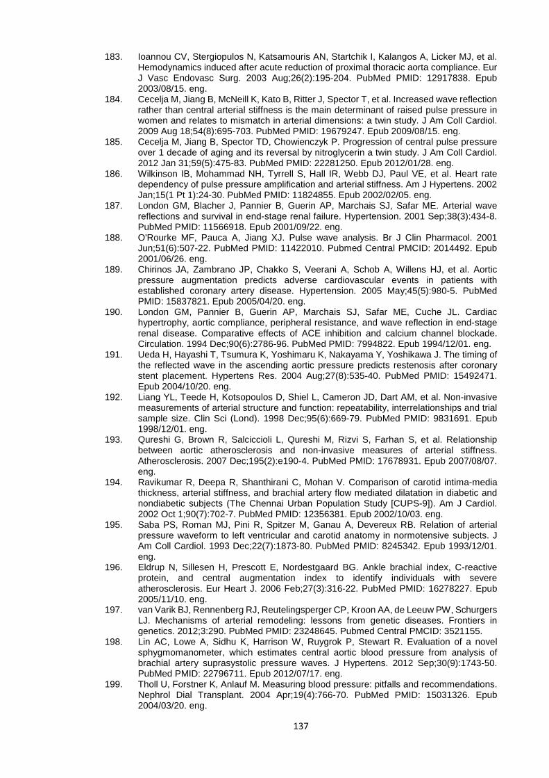
137
183. Ioannou CV, Stergiopulos N, Katsamouris AN, Startchik I, Kalangos A, Licker MJ, et al. Hemodynamics induced after acute reduction of proximal thoracic aorta compliance. Eur J Vasc Endovasc Surg. 2003 Aug;26(2):195-204. PubMed PMID: 12917838. Epub 2003/08/15. eng.
184. Cecelja M, Jiang B, McNeill K, Kato B, Ritter J, Spector T, et al. Increased wave reflection rather than central arterial stiffness is the main determinant of raised pulse pressure in women and relates to mismatch in arterial dimensions: a twin study. J Am Coll Cardiol. 2009 Aug 18;54(8):695-703. PubMed PMID: 19679247. Epub 2009/08/15. eng.
185. Cecelja M, Jiang B, Spector TD, Chowienczyk P. Progression of central pulse pressure over 1 decade of aging and its reversal by nitroglycerin a twin study. J Am Coll Cardiol. 2012 Jan 31;59(5):475-83. PubMed PMID: 22281250. Epub 2012/01/28. eng.
186. Wilkinson IB, Mohammad NH, Tyrrell S, Hall IR, Webb DJ, Paul VE, et al. Heart rate dependency of pulse pressure amplification and arterial stiffness. Am J Hypertens. 2002 Jan;15(1 Pt 1):24-30. PubMed PMID: 11824855. Epub 2002/02/05. eng.
187. London GM, Blacher J, Pannier B, Guerin AP, Marchais SJ, Safar ME. Arterial wave reflections and survival in end-stage renal failure. Hypertension. 2001 Sep;38(3):434-8. PubMed PMID: 11566918. Epub 2001/09/22. eng.
188. O'Rourke MF, Pauca A, Jiang XJ. Pulse wave analysis. Br J Clin Pharmacol. 2001 Jun;51(6):507-22. PubMed PMID: 11422010. Pubmed Central PMCID: 2014492. Epub 2001/06/26. eng.
189. Chirinos JA, Zambrano JP, Chakko S, Veerani A, Schob A, Willens HJ, et al. Aortic pressure augmentation predicts adverse cardiovascular events in patients with established coronary artery disease. Hypertension. 2005 May;45(5):980-5. PubMed PMID: 15837821. Epub 2005/04/20. eng.
190. London GM, Pannier B, Guerin AP, Marchais SJ, Safar ME, Cuche JL. Cardiac hypertrophy, aortic compliance, peripheral resistance, and wave reflection in end-stage renal disease. Comparative effects of ACE inhibition and calcium channel blockade. Circulation. 1994 Dec;90(6):2786-96. PubMed PMID: 7994822. Epub 1994/12/01. eng.
191. Ueda H, Hayashi T, Tsumura K, Yoshimaru K, Nakayama Y, Yoshikawa J. The timing of the reflected wave in the ascending aortic pressure predicts restenosis after coronary stent placement. Hypertens Res. 2004 Aug;27(8):535-40. PubMed PMID: 15492471. Epub 2004/10/20. eng.
192. Liang YL, Teede H, Kotsopoulos D, Shiel L, Cameron JD, Dart AM, et al. Non-invasive measurements of arterial structure and function: repeatability, interrelationships and trial sample size. Clin Sci (Lond). 1998 Dec;95(6):669-79. PubMed PMID: 9831691. Epub 1998/12/01. eng.
193. Qureshi G, Brown R, Salciccioli L, Qureshi M, Rizvi S, Farhan S, et al. Relationship between aortic atherosclerosis and non-invasive measures of arterial stiffness. Atherosclerosis. 2007 Dec;195(2):e190-4. PubMed PMID: 17678931. Epub 2007/08/07. eng.
194. Ravikumar R, Deepa R, Shanthirani C, Mohan V. Comparison of carotid intima-media thickness, arterial stiffness, and brachial artery flow mediated dilatation in diabetic and nondiabetic subjects (The Chennai Urban Population Study [CUPS-9]). Am J Cardiol. 2002 Oct 1;90(7):702-7. PubMed PMID: 12356381. Epub 2002/10/03. eng.
195. Saba PS, Roman MJ, Pini R, Spitzer M, Ganau A, Devereux RB. Relation of arterial pressure waveform to left ventricular and carotid anatomy in normotensive subjects. J Am Coll Cardiol. 1993 Dec;22(7):1873-80. PubMed PMID: 8245342. Epub 1993/12/01. eng.
196. Eldrup N, Sillesen H, Prescott E, Nordestgaard BG. Ankle brachial index, C-reactive protein, and central augmentation index to identify individuals with severe atherosclerosis. Eur Heart J. 2006 Feb;27(3):316-22. PubMed PMID: 16278227. Epub 2005/11/10. eng.
197. van Varik BJ, Rennenberg RJ, Reutelingsperger CP, Kroon AA, de Leeuw PW, Schurgers LJ. Mechanisms of arterial remodeling: lessons from genetic diseases. Frontiers in genetics. 2012;3:290. PubMed PMID: 23248645. Pubmed Central PMCID: 3521155.
198. Lin AC, Lowe A, Sidhu K, Harrison W, Ruygrok P, Stewart R. Evaluation of a novel sphygmomanometer, which estimates central aortic blood pressure from analysis of brachial artery suprasystolic pressure waves. J Hypertens. 2012 Sep;30(9):1743-50. PubMed PMID: 22796711. Epub 2012/07/17. eng.
199. Tholl U, Forstner K, Anlauf M. Measuring blood pressure: pitfalls and recommendations. Nephrol Dial Transplant. 2004 Apr;19(4):766-70. PubMed PMID: 15031326. Epub 2004/03/20. eng.
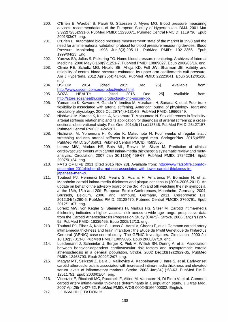
138
200. O'Brien E, Waeber B, Parati G, Staessen J, Myers MG. Blood pressure measuring devices: recommendations of the European Society of Hypertension. BMJ. 2001 Mar 3;322(7285):531-6. PubMed PMID: 11230071. Pubmed Central PMCID: 1119736. Epub 2001/03/07. eng.
201. O'Brien E. Automated blood pressure measurement: state of the market in 1998 and the need for an international validation protocol for blood pressure measuring devices. Blood Pressure Monitoring. 1998 Jun;3(3):205-11. PubMed PMID: 10212355. Epub 1999/04/23. Eng.
202. Yarows SA, Julius S, Pickering TG. Home blood pressure monitoring. Archives of Internal Medicine. 2000 May 8;160(9):1251-7. PubMed PMID: 10809027. Epub 2000/05/16. eng.
203. Climie RE, Schultz MG, Nikolic SB, Ahuja KD, Fell JW, Sharman JE. Validity and reliability of central blood pressure estimated by upper arm oscillometric cuff pressure. Am J Hypertens. 2012 Apr;25(4):414-20. PubMed PMID: 22223041. Epub 2012/01/10. eng.
204. USCOM 2014 [cited 2015 Dec 25]. Available from: http://www.uscom.com.au/product/index.html.
205. SOZA HEALTH [cited 2015 Dec 25]. Available from: http://store.sozahealth.com/products/sh-chp-uscom-bp.
206. Yamamoto K, Kawano H, Gando Y, Iemitsu M, Murakami H, Sanada K, et al. Poor trunk flexibility is associated with arterial stiffening. American journal of physiology Heart and circulatory physiology. 2009 Oct;297(4):H1314-8. PubMed PMID: 19666849.
207. Nishiwaki M, Kurobe K, Kiuchi A, Nakamura T, Matsumoto N. Sex differences in flexibility-arterial stiffness relationship and its application for diagnosis of arterial stiffening: a cross-sectional observational study. Plos One. 2014;9(11):e113646. PubMed PMID: 25427157. Pubmed Central PMCID: 4245207.
208. Nishiwaki M, Yonemura H, Kurobe K, Matsumoto N. Four weeks of regular static stretching reduces arterial stiffness in middle-aged men. SpringerPlus. 2015;4:555. PubMed PMID: 26435901. Pubmed Central PMCID: 4583555.
209. Lorenz MW, Markus HS, Bots ML, Rosvall M, Sitzer M. Prediction of clinical cardiovascular events with carotid intima-media thickness: a systematic review and meta-analysis. Circulation. 2007 Jan 30;115(4):459-67. PubMed PMID: 17242284. Epub 2007/01/24. eng.
210. FATS OF LIFE 2011 [cited 2015 Nov 23]. Available from: http://www.fatsoflife.com/fol-december-2011/higher-dha-not-epa-associated-with-lower-carotid-thickness-in-japanese-men-2/.
211. Touboul PJ, Hennerici MG, Meairs S, Adams H, Amarenco P, Bornstein N, et al. Mannheim carotid intima-media thickness and plaque consensus (2004-2006-2011). An update on behalf of the advisory board of the 3rd, 4th and 5th watching the risk symposia, at the 13th, 15th and 20th European Stroke Conferences, Mannheim, Germany, 2004, Brussels, Belgium, 2006, and Hamburg, Germany, 2011. Cerebrovasc Dis. 2012;34(4):290-6. PubMed PMID: 23128470. Pubmed Central PMCID: 3760791. Epub 2012/11/07. eng.
212. Lorenz MW, von Kegler S, Steinmetz H, Markus HS, Sitzer M. Carotid intima-media thickening indicates a higher vascular risk across a wide age range: prospective data from the Carotid Atherosclerosis Progression Study (CAPS). Stroke. 2006 Jan;37(1):87-92. PubMed PMID: 16339465. Epub 2005/12/13. eng.
213. Touboul PJ, Elbaz A, Koller C, Lucas C, Adrai V, Chedru F, et al. Common carotid artery intima-media thickness and brain infarction : the Etude du Profil Genetique de l'Infarctus Cerebral (GENIC) case-control study. The GENIC Investigators. Circulation. 2000 Jul 18;102(3):313-8. PubMed PMID: 10899095. Epub 2000/07/19. eng.
214. Luedemann J, Schminke U, Berger K, Piek M, Willich SN, Doring A, et al. Association between behavior-dependent cardiovascular risk factors and asymptomatic carotid atherosclerosis in a general population. Stroke. 2002 Dec;33(12):2929-35. PubMed PMID: 12468793. Epub 2002/12/07. eng.
215. Magyar MT, Szikszai Z, Balla J, Valikovics A, Kappelmayer J, Imre S, et al. Early-onset carotid atherosclerosis is associated with increased intima-media thickness and elevated serum levels of inflammatory markers. Stroke. 2003 Jan;34(1):58-63. PubMed PMID: 12511751. Epub 2003/01/04. eng.
216. Vicenzini E, Ricciardi MC, Puccinelli F, Altieri M, Vanacore N, Di Piero V, et al. Common carotid artery intima-media thickness determinants in a population study. J Ultras Med. 2007 Apr;26(4):427-32. PubMed PMID: WOS:000245166400002. English.
217. . !!! INVALID CITATION !!!
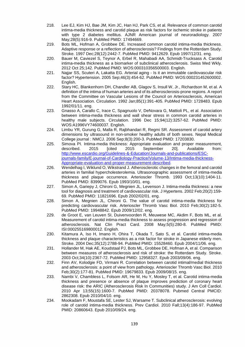
139
218. Lee EJ, Kim HJ, Bae JM, Kim JC, Han HJ, Park CS, et al. Relevance of common carotid intima-media thickness and carotid plaque as risk factors for ischemic stroke in patients with type 2 diabetes mellitus. AJNR American journal of neuroradiology. 2007 May;28(5):916-9. PubMed PMID: 17494669.
219. Bots ML, Hofman A, Grobbee DE. Increased common carotid intima-media thickness. Adaptive response or a reflection of atherosclerosis? Findings from the Rotterdam Study. Stroke. 1997 Dec;28(12):2442-7. PubMed PMID: 9412629. Epub 1997/12/31. eng.
220. Bauer M, Caviezel S, Teynor A, Erbel R, Mahabadi AA, Schmidt-Trucksass A. Carotid intima-media thickness as a biomarker of subclinical atherosclerosis. Swiss Med Wkly. 2012 Oct 25;142. PubMed PMID: WOS:000310356500003. English.
221. Najjar SS, Scuteri A, Lakatta EG. Arterial aging - Is it an immutable cardiovascular risk factor? Hypertension. 2005 Sep;46(3):454-62. PubMed PMID: WOS:000231452600002. English.
222. Stary HC, Blankenhorn DH, Chandler AB, Glagov S, Insull W, Jr., Richardson M, et al. A definition of the intima of human arteries and of its atherosclerosis-prone regions. A report from the Committee on Vascular Lesions of the Council on Arteriosclerosis, American Heart Association. Circulation. 1992 Jan;85(1):391-405. PubMed PMID: 1728483. Epub 1992/01/11. eng.
223. Gnasso A, Carallo C, Irace C, Spagnuolo V, DeNovara G, Mattioli PL, et al. Association between intima-media thickness and wall shear stress in common carotid arteries in healthy male subjects. Circulation. 1996 Dec 15;94(12):3257-62. PubMed PMID: WOS:A1996VY74600037. English.
224. Limbu YR, Gurung G, Malla R, Rajbhandari R, Regmi SR. Assessment of carotid artery dimensions by ultrasound in non-smoker healthy adults of both sexes. Nepal Medical College journal : NMCJ. 2006 Sep;8(3):200-3. PubMed PMID: 17203830.
225. Simova PI. Intima-media thickness: Appropriate evaluation and proper measurement, described. 2015 [cited 2015 September 20]. Available from: http://www.escardio.org/Guidelines-&-Education/Journals-and-publications/ESC-journals-family/E-journal-of-Cardiology-Practice/Volume-13/Intima-media-thickness-Appropriate-evaluation-and-proper-measurement-described.
226. Wendelhag I, Wiklund O, Wikstrand J. Atherosclerotic changes in the femoral and carotid arteries in familial hypercholesterolemia. Ultrasonographic assessment of intima-media thickness and plaque occurrence. Arterioscler Thromb. 1993 Oct;13(10):1404-11. PubMed PMID: 8399076. Epub 1993/10/01. eng.
227. Simon A, Gariepy J, Chironi G, Megnien JL, Levenson J. Intima-media thickness: a new tool for diagnosis and treatment of cardiovascular risk. J Hypertens. 2002 Feb;20(2):159-69. PubMed PMID: 11821696. Epub 2002/02/01. eng.
228. Simon A, Megnien JL, Chironi G. The value of carotid intima-media thickness for predicting cardiovascular risk. Arterioscler Thromb Vasc Biol. 2010 Feb;30(2):182-5. PubMed PMID: 19948842. Epub 2009/12/02. eng.
229. de Groot E, van Leuven SI, Duivenvoorden R, Meuwese MC, Akdim F, Bots ML, et al. Measurement of carotid intima-media thickness to assess progression and regression of atherosclerosis. Nat Clin Pract Card. 2008 May;5(5):280-8. PubMed PMID: ISI:000255169800012. English.
230. Kitamura A, Iso H, Imano H, Ohira T, Okada T, Sato S, et al. Carotid intima-media thickness and plaque characteristics as a risk factor for stroke in Japanese elderly men. Stroke. 2004 Dec;35(12):2788-94. PubMed PMID: 15528460. Epub 2004/11/06. eng.
231. Hollander M, Hak AE, Koudstaal PJ, Bots ML, Grobbee DE, Hofman A, et al. Comparison between measures of atherosclerosis and risk of stroke: the Rotterdam Study. Stroke. 2003 Oct;34(10):2367-72. PubMed PMID: 12958327. Epub 2003/09/06. eng.
232. Finn AV, Kolodgie FD, Virmani R. Correlation between carotid intimal/medial thickness and atherosclerosis: a point of view from pathology. Arterioscler Thromb Vasc Biol. 2010 Feb;30(2):177-81. PubMed PMID: 19679833. Epub 2009/08/15. eng.
233. Nambi V, Chambless L, Folsom AR, He M, Hu Y, Mosley T, et al. Carotid intima-media thickness and presence or absence of plaque improves prediction of coronary heart disease risk: the ARIC (Atherosclerosis Risk In Communities) study. J Am Coll Cardiol. 2010 Apr 13;55(15):1600-7. PubMed PMID: 20378078. Pubmed Central PMCID: 2862308. Epub 2010/04/10. eng.
234. Mookadam F, Moustafa SE, Lester SJ, Warsame T. Subclinical atherosclerosis: evolving role of carotid intima-media thickness. Prev Cardiol. 2010 Fall;13(4):186-97. PubMed PMID: 20860643. Epub 2010/09/24. eng.
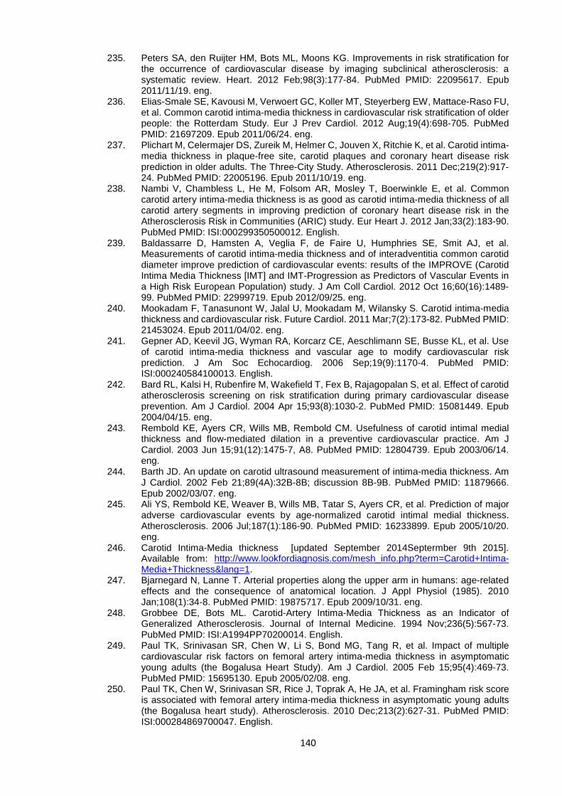
140
235. Peters SA, den Ruijter HM, Bots ML, Moons KG. Improvements in risk stratification for the occurrence of cardiovascular disease by imaging subclinical atherosclerosis: a systematic review. Heart. 2012 Feb;98(3):177-84. PubMed PMID: 22095617. Epub 2011/11/19. eng.
236. Elias-Smale SE, Kavousi M, Verwoert GC, Koller MT, Steyerberg EW, Mattace-Raso FU, et al. Common carotid intima-media thickness in cardiovascular risk stratification of older people: the Rotterdam Study. Eur J Prev Cardiol. 2012 Aug;19(4):698-705. PubMed PMID: 21697209. Epub 2011/06/24. eng.
237. Plichart M, Celermajer DS, Zureik M, Helmer C, Jouven X, Ritchie K, et al. Carotid intima-media thickness in plaque-free site, carotid plaques and coronary heart disease risk prediction in older adults. The Three-City Study. Atherosclerosis. 2011 Dec;219(2):917-24. PubMed PMID: 22005196. Epub 2011/10/19. eng.
238. Nambi V, Chambless L, He M, Folsom AR, Mosley T, Boerwinkle E, et al. Common carotid artery intima-media thickness is as good as carotid intima-media thickness of all carotid artery segments in improving prediction of coronary heart disease risk in the Atherosclerosis Risk in Communities (ARIC) study. Eur Heart J. 2012 Jan;33(2):183-90. PubMed PMID: ISI:000299350500012. English.
239. Baldassarre D, Hamsten A, Veglia F, de Faire U, Humphries SE, Smit AJ, et al. Measurements of carotid intima-media thickness and of interadventitia common carotid diameter improve prediction of cardiovascular events: results of the IMPROVE (Carotid Intima Media Thickness [IMT] and IMT-Progression as Predictors of Vascular Events in a High Risk European Population) study. J Am Coll Cardiol. 2012 Oct 16;60(16):1489-99. PubMed PMID: 22999719. Epub 2012/09/25. eng.
240. Mookadam F, Tanasunont W, Jalal U, Mookadam M, Wilansky S. Carotid intima-media thickness and cardiovascular risk. Future Cardiol. 2011 Mar;7(2):173-82. PubMed PMID: 21453024. Epub 2011/04/02. eng.
241. Gepner AD, Keevil JG, Wyman RA, Korcarz CE, Aeschlimann SE, Busse KL, et al. Use of carotid intima-media thickness and vascular age to modify cardiovascular risk prediction. J Am Soc Echocardiog. 2006 Sep;19(9):1170-4. PubMed PMID: ISI:000240584100013. English.
242. Bard RL, Kalsi H, Rubenfire M, Wakefield T, Fex B, Rajagopalan S, et al. Effect of carotid atherosclerosis screening on risk stratification during primary cardiovascular disease prevention. Am J Cardiol. 2004 Apr 15;93(8):1030-2. PubMed PMID: 15081449. Epub 2004/04/15. eng.
243. Rembold KE, Ayers CR, Wills MB, Rembold CM. Usefulness of carotid intimal medial thickness and flow-mediated dilation in a preventive cardiovascular practice. Am J Cardiol. 2003 Jun 15;91(12):1475-7, A8. PubMed PMID: 12804739. Epub 2003/06/14. eng.
244. Barth JD. An update on carotid ultrasound measurement of intima-media thickness. Am J Cardiol. 2002 Feb 21;89(4A):32B-8B; discussion 8B-9B. PubMed PMID: 11879666. Epub 2002/03/07. eng.
245. Ali YS, Rembold KE, Weaver B, Wills MB, Tatar S, Ayers CR, et al. Prediction of major adverse cardiovascular events by age-normalized carotid intimal medial thickness. Atherosclerosis. 2006 Jul;187(1):186-90. PubMed PMID: 16233899. Epub 2005/10/20. eng.
246. Carotid Intima-Media thickness [updated September 2014Septermber 9th 2015]. Available from: http://www.lookfordiagnosis.com/mesh_info.php?term=Carotid+Intima-Media+Thickness&lang=1.
247. Bjarnegard N, Lanne T. Arterial properties along the upper arm in humans: age-related effects and the consequence of anatomical location. J Appl Physiol (1985). 2010 Jan;108(1):34-8. PubMed PMID: 19875717. Epub 2009/10/31. eng.
248. Grobbee DE, Bots ML. Carotid-Artery Intima-Media Thickness as an Indicator of Generalized Atherosclerosis. Journal of Internal Medicine. 1994 Nov;236(5):567-73. PubMed PMID: ISI:A1994PP70200014. English.
249. Paul TK, Srinivasan SR, Chen W, Li S, Bond MG, Tang R, et al. Impact of multiple cardiovascular risk factors on femoral artery intima-media thickness in asymptomatic young adults (the Bogalusa Heart Study). Am J Cardiol. 2005 Feb 15;95(4):469-73. PubMed PMID: 15695130. Epub 2005/02/08. eng.
250. Paul TK, Chen W, Srinivasan SR, Rice J, Toprak A, He JA, et al. Framingham risk score is associated with femoral artery intima-media thickness in asymptomatic young adults (the Bogalusa heart study). Atherosclerosis. 2010 Dec;213(2):627-31. PubMed PMID: ISI:000284869700047. English.
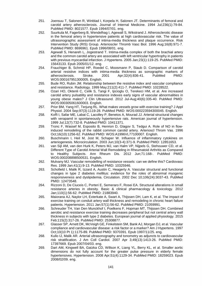
141
251. Joensuu T, Salonen R, Winblad I, Korpela H, Salonen JT. Determinants of femoral and carotid artery atherosclerosis. Journal of Internal Medicine. 1994 Jul;236(1):79-84. PubMed PMID: 8021577. Epub 1994/07/01. eng.
252. Suurkula M, Fagerberg B, Wendelhag I, Agewall S, Wikstrand J. Atherosclerotic disease in the femoral artery in hypertensive patients at high cardiovascular risk. The value of ultrasonographic assessment of intima-media thickness and plaque occurrence. Risk Intervention Study (RIS) Group. Arterioscler Thromb Vasc Biol. 1996 Aug;16(8):971-7. PubMed PMID: 8696961. Epub 1996/08/01. eng.
253. Agewall S, Henareh L, Jogestrand T. Intima-media complex of both the brachial artery and the common carotid artery are associated with left ventricular hypertrophy in patients with previous myocardial infarction. J Hypertens. 2005 Jan;23(1):119-25. PubMed PMID: 15643133. Epub 2005/01/12. eng.
254. Frauchiger B, Schmid HP, Roedel C, Moosmann P, Staub D. Comparison of carotid arterial resistive indices with intima-media thickness as sonographic markers of atherosclerosis. Stroke. 2001 Apr;32(4):836-41. PubMed PMID: WOS:000167951300005. English.
255. Bude RO, Rubin JM. Relationship between the resistive index and vascular compliance and resistance. Radiology. 1999 May;211(2):411-7. PubMed PMID: 10228522.
256. Ozari HO, Oktenli C, Celik S, Tangi F, Ipcioglu O, Terekeci HM, et al. Are increased carotid artery pulsatility and resistance indexes early signs of vascular abnormalities in young obese males? J Clin Ultrasound. 2012 Jul-Aug;40(6):335-40. PubMed PMID: WOS:000305061600003. English.
257. Prior BM, Yang HT, Terjung RL. What makes vessels grow with exercise training? J Appl Physiol. 2004 Sep;97(3):1119-28. PubMed PMID: WOS:000223594300045. English.
258. Koffi I, Safar ME, Labat C, Lacolley P, Benetos A, Mourad JJ. Arterial structural changes with verapamil in spontaneously hypertensive rats. American journal of hypertension. 1999 Jul;12(7):732-8. PubMed PMID: 10411371.
259. Tronc F, Wassef M, Esposito B, Henrion D, Glagov S, Tedgui A. Role of NO in flow-induced remodeling of the rabbit common carotid artery. Arterioscl Throm Vas. 1996 Oct;16(10):1256-62. PubMed PMID: WOS:A1996VL77100007. English.
260. Buschmann I, Heil M, Jost M, Schaper W. Influence of inflammatory cytokines on arteriogenesis. Microcirculation. 2003 Jun;10(3-4):371-9. PubMed PMID: 12851653.
261. van Sijl AM, van den Hurk K, Peters MJ, van Halm VP, Nijpels G, Stehouwer CD, et al. Different Type of Carotid Arterial Wall Remodeling in Rheumatoid Arthritis as Compared to Healthy Subjects. Ann Rheum Dis. 2012 Jun;71:168-. PubMed PMID: WOS:000208898500531. English.
262. Mulvany MJ. Vascular remodelling of resistance vessels: can we define this? Cardiovasc Res. 1999 Jan;41(1):9-13. PubMed PMID: 10325946.
263. Schofield I, Malik R, Izzard A, Austin C, Heagerty A. Vascular structural and functional changes in type 2 diabetes mellitus: evidence for the roles of abnormal myogenic responsiveness and dyslipidemia. Circulation. 2002 Dec 10;106(24):3037-43. PubMed PMID: 12473548.
264. Rizzoni D, De Ciuceis C, Porteri E, Semeraro F, Rosei EA. Structural alterations in small resistance arteries in obesity. Basic & clinical pharmacology & toxicology. 2012 Jan;110(1):56-62. PubMed PMID: 21883940.
265. Maiorana AJ, Naylor LH, Exterkate A, Swart A, Thijssen DH, Lam K, et al. The impact of exercise training on conduit artery wall thickness and remodeling in chronic heart failure patients. Hypertension. 2011 Jan;57(1):56-62. PubMed PMID: 21059991.
266. Schreuder TH, Van Den Munckhof I, Poelkens F, Hopman MT, Thijssen DH. Combined aerobic and resistance exercise training decreases peripheral but not central artery wall thickness in subjects with type 2 diabetes. European journal of applied physiology. 2015 Feb;115(2):317-26. PubMed PMID: 25308877.
267. Glasser SP, Arnett DK, McVeigh GE, Finkelstein SM, Bank AJ, Morgan DJ, et al. Vascular compliance and cardiovascular disease: a risk factor or a marker? Am J Hypertens. 1997 Oct;10(10 Pt 1):1175-89. PubMed PMID: 9370391. Epub 1997/11/25. eng.
268. Kullo IJ, Malik AR. Arterial ultrasonography and tonometry as adjuncts to cardiovascular risk stratification. J Am Coll Cardiol. 2007 Apr 3;49(13):1413-26. PubMed PMID: 17397669. Epub 2007/04/03. eng.
269. Dart AM, Kingwell BA, Gatzka CD, Willson K, Liang YL, Berry KL, et al. Smaller aortic dimensions do not fully account for the greater pulse pressure in elderly female hypertensives. Hypertension. 2008 Apr;51(4):1129-34. PubMed PMID: 18259023. Epub 2008/02/09. eng.
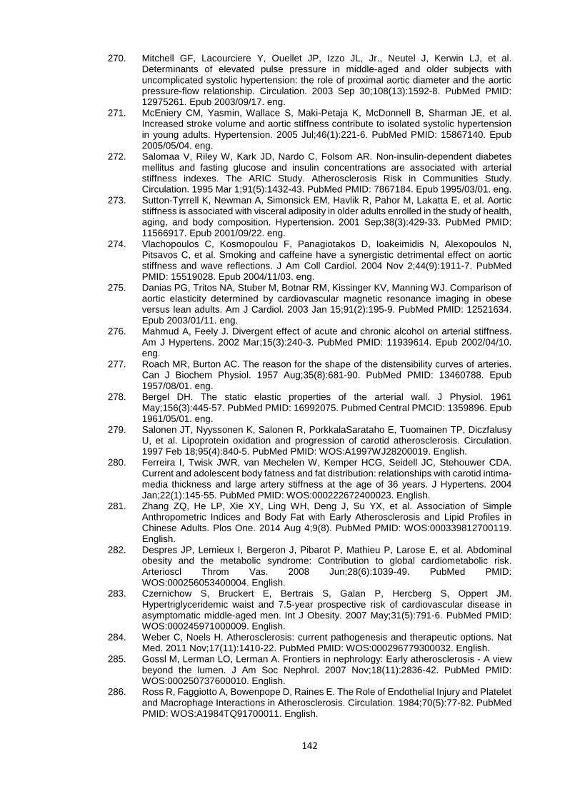
142
270. Mitchell GF, Lacourciere Y, Ouellet JP, Izzo JL, Jr., Neutel J, Kerwin LJ, et al. Determinants of elevated pulse pressure in middle-aged and older subjects with uncomplicated systolic hypertension: the role of proximal aortic diameter and the aortic pressure-flow relationship. Circulation. 2003 Sep 30;108(13):1592-8. PubMed PMID: 12975261. Epub 2003/09/17. eng.
271. McEniery CM, Yasmin, Wallace S, Maki-Petaja K, McDonnell B, Sharman JE, et al. Increased stroke volume and aortic stiffness contribute to isolated systolic hypertension in young adults. Hypertension. 2005 Jul;46(1):221-6. PubMed PMID: 15867140. Epub 2005/05/04. eng.
272. Salomaa V, Riley W, Kark JD, Nardo C, Folsom AR. Non-insulin-dependent diabetes mellitus and fasting glucose and insulin concentrations are associated with arterial stiffness indexes. The ARIC Study. Atherosclerosis Risk in Communities Study. Circulation. 1995 Mar 1;91(5):1432-43. PubMed PMID: 7867184. Epub 1995/03/01. eng.
273. Sutton-Tyrrell K, Newman A, Simonsick EM, Havlik R, Pahor M, Lakatta E, et al. Aortic stiffness is associated with visceral adiposity in older adults enrolled in the study of health, aging, and body composition. Hypertension. 2001 Sep;38(3):429-33. PubMed PMID: 11566917. Epub 2001/09/22. eng.
274. Vlachopoulos C, Kosmopoulou F, Panagiotakos D, Ioakeimidis N, Alexopoulos N, Pitsavos C, et al. Smoking and caffeine have a synergistic detrimental effect on aortic stiffness and wave reflections. J Am Coll Cardiol. 2004 Nov 2;44(9):1911-7. PubMed PMID: 15519028. Epub 2004/11/03. eng.
275. Danias PG, Tritos NA, Stuber M, Botnar RM, Kissinger KV, Manning WJ. Comparison of aortic elasticity determined by cardiovascular magnetic resonance imaging in obese versus lean adults. Am J Cardiol. 2003 Jan 15;91(2):195-9. PubMed PMID: 12521634. Epub 2003/01/11. eng.
276. Mahmud A, Feely J. Divergent effect of acute and chronic alcohol on arterial stiffness. Am J Hypertens. 2002 Mar;15(3):240-3. PubMed PMID: 11939614. Epub 2002/04/10. eng.
277. Roach MR, Burton AC. The reason for the shape of the distensibility curves of arteries. Can J Biochem Physiol. 1957 Aug;35(8):681-90. PubMed PMID: 13460788. Epub 1957/08/01. eng.
278. Bergel DH. The static elastic properties of the arterial wall. J Physiol. 1961 May;156(3):445-57. PubMed PMID: 16992075. Pubmed Central PMCID: 1359896. Epub 1961/05/01. eng.
279. Salonen JT, Nyyssonen K, Salonen R, PorkkalaSarataho E, Tuomainen TP, Diczfalusy U, et al. Lipoprotein oxidation and progression of carotid atherosclerosis. Circulation. 1997 Feb 18;95(4):840-5. PubMed PMID: WOS:A1997WJ28200019. English.
280. Ferreira I, Twisk JWR, van Mechelen W, Kemper HCG, Seidell JC, Stehouwer CDA. Current and adolescent body fatness and fat distribution: relationships with carotid intima-media thickness and large artery stiffness at the age of 36 years. J Hypertens. 2004 Jan;22(1):145-55. PubMed PMID: WOS:000222672400023. English.
281. Zhang ZQ, He LP, Xie XY, Ling WH, Deng J, Su YX, et al. Association of Simple Anthropometric Indices and Body Fat with Early Atherosclerosis and Lipid Profiles in Chinese Adults. Plos One. 2014 Aug 4;9(8). PubMed PMID: WOS:000339812700119. English.
282. Despres JP, Lemieux I, Bergeron J, Pibarot P, Mathieu P, Larose E, et al. Abdominal obesity and the metabolic syndrome: Contribution to global cardiometabolic risk. Arterioscl Throm Vas. 2008 Jun;28(6):1039-49. PubMed PMID: WOS:000256053400004. English.
283. Czernichow S, Bruckert E, Bertrais S, Galan P, Hercberg S, Oppert JM. Hypertriglyceridemic waist and 7.5-year prospective risk of cardiovascular disease in asymptomatic middle-aged men. Int J Obesity. 2007 May;31(5):791-6. PubMed PMID: WOS:000245971000009. English.
284. Weber C, Noels H. Atherosclerosis: current pathogenesis and therapeutic options. Nat Med. 2011 Nov;17(11):1410-22. PubMed PMID: WOS:000296779300032. English.
285. Gossl M, Lerman LO, Lerman A. Frontiers in nephrology: Early atherosclerosis - A view beyond the lumen. J Am Soc Nephrol. 2007 Nov;18(11):2836-42. PubMed PMID: WOS:000250737600010. English.
286. Ross R, Faggiotto A, Bowenpope D, Raines E. The Role of Endothelial Injury and Platelet and Macrophage Interactions in Atherosclerosis. Circulation. 1984;70(5):77-82. PubMed PMID: WOS:A1984TQ91700011. English.
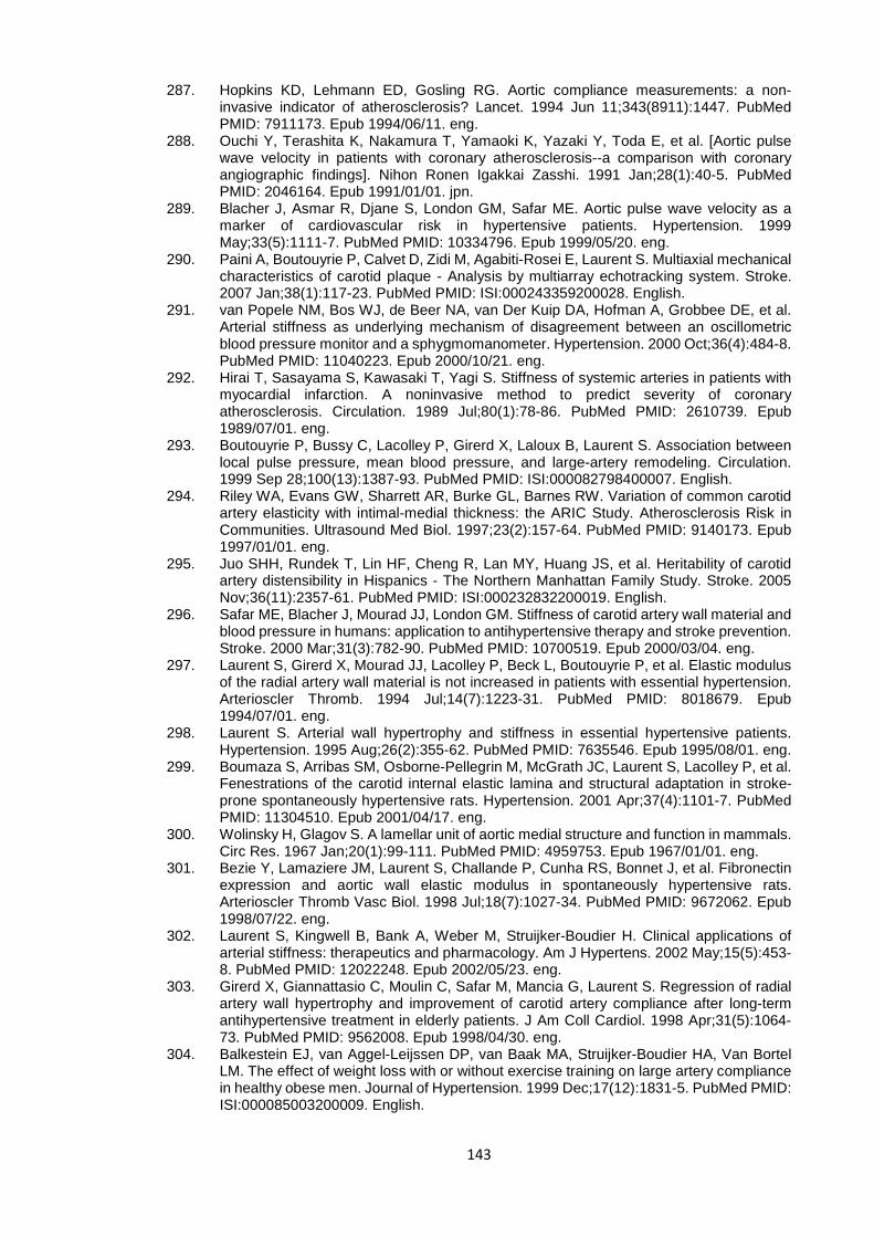
143
287. Hopkins KD, Lehmann ED, Gosling RG. Aortic compliance measurements: a non-invasive indicator of atherosclerosis? Lancet. 1994 Jun 11;343(8911):1447. PubMed PMID: 7911173. Epub 1994/06/11. eng.
288. Ouchi Y, Terashita K, Nakamura T, Yamaoki K, Yazaki Y, Toda E, et al. [Aortic pulse wave velocity in patients with coronary atherosclerosis--a comparison with coronary angiographic findings]. Nihon Ronen Igakkai Zasshi. 1991 Jan;28(1):40-5. PubMed PMID: 2046164. Epub 1991/01/01. jpn.
289. Blacher J, Asmar R, Djane S, London GM, Safar ME. Aortic pulse wave velocity as a marker of cardiovascular risk in hypertensive patients. Hypertension. 1999 May;33(5):1111-7. PubMed PMID: 10334796. Epub 1999/05/20. eng.
290. Paini A, Boutouyrie P, Calvet D, Zidi M, Agabiti-Rosei E, Laurent S. Multiaxial mechanical characteristics of carotid plaque - Analysis by multiarray echotracking system. Stroke. 2007 Jan;38(1):117-23. PubMed PMID: ISI:000243359200028. English.
291. van Popele NM, Bos WJ, de Beer NA, van Der Kuip DA, Hofman A, Grobbee DE, et al. Arterial stiffness as underlying mechanism of disagreement between an oscillometric blood pressure monitor and a sphygmomanometer. Hypertension. 2000 Oct;36(4):484-8. PubMed PMID: 11040223. Epub 2000/10/21. eng.
292. Hirai T, Sasayama S, Kawasaki T, Yagi S. Stiffness of systemic arteries in patients with myocardial infarction. A noninvasive method to predict severity of coronary atherosclerosis. Circulation. 1989 Jul;80(1):78-86. PubMed PMID: 2610739. Epub 1989/07/01. eng.
293. Boutouyrie P, Bussy C, Lacolley P, Girerd X, Laloux B, Laurent S. Association between local pulse pressure, mean blood pressure, and large-artery remodeling. Circulation. 1999 Sep 28;100(13):1387-93. PubMed PMID: ISI:000082798400007. English.
294. Riley WA, Evans GW, Sharrett AR, Burke GL, Barnes RW. Variation of common carotid artery elasticity with intimal-medial thickness: the ARIC Study. Atherosclerosis Risk in Communities. Ultrasound Med Biol. 1997;23(2):157-64. PubMed PMID: 9140173. Epub 1997/01/01. eng.
295. Juo SHH, Rundek T, Lin HF, Cheng R, Lan MY, Huang JS, et al. Heritability of carotid artery distensibility in Hispanics - The Northern Manhattan Family Study. Stroke. 2005 Nov;36(11):2357-61. PubMed PMID: ISI:000232832200019. English.
296. Safar ME, Blacher J, Mourad JJ, London GM. Stiffness of carotid artery wall material and blood pressure in humans: application to antihypertensive therapy and stroke prevention. Stroke. 2000 Mar;31(3):782-90. PubMed PMID: 10700519. Epub 2000/03/04. eng.
297. Laurent S, Girerd X, Mourad JJ, Lacolley P, Beck L, Boutouyrie P, et al. Elastic modulus of the radial artery wall material is not increased in patients with essential hypertension. Arterioscler Thromb. 1994 Jul;14(7):1223-31. PubMed PMID: 8018679. Epub 1994/07/01. eng.
298. Laurent S. Arterial wall hypertrophy and stiffness in essential hypertensive patients. Hypertension. 1995 Aug;26(2):355-62. PubMed PMID: 7635546. Epub 1995/08/01. eng.
299. Boumaza S, Arribas SM, Osborne-Pellegrin M, McGrath JC, Laurent S, Lacolley P, et al. Fenestrations of the carotid internal elastic lamina and structural adaptation in stroke-prone spontaneously hypertensive rats. Hypertension. 2001 Apr;37(4):1101-7. PubMed PMID: 11304510. Epub 2001/04/17. eng.
300. Wolinsky H, Glagov S. A lamellar unit of aortic medial structure and function in mammals. Circ Res. 1967 Jan;20(1):99-111. PubMed PMID: 4959753. Epub 1967/01/01. eng.
301. Bezie Y, Lamaziere JM, Laurent S, Challande P, Cunha RS, Bonnet J, et al. Fibronectin expression and aortic wall elastic modulus in spontaneously hypertensive rats. Arterioscler Thromb Vasc Biol. 1998 Jul;18(7):1027-34. PubMed PMID: 9672062. Epub 1998/07/22. eng.
302. Laurent S, Kingwell B, Bank A, Weber M, Struijker-Boudier H. Clinical applications of arterial stiffness: therapeutics and pharmacology. Am J Hypertens. 2002 May;15(5):453-8. PubMed PMID: 12022248. Epub 2002/05/23. eng.
303. Girerd X, Giannattasio C, Moulin C, Safar M, Mancia G, Laurent S. Regression of radial artery wall hypertrophy and improvement of carotid artery compliance after long-term antihypertensive treatment in elderly patients. J Am Coll Cardiol. 1998 Apr;31(5):1064-73. PubMed PMID: 9562008. Epub 1998/04/30. eng.
304. Balkestein EJ, van Aggel-Leijssen DP, van Baak MA, Struijker-Boudier HA, Van Bortel LM. The effect of weight loss with or without exercise training on large artery compliance in healthy obese men. Journal of Hypertension. 1999 Dec;17(12):1831-5. PubMed PMID: ISI:000085003200009. English.
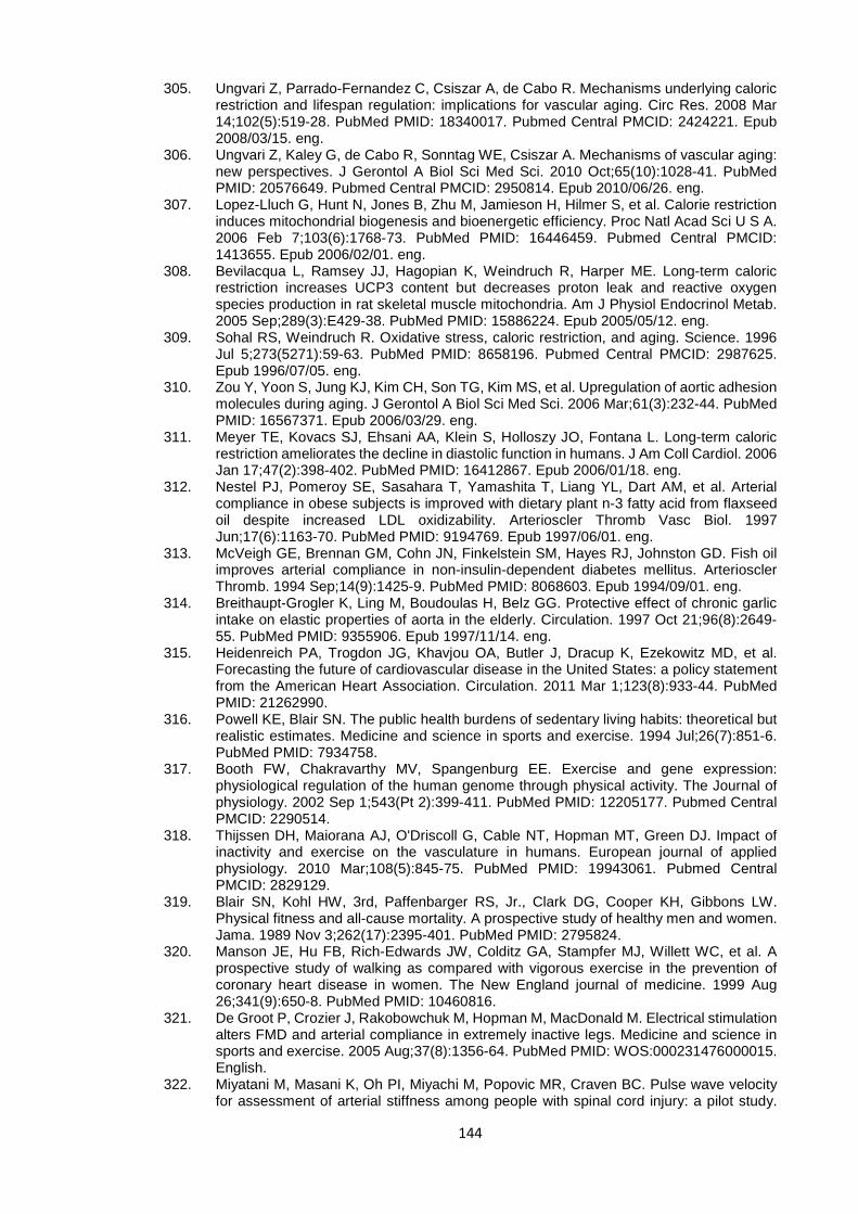
144
305. Ungvari Z, Parrado-Fernandez C, Csiszar A, de Cabo R. Mechanisms underlying caloric restriction and lifespan regulation: implications for vascular aging. Circ Res. 2008 Mar 14;102(5):519-28. PubMed PMID: 18340017. Pubmed Central PMCID: 2424221. Epub 2008/03/15. eng.
306. Ungvari Z, Kaley G, de Cabo R, Sonntag WE, Csiszar A. Mechanisms of vascular aging: new perspectives. J Gerontol A Biol Sci Med Sci. 2010 Oct;65(10):1028-41. PubMed PMID: 20576649. Pubmed Central PMCID: 2950814. Epub 2010/06/26. eng.
307. Lopez-Lluch G, Hunt N, Jones B, Zhu M, Jamieson H, Hilmer S, et al. Calorie restriction induces mitochondrial biogenesis and bioenergetic efficiency. Proc Natl Acad Sci U S A. 2006 Feb 7;103(6):1768-73. PubMed PMID: 16446459. Pubmed Central PMCID: 1413655. Epub 2006/02/01. eng.
308. Bevilacqua L, Ramsey JJ, Hagopian K, Weindruch R, Harper ME. Long-term caloric restriction increases UCP3 content but decreases proton leak and reactive oxygen species production in rat skeletal muscle mitochondria. Am J Physiol Endocrinol Metab. 2005 Sep;289(3):E429-38. PubMed PMID: 15886224. Epub 2005/05/12. eng.
309. Sohal RS, Weindruch R. Oxidative stress, caloric restriction, and aging. Science. 1996 Jul 5;273(5271):59-63. PubMed PMID: 8658196. Pubmed Central PMCID: 2987625. Epub 1996/07/05. eng.
310. Zou Y, Yoon S, Jung KJ, Kim CH, Son TG, Kim MS, et al. Upregulation of aortic adhesion molecules during aging. J Gerontol A Biol Sci Med Sci. 2006 Mar;61(3):232-44. PubMed PMID: 16567371. Epub 2006/03/29. eng.
311. Meyer TE, Kovacs SJ, Ehsani AA, Klein S, Holloszy JO, Fontana L. Long-term caloric restriction ameliorates the decline in diastolic function in humans. J Am Coll Cardiol. 2006 Jan 17;47(2):398-402. PubMed PMID: 16412867. Epub 2006/01/18. eng.
312. Nestel PJ, Pomeroy SE, Sasahara T, Yamashita T, Liang YL, Dart AM, et al. Arterial compliance in obese subjects is improved with dietary plant n-3 fatty acid from flaxseed oil despite increased LDL oxidizability. Arterioscler Thromb Vasc Biol. 1997 Jun;17(6):1163-70. PubMed PMID: 9194769. Epub 1997/06/01. eng.
313. McVeigh GE, Brennan GM, Cohn JN, Finkelstein SM, Hayes RJ, Johnston GD. Fish oil improves arterial compliance in non-insulin-dependent diabetes mellitus. Arterioscler Thromb. 1994 Sep;14(9):1425-9. PubMed PMID: 8068603. Epub 1994/09/01. eng.
314. Breithaupt-Grogler K, Ling M, Boudoulas H, Belz GG. Protective effect of chronic garlic intake on elastic properties of aorta in the elderly. Circulation. 1997 Oct 21;96(8):2649-55. PubMed PMID: 9355906. Epub 1997/11/14. eng.
315. Heidenreich PA, Trogdon JG, Khavjou OA, Butler J, Dracup K, Ezekowitz MD, et al. Forecasting the future of cardiovascular disease in the United States: a policy statement from the American Heart Association. Circulation. 2011 Mar 1;123(8):933-44. PubMed PMID: 21262990.
316. Powell KE, Blair SN. The public health burdens of sedentary living habits: theoretical but realistic estimates. Medicine and science in sports and exercise. 1994 Jul;26(7):851-6. PubMed PMID: 7934758.
317. Booth FW, Chakravarthy MV, Spangenburg EE. Exercise and gene expression: physiological regulation of the human genome through physical activity. The Journal of physiology. 2002 Sep 1;543(Pt 2):399-411. PubMed PMID: 12205177. Pubmed Central PMCID: 2290514.
318. Thijssen DH, Maiorana AJ, O'Driscoll G, Cable NT, Hopman MT, Green DJ. Impact of inactivity and exercise on the vasculature in humans. European journal of applied physiology. 2010 Mar;108(5):845-75. PubMed PMID: 19943061. Pubmed Central PMCID: 2829129.
319. Blair SN, Kohl HW, 3rd, Paffenbarger RS, Jr., Clark DG, Cooper KH, Gibbons LW. Physical fitness and all-cause mortality. A prospective study of healthy men and women. Jama. 1989 Nov 3;262(17):2395-401. PubMed PMID: 2795824.
320. Manson JE, Hu FB, Rich-Edwards JW, Colditz GA, Stampfer MJ, Willett WC, et al. A prospective study of walking as compared with vigorous exercise in the prevention of coronary heart disease in women. The New England journal of medicine. 1999 Aug 26;341(9):650-8. PubMed PMID: 10460816.
321. De Groot P, Crozier J, Rakobowchuk M, Hopman M, MacDonald M. Electrical stimulation alters FMD and arterial compliance in extremely inactive legs. Medicine and science in sports and exercise. 2005 Aug;37(8):1356-64. PubMed PMID: WOS:000231476000015. English.
322. Miyatani M, Masani K, Oh PI, Miyachi M, Popovic MR, Craven BC. Pulse wave velocity for assessment of arterial stiffness among people with spinal cord injury: a pilot study.
145
The journal of spinal cord medicine. 2009;32(1):72-8. PubMed PMID: 19264052. Pubmed Central PMCID: 2647504.
323. Yeboah J, Crouse JR, Hsu FC, Burke GL, Herrington DM. Brachial flow-mediated dilation predicts incident cardiovascular events in older adults: the Cardiovascular Health Study. Circulation. 2007 May 8;115(18):2390-7. PubMed PMID: 17452608.
324. Thijssen DH, Rongen GA, Smits P, Hopman MT. Physical (in)activity and endothelium-derived constricting factors: overlooked adaptations. The Journal of physiology. 2008 Jan 15;586(2):319-24. PubMed PMID: 17962322. Pubmed Central PMCID: 2375603.
325. Bleeker MW, De Groot PC, Rongen GA, Rittweger J, Felsenberg D, Smits P, et al. Vascular adaptation to deconditioning and the effect of an exercise countermeasure: results of the Berlin Bed Rest study. J Appl Physiol (1985). 2005 Oct;99(4):1293-300. PubMed PMID: 15932956.
326. Newcomer SC, Thijssen DH, Green DJ. Effects of exercise on endothelium and endothelium/smooth muscle cross talk: role of exercise-induced hemodynamics. J Appl Physiol (1985). 2011 Jul;111(1):311-20. PubMed PMID: 21436465.
327. Cook JN, DeVan AE, Schleifer JL, Anton MM, Cortez-Cooper MY, Tanaka H. Arterial compliance of rowers: implications for combined aerobic and strength training on arterial elasticity. American journal of physiology Heart and circulatory physiology. 2006 Apr;290(4):H1596-600. PubMed PMID: 16284225.
328. Little JP, Safdar A, Wilkin GP, Tarnopolsky MA, Gibala MJ. A practical model of low-volume high-intensity interval training induces mitochondrial biogenesis in human skeletal muscle: potential mechanisms. The Journal of physiology. 2010 Mar 15;588(Pt 6):1011-22. PubMed PMID: 20100740. Pubmed Central PMCID: 2849965.
329. Shiraev T, Barclay G. Evidence based exercise - clinical benefits of high intensity interval training. Australian family physician. 2012 Dec;41(12):960-2. PubMed PMID: 23210120.
330. Bartlett JD, Close GL, MacLaren DP, Gregson W, Drust B, Morton JP. High-intensity interval running is perceived to be more enjoyable than moderate-intensity continuous exercise: implications for exercise adherence. Journal of sports sciences. 2011 Mar;29(6):547-53. PubMed PMID: 21360405.
331. Sesso HD, Paffenbarger RS, Jr., Lee IM. Physical activity and coronary heart disease in men: The Harvard Alumni Health Study. Circulation. 2000 Aug 29;102(9):975-80. PubMed PMID: 10961960.
332. Jolliffe JA, Rees K, Taylor RS, Thompson D, Oldridge N, Ebrahim S. Exercise-based rehabilitation for coronary heart disease. The Cochrane database of systematic reviews. 2001 (1):CD001800. PubMed PMID: 11279730.
333. Ciolac EG, Bocchi EA, Bortolotto LA, Carvalho VO, Greve JM, Guimaraes GV. Effects of high-intensity aerobic interval training vs. moderate exercise on hemodynamic, metabolic and neuro-humoral abnormalities of young normotensive women at high familial risk for hypertension. Hypertension research : official journal of the Japanese Society of Hypertension. 2010 Aug;33(8):836-43. PubMed PMID: 20448634.
334. Schmidt-Trucksass A, Schmid A, Brunner C, Scherer N, Zach G, Keul J, et al. Arterial properties of the carotid and femoral artery in endurance-trained and paraplegic subjects. J Appl Physiol (1985). 2000 Nov;89(5):1956-63. PubMed PMID: 11053349.
335. Heffernan KS, Jae SY, Echols GH, Lepine NR, Fernhall B. Arterial stiffness and wave reflection following exercise in resistance-trained men. Medicine and science in sports and exercise. 2007 May;39(5):842-8. PubMed PMID: 17468584.
336. Otsuki T, Maeda S, Iemitsu M, Saito Y, Tanimura Y, Ajisaka R, et al. Relationship between arterial stiffness and athletic training programs in young adult men. American journal of hypertension. 2007 Sep;20(9):967-73. PubMed PMID: 17765138.
337. Petersen SE, Wiesmann F, Hudsmith LE, Robson MD, Francis JM, Selvanayagain JB, et al. Functional and structural vascular remodeling in elite rowers assessed by cardiovascular magnetic resonance. J Am Coll Cardiol. 2006 Aug 15;48(4):790-7. PubMed PMID: WOS:000239771100018. English.
338. Fahs CA, Heffernan KS, Ranadive S, Jae SY, Fernhall B. Muscular strength is inversely associated with aortic stiffness in young men. Medicine and science in sports and exercise. 2010 Sep;42(9):1619-24. PubMed PMID: 20195176.
339. O'Donovan C, Lithander FE, Raftery T, Gormley J, Mahmud A, Hussey J. Inverse relationship between physical activity and arterial stiffness in adults with hypertension. Journal of physical activity & health. 2014 Feb;11(2):272-7. PubMed PMID: 23359316.
340. Rowley NJ, Dawson EA, Birk GK, Cable NT, George K, Whyte G, et al. Exercise and arterial adaptation in humans: uncoupling localized and systemic effects. J Appl Physiol (1985). 2011 May;110(5):1190-5. PubMed PMID: 21350023.
146
341. Rowley NJ, Dawson EA, Hopman MT, George KP, Whyte GP, Thijssen DH, et al. Conduit diameter and wall remodeling in elite athletes and spinal cord injury. Medicine and science in sports and exercise. 2012 May;44(5):844-9. PubMed PMID: 22508165.
342. Tanaka H, Seals DR, Monahan KD, Clevenger CM, DeSouza CA, Dinenno FA. Regular aerobic exercise and the age-related increase in carotid artery intima-media thickness in healthy men. J Appl Physiol (1985). 2002 Apr;92(4):1458-64. PubMed PMID: 11896010.
343. Hagg U, Wandt B, Bergstrom G, Volkmann R, Gan LM. Physical exercise capacity is associated with coronary and peripheral vascular function in healthy young adults. American journal of physiology Heart and circulatory physiology. 2005 Oct;289(4):H1627-34. PubMed PMID: 15937100.
344. Rauramaa R, Rankinen T, Tuomainen P, Vaisanen S, Mercuri M. Inverse relationship between cardiorespiratory fitness and carotid atherosclerosis. Atherosclerosis. 1995 Jan 20;112(2):213-21. PubMed PMID: 7772080.
345. Sandrock M, Schulze C, Schmitz D, Dickhuth HH, Schmidt-Trucksaess A. Physical activity throughout life reduces the atherosclerotic wall process in the carotid artery. British journal of sports medicine. 2008 Oct;42(10):839-44. PubMed PMID: 18381823.
346. Popovic M, Puchner S, Endler G, Foraschik C, Minar E, Bucek RA. The effects of endurance and recreational exercise on subclinical evidence of atherosclerosis in young adults. The American journal of the medical sciences. 2010 Apr;339(4):332-6. PubMed PMID: 20186039.
347. Folsom AR, Eckfeldt JH, Weitzman S, Ma J, Chambless LE, Barnes RW, et al. Relation of carotid artery wall thickness to diabetes mellitus, fasting glucose and insulin, body size, and physical activity. Atherosclerosis Risk in Communities (ARIC) Study Investigators. Stroke. 1994 Jan;25(1):66-73. PubMed PMID: 8266385.
348. Stensland-Bugge E, Bonaa KH, Joakimsen O, Njolstad I. Sex differences in the relationship of risk factors to subclinical carotid atherosclerosis measured 15 years later : the Tromso study. Stroke. 2000 Mar;31(3):574-81. PubMed PMID: 10700488.
349. Pauletto P, Palatini P, Da Ros S, Pagliara V, Santipolo N, Baccillieri S, et al. Factors underlying the increase in carotid intima-media thickness in borderline hypertensives. Arteriosclerosis, thrombosis, and vascular biology. 1999 May;19(5):1231-7. PubMed PMID: 10323774.
350. Cuspidi C, Lonati L, Sampieri L, Leonetti G, Zanchetti A. Similarities and differences in structural and functional changes of left ventricle and carotid arteries in young borderline hypertensives and in athletes. J Hypertens. 1996 Jun;14(6):759-64. PubMed PMID: 8793699.
351. Kingwell BA, Berry KL, Cameron JD, Jennings GL, Dart AM. Arterial compliance increases after moderate-intensity cycling. The American journal of physiology. 1997 Nov;273(5 Pt 2):H2186-91. PubMed PMID: 9374752.
352. Naka KK, Tweddel AC, Parthimos D, Henderson A, Goodfellow J, Frenneaux MP. Arterial distensibility: acute changes following dynamic exercise in normal subjects. American journal of physiology Heart and circulatory physiology. 2003 Mar;284(3):H970-8. PubMed PMID: 12433655.
353. Tabara Y, Yuasa T, Oshiumi A, Kobayashi T, Miyawaki Y, Miki T, et al. Effect of acute and long-term aerobic exercise on arterial stiffness in the elderly. Hypertension research : official journal of the Japanese Society of Hypertension. 2007 Oct;30(10):895-902. PubMed PMID: 18049020.
354. Hanssen H, Nussbaumer M, Moor C, Cordes M, Schindler C, Schmidt-Trucksass A. Acute effects of interval versus continuous endurance training on pulse wave reflection in healthy young men. Atherosclerosis. 2015 Feb;238(2):399-406. PubMed PMID: 25558034.
355. B Hoonjan RD, M Ahmed, A Lucey, D Morrissey, R Twycross-Lewis, S Greenwald. The effect of moderate intensity exercise on arterial stiffness in resistance trained athletes, endurance trained athletes and sedentary controls: a cross-sectional observational study. British journal of sports medicine. 2011;45:A7-A8.
356. Naka KK, Doshi SN, Ashton M, Frenneaux MP, Jones CJH, Goodfellow J. Changes in pulse wave velocity in large arteries are NO-mediated. Eur Heart J. 2000 Aug-Sep;21:357-. PubMed PMID: WOS:000089136601386. English.
357. Endo T, Imaizumi T, Tagawa T, Shiramoto M, Ando S, Takeshita A. Role of nitric oxide in exercise-induced vasodilation of the forearm. Circulation. 1994 Dec;90(6):2886-90. PubMed PMID: 7994834.
358. Green D, Cheetham C, Mavaddat L, Watts K, Best M, Taylor R, et al. Effect of lower limb exercise on forearm vascular function: contribution of nitric oxide. American journal of
147
physiology Heart and circulatory physiology. 2002 Sep;283(3):H899-907. PubMed PMID: 12181117.
359. Lantelme P, Mestre C, Lievre M, Gressard A, Milon H. Heart rate: an important confounder of pulse wave velocity assessment. Hypertension. 2002 Jun;39(6):1083-7. PubMed PMID: 12052846.
360. Rakobowchuk M, Stuckey MI, Millar PJ, Gurr L, Macdonald MJ. Effect of acute sprint interval exercise on central and peripheral artery distensibility in young healthy males. European journal of applied physiology. 2009 Mar;105(5):787-95. PubMed PMID: 19125283.
361. Yoon ES, Jung SJ, Cheun SK, Oh YS, Kim SH, Jae SY. Effects of acute resistance exercise on arterial stiffness in young men. Korean circulation journal. 2010 Jan;40(1):16-22. PubMed PMID: 20111648. Pubmed Central PMCID: 2812793.
362. Thijssen DH, Dawson EA, Tinken TM, Cable NT, Green DJ. Retrograde flow and shear rate acutely impair endothelial function in humans. Hypertension. 2009 Jun;53(6):986-92. PubMed PMID: 19380611.
363. Dobrin PB. Mechanical factors associated with the development of intimal and medial thickening in vein grafts subjected to arterial pressure. A model of arteries exposed to hypertension. Hypertension. 1995 Jul;26(1):38-43. PubMed PMID: 7607730.
364. Safar ME, Toto-Moukouo JJ, Bouthier JA, Asmar RE, Levenson JA, Simon AC, et al. Arterial dynamics, cardiac hypertrophy, and antihypertensive treatment. Circulation. 1987 Jan;75(1 Pt 2):I156-61. PubMed PMID: 2947747.
365. Wilkinson IB, MacCallum H, Cockcroft JR, Webb DJ. Inhibition of basal nitric oxide synthesis increases aortic augmentation index and pulse wave velocity in vivo. British journal of clinical pharmacology. 2002 Feb;53(2):189-92. PubMed PMID: 11851643. Pubmed Central PMCID: 1874288.
366. R Dulai MA, D Morrissey, R Twycross-Lewis, S Greenwald. Arterial stiffness before and after moderate intensity exercise in athletes and controls: A cross-sectional observational study. British journal of sports medicine. 2011;45(e1):8-9.
367. Dischl B, Engelberger RP, Gojanovic B, Liaudet L, Gremion G, Waeber B, et al. Enhanced diastolic reflections on arterial pressure pulse during exercise recovery. Scandinavian journal of medicine & science in sports. 2011 Dec;21(6):e325-33. PubMed PMID: 21435019.
368. Sung J, Yang JH, Cho SJ, Hong SH, Huh EH, Park SW. The effects of short-duration exercise on arterial stiffness in patients with stable coronary artery disease. Journal of Korean medical science. 2009 Oct;24(5):795-9. PubMed PMID: 19794973. Pubmed Central PMCID: 2752758.
369. Sharman JE, McEniery CM, Campbell RI, Coombes JS, Wilkinson IB, Cockcroft JR. The effect of exercise on large artery haemodynamics in healthy young men. European journal of clinical investigation. 2005 Dec;35(12):738-44. PubMed PMID: 16313249.
370. Jeyasundar Radhakrishnan DM, Keiran Henderson, David A. Brodie. Acute changes in arterial stiffness following exercise in healthy Caucasians and South Asians. Artery Research. 2016 March 2016;13:6-16.
371. DeVan AE, Anton MM, Cook JN, Neidre DB, Cortez-Cooper MY, Tanaka H. Acute effects of resistance exercise on arterial compliance. J Appl Physiol (1985). 2005 Jun;98(6):2287-91. PubMed PMID: 15718412.
372. Li Y, Bopp M, Botta F, Nussbaumer M, Schafer J, Roth R, et al. Lower Body vs. Upper Body Resistance Training and Arterial Stiffness in Young Men. International journal of sports medicine. 2015 Nov;36(12):960-7. PubMed PMID: 26212244.
373. Sacre JW, Jennings GL, Kingwell BA. Exercise and dietary influences on arterial stiffness in cardiometabolic disease. Hypertension. 2014 May;63(5):888-93. PubMed PMID: 24516111.
374. Moreau KL, Donato AJ, Seals DR, DeSouza CA, Tanaka H. Regular exercise, hormone replacement therapy and the age-related decline in carotid arterial compliance in healthy women. Cardiovasc Res. 2003 Mar;57(3):861-8. PubMed PMID: 12618248.
375. Moreau KL, Gavin KM, Plum AE, Seals DR. Oxidative stress explains differences in large elastic artery compliance between sedentary and habitually exercising postmenopausal women. Menopause. 2006 Nov-Dec;13(6):951-8. PubMed PMID: 16977254.
376. Tordi N, Colin E, Mourot L, Bouhaddi M, Regnard J, Laurant P. Effects of resuming endurance training on arterial stiffness and nitric oxide production during exercise in elite cyclists. Applied physiology, nutrition, and metabolism = Physiologie appliquee, nutrition et metabolisme. 2006 Jun;31(3):244-9. PubMed PMID: 16770351.
148
377. Seals DR. Edward F. Adolph Distinguished Lecture: The remarkable anti-aging effects of aerobic exercise on systemic arteries. J Appl Physiol (1985). 2014 Sep 1;117(5):425-39. PubMed PMID: 24855137. Pubmed Central PMCID: 4157159.
378. Montero D, Roche E, Martinez-Rodriguez A. The impact of aerobic exercise training on arterial stiffness in pre- and hypertensive subjects: A systematic review and meta-analysis. Int J Cardiol. 2014 May 15;173(3):361-8. PubMed PMID: WOS:000335227900024. English.
379. Huang C, Wang J, Deng S, She Q, Wu L. The effects of aerobic endurance exercise on pulse wave velocity and intima media thickness in adults: A systematic review and meta-analysis. Scandinavian journal of medicine & science in sports. 2015 Jun 9. PubMed PMID: 26059748.
380. Lakatta EG, Levy D. Arterial and cardiac aging: major shareholders in cardiovascular disease enterprises: Part II: the aging heart in health: links to heart disease. Circulation. 2003 Jan 21;107(2):346-54. PubMed PMID: 12538439.
381. Gates PE, Tanaka H, Graves J, Seals DR. Left ventricular structure and diastolic function with human ageing. Relation to habitual exercise and arterial stiffness. Eur Heart J. 2003 Dec;24(24):2213-20. PubMed PMID: 14659773.
382. Maeda S, Sugawara J, Yoshizawa M, Otsuki T, Shimojo N, Jesmin S, et al. Involvement of endothelin-1 in habitual exercise-induced increase in arterial compliance. Acta physiologica. 2009 Jun;196(2):223-9. PubMed PMID: 18945274. Pubmed Central PMCID: 2693217.
383. Kingwell BA. Large artery stiffness: implications for exercise capacity and cardiovascular risk. Clinical and experimental pharmacology & physiology. 2002 Mar;29(3):214-7. PubMed PMID: 11906486.
384. DeLoach SS, Townsend RR. Vascular stiffness: its measurement and significance for epidemiologic and outcome studies. Clinical journal of the American Society of Nephrology : CJASN. 2008 Jan;3(1):184-92. PubMed PMID: 18178784.
385. Padilla J, Harris RA, Rink LD, Wallace JP. Characterization of the brachial artery shear stress following walking exercise. Vascular medicine. 2008;13(2):105-11. PubMed PMID: 18593799.
386. Ashor AW, Lara J, Siervo M, Celis-Morales C, Mathers JC. Effects of exercise modalities on arterial stiffness and wave reflection: a systematic review and meta-analysis of randomized controlled trials. Plos One. 2014;9(10):e110034. PubMed PMID: 25333969. Pubmed Central PMCID: 4198209.
387. Whyte LJ, Gill JM, Cathcart AJ. Effect of 2 weeks of sprint interval training on health-related outcomes in sedentary overweight/obese men. Metabolism: clinical and experimental. 2010 Oct;59(10):1421-8. PubMed PMID: 20153487.
388. Hambrecht R, Adams V, Erbs S, Linke A, Krankel N, Shu Y, et al. Regular physical activity improves endothelial function in patients with coronary artery disease by increasing phosphorylation of endothelial nitric oxide synthase. Circulation. 2003 Jul 1;107(25):3152-8. PubMed PMID: 12810615.
389. Laughlin MH, Newcomer SC, Bender SB. Importance of hemodynamic forces as signals for exercise-induced changes in endothelial cell phenotype. J Appl Physiol (1985). 2008 Mar;104(3):588-600. PubMed PMID: 18063803. Pubmed Central PMCID: 3289055.
390. Ferrier KE, Waddell TK, Gatzka CD, Cameron JD, Dart AM, Kingwell BA. Aerobic exercise training does not modify large-artery compliance in isolated systolic hypertension. Hypertension. 2001 Aug;38(2):222-6. PubMed PMID: WOS:000170574800013. English.
391. Seals DR, Walker AE, Pierce GL, Lesniewski LA. Habitual exercise and vascular ageing. The Journal of physiology. 2009 Dec 1;587(Pt 23):5541-9. PubMed PMID: 19723776. Pubmed Central PMCID: 2805366.
392. Nosaka T, Tanaka H, Watanabe I, Sato M, Matsuda M. Influence of regular exercise on age-related changes in arterial elasticity: mechanistic insights from wall compositions in rat aorta. Canadian journal of applied physiology = Revue canadienne de physiologie appliquee. 2003 Apr;28(2):204-12. PubMed PMID: 12825330.
393. Fleenor BS, Marshall KD, Durrant JR, Lesniewski LA, Seals DR. Arterial stiffening with ageing is associated with transforming growth factor-beta1-related changes in adventitial collagen: reversal by aerobic exercise. The Journal of physiology. 2010 Oct 15;588(Pt 20):3971-82. PubMed PMID: 20807791. Pubmed Central PMCID: 3000586.
394. Millen AM, Norton GR, Avidon I, Woodiwiss AJ. Effects of short-term exercise-training on aortic systolic pressure augmentation in overweight and obese individuals. European journal of applied physiology. 2013 Jul;113(7):1793-803. PubMed PMID: 23423304.
149
395. Yoshizawa M, Maeda S, Miyaki A, Misono M, Saito Y, Tanabe K, et al. Effect of 12 weeks of moderate-intensity resistance training on arterial stiffness: a randomised controlled trial in women aged 32-59 years. British journal of sports medicine. 2009 Aug;43(8):615-8. PubMed PMID: 18927168.
396. Rossow LM, Fahs CA, Thiebaud RS, Loenneke JP, Kim D, Mouser JG, et al. Arterial stiffness and blood flow adaptations following eight weeks of resistance exercise training in young and older women. Experimental gerontology. 2014 May;53:48-56. PubMed PMID: 24566193.
397. Beck DT, Martin JS, Casey DP, Braith RW. Exercise training reduces peripheral arterial stiffness and myocardial oxygen demand in young prehypertensive subjects. American journal of hypertension. 2013 Sep;26(9):1093-102. PubMed PMID: 23736111. Pubmed Central PMCID: 3741227.
398. Currie KD, Thomas SG, Goodman JM. Effects of short-term endurance exercise training on vascular function in young males. European journal of applied physiology. 2009 Sep;107(2):211-8. PubMed PMID: 19554346.
399. Cortez-Cooper MY, Anton MM, Devan AE, Neidre DB, Cook JN, Tanaka H. The effects of strength training on central arterial compliance in middle-aged and older adults. European journal of cardiovascular prevention and rehabilitation : official journal of the European Society of Cardiology, Working Groups on Epidemiology & Prevention and Cardiac Rehabilitation and Exercise Physiology. 2008 Apr;15(2):149-55. PubMed PMID: 18391640.
400. Juonala M, Viikari JS, Kahonen M, Taittonen L, Laitinen T, Hutri-Kahonen N, et al. Life-time risk factors and progression of carotid atherosclerosis in young adults: the Cardiovascular Risk in Young Finns study. Eur Heart J. 2010 Jul;31(14):1745-51. PubMed PMID: 20501481.
401. Nordstrom CK, Dwyer KM, Merz CN, Shircore A, Dwyer JH. Leisure time physical activity and early atherosclerosis: the Los Angeles Atherosclerosis Study. The American journal of medicine. 2003 Jul;115(1):19-25. PubMed PMID: 12867230.
402. Stensland-Bugge E, Bonaa KH, Joakimsen O. Age and sex differences in the relationship between inherited and lifestyle risk factors and subclinical carotid atherosclerosis: the Tromso study. Atherosclerosis. 2001 Feb 1;154(2):437-48. PubMed PMID: 11166777.
403. Kozakova M, Palombo C, Morizzo C, Nolan JJ, Konrad T, Balkau B, et al. Effect of sedentary behaviour and vigorous physical activity on segment-specific carotid wall thickness and its progression in a healthy population. Eur Heart J. 2010 Jun;31(12):1511-9. PubMed PMID: 20400760.
404. Lakka TA, Laukkanen JA, Rauramaa R, Salonen R, Lakka HM, Kaplan GA, et al. Cardiorespiratory fitness and the progression of carotid atherosclerosis in middle-aged men. Annals of internal medicine. 2001 Jan 2;134(1):12-20. PubMed PMID: 11187415.
405. Jae SY, Carnethon MR, Heffernan KS, Choi YH, Lee MK, Fernhall B. Association between cardiorespiratory fitness and prevalence of carotid atherosclerosis among men with hypertension. Am Heart J. 2007 Jun;153(6):1001-5. PubMed PMID: WOS:000247540600016. English.
406. Palatini P, Puato M, Rattazzi M, Pauletto P. Effect of regular physical activity on carotid intima-media thickness. Results from a 6-year prospective study in the early stage of hypertension. Blood pressure. 2011 Feb;20(1):37-44. PubMed PMID: 20977389.
407. Tinken TM, Thijssen DH, Black MA, Cable NT, Green DJ. Time course of change in vasodilator function and capacity in response to exercise training in humans. The Journal of physiology. 2008 Oct 15;586(Pt 20):5003-12. PubMed PMID: 18755749. Pubmed Central PMCID: 2614057.
408. Ku DN, Giddens DP, Zarins CK, Glagov S. Pulsatile flow and atherosclerosis in the human carotid bifurcation. Positive correlation between plaque location and low oscillating shear stress. Arteriosclerosis. 1985 May-Jun;5(3):293-302. PubMed PMID: 3994585.
409. Zarins CK, Giddens DP, Bharadvaj BK, Sottiurai VS, Mabon RF, Glagov S. Carotid Bifurcation Atherosclerosis Quantitative Correlation of Plaque Localization with Flow Velocity Profiles and Wall Shear-Stress. Circ Res. 1983;53(4):502-14. PubMed PMID: WOS:A1983RP43700008. English.
410. Ziegler T, Bouzourene K, Harrison VJ, Brunner HR, Hayoz D. Influence of oscillatory and unidirectional flow environments on the expression of endothelin and nitric oxide synthase in cultured endothelial cells. Arteriosclerosis, thrombosis, and vascular biology. 1998 May;18(5):686-92. PubMed PMID: 9598825.
150
411. Dinenno FA, Jones PP, Seals DR, Tanaka H. Age-associated arterial wall thickening is related to elevations in sympathetic activity in healthy humans. American journal of physiology Heart and circulatory physiology. 2000 Apr;278(4):H1205-10. PubMed PMID: 10749715.
412. Pauletto P, Scannapieco G, Pessina AC. Sympathetic drive and vascular damage in hypertension and atherosclerosis. Hypertension. 1991 Apr;17(4 Suppl):III75-81. PubMed PMID: 2013498.
413. Minuz P, Fava C, Cominacini L. Oxidative stress, antioxidants, and vascular damage. British journal of clinical pharmacology. 2006 Jun;61(6):774-7. PubMed PMID: 16722844. Pubmed Central PMCID: 1885107.
414. Stocker R, Keaney JF, Jr. Role of oxidative modifications in atherosclerosis. Physiological reviews. 2004 Oct;84(4):1381-478. PubMed PMID: 15383655.
415. Thijssen DH, de Groot PC, Smits P, Hopman MT. Vascular adaptations to 8-week cycling training in older men. Acta physiologica. 2007 Jul;190(3):221-8. PubMed PMID: 17394568.
416. Feairheller DL, Diaz KM, Kashem MA, Thakkar SR, Veerabhadrappa P, Sturgeon KM, et al. Effects of moderate aerobic exercise training on vascular health and blood pressure in African Americans. Journal of clinical hypertension. 2014 Jul;16(7):504-10. PubMed PMID: 24779748. Pubmed Central PMCID: 4107013.
417. Stebbings GK, Morse CI, McMahon GE, Onambele GL. Resting arterial diameter and blood flow changes with resistance training and detraining in healthy young individuals. Journal of athletic training. 2013 Mar-Apr;48(2):209-19. PubMed PMID: 23672385. Pubmed Central PMCID: 3600923.
418. Yasuda Y, Miyamura M. Cross transfer effects of muscular training on blood flow in the ipsilateral and contralateral forearms. European journal of applied physiology and occupational physiology. 1983;51(3):321-9. PubMed PMID: 6685030.
419. Naylor LH, Weisbrod CJ, O'Driscoll G, Green DJ. Measuring peripheral resistance and conduit arterial structure in humans using Doppler ultrasound. J Appl Physiol (1985). 2005 Jun;98(6):2311-5. PubMed PMID: 15691903.
420. Miyachi M, Tanaka H, Yamamoto K, Yoshioka A, Takahashi K, Onodera S. Effects of one-legged endurance training on femoral arterial and venous size in healthy humans. J Appl Physiol (1985). 2001 Jun;90(6):2439-44. PubMed PMID: 11356811.
421. Huonker M, Halle M, Keul J. Structural and functional adaptations of the cardiovascular system by training. International journal of sports medicine. 1996 Nov;17 Suppl 3:S164-72. PubMed PMID: 9119538.
422. Green DJ, Spence A, Rowley N, Thijssen DH, Naylor LH. Vascular adaptation in athletes: is there an 'athlete's artery'? Experimental physiology. 2012 Mar;97(3):295-304. PubMed PMID: 22179421.
423. Rennie KL, Hemingway H, Kumari M, Brunner E, Malik M, Marmot M. Effects of moderate and vigorous physical activity on heart rate variability in a British study of civil servants. American journal of epidemiology. 2003 Jul 15;158(2):135-43. PubMed PMID: 12851226.
424. Mueller PJ. Physical (in)activity-dependent alterations at the rostral ventrolateral medulla: influence on sympathetic nervous system regulation. American journal of physiology Regulatory, integrative and comparative physiology. 2010 Jun;298(6):R1468-74. PubMed PMID: 20357021. Pubmed Central PMCID: 2886709.
425. Ray CA, Hume KM. Sympathetic neural adaptations to exercise training in humans: insights from microneurography. Medicine and science in sports and exercise. 1998 Mar;30(3):387-91. PubMed PMID: 9526884.
426. Van Guilder GP, Westby CM, Greiner JJ, Stauffer BL, DeSouza CA. Endothelin-1 vasoconstrictor tone increases with age in healthy men but can be reduced by regular aerobic exercise. Hypertension. 2007 Aug;50(2):403-9. PubMed PMID: 17576858.
427. Donato AJ, Eskurza I, Silver AE, Levy AS, Pierce GL, Gates PE, et al. Direct evidence of endothelial oxidative stress with aging in humans: relation to impaired endothelium-dependent dilation and upregulation of nuclear factor-kappaB. Circ Res. 2007 Jun 8;100(11):1659-66. PubMed PMID: 17478731.
428. Huang G, Shi X, Davis-Brezette JA, Osness WH. Resting heart rate changes after endurance training in older adults: a meta-analysis. Medicine and science in sports and exercise. 2005 Aug;37(8):1381-6. PubMed PMID: 16118586.
429. Seals DR, Chase PB. Influence of physical training on heart rate variability and baroreflex circulatory control. J Appl Physiol (1985). 1989 Apr;66(4):1886-95. PubMed PMID: 2732182.
151
430. Studinger P, Lenard Z, Kovats Z, Kocsis L, Kollai M. Static and dynamic changes in carotid artery diameter in humans during and after strenuous exercise. The Journal of physiology. 2003 Jul 15;550(Pt 2):575-83. PubMed PMID: 12766246. Pubmed Central PMCID: 2343041.
431. Svedenhag J, Wallin BG, Sundlof G, Henriksson J. Skeletal muscle sympathetic activity at rest in trained and untrained subjects. Acta physiologica Scandinavica. 1984 Apr;120(4):499-504. PubMed PMID: 6485828.
432. Sugawara J, Komine H, Hayashi K, Yoshizawa M, Otsuki T, Shimojo N, et al. Systemic alpha-adrenergic and nitric oxide inhibition on basal limb blood flow: effects of endurance training in middle-aged and older adults. American journal of physiology Heart and circulatory physiology. 2007 Sep;293(3):H1466-72. PubMed PMID: 17496216.
433. Walker AE, Eskurza I, Pierce GL, Gates PE, Seals DR. Modulation of vascular endothelial function by low-density lipoprotein cholesterol with aging: influence of habitual exercise. American journal of hypertension. 2009 Mar;22(3):250-6. PubMed PMID: 19114985. Pubmed Central PMCID: 2921324.
434. DeVan AE, Seals DR. Vascular health in the ageing athlete. Experimental physiology. 2012 Mar;97(3):305-10. PubMed PMID: 22266948. Pubmed Central PMCID: 3303941.
435. Gioscia-Ryan R, Battson M, Sindler A, Cuevas L, Zigler M, Seals D. Aerobic exercise increases stress resistance in arteries of old mice. Faseb J. 2014 Apr;28(1). PubMed PMID: WOS:000346651001308. English.
436. Williams DM. Exercise, Affect, and Adherence: An Integrated Model and a Case for Self-Paced Exercise. J Sport Exercise Psy. 2008 Oct;30(5):471-96. PubMed PMID: WOS:000260354000001. English.
437. Gottschall JS, Jones JL, Mills J, Hastings B. Get Fit Together Intervention: Group Fitness Participation Produces Positive Health Benefits. Medicine and science in sports and exercise. 2012 May;44:916-. PubMed PMID: WOS:000310363304639. English.
438. S.H Faulkener J.K Pugh TMH, K Menon, J.A. King, M.A. Nimmo Group Studio Cycling; an Effective Intervention to Improve Cardio-Metabolic Health in Overweight Physically Inactive Individuals. Journal of Fitness Research. 2015;4(2).
439. ACSM's guidelines for exercise testing and prescription. eighth ed. Baltimore, MD, USA: Wolters Kluwer | Lippincott Williams & Wilkins; 2010. 380 p.
440. Oosthuyse T, Bosch AN. The effect of the menstrual cycle on exercise metabolism: implications for exercise performance in eumenorrhoeic women. Sports medicine. 2010 Mar 1;40(3):207-27. PubMed PMID: 20199120.
441. Birch K. Circamensal rhythms in physical performance. Biol Rhythm Res. 2000 Feb;31(1):1-14. PubMed PMID: WOS:000085483800001. English.
442. Magness RR, Rosenfeld CR. Local and Systemic Estradiol-17-Beta - Effects on Uterine and Systemic Vasodilation. American Journal of Physiology. 1989 Apr;256(4):E536-E42. PubMed PMID: WOS:A1989U215900042. English.
443. Stephenson LA, Kolka MA. Menstrual cycle phase and time of day alter reference signal controlling arm blood flow and sweating. The American journal of physiology. 1985 Aug;249(2 Pt 2):R186-91. PubMed PMID: 4025576.
444. D'Agostino RB, Sr., Vasan RS, Pencina MJ, Wolf PA, Cobain M, Massaro JM, et al. General cardiovascular risk profile for use in primary care: the Framingham Heart Study. Circulation. 2008 Feb 12;117(6):743-53. PubMed PMID: 18212285.
445. Sutton-Tyrrell K, Mackey RH, Holubkov R, Vaitkevicius PV, Spurgeon HA, Lakatta EG. Measurement variation of aortic pulse wave velocity in the elderly. Am J Hypertens. 2001 May;14(5 Pt 1):463-8. PubMed PMID: 11368468. Epub 2001/05/23. eng.
446. Rajzer MW, Wojciechowska W, Klocek M, Palka I, Brzozowska-Kiszka M, Kawecka-Jaszcz K. Comparison of aortic pulse wave velocity measured by three techniques: Complior, SphygmoCor and Arteriograph. J Hypertens. 2008 Oct;26(10):2001-7. PubMed PMID: 18806624. Epub 2008/09/23. eng.
447. Baguet JP, Kingwell BA, Dart AL, Shaw J, Ferrier KE, Jennings GL. Analysis of the regional pulse wave velocity by Doppler: methodology and reproducibility. J Hum Hypertens. 2003 Jun;17(6):407-12. PubMed PMID: 12764403. Epub 2003/05/24. eng.
448. Pignoli P, Tremoli E, Poli A, Oreste P, Paoletti R. Intimal plus medial thickness of the arterial wall: a direct measurement with ultrasound imaging. Circulation. 1986 Dec;74(6):1399-406. PubMed PMID: 3536154. Epub 1986/12/01. eng.
449. Wong M, Edelstein J, Wollman J, Bond MG. Ultrasonic-pathological comparison of the human arterial wall. Verification of intima-media thickness. Arterioscler Thromb. 1993 Apr;13(4):482-6. PubMed PMID: 8466883. Epub 1993/04/01. eng.
152
450. Peters SA, Bots ML. Carotid intima-media thickness studies: study design and data analysis. Journal of stroke. 2013 Jan;15(1):38-48. PubMed PMID: 24324938. Pubmed Central PMCID: 3779675.
451. Bots ML, de Jong PT, Hofman A, Grobbee DE. Left, right, near or far wall common carotid intima-media thickness measurements: associations with cardiovascular disease and lower extremity arterial atherosclerosis. Journal of clinical epidemiology. 1997 Jul;50(7):801-7. PubMed PMID: 9253391.
452. Fournier SB, Reger BL, Donley DA, Bonner DE, Warden BE, Gharib W, et al. Exercise reveals impairments in left ventricular systolic function in patients with metabolic syndrome. Experimental physiology. 2014 Jan;99(1):149-63. PubMed PMID: 24036595. Pubmed Central PMCID: 3947356.
453. Halson SL. Monitoring Training Load to Understand Fatigue in Athletes. Sports medicine. 2014 Nov;44:S139-S47. PubMed PMID: WOS:000344973400004. English.
454. Borresen J, Lambert MI. The Quantification of Training Load, the Training Response and the Effect on Performance. Sports medicine. 2009;39(9):779-95. PubMed PMID: WOS:000269773900006. English.
455. Foster C. Monitoring training in athletes with reference to overtraining syndrome. Medicine and science in sports and exercise. 1998 Jul;30(7):1164-8. PubMed PMID: WOS:000074599300023. English.
456. Helgerud J, Hoydal K, Wang E, Karlsen T, Berg P, Bjerkaas M, et al. Aerobic high-intensity intervals improve VO2max more than moderate training. Medicine and science in sports and exercise. 2007 Apr;39(4):665-71. PubMed PMID: 17414804.
457. Milanovic Z, Sporis G, Weston M. Effectiveness of High-Intensity Interval Training (HIT) and Continuous Endurance Training for VO2max Improvements: A Systematic Review and Meta-Analysis of Controlled Trials. Sports medicine. 2015 Oct;45(10):1469-81. PubMed PMID: 26243014.
458. Gnasso A, Irace C, Carallo C, DeFranceschi MS, Motti C, Mattioli PL, et al. In vivo association between low wall shear stress and plaque in subjects with asymmetrical carotid atherosclerosis. Stroke. 1997 May;28(5):993-8. PubMed PMID: WOS:A1997WZ26700021. English.
459. Guerin AP, Blacher J, Pannier B, Marchais SJ, Safar ME, London GM. Impact of aortic stiffness attenuation on survival of patients in end-stage renal failure. Circulation. 2001 Feb 20;103(7):987-92. PubMed PMID: 11181474.
460. Boutouyrie P, Vermersch S, Laurent S, Briet M. Cardiovascular risk assessment through target organ damage: Role of carotid to femoral pulse wave velocity. Clin Exp Pharmacol P. 2008 Apr;35(4):530-3. PubMed PMID: WOS:000254296600037. English.
461. Madden KM, Lockhart C, Cuff D, Potter TF, Meneilly GS. Aerobic training-induced improvements in arterial stiffness are not sustained in older adults with multiple cardiovascular risk factors. J Hum Hypertens. 2013 May;27(5):335-9. PubMed PMID: 22951625. Pubmed Central PMCID: 3626024.
462. Madden KM, Lockhart C, Cuff D, Potter TF, Meneilly GS. Short-term aerobic exercise reduces arterial stiffness in older adults with type 2 diabetes, hypertension, and hypercholesterolemia. Diabetes care. 2009 Aug;32(8):1531-5. PubMed PMID: 19509011. Pubmed Central PMCID: 2713633.
463. Park CM, Korolkova O, Davies JE, Parker KH, Siggers JH, March K, et al. Arterial pressure: agreement between a brachial cuff-based device and radial tonometry. J Hypertens. 2014 Apr;32(4):865-72. PubMed PMID: WOS:000333301000023. English.
464. Chung JW, Lee YS, Kim JH, Seong MJ, Kim SY, Lee JB, et al. Reference values for the augmentation index and pulse pressure in apparently healthy korean subjects. Korean circulation journal. 2010 Apr;40(4):165-71. PubMed PMID: 20421956. Pubmed Central PMCID: 2859333.
465. Shiburi CP, Staessen JA, Maseko M, Wojciechowska W, Thijs L, Van Bortel LM, et al. Reference values for SphygmoCor measurements in South Africans of African ancestry. American journal of hypertension. 2006 Jan;19(1):40-6. PubMed PMID: 16461189.
466. Kang JH, Lee DI, Kim S, Kim SW, Im SI, Na JO, et al. A comparison between central blood pressure values obtained by the Gaon system and the SphygmoCor system. Hypertension Research. 2012 Mar;35(3):329-33. PubMed PMID: WOS:000301188600015. English.
467. Pall D, Komonyi E, Lengyel S, Juhasz M, Zatik J, Paragh G, et al. Diurnal variation of augmentation index in young healthy volunteers. Int J Cardiol. 2009 Oct;137:S13-S4. PubMed PMID: WOS:000273333200045. English.
153
468. Westerhof BE, Westerhof N. Magnitude and return time of the reflected wave: the effects of large artery stiffness and aortic geometry. J Hypertens. 2012 May;30(5):932-9. PubMed PMID: WOS:000302819500017. English.
469. Janner JH, Godtfredsen NS, Ladelund S, Vestbo J, Prescott E. Aortic augmentation index: reference values in a large unselected population by means of the SphygmoCor device. American journal of hypertension. 2010 Feb;23(2):180-5. PubMed PMID: 19959999.
470. Yasmin, Brown MJ. Similarities and differences between augmentation index and pulse wave velocity in the assessment of arterial stiffness. Qjm-Mon J Assoc Phys. 1999 Oct;92(10):595-600. PubMed PMID: WOS:000083155700007. English.
471. Kelly RP, Millasseau SC, Ritter JM, Chowienczyk PJ. Vasoactive drugs influence aortic augmentation index independently of pulse-wave velocity in healthy men. Hypertension. 2001 Jun;37(6):1429-33. PubMed PMID: 11408390.
472. Boreham C, Twisk J, van Mechelen W, Savage M, Strain J, Cran G. Relationships between the development of biological risk factors for coronary heart disease and lifestyle parameters during adolescence: The Northern Ireland Young Hearts Project. Public Health. 1999 Jan;113(1):7-12. PubMed PMID: WOS:000078814900002. English.
473. Cameron JD, Asmar R, Struijker-Boudier H, Shirai K, Sirenko Y, Kotovskaya Y, et al. Current and future initiatives for vascular health management in clinical practice. Vascular health and risk management. 2013;9:255-64. PubMed PMID: 23745049. Pubmed Central PMCID: 3671794.
474. Roque FR, Briones AM, Garcia-Redondo AB, Galan M, Martinez-Revelles S, Avendano MS, et al. Aerobic exercise reduces oxidative stress and improves vascular changes of small mesenteric and coronary arteries in hypertension. British journal of pharmacology. 2013 Feb;168(3):686-703. PubMed PMID: 22994554. Pubmed Central PMCID: 3579288.
475. Higashi Y, Yoshizumi M. Exercise and endothelial function: role of endothelium-derived nitric oxide and oxidative stress in healthy subjects and hypertensive patients. Pharmacology & therapeutics. 2004 Apr;102(1):87-96. PubMed PMID: 15056500.
476. Kojda G, Hambrecht R. Molecular mechanisms of vascular adaptations to exercise. Physical activity as an effective antioxidant therapy? Cardiovasc Res. 2005 Aug 1;67(2):187-97. PubMed PMID: 15935334.
477. Teixeira-Lemos E, Nunes S, Teixeira F, Reis F. Regular physical exercise training assists in preventing type 2 diabetes development: focus on its antioxidant and anti-inflammatory properties. Cardiovascular diabetology. 2011;10:12. PubMed PMID: 21276212. Pubmed Central PMCID: 3041659.
478. Bruel A, Ortoft G, Oxlund H. Inhibition of cross-links in collagen is associated with reduced stiffness of the aorta in young rats. Atherosclerosis. 1998 Sep;140(1):135-45. PubMed PMID: 9733224.
479. Vlachopoulos C, Aggelakas A, Ioakeimidis N, Xaplanteris P, Terentes-Printzios D, Abdelrasoul M, et al. Music decreases aortic stiffness and wave reflections. Atherosclerosis. 2015 May;240(1):184-9. PubMed PMID: 25796036.
480. Dimsdale JE. Psychological stress and cardiovascular disease. J Am Coll Cardiol. 2008 Apr 1;51(13):1237-46. PubMed PMID: 18371552. Pubmed Central PMCID: 2633295.
481. Vlachopoulos C, Kosmopoulou F, Alexopoulos N, Ioakeimidis N, Siasos G, Stefanadis C. Acute mental stress has a prolonged unfavorable effect on arterial stiffness and wave reflections. Psychosom Med. 2006 Mar-Apr;68(2):231-7. PubMed PMID: WOS:000236591400009. English.
482. Vlachopoulos C, Xaplanteris P, Alexopoulos N, Aznaouridis K, Vasiliadou C, Baou K, et al. Divergent effects of laughter and mental stress on arterial stiffness and central hemodynamics. Psychosom Med. 2009 May;71(4):446-53. PubMed PMID: 19251872.
483. Trappe HJ. The effects of music on the cardiovascular system and cardiovascular health. Heart. 2010 Dec;96(23):1868-71. PubMed PMID: 21062776.
484. Stefano GB, Hartman A, Bilfinger TV, Magazine HI, Liu Y, Casares F, et al. Presence of the mu3 opiate receptor in endothelial cells. Coupling to nitric oxide production and vasodilation. The Journal of biological chemistry. 1995 Dec 22;270(51):30290-3. PubMed PMID: 8530450.
485. Trappe HJ. Role of music in intensive care medicine. International journal of critical illness and injury science. 2012 Jan;2(1):27-31. PubMed PMID: 22624099. Pubmed Central PMCID: 3354373.
486. Markert MS, Della-Morte D, Cabral D, Roberts EL, Jr., Gardener H, Dong C, et al. Ethnic differences in carotid artery diameter and stiffness: the Northern Manhattan Study.
154
Atherosclerosis. 2011 Dec;219(2):827-32. PubMed PMID: 21906739. Pubmed Central PMCID: 3226921.
487. van den Munckhof I, Scholten R, Cable NT, Hopman MT, Green DJ, Thijssen DH. Impact of age and sex on carotid and peripheral arterial wall thickness in humans. Acta physiologica. 2012 Dec;206(4):220-8. PubMed PMID: 23088509.
488. Terziotti P, Schena F, Gulli G, Cevese A. Post-exercise recovery of autonomic cardiovascular control: a study by spectrum and cross-spectrum analysis in humans. European journal of applied physiology. 2001 Mar;84(3):187-94. PubMed PMID: 11320634.
489. Thijssen DH, Scholten RR, van den Munckhof IC, Benda N, Green DJ, Hopman MT. Acute change in vascular tone alters intima-media thickness. Hypertension. 2011 Aug;58(2):240-6. PubMed PMID: 21670415.
490. Tinken TM, Thijssen DH, Hopkins N, Dawson EA, Cable NT, Green DJ. Shear stress mediates endothelial adaptations to exercise training in humans. Hypertension. 2010 Feb;55(2):312-8. PubMed PMID: 20048193.
491. Thijssen DH, Dawson EA, van den Munckhof IC, Tinken TM, den Drijver E, Hopkins N, et al. Exercise-mediated changes in conduit artery wall thickness in humans: role of shear stress. American journal of physiology Heart and circulatory physiology. 2011 Jul;301(1):H241-6. PubMed PMID: 21515668.
492. Langille BL, O'Donnell F. Reductions in arterial diameter produced by chronic decreases in blood flow are endothelium-dependent. Science. 1986 Jan 24;231(4736):405-7. PubMed PMID: 3941904.
493. Terjung RL, Yang HT. Exercise-induced angiogenesis and arteriogenesis. Faseb J. 2007 Apr;21(5):A79-A. PubMed PMID: WOS:000245708500376. English.
494. Frankenhaeuser M, Dunne E, Lundberg U. Sex differences in sympathetic-adrenal medullary reactions induced by different stressors. Psychopharmacology. 1976 May 5;47(1):1-5. PubMed PMID: 959463.
495. Tanaka H, Dinenno FA, Monahan KD, DeSouza CA, Seals DR. Carotid artery wall hypertrophy with age is related to local systolic blood pressure in healthy men. Arteriosclerosis, thrombosis, and vascular biology. 2001 Jan;21(1):82-7. PubMed PMID: 11145937.
496. Norton KH, Boushel R, Strange S, Saltin B, Raven PB. Resetting of the carotid arterial baroreflex during dynamic exercise in humans. J Appl Physiol (1985). 1999 Jul;87(1):332-8. PubMed PMID: 10409592.
497. Michelini LC, O'Leary DS, Raven PB, Nobrega AC. Neural control of circulation and exercise: a translational approach disclosing interactions between central command, arterial baroreflex, and muscle metaboreflex. American journal of physiology Heart and circulatory physiology. 2015 Aug 1;309(3):H381-92. PubMed PMID: 26024683. Pubmed Central PMCID: 4631530.
498. Paul TK, Chen W, Srinivasan SR, He J, Berenson GS. Contrast of the impact of multiple cardiovascular risk factors on the femoral and carotid intima-media thickness in asymptomatic young adults: the Bogalusa Heart Study. Atherosclerosis. 2011 Jun;216(2):359-64. PubMed PMID: 21376319. Pubmed Central PMCID: 3110625.
499. Pal S, Radavelli-Bagatini S, Ho S. Potential benefits of exercise on blood pressure and vascular function. Journal of the American Society of Hypertension. 2013 Nov-Dec;7(6):494-506. PubMed PMID: WOS:000329272800013. English.
500. Prior BM, Lloyd PG, Yang HT, Terjung RL. Exercise-induced vascular remodeling. Exercise and sport sciences reviews. 2003 Jan;31(1):26-33. PubMed PMID: 12562167.
501. Hanon O, Haulon S, Lenoir H, Seux ML, Rigaud AS, Safar M, et al. Relationship between arterial stiffness and cognitive function in elderly subjects with complaints of memory loss. Stroke. 2005 Oct;36(10):2193-7. PubMed PMID: WOS:000232172700040. English.
502. Klausen K, Secher NH, Clausen JP, Hartling O, Trap-Jensen J. Central and regional circulatory adaptations to one-leg training. Journal of applied physiology: respiratory, environmental and exercise physiology. 1982 Apr;52(4):976-83. PubMed PMID: 7085432.
503. Koivistoinen T, Virtanen M, Hutri-Kahonen N, Lehtimaki T, Jula A, Juonala M, et al. Arterial pulse wave velocity in relation to carotid intima-media thickness, brachial flow-mediated dilation and carotid artery distensibility: the Cardiovascular Risk in Young Finns Study and the Health 2000 Survey. Atherosclerosis. 2012 Feb;220(2):387-93. PubMed PMID: 21871623.
155
504. Donges CE, Duffield R, Drinkwater EJ. Effects of resistance or aerobic exercise training on interleukin-6, C-reactive protein, and body composition. Medicine and science in sports and exercise. 2010 Feb;42(2):304-13. PubMed PMID: 20083961.
505. Canepa M, AlGhatrif M, Pestelli G, Kankaria R, Makrogiannis S, Strait JB, et al. Impact of Central Obesity on the Estimation of Carotid-Femoral Pulse Wave Velocity. American journal of hypertension. 2014 Sep;27(9):1209-17. PubMed PMID: WOS:000342853700012. English.
506. de Koning L, Merchant AT, Pogue J, Anand SS. Waist circumference and waist-to-hip ratio as predictors of cardiovascular events: meta-regression analysis of prospective studies. Eur Heart J. 2007 Apr;28(7):850-6. PubMed PMID: WOS:000246446400017. English.
507. Jacobs EJ, Newton CC, Wang Y, Patel AV, McCullough ML, Campbell PT, et al. Waist Circumference and All-Cause Mortality in a Large US Cohort. Arch Intern Med. 2010 Aug 9;170(15):1293-301. PubMed PMID: WOS:000280651500001. English.
508. Cooper JN, Buchanich JM, Youk A, Brooks MM, Barinas-Mitchell E, Conroy MB, et al. Reductions in arterial stiffness with weight loss in overweight and obese young adults: potential mechanisms. Atherosclerosis. 2012 Aug;223(2):485-90. PubMed PMID: 22703865. Pubmed Central PMCID: 3411893.
509. Maeda S, Zempo-Miyaki A, Sasai H, Tsujimoto T, So R, Tanaka K. Lifestyle modification decreases arterial stiffness in overweight and obese men: dietary modification vs. exercise training. International journal of sport nutrition and exercise metabolism. 2015 Feb;25(1):69-77. PubMed PMID: 25029200.
510. Segal SS, Kurjiaka DT. Coordination of blood flow control in the resistance vasculature of skeletal muscle. Medicine and science in sports and exercise. 1995 Aug;27(8):1158-64. PubMed PMID: 7476060.
511. Lakatta EG. Cardiovascular regulatory mechanisms in advanced age. Physiological reviews. 1993 Apr;73(2):413-67. PubMed PMID: 8475195.
512. Boreham CA, Ferreira I, Twisk JW, Gallagher AM, Savage MJ, Murray LJ. Cardiorespiratory fitness, physical activity, and arterial stiffness: the Northern Ireland Young Hearts Project. Hypertension. 2004 Nov;44(5):721-6. PubMed PMID: 15452034.
513. Kim YK. Impact of the metabolic syndrome and its components on pulse wave velocity. The Korean journal of internal medicine. 2006 Jun;21(2):109-15. PubMed PMID: 16913440. Pubmed Central PMCID: 3890732.
514. Ahluwalia N, Drouet L, Ruidavets JB, Perret B, Amar J, Boccalon H, et al. Metabolic syndrome is associated with markers of subclinical atherosclerosis in a French population-based sample. Atherosclerosis. 2006 Jun;186(2):345-53. PubMed PMID: WOS:000238327800013. English.
515. O'Neal DN, Dragicevic G, Rowley KG, Ansari MZ, Balazs N, Jenkins A, et al. A cross-sectional study of the effects of type 2 diabetes and other cardiovascular risk factors on structure and function of nonstenotic arteries of the lower limb. Diabetes care. 2003 Jan;26(1):199-205. PubMed PMID: 12502681.
516. Ferreira I, Twisk JW, Van Mechelen W, Kemper HC, Stehouwer CD, Amsterdam G, et al. Current and adolescent levels of cardiopulmonary fitness are related to large artery properties at age 36: the Amsterdam Growth and Health Longitudinal Study. European journal of clinical investigation. 2002 Oct;32(10):723-31. PubMed PMID: 12406019.
517. Sim AY, Wallman KE, Fairchild TJ, Guelfi KJ. Effects of High-Intensity Intermittent Exercise Training on Appetite Regulation. Medicine and science in sports and exercise. 2015 Nov;47(11):2441-9. PubMed PMID: WOS:000362940900024. English.
518. Albert MH, Drapeau V, Mathieu ME. Timing of moderate-to-vigorous exercise and its impact on subsequent energy intake in young males. Physiol Behav. 2015 Nov 1;151:557-62. PubMed PMID: WOS:000362602600072. English.
519. Clayton DJ, Stensel DJ, Watson P, James LJ. The effect of post-exercise drink macronutrient content on appetite and energy intake. Appetite. 2014 Nov 1;82:173-9. PubMed PMID: WOS:000343385500025. English.
520. Alkahtani SA, Byrne NM, Hills AP, King NA. Interval Training Intensity Affects Energy Intake Compensation in Obese Men. International journal of sport nutrition and exercise metabolism. 2014 Dec;24(6):595-604. PubMed PMID: WOS:000348178500001. English.
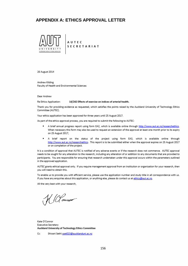
156
APPENDIX A: ETHICS APPROVAL LETTER
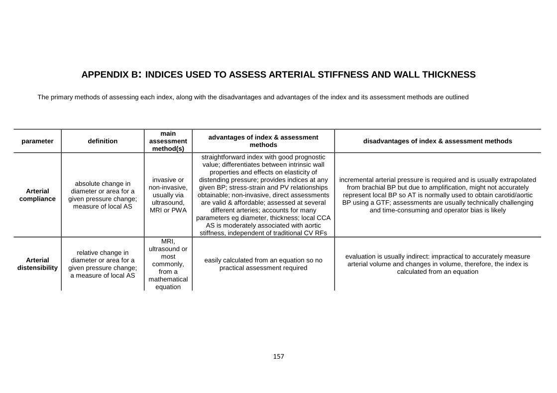
157
APPENDIX B: INDICES USED TO ASSESS ARTERIAL STIFFNESS AND WALL THICKNESS
The primary methods of assessing each index, along with the disadvantages and advantages of the index and its assessment methods are outlined
parameter definition main
assessment method(s)
advantages of index & assessment methods disadvantages of index & assessment methods
Arterial compliance
absolute change in diameter or area for a
given pressure change; measure of local AS
invasive or non-invasive,
usually via ultrasound,
MRI or PWA
straightforward index with good prognostic value; differentiates between intrinsic wall
properties and effects on elasticity of distending pressure; provides indices at any given BP; stress-strain and PV relationships obtainable; non-invasive, direct assessments are valid & affordable; assessed at several
different arteries; accounts for many parameters eg diameter, thickness; local CCA
AS is moderately associated with aortic stiffness, independent of traditional CV RFs
incremental arterial pressure is required and is usually extrapolated from brachial BP but due to amplification, might not accurately
represent local BP so AT is normally used to obtain carotid/aortic BP using a GTF; assessments are usually technically challenging
and time-consuming and operator bias is likely
Arterial distensibility
relative change in diameter or area for a
given pressure change; a measure of local AS
MRI, ultrasound or
most commonly,
from a mathematical
equation
easily calculated from an equation so no practical assessment required
evaluation is usually indirect: impractical to accurately measure arterial volume and changes in volume, therefore, the index is
calculated from an equation
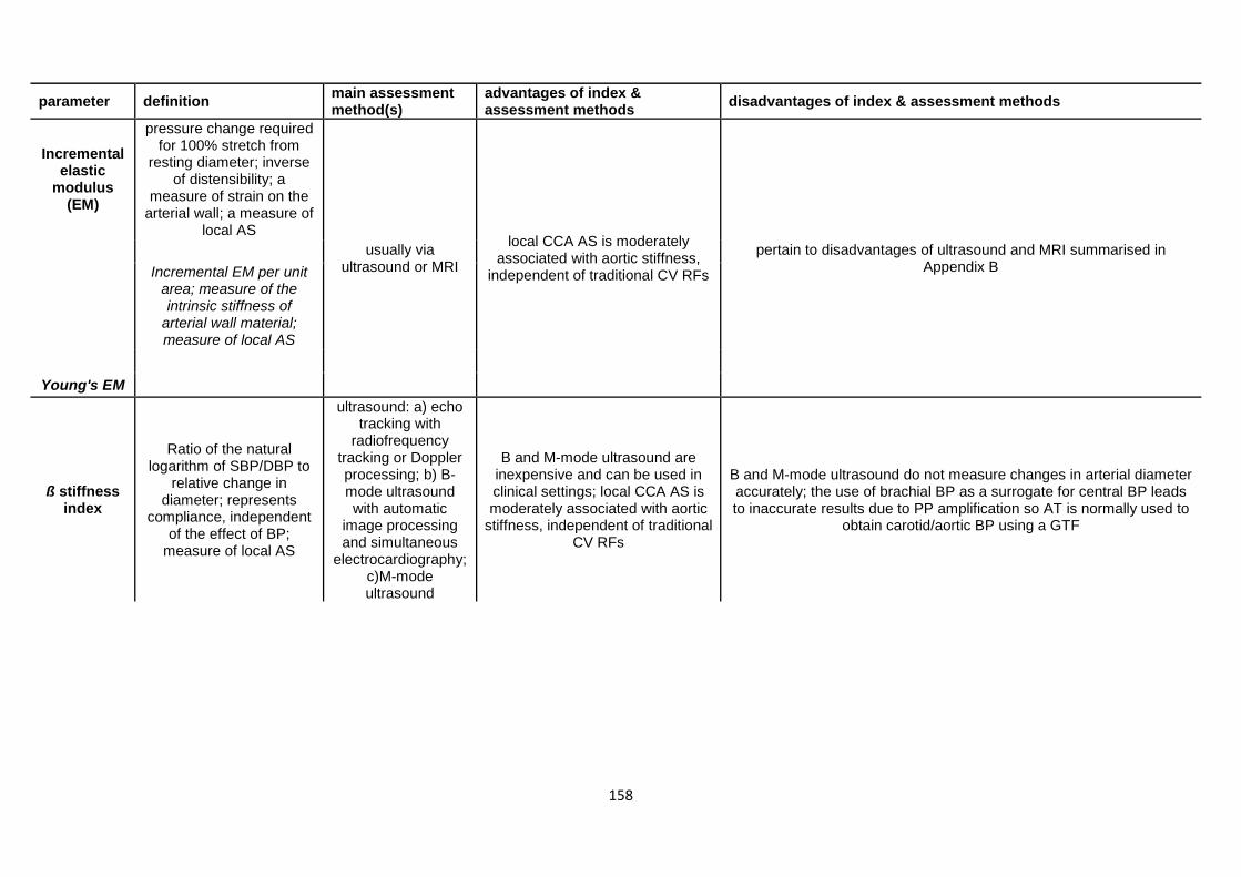
158
parameter definition main assessment method(s)
advantages of index & assessment methods disadvantages of index & assessment methods
Incremental elastic
modulus (EM)
pressure change required for 100% stretch from
resting diameter; inverse of distensibility; a
measure of strain on the arterial wall; a measure of
local AS usually via
ultrasound or MRI
local CCA AS is moderately associated with aortic stiffness,
independent of traditional CV RFs
pertain to disadvantages of ultrasound and MRI summarised in Appendix B
Incremental EM per unit area; measure of the intrinsic stiffness of
arterial wall material; measure of local AS
Young's EM
ß stiffness index
Ratio of the natural logarithm of SBP/DBP to
relative change in diameter; represents
compliance, independent of the effect of BP;
measure of local AS
ultrasound: a) echo tracking with
radiofrequency tracking or Doppler processing; b) B-mode ultrasound with automatic
image processing and simultaneous
electrocardiography; c)M-mode ultrasound
B and M-mode ultrasound are inexpensive and can be used in clinical settings; local CCA AS is moderately associated with aortic
stiffness, independent of traditional CV RFs
B and M-mode ultrasound do not measure changes in arterial diameter accurately; the use of brachial BP as a surrogate for central BP leads to inaccurate results due to PP amplification so AT is normally used to
obtain carotid/aortic BP using a GTF
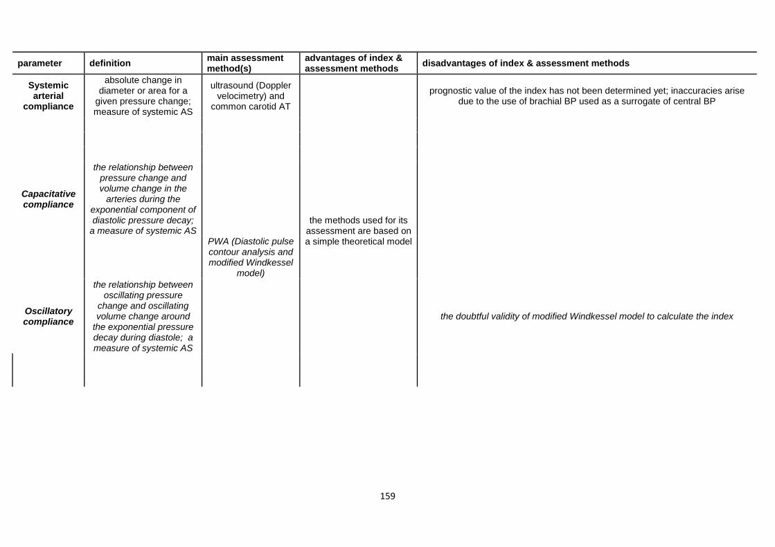
159
parameter definition main assessment method(s)
advantages of index & assessment methods disadvantages of index & assessment methods
Systemic arterial
compliance
absolute change in diameter or area for a
given pressure change; measure of systemic AS
ultrasound (Doppler velocimetry) and
common carotid AT
the methods used for its assessment are based on a simple theoretical model
prognostic value of the index has not been determined yet; inaccuracies arise due to the use of brachial BP used as a surrogate of central BP
Capacitative compliance
the relationship between pressure change and volume change in the
arteries during the exponential component of diastolic pressure decay;
a measure of systemic AS
PWA (Diastolic pulse contour analysis and modified Windkessel
model)
Oscillatory compliance
the relationship between oscillating pressure
change and oscillating volume change around
the exponential pressure decay during diastole; a measure of systemic AS
the doubtful validity of modified Windkessel model to calculate the index

160
parameter definition main assessment method(s) advantages of index & assessment methods disadvantages of index & assessment
methods
PWV
the velocity of travel of the pulse along a length of the artery; a
measure of regional/local AS; gold standard measure of AS; influenced
by arterial wall elasticity, artery geometry and blood viscosity
invasive angiography (local); mechanical
sensors, AT (regional); radiological: ultrasound,
MRI (local)
several assessment methods and sites exist: can be measured from pressure, distension or flow
waveforms obtained transcutaneously at carotid/femoral arteries; aortic PWV (cfPWV) is
most prone to age-related stiffening; can be measured invasively or non-invasively;
assessment methods are all simple, valid, reproducible and fairly affordable; widely applied in clinical/research settings; PWV independently
predicts CV risk (highest prognostic value from all predictors); largest amount of clinical evidence providing predictive value of AS for CV events
comparisons between studies and meta-analyses should be made with caution due to
the huge array of assessment methods; central arteries are inaccessible; difficult to determine
foot of waveform; inaccuracies in surface measurements of distance between arteries; limited to larger arteries because accuracy
decreases if recording points are close together; underestimation of aortic PWV with
age as it becomes more tortuous; assessments are usually time-consuming and require operator training; peripheral PWV has
unknown predictive values, questionable validity and high sensitivity to heart rate; non-invasive data suffer more drift and noise than
data obtained invasively; values are influenced by HR and BP
Local
average PWV over a long segment composed of arteries with varying
mechanical properties
ultrasound, MRI, invasive aortic catheterization
takes into account heterogenicity in AS and changes in AS along arterial tree; specific
assessment of arteries which are prone to early atherosclerotic alterations can be carried out;
direct measurement of path length
equipment is expensive
Regional PWV over one particular segment of the arterial tree
specialised devices, ultrasound
assessed easily using specialised commercial devices which are well-validated and more affordable; regional PWV is correlated to
atherosclerotic changes
external, coarse distance measurements
affected by body habitus; exact position of arterial abnormalities cannot be determined;
biochemical alterations and initial variations in elastic properties cannot be detected; direct
contact (of sensors/probes) with skin is required
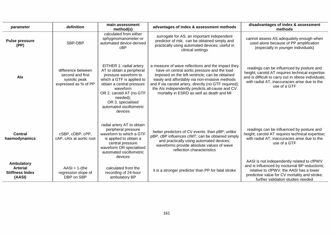
161
parameter definition main assessment method(s) advantages of index & assessment methods disadvantages of index & assessment
methods
Pulse pressure (PP) SBP-DBP
calculated from either sphygmomanometer-or
automated device-derived cBP
surrogate for AS; an important independent predictor of risk; can be obtained simply and practically using automated devices; useful in
clinical settings
cannot assess AS adequately enough when used alone because of PP amplification
(especially in younger individuals)
AIx difference between
second and first systolic peak
expressed as % of PP
EITHER 1. radial artery AT to obtain a peripheral
pressure waveform to which a GTF is applied to obtain a central pressure
waveform OR 2. carotid AT (no GTF
needed);
a measure of wave reflections and the impact they
have on central aortic pressure and the load imposed on the left ventricle; can be obtained
easily and affordably via non-invasive methods and if via carotid artery, directly (no GTF required); the AIx independently predicts all-cause and CV
mortality in ESRD as well as death and MI
readings can be influenced by posture and height; carotid AT requires technical expertise and is difficult to carry out in obese individuals;
with radial AT, inaccuracies arise due to the use of a GTF
OR 3. specialised
automated oscillometric devices
Central haemodynamics
cSBP, cDBP, cPP, cAP, cAIx at aortic root
radial artery AT to obtain peripheral pressure
waveform to which a GTF is applied to obtain a
central pressure waveform OR specialised automated oscillometric
devices
better predictors of CV events than pBP; unlike pBP, cBP influences cIMT; can be obtained simply
and practically using automated devices; waveforms provide absolute values of wave
reflection characteristics
readings can be influenced by posture and height; carotid AT requires technical expertise;
with radial AT, inaccuracies arise due to the use of a GTF
Ambulatory Arterial
Stiffness Index (AASI)
AASI = 1-(the
regression slope of DBP on SBP
calculated from the recording of 24-hour
ambulatory BP
it is a stronger predictor than PP for fatal stroke
AASI is not independently related to cfPWV and is influenced by nocturnal BP reductions;
relative to cfPWV, the AASI has a lower predictive value for CV mortality and stroke;
further validation studies needed
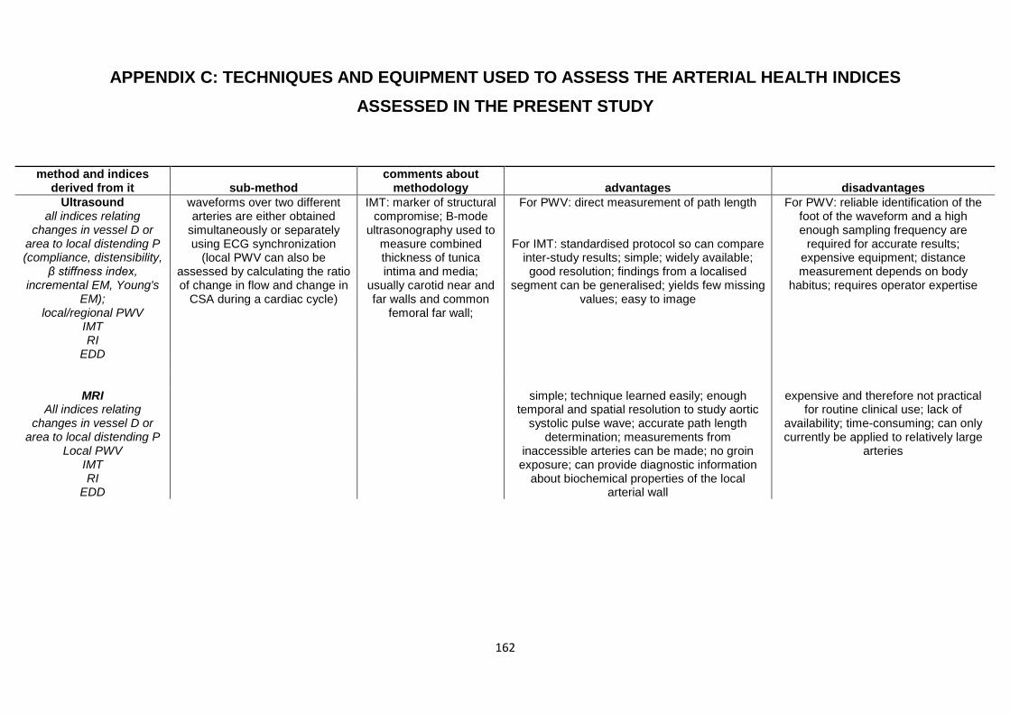
162
APPENDIX C: TECHNIQUES AND EQUIPMENT USED TO ASSESS THE ARTERIAL HEALTH INDICES ASSESSED IN THE PRESENT STUDY
method and indices
derived from it sub-method comments about
methodology advantages disadvantages Ultrasound
all indices relating changes in vessel D or
area to local distending P (compliance, distensibility,
β stiffness index, incremental EM, Young's
EM); local/regional PWV
IMT RI
EDD
waveforms over two different arteries are either obtained
simultaneously or separately using ECG synchronization
(local PWV can also be assessed by calculating the ratio of change in flow and change in
CSA during a cardiac cycle)
IMT: marker of structural compromise; B-mode
ultrasonography used to measure combined thickness of tunica intima and media;
usually carotid near and far walls and common
femoral far wall;
For PWV: direct measurement of path length
For IMT: standardised protocol so can compare inter-study results; simple; widely available; good resolution; findings from a localised
segment can be generalised; yields few missing values; easy to image
For PWV: reliable identification of the foot of the waveform and a high enough sampling frequency are
required for accurate results; expensive equipment; distance measurement depends on body
habitus; requires operator expertise
MRI All indices relating
changes in vessel D or area to local distending P
Local PWV IMT RI
EDD
simple; technique learned easily; enough temporal and spatial resolution to study aortic
systolic pulse wave; accurate path length determination; measurements from
inaccessible arteries can be made; no groin exposure; can provide diagnostic information
about biochemical properties of the local arterial wall
expensive and therefore not practical for routine clinical use; lack of
availability; time-consuming; can only currently be applied to relatively large
arteries
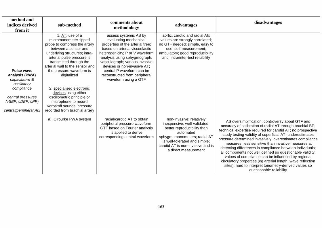
163
method and indices derived
from it sub-method comments about
methodology advantages disadvantages
Pulse wave analysis (PWA) capacitative &
oscillatory compliance
central pressures
(cSBP, cDBP, cPP)
central/peripheral AIx
1. AT: use of a micromanometer-tipped
probe to compress the artery between a sensor and
underlying structures; intra-arterial pulse pressure is transmitted through the
arterial wall to the sensor and the pressure waveform is
digitalized
2. specialised electronic devices using either
oscillometric principle or microphone to record
Korotkoff sounds; pressure recorded from brachial artery
a). O'rourke PWA system
assess systemic AS by evaluating mechanical
properties of the arterial tree; based on arterial viscoelastic
heterogenicity; P or V waveform analysis using sphygmograph, vasculograph; various invasive
devices or non-invasive AT; central P waveform can be
reconstructed from peripheral waveform using a GTF
radial/carotid AT to obtain peripheral pressure waveform. GTF based on Fourier analysis
is applied to derive corresponding central waveform
aortic, carotid and radial AIx values are strongly correlated;
no GTF needed; simple, easy to use; self-measurement;
ambulatory; good reproducibility and intra/inter-test reliability
non-invasive; relatively inexpensive; well-validated; better reproducibility than
automated sphygmomanometers; radial AT
is well-tolerated and simple; carotid AT is non-invasive and is
a direct measurement
AS oversimplification; controversy about GTF and accuracy of calibration of radial AT through brachial BP;
technical expertise required for carotid AT; no prospective study testing validity of superficial AT; underestimates
pressure determined invasively; overestimates compliance measures; less sensitive than invasive measures at
detecting differences in compliance between individuals; all components not well defined so questionable validity;
values of compliance can be influenced by regional circulatory properties (eg arterial length, wave reflection
sites); hard to interpret tonometry-derived values so questionable reliability
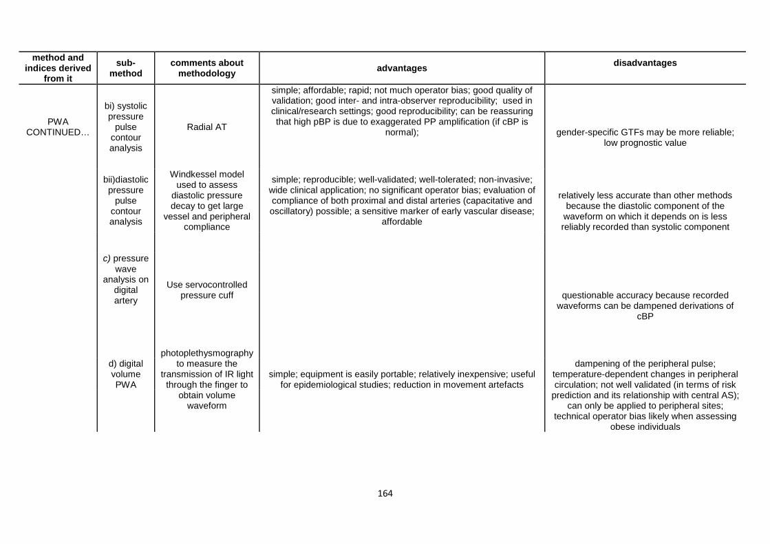
164
method and indices derived
from it sub-
method comments about
methodology advantages disadvantages
PWA CONTINUED…
bi) systolic pressure
pulse contour analysis
Radial AT
simple; affordable; rapid; not much operator bias; good quality of validation; good inter- and intra-observer reproducibility; used in clinical/research settings; good reproducibility; can be reassuring that high pBP is due to exaggerated PP amplification (if cBP is
normal);
gender-specific GTFs may be more reliable; low prognostic value
bii)diastolic pressure
pulse contour analysis
Windkessel model used to assess
diastolic pressure decay to get large
vessel and peripheral compliance
simple; reproducible; well-validated; well-tolerated; non-invasive; wide clinical application; no significant operator bias; evaluation of compliance of both proximal and distal arteries (capacitative and oscillatory) possible; a sensitive marker of early vascular disease;
affordable
relatively less accurate than other methods because the diastolic component of the
waveform on which it depends on is less reliably recorded than systolic component
c) pressure wave
analysis on digital artery
Use servocontrolled pressure cuff
questionable accuracy because recorded waveforms can be dampened derivations of
cBP
photoplethysmography
to measure the transmission of IR light
through the finger to obtain volume
waveform
dampening of the peripheral pulse; temperature-dependent changes in peripheral circulation; not well validated (in terms of risk
prediction and its relationship with central AS); can only be applied to peripheral sites;
technical operator bias likely when assessing obese individuals
d) digital volume PWA
simple; equipment is easily portable; relatively inexpensive; useful
for epidemiological studies; reduction in movement artefacts
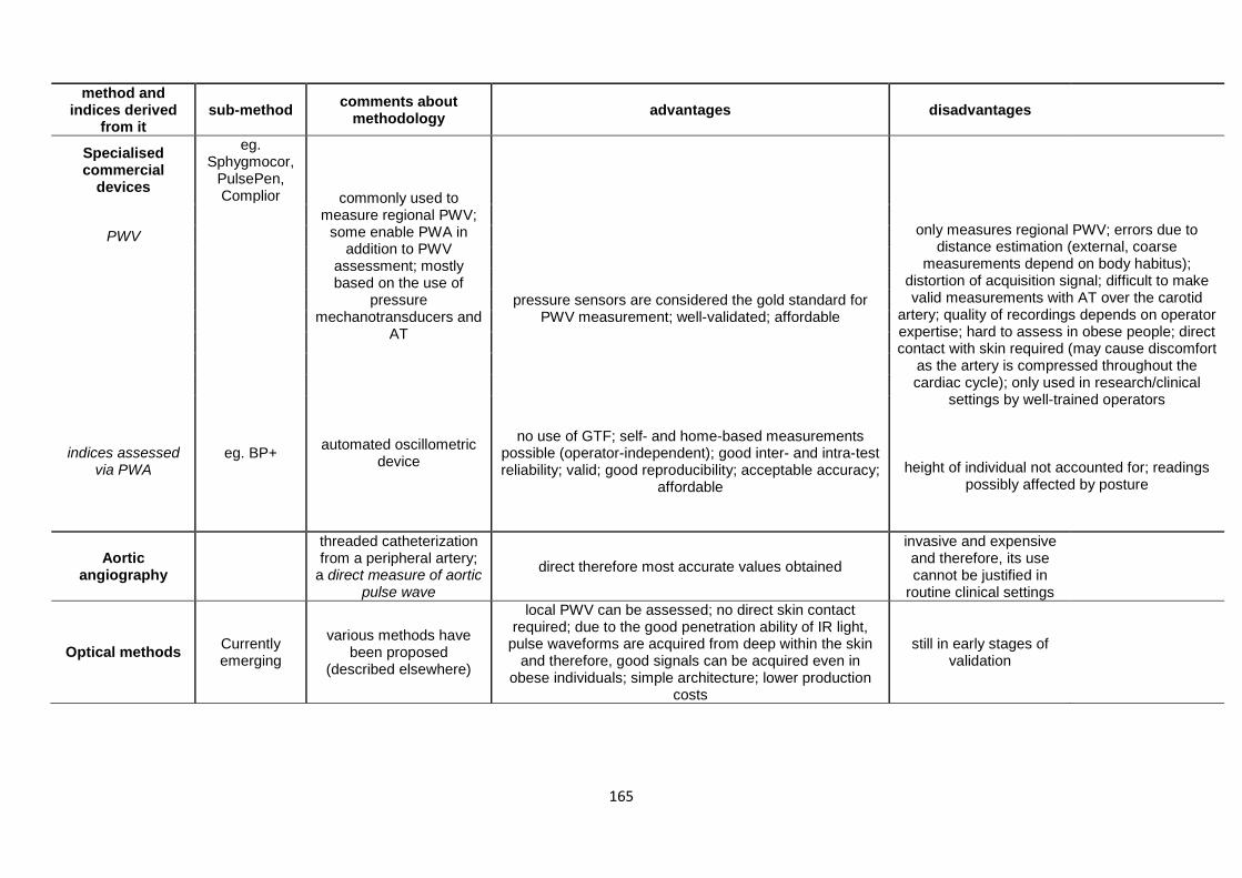
165
method and indices derived
from it sub-method comments about
methodology advantages disadvantages
Specialised commercial
devices
eg. Sphygmocor,
PulsePen, Complior commonly used to
measure regional PWV; some enable PWA in
addition to PWV assessment; mostly based on the use of
pressure mechanotransducers and
AT
pressure sensors are considered the gold standard for PWV measurement; well-validated; affordable
only measures regional PWV; errors due to distance estimation (external, coarse
measurements depend on body habitus); distortion of acquisition signal; difficult to make valid measurements with AT over the carotid
artery; quality of recordings depends on operator expertise; hard to assess in obese people; direct contact with skin required (may cause discomfort
as the artery is compressed throughout the cardiac cycle); only used in research/clinical
settings by well-trained operators
height of individual not accounted for; readings possibly affected by posture
PWV
indices assessed
via PWA eg. BP+ automated oscillometric
device
no use of GTF; self- and home-based measurements
possible (operator-independent); good inter- and intra-test reliability; valid; good reproducibility; acceptable accuracy;
affordable
Aortic angiography
threaded catheterization from a peripheral artery;
a direct measure of aortic pulse wave
direct therefore most accurate values obtained
invasive and expensive and therefore, its use cannot be justified in
routine clinical settings
Optical methods Currently emerging
various methods have been proposed
(described elsewhere)
local PWV can be assessed; no direct skin contact required; due to the good penetration ability of IR light,
pulse waveforms are acquired from deep within the skin and therefore, good signals can be acquired even in
obese individuals; simple architecture; lower production costs
still in early stages of validation
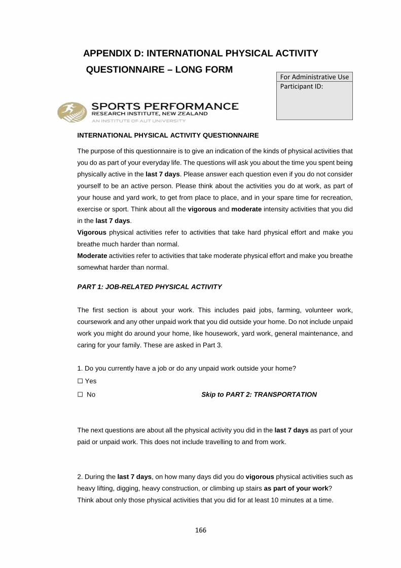
166
APPENDIX D: INTERNATIONAL PHYSICAL ACTIVITY QUESTIONNAIRE – LONG FORM
INTERNATIONAL PHYSICAL ACTIVITY QUESTIONNAIRE The purpose of this questionnaire is to give an indication of the kinds of physical activities that
you do as part of your everyday life. The questions will ask you about the time you spent being
physically active in the last 7 days. Please answer each question even if you do not consider
yourself to be an active person. Please think about the activities you do at work, as part of
your house and yard work, to get from place to place, and in your spare time for recreation,
exercise or sport. Think about all the vigorous and moderate intensity activities that you did
in the last 7 days.
Vigorous physical activities refer to activities that take hard physical effort and make you
breathe much harder than normal.
Moderate activities refer to activities that take moderate physical effort and make you breathe
somewhat harder than normal.
PART 1: JOB-RELATED PHYSICAL ACTIVITY
The first section is about your work. This includes paid jobs, farming, volunteer work,
coursework and any other unpaid work that you did outside your home. Do not include unpaid
work you might do around your home, like housework, yard work, general maintenance, and
caring for your family. These are asked in Part 3.
1. Do you currently have a job or do any unpaid work outside your home?
☐ Yes
☐ No Skip to PART 2: TRANSPORTATION
The next questions are about all the physical activity you did in the last 7 days as part of your
paid or unpaid work. This does not include travelling to and from work.
2. During the last 7 days, on how many days did you do vigorous physical activities such as
heavy lifting, digging, heavy construction, or climbing up stairs as part of your work?
Think about only those physical activities that you did for at least 10 minutes at a time.
For Administrative Use Participant ID:

167
_____ days per week
☐ No vigorous job-related physical activity Skip to question 4
3. How much time did you usually spend on one of those days doing vigorous physical
activities as part of your work?
_____ hours per day _____ minutes per day
4. Again, think about only those physical activities that you did for at least 10 minutes at a
time. During the last 7 days, on how many days did you do moderate physical activities such
as carrying light loads as part of your work? Please do not include walking.
_____ days per week
☐ No moderate job-related physical activity Skip to question 6
5. How much time did you usually spend on one of those days doing moderate physical
activities as part of your work?
_____ hours per day _____ minutes per day
6. During the last 7 days, on how many days did you walk for at least 10 minutes at a time
as part of your work? Please do not count any walking you did to travel to or from work.
_____ days per week
☐ No job-related walking Skip to PART 2:
TRANSPORTATION
7. How much time did you usually spend on one of those days walking as part of your
work?
_____ hours per day

168
_____ minutes per day
PART 2: TRANSPORTATION PHYSICAL ACTIVITY
These questions are about how you travelled from place to place, including to places like
work, stores, movies, and so on.
8. During the last 7 days, on how many days did you travel in a motor vehicle like a train,
bus, car, or tram?
_____ days per week
☐ No traveling in a motor vehicle Skip to question 10
9. How much time did you usually spend on one of those days traveling in a train, bus, car,
tram, or other kind of motor vehicle?
_____ hours per day _____ minutes per day
Now think only about the bicycling and walking you might have done to travel to and from
work, to do errands, or to go from place to place.
10. During the last 7 days, on how many days did you bicycle for at least 10 minutes at a
time to go from place to place?
_____ days per week
☐ No bicycling from place to place Skip to question 12
11. How much time did you usually spend on one of those days to bicycle from place to
place?
_____ hours per day _____ minutes per day

169
12. During the last 7 days, on how many days did you walk for at least 10 minutes at a time
to go from place to place?
_____ days per week
☐ No walking from place to place Skip to PART 3: HOUSEWORK, HOUSE
MAINTENANCE, AND CARING FOR FAMILY
13. How much time did you usually spend on one of those days walking from place to place?
_____ hours per day _____ minutes per day
PART 3: HOUSEWORK, HOUSE MAINTENANCE, AND CARING FOR FAMILY
This section is about some of the physical activities you might have done in the last 7 days
in and around your home, like housework, gardening, yard work, general maintenance work,
and caring for your family.
14. Think about only those physical activities that you did for at least 10 minutes at a time.
During the last 7 days, on how many days did you do vigorous physical activities such as
heavy lifting, chopping wood, shovelling snow, or digging in the garden or yard?
_____ days per week
☐ No vigorous activity in garden or yard Skip to question 16
15. How much time did you usually spend on one of those days doing vigorous physical
activities in the garden or yard?
_____ hours per day _____ minutes per day
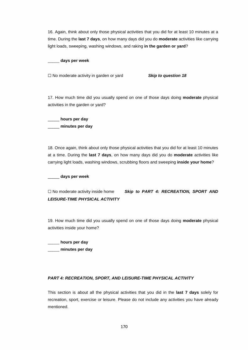
170
16. Again, think about only those physical activities that you did for at least 10 minutes at a
time. During the last 7 days, on how many days did you do moderate activities like carrying
light loads, sweeping, washing windows, and raking in the garden or yard?
_____ days per week
☐ No moderate activity in garden or yard Skip to question 18
17. How much time did you usually spend on one of those days doing moderate physical
activities in the garden or yard?
_____ hours per day _____ minutes per day
18. Once again, think about only those physical activities that you did for at least 10 minutes
at a time. During the last 7 days, on how many days did you do moderate activities like
carrying light loads, washing windows, scrubbing floors and sweeping inside your home?
_____ days per week
☐ No moderate activity inside home Skip to PART 4: RECREATION, SPORT AND
LEISURE-TIME PHYSICAL ACTIVITY
19. How much time did you usually spend on one of those days doing moderate physical
activities inside your home?
_____ hours per day _____ minutes per day
PART 4: RECREATION, SPORT, AND LEISURE-TIME PHYSICAL ACTIVITY
This section is about all the physical activities that you did in the last 7 days solely for
recreation, sport, exercise or leisure. Please do not include any activities you have already
mentioned.

171
20. Not counting any walking you have already mentioned, during the last 7 days, on how
many days did you walk for at least 10 minutes at a time in your leisure time?
_____ days per week
☐ No walking in leisure time Skip to question 22
21. How much time did you usually spend on one of those days walking in your leisure time?
_____ hours per day _____ minutes per day
22. Think about only those physical activities that you did for at least 10 minutes at a time.
During the last 7 days, on how many days did you do vigorous physical activities like
aerobics, running, fast bicycling, or fast swimming in your leisure time?
_____ days per week
☐ No vigorous activity in leisure time Skip to question 24
23. How much time did you usually spend on one of those days doing vigorous physical
activities in your leisure time?
_____ hours per day _____ minutes per day
24. Again, think about only those physical activities that you did for at least 10 minutes at a
time. During the last 7 days, on how many days did you do moderate physical activities like
bicycling at a regular pace, swimming at a regular pace, and doubles tennis in your leisure time?
_____ days per week
☐ No moderate activity in leisure time Skip to PART 5: TIME SPENT
SITTING

172
25. How much time did you usually spend on one of those days doing moderate physical
activities in your leisure time?
_____ hours per day _____ minutes per day
PART 5: TIME SPENT SITTING The last questions are about the time you spend sitting while at work, at home, while doing
coursework and during leisure time. This may include time spent sitting at a desk, visiting
friends, reading or sitting or lying down to watch television. Do not include any time spent
sitting in a motor vehicle that you have already told me about.
26. During the last 7 days, how much time did you usually spend sitting on a weekday?
_____ hours per day _____ minutes per day
27. During the last 7 days, how much time did you usually spend sitting on a weekend day?
_____ hours per day _____ minutes per day
This is the end of the questionnaire. Thank you for completing it!
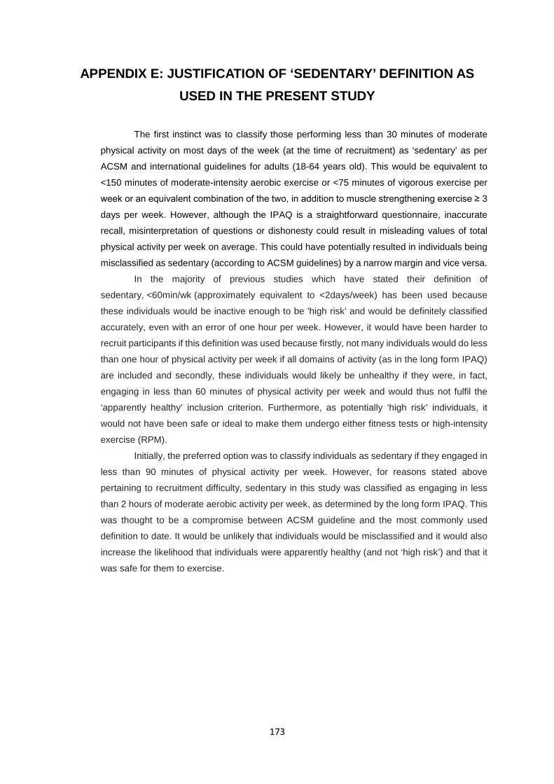
173
APPENDIX E: JUSTIFICATION OF ‘SEDENTARY’ DEFINITION AS USED IN THE PRESENT STUDY
The first instinct was to classify those performing less than 30 minutes of moderate
physical activity on most days of the week (at the time of recruitment) as ‘sedentary’ as per
ACSM and international guidelines for adults (18-64 years old). This would be equivalent to
<150 minutes of moderate-intensity aerobic exercise or <75 minutes of vigorous exercise per
week or an equivalent combination of the two, in addition to muscle strengthening exercise ≥ 3
days per week. However, although the IPAQ is a straightforward questionnaire, inaccurate
recall, misinterpretation of questions or dishonesty could result in misleading values of total
physical activity per week on average. This could have potentially resulted in individuals being
misclassified as sedentary (according to ACSM guidelines) by a narrow margin and vice versa.
In the majority of previous studies which have stated their definition of
sedentary, <60min/wk (approximately equivalent to <2days/week) has been used because
these individuals would be inactive enough to be 'high risk' and would be definitely classified
accurately, even with an error of one hour per week. However, it would have been harder to
recruit participants if this definition was used because firstly, not many individuals would do less
than one hour of physical activity per week if all domains of activity (as in the long form IPAQ)
are included and secondly, these individuals would likely be unhealthy if they were, in fact,
engaging in less than 60 minutes of physical activity per week and would thus not fulfil the
‘apparently healthy’ inclusion criterion. Furthermore, as potentially ‘high risk’ individuals, it
would not have been safe or ideal to make them undergo either fitness tests or high-intensity
exercise (RPM).
Initially, the preferred option was to classify individuals as sedentary if they engaged in
less than 90 minutes of physical activity per week. However, for reasons stated above
pertaining to recruitment difficulty, sedentary in this study was classified as engaging in less
than 2 hours of moderate aerobic activity per week, as determined by the long form IPAQ. This
was thought to be a compromise between ACSM guideline and the most commonly used
definition to date. It would be unlikely that individuals would be misclassified and it would also
increase the likelihood that individuals were apparently healthy (and not ‘high risk’) and that it
was safe for them to exercise.
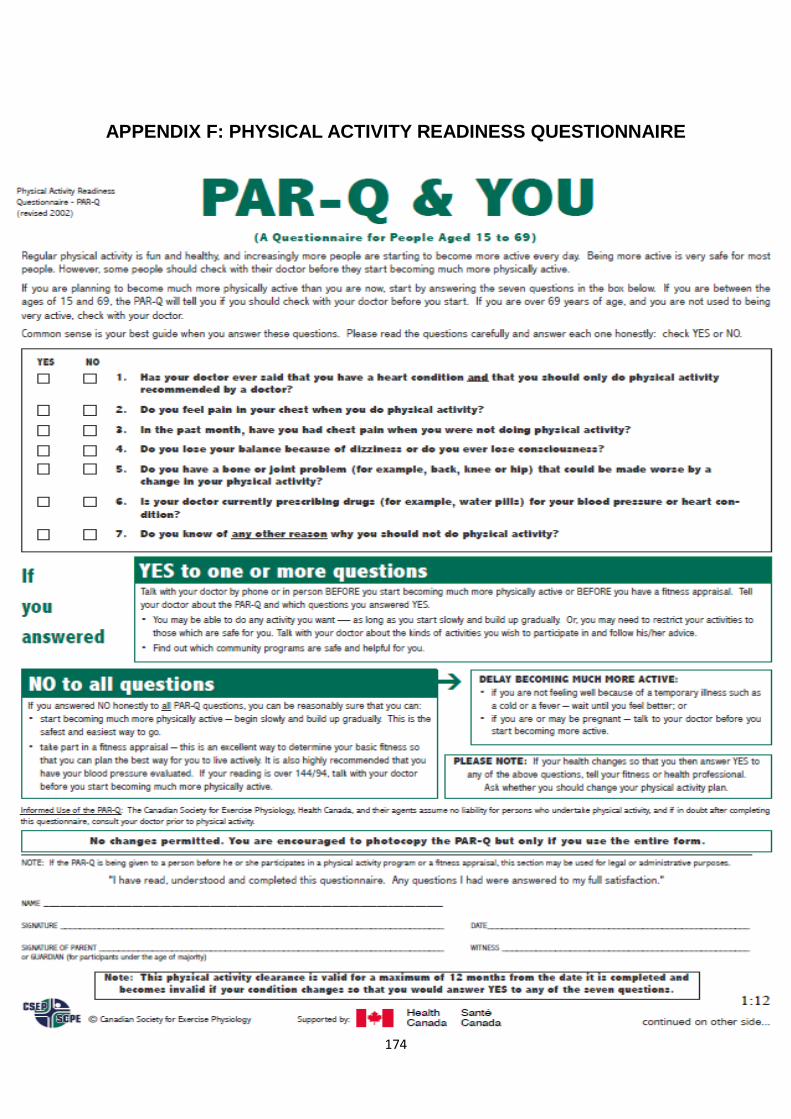
174
APPENDIX F: PHYSICAL ACTIVITY READINESS QUESTIONNAIRE
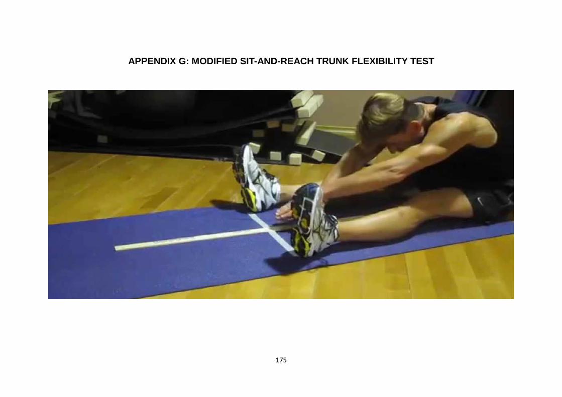
175
APPENDIX G: MODIFIED SIT-AND-REACH TRUNK FLEXIBILITY TEST
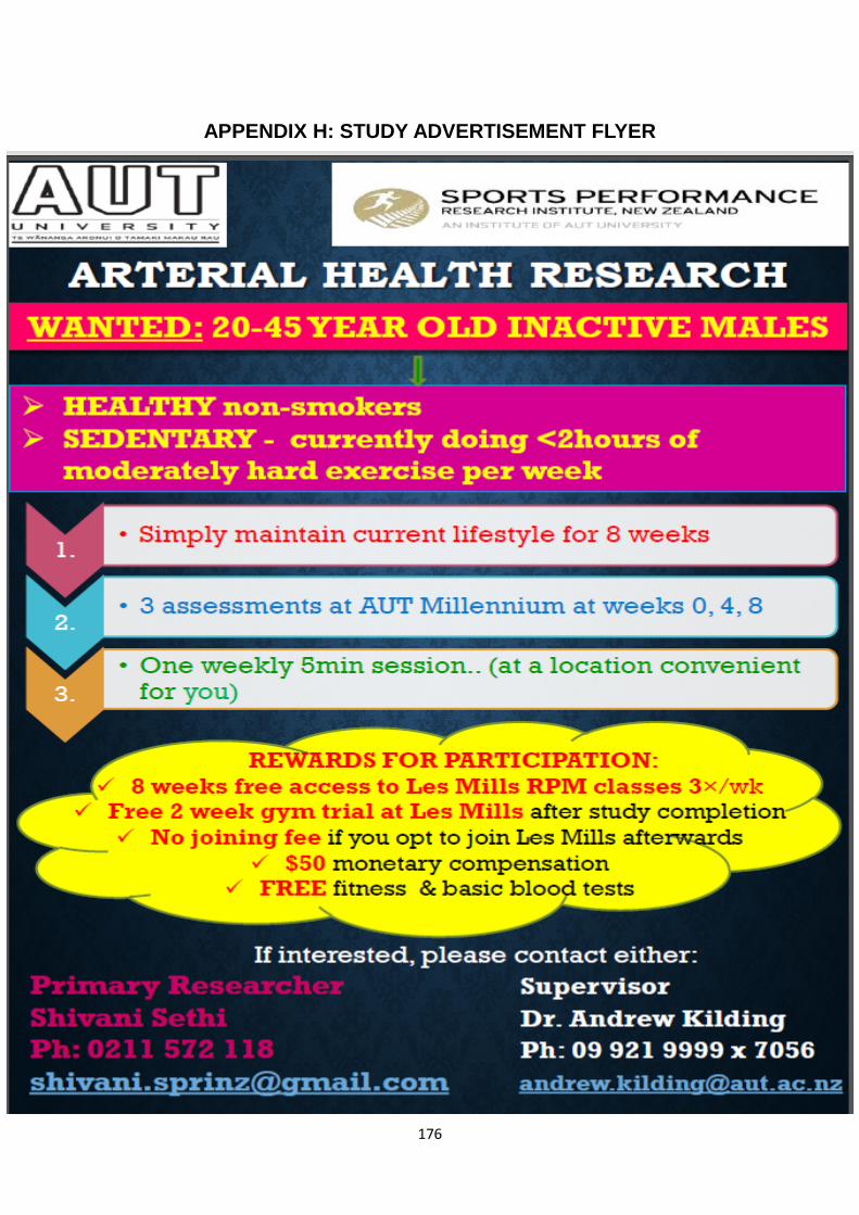
176
APPENDIX H: STUDY ADVERTISEMENT FLYER
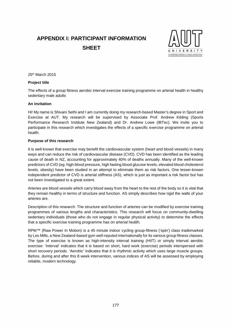
177
APPENDIX I: PARTICIPANT INFORMATION SHEET
25th March 2015
Project title
The effects of a group fitness aerobic interval exercise training programme on arterial health in healthy sedentary male adults
An invitation
Hi! My name is Shivani Sethi and I am currently doing my research-based Master’s degree in Sport and Exercise at AUT. My research will be supervised by Associate Prof. Andrew Kilding (Sports Performance Research Institute New Zealand) and Dr. Andrew Lowe (IBTec). We invite you to participate in this research which investigates the effects of a specific exercise programme on arterial health.
Purpose of this research
It is well known that exercise may benefit the cardiovascular system (heart and blood vessels) in many ways and can reduce the risk of cardiovascular disease (CVD). CVD has been identified as the leading cause of death in NZ, accounting for approximately 40% of deaths annually. Many of the well-known predictors of CVD (eg. high blood pressure, high fasting blood glucose levels, elevated blood cholesterol levels, obesity) have been studied in an attempt to eliminate them as risk factors. One lesser-known independent predictor of CVD is arterial stiffness (AS), which is just as important a risk factor but has not been investigated to a great extent.
Arteries are blood vessels which carry blood away from the heart to the rest of the body so it is vital that they remain healthy in terms of structure and function. AS simply describes how rigid the walls of your arteries are.
Description of this research: The structure and function of arteries can be modified by exercise training programmes of various lengths and characteristics. This research will focus on community-dwelling sedentary individuals (those who do not engage in regular physical activity) to determine the effects that a specific exercise training programme has on arterial health.
RPM™ (Raw Power In Motion) is a 45 minute indoor cycling group-fitness (‘spin’) class trademarked by Les Mills, a New Zealand-based gym well-reputed internationally for its various group fitness classes. The type of exercise is known as high-intensity interval training (HIIT) or simply interval aerobic exercise: ‘Interval’ indicates that it is based on short, hard work (exercise) periods interspersed with short recovery periods. ‘Aerobic’ indicates that it is rhythmic activity which uses large muscle groups. Before, during and after this 8 week intervention, various indices of AS will be assessed by employing reliable, modern technology.
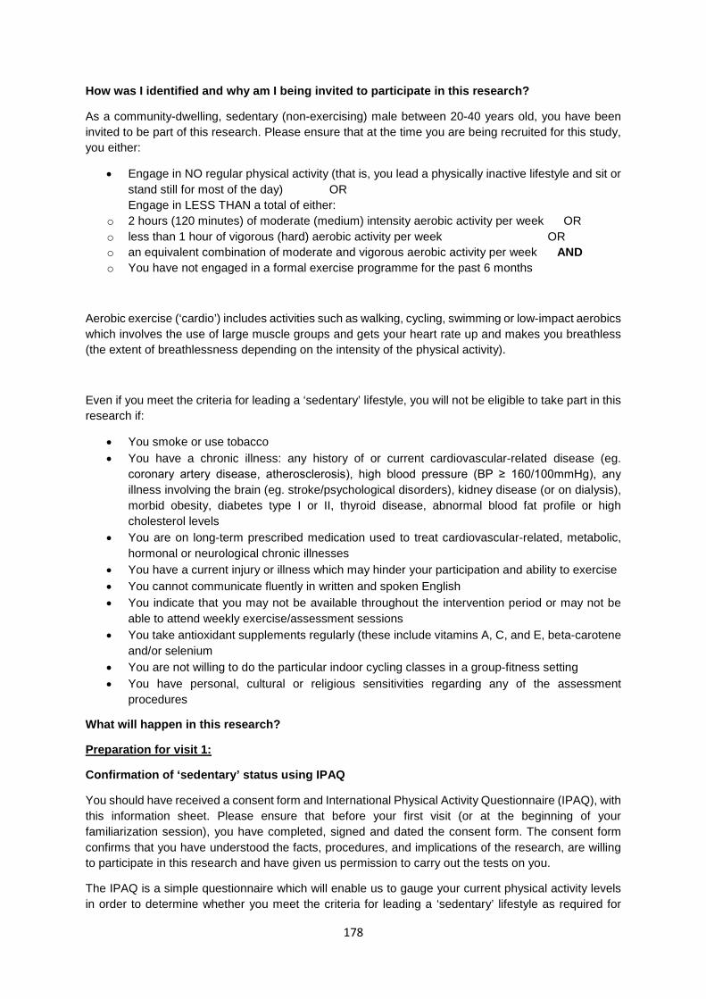
178
How was I identified and why am I being invited to participate in this research?
As a community-dwelling, sedentary (non-exercising) male between 20-40 years old, you have been invited to be part of this research. Please ensure that at the time you are being recruited for this study, you either:
• Engage in NO regular physical activity (that is, you lead a physically inactive lifestyle and sit or stand still for most of the day) OR Engage in LESS THAN a total of either:
o 2 hours (120 minutes) of moderate (medium) intensity aerobic activity per week OR o less than 1 hour of vigorous (hard) aerobic activity per week OR o an equivalent combination of moderate and vigorous aerobic activity per week AND o You have not engaged in a formal exercise programme for the past 6 months
Aerobic exercise (‘cardio’) includes activities such as walking, cycling, swimming or low-impact aerobics which involves the use of large muscle groups and gets your heart rate up and makes you breathless (the extent of breathlessness depending on the intensity of the physical activity).
Even if you meet the criteria for leading a ‘sedentary’ lifestyle, you will not be eligible to take part in this research if:
• You smoke or use tobacco • You have a chronic illness: any history of or current cardiovascular-related disease (eg.
coronary artery disease, atherosclerosis), high blood pressure (BP ≥ 160/100mmHg), any illness involving the brain (eg. stroke/psychological disorders), kidney disease (or on dialysis), morbid obesity, diabetes type I or II, thyroid disease, abnormal blood fat profile or high cholesterol levels
• You are on long-term prescribed medication used to treat cardiovascular-related, metabolic, hormonal or neurological chronic illnesses
• You have a current injury or illness which may hinder your participation and ability to exercise • You cannot communicate fluently in written and spoken English • You indicate that you may not be available throughout the intervention period or may not be
able to attend weekly exercise/assessment sessions • You take antioxidant supplements regularly (these include vitamins A, C, and E, beta-carotene
and/or selenium • You are not willing to do the particular indoor cycling classes in a group-fitness setting • You have personal, cultural or religious sensitivities regarding any of the assessment
procedures
What will happen in this research?
Preparation for visit 1:
Confirmation of ‘sedentary’ status using IPAQ
You should have received a consent form and International Physical Activity Questionnaire (IPAQ), with this information sheet. Please ensure that before your first visit (or at the beginning of your familiarization session), you have completed, signed and dated the consent form. The consent form confirms that you have understood the facts, procedures, and implications of the research, are willing to participate in this research and have given us permission to carry out the tests on you.
The IPAQ is a simple questionnaire which will enable us to gauge your current physical activity levels in order to determine whether you meet the criteria for leading a ‘sedentary’ lifestyle as required for
179
participation in this study. Please complete the IPAQ and email it back to us if you think you would like to participate.
Familiarization session arrangement
If you meet all the inclusion criteria, you will be invited to come to AUT Millennium for a familiarization session. A time will be arranged via e-mail/phone regarding the time of your first visit. Please ensure that you bring your exercise gear with you for the first visit. Also, please try to avoid strenuous physical activity for 24 hours before the first visit and please ensure you attend the first session after an overnight fast (9-11 hours).
Visit 1 - Familiarization and baseline testing:
After the study background and protocol have been outlined to you, you will have a chance to ask any questions. If you think you would like to participate, you will have to submit the consent form formally. You will then be required to fill out a Physical Activity Readiness Questionnaire (PAR-Q) which will enable us to determine whether it is safe for you to exercise based on your answers to specific health-related questions. Some general details and basic measures (height, weight, waist circumference, resting heart rate, resting blood pressure & arterial stiffness) will then be taken.
Next, your bloods will be taken using a simple finger-prick method (see page 6). The results will enable us to classify your CVD risk using a Cardiovascular Risk Assessment (CRA) form.
The distance between your neck and upper thigh (groin area) will be taken using a tape measure. You will then be required to lie down on your back or sit up slightly (if needed) with an ECG attached to you whilst ultrasound recordings are carried out on various parts of your body (neck, upper thigh, and arm). This will require approximately 1 hour (see page 6 for further details).
You will then be required to perform the ‘sit-and-reach test’ which is a measure of trunk flexibility. It just involves sitting down on the ground with your legs straight out and reaching as far forward as possible.
Finally, you will be required to perform a ‘VO2peak’ test which will provide us with information about your fitness levels. The test will involve cycling at progressively increasing intensities until you can no longer continue. Note: If your blood pressure is elevated (>160/100mmHg), you will be referred to your GP and you will not be allowed to participate further. The test will start very easy and will continue to a high intensity. The test lasts about 12 minutes. During the test, you will wear a heart rate monitor around your chest and a face mask with a mouthpiece (which resembles a snorkel) for breathing analysis.
Finally, you will be given a Food Frequency questionnaire (FFQ) which will give us an idea about your dietary habits.
180
Group allocation
Following visit 1, you will be randomly allocated to either the ‘control’ group or ‘intervention’ group. It is important you understand that you may be asked to serve as a control subject in this study (see below).
What happens if I get allocated to the control group?
If you are randomly allocated to the control group, you will complete all the same procedures and assessments as the intervention group. Refer to page 6 and page 8 (table 1) for a summary of the assessment sessions and to page 7 (figure 1) for a visual representation of when the measurements will be taken. All your assessments will take place at the AUT Millennium Institute of Sport and Health.
The only inter-group difference is that the intervention group will undergo 8 weeks of exercise training, 3 times per week, whereas you will continue with your normal lifestyle. You will be requested to maintain your current, normal physical activity, and dietary habits for the entire intervention period and will be required to keep a physical activity diary (which will be provided during the familiarization session).
Once you have completed all pre-, mid- and post-intervention assessments, you will be invited to participate as the ‘intervention group’ and do what those initially allocated to the intervention group did for the first 8 weeks (i.e. 3 weekly Les Mills RPM classes for 8 weeks). No formal physiological assessments will be carried out at this time but you will be given free membership at Les Mills Victoria Street/Takapuna for 8 weeks.
What happens if I get allocated to the intervention group?
You will be required to participate in three RPM sessions per week (for 8 consecutive weeks) at the Les Mills Takapuna or Victoria Street gym. These can be done at any time of day according to your convenience but each session must be separated by at least 36 hours. A timetable of classes will be provided.
Once a week, your basic arterial stiffness measurement (no. 1, page 5), body mass and blood pressure measurements will be carried out at Les Mills Vic St/Takapuna approximately before your last exercise session of the week.
The other arterial health measures (no. 2, page 5) will be carried out at the AUT Millennium Institute of Sport and Health (Weeks 0, 4 and 9). These ultrasound-based arterial health assessments will be carried out at your convenience, but in week 4, it will not be on a day that you have done RPM.
At the end of 8 weeks (excluding week 0), a final assessment session will take place where bloods, ultrasound assessments, and the fitness test will be repeated. Refer to page 6 and page 9 (table 2) for a summary of the assessment and RPM sessions and to page 7 (figure 1) for a visual representation of when the measurements will be taken.
You will be requested, aside from the RPM classes, to maintain your current, normal physical activity, and dietary habits for the entire intervention period and will be required to keep a physical activity diary (which will be provided during the familiarization session).
You will also be required to wear a heart rate monitor during each RPM session. You will be responsible for ensuring that you have done so as the information obtained from it will firstly enable us to determine, the intensity at which you worked at during each session and secondly, will be used to monitor your attendance to RPM. Your heart rate monitor will be collected after the end of your last RPM session each week and a new one will be available to you before your first class the following week.
181
Physiological assessment procedures: Refer to figure 1 on page 7
Basic arterial stiffness, blood pressure and heart rate measurement: This will be similar to a normal blood pressure measurement which you are likely to be familiar with. It simply involves placing a cuff around your arm so that a reading can be taken. There will be very mild discomfort as the cuff compresses your arm, but this will not last more than a few seconds! These measurements will be taken 10 times in total (before and after the 8 week intervention phase as well as weekly, throughout the 8 weeks).
Arterial health measures: Before, halfway through, and at the end of the intervention, you will be required to come in to get an ultrasound assessment of your arteries done. There will be 2 separate tests being carried out. One will involve ultrasound assessment of the arteries in your arm whilst the other will involve the neck area (where you can feel your pulse) and the top of the lower limb, near the groin. The names of the measures to be determined are ‘intima-media thickness’ (IMT, the thickness of your artery walls), ‘pulse wave velocity’ (PWV, the speed at which your blood travels) and carotid artery diameter. You will be fully clothed for all procedures and there will be nothing invasive. Ultrasound will simply involve putting gel on a probe which will be rolled on the surface of the skin to give an image on a computer-like screen. You will not feel any pain and may feel only very slight discomfort, if any. At the same time, an ECG will be set up to non-invasively measure the electrical activity of your heart. This will involve placing 3 small patches called electrodes on your chest and abdomen. Please note that it may be necessary to shave or clip some hair so the patches stick to the skin. These ultrasound-based arterial health measures will be taken 3 times in total (before, halfway through and after the intervention period).
Bloods: Don’t worry! Your blood sample will be taken using a very basic prick of your finger, so there will be no needles and hardly any visible blood. Samples will be taken 3 times in total (pre, mid and post-intervention). They will enable us to fill out a CRA form and compare the results before and after 8 weeks. If you wish that we return your blood samples to you upon completion of the study, please let us know before the study starts.
VO2peak test: This is simply just a fitness test. It will be carried out once during session 1 and at the end of the 8 week study period. It involves cycling in the laboratory while wearing a snorkel-like mask to measure your breath. The test starts easy and gets progressively harder until you cannot continue. The test is maximal which means that it usually stops when you are completely exhausted and cannot continue pedalling.
If you are in the intervention group, your heart rate (HR) during the RPM sessions will be monitored. You will be assigned a HR strap and will be requested to wear it at every class you book into (see page 5). You will be given instructions as to how to use and store it.
Your body mass will be recorded 10 times in total (before, weekly and after the 8 week intervention phase as well as weekly, throughout the 8 weeks).
Note on assessment timings and preparation: All your ultrasound-based assessments will be carried out at the same time of day, at your convenience. This should ideally be in the morning (between 8:30-11:30am) as a fasted state (of 9-11 hours) is preferred for these tests. Due to this, refreshments (drinks and a light snack) will be provided upon completion of testing. You will also be required to avoid vitamin supplementation throughout the study and will be asked to avoid caffeine, alcohol, and high-fat meals for at least 12 hours before testing.
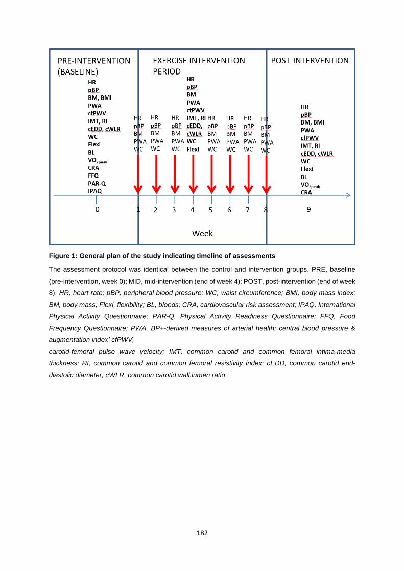
182
Figure 1: General plan of the study indicating timeline of assessments
The assessment protocol was identical between the control and intervention groups. PRE, baseline
(pre-intervention, week 0); MID, mid-intervention (end of week 4); POST, post-intervention (end of week
8). HR, heart rate; pBP, peripheral blood pressure; WC, waist circumference; BMI, body mass index;
BM, body mass; Flexi, flexibility; BL, bloods; CRA, cardiovascular risk assessment; IPAQ, International
Physical Activity Questionnaire; PAR-Q, Physical Activity Readiness Questionnaire; FFQ, Food
Frequency Questionnaire; PWA, BP+-derived measures of arterial health: central blood pressure &
augmentation index’ cfPWV,
carotid-femoral pulse wave velocity; IMT, common carotid and common femoral intima-media
thickness; RI, common carotid and common femoral resistivity index; cEDD, common carotid end-
diastolic diameter; cWLR, common carotid wall:lumen ratio
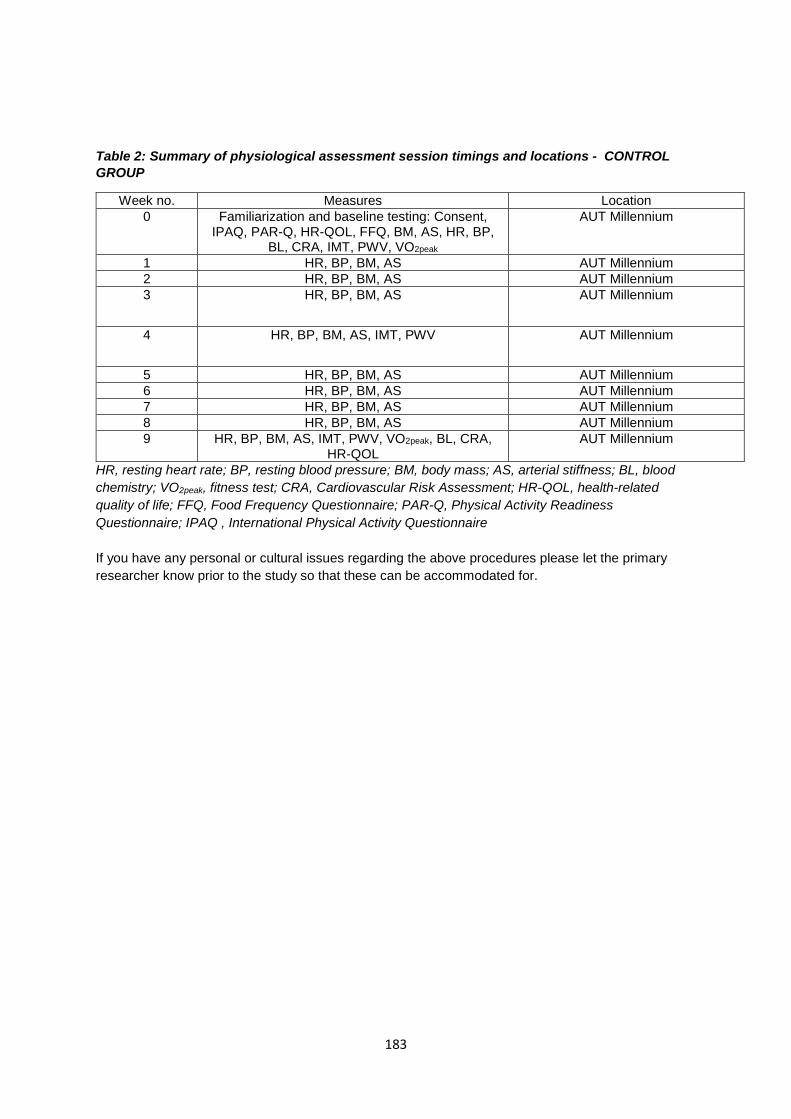
183
Table 2: Summary of physiological assessment session timings and locations - CONTROL GROUP
Week no. Measures Location 0 Familiarization and baseline testing: Consent,
IPAQ, PAR-Q, HR-QOL, FFQ, BM, AS, HR, BP, BL, CRA, IMT, PWV, VO2peak
AUT Millennium
1 HR, BP, BM, AS AUT Millennium 2 HR, BP, BM, AS AUT Millennium 3 HR, BP, BM, AS AUT Millennium
4 HR, BP, BM, AS, IMT, PWV AUT Millennium
5 HR, BP, BM, AS AUT Millennium 6 HR, BP, BM, AS AUT Millennium 7 HR, BP, BM, AS AUT Millennium 8 HR, BP, BM, AS AUT Millennium 9 HR, BP, BM, AS, IMT, PWV, VO2peak, BL, CRA,
HR-QOL AUT Millennium
HR, resting heart rate; BP, resting blood pressure; BM, body mass; AS, arterial stiffness; BL, blood chemistry; VO2peak, fitness test; CRA, Cardiovascular Risk Assessment; HR-QOL, health-related quality of life; FFQ, Food Frequency Questionnaire; PAR-Q, Physical Activity Readiness Questionnaire; IPAQ , International Physical Activity Questionnaire If you have any personal or cultural issues regarding the above procedures please let the primary researcher know prior to the study so that these can be accommodated for.
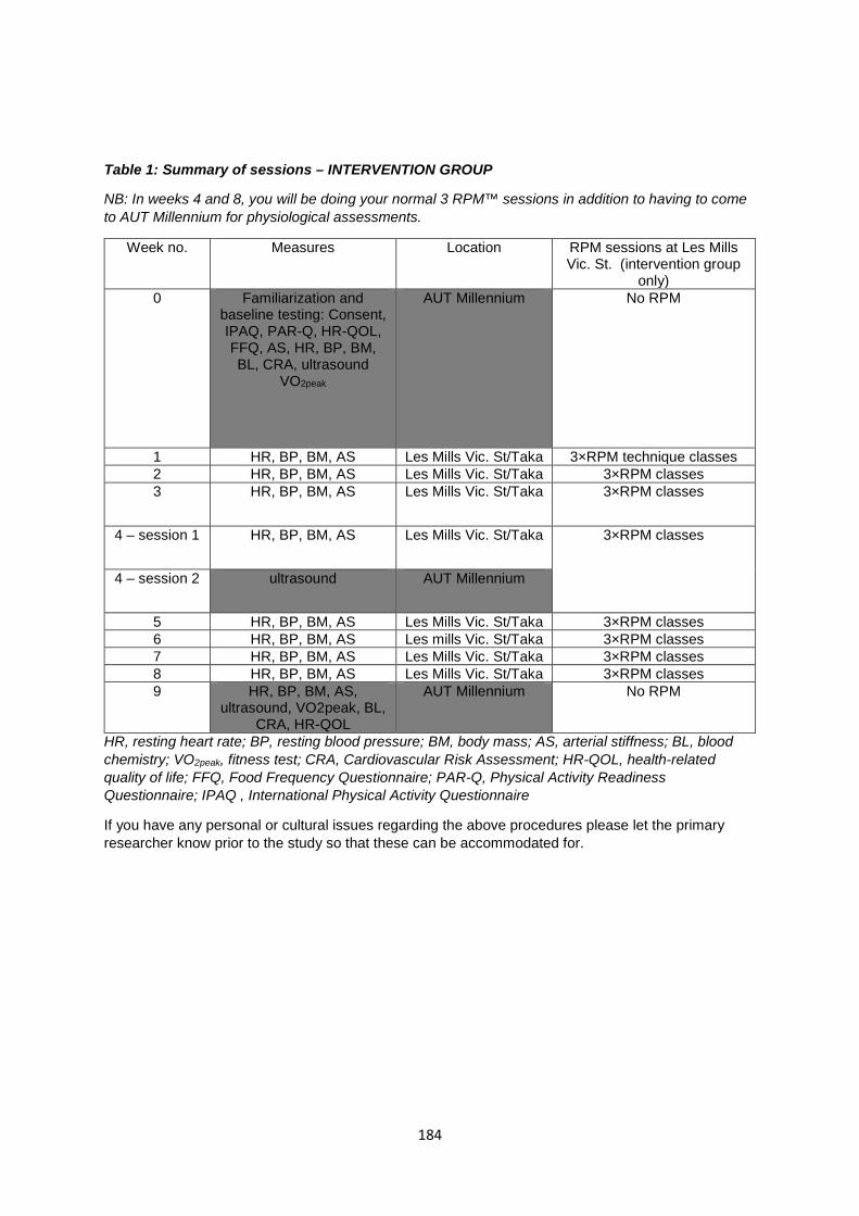
184
Table 1: Summary of sessions – INTERVENTION GROUP
NB: In weeks 4 and 8, you will be doing your normal 3 RPM™ sessions in addition to having to come to AUT Millennium for physiological assessments.
Week no. Measures Location RPM sessions at Les Mills Vic. St. (intervention group
only) 0
Familiarization and baseline testing: Consent, IPAQ, PAR-Q, HR-QOL, FFQ, AS, HR, BP, BM, BL, CRA, ultrasound
VO2peak
AUT Millennium
No RPM
1 HR, BP, BM, AS Les Mills Vic. St/Taka 3×RPM technique classes 2 HR, BP, BM, AS Les Mills Vic. St/Taka 3×RPM classes 3 HR, BP, BM, AS Les Mills Vic. St/Taka 3×RPM classes
4 – session 1 HR, BP, BM, AS Les Mills Vic. St/Taka 3×RPM classes
4 – session 2 ultrasound AUT Millennium
5 HR, BP, BM, AS Les Mills Vic. St/Taka 3×RPM classes 6 HR, BP, BM, AS Les mills Vic. St/Taka 3×RPM classes 7 HR, BP, BM, AS Les Mills Vic. St/Taka 3×RPM classes 8 HR, BP, BM, AS Les Mills Vic. St/Taka 3×RPM classes 9 HR, BP, BM, AS,
ultrasound, VO2peak, BL, CRA, HR-QOL
AUT Millennium No RPM
HR, resting heart rate; BP, resting blood pressure; BM, body mass; AS, arterial stiffness; BL, blood chemistry; VO2peak, fitness test; CRA, Cardiovascular Risk Assessment; HR-QOL, health-related quality of life; FFQ, Food Frequency Questionnaire; PAR-Q, Physical Activity Readiness Questionnaire; IPAQ , International Physical Activity Questionnaire
If you have any personal or cultural issues regarding the above procedures please let the primary researcher know prior to the study so that these can be accommodated for.
185
What are the discomforts and risks?
You may feel a small pinch on your finger when blood is being drawn but this should last less than 2 seconds and depending on you as an individual, you may bleed extremely little or slightly more from the site of the prick on your finger. Again, there should not be a lot of blood and it should not bleed for more than a few minutes, if that!
During testing: the only discomfort you may feel is when a cuff placed around your arm inflates and compresses your artery during the arterial stiffness measurements. This will last for no longer than 15 seconds. The cuff will not be too tight and it is totally safe! During the ultrasound assessments, you may feel slight pressure from the probe on your neck and groin but this would be extremely light pressure and should not be uncomfortable. As you will need to be tested when in a fasted state, you may be hungry before and during the assessment procedures (weeks 0, 4 and 9) but you will be provided with free drinks and snacks at the end of the assessment session!
During exercise: during the fitness test, exercise intensity may be quite high and you will feel tired and breathless and your muscles may even feel fatigued. However, the test will occur in a controlled, supervised environment.
If you are in the intervention group, during the indoor cycling (rpm) sessions, you may feel tired, thirsty, fatigued and have slightly sore muscles from previous sessions but this is normal when exercising, particularly if you are new to exercise. We understand that you may not be fmailiar with exercise but you can rest assured that these sessions will also be carried out in safe, controlled environments. In addition, you will be given technique classes to ensure that you are exercising safely and have proper form when cycling. As with any physical activity, there is always a small chance of injury but we will try to avoid this by ensuring proper technique, a warm-up and cool-down.
What are the benefits?
You will benefit from being part of this study as you will receive a lot of information about different aspects of your health status (eg. blood profile, fitness levels and cardiovascular, in particular, arterial health). The tests are normally very costly and are therefore not carried out often on healthy people in the general public.
You shall be introduced to exercise in a safe way and although you will not be able to participate in other physical activity during the study, it will hopefully make you interested in exercise and enable you to keep it up after the study has been completed.
For those initially allocated to the control group for the first 10 weeks, a 2 week free Les Mills gym trial at the end of the study will be provided and there will be no joining fee if you decide to obtain Les Mills membership.
For those initially allocated to the control group for the first 10 weeks, free 4 week access to RPM™ rides will be provided as will a 2 week free Les Mills gym trial at the end of the study, plus no joining fee if you decide to become a member of Les Mills.
What compensation is available for injury or negligence?
In the unlikely event of a physical injury as a result of your participation in this study, rehabilitation and compensation for injury by accident may be available from the Accident Compensation Corporation, providing the incident details satisfy the requirements of the law and the Corporation's regulations.
How will my privacy be protected?
186
All the data obtained during this study will only be available to the researchers involved. If the data is published in the public domain your name as a subject will not be revealed and you will remain anonymous. The only information you will need to provide to a third party would be your personal details (ie. who you are and your contact details). This is required to allow you free access into the Les Mills gym.
What are the costs of participating in this research?
The cost of participating in this study will be the time you will have to put aside for it. If you are initially allocated to the intervention group, you will need to set aside a total of 24 hours for the exercise sessions and a total of about 6 hours for testing sessions (this time excludes travel time to and from the venue). If you are initially allocated to the control group, you will need to come in for a total of about 9 hours during the first 10 weeks and then set aside 24 hours during the next 8 weeks (includes exercise and assessment sessions) when you have the opportunity to go through the intervention.
Will I receive feedback on the results of this research?
At the end of the study, verbal feedback will be given to you and written feedback can be provided upon request. Your results from the study will only be shared with your doctor if you are at any health risk and you grant us permission to do so.
What do I do if I have concerns about this research?
Any concerns regarding the nature of this project should be notified in the first instance to the Project Supervisor:
Name: Associate Professor Andrew Kilding
E-mail: [email protected]
Concerns regarding the conduct of the research should be notified to the Executive Secretary, AUTEC, Kate O’Connor, [email protected], 921 9999 ext 6038.
Whom do I contact for further information about this research?
Researcher contact details
Name: Shivani Sethi; E-mail: [email protected]
Project Supervisor contact details
Name: Dr. Andrew Kilding; E-mail: [email protected]
Approved by the Auckland University of Technology Ethics Committee on the 26th August 2014, AUTEC Reference number 14/242.
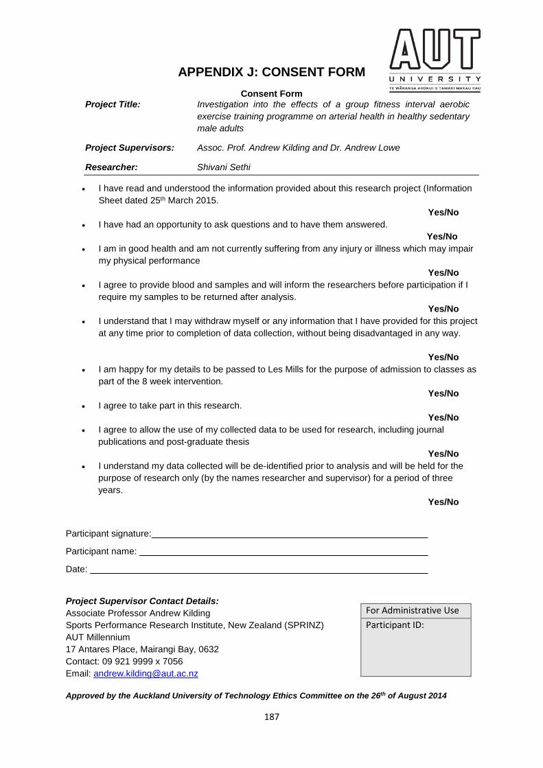
187
APPENDIX J: CONSENT FORM Consent Form
Project Title: Investigation into the effects of a group fitness interval aerobic exercise training programme on arterial health in healthy sedentary male adults
Project Supervisors: Assoc. Prof. Andrew Kilding and Dr. Andrew Lowe
Researcher: Shivani Sethi
• I have read and understood the information provided about this research project (Information Sheet dated 25th March 2015. Yes/No
• I have had an opportunity to ask questions and to have them answered. Yes/No
• I am in good health and am not currently suffering from any injury or illness which may impair my physical performance Yes/No
• I agree to provide blood and samples and will inform the researchers before participation if I require my samples to be returned after analysis. Yes/No
• I understand that I may withdraw myself or any information that I have provided for this project at any time prior to completion of data collection, without being disadvantaged in any way. Yes/No
• I am happy for my details to be passed to Les Mills for the purpose of admission to classes as part of the 8 week intervention. Yes/No
• I agree to take part in this research. Yes/No
• I agree to allow the use of my collected data to be used for research, including journal publications and post-graduate thesis Yes/No
• I understand my data collected will be de-identified prior to analysis and will be held for the purpose of research only (by the names researcher and supervisor) for a period of three years. Yes/No
Participant signature:
Participant name:
Date:
Project Supervisor Contact Details: Associate Professor Andrew Kilding Sports Performance Research Institute, New Zealand (SPRINZ) AUT Millennium 17 Antares Place, Mairangi Bay, 0632 Contact: 09 921 9999 x 7056 Email: [email protected] Approved by the Auckland University of Technology Ethics Committee on the 26th of August 2014
For Administrative Use Participant ID:
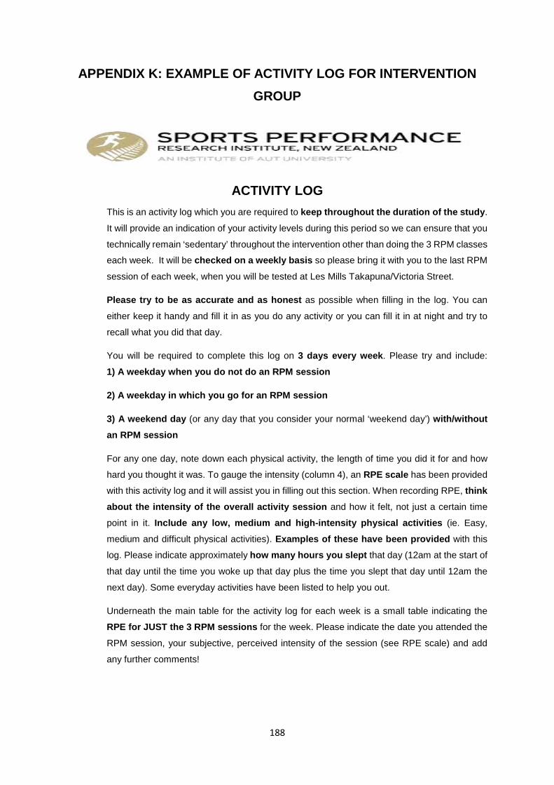
188
APPENDIX K: EXAMPLE OF ACTIVITY LOG FOR INTERVENTION GROUP
ACTIVITY LOG
This is an activity log which you are required to keep throughout the duration of the study.
It will provide an indication of your activity levels during this period so we can ensure that you
technically remain ‘sedentary’ throughout the intervention other than doing the 3 RPM classes
each week. It will be checked on a weekly basis so please bring it with you to the last RPM
session of each week, when you will be tested at Les Mills Takapuna/Victoria Street.
Please try to be as accurate and as honest as possible when filling in the log. You can
either keep it handy and fill it in as you do any activity or you can fill it in at night and try to
recall what you did that day.
You will be required to complete this log on 3 days every week. Please try and include:
1) A weekday when you do not do an RPM session
2) A weekday in which you go for an RPM session
3) A weekend day (or any day that you consider your normal ‘weekend day’) with/without an RPM session
For any one day, note down each physical activity, the length of time you did it for and how
hard you thought it was. To gauge the intensity (column 4), an RPE scale has been provided
with this activity log and it will assist you in filling out this section. When recording RPE, think about the intensity of the overall activity session and how it felt, not just a certain time
point in it. Include any low, medium and high-intensity physical activities (ie. Easy,
medium and difficult physical activities). Examples of these have been provided with this
log. Please indicate approximately how many hours you slept that day (12am at the start of
that day until the time you woke up that day plus the time you slept that day until 12am the
next day). Some everyday activities have been listed to help you out.
Underneath the main table for the activity log for each week is a small table indicating the
RPE for JUST the 3 RPM sessions for the week. Please indicate the date you attended the
RPM session, your subjective, perceived intensity of the session (see RPE scale) and add
any further comments!

189
Please note: when recording the intensity (RPE) for the RPM session, think about the session as a whole and how the overall class felt (ie. try not to simply think of the single
hardest point and allocate a number to that specific feeling).
RPE scale
This scale will enable you to gauge the intensity of each physical activity that you do. It is very subjective so try and pick the number which best represents how you felt during each activity
RPE DESCRIPTION 0 Complete Rest; Nothing at all; Sleeping; sitting quietly 1 Very, very easy; Very light; Just noticeable; Standing 2 Easy; Weak; Light 3 Moderate 4 Somewhat hard 5
Hard; Strong; Heavy 6 7
Very hard; very strong 8 9 Near maximal 10 Maximal; Extremely hard; about to give up 11 All-out effort; unable to continue; Exhaustion; Absolute maximum; Highest possible
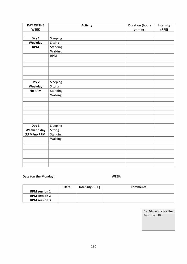
190
Date (on the Monday): WEEK:
DAY OF THE WEEK
Activity Duration (hours or mins)
Intensity (RPE)
Day 1 Sleeping
Weekday Sitting RPM Standing
Walking RPM
Day 2 Sleeping Weekday Sitting No RPM Standing
Walking
Day 3 Sleeping Weekend day Sitting
(RPM/no RPM) Standing Walking
Date Intensity (RPE) Comments RPM session 1 RPM session 2 RPM session 3
For Administrative Use Participant ID:
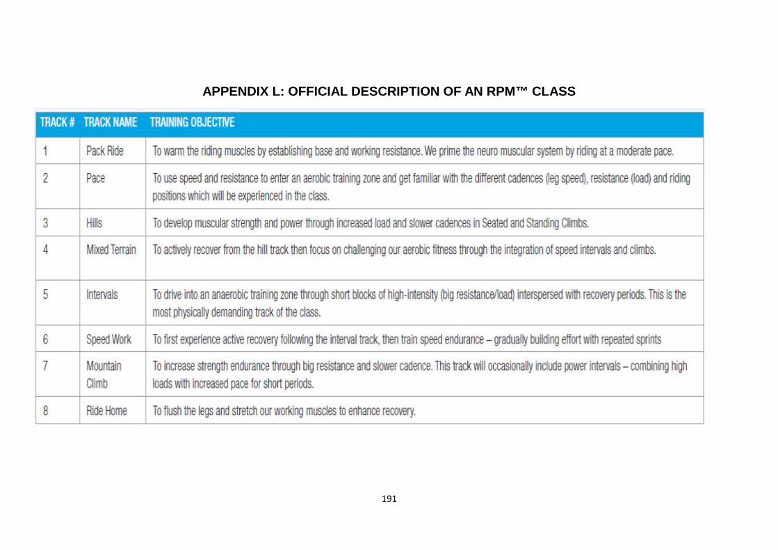
191
APPENDIX L: OFFICIAL DESCRIPTION OF AN RPM™ CLASS
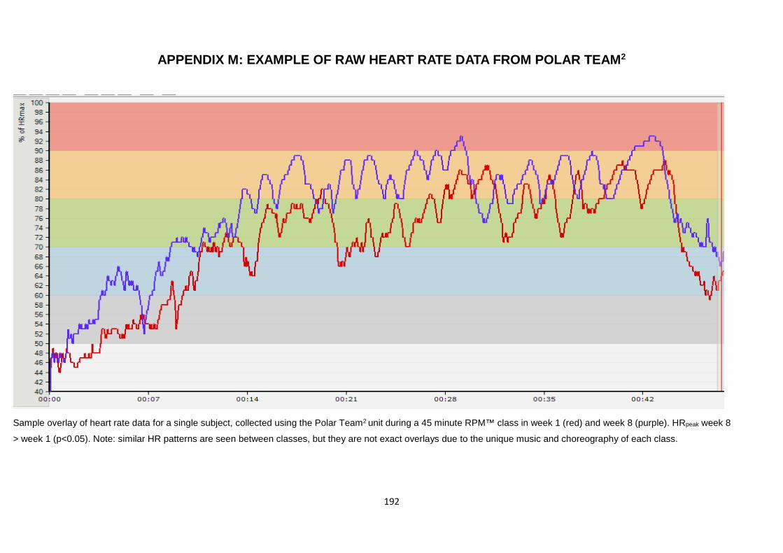
192
APPENDIX M: EXAMPLE OF RAW HEART RATE DATA FROM POLAR TEAM2
Sample overlay of heart rate data for a single subject, collected using the Polar Team2 unit during a 45 minute RPM™ class in week 1 (red) and week 8 (purple). HRpeak week 8
> week 1 (p<0.05). Note: similar HR patterns are seen between classes, but they are not exact overlays due to the unique music and choreography of each class.
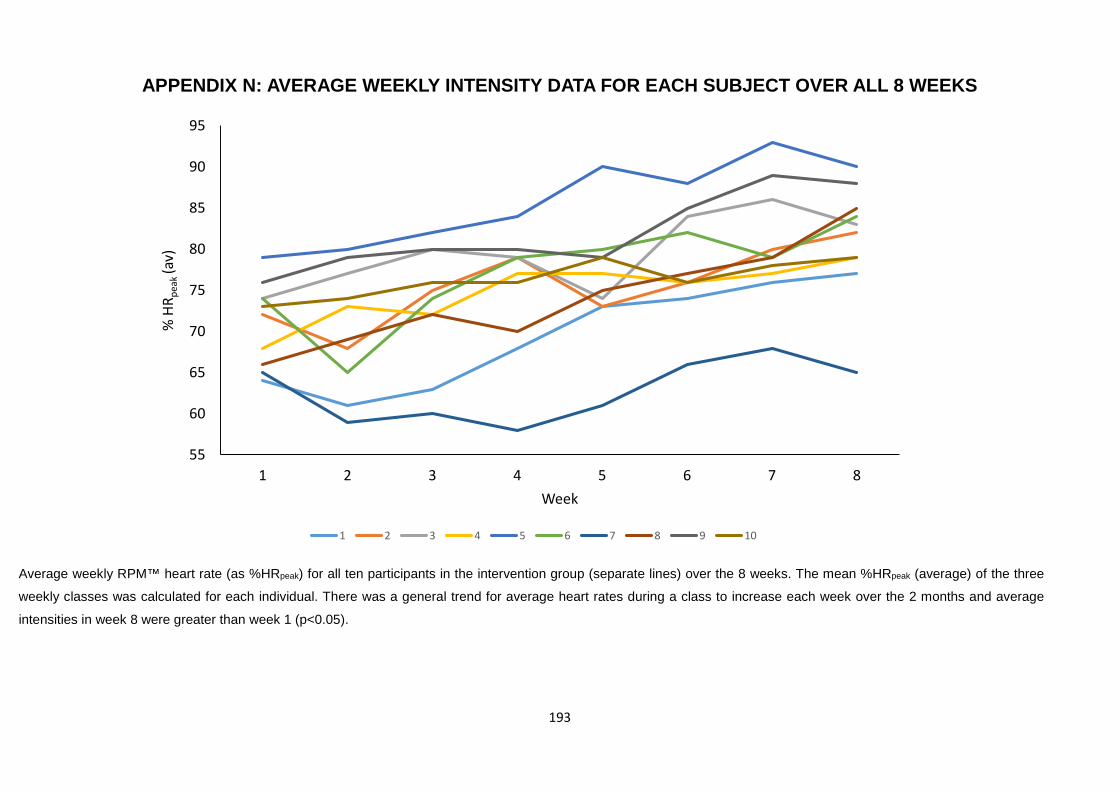
193
APPENDIX N: AVERAGE WEEKLY INTENSITY DATA FOR EACH SUBJECT OVER ALL 8 WEEKS
Average weekly RPM™ heart rate (as %HRpeak) for all ten participants in the intervention group (separate lines) over the 8 weeks. The mean %HRpeak (average) of the three
weekly classes was calculated for each individual. There was a general trend for average heart rates during a class to increase each week over the 2 months and average
intensities in week 8 were greater than week 1 (p<0.05).
55
60
65
70
75
80
85
90
95
1 2 3 4 5 6 7 8
% H
R pea
k (a
v)
Week
1 2 3 4 5 6 7 8 9 10