Water Sorption, Solubility, and Translucency of 3D-Printed ...
Upload
khangminh22Category
view
2download
0
Citation: Bácskay, I.; Ujhelyi, Z.;
Fehér, P.; Arany, P. The Evolution of
the 3D-Printed Drug Delivery
Systems: A Review. Pharmaceutics
2022, 14, 1312. https://doi.org/
10.3390/pharmaceutics14071312
Academic Editors: Mateusz Kurek
and Witold Jamróz
Received: 11 May 2022
Accepted: 18 June 2022
Published: 21 June 2022
Publisher’s Note: MDPI stays neutral
with regard to jurisdictional claims in
published maps and institutional affil-
iations.
Copyright: © 2022 by the authors.
Licensee MDPI, Basel, Switzerland.
This article is an open access article
distributed under the terms and
conditions of the Creative Commons
Attribution (CC BY) license (https://
creativecommons.org/licenses/by/
4.0/).
pharmaceutics
Review
The Evolution of the 3D-Printed Drug Delivery Systems: A ReviewIldikó Bácskay 1,2,* , Zoltán Ujhelyi 2 , Pálma Fehér 2 and Petra Arany 1
1 Healthcare Industry Institute, University of Debrecen, Nagyerdei körút 98, H-4032 Debrecen, Hungary;[email protected]
2 Department of Pharmaceutical Technology, Faculty of Pharmacy, University of Debrecen, Nagyerdei körút 98,H-4032 Debrecen, Hungary; [email protected] (Z.U.); [email protected] (P.F.)
* Correspondence: [email protected]; Tel.: +36-52411717 (ext. 56946)
Abstract: Since the appearance of the 3D printing in the 1980s it has revolutionized many researchfields including the pharmaceutical industry. The main goal is to manufacture complex, personalizedproducts in a low-cost manufacturing process on-demand. In the last few decades, 3D printinghas attracted the attention of numerous research groups for the manufacturing of different drugdelivery systems. Since the 2015 approval of the first 3D-printed drug product, the number ofpublications has multiplied. In our review, we focused on summarizing the evolution of the produceddrug delivery systems in the last 20 years and especially in the last 5 years. The drug deliverysystems are sub-grouped into tablets, capsules, orodispersible films, implants, transdermal deliverysystems, microneedles, vaginal drug delivery systems, and micro- and nanoscale dosage forms. Ourclassification may provide guidance for researchers to more easily examine the publications and tofind further research directions.
Keywords: 3D printing; drug delivery systems; tablet; implant; TTS; microneedle
1. Introduction
Three-dimensional (3D) printing was developed more than 30 years ago to manu-facture 3D objects based on a digital design. This layer-by-layer process enables a fastand cheap design cycle for the preparation of personalized medication [1]. The term 3Dprinting was coined as an umbrella term and encompasses a number of processes, and inmany reviews the main types were described in detail [2–5]. Three-dimensional printinggave the means to the manufacture of a high-quality product within minutes in an easymanufacturing cycle. This on-demand manufacturing was time and material saving. Notto mention the fact that 3D printers could conquer the traditional manufacturing regimeof ‘one size fits all’ [6]. As 3D printing was based on a computer-aided design (CAD), itprovided the ability to quickly create and produce a flexible and innovative product [7]. Per-sonalized medication carried the opportunity to create drug delivery systems for patient’srequirements. Furthermore, 3D printing gained access to the creation of unique dosageforms and achieving more complex drug release profiles [8]. The image could be madeto meet the patient’s individual needs regarding their age, weight, organ function, andseverity of disease [4]. The application of 3D printing technology might be an alternativeway to construct effective, customized active pharmaceutical ingredient (API) combinationsfor the patient immediately [9]. The 3D printing technique opened up the opportunity forthe development of tailored single and multi-drug products at the point-of-care [10].
In recent years, many comprehensive publications have been presented on the differentdesigned drug dosage forms. As Moulton et al. highlighted, this kind of process created theopportunity for the manufacturing of controlled and modified release of the APIs, enabledthe delivery of poorly water soluble drugs, increased drug stability, and reduced the usedAPI amount without compromising the efficacy [11]. In 2018, two distinct research groupssummarized the recent achievements in the manufacturing of pharmaceuticals but as a
Pharmaceutics 2022, 14, 1312. https://doi.org/10.3390/pharmaceutics14071312 https://www.mdpi.com/journal/pharmaceutics

Pharmaceutics 2022, 14, 1312 2 of 30
rapidly developing area the achievements vary from year to year [12,13]. Mohapatra et al.gathered together the newest publications in recent years and grouped the research basedon the type of 3D printing [14].
The available reviews mostly focus on one or multiple drug dosage forms manufac-tured by one type of 3D printing technology. Cunha-Filho et al. discussed the fabricateddrug delivery systems by fused deposition modeling (FDM) 3D printing [15]. While Guecheet al. summed up the oral dosage forms created by selective laser sintering (SLS) [16] andWang et al. described the stereolithographic (SLA) constructed oral dosage forms [17].Inkjet printing of pharmaceuticals was summarized by Dali et al. [18]. In more and moreresearch, three-dimensional bioprinting was used which is a new era of 3D printing tech-nologies where researchers aim to build living tissue models [19].
During the last decades multiple research groups were established to fabricate drugdelivery systems. FabRx Ltd. is one of the most innovative start-up companies, which is abiotech company, designed to produce 3D-printed medication [20]. Regarding the diversedrug dosage forms, different reviews summarized the achievements. For example buccalpatches were analyzed in the work of Shirvan et al. [21], implants in the work of Domstaet al. [22], oral dosage forms in the work of Khatri et al. [23], and transdermal deliverysystems (TTS) in the work of Economidou et al. [24].
The aim of this work was to provide a comprehensive image on how the manufacturingof the different drug delivery systems started and where the experiments are headed now.The chosen drug delivery systems were divided into subgroups based on the type of thedrug delivery system and the tables summarized the most important research in the last20 years in chronological order.
2. The 3D Printing of Drug Delivery Systems2.1. Tablets
The first publication of a 3D-printed tablet dates back to 1996 when solid sampleswere created with a desktop printer from PCL and PEO polymers containing blue andyellow dyes. Based on the results, complex drug delivery regimes could be created withthis technique, such as the release of multiple drugs or multiphasic release of a single drug.This study demonstrated several simple examples of such devices and several constructionmethods that could be used to control the release of the drugs [25].
In the early 3D printing articles, droplet binding was used for the manufacturing ofthe samples when the used binder was not necessarily polymer but some other auxiliarymaterial, e.g., Eudragit® or mannitol. The authors concluded that with this method ade-quate oral dosage forms can be manufactured which exhibit erosion or diffusion releasemechanisms [26,27]. At the beginning of the research, the most important question forthe authors was the type of the chosen additive manufacturing process, the used printingparameters, immediate- or delayed release tablet manufacturing, and first- or zero-orderkinetic profile manufacturing [28].
Gbureck et al. used a unique technology for the manufacturing of the drug deliverysystem. Firstly, they created the sample with a 3D bioceramic powder printing processand then the used antibiotics were adsorbed during a week to fabricate the tablets [29]. Yuet al. produced an acetaminophen containing matrix tablet using a desktop 3D printer. Themiddle drug-containing regions of the tablets were formed by depositing the binder liquidcontaining release-modulation materials onto the automatically spread powder layers(Figure 1) [30]. Two years later, the same investigators decided to construct a drug deliverysystem from the same API, polymer, and printing technique but in this case the layers werenot designed horizontally, yet vertically, to provide a different dissolution mechanism [31].

Pharmaceutics 2022, 14, 1312 3 of 30
Pharmaceutics 2022, 14, 1312 3 of 30
layers were not designed horizontally, yet vertically, to provide a different dissolution
mechanism [31].
Figure 1. Cross-section of the acetaminophen-containing matrix tablets based on the authors’ figure.
The different colors label dissimilar compartments [30].
Guaifenesin-containing controlled release bilayer tablets were constructed with ex-
trusion-based desktop 3D printing. The samples were formulated to demonstrate the pro-
duction of relatively complex formulations that could mimic the release profile of a com-
mercially available tablet [32].
Even though the wide range experiments started in the 2010s, the Food and Drug
Administration agency (FDA) granted the approval of Spritam® in 2015. This 3D-printed
tablet was used for the treatment of epileptic seizures. This was the first and still only
approved 3D-printed drug delivery system. For the manufacturing, a so-called ZipDose
Technology was used [33].
Goyanes et al. published four articles in 2015 about the manufacturing of tablets for
distinct purposes with the use of FDM 3D printing and PVA filament. In one of the arti-
cles, the commercially produced PVA filaments were loaded with 5- and 4-amino salicylic
acid in an ethanolic drug solution. A final drug loading of 0.06% w/w and 0.25% w/w was
achieved for the APIs, respectively. This filament was created with unsimilar infill per-
centages in a nonidentical pattern (Figure 2). The dissolution tests showed that the release
profiles depended on the used infill percentage and the used API itself [34].
Figure 2. Cross-section of the constructed drug dosage forms in the article of Goyanes et al. (a) Sec-
tioned multilayer tablet, (b) sectioned DuoCaplet (caplet in caplet) [35].
A polypill was designed by a RegenHU extrusion-based 3D printer which contained
distinct fillable ink cartridges for the production of semi-solid API-containing materials.
In this research, three different API—nifedipine, captopril, and glipizide—containing inks
were manufactured by HPMC. The three APIs could be found in three diverse compart-
ments. From the nifedipine and the glipizide-containing formulations, the drug was re-
leased by diffusion and from the captopril formulation by osmosis. The schematic image
of the samples is shown in Figure 3 [36].
Figure 1. Cross-section of the acetaminophen-containing matrix tablets based on the authors’ figure.The different colors label dissimilar compartments [30].
Guaifenesin-containing controlled release bilayer tablets were constructed with extrusion-based desktop 3D printing. The samples were formulated to demonstrate the production ofrelatively complex formulations that could mimic the release profile of a commercially availabletablet [32].
Even though the wide range experiments started in the 2010s, the Food and DrugAdministration agency (FDA) granted the approval of Spritam® in 2015. This 3D-printedtablet was used for the treatment of epileptic seizures. This was the first and still onlyapproved 3D-printed drug delivery system. For the manufacturing, a so-called ZipDoseTechnology was used [33].
Goyanes et al. published four articles in 2015 about the manufacturing of tablets fordistinct purposes with the use of FDM 3D printing and PVA filament. In one of the articles,the commercially produced PVA filaments were loaded with 5- and 4-amino salicylic acid inan ethanolic drug solution. A final drug loading of 0.06% w/w and 0.25% w/w was achievedfor the APIs, respectively. This filament was created with unsimilar infill percentages ina nonidentical pattern (Figure 2). The dissolution tests showed that the release profilesdepended on the used infill percentage and the used API itself [34].
Pharmaceutics 2022, 14, 1312 3 of 30
layers were not designed horizontally, yet vertically, to provide a different dissolution
mechanism [31].
Figure 1. Cross-section of the acetaminophen-containing matrix tablets based on the authors’ figure.
The different colors label dissimilar compartments [30].
Guaifenesin-containing controlled release bilayer tablets were constructed with ex-
trusion-based desktop 3D printing. The samples were formulated to demonstrate the pro-
duction of relatively complex formulations that could mimic the release profile of a com-
mercially available tablet [32].
Even though the wide range experiments started in the 2010s, the Food and Drug
Administration agency (FDA) granted the approval of Spritam® in 2015. This 3D-printed
tablet was used for the treatment of epileptic seizures. This was the first and still only
approved 3D-printed drug delivery system. For the manufacturing, a so-called ZipDose
Technology was used [33].
Goyanes et al. published four articles in 2015 about the manufacturing of tablets for
distinct purposes with the use of FDM 3D printing and PVA filament. In one of the arti-
cles, the commercially produced PVA filaments were loaded with 5- and 4-amino salicylic
acid in an ethanolic drug solution. A final drug loading of 0.06% w/w and 0.25% w/w was
achieved for the APIs, respectively. This filament was created with unsimilar infill per-
centages in a nonidentical pattern (Figure 2). The dissolution tests showed that the release
profiles depended on the used infill percentage and the used API itself [34].
Figure 2. Cross-section of the constructed drug dosage forms in the article of Goyanes et al. (a) Sec-
tioned multilayer tablet, (b) sectioned DuoCaplet (caplet in caplet) [35].
A polypill was designed by a RegenHU extrusion-based 3D printer which contained
distinct fillable ink cartridges for the production of semi-solid API-containing materials.
In this research, three different API—nifedipine, captopril, and glipizide—containing inks
were manufactured by HPMC. The three APIs could be found in three diverse compart-
ments. From the nifedipine and the glipizide-containing formulations, the drug was re-
leased by diffusion and from the captopril formulation by osmosis. The schematic image
of the samples is shown in Figure 3 [36].
Figure 2. Cross-section of the constructed drug dosage forms in the article of Goyanes et al. (a) Sec-tioned multilayer tablet, (b) sectioned DuoCaplet (caplet in caplet) [35].
A polypill was designed by a RegenHU extrusion-based 3D printer which containeddistinct fillable ink cartridges for the production of semi-solid API-containing materials.In this research, three different API—nifedipine, captopril, and glipizide—containinginks were manufactured by HPMC. The three APIs could be found in three diverse com-partments. From the nifedipine and the glipizide-containing formulations, the drug wasreleased by diffusion and from the captopril formulation by osmosis. The schematic imageof the samples is shown in Figure 3 [36].
In the same year, a newer design was applied which is also a polypill but containedfive different APIs: amino salicylic acid (ASA) and hydrochlorothiazide (HCT) in thebottom layer, and atenolol, ramipril, and pravastatin in the middle area in three separatedregions. The manufacturing was described in the previous article. The ASA and HCTformulation were an immediate release compartment, while the others were controlledwith a cellulose acetate membrane to provide extended release. The graphical scheme isshown in Figure 4 [37].

Pharmaceutics 2022, 14, 1312 4 of 30Pharmaceutics 2022, 14, 1312 4 of 30
Figure 3. Cross-section of the printed polypills where three diverse compartments were created (la-
beled with three nonidentical colors) [36].
In the same year, a newer design was applied which is also a polypill but contained
five different APIs: amino salicylic acid (ASA) and hydrochlorothiazide (HCT) in the bot-
tom layer, and atenolol, ramipril, and pravastatin in the middle area in three separated
regions. The manufacturing was described in the previous article. The ASA and HCT for-
mulation were an immediate release compartment, while the others were controlled with
a cellulose acetate membrane to provide extended release. The graphical scheme is shown
in Figure 4 [37].
Figure 4. Cross-section of the polypills where ASA and HCT formulations were located in the upper
immediate release compartments (labeled with blue and orange rectangles) and atenolol, pravas-
tatin, and ramipril formulations were in three distinct extended release compartments (labeled with
yellow, green, and peach blossom). The three compartments were the same size but could be visu-
alized like this because of the original design of the “cake slice” [37].
Metolose® (a special cellulose ester) and PLA disks were created and co-extruded
with nitrofurantoin as a model API. The research showed that the rheological properties
depended on the amount of the undissolved particles and, as in the case of modified re-
lease tablets, the amount of the cellulose derivative affected the dissolution time from the
fabricated filament [38].
In the work of Okwuosa et al., a 10% API-containing filament was produced by hot-
melt extrusion. As an API dipyridamole or theophylline, as a polymer PVP, and as an
excipient plasticizer was used. These constructed filaments were then FDM 3D printed.
The novelty of this work is that, for the extrusion, thermostable filler (talc) was used which
enabled lower temperature printing around 110 °C and the stability of the used APIs was
not affected [39].
In the same year, Sadia et al. decided to use a pharmaceutical-grade non-melting filler
(TCP) through the hot-melt extrusion to allow a consistent flow from the nozzle of the
printer. This novel approach meant the addition of 20–50% non-melting component to the
filament and four model drugs were incorporated separately. This process aimed at the
fabrication of well-defined caplets. In the case of 5-ASA and prednisolone, 93% of the drug
contents remained intact in the tablet but a significant drop in captopril content was ob-
Figure 3. Cross-section of the printed polypills where three diverse compartments were created(labeled with three nonidentical colors) [36].
Pharmaceutics 2022, 14, 1312 4 of 30
Figure 3. Cross-section of the printed polypills where three diverse compartments were created (la-
beled with three nonidentical colors) [36].
In the same year, a newer design was applied which is also a polypill but contained
five different APIs: amino salicylic acid (ASA) and hydrochlorothiazide (HCT) in the bot-
tom layer, and atenolol, ramipril, and pravastatin in the middle area in three separated
regions. The manufacturing was described in the previous article. The ASA and HCT for-
mulation were an immediate release compartment, while the others were controlled with
a cellulose acetate membrane to provide extended release. The graphical scheme is shown
in Figure 4 [37].
Figure 4. Cross-section of the polypills where ASA and HCT formulations were located in the upper
immediate release compartments (labeled with blue and orange rectangles) and atenolol, pravas-
tatin, and ramipril formulations were in three distinct extended release compartments (labeled with
yellow, green, and peach blossom). The three compartments were the same size but could be visu-
alized like this because of the original design of the “cake slice” [37].
Metolose® (a special cellulose ester) and PLA disks were created and co-extruded
with nitrofurantoin as a model API. The research showed that the rheological properties
depended on the amount of the undissolved particles and, as in the case of modified re-
lease tablets, the amount of the cellulose derivative affected the dissolution time from the
fabricated filament [38].
In the work of Okwuosa et al., a 10% API-containing filament was produced by hot-
melt extrusion. As an API dipyridamole or theophylline, as a polymer PVP, and as an
excipient plasticizer was used. These constructed filaments were then FDM 3D printed.
The novelty of this work is that, for the extrusion, thermostable filler (talc) was used which
enabled lower temperature printing around 110 °C and the stability of the used APIs was
not affected [39].
In the same year, Sadia et al. decided to use a pharmaceutical-grade non-melting filler
(TCP) through the hot-melt extrusion to allow a consistent flow from the nozzle of the
printer. This novel approach meant the addition of 20–50% non-melting component to the
filament and four model drugs were incorporated separately. This process aimed at the
fabrication of well-defined caplets. In the case of 5-ASA and prednisolone, 93% of the drug
contents remained intact in the tablet but a significant drop in captopril content was ob-
Figure 4. Cross-section of the polypills where ASA and HCT formulations were located in the upperimmediate release compartments (labeled with blue and orange rectangles) and atenolol, pravastatin,and ramipril formulations were in three distinct extended release compartments (labeled with yellow,green, and peach blossom). The three compartments were the same size but could be visualized likethis because of the original design of the “cake slice” [37].
Metolose® (a special cellulose ester) and PLA disks were created and co-extrudedwith nitrofurantoin as a model API. The research showed that the rheological propertiesdepended on the amount of the undissolved particles and, as in the case of modifiedrelease tablets, the amount of the cellulose derivative affected the dissolution time from thefabricated filament [38].
In the work of Okwuosa et al., a 10% API-containing filament was produced by hot-melt extrusion. As an API dipyridamole or theophylline, as a polymer PVP, and as anexcipient plasticizer was used. These constructed filaments were then FDM 3D printed.The novelty of this work is that, for the extrusion, thermostable filler (talc) was used whichenabled lower temperature printing around 110 ◦C and the stability of the used APIs wasnot affected [39].
In the same year, Sadia et al. decided to use a pharmaceutical-grade non-meltingfiller (TCP) through the hot-melt extrusion to allow a consistent flow from the nozzle ofthe printer. This novel approach meant the addition of 20–50% non-melting componentto the filament and four model drugs were incorporated separately. This process aimedat the fabrication of well-defined caplets. In the case of 5-ASA and prednisolone, 93% ofthe drug contents remained intact in the tablet but a significant drop in captopril contentwas observed due to thermal degradation. This procedure made the manufacturing ofpersonalized immediate release tablets easy [40]. In Figure 5. the most important researchresults can be seen between 1996–2016.

Pharmaceutics 2022, 14, 1312 5 of 30
1
Figure 5. Flow chart on the described tablet manufacturing methods and main breakthroughsbetween 1996 and 2016 [34–37,39–42].
Acosta-Vélez reported the production of a biocompatible photocurable pharmaceuticalpolymer for inkjet 3D printing that was suitable for the manufacturing of hydrophilic activepharmaceutical ingredients. More specifically, hyaluronic acid was functionalized withnorbornene moieties. This conjugate in the presence of poly(ethylene) glycol dithiol, EosinY, and a visible light source underwent a polymerization reaction. The manufacturedbioink was loaded with ropinirole HCL and dispensed through a piezoelectric nozzle ontoa blank preform tablet, and then polymerized. The study confirmed the potential of inkjetprinting for the rapid production of tablets through the deposition of a photocurable bioinkdesigned for hydrophilic APIs [43].
Beck et al. combined two important, innovative fields: additive manufacturing andnanotechnology. The researchers’ idea was to first create their own filament with a chan-neling agent with hot-melt extrusion then FDM print it and finally load the previouslyfabricated channels with nanocapsules by soaking. The researchers believed that thismethod could improve the delivery of the drugs [44].
Chai et al. planned an intragastric floating tablet. For the manufacturing, first dom-peridone was hot-melt extruded with hydroxypropyl cellulose and then the producedfilament was FDM 3D printed. Based on the authors’ findings, the sample with a hollowstructure was successfully fabricated and the buoyancy of tablets was closely related totheir densities. Due to the rigid shells produced by the melting deposition, HPC polymerchains dissociated slowly [45].
An oral dual-compartmental dosage unit was designed for the treatment of tuber-culosis (Figure 6). The aim of the research was to physically isolate and modulate therelease profile of an anti-tuberculosis drug combination because rifampicin and isoniazidnegatively interact with each other upon simultaneous release in an acidic environment.The samples were fabricated in two steps; first, 3D printing of the outer structure, followedby hot-melt extrusion of the two different drug-containing filaments. This way, the twoAPIs were separated and resulted in modified release and an effective retardation, basedon the authors’ findings [46].
Pharmaceutics 2022, 14, 1312 6 of 30
Pharmaceutics 2022, 14, 1312 6 of 30
samples were fabricated in two steps; first, 3D printing of the outer structure, followed by
hot-melt extrusion of the two different drug-containing filaments. This way, the two APIs
were separated and resulted in modified release and an effective retardation, based on the
authors’ findings [46].
Figure 6. Cross-section of the dual-compartmental dosage form designed by Genina et al. In the
research, isoniazid (white colored) and rifampicin (red colored) were hot-melt extruded and then
3D printed into the polymeric cap (brown colored) and closed with a cap (blue colored) [46].
A DuoTablet was designed, which meant that for the first time glipizide was hot-
melt extruded with PVA and then the drug-loaded filament was printed and formed a
double-chamber device composed of a tablet embedded within a larger tablet [47].
Gyroid lattice printlets were designed containing paracetamol with SLS technology.
The novel structure was able to modulate the drug release from all four polymers. This
work was the first to demonstrate the feasibility of using SLS to achieve customized drug
release properties of several polymers, and avoided the alteration of the formulation com-
position [48].
The goal of Hollander et al. was to study the printability of poly(dimethyl siloxane)
(PDMS) with a semi-solid extrusion printer in combination with the UV-assisted cross-
linking technology using UV-LED light to produce drug delivery systems. Samples with
different pore sizes and API amount were prepared and contained prednisolone as a
model drug. By altering the surface area/volume ratio, it was possible to create structures
with different release rates. The study shows that this 3D printing technique in combina-
tion with UV-LED crosslinking was an applicable method and an interesting alternative
for manufacturing controlled release devices containing temperature-susceptible drugs
[49].
Kollamaram et al. aimed to fabricate low-melting and thermolabile drugs by reduc-
ing the FDM printing temperature. For this purpose, two immediate release polymers,
Kollidon VA64 and Kollidon 12PF, were investigated and ramipril was used as the model
low melting point drug (109 °C). The drug loaded filaments were extruded at 70 °C and
contained 3 w/w% API, while the printing temperature was 90 °C. This work demon-
strated that the selection and use of new excipients could make this technique suitable for
drugs with lower melting temperatures [50].
Gastro-floating tablet were created with three kinds of infill percentage and prepared
by hydroxypropyl methylcellulose (HPMC K4M) and hydroxypropyl methylcellulose
(HPMC E15) as hydrophilic matrices and microcrystalline cellulose (MCC PH101) as ex-
trusion molding agent (Figure 7). The study determined that floating could be maintained
for up to eight hours with the combination of traditional pharmaceutical excipients and a
modern technique [51].
Figure 7. Cross-section of the 3D-printed gastro-floating tablets with 30% infill percentage rate [51].
Figure 6. Cross-section of the dual-compartmental dosage form designed by Genina et al. In theresearch, isoniazid (white colored) and rifampicin (red colored) were hot-melt extruded and then 3Dprinted into the polymeric cap (brown colored) and closed with a cap (blue colored) [46].
A DuoTablet was designed, which meant that for the first time glipizide was hot-melt extruded with PVA and then the drug-loaded filament was printed and formed adouble-chamber device composed of a tablet embedded within a larger tablet [47].
Gyroid lattice printlets were designed containing paracetamol with SLS technology.The novel structure was able to modulate the drug release from all four polymers. Thiswork was the first to demonstrate the feasibility of using SLS to achieve customized drugrelease properties of several polymers, and avoided the alteration of the formulationcomposition [48].
The goal of Hollander et al. was to study the printability of poly(dimethyl siloxane)(PDMS) with a semi-solid extrusion printer in combination with the UV-assisted crosslink-ing technology using UV-LED light to produce drug delivery systems. Samples withdifferent pore sizes and API amount were prepared and contained prednisolone as a modeldrug. By altering the surface area/volume ratio, it was possible to create structures withdifferent release rates. The study shows that this 3D printing technique in combinationwith UV-LED crosslinking was an applicable method and an interesting alternative formanufacturing controlled release devices containing temperature-susceptible drugs [49].
Kollamaram et al. aimed to fabricate low-melting and thermolabile drugs by reducingthe FDM printing temperature. For this purpose, two immediate release polymers, KollidonVA64 and Kollidon 12PF, were investigated and ramipril was used as the model low meltingpoint drug (109 ◦C). The drug loaded filaments were extruded at 70 ◦C and contained3 w/w% API, while the printing temperature was 90 ◦C. This work demonstrated that theselection and use of new excipients could make this technique suitable for drugs with lowermelting temperatures [50].
Gastro-floating tablet were created with three kinds of infill percentage and preparedby hydroxypropyl methylcellulose (HPMC K4M) and hydroxypropyl methylcellulose(HPMC E15) as hydrophilic matrices and microcrystalline cellulose (MCC PH101) as extru-sion molding agent (Figure 7). The study determined that floating could be maintainedfor up to eight hours with the combination of traditional pharmaceutical excipients and amodern technique [51].
Pharmaceutics 2022, 14, 1312 6 of 30
samples were fabricated in two steps; first, 3D printing of the outer structure, followed by
hot-melt extrusion of the two different drug-containing filaments. This way, the two APIs
were separated and resulted in modified release and an effective retardation, based on the
authors’ findings [46].
Figure 6. Cross-section of the dual-compartmental dosage form designed by Genina et al. In the
research, isoniazid (white colored) and rifampicin (red colored) were hot-melt extruded and then
3D printed into the polymeric cap (brown colored) and closed with a cap (blue colored) [46].
A DuoTablet was designed, which meant that for the first time glipizide was hot-
melt extruded with PVA and then the drug-loaded filament was printed and formed a
double-chamber device composed of a tablet embedded within a larger tablet [47].
Gyroid lattice printlets were designed containing paracetamol with SLS technology.
The novel structure was able to modulate the drug release from all four polymers. This
work was the first to demonstrate the feasibility of using SLS to achieve customized drug
release properties of several polymers, and avoided the alteration of the formulation com-
position [48].
The goal of Hollander et al. was to study the printability of poly(dimethyl siloxane)
(PDMS) with a semi-solid extrusion printer in combination with the UV-assisted cross-
linking technology using UV-LED light to produce drug delivery systems. Samples with
different pore sizes and API amount were prepared and contained prednisolone as a
model drug. By altering the surface area/volume ratio, it was possible to create structures
with different release rates. The study shows that this 3D printing technique in combina-
tion with UV-LED crosslinking was an applicable method and an interesting alternative
for manufacturing controlled release devices containing temperature-susceptible drugs
[49].
Kollamaram et al. aimed to fabricate low-melting and thermolabile drugs by reduc-
ing the FDM printing temperature. For this purpose, two immediate release polymers,
Kollidon VA64 and Kollidon 12PF, were investigated and ramipril was used as the model
low melting point drug (109 °C). The drug loaded filaments were extruded at 70 °C and
contained 3 w/w% API, while the printing temperature was 90 °C. This work demon-
strated that the selection and use of new excipients could make this technique suitable for
drugs with lower melting temperatures [50].
Gastro-floating tablet were created with three kinds of infill percentage and prepared
by hydroxypropyl methylcellulose (HPMC K4M) and hydroxypropyl methylcellulose
(HPMC E15) as hydrophilic matrices and microcrystalline cellulose (MCC PH101) as ex-
trusion molding agent (Figure 7). The study determined that floating could be maintained
for up to eight hours with the combination of traditional pharmaceutical excipients and a
modern technique [51].
Figure 7. Cross-section of the 3D-printed gastro-floating tablets with 30% infill percentage rate [51]. Figure 7. Cross-section of the 3D-printed gastro-floating tablets with 30% infill percentage rate [51].
Tablets with a novel design approach of caplets with perforated channels were fabri-cated by Sadia et al. to accelerate drug release from FDM 3D-printed samples (Figure 8). Theexperimental arrangement was to use different channel widths, lengths, and alignments.Based on the results, the parameters should be carefully considered in addition to surfacearea when optimizing drug release from samples. The incorporation of short channelscould be adopted in the patterns of dosage forms built from polymeric filaments [52].

Pharmaceutics 2022, 14, 1312 7 of 30
Pharmaceutics 2022, 14, 1312 7 of 30
Tablets with a novel design approach of caplets with perforated channels were fab-
ricated by Sadia et al. to accelerate drug release from FDM 3D-printed samples (Figure 8).
The experimental arrangement was to use different channel widths, lengths, and align-
ments. Based on the results, the parameters should be carefully considered in addition to
surface area when optimizing drug release from samples. The incorporation of short chan-
nels could be adopted in the patterns of dosage forms built from polymeric filaments [52].
Figure 8. Cross-section of the 3D-printed channeled tablets. Each white square represents a channel.
(a) Channels parallel to the longer side; (b) channels parallel to the shorter side [52].
Scoutaris et al. created indomethacin (as a model drug) containing PEG filaments,
then FDM 3D printed to construct chewable tablets. The shapes of the samples were var-
iable (lion, heart, and teddy bear) for improved patient compliance in the case of children.
This research also demonstrated that 3D printing could be effectively used for the manu-
facturing of personalized medication in the field of pediatrics [53].
In the same year a high—up to 60 w/w%—API-containing filaments were designed
by TPU polymers and a prolonged release profile was achieved with a time period of 24
h. High drug loaded filaments were fabricated which were heat stable through the FDM
3D printing [54]. In Figure 9. the most important research results can be seen between
2017–2018.
Figure 9. Flow chart of the described tablet manufacturing methods and main breakthroughs in
2017 and 2018.
Goyanes et al. aimed to produce a drug delivery system for a rare metabolic disorder
called maple syrup urine disease, which required strict dietary restriction and oral sup-
Figure 8. Cross-section of the 3D-printed channeled tablets. Each white square represents a channel.(a) Channels parallel to the longer side; (b) channels parallel to the shorter side [52].
Scoutaris et al. created indomethacin (as a model drug) containing PEG filaments, thenFDM 3D printed to construct chewable tablets. The shapes of the samples were variable(lion, heart, and teddy bear) for improved patient compliance in the case of children. Thisresearch also demonstrated that 3D printing could be effectively used for the manufacturingof personalized medication in the field of pediatrics [53].
In the same year a high—up to 60 w/w%—API-containing filaments were designedby TPU polymers and a prolonged release profile was achieved with a time period of 24 h.High drug loaded filaments were fabricated which were heat stable through the FDM3D printing [54]. In Figure 9. the most important research results can be seen between2017–2018.
Pharmaceutics 2022, 14, 1312 7 of 30
Tablets with a novel design approach of caplets with perforated channels were fab-
ricated by Sadia et al. to accelerate drug release from FDM 3D-printed samples (Figure 8).
The experimental arrangement was to use different channel widths, lengths, and align-
ments. Based on the results, the parameters should be carefully considered in addition to
surface area when optimizing drug release from samples. The incorporation of short chan-
nels could be adopted in the patterns of dosage forms built from polymeric filaments [52].
Figure 8. Cross-section of the 3D-printed channeled tablets. Each white square represents a channel.
(a) Channels parallel to the longer side; (b) channels parallel to the shorter side [52].
Scoutaris et al. created indomethacin (as a model drug) containing PEG filaments,
then FDM 3D printed to construct chewable tablets. The shapes of the samples were var-
iable (lion, heart, and teddy bear) for improved patient compliance in the case of children.
This research also demonstrated that 3D printing could be effectively used for the manu-
facturing of personalized medication in the field of pediatrics [53].
In the same year a high—up to 60 w/w%—API-containing filaments were designed
by TPU polymers and a prolonged release profile was achieved with a time period of 24
h. High drug loaded filaments were fabricated which were heat stable through the FDM
3D printing [54]. In Figure 9. the most important research results can be seen between
2017–2018.
Figure 9. Flow chart of the described tablet manufacturing methods and main breakthroughs in
2017 and 2018.
Goyanes et al. aimed to produce a drug delivery system for a rare metabolic disorder
called maple syrup urine disease, which required strict dietary restriction and oral sup-
Figure 9. Flow chart of the described tablet manufacturing methods and main breakthroughs in 2017and 2018.
Goyanes et al. aimed to produce a drug delivery system for a rare metabolic disordercalled maple syrup urine disease, which required strict dietary restriction and oral supple-mentation of isoleucine. In the research, isoleucine containing printlets were constructedin six diverse flavors and four distinct API amounts with a special 3D printer (The MagicCandy Factory). The dissolution profile of the samples was adequate and the patients—with different preferences in terms of flavor and color—reported good acceptability of theformulations [55].
Öblom et al. designed isoniazid-containing filaments and then printed tablets withFDM printing technology. As a polymer, nonidentical cellulose based (such as HPMC,HPC, or Eudragit) filaments were fabricated with a constant content of 30 w/w% API. Theeffect of the used polymer, the size, and the infill percentage were investigated using the

Pharmaceutics 2022, 14, 1312 8 of 30
dissolution profile. Drug release characteristics could be altered by changing these criticalprinting parameters and allowing personalization of the tablets [56].
A polypill with SLA technique was manufactured from six different APIs: paracetamol,chloramphenicol, acetylsalicylic acid, naproxen, caffeine, and prednisolone, where thecreated structure was cylinder (Figure 10) or ring shaped. For the fabrication, a novelmethod was developed to fabricate multi-layered constructs with variable drug contentsand shapes [57].
Pharmaceutics 2022, 14, 1312 8 of 30
plementation of isoleucine. In the research, isoleucine containing printlets were con-
structed in six diverse flavors and four distinct API amounts with a special 3D printer
(The Magic Candy Factory). The dissolution profile of the samples was adequate and the
patients—with different preferences in terms of flavor and color—reported good accept-
ability of the formulations [55].
Ö blom et al. designed isoniazid-containing filaments and then printed tablets with
FDM printing technology. As a polymer, nonidentical cellulose based (such as HPMC,
HPC, or Eudragit) filaments were fabricated with a constant content of 30 w/w% API. The
effect of the used polymer, the size, and the infill percentage were investigated using the
dissolution profile. Drug release characteristics could be altered by changing these critical
printing parameters and allowing personalization of the tablets [56].
A polypill with SLA technique was manufactured from six different APIs: paraceta-
mol, chloramphenicol, acetylsalicylic acid, naproxen, caffeine, and prednisolone, where
the created structure was cylinder (Figure 10) or ring shaped. For the fabrication, a novel
method was developed to fabricate multi-layered constructs with variable drug contents
and shapes [57].
Figure 10. Cross-section of the cylinder-shaped polypill. Each color represents diverse API-contain-
ing layers: naproxen—yellow; aspirin—purple; paracetamol—orange; caffeine—red; chloramphen-
icol—green; and prednisolone—blue [57].
Nineteen semisolid formulations were prepared for a fractional factorial design. The
variables were the amount of the API and unalike soluble and insoluble excipients. First,
a Carbopol gel was made; then, with diclofenac sodium a semisolid pasta was created and
then this special “ink” was 3D printed with a Bioplotter printer. The researchers found
out that with the determination of critical process parameters a robust and consistent 3D
printing method could be achieved [58].
Awad et al. decided to manufacture tablets by 3D printing technology with braille
and moon patterns in various shapes for patients with visual impairment. Printlets with
different shapes were fabricated to offer additional information, such as the medication
indication or its dosing regimen. Despite the presence of the patterns, the printlets re-
tained their original properties of a conventional tablet [59].
A special technique was used for the avoidance of high printing temperature in the
case of FDM printing. A study was performed to develop novel core-shell gastroretentive
floating pulsatile drug delivery systems using a hot-melt extrusion-paired FDM 3D print-
ing and direct compression method. In the research, hydroxypropyl cellulose (HPC) and
ethyl cellulose (EC)-based filaments were fabricated using hot-melt extrusion technology
and were utilized as feedstock material for printing shells through the production. The
directly compressed theophylline tablet was used as the core. The researchers fabricated
a gastro-floating theophylline-containing drug delivery system [60].
An osmotically controlled dosage form was designed where the core contained the
PVA, diltiazem, and osmogene. The 3D-printed core was covered by cellulose acetate to
provide modified release. In the graphic, an imported hole and several linear cavities were
shaped to achieve the controlled release profile [61].
Karavasili et al. created chocolate and corn syrup ink to print ibuprofen and parace-
tamol-containing dosage forms for children. The main concept was to fabricate a chewable
tablet for pediatric use and to 3D print both hydrophilic and lipophilic APIs [62].
Figure 10. Cross-section of the cylinder-shaped polypill. Each color represents diverse API-containinglayers: naproxen—yellow; aspirin—purple; paracetamol—orange; caffeine—red; chloramphenicol—green; and prednisolone—blue [57].
Nineteen semisolid formulations were prepared for a fractional factorial design. Thevariables were the amount of the API and unalike soluble and insoluble excipients. First, aCarbopol gel was made; then, with diclofenac sodium a semisolid pasta was created andthen this special “ink” was 3D printed with a Bioplotter printer. The researchers foundout that with the determination of critical process parameters a robust and consistent 3Dprinting method could be achieved [58].
Awad et al. decided to manufacture tablets by 3D printing technology with brailleand moon patterns in various shapes for patients with visual impairment. Printlets withdifferent shapes were fabricated to offer additional information, such as the medicationindication or its dosing regimen. Despite the presence of the patterns, the printlets retainedtheir original properties of a conventional tablet [59].
A special technique was used for the avoidance of high printing temperature in thecase of FDM printing. A study was performed to develop novel core-shell gastroretentivefloating pulsatile drug delivery systems using a hot-melt extrusion-paired FDM 3D printingand direct compression method. In the research, hydroxypropyl cellulose (HPC) and ethylcellulose (EC)-based filaments were fabricated using hot-melt extrusion technology andwere utilized as feedstock material for printing shells through the production. The directlycompressed theophylline tablet was used as the core. The researchers fabricated a gastro-floating theophylline-containing drug delivery system [60].
An osmotically controlled dosage form was designed where the core contained thePVA, diltiazem, and osmogene. The 3D-printed core was covered by cellulose acetate toprovide modified release. In the graphic, an imported hole and several linear cavities wereshaped to achieve the controlled release profile [61].
Karavasili et al. created chocolate and corn syrup ink to print ibuprofen and paracetamol-containing dosage forms for children. The main concept was to fabricate a chewable tablet forpediatric use and to 3D print both hydrophilic and lipophilic APIs [62].
Melting solidification printing was used as a novel technique for the manufacture oforal solid dosage forms to avoid the use of solvents and high temperatures. This processwas performed with a special ink—Gelucire® 50/13 (fatty polyethylene glycol esters)—which could be used to obtain a floating sustained release system with improved dissolutionand absorption of drugs, for example, from ricobendazole, which had showed a low anderratic bioavailability [63].
Tsintavi et al. dedicated their work to partially coating tablets with a glyceride, namelyPrecirol ATO 5, using a semi-solid 3D printer as an approach for tuning the release of twoAPIs, the hydrophilic methyl-levodopa hydrochloride and the lipophilic acyclovir. The

Pharmaceutics 2022, 14, 1312 9 of 30
surface coating percentage, the number of coating layers, and the coated sides of the tabletcontrolled the release profile and diverse dissolution profiles were reached [64].
Multi-layered polyprintlets were produced from PEG 300, PEGDA, and four differentantihypertensive drugs: irbesartan, atenolol, hydrochlorothiazide, and amlodipine by SLA3D printing. The created drug delivery system could deliver a low-dose combinationtherapy, but an interaction occurred between PEGDA and amlodipine. This unexpecteddrug–polymer interaction had a serious impact because it highlighted the need to screenthe biocompatibility properties of photoreactive monomers to ensure the safety and com-patibility of drug-loaded oral dosage forms produced by SLA [65].
Dapagliflozin-containing self-nanoemulsifying tablets were manufactured by semisolidpressure-assisted microsyringe (PAM) extrusion-based 3D printing technique. This workcombined two very investigated fields recently in pharmaceutic manufacturing: SNEDDSand 3D printing. For the manufacturing, a liquid and a solid phase were fabricated sepa-rately. First, the solid phase was melted then the liquid phase ingredients were added. Thissemi-solid syringe was transferred to the extruder syringe when 3D printing could takeplace. This manufacturing enabled the manufacturing of a special drug delivery systemwith the combination of two innovative research fields [66].
A special method was designed for the fabrication of tablets with customizabledosages, durations, and combinations of multiple drugs by FDM 3D printing technol-ogy. The method and the structure of the tablet was simple: first, a template was printed byFDM 3Dprinter; then, a solution was poured into a PDMS mold where solidification takeplace. Finally, the samples were covered with pre-printed white wax coatings. The tabletswere customized by varying the amount of excipient used, the height of the tablet, and thenumber and amount of used APIs (paracetamol, phenylephrine HCl, and diphenhydramineHCl). Based on the authors’ findings, with the use of templates a high variety of tabletscould be constructed [67].
In a study, photoabsorbers were used to improve the SLS printability of five differentcolorless drugs and distinct excipients with low glass transition temperature and lowstability. The forming mechanism of amorphous and crystalline polymers was sinteringand melting, respectively. Immediate-release tablets with a high drug loading of 90% andsustained-release tablets with tunable dissolution behavior were successfully prepared,suggesting that the SLS technique had great prospects in producing personalized oralpreparations [40,68]. In Figure 11, the most important research results could be seen since2019.
Pharmaceutics 2022, 14, 1312 10 of 30
Figure 11. Flow chart of the described tablet manufacturing methods and main breakthroughs since
2019.
In recent decades, the number of publications on 3D-printed tablets has multiplied.
The above-described research was highlighted because of their novelty in some way. Since
it would be completely impossible to characterize all the innovative research, we tempted
to summarize even more research in the two tables below. The studies were classified
based on the publication year and then in alphabetical order of the author. Table 1 sum-
marizes the publications between 1996 and 2016, and Table 2 from 2017 to present.
Some advantages and limitations should also be mentioned. One of the biggest ad-
vantages against the conventional tablets were the possibility of incorporating several
drug substances into one product to produce a polypill, which is personalized regarding
both the combination of drug substances and the doses. These tablets would benefit the
drug treatments of several medical conditions and would improve adherence to medica-
tions. We had to mention that low printing efficiency was one of the major limitations [69].
The other limitations were the lower productivity, higher costs, and incapability of pro-
duction and delivery on-demand compared to the conventional tablets [70]. In addition,
the healthcare professionals expressed some concerns associated with medication safety
and quality aspects, including dose accuracy, quality control, stability, shelf life of formu-
lations, and the identification of drug products at hospital wards [71]. Another research
group named poor surface quality and mechanical strength of the final object as a limiting
factor [72]. The possibility to 3D print personalized medications not only at an industry
or pharmacy setting, nor compounding or community, but also even at the patient’s home
could revolutionize the healthcare system [70].
Table 1. The grouping of the manufactured tablets based on the publication year and then in alpha-
betical order between 1996 and 2016.
Year Type of 3D Printing Type of Polymer Type of API Article
1996 desktop 3D printer PCL, PEO yellow and blue dye Wu et al. [25]
2000
droplet binding methacrylate copoly-
mers chlorpheniramine Katstra et al. [26]
droplet binding methacrylate copoly-
mers
chlorpheniramine, di-
clofenac Rowe et al. [73]
2003 droplet binding (Theri-
Form™ process) none (mannitol) captopril Lee et al. [27]
2006 droplet binding (Theri-
Form™ process)
Kollidon SR (80% poly-
vinyl acetate, 19% pol-
yvinyl pyrrolidone)
pseudoephedrine Wang et al. [28]
Figure 11. Flow chart of the described tablet manufacturing methods and main breakthroughs since2019.
In recent decades, the number of publications on 3D-printed tablets has multiplied.The above-described research was highlighted because of their novelty in some way. Sinceit would be completely impossible to characterize all the innovative research, we temptedto summarize even more research in the two tables below. The studies were classified based

Pharmaceutics 2022, 14, 1312 10 of 30
on the publication year and then in alphabetical order of the author. Table 1 summarizesthe publications between 1996 and 2016, and Table 2 from 2017 to present.
Some advantages and limitations should also be mentioned. One of the biggest advan-tages against the conventional tablets were the possibility of incorporating several drugsubstances into one product to produce a polypill, which is personalized regarding boththe combination of drug substances and the doses. These tablets would benefit the drugtreatments of several medical conditions and would improve adherence to medications. Wehad to mention that low printing efficiency was one of the major limitations [69]. The otherlimitations were the lower productivity, higher costs, and incapability of production anddelivery on-demand compared to the conventional tablets [70]. In addition, the healthcareprofessionals expressed some concerns associated with medication safety and quality as-pects, including dose accuracy, quality control, stability, shelf life of formulations, and theidentification of drug products at hospital wards [71]. Another research group named poorsurface quality and mechanical strength of the final object as a limiting factor [72]. The pos-sibility to 3D print personalized medications not only at an industry or pharmacy setting,nor compounding or community, but also even at the patient’s home could revolutionizethe healthcare system [70].
Table 1. The grouping of the manufactured tablets based on the publication year and then inalphabetical order between 1996 and 2016.
Year Type of 3D Printing Type of Polymer Type of API Article
1996 desktop 3D printer PCL, PEO yellow and blue dye Wu et al. [25]
2000
droplet binding methacrylate copolymers chlorpheniramine Katstra et al. [26]
droplet binding methacrylate copolymers chlorpheniramine,diclofenac Rowe et al. [73]
2003 droplet binding(TheriForm™ process) none (mannitol) captopril Lee et al. [27]
2006 droplet binding(TheriForm™ process)
Kollidon SR (80%polyvinyl acetate, 19%polyvinyl pyrrolidone)
pseudoephedrine Wang et al. [28]
2007
bioceramic powderprinting
Resomer RG502H(polylactide-polyglycolide
50:50)
vancomycin, ofloxacin,and tetracycline Gbureck et al. [29]
powder binding desktop3D machine PVP acetaminophen Yu et al. [30]
2009 powder binding desktop3D machine PVP K30 acetaminophen Yu et al. [31]
2012 SLS PCL progesterone Salmoria et al. [74]
2014
FDM PVA fluorescein Goyanes et al. [75]
Extrusion-based 3Dprinter (Fab@Home) PAA guaifenesin Khaled et al. [32]
2015—FDA approved ZipDose unknown levetiracetam Aprecia Pharmaceuticals [33]
2015
FDM PVA paracetamol, caffeine Goyanes et al. [35]
FDM PVA paracetamol Goyanes et al. [41]
FDM PVA budesonide Goyanes et al. [42]
FDM PVA 5- and 4- amino salicylicacid Goyanes et al. [34]
RegenHU 3D printer HPMC nifedipine, captopril,glipizide Khaled et al. [36]
RegenHU 3D printer HPMC ASA, HCT, atenolol,pravastatin, captopril Khaled et al. [37]
FDM Eudragit RL100and RS100, HPC theophylline Pietrzak et al. [76]
FDM PVA prednisolone Skowyra et al. [77]

Pharmaceutics 2022, 14, 1312 11 of 30
Table 1. Cont.
Year Type of 3D Printing Type of Polymer Type of API Article
2016
FDM Eudragit EPO, Soluplusand PVA felodipine Alhijjaj et al. [78]
FDM PLA, HPMC nitrofurantoin Boetker et al. [38]
FDM PVA paracetamol, caffeine Goyanes et al. [79]
FDM PVP dipyridamole ortheophylline Okwuosa et al. [39]
FDM Eudragit EPO theophylline, 5-ASA,captopril, prednisolone Sadia et al. [40]
SLA PEGDA 4-ASA, paracetamol Wang et al. [17]
Table 2. The grouping of the manufactured tablets based on the publication year and then inalphabetical order between 2017 and 2021.
Year Type of 3D Printing Type of Polymer Type of API Article
2017
inkjet printing PEG ropinirole Acosta-Vélez et al. [43]
FDM PCL, Eudragit RL 100 nanocapsules Beck et al. [44]
FDM HPC domperidone Chai et al. [45]
inkjet printing PEGDA ropinirole Clark et al. [80]
SLS Kollicoat IR paracetamol Fina et al. [81]
FDM PEO, PLA rifampicin, isoniazid Genina et al. [46]
FDM HPMCAS paracetamol Goyanes et al. [82]
FDM HEC food coloring Goyanes et al. [83]
FDM beeswax fenofibrate Kyobula et al. [84]
FDM PVA glipizide Li et al. [47]
FDM PVP theophylline Okwuosa et al. [85]
FDM Kollidon® VA64, Kollicoat® IR,Affinsiol™15 cP and HPMCAS
haloperidol Solanki et al. [86]
FDM PVA curcumin Tagami et al. [87]
FDM PLA acetaminophen Zhang et al. [88]
2018
inkjet printing withpiezoelectric nozzle PEG, PEGDA naproxen Acosta-Vélez et al. [89]
FDM HPC theophylline Arafat et al. [90]
FDM Eudrgait EPO warfarin Arafat et al. [91]
SLS Eudragit (L100-55 and RL) paracetamol Fina et al. [48]
SLS HPMC E5, Kollidon VA64 paracetamol Fina et al. [92]
UV-assisted crosslinkingtechnology PDMS prednisolone Hollander et al. [49]
ZMorph® Kollicoat®, PLA aripiprazole Jamróz et al. [69]
RegenHU bioprinter PVP K25 paracetamol Khaled et al. [93]
FDM Kollidon VA64, Kollidon 12PF ramipril Kollamaram et al. [50]
extrusion-based MAMII HPMC K4M, HPMC E15, MCCPH101, PVP dipyridamole Li et al. [51]
SLA PEGda paracetamol Robles- Martinez et al. [94]
SLS HPMC paracetamol Trenfield et al. [95]
FDM Polyplasdone-XL® hydrochlorothiazide Sadia et al. [52]
FDM PEG indomethacin Scoutaris et al. [53]
FDM TPU theophylline, metformin Verstraete et al. [54]

Pharmaceutics 2022, 14, 1312 12 of 30
Table 2. Cont.
Year Type of 3D Printing Type of Polymer Type of API Article
2019
SLS Kollidon® VA 64 diclofenac Barakh Ali et al. [96]
direct single-screwpowder extruder (FabRx) HPC itraconazole Goyanes et al. [97]
specially adapted 3Dprinter (The Magic Candy
Factory)pectin isoleucine Goyanes et al. [55]
FDM HPMC carvedilol Ilyés et al. [98]
FDM PEO theophylline Isreb et al. [99]
FDM Eudragit® RS 100 acetaminophen Krause et al. [100]
FDM HPMCAS, PEG 400 pregabalin Lamichhane et al. [101]
FDM Cellulose based polymers isoniazid Öblom et al. [56]
SLA PEGda
paracetamol,chloramphenicol,
acetylsalicylic acid,naproxen, caffeine,
prednisolone
Robles-Martinez et al. [57]
FDM HPMC acyclovir Shin et al. [102]
Bioplotter 3D printer Polyplasdone diclofenac sodium Zidan et al. [58]
pressure-assistedmicrosyringe PVP ginkgolide Wen et al. [103]
FDM PVA paracetamol Xu et al. [104]
2020
SLS Kollidon VA64 ondansetron Allahham et al. [105]
SLS Kollidon VA64 paracetamol Awad et al. [59]
FDM HPMC theophylline Cheng et al. [106]
semi-solid 3D extrusionprinter HPCM levetiracetam Cui et al. [72]
FDM HPC, EC theophylline Dumpa et al. [60]
FDM HPC caffeine Fanous et al. [107]
FDM PVA diltiazem Gioumouxouzis et al. [61]
SLS Kollicoat® IR lopinavir Hamed et al. [108]
FDM Kollicoat® IR, PLA, PVA bicalutamide Jamróz et al. [109]
DLP, SLS, SSE, FDM PVA, PEGDA placebo Januskaite et al. [110]
inkjet technologyXYZprinting 3D FoodPrinter (Model 3C10A)
chocolate, corn syrup ibuprofen, paracetamol Karavasili et al. [62]
SLS MCC clindamycin Mohamed et al. [111]
direct powder extrusion PEO tramadol Ong et al. [112]
melting solidificationprinting process Gelucire 50/13 ricobendazole Real et al. [63]
semi-solid 3D printer Precirol ATO 5 methyldopa, acyclovir Tsintavi et al. [64]
FDM HPC cinnarizine Vo et al. [113]
SLA PEG 300, PEGDAirbesartan, atenolol,
hydrochlorothiazide,amlodipine
Wu et al. [65]

Pharmaceutics 2022, 14, 1312 13 of 30
Table 2. Cont.
Year Type of 3D Printing Type of Polymer Type of API Article
2021
pressure-assistedmicrosyringe PEG 400, PEG 6000 dapagliflozin Algahtani et al. [66]
direct powder extrusion Kollidon VA64 praziquantel Boniatti et al. [114]
SLS PVPA ropinirole Davis et al. [115]
SSE emulsion gel fenofibrate Johannesson et al. [116]
FDM PEG 1000paracetamol,
phenylephrine HCl,diphenhydramine HCl
Tan et al. [67]
FDM PCL indomethacin,theophylline Viidik et al. [117]
FDM PEGDA warfarin sodium Xu et al. [118]
SLS PVA
indomethacin, nifedipine,tinidazole, ibuprofen,
metoprolol, paracetamol,diclofenac sodium
Yang et al. [68]
2.2. Capsules
The first 3D-printed capsular devices were manufactured in 2015 by Melocchi et al. Forthe manufacturing, hydroxypropyl cellulose-containing filaments were created by hot-meltextrusion and then the filament was 3D printed. The manufactured samples were swellableerodible capsules for oral pulsatile drug release [119].
Fused deposition modeling and inkjet printing were used to fabricate capsules fromdifferent polymer formulations. The capsules were formed by three parts: two hollow partswhich had a cylindrical closed end and a rounded open end; the middle part acted like ajoint and a partition (Figure 12). The hollow parts differed in geometry and wall-thickness.The samples were filled with model APIs and the results showed that the device was ableto successfully release the model APIs in pulses within 2 h [120].
Pharmaceutics 2022, 14, 1312 14 of 30
Fused deposition modeling and inkjet printing were used to fabricate capsules from
different polymer formulations. The capsules were formed by three parts: two hollow
parts which had a cylindrical closed end and a rounded open end; the middle part acted
like a joint and a partition (Figure 12). The hollow parts differed in geometry and wall-
thickness. The samples were filled with model APIs and the results showed that the device
was able to successfully release the model APIs in pulses within 2 h [120].
Figure 12. Cross-section of the designed capsules. (a) With the same wall thickness, (b) with differ-
ent wall thicknesses [120].
A research group combined the versatility of 3D printing capsules with controlled
geometry and the drug release properties of nanocellulose hydrogel to accurately modu-
late its drug release properties. As a novel method, the capsules were filled with a drug
dispersion composed of model compounds and anionic cellulose nanofiber hydrogel. The
main benefits of this device were that the release could be modulated simply by modulat-
ing the inner geometry of the PLA capsule and as the API did not undergo heating a wide
range of APIs could be used. e.g., proteins and liposomes [121].
As it could be seen. capsules were investigated by a few research groups because
only hard-shell capsules can be manufactured by 3D printing. The advantage of the man-
ufacturing by 3D printing, more or less the same as in the case of the tablets as personal-
ized drug delivery systems, is that it could be made with flexible on-demand doses with
better health outcomes. As a limitation, a research group mentioned the API stability and
the low amount of pharmaceutical grade polymeric carriers [119]. The published research
on the created capsules can be found in Table 3.
Table 3. The grouping of the manufactured capsules based on the publication year and then in al-
phabetical order.
Year Type of 3D Printing Type of Polymer Type of API Article
2015 FDM HPC no (yellow and blue
dye) Melocchi et al. [119]
2016 FDM
PLA, EC, HPC, HPMC,
HPMCAS, various
Eugradit, PEO, PVA,
Soluplus, PEG 400 and
8000
acetaminophen, furo-
semide Melocchi et al. [122]
2017 FDM, Inkjet PLA, PVA, polymer
formulations
no (yellow and blue
dye) Maroni et al. [120]
2018 FDM PVA-PEG, HPC, EC
Fluorodeoxyglucose
(18F-FDG) TRACERlab
MX synthesizer (GE
Healthcare® )
Basit et al. [123]
Figure 12. Cross-section of the designed capsules. (a) With the same wall thickness, (b) with differentwall thicknesses [120].
A research group combined the versatility of 3D printing capsules with controlledgeometry and the drug release properties of nanocellulose hydrogel to accurately modulateits drug release properties. As a novel method, the capsules were filled with a drugdispersion composed of model compounds and anionic cellulose nanofiber hydrogel. Themain benefits of this device were that the release could be modulated simply by modulatingthe inner geometry of the PLA capsule and as the API did not undergo heating a widerange of APIs could be used. e.g., proteins and liposomes [121].
As it could be seen. capsules were investigated by a few research groups becauseonly hard-shell capsules can be manufactured by 3D printing. The advantage of themanufacturing by 3D printing, more or less the same as in the case of the tablets as

Pharmaceutics 2022, 14, 1312 14 of 30
personalized drug delivery systems, is that it could be made with flexible on-demand doseswith better health outcomes. As a limitation, a research group mentioned the API stabilityand the low amount of pharmaceutical grade polymeric carriers [119]. The publishedresearch on the created capsules can be found in Table 3.
Table 3. The grouping of the manufactured capsules based on the publication year and then inalphabetical order.
Year Type of 3D Printing Type of Polymer Type of API Article
2015 FDM HPC no (yellow and blue dye) Melocchi et al. [119]
2016 FDM
PLA, EC, HPC, HPMC,HPMCAS, various Eugradit,PEO, PVA, Soluplus, PEG
400 and 8000
acetaminophen, furosemide Melocchi et al. [122]
2017 FDM, Inkjet PLA, PVA, polymerformulations no (yellow and blue dye) Maroni et al. [120]
2018FDM PVA-PEG, HPC, EC
Fluorodeoxyglucose(18F-FDG) TRACERlab MX
synthesizer (GE Healthcare®)Basit et al. [123]
FDM HPC, PLA caffeine, blue and yellow dye Melocchi et al. [124]
2020 FDM PLA metoprolol, nadalolol Auviven et al. [121]
2.3. Orodispersible Films
The first 3D-printed oral film was printed by thermal inkjet printing where the usedAPI (salbutamol sulfate) was dissolved in the aqueous solution, the ink cartridges werefilled with this solution, and it was printed onto a commercial potato starch film. Theauthors concluded that this process was suitable for the manufacturing of aqueous drug so-lutions into thin polymer films but the viscosity and API stability had to be controlled [125].
In another work, the aim was to evaluate the applicability of orodispersible films(ODFs), porous copy paper sheets, and water impermeable transparency films (TFs) whichcontained rasagiline mesylate (RM) as a low dose active pharmaceutical ingredient. Flexibledoses of the API were obtained by printing several subsequent layers on top of the alreadyprinted ones, using an off-the-shelf consumer thermal inkjet (TIJ) printer [126].
A research group manufactured the drug dosage form with a special 3D printingmethod which incorporated two different methods: piezoelectric- and solenoid valve-basedinkjet printing technologies to allow the dispensing of an extensive range of fluids. Theresearch demonstrated the opportunity to 3D print a wide range of formulations for thepatient needs. The fabrication avoided the risk of drug degradation by ink heating andof substrate damage (by contact printing) and the manufacturing scheme avoided theemergence of defects [127].
Vakili et al. used inkjet printing to create orodispersible films, which containedpropranolol hydrochloride. The drug delivery systems were designed with escalatingdoses of propranolol hydrochloride on three different substrates and three unalike areasizes were used through the 3D printing with thermal inkjet printing technology. A thinsweetener coating layer of saccharin was successfully included in the final dosage form toincrease the patient compliance among pediatric patients [128].
Aripiprazole-containing orodispersible films were fabricated with FDM technologyfrom PVA by Jamróz et al. The aripiprazole in the sample is fully amorphous due to thetwo-step hot-melt extrusion process (filament fabrication and 3D printing) and the highconcentration of PVA polymer helped to maintain the amorphous form [129].
In a study, benzydamine hydrochloride and HEC were used for the manufacturing of aprinting dispersion. For the 3D printing, a modified FDM technique was used in which theFDM extruder was replaced by linear syringe pump. This method could be implemented
Pharmaceutics 2022, 14, 1312 15 of 30
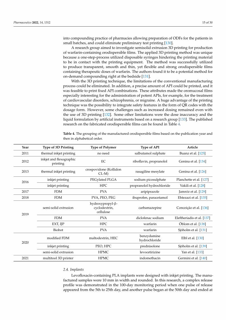
into compounding practice of pharmacies allowing preparation of ODFs for the patients insmall batches, and could eliminate preliminary test printing [130].
A research group aimed to investigate semisolid extrusion 3D printing for productionof warfarin-containing orodispersible films. The applied 3D printing method was uniquebecause a one-step-process utilized disposable syringes hindering the printing materialto be in contact with the printing equipment. The method was successfully utilizedto produce transparent, smooth and thin, yet flexible and strong orodispersible filmscontaining therapeutic doses of warfarin. The authors found it to be a potential method foron-demand compounding right at the bedside [131].
With the 3D printing technique, the limitations of the conventional manufacturingprocess could be eliminated. In addition, a precise amount of API could be printed, and itwas feasible to print fixed API combinations. These attributes made the oromucosal filmsespecially interesting for the administration of potent APIs, for example, for the treatmentof cardiovascular disorders, schizophrenia, or migraine. A huge advantage of the printingtechnique was the possibility to integrate safety features in the form of QR codes with thedosage form. However, some challenges such as increased dosing remained even withthe use of 3D printing [132]. Some other limitations were the dose inaccuracy and theliquid formulation by artificial instruments based on a research group [133]. The publishedresearch on the fabricated orodispersible films can be found in Table 4.
Table 4. The grouping of the manufactured orodispersible films based on the publication year andthen in alphabetical order.
Year Type of 3D Printing Type of Polymer Type of API Article
2011 thermal inkjet printing no need salbutamol sulphate Buanz et al. [125]
2012 inkjet and flexographicprinting EC riboflavin, propranolol Genina et al. [134]
2013 thermal inkjet printing crospovidone (KollidonCL-M) rasagiline mesylate Genina et al. [126]
2016inkjet printing PEGylated PLGA sodium picosulphate Planchette et al. [127]
inkjet printing HPC propranolol hydrochloride Vakili et al. [128]
2017 FDM PVA aripiprazole Jamróz et al. [129]
2018 FDM PVA, PEO, PEG ibuprofen, paracetamol Ehtezazi et al. [135]
2019
semi-solid extrusionhydroxypropyl-β-
cyclodextrin,cellulose
carbamazepine Conceição et al. [136]
FDM PVA diclofenac sodium Eleftheriadis et al. [137]
EXT, IJP HPC warfarin Öblom et al. [138]
Biobot PVA warfarin Sjöholm et al. [131]
2020modified FDM maltodextrin, HEC benzydamine
hydrochloride Elbl et al. [130]
inkjet printing PEO, HPC prednisolone Sjöholm et al. [139]
semi-solid extrusion HPMC levocetirizine Yan et al. [133]
2021 multitool 3D printer HPMC indomethacin Germini et al. [140]
2.4. Implants
Levofloxacin-containing PLA implants were designed with inkjet printing. The manu-factured samples were 10 mm in width and rounded. In this research, a complex releaseprofile was demonstrated in the 100-day monitoring period when one pulse of releaseappeared from the 5th to 25th day, and another pulse began at the 50th day and ended at

Pharmaceutics 2022, 14, 1312 16 of 30
the 80th day, with a lag time of 25 days between the two pulses, wherein a steady state ofrelease was observed at about 5 µg/mL [141].
Rifampicin and isoniazid-containing multi-layered concentric cylindrical implantswere fabricated against tuberculosis. The multi-layered concentric cylinder was dividedinto four layers from the center to the periphery and the APIs were distributed individuallyinto the different layers in a specific sequence of isoniazid–rifampicin–isoniazid–rifampicin.The dissolution tests proved that the API liberation takes place orderly from the outsideto the center and the peak concentrations were between 8 and 12 days. In this study, aprogrammed release multi-drug implant with a complex construction was fabricated by3D printing [142].
A research group prepared dexamethasone-containing tailored drug delivery plat-forms where two distinct designs—structure A: rolled and sealed; structure B: layer-by-layer—were extrusion printed. As the API liberation was continuous for more than4 months, these samples could be used as implants [143].
Genina et al. manufactured intrauterine device and subcutaneous rods from ethy-lene vinyl acetate (EVA) copolymer with FDM printing. The samples were containingindomethacin as a model API and with the device the drug dissolution was over 30 days.A long-acting 3D-printed implantable system was built [144].
In a study, levofloxacin and tobramycin-containing implants were fabricated for thetreatment of osteomyelitis. A multi-layered concentric cylinder construction was createdby powder-based inkjet printing. A sustained and programmed drug delivery system wasprovided [145].
In a study, the effect of the used polymers on the drug release profile of quinine wasexamined as a model drug. The used polymers were Eudragit® RS, PCL, PLLA, and ECand affected the dissolution profile of the samples. The fastest relative drug release wasobserved from PCL where the dissolved API amount was approximately 76% in 51 daysand the lowest from Eudragit RS and EC with less than 5% of quinine release in 78 and100 days, respectively [146].
Qamar et al. manufactured an implantable mesh for the treatment of hernia. PP andPVA meshes were produced with distinct pore size, shape, and thread thickness. Themeshes were filled with ciprofloxacin for the management of hernia. Based on the research,animals implanted with ciprofloxacin HCl loaded meshes exhibited fewer fluctuations inbody temperature and faster wound healing [147].
The purpose of a study was to demonstrate the applicability of 3D printing methodsfor the fabrication of patient-specific fixation implants that allow localized drug delivery.The 3D printing was used to fabricate gentamicin and methotrexate loaded fixation devices,including screws, pins, and bone plates [148].
In a study, PLLA samples were printed using a special Zcorp Zprinter 650 then im-mersed into the solution of various anticancer drugs such as cisplatin, ifosfamid, methotrex-ate, or doxorubicin and finally dried. The proposed 3D-printed drug delivery system couldsimultaneously realize individual local chemotherapy, multi-drug delivery, long-termsustainable drug release, and non-reoperation in osteosarcoma treatment [149].
Ciprofloxacin containing PLA implants were fabricated with the combination of semi-solid extrusion and fused-deposition modeling for the treatment of bone infections. Theauthors found this method more adequate than the conventional method for manufactur-ing [150].
The 3D printing technologies have promising potential for the manufacture of so-phisticated drug implants and patient-specific macro-porosity implants with personalizeddrug release behavior. In general, the currently commercially available implants lack thepersonalization of the treatment and consideration of several issues such as anatomicaldifferences, age, genders, and disease condition [151]. A research group determined that3D-printed implants could display complex drug release patterns compared to conven-tionally fabricated drug implants [141]. The manufacturing of 3D printing provided thepossibility to produce smaller, less invasive, and more site-specific implants compared to

Pharmaceutics 2022, 14, 1312 17 of 30
the conventional ones [152]. Even though the clinical data—to support the routine use ofthe 3D-printed customized implants—are currently limited, the patient-specific implantsmay gain more attention and popularity in the prophylaxis and treatment of diseases suchas complicated bone infections and bone tuberculosis in the near future when the digitaland manufacturing technology advances further [150].
Some published research on the produced implants can be found in Table 5.
Table 5. The grouping of the manufactured implants based on the publication year and then inalphabetical order.
Year Type of 3D Printing Type of Polymer Type of API Article
2007 inkjet printing L-PLA levofloxacin Huang et al. [141]
2009 inkjet printing PDLLA rifampicin, isoniazid Wu et al. [142]
2012 extrusion based 3Dprinting PLGA, PVA dexamethasone Rattanakit et al. [143]
2014 powder binding PLLA isoniazid Wu et al. [153]
20153D-Bioplotter system
(3-hydroxybutyrate-co-3-hydroxyhexanoate)
(PHBHHx)isoniazid, rifampin Min et al. [154]
FDM (MakerBot®) PLA nitrofurantoin Water et al. [155]
2016FDM PCL containing EVA indomethacin Genina et al. [144]
inkjet powder printing PDLLA levofloxacin, tobramycin Wu et al. [145]
2017 FDM Eudragit RS, PCL, PLLA,EC quinine Kempin et al. [146]
2019
FDM, DMLS PLA, PCL, titanium dioxide doxycycline Benmassaoud et al. [156]
FDM PP, PVA ciprofloxacin Qamar et al. [147]
FDM PLA gentamicin, methotrexate Tappa et al. [148]
2020
FDM PLA, antibacterial PLA,PETG, PMMA diclofenac sodium Arany et al. [157]
powder bed printing PLLA cisplatin, ifosfamid,methotrexate, doxorubicin Wang et al. [149]
2021
SSE, FDM PLA ciprofloxacin Cui et al. [150]
SLA Elastic Resin lidocaine Xu et al. [158]
DLP PEGDA, PEG 400 dexamethasone, phenyl bisphosphine oxide, β-carotene Xu et al. [159]
2.5. TTS
Anti-acne drug loaded masks/patches were fabricated by Goyanes et al., but as thissystem provides transdermal delivery, we decided to subgroup the research here. In theresearch, salicylic-acid-containing filaments were used for the FDM 3D printing but the APIshowed significant thermal degradation. The manufacturing by SLA contained a higheramount of drug and showed no drug degradation, so the researchers found this methodmore adequate [160].
Yi et al. manufactured a 3D-printed biodegradable patch with a versatile shapeand incorporated a high drug concentration for the achievement of a controlled drugrelease profile. The patches composed of poly(lactide-co-glycolide), polycaprolactone,and 5-fluorouracil were the antitumor agent. With the use of 3D printing technology, thegeometry of the patch and the drug release kinetics could be manipulated. The patcheswere flexible, and released the drug over four weeks with minimized side effects [161].
A research group developed an electro hydrodynamic (EHD) printing technique tofabricate antibiotic-containing patches using polycaprolactone (PCL), polyvinyl pyrrolidone

Pharmaceutics 2022, 14, 1312 18 of 30
(PVP), and their composite system (PVP-PCL). Drug loaded 3D patches possessed perfectlyaligned fibers giving rise to fibrous strut orientation, variable inter-strut pore size, andcontrolled film width (via layering). The used polymer type and the printed patch voidsize affected the dissolution profiles [162].
If we compared the advantages and the limitations, we found that the conventionalmanufacturing process involves solvent evaporation and multiple steps which were oftentime consuming in comparison with the 3D printing method. In case of the TTSs, theadhesion was a critical point and the skin could be undulating and curved, especially onthe nose or head, which the conventional TTSs could not take into consideration [151]. Thecombination of 3D scanning and 3D printing had the potential to create personalized drugloaded devices, adapted in shape and size to individual patients [160]. The 3D printingof differently shaped patches demonstrated the ability to manipulate the drug releaseby altering the surface area which allowed precise control of the liberation amount andplacement of the API, high efficacy, and minimized systemic toxicity [161]. A few publishedstudies on the created TTSs can be found in Table 6.
Table 6. The grouping of some manufactured TTSs based on the publication year and then inalphabetical order.
Year Type of 3D Printing Type of Polymer Type of API Article
2016
FDM, SLA Flex EcoPLA, PCL salicylic acid Goyanes et al. [160]
inhouseextrusion-based 3Dprinter—multi-headdeposition system
PLA, PCL 5-fluorouracil Yi et al. [161]
2017 EHD PCL, PCL/PVP tetracyclinehydrochloride Wang et al. [162]
2021 FDM PVP quercetin Chaudhari et al. [163]
2.6. Microneedles
In the early research of Ovsianikov et al., a placebo microneedle was developed byfemtosecond laser two photon polymerization 3D printing technology [164]. In 2013,an amphotericin B containing microneedle was created by the combination of visiblelight dynamic mask micro stereolithography, micro molding, and piezoelectric inkjetprinting. Based on the results, the printing process was found to be a scalable approachthat could be used to incorporate pharmacologic agents, even with complex solubilityprofiles, within microneedles [165]. The same researchers fabricated miconazole-containingmicroneedles with the same technology but with a special polymer called Gantrez® AN169 BF (poly(methyl vinyl ether-co-maleic anhydride)). The manufactured sample hadpotential use in transdermal treatment of cutaneous fungal infections [166].
A dacarbazine-containing drug delivery system was produced which could be usedfor the therapy of skin cancer locally. For the manufacturing, a special 3D printing processwas used named as multi-material micro stereolithography (µSL). First, the microneedlearray was built and then the API was added with blending due to the crosslinking effect ofthe polymer (Figure 13) [167].
Insulin polymeric layers on metal microneedles were constructed by Ross et al. Thedissolution profiles showed rapid insulin release rates in the first 20 min, suggesting thatsolid-state insulin delivery via microneedles was feasible [168].

Pharmaceutics 2022, 14, 1312 19 of 30
Pharmaceutics 2022, 14, 1312 19 of 30
Table 6. The grouping of some manufactured TTSs based on the publication year and then in alpha-
betical order.
Year Type of 3D Printing Type of Polymer Type of API Article
2016
FDM, SLA Flex EcoPLA, PCL salicylic acid Goyanes et al. [160]
inhouse extrusion-
based 3D printer—
multi-head deposition
system
PLA, PCL 5-fluorouracil Yi et al. [161]
2017 EHD PCL, PCL/PVP tetracycline hydrochlo-
ride Wang et al. [162]
2021 FDM PVP quercetin Chaudhari et al. [163]
2.6. Microneedles
In the early research of Ovsianikov et al., a placebo microneedle was developed by
femtosecond laser two photon polymerization 3D printing technology [164]. In 2013, an
amphotericin B containing microneedle was created by the combination of visible light
dynamic mask micro stereolithography, micro molding, and piezoelectric inkjet printing.
Based on the results, the printing process was found to be a scalable approach that could
be used to incorporate pharmacologic agents, even with complex solubility profiles,
within microneedles [165]. The same researchers fabricated miconazole-containing mi-
croneedles with the same technology but with a special polymer called Gantrez® AN 169
BF (poly(methyl vinyl ether-co-maleic anhydride)). The manufactured sample had poten-
tial use in transdermal treatment of cutaneous fungal infections [166].
A dacarbazine-containing drug delivery system was produced which could be used
for the therapy of skin cancer locally. For the manufacturing, a special 3D printing process
was used named as multi-material micro stereolithography (μSL). First, the microneedle
array was built and then the API was added with blending due to the crosslinking effect
of the polymer (Figure 13) [167].
Figure 13. Cross-sectional design of the microneedle array [167].
Insulin polymeric layers on metal microneedles were constructed by Ross et al. The
dissolution profiles showed rapid insulin release rates in the first 20 min, suggesting that
solid-state insulin delivery via microneedles was feasible [168].
Lim et al. used 3D printing for manufacturing a dual-function microneedle array on
personalized curved surfaces (microneedle splint) for drug delivery and splinting of the
affected finger. Sufficient penetration efficiency was achieved, and the final microneedle
splint showed biocompatibility. A significantly higher amount of diclofenac permeated
through the skin with the use of the microneedle splint as compared to intact skin. The
fabricated microneedle splint could thus be a potential new approach to treat trigger fin-
ger via personalized splinting without affecting normal hand function [169].
Figure 13. Cross-sectional design of the microneedle array [167].
Lim et al. used 3D printing for manufacturing a dual-function microneedle array onpersonalized curved surfaces (microneedle splint) for drug delivery and splinting of theaffected finger. Sufficient penetration efficiency was achieved, and the final microneedlesplint showed biocompatibility. A significantly higher amount of diclofenac permeatedthrough the skin with the use of the microneedle splint as compared to intact skin. Thefabricated microneedle splint could thus be a potential new approach to treat trigger fingervia personalized splinting without affecting normal hand function [169].
Two research groups produced insulin-releasing microneedles from Dental SG withthe use of inkjet printing and the SLA technique. In the case of the research of Pere et al.,the API was released rapidly within 30 min [170]. Economidou et al. reached better glucoselevels and hypoglycemia control [171].
One of the biggest advantages of 3D printing was that the technique allowed theincorporation of pharmacologic agents even with a complex solubility profile into themicroneedles for patient needs [165]. The manufactured microneedles provided accuratedosing which also maintained the mechanical strength in comparison with the existingmicroneedles, e.g., made of metal. Furthermore, microneedles that have extremely fine tipsin nano scale allowed good penetration of skin and potentially cell targeted delivery [172].A few studies on the 3D-printed microneedles can be found in Table 7.
Table 7. The grouping of some manufactured microneedles based on the publication year and thenin alphabetical order.
Year Type of 3D Printing Type of Polymer Type of API Article
2007 femtosecond laser twophoton polymerization Ormocer® none Ovsianikov et al. [164]
2013 piezoelectric inkjet printing PDMS, PMMA amphotericin B Boehm et al. [165]
2014 piezoelectric inkjet printingGantrez® AN 169 BF(poly(methyl vinyl
ether-co-maleic anhydride))miconazole Boehm et al. [166]
2015
multi-materialmicrostereolithography
(µSL)poly(propylene fumarate) dacarbazine Lu et al. [167]
inkjet printing
polyvinylcaprolactame-polyvinyl
cetatepolyethylene glycol(SOL),
poly(2-ethyl-2-oxazoline)(POX)
insulin Ross et al. [168]
inkjet printing Soluplus® 5-fluorouracil Uddin et al. [173]

Pharmaceutics 2022, 14, 1312 20 of 30
Table 7. Cont.
Year Type of 3D Printing Type of Polymer Type of API Article
2017 DLP 3DMCastable resin diclofenac sodium Lim et al. [169]
2018SLA medium viscosity alginate blue dye, HepG2 cell
encapsulation Farias et al. [174]
FDM PLA fluorescein Luzuriaga et al. [175]
inkjet printer Dental SG insulin Pere et al. [170]
2019 SLA Dental SG insulin Economidou et al. [171]
2.7. Vaginal Drug Delivery Systems
Even though this subsection consists of numerous drug delivery systems, we wouldlike to discuss them together for easier accessibility.
The article of Genina et al. was already discussed in the section of implants becausetwo distinct type of drug delivery systems were fabricated: intrauterine device (IUD) andsubcutaneous rods [144].
Tappa et al. designed estrogen and progesterone-containing PCL filaments and FDMprinted into different structures: mesh, IUD, and subdermal implant. As the FDM printingwith PCL was possible at lower temperatures (around 130 ◦C), the API remained stablebased on the TG/DSC results. This was the first study to offer data on hormone-loaded3D-printed constructs [176].
Bioadhesive vaginal films were produced by Varan et al., where the used APIs werepaclitaxel and cidofovir, which had antiviral efficacy and used for the treatment of cervicalcancer locally. The basic idea was to increase the solubility of the paclitaxel with a cyclodex-trin complex while cidofovir was encapsulated in PCL nanoparticles to manufacture anink [177].
Our research group fabricated a vaginal ring as a carrier system for the treatment ofbacterial vaginosis. For the manufacturing, FDM 3D printing was used and then thesepre-printed samples were easily filled with the jellified APIs (vaginal gels) depending onthe patient’s need. (Figure 14). This article focused on avoiding the loss and decompositionof API through the printing process and manufacturing a drug delivery system, whichcould be directly and rapidly printed at the bedside or in the pharmacy as individualizedmedication [178].
Pharmaceutics 2022, 14, 1312 21 of 30
cancer locally. The basic idea was to increase the solubility of the paclitaxel with a cy-
clodextrin complex while cidofovir was encapsulated in PCL nanoparticles to manufac-
ture an ink [177].
Our research group fabricated a vaginal ring as a carrier system for the treatment of
bacterial vaginosis. For the manufacturing, FDM 3D printing was used and then these pre-
printed samples were easily filled with the jellified APIs (vaginal gels) depending on the
patient’s need. (Figure 14.) This article focused on avoiding the loss and decomposition of
API through the printing process and manufacturing a drug delivery system, which could
be directly and rapidly printed at the bedside or in the pharmacy as individualized med-
ication [178].
Figure 14. The designed vaginal drug delivery system by our research group [178].
The currently available approaches did not consider the specific anatomy of each pa-
tient, the patient’s medical conditions, age, and gender specificity which limited the effec-
tive therapy whereas every patient is unique and should require different doses of drugs
and hormones [179]. In addition, other factors such as variability in drug absorption re-
lated with menstrual cycle, menopause, and pregnancy were different among patients
[180]. Therefore, patient-specific systems are needed to provide personalized shape, size,
and tailored drug release to improve the efficacy and to increase compliance. The 3D-
printed delivery system in this regard can provide personalized geometry, snug fit, in-
crease efficacy, improve patient compliance, and prevent the post-surgical complications
[151].
Some research on the manufacturing of vaginal drug delivery systems can be found
in Table 8. The research was classified based on the publication year and then in alpha-
betical order. In the first column, the type of the drug delivery system is described.
Table 8. The grouping of the manufactured vaginal drug delivery systems based on the publication
year and then in alphabetical order.
Drug Delivery Type Year Type of 3D Printing Type of Polymer Type of API Article
IUD, subcutaneous
rod 2016 FDM EVA, PCL indomethacin Genina et al. [144]
IUS FDM PCL indomethacin Holländer et al. [181]
Mesh, IUD, subder-
mal implant 2017 FDM PCL
estrogen, progester-
one Tappa et al. [176]
Bioadhesive film inkjet printing PCL, PEG-PCL paclitaxel, cidofovir Varan et al. [177]
Bioadhesive film 2019 inkjet printing PEG-PCL paclitaxel, cidofovir Varan et al. [182]
Intravaginal ring 2021 FDM TPU chloramphenicol,
metronidazole Arany et al. [178]
2.8. Micro and Nanoscale Dosage Forms
Another area that concerns the researchers is the possible manufacturing of micro
and nanoscale drug delivery systems. In a study, a poorly soluble model drug was used
to construct a 10% folic acid containing nanosuspension and the printing was performed
on an inkjet-based microdosing dispenser head. In the research, the authors found this
Figure 14. The designed vaginal drug delivery system by our research group [178].
The currently available approaches did not consider the specific anatomy of eachpatient, the patient’s medical conditions, age, and gender specificity which limited the ef-fective therapy whereas every patient is unique and should require different doses of drugsand hormones [179]. In addition, other factors such as variability in drug absorption relatedwith menstrual cycle, menopause, and pregnancy were different among patients [180].Therefore, patient-specific systems are needed to provide personalized shape, size, andtailored drug release to improve the efficacy and to increase compliance. The 3D-printed

Pharmaceutics 2022, 14, 1312 21 of 30
delivery system in this regard can provide personalized geometry, snug fit, increase efficacy,improve patient compliance, and prevent the post-surgical complications [151].
Some research on the manufacturing of vaginal drug delivery systems can be found inTable 8. The research was classified based on the publication year and then in alphabeticalorder. In the first column, the type of the drug delivery system is described.
Table 8. The grouping of the manufactured vaginal drug delivery systems based on the publicationyear and then in alphabetical order.
Drug Delivery Type Year Type of 3D Printing Type of Polymer Type of API Article
IUD, subcutaneous rod2016
FDM EVA, PCL indomethacin Genina et al. [144]
IUS FDM PCL indomethacin Holländer et al. [181]
Mesh, IUD, subdermalimplant
2017
FDM PCL estrogen,progesterone Tappa et al. [176]
Bioadhesive film inkjet printing PCL, PEG-PCL paclitaxel,cidofovir Varan et al. [177]
Bioadhesive film 2019 inkjet printing PEG-PCL paclitaxel,cidofovir Varan et al. [182]
Intravaginal ring 2021 FDM TPU chloramphenicol,metronidazole Arany et al. [178]
2.8. Micro and Nanoscale Dosage Forms
Another area that concerns the researchers is the possible manufacturing of micro andnanoscale drug delivery systems. In a study, a poorly soluble model drug was used toconstruct a 10% folic acid containing nanosuspension and the printing was performed onan inkjet-based microdosing dispenser head. In the research, the authors found this methodadequate for the incorporation of poorly soluble drugs to increase the oral absorption [183].
Scoutaris et al. produced poorly soluble felodipine and PVP-containing solution forinkjet printing. Based on the authors’ research, inkjet printing could be used to prepare thisnovel drug dosage form consisting of micro-sized dried deposits from sprayed picolitredroplets containing a drug on a substrate. The novelty of the work was that a scalabledosage form could be produced whereby many droplets could be produced to achieve adissolution profile equivalent to conventional bulk dosage formulations [184].
In another study, rifampicin and PLGA-containing inks were fabricated for micro-pattern printing on a glass or titanium carrier. This research combines special ink for-mulations in microscale range and the author’s idea was to use these micro-patterns inorthopedic surgeries [185].
Paclitaxel-containing PLGA inks were created for piezoelectric inkjet printing wherefour different shapes were printed. Microparticles with diverse geometries exhibitednon-similar drug release rates mainly due to nonidentical surface areas [186].
Some studies on the manufacturing of micro and nanoscale systems can be found inTable 9. The research was classified based on the publication year and then in alphabeticalorder. In the first column, the type of the drug delivery system was described.

Pharmaceutics 2022, 14, 1312 22 of 30
Table 9. The grouping of the manufactured micro- and nanoscale drug delivery systems based on thepublication year and then in alphabetical order.
Drug Delivery Type Year Type of 3D Printing Type of Polymer Type of API Article
Nanosuspension
2011
inkjet-based microdosing dispenser head none folic acid Pardeike et al. [183]
Micron-sized drieddeposits inkjet printing PVP felodipine Scoutaris et al. [184]
Micropatterns
2012
inkjet printing PLGA rifampicin Gu et al. [185]
Microparticles piezoelectric inkjetprinting PLGA paclitaxel Lee et al. [186]
2.9. Other
A few other interesting but not mandatory ‘drug delivery systems’ are discussed inthis section. Kitson et al. used ibuprofen with a modified Prusa FDM 3D printer where theAPI was deposited in a pre-printed vessel. In this case, the API could be directly printedto a drug delivery system, e.g., tablet or capsule, to receive a ready-to-use drug dosageform [187].
Long et al. 3D-printed a chitosan-pectin hydrogel. To the gel, lidocaine hydrochloridewas incorporated as a potential wound dressing candidate. The scaffolds were printedusing an extrusion-based 3D printer using a mechanical positive displacement dispensingsystem followed by lyophilization. The research confirmed the possible manufacturing ofhydrogels with 3D printing [188].
Seoane-Viaño et al. fabricated tacrolimus-containing rectal suppositories with semi-solid extrusion-based 3D printing. For the printing, Gelucire 44/14 and coconut oil wereused. The designed drug dosage form could contribute to a long-lasting effect on thetreatment of colitis [189,190].
3. Future Perspective
Three-dimensional printing enables the manufacture of personalized drug deliverysystems. With the use of this modern technique, cost-effectiveness, simplified production,complex formulations, and increased opportunities for collaboration can be achieved. Thehigh flexibility provides the production of a multitude of drug products with tailored releaseprofiles and designs [20]. Additionally, 3D printing has to challenge some limitations inthe future. As 3D printing right now cannot compete with the speed of the conventionaldrug manufacturing method, in the future the improvement in the printing speed andresolution has to be adjusted which can also reduce the energy consumption and costas a recent, huge concern. The printed product dimension accuracy and scale size (e.g.,micro or nanoscale) must be improved also. As 3D printing technology is limited interms of materials, the manufacturing of novel polymers, inks, etc., are still required andnecessary for the proper manufacturing [191]. Even though the FDA approved Spritam®
in 2015, the used ZipDose® technology was similar to traditional powder compactionthat could help the process of approval. At present, there are no fixed guidelines forthe regulation of 3D-printed pharmaceutical dosage forms, so the researchers have toface regulatory concerns [151]. Another interesting area is bioprinting where living cells,tissues, or organs can be printed and exploratory studies of bioprinting in pharmaceuticshave shown promising applications of this technique, for example, in the field of targetidentification and validation or in vitro efficacy assessment [19].
4. Conclusions
In conclusion, 3D printing represents a very interesting and modern technique in thefield of drug manufacturing which is about to revolutionize the health industry. Three-dimensional printing is a layer-by-layer, automated process, which enables the manufac-

Pharmaceutics 2022, 14, 1312 23 of 30
turing of complex, personalized products on-demand. In the last decade, the number ofpublications multiplied year-by-year. The FDA approved the first and only 3D-printeddrug in 2015, which supported the commercial feasibility of this technology. In the last twodecades, dozens of research groups aimed to manufacture different drug dosage forms,e.g., tablets, capsules, implants, even rectal suppositories by 3D printing to improve thesafety, efficacy, and tolerability of medicines and provide individualized therapy for thosemost in need.
Author Contributions: I.B. conceived the overall project, and wrote the conclusion part. Z.U. wrotethe first part of the introduction and P.F. wrote the second half of the introduction. P.A. wrote Section 2and was responsible for drawing the figures. All authors have read and agreed to the publishedversion of the manuscript.
Funding: The publication is supported by the GINOP-2.3.4-15-2020-00008 project. The project isco-financed by the European Union and the European Regional Development Fund. The research wassupported by the Thematic Excellence Programme (TKP2020-IKA-04) of the Ministry for Innovationand Technology in Hungary. Project no. TKP2021-EGA-18 has been implemented with the supportprovided from the National Research, Development and Innovation Fund of Hungary, financedunder the TKP2021-EGA funding scheme.
Institutional Review Board Statement: Not applicable.
Informed Consent Statement: Not applicable.
Data Availability Statement: Not applicable.
Conflicts of Interest: The authors declare no conflict of interest.
References1. Norman, J.; Madurawe, R.D.; Moore, C.M.V.; Khan, M.A.; Khairuzzaman, A. A new chapter in pharmaceutical manufacturing:
3D-printed drug products. Adv. Drug Deliv. Rev. 2017, 108, 39–50. [CrossRef] [PubMed]2. Stansbury, J.W.; Idacavage, M.J. 3D printing with polymers: Challenges among expanding options and opportunities. Dent. Mater.
2016, 32, 54–64. [CrossRef] [PubMed]3. Dizon, J.R.C.; Espera, A.H.; Chen, Q.; Advincula, R.C. Mechanical characterization of 3D-printed polymers. Addit. Manuf. 2018,
20, 44–67. [CrossRef]4. Alhnan, M.A.; Okwuosa, T.C.; Sadia, M.; Wan, K.W.; Ahmed, W.; Arafat, B. Emergence of 3D Printed Dosage Forms: Opportunities
and Challenges. Pharm. Res. 2016, 33, 1817–1832. [CrossRef] [PubMed]5. Singh, S.; Ramakrishna, S.; Singh, R. Material issues in additive manufacturing: A review. J. Manuf. Process. 2017, 25, 185–200.
[CrossRef]6. Elkasabgy, N.A.; Mahmoud, A.A.; Maged, A. 3D printing: An appealing route for customized drug delivery systems. Int. J.
Pharm. 2020, 588, 119732. [CrossRef]7. Okafor-Muo, O.L.; Hassanin, H.; Kayyali, R.; ElShaer, A. 3D Printing of Solid Oral Dosage Forms: Numerous Challenges with
Unique Opportunities. J. Pharm. Sci. 2020, 109, 3535–3550. [CrossRef]8. Aquino, R.P.; Barile, S.; Grasso, A.; Saviano, M. Envisioning smart and sustainable healthcare: 3D Printing technologies for
personalized medication. Futures 2018, 103, 35–50. [CrossRef]9. Ursan, I.; Chiu, L.; Pierce, A. Three-dimensional drug printing: A structured review. J. Am. Pharm. Assoc. 2013, 53, 136–144.
[CrossRef]10. Sandler, N.; Preis, M. Printed Drug-Delivery Systems for Improved Patient Treatment. Trends Pharmacol. Sci. 2016, 37, 1070–1080.
[CrossRef]11. Moulton, S.E.; Wallace, G.G. 3-dimensional (3D) fabricated polymer based drug delivery systems. J. Control. Release 2014, 193,
27–34. [CrossRef]12. Jamróz, W.; Szafraniec, J.; Kurek, M.; Jachowicz, R. 3D printing in pharmaceutical and medical applications. Pharm. Res. 2018, 35,
176. [CrossRef]13. Kotta, S.; Nair, A.; Alsabeelah, N. 3D Printing Technology in Drug Delivery: Recent Progress and Application. Curr. Pharm. Des.
2018, 24, 5039–5048. [CrossRef]14. Mohapatra, S.; Kar, R.K.; Biswal, P.K.; Bindhani, S. Approaches of 3D printing in current drug delivery. Sensors Int. 2021, 3, 100146.
[CrossRef]15. Cunha-Filho, M.; Araújo, M.R.; Gelfuso, G.M.; Gratieri, T. FDM 3D printing of modified drug-delivery systems using hot melt
extrusion: A new approach for individualized therapy. Ther. Deliv. 2017, 8, 957–966. [CrossRef]16. Gueche, Y.A.; Sanchez-Ballester, N.M.; Cailleaux, S.; Bataille, B.; Soulairol, I. Selective laser sintering (Sls), a new chapter in the
production of solid oral forms (sofs) by 3d printing. Pharmaceutics 2021, 13, 1212. [CrossRef]

Pharmaceutics 2022, 14, 1312 24 of 30
17. Wang, J.; Goyanes, A.; Gaisford, S.; Basit, A.W. Stereolithographic (SLA) 3D printing of oral modified-release dosage forms. Int. J.Pharm. 2016, 503, 207–212. [CrossRef]
18. Daly, R.; Harrington, T.S.; Martin, G.D.; Hutchings, I.M. Inkjet printing for pharmaceutics—A review of research and manufactur-ing. Int. J. Pharm. 2015, 494, 554–567. [CrossRef]
19. Peng, W.; Datta, P.; Ayan, B.; Ozbolat, V.; Sosnoski, D.; Ozbolat, I.T. 3D bioprinting for drug discovery and development inpharmaceutics. Acta Biomater. 2017, 57, 26–46. [CrossRef]
20. Trenfield, S.J.; Awad, A.; Madla, C.M.; Hatton, G.B.; Firth, J.; Goyanes, A.; Gaisford, S.; Basit, A.W. Shaping the future: Recentadvances of 3D printing in drug delivery and healthcare. Expert Opin. Drug Deliv. 2019, 16, 1081–1094. [CrossRef]
21. Rohani Shirvan, A.; Bashari, A.; Hemmatinejad, N. New insight into the fabrication of smart mucoadhesive buccal patches as anovel controlled-drug delivery system. Eur. Polym. J. 2019, 119, 541–550. [CrossRef]
22. Domsta, V.; Seidlitz, A. 3d-printing of drug-eluting implants: An overview of the current developments described in the literature.Molecules 2021, 26, 4066. [CrossRef] [PubMed]
23. Khatri, P.; Shah, M.K.; Vora, N. Formulation strategies for solid oral dosage form using 3D printing technology: A mini-review. J.Drug Deliv. Sci. Technol. 2018, 46, 148–155. [CrossRef]
24. Economidou, S.N.; Lamprou, D.A.; Douroumis, D. 3D printing applications for transdermal drug delivery. Int. J. Pharm. 2018,544, 415–424. [CrossRef]
25. Wu, B.M.; Borland, S.W.; Giordano, R.A.; Cima, L.G.; Sachs, E.M.; Cima, M.J. Solid free-form fabrication of drug delivery devices.J. Control. Release 1996, 40, 77–87. [CrossRef]
26. Katstra, W.E.; Palazzolo, R.D.; Rowe, C.W.; Giritlioglu, B.; Teung, P.; Cima, M.J. Oral dosage forms fabricated by Three DimensionalPrinting. J. Control. Release 2000, 66, 1–9. [CrossRef]
27. Lee, K.J.; Kang, A.; Delfino, J.J.; West, T.G.; Chetty, D.; Monkhouse, D.C.; Yoo, J. Evaluation of Critical Formulation Factors in theDevelopment of a Rapidly Dispersing Captopril Oral Dosage Form. Drug Dev. Ind. Pharm. 2003, 29, 967–979. [CrossRef]
28. Wang, C.C.; Tejwani, M.R.; Roach, W.J.; Kay, J.L.; Yoo, J.; Surprenant, H.L.; Monkhouse, D.C.; Pryor, T.J. Development of nearzero-order release dosage forms using three-dimensional printing (3-DPTM) technology. Drug Dev. Ind. Pharm. 2006, 32, 367–376.[CrossRef]
29. Gbureck, U.; Vorndran, E.; Müller, F.A.; Barralet, J.E. Low temperature direct 3D printed bioceramics and biocomposites as drugrelease matrices. J. Control. Release 2007, 122, 173–180. [CrossRef]
30. Yu, D.G.; Yang, X.L.; Huang, W.D.; Liu, J.; Wang, Y.G.; Xu, H. Tablets With Material Gradients Fabricated by Three-DimensionalPrinting. J. Pharm. Biomed. Anal. 2007, 96, 2446–2456. [CrossRef]
31. Yu, D.G.; Branford-White, C.; Ma, Z.H.; Zhu, L.M.; Li, X.Y.; Yang, X.L. Novel drug delivery devices for providing linear releaseprofiles fabricated by 3DP. Int. J. Pharm. 2009, 370, 160–166. [CrossRef]
32. Khaled, S.A.; Burley, J.C.; Alexander, M.R.; Roberts, C.J. Desktop 3D printing of controlled release pharmaceutical bilayer tablets.Int. J. Pharm. 2014, 461, 105–111. [CrossRef]
33. Bhattacharya, S.; Singh, S.K.; Shrestha, S.; Baghel, Y.S.; Maity, D.; Kumar, A.; Gupta, G.D.; Kumar, R. Recent Findings andDevelopment of 3D Printing Technology in Pharmaceutical Formulation Development: An Extensive Review. Int. J. Drug Dev.Res. 2019, 11, 1–14.
34. Goyanes, A.; Buanz, A.B.M.; Hatton, G.B.; Gaisford, S.; Basit, A.W. 3D printing of modified-release aminosalicylate (4-ASA and5-ASA) tablets. Eur. J. Pharm. Biopharm. 2015, 89, 157–162. [CrossRef]
35. Goyanes, A.; Wang, J.; Buanz, A.; Martínez-Pacheco, R.; Telford, R.; Gaisford, S.; Basit, A.W. 3D Printing of Medicines: EngineeringNovel Oral Devices with Unique Design and Drug Release Characteristics. Mol. Pharm. 2015, 12, 4077–4084. [CrossRef]
36. Khaled, S.A.; Burley, J.C.; Alexander, M.R.; Yang, J.; Roberts, C.J. 3D printing of tablets containing multiple drugs with definedrelease profiles. Int. J. Pharm. 2015, 494, 643–650. [CrossRef]
37. Khaled, S.A.; Burley, J.C.; Alexander, M.R.; Yang, J.; Roberts, C.J. 3D printing of five-in-one dose combination polypill withdefined immediate and sustained release profiles. J. Control. Release 2015, 217, 308–314. [CrossRef]
38. Boetker, J.; Water, J.J.; Aho, J.; Arnfast, L.; Bohr, A.; Rantanen, J. Modifying release characteristics from 3D printed drug-elutingproducts. Eur. J. Pharm. Sci. 2016, 90, 47–52. [CrossRef]
39. Okwuosa, T.C.; Stefaniak, D.; Arafat, B.; Isreb, A.; Wan, K.W.; Alhnan, M.A. A Lower Temperature FDM 3D Printing for theManufacture of Patient-Specific Immediate Release Tablets. Pharm. Res. 2016, 33, 2704–2712. [CrossRef]
40. Sadia, M.; Sosnicka, A.; Arafat, B.; Isreb, A.; Ahmed, W.; Kelarakis, A.; Alhnan, M.A. Adaptation of pharmaceutical excipients toFDM 3D printing for the fabrication of patient-tailored immediate release tablets. Int. J. Pharm. 2016, 513, 659–668. [CrossRef]
41. Goyanes, A.; Robles Martinez, P.; Buanz, A.; Basit, A.W.; Gaisford, S. Effect of geometry on drug release from 3D printed tablets.Int. J. Pharm. 2015, 494, 657–663. [CrossRef] [PubMed]
42. Goyanes, A.; Chang, H.; Sedough, D.; Hatton, G.B.; Wang, J.; Buanz, A.; Gaisford, S.; Basit, A.W. Fabrication of controlled-releasebudesonide tablets via desktop (FDM) 3D printing. Int. J. Pharm. 2015, 496, 414–420. [CrossRef] [PubMed]
43. Acosta-Vélez, G.F.; Linsley, C.S.; Craig, M.C.; Wu, B.M. Photocurable bioink for the inkjet 3D pharming of hydrophilic drugs.Bioengineering 2017, 4, 11. [CrossRef] [PubMed]
44. Beck, R.C.R.; Chaves, P.S.; Goyanes, A.; Vukosavljevic, B.; Buanz, A.; Windbergs, M.; Basit, A.W.; Gaisford, S. 3D printed tabletsloaded with polymeric nanocapsules: An innovative approach to produce customized drug delivery systems. Int. J. Pharm. 2017,528, 268–279. [CrossRef]

Pharmaceutics 2022, 14, 1312 25 of 30
45. Chai, X.; Chai, H.; Wang, X.; Yang, J.; Li, J.; Zhao, Y.; Cai, W.; Tao, T.; Xiang, X. Fused deposition modeling (FDM) 3D printedtablets for intragastric floating delivery of domperidone. Sci. Rep. 2017, 7, 2829. [CrossRef]
46. Genina, N.; Boetker, J.P.; Colombo, S.; Harmankaya, N.; Rantanen, J.; Bohr, A. Anti-tuberculosis drug combination for controlledoral delivery using 3D printed compartmental dosage forms: From drug product design to in vivo testing. J. Control. Release 2017,268, 40–48. [CrossRef]
47. Li, Q.; Wen, H.; Jia, D.; Guan, X.; Pan, H.; Yang, Y.; Yu, S.; Zhu, Z.; Xiang, R.; Pan, W. Preparation and investigation ofcontrolled-release glipizide novel oral device with three-dimensional printing. Int. J. Pharm. 2017, 525, 5–11. [CrossRef]
48. Fina, F.; Goyanes, A.; Madla, C.M.; Awad, A.; Trenfield, S.J.; Kuek, J.M.; Patel, P.; Gaisford, S.; Basit, A.W. 3D printing ofdrug-loaded gyroid lattices using selective laser sintering. Int. J. Pharm. 2018, 547, 44–52. [CrossRef]
49. Holländer, J.; Hakala, R.; Suominen, J.; Moritz, N.; Yliruusi, J.; Sandler, N. 3D printed UV light cured polydimethylsiloxanedevices for drug delivery. Int. J. Pharm. 2018, 544, 433–442. [CrossRef]
50. Kollamaram, G.; Croker, D.M.; Walker, G.M.; Goyanes, A.; Basit, A.W.; Gaisford, S. Low temperature fused deposition modeling(FDM) 3D printing of thermolabile drugs. Int. J. Pharm. 2018, 545, 144–152. [CrossRef]
51. Li, Q.; Guan, X.; Cui, M.; Zhu, Z.; Chen, K.; Wen, H.; Jia, D.; Hou, J.; Xu, W.; Yang, X.; et al. Preparation and investigation of novelgastro-floating tablets with 3D extrusion-based printing. Int. J. Pharm. 2018, 535, 325–332. [CrossRef]
52. Sadia, M.; Arafat, B.; Ahmed, W.; Forbes, R.T.; Alhnan, M.A. Channelled tablets: An innovative approach to accelerating drugrelease from 3D printed tablets. J. Control. Release 2018, 269, 355–363. [CrossRef]
53. Scoutaris, N.; Ross, S.A.; Douroumis, D. 3D Printed “Starmix” Drug Loaded Dosage Forms for Paediatric Applications. Pharm.Res. 2018, 35, 34. [CrossRef]
54. Verstraete, G.; Samaro, A.; Grymonpré, W.; Vanhoorne, V.; Van Snick, B.; Boone, M.N.; Hellemans, T.; Van Hoorebeke, L.; Remon,J.P.; Vervaet, C. 3D printing of high drug loaded dosage forms using thermoplastic polyurethanes. Int. J. Pharm. 2018, 536,318–325. [CrossRef]
55. Goyanes, A.; Madla, C.M.; Umerji, A.; Duran Piñeiro, G.; Giraldez Montero, J.M.; Lamas Diaz, M.J.; Gonzalez Barcia, M.; Taherali,F.; Sánchez-Pintos, P.; Couce, M.L.; et al. Automated therapy preparation of isoleucine formulations using 3D printing for thetreatment of MSUD: First single-centre, prospective, crossover study in patients. Int. J. Pharm. 2019, 567, 118497. [CrossRef]
56. Öblom, H.; Zhang, J.; Pimparade, M.; Speer, I.; Preis, M.; Repka, M.; Sandler, N. 3D-Printed Isoniazid Tablets for the Treatmentand Prevention of Tuberculosis—Personalized Dosing and Drug Release. AAPS PharmSciTech 2019, 20, 52. [CrossRef]
57. Robles-Martinez, P.; Xu, X.; Trenfield, S.J.; Awad, A.; Goyanes, A.; Telford, R.; Basit, A.W.; Gaisford, S. 3D printing of amulti-layered polypill containing six drugs using a novel stereolithographic method. Pharmaceutics 2019, 11, 274. [CrossRef]
58. Zidan, A.; Alayoubi, A.; Asfari, S.; Coburn, J.; Ghammraoui, B.; Cruz, C.N.; Ashraf, M. Development of mechanistic models toidentify critical formulation and process variables of pastes for 3D printing of modified release tablets. Int. J. Pharm. 2019, 555,109–123. [CrossRef]
59. Awad, A.; Yao, A.; Trenfield, S.J.; Goyanes, A.; Gaisford, S.; Basit, A.W. 3D printed tablets (Printlets) with braille and moonpatterns for visually impaired patients. Pharmaceutics 2020, 12, 172. [CrossRef] [PubMed]
60. Dumpa, N.R.; Bandari, S.; Repka, M.A. Novel gastroretentive floating pulsatile drug delivery system produced via hot-meltextrusion and fused deposition modeling 3D printing. Pharmaceutics 2020, 12, 52. [CrossRef]
61. Gioumouxouzis, C.I.; Tzimtzimis, E.; Katsamenis, O.L.; Dourou, A.; Markopoulou, C.; Bouropoulos, N.; Tzetzis, D.; Fatouros,D.G. Fabrication of an osmotic 3D printed solid dosage form for controlled release of active pharmaceutical ingredients. Eur. J.Pharm. Sci. 2020, 143, 105176. [CrossRef] [PubMed]
62. Karavasili, C.; Gkaragkounis, A.; Moschakis, T.; Ritzoulis, C.; Fatouros, D.G. Pediatric-friendly chocolate-based dosage forms forthe oral administration of both hydrophilic and lipophilic drugs fabricated with extrusion-based 3D printing. Eur. J. Pharm. Sci.2020, 147, 105291. [CrossRef] [PubMed]
63. Real, J.P.; Barberis, M.E.; Camacho, N.M.; Sánchez Bruni, S.; Palma, S.D. Design of novel oral ricobendazole formulation applyingmelting solidification printing process (MESO-PP): An innovative solvent-free alternative method for 3D printing using asimplified concept and low temperature. Int. J. Pharm. 2020, 587, 119653. [CrossRef] [PubMed]
64. Tsintavi, E.; Rekkas, D.M.; Bettini, R. Partial tablet coating by 3D printing. Int. J. Pharm. 2020, 581, 119298. [CrossRef] [PubMed]65. Xu, X.; Robles-Martinez, P.; Madla, C.M.; Joubert, F.; Goyanes, A.; Basit, A.W.; Gaisford, S. Stereolithography (SLA) 3D printing
of an antihypertensive polyprintlet: Case study of an unexpected photopolymer-drug reaction. Addit. Manuf. 2020, 33, 101071.[CrossRef]
66. Algahtani, M.S.; Mohammed, A.A.; Ahmad, J.; Abdullah, M.M.; Saleh, E. 3d printing of dapagliflozin containing self-nanoemulsifying tablets: Formulation design and in vitro characterization. Pharmaceutics 2021, 13, 993. [CrossRef]
67. Tan, Y.J.N.; Yong, W.P.; Low, H.R.; Kochhar, J.S.; Khanolkar, J.; Lim, T.S.E.; Sun, Y.; Wong, J.Z.E.; Soh, S. Customizable drug tabletswith constant release profiles via 3D printing technology. Int. J. Pharm. 2021, 598, 120370. [CrossRef]
68. Yang, Y.; Xu, Y.; Wei, S.; Shan, W. Oral preparations with tunable dissolution behavior based on selective laser sintering technique.Int. J. Pharm. 2021, 593, 120127. [CrossRef]
69. Jamróz, W.; Kurek, M.; Czech, A.; Szafraniec, J.; Gawlak, K.; Jachowicz, R. 3D printing of tablets containing amorphousaripiprazole by filaments co-extrusion. Eur. J. Pharm. Biopharm. 2018, 131, 44–47. [CrossRef]
70. Raijada, D.; Wac, K.; Greisen, E.; Rantanen, J.; Genina, N. Integration of personalized drug delivery systems into digital health.Adv. Drug Deliv. Rev. 2021, 176, 113857. [CrossRef]

Pharmaceutics 2022, 14, 1312 26 of 30
71. Rautamo, M.; Kvarnström, K.; Sivén, M.; Airaksinen, M.; Lahdenne, P.; Sandler, N. Benefits and prerequisites associated with theadoption of oral 3D-printed medicines for pediatric patients: A focus group study among healthcare professionals. Pharmaceutics2020, 12, 229. [CrossRef]
72. Cui, M.; Pan, H.; Fang, D.; Qiao, S.; Wang, S.; Pan, W. Fabrication of high drug loading levetiracetam tablets using semi-solidextrusion 3D printing. J. Drug Deliv. Sci. Technol. 2020, 57, 101683. [CrossRef]
73. Rowe, C.W.; Katstra, W.E.; Palazzolo, R.D.; Giritlioglu, B.; Teung, P.; Cima, M.J. Multimechanism oral dosage forms fabricated bythree dimensional printing. J. Control. Release 2000, 66, 11–17. [CrossRef]
74. Salmoria, G.V.; Klauss, P.; Zepon, K.; Kanis, L.A.; Roesler, C.R.M.; Vieira, L.F. Development of functionally-graded reservoir ofPCL/PG by selective laser sintering for drug delivery devices: This paper presents a selective laser sintering-fabricated drugdelivery system that contains graded progesterone content. Virtual Phys. Prototyp. 2012, 7, 107–115. [CrossRef]
75. Goyanes, A.; Buanz, A.B.M.; Basit, A.W.; Gaisford, S. Fused-filament 3D printing (3DP) for fabrication of tablets. Int. J. Pharm.2014, 476, 88–92. [CrossRef]
76. Pietrzak, K.; Isreb, A.; Alhnan, M.A. A flexible-dose dispenser for immediate and extended release 3D printed tablets. Eur. J.Pharm. Biopharm. 2015, 96, 380–387. [CrossRef]
77. Skowyra, J.; Pietrzak, K.; Alhnan, M.A. Fabrication of extended-release patient-tailored prednisolone tablets via fused depositionmodelling (FDM) 3D printing. Eur. J. Pharm. Sci. 2015, 68, 11–17. [CrossRef]
78. Alhijjaj, M.; Belton, P.; Qi, S. An investigation into the use of polymer blends to improve the printability of and regulate drugrelease from pharmaceutical solid dispersions prepared via fused deposition modeling (FDM) 3D printing. Eur. J. Pharm.Biopharm. 2016, 108, 111–125. [CrossRef]
79. Goyanes, A.; Kobayashi, M.; Martínez-Pacheco, R.; Gaisford, S.; Basit, A.W. Fused-filament 3D printing of drug products:Microstructure analysis and drug release characteristics of PVA-based caplets. Int. J. Pharm. 2016, 514, 290–295. [CrossRef]
80. Clark, E.A.; Alexander, M.R.; Irvine, D.J.; Roberts, C.J.; Wallace, M.J.; Sharpe, S.; Yoo, J.; Hague, R.J.M.; Tuck, C.J.; Wildman, R.D.3D printing of tablets using inkjet with UV photoinitiation. Int. J. Pharm. 2017, 529, 523–530. [CrossRef]
81. Fina, F.; Goyanes, A.; Gaisford, S.; Basit, A.W. Selective laser sintering (SLS) 3D printing of medicines. Int. J. Pharm. 2017, 529,285–293. [CrossRef] [PubMed]
82. Goyanes, A.; Fina, F.; Martorana, A.; Sedough, D.; Gaisford, S.; Basit, A.W. Development of modified release 3D printed tablets(printlets) with pharmaceutical excipients using additive manufacturing. Int. J. Pharm. 2017, 527, 21–30. [CrossRef] [PubMed]
83. Goyanes, A.; Scarpa, M.; Kamlow, M.; Gaisford, S.; Basit, A.W.; Orlu, M. Patient acceptability of 3D printed medicines. Int. J.Pharm. 2017, 530, 71–78. [CrossRef] [PubMed]
84. Kyobula, M.; Adedeji, A.; Alexander, M.R.; Saleh, E.; Wildman, R.; Ashcroft, I.; Gellert, P.R.; Roberts, C.J. 3D inkjet printing oftablets exploiting bespoke complex geometries for controlled and tuneable drug release. J. Control. Release 2017, 261, 207–215.[CrossRef] [PubMed]
85. Okwuosa, T.C.; Pereira, B.C.; Arafat, B.; Cieszynska, M.; Isreb, A.; Alhnan, M.A. Fabricating a Shell-Core Delayed Release TabletUsing Dual FDM 3D Printing for Patient-Centred Therapy. Pharm. Res. 2017, 34, 427–437. [CrossRef]
86. Solanki, N.G.; Tahsin, M.; Shah, A.V.; Serajuddin, A.T.M. Formulation of 3D Printed Tablet for Rapid Drug Release by FusedDeposition Modeling: Screening Polymers for Drug Release, Drug-Polymer Miscibility and Printability. J. Pharm. Sci. 2018, 107,390–401. [CrossRef]
87. Tagami, T.; Fukushige, K.; Ogawa, E.; Hayashi, N.; Ozeki, T. 3D Printing Factors Important for the Fabrication of PolyvinylalcoholFilament-Based Tablets. Biol. Pharm. Bull. 2017, 40, 357–364. [CrossRef]
88. Zhang, J.; Feng, X.; Patil, H.; Tiwari, R.V.; Repka, M.A. Coupling 3D printing with hot-melt extrusion to produce controlled-releasetablets. Int. J. Pharm. 2017, 519, 186–197. [CrossRef]
89. Acosta-Vélez, G.F.; Zhu, T.Z.; Linsley, C.S.; Wu, B.M. Photocurable poly(ethylene glycol) as a bioink for the inkjet 3D pharming ofhydrophobic drugs. Int. J. Pharm. 2018, 546, 145–153. [CrossRef]
90. Arafat, B.; Wojsz, M.; Isreb, A.; Forbes, R.T.; Isreb, M.; Ahmed, W.; Arafat, T.; Alhnan, M.A. Tablet fragmentation without adisintegrant: A novel design approach for accelerating disintegration and drug release from 3D printed cellulosic tablets. Eur. J.Pharm. Sci. 2018, 118, 191–199. [CrossRef]
91. Arafat, B.; Qinna, N.; Cieszynska, M.; Forbes, R.T.; Alhnan, M.A. Tailored on demand anti-coagulant dosing: An in vitro andin vivo evaluation of 3D printed purpose-designed oral dosage forms. Eur. J. Pharm. Biopharm. 2018, 128, 282–289. [CrossRef][PubMed]
92. Fina, F.; Madla, C.M.; Goyanes, A.; Zhang, J.; Gaisford, S.; Basit, A.W. Fabricating 3D printed orally disintegrating printlets usingselective laser sintering. Int. J. Pharm. 2018, 541, 101–107. [CrossRef] [PubMed]
93. Khaled, S.A.; Alexander, M.R.; Wildman, R.D.; Wallace, M.J.; Sharpe, S.; Yoo, J.; Roberts, C.J. 3D extrusion printing of high drugloading immediate release paracetamol tablets. Int. J. Pharm. 2018, 538, 223–230. [CrossRef]
94. Martinez, P.R.; Goyanes, A.; Basit, A.W.; Gaisford, S. Influence of Geometry on the Drug Release Profiles of Stereolithographic(SLA) 3D-Printed Tablets. AAPS PharmSciTech 2018, 19, 3355–3361. [CrossRef]
95. Trenfield, S.J.; Goyanes, A.; Telford, R.; Wilsdon, D.; Rowland, M.; Gaisford, S.; Basit, A.W. 3D printed drug products: Non-destructive dose verification using a rapid point-and-shoot approach. Int. J. Pharm. 2018, 549, 283–292. [CrossRef]

Pharmaceutics 2022, 14, 1312 27 of 30
96. Barakh Ali, S.F.; Mohamed, E.M.; Ozkan, T.; Kuttolamadom, M.A.; Khan, M.A.; Asadi, A.; Rahman, Z. Understanding the effectsof formulation and process variables on the printlets quality manufactured by selective laser sintering 3D printing. Int. J. Pharm.2019, 570, 118651. [CrossRef]
97. Goyanes, A.; Allahham, N.; Trenfield, S.J.; Stoyanov, E.; Gaisford, S.; Basit, A.W. Direct powder extrusion 3D printing: Fabricationof drug products using a novel single-step process. Int. J. Pharm. 2019, 567, 118471. [CrossRef]
98. Ilyés, K.; Balogh, A.; Casian, T.; Igricz, T.; Borbás, E.; Démuth, B.; Vass, P.; Menyhárt, L.; Kovács, N.K.; Marosi, G.; et al. 3D floatingtablets: Appropriate 3D design from the perspective of different in vitro dissolution testing methodologies. Int. J. Pharm. 2019,567, 118433. [CrossRef]
99. Isreb, A.; Baj, K.; Wojsz, M.; Isreb, M.; Peak, M.; Alhnan, M.A. 3D printed oral theophylline doses with innovative ‘radiator-like’design: Impact of polyethylene oxide (PEO) molecular weight. Int. J. Pharm. 2019, 564, 98–105. [CrossRef]
100. Krause, J.; Bogdahn, M.; Schneider, F.; Koziolek, M.; Weitschies, W. Design and characterization of a novel 3D printed pressure-controlled drug delivery system. Eur. J. Pharm. Sci. 2019, 140, 105060. [CrossRef]
101. Lamichhane, S.; Park, J.; Sohn, D.H.; Lee, S. Customized Novel Design of 3D Printed Pregabalin Tablets for Intra-Gastric Floatingand Controlled Release Using Fused Deposition Modeling. Pharmaceutics 2019, 11, 564. [CrossRef] [PubMed]
102. Shin, S.; Kim, T.H.; Jeong, S.W.; Chung, S.E.; Lee, D.Y.; Kim, D.H.; Shin, B.S. Development of a gastroretentive delivery system foracyclovir by 3D printing technology and its in vivo pharmacokinetic evaluation in Beagle dogs. PLoS ONE 2019, 14, e0216875.[CrossRef] [PubMed]
103. Wen, H.; He, B.; Wang, H.; Chen, F.; Li, P.; Cui, M.; Li, Q.; Pan, W.; Yang, X. Structure-Based Gastro-Retentive and Controlled-Release Drug Delivery with Novel 3D Printing. AAPS PharmSciTech 2019, 20, 68. [CrossRef] [PubMed]
104. Xu, X.; Zhao, J.; Wang, M.; Wang, L.; Yang, J. 3D Printed Polyvinyl Alcohol Tablets with Multiple Release Profiles. Sci. Rep. 2019,9, 12487. [CrossRef]
105. Allahham, N.; Fina, F.; Marcuta, C.; Kraschew, L.; Mohr, W.; Gaisford, S.; Basit, A.W.; Goyanes, A. Selective laser sintering 3Dprinting of orally disintegrating printlets containing ondansetron. Pharmaceutics 2020, 12, 110. [CrossRef]
106. Cheng, Y.; Qin, H.; Acevedo, N.C.; Jiang, X.; Shi, X. 3D printing of extended-release tablets of theophylline using hydroxypropylmethylcellulose (HPMC) hydrogels. Int. J. Pharm. 2020, 591, 119983. [CrossRef]
107. Fanous, M.; Gold, S.; Muller, S.; Hirsch, S.; Ogorka, J.; Imanidis, G. Simplification of fused deposition modeling 3D-printingparadigm: Feasibility of 1-step direct powder printing for immediate release dosage form production. Int. J. Pharm. 2020, 578,119124. [CrossRef]
108. Hamed, R.; Mohamed, E.M.; Rahman, Z.; Khan, M.A. 3D-printing of lopinavir printlets by selective laser sintering andquantification of crystalline fraction by XRPD-chemometric models. Int. J. Pharm. 2020, 592, 120059. [CrossRef]
109. Jamróz, W.; Kurek, M.; Szafraniec-Szczesny, J.; Czech, A.; Gawlak, K.; Knapik-Kowalczuk, J.; Leszczynski, B.; Wróbel, A.; Paluch,M.; Jachowicz, R. Speed it up, slow it down . . . An issue of bicalutamide release from 3D printed tablets. Eur. J. Pharm. Sci. 2020,143, 105169. [CrossRef]
110. Januskaite, P.; Xu, X.; Ranmal, S.R.; Gaisford, S.; Basit, A.W.; Tuleu, C.; Goyanes, A. I spy with my little eye: A paediatric visualpreferences survey of 3d printed tablets. Pharmaceutics 2020, 12, 1100. [CrossRef]
111. Mohamed, E.M.; Barakh Ali, S.F.; Rahman, Z.; Dharani, S.; Ozkan, T.; Kuttolamadom, M.A.; Khan, M.A. Formulation Optimizationof Selective Laser Sintering 3D-Printed Tablets of Clindamycin Palmitate Hydrochloride by Response Surface Methodology.AAPS PharmSciTech 2020, 21, 13–15. [CrossRef]
112. Ong, J.J.; Awad, A.; Martorana, A.; Gaisford, S.; Stoyanov, E.; Basit, A.W.; Goyanes, A. 3D printed opioid medicines withalcohol-resistant and abuse-deterrent properties. Int. J. Pharm. 2020, 579, 119169. [CrossRef]
113. Vo, A.Q.; Zhang, J.; Nyavanandi, D.; Bandari, S.; Repka, M.A. Hot melt extrusion paired fused deposition modeling 3D printingto develop hydroxypropyl cellulose based floating tablets of cinnarizine. Carbohydr. Polym. 2020, 246, 116519. [CrossRef]
114. Boniatti, J.; Januskaite, P.; da Fonseca, L.B.; Viçosa, A.L.; Amendoeira, F.C.; Tuleu, C.; Basit, A.W.; Goyanes, A.; Ré, M.I. Directpowder extrusion 3d printing of praziquantel to overcome neglected disease formulation challenges in paediatric populations.Pharmaceutics 2021, 13, 1114. [CrossRef]
115. Davis, D.A.; Thakkar, R.; Su, Y.; Williams, R.O.; Maniruzzaman, M. Selective Laser Sintering 3-Dimensional Printing as a SingleStep Process to Prepare Amorphous Solid Dispersion Dosage Forms for Improved Solubility and Dissolution Rate. J. Pharm. Sci.2021, 110, 1432–1443. [CrossRef]
116. Johannesson, J.; Khan, J.; Hubert, M.; Teleki, A.; Bergström, C.A.S. 3D-printing of solid lipid tablets from emulsion gels. Int. J.Pharm. 2021, 597, 120304. [CrossRef]
117. Viidik, L.; Vesala, J.; Laitinen, R.; Korhonen, O.; Ketolainen, J.; Aruväli, J.; Kirsimäe, K.; Kogermann, K.; Heinämäki, J.; Laidmäe, I.;et al. Preparation and characterization of hot-melt extruded polycaprolactone-based filaments intended for 3D-printing of tablets.Eur. J. Pharm. Sci. 2021, 158, 105619. [CrossRef] [PubMed]
118. Xu, X.; Seijo-Rabina, A.; Awad, A.; Rial, C.; Gaisford, S.; Basit, A.W.; Goyanes, A. Smartphone-enabled 3D printing of medicines.Int. J. Pharm. 2021, 609, 121199. [CrossRef]
119. Melocchi, A.; Parietti, F.; Loreti, G.; Maroni, A.; Gazzaniga, A.; Zema, L. 3D printing by fused deposition modeling (FDM) of aswellable/erodible capsular device for oral pulsatile release of drugs. J. Drug Deliv. Sci. Technol. 2015, 30, 360–367. [CrossRef]
120. Maroni, A.; Melocchi, A.; Parietti, F.; Foppoli, A.; Zema, L.; Gazzaniga, A. 3D printed multi-compartment capsular devices fortwo-pulse oral drug delivery. J. Control. Release 2017, 268, 10–18. [CrossRef]

Pharmaceutics 2022, 14, 1312 28 of 30
121. Auvinen, V.V.; Virtanen, J.; Merivaara, A.; Virtanen, V.; Laurén, P.; Tuukkanen, S.; Laaksonen, T. Modulating sustained drugrelease from nanocellulose hydrogel by adjusting the inner geometry of implantable capsules. J. Drug Deliv. Sci. Technol. 2020, 57,101625. [CrossRef]
122. Melocchi, A.; Parietti, F.; Maroni, A.; Foppoli, A.; Gazzaniga, A.; Zema, L. Hot-melt extruded filaments based on pharmaceuticalgrade polymers for 3D printing by fused deposition modeling. Int. J. Pharm. 2016, 509, 255–263. [CrossRef] [PubMed]
123. Goyanes, A.; Fernández-Ferreiro, A.; Majeed, A.; Gomez-Lado, N.; Awad, A.; Luaces-Rodríguez, A.; Gaisford, S.; Aguiar, P.; Basit,A.W. PET/CT imaging of 3D printed devices in the gastrointestinal tract of rodents. Int. J. Pharm. 2018, 536, 158–164. [CrossRef]
124. Melocchi, A.; Parietti, F.; Maccagnan, S.; Ortenzi, M.A.; Antenucci, S.; Briatico-Vangosa, F.; Maroni, A.; Gazzaniga, A.; Zema,L. Industrial Development of a 3D-Printed Nutraceutical Delivery Platform in the Form of a Multicompartment HPC Capsule.AAPS PharmSciTech 2018, 19, 3343–3354. [CrossRef]
125. Buanz, A.B.M.; Saunders, M.H.; Basit, A.W.; Gaisford, S. Preparation of personalized-dose salbutamol sulphate oral films withthermal ink-jet printing. Pharm. Res. 2011, 28, 2386–2392. [CrossRef]
126. Genina, N.; Janßen, E.M.; Breitenbach, A.; Breitkreutz, J.; Sandler, N. Evaluation of different substrates for inkjet printing ofrasagiline mesylate. Eur. J. Pharm. Biopharm. 2013, 85, 1075–1083. [CrossRef]
127. Planchette, C.; Pichler, H.; Wimmer-Teubenbacher, M.; Gruber, M.; Gruber-Woelfler, H.; Mohr, S.; Tetyczka, C.; Hsiao, W.K.;Paudel, A.; Roblegg, E.; et al. Printing medicines as orodispersible dosage forms: Effect of substrate on the printed micro-structure.Int. J. Pharm. 2016, 509, 518–527. [CrossRef]
128. Vakili, H.; Nyman, J.O.; Genina, N.; Preis, M.; Sandler, N. Application of a colorimetric technique in quality control for printedpediatric orodispersible drug delivery systems containing propranolol hydrochloride. Int. J. Pharm. 2016, 511, 606–618. [CrossRef]
129. Jamróz, W.; Kurek, M.; Łyszczarz, E.; Szafraniec, J.; Knapik-Kowalczuk, J.; Syrek, K.; Paluch, M.; Jachowicz, R. 3D printedorodispersible films with Aripiprazole. Int. J. Pharm. 2017, 533, 413–420. [CrossRef]
130. Elbl, J.; Gajdziok, J.; Kolarczyk, J. 3D printing of multilayered orodispersible films with in-process drying. Int. J. Pharm. 2020, 575,118883. [CrossRef]
131. Sjöholm, E.; Sandler, N. Additive manufacturing of personalized orodispersible warfarin films. Int. J. Pharm. 2019, 564, 117–123.[CrossRef]
132. Tian, Y.; Orlu, M.; Woerdenbag, H.J.; Scarpa, M.; Kiefer, O.; Kottke, D.; Sjöholm, E.; Öblom, H.; Sandler, N.; Hinrichs, W.L.J.;et al. Oromucosal films: From patient centricity to production by printing techniques. Expert Opin. Drug Deliv. 2019, 16, 981–993.[CrossRef] [PubMed]
133. Yan, T.T.; Lv, Z.F.; Tian, P.; Lin, M.M.; Lin, W.; Huang, S.Y.; Chen, Y.Z. Semi-solid extrusion 3D printing ODFs: An individual drugdelivery system for small scale pharmacy. Drug Dev. Ind. Pharm. 2020, 46, 531–538. [CrossRef]
134. Genina, N.; Fors, D.; Vakili, H.; Ihalainen, P.; Pohjala, L.; Ehlers, H.; Kassamakov, I.; Haeggström, E.; Vuorela, P.; Peltonen, J.; et al.Tailoring controlled-release oral dosage forms by combining inkjet and flexographic printing techniques. Eur. J. Pharm. Sci. 2012,47, 615–623. [CrossRef]
135. Ehtezazi, T.; Algellay, M.; Islam, Y.; Roberts, M.; Dempster, N.M.; Sarker, S.D. The Application of 3D Printing in the Formulationof Multilayered Fast Dissolving Oral Films. J. Pharm. Sci. 2018, 107, 1076–1085. [CrossRef]
136. Conceição, J.; Farto-Vaamonde, X.; Goyanes, A.; Adeoye, O.; Concheiro, A.; Cabral-Marques, H.; Sousa Lobo, J.M.; Alvarez-Lorenzo, C. Hydroxypropyl-β-cyclodextrin-based fast dissolving carbamazepine printlets prepared by semisolid extrusion 3Dprinting. Carbohydr. Polym. 2019, 221, 55–62. [CrossRef]
137. Eleftheriadis, G.K.; Ritzoulis, C.; Bouropoulos, N.; Tzetzis, D.; Andreadis, D.A.; Boetker, J.; Rantanen, J.; Fatouros, D.G.Unidirectional drug release from 3D printed mucoadhesive buccal films using FDM technology: In vitro and ex vivo evaluation.Eur. J. Pharm. Biopharm. 2019, 144, 180–192. [CrossRef]
138. Öblom, H.; Sjöholm, E.; Rautamo, M.; Sandler, N. Towards printed pediatric medicines in hospital pharmacies: Comparison of 2dand 3d-printed orodispersiblewarfarin films with conventional oral powders in unit dose sachets. Pharmaceutics 2019, 11, 334.[CrossRef]
139. Sjöholm, E.; Mathiyalagan, R.; Prakash, D.R.; Lindfors, L.; Wang, Q.; Wang, X.; Ojala, S.; Sandler, N. 3D-Printed Veterinary DosageForms—A Comparative Study of Three Semi-Solid Extrusion 3D Printers. Pharmaceutics 2020, 12, 1239. [CrossRef]
140. Germini, G.; Peltonen, L. 3D Printing of Drug Nanocrystals for Film Formulations. Molecules 2021, 26, 3941. [CrossRef]141. Huang, W.; Zheng, Q.; Sun, W.; Xu, H.; Yang, X. Levofloxacin implants with predefined microstructure fabricated by three-
dimensional printing technique. Int. J. Pharm. 2007, 339, 33–38. [CrossRef] [PubMed]142. Wu, W.; Zheng, Q.; Guo, X.; Sun, J.; Liu, Y. A programmed release multi-drug implant fabricated by three-dimensional printing
technology for bone tuberculosis therapy. Biomed. Mater. 2009, 4, 065005. [CrossRef]143. Rattanakit, P.; Moulton, S.E.; Santiago, K.S.; Liawruangrath, S.; Wallace, G.G. Extrusion printed polymer structures: A facile and
versatile approach to tailored drug delivery platforms. Int. J. Pharm. 2012, 422, 254–263. [CrossRef] [PubMed]144. Genina, N.; Holländer, J.; Jukarainen, H.; Mäkilä, E.; Salonen, J.; Sandler, N. Ethylene vinyl acetate (EVA) as a new drug carrier for
3D printed medical drug delivery devices. Eur. J. Pharm. Sci. 2016, 90, 53–63. [CrossRef] [PubMed]145. Wu, W.; Ye, C.; Zheng, Q.; Wu, G.; Cheng, Z. A therapeutic delivery system for chronic osteomyelitis via a multi-drug implant
based on three-dimensional printing technology. J. Biomater. Appl. 2016, 31, 250–260. [CrossRef] [PubMed]146. Kempin, W.; Franz, C.; Koster, L.C.; Schneider, F.; Bogdahn, M.; Weitschies, W.; Seidlitz, A. Assessment of different polymers and
drug loads for fused deposition modeling of drug loaded implants. Eur. J. Pharm. Biopharm. 2017, 115, 84–93. [CrossRef]

Pharmaceutics 2022, 14, 1312 29 of 30
147. Qamar, N.; Abbas, N.; Irfan, M.; Hussain, A.; Arshad, M.S.; Latif, S.; Mehmood, F.; Ghori, M.U. Personalized 3D printedciprofloxacin impregnated meshes for the management of hernia. J. Drug Deliv. Sci. Technol. 2019, 53, 101164. [CrossRef]
148. Tappa, K.; Jammalamadaka, U.; Weisman, J.A.; Ballard, D.H.; Wolford, D.D.; Pascual-Garrido, C.; Wolford, L.M.; Woodard, P.K.;Mills, D.K. 3D printing custom bioactive and absorbable surgical screws, pins, and bone plates for localized drug delivery. J.Funct. Biomater. 2019, 10, 17. [CrossRef]
149. Wang, Y.; Sun, L.; Mei, Z.; Zhang, F.; He, M.; Fletcher, C.; Wang, F.; Yang, J.; Bi, D.; Jiang, Y.; et al. 3D printed biodegradableimplants as an individualized drug delivery system for local chemotherapy of osteosarcoma. Mater. Des. 2020, 186, 108336.[CrossRef]
150. Cui, M.; Pan, H.; Li, L.; Fang, D.; Sun, H.; Qiao, S.; Li, X.; Pan, W. Exploration and Preparation of Patient-specific CiprofloxacinImplants Drug Delivery System Via 3D Printing Technologies. J. Pharm. Sci. 2021, 110, 3678–3689. [CrossRef]
151. Lim, S.H.; Kathuria, H.; Tan, J.J.Y.; Kang, L. 3D printed drug delivery and testing systems—A passing fad or the future? Adv.Drug Deliv. Rev. 2018, 132, 139–168. [CrossRef]
152. Kumar, A.; Pillai, J. Implantable Drug Delivery Systems: An Overview; Elsevier: Amsterdam, The Netherlands, 2018; ISBN9780128136669.
153. Wu, G.; Wu, W.; Zheng, Q.; Li, J.; Zhou, J.; Hu, Z. Experimental study of PLLA/INH slow release implant fabricated by threedimensional printing technique and drug release characteristics in vitro. Biomed. Eng. Online 2014, 13, 97. [CrossRef]
154. Min, Z.; Kun, L.; Yufang, Z.; Jianhua, Z.; Xiaojian, Y. 3D-printed hierarchical scaffold for localized isoniazid/rifampin drugdelivery and osteoarticular tuberculosis therapy. Acta Biomater. 2015, 16, 145–155.
155. Water, J.J.; Bohr, A.; Boetker, J.; Aho, J.; Sandler, N.; Nielsen, H.M.; Rantanen, J. Three-dimensional printing of drug-elutingimplants: Preparation of an antimicrobial polylactide feedstock material. J. Pharm. Sci. 2015, 104, 1099–1107. [CrossRef]
156. Benmassaoud, M.M.; Kohama, C.; Kim, T.W.B.; Kadlowec, J.A.; Foltiny, B.; Mercurio, T.; Ranganathan, S.I. Efficacy of elutedantibiotics through 3D printed femoral implants. Biomed. Microdevices 2019, 21, 51. [CrossRef]
157. Arany, P.; Papp, I.; Zichar, M.; Csontos, M.; Elek, J.; Regdon, G.; Budai, I.; Béres, M.; Gesztelyi, R.; Fehér, P.; et al. In Vitro Tests ofFDM 3D-Printed Diclofenac Sodium-Containing Implants. Molecules 2020, 25, 5889. [CrossRef]
158. Xu, X.; Goyanes, A.; Trenfield, S.J.; Diaz-Gomez, L.; Alvarez-Lorenzo, C.; Gaisford, S.; Basit, A.W. Stereolithography (SLA) 3Dprinting of a bladder device for intravesical drug delivery. Mater. Sci. Eng. C 2021, 120, 111773. [CrossRef]
159. Xu, X.; Awwad, S.; Diaz-Gomez, L.; Alvarez-Lorenzo, C.; Brocchini, S.; Gaisford, S.; Goyanes, A.; Basit, A.W. 3D Printed PunctalPlugs for Controlled Ocular Drug Delivery. Pharmaceutics 2021, 13, 1421. [CrossRef]
160. Goyanes, A.; Det-Amornrat, U.; Wang, J.; Basit, A.W.; Gaisford, S. 3D scanning and 3D printing as innovative technologies forfabricating personalized topical drug delivery systems. J. Control. Release 2016, 234, 41–48. [CrossRef] [PubMed]
161. Yi, H.G.; Choi, Y.J.; Kang, K.S.; Hong, J.M.; Pati, R.G.; Park, M.N.; Shim, I.K.; Lee, C.M.; Kim, S.C.; Cho, D.W. A 3D-printed localdrug delivery patch for pancreatic cancer growth suppression. J. Control. Release 2016, 238, 231–241. [CrossRef]
162. Wang, J.C.; Zheng, H.; Chang, M.W.; Ahmad, Z.; Li, J.S. Preparation of active 3D film patches via aligned fiber electrohydrody-namic (EHD) printing. Sci. Rep. 2017, 7, srep43924. [CrossRef] [PubMed]
163. Chaudhari, V.S.; Malakar, T.K.; Murty, U.S.; Banerjee, S. Extruded filaments derived 3D printed medicated skin patch to mitigatedestructive pulmonary tuberculosis: Design to delivery. Expert Opin. Drug Deliv. 2021, 18, 301–313. [CrossRef] [PubMed]
164. Ovsianikov, A.; Chichkov, B.; Mente, P.; Monteiro-Riviere, N.A.; Doraiswamy, A.; Narayan, R.J. Two photon polymerization ofpolymer-ceramic hybrid materials for transdermal drug delivery. Int. J. Appl. Ceram. Technol. 2007, 4, 22–29. [CrossRef]
165. Boehm, R.D.; Miller, P.R.; Schell, W.A.; Perfect, J.R.; Narayan, R.J. Inkjet printing of amphotericin B onto biodegradablemicroneedles using piezoelectric inkjet printing. Jom 2013, 65, 525–533. [CrossRef]
166. Boehm, R.D.; Miller, P.R.; Daniels, J.; Stafslien, S.; Narayan, R.J. Inkjet printing for pharmaceutical applications. Mater. Today 2014,17, 247–252. [CrossRef]
167. Lu, Y.; Mantha, S.N.; Crowder, D.C.; Chinchilla, S.; Shah, K.N.; Yun, Y.H.; Wicker, R.B.; Choi, J.-W. Microstereolithographyand characterization of microneedle arrays Microstereolithography and characterization of poly (propylene fumarate) -baseddrug-loaded microneedle arrays. Biofabrication 2015, 7, 045001. [CrossRef]
168. Ross, S.; Scoutaris, N.; Lamprou, D.; Mallinson, D.; Douroumis, D. Inkjet printing of insulin microneedles for transdermal delivery.Drug Deliv. Transl. Res. 2015, 5, 451–461. [CrossRef]
169. Lim, S.H.; Ng, J.Y.; Kang, L. Three-dimensional printing of a microneedle array on personalized curved surfaces for dual-prongedtreatment of trigger finger. Biofabrication 2017, 9, 015010. [CrossRef]
170. Pere, C.P.P.; Economidou, S.N.; Lall, G.; Ziraud, C.; Boateng, J.S.; Alexander, B.D.; Lamprou, D.A.; Douroumis, D. 3D printedmicroneedles for insulin skin delivery. Int. J. Pharm. 2018, 544, 425–432. [CrossRef]
171. Economidou, S.N.; Pere, C.P.P.; Reid, A.; Uddin, M.J.; Windmill, J.F.C.; Lamprou, D.A.; Douroumis, D. 3D printed microneedlepatches using stereolithography (SLA)for intradermal insulin delivery. Mater. Sci. Eng. C 2019, 102, 743–755. [CrossRef]
172. Chiappini, C.; De Rosa, E.; Martinez, J.O.; Liu, X.; Steele, J.; Stevens, M.M.; Tasciotti, E. Biodegradable silicon nanoneedlesdelivering nucleic acids intracellularly induce localized in vivo neovascularization. Nat. Mater. 2015, 14, 532–539. [CrossRef]
173. Uddin, M.J.; Scoutaris, N.; Klepetsanis, P.; Chowdhry, B.; Prausnitz, M.R.; Douroumis, D. Inkjet printing of transdermalmicroneedles for the delivery of anticancer agents. Int. J. Pharm. 2015, 494, 593–602. [CrossRef]
174. Farias, C.; Lyman, R.; Hemingway, C.; Chau, H.; Mahacek, A.; Bouzos, E.; Mobed-Miremadi, M. Three-dimensional (3D) printedmicroneedles for microencapsulated cell extrusion. Bioengineering 2018, 5, 59. [CrossRef]

Pharmaceutics 2022, 14, 1312 30 of 30
175. Luzuriaga, M.A.; Berry, D.R.; Reagan, J.C.; Smaldone, R.A.; Gassensmith, J.J. Biodegradable 3D printed polymer microneedles fortransdermal drug delivery. Lab Chip 2018, 18, 1223–1230. [CrossRef]
176. Tappa, K.; Jammalamadaka, U.; Ballard, D.H.; Bruno, T.; Israel, M.R.; Vemula, H.; Meacham, J.M.; Mills, D.K.; Woodard, P.K.;Weisman, J.A. Medication eluting devices for the field of OBGYN (MEDOBGYN): 3D printed biodegradable hormone elutingconstructs, a proof of concept study. PLoS ONE 2017, 12, e0182929. [CrossRef]
177. Varan, C.; Wickström, H.; Sandler, N.; Aktas, Y.; Bilensoy, E. Inkjet printing of antiviral PCL nanoparticles and anticancercyclodextrin inclusion complexes on bioadhesive film for cervical administration. Int. J. Pharm. 2017, 531, 701–713. [CrossRef]
178. Arany, P.; Papp, I.; Zichar, M.; Regdon, G.; Béres, M.; Szalóki, M.; Kovács, R.; Fehér, P.; Ujhelyi, Z.; Vecsernyés, M.; et al.Manufacturing and Examination of Vaginal Drug Delivery System by FDM 3D Printing. Pharmaceutics 2021, 13, 1714. [CrossRef]
179. Weisman, J.A.; Ballard, D.H.; Jammalamadaka, U.; Tappa, K.; Sumerel, J.; D’Agostino, H.B.; Mills, D.K.; Woodard, P.K. 3D printedantibiotic and chemotherapeutic eluting catheters for potential use in interventional radiology: In vitro proof of concept study.Acad. Radiol. 2019, 26, 270–274. [CrossRef]
180. Espey, E.; Ogburn, T. Long-acting reversible contraceptives: Intrauterine devices and the contraceptive implant. Obstet. Gynecol.2011, 117, 705–719. [CrossRef]
181. Holländer, J.; Genina, N.; Jukarainen, H.; Khajeheian, M.; Rosling, A.; Mäkilä, E.; Sandler, N. Three-Dimensional PrintedPCL-Based Implantable Prototypes of Medical Devices for Controlled Drug Delivery. J. Pharm. Sci. 2016, 105, 2665–2676.[CrossRef]
182. Varan, C.; Sen, M.; Sandler, N.; Aktas, Y.; Bilensoy, E. Mechanical characterization and ex vivo evaluation of anticancer andantiviral drug printed bioadhesive film for the treatment of cervical cancer. Eur. J. Pharm. Sci. 2019, 130, 114–123. [CrossRef][PubMed]
183. Pardeike, J.; Strohmeier, D.M.; Schrödl, N.; Voura, C.; Gruber, M.; Khinast, J.G.; Zimmer, A. Nanosuspensions as advancedprinting ink for accurate dosing of poorly soluble drugs in personalized medicines. Int. J. Pharm. 2011, 420, 93–100. [CrossRef][PubMed]
184. Scoutaris, N.; Alexander, M.R.; Gellert, P.R.; Roberts, C.J. Inkjet printing as a novel medicine formulation technique. J. Control.Release 2011, 156, 179–185. [CrossRef] [PubMed]
185. Gu, Y.; Chen, X.; Lee, J.H.; Monteiro, D.A.; Wang, H.; Lee, W.Y. Inkjet printed antibiotic- and calcium-eluting bioresorbablenanocomposite micropatterns for orthopedic implants. Acta Biomater. 2012, 8, 424–431. [CrossRef]
186. Lee, B.K.; Yun, Y.H.; Choi, J.S.; Choi, Y.C.; Kim, J.D.; Cho, Y.W. Fabrication of drug-loaded polymer microparticles with arbitrarygeometries using a piezoelectric inkjet printing system. Int. J. Pharm. 2012, 427, 305–310. [CrossRef]
187. Kitson, P.J.; Glatzel, S.; Cronin, L. The digital code driven autonomous synthesis of ibuprofen automated in a 3D-printer-basedrobot. Beilstein J. Org. Chem. 2016, 12, 2776–2783. [CrossRef]
188. Long, J.; Etxeberria, A.E.; Nand, A.V.; Bunt, C.R.; Ray, S.; Seyfoddin, A. A 3D printed chitosan-pectin hydrogel wound dressingfor lidocaine hydrochloride delivery. Mater. Sci. Eng. C 2019, 104, 109873. [CrossRef]
189. Seoane-Viaño, I.; Gómez-Lado, N.; Lázare-Iglesias, H.; García-Otero, X.; Antúnez-López, J.R.; Ruibal, Á.; Varela-Correa, J.J.;Aguiar, P.; Basit, A.W.; Otero-Espinar, F.J.; et al. 3D printed tacrolimus rectal formulations ameliorate colitis in an experimentalanimal model of inflammatory bowel disease. Biomedicines 2020, 8, 563. [CrossRef]
190. Seoane-Viaño, I.; Ong, J.J.; Luzardo-Álvarez, A.; González-Barcia, M.; Basit, A.W.; Otero-Espinar, F.J.; Goyanes, A. 3D printedtacrolimus suppositories for the treatment of ulcerative colitis. Asian J. Pharm. Sci. 2021, 16, 110–119. [CrossRef]
191. Quanjin, M.; Rejab, M.R.M.; Idris, M.S.; Kumar, N.M.; Abdullah, M.H.; Reddy, G.R. Recent 3D and 4D intelligent printingtechnologies: A comparative review and future perspective. Procedia Comput. Sci. 2020, 167, 1210–1219. [CrossRef]
Copyright © 2022 FDOKUMEN