Nitric Oxide in Plant Growth, Development and Stress Physiology
Upload
khangminh22Category
view
0download
0


List of contributors
Pieter Baas, Nationaal Herbarium Nederland, Universiteit Leiden Branch, PO Box 9514, 2300 RA Leiden, The Netherlands
Hendrik Bargel, Institut flir Botanik, Zellescher Weg 22, 01062 Dresden, Germany
Wilheim Barthlott, Botanisches Institut, Abteilung SystematiR und Biodiversitat, Meckenheimer Allee 170, 53115 Bonn, Germany
David J BeerUng, Department of Animal and Plant Sciences, University of Sheffield, Sheffield SIO 2TN, UK
Pim F van Bergen, Organic Geochemistry, Earth Sciences, Utrecht University, PO Box 80021, 3508 TA Utrecht, The Netherlands
Peter C Bilkey, AgResearch International, 7841 East Oakbrook Circle, Madison, WI 53717, USA
Peter Blokker, Vrije Universiteit, Analytical Chemistry and Applied Spectroscopy, Faculty of Sciences, De Boelelaan 1083, 1081 HV Amsterdam, The Netherlands
William J Bond, Department of Botany, University of Cape Town, Private Bag, Rondebosch, 7700 South Africa
Adrianus C Borstlap, Transport Physiology Research Group, Department of Plant Sciences, Utrecht University, Sorbonnelaan 16, NL-3584 CA Utrecht, The Netherlands
Tim Brodribb, Parque Nacional Santa Rosa, Costa Rica
William G Chaloner, Department of Geology, Royal Hollow^ay, University of London, Egham Hill, Egham, Surrey TW20 OEX, UK
Mark W Chase, Molecular Systematics Section, Jodrell Laboratory, Royal Botanic Gardens, Kew, Richmond TW9 3DS, UK
Jerry D Cohen, Department of Horticultural Science, University of Minnesota, Saint Paul, MN 55108, USA
Margaret E CoUinson, Department of Geology, Royal HoUoway University of London, Egham, Surrey TW20 OEX, UK
Martha E Cook, Department of Biological Sciences, Illinois State University, Normal, IL, USA
Todd J Cooke, Department of Cell Biology and Molecular Genetics, University of Maryland, College Park, MD 20742, USA
viii CONTRIBUTORS
Stephen D Davis, Pepperdine University, Natural Science Division, Malibu, CA 90263-4321, USA
Michael E Day, Department of Forest Ecosystem Science, University of Maine, 5755 Nutting Hall, Orono, Maine, USA
Steven Dessein, Laboratory of Plant Systematics, Institute of Botany and Microbiology, K.U.Leuven, Kasteelpark Arenberg 31, B-3001 Leuven, Belgium
Joost van Dongen, Max Planck Institute of Molecular Plant Physiology, Am Muhlenberg 1, 14476 Golm, Germany
Dianne Edv^ards, School of Earth, Ocean and Planetary Sciences, Cardiff University, PO Box 914, Cardiff CFl 3YE, UK
Frank W Ev^ers, Michigan State University, Department of Plant Biology, East Lansing, MI 48824, USA
Richard D Firn, Department of Biology, University of York, York YOl 5DD, UK
Madeline M Fisher, Wisconsin Alumni Research Foundation, University of Wisconsin, Madison, WI 53706, USA
James M Graham, Department of Botany, University of Wisconsin, Madison, WI 53706, USA
Linda E Graham, Department of Botany, University of Wisconsin, Madison, WI 53706, USA
How^ard Griffiths, Department of Plant Sciences, Dow^ning Street, University of Cambridge, Cambridge CB2 3EA, UK
John M Hackney, Department of Botany, University of Wisconsin-Madison, WI 53706, USA
David T Hanson, Molecular Plant Physiology, Research School of Biological Sciences, National University, Canberra, ACT 2601, Australia
Robert S Hill, Centre for Evolutionary Biology and Biodiversity, South Australian Museum, Adelaide, South Australia 5000; Department of Environmental Biology, Adelaide University, South Australia 5005
Martin Ingrouille, School of Biological and Chemical Sciences, Birkbeck University of London, Malet Street, London WCIE 7HX, UK
Richard Jagels, Department of Forest Ecosystem Science, University of Maine, 5755 Nutting Hall, Orono, Maine, USA
Steven Jansen, Laboratory of Plant Systematics, Institute of Botany and Microbiology, K.U.Leuven, Kasteelpark Arenberg 31, B-3001 Leuven, Belgium
Philip John, School of Plant Sciences, The University of Reading, Reading RG6 6AS, UK
Clive G Jones, Institute of Ecosystem Studies, Box AB, Millbrook NY 12545-0129 Millbrook, USA
Kerstin Koch, Botanisches Institut, Abteilung SystematiR und Biodiversitat, Meckenheimer AUee 170, 53115 Bonn, Germany
CONTRIBUTORS be
Robin B Kodner, Department of Organismal and Evolutionary Biology, Harvard University, Cambridge, MA 02138, USA
Pieter J C Kuiper, Department of Plant Biology, University of Groningen, The Netherlands
Cecile M H Lapre, Freelance Research Consultancy, Haren, The Netherlands
Tracy Lav^son, Department of Biological Sciences, University of Essex, Wivenhoe Park, Colchester, Essex C 0 4 3SQ, UK
Jan W de Leeuw, Organic Geochemistry, Earth Sciences, Utrecht University, PO Box 80021, 3508 TA Utrecht; Marine Biogeochemistry and Toxicology, Royal NIOZ, PO Box 59, AB Den Burg, Texel, The Netherlands
Ben A LePage, Department of Earth and Environmental Science, University of Pennsylvania, 240 S. 33rd St, Philadelphia, PA 1910-6316, USA
Kate Maxwell, Department of Plant Sciences, Downing Street, University of Cambridge, Cambridge CB2 3EA, UK
Guy F Midgley, Ecology and Conservation, Kirstenbosch Research Centre, National Botanical Institute, Private Bag X7 Claremont, 7735 South Africa
James I L Morison, Department of Biological Sciences, University of Essex, Wivenhoe Park, Colchester, Essex C 0 4 3SQ, UK
Christoph Neinhuis, Institut fiir Botanik, Zellescher Weg 22, 01062 Dresden, Germany
John Obst, UDSA Forest Products Laboratory, Madison, WI 53706, USA
Colin P Osborne, Department of Animal and Plant Sciences, University of Sheffield, Sheffield SIO 2TN, UK
Christopher N Page, Honorary Associate, Royal Botanic Garden, Edinburgh. Correspondence: Cornwall Geological Museum, Penzance TR18 2QR, UK
Norman W Pammenter, School of Life and Environmental Sciences, University of Natal, Durban, 4041 South Africa
DorothyBelle Poli, Department of Cell Biology and Molecular Genetics, University of Maryland, College Park, MD 20742, USA
John A Raven, Division of Environmental and Applied Biology, Biological Sciences Institute, School of Life Sciences, University of Dundee, Dundee DDl 4HN, UK
Elizabeth A Reynolds, School of Plant Sciences, The University of Reading, Reading RG6 6AS, UK
David Richardson, Department of Plant Sciences, Downing Street, University of Cambridge, Cambridge CB2 3EA, UK
Elmar Robbrecht, National Botanic Garden of Belgium, Domein van Bouchout, B-1860 Meise, Belgium
Wendy Robe, Department of Plant Sciences, Downing Street, University of Cambridge, Cambridge CB2 3EA, UK
Nick Rowe, Botanique et bioinformatique de I'architecture des plantes, UMR 5120, TA 40/PS 2 Boulevard de la Lironde, 34398 Montpellier, cedex 5, France

CONTRIBUTORS
Lukas Schreiber, Botanisches Institut, Abteilung Okophysiologie, Kirschallee 1, 53115 Bonn, Germany
Jaap S Sinninghe Damste, Organic Geochemistry, Earth Sciences, Utrecht University, PO Box 80021, 3508 TA Utrecht; Marine Biogeochemistry and Toxicology, Royal NIOZ, PO Box 59, AB Den Burg, Texel, The Netherlands
Erik Smets, Laboratory of Plant Systematics, Institute of Botany and Microbiology, K.U.Leuven, Kasteelpark Arenberg 31, B-3001 Leuven, Belgium
Thomas Speck, Plant Biomechanics Group, Botanical Garden of the Albert-Ludwigs-Universitat, Schanzlestrasse 1, D-79104 Freiburg, Germany
David R Vann, Department of Earth and Environmental Science, University of Pennsylvania, 240 S. 33rd St, Philadelphia, PA 1910-6316, USA
Toshihiro Watanabe, Graduate School of Agriculture, Hokkaido University, Sapporo, 0608589,Japan
Charles H Wellman, Department of Animal and Plant Sciences, University of Sheffield, Alfred Denny Building, Western Bank, Sheffield SIO 2TN, UK
Elisabeth A Wheeler, North Carolina State University, Department of Wood & Paper Science, Box 8005, Raleigh, NC 27695-8005, USA
Lee W Wilcox, Department of Botany, University of Wisconsin, Madison, WI 53706, USA
Christopher J Williams, Department of Earth and Environmental Science, University of Pennsylvania, 240 S. 33rd St, Philadelphia, PA 1910-6316, USA

Preface
Despite its extensive history as a field of study, plant physiology has rarely been considered by palaeobotanists in the context of the fossil record. Similarly, those involved with modern physiology have rarely considered that the fossil record might have anything to offer with respect to a modern view of plants and their responses to environmental change. This is of no great surprise since few fossils are amenable to the traditional methods used in modern physiology and fossils are, quite rightly, viewed as being deficient in useful characters when compared with living specimens. However, over the past few years, the emerging field of palaeophytophysiology (the study of the physiology of living plant ancestors and their extinct relatives) has begun to redress this imbalance and the wealth of physiological information hidden within the palaeobotanical realm is finally being unearthed. It was with these thoughts in mind that the symposium, sharing the same title as this book, was organized jointly between the Linnean Society of London (Palaeobotany Specialist Group) and the Royal Botanic Gardens, Kew with sponsorship from the Annals of Botany Company. Its aim was to bring together researchers from a range of disciplines, each with their own perspective on the overlap between an interest in plant physiology and the botanical fossil record. At this unique and somewhat unusual event we were able to begin considering the mechanisms, responses, effects and subsequent repercussions of plant physiology through geological time.
The synthesis of such previously disparate disciplines has required the development of new techniques and interpretative frameworks. These have brought about an understanding of palaeophytophysiology in its widest context and have provided exciting ideas for physiologists, palaeobotanists and climate modellers alike. Cutting edge developments in this novel field provide the basis of this book drawing on subjects as distant as animal evolution, biochemistry, computer modelling, phylogenetic analyses, organic geochemistry and plant ecology to provide greater insights into the evolution of plant physiology in its widest context.
The origins of plant physiology
We begin with a focus on the physiology of early land plants with reference to the problems faced by bryophytes and embryophytes; their photosynthetic limitations and the mechanistic means of overcoming associated physiological limitations. The necessary advances in spore wall physiology, involving crucial adaptive responses to the new harsh subaerial environment, which ensured a successful invasion of the land, are discussed.
Evolution of plant physiology from the molecular level
Any consideration of physiological evolution must include reference to the associated biochemistry. This section delves deeper into our understanding of how and why selected
xii PREFACE
molecules and molecular structures have played an important role in palaeophytophysi-ology. These chapters provide an introduction to this area vs ith focus on specific biomol-ecules such as auxin, aquaporins, ethylene and phenolics and their resulting influence on the plants themselves concurrent with evidence from the fossil record. Biomacromolecules with protective and supportive roles are considered.
Evolution of anatomical physiology
Physiological adaptation to environmental variables cannot improve without associated advances in morphology and anatomy. Evolutionary development of the leaf and its associated anatomy is an obvious example but without an improved hydraulic system the functioning of the leaves would undoubtedly fail. This section focuses on the development of the megaphyll leaf, the stomata (a crucial advancement for photosynthesis and controlling water loss through transpiration) and the plan of hydraulic delivery of water throughout the plant. This section also considers physiology with respect to reproduction and its phylogenetic utility.
Evolution of environmental and ecosystem physiology
Evolutionary adaptation is inevitably a response to environmental change. Throughout the course of geological time, the environments in which plants grew have been changing, often radically and irreversibly. Therefore it is only right to include a section on their adaptations to environments. Such adaptations include responses to factors as far reaching as the unique polar regime, specific elements present within the soil and large-scale relationships between physiology, environment and species distribution.
This broad, but readable collection of contributions from leading specialists in systematics, plant physiology, palaeobotany and bio/geochemistry provides an essential resource base for both the newcomer and the established researcher in this new field. The contributions are individual, thought provoking and sometimes even provocative. In some cases authors disagree, but we view this as inevitable in a newly emerging field. Already, new terminology and conceptual frameworks are accruing; clearly the idea of 'trade-offs' among past physiological requirements permeates this book. Our personal interest and enthusiasm for this research area is only dampened somewhat by the realization that previous publications, and the chapters that make up this volume, represent only a small body of work and that this currently constrains the intellectual walls against which we push. We are confident, however, that increasing interest, inspiring curiosity-driven research, and the obvious relevance of palaeophytophysiology to all aspects of palaeoecology and environmental change, coupled with the development of newly emerging techniques, will promote rigorous evaluation and notable expansion of this field. Regardless of the ultimate conclusions, palaeophytophysiology certainly merits further investigation and we are confident that this volume will act as a seed for the pursuit and dispersal of additional, more speciaUzed and comprehensive texts in the not too distant future.
Finally, we would like to thank all those who helped make the symposium and this publication a reality, including the independent reviewers for their time and effort spent on each chapter. Special mention goes to John Marsden and the staff at the Linnean Society

PREFACE xiii
along with Peter Crane and Simon Owens and the staff at the Royal Botanic Gardens Kew. For financial support and sponsorship we are grateful to The Annals of Botany and the Linnean Society of London.
Alan R Hemsley Imogen Poole

Turning the land green: inferring photosynthetic physiology and diffusive
limitations in early bryophytes
Howard Griffiths, Kate Maxwell, David Richardson and Wendy Robe
CONTENTS.
Introduction 3
Phylogeny of bryophytes 4
Rubisco: a discriminating marker for
photosynthetic metabolism 5
Life on land: caught in a compromising situation 8
Why is there no biophysical CCM in terrestrial
plants other than hornworts? 8
Comparative physiology of bryophyte photosynthesis 9
Conclusions 12
Acknowledgements 13
References 13
Introduction
The tremendous interest in the form and function of the earliest land plants mirrors the enormous effect such plants had on the early climate, increasing the drawdown of CO2 directly through photosynthesis and indirectly via weathering (Berner, 1998), both likely to lower global temperatures and encourage additional diversification on land (Algeo et aL^ 2001). Despite recent developments in our understanding of land plant evolution, the physiological ecologist could feel somewhat marginalized, particularly if wary of engaging in post hoc speculations and reconstructions. There seems little doubt that bryophytes were key early players, since the fossil record has revealed some exquisite examples of early morphological details (Edwards et aL, 1995, 1998; Niklas, 1997). For many specimens, spore structure is consistent with bryophytes occurring throughout the Silurian,
The Evolution of Plant Physiology Copyright © 2004 Elsevier Ltd ISBN 0-12-33955-26 All rights of reproduction in any form reserved

H. GRIFFITHS ETAL.
particularly when associated with fossilized axes containing non-tracheophyte conducting elements in the Late Silurian/Early Devonian (Edwards, 1998, 2000). There has been considerable debate regarding the phyletic origins of bryophytes (Kenrick and Crane, 1997; Niklas, 1997; Renzaglia et al, 2000; Kenrick, 2000). Despite the widespread use of rbcL as a phylogenetic marker (Chase et ^/., 1993; Lewis et aL, 1997; Qiu and Palmer, 1999), analysis of the coevolution of Rubisco kinetic properties and variations in CO2 concentrating mechanisms (CCM) have, in equivalent terms, received less attention (but see Badger and Andrews, 1987; Badger et ai, 1998; Raven et aL, 1998; Raven, 2000). Given that Rubisco is arguably the most abundant and important protein on Earth, such deficiencies need to be redressed.
Additionally, there have been few attempts to examine constraints to Rubisco carb-oxylation, mesophyll conductance and light utilization in extant representatives of early land-plant life-forms, such as the bryophytes (but see Green and Snelgar, 1982; Proctor et al, 1992; Green and Lange, 1994; Deltoro et al, 1998; Green et al, 1998; Csintalan et aL, 1999; Zotz et al, 2000; Proctor, 2001). This is despite many theoretical approaches (Raven, 1977,1995; Edwards et aL, 1998), which have also called for additional measurements of water relations and photosynthetic characteristics of bryophytes. Accordingly, it is the aim of this chapter to redress the imbalance in some of these approaches and to consider why terrestrial land plants did not adopt the more widely used biophysical CCMs found in most algae and then in the Anthocerotae (Smith and Griffiths, 1996a,b), only to develop biochemical CCMs such as the C4 pathway and crassulacean acid metabolism (CAM) much later in plant evolution.
Phylogeny of bryophytes
There has been considerable debate in the literature on whether the bryophytes (mosses, liverworts and hornworts) represent a monophyletic or paraphyletic group, relative to the tracheophytes (Kenrick and Crane, 1997; Kenrick, 2000). Based on a suite of morphological and molecular characteristics, Renzaglia et al. (2000) propose that the hornworts were the earliest divergent clade. Advanced features associated with reproductive and sporophyte development, as well as stomata and conducting tissues, arose in parallel and were not considered homologous across the bryophytes (Renzaglia et al., 2000; Ligrone et al., 2000). Other evidence suggests that hornworts represent the basal topology of the land-plant phylogenetic tree: from Rubisco large subunit (chloroplast rbcL) and small subunit rDNA sequences (Nickrent et al., 2000), as well as mitochondrial nadS (Beckert et al., 1999) and from an analysis of marchantioid liverwort radiation (Wheeler, 2000). Alternatively, liverworts have been suggested to be the earliest land plants, with hornworts monophyletic with mosses and closer to the tracheophytes, as identified by mitochondrial DNA markers and rbcL sequences (Lewis et al., 1977; Qiu et al., 1998; Qiu and Palmer, 1999).
Three alternative strategies were proposed by Kenrick (2000), based on a combination of traditional and molecular phylogenies, but ultimately he suggested that mosses were the immediate progenitor of higher plants in a progression including extinct protracheo-phytes. In conclusion, it seems that the three bryophyte groups are sufficiently similar for all authorities to support the notion strongly that they gave rise to the tracheophytes in a paraphyletic fashion, although the precise relationships are still to be resolved. Even the fossil record is not helpful here, since the hepatic characteristics which might be expected to be associated with spores and sporangia in the mid-Ordovician (460 miUion years (Ma))

TURNING THE LAND GREEN
are not well represented even in the Late Silurian, as compared to the megafossil record of protracheophytes, which occurs more clearly in the Early Devonian (Edwards, 2000; Kenrick, 2000). However, we note the warning given by Schuster (1981) in interpreting the phylogenetic progression, whereby many of the bryophyte lines were ultimately unsuccessful in colonizing land, and many of the more advanced liverworts families diversified in moist microclimates beneath angiosperms some 300 Ma later.
Rubisco: a discriminating marker for photosynthetic metabolism
Excursions in the stable carbon isotope record have long been used to infer changes in mass balance of ^^C:^^C, which represent changes in the partitioning between geosphere and biosphere (Schidlowski, 2001). At present, the source air is progressively being depleted in CO2 as we return the equivalent of some 60 years of net C3 photosynthesis to the atmosphere by means of fossil fuel combustion (Hall and Rao, 1994). Carbon isotopes can also be used to distinguish photosynthetic pathways, such that a low discrimination (more enriched ^^C signal) is associated with terrestrial C4 and CAM pathway and aquatic CCMs (Farquhar et al, 1989; Griffiths etal, 1999). In Figure 1.1 we collate data for a variety of such photosynthetic pathways, including terrestrial bromeliads (Figure 1.1 A), which show the traditional bimodal distribution of carbon isotope discrimination (a measure which corrects the measured d^^C of organic material for source CO2 contribution to provide a positive value of biological discrimination). Thus CAM and C4 plants show a lower discrimination because of the biochemical CCM and the action of PEP carboxylase, which suppress the inherent discrimination of Rubisco; in C3 plants, this potentially high value of Rubisco fractionation is tempered by the diffusive limitation imposed by stomata, such that lower values of discrimination can be used to infer high water-use efficiency under comparable growth conditions
The biophysical CCM in algae and cyanobacteria is normally associated with low values of carbon isotope discrimination (Beardall et aL, 1982; Maguas et aL, 1995) and the values for lichens are included in Figure I.IB to show the effect of assimilating CO2 when high rates of respiratory CO2 (from the associated fungal partner, the mycobiont) are presented to the photobiont. There are lessons here for bryophytes, which normally grow appressed to the soil substrate and hence would be likely to receive a respiratory CO2 bonus (Raven, 2000; Raven and Edwards, 2001). Despite this, an analysis of the carbon isotope discrimination in a number of bryophyte species also shows a bimodal pattern much more closely allied to the C4/CAM range (Figure I.IC), because of the operation of a biophysical CCM in some hornworts (Smith and Griffiths, 1996a,b, 2000). Therefore, carbon isotopes provide one means to distinguish the occurrence of a CCM in bryophytes.
In addition, as shown below, we can also characterize the expression and activity of a CCM by measuring carbon isotope discrimination instantaneously during photosynthesis. Together with other measures, such as CO2 compensation point, accumulation of an internal pool of dissolved inorganic carbon (DIG) and high carboxylation efficiency, it is possible to diagnose the operation of a CCM (Smith and Griffiths, 1996a,b). However, it should be noted that these are mostly indirect measures, and care should be taken in using CO2 compensation points as a primary means of identifying CCM activity (Badger et aL, 199S; Raven etaL, 2000).
Why, then, does Rubisco need this type of photosynthetic turbocharger? The answer lies in the kinetic deficiencies of this extraordinary enzyme: often characterized as slow

H. GRIFFITHS £TAL.
Carbon isotope discrimination, A (%o)
27 22 17 12 7
-A
"
1 1 \
^m CAM bromeliads (HZl C3 bromeliads
nn
— n n n
~ n n n n
-B
-
\ 1 ^
r ^ « Phycobiont and cyanobiont +CCM
[=1] Phycobiont -CCM — ^ r-, [ 1
— p, -, 1]
nn n JH
- n l i 1 II ] n n Inlllln lllllnllllH 1 1 T
c n FT
1 c 1—1
— n
"
n
n n nll l l l l l l l l .n
\ \ r -
n n ~
11 n 1 ~
n n U t r* r i 1111 f' 111 Li i l l I r< 111 n i l l i l l l l l l n "
[111 tj M M 11 [ 1 [ j 11r 1 [ i p]1
i i 1 ir L^UiLJ 1 T
-
[j n [ 1 Fi n M ~ E1rJ r 1 i i i lrirl nH filJIin JII ~ I 1 LI [ 1 11 r 1 r 1
n IJ I.I N H j [i r l l l f i l l M P i l l N ~"
1 IHim m n n 1111 ri i 1 N 1 ] I i l Fi n 1 IJ M Fi N M 11
HlflHliflil lin f i
33 Hornworts with CCM
=1 C3 mosses, liven/vorts and hornworts
-
n n
fin n l nnl r
n llHiJIinilnll ,
6
5
0 4 0
CD Q.
3 B 0
^ n 2 E 3 z
1
0 12
10 0
8 g Q. W
6 2 0
JD
4 i Z
2
0
10
8 ,« " CO
CD 0 CD
6 ^ " CO
'o 4 0
E 3
2 ^
n -40 - 3 5 - 3 0 - 2 5 - 2 0 - 1 5
Carbon isotope ratio, 8^^C (%o)
-10
Figure 1.1 Carbon isotope ratio (6^^C) and discrimination (A) in organic material of contrasting plant groups: (A) bromeliads from Trinidad, showing distribution of C3 and CAM photosynthetic pathways (data redrawn from Griffiths and Smith, 1983); (B) lichens, showing C3-like discrimination associated with non-pyrenoidal algal photobionts and the more C4-like signal associated with biophysical CO2 concentrating mechanism (CCM) in pyrenoid (phycobiont) and carboxysome (cyanobacterial) of photobionts (data redrawn from Maguas et al.^ 1995; Smith and Griffiths, 1996b); (C) bryophytes, showing C3-like signal in most mosses and liverworts, with the CCM in Anthocerotae possessing a chloroplast pyrenoid (data redrawn from Proctor etal.^ 1992; Smith and Griffiths, 1996b).
TURNING THE LAND GREEN 7
and inefficient, it has also been suggested to have evolved as a 'qwerty' enzyme, whereby, like the first typewriter keyboard which was designed to slow the typist, carboxylation efficiency is deliberately inefficient and prevents the total drawdown of CO2 in the biosphere (Nisbet and Sleep, 2001). Additionally, having evolved prior to oxygenation of the atmosphere, this enzyme has a potentially fatal predilection for O2 (as well as CO2), leading to the production of phosphoglycolate, which can either be excreted (some algae: Raven et aL, 2000) or metabolized in photorespiration. Such competition between gaseous substrates would intuitively be over-run by the current molar ratios of C02:02 in the atmosphere. Indeed, Rubisco affinity for CO2 is low (Km, or K0.5, the substrate concentration required to half-saturate the enzyme), around 10-20 juumol CO2 at 25°C, which is close to the CO2 concentration in the cell cytosol. Thus, the enzyme should only ever be able to operate at half maximum velocity under normal conditions of diffusive CO2 supply. However, the relative solubility of O2 is lower than CO2 at moderate temperatures, with O2 now only some 25-fold higher in concentration. Another saving grace is that the affinity for O2 is also relatively lower, around twice the dissolved O2 concentration at 25°C, so that under these conditions Rubisco operates with the ratio of carboxylase: oxygenase rates (VJVQ) at around 2.5.
Rubisco is also catalytically slow, having a turnover rate (kcat) oi some 3 s~\ as compared to 30 000s~^ for carbonic anhydrase, an enzyme associated with the interconver-sion of bicarbonate and CO2 in solution as a substrate for Rubisco, as well as central to many types of CCM activity. There is considerable variation in the kinetic properties of Rubisco, with one key indicator being the specificity factor (r or S /), which reflects the selectivity for CO2 over O2. In general terms, cyanobacteria and other primitive (L2) forms of the enzyme have low specificity, at 25°C, while there seems to have been a progressive improvement from chlorophyte algae through to higher plants, and then, most surprisingly, to certain thermophilic red algae (Badger and Andrews, 1997; Badger et aL, 1998; Raven, 2000). While this is usually reflected in the Km for CO2, there have been certain studies using site-directed mutagenesis which have led to dramatic increases in the Km for O2 (Zhu et al., 1998; Schlitter and Wildner, 2000). There is also usually a 'tradeoff between specificity and catalytic turnover, such that an enzyme with a high affinity for CO2 tends to show a much lower k^at- At any event, we now urgently need a more detailed survey of Rubisco specificity for the three bryophyte groups if we are to map the kinetic properties on to the wealth of phylogenetic information available to date.
What are the implications for Rubisco in bryophytes and the colonization of land.^ The elevated CO2 likely to have been prevalent at 460 my would have suppressed oxygenase activity and photorespiration (Raven, 2000; Sage, 2002). Secondly, appressed to the soil surface, the respiratory bonus from today's organically enriched substrates may help to offset any oxygenase activity, particularly if thallus surfaces limit diffusive uptake of CO2 (Raven, 2000; Raven and Edwards, 2001). In this regard, it is important to note that CO2 diffuses 10 000 times more slowly in water than in air and that surficial films of water on bryophyte thalli can impose a significant limitation to CO2 uptake. Therefore, an increasing degree of ventilation and internalization of air spaces seen in liverwort thalli represent a progression towards restricting external diffusion limitations at the thallus surface. Finally, the occurrence of a CCM in hornworts suggests that at some stage during the colonization of the land, the energetic differences associated with powering active transport for the CCM may have been replaced by those associated with recycling carbon skeletons during photorespiration. We evaluate the relative costs of these limitations experimentally below.

H. GRIFFITHS £TAL.
Life on land: caught in a compromising situation
An important point has recently been raised regarding the pattern of land plant evolution (Sage, 2002): while we accept that land plants evolved from a group within the Charophyceae, it is perhaps no coincidence that this has been the only group to evolve a high efficiency photorespiratory pathway to dispose of oxygenase products for aerial organs (see also Raven et aL, 2000; Raven, 2000). When moving onto land, two irreconcilable problems arose for plants: the compromise between water loss and desiccation and the need to dispose of glycolate as an oxygenase waste product in an aerial environment. This, Sage (2002) suggests, provides compelling evidence for the origins of land plants via the only group to evolve an effective photorespiratory pathway associated with the development of the peroxisome. The subsequent exploitation of the aerial environment has left us with a green world rather than red, yellow or brown should one of the other classes of algae have come to dominate. However, if hornworts were basal to the phylogeny of land plants, what happened to the genetic capacity to express a CCM, and why was it not more widely adopted?
In addition to direct effects of O2, we have also alluded to other possible problems for a thalloid life form - the compromise between diffusion limitation and minimizing the thickness of water films through which CO2 must diffuse (via internalization of air spaces); the difficulties of occupying a high UV world and need to develop mechanisms to control and dissipate excess photon energy; the need for additional C reserves to be allocated for lignin and structural support rather than simply for reproduction; and finally, the need to accommodate air spaces and operation of stomata (Raven, 1995, 2000). In addition, another problem might have been high temperatures (Algeo et aL, 2001), which in themselves promote the rate of oxygenase activity, spurred on by the double disadvantage of O2 being increasingly soluble at high temperatures relative to CO2, and the disproportionate shift in Rubisco S^ei in favour of O2 at high temperatures. A final point to consider is whether Rubisco activase would have been present, and if so, operational under these conditions. Recent evidence has suggested that at temperatures close to 40°C, the activase does not activate Rubisco as effectively (Crafts-Brandner and Salvucci, 2000), rather acting to protect the enzyme (Rokka et ai^ 2001).
Why is there no biophysical CCM in terrestrial plants other than hornworts?
Two features are required to allow the development of a CCM: the first is a compartment within which CO2 may be concentrated. For C4 plants, this is the bundle sheath; for CAM, in chlorenchyma throughout the entire leaf or cladode, as stomata close in the light; for a CCM, in cyanobacteria and phycobiont algae, some means to concentrate Rubisco and generate elevated CO2, respectively, via the carboxysome and the pyrenoid. Secondly, some mechanism to concentrate CO2, whether via biochemistry (C4, CAM) or a biophysical CCM (cyanobacteria, algae and hornworts). The distribution of pyrenoids and association with single chloroplasts in algal cells has been reviewed in the context of the activity of the CCM and changes in Rubisco kinetics (Badger et al., 1998). The association between pyrenoids, Rubisco and other Calvin cycle enzymes had been long established (Vaughn et aL, 1990; McKay and Gibbs, 1991). Earlier studies had suggested that the concentration of Rubisco and Rubisco activase in the pyrenoid reflected the evolutionary

TURNING THE LAND GREEN
progression from uniplastidic to more advanced multiplastidic systems, with Rubisco distributed throughout the stroma (McKay and Gibbs, 1991). It now seems clear that the pyrenoid is consistently associated with CCM activity (Badger et al., 1993, 1998; Palmqvist, 1993; Maguas et al, 1995; Smith and Griffiths, 1996a,b). Indeed, there may be speciaUzed thylakoid lamellae which are inserted through the pyrenoid which are enriched in Photosystem I (PSI). Such observations have led to suggestions that spatial separation of O2 evolving PSII is one advantage (Pronino and Semenenko, 1992), and that cyclic electron flux may contribute to the ATP requirements of the CCM (Badger et aL, 1998). Whether this also indirectly generates the required pH environment for a specific intrathylakoid carbonic anhydrase enzyme still requires validation (Raven, 1997). Alternatively, pseudo-psyclic ATP generation has been associated with pyrenoid function (Siiltemeyer et aL, 1993). Most recently, changes in allocation of Rubisco to the pyrenoid have been associated with diurnal changes in dinoflagellate photosynthesis (Nassoury et aL, 2001). In conclusion, theoretical models of eukaryote CCM activity strongly support the role of the pyrenoid in facilitating CCM activity, together with a central role for carbonic anhydrase (Badger et aL, 1998; Thoms et aL, 2001).
However, the evidence that the loss of the pyrenoid (and hence CCM) in the hornworts is associated with the multiplastidic condition is compelling, and is mirrored in a similar progression in unicellular algae (Smith and Griffiths, 1996b; Badger et ai, 1998). Despite analyses of the morphological correlates (Brown and Lemon, 1990), one key feature requiring more detailed analysis is the molecular control of chloroplast differentiation, so as to determine why genetic expression of the pyrenoid was lost, seemingly irrevocably. It seems likely that the development of the multiplastidic cell would bring about a dramatic increase in mesophyll conductance, allowing smaller chloroplasts to be appressed close to air spaces in increasingly well-ventilated photosynthetic thalli, thereby dramatically reducing the liquid- and lipid-phase diffusion limitation in the aerial environment. Perhaps it is this 'trade-off which allowed the energetic disadvantage of photorespiration to be offset by higher values of Cc, the concentration of CO2 at Rubisco. In a uniplastidic cell, the retention of the pyrenoid +CCM would be essential since Rubisco is so tightly packed in the pyrenoid that the low mesophyll conductance without a CCM would cause a major drawdown of CO2 intracellularly (analagous to CAM plants during Phase IV of gas exchange: Maxwell et aL, 1997).
Comparative physiology of bryophyte photosynthesis
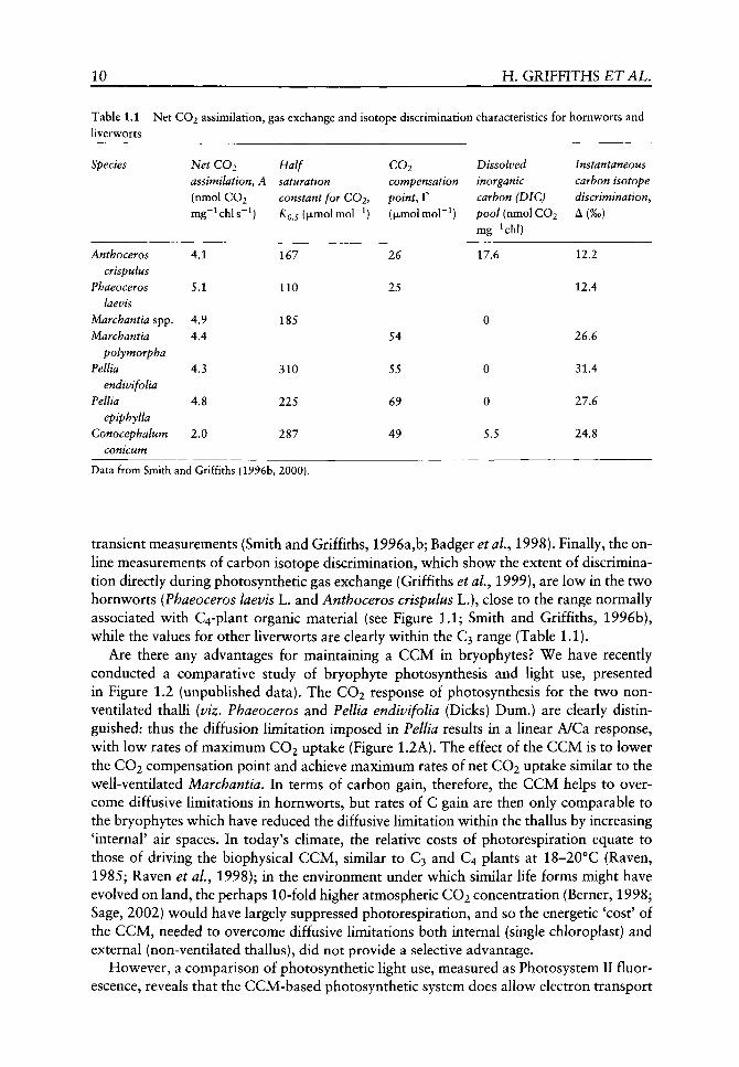
First, we consider the general characteristics of the CCM in hornworts, as compared to gas exchange and carbon isotope discrimination characteristics for other liverworts (Table 1.1). Rates of net CO2 assimilation are similar for contrasting thalloid life forms when expressed on a chlorophyll basis (and also on a weight and area basis: Smith and Griffiths, 1996a,b, 2000). However, the higher carboxylation conductance of hornworts, as inferred from the XQ 5 and the lower CO2 compensation point (F), is related to the magnitude of the dissolved inorganic carbon (DIG) pool accumulated (Table 1.1). Alternatively, for bryophytes with a varying degree of thallus ventilation, the gas exchange characteristics were uniformly more 'C3-like', with higher KQS values and higher F, suggesting that the efficiency of CO2 acquisition is lower. The values for Conocephalum con-icum (L.) Underw, with generally a low chlorophyll content, show a slight DIG pool, which is probably associated with alkalinization of the chloroplast stroma during the Ught-dark
10 H. GRIFFITHS ETAL.
Table 1.1 Net CO2 assimilation, liverworts
Species
Anthoceros crispulus
Phaeoceros laevis
Marchantia spp. Marchantia
polymorpha Pellia
endivifolia Pellia
epiphylla Conocephalum
conicum
Net CO2 assimilatiofiy A (nmol CO2 mg~^chls~^)
4.1
5.1
4.9 4.4
4.3
4.8
2.0
gas exchange
Half saturation
and isotope discrimination characteristics for
constant for COi, KQJ (jxmolmol"^)
167
110
185
310
225
287
CO2 compensation
point, r ((xmolmol"^)
26
25
54
55
69
49
Dissolved inorganic carbon (DIC) pool (nmol CO2 mg-^chl)
17.6
0
0
0
5.5
hornworts and
Instantaneous carbon isotope discrimination. A(%o)
12.2
12.4
26.6
31.4
27.6
24.8
Data from Smith and Griffiths {1996b, 2000).
transient measurements (Smith and Griffiths, 1996a,b; Badger etal., 1998). Finally, the online measurements of carbon isotope discrimination, which show the extent of discrimination directly during photosynthetic gas exchange (Griffiths et al.y 1999), are low in the two hornworts (Phaeoceros laevis L. and Anthoceros crispulus L.), close to the range normally associated with C4-plant organic material (see Figure 1.1; Smith and Griffiths, 1996b), while the values for other liverworts are clearly within the C3 range (Table 1.1).
Are there any advantages for maintaining a CCM in bryophytes? We have recently conducted a comparative study of bryophyte photosynthesis and light use, presented in Figure 1.2 (unpublished data). The CO2 response of photosynthesis for the two non-ventilated thalli [viz, Phaeoceros and Pellia endivifolia (Dicks) Dum.) are clearly distinguished: thus the diffusion limitation imposed in Pellia results in a linear A/Ca response, with low rates of maximum CO2 uptake (Figure 1.2A). The effect of the CCM is to lower the CO2 compensation point and achieve maximum rates of net CO2 uptake similar to the well-ventilated Marchantia. In terms of carbon gain, therefore, the CCM helps to overcome diffusive limitations in hornworts, but rates of C gain are then only comparable to the bryophytes which have reduced the diffusive limitation within the thallus by increasing 'internal' air spaces. In today's climate, the relative costs of photorespiration equate to those of driving the biophysical CCM, similar to C3 and C4 plants at 18-20°C (Raven, 1985; Raven et aL, 1998); in the environment under which similar life forms might have evolved on land, the perhaps 10-fold higher atmospheric CO2 concentration (Berner, 1998; Sage, 2002) would have largely suppressed photorespiration, and so the energetic 'cost' of the CCM, needed to overcome diffusive limitations both internal (single chloroplast) and external (non-ventilated thallus), did not provide a selective advantage.
However, a comparison of photosynthetic light use, measured as Photosystem II fluorescence, reveals that the CCM-based photosynthetic system does allow electron transport
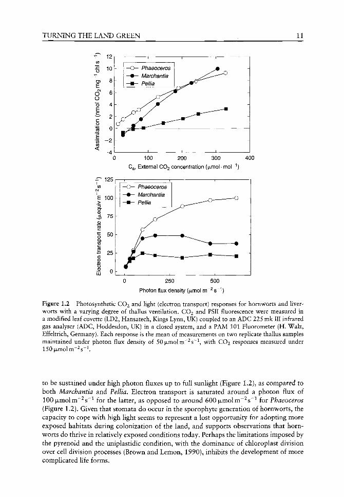
TURNING THE LAND GREEN 11
T 12
o
CF)
E CVJ
O O "o E
c
E 0) 0)
<
^ 125
100
75
£ >
(D
p5
o Q. 0) C CO
- 25
o ^ 0 I I I ^
50
LU
100 200 300
Ca, External CO2 concentration (ixmol-mor'')
400
Phaeoceros Marchantia Pellia
250 500
Photon flux density (|xmol m ^s '')
Figure 1.2 Photosynthetic CO2 and light (electron transport) responses for hornworts and liverworts with a varying degree of thallus ventilation. CO2 and PSII fluorescence were measured in a modified leaf cuvette (LD2, Hansatech, Kings Lynn, UK) coupled to an ADC 225 mk III infrared gas analyser (ADC, Hoddesdon, UK) in a closed system, and a PAM 101 Fluorometer (H. Walz, Effeltrich, Germany). Each response is the mean of measurements on two repHcate thallus samples maintained under photon flux density of 50|xmolm~^s~\ with CO2 responses measured under 150|xmolm~^s~^.
to be sustained under high photon fluxes up to full sunlight (Figure 1.2), as compared to both Marchantia and Pellia. Electron transport is saturated around a photon flux of 100 jULmolm~^s~^ for the latter, as opposed to around 600|LJLmolm"^s~^ for Phaeoceros (Figure 1.2). Given that stomata do occur in the sporophyte generation of hornworts, the capacity to cope with high light seems to represent a lost opportunity for adopting more exposed habitats during colonization of the land, and supports observations that hornworts do thrive in relatively exposed conditions today. Perhaps the limitations imposed by the pyrenoid and the uniplastidic condition, with the dominance of chloroplast division over cell division processes (Brown and Lemon, 1990), inhibits the development of more complicated life forms.
12 H. GRIFFITHS £TAL.
Conclusions
A definitive phylogenetic tree would help to clarify the paraphyletic development of the bryophytes in the context of the protracheophytes and help to resolve how the dramatic change in reproductive life cycle was accomplished during the progression towards vascular plants. However, it would not necessarily resolve the occurrence of the pyrenoid in horn-worts, since in the genus Megaceros there is the gradual loss of the pyrenoid associated with the development of the multiplastidic condition, which seemingly represents the derived condition (Burr, 1970; Brown and Lemon, 1990; Vaughn et ai, 1992; Badger et al, 1998). However, we may make some physiological contribution towards the debate regarding phylogeny: if hornworts are to be considered basal (Beckert et al, 1999; Wheeler, 2000; Nickrent et aL, 2000), and by inference closest to the Coleochaete-\ikc ancestor, why was the genetic basis for expressing a pyrenoid lost? That the pyrenoid-containing members of the family are more primitive and uniplastidic (Vaughn et aL, 1992; Badger et aL, 1998), as well as possessing stomata in the sporophyte, seems to make this family the sister group for other bryophytes and hence all embryophytes (Beckert et aL, 1999; Nickrent et al, 2000; Renzaglia et al, 2000). Then again, the conclusions that liverworts are basal, using rbcL sequence of many bryophytes (Lewis et aL, 1997; Qiu et aL^ 1998; Qui and Palmer, 1999) also seems compelling, and so ultimately we must adopt a compromise position (Kenrick, 2000) to account for the lack of agreement between molecular phylogeny and the bryophyte fossil appearance in terms of poor bryophyte preservation and changing geological conditions, rather than rapid diversification. Importantly, we now need to conduct a search at the molecular level for genes encoding the pyrenoid in other bryophytes and early tracheophytes (cf, Pfannschmidt et aL, 1999).
As far as Rubisco functioning, a study has now been completed in the variation of catalytic properties in a range of hornworts (Hanson et aL, 2002). We urgently need a comparative study on the range of bryophytes to clarify the interrelationships between the CCM and the changes in Rubisco specificity which may have occurred in terrestrial vascular plants. Since lower Sj.ei values are associated with C4 plants which have perhaps maintained Rubisco under elevated CO2 for only 10 my, it would be intriguing to determine whether any significant variations have developed between Anthoceros and Megaceros, respectively with and without the pyrenoid, or across the mosses and liverworts. At any event, when bryophytes first colonized the land some 460 my ago, the advantage of elevated CO2 at that time would have been offset by the higher ambient temperatures, likely to have caused vjv^ to have decreased from 5.7 to 2.2 for an increase from 15 to 35°C (at current CO2 concentrations) and also reduced activation by any Rubisco activase (Crafts-Brandner and Salvucci, 2000; Rokka et al, 2001).
The increasing ventilation of thalli, in a progression seen in extant liverworts as well as in the fossil record through increasing stomatal densities (Osbourne et aL, 2001) would undoubtedly increase internal conductance to CO2 (gi)-, but at the cost of higher water loss. When we performed measurements of photosynthesis and carbon isotope discrimination on a wetted thallus, it was noticeable that the on-line discrimination signal decreased by 4%o consistent with higher diffusion limitation, although net CO2 uptake rate was barely affected (Smith and Griffiths, 1996b). Our data suggest that the CCM can operate in a non-wetted thallus and help to overcome diffusion limitation and Anthoceros is often observed growing in quite exposed conditions in arable fields (MCF Proctor, personal communication) and so wetting does not seem to be a prerequisite for CCM activity. We need additional information on the occurrence, location and activity of
TURNING THE LAND GREEN 13
carbonic anhydrase throughout the hornworts (whether external, periplasmic, cytosolic, stromal or intrathylakoidal) and their interrelationship with CCM operation (Raven, 1997; Badger et aL, 1998; Thorns et aL, 2001).
Gas exchange characteristics of extant bryophytes provide some insight into the likely benefits of a CCM for hornworts as compared to the non-ventilated Pellia (see Figure 1.2). However, despite a higher carboxylation conductance (i.e. low KQS for CO2), and low compensation point, maximum rates of carbon gain are similar to the ventilated liverwort thalli (see Table 1.1, Figure 1.2). Indeed, we may infer that the expected energetic costs of the CCM are substantial (in contrast to the theoretical predictions of Raven, 1985), given the higher rates of electron transport needed to support this rate of CO2 assimilation in Anthoceros (see Figure 1.2).
Ultimately, without a better understanding of the genes and regulatory processes leading to the expression (or suppression) of the pyrenoid or multiplastidic cells, we cannot make any more detailed inferences on the selective processes likely to have shaped the earliest terrestrial bryophytes. Perhaps we may uncover molecular evidence to show how and when the pyrenoid was lost from Coleochaete and hornworts and with it the potential to express a CCM. One thing seems certain - by becoming multiplastidic and internalizing airspaces, mesophyll conductance would have been dramatically increased. As long as oxygenase products could be detoxified via photorespiration, the higher light intensities available to drive electron transport (and not the CCM) could then have led to additional carbon reserves for creating structural material and conducting tissues for competing in the developing land-plant canopy. Despite the early opportunities and genetic basis for developing a CCM in early land plants, it appears that 400 to 450 my later, when the C4 and CAM pathways became widespread, that potential had been irrevocably lost.
Acknowledgements
We are grateful for support from NERC and The Leverhulme Trust.
References
Algeo TJ, Scheckler SE, Maynard JB. 2001. Effects of the middle and late Devonian spread of vascular land plants on terrestrial weathering processes, and potential links to coeval marine extinction events and global climate change. In: Gensel GE, Edwards D, eds. Plants Invade the Land. New York: Columbia, 213-236.
Badger M, Andrews TJ. 1987. Co-evolution of Rubisco and CO2 concentrating mechanisms In: Biggins J, ed. Progress in Photosynthesis Research. Dordrecht: Martinus Nijhof, 601-609.
Badger MR, Andrews TJ, Whitney SM, et al. 1998. The diversity and coevolution of Rubisco, plas-tids, pyrenoids, and chloroplast-based CO2 concentrating mechanisms. Canadian journal of Botany 76: 1052-1071.
Badger MR, Pfanz H, Buedel B, et al. 1993. Evidence for the functioning of photosynthetic carbon dioxide concentrating mechanism in lichens containing green algal and cyanobacterial photobionts. Planta 191: 57-70.
Beardall J, Griffiths H, Raven JA. 1982. Carbon isotope discrimination and the CO2 accumulating mechanism in Chlorella emersonii. Journal of Experimental Botany 33: 173-1YI.
Beckert S, Steinhauser S, Muhle H, Knoop V. 1999. A molecular phylogeny of bryophytes based on nucleotide sequences of the mitochondrial nadS gene. Plant Systematics and Evolution 218: 179-192.
14 H. GRIFFITHS £ T A L .
Berner RA. 1998. The carbon cycle and CO2 over Phanerozoic time: the role of land plants. Philospohical Transactions of the Royal Society of London Series B 353: 75-82.
Brown RC, Lemon BE. 1990. Monoplastidic cell division in lov^er land plants. American Journal of Botany 77: 559-571.
Burr FA. 1970. Phylogenetic transitions in the chloroplasts of the Anthocerotales I. The number and ultrastructure of the mature plastids. American Journal of Botany 57: 97-110.
Chase M, Soltis DE, Olmstead RG, et al. 1993. Phylogenetics of seed plants: an analysis of nucleotides sequences from the plastid gene rbcL, Annals of the Missouri Botanical Garden 80: 528-580.
Crafts-Brandner SJ, Salvucci ME. 2000. Rubisco activase constrains the photosynthetic potential at high temperature and CO2. Proceedings of the National Academy of Sciences of the United States of America 97: 13430-13435.
Csintalan Z, Proctor MCF, Tuba Z. 1999. Chlorophyll fluorescence during drying and rehydration in the mosses Rhytidiadelphus loreus (Hedw.) Warnst., Anomodon viticulosus (Hedw.) Hook & Tayl. and Grimmia pulvinata (Hedw.)Sm. Annals of Botany 84:1'i5-1AA.
Deltoro VI, Calatayud A, Gimeno C, et al. 1998. Changes in chlorophyll a fluorescence, photosynthetic CO2 assimilation and xanthophyll cycle interconverison during dehydration in desiccation-tolerant and intolerant liverworts. Planta 201: 224-228.
Edwards D. 1998. Climate signals in Palaeozoic land plants. Philosophical Transactions of the Royal Society of London Series JB 353: 141-157.
Edwards D. 2000. The role of Mid-Palaeozoic mesofossils in the detection of early bryophytes. Philosophical Transactions of the Royal Society of London Series B 355: 733-755.
Edwards D, Duckett JG, Richardson JB. 1995. Hepatic characters in the earUest land plants. Nature 374: 635-636.
Edwards D, Kerp H, Hass H. 1998. Stomata in early land plants: an anatomical and ecophysio-logical approach. Journal of Experimental Botany 49: 255-278.
Farquhar GD, Ehleringer JR, Hubick K. 1989. Carbon isotope discrimination and photosynthesis. Annual Reviews of Plant Physiology and Plant Molecular Biology 40: 503-557.
Green TGA, Lange OL. 1994. Photosynthesis in poikilohydric plants: a comparison of lichens and bryophytes. In: Schulze ED, Caldwell MM, eds. Ecophysiology of Photosynthesis. Berlin: Springer Verlag.
Green TGA, Schroeter B, Kappen L, et al. 1998. An assessment of the relationship between chlorophyll a fluorescence and CO2 gas exchange from field measurements on a moss and a lichen. Planta 201:611-618.
Green TGA, Snelgar WP. 1982. A comparison of photosynthesis in two thalloid liverworts. Oecologia 54: 275-280.
Griffiths H, Borland AM, Gillon J, et al. 1999. Stable isotopes reveal exchanges between soil, plants and the atmosphere. In: Press MC, Scholes JD, Barker MG, eds. Physiological Plant Ecology. Oxford: Blackwell Science, 415-441.
Griffiths H, Smith JAC. 1983. Photosynthetic pathways in the BromeUaceae of Trinidad: relations between life-form, habitat preference and the occurrence of CAM. Oecologia 60: 176-184.
Hall DO, Rao KK. 1994. Photosynthesis. Cambridge: University Press. Hanson D, Andrews TJ, Badger MR. 2002. Variability of the pyrenoid-based CO2 concentrating
mechanism in hornworks (Anthocerophyta). Functional Plant Biology 29: 407-416. Kenrick P. 2000. The relationships of vascular plants. Philosophical Transactions of the Royal
Society of London Series B 355: 747-855. Kenrick P, Crane PR. 1997. The origin and early evolution of land plants. Nature 389: 33-39. Lewis LA, Mishler BD, Vilgalys R. 1997. Phylogenetic relationships of the liverworts (Hepaticae),
a basal embryophyte lineage, inferred from nucleotide sequence data of the chloroplast gene rbcL. Molecular Phylogenetics and Evolution 7: 377-393.
Ligrone R, Duckett JG, Renzaglia KS. 2000. Conducting tissues and phyletic relationships of bryophytes. Philosophical Transactions of the Royal Society of London Series B 355: 795-813.
Maguas C, Griffiths H, Broadmeadow MSJ. 1995. Gas exchange and carbon isotope discrimination in lichens: evidence for interactions between CO2 concentrating mechanisms and diffusion Hmi-tation. Planta 196: 95-102.
T U R N I N G T H E LAND GREEN 15
Maxwell K, von Caemmerer S, Evans JR. 1997. Is a low conductance to CO2 diffusion a consequence of succulence in plants with crassulacean acid metabolism? Australian Journal of Plant Physiology 24: 777-786.
McKay RML, Gibbs SP. 1991. Composition and function of pyrenoids: cytochemical and immuno-cytochemical approaches. Canadian Journal of Botany 69: 1040-1052.
Nassoury N, Fritz L, Morse D. 2001. Circadian changes in ribulose-l,5-bisphosphate carboxylase/ oxygenase distribution inside individual chloroplasts can account for the rhythm in dinoflagel-late carbon fixation. The Plant Cell 13: 923-934.
Nickrent DL, Parkinson CL, Palmer D, Duff JD. 2000. Multigene phylogeny of land plants with special reference to bryophytes and the earliest land plants. Molecular Biology and Evolution 17: 1885-1895.
Niklas KJ. 1997. The Evolutionary Biology of Plants. Chicago: University of Chicago Press. Nisbet EG, Sleep NH. 2001. The habitat and nature of early life. Nature 409: 1083-1091. Osbourne C, Beerling DJ, Chaloner WG. 2001. Evolution of leaf form in land plants linked to
atmospheric CO2 decline in late Phanerozoic. Nature 410: 352-354. Palmqvist K. 1993. Photosynthetic C02-use efficiency in lichens and their isolated photobionts:
the possible role of a C02-concentrating mechanism in cyanobacterial lichens. Planta 191: 48-56.
Pfannschmidt T, Nilsson A, Allen JF. 1999. Photosynthetic control of chloroplast gene expression. Nature 397: 625-628.
Proctor MCF. 2001. Mosses and alternative adaptation to life on land. New Phytologist 106: 117-134.
Proctor MCF, Raven JA, Rice SK. 1992. Stable carbon isotope discrimination measurements in Sphagnum and other bryophytes. Journal of Bryology 17: 193-202.
Pronino NA, Semenenko VE. 1992. The role of the pyrenoid in concentration, generation and fixation of carbon dioxide in chloroplasts of microalgae. Eiziol Past 39: 723-732.
Qiu Y-L, Cho JC, Cox JC, Palmer JD. 1998. The gain of three mitochondrial inserts identifies liverworts as the earliest land plants. Nature 394: 671-674.
Qiu Y-L, Palmer JD. 1999. Phylogeny of early land plants: insights from genes and genomes. Trends in Plant Science 4: 26-30.
Raven JA. 1977. The evolution of vascular land plants in relation to supra-cellular transport processes. Advances in Botanic Research 5: 153-219.
Raven JA. 1985. The CO2 concentrating mechanism In: Lucas WJ, Berry JA, eds. Inorganic Carbon Uptake by Aquatic Photosynthetic Organisms. Rockville: The American Society of Plant Physiologists, 67-81.
Raven JA. 1995. The early evolution of land plants: aquatic ancestors and atmospheric interactions. Botanical Journal of Scotland 47: 151-175.
Raven JA. 1997. C02-concentrating mechanisms: a direct role for thylakoid lumen acidification? Plant, Cell and Environment 20: 147-154.
Raven JA. 2000. Land plant biochemistry. Philosophical Transactions of the Royal Society of London Series B 355: 833-846.
Raven JA, Edwards D. 2001 Roots: evolutionary origins and biogeochemical significance. Journal of Experimental Botany 52: 381-401.
Raven JA, Griffiths H, Smith EC, Vaughn KC. 1998. New perspectives in the biophysics and physiology of bryophytes. In: Bates JW, Ashton NW, Duckett JG, eds. Bryology for the Twenty-First Century. Leeds: Maney Publishing and The British Bryological Society, 261-275.
Raven JA, Kubler J, Beardall J. 2000. Put out the light and then put out the light. Journal of the Marine Biological Association 80: 1-27.
Renzaglia KS, Duff RJ, Nickrent DL, Garbary DJ. 2000. Vegetative and reproductive innovations of early land plants: implications for a unified phylogeny Philosophical Transactions of the Royal Society of London Series B 355: 769-793.
Rokka A, Zhang LX, Aro EM. 2001. Rubisco activase: an enzyme with a temperature-dependent dual function? The Plant Journal 25: 463-471.
Sage RF. 2002. Evolution of photosynthetic metabolism in terrestrial plants. PL14 Proceedings of the 12th International Photosynthesis Congress. Collingwood, Australia: CSIRO Publishing.

16 H. GRIFFITHS E T A L .
Schidlowski M. 2001. Carbon isotopes as biogeochemical recorders of life over 3.8 Ga of Earth history: evolution of a concept. Frecambrian Research 106: 117-134.
Schlitter J, Wildner GF. 2000. The kinetics of conformation change as determinant of Rubisco's specificity. Photosynthesis Research 65: 7-13.
Schuster RM. 1981. Paleoecology, origin, distribution through time, and evolution of Hepaticae and Anthocerotae. In: Niklas KJ, ed. Paleobotany, Paleoecology and Evolution. New York: Praeger Publishers, 129-191.
Smith EC, Griffiths H. 1996a. The occurrence of the chloroplast pyrenoid is correlated v^ith the activity of a C02-concentrating mechanism and carbon isotope discrimination in lichens and bryophytes. Planta 198: 6-16.
Smith EC, Griffiths H. 1996b. A pyrenoid-based carbon-concentrating-mechanism is present in terrestrial bryophtes of the class Anthocerotae. Planta 200: 203-212.
Smith EC, Griffiths H. 2000. The role of carbonic anhydrase in photosynthesis and the activity of the carbon-concentrating-mechanism in bryophytes of the class Anthocerotae. New Phytologist 145: 29-37.
Siiltemeyer DF, Biehler K, Fock HP. 1993. Evidence for the contribution of pseudocyclic photophosphorylation to the energy requirement of the mechanism for concentrating inorganic carbon in Chlamydomonas. Planta 198: 235-242.
Thoms S, Pahlow M, Wolf-Gladrow DA. 2001. Model of the carbon concentrating mechanism in chloroplasts of eukaryotic algae. Journal of Theoretical Biology 208: 295-313.
Vaughn KC, Campbell EO, Hasegav^a J, et al. 1990. The pyrenoid is the site of ribulose 1-5 bisphos-phate carboxylase/oxygenase accumulation in the hornwort (Bryophyta:Anthocerotae) chloroplast. Protoplasma 156: 117-129.
Vaughn KC, Ligrone R, Owen H, et al. 1992. The Anthocerote chloroplast: a review. New Phytologist 120: 169-190.
Wheeler JA. 2000. Molecular phylogenetic reconstructions of the marchantioid liverwort radiation. The Bryologist 103: 314-333.
Zhu G, Jensen, RG, Bohnert H, et al. 1998. Dependence of catalysis and CO2/O2 specificity of Rubisco on the C terminus. Photosynthesis Research 57: 71-79.
Zotz G, Schweikert A, Jetz W, Westeman H. 2000. Water relations and carbon gain are closely related to cushion size in the moss Grimmia pulvinata. New Phytologist 148: 1-6.

Physiological evolution of lower embryophytes: adaptations to the terrestrial
environment
John A Raven and Dianne Edwards
CONTENTS
Introduction 17
The ancestors of embryophytes 18
Water, carbon dioxide and energetics of land plants 19
Desiccation tolerance, desiccation intolerance, poikilohydry and homoiohydry 22
Poikilohydry of algae and early-evolving embryophytes 22 Desiccation tolerance and intolerance 27 Evolution of homoiohydry 28
History of physiological interpretations of early embryophytes 31
Introduction 31 Transpiration rate and endodermal function in
regulating nutrient supply to the shoot 33 Mechanism of endohydric water movement 34 Role of stomata in determining the rate of
photosynthesis and the water cost of photosynthesis 35
Conclusions 37
Acknowledgements 37
References 38
Introduction
The physiology of embryophytes differs from that of their algal ancestors in a number of ways. Most relate to the differences in water relations of organisms which live on land, i.e. extant embryophytes, except the small number of species which have returned to live in water and those which live permanently in water, i.e. the great majority of species of algae.
The Evolution of Plant Physiology ISBN 0-12-33955-26
Copyright © 2004 Elsevier Ltd All rights of reproduction in any form reserved

18 J. A. RAVEN &c D. EDWARDS
This chapter sets out to examine the differences in physiology between embryophytes and their algal ancestors, with particular emphasis on their water relations. The embryophytes have very significant variations in water relations and the chapter considers their evolution within the embryophytes as well as the evolution of embryophyte water relations from those of their algal ancestors. The chapter also considers the relationship of the likely evolution of embryophyte water relations to cladistic analyses of embryophyte phylogeny and to the fossil record. A final point concerns the history of our understanding of this subject area and the possible constraints on achieving earlier syntheses.
The ancestors of embryophytes
The origins of the physiology of plants, i.e. embryophytes, must be sought in the extant algae which are most closely related to their algal ancestors. These algae are the Charophyceae sensu lato, and it is to the comparative electron microscopic studies of Pickett-Heaps (reviewed by Pickett-Heaps, 1975) and of Stewart, Mattox, Floyd and O'Kelly (reviewed by Stewart and Mattox, 1975) that the relationship of the Charophyceae to embryophytes was firmly established. Data on the occurrence of different enzymes catalysing the oxidation of glycolate, the hydrolysis of urea and the dismutation of superoxide radical anions supported the ultrastructural evidence (Bekheet and Syrett, 1977; Syrett and Al-Houty, 1984; De Jesus etaL,19S9). These findings have been supported by molecular phylogenetic studies (Nickrent et aL, 2000) and multifactorial cladistic analyses based on non-molecular studies (van den Hoek et aL, 1995; Kenrick and Crane, 1997; Graham and Wilcox, 2000a). The closest living relative of the embryophytes in the Charophyceae sensu lato has been widely held to be the small discoid alga Coleochaete Breb. (van den Hoek et ai, 1995; Kenrick and Crane, 1997; Graham and Wilcox, 2000a; but see Karol etaL, 2001) which lives epilithically or epiphytically in fresh waters. The vegetative phase of Coleochaete is haploid and the only diploid phase in the life cycle is the zygote. Although earlier suggestions that there were mitotic divisions within the zygote before meiosis occurred have not been substantiated, one aspect of the life cycle of Coleochaete resembles that of the lower embryophytes. This is matrotrophy, i.e. nutrition of the zygote by the haploid vegetative phase using 'transfer cell'-like wall invaginations (Graham and Wilcox, 2000b). This similarity of Coleochaete to embryophytes is not shared by some charophyceans such as Stichococcus NageU and Klebsormidium Silva Mattox et Blackwell which are, however, sometimes found in terrestrial habitats (Graham, 1993). The analysis by Karol etal (2001) of the phylogeny of the Charophyceae sensu lato in relation to the Embryophyta uses the sequences of one nuclear, one mitochondrial and two plastid genes in a range of analytical methods including Bayesian inference. Karol et al, (2001) conclude that the Charales are the sister clade to the embryophytes. Their findings suggest that the ancestors of embryophytes were vegeta-tively somewhat more complex than Coleochaete, However, Raven (1977) points out that extant members of the Charales, with most of the volume of the organism occupied by giant cells with volumes up to 10 mm^, are not good mechanical or physiological prototypes for early embryophytes (Raven, 1977, 1986).
The embryophytes are characterized by an alternation of gametophyte and sporophyte phases, with the gametophyte phase occupying a decreasing fraction of the biomass in the life cycle in proceeding from the bryophyte grade of evolution through the pteridophyte grade to the spermatophyte grade. In parallel to this the frequency of desiccation tolerant species decreases and the frequency of species with internal conduction pathways for water increases
EARLY EVOLUTION OF HIGHER PLANT PHYSIOLOGY 19
conifers angiosperms cycads , gnetophytes
Equisetum
Seed Plants
Selaginella
mosses
hornworts
liven/vorts
Charales
Coleochaetales
Chlorophyta
Tracheophytes
Embryophytes
Figure 2.1 A phylogeny of the major groups of green plants based on several recent synthetic studies (summarized in Oliver et al. (2000), Karol et al. (2001)).
(Raven, 1977, 2002). Cladistic phylogeny (Kenrick and Crane, 1997; Qiu et al, 1998; Qiu and Lee, 2000; Renzaglia et al, 2000; Pryer et al, 2001) and the fossil record (Chaloner, 1970; Gensel and Andrews, 1984; Edwards, 1993,1996,1998, 2000, 2003; Edwards et al, 1996, 1998; Kenrick and Crane, 1997; Wellman and Gray, 2000; Raven and Edwards, 2001; Raven, 2002) are used as a framework for considering the evolution and the physiology of embryophytes (Figures 2.1 and 2.2).
Water, carbon dioxide and energetics of land plants
The physiology of the embryophytes is a very large subject and our considerations will be limited to resource acquisition and related mechanical matters. In particular, the acquisition of photons and inorganic carbon for photosynthesis from the atmospheric environment necessitates the loss of water vapour to the atmosphere. The use of light energy in photosynthesis necessarily involves dissipation of at least 73% of the absorbed photosynthetically active radiation other than in the reduced organic products of photosynthesis. The influx of CO2 from the bulk atmosphere to Rubisco in chloroplasts requires a high-conductance diffusion pathway in the gas phase to a wet cell wall in which the CO2 dissolves prior to diffusion in solution to Rubisco. The combination of an energy source for evaporation of water, the presence of a high conductance pathway for gas diffusion to the atmosphere and the general lack of saturation with water vapour of the atmosphere results in the loss of water from the photosynthetic structures to the atmosphere (Raven, 1977; Jones, 1992).
20 J. A. RAVEN & D. EDWARDS
Known range
Possible range
Probable range but derivation different Probable range based on megafossils
Earliest embryophyte
Earliest rhyniophytoid
Earliest lycophyte
Earliest euphyllophyte
Figure 2.2 Stratigraphic ranges of fossils relevant to the early history of land plants. 1 = cryptospores (dyads and tetrads) thought to derive from bryophytes; 2 = trilete spores (monads) produced by tra-cheophytes; 3 = Cooksonia s.L; 4 = axial fragments of Ptracheophytes; 5 = Nematothalius cuticles; 6 = sporangial and Ptracheophyte cuticles; 7 = stomata; 8 = unevenly thickened tubes of nemato-phyte affinity resembling tracheids; 9 = cooksonioid tracheids; 10 = G-type tracheid (zosterophylls and lycophytes); 11 = S-type tracheid (Rhyniopsida); 12 = P-type tracheid (typical early euphyllophyte tracheid).
An important aspect of the argument is the physics underlying the partitioning of energy dissipation from the photosynthetic apparatus. In addition to dissipation as the latent heat of evaporation of w^ater, radiation, conduction and convection can also be involved in energy dissipation (Jones, 1992; Denny, 1993). Conduction betw^een the photosynthetic

EARLY EVOLUTION OF HIGHER PLANT PHYSIOLOGY 21
structures and the environment is much less in photosynthetic structures in air than in water as a resuh of the much lower thermal conductivity of air than of water as modulated by differences in boundary layer thickness and volume-based specific heat (Denny, 1993). This means that the temperature of the photosynthetic structures of the aquatic ancestors of the terrestrial embryophytes was much closer to that of surrounding water than the photosynthetic structures of the embryophytes are to the surrounding air. The temperature of the photosynthetic structures in air is also dependent on the extent of energy dissipation in its evaporating water and the temperature of the structures in air and in water is also determined by and determines, energy dissipation as long-wave radiation according to the Stefan-Boltzman law (Jones, 1992; Denny, 1993).
The outcome of these various considerations on the energy balance and gas phase conductances is that photosynthetic organs on land with C3 physiology (diffusive supply of CO2 to Rubisco) lose hundreds of grams of water vapour during the photosynthesis equivalent to the gain of one gram dry matter. The exact value of the water cost of growth depends on the radiation environment, wind speed, air temperature and relative humidity and the adaptive, acclimatory and regulatory characteristics of the photosynthetic organism (Jones, 1992). The CO2 content of the atmosphere is another factor which alters the water cost of photosynthetic growth; more CO2 in the atmosphere can permit lower water costs (Edwards et al, 1998; Woodward, 1998; Konrad et ai, 2000). The CO2 content of the atmosphere varies annually by about 5|ULmolmol"^ out of a current total of some 360 jjimol mol~^ as a result of the excess of terrestrial photosynthesis, mainly in the northern hemisphere, over terrestrial respiration in the summer and the excess of terrestrial respiration over photosynthesis in the winter (Keeling et al.y 1995; Berner and Berner, 1996). The present trend of year-on-year increases in atmospheric CO2 as a result of changed land use, combustion of fossil fuels and cement manufacture could double atmospheric CO2 over the next century (Berner and Berner, 1996). Clearly these arthropogenic effects are not relevant to the changes in atmospheric CO2 prior to the evolution of man and rates of change of atmospheric CO2 known from the Pleistocene ice record (Petit et al.^ 1999) are lower than the current rate of increase. More relevant to the evolution of an embryophytic land flora is the suggested high CO2 in the Ordovician, Silurian and Lower Devonian (Berner and Kothavala, 2001; cf. Boucot and Gray, 2001), as modelled for photosynthesis by Lower Devonian organisms at the pteridophyte grade of organization (Konrad ^^^/., 2000).
Another atmospheric factor which influences the water cost of growth is O2 (which shows much less interannual variation in relative terms, than does CO2: Keeling and Shertz, 1992; Keeling et al, 1995; Berner and Berner, 1996). Higher O2 mol fractions in the atmosphere, combined with a CO2 mol fraction which is limiting for photosynthesis, limits gross photosynthesis via the oxygenase activity of Rubisco relative to an atmosphere with less O2 (Raven et al., 1994). The high CO2 in the Ordovician, Silurian and Lower Devonian atmosphere (Berner and Kothavala, 2001), together with the evidence that the O2 level was similar to the extant values (Berner, 2001), shows that O2 was probably not a significant restriction on photosynthesis by the earliest embryophytes.
These considerations show that the water lost per unit dry matter increase is, other things being equal, lower in the atmosphere found in the Ordovician, Silurian and Lower Devonian than in the present atmosphere. A factor that would decrease the water cost of dry matter gain, especially at low CO2 partial pressures, is the occurrence of CO2 concentration mechanisms (Surif and Raven, 1990). Most green algae, including Coleochaete and many other members of the Charophyceae sensu lato^ as well as some hornworts

22 J. A. RAVEN &: D. EDWARDS
(see Figure 2.3), have pyrenoids which are invariably correlated with the presence of a CO2 concentrating mechanism (CCM) (van den Hoek et aL, 1995; Smith and Griffiths, 1996a,b, 2000; Badger et al.^ 1998; Graham and Wilcox, 2000a). These mechanisms decrease the external inorganic C concentration required to half-saturate photosynthesis and usually lead to whole-cell photosynthesis which is 02-insensitive and which has a greater CO2 affinity than does Rubisco from the same organism in vitro (Badger et al., 1998; Raven, 2000). In addition to their occurrence in many charophycean algae, pyrenoids and the associated CCM are also found in a number of species of hornwort (Smith and Griffiths, 1996a,b, 2000). These data could be construed as indicating that the common ancestor of Coleochaete and the embryophytes had pyrenoids. Regardless of whether the liverworts (see Figure 2.1; Qiu et al., 1998; Qiu and Lee, 2000) or the hornworts (Nickrent et aL, 2000; Renzaglia et al, 2000) are the taxon of embryophytes which is most closely related to the algal ancestors of embryophytes, losses of pyrenoids would be required to explain the pyrenoid-less state of almost all embryophytes on the basis of the occurrence of pyrenoids in the algal ancestor of embryophytes. However, it is likely that pyrenoids are polyphyletic (Raven, 1997) and evolved in response to low CO2 concentrations and so are unlikely to have occurred before the low CO2 in the Upper Devonian and Carboniferous. Accordingly, it is not likely that the earliest embryophytes had pyrenoids and so they would not have had higher CO2 affinity and higher water-use efficiencies than plants with C3 physiology. In any case, C3 plants growing in the high CO2 environment of the Ordovician, Silurian and Lower Devonian would, as indicated above, already have relatively low water costs of growth (Konrad et al., 2000).
Desiccation tolerance, desiccation intolerance, poikilohydry and homoiohydry
Poikilohydry of algae and early-evolving embryophytes
The inevitability of water loss in terrestrial photolithotrophs during growth, combined with variability of water supply, leads to potentially lethal water loss in organisms which combine two characteristics: poikilohydry and desiccation intolerance. Poikilohydric plants have little or no capacity to restrict water loss when the rate of evaporative water loss exceeds the rate of liquid water supply. Desiccation-intolerant plants have a lethal lower limit of water content corresponding to a cell or tissue water potential which is still relatively high. Before attempting to quantify the 'little or no capacity to restrict water loss' and 'relatively high water potential', we point out that the 'transmigrant' charophyceans were poikilohydric.
Whether these 'transmigrant' charophyceans were desiccation-tolerant is not clear, but it is very likely that they were (Oliver et al., 2000). Some extant charophyceans (e.g. members of the Zygnematales) have desiccation-tolerant zygotes. As for the embryophytes, Oliver et al. (2000) (see also Tuba et ai, 1999) have performed a parsimony analysis of the occurrence and evolution of vegetative desiccation tolerance. A very significant fraction of the taxa in extant liverworts, hornworts and mosses are desiccation-tolerant and Oliver et al. (2000) suggest that these organisms are ancestrally desiccation-tolerant. Oliver et al, (2000) suggest that vegetative desiccation tolerance was lost early in the evolution of the vascular plant sporophyte; the loss was permitted by the evolution of homoiohydry and was evolutionarily favoured by the metabolic costs of desiccation tolerance which exceed those of homoiohydry. However, while there are data sets with lower specific growth rates for desiccation-tolerant algae (e.g. Porphyra C.A. Agardh sp.) than for less desiccation-tolerant algae (e.g. Enteromorpha Link in Nees, nom. con sp. and Ulva lactuca L.) (Nielsen and
EARLY EVOLUTION OF HIGHER PLANT PHYSIOLOGY 23
Sand-Jensen, 1990; Abe et al., 2001), phylogenetic bias must be considered (Raven, 1999). More research is needed to determine if the metabolic costs of desiccation tolerance exceed those of homoiohydry (Raven, 1999). There is also the correlation of vegetative desiccation tolerance with plants of relatively small stature (Raven, 1999). Propagules (spores and ultimately seeds) are frequently desiccation tolerant even in vascular embryophytes which are vegetatively intolerant of desiccation (Raven, 1999; Oliver et al, 2000). The occurrence of reproductive desiccation tolerance could form the basis for the polyphyletic (re)evolution of vegetative desiccation tolerance in the lycopsid Selaginella Pal. and in ferns and at least eight times among the angiosperms (Figures 1 and 2 of Oliver et aL, 2000; Pryer et al,^ 2001; seealsoGaff, 1981, 1997).
Tuba et al. (1999) reviewed the responses of desiccation-tolerant and desiccation-intolerant terrestrial photosynthetic organisms to CO2 levels higher than the present level and concluded that neither theoretical nor observational results suggest that elevated CO2 will lead to any substantial shift in the balance of advantage between the two groups of plants. The theoretical aspects of the study should apply to the early embryophytes in their high CO2 environment. Tuba et al (1999) could find data on CO2 enrichment only for one vascular plant species (Xerophyta scabrida) so that all of the data for desiccation-tolerant plants comes from poikilohydric plants (bryophytes) and lichens. Tuba et al, (1999) noted that Polytrichum formosum Hedw. is the moss gametophyte which comes closest in its response to increased CO2 to non-desiccation-tolerant homoiohydric plants. Some Foly-trichum spp. do approach the homoiohydric condition (Raven, 2002). Desiccation tolerance in photosynthetic organisms has recently been related to the expression of certain proteins (late embryogenesis expressed proteins, dehydrins, rehydrins: Close, 1997; Campbell and Close, 1997; Li et al, 1998; Farnsworth, 2000; Velten and Oliver, 2001; cf, Thomson et al, 1998). Although Close (1997) refers to unpublished immunological evidence of dehydrin protein in green algae there seem to be no sequence data confirming the presence of dehydrins in green and, especially, charophycean algae. However, the presence of dehydrins in cyanobacteria, based on sequence evidence (Close, 1997), as well as the extensive sequence evidence for dehydrins in seed plants and a lycopod (Close, 1997) is consistent with the universal occurrence of dehydrins in photosynthetic organisms, although lateral gene transfer cannot be ruled out. However, it is likely that other genes are significant in permitting desiccation tolerance.
Thus far our considerations of poikilohydry and desiccation tolerance as putatively ancestral states in embryophytes, i.e. present in their algal ancestors, has not dealt with whether the poikilohydric and the desiccation-tolerant states are qualitatively or quantitatively different from their alternate states, i.e. homoiohydry and desiccation intolerance. Desiccation intolerance has already been considered; it is the (apparently) obligatory state for the vegetative phase of all homoiohydric plants more than 1 m in height, and for the great majority of homoiohydric plants of smaller stature (Oliver et al, 2000; Raven, 2002). Homoiohydry is the capacity to maintain vegetative tissue hydration over a range of external conditions of water availability in the soil and evaporative demand by the atmosphere. The restriction on water loss rate when evaporative demand exceeds the soil supply of water requires a waxy cuticle over above-ground parts of the plant combined with stomatal closure. The occurrence of photosynthesis and transpiration when evaporative demand does not exceed the soil supply of water requires open stomata, an intercellular gas space system supplying atmospheric CO2 to photosynthetic cells and an endohydric conducting system (true xylem in all extant homoiohydric plants: Walter and Stadelmann, 1968; Raven, 1977, 1984, 1993, 1996, 2002; Jarvis, 1998).
24 J. A. RAVEN &c D. EDWARDS
Dealing first with poikilohydry, is there a quaUtative difference between poikilohydric and homoiohydric plants? Slowing down the rate of water loss from plants in a habitat with intermittent water supply can to some extent replace homoiohydry in maintaining cell and tissue hydration. This is because the likelihood of the organisms remaining above a given water potential by the time another rain event occurs is increased. The requirement here is a lower conductance for water vapour between the evaporating surface and the bulk atmosphere, so that a given concentration difference of water vapour between the evaporating surface and the bulk atmosphere yields a smaller flux of water vapour. The geometry which lowers the conductance for water vapour occurs at the cell and tissue level (Green and Lange, 1994) and at the canopy level (Rice et aL, 2001) in bryophytes. Of course, a lower gas phase conductance for water vapour involves a lower gas phase conductance for CO2, so that there is a lower rate of CO2 fixation in photosynthesis on a unit biomass basis. Because there is a biochemical conductance involved in photosynthesis but not in transpiration, a given decrement in gas-phase conductance gives a greater decrement in water loss than in photosynthesis. This effect is partially offset by the higher tissue temperatures which occur if there is the same biomass-based rate of absorption of photosynthetically active radiation but less dissipation of this radiation as the latent heat of evaporation of water. Nevertheless, slowing the rate of dehydration as a result of a decrease in the gas-phase conductance also yields a greater dry matter gain per unit water lost (Table 2.1).
Such a low gas-phase conductance means that the achieved photosynthetic rate is lower than would be the case if there was a larger conductance. A low gas-phase conductance means that a given biochemical capacity for photosynthesis is not used to the extent that is possible with a higher gas-phase conductance. In the low gas-phase conductance case the rate of photosynthesis on a tissue nitrogen basis is lower than in the case of high gas-phase conductance. If there is a lower biochemical capacity for photosynthesis in parallel with a lower gas-phase conductance then the photosynthetic rate per unit nitrogen is the same in the high and the low gas-phase conductance cases, but the dry matter gain per unit water lost is also the same in the two cases (Table 2.1). A low gas-phase conductance reduces the use of the latent heat of evaporation of water in minimizing the increase in temperature of the tissue relative to the atmosphere (Table 2.1); changes in surface reflectance as a function of water content can also have an influence here (Hamerlynck et ai, 2000).
Conclusions from this analysis (see Table 2.1) show that there are cost-benefit 'trade-offs' for poikilohydric terrestrial plants in relation to variations in gas-phase and liquid-phase conductance in the absence of any specific regulatory changes in the conductances as a response to changed tissue water status, soil water availability, or the driving force for water loss to the atmosphere. Presumably different strategies (see Table 2.1) are more appropriate for particular habitats with (for example) different mean intervals between rainfall events, although it is not easy to find data which test this suggestion. Overall, poikilohydry, defined as the occurrence of little or no capacity to restrict water loss when the rate of evaporative water loss exceeds the rate of liquid water supply, is qualitatively different from homoiohydry.
However, there are examples of essentially poikilohydric plants which show some homoiohydric structural features. Examples are the endohydric, ventilated, cuticularized bryophyte gametophytes of many marchantiaceous liverworts and polytrichaceous mosses (Green and Lange, 1994; Raven, 2002). Despite the occurrence of intercellular gas spaces in the marchantiaceous liverworts, resembling those in homoiohydric sporophytes of vascular plants, there is no evidence of regulation of gas-phase conductance via changed pore

Table 2.1 The influence on poikilohydric plants of variation in gas-phase conductance and in biochemical conductance on maximum growth rate, dry weight gain per unit water lost, rate of dry weight gain per unit tissue nitrogen and the capacity to retain tissue hydration in environments of episodic rainfall. Based on Raven (1977, 1984, 1993, 2002) and Konrad et al. (2000), and discussion in the text of this chapter
Gas-phase conductance
High High Low Low
Aqueous phase and biochemical conductance
High Low High Low
Potential specific growth rate
High Moderate Moderate Low
Dry weight gain per unit water
Moderate Low High Moderate
lost Rate of dry weight gain per unit tissue nitrogen
Moderate High Low Moderate
Capacity to maintain hydration in environments with episodic rainfall
Low Low High High
Excess of tissue temperature over air temperature in high light
Low Low High High
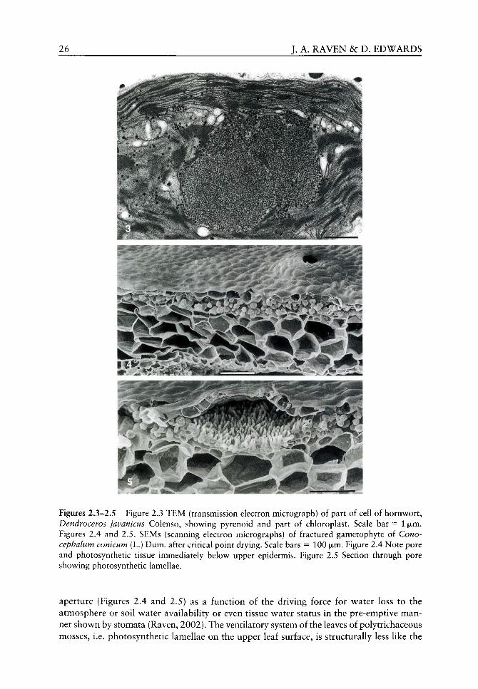
26 J. A. RAVEN &c D. EDWARDS
Figures 2.3-2.5 Figure 2.3 TEM (transmission electron micrograph) of part of cell of hornwort, Dendroceros javanicus Colenso, showing pyrenoid and part of chloroplast. Scale bar = 1 |xm. Fi gures 2.4 and 2.5. SEMs (scanning electron micrographs) of fractured gametophyte of Cono-cephalum conicum (L.) Dum. after critical point drying. Scale bars = 100 fxm. Figure 2.4 Note pore and photosynthetic tissue immediately below upper epidermis. Figure 2.5 Section through pore showing photosynthetic lamellae.
aperture (Figures 2.4 and 2.5) as a function of the driving force for water loss to the atmosphere or soil water availability or even tissue water status in the pre-emptive manner shown by stomata (Raven, 2002). The ventilatory system of the leaves of polytrichaceous mosses, i.e. photosynthetic lamellae on the upper leaf surface, is structurally less like the
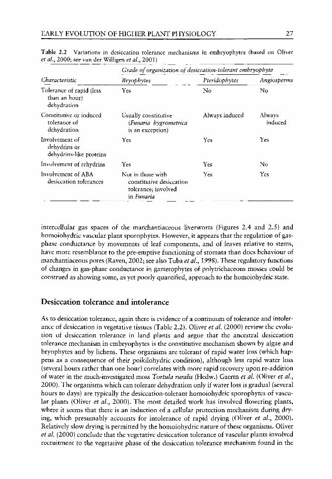
EARLY EVOLUTION OF HIGHER PLANT PHYSIOLOGY 27
Table 2.2 Variations in desiccation tolerance mechanisms et ai, 2000; see van der Willigen et al., 2001)
Characteristic
Tolerance of rapid (less than an hour) dehydration
Constitutive or induced tolerance of dehydration
Involvement of dehydrins or dehydrins-like proteins
Involvement of rehydrins
Involvement of ABA desiccation tolerances
in embryophytes (based on Oliver
Grade of organization of desiccation-tolerant embryophyte
Bryophytes
Yes
Usually constitutive (Funaria hygrometrica is an exception)
Yes
Yes
Not in those w ith constitutive desiccation tolerance; involved in Funaria
Pteridophytes
No
Always induced
Yes
Yes
Yes
Angiosperms
No
Always induced
Yes
No
Yes
intercellular gas spaces of the marchantiaceous liverworts (Figures 2.4 and 2.5) and homoiohydric vascular plant sporophytes. However, it appears that the regulation of gas-phase conductance by movements of leaf components, and of leaves relative to stems, have more resemblance to the pre-emptive functioning of stomata than does behaviour of marchantiaceous pores (Raven, 2002; see also Tuba et aL, 1998). These regulatory functions of changes in gas-phase conductance in gametophytes of polytrichaceous mosses could be construed as showing some, as yet poorly quantified, approach to the homoiohydric state.
Desiccation tolerance and intolerance
As to desiccation tolerance, again there is evidence of a continuum of tolerance and intolerance of desiccation in vegetative tissues (Table 2.2). Oliver et aL (2000) review the evolution of desiccation tolerance in land plants and argue that the ancestral desiccation tolerance mechanism in embryophytes is the constitutive mechanism shown by algae and bryophytes and by lichens. These organisms are tolerant of rapid water loss (which happens as a consequence of their poikilohydric condition), although less rapid water loss (several hours rather than one hour) correlates with more rapid recovery upon re-addition of water in the much-investigated moss Tortula ruralis (Hedw.) Gaertn et al, (Oliver et aL, 2000). The organisms which can tolerate dehydration only if water loss is gradual (several hours to days) are typically the desiccation-tolerant homoiohydric sporophytes of vascular plants (Oliver et aL, 2000). The most detailed work has involved flowering plants, where it seems that there is an induction of a cellular protection mechanism during drying, which presumably accounts for intolerance of rapid drying (Oliver et aL, 2000). Relatively slow drying is permitted by the homoiohydric nature of these organisms. Oliver et aL (2000) conclude that the vegetative desiccation tolerance of vascular plants involved recruitment to the vegetative phase of the desiccation tolerance mechanism found in the
28 J. A. RAVEN &c D. EDWARDS
propagules, which in turn had evolved from the vegetative and reproductive desiccation-tolerance mechanism in algae and bryophytes. In the flov^ering plants the majority are homoiochlorophyllous (i.e. are able to tolerate vegetative desiccation in the green state). A few monocotyledonous flowering plants from the almost soil-less habitats of tropical inselbergs are tolerant of desiccation by a mechanism that involves loss of chloroplast fine structure with the need for three days of rehydration to re-establish full photosynthetic function (Oliver et aL, 2000). There is some evidence that vascular plants at the pteridophyte grade of organization have a desiccation-tolerance mechanism which is closer to the ancestral form than that of flowering plants. Thus, while pteridophytes, like angiosperms but unlike most bryophytes (an exception is Funaria hygrometrica Hedw.), show an induction of a cellular protection system upon dehydration, they resemble bryophytes in possessing rehydrins (Oliver et ai, 2000) (see Table 2.2).
Clearly the bryophyte mechanism(s) of vegetative desiccation tolerance are more relevant to the origin and early evolution of an embryophytic land flora. However, most of the quantitative data (tissue water potentials) or the extent of survivable dehydration in desiccation-tolerant and in desiccation-intolerant land plants comes from vascular plants. Raven (2002) suggests that desiccation-intolerant plants can tolerate water potentials not lower than - 2 2 MPa, although the plants cannot grow at such low water potentials. By contrast, desiccation-tolerant plants can tolerate water losses equivalent to a cell water potential of - 2 0 0 MPa or lower, and some of them (algae, bryophytes) can photosynthesize, and even grow, at water potentials as low as - 3 0 MPa (Raven, 2002).
Evolution of homoiohydry
We have seen that the charophycean ancestors of the embryophytes, and the earliest embryophytes, were certainly poikilohydric and almost certainly desiccation tolerant. In considering how homoiohydry evolved, i.e. the order in which the homoiohydric characteristics occurred, what is physiologically plausible must be considered in relation to phylogeny of the embryophytes and to the fossil record.
The consideration of the origins of differentiated endohydric conducting tissues and stomata comes to different conclusions as to polyphyletic origins or losses and depends on the assumptions made about the phylogeny of the lower embryophytes. One interpretation has the hornworts as the extant embryophytes which are most closely related to ancestral embryophytes (Kenrick and Crane, 1997; Nickrent et aL, 2000; Renzaglia et al., 2000; Karol et al., 2001; see Figure 2.1), while another interpretation has liverworts as the extant organisms closest to the ancestral embryophytes (Kenrick and Crane, 1997; Qiu et aL, 1998; Qiu and Lee, 2000). The liverworts have some extant representatives with endohydric conducting systems in their gametophytes (see Figures 2.4 and 2.5), and others with an intercellular gas space system and pores in their gametophytes, together with a cuticle in some gametophytes (Raven, 1996; Kenrick and Crane, 1997; Ligrone et al, 2000; Raven, 2002). This assumption of liverworts as the ancestral embryophyte taxon (at least as far as extant taxa are concerned) permits monophyly of stomata (and intercellular gas spaces) in sporophytes without loss of these structures at the higher taxon (superclass or subdivision) level. By contrast, assuming that the hornworts are the ancestral embryophytes requires either that stomata are polyphyletic, or that stomata were lost in the whole super-class or subdivision of liverworts (Kenrick and Crane, 1997; Raven, 2002). Hornworts lack differentiated endohydric-conducting tissues in both the gametophyte or the sporophyte phases.

EARLY EVOLUTION OF HIGHER PLANT PHYSIOLOGY 29
Regardless of whether the hornworts or the Uverworts were closest extant relatives of the ancestral embryophytes, it is clear that the stomata (Figures 2.6-2.9), with intercellular gas spaces and cuticle, preceded the vascular plant endohydric conducting system and may have preceded the occurrence of a differentiated endohydric conducting system. This would presumably involve a sporophyte of thalloid morphology, similar to the structure and function of the thalloid gametophytes of extant marchantiaceous liverworts. Such organisms are endohydric but, with only a few hundred (xm vertically of aqueous phase transport pathway from rhizoids to transpiring surface, the transpiratory flux can be supported by movement through undifferentiated cell walls of parenchyma cells (Raven, 1993,2002). The occurrence of cuticle and intercellular gas spaces, with stomata, would permit increased photosynthesis on a thal-lus projected area basis and maintain hydration even when evaporative demand with open stomata exceeds water supply from the soil (Raven, 1984, 1993,1994a,b, 1998, 2002).
The alternative of endohydric water conduction (Ligrone et aL^ 2000) with water-repellent cuticle but with no intercellular gas spaces or stomata has an extant analogue in the gametophytes of polytrichaceous mosses. Although there is no intercellular gas space system, these gametophytes are 'ventilated' via photosynthetic lamellae on the upper leaf surface and movements of lamellae and leaves can limit water loss at the expense of the photosynthetic rate (Green and Lange, 1994; Raven, 2002). However, these regulatory responses do not make the gametophytes homoiohydric. The sporophyte phase of these mosses has an endohydric conducting system as well as intercellular gas spaces, stomata (see Figures 2.8-2.11) and cuticle (Raven, 2002). While this apparently gives these sporophytes the attributes of homoiohydry, it must be remembered that these sporophytes are parasitic on the poikilohydric gametophyte, thereby constraining the extent to which the hydration state of the sporophyte can be maintained when soil water supply is low relative to the evaporative demand of the atmosphere (Raven, 1993, 2002).
While both of these hypotheses as to the evolution of homoiohydry seem evolutionarily plausible in terms of ecophysiology, the 'stomata before differentiated endohydric conducting system' seems more plausible in terms of phylogeny, at least if the endohydric system is equated with xylem sensu stricto (Raven, 1993, 2002).
Turning to the endohydric conducting system in the early embryophytes, including the conducting system in the gametophytes in some Lower Devonian polysporangiophytes, the plants were of relatively low stature with what are now known to be relatively low conductivity xylem (m^ H2O m~^ transverse section area of conducting tissue s~^Pa~^m = m^H20Pa~^s~^) present in relatively small amounts in terms of the fraction of the axis cross-sectional area which is occupied by the conducting tissue (Raven, 1977,1984). A low conductivity is a function of the small radius of the tracheids in the conducting tissue; there seems to be no evidence as to the length of the tracheids. The radius impacts on conductivity as defined above via the Hagen-Poiseuille equation (Jones, 1992; Nobel, 1999) in which the conductivity is directly proportional to the square of the conduit radius. The length of the tracheids relates to conductivity via the very low conductivity of the end walls relative to the lumen of the tracheids; the more frequent the cross walls, the lower the conductivity. There is a complex 'trade-off between conductivity and prevention of cavitation and embolism; the former requires a large radius conduit and a large distance between cross-walls if a high conductivity is to occur, while a small radius and a small length of conduits means that a given cavitation or embolism event has less impact on overall conductivity. This latter correlation has a further relationship to overall conductivity: with more conducting units in a given cross-sectional area of xylem when the individual elements are small, the decreased effect of the failure of an individual element on the overall
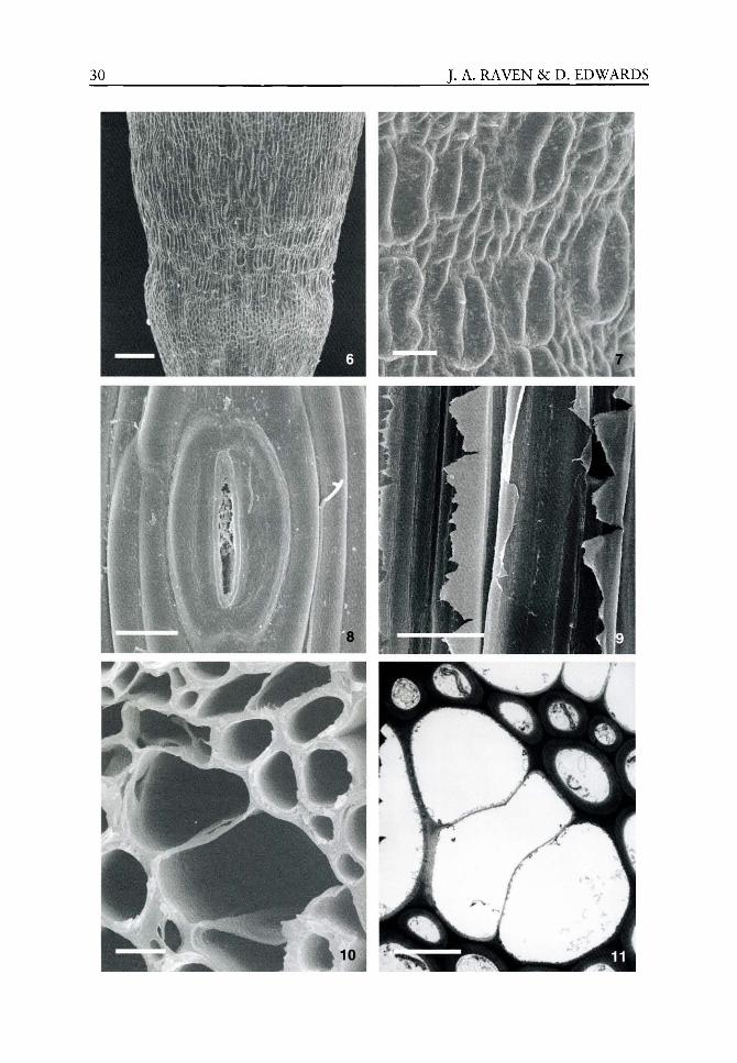
30 J. A. RAVEN &c P. EDWARDS

EARLY EVOLUTION OF HIGHER PLANT PHYSIOLOGY 31
conductivity of the xylem might be offset by a greater number of elements in a given cross-sectional area or volume. The impact of element size on the likelihood of a given decrement in conductivity in response to a certain set of environmental conditions essentially depends on whether cavitation or embolism is a function of the volume of an element, or of the number of elements, or the total volume of the segment of xylem.
The discussion of the factors involved in the evaluation of the endohydric conducting system, and specifically of xylem, has taken place in the context of the cohesion-tension hypothesis (Dixon, 1914). This hypothesis seems to account for most of the data which are currently available (Steudle, 2001), although there is still dispute (Canny et aL, 2001; Cochard et al, 2001; Richter, 2001).
As to the fossil record, the crucial evolutionary events apparently occurred in the Silurian (see Figure 2.2). Cuticles occur throughout the Silurian; although the earlier ones did not have any obvious relation to embryophytes (see Figures 2.12 and 2.13), the younger cuticles include those with clear relation to embryophytes (Edwards, 1993, 1996, 1998, 2000; Edwards et aL, 1996, 1998). Alas, the earliest embryophyte cuticles in the record yield no direct evidence on their chemistry or permeability (Edwards et al, 1996). Fossil evidence for embryophyte cuticles only preceded the endohydric conducting system (Edwards and Wellman, 1996); later came stomata (see Figures 2.14 and 2.17) and xylem, with these two structures appearing in the record at a very similar time in the Upper Silurian (Edwards, 1993, 1996, 1998, 2000; Edwards et al, 1996, 1998).
History of physiological interpretations of early embryophytes
Introduction
Here we consider the timing of some major discoveries in the palaeobotany of lower embryophytes and of major discoveries in plant physiology, and especially plant water relations. The three case histories considered in more detail are those of the role of transpiration and the endodermis in nutrient supply to the shoot of endohydric plants, the mechanism of water movement up the xylem during transpiration, and the role of stomata in determining the rate of photosynthesis and the water cost of photosynthesis in relation to the functioning of stomata.
Dawson, between 1850 and 1890, discovered and described Devonian vascular plants (e.g. Fsilophyton) from the Maritime Provinces of Canada. The Rhynie Chert in Aberdeenshire, Scotland was discovered in 1912, and Kidston and Lang published on the Rhynie Chert embryophytes, with exquisitely preserved (permineralized) cellular structure, between 1917 and 1921. This work clearly shows xylem (or what in some cases are now interpreted to be analogous to endohydric water conducting systems: Edwards, 1993), stomata, intercellular gas spaces and cuticle (see Figures 2.19-2.33). Subsequent work extended the geographical and stratigraphical range of early embryophytes with good anatomical, cell-level preservation (Edwards, 1993). Scanning electron microscopy has
Figures 2.6-2.11 Figures 2.6 and 2.7 SEMs of stomata on seta of Polytrichum commune Hedw. Scale bars = 200 and 35 |xm. Figure 2.8 SEM of surface view of stoma of hornwort, Phaeoceros lae-vis (L.) Prosk. Scale bar = 20 ixm. Figure 2.9 SEM of longitudinal fracture through imperforate hydroids in central strand of Dawsonia superba Grev. Scale bar = 5 (xm. Figure 2.10 SEM of transverse fracture through hydroids and surrounding stereids in Dawsonia superba Grev. Scale bar = 5 ixm. Figure 2.11 TEM section through similar tissue. Scale bar = 5 jxm.
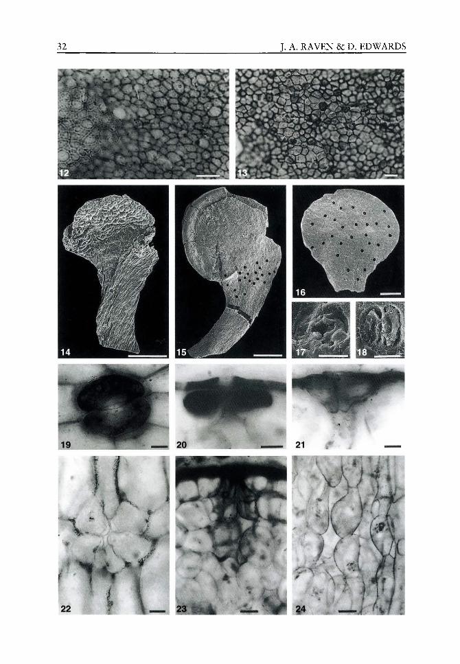
32 J. A. RAVEN &c D. EDWARDS

EARLY EVOLUTION OF HIGHER PLANT PHYSIOLOGY 33
been very important in investigating the anatomy of early embryophytes, and has been particularly significant in examining coalified fragmentary polysporangiophyte (Figures 2.14-2.16) and possible bryophyte remains (Edv^ards, 1993, 1996, 2000).
Discoveries in plant photosynthesis, water relations and mineral nutrition include the discovery of photosynthesis by van Helmont and measurements of the water use efficiency of growth (Woodward, 1699) in the late seventeenth century. In the eighteenth century plant plumbing was investigated and root pressure discovered by Hales (1727), and the role of Oi in photosynthesis and respiration was discovered. The nineteenth century saw advances in understanding of nutrition (Liebig's Law of the Minimum; Liebig, 1840), and by early in the twentieth century the physical basis of gas diffusion through stomata and of the cohesion-tension hypothesis of xylem water movement in transpiration had been formalized (Brown and Escombe, 1900, 1905; Dixon, 1914) as had limitation of rate (Blackman, 1905), as well as extent (Liebig, 1840), of physiological processes by external factors. Later in the twentieth century great advances were made in quantifying energetic as well as kinetic aspects of transpiratory water loss (the Penman-Monteith equation: Jones, 1992) and formalizing concepts of homoiohydry and poikilohydry in relation to desiccation tolerance and desiccation intolerance (Walter and Stadelmann, 1968).
Transpiration rate and endodermal function in regulating nutrient supply to the shoot
Turning now to the interpretation of the physiology of the early land embryophytes in relation to their anatomy, Haberlandt set an excellent precedent in the late nineteenth century by attempting to interpret the functional anatomy of extant plants. However, later attempts to interpret the evolution of land plants by Church (1919) made little use of fossil data
Figures 2.12-2.24 Figure 2.12 LM (Light micrograph) of isolated cuticle of Nematothalius. Area out of focus results from the typical undulating nature of the cuticle. Lochkovian: north of Brown Clee Hill, Shropshire. DEI. Scale bar = 20 iJim. Figure 2.13 LM of papillate cuticle of Cosmochlaina maculata. Lochkovian: M50 motorway near exit 3 (Newent, Gloucestershire). NMW85.20G.6. Scale bar = 20 jxm. Figure 2.14 SEM of holotype oi Hollandophyton collicula with terminal bivalved sporangium terminating stomatiferous axis. Pridoli: Ludford Corner, Shropshire. NMW96.11G.7. Scale bar = 500 [xm. Figure 2.14 was published in Special Papers in Palaeontology, 67, 2002, 233-249 and is reproduced here with kind permission of The Palaeontological Association. Figure 2.15 SEM of bivalved sporangium, Sporathylacium salopense, with oblique band of stomata (dots). Lochkovian: north of Brown Clee Hill, Shropshire. NMW94.60G.il. Scale bar = 500 |xm. Figure 2.16 SEM of unidentified sporangium with scattered stomata (dots). Lochkovian: north of Brown Clee Hill, Shropshire. NMW96.5G.7. Scale bar = 500 |jim. Figures 2.15 and 2.16 were published in Plant Cuticles, 1996,1-31 and are reproduced here with kind permission of BIOS Scientific Publishers Ltd, Oxford. Figure 2.17 SEM of stoma from Hollandophyton (Figure 2.14): the earliest stomata illustrated in a fertile taxon. Scale bar = 20|jLm. Figure 2.18 SEM of stoma from Figure 2.15 Scale bar = 20 fxm. Figures 2.19-2.24 LMs of silicified sections of axes of Aglaophyton major. Pragian: Rhynie Chert, Rhynie, Aberdeenshire. Scale bars = 20 (xm (Figures 2.19-2.22) and 50|jLm (Figures 2.23 and 2.24). Figure 2.19 Paradermal section through stoma. Note dark staining of guard cells compared with adjacent epidermal cells. PI603. Figure 2.20 TS guard cells with slightly separating cuticle and ledges. P1407. Figure 2.21 TS polar regions of stoma with extensions of the inner peri-clinal walls. PI 611. Figure 2.22 TLS hypodermal cells below stoma: note thickenings surrounding channel to substomatal cavity. PI603. Figure 2.23 TS aerial axis just below a stoma to margin of the substomatal cavity. PI 826. Figure 2.24 LS aerial axis showing parenchyma adjacent to substomatal cavity. Figures 2.19-2.24 were published in Journal of Experimental Botany, 49, special issue, 1998, 255-298 and are reproduced here with kind permission of Oxford University Press.
34 J. A. RAVEN &c D. EDWARDS
and did not take fully into account what was known about the physics of stomatal function from the work of Brown and Escombe (1900,1905) (see Edwards etaL, 1996; Raven, 2002). Thus, Church (1919) held that stomata were not involved in photosynthetic CO2 uptake, but were involved in the transpirational flux of nutrients from the soil solution to the shoot. This suggestion is not entirely without foundation in evidence from the fossil record as far as transpiration and nutrient uptake are concerned. One line of evidence comes from the distribution of stomata on the sporophytes of Upper Silurian and Lower Devonian embryophytes, where the stomata are often concentrated around sporangia (Figures 2.15, 2.16 and 2.18), a presumed site of a high requirement for soil-derived nutrients such as N, P, K, Ca, Mg, S and Fe (Edwards et ai, 1996,1998; Raven, 2002). A second line of evidence comes from the fact that, to date, presence of an endodermis in below-ground, nutrient-absorbing structures in embryophytes before the Carboniferous has not been demonstrated (Raven, 1984; Raven and Edwards, 2001), although it must be acknowledged that we have next to no anatomical information on such structures through this time interval. The endodermis is widely held to be a means of limiting entry of soil solutes which are present in the soil solution in concentrations in excess of plant demand, granted the likely tran-spiratory water use in growth equivalent to 1 g gain in dry matter and the elemental content per g dry matter. The endodermis is also held to be involved in limiting leakage of nutrients from the stelar tissues in those cases where energized transport of nutrients from the soil solution to the stele compensates for lower nutrient concentrations in the soil solution than would satisfy plant elemental requirement (per g dry matter) and the transpiration occurring during production of 1 g dry matter (Raven, 1984; Raven and Edwards, 2001). However, among extant endohydric plants an anatomically evident endodermis is absent from the gametophytes of endohydric mosses and leafy liverworts, and the sporophytes of Lycopodium spp. among vascular plants. Accordingly, the absence of an anatomically evident endodermis may not prevent the accumulation in, or exclusion from, the xylem of components of the soil solution (Raven and Edwards, 2001). Church (1919) must not, of course, be castigated for not commenting on these functions of the endodermis (or its anatomically indistinguishable analogue) in relation to nutrient acquisition since, although what is now known as root pressure was investigated by Hales in the eighteenth century and the barrier role of the endodermis had been shown by Lavison (1910), the function of the root system in the accumulation and exclusion of soil solutes was only fully characterized late in the twentieth century (Raven and Edwards, 2001).
The endodermis can also be related to Bower's suggestions (Bower, 1921,1930) on the role of elaboration of the anatomy of primary stele tissue with increasing size of diameter of plant axial structure, both ontogenetically and phylogenetically as shown in the fossil record. Bower suggested that the increased complexity of the shape of the primary xylem in transverse section with increasing cross-sectional area of the axial plant structure was related to maintaining a near constant area of exchange of resources between xylem and the surrounding ground tissue. However, the nature of the exchanges was not specified and the restriction on radial solute fluxes may occur in the endodermis or a functional analogue thereof (Raven and Edwards, 2001). Nevertheless, Bower was prescient in attempting further functional interpretations of the anatomy and morphology of fossil plants.
Mechanism of endohydric water movement
In the context of the perception of the mechanism of endohydric water fluxes in interpreting the structure of the endohydric conduits in fossils of early embryophytes, the publication of

EARLY EVOLUTION OF HIGHER PLANT PHYSIOLOGY 35
the book by Dixon (1914; see Dixon and Joly, 1894, 1895) on the cohesion-tension hypothesis just predated the pubHcation of anatomical details of the Rhynie Chert plants between 1917 and 1921 by Kidston and Lang. Integration of the cohesion-tension hypothesis into the structural data from early embryophytes (Figures 2.27-232) took some time (Raven, 1977). Indeed, in their ultrastructural detail, the walls of tracheids have no counterparts in extant tracheophytes (e.g. S-types: Figures 2.30,2.31 and 2.33) and some of the features of xylem, e.g. the presence of lignin, are not generally directly demonstrable in early fossil embryophytes (e.g. by pyrolysis/mass spectrometry of fossils with organic matter preserved: Ewbank et al.^ 1997). Lignin is an important component of the cell wall of xylem conduits, providing rigidity and resisting the tendency to implode with cell contents under tension (Wainwright, 1970). Further work is needed on the functioning of the endohydric conducting system (hydrome) in, for example, extant polytrichaceous mosses to determine the tolerance of non-lignified but possibly polyphenolic endohydric conducting systems to increasingly negative pressures of the solution they contain.
Role of stomata in determining the rate of photosynthesis and the water cost of photosynthesis
Returning to the functioning of stomata, this time in the context of the possible photosyn-thetic rates and ratios of carbon assimilation to transpiratory water loss, post-Church (1919) analyses include the very important perception of Chaloner (see McElwain and Chaloner, 1995) about the significance of stomatal density measurements on early vascular plant fossils in the context of the potential rate of photosynthesis. Such concepts have now been integrated with the knowledge that the CO2 mole fraction in the atmosphere has varied greatly over the Phanerozoic (McElwain and Chaloner, 1995); for a recent model for the CO2 content of the atmosphere over the last 500 million years see Berner and Kothavala (2001).
However, these modelling studies relying on stomatal densities as proxies for atmospheric CO2 (McElwain and Chaloner, 1995) and on the role of tracheophytes, via increased weathering, in lowering the CO2 level in the Upper Palaeozoic (Berner and Kothavala, 2001) have been challenged by Boucot and Gray (2001). The main criticisms by Boucot and Gray (2001) include the mismatch between data from climatically sensitive sediments and the climate models and the lack of consideration of pretracheophytic land plants and of aquatic primary production in the consideration of the effect of biota on atmospheric CO2 levels.
Despite the critique of Boucot and Gray (2001), we consider it likely that the CO2 content of the atmosphere was 10-20 times higher than the present value at the time at which the earliest stomata are known from the fossil record and it is clear that the stomatal density of these Upper Silurian and Lower Devonian plants was much lower than that found today or in the Carboniferous when the CO2 level was little greater than that found today (McElwain and Chaloner, 1995; Edwards et al, 1998; Berner and Kothavala, 2001; Lake et al., 2001; Raven, 2002). The possible photosynthetic rates and water costs of growth of early homoiohydric plants have been suggested by Raven (1977) who assumed that the atmospheric CO2 content was the same as that found today. Konrad et al. (2000) give much more plausible estimates of gas exchange in Lower Devonian homoiohydric plants. The structural adaptations in the vicinity of stomata, including the deep seated substom-atal cavity accessed by a narrow subporal canal illustrated here in certain Rhynie plants (see Figures 2.18-2.27), combined with low stomatal frequencies, infer high water-use efficiency (Edwards et al.^ 1998). Further modelling of stomatal function has been carried out by Beerling et al. (2001a) who have related the evolution of the euphyllophyte leaf

36 J. A. RAVEN &c D. EDWARDS
Figures 2.25-2.33 Figures 2.25 and 2.26 Guard cells and substomatal cavities in TS axes of Aglaophyton major (PI980) and Rhynia gwynne-vaughanii (P2219). Pragian: Rhynie Chert, Rhynie, Aberdeenshire. Scale bars = 100 iJim. Figure 2.27 LS axis of R. gwynne-vaughanii with epidermis to left and aerating tissue at margin of substomatal cavity on right. P2238. Pragian: Rhynie Chert, Rhynie, Aberdeenshire. Scale bar = 100 |xm. Figures 2.25-2.27 were pubHshed in Journal of
EARLY EVOLUTION OF HIGHER PLANT PHYSIOLOGY 37
(Kenrick and Crane, 1997; Pryer et al., 2001) in the context of decreasing atmospheric CO2 content of the atmosphere during the Devonian and thermal balance of the photo-synthetic structures. These suggestions have not been universally accepted (Hedrich and Steinmeyer, 2001; Tanner, 2001; cf. Beerling et aL, 2001b).
Conclusions
The physiological changes which occurred in the evolution from algal ancestors to the different grades of organization of embryophytes has been determined from the physiology of extant plants in relation to their phylogeny as determined by cladistic analysis and from the order in which anatomical features appeared in the fossil record. The fossil record of embryophytes also tells us about organisms with characteristics which are not found today. Examples are gametophytes with the anatomical characteristics of homoiohydry and homoiohydric sporophytes of polysporangiophytes which lacked true xylem as their endo-hydric conduits in the Rhynie Chert (Lower Devonian). The fossil record is not helpful in telling us about such important properties as desiccation tolerance or intolerance, except by applying an empirical correlation from extant plants that no embryophyte more than 1 m in height is desiccation tolerant in the vegetative phase. Overall conclusions from consideration of the lines of evidence indicated above are that the earliest embryophytes were desiccation tolerant and poikilohydric. Achieving homoiohydry could have followed the cuticle plus gas spaces plus stomata before endohydric conducting system, or vice versa.
Acknowledgements
JAR thanks colleagues past and present for their encouragement and support. We are grateful to Professor Hans Kerp and Herr Hagen Hass for the use of the Rhynie Chert
Experimental Botany, 49, special issue, 1998, 255-298 and are reproduced here with kind permission of Oxford University Press. Figures 2.28 and 2.29 SEMs of coalified examples of G-type tra-cheids. Lochkovian: north of Brown Glee Hill, Shropshire. NMW99.20G.1. Scale bars = 5 jjim. In these tracheids that characterize zosterophylls and basal lycophytes, annular or helical secondary thickenings are connected by perforated sheets of resilient material, believed to be deposited on the intervening compound middle lamella. Figure 2.28 Transverse fracture of xylem showing pitting in adjacent cell walls and a simple annular thickening. Note that the pits are only rarely coincident on adjacent walls. Figure 2.29 Longitudinal fracture of parts of tracheids. Note variation in size and shape of the pits. Figures 2.30-2.32 Xylem in Sennicaulis hippocrepiformis, preserved in pyrite, composed of S-type tracheids. These S-type tracheids occur in rhyniophytes such as R. gwynne-vaughanii. They are composed of a resilient spongy material and have broad predominantly helical thickenings. The lumen of the tracheid is lined by a layer (with perforations of nannometre dimensions) which also covers the secondary thickenings. Lower Old Red Sandstone: Mill Bay West, South Wales. Figures 2.30-2.32 were pubHshed in Palaeontology, 34, 1991, 751-766 and are reproduced here with kind permission of The Palaeontological Association. Figure 2.30 Longitudinally fractured xylem. Note residues of coaHfied material on left, pyrite casts of lumina on right. NMW90.42G.3. Scale bar = 20 iJim. Figure 2.31 Fragment showing part of a thickening and its spongy matrix with overlying perforate layer. NMW90.42G.3. Scale bar = 5 (xm. Figures 2.32 and 2.33 illustrate relative size of perforations in S and G-type tracheids. Figure 2.32 NMW90.42G.3. Scale bar = 1 fxm. Figure 2.33 (from Figure 2.29) NMW99.20G.1. Scale bar = 1 juim. Figures 2.28, 2.29 and 2.33 were published in Philosophical Transactions of the Royal Society of London, B, 355, 2000, 733-755 and are reproduced here with kind permission of The Royal Society.

38 J. A. RAVEN &c D. EDWARDS
photographs and Professors J.G. Duckett, K.S. Renzaglia and R. Ligrone for those on
bryophytes. DE thanks L. Axe for technical support and the N E R C for various grants.
References
Abe S, Kurashima A, Yokohana Y, Tanaka J. 2001. The cellular ability of desiccation tolerance in Japanese intertidal seaweeds. Botanica Marina 44: 125-131.
Badger MR, Andrews TJ, Whitney SM, et al. 1998. The diversity and coevolution of Rubisco, plastids, pyrenoids, and chloroplast-based C02-concentrating mechanisms in algae. Canadian Journal of Botany 76: 1052-1071.
Beerling DJ, Osborne CP, Chaloner WG. 2001a. Evolution of leaf-form in land plants linked to atmospheric CO2 decline in the late Palaeozoic era. Nature 410: 352-354.
Beerling DJ, Osborne CP, Chaloner WG. 2001b. Do drought-hardened plants suffer from fever? Trends in Plant Science 6: 507-508.
Bekheet lA, Syrett PJ. 1977. Urea-degrading enzymes in algae. British Phycological Journal 12: 137-143.
Berner EK, Berner RA. 1996. Global Environment: Water, Air and Geochemical Cycles. Upper Saddle River, New Jersey: Prentice Hall.
Berner RA. 2001. Modelling atmospheric O2 over Phanerozoic time. Geochimica et Cosmochimica Acta 65: 685-694.
Berner RA, Kothavala Z. 2001. GEOCARB III: a revised model of atmospheric CO2 over Phanerozoic time. American Journal of Science 301: 182-204.
Blackman FF. 1905. Optima and limiting factors. Annals of Botany 19: 281-295. Boucot AJ, Gray J. 2001. A critique of Phanerozoic climatic models involving changes in the CO2
content of the atmosphere. Earth Science Reviews 56: 1-159. Bower FO. 1921. Size, a neglected factor in stellar morphology. Proceedings of the Royal Society of
Edinburgh 41: 1-25. Bower FO. 1930. Size and Form of Plants. London: Macmillan. Brown HT, Escombe F. 1900. Static diffusion of gases and Hquids in relation to the assimilation of
carbon and translocation in shoots. Philosophical Transactions of the Royal Society of London B 193:223-291.
Brown HT, Escombe F. 1905. Researches on some of the physiological processes of green leaves, with special reference to the interchange of energy between the leaf and its surroundings. Proceedings of the Royal Society of London B 76: 29-111.
Campbell SA, Close TJ. 1997. Dehydrins: genes, proteins and associations with phenotypic traits. New Phytologist 137: 61-74.
Canny MJ, Huang CX, McCully ME. 2001. The cohesion theory debate continues. Trends in Plant Science 6: 454-455.
Chaloner WG. 1970. The rise of the first land plants. Biological Reviews 45: 353-377. Church AH. 1919. Thalassiophyta and the Subaerial Transmigration. Oxford Memoirs No 3.
Oxford: Oxford University Press 1-95. Close TJ. 1997. Dehydrins: A commodity in the response of plants to dehydration and low tempera
ture. Physiologia Plantanum 100: 291-296. Cochard H, Ameglio T, Cruiziat P. 2001. The cohesion theory debate continues. Trends in Plant
Science 6: 456. De Jesus MD, Tabatabai F, Chapman DJ. 1989. Taxonomic distribution of copper-zinc superoxide
dismutase in green algae and its phylogenetic importance. Journal of Phycology 25: 767-772. Denny MW. 1993. Air and Water. The Biology and Physics of Life's Media. New Jersey: Princeton
University Press. Dixon HH. 1914. Transpiration and the Ascent of Water in Plants. London: Macmillan. Dixon HH, Joly J. 1894. On the ascent of sap. Philosophical Transactions of the Royal Society of
London B 186:563-576. Dixon HH, Joly J. 1895. The path of the transpiration current. Annals of Botany 9: 403-420.

EARLY EVOLUTION OF HIGHER PLANT PHYSIOLOGY 39
Edwards D. 1993. Tansley Review No 53. Cells and tissues in the vegetative sporophytes of early land plants. New Phytologist 125: 225-247.
Edwards D. 1996. New insights into early land ecosystems; a glimpse of a Lilliputian world. Review of Palaeobotany and Palynology 90: 159-174.
Edwards D. 1998. Climate signals in Palaeozoic land plants. Philosophical Transactions of the Royal Society of London B 353: 141-157.
Edwards D. 2000. The role of Mid-Palaeozoic mesofossils in the detection of early bryophytes. Philosophical Transactions of the Royal Society of London B 355: 733-755.
Edwards D. 2003. Xylem in early tracheophytes. Plant Cell and Environment 26: 57-72. Edwards D, Wellman CH. 1996. Chapter 13F. Older plant macerals (excluding spores). In:
Jansonius J, McGregor DC, eds. Palynology: Principles and Applications. Salt Lake City, Utah: American Association of Stratigraphical Palynologists Foundation, Pubhshers Press, 383-387.
Edwards D, Abbott GD, Raven JA. 1996. Cuticles of early land plants: a palaeoecophysiological evaluation. In: Kerstiens G, ed. Plant Cuticles: an Integrated Functional Approach. Oxford: Bios Scientific publishers, 1-31.
Edwards D, Kerp H, Hass H. 1998. Stomata in early land plants: an anatomical and ecophysiolog-ical approach. Journal of Experimental Botany 49: 255-278.
Ewbank G, Edwards D, Abbott GD. 1997. Chemical characterization of Lower Devonian vascular plants. Organic Geochemistry 25: 461-473.
Farnsworth E. 2000. The ecology and physiology of viviparous and recalcitrant seeds. Annual Review of Ecology and Systematics 31: 107-138.
Gaff DF. 1981. The biology of resurrection plants. In: Pate JS, Macomb AS, eds. The Biology of Australian Plants. Nedlands: University of Western Austraha Press, 110-146.
Gaff DF. 1997. Mechanisms of desiccation tolerance in resurrection vascular plants. In: Basra AS, Basra RJ, eds. Mechanisms of Environmental Stress Resistance in Plants. Netherlands: Haward Academic Pubhshers, 43-58.
Gensel PG, Andrews HN. 1984. Plant Life in the Devonian. New York: Praeger. Graham LE. 1993. Origin of Land Plants. New York: John Wiley & Sons, Inc. Graham LE, Wilcox LW. 2000a. Algae. Upper Saddle River, New Jersey: Prentice Hall. Graham LKE, Wilcox LW. 2000b. The origin of alternation of generations in land plants: a focus
on matrotrophy and hexose transport. Philosophical Transactions of the Royal Society of London B 355: 757-767.
Green TGA, Lange OL. 1994. Photosynthesis in poikilohydric plants; a comparison of lichens and bryophytes. In: Schultze E-D, Caldwell MM, eds. Ecophysiology of Photosynthesis. Berlin: Springer-Verlag, 319-341.
Hales S. 1727. Statical Essays: Containing Vegetable Staticks; of an Account of some Statical Experiments on the Sap of Vegetables. A. and J. Innys and T. Woodward. Reprinted by The Scientific Book Guild, London.
Hamerlynck EP, Tuba Z, Csintalan Z, et al. 2000. Diurnal variation in photochemical dynamics and surface reflectance of the desiccation-tolerant moss, Tortula ruralis. Plant Ecology 151: 55-63.
Hedrich R, Steinmeyer R. 2001. Do drought-hardened plants suffer from fever? Trends in Plant Science 6: 506.
van den Hoek C, Mann DG, Jahns HM. 1995. Algae: An Introduction to Phycology. Cambridge: Cambridge University Press.
Jarvis MC. 1998. Intercellular separation forces generated by intracellular pressure. Plant, Cell and Environment 11: 1307-1310.
Jones HG. 1992. Plants and Microclimate. A Quantitative Approach to Environmental Plant Physiology., 2nd edn. Cambridge: Cambridge University Press.
Karol KG, McCourt RM, Cimino MT, Delwiche CF. 2001. The closest living relatives of land plants. Science 294: 2351-2353.
Keeling CD, Whorf TP, Wahlen M, Van der Plicht J. 1995. Interannual extremes in the rate of rise of atmospheric carbon dioxide since 1980. Nature 375: 666-670.
Keehng RF, Shertz SR. 1992. Seasonal and interannual variations in atmospheric oxygen and imph-cations for the global carbon cycle. Nature 358: 723-727.

40 J. A. RAVEN &: D. EDWARDS
Kenrick P, Crane PR. 1997. The Origin and Early Diversification of Land Plants: a Cladistic Study. Smithsonian Series in Comparative Evolutionary Biology. Washington: Smithsonian Institution Press.
Konrad W, Roth-Nebelsick A, Kerp H, Hass H. 2000. Transpiration and assimilation of Early Devonian land plants with axially symmetric telomes - simulations on the tissue level. Journal of Theoretical Biology 206: 91-107.
Lake JA, Quick WP, Beerling DJ, Woodw^ard FI. 2001. Signals from mature to new^ leaves. Nature 411: 154.
Lavison JR. 1910. Du mode de penetration de quelques sels dans la plante vivante. Revue Generate de Botanique 11: 93-97.
Li R, Brav^ ley SH, Close TJ. 1998. Proteins immunologically related to dehydrins in fucoid algae. Journal of Phycology 34: 642-650.
Liebig J, von. 1840. Chemistry in its Application to Agriculture and Physiology. London. Ligrone R, Duckett JG, Renzaglia KS. 2000. Conducting tissues and phyletic relationships of
bryophytes. Philosophical Transactions of the Royal Society of London B 355: 795-813. McElv^ain JC, Chaloner WG. 1995. Stomatal density and index of fossil plants track atmospheric
carbon dioxide in the Palaeozoic. Annals of Botany 76: 389-395. Nickrent DL, Parkinson CL, Palmer JD, Duff RJ. 2000. Multigene phylogeny of land plants w ith
special reference to bryophytes and the earliest land plants. Molecular Biology and Evolution 17: 1885-1895.
Nielsen SL, Sand-Jensen K. 1990. Allometric scaling of maximal photosynthetic grov^th rate to surface/volume ratio. Limnology and Oceanography 35: 177-181.
Nobel PS. 1999. Physicochemical and Environmental Plant Physiology., 2nd edn. San Diego: Academic Press.
Oliver MJ, Tuba Z, Mishler BD. 2000. The evolution of vegetative desiccation tolerance in land plants. Plant Ecology 151: 85-100.
Petit JR, Jouzel J, Raynaud D, et al. 1999. Climate and atmospheric history of the past 420,000 years from the Vostok ice core, Antarctica. Nature 399: 429-436.
Pickett-Heaps JD. 1975. Green Algae. Structure, Reproduction and Evolution in Selected Genera. Sunderland, Mass: Sinauer Associates, Inc.
Pryer KM, Schneider H, Smith AR, et al. 2001. Horsetails and ferns are a monophyletic group and the closest living relatives to seed plants. Nature 409: 618-622.
Qiu Y-L, Cho Y, Cox JC, Palmer JD. 1998. The gain of three mitochondrial introns identifies liverworts as the earliest land plants. Nature 394: 671-674.
Qiu Y-L, Lee J. 2000. Transition to a land flora: a molecular phylogenetic perspective. Journal of Phycology 36: 799-802.
Raven JA. 1977. The evolution of vascular land plants in relation to supracellular transport processes. Advances in Botanical Research 5: 153-219.
Raven JA. 1984. Physiological correlates of the morphology of early vascular plants. Botanical Journal of the Linnean Society 88: 105-126.
Raven JA. 1986. Evolution of plant life forms. In: Givnish TJ, ed. On the Economy of Plant Form and Function. Cambridge: Cambridge University Press, 421-492.
Raven JA. 1993. The evolution of vascular plants in relation to quantitative functioning of dead water-conducting cells and stomata. Biological Reviews 68: 337-363.
Raven JA. 1994a. Physiological analyses of aspects of the functioning of vascular tissue in early plants. Botanical Journal of Scotland 47: 49-64.
Raven JA. 1994b. The significance of the distance from photosynthesising cells to vascular tissue in extant and early vascular plants. Botanical Journal of Scotland 47: 65-81.
Raven JA. 1996. Into the voids: the distribution, function, development and maintenance of gas spaces in plants. Annals of Botany 78: 137-142.
Raven JA. 1997. Putting the C in phycology. European Journal of Phycology 32: 319-333. Raven JA. 1998. Extrapolating feedback processes from the present to the past. Philosophical
Transactions of the Royal Society of London B 353: 19-28. Raven JA. 1999. The size of cells and organisms in relation to the evolution of embryophytes. Plant
Biology 1: 2-12.
EARLY EVOLUTION OF HIGHER PLANT PHYSIOLOGY 41
Raven JA. 2000. Land plant biochemistry. Philosophical Transactions of the Royal Society of London B 355:833-846.
Raven JA. 2002. Selective pressures on stomatal evolution. New Phytologist 153: 371-386. Raven JA, Edw^ards D. 2001. Roots: evolutionary origins and biogeochemical significance. Journal
of Experimental Botany 52: 381-401. Raven JA, Johnston AM, Parsons R, Kiibler J. 1994. The influence of natural and experimental high
O2 concentrations on 02-evolving phototrophs. Biological Reviews 69: 61-94. Renzaglia KS, Duff RJ, Nickrent DL, Garbary DJ. 2000. Vegetative and reproductive innovations
of early land plants: implications for a unified phylogeny. Philosophical Transactions of the Royal Society of London B 355: 769-793.
Rice SK, Collins D, Anderson AM. 2001. Functional significance of variation in bryophyte canopy structure. American Journal of Botany 88: 1568-1576.
Richter H. 2001. The cohesion theory debate continues: the pitfalls of cryobiology. Trends in Plant Science 6: 456-457.
Smith EC, Griffiths H. 1996a. The occurrence of the chloroplast pyrenoid is correlated with the activity of CO2 concentrating mechanism and carbon isotope discrimination in lichens and bryophytes. Planta 198: 6-16.
Smith EC, Griffiths H. 1996b. A pyrenoid-based carbon-concentrating mechanism is present in terrestrial bryophytes of the class Anthocerotae. Planta 200: 203-212.
Smith EC, Griffiths H. 2000. The role of carbonic anhydrase in photosynthesis and the activity of the carbon-concentrating-mechanism in bryophytes of the class Anthocerotae. New Phytologist 145: 29-37.
Steudle E. 2001. The cohesion-tension mechanism and the acquisition of vs ater by plant roots. Annual Review of Plant Physiology and Plant Molecular Biology 51: 847-875.
Stew^art KD, Mattox KR. 1975. Comparative cytology, evolution and classification of the green algae with some consideration of the origin of other organisms with chlorophylls a and b. Botanical Review 4\'. 104-135.
Surif MB, Raven JA. 1990. Photosynthetic gas exchange under emersed conditions in eulittoral and normally submersed members of the Fucales and Laminariales: interpretation in relation to C isotope ratio and N and water use efficiency. Oecologia 82: 68-80.
Syrett PJ, Al-Houty FAA. 1984. The phylogenetic significance of the occurrence of urease/urea amidolyase and glycollate oxidase/glycoUate dehydrogenase in green algae. British Phycological Journal 19: 11-21.
Tanner W. 2001. Do drought-hardened plants suffer from fever? Trends in Plant Science 6: 507. Thomson GJ, Howlett GJ, Ashcroft AE, Berry A. 1998. A dhnA gene of Escherichia coli encodes
class I fructose bisphosphate aldolase. Biochemical Journal 331: 437-445. Tuba Z, Proctor MCF, Csintalan Z. 1998. Ecophysiological responses of homoiochlorophyllous
and poikilochlorophyllous desiccation tolerant plants: a comparison and an ecological perspective. Plant Growth Regulation 24: 211-217.
Tuba Z, Proctor MCF, Tacacs Z. 1999. Desiccation-tolerant plants under elevated air CO2: a review. Zeitschrift fiir Naturforschung 54C: 788-796.
Velten J, Oliver MJ. 2001. Tr288, A rehydrin with a dehydrin twist. Plant Molecular Biology 45: 713-722.
Wainwright SA. 1970. Design in hydraulic organisms. Naturwissenschaften 57: 321-326. Walter H, Stadelmann EJ. 1968. The physiological prerequisites for the transition of autotrophic
plants from water to terrestrial life. BioScience 18: 694-701. Wellman CH, Gray J. 2000. The microfossil record of early land plants. Philosophical Transactions
of the Royal Society of London B 355: 717-732. van der Willigen C, Farrant JM, Pammenter NW. 2001. Anomalous pressure volume curves of res
urrection plants do not suggest negative turgor. Annals of Botany 88: 537-543. Woodward FI. 1998. Do plants really need stomata.^ Journal of Experimental Botany 49: 471-480. Woodward J. 1699. Some thoughts and experiments concerning vegetation. Philosophical Transactions
of the Royal Society of London 21: 193-227.

Origin, function and development of the spore wall in early land plants
Charles H Wellman
CONTENTS
Introduction 43
Origin of the spore wall 44
Function of the spore wall 45
Spore wall development 46 Basic mechanisms of spore wall formation 46 Substructural organization of spore walls 48 Spore wall formation in extant plants 48 Spore wall formation in early land plants 52 Molecular genetics of spore wall development 55
Conclusions 59
Acknowledgements 60
References 60
Introduction
The embryophytes (i.e. land plants) are one of the major kingdoms of eukaryote life. There is overwhelming evidence indicating that they are monophyletic, with extant charophycean green algae their sister group (e.g. Graham, 1993; Mishler et al.^ 1994; Kenrick and Crane, 1997). All sexually reproducing land plants produce either spores, or their more derived homologues pollen, as part of their lifecycle. The resistant sporopollenin wall that encloses spores and pollen (spore exospore or pollen exine) is considered to be a synapomorphy of the embryophytes (e.g. Graham, 1993). It seems quite plausible that the invasion of the terrestrial subaerial habitat was not possible until a sporopollenin spore wall had evolved. Virtually all land plants possess spore/pollen walls composed of sporopollenin, except very rarely where this character has been secondarily lost (e.g. in certain aquatic angiosperms). This chapter is concerned with the role of the sporopollenin spore wall in the origin and early diversification of land plants (i.e. mid Ordovician-end Devonian). The aim is to explore and review our current understanding of: (1) the origin of the spore wall; (2) the function(s) of
The Evolution of Plant Physiology Copyright © 2004 Elsevier Ltd ISBN 0-12-33955-26 All rights of reproduction in any form reserved
44 C. H. WELLMAN
the spore wall in early land plants (including evolution of the spore wall in response to changes in the habit and external environment of the plants); and (3) mode of spore wall formation in early land plants, including recent developments in the study of the genetic basis for spore/pollen wall formation. Referencing is not extensive, but is intended to provide easy access into the literature: it includes major texts and review papers, along with some more specific papers that are either directly relevant to discussions or are of particular interest.
Origin of the spore wall
The embryophytes can be considered as a grade of organization that evolved as an adaptive response to their invasion of the terrestrial environment, i.e. the origin of land plants is intimately related to the invasion of the land, with the unique features of the land plants (autapomorphies) evolving as an adaptive response that enabled transmigration from an aquatic to subaerial habitat. In this respect it is intriguing to consider the origin and function of the spore wall. Almost intuitively we tend to equate acquisition of a sporopoUenin spore wall with the origin of land plants/invasion of the land. Hence sporopoUenin spore/pollen walls are considered a synapomorphy of the embryophytes and are not present in their putative green algal precursors.
When plants made the transition from an aqueous to subaerial environment their reproductive propagules experienced a dramatic change in environment from aqueous (aqueous green algal precursors) to subaerial (earliest land plants). The aqueous environment may be considered as one of relative comfort and a medium in which propagule movement is relatively straightforward. The subaerial environment, on the other hand, is one that is extremely hostile and in which propagule transportation is problematic. Thus the process of propagule transport was translocated from: (1) the relative safety encountered in an aqueous medium, into the harsh subaerial environment, where propagules were exposed to increased solar UV-B radiation levels, desiccation and likelihood of mechanical damage; and (2) an aqueous environment in which movement is relatively easy, into a subaerial environment where transportation must essentially be subaerial (but with free water provided during some stage in the lifecycle). In this scenario the sporopoUenin wall is viewed as an adaptation ideally suited to overcome these problems and protect the propagules in their harsh new environment. Of course, evolution of the spore is fundamentally related to evolution of the embryophyte Ufe cycle (i.e. alternation of generations, e.g. Kenrick, 1994).
SporopoUenin production, however, is almost certainly preadaptive. It occurs in a variety of diverse algal groups (usually enclosing resting cysts or reproductive structures), including the charophyceans (the sister group to embryophytes) where it is located in an inner layer of the wall enclosing the zygote (Graham, 1993). In the vast majority, if not all, of these cases, the function of the sporopoUenin coating is usually considered to be protective, i.e. to protect the enclosed resting cyst or reproductive structure from mechanical damage and/or microbial and fungal attack, and if exposed to the subaerial environment, desiccation and/or solar UV-B radiation (see p. 46). When considering mutations that enabled green algal ancestors to invade the subaerial environment and survive as the earliest land plants. Knoll and Bambach (2000, p. 8) note that 'principal among these mutations must have been one involving a simple change in the timing of gene expression for sporopoUenin biosynthesis from just after zygote formation to after meiosis and spore formation... such a mutation, arguably lethal in water, would have provided protection against desiccation and harmful radiation at a critically vulnerable phase of the lifecycle'.

ORIGIN, FUNCTION AND DEVELOPMENT OF THE SPORE WALL 45
However, the extent to which the sporopollenin of spore/pollen walls of embryophytes and the resting cysts/reproductive structures of the diverse algal groups are similar is unclear. Studies of sporopollenin chemistry are difficult to assess. Sporopollenin is one of the toughest and most chemically resistant biological polymers known (hence its utility as a protective coating). These properties, however, render it difficult to analyse and the chemical composition and structure of sporopollenin are poorly understood as are the biochemical pathways leading to its synthesis. Recent reports suggest that the sporopollenin of different algal groups varies in chemical composition and structure (see, for example, Graham and Gray, 2001). In fact the sporopollenin produced by different embryophyte groups also appears to vary in chemical composition and structure (Hemsley et al., 1995). However, it is unwise to use such evidence to prove/disprove homologies because: (1) analysis of such resistant material is fraught with difficulties; and (2) chemical composition/structure may simply have evolved in response to relatively minor biochemical changes, possibly related to changing utility and/or environment. It is generally considered that spore/pollen walls are homologous to the zygote walls of charophycean algae - we simply have a het-erochronic switch from enclosing the zygote to the products of meiosis (e.g. Blackmore and Barnes, 1987; Graham, 1993; Hemsley, 1994). However, this needs to be tested at the biochemical and molecular level. Similarities in the mode of sporopollenin deposition in embryophytes and certain green algae (see p. 47) support interpretation of these structures as homologous. It is anticipated that ongoing research in the field of molecular genetics may also aid identification of homology by identifying homologous genes involved in sporopollenin synthesis and/or regulation of its deposition.
Function of the spore wall
In the diverse algal groups possessing sporopollenin walls that enclose resting cysts/ reproductive structures, the primary function of the sporopollenin is most likely one of protection of the contents. In aquatic algae this involves protection from mechanical damage and fungal/microbial attack. There are almost certainly multiple secondary functions. These include aids to buoyancy (e.g. extensions to the walls of resting cysts) etc.
In the charophycean green algae, the sporopollenin enclosed zygote is generally confined to an aquatic habitat, but may be exposed to the subaerial environment, e.g. when a pond dries out. Graham and Gray (2001) recently reviewed the function of the sporopollenin zygote wall of charophycean green algae. They suggest that, in charophyceans, the primary function of the sporopollenin wall is to protect the zygote from mechanical damage and fungal/microbial attack during seasonal dormancy (and possibly also transport). Interestingly, they suggest that desiccation resistance is almost certainly a secondary function (if one at all). Graham and Gray (2001) note that 'there is no definitive evidence that sporopollenin (or other wall modifications) of algal resting cells prevents cellular water loss over extended periods of time, although it is possible that it functions as a short-term barrier to diffusion, as compact sporopollenin is regarded as essentially impermeable to water. Rather, sporopollenin in cell walls of charophyceans (and other aquatic algae) may provide protection from varied environmental extremes, where resistance to degradation of cytoplasmic contents is essential, such as during obligate periods of zygote dormancy'. They conclude that 'desiccation protection in aerial distribution, if any, is probably a secondary function of sporopollenin in charophycean algae'. This interpretation is based on the fact that of the algal groups that produce propagules with a sporopollenin component, few are
46 C. H. WELLMAN
demonstrably dispersed subaerially. In the majority of those that are subaerially dispersed, birds and insects are the dispersal vector and the sporopoUenin is likely to protect against enzymatic digestion in the digestive tracts. In any case, the presence of a sporopoUenin w all might even impede rehydration during germination.
The arguments developed are interesting regarding the function of the sporopoUenin spore/pollen wall of embryophytes, particularly in that these are usually regarded as homologous to the sporopoUenin walls present in various green algal groups. One of the major problems faced by land plants during the transition from an aquatic to subaerial environment was that of protection and dispersal of propagules outside of the aqueous environment (see p. 44). To what extent are the functions of the sporopoUenin spore/poUen wall simUar to those in green algal precursors and to what extent are they exaptations? Graham and Gray (2001) suggest that the primary function of the sporopoUenin spore/pollen waU in embryophytes is similar to that in extant charophycean green algae, i.e. to provide mechanical protection to the spore protoplasm during seasonal dormancy prior to germination and to resist fungal and microbial attack. They consider that protection from desiccation is likely to be a secondary function (possibly as is protection from solar UV-B radiation). However, it is clear that sporopoUenin does provide at least short-term protection of the spore protoplasm from desiccation. It is also clear that sporopoUenin plays a critical role in protecting spore contents from solar UV-B radiation (summarized in Rozema et aL, 2001a,b).
As the embryophytes have evolved and diversified the spore/pollen wall morphology evolved as it acquired additional functions (i.e. exaptations) (e.g. Chaloner, 1976; Traverse, 1988). These include: (1) dispersal (by wind, water or animal vectors, including protection when passing through their guts); (2) protection from herbivores and detritivores; (3) lodging; and (4) germination control. A brief examination of spore/poUen walls in fossil and extant plants reveals prodigious morphological diversity (structure and ornament). This almost certainly reflects the multiple functions of the wall (both primary functions and exaptations) and these of course are intimately related to the evolving mode of life of plants and changes in the external environment. Ultimately, however, such diverse morphological adaptations function to facilitate sexual reproduction.
Spore wall development
So how do embryophytes actually build the sporopoUenin spore/pollen wall? Clearly a very flexible system is involved that allows diverse morphology to evolve in response to changing lifestyles and environments. In the following section current knowledge of spore wall formation is summarized, concentrating on the information relevant to early land plant spores.
Basic mechanisms of spore wall formation
Ultrastructural studies carried out across the plant kingdom (extant and fossil) have shed light on spore wall ontogeny and the structures/processes operative during development. Blackmore and Barnes (1987) recognize four basic modes of sporopoUenin deposition in spore walls. These are:
1. Accumulation on white-line-centred-lamellae (WLCL) 2. Deposition from surrounding cells of the sporangium onto previously existing layers 3. Accumulation within primexine (a pre-patterned cell surface glycocalyx) 4. Centripetal accumulation onto pre-existing layers.
ORIGIN, FUNCTION AND DEVELOPMENT OF THE SPORE WALL 47
Blackmore et al. (2000) subsequently considered these four principal modes of spore wall formation in greater detail and subdivided them. This was largely in order that they could be utilized as characters in a cladistic analysis of extant free-sporing vascular plants, but also to reflect an increasing understanding of these modes of spore wall formation.
Regarding mode of formation (1), WLCL have long been prominent in studies of spore wall formation. They appear to be involved in sporopollenin deposition in various green algae and the vast majority of, if not all, land plants. They have also been identified in fossil spores, including those over 400 miUion years old (Wellman et aL, 1998a). Deposition on a system of WLCL is considered to be the most primitive mode of sporopollenin wall formation. It occurs in many algae and is plesiomorphic in all embryophytes. WLCL appear to form at the plasma membrane with sporopollenin polymerizing out on either side of the white line. In Chlorella Beijerinck they link up to form a single continuous layer (Atkinson et aL, 1972). In bryophytes we see the acquisition of numerous superimposed lamellae (this appears to be an early transformation in the evolution of land plants). They can also occur in other forms (as extensions, overlapping etc.) and although walls thus formed are usually rather simple, complex ornamentation can be built up in this fashion. In their recent cladistic analysis, character 8 of Blackmore et al, (2000) concerned exine deposition involving WLCL formed on the plasma membrane (notably initial exine deposition). In mature spore/pollen walls parts formed by the accumulation of WLCL may appear lamellate or homogeneous. The latter occurs when compression and/or sporopollenin deposition conceals early substructures. Hence it is desirable to study ontogenetic sequences, not just the mature wall.
Mode of formation (2) (deposition from surrounding cells of the sporangium onto previously existing layers) was subdivided by Blackmore et al (2000). Their character 10 concerned tapetal contribution to the exine. However, they distinguished exine formation involving a tapetum-derived component incorporated onto surfaces formed by the microspore (and usually based on WLCL) from epispore, perispore and paraexospore formation. Epispore (their character 13) is a layer within the sporoderm of heterosporous pteridophytes that is formed during the latest stages of exospore development, is composed of sporopollenin and is largely, if not exclusively, of tapetal origin. Perispore (their character 14) consists of material derived from the degenerating tapetum that condenses in one or more continuous layers over the surface of the spores. Taylor (2000) notes that sporopollenin derived from a tapetum may produce mature walls that are homogeneous and/or structures known as globules, orbicules or Ubisch bodies. Blackmore et al. (2000) also distinguish paraexospores (sensu Lugardon, 1976) (their character 11). A paraexospore is an outer layer of the exospore that is separated from the inner component by a large discontinuity (gap). Both layers are structurally similar and formed by WLCL. Clearly mode of formation (2), in its various guises, has major input from the diploid sporophyte (i.e. the tapetum). Blackmore and Barnes (1987) suggested that a tapetal contribution to the sporoderm was absent in basal land plants but acquired within both mosses and vascular plants. They considered this mode of deposition largely in terms of perispore formation (present in mosses and all vascular plants except Lycopodium L.). However, Blackmore et al (2000) noted that a tapetal contribution to the sporoderm takes many forms (including addition to layers previously formed from WLCL or the formation of an epispore).
Mode of formation (3), accumulation within primexine (a pre-patterned cell surface glycocalyx), will not be considered further. This mode is confined to the pollen of seed plants and is thus of little relevance to our consideration of spore wall formation in early land plants.
Mode of formation (4) involves centripetal accumulation onto pre-existing layers. Blackmore et aL (2000) covered this in their character 12. They note that spore wall

48 C. H. WELLMAN
formation may involve accumulation below an earlier formed layer, either by accumulation by WLCL or deposition of granular or amorphous sporopoUenin.
Blackmore et al. (2000) note that during sporogenesis a wide variety of mature forms is generated by a limited number of ontogenetic processes. They also note that the phylo-genetic pattern of sporoderm synthesis is not simply a sequence of gains of additional wall components as there also appear to have been losses of developmental processes. However, later stages of development of spores are not always contingent upon the successful completion of earlier ones, so the deletion of an entire phase may not always prove fatal (Blackmore and Crane, 1988). Blackmore and Crane (1988) noted that sporoderm ontogeny is complex, but includes non-terminal additions or deletions of developmental steps without concomitant loss of reproductive viability. Blackmore et al. (2000) consider 'in our view this reflects the particular aspect of spore ontogeny that is unique among all plant cell walls, namely development through an interplay of haploid, gametophytic and diploid, sporophytic processes'.
Substructural organization of spore walls
A number of models for the substructure of spore/pollen walls have been proposed. Two recent models are briefly discussed below.
Rowley (e.g. 1995, 1996) suggests that all embryophyte spore/pollen walls are composed of similar substructures. He interprets these substructures as like wire-wound springs (termed tufts) that are always of similar size and configuration. Rowley suggests that the tufts form an early substructure and act as receptors for sporopoUenin (i.e. sporopoUenin polymerizes out on these tufts). Furthermore, he suggests that they can open and close and thus act as migration routes for various materials, including those utilized during spore wall construction. Rowley also suggests that the tufts combine actually to form WLCL. He suggests that the tufts orientate perpendicular to the white line and hence the sporopoUenin polymerizes out on either side of the white line. He considers that the WLCL can also open and close and thus act as a migration route for various materials. Because they form early, and are subsequently coated in sporopoUenin, tufts are difficult to study (they can only be viewed in early maturation or degraded walls). Rowley (1995) provides reconstructions of these substructures alongside photographs of immature and degraded spore/pollen walls in which they can purportedly be seen.
Another exciting development is the discovery that self-assembly is operative during spore wall formation in the rather thick walls of certain lycopsid megaspores (fossil and extant) (e.g. Hemsley et aL, 1994, 2000; Gabarayeva, 2000). It will be interesting to see how widespread this is in the plant kingdom, or if it is confined to the abnormally thick walls of megaspores. Interestingly, Scott (1994) has suggested that self-assembly of molecules (lipids and so on) is responsible for the formation of WLCL. I see absolutely no reason why self-assembly should not be involved in actual wall construction. However, I am convinced that this is ultimately under genetic control, in that the amount, composition and delivery of materials is genetically controlled, as is ornament morphology through various template systems.
Spore wall formation in extant plants
Extant plants most relevant to the study of early land plants essentially belong to the more 'primitive' free-sporing plants (i.e. non-seed plants): 'bryophytes' (liverworts, hornworts, mosses) and 'pteridophytes' (lycopsids, sphenopsids, ferns). Spore wall formation has been
ORIGIN, FUNCTION AND DEVELOPMENT OF THE SPORE WALL 49
studied in representatives of all of these groups and includes studies of the isospores of homosporous forms and the microspores and megaspores of heterosporous forms. For reviews of spore w all formation in these plant groups the reader is referred to the following: bryophytes (Brown and Lemmon, 1988, 1990, 1991); pteridophytes (Lugardon, 1990; Tryon and Lugardon, 1991; Blackmore et aL, 2000). A brief summary of this information follows.
In most liverworts, immediately following meiosis, a spore special wall is deposited directly outside of the plasma membrane. This spore special wall appears to predict exospore ornamentation. More rarely, however, ornament is determined by exine precursors produced by the sporocyte (rather than individual haploid spores). In liverworts exospore development proceeds in a centripetal fashion. WLCL are formed external to the spore cytoplasm. These take on various forms and orientations (see Brown and Lemmon, 1990, Figure 8). Sometimes they are organized in multilaminar bands consisting of numerous parallel WLCL. The entire wall essentially comprises sporopollenin deposited on WLCL and the lamellate structure is clearly discernible at maturity. There is apparently no tapetal input and thus no extra-exosporal layers. Interestingly, the spores of Sphaerocarpos Boehmer often remain united in tetrads and the permanent tetrads may be enclosed within an envelope (Gray, 1985, 1991). These spores are similar to the envelope-enclosed cryptospore permanent tetrads produced by the earliest land plants and it has been suggested that such cryptospores derive from bryophyte-like plants closely related to the liverworts. Some workers have suggested that the cryptospore envelopes are tapetally derived (Gray, 1991; Edwards et ai, 1999). However, Renzaglia and Vaughn (2000) have demonstrated that in extant Sphaerocarpos it derives from the spore mother cell.
Spore wall formation in hornworts is little studied and poorly understood. A spore special wall is formed after meiosis and acts as a primexine in which the exospore is deposited. Interestingly, however, the exospore appears to develop without WLCL and the mature exospore is homogeneous.
In mosses, spore wall formation appears to occur in the absence of a spore special wall. Three types of spore wall are recognized among the mosses: Sphagnidae-type; Andreaeidae-type and Bryopsida-type (Brown and Lemmon, 1990).
Sphagnidae-type is illustrated by Sphagnum L. Sphagnum has a complex spore wall consisting of five layers: the endospore, lamellate inner exospore (A-layer), homogeneous outer exospore (B-layer), a unique translucent layer and the perine. The A-layer forms first and consists of 20-30 alternating light/dark layers that appear to derive from the spore, forming by sporopollenin accumulation on WLCL. The homogeneous B-layer accumulates above the A-layer. Overlying the exospore is the unique translucent layer, that consists of unconsolidated exospore lamellae in a matrix of unknown composition. Above this is the perine.
Andreaeidae-type is illustrated by Andreaea Hedwig, which has a spongy exospore that develops in the absence of WLCL. Development begins with the accumulation of homogeneous globules outside of the plasma membrane. These build up into an irregular layer with many interstitial spaces.
The Bryopsida-type of spore wall is homogeneous throughout, except for an inconspicuous foundation layer. The foundation layer develops first via sporopollenin accumulation on WLCL. In most cases a thick homogeneous exospore layer is deposited outside of the foundation layer in centrifugal fashion. It is likely that the outer homogeneous layer is in part of extrasporal origin, possibly deposited from the tapetum-like lining of the spore sac which may be secretory. Additionally, in some cases homogeneous material, presumably deriving from the spore cytoplasm, is deposited on the inside of the foundation layer (hence

50 C. H. WELLMAN
the foundation layer is not always located at the base of the exospore, but is sometimes located within the exospore). The perine is finally deposited on the exospore. Ornament may be elaborated in the exospore below, or perine may provide the sole ornament. The perine is believed to be of extrasporal origin. The perine is not acetolysis resistant.
Lycopodium clavatum L. is a good example of a homosporous lycopsid in which spore wall development is well understood (e.g. Uehara and Kurita, 1991). Here the plasma membrane folds shortly after meiosis indicating ornament pattern. Short WLCL begin to form on the plasma membrane and are elaborated in a centripetal direction, forming the bulk of the exospore. After the outer lamellar layer is formed, an inner granular layer is deposited, but only in the proximal region. There is no perispore (or other form of extra-exosporal layer). Spore wall development in all homosporous lycopsids appears to be similar to that in L. clavatum^ except that a very thin perispore, consisting of one or two layers and forming after exospore completion, is present in some taxa (Lugardon, 1990; Tryon and Lugardon, 1991).
In the heterosporous lycopsids, spore wall structure and development differ between: (i) different taxa; (ii) the microspores and megaspores in the same taxon. Both Selaginella Palisot de Beauvois and Isoetes L. have been studied in detail and I will consider the microspores and megaspores of both.
The microspores of Selaginella have a bilayered exospore. The inner layer forms first. It is narrow and consists of imbricate lamellae, formed on WLCL, that are initiated in a centripetal direction. The outer layer forms second, after the inner layer is completed, and consists of amorphous sporopollenin that is deposited onto it. Extra-exosporal layers vary in the microspores of different species of Selaginella, It may consist of a perispore, a para-exospore {sensu Lugardon, 1976) or be absent. In S. selaginoides (L.) Link (Tryon and Lugardon, 1991) a paraexospore is present. This is ontogenetically and chemically related to the exospore. It begins to form before the exospore, consisting of a granulate accumulation on the inner surface of the special wall surrounding the tetraspores. Lamellae are formed within the granulate material as the exospore begins to develop below the paraexospore. The exospore and the paraexospore are completed at the same time, as similar amorphous sporopollenin is deposited forming the outer exospore layer and also accumulates on the lamellae of the paraexospore. In some species of Selaginella a perispore is present, that is usually thin and firmly attached to the exospore surface.
Selaginella megaspores have a bilayered exospore. Two layers of similar thickness are recognized early in sporogenesis. The inner layer is lamellate and the outer layer consists of small and poorly delimited elements. During exospore development the inner layer does not thicken and the lamellae form a compact basal layer. The outer layer, however, increases in thickness dramatically, due to self-assembly (Hemsley et al.^ 1994, 2000; Gabarayeva, 2000). The outer layer has a very characteristic appearance. In most species silica is deposited in the voids of the outer layer and on the exospore surface. There are no extra-exosporal layers. The endospore forms between the exospore and the plasma membrane during the final stages of sporogenesis.
Uehara et al. (1991) describe microspore development in Isoetes japonica A. Br. The exospore is bilayered, with a large gap between the two layers. The outer layer forms first. Immediately after meiosis WLCL develop on the plasma membrane. These form an undulating plate consisting of two to three long and irregularly fused lamellae. The inner layer forms second. This forms by the centripetal accumulation of WLCL that develop on the plasma membrane. It comprises 12-14 lamellae (although more may be added later). At this point the lamellae of the outer exine layer are thickened by the addition of homogeneous

ORIGIN, FUNCTION AND DEVELOPMENT OF THE SPORE WALL 51
sporopollenin. Lastly the perispore forms, from electron dense material that is presumably tapetally derived, and the endospore forms, between the exospore and the plasma membrane. Lugardon (1990) and Tryon and Lugardon (1991) regard the exospore outer layer of Isoetes as a paraexospore, because it is initiated prior to the exospore, but consists of sporopollenin similar to the exospore (i.e. the inner exospore layer of Uehara etal.) and both are completed at the same time.
The megaspores of Isoetes are similar in wall structure and development to those of Selaginella, It is bilayered, with the outer layer initiated before the inner layer. Large amounts of silica are deposited within and above the outer layer prior to exospore completion. During the late stages of sporogenesis the endospore is deposited between the exospore and plasma membrane.
Spores in extant sphenopsids are highly distinctive in terms of morphology and development and are unique among the plant kingdom. They are most likely highly derived. Uehara and Kurita (1989) provide a detailed description of spore wall development in Equisetum arvense L. They note that the spore wall consists of four layers: endospore, exospore, middle layer and pseudoelaters. The exospore forms after meiosis and consists of two distinct layers (inner exospore and outer exospore). The inner exospore is first formed by the accumulation of plate-like structures that are deposited on the plasma membrane. As this layer accumulates it becomes thick and homogeneous. The outer exospore then forms. It accumulates as granular material on the inner exospore and becomes thick and homogeneous. The inner and outer exospore are similar in thickness. After the exospore is completed, the middle layer is deposited. Initially the middle layer consists of a membranous structure, which subsequently thickens up to about 0.2-0.3 luim thick. It forms in the gap between the spore and the plasmodial plasma membrane, completely surrounding the spore, but only in contact in the region of the aperture. Next the pseudoelaters form. Initially the pseudoelaters appear as membranous structures on the surface of the plasmodial plasma membrane, spirally coiled around the middle layer. The pseudoelaters are subdivided into two distinct layers. The inner layer initially comprises longitudinal microfibrils, that surround the spore in spiral fashion, but is homogeneous at maturity. The outer layer is formed by discharge of granules from vesicles in the plasmodial cytoplasm and is homogeneous. The pseudoelaters are joined to the spore, via the middle layer, at the aperture. The final part of the spore wall to form is the endospore which is deposited on the inside of the exospore. Only the exospore and middle layer are acetolysis-resistant.
In homosporous ferns the active plasmodial tapetum surrounds the spore tetrads and exospore formation commences. The exospore develops in a centrifugal fashion. It is two-layered: an inner substructure and an outer thick layer of amorphous sporopollenin. The inner substructure consists of between one and twelve partly fused sheets. These form by sporopollenin accumulation of WLCL and develop in a centrifugal manner. The number of sheets appears to be greater in more primitive ferns (up to twelve sheets) than in more derived ferns (often reduced to a single sheet). The outer thick layer of homogeneous material contains very small cavities and thin radial canals or fissures. The perispore forms after the exospore is completed, through the condensation of particulate material and is deposited from the tapetum as it decays. The perispore is always present, but is extremely variable in morphology. Where the perispore is multilayered, the inner layer is first formed with other layers successively deposited on pre-existing layers.
Spore wall development is similar in all of the heterosporous ferns and is similar in both microspores and megaspores (although the spore wall is clearly much thicker in the latter). Spore wall development is similar to that in advanced homosporous ferns, except
52 C. H. WELLMAN
that the plasmodial tapetum penetrates the tetrad at an early stage enveloping each of the spores, and the perispore is replaced by an epispore (sensu Lugardon and Husson, 1982). The exospore consists of a single substructural sheet (similar to that in more derived homosporous ferns) covered by a thick outer layer of amorphous sporopollenin. In both microspores and megaspores the outer layer consists of an epispore. This is initiated between the tapetum and the incomplete exospore, and consists of amorphous sporopollenin deriving from the tapetum that is deposited simultaneously on the epispore and exospore until both are completed. Thus the completed epispore is partly fused w ith the exospore (especially in the distal and equatorial areas).
Spore wall formation in early land plants
The wealth of data obtained from ultrastructural studies of spore wall formation in extant plants has prompted the extension of similar investigations into the fossil record. Such research has been extremely profitable (see, for example, the series of investigations reported in Kurmann and Doyle, 1994). However, analysis of fossil material poses its own set of distinct problems, most important of which involves the identification of preservational artefacts. However, it has been demonstrated that well-preserved fossil spores can preserve exquisite ultrastructure (e.g. Wellman, 2001).
At present, studies of spore wall ultrastructure in early land plants is limited. Dispersed spores of latest Ordovician-earliest Silurian age have been reported by Taylor (1995a,b, 1996, 1997, 2000). In situ spores, from exceptionally preserved floras of latest Silurian-earliest Devonian age, have provided a wealth of information on spores that can, in many cases, be related to their parent plants (Rogerson et al,^ 1993; Edwards et aL, 1995a,b, 1996,1999; Wellman, 1999; Wellman etaL, 1998a,b; Habgood, 2000). Dispersed Middle Devonian spores have been described by Wellman (2001, 2002). There are a number of other reports of wall ultrastructure from a variety of Devonian in situ and dispersed spores (Pettitt, 1966; Fletcher, 1976; Gensel, 1979; Taylor et aL, 1980; Taylor and Brauer, 1983; Gensel and White, 1983; Cichan etaL, 1984; Hemsley, 1989; Meyer-Melikyan and Telnova, 1989; Telnova, 1993; Foster and Balme, 1994; Taylor and Scheckler, 1996).
Taylor's work on wall ultrastructure in dispersed spores of latest Ordovician-earliest Silurian age has shed light on the mode of spore wall formation in the earliest land plants, in addition to providing evidence for their affinities. Spores of this age derive from the 'bryophyte-like' plants that are believed to have comprised the land flora for the first 40 or so million years of its existence.
Taylor reports a variety of types of spore wall ultrastructure. Some have entirely homogeneous walls, and he notes that these can be produced in several ways, with the final product leaving little evidence for mode of formation. Hence little can be discerned regarding wall development in spores whose wall is homogeneous at maturity. However, many of Taylor's spores have walls that are, at least in part, lamellate. He reports a wide range of lamella morphology, with many similarities to those produced by extant plants.
Taylor notes that the walls of certain dyads are most similar to those of extant sphae-rocarpalean liverworts. However, development of the envelope that often encloses early land plant spores (including monads and polyads) is problematic. Taylor considers that, in extant liverworts, the tapetum (if present at all) does not contribute to sporopollenin deposition and hence does not form a perispore. He also notes that the sporangium does not contribute to the sporopollenin wall of extant charophycean green algae, as the enclosed zygote represents the entire sporophyte generation. Thus the presence of an envelope, most
ORIGIN, FUNCTION AND DEVELOPMENT OF THE SPORE WALL 53^
easily explained as a perispore, is at odds with liverwort affinities. Taylor, however, considers that the absence of a perispore in extant liverworts does not preclude its presence in an ancestor of that lineage. It is interesting to note that Renzaglia and Vaughn (2000) consider that Phomologous envelopes in extant Sphaerocarpos derive from the spore mother cell.
Taylor (2000) goes on to propose developmental scenarios for the walls of a number of early land plant spore morphologies. He concludes that 'it is difficult to envision a reasonable developmental pathway for producing these complex cryptospore walls that does not involve tapetal deposition of sporopollenin and/or the envelope (perispore)'. He also notes that these early land plants 'had already evolved the basic structural units (and the developmental patterns to produce these units) that make up the sporoderm of most modern plants'.
Wall ultrastructure has been described in a number of different in situ spores from exceptionally preserved floras of latest Silurian-earliest Devonian age from the Anglo-Welsh Basin (see Figures 3.8-3.9). These include trilete spores (Rogerson et aL, 1993; Edwards etaL, 1995a,b, 1996; Wellman, 1999), hilate cryptospores (Wellman etai, 1998a), permanent dyads (Wellman et ai, 1998b; Habgood, 2000) and permanent tetrads (Edwards et al.y 1999; Habgood, 2000). These spores derive from a mixture of vascular rhynio-phytes and plants of uncertain status (including among them rhyniophytoids). In many of these the walls are entirely homogeneous. Others are at least in part lamellate, exhibiting a diverse array of different lamella morphologies, including those typical of WLCL (Wellman et al., 1998a) (see Figure 3.8). Some spore morphotypes possess outer wall layers that are globular and interpreted as being tapetally derived (e.g. Wellman et al., 1998a) (see Figures 3.7 and 3.9). Many, if not all, of the structures encountered can be recognized among extant plants (principally bryophytes and pteridophytes) and include evidence of sporopollenin deposition from both the haploid spore and diploid tapetum.
Wellman (2001, 2002), has commenced work documenting wall ultrastructure in dispersed spores of Middle Devonian age from the Middle Old Red Sandstone of Scotland. The spores are exceptionally well preserved and preserve exquisite wall ultrastructure (Figures 3.1-3.7, 3.10-3.14). Some of the examined spores have morphology far more complex than that in the previously discussed simple spores of Ordovician-Early Devonian age (e.g. Ancyrospora (Richardson) Richardson and Samarisporites Richardson are mul-tilayered, acamerate and zonate; Rhabdosporites Richardson is two-layered and camerate). These spores preserve a number of different lamella morphologies, including WLCL (Figures 3.10 and 3.12) and clear evidence for extra-exosporal layers that are tapetally derived (Figure 3.7). Despite the complexities of the spore walls, Wellman was able to link preserved structures to those present in extant free-sporing plants and hence interpret spore wall formation in terms of developmental processes observed in extant plants. Some of the spore types could clearly be related to those of extant plants. For example, it was demonstrated that spore wall formation in Ancyrospora was similar to that in extant lycopsids and hence the parent plants (hitherto unknown) were almost certainly lycopsid (Wellman, 2002). Furthermore, it was demonstrated that complex structures, such as inner bodies, zona, camera and grapnel-tipped processes, were easily constructed using the simple mechanisms of spore wall formation identified in extant free-sporing plants.
In conclusion, I consider that virtually all of the structure we see in the spore walls of primitive extant plants (bryophytes and pteridophytes) and in early land plants fossil spores can be accounted for by a small number of relatively simple modes of formation involving principally lamellae (of various morphologies) and tapetally-derived material. Clearly we have a very flexible system that is capable of producing immense morphological diversity that can satisfy the rapidly evolving and diverse functions of spore walls.
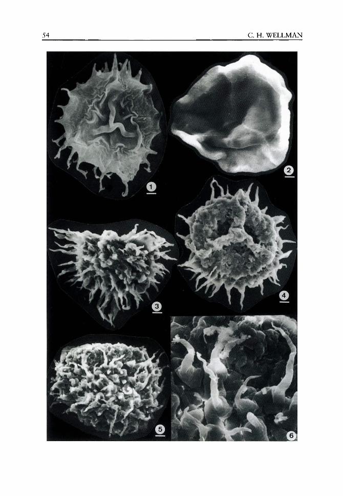
54 C. H. WELLMAN

ORIGIN, F U N C T I O N A N D DEVELOPMENT OF T H E SPORE WALL 55_
Molecular genetics of spore wall development
Research into the molecular genetics of sporogenesis is in its infancy, but understanding of this subject is progressing rapidly as a number of active research groups explore this fascinating topic. In the following section I have attempted to summarize current understanding of the molecular genetics of spore/pollen wall development. This review draws heavily on the most recent reviews of the subject (e.g. Davies et al.^ 1992; Mascarenhas, 1989, 1990, 1992; McCormick, 1991, 1993; Scott et aL, 1991a), but also discusses important recent findings (e.g. Paxson-Sowders et al., 2001). It is important to note that to date research has been confined to certain 'model' angiosperms (e.g. Arabidopsis thaliana (L.) Heynh., Brassica napus L.). Towards the end of this section I will discuss the potential relevance of this work on angiosperms with respect to more primitive plants (e.g. charo-phycean green algae, 'bryophytes' and 'pteridophytes') that are of more relevance to our understanding of spore wall development in early land plants.
Regarding genetic control of spore/pollen wall development, a couple of aspects have long been known. First, it is clear that in building the sporopoUenin wall there is input from both the haploid spore and the diploid sporophyte. Secondly, control of wall patterning appears usually to be associated with the diploid pre-meiotic sporocyte. There are two lines of evidence for the latter: (1) ontogenetic studies often reveal pre-patterned templates forming in the sporocyte (e.g. Brown et aL, 1986); and (2) following abnormal meiosis, cytoplasmic fragments lacking a nucleus produce normal spore walls. However, for more detailed studies of the molecular genetics of spore/pollen wall development, we currently rely on research conducted on various model angiosperms.
The production of the angiosperm male gametophyte is a complex developmental process involving a tightly controlled series of cytological and biochemical changes. These are coordinated with the expression of anther-specific genes. Currently relatively little is known concerning the genes involved, although it is considered to involve a large number of both sporophytic and gametophytic genes. Evidence for the involvement of sporophytic genes includes numerous male-sterile mutations that disrupt microsporogenesis. The recessive nature of most such mutations suggests that expression of the involved genes occurs in diploid cells and not in the haploid developing pollen grain. Additionally, some male-sterile mutants exhibit altered tapetal metabolism. Evidence for the involvement of gametophytic
Figures 3.1-3.6 SEM images of early land plant fossil spores form the Eifelian (Middle Devonian) of Scotland (see Wellman, 2001, 2002). Figure 3.1 Ancyrospora ancyrea (Eisenack) Richardson, 1962: a bilayered, acamerate, zonate, trilete spore of probable lycopsid affinity. Scale bar =10 |xm. Note the ornament comprising long processes with grapnel-tipped endings. The function of the grapnel-tips is uncertain, but possible facilitated attachment to arthropods that possibly acted as a dispersal vector (Wellman, 2002). Figure 3.2 Rhabdosporites langii (Eisenack) Richardson, 1960: a bilayered, camerate, trilete spore of probable aneurophytalean (progymnosperm) affinity. Scale bar = 10 |jLm. The spore has a thick-walled, rigid inner body enclosed within a very loose, thin-walled outer layer, that is attached to the inner body only over the proximal surface. This 'bladder' possibly increases buoyancy. Figures 3.3-3.6 Acinosporites macrospinosus Richardson, 1965: a trilete spore, with a distinct apical prominence and a dense distal ornament of long and prominent spines, possibly of lycopsid affinity (based on wall ultrastructure evidence). Scale bars = 10|jLm (Figure 3.3), 10 ixm (Figure 3.4), 10 [xm (Figure 3.5) and 3 iJim (Figure 3.6). Figure 3.3 is preserved in lateral compression. Note that the apical prominence is missing (cf. Figure 3.5). Figure 3.4 is preserved in polar compression. Note the nature of the apical prominence and associated trilete mark. Figure 3.5 is preserved in lateral compression, clearly displaying the apical prominence. Figure 3.6 is an enlarged portion of the specimen in Figure 3.5 illustrating the nature of the ornament.
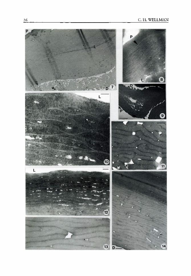
56 C. H. WELLMAN
ORIGIN, FUNCTION AND DEVELOPMENT OF THE SPORE WALL 57
genes includes proof that transcription and translation from the haploid genome occurs during pollen development based on: (1) studies of several dimeric enzymes; and (2) utilizing pollen-expressed and pollen-specific clones to demonstrate activation of specific genes after meiosis and at specific periods during spore/pollen development (see Macarenhas 1989, 1992).
Male gametogenesis may be divided into three distinct phases. First, 'sporogenesis' consists of a series of archesporial cell mitotic divisions which give rise to the tapetum and sporogenous cells, the latter undergoing meiosis to produce microsporogenous cells. Secondly, 'microspore development' involves the development of the free uninucleate microspores. Thirdly, 'pollen maturation' encompasses microspore mitosis and the formation of the mature bi-or trinucleate gametophyte.
It is during the first and second phases that the pollen wall develops. In the majority of angiosperms, meiosis is synchronous, forming tetrads of microspores enclosed within a thick callose wall. This callose wall is digested and the microspores liberated, following the release of callase (j8-(l,3)-glucanase) secreted into the locule from the tapetum. Pollen wall development commences while the microspores are still united in a tetrad, but following liberation there is further elaboration of the wall and the deposition of reserves such as lipids. The 'microspore development phase' essentially terminates when microspore mitosis commences.
It is generally believed that the protein components of intine are derived from gametophyte gene expression while those of the exine are produced by the sporophyte tapetal layer. The sporopoUenin component of the pollen wall is considered to be polymerized from a
Figures 3.7-3.14 TEM images of wall ultrastructure in early land plant fossil spores. For all images: L = lumen; P = proximal surface; D = distal surface. Figure 3.7 Rhabdosporites langii: a bilayered, camerate, trilete spore from the Eifelian (Middle Devonian) of Scotland. Scale bar = 2 |xm. The spore has a thick-walled inner body, that has lamellate ultrastructure only discernible at higher magnification. The outer layer is globular and almost certainly formed by the accumulation of tapetally-derived globules. The outer/inner layers are attached over the proximal surface, where the outer layer is thin and compressed, but separated over the distal surface (i.e. camerate), where the outer layer is thick and uncompressed. However, due to compression of the fossil, the camera is reduced. Figure 3.8 Laevolancis divellomedia Type C (sensu Wellman et aL, 1998a): a laevigate hilate cryptospore from the Lochkovian (Early Devonian) of the Welsh Borderland (see Wellman et ai, 1998a). Scale bar = 0.25 ixm. The spore is bilayered, with a thick inner layer comprising structures typical of WLCL and a thinner outer layer, interpreted as tapetally derived. The arrowhead marks the junction between the two layers. Figure 3.9 Laevolancis divellomedia Type B (sensu Wellman et ai, 1998a): a laevigate hilate cryptospore from the PridoH (Late Silurian) of the Welsh Borderland (see Wellman et ai, 1998a). Scale bar = 1 [xm. The spore wall is bilayered with a lamellate inner layer, consisting of relatively thick and laterally continuous lamellae and a homogeneous outer layer. Globules of extra-exosporal material, similar to the material forming the outer layer of the exospore, adhere to the surface and are interpreted as tapetally derived. Figures 3.10-3.12 Acinosporites macrospinosus: a trilete spore with a dense distal ornament of long and prominent spines, from the EifeHan (Middle Devonian) of Scotland. Scale bars = 0.1 fjim (Figure 3.10) and 0.25 ixm (Figures 3.11-3.12). In the inner part of the spore wall laterally impersistent and overlapping WLCL are conspicuous (Figures 3.10, 3.12). Towards the outside of the wall these become less conspicuous (see Figure 3.12), presumably as sporopoUenin has accreted onto them, forming a series of lamellae that thicken towards the outside of the wall, and are often folded (Figure 3.11). Figures 3.13-3.14 Ancyrospora ancyrea: a bilayered, acamerate, zonate, trilete spore from the Eifelian (Middle Devonian) of Scotland (see Wellman, 2002). Scale bars = 0.25 (jim (Figure 3.13) and 0.5 ixm (Figure 3.14). The spore has an electron dense inner body, consisting of narrow, parallel and continuous lamellae. The outer layer consists of thicker lamellae, that become increasingly thicker towards the outside of the wall. These lamellae are laterally continuous and bifurcate.
58 C. H. WELLMAN
lipid-like monomer derived from caretenoids and/or caretenoid esters. The sporopollenin almost certainly derives from both the microspore and the tapetum, although the bulk usually derives from the latter. The species-specific sculpture patterns of the exine are believed to be genetically determined by the sporophyte.
The number of genes expressed in developing microspores and pollen grains (incorporating sporogenesis and gametogenesis) is considered to be very large. It has been estimated that during the lifecycle of pollen at least 15 000 different genes may be transcribed. However, substantial overlap occurs betw^een genes active in the male gametophyte and in the sporophyte and studies of dimeric enzymes suggest that the vast majority of genes expressed in the male gametophyte are also expressed in the sporophyte, suggesting that many of these genes are simple housekeeping genes etc. Very few (usually less than 10%) are pollen specific (Mascarenhas, 1989).
Two distinct sets of genes have been identified with respect to the timing of their activity during pollen development (e.g. Mascarenhas, 1989,1990). The so called 'early genes' are active soon after meiosis is completed. The mRNAs reach their maximum accumulation by late pollen interphase and then decrease substantially by anthesis. It is generally surmised that 'early genes' might encode cytoskeletal proteins and proteins needed for wall synthesis or starch deposition. The so-called 'late genes' are activated after microspore mitosis. The mRNAs increase in content up to maturity, suggesting a major function during germination and early tube growth (and possibly also during the later parts of pollen maturation).
Far more is known about the 'late genes' than 'early genes', which is unfortunate as the latter are almost certainly those involved in spore wall formation. This disparity occurs because the majority of studies have been based on cDNA libraries constructed from mature pollen or mature anther RNA, and pollen-specific cDNAs thus isolated are representative of these 'late genes'. We do not at present have any estimates for the numbers of 'early genes' or of their similarity to genes expressed in later development.
However, workers such as Scott et al, (1991a) have illustrated a strong allometric relationship between bud length/anther length and stages of anther development. They have demonstrated how this relationship can be utilized in the isolation and characterization of anther-specific cDNAs expressed at defined stages of anther development, including early stages (i.e. 'early genes'). Examples of identified 'early genes' are actin, alcohol dehydrogenase, /3-galactosidase and those specifying the anther-specific tobacco transcripts TA25 and TA29 (see for example: Stinson and Mascarenhas, 1985; Albini et al, 1990, 1991; Koltunow et al, 1990; Smith et al, 1990; Scott et al, 1991b; Theerakulpisut et al, 1991; Foster et al, 1992; Roberts et al, 1993).
For some of the identified 'early genes' possible functions have been suggested, including, in some cases, involvement in spore wall formation. Scott et al (1991b) isolated 13 cDNA from immature anthers of Brassica napus that exhibits microspore-specific expression. They speculated that the protein encoded by the gene represented by 13 may be involved in early cell surface structure of developing pollen, or even in directing the development of the intine. Foster et al (1992) report on the isolation of a Brassica napus mRNA which is expressed during the 'microspore development phase' in both the developing microspore and the tapetum. The predicted protein of the E2 cDNA shows high homology with phospholipid transfer proteins (PLTPs), and Foster et al speculated that the E2 PLTP isoenzyme might be involved in sporopollenin production.
More recently Paxson-Sowders et al (2001) identified a gene (DEXl) they considered important in exine pattern formation. They worked on the dexl mutation of Arabidopsis which disrupts exine formation and patterning. Pollen wall development in dexl plants is
ORIGIN, FUNCTION AND DEVELOPMENT OF THE SPORE WALL 59
similar to that in wild-type plants until the early tetrad stage. Thereafter, in dexl plants, primexine deposition is delayed and reduced, rippling of the plasma membrane and spacer production is absent and the produced sporopollenin is randomly deposited on the plasma membrane, but is not anchored to the microspore, and forms large aggregates on both the developing microspore and the locule walls. Following isolation and molecular characterization of DEXl it was demonstrated that it encodes a novel plant protein, that was predicted to be membrane associated and to contain several potential calcium-binding domains. Factors controlling exine pattern are poorly understood and structures such as the primexine matrix, plasma membrane, endoplasmic reticulum and microtubules have been implicated. Interestingly, this work suggests that the plasma membrane does have an integral role in exine pattern formation. The fact that sporpollenin was synthesized and deposited, but failed to anchor to the surface of the microspore, suggests that DEXl may function as the nucleation point for sporopollenin deposition. The authors speculated that DEXl may either be a component of the primexine matrix or endoplasmic reticulum and involved in the assembly of primexine precursors. Recent discoveries of a number of other mutants in which exine development is disrupted, in Arabidopsis (e.g. Taylor et al., 1998) and other plants such as Zea mays L. (Loukides et al., 1995), are encouraging regarding the progress of similar research in the near future.
It is clear from the brief review outlined above that studies of the molecular genetics of spore/pollen wall formation are in their infancy. As more work is undertaken and techniques improve we can expect a dramatic, and hopefully rapid, increase in our understanding of these issues. Initially such work will be confined to angiosperms, but it is hoped that studies on other plant groups (gymnosperms, 'pteridophytes' and 'bryophytes') will follow. Only then will we be able to assess similarities/differences between the molecular genetics of spore/pollen wall formation in angiosperms and more primitive plant groups and ascertain how conserved the molecular mechanism and involved genes actually are. Also fruitful, and of direct relevance to early land plants, will be comparisons with the charophycean green algae, the sister group to embryophytes (land plants). Of interest will be the molecular genetics of the development of the sporopollenin layer surrounding the zygote, which will potentially shed light on the evolution of sporopollenin walled reproductive propag-ules in the earliest land plants.
Conclusions
It is concluded that the sporopollenin wall surrounding charophycean zygotes and embryophyte spores/pollen grains is homologous, and that the spore/pollen wall is an embryophyte synapomorphy that evolved as an adaptive response to the invasion of the land. It is considered that the primary function of the spore/pollen wall involves protection in the harsh subaerial environment. However, spore/pollen walls have taken on multiple secondary functions (exaptations), particularly as they evolved in response to changes in the habit and external environment of early land plants. Regarding spore wall development, ultrastructural studies have demonstrated that structural elements present in the spore walls of extant plants can be recognized in the fossil spores of early land plants, suggesting that spore wall development was similar in extant and ancient land plants. It is clear that spore wall construction is a simple and flexible process allowing rapid evolution of complex structure and morphology in response to evolving mode of life and external environment. Finally, studies of molecular genetics of spore wall formation are in their infancy, but have the potential to
60 C. H . W E L L M A N
solve many unanswered questions regarding spore wall homologies and developmental
processes, particularly when such studies are commenced on more 'primitive' land plants
(bryophytes and pteridophytes) and their extant sister group (charophycean green algae).
Acknowledgements
This work was supported by N E R C research grant GR8/03668.
References
Albini D, Altosaar I, Arnison PG, Fabijanski SF. 1991. A gene showing sequence similarity to pectin esterase is specifically expressed in developing pollen of Brassica napus. Sequences in the 5' flanking region are conserved in other pollen-specific promoters. Plant Molecular Biology 16: 501-513.
Albini D, Robert LS, Donaldson PA, et al. 1990. Characterization of a pollen specific gene family from Brassica napus which is activated during early microspore development. Plant Molecular Biology 15: 605-622.
Atkinson AW, Gunning BES, John PCL. 1972. SporopoUenin in the cell wall of Chlorella and other algae: ultrastructure, chemistry and incorporation of 14C-acetate studies in synchronous culture. Planta 107: 1-32.
Blackmore S, Barnes SH. 1987. Embryophyte spore walls: origin, development and homologies. Cladistics 3: 185-195.
Blackmore S, Crane PR. 1988. The systematic implications of pollen and spore ontogeny. In: Humphries CJ, ed. Ontogeny and Systematics. New York: Columbia University Press, 83-115.
Blackmore S, Takahashi M, Uehara K. 2000. A preliminary phylogenetic analysis of sporogenesis in pteridophytes. In: Harley MM, Morton CM, Blackmore S, eds. Pollen and Spores: Morphology and Biology. London, Kew: Royal Botanic Gardens, 109-124.
Brown RC, Lemmon BE. 1988. Sporogenesis in bryophytes. Advances in Bryology 3: 159-223. Brown RC, Lemmon BE. 1990. Sporogenesis in bryophytes. In: Blackmore S, Barnes SH, eds. Pollen
and Spores: Patterns of Diversification. The Systematics Association Special Volume No. 44. Oxford: Oxford Science Publications, 9-24.
Brown RC, Lemmon BE. 1991. Sporogenesis in simple land plants. In: Blackmore S, Knox RB, eds. Microspores: Evolution and Ontogeny. London: Academic Press, 55-94.
Brown RC, Lemmon BE, Renzaglia KS. 1986. Sporocytic control of spore wall pattern in liverworts. American Journal of Botany 73: 593-596.
Chaloner WG. 1976. The evolution of adaptive features in fossil exines. In: Ferguson IK, Muller J, eds. The Evolutionary Significance of the Exine. Linnean Society Symposium Series No. 1. Academic Press, London, 1-14.
Cichan MA, Taylor TN, Brauer DF. 1984. Ultrastructural studies on in situ Devonian spores: Protobarinophyton pennsylvanicum Brauer. Review of Paleobotany and Palynology 41:167-175.
Davies SP, Singh MB, Knox RB. 1992. Identification and in situ localization of pollen specific genes. International Review of Cytology 140: 19-34.
Edwards D, Davies KL, Richardson JB, Axe L. 1995a. The ultrastructure of spores of Cooksonia pertoni Lang. Palaeontology 38: 153-168.
Edwards D, Fanning U, Davies KL, et al. 1995b. Exceptional preservation in Lower Devonian coal-ified fossils from the Welsh Borderland: a new genus based on reniform sporangia lacking thickened borders. Botanical Journal of the Linnean Society 117: 233-254.
Edwards D, Davies KL, Richardson JB, et al. 1996. Ultrastructure of Synorisporites downtonensis and Retusotriletes cf. coronadus in spore masses from the Pridoli of the Welsh Borderland. Palaeontology 39: 783-800.
Edwards D, Wellman CH, Axe L. 1999. Tetrads in sporangia and spore masses from the Upper Silurian and Lower Devonian of the Welsh Borderland. Botanical Journal of the Linnean Society 130: 11-155.

ORIGIN, F U N C T I O N A N D DEVELOPMENT OF T H E SPORE WALL 61
Fletcher AJ. 1976. Studies on Devonian megaspores. Unpublished D. Phil. Thesis Bristol University. Foster CB, Balme BE. 1994. Ultrastructure of Teichertospora torquata (Higgs) from the Late
Devonian: oldest saccate palynomorph. In: Kurmann MH, Doyle JA, eds. Ultrastructure of Fossil Spores and Pollen. London, Kev^: Royal Botanic Gardens, ^7-97.
Foster GD, Robinson SW, Blundell RP, et al. 1992. A Brassica napus mRNA encoding a protein homologous to phospholipid transfer proteins, is expressed specifically in the tapetum and developing microspores. Flant Science 84: 187-192.
Gabarayeva NI. 2000. Principles and recurrent themes in sporoderm development. In: Harley MM, Morton CM, Blackmore S, eds. Pollen and Spores: Morphology and Biology. London, Kew : Royal Botanic Gardens, 1-16.
Gensel PG. 1979. Two Psilophyton species from the Lower Devonian of eastern Canada with a discussion of morphological variation within the genus. Palaeontographica B 168: 81-99.
Gensel PG, White AR. 1983. The morphology and ultrastructure of the Early Devonian Trimerophyte Psilophyton (Dawson) Hueber and Banks. Palynology 7: 221-233.
Graham LE. 1993. Origin of Land Plants. New York: Wiley. Graham LE, Gray J. 2001. The origin, morphology, and ecophysiology of early embryophytes:
neontological and paleontological perspectives. In: Gensel PG, Edwards D, eds. Plants Invade the Land: Evolutionary and Environmental Perspectives. New York: Columbia University Press, 140-158.
Gray J. 1985. The microfossil record of early land plants: advances in understanding of early terrestrialization, 1970-1984. Philosophical Transactions of the Royal Society London B 309: 167-195.
Gray J. 1991. Tetrahedraletes, Nodospora, and the 'cross' tetrad: an accretion of myth. In: Blackmore S, Barnes SH, eds. Pollen and Spores, Patterns of Diversification. The Systematics Association Special Volume No. 44. Oxford: Clarendon Press, 49-87.
Habgood KS. 2000. Two cryptospore-bearing land plants from the Lower Devonian (Lochkovian) of the Welsh Borderland. Botanical Journal of the Linnean Society 133: 203-227.
Hemsley AR. 1989. The ultrastructure of the spores of the Devonian plant Parka decipiens. Annals of Botany 64: 359-367.
Hemsley AR. 1994. The origin of the land plant sporophyte: an interpolation scenario. Biological Reviews 69: 263-273.
Hemsley AR, Barrie PJ, Scott AC. 1995. ^^C soHd-state n.m.r. spectroscopy of fossil sporopoUenins: variation in composition independent of diagenesis. Fuel 74: 1009-1012.
Hemsley AR, Collinson ME, Kovach WL, et al. 1994. The role of self-assembly in biological systems: evidence from iridescent colloidal sporopollenin in Selaginella megaspore walls. Philosophical Transactions of the Royal Society London., Series B 345: 163-173.
Hemsley AR, Collinson ME, Vincent B, et al. 2000. Self-assembly of colloidal units in exine development. In: Harley MM, Morton CM, Blackmore S, eds. Pollen and Spores: Morphology and Biology. London, Kew: Royal Botanic Gardens, 31-44.
Kenrick P. 1994. Alternation of generations in land plants: new phylogenetic and palaeobotanical evidence. Biological Reviews 69: 293-330.
Kenrick P, Crane PR. 1997. The Origin and Early Diversification of Land Plants. A Cladistic Study. Washington and London: Smithsonian Institution Press.
Knoll AH, Bambach RK. 2000. Directionality in the history of Hfe: diffusion from the left wall or repeated scaling of the right? Paleobiology 26: 1-14.
Koltunow AM, Turettner J, Cox KH, et al. 1990. Differential temporal and spatial gene expression patterns occur during anther development. Plant Cell 2: 1201-1224.
Kurmann MH, Doyle JA. 1994. Ultrastructure of Fossil Spores and Pollen. London, Kew: Royal Botanic Gardens.
Loukides CA, Broadwater AH, Bedinger PA. 1995. Two new male-sterile mutants of Zea mays (Poaceae) with abnormal tapetal cell morphology. American journal of Botany 82: 1017-1023.
Lugardon B. 1976. Sur la structure fine de I'exospore dans les diverse groupes de pteridophytes actuelles (microspores et isospores). In: Ferguson IK, Muller J, eds. The Evolutionary Significance of the Exine. London: Academic Press, 231-250.
62 C. H . WELLMAN
Lugardon B. 1990. Pteridophyte sporogenesis: a survey of spore wall ontogeny and fine structure in a polyphyletic plant group. In: Blackmore S, Knox RB, eds. Microspores: Evolution and Ontogeny. London: Academic Press, 95-120.
Lugardon B, Husson P. 1982. Ultrastructure exosporale et caracteres generaux du sporoderme dans les microspores et les megaspores des Hydropteridees. Comptes Rendus de VAcademie des Sciences de Paris 294: 789-794.
Mascarenhas JP. 1989. The male gametophyte of flowering plants. The Plant Cell 1: 657-664. Mascarenhas JP. 1990. Gene activity during pollen development. Annual Review of Plant Physiology
and Plant Molecular Biology 41: 317-338. Mascarenhas JP. 1992. Pollen gene expression: molecular evidence. International Review of Cytology
140: 3-18. McCormick S. 1991. Molecular analysis of male gametogenesis in plants. Trends in Genetics 7:
298-303. McCormick S. 1993. Male gametophyte development. The Plant Cell 5: 1265-1275. Meyer-Melielikyan NR, Telnova OP. 1989. Sporoderm of the Frasnian and Famennian
Archaeoperisaccus Naumova (the results of optical and electron microscopy). Palaeontol. Zh. 1: 88-94.
Mishler BD, Lewis LA, Buchheim MA, et al. 1994. Phylogenetic relationships of the 'Green algae' and 'bryophytes'. Annals of the Missouri Botanical Gardens 81: 451-483.
Paxson-Sowders DM, Dodrill CH, Owen HA, Makaroff CA. 2001. DEXl, a novel plant protein, is required for exine pattern formation during pollen development in Arabidopsis. Plant Physiology 127: 1739-1749.
Pettitt JM. 1966. Exine structure in some fossil and recent spores and pollen as revealed by light and electron microscopy. Bulletin of the British Museum (Natural History) Geology 13: 221-257.
Renzagha KS, Vaughn KG. 2000. Anatomy, development and classification of the hornworts. In: Shaw J, Goffinet B, eds. The Biology of Bryophytes. Cambridge: Cambridge University Press, 1-19.
Roberts MR, Foster GD, Blundell RP, et al. 1993. Gametophytic and sporophytic expression of an anther-specific Arabidopsis thaliana gene. The Plant Journal 3: 111-120.
Roberts MR, Robson F, Foster GD, et al. 1991. A Brassica napus mRNA expressed specifically in developing microspores. Plant Molecular Biology 17: 295-299.
Rogerson ECW, Edwards D, Davies KL, Richardson JB. 1993. Identification of in situ spores in a Cooksonia from the Welsh Borderland. Special Papers in Palaeontology 49: 17-30.
Rowley JR. 1995. Are the endexines of pteridophytes, gymnosperms and angiosperms structurally equivalent? Review of Paleobotany and Palynology 85: 13-34.
Rowley JR. 1996. Exine origin, development and structure in pteridophytes, gymnosperms, and angiosperms. In: Jansonius J, McGregor DC, eds. Palynology: Principles and Applications. American Association of Stratigraphic Palynologists. Publishers Press, Salt Lake City, Utah, 445-462.
Rozema J, Noordijk AJ, Broekman RA, et al. 2001a. (Poly)phenolic compounds in pollen and spores of Antarctic plants as indicators of solar UV-B. A new proxy for the reconstruction of past solar UV-B? Plant Ecology 154: 11-26.
Rozema J, Broekman RA, Blokker P, et al. 2001b. UV-B absorbance and UV-B absorbing compounds (p^^m-coumaric acid in pollen and sporopoUenin: the perspective to track historic UV-B levels. Journal of Photochemistry and Photobiology B: Biology 62: 108-117.
Scott R, Hodge R, Paul W, Draper J. 1991a. The molecular biology of anther differentiation. Plant Science SO: 167-191.
Scott R, Dagless E, Hodge R, et al. 1991b. Patterns of gene expression in developing anthers of Brassica napus. Plant Molecular Biology 17: 195-207.
Scott RJ. 1994. Pollen exine - the sporopoUenin enigma and the physics of pattern. In: Scott RJ, Stead MA, eds. Society for Experimental Biology Seminar Series 55: Molecular and Cellular Aspects of Plant Reproduction. Cambridge: Cambridge University Press, 49-81.
Smith AG, Gasser CS, Budelier KA, Fraley RT. 1990. Identification and characterization of stamen-and tapetum-specific genes from tomato. Mol. Gen. Genetic. 222: 9-16.
Stinson J, Mascarenhas JP. 1985. Onset of alcohol dehydrogenase synthesis during microsporogenesis in maize. Plant Physiology 77: 222-224.
ORIGIN, F U N C T I O N A N D DEVELOPMENT OF T H E SPORE WALL 63
Taylor PE, Glover JA, Lavithis M, et al. 1998. Genetic control of male fertility in Arabidopsis thaliana: structural analysis of post meiotic developmental mutants. Planta 205: 492-505.
Taylor TN, Brauer DF. 1983. Ultrastructural studies of in situ Devonian spores: Barinophyton citrulliforme. American Journal of Botany 70: 106-112.
Taylor TN, Maihle NJ, Hills LV. 1980. Morphological and ultrastructural features of Nikitinsporites canadensis Chaloner, a Devonian megaspore from the Frasnian of Canada. Review of Palaeobotany and Palynology 30: 89-99.
Taylor TN, Scheckler SE. 1996. Devonian spore ultrastructure: Rhabdosporites. Review of Palaeobotany and Palynology 93: 147-158.
Taylor WA. 1995a. Spores in earliest land plants. Nature 373: 391-392. Taylor WA. 1995b. Ultrastructure of Tetrahedraletes medinensis (Strother and Traverse) Wellman and
Richardson, from the Upper Ordovician of southern Ohio. Review of Palaeobotany and Palynology 85: 183-187.
Taylor WA. 1996. Ultrastructure of lov^er Paleozoic dyads from southern Ohio. Review of Palaeobotany and Palynology 92: 269-279.
Taylor WA. 1997. Ultrastructure of lower Paleozoic dyads from southern Ohio II: Dyadospora murusattenuata, functional and evolutionary considerations. Review of Palaeobotany and Palynology 97: 1-8.
Taylor WA. 2000. Spore v^all development in the earliest land plants. In: Harley MM, Morton CM, Blackmore S, eds. Pollen and Spores: Morphology and Biology. London, Kew: Royal Botanic Gardens, 425-434.
Telnova OP. 1993. Spore Research on Devonian and Carboniferous Deposits of the Timan-Pechora Province. St Petersburg: Nauka.
Theerakulpisut P, Xu H, Singh MB, et al. 1991. Isolation and developmental expression of Bcp an anther-specific cDNA clone in Brassica campestris. Plant Cell 3: 1073-1084.
Traverse A. 1988. Paleopalynology. Boston: Unv^in Hyman. Tryon AF, Lugardon B. 1991. Spores of the Pteridophyta. Surface, wall structure, and diversity
based on electron microscope studies. New York: Springer-Verlag. Uehara K, Kurita S. 1989. An ultrastructural study of spore wall morphogenesis in Equisetum
arvense. American Journal of Botany 76: 939-951. Uehara K, Kurita S. 1991. Ultrastructural study on spore wall morphogenesis in Lycopodium clava-
tum (Lycopodiaceae). American Journal of Botany 78: 24-36. Uehara K, Kurita S, Sahashi N, Ohmoto T. 1991. Ultrastructural study of microspore wall mor
phogenesis in Isoetes japonica (Isoetaceae). American Journal of Botany 78: 1182-1190. Wellman CH. 1999. Sporangia containing Scylaspora from the Lower Devonian of the Welsh
Borderland. Palaeontology 42: 67-81. Wellman CH. 2001. Morphology and ultrastructure of Devonian spores: Samarisporites
(Cristatisporites) orcadensis (Richardson) Richardson 1965. Review of Palaeobotany and Palynology 116: 87-107.
Wellman CH. 2002. Morphology and wall ultrastructure in Devonian spores with bifurcate-tipped processes. International Journal of Plant Sciences 163: 451-474.
Wellman CH, Edwards D, Axe L. 1998a. Ultrastructure of laevigate hilate spores in sporangia and spore masses from the Upper Silurian and Lower Devonian of the Welsh Borderland. Philosophical Transactions of the Royal Society London 353: 1983-2004.
Wellman CH, Edwards D, Axe L. 1998b. Permanent dyads in sporangia and spore masses from the Lower Devonian of the Welsh Borderland. Botanical Journal of the Linnean Society 127: 117-147.

The evolution of plant biochemistry and the implications for physiology
Richard D Firn and Clive G Jones
CONTENTS.
Introduction 68
Molecular evolution, biochemical evolution and metabolic evolution - hierarchical terms 68
Metabolic evolution - what determines whether a new enzyme is retained? 69
Biomolecular activity - the evolution of 'secondary metabolism' 69
Molecules retained because of their physicochemical properties 70
Primary metabolism - canalized metabolism, each step depending on other pre-existing metabolic capabilities 71
Selection for different molecular properties has
consequences for metabolic evolution 71
Biochemical evolution and physiology 72
The interaction of plants with other organisms 73 The human experience 73 Plant/microbe and plant/insect interactions 74
The evolution of regulatory systems for secondary metabolism 76
A speculative scenario for the evolution of the control of pathways leading to compounds retained because they possess biomolecular activity 77
Signalling molecules within plants 78 The link between secondary metabolism
and hormonal control 78
Summary 81
Acknowledgements 82
References 82
The Evolution of Plant Physiology ISBN 0-12-33955-26
Copyright © 2004 Elsevier Ltd All rights of reproduction in any form reserved
R. D. FIRN &c C. G. JONES
Introduction
Plant physiology and plant biochemistry are seen by some as brother and sister but by others as distant cousins. All would agree that there is a relationship but few would agree how close it is. The fact that books have been written on plant biochemistry with very little discussion of the physiology of plants shows that families can lose touch with each other. The aim of this chapter is to show that a functional understanding of plants can be gained by integrating physiological and biochemical knowledge. Our attempts to bring about integration start with an evolutionary prospective.
When introductory biochemistry is taught, there is a tendency initially to concentrate on the central metabolic pathways of 'the cell'. The default cell is usually mammalian. Plant cells are usually treated as unusual, but only in so far as they have walls and chloroplasts. This emphasis on the commonality of the basic metabolism of organisms is helpful to those learning the subject but it does tend to limit conceptual approaches to biochemistry. The reality is that most cells are biochemical specialists. Evolution has selected for biochemical traits of cells that are appropriate for their particular cellular environment in a manner analogous to the selection of organisms that are more suited to their environment. This specialization is found in both unicellular and multicellular organisms. However, in multicellular organisms, higher order coordination and control - physiological processes -can have a marked effect on the environment of specialist cells. To provide this coordination, specialist cells have evolved to play a central role in controlling many plant physiological processes (e.g. gas exchange is regulated by the guard cells, abscission depends on the abscission layer, seed reserve mobilization in grasses is dependent on the aleurone cells, etc.). Consequently there will be very clear links between physiological and biochemical processes. In this chapter we will argue that the selection pressures operating on the evolution of metabolism will have given rise to certain metabolic traits and, because of the intimate links between biochemistry and physiology, those traits have helped shape physiological processes. The intimate connection between the physiology and biochemistry of plants is well established in the case of some aspects of physiology, especially where there are dramatic morphological and anatomical adaptations which make the links clear - photosynthesis, germination, stomatal functioning, for example - and the links between the physiology and biochemistry of these processes will not be considered here. Instead, some more general principles concerning the evolution of metabolism will be discussed.These simple principles will then be used to consider the links between metabolism and physiology in cases where the anatomical and morphological signposts are less clear.
Molecular evolution, biochemical evolution and metabolic evolution - hierarchical terms
The term molecular evolution is currently used to refer to the way in which the evolution of protein structure relates to protein function, frequently with emphasis on tracing the lineage of base sequences in specific genes. Those working on molecular evolution generally focus on the role of one protein (or family of proteins). Biochemical evolution overlaps with molecular evolution but can extend to consider more than one enzyme in a pathway and the control of that pathway. Metabolic evolution could be considered to operate at the next higher level, determining the way in which the whole of metabolism evolves, with extensions or deletions of metabolic steps being the result of descent via modification in lineages subjected
EVOLUTIONARY LINKS BETWEEN BIOCHEMISTRY AND PHYSIOLOGY 69
to natural selection. It is metabolic evolution that determines the scope of metabolism in an organism, with biochemical evolution and molecular evolution determining the degree to which operation of the enzymes and their controlling elements effect organismal fitness. It is metabolic evolution on which we focus, because it is at this level that links between physiology and biochemistry are most apparent.
Metabolic evolution - what determines whether a new enzyme is retained?
New enzymes usually arise as a result of gene duplication and subsequent mutation of one gene copy such that the mutated enzyme has an altered substrate tolerance and can act on a new substrate to produce a new product (Petsko et aL^ 1993). Whether that new metabolite enhances the fitness of the producer depends entirely on whether the benefits of possession of the new product exceed the costs of producing it. In turn, the benefits depend upon the properties that the new metabolite brings to the organism. We identify three major classes of property that natural selection appears to have favoured during metabolic evolution.
Biomolecular activity - the evolution of 'secondary metabolism'
It has been well estabUshed from screening trials of collections of synthetic or naturally occurring molecules that the probability of any individual chemical possessing potent biological activity is very low (Jones and Firn, 1991; Firn and Jones, 1996). We proposed that this fact must have been a great constraint on the evolution of pathways leading to molecules that benefit their maker by possessing biological activity. However, the relevance of this fact to the understanding of the evolution of secondary metabolism has been challenged (Berenbaum and Zangerl, 1996). Berenbaum and Zangerl argued that the analogy between humans screening for a useful biological activity and organisms evolving chemical diversity in order to gain fitness by making biologically active chemicals was inappropriate - the nub of their argument was that vagueness in the definition of the term 'biological activity' led to a false analogy. However, by defining the term biomolecular activity quite precisely (as the ability of a molecule to interact with a biologically functional molecule such that its biological function is significantly changed), Firn and Jones (2000) countered this objection and provided evidence that the low probability of any chemical possessing potent biomolecular activity is a predictable and well understood consequence of ligand-protein interactions. Consequently, they reiterated a refined argument stating that the low probability of specific ligand-protein binding has been a significant evolutionary constraint on the production of biologically active molecules by organisms. Humans and other organisms may adopt different means of trying to reduce the impact of this fundamental constraint but they have both had to evolve ways of doing so. The methodology that humans have used when seeking to exploit the biological activity of chemicals (for example as pesticides or pharmaceuticals) has been the development and use of screening trials. These trials provide useful data about the frequency of any particular biological activity occurring in random collections of chemicals. Because such trials have been used to screen synthetic and naturally-occurring chemicals, and because they have been used against a wide range of biological targets, the information available from such trials is very extensive.
70 R. D, FIRN &c C. G. JONES
Do screening trials reveal any other relevant information? Indeed they do. They show that the type of biomolecular activity possessed by any individual chemical is unpredictable. Humans synthesizing new^ chemicals to test for biological activity are often surprised that a chemical made with the intention of producing one type of biological activity actually turns out to possess a very different, equally valuable activity. The discovery of Viagra is a recent well known example of human serendipity, but previous examples abound (Roberts, 1989). The herbicides paraquat and diquat emerged from an observation that a surfactant used in an experimental formulation was surprisingly phytotoxic, which led to some diquaternary dyestuff intermediates being examined in a herbicide screen with the result that the bipyridylium herbicides were discovered. Another example would be that the discovery of the pyrimidine fungicides started with attempts to make insecticides. In other words, the structure of any individual molecule is only partly predictive as to whether a molecule will possess biomolecular activity and is poorly predictive as to the type of biomolecular activity. The successful organism, like the successful company, exploits fortuitous events. Consequently, it is reasonable to predict that as a result of evolutionary adaptation, a pathway in an organism initially leading to one form of biomolecular activity can eventually lead to a quite different form of biomolecular activity. Hence we have two constraints that must be taken into account when considering the evolution of chemical diversity in plants -any new molecule has a very low probability of possessing biomolecular activity and the type of biomolecular activity cannot be reliably predicted from the biomolecular activity (if any) of the chemical(s) from which it derived.
Molecules retained because of their physicochemical properties
The terms 'primary metabolism' and 'secondary metabolism' are very commonly used, however, there are large groups of chemicals in organisms that sit between these two traditional groupings. Lipids, many pigments such as the carotenoids, many polysaccharides and some anthocyanins fall into this category. For example, consider lipids. All cellular organisms need lipids but they do not need them all - as a group lipids are essential but individually they are not. The paradox can be resolved by recognizing that the properties that have been selected for are physicochemical traits - lipophilicity, light absorption, structural properties, etc. These properties depend on the molecular properties shared by large numbers of structurally similar chemicals. Minor changes to part of the molecular structure might predictably make little difference to these properties - this is a marked contrast to the impossibility of predicting how such changes might alter the biomolecular activity of the same molecule. (Once again the analogy with chemicals made by humans is appropriate. A chemist who has synthesized a novel chemical can predict with some confidence its lipophilicity or its spectral properties but cannot reliably make any precise predictions about the biomolecular activity of the molecule.) Thus an organism that gains fitness by making a chemical that protects it against harmful irradiation, might as a result of extending the pathway leading to that pigment, produce another chemical with predictably similar useful properties. Slightly different extensions of the pathway leading from that point might have similar chances of creating new molecules with similar properties to the common precursor hence it is predictable that different species will produce a rather different mix of such chemicals - their overall requirement for a certain mix of chemicals with an average physicochemical property can be met in many different ways. The diversity of molecules selected for their physicochemical properties is thus predictable but the contribution that any one metabolite will make to the overall requirements is unpredictable. Under these

EVOLUTIONARY LINKS BETWEEN BIOCHEMISTRY AND PHYSIOLOGY 71
circumstances, it would seem that the advantage given to the producer of a new chemical depends greatly on the existing overall mix of existing similar chemicals.
The diversity of molecules selected for their contribution to the overall physicochemical requirements carries with it a chemical diversity that can be exploited as a route to molecules possessing useful biomolecular activity. Thus it is predictable that compounds with useful biomolecular activity will sometimes arise from pathways usually considered to operate principally for other purposes. Thus the phenylpropanoid pathway can generate chemicals that have a role in absorbing visible or non-visible light but this pathway may also lead to compounds that can enhance the fitness of the producer because they possess useful biomolecular activity (Winkel-Shirley, 2001). Likewise, the isoprenoid pathway can give rise to photoprotecting pigments such as the xanthophylls (Taiz and Zeiger, 1991) or carotenoid pigments in flowers or fruits (Harborne, 1988), yet also gives rise to the plant hormones such as the gibberellins or abscisic acid (Davies, 1995).
Primary metabolism - canalized metabolism, each step depending on other pre-existing metabolic capabilities
For the purpose of this discussion we define 'primary metabohsm' as metaboHc pathways where there is a great interdependence of the individual steps and where the individual molecules made by an enzyme serve only to feed into another enzyme - the complete pathway is greater than the sum of the parts. A significant difference between 'primary metabolism' and the previous two categories of metabolism, is that in primary metabolism selection is less environmentally contingent and the evolved homeostatic mechanisms of the organism and of the cell make the selection pressure more constant. At some stage early in the evolutionary history of primary metabolism the incorporation of a new metabolite into the developing primary metabolism could only have occurred if the new molecule fitted usefully into the specific scheme of existing pathways. The actual advantage to the producer of this new metabolite would arise solely from the ability of the new molecule to be acted upon by an existing enzyme(s) to produce another molecule(s) that had properties that enhanced fitness. The most extreme outcome would be the production of a metabolic cycle where there is no single 'end point' on which selection can act but the overall net efficiency of the cycle is subject to intense selection. However, in such cycles (e.g. Calvin cycle, the photorespiratory carbon oxidation cycle, the C4 cycle, etc.), selection that has fitted existing capabilities to local circumstance makes it much harder to introduce a new, compatible transformative capacity. Consequently, the pathway becomes severely canalized - the optimization of the coordinated processes increasingly reduces the opportunities to evolve radically different methods of achieving the same goal. (A dramatic example of canalization would be the genetic code - it is only one possible code of many that could have been used but once organisms evolved with a workable system on which selection could improve, the die was cast.) The powerful selection against new chemical diversity is in contrast to the previous property classes where there is a tolerance of chemical diversity (physicochemical properties) or selection for chemical diversity (biomolecular activity).
Selection for different molecular properties has consequences for metabolic evolution
Although it has been biochemical dogma that enzymes are highly substrate specific, this dogma has largely arisen from studies of enzymes involved in primary metabolism where
72 R. D. FIRN &c C. G. JONES
canalization is severe. The biochemical properties evident in a highly evolved and specialized metabolism tell us about the outcome of the selection processes operating on that sphere of metabolism and provide little reliable guidance as to the properties of individual new enzymes arising as a result of mutation. We would argue that a mutant enzyme would have a high probability of possessing a broad substrate tolerance intially. The reasoning is that a mutant enzyme that cannot access an existing substrate because it has narrow substrate specificity cannot produce a new product. Hence the enzyme will not be selected for in any circumstance in which new products would confer evolutionary advantage. In contrast, a mutant with a broad substrate tolerance has a greater initial probability of producing new substances with useful properties, irrespective of the type of property considered. However, it is clear that the selection pressures that would operate after the new product(s) have been generated would differ greatly depending on the type of property brought to the cell. For example, gaining fitness by producing biomolecular activity requires chemical diversity to be generated and maintained, hence there might be little selection pressure narrowing substrate tolerance (Jones and Firn, 1991). Indeed, enzymes capable of acting on more than one substrate might bring benefits. So if a new enzyme produces a new lipid that adds to the physicochemical properties of the cell, as long as the enzyme does not generate a chemical with adverse cellular properties, there may be little selection to narrow the initial broad substrate tolerance. Where there will be strong selection for narrow substrate tolerance is in the final property class - participation in primary metabolism. Here an enzyme accessing a common, important intermediate would be expected to have a negative effect on the overall metabolic and homeostatic mechanisms due to substrate competition and the possible generation of compounds that act allosterically to hinder rather than aid metabolic control. These simple concepts predict that metabolic evolution will have produced rather different metabolic traits in different pathways and even at different stages within a pathway.
Biochemical evolution and physiology
Why should the ideas outlined above be of interest to physiologists or biochemists? The fact that these issues have rarely been addressed previously suggests that both physiologists and biochemists have been comfortable working without such general principles for most of the 150 years that these disciplines have existed. In order to help promote interest in building the evolutionary frameworks underpinning the links between plant biochemistry and physiology, we will use two topics as examples of how simple ideas about metabolic evolution could guide experiments:
1. the physiology of chemical interactions between plants and other organisms 2. the physiology of intraplant signalling (plant hormones).
The former area is chosen because it is one where an alternative evolutionary model - based on ecological rather than metabolic considerations - was well developed and widely accepted. That model is now challenged by our metabolic evolutionary model. The second area - the evolution of plant hormonal control - is chosen because plant hormones have been shown to play a role in many very important, basic physiological processes.

EVOLUTIONARY LINKS BETWEEN BIOCHEMISTRY AND PHYSIOLOGY 73
The interaction of plants with other organisms
The human experience
Human experience has been both a useful guide and a distraction in understanding the role of plant chemicals in the coevolution of plants with other organisms. Humans are themselves massively influenced by plant chemistry, although most people go about their lives unaware of this fact because plant chemicals are so embedded in most cultures. Few readers will be reading this sentence without some plant chemicals being active in their bodies - it will be the rare reader who starts this chapter (and an even rarer reader who finishes this chapter) without having taken one of the following during the previous few hours - coffee, tea, tobacco, chocolate, a recreational or prescription drug, a tasty wine, a flavoursome beer, a fruit drink or a piece of confectionery. If a reader cannot concentrate on this chapter because their mind would prefer to think of the meal that awaits them, it will not be the expectation of a plate of starch or a piece of protein that excites their mind but the thought of the pasta sauce, the exciting curry, the sharp onions, garlic or interesting green salad. Plant secondary chemicals feed the mind while plant storage products feed the body. The human craving for particular plant chemicals has been so powerful that it has driven colonial expansion in the past and today many national economies are dependent on such chemicals. The power that a few plant species exert over humans by making just a few strange chemicals is quite remarkable and yet traditionally we have considered such chemicals 'secondary products'! If we delve a little deeper into the human experience with such chemicals we find that humans value such chemicals for different reasons:
1. such chemicals attract or repel humans by acting on sense organs - smell and taste mechanisms have been honed by evolution in many animals just to identify potential food sources and avoid intoxication based on their chemistries. Had there been no chemical diversity in the plant or microbial derived material would we have such fine senses?
2. the ability of these chemicals to influence mental processes (behaviour, well-being, etc) means that we can change our perception of the world
3. less commonly in humans, these chemicals can have a physiological effect, acting on some metabolic pathways in a positive or negative manner.
Each of these apparently different modes of action shares a common feature in that the effect of the chemical at a cellular level is caused by the chemicals binding to a specific receptor. Here the rules of ligand/receptor interactions apply. However, there is one crucial difference in the taste/smell receptors that differentiates them from the neurological and physiological receptors. In the case of the taste/smell receptors, the receptors have evolved to detect ligands and that means that there is potential for coevolution of the ligand/receptor interaction. A pollinator that is attracted to a food source by a plant-derived odour gains fitness if it is a mutant that has an odour binding protein that better matches the structure of the odorous chemical - the evolution of the detection system becomes 'locked on' to the chemistry of the producer. In contrast, neurological and physiological receptors are usually 'fortuitous receptors' - the receptor proteins have roles unrelated to their fortuitous ability to bind the plant-derived chemical and selection for their primary role would be paramount. If the plant-derived chemical has a serious adverse effect on the organism by interrupting its normal function there will be massive selection pressure to select individuals that make a mutant protein which functions in its original role but which cannot bind

74 R. D. FIRN & C. G. JONES
the plant-derived chemical at the concentration that it occurs - the evolution of detection is 'locked off (this is why insecticide-resistant pests and antibiotic-resistant microbes are an inevitable consequence of human attempts to use chemicals to reduce the fitness of competing organisms).
The human experience of plant and microbe-derived chemical diversity has thus been very important in human affairs and this experience can inform us about ligand/protein interactions but it can tell us little about the role of plant chemicals in the organisms that make them. Hov^ever, this human experience does tell us a final, very important lesson. The great majority of chemicals made by plants or microbes have no direct impact on humans in any observable w ay. Even in plants grow^n and consumed by humans, the majority of plant chemicals made by those plants are unsensed by humans, these chemicals have no unambiguous physiological or neurological effect and there must be many such chemicals that are as yet uncharacterized by humans (w ho have tended to concentrate on the compounds that occur in large amounts or w^hich are physiologically or neurologically active).
Plant/microbe and plant/insect interactions
Being (w ith a fev^ exceptions) primary producers, plants are subject to attack by other organisms that seek access to the resources captured by the plant, resources that are novvr in a form usable by attackers. The evolution of plant physical and life cycle strategies w^hich can reduce the rate, frequency or effectiveness of attack has obviously been an important feature of plant evolution (Rauscher, 1992) but such strategies w ill not be considered further, rather vve shall concentrate on chemical defences. However, it is worth noting that there is an intimate link between physical defences and chemical ones. As in the case of physical defences used by humans, the chemistry of the material used to construct the physical form is crucial to its effectiveness. Likewise in plants, the cuticle, some trichomes and cell walls have chemistries that make the physical structure more suited to its purpose. The evolution of the chemistries of such structures is a topic which the following discussion might inform but it will not be considered further. We will concentrate on low molecular weight chemicals which are made by plants to gain fitness by acting directly on the interacting organism.
Because many organisms interacting with plants use volatile phytochemicals in order to locate the plant, plants will have evolved in response to the selection pressure that is associated with these volatile-mediated interactions (the clearest example of such selection comes from human selective breeding where human preferences for certain scents and flavours has resulted in plants (for example roses, carnations, apples, etc.) with extreme characteristics being bred and widely propagated throughout the globe by humans). The attraction of insects to a few plant volatiles can result in increased fitness for the plant in the case of pollination or in attraction of the insect natural enemies of plant-feeding insects (Harborne, 1988; Vet, 1999). However, a role can only be assigned (as an attractant or repellent) to a very few plant volatiles in the complex mixture made by any single plant species. There has certainly been a tendency to focus attention on the very small fraction of plant chemistry that possesses clear biological activity when building an evolutionary framework explaining all plant 'secondary' chemical diversity. Being impressed by the potent biological activity of some secondary chemicals, it was argued that these chemicals are used by the plant to increase fitness by negatively or positively affecting the metabolism or behaviour of other organisms (Fraenkel, 1959; Ehrlich and Raven, 1964; Harborne, 1988). To account for the tens of thousands of secondary chemicals for which there was no convincing evidence that the production enhanced the fitness of the producer, various

EVOLUTIONARY LINKS BETWEEN BIOCHEMISTRY AND PHYSIOLOGY 75
explanations were offered. For example, given the diversity of other plants, animals and microbes that come into contact with a single plant species over evolutionary time and throughout the contemporary geographical range, it was argued that in order to confer specificity to the chemical interactions between a plant and these other organisms, a very large number of biologically active compounds would be expected to be found in any single plant species. This idea, supplemented with an idea of evolutionary relics, led to the view that 'every natural product has, or had, a purpose in the evolutionary strategy of the taxon concerned' (Swain, 1975). This model ignores the fact that the probability of any compound possessing any biomolecular activity is very low (see earlier). To generate a new chemical with useful biomolecular activity, repeatedly at each stage of a linear pathway, is extremely unlikely. Furthermore, there are countering selection pressures which would further reduce the chances of such linear pathways evolving. Consider a scenario, where by such extreme chance, a plant species has evolved a four-step synthesis of a compound that reduces the fitness of a herbivorous insect. Once the end product of the pathway has lost its effectiveness (which it will do rapidly judging by > 100 years experience with evolution of resistance to many synthetic pesticides and antibiotics), the chances of the plant gaining fitness by evolving another novel new biologically active chemical from the now-redundant chemical is much lower than the chance of gaining fitness by reducing the costs by eliminating the now redundant four-step pathway. The longer the pathway the greater the problem becomes because the number of genes in which a mutation can give cost savings is greatly in excess of the number of genes that can give rise to a new useful product. Furthermore, in each gene, there will be a greater chance of destroying enzyme activity via mutation than changing it usefully. The alternative model to explain secondary product chemical diversity (The Screening Hypothesis; Jones and Firn, 1991), was based on well-defined general principles of ligand/protein interactions instead of being based on limited and maybe selective ecological evidence. The Screening Hypothesis proposes that organisms that gain fitness by making compounds with potent biomolecular activity are selected for because of the overall metabolic traits they possess that enhance the generation and retention of chemical diversity. Some of the chemicals made by this metabolic capacity will possess fitness enhancing properties but many (the great majority?) chemicals made will possess no properties that contribute to the current fitness of the maker. Many concepts that have driven previous studies should now be questioned:
1. Fresence indicates purpose? Not necessarily. The presence of a chemical in a plant, even if the chemical is shown to possess interesting biomolecular properties in some assay, is insufficient evidence that selection has operated to promote the synthesis of this specific property. The safer deduction is that selection has operated to retain the overall metabolic capacity which gave rise to this and other molecules, one or more of which must possess properties that enhance fitness, provided that the overall fitness benefits outweigh the overall metabolic costs.
2. Quantity indicates purpose? Not necessarily. It is known from many structure-activity studies that the relative biomolecular activity of several structurally related molecules can vary by many orders of magnitude (Firn and Jones, 1996) hence the amount of chemical made by a plant provides little evidence of role.
3. Pathways indicate purpose? Not necessarily. Because the biomolecular properties of a chemical are unpredictable, a pathway at one stage in evolution contributing to plant defence against insect herbivores could subsequently contribute to another property (such as reducing the fitness of a pathogen).
76 R. D. FIRN & C. G. JONES
4. Compound X has been shown to defend plant A hence surely a similar chemical in plant B plays a similar role? Not necessarily. The different evolutionary experience of different species and the flexibility of the metabolic traits used to make molecules possessing biomolecular activity means that similar compounds could serve different roles in different species and the different compounds could serve the same role in different species.
5. 'A well-known physiological function of the anthocyanin pigments and flavonol copigments is the recruitment of pollinators and seed dispersers....' (Winkel-Shirley, 2001). 'Glucosinolates are biologically active secondary metabolites,,.' (Kliebenstein et aL, 2001). Both these statements come from papers pubUshed recently and they are inaccurate in that they generalize for a pathway and imply that all the compounds made from a particular pathway play a particular role. It has not been shown that all flavonoids play a role in attracting insects. It has not been shown that all glucosino-lates are biologically active (and it would mean little unless that activity was shown to be of benefit to the producer). It might be true to say that some members of these chemical groups play a particular role but there needs to be the recognition that most chemicals made by these pathways have not been shown to play any role.
The Screening Hypothesis clearly places great demands on those studying the role of chemicals in plants (and microbes). The 'rules' which have shaped metabolism in plants are operating at the molecular level and the outcome of these rules is usually studied at higher levels of organization. The rules do not predict outcomes. As in a game of chess, the few simple rules cannot predict the outcome. The operation of any rules simply opens up multiple opportunities and it is the players that ultimately shape the game.
The evolution of regulatory systems for secondary metabolism
As in the case of the immune system (another strategy evolved to counter the low probability of any antibody possessing the correct properties to enable it to bind at low concentrations to a specific hapten), an ability to induce the chemical defence only when needed provides a very great cost saving. To achieve cost saving by having inducible defences three extra elements are needed:
1. a sensing system(s) that can detect the conditions that are an accurate indicator of a need for defence
2. a regulatory step in the chemical production capacity (by either regulating transcription, translation or by allosteric means)
3. a linkage mechanism between the detection system and the regulatory system.
Evidence is accumulating that plants possess all three of the above abilities and evolution may have provided multiple ways of linking the three elements. Damage or invasion detection mechanisms involving the detection of low molecular weight compounds or proteins, either arising from the attacker or as a result of the attacker creating low molecular weight compounds as they break into the plant, have now been found in many plants (Karban and Baldwin, 1997; McDowell and Dangl, 2000). One linkage between the detection system and the regulation of gene expression involves transcription factors (Tamagnone et al., 1998), however, we shall not consider the detailed mechanisms involved in such responses.

EVOLUTIONARY LINKS BETWEEN BIOCHEMISTRY AND PHYSIOLOGY 11
Rather, we will consider the evolutionary strategy that has given rise to inducibility. How could an organism best gain the cost-saving benefits of inducibility yet retain the flexibility to generate and retain chemical diversity? Might not a well-defined regulatory system operating on a pathway begin to canalize an area of metabolism in a manner which ultimately constrains the production and retention of new chemical diversity?
A speculative scenario for the evolution of the control of pathways leading to compounds retained because they possess biomolecular activity
Given the advantages that inducibility confers (reducing costs increases chances that benefits outweigh costs), it is expected that inducibility should have evolved at an early stage in the evolutionary history of any secondary product pathway. Once the inducibility had evolved at that position in a pathway, as long as the mechanisms provided adequate control of the amount of active compound made at a later stage along the pathway, the selection pressure to regulate the pathway at a later stage might be small. Thus regulation of flux through a pathway could be achieved by regulation at an entry point or early stage of a metabolic sequence. An immediate implication of this logic is that inducibility becomes quite a poor predictor of the role of any chemical made as a consequence of a pathway being induced by a particular stimulus. Just as the inducibility of the immune system tells one little about the role of each type of antibody, maybe the inducibility of secondary chemicals after a biotic challenge indicates a mechanism of response and not a role for each chemical made. However, a further complexity is introduced because of the predicted multi-functionality of secondary metabolic pathways. Because a pathway may serve different roles at different stages of evolution, or in different organisms, how can natural selection result in a sensing/induction system that adapts to the new role that a pathway may best serve? Consider two extreme scenarios:
1. At one extreme, a particular pathway could evolve with associated regulatory processes finely tuned to deal with one specific type of challenge only - an insect herbivore defence system using products of an alkaloid pathway, for example. This would result in excellent cost savings when it first evolved, but the chances of evolving novel anti-insect compounds (i.e. compounds that are sufficiently different in their mode of action from a now redundant one for which the insect now has evolved to resist) would be greatly reduced if there was a reliance on this one pathway. If the insect damage sensing system is uniquely linked to this alkaloid pathway then to evolve an extension of a non-alkaloid pathway brings a requirement for a whole new regulatory system for that pathway in order to gain cost savings.
2. At the other extreme, regulatory systems for several pathways could be evolved which could respond to one or more of several different challenges (insects, fungal, physical damage, etc.). Such a strategy would increase the chances of evolving new compounds with biomolecular activity able to serve any purpose. Thus a pathway evolved because it enhanced the fitness of a plant by making compounds that reduced the fitness of insects could at any time produce a compound that enhanced plant fitness by reducing the fitness of an invading pathogen (see the earlier analogous human experience of using whatever biological activity one finds despite the original purpose). The multifunctionality of the pathways maybe dictates a multifunctional sensing system.
These extremes are not mutually exclusive and a plant may have different pathways which fall anywhere along the spectrum of the extremes. However, in recent years there has been
78 R. D. FIRN & C. G. JONES
a growing awareness of 'cross talk' between signalling and response mechanisms in plants (Felton et aL, 1999a,b; Feys and Parker, 2000). This is precisely what would be predicted by one of the extreme scenarios just discussed where the multifunctionality of a pathway leads to a flexible response system which has 'cross talk' built into it. The possibility that cross talk is an inevitable consequence of the metabolic traits of such pathways has many consequences for those studying the role of products made by such pathways. Inducibility becomes a very poor predictor of the role of a chemical because inducibility has possibly been evolved as a general means of cost-saving to be applied in a non-specific manner because the underlying metabolism needs to retain the ability to generate chemical diversity.
Signalling molecules within plants
The concept of specific chemicals acting as the controllers of developmental and functional processes in plants has dominated the thinking of plant physiologists for many decades. At the centre of this thinking are the well established roles for the five major groups of plant growth substances (auxin, ethylene, abscissic acid, cytokinins and the gibberellins (Davies, 1995)). Flowever, there are numerous reports in the literature of a very wide range of other chemicals (nearly always secondary plant metabolites) purportedly playing a regulatory role (Gross, 1975). It is often suggested that these secondary metabolites either replace or supplement the five major types of plant growth substances in particular circumstances (Gross, 1975). During the 35 years (1928-1963) that the major hormone groups were being discovered, a large number of plant extracts were tested for biological activity in plant-based bioassays, and many reports of new endogenous regulators appeared during that period. Possibly because the discovery of each of the major groups of plant hormone was unusual in some respect, with the active compounds first being isolated from unlikely sources or in a study that did not establish an unambiguous role (Table 4.1), close scrutiny was not always given to other claims that new endogenous regulators had been discovered in certain plants. The result is that many substances or groups of substances have been ascribed roles as endogenous regulators in plants. The most commonly discussed examples are the polyamines (Evans and Malmberg, 1989; Bagni and Torrigiani, 1992), oligosaccharines (Albersheim, 1985), acetyl choline (Tretyn et aL, 1990) and the jas-monates (Pathier et ai, 1992). However, claims were also made for a much larger number of compounds of more much limited taxonomic distribution. Gross (1975) reviewed over 100 such substances that were considered to play a role as endogenous regulators, these included representatives of aliphatic and aromatic carboxylic acids, phenols, alkaloids, terpenes and S- and N-heterocyclic compounds. However, a satisfactory evolutionary explanation to explain why different species use different chemicals to control the same basic physiological process does not yet exist. Why should plant X use compound A to control flowering when plant Y uses compound B? Are the links between physiology and biochemistry in plants really anarchic? An answer to this question might be formed by considering the links between secondary metabolism and plant 'hormones'.
The link between secondary metabolism and hormonal control
Are plant hormones 'secondary metabolites'? It is known that chemical communication is important in some simple organisms as a way of coordinating sexual reproduction. For example, in the water mold Achlya, the terpenoid compounds oogoniol or antheridiol are used to coordinate the sexual reproduction of
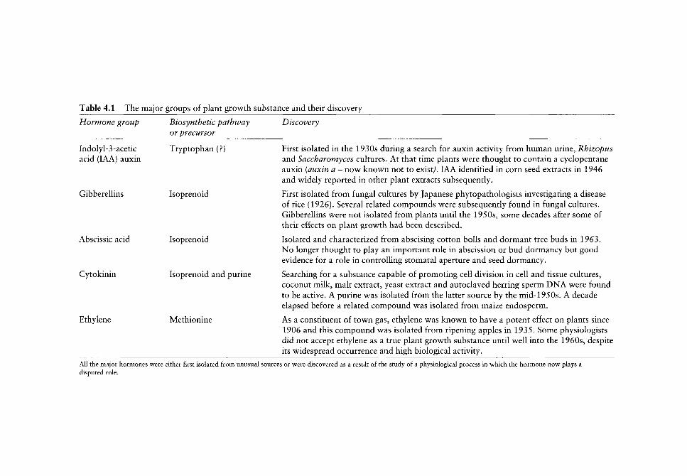
Table 4.1 The major groups of plant growth substance and their discovery
Hormone group Biosynthetic pathway or precursor
Discovery
Indolyl-3-acetic acid (lAA) auxin
Gibberelhns
Abscissic acid
Cytokinin
Ethylene
Tryptophan (?)
Isoprenoid
Isoprenoid
Isoprenoid and purine
Methionine
First isolated in the 1930s during a search for auxin activity from human urine, Rhizopus and Saccharomyces cultures. At that time plants were thought to contain a cyclopentane auxin (auxin a - now known not to existj. lAA identified in corn seed extracts in 1946 and widely reported in other plant extracts subsequently.
First isolated from fungal cultures by Japanese phytopathologists investigating a disease of rice (1926). Several related compounds were subsequently found in fungal cultures. Gibberellins were not isolated from plants until the 1950s, some decades after some of their effects on plant growth had been described.
Isolated and characterized from abscising cotton bolls and dormant tree buds in 1963. No longer thought to play an important role in abscission or bud dormancy but good evidence for a role in controlling stomatal aperture and seed dormancy.
Searching for a substance capable of promoting cell division in cell and tissue cultures, coconut milk, malt extract, yeast extract and autoclaved herring sperm DNA were found to be active. A purine was isolated from the latter source by the mid-1950s. A decade elapsed before a related compound was isolated from maize endosperm.
As a constituent of town gas, ethylene was known to have a potent effect on plants since 1906 and this compound was isolated from ripening apples in 1935. Some physiologists did not accept ethylene as a true plant growth substance until well into the 1960s, despite its widespread occurrence and high biological activity.
All the major hormones were either first isolated from unusual sources or were discovered as a result of the study of a physiological process in which the hormone now plays a disputed role.

80 R. D. FIRN &c C. G. JONES
colonies and in some simple fungi, trisporic acid (CI5 isoprenoid) is used as a sporulation coordinator (Gooday and Adams, 1993). Thus the roots of plant hormonal control may lie in simple organisms communicating between cells of the same species in different individuals. With the evolution of true multicellular organisms, the need for coordinated development and functioning v^ould have extended the scope for chemical signalling and using chemicals to coordinate the functioning or fate of cells w^ithin the same individual w^ould be a small step. The extent and w ay in which such chemical signalling would have evolved would have depended on the nature of the specialized functions that appeared in various types of organism over evolutionary time. It can be postulated that in all early multicellular organisms, secondary metabolites, already selected for the capacity to generate compounds with potent biomolecular activity were put to a new role. Evidence for such a scenario can be found in the multiple roles played by members of the isoprenoid pathway. Individual isoprenoids function as plant growth substances, plant defence compounds, fungal sexual coordinators, animal hormones and animal olfactory attractants and repellents. Of the plant growth substances, three are derived fully or partly from the isoprenoid pathway - the gibberellins, abscissic acid and cytokinins (see Table 4.1). The other two plant hormones, auxin and ethylene, are derived from amino-acid precursors and amino acids also serve as the precursors for some chemical regulators in animals and play a part in plant defences, possibly because amino acids have commonly been used as precursors in 'inventive biosynthesis' (Wong, 1981). It is interesting that the exact route leading to the biosynthesis of lAA has been debated for nearly half a century, with the expectation that the biosynthesis of such an important molecule would be finely controlled. Traditional biochemical investigations could not provide definitive evidence for such a pathway, but unexpected phenotypes resulting from the manipulation of cytochrome P450 genes suggests that, in some species, there may be an intimate link between the synthesis of indole glucosinolates and the synthesis of lAA (Feldmann, 2001). lAA could be considered to be a 'secondary product' with a primary role. If the evolutionary recruitment of a 'secondary metabolite' to serve a role as an endogenous coordinator or regulator in plants has occurred, it is likely that the event will have brought to the emerging 'hormonal control' a series of metabolic traits that have evolved to serve a quite different purpose - the generation and retention of chemical diversity. If there is not a duplication of all the enzymes involved in the pathway leading to the plant hormone, the plant hormonal control will inevitably be somewhat compromised by the metabolic features of a pathway which is even more multifunctional in that it now includes a role as an internal signalling molecule. Could such multifunctionality explain some previously puzzling aspects of plant hormone biosynthesis.^ Maybe this multifunctionality explains why so many stimuli (from insects and fungi to many forms of physical stimuli such as various wavelengths of light, low and high temperatures, too little or too much water, etc.) can change the hormone content of plants (Davies, 1995).
Gibberellin synthesis - generating diversity? Over 100 different gibberellins have been isolated and characterized - some are found in many species but others have a much more limited taxonomic distribution. A single plant species usually contains several gibberellins. The great majority of gibberellins do not possess high biological activity (or possess activity only because they are converted to other more active gibberellins) (Davies, 1995). The fact that the great majority of gibberellins possess low activity is itself consistent with the Screening Hypothesis, but is harder for other models of secondary metabolite evolution to explain. But why do plants (and some

EVOLUTIONARY LINKS BETWEEN BIOCHEMISTRY AND PHYSIOLOGY 81
fungi) make so many gibberellins? We would argue that this group of plant hormones are showing their evolutionary origin as secondary products. Even when there is a need for a gibberellin to act as an essential regulator in the plant, the metabolic traits that generate chemical diversity are retained (or at least are not selected against). The particular trait of relevance to gibberellin biosynthesis is the proposal that some enzymes involved in secondary metabolite biosynthesis may possess a relatively broad substrate specificity, leading to matrix grid transformations. In such matrix grid biosynthetic routes, a few enzymes could add or transform substituents to a carbon skeleton and the order in which they are added or transformed is not fixed. Hence a number of intermediates can be generated. As long as the possession of at least one of the compounds made results in a net gain in plant fitness, the chemical diversity represented in all the intermediates can be retained. Evidence in support of this prediction can be found in the study of the gibberellin 20-oxidase which can convert two precursors into at least 11 products because it is multifunctional (Lange et aL, 1994). The fact that some parts of the biosynthetic pathway leading to the active gibberellin, GA^, can operate as a matrix (Taiz and Zeiger, 1991) would be evidence that this type of mechanism does operate in plants even in a pathway used to make compounds which are central to controlling plant growth and development and that some enzymes involved in the pathway are following rules that would allow the extension and retention of chemical diversity. A similar complex metabolic grid may also exist in the biosynthetic pathways leading to brassinosteroids (Wang and Chory, 2000).
Plant hormone degradation - another role? For many decades it has been argued by some that enzymes involved in metabolizing plant hormones (to give either breakdown products or conjugates) may play an important role in plant hormone homeostasis (Bandurski et aL^ 1992). The control of hormone concentration by controlling the breakdown, rather than the synthesis, seemed counter intuitive to those schooled in hormonal control in mammals. However, if the biosynthetic machinery leading to plant hormone production carries with it some of the flexibility (and maybe the inducibility) of a metabolism evolved for multifunctionality, and where precise control of the amount of product may not be something that has been highly selected for, the evolution of other means of controlling the amount of any hormone by degradation perhaps deserves attention. However, an alternative explanation could be that, in some cells (maybe cells that are insensitive themselves to hormones), an ability to generate and retain chemical diversity has resulted in a production of hormones and it is in these cells that selection has resulted in a fairly crude method (degradation) of hormone concentration regulation so as to avoid disturbing more carefully regulated hormone levels in other cells. However, if these cells retain a capacity to make hormones and genes can be induced in them, possibly the extra burst of hormone synthesis could give rise to the hormone level changes that are sometimes associated with insect or fungal attack. This may be yet another opportunity for cross talk.^
Summary
The aim of this chapter has not been to inform readers how plants work or indeed how plants have evolved. Rather it has been to try to engage readers in at least considering whether there might be an appropriate evolutionary framework for metabolism that could help us investigate and eventually understand many physiological processes. We have indulged in
82 R. D. FIRN &c C. G. JONES
speculation in order to provoke readers and some may find the lack of certainty unsatis
factory. Readers provoked into rejecting the evolutionary model that has been advanced
should feel free to make such a rejection ... as long as they have a better model with which
to replace it. After any discovery, the question 'why do plants do that? ' needs some
evolutionary explanation. Maybe any plant physiology or plant biochemistry textbook
without a section on evolution should be regarded as seriously incomplete.
Acknowledgements
We thank the Institute of Ecosystem Studies for support (CGJ). Contribution to the pro
gramme of the Institute of Ecosystem Studies.
References
Albersheim P, Darvill AG. 1985. Oligosaccharins: novel molecules that can regulate growth, development, reproduction, and defense against disease in plants. Scientific American 253(3): 58-64.
Bagni N, Torrigiani P. 1992. Polyamines: a new class of growth substances. In: Karssen CM, van Loon LC, Vreugdenhil D, eds. Progress in Plant Growth Regulation. Dordrech: Kluwer, 264-275.
Bandurski RS, Desrosiers MF, Jensen P, et al. 1992. Genetics, chemistry and biochemical physiology in the study of hormonal homeostasis. In: Karssen CM, van Loon LC, Vreugdenhil D, eds. Progress in Plant Growth Regulation. Dordrech: Kluwer, 1-12.
Berenbaum MR, Zangerl AR. 1996. Phytochemical diversity: adaptation or random variation. In: Romeo JT, Saunders J A, Barbosa P, eds. Phytochemical Diversity and Redundancy in Ecological Interactions. Recent Advances in Phytochemistry. Volume 30. New York: Plenum Press, 1-24.
Davies PJ. 1995. Plant Hormones: Physiology, Biochemistry and Molecular Biology. Dordrecht: Kluwer.
Ehrlich PR, Raven PH. 1964. Butterflies and plants: a study in coevolution. Evolution 18: 586-608. Evans PT, Malmberg RL. 1989. Do polyamines have a role in plant development. Annual Review
of Plant Physiology and Plant Molecular Biology 40: 235-269. Feldmann KA. 2001. Cytochrome P450s as genes for crop improvement. Current Opinion on Plant
Biology 4: 162-167. Felton GW, Korth KL, Bi JL, et al. 1999a. Inverse relationship between systemic resistance of plants
to micro-organisms and to insect herbivory. Current Biology 9: 317-320. Felton GW, Bi JL, Mathews MC, et al. 1999b. Cross-talk between the signal pathways for
pathogen-induced systemic acquired resistance and grazing-induced resistance. In: Chadwick DJ, Goode J A. eds. Insect-Plant Interactions and Induced Plant Defence. Novartis Foundation Symposium 223. Chichester: John Wiley, 166-170.
Feys BJ, Parker JE. 2000. Interplay of signalling pathways in plant disease resistance. Trends in Genetics 16: 449-455.
Firn RD, Jones C G 1996. An explanation of secondary product redundancy. In: Romeo JT, Saunders JA, Barbosa P, eds. Phytochemical Diversity and Redundancy in Ecological Interactions. Recent Advances in Phytochemistry. Volume 30. New York: Plenum Press, 295-312.
Firn RD, Jones C G 2000. The evolution of secondary metabolism - a unifying model. Molecular Microbiology 37: 989-994.
Fraenkel G 1959. The reason d'etre of secondary plant substances. Science 129: 1466-1470. Gooday GW, Adams DJ. 1993. Sex hormones and fungi. Advances in Microbial Physiology
34: 65-145. Gross D. 1975. Growth regulating substances of plant origin. Phytochemistry 14: 2105-2112. Harborne JB. 1988. Introduction to Ecological Biochemistry. London: Academic Press.

EVOLUTIONARY LINKS BETWEEN BIOCHEMISTRY AND PHYSIOLOGY 83
Jones CG, Firn RD. 1991. On the evolution of plant secondary metabolite chemical diversity. Philosophical Transactions of the Royal Society of London 333: 273-280.
Karban R, Baldwin IT. 1997. Induced Responses to Herbivory. Chicago: The University of Chicago Press.
Kliebenstein DJ, Kroymann J, Brown P, et al. 2001. Genetic control of natural variation in Arabidopsis glucosinolate accumulation. Plant Physiology 126: 811-825.
Lange T, Hedden P, Graebe JE. 1994. Expression cloning of a gibberellin 20-oxidase, a multifunctional enzyme involved in gibberellin biosynthesis. Proceedings of the National Academy of Science 91: 8552-8556.
McDowell JM, Dangl JL. 2000. Signal transduction in the plant immune response. Trends in Biochemical Science 25: 79-82.
Pathier B, Briickner, Dathe W, et al. 1992. Jasmonates: metaboUsm, biological activities, and modes of action in senescence and stress responses. In: Karssen CM, van Loon LC, Vreugdenhil D, eds. Progress in Plant Growth Regulation. Dordrech: Kluwer, 276-285.
Petsko GA, Kenyon GL, Gerlt JA, et al. 1993. On the origin of enzymatic species. Trends in Biochemical Science 18: 372-376.
Rauscher MD. 1992. Natural selection and evolution of plant-insect interactions. In: Roitberg BS, Isman MD, eds. Insect Chemical Ecology: an Evolutionary Approach. New York: Chapman Sc Hall, 20-88.
Roberts RM. 1989. Serendipity - accidental discoveries in science. New York: John Wiley. Swain T. 1975. Evolution of flavenoids compounds. In: Harborne JB, Maby TJ, Maby H, eds. The
Flavonoids. London: Chapman and Hall, 1104. Taiz L, Zeiger E. 1991, Plant Physiology. Redwood City, California: Benjamin. Tamagnone L, Merida A, Parr A, et al. 1998. The AmMYB308 and AmMYB330 transcription fac
tors from Antirrhinum majus regulate phenylpropanoid and lignin biosynthesis in transgenic tobacco. Plant Cell 10: 135-154.
Tretyn A, Bossen ME, Kendrick RE. 1990. Evidence for different types of acetyl choline receptors in plants. In: Karssen CM, van Loon LC, Vreugdenhil D, eds. Progress in Plant Growth Regulation. Dordrech: Kluwer, 306-311.
Vet LEM. 1999. Evolutionary aspects of plant-carnivore interactions. In: Chadwick DJ, Goode JA, eds. Insect-Plant Interactions and Induced Plant Defence. Chichester: Wiley, 3-20.
Wang Z-Y, Chory J. 2000. Recent advances in molecular genetic studies of the functions of brassinolide, a steroid hormone in plants. In: Romeo JT, Ibrahim R, Varin L, De Luca V , eds. The Evolution of Metabolic Pathways. Recent Advances in Phy to chemistry. Volume 34. Oxford: Pergamon, 409-432.
Winkel-Shirley B. 2001. Flavonoid biosynthesis. A colourful model for genetics, biochemistry, cell biology and biotechnology. Plant Physiology 126: 485-493.
Wong JT-F. 1981. Coevolution of genetic code and amino acid biosynthesis. Trends in Biochemical Science 6: 33-36.

Did auxin play a crucial role in the evolution of novel body plans during the Late Silurian-Early Devonian radiation
of land plants?
Todd J Cooke, DorothyBelle Poll and Jerry D Cohen
CONTENTS.
Introduction 85
Brief overview of Cambrian radiation of bilateral animals 86 Rapid diversification of animal phyla 86 Characteristic body plan of each phylum 86 Early establishment of body plan 87 Altered expression of embryonic genes resulting
in new body plans 87
Silurian-Devonian radiation of land plants 88 Did early land plants diverge in a rapid
evolutionary radiation? 8S Are the characteristic body plans of land plants
established during embryonic development? 90 What developmental mechanisms act to generate
plant body plans? 96 Did major changes in auxin regulation occur
prior to the Silurian-Devonian radiation? 99
Conclusions 101
Acknowledgements 102
References 102
Introduction
The overall objective of this chapter is to identify plausible developmental mechanisms that might have contributed to the rapid diversification of early land plant lineages during the Late Silurian to Early Devonian Periods (Kenrick and Crane, 1997). An even more rapid diversification of bilateral animal lineages seems to have occurred during the well-known
The Evolution of Plant Physiology Copyright © 2004 Elsevier Ltd ISBN 0-12-33955-26 All rights of reproduction in any form reserved

86 T.J. COOKE £TAL.
Cambrian radiation (Gould, 1989; Raff, 1996; Conway Morris, 2000). Given that the Uneages that uhimately gave rise to animals and to plants are most likely to have diverged as ancient lineages of unicellular flagellates (Baldauf et aL, 2000), the origins of early land plants occurred via different macroevolutionary processes than those operating in ancient animals (Meyerowitz, 2002). Nonetheless, as an archetypal example of organismal radiation, our current understanding of the Cambrian radiation can be used as a conceptual framework for considering the analogous radiation of early land plants.
Brief overview of Cambrian radiation of bilateral animals
Rapid diversification of animal phyla
Although well-preserved fossils of the soft-bodied Ediacaran fauna are widely distributed in Late Precambrian rocks, it has been very problematical to trace the evolution of the simple features of Ediacaran fossils into the more complex body plans of Cambrian metazoa (Knoll and Carroll, 1999; Conway Morris, 2000). Therefore, palaeontologists have focused on the Cambrian radiation that resulted in the rapid (or 'explosive' with respect to geological time) appearance of the crown phyla of bilateral animals, including molluscs, arthropods, echino-derms and chordates, around 550-530 million years ago (Ma) (Raff, 1996; Erwin et aL, 1997; Valentine et ai, 1999; Conway Morris, 2000). In marked contrast to the fossil record, molecular clock estimates predict that initial divergences of these lineages occurred much earlier in the Precambrian era (Bromham et aL, 1998; Heckman et aL, 2001). Even if a long Precambrian fuse was needed to ignite the Cambrian explosion (Fortey, 2001), it is still undeniable that earliest bilateral animals diversified into the crown metazoan lineages during a rather narrow slice of deep geological time, with the result that all extant animal phyla appeared before the end of the Cambrian. Some authors attribute the Cambrian radiation to the occurrence of large-scale environmental perturbations resulting in new adaptive landscapes at the Precambrian-Cambrian boundary (Knoll and Carroll, 1999), while others emphasize the possible role of new ecological selection pressures associated with the rise of filter feeding and macroscopic predation (Conway Morris, 2000). Of particular relevance to this chapter are the putative developmental mechanisms discussed below that may be responsible for generating new phylum-specific body plans in the presence of those selective pressures.
Characteristic body plan of each phylum
Traditionally, animal biologists have recognized different phyla on the basis of their fundamental Baupldne (or body plans in English), which encapsulate the nature and organization of tissues and organs within the animal body. For example, the arthropods comprise a diverse assemblage of bilateral organisms, including horseshoe crabs, spiders, millipedes, crustaceans and insects. Yet the arthropod phylum as a whole exhibits a common body plan with such unifying features as a segmented exoskeleton, jointed legs, ventral nerve cord and dorsal heart with an open circulatory system. Interestingly, the characteristic body plans of all extant animal phyla were already expressed in their marine ancestors by the end of the Cambrian radiation (Raff, 1996). While certain phyla underwent major structural innovations accompanying their post-Cambrian invasion of the land environment, these terrestrial adaptations occurred within the conserved patterns of their pre-existing body plans.

AUXIN'S ROLE IN SILURIAN-DEVONIAN RADIATION OF LAND PLANTS 87
Early establishment of body plan
For two related reasons, until the recent innovations in molecular analyses, phylogenetic schemes for animal phyla were typically based on the comparative morphology of embryos and larvae. (1) the basic body plan of each phylum is first expressed at these early stages of animal development (Gilbert, 2000). Ever since von Baer (1828), it has been recognized that the general features of each phylum appear in embryonic and/or early postembryonic development as opposed to the more specialized features of individual classes that develop at later stages. Indeed, it is the common embryonic or larval features that disclose the close evolutionary relationships within those phyla like molluscs or arthropods with divergent adult forms. Furthermore, the common body plan of vertebrate embryos makes them also appear remarkably similar even though the specialized features of the adults are distinctly different. (2) the structures developing in the early, so-called phylotypic stage are more evolutionarily conserved than those formed at later stages (Raff, 1996). For example, in insects, the germ band or early larval stages have a segmented worm-like appearance that resembles the body plans of other taxa, such as the onychophorans, related to the arthropods (Ballard et aL, 1992). As another intriguing example, living echinoderms develop free-swimming bilateral larvae that undergo a complicated metamorphosis to become adults exhibiting 5-parted radial symmetry. Peterson and Davidson (2000) have hypothesized that ancestral stem-groups of bilateral animals developed simple larva similar to those characteristic of extant echinoderms. These larvae are then envisioned to have served as the structural platform for the activity of 'set-aside cells' required to generate the complex body plans of different crown bilateral animals.
Altered expression of embryonic genes resulting in new body plans
The basic approach in evolutionary developmental biology ('evo-devo') is to study the genetic regulation of embryonic and larval development of extant organisms belonging to various lineages in order to elucidate the developmental mechanisms responsible for the origin and/or diversification of those lineages (Raff, 1996; Gilbert, 2000; Arthur, 2002). This research is based on the reasonable, albeit untestable, assumption that genetic regulation of embryonic development is extraordinarily stable over vast geological time. The justification for this assumption is that any disruption in embryonic regulation should result in an avalanche of disruptive, and inevitably fatal, consequences for postembryonic development (Raff, 1996). Similar arguments have been also put forth to explain the failure of any new metazoan body plans to evolve following the Cambrian radiation (Gilbert, 2000).
The paradigmatic case of genetic regulation of metazoan body plan involves a conserved group of homeobox (Hox) genes (Erwin et aL, 1997; Gellon and McGinnis, 1998: Valentine et aL, 1999; Carroll et aL, 2001). Hox genes play crucial regulatory roles in various aspects of metazoan axis specification, such as external segment identity in arthropods and internal body segmentation in vertebrates. Molecular analyses of Hox gene diversity have shown that these genes were gradually duplicated during the ancient evolution of non-bilateral animals, with the result that the stem group of bilateral animals appear to have evolved a fully-fledged cluster consisting of a minimum of seven Hox genes (de Rosa et al.^ 1999; Peterson and Davidson, 2000). Some workers have proposed that the Hox genes were responsible for encoding the original anterior-posterior axis in the common ancestor of all animals (e.g. Slack et aL, 1993), while others argue that the Hox cluster was recruited for pattern formation of the pre-existing anterior-posterior axis during the evolution of the stem group of bilateral animals (e.g. Peterson and Davidson, 2000). In either

T.J. COOKEETAL.
event, it appears that the evolution of different body plans in the lineages of bilateral animals did not depend on the evolution of ntw Hox clusters or other developmental master genes. Instead, the diversification of bilateral animals required various innovations in regulatory netw^orks controlling the expression of these genes (Gellon and McGinnis, 1998; Knoll and Carroll, 1999; Carroll et al.^ 2001). The molecular palette of developmental genes v^as already present in primordial bilateral animals before these genes w ere rew ired to sculpt novel bilateral body plans.
In summary, it seems reasonable to conclude from this brief description that the Cambrian radiation of bilateral animals can be viewed as having arisen from the rapid emergence of nev^ expression patterns of pre-existing developmental regulatory genes. These altered expression patterns resulted in a wide range of new body plans for bilateral animals. These new animals were presumably subjected in turn to ecological selection pressures that favoured those animals with body plans best adapted for surviving in Cambrian oceans.
Silurian-Devonian radiation of land plants
Using the conceptual framework derived from our consideration of the Cambrian radiation of bilateral animals, we intend here to discuss plausible developmental mechanisms underlying the evolution of novel plant body plans and hence the macroevolution of different plant lineages in the Late Silurian to Middle Devonian periods. The hormone auxin appears to regulate the organizational features comprising body plans of contemporary plants; therefore, particular attention is granted to the hypothesis that evolutionary changes in auxin action were causally involved in the generation of different body plans during the radiation of early vascular plants.
Did early land plants diverge in a rapid evolutionary radiation?
Recent work has provided cogent molecular, biochemical and cellular evidence that ancient charophycean green algae (also called charophytes), whose living descendants include the orders Zygnematales, Coleochaetales and Charales, represent the primordial group giving rise to land plants (Graham, 1993; Graham et aL, 2000; Karol et ai, 2001). Living charophytes, sometimes derided as 'pond scum', have haplobiontic life cycles, with a dominant haploid gametophyte and a diploid phase solely consisting of the zygote that undergoes meiosis to produce four haploid cells. These algae exhibit a diverse range of growth forms, including unicells, tip-growing filaments, margin-expanding discs and complex shoot-like axes consisting of alternating large multinucleate internodal cells and multicellular nodes bearing lateral branches. This diversity of growth forms in the charophytes is thought to reflect the more permissive nature of aquatic habitats (Niklas, 2000).
According to molecular clock estimates, ancient charophycean green algae are predicted to have invaded the land during the Precambrian around 600 Ma (Heckman et aL, 2001). Dating from the Middle Ordovician around 470 Ma, the oldest microfossils, which are considered to have originated from genuine land plants, are obligate spore tetrads with sporopollenin-impregnated walls, imperforate cuticles and narrow tubes (Gray, 1985; Edwards and Wellman, 2001; Graham and Gray, 2001). Indeed, the ability of these first land plants successfully to colonize terrestrial environments is often attributed to the desiccation-resistant coverings observed on these microfossils. This microfossil evidence suggests that the earliest land plants are likely to have exhibited a bryophyte-grade of structural organization,
AUXIN'S ROLE IN SILURIAN-DEVONIAN RADIATION OF LAND PLANTS 89
at least with respect to spore morphology (Gray, 1985; Edwards and Wellman, 2001; Graham and Gray, 2001). Available molecular sequence information is also consistent with the perspective that the three extant bryophyte lineages diverged earlier than the mono-phyletic lineage giving rise to the vascular plants (Qiu et aL^ 1998; Nickrent et al.^ 2000; KaroU^a/., 2001).
It is sometimes postulated that the first land plants had quickly evolved the embryo, via the intercalation of mitotic divisions of the zygote prior to the occurrence of sporic meiosis (Graham and Wilcox, 2000). However, the lack of any confirming fossil evidence makes it conceivable that land plants did not develop embryos until long after the successful colonization of terrestrial habitats (Niklas, 1997). The earliest mesofossils with possible bryophyte affinities have been identified as miniature branching axes in Lower Devonian rocks (Edwards et aL, 1995; Edwards, 2000; Edwards and Axe, 2000). However, very few macrofossils, including Sporogonites, Tortilicaulis and Pallaviciniities from Devonian strata, appear to exhibit the characteristics of dorsiventral thalli and/or monosporangiate sporophytes that disclose their likely affinities to extant bryophytes (Taylor and Taylor, 1993). Thoughtful discussions about the inadequacy of the fossil record for elucidating the major events in bryophyte evolution can be found in many sources (e.g. Kenrick and Crane, 1997; Niklas, 1997; Edwards, 2000; Kenrick, 2000).
It is irrefutable that a considerable time interval exists between the first putative land plant spores in mid-Ordovician strata and the first protracheophyte Cooksonia in Upper Silurian rocks. In marked contrast to the bryophyte enigma, the fossil record for vascular plants provides compelling evidence that the appearance of Cooksonia was followed by the rapid radiation of numerous vascular plant lineages including those lineages that evolved into extant lycophytes, ferns and horsetails (Taylor and Taylor, 1993; Kenrick and Crane, 1997; Niklas, 1997). This radiation of vascular plants is dated to have occurred in the Late Silurian (424-409 Ma) according to cladistic analysis (Kenrick and Crane, 1997: Figure 7.15) or in the Early to Middle Devonian (409-381 Ma) according to macrofossil stratigraphy (Taylor and Taylor, 1993; Kenrick and Crane, 1997). The rapid diversification of early vascular plant lineages culminated with the origin of the progymnosperms (i.e. the progenitor lineage for seed plants) in the Middle Devonian, as indicated by the fossil Svalbardia (Taylor and Taylor, 1993; Berry and Fairon-Demaret, 2001).
The fossil record indicates that the Late Silurian to Middle Devonian was indeed the most innovative interval in the morphological diversification of land plants. During this interval, land plants appear to have evolved many features often assumed to serve as crucial terrestrial adaptations, including multicellular primary and secondary meristems, vascular tissues, vegetative organs (roots, stems, and leaves), and sporangium-bearing organs (sporophylls and sporangiophores) (Graham, 1993; Taylor and Taylor, 1993; Kenrick and Crane, 1997). Of particular interest to this chapter, it is often presumed that the first sporophyte consisted of a spherical embryo that was directly modified to form a simple sporangium embedded in the archegonium (Niklas, 1997; Graham and Wilcox, 2000). Its evolutionary elaboration into the complex vascular plant sporophyte with elevated sporangium-bearing axes must have been a critical adaptation for flourishing in terrestrial environments, because this plant body is well-constructed to produce numerous meiospores for effective aerial dispersal.
In parallel to the Cambrian radiation of bilateral animals, the rapid diversification of vascular plants starting in the Late Silurian hints at the possibility that the ancestral stem group for all vascular plants had just previously experienced a limited number of genetic innovations, which permitted and/or facilitated the rapid origin of new body plans. Contrary
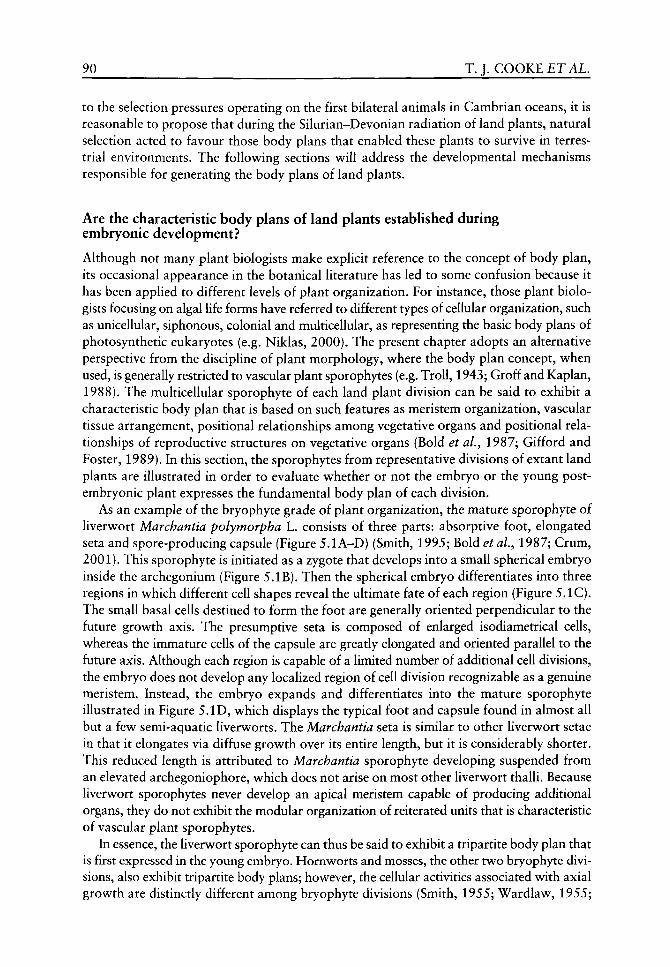
90 T.J. COOKE ETAL,
to the selection pressures operating on the first bilateral animals in Cambrian oceans, it is reasonable to propose that during the Silurian-Devonian radiation of land plants, natural selection acted to favour those body plans that enabled these plants to survive in terrestrial environments. The follow^ing sections will address the developmental mechanisms responsible for generating the body plans of land plants.
Are the characteristic body plans of land plants established during embryonic development?
Although not many plant biologists make explicit reference to the concept of body plan, its occasional appearance in the botanical literature has led to some confusion because it has been applied to different levels of plant organization. For instance, those plant biologists focusing on algal life forms have referred to different types of cellular organization, such as unicellular, siphonous, colonial and multicellular, as representing the basic body plans of photosynthetic eukaryotes (e.g. Niklas, 2000). The present chapter adopts an alternative perspective from the discipline of plant morphology, w^here the body plan concept, w^hen used, is generally restricted to vascular plant sporophytes (e.g. Troll, 1943; Groff and Kaplan, 1988). The multicellular sporophyte of each land plant division can be said to exhibit a characteristic body plan that is based on such features as meristem organization, vascular tissue arrangement, positional relationships among vegetative organs and positional relationships of reproductive structures on vegetative organs (Bold et aL, 1987; Gifford and Foster, 1989). In this section, the sporophytes from representative divisions of extant land plants are illustrated in order to evaluate w^hether or not the embryo or the young post-embryonic plant expresses the fundamental body plan of each division.
As an example of the bryophyte grade of plant organization, the mature sporophyte of liverwort Marchantia polymorpha L. consists of three parts: absorptive foot, elongated seta and spore-producing capsule (Figure 5.1A-D) (Smith, 1995; Bold et aL, 1987; Crum, 2001). This sporophyte is initiated as a zygote that develops into a small spherical embryo inside the archegonium (Figure 5.IB). Then the spherical embryo differentiates into three regions in which different cell shapes reveal the ultimate fate of each region (Figure 5.1C). The small basal cells destined to form the foot are generally oriented perpendicular to the future growth axis. The presumptive seta is composed of enlarged isodiametrical cells, whereas the immature cells of the capsule are greatly elongated and oriented parallel to the future axis. Although each region is capable of a limited number of additional cell divisions, the embryo does not develop any localized region of cell division recognizable as a genuine meristem. Instead, the embryo expands and differentiates into the mature sporophyte illustrated in Figure 5.ID, which displays the typical foot and capsule found in almost all but a few semi-aquatic liverworts. The Marchantia seta is similar to other liverwort setae in that it elongates via diffuse growth over its entire length, but it is considerably shorter. This reduced length is attributed to Marchantia sporophyte developing suspended from an elevated archegoniophore, which does not arise on most other liverwort thalli. Because liverwort sporophytes never develop an apical meristem capable of producing additional organs, they do not exhibit the modular organization of reiterated units that is characteristic of vascular plant sporophytes.
In essence, the liverwort sporophyte can thus be said to exhibit a tripartite body plan that is first expressed in the young embryo. Hornworts and mosses, the other two bryophyte divisions, also exhibit tripartite body plans; however, the cellular activities associated with axial growth are distinctly different among bryophyte divisions (Smith, 1955; Wardlaw, 1955;

AUXIN'S ROLE IN SILURIAN-DEVONIAN RADIATION OF LAND PLANTS 91
Figure 5.1 The development of the embryo and sporophyte of the Uverwort Marchantia polymor-pha L. (A) Two-celled embryo inside the archegonium. (B) Young spherical embryo lacking obvious cellular differentiation. (C) Expanded spherical embryo exhibiting tripartite organization. (D) Nearly mature sporophyte with well-defined foot, seta and capsule. Redrawn from Smith (1955) with permission from McGraw-Hill Companies.
Bold et aL, 1987; Crum, 2001). The embryo of the hornworts is also composed of a basal tier destined to become the foot and an apical tier representing the future apicalmost cells of the capsule (Campbell, 1918; Smith, 1955; Wardlaw, 1955; Crum, 2001). The intermediate tier develops into a narrow band of dividing cells called an intercalary meris-tem that remains positioned just above the foot. The intercalary meristem undergoes uni-facial divisions on its capsule side, w ith the result that these new^ cells compose almost the entire linear capsule that rises above the gametophyte. Thus, the three-tiered hornwort embryo is directly enlarged into a tripartite body plan. In most mosses, it is difficult to recognize three cellular tiers in the first stages of embryonic development (Smith, 1955; Wardlaw, 1955; Lai and Bhandari, 1968; Bold etai, 1987; Crum, 2001). A transient apical cell differentiates into the upper hemisphere and then this apical cell and its derivatives act to generate a spindle-shaped embryo. This embryo exhibits three well-defined regions that are destined to develop into the foot, seta and capsule of the mature moss sporophyte. In marked contrast to the diffuse growth of the liverwort seta, a unifacial intercalary meristem arises in the moss seta just beneath the immature capsule and its activity generates most of the cells composing the elongating seta.
The fern Pityrogramma triangularis (Kaulf.) Maxon serves here as a representative example of the pteridophyte grade of plant organization (Figure 5.2). The fern zygote within the archegonium undergoes a series of segmentation divisions resulting in the formation of a globular-shaped embryo (Campbell, 1918; Smith, 1955; Wardlaw, 1955; Gifford and Foster, 1989). This embryo is often said to be composed of four quadrants representing the shoot apex, first leaf, first root and foot (Figure 5.2A-D). The shoot apex is solely responsible

92 T.J. COOKE £TAL.
R3 R'' R2
Lf1 Lf2
Figure 5.2 Embryonic development of the fern Fityrogramma triangularis (Kaulf.) Maxon (A-E) and the body plans of the ferns (F) and the dicots (G). (A). Two-celled embryo inside the archegonium. (B) Young spherical embryo exhibiting characteristic apical cells for shoot apex and first leaf in apical hemisphere. (C-E) Successive stages of the four-quadrant embryo composed of shoot apex, first leaf, first root and foot (unlabelled). The first root arises in a lateral position near the base of the first leaf. Panels (A-E) were redrawn with permission from unpublished work of W. Hagemann. (F) Diagrammatic illustration of typical fern body plan, which shows that the positional relationships first expressed in the unipolar embryo are reiterated in postembryonic development. (G) Diagrammatic illustration of the model dicot body plan, which shows that the positional relationships first expressed in the bipolar embryo are reiterated in postembryonic development. This situation does not hold true for all dicots. Panels (F) and (G) were redrawn from Troll (1959) with permission of Georg Thieme Verlag. Abbreviations: Em, embryo; AN, archegonial neck; SA, shoot apex; Lf, leaf; R, root; TB, terminal bud; AB, axillary bud; Co, cotyledon; PR, primary root; RC, root cap.
for generating the growth axis of the postembryonic plant; hence, the fern embryo is referred to as being unipolar (Groff and Kaplan, 1988). The first root arises near the base of first leaf (Figure 5.2E). All postembryonic roots are also observed to originate near leaf bases and thus, fern roots are consistently lateral writh respect to the longitudinal axis of

AUXIN'S ROLE IN SILURIAN-DEVONIAN RADIATION OF LAND PLANTS 93
the growing plant (Figure 5.2F), which has been termed the homorhizic condition (Troll, 1943; Groff and Kaplan, 1988). The horsetails (Equistem)^ which are thought to comprise a monophyletic group with the ferns (Pryer et aL, 2001), exhibit similar organographic arrangements, with the first root subtending the first leaf and all postembryonic roots arising at the nodes (Gifford and Foster, 1989).
The relative positions of embryonic organs illustrated in Figure 5.2 foreshadow the postembryonic body plan in almost all ferns (Fames, 1936; Smith, 1955; Bierhorst, 1971; Gifford and Foster, 1989). In even those ferns exhibiting unusual habits, the body plan is irrevocably fixed by its embryonic organization. In marked contrast to the subterranean root systems of dicot trees, the tree ferns from such genera as Dicksonia and Cyathea develop buttress-like coverings of interwoven roots that arise at the bases of lower leaves and grow down the outside of the trunk and into the ground (Smith, 1955; Gifford and Foster, 1989). Additional roots continue to form at the bases of higher tree fern leaves, even though these roots are destined to remain short and never penetrate the soil. Mature plants of the aquatic floating fern Salvinia do not develop roots and this rootless feature can be traced back to its embryo, where the non-growing sector subjacent to the first leaf is often interpreted as a vestigial first root (Eames, 1936; Bierhorst, 1971). Even the problematic whisk ferns (Psilotales), which are apparently closely related to eusporangiate ferns according to molecular analyses (Pryer et aL, 2001), exhibit an embryonic organization that correlates with its mature morphology, at least with respect to the absence of any roots. In Tmesipteris tannensis Bernh. and Psilotum nudum (L.) Pal. Beauv., the embryos display a two-parted organization consisting of a distal shoot apex and a proximal foot with no evidence at all for leaf or root quadrants (Holloway, 1921,1939; Bierhorst, 1971). The embryonic shoot apex develops into a branched plagiotropic rhizome lacking roots, which may be an adaptive response to the epiphytic habit frequently adopted by whisk ferns. (Eventually, the rhizome produces aerial axes bearing reduced dorsiventral structures called enations which are actually homologous to genuine leaves (for resolution of this controversial issue, see Kaplan, 2001).) A rare exception to the general rule about the embryonic encapsulation of fern body plan is seen in certain Ophioglossum species where roots develop subterminal buds capable of growing into new shoots (Peterson, 1970); nevertheless, the roots of the daughter shoots also emerge at the base of their fronds, thereby replicating the positional relationships of the original shoot.
The other major pteridophyte group, the lycophytes, also manifests unipolar embryonic organization, which is largely reproduced in the body plans of adult plants (Groff and Kaplan, 1988; Gifford and Foster, 1989). However, postembryonic roots in the lycophytes do not exhibit the same positional relationships as the ephemeral first root. For example, the embryos of different Selaginella species exhibit wide variation in the position of the first root relative to other embryonic organs, which are not reflected in the organographic arrangements of their postembryonic plant bodies (Bower, 1935; Gifford and Foster, 1989). Subsequent roots in many Selaginella species originate from leafless axes called rhizophores that form at stem bifurcations. The distal ends of these rhizophores bear typical roots with root caps. Some workers suggest that rhizophores are considered as true roots (e.g. Gifford and Foster, 1989), while others view them as unique root-bearing structures (e.g. Lu and Jernstedt, 1996). A similar uncoupling of embryonic root position and postembryonic organization is observed in other lycophytes. In most Lycopodium species, postembryonic roots originate close to the shoot tip and traverse down the cortex before emerging into the soil (Gifford and Foster, 1989). The postembryonic roots of Isoetes and its extinct relatives are borne on different specialized structures called rhizomorphs of uncertain homology

94 T.J. COOKEETAL.
Figure 5.3 Embryonic development of the dicot Capsella bursa-pastoris (L.) Medic. (A) and (B) Young embryos exhibiting well-defined suspensor and embryo proper. The incipient root apical meris-tem can be recognized by the periclinal cell divisions delimiting the future root cap. (C) Mid-globular embryo displaying incipient shoot apical meristem with a lighter staining central (or initial) zone and a darker staining lateral (or morphogenetic) zone. (D-F) Early to late heart embryos showing cotyledon emergence from the lateral zones. (G) Early torpedo stage. Reprinted from Kaplan and Cooke (1997) with permission from American Society of Plant Biologists. Abbreviations: RA, root apex; SU, suspensor; MZ, morphogenetic zone; IZ, initial zone; Cot, cotyledon.
(Paolillo, 1963, 1982). In conclusion, it appears that the fundamental pteridophyte body plan is fully manifested in either developing embryos (ferns and horsetails) or young post-embryonic plants (lycophytes) and then it is reiterated throughout the life of the plant.
Characteristically, seed plant embryos exhibit bipolar (or allorhizic) organization with the embryonic shoot and root apices arising at opposite poles (Troll, 1943; Groff and Kaplan, 1988). In many, but not all, embryos, these incipient meristems act to perpetuate the bipolar organization, with the result that the primary plant body exhibits a central axis with opposite shoot and root systems, as is diagrammatically illustrated in Figure 5.2G. For example, in the dicot Capsella bursapastoris (L.) Medic. (Figure 5.3), the incipient root and shoot apical meristems arise during the globular stage of embryonic development (Kaplan and Cooke, 1997). These meristems can first be recognized by the cellular activities that accompany the formation of their distal or lateral structures. In particular, the origin of the root apical meristem is disclosed by periclinal divisions that delimit the distal root cap from the more proximal root body (Figure 5.3A-D). This first root will then generate the root system of the mature plant. The embryonic shoot apical meristem is revealed by the presence of cytohistological zonation at the shoot pole of the late globular embryo (Figure 5.3C-G).
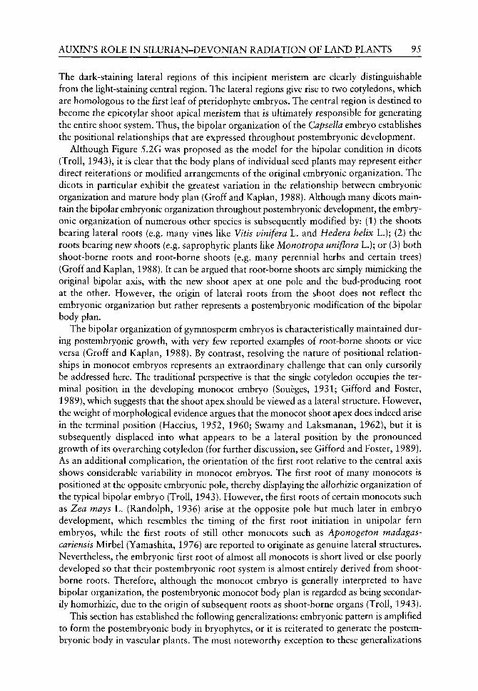
AUXIN'S ROLE IN SILURIAN-DEVONIAN RADIATION OF LAND PLANTS 95
The dark-staining lateral regions of this incipient meristem are clearly distinguishable from the light-staining central region. The lateral regions give rise to two cotyledons, which are homologous to the first leaf of pteridophyte embryos. The central region is destined to become the epicotylar shoot apical meristem that is ultimately responsible for generating the entire shoot system. Thus, the bipolar organization of the Capsella embryo establishes the positional relationships that are expressed throughout postembryonic development.
Although Figure 5.2G was proposed as the model for the bipolar condition in dicots (Troll, 1943), it is clear that the body plans of individual seed plants may represent either direct reiterations or modified arrangements of the original embryonic organization. The dicots in particular exhibit the greatest variation in the relationship between embryonic organization and mature body plan (Groff and Kaplan, 1988). Although many dicots maintain the bipolar embryonic organization throughout postembryonic development, the embryonic organization of numerous other species is subsequently modified by: (1) the shoots bearing lateral roots (e.g. many vines like Vitis vinifera L. and Hedera helix L.); (2) the roots bearing new shoots (e.g. saprophytic plants like Monotropa uniflora L.); or (3) both shoot-borne roots and root-borne shoots (e.g. many perennial herbs and certain trees) (Groff and Kaplan, 1988). It can be argued that root-borne shoots are simply mimicking the original bipolar axis, with the new shoot apex at one pole and the bud-producing root at the other. However, the origin of lateral roots from the shoot does not reflect the embryonic organization but rather represents a postembryonic modification of the bipolar body plan.
The bipolar organization of gymnosperm embryos is characteristically maintained during postembryonic growth, with very few reported examples of root-borne shoots or vice versa (Groff and Kaplan, 1988). By contrast, resolving the nature of positional relationships in monocot embryos represents an extraordinary challenge that can only cursorily be addressed here. The traditional perspective is that the single cotyledon occupies the terminal position in the developing monocot embryo (Soueges, 1931; Gifford and Foster, 1989), which suggests that the shoot apex should be viewed as a lateral structure. However, the weight of morphological evidence argues that the monocot shoot apex does indeed arise in the terminal position (Haccius, 1952, 1960; Swamy and Laksmanan, 1962), but it is subsequently displaced into what appears to be a lateral position by the pronounced growth of its overarching cotyledon (for further discussion, see Gifford and Foster, 1989). As an additional complication, the orientation of the first root relative to the central axis shows considerable variability in monocot embryos. The first root of many monocots is positioned at the opposite embryonic pole, thereby displaying the allorhizic organization of the typical bipolar embryo (Troll, 1943). However, the first roots of certain monocots such as Zea mays L. (Randolph, 1936) arise at the opposite pole but much later in embryo development, which resembles the timing of the first root initiation in unipolar fern embryos, while the first roots of still other monocots such as Aponogeton madagas-cariensis Mirbel (Yamashita, 1976) are reported to originate as genuine lateral structures. Nevertheless, the embryonic first root of almost all monocots is short lived or else poorly developed so that their postembryonic root system is almost entirely derived from shoot-borne roots. Therefore, although the monocot embryo is generally interpreted to have bipolar organization, the postembryonic monocot body plan is regarded as being secondarily homorhizic, due to the origin of subsequent roots as shoot-borne organs (Troll, 1943).
This section has established the following generalizations: embryonic pattern is amplified to form the postembryonic body in bryophytes, or it is reiterated to generate the postembryonic body in vascular plants. The most noteworthy exception to these generalizations
96 T.J. COOKE £TAL,
is that in some vascular plants, the positional relationships of all roots except the embryonic root are established in the young postembryonic body.
What developmental mechanisms act to generate plant body plans?
It must be appreciated that the botanical research on this question is far less advanced than comparable research in animal development. However, the initial evidence review^ed below^ suggests that the hormone auxin (indole-3-acetic acid) acts as a critical developmental mechanism for generating the body plans of land plants.
As discussed in a previous section, the current theory for explaining the developmental mechanisms underlying the Cambrian radiation is that the genes responsible for regulating embryonic development in basal animals experienced repeated duplication and altered transcriptional regulation, v^ith the result that these genes were able to specify more complex body plans. A similar working hypothesis is now being adopted by the emerging disciple of plant evolutionary developmental biology (for an excellent overview, see Cronk, 2001). For instance, plant homeobox genes can be classified into at least three subfamilies: KNOTTED!-like {KNOX) genes affecting meristematic cell fates; homeodomain-leucine zipper (HD-ZIP) genes regulating later developmental and physiological processes; and GLABRO 1752-like genes specifying epidermal cell fates (Chan et al,, 1998; WiUiams, 1998; Bharathan et aL, 1999). The archetypal example of the first subfamily is the so-called SHOOTMERISTEMLESS gene (Barton and Poethig, 1993) that is expressed in the central region of the globular embryo of Arabidopsis thaliana (L.) Heynh. (Long etal.^ 1996). The identical stage in the related Capsella embryo is illustrated in Figure 5.3C. Although the name of this gene implies that it is essential for the origin of the shoot apical meristem, an alternative interpretation is that it maintains the proliferative activity of the embryonic central region in order to generate the epicotylar shoot apical meristem (Kaplan and Cooke, 1997). The recent report of two KNOX-like genes in the moss Physcomitrella patens (Hedw.) B.S.G. indicates that this subfamily started diverging early in the evolution of land plants (Champagne and Ashton, 2001). The ability of Physcomitrella to perform homologous recombination should allow these investigators to determine the precise roles of homeobox genes in mosses (Theissen et ai, 2001). However, research to date has not revealed any direct relationship between evolutionary changes in homeobox genes and new body plans in plants. Since the primeval protist lineages evolving into animals and plants had probably diverged as single-celled eukaryotes, it should come as no surprise that plants might depend on different genetic mechanisms to organize their body plans (Meyerowitz, 2002).
On the other hand, it is quite intriguing that the molecular diversity of the MADS-box family of transcription factors correlates with the morphological complexity of land plants (Theissen et aL, 2000; Vergara-Silva et al, 2000; Cronk, 2001). Seven MADS-box genes have been isolated from the moss Physcomitrella patens (Krogan and Ashton, 2000; Henschel et aL, 2002) in contrast to 15 genes from the fern Ceratopteris richardii Brogn. and to even larger numbers from several angiosperm species. Of particular importance to angiosperm reproductive development are the MADS-box genes that act as homeotic selector genes for controlling floral organ identity. Indeed, the ABC model provides molecular validation for the classical morphological concept of serial homology, i.e. all lateral dor-siventral structures from cotyledons to carpels are leaf homologues (Goethe, 1790), because the triple mutant in all three functions exhibits foliage leaf-like structures in place of floral organs (Coen and Meyerowitz, 1991). The molecular evolution literature expresses considerable optimism that the MADS-box gene diversity underUes the evolution of plant body

AUXIN'S ROLE IN SILURIAN-DEVONIAN RADIATION OF LAND PLANTS 97
Inhibitor sensitivity
Characteristic processes
Anti-auxins Polar auxin transport inhibitors
Strong auxins
Unorganized cell proliferation
Spherical gro\wth
Axis elongation
Cotyledon formation
Ovule formation
Spherical growth
Cotyledon formation
Axis elongation
Figure 5.4 Auxin regulation of the somatic (A) and zygotic (B) embryogenesis of the carrot Daucus carota L. (for details, see Schiavone and Cooke, 1987; Michalczuk etal.^ 1992a; Ribnicky etal.^ 2002). Trivial names refer to the shapes of each embryonic stage; characteristic processes refer to the mor-phogenetic processes occurring at each stage; and inhibitor sensitivity refers to the auxin agonists or antagonists active at each stage. For the purpose of normalizing the data for free auxin concentrations between embryo types, all data are expressed relative to basal levels, which are 7ng lAA/g FW and 14 ng 2,4-D/g FW in oblong to torpedo stages in somatic embryos and 26 ng lAA/g FW in unfertilized ovules and torpedo stages in zygotic embryos. Redrawn from Ljung et al. (2002) with permission from Kluwer Academic PubHshers. Abbreviations: lAA, indole-3-acetic acid = endogenous auxin; 2,4-D,2,4-dichlorophenoxyacetic acid = synthetic auxin.
plans in general (for enthusiastic advocacy, see Vergara-Silva et aL, 2000). We wish to point out to the contrary that no compelling evidence yet exists to support the notion that MADS-box genes play even minor roles in establishing the fundamental tripartite, unipolar or bipolar organization of any division of land plants, including the angiosperms.
In our opinion, auxin is causally involved in the estabUshment of plant body plans, at least in the seed plants (Figure 5.4). The progression through both somatic and zygotic embryogenesis of the carrot Daucus carota L. appears to require the sequential activation of tv^o different auxin biosynthetic pathwrays (Michalczuk et aL, 1992a,b; Ribnicky et aL, 1996, 2002). During the initial stages of embryo grow^th, a tryptophan-dependent pathw^ay for auxin biosynthesis produces high levels of free (i.e. active) auxin, v^hich are apparently critical for mediating the rapid cell divisions needed to generate the globular embryo. Then the embryo switches to a tryptophan-independent pathway for auxin biosynthesis that appears capable of exercising greater homeostatic control over the free auxin levels. The action of this pathway results in much lower free auxin levels that may be a necessary precondition for establishing auxin gradients regulating the polarized growth of older embryos. The results from inhibitor experiments are entirely consistent with these concepts: both synthetic auxins and polar auxin transport inhibitors are able to block or alter the polarized growth, but not the initial isodiametric expansion, of all angiosperm embryos examined
98 T.J. COOKEETAL.
(Schiavone and Cooke, 1987; Liu et al, 1993; Fischer et aL, 1997; Hadfi et aL, 1998). It remains critical to characterize auxin levels and biosynthetic pathways in other embryos than those of carrots. Auxin levels are reported to increase between the initial embryonic and subsequent postembryonic development in the wheat (Triticum aestivum L.) caryopsis (Fischer-Iglesias et aL, 2001). It is clear that these results are not directly comparable to those obtained from carrot embryo work because wheat embryos undergo prolonged postembryonic growth while still retained inside the caryopsis.
Molecular research is beginning to reveal the molecular basis for auxin action in Arabidopsis embryos (Souter and Lindsey, 2000; Hamann, 2001). For instance, a homozygous null mutation in the putative auxin receptor gene {ABPl) results in embryo development being blocked at the early globular stage (Chen et aL, 2001). Of even greater importance are the observations on gnom mutant embryos, which become enlarged spherical structures unable to initiate a polarized growth axis. The molecular basis for the mutant phenotype is that the embryos fail to localize the auxin efflux carrier PINl in the proper position for carrying out polar auxin transport (Steinmann et aL, 1999).
Moreover, auxin acts as a critical regulator of postembryonic body plan of seed plants. In particular, localized synthesis and/or polarized transport are thought to establish auxin gradients that appear absolutely critical for the positioning of new leaf primordia on the shoot apex (Meicenheimer, 1981; Reinhardt et al., 2000) and of new lateral root primordia along developing roots (Reed et ^/., 1998; Casimiro et aL, 2001). Finally, it is well documented that auxin exercises predominant control over many aspects of vascular tissue development, including the induction of primary vascular tissues (Roberts etaL, 1988; Aloni, 1995); the positioning of primary vascular bundles (Sachs, 1991; Berleth etaL, 2000); and the activity of vascular cambia (Uggla et al., 1996, 1998). All this evidence taken together demonstrates that auxin acts as a very important regulator of the body plans of seed plants.
Much less research has been devoted to the auxin regulation of developmental processes in bryophytes and pteridophytes (for review, see Cooke et aL, 2002). In fact, to the best of our knowledge, no published work has directly studied auxin action in body plan organization in these plants. Nonetheless, three arguments can be advanced in support of the prediction that auxin must also help to regulate the body plans of non-seed plants. (1) it seems quite plausible that the seed plants would not have evolved de novo development mechanisms for generating body plans but rather would have modified pre-existing mechanisms already operating in the common ancestor of non-seed and seed plants. (2) we have noted elsewhere that the nature of metabolic regulation of auxin levels appears to have evolved in concert with the increasing morphological complexity in the land plant lineage (Sztein et aL^ 2000). (3) certain auxin-mediated processes, including tropisms, apical dominance and axis elongation, are widespread among all land plants, including bryophytes (Cooke et aL, 2002). Among the positional relationships subject to auxin regulation in non-seed plants are: rhizoid initiation in bryophyte gametophytes (Kaul et aL^ 1962; Kumra and Chopra, 1987; Nyman and Cutter, 1981: Chopra and Vashistha, 1990), root initiation in pterido-phyte sporophytes (Wardlaw, 1957; Partanen and Partanen, 1963; Wochok and Sussex, 1975) and vascular differentiation in fern sporophytes (Ma and Steeves, 1992). It is also worthwhile here to mention the provocative modelling work of Stein (1993) who attempted to relate predicted patterns of auxin concentration in the shoot apex to the observed arrangements of primary vascular tissues in a wide range of fossil plants. Although this model is based on several assumptions about auxin biosynthesis, movement and accumulation that have never been evaluated with real meristems, it does result in a close correspondence between predicted hormone distributions and underlying stelar patterns for many fossil
AUXIN'S ROLE IN SILURIAN-DEVONIAN RADIATION OF LAND PLANTS 99
plants, which suggests that ahered patterns of auxin action may have been involved in the diversification of stelar patterns throughout the land plant lineage.
Did major changes in auxin regulation occur prior to the Silurian-Devonian radiation?
The approach adopted in evolutionary development biology to address such questions involves: (1) the use of the fossil record and/or molecular phylogenies to identify those groups that diverged from the stem group prior to the radiation; and (2) the characterization of the developmental mechanisms in extant organisms from those early diverging lineages (Raff, 1996; Arthur, 2002). The ultimate goal is to predict those developmental mechanisms operating in the stem group that may have contributed to body plan diversification during the radiation. The critical assumption discussed earlier is that the developmental mechanisms for generating embryonic body plans are assumed to remain extraordinarily stable over geological time. This assumption has been validated in non-bilateral animals by the direct relationship between their homeobox gene diversity and their phylogenetic relationships (Peterson and Davidson, 2000), but we have no evidence to evaluate its validity for plants.
A comparable approach toward the Silurian-Devonian radiation of vascular plants would involve an investigation of the genes responsible for regulating auxin action in the three extant bryophyte lineages, which diverged from the vascular plant stem group prior to this radiation, according to morphological and molecular phylogenies (Kenrick and Crane, 1997; Qiu et aL, 1998; Nickrent et al., 2000; Renzaglia et aL, 2000; Karol et al, 2001). It must be acknowledged that this effort is hampered by several limitations. For instance, it is probably premature to attempt a comparative study of auxin regulatory genes in land plants because few non-seed plants, with the notable exception of Fhyscomitrella patens^ are readily amenable to molecular manipulation. Moreover, the phylogenetic relationships among the bryophyte lineages remain unresolved, although the research cited above does frequently, but not always, place the mosses as the sister group to the vascular plants. Lastly, since macrofossils exhibiting the morphological characteristics of modern bryophytes (e.g. monosporangiate sporophytes and heteromorphic gametophyte-dominant generations) appear relatively late in the fossil record, one must always keep in mind the possibility that one or more modern bryophyte lineages may be derived from isomorphic, polysporangiate ancestors (Kenrick and Crane, 1997).
What can be accomplished here is that we can discuss the distribution of auxin regulatory processes in charophytes and bryophytes (Table 5.1) in order to speculate about developmental mechanisms that may have been operating in the stem group before the diversification of vascular plants in the Late Silurian-Early Devonian radiation. The tryptophan-independent pathway has definitively been identified as the predominant auxin biosynthetic pathway in the liverwort Marchantia polymorpha L., the moss Polytrichum ohioense Ren. & Card., and several vascular plants, which indicates that the capacity for tightly regulating auxin biosynthesis may be ubiquitous in the land plant lineage (Sztein et aL, 2000). Because the charophyte Nitella spp. maintains auxin levels that are comparable with those measured in bryophytes, it is likely that the tryptophan-independent pathway is also operative in this group. In so far as charophytes and liverworts carry out metabolic inter-conversions between active free and inactive conjugated forms of auxin at slow rates, it appears that the free auxin levels are maintained in these groups via the balance between biosynthetic and degradative reactions. However, hornworts and mosses share the ability for rapid auxin conjugation with vascular plants, which provides these lineages the potential

Table 5.1 Phylogenetic distribution of critical processes involved in auxin action in green plants. The occurrence of polar auxin transport (PAT) was assessed by the direct measurement of PAT in agar-block experiments and/or by the sensitivity of auxin efflux to PAT inhibitors.
Process
Tryptophan-independent pathway
Auxin conjugation rates
Predicted mechanism for regulating auxin levels
PAT in gametophyte rhizoids
PAT in gametophyte thalli
PAT in young sporophytes
Charophytes
Predicted
Very slow
Biosynthesis/ degradation
Yes?
No
n.s.
Liverworts
Yes
Slow
Biosynthesis/ degradation
n.d.
Yes
No
Hornworts
n.d.
Intermediate to rapid
Conjugation/ hydrolysis
n.d.
n.d.
No
Mosses
Yes
Intermediate to rapid
Conjugation/ hydrolysis
Yes
Yes
Yes
Vascular plants
Yes
Rapid to very rapid
Conjugation/ hydrolysis
n.d.
n.d.
Yes
n.d.. ) data; n.s., non-existent structure. For references, see text.

AUXIN'S ROLE IN SILURIAN-DEVONIAN RADIATION OF LAND PLANTS 101
for even more precise regulation of auxin levels (Sztein et al.^ 1995,1999, 2000). One significant difference is that almost all vascular plants produce two specific conjugates, lAA-aspartate (or -glutamate) and IAA-1-O-glucose, that are not accumulated in bryophytes.
Membrane proteins capable of mediating the transmembrane auxin transport also evolved before the origin of the land plants, as evidenced by their activity in the thalli of the charophyte Chara vulgaris L. (Dibb-Fuller and Morris, 1992). Hov^ever, there is contradictory evidence about whether charophytes carry out the more sophisticated process of intercellular auxin transport, as characterized by basipetal polarity and inhibitor sensitivity. The standard inhibitors of polar auxin transport do not affect auxin efflux from intact thalli (Dibb-Fuller and Morris, 1992), but these inhibitors are reported to have significant effects on decapitated thalli with growing rhizoids (Klambt et al, 1992). Intercellular auxin transport with strong basipetal polarity and inhibitor sensitivity has been measured in liverwort thaUi (Maravolo, 1976; Gaal et ai, 1982); moss protonemata (Rose et al., 1983; Geier et aL, 1990), and moss rhizoids (Rose and Bopp, 1983). The sporophytes of hornworts and liverworts do not exhibit polar auxin transport (Thomas, 1980; DB. Poh, unpubhshed observations). By contrast, young setae of the moss Polytrichum maintain significant fluxes of basipetal polar transport that are even higher than those measured in corn coleoptiles (DB. Poli, unpublished observations). If mosses are the actual sister group of the vascular plants, then this observation suggests that their common ancestor evolved the auxin-dependent mechanism for generating polarized axes that is still being utilized in the sporophytes of extant members of both groups. Finally, bryophytes exhibit many of the auxin-mediated responses, such as apical dominance, phototropism and axis elongation also found in vascular plants (Cooke et aL^ 2002). In summary, the range of auxin-regulated processes reported in extant bryophytes lends some credibility to the hypothesis that auxin was intimately involved in establishing the body plans of ancestral Silurian plants prior to the diversification of vascular plant lineages.
Conclusions
Using the conceptual framework derived from the study of the Cambrian radiation of bilateral animals, this chapter attempted to address four questions concerning the evolution of the early land plants. (1) the evidence assembled indicates that the early land plants did undergo a rapid evolutionary radiation during the Late Silurian to Early Devonian periods. (2) the characteristic body plans of different divisions of extant land plants are established during embryonic and early postembryonic development, which means that the regulatory mechanisms operating in embryonic development are also critical for generating these body plans. (3) the research evidence surveyed in this chapter establishes that the hormone auxin serves as a primary mechanism for regulating embryo and postembryonic development, at least in vascular plants. (4) judging from our current knowledge of auxin action in early-divergent bryophyte lineages, it appears that major changes in auxin action occurred in the earliest land plants prior to the Late Silurian. Therefore, the evidence available to date lends support to the appealing perspective that genetic changes in auxin action in early land plants were instrumental in the subsequent diversification of body plans in vascular plants. It follows that increased knowledge of the auxin regulation of embryonic mechanisms in extant plants should help to elucidate the developmental events that generated novel body plans during the Silurian-Devonian radiation of new vascular plant lineages.

102 T . J . C O O K E E T A L .
Our enthusiasm for these interpretations is dampened somewhat by the reahzation that
due to the limited number of relevant papers, we are skating over the conceptual equivalent
of thin ice. A rigorous evaluation of the question posed in the title requires that much greater
research effort be devoted toward the characterization of: (1) the phylogenetic relation
ships among the lineages of both extant bryophytes and putative bryophyte fossils; (2) the
genetic regulation of auxin action in embryo development; and (3) the developmental mech
anisms operating in charophytes, bryophytes and pteridophytes. Regardless of the ultimate
answer, that question will be worth considering if it inspires further research on those three
subjects.
Acknowledgements
The authors thank Drs Donald R. Kaplan (University of California, Berkeley) and Linda
E. Graham (University of Wisconsin-Madison) for their useful discussions of several topics
addressed in this chapter. The work reported from the laboratories of the author was
supported in part by United States Department of Energy grant DE-FG02-00ER15079
and National Science Foundation grant IBN9817604.
References
Aloni R. 1995. The induction of vascular tissues by auxin and cytokinin. In: Davies PJ, ed. Plant Hormones: Physiology, Biochemistry and Molecular Biology^ 2nd edn. Dordrecht: Kluwer Academic Publishers, 531-546.
Arthur W. 2002. The emerging conceptual framework of evolutionary developmental biology. Nature 415: 757-764.
von Baer KE. 1828. Entwicklungsgeschichte der Tiere: Beobachtung und Reflexion. Konigsberg: Borntrager.
Baldauf SL, Roger AJ, Wenk-Siefert I, Doolittle WF. 2000. A kingdom-level phylogeny of eukaryotes based on combined protein data. Science 290: 972-977.
Ballard JWO, Olsen GJ, Faith DP, et al. 1992. Evidence from 12S ribosomal RA sequences that onychophorans are modified arthropods. Science 258: 1345-1348.
Barton MK, Poethig RS. 1993. Formation of the shoot apical meristem in Arabidopsis thaliana: an analysis of development in the wild type and shootmeristemless mutant. Development 119: 823-831.
Berleth T, Mattisson J, Hardtke CS. 2000. Vascular continuity and auxin signals. Trends in Plant Science 5: 387-393.
Berry CM, Fairon-Demaret M. 2001. The middle Devonian flora revisited. In: Gensel PG, Edwards D, eds. Plants Invade the Land: Evolutionary and Ecological Perspectives. New York: Columbia University Press, 120-139.
Bharathan G, Janssen BJ, Kellogg EA, Sinha N. 1999. Phylogenetic relationships and evolution of the KNOTTED class of plant homeodomain proteins. Molecular Biology and Evolution 16: 553-563.
Bierhorst DW. 1971. Morphology of Vascular Plants. New York: Macmillan. Bold HC, Alexopoulos CI, Delevoryas T. 1987. Morphology of Plants and Fungi, 5th edn.
New York: Harper &c Row. Bower FO. 1935. Primitive Land Plants. London: Macmillan. Bromham L, Rambaut A, Fortey R, et al. 1998. Testing the Cambrian explosion hypothesis by using
a molecular dating technique. Proceedings of the National Academy of Sciences (USA) 95: 12386-12389.

AUXIN^S ROLE IN SILURIAN-DEVONIAN RADIATION OF LAND PLANTS 103
Campbell DH. 1918. The Structure and Development of Ferns, 3rd edn. New York: MacMillan. Carroll SB, Grenier JK, Weatherbee SC. 2001. From DNA to Diversity: Molecular Genetics and the
Evolution of Animal Design. Maiden, Massachusetts: Blackwell Science. Casimiro I, Marchant A, Bhalerao RP, et al. 2001. Auxin transport promotes Arabidopsis lateral
root initiation. Flant Cell 13: 843-852. Champagne CEM, Ashton NW. 2001. Ancestry of KNOX genes revealed by bryophyte
Physcomitrella patens homologs. New Phytologist 150: 23-36. Chan RL, Gago GM, Palena CM, Gonzalez DH. 1998. Homeoboxes in plant development. Biochimica
et Biophysica Acta 1442: 1-19. Chen JG, Ullah H, Young JC, et al. 2001. ABPl is required for organized cell elongation and div
ision in Arabidopsis embryogenesis. Genes and Development 15: 902-911. Chopra RN, Vashistha BD. 1990. The effect of auxins and antiauxins on shoot-bud induction and
morphology in the moss, Bryum atrovirens Will, ex Brid. Australian Journal of Botany 38:177-184. Coen ES, Meyerowitz EM. 1991. The war of the whorls: Genetic interactions controlling flower
development. Nature 353: 31-37. Conway Morris S. 2000. The Cambrian 'explosion': Slow-fuse or megatonnage? Proceedings of the
National Academy of Sciences (USA) 97: 4426-4429. Cooke TJ, PoH DB, Sztein AE, Cohen JD. 2002. Evolutionary patterns in auxin action. Plant
Molecular Biology 49: 319-338. Cronk QCB. 2001. Plant evolution and development in a post-genomic context. Nature Reviews
Genetics 2: 607-619. Crum H. 2001. Structural Diversity of Bryophytes. Ann Arbor, Michigan: University of Michigan
Herbarium. Dibb-Fuller JE, Morris DA. 1992. Studies on the evolution of auxin carriers and phytotropin recep
tors: Transmemebrane auxin transport in unicellular and multicellular Chlorophyta. Planta 186: 219-226.
Eames AJ. 1936. Morphology of Vascular Plants: Lower Groups. New York: McGraw-Hill. Edwards D. 2000. The role of Mid-Palaeozoic mesofossils in the detection of early bryophytes.
Philosophical Transactions of the Royal Society of London, Series B 355: 733-755. Edwards D, Axe L. 2000. Novel conducting tissues in Lower Devonian plants. Botanical Journal of
the Linnean Society 134: 383-399. Edwards D, Duckett JG, Richardson JB. 1995. Hepatic characters in the earliest land plants. Nature
374: 635-636. Edwards D, Wellman C. 2001. Embryophytes on land: The Ordovician to Lochkovian (Lower
Devonian) record. In: Gensel PG, Edwards D, eds. Plants Invade the Land: Evolutionary and Ecological Perspectives. New York: Columbia University Press, 3-28.
Erwin D, Valentine J, Jablonski D. 1997. The origin of animal body plans. American Scientist 85: 126-137.
Fischer C, Speth V, Fleig-Eberenz S, Neuhaus G. 1997. Induction of zygotic polyembryos in wheat: influence of auxin polar transport. Plant Cell 9: 1767-1780.
Fischer-Iglesias C, Sundberg B, Neuhaus G, Jones AM. 2001. Auxin distribution and transport during embryonic pattern formation in wheat. Plant Journal 26: 115-129.
Fortey R. 2001. The Cambrian explosion exploded. Science 293: 438-439. Gaal DJ, Dufresne SJ, Maravolo NC. 1982. Transport of ^^C-indoleacetic acid in the hepatic
Marchantia polymorpha. The Bryologist 85: 410-418. Geier U, Werner O, Bopp M. 1990. Indole-3-acetic acid uptake in isolated protoplasts of the moss
Funaria hygrometrica. Physiologia Plantarum 80: 584-592. Gellon D, McGinnis W. 1998. Shaping animal body plans in development and evolution by modu
lation of Hox expression patterns. Bioessays 20: 116-125. Gifford EM, Foster AS. 1989. Morphology and Evolution of Vascular Plants, 3rd edn. New York:
Freeman. Gilbert SF. 2000. Developmental Biology, 6th edn. Sunderland, Massachusetts: Sinaeuer Associates. Goethe JW. 1790. Versuch die Metamorphose der Pflanzen zu erkldren. Gotha: Ettinger. Gould SJ. 1989. Wonderful Life: The Burgess Shale and the Nature of History. New York: Norton.

104 T.J, COOKE ETAL.
Graham LE. 1993. Origin of Land Plants. New York: Wiley. Graham LE, Cook ME, Busse JS. 2000. The origin of plants: Body plan changes contributing to a major
evolutionary radiation. Proceedings of the National Academy of Sciences (USA) 97: 4535^540. Graham LE, Gray J. 2001. The origin, morphology, and ecophysiology of early embryophytes:
Neontological and paleontological perspectives. In: Gensel PC, Edw^ards D, eds. Plants Invade the Land: Evolutionary and Ecological Perspectives. Nev^ York: Columbia University Press, 140-158.
Graham LE, Wilcox LW. 2000. The origin of alternation of generations in land plants: A focus on matrotrophy and hexose transport. Philosophical Transactions of the Royal Society of London, Series B 355: 755-767.
Gray J. 1985. The microfossil record of early land plants: Advances in understanding of early ter-restrialization, 1970-1984. Philosophical Transactions of the Royal Society of London, Series B 309: 167-192.
Groff PA, Kaplan DR. 1988. The relation of root systems to shoot systems in vascular plants. Botanical Review 54: 387-422.
Haccius B. 1952. Die Embryoentsvicklung bei Ottelia alismoides und das Problem des terminalen Monokotylen-Keimblatts. Planta 40: 443-460.
Haccius B. 1960. Experimentell induzierte Einkeimblattrigkeit bei Eranthis hiemalis. IL MonokotyUe durch Pheylborsaure. Planta 54: 482-497.
Hadfi K, Speth V, Neuhaus G. 1998. Auxin-induced developmental patterns in Brassica juncea embryos. Development 125: 879-887.
Hamann T. 2001. The role of auxin in apical-basal pattern formation during Arabidopsis embryo-genesis. Journal of Plant Growth Regulation 20: 292-299.
Heckman DS, Geiser DM, Eidell BR, et al. 2001. Molecular evidence for the early colonization of land by fungi and plants. Science 293: 1129-1133.
Henschel K, Kofuji R, Hasebe M, et al. 2002. Tv^o ancient classes of MIKC-type MADS-box genes are present in the moss Physcomitrella patens. Molecular Biology and Evolution 19: 801-814.
Holloway JE. 1921. Further studies on the prothallus, embryo, and young sporophyte of Tmesipteris. Transactions of the New Zealand Institute 53: 386-422.
Holloway JE. 1939. The gametophyte, embryo, and young rhizome oiPsilotum triquetrum Swartz. Annals of Botany 3: 313-336.
Kaplan DR. 2001. The science of plant morphology: Definition, history, and role in modern biology. American journal of Botany 88: 1711-1741.
Kaplan DR, Cooke TJ. 1997. Fundamental concepts in the embryogenesis of dicotyledons: a morphological interpretation of embryo mutants. Plant Cell 9: 1903-1919.
Karol KG, McCourt RM, Cimino MT, Delwiche CF. 2001. The closest living relatives to the land plants. Science 294: 2351-2353.
Kaul KN, Mitra GC, Tripathi BK. 1962. Responses of Marchantia in aseptic culture to well-known auxins and antiauxins. Annals of Botany 26: 447-467.
Kenrick P. 2000. The relationships of vascular plants. Philosophical Transactions of the Royal Society of London, Series B 355: 847-855.
Kenrick P, Crane PR. 1997. The Origin and Early Diversification of Land Plants: A Cladistic Study. Washington: Smithsonian Institution Press.
Klambt D, Knauth B, Dittmann I. 1992. Auxin dependent growth of rhizoids of Chara globularis. Physiologia Plantarum 85: 537-540.
Knoll AH, Carroll SB. 1999. Early animal evolution: Emerging views from comparative biology and geology. Science 284: 2129-2137.
Krogan NT, Ashton NW. 2000. Ancestry of plant MADS-box genes revealed by bryophyte (Physcomitrella patens) homologues. New Phytologist 147: 505-517.
Kumra S, Chopra RN. 1987. Callus initiation, its growth and differentiation in the liverwort Asterella wallichiana (Lehm. et Lindenb.) Groelle I. Effect of auxins and cytokinins. journal of the Hattori Botanical Laboratory 63: 237-245.
Lai M, Bhandari NN. 1968. The development of sex organs and sporophyte in Physcomitrium cyathicarpum Mitt. The Bryologist 71: 11-20.
AUXIN^S ROLE IN SILURIAN-DEVONIAN RADIATION OF LAND PLANTS 105
Liu C-M, Xu Z-H, Chua N-H. 1993. Auxin polar transport is essential for the establishment of bilateral symmetry during early plant embryogenesis. Plant Cell 5: 621-630.
Ljung K, Hull AK, Kowalczyk M, et al. 2002. Biosynthesis, conjugation, cataboUsm and homeostasis of indole-3-acetic acid in Arahidopsis thaliana. Plant Molecular Biology 49: 249-272.
Long JA, Moan EI, Medford JI, Barton MK. 1996. A member of the KNOTTED class of home-odomain proteins encoded by the STM gene of Arabidopsis. Nature 379: 66-69.
Lu P, Jernstedt JA. 1996. Rhizophore and root development in Selaginella martensii: Meristem transitions and identity. International Journal of Plant Sciences 157: 180-194.
Ma Y, Steeves TA. 1992. Auxin effects on vascular differentiation in Ostrich fern. Annals of Botany 70: 277-282.
Maravolo NC. 1976. Polarity and localization of auxin movement in the hepatic, Marchantia poly-morpha. American Journal of Botany 63: 529-531.
Meicenheimer RD. 1981. Changes in Epilobium phyllotaxy induced by N-1-naphthylphthalmic acid and a-4-chlorophenoxyisobutric acid. American Journal of Botany 68: 1139-1154.
Meyerowitz EM. 2002. Plants compared to animals: The broadest comparative study of development. Science 295: 1482-1485.
Michalczuk L, Cooke TJ, Cohen JD. 1992a. Auxin levels at different stages of carrot somatic embryogenesis. Phy to chemistry 31: 1097-1103.
Michalczuk L, Ribnicky DM, Cooke TJ, Cohen JD. 1992b. Regulation of indole-3-acetic acid biosynthetic pathways in carrot cell cultures. Plant Physiology 100: 1346-1353.
Nickrent DL, Parkinson CL, Palmer JD, Duff RJ. 2000. Multigene phylogeny of land plants v^ith special reference to bryophytes and the earliest land plants. Molecular Biology and Evolution 17: 1885-1895.
Niklas KJ. 1997. The Evolutionary Biology of Plants. Chicago: University of Chicago Press. Niklas KJ. 2000. The evolution of plant body plans: A biomechanical perspective. Annals of Botany
85:411-438. Nyman LP, Cutter EG. 1981. Auxin-cytokinin interaction in the inhibition, release, and morphology of
gametophore buds oi Plagiomnium cupidatum from apical dominance. Canadian Journal of Botany 59: 750-760.
Paolillo DJ. 1963. The Developmental Anatomy o/Isoetes. Urbana: University of Illinois Press. Paolillo DJ. 1982. Meristems and evolution: Developmental correspondence among the rhizomorphs
of the lycopsids. American Journal of Botany 69: 1032-1042. Partanen JN, Partanen CR. 1963. Observations on the culture of roots of the bracken fern.
Canadian Journal of Botany 41: 1657-1661. Peterson KJ, Davidson EH. 2000. Regulatory evolution and the origin of the bilaterians. Proceedings
of the National Academy of Sciences (USA) 97: 4430-4433. Peterson RL. 1970. Bud formation at the root apex of Ophioglossum petiolatum. Phytomorphology
20: 183-190. Pryer KM, Schneider H, Smith AR, et al. 2001. Horsetails and ferns are a monoplyletic group and
the closest living relatives to seed plants. Nature 409: 618-622. Qiu Y-L, Cho Y, Cox JC, Palmer JD. 1998. The gain of three mitochondrial introns identifies
liverv^orts as the earliest land plants. Nature 394: 671-674. Randolph LF. 1936. Developmental morphology of the caryopsis of maize. Journal of Agricultural
Research 53: 881-916. Raff RA. 1996. The Shape of Life: Genes, Development, and the Evolution of Animal Form. Chicago:
University of Chicago Press. Reed RC, Brady SR, Muday GK. 1998. Inhibition of auxin movement from the shoot into the root
inhibits lateral root development in Arabidopsis. Plant Physiology 118: 1369-1378. Reinhardt D, Mandel T, Kuhlemeier C. 2000. Auxin regulates the initiation and radial position of
plant lateral organs. Plant Cell 12: 507-518. Renzaglia KS, Duff RJ, Nickrent DL, Garbary DJ. 2000. Vegetative and reproductive innovations
of early land plants: Implications for a unified phylogeny. Philosophical Transactions of the Royal Society of London, Series B 355: 769-793.

106 T.J. COOKE £TAL.
Ribnicky DM, Cohen JD, Hu WS, Cooke TJ. 2002. An auxin surge following fertilization in carrots: a general mechanism for regulating plant totipotency. Planta 214: 505-509.
Ribnicky DM, Ilic N, Cohen JD, Cooke TJ. 1996. The effects of exogenous auxins on endogenous indole-3-acetic acid metabolism. The implications for carrot somatic embryogenesis. Plant Physiology 112: 549-558.
Roberts LW, Gahan PB, Aloni R. 1988. Vascular Differentiation and Plant Growth Regulators. Berlin: Springer-Verlag.
de Rosa R, Grenier JK, Andreeva T, et al. 1999. Hox genes in brachiopods and priapulids and pro-tostome evolution. Nature 399: 722-776.
Rose S, Bopp M. 1983. Uptake and polar transport of indoleacetic acid in moss rhizoids. Physiologia Plantarum 58: 57-61.
Rose S, Rubery PH, Bopp M. 1983. The mechanism of auxin uptake and accumulation in moss protonemata. Physiologia Plantarum 58: 52-56.
Sachs T. 1991. Cell polarity and tissue patterning in plants. Development (Supplement) 1: 83-93. Schiavone FM, Cooke TJ. 1987. Unusual patterns of somatic embryogenesis in the domesticated car
rot: Developmental effects of exogenous auxins and auxin transport inhibitors. Cell Differentation 21: 53-62.
Slack MW, Holland PWH, Graham CF. 1993. The zootype and the phylotypic stage. Nature 361: 490-492.
Smith GM. 1955. Cryptogamic Botany: Volume Ily Bryophytes and Pteridophytes. New York: McGraw-Hill.
Soueges R. 1931. L'embryon chez Sagittaria sagittaefolia L. Le cone vegetatif de la tige et I'extremite radiculaire chez les monocotyledones. Annales de Sciences Naturelles. Botanique et Biologie Vegetale, lOe Serie 13: 353-402.
Souter M, Lindsey K. 2000. Polarity and signalling in plant embryogenesis. Journal of Experimental Botany 51:971-983.
Stein W, 1993. ModeUing the evolution of stelar architecture in vascular plants. International Journal of Plant Science 154: 229-263.
Steinmann T, Geldner N, Grebe M, et al. 1999. Coordinated polar localization of auxin efflux carrier PINl by GNOM ARF GEF. Science 286: 316-318.
Swamy BGL, Laksmanan KK. 1962. The origin of epicotyledonary meristem and cotyledon in Halophila ovata Gaudich. Annals of Botany 26: 243-249.
Sztein AE, Cohen JD, Cooke TJ. 2000. Evolutionary patterns in the auxin metabolism of green plants. International Journal of Plant Sciences 161: 849-859.
Sztein AE, Cohen JD, de la Fuente IG, Cooke TJ. 1999. Auxin metabolism in mosses and liverworts. American Journal of Botany 86: 1544-1555.
Sztein AE, Cohen JD, Slovin JP, Cooke TJ. 1995. Auxin metaboHsm in representative land plants. American Journal of Botany 82: 1514-1521.
Taylor TN, Taylor EL. 1993. The Biology and Evolution of Fossil Plants. Englewood Cliffs, New Jersey: Prentice Hall.
Theissen G, Becker A, Di Rosa A, et al. 2000. A short history of MADS-box genes in plants. Plant Molecular Biology 41: 115-149.
Theissen G, Munster T, Henschel K. 2001. Why don't mosses flower? New Phytologist 150: 1-5. Thomas RJ. 1980. Cell elongation in hepatics: The seta system. Bulletin of the Torrey Botanical
Club 107: 339-345. Troll W. 1943. Vergleichende Morphologie der hoheren Pflanzen. Erster Band Vegetationsorgane.
Dritter Teil. Berlin: Gebriider Bomtraeger. Troll W. 1959. Allgemeine Botanik. Stuttgart: Enke Verlag. Uggla C, Mellerowicz EJ, Sundberg B. 1998. Indole-3-acetic acid controls cambial growth in Scots
pine by positional signaling. Plant Physiology 117: 113-121. Uggla C, Moritz T, Sandberg G, Sundberg B. 1996. Auxin as a positional signal in pattern forma
tion in plants. Proceedings of the National Academy of Sciences (USA) 93: 9282-9286. Valentine JW, Jablonski D, Erwin DH. 1999. Fossils, molecules and embryos: New perspectives on
the Cambrian explosion. Development 126: 851-859.

AUXIN'S ROLE IN SILURIAN-DEVONIAN RADIATION OF LAND PLANTS 107
Vergara-Silva F, Martinez-Castilla L, Alvarez-Buylla E. 2000. MADS-box genes: development and evolution of plant body plans. Journal of Phycology 36: 803-812.
Wardlaw CW. 1955. Embryogenesis in Plants. New York: Wiley. Wardlaw CW. 1957. Experimental and analytical studies of pteridophytes. XXXVII. The effects of
direct applications of various substances to the shoot apex of Dryopteris austriaca (D. aristata). Annals of Botany 21: 427-437.
Williams RW. 1998. Plant homeobox genes: Many functions stem from a common motif. Bioessays 20: 280-282.
Wochok ZS, Sussex IM. 1975. Morphogenesis in Selaginella. III. Meristem determination and cell differentiation. Developmental Biology 47: 376-383.
Yamashita T. 1976. Uber die Embryo- und Wurzelentwicklung bei Aponogeton madagascariensis. Journal of the Faculty of Science, University of Tokyo, Section III. Botany 12: 37-63.
Aquaporins: structure, function and phylogenetic analysis
Joost T van Dongen and Adrianus C Borstlap
CONTENTS.
Introduction 109
Transport of water across cell membranes 110
Discovery of aquaporins and the MlP-family 111
Structure and function of MIPs 112
MIPs of bacteria, fungi and animals 113
Plant MIPs 115
Phylogenetic analysis 117
Acknowledgement 118
References 118
Introduction
The colonization of land by plants - one of the most important events in the history of planet Earth - coincided w ith the diversification of basal embryophytes, v^hich led to the emergence of mosses, horsetails, ferns and seed plants (Kenrick and Crane, 1997). The first embryophytes evolved as far back as 500 miUion years (Ma) ago from charophycean green algae, probably on the margins of drying pools. The earliest land plants inhabited damp places and vs ere poikilohydric, that is their degree of hydration depended on atmospheric humidity. Large central vacuoles are typical of plant cells and must have already been present in the algal ancestors. In the earliest land plants vacuoles may have served, for the first time, as a reserve wrater supply stabilizing the hydration of the cytoplasm during short periods of drought stress (Wiebe, 1978).
The transition of poikilohydric into homoiohydric organisms has been a major step in the evolution of plants and allow^ed them to invade drier habitats (Walter and Stadelmann, 1968). Homoiohydric plants can maintain a constant internal w^ater balance independent of atmospheric humidity. Their aerial parts are characterized by internal gas spaces and, more importantly, by a cuticle and closable stomata by which transpiration can be regulated.
The Evolution of Plant Physiology Copyright © 2004 Elsevier Ltd ISBN 0-12-33955-26 All rights of reproduction in any form reserved
n o J. T. VAN DONGEN &c A. C, BORSTLAP
The emergence of homoiohydric plants also required the innovation of a root system, or some similar underground structure, to exploit soil water resources (Raven and Edwards, 2001). In addition, an endohydric water-conducting system was needed to supply water to the aerial parts at rates sufficient to support the transpiratory loss of water during photosynthesis. Early land plants had an unexpected diversity of conducting cells, which evolved by parallel evolution (Ligrone et ai, 2000). Among these, the precursors of the xylem tracheary elements were the most successful and became the water-conducting system of all vascular plants (Friedman and Cook, 2000). At the same time the phloem evolved as a food conducting system to supply the heterotrophic roots with the products of photosynthesis.
The tracheal system in the xylem provides a low-resistance, apoplasmic pathway for the upward flow of water. But the passage of water through plant tissues also involves sym-plasmic and transcellular pathways and, therefore, transmembrane water fluxes (Steudle and Peterson, 1998). The sieve elements in the phloem constitute a symplasmic route by which organic nutrients are translocated. Being hydraulically operated, the functioning of this food-conducting system depends on the influx of water into the sieve elements at the loading site and the efflux of water at the site of unloading. Influx and efflux of water across the plasmalemma and tonoplast is also at the basis of turgor-regulated movements in plants, such as the opening and closure of stomata and the diurnal movements of leaf pulvini. Thigmotropic, turgor-dependent movements may be very fast as, for example, in the pulvini of the sensitive plant (Mimosa pudica Mimosa pudica L.), the traps of insectivorous plants (Dionaea Dionaea Ellis, Utricularia Utricularia L.), and the gynostemium of trigger plants (Stylidium). These rapid movements (15-30 ms in Stylidium Styhdium Sw. ex Willd) are thought to result from a very fast efflux of both solutes and water from motor cells (Hill and Findlay, 1981).
Transport of water across cell membranes
In the 18th century, Stephen Hales was the first to measure the transpiration of plants and the force of upward sap pressure in stems (Hales, 1727). Since then, the study of water flows and water relations in plants has been a major theme in plant physiology, though the mechanism by which water crosses plant cell membranes has received relatively little attention. The basic structure of cell membranes is a fluid lipid bilayer, which mainly consists of phospholipids and sterols. The intrinsic (or integral) membrane proteins are 'floating' in this 'sea' of lipid molecules, whereas extrinsic membrane proteins are located on both surfaces of the membrane (Singer and Nicolson, 1972). The lipid bilayer is a real barrier for water movements, impeding the diffusion of water by at least a factor of 3000.
Water can cross a membrane by diffusion or in response to an osmotic pressure gradient. The rate at which water permeates a membrane may accordingly be defined by the diffusion permeability coefficient (P^) or by the osmotic permeability coefficient (P^). Interestingly, the relationship between Pf and P^ depends on the nature of the membrane. For an 'oil' membrane, such as a phospholipid bilayer, the concentration of water within the membrane is so low that interactions between water molecules may be neglected. As a consequence, Ff and P^ for such membranes are equal. This may be different for porous membranes. If the movement of water through the membrane is restricted to the pores, and if the pores are so narrow that the transport of water molecules occurs in a single file, the remarkable relationship FflF^ = N can be derived, where N is the number of water molecules in the single file that fills the pore (Finkelstein, 1987).

AQUAPORINS: STRUCTURE, FUNCTION AND PHYLOGENETIC ANALYSIS 111
The permeability of lipid membranes for water has been the subject of intensive investigations (Finkelstein, 1987). Depending on parameters such as lipid chain length, degree of chain saturation and sterol content, P^ (or P^), values for artificial lipid bilayers have been found to span almost three orders of magnitude, ranging from 2 X 10~^ to 1 X 10~^cm s~^. As these values encompass nearly the entire range of values reported for cell membranes it seemed that the water permeability of a membrane was fully attributable to its bilayer structure. Besides the magnitude of Pf, however, some additional issues remained that required an explanation. First, the Pf and Pj values for cell membranes were often found to be widely different. In careful experiments with red cell membranes, for instance, Pj amounted to 0.37 X 10~^cms~^, whereas the value of P^ was nearly 6-fold higher and determined at 2.15 X 10~^cms~^ (Moura et aL, 1984). Secondly, unlike the water flux through lipid bilayers, that through cell membranes could be largely inhibited by sul-phydryl reagents such as p-chloromercuribenzene sulphonate (pCMBS), indicating that a proteinaceous channel is involved. Finally, the water permeability of epithelial cells in the urinary bladder of frog and toad and the mammalian collecting kidney tubules can increase up to 50-fold in response to vasopressin (the 'antidiuretic hormone'). This hormonal regulation is difficult to explain if water transport occurred exclusively through the bilayer. Thus around 1985 evidence had accumulated that the membranes of animal cells could contain pores which transported water and little else (Finkelstein, 1987). Similar conclusions were reached from studies of water transport into characean cells (Wayne and Tazawa, 1990). The nature of these pores, however, remained a mystery.
Discovery of aquaporins and the MlP-family
The first aquaporin was serendipitously discovered in Peter Agre's laboratory during efforts to purify the 32-kDa subunit of the red cell Rh blood group antigen. A novel 28-kDa protein was found, which displayed some similarity with certain channel-forming integral proteins, hence its name CHIP28. Further studies showed that the protein was also abundantly present in the epithelia of proximal tubules and thin descending limbs of rat kidneys. Renal epithelia are the site of intense water transport. In humans they are responsible for the absorption of 180 litres of fluid each day. Its presence in the kidney therefore sparked the idea that the novel 28-kDa polypeptide may be the long sought water channel (Agre et ah, 1998; Borgnia et al., 1999). When the gene coding for CHIP28 was cloned this hypothesis could be tested by expression in oocytes of the African claw frog, Xenopus laevis. After injection of cRNA of CHIP28 into the oocytes, the permeability of oocyte membrane for water increased dramatically (from —0.2 X 10"^ to ~2 X lO'^cms"^), which caused them to explode rapidly when transferred to a hypotonic medium (Preston ^ < /., 1992). Thus CHIP28 was identified as the first water-transporting protein and aptly renamed as aquaporin 1 (AQPl).
AQPl belongs to the family of major intrinsic proteins (MIPs), one of the 46 famiUes of the class of a-helix-type channels (Saier, 2000). The family has been named after the major intrinsic protein in the cell membrane of lens fibre cells, where it comprises >60% of the membrane proteins (Gorin et al.^ 1984). The existence of the MIP family first transpired when striking similarities were noticed in the amino acid sequences of the MIP from eye lens fibre cells (now known as AQPO) and two other membrane proteins from quite disparate cells (Baker and Saier, 1990). One of these was GlpF, the glycerol facilitator in the cell membrane of Escherichia coli. The other was Nodulin 26, a plant-encoded abundant

112 J. T. VAN DONGEN &c A. C. BORSTLAP
protein isolated from the peribacteroid membrane of soybean nodules. At the time CHIP28 joined the family and was identified as the first aquaporin, some additional plant members of the MIP family were already known. These included the turgor-responsive gene clone 7a/TRG-31 from the pea (Guerrero et ai, 1990; Guerrero and Crossland, 1993); TobRB7, which was identified in a cDNA hbrary from tobacco roots (Yamamoto et ai, 1990); and a-TIP, an abundant protein from the tonoplast of protein-storing vacuoles in seeds of the common bean (Johnson et al., 1990). The gene that codes for another tonoplast localized MlP-like protein in Arabidopsis thaliana (L.) Heynh, called y-TIP, was also cloned in Maarten Chrispeels' laboratory, and subsequently characterized as the first plant aquaporin (Hofte et al, 1992; Maurel et al., 1993). The discovery of the PIPs, plant aquaporins that localize to the plasma membrane, followed soon after (Kammerloher et aL, 1994).
Structure and function of MIPs
Most MIPs are composed of 250-290 amino acids. All have six hydrophobic, putative transmembrane helices, which are connected by loops (Figure 6.1). The two halves of the protein exhibit substantial sequence similarity to one another. The B and E loop contain the asparagine-proline-alanine (NPA) motif which is highly characteristic for aquaporins. Because of the uneven number of transmembrane helices in each repeat, the two domains are obversely oriented in the membrane.
The functionality of MIPs is generally assessed after expression in Xenopus oocytes. These studies have shown that most MIPs can be classified either as aquaporins, which selectively facilitate the transport of water across the membrane, or as aquaglyceroporins that transport water as well as glycerol. Recently atomic models were published for the prototypes of the two functional classes, AQPl and GlpF (Murata et al, 2000; Fu et al, 2000).
COOH
Figure 6.1 Topology of a major intrinsic protein. Six transmembrane helices (Hl-6) are linked by loops (A-E). Loops B and E contain the highly conserved asparagine-proline-alanine (NPA) motif and bend inward into the protein. The NH2- and COOH-termini are localized in the cytosol, whereas the opposite part of the protein is orientated towards the apoplast or the vacuolar lumen. Note that loop D between helices 4 and 5 is not visible in this figure.
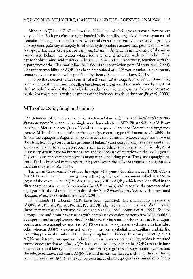
AQUAPORINS: STRUCTURE, FUNCTION AND PHYLOGENETIC ANALYSIS 113
Although AQPl and GlpF are less than 30% identical, their gross structural features are very similar. Both proteins are right-handed helix bundles, organized in two symmetrical domains. The aquaporin has a narrow central constriction and wider external openings. The aqueous pathway is largely lined with hydrophobic residues that permit rapid water transport. The narrowest part of the pore, 0.3 nm (3 A) wide, is in the centre of the membrane, just behind the region where loops B and E interact with each other. Four hydrophobic amino acid residues in helices 1, 2, 4, and 5, respectively, together with the asparagines of the NPA-motifs line the inside of the constriction pore (Murata et aL, 2000). The unit permeability of AQPl has been determined at —10^ water molecule per second, remarkably close to the value predicted by theory (Sansom and Law, 2001).
In GlpF the selectivity filter consists of a 2.8 nm (28 A) long, 0.34-0.38 nm (3.4-3.8 A) wide amphipathic channel. The alkyl backbone of the glycerol molecule is wedged against the hydrophobic side of the channel, whereas the three hydroxyl groups of glycerol form successive hydrogen bonds with side groups of the hydrophilic side of the pore (Fu et al., 2000).
MIPs of bacteria, fungi and animals
The genomes of the archaebacteria Archaeoglobus fulgidus and Methanobacterium thermoautotrophicum contain a single gene that codes for a MIP (Figure 6.2), but MIPs are lacking in Methanococcus janaschii and other sequenced archaea. Bacteria and fungi may possess MIPs of the aquaporin or the aquaglyceroporin type (Hohmann et aL, 2000). In E, colt the aquaporin AqpZ is involved in cellular hydration, whereas GlpF has a role in the utilization of glycerol. In the genome of bakers' yeast (Saccharomyces cerevisiae) three genes are related to aquaglyceroporins and three others to aquaporins. Curiously, most laboratory strains have no functional aquaporins because of mutations in the coding genes. Glycerol is an important osmolyte in many fungi, including yeast. The yeast aquaglyceroporin Fpsl is involved in the export of glycerol when the cells are exposed to a hypotonic medium (Luyten et al, 1995).
The worm Caenorhabditis elegans has eight MIP genes (Kuwahara et aL, 1998). Only a few MIPs are known from insects. One is BIB (big brain) of Drosophila^ which is a homo-logue of the mammalian AQP4. Another insect MIP is AQP^c which was identified in the filter chamber of a sap-sucking cicada (Cicadella viridis) and, recently, the presence of an aquaporin in the Malpighian tubules of the bug Rhodnius prolixus was demonstrated (Borgnia et al., 1999; Echevarria et al, 2001).
In mammals 11 different MIPs have been identified. The mammalian aquaporins (AQPO, AQPl, AQP2, AQP4, AQP5, AQP8) play key roles in transmembrane water fluxes in many tissues (reviewed by Deen and Van Os, 1998; Borgnia et al, 1999). Kidney, airways, eye and brain have tissues with complex expression patterns involving multiple aquaporins and aquaglyceroporins. The kidney, for instance, harbours at least four aquaporins and two aquaglyceroporins. AQPO seems to be expressed exclusively in lens fibre cells, whereas AQPl is expressed widely in various epitheUal and capillary endothelia, including proximal tubule and thin descending limb in kidney. In kidney collecting ducts AQP2 mediates the vasopressin-induced increase in water permeability, which is required for the concentration of urine. AQP4 is the main aquaporin in brain. AQP5 resides in lung and salivary and lachrymal glands and presumably regulates airways humidification and the release of saliva and tears. AQP8 is found in various tissues, including those of testis, pancreas and liver. AQP6 is the only known intracellular aquaporin in animal cells. It has
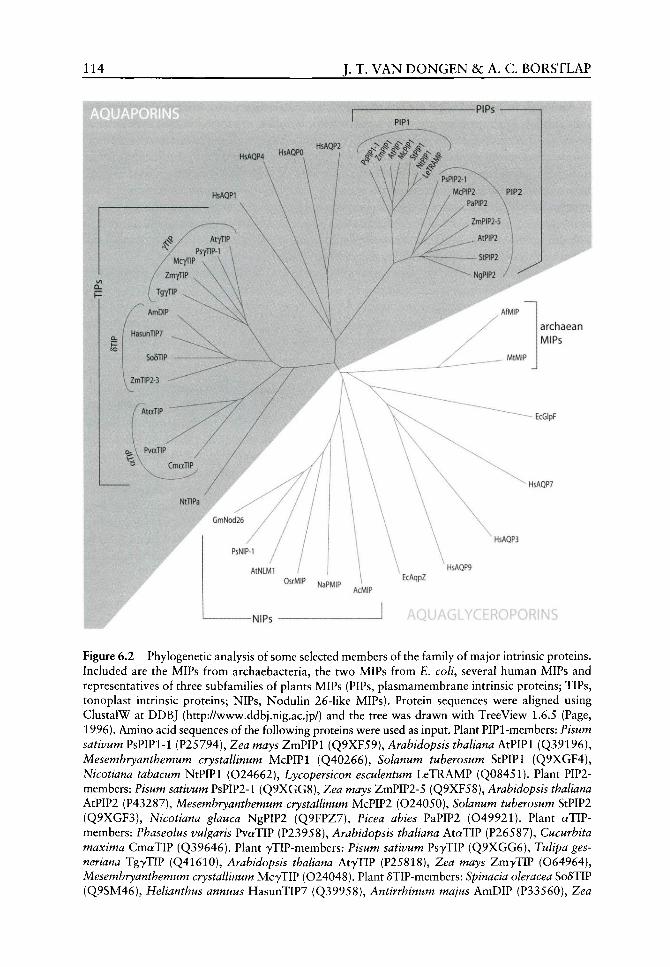
114 J. T. VAN DONGEN &c A. C. BORSTLAP
Ws-
archaean MIPS
EcGlpF
HsAQP7
mm GmNod26
HsAQPB
PsNIP-1
AtNLMI
O^^MiP N3PMIP EcAqpZ
HsAQP9
AcM!P
-NIPs
Figure 6.2 Phylogenetic analysis of some selected members of the family of major intrinsic proteins. Included are the MIPs from archaebacteria, the two MIPs from E. colt, several human MIPs and representatives of three subfamilies of plants MIPs (PIPs, plasmamembrane intrinsic proteins; TIPs, tonoplast intrinsic proteins; NIPs, Nodulin 26-like MIPs). Protein sequences v^ere aligned using ClustalW at DDBJ (http://v^^wvv^.ddbj.nig.ac.jp/) and the tree w as draw^n w ith TreeViev^ 1.6.5 (Page, 1996). Amino acid sequences of the following proteins were used as input. Plant PIPl-members: Pisum sativum PsPIPl-1 (P25794), Zea mays ZmPIPl (Q9XF59), Arabidopsis thaliana AtPIPl (Q39196), Mesembryanthemum crystallinum McPIPl (Q40266), Solanum tuberosum StPIPl (Q9XGF4), Nicotiana tabacum NtPIPl (024662), Lycopersicon esculentum LeTRAMP (Q08451). Plant PIP2-members: Fisum sativum PsPIP2-l (Q9XGG8), Zea mays ZmPIP2-5 (Q9XF58), Arabidopsis thaliana AtPIP2 (P43287), Mesembryanthemum crystallinum McPIP2 (024050), Solanum tuberosum StPIP2 (Q9XGF3), Nicotiana glauca NgPIP2 (Q9FPZ7), Picea abies PaPIP2 (049921). Plant aTIP-members: Phaseolus vulgaris PvaTIP (P23958), Arabidopsis thaliana AtctTIP (P26587), Cucurbita maxima CmaTIP (Q39646). Plant yTIP-members: Pisum sativum PsyTIP (Q9XGG6), Tulipa ges-neriana TgyTIP (Q41610), Arabidopsis thaliana AtyTIP (P25818), Zea mays ZmyTIP (064964), Mesembryanthemum crystallinum McyTIP (024048). Plant 5TIP-members: Spinacia oleracea S06TIP (Q9SM46), Helianthus annuus HasunTIP7 (Q39958), Antirrhinum majus AmDIP (P33560), Zea
AQUAPORINS: STRUCTURE, FUNCTION AND PHYLOGENETIC ANALYSIS 115
low water permeability but, interestingly, is also permeable for anions (Yasui et aL, 1999). Of the aquaglyceroporins AQP3 is abundantly expressed in kidney collecting duct and at lower levels at multiple sites including airways, whereas AQPIO seems to be specific for the intestine (Ishibashi et aL, 2002). AQP9 from the liver is an extraordinary aquaglyc-eroporin as it is also permeable for various other neutral solutes (Tsukaguchi et aL, 1998). The aquaglyceroporin AQP7, isolated from testis, is identical to AQPap from adipocytes, which is involved in the glycerol export during lipolysis (Kishida et al, 2000, 2001). The physiological role of the other aquaglyceroporins is not so clear.
Plant MIPs
Genes coding for MIPs are particularly abundant in plant genomes. In Arabidopsis as many as 35 MlP-genes have been recorded, which is 3-fold more than in any other sequenced organism and hints at the importance of hydraulics in a wide range of plant processes. Based on sequence homology, members of the MlP-family in higher plants fall into three major subgroups: the plasma membrane intrinsic proteins (PIPs), the tonoplast intrinsic proteins (TIPs), and the Nodulin 26-like MIPs (NLMs or MPs). A fourth subfamily, the short, basic intrinsic proteins (SIPs), was recently revealed by genomic analysis, but nothing is known about their function, intracellular localization or expression patterns. Arabidopsis possesses 13 PIPs, 10 TIPs, 9 NIPs and 3 SIPs (Johanson et aL, 2001).
The PIP subfamily can be further divided into the highly homologous but clearly distinct subgroups PIPl and PIP2, which differ significantly in the N- and C-terminus (Kammerloher et al.^ 1994). Inasmuch as PIP2-members expressed in Xenopus oocytes induce relatively high permeabilities for water, but not for small neutral solutes like glycerol and urea, they may be regarded as orthodox aquaporins. The function of PIPl-members is less clear because, after expression in oocytes, they show a much lower aquaporin activity than PIP2-members or no activity at all (Kammerloher et aL, 1994; van Dongen, 2001). On the basis of circumstantial evidence it has been suggested that some PIPl-members (TRG-31 from pea and the ripening-associated membrane protein from tomato, TRAMP) might function as solute transporters (Jones and Mullet, 1995; Chen et aL, 2001). Another PIPl-member from maize expressed in oocytes slightly increased the transport of boric acid (Dordas ^^^/., 2000).
On the basis of a phylogenetic analysis of TIPs in Arabidopsis and maize, five subgroups are currently distinguished (Chaumont et al., 2001; Johanson et al.^ 2001). Best known are the a-, y-, and 6-TIPs which, according to a newly proposed nomenclature are designated as the TIP3-, TIPl- and TIP2-group, respectively (Johanson et aL, 2001). Representatives of the «-, y-, and 6-TIPs have been shown to function as aquaporins (reviewed by van Dongen, 2001). Different vacuolar compartments in the plant cell may
mays ZmTIP2-3 (081216), Nicotiana tabacum NtSTIPa (Q9XG70). Members of the plant NIP-subfamily: Fisum sativum PsNLM (Q9XGG7), Glycine max GmNod26 (P08995), Arabidopsis thaliana AtNLMl (048595), Oryza sativa OsrMIP (Q40746), Nicotiana alata NaPMIP (P49173), Adiantum capillus-veneris AcMIP (Q9FXW2). Human aquaporins: Homo sapiens HsAQPO (P30301), HsAQPl (P29972), HsAQP2 (P41181), HsAQP4 (P55087). Human aquaglyceroporins: Homo sapiens HsAQP3 (Q92482), HsAQP7 (014520), HsAQP9 (043315). MIPs from Escherichia coli EcAqpZ (P48838), EcGlpF (P11244). Archaean MIPs: Archaeoglobus fulgidus AfMIP (028846), Methanobacterium thermoautotrophicum MtMIP (026206).
116 J. T. VAN DONGEN &c A. C. BORSTLAP
have different complements of TIP-subtypes. The a-TIPs are found in the membrane of protein storage vacuoles in seeds, whereas 5-TIPs localize to the membrane of vacuoles containing vegetative storage proteins and pigments. The y-TIPs are typical for the membrane of lytic vacuoles, but may be also present in the tonoplast of storage vacuoles together v^ith a- and/or 6-TIPs (Jauh et ai, 1999).
MPs probably represent the most ancient MIPs in the plant kingdom, as their closest homologues are the MIPs from the archaebacteria (see Figure 6.2). The best-studied NIP is Nodulin 26, the major intrinsic protein of the peribacteroid membrane of the soybean nodule (Dean et al,, 1999, and references therein). Nodulin 26 as v^ell as LIMP2 from the legume Lotus japonicus (Kegel) K. Larsen (Guenther and Roberts, 2000) are exclusively expressed in nodules. A pea homologue (possibly orthologue) of Nodulin 26 is specifically expressed in the coat (integument) of developing seeds (van Dongen, 2001). It may be noted that the peribacteroid membrane in nodules and the plasma membrane of seed coat parenchyma cells have a common function. Both membranes have to accommodate the export of a variety of nutrients from the cytosol of the plant cell to feed the symbiotic bac-teroids or the plant embryo.
It has been suggested repeatedly that the Nodulin 26 channel might be involved in the release of nutrients from the host cell, but up to now only tw o transport functions have been revealed. When expressed in oocytes, Nodulin 26 appears to function as a channel for w^ater and glycerol (Dean et ai, 1999), w^hereas purified Nodulin 26 reconstituted in lipid vesicles functions as an ion channel (Lee et ai, 1995).
Though once thought to be unique for the legume nodule, NIPs have also been identified in non-legumes. The NIPs from Arabidopsis w ere reported to be expressed exclusively (AtNLMl and AtNLMS) or predominantly (AtNLMI and AtNLM4) in roots (Weig and Jakob, 2000 and references therein). A gene coding for a NIP v^as also cloned from the pollen of Nicotiana data Link and Otto and another one from rice anthers (Liu et al.^ 1994), the latter being expressed in shoots but not in roots. Interestingly, the only MlP-gene cloned so far from a non-seed plant, the fern Adiantum capillus-veneris L., belongs to the NIP subfamily (see Figure 6.2). As compared w ith other MIP genes, those coding for non-nodule NIPs seem to be expressed at low^ levels. Because the peribacteroid membrane is derived from the plasma membrane of the host cell, it may be expected that the non-nodule NIPs are localized in the latter membrane, but experimental evidence for this is lacking.
Classification of the NIP-subfamily into the aquaporin cluster (Heymann and Engel, 1999) has now^ to be revised because the five NIP-members v^hose functionality has been assessed all appeared to transport water as well as glycerol (reviewed by van Dongen, 2001). This seems to imply that members of the NIP-subfamily are the aquaglyceroporins of plants. As for mammalian aquaglyceroporins, the physiological significance of their plant counterparts is largely unknown. Glycerol is generally not regarded as an important osmolyte in plants, although the possibility that it might be so in certain specialized cells cannot be excluded.
That NIPs are aquaglyceroporins is underscored by sequence alignments with other glycerol transporters. The constriction pore (selectivity filter) in GlpF is lined by three residues: Trp"^ , Phe^^^ and Arg^^^, which are crucial for the selective permeation of glycerol by this transporter (Fu et al, 2000; Unger, 2000). Alignment of several glycerol-transporting MIPs showed that a tryptophan residue similar to Trp" ^ in GlpF is conserved in Fspl, the glycerol facilitator from Saccharomyces cerevisiae^ and in several members of the NIP-subfamily, whereas in the mammalian aquaglyceroporins (AQP3, AQP7 and AQP9) it is replaced by a phenylalanine residue (van Dongen, 2001).

AQUAPORINS: STRUCTURE, FUNCTION AND PHYLOGENETIC ANALYSIS 117
Plant aquaporins may play a role in a wide variety of processes, from water uptake during cell elongation to the water fluxes through plant tissues that are linked to the transpiration stream. Most likely they are also involved in other hydraulic processes such as the translocation of organic nutrients through sieve elements and turgor dependent movements of stomata, pulvini and the traps of insectivorous plants. The vacuolar aquaporin yTIP seems to play a crucial role in the large and rapid turgor variations in pulvinar motor cells of the sensitive plant Mimosa pudica (Fleurat-Lessard et al.^ 1997).
As for other multigene families the expression of various MIPs in plants probably runs the gamut from general and overlapping to specific and localized. Expression of certain plant aquaporins is regulated by blue light, gibberellic acid or water deprivation. Specific aquaporins are involved in the inhibition of self-pollination, nematodal infection and the interaction between the plant parasite Cuscuta L. with its host (reviewed by Maurel, 1997; Werner ^^^/., 2001).
Phylogenetic analysis
Phylogenetic analysis of MIP sequences reveals two main clusters (see Figure 6.2), which have been designated as the AQP and GLP cluster (Heymann and Engel, 1999). The AQP cluster includes the plant aquaporins of plasma membrane and tonoplast and the animal aquaporins. The GLP cluster includes the aquaglyceroporins of bacteria, yeast, animals and plants (MPs), but also AqpZ, the aquaporin from £. co//, and the two MIPs from archaebacteria.
One interpretation of this analysis could be that the common ancestor of archaea, bacteria and eukaryotes contained a single gene that coded for a MIP. This gene may be thought to have persisted in extant archaebacteria, while a gene duplication in the common ancestor of bacteria and eukaryotes gave rise to aquaporins and aquaglyceroporins. This scenario would provide a simple explanation for the presence of the two functional MlP-types in all major clades, except the archaea. However, it seems now generally accepted that the precursor of the eukaryotic cell originated from an archaean line and that this event was preceded by an earlier diversification into the bacteria and archaeans (Doolittle, 1999; Woese, 2000). This would mean that the bacterial aquaglyceroporins (such as GlpF) and those of eukaryotes (the mammalian AQP3, AQP7, AQP9 and AQPIO and the NIP-members in plants) have evolved by parallel evolution.
Nearly all known sequences of plant MIPs are from angiosperms. Members of the PIPl and PIP2 subtypes as well as of the a, y, and d subtypes of TIPs are present in both monocotyledons and dicotyledons. This indicates that the various subtypes of plant aquaporins were already invented before the two angiosperm classes diverged. Gymnosperm MIPs are represented in the databases by a few PIPl- and PIP2-members. The only MlP-gene cloned so far from a non-seed plant, the fern Adiantum capillus-veneris^ belongs to the NIP subfamily (see Figure 6.2). Among the 10954 sequences in an EST Hbrary of the moss Physcomitrella patens (Hedw.) Bruch Schimp. (http://www.moss.leeds.ac.uk) we found only a single fragment of a MIP gene. This fragment (GenBank ace. no. AW476973) is most similar to the corresponding fragment of a gymnosperm PIP2-member.
It seems likely that the plant aquaporins of the plasma membrane (PIPs) and the tonoplast (TIPs) are derived from a common ancestor. Tonoplast aquaporins probably arose after the diversification of fungi and green plants, because the vacuole membrane in fungi, at least that in yeast, seems to lack aquaporins. The various types of TIPs possibly evolved concurrently with the emergence of distinct types of plant vacuoles. Because of the lack of
118 J. T. VAN D O N G E N &c A. C. BORSTLAP
information of MIPs in green algae, bryophytes, lycopods, horsetails and ferns we have no idea when the various types of PIPs and TIPs have originated during the evolution of plants.
Acknowledgement
We thank Jolanda A . M J . Schuurmans for constructing the phylogenetic tree in Figure 6.2.
References
Agre P, Bonhivers M, Borgnia MJ. 1998. The aquaporins, blueprints for cellular plumbing systems. The Journal of Biological Chemistry 273: 14659-14662.
Baker ME, Saier Jr MH. 1990. A common ancestor for bovine lens fiber major intrinsic protein, soybean nodulin-26 protein, and E. coli glycerol facilitator. Cell 60: 185-186.
Borgnia M, Nielsen S, Engel A, Agre P. 1999. Cellular and molecular biology of the aquaporin water channels. Annual Review of Biochemistry 68: 425-458.
Chaumont F, Barrieu F, Wojcik E, et al. 2001. Aquaporins constitute a large and highly divergent protein family in maize. Plant Physiology 125: 1206-1215.
Chen G, Wilson ID, Kim SH, Grierson D. 2001. Inhibiting expression of a tomato ripening-associated membrane protein increases organic acids and reduces sugar levels of fruit. Planta 212: 799-807.
Dean RM, Rivers RL, Zeidel ML, Roberts DM. 1999. Purification and functional reconstitution of soybean nodulin 26. An aquaporin with water and glycerol transport properties. Biochemistry 38: 347-353.
Deen PMT, Van Os CH. 1998. Epithelial aquaporins. Current Topics in Cell Biology 10: 4?>5-AAl. van Dongen JT. 2001. Transport Processes in Pea Seed Coats. D. Phil. Thesis, Utrecht University. Doolittle WF. 1999. Phylogenetic classification and the universal tree. Science 284:1\1A-1 \1^ . Dordas C, Chrispeels MJ, Brown PH. 2000. Permeability and channel-mediated transport of boric
acid across membrane vesicles isolated from squash roots. Plant Physiology 124: 1349-1362. Echevarria M, Ramirez-Lorca R, Hernandez CS, et al. 2001. Identification of a new water channel
(Rp-MIP) in the Malpighian tubules of the insect Rhodnius prolixus. Pfliigers Archiv: European Journal of Physiology 442: 27-34.
Finkelstein A. 1987. Water Movement Through Lipid Bilayers, Pores, and Plasma Membranes. Theory and Reality. New York: Wiley.
Fleurat-Lessard P, Frangne N, Maeshima M, et al. 1997. Increased expression of vacuolar aquaporin and H^-ATPase related to motor cell function in Mimosa pudica L. Plant Physiology 114: 827-834.
Friedman WE, Cook ME. 2000. The origin and early evolution of tracheids in vascular plants: integration of palaeobotanical and neobotanical data. Philosophical Transactions of the Royal Society of London - Biological Sciences 355: 857-868.
Fu D, Libson A, Miercke LJW, Weitzman C, et al. 2000. Structure of a glycerol-conducting channel and the basis for its selectivity. Science 290: 481-486.
Gorin MB, Yancey SB, Cline J, et al. 1984. The major intrinsic protein (MIP) of the bovine lens fiber membrane: characterization and structure based on cDNA cloning. Cell 39: 49-59.
Guenther JF, Roberts DM. 2000. Water-selective and multifunctional aquaporins from Lotus japonicus nodules. Planta 210: 741-748.
Guerrero ED, Crossland L. 1993. Tissue-specific expression of a plant turgor-responsive gene with amino acid sequence homology to transport-facilitating proteins. Plant Molecular Biology 21: 929-935.
Guerrero ED, Jones JT, Mullet JE. 1990. Turgor-responsive gene transcription and RNA levels increase rapidly when pea shoots are wilted. Sequence and expression of three inducible genes. Plant Molecular Biology 15: 11-26.
Hales S. 1727. Statical Essays: Containing Vegetable Staticks; of an Account of some Statical Experiments on the Sap of Vegetables. A. and J. Innys and T. Woodward. Reprinted by The Scientific Book Guild, London.

AQUAPORINS: STRUCTURE, FUNCTION AND PHYLOGENETIC ANALYSIS 119
Heymann JB, Engel A. 1999. Aquaporins: phylogeny, structure, and physiology of water channels. News in Physiological Sciences 14: 187-193.
Hill BS, Findlay GP. 1981. The power of movements in plants: the role of osmotic machines. Quarterly Reviews of Biophysics 14: 173-222.
Hofte H, Hubbard L, Reizer J, et al. 1992. Vegetative and seed-specific forms of tonoplast intrinsic proteins in the vacuolar membrane oi Arabidopsis thaliana. Plant Physiology 99: 561-570.
Hohmann S, Bill RM, Kayingo G, Prior B. 2000. Microbial MIP channels. Trends in Microbiology 8: 33-38.
Ishibashi K, Morinaga T, Kuwahara M, et al. 2002. Cloning and identification of a new member of water channel (AQPIO) as an aquaglyceroporin. Biochimica et Biophysica Acta 1576: 335-340.
Jauh G-Y, Phillips TE, Rogers JC. 1999. Tonoplast intrinsic protein isoforms as markers of vacuolar functions. The Plant Cell 11: 1867-1882.
Johanson U, Karlsson M, Johansson I, et al. 2001. The complete set of genes encoding Major Intrinsic Proteins in Arabidopsis provides a framework for a new nomenclature for Major Intrinsic Proteins in plants. Plant Physiology 126: 1358-1369.
Johnson KD, Hofte H, Chrispeels MJ. 1990. An intrinsic tonoplast protein of protein storage vacuoles in seeds is structurally related to a bacterial solute transporter (GlpF). The Plant Cell 2: 525-532.
Jones JT, Mullet JE. 1995. Developmental expression of a turgor-responsive gene that encodes an intrinsic membrane protein. Plant Molecular Biology 28: 983-996.
Kammerloher W, Fischer U, Piechottka GP, Schaffner AR. 1994. Water channels in the plant plasma membrane cloned by immunoselection from a mammalian expression system. The Plant Journal 6: 187-199.
Kenrick P, Crane PR. 1997. The Origin and Early Diversification of Land Plants. A Cladistic Study. Washington and London: Smithsonian Institution Press.
Kishida K, Kuriyama H, Funahashi T, et al. 2000. Aquaporin adipose, a putative glycerol channel in adipocytes. The Journal of Biological Chemistry 275: 20896-20902.
Kishida K, Shimomura I, Kondo H, et al. 2001. Genomic structure and insulin-mediated repression of the aquaporin adipose (AQPap), adipose-specific glycerol channel. The Journal of Biological Chemistry 276: 36251-36260.
Kuwahara M, Ishibashi K, Gu Y, et al. 1998. A water channel of the nematode C. elegans and its implications for channel selectivity of MIP proteins. American Journal of Physiology 275: C1459-C1464.
Lee JW, Zhang Y, Weaver CD, et al. 1995. Phosphorylation of noduHn 26 on serine 262 affects its voltage-sensitive channel activity in planar lipid bilayers. The Journal of Biological Chemistry 270: 27051-27057.
Ligrone R, Duckett JG, RenzagHa KS. 2000. Conducting tissues and phyletic relationships of bryophytes. Philosophical Transactions of the Royal Society of London - Biological Sciences 355: 795-813.
Liu Q, Umeda M, Uchimiya H. 1994. Isolation and expression analysis of two rice genes encoding the major intrinsic protein. Plant Molecular Biology 26: 2003-2007.
Luyten K, Albertyn J, Skibbe WF, et al. 1995. Fpsl, a yeast member of the MIP family of channel proteins, is a facilitator for glycerol uptake and efflux and is inactive under osmotic stress. The EMBO Journal 14: 1360-1371.
Maurel C. 1997. Aquaporins and water permeability of plant membranes. Annual Review of Plant Physiology and Plant Molecular Biology 48: 399-429.
Maurel C, Reizer J, Schroeder JI, Chrispeels MJ. 1993. The vacuolar membrane protein 7-TIP creates water specific channels in Xenopus oocytes. The EMBO Journal 12: 2241-2247.
Moura TF, Macey RI, Chien DY, et al. 1984. Thermodynamics of all-or-none water channel closure in red cells. Journal of Membrane Biology 81: 105-111.
Murata K, Mitsuoka K, Hirai T, et al. 2000. Structural determinants of water permeation through aquaporin-1. Nature 407: 599-605.
Page RDM. 1996. TREEVIEW: An application to display phylogenetic trees on personal computers. Computer Applications in the Biosciences 12: 357-358.
120 J. T. VAN D O N G E N &c A. C. BORSTLAP
Preston GM, Carroll TP, Guggino WB, Agre P. 1992. Appearance of water channels in Xenopus oocytes expressing the red cell CHIP28 protein. Science 256: 385-387.
Raven JA, Edwards D. 2001. Roots: evolutionary origin and biogeochemical significance. Journal of Experimental Botany 52: 381-401.
Saier Jr MH. 2000. Families of proteins forming transmembrane channels. Journal of Membrane Biology 175: 165-180.
Sansom MSP, Law RJ. 2001. Membrane proteins: Aquaporins - channels without ions. Current Biology 11: R71-R73.
Singer SJ, Nicolson GL. 1972. The fluid mosaic model of the structure of cell membranes. Science 175: 720-731.
Steudle E, Peterson CA. 1998. How does water get through roots? Journal of Experimental Botany 49: 775-788.
Tsukaguchi H, Shayakul C, Berger UV, et al. 1998. Molecular characterization of a broad selectivity neutral solute channel. The Journal of Biological Chemistry 273: 24737-24743.
Unger VM. 2000. Fraternal twins: AQPl and GlpF. Nature Structural Biology 7: 1082-1084. Walter H, Stadelmann E. 1968. The physiological prerequisites for the transition of autotrophic
plants from water to terrestrial life. BioScience 18: 694-701. Wayne R, Tazawa M. 1990. Nature of water channels in the internodal cells of Nitellopsis. Journal
of Membrane Biology 116: 31-39. Weig AR, Jakob C. 2000. Functional identification of the glycerol permease activity oi Arabidopsis
thaliana NLMl and NLM2 proteins by heterologous expression in Saccharomyces cerevisiae. FEBS Letters 481: 293-298.
Werner M, Uehlein N, Proksch P, Kaldenhof R. 2001. Characterization of two tomato aquaporins and expression during the incompatible interaction of tomato with the plant parasite Cuscuta reflexa. Planta 213: 550-555.
Wiebe HH. 1978. The significance of plant vacuoles. BioScience 28: 327-331. Woese CR. 2000. Interpreting the universal phylogenetic tree. Proceedings of the National
Academy of Sciences of the United States of America 97: 8392-8396. Yamamoto YT, Cheng C-L, Conkling MA. 1990. Root-specific genes from tobacco and Arabidopsis
homologous to an evolutionary conserved gene family of membrane channel proteins. Nucleic Acids Research 18: 7449.
Yasui M, Flazama A, Kwon T-H, et al. 1999. Rapid gating and anion permeability of an intracellular aquaporin. Nature 402: 184-187.
7 Evolutionary origin of the ethylene
biosynthesis pathway in angiosperms^
Elizabeth A Reynolds and Philip John
CONTENTS
Introduction
Evolution of the angiosperm ethylene biosynthesis pathway Early responses to stress conditions The role of ACC The acquisition of ACC oxidase
How ACC oxidase originated ACC oxidase as a 2-oxoacid-dependent
dioxygenase Molecular changes The enzyme ancestral to ACC oxidase
When did the ethylene biosynthesis pathway arise? Palaeoclimatic considerations
Acknowledgements
References
121
122 122 123 123
125
125 126 128
128 128
129
130
Introduction
In angiosperms, ethylene is an important regulator of seed germination, the response to wounding, leaf and flower abscission, senescence and the ripening of climacteric fruits. Its biosynthesis follows a well-characterized pathway from methionine which gives rise to the immediate precursor of ethylene, 1-aminocyclopropane-l-carboxylic acid (ACC). In non-angiosperms, much less is known of how ethylene is generated. However, it is known that in non-seed plants, ACC is not involved (Osborne, 1989). In this chapter we describe a feasible scenario for the evolutionary origin of the angiosperm ethylene biosynthesis pathway, refining previously published preliminary accounts (John, 1997; John et al., 1997,1999) and presenting new molecular evidence relating to the origin of ACC oxidase.
^ Dedicated to the memory of Dr Andy G Prescott.
The Evolution of Plant Physiology ISBN 0-12-33955-26
Copyright © 2004 Elsevier Ltd All rights of reproduction in any form reserved
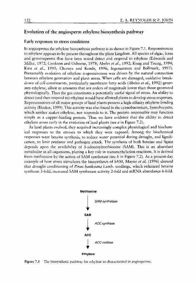
122 E. A. REYNOLDS &c P. JOHN
Evolution of the angiosperm ethylene biosynthesis pathway
Early responses to stress conditions
In angiosperms the ethylene biosynthesis pathway is as shown in Figure 7.1. Responsiveness to ethylene appears to be present throughout the plant kingdom. All species of algae, ferns and gymnosperms that have been tested detect and respond to ethylene (Edwards and Miller, 1972; Cookson and Osborne, 1978; Abeles et al, 1992; Kong and Yeung, 1994; Kwa et aL, 1995; Chernys and Kende, 1996; Ingemarsson and Bollmark, 1997). Presumably evolution of ethylene responsiveness was driven by the natural connection between ethylene generation and plant stress. When cells are damaged, oxidative breakdown of cell constituents, particularly membrane fatty acids (Abeles et al.^ 1992) generates ethylene, albeit in amounts that are orders of magnitude lower than those generated physiologically. Thus the gas constitutes a potentially useful signal of stress. An ability to detect (and then respond to) ethylene would have allowed plants to develop stress responses. Representatives of all major groups of land plants possess a high-affinity ethylene-binding activity (Bleeker, 1999). This activity was also found in the cyanobacterium, Synechocystis^ which neither makes ethylene, nor responds to it. The protein responsible may function simply as a copper-binding protein. Thus we have evidence that the ability to detect ethylene arose early in the evolution of land plants (see a in Figure 7.2).
As land plants evolved, they acquired increasingly complex physiological and biochemical responses to the stresses to which they were exposed. Among the biochemical responses were betaine synthesis, to reduce water potential during drought, and lignifi-cation, to limit predator and pathogen attack. The synthesis of both betaine and lignin depends upon the availability of S-adenosylmethionine (SAM). This is an abundant metabolite in all organisms, playing a key role in transmethylation reactions. It is derived from methionine by the action of SAM synthetase (see b in Figure 7.2). As a present-day example of how stress stimulates the biosynthesis of SAM, Mayne et al. (1996) showed that drought conditioning of Pinus banksiana Lamb, seedlings, which enhanced betaine synthesis 3-fold, increased SAM synthetase activity 2-fold and mRNA abundance 6-fold.
Methionine
SAM synthetase
SAM
ACC synthase
ACC
ACC oxidase
Ethylene
Figure 7.1 The biosynthetic pathway for ethylene as characterized in angiosperms.

EVOLUTION OF ETHYLENE SYNTHESIS 123
The role of ACC
SAM is also the precursor of ACQ which arises by the action of ACC synthase on SAM. ACC is detectable in representatives of all major groups of land plants (J. Hodson, unpublished data). ACC has been shown to be present in the ferns Regnellidium diphyllum Lindman (Chernys and Kende, 1996; Osborne et al.^ 1996) and Marsilea quadrifolia L., and notably, in M. quadrifolia it is generated by an ACC synthase with many of the properties of the angiosperm ACC synthase (Chernys and Kende, 1996). However, ACC is not used as the precursor of ethylene in these ferns (Cookson and Osborne, 1978; Osborne et al, 1996; Chernys and Kende, 1996). Thus we propose that ACC was accumulated in these non-seed plants without being used as a precursor of ethylene. Naturally occurring compounds, such as ACC, that contain a cyclopropane ring can have insecticidal, antimicrobial and neurochemical properties (Salatin and Baird, 1995), with ACC itself known to have neurochemical properties (Zapata et aL, 1996). These properties would have made ACC useful in the phytochemical armoury of the plant. Therefore, it is proposed that before ACC became the precursor of ethylene, it accumulated as a response to predator or pathogen attack. Like the other biochemical responses to stress, ACC accumulation would have resulted from enhanced rates of SAM generation (see b in Figure 7.2).
The acquisition of ACC oxidase
The key step in the evolution of the complete angiosperm ethylene biosynthetic pathway is the acquisition of the final stage: the ability to generate ethylene from ACC by the action of the enzyme ACC oxidase (Figure 7.3). ACC readily penetrates plant tissues and, therefore, in principle, a simple test for the presence of ACC oxidase is available by measuring the ethylene generated when plant tissues are presented with ACC. On the basis of
Methionine
b SAM synthetase
V betaine ^ SAM
lignin ^ b ACC synthase ^ stress
ACC
c I ACC oxidase I Ethylene M cell damage
a
Figure 7.2 The evolutionary sequence leading to the pathway for ethylene biosynthesis in angiosperms. a, in the earliest land plants, cell damage arising from stress leads to the formation of ethylene by chemical breakdown of cell constituents; b, in ferns and allied non-seed plants, stress leads to the increased expression of S-adenosylmethionine (SAM) synthetase for lignin and betaine synthesis; and to enhanced ACC synthase activity and the accumulation of ACC; c, with acquisition of ACC oxidase in a group of seed plants ancestral to the Gnetales, Pinopsida and angiosperms, ethylene is produced from ACC.

124 E. A. REYNOLDS &c P. JOHN
Figure 7.3 The reaction catalysed by ACC oxidase. 1-aminocyclopropane-l-carboxylate is oxidized to ethylene, and ascorbate is oxidized to dehydroascorbate. Fe(II) and CO2 act as cofactors.
Table 7.1 Distribution among gymnosperms of ACC oxidase activity measured in vitro (from data in Reynolds and John, 2000)
(angiosperms Pinopsida
Gnetales Ginkgoales Cycadales
Cucumis Pinus, Pseudotsuga
X Cupressocyparis Ephedra Ginkgo Cycas, Dioon^ Zamia
Leaf
Yes No
No No No
Germinated seed
Yes) Yes
Yes No No
such tests, Osborne (1989) proposed that ACC was used by seed plants, but not by ferns and mosses. After it became possible for ACC oxidase to be assayed in vitro (Ververidis and John, 1991), Reynolds and John (2000) demonstrated that among the extant seed plants, only angiosperms showed ACC oxidase activity in leaf material; activity was absent from gymnosperm leaf extracts. Moreover, among representatives of the four extant gymnosperm groups, ACC oxidase activity was present in seedlings of the Pinopsida and Gnetales, but absent from seedlings of the Ginkgoales and Cycadales (Table 7.1). As a representative of gymnosperm ACC oxidases, the enzyme from Pinus nigra var. nigra Arnold was shown to resemble biochemically the angiosperm enzyme, including a requirement for ascorbate, CO2 and Fe(II) (Reynolds and John, 2000). The simplest explanation of these findings was that ACC oxidase arose in a common ancestor of the three groups: angiosperms, Gnetales and Pinopsida, with the Ginkgoales and Cycadales basal to this proposed origin (Reynolds and John, 2000).
Our finding that ACC oxidase was strongly represented in germinated seeds of certain gymnosperms, but absent from leaf tissues of all gymnosperms tested (Table 7.1), led us to suggest (Reynolds and John, 2000) that the earliest role played by ACC oxidase may have been associated with germination, and only later in the evolution of angiosperms did it expand its repertoire of functions to leaves, and to the distinctively angiosperm organs of flowers and fruits. However, this suggestion must remain tentative until more is known of the relationship between ethylene production and germination (Petruzzelli et al.^ 2000;

EVOLUTION OF ETHYLENE SYNTHESIS 125
Matilla, 2000). Even in the relatively well-studied angiosperms 'the role of ethylene in germination remains controversial. Some authors hold that (it) is a consequence of the germination process, while others contend that ethylene production is a requirement for germination' (Matilla, 2000). Among the gymnosperms, nothing is known of the role of ethylene in germination.
Chemistry was on the side of ACC becoming the ethylene precursor in more ways than one. The cyclopropane ring of ACC is readily broken, with the release of ethylene, by oxidative reactions, such as the Fenton reaction in which hydrogen peroxide formed by the action of Fe(II) on ascorbate acts as the oxidizing agent (McRae et aL, 1983). ACC oxidase uses Fe(II) and CO2 as cofactors and ascorbate as a cosubstrate to catalyse an accelerated and controllable conversion of ACC to ethylene. Additionally, one of the other products generated from ACC by ACC oxidase is cyanide, which may have helped the plant defend itself against attack (Grossman, 1996).
In the preceding we have identified the acquisition of ACC oxidase among the gymno-sperm group as being the critical step in the evolutionary development of the biosynthetic pathway of ethylene biosynthesis present in angiosperms (see c in Figure 7.2). The production of ethylene from ACC meant that ethylene production now became a signal that was related to the response of the plant to the stress, as opposed to (or in addition to) ethylene production as a signal of plant stress per se. In angiosperms all three enzymes in the pathway from methionine to ethylene increase activity in response to stress (see Figure 7.2). Similarly in the non-angiosperms, in which ACC is not a precursor of ethylene, the activities of SAM synthetase (Mayne etaL, 1996) and ACC synthase (Chernys and Kende, 1996) increase in response to stress. The acquisition of ACC oxidase can thus be viewed as taking ACC from the relative obscurity of a secondary metabolite involved in stress responses, to being a key intermediate in the biosynthesis of a major plant growth regulator.
Two characteristic features of the angiosperm ACC oxidase assayed in vitro are: (1) a low specific activity of the purified enzyme compared with other 2-ODDs (John, 1994); and (2) a non-linear reaction rate due to progressive inactivation during catalysis (Smith et al., 1994). It is not known whether these are features of the enzyme activity in vivo; they may be attributable to the in vitro conditions used for assay. However, it has been noted that in ripening fruits the enzyme is present in large amounts relative to the amount of ethylene formed: in apple, ACC oxidase has been estimated to constitute 5% of the total soluble protein (Abeles et al.^ 1992). Enzymes involved in secondary metabolism commonly show high abundance where the enzyme has acquired an important function (Pichersky and Gang, 2000). The rationale for this high abundance is that the molecular changes required to increase expression of a protein are simpler and more feasible than those required to increase the rate of substrate turnover or enhance stability (Pichersky and Gang, 2000). Thus we suggest that ACC oxidase, as the 'newest recruit' to the ethylene biosynthesis pathway, compensates for its low catalytic efficiency by a high level of expression.
H o w A C C oxidase originated
ACC oxidase as a 2-oxoacid-dependent dioxygenase
Having identified the acquisition of ACC oxidase as the critical step in the evolutionary development of the ethylene biosynthetic pathway of angiosperms, we now turn to the evolutionary origin of the enzyme itself. What was the ancestral enzyme? What molecular gymnastics did it undergo to result in ACC oxidase activity?
126 E. A. REYNOLDS &: P, JOHN
By similarity of primary sequence and function, ACC oxidase belongs to the non-heme Fe(II), 2-oxoacid dependent dioxygenase (2-ODD) family of enzymes (Prescott and John, 1996). It is to these enzymes that we must turn when considering the possible evolutionary ancestor of ACC oxidase. The 2-ODDs are a large and important class of enzymes that catalyse a wide variety of oxidative reactions, including hydroxylation, desaturation and epoxidation (Prescott, 2000). Known 2-ODDs in plants are involved in a variety of pathways. Commonly, more than one 2-ODD is found in a biosynthetic pathway, notably those involving alkaloids, gibberellins and flavonoids. In Arabidopsis Heynh. it is estimated (Prescott, 2000) that there are about 100 genes that code for 2-ODD proteins, but it is only for a minority that any function can be ascribed.
The presence in specific plant groups of 2-ODDs catalysing particular reactions can be assumed on the basis of the presence of particular biochemical products. Flavonoids are present in mosses and higher orders; gibberellins in ferns and higher orders; and antho-cyanins in gymnosperms and angiosperms (Koes et ai, 1994). On the basis of this, it can be inferred that, as a first approximation, the evolutionary sequence in which 2-ODDs have appeared is: (1) flavonoid enzymes, (2) gibberellin enzymes, (3) anthocyanin enzymes.
Molecular changes
Protein sequence considerations have been used to identify the gross molecular changes required in an evolutionary transition from a 2-ODD ancestor to a functional ACC oxidase. Multiple alignment of all available ACC oxidase sequences revealed that only 30% of the residues are functionally conserved in all sequences examined (Reynolds, 2001). Of the 95 conserved residues, 36 are also conserved in related 2-ODDs (Reynolds, 2001). Thus one can define ACC oxidase at a primary sequence level. However, these conserved sequences are more usefully interpreted in a functional context. In its enzymatic action, ACC oxidase differs from other 2-ODDs not only in its primary substrate specificity, but also in using ascorbate as a cosubstrate (rather than 2-oxoglutarate) and in using CO2 as an essential cofactor (see Figure 7.3). To account for these unique functional features of ACC oxidase, the protein alignments must be viewed in terms of protein structure. The 3-D structure of ACC oxidase is unknown, but a structure for the microbial 2-ODD, isopeni-cillin N synthase (IPNS) is available (Roach et aL, 1995). There is sufficient sequence homology between IPNS and ACC oxidase for the IPNS structure to provide a basis for structural features of the ACC oxidase protein to be recognized (Roach et aL, 1995).
A summary of secondary structure predictions for ACC oxidase is shown in Figure 7.4. The most extensive region of conservation in ACC oxidase is in the centre of the protein from Pro^^^ to Pro^^^ (Figure 7.4). This region also shares most similarity with other 2-ODDs. Based on homology with IPNS, this domain probably constitutes the jellyroll motif (Stuart, 1993) of eight jS-sheets, containing the active site of the protein. Conserved residues within this catalytic domain will provide the binding sites for Fe(II), oxygen, ACC and probably ascorbate (Roach et aL, 1995).
The region of ACC oxidase that diverges most from other 2-ODDs is the C-terminus (see Figure 7.4). This region can therefore be considered diagnostic for ACC oxidases compared with other 2-ODDs. The C-terminal region beginning at Tyr^^^ contains 14 conserved residues, none of which is conserved in related 2-ODDs. Secondary structure prediction indicates that the C-terminal part of the protein forms a domain consisting of two a-helixes connected to the body of the protein by a loop section. This loop section is a poorly conserved region of ACC oxidase which can be up to 16 residues in length.
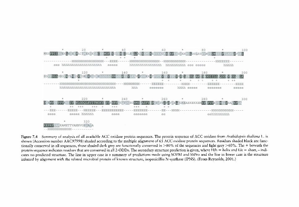
• nHI-;H!-[H[-ii;MHHF[MHIiH FinFE - - - HHFHHHI-;i{!lIIliH;!IIH HUHHUHHHHHH • • - - - - - - - - I-iHEhihic:!-- •
* i ; : ! i * 14 0 * I ' i u * i H o * ::I:JI:I
SL) igjiL'DD i^TL^ rig '-.K |fc F, E §jj | Q vgY Kpg'r 2 S gMgog g i ^jj S WeVAiJn ssaa 2 s j 35 i I I I I ! • 1 1 + -i -t-
• • - HHHHIIIiI i l i I imii l l IHIi i ; t lHIIl i i IIII iI[ l l III i l - i liilHI-inHHH E E E E E E - - - i n H i l - ; I % I ' ; F : E E : E - - • - - - E E E E
h : i | i h l i : i h l i l i i ! l i h h i i ! i F i t i i i i i F i ! i i i i i , h l i l i h l i l i i m e e e e - e e h i i l i i i ••^••.,(••.•• 'jeo-ieee eeeee
S E 2 2 i3^K EE i!! <tgi'M*=i*><<»Heti4'< ^ E I J J ^ S T E.;;i2JS ^if33i g s svQpgv Ejl GKEAEI:EKKEN23 J ^ E Ji QSA ^ •f + -t- I -1 1 4 1 )• t t 1 -f I + + -f t
--•EEE E E E E E - H E E E E E • E E E E E E F F E E E - - • EEEEEEE EE- UHl-IH • - - HHHHHHHHHHH •-
- ^ ^ - ,- ,= t.e hhlihlils , ef=e eeeeeee ^e eehiilihhlihhli
* 3 2 0
laaaaa I-:AMEI"FVANNV<;FP A A innniiniHHHiiHH • •
Figure 7.4 Summary of analysis of all available ACC oxidase protein sequences. The protein sequence of ACC oxidase from Arabidopsis thaliana L. is shown (Accession number AAC97998) shaded according to the multiple aUgnment of 65 ACC oxidase protein sequences. Residues shaded black are functionally conserved in all sequences, those shaded dark grey are functionally conserved in >80% of the sequences and light grey >60%. The + beneath the protein sequence indicates residues that are conserved in all 2-ODDs. The secondary structure prediction is given, where H/h = helix and E/e = sheet, - indicates no predicted structure. The line in upper case is a summary of predictions made using SOPM and SSPro and the line in lower case is the structure inferred by alignment with the related microbial protein of known structure, isopenicillin N-synthase (IPNS). (From Reynolds, 2001.)
128 E. A. REYNOLDS &c P. JOHN
The high degree of sequence conservation in the C-terminus region of ACC oxidase suggests that it is important functionally. ACC oxidase is unique among 2-ODDs in its requirement for CO2 as a cofactor. The C-terminus region of ACC oxidase is rich in positively charged residues, such as arginine and lysine, which are potential binding sites for CO2. Site-directed mutagenesis of ACC oxidase has provided evidence for the location of the CO2 binding site at a conserved arginine residue in the C-terminus region: Arg^^^ in ACC oxidase from kiw i fruit (Lay et ai, 1996); Arg^^^ in ACC oxidase from apple fruit (Kadyrzhanova et ai, 1999). Thus v e propose that acquisition of the C-terminal domain, by an ancestral 2-ODD v^as crucial in the step change in catalytic function to ACC oxidase, and it coincided with the requirement for CO2 as a cofactor.
In the evolutionary acquisition of ACC oxidase activity, the ability to bind and turn over the novel substrate ACC was acquired, but also, we assume that ascorbate took the place of 2-oxoglutarate as cosubstrate. This latter assumption is strengthened by the finding (Iturriagagoitia-Bueno etal., 1997) that 2-oxoglutarate, and other 2-oxo acids, inhibit ACC oxidase activity competitively with respect to ascorbate. Fe(II) chelation was excluded as an explanation of the action of 2-oxoglutarate, and it was concluded that ascorbate and 2-oxoglutarate were competing for the same binding site (Iturriagagoitia-Bueno^^^/., 1997).
The enzyme ancestral to ACC oxidase
Prescott (2000) has created a phylogenetic tree of 64 2-ODDs from Arabidopsis (Figure 7.5). In this phylogeny, the three Arabidopsis ACC oxidases appear as terminal branches of a subgroup of five proteins, the other members of which are uncharacterized. The proteins of known function most closely related to ACC oxidases are those of the flavonoid pathway. They, and the ACC oxidases, appear to diverge from a common origin (Figure 7.5), On this basis we suggest that ACC oxidases evolved from an ancestral 2-ODD that was involved in flavonoid biosynthesis.
Flavonoids and ACC do not appear to have much in common chemically and further information is needed before our suggestion can be substantiated. However, in support of our suggestion, we note that 2-ODDs can show a relatively loose substrate specificity (Prescott and John, 1996; Prescott, 2000). This substrate flexibility is shown by the 2-ODDs in alkaloid, antibiotic and gibberellin synthetic pathways catalysing more than one reaction; ACC oxidase itself can convert D-valine to /so-butanal (Gibson et al.^ 1998). We envisage that the substrate flexibility of 2-ODDs played a part in the early stages of the evolutionary development of the ACC oxidase.
When did the ethylene biosynthesis pathway arise?
Palaeoclimatic considerations
Although seed plants may have first appeared during the Devonian period, it was during the relatively dry Jurassic period that they diversified extensively and began to dominate (Willis and McElwain, 2002). Ethylene diffuses 10"* times faster in air than in water (Musgrave et aL, 1972). Thus, in general, ethylene would have been a more useful signalling compound after the end of the relatively wet Devonian and Carboniferous periods, as suggested by Osborne et al. (1996). It is pertinent to note that the submerged aquatic angiosperm Potamogeton pectinatus L. is incapable of producing ethylene because it lacks
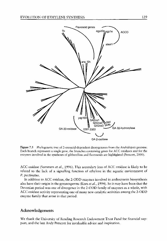
EVOLUTION OF ETHYLENE SYNTHESIS 129
agp20eagp1e \ ACCO
GA 3p-hydroxylase
v ^ GA 2-oxidase
Figure 7,5 Phylogenetic tree of 2-oxoacid-dependent dioxygenases from the Arabidopsis genome. Each branch represents a single gene, the branches containing genes for ACC oxidases and for the enzymes involved in the syntheses of gibberellins and flavonoids are highlighted (Prescott, 2000).
ACC oxidase (Summers et aL, 1996). This secondary loss of ACC oxidase is likely to be related to the lack of a signalling function of ethylene in the aquatic environment of P. pectinatus.
In addition to ACC oxidase, the 2-ODD enzymes involved in anthocyanin biosynthesis also have their origin in the gymnosperms (Koes et al., 1994). So it may have been that the Devonian period v^as one of divergence in the 2-ODD family of enzymes as a whole, vs ith ACC oxidase activity representing one of many new catalytic activities among the 2-ODD enzyme family that arose in that period.
Acknowledgements
We thank the University of Reading Research Endowment Trust Fund for financial support, and the late Andy Prescott for invaluable advice and inspiration.

130 E. A. REYNOLDS &c P. JOHN
References
Abeles FB, Morgan PW, Saltweit ME. 1992. Ethylene in Plant Biology. San Diego: Academic Press. Bleeker AB. 1999. Ethylene perception and signalling: an evolutionary perspective. Trends in Plant
Sciences 4: 269-274. Chernys J, Kende H. 1996. Ethylene biosynthesis in Regnellidium diphyllum and Marsilea quadri-
folia. Planta 200: 113-118. Cookson C, Osborne DJ. 1978. The stimulation of cell extension by ethylene and auxin in aquatic
plants. Planta 144: 39-47. Edwards ME, Miller JH. 1972. Grow^th regulation by ethylene in fern gametophytes II. Inhibition
of cell division. American Journal of Botany 59: 450-457. Gibson EJ, Zhang Z, Baldwin J, Schofield CJ. 1998. Substrate analogues and inhibition of ACC
oxidase: Conversion of D-valine to /so-butanal. Phy to chemistry 48: 619-624. Grossman K. 1996. A role for cyanide, derived from ethylene biosynthesis, in the development of
stress symptoms. Physiologia Plantarum 97: 771-775. Ingemarsson BSM, Bollmark M. 1997. Ethylene production and 1-aminocyclopropane-l-carboxylic
acid turnover in Picea abies hypocotyls after wounding. Journal of Plant Physiology 151: 711-715. Iturriagagoitia-Bueno T, Gibson EJ, Schofield CJ, John P. 1997. Inhibition of 1-aminocyclo-
propane-1-car boxy late oxidase by 2-oxoacids. Phy to chemistry 43: 343-349. John P. 1994. Oxidation of 1-aminocyclopropane-l-carboxylic acid (ACC) in the generation of
ethylene by plants. In: Wallsgrove R, ed. Amino Acids and their Derivatives in Higher Plants -Biosynthesis and Metabolism. SEB Seminar series. Cambridge: Cambridge University Press, 51-57.
John P. 1997. Ethylene biosynthesis: The role of 1-amino-cyclopropane-l-carboxylate ACC oxidase, and its possible evolutionary origin. Physiologia Plantarum 100: 583-592.
John P, Iturriagagoitia-Bueno T, Lay V, et al. 1997. 1-aminocyclopropane-l-carboxylate oxidase: molecular structure and catalytic function. In: Kanellis AK, Chang C, Kende H, Grierson D, eds. Biology and Biotechnology of the Plant Hormone Ethylene. Dordrecht: Kluwer Academic Publishers, 15-21.
John P, Reynolds EA, Prescott AG, Bauchot AD. 1999. ACC oxidase in the biosynthesis of ethylene. In: Kanellis AK, Chang C, Klee H, et al. eds. Biology and Biotechnology of the Plant Hormone Ethylene II. Dordrecht: Kluwer Academic Publishers, 1-6.
Kadyrzhanova D, McCuly TJ, Warner T, et al. 1999. Analysis of ACC oxidase activity by site directed mutagenesis of conserved amino acid residues. In: Kanellis AK, Chang C, Klee H, et al., eds. Biology and Biotechnology of the Plant Hormone Ethylene II. Dordrecht: Kluwer Academic Publishers, 7-12.
Koes RE, Quattrochio F, Moi JNM. 1994. The flavonoid biosynthetic pathway in plants: Function and evolution. BioEssays 16: 123-132.
Kong LS, Yeung EC. 1994. Effects of ethylene and ethylene inhibitors on white spruce somatic embryo maturation. Plant Science 104: 71-80.
Kwa S-H, Wee Y-C, Kumar PP. 1995. Role of ethylene in the production of sporophytes from Platycerium coronarium Koenig Desv. frond and rhizome pieces cultured in vitro. Journal of Plant Growth Regulation 14: 183-189.
Lay VJ, Prescott AG, Thomas PG, John P. 1996. Heterologous expression and site-directed mutagenesis of the 1-aminocyclopropane-l-carboxylate oxidase from kiwi fruit. European Journal of Biochemistry 141: llS-134.
Matilla AJ. 2000. Ethylene in seed formation and germination. Seed Science Research 10: 111-126. Mayne MB, Coleman JR, Blumwald E. 1996. Differential expression during drought conditioning
of a root-specific 5-adenosylmethionine synthetase from jack pine (Pinus banksiana Lamb.) seedlings. Plant, Cell and Environment 19: 958-966.
McRae DG, Cober JA, Legge RL, Thompson JE. 1983. Bicarbonate C02-facilitated conversion of 1-aminocyclopropane-l-carboxyIic acid to ethylene in model systems and intact tissues. Plant Physiology 35: 426-432.
Musgrave A, Jackson MB, Ling E. 1972. Callitriche stem elongation is controlled by ethylene and gibberellin. Nature New Biology 238: 93-96.
EVOLUTION O F ETHYLENE SYNTHESIS 131
Osborne DJ. 1989. The control role of ethylene in plant growth and development. In: CHdjsters H, De Proft M, Marcelle R, Van Poucke M, eds. Biochemical and Physiological Aspects of Ethylene Production in Lower and Higher Plants. Dordrecht: Kluwer Academic Publishers, 1-11.
Osborne DJ, Walters J, Milborrow BV, et al. 1996. Evidence for a non-ACC ethylene biosynthesis pathway in lower plants. Phy to chemistry 42: 51-60.
Petruzzelli L, Coraggio I, Leubner-Metzger G. 2000. Ethylene promotes ethylene biosynthesis during pea seed germination by positive feedback regulation of 1-aminocyclo-propane-l-carboxylic acid oxidase. Planta 111: 144-149.
Pichersky E, Gang RG. 2000. Genetics and biochemistry of secondary metabolites in plants: an evolutionary perspective. Trends in Plant Science 5: 439-445.
Prescott AG. 2000. Two-oxoacid-dependent dioxygenases: Inefficient enzymes or evolutionary driving force? In: Romeo JT, Ibrahim R, Varin L, DeLuca V, eds. Evolution of Metabolic Pathways. Amsterdam: Pergamon Press, 249-284.
Prescott AG, John P. 1996. Dioxygenases: molecular structure and role in plant metabohsm. Annual Review of Plant Physiology and Plant Molecular Biology 47: 245-271.
Reynolds EA. 2001. Studies on the evolution of the ethylene forming enzyme: 1-aminocyclo-propane-1-carboxylate (ACC) oxidase. Unpubhshed PhD Thesis, The University of Reading.
Reynolds EA, John P. 2000. ACC oxidase is found in seedhngs of two (Coniferales, Gnetales) of the four gymnosperm orders. Physiologia Plantarum 110: 38-41.
Roach PL, Chfton IJ, Fulop W, et al. 1995. Crystal structure of isopenicillin-N-synthase is the first from a new structural family of enzymes. Nature 375: 700-704.
Salaiin J, Baird MS. 1995. Biologically active cyclopropanes and cyclopropenes. Current Medicinal Chemistry 2: 511-542.
Smith JJ, Zhang ZH, Schofield CJ, et al. 1994. Inactivation of 1-aminocyclopropane-l-carboxylate ACC oxidase. Journal of Experimental Botany 45: 521-527.
Stuart D. 1993. Viruses. Current Opinion in Structural Biology 3: 167-174. Summers JE, Voesenek LACJ, Blom CWPM, et al. 1996. Potamogeton pectinatus is constitutively
incapable of synthesizing ethylene and lacks 1-aminocyclopropanel-carboxylic acid oxidase. Plant Physiology 111: 901-908.
WiUis KJ, McElwain JC. 2002. The Evolution of Plants. Oxford: Oxford University Press. Ververidis P, John P. 1991. Complete recovery in vitro of ethylene forming enzyme activity.
Phy to chemistry 30: 715-717. Zapata A, Capdevila JL, Viu E, Trullas R. 1996. 1-aminocyclopropane-carboxylic acid reduces
NMDA-induced hippocampal neurodegeneration in vivo. NeuroReport 7: 397-400.
8 Structural biomacromolecules in plants:
what can be learnt from the fossil record?
Pirn F van Bergen, Peter Blokker, Margaret E Collinson, Jaap S Sinninghe Damste and Jan W de Leeuw
CONTENTS
Introduction
Characterizing resistant biomacromolecules
Resistant biomacromolecules in outer coverings Algal cell walls Pollen and spore walls Higher land plant leaf and stem cuticles
Inner structural entities Water-conducting and strengthening tissues
Conclusions
References
133
134
136 136 138 143
146 146
150
150
Introduction
When plants entered land they needed specific physiological adaptations to survive this nev^, hostile environment. Some of the main problems plants had to overcome included v^ater loss or desiccation and exposure to increased UV radiation (Edw^ards, 2001). An increase in phenolic constituents in various plant tissues is believed to be one of the main adaptations to counteract the problem vs ith UV radiation (Rozema et al, 2001). With respect to desiccation, tv^o important adaptations are suggested, i.e. (1) increased protection of the outer covering of the plant to prevent direct w^ater loss and (2) enhanced water transport w ithin the plant. The first aspect is directly related to the development of the cuticle on leaves and stems and resistant walls on the plant's reproductive tissues (i.e. spores), while the second relates to the formation of water-conducting elements such as tracheids and vessels.
Evidence for the evolution of these distinct structures is based primarily on morphological observations of plant remains dating as far back as the Ordovician for cuticles and distinct spore material (i.e. tetrads, dyads and monads) and the Middle Silurian for tracheids (Edwards, 2001 and references cited therein). In terms of evolution, it has been
The Evolution of Plant Physiology Copyright © 2004 Elsevier Ltd ISBN 0-12-33955-26 All rights of reproduction in any form reserved

134 P. F. VAN BERGEN ET AL.
suggested that spore walls may have evolved from algal cell w^alls (Hemsley, 1994), whereas the direct origin of the cuticle and tracheids is still unknown (Edwards, 2001).
Apart from evidence for the morphological evolution of these entities, many studies link microscopic observations directly to biochemical evolution in these structures enabling plants to colonize land. Commonly, the presence of fossil cuticles is suggested to be directly related to the occurrence of the biopolyester cutin (e.g. Edwards, 2001; Cooper-Driver, 2001), whereas the findings of fossil water-conducting tissues such as tracheids in xylem are often indirectly related to the biomacromolecule lignin (e.g. Edwards et ai, 1997; Edwards, 2001; Cooper-Driver, 2001). In addition, acid resistant fossil spore wall material is suggested mostly to be composed of or impregnated by the, yet chemically poorly defined, biomacromolecule sporopoUenin (Cooper-Driver, 2001; Edwards, 2001). Additional resistant macromolecular compounds mentioned in connection with fossil plant remains are cutan in cuticles and algaenan in algal cell walls.
Despite the circumstantial evidence that the chemical compositions of fossil plant remains were very similar to those occurring in modern plant cuticles, spore and pollen walls or xylem elements, to date no unequivocal molecular chemical evidence exists proving that this was actually the case. The search for such evidence is complicated by a number of aspects including: (1) the chemically resistant nature of these, mainly macromolecular, entities in modern plants; (2) the intimate association of these molecules with other, often more abundant, compounds preventing purification of the specific biomacromolecules and thus hampering the elucidation of the detailed molecular composition of the extant counterparts (i.e. sporopoUenin); (3) the biological and chemical transformation reactions occurring upon burial and subsequent fossilization (i.e. diagenesis); and (4) the inconsistent use of nomenclature for the various resistant macromolecules. Note that the term 'resistant macromolecules' refers to those compounds that are resistant against normal chemical treatments but are destroyed by chemical oxidation. This chemical resistance is believed to be the main underlying reason for their high preservation potential under most, but not all, depositional settings.
The following terms will be used to avoid confusion: algaenans for the chemically resistant macromolecules present in algal cell walls (including the walls of algal zygospores); SporopoUenin is the chemically resistant macromolecule in spore and pollen walls of non-algae excluding fungal spores; cutan is the highly aliphatic macromolecule recognized in modern and fossil plant cuticles; and lignin will only be used for the ether-linked macromolecule based on monolignols, i.e. /?-coumaryl alcohol, coniferyl alcohol and sinapyl alcohol. Terms such as 'lignin-like' and 'sporopoUenin-like' should be avoided.
In this chapter we will first describe the main chemical methods that can provide detailed molecular insight into the chemical composition of both extant and fossil resistant (bio)macromolecules. Subsequently, the macromolecular composition of modern and fossil outer coverings, including algal cell walls, spore and pollen walls and cuticular tissues will be discussed both in terms of their chemical similarities and differences and in relation to the physiological adaptations and evolution of land plants. Finally, the chemical composition of water-conduction tissues of modern and fossil land plants will be evaluated also in terms of their physiology but with specific emphasis on the biomacromolecule lignin.
Characterizing resistant biomacromolecules
In order to study chemically resistant fossil (bio)macromolecules one has to have detailed molecular insight into their modern counterparts. In most cases, the modern counterparts

STRUCTURAL BIOMACROMOLECULES IN PLANTS 135
Figure 8.1 Example of purified algal cell walls of modern Tetraedron minimum obtained by Potter homogenization. For chemical analyses of this sample the reader is referred to Blokker et al. (1998).
can only be isolated by purification steps involving both physical and chemical separation methods. Mechanical stripping of cuticular membranes from leaf material (Mosle et aL, 1997) and ultrasonic destruction to obtain purified algal cell wall material (Potter homogenization; Blokker et al., 1998; Figure 8.1) are effective physical pretreatments.
Numerous chemical extraction methods exist to obtain the most resistant organic molecules in plant remains. Although it should be emphasized that the organic remains recognized in the fossil record are not necessarily identical to the resistant material that one obtains after purification and extraction steps under experimental conditions. Normally a full extraction method involves the following steps:
1. 2.
3. 4.
solvent extraction to eliminate low molecular weight lipids hydrolysis, either with base and/or acid, to remove ester-bound/amide-bound moieties; this treatment often consists of two sequential steps, a base hydrolysis and an acid hydrolysis acid treatment using H2SO4 to remove polysaccharides an ultimate base treatment to remove moieties that have become accessible after the previous steps {cf. the results of cutan isolation from Agave L. in Tegelaar et aL (1989: Figure 17) and the same sample after this final base treatment in de Leeuw et al. (1991: Figure 6) revealing unequivocally that the polysaccharides are not part of the resistant molecule in cuticles!).
Flowever, it should be noted that some chemical pretreatments that are used to mimic fossilization processes (i.e. acetolysis) might drastically affect the chemical composition of extant material under examination. A prime example in this respect relates to the study of sporopoUenin whereby the most commonly used treatment involves acetolysis (Graham and Gray, 2001; Hemsley et aL, 1996). Although the remains obtained after this treatment are morphologically identical to those found in the fossil record, detailed molecular information based on ^^C NMR revealed that the chemical composition of sporopoUenin from pine pollen was severely altered (Flemsley et aL, 1996). Furthermore, Allard et al. (1997) showed that newly formed condensation products (i.e. melanoidin-like polymers derived from cell wall polysaccharides), that are not related to the resistant entities originally present, can easily be produced. Appropriate modified isolation procedures can avoid such problems (Allard et al., 1998).

136 P. F. VAN BERGEN ET AL,
The various chemical methods that are used to study the resistant biomacromolecules can be subdivided into non-destructive and destructive techniques. The former includes solid state ^^C NMR and FTIR, both of which reveal information pertaining to the various carbon environments present (e.g. Hatcher et aL, 1999) and have revealed very informative data on lignin, cutan, algaenan and sporopoUenin. Pyrolysis and chemolysis are both destructive methods that will reveal detailed molecular information about the specific building blocks (e.g. van Bergen, 1999). Pyrolysis is often used as an effective initial screening method as it reveals qualitative insights into the overall chemical composition of the macromolecule. Pyrolysis has been used on most resistant biomacromolecules (i.e. lignin, cutan, algaenan, sporopoUenin). For further information about the various pyrolysis methods, the reader is referred to Stankiewicz et al. (1998) and van Bergen (1999). The advantage of chemolysis is that one can target specific molecular building blocks using specific chemical reagents. Chemolysis is often used in combination with pyrolysis as the latter reveals characteristic moieties that subsequently can be examined using appropriate reagents (van Bergen et al., 1994a; Gelin et al, 1997; Blokker et al, 1998). The main specific reagents or methods used nowadays include treatment with CuO, RUO4, HI, KMn04 or thioacidolysis. For lignin the most commonly used methods include treatment with CuO and, more recently, thioacidolysis, for cutan RUO4, for algaenan RUO4 and HI and for sporopoUenin KMn04, NO^.
As both the destructive and non-destructive methods can yield biased results, a combination of these techniques should be used when possible, as this will provide greater insight into the composition of the biomacromolecule under investigation. Furthermore, it is always very instructive to undertake microscopic examinations prior to and after chemical treatments (Boon et al., 1989; van Bergen et al., 1995; Blokker et al., 2001; Collinson et al., 1998) as this can reveal where the actual resistant organic material resides in the plant organ. Also histochemical staining can reveal specific insight into the tissues that are preserved containing resistant macromolecules (van Bergen, 1994). However, a colouri-metric method in itself does not prove the presence of one characteristic molecule as was shown in a study on modern and fossil chitin in arthropod cuticles (Bierstedt et al., 1998). In particular with respect to fossil material specific histochemical colours cannot be used as chemical evidence for the presence of a certain macromolecule.
Resistant biomacromolecules in outer coverings
Algal cell w alls
The abundant contribution of algal cell walls in many palynological microspore assemblages and coals (i.e. Botryococcus Kutzing in boghead) has always been the main reason to suggest that these structures in the modern counterparts should contain a specific chemically resistant material. Subsequent chemical investigations, focusing initially on modern Botryococcus (Largeau et al., 1986) and modern Tetraedron Kutzing (Goth et al., 1988), did indeed reveal the presence of distinct types of biomacromolecules nowadays called algaenans.
Recent studies on a number of modern freshwater and marine microalgae have shown that some, but not all, contain a resistant molecule in their cell walls (Gelin et al., 1999; Blokker, 2000). To date, all algaenans obtained from modern algae are based on, so-called, aliphatic building blocks. However, large differences do exist. The algaenan obtained from Botryococcus is very different from those of all other algae studied to date (i.e. based on
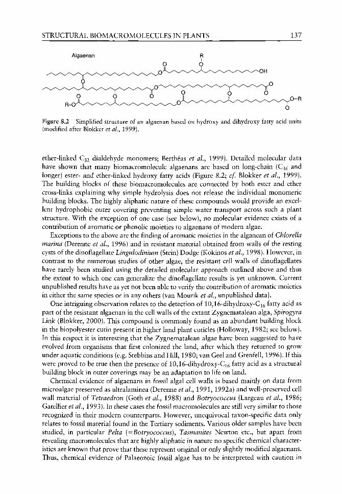
STRUCTURAL BIOMACROMOLECULES IN PLANTS 137
Algaenan
,0-R
Figure 8.2 Simplified structure of an algaenan based on hydroxy and dihydroxy fatty acid units (modified after Blokker et al, 1999).
ether-linked C32 dialdehyde monomers; Bertheas et al.^ 1999). Detailed molecular data have shown that many biomacromolecule algaenans are based on long-chain (€35 and longer) ester- and ether-linked hydroxy fatty acids (Figure 8.2; cf. Blokker et aL, 1999). The building blocks of these biomacromolecules are connected by both ester and ether cross-links explaining why simple hydrolysis does not release the individual monomeric building blocks. The highly aliphatic nature of these compounds would provide an excellent hydrophobic outer covering preventing simple water transport across such a plant structure. With the exception of one case (see below), no molecular evidence exists of a contribution of aromatic or phenolic moieties to algaenans of modern algae.
Exceptions to the above are the finding of aromatic moieties in the alganean of Chlorella marina (Derenne et aL, 1996) and in resistant material obtained from walls of the resting cysts of the dinoflagellate Lingulodinium (Stein) Dodge (Kokinos et al., 1998). Flowever, in contrast to the numerous studies of other algae, the resistant cell walls of dinoflagellates have rarely been studied using the detailed molecular approach outlined above and thus the extent to which one can generalize the dinoflagellate results is yet unknown. Current unpublished results have as yet not been able to verify the contribution of aromatic moieties in either the same species or in any others (van Mourik et aL, unpublished data).
One intriguing observation relates to the detection of 10,16-dihydroxy-Ci6 fatty acid as part of the resistant algaenan in the cell walls of the extant Zygnematalean alga, Spirogyra Link (Blokker, 2000). This compound is commonly found as an abundant building block in the biopolyester cutin present in higher land plant cuticles (Holloway, 1982; see below). In this respect it is interesting that the Zygnematalean algae have been suggested to have evolved from organisms that first colonized the land, after which they returned to grow under aquatic conditions (e.g. Stebbins and Fiill, 1980; van Geel and Grenfell, 1996). If this were proved to be true then the presence of 10,16-dihydroxy-Ci5 fatty acid as a structural building block in outer coverings may be an adaptation to life on land.
Chemical evidence of algaenans in fossil algal cell walls is based mainly on data from microalgae preserved as ultralaminea (Derenne etaL, 1991,1992a) and well-preserved cell wall material of Tetraedron (Goth et ai, 1988) and Botryococcus (Largeau et aL, 1986; GatelUer et aL, 1993). In these cases the fossil macromolecules are still very similar to those recognized in their modern counterparts. However, unequivocal taxon-specific data only relates to fossil material found in the Tertiary sediments. Various older samples have been studied, in particular Pelta (=Botryococcus), Tasmanites Newton etc., but apart from revealing macromolecules that are highly aliphatic in nature no specific chemical characteristics are known that prove that these represent original or only slightly modified algaenans. Thus, chemical evidence of Palaeozoic fossil algae has to be interpreted with caution in

138 P. F. VAN BERGEN ET AL.
relation to biochemical and physiological evolution. Even Tertiary material that is composed of rich monotypic algal cell v^all material can be altered or interpreted incorrectly due to additional non-algal organic material that overprints the chemical signal. For example, chemical data from Late Miocene lacustrine oil shales containing up to 80% Pediastrum Meyen were used to suggest that the algaenan of this alga was mainly aromatic in nature (Sinninghe Damste et al., 1993). Subsequent analyses of modern Pediastrum cell walls revealed that the algaenan is based on C30 and C32 a>-hydroxy fatty acids clearly revealing its aliphatic nature similar to that of other Chlorococcales (Blokker et aL, 1998).
Pyrolysis data from a number of Tertiary, Palaeocene and Eocene, dinoflagellate cyst walls have revealed that these contain mainly aliphatic building blocks, although samples that appear to be more mature, based on colour, reveal an increase of aromatic pyrolysis products (Warnaar et ai, unpublished results). However, as no chemical data from modern counterparts are available yet, these data and, in particular, the significance of an aromatic contribution to algaenans, cannot be interpreted in terms of their original chemical composition or physiological differences.
Another distinct microfossil, without a direct modern counterpart, is Gloeocapsomorpha prisca Zalessky. The fossil remains of this organism constitute the bulk of the organic matter in Ordovician Kukersites (Foster et aL, 1989, 1990). Transmission electron microscopy revealed that the walls are composed of multiple sheets surrounding a void (Derenne et aL, 1992b; Blokker et al., 2001). This distinct morphology, in combination with chemical data, means that the precise biological affinity of this organism is still unknown, although an algal or a bacterial affinity appears most likely (see Blokker et al,^ 2001 and references cited therein). Based on the extremely low maturity of the Kukersite rocks it is believed that the organic matter preserved has not been altered significantly, which is an essential prerequisite for further meaningful interpretations. The building blocks of the resistant macromolecule present in the fossil remains of this organism are based on 5-w-alkyl-resorcinols with the alkyl side chain being mainly C15, C17 and C19 (Derenne et al, 1992b; Blokker et ai, 2001). If this organism were a true alga then this would be the first algaenan type containing 1,3-benzenediol (=resorcinol) building blocks (Metzger and Largeau, 1994). But if its closest biological affinity lies with the bacteria then the benzenediol contribution would be less surprising as these are known to produce resorcinols upon stress (Kozubek and Tyman, 1999). From a physiological point of view, however, the biological affinity is of minor importance as geological information has suggested that these organisms lived in a high UV light lagoonal or very near-shore environment (Foster et al., 1990). This would probably induce the production of phenolic, i.e. resorcinol, moieties to counteract possible UV-induced damage.
Overall, the current molecular data on modern algaenans show primarily biomacro-molecules based on long-chain (^€25) hydrophobic moieties that most probably provide water repellent properties to the cell walls of these algae. Phenolic entities are not detected, but then one would not expect them in these aquatic organisms if their primary function was related to counteract high UV radiation levels.
Pollen and spore walls
In stark contrast to the algal material, spore and pollen walls are not protected constantly by an aqueous environment and thus will have been adapted to minimize water loss and to reduce UV damage. Studies of the chemical composition of the resistant material present in spore and pollen walls have a long history because acid resistant pollen and spores are

STRUCTURAL BIOMACROMOLECULES IN PLANTS 139
present in coals and other sedimentary rocks, dating as far back as the Ordovician (Edwards, 2001). This fossil record is believed to be due to the presence of a highly resistant macro-molecule, namely sporopoUenin. The term 'sporopoUenin' was first used by Zetzsche and Kalin (1931) to represent the structural constituent of the outer walls (exines) of pollen and spores from vascular plants that is resistant to non-oxidative chemical treatments. As stated above, here we will use this definition of sporopoUenin, excluding other remains such as algal and fungal spores, as well as avoiding the term 'sporopollenin-like'. However, we do include spore wall material from Lycopodium clavatum L. which has previously been mistaken for a marine algae (Bestougeff et al,^ 1985).
Although earlier chemical studies had been undertaken (see Brooks and Shaw, 1978 for a review), it was Zetzsche and co-workers who started to analyse sporopoUenin systematically (e.g. Zetzsche and Kalin, 1931; Zetzsche et aL, 1937). Using mainly oxidative techniques and elemental analyses, they concluded that all sporopollenins could be represented by a general empirical formula which they chose to formulate arbitrarily on a C90 basis. Although no precise molecular structure was postulated, Zetzsche et al. (1937) assigned a polyterpenoid structure to sporopoUenin.
Following this early work it was not until the 1960s that, in particular, Shaw and Brooks started to re-examine sporopoUenin using, among a variety of techniques, FTIR and ozonolysis as their main methods (e.g. Shaw and Yeadon, 1964; Brooks and Shaw, 1968, 1978). Based on their results, they postulated that the structural buUding units of sporopoUenin were carotenoids and/or carotenoid esters. In particular, the simUarities observed in FTIR spectra of a variety of samples, ranging from fossil and extant spores and pollen to algal cysts (Tasmanites) and meteorites, were thought to reflect the omnipresence of carotenoid-composed sporopoUenin. However, subsequent pyrolysis data from the algal cyst Tasmanites showed that the structural building units of these cysts are, primarily, linear long-chain aliphatics in addition to some terpenoids (PhUip et al,^ 1982). No evidence of carotenoids was found, thus questioning the inference of the presence of carotenoids from the FTIR spectra. Moreover, it is interesting to note that similar FTIR spectra have been interpreted as indicative of aliphatic (Hayatsu et al., 1988) or emphasizing aromatic compounds (Schulze Osthoff and Wiermann, 1987) depending on the authors.
One of the possible reasons for Brooks and Shaw to postulate carotenoids as major constituents of sporopoUenin might have been the fact that they expected the sporopoUenin precursor to be a lipid abundantly present during the development of the pollen or spore waU (cf. Shaw, 1970, 1971). The finding of large amounts of carotenoids with, at that time, no apparent biological function (Shaw, 1970, 1971), may have led to the suggestion that these compounds were the precursors of sporopoUenin. Nowadays, however, carotenoids are known to be involved in light harvesting and/or protection against photo-oxidation (e.g. Wiermann and Gubutz, 1992 and references cited therein). Moreover, subsequent studies of sporopoUenin indicate that carotenoids and/or carotenoid esters do not contribute at all to sporopollenins and that their recognition in these earlier studies must be ascribed to an incomplete purification of sporopoUenin isolates (e.g. incomplete removal of non-resistant material; for reviews see Wiermann and Gubatz, 1992 and de Leeuw and Largeau, 1993).
Studies using solid state ^^C NMR, FTIR, oxidation and mUd degradation techniques, tracer experiments and pyrolysis implied the presence of two chemically different types of sporopoUenin. In one type the main building blocks are oxygenated aromatics (e.g. Schenck et al, 1981; Schulze Osthoff and Wiermann, 1987; Herminghaus et al, 1988; Wehling et al, 1989; Mulder et al, 1992), while in the other, the main buUding units are thought to
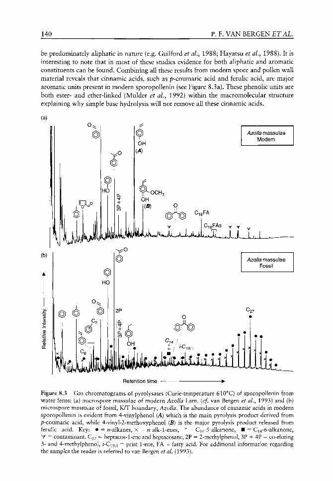
140 P. F. VAN BERGEN ET AL.
be predominately aliphatic in nature (e.g. Guilford et aL^ 1988; Hayatsu et ai, 1988). It is interesting to note that in most of these studies evidence for both aliphatic and aromatic constituents can be found. Combining all these results from modern spore and pollen wall material reveals that cinnamic acids, such as p-coumaric acid and ferulic acid, are major aromatic units present in modern sporopollenin (see Figure 8.3a). These phenolic units are both ester- and ether-linked (Mulder et aL, 1992) within the macromolecular structure explaining why simple base hydrolysis will not remove all these cinnamic acids.
Azolla massulae Modern
Retention time
Figure 8.3 Gas chromatograms of pyrolysates (Curie-temperature 610°C) of sporopollenin from water ferns: (a) microspore massulae of modern Azolla Lam. (cf, van Bergen et ai, 1993) and (b) microspore massulae of fossil, K/T boundary, Azolla. The abundance of cinnamic acids in modern sporopollenin is evident from 4-vinylphenol (A) which is the main pyrolysis product derived from p-coumaric acid, while 4-vinyl-2-methoxyphenol (B) is the major pyrolysis product released from ferulic acid. Key: • = w-alkanes, X = w-alk-1-enes, * = Ci4-5-aIkanone, • = Ci4-6-alkanone, V = contaminant. C27 = heptacos-1-ene and heptacosane, 2P = 2-methylphenol, 3P -H 4P = co-eluting 3- and 4-methylphenol, /-C19.1 = prist-1-ene, FA = fatty acid. For additional information regarding the samples the reader is referred to van Bergen et al. (1993).
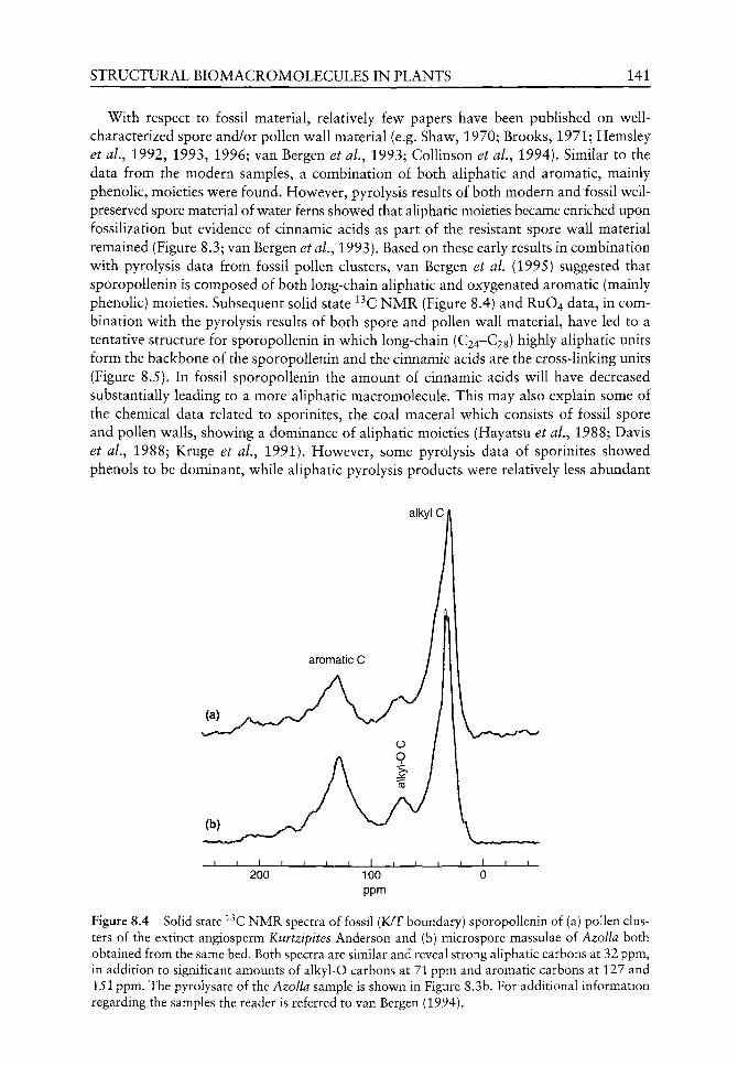
STRUCTURAL BIOMACROMOLECULES IN PLANTS 141
With respect to fossil material, relatively few papers have been published on well-characterized spore and/or pollen wall material (e.g. Shaw, 1970; Brooks, 1971; Hemsley et al, 1992, 1993, 1996; van Bergen et aL, 1993; Collinson et ai, 1994). Similar to the data from the modern samples, a combination of both aliphatic and aromatic, mainly phenolic, moieties were found. However, pyrolysis results of both modern and fossil well-preserved spore material of water ferns showed that aliphatic moieties became enriched upon fossilization but evidence of cinnamic acids as part of the resistant spore wall material remained (Figure 8.3; van Bergen etai, 1993). Based on these early results in combination with pyrolysis data from fossil pollen clusters, van Bergen et al. (1995) suggested that sporopollenin is composed of both long-chain aliphatic and oxygenated aromatic (mainly phenolic) moieties. Subsequent soUd state ^^C NMR (Figure 8.4) and RUO4 data, in combination with the pyrolysis results of both spore and pollen wall material, have led to a tentative structure for sporopollenin in which long-chain (C24-C28) highly aliphatic units form the backbone of the sporopollenin and the cinnamic acids are the cross-linking units (Figure 8.5). In fossil sporopollenin the amount of cinnamic acids will have decreased substantially leading to a more aliphatic macromolecule. This may also explain some of the chemical data related to sporinites, the coal maceral which consists of fossil spore and pollen walls, showing a dominance of aliphatic moieties (Hayatsu et al.^ 1988; Davis et al., 1988; Kruge et al., 1991). However, some pyrolysis data of sporinites showed phenols to be dominant, while aliphatic pyrolysis products were relatively less abundant
alkylC/
200 100 ppm
Figure 8.4 Solid state ^ C NMR spectra of fossil (K/T boundary) sporopollenin of (a) pollen clusters of the extinct angiosperm Kurtzipites Anderson and (b) microspore massulae of Azolla both obtained from the same bed. Both spectra are similar and reveal strong aliphatic carbons at 32 ppm, in addition to significant amounts of alkyl-O carbons at 71 ppm and aromatic carbons at 127 and 151 ppm. The pyrolysate of the Azolla sample is shown in Figure 8.3b. For additional information regarding the samples the reader is referred to van Bergen (1994).
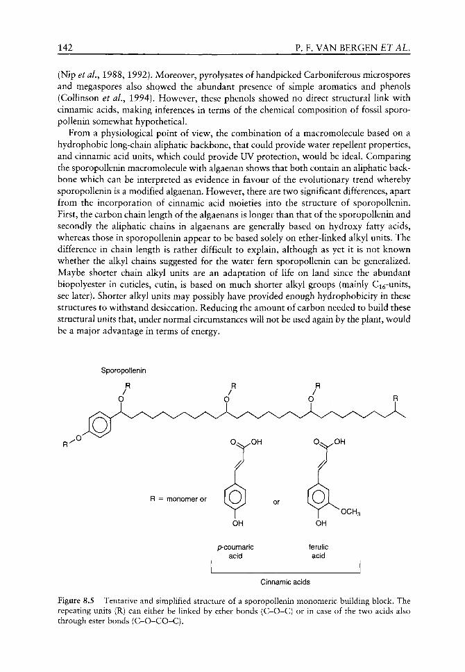
142 P. F. VAN BERGEN ET AL.
(Nip etal., 1988, 1992). Moreover, pyrolysates of handpicked Carboniferous microspores and megaspores also showed the abundant presence of simple aromatics and phenols (CoUinson et al.^ 1994). However, these phenols showed no direct structural link with cinnamic acids, making inferences in terms of the chemical composition of fossil sporo-pollenin somewhat hypothetical.
From a physiological point of view, the combination of a macromolecule based on a hydrophobic long-chain aliphatic backbone, that could provide water repellent properties, and cinnamic acid units, which could provide UV protection, would be ideal. Comparing the sporopollenin macromolecule with algaenan shows that both contain an aliphatic backbone which can be interpreted as evidence in favour of the evolutionary trend whereby sporopollenin is a modified algaenan. However, there are two significant differences, apart from the incorporation of cinnamic acid moieties into the structure of sporopollenin. First, the carbon chain length of the algaenans is longer than that of the sporopollenin and secondly the aliphatic chains in algaenans are generally based on hydroxy fatty acids, whereas those in sporopollenin appear to be based solely on ether-linked alkyl units. The difference in chain length is rather difficult to explain, although as yet it is not known whether the alkyl chains suggested for the water fern sporopollenin can be generalized. Maybe shorter chain alkyl units are an adaptation of life on land since the abundant biopolyester in cuticles, cutin, is based on much shorter alkyl groups (mainly Ci^-units, see later). Shorter alkyl units may possibly have provided enough hydrophobicity in these structures to withstand desiccation. Reducing the amount of carbon needed to build these structural units that, under normal circumstances will not be used again by the plant, would be a major advantage in terms of energy.
Sporopollenin
p-coumaric acid
ferulic acid
Cinnamic acids
Figure 8.5 Tentative and simplified structure of a sporopollenin monomeric building block. The repeating units (R) can either be linked by ether bonds (C-O-C) or in case of the two acids also through ester bonds (C-O-CO-C).

STRUCTURAL BIOMACROMOLECULES IN PLANTS 143
Higher land plant leaf and stem cuticles
The bulk of the cuticle in modern plants is composed of a solvent-insoluble matrix (Tegelaar et al.^ 1991). This matrix makes up the framework of the cuticle and is composed either of the hydrolysable biopolyester cutin, an insoluble non-hydrolysable macromolecule, named cutan, or (most commonly) a mixture of both (Tegelaar et aL, 1991).
The chemical structure of cutin in modern cuticles from both angiosperms and gymno-sperms (Holloway, 1982; Collinson et al., 1998) is well understood and is based primarily on ester-linked functionalized, mainly hydroxy- and dihydroxy-, C^^ and C g alkanoic acids (Figure 8.6; Holloway, 1982; Tegelaar et aL, 1991). Major building blocks include 10,cu-dihydroxy C^^ or Cig alkanoic acids and 9,10,6t)-trihydroxy Cig alkanoic acid, although the cutin is normally based either mainly on Ci^ or C g units. In addition to the linear aliphatic moieties, the cutin fraction of some taxa also reveals the presence of cinnamic acids, in particular p-coumaric and ferulic acid (Kolattukudy, 1981; Holloway, 1982). Holloway (1982) however, stated that phenolic acids esterified to carbohydrates, rather than to cutin alkanoic acids, first have to be eliminated as a possible source for cinnamic acids in cutin. In particular, incorporation of cell wall material to the cuticle might lead to the apparent recognition of cinnamic acids as an integral part of cutin. Cinnamic acids have been suggested to be part of the cutin of only a small number of plants including Lycopersicon esculentum Mill, and Ginkgo biloba Linn. (Collinson et aL., 1998).
Unequivocal molecular evidence of cutin in fossil cuticles is sparse (Tegelaar etal,., 1991) as it would need to show the presence of the distinct C^^ and Cig alkanoic acids; upon pyrolysis they occur as unsaturated alkanoic acids. In the few fossil cases where cutin was suggested, cinnamic acids were not recognized (Tegelaar et aL, 1991). In stark contrast, most fossil cuticles consist of macromolecular material composed of long-chain aliphatic compounds, termed cutan. The discovery of this material in fossil cuticles triggered a search to identify it in modern cuticles.
To date, the search for cutan in modern taxa has shown that only some modern cuticles reveal unequivocal evidence of this macromolecule (e.g. Agave^ Clivia Lindl.). Moreover, the precise structure of cutan is still unclear. Pyrolysis data (de Leeuw et ai, 1991) in combination with chemolysis results (RUO4 oxidation; Schouten et aL, 1998) indicate that it is based mainly on linear long-chain (C30-C34) aliphatic compounds, probably hydroxy-alkanoic acids. These could be bound covalently via non-hydrolysable ether bonds as well as via relatively labile ester bonds (Figure 8.7). Evidence of cinnamic acids in the cutan is
p-coumaric acid
Figure 8.6 Structure of cutin building blocks based mainly on a dihydroxy fatty acid. See Figure 8.5 for additional information.

144 P. F. VAN BERGEN ET AL,
Cutan
p-coumaric acid
Figure 8.7 Tentative and simplified structure of a cutan based on long-chain alkyl units linked by ether moieties. See Figure 8.5 for additional information.
absent but the involvement of some small amounts of aromatic units in the cutan has been postulated based on ^^C NMR and thermochemolysis data (McKinney et al.^ 1996).
Most fossil cuticles reveal the presence of a macromolecule based on long-chain aliphatic moieties often v^ith distinct distribution patterns indicating mainly building blocks of even carbon chain length (C22-C34; Tegelaar et aL, 1991; van Bergen, 1994; CoUinson et aL, 1994, 1998; Zodrov^ and Mastalerz, 2001). Interestingly, hov^ever, these fossils include taxa (e.g. Ginkgo; Collinson et al.^ 1998) the modern counterparts of w^hich are devoid of cutan. This therefore implies that the long-chain aliphatic macromolecule in fossil cuticles may in some cases be selectively preserved cutan or alternatively, a newly formed highly aliphatic geomacromolecule. As these fossil remains still appear as cuticles based on morphology and ultrastructure, the compounds involved will have to be derived from very near the cuticle itself. Cutin has been suggested as a source but it is unlikely that stabilization of this biopolyester plays a major part as this should reveal evidence of distinct linear C16 and/or Cig moieties. More recently, these observations have led to the hypothesis of within-cuticle diagenetic stabilization of normally labile aliphatic constituents, i.e. cuticu-lar waxes, for the formation of highly aliphatic macromolecules in fossil cuticles (Collinson et ai, 1998). Interestingly, the chemical composition of fossil cuticular layers of related taxa can reveal chemosystematic characteristics indicating that if the highly aliphatic macromolecule in most fossil cuticles were formed upon within-cuticle stabilization this process cannot be entirely random as this would mask the chemosystematic signal (cf, Potamogeton L. versus Limnocarpus Reid and Chandler, van Bergen, 1999; van Bergen et ai, 1999; Typha L. versus Sparganium L.; Collinson and van Bergen, 2003; differences between various Carboniferous plant cuticles, Mosle et al,^ 2002). Maybe cutan in modern cuticles is also based on within-cuticle cross-linking of cuticular lipids including long-chain w-alkanols, aldehydes and alkanoic acids. Enzymatic reactions or photo-oxidation in the living cuticle may in that case aid the formation of cutan. Recent pyrolysis data of cuticular lipids in combination with a mineral matrix showed that a cutan-type signal was obtained, suggesting that such an aliphatic macromolecular signal found in fossil cuticles may be formed during early diagenesis from waxes, some of which are present as metal salts (Finch and Freeman, 2001). In addition, one has to be aware of pyrolytically-induced alterations that can affect the chemical composition of fossil cuticles leading to spurious interpretations with respect to their original chemical composition. In particular, older cuticles often reveal chemical data implying relatively short-chain aliphatic building blocks (C^Q-Cig; Collinson et aL,

STRUCTURAL BIOMACROMOLECULES IN PLANTS 145
(a)
0) c 0
i5 DC
HO
Fossil Typha seed coat
^22 D
Fossil Sparganium testa
^22
C29-steroids
IMwiUiiiiiJL Retention time
Figure 8.8 Gas chromatograms of pyrolysates (Curie-temperature 610°C) of (a) cuticular seed coat of fossil, Eocene (ca. 34 Ma), Typha and (b) fossil cuticular testa of Sparganium, obtained from the same bed, showing the presence of phenolic pyrolysis products (annotated P) derived from p-coumaric acid. Key: • = w-alkanes, X = w-alk-1-enes, D = 2-alkanones, C9 = non-1-ene and nonane. C^x indicate alkanones with xx representing the number of carbon atoms. P is phenol, 3P+4P = co-eluting 3- and 4-methylphenol. The LM photograph shows the two-layered cuticular composition of the fossil seed coat wall. For additional information regarding the samples, the reader is referred to van Bergen (1994a).
1994; Ewbank et al,^ 1996; Zodrow and Mastalerz, 2001). These patterns are most probably solely due to increasing maturity of the organic material leading to an increase in cross-linking (van Bergen, 1999) and as such prevent unambiguous interpretations in terms of evolution and physiology.
In a few fossil cuticles evidence v^as found for the presence of phenolic compounds directly related to p-coumaric acid (Figure 8.8; seed cuticles of Typha and Sparganium-,

146 P. F, VAN BERGEN ET AL.
leaf cuticles of Ginkgo; Collinson et al., 1998). These phenolic compounds are linked both by ester- and ether-bonds. This could indicate that cinnamic acids are also incorporated in cutan of the cuticle, possibly with the same physiological function as in other plant organs (i.e. UV protection). However, the cuticular membrane of Sparganium is within a sclerotic endocarp making the need for UV protection less likely. Alternatively, these cinnamic acids have become incorporated upon within-cuticle stabilization during early diagenesis.
From a physiological point of view, the structural macromolecules in cuticles are, to some extent, similar and in other aspects rather different from those in algal, spore and pollen walls. First, the macromolecules in most cuticles do not contain abundant cinnamic acids, thus being distinct from sporopoUenin. This despite the fact that most cuticles are directly exposed to direct sunlight. Most probably, other tissues underlying the cuticle (i.e. cell walls of the epidermis) may be involved in UV shielding, whereas no such protection exists for the cytoplasm in spores and pollen. A second difference is the presence of a structural polyester, i.e. cutin, that is based on relatively short-chain hydroxyacids. As long as these shorter alkyl units provide enough hydrophobicity in these structures, the production of such macromolecules may be advantageous. Reducing the amount of carbon needed to build these structural units, that under normal circumstances will not be used again by the plant, would be a major advantage. The similarity with algaenan and sporopoUenin relates to cutan. This latter macromolecule is also based on long-chain aliphatic, ether- and possibly ester-linked (hydroxy) units. Flowever, the paucity of in-depth molecular information of modern cutan prevents further physiological inferences.
Inner structural entities
Water-conducting and strengthening tissues
Lignin is most commonly associated with the secondary xylem in wood. This methoxyphenol-based biomacromolecule is often suggested to have been pivotal in an evolutionary sense for both water conductance and strengthening of the water-conducting elements (e.g. Edwards, 2001; Cooper-Driver, 2001). The findings of tracheids and other xylem-like elements in fossil settings as far back at the Silurian have been suggested to relate to the presence of lignin in these fossil remains (or at least lignification is implicated; Cook and Friedman, 1998; Cooper-Driver, 2001; Edwards, 2001).
The molecular building blocks of modern lignin are relatively well known despite the uncertainties of the overall molecular composition of the macromolecule itself. Lignin is based on monolignols including p-coumaryl alcohol, coniferyl alcohol and/or sinapyl alcohol (Figure 8.9) depending on the plant taxon (Sarkanen and Ludwig, 1971; Saiz-Jimenez and de Leeuw, 1986). In modern specimens the chemical composition of lignin is rather diverse with that of gymnosperms based mainly on coniferyl alcohol, whereas the angiosperms always contain coniferyl and sinapyl units. Monocotyledonous angiosperms and legumes may also contain abundant p-coumaryl units in addition to the other two. With respect to lower plants, such as bryophytes, controversy still exists as to whether the phenolic moieties present in these plants are indicative of real lignin (Lewis and Yamamoto, 1990). Proving the presence of lignin units is often dependent on detailed molecular analyses that can show intact and rather specific methoxyphenols. In particular the presence of intact side chains attached to the aromatic ring (Figure 8.10) is crucial
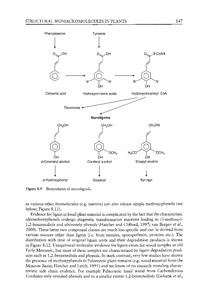
STRUCTURAL BIOMACROMOLECULES IN PLANTS 147
Phenylalanine Tyrosine
0.«^ ^S-CoAH
Cinnamic acid Hydroxycinnamic acids
OH
HydroxycinnamoyI CoA
Flavonoids
CH2OH
OH p-Coumaryl alcohol
CH2OH
"^^OCHs OH
Coniferyl alcohol
p-Hydroxyphenyl Guaiacyl
Figure 8.9 Biosynthesis of monolignols.
H3CO" y OCH3 OH
Sinapyl alcohol
Syringyl
as various other biomolecules (e.g. tannins) can also release simple methoxyphenols (see below; Figure 8.11).
Evidence for lignin in fossil plant material is complicated by the fact that the characteristic (di)methoxyphenols undergo diagenetic transformation reactions leading to (3-methoxy)-1,2-benzenediols and ultimately phenols (Hatcher and Clifford, 1997; van Bergen et al., 2000). These latter tv^o compound classes are much less specific and can be derived from various sources other than lignin (i.e. from tannins, sporopollenin, proteins etc.). The distribution v^ith time of original lignin units and their degradation products is shown in Figure 8.12. Unequivocal molecular evidence for lignin exists for wood samples as old Early Mesozoic, but most of these samples are characterized by lignin degradation products such as 1,2-benzenediols and phenols. In stark contrast, very few studies have shown the presence of methoxyphenols in Palaeozoic plant remains (e.g. wood material from the Moscow Basin; Hatcher and Lerch, 1991) and we know of no research revealing characteristic side chain evidence. For example Palaeozoic fossil wood from Carboniferous Cordaites only revealed phenols and to a smaller extent 1,2-benzenediols (Ewbank et aL,

148 P. F. VAN BERGEN ET AL.
PS Retention time
Brasenia schreberi extant seed coat
Py(610°C)-GC-FID
OH
OCH3 OH
po _ Polysaccharide pyrolysis products
^ ^ J ' ^ U u j u ^ ^ EX-BRADAW
Figure 8.10 Gas chromatogram of the pyrolysate (Curie-temperature 610°C) of the Hgno-cellulose complex present in seed coat of extant Brasenia schreberi J.F. Gmel. Note the abundance of products indicating 2-methoxyphenols (G) and 2,6-dimethoxyphenols (S) with side chains.
i5 o
DC
d]& OCH.
OH OH
(G)
OH OH
^OH X O H3C0 T 0CH3
OH 4C (S)
1 Retention time -
Figure 8.11 Gas chromatogram of the pyrolysate (Curie-temperature 610°C) of non-hydrolysable red wine tannin. Note the absence of 2-methoxyphenols and 2,6-dimethoxyphenols with side chains when compared with Figure 8.10.
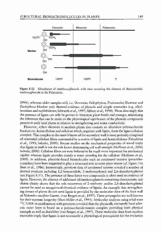
STRUCTURAL BIOMACROMOLECULES IN PLANTS 149
Cenozoic
OH
Marker O^
OH OH
OH
Biomolecule Lignin Lignin
Figure 8.12 Abundance of (methoxy)phenols with time revealing the absence of characteristic methoxyphenols in the Palaeozoic.
1996), whereas older samples still, i.e. Devonian Psilophyton, Prototaxites Davison and Pachytheca Hooker only showed evidence of phenols and simple aromatics (e.g. alkyl-benzenes and naphthalenes; Edwards etaL, 1997; Abbott etaL, 1998). These data imply that the presence of lignin can only be proven in Mesozoic plant fossils and younger, minimizing the inferences that can be made on the physiological significance of the phenolic compounds present in early land plants in relation to strengthening and water conductivity.
However, xylem elements in modern plants also contain an abundant polysaccharide fraction viz. hemicellulose and cellulose which, together with lignin, form the ligno-cellulose complex. This complex in the main S-layers of the secondary wall is most probably composed of orientated cellulose fibres surrounded by a matrix of lignin and hemicelluloses (Terashima et al.y 1993; Salmen, 2000). Recent studies on the mechanical properties of wood imply that lignin in itself is not the sole factor determining cell wall strength (Hoffman et aL, 2000; Salmen, 2000). Cellulose fibres are now believed to be much more important for mechanical rigidity whereas lignin provides mainly a water proofing for the cellulose (Hoffman et aL, 2000). In addition, phenolic-based biomolecules such as condensed tannins (proantho-cyanidins) have been suggested to play a structural role in some plant tissues (cf. Figure 3 in Shen et ai, 1986). Interestingly, pyrolysis data of condensed tannins revealed a number of distinct products including 1,2-benezendiols, 2-methoxyphenol and 2,6-dimethoxyphenol (see Figure 8.11). The presence of these latter two compounds is often used as evidence of lignin. However, the absence of additional (di)methoxyphenols containing characteristic side chains clearly shows that the sole occurrence of 2-methoxy- and/or 2,6-dimethoxyphenol cannot be used as unequivocal chemical evidence of lignin. An example that strengthening tissues of plants do not need lignin is provided by the molecular data of the fruit wall of Nelumbo nucifera Gaertn. (van Bergen et al.^ 1997). These propagules are well-known for their extreme longevity (Shen-Miller et al,^ 1995). Molecular analyses using solid state ^^C NMR in combination with pyrolysis revealed that the physically extremely hard sclerotic outer layer is based on a polysaccharide-tannin complex providing both physical strength as well as durability (van Bergen et al,^ 1997). These molecular data from modern examples imply that lignin is not necessarily a physiological prerequisite for the evolution

150 P. F, VAN BERGEN ET AL,
of structural plant tissues and that other phenolic macromolecular biomolecules may have
played a key role in the early evolution of land plants (cf. Abbott et al.^ 1998).
Conclusions
The invasion of the land by plants may have forced the evolution of specific physiological
adaptation to survive this hostile new environment. Two of the main problems plants had
to overcome included an increase in the levels of UV radiation and water loss or desiccation.
Studying the resistant macromolecular composition of outer coverings and strengthening
tissues from both modern and fossil examples can reveal information on the molecular evo
lution of these structures. The resistant molecules in cuticles (i.e. cutin and cutan) and spore
and pollen walls (sporopoUenin) are all based on even carbon numbered long-chain
aliphatic chemical building blocks providing sufficient hydrophobicity to reduce water loss.
These aliphatic moieties are largely similar to those present in the resistant walls of algae
(algaenan) from which the land plants may have evolved. Apart from the aliphatic material,
sporopoUenin and, to some degree, cutin and cutan from both modern and fossil examples
also reveal the presence of cinnamic acids, which probably are responses to the enhanced
levels of UV radiation on land. With respect to the strengthening tissues, lignin may have
been an important biomolecule in early land plants but, to date, no unequivocal molecular
evidence exists that it actually occurred in the oldest land plants. Moreover, molecular data
from modern strengthening tissues indicate that lignin is not necessarily a physiological
prerequisite for the evolution of plant tissues that provide physical strength and that other
phenolic macromolecular biomolecules may have played an additional (key) role in the
evolution of the early land plants.
References
Abbott GD, Ewbank G, Edwards D, Wang G-Y. 1998. Molecular characterization of some enigmatic Lower Devonian fossils. Geochimica et Cosmochimica Acta 62: 1407-1418.
Allard B, Templier J, Largeau C. 1997. Artificial origin of mycobacterial bacteran. Formation of melanoidin-like artifact macromolecular material during the usual isolation process. Organic Geochemistry 26: 691-704.
Allard B, Templier J, Largeau C. 1998. An improved method for the isolation of artifact-free algae-nans from microalgae. Organic Geochemistry 28: 543-548.
van Bergen PF. 1994. Paleaobotany of Propagules: An investigation combining microscopy and chemistry. D.Phil. Thesis, London University, UK.
van Bergen PF. 1999. Pyrolysis and chemolysis of fossil plant remains: applications to palaeo-botany. In: Jones TP, Rowe NP, eds. f oss/7 Plants and Spores: Modern Techniques. London: The Geological Society, 143-148.
van Bergen PF, Collinson ME, de Leeuw JW. 1993. Chemical composition and ultrastructure of fossil and extant salvinialean microspore massulae and megaspores. Grana Suppl. 1: 18-30.
van Bergen PF, Goni M, Collinson ME, et al. 1994a. Chemical and microscopic characterization of outer seed coats of fossil and extant water plants. Geochimica et Cosmochimica Acta 58: 3823-3844.
van Bergen PF, Scott AC, Barrie PJ, et al. 1994b. The chemical composition of Upper Carboniferous pteridosperm cuticles. Organic Geochemistry 21: 107-112.
van Bergen PF, Collinson ME, Briggs DEG, et al. 1995. Resistant biomacromolecules in the fossil record. Acta Botanica Neerlandica 44: 319-342.
van Bergen PF, Hatcher PG, Boon JJ, et al. 1997. Macromolecular composition of the propagule wall oi Nelumbo nucifera. Phy to chemistry 45: 601-610.

STRUCTURAL BIOMACROMOLECULES IN PLANTS 151
van Bergen PF, Collinson ME, Stankiewicz BA. 1999. The importance of molecular palaeobotany. Acta Palaeobotanica Suppl. 2: 653-657.
van Bergen PF, Poole I, Ogilvie TMA, et al. 2000. Evidence for demethylation of syringyl moieties in archaeological wood using pyrolysis/gas chromatography/mass spectrometry. Rapid Communications in Mass Spectrometry 14: 71-79.
Bertheas O, Metzger P, Largeau C. 1999. A high molecular vv eight complex lipid, aliphatic polyalde-hyde tetraterpenediol polyacetal from Botryococcus hraunnii (L race). Fhy to chemistry 50: 85-96.
Bestougeff MA, Byramjee RJ, Pesneau B. 1985. On the chemical mechanism of kerogen thermal transformation. Study of the transformation of sporopollenin. Organic Geochemistry 8: 389-398.
Bierstedt A, Stankiev^icz BA, Briggs DEG, Evershed RP. 1998. Quantitative and qualitative analysis of chitin in fossil arthropods using a combination of colorimetric assay and pyrolysis gas chromatography mass spectrometry. The Analyst 123: 139-145.
Blokker P. 2000. Structural analysis of resistant polymers in extant algae and ancient sediments. Geologica Ultraiectina 193: 145.
Blokker P, Schouten S, van den Ende H, et al. 1998. Chemical structure of algaenans from the fresh v^ater algae Tetraedron minimum, Senedesmus communis and Pediastrum boryanum. Organic Geochemistry 29: 1453-1468.
Blokker P, Schouten S, de Leeuv^ JW, et al. 1999. Molecular structure of the resistant biopolymer in the zygospore cell v^alls of Chlamydomonas monoica. Planta 207: 539-543.
Blokker P, van Bergen PF, Pancost R, et al. 2001. The chemical structure of Gloeocapsomorpha prisca microfossils: Implications for their origin. Geochimica et Cosmochimica Acta 65: 886-900.
Boon JJ, Stout SA, Genuit W, Spackman W. 1989. Molecular paleobotany of Nyssa endocarps. Acta Botanica Neerlandica 38: 391-404.
Brooks J. 1971. Some chemical and geochemical studies on sporopollenin. In: Brooks J, Grant P, Muir MD, et al., eds. Sporopollenin. London: Academic Press, 351-407.
Brooks J, Shaw G. 1968. The post-tetradontogeny of the pollen wall and the chemical structure of the sporopollenin of Lilium henryii. Grana 8: 227-234.
Brooks J, Shaw G. 1978. Sporopollenin: a review of its chemistry, palaeochemistry and geochemistry. Grana 17: 91-97.
Collinson ME, van Bergen PF. 2003. Evolution of angiosperm fruit and seed physiology: anatomical and chemical evidence from fossils. In: Hemsley AR, Poole I, eds. The Evolution of Plant Physiology. Oxford: Elsevier, 347-381.
Collinson ME, van Bergen PF, Scott AC, de Leeuw JW. 1994. The oil-generating potential of plants from coal and coal-bearing strata through time: a review with new evidence from Carboniferous plants. In: Scott AC, Fleet AJ, eds. Coal and Coal-bearing Strata as Oil-prone Source Rocksf Geological Society Special Publication 77: 31-70.
Collinson ME, Mosle B, Finch P, et al. 1998. The preservation of plant cuticle in the fossil record: a chemical and microscopical investigation. Ancient Biomolecules 2: 251-265.
Cook ME, Friedman WE. 1998. Tracheid structure in a primitive extant plant provides an evolutionary link to earhest fossil tracheids. International Journal of Plant Science 159: 881-890.
Cooper-Driver GA. 2001. Biological roles for phenolic compounds in the evolution of early land plants. In: Gensel PG, Edwards D, eds. Plants Invade the Land. Evolutionary and Environmental Perspectives. New York: Columbia University Press, 159-172.
Davis MR, Abbott JM, Cudby M, Gaines A. 1988. Chemical structure of telocollinites and sporinites. 2. Analysis of material unextractableby pyridine using ^^C CP/MAS N.M.R. and G.C.-M.S. spectrometry. Fuel 67: 960-966.
Derenne S, Largeau C, Casadevall E, et al. 1991. Chemical evidence of kerogen formation in source rocks and oil shales via selective preservation of thin resistant outer walls of microalgae: origin of ultralaminae. Geochimica et Cosmochimica Acta 55: 1041-1050.
Derenne S, Largeau C, Hatcher PG. 1992a. Structure of Chlorella fusca algaenan: relationships with ultralaminae in lacustrine kerogens: species- and environmental-dependent variations in the composition of fossil ultralaminae. Organic Geochemistry 18: 417-422.
Derenne S, Metzger P, Largeau C, et al. 1992b. Similar morphological and chemical variations of Gloeocapsomorpha prisca in Ordovician sediments and cultured Botryococcus braunii as a response to changes in salinity. Organic Geochemistry 19: 299-313.
152 P. F. VAN BERGEN ET AL.
Derenne S, Largeau C, Berkaloff C. 1996. First example of an algaenan yielding an aromatic-rich pyrolysate. Possible geochemical implications on marine kerogen formation. Organic Geochemistry 24: 617-627.
Edwards D. 2001. Early land plants. In: Briggs DEG, Crowther PR, eds. Palaeobiology 11. Oxford: Blackwell Science, 63-66.
Edwards D, Ewbank G, Abbott GD, 1997. Flash pyrolysis of the outer cortical tissues in Lower Devonian Psilophyton dawsonii. Botanical Journal of the Linnean Society 124: 334-360.
Ewbank G, Edwards D, Abbott GD. 1996. Chemical characterization of Lower Devonian vascular plants. Organic Geochemistry 25: 461-473.
Finch P, Freeman G. 2001. Simulated diagenesis of plant cuticles - implications for organic fossi-lisation. Journal of Analytical and Applied Pyrolysis 58-59: 229-235.
Foster CB, Wicander R, Reed JD. 1989. Gloeocapsomorpha prisca Zalessky, 1917: A new study. Part I: taxonomy, geochemistry and palaeoecology. Geobios 22: 735-759.
Foster CB, Wicander R, Reed JD. 1990. Gloeocapsomorpha prisca Zalessky, 1917: A new study. Part II: origin of Kukersite, a new interpretation. Geobios 23: 133-140.
Gatellier J-PLA, de Leeuw JW, Sinninghe Damste JS, et al. 1993. A comparative-study of macro-molecular substances of a coorongite and cell-walls of the extant alga Botryococcus braunii. Geochimica et Cosmochimica Acta 57: 2053-2068.
van Geel B, Grenfell HR. 1996. Spores of Zygnemataceae. In: Jansonius J, McGregor DC, eds. Palynology: Principles and Applications. Salt Lake City: American Association of Stratigraphic Palynologists Foundation, 173-179.
Gelin F, Boogers I, Noordeloos AAM, et al. 1997. Resistant biomacromolecules in marine micro-algae of the classes Eustigmatophyceae and Chlorophyceae: geochemical implications. Organic Geochemistry 26: 659-675.
Gelin F, Volkman JK, Largeau C, et al. 1999. Distribution of aliphatic, nonhydrolyzable biopoly-mers in marine microalgae. Organic Geochemistry 30: 147-159.
Goth K, de Leeuw JW, Piittmann W, Tegelaar EW. 1988. Origin of Messel oil shale kerogen. Nature 336: 759-761.
Graham LE, Gray J. 2001. The origin, morphology and ecophysiology of early embryophytes: neontological and paleontological perspectives. In: Gensel PG, Edwards D, eds. Plants Invade the Land. Evolutionary and Environmental Perspectives. New York: Columbia University Press, 140-158.
Guilford WJ, Schneider DM, Labovitz J, Opella SJ. 1988. High resolution solid state ^^C NMR spectroscopy of sporopollenin from different plant taxa. Plant Physiology 86: 134-136.
Hatcher PG, Lerch HE III. 1991. Survival of lignin-derived structural units in ancient coalified wood samples. In: Schobert HH, Bartle KD, Lynch LJ, eds. Goal Science II ACS symposium series 461, Washington DC: American Chemical Society, 9-19.
Hatcher PG, Clifford DJ. 1997. The organic geochemistry of coal: from plant materials to coal. Organic Geochemistry 27: 251-274.
Hatcher PG, Pan VH, Maciel GE. 1999. Solid state ^^C nuclear magnetic resonance of fossil plants and spores. In: Jones TP, Rowe NP, eds. Fossil Plants and Spores: Modern Techniques. London: The Geological Society, 149-155.
Hayatsu R, Botto RE, Mcbeth RL, et al. 1988. Chemical alteration of a biological polymer 'sporopollenin' during coalification: origin, formation, and transformation of the coal maceral sporinite. Energy and Fuels 2: 843-847.
Hemsley AR, 1994. Exine ultrastructure in the spores of enigmatic Devonian plants: its bearing on the interpretation and on the origin of the sporophyte. In: Kurmann MH, Doyle JA, eds. Ultrastructure of fossil spores and pollen. Kew: Royal Botanic Gardens, 1-21.
Hemsley AR, Chaloner WG, Scott AC, Groombridge CJ. 1992. Carbon-13 solid-state nuclear magnetic resonance of sporopollenins from modern and fossil plants. Annals of Botany 69: 545-549.
Hemsley AR, Barrie PJ, Chaloner WG, Scott AC. 1993. The composition of sporopollenin: its contribution to living and fossil spore systematics. Grana Suppl. 1: 2-11.
Hemsley AR, Scott AC, Barrie PJ, Chaloner WG. 1996. Studies of fossil and modern spore wall biomacromolecules using ^^C soUd state NMR. Annals of Botany 78: 83-94.

STRUCTURAL BIOMACROMOLECULES IN PLANTS 153
Herminghaus S, Gubatz S, Arendt S, Wiermann R. 1988. The occurrence of phenols as degradation products of natural sporopoUenin - a comparison with 'synthetic sporopollenin'. Zeitschrift Naturforschung 43c: 491-500.
Hoffmann B, Chabbert B, Monties B, Speck T. 2000. Tine-tuning' of mechanical properties in two tropical Hanas. In: Spatz H-C, Speck T, eds. Plant Biomechanics 2000. Stuttgart: Georg Thieme Verlag, 10-18.
HoUoway PJ. 1982. The chemical constitution of plant cutins. In: Cutler DF, Alvin KL, Price CE, eds. The Plant Cuticle. Linnean Society Symposium Series 10, 45-85.
Kokinos JP, Eglinton TI, Goni MA, et al. 1998. Characterization of a highly resistant biomacro-molecular material in the cell wall of a marine dinoflagellate resting cyst. Organic Geochemistry 28: 265-288.
Kolattukudy PE. 1981. Structure, biosynthesis and biodegradation of cutin and suberin. Annual Reviews in Plant Physiology 32: 539-567.
Kozubek A, Tyman JHP. 1999. Resorcinolic lipids, the natural non-isoprenoic amphiphiles and their biological activity. Chemical Reviews 99: 1-26.
Kruge MA, Crelling JC, Hippo EJ, Palmer SR. 1991. Aspects of sporinite chemistry. Organic Geochemistry 17: 193-204.
Largeau C, Derenne S, Casadevall E, et al. 1986. Pyrolysis of immature torbanite and of the resistant biopolymer (PRB A) isolated from the extant alga Botryococcus hraunii. Mechanism for the formation and structure of torbanite. Organic Geochemistry 10: 1023-1032.
de Leeuw JW, Largeau C. 1993. A review of macromolecular organic compounds that comprise living organisms and their role in kerogen, coal and petroleum formation. In: Engel MH, Macko SA, eds. Organic Geochemistry. New York: Plenum Pubhshing Corp., 23-72.
de Leeuw JW, van Bergen PF, van Aarssen BGK, etal. 1991. Resistant biomacromolecules as major contributors to kerogen. Philosophical Transactions of the Royal Society of London B 333: 329-337.
Lewis NG, Yamamoto E. 1990. Lignin: Occurence, Biogenesis and Biodegredation. Annual Reviews in Plant Physiology and Plant Molecular Biology 41: 455-496.
McKinney DE, Bortiatynski JM, Carson DM, et al. 1996. Tetramethylammonium hydroxide (TMAH) thermochemolysis of the aliphatic biopolymer cutan: Insights into the chemical structure. Organic Geochemistry 24: 641-650.
Metzger P, Largeau C. 1994. A new type of ether lipid comprising phenolic moieties in Botryococcus hraunii. Chemical structure and abundance, and geochemical implications. Organic Geochemistry 11'. 802-814.
Mosle B, Finch P, Collinson ME, Scott AC. 1997. Comparison of modern and fossil plant cuticles by selective chemical extraction monitored by flash pyrolysis-gas chromatography-mass spectrometry and electron microscopy, journal of Analytical and Applied Pyrolysis 40-41: 585-597.
Mosle B, Collinson ME, Scott AC, Finch P. 2002. Chemosystematic and microstructural investigations on carboniferous seed plant cuticles from four North American localities. Review of Paleobotany and Palynology 120: 41-52.
Mulder MM, van der Hage ERE, Boon JJ. 1992. Analytical in source pyrolytic methylation electron impact mass spectrometry of phenolic acids in biological matrices. Phytochemical Analysis 3: 165-172.
Nip M, de Leeuw JW, Schenck PA. 1988. The characterization of eight maceral concentrates by means of Curie point pyrolysis-gas chromatography and Curie point pyrolysis-gas chromatography-mass spectrometry. Geochimica et Cosmochimica Acta 52: 637-648.
Nip M, de Leeuw JW, Crelling JC. 1992. Chemical structure of bituminous coal and its constituting maceral fractions as revealed by flash pyrolysis. Energy and Fuels 6: 125-136.
Philip RP, Gilbert TD, Russel NJ. 1982. Characterization by pyrolysis-gas chromatography-mass spectrometry of the insoluble organic residues from the hydrogenation of Tasmanites sp. oil shale. Fuel 61x111-116.
Rozema J, Broekman RA, Blokker P, et al. 2001. UV-B absorbance and UV-B absorbing compounds (para-coum2inc acid) in pollen and sporopollenin: the perspective to track historic UV-B levels. Journal of Photochemistry and Photobiology B: Biology 61: 108-117.
Saiz-Jimenez C, de Leeuw JW. 1986. Lignin pyrolysis products: Their structures and their significance as biomarkers. Organic Geochemistry 10: 869-876.

154 P. F. VAN BERGEN ET AL.
Salmen L. 2000. Structure-property relations for wood: from the cell-wall polymeric arrangement to the macroscopic behaviour. In: Spatz H-C, Speck T, eds. Flant Biomechanics 2000. Stuttgart: Georg Thieme Verlag, 452-462.
Sarkanen KV, Ludwig CH. 1971. Lignins. Occurrence, Formation, Structure and Reactions. New York: Wiley-Interscience.
Schenck PA, de Leeuw JW, van Graas G, et al. 1981. Analysis of recent spores and pollen and of thermally altered sporopollenin by flash pyrolysis-mass spectrometry and flash pyrolysis-gas chromatography-mass spectrometry. In: Brooks J, ed. Organic Maturation Studies and Fossil Fuel Exploration. London: Academic Press, 225-237.
Schouten S, Moerkerken P, Gelin F, et al. 1998. Structural characterization of aliphatic, non-hydrolyzable biopolymers in freshwater algae and leaf cuticle by ruthenium tetroxide degradation. Phytochemistry 49: 987-993.
Schulze Osthoff K, Wiermann R. 1987. Phenols as integrated compounds of sporopollenin from Pinus pollen. Journal of Plant Physiology 131: 5-15.
Shaw G. 1970. Sporopollenin. In: Harborne JB, ed. Phytochemical Phylogeny. London: Academic Press, 31-58.
Shaw G. 1971. The chemistry of sporopollenin. In: Brooks J, Grant P, Muir MD, et al.., eds. Sporopollenin. London: Academic Press, 305-350.
Shaw G, Yeadon A. 1964. Chemical studies on the constitution of some pollen and spore membranes. Grana Palynologica 5: 247-252.
Shen Z, Haslam E, Falshaw CP, Begley MJ. 1986. Procyanides and polyphenols of Larix gmelini bark. Phytochemistry 25: 2629-2635.
Shen-Miller J, Mudgett MB, Schopf JW, et al. 1995. Exceptional seed longevity and robust growth -ancient sacred lotus from China. American Journal of Botany 82: 1367-1380.
Sinninghe Damste JS, de las Heras FXC, van Bergen PF, de Leeuw JW. 1993. Characterisation of Tertiary Catalan lacustrine oil shales: Discovery of extremely organic sulphur-rich type I kero-gens. Geochimica et Cosmochimica Acta 57: 389-415.
Stankiewicz BA, van Bergen PF, Smith MB, et al. 1998. Comparison of analytical performance of filament and Curie-point pyrolysis devices. Journal of Analytical and Applied Pyrolysis 45: 133-151.
Stebbins GL, Hill GJC. 1980. Did multicellular plants invade the land? American Naturalist 115: 342-353.
Tegelaar EW, de Leeuw JW, Largeau C, et al. 1989. Scope and limitations of several pyrolysis methods in the structural elucidation of macromolecular plant constituents in the leaf cuticle of Agave americana L. Journal of Analytical and Applied Pyrolysis 15: 29-54.
Tegelaar EW, Kerp JHF, Visscher H, et al. 1991. Bias of the paleobotanical record as a consequence of variations in the chemical composition of higher vascular plant cuticles. Paleobiology 17: 133-144.
Terashima N, Fukushima K, He L-F, Takabe K. 1993. Comprehensive model of the lignified plant cell wall. In: Jung HG, Buxton DH, Hatfield RD, Ralf H, eds. Forage Cell Wall Structure and Digestibility. Madison: ASA-CSSA-SSSA, 247-270.
Wehling K, Niester C, Boon JJ, et al. 1989. p-Coumaric acid - a monomer in the sporopollenin skeleton. Planta 179: 376-380.
Wiermann R, Gubatz S. 1992. Pollen wall and sporopollenin. International Reviews in Cytology 140: 35-72.
Zetzsche F, Kalin O. 1931. Membranes of spores and pollen. V. Autoxidation of sporopollenin. Helvetica Chimica Acta 14: 517-519.
Zetzsche F, Kalt P, Liechti J, Ziegler E. 1937. Membranes of spores and pollen. XL Constitution of Lycopodium sporonin, tasmanin and fossil sporonins. Journal fiir Praktische Chemie 148:267-286.
Zodrow EL, Mastalerz M. 2001. Chemotaxonomy for naturally macerated tree-fern cuticles (Medullosales and Marattiales), Carboniferous Sydney and Mabou Sub-Basins, Nova Scotia, Canada. International Journal of Coal Geology 47: 255-275.

Early land plant adaptations to terrestrial stress: a focus on phenolics
Linda E Graham, Robin B Kodner, Madeline M Fisher, James M Graham, Lee W Wilcox, John M Hackney,
John Obst, Peter C Bilkey, David T Hanson and Martha E Cook
CONTENTS
Introduction
Materials and techniques Trait mapping Thioacidolysis Qualitative and quantitative assessment of acid
liydrolysis-resistant biomass Fluorescence, scanning and transmission electron
microscopy Global estimates of early Palaeozoic resistant and
sequestered carbon
Results
Discussion
Conclusions
Acknowledgements
References
Appendix
155
156 156 156
157
158
158
159
162
165
165
165
168
Introduction
The first land plants no doubt found the terrestrial environment to be a bountiful source of carbon dioxide and light - resources that may have limited grov^th and reproduction of their aquatic algal ancestors (Graham, 1993). But there was probably a stressful 'trade-off in the form of increased heat, desiccation, oxidative damage, harmful radiation and low soil
The Evolution of Plant Physiology Copyright © 2004 Elsevier Ltd ISBN 0-12-33955-26 All rights of reproduction in any form reserved
156 L. E.GRAHAM £TAL.
nutrient content. Interactions with terrestrial microbes may have been an additional new challenge. That early land plants successfully coped, is evidenced by more than a quarter million species of extant descendants and the many millions of terrestrial animal and microbial species that depend upon plants. Comparing the traits of modern representatives of ancient plant lineages offers insight into how plants developed ways of responding to terrestrial stressors. Molecular phylogenetic evidence indicates that modern land plants (embryophytes) are a monophyletic group descended from charophycean green algae (primarily aquatic) and that modern bryophyte lineages (primarily terrestrial) diverged prior to any group of modern vascular plants (data summarized by Kenrick and Crane, 1997).
The prevascular terrestrial biota probably included early plants that shared features with modern bryophytes. This assumption is supported not only by molecular sequence information, but also by an increasingly rich microfossil record of spores, tubes and cellular sheets that have been attributed to early, bryophyte-like plants (Edwards et al.^ 1995; Graham and Gray, 2001 and references cited therein). Additional evidence is provided by morphometric similarity of ancient microfossils to remains of modern bryophytes that were subjected to procedures designed to mimic degradative changes that occur during fos-silization (Kroken et aL, 1996; Kodner and Graham, 2001). Therefore, some traits of extant bryophytes may model early plant adaptations to terrestrial stress (though other features could be of more recent origin and thus not mirror the traits of ancient relatives).
One goal of our work was to map stress-related physiological traits onto a robust phyl-ogeny for modern charophycean algae and bryophytes, because previous mapping of reproductive and structural developmental traits has provided useful insights into early plant evolution (Graham and Wilcox, 2000a; Graham et aL, 2000). Additional goals were to compare aspects of phenolic chemistry among charophyceans, bryophytes and pter-idophytes and to estimate the extent to which non-vascular plants could have contributed to carbon sequestration prior to the origin of vascular plants. To these ends we: (1) used thioacidolysis as an assay for lignin-specific j8-0-4 phenolic linkages in representative green algae and early-divergent land plants; (2) surveyed selected green algae and bryophytes for presence of resistant biomass; (3) quantitatively determined the percentages of resistant cell wall biomass for those that produced measurable amounts; and (4) used these and other data to estimate the amount of resistant organic carbon that might have been generated by early non-vascular land plants.
Materials and techniques
Trait mapping
We used phytogenies published by Karol et al. (2001) for charophyceans, Lewis et al. (1997) for liverworts, Qiu and Palmer (1999) and Nickrent etal (2000) for bryophytes and Newton et al, (2000) for mosses, in order to compile a phylogram representing an emerging view of the early streptophyte (charophyceans plus embryophytes) radiation. Putative stress adaptation traits (whose occurrence was derived from the literature) were mapped onto the phylogram. Fourteen of these stress-related traits are described more fully in Appendix 9.1.
Thioacidolysis
Algae treated by thioacidolysis included unialgal, soil-free cultures representing major green algal lineages (Graham and Wilcox, 2000b): Cladophora glomerata (L.) Klitz.

PHENOLICS AND EARLY PLANTS 157
(Ulvophyceae), Chlorella vulgaris Beij. (Trebouxiophyceae), Oedogonium Link sp. (Chloro-phyceae), Klebsormidium barlowii (Silva et aL), Spirogyra Link sp., Mougeotia C.A. Agardh sp., Staurastrum Meyen sp., Chara L. sp. and Coleochaete orbicularis Pringsheim (Charophyceae). Soil-free laboratory cultures of the following bryophytes were examined: Riccia fluitans L. (liverwort), and the mosses Sphagnum compactum DC, S. centrale C. Jens, and Polytrichum commune Hedw. Moss cultures were generously provided by M. Sargent, University of Illinois. Cultures of the fern Polypodium aureum L. gametophytes and young sporophytes were grown from surface-sterilized spores in closed petri dishes containing mineral nutrient agar. The lycophytes Lycopodium obscurum L. and Selaginella kraussiana (Kunze) A. Braun and the pteridophytes Psilotum nudum L., Tmesipteris vieillardii Dangeard and Adiantum capillus-veneris L. were obtained from the University of Wisconsin Botany Department greenhouses and washed free of soil with sterile water prior to testing. Thioacidolysis was performed as described by LaPierre (1993). Freeze-dried plant or algal samples were boiled for 4 h under nitrogen in 0.2 M boron trifluoride etherate in 9:1 dioxane ethanethiol. After cooling, 10 |xl distilled water was added and the pH was adjusted to 3-4 with 0.4 M NaHC03. Five ml of a tetracosane solution (either 0.1 or 0.01 of tetracosane per ml of CH2CI2 depending upon the anticipated concentration of derivatized product to be measured) was added as an internal standard. The sample was then extracted three times with 10 ml CH2CI2. The combined extracts were dried over a small amount of MgS04 ^^^ paper filtered into an evaporation flask and the sample was rotationally evaporated to near dryness. The resultant residue was subsequently evaporated to dryness with a directed stream of N2 gas and the products were derivatized by adding 1 drop of pyridine and 4-5 drops of BSTFA (bis trimethylsilyl trifluoroacetamide). Finally, the sample was reacted by warming at the lowest setting on a hot plate for 0.5 h prior to its analysis by GC/MS (Finnagan Model MAT 4500; 30 m DB-1 column).
Qualitative and quantitative assessment of acid hydrolysis-resistant biomass
Green algae studied included the desmids (Chlorophyta, Charophyceae) Staurastrum sp. Meyen, Euastrum insigne (Smith) Breb. ex Ralfs, Euastrum pinnatum Ralfs, Micrasterias truncata (Corda) Breb., Closterium ehrenbergi Meneghini, Hyalotheca dissiliens (Sm.) Breb., Desmidium majus Lagerheim, and D. grevillii De Bary, and the co-occurring, non-desmid Eremosphaera viridis De Bary (Chlorophyta, Trebouxiophyceae). These taxa were isolated from Blueberry Lake (Vilas County, WI), Jyme Lake (Oneida Co., WI), or Birch Lake (Marquette Co., WI) and grown in unialgal culture in defined, soil-free medium DYIII (Lehman, 1976). Cultured algal cells of the same age were harvested by micropore filtration and lyophilized. Early-divergent mosses studied included two species of Sphagnum^ Andreaea rupestris Hedw. and Polytrichum ohioense Ren and Card. Axenic cultures of Sphagnum nemoreum and S, compactum Lam. et Cand. (obtained from M. Sargent, University of Illinois) were grown in one-third strength Gamborg's B5 Basal Salts medium (Sigma) with 1% sucrose added, for an equivalent period of time, in both liquid or agar form. Small amounts of Andreaea rupestris were obtained from granitic rocks at 11 000 ft (3350 m) elevation from the Indian Peaks Wilderness Area, Boulder Co., CO (with permission of the US National Forest Service). Gametophytes of Polytrichum ohioense were collected from UW Madison Kemp Biological Station, Oneida Co., WI.
Qualitative assessment of resistance was performed by acetolysis treatment in Eppendorf tubes with concentrated acetic acid for 5-10 min at room temperature, then boiling in 9:1 acetic anhydride/concentrated sulphuric acid for 20 minutes, followed by washes in
158 L. E.GRAHAM £TAL.
concentrated acetic acid and then water (Kroken et aL, 1996). Each step was followed by gentle centrifugation in order to concentrate biomass so that supernatant could be removed by pipette. If plant or algal remains were present, they were visible as dark sediment in the tips of tubes and were removed for microscopic examination with a Pasteur pipette.
Quantitative determinations of resistant biomass were performed for the resistant-walled charophyceans Staurastrum spp., Euastrum insigne, Desmidium grevillii and Hyalotheca dissiliens and all of the mosses listed above. We first experimentally determined that the high temperature acid hydrolysis procedure described above did not significantly alter the weight of empty Eppendorf tubes. Lyophilized algae and mosses were thoroughly dried before being added to preweighed tubes, then tubes were re-weighed prior to acetolysis treatment. After treatment the tubes were dried for several days in an oven at 45°C before final weighing. Before and after treatment, plant or algal weight was calculated by subtracting the weight of the tube from the weight of the tube plus algae or plant material. The proportions of resistant to initial dry weights of specimens were computed and Student's ^test was used to identify significant differences among taxa.
Fluorescence, scanning and transmission electron microscopy
Acetolysed Micrasterias and Andreaea were examined with a Zeiss Axioplan fluorescence microscope in violet and UV excitation as described by Kroken et al. (1996). Andreaea was prepared for SEM by gold coating remains that had been affixed to aluminum stubs by double-sticky tape. Living specimens were prepared for transmission electron microscopy by soaking in 1 % Triton-X for 1 h, followed by three quick rinses with phosphate buffer; fixation in 2% glutaraldehyde and 4% acrolein for 5 h, six buffer washes for 10 min each; fixation in 2% osmium tetroxide for 2h, followed by six 10 min buffer rinses; dehydration in acetone, followed by infiltration with Spurr's resin over a period of 3 days; and polymerization at 70°C overnight. Images of sectioned, stained cells were obtained with a Zeiss 10 transmission electron microscope.
Global estimates of early Palaeozoic resistant and sequestered carbon
Estimates of sequesterable carbon produced by prevascular plants were developed by combining our quantitative determinations of acetolysis-resistant biomass in three early-divergent mosses (Sphagnum, Polytrichum and Andreaea) with literature-derived estimates for productivity and global area coverage. Minimal and maximal productivity estimates were l l -1656gm"^ year~^ (Overbeck and Happach, 1957; Klinger et al, 1994) for Sphagnum spp., 43-647 gm"-^ year"^ (Davis, 1981; Cole and Monger, 1994) for Polytrichum spp. and 104gm~^ year~^ (Longton, 1988) for Andreaea alpina. Sphagnum wetland habitat was estimated at 100% substrate cover (Vitt, 2000) of 3.5 • lO^km^ terrestrial surface, a minimal estimate for area occupied by this moss today (Gorham, 1991). Polytrichum substrate cover was estimated at 18%, based on measurements made on Signy Island, Antarctica (Longton, 1988). Today Polytrichum occupies a wide range of mesic to xeric environments, so its potential habitat prior to the origin and spread of vascular plants was conservatively estimated as 11 • 10^km^, 10% of the extent of modern humid tropical, temperate and dryland regions (Bailey, 1989). Andreaea substrate cover was estimated as 50% (Signy Island, Antarctica; Longton, 1988). Modern high latitude and altitude habitats occupy 38 • 10^km^ (Bailey, 1989). Since similar Ordovician-early Silurian habitat areas are difficult to estimate, we used 10% of this modern value (3.8 • lO^km^) to estimate conservatively habitat area for ancient Andreaea-likt mosses.

PHENOLICS AND EARLY PLANTS 159
Molecular sequence divergence evidence suggests that earliest land plants may have appeared 700 million years ago (Ma) (Heckman et aL, 2001). However, our calculations were more conservatively made for the 40 Ma period ranging from the mid-Ordovician (Caradoc) (460 Ma) - when bryophyte-like microfossils first become apparent (Graham and Gray, 2001) - to the mid-Silurian (Wenlock-Ludlow boundary) (about 420 Ma) -when more complex plants began to dominate, as indicated by changes in the structure of dispersed spores and the appearance of higher plant-type cuticles in the fossil record (Wellman and Gray, 2000). Non-vascular decay-resistant plant carbon that could potentially be buried during this period was calculated as (biomass unit area~^year"^ • % resistant biomass) • (% substrate cover • global habitat area) • 4 • 10^). From the amount of such resistant carbon that might have survived weathering and other loss processes, sequestered carbon was conservatively estimated at 1 % of this value.
Results
Figure 9.1 represents a consolidation of recent phylogenetic information for charophyceans, bryophytes and early-divergent vascular plants, onto which we mapped traits associated with stress adaptation that have been reported in the literature (see Appendix 9.1 for additional details regarding 14 of these traits). No evidence of hydroxyphenyl, guiacyl, or syringyl-propyl thioacidolysis products from j8-0-4 ethers was detected by GC/MS for any of the green algae or bryophytes tested, nor for gametophytes of the leptosporan-giate fern Polypodium aureum. However, guiacyl thioacidolysis products were detected at the 1.7% (dry biomass) level in cultured Polypodium sporophytes. p-Hydroxylphenyl thioacidolysis products were detected in adventitious roots (0.12%) and stems (0.01%) of the lycophyte Lycopodium obscurum^ and in adventitious roots (0.02%) and stems (0.01%) of the lycophyte Selaginella kraussiana. /?-Hydroxyphenyl thioacidolysis products were not detected in any of the other pteridophytes examined, though guiacyl residues were found in these pteridophytes.
Cell walls of both Euastrum species examined, Staurastrum spp., Closterium ehrenbergii, Desmidium grevillii^ Hyalotheca dissiliens and Micrasterias truncata (Figure 9.2) survived acetolysis and were autofluorescent in violet and ultraviolet irradiation, suggesting the presence of phenolic constituents. Cell walls of the desmid Desmidium majus and the tre-bouxiophycean Eremosphaera, which occur in the same habitat as resistant desmids, did not survive acetolysis. Among the desmids whose walls were resistant and for which quantitative measurements were performed, the proportion of resistant biomass ranged from 5 to 17% and differences among genera were statistically significant (Table 9.1).
There was no significant difference in acetolysis resistance between the two Sphagnum species examined and there was no significant difference in the resistant carbon content of Sphagnum compactum grown in liquid versus that grown on agar media. Resistant biomass of the mosses examined ranged from 25 to 82% and differences among genera were statistically significant (Table 9.2). The extent of Andreaea resistance was surprising; entire stem and branch systems survived (Figure 9.3). Chlorophyll autofluorescence persisted in some cells, suggesting that acid did not penetrate to their cytoplasm. Cell walls of acetolysed Andreaea remains were strongly autofluorescent in V and UV excitation (Figure 9.4). Though some Andreaea cell walls were eroded by the acid treatment, many retained their typical morphology (Figures 9.3 and 9.4) and leaf fragments retained sufficient structure to be recognizable as moss leaves. Ultrastructural examination of non-acetolysed

Mesostigma Chlorokybus Zygn
1 ^^^^^^^^^ ^ %^^^0^^^^Wi phragmoplasi
^ ^ ^ l ^ ^ ^ l ^ cellulose cell walls: precursor
^ ^ ^ ^ S rosette-type • leaf-type synttiesizing wall-bound
peroxisomes complexes ptienolics
large, central vacuole
Cu, Zrn superoxide dismutase?
ematales Charales
phragmoplast
plantlike plasmodesmata
oogamy
retention of diploid on haploid stage
loss of glycoiate excretion
Coleochaete
multicellular sporophyte
walled spores
fiistogenetic meristem
preprophase bands
lignans
protract leophytc polysporangiophytes seedless
liverworts mosses (fossils) eutracfieophytes seed plan-
Ji J brancfied independent
auxin sporophyfes conjugation
isoprene
divergence of small heat shock
proteins?
X""^^ • diverse cytokinins
r^lKC-type ' MADS box genes
8-0-4 linked phenylpropanoids
(lignin)
MADS box K-domain?
Phytochrome PAS module?
li-type chloroplast stromal carbonic
anhydrase
CDPKs?
Figure 9.1 Traits 1-3 (see Appendix 9.1) appeared during the charophycean radiation, whereas traits 5 and 6 (as with the origin of the sporophyte) are associated with the dawn of embryophytes. Traits 7-12 likely appeared somewhat later, as their presence varies among bryophytes and trait 14 defines vascular plants. Though Karol et al. (2001) have linked Charales more closely with ancestry of land plants than Coleochaete, these taxa are figured together because they do not differ in the traits mapped here.

PHENOLICS A N D EARLY PLANTS 161
Figures 9.2-9.6 Resistant desmid and moss cells/tissues. Figure 9.2a Acetolysed Micrasterias truncata, LM view of semi-cell showing resistant wall having unaltered morphology. Scale bar = 50 ixm. Figure 9.2b Same M. truncata specimen viewed with violet epifluorescence excitation. Figure 9.3 SEM of acetolysed Andreaea thallus showing intact branch and leaves. Scale bar = 23 |xm. Figure 9.4 Autofluorescence of acetolysed Andreaea leaf in violet excitation suggests the presence of phenolics. Scale bar = 23 |xm. Figures 9.5-9.6 TEMs of non-acetolysed Andreaea leaf cells showing extremely thick, densely-stained walls. Scale bar = 1 . 3 fxm.
Table 9.1 Percent acetolysis-resistant biomass in selected charophycean green algae
Staurastrum sp. Desmidium grevillii Hyalotheca dissiliens Euastrum insigne
Number analyses
8 4 4 4
of X
0.17 0.08 0.08 0.05
5^
0.006
o.ooor o.oor o.ooor=-'
* Significantly different from Staurastrum at P --from Staurastrum at ? = 0.01 level.
0.05 level; ^^significantly different
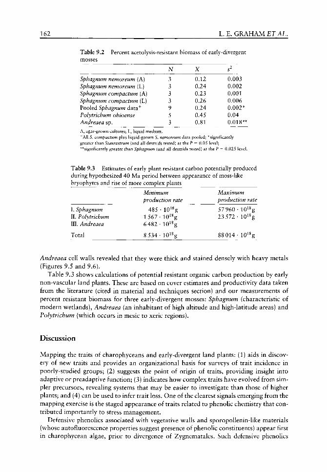
N
3 3 3 3 9 5 3
X
0.12 0.24 0.23 0.26 0.24 0.45 0.81
s'
0.003 0.002 0.001 0.006 0.002 0.04 0.018
162 L.E, GRAHAM ETAL.
Table 9.2 Percent acetolysis-resistant biomass of early-divergent mosses
Sphagnum nemoreum (A) Sphagnum nemoreum (L) Sphagnum compactum (A) Sphagnum compactum (L) Pooled Sphagnum data^ Polytrichum ohioense Andreaea sp. A, agar-grown cultures; L, liquid medium, " All S. compactum plus liquid-grown S. nemoreum data pooled; * significantly greater than Staurastrum (and all desmids tested) at the P = 0.05 level; **significantly greater than Sphagnum (and all desmids tested) at the P = 0.025 level.
Table 9.3 Estimates of early plant resistant carbon potentially produced during hypothesized 40 Ma period between appearance of moss-like bryophytes and rise of more complex plants
Minimum Maximum production rate production rate
I. Sphagnum 485 • lO^^g 57960 • lO^^g II. Polytrichum 1 567 • lO^^g 23 572 • lO^^g III. Andreaea 6482 • lO^^g
Total 8 534 • lO^^g 88 014 • lO^^g
Andreaea cell walls revealed that they were thick and stained densely with heavy metals (Figures 9.5 and 9.6).
Table 9.3 shows calculations of potential resistant organic carbon production by early non-vascular land plants. These are based on cover estimates and productivity data taken from the literature (cited in material and techniques section) and our measurements of percent resistant biomass for three early-divergent mosses: Sphagnum (characteristic of modern wetlands), Andreaea (an inhabitant of high altitude and high-latitude areas) and Polytrichum (which occurs in mesic to xeric regions).
Discussion
Mapping the traits of charophyceans and early-divergent land plants: (1) aids in discovery of new traits and provides an organizational basis for surveys of trait incidence in poorly-studied groups; (2) suggests the point of origin of traits, providing insight into adaptive or preadaptive function; (3) indicates how complex traits have evolved from simpler precursors, revealing systems that may be easier to investigate than those of higher plants; and (4) can be used to infer trait loss. One of the clearest signals emerging from the mapping exercise is the staged appearance of traits related to phenolic chemistry that contributed importantly to stress management.
Defensive phenolics associated with vegetative walls and sporopoUenin-like materials (whose autofluorescence properties suggest presence of phenolic constituents) appear first in charophycean algae, prior to divergence of Zygnematales. Such defensive phenolics
PHENOLICS AND EARLY PLANTS 163
(Appendix 9.1, Traits 2 and 4) were likely inherited by embryophytes (Graham, 1996), where they serve multiple roles (Cooper-Driver, 2001). Intracellular flavonoids and lignans, whose synthesis depends upon occurrence of the phenylpropanoid (C5C3) pathway, appear during divergence of bryophyte groups, but the order of first occurrence is unclear because flavonoids are not reported for hornworts and lignans have not been reported for mosses (Appendix 9.1, Traits 8 and 9). Presence of flavonoids and lignans impHes the existence of biosynthetic pathways leading to production of cinnamic acid by deamina-tion of phenylalanine, hydroxylation of cinnamate to p-coumaric acid and synthesis of ferulic acid and other compounds that are known intermediates and/or constituents of vascular plant lignin, cutin, suberin and sporopollenin (Cooper-Driver, 2001). O-methyl transferases, which confer greater reactivity to the number 4 carbon of phenylpropanoid monomers and thus facilitate formation of 8-0-4 (/3-0-4) linkages (widely used to define lignin), appeared before the divergence of vascular plants (Appendix 9.1, Trait 14). The pattern of trait appearance suggests that biosynthetic and regulatory processes preadap-tive to the evolution of lignin likely occurred in non-vascular plants, including ancestors of modern bryophyte and charophycean lineages. Trait mapping also suggests that early phenolics could have been preadaptive to the development of stable plant-microbe relationships (Appendix 9.1, Traits 3 and 11). As in modern plants, phenolic compounds may have controlled microbial behaviour, allowing microbes to live in close proximity to algae and early land plants without becoming pathogenic.
Our thioacidolysis data indicate that phenolic j8-0-4 linkages are absent from nonvascular plants and charophyceans. Although lacking /3-0-4 linkages. Sphagnum generates phenylpropanoids from phenylalanine via activity of phenylalanine ammonia lyase (PAL) and 4-cinnamic acid hydroxylase (4-CL) (Rasmussen et al.^ 1995), as do vascular plants. Higher plant cell walls contain covalently bound non-lignin phenolics, which have been proposed to inhibit microbial degradation, serve as lignin-initiation sites, or limit wall extensibility (Wallace and Fry, 1994). We speculate that these higher plant wall phenolics may be related to those found in charophyceans and bryophytes. Our thioacidolysis results, that p-hydroxyphenyl monomers were more abundant in lycophytes (earliest-divergent extant vascular plants) than in the other vascular plants we examined, suggests that early lignins may similarly have been richer in non-methoxylated monomers than those characteristic of modern gymnosperms and angiosperms, where dominant monomers are methoxylated and j8-0-4 bonds comprise 50-70% of linkages (Lewis, 1999). Quantitative assessment of resistant carbon content of selected desmids indicates that there is considerable variability among genera, with some desmids apparently lacking resistant walls, most tested species possessing resistant walls and Staurastrum having the largest proportion of resistant dry weight among desmid taxa tested. The latter result is consistent with: (1) the finding that Staurastrum is among the algae most resistant to microbial degradation (Gunnison and Alexander, 1975a); (2) the presence in Staurastrum's walls of a 'lignin-like' phenoHc polymer in amounts sufficient for chemical extraction, pyrolysis GC, thin-layer chromatography and other chemical analysis procedures (Gunnison and Alexander, 1975b); and (3) strong autofluorescence in V and UV excitation of Staurastrum cell walls both before and after high-temperature acid hydrolysis (Kroken et al.^ 1996). Findings reported in the present work suggest that a variety of desmids (though not all) likely possess resistant cell walls that may protect them from microbial degradation and possibly also desiccation in some cases. Our data may explain: (1) the survival of desmid cells of several species after 3 months of drying and at depths to 6 cm in mud (Brook and WiUiamson, 1988); and (2) the occurrence of the
164 L. E.GRAHAM £TAL.
earliest known putative fossil desmid (Paleoclosterium) in Middle Devonian deposits (Baschnagel, 1966).
Desmidium grevillii and Hyalotheca dissiliens, found in this study to possess similar levels (8% dry weight) of resistant biomass, are both known to harbour epibiontic bacterial communities. These communities include saprophytes having the potential to degrade carbohydrate components of cell walls, as well as members of the alpha proteobacteria (Fisher et al., 1998a). Since many of the alpha proteobacteria are nitrogen fixers, it is possible (though not as yet demonstrated) that some desmid bacterial epibionts may provide nutritional advantage in nutrient-poor bog habitats. The presence of hydrolysis-resistant walls may reduce the chances that desmids might suffer deleterious effects, such as wall degradation, as a result of the activities of their epibiontic bacterial community. We propose that resistant wall phenolics originated in aquatic charophyceans as antimicrobial defences (that may also have provided some desiccation resistance), then acquired additional functions, including structural support and UV-screening in early land plants.
Our acetolysis analysis, demonstrating high proportions of resistant dry weight biomass in Sphagnum and other bryophytes, are consistent with conclusions that Sphagnum moss walls contain a 'lignin-like' 'polyphenolic network composed of p-hydroxyphenyl groups' 'linked with simple ether and ester bonds, interspersed with cinnamic acid' (Williams et ai, 1998). Our results help to explain why moss litter decomposes at 1-10% of the rate of vascular plants (Oechel and Van Cleve, 1986 and works cited therein).
Adaptive utility for high levels of wall phenolics in mosses might include: (1) resistance to attack by pathogenic bacteria, protists and fungi; (2) increased stability of cell walls, contributing to the ability to achieve increased height; (3) UV-damage resistance; and (4) desiccation resistance. Sphagnum occupies moist to waterlogged habitats in which bacterial abundance and activities are surprisingly high (Fisher et ai, 1998b). Hydrolysis-resistant cell walls may be helpful in resisting microbial attack and may also contribute to dimensional stability of the large, empty, porose water-holding hyaline cells of Sphagnum, which might otherwise tend to collapse. The relatively large size of Polytrichum erect shoots and the ability to form tall moss turf may be due in part to compression resistance aided by the presence of wall phenolics. Prevention of attack by microorganisms (and perhaps also invertebrate herbivores) provided by phenolic wall compounds may also contribute to biomass accumulation by Polytrichum, an inhabitant of mesic environments worldwide. The primary benefit obtained by Andreaea from its very high dry weight content of hydrolysis-resistant wall compounds may be UV protection afforded by phenolics, whose presence is implied by specific autofluorescence properties. This high altitude/high latitude moss occurs in some of the most UV-impacted habitats on earth. Resistant wall compounds may contribute to plant strength and formation of short, but high-density turfs and cushions, as may the extreme thickness of cell walls, an advantage in its harsh, windswept habitat. Wall phenolics may help maintain dimensional stability during cycles of hydration and dehydration, as well as contributing to desiccation and decay resistance. The bryalean moss, Racocarpus purpurascens, which likewise grows on rocks at high altitude, is reported to have cell walls consisting mainly of 'lignin', hemicellulose and cellulose in a ratio of about 9:8:5, that are architecturally adapted for absorption of water from fog, dew or rain (Edelmann et al., 1998). The extreme habitats of Andreaea and Racocarpus model the exposed, bare rock surfaces that may have been occupied by some early land plants.
The rise of vascular land plants is regarded as an exceedingly important biogeochemi-cal event in Earth's history because it is proposed to have played a dominant role in the dramatic Palaeozoic reduction in atmospheric CO2 level (Berner, 1997) (but see Boucot

PHENOLICS AND EARLY PLANTS 165
and Gray, 2001 for a critique). The twin mechanisms of this CO2 reduction are suggested (by Berner, 1997) to have been: (1) an increase in weathering of siHcate minerals by root systems; and (2) burial of decay-resistant organic carbon in the form of lignin. However, unequivocal fossil evidence for roots (assembled by Kenrick and Crane, 1997) does not occur until the Early Devonian, whereas Berner's (1997) models based on geochemical evidence suggest that atmospheric CO2 drawdown began prior to that time. Analyses of carbon isotopic ratios in Fe(C03)0H of an upper Ordovician (440 myr) goethite dominating an oolitic ironstone (Neda Formation, Neda, Wisconsin, USA) suggest that the productivity of the prevascular biota was similar to that on modern soils (Yapp and Poths, 1994). Boucot and Gray (2001) point out that prevascular land plants, terrestrial and freshwater cyanobacteria, and marine algae may have generated considerable buried carbon during the Precambrian and Early Palaeozoic. Our results support Boucot and Gray's (2001) view that early bryophyte-like land plants may have played an important role in the carbon cycle prior to the rise of vascular plants.
Hanson et ah (1999) demonstrated that many mosses, including Polytrichum and Sphagnum, have the capacity to generate isoprene, which may protect the photosynthetic apparatus under high irradiance and temperature conditions. Polytrichum and Andreaea maintain the ability to photosynthesize at high levels after repeated cycles of dehydration and rehydration (Davey, 1997) associated with intermittently desiccating environments. Such environmental conditions probably prevailed in Ordovician-Silurian times, when a shady canopy of larger plants was absent. Soil-enriching, nutrient-binding humus is generated by modern mosses, which are the primary source of fresh organic matter in regions such as Antarctica (Beyer et al, 1997).
Conclusions
The original data presented here, together with published molecular systematic and fossil evidence, suggest that productive bryophyte-like early land plants probably enriched soils with organic carbon and contributed to CO2 sequestration for a long period prior to the rise of rooted plants. Even if as little as 1% of the 8540-82 000 X 10^^ g resistant C that we estimated to have been produced by early bryophyte-like plants survived decomposition and weathering processes and was sequestered, early non-vascular plants could have removed 0.6-6% of atmospheric CO2. If Berner's (1997) CO2 drawdown models are correct in inferring that atmospheric CO2 levels dropped from 20X present levels to lOX present levels during the 40 million year time period just prior to the rise of vascular plants, then our estimate for non-vascular plant resistant C sequestration (1% of resistant C produced) could account for 1-11% of this CO2 drawdown.
Acknowledgements
We thank Kandis Elliot for rendering Figure 9.1 and two anonymous reviewers for helpful recommendations.
References
Bailey RG. 1989. Ecoregions of the continents. Washington DC: Forest Service, US Department of Agriculture.

166 L, E.GRAHAM £TAL.
Baschnagel RA. 1966. New fossil algae from the Middle Devonian of New York. Transactions of the American Microbiological Society 85: 297-502.
Berner RA. 1997. The rise of plants and their effect on weathering and atmospheric CO2. Science 176: 544-545.
Beyer L, Blume HP, Sorge C, et al. 1997. Humus composition and transformations in a pergelic cry-ohemist of coastal Antarctica. Arctic and Alpine Research 29: 358-365.
Boucot AJ, Gray J. 2001. A critique of Phanerozoic climatic models involving changes in the CO2 content of the atmosphere. Earth Science Reviews 56: 1-159.
Brook AJ, Williamson DB. 1988. The survival of desmids on the drying mud of a small lake. In: Round FE, ed. Algae and the Aquatic Environment. Bristol: BioPress, 186-196.
Cole DR, Monger HC. 1994. Influence of atmospheric CO2 on the decline of C4 plants during the last deglaciation. Nature 368: 533-536.
Cooper-Driver GA. 2001. Biological roles for phenolic compounds in the evolution of early land plants. In: Gensel P, Edwards D, eds. Plants Invade the Land. New York: Columbia University Press, 159-172.
Cooper-Driver GA, Bhattacharya M. 1998. Role of phenolics in plant evolution. Fhy to chemistry 49:1165-1174.
Cullman F, Becker H. 1999. Lignans from the liverwort Lepicolea ochroleuca. Fhy to chemistry 52: 1651-1656.
Davey MC. 1997. Effects of continuous and repeated dehydration on carbon fixation by bryophytes from the maritime Antarctic. Oecologia 110: 25-31.
Davis RC. 1981. Structure and function of two Antarctic terrestrial moss communities. Ecological Monographs 51: 125-143.
Dejesus MD, Tabatabai F, Chapman DJ. 1989. Taxonomic distribution of copper-zinc superoxide dismutase in green algae and its phylogenetic importance. Journal of Phycology 25: 767-772.
Delwiche CF, Graham LE, Thomson N. 1989. Lignin-like compounds and sporopollenin in Coleochaete, an algal model for land plant ancestry. Science 245: 399-401.
Edelmann HG, Neihuis C, Jarvis M, et al. 1998. Ultrastructure and chemistry of the cell wall of the moss Rhacocarpus purpurascens (Rhacocarpaceae): a puzzling architecture among plants. Planta 206: 315-321.
Edwards D, Duckett JG, Richardson JB. 1995. Hepatic characters in the earliest land plants. Nature 374: 635-636.
Fisher MM, Wilcox LW, Graham LG 1998a. Molecular characterization of epiphytic bacterial communities on charophycean green algae. Applied and Environmental Microbiology 64: 4384-4389.
Fisher MM, Graham JM, Graham LG. 1998b. Bacterial abundance and activity across sites within two northern Wisconsin Sphagnum bogs. Microbial Ecology 36: 259-269.
Gorham E. 1991. Northern peatlands: Role in the carbon cycle and probable responses to climatic warming. Ecological Applications 1: 182-195.
Graham LE. 1990. Meiospore formation in charophycean algae. In: Blackmore S, Knox RB, eds. Microspores: Evolution and Ontogeny. London: Academic Press, 43-54.
Graham LE. 1993. Origin of Land Plants. New York: John Wiley & Sons, Inc. Graham L. 1996. Green algae to land plants: An evolutionary transition. Journal of Plant Research
109: 241-251. Graham LE, Gray J. 2001. The origin, morphology and ecophysiology of early embryophytes:
Neontological and palaeontological perspectives. In: Gensel P, Edwards D, eds. Plants Invade the Land. New York: Columbia University Press, 140-158.
Graham LG, Wilcox LW. 2000a. The origin of alternation of generations in land plants: a focus on matrotrophy and hexose transport. Philosophical Transactions of the Royal Society of London B 335: 757-767.
Graham LG, Wilcox LW. 2000b. Algae. Upper Saddle River, NJ: Prentice Hall. Graham LE, Cook ME, Busse JS. 2000. The origin of plants: Body plan changes contributing to a
major evolutionary radiation. Proceedings of the National Academy of Sciences 97: 4535-4540. Gunnison D, Alexander M. 1975a. Resistance and susceptibility of algae to decomposition by nat
ural microbial communities. Limnology and Oceanography 20: 64-70.
PHENOLICS A N D EARLY PLANTS 167
Gunnison D, Alexander M. 1975b. Basis for the resistance of several algae to microbial decomposition. Applied Microbiology 29: 729-738.
Hanson DT, Swanson S, Graham LE, Sharkey TD. 1999. Evolutionary significance of isoprene emission from mosses. American Journal of Botany 86: 634-639.
Heckman DS, Geiser DM, Eidell BR, et al. 2001. Molecular evidence for the early colonization of land by fungi and plants. Science 293: 1129-1133.
Karol KG, McCourt RM, Cimino MT, DeWiche CF. 2001. The closest living relatives of land plants. Science 294: 2351-2353.
Kenrick P, Crane PR. 1997. The origin and early evolution of plants on land. Nature 389: 33-39. Khnger LF, Zimmerman PR, Greenberg JP, etal. 1994. Carbon trace gas fluxes along a successional
gradient in the Hudson Bay lowland. Journal of Geophysical Research-Atmospheres 99: 1469-1494.
Kodner RB, Graham LE. 2001. High-temperature, acid-hydrolyzed remains oi Polytrichum (Musci, Polytrichaceae) resemble enigmatic Silurian-Devonian tubular microfossils. American Journal of Botany 88: 462-466.
Kroken SB, Graham LE, Cook ME. 1996. Occurrence and evolutionary significance of resistant cell walls in charophytes and bryophytes. American Journal of Botany 83: 1241-1254.
LaPierre C. 1993. Applications of new methods for the investigation of lignin structure. In: Jung HG, Buxton DR, Hatfield RD, Ralph J, eds. Forage Cell Wall Structure and Digestibility. Madison, WI: American Society of Agronomy, 133-166.
Lau E. 2000. Molecular assessment of microbial communities in a Northern Wisconsin peatland. D. Phil. Thesis, University of Wisconsin-Madison.
Lehman JT. 1976. Ecological and nutritional studies on Dinobryon Ehrenb.: Seasonal periodicity and the phosphate toxicity problem. Limnology and Oceanography 21: 646-658.
Lewis N. 1999. A 20th century roller coaster ride: a short account of lignification. Current Opinion in Plant Biology 2: 153-162.
Lewis N, Davin LB. 1994. Evolution of lignan and neolignan biochemical pathways. American Chemical Society Symposium Series 562: 202-246.
Lewis LA, Mishler BD, Vilgalys R. 1997. Phylogenetic relationships of the liverworts (Hepaticae), a basal embryophyte lineage, inferred from nucleotide sequence data of the chloroplast gene rbcL. Molecular Phylogenetics and Evolution 7: 377-393.
Longton RE. 1988. Biology of Polar Bryophytes and Lichens. Cambridge: Cambridge University Press.
Newton AE, Cox CJ, Duckett JG, et al. 2000. Evolution of the major moss lineages: phylogenetic analyses based on multiple gene sequences and morphology. The Bryologist 103: 187-211.
Nickrent DL, Parkinson CL, Palmer JD, Duff RJ. 2000. Multigene phylogeny of land plants with special reference to bryophytes and the earliest land plants. Molecular Biological Evolution 17: 1885-1895.
Oechel WC, Van Cleve K. 1986. The role of bryophytes in the nutrient cycling in the taiga. In: Van Cleve K, Chapin IFS, Flanagan PW, et al., eds. Forest Ecosystems in the Alaskan Taiga. New York: Springer-Verlag, 121-137.
Oliver MJ, Tuba Z, Mishler BD. 2000. The evolution of vegetative desiccation tolerance in land plants. Plant Ecology 151: 85-100.
Overbeck F, Happach H. 1957. Uber das Wachstum und den Wasserhaushalt einiger Hochmoorsphagnen. Flora 144: 335-402.
Proctor MCF. 2000. The bryophyte paradox: tolerance of desiccation, evasion of drought. Plant Ecology 151: 41-49.
Qiu Y-L, Palmer JD. 1999. Phylogeny of early land plants: insights from genes and genomes. Trends in Plant Science 4: 26-30.
Rasmussen S, Peters G, Rudolph H. 1995. Regulation of phenylpropanoid metabolism by exogenous precursors in axenic cultures of Sphagnum fallax. Physiologia Plantarum 95: 83-90.
Read DJ, Duckett JG, Francis R, et al. 2000. Symbiotic fungal associations in 'lower' land plants. Philosophical Transactions of the Royal Society London B 355: 815-831.
Redeker D, Kodner R, Graham LE. 2000. Glomalean fungi from the Ordovician. Science 289: 1920-1921.

168 L. E . G R A H A M E T A L .
Remy E, Taylor TN, Hass H, Kerp H. 1994. Four hundred-million-year-old vesicular arbuscular mycorrhizae. Proceedings of the National Academy of Sciences USA 91: 11841-11843.
Takeda R, Hasegawa J, Shinozaki M. 1990. The first isolation of lignans, megacerotonic acid and anthocerotonic acid, from non-vascular plants, Anthocerotae (hornworts). Tetrahedron Letters 31:4159-4162.
Vitt DH. 2000. Peatlands: ecosystems dominated by bryophytes. In: Shaw AJ, Goffinet B, eds. Bryophyte Biology. Cambridge: Cambridge University Press, 312-343.
Wallace G, Fry SC. 1994. Phenolic components of the plant cell wall. International Review of Cytology 151: 229-267.
Waters ER, Vierling E. 1999. The divergence of plant cytosolic small heat shock proteins preceded the divergence of mosses. Molecular Biology and Evolution 16: 127-139.
Wellman CH, Gray J. 2000. The microfossil record of early land plants. Philosophical Transactions of the Royal Society London B 355: 717-732.
Williams CJ, Yavitt JB, Wieder RK, Cleavitt NL. 1998. Cupric oxide oxidation products of northern peat and peat-forming plants. Canadian Journal of Botany 76: 51-62.
Yapp CJ, Poths H. 1994. Productivity of pre-vascular continental biota inferred from the Fe(C03)0H content of goethite. Nature 368: 49-51.
Appendix 9.1 Physiological traits related to early stress adaptation in land plants
Trait i , Cu, Zn superoxide dismutase, is absent from most green algae, but occurs in all streptophytes that have been examined (Dejesus etal., 1989). This enzyme prevents damage to cells by eliminating highly reactive oxygen radicals and may have contributed to early plant survival in the terrestrial atmosphere v^here O2 diffuses into cells at a rate 10 000 times higher than in water.
Trait 2, decay- and acetolysis-resistant, autofluorescent phenolics in vegetative cell w^alls in some Zygnematales (Gunnison and Alexander, 1975a) as well as later-divergent charophyceans and bryophytes (Kroken et ai, 1996) is hypothesized to confer resistance to microbial attack (Gunnison and Alexander, 1975b), UV and possibly also desiccation and herbivory.
Trait 3 , bacterial associations having putative beneficial properties with charophyceans (Fisher et al, 1998a) and Sphagnum moss (Lau, 2000) suggest that such associations may be of ancient origin, possibly helping to alleviate low-nutrient stress.
Trait 4, sporopollenin-like layers in charophycean zygote walls (Delwiche et al.^ 1989 and references cited therein), likely provide protection from microbial attack and possibly also desiccation during resting periods and dispersal of later-divergent charophyceans. Biochemical homology to embryophytic sporopollenin requires investigation.
Trait 5, sporopollenin-walled spores allowed meiospores of early plants (known as microfossils) to disperse via air. Sporopollenin provided dimensional stability, pathogen resistance (Graham and Gray, 2001) and possibly also desiccation resistance. Charophycean meiospores are devoid of sporopollenin wall layers (Graham, 1990).
Trait 6, tissues produced by a histogenetic meristem that cuts off derivatives in more than two directions is hypothesized as an adaptat ion that helped to prevent water loss from early plants by reducing surface area to volume ratio (Graham, 1996).
Trait 7, cuticle present on some moss tissues and hornwort sporophytes (Kroken et ai,
1996) (for which autofluorescence properties suggest inclusion of phenolics) likely reduces desiccation of underlying tissues, retards microbial attack and acts as a UV screen.

PHENOLICS AND EARLY PLANTS 169
Trait 8, lignans - small, soluble polymers of phenylpropanoid monomers similar to those in lignin - characterize hornworts (Takeda et al.^ 1990), liverworts (e.g. Cullman and Becker, 1999) and vascular plants (Lewis and Davin, 1994), but have not been reported from charophyceans or mosses. Because lignans function in lignin synthesis and perform as antibacterial, antifungal, or antiviral agents, or serve as antioxidants, they may have helped early land plants cope with the oxidative effects of higher terrestrial O2 diffusion rates and pathogen attack or served as intracellular UV screens.
Trait 9, simple flavonoids, derived from the phenylpropanoid pathway and involving chalcone synthase, characterize at least some mosses and liverworts (but not hornworts, so far as is known) (Cooper-Driver and Bhattacharya, 1998). UV screening, antioxidant activity, mediation of plant-microbe symbioses and pathogen defences are among the roles played by flavonoids in modern plants that might be extrapolated to early plants.
Trait 10, vegetative desiccation tolerance, is present in at least some members of liverwort, hornwort and moss lineages, as well as some lycophytes, pteridophytes and angiosperms, and is regarded as a crucial step in the ecological transition from water to land (Oliver et al, 2000; Proctor, 2000).
Trait 11 reflects associations of modern liverworts, hornworts and most groups of vascular plants (though not mosses or Equisetum^ so far as is known) with 'mycorrhizal' fungi (Read et aL, 2000 and works cited therein). Presence of 455-460 My fossil gloma-lean fungi, which now occur only in symbiotic associations, together with molecular systematic analysis of the fungi (Redeker et aL, 2000 and works cited therein) and later fossil associations (Remy et aL, 1994) suggest that plants acquired fungal symbionts very early in their history. However, the time at which plants accrued positive benefits, namely increased access to nutrients and water, is not yet clear; the earliest plant-fungal associations may have been parasitic or saprophytic.
Trait 12, isoprene production in response to thermal and desiccation stress, has been demonstrated to occur in a hornwort, several mosses and various vascular plants, possibly functioning in protection of the photosynthetic apparatus. Absence of isoprene production from all Hverworts examined (Hanson et aL, 1999) suggests that the enzyme isoprene synthase arose after the divergence of liverworts.
Trait 13, small heat shock proteins are produced by mosses and vascular plants in greater amounts as a response to high-temperature stress; they prevent thermal aggregation of cell proteins by maintaining them in a folded state. At least two of the five vascular plant families of small heat shock proteins occur in mosses (Waters and Vierling, 1999).
Trait 14, lignin, if defined as a polymer of phenylpropanoid monomers - at least some of which are linked by /3-0-4 (8-0-4) bonds - occurs among extant plants only in vascular plants (Lewis, 1999) and coincides with the occurrence of O-methyl transferases.

10 Plant cuticles: multifunctional interfaces
between plant and environment
Hendrik Bargel, Wilhelm Barthlott, Kerstin Koch, Lukas Schreiber and Christoph Neinhuis
CONTENTS.
Introduction 171
The multifunctional interface 174 Transport properties of plant cuticles 175 Wax movement: a simple solution? 178 Interactions with the biotic and abiotic environment 180 Water repellency and self-cleaning property 181 Biomechanical properties 185
Acknow ledgements 187
References 187
Introduction
The plant cuticle covers all primary parts of vascular plants (except roots) and many bryophytes as a thin extracellular membrane. Deposited by the epidermis, the cuticle serves as the crucial protective layer between the organism and its environment, representing one of the largest interfaces between biosphere and atmosphere covering more than 1.2 X 10^ km^ in total (Riederer and Schreiber, 1995). The cuticle has been of particular interest since it was first described by Brogniart (1834). In early studies, macroscopically visible features and fine structures were described and analysed. The reader is referred to Martin and Juniper (1970) or Holloway (1982b) for a summary. More recently, the ontogenetic development and chemical composition of both cuticle and waxes, as well as biosynthesis and transport phenomena have been subjects of major interest (Schonherr, 1982; Jeffree, 1986; Hamilton, 1995; Kolattukudy, 2001).
The cuticle represents a natural composite that consists mainly of two hydrophobic components, the insoluble biopolyester cutin and soluble lipids (Martin and Juniper, 1970). This type of lipid-derived polyester membrane is restricted to plants, whereas animals use carbohydrates or proteins as outer coverings (Kolattukudy, 2001). Minor amounts of cutinized polysaccharide fibre including cellulose and hemicellulose and a frequently abundant
The Evolution of Plant Physiology Copyright © 2004 Elsevier Ltd ISBN 0-12-33955-26 All rights of reproduction in any form reserved
172 H.BARGEL ETAL.
pectin layer link the plant cuticle to the cell wall (HoUoway, 1994). The basic framework is made of cutin, which is composed of saturated Ci^ hydroxy- and unsaturated Cig hydroxy-epoxy fatty acids (Kolattukudy, 1980a). The most common monomers are 16-hydroxy Ci6 acid, 9- or 10,16-dihydroxy Ci^ acid, 18-hydroxy-9,10-epoxy Cig acid and 9,10,18-trihydroxy Cjg acid (Kolattukudy, 2001). The ratio of the Ci^/Cig monomers is organ as well as species specific and appears to vary. In general, fast growing plant organs, e.g. young leaves or fruits, seem to have a higher content of Ci^ monomers (Espelie et al.^ 1979; Baker et ai, 1982). The cutin monomers form a linear polyester by esterified primary hydroxyl groups, and cross-linking of esterfied secondary hydroxyl groups yields a three-dimensional network (Kolattukudy, 1996; Ray et al.^ 1998). In addition to cutin, a highly resistant residue, originally found in fossilized cuticles but also present in a few recent species, is named cutan (Nip et al.^ 1986; CoUinson et aL, 1998). Cutan has a high fossilization potential (van Bergen et al., 1995; Mosle et al., 1997) because of ether-linked long chain alkyl moieties mainly of even carbon chain length (C22-C34; CoUinson et al., 1998; Villena et al.^ 1999). However, the origin and precise structure of this long chain aliphatic macro-molecule still remains unclear (Jeffree, 1996; see also van Bergen et ai, Chapter 8).
The soluble lipids, collectively termed waxes, are embedded into the cutin matrix (intra-cuticular) as well as deposited onto the surface (epicuticular). Whereas the intracuticular waxes may be either amorphous or crystalline (Riederer and Schreiber, 1995), the epicuticular waxes often form complex three-dimensional crystalline structures in addition to thin amorphous films (Bianchi, 1995), a feature useful in plant systematics (Barthlott, 1981; Giilz, 1994; Barthlott et ai, 1998; Figures 10.1-10.6). Plant cuticular waxes are a complex mixture of aliphatic or cyclic components, including hydrocarbons, long chain fatty acids, aldehydes, primary and secondary alcohols, ketones, j8-diketones and pentacyclic triter-penoids (Kolattukudy, 1980b; Walton, 1990), with overall dominating chain length for the aliphatic compounds of C20 to C35 (von Wettstein-Knowles, 1995). It has been shown in recrystallization studies that crystalline wax projections originate from self-assembly based on their specific chemistry (Jeffree et ai, 1975; Jetter and Riederer, 1995; Meusel et al., 2000). Moreover, one predominating component often is responsible for a characteristic type of epicuticular wax crystals (von Wettstein-Knowles, 1972; Barthlott et aL, 1996; Meusel et aL, 1999). Nevertheless, wax composition is subject to great variation among plant species as well as during organ ontogeny, indicating a very customizable system (Markstadter, 1994; Riederer and Markstadter, 1996). It is worth noting that former studies on cuticular wax composition are more or less based on a mixture of intra- and epicuticular waxes caused by the extraction methods (Riederer and Markstadter, 1996), whereas it is now possible to separate both fractions distinctively with the help of a recently developed freeze-embedding method utilizing glycerol (Ensikat et aL, 2000; Jetter and Riederer, 2000).
Based on the results of chemical composition for different species, the plant cuticle appears to be without uniform composition and alterable during organ growth. Additional evidence for inhomogeneity was obtained from ultrastructural studies, e.g. the distinction between a thin outer 'cuticle proper' (<200 nm) with a variable number of layers supposed to contain either wax or cutin (lamellate region) and an inner 'cuticle layer' of variable thickness (up to 17 |jLm) (HoUoway, 1982b). The latter represents a mixture of waxes, cutin and polysaccharide fibres originating in the cell wall, in addition to pectins (reticulate region), resulting from the impregnation of the epidermal cell wall (Jeffree, 1996). There have been several attempts to summarize the structural variation, most prominently by HoUoway (1982b, 1994). He identified six different types of lameUated cuticles and an extended review by Jeffree (1996) displays data of 119 species from 94 genera. From a systematic
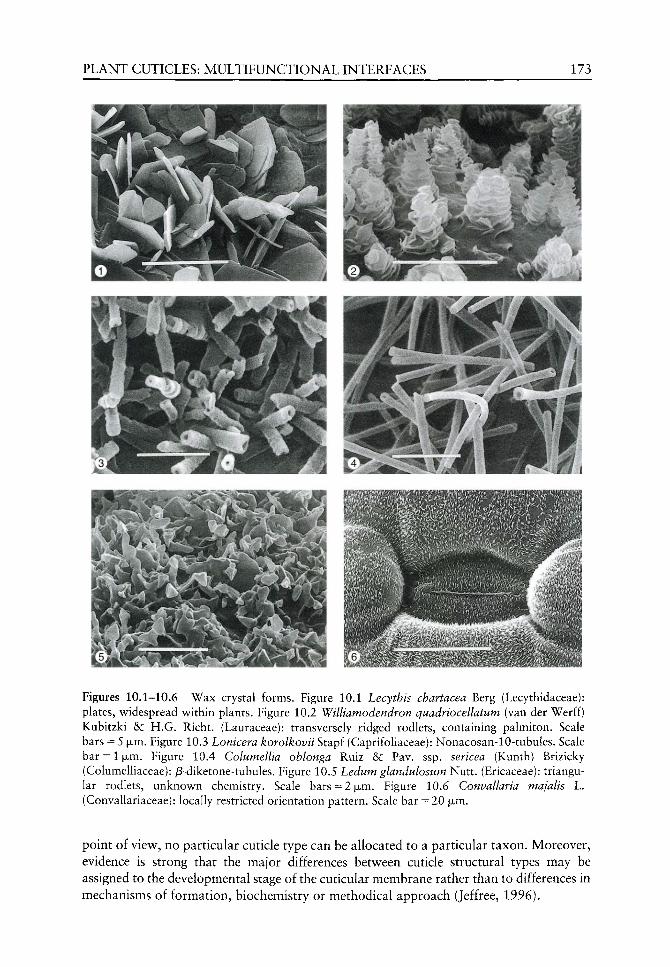
PLANT CUTICLES: MULTIFUNCTIONAL INTERFACES 173
Figures 10.1-10.6 Wax crystal forms. Figure 10.1 Lecythis chartacea Berg (Lecythidaceae): plates, widespread within plants. Figure 10.2 Williamodendron quadriocellatum (van der Werff) Kubitzki &: H.G. Richt. (Lauraceae): transversely ridged rodlets, containing palmiton. Scale bars = 5 fjim. Figure 10.3 Lonicera korolkovii Stapf (Caprifoliaceae): Nonacosan-10-tubules. Scale bar = 1 |jLm. Figure 10.4 Columellia oblonga Ruiz & Pav. ssp. sericea (Kunth) Brizicky (Columelliaceae): j8-diketone-tubules. Figure 10.5 Ledum glandulosum Nutt. (Ericaceae): triangular rodlets, unknown chemistry. Scale bars=2(xm. Figure 10.6 Convallaria majalis L. (Convallariaceae): locally restricted orientation pattern. Scale bar = 20 ixm.
point of view, no particular cuticle type can be allocated to a particular taxon. Moreover, evidence is strong that the major differences between cuticle structural types may be assigned to the developmental stage of the cuticular membrane rather than to differences in mechanisms of formation, biochemistry or methodical approach (Jeffree, 1996).

174 H.BARGEL ETAL.
Both cutin and the wax compounds, except the cycHc ones, are ahphatic homologues derived by de-novo synthesis of Ci6 and Cis fatty acids in piastids of epidermal cells (Post-Beitenmiller, 1996). Two enzyme complexes, namely fatty acid synthetase and palmitoyl (Ci6)-elongase, have been found to be responsible for the basic fatty acid synthesis by a stepwise condensation-elongation mechanism (von Wettstein-Knowles, 1995). The resulting Ci^/Cig fatty acids are then processed or elongated extraplastidally by either several microsomal enzymes to the different wax derivates, or by a family of mixed-function oxidases to cutin monomers (von Wettstein-Knowles, 1987; Post-Beitenmiller, 1996). However, little is known about either the molecular and genetic control, or the precise location where the biosynthetic processes take place.
The evolution of the cuticle is strongly interconnected to that of plant life on land. Pioneering land plants had to face the physical and physiological problems resulting from life out of water, such as gravity, desiccation, UV radiation or pollutant deposition on their surface (Edwards et aL, 1982). Earliest fossils, interpreted as cuticles, date back to the Ordovician and are more frequent in the Silurian (Edwards et al.^ 1996; see also Raven and Edwards, Chapter 2). Due to their great functional importance, which will be discussed below, it is highly probable that early land plants were already covered, at least partially, by epicuticular wax crystals. These waxes, however, have no fossil record because of their delicate nature. Therefore, we only can speculate from extant plants what might have been the epicuticular coverings in extinct plants. The most widespread type of wax crystals to be found among all land plants is represented by small platelets, often composed of primary alcohols. Their most important function, especially in mosses and ferns, seems to be the protection against the formation of water films above stomata (Neinhuis and Barthlott, 1997) or other air-filled spaces, such as assimilation lamellae in Polytrichales (Neinhuis and Jetter, 1995), presumably to maintain high gas exchange rates. Another common type is represented by small tubules composed of, or dominated by, the secondary alcohol nonacosan-10-ol. It has been shown to occur in sporophytes of the moss family Polytrichaceae (Neinhuis and Jetter, 1995), in the fern genus Pteris L. (unpublished data), and characterizes most gymnosperms (Wilhelmi and Barthlott, 1997), as well as certain groups of angiosperms (Hennig et ai, 1994). This may indicate a mutual biosynthetic pathway once developed by the common ancestor of land plants. This question, however, remains open since the particular pathway leading to noncosan-10-ol is unknown to date.
Generally, the diversity of wax crystals is lowest in lycophytes and mosses. In ferns a large number of substances have been analysed, but only for a limited number of species. Gymnosperms are characterized mainly by nonacosan-10-ol tubules and appear to be extremely uniform with respect to epicuticular waxes, except for cycads which show some degree of variability. On the other hand, angiosperms exhibit by far the greatest diversity, which even allows specific groups to be circumscribed on the ordinal level or above. A detailed description and discussion of the systematic significance of wax crystals among land plants is given by Barthlott et al. (2002).
Despite the variability of both cuticle and epicuticular wax, this chapter embraces the complex processes at the interface between plant and environment and thus stresses the relevance of this multifunctional hydrophobic coverage of land plants.
The multifunctional interface
The multiple constraints on the evolution of such an outer envelope, found on fossil and recent plant material, can be summarized by transport phenomena across the cuticle.
PLANT CUTICLES: MULTIFUNCTIONAL INTERFACES 175
interaction with biotic and abiotic factors and biomechanical requirements. Most of the functions described in the following are related to the waxes within and upon the cuticle, a fact that stresses their importance in the interfacial interactions.
Transport properties of plant cuticles
One of the main functions of the cuticle, if not the most important, is the limitation of uncontrolled water loss via evaporation (Schonherr, 1982). Thus, selective pressure acted strongly on plants to develop an outer hydrophobic envelope functioning as a compromise between the contrary demands of desiccation avoidance and free gas exchange (Edwards et al,^ 1996; Bateman et al.^ 1998). Furthermore, leaching of organic and inorganic substances from the leaf interior has to be minimized (Tukey, 1970), as well as providing an effective barrier in terms of foliar uptake (Schonherr and Riederer, 1989) - a crucial role of the plant cuticle. Regarding the transport of organic and inorganic substances across plant cuticles, four different groups of compounds can be treated separately: (1) organic non-electrolytes (e.g. pesticides and xenobiotics); (2) water; (3) organic and inorganic ions; and, finally, (4) lipophilic molecules which form the cuticular waxes. The waxes are synthesized in epidermal cells and must cross the outer epidermal wall before they can be either sorbed into the cutin matrix or reach the surface of cuticles were they are deposited as epicuticular waxes. These different groups of compounds are considered in more detail below.
Organic non-electrolytes In the past many investigations have been carried out analysing the transport of neutral organic compounds across plant cuticles (Schonherr and Riederer, 1989) and cuticular waxes (Riederer and Schreiber, 1995), and the effect of plasticizers on mobility in cuticles (Schonherr and Baur, 1994). Since plant cuticles themselves are essentially hydrophobic biopolymers, lipophilic substances generally have fairly high rates of diffusion across them. These investigations were either carried out in ecotoxicological research programmes, thus quantifying the uptake of lipophilic environmental xenobiotics entering the leaves by diffusion across the cuticle (Schonherr and Riederer, 1989), or it was intended to try to optimize the uptake of lipophilic herbicides into leaves (Hess and Foy, 2000). Transport across cuticles was studied using experimental systems at three different levels of complexity: (1) intact leaves (Schreiber and Schonherr, 1992a,b); (2) isolated cuticles (Riederer and Schonherr, 1984, 1985); and (3) isolated and subsequently recrystallized cuticular waxes (Schreiber and Schonherr, 1993). These studies showed that transport properties of recrystallized cuticular waxes, which represents a fairly artificial experimental system, were closely related to the transport properties of isolated cuticles and intact leaves (Kirsch et al.^ 1997). From these results it may be concluded that the transport barrier of cuticles provided by waxes is a system which is self-organized, as waxes spontaneously recrystal-lize leading to similar barrier properties as found in intact leaves and isolated cuticles (Schreiber ^ /., 1996a).
In steady state, transport of neutral molecules across cuticles is conveniently quantified using equation (1):
P - P - A - A c (1)
Flow, f, in mass per time (e.g. g-s~^), is directly proportional the area A (m^) and the driving force Ac, in units mass per volume (e.g. g • m~^). The driving force Ac is defined as the concentration of substance in the donor minus that in the receiver (cjonor ~" receiver)- The permeance P, in units of velocity (m-s~^), is the proportionality factor between P and

176 H.BARGEL ETAL.
A ' Ac. The larger the value of P, the faster the transport across the cuticle and the poorer the barrier properties of cuticles. When P is known, barrier properties of cuticles from different species can be directly compared since they do not depend on experimental boundary conditions such as driving force, exposed cuticle area, or time (Kerler et aL^ 1984). Cuticular permeances of different compounds across intact leaves and isolated cuticles varied between 10~^^olO~^m-s~^ and the extraction of cuticular waxes with organic solvents increased cuticular permeability for neutral organic compounds by factors varying between 10 and 10 000 (Schonherr and Riederer, 1989). This provides good evidence for cuticular waxes forming the transport-limiting barriers of plant cuticles for the diffusion of organic substances.
The permeance P (m • s~ ) itself is a composite quantity (Equation (2)):
- ^
The diffusion coefficient D (m^ • s~ ) describes the mobility of the compound in the cutin and amorphous wax phases. The partition coefficient K (dimensionless) is the ratio of the solubility of the compound in the lipophilic cuticle phase and in water (or other solvents), and A/ (m) gives the path length of diffusion across the cuticle (Kerler et al.^ 1984). In simple homogeneous membranes, A/ would simply represent the thickness of the membrane, but since plant cuticles are heterogeneous membranes with the cuticular waxes basically establishing the transport-limiting barrier (HoUoway, 1982a), the exact path length of diffusion is not known. There are, however, indications that the path length can be more than 2 orders of magnitude longer than the thickness of the cuticles due to the tortuosity of diffusion around impermeable, crystalline wax phases (Baur et al.^ 1999; Buchholz and Schonherr, 2000). The partition coefficient K, a measure of the solubility of the compounds in the lipophilic cutin and wax phases, rises with increasing lipophility of the compounds and can vary by several orders of magnitude (Schonherr and Riederer, 1989). The diffusion coefficient D describes the mobility of the molecules in the cuticular membrane and decreases greatly when the molar volume of the solutes is increased (Baur et al., 1997).
Water Water permeability of plant cuticles has been studied repeatedly over the last decades (Schonherr, 1982; Kerstiens, 1996; Riederer and Schreiber, 2001). These ecophysiological investigations aimed at understanding the amount of water lost by cuticular transpiration under severe environmental conditions, such as water stress and high temperatures (Schreiber and Riederer, 1996), evaluating the limits of survival for plants. Most of these studies were carried out using isolated, astomatous cuticles. As water molecules are non-ionized, water transport across cuticles is normally analysed and quantified in the same way as with lipophilic organic compounds through equations (1) and (2).
Cuticular permeances for water, based on the concentration of liquid water as the driving force at 25°C, were measured on 24 species from different habitats (Schreiber and Riederer, 1996). Permeances varied between 10"^^ and 10~^ m -s"^ and there was a clear relationship between cuticular transpiration and the natural habitat of the investigated species. Water permeability was lowest in tropical lianas reaching above the canopy with high radiation, increased in xeromorphic species naturally growing in the Mediterranean vegetation zone and were highest in mesomorphic species (Riederer and Schreiber, 2001). Most of the test specimens were not collected from their natural habitats but taken from
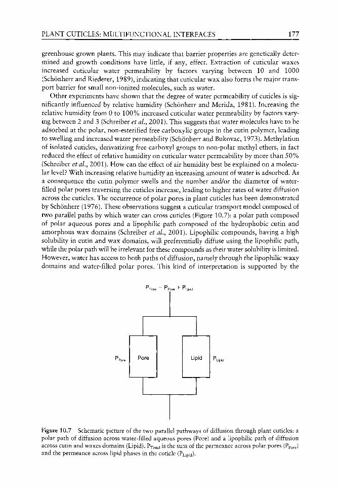
PLANT CUTICLES: MULTIFUNCTIONAL INTERFACES 177
greenhouse grown plants. This may indicate that barrier properties are genetically determined and growth conditions have little, if any, effect. Extraction of cuticular waxes increased cuticular water permeability by factors varying between 10 and 1000 (Schonherr and Riederer, 1989), indicating that cuticular wax also forms the major transport barrier for small non-ionized molecules, such as water.
Other experiments have shown that the degree of water permeability of cuticles is significantly influenced by relative humidity (Schonherr and Merida, 1981). Increasing the relative humidity from 0 to 100% increased cuticular water permeability by factors varying between 2 and 3 (Schreiber et aL, 2001). This suggests that water molecules have to be adsorbed at the polar, non-esterified free carboxylic groups in the cutin polymer, leading to swelling and increased water permeability (Schonherr and Bukovac, 1973). Methylation of isolated cuticles, derivatizing free carboxyl groups to non-polar methyl ethers, in fact reduced the effect of relative humidity on cuticular water permeability by more than 50% (Schreiber et al., 2001). How can the effect of air humidity best be explained on a molecular level? With increasing relative humidity an increasing amount of water is adsorbed. As a consequence the cutin polymer swells and the number and/or the diameter of water-filled polar pores traversing the cuticles increase, leading to higher rates of water diffusion across the cuticles. The occurrence of polar pores in plant cuticles has been demonstrated by Schonherr (1976). These observations suggest a cuticular transport model composed of two parallel paths by which water can cross cuticles (Figure 10.7): a polar path composed of polar aqueous pores and a lipophilic path composed of the hydrophobic cutin and amorphous wax domains (Schreiber et aL, 2001). Lipophilic compounds, having a high solubility in cutin and wax domains, will preferentially diffuse using the lipophilic path, while the polar path will be irrelevant for these compounds as their water solubility is limited. However, water has access to both paths of diffusion, namely through the lipophilic waxy domains and water-filled polar pores. This kind of interpretation is supported by the
•^Total r-'pore "*" "^Upid
Lipid
Figure 10.7 Schematic picture of the two parallel pathways of diffusion through plant cuticles: a polar path of diffusion across water-filled aqueous pores (Pore) and a lipophilic path of diffusion across cutin and waxes domains (Lipid). Pxotal is the sum of the permeance across polar pores (Ppore) and the permeance across lipid phases in the cuticle (Pupid)-

178 H.BARGEL £TAL.
experimental results for the transport of water across cuticles since water transport is strongly influenced by cuticular waxes. This indicates a transport across the lipophilic path, which is also affected by humidity, and suggests a parallel transport across polar pores. This transport model is further supported by studies of the transport of charged organic molecules and inorganic ions across cuticles, as discussed below.
Ions Transport of ions (Schonherr, 2000, 2001) and charged organic molecules (Schonherr, personal communication) across cuticles isolated from several different plant species has been recently studied. Most interestingly, rates of cuticular penetration of these charged compounds were not significantly affected by temperature, since activation energies of permeation estimated from Arrhenius plots (P vs. 1/T) were close to Okjmol"^ (Schonherr, 2000). In contrast to water transport properties, wax extraction for ion transport had only a small effect on rates of penetration, as they increased by factors between 2 and 3. These findings are fundamentally different from those previously published on transport of water and lipophilic molecules across plant cuticles. Activation energies for these two compounds vary between 20 and 200k jmor^ (Schonherr et al., 1979; Baur et aL, 1997; Schreiber, 2001) and permeability always increased by factors from 10 to 10000 upon wax extraction (Schonherr and Riederer, 1989). Furthermore, typical lipophilic plasticiz-ers significantly increased the mobility of organic compounds and water in cuticles and waxes (Riederer and Schonherr, 1990; Schonherr et al., 2001), whereas wax domains of the cuticle had no effect on the penetration rates of charged molecules (Schreiber et al.^ 1996b). As observed with water permeability, the rates of penetration of inorganic ions increased with increasing humidity by factors of 2 to 3. Since hydrated charged molecules are neither soluble in oils nor in the lipophilic cutin and wax domains, nor can lose their hydration shells, these experimental results focusing on the transport of hydrated ions can only be explained when the existence of water-filled polar pores traversing the cuticles is postulated.
Wax movement: a simple solution?
In addition to organic non-electrolytes, water as well as ions, the transport of wax molecules through and onto the cuticles has gained special attention. The presence of epicuticular wax crystals has been continuously associated with the question of how these move through the cuticle. From a physicochemical point of view, the transport of wax compounds should follow the same rules as described for organic non-electrolytes, since cuticular waxes are either composed of linear, long-chain, aliphatic molecules or of pentacyclic, lipophilic aliphatic triterpenoids (Walton, 1990). After more than a century of intensive work, only recent studies have revealed some essential answers to the potential molecular mechanisms for wax transport through the plant cuticle (Riederer and Schreiber, 1995; Neinhuis et al., 2001).
Tores' or 'microchannels' One old, yet still cited, model suggests wax transport across the cuticle via 'pores' or 'microchannels'. Deduced from the observation that single wax crystals lie a certain distance from the neighbouring crystals on the cuticle surface, numerous authors have postulated that channels or pore-like structures in the cuticle are involved in the process of wax movement (de Bary, 1871; Hall and Donaldson, 1962; Baker, 1982; Anton et al., 1994). However, the concept of pores is inconsistent with the findings of transmission electron microscopy studies, where structures similar to such 'pores' show a greater electron-density

PLANT CUTICLES: MULTIFUNCTIONAL INTERFACES 179
than the cutin matrix (Jeffree, 1996). Wax 'microchannels' were reported in freeze-etched cuticles of Trifolium repens L. and using carbon repUca of Brassica oleracea var. botrytis L. cuticles, in addition to other species, by Hall (1967). Miller (1986) reported the existence of 'transcuticular pores' in leaves of more than 50 species but, unfortunately, his observations w ere made with the light microscope having a resolution which is inadequate to confirm the identity of such features (Jeffree, 1996).
Lipid transfer proteins Lipid transfer proteins (LTPs) are small, abundant proteins, first identified in animals as possible intercellular carriers of lipids between membranes and organelles (see also van Dongen and Borstlap, Chapter 6). Later, they were found in microorganisms (Yamada, 1992) and plants (Sterk et aL, 1991), where they have been isolated from epidermal tissues and leaf surface waxes, e.g. of Brassica oleracea L. ssp. oleracea convar, botrytis (L.) Alef. var. italica Plenck (sprouting broccoli). This led to speculation that they may be involved in transporting cutin monomers and wax compounds across the aqueous cell wall phase (Pyee et al., 1994; Kader, 1997). As the transport direction of LTPs is from the inside of the cuticle to the outside and the LTPs do not return for additional transfer, the number of lipoid molecules to be transferred is limited (Post-Beitenmiller, 1996). As LTPs cannot explain all transport phenomena of the wax components across the cuticle, '... another, as yet unidentified mechanism must also be involved'. (Jeffree, 1996: 58).
Wax transport via diffusion Model experiments analysing the transport properties of wax compounds in isolated plant cuticles (Baur et ai, 1996, 1997, 1999; Buchholz et aL, 1998; Buchholz and Schonherr, 2000) or in recrystallized cuticular waxes (Schreiber and Schonherr, 1993; Schreiber et al., 1996a) from several plants species did show that waxes behave like lipophilic aliphatic and aromatic substances, supporting the conclusion that waxes behave in a similar way to organic non-electrolytes. Linear, long-chain alkanes, alcohols, fatty acids and cholesterol, serving as model compounds for triterpenoids, had mobilities in both cuticular waxes and isolated cuticular membranes in a similar range to that observed for typical organic compounds such as herbicides (Baur et aL^ 1996; Schreiber et aL, 1996a). Typical plasticizers, which increase mobilities of organic non-electrolytes (Riederer and Schonherr, 1990) and water (Schonherr and Baur, 1994) diffusion across plant cuticles, had the same effect on characteristic wax compounds, e.g. tetracosanoic acid, where mobility was increased in recrystallized cuticular wax of Hordeum vulgare L. (barley) by factors ranging from 22 to 315 (Schreiber, 1995; Schreiber et al, 1996b). From these experimental observations, it can be concluded that wax molecules themselves can passively diffuse within the lipophilic cutin matrix and wax domains along the concentration gradient, from the inner side to the sink, in the cutin matrix (intracuticular waxes) and onto the surface (epicuticular waxes).
Yet another possible mechanism for wax transport was proposed by Jeffree et al. (1976) and Baker (1982). These authors discussed diffusion of the wax precursors with an unknown carrier solvent through the cuticle with the wax components crystallizing after evaporation of the solvent. More recently, Neinhuis et al. (2001) argued that the solvent permeating through the cutin polymer could be water vapour. Thereby the cuticular transpiration is the major driving force for the movement of wax through the plant cuticle. However, this model requires the assumption that the Hpid wax components interact with water. This could possibly result from a process similar to steam distillation in chemistry
180 RBARGEL ETAL.
where substances with high boiUng-points (e.g. hydrocarbons) can be separated by water vapour at ambient pressure so long as they are insoluble, or virtually insoluble, in water (Onken and Weiland, 1984; Falbe and Regitz, 1999). This holds true for the wax components. During this distillation process, the partial pressure of the water vapour is added to the partial pressure of the wax components, therefore their mobility should increase, allowing a facilitated movement through the lipophilic cuticle. As advantageous as the model of co-transport of the cuticular waxes with water sounds, up until now not all phenomena are properly understood and data concerning the general kinetics are lacking.
Interactions with the biotic and abiotic environment
Due to their sessile nature, plants have evolved defence and resistance strategies that are adapted to the selective pressures of offending organisms such as pathogenic fungi, bacteria or herbivorous animals in different environments (Edwards, 1992). Generally, colonization of microorganisms or feeding by herbivores on plant surfaces involve constitutive factors like contact and adhesion, recognition markers based on surface chemistry, cuticle thickness and toughness as well as tissue toughness, or is set by environmental factors such as the pH of surface water, water availability over time, wind exposure and speed as well as temperature (Southwood, 1986; Bernays, 1991; Juniper, 1991; Butler, 1996). With regard to the problem of adhesion for example, the offending pathogens and insects have to face self-cleaning and slippery plant surfaces caused by epicuticular waxes (Knoll, 1914; Barthlott and Neinhuis, 1997; Markstadter et al., 2000). Additional hindering of successful growth or feeding is evoked by a range of inhibitory substances metabolized by plants, toxic for both pathogens and herbivores (Juniper, 1991; Zangerl and Berenbaum, 1993). Among these antimicrobial substances, mainly phenols and terpenoids, and to a lesser extent organic acids, flavones, polyacetylenes and alkaloids can be found. All appear to have three sources of origin: (1) deposited on the plant surface via diffusion; (2) associated with waxes; or (3) stored in glandular trichomes (Weinhold and Hancock, 1980; Blakeman and Atkinson, 1981). Again, the epicuticular waxes play a particularly important role in plant-animal interactions where the lipid components can determine growth and feeding (Woodhead and Chapman, 1986; Schwab etal., 1995; Espelie, 1996). Instead of constitutive plant defence mechanisms, induced ones are based on cellular cascade mechanisms, e.g. hypersensitive response (Strange, 1992).
However, microorganisms themselves have developed strategies for establishment on their host plants, such as attaching structures (appressorium) (Wheeler, 1981), contact mediating proteins such as hydrophobins (Wessels, 1994), slime production often including glycoproteins to form adhesion pads (Mendgen, 1996) and an enzymatic repertoire of cutinases and esterases (Kolattukudy, 1985). Herbivores, particulary insects, exhibit tarsal pulvilli or mucilaginous secretion (Eigenbrode, 1996), morphological adaptations of the mandibles (Bernays, 1991), or pathways to metabolize toxic plant deterrents (Zangerl and Berenbaum, 1993). The interactions between defending plants and insects have been categorized to be co-evolutionary based on selective pressure on plant secondary metabolites. However, this is not necessarily correlated since plants are subject to multiple selective pressures from vertebrate to invertebrate herbivores, pathogens, other plants and from the abiotic environment (Edwards, 1992).
Interactions between insolation and UV-B radiation and the plant cuticle must also be noted. Evidence is strong that in visible (400-700 nm) and in infrared light (700-3000 nm), leaf epicuticular waxes increase reflectance of solar radiation leading to
PLANT CUTICLES: MULTIFUNCTIONAL INTERFACES 181
reduced photoinhibition of photosynthesis, reduced rates of transpiration and thereby-increased leaf water-use efficiency (Flier, 1979; Robinson et al., 1993; Barnes and Cadoso-Vilhena, 1996). Ultraviolet radiation (280-400 nm), especially UV-B, appears to be rather attenuated by flavonoids incorporated in the cuticle matrix or the surfaces waxes (Day et al., 1993; Krauss et al., 1997) and may have stimulating effects on epicu-ticular wax production and cuticle thickness (Steinmiiller and Tevini, 1985; Givnish, 1988; Barnes et aL, 1994). The same holds true for visible radiation (Hallam, 1970; Reed and Tuckey, 1982).
Water repellency and self-cleaning property
Water repellency In addition to the hydrophobic nature of the cuticle, the presence of epicuticular wax crystals often leads to water repellency instead of wetting. This is due to their hydrophobic nature and microroughness of about 1-5 jjim (Baker, 1982; Jeffree, 1986). This is frequently observed in the garden cabbage patch where the leaves of Brassica oleracea L. are simply not wetted during rainfall. The physical principles of surface wetting were solved at the beginning of the last century and summarized by Wenzel (1936) and Cassie and Baxter (1944). Basically, roughening a hydrophilic solid increases wetting, in contrast, roughening of a hydrophobic solid causes water-repellency. Since then, the wetting properties of surfaces have been studied intensively and later reviewed in the fields of biology and physics (Holloway, 1970; Adamson, 1990; Myers, 1991; Herminghaus, 2000). Whenever a Uquid is applied on a solid, the process of wetting involves three different interfacial boundaries, i.e. solid-liquid, solid-air and liquid-air. The epicuticular waxes minimize the contact area between water (liquid) and the plant surface (solid) by the combination of hydrophobic chemistry and microroughness and form an enlarged water/air interface, thus constituting a composite surface with air enclosures between the epicuticular wax crystals (Dettre and Johnson, 1964). On such 'low energy' surfaces water forms spherical droplets due to the surface tension and rest on the outermost tips of the wax crystals, a phenomenon called water repellency. Water repellency is generally expressed as the contact angle 6 [°] between the water droplet and the surface (Holloway, 1970). A contact angle of 0° implies complete wetting, while an angle of 180° describes complete non-wetting, but neither extreme is apparent in plants. Barthlott and Neinhuis (1997) categorized the contact angle of wettable leaves <110°, whereas water-repellent species often display contact angles >150°.
Influence ofbiotic and non-biotic factors on water repellency As mentioned above, plant surfaces are living habitats for microorganisms such as pathogenic fungi and bacteria, all of which have more or less pronounced effects on wetting (Martin, 1964; Dickinson, 1976; Juniper, 1991). Knoll and Schreiber (2000) have shown that epiphytic microorganisms can mask the native wetting properties of leaf surfaces and discuss a pH-dependent decrease of contact angles due to carboxylic groups of the microorganisms (Bunster et aL, 1989).
As well as organisms, plant surfaces are subject to large quantities of airborne pollutants such as organic and inorganic dust. These contaminants are the product of natural erosion or have anthropogenic origin (Pye, 1987) and may cause considerable damage to leaf surface morphology as well as influencing physiological processes, depending upon their size and chemical nature (Crossley and Fowler, 1986; Eveling, 1986; Farmer, 1993).

182 H.BARGEL £TAL.
It was shown by Eller (1977, 1985) that road dust deposits lead to increased leaf temperature under insolation and result in reduced photosynthesis and higher transpiration rates, as well as influencing stomatal diffusive resistance (Fliickiger et al.^ 1979). Direct impact on plant surfaces and their waxes derives from mechanical abrasion due to dust particles or even snow crystals (van Gardingen et al.^ 1991; Grace and van Gardingen, 1996).
The influence of acidic rain or ozone on the micromorphology of plant surfaces has been extensively studied and reviewed in forest decline research (Mengel et aL, 1989; Turunen and Huttunen, 1990; Percy et aL, 1994; Huttunen, 1996) but no uniform picture could be drawn from the findings. Whereas several authors claim that air pollutants cause severe damage, e.g. destruction of waxes caused by chemical degradation processes (Mudd et al., 1982), other researchers reached the conclusion that the observed effects cannot be separated from natural environmental influences (Grill et aL, 1987; Euteneuer-Macher, 1990; Neinhuis et aL, 1994; Neinhuis and Barthlott, 1998) or even could not be found (Riederer, 1989). Burkhardt et al, (2001) argue that fused wax patterns described in forest decline research are more likely caused by deliquescent hygroscopic aerosol particles. It can be concluded that air pollution effects can alter the structural and physiological appearance of epicuticular waxes at least in combination with other factors, but strongly depend on the species investigated (Kim and Lee, 1990).
Concerning pesticides, influences of tensides, which enable the uptake of active ingredients by decreasing the surface tension of water (Stevens and Bukovac, 1985; Knoche and Bukovac, 1993), can cause considerable damage to the wax ultrastructure and thus decreased water repellency (Noga f <a/., 1987, 1991; Wolter ^^< /., 1988). Contrary to the aim of pesticide application, contaminating particles including spores and conidia are also found within the areas of altered waxes, a condition that enhances the probability of infection (Neinhuis et al, 1992).
Self-cleaning property: the 'lotus-effecf Despite numerous types of potential contamination, there are several plants that appear to be almost completely clean throughout the year. This can be most impressively demonstrated with the large peltate leaves of Nelumbo nucifera Adan. (sacred lotus. Figure 10.8), which are characterized by large epidermal papillae densely covered with wax tubules (Barthlott et al., 1996). There has been a vague knowledge of the correlation between water repellency and reduced contamination for more than a century (Lundstrom, 1884), but enlightenment in this aspect can be assigned to Barthlott and co-workers (Barthlott and Ehler, 1977; Barthlott and Wollenweber, 1981; Barthlott, 1990; Barthlott and Neinhuis, 1997), who named this self-cleaning ability of plant surfaces the 'lotus-effect'. After screening the leaf surfaces of some 15 000 species by electron microscopy, it can be demonstrated that microrough water repellent plant surfaces display the ability to self-clean, whereas smooth wettable cuticles accumulate particulate contaminations. This holds true even for very small particles, independent of their chemical nature. Again, minimization of the contact area is the clue, here, between particle and plant surface or epicuticular waxes, respectively, leading to a quantitative higher contact area between water droplet and particle. The consequence is greater adhesion of particles to the water droplet instead of to the plant surface (Barthlott and Neinhuis, 1997). Thus, depositions are removed from the plant surface by rainwater droplets and even dew droplets are able to clean such leaves (see Figures 10.9 and 10.10). During recent studies on structural parameters for an optimized 'lotus-effect', evidence was found that a single surface microstruc-ture built up by epicuticular waxes and a secondary, larger scaled papillose cellular

PLANT CUTICLES: MULTIFUNCTIONAL INTERFACES 183
Figure 10.8 The Lotus plant (Nelumbo nucifera Adan.), a symbol of purity and model for technical surfaces with a self-cleaning property.
structure underneath the waxes are necessary, as characterized for Nelumbo nucifera Adan. (Wagner et ai, 2002).
Hence, double-structured plant surfaces represent an optimized compromise. Epicuticular waxes are fragile and thus limit the self-cleaning property of plants. As outlined above, the waxes can be altered, destructed or removed mechanically. To some extent plants can regenerate their coverage to compensate for the damage (Flallam, 1970;

184 H.BARGEL ETAL.
Figure 10.9 Droplet of saturated sugar solution on the papillose adaxial epidermal surface of Alocasia macrorrhiza (L.) G. Don f., demonstrating the 'lotus-effect'. Contaminating particles adhere more to the surface of the droplet than to the plant's surface. Scale bar = 500 |xm.
Wolter et al., 1988) but this is not ubiquitous. Considerable differences in the ability to regenerate wax were discussed with respect to seasonal changes of leaf contamination of Fagus sylvatica L., Quercus robur L. and Ginkgo biloba L. by Neinhuis and Barthlott (1998). Recently, Neinhuis et al. (2001) studied the regeneration ability of 24 plant species and classified four categories: (1) regeneration occurs at all stages of development; (2) regeneration occurs only during leaf expansion; (3) regeneration occurs only in fully developed leaves; and (4) plants were not able to regenerate wax at all. What is the strategy of these plants that do not regenerate their epicuticular wax crystals? Conifers and evergreen as well as deciduous angiosperm trees can be generally distinguished by three different surface syndromes via the evolution of two protection mechanisms, either the 'lotus-effect' or the development of a thick cuticle (Neinhuis and Barthlott, 1997). Moreover, several species replace the surface waxes by developing thick cuticles during ontogeny.
The biological implications of the self-cleaning property are obvious - contaminations of multiple origin and their impacts are effectively excluded. Moreover, the 'lotus-effect' plays an important role in the green arms race against pathogens. The availability of water is a crucial factor for adhesion, germination and growth of spores and conidia (Rogers, 1979; Campbell et al, 1980; Juniper, 1991). Due to extremely low water-capacity, self-cleaning plant surfaces are virtually dry, a condition not very beneficial for the majority of plant invaders, except for a few pathogens causing powdery mildew (Wheeler, 1981). But the self-cleaning property is not only restricted to plants. It can also be found on large insect wings that cannot be cleaned by legs (Wagner et aL^ 1996). In this case, the maintenance of flight capability seems to be the major evolutionary impetus. Thus, the 'lotus-effect' has an overall biological importance, which is based only on physicochemical properties of natural surfaces. It was therefore possible to initiate a joint project together with several
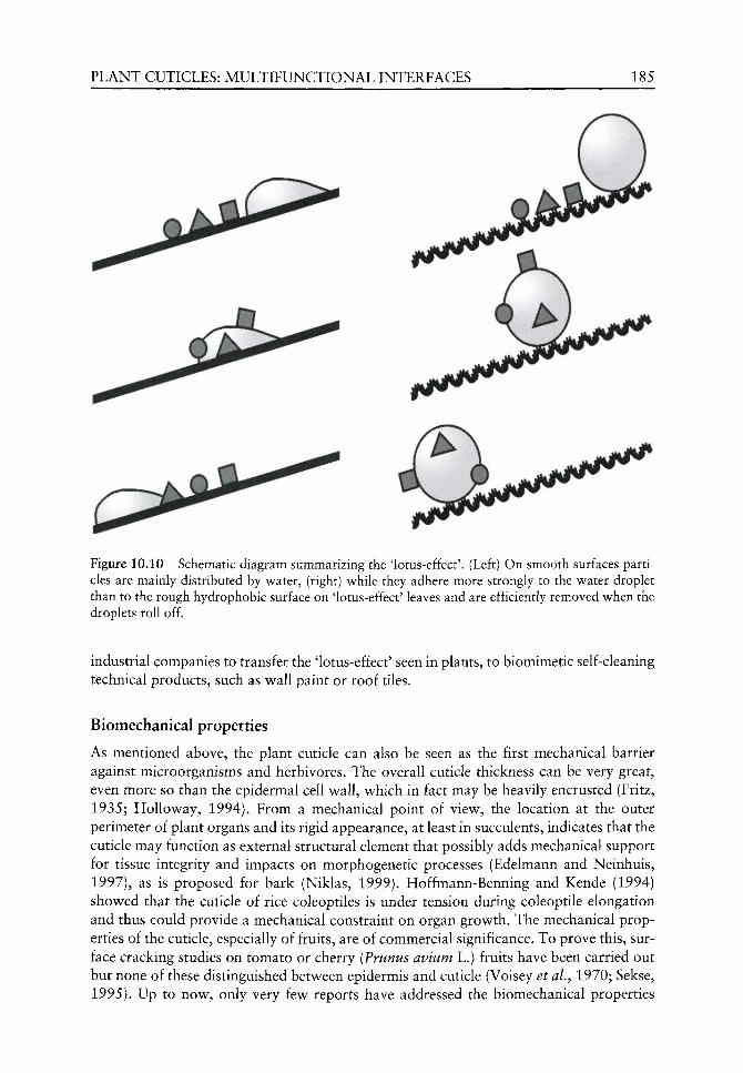
PLANT CUTICLES: MULTIFUNCTIONAL INTERFACES 185
Q-^ Figure 10.10 Schematic diagram summarizing the 'lotus-effect'. (Left) On smooth surfaces particles are mainly distributed by water, (right) while they adhere more strongly to the water droplet than to the rough hydrophobic surface on 'lotus-effect' leaves and are efficiently removed when the droplets roll off.
industrial companies to transfer the 'lotus-effect' seen in plants, to biomimetic self-cleaning technical products, such as wall paint or roof tiles.
Biomechanical properties
As mentioned above, the plant cuticle can also be seen as the first mechanical barrier against microorganisms and herbivores. The overall cuticle thickness can be very great, even more so than the epidermal cell w^all, w^hich in fact may be heavily encrusted (Fritz, 1935; HoUoway, 1994). From a mechanical point of viev^, the location at the outer perimeter of plant organs and its rigid appearance, at least in succulents, indicates that the cuticle may function as external structural element that possibly adds mechanical support for tissue integrity and impacts on morphogenetic processes (Edelmann and Neinhuis, 1997), as is proposed for bark (Niklas, 1999). Hoffmann-Benning and Kende (1994) show^ed that the cuticle of rice coleoptiles is under tension during coleoptile elongation and thus could provide a mechanical constraint on organ grov^th. The mechanical properties of the cuticle, especially of fruits, are of commercial significance. To prove this, surface cracking studies on tomato or cherry {Prunus avium L.) fruits have been carried out but none of these distinguished betv^een epidermis and cuticle (Voisey et aL, 1970; Sekse, 1995). Up to nov^, only very few reports have addressed the biomechanical properties

186 H.BARGEL ETAL.
1,6 GPa
• Dry Hyd rated
D Thickness
16 )jLm
14
12
10
8
6
4
2
0 Yucca Clusia Hedera Nerium Lycopersicon
Figure 10.11 Young's moduli and thicknesses of isolated plant cuticles of leaves of Yucca aloifolia L., Clusia fluminensis Planch. &: Triana, Hedera helix L., Nerium oleander L. and fruit of Lycopersicon esculentum Mill. Hydration causes a decrease in stiffness of about 35-50%.
solely of the plant cuticle in an experimental approach (Pescareta et aL, 1991; Hasenstein et aL, 1993; Wiedemann and Neinhuis, 1997), whereas the literature on general plant biomechanics does not refer to cuticles (Niklas, 1992). Hence, available data are rare, although vague indications of significance can be found in the literature. Wiedemann and Neinhuis (1997) studied the mechanical properties of isolated cuticles from five leaf and one fruit species by means of a one-dimensional tension test and reported Young's modulus - a measure of stiffness - ranging between 0.6 and 1.3 GPa for the leaves and 0.1 GPa for tomato fruit (Figure 10.11). Direct comparison with polyethylene (0.2 GPa) reveals remarkably high stiffness values for these cuticles (Czichos, 1989). Hydration of the cuticles caused a decrease in the Young's modulus of about 35-50%. As a consequence, water acts as a plasticizer, a fact supported by studies focusing on the decrease in the surface elastic modulus of isolated tomato fruit cutin in relation to water content (Round et aL, 2000). This is most likely to occur if water binds to polar groups or disrupts hydrogen-bonded cross-links in the cutin matrix (Dominguez and Heredia, 1999; Round et aL, 2000). Recently, Marga et at. (2001) undertook chemical analyses of cuticles from different organs, each having different biomechanical properties, and found that rigid cuticles can be classified by C g cutin monomers, while elastic cuticles correspond to mixed Ci^/Cig cutin monomers. During tomato fruit ripening, the elasticity of the cuticle seem to decrease as the stiffness increases (Bargel et at., 2000; Bargel and Neinhuis, unpublished data), an aspect that may be determined by a decrease in the relative amount of tri-hydroxy Gig fatty acids (Baker et al.^ 1982). Thus, hydroxyl groups seem to enhance the hydrophilic character and the hydration state of the cutin matrix, which in turn results in higher plasticity.
The few results outlined here could be seen as the first steps towards an understanding of cuticle biomechanics. Even from an engineering approach, the outer plant coverage appears to be a highly diverse composite with biomechanical properties determined by its dynamic structure and chemical composition. From these, the cuticle may inherit a possible role in stabilization of plant organs and thus a newly described function that has been largely neglected so far to date.
PLANT CUTICLES: MULTIFUNCTIONAL INTERFACES 187
Acknowledgements
The authors would Uke to thank the Deutschen Forschungsgemeinschaft (DFG), Deutsche
Bundesstiftung Umwelt (DBU), Bundesministerium fiir Bildung und Forschung
(BMBF), Fonds der Chemischen Industrie (FCI) and Creavis G m b H (Marl, Germany) for
substantial funding and Robin Seidel for critical comments on the manuscript.
(http://www.lotus-effect.de)
References
Adamson AW. 1990. Physical Chemistry of Surfaces. London: Wiley. Anton LH, Ewers FW, Hammerschmidt R, Klomparens KL. 1994. Mechanisms of deposition of
epicuticular wax in leaves of broccoli, Brassica oleracea L. var. capitata L. New Phytologist 126: 505-510.
Baker EA. 1982. Chemistry and morphology of plant epicuticular waxes. In: Cutler DP, Alvin KL, Price CE, eds. The Plant Cuticle. London, New York: Academic Press, 139-166.
Baker EA, Bukovac MJ, Hunt GM. 1982. Composition of tomato fruit cuticle as related to fruit growth and development. In: Cutler DF, Alvin KL, Price CE, eds. The Plant Cuticle. London, New York: Academic Press, 33-44.
Bargel H, Edelmann HG, Neinhuis C. 2000. Biomechanics of tomato fruit growth. In: Spatz HC, Speck T, eds. Plant Biomechanics 2000. Stuttgart: Thieme, 313-314.
Barnes JD, Cadoso-Vilhena J. 1996. Interactions between electromagnetic radiation and the plant cuticle. In: Kerstiens G, ed. Plant Cuticles an Integrated Functional Approach. Oxford: Bios Scientific, 157-174.
Barnes J, Paul N, Percy K, et al. 1994. Effects of UV-B radiation on wax biosynthesis. In: Percy KE, Cape JN, Jagels R, Simpson CJ, eds. Air Pollution and the Leaf Cuticle. Berlin, Heidelberg: Springer, 321-328.
Barthlott W. 1981. Epidermal and seed surface characters of plants: systematic applicability and some evolutionary aspects. Nordic Journal of Botany 3: 345-355.
Barthlott W. 1990. Scanning electron microscopy of the epidermal surface in plants. In: Claugher D, ed. Scanning Electron Microscopy in Taxonomy and Functional Morphology. Oxford: Clarendon Press, 69-94.
Barthlott W, Ehler N. 1977. Raster-Elektronenmikroskopie der Epidermisoberflachen von Spermatophyten. Tropische und subtropische Pflanzenwelt 19: 367-467.
Barthlott W, Neinhuis C. 1997. Purity of the sacred lotus or escape from contamination in biological surfaces. Planta 202: 1-7.
Barthlott W, Wollenweber E. 1981. Zur Feinstruktur, Chemie und taxonomischen Signifikanz epicuticularer Wachse und ahnlicher Sekrete. Tropische und subtropische Pflanzenwelt 32: 7-67.
Barthlott W, Neinhuis C, Cuder D, et al. 1998. Classification and terminology of plant epicuticular waxes. Botanical Journal of the Linnean Society 126: 137-260.
Barthlott W, Neinhuis C, Jetter R, et al. 1996. Waterlily, poppy, or sycamore: on the systematic position oiNelumbo. Flora 191: 169-174.
Barthlott W, Theisen I, Borsch T, Neinhuis C. 2002. Epicuticular waxes and vascular plant system-atics: integrating micromorphological and chemical data. In: Stuessy TF, Mayer V, Horandl E, eds. Deep morphology - towards a Renaissance of morphology in plant systematics. Regnum Vegetabile 141: 189-206.
de Bary A. 1871. Uber die Wachsiiberzlige der Epidermis. Botanische Zeitschrift 29: 128-139, 145-154, 161-176, 566-571, 573-585, 605-619.
Bateman RM, Crane R, DiMichele WA, et al. 1998. Early evolution of land plants: phylogeny, physiology, and ecology of the primary terrestrial radiation. Annual Review of Ecology and Systematics 29: 263-292.
188 H . B A R G E L £ T A L .
Baur P, Buchholz A, Schonherr J. 1997. Diffusion in plant cuticles as affected by temperature and size of organic solutes: similarity and diversity among species. Plant, Cell and Environment 20: 982-994.
Baur P, Marzouk H, Schonherr J. 1999. Estimation of path lengths for diffusion of organic compounds through leaf cuticles. Plant, Cell and Environment 11: 291-299.
Baur P, Marzouk H, Schonherr J, Bauer H. 1996. Mobilities of organic compounds in plant cuticles as affected by structure and molar volumes of chemicals and plant species. Planta 199: 404-412.
Bergen PF van, Collinson ME, Briggs DEG, et al. 1995. Resistant biomacromolecules in the fossil record. Acta Botanica Neerlandica 44: 319-342.
Bernays EA. 1991. Evolution of insect morphology in relation to plants. Philosophical Transactions of the Royal Society London B 333: 257-264.
Bianchi G. 1995. Plant waxes. In: Hamilton RJ, ed. Waxes: Chemistry, Molecular Biology and Functions. Dundee: The Oily Press, 177-222.
Blakeman JP, Atkinson P. 1981. Antimicrobial substances associated with the aerial surfaces of plants. In: Blakeman JP, ed. Microbial Ecology of the Phylloplane. London, New York: Academic Press, 245-264.
Brogniart A. 1834. Nouvelles recherches sur la structure de Tepiderme des vegeteux. Annales des Sciences naturelles 2e Ser. 1: 65-71.
Buchholz A, Schonherr J. 2000. Thermodynamic analysis of diffusion of non-elecrtolytes across plant cuticles in the presence and absence of the plasticiser tributyl phosphate. Planta 111: 103-111.
Buchholz A, Baur P, Schonherr J. 1998. Differences among plant species in cuticular permeabilities and solute mobilities are not caused by differential size selectivities. Planta 206: 322-328.
Bunster L, Fokkema NJ, Schippers B. 1989. Effect of surface-active Pseudomonas spp. on leaf wettability. Applied and Environmental Microbiology 55: 1340-1345.
Burkhardt J, Koch K, Kaiser H. 2001. Deliquescence of deposited atmospheric particles on leaf surfaces. Water, Soil and Air Pollution Focus 1: 313-321.
Butler DR. 1996. The presence of water on leaf surfaces and its importance for microbes and insects. In: Kerstiens G, ed. Plant Cuticles an Integrated Functional Approach. Oxford: Bios Scientific, 267-282.
Campbell CL, Huang J-S, Payne GA. 1980. Defense at the perimeter: the outer walls and the gates. In: Horsfall JG, Cowling EB, eds. Plant Disease an Advanced Treatise. New York, London: Academic Press, 103-120.
Cassie ABD, Baxter S. 1944. Wettability of porous surfaces. Transaction of the Faraday Society 40: 546-551.
Collinson ME, Mosle B, Finch P, et al. 1998. The preservation of plant cuticles in the fossil record: a chemical and microscopical investigation. Ancient Biomolecules 1: 251-265.
Crossley A, Fowler D. 1986. The weathering of Scots pine epicuticular wax in polluted and clean air. New Phytologist 103: 207-218.
Czichos H ed. 1989. Fliitte - Die Grundlagen der Ingenieurwissenschaften. Berlin: Springer. Day TA, Martin G, Vogelmann TC. 1993. Penetration of UV-B radiation in fohage: evidence that
the epidermis behaves as a non-uniform filter. Plant, Cell and Environment 16: 735-741. Dettre RH, Johnson RE. 1964. Contact angle hysteresis II. Contact angle measurements on rough
surfaces. In: Gould RF, ed. Advances in Chemistry Series. Los Angeles: American Chemical Society, 136-144.
Dickinson CH. 1976. Fungi on the aerial surfaces of higher plants. In: Dickinson CH, Preece TF, eds. Micobiology of Aerial Plant Surfaces. London, New York: Academic Press, 293-324.
Dominguez E, Heredia A. 1999. Water hydration in cutinized cell walls: a physico-chemical analysis. Biochimica et Biophysica Acta 1426: 168-176.
Edelmann HG, Neinhuis C. 1997. Effect of hydration and temperature on the extensibility of isolated plant cuticles. In: Jeronomidis G, Vincent JFV, eds. Plant Biomechanics 1997. Reading, Hampshire: Ashford, 31-32.
Edwards D, Abbot GD, Raven JA. 1996. Cuticles of early land plants. In: Kerstiens G, ed. Plant Cuticles an Integrated Functional Approach. Oxford: Bios Scientific, 1-31.
PLANT CUTICLES: MULTIFUNCTIONAL INTERFACES 189
Edwards D, Edwards DS, Rayner R. 1982. The cuticle of early vascular plants and its evolutionary significance. In: Cutler DF, Alvin KL, Price CE, eds. The Plant Cuticle. London: Academic Press, 341-36L
Edwards PJ. 1992. Resistance and defence: the role of secondary plant substances. In: Ayres PG, ed. Pests and Pathogens. Plant Responses to Foliar Uptake. Oxford: Bios Scientific, 69-84.
Eigenbrode S. 1996. Plant surface waxes and insect behaviour. In: Kerstiens G, ed. Plant Cuticles an Integrated Functional Approach. Oxford: Bios Scientific, 201-222.
Eller BM. 1977. Road dust induced increase of leaf temperature. Environmental Pollution 13: 99-107. EUer BM. 1979. Die strahlungsokologische Bedeutung von Epidermisauflagen. Flora 168:146-192. Eller BM. 1985. Epidermis und spektrale Eigenschaften pflanzlicher Oberflachen. Berichte der
Deutschen Botanischen Gesellschaft 98: 465-475. Ensikat HJ, Neinhuis C, Barthlott W. 2000. Direct access to plant epicuticular wax crystals by a new
mechanical isolation method. International Journal of Plant Science 161: 143-148. Espelie KE. 1996. Integrated case study: effects of maize leaf epicuticular lipids on insect pests. In:
Kerstiens G, ed. Plant Cuticles an Integrated Functional Approach. Oxford: Bios Scientific, 223-230.
Espelie KE, Dean BB, Kolattukudy PE. 1979. Composition of lipid-derived polymers from different anatomical regions of several plant species. Plant Physiology 64: 1089-1093.
Euteneuer-Macher T. 1990. Morphologie und Chemie der Epikutikularwachse von Picea abies L. Karst. unter dem Einfluf^ von Klima und Immissionen. Berlin, Stuttgart: J. Cramer.
Eveling DW. 1986. Scanning electron microscopy of damage by dust deposits to leaves and petals. Botanical Gazette 147: 159-165.
Falbe J, Regitz M eds. 1999. Rompp Lexikon Chemie. Stuttgart: Georg Thieme. Farmer AM. 1993. The effect of dust on vegetation - a review. Environmental Pollution 79: 63-75. Fliickiger W, Oertli JJ, Fliickiger H. 1979. Relationship between stomatal diffusive resistance and
various apphed particle sizes on leaf surfaces. Zeitschrift fiir Pflanzenphysiologie 91: 173-175. Fritz F. 1935. Uber die Kutikula von Aloe- und Gasteriaarten. Jahrbiicher fiir wissenschaftliche
Botanik 81: 718-746. van Gardingen PR, Grace J, Jeffree CE. 1991. Abrasive damage by wind to the needle surfaces of
Picea sitchensis (Bong.) Carr. and Pinus sylvestris L. Plant, Cell and Environment 14: 185-193. Givnish TJ. 1988. Adaptation to sun and shade: a whole-plant perspective. Australian Journal of
Plant Physiology 15: 63-92. Grace J, van Gardingen P. 1996. Plant cuticles under challenge. In: Kerstiens G, ed. Plant Cuticles
an Integrated Functional Approach. Oxford: Bios Scientific, 319-329. Grill D, Pfeifhofer H, Halbwachs G, Waltinger H. 1987. Investigations on epicuticular waxes of dif
ferently damaged spruce needles. European Journal of Forest Pathology 17: 246-255. Giilz P-G. 1994. Epicuticular leaf waxes in the evolution of the plant kingdom. Journal of Plant
Physiology 143: 453-464. Hall DM. 1967. The ultrastructure of wax deposits on plant leaf surfaces II. Cuticular pores and
wax formation. Journal of Ultrastructure Research 17: 34-44. Hall DM, Donaldson LA. 1962. Secretion from pores of surface wax on plant leaves. Nature
194: 1196. Hallam ND. 1970. Growth and regeneration of waxes on the leaves of Eucalyptus L'Herit. Planta
93: 257-268. Hamilton RJ ed. 1995. Waxes: Chemistry, Molecular Biology and Functions. Dundee: The Oily Press. Hasenstein KH, Pescareta TC, Sullivan VI. 1993. Thigmonasticity of thistle staminal filaments II.
Mechano-elastic properties. Planta 190: 58-64. Hennig S, Barthlott W, Meusel I, Theisen I. 1994. Mikromorphologie der Epicuticularwachse und
die Systematik der Magnoliidae, Ranunculidae und Hamamelididae. Tropische und subtropische Pflanzenwelt 90: 5-60.
Herminghaus S. 2000. Roughness-induced non-wetting. Europhysics Letters 52: 165-170. Hess FD, Foy CL. 2000. Interaction of surfactants with plant cuticles. Weed Technology 14: 807-813. Hoffmann-Benning S, Kende H. 1994. Cuticle biosynthesis in rapidly growing internodes of deep-
water rice. Plant Physiology 104: 719-723.
190 H . B A R G E L £ T A L .
Holloway PJ. 1970. Surface factors affecting the wetting of leaves. Pesticide Science 1: 156-163. Holloway PJ. 1982a. The chemical composition of plant cutins. In: Cutler DF, Alvin KL, Price CE,
eds. The Plant Cuticle. London, New York: Academic Press, 46-86. Holloway PJ. 1982b. Structure and histochemistry of plant epicuticular membranes: an overview. In:
Cutler DF, Alvin KL, Price CE, eds. The Plant Cuticle. London, New York: Academic Press, 1-32. Holloway PJ. 1994. Plant cuticles: physicochemical characteristics and biosynthesis. In: Percy KE,
Cape JN, Jagels R, Simpson CJ, eds. Air Pollution and the Leaf Cuticle. Bedin: Springer, 1-13. Huttunen S. 1996. Interactions between epiphytic microbes and deposited compounds. In: Kerstiens G,
ed. Plant Cuticles an Integrated Functional Approach. Oxford: Bios Scientific, 301-318. Jeffree CE. 1986. The cuticle, epicuticular waxes and trichomes of plants, with reference to their
structure, functions and evolution. In: Juniper BE, Southwood SR, eds. Insects and the Plant Surface. London: Edward Arnold, 23-63.
Jeffree CE. 1996. Structure and ontogeny of plant cuticles. In: Kerstiens G, ed. Plant Cuticles an Integrated Functional Approach. Oxford: Bios Scientific, 33-82.
Jeffree CE, Baker EA, Holloway PJ. 1975. Ultrastructure and recrystallization of plant epicuticular waxes. New Phytologist 75: 539-549.
Jeffree CE, Baker EA, Holloway PJ. 1976. Origins of the fine structure of plant epicuticular waxes. In: Dickinson CH, Preece TF, eds. Microbiology of Aerial Plant Surfaces. London, New York: Academic Press, 119-158.
Jetter R, Riederer M. 1995. In vitro reconstitution of epicuticular wax crystals: Formation of tubular aggregates by long chain secondary alkanediols. Botanica Acta 108: 111-120.
Jetter R, Riederer M. 2000. Leaf cuticular waxes are arranged in chemically and mechanically distinct layers: evidence from Prunus laurocerasus L. Plant, Cell and Environment 23: 619-628.
Juniper BE. 1991. The leaf from the inside and the outside: a microbe's perspective. In: Andrews JH, Hirano SS, eds. Microbial Ecology of Leaves. New York, Berlin: Springer, 21-42.
Kader JC. 1997. Lipid-transfer proteins: A puzzling family of plant proteins. Trends in Plant Science 2: 66-70.
Kerler F, Riederer M, Schonherr J. 1984. Non-electrolyte permeabiHty of plant cuticles: a critical evaluation of experimental methods. Physiologia Plantarum 62: 599-602.
Kerstiens G. 1996. Cuticular water permeability and its physiological significance. Journal of Experimental Botany 47: 1813-1832.
Kim YS, Lee JK. 1990. Chemical and structural characteristics of conifer needles exposed to ambient air pollution. European Journal of Forest Pathology 20: 193-200.
Kirsch T, Kaffarnik F, Riederer M, Schreiber L. 1997. Cuticular permeability of the three tree species Prunus laurocerasus L., Ginkgo biloba L. and Juglans regia L. - comparative investigation of the transport properties of intact leaves, isolated cuticles and reconstituted cuticular waxes. Journal of Experimental Botany 48: 1035-1045.
Knoche M, Bukovac MJ. 1993. Studies on octylphenoxy surfactants: XI. Effect on NAA diffusion through the isolated tomato fruit cuticular membrane. Pesticide Science 38: 211-217.
Knoll D, Schreiber L. 2000. Plant-microbe interactions: wetting of Ivy {Hedera helix L.) leaf surfaces in relation to colonization by epiphytic microorganisms. Microbial Ecology 41: 33-42.
Knoll F. 1914. Uber die Ursache des Ausgleitens der Insektenbeine an wachsbedeckten Pflanzenteilen. Jahrbiicher fiir wissenschaftliche Botanik 54: 448-498.
Kolattukudy PE. 1980a. Biopolyester membranes of plants: cutin and suberin. Science 208: 990-1000. Kolattukudy PE. 1980b. Cutin, suberin, and waxes. In: Stumpf PK, ed. Lipids: Structure and
Function. London, New York: Academic Press, 571-646. Kolattukudy PE. 1985. Enzymatic penetration of the plant cuticle by fungal pathogens. Annual
Review of Phytopathology 23: 223-250. Kolattukudy PE. 1996. Biosynthetic pathways of cutin and waxes. In: Kerstiens G, ed. Plant Cuticles
an Integrated Functional Approach. Oxford: Bios Scientific, 83-108. Kolattukudy PE. 2001. Polyesters in higher plants. In: Scheper T, ed. Advances in Biochemical
Engineering/Biotechnology. Berlin: Springer Verlag, 4-49. Krauss P, Markstadter C, Riederer M. 1997. Attenuation of UV radiation by plant cuticles from
woody species. Plant, Cell and Environment 20: 1079-1085.

PLANT CUTICLES: MULTIFUNCTIONAL INTERFACES 191
Lundstrom AN. 1884. Fflanzenbiologische Studien. Lundequistsche Buchhandlung, Upsala. Marga F, Pesacreta TC, Hasenstein KH. 2001. Biochemical analysis of elastic and rigid cuticles of
Cirsium horridulum. Planta 213: 841-848. Doi: 10.1007/s004250100576. Markstadter C. 1994. Untersuchungen zur jahreszeitlichen Entwicklung der kutikularen Wachse
von Fagus sylvatica L. D. Phil Thesis, Kaiserslautern. Markstadter C, Federle W, Jetter R, et al. 2000. Chemical composition of the slippery epicuticular
wax blooms on Macaranga Thours. (Euphorbiaceae) ant-plants. Chemoecology 10: 33-40. Martin JT. 1964. Role of cuticle in the defense against plant disease. Annual Review of Phytopathology
2: 81-100. Martin JT, Juniper BE. 1970. The Cuticles of Plants. London: Edward Arnold. Mendgen K. 1996. Fungal attachment and penetration. In: Kerstiens G, ed. Plant Cuticles an
Integrated Functional Approach. Oxford: Bios Scientific, 175-187. Mengel K, Hogrebe AMR, Esch A. 1989. Effect of acidic fog on needle surface and water relations
of Picea abies (L.) Karst. Physiologia Plantarum 75: 201-207. Meusel I, Barthlott W, Kutzke H, Barbier B. 2000. Crystallographic studies of plant waxes. Powder
Diffraction 15: 123-129. Meusel I, Neinhuis C, Markstadter C, Barthlott W. 1999. Ultrastructure, chemical composition and
recrystallisation of epicuticular waxes: transversely ridged rodlets. Canadian Journal of Botany 77: 706-720.
Miller RH. 1986. The prevalence of pores and canals in leaf cuticular membranes. Annals of Botany 57: 419-434.
Mosle B, Finch P, Collinson ME, Scott AC. 1997. Comparison of modern and fossil plant cuticles by selective chemical extraction monitored by flash pyrolysis-gas chromatography-mass spectroscopy and electron microscopy. Journal of Analytical and Applied Pyrolysis 40-41: 5S5-597.
Mudd JB, Banerjee SK, Dooley MM, Knight KL. 1982. Pollutants and plant cells. Effects on membranes. In: Koziol MJ, Whatley FR, eds. Gaseous Air Pollutants and Plant Metabolism. London: Butterworth Scientific, 105-116.
Myers D. 1991. Surfaces, Interfaces, and Colloids. Principles and Applications. Weinheim: VCH. Neinhuis C, Barthlott W. 1997. The tree leaf surface: structure and function. In: Rennenberg H,
Eschrich W, Ziegler H, eds. Trees - Contributions to Modern Tree Physiology. Amsterdam: SPB Academic Publishing, 3-18.
Neinhuis C, Barthlott W. 1998. Seasonal change of leaf surface contamination in beech, oak, and ginkgo in relation to micromorphology and wettability. New Phytologist 138: 91-98.
Neinhuis C, Jetter R. 1995. Ultrastructure and chemistry of epicuticular wax crystals in Polytrichales sporophytes. Journal of Bryology 18: 399-406.
Neinhuis C, Koch K, Barthlott W. 2001. Movement and regeneration of epicuticular waxes through plant cuticles. Planta 213: 427-434. Doi: 10.1007/s004250100530.
Neinhuis C, Wolter M, Barthlott W. 1992. Epicuticular wax of Brassica oleracea L.: changes of microstructure and ability to be contaminated of leaf surfaces after application of Triton X-100. Zeitschrift fUr Pflanzenkrankheiten und Pflanzenschutz 99: 542-549.
Neinhuis C, Wolter M, Kiippers K, Barthlott W. 1994. Der EinfluE gasformiger Luftschadstoffe auf die Epicuticular-Wachse von Quercus robur. European Journal of Forest Pathology 24: 210-216.
Niklas KJ. 1992. Plant Biomechanics. Chicago: University of Chicago Press. Niklas KJ. 1999. The mechanical role of bark. American Journal of Botany 86: 465-469. Nip M, Tegelaar EW, de Leeuw JW, Schenk PA. 1986. A new non-saponifiable highly aliphatic and
resistant biopolymer in plant cuticles. Evidence from pyrolysis an 13 C-NMR analysis of present-day and fossil plants. Naturwissenschaften 73: 579-5S5.
Noga G, Knoche M, Wolter M, Barthlott W. 1987. Changes in leaf micromorphology induced by surfactant application. Angewandte Botanik 61: 521-528.
Noga G, Wolter M, Barthlott W, Petry W. 1991. Quantitative evaluation of epicuticular wax alterations as induced by surfactant treatment. Angewandte Botanik 62: 239-252.
Onken U, Weiland P. 1984. Grundziige der thermischen Verfahrenstechnik. In: Harnisch H, Steiner R, Winnacker K, eds. Chemische Technologie, 4th edn. Miinchen: Carl Hanser.

192 H . B A R G E L £ T A L .
Percy KE, Cape JN, Jagels R, Simpson CJ eds. 1994. Air Pollution and the Leaf Cuticle. Berlin, Heidelberg: Springer.
Pescareta TC, Sullivan VI, Hasenstein KH, Durand JM. 1991. Thigmonasticity of thistle staminal filaments I. involvement of a contractile cuticle. Protoplasma 163: 174-180.
Post-Beitenmiller D. 1996. Biochemistry and molecular biology of v^ax production in plants. Annual Review of Plant Physiology and Plant Molecular Biology 47: 405-430.
Pye K. 1987. Aeolian Dusts and Dust Deposits. London, New^ York: Academic Press. Pyee J, Yu H, Kolattukudy PE. 1994. Identification of a lipid transfer protein as the major protein
in the surface wax of broccoli (Brassica oleracea L.) leaves. Archives of Biochemistry and Biophysics 311: 460-468.
Ray AK, Chen Z, Stark RE. 1998. Chemical depolymerization studies of the molecular architecture of lime fruit cuticle. Phy to chemistry 49: 65-70.
Reed DW, Tuckey HB. 1982. Light intensity and temperature effects on epicuticular WSLX morphology and internal cuticle ultrastructure of carnation and brussel sprout leaf cuticles. Journal of American Society for Horticultural Science 107: 417-420.
Riederer M. 1989. The cuticles of conifers: structure, composition and transport properties. In: Schulze ED, Lange OL, Oren R, eds. Ecological Studies, Vol. 77. Berlin, Heidelberg: Springer, 157-192.
Riederer M, Markstadter C. 1996. Cuticular w^axes: a critical assessment of current know^ledge. In: Kerstiens G, ed. Plant Cuticles an Integrated Functional Approach. Oxford: Bios Scientific, 189-200.
Riederer M, Schonherr J. 1984. Accumulation and transport of (2,4-dichlorophenoxy) acetic acid in plant cuticles: 1. Sorption in the cuticular membrane and its components. Ecotoxicology and Environmental Safety 8: 236-247.
Riederer M, Schonherr J. 1985. Accumulation and transport of (2,4-dichlorophenoxy) acetic acid in plant cuticles: 2. Permeability of the cuticular membrane. Ecotoxicology and Environmental Safety 9: 196-208.
Riederer M, Schonherr J. 1990. Effects of surfactants on v^ater permeability of isolated plant cuticles and on the composition of their cuticular v^axes. Pesticide Science 29: 85-94.
Riederer M, Schreiber L. 1995. Waxes - the transport barriers of plant cuticles. In: Hamilton RJ, ed. Waxes: Chemistry, Molecular Biology and Functions. Dundee: The Oily Press, 131-156.
Riederer M, Schreiber L. 2001. Effects of environmental factors on the v^ater permeability of plant cuticles. Journal of Experimental Botany 52: 2023-2033.
Robinson SA, Lovelock CE, Osmond CB. 1993. Wax as a mechanism for protection against photoinhibition - a study of Cotyledon orbiculata L. Botanica Acta 106: 307-312.
Rogers HJ. 1979. Adhesion of microorganims to surfaces: some general considerations of the role of the envelope. In: Ellwood DC, Melling J, Rutter P, eds. Adhesion of Microorganisms to Surfaces. London, New^ York: Academic Press, 29-55.
Round AN, Yan B, Dang S, et al. 2000. The influence of v^ater on the nanomechanical behavior of the plant biopolyester cutin studies by AFM and solid-state NMR. Biophysical Journal 79: 2761-2767.
Schonherr J. 1976. Water permeability of isolated cuticular membranes: the effect of cuticular waxes on diffusion of water. Planta 131: 159-164.
Schonherr J. 1982. Resistance of plant surfaces to water loss: transport properties of cutin, suberin and associated lipids. In: Lange OL, Nobel PS, Osmond CB, Ziegler H, eds. Encyclopedia of Plant Physiology. Berlin: Springer, 153-179.
Schonherr J. 2000. Calcium chlorite penetrates plant cuticles via aqueous pores. Planta 111: 112-118.
Schonherr J. 2001. Cuticular penetration of calcium salts: effects of humidity, anions, and adjuvants. Journal of Plant Nutrition 164: 225-231.
Schonherr J, Baur P. 1994. Modelling penetration of plant cuticles by crop protection agents (CPA) and effects of adjuvants on rates of penetration. Pesticide Science 42: 185-208.
Schonherr J, Bukovac MJ. 1973. Ion exchange properties of isolated tomato fruit cuticular membrane: exchange capacity, nature of fixed charges and cation selectivity. Planta 109: 73-93.
Schonherr J, Eckl K, Gruler H. 1979. Water permeability of plant cuticles: the effect of temperature on diffusion of water. Planta 147: 21-26.
PLANT CUTICLES: MULTIFUNCTIONAL INTERFACES 193
Schonherr J, Merida T. 1981. Water permeability of plant cuticular membranes: the effects of humidity and temperature on the permeability of non-isolated cuticles of onion bulb scales. Plant, Cell and Environment 4: 349-354.
Schonherr J, Riederer M. 1989. Foliar penetration and accumulation of organic chemicals on plant cuticles. Review of Environmental Contamination and Toxicology 108: 1-70.
Schonherr J, Schreiber L, Buchholz A. 2001. Effects of temperature and concentration of the accelerators ethoxylated alcohols, diethyl suberate and tributyl phosphate on the mobility of [C-14]2,4-dichlorophenoxy butyric acid in plant cuticles. Fest Management Science 57: 17-24.
Schreiber L. 1995. A mechanistic approach towards surfactant/wax interactions: effects of octaeth-yleneglycolmonododecylether on sorption and diffusion of organic chemicals in reconstituted cuticular wax of barley leaves. Pesticide Science 45: 1-11.
Schreiber L. 2001. Effect of temperature on cuticular transpiration of isolated cuticular membranes and intact leaf disks. Journal of Experimental Botany 52: 1893-1900.
Schreiber L, Kirsch T, Riederer M. 1996a. Transport properties of cuticular waxes of Fagus sylvatica L. and Picea abies (L.) Karst.: estimation of size selectivity and tortuosity from diffusion coefficients of aliphatic molecules. Planta 198: 104-109.
Schreiber L, Riederer M. 1996. Ecophysiology of cuticular transpiration: comparative investigation of cuticular water permeability of plant species from different habitats. Oecologia 107: 426-432.
Schreiber L, Riederer M, Schorn K. 1996b. Mobilities of organic compounds in reconstituted cuticular wax of barley leaves: effects of monodiperse alcohol ethoxylates on diffusion of pentachor-phenol and tetracosanoic acid. Pesticide Science 48: 117-124.
Schreiber L, Schonherr J. 1992a. Analysis of fohar uptake of pesticides in barley leaves: role of epi-cuticular waxes and compartmentation. Pesticide Science 36: 213-221.
Schreiber L, Schonherr J. 1992b. Uptake of organic chemicals in conifer needles: surface adsorption and permeability of cuticles. Environmental Science and Technology 26: 153-159.
Schreiber L, Schonherr J. 1993. Mobilities of organic compounds in reconstituted cuticular wax of barley leaves: determination of diffusion coefficients. Pesticide Science 38: 353-361.
Schreiber L, Skrabs M, Hartmann K, et al. 2001. Effect of humidity on cuticular transpiration of isolated cuticular membranes and leaf disks. Planta 214: 274-282.
Schwab M, Noga G, Barthlott W. 1995. Bedeutung der Epicuticularwachse fiir die Pathogenabwehr am Beispiel von Botrytris cinerea-Infektion bei Kohlrabi und Erbse. Gartenhauwissenschaft 60: 102-109.
Sekse L. 1995. Fruit cracking in sweet cherries {Prunus avium L.). Some physiological aspects - a mini review. Scientia Horticulturae 63: 135-141.
Southwood TRE. 1986. Plant surfaces and insects - an overwiew. In: Juniper BE, Southwood TRE, eds. Insects and the Plant Surface. London: Edward Arnold, 1-22.
Steinmiiller D, Tevini M. 1985. Action of ultraviolet radiation (UV-B) upon cuticular waxes in some crop plants. Planta 164: 557-564.
Sterk P, Booij H, Schellenkens GA, et al. 1991. Cell-specific expression of the carrot EP2 lipid transfer protein gene. The Plant Cell 3: 907-921.
Stevens PJG, Bukovac MJ. 1985. Properties of octylphenoxy surfactants and their effects on foliar uptake. Weeds 3C: 309-316.
Strange RN. 1992. Resistance: the role of the hypersensitive response and phytoalexins. In: Ayres PG, ed. Pests and Pathogens. Plant Responses to Foliar Uptake. Oxford: Bios Scientific, 39-56.
Tukey HB, Jr. 1970. The leaching of substances from plants. Annual Review of Plant Physiology 21: 305-324.
Turunen M, Huttunen S. 1990. A review of the response of epicuticular waxes of conifer needles to air pollution. Journal of Environmental Quality 19: 35-45.
Villena JF, Dominguez E, Stewart D, Heredia A. 1999. Characterization and biosynthesis of non-degradable polymers in plant cuticles. Planta 208: 181-187.
Voisey PW, Lyall LH, Kloek M. 1970. Tomato skin strength - its measurement and relation to cracking. Journal of the American Society for Horticultural Science 95: 485-488.
Wagner P, Fiirstner R, Barthlott W, Neinhuis C. 2002. Quantitative assessment to the structural basis of water repellency in natural and technical surfaces. Comparative Biochemistry and Physiology 132/A: 160.

194 H . B A R G E L £ T A L .
Wagner T, Neinhuis C, Barthlott W. 1996. Wettability and contaminability of insect wings as a function of their surface sculpture. Acta Zoologica 77: 213-225.
Walton TJ. 1990. Waxes, cutin and suberin. In: Harwood JL, Bowyer JR, eds. Lipids, Membranes and Aspects ofPhotobiology. London: Academic Press, 105-158.
Weinhold AR, Hancock JG. 1980. Defense at the perimeter: extruded chemicals. In: Horsfall JGEBC, ed. Plant Disease. London, New York: Academic Press, 121-138.
Wenzel RN. 1936. Resistance of solid surfaces to wetting by water. Industrial and Engineering Chemistry 2S: 9SS-994.
Wessels JGH. 1994. Developmental regulation of fungal cell wall formation. Annual Review of Phytopathology 32: 413-437.
von Wettstein-Knowles P. 1972. Genetic control of p-diketone and hydroxy-p-diketone synthesis in epicuticular waxes of barley. Planta 106: 113-130.
von Wettstein-Knowles P. 1987. Genes, elongases and associated enzymes system in epicuticular wax syntheses. In: Stumpf PK, Mudd JB, Nes WD, eds. The Metabolism, Structure, and Function of Plant Lipids. New York: Plenum Press, 489-498.
von Wettstein-Knowles P. 1995. Biosynthesis and genetics of waxes. In: Hamilton RJ, ed. Waxes: Chemistry, Molecular Biology and Functions. Glasgow: The Oily Press, 91-130.
Wheeler BEJ. 1981. Biology of powdery mildews on leaf surfaces. In: Blakeman JP, ed. Micobial Ecology of the Phylloplane. London, New York: Academic Press, 69-84.
Wiedemann P, Neinhuis C. 1997. Biomechanics of isolated plant cuticles. Botanica Acta 111: 28-34.
Wilhelmi H, Barthlott W. 1997. Mikromorphologie der Epicuticularwachse und die Systematik der Gymnospermen. Tropische und subtropische Pflanzenwelt 96: 7-48.
Wolter M, Barthlott W, Knoche M, Noga GJ. 1988. Concentration effects and regeneration of epicuticular waxes after treatment with Triton X-100 surfactant. Angewandte Botanik 62: 53-62.
Woodhead S, Chapman RF. 1986. Insect behaviour and the chemistry of plant surface waxes. In: Juniper BRS, ed. Insects and the Plant Surface. London: Arnold, 123-135.
Yamada M. 1992. Lipid transfer proteins in plants and microorganisms. Plant Cell Physiology 33:1-6. Zangerl AR, Berenbaum MR. 1993. Plant chemistry, insect adaptations to plant chemistry, and
host plant utilization pattern. Ecology 74: 47-54.
Plate 1 A reflected light image of a section of epidermal peel taken from Commelina communis showing two stomatal complexes within the epidermis.
Plate 2 Example of the use of high resolution imaging of chlorophyll fluorescence in guard cells in Tradescantia (a) reflected light image, (b) whole fluorescence image, (c) and (e) isolated areas of mesophyll and guard cells showing steady state fluorescence images, (d) and (f) built up image of F/F'

• , N
> • v-'f
Plate 3 Three growth forms of the Fynbos Mediterranean ecosystem: the deeper rooted proteoid growth form, Protea nitida Mill, (top), the more shallow rooted ericoid form, Phaenocoma prolifera (L.) D.Don, (bottom left) and also the shallow rooted restioid form, Elegia juncea L. (bottom right).
Plate 4 The open vegetation of the Fynbos, characterized by its low density with representatives of the famiUes Restionaceae, Compositae, Ericaceae and Proteaceae (left). Vertical distribution of the open vegetation is accentuated by Erica chloroloma Lindley (right) and other species.
Plate 5 Three Pelargonium species with entire, deeply lobed and parsley-like leaf forms. From left to right: P. cucullatum (L.) L.'Her., P. scabrum (L.) L.'Her. and P. triste (L.) L.'Her.
11 Falling atmospheric CO2 - the key
to megaphyll leaf origins
Colin P Osborne, William G Chaloner and David J Beerling
CONTENTS.
Introduction 197
A mechanism coupling Devonian megaphyll evolution
w ith falling CO2 199
Early evolution of the megaphyll leaf 202
Quantifying the trends in early megaphyll leaf evolution 208
Discussion 210
Acknow ledgements 211
References 211
Introduction
The vast majority of contemporary leaves are megaphyils, derived from the 'pianation' of determinate, overtopped side-branch systems and their 'webbing' by a thin lamina of photo-synthetic mesophyli tissue, a sequence first documented as the telome theory (Figure 11.1; Zimmermann, 1930, 1952). Since the independent occurrence of this evolutionary innovation in four clades around 360 miUion years ago (Ma) (Boyce and Knoll, 2002), megaphyll leaves have become such a successful and ubiquitous feature of plants that it is difficult to imagine a w^orld without them. Primary production by these organs is the energy source for almost all terrestrial life, with tetrapod and arthropod foliar herbivores key components in most modern ecosystems. Similarly, leaf carbon, energy and water exchanges are key driving steps in biogeochemical cycles and the regional climate system. Despite this pivotal role in ecosystem energy flows, laminate megaphyll leaves did not become widespread in plant assemblages until the Late Devonian, some 40-50 million years after the first appearance of their vascular ancestors on land (Figure 11.2) (Chaloner and Sheerin, 1979). Quite why this evolutionary change took so long to accomplish has hitherto been unclear (Robinson, 1991; van der Burgh, 1996; Niklas, 1997).
The Evolution of Plant Physiology Copyright © 2004 Elsevier Ltd ISBN 0-12-33955-26 All rights of reproduction in any form reserved

198 C. P. OSBORNE ETAL.
Figure 11.1 Stages in the evolution of the megaphyll leaf, as documented by the telome theory (Zimmermann, 1930, 1952). A. The ancestral form - a dichotomizing axis branching in three dimensions (e.g. rhyniophytes); B. Evolutionary 'overtopping' produces a main axis bearing reduced, lateral, determinate, photosynthetic stem systems, each branching in three dimensions (e.g. trimerophytes); C. 'planation' flattens these lateral systems of terete stem segments to a single plane (e.g. some cladoxylaleans); D. Vebbing' joins the segments of lateral branches v^ith a lamina of photosynthetic mesophyll tissue (e.g. some progymnosperms).
320
Figure 11.2 Evolutionary innovations of early terrestrial plants (upper panel) and concurrent changes in atmospheric COj (lov^er panel). Intervals for the emergence and duration of characteristics in the fossil record w ere obtained for: axial forms, stomata, microphylls, primary vascular tissues (Edw^ards and Wellman, 2001); secondary xylem, megaphylls (Chaloner and Sheerin, 1979); 'proto-leaves' (Berry and Fairon-Demaret, 2001; Hao and Gensel, 2001); root systems (Raven and Edwards, 2001); and seed plants (Chaloner et aL, 1977). Periods and epochs of the geological timescale are shov^n for reference, w ith the abbreviations: Lly, Llandovery; Wen, Wenlock; Lud, Ludlow^; Prd, PridoH; Tou, Tournaisian; Vis, Visean; and Spk, Serpukhovian (Harland et al., 1990). Modelled atmospheric CO2 concentration, relative to today's value (RCO2), is plotted w ith a range of error (bold lines) based on sensitivity analyses (Berner and Kothavala, 2001). RCO2 ranges estimated from the carbon isotope composition of palaeosols (C, Cox et al., 2001; M, Mora et aL, 1996; Y, Yapp and Poths, 1996) are also show^n.
FALLING CO2 AND MEGAPHYLL LEAF EVOLUTION 199
The earliest vascular land plants consisted of dichotomizing, leafless, cylindrical (terete) axes, but megaphyll evolution soon gave rise to determinate, non-laminate but presumably photosynthetic, lateral branch systems in their successors (Edwards and Wellman, 2001; Figure 11.2). The deeply lobed laminate leaves of the enigmatic Early Devonian fossil Eophyllophyton bellum Hao and Beck (1993) demonstrate that such evolution had occurred by around 390 Ma, although most plants at this time were still leafless or possessed microphyll leaves (Gensel et aL, 2001; Figure 11.2). Laminate megaphyll leaves had therefore clearly evolved in at least one lineage by the Early Devonian, but were not widely adopted until the appearance of the first forests in the Late Devonian (Gensel and Andrews, 1984; see Figure 11.2). This delay is puzzling, because the Devonian period witnessed far more complex polyphyletic evolutionary innovations in other plant structures (see Figure 11.2): the development of the seed habit and heterospory from homosporous ancestors (Chaloner et al.^ 1977) and the rise of arboreal forms from tiny herbaceous ancestors (Chaloner and Sheerin, 1979). The latter feature, in itself, necessitated the origin of extensive root systems (Algeo and Scheckler, 1998; Algeo et aL, 2001; Gensel et al., 2001; Raven and Edwards, 2001). It therefore seems appropriate to enquire why the widespread adoption of laminate megaphyll leaves took so long to come about.
The close coupling between leaves and the atmosphere suggests a likely role for some change in the aerial environment in early megaphyll evolution. A dramatic drop in atmospheric CO2 is a good candidate, because simulations of the long-term geochemical carbon cycle indicate a fifteen-fold reduction during the Devonian (Berner and Kothavala, 2001). These model predictions are supported by independent estimates of atmospheric CO2 (see Figure 11.2) using the stable carbon isotope composition of fossil soils (Mora et al,^ 1996; Yapp and Poths, 1996; Cox et al., 2001). We previously proposed a causal link between the delayed evolution of megaphyll leaves and this pronounced decline in atmospheric CO2 (Beerling et aL, 2001a), mediated on a timescale of at least 40 Ma by the tight inverse relationship between the density of stomata on photosynthetic structures and atmospheric CO2 concentrations (Beerling and Royer, 2002).
This chapter further details the mechanism of this linkage and the distinctive trends in early megaphyll leaf evolution expected to arise from it. The validity of these expected trends is explored by a preliminary quantitative analysis of megafossils, using pubUshed works on the Devonian and Early Carboniferous fossil record. In this analysis, we have focused on the origin, occurrence and form of megaphyll leaves to provide a preliminary to test of the Beerling et al. (2001a) hypothesis.
A mechanism coupling Devonian megaphyll evolution v^ith falling CO2
In developing our hypothesized link between CO2 and plant evolution we have used a model of leaf biophysics and physiology to investigate the functional consequences for Devonian megaphylls of the evolutionary relationship between stomatal density and atmospheric CO2 (Beerling et al,^ 2001a) (Figure 11.3). The model mathematically describes the key interactions between photosynthetic CO2 fixation, stomatal conductance, transpiration and the leaf energy balance (Beerling and Woodward, 1997). It explicitly accounts for the influences of temperature, light, humidity and CO2 on these processes, and their feedbacks operating via CO2 concentration in the sub-stomatal cavities, leaf temperature and the leaf-air vapour pressure deficit.

200 C. P. OSBORNE £TAL.
Stomatal and leaf characteristics from fossils
Figure 11.3 Model of leaf biophysics. A biochemical model of leaf photosynthesis is fully coupled with a model of stomatal functioning and leaf energy balance, incorporating the environmental feedbacks from atmospheric humidity, temperature and CO2 (Beerling and Woodward, 1997). The stomatal model is initialized using measurements of density and pore dimensions from fossil cuticles, and simulations of the leaf laminar boundary layer (see Figure 11.4C) utilize estimates of fossil leaf dimensions (for details, see Beerling et ai, 2001a).
Leaf temperature in the model is determined by the absorption of solar energy, and the extent to which this is dissipated through: (1) the emission of longwave radiation; (2) the loss of latent heat by the evaporation of water in transpiration; and (3) the transfer of sensible heat to air flowing across the leaf surface. Stomatal density and pore size are key inputs into the model (see Figure 11.3) because they constrain the maximum stomatal conductance and therefore the rates of transpiration and latent heat loss. Leaf size is also a critical model input because it influences leaf boundary layer characteristics and hence the transfer of sensible heat to the atmosphere. A detailed model description is provided by Beerling and Woodward (1997).
Model simulations show that hypothetical large megaphyll leaves would have provided no selective advantage over branched axes for photosynthesis in the Early Devonian high CO2 atmosphere (Beerling et aL, 2001a). This is because the exceptionally low stomatal densities observed for this period (Edwards, 1998) would have restricted transpiration rates in these leaves, and so dissipated very little absorbed solar energy as latent heat (Figure 11.4B), with a high associated risk of lethal overheating (Beerling et al., 2001a). Efficient absorption of solar energy for photosynthesis by laminate leaves (Figure 11.4A) may therefore have carried a fatal cost.
By contrast, the erect forms of early axial land plants protected them from overheating by minimizing solar energy absorption around midday (Figure 11.4A), when the sun is high in the sky and air temperature approaches its maximum. This ecological strategy is used to good effect by some modern aridland plants, which have secondarily evolved reduced, erect leaves (Valladares and Pearcy, 1997) or photosynthetic stems (Flaase et al,^ 1999). Further protection from overheating in early plants could have been obtained by small, cylindrical, or highly dissected photosynthetic surfaces, which minimize the thickness of the laminar boundary layer and therefore aerodynamic resistance to sensible heat dissipation (Gurevitch and Schuepp, 1990; Figure 11.4C).
This mechanism leads us to predict that the earliest megaphyll leaves would have been erect, small and/or highly dissected (Beerling et al.^ 2001a). In the absence of significant cooling through sensible and latent heat fluxes, we calculate that the effective solar energy
FALLING CO2 AND MEGAPHYLL LEAF EVOLUTION 201
Figure 11.4 Mechanisms coupling leaf morphology and stomatal density with energy balance. If energy gained by the absorption of solar radiation exceeds that lost as latent heat and longwave radiation, leaf temperature will rise (Gates, 1979). A. Interception of solar energy is lower in an erect axis (left) and it therefore casts a smaller shadow (grey) than a more horizontal laminate leaf (right) when the sun is high in the sky. B. Rising stomatal density leads to greater stomatal conductance and therefore transpiration (top to bottom) indicated by relative size of the arrows. This higher transpiration rate increases the energy lost as latent heat. C. Air flowing over the leaf surface is slowed by friction (wind velocity indicated by length and direction of arrows), forming a laminar boundary layer that impedes energy losses as latent heat and convection. This resistance to energy loss increases with the thickness of the layer, which is greater for a large leaf (top) than a cylindrical stem, or a small (bottom) or dissected leaf (both examples are viewed in cross-section).
interception by large megaphylls would have caused lethal overheating. Hovi^ever, drastic evolutionary increases in stomatal density tracking the decline in Late Palaeozoic CO2 concentration would have markedly increased latent heat loss, thus reducing the inherent biophysical constraints for developing larger and more entire megaphylls.
Our calculations show that, as stomatal density rose in response to falling CO2 levels through the Devonian and Early Carboniferous, large laminate megaphyll leaves increasingly gained a selective advantage in terms of carbon gain by photosynthesis, without the penalty of high temperature damage. The advantage to photosynthesis was gained through a higher leaf conductance to CO2 and more effective absorption of solar energy by the planated and more horizontal lamina (see Figure 11.4A). Higher stomatal densities would also have permitted greater transpiration rates, dissipating more absorbed solar energy as latent heat (Figure 11.4B), thereby lowering leaf temperatures and reducing the risk of lethal overheating (Beerling et al.^ 2001a). It is important to note that cooling by transpiration is a fortuitous, but inevitable, consequence of stomatal guard cell opening for C02-fixation, rather than a prescribed primary function for stomata in our model (BeerUng et aL, 2001b). The mechanism is widely accepted as a cooling device in leaves (Gates, 1968; Burke and Upchurch, 1989). Experiments using soybean clones differing in stomatal density provide strong support for our proposed linkage between cooling and stomatal characteristics. Rapid transpiration rates significantly reduced leaf temperatures in a high stomatal density soybean clone compared with those of its low stomatal density counterpart (Tan and Buttery, 1995).
Cooling by transpiration is an ecological strategy adopted by large-leaved plants in today's deserts, savannah, Mediterranean and riparian zones, and may be critical in avoiding lethal overheating during exposure to high solar radiation (Gates, 1979; Ehleringer, 1988; Matsumoto et aL, 2000). Latent heat losses from these rapidly transpiring leaves may lower their temperature by up to 15°C below that of the air (Lange, 1959). By contrast, leaves coated experimentally with a compound designed to prevent transpiration quickly

202 C P. OSBORNE ETAL.
reach lethal temperatures under these conditions (Lange, 1959). Although the application of such 'anti-transpirants' may alter leaf surface properties such as absorptance, significant cooling by transpiration has also been demonstrated using independent techniques causing stomatal closure, such as the exogenous application of abscisic acid (Kitano et aL, 1995).
The cooling strategy requires an adequate water supply and rapid rates of uptake and vascular transport (Beerling et al.^ 2001b). Studies of stem conductance in extant species with a similar primitive stelar anatomy to those of early land plants, show that these would have been insufficient to meet the high hydraulic demands of laminate leaves simulated by our model (BeerHng et ai, 2001a). The requirement for effective water transport would therefore have provided a strong selection pressure for the coevolution of xylem tissues and leaves in Late Devonian plants. The fossil record supports this prediction, showing that the simulated increase in water demand was met by the evolution of secondary xylem tissues (see Figure 11.2), and an increase in the complexity of stelar anatomy (Niklas, 1997) and root systems (Algeo and Scheckler, 1998; Raven and Edwards, 2001) in several clades. However, the appearance of leaves is unlikely to have been the sole selection pressure acting on the evolution of Devonian vascular anatomy. Selection for the efficient operation of xylem tissues in mechanical support (Niklas, 1994) and the transport of mineral nutrients for growth (Niklas, 1997) would also have been driven by an increase in plant height and size. An evolutionary trend in height and size is observed in plant fossils throughout the Devonian (Chaloner and Sheerin, 1979), and would have increased fitness by promoting the dispersal of propagules and minimizing the shading of photosynthetic organs (Niklas, 1997).
Our simulations therefore provide a biologically plausible mechanism for the observed delay in megaphyll leaf evolution and suggest strong selection pressures for Devonian stem evolution. Critically, they also make important predictions about the morphology of the earliest leaves that are testable using the plant fossil record. These predictions are characterized by two key expectations:
1. Biophysical constraints would have restricted the earliest megaphylls to small or highly dissected shapes, which maximize the dissipation of absorbed solar energy (see Figure 11.4C).
2. Concurrent changes in atmospheric CO2 and stomatal density during the Devonian would have permitted the evolution of larger and less dissected laminate megaphylls by increasing the energy lost in transpiration (see Figure 11.4B).
We next turn to evidence from the fossil record in an effort to test these expectations, using the first quantitative analysis of leaf size in Late Palaeozoic fossil floras.
Early evolution of the megaphyll leaf
The earliest tracheophyte land plants had neither lateral branches nor leaves, and consisted merely of naked, dichotomous-branching, three-dimensional stem systems (Edwards and Wellman, 2001). Their simple axial form is typified by Cooksonia Lang (Edwards et aL, 1983), initially appearing as macrofossils in the Mid-Silurian plant fossil record (Wenlock), but inferred from earlier spore assemblages (Edwards and Wellman, 2001). The discovery of stomata in Late Silurian (Pridoli) plants with a similar morphology is strongly suggestive of photosynthetic function in these axes, with gas exchange occurring through stomatal openings in an otherwise impermeable cuticle (Edwards et aL, 1996).
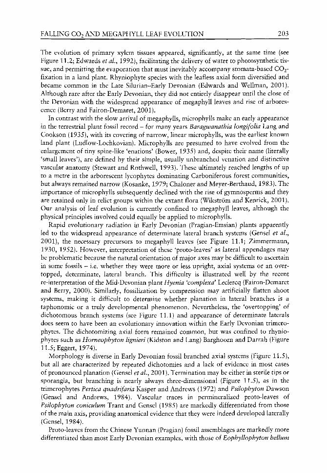
FALLING CO2 AND MEGAPHYLL LEAF EVOLUTION 203
The evolution of primary xylem tissues appeared, significantly, at the same time (see Figure 11.2; Edwards et aL, 1992), facilitating the delivery of v^ater to photosynthetic tissue, and permitting the evaporation that must inevitably accompany stomata-based CO2-fixation in a land plant. Rhyniophyte species w ith the leafless axial form diversified and became common in the Late Silurian-Early Devonian (Edwards and Wellman, 2001). Although rare after the Early Devonian, they did not entirely disappear until the close of the Devonian with the widespread appearance of megaphyll leaves and rise of arbores-cence (Berry and Fairon-Demaret, 2001).
In contrast with the slow arrival of megaphylls, microphylls make an early appearance in the terrestrial plant fossil record - for many years Baragwanathia longifolia Lang and Cookson (1935), with its covering of narrow, linear microphylls, was the earliest known land plant (Ludlow-Lochkovian). Microphylls are presumed to have evolved from the enlargement of tiny spine-like 'enations' (Bower, 1935) and, despite their name (literally 'small leaves'), are defined by their simple, usually unbranched venation and distinctive vascular anatomy (Stewart and Rothwell, 1993). These ultimately reached lengths of up to a metre in the arborescent lycophytes dominating Carboniferous forest communities, but always remained narrow (Kosanke, 1979; Chaloner and Meyer-Berthaud, 1983). The importance of microphylls subsequently declined with the rise of gymnosperms and they are retained only in relict groups within the extant flora (Wikstrom and Kenrick, 2001). Our analysis of leaf evolution is currently confined to megaphyll leaves, although the physical principles involved could equally be applied to microphylls.
Rapid evolutionary radiation in Early Devonian (Pragian-Emsian) plants apparently led to the widespread appearance of determinate lateral branch systems (Gensel et al,^ 2001), the necessary precursors to megaphyll leaves (see Figure 11.1; Zimmermann, 1930, 1952). However, interpretation of these 'proto-leaves' as lateral appendages may be problematic because the natural orientation of major axes may be difficult to ascertain in some fossils - i.e. whether they were more or less upright, axial systems or an overtopped, determinate, lateral branch. This difficulty is illustrated well by the recent re-interpretation of the Mid-Devonian plant Hyenia 'complexa' Leclercq (Fairon-Demaret and Berry, 2000). Similarly, fossilization by compression may artificially flatten shoot systems, making it difficult to determine whether planation in lateral branches is a taphonomic or a truly developmental phenomenon. Nevertheless, the 'overtopping' of dichotomous branch systems (see Figure 11.1) and appearance of determinate laterals does seem to have been an evolutionary innovation within the Early Devonian trimero-phytes. The dichotomizing axial form remained common, but was confined to rhynio-phytes such as Horneophyton lignieri (Kidston and Lang) Barghoorn and Darrah (Figure 11.5; Eggert, 1974).
Morphology is diverse in Early Devonian fossil branched axial systems (Figure 11.5), but all are characterized by repeated dichotomies and a lack of evidence in most cases of pronounced planation (Gensel et al.^ 2001). Termination may be either in sterile tips or sporangia, but branching is nearly always three-dimensional (Figure 11.5), as in the trimerophytes Pertica quadrifaria Kasper and Andrews (1972) and Psilophyton Dawson (Gensel and Andrews, 1984). Vascular traces in permineralized proto-leaves of Psilophyton coniculum Trant and Gensel (1985) are markedly differentiated from those of the main axis, providing anatomical evidence that they were indeed developed laterally (Gensel, 1984).
Proto-leaves from the Chinese Yunnan (Pragian) fossil assemblages are markedly more differentiated than most Early Devonian examples, with those of Eophyllophyton helium

204 C. P. OSBORNE £ T A L .
Figure 11.5 Photosynthetic structures of characteristic Early Devonian genera. Asterisks indicate that the genus is confined to this epoch, following Chaloner and Sheerin (1979). Each outline is redrawn from either a: "^reconstruction; ^drawing of a fossil; or ''photograph of a fossil. Scale bars represent 10 mm for a-g and 1 mm for h. Estimated stages follow the time scale of Harland et al. (1990) and are abbreviated as: Pra, Pragian; Ems, Emsian; Eif, Eifelian; Giv, Givetian; Frs, Frasnian; and Fam, Famennian. a Fsilophyton dapsile Andrews et al. (1977)"^ (Ems/Eif). b Fs. forbesii Gensel"^ Andrews et al. (1977). c ?s. dawsonii Banks et al. (1975)"*" (Ems/Eif). d Horneophyton lignieri (Kidston and Lang) Barghoorn
and Darrah+ (Pra/Ems) Eggert (1974). e Pertica quadrifaria Kaspar and Andrews"'" (Ems/Eif) Andrews et al. (1977). f Fsilophyton microspinosum Andrews et al. (1977)"^ (Ems/Eif). g Fs. charientos Gensel (1979)" " (Ems/Eif). h Eophyllophyton helium Hao and Beck (1993)* (Pra).
being both planated and webbed by a thin lamina (thickness = 4 0 - 2 0 0 luim; Flao and Beck, 1993). Unlike similar laminar structures in Adoketophyton subverticillatum Li and Edwards and Calatheca beckii H a o and Gensel from the same flora, which are always confined to terminal fertile regions, those in E. helium are lateral and may be fertile or

FALLING CO2 AND MEGAPHYLL LEAF EVOLUTION 205
sterile (Hao and Gensel, 2001). Thus the proto-leaves of E. bellum seem to have no clear role in protecting the sporangia and are interpreted as true photosynthetic megaphyll leaves (Hao, 1988; Hao and Beck, 1993). Their morphology is distinctive, being deeply divided into lobes and reach only 2.0 to 4.5 mm in length and 1.4 to 4.0 mm in v^idth (see Figure 11.5; Hao and Beck, 1993). While their interpretation as the earliest photosynthetic megaphyll leaves may be contentious, these structures undoubtedly demonstrate that there were no developmental barriers to megaphyll leaf evolution in at least one lineage by the Early Devonian.
A transition towards later forest floras began in the Mid-Devonian (EifeUan-Givetian), with large increases in plant stature of up to 3-5 m permitted by the evolution of secondary xylem or cortex tissues (Chaloner and Sheerin, 1979; Niklas, 1997; Berry and Fairon-Demaret, 2001). Laminate megaphyll leaves also appear at this time in several rare genera of uncertain affinity (Hoeg, 1967), and may be present in the early progymnosperm Svalbardia polymorpha Hoeg, although the finely dissected ultimate appendages of this species have been variously interpreted as branches (Beck, 1970) and laminae (Hoeg, 1942). However, species with non-laminate, determinate, lateral branch systems (proto-leaves) remain the norm in fossil assemblages (Figure 11.6), and naked, dichotomizing rhyniophyte axes are still present, but restricted in distribution (Berry and Fairon-Demaret, 2001).
Non-laminate proto-leaves of Mid-Devonian age are more commonly planated than are their Early Devonian counterparts (see Figures 11.1 and 11.6), as illustrated by the cladoxylalean Cladoxylon scoparium Krausel and Weyland (Leclercq, 1970) and the putative sphenophyte Ibyka amphikoma Skog and Banks (1973). Nevertheless, some species retain three-dimensional branching structures, for example the progymnosperms Tetraxylopteris schmidtii Beck (1957) and Actinoxylon banksii Matten (1968) and the cladoxylalean Pseudosprochnus nodosus Leclercq and Banks (Berry and Fairon-Demaret, 1997). While the terminal segments of these fossils may appear laminate, this is as likely to be taphonomic as developmental, the result of compression of an initially terete structure during preservation (Chaloner, 1999).
Large laminate megaphylls are atypical of Mid-Devonian fossil floras and, in most cases, are species or genera known only from a single locality or specimen. The morphogenus Platyphyllum (Dawson) White is represented by five Mid-Devonian species, all characterized by fan-shaped (flabelliform) laminae between 2 and 15 cm in length and 1 and 5 cm in width (Hoeg, 1967). Apparent evidence of tracheids in at least three of these specimens confirms their status as tracheophytes (Hoeg, 1967), but details are lacking on the plants that bore them. Ginkgophytopsis gilkinetii (Leclercq, 1928) Hoeg is similar in morphology and similarly enigmatic. However, the largest leaves of late Mid-Devonian (or earliest Late Devonian) age belong to the flabelliform Enigmophyton superbum Hoeg (1942) and measure at least 16 cm by 12 cm. They demonstrate that, although rare, large laminate megaphylls were clearly a viable adaptation in some environments at this time. It would be of great interest to know more of the ecology of these rare Mid-Devonian flabelliform leaves.
True laminate megaphyll leaves finally came of age in the forest floras of the Late Devonian (Frasnian-Famennian) dominated by the progymnosperm genus Archaeopteris Dawson (Beck, 1964). Fossil megaphylls of these trees vary considerably in size and morphology, ranging from the laminate leaves of A. hibernica (Forbes) Dawson to the finely dissected branching structures of A. fissilis Schmalhausen (Figure 11.7). Thus, although laminate leaves were widespread, plants bearing non-laminate proto-leaves remained key components of forest floras (Gensel and Andrews, 1984). The planated, finely branching.

206 C. P. OSBORNE £TAL.
Figure 11.6 Photosynthetic structures of characteristic Middle Devonian genera. See Figure 11.5 for key. Scale bars represent 10 mm for all except a, where the scale is 5 mm. a Calamophyton bicephalum Leclercq and Andrews (I960)** (Giv). b Hyenia elegans Krausel and Weyland** (Eif) Hirmer (1927). c Pseudosprochnus nodosus Leclercq and Banks (1962)"'" (Giv). d Actinoxylon banksii Matten (1968) *"' (Giv). e Ihyka amphikoma Skog and Banks (1973)" (Giv). f Arctophyton gracile Schweitzer (1968)" (Eif). g Protocephalopteris praecox (H0eg) Ananiev*^ (Eif/Giv) Schweitzer (1968).
non-laminate 'fronds' of the putative primitive fern Rhacophyton ceratangium Andrew^s and Phillips are particularly notew^orthy in this context (see Figure 11.7), many reaching lengths in excess of 30 cm (Andrew^s and Phillips, 1968). They demonstrate that laminate megaphylls, although obviously successful, w ere by no means a ubiquitous adaptation in Late Devonian plants.
Fossil floras of the Early Carboniferous (Tournaisian-Visean) are dominated by arborescent lycophytes v ith long microphyll leaves, and megaphylls are confined to the

FALLING CO2 AND MEGAPHYLL LEAF EVOLUTION 207
Figure 11.7 Photosynthetic structures of characteristic Late Devonian genera. See Figure 11.5 for key. Scale bars represent 10 mm. a Archaeopteris hibernica (Forbes) Dawson*^ (Fam) Original. b A. halliana (Goeppert) Lesquereux''' (Fam) Hirmer (1927). c Rhacophyton ceratangium Andrews and Phillips^ (Fam) Cornet et a\. (1976). d Archaeopteris fissilis Schmalhausen """ (Stage uncertain) Andrews et al. (1965). e Svalbardia banksii Matten (1981)" (Frs). f Sphenopteridium rigidum (Ludwig) Potonie^ (Fam) Cleal and Thomas (1995).
ferns, pteridosperms and sphenophytes. In early sphenophytes such as Archaeocalamites radiatus (Brongniart) Stur and Spenophyllum tenerrimum Ettingshausen, these remain as lateral branching systems without laminae (Figure 11.8), although laminate forms appear in later genera of this group (Stewart and Rothwell, 1993). By contrast, megaphylls of the pteridosperms Diplopteridium teilianum (Kidston) Walton and Rhacopteris circularis Walton, and likely members of the same group Charbeckia macrophylla Knaus et al. (2000) and Genselia compacta Knaus and Gillespie (2001), are exclusively laminate, closely resembling those of modern ferns (Figure 11.8). However, Lower Carboniferous leaves display a wide diversity in form (Boyce and Knoll, 2002), with the broad laminae of pteridosperms such as C. macrophylla coexisting with the highly dissected terete forms of examples such as Diplopteridium holdenii. With the decline of arborescent lycophytes in the Late Carboniferous and rise of gymnosperms, the laminate megaphyll leaf became firmly established in subsequent floras, as it is today (Beck, 1970).
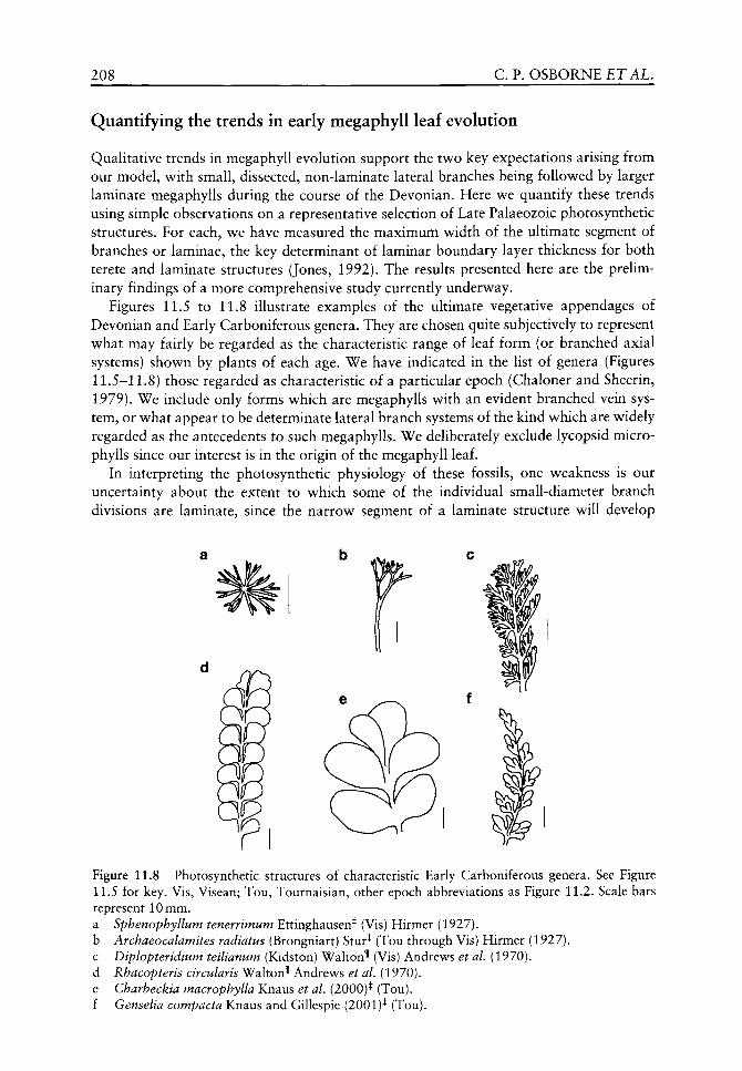
208 C. P. OSBORNE £TAL.
Quantifying the trends in early megaphyll leaf evolution
Qualitative trends in megaphyll evolution support the tv^o key expectations arising from our model, v^ith small, dissected, non-laminate lateral branches being followed by larger laminate megaphylls during the course of the Devonian. Here we quantify these trends using simple observations on a representative selection of Late Palaeozoic photosynthetic structures. For each, we have measured the maximum width of the ultimate segment of branches or laminae, the key determinant of laminar boundary layer thickness for both terete and laminate structures (Jones, 1992). The results presented here are the preliminary findings of a more comprehensive study currently underway.
Figures 11.5 to 11.8 illustrate examples of the ultimate vegetative appendages of Devonian and Early Carboniferous genera. They are chosen quite subjectively to represent what may fairly be regarded as the characteristic range of leaf form (or branched axial systems) shown by plants of each age. We have indicated in the list of genera (Figures 11.5-11.8) those regarded as characteristic of a particular epoch (Chaloner and Sheerin, 1979). We include only forms which are megaphylls with an evident branched vein system, or what appear to be determinate lateral branch systems of the kind which are widely regarded as the antecedents to such megaphylls. We deliberately exclude lycopsid micro-phylls since our interest is in the origin of the megaphyll leaf.
In interpreting the photosynthetic physiology of these fossils, one weakness is our uncertainty about the extent to which some of the individual small-diameter branch divisions are laminate, since the narrow segment of a laminate structure will develop
Figure 11.8 Photosynthetic structures of characteristic Early Carboniferous genera. See Figure 11.5 for key. Vis, Visean; Tou, Tournaisian, other epoch abbreviations as Figure 11.2. Scale bars represent 10 mm.
Sphenophyllum tenerrimum Ettinghausen* (Vis) Hirmer (1927). Archaeocalamites radiatus (Brongniart) Stur* (Tou through Vis) Hirmer (1927). Diplopteridium teilianum (Kidston) Walton* (Vis) Andrews et al. (1970). Rhacopteris circularis Waltou'lf Andrews et al. (1970). Charbeckia macrophylla Knaus et al. (2000)* (Tou). Genselia compacta Knaus and Gillespie (2001)* (Tou).
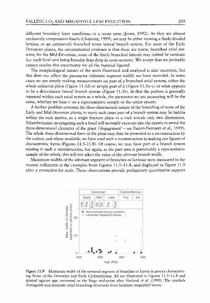
FALLING CO2 AND MEGAPHYLL LEAF EVOLUTION 209
different boundary layer conditions to a terete stem (Jones, 1992). As they are almost exclusively compression fossils (Chaloner, 1999), we may be either viewing a finely divided lamina, or an extensively branched terete lateral branch system. For most of the Early Devonian plants, the circumstantial evidence is that these are terete, branched axial systems; for the Mid-Devonian, some of the finely branched laterals may indeed be laminate (i.e. each final unit being broader than deep in cross-section). We accept that we probably cannot resolve this uncertainty for all the material figured.
The morphological nature of the units illustrated and analysed is also uncertain, but this does not affect the parameter (ultimate segment width) we have recorded. In some cases we are simply making measurements on part of a branched axial system, either the whole subaerial plant (Figure 11.5d) or simply part of it (Figure 11.5a-c) or what appears to be a determinate lateral branch system (Figure 11.5h). In that the pattern is generally repeated within each axial system as a whole, the parameter we are measuring will be the same, whether we base it on a representative sample or the entire system.
A further problem concerns the three-dimensional nature of the branching of some of the Early and Mid-Devonian plants; in many such cases part of a branch system may be hidden within the rock matrix, as a single fracture plane in a rock reveals only two dimensions. Palaeobotanists investigating such a fossil will normally excavate into the matrix to reveal the three-dimensional character of the plant ('degagement' - see Fairon-Demaret et a\.^ 1999). The whole three-dimensional form of the plant may then be presented as a reconstruction by the author, and where available, we have used such a reconstruction in making our figures of characteristic forms (Figures 11.5-11.8). Of course, we may have part of a branch system missing in such a reconstruction, but again, as the part seen is presumably a representative sample of the whole, this will not affect the value of the ultimate branch width.
Maximum widths of the ultimate segment of branches or laminae were measured to the nearest millimetre in the examples from Figures 11.5-11.8, and displayed in Figure 11.9 after a correction for scale. These observations provide preliminary quantitative support
CO
I 20 CO
B o
E S?10
Devonian
Early
0 _i
2 CL
CO
E LU
Mid
LD >
Late
CO E CO
Carboniferous
Tou Vis
• Non-laminate branch systems 0 Laminate megaphyll leaves Q
o-0
0 0
400 380 360
Age (Ma)
340
Figure 11.9 Maximum width of the terminal segment of branches or leaves in genera characterizing floras of the Devonian and Early Carboniferous. All are illustrated in Figures 11.5-11.8 and plotted against age, estimated as the Stage mid-point after Harland et al. (1990). The symbols distinguish non-laminate axial branching structures from laminate megaphyll leaves.
210 C. P. OSBORNE £TAL.
for the qualitative trends already described. Branched, non-laminate photosynthetic organs of the selected species were less than 3 mm in width throughout the Early and Mid-Devonian. Eophyllophyton bellum is the only example of a species with laminate mega-phyll leaves during this interval and also conforms to the pattern, with leaf lobes less than 1 mm in width. Only late in the Late Devonian (Famennian) did photosynthetic organs increase in size, with the widespread appearance in progymnosperms of laminate mega-phyll leaves up to 10 mm in width. This upward trend in maximum leaf size continued in the Carboniferous, as illustrated by the pteridosperms in Figure 11.8, exceeding 20 mm in Charbeckia macrophylla. Narrow, non-laminate 'proto-leaves' were still present in sphenophyte floras at this time, but were rare by comparison with their heyday in the late Early and Mid-Devonian.
Discussion
The quantitative analysis presented in Figure 11.9 must be regarded as a preUminary investigation of the trends occurring in megaphyll evolution during the Late Palaeozoic, rather than a rigorous objective test of our hypothesis. Nevertheless, it supports the two key predictions arising from our model simulations. First, that biophysical constraints restricted the earliest megaphylls to narrow or highly dissected shapes. Evidence from the fossil record suggests that these constraints were important limiters of megaphyll evolution, confining proto-leaves to (predominantly) non-laminate forms throughout the Early and Mid-Devonian, and delaying the widespread appearance of laminate forms until the Late Devonian (see Figure 11.9).
Secondly, evidence supports our expectation that the evolution of larger and less dissected laminate megaphylls tracked the Devonian and Early Carboniferous decline in atmospheric CO2. Our examples show a clear trend in maximum lamina width and suggest that this evolutionary change was particularly rapid in the 20 Ma spanning the latest Devonian and earliest Carboniferous (see Figure 11.9). This period must therefore be regarded as an important focus for future studies. The precise starting date for these increases in lamina size is uncertain, because of the combination of examples selected for study and the small sample size. Future examination of Givetian-Frasnian fossils will be important to date this evolutionary event with greater accuracy (see Figure 11.9).
For the reasons set out earlier (Beerling et al,^ 2001a) and amplified here, we therefore interpret the late appearance of laminate foliage as a response to changes in atmospheric composition, largely brought about by terrestrial plant life itself (Algeo and Scheckler, 1998; Algeo et al.^ 2001). However, we accept that this simple interpretation must be qualified with some provisos.
The first of these is the presence from the Early Devonian onwards of very few records of flabelliform leaves, seemingly produced by vascular plants. Unfortunately we know next to nothing of the ecology of the plants producing them, or indeed the ecology of most of the plants that we use here to illustrate this theme. The interpretation of these 'anomalous' leaves is complicated by the fact that the vascular nature of the plants themselves is not in every case securely documented. The flabelliform phylloids attributed to Prototaxites Dawson (see e.g. Schweitzer, 1987: Figure 11.7), a structure now considered to be fungal (Hueber, 2001), are a reminder of the possible confusion between such structures and the leaves of terrestrial land plants.
A further proviso relates to the fact that, synchronously with the early pinnate megaphylls of the Late Devonian/Early Carboniferous progymnosperms, pterodosperms

FALLING CO2 AND MEGAPHYLL LEAF EVOLUTION 211
and/or pteridophytes, there remains a range of deeply divided leaves or leaf-like lateral systems (e.g. Figure 11.8a-c). This is hardly surprising because modern plants with very different light-trapping and transpiration strategies evidently coexist in the same communities (e.g. Gates, 1979). The presence of rosette weeds such as the deeply dissected Achillea millefolium L. in juxtaposition in the same sward as the entire-leaved Plantago major L. reminds us that we need to be guarded in offering simplistic explanations attributing 'adaptive' merit of one strategy against another. Leaves of small herbaceous plants encounter a wide range of environmental variables through the course of a seasonal climate and equally must ward off a range of different herbivores. While one plant may use chemical means of achieving this, others may use structural modifications, so producing very different leaf forms for different reasons. It is evident that the appearance of megaphyll leaves did not 'displace' their more deeply dissected antecedents, but while expanding in diversity in different plant groups, came to coexist with them.
As we see in other aspects of evolutionary change in plants, an innovation may partially displace the version that had preceded it, but only rarely supplants it entirely. In a simple way, this might be argued of the development of heterospory from homospory and subsequently of the seed habit from heterospory. Those three types of life cycle are still well represented in the present-day flora, although clearly the representation of each has changed drastically since Devonian time.
Further expectations from our biophysical simulations relate to the ecology and spatial distribution of early megaphyll evolution. First, we might expect laminate leaves to have appeared first in species of shaded forest understorey habitats, where the adverse effects of high solar irradiance are reduced, but not eliminated (Woodward, 1980). Unfortunately, the Devonian plant fossil record does not permit a test of this expectation, since sun leaves cannot be distinguished from the shade leaves of early forests. Secondly, we might expect to see significant latitudinal trends in the early evolution of leaves. Since solar elevation declines with latitude, the interception of solar energy by a planated lamina must also decrease (see Figure 11.4A), reducing the risk of overheating and the necessity for effective heat dissipation. The laminate megaphyll leaf is therefore likely to have become widespread first at high latitudes, an expectation that may be tested using palaeolatitude data for fossil localities (Scotese and McKerrow, 1990). Furthermore, the temporal trend in leaf width seen in Figure 11.9 is likely to be paralleled by a spatial pattern, with megaphyll laminae becoming larger and less dissected with latitude. Such expectations offer another key test of our simulations, requiring the assembly of a larger set of observations.
Acknowledgements
We thank Charles Wellman and Dana Royer for their comments on the manuscript. This work was funded through the award of a Leverhulme Trust research grant. DJB gratefully acknowledges funding through a Royal Society University Research Fellowship and the Leverhulme Trust.
References
Algeo TJ, Scheckler SE. 1998. Terrestrial-marine teleconnections in the Devonian: links between the evolution of land plants, weathering processes, and marine anoxic events. Philosophical Transactions of the Royal Society of London B 353: 113-130.
212 C. P. OSBORNE £ T A L .
Algeo TJ, Scheckler SE, Maynard JB. 2001. Effects of the Middle to Late Devonian spread of vascular plants on weathering regimes, marine biotas, and global climate. In: Gensel PG, Edwards D, eds. Plants Invade the Land. New York: Columbia University Press, 213-236.
Andrews HN, Arnold CA, Boureau E, et al. 1970. Traite de Paleobotanique; Volume 4, Part 1, Filicophyta. Paris: Masson et Cie.
Andrews HN, Kasper AE, Forbes WH, et al. 1977. Early Devonian flora of the Trout Valley Formation of northern Maine. Review of Palaeobotany and Palynology 23: 255-285.
Andrews HN, Phillips TL. 1968. Rhacophyton from the Upper Devonian of West Virginia. Botanical Journal of the Linnean Society 61: 37-64.
Andrews HN, Phillips TL, Radforth NW. 1965. Paleobotanical studies in Arctic Canada. L Archaeopteris from EUesmere Island. Canadian Journal of Botany 43: 545-556.
Banks HP, Leclercq S, Hueber FM. 1975. Anatomy and morphology of Psilophyton dawsoni sp. n. from the later Lower Devonian of Quebec (Gaspe), and Ontario, Canada. Palaeontographica Americana 8: 77-127.
Beck CB. 1957. Tetraxylopteris schmidtii gen. et sp. nov., a probable pteridosperm precursor from the Devonian of New York. American Journal of Botany 44: 350-367.
Beck CB. 1964. Predominance of Archaeopteris in Upper Devonian flora of western Catskills and adjacent Pennsylvania. Botanical Gazette 125: 126-128.
Beck CB. 1970. The appearance of gymnospermous structure. Biological Review 45: 379-400. Beerling DJ, Osborne CP, Chaloner WG. 2001a. Evolution of leaf form in land plants linked to
atmospheric CO2 decline in the Late Palaeozoic era. Nature 410: 352-354. Beerling DJ, Osborne CP, Chaloner WG. 2001b. Do drought-hardened plants suffer from fever?
Response. Trends in Plant Science 6: 507-508. Beerling DJ, Royer DL. 2002. Reading a CO2 signal from fossil stomata. New Phytologist
153: 387-397. Beerling DJ, Woodward FI. 1997. Changes in land plant function over the Phanerozoic: recon
structions based on the fossil record. Botanical Journal of the Linnean Society 124: 137-153. Berner RA, Kothavala Z. 2001. GEOCARB III: a revised model of atmospheric CO2 over
Phanerozoic time. American Journal of Science 301: 182-204. Berry CM, Fairon-Demaret M. 1997. A reinvestigation of the cladoxylopsid Pseudosporochnus
nodosus Leclercq et Banks from the Middle Devonian of Goe, Belgium. International Journal of Plant Sciences 158: 350-372.
Berry CM, Fairon-Demaret M. 2001. The Middle Devonian flora revisited. In: Gensel PG, Edwards D, eds. Plants Invade the Land. New York: Columbia University Press, 120-139.
Bower FO. 1935. Primitive Land Plants. London: Macmillan & Co. Boyce CK, Knoll AH. 2002. Evolution of developmental potential and the multiple independent
origins of leaves in Paleozoic vascular plants. Paleobiology 28: 70-100. van der Burgh J. 1996. Changes in leaf shape and environment during the Upper Devonian.
In: Corsettii F, Tiffney BH, eds. Fifth Quadrennial Conference Abstract Volume. Santa Barbara: International Organization of Paleobotany, 106.
Burke JJ, Upchurch DR. 1989. Leaf temperature and transpirational control in cotton. Environmental and Experimental Botany 29: 487-492.
Chaloner WG. 1999. Plant and spore compression in sediments. In: Jones TP, Rowe NP, eds. Fossil Plants and Spores - Modern Techniques. London: Geological Society, 36-40.
Chaloner WG, Hill AJ, Lacey WS. 1977. First Devonian platyspermic seed and its implications in gymnosperm evolution. Nature 265: 233-235.
Chaloner WG, Meyer-Berthaud B. 1983. Leaf and stem growth in the Lepidodendrales. Botanical Journal of the Linnean Society 86: 135-148.
Chaloner WG, Sheerin A. 1979. Devonian macrofloras. In: House MR, Scrutton CT, Bassett MG, eds. The Devonian System. London: The Palaeontological Association, 145-161.
Cleal CJ, Thomas BA. 1995. Palaeozoic Palaeobotany of Great Britain. London: Chapman 8c Hall. Cornet B, Phillips TL, Andrews HN. 1976. The morphology and variation in Rhacophton
ceratangium from the Upper Devonian and its bearing on frond evolution. Palaeontographica B 158: 105-129.
FALLING CO2 A N D MEGAPHYLL LEAF EVOLUTION 213
Cox JE, Railsback LB, Gordon EA. 2001. Evidence from Catskill Pedogenic carbonates for a rapid Late Devonian decrease in atmospheric carbon dioxide concentrations. Northeastern Geology and Environmental Sciences 23: 91-102.
Edv^ards D. 1998. Climate signals in Palaeozoic land plants. Philosophical Transactions of the Royal Society of London B 353: 141-157.
Edwards D, Abbott GD, Raven JA. 1996. Cuticles of early land plants: a palaeoecophysiological evaluation. In: Kerstiens G, ed. Plant Cuticles - an Integrated Functional Approach. Oxford: BIOS Scientific Publishers, 1-31.
Edwards D, Davies KL, Axe L. 1992. A vascular conducting strand in the early land plant Cooksonia. Nature 357: 683-685.
Edwards D, Feehan J, Smith DG. 1983. A late Wenlock flora from Co. Tipperary, Ireland. Botanical Journal of the Linnean Society 86: 19-36.
Edwards D, Wellman C. 2001. Embryophytes on land: The Ordovician to Lochkovian (Lower Devonian) record. In: Gensel PG, Edwards D, eds. Plants Invade the Land. New York: Columbia University Press, 3-28.
Eggert DA. 1974. The sporangium oi Horneophyton lignieri (Rhyniophytina). American journal of Bof^wy 61: 405-413.
Ehleringer JR. 1988. Comparative ecophysiology of Encelia farinosa and Encelia frutescens. 1. Energy balance considerations. Oecologia 76: 553-561.
Fairon-Demaret M, Berry CM. 2000. A reconsideration of Hyenia elegans Krausel et Weyland and Hyenia 'complexa' Leclercq: two Middle Devonian cladoxylopsids from Western Europe. International Journal of Plant Sciences 161: 473-494.
Fairon-Demaret M, Hilton J, Berry CM. 1999. Surface preparation of macrofossils (degagement). In: Jones TP, Rowe NP, eds. Foss/7 Plants and Spores - Modern Techniques. London: Geological Society, 33-35.
Gates DM. 1968. Transpiration and leaf temperature. Annual Review of Plant Physiology 19:211-238.
Gates DM. 1979. Biophysical Ecology. New York: Springer Verlag. Gensel PG. 1979. Two Psilophyton species from the Lower Devonian of eastern Canada
with a discussion of morphological variation within the genus. Palaeontographica B 168: 81-99.
Gensel PG. 1984. A new Lower Devonian plant and the early evolution of leaves. Nature 309: 785-787.
Gensel PG, Andrews HN. 1984. Plant Life in the Devonian Period. New York: Praeger Scientific. Gensel PG, Kotyk ME, Basinger JF. 2001. Morphology of above- and below-ground structures in
Early Devonian (Pragian-Emsian) plants. In: Gensel PG, Edwards D, eds. Plants Invade the Land. New York: Columbia University Press, 83-102.
Gurevitch J, Schuepp PH. 1990. Boundary layer properties of highly dissected leaves - an investigation using an electrochemical fluid tunnel. Plant, Cell and Environment 13: 783-792.
Haase P, Pugnaire FI, Clark SC, Incoll LD. 1999. Diurnal and seasonal changes in cladode photo-synthetic rate in relation to canopy age structure in the leguminous shrub Retama sphaerocarpa. Functional Ecology 13: 640-649.
Hao SG. 1988. A new Lower Devonian genus from Yunnan, with notes on the origin of the leaf. Acta Botanica Sinica 30: 441-448.
Hao SG, Beck CB. 1993. Further observations on Eophyllophyton helium from the Lower Devonian (Siegenian) of Yunnan, China. Palaeontographica B 230: 27-41.
Hao SG, Gensel PG. 2001. The Posongchong floral assemblages of southeastern Yunnan, China -diversity and disparity in Early Devonian plant assemblages. In: Gensel PG, Edwards D, eds. Plants Invade the Land. New York: Columbia University Press, 103-119.
Harland WB, Armstrong RL, Cox AV, et al. 1990. A Geologic Timescale 1989. Cambridge: University Press.
Hirmer M. 1927. Handbuch der Palaeobotanik. Oldenbourg, Miinchen, Berlin. Hoeg OA. 1942. The Downtonian and Devonian flora of Spitsbergen. Norges Svalbard-og Inshavs-
Underskelser 83: 1-228.
214 C. P. OSBORNE £ T A L .
Heeg OA. 1967. Ordre Incertae Sedis des Palaeophyllales. In: Boureau E, ed. Traite de Faleobotanique, Vol. 11. Paris: Masson et Cie, 362-399.
Hueber FM. 2001. Rotted wood-alga-fungus: the history and Hfe of Frototaxites Dawson 1859. Review of Palaeobotany and Palynology 116: 123-158.
Jones HG. 1992. Plants and Microclimate, 2nd edn. Cambridge: University Press. Kasper AE, Andrews HN. 1972. Pertica, a new genus of Devonian plants from northern Maine.
American Journal of Botany 59: 897-911. Kitano M, Tateishi J, Eguchi H. 1995. Evaluation of leaf boundary layer conductance of a whole
plant by application of abscisic acid inhibiting transpiration. Biotronics 24: 51-58. Knaus MJ, Gillespie WH. 2001. Genselia compacta (Jongmans et al.) Knaus et Gillespie comb.
nov.: new insights into possible developmental pathways of early photosynthetic units. Palaeontographica B 110: 1-34.
Knaus MJ, Upchurch GR, Gillespie WH. 2000. Charbeckia macrophylla gen. et sp. nov. from the Lower Mississipian Price (Pocono) Formation of southeastern West Virginia. Review of Palaeobotany and Palynology 111: 71-92.
Kosanke RM. 1979. A long-leaved specimen of Lepidodendron. Geological Society of America Bulletin 90: 431-434.
Lang WH, Cookson IC. 1935. On a flora, including vascular plants associated with Monograptus, in rocks of Silurian age, from Victoria, Australia. Philosophical Transactions of the Royal Society of London B 224: 421-449.
Lange OL. 1959. Untersuchungen iiber den Warmehaushah und Hitzeresistenz mauretanischer Wiistern- und Savennenpflanzen. Flora 147: 595-651.
Leclercq S. 1928. Psygomophyllum gilkineti, sp. n. du Devonien Moyen a facies old red sandstone de Malonne (environs de Namur, Belgique), par S. Leclercq et M. Belliere. Botanical Journal of the Linnean Society 48: 1-14.
Leclercq S. 1970. Classe des Cladoxylopsida Pichi-SermoH 1959. In: Boureau E, ed. Traite de Paleobotanique, Vol. IV. Paris: Masson et Cie, 119-165.
Leclercq S, Andrews HN. 1960. Calamophyton bicephalum, a new species from the Middle Devonian of Belgium. Annals of the Missouri Botanical Gardens 47: 1-23.
Leclercq S, Banks HP. 1962. Pseudosporochnus nodosus nov. sp., a Middle Devonian plant with cladoxylalean affinities. Palaeontographica B 110: 1-34.
Matsumoto J, Muroaka H, Washitani I. 2000. Ecophysiological mechanisms used by Aster kantoensis, an endangered species, to withstand high light and heat stress of its gravely flood-plain environment. Annals of Botany 86: 777-785.
Matten LC. 1968. Actinoxylon banksii gen. et sp. nov.: a progymnosperm from the Middle Devonian of New York. American Journal of Botany 55: 773-782.
Matten LC. 1981. Svalbardia banksii sp. nov. from the Upper Devonian (Frasnian) of New York State. American Journal of Botany 68: 1383-1391.
Mora CI, Driese SG, Colarusso LA. 1996. Middle to Late Palaeozoic atmospheric CO2 levels from soil carbonate and organic matter. Science 271: 1105-1107.
Niklas KJ. 1994. Plant Allometry: The Scaling of Form and Process. Chicago: University Press. Niklas KJ. 1997. The Evolutionary Biology of Plants. Chicago: University Press. Raven JA, Edwards D. 2001. Roots: evolutionary origins and biogeochemical significance./owrw<3/
of Experimental Botany 52: 381-401. Robinson JM. 1991. Phanerozoic atmospheric reconstructions: a terrestrial perspective.
Palaeogeography, Palaeoclimatology, Palaeoecology 97: 51-62. Scotese CR, McKerrow WS. 1990. Revised world maps and introduction. In: Scotese CR,
McKerrow WS, eds. Palaeozoic Palaeogeography and Biogeography. Geological Society Memoir Number 12. London: Geological Society, 1-21.
Schweitzer HJ. 1968. Pflanzenreste aus dem Devon Nord-westspitzbergens. Palaeontographica B 123: 43-73.
Schweitzer HJ. 1987. Introduction to the plant bearing beds and flora of the Lower Devonian of the Rhineland. Bonner Faldobotanische Mitteilungen 13: 1-94.
Skog JE, Banks HP. 1973. Ibyka amphikoma gen. et sp. n., a new protoarticulate precursor from the late Middle Devonian of New York state. American Journal of Botany 60: 366-380.

FALLING CO2 A N D MEGAPHYLL LEAF EVOLUTION 215
Stewart WN, Rothwell GW. 1993. Paleobotany and the Evolution of Plants, 2nd edn. New York: Cambridge University Press.
Tan CS, Buttery BR. 1995. Determination of the water-use of 2 pairs of soybean isolines differing in stomatal frequency using a heat-balance stem-flow gauge. Canadian Journal of Plant Science 75: 99-103.
Trant CA, Gensel PG. 1985. Branching in Psilophyton: a new species from the Lower Devonian of New Brunswick, Canada. American Journal of Botany 72: 1256-1273.
Valladares F, Pearcy RW. 1997. Interactions between water stress, sun-shade acclimation, heat tolerance and photoinhibition in the sclerophyll Heteromeles arbutifolia. Plant, Cell and Environment 20: 25-36.
Wikstrom N, Kenrick P. 2001. Evolution of Lycopodiaceae (Lycopsida): estimating divergence times from rbcL gene sequences by use of nonparametric rate smoothing. Molecular Phylogenetics and Evolution 19: 177-186.
Woodward FI. 1980. Shoot extension and water relations of Circea lutetiana in sunflecks. In: Grace J, Ford ED, Jarvis PG, eds. Plants and their Atmospheric Environment. Oxford: Blackwell Scientific Pubhshers.
Yapp CJ, Poths H. 1996. Carbon isotopes in continental weathering environments and variations in ancient atmospheric CO2 pressure. Earth and Planetary Science Eetters 137: 71-82.
Zimmermann W. 1930. Die Phylogenie der Pflanzen. Jena: Gustav Fischer Verlag. Zimmermann W. 1952. Main results of the telome theory. The Palaeobotanist 1: 456-470,

12 Stomatal function and physiology
Tracy Lawson and James I L Morison
CONTENTS
Introduction
Stomatal control of leaf gas exchange
Role of stomata in leaf gas exchange
Heterogeneity in stomatal characters
Effect of environmental variables on stomata and photosynthesis
Stomatal response to CO2 Stomatal response to humidity Stomatal response to light
Environmental interactions and stomatal responses
Modern techniques for ecophysiological stomatal research
Evolutionary context
Conclusion
AcknoM^ledgements
References
217
218
222
224
225 226 228 230
233
235
236
237
238
238
Introduction
Stomata are small adjustable pores found in large numbers on the surface of most aerial parts of higher plants (Spermatophyta), and also in the Pteridophyta. They have been recorded in the fossil record from as early as 411 million years (Ma) ago in the late Silurian-early Devonian period (Edw^ards et al., 1992). During this period plants colonized terrestrial environments w^hich led to the evolution of thin cuticles, stomata and vascular systems in order to avoid desiccation and to transport w^ater v^hile still allow^ing CO2 exchange (Chaloner, 1970). Stomata are formed from tv\ o specialized cells in the epidermis, know^n as guard cells and, in many species, there are also other adjacent subsidiary cells, that are
The Evolution of Plant Physiology Copyright © 2004 Elsevier Ltd ISBN 0-12-33955-26 All rights of reproduction in any form reserved

218 T. LAWSON &c J. L L. MORISON
morphologically distinguishable from the general epidermal cell (see Colour Plate 1). The central role of stomata is in regulating gas exchange between the inside of the leaf and the external environment (Cowan and Troughton, 1971; Jones, 1992) because the cuticle is almost impermeable to water vapour and CO2. The plant needs sufficient CO2 to enter the leaf to photosynthesize, while conserving water to avoid tissue dehydration and metabolic disruption. Even though the stomatal pores as a whole occupy only 0.5-5% of the leaf surface when fully open, almost all the water transpired as well as the CO2 absorbed for photosynthesis passes through these pores, so stomatal function has huge importance in the global hydrological and carbon cycles.
Stomatal aperture is regulated by both internal physiological and external environmental factors. Pore opening through guard cell movements is stimulated by illumination with light in the photosynthetically effective waveband (particularly the blue waveband), low CO2 concentrations and high humidity, while closure is promoted by darkness, low humidity, high temperature and high CO2 concentrations (see reviews by Assmann, 1993 and Willmer and Fricker, 1996). Such guard cell movements are brought about through changes in guard cell turgor (Heath, 1938) and through changes in the difference between guard and epidermal cell turgor (Weyers and Meidner, 1990). These turgor changes require the loss or accumulation of K" or other cations and the parallel exchange of anions including organic solutes such as malate and sucrose (e.g. Willmer and Fricker, 1996; Outlaw, 1996; Asai etaL, 2000).
The numbers of stomata per unit leaf area (referred to as stomatal density or frequency) vary with species and conditions and range from 0 to 2000 or more stomata mm"'^ (Willmer and Fricker, 1996). In herbaceous plants, stomata are found on both the upper (usually adaxial) and lower (usually abaxial) surfaces of leaves and are termed amphistomatous, although there are usually more stomata on the lower surface. However, many tree species have stomata only on the lower surface (hypostomatous) and aquatic plants with floating leaves, such as water lilies have stomata only on the upper surface (epi- or hyperstomatous).
Interest in stomata and their evolution, function, anatomy and physiology have been the subjects of intense studies over the last century. In this chapter, we will concentrate on their function and physiology, in order to clarify the role stomata play in determining carbon assimilation. First, we look at the role of stomata in controlling gas exchange and the limitation stomata can impose on leaf CO2 uptake. We then describe the effects of three key environmental factors on stomatal movements and the consequences for photosynthesis. In the last section we will briefly discuss some new approaches for studying stomatal function and physiology. Our approach is illustrative, rather than an extensive review as there are many detailed texts and reviews available (e.g. Assmann, 1993; Willmer and Fricker, 1996; Zeiger, 2000).
Stomatal control of leaf gas exchange
The diffusion rate of gases into or out of the leaf, or any other plant part, depends on the concentration gradient and the diffusive resistance of the pathway. For water loss from the mesophyll cells inside the leaf, the major pathway is therefore from the mesophyll cell walls through the sub-stomatal cavity to the pore and then out through the layer of air immediately surrounding the leaf, to the mixed air stream (Figure 12.1). The pathway for the uptake of CO2 by the mesophyll during photosynthesis is essentially the same, in the converse direction, but with an additional component of diffusion through the mesophyll cell into the chloroplast. The resistance of the stomatal pathway depends on the geometry

STOMATAL FUNCTION AND PHYSIOLOGY 219
^^IteA^-. -fe^tel^fe Upper epidermis
Palisade parenchyma
Spongy mesophyii
Lower epidermis
^fb
rf^-^^^f«f^j¥;^^^T||?f^|w^^
Guard cell
Figure 12.1 Diagrammatic cross-section of leaf showing pathway for diffusion for CO2 and H2O into and out of a leaf, respectively. The resistance network analogy is also shown for one surface combining the boundary layer (r^), cuticular (r ), stomatal (rj and intercellular space (TJ) resistances (see Nobel, 1991: 143, for details and typical values of the resistances).
of the pores as well as their frequency. Note that although the pore area when open may only be at maximum a few percent of the total leaf area, the rates of evaporation can be about half that of a wet surface of similar dimensions; this is due to the 'edge effect' of diffusion through multiple pores (see Willmer and Fricker, 1996: 116). The stomatal resistance, Ts, can be calculated from pore dimensions of elliptical pores as:
(d + 2c)
A,. ' A'SF
where: D^ = water diffusivity in air (mm^s~^); A^ = mean pore area (mm^), d = pore depth (mm), c = an 'end correction' for the edge effect (mm) and SF = stomatal frequency (mm~^). Different end corrections are necessary for different shaped pores (for further details see Weyers and Meidner, 1990: 57). The units of r^ are therefore in time taken to diffuse unit distance (s mm"^). However, it is common to use the reciprocal of resistance, termed a 'conductance', gs, and to express fluxes as a molar density, giving conductance in mmol m~^s~^ To convert r in smm~^ to g^ in mmolm~^s~\ the l/r^ is multiplied by (P/RT) where P = atmospheric pressure (Pa), R = gas constant (Jmor^K"^), and T = mean of leaf and air temperature (K, see Jones, 1992: 56). Equation (1) can be used to examine the sensitivity of Ts or gs to changes in the component parameters (Weyers and Lawson, 1997). Using a plausible range of values for the stomatal parameters for Phaseolus vulgaris L. (Figure 12.2), it is clear that the various stomatal characters influence the calculation of g to different extents. The main determinant of g^ is pore aperture (width), and stomatal frequency makes a smaller contribution, although more than pore depth or length.
Equation (1) only describes the stomatal part of the pathway for diffusion of water vapour from inside the leaf into the mixed air stream. First, the 'boundary layer' of air close
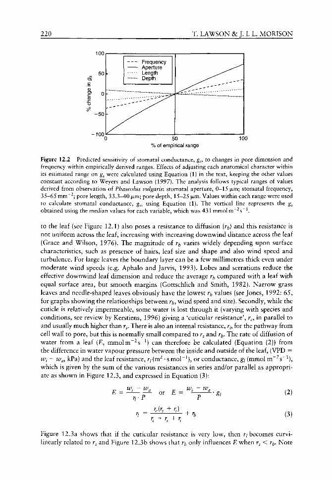
220 T. LAWSON &: J. L L. MORISON
100
C (0
-100
Frequency Aperture Length Depth
50 % of empirical range
100
Figure 12.2 Predicted sensitivity of stomatal conductance, ^5, to changes in pore dimension and frequency within empirically derived ranges. Effects of adjusting each anatomical character v ithin its estimated range on g^ were calculated using Equation (1) in the text, keeping the other values constant according to Weyers and Lawson (1997). The analysis follows typical ranges of values derived from observation of Phaseolus vulgaris: stomatal aperture, 0-15 |jim; stomatal frequency, 35-65 mm~ ; pore length, 33.3-40 fxm; pore depth, 15-25 jxm. Values within each range were used to calculate stomatal conductance, g , using Equation (1). The vertical line represents the gs obtained using the median values for each variable, which was 431 mmolm"^s~^
to the leaf (see Figure 12.1) also poses a resistance to diffusion (r^) and this resistance is not uniform across the leaf, increasing with increasing downwind distance across the leaf (Grace and Wilson, 1976). The magnitude of r^ varies widely depending upon surface characteristics, such as presence of hairs, leaf size and shape and also wind speed and turbulence. For large leaves the boundary layer can be a few millimetres thick even under moderate wind speeds (e.g. Aphalo and Jarvis, 1993). Lobes and serrations reduce the effective downwind leaf dimension and reduce the average r^ compared with a leaf with equal surface area, but smooth margins (Gottschlich and Smith, 1982). Narrow grass leaves and needle-shaped leaves obviously have the lowest r^ values (see Jones, 1992: 65, for graphs showing the relationships between r^, wind speed and size). Secondly, while the cuticle is relatively impermeable, some water is lost through it (varying with species and conditions, see review by Kerstiens, 1996) giving a 'cuticular resistance', r , in parallel to and usually much higher than r . There is also an internal resistance, r , for the pathway from cell wall to pore, but this is normally small compared to r^ and r^. The rate of diffusion of water from a leaf (£, mmolm^^^s"^) can therefore be calculated (Equation (2)) from the difference in water vapour pressure between the inside and outside of the leaf, (VPD = Wi — Way kPa) and the leaf resistance, r/ (m^ • s moI~^), or conductance, gi (mmol m~^s~^), which is given by the sum of the various resistances in series and/or parallel as appropriate as shown in Figure 12.3, and expressed in Equation (3):
£ ^ ^t - ^a
rrP or E = —^ ~ 'gi (2)
(3)
Figure 12.3a shows that if the cuticular resistance is very low, then r/ becomes curvi-linearly related to r^ and Figure 12.3b shows that r only influences E when r^ < Tb- Note
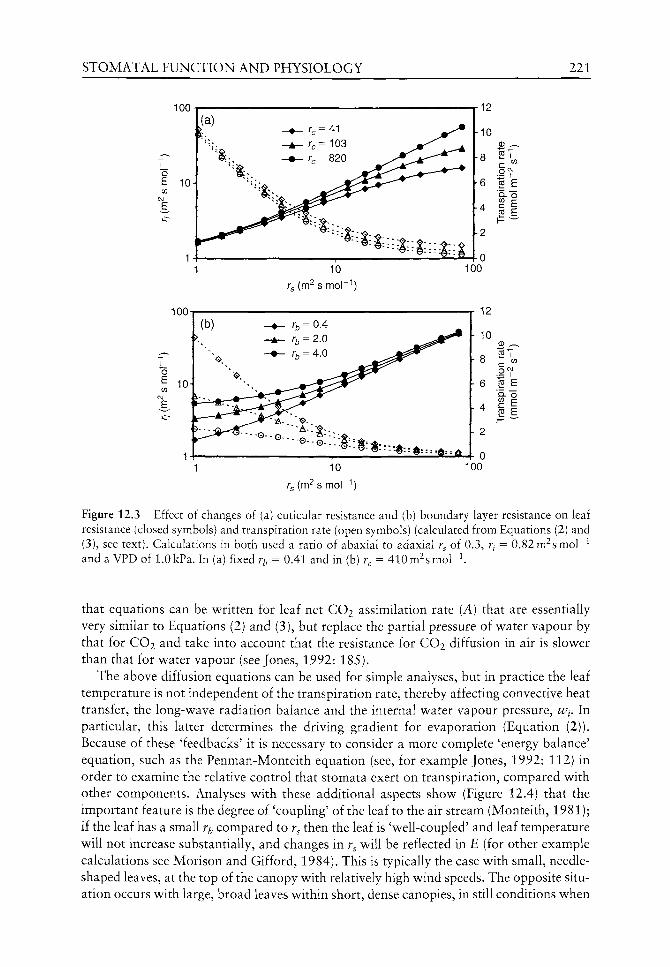
STOMATAL FUNCTION AND PHYSIOLOGY 221
100
/-g (m^ s mol )
100
h 10
12
2T c ^
1 ^ "Q."O
"I 2 E I ^-^
h 2
10
Tg (m^ s mor"*)
100
Figure 12.3 Effect of changes of (a) cuticular resistance and (b) boundary layer resistance on leaf resistance (closed symbols) and transpiration rate (open symbols) (calculated from Equations (2) and (3), see text). Calculations in both used a ratio of abaxial to adaxial r of 0.3, fy = 0.82m^smol~^ and a VPD of 1.0kPa. In (a) fixed r^, = 0.41 and in (b) r, = 410m^smor^.
that equations can be written for leaf net CO2 assimilation rate (A) that are essentially very similar to Equations (2) and (3), but replace the partial pressure of water vapour by that for CO2 and take into account that the resistance for CO2 diffusion in air is slower than that for water vapour (see Jones, 1992: 185).
The above diffusion equations can be used for simple analyses, but in practice the leaf temperature is not independent of the transpiration rate, thereby affecting convective heat transfer, the long-wave radiation balance and the internal water vapour pressure, w^. In particular, this latter determines the driving gradient for evaporation (Equation (2)). Because of these 'feedbacks' it is necessary to consider a more complete 'energy balance' equation, such as the Penman-Monteith equation (see, for example Jones, 1992: 112) in order to examine the relative control that stomata exert on transpiration, compared with other components. Analyses with these additional aspects show (Figure 12.4) that the important feature is the degree of 'coupling' of the leaf to the air stream (Monteith, 1981); if the leaf has a small r^ compared to r^ then the leaf is 'well-coupled' and leaf temperature will not increase substantially, and changes in r^ will be reflected in E (for other example calculations see Morison and Gifford, 1984). This is typically the case with small, needle-shaped leaves, at the top of the canopy with relatively high wind speeds. The opposite situation occurs with large, broad leaves within short, dense canopies, in still conditions when

222 T. LAWSON &c J. I. L. MORISON
100
Figure 12.4 Effect of set increases in the stomatal resistance, r on the relative evaporation rate, £, from a leaf. Calculated using energy-balance equations, w ith air temperature of 20°C, radiative resistance of 5.1 and a boundary layer resistance of 0.37m^smol~^ Evaporation rate is shov^n as the changed rate, relative to original. Dotted lines indicate E change equals r^ change.
E will not closely reflect changes in r^ as r /r^ is small (Figure 12.4). Indeed, it has recently been suggested that the evolution of larger planate leaves from the earlier leafless branched shapes became possible during the late Devonian period because increased stomatal frequencies, in response to declining atmospheric CO2 concentrations, produced greater evaporative cooling and kept leaves below^ their lethal temperature limit (Beerling et al, 2001; see Chapter 11).
Role of stomata in leaf gas exchange
Stomatal behaviour is obviously important because it directly modifies the CO2 assimilation rate and transpiration rate and consequently affects plant w^ater and carbon status. In addition, there are also other less obvious indirect effects such as on nutrient status and leaf temperature, caused by stomatal control of transpiration. Here w e w ill concentrate on the direct effects. Clearly, the restriction that stomata (and the other parts of the diffusion pathw^ay) place on CO2 assimilation rate, A, (i.e. the diffusion limitation) depends upon environmental conditions and plant photosynthetic characteristics. Assimilation rate. A, is not linearly related to leaf conductance, gi (Figure 12.5), because under all but very restricted CO2 supply rates, assimilation rate is colimited by other factors (primarily light). When CO2 supply through the stomata is high, further increase in gi has little or no effect (in this example, w^hen^/ > 500mmolm~^s~^). The limitation that stomata can pose to leaf CO2 assimilation rate can best be examined through the now^ traditional 'A/C/ analysis, vv here A is measured at a range of external CO2 concentrations, usually at high light and constant temperature and the intercellular space CO2 concentration (Q) is calculated from the diffusion equations (see Farquhar and Sharkey, 1982). Typical A/Q response curves constructed for Zea mays L. (C4) and Phaseolus vulgaris (C3) show^ (Figure 12.6) that A is near saturation at low^ Q concentrations in maize, but in bean A increases up to C of ca. 500 luimol mol~^. The intersections of the dashed vertical line writh the A curves indicate the assimilation rates if there was no diffusion limitation by stomata (gl = °° so Cj = Ca) at present atmospheric CO2 concentration. The dashed diagonal lines therefore represent 'supply functions' corresponding to lower g/ values and, as g/ is reduced, supply of CO2 is reduced, so Q declines, possibly affecting A depending on the
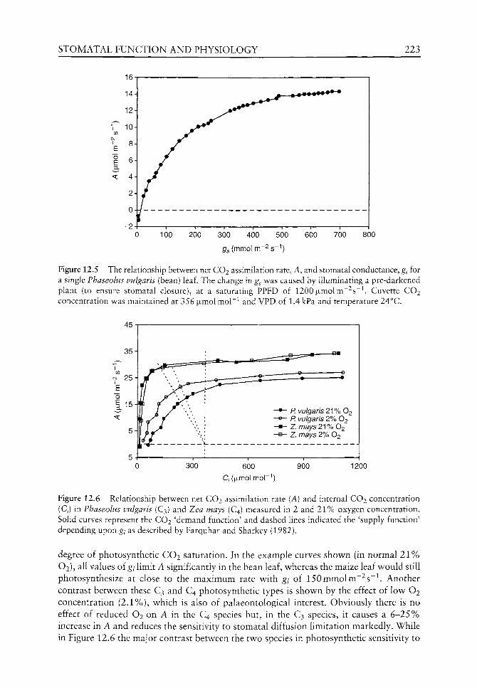
STOMATAL FUNCTION AND PHYSIOLOGY 223
300 400 500 800
Figure 12.5 The relationship between net CO2 assimilation rate, A, and stomatal conductance, gs for a single Fhaseolus vulgaris (bean) leaf. The change in g^ was caused by illuminating a pre-darkened plant (to ensure stomatal closure), at a saturating PPFD of 1200 |xmolm~^s~^. Cuvette CO2 concentration was maintained at 356 jjimolmol"^ and VPD of 1.4 kPa and temperature 24°C.
600
C/(|jLmol mor'')
900 1200
Figure 12.6 Relationship between net CO2 assimilation rate (A) and internal CO2 concentration (Q) in Fhaseolus vulgaris (C3) and Zea mays (C4) measured in 2 and 2 1 % oxygen concentration. Solid curves represent the CO2 'demand function' and dashed lines indicated the 'supply function' depending upon g/ as described by Farquhar and Sharkey (1982).
degree of photosynthetic CO2 saturation. In the example curves shov\^n (in normal 2 1 % O2), all values of g/ limit A significantly in the bean leaf, w^hereas the maize leaf would still photosynthesize at close to the maximum rate w^ith g/ of 150mmolm~^s~^ . Another contrast betw^een these C3 and C4 photosynthetic types is shown by the effect of low O2 concentration (2.1%), which is also of palaeontological interest. Obviously there is no effect of reduced O2 on A in the C4 species but, in the C3 species, it causes a 6 - 2 5 % increase in A and reduces the sensitivity to stomatal diffusion limitation markedly. While in Figure 12.6 the major contrast between the two species in photosynthetic sensitivity to
224 T. LAWSON &c J. I. L. MORISON
stomatal limitation is due to different photosynthetic pathways, similar but less dramatic differences can be seen between leaves with nitrogen content differences (Wong, 1979), or sun and shade morphologies and biochemistry (Patterson, 1980) and with other stresses, e.g. water (von Caemmerer and Farquhar, 1984). It should be clear from these examples that to understand the control that stomata exert on photosynthesis requires a quantitative description of photosynthesis in the particular conditions being examined. This in turn will help in understanding stomatal responses to changes of environmental conditions in the geological past.
Heterogeneity in stomatal characters
It is probable that when they measure gi or r/ on a whole leaf in a cuvette, many researchers imagine that it is a simple bulk version of that shown in the stylized diffusion pathway diagram of Figure 12.1. However, we should always remember that it is a bulk measurement across a very large and heterogeneous population of stomata and meso-phyll. As mentioned in the introduction, there are likely to be differences between the leaf surfaces, as well as other important sources of variation. Heterogeneity in both space and in time of stomatal anatomical characteristics (or related variables such as g j is found at many scales, from the size, frequency and behaviour of small groups of guard cells to the gas exchange of whole plants or stands of plants, even in apparently homogeneous environments (Ticha, 1982; Solarova and Pospisilova, 1983; Pospisilova and Santrucek, 1994; Weyers and Lawson, 1997). For example. Colour Plate 1 shows two guard cell complexes adjacent to one other, yet one pore is open and the other is closed, illustrating variation in stomatal behaviour even at this small scale. Heterogeneity in stomatal characters is not confined to anatomical features but has also been observed in function. As an example. Figure 12.7 shows substantial variation in g^, A and Q over the adaxial surface of a Phaseolus leaf interpolated from approximately 30 spot readings on 1.25 cm^ areas (see Lawson and Weyers, 1999 for other examples). It is also obvious that gi and A are not consistently well correlated in different areas, although a general pattern exists. Comparison with the map of Q emphasizes that areas of high g^ may have high Q and low A rates, indicating that g^ and Q are not limiting the rate of A. Other common examples of stomatal variation can be found between leaves on plants, due probably to age and ontogenetic effects. Figure 12.8 shows that in grape vine shoots g values increased with leaf number from the base (and therefore also with decreasing age) up to leaf 10, but newer leaves further along the shoot had progressively lower g^. This was not simply related to illumination as all apart from the basal few leaves were well lit, nor was it related to leaf size, but was probably largely due to leaf age effects, with a peak g^ in mature but not old leaves (see also Solarova and Pospisilova, 1983).
It is important to recognize the extent of spatial and temporal variability in stomatal and photosynthetic parameters because experimental treatment effects are often examined with 'snapshot' measurements of gas exchange with small chambers that sample only small proportions of the total area of a leaf, at least on broadleaved species. The existence of spatial and temporal heterogeneity in stomatal behaviour necessitates that we ensure that: (1) measurements are taken from a similar area in each sample leaf; (2) conditions around the leaf under examination are kept constant; and (3) a large sampling area is employed. Furthermore, we should remember that leaves on a plant are not independent particularly because of hydraulic connections and therefore ideally conditions around the whole plant

STOMATAL FUNCTION AND PHYSIOLOGY 225
(a) Photosynthesis (b) Conductance
20 mm
(c) C,
Photosynthesis (ixmol 1X1-2 s-1)
r~i m ^ ^ ^ ^ d ]
CZ3 ^ ^ ^ M ^ M
>13.5 12.2-13.5 11.4-12.2 10.6-11.4 9.8-10.6 9.0-9.8 8.2-9.0 7.4-8.2 6.6-7.4 5.8-6.6 5.0-5.8
<5.0
Conductance (mmol m~2 s-i)
>700 660-700 620-660 580-620 540-580 500-540 460-500 420-460 380-420 340-380 300-340
<300
Ci (ixmol moh"')
>325 321-325 317-321 313-317 309-313 305-309 301-305 297-301 293-297 289-293 285-289
<285
Figure 12.7 Contour maps showing spatial variation in gas exchange in Phaseolus vulgaris: (a) net CO2 assimilation rate, (b) leaf conductance and (c) estimated internal CO2. Data were collected from 24 sites using a 125 mm^ cuvette. Cuvette conditions were set at an external CO2 concentration of 347 ± 3 ixmolmol"^ and relative humidity of 47.2 ± 4%.
should be controlled. We need also to bear in mind that treatments or stresses may change the variability, which may be interesting in itself (e.g. Weyers et al.^ 1997).
Effect of environmental variables on stomata and photosynthesis
Part of the fascination with stomata is that apertures respond to many different environmental stimuli because the guard cells sense several stimuli, most notably CO2, humidity, light and temperature. While many studies have tried to isolate individual factors for study, it is important to realize that naturally it is often the combination of environmental stimuli that affect stomatal behaviour. This is as important to studies on palaeoeco-physiology as to those on modern plants. For example, a change in light intensity may
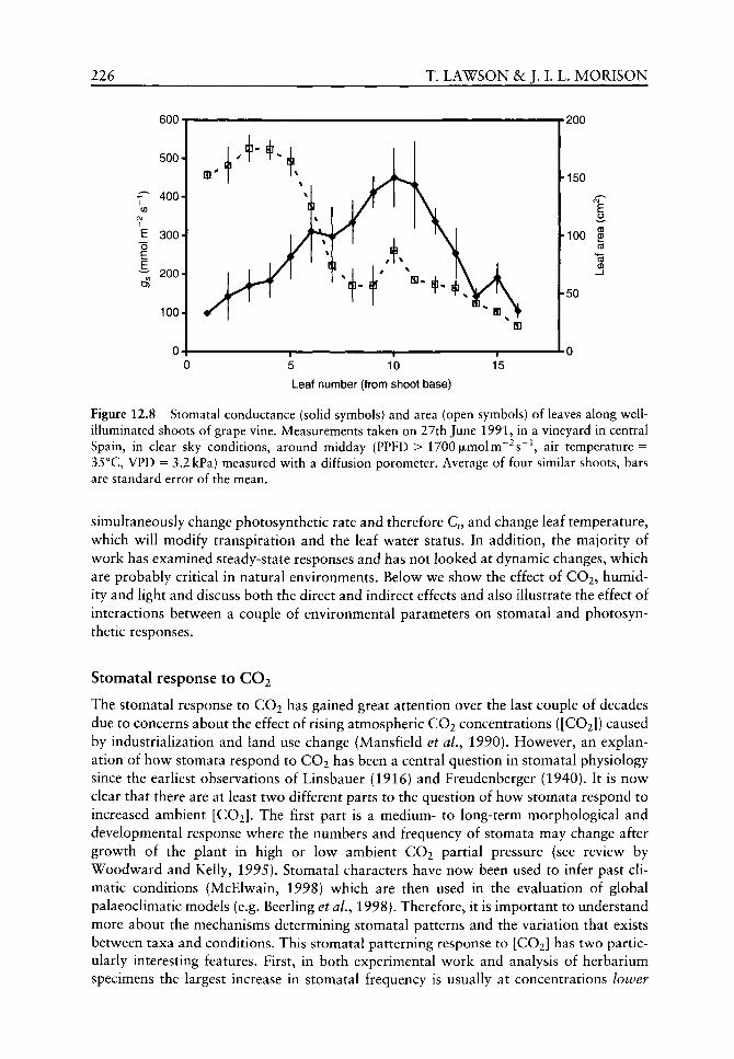
226
T CM
1 E "o E
fiOn m o u u •
500-
400-
300-
200-
100-
0-(
T. LAWSON &c J. L
T
T + \ 1 ^ S: \
) 1 i 1 1
10
^ si^ t "* m
1
15
L. MORISON
^onn
-150
£
-100 S
-50
- 0 u
Leaf number (from shoot base)
Figure 12.8 Stomatal conductance (solid symbols) and area (open symbols) of leaves along well-illuminated shoots of grape vine. Measurements taken on 27th June 1991, in a vineyard in central Spain, in clear sky conditions, around midday (PPFD > 1700 ixmolm'^s'^ air temperature = 35°C, VPD = 3.2 kPa) measured w ith a diffusion porometer. Average of four similar shoots, bars are standard error of the mean.
simultaneously change photosynthetic rate and therefore Q, and change leaf temperature, w^hich w ill modify transpiration and the leaf water status. In addition, the majority of work has examined steady-state responses and has not looked at dynamic changes, which are probably critical in natural environments. Below we show the effect of CO2, humidity and light and discuss both the direct and indirect effects and also illustrate the effect of interactions between a couple of environmental parameters on stomatal and photosynthetic responses.
Stomatal response to CO2
The stomatal response to CO2 has gained great attention over the last couple of decades due to concerns about the effect of rising atmospheric CO2 concentrations ([CO2]) caused by industrialization and land use change (Mansfield et aL, 1990). However, an explanation of how stomata respond to CO2 has been a central question in stomatal physiology since the earUest observations of Linsbauer (1916) and Freudenberger (1940). It is now clear that there are at least two different parts to the question of how stomata respond to increased ambient [CO2]. The first part is a medium- to long-term morphological and developmental response where the numbers and frequency of stomata may change after growth of the plant in high or low ambient CO2 partial pressure (see review by Woodward and Kelly, 1995). Stomatal characters have now been used to infer past climatic conditions (McElwain, 1998) which are then used in the evaluation of global palaeoclimatic models (e.g. Beerling et aL, 1998). Therefore, it is important to understand more about the mechanisms determining stomatal patterns and the variation that exists between taxa and conditions. This stomatal patterning response to [CO2] has two particularly interesting features. First, in both experimental work and analysis of herbarium specimens the largest increase in stomatal frequency is usually at concentrations lower
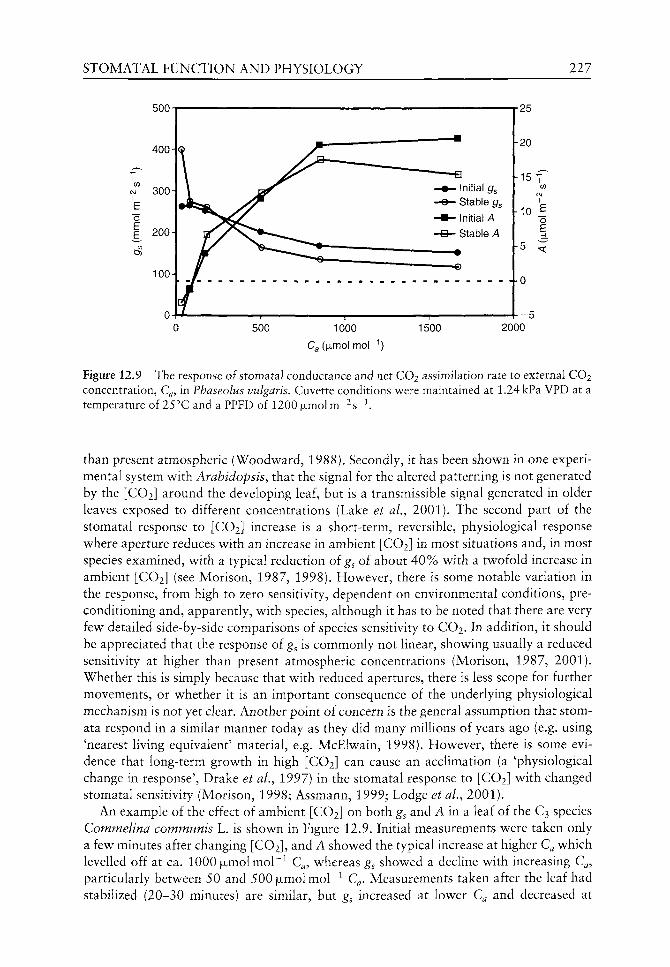
STOMATAL FUNCTION AND PHYSIOLOGY 227
500 1000
Ca(|ULmol mor' ')
1500
Figure 12.9 The response of stomatal conductance and net CO2 assimilation rate to external CO2 concentration, Q, in Fhaseolus vulgaris. Cuvette conditions were maintained at 1.24 kPa VPD at a temperature of 25°C and a PPFD of 1200 jjimolm-^s'^
than present atmospheric (Woodward, 1988). Secondly, it has been shown in one experimental system with Arabidopsis, that the signal for the altered patterning is not generated by the [CO2] around the developing leaf, but is a transmissible signal generated in older leaves exposed to different concentrations (Lake et al., 2001). The second part of the stomatal response to [CO2] increase is a short-term, reversible, physiological response where aperture reduces with an increase in ambient [CO2] in most situations and, in most species examined, with a typical reduction oi g^ of about 40% with a twofold increase in ambient [CO2] (see Morison, 1987, 1998). However, there is some notable variation in the response, from high to zero sensitivity, dependent on environmental conditions, preconditioning and, apparently, with species, although it has to be noted that there are very few detailed side-by-side comparisons of species sensitivity to CO2. In addition, it should be appreciated that the response of g^ is commonly not linear, showing usually a reduced sensitivity at higher than present atmospheric concentrations (Morison, 1987, 2001). Whether this is simply because that with reduced apertures, there is less scope for further movements, or whether it is an important consequence of the underlying physiological mechanism is not yet clear. Another point of concern is the general assumption that stom-ata respond in a similar manner today as they did many millions of years ago (e.g. using 'nearest living equivalent' material, e.g. McElwain, 1998). However, there is some evidence that long-term growth in high [CO2] can cause an acclimation (a 'physiological change in response', Drake et al., 1997) in the stomatal response to [CO2] with changed stomatal sensitivity (Morison, 1998; Assmann, 1999; Lodge et al, 2001).
An example of the effect of ambient [CO2] on both g^ and A in a leaf of the C3 species Commelina communis L. is shown in Figure 12.9. Initial measurements were taken only a few minutes after changing [CO2], and A showed the typical increase at higher C^ which levelled off at ca. 1000 jjimolmol"^ C , whereas g^ showed a decline with increasing C , particularly between 50 and 500 ixmolmol'^ C . Measurements taken after the leaf had stabilized (20-30 minutes) are similar, but g^ increased at lower C^ and decreased at

228 T. LAWSON 8c J. I. L. MORISON
higher Q . This slower change in g^ than in A resuhed in small but significant modifications in A, emphasizing the effect of stomatal behaviour on CO2 diffusion into the leaf.
There is no clear explanation for how changes in [CO2] cause the change in aperture or conductance (Assmann, 1999), although it can be demonstrated that several physiological processes in guard cells, whether intact, in peels or in protoplast suspensions (e.g. carbon fixation, ion transport, chlorophyll fluorescence) can be affected by large changes in ambient [CO2]. However, this is not the same as elucidating the signalling pathway for how a 50 or 100 jimolmol"^ change in Q results in a marked change of aperture in the intact leaf (e.g. Figure 12.9). It has been demonstrated convincingly that it is Q not the external concentration that affects guard cells (Mott, 1988). From an entirely teleological point of view, a control mechanism based on Q could be a way to link demand for CO2 by the mesophyll cells with supply by the guard cells (Raschke, 1976). However, the many observations showing that g^ is largely independent of the rate of mesophyll carbon fixation over a short period of time (e.g. Figure 12.5) show that this is not the only controlling factor. Clearly, Q interacts with other environmental signals and Q is not constant^ although it is conservative, because it is both the result and an effector of stomatal aperture (Jarvis and Morison, 1981).
Stomatal response to humidity
As described above in Equation (2), diffusion of gases through stomata depends on the difference in concentration between the inside and outside of the pore. If g is unchanged, the transpiration rate increases linearly with leaf-to-air vapour pressure difference (VPD) caused either by changes in air vapour pressure {wj or by leaf temperature affecting the vapour pressure inside the leaf, w^. However, it is important to realize that VPD has a direct effect on stomata independently of the effect on whole leaf transpiration and it is the VPD, rather than relative humidity to which the stomata respond (Aphalo and Jarvis, 1991). Stomatal responses to VPD were first described by Schulze et al. (1972) who studied species living in desert habitats. In general, stomatal aperture declines as the VPD increases and, under certain conditions, the reduction in aperture may be so large as to reduce the transpiration rate (Farquhar, 1978), thereby preventing extreme water loss from the plant. In an elegant experiment, Mott and Parkhurst (1991) used differences in the diffusion rate of water vapour in different gases to conclude that stomata respond to water loss rates and do not directly sense, and respond to, the water vapour pressure near the leaf. Therefore, the most widely accepted mechanism for stomatal responses to humidity is that evaporation directly from epidermal and guard cells near the stomatal pore alters the guard cell water potential independently from that of mesophyll cells.
Figure 12.10a shows the effect of increasing w^ from 0.8 kPa to 1.88 kPa on leaf gas exchange in Phaseolus vulgaris. This caused a drop in the VPD and resulted in an initial drop in g^ for about 5 min, a 'hydropassive' stomatal behaviour (Stalfelt, 1955), caused by a change in water balance between epidermal and guard cells. This was followed by a steady increase for the subsequent 35 min, after which time g^ remained stable with an overall increase in g^ of 57%. The initial drop in g^ was matched by a fall in Q, which then rose with increased g^ and remained stable after about 20 min; but further increase in g^ did not result in a further increase in Q. A remained stable for the first 20 min, after which A increased with increasing g , which accounts for the lack of increasing Q with increasing gs. These results show that decreased VPD eventually allowed a larger g^ and consequently a higher Q and higher A. An opposite increase in VPD (Figure 12.10b) led to an
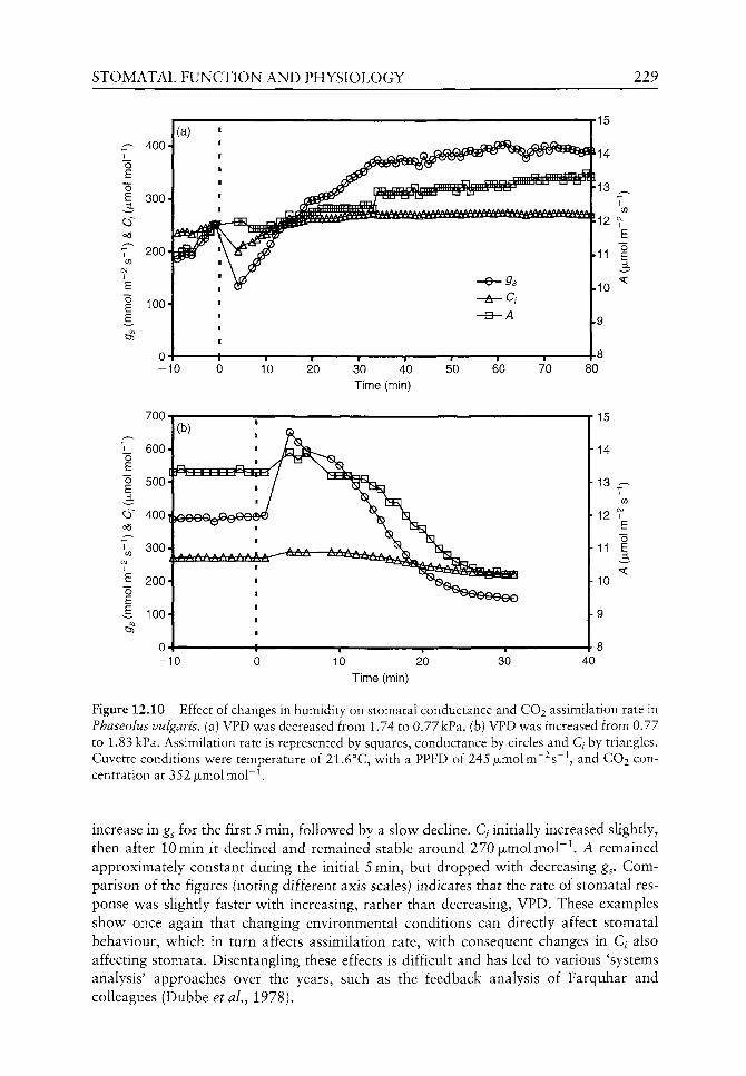
STOMATAL FUNCTION AND PHYSIOLOGY 229
— 400
o E
C3)
300
o E "o E
o6
T 200
100
40 Time (min)
700
10 20 Time (min)
Figure 12.10 Effect of changes in humidity on stomatal conductance and CO2 assimilation rate in Phaseolus vulgaris, (a) VPD was decreased from 1.74 to 0.77 kPa. (b) VPD was increased from 0.77 to 1.83 kPa. Assimilation rate is represented by squares, conductance by circles and Q by triangles. Cuvette conditions were temperature of 21.6°C, with a PPFD of 245 |jLmolm"^s~\ and CO2 concentration at 352 ixmolmol"^.
increase in g^ for the first 5 min, followed by a slow decline. Q initially increased slightly, then after 10 min it declined and remained stable around 270|jLmolmol~^. A remained approximately constant during the initial 5 min, but dropped with decreasing g . Comparison of the figures (noting different axis scales) indicates that the rate of stomatal response was slightly faster with increasing, rather than decreasing, VPD. These examples show once again that changing environmental conditions can directly affect stomatal behaviour, which in turn affects assimilation rate, with consequent changes in Q also affecting stomata. Disentangling these effects is difficult and has led to various 'systems analysis' approaches over the years, such as the feedback analysis of Farquhar and colleagues (Dubbe et aL^ 1978).
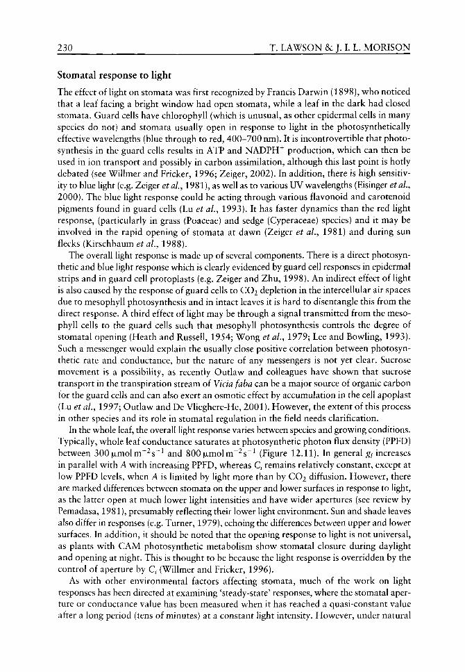
230 T. LAWSON & J. L L. MORISON
Stomatal response to light
The effect of hght on stomata was first recognized by Francis Darwin (1898), who noticed that a leaf facing a bright window had open stomata, while a leaf in the dark had closed stomata. Guard cells have chlorophyll (which is unusual, as other epidermal cells in many species do not) and stomata usually open in response to light in the photosynthetically effective wavelengths (blue through to red, 400-700 nm). It is incontrovertible that photosynthesis in the guard cells results in ATP and NADPH^ production, which can then be used in ion transport and possibly in carbon assimilation, although this last point is hotly debated (see Willmer and Fricker, 1996; Zeiger, 2002). In addition, there is high sensitivity to blue light (e.g. Zeiger etaL,19Sl), as well as to various UV wavelengths (Eisinger et aL, 2000). The blue light response could be acting through various flavonoid and carotenoid pigments found in guard cells (Lu et aL, 1993). It has faster dynamics than the red light response, (particularly in grass (Poaceae) and sedge (Cyperaceae) species) and it may be involved in the rapid opening of stomata at dawn (Zeiger et aL, 1981) and during sun flecks (Kirschbaum et aL, 1988).
The overall light response is made up of several components. There is a direct photosyn-thetic and blue light response which is clearly evidenced by guard cell responses in epidermal strips and in guard cell protoplasts (e.g. Zeiger and Zhu, 1998). An indirect effect of light is also caused by the response of guard cells to CO2 depletion in the intercellular air spaces due to mesophyll photosynthesis and in intact leaves it is hard to disentangle this from the direct response. A third effect of light may be through a signal transmitted from the mesophyll cells to the guard cells such that mesophyll photosynthesis controls the degree of stomatal opening (Heath and Russell, 1954; Wong et al., 1979; Lee and Bowling, 1993). Such a messenger would explain the usually close positive correlation between photosyn-thetic rate and conductance, but the nature of any messengers is not yet clear. Sucrose movement is a possibility, as recently Outlaw and colleagues have shown that sucrose transport in the transpiration stream of Vicia fab a can be a major source of organic carbon for the guard cells and can also exert an osmotic effect by accumulation in the cell apoplast (Lu et al., 1997; Outlaw and De Vlieghere-He, 2001). However, the extent of this process in other species and its role in stomatal regulation in the field needs clarification.
In the whole leaf, the overall light response varies between species and growing conditions. Typically, whole leaf conductance saturates at photosynthetic photon flux density (PPFD) between 300jxmolm~^s~^ and 800|ULmolm"^s"^ (Figure 12.11). In general g/ increases in parallel with A with increasing PPFD, whereas Q remains relatively constant, except at low PPFD levels, when A is limited by light more than by CO2 diffusion. However, there are marked differences between stomata on the upper and lower surfaces in response to light, as the latter open at much lower light intensities and have wider apertures (see review by Pemadasa, 1981), presumably reflecting their lower light environment. Sun and shade leaves also differ in responses (e.g. Turner, 1979), echoing the differences between upper and lower surfaces. In addition, it should be noted that the opening response to light is not universal, as plants with CAM photosynthetic metabolism show stomatal closure during daylight and opening at night. This is thought to be because the light response is overridden by the control of aperture by Q (Willmer and Fricker, 1996).
As with other environmental factors affecting stomata, much of the work on light responses has been directed at examining 'steady-state' responses, where the stomatal aperture or conductance value has been measured when it has reached a quasi-constant value after a long period (tens of minutes) at a constant light intensity. However, under natural
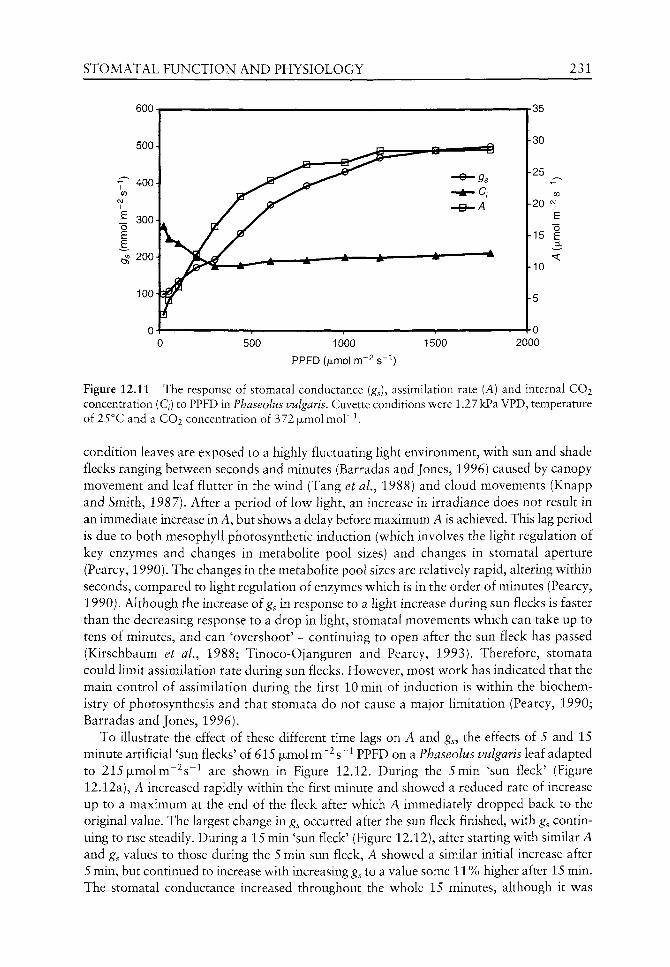
STOMATAL FUNCTION AND PHYSIOLOGY 231
UVJU -
500-
400-
300-
200-
100-
0- 1 1
A ^ """^
1 T
35
30
25
20
15 i
500 1000
PPFD(fxmolm~2s""')
1500
10
5
0 2000
Figure 12.11 The response of stomatal conductance (gj, assimilation rate (A) and internal CO2 concentration (Q) to PPFD in Phaseolus vulgaris. Cuvette conditions were 1.27 kPa VPD, temperature of 25°C and a CO2 concentration of 372 |jimolmol~^
condition leaves are exposed to a highly fluctuating light environment, v^ith sun and shade flecks ranging betw^een seconds and minutes (Barradas and Jones, 1996) caused by canopy movement and leaf flutter in the w ind (Tang et al., 1988) and cloud movements (Knapp and Smith, 1987). After a period of low light, an increase in irradiance does not result in an immediate increase in A, but shoves a delay before maximum A is achieved. This lag period is due to both mesophyll photosynthetic induction (which involves the light regulation of key enzymes and changes in metabolite pool sizes) and changes in stomatal aperture (Pearcy, 1990). The changes in the metabolite pool sizes are relatively rapid, altering within seconds, compared to light regulation of enzymes which is in the order of minutes (Pearcy, 1990). Although the increase of g in response to a light increase during sun flecks is faster than the decreasing response to a drop in light, stomatal movements which can take up to tens of minutes, and can 'overshoot' - continuing to open after the sun fleck has passed (Kirschbaum et aL, 1988; Tinoco-Ojanguren and Pearcy, 1993). Therefore, stomata could limit assimilation rate during sun flecks. However, most work has indicated that the main control of assimilation during the first lOmin of induction is within the biochemistry of photosynthesis and that stomata do not cause a major limitation (Pearcy, 1990; Barradas and Jones, 1996).
To illustrate the effect of these different time lags on A and g , the effects of 5 and 15 minute artificial 'sun flecks' of 615 (jimol m'^ s~^ PPFD on a Phaseolus vulgaris leaf adapted to 215|jLmolm~^s~^ are shown in Figure 12.12. During the 5min 'sun fleck' (Figure 12.12a), A increased rapidly within the first minute and showed a reduced rate of increase up to a maximum at the end of the fleck after which A immediately dropped back to the original value. The largest change in g^ occurred after the sun fleck finished, with g^ continuing to rise steadily. During a 15 min 'sun fleck' (Figure 12.12), after starting with similar A and gs values to those during the 5 min sun fleck, A showed a similar initial increase after 5 min, but continued to increase with increasing g to a value some 11 % higher after 15 min. The stomatal conductance increased throughout the whole 15 minutes, although it was
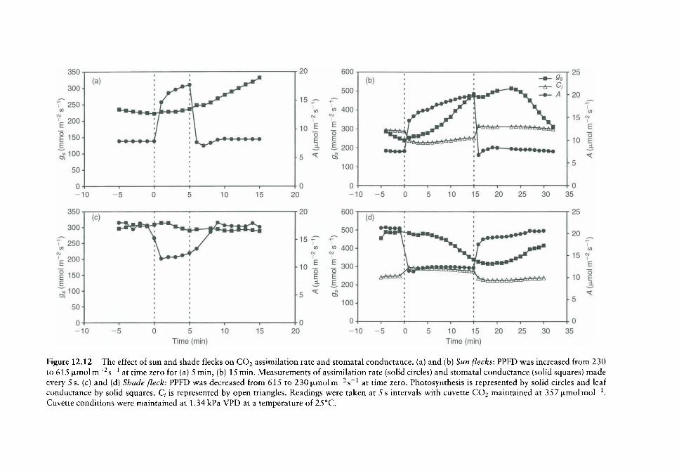
Figure 12.12 The effect of sun and shade flecks on CO2 assimilation rate and stomatal conductance, (a) and (b) Sun flecks: PPFD was increased from 230 to 615 (jimoi m~^s~^ at time zero for (a) 5 min, (b) 15 min. Measurements of assimilation rate (sohd circles) and stomatal conductance (solid squares) made every 5 s. (c) and (d) Shade fleck: PPFD was decreased from 615 to 230 ixmolm^^s"^ at time zero. Photosynthesis is represented by solid circles and leaf conductance by solid squares. C, is represented by open triangles. Readings were taken at 5 s intervals with cuvette CO2 maintained at 357|xmolmol"^. Cuvette conditions were maintained at 1.34 kPa VPD at a temperature of 25°C.

STOMATAL FUNCTION AND PHYSIOLOGY 233
appearing to slow down by the end. After the sun fleck, the increase was small and continued for only 6-8 min, before closure started. Note that most of the Q change occurred when illumination changed and, although there were subsequent small changes, changes in g were not sufficient to keep Q constant. Of course, the effect of 'shade flecks' on A and g^ are potentially as important as sun flecks (Figure 12.12c and d). During both 5 and 15 min duration 'shade flecks' there was an immediate decrease in A which recovered back to its original value when the PPFD was restored in the 5 min fleck. However, A took about 15 min to return back to the original value after a 15 min shade fleck, because of the decrease in g^ that had occurred with the lower light. The start of a stomatal opening response was delayed for about 8 min after the end of the shade period and the slow recovery back towards its original value took over 15 min, consequently limiting A through CO2 diffusion by some 10%. Such sun and shade fleck data emphasize the importance of the different dynamic behaviour of stomata and photosynthesis which can result in A and ^5 being uncorrelated with each other, in the natural, changeable environment. Note also that these examples are with a species with rapidly responsive stomata, but there are other species with much more sluggish stomata (e.g. some conifers, Ng and Jarvis, 1980), where the correlation must be much less complete or frequent (Jarvis and Morison, 1981).
Environmental interactions and stomatal responses
Clearly, stomata are sensitive to a large number of environmental factors, but these rarely vary singly in nature, so the interaction between factors must be borne in mind. Two examples are shown here: the interaction between [CO2] and light (PPFD) and between [CO2] and VPD, Figure 12.13 shows the effect of increasing the ambient CO2 concentration from 360 to 700 ijumolmol"^ on g^ and A (Figure 12.13a and b) at different PPFD. At each PPFD the leaf was left to stabilize for a minimum of 30 min before Q was increased. Clearly, the initial rates of g and A depended upon the initial PPFD as we expect from the steady-state responses discussed above. In addition, the effect of increased C^ was much larger at higher (1000 and 500 |jLmolm~^s"^) than at lower PPFD. Note again that g^ took considerably longer than A to reach a new approximately steady value following the step change. As expected, A increased in response to high C^ as the extra CO2 removed the supply limitation at high light. In contrast, the larger g^ at high PPFD was largely suppressed by the high C , showing clearly the interactive effect of CO2 and PPFD on stomatal behaviour.
While light and [CO2] both influence assimilation rates directly, VPD does not. However, VPD affects stomata, so can indirectly affect assimilation rate through influencing CO2 supply, as exemplified in Figure 12.13c and d. At normal ambient [CO2], gs differed by about 70% between a VPD of 0.95 and 2.2 kPa, so that at the higher VPD assimilation rate was reduced by about 25% (as suggested by the example A/Q curve in Figure 12.6). However, when the C^ was increased, A increased and the stomatal limitation was reduced. In all three VPD conditions, g^ followed a similar time course when CO2 was doubled, taking over 20 min to come to a new steady value, twice that for A. Note also that the marked effect of VPD on g^ was reduced in high [CO2] which is in part due to the reduction in aperture and the consequent reduced 'scope' for stomatal aperture changes (see Morison and Gifford, 1984 for other examples).
Several other interactions have been examined, but there is obviously a potentially bewildering range of possible combinations and few studies have examined any more than two or three, and usually on only one or two species in the same conditions. For this

450
400
350
" 300 CO
p 250
I 200 E » 150
100
50
0
(a) , _ . • 1000 • ' • • • •
• , • , " 5 0 0 • j A 300
• • 100 •
10 10 15 20
4501
400
350-
300 to
g 250
I 200
150
100
50
0
D)
• 50°'
A 30°
^^^tttttt^^AttlAtti^ttltl"
10 10 20 30
Time (min) 40
25
50
45 -
40 -
- 35 -
,f 30
e 25-o
!_ 20 -
^ 15-
10 -
5 -
0 -
(b)
• • • • •
••••••
A A A A A A
• • • • • •
• • •• •• •••••,
A A ^ A A A A A A A A * A A A A A A
• * • • • • • • « « « « * « * * «
10 10 15 20
-10 10 20 30
Time (min) 40
25
Figure 12.13 The effect of increasing COj from 360 to 720[xmolmor^ on stomatal conductance (a and c) and CO2 assimilation rate (b and d) in Phaseolus vulgaris at different (a and b) PPFD or (c and d) humidity. Apart from the variable under investigation other cuvette conditions remained constant, at a PPFD of 1200 (jimol m o l ' ^ temperature 25°C; VPD 1.27kPa, Q 365 |xmol mol" ' . Initially the leaf was left to stabilize (for 20-30 min) at the conditions described above before a step increase in CO2.

STOMATAL FUNCTION AND PHYSIOLOGY 235
reason, much reliance has been placed on models for prediction of leaf gas exchange under different environmental conditions. There are two common types: the empirical model pioneered by Jarvis (1976) and the assimilation rate linked model suggested originally by Ball et al. (1987). The Jarvis-type model is based upon a multipHcative combined reduction of conductance from a maximum value depending on functions derived from measurements of stomatal responses to environmental factors. The Ball-Berry model and its subsequent refinements assume that stomata respond in order to regulate Q. While both approaches have had considerable success for particular tasks, the problem is that they are not based upon the various mechanisms involved in stomatal behaviour and so w ill not capture the sorts of interactions demonstrated above, nor the dynamic changes illustrated. Some models that are more closely based on the physiological processes in guard cells have been devised (e.g. Farquhar and Wong, 1984; Jarvis and Davies, 1998), but v^hile very promising, they are at the moment experimental and limited. It may be unrealistic to include all the many observed environmental sensitivities, but wt urgently require physiological models that can integrate the key responses of stomata that are so readily demonstrated in the laboratory and the field.
Modern techniques for ecophysiological stomatal research
Although gas exchange techniques, such as exemplified here, have been important in monitoring stomatal behaviour in relation to mesophyll photosynthesis, they obviously do not probe v^hat is going on at the single cell scale. Clearly, physiological and molecular biology w^ork on guard cells in epidermal strips and other isolated systems is invaluable for probing the complexity of guard cell ion transport and metabohsm. How^ever, results from that research do need to be placed in the in vivo, in natura context of intact, photosynthesizing and transpiring leaves. Recently, there have been a number of cell scale investigations that open up nev^ possibilities. First, microscope v^ork v^ith pressure probes has investigated guard cell mechanics and w^ater relations (e.g. Franks and Farquhar, 2001). Secondly, imaging systems are now^ available to measure aperture changes in intact leaves, in the laboratory and even in the field as the environment changes and consequently relate these to gas exchange (e.g. Kaiser and Kappen, 2000). Thirdly, it is now possible to study guard cell photosynthesis in vivo using high resolution chlorophyll fluorescence imaging (Oxborough and Baker, 1997; Lav^son et al., 2002) w^hich allov^s pos-sible links between stomatal behaviour and underlying mesophyll photosynthesis to be studied. The basic principle in this latter technique is that fluorescence measurements are made with an imaging system through a microscope under the experimental conditions and under different light conditions when photosystem II (PSII) is in defined states (for details see Baker et al., 2001). This results in measurements of the parameter F^/F^ which estimates the yield of PSII photochemistry, often described as a measure of the efficiency of photosynthetic electron transport. Values from guard cell chloroplasts can be compared with those from the adjacent underlying mesophyll, and an example is shown in Colour Plate 2. This work has revealed that the photosynthetic Calvin cycle is functional in guard cells in a range of species and that the guard cell photosynthetic electron transport efficiency responds to light, water stress and [CO2] in a quantitatively similar way to that in mesophyll chloroplasts. The technique now makes it possible to compare a range of species which are different ecologically, taxonomically and evolutionarily, which will help build more mechanistic models of stomatal behaviour and the relationship with mesophyll assimilation.
236 T. LAWSON &c J. I. L. MORISON
Evolutionary context
Fossilized stomatal characters such as stomatal density (SD) have been used to determine past CO2 concentrations (Kiirschner, 1996; McElwain, 1998) and, in conjunction with other parameters, can be used to estimate g^ (Kiirschner et al., 1997). While in some species SD has been shown to correlate closely with g^ under certain conditions (e.g. Woodward and Bazzaz, 1988) the influence of SD on g^ is secondary to that of stomatal aperture (see Figure 12.2) which may not always be obtainable from fossil material. It may be possible to use a surrogate measure of aperture (Lawson et aL, 1998) or use pore length to give an indication of the maximum possible aperture (Beerling and Woodward, 1987). Maximum gs can then be used to predict water-use efficiency and carbon balance (Beerling and Woodward, 1993), given assumptions about the link between conductance and assimilation rate (for example, see survey in Leuning et al.^ 1995). While a general correlation between A and g^ certainly exists, there are many situations where stomatal aperture does not always correlate with assimilation rate (Jarvis and Morison, 1981) and as exemplified in the A/g^ curve shown in Figure 12.5. Furthermore, the evolution of stomatal behaviour may not have been driven by an optimization of instantaneous A with a minimum £, but may have been driven by other very different pressures such as the need to avoid water stress and runaway xylem embolism, or to avoid lethal high temperatures. Clearly, adaptation pressures may have differed according to habitats and conditions as they do in modern plants today. Therefore, we should be cautious about simple correlative models.
An example of how physiological understanding of plants can help to identify and explain plant fossils records has been shown by McElwain et al. (1999) who suggested that high levels of atmospheric CO2 at the end of the Triassic may have resulted in the observed decrease in floral species in the fossil record. They suggest that high atmospheric CO2 led to low stomatal density/index which resulted in reduced evapotranspiration rates and lethal increases in leaf temperature. Further evidence to support the theory that forced-CO2 global warming can explain selective floral extinctions found in the fossil record has been published recently by Beerling (2002). Although this shows the important use of modern physiological understanding in determining past floral distribution, it has been pointed out that the use of anatomical parameters of fossil plants to reconstruct palaeoatmospheric CO2 concentrations, which are then used to evaluate consequences for palaeoplant physiology, may compound errors already associated with assuming a direct correlation between stomatal properties and CO2 (Beerling and Chaloner, 1993; Cowling, 2001).
It has been suggested that evolution of land plants should be considered in terms of the plants' ability to balance photosynthetic carbon gain versus water loss (Knoll, 1984). As both of these processes are under the direct influence of stomata, this implies that stomata could play a key role in plant evolution and distribution. Therefore, a full understanding of the physiology and function of stomata is important not only in modern plant physiology but could provide vital information about plant evolutionary processes. The study of modern plants compared with those that grew in the geological past could give an indication of the direction of such processes. For example, comparing ferns and cycads (which could be taken as representing evolutionary end points) with evolutionarily modern plants (such as angiosperms) could indicate evolutionary changes and adaptations. Such an experimental approach has suggested that ancient taxa such as ferns and cycads evolving under periods of high [O2] and low [CO2] may have photosynthetic systems with lower sensitivities to O2 when compared with modern day angiosperms (Beerling et al., 1998). However, when studying modern plant physiology it is important to remember that they have
STOMATAL FUNCTION AND PHYSIOLOGY 237
developed over many hundreds of years, therefore biochemical and biophysical adjustments (acclimation) to their current environmental conditions could have taken place as w ell as genetic adaptation (Cowling, 2001) and therefore, behave differently today than in the past.
By studying modern C3 species, C4 and CAM plants we are looking at the results of millions of years of evolution and we can begin to understand how plants have adapted to changing environmental conditions. For example, the development of a CO2 concentrating mechanism in C4 and CAM photosynthetic pathways was probably the evolutionary result of changed atmospheric gas concentrations (Sage, 2001). Whether a particular species evolved a CAM or C4 pathway depended upon the initial steps in the evolutionary sequence. These initial steps involved different selection pressures, in CAM this was associated with the scavenging of respiratory CO2 during the dark, while in C4 evolution it was associated with scavenging photorespiratory CO2 in the light (Sage, 2002). Due to the CO2 concentrating mechanisms displayed in CAM and C4 plants, changes in atmospheric CO2 concentrations have little effect on stomatal density, index or pore areas (Raven and Ramsden, 1988). Royer (2001) documented that only one out of nine C4 plants studied revealed an inverse relationship with CO2 concentration. Although stomatal responses to changing CO2 concentration may be less significant in C4 and CAM plants, it is possible that they still played a critical role in C4 distribution and evolution. Sage (2001) suggested that many ecological factors, including CO2 concentration, influenced where and when C4 photosynthesis evolved. One such factor was the influence that low [CO2] contributed to aridification, through increased transpiration resulting from an increase in stomata aperture - such conditions would benefit the establishment of C4 over C3 plants.
The use of modern techniques may help to establish the timing of evolutionary processes, for example, the oldest C4 fossils are believed to be 12 My old, however, phylo-genetic evidence from molecular clock interpretations of genetic similarities in various grass lineages, indicates divergence of C4 taxa was occurring 20-30 My ago (Kellogg, 1999). Further, comparison of responses of angiosperms with gymnosperms, CAM with C3 plants and also plants from different environmental locations could reveal novel and interesting information about the evolutionary development of plant physiology. By combining palaeoecophysiology with modern plant physiology we will enhance our predictions of both future and past interpretations of global climate environments and associated plant responses. For example, recent work by Sage (2002) suggests that low CO2 probably acted as a significant evolutionary agent, selecting plants adapted to CO2 deficiency and that adaptations to low CO2 might still exist in plants today, which might constrain responses to the current rising CO2 concentration. Such acclimation would have important implications for current agriculture, water-use efficiency and natural selection.
Conclusion
A detailed understanding of stomatal behaviour and function is essential to understand both past and present impacts of environmental change on global carbon and hydrological cycles. In this chapter we have looked at the role of stomata in leaf gas exchange and the way they respond to three key environmental variables of light, CO2 and humidity. Other factors such as leaf water status are obviously as important as these to plant growth and survival, but are less directly amenable to short-term experiments. We have tried to

238 T. LAWSON &c J. I. L. M O R I S O N
emphasize the role of stomata in determining mesophyll CO2 assimilation rates. Examples
of physiological variation at a number of different levels stress the importance of appro
priate sampling strategies for both modern plant physiologists and those attempting to
use stomatal characters as indicators of past environments. The effects of a number of
environmental factors, both singly and when interacting, on both stomatal and photosyn-
thetic behaviour have exemplified the complexity of the relationship between mesophyll
assimilation and stomatal function. In the final sections, examples of modern techniques
for studying stomatal physiology have been illustrated. Use of such techniques should
improve the elucidation of stomatal behaviour and mechanisms in a range of species
(from ancient to modern), and hence reveal evolutionary changes.
Acknowledgements
Dr Jonathan Weyers (University of Dundee) is gratefully acknow^ledged for his input and
guidance on a number of the experiments reported here. The development of the high reso
lution chlorophyll fluorescence imaging system is an ongoing collaboration w^ith Dr Kevin
Oxborough and Professor Neil Baker at the University of Essex, funded by the UK BBSRC.
References
Aphalo PJ, Jarvis PG. 1991. Do stomata respond to relative humidity? Plant, Cell and Environment 14: 127-132.
Aphalo PJ, Jarivs PG. 1993. The boundary layer and the apparent responses of stomatal conductance to wind speed and to the mole fractions of CO2 and water vapour in the air. Flant, Cell and Environment 16: 771-783.
Asai N, Nakajima N, Tamaoki M, et al. 2000. Role of malate synthesis mediated by phospho-enolpyruvate carboxylase in guard cells in the regulation of stomatal movements. Flant Cell Physiology 4\: 10-15.
Assman SM. 1993. Signal transduction in stomatal guard cells. Annual Review of Cell Biology 9: 345-375.
Assman SM. 1999. The cellular basis of guard cell sensing of rising CO2. Plant, Cell and Environment 22: 629-637.
Baker NR, Oxborough K, Lawson T, Morison JIL. 2001. High resolution imaging of photo-synthetic activities of tissues, cells and chloroplasts in leaves. Journal of Experimental Botany SI: 615-621.
Ball JT, Woodrow IE, Berry JA. 1987. A model predicting stomatal conductance and its contribution to the control of photosynthesis under different environmental conditions. In: Biggins J, ed. Progress in Photosynthesis Research IV, Dordrecht: Martinus Nijhoff, 221-224.
Barradas VL, Jones HG. 1996. Responses of CO2 assimilation to changes in irradiance: laboratory and field data and a model for beans (Phaseolus vulgaris L.). Journal of Experimental Botany 47: 639-645.
Beerling DJ. 2002. CO2 and the end-Triassic mass extinction. Nature 415: 386-387. Beerling DJ, Chaloner WG. 1993. Evolutionary responses of stomatal density to global CO2
change. Biological Journal of the Linnean Society 48: 343-353. Beerling DJ, Woodward FI. 1987. Changes in land plant function over the Phanerozoic: recon
structions based on the fossil record. Botanical Journal of the Linnean Society 124: 137-153. Beerling DJ, Woodward FI. 1993. Ecophysiological responses of plants to global environmental
change since the last glacial maximum. New Phytologist 125: 641-648. Beerling DJ, McElwain JC, Osborne CP. 1998. Stomatal responses of the 'living fossil' Ginkgo
hiloba L. to changes in atmospheric CO2 concentrations. Journal of Experimental Botany 49: 1603-1607.

STOMATAL F U N C T I O N AND PHYSIOLOGY 239
Beerling DJ, Woodward FI, Lomas MR, et al. 1998. The influence of carboniferous palaeoatm-ospheres on plant function: An experimental and modelling assessment. Philosophical Transactions of the Royal Society, Series B 353: 131-140.
Beerling DJ, Osborne CP, Chaloner WG. 2001. Evolution of leaf-form in land plants linked to atmospheric CO2 decline in the late Palaeozoic era. Nature 410: 352-354.
von Caemmerer SC, Farquhar GD. 1984. Effects of partial defoliation, changes of irradiance during growth, short-term water stress and growth at enhanced p(C02) on photosynthetic capacity of leaves of Phaseolus vulgaris L. Planta 160: 320-329.
Chaloner WG. 1970. The rise of the first land plants. Biological Review, Cambridge Philosophical Society 45: 353-377.
Cowan IR, Troughton JH. 1971. The relative role of stomata in transpiration and assimilation. Planta 97: 325-336.
Cowling SA. 2001. Plant carbon balance, evolutionary innovation and extinction in land plants. Global Change Biology 7: 231-239.
Darwin F. 1898. Observations on stomata. Philosophical Transactions of the Royal Society, Series B 190: 531-621.
Drake BG, Gonzalez-Meier MA, Long SP. 1997. More efficient plants: a consequence of rising atmopheric CO2. Annual Review of Plant Physiology and Plant Molecular Biology 48: 609-639.
Dubbe DR, Farquhar GD, Raschke K. 1978. Effects of abscisic acid on the gain of the feedback loop involving carbon dioxide and stomata. Plant Physiologist 62: 413-417.
Edwards D, Davies KL, Axe L. 1992. A vascular conducting strand in the early land plant Cooksonia. Nature 357: 683-685.
Eisinger W, Swartz T, Bogomolni R, Taiz L. 2000. The ultraviolet action spectrum for stomatal opening in broad bean. Plant Physiology 111: 99-105.
Farquhar GD. 1978. Feed forward responses of stomata to humidity. Australian journal of Plant Physiology 5: 787-800.
Farquhar GD, Sharkey TD. 1982. Stomatal conductance and photosynthesis. Annual Reviews of Plant Physiology 33: 317-345.
Farquhar GD, Wong SC. 1984. An empirical model of stomatal conductance. Australian journal of Plant Physiology 11: 191-210.
Franks PJ, Farquhar GD. 2001. The effect of exogenous abscisic acid on stomatal development, stomatal mechanics, and leaf gas exchange in Tradescantia virginiana. Plant Physiology 125: 935-942.
Freudenberger H. 1940. Die Reaktion der Schliesszellen auf Kohlensaure und Sauerstoff Entzug. Protoplasma 35: 15-34.
Gottschlich DE, Smith AP. 1982. Convective heat transfer characteristics of toothed leaves. O^co/og/^ 53: 418-420.
Grace J, Wilson J. 1976. The boundary layer over a populus leaf. Journal of Experimental Botany 17: 231-241.
Heath OVS. 1938. An experimental investigation of the mechanism of stomatal movement, with some preliminary observations upon the response of the guard cells to 'shock'. New Phytologist 37: 385-395.
Heath OVS, Russell J. 1954. Studies in stomatal behaviour. VI. An investigation of the light responses of wheat stomata with the attempted elimination of control by the mesophyll. Part 2: interactions with external carbon dioxide and general discussion. Journal of Experimental Botany 5:269-292.
Jarvis AJ, Davies WJ. 1998. The coupled response of stomatal conductance to photosynthesis and transpiration. Journal of Experimental Botany 49: 399-406.
Jarvis PG. 1976. The interpretation of the variations in leaf water potential and stomatal conductance found in canopies in the field. Philosophical Transactions of the Royal Society, Series B 273: 593-610.
Jarvis PG, Morison JIL. 1981. The control of transpiration and photosynthesis by the stomata. In: Jarvis PG, Mansfield TA, eds. Stomatal Physiology. Cambridge: Cambridge University Press, 248-279.
Jones HG. 1992. Plants and Microclimate: a Quantitative Approach to Environmental Plant Physiology. Cambridge: Cambridge University Press, 56-185.
240 T. LAWSON &c J. L L. MORISON
Kaiser H, Kappen L. 2000. In situ observation of stomatal movements and gas exchange of Aegopodium podagraria L. in the understorey. Journal of Experimental Botany 51: 1741-1749.
Kellogg EA. 1999. Phylogenetic aspects of the evolution of C4 photosynthesis. In: Sage RF, Monson RK, eds. C4 Plant Biology. San Diego: Academic Press, 411-444.
Kerstiens G. 1996. Diffusion of v^ater vapour and gases across cuticles and through stomatal pores presumed closed. In: Kerstiens G, ed. Plant Cuticles - an Integrated Functional Approach, Oxford: BIOS Scientific Publishers, 121-134.
Kirschbaum MUF, Gross LJ, Pearcy RW. 1988. Observed and modelled stomatal responses to dynamic light environments in the shade plant Alocasia macrorrhiza. Plant, Cell and Environment 11: 111-121.
Knapp AK, Smith WK. 1987. Stomatal and photosynthetic responses during sun/shade transitions in subalpine plants: influence on water use efficiency. Oecologia 74: 62-67.
Knoll AH. 1984. Patterns of extinction in the fossil record of vascular plants. In: Nitecki MH, ed. Extinctions. Chicago: University of Chicago Press, 21-68.
Kiirschner WM. 1996. Leaf stomata as biosensors of paleoatmospheric CO2 levels. LLP contribution. Series 5, 1-153.
Kiirschner WM, Wagner F, Visscher EH, Visscher H. 1997. Predicting the response of leaf stomatal frequency to a future C02-enriched atmosphere: constraints from historical observations. Geologische Rundschau 86: 512-517.
Lake JA, Quick WP, Beetling DJ, Woodward FI. 2001. Signals from mature to new leaves. Nature 411: 154.
Lawson T, James W, Weyers JDB. 1998. A surrogate measure of stomatal aperture. Journal of Experimental Botany 49: 1397-1403.
Lawson T, Weyers JDB. 1999. Spatial and temporal variation in gas exchange over the lower surface of Phaseolus vulgaris primary leaves. Journal of Experimental Botany 50: 1381-1391.
Lawson T, Oxborough K, Morison JIL, Baker NR. 2002. Responses of photosynthetic electron transport in stomatal guard cells and mesophyll cells in intact leaves to light, CO2 and humidity. Plant Physiology 128: 52-62.
Lee J, Bowling DJF. 1993. Influence of the mesophyll on stomatal opening. Australian Journal of Plant Physiology 22: 357-363.
Leuning R, Kelliher FM, De Pury DGG, Schulze ED. 1995. Leaf nitrogen, photosynthesis, conductance and transpiration: scaling from leaves to canopies. Plant, Cell and Environment 18: 1183-1200.
Linsbauer K. 1916. Betrage zur Kenntnis der Spaltoffnungsbewegung. Flora 9: 100-143. Lodge RJ, Dijkstra P, Drake BG, Morison JIL. 2001. Stomatal acclimation to increased CO2 con
centration in a Florida scrub oak species Quercus myrtifolia Willd. Plant, Cell and Environment 24: 77-88.
Lu P, Outlaw HWJr, Smith BG, Freed GA. 1997. A new mechanism for the regulation of stomatal aperture size in intact leaves. Accumulation of mesophyll-derived sucrose in the guard-cell wall of Vicia faba L. Plant Physiology 114: 109-114.
Lu Z, Quinones MA, Zeiger E. 1993. Abaxial and adaxial stomata from Pima cotton (Gossypium barbadense L.) differ in their pigment content and sensitivity to light quality. Plant, Cell and Environment 16: 851-858.
Mansfield TA, Hetherington AM, Atkinson CJ. 1990. Some current aspects of stomatal physiology. Annual Review of Plant Physiology 41: 55-75.
McElwain JC. 1998. Do fossil plants signal palaeoatmospheric CO2 concentration in the geological past? Philosophical Transactions of the Royal Society, Series B 353: 83-96.
McElwain JC, BeerHng DJ, Woodward FI. 1999. Fossil plants and global warming at the Triassic-Jurassic boundary. Science 285: 1386-1390.
Monteith JL. 1981. Coupling of plants to the atmosphere. In: Grace J, Ford ED, Jarvis PG, eds. Plants and their Atmospheric Environment. Oxford: Blackwell, 1-29.
Morison JIL, Gifford RM. 1984. Plant growth and water use with limited water supply in high CO2 concentrations. I. Leaf area, water use and transpiration. Australian Journal of Plant Physiology 11: 361-374.

STOMATAL F U N C T I O N A N D PHYSIOLOGY 241
Morison JIL. 1987. Intercellular CO2 concentration and stomatal response to CO2. In: Zeiger E, Cowan IR, Farquhar CD, eds. Stomatal Function. Standford, CA: Stanford University Press, 229-251.
Morison JIL. 1998. Stomatal response to increased atmospheric CO2. Journal of Experimental Botany 49: 443-452.
Morison JIL. 2001. Increasing atmospheric CO2 and stomata. New Phytologist 149: 154-158. Mott KA. 1988. Do stomata respond to CO2 concentrations other than intercellular? Plant Physiology
86: 200-203. Mott KA, Parkhurst DF. 1991. Stomatal response to humidity in air and helox. Plant, Cell and
Environment 14: 509-515. Ng PAP, Jarvis PC. 1980. Hysteresis in the response of stomatal conductance in Pinus sylvestris L.
needles to light: observations and a hypothesis. Plant, Cell and Environment 3: 207-216. Nobel PS. 1991. Physiochemical and Environmental Plant Physiology. San Diego: Academic Press. Outlaw WH. 1996. Stomata: biophysical and biochemical aspects. In: Baker NR, ed. Photosyn
thesis and the Environment. Dordrecht: Kluwer Academic Publishers, 241-259. Outlaw WH, De Vlieghere-He X. 2001. Transpiration rate. An important factor controlling the
sucrose content of the guard cell apoplast of broad bean. Plant Physiology 126: 1716-1720. Oxborough K, Baker NR. 1997. An instrument capable of imaging chlorophyll a fluorescence from
intact leaves at very low irradiance and at cellular and subcellular levels of organization. Plant, Cell and Environment 20: 1473-1483.
Patterson DT. 1980. Light and temperature adaptation. In: Hesketh JD, Jones JW. eds. Predicting photosynthate production and use for ecosystem models. Boca Raton, Florida: CRC Press, Inc., Vol I, 205-235.
Pearcy RW. 1990. Sunflecks and photosynthesis in plant canopies. Annual Reviews Plant Physiology, Plant Molecular Biology 41: 421-453.
Pemadasa MA. 1981. Photocontrol of stomatal movements. Biological Reviews of the Cambridge Philosophical Society 56: 551-559.
Pospisilova J, Santrucek J. 1994. Stomatal patchiness (review). Biological Plantarum 36: 481-510. Raschke K. 1976. How do stomata resolve the dilemma of opposing priorities? Philosophical
Transactions of the Royal Society, Series B 17?>: 551-560. Raven J A, Ramsden HJ. 1988. Similarity of stomatal index in the C4 plant Salsola kali L. in mater
ial collected in 1843 and 1987: relevance to changes in atmospheric CO2 content. Transactions of the Botanical Society of Edinburgh 45: 223-233.
Royer DL. 2001. Stomatal density and stomatal index as indicators of paleoatmospheric CO2 concentration. Review of Palaeobotany and Palynology 114: 1-28.
Sage RF. 2001. Environmental and evolutionary preconditions for the origin and diversification of the C-4 photosynthetic syndrome. Plant Biology 3: 202-213.
Sage RF. 2002. Are crassulacean acid metabolism and C4 photosynthesis incompatible? Functional Plant Biology 29: 775-7^5.
Schulze ED, Lange OL, Buschbom U, et al. 1972. Stomatal responses to changes in humidity in plants growing in the desert. Planta 108: 259-270.
Solarova J, Pospisilova J. 1983. Photosynthetic characteristics during ontogenesis of leaves. Stomatal diffusive conductance and stomata reactivity. Photosynthetica 17: 101-151.
Stalfelt MG. 1955. The stomata as a hydrophotic regulator of the water deficit of the plant. Physiologia Plantarum 8: 572-593.
Tang Yt, Washitani I, Tsuchiya T. 1988. Fluctuations of photosynthetic flux density within a Miscanthus sinensis canopy. Ecological Research 3: 253-266.
Ticha I. 1982. Photosynthetic characteristics during ontogenesis of leaves. 7. Stomatal density and size. Photosynthetica 16: 375-471.
Tinoco-Ojanguren C, Pearcy RW. 1993. Stomatal dynamics and its importance to carbon gain in two rain forest Piper species I. VPD effects on the transient stomatal response to light flecks. Oecologia 94: 388-394.
Turner NC. 1979. Differences in response of adaxial and abaxial stomata to environmental variables. In: Sen DS, Chawan DD, Bansal RP, eds. Structure Function and Ecology of the Stomata. Dehra Dun: Bishen Singh Mahendra Pal Singh, 320-329.
242 T. LAWSON &c J. L L. MORISON
Weyers JDB, Meidner H. 1990. Methods in Stomatal Research. Harlow: Longman Scientific & Technical, 57-58.
Weyers JDB, Lawson T, Peng Z. 1997. Variation in stomatal characteristics at the whole leaf level. In: van Gardingen P, Foody G, Curran P, eds. Scaling up. Society for Experimental Biology, Seminar Series. Cambridge: Cambridge University Press, 129-149.
Weyers JDB, Lawson T. 1997. Heterogeneity in stomatal characteristics Advances in Botanical Research 26: 317-352.
Willmer CM, Fricker M. 1996. Stomata, 2nd edn. London: Chapman & Hall. Woodward FL 1988. The response of stomata to changes in atmospheric levels of CO2. Plants
To J^y July-August: 132-135. Woodward FI, Bazzaz FA. 1988. The responses of stomatal density to CO2 partial pressure. Journal
of Experimental Botany 39: 1771-1778. Woodward FI, Kelly CK. 1995. The influence of CO2 concentration on stomatal density. New
Bhytologist 131: 3n-327. Wong SC. 1979. Elevated atmospheric partial pressure of CO2 and plant growth. 1. Interactions for
nitrogen nutrition and photosynthetic capacity in C3 and C4 plants. Oecologia 44: 68-74. Wong SC, Cowan IR, Farquhar GD. 1979. Stomatal conductance correlates with photosynthetic
capacity. Nature 282: 424-426. Zeiger E, lino M, Shimazaki K-I, Ogawa T. 1981. The blue-light response of stomata: Mechanism
and function. In: Zeiger E, Farquhar GD, Cowan IR, eds. Stomatal Function. Stanford: Stanford University Press, 207-227.
Zeiger E, Zhu J. 1998. Role of zeaxanthin in blue light photoreception and the modulation of light-C02 interactions in guard cells. Journal of Experimental Botany 49: 433-442.
Zeiger E. 2000. Sensory transduction of blue light in guard cells. Trends in Plant Science 5: 183-185.
Zeiger E. 2002. The guard cell chloroplast: A perspective for the 21st century. New Phytologist 153: 415-424.

13 The photosynthesis-transpiration
compromise and other aspects of leaf morphology and leaf functioning within an
evolutionary and ecological context of changes in CO2 and H2O availability
Pieter J C Kuiper and Cecile M H Lapre
CONTENTS
Introduction
'Trade-off between photosynthesis and transpiration
Desert plants Stomatal response to atmospheric CO2 Analysis of the 'trade-off response within a
broader context
Modern Mediterranean ecosystems Dry season Wet season
Prevention of wetting of leaves by rain and facilitation of drying
Dripping tip of leaves and position of the leaf Wettability of leaves Avoidance of direct contact between water film
and stomata: effect of a hair layer and stomatal wax plugs
Further ecological consequences: effect of fungal leaf pathogens and ion leaching
Drying of wet leaves by evaporation Formulation of the rate of drying
of a wet surface Ecological consequences of leaf morphology
on the rate of drying of wet leaves
The Evolution of Plant Physiology ISBN 0-12-33955-26 All rights of n
244
244 244 245
246
246 246 247
247 247 248
248
249
249
249
253
Copyright © 2004 Elsevier Ltd ^production in any form reserved

244 P. J. C. KUIPER &c C. M. H. LAPRE
Adaptive changes in leaf morphology in relation to the
*trade-off between photosynthesis and transpiration 254
Final remarks 255
Acknowledgements 255
References 256
Introduction
This chapter begins with a description of the 'trade-off between photosynthesis and water loss by transpiration in desert plants. Is saving water a key strategy for desert plants? The stomatal response to atmospheric CO2, recently and in the past, is another factor affecting the water-use efficiency of plants. The 'trade-off between photosynthesis and transpiration is only one of several compromises and a strategy with emphasis on a single environmental factor should be replaced by strategies in which the whole environment, including the ecosystem (vegetation) structure and seasonality, is taken into account.
Modern Mediterranean ecosystems are chosen as an example of seasonality, in which the strategies concerning maintenance of photosynthesis and water relations should operate in the dry and hot summer as well as in the wet and cool winter.
During the wet season rainfall and fog may inhibit photosynthesis by blocking stomatal opening with raindrops. Also, a dripping point of the leaf and positioning of the leaf within the canopy are important in rainfall interception and possible inhibition of photosynthesis. Other factors, such as wettability of leaves and the presence of wax plugs in the stomatal pore and a hair layer over the leaf, also may alleviate negative effects of rainfall on photosynthesis. Next to inhibition of photosynthesis by rain, wet leaves are susceptible to fungal leaf pathogens; also, during rainfall leaves may leach nutritional ions.
Photosynthesis of wet leaves may be restored by drying of the leaves by evaporation of the intercepted rain. The rate of drying of wet surfaces of different sizes is formulated under conditions of an ample energy supply (diffusion law of Pick, see Martin (1943), e.g. in an open vegetation) and under energy limitation (Penman-Monteith equation, see Monteith and Unsworth (1990), e.g. in a dense forest). The role of the (wet) leaf size in the rate of drying is included in these formulations. In addition vegetation structure is included as a factor influencing the rate of drying of wet vegetation: drying of wet leaves in a dense forest with a closed canopy will be limited by available energy for evaporation sooner than an open and heterogeneous Mediterranean ecosystem with small leaves and a low leaf area index. A simulation example will be presented.
The review concludes with a discussion regarding how differences in leaf morphology could be functionally related to 'trade-off between photosynthesis and water relations in a Mediterranean ecosystem.
'Trade-off between photosynthesis and transpiration
Desert plants
In the desert environment saving water via a 'trade-off between photosynthesis and water loss by transpiration is a popular view (Gibson, 1998). Indeed CAM succulents such as cacti, euphorbs and stapeliads exhibit a high degree of water saving by having closed
PHOTOSYNTHESIS-TRANSPIRATION COMPROMISE 245
stomata during the daytime and open stomata during the night (Gibson and Nobel, 1986). They tolerate extremely high temperatures in the stem at midday because of a lack of evaporative cooling.
Non-succulent desert plants that have both leaves and photosynthetic stems are very well suited for maximizing photosynthesis: under an adequate water supply the leaves are best suited for absorption of light and CO2 together with evaporative cooling. At the same time stems are better at saving water, as evident from the less negative carbon isotope ratio of the stem tissue (Ehleringer et aL, 1987). The relationship between water-use efficiency and carbon isotope ratio can be explained as follows. During photosynthesis, CO2 diffuses into the leaf via the stomata, while simultaneously H2O diffuses outwards to the atmosphere. Because of its lower molecular weight, ^^C02 diffuses at a faster rate into the leaf than the heavier ^^C02 causing an enrichment of ^^C in the plant tissue, compared with atmospheric CO2. This effect of ^^C enrichment in plants can be counterbalanced by an increase in affinity for CO2 in the plant tissue, resulting in a change in the carbon isotope ratio, as mentioned above for stems of desert plants.
Non-succulent desert plants exhibit a wide range of responses to the desert climate. In a careful review, Gibson (1998) doubts that saving water is the key strategy for these plants, but that maximizing carbon gain by photosynthesis is at least as important. Annual desert plants show the highest Pj x values, followed by shrubs with drought-deciduous leaves and evergreen shrubs. The longer time span of photosynthesis of an evergreen, such as the creosote bush (Larrea divaricata Cav.), compensates for a lower rate of photosynthesis with its deep root system (Table 1 in Gibson, 1998). In conclusion, there is no single and generally valid 'trade-off between photosynthesis and transpiration in desert plants.
Stomatal response to atmospheric CO2
In 1987 Woodward pubhshed his observation that stomatal numbers were sensitive to an increase of atmospheric CO2; since pre-industrial times a gradual decrease in stomatal density with time has been observed. More recently Wagner et al. (1996) demonstrated that global climate change, in the form of an effect of increasing atmospheric CO2 on plant performance, is clearly present: analysis of the buried leaves of an isolated birch tree (Betula pendula Roth) in a Dutch bog revealed that stomatal density of the leaves from this tree had decreased from 270 per mm^ in 1955 to 180 per mm^ in 1995, a response in line with the increase in atmospheric CO2 during these 40 years. In the same birch leaves a similar reduction in the stomatal index, the proportion of stomata as a percentage of the total number of cells (epidermal cells + stomatal cells), was noted.
Other tree species, e.g. durmast oak (Quercus petraea Liebl.), showed a similar reduction in stomatal density and index when tree seedlings were exposed to elevated CO2 in a climate room experiment (Kiirschner et ai, 1998). Transpiration was reduced at elevated CO2 by partial closure of the stomata, as has been observed in other tree species (Ceulemans and Mousseau, 1994; Curtis, 1996). There is an evolutionary effect of the response of stomata to elevated CO2; some coniferous species show no reaction to elevated CO2 (Eames and Jarvis, 1989). In modern species stomata close as a response to high CO2. Stomatal opening is stimulated at low CO2, an adaptive response to the Hmiting CO2 concentrations during the ice age intervals (Kuiper, 1998).
It is important to test the validity of the field observations on the above mentioned birch tree (Wagner et al., 1996) on other tree species (e.g. Eucalyptus). By their high transpiration rate eucalypt forests maintain a low level of saline groundwater and thus
246 P. J. C. KUIPER &c C. M. H. LAPRE
prevent dry land salinization. Removal of forest for agriculture may result in rising groundw^ater and consequent salinization. A similar stomatal response to increasing atmospheric CO2 in Eucalyptus, as observed in birch, w^ould contribute to dry land salinization and provide part of the explanation for the present-day dramatic dry land salinization in south and v^estern Australia.
So far, discussion has focused on the effect of atmospheric CO2 on stomata. The tree fern, Cibotium schiedei (Dicksoniaceae), has a leaf epidermis almost exclusively of stomata w ith an occasional parenchyma cell (Eschrich, 1995). In this ancient group of plants epidermal parenchyma cells may have evolved during times of adequate atmospheric CO2, eventually leading to the modern pattern of a C02-dependent stomata/parenchyma ratio of the leaf epidermal cells.
C4-photosynthesis is the evolutionary answ^er to low atmospheric CO2 concentrations in more recent geological times (see an extensive review^ by Sage, 2001).
Analysis of the 'trade-off response within a broader context
As mentioned in the introduction, the 'trade-off betw^een photosynthesis and transpiration is only one of several compromises and a strategy w ith emphasis on a single environmental factor such as w^ater and CO2 availibility should be replaced by strategies in which the whole environment, including ecosystem (vegetation) structure and seasonality of the environment, is taken into account. Such an approach is also in line with an evolutionary approach: evolutionary changes in plant populations and species occur within an ecosystem context. Sage (2001) also stresses the point that the evolution of C4-photosynthesis should be analysed within the context of a combination of environmental conditions, such as high light and high temperature, salinity and aridity.
Modern Mediterranean ecosystems were choosen as an example of seasonality, in which the adaptive strategies concerning maintenance of photosynthesis and water relations should operate during the dry and hot summer as well as in the wet and cool winter. Besides, in line with continental drift, many land surfaces have moved from a situation with a relatively constant environment to one with seasonality, as demonstrated by the splitting up of the ancient Gondwana palaeo-continent.
Modern Mediterranean ecosystems
Dry season
During the dry season, water availability will determine photosynthesis and transpiration of the plant species. In the South African Fynbos, a Mediterranean ecosystem with a relatively low drought stress, three growth forms are recognized: shallow-rooted restios and cricoid shrubs (see Colour Plate 3) exhibit a greater response to drought in the form of a strong reduction in water potential and photosynthesis in summer; the deeper rooted pro-teoid growth form maintains photosynthesis and water potential in summer (Stock et al., 1992). Wind will cool the foliage of the restioid and cricoid growth forms by heat exchange between leaf and air and wind will cool the leaves of the proteoid growth form by heat exchange as well as by increased transpiration, under ample water availability. The so-called sclerophylly in the above growth forms has not been entirely attributed as an adaptive trait to drought stress: in particular in South Africa and Australia a sclerophyllous
PHOTOSYNTHESIS-TRANSPIRATION COMPROMISE 247
leaf structure has been related to nutrient use efficiency under the relatively nutrient-poor habitats of these regions, phosphate in particular (Beadle, 1966; Specht and Rundel, 1990; Stock et al, 1992; Keeley, 1992). Sclerophylly and low nutrient levels in the leaf are linked together since sclerophyllous leaves are rich in structural carbon compounds such as cellulose and lignins, which lead to a dilution of nutrient concentration per unit leaf weight. Moreover, low nutrient levels in combination with a high content of cellulose and lignin will reduce edibility and act as a defence against herbivory (see also Johnson, 1992).
Wet season
During the wet season the above mentioned growth forms of the Fynbos and similar growth forms of other Mediterranean ecosystems are physiologically active due to an ample water supply and moderate temperatures. No 'trade-off between photosynthesis and transpiration is evident, on the contrary, CO2 exchange between the atmosphere and leaf and, consequently, photosynthesis may be hindered by rain and fog: leaves may become wet and covered with a layer of water. The rate of diffusion of CO2 in liquid water is around 10 000 times slower than in air (Nobel, 1991), which explains an inhibition of CO2 exchange in rain-wetted leaves. To give an example, after a short rain shower the leaves of an ash tree {Fraxinus excelsior L.), located at the Biological Centre, Haren, The Netherlands, were covered with a layer of water of 0.03 mm, measured by radar (de Jong, 2001). Diffusion of CO2 through this liquid film layer is equal to diffusion through a gaseous leaf boundary air layer of 30 cm; in reality the latter is around 0.5 to 1cm under still air conditions in a room (Kuiper, 1961; Gates, 1980; Monteith and Unsworth, 1990). In this case the wet ash leaf has no appreciable rate of CO2 exchange and a strong wind is essential to dry the leaf quickly for restoration of photosynthesis.
The problem of the low rate of diffusion of CO2 in liquid water has long been recognized in studies on CO2 exchange in submerged aquatic higher plants and algae. Various mechanisms to counteract this negative effect on photosynthesis have been described: (1) a more effective CO2 concentrating mechanism; (2) conversion of bicarbonate into extra CO2 by acidification of the boundary layer of water surrounding the leaf of higher plants; and (3) direct uptake of bicarbonate in algae (Elzenga and Prins, 1989). Surprisingly, the possible negative effects of a layer of water or water droplets on photosynthesis in wet leaves due to rain have never attracted much attention and, with the exception of Horton (1919), have only recently been adequately studied (Smith and McClean, 1989; Ishibashi and Terashima, 1995; Brewer and Smith, 1997; Feild et al, 1998; Hill and Brodribb, 2001). The next section deals with the various strategies plants have developed to reduce prolonged wetting of leaves and facilitation of drying. In addition to inhibition of photosynthesis in wet leaves, rain may also leach nutritional ions from the wet leaves and stimulate fungal leaf pathogens.
Prevention of wetting of leaves by rain and facilitation of drying
Dripping tip of leaves and position of the leaf
In tropical rainforests, leaves of several species possess a dripping tip, which may facilitate water runoff (Berrie et al., 1987). In addition, such leaves are often hanging - the perpendicular position may facilitate runoff of water by gravity and result in a thinner water film on the leaf. In Mediterranean ecosystems numerous Eucalyptus species exhibit vertical leaves, with a possible similar function. The sleeping position of leaflets of many Leguminosae at night may also facilitate rainwater runoff during the night.

248 P. J. C. KUIPER &c C, M, H. LAPRE
Wettability of leaves
Wettability of the leaf is involved in the prevention of the formation of a water film on a leaf; a low adhesion between leaf surface and raindrops is created by a waxy cuticle which, as in Ficus elastica Roxb.ex Hornem. and Asplenium scolopendrium L., may prevent the formation of a thin film of water on the leaf. On the other hand, leaf surface roughness may facilitate the aggregation of small rain droplets into larger ones which eventually may roll off the leaf surface. In several plant species leaf veins are slightly sunken in the leaf surface and thus may function as 'gutters'.
Horton (1919) was the first to describe raindrop interception on leaves of various species. Smith and McClean (1989) observed an adaptive relationship between leaf water repellency and stomatal distribution over both sides of the leaves. In 50 out of 57 species the leaf side with the highest stomatal frequency was also the side with the highest water repellency. Application of a fine mist to bean plants caused closure of the stomata within 2 min and partial recovery of stomatal opening within 60 min (Ishibashi and Terashima, 1995). Photosynthesis was reduced similarly: an abrupt reduction at the onset of rain, followed by partial recovery. In addition, a long-term negative effect of 24 h of rain was measured under dry conditions, indicating that leaf wetness caused not only immediate reduction of photosynthesis, but also damage to the chloroplast. Brewer and Smith (1997) introduced an elegant method to determine leaf water repellency: a droplet of 5 mm^ water was placed on a horizontal leaf and its contact angle was determined by measuring the line tangent to droplet through the point of contact between droplet and leaf surface. In the montane and alpine area of the Rocky Mountains (USA) plant species of open meadows were characterized by more frequent leaf wetting by rain and dew than species of the forest understorey. As an adaptive response, species of open meadows were less wettable and had more stomata than species of the understorey. Also, in species of the open meadows the presence of leaf trichomes reduced the area of leaf surface covered with water.
Avoidance of direct contact between water film and stomata: effect of a hair layer and stomatal wax plugs
CO2 exchange in a leaf takes place via the stomatal pores in the epidermis and wetting of the leaves may block the stomatal pore with liquid water. Bosveld (1999) observed a difference between measured and calculated transpiration in a partially wet Douglas-fir (Pseudotsugo menziesii Franco) forest: this difference could be explained by assuming that one-third of the stomata were blocked by rain under wet conditions.
Blockage of the pores by rainwater may possibly be prevented by several adaptive strategies, e.g. an interrupted water film on the leaf by low wettability of the cuticular surrounding of the stomatal guard cell in combination with a sunken location of the stomata. Feild et al. (1998) observed that stomatal cutin plugs of Drimys winteri Forster and Forster, a tropical cloudforest species, protect leaves from mist: removal of the stomatal plugs resulted in a marked increase of wettability together with reduced photosynthesis. Leaves without stomatal plugs showed closure of the stomata with increasing evaporative demand, a usual reaction for higher plants. Intact leaves with stomatal plugs remained open, as expected from a cloudforest tree.
Another possibility is the presence of a hair layer on the leaf, which prevents formation of a continuous water film so that direct contact of the rain drops with the stomatal pores is no longer possible (Brewer and Smith, 1997). From the Australian macrofossil record of Proteaceae and Casuarinaceae, Hill and Brodribb (2001) conclude that a dense covering of the leaves with trichomes primarily functions as mechanism of keeping the
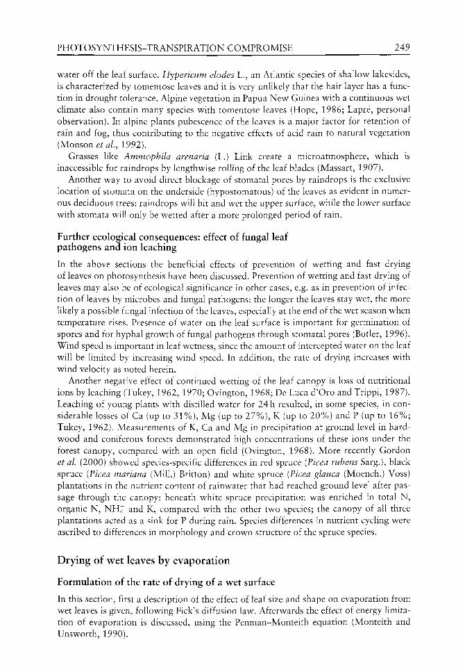
PHOTOSYNTHESIS-TRANSPIRATION COMPROMISE 249
water off the leaf surface. Hypericum elodes L., an Atlantic species of shallow lakesides, is characterized by tomentose leaves and it is very unlikely that the hair layer has a function in drought tolerance. Alpine vegetation in Papua New Guinea with a continuous wet climate also contain many species with tomentose leaves (Hope, 1986; Lapre, personal observation). In alpine plants pubescence of the leaves is a major factor for retention of rain and fog, thus contributing to the negative effects of acid rain to natural vegetation (Monson ^ /., 1992).
Grasses like Ammophila arenaria (L.) Link create a microatmosphere, which is inaccessible for raindrops by lengthwise rolling of the leaf blades (Massart, 1907).
Another way to avoid direct blockage of stomatal pores by raindrops is the exclusive location of stomata on the underside (hypostomatous) of the leaves as evident in numerous deciduous trees: raindrops will hit and wet the upper surface, while the lower surface with stomata will only be wetted after a more prolonged period of rain.
Further ecological consequences: effect of fungal leaf pathogens and ion leaching
In the above sections the beneficial effects of prevention of wetting and fast drying of leaves on photosynthesis have been discussed. Prevention of wetting and fast drying of leaves may also be of ecological significance in other cases, e.g. as in prevention of infection of leaves by microbes and fungal pathogens: the longer the leaves stay wet, the more likely a possible fungal infection of the leaves, especially at the end of the wet season when temperature rises. Presence of water on the leaf surface is important for germination of spores and for hyphal growth of fungal pathogens through stomatal pores (Butler, 1996). Wind speed is important in leaf wetness, since the amount of intercepted water on the leaf will be limited by increasing wind speed. In addition, the rate of drying increases with wind velocity as noted herein.
Another negative effect of continued wetting of the leaf canopy is loss of nutritional ions by leaching (Tukey, 1962, 1970; Ovington, 1968; De Luca d'Oro and Trippi, 1987). Leaching of young plants with distilled water for 24 h resulted, in some species, in considerable losses of Ca (up to 31%), Mg (up to 27%), K (up to 20%) and P (up to 16%; Tukey, 1962). Measurements of K, Ca and Mg in precipitation at ground level in hardwood and coniferous forests demonstrated high concentrations of these ions under the forest canopy, compared with an open field (Ovington, 1968). More recently Gordon et al. (2000) showed species-specific differences in red spruce (Picea rubens Sarg.), black spruce {Ficea mariana (Mill.) Britton) and white spruce [Vicea glauca (Moench.) Voss) plantations in the nutrient content of rainwater that had reached ground level after passage through the canopy: beneath white spruce precipitation was enriched in total N, organic N, N H j and K, compared with the other two species; the canopy of all three plantations acted as a sink for P during rain. Species differences in nutrient cycling were ascribed to differences in morphology and crown structure of the spruce species.
Drying of wet leaves by evaporation
Formulation of the rate of drying of a wet surface
In this section, first a description of the effect of leaf size and shape on evaporation from wet leaves is given, following Pick's diffusion law. Afterwards the effect of energy limitation of evaporation is discussed, using the Penman-Monteith equation (Monteith and Unsworth, 1990).
250 P. J. C. KUIPER &c C. M. H. LAPRE
Under conditions when there is a direct contact between the water film and leaf surface and the stomatal pores are blocked by liquid water, the resulting question is how quickly a leaf becomes dry again and CO2 exchange can be restored. Climatic factors, which determine the rate of evaporation from a wet surface are wind velocity and the difference between the saturation vapour pressure of the evaporating wet surface (at leaf surface temperature) and the actual vapour pressure of the atmosphere.
Leaf size and shape are important for the rate of drying. Aerodynamic properties of the evaporating leaf will determine the thickness of the boundary air layer adjacent to the evaporating surface: under identical external conditions small leaves will dry more quickly than leaves with a larger surface and, as will be shown below, long, narrow leaves will dry quicker than short and wide leaves of the same surface area (under condition that the wind direction is parallel to the length of the leaf). In addition, the leaf edge is important: a serrate or dentate leaf edge will facilitate air turbulence thus reducing the boundary air layer with its laminar flow of water vapour. No data on evaporation of wet leaves are available, but as a first approximation evaporation of rectangular pieces of wet blotting paper has been measured and formulated (Martin, 1943; Penman, 1948; Raschke, 1956, 1960; Kuiper, 1961; Gates, 1980) in wind tunnel experiments under the condition of ample energy supply for evaporation. More recently, Brenner and Jarvis (1995) confirmed these earlier results.
Formulation of the combined results yields:
E = 0.73 'k-(e,- e^) • W"^'^ • L" " • V^" (1)
in which E = evaporation of a rectangular piece of blotting paper (g-h~^ • lOOcm"^), k = coefficient of diffusion of water vapour (cm^ -sec"^), e^ - e^ = vapour pressure difference in mmHg at atmospheric pressure, W = width (cm) of the evaporating area at right angles to the wind direction, L = length (cm) of the evaporating area parallel to the wind direction, V = wind velocity (cm-sec~^), a = a proportionality factor, 0.78 (Raschke, 1956) or 0.76 (Kuiper, 1961) for turbulent air flow and 0.5 (Kuiper, 1961) for laminar air flow (see also Brenner and Jarvis, 1995). Reformulation of (1) gives:
E/0.73 -k'ie^ - ej)'V^'' = W'^'^ • L" ' (2)
A proportionality factor, X, is introduced for the first part of equation (3):
K = W-^-^'L-^-^ (3)
in which K = 1 for W and L both being 1 cm, a quadrant of 1 cm^. Figure 13.1 summarizes values for K for evaporating surfaces with W and L varying
between 0.1 and 100 cm. High values of K implicate a relatively high rate of evaporation and fast drying of a wet leaf surface and low values of K the reverse. K = 3.16 for W and L both being 0.1 cm, a quadrant of 0.01 cm^, indicative for a very small leaf (upper left corner of Figure 13.1). K = intermediate for long and narrow quadrants, e.g. K = 0.47 for W = 0.5 cm and L = 20 cm, representative for grass- and rush-like leaves and very low for larger quadrants (lower right corner of Figure 13.1). In this figure the curves for varying width merge at increasing length, indicating that differences between the rate of drying of wet surfaces are most visible at low values of W and L.
It is important to realize that the effect of large differences between high and low values of K on drying may be considerably reduced, because heat exchange is also dependent
PHOTOSYNTHESIS-TRANSPIRATION COMPROMISE 251
2 5 10 Length (cm)
Figure 13.1 Effect of length (L) and width (W) of a rectangular wet surface, as a model for plant leaves, on K, the evaporation rate per unit surface and per unit vapour pressure difference between wet surface and air, including an effect of wind velocity. On the ordinate K is expressed as:
K = E/0.73 -k'ie, - e^) • V^ W -0.2 -0.3
in which £ = evaporation per unit time and surface, k = diffusion coefficient of water vapour, ^s ~ ^d ^ vapour pressure difference between wet surface and air, V = wind velocity and a = a proportionality factor. High and low values of K represent respectively high and low rates of evaporation, due to the length and width of the evaporating surface.
on leaf morphology, causing a counterbalancing effect of the surface water vapour saturation pressure, e^, which is highly temperature dependent. Even more important, the energy demand for evaporation of water may easily limit evaporation and transpiration of partially wet leaves and leaf temperature and leaf vapour pressure will drop, resulting in reduced effects of leaf size on the rate of drying. Also, competition for energy between transpiration and evaporation of the partially wet needle surface of Douglas fir was evident (Bosveld, 1999). Water storage by leaves during rain and the simultaneous evaporation of components of rainfall interception are analysed by Lankreijer et al. (1993) and Klaassen ^ /. (1998).
The Penman-Monteith equation removes the problem of the strong temperature dependence of the vapour pressure of water: two components, the energy requirement for evaporation and the water vapour pressure difference between leaf and air, are combined in a single formulation (see Monteith and Unsworth, 1990). Heat transport by convection (sensible heat):
C = pCp{T - T^yr^
and heat transport by evaporation (latent heat):
XE = pcp(e^ - e^)/y''r^
(4)
(5)
252 P. J. C. KUIPER &c C. M. H. LAPRE
With incoming radiation, R„, the heat balance is:
XE + C = R, (6)
Combination of (4), (5) and (6) yields:
XE AR„ + pcp(e^ - ej)rH^ ^yj
A + 7 -
in which: C = flux of heat per unit area, p = density of air, Cp = specific heat of air, T = temperature, TQ = temperature of surface, r^ = resistance for heat transfer for convection, k = latent heat of vaporization, E = evaporation rate, 7''' = apparent value of psychrometer constant, R„ = net radiation flux density and A = rate of change of saturation vapour pressure w ith temperature.
Equation (7) contains a radiation component and a vapour pressure component. The contribution of these components to drying of w et vegetation nov^ will strongly depend upon the following factors:
1. The amount of incoming radiation for energy supply 2. The conductance of the vegetation to heat and evaporation fluxes, which in turn will
depend upon leaf area and leaf distribution within the vegetation.
Clearly vegetation structure will be a decisive factor for the rate of drying: a high vegetation density as in crops and forests with a closed canopy will easily lead to a limitation of drying, due to lack of energy, especially at low net radiation (see Lankreijer et al.^ 1993; Klaassen et al, 1998; Bosveld, 1999; Klaassen, 2001).
The situation in Mediterranean vegetations (see Colour Plate 4) is quite different from the above situation with:
1. A low vegetation density 2. A low leaf area index 3. An effective vertical distribution of the leaves 4. Strong wind during the rainy winter.
The radiation component will not so easily limit drying of wet Mediteranean vegetation as is the case with high yield crops and forests with a closed canopy.
A detailed study of drying of wet forest in relation to canopy wetness, canopy cover and net radiation is given by Klaassen (2001). A micrometeorological model with a detailed representation of a forest canopy, including a vertical distribution of the leaf area index (LAI) over 20 m, was evaluated against experimental results. The vertical distribution of sensible and latent heat (equations (4) and (5) respectively) in a wet and homogeneous forest was simulated. Evaporation of the wet canopy increased with canopy cover and, because of increasing energy limitation and increasing canopy roughness, the increase in canopy evaporation with canopy cover gradually slowed down. As expected, a patchy distribution of canopy leaves facilitates drying of the leaves and the drying rate in a patchy canopy cover was stimulated more at low canopy cover: 28% (cover = 0.2), decreasing to 4% (cover = 0.8).
A similar procedure was followed for canopy evaporation versus canopy wetness: after the start of rain, canopy evaporation strongly increased with canopy wetness and reached
PHOTOSYNTHESIS-TRANSPIRATION COMPROMISE 253
a rather constant level when canopy wetness approached 0.5. When the rain stopped, a fully wet canopy exhibited the fastest drying rate, which strongly decreased until a wetness value of 0.6 was reached.
In conclusion, the above described methodology may be very helpful to compare forests and shrub vegetations with different heights and canopy structures - a closed canopy versus an open and often patchy structure - and to relate these canopy structures to possible adaptive leaf morphology.
Ecological consequences of leaf morphology on the rate of drying of wet leaves
Wetted small leaves are characterized by high values of K and thus will dry relatively quickly (see Figure 13.1). In the Mediterranean Fynbos of South Africa wetted leaves of the restioid and cricoid growth form will dry quicker than leaves of the proteoid growth form, enabling the first two growth forms to perform photosynthesis even during cool winter days with frequent showers, provided wind velocity sufficiently compensates for the low vapour pressure deficit during these cool and rainy days.
The same conclusion also applies for other Mediterranean ecosystems. As a consequence the so-called sclerophyllous leaf structure also functions adequately in the fast drying process as a possible adaptation to frequent rainy and windy conditions. The above cricoid leaf growth form exhibits small leaves, which dry quickly. In addition, many species are characterized by hairs on the surface, which will reduce wetting of the leaf during rain, as in many Erica species (Schumann et al., 1992), including the European Atlantic Erica tetralix L.
A comparison of entire and compound leaves with respect to drying seems appropriate. Wet compound leaves will dry quicker than entire leaves of a similar leaf area as the combined leaflets, because of the spatial distribution of the small and separate leaflets, have a reduced leaf boundary layer thickness. We will give an example, based on the data of K in Figure 13.1. Assume an entire large leaf, corresponding to a rectangular evaporating surface oiW = 10 cm and L = 20 cm and K = 0.26. In a narrower leaf of W = 5 cm and L remains 20 cm the K value is 0.295 and the drying rate will be enhanced by 14% because of the narrower leaf shape. If the leaf is divided into leaflets of a size comparable with W = 2 cm and L = 5 cm, the K value is 0.26 and the drying rate is more than double that of the original entire leaf, all under non-limiting energy supply.
There is some experimental evidence that exchange processes, like evaporation, proceed faster in compound leaves. Taylor (1975) reported that the first pinnation in the leaf of Pteridium aquilinum (L.) Kuhn, the bracken fern, was the fundamental unit (boundary layer) for heat exchange. The third and second pinnation had a common boundary air layer and heat exchange was only enhanced up to the second pinnation. Unfortunately, no experimental data are given. The large leaves of Musaceae (Heliconia latispatha Berth, and Strelitzia nicolia Regel and Koern.) commonly show leaf tearing by wind and the boundary layer may be reduced by tearing as evident from leaf temperature measurements: in exposed sites entire leaves often exhibited nearly lethal leaf temperatures, with even a reduced photosynthesis, while plants with damaged leaves were at an advantage in this respect (Taylor and Sexton, 1972).
With respect to drying, a similar comparison can be made between leaves with an entire leaf margin and leaves with a serrate or dentate leaf margin. The aerodynamic properties of the latter category would facilitate the drying process but, at the same time, transpiration of
254 P. J. C. KUIPER &c C. M. H. LAPRE
the leaf would be enhanced under dry weather conditions. In this respect it is of interest that the veins of serrate or dentate leaves in general extend to the leaf margin, while the major veins of leaves with an entire margin in general loop within the margins (Parrish, 1998: 140). As mentioned earlier, veins of leaves of many species may act as gutters for runoff for water during rain and thus facilitate drying afterwards, as in many species of Melastomataceae.
Adaptive changes in leaf morphology in relation to the 'trade-off between photosynthesis and transpiration
As mentioned earlier, the wet season in Mediterranean ecosystems is characterized by a relatively cool and rainy period, where drying of leaves is determined by a compensatory effect of strong wind to the relatively low water vapour pressure deficit. Palaeoclimate has regularly fluctuated from warmer to cooler conditions and vice versa, including the last 2 miUion years (Deacon et aL, 1992). Changes in climatological conditions in the past, e.g. from a geological period with a high transpiration demand to a cool and rainy period with a low transpiration demand could exert selective pressure on leaf morphology via the drying process: genotypes with entire and simple leaves might possibly be replaced by genotypes with an increased leaf rim as for example in dentate and serrate leaves or lobed leaves, palmately or pinnately. Under further selective pressure, genotypes with compound leaves divided into separate leaflets may have evolved. As mentioned earlier, a reduction of leaf surface is much less effective in drying after rain when the entire leaf shape is maintained than when the reduction in leaf area is realized by development of leaflets of compound leaves. It is also evident, that the selection pressure will only be profound in open vegetations, as occurs after a catastrophic event in which all vegetation has been destroyed.
The effectiveness of selective pressure of climate changes will depend on the number of genes involved in the adaptive change. The genetics of leaf shape seems to be controlled by relatively few genes e.g., Sambucus nigra L. with its one time pinnate leaves also has a variety laciniata with a delicate parsley-like leaf structure. Variability of leaf shape within a single plant may be large, e.g. in the case oi Acacia heterophylla Vassal (Ursem, personal communication). Some plant species have two different leaf forms, during the vegetative and generative phase, as in ivy (Hedera helix L.), indicating developmental timing in leaf development. A similar case has been described by Stebbins (1974) for the effect of developmental timing on flower petal shape: a large fused and sympetalous flower corolla is formed if the intercalary cells develop along with the petal primordia. If they develop after the petal primordia have grown, the result is well separated petal lobes of the flower. Clearly, modification of leaf and petal shape is, genetically speaking, not complicated.
Closely related species are important since they may exhibit strikingly different leaf forms. An example within a very limited area as the Fynbos, South Africa, is the genus Pelargonium and the following species may be mentioned: P. cuculatum (L.) L.'Her. with entire leaves, P. scabrum (Burm.f) L.'Her. with deeply lobed leaves with coarse hairs, P. triste (L.) L.'Her. with a delicate parsley-like leaf form (see Colour Plate 5). A study of the microclimate of sites of these species may reveal whether these different leaf forms exhibit any consistent adaptive value to differences in microclimate.
To take another example, wet, cool and shady habitats with practically no wind exhibit a low transpiration demand. Such a habitat may contain ferns, mosses and liverworts.

PHOTOSYNTHESIS-TRANSPIRATION COMPROMISE 255
Ferns like Athyrium filix-femina (L.) Roth and Woodwardia radicans (L.) Sw. exhibit compound leaves, sometimes divided tw o or three times, w ith only a relatively small reduction in leaf area. This morphology allows a large leaf area for photosynthesis in the shade, together w ith the advantage of numerous small leaflets, v^hich are adapted to relatively quick drying under these humid conditions. A fern species with entire leaves from a humid and wet habitat such as Asplenium scolopendrium L. has shiny leaves, with probably a low wettability. Mosses and foliose liverworts with their minute foliage will also be able to dry quickly under these otherwise unfavourable evaporative conditions.
Finally, evolutionary aspects should be taken into account. As an example for Mediterranean ecosystems, the Fynbos is situated along the rim, where two oceans meet: the Atlantic Ocean with cold water and the Indian Ocean with warm water. This situation causes an extreme average westerly wind speed and together with the large number of climate fluctuations in the past may have led to a species-rich flora (Cowling, 1992), in which the rate of speciation exceeded the rate of extinction. The combination of a continuous strong westerly wind since the breakup of Gondwana, together with regular climatic fluctuations, may also have caused a relatively low degree of variation of leaf shape: the larger the number of repeated climatic fluctuations the lower the number of adaptive traits which will facilitate in survival of the species.
Final remarks
In the above, leaf morphology is related to drying after rain in order: (1) to restore photosynthesis; (2) to minimize nutritional ion leaching; and (3) to reduce infection by fungal leaf pathogens. Physical formulations of evaporation from wet surfaces under energy limiting conditions, as in closed forest canopies, and under less energy limiting conditions, as in open Mediterranean ecosystems, are used in order to evaluate the effect of vegetation structure on leaf morphology. In addition, suggestions are made for prevention of wetting by various adaptive leaf morphology traits.
The presented experimental evidence and observations in the field are, in general, limited to recent publications and the approach is to collect more experimental data in the field to test the applicability of the hypothesis. Within this perspective a number of questions may be asked and the answer to these questions may help to understand leaf morphology within the photosynthesis-transpiration compromise.
The following questions among others may be asked. Is photosynthesis reduced in rain-wetted leaves of varying morphology? How thick is the water layer of wet leaves of different sizes? Which species have a low/high wettability of leaves? Does photosynthesis continue during rain in velvet and tomentose leaves? Can the simulation of rainfall interception and evaporation of Klaassen (2001) be used for Fynbos vegetation and other Mediterranean ecosystems?
Acknowledgements
We cordially thank Dr W. Klaassen, Dr J.J.M. de Jong, Professor V. Westhoff (f), Drs Ir. W.N.J. Ursem and Dr G. Londo for assistance and critical advice during the preparation of the manuscript.

256 P. J, C. KUIPER &c C. M. H. LAPRE
References
Beadle NCW. 1966. Soil phosphate and its role in molding segments of the Australian flora and vegetation, with special reference to scleromorphy and sclerophylly. Ecology 47: 952-1007.
Berrie GK, Berrie A, Eze JMO. 1987. Tropical Plant Science. New York: Longman Scientific and Technical.
Bosveld FC. 1999. Exchange processes between a coniferous forest and the atmosphere. D. Phil. thesis Wageningen University.
Brenner AJ, Jarvis PG. 1995. A heated leaf replica technique for determination of leaf boundary layer conductance in the field. Agricultural and Forest Meteorology 72: 261-275.
Brewer CA, Smith WK. 1997. Patterns of leaf surface wetness for montane and subalpine plants. Plant, Cell and Environment 20: 1-11.
Butler DR. 1996. The presence of water on leaf surfaces and its importance for microbes and insects. In: Kerstiens G, ed. Plant Cuticles. Oxford: BIOS Scientific Publishers, 267-282.
Ceulemans R, Mousseau M. 1994. Effects of elevated atmospheric CO2 on woody plants. New Phytologist 127: 425-446.
Cowling R. 1992. The Ecology ofFynbos: Nutrients, Fire and Diversity. Oxford: Oxford University Press.
Curtis PS. 1996. A meta-analysis of leaf gas exchange and nitrogen in trees grown under elevated carbon dioxide. Plant, Cell and Environment 19: 127-137.
Deacon HJ, Jury MR, Ellis F. 1992. Selective regime and time. In: Cowling R, ed. The Ecology of Fynbos: Nutrients, Fire and Diversity. Oxford: Oxford University Press, 6-23.
De Jong JJM. 2001. Remote sensing of wet forests. D.Phil, thesis Groningen University, The Netherlands.
De Luca d'Oro GM, Trippi VS. 1987. Effect of stress conditions induced by temperature, water and rain on senescence development. Plant and Cell Physiolology 28: 1389-1396.
Fames D, Jarvis PG. 1989. The direct effects of increase in global atmospheric CO2 concentration on natural and commercial temperate trees and forests. Advances in Ecological Research 19: 1-55.
Ehleringer JR, Comstock JP, Cooper TA. 1987. Leaf-twig carbon isotope ratio differences in photosynthetic-twig desert shrubs. Oecologia 57: 318-320.
Elzenga JTM, Prins HBA. 1989. Light-induced polar pH changes in leaves of Elodea canadensis. I. Effects of carbon concentration and light intensity. Plant Physiology 91: 62-67.
Eschrich W. 1995. Funktionelle Pflanzenanatomie. Berlin: Springer. Feild TS, Zwieniecki MA, Donoghue MJ, Holbrook NM. 1998. Stomatal plugs of Drimys winteri
(Winteraceae) protect leaves from mist but not drought. Proceedings of the National Academy of Sciences, USA 95: 14256-14259.
Gates DM. 1980. Biophysical Ecology. Berhn: Springer. Gibson AC. 1998. Photosynthetic organs of desert plants. BioScience 48: 911-920. Gibson AC, Nobel PS. 1986. The Cactus Primer. Cambridge: Harvard University Press. Gordon AM, Chourmouzis C, Gordon AG. 2000. Nutrients in litterfall and rainwater fluxes in
27-year old red, black and white spruce plantations in Central Ontario, Canada. Forest Ecology and Management 138: 65-78.
Hill RS, Brodribb TJ. 2001. Macrofossil evidence for the onset of xeromorphy in Australian Casuarinaceae and tribe Banksieae (Proteaceae). Journal of Mediterranean Ecology 2: 127-136.
Hope GS. 1986. Development of present day biotic distributions in the New Guinea mountains. In: Barlow, ed. Flora and Fauna of Alpine Australasia: Ages and Origin. Melbourne: CSIRO, 129-147.
Horton RE. 1919. Rainfall interception. Monthly Weather Review 47: 603-623. Ishibashi M, Terashima I. 1995. Effects of continuous leaf wetness on photosynthesis: adverse
effects of rainfall. Plant, Cell and Environment 18: 431-438. Johnson SD. 1992. Plant-animal relationships. In: Cowling R, ed. The Ecology of Fynbos:
Nutrients, Fire and Diversity. Oxford: Oxford University Press, 175-206.

PHOTOSYNTHESIS-TRANSPIRATION COMPROMISE 257
Keeley JE. 1992. A Californian's view of Fynbos. In: Cowling R, ed. The Ecology of Fynbos: Nutrients, Fire and Diversity. Oxford: Oxford University Press, 372-389.
Klaassen W. 2001. Evaporation from rain-wetted forest in relation to canopy wetness, soil cover and net radiation. Water Resources Research 37: 3227-3236.
Klaassen W, Bosveld F, de Water E. 1998. Water storage and evaporation as constituents of rainfall interception. Journal of Hydrology 212-213: 36-50.
Kuiper PJC. 1961. The effects of environmental factors on the transpiration of leaves, with special reference to stomatal light response. Mededelingen Landbouwhogeschool, Wageningen, The Netherlands 61: 1-48.
Kuiper PJC. 1998. Adaptation mechanisms of green plants to environmental stress. Annals of the New York Academy of Sciences 851: 209-215.
Kiirschner WM, Stulen I, Wagner F, Kuiper PJC. 1998. Comparison of palaeobotanical observations with experimental data on the leaf anatomy of durmast oak (Quercus petraea, Fagaceae) in response to environmental change. Annals of Botany 81: 657-664.
Lankreijer HJM, Hendriks MJ, Klaassen W. 1993. A comparison of models simulating rainfall interception of forests. Agricultural and Forest Meteorology 64: 187-199.
Martin E. 1943. Studies on evaporation and transpiration under controlled conditions. Publications Carnegie Institute at Washington 550: 1-48.
Massart J. 1907. Essai de Geographic Botanique des Districts Littoraux et AUuviaux de la Belgique. Recueil de ITnstitut botanique Leo Errera, Bruxelles 7: 167-584.
Monson RK, Grant RC, Jaeger CH, Schoettle AW. 1992. Morphological causes for the retention of precipitation in the crowns of alpine plants. Environmental and Experimental Botany 32: 319-327.
Monteith JL, Unsworth MH. 1990. Principles of Environmental Physics. London: Edward Arnold. Nobel PS. 1991. Physicochemical and Environmental Plant Physiology. New York: Academic
Press. Ovington JD. 1968. Some factors affecting nutrient distribution within ecosystems. In: Eckhardt
FE, ed. Functioning of Terrestrial Ecosystems at the Primary Production Level. Paris: UNESCO, 95-105.
Parrish JT. 1998. Interpreting pre-Quaternary Climate from the Geologic Record. New York: Columbia University Press.
Penman HL. 1948. Natural evaporation from open water, bare soil and grass. Proceedings of the Royal Society of London A 193: 120-146.
Raschke K. 1956. Uber die physikalischen Beziehungen zwischen Warmeiibergangszahl, Strahlungaustausch, temperatur und transpiration eines Blattes. Planta 48: 200-238.
Raschke K. 1960. Heat transfer between the plant and the environment. Annual Review of Plant Physiology 11: 111-126.
Sage RF. 2001. Environmental and evolutionary precondition for the origin and diversication of the C4 photosynthetic syndrome. Plant Biology 3: 202-213.
Schumann D, Kirsten G, Oliver EGH. 1992. Ericas of South Africa. Vlaeberg: Fernwood Press. Smith WK, McClean TM. 1989. Adaptive relationship between leaf water repellency, stomatal
distribution, and gas exchange. American Journal of Botany 76: 465-469. Specht RL, Rundel PW. 1990. Sclerophylly and foliar nutrient status of Mediterranean-
climate plant communities in Southern Australia. Australian Journal of Botany 38: 459-474.
Stebbins GL. 1974. Flowering Plants: Evolution above the Species Level. Cambridge: Belknap Press of Harvard University Press.
Stock WD, van der Hey den F, Lewis OAM. 1992. Plant structure and function. In: Cowling R, ed. The Ecology of Fynbos: Nutrients, Fire and Diversity. Oxford: Oxford University Press, 226-241.
Taylor SE. 1975. Optimal leaf form. In: Gates DM, Schmerl RB, eds. Perspectives of Biophysical Ecology. Ecological Studies 12: 73-87.
Taylor SE, Sexton OJ. 1972. Some implications of leaf tearing in Musaceae. Ecology 53: 143-149.

258 P. J. C. KUIPER &c C. M. H. LAPRE
Tukey HB. 1962. Leaching of metabolites from above-ground plant parts, with special reference to cuttings used for propagation. Combined Proceedings of the Plant Propagators Society 11: 63-70.
Tukey HB. 1970. The leaching of substances from plants. Annual Review of Plant Physiology 21: 305-324.
Wagner F, Below R, De Klerk P, et al. 1996. A natural experiment on plant acclimation: lifetime stomatal frequency response of an individual tree to annual atmospheric CO2 increase. Proceedings of the National Academy of Sciences, USA 93: 11705-11708.
Woodward IF. 1987. Stomatal numbers are sensitive to increases in CO2 from pre-industrial levels. N^^wr^ 327: 617-618.
14 Xylem hydraulics and angiosperm success:
a test using broad-leafed species
Norman W Pammenter, Guy F Midgley and William J Bond
CONTENTS
Introduction 259
Materials and methods 261 Choice of species 261 Physiological studies 261
Resuhs 263
Discussion 266
References 270
Introduction
Angiosperms are both more numerous in species and more diverse in form than other seed plants. They also dominate low latitude and low altitude biomes of the world in terms of biomass. Angiosperms began to diversify only in the Cretaceous when they also began to infiltrate, and eventually displace, vegetation dominated by Ginkgo L., conifers, cycads, Gnetales and extinct gymnosperm groups (Crane et al.^ 1995; Wing and Boucher, 1998). Over the last ten years, there has been considerable progress in identifying attributes of early angiosperms from remarkable fossil discoveries and in identifying sister taxa from molecular and morphological phylogenetic analyses (Crane et al., 1995; Crepet, 2000; Friedman and Floyd, 2001). There has also been a growing number of studies on angiosperm traits that might account for their ecological 'success'. Diversification of angiosperms has been linked to co-evolution with insect pollinators in many angiosperm groups (Grimaldi, 1999; Crepet, 2000) and to the evolution of growth forms with short generation times (Eriksson and Bremer, 1992; Tiffney and Mazer, 1995; Dodd et aL, 1999). However, diversity does not necessarily translate to ecological dominance, as measured by the area occupied or the proportion of biomass present (Bond, 1989). It is this aspect of angiosperm success that we consider here.
Extant gymnosperms are all woody and most have long-lived leaves with low surface area to weight ratios. Many angiosperms resemble gymnosperms, with long-lived evergreen
The Evolution of Plant Physiology Copyright © 2004 Elsevier Ltd ISBN 0-12-33955-26 All rights of reproduction in any form reserved

260 N. W. PAMMENTER ET AL.
leaves similar to extant conifers (although few have needle-like leaves) and often grov^ together with them. However, angiosperms also include trees, shrubs and herbs with leaves of a short life span and high surface area to weight ratios (Reich 1998), unlike any extant (or extinct) gymnosperms. Plants with these leaf properties dominate ecosystems where gymnosperms are rare or absent, including temperate deciduous forests, lowland tropical forests (which, although evergreen, are ever-growing), savannas, and herb-dominated grasslands and ruderal communities. Bond (1989) suggested that the presence of vessels and of distinctive, anastomosing leaf venation in angiosperms, coupled with a reproductive system that allowed them rapidly to produce seeds, permitted flowering plants to develop novel growth forms that could out-grow gymnosperms. He suggested that these features might be most important in the recruitment stage, before gymnosperms could build up a large leaf mass by accumulating successive cohorts of evergreen leaves. Rapid growth and rapid reproduction would be most favoured in productive environments and it is these that are now dominated by flowering plants. Angiosperms would have little advantage over gymnosperms in environments with chronic stress such as cold, or nutrient poor areas and here gymnosperms would still be able to persist.
There are a number of recent studies of hydraulic properties and leaf physiology of angiosperms and gymnosperms (Becker et al., 1999; Brodribb and Feild, 2000) and of angiosperms which lack vessels (Feild et al, 1998, 2000; Feild and Holbrook, 2000). Becker (2000) has reviewed such studies in the context of Bond's (1989) hypothesis, that differences in the hydraulic system partly account for angiosperm 'success'. The general conclusion is that angiosperms can attain higher conductance and photosynthetic capacity than gymnosperms but that values measured for many angiosperms overlap those obtained for gymnosperms. Becker (2000) argues that the results do not support the contention that angiosperm innovations in the hydraulic system influence leaf physiology and competitive performance. However, most of the studies compared angiosperm and gymnosperm trees growing together in low productivity environments including cloud forests, nutrient poor soils and other sites with chronic stress, where the hydraulic characteristics of angiosperms would have little advantage.
A more appropriate test of the importance of hydraulic design in limiting gymnosperm success, would be to compare relative performance of species with similar growth forms in productive environments, from which gymnosperms have been excluded. The question is whether gymnosperm-like vascular systems can support fast-growing angiosperm-like canopies. Could a gymnosperm-type hydraulic system, made up of tracheids, supply water fast enough to sustain uniquely angiosperm-type leaves with short life spans, a high surface area to mass ratio and the capacity to rapidly grow and fill space? Could these uniquely angiosperm leaf properties function with gymnosperm-like leaf vasculature (Roth-Nebelsick et al.y 2001)? Because we do not have the technology to design plants to specification, with mixed angiosperm and gymnosperm elements, we explored the first question, of hydraulic limitations on leaf function, by searching for gymnosperms with leaves most similar to those of angiosperms growing in productive environments. There are very few of them: most extant conifers have needle-like or scale leaves. Ginkgo is the only extant gymnosperm which grows a broad-leaf and discards it in a single season. We compared this species with broad-leaved deciduous angiosperm trees of genera that occur in temperate deciduous forests. Although the earliest angiosperms were probably evergreen, we chose deciduous rather than evergreen angiosperms for three reasons: (1) the hypothesis concerns the dominance, not the origin of angiosperms, and deciduous broad-leafed ginkgo-phytes were displaced by deciduous angiosperms from the Cretaceous (Upchurch et aL,
XYLEM HYDRAULICS AND ANGIOSPERM SUCCESS 261
1998); (2) we wished to ensure that the study species had short-lived leaves (leaf life span of evergreen plants is extremely variable among species and growth conditions); and (3) the angiosperm and 'model' gymnosperm species to be compared would have the same growth form. To extend the database for the gymnosperms, we also included data from two further species, Agathis Salisb. and Podocarpus V Her. ex Pers. which, although evergreen, are relatively broad-leafed. All the trees were growing in resource-rich productive sites. We wished to determine whether: (1) the hydraulic properties of these broad-leafed gymnosperms differ from those of angiosperms with a similar tree growth form; (2) whether any such differences are correlated with leaf physiology; and (3) whether differences in leaf physiology could potentially influence competitive ability.
Materials and methods
Choice of species
To assess the influence of anatomy or morphology on physiological characteristics, the plants chosen should ideally be growing together, so as to avoid potentially confounding effects of different growth conditions. This requirement for species of similar habit occurring in the same habitat imposed constraints on the species available for study. However, in a municipal park in Cape Town, South Africa, some suitable species have been planted. In addition to Ginkgo biloba L., the following broad-leafed deciduous angiosperm species were studied: Quercus robur L. (English oak), Quercus palustris Miinchh. (southern pin oak), Ulmus procera Salisb. (English elm), and the London plane (Platanus X acerifolia (Aiton) Willd., a hybrid between Platanus orientalis L. and P. occidentalis L., sometimes also referred to as Platanus X hispanica (Pakenham, 1996)). The park where the plants were growing was irrigated during the dry summer months, when the measurements were taken. To broaden the base of the gymnosperm data set, studies were undertaken on Agathis robusta (Moore) Bailey: although not deciduous this species has relatively broad leaves in comparison with most gymnosperms. Additionally, some data collected for a different study on the gymnosperm Podocarpus latifolius (Thunb.) R. Br. ex Mirb. were included in the data set.
The constraints imposed on the choice of species create statistical difficulties in that truly independent samples were not available for all species. Studies were conducted on four individual young trees of G. biloba^ three mature Q. robur trees, two young specimens of Q. palustris and a single young but mature tree of each of U. procera and P. X acerifolia, A single specimen of A. robusta growing as a street tree near the park in Cape Town was used for the water relations studies and a young specimen in the Durban Botanic Gardens was used to investigate photosynthesis characteristics. The specimen of P. latifolius was growing in an irrigated domestic garden in Durban. In all cases, measurements were made on several (up to five) individual leaves or twigs from the trees available. Where appropriate, analyses of variance were conducted and data that were not normally distributed were log-transformed prior to analysis.
Physiological studies
Water relations Hydraulic conductivities of excised young branches were measured using the procedure of Sperry etal. (1987). Briefly, conductivity was calculated by measuring the mass flow of water passing through a branch segment connected to a constant pressure reservoir (2kPa).

262 N. W. PAMMENTER ET AL.
It was ensured that all branch segments were longer than the longest xylem vessel. Hydraulic conductivity was measured after flushing the branch segment at a pressure of approximately 200 kPa until no further increase in flow rate occurred and values reported here are maximum values. A 0.01 M HCl solution (with filtered (0.22 ixm membrane filter), degassed, distilled water) was used throughout. After measurement, safranin dye (0.05%) was passed into the stem to stain the functional xylem, the area of which was measured and the data were expressed as specific conductivity (^J. Leaf area distal to the measured stem was measured and data also expressed as leaf specific conductivity (fe/).
Stomatal conductances and transpiration rates of leaves in their natural orientation were measured using a LiCor 1600 porometer (LiCor, Lincoln, Nebraska) and light intensity incident on the leaf surface noted. The water potential of each leaf on which transpiration rate had been measured was determined immediately afterwards by means of a pressure chamber (Soil Moisture Corporation, Santa Barbara, California). Measurements were taken from pre-dawn to shortly after midday to establish the relationship between transpiration rate and leaf water potential (measurements were not taken in the afternoon to avoid hysteresis effects).
Photosynthesis studies Rates of net carbon dioxide assimilation (A) were measured using a LiCor 6200 portable photosynthesis system with a 11 chamber volume. Leaves were maintained in their natural orientation during measurements and light intensity incident on the surface was measured. Data were collected from pre-dawn to shortly after midday and measured rates were plotted against incident photosynthetic photon flux density (PPFD), so that photosynthesis rate at saturating light intensity (A^^^^) could be assessed. We used a number of leaves to construct a single 'light curve' and replicates consisted of measurements on different days.
The response of rates of assimilation to intercellular CO2 concentration (Q) were measured using the same equipment (except for A. robusta and P. latifalius). A fully illuminated leaf was enclosed in the chamber at normal atmospheric CO2 concentration and photosynthetic rates measured as the CO2 concentration in the chamber was drawn down by the photosynthesis of the leaf. Once the compensation point had been approached the CO2 concentration in the chamber was increased by the injection of a small quantity of pure CO2, and photosynthetic rates measured as CO2 concentration was drawn down to ambient levels. Data sets in which there were differences between the two measures of assimilation rate at ambient CO2 concentration (at the start and end of the experiment) were rejected. Relative humidity in the chamber was maintained close to ambient levels by controlling the proportion of the airflow passing through a desiccant. Leaf temperatures increased during the course of measurements, but did not exceed air temperature by more than 5°C. A:Q curves and light response curves of A. robusta and P. latifolius were measured using a LiCor 6400 portable photosynthesis measuring system. The initial slope of the response of A to Q was taken as a measure of the carboxylation coefficient, which has been related to the activity of ribulosebisphosphate (RuBP) carboxylase (Farquhar and Sharkey, 1982). The saturation value (/max) indicates the maximum rate of RuBP regeneration and thus electron flow through the photosynthetic electron transport pathway (Farquhar and Sharkey, 1982). To have sufficient data points for calculation and analysis of initial slopes, the data from individual A:Cj curves were combined.
Stomatal limitation to photosynthesis was assessed according to Farquhar and Sharkey (1982). Instead of calculating a stomatal limitation for each individual A:Ci curve for each species, a single value was estimated by combining all the curves for a species and using

XYLEM HYDRAULICS AND ANGIOSPERM SUCCESS 263
l o - l the mean value of stomatal conductances at PPFD values greater than 200 (xmolm~ s (the light intensity by w^hich stomatal conductance had saturated). Although this means that replicate values w ere not available, it was felt to be more representative of the leaves in their natural state, as stomatal conductances of leaves in these conditions, rather than in a chamber, v^ere used.
Results
The maximum hydraulic conductivity of minor branches expressed per unit area of functional xylem is shown in Figure 14.1. The values separated into two statistically distinct groups, with the angiosperms having specific conductivities an order of magnitude higher than those of the gymnosperms. In terms of water supply to leaves, inefficient xylem could be compensated for by decreases in the leaf area supplied per unit area of xylem. In the species studied, although the gymnosperms did have lower leaf area/xylem area ratios, the differences were not large (data not shown). Consequently, when hydraulic conductivity was expressed per unit leaf area supplied, the differences apparent in specific conductivity were maintained, with the angiosperms having leaf specific conductivities five to ten times higher than those of the gymnosperms (Figure 14.2).
The tension developed in the transpiration stream is directly related to the transpiration rate and inversely proportional to the leaf specific conductivity (Tyree and Ewers, 1991). Thus a low leaf specific conductivity would imply either, or both, low transpiration rates and low leaf water potential. The relationship between transpiration rate and the leaf water potential developed support this (Figure 14.3; the lines for individual species are extended to the maximum transpiration rates measured). The slopes of this relationship for the species studied fall into two statistically distinct groups, with the gymnosperms showing the steeper slopes. Consequently, the gymnosperm species had midday transpiration rates
G.b. A.r. P.I. P. X a. U.p. Q.r. Q.p.
Figure 14.1 Maximum hydraulic conductivity per unit xylem area (k^) of three gymnosperm and four angiosperm species. Abbreviations: G.b., Ginkgo biloba; A.r., Agathis robusta; P.I., Podocarpus latifolius; P. X a., Platanus X acerifolia; U.p., Ulmus procera; Q.r., Quercus robur; Q.p., Quercus palustris. The same abbreviations are used in all the figures. Values with the same letter are not statistically different (analysis of variance followed by Tukey's multiple range test, P < 0.05).
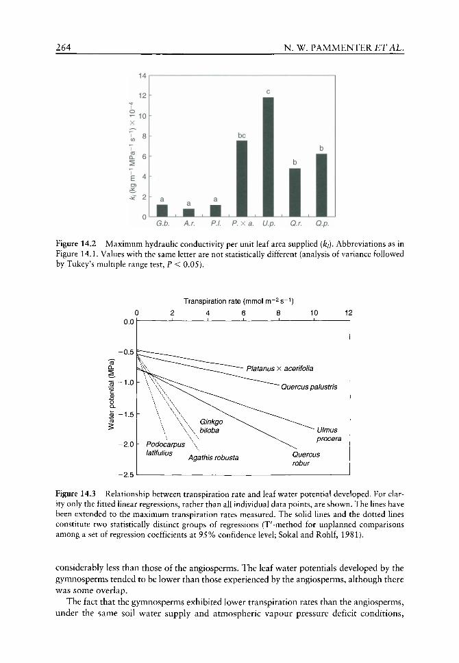
264 N . W. P A M M E N T E R ET AL.
Figure 14.2 Maximum hydraulic conductivity per unit leaf area supplied (ki). Abbreviations as in Figure 14.1. Values with the same letter are not statistically different (analysis of variance followed by Tukey's multiple range test, P < 0.05).
Transpiration rate (mmol m-2 s-
4 6 8 10 12 0.0
-2.5
Podocarpus latifulius
Platanus x acerifolia
Quercus palustris
Ulmus procera
Agathis robusta Quercus robur
Figure 14.3 Relationship between transpiration rate and leaf water potential developed. For clarity only the fitted linear regressions, rather than all individual data points, are shown. The lines have been extended to the maximum transpiration rates measured. The solid lines and the dotted lines constitute two statistically distinct groups of regressions (T'-method for unplanned comparisons among a set of regression coefficients at 95% confidence level; Sokal and Rohlf, 1981).
considerably less than those of the angiosperms. The leaf water potentials developed by the gymnosperms tended to be lower than those experienced by the angiosperms, although there was some overlap.
The fact that the gymnosperms exhibited lower transpiration rates than the angiosperms, under the same soil water supply and atmospheric vapour pressure deficit conditions,

XYLEM HYDRAULICS AND ANGIOSPERM SUCCESS 265
G.b. A.r. P.I. P. X a. U.p. Q.r. Q.p.
Figure 14.4 Average stomatal conductances measured in the field at incident photosynthetically active radiation intensities greater than 200 iJimolphotons m~^ s~ . Abbreviations as in Figure 14.1. Values with the same letter are not statistically different (analysis of variance followed by Tukey's multiple range test, P < 0.05).
suggests that the stomatal conductances to vapour diffusion were lower in the gymnosperms. Stomatal conductance is dependent upon incident light intensity, but in the species studied the effect was generally saturated by a PPFD of 200 |ULmolm"^s'^. The mean stomatal conductance measured for each species at incident radiation higher than this is shown in Figure 14.4 (as the data for P. latifolius were collected for a different study, stomatal conductance values are not available). Although the differences were not as marked as reported for the hydraulic conductivities, there is a tendency for the gymnosperms to have lower stomatal conductances than the angiosperms, with P. X acerifolia showing intermediate values closer to the gymnosperms than the other angiosperms.
Lower stomatal conductances could possibly limit CO2 diffusion into the leaf and hence reduce maximum photosynthetic rates. Light-saturated rates of photosynthesis (Aj ax? PPFD > 1000 jjimol m~^s"^) fell into distinct groups (Figure 14.5), although P. X acerifolia again grouped with the gymnosperms, not the angiosperms. To assess whether the lower maximum photosynthetic rates were a consequence of stomata-imposed limitations on rates of CO2 diffusion, stomatal limitations were calculated (Figure 14.6). Although the method of calculation did not permit statistical analysis of the data, it is clear that there are no major differences in stomatal limitation between the two groups of plants. To assess whether photosynthetic capacity, rather than stomata, limited light-saturated photosynthetic rates, the carboxylation coefficients (Figure 14.7) and maximum rates of RuBP regeneration (Figure 14.8) were measured from A:Q curves. Differences in carboxylation coefficients were not clear, although there was a tendency for the species with high rates of light-saturated photosynthesis to have high carboxylation coefficients (Figure 14.7). In terms of maximum rates of RuBP regeneration, although P. X acerifolia had the lowest value among the angiosperms, the gymnosperms constituted a single group with values lower than any of the angiosperms (Figure 14.8). Thus the low maximum photosynthetic rates of the gymnosperms is a consequence of low photosynthetic capacity, rather than directly due to stomatal limitation to CO2 diffusion. In terms of photosynthetic characteristics, P. X acerifolia is more similar to the gymnosperms, than to the angiosperms.

266 N . W. P A M M E N T E R ET AL.
Figure 14.5 Average rates of photosynthesis measured in the field at incident photosynthetically active radiation intensities greater than 1000 ixmol photons m~^s~^ (- max)- Abbreviations as in Figure 14.1. Values v^ith the same letter are not statistically different (analysis of variance followed by Tukey's multiple range test, P < 0.05).
G.b. A.r. P.I. P. X a. U.p. Q.r. Q.p.
Figure 14.6 Stomatal limitation to photosynthesis, assessed from bulked replicate A:Q curves and average stomatal conductance measured in the field at incident photosynthetically active radiation intensities greater than 200 jjimol photons m~^s~^ Abbreviations as in Figure 14.1.
Discussion
The essence of the argument presented here concerning the current dominance of the angiosperms is that the more efficient hydraulic characteristics conferred by the vessels of the angiosperms could provide this group w^ith a competitive advantage under conditions of high potential productivity. Thus, w^hen subjecting this hypothesis to test, the choice of species is critical. Additionally, although there is considerable overlap in the hydraulic properties of gymnosperms and angiosperms (Tyree and Ewers, 1996; Becker, 2000), hydraulic

XYLEM HYDRAULICS AND ANGIOSPERM SUCCESS 267
0.12
0.09
o
i t 0 O 0.06
0.03
CO
O
Figure 14.7 Carboxylation coefficient measured as the slope of bulked A:Q curves. Abbreviations as in Figure 14.1. Values w ith the same letter are not statistically different (T'-method for unplanned comparisons among a set of regression coefficients at 95% confidence level).
Figure 14.8 Maximum rates of RuBP regeneration (/max) measured from A:Q curves. Abbreviations as in Figure 14.1. Values with the same letter are not statistically different (analysis of variance followed by Tukey's multiple range test, P < 0.05).
architecture and conductivities may respond to growth conditions (Shumway et al., 1993; Mencuccini and Grace, 1995; Heath et aL, 1997; Vander Willigen and Pammenter, 1998; Brodribb and Feild, 2000). This reinforces the need to select, not only the test species, but also the individual plants, with care. Becker et al. (1999) showed similar whole plant hydraulic conductances of saphng-sized angiosperms and gymnosperms, but these were growing on nitrogen- and phosphorus-poor white sands in Borneo. Similarly, Feild and Holbrook (2000) showed no difference in leaf specific conductivities between the vessel-less Drimys granadensis L. (Winteraceae) and co-occurring vessel-bearing angiosperms in a cloud forest of Costa Rica. Fiowever, the conductivities measured were at the low end of the range for angiosperms and were, in fact, similar to the values we measured for the
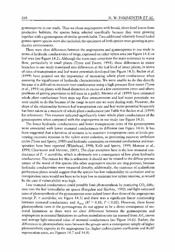
268 N. W. PAMMENTER ET AL.
gymnosperms in our study. Thus we chose angiosperms with broad, short-hved leaves from productive habitats, the species being selected specifically because they were growing together with a gymnosperm of similar growth habit. Two additional relatively broad-leafed gymno-sperm species were also included; the specimens of both species were growing in productive environments.
There were clear differences between the angiosperms and gymnosperms in our study in terms of hydraulic conductivities of twigs, expressed on either xylem area (see Figure 14.1) or leaf area (see Figure 14.2). Although the roots may constitute the main resistance to water flow, particularly in small plants (Tyree and Ewers, 1991), these differences in minor branches in our study translated into differences at the leaf level of intact plants, in terms of rates of transpiration and leaf water potentials developed (see Figure 14.3). Becker et al. (1999) have pointed out the importance of measuring whole plant conductance when assessing the significance of hydraulic characteristics. We were unable to do this directly because it is difficult to measure root conductance using a high pressure flow meter (Tyree et al., 1995) on plants with basal diameters in excess of a few centimetres (over-and-above problems of getting permission to fell trees in a park!). Meinzer et al. (1999) have estimated whole plant conductance from stem sap flow measurements and leaf water potentials: we were unable to do this because of the range in stem size we were dealing with. However, the slope of the relationship between leaf transpiration rate and leaf water potential frequently has been taken as a measure of whole plant conductance (see Table 4 in Becker et al. (1999) for references). This measure indicated significantly lower whole plant conductances of the gymnosperms when compared with the angiosperms in our study (see Figure 14.3).
The lower hydraulic conductances and lower transpiration rates of the gymnosperms were associated with lower stomatal conductances to diffusion (see Figure 14.4). It has been suggested that a function of stomata is to maintain transpiration rates at levels preventing excessive tensions in the xylem water columns, so preventing runaway embolism cycles (Tyree and Sperry, 1989) and hydraulic constraints on stomatal conductance and transpiration have been reported (Whitehead, 1998; Kolb and Sperry, 1999; Meinzer et al., 1999; Clearwater and Meinzer, 2001). The clear exception here is the low stomatal conductance of P. X acerifolia, which is obviously not a consequence of low plant hydraulic conductance. The reason for this is unknown: it should not be related to the diffuse-porous nature of the wood of this species (the other angiosperm species are ring-porous), because hydraulic conductivities were measured directly; additionally, the existence of scalariform perforation plates would suggest that the species has low vulnerability to cavitation and so transpiration rates would not have to be kept low to maintain low xylem tensions, as would be the case if vulnerability was high.
Low stomatal conductances could possibly limit photosynthesis by restricting CO2 diffusion into the leaf intercellular air spaces (Farquhar and Sharkey, 1982) and light-saturated rates of photosynthesis of the gymnosperms were indeed lower than those of the angiosperms (except P. X acerifolia; see Figure 14.5) and there was a significant linear relationship between stomatal conductance and A^^^ (R^ = 0.81, P < 0.02). However, these lower photosynthetic rates in the gymnosperms do not appear to be a direct consequence of stomatal limitations as there were no clear differences between the gymnosperms and angiosperms in stomatal limitations to carbon assimilation rate (as assessed from A:Q curves and average light-saturated value of stomatal conductances (see Figure 14.6)). Rather, the differences in photosynthetic rates between the groups seem a consequence simply of higher photosynthetic capacity in the angiosperms (i.e. higher carboxylation coefficients and RuBP regeneration rates, see Figures 14.7 and 14.8).
XYLEM HYDRAULICS AND ANGIOSPERM SUCCESS 269
It is now well known that the relationship at leaf level between maximum stomatal conductance and maximum assimilation rate is strongly conserved across many plant species and functional types (Schulze et al.^ 199A)^ as is the relationship between carboxyla-tion capacity and RuBP regeneration rate (WuUschleger, 1993). Therefore, a functional limitation on stomatal conductance imposed by hydraulic constraints should translate to lower leaf level photosynthetic capacity. Our data indeed show a significant positive relationship between A^^^ and ki {R^ = 0.59, P < 0.05). This resdt echoes that of Brodribb and Feild (2000), who showed a significant positive relationship between ki and photosynthetic capacity (assessed from electron flux through PSII) in a wide range of angiosperm and gymnosperm trees. In that study, although there were some overlaps, the gymnosperms and vessel-less angiosperms had significantly lower hydraulic conductivities and photosynthetic capacities (from which was inferred lower stomatal conductances) than the vessel-bearing angiosperms.
Thus, in our study the hydraulic conductivities of broad-leafed deciduous angiosperms were higher than those of broad-leafed gymnosperms growing in the same or similar environments and these differences in hydraulic characteristics were associated with differences in leaf physiology, with the angiosperms having higher photosynthetic capacities and light-saturated photosynthetic rates (with the exception of P. X acerifolia). Growth rates, of course, depend upon more than photosynthesis rates per unit leaf area, with characteristics such as partitioning of photosynthate (Korner, 1991) and respiration rates and relative sink strengths (Farrar, 1999) being important. It is thus unlikely that differences in leaf physiology are the sole factor underlying the dominance of woody angiosperms over gymnosperms in these environments. However, among species of similar growth habit (woody, broad-leafed, short leaf life span) and habitat (productive environments), a higher photosynthetic capacity could translate into potentially higher growth rates and hence competitive ability.
We speculate that hydraulic limitations are likely to emerge as constraints on plant growth and there is evidence of a general trend of high whole plant hydraulic conductivities being associated with fast growth rates of pioneer species (Tyree and Ewers, 1996). In addition to any affect at the level of leaf physiology, hydraulic limitations are likely to constrain canopy development rates as well. Because of the lower stem hydraulic conductivity per unit xylem area of the gymnosperms, to maintain high capacity for water delivery to leaves with high transpiration rates (and thus photosynthetic rates), more wood has to be allocated to support unit leaf area. (This ratio, like other aspects of hydraulic architecture, is influenced by locality and growth conditions, and so comparisons should be made among similar growth forms and habitat type.) Allocation of more carbon to non-photosynthetic conducting tissue is likely to reduce total photosynthate assimilation and so potential growth rate. To develop both the physiological and allometric arguments fully, information on maximum individual growth rate, maximum canopy leaf area index and canopy development rate are needed, not only at the seedling level (the 'slow seedling' hypothesis of Bond (1989)), but also at the sapling stage. It is well known, for example, that pine (gymnosperm) plantation forests are highly productive, but the key to this apparent paradox is that they require time to develop the leaf area indexes and canopies to achieve high production rates once established.
An additional possible factor in the competitive potential of angiosperms is that our casual (unrecorded) observations suggest that the growth form of angiosperm trees is more plastic than that of gymnosperms. This plasticity would be an advantage in exploiting stochastically available resources (such as gaps in a forest canopy), leading to a competitive edge. However, high growth rates are a prerequisite for plants to be able to take advantage of this plasticity.
270 N . W. P A M M E N T E R ET AL.
In summary, our data set pertaining to angiosperms and gymnosperms species of similar
growth habit in similar habitats are consistent with the concept of a hydraulic limitation to
the growth rate of gymnosperms and hence their relative lack of success in productive
habitats in the face of competition from 'better-plumbed' angiosperms. The ultimate test
of manipulating the hydraulic conductivities of the test species and observing the effects
on leaf physiology and growth rates awaits to be done.
References
Becker P. 2000. Competition in the regeneration niche between conifers and angiosperms: Bond's slow seedling hypothesis. Functional Ecology 14: 401-412.
Becker P, Tyree MT, Tsuda M. 1999. Hydraulic conductances of angiosperms versus conifers: similar transport sufficiency at the whole-plant level. Tree Physiology 19: 445-452.
Bond WJ. 1989. The tortoise and the hare: ecology of angiosperm dominance and gymnosperm persistence. Biological Journal of the Linnean Society 36: 227-249.
Brodribb TJ, Feild TS. 2000. Stem hydraulic supply is linked to leaf photosynthetic capacity: evidence from New Caledonian and Tasmanian rainforests. Plant, Cell and Environment 23: 1381-1388.
Clearwater MJ, Meinzer FC. 2001. Relationships between hydraulic architecture and leaf photo-synthetic capacity in nitrogen-fertilized Eucalyptus grandis trees. Tree Physiology 21: 683-690.
Crane PR, Friis EM, Pedersen KR. 1995. The origin and early diversification of angiosperms. Nature 374: 27-33.
Crepet WL. 2000. Progress in understanding angiosperm history, success, and relationships: Darwin's abominably 'perplexing phenomenon'. Proceedings of the National Academy of Sciences of the United States of America 97: 12939-12941.
Dodd ME, Silvertown J, Chase MW. 1999. Phylogenetic analysis of trait evolution and species diversity variation among angiosperm families. Evolution 53: 732-744.
Eriksson O, Bremer B. 1992. Pollination systems, dispersal modes, life forms, and diversification rates in angiosperm families. Evolution 46: 25S-266.
Farquhar CD, Sharkey TD. 1982. Stomatal conductance and photosynthesis. Annual Review of Plant Physiology 33: 317-345.
Farrar JF. 1999. Acquisition, partitioning and loss of carbon. In: Press MC, Scholes JD, Barker MD, eds. Physiological Plant Ecology. Oxford: Blackwell Science, 25-43.
Feild TS, Zwieniecki MA, Donoghue MJ, Holbrook NM. 1998. Stomatal plugs of Drimys winteri (Winteraceae) protect leaves from mist but not drought. Proceedings of the National Academy of Sciences of the United States of America 95: 14256-14259.
Feild TS, Holbrook NM. 2000. Xylem sap flow and stem hydraulics of the vesselless angiosperm Drimys granadensis (Winteraceae) in a Costa Rican elfin forest. Plant, Cell and Environment 23: 1067-1077.
Feild TS, Zwieniecki MA, Holbrook NM. 2000. Winteraceae evolution: An ecophysiological perspective. Annals of the Missouri Botanical Garden 87: 323-334.
Friedman WE, Floyd SK. 2001. Perspective: The origin of flowering plants and their reproductive biology - A tale of two phylogenies. Evolution 55: 217-231.
Grimaldi D. 1999. The co-radiations of pollinating insects and angiosperms in the Cretaceous. Annals of the Missouri Botanical Garden 86: 373-406.
Heath J, Kerstiens G, Tyree MT. 1997. Stem hydraulic conductance of European beech (Fagus sylvatica L.) and pedunculate oak (Quercus robur L.) grown in elevated CO2. Journal of Experimental Botany 48: 1487-1489.
Kolb KJ, Sperry JS. 1999. Transport constraints on water use by the Great Basin shrub, Artemesia tridentata. Plant, Cell and Environment 22: 925-935.
Korner Ch. 1991. Some often overlooked plant characteristics as determinants of plant growth: a reconsideration. Functional Ecology 5: 162-173.
XYLEM HYDRAULICS A N D ANGIOSPERM SUCCESS 271
Meinzer FC, Goldstein G, Franco AC, et al. 1999. Atmospheric and hydraulic limitations on transpiration in Brazilian cerrado woody species, functional Ecology 13: 273-282.
Mencuccini M, Grace J. 1995. Climate influences the leaf area-sap wood area ratio in Scots pine. Tree Physiology 15: 1-10.
Pakenham T. 1996. Meetings with Remarkable Trees. London: Weidenfeld and Nicolson. Reich PB. 1998. Variation among plant species in leaf turnover rates and associated traits: implications
for growth at all life stages. In: Lambers H, Poorter H, Van Vauren MMI, eds. Inherent Variation in Plant Growth: Physiological Mechanisms and Ecological Consequences. Leiden: Backhuys, 467-487.
Roth-Nebelsick A, Uhl D, Mosbrugger V, Kerp H. 2001. Evolution and function of leaf venation architecture: A review. Annals of Botany 87: 553-566.
Schulze E-D, Kelliher FM, Korner Ch, et al. 1994. Relationships among maximum stomatal conductance, ecosystem surface conductance, carbon assimilation, and plant nitrogen: A global ecology scaling exercise. Annual Review of Ecology and Systematics 23: 629-660.
Shumway DL, Steiner KC, Kolb TE. 1993. Variation in seedling hydraulic architecture as a function of species and environment. Tree Physiology 12: 41-54.
Sokal RR, Rohlf FJ. 1981. Biometry, 2nd edn. San Francisco: W.H. Freeman. Sperry JS, Donnelly JR, Tyree MT. 1987. A method for measuring hydraulic conductivity and
embohsm in xylem. Plant, Cell and Environment 11: 35-40. Tiffney BH, Mazer SJ. 1995. Angiosperm growth habit, dispersal and diversification reconsidered.
Evolutionary Ecology 93: 93-117. Tyree MT, Ewers FW. 1991. The hydraulic architecture of trees and other woody plants. New
Phytologist 119: 345-360. Tyree MT, Ewers FW. 1996. Hydraulic architecture of woody tropical plants. In: Mulkey SS,
Chazdon RL, Smith AP, eds. Tropical Forest Plant Ecophysiology. New York: Chapman &: Hall, 217-243.
Tyree MT, Patiiio S, Bennick J, Alexander J. 1995. Dynamic measurements of root hydraulic conductance using a high pressure flow meter in the laboratory and field. Journal of Experimental Botany 46: 83-94.
Tyree MT, Sperry JS. 1989. Do woody plants operate near the point of catastrophic xylem dysfunction caused by dynamic water stress? Answers from a model. Plant Physiology 88: 574-580.
Vander Willigen C, Pammenter NW. 1998. Relationship between growth and xylem hydraulic characteristics of clones of Eucalyptus spp. at contrasting sites. Tree Physiology 18: 595-600.
Upchurch GR, Otto-Bliesner BL, Scotese C. 1998. Vegetation - atmospheric interactions and their role in global warming during the latest Cretaceous. Philosphical Transactions of the Royal Society, Series B 353: 97-111.
Whitehead D. 1998. Regulation of stomatal conductance and transpiration in forest canopies. Tree Physiology 18: 633-644.
Wing SL, Boucher LD. 1998. Ecological aspects of the Cretaceous flowering plant radiation. Annual Review of Earth and Planetary Sciences 26: 379-421.
Wullschleger SD. 1993. Biochemical limitations to carbon assimilation in C3 plants - a retrospective analysis of the A/Q curves from 109 species. Journal of Experimental Botany 44: 907-920.
15 Evolution of xylem physiology
Pieter Baas, Frank W Ewers, Stephen D Davis and Elisabeth A Wheeler
CONTENTS
Introduction 273 'Trade-off triangle 274 Early beginnings 274
Distribution of wood anatomical features 275 Vessel element perforations 275 Diameter, density and length of vessels 277 Ecological preferences in modern woods 277
Geological record 280
Experimental work 283 Conductive efficiency versus vulnerability to embolism 283 Conductive efficiency versus mechanical strength 287 Cohesion-tension theory and sap ascent 289 Mechanical strength, implosion resistance
and resistance to embolism 289 Roots versus stems 290 Parenchyma 290
Towards a synthesis: the evolution of hydraulic
structure and function 290
Acknowledgements 291
References 291
Introduction
This chapter summarizes how, according to our present understanding, certain wood
anatomical traits evolved in woody angiosperms and how during the geological past clear
ecological preferences can be recognized for a number of arbitrarily defined wood func
tional types. That summary will be related to experimental evidence of how key wood
anatomical traits affect hydraulic (and to a lesser extent mechanical) functioning of
woody trees, shrubs and vines. In conclusion, the ecological patterns in xylem anatomy
will be discussed in terms of their functional significance.
The Evolution of Plant Physiology Copyright © 2004 Elsevier Ltd ISBN 0-12-33955-26 All rights of reproduction in any form reserved
274 P. BAAS £TAL.
'Trade-off triangle
Xylem evolution can be viewed in the context of a 'trade-off triangle (Figure 15.1), with different adaptive solutions to the structure/function problems depending on environmental demands as well as phylogenetic constraints (taxonomic history). Evolution of xylem physiology is complicated by the fact that, ever since the early evolution of land plants, xylem has simultaneously performed multiple functions. These include not only water transport but also mechanical support of the plant and storage of water, minerals and carbohydrates. The optimal structures for each of the xylem functions most likely differ and selection to optimize for one function could lead to suboptimal performance or even complete failure of another function. An extreme example would be wood maximized for efficient water conduction with many wide thin-walled conduits, but which is so mechanically weak that stem failure occurs prior to seed production.
Herein, conductive efficiency of xylem is expressed in terms of specific conductivity (ks in m^MPa"^ s~^). Specific conductivity is the hydraulic conductivity divided by the xylem transverse area, with hydraulic conductivity equal to the flow rate per pressure gradient (Wagner et aL, 1998). Resistance to embolism (air blockage) is defined as the water potential (in MPa) at which there is 50% loss in conductivity due to embolism (Tyree et al., 1994). We define mechanical strength both in terms of stem strength and vessel wall strength. For stem strength we use modulus of rupture (MOR in GNm~^) of fresh stems, as measured with a cantilever test. This test is relevant since breakage of stems in nature would impair reproductive output by a plant (Wagner et al,^ 1998). For vessel strength we use {tlb)^^ which is the square of vessel wall thickness {t) divided by the diameter of the conduit {b). This parameter is relevant to determining the resistance to vessel implosion (Hacke et al.^ 2001), which is a danger to plants since the xylem sap is transported under negative pressure (= tension).
Early beginnings
The first woody plants had vessel-less wood and the first forests were comprised of trees with vessel-less woods (Meyer-Berthaud et aL, 1999). Vessels arose independently in
Resistance to embolism (-MPa)
Figure 15.1 Xylem evolution can be viewed in the context of a 'trade-off triangle, where conductive efficiency is inversely proportional to both mechanical strength and resistance to dysfunction via embolism.
EVOLUTION OF XYLEM PHYSIOLOGY 275
several clades (e.g. ferns, horse tails, Selaginella Pal., Gnetales, angiosperms). Recent molecular phylogenies indicate angiosperms were primitively vessel-less (Soltis et al.^ 2000). The vessel-less Amborella Baillon, a monotypic genus of evergreen shrubs from New Caledonia, is basal to all angiosperms. Thus, presumably, vessel-less angiosperms gave rise to vessel-bearing ones. Bailey and Tupper (1918) proposed a transformation series for this evolutionary event. It is debated whether vessel-lessness in Winteraceae and Tetracentron Oliver, which are nested in more advanced angiosperm clades, represents a reversion or whether these woods are primitively vessel-less with several independent, unparsimonious origins of vessels in the lower clades (Baas and Wheeler, 1996; Herendeen et al., 1999; Doyle and Endress, 2000). Comparison of the most successful of modern vessel-less woody plants, the conifers or softwoods, with the most successful modern vessel-bearing plants, the dicotyledons or hardwoods, shows two alternative strategies.
Conifers have longitudinal tracheids interconnected by large bordered intertracheary pits whose pit membranes (modified primary walls) usually are differentiated into a porous margo and a central thickened non-porous torus. The porous margo allows for effective sap transport. The non-porous torus provides safety against embolisms spreading from one tracheid to another. If an embolism occurs, the pit membrane, if flexible, deflects so that the torus presses against the pit border and seals the opening (aperture) between adjacent cells. This process effectively limits the spread of the embolism, as long as pressure differences across the pit aperture do not exceed a threshold that disrupts the seal (Sperry and Tyree, 1990). Individual longitudinal tracheids are approximately 100 times longer (2-4 mm) than wide (0.02-0.04 mm). Vessel-less angiosperms also rely on long tracheids, but their pit membranes are much less porous than conifers and the membranes generally lack tori.
Angiosperms have vessels for xylem sap transport and fibres for support. Vessels are comprised of a series of vessel elements with perforated common end walls. Vessels, while considerably longer than longitudinal tracheids, are finite in length, varying from a few millimetres to several metres long (Zimmermann, 1983). Vessel diameter ranges from 0.025-0.3 mm, most commonly more than 0.05 mm. Intact sapwood intervessel pits have relatively compact, seemingly non-porous pit membranes which would not readily allow movement of embolisms from vessel to vessel.
Distribution of wood anatomical features
Vessel element perforations
According to the transformation series of Bailey and Tupper (1918), in angiosperms, vessel elements with scalariform perforation plates and scalariform pitting were derived from imperforate elements with scalariform pitting. Subsequently, vessel elements with simple perforation plates were derived from vessel elements with scalariform perforations. Support from the fossil record for this transformation series includes: (1) the angiosperms as a whole, a higher incidence of scalariform perforations in the Cretaceous than in Tertiary (Figure 15.2) (Wheeler and Baas, 1991, 1993); (2) the Platanaceae, Cretaceous and Early Tertiary species have exclusively scalariform perforation plates, while Late Tertiary and modern species have a mixture of simple and scalariform perforations (Wheeler, 1991). However, the fossil record also shows ambiguities with respect to the Baileyan transformation series: Paraphyllanthoxylon is one of the two oldest types of angiosperm and it has vessel elements with simple perforations (about 100 miUion years
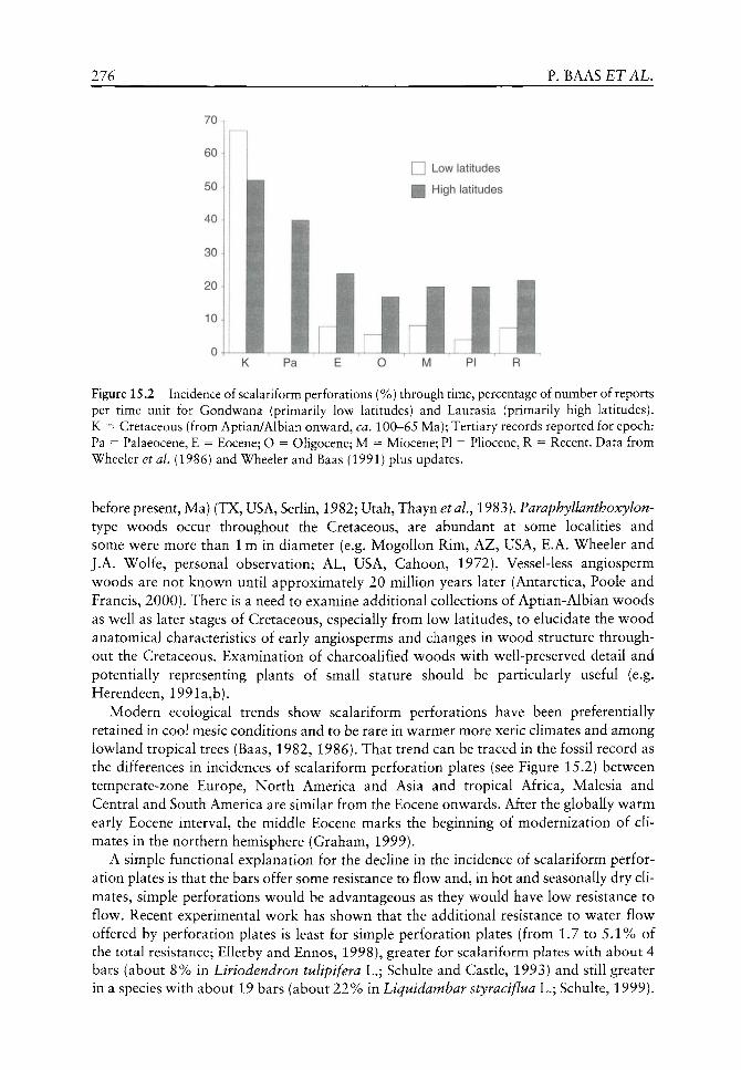
276 P. BAAS £TAL.
70 1
I I Low latitudes
H High latitudes
JiUJ K Pa M PI R
Figure 15.2 Incidence of scalariform perforations (%) through time, percentage of number of reports per time unit for Gondwana (primarily low latitudes) and Laurasia (primarily high latitudes). K = Cretaceous (from Aptian/Albian onward, ca. 100-65 Ma); Tertiary records reported for epoch: Pa = Palaeocene, E = Eocene; O = Oligocene; M = Miocene; PI = PHocene, R = Recent. Data from Wheeler et ai (1986) and Wheeler and Baas (1991) plus updates.
before present, Ma) (TX, USA, Serlin, 1982; Utah, Thayn etaL, 1983). Paraphyllanthoxylon-type woods occur throughout the Cretaceous, are abundant at some localities and some were more than 1 m in diameter (e.g. Mogollon Rim, AZ, USA, E.A. Wheeler and J.A. Wolfe, personal observation; AL, USA, Cahoon, 1972). Vessel-less angiosperm woods are not known until approximately 20 million years later (Antarctica, Poole and Francis, 2000). There is a need to examine additional collections of Aptian-Albian woods as well as later stages of Cretaceous, especially from low latitudes, to elucidate the wood anatomical characteristics of early angiosperms and changes in wood structure throughout the Cretaceous. Examination of charcoalified woods with well-preserved detail and potentially representing plants of small stature should be particularly useful (e.g. Herendeen, 1991a,b).
Modern ecological trends show scalariform perforations have been preferentially retained in cool mesic conditions and to be rare in warmer more xeric climates and among lowland tropical trees (Baas, 1982, 1986). That trend can be traced in the fossil record as the differences in incidences of scalariform perforation plates (see Figure 15.2) between temperate-zone Europe, North America and Asia and tropical Africa, Malesia and Central and South America are similar from the Eocene onwards. After the globally warm early Eocene interval, the middle Eocene marks the beginning of modernization of climates in the northern hemisphere (Graham, 1999).
A simple functional explanation for the decline in the incidence of scalariform perforation plates is that the bars offer some resistance to flow and, in hot and seasonally dry climates, simple perforations would be advantageous as they would have low resistance to flow. Recent experimental work has shown that the additional resistance to water flow offered by perforation plates is least for simple perforation plates (from 1.7 to 5.1% of the total resistance; Ellerby and Ennos, 1998), greater for scalariform plates with about 4 bars (about 8% in Liriodendron tulipifera L.; Schulte and Castle, 1993) and still greater in a species with about 19 bars (about 22% in Liquidambar styraciflua L.; Schulte, 1999).

EVOLUTION OF XYLEM PHYSIOLOGY 277
Models have not been tested for species with a large number (more than 50) of closely-spaced bars and with steeply inclined end walls, but it is possible that this 'primitive' type of perforation plate would offer still greater resistance to flow. Zimmermann offered an alternative explanation (1983), that the retention of scalariform perforations was adaptive in regions with frost as the bars in the perforations would trap embolisms during the thawing of frozen water. That explanation may apply to Arctic and cool temperate regions, but would not explain the high incidence of scalariform plates in tropical montane regions that, although cool, usually are frost free at 1500-2000 m. More experimental work is needed by physiologists and biophysicists to understand the role of scalariform perforations in the hydraulic architecture of woody plants. Monocots also show a trend from vessel-less to scalariform perforations to the elimination of scalariform perforations, especially within roots and basal parts of stems (e.g. Cheadle, 1944; Thorsch, 2000).
Diameter, density and length of vessels
Vessel diameter and density are less controversial wood functional parameters than vessel element length and perforation plate type. Numerous studies show that in erect trees and shrubs vessel density is roughly inversely proportional to vessel diameter (at least the logarithmically transformed data. Baas, 1973; van der Oever et al.^ 1981; Noshiro and Baas, 1998, 2000; Klaassen, 1999). Only lianas often have both wide vessels and high vessel densities (Carlquist, 1991). Another correlation that has been estabhshed several times is that wide vessels are associated with much greater total vessel length than narrow vessels (Zimmermann and Jeje, 1981). Thus, vessel diameter might be considered a proxy for vessel length for purposes of discussion of hydraulic efficiency.
We have delimited a number of somewhat arbitrary hydraulic types and analysed their occurrence in modern floras and, when data permit, in the geological past:
1. Diffuse-porous woods with very wide vessels (>200 \hm) (Figure 15.3a, see Figure 15.6) 2. Diffuse-porous woods with narrow vessels (<100 (uim) (Figure 15.3b, see Figure 15.7) 3. Ring-porous woods with very wide earlywood vessels and narrow latewood vessels.
(Figure 15.3c, see Figure 15.8) 4. Wood with mixed wide and narrow vessels throughout the growth ring (Figure
15.3d, see Figure 15.5) 5. Lianas: vessels both wide and frequent; often with clusters or multiples of narrow
vessels (Figure 15.3e, see Figure 15.5)
Ecological preferences in modern woods
Typel Wide-vesselled diffuse-porous species are typical of lowland evergreen rainforest trees (Baas and Wheeler, 2000). The syndrome of wide, infrequent vessels also occurs in some drought tolerant deep-rooting desert trees (e.g. Acacia gerrardii Benth. subsp. negevensis^ Balanites aegyptiaca (L.) Del., Fahn et al.^ 1986). This wood type is not known to occur in shrubs and trees of high latitudes.
Typel Narrow-vesselled diffuse-porous species are typical of both evergreen tropical montane and temperate trees and many deciduous temperate species of trees and shrubs. In widespread

278 P. BAAS £TAL.
taxa the narrowest vessels occur in Arctic and alpine shrubs and in highly xeric desert shrubs. This has been demonstrated for florulas within California (Carlquist and Hoekman, 1985), for the Middle East (Baas et al,, 1983), North America, and for temperate versus tropical world woods (Wheeler and Baas, 1991, 1993). Within the genera Cornus, Ilex, Symplocos, or the family Theaceae, the widest vessels occur in tropical lowlands, the narrowest at high latitudes and intermediate at high tropical altitudes.
Type 3 Ring-porosity is typical of seasonal climates in the Northern Hemisphere (Gilbert, 1940; Woodcock, 1994; Woodcock and Ignas, 1994) and subtropical/mediterranean climates. In the tropics, only some deciduous species of seasonally dry monsoon forests are ring-porous; Teak, Tectona grandis L. is the most well known example. Ring-porosity tends to be restricted to certain clades (families, genera). Some typically ring-porous genera include diffuse-porous species at the tropical periphery of their primarily temperate distribution (e.g. Celtis, Fraxinus, Sapindus, Ulmus) (Baas et al., 1988; Zhong et al.y 1992; Klaassen,
Figure 15.3 Wood types 1, 2, 3, 4 and 5. (a) Diffuse-porous wood with few wide vessels (Clarno Nut Beds, Oregon, Middle Eocene), (b) Diffuse-porous wood with many narrow vessels (Clarno Nut Beds, Oregon, Middle Eocene), (c) Ring-porous wood (Florissant Fossil Beds, Late Eocene). (d) Diffuse-porous wood with different vessel size classes intermingled, Nitraria retusa (Forssk.) Aschers. (Recent), (e) Liana-type wood (Clarno Nut Beds, Oregon, Middle Eocene).

EVOLUTION OF XYLEM PHYSIOLOGY 279
1999). Some typically diffuse-porous clades (e.g. Bignoniaceae, Morus) include extrat-ropical deciduous genera or species that are ring-porous (e.g. Catalpa, Morus rubra L.). However, there are also many families that are entirely diffuse-porous, even though they include deciduous species that occur in north temperate or seasonally dry monsoonal forests (e.g. Cornaceae, Hamamehdaceae, Magnoliaceae).
As far as we know ring-porosity only occurs in deciduous plants. A subsequent section on xylem physiology discusses the strategy of ring-porosity and its correlation with phenology.
Type 4 The wood type with different vessel size classes intermingled throughout a growth ring is rather poorly documented, but analyses of ecological trends within the woody floras in the Middle-East and Europe have revealed the common occurrence of this syndrome (Baas et al., 1983; Baas and Schweingruber, 1987). In some ecosystems of temperate and dry Mediterranean to desert regions, 80-97% of all species have a dual strategy for safe and efficient transport with conduits for both (relatively) efficient and (relatively) safe transport.
Type 5 Lianas also show two vessel size classes with large and small vessels intermixed throughout the wood (Carlquist, 1991; Gasson and Dobbins, 1991). This contradicts the notion that efficiency of xylem sap transport is all that matters in lianas with long, slender climbing stems and massive foliage many tens of metres from the root system. There is apparently a need for a safety network through narrow (often clustered) and assumedly short vessels as well.
In Figures 15.4 and 15.5, the incidence of trees and shrubs with two vessel size classes in the European, Middle Eastern and tropical Javanese floras is illustrated. The category of
100"
90^
80
70-
60 '
50-
40-
30-
20-
10^
n. u^ C )eser' t Med
MID[
terrar
DLE[
i
lean Riparian
EAST
n Rain forest Monsoon forest
JAVA
Figure 15.4 Incidence of species (%) with two or more vessel size classes (caused by ring- or semiring porosity or by different size classes throughout the growth rings) in different ecological categories in the Middle East and in tropical Java (data from Table 2 in Baas et al., 1983).
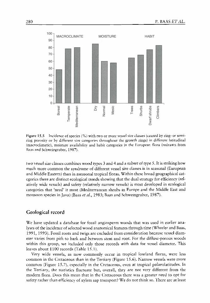
280 P. BAAS £TAL.
MOISTURE HABIT
Figure 15.5 Incidence of species (%) with two or more vessel size classes (caused by ring- or semiring porosity or by different size categories throughout the growth rings) in different latitudinal (macroclimatic), moisture availability and habit categories in the European flora (redrawn from Baas and Schweingruber, 1987).
two vessel size classes combines w^ood types 3 and 4 and a subset of type 5. It is striking how^ much more common the syndrome of different vessel size classes is in seasonal (European and Middle Eastern) than in aseasonal tropical floras. Within these broad geographical categories there are distinct ecological trends shov^ing that the dual strategy for efficiency (relatively w ide vessels) and safety (relatively narrow^ vessels) is most developed in ecological categories that 'need' it most (Mediterranean shrubs in Europe and the Middle East and monsoon species in Java) (Baas et al.^ 1983; Baas and Schv^eingruber, 1987).
Geological record
We have updated a database for fossil angiosperm vv oods that w as used in earlier analyses of the incidence of selected v^ood anatomical features through time (Wheeler and Baas, 1991, 1993). Fossil roots and twigs are excluded from consideration because vessel diameter varies from pith to bark and between stem and root. For the diffuse-porous woods within this group, we included only those records with data for vessel diameter. This leaves about 1100 records (Table 15.1).
Very wide vessels, as now commonly occur in tropical lowland floras, were less common in the Cretaceous than in the Tertiary (Figure 15.6). Narrow vessels were more common (Figure 15.7), especially in the Cretaceous, even at tropical palaeolatitudes. In the Tertiary, the statistics fluctuate but, overall, they are not very different from the modern flora. Does this mean that in the Cretaceous there was a greater need to opt for safety rather than efficiency of xylem sap transport? We do not think so. There are at least
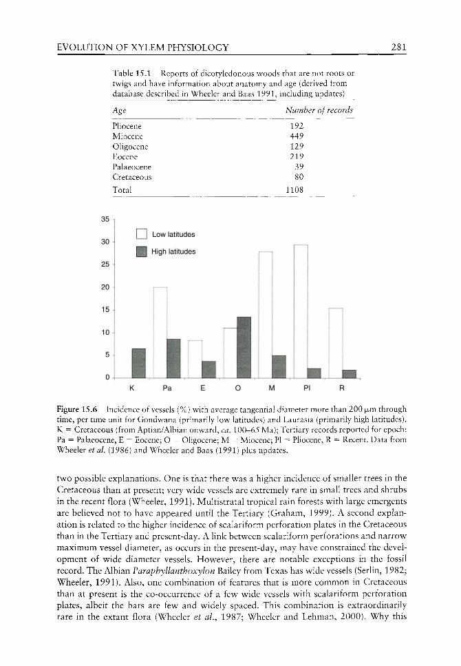
EVOLUTION OF XYLEM PHYSIOLOGY 281
Table 15.1 Reports of dicotyledonous woods that are not roots or twigs and have information about anatomy and age (derived from database described in Wheeler and Baas 1991, including updates)
Number of records
192 449 129 219 39 80
1108
Pliocene Miocene Oligocene Eocene Palaeocene Cretaceous
Total
35
30
25
20
15
104
5
0 ^ ^ ^
K
L J LOW latitudes
H High latitudes
Pa O M Pi
Figure 15.6 Incidence of vessels (%) with average tangential diameter more than 200 jxm through time, per time unit for Gondwana (primarily low latitudes) and Laurasia (primarily high latitudes). K = Cretaceous (from Aptian/Albian onward, ca. 100-65 Ma); Tertiary records reported for epoch: Pa = Palaeocene, E = Eocene; O = Oligocene; M = Miocene; PI = Pliocene, R = Recent. Data from Wheeler et aL (1986) and Wheeler and Baas (1991) plus updates.
two possible explanations. One is that there was a higher incidence of smaller trees in the Cretaceous than at present; very wide vessels are extremely rare in small trees and shrubs in the recent flora (Wheeler, 1991). Multistratal tropical rain forests with large emergents are believed not to have appeared until the Tertiary (Graham, 1999). A second explanation is related to the higher incidence of scalariform perforation plates in the Cretaceous than in the Tertiary and present-day. A link between scalariform perforations and narrow maximum vessel diameter, as occurs in the present-day, may have constrained the development of wide diameter vessels. However, there are notable exceptions in the fossil record. The Albian Faraphyllanthoxylon Bailey from Texas has wide vessels (Serlin, 1982; Wheeler, 1991). Also, one combination of features that is more common in Cretaceous than at present is the co-occurrence of a few wide vessels with scalariform perforation plates, albeit the bars are few and widely spaced. This combination is extraordinarily rare in the extant flora (Wheeler et aL, 1987; Wheeler and Lehman, 2000). Why this

282 P. BAAS £TAL.
30
Q Low latitudes
K Pa E O M PI R
Figure 15.7 Incidence of vessels (%) with average tangential diameter less than 50|Jim through time, per time unit for Gondv^ana (primarily lov latitudes) and Laurasia (primarily high latitudes). K = Cretaceous (from Aptian/Albian onward, ca. 100-65 Ma); Tertiary records reported for epoch: Pa = Palaeocene, E = Eocene; O = Oligocene; M = Miocene; PI = Pliocene, R = Recent. Data from Wheeler et al (1986) and Wheeler and Baas (1991) plus updates.
combination should occur in trees of the Cretaceous is unclear. The Cretaceous world was not similar to the present-day, e.g. CO2 levels were higher and large herbivores were more common. Whether and how these different conditions might affect leaf development and characteristics of Cretaceous plants and, consequently, their xylem differentiation and physiology is a fascinating, but poorly understood topic.
From the Palaeocene onwards the percentage of woods with very wide vessels is at, above, or slightly below modern values, with a clear predominance of widest vesselled species at tropical (palaeo) latitudes (Figure 15.6). Complementarity, a relatively high incidence of tree species with very narrow vessels is typical for high latitudes from the Eocene onwards (Figure 15.7). In the Cretaceous, the Northern (Laurasia) and Southern (Gondwana) floras do not differ in their incidence of narrow vessels. The Cretaceous records for diffuse-porous woods with wide vessels are from Laurasia, in contrast with the Tertiary and Recent. Flowever, during the Albian, Texas and Utah, where these wide-vesselled woods occur, were at lower latitudes than today.
The oldest known ring-porous wood is from the Late Cretaceous of Antarctica (Poole et al.y 2000). This report of Sassafras-like wood is remarkable and supports the suggestion that high latitude trees of the Cretaceous were deciduous as an adaptation to a seasonal climate, with a long, dark and relatively cool season. The first occurrence of ring-porous woods being in the southern hemisphere is also remarkable because ring-porous woods are now rare in temperate zones of the Southern Hemisphere (data from the OPCN database, Wheeler et aL^19S6).ln both the Cretaceous and Palaeogene, the incidence of ring-porosity was low and reached more or less modern levels in the Miocene of the northern hemisphere (Figure 15.8), coincident with development of temperate seasonal cHmates (Graham, 1999).
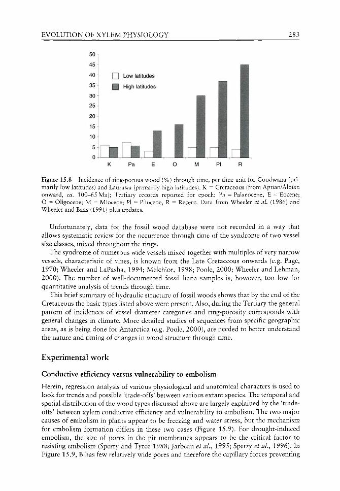
EVOLUTION OF XYLEM PHYSIOLOGY 283
50
45
40
35
30 ^
25 ^
20 1
15 :
io\
5 i
0 -
LJ Low latitudes
High latitudes
K Pa E O M PI R
Figure 15.8 Incidence of ring-porous wood (%) through time, per time unit for Gondwana (primarily low latitudes) and Laurasia (primarily high latitudes). K = Cretaceous (from Aptian/Albian onward, ca. 100-65 Ma); Tertiary records reported for epoch: Pa = Palaeocene, E = Eocene; O = Oligocene; M = Miocene; PI = Phocene, R = Recent. Data from Wheeler et al. (1986) and Wheeler and Baas (1991) plus updates.
Unfortunately, data for the fossil wood database were not recorded in a way that allows systematic review for the occurrence through time of the syndrome of two vessel size classes, mixed throughout the rings.
The syndrome of numerous wide vessels mixed together with multiples of very narrow vessels, characteristic of vines, is known from the Late Cretaceous onwards (e.g. Page, 1970; Wheeler and LaPasha, 1994; Melchior, 1998; Poole, 2000; Wheeler and Lehman, 2000). The number of well-documented fossil Hana samples is, however, too low for quantitative analysis of trends through time.
This brief summary of hydraulic structure of fossil woods shows that by the end of the Cretaceous the basic types listed above were present. Also, during the Tertiary the general pattern of incidences of vessel diameter categories and ring-porosity corresponds with general changes in climate. More detailed studies of sequences from specific geographic areas, as is being done for Antarctica (e.g. Poole, 2000), are needed to better understand the nature and timing of changes in wood structure through time.
Experimental work
Conductive efficiency versus vulnerability to embolism
Herein, regression analysis of various physiological and anatomical characters is used to look for trends and possible 'trade-offs' between various extant species. The temporal and spatial distribution of the wood types discussed above are largely explained by the 'tradeoffs' between xylem conductive efficiency and vulnerability to embolism. The two major causes of embolism in plants appear to be freezing and water stress, but the mechanism for emboHsm formation differs in these two cases (Figure 15.9). For drought-induced embolism, the size of pores in the pit membranes appears to be the critical factor to resisting embolism (Sperry and Tyree 1988; Jarbeau et al., 1995; Sperry et al., 1996). In Figure 15.9, B has few relatively wide pores and therefore the capillary forces preventing

284 P. BAAS ETAL.
Two causes of xylem embolism
Water stress Freeze-thaw
Tension
0I 1 '
air
A. Narrow pores
I ^ B. Wide
pores a
Vessels Pit pairs C. Narrow vessel D. Wide vessel
Figure 15.9 Water stress and freezing stress are the two major causes of embolism in plants. Vulnerability to water-stress induced embolism is proportional to the diameter of pores in the pit membrane; case B is more vulnerable than A. Vulnerability to freeze-induced embolism is proportional to the diameter of the vessel, and so case D is more vulnerable than C. Modified from Langan et al. (1997) and Sperry et al. (1996).
the entry of air from an adjacent air-filled conduit are not as great as in A, which has relatively narrow pores.
The positive correlation between conductive efficiency and vulnerability to freezing-induced embohsm is clear (Figure 15.10); wider conduits, which are the most efficient in transport, are also the most prone to embolism caused by freezing (Ewers, 1985; Cochard and Tyree, 1990; Sperry and Sullivan, 1992; Davis et al., 1999b). Evidently, vessel diameter determines the size of gas bubbles forced out of solution during a freezing event and wider vessels produced larger bubbles which are more resistant to dissolution at the time of thaw. Such bubbles are more likely to result in embolism. Why would plants evolve such risky vessels? There must be a selective advantage for wide vessels, as they occur early in the history of angiosperm wood. According to Poiseuille's law for ideal capillaries, conductive efficiency should be proportional to the sum of vessel diameters to the fourth power (Zimmermann, 1983). This means that doubling the number of vessels per cross-sectional area should double fe^, whereas doubling vessel diameter should increase k^ by 16-fold.
The amount of embolism with freeze-thaw depends not just on vessel diameter but also the water stress that the plants experience during the freeze-thaw event (Sperry and Sullivan, 1992; Sperry et al., 1994), the rate of thaw (Langan et al., 1997) and the position of the stem within the plant (Lemoine et al., 1999). Davis et al. (1999b) found that for isolated 8-12 mm diameter stem segments under a water stress of - 0 . 5 MPa, vessels wider than 44 jjum will always embolize upon freeze/thaw (Figure 15.10). There may be a similar vessel diameter threshold for 8-12 mm stems with regard to phenology of wood production and leaf maturation.
It is well established that temperate ring-porous trees, which have very wide earlywood vessels, have delayed bud break and delayed leaf maturation in the spring (Lechowicz,

EVOLUTION OF XYLEM PHYSIOLOGY 285
100
80
60
40
20
0
1
r2 = 0.75 P < 0.001
-
-
• /
Z I
• m
—1
• • >
• y^
•
„,. 1 —
y ^ m
i.._.
H
H
H
A
H
H
20 40 60 Mean tracheid or vessel diameter (|jnn)
80
Figure 15.10 Percent loss conductivity w ith freeze-thav^ as a function of mean tracheid diameter (in the case of two conifer species) or mean vessel diameter (in the case of 10 angiosperm species) in 8-12 mm diameter stems. Each point is an average for one species, the curve fit v as a second order polynomial. Modified from Davis et al (1999a).
1984; Wang et al.^ 1992). Why is vessel diameter related to leaf phenology in temperate woody plants? In north temperate areas, where freezing now occurs every year, the wide earlywood vessels of ring-porous trees remain conductive for only one growing season (Zimmermann, 1983; Ellmore and Ewers, 1986; Cochard and Tyree, 1990; Sperry et al.^ 1994; Hacke and Sauter, 1996; Jaquish and Ewers, 2001). The narrow latewood vessels often remain conductive for several years, but they contribute little to the total hydraulic conductance of the stem. For instance, in Ulmus americana L. the latewood vessels of wood more than a year old contributed only about 6% of the total axial conductance (Ellmore and Ewers, 1986). The total conductive efficiency of stems of ring-porous trees tends to be quite high, but there is a cost because their current year's wood does not mature until danger of freezing is past in the late spring and thus they have a shorter growing season. Apparently there has been natural selection for ring-porous trees to delay maturation of their leaves and their new xylem, thus allowing the mature leaves to have a reliable water supply (Zimmermann, 1983; Lechowicz, 1984; Wang et al, 1992). While ring-porous wood can be extremely efficient in conduction, the wide vessels tend to be prone to dysfunction during periods of freezing or severe drought. For species with wider vessels, bud break and leaf maturation is delayed in the spring and the leaves also tend to senesce earlier in the autumn, with the result that there is an inverse correlation between vessel diameter and the length of the growing season. In Figure 15.11, growing season length was defined as the number of weeks between bud break in the spring and leaf colour change or leaf abscission in autumn. Note that in Figures 15.10 and 15.11, narrow stems were used; there is research currently underway to examine the same relationships in the trunk xylem of trees, which have much wider vessels than those of the narrow stems.
At the other extreme from ring-porous trees and shrubs in North America, evergreen plants have the longest potential growing season and they have consistently narrow conduits. At the North American sites for the study shown in Figure 15.11, there were also seven evergreen species present (not shown in Figure 15.11), five conifers and two
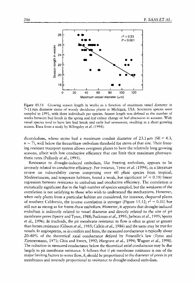
286 P. BAAS £TAL.
28
<f> 2 6
I 24
S 22 c o </)
eg 2 0
18
\m • r2 = 0.53 P< 0.001
JL. -L .
20 40 60 80 100 Maximum vessel diameter (jjum)
120
Figure 15.11 Growing season length in weeks as a function of maximum vessel diameter in 7-11 mm diameter stems of woody deciduous plants in Michigan, USA. Seventeen species were sampled in 1991, with three individuals per species. Season length was defined as the number of weeks between bud break in the spring and leaf colour change or leaf abscission in autumn. Wide vessel species tend to have late bud break and early leaf senescence, resulting in a short growing season. Data from a study by Billingsley et al, (1994).
dicotyledons, whose stems had a maximum conduit diameter of 23.1 luim (SE = 4.3, n = 7), well below the freeze/thaw embolism threshold for stems of that size. Their freezing resistant transport system allows evergreen plants to have the relatively long growing seasons, albeit with low conductive efficiency that can limit their maximum photosyn-thetic rates (Pallardy et al., 1995).
Resistance to drought-induced embolism, like freezing embolism, appears to be inversely related to conductive efficiency. For instance, Tyree et al. (1994), in a literature review on vulnerability curves comprising over 60 plant species from tropical, Mediterranean, and temperate habitats, found a weak, but significant (r^ = 0.18) linear regression between resistance to embolism and conductive efficiency. The correlation is statistically significant due to the high number of species sampled, but the weakness of the correlation is not satisfying to those who wish to understand the mechanisms. Flowever, when only plants from a particular habitat are considered, for instance, chaparral plants of southern California, the inverse correlation is stronger (Figure 15.12; r^ — 0.31) but still not as strong as for freeze-thaw embolism. Flowever, it appears that drought-induced embolism is indirectly related to vessel diameter and directly related to the size of pit membrane pores (Sperry and Tyree, 1988; Pockman et al., 1995; Jarbeau et al., 1995; Sperry et al., 1996). In tracheids, the pit membrane resistance to flow is often as great or greater than lumen resistance (Gibson et al., 1985; Calkin et al., 1986) and the same may be true for vessels. In angiosperms, as in conifers and ferns, the measured conductance is typically about 20-80% of the theoretical axial conductance defined by Poiseuille's law (Tyree and Zimmermann, 1971; Chiu and Ewers, 1992; Hargrave et al, 1994; Wagner et al, 1998). The reduction in measured conductance below the theoretical axial conductance may be due largely to pit membrane resistance. It follows that if pit membrane resistance is one of the major limiting factors to water flow, k^ should be proportional to the diameter of pores in pit membranes and inversely proportional to resistance to drought-induced embolism.

EVOLUTION OF XYLEM PHYSIOLOGY 287
C ^ 0 I
• Q OJ
CD ^
§ E
c ^ o c
" 5 >
L
k L
h
h
~T~
•
• •
1
I ~~"
•
t^
• i
- i —
•
-J
1 —
r^--
1 1
= 0.31 P <0.01
•
•
H
_i
^
• • J . 1. 1 2 4 6 8 10
Resistance to embolism (-MPa)
12
n """" ' '" Y r2 = 0.73
P < 0.001
\ • ^ 1 , .„i,...„
1 -
• •
- v
^
1
-J
\
40 60 80 100
Maximum vessel diameter (ixm)
120
Figure 15.12 Conductive efficiency ( ^ in 10~^ m^MPa"^ s~ ) as a function of resistance to embolism and maximum vessel diameter in species of chaparral shrubs in southern California. Each point is an average for one species. Resistance to embolism is equal to the w ater potential at 50% embolism, based upon dehydration vulnerability curves. Unpublished data from studies by Davis et al.
As with freezing embolism, the narrow vessels and tracheids in a stem are generally more resistant to drought-induced embolism than are wide vessels in the same stem (Salleo and Lo Gullo, 1986, 1989; Sperry and Tyree, 1988; Lo Gullo and Salleo, 1991; Hargrave et al., 1994). Thus there seems to be vahdity to Carlquist's hypothesis (1988) that narrow vessels and tracheids serve as high resistance auxiliary pathways for use when wide vessels become embolized. This would explain the common occurrence of narrow conduits closely associated with wide ones within the woods from arid regions.
The common occurrence of wood with strictly narrow vessels or tracheids in Arctic, alpine and dryland habitats is best explained in terms of resistance to freezing or drought-induced embolism. Diffuse-porous wood, a characteristic of the earliest known dicot woods, appears to be adapted to a wide range of habitats. Ring-porous wood appears to be more narrowly adapted to strongly seasonal environments where growing conditions are suitable for only a part of the year. This is consistent with the first appearance of ring-porous woods in the Late Cretaceous of Antarctica and the increased incidence of this wood type in the Late Tertiary of the Northern Hemisphere when seasonality becomes more pronounced (see Figure 15.8).
Conductive efficiency versus mechanical strength
Increasing vessel diameter or vessel frequency, both of which would increase conductive efficiency, should tend to make less dense, weaker wood, since vessels represent weak areas of the xylem. For modulus of rupture, it is the 'weakest link' that determines the point of failure and for wood the weakest mechanical link could be the vessels (Wagner et ai, 1998). An inverse relationship between mechanical strength and conductive efficiency has been found when comparisons are made between closely related taxa. For instance, when co-occurring species pairs of Ceanothus L. and Adenostoma Hook, and Arn. were examined, C. spinosus Nutt. and A. sparsifolium Torr. had greater k^ values but lower stem mechanical strength than the co-occurring C. megacarpus Nutt. and A. fasciculatum Hook, and Arn. (Wagner et al., 1998). Similarly, within Toxicodendron diversilobum (Torr. and Gray) Greene, a species that produces heteromorphic stems, viney (supported)
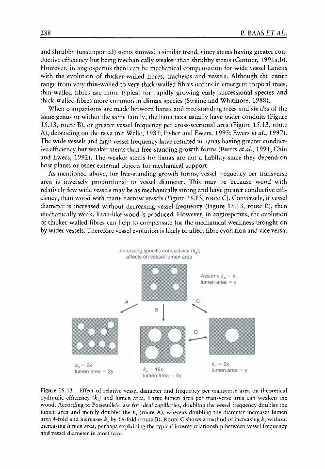
288 P. BAAS ETAL.
and shrubby (unsupported) stems showed a similar trend, viney stems having greater conductive efficiency but being mechanically w^eaker than shrubby stems (Gartner, 1991a,b). However, in angiosperms there can be mechanical compensation for wide vessel lumens with the evolution of thicker-walled fibres, tracheids and vessels. Although the entire range from very thin-walled to very thick-walled fibres occurs in emergent tropical trees, thin-walled fibres are more typical for rapidly growing early successional species and thick-walled fibres more common in climax species (Swaine and Whitmore, 1988).
When comparisons are made between lianas and free-standing trees and shrubs of the same genus or within the same family, the liana taxa usually have wider conduits (Figure 15.13, route B), or greater vessel frequency per cross-sectional area (Figure 15.13, route A), depending on the taxa (ter Welle, 1985; Fisher and Ewers, 1995; Ewers et aL, 1997). The wide vessels and high vessel frequency have resulted in lianas having greater conductive efficiency but weaker stems than free-standing growth forms (Ewers et aL, 1991; Chiu and Ewers, 1992). The weaker stems for lianas are not a liability since they depend on host plants or other external objects for mechanical support.
As mentioned above, for free-standing growth forms, vessel frequency per transverse area is inversely proportional to vessel diameter. This may be because wood with relatively few wide vessels may be as mechanically strong and have greater conductive efficiency, than wood with many narrow vessels (Figure 15.13, route C). Conversely, if vessel diameter is increased without decreasing vessel frequency (Figure 15.13, route B), then mechanically weak, liana-like wood is produced. However, in angiosperms, the evolution of thicker-walled fibres can help to compensate for the mechanical weakness brought on by wider vessels. Therefore vessel evolution is likely to affect fibre evolution and vice versa.
Increasing specific conductivity (/Cg); effects on vessel lumen area
Assume /c = x lumen area = y
/C3-2X lumen area 2y ks= 16x
lumen area = 4y lumen area
Figure 15.13 Effect of relative vessel diameter and frequency per transverse area on theoretical hydraulic efficiency (ks) and lumen area. Large lumen area per transverse area can weaken the wood. According to Poiseuille's law for ideal capillaries, doubling the vessel frequency doubles the lumen area and merely doubles the ks (route A), whereas doubling the diameter increases lumen area 4-fold and increases ks by 16-fold (route B). Route C shows a method of increasing k^ without increasing lumen area, perhaps explaining the typical inverse relationship between vessel frequency and vessel diameter in most taxa.

EVOLUTION OF XYLEM PHYSIOLOGY 289
Cohesion-tension theory and sap ascent
The cohesion-tension theory has been the leading explanation for xylem transport in plants for more than a century and, despite recent challenges (Balling and Zimmermann, 1990; Canny, 1995, 1997) support for the theory is quite robust (Tyree, 1997; Steudle, 2001). The theory is critical to our understanding of xylem evolution, since it means that the water is normally under considerable negative pressure (tension) during transport. The vessels thus must be constructed to avoid the entry of air, w^hich can cause cavitation and embohsm (air blockage) of the w^ater columns. They also have to v^ithstand the negative pressure (tension) of the water column to avoid implosion. The cohesion-tension theory also implies that parenchyma cells, which are present in most woods, do not have a direct role in transport but function in storage of photosynthates, water and minerals and for resisting the invasion of pathogens.
Mechanical strength, implosion resistance and resistance to embolism
In a recent study including 36 species of angiosperms and 12 species of conifers from North America, it was found that there was a positive correlation between wood density and resistance to drought-induced embolism (Hacke et aL, 2001). The authors offered the explanation that resistance to drought-induced embolism required, among other things, that vessels or tracheids must be able to resist implosion due to the water tension (negative pressure) that the conduits experience. They argued that the critical factor was (t/b)^, that is, the square of the thickness of the vessel or tracheid wall (t) divided by the diameter of the conduit (fc), as used in Figures 15.1 and 15.14. Therefore, to resist collapse of the conduits, plants that experience very low water potentials must have thick walls and narrow lumens, with high wood density as a corollary. However, it may not be avoidance of vessel implosion itself that causes the observed correlation, but characteristics of the pit membranes. Stretching of pit membranes under extreme negative pressures may enlarge the pit membrane pores so that they are more prone to air seeding. Is deflection of pit membranes in high density woods less likely to enlarge pit membrane pores?
Another possible explanation is that selection for resistance to embolism would tend to result in narrower conduits, as argued above. Wood with narrower conduits will tend to have greater branch strength. Among chaparral shrubs we have found that resistance to
o c B CO CD
tr 0.6 0.65 0.7 0.75 0.8 0.85
Xylem density (g cm-3)
0.12 0.15 0.18 0.21 0.24 0.01 0.02 0.03 0.04 0.05 0.06
Stem strength vessel strength (t/b)^ (MORinGNm-2)
Figure 15.14 Resistance to embolism as functions of xylem dry density, stem strength (modulus of rupture, MOR in GNm"^), and vessel strength {t/b)^ in chaparral shrubs. Each point represents the mean for one species. Based upon data from Wagner et aL (1998) and Hacke et al. (2001).

290 P. BAAS £TAL.
embolism correlates well with wood density, stem strength and vessel strength (Figure 15.14). It is well known that wood density is correlated with wood strength, but in some cases high wood density may have been selected for as a mechanism to resist vessel embolism or as a mechanism to resist vessel implosion. It remains to be determined which of these factors are most critical in wood evolution.
Hacke et al. (2001) note that fibre evolution would impact implosion resistance of vessels and they suggest that, as a result, (t/b)'^ values are fundamentally different for angiosperms versus gymnosperms. In angiosperms, fibres offer some of the support against implosion, whereas in conifers only tracheid walls are involved with implosion resistance.
Roots versus stems
The aforementioned studies have all involved the xylem of stems. In considering the 'tradeoff triangle, it should be noted that, compared to stems, roots would normally not experience such low water potentials, nor would the mechanical support demands be as great as for free-standing stems; the soil surrounding roots provides some of the mechanical support for plants. In addition, roots tend to play a greater role in carbohydrate storage than do stems. Given the above considerations, it is not surprising that woody roots in many plants have wider vessels than woody stems, more parenchyma and less fibre (Fegel, 1941; Zimmermann and Potter, 1982; Gasson, 1985; Gartner, 1995; Pate etaL, 1995). Based upon vulnerability curves, roots tend to be more vulnerable to embolism than are stems, as might be predicted by their wider vessel diameters (Sperry and Saliendra, 1994; Alder et al., 1996). However, some species' roots have fewer, narrower vessels than do their stems and so their roots may prove to have similar or lower vulnerability to embolism than the stems (see illustrations in Cutler et al., 1987). Another exception may be for lianas, where the mechanical demands on the stems are much reduced. In lianas, unlike free-standing grov^h forms, vessel diameters of stems tend to be similar to those found in roots (Ewers et al., 1997).
Parenchyma
In this chapter little attention was paid to the role of parenchyma (ray and axial). The abundance and distribution of parenchyma varies considerably within the angiosperms. Some schemes suggesting functional significance in variation of parenchyma distribution and abundance have been devised (e.g. Braun, 1970).
Woods with abundant (diffuse) parenchyma and high ray volumes are common in some Cretaceous floras (Wheeler et al., 1987; Wheeler and Lehman, 2000): were these woods indicative of xeric conditions and the need for water storage? In modern floras such types are exemplified by certain Bombacaceae (Adansonia L., abundant axial parenchyma) and Cucurbitales (very broad rays). Alternative strategies for water storage are found in woods that lack axial parenchyma but have septate fibres which take over the water storage and other living functions of parenchyma, e.g. in many Sapindales (Burseraceae, Sapindaceae, Meliaceae). This wood type is among the earliest known and is abundant throughout the Cretaceous (Wheeler and Baas, 1991), but today tends to be most common in mesic tropical rain forests (Baas, 1982).
Towards a synthesis: the evolution of hydrauhc structure and function
What sense do the global ecological trends in xylem anatomy make in terms of the experimentally demonstrated 'trade-offs' between vessel diameter, vessel density and dual vessel

EVOLUTION OF XYLEM PHYSIOLOGY 291
diameter strategies and hydraulic efficiency (conductivity) and safety (control and repair of
tension and freezing embolisms)?
In frost-prone areas it has been abundantly shown that the freezing/thawing embolisms
must somehow be controlled and prevented from spreadmg. That possibility exists in
short- and narrow-vesselled woods (often provided with scalariform perforations), which
is the most common type in these regions. Woods with a dual safety-efficiency strategy
(ring-porosity; two vessel size classes in diffuse-porous condition) are extremely common
in ecosystems characterized by seasonality where temporary high demands for conductive
efficiency alternate with demands for safety and localization and/or repair of drought-
stress embolism. Finally, wide-vesselled diffuse-porous woods, opting for high conductive
efficiency, without apparent safety provisions characterize the rapidly dwindling tropical
rainforest flora. This should perhaps surprise us rather than confirm our intuitive ideas.
After all tall rainforest emergents have their transpiring crowns above the canopy, with
daily sunny periods when the humidity on the forest floor stays high, but becomes quite
low above the canopy. According to the cohesion theory these tall trees have to develop
considerable xylem sap tensions, although the tensions are less than those that shorter,
arid habitat plants experience during droughts. The experimental work summarized in
this chapter provides a basis for understanding the variations in the incidences of vessel
diameters as related to freezing and drought. However, additional experimental work is
needed to elucidate the physiological significance of variations in vessel wall thickness,
vessel perforation type, wood density and parenchyma distribution.
Acknowledgements
Some of the work reported in this chapter was supported by grants from the National
Science Foundation NSF IBN-0130870 and 0131247
References
Alder NN, Sperry JS, Pockman WT. 1996. Root and stem xylem cavitation, stomatal conductance, and leaf turgor in Acer grandidentatvim across a soil moisture gradient. Oecologia 105: 293-301.
Baas P. 1973. The wood anatomical range in Ilex (Aquifoliaceae) and its ecological and pliylogenetic significance. Blumea 21: 193-258.
Baas P. (ed.) 1982. Systematic, phyk)genetic and ecological wood aaittomy - history and perspectives. In: New Perspectives in Wood Anatomy. The Hague: Nijhoff Junk, 23-58.
Baas P. 1986. Ecological patterns of xylem anatomy. In: Givnish ] , ed. On the Economy of Plant Form and Function. New York: Cambridge University Press, 327-352.
Baas P, Schw^eingruber FH. 1987. Ecological trends m the wood anatomy of trees, shrubs and climbers from Europe. International Association of Wood Anatomists Bulletin, New Series 8: 245-274.
Baas P, Wheeler EA. 1996. Parallelism and reversibility ni xylem evolution - a review. International Association of Wood Anatomists Journal 17: 351-36-1.
Baas P, Wheeler EA. 2000. Wood structure of southeasiasian timbers - the PROSEA woods reviewed. In: Kim, YS, ed. New Horizons in Wood Anatomy. Korea: Chonnam National University Press, 1-9.
Baas P, Werker E, Fahn A. 1983. Some ecological trends m vessel characters. International Association of Wood Anatomists Bulletin, New Series 4: 141-159.

292 P. BAAS E T A L .
Baas P, Esser PM, van der Westen MET, Zandee M. 1988. Wood anatomy of the Oleaceae. International Association of Wood Anatomists Bulletin, New Series 9: 103-182.
Bailey IW, Tupper WW. 1918. Size variation in tracheary cells. I. A comparison betv^een the secondary xylems of vascular cryptogams, gymnosperms and angiosperms. Proceedings American Academy of Arts and Sciences 54: 149-204.
Balling A, Zimmermann U. 1990. Comparative measurements of the xylem pressure of Nicotiana plants by means of the pressure bomb and pressure probe. Planta 182: 325-338.
Billingsley GF, Kopper BJ, Ev^ers FW. 1994. Vessel diameter and season length in temperate v^oody plants. American Journal of Botany 81 (sup. 6): 19 (abstract).
Braun, HJ. 1970. Funktionelle Histologic der sekunddren Sprossachse.I. Das Holz. Encyclopedia of Plant Anatomy IX (1). Berlin: Borntraeger.
Gaboon EJ. 1972. Paraphyllanthoxylon alabamense - 3. new species of fossil dicotyledonous wood. American Journal of Botany 59: 5-11.
Galkin HW, Gibson AG, Nobel PS. 1986. Biophysical model of xylem conductance in tracheids of the fern, Pteris vitata. Journal of Experimental Botany 37: 1054-1064.
Ganny MJ. 1995. A new theory of the ascent of sap - cohesion supported by tissue pressure. Annals of Botany 75: 343-357.
Ganny MJ. 1997. Vessel contents during transpiration - embolisms and refilling. American Journal of Botany ^4: 1223-1230.
Garlquist S. 1988. Comparative Wood Anatomy. Systematic, Ecological, and Evolutionary Aspects of Dicotyledon Wood. New York, Berlin: Springer-Verlag.
Garlquist S. 1991. Anatomy of vine and Hana stems: a review and synthesis. In: Putz FE, Mooney HA, eds. The Biology of Vines. Cambridge: Cambridge University Press, 53-71.
Garlquist S, Hoekman DA. 1985. Ecological wood anatomy of the woody southern California flora. International Association of Wood Anatomists Bulletin, New Series 6: 319-347.
Gheadle VI. 1944. Specialization of vessels within the xylem of each organ in the Monocotyledonae. American Journal of Botany 31: 81-92.
Chiu ST, Ewers FW. 1992. Xylem structure and water transport in a twiner, a scrambler, and a shrub of Lonicera (Caprifoliaceae). Trees, Structure and Function 6: 216-224.
Cochard H, Tyree MT. 1990. Xylem dysfunction in Quercus: vessel size, tyloses, cavitation and seasonal changes in embolism. Tree Physiology 6: 393-407.
Cutler DF, Rudall PJ, Gasson PE, Gale RMO. 1987. Root Identification Manual of Trees and Shrubs. A Guide to the Anatomy of Roots of Trees and Shrubs Hardy in Britain and Northern Europe. London: Chapman and Hall.
Davis SD, Ewers FW, Wood J, et al. 1999a. Differential susceptibility to xylem cavitation among three pairs of Ceanothus species in the Transverse Mountain Ranges of Southern California. Ecoscience 6: 180-186.
Davis SD, Sperry JS, Hacke UG. 1999b. The relationship between xylem conduit diameter and cavitation caused by freezing. American Journal of Botany 86: 1367-1372.
Doyle JA, Endress PK. 2000. Morphological phylogenetic analysis of basal angiosperms: Comparison and combination with molecular data. International Journal of Plant Sciences
161: 121-153. Ellerby DJ, Ennos AR. 1998. Resistances to fluid flow of model xylem vessels with simple and
scalariform perforation plates. Journal of Experimental Botany 49: 987-985. EUmore GS, Ewers FW. 1986. Fluid flow in the outermost xylem increment of a ring-porous tree,
Ulmus americana. American Journal of Botany 73: 1771-1774. Ewers FW. 1985. Xylem structure and water conduction in conifer trees, dicot trees and hanas.
International Association of Wood Anatomists Bulletin, New Series 6: 309-317. Ewers FW, Carleton MR, Fisher JB, et al. 1997. Vessel diameters in roots versus stems of tropical
lianas and other growth forms. International Association of Wood Anatomists Journal 18: 261-279.
Ewers FW, Fisher JB, Fichtner K. 1991. Water flux and xylem structure in vines. In: Putz F, Mooney H, eds. The Biology of Vines. Cambridge: Cambridge University Press, 119-152.
Fahn A, Werker E, Baas P. 1986. Wood Anatomy and Identification of Trees and Shrubs from Israel and Adjacent Areas. Jerusalem: Israel Academy of Science and Humanities.
EVOLUTION OF XYLEM PHYSIOLOGY 293
Fegel AC. 1941. Comparative anatomy and varying physical properties of trunk, branch and root v^ood in certain northeastern trees. Bulletin New York State College of Forestry, Syracuse, Vol 14, No 2b, Technical Publication no. 55: 1-20.
Fisher JB, Ev^ers FW. 1995. Vessel dimensions in hana and tree species of Gnetum (Gnetales). American journal of Botany 82: 1350-1357.
Gartner BL. 1991a. Structural stability and architecture of vines vs. shrubs of poison oak, Toxicodendron diversilobum. Ecology 71: 2005-2015.
Gartner BL. 1991b. Stem hydraulic properties of vines vs. shrubs of poison oak, Toxicodendron diversilobum. Oecologia 87: 180-189.
Gartner BL. 1995. Patterns of xylem variation v^ithin a tree and their hydraulic and mechanical consequences. In: Gartner BL, ed. Plant Stems: Physiology and Functional Morphology. London: Academic Press, 175-196.
Gasson PE. 1985. Automatic measurement of vessel lumen area and diameter with particular reference to pedunculate oak and common beech. International Association of Wood Anatomists Bulletin, New Series 6: 219-237.
Gasson PE, Dobbins DR. 1991. Wood anatomy of the Bignoniaceae, with a comparison of trees and lianas. International Association of Wood Anatomists Bulletin, New Series 12: 389-417.
Gibson AC, Calkin HW, Nobel PS. 1985. Hydraulic conductance and xylem structure in tracheid-bearing plants. International Association of Wood Anatomists Bulletin, New Series 6: 293-302.
Gilbert SG. 1940. Evolutionary significance of ring porosity in woody angiosperms. Botanical Gazette 102: 105-120.
Graham A. 1999. Late Cretaceous and Cenozoic History of North American Vegetation. New York: Oxford University Press.
Hacke UG, Sauter JJ. 1996. Xylem dysfunction during winter and recovery of hydraulic conductivity in diffuse-porous and ring-porous trees. Oecologia 105: 435-439.
Hacke UG, Sperry JS, Pockman WT, et al. 2001. Trends in wood density and structure are linked to prevention of xylem implosion by negative pressure. Oecologia 126: 457-461.
Hargrave KR, Kolb KJ, Ewers FW, Davis SD. 1994. Conduit diameter and drought-induced embolism in Salvia mellifera Greene (Labiatae). New Phytologist 126: 695-705.
Herendeen PS. 1991a. Lauraceous wood from the mid-Cretaceous Potomac Group of eastern North America: Paraphyllanthoxylon marylandense sp. nov. Review of Paleobotany and Palynology 69: 277-290.
Herendeen PS. 1991b. Charcoalified angiosperm wood from the Cretaceous of eastern North America and Europe. Review of Palaeobotany and Palynology 70: 225-239.
Herendeen PS, Wheeler FA, Baas P. 1999. Angiosperm wood evolution and the potential contribution of paleontological data. Botanical Review 65: 278-300.
Jaquish LL, Ewers FW. 2001. Seasonal conductivity and embolism in the roots and stems of two clonal ring-porous trees. Sassafras albidum (Lauraceae) and Rhus typhina (Anacardiaceae). American Journal of Botany 88: 206-212.
Jarbeau JA, Ewers FW, Davis SD. 1995. The mechanism of water-stress-induced embolism in two species of chaparral shrubs. Plant, Cell and Environment 18: 189-196.
Klaassen R. 1999. Wood anatomy of the Sapindaceae. International Association of Wood Anatomists Journal, Supplement 2, 1-214.
Langan SJ, Ewers FW, Davis SD. 1997. Xylem dysfunction caused by water stress and freezing in two species of co-occurring chaparral shrubs. Plant, Cell and Environment 20: 425-437.
Lechowicz MJ. 1984. Why do temperate deciduous trees leaf out when they do? Adaptation and the ecology of forest communities. American Naturalist 124: 821-842,
Lemoine D, Granier A, Cochard H. 1999. Mechanism of freeze-induced embohsm in Fagus sylvatica L. Trees: Structure and Function 13: 206-210.
Lo Gullo MA, Salleo S. 1991. Three different methods of measuring xylem cavitation and embolism: a comparison. Annals of Botany 72: 417-424.
Melchior RC. 1998. Paleobotany of the Williamsburg Formation (Paleoocene) at the Santee Rediversion site Berkeley County, South Carolina. In: Sanders AE, ed. Paleobiology of the WilUamsburg Formation (Black Mingo Group; Paleogene) of South Carolina, USA. Transactions of the American Philosophical Society 4: 49-121.
294 P. BAAS £ T A L .
Meyer-Berthaud B, Scheckler SE, Wendt J. 1999. Archaeopteris is the earliest known modern tree. Nature 39^: 7m-70\.
Noshiro S, Baas P. 1998. Systematic anatomy of Cornaceae and allies. International Association of Wood Anatomists Journal 19: 43-97.
Noshiro S, Baas P. 2000. Latitudinal trends in wood anatomy within species and genera: case study in Cornus SL. American Journal of Botany 87: 1495-1506.
van den Oever L, Baas P, Zandee M. 1981. Comparative wood anatomy of Symplocos and latitude of provenance. International Association of Wood Anatomists Bulletin, New Series 23: 3-24.
Page VM. 1970. Angiosperm wood from the Upper Cretaceous of central California. III. American Journalof Botany 57: 1139-1144.
Pallardy SG, Cermak J, Ewers FW, et al. 1995. Water transport dynamics in trees and stands. In: Smith WK, Hinckley TM, eds. Resource Physiology of Conifers: Acquisition, Allocation and Utilization. New York: Academic Press, 299-387.
Pate JS, Jeschke WD, Aylward MJ. 1995. Hydraulic architecture and xylem structure of the dimorphic root systems of south-west Australian species of Proteaceae. Journal of Experimental Botany ^6: 907-915.
Pockman WT, Sperry JS, O'Leary JW. 1995. Sustained and significant negative water pressure in xylem. Nature 378: 715-716.
Poole I. 2000. Fossil angiosperm wood: its role in the reconstruction of biodiversity and palaeoen-vironment. Botanical Journal of the Linnean Society 134: 361-381.
Poole I, Francis JE. 2000. The first record of fossil wood of the Winteraceae from the Upper Cretaceous of Antarctica. Annals of Botany 85: 307-315.
Poole I, Richter HG, Francis JE. 2000. Evidence for Gondwanan origins for Sassafras (Lauraceae)? Late Cretaceous fossil wood of Antarctica. International Association of Wood Anatomists Journal 21: 463-475.
Salleo S, Lo Gullo MA. 1986. Xylem cavitation in nodes and internodes of whole Chorisia insignis H.B. et K. plants subjected to water stress: relations between xylem conduit size and cavitation. Annals of Botany 58: 431-441.
Salleo S, Lo Gullo MA. 1989. Differential aspects of cavitation resistance in Ceratonia silique, a drought-avoiding Mediterranean tree. Annals of Botany 64: 325-336.
Schulte PJ. 1999. Water flow through a 20-pore perforation plate in vessels of Liquidambar styraci-flua. Journal of Experimental Botany 50: 1179-1187.
Schulte PJ, Castle AL. 1993. Water flow through vessel perforation plates - a fluid mechanical approach. Journal of Experimental Botany 44: 1135-1142.
Serlin BS. 1982. An early Cretaceous fossil flora from northwest Texas: its composition and implications. Palaeontographica 182B: 52-86.
Soltis DE, Soltis PS, Chase MW, et al. 2000. Angiosperm phylogeny inferred from 18S rDNA, rbcL, and atpB sequences. Botanical Journal of the Linnean Society 133: 381-446.
Sperry JS, Sullivan JEM. 1992. Xylem embolism in response to freeze-thaw cycles and water stress in ring-porous, diffuse-porous, and coniferous species. Plant Physiology 100: 603-613.
Sperry JS, Tyree MT. 1988. Mechanism of water stress-induced xylem dysfunction. Physiologia Plantarum 74: 276-283.
Sperry JS, Tyree MT. 1990. Water-stress-induced xylem embolism in three species of conifers. Planty Cell and Environment 13: 427-436.
Sperry JS, Nichols KL, Sullivan JEM, Eastlack SE. 1994. Xylem embolism in ring-porous, diffuse-porous, and coniferous trees of northern Utah and interior Alaska. Ecology 75: 1736-1752.
Sperry JS, Saliendra NZ. 1994. Intra- and inter-plant variation in xylem cavitation in Betula occidentalis. Plant, Cell and Environment 17: 1233-1241.
Sperry JS, Saliendra NZ, Pockman WT, et al. 1996. New evidence for large negative xylem pressures and their measurement by the pressure chamber method. Plant, Cell and Environment 19: 427-436.
Steudle E. 2001. The cohesion-tension mechanism and the acquisition of water by plant roots. Annual Review of Plant Physiology and Plant Molecular Biology 52: 847-875.
Swaine MD, Whitmore TC. 1988. On the definition of ecological species groups in tropical forests. Vegetatio 75: 81-86.

EVOLUTION OF XYLEM PHYSIOLOGY 295
Thayn GF, Tidwell WD, Stokes WL. 1983. Flora of the Lower Cretaceous Cedar Mountain Formation of Utah and Colorado. Part I. Paraphyllanthoxylon utahense. Great Basin Naturalist 43: 394-402.
Thorsch JA. 2000. Vessels in Zingiberaceae: a light, scanning, and transmission microscope study. International Association of Wood Anatomists journal 21: 61-76.
Tyree MT. 1997. The cohesion-tension theory of sap ascent: current controversies. Journal of Experimental Botany 48: 1753-1765.
Tyree MT, Davis SD, Cochard H. 1994. Biophysical perspectives of xylem evolution: is there a tradeoff of hydraulic efficiency for vulnerability to dysfunction? International Association of Wood Anatomists Journal 15: 335-360.
Tyree MT, Zimmermann MH. 1971. The theory and practice of measuring transport coefficients and sap flow in the xylem of red maple stems (Acer rubrum). Journal of Experimental Botany 22: 1-18.
Wagner KR, Ewers FW, Davis SD. 1998. Tradeoffs between hydraulic efficiency and mechanical strength in the stems of four co-occurring species of chaparral shrubs. Oecologia 117: 53-62.
Wang J, Ives NE, Lechowicz MJ. 1992. The relation of foliar phenology to xylem embolism in trees. Functional Ecology 6: 469-475.
ter Welle BJH. 1985. Differences in wood anatomy of hanas and trees. International Association of Wood Anatomists Bulletin, New Series 6: 70.
Wheeler, EA. 1991. Paleocene dicotyledonous trees from Big Bend National Park, Texas. Variability in wood types common in the Late Cretaceous and Early Tertiary, and ecological inferences. American Journal of Botany 78: 658-671.
Wheeler EA, Baas P. 1991. A survey of the fossil record for dicotyledonous wood and its significance for evolutionary and ecological wood anatomy. International Association of Wood Anatomists Bulletin, New Series 12: 271-332.
Wheeler EA, Baas P. 1993. The potentials and limitations of dicotyledonous wood anatomy for climatic reconstructions. Paleobiology 19: 486-497.
Wheeler EA, LaPasha CA. 1994. Woods of the Vitaceae. Fossil and modern. Review of Paleobotany and Palynology 80: 175-207.
Wheeler EA, Lehman TM. 2000. Late Cretaceous woody dicots from the Aguja and Javelina Formations, Big Bend National Park, Texas, USA. International Association of Wood Anatomists Journal 21: 83-120.
Wheeler EA, Lee M, Matten LC. 1987. Dicotyledonous woods from the Upper Cretaceous of southern Illinois. Botanical Journal of the Linnean Society 95: 77-100.
Wheeler EA, Pearson RG, LaPasha CA, et al. 1986. Computer-aided Wood Identification. Raleigh, NC: Station Bulletin No. 474, North Carolina Agriculture Research Service.
Woodcock DW. 1994. Occurrence of woods with a gradation in vessel diameter across a ring. International Association of Wood Anatomists Journal 15: 377-385.
Woodcock DW, Ignas CM. 1994. Prevalence of wood characters in eastern North America: What characters are most promising for interpreting climates from fossil wood? American Journal of Botany n-. 1243-1251.
Zhong Y, Baas P, Wheeler EA. 1992. Wood anatomy of trees and shrubs from China. IV. Ulmaceae. International Association of Wood Anatomists Bulletin, New Series 13: 419-453.
Zimmermann MH. 1983. Xylem Structure and the Ascent of Sap. New York: Springer. Zimmermann MH, Jeje AA. 1981. Vessel-length distribution in stems of some American woody
plants. Canadian Journal of Botany 59: 1882-1892. Zimmermann MH, Potter D. 1982. Vessel-length distribution in branches stems and roots oi Acer
rubrum L. International Association of Wood Anatomists Bulletin 3: 103-109.

16 Hydraulics and mechanics of plants: novelty, innovation and evolution
Nick Rowe and Thomas Speck
CONTENTS.
Introduction 297
Terminology and evolution 298
Turgor 300
The hypodermal sterome 301
Secondary growth 304 Does the appearance of the bifacial vascular cambium
represent a key innovation? 309
Lignification and biomechanics of the plant cell wall 310
Reaction wood 312
Hydrauhcs, mechanics and evolution of the climbing habit 314
Types of climbing strategy 314 Appearance of the lianoid habit 314 Vessels and the climbing habit 315 What is special about lianas? 318
Conclusions 318
Acknowledgements 321
References 321
Introduction
Physiological traits are exceptionally difficult to ascertain in fossils of long extinct plant
groups. In this chapter we discuss the significance of hydraulic and mechanical novelties,
both of vv^hich are physiologically interrelated and show high levels of interdependence
* Dedicated to Hanns-Christof Spatz on the occasion of his 65th birthday.
The Evolution of Plant Physiology Copyright © 2004 Elsevier Ltd ISBN 0-12-33955-26 All rights of reproduction in any form reserved

298 N. ROWE &c T. SPECK
and constraint. Mechanical tissue for generating stiffness is most effective if comprised of thick-walled fibre elements, but these are physiologically expensive; water-conducting elements are most effective if composed of wide diameter elements but such tissues are less dense and thus contribute less effectively to mechanical stiffness. These two functions could, therefore be viewed to be in conflict. Another example of conflicting functional roles is seen in stem systems without specialized mechanical tissues, where mechanical structure is upheld by the hydraulic system and maintenance of turgor. Turgor requires living parenchyma cells and when these are fully hydrated the stem/leaf system is mechanically sound and does not wilt. If water supply becomes limited the stem/leaf system will wilt if there are no other mechanical strengthening elements within the stem. Advanced wilting and water stress can have a range of deleterious effects including embolism and mechanical failure. There is a conflict in this system because turgescent water-filled cells are heavy when fully hydrated and thus mechanically load the plant. These are some simple examples of potential compromises between mechanics and hydraulics that have probably played important roles during radiations of terrestrial plants. These kinds of processes are potentially relevant to a wide range of plant organs as well as entire plants of different complexity. The 'trade-offs' associated with turgor, such as weight load, were probably important for many early land plants and are still so for almost any organ or young developmental stage of any plant. The compromise between hydraulic and mechanical functions in terms of water-conducting elements and fibre tissues has many examples among more complex architectures such as the appearance of hypodermal steromes and lateral meristems. Hydraulic and mechanical constraints can be seen as coupled functions and, to a large extent, physiological compromises which have to be met for adaptive modification of the plant body. Many authors have commented on this, but in our view co-organization of these constraints is especially crucial for morphological radiation of growth forms. By growth form we mean the size, shape and, importantly, the mechanical posture or orientation of the plant. Self-supporting plants can show quite different mechanical and hydraulic specializations from non-self-supporting plants. Trees, shrubs, herbs, climbers, epiphytes and hemi-epiphytes show an incredible variety of hydraulically and mechanically coupled innovations. Given the recent advances in plant biomechanics and hydraulics as well as more decisive and testable methods for estabUshing phylogenetic and historical contexts, the combination of functional biology and phylogenetic studies offers the opportunity for more accurately understanding how diverse plant forms and their underlying functioning have evolved.
Terminology and evolution
An accepted terminology for unambiguously referring to functional aspects of an organism and their evolutionary significance is a rich area of controversy. The main point of discussion is historically and in our opinion misleadingly centred around adaptation. In the latter half of the 20th century, such discussions focused particularly on whether it is possible to identify traits which are of adaptive significance (Gould and Lewontin, 1979; Gould and Vrba, 1982; Rose and Lauder, 1996). This has more recently led to an extension of this theme questioning the adaptive significance of morphological radiations in plants as well as the gradual or punctual processes characterizing them (Bateman and DiMichele, 1994; Bateman, 1999a,b). While these arguments are of great interest in resolving high level evolutionary processes we argue that significant avenues of doubt still remains over

HYDRAULICS AND MECHANICS OF PLANTS 299
the actual functioning of many observed traits, let alone whether they are adaptive or not. This is particularly relevant to systems of long extinct organisms v^hich might show little in the way of modern potential analogues (Novacek, 1996).
Another term directly relevant to hydraulic and mechanical function is that of 'evolutionary innovation' or 'key innovation', terms that are generally deployed to describe a trait or syndrome which launched a significant morphological radiation. Perceived interpretation of a key innovation without a (1) functional, (2) phylogenetic and historical framework is not proof. Recent observers point to more combined and testable approaches which usually include (1) a functional argument to assess the 'performance' of the structure and (2) a phylogenetic framework to observe and test patterns of traits within the context of a chosen evolutionary hypothesis (Jablonski and Bottjer, 1990; Sanderson and Donoghue 1996; Bateman, 1999b). Both analyses of function and evolutionary pattern are complicated by the fact that many plants are complex structures and that a single trait or structure might have several functions and confer several benefits to the life history of the plant which might interplay in any number of ecological, evolutionary or adaptive scenarios. This aspect of functional biology is one of several bugbears at the heart of controversies concerning function and evolution where functional studies attempting to assess the significance of one of a range of functions stand accused of impUcitly 'atomizing' an organism's biology (Gould and Lewontin, 1979).
Inferred physiological constraints and innovations have formed the basis of many evolutionary discussions concerning plant radiations, especially the appearance and morphological radiations of land plants. A convincing interpretation of function is a basic requirement for any higher-level conceptual interpretation of evolutionary process. In this chapter we discuss the impact of biophysical studies for interpreting specific functions of plants and how knowledge of basic biomechanical results from experimental work on living plants can be usefully applied, with caution, to long extinct but nevertheless, pivotal representatives of major radiations. The second aim of this chapter is to demonstrate the complex nature of interpreting functional process - let alone innovatory or adaptive significance - and to investigate examples from different levels and periods of plant evolution. While specific functional and evolutionary approaches have been traditionally the realm of zoologists, e.g. (Lauder, 1990, 1991; Larson and Losos, 1996), we hope that this contribution might help to consolidate and open the field more into studies based on plants.
Hydraulic and mechanical functioning are two of several areas of functions commonly believed to be essential for invading and estabUshing major plant lineages on the land. Hydraulic innovations have been popularly stated to include: an outer envelope of cuticle to control evaporation from the plant body; the appearance of primary xylem or xylem-like water-conducting tissue to increase conductance over less efficient parenchyma; the appearance of stomata to control and optimize gaseous exchange; and the appearance of roots or rhizoids for water uptake. Mechanical innovations have been stated to include an outer envelope to act as a mechanical wall of tissue surrounding soft tissues - in mechanical parlance, a pneumatic structure; the appearance of fibre tissue comprising a hypoder-mal sterome towards the outside of the stem for added mechanical stiffness; the appearance of xylem tissue to impart additional mechanical stiffness and toughness; and the appearance of lateral meristems for augmenting and mechanically sustaining increased body size and complex branched architectures. From both the relatively simple viewpoint of perceived function up to the more conceptually challenging levels of perceived ecological performance and evolutionary fitness many, if not all, of the major structures discussed have therefore been attributed both hydraulic and mechanical significance.
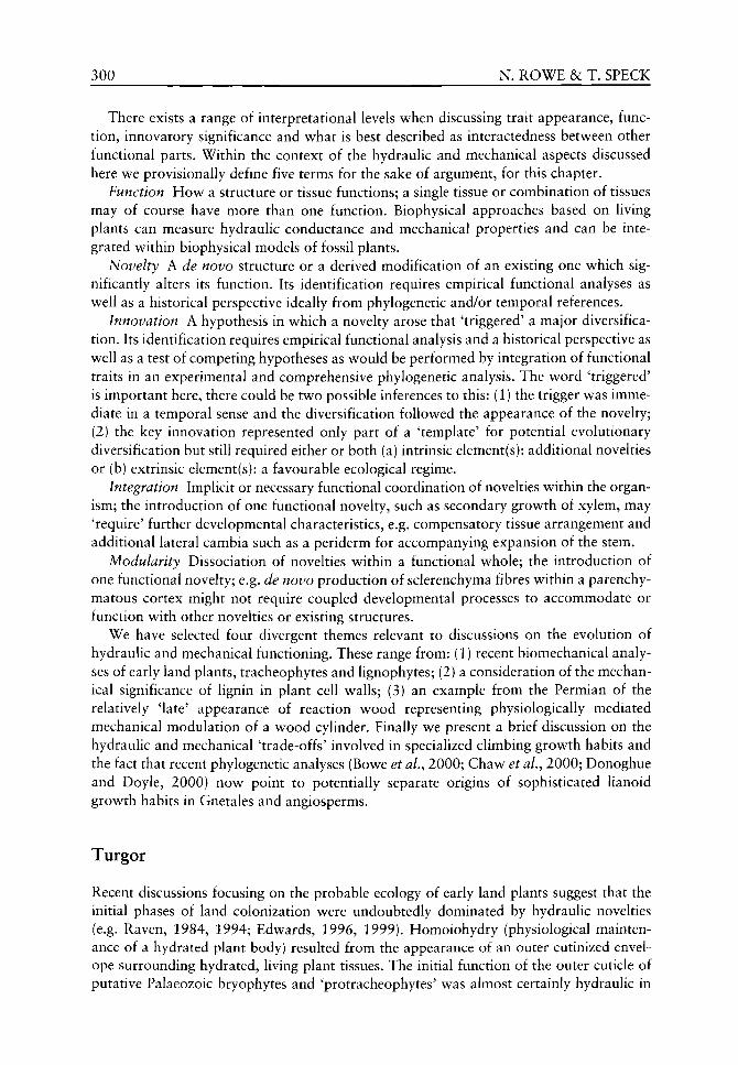
300 N. ROWE &c T. SPECK
There exists a range of interpretational levels when discussing trait appearance, function, innovatory significance and what is best described as interactedness between other functional parts. Within the context of the hydraulic and mechanical aspects discussed here we provisionally define five terms for the sake of argument, for this chapter.
Function How a structure or tissue functions; a single tissue or combination of tissues may of course have more than one function. Biophysical approaches based on living plants can measure hydraulic conductance and mechanical properties and can be integrated within biophysical models of fossil plants.
Novelty A de novo structure or a derived modification of an existing one which significantly alters its function. Its identification requires empirical functional analyses as well as a historical perspective ideally from phylogenetic and/or temporal references.
Innovation A hypothesis in which a novelty arose that 'triggered' a major diversification. Its identification requires empirical functional analysis and a historical perspective as well as a test of competing hypotheses as would be performed by integration of functional traits in an experimental and comprehensive phylogenetic analysis. The word 'triggered' is important here, there could be two possible inferences to this: (1) the trigger was immediate in a temporal sense and the diversification followed the appearance of the novelty; (2) the key innovation represented only part of a 'template' for potential evolutionary diversification but still required either or both (a) intrinsic element(s): additional novelties or (b) extrinsic element(s): a favourable ecological regime.
Integration Implicit or necessary functional coordination of novelties within the organism; the introduction of one functional novelty, such as secondary growth of xylem, may 'require' further developmental characteristics, e.g. compensatory tissue arrangement and additional lateral cambia such as a periderm for accompanying expansion of the stem.
Modularity Dissociation of novelties within a functional whole; the introduction of one functional novelty; e.g. de novo production of sclerenchyma fibres within a parenchymatous cortex might not require coupled developmental processes to accommodate or function with other novelties or existing structures.
We have selected four divergent themes relevant to discussions on the evolution of hydraulic and mechanical functioning. These range from: (1) recent biomechanical analyses of early land plants, tracheophytes and lignophytes; (2) a consideration of the mechanical significance of lignin in plant cell walls; (3) an example from the Permian of the relatively 'late' appearance of reaction wood representing physiologically mediated mechanical modulation of a wood cylinder. Finally we present a brief discussion on the hydraulic and mechanical 'trade-offs' involved in specialized climbing growth habits and the fact that recent phylogenetic analyses (Bowe et al., 2000; Chaw et al.^ 2000; Donoghue and Doyle, 2000) now point to potentially separate origins of sophisticated Hanoid growth habits in Gnetales and angiosperms.
Turgor
Recent discussions focusing on the probable ecology of early land plants suggest that the initial phases of land colonization were undoubtedly dominated by hydraulic novelties (e.g. Raven, 1984, 1994; Edwards, 1996, 1999). Homoiohydry (physiological maintenance of a hydrated plant body) resulted from the appearance of an outer cutinized envelope surrounding hydrated, living plant tissues. The initial function of the outer cuticle of putative Palaeozoic bryophytes and 'protracheophytes' was almost certainly hydraulic in

HYDRAULICS AND MECHANICS OF PLANTS 301
combating desiccation. Hydrated turgor systems in plant stems were mechanically important for upright columnar growth and spreading semi-recumbent clonal axes in plants such as Aglaophyton major Kidston and Lang, Horneophyton lignieri Barghorn and Darrah and Rhynia gwynne-vaughanii Kidston and Lang. In such stems, hydrated parenchyma was the principal contributing element to stem stiffness (Speck and Vogellehner, 1988, 1994; Niklas 1989, 1992; Speck et aL, 1990). Thickened epidermal cell walls combined with a layer of cuticle could also have functioned as a peripheral tension bracing system and mechanical pneumatic structure (Speck and Vogellehner, 1988; Speck et al., 1990). Quantitative biomechanical models of this grade of organization indicate that central conducting strands were small and too centrally placed within the stem to contribute significantly to stem flexural stiffness (Speck et al.^ 1990; Speck and Vogellehner, 1994) (Figure 16.1); stem stiffness permitting upright and probably mutually supported stands of upright stems was almost entirely a result of turgor systems. The central conducting strands of such early land plants represent primarily hydraulic functional novelties (Roth and Mosbrugger, 1996; Bateman etal, 1998; Roth etal, 1994,1998; Roth-Nebelsick et al., 2000; Konrad et al., 2000) compared with aquatic and dorsoven-tral body plans of antecedent forms. Hydraulic optimization via tracheid-like central strands and the increased and more rapid supply (Raven, 1994) to more extensive and more distant parts of the plant body probably conferred improved hydraulic supply for more extensive clonal systems of horizontal axes.
Central conducting strands did not contribute considerably in a direct manner to stem stiffness and were primarily hydraulic in function. On the face of it, this appears to have permitted greater hydraulic supply to a larger plant body and ensuring greater degrees of intrinsic homoiohydry. The appearance of a small central strand of tracheids is effectively a 'modular novelty' in that its appearance did not necessitate further simultaneous developmental novelties for the new structure to function; this holds true if one assumes that a cuticle and epidermis with stomata for regulating transpiration and rhizoids for water uptake had already been put in place. Within limits, the appearance of a conducting strand could be interpreted as a 'key innovation' in as much as it set a template for optimized conductance in a number of putative lineages (Kenrick and Crane, 1997a,b). In terms of an immediate morphological diversification, the appearance of the conducting strand probably represented an innovation for extensive clonal growth and low-lying vegetation commonly depicted for the early land plant radiation, but not one for significantly enlarging and expanding plant architectures into the aerial realm.
The hypodermal sterome
By the Middle Devonian many lineages of land plants possessed members with thick-walled fibre tissues at or towards the outside of the stem cross-section. Biomechanical investigations indicate that these types of tissue configurations represented significant contributions to the stiffness of the stem (Speck et al., 1990; Speck and Vogellehner, 1994) potentially allowing greater height and more diverse branched architectures than were possible from turgor systems. Devonian plants such as Psilophyton daivsonii Banks, Leclercq and Hueber (Figure 16.2), Gosslingia breconensis Heard and species of Zosterophyllum Penhallow show that the hypodermal sterome generally contributes to over 95% of the flexural stiffness of the stem. In these plants at least, the hypodermal sterome undoubtedly had a mechanical function.
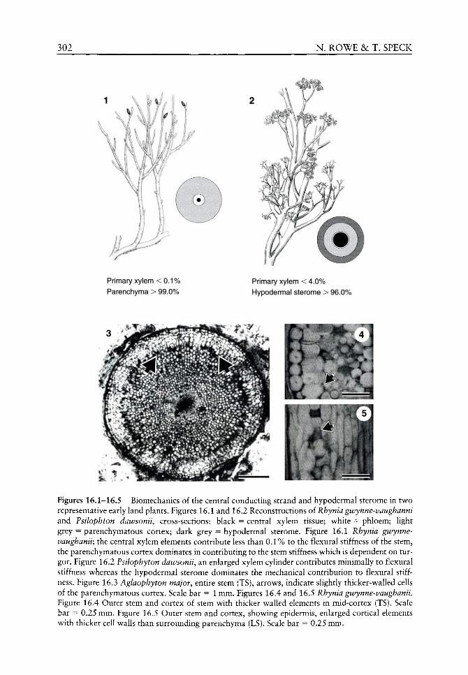
302 N. ROWE & T. SPECK
Primary xylem < 0.1% Parenchyma > 99.0%
Primary xylem < 4.0% Hypodermal sterome > 96.0%
Figures 16.1-16.5 Biomechanics of the central conducting strand and hypodermal sterome in two representative early land plants. Figures 16.1 and 16.2 Reconstructions oi Rhynia gwynne-vaughanni and Psilophton dawsonii, cross-sections: black = central xylem tissue; white = phloem; light grey = parenchymatous cortex; dark grey = hypodermal sterome. Figure 16.1 Rhynia gwynne-vaughanii; the central xylem elements contribute less than 0.1% to the flexural stiffness of the stem, the parenchymatous cortex dominates in contributing to the stem stiffness which is dependent on turgor. Figure 16.2 Psilophyton dawsonii, an enlarged xylem cylinder contributes minimally to flexural stiffness whereas the hypodermal sterome dominates the mechanical contribution to flexural stiffness. Figure 16.3 Aglaophyton major^ entire stem (TS), arrows, indicate slightly thicker-walled cells of the parenchymatous cortex. Scale bar = 1 mm. Figures 16.4 and 16.5 Rhynia gwynne-vaughanii. Figure 16.4 Outer stem and cortex of stem with thicker walled elements in mid-cortex (TS). Scale bar = 0.25 mm. Figure 16.5 Outer stem and cortex, showing epidermis, enlarged cortical elements with thicker cell walls than surrounding parenchyma (LS). Scale bar = 0.25 mm.
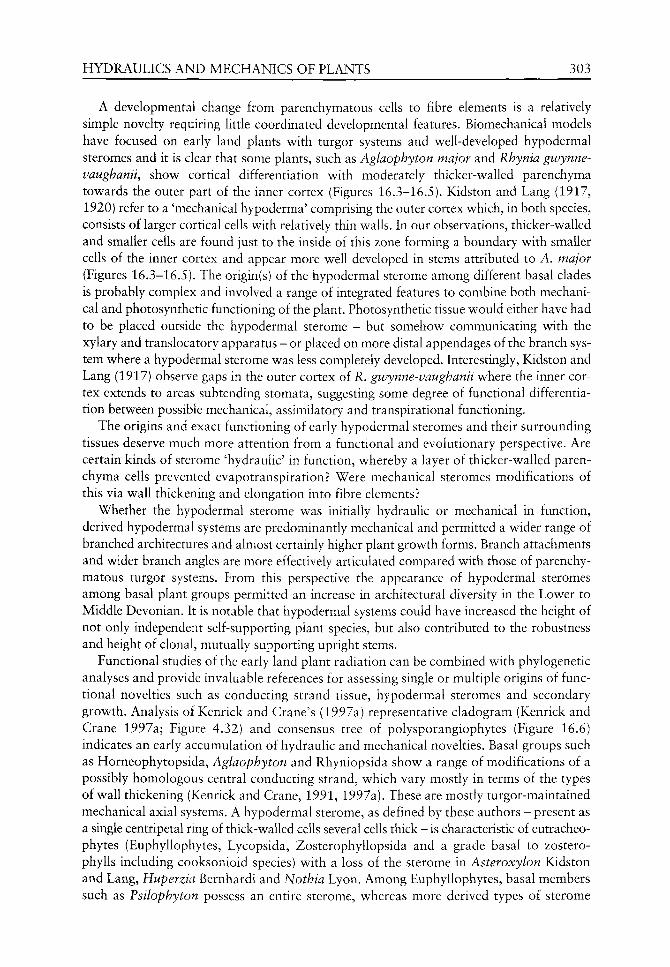
HYDRAULICS AND MECHANICS OF PLANTS 303
A developmental change from parenchymatous cells to fibre elements is a relatively simple novelty requiring little coordinated developmental features. Biomechanical models have focused on early land plants with turgor systems and v^ell-developed hypodermal steromes and it is clear that some plants, such as Aglaophyton major and Rhynia gtvynne-vaughanii, show cortical differentiation with moderately thicker-walled parenchyma towards the outer part of the inner cortex (Figures 16.3-16.5). Kidston and Lang (1917, 1920) refer to a 'mechanical hypoderma' comprising the outer cortex which, in both species, consists of larger cortical cells with relatively thin walls. In our observations, thicker-walled and smaller cells are found just to the inside of this zone forming a boundary with smaller cells of the inner cortex and appear more well developed in stems attributed to A. major (Figures 16.3-16.5). The origin(s) of the hypodermal sterome among different basal clades is probably complex and involved a range of integrated features to combine both mechanical and photosynthetic functioning of the plant. Photosynthetic tissue would either have had to be placed outside the hypodermal sterome - but somehow communicating with the xylary and translocatory apparatus - or placed on more distal appendages of the branch system where a hypodermal sterome was less completely developed. Interestingly, Kidston and Lang (1917) observe gaps in the outer cortex of R. givynne-vaughanii where the inner cortex extends to areas subtending stomata, suggesting some degree of functional differentiation between possible mechanical, assimilatory and transpirational functioning.
The origins and exact functioning of early hypodermal steromes and their surrounding tissues deserve much more attention from a functional and evolutionary perspective. Are certain kinds of sterome 'hydraulic' in function, whereby a layer of thicker-walled parenchyma cells prevented evapotranspiration? Were mechanical steromes modifications of this via wall thickening and elongation into fibre elements?
Whether the hypodermal sterome was initially hydraulic or mechanical in function, derived hypodermal systems are predominantly mechanical and permitted a wider range of branched architectures and almost certainly higher plant growth forms. Branch attachments and wider branch angles are more effectively articulated compared with those of parenchymatous turgor systems. From this perspective the appearance of hypodermal steromes among basal plant groups permitted an increase in architectural diversity in the Lower to Middle Devonian. It is notable that hypodermal systems could have increased the height of not only independent self-supporting plant species, but also contributed to the robustness and height of clonal, mutually supporting upright stems.
Functional studies of the early land plant radiation can be combined with phylogenetic analyses and provide invaluable references for assessing single or multiple origins of functional novelties such as conducting strand tissue, hypodermal steromes and secondary growth. Analysis of Kenrick and Crane's (1997a) representative cladogram (Kenrick and Crane 1997a; Figure 4.32) and consensus tree of polysporangiophytes (Figure 16.6) indicates an early accumulation of hydraulic and mechanical novelties. Basal groups such as Horneophytopsida, Aglaophyton and Rhyniopsida show a range of modifications of a possibly homologous central conducting strand, which vary mostly in terms of the types of wall thickening (Kenrick and Crane, 1991, 1997a). These are mostly turgor-maintained mechanical axial systems. A hypodermal sterome, as defined by these authors - present as a single centripetal ring of thick-walled cells several cells thick - is characteristic of eutracheo-phytes (Euphyllophytes, Lycopsida, Zosterophyllopsida and a grade basal to zostero-phylls including cooksonioid species) with a loss of the sterome in Asteroxylon Kidston and Lang, Huperzia Bernhardi and Nothia Lyon. Among Euphyllophytes, basal members such as Psilophyton possess an entire sterome, whereas more derived types of sterome

304 N. ROWE & T. SPECK
Euphyllophytina
Eutracheophytes
Lycophytina
Tracheophyta
Polysporangiophytes
Tetraxylopteris ( H ) - ©
Rhacophyton
Psilophyton ( H )
Eophyllophyton
Asteroxylon ( T )
Huperzia
Nothia
Zosterophyllum ( H ) - ( T )
Sawdonia
Gosslingia ( H )
Huvenia
Rhynia (j^
Aglaophyton (j)
Horneophyton ( j f )
Polytrichum
Figure 16.6 Phylogenetic relationships of early polysporangiophytes (based on Kenrick and Crane, 1997a; Figure 4.33). Biomechanical structures are indicated for tested taxa: T = turgor system, H = hypodermal sterome, C = vascular cambium.
characterize both lignophyte taxa and the putative basal sphenopsid Ibkya Skogg and Banks. Finally, a hypodermal sterome and a bifacial vascular cambium characterizes Tetraxylopteris Beck and other lignophytes (Figure 16.6).
Secondary growth
Secondary xylem evolved separately in possibly five different lineages and four of these, lycopsids, lignophytes, Rhacophyton Crepin and cladoxylls, had appeared by the Middle Devonian. The vascular cambia generally differ considerably in organization and products formed (Cichan, 1986a; Cichan and Taylor, 1990) as w ell as in actual function. Biomechanical analyses indicate that w^ood in arboreous lycophytes is nearly entirely hydraulic in function (Speck and Vogellehner, 1992; Speck, 1994b). Wood in some stem-group lignophytes, such as Archaeopteris Davison, v^as clearly both hydraulic and mechanical (Galtier et al., 1999). The functional and ecological significance of these separate origins is of extreme interest for understanding the post-homoiohydric phase of diversification in land plants and has been discussed briefly in a broadly biomechanical perspective elsew^here (Rov^e, 2000). In this section and below^ w e focus on the bifacial vascular cambium in lignophytes and consider the functional significance of the appearance of secondary xylem in basal members of the group.

HYDRAULICS AND MECHANICS OF PLANTS 305
Preliminary biomechanical findings based on Tetraxylopteris schmidtii Beck indicate that young stages of growth possess a mechanical hypoderm which contributes dominantly to the flexural stiffness of the stem (Galtier et al, 1999; Rowe et al., 2000) (Figure 16.7). A second stage of development where secondary xylem and phloem mostly fill the previously mid-cortical region up to the outer hypodermal tissue (Figure 16.7) shows a higher contribution of secondary xylem to the flexural stiffness of the stem and, by the final stage of development tested, in which an extensive layer of peridermal tissue has formed, the contribution of the wood to flexural stiffness approaches 60% in the model selected (Figure 16.7). Despite the increase in contribution of flexural stiffness of the wood, the pattern of Young's modulus for these three stages is not typical for a self-supporting plant (Figure 16.8) (Speck, 1994a; Speck and Rowe, 1999a,b). Self-supporting plants show basal or older stages of development having higher Young's elastic moduli than more distal segments, which therefore represent relatively stiffer bases for supporting the distal load. In T. schmidtii, periderm forms a wide, parenchymatous and disorganized tissue continuing outwards from the secondary phloem (Figure 16.12) (Scheckler and Banks, 1971; Scheckler, 1976). Preliminary results based on a model of ontogeny of Triloboxylon arnoldii Matten indicate a similar general pattern in the trend of calculated Young's modulus, although material under investigation of this plant (Stein and Beck, 1983) shows a different pattern of secondary development (Figures 16.9-16.10) where fibre and sclereid reorganization of the hypoderm as well as periderm formation differ from that in Tetraxylopteris. The results for Tetraxylopteris and Triloboxylon will be presented in detail elsewhere.
Both T. schmidtii and T. arnoldii show a hypodermal sterome organization (Figures 16.9 and 16.11) which dominates the mechanical architecture in youngest growth stages. This organization is comparable with some basal land plants relying on a hypodermal sterome for mechanical support. The hypodermal sterome in these aneurophytes consists of longitudinal ribs around the perimeter of the stem in the form of a 'sparganum' cortex differing from the entire hypodermal steromes seen in plants such as Psilophyton and Leclercqia Banks, Bonamo and Grierson.
Current phylogenetic analyses (Rothwell and Serbet, 1994; Kenrick and Crane, 1997a) most commonly place aneurophytalean progymnosperms at the base of the lignophyte clade. These plants are usually depicted as small statured, possibly understorey shrubs, with relatively determinate growth (Scheckler, 2001). Significant development of the bifacial vascular cambium enlarges and modifies the geometry of the primary body and is developmentally complex. Secondary development within the primary plant body clearly requires integral developmental pathways for expansion, containment, prevention of rupturing and sealing-off of the outside of the primary body (Figures 16.9 and 16.12). Neither of the aneurophytes examined shows a secondary growth process, which produces trends in Young's modulus typical of a self-supporting plant. In other words, the production of secondary xylem and secondary phloem fibres in the latest stage of growth does not occupy a sufficiently large proportion of the stem to 'optimize' the stiffness of the basal stem part to a value which would be normal for a self-supporting plant (Speck and Rowe, 1999a). In T. schmidtii, there is a wide zone of outer secondary cortical tissue (Figure 16.12) and in T. arnoldii there are one or more zones of proliferated primary and secondary cortex (Figure 16.9).
At the risk of generalization, many woody self-supporting gymnosperms and angio-sperms have a woody cylinder surrounded by a zone of bark tissue which is relatively thin compared with the diameter of the wood. A relatively stiff woody component
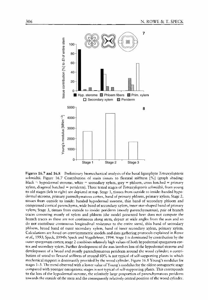
306 N. ROWE & T. SPECK
E 0)
I c 0
o
o >
100
80-1
60-1
40
20 i 5000
4000
f 3000
ZJ
I 2000 CO
1000
Hyp. sterome • Phloem fibers M Prim, xylem n Secondary xylem ^ Periderm
8
Stage 1 Stage 2 Stage 3
Figures 16.7 and 16.8 Preliminary biomechanical analysis of the basal lignophyte Tetraxylopteris schmidtii. Figure 16.7 Contribution of main tissues to flexural stiffness [%] (graph shading: black = hypodermal sterome, white = secondary xylem, grey = phloem, cross hatched = primary xylem, diagonal hatched = periderm). Three tested stages of Tetraxylopteris schmidtii^ from young to old stages (left to right) are depicted at top. Stage 1, tissues from outside to inside: banded hypo-dermal sterome, primary parenchymatous cortex, band of primary phloem, primary xylem; Stage 2, tissues from outside to inside: banded hypodermal sterome, thin band of secondary phloem and compressed cortical parenchyma, wide band of secondary xylem, inner star-shaped band of primary xylem; Stage 3, tissues from outside to inside: periderm (mostly parenchymatous), pair of branch traces consisting mostly of xylem and phloem (the model presented here does not compute the branch traces as these are not continuous along stem, depart at wide angles from the axis and so do not contribute continuous longitudinal resistance to the entire stem), thin band of secondary phloem, broad band of outer secondary xylem, band of inner secondary xylem, primary xylem. Calculations are based on centrisymmetric models and data gathering protocols explained in Rowe et al.^ 1993; Speck, 1994b; Speck and Vogellehner, 1994. Stage 1 is dominated by contribution by the outer sparganum cortex; stage 2 combines relatively high values of both hypodermal sparganum cortex and secondary xylem. Further development of the axis involves loss of the hypodermal sterome and development of a broad and mostly parenchymatous periderm around the wood cylinder; a contribution of wood to flexural stiffness of around 60% is not typical of self-supporting plants in which mechanical support is dominantly provided by the wood cylinder. Figure 16.8 Young's modulus for stages 1-3. The trend observed with a lower value of Young's modulus for the oldest ontogenetic stage compared with younger ontogenetic stages is not typical of self-supporting plants. This corresponds to the loss of the hypodermal sterome, the relatively large proportion of parenchymatous periderm towards the outside of the stem and the consequently relatively central position of the wood cylinder.
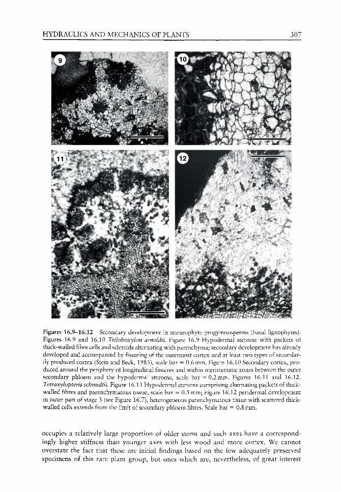
HYDRAULICS AND MECHANICS OF PLANTS 307
Figures 16.9-16.12 Secondary development in aneurophyte progymnosperms (basal lignophytes). Figures 16.9 and 16.10 Triloboxylon arnoldii. Figure 16.9 Hypodermal sterome with packets of thick-walled fibre cells and sclereids alternating with parenchyma; secondary development has already developed and accompanied by fissuring of the outermost cortex and at least two types of secondarily produced cortex (Stein and Beck, 1983), scale bar = 0.6 mm. Figure 16.10 Secondary cortex, produced around the periphery of longitudinal fissures and within meristematic zones between the outer secondary phloem and the hypodermal sterome, scale bar = 0.2 mm. Figures 16.11 and 16.12. Tetraxylopteris schmidtii. Figure 16.11 Hypodermal sterome comprising alternating packets of thick-walled fibres and parenchymatous tissue, scale bar = 0.5 mm; Figure 16.12 peridermal development in outer part of stage 3 (see Figure 16.7), heterogeneous parenchymatous tissue with scattered thick-walled cells extends from the limit of secondary phloem fibres. Scale bar = 0.8 mm.
occupies a relatively large proport ion of older stems and such axes have a correspondingly higher stiffness than younger axes with less v^ood and more cortex. We cannot overstate the fact that these are initial findings based on the itw adequately preserved specimens of this rare plant group, but ones which are, nevertheless, of great interest

308 N. ROWE &c T. SPECK
for determining the constraints and possible functioning of the appearance of the vascular cambium in lignophytes. We summarize the functional significance of these findings below.
1. Young stages of aneurophytes are mainly supported by a hypodermal sterome of the sparganum type of longitudinal ribs of sclerenchymatous fibres that are fused or nearly fused in young ontogenetic stages. This organization can be viewed as a structural and developmental novelty compared with entire hypodermal steromes of antecedent tracheophytes. The modified function of such an arrangement could be linked to integral processes such as (i) coordination of mechanics with externally placed photosyn-thetic tissues (as seen in many extant herbaceous plants), (ii) reduced expenditure of physiologically costly thick-walled fibres, or (iii) integral optimization permitting expansion of the outer parts of the primary body as a result of expansion of secondary growth from inside. Whatever the functional innovation operating here, there are several possibilities which would confer developmental innovation over archetypal forms with entire hypodermal steromes.
2. The xylem produced by the secondary vascular cambium, at least in Tetraxylopteris, exceeds the original area of primary xylem within the primary body. This is clearly a developmental novelty over other body plans with only primary xylem development. Some authors have interpreted xylem in Psilophyton to show possible secondary development as seen from aligned primary xylem elements (Banks et aL, 1975). The actual function of the wood cylinder seen in Tetraxylopteris and Triloboxylon is difficult to demonstrate unequivocally from a physiological, hydraulic or mechanical point of view. The result from the biomechanical models of these plants indicate that the increase in xylem volume does not contribute 'mechanically' to a self-supporting growth form. All tested self-supporting plants show an increase in Young's modulus during ontogeny or towards the base of the plant so that older development stages are constructed from a stiffer material (Speck and Rowe, 1999a; Figure 1). Despite the comparatively large volume of wood in the old stage of Tetraxylopteris^ the Young's modulus for this stage is less than that calculated for younger stages. The larger size and the presence of wood in older stages of Tetraxylopteris do produce a stiffer stem in terms of flexural stiffness and this is true for many lianas in which older stages are simply larger (Speck and Rowe, 1999a; Figure 2) but, as in lianas and other non-self-supporting stems, the material properties of the older basal parts of the stem are not stiffer than those younger or above in terms of Young's modulus and this is not an optimal design for a self-supporting plant. Xylem volume increase is possibly some kind of hydraulic novelty but it is difficult to examine further in what precise way. It is unknown, for example, whether the entire wood cylinder remained water conducting throughout development or only in the most recently formed wood. It is possible that an increased xylem cylinder either optimized conductivity over a longer time or over more extended aerial portions of the plant or both. Uncertainty of how much of a wood cylinder remained conductive and, indeed, how much of the distal part of the fossil plant was supplied, remain major obstacles for models of hydraulic functioning in fossil plants (Cichan, 1986a; Rowe and Speck, 1998).
3. Cortical proliferation and peridermal activity also represent developmental novelties compared with most earlier tracheophyte body plans. Evidence exists of localized cellular proliferation in plants lacking other secondary growth and this is sometimes referred to as 'wound tissue' (Banks, 1981) such as in Psilophyton. This is quite different
HYDRAULICS AND MECHANICS OF PLANTS 309
(though possibly developmentally aUied) with fully circumferential periderm growth coupled as integral developmental traits with bifacial vascular cambium activity. Periderm and cortical development in T. schmidtii and T. arnoldii appear to 'surround' and 'seal off respectively the geometric change brought about by secondary growth. They could justifiably be cited as examples of integral functional novelties formed de novo and coupled with expansion of the wood and secondary phloem. Interestingly, the peri-dermal systems observed in Tetraxylopteris and Triloboxylon are different (see Figures 16.9-16.12) suggesting that integral pathways linked with secondary development of the bifacial vascular cambium were derived independently. Once again the functional origin of periderm is an interesting one of possible co-option where an original wound repair mechanism possibly related to phytophagy was later modified and deployed as an integral developmental system linked to growth of the bifacial vascular cambium. Certainly this is hinted at by the cortical activity in T. arnoldii where at least two broadly defined meristematic areas occur just inside the outer hypodermal sterome and flanking longitudinal fissures caused by internal expansion of the axis (see Figure 16.9).
4. Secondary phloem proUferation and significant volumes of tissues in both species suggest prolonged translocatory function over a more extensive plant body. Fibre cells within the secondary phloem might have had a mechanical role, but rows of fibres are not interconnected and contain rounded sclereids, which are not as mechanically efficient as fibres.
Does the appearance of the bifacial vascular cambium represent a key innovation?
More derived progymnosperms, Archaeopteridales, co-occurred with aneurophytes, became globally distributed and developed additional vegetative novelties including leaves, complex branch organization and large-bodied architectures (Scheckler, 2001). Lignophytes from the Late Devonian onwards possess a bifacial vascular cambium and show increasing architectural diversity. From this viewpoint, the combination of novelties built around the bifacial vascular cambium represents further consolidation of the lignophyte body plan and accompany a morphological and architectural diversification. In terms of the long-term success of lignophytes, the implication that the bifacial vascular cambium was a key innovation triggering a morphological radiation of lignophytes must be considered with caution. The appearance of the seed habit - perhaps a clear example of a modular innovation in relation to vegetative novelties associated with secondary growth - is another of the 'key innovations' widely believed to have contributed to the radiation of the group.
The bifacial vascular cambium emerged prior to the seed. We have shown that, among at least two representatives, cambial activity might have optimized hydraulic conductivity and photosynthate translocation. Both are physiological novelties compared to basal groups with a more static primary growth trajectory.
Phylogenetic resolution of basal lignophytes and seed plants remains unsubstantiated in any great detail. However, biomechanical analyses of some early seed plant representatives indicate that some and probably many show body plans with a vascular cambium in which secondary growth is mainly confined within the primary plant body and that mechanical stiffness was conferred by a hypodermal sterome of the sparganum or dicty-oxylon type (Speck and Vogellehner, 1992, 1994; Rowe et al, 1993). Other early seed plants following the Late Devonian were undoubtedly capable of woody self-supporting
310 N. ROWE &c T. SPECK
growth habits (Speck and Rowe, 1994a). Interestingly, the earliest known seed plants are small-bodied plants with limited secondary growth but extensive leaves (Rothwell and Scheckler, 1988; Serbet and Rothwell, 1992), a later novelty which could be interpreted as an innovation added to the cambial body plan organization observed in aneurophytes. Furthermore, the possession of planated leaf surfaces could be considered as integrated novelties linked to the enhanced hydraulic potential of cambial activity. The appearance of seed structures represents a modular innovation in relation to the bifacial cambium body plan.
In summary, the appearance of the bifacial vascular cambium appears to have been an innovation for lignophyte architecture with a primary functional novelty concerned with optimizing hydraulics rather than immediately conferring mechanical advantages as witnessed with the limited stature of tested aneurophytes and the biomechanical results suggesting poor optimization for self-supporting habits. Self-supporting architectures are seen among Archaeopteridales and other putative progymnosperms, such as Protopitys^ and it is a fascinating aspect of the early evolution of lignophytes that this lignophyte stem group became largely extinct by the early Carboniferous - apart from one possible Lazarus taxon Cecropsis lunulata Stubblefield and Rothwell (Stubblefield and Rothwell, 1989). Only members of the clade having the seed habit, an unrelated additional modular innovation, diversified after this point.
Lignification and biomechanics of the plant cell wall
Chemical and physical complexity of plant phenolics and especially lignin have been of particular interest to evolutionary studies, where their primary roles as either light screens, phytophagy deterrents, water-repellents or mechanical stiffening have been discussed recently (Cooper-Driver, 2001). In addition to the difficulties of understanding what lignin actually does in living plant cell walls, is the notorious problem of unequivocally identifying lignin derivatives in fossil plants, especially in the early land plant radiation. There are several elements to the potential functional roles of lignin in plant tissue mechanics. First is its potential role as a 'water proofing agent'; as a hydrophobic molecule it 'chemically dries' the embedded cellulose microfibrils which can maintain more intramolecular H-bonds and so retain higher stiffness. Second, as a matrix structure or filler (Wainwright et al., 1976; Niklas, 1992) in composite cell walls, it can only act mechanically in combination with longitudinally orientated molecules, such as cellulose. These two functions of lignin discussed in the recent experimental Hterature (Hoffmann et ai, 2000a,b; Spatz and Speck, 2000) are significant for interpreting the origins of such roles in early land plants. Retaining stiffness in water-conducting cell walls is important to prevent collapse resulting from negative pressures building up inside the lumen; it is possible that lignin might have played a crucial role at some point in the evolution of water-conducting strands where lignin maintained stiffness in cell walls constantly in contact with water. Somewhat different constraints might have characterized lignin functioning in hypodermal steromes. As these tissues do not conduct water after cell death they might have been less reliant on lignified cell walls for protecting cellulose wall-stiffening elements from contact with water. The role of lignin in strengthening water-conducting cells is possibly linked with other geometric features of the cell wall including spiral, annular and scalariform thickenings, which can also locally reinforce and strengthen water-conducting cells against negative pressure.

HYDRAULICS AND MECHANICS OF PLANTS 311
Recent biomechanical research on a wide systematic range of plants from algae to seed plants is one way of observing how different clades deploy varying mechanical strategies at the architectural, morphological, anatomical, ultrastructural and biochemical levels (Spatz and Speck, 2000). In this case study we briefly report some recent results from studies on Equisetum L. where mechanical support is provided from non-lignified fibre cells.
Equisetum hyemale L., is a herbaceous, clonal plant with a hollow stem, nodal structure and a mechanical architecture which provides a lightweight, 'cheap' organization that resists mechanical failure by local buckling (Speck et aL, 1998). In Equisetum hyemale ovalization of the stem and local buckling are resisted by relatively high Young's elastic moduli of the hollow stem wall. The wall includes a non-lignified hypodermal sterome in combination with a double layer of relatively thick-walled endodermis cells with lignified casparian rings (Figure 16.13). Detailed staining techniques with phloroglucinol (Gerlach, 1984) and other reagents indicate that cells of the hypodermal sterome are living and unlig-nified (Figure 16.13). This tissue has Young's moduU varying from 4.5 GPa in basal inter-nodes to lower values of 1.5 Gpa towards the apex (Speck et al.^ 1998). All values in these data are lower than the usual range for sclerenchyma which range in values from 5 to 30 GPa (Wainwright et aL, 1976; Niklas, 1992) and higher than values measured for collenchyma ranging from 1 to 2 GPa (Nachtigall et al.^ 1988; Spatz and Speck, 1995).
Unlignified tissues can provide significant mechanical stiffness approaching values to those of lignified tissues. One of the uncertainties of any kind of biomechanical modelling of fossil plants is that biochemical composition of cell walls is unknown or unknowable and that measurements on living plants might not be confidently applied to fossils (Edwards et al.^ 1997). Of course, cell wall chemistry, ultrastructure and degrees of hydration as well as numerous other factors influence cell wall mechanical properties and values of Young's moduli assigned to fossil tissues can only be hoped to represent a similar magnitude of value. The point we wish to make here is that lignification is not a prerequisite for conferring 'stiffness' to a plant tissue and that a thick-walled tissue comprising fibre-shaped elements would not necessarily require lignin in its walls to have a high value of Young's modulus. This has direct implications for interpreting the functioning of hypodermal
^ # ^
Figure 16.13 Transverse section of Equisetum hyemale^ stained for lignin, based on Speck et al. (1998). A segment of dense thick-walled tissue representing a hypodermal sterome (HS) represents an important contribution to the mechanical stability of the stem but is unlignified; other lignified tissues in the same stem cross-section include the metaxylem (MX) and the casparian thickenings (CT). Scale bar = 0.1 mm.

312 N. ROWE &c T. SPECK
steromes in early land plant diversifications where lignification of cell walls would not be prerequisite for a mechanically significant structure. The physiological and mechanical roles assigned to phenolics in general, from light screens, pathogen/phytophage deterrence, water repellent and mechanical properties of cell walls underline the difficulty of confidently assigning function to biochemical processes and such areas undoubtedly require more research based on carefully deployed studies of living plants.
Reaction wood
Reaction wood is a developmental modification of normal wood concerned with righting, adjusting and maintaining the orientation of woody leaders and branches (Archer and Wilson, 1970; Wilson and Archer, 1977). Two types of reaction wood exist, including compression wood in extant conifers and tension wood in angiosperms. Both show cellular characteristics differing from normal wood and often occur in crescent-shaped areas within the wood cylinder. Generally, compression wood is often, but not always, found on the underside of conifer lateral branches and tension wood is often found in the upper side of angiosperm branches.
The fact that reaction wood is so widespread and common in extant trees and shrubs, prompted some workers to consider when it first appeared in the evolution of woody plants (Timell, 1983, 1986). Considering the large body size and architectural complexity of woody lignophytes since the Devonian and the dependence of extant woody architectures on reaction wood, this is an interesting question. Was compression wood present since the appearance of Middle Devonian plants such as Archaeopteris or was it a more derived feature that was 'added' to an increasingly complex and derived system comprising the bifacial vascular cambium? Timell (1983) located only low to moderate wood heterogeneity in Callixylon wood and nothing that he could confidently ascribe to reaction wood. His survey of wood illustrations from the Permian fossil woods of eastern Europe (Greguss, 1967) also revealed difficulties in definitely establishing the presence of reaction wood.
Re-investigation of cordaite shoots of Fennsylvanioxylon tianii from the Early Permian of China (Baolin and Wang, 1988; Li, 1995; Baolin et aL^ 1996) and Fennsylvanioxylon from the Permian of France, indicate crescent-shaped zones of modified wood within several small branches (Figures 16.14-16.16). One branch pair includes reaction wood, on opposite sides of each branch. Areas of reaction wood are sharply differentiated with transitions to normal wood (Figure 16.15). Cell wall outlines of individual tracheids within crescent-shaped areas have more elliptical outlines, thicker walls and a different type of preservation in which the wall content between the outline of the lumen and the outer part of the secondary wall is usually empty (Figure 16.16). Opposite and temporally alternating areas of reaction wood are a well-known phenomenon in extant compression wood formation during 'overcorrection' of upright or lateral branches (Timell, 1986). In cases of over-correction, physiological and mechanical reactions in one direction exceed the 'programmed' equilibrium position for the orientation of the stem and result in the formation of reaction wood in the opposite direction to readjust the position of the branch. This finding suggests a relatively late appearance of reaction wood among lignophytes. It remains to be seen whether reaction wood can be detected in other seed plant groups or among progymnosperms. Analyses of Archaeopteris^ Tetraxylopteris and Triloboxylon as well as wood of putative seed plants Pitus Witham, Endoxylon Kidston and Eristophyton Zallesky have not yet yielded the levels of differentiation found among these cordaites.

HYDRAULICS A N D MECHANICS OF PLANTS 313
14. • ' •." M
^ 1
'" i • . -. ' :^
.4 '~v. .
# .
# ^ '
Figures 16,14-16.16 Pennsylvanioxylon tianni, a late Permian cordaitalean plant from China. Figure 16.14 A pair of twigs showing at least three crescent-shaped developments of reaction wood represented by the lighter areas of wood. Reaction wood is organized in the same (left to right) orientation for both axes and is very similar to opposite reaction wood formed in extant conifers, scale bar = 1.2 mm. Figure 16.15 Crescent-shaped area of reaction wood alternating with segments of normal wood, scale bar = 0.6 mm. Figure 16.16 Transverse section of transition between reaction wood, below and normal wood above. As in many instances of compression wood in extant plants, the compression wood consists of tracheids with thicker cell walls and elliptical outlines. Scale bar = 80 ixm.
Whether reaction wood will be identified in basal groups of lignophytes or not, it represents a physiological and mechanical novelty superposed on the vascular cambium body plan. This highlights a crucial aspect of the vascular cambium as a developmental plan that can accommodate added functional novelties. Reaction wood is clearly mechanical in function in extant plants and almost certainly so for cordaites. Interestingly, recent studies on extant conifers indicate that conductance might be reduced by compression wood (Spicer and Gartner, 1998) suggesting that the appearance of reaction wood might have been of mechanical advantage for the development of complex architectures but with some integrative burden and 'trade-off with stem hydraulics.

314 N. ROWE &c T. SPECK
Hydraulics, mechanics and evolution of the climbing habit
Types of climbing strategy
Climbing plants can show quite different mechanical and hydraulic constraints from self-supporting plants (Gartner, 1991; Putz and Holbrook, 1991; Ewers et aL, 1991; Speck, 1991, 1994b; Speck and Rowe, 1999a). Furthermore, different types of climbing strategy also show quite different trends in mechanical and hydraulic properties during development. Recent and ongoing biomechanical and hydraulic analyses have shown that climbing strategies involve a range of interrelated constraints based on hydraulics and mechanics. A plant that has reduced the physiological cost of mechanical support by producing slender stems needs to augment its hydraulic conductance if it is to maintain a comparable photosynthetic surface. This is often accomplished via large-diameter conducting elements; water conductance, generally speaking, scales to the fourth power of the radius of the conducting element. However, slender stems with large-diameter conducting elements supplying a large leaf area can be a risky business. Climbing stems can be easily damaged by host tree movement, canopy sway, tree falls, host branch failure from epiphyte loading and so on. A relatively small amount of damage to the stem in terms of surface area because of damage by excessive bending or torsion could potentially cut off a large portion of hydraulic supply; this situation is made worse by the fact that large diameter elements are more prone to dysfunction on account of cavitation (embolism) and that relatively limited damage such as localized cracks passing in the vicinity of vessels might be enough to cause embolism and hydraulic dysfunction (Putz and Holbrook, 1991).
At the risk of over generalization, there are basically three different strategies to overcome these conflicts involved in minimizing stem diameter and maximizing stem conductance. First, grow very close to or actually fixed along the surface of large-bodied supports and reduce free movement between the host support and the climber (root climbers, e.g. Araceae, Hedera L.). Second, protect a non-self-supporting stem with stiff mechanical properties, these plants (sometimes referred to as semi-self-supporters (Speck, 1994a,b; Speck and Rowe, 1999a) can exceed their stable critical buckling height and maintain a relatively 'loose' more-or-less vertical scrambling position in the surrounding vegetation. Third, attach firmly along host plants by stem twining, winding and tendrils etc. and produce specialized compliant stems, which dissipate mechanical energy under bending or torsional stresses. This latter strategy can increase survival of the attached plant during swaying and movement of the host as well as raising the probability of survival during dramatic rearrangements of the surrounding forest structure during tree falls. Arguably the most complex strategy in terms of hydraulics and mechanics is the third kind of strategy which is found among many families of lianoid angiosperms and the gnetalean genera Gnetum L. and Ephedra L.
Appearance of the lianoid habit
The possibility of elucidating specific growth habits of long extinct organisms and the implications for interpreting community structure and evolutionary patterns is appealing. Progress has been made in recent years with the discovery of cuticular plant compressions with attachment organs from the late Palaeozoic indicating probable climbing strategies of the first type described above (Kerp and Krings, 1997; Krings and Kerp, 1997, 1999) as well as biomechanical models indicating climbing strategies of the second type (Speck and Vogellehner, 1992; Rowe et aL, 1993; Speck, 1994b; Speck and Rowe 1999b). In addition to these are the quantitative hydraulic studies, particularly of Michael Cichan,
HYDRAULICS AND MECHANICS OF PLANTS 315
investigating the potential hydraulic traits of a range of fossil plants discussed in reference to climbing habits. These include sphenophylls (Cichan, 1985) and sphenopsids, (Cichan and Taylor, 1983; Cichan, 1986b), and medullosans and cordaites (Cichan, 1986a). Other recent discoveries include remarkable vessel-like structures among gigantopterid seed plants (Li et al., 1996), w^hich have also been suggested as representing climbing plants.
In many forms of lianoid development there is an ontogenetic shift from young, relatively stiff stems w ith dense wood and low specific conductance, to larger diameter, relatively flexible stems with high specific conductance (Figures 16.17-16.20). Old stages of many lianas also have high degrees of built in toughness (not to be confused with stiffness), where stems can be bent or twisted to a remarkable extent before becoming mechanically and hydraulically dysfunctional (Putz and Holbrook, 1991). This is often accomplished by predisposed segmentation via alternation of hydraulically active tissues with wedges of deformable and fracture-confining tissues (Figure 16.18). During early development, mechanical stiffness is a prerequisite for searching leaders or self-supporting young individuals (Speck and Rowe, 1999a). Following secure attachment to the host, increased hydraulic optimization, seen in large surface areas of large diameter vessels, is combined with mechanical compliance and segmentation of the wood cylinder. Our examples here serve to illustrate the complex developmental patterns in a species of the angiosperm Bauhinia L. and the gymnosperm Gnetum. Mechanical data based on Young's modulus measured in bending for the two species illustrates that both species show a significant drop in Young's modulus during ontogeny.
Vessels and the climbing habit
Enhanced hydraulic conductance via vessels can be viewed as a functional novelty compared with the type of organization seen among most basal lignophytes and non-lignophytes, most of which show little differentiation of tracheid elements in terms of wide size variation. Like the appearance of reaction wood discussed above, vessel appearance could be seen as another supplementary novelty to the existing bifacial cambium body plan. The appearance of vessels has often been cited as a specialization, whereby hydraulic and mechanical functioning within the wood cylinder could be performed by more specialized cell types within the same tissue (Carlquist, 1975).
It is not clear whether, climbers with lianoid (type 3 climbing strategy) existed in early or later Palaeozoic ecosystems. Certain forms such as Sphenophyllum Koenig, Medullosa Cotta and cordaites have been observed to combine large diameter xylem elements with slender stems, but biomechanical studies are yet to provide evidence whether such forms comprised biomechanically sophisticated flexibility and toughness or adopted the arguably more conservative mechanical strategies such as root climbers or relatively rigid 'semi-self-supporting' types mentioned above. Xylem element diameter taken on its own is not a clear indicator of a lianoid habit.
The appearance of wide conducting elements and vessels in the lignophyte body plan could have potentially elevated leaf surface to stem diameter ratios. The three types of climbing habit outlined above would require different integral and modular novelties. For example, semi-self-supporting plants produce and retain stiff stem mechanics, which would require either stiffening elements in the wood cylinder or in the cortex. This situation contrasts with the necessary integral novelties associated with lianoid climbers, which require compliancy and toughness. Lianoid features arose independently many times among angio-sperms and with many variations in terms of ways in which the older stages of development

316 N . R O W E & T. SPECK
Figures 16.17-16.20 Anatomy and organization in extant lianas. Figures 16.17 and 16.18 Anatomy in young and older stems of the gymnospermous liana Gnetum africanum. Figure 16.17 The young stage has relatively dense wood with increasing numbers of vessels towards the outside of the wood cylinder, scale bar = 2.5 mm. Figure 16.18 The older stage shows segmentation of the wood cylinder via variant cambium development, which has formed segments of secondary phloem and cortical tissue around and between the second ring of wood formed. This type of organization is similar to many angiosperm lianas and is typical of older stem development showing high compliance and protection of the hydraulic system via fracture-limiting areas between xylem segments, scale bar = 5.0 mm. Figures 16.19 and 16.20 Transverse sections of the angiosperm liana Bauhinia guianense (see also Speck and Rowe, 1999a). Figure 16.19 Young developmental stage of a self-supporting individual prior to the climbing phase. The young growth has produced dense wood with many fibres and very few vessels, scale bar = 3.0 mm. Figure 16.20 Anatomical transition marking shift from young self-supporting phase to lianoid phase and a shift from dense stiff wood to a second wood type with many large vessels. In this species, the vascular cambium is limited to grov^h in two areas of the perimeter forming a ribbon-shaped lianoid stem, in this figure the continued growth of Hanoid wood is at the top. Scale bar = 0.85 mm.
produce flexibility and partition their hydraulic system for protection against hydraulic failure following mechanical perturbation (Gentry, 1991; Ewers et al,^ 1991 ; Speck, 1991 , 19942i; Speck et aL^ 1996a; Speck and Rowe, 1999a). Close inspection of any modern habitat, temperate or tropical, indicates enormous differences in position, volume, length, branching, shading and many other characteristics between root climbers, self-supporters and lianas. Dense swathes of semi-self-supporting ferns of Gletchenia Smith in the t ropics, for example, up to eight or so metres high occupy an entirely different niche from lianoid forest cUmbers which produce the bulk of their leaves in the canopy via stems that

HYDRAULICS AND MECHANICS OF PLANTS 317
are entirely dependent on self-supporting hosts. These kinds of differences are detectable from consideration of stem mechanics and hydraulics of fossil plants. A putative fossil hana with wide xylem elements, but with a thick fibrous hypodermal sterome around the periphery of old stages of growth can not have been a flexible lianoid climber but more probably a semi-self-supporter.
Since the earliest land plant radiations, the different kinds of potential climbing strategy we have outlined above would have required similar mechanical functioning as plants today involved in attachment and stem stiffness and compliancy. If the entire suite of characters contributing to a climbing syndrome is viewed as a complex character, clearly some climbing strategies are more complex than others.
Growth forms requiring minimal associated novelties probably include root climbing and semi-self-supporting growth forms where increased hydraulic capacity could be accompanied by either retainment of existing mechanical tissues, e.g. the hypoderm for stem stiffness (semi-self supporters) and repositioning of rooting meristems or other attachment organs on cauline parts of the stem (root climbers). Both of these types of climbing strategy could be attained by simple body plans without secondary growth, with relatively little organizational rearrangement and few additional novelties. Heterotopic expression of rooting meristems on stem axes could provide root climbers with the necessary anchorage mechanism. Given adequate hydraulic supply, semi-self-supporters would probably require even fewer additional novelties. If growth was confined to the primary body and primary tissues of the hypodermal sterome, simple continuation of apical meristems would rapidly exceed the plant's critical buckling length, the plant would no longer be 'self-supporting' and would then require support from a dense stand of similar neighbours perhaps of the same clonal stand or larger bodied self-supporters. Simply leaning or interlocking branches to maintain a relatively stiff plant stem upright would not require sophisticated attachment devices and, indeed, many modern tested semi-self-supporters simply interlock branches or petioles with the surrounding vegetation to stay upright (Gallenmiiller et al, 2001).
At least these two 'types' of climbing strategies require relatively little modification from bauplans based on turgor, hypodermal or lignophyte body plans. It is possible that such forms were appearing relatively early in the land plant radiations and that such climbing growth forms were just a short physiological step away from the underlying thigmomor-phogenetic economizing (Jaffe, 1973) that a great many green plants appear inherently to possess. A shift to a root-climbing or semi-self-supporting growth form would not require a large input of secondary novelties. A number of early land plants do possess both horizontal axes with rhizoids as well as upright aerial axes and this organization might be relatively predisposed towards at least potentially facultative root-climbing, if not on host plants then on local ground topography. Root climbing is possibly more amenable to turgor systems as once firmly attached to a solid host support there is little requirement for stem stiffness or toughness, indeed many root-climber stems are actually brittle structures. Semi-self-supporting strategies among turgor systems were probably more limited as local bending moments on a cylindrical turgor system from greatly extended upright stems may be prone to mechanical and hydrauUc failure. It is possible that 'climbing' strategies among such forms were more Umited to optimizing and opportunistically modulating safety factors (variable stem lengths below the critical buckling length) than greatly developing long semi-self-supporting axes (Speck and Vogellehner, 1994),
The appearance of the hypodermal sterome is often talked about as being an innovation for stem stiffening among early land plants and for optimizing self-supporting growth habits (Niklas, 1992; Speck and Vogellehner, 1994; Rowe and Speck, 1997).
318 N. ROWE &c T. SPECK
In fact, the appearance of the hypodermal sterome could have also facilitated climbing semi-self-supporters by imparting sufficient integral stiffness in stems exceeding their critical buckling length but laterally supported by other supports. Evolutionary transitions between self-supporting and semi-self supporting among plants with simple body plans therefore probably require little added novelties and little extra integrated developmental characters.
What is special about lianas?
The mechanical and hydraulic properties present in lianoid climbing habits require numerous integral and modular novelties superimposed on a basal early land plant or early ligno-phyte body plan. Like other climbing growth forms an optimized hydraulic supply and vessel-sized elements is probably essential to maintain hydraulic supply to a large effective leaf surface via a narrow stem. Essential mechanical features include stiff young stages as searchers and increasing compliancy during development associated with stem toughness. This mechanical shift in properties is an essential feature and one that distinguishes the lianoid habit from root climbing and semi-self-supporting strategies. A basal lianoid body plan would require relatively high levels of integral novelties including optimized hydrauUc conductance, compliance and toughness.
Conclusions
Following the establishment of basic embryophyte organization and physiology (Graham et al., 2000), the early phase of terrestrialization concerned mostly hydraulic novelties and the estabUshment of physiologically homoiohydric body plans. Columnar growth forms depending on physiologically maintained turgor were hydraulically optimized by outer cutinized sheaths and central conducting strands. Both represented primary hydraulic novelties. Biomechanical studies demonstrate that many early conducting strands did not contribute directly to the bending mechanics of the stem and thus clarify that mechanical support of columns relied on physiologically mediated water maintenance. The central position of the xylem tissue and its relatively small contribution to the second moment of area of the stem indicate a small direct contribution to flexural stiffness of the stem (see Figure 16.1). It is possible that xylem strands and cutinized sheaths might, however, have been of secondary mechanical significance in ensuring turgor and hydraulically stable axes for larger plant bodies or during intermittent water availability (e.g. Bateman et aL, 1998).
Representatives of basal clades show of a range of hypodermal steromes. Biomechanical analyses indicate that the hypodermal sterome became important for contributing directly to the stiffness of the stem and represented a functional novelty for larger and more diverse architectures. Mechanical hypodermal steromes were important in optimizing self-supporting growth forms as well as semi-self-supporting forms. There is some indication that cell wall thickening in the mid to outer cortex in some early representatives such as Rhynia gwynne-vaughanii and Aglaophyton major may have initially been concerned with limiting evapotranspiration and had little or no effect on directly contributing to stem stiffness. It is possible that such steromes could have secondarily optimized mechanical stability by better controlling turgor pressure over larger and/or taller body plans. Further functional analyses coupled with more inclusive phylogenetic histories might indicate that 'hypodermal steromes' were primarily hydraulic novelties co-opted for mechanical support.

HYDRAULICS AND MECHANICS OF PLANTS 319
Basal lignophytes possess an undoubtedly mechanical hypoderm in young stages of growth, which are fused or nearly fused in young ontogenetic stages. Development of the bifacial vascular cambium conferred several functional breakthroughs including increased hydraulic, mechanical and translocatory functions. Biomechanical studies suggest that large surface areas of cortex at the 'mechanically influential' outside of the stem meant that the relatively centrally positioned wood cylinder contributed little to flexural stiffness of the stem and the products of the secondary vascular cambium were not optimized for a mechanically self-supporting stem system.
Whereas many authors agree that the Siluro-Devonian radiations were periods of 'innovation' characterized by appearances of many new physiological and morphological 'novelties', precisely how these new novelties actually functioned is more easy to speculate on than to test empirically. This is undoubtedly true in terms of 'hydraulic' or 'mechanical' functioning. A major part of the problem lies in the potentially rapid integral functioning of hydraulic and mechanical features among these early body plans. Conducting strands may have represented secondary mechanical novelties by increasing turgor potential over a larger or taller plant body; thick-walled hypodermal sterome fibres might represent modified cortical cells initially concerned with restricting evapotranspiration; secondary xylem acting initially as hydraulic optimization became mechanically important for self-supporting architectures after the addition of further integrated novelties such as entire periderm formation. Further additional novelties to the cambial body plan then permitted yet greater diversification of growth forms via novelties, such as reaction wood and vessels.
The appearance of the bifacial vascular cambium as seen in basal aneurophytalean lignophytes provided a developmental template for a wide range of further physiological and structural novelties including indeterminate, self-supporting as well as non-self-supporting woody architectures. Reaction wood could be said to represent one of the 'bells and whistles' or additional novelties added to the basic cambial body plan, which can be perhaps more directly interpreted as a purely mechanical novelty. The evidence from observed aneurophytes and Archaeopteridales - up to present - indicates that reaction wood was not present in these basal lignophytes. Reaction wood, is an example where a functional novelty is more easily ascribed a single function, compared with more primordial novelties more basic to the emerging polysporangiophyte and tracheophyte body plan where emerging developmental templates share a range of functions.
Phylogenetic investigations of recent radiations, which include functional observations based on living plants, potentially carry higher expectancies for resolving evolutionary processes. A combination of molecular and morphological character traits as well as empirical experimentation on the physiology, hydrauHcs and mechanics can offer more robust frameworks for interpreting correctly the significance of novelties, innovations and adaptations. Functional studies combined with phylogenetic frameworks can more easily address patterns in hydraulic and mechanical developmental strategies such as transitions between self-supporting plants and climbers. Derived seed plants such as Gnetales and angiosperms have complex and integrated hydraulic and mechanical novelties adapted for lianoid climbing strategies.
Such studies are demonstrating whether transitions in growth form during evolution from self-supporters to climbers occurs via hydraulically and mechanically 'intermediate' forms (e.g. semi-self-supporters) and whether a clade that has once established a lianoid body plan can revert back to a self-supporting architecture (Speck et al.^ 1997; Civeyrel and Rowe, 2001). In the latter case ongoing work is indicating that the hydraulic and mechanical novelties accumulated during the development of climbing body plans carries
320 N. ROWE &c T. SPECK
high degrees of developmental and ecological burden which is rarely reversed and that subsequent 'self-supporting' plants shov^ differences in trends of mechanical or geometrical characters.
Developmental burden and the ability to 'turn back' having accumulated a specialized mechanical and hydraulic body plan is a fascinating aspect of plant evolution and one v^hich might be relevant to many patterns of body plan complexity and intrinsic constraint from the Devonian radiations onward. The rise and decline of the lycopsid, sphenopsid and ifilicopsid clades are monumental examples of rises in complexity, long-term ecological success, potential intrinsic constraint and canalization and, finally, disastrous extinction. We argue that an increased application of biophysical modelling on such topics based on parallel studies of living plants that can be appropriately applied to fossils can potentially offer much in the way of explaining or at least reducing the number of speculative scenarios.
Key innovations are widely believed to initiate major radiations and observers are surely correct in emphasizing the necessary phylogenetic framework for testing potentially conflicting or alternative hypotheses based on phylogenetic pattern rather than simply mapping a priori assessed functional related traits (Bateman, 1999b). The above examples illustrate the wide range of data-related and conceptual difficulties centred on interpreting function, let alone interpretation of higher conceptual levels such as innovatory significance and adaptation. We propose that function-level interpretations lie at the core of any study involving assessments of innovation and adaptation.
A recurring theme in identifying key innovations in plants is the significance of vegetative and architectural novelties as opposed to reproductive novelties. Was the bifacial vascular cambium a key innovation for the radiation of lignophytes.^ Or can the ultimate success of lignophytes be attributed to a step-wise series of innovations including acquisition of heterospory and the seed habit. What was the key innovation underlining the radiation of angiosperms? Was it based on sophisticated hydraulic and mechanical architectures generated by deployment of vessels? Or was it due more to coevolution with insect pollinators or rapid generation turnover? Given an appropriate phylogenetic framework as well as rigorous functional and biophysical studies these kinds of questions might be answerable. Many functional traits or processes are 'taken for granted' in the literature as well as in the relatively few systematic studies that comment on functional and ecological traits in any mechanistic detail. Examples include the supposed mechanical significance of the appearance of wood, the functioning of the seed plant ovule and seed and the appearance and developmental significance of periderm, to name just a few. If one of the aims of evolutionary studies is to infer functional process-related dynamics, such as the radiation of early land plants and the radiation of angiosperms and Gnetales, such studies are potentially just as lost without functional analysis as they are without a phylogenetic and historical background.
Functional studies and biophysical modelling allow investigations of structural evolution over long time periods and across gradients of complexity. The origin(s) of complex structures have been one of the most discussed aspects of evolutionary biology. Some of the findings discussed above show potentially interesting patterns of function and complexity. Simple de novo appearance of the conducting strand, the hypoderm and the bifacial vascular cambium show relatively clear primary functional characteristics, which are possibly either coupled initially with secondary functional implications or closely followed by that secondary function. In all three novelties this has been argued as a shift from primarily hydraulic to hydraulic and mechanical functioning.

HYDRAULICS A N D MECHANICS OF PLANTS 321
Acknowledgements
We thank W m . Stein and Robyn Burnham for kindly providing slides of Triloboxylon for
analysis and Steve Scheckler for lending slides of Tetraxylopteris. Shijun Wang generously
provided material of Fennsylvanioxylon for the analysis of reaction wood - research car
ried out with the support an exchange grant from the French and Chinese consuls. We
both acknowledge support from a French-German PROCOPE exchange grant over the
last few years. Finally, we would like to thank Hanns-Christof Spatz for all of his lively
ideas and encouragement over the years and we are confident that this long and amicable
collaboration will continue for a long time to come.
References
Archer RR, Wilson BF. 1970. Mechanics of the compression wood response I. Preliminary analyses. Flant Physiology 46: 550-556.
Banks HP. 198L Peridermal activity (wound repair) in an early Devonian (Emsian) trimerophyte from the Gaspe Peninsula, Canada. Palaeobotanist 28-29: 20-25.
Banks HP, Leclercq S, Hueber FM. 1975. Anatomy and morphology of Psilophyton dawsonii, sp.n; from the late Lower Devonian of Quebec (Gaspe), and Ontario, Canada. Palaeontographica Americana 48: 77-127.
Baolin T, Wang S. 1988. Study on primary vascular systems of cordaitean stems Shanxioxylon sinense and Pennsylvanioxylon tianii in coal balls from Shanxi. Acta Palaeontologica Sinica 27: 21-29.
Baolin T, Wang S, Guo Y, et al. 1996. Flora of Palaeozoic coal balls of China. The Palaeobotanist 45: 247-245.
Bateman RM. 1999a. Architectural radiations cannot be optimally interpreted without morphological and molecular phylogenies. In: Kurmann MH, Hemsley AR, eds. The Evolution of Plant Architecture. Kew: Royal Botanic Gardens, 221-250.
Bateman RM. 1999b. Integrating molecular and morphological evidence of evolutionary radiations. In: Hollingsworth PM, Bateman RM, Gornall RJ, eds. Molecular Systematics and Plant Evolution. London: Taylor and Francis, 432-471.
Bateman RM, DiMichele WA. 1994. Saltational evolution of form in vascular plants: a neo-Goldschmidtian synthesis. In: Ingram DS, Hudson A, eds. Shape and Form in Plants and Fungi. London: Academic Press, 63-102.
Bateman RM, Crane PR, DiMichele WA, et al. 1998. Early evolution of land plants: Phylogeny, physiology and ecology of the primary terrestrial radiation. Annual Review of Ecology and Systematics 29: 263-292.
Bowe LM, Coat G, dePamphilis CW. 2000. Phylogeny of seed plants based on all three genomic compartments: Extant gymnosperms are monophyletic and Gnetales' closest relatives are conifers. Proceedings of the National Academy of Sciences 97: 4092-4097.
Carlquist S. 1975. Ecological Strategies ofXylem Evolution. Berkeley and Los Angeles: University of California Press.
Chaw SM, Parkinson CL, Cheng Y, et al. 2000. Seed plant phylogeny inferred from all three plant genomes: monophyly of extant gymnosperms and origin of Gnetales from conifers. Proceedings of the National Academy of Sciences 97: 4086-4089.
Cichan MA. 1985. Vascular cambium and wood development in Carboniferous plants. II. Sphenophyllum plurifoliatum. Botanical Gazette 146: 395-403.
Cichan MA. 1986a. Conductance in the wood of selected Carboniferous plants. Palaeobiology 12: 302-310.
Cichan MA. 1986b. Vascular cambium and wood development in Carboniferous plants III. Arthropitys (Equisetales, Calamitaceae). Canadian Journal of Botany 64: 688-695.
Cichan MA, Taylor TN. 1983. A systematic and developmental analysis oi Arthropitys deltoides sp. nov. Botanical Gazette 144: 2S5-294.

322 N . R O W E &c T, SPECK
Cichan MA, Taylor TN. 1990. Evolution of cambium in geologic time. In: Iqbal M, ed. The Vascular Cambium. Taunton, Somerset: John Wiley, 213-228.
Civeyrel L, Rowe NP. 2001. Relationships of the Secamonoideae based on molecules, morphology and anatomy. Annals of the Missouri Botanical Garden 88: 583-602.
Cooper-Driver GA. 2001. Biological roles for phenolic compounds in the evolution of early land plants. In: Gensel PC, Edwards D, eds. Plants Invade the Land, Evolutionary and Environmental Perspectives. New York: Columbia University Press, 159-172.
Donoghue MJ, Doyle JA. 2000. Seed plant phylogeny: demise of the anthophyte hypothesis? Current Biology 10: 106-109.
Edwards D. 1996. New insights into early land ecosystems: a glimpse of a Lilliputian world. Review of Palaeobotany and Palynology 90: 159-174.
Edwards D. 1999. Origins of plant architecture: adapting to life in a brave new world. In: Kurmann MH, Hemsley AR, eds. The Evolution of Plant Architecture. Kew: Royal Botanic Gardens, 3-21.
Edwards D, Ewbank G, Abbot GD. 1997. Flash pyrolysis of the outer cortical tissues in Lower Devonian Psilophyton dawsonii. Botanical Journal of the Linnean Society 124: 345-360.
Ewers FW, Fisher JB, Fichtner K. 1991. Water flux and xylem structure in vines. In: Putz FE, Mooney HA, eds. The Biology of Vines. Cambridge: Cambridge University Press, 127-160.
Gallenmiiller F, Miiller U, Rowe NP, Speck T. 2001. The Growth form of Croton pullei (Euphorbiaceae) - Functional morphology and biomechanics of a neotropical liana. Plant Biology 3: 50-61.
Galtier J, Meyer-Berthaud B, Rowe NP, et al. 1999. Biomechanical consequences of the progymno-sperm vascular cambium. In: XV7 International Botanical Congress (Abstracts). St Louis: IBC XVI, 13.
Gartner BL. 1991. Stem hydraulic properties of vines vs. shrubs of western poison oak. Toxicodendron diversilobum. Oecologia 87: 180-189.
Gentry AG. 1991. The distribution and evolution of climbing plants. In: Putz FE, Mooney HA, eds. The Biology of Vines. Cambridge: Cambridge University Press, 73-97.
Gerlach D. 1984. Botanische Mikrotechnik. Stutgart, New York: Thieme Verlag. Gould SJ, Lewontin RC. 1979. The spandrels of san marco and the panglossian paradigm: a critique
of the adaptationist programme. Proceedings of the Royal Society of London, series B 205: 581-598.
Gould SJ, Vrba ES. 1982. Exaptation - a missing term in the science of form. Paleobiology 8: 4-15. Graham LE, Cook ME, Busse JS. 2000. The origin of plants: Body plan changes contributing
to a major evolutionary radiation. Proceedings of the National Academy of Sciences 97: 4535-4540.
Greguss P. 1967. Foss/7 Gymnosperm Woods in Hungary. Budapest: Akademiai Kiado. Hoffmann BJ, Chabbert B, Monties B, Speck T. 2000a. Fine-tuning of mechanical properties in two
tropical lianas. In: Spatz H-C, Speck T, eds. Plant Biomechanics 2000 - Proceedings of the 3rd International Plant Biomechanics Conference. Freiburg-Badenweiler, Stuttgart-New York: Thieme-Verlag, 10-18.
Hoffmann BJ, Chabbert B, Monties B, Speck T. 2000b. Mechanical properties and chemical cell wall composition of two tropical lianas. In: Wisser A, Nachtigal W, eds. Proceedings of the Workshops in Saarbriicken 1999, BIONA-report 14. Mainz: Akademie der Wissenschaften und der Literatur, 10-15.
Jablonski D. 2001. Origin of evolutionary novelties. In: Briggs DEC, Crowther, PR. eds. Palaeo-biology 11. Oxford: Blackwell Science, 162-166.
Jablonski D, Bottjer DJ. 1990. The ecology of evolutionary innovations: the fossil record. In: Nitecki MH, ed. Evolutionary Innovations. Chicago: University of Chicago Press, 253-288.
Jaffe MJ. 1973. Thigmomorphogenesis: the response of plant growth and development to mechanical stress. Planta 114: 143-157.
Kenrick P, Crane PR. 1991. Water-conducting cells in early fossil and land plants: implications for the early evolution of the tracheophytes. Botanical Gazette 152: 335-356.
Kenrick P, Crane PR. 1997a. The Origin and Early Diversification of Land Plants: a Cladistic Study. Washington, DC: Smithsonian Institution Press.
HYDRAULICS A N D MECHANICS OF PLANTS 323
Kenrick P, Crane PR. 1997b. The origin and early evolution of plants on land. Nature 389: 33-39. Kerp H, Krings M. 1997. Climbing and scrambling growth habits: common Hfe strategies among
Late Carboniferous seed ferns. Comptes rendu hebdomaire des seances de Vacademie des sciences, Paris, Sciences de la terre et des planetes 326: 583-588.
Kidston R, Lang WH. 1917. On Old Red Sandstone plants showing structure, from the Rhynie Chert Bed, Aberdeenshire. Part 1. Rhynia gwynne-vaughani Kidston and Lang. Transactions of the Royal Society of Edinburgh 51: 761-784.
Kidston R, Lang WH. 1920. On Old Red Sandstone plants showing structure, from the Rhynie chert bed, Aberdeenshire. Part II. Additional notes on Rhynia gwynne-vaughani, Kidston and Lang; with descriptions of Rhynia major n. sp., and Hornea lignieri, n. g., n. sp. Transactions of the Royal Society of Edinburgh 52: 603-627.
Konrad W, Roth-Nebelsick A, Kerp H, Hass H. 2000. Transpiration and assimilation of Early Devonian land plants with axially symmetric telomes - simulations on the tissue level. Journal of Theoretical Biology 206: 91-107.
Krings M, Kerp H. 1997. Cuticles of Lescuropteris genuina from the Stephanian (Upper Carboniferous) of Central France: evidence for a climbing growth habit. Botanical Journal of the Linnean Society 123: 73-89.
Krings M, Kerp H. 1999. Morphology, growth habit, and ecology of Blanzyopteris praedentata (Gothan) Nov; comb., a climbing neuropterid seed fern from the Stephanian of Central France. International Journal of Plant Sciences 603: 603-619.
Larson A, Losos JB. 1996. Phylogenetic systematics of adaptation. In: Rose MR, Lauder GV, eds. Adaptation. San Diego: Academic Press, 187-220.
Lauder GV. 1990. Functional morphology and systematics: studying functional patterns in an historical context. Annual Review of Ecology and Systematics 21: 317-340.
Lauder GV. 1991. Biomechanics and evolution: integrating physical and historical biology in the study of complex systems. In: Rayner JMV, Wooton RJ, eds. Biomechanics in Evolution. Cambridge: Cambridge University Press, 1-19.
Li C, Cui J. 1995. Atlas of Fossil Plant Anatomy in China. Beijing: Science Press. Li H, Taylor EL, Taylor TN. 1996. Permian vessel elements. Science 271: 188-189. Nachtigall W, Wisser C-M, Wisser A. 1988. Fin erster Einblick in biomechanische Konstruk-
tionsprinzipien von Grasern. NatUrliche konstruktionen - Mitt. SFB 230 1: 59-66. Niklas KJ. 1984. Size-related changes in the primary xylem anatomy of some early tracheophytes.
Paleobiology 10: 487-506. Niklas KJ. 1989. Mechanical behaviour of plant tissues as inferred from the theory of pressurized
solids. American Journal of Botany 76: 929-937. Niklas KJ. 1992. Plant Biomechanics: An Engineering Approach to Plant Form and Function.
Chicago, USA: University of Chicago Press. Novacek MJ. 1996. Paleontological data and the study of adaptation. In: Rose MR, Lauder GV,
eds. Adaptation. San Diego: Academic Press, 311-359. Putz FE, Holbrook NM. 1991. Biomechanical studies of vines. In: Putz FF, Mooney HA, eds. The
Biology of Vines. Cambridge: Cambridge University Press, 73-97. Raven JA. 1984. Physical correlates of the morphology of early vascular plants. Botanical Journal
of the Linnean Society 88: 105-126. Raven JA. 1994. Physiological analyses of aspects of the functioning of vascular tissue in early land
plants. Botanical Journal of Scotland 47: 49-64. Rose MA, Lauder GV. 1996. Post-Spandrel adaptationism. In: Rose MR, Lauder GV, eds.
Adaptation. San Diego: Academic Press, 187-220. Roth A, Mosbrugger V. 1996. Numerical studies of water conduction in land plants: evolution of
early stele types. Paleobiology 22: 411-421. Roth A, Mosbrugger V, Neugebauer HJ. 1994. Efficiency and evolution of water transport systems
in higher plants: a modelling approach. II Stelar evolution. Philosophical Transactions of the Royal Society of London, series B 345: 153-162.
Roth A, Mosbrugger V, Wunderhn A. 1998. Computer simulations as a tool for understanding the evolution of water transport systems in land plants: a review and new data. Review of Paleobotany and Palynology 102: 79-99.

324 N . R O W E &c T. SPECK
Roth-Nebelsick A, Grimm G, Mosbrugger V, et al 2000. Morphometric analysis of Rhynia and Asteroxylon: testing functional aspects of early land plant evolution. Paleobiology 26:405-418.
Rothwell GW, Scheckler SE. 1988. Biology of ancestral gymnosperms. In: Beck CB, ed. Origin and Evolution of Gymnosperms. New York: Columbia University Press, 85-134.
Rothwell GW, Serbet R. 1994. Lignophyte phylogeny and the evolution of spermatophytes: A numerical cladistic analysis. Systematic Botany 19: 443-482.
Rowe NP. 2000. The insides and outsides of plants: the long and chequered evolution of secondary growth. In: Spatz H-C, Speck T, eds. Plant Biomechanics 2000, Proceedings of the 3rd Plant Biomechanics Conference. Freiburg-Badenweiler: Georg Thieme Verlag, 129-140.
Rowe NP, Speck T. 1997. Biomechanics of Lycopodiella cernua and Huperzia squarrosa: implications for inferring growth habits of fossil small-bodied lycopsids. Mededelingen Nederlands Instituut voor Toegepaste Geowetenschappen TNO 58: 293-302.
Rowe NP, Speck T. 1998. Biomechanics of plant growth forms: the trouble with fossil plants. Review of Paleobotany and Palynology 102: 43-62.
Rowe NP, Speck T, Galtier J. 1993. Biomechanical analysis of a Palaeozoic gymnosperm stem. Proceedings of the Royal Society of London 252: 19-28.
Rowe NP, Speck T, Scheckler SE. 2000. Mechanical significance of the appearance of wood in land plants. In: Society for Experimental Biology (Abstracts). Exeter: Springer, 32.
Sanderson MJ, Donoghue MJ. 1996. Reconstructing shifts in diversification rates on phylogenetic trees. Trends in Ecology and Evolution 11: 15-20.
Scheckler SE. 1976. Ontogeny of Progymnosperms. I. Shoots of Upper Devonian Aneurophytales. Canadian Journal of Botany 54: 202-219.
Scheckler SE, Banks HP. 1971. Anatomy and relationships of some Devonian progymnosperms from New York. American Journal of Botany 58: 737-751.
Scheckler SE. 2001. Afforestation - the first forests. In: Briggs DEC, Crowther PR eds. Palaeo-biology II. Oxford: Blackwell Science, 67-71.
Serbet R, Rothwell GW. 1992. Characterizing the most primitive seed ferns. I. A reconstruction of Elkinsia polymorpha. International Journal of Plant Science 153: 602-621.
Spatz H-C, Speck T. 1995. Mechanische Eigenschaften von Hohlrohren am Beispiel von Grasern. In: Nachtigall W, ed. BIONA-report 9 - Technische Biologic und Bionik 2. Stuttgart, Jena, New York: Gustav Fischer Verlag, 91-132.
Spatz H-C, Speck T. 2000. Plant Biomechanics 2000, Proceedings of the 3rd Plant Biomechanics Conference Freiburg-Badenweiler: Georg Thieme Verlag.
Speck T. 1991. Changes of the bending mechanics of hanas and self-supporting taxa during ontogeny. In: Natural Structures: Principles, Strategies and Models in Architecture and Nature, Proceedings of the II International Symposium Sonderforschungsbereich 230, part I. Mitteilungen des SFB 230 Heft 6, Stuttgart, 89-95.
Speck T, Neinhuis C, Gallenmiiller F, Rowe NP. 1997. Trees and shrubs in the mainly lianescent genus Aristolochia s.l.: Secondary evolution of the self-supporting habit? In: Jeronimidis G, Vincent JFV, eds. Plant Biomechanics. Reading: University of Reading, 201-208.
Speck T. 1994a. A biomechanical method to distinguish between self-supporting and non-self-supporting plants. Review of Paleobotany and Palynology 81: 65-82.
Speck T. 1994b. Bending stability of plant stems: ontogenetical, ecological and phylogenetical aspects. Biomimetics 2: 109-128.
Speck T, Rowe NP. 1994a. Biomechanical analysis of Pitus dayi: early seed plant vegetative morphology and its implications on growth habit. Journal of Plant Research 107: 443-460.
Speck T, Rowe NP. 1994b. Biomechanical consequences of different stem structures in early seed plants: efficiency of the distribution of stem tissues. In: Evolution of natural structures: principles, strategies and models in architecture and nature. Proceedings of the III International Symposium Sonderforschungsbereich 230, part 9. Mitteilungen des SFB 230 Heft 9, Stuttgart, 197-202.
Speck T, Rowe NP. 1999a. A quantitative approach for analytically defining, growth form and habit in living and fossil plants. In: Kurmann MH, Hemsley AR, eds. The Evolution of Plant Architecture. Kew: Royal Botanic Gardens, 447-479.

HYDRAULICS A N D MECHANICS OF PLANTS 325
Speck T, Rowe NP. 1999b. Biomechanical analysis. In: Jones TP, Rowe NP, eds. Fossil Plants and Spores, Modern Techniques, The Geological Society of London Special volume. Bath: The Geological Society, 105-109.
Speck T, Rowe NP, Briichert F, Haberer W, et al. 1996a. How plants adjust the 'material properties' of their stems according to differing mechanical constraints during growth: an example of smart design in nature. In: Engin AE, ed. Bioengineering. Proceedings of the 1996 Engineering Systems and Design and Analysis Conference, PD-Vol. 77, volume 5, ASME 1996, Vol. 5 Bioengineering. New York: The American Society of Mechanical engineers, 233-241.
Speck T, Rowe NP, Spatz H-C. 1996b. Pflanzliche Achsen, hochkomplexe Verbundmaterialien mit erstaunlichen mechanischen Eigenschaften. Technische Biologic und Bionik 3, BIONA-report 10: 101-131. In: Nachtigall W, Wisser A, ed. Stuttgart: Fischer Verlag, 101-131.
Speck T, Spatz H-C, Vogellehner D. 1990. Contributions to the biomechanics of plants. I. Stabilities of plant stems with strengthening elements of differing cross-sections against weight and wind forces. Botanical Acta 103: 111-122.
Speck T, Speck O, Emanns A, Spatz H-C. 1998. Biomechanics and functional anatomy of hollow stemmed sphenopsids: III. Equisetum hyemale. Botanical Acta 111: 366-376.
Speck T, Vogellehner D. 1988. Biophysical examinations of the bending stability of various stele types and the upright axes of early 'vascular' land plants. Botanica Acta 101: 262-268.
Speck T, Vogellehner D. 1992. Fossile Baume, Spreizklimmer und Lianen. Versuch einer biomech-anischen Analyse der Stammstruktur. Courier Forschungs-Institut Senckenberg 147: 31-53.
Speck T, Vogellehner D. 1994. Devonische Landpflanzen mit und ohne hypodermales Sterom. Fine biomechanische Analyse mit Uberlegungen zur Friihevolution des Leit- und Festigungssystems. Palaeontographica B 233: 157-227.
Spicer R, Gartner BL. 1998. Hydrauhc properties of Douglas-fir (Pseudostuga menziesii) branches and branch halves with reference to compression wood. Tree Physiology 18: 777-784.
Stein WE, Beck CB. 1983. Triloboxylon arnoldii from the Middle Devonian of western New York. Contributions to the Museum of Palaeontology University of Michigan 26: 257-288.
Stubblefield SP, Rothwell GW. 1989. Cecropsis luculentum gen. et sp. nov.: evidence for heterosporous progymnosperms in the Upper Pennsylvanian of North America. American Journal of Botany 76\ 1415-1428.
Timell TE. 1983. Origin and evolution of compression wood. Holzforschung 37: 1-10. Timell TE. 1986. Compression Wood in Gymnosperms. New York: Springer-Verlag. Wainwright SA, Biggs WD, Currey JD, Goshne JM. 1976. Mechanical Design in Organisms. New
York: John Wiley. Wilson BF, Archer RR. 1977. Reaction wood: induction and mechanical action. Annual Review of
Plant Physiology 28: 24-43.

17 Becoming fruitful and diversifying: DNA sequence phylogenetics and reproductive
physiology of land plants
Martin Ingrouille and Mark W Chase
CONTENTS.
Introduction 327
Reproductive Thysiology' 329
Sexual incompatibility systems 332 Self-incompatibility (SI) as a defining angiosperm
characteristic 332 SI acting at the stigma 333 SI acting in the style 334 SI in euasterid I 335 Alternative kinds of SI 336 Heteromorphic SI 336 Self-compatibility 337
Conclusion 337
References 338
Introduction
The use of physiological data in systematics and phylogeny reconstruction has been limited because the physiology of relatively few species has been examined in any detail. However, morphology, anatomy and biochemistry have provided many useful surrogate physiological characters, if physiology is regarded in the broadest sense as covering all aspects of the plant in action. For example, photosynthetic pathways in grasses have been usually inferred from the anatomy of the leaf (Clayton and Renvoize, 1986). The same broad approach to physiology has been used in this chapter.
In general, though, systematists have not valued physiological characters highly. Physiological characters have been regarded as being somewhat different from other characters, more influenced by the environment and not normally a source of phylogenetic information. This was not always the case. Early systematists emphasized the importance of
The Evolution of Plant Physiology Copyright © 2004 Elsevier Ltd ISBN 0-12-33955-26 All rights of reproduction in any form reserved
328 M. INGROUILLE &c M. W. CHASE
functional characters that had the greatest influence on the organism. This essentialist approach, pioneered perhaps by Cesalpino (1583), achieved great success since it directed early taxonomists to the importance of reproductive characters and led to the establishment of many 'natural' groups still recognized today. One example of the success of this a priori approach was the recognition by Ray of the taxonomic significance of the number of cotyledons, although it has become clear that only the monocot type is useful phylogenetically (Chase et al,, 1993).
How^ever, the realization that it v^as difficult to identify the most essential, or most important functional, characters led to the rejection of this a priori approach. De CandoUe, who at first propounded a priori principles (de CandoUe, 1813) later, in his Prodromus systema-tis naturalis regni vegetabilis (de CandoUe, 1824-onward), adopted a more pragmatic Adansonian approach, utilizing the distribution of characters to identify the most essential, i.e. those with the most constancy within groups were obviously the most essential.
Most post-Darwinian taxonomists adopted this pragmatic approach (Cain, 1959), even those who explicitly indicated that their classification was phylogenetic. Now they had a theoretical framework to explain why physiological/functional characters might not indicate the phylogenetic history of large groups: they were regarded as having been particularly strongly selected and especially exposed to parallel and convergent evolution that might obscure a phylogenetic signal. For example, a fundamental physiological difference like the presence or absence of crassulacean acid metabolism (CAM) was found to be seemingly randomly distributed in ferns and a range of angiosperm families. Many of the physiological characters have a scattered distribution across all angiosperms and even within families. For example, in a well-defined family such as Poaceae, distinct kinds of PS ('starch' or parenchyma sheath) type and MS (inner or mestome sheath) type Kranz anatomy associated with variation in C4 photosynthesis, have been shown to have evolved independently at least five times (Clayton and Renvoize, 1986). Nevertheless, surrogate physiological characters, especially from reproductive physiology have remained important.
This rejection of the use of functional characters did not occur at species rank at which adaptation of a species to its environment came to be viewed as an important validation for an evolutionary species concept. Biosystematists such as Clausen and his colleagues (Clausen, 1951) emphasized that the process of speciation involved adaptation to different environments so that at first ecotypes and then sister species differed in their functional characters. The use of that important tool of the biosystematist, the botanic garden, can be regarded as a 20th century attempt to identify what are the 'essential characters' of different groups by cultivating plants in order to minimize environmentally induced variation. In addition, functional differences, relating to pollination and fertilization, which might confer reproductive isolation, were regarded as important for the recognition of what came to be called the 'biological species' (Mayr, 1942). In general, though, taxonomic practice changed little from the morphological-geographical approach long used by most systematists.
However, the availability of stable phylogenetic trees derived from genetic (DNA sequence) data (Chase et al, 1993; Soltis et aL, 1997, 1999, 2000; Qiu et al., 2000; Savolainen et ^/., 2000; Pryer et al.^ 2001), has provided a new opportunity to study physiological data in a phylogenetic context and thereby evaluate the importance of physiological diversification in the adaptive radiation of plants (Weller et al.^ 1995).
At first glance, physiological characters, such as the distribution of CAM (crassulacean acid metabolism) or C4 photosynthesis, widespread in many lineages, do not appear to be constant within clades in the new phylogenetic trees. This scattered distribution in the new

PHYSIOLOGY - APG PHYLOGENY 329
classification (APG, 1998) is also true of some other kinds of characters and is not just a feature of physiological characters. A striking example of sister taxa that is counterintuitive is the placing of such morphologically distinct families as the Nelumbonaceae (lotus), Proteaceae (Banksia L.i., Grevillea R.Br, ex J.Knight, Leucadendron R.Br etc.) and Platanaceae (planes) as close lineages in Proteales. The new phylogeny does point out some remarkable associations such as all the nitrogen fixing taxa in the same major clade of the rosids.
In a similar way, an understanding of the evolution of self-incompatibility (SI) in angiosperms has been hindered by two things: first, by a lack of information outside a few well studied crop plants and, secondly, by the use of broad and imprecise categories. As a consequence, little correlation between phylogeny and SI has been detected. A traditional dichotomy has been made between sporophytic and gametophytic SI on the basis of whether the haploid gametophytic tissue in the pollen grain or the diploid sporophytic tissue in the tapetum mediates the SI specificity, but as we will explain below this is misleading. Gametophytic SI is so widespread that it does not appear to carry any phylogenetic signal (Figure 17.1). Likewise it has been perplexing that the more narrowly distributed sporophytic SI is found in such diverse and unrelated groups as Asteraceae, Betulaceae and Brassicacae. The perceived low taxonomic value of SI data is being challenged by recent research that has studied the physiology of SI in greater depth and in a greater range of organisms. Below we discuss the distribution of different kinds of SI in the angiosperms in the light of the latest phylogenetic trees derived from DNA sequence data.
First we explore some examples from the reproductive physiology of plants in the broadest sense, utilizing surrogate characters from gynoecial and androecial morphology, anatomy, cytology and development. In fact, although there are some remarkable differences between the phylogenies derived from molecular data and pre-existing phylogenetic classifications, there is, overall, broad agreement. Ironically, this is largely because taxono-mists, who have rejected the a priori choice of physiologically important characters, have discovered the self-same 'biologically important', 'organizational' or 'constitutive' characters, a posteriori^ because of their constancy within groups (Davis and Heywood, 1973). The importance of a number of surrogate physiological reproductive characters has been confirmed or re-established and that of a number of new ones indicated. Several of these reproductive characters are also highly significant in the evolution of self-incompatibility in the angiosperms.
Reproductive Thysiology'
In the following, we review some of the most remarkable examples of the distribution of surrogate reproductive physiological characters in relation to the phylogenies derived from molecular sequence data (some data from Watson and Dallwitz, 1992 onwards). The examples may be placed into one of two contrasting categories. One category includes lineages/groups that have long been established on the basis of reproductive characters and which find support in the new phylogenetic trees, though frequently the circumscription of the lineage/group has undergone minor changes. The other category includes lineages/ groups that have been previously unsuspected but which are supported by some characters from the reproductive physiology of plants previously undervalued or ignored.
One of the most remarkable results of molecular phylogenetics has been the re-establishment of extant gymnosperms as a monophyletic group (Bowe et al.^ 2000; Pryer et al., 2001). In recent decades gymnosperms have been regarded as a paraphyletic group

330 M. INGROUILLE & M. W. CHASE
s
G G
S & G S
G
G S S G S S & G
S & G S & G
G
Fagales Cucurbitales Resales Fabales Oxalidales Malpighiales Eurosid 1 Celastrales Zygophyllales Sapindales Malvales Eurosid 2 Brassicales Myrtales Crossosomatales Geraniales Vitaceae Saxifragales Berberidopsidales Dipsacaies
Aquifoliales Lamiales Solanales Gentianales Euasterid 2 Garryales Cornales Ericales Caryophyllales Dilleniaceae Santalales Myrothamnaceae/Gunneraceae Trochodendraceae Buxaceae/Didymeleaceae Sabiaceae Proteales Ranunculales
Rosids
Asterids
Eudicots
s G
Ceratophyllales Winte rales Magnoliales Lau rales Piperales Monocots Chloranthaceae
Magnoliids (including monocots)
llliclaceae Schisandraceae Austrobaileyaceae Nymphaeaceae Amborellaceae
ANITA grade
S = Sporophytic G = Gametophytic
Figure 17.1 A simplified taxonomic arrangement of the angiosperms, based on APG (1998), with the distribution of sporophytic (S) and gametophytic (G) self-incompatibiHty indicated.
with at least two distinct clades, the cycads plus Ginkgo L. and the conifers plus gneto-phytes differing in seed and other characters. Similarly, the angiosperms are maintained as a monophyletic group for which there has been little doubt, although there are few synapo-morphic characters. The production of a specialized pollen reception tissue, the stigma, along with the closure or semi-closure of the carpel, is confirmed as an apomorphic or 'essential' character for the angiosperms, although in some basal angiosperms the carpel is not fused but sealed by mucilage (Endress, 2001). The presence of a stigma greatly enhanced the possibilities of the evolution of self-incompatibility and the evolution of a distinct, elongated style in eudicots and advanced monocots greatly expanded this potential.

PHYSIOLOGY - APG PHYLOGENY 331
Alternatively the gnetophytes rather than being a distinct gymnosperm lineage, 'antho-phyte' sister to the angiosperms, are now placed as a highly derived lineage of pines (Pinopsida) in the new phylogenetic trees (Bowe et al.^ 2000). A bisexual fertile apex is now relegated from being an 'essential' character.
The new phylogeny does not support the dichotomy of monocots versus dicots, although it does support the essential nature of the possession of a single cotyledon because, although the monocots are demonstrated to be a monophyletic lineage, they are now subsumed as a clade within a broader magnolioid clade close to the base of the angiosperm tree. Closer to the root of the angiosperm tree are a set of minor relatively primitive minor clades, sometimes called the ANITA group from the constituent taxa (Amborellaceae, Nympheales, lUiciaceales, Trimeniaceae, Austrobaileyaceae). Therefore, the possession of more than one, usually two cotyledons is shown to be unreliable and dicots are shown to be a para-phyletic group (Savolainen et al,^ 2000). The exclusion of the magnoliid dicots and various other relatively primitive dicot taxa leaves a monophyletic eudicot group defined by several features. This major lineage of flowering plants has triaperturate pollen with furrows running in parallel to the polar axis, compared to the monoporate/monosulcate pollen of the non-eudicots. They also have a secretory anther tapetum, simultaneous microsporo-genesis, well-differentiated stamens with a filament much longer than the anther and two leaf traces. Within the eudicot clade there are two major clades, the rosids and asterids, each divided into two large subclades (euasterid 1 and 2, eurosid 1 and 2) and several other clades such as the ranuncuHds and caryophyUids (see Figure 17.1).
Non-eudicots frequently have pollen produced by successive microsporogenesis and rather flattened or petaloid stamens. The relationship of some of these characters to the evolution of SI may prove a fruitful avenue for research. An obvious relationship is suggested between SI and a secretory tapetum producing and depositing molecules carrying self-incompatibility specificity into the complex exine of eudicot pollen. One wonders if there is any relationship between type of SI and the distinction that can be made in Asparagales between those (including the Orchidaceae, Iridaceae, Asphodelaceae) with simultaneous microsporogenesis and those with successive microsporogenesis (Alliaceae, Amaryllidaceae, Agavaceae, Hyacinthaceae, Asparagaceae and Rus aceae).
Other reproductive characters do not relate to SI. For example, the core of the caryophyllid clade has been long recognized on the basis of their gynoecium and developing embryo as the Centrospermae as well as by their betalain pigments and particular phloem sieve-tube plastids. The new phylogenetic trees have revolutionized the circumscription of the main mass of eudicots with the recognition of two main lineages the asterids and rosids, the former including nearly all tenuinucellate and unitegmic eudicots and the latter being mostly crassinucellate and bitegmic. These last two features are also present in non-eudicots but are homoplasious. There are several kinds of crassinucellate conditions, each separately derived. This distinction between the apomorphic tenuinucellate unitegmic state and a plesiomorphic crassinucellate bitegmic state is paralleled by the distinction between the monophyletic leptosporangiate ferns and the paraphyletic eusporangiate ferns and fern allies (Pryer et al., 2001). Remarkably the horsetails are now included within this diverse set of eusporangiate taxa, amply demonstrating that the 'eusporangiate' condition includes many different types of sporangia, in the same way as the crassinucellate condition includes many different kinds of nucellus.
Other aspects of reproductive physiology are valuable phylogenetically at lower taxo-nomic ranks. For example, tetrasporic development is found in a number of groups of families indicated by molecular data. It links the families now subsumed within the

332 M. INGROUILLE &c M. W. CHASE
Adoxaceae (Adoxaceae, Sambucaceae and Viburnaceae). Similar groups of sister lineages can be identified that are bisporic such as the Alismataceae plus Potamagetonaceae, Zannichelliaceae and Limnocharitaceae. Another useful character is the nature of the endothecium. For example, the endothecium has fibrous thickenings in a number of groups including Ericaceae (including Empetraceae, Epacridaceae and Monotropaceae) and Myrsinaceae. Girdling thickenings are widespread in monocots but spiral thickenings are found in some sets of families such as the Anarthiaceae-Eriocaulaceae-Flagellariaceae-Poaceae-Joinvilleaceae.
Sexual incompatibility systems
Self-incompatibility (SI) as a defining angiosperm characteristic
Among plants, self-incompatibility (SI) is almost uniquely an angiosperm phenomenon. The presence of mating types in isogamous algae is closer to the determination of two different sexes than the self-recognition characteristic of self-incompatibility. Self-incompatibility has not been reported from bryophytes and pteridophytes. Outcrossing rates that have been measured are relatively high, but this is largely due to alternative mechanisms such as herkogamy (dicliny or dioecy) or dichogamy (protogyny or protandry), or in other cases to inbreeding depression. The single documented case of SI in the fern Fteridium Gled. Ex Scop. (Hiscock and Kuess, 2000) is geographically variable within the species and has been doubted by several other workers. Among the gymnosperms self-incompatibility would not be expected in the dioecious cycads, Ginkgo and gneto-phytes. Owens et al. (1998) report SI in Ficea, active at the stage of pollen tube penetration of the nucellus. This is the only attested example in the gymnosperms, but a similar acting interspecific incompatibility is widespread in Pinaceae (Hiscock and Kuess, 2000).
The relationship between self-incompatibility and the remarkable Cretaceous radiation of the angiosperms has been fertile ground for speculation. Clavatipollenites Couper, Forster and Forster grains provide the earliest direct evidence for the presence of angiosperms because they have a complex tectate structure (Friis et al.^ 1987). Clavatipollenites is like the pollen of the living Ascarina (Chloranthaceae). Its columellate tectate pollen is associated in living angiosperms with sporophytic incompatibility; the chemical signal mediating one part of the incompatibility is carried within the tectum. In a similar way, the presence of an earlier analogous complex, Classopollis Pfl., of the extinct conifers Cheirolepidaceae has been related to their high Jurassic and Cretaceous diversity as if they had had SI (Alvin, 1982). However, SI, especially sporophytic SI, is unlikely to have been ancestral in angiosperms. The distribution of SI among 'primitive' living angiosperms is haphazard as far as has been determined. For example, Amborella Baillon., sister to all other angiosperms (Qiu et al., 1999), is dioecious. Many of the ANITA group and relatively primitive eumagnoliids are self-compatible, but some do have SI: Saururus L. (Saururaceae) has SI acting at the dry stigma (Pontieri and Sage, 1999), and some members of Winteraceae have late-acting SI.
In recent years information has accumulated from more diverse groups, and molecular techniques have enabled a more precise definition of SI types. In harness with the new phylogeny (Chase et aL, 1993), these advances are beginning to demonstrate that the evolution of SI has been far from haphazard. Indeed it reinforces the importance of the evolution of different kinds of SI in the evolutionary diversification of angiosperms, enabled as it was by the key event of the closure of the carpel. The closure of the carpel and evolution of the stigma and style provided a greatly magnified stage on which self-incompatibility
PHYSIOLOGY - APG PHYLOGENY 333
could act. Pollination became a complex sequence of physiological events, from early to late stages, that provided many potential opportunities for the evolution of SI specificity. SI can become manifest at any stage (Wilhemi and Preuss, 1999):
adhesion of the pollen to the stigma hydration of the pollen grain germination of the pollen grain penetration of the stigma growth of the pollen tube in the style guidance of the pollen tube to the ovule and embryo sac.
There are also post-fertilization SI mechanisms (Lipov^ and Wyatt, 2000; Vervaeke et al.^ 2001).
Each of these stages requires several coordinated physiological events involving cell signalling, and there is a complex cascade of activity with a large potential for specificity at any stage. Different groups have evolved SI by introducing specificity at one or more stages. Although, it seems in some families, or groups of families, a particular form of SI was adopted early in their diversification and has become widespread within all derived lineages, it is clear that some groups have multiple types of SI present. Even in the single species of Asteraceae, Rutidosis leptorrhynchoides F. MuelL, the rejection of self-pollen takes place at multiple stages, by the cumulative reduction in adherence of pollen to stigma, pollen germination and pollen-tube penetration of stigma and fertilization (Young et al.^ 2000). In heterostylous Turneraceae SI is mediated by a different mechanism in short-styled plants from long-styled plants (Tamari et al., 2001). Polemoniaceae have two distinct kinds of SI in different genera (Goodwillie, 1997) as shown by their distinct pattern of inheritance: in Phlox L. there is gametophytic SI and Linanthus Benth. sporophytic SI (Goodwillie, 1999).
SI acting at the stigma
Brassicaceae exhibit a self-incompatibility type termed sporophytic because the pollen carries the male determinant of both SI alleles present in the diploid anther. However, as has been pointed out (Doughty etai, 1998, 1999) this may simply be because the developing haploid produces pollen coat proteins (PCPs) gametophytically but which are freed and mix within the anther with PCPs from alternative alleles so that all pollen grains carry both signals. In this case SI is only pseudo-sporophytic. Alternatively the diploid tapetum cells might express both alleles in a heterozygote, which would be true sporophytic SI.
The complex cascade of physiological activity involved in SI has been studied in most detail in Brassicaceae. The complex S-locus in Brassicaceae contains several genes expressed in the stigma, including an S glycoprotein (SLG) and a related S receptor protein kinase (SRK) located in the plasma membrane. SLG and SRK function in the stigma and not in the pollen (Conner et aL, 1997). SRK is a transmembrane protein, one of a class of receptor-like protein kinases (RLKs) (McCubbin and Kao, 2000). The SLG acts to transfer pollen-coat proteins to the SRK and the SRK mediates the SI (Luu et al, 1997,1999). SLG also has an adhesive function. Pollen coat proteins, called SCRs or SPll , which are the male determinants of SI, are also part of the complex S-locus (Watanabe et al., 2000). The SCRs act by releasing the inhibition of SRK autophosphorylation normally caused by a stigma thioredoxin (THLl)(Cabrillac et aL, 2001). Hypervariable regions that have evolved by point substitutions and intragenic recombination may provide specificity. Phylogenetic trees suggest possible co-evolution of genes coding for male and female determinants (Watanabe et al.^ 2000).

334 M. INGROUILLE &c M. W. CHASE
Pre-existing signal and receptor molecules have been taken over to mediate the SI response. The SLG (S locus glycoprotein) is similar to the extracellular part of SRK. SLG diversification predates speciation of B. oleracea L. and B. campestris L. (Kusaba et al., 1997), and also SLGs diverged before the Brassica/Raphanus split. SLR is an SLG-like receptor targeted to the stigma cell wall. SLRl is expressed and highly conserved among Brassica and functions in pollen-stigma adhesion. SRK and SLG are part of an S-gene family that includes several receptor-like kinase genes (RLKs). The other classes of RLKs are either leucine-rich (LRR) or epidermal growth factor-like (EGF) RLKs. These three have kinase domains that share 40% identity at the amino acid sequence but have different patterns of expression. Other S-gene family kinases are not related to the SI response. In Brassica there are other SLRs that are not factors in cell adhesion such as SLRl and SLR3: the latter is transcribed in leaves, cotyledons and developing anthers and is not linked to the S-locus (Luu et al.^ 2001). Orychophragmus violaceus Bunge and more distantly related Brassicaceae had similar SLRl sequences to Brassica/Raphanus but lacked closely-related SLG sequences (Sakamoto et al.y 1998). One, a kinase called SFR2, is implicated in plant defence and accumulates rapidly in response to bacterial infection (Pastuglia et aL^ 1997), emphasizing the availability of molecules with the potential to mediate SI.
Other genes associated with SI activity in Brassicaceae are an aquaporin gene related to water channel for water transfer to pollen (Ikeda et al., 1997), a kinase-associated protein phosphatase (KAPP) gene (McCubbin and Kao, 2000) and a gene coding for a molecule with thioredoxin activity. Each has a role as an effector molecule in the SI signal cascade as targets of the SRK, but again they have a more general biological function in plant tissues.
In Ipomoea trifida (Kunth) G. Don (Convolvulaceae), there is a single S-locus homologous to that in Brassica and with molecules called IPG and IRK that have 40-46% similarity to Brassica SLGs and SRKs, respectively. IPG expression is developmentally regulated in stigma and anther tissue (Kov^ama et aL, 1995; Kakeda and Kowyama, 1996). However IRKl, a putative receptor kinase, appears not to be primarily involved in the self-incompatibility system and shows no linkage to the S-locus (Kowyama et al.^ 1996, 2000). Similar S-gene RLKs have been detected in maize {Zea L.).
Asteraceae also have sporophytic SI, but this is different in detail from that in Brassicaceae and is likely to be of independent origin. For at least some species it acts in a different way to Brassicaceae SI: by cumulative reductions in the adherence of pollen, pollen germination, pollen tube penetration of the stigma and fertilization (Young et al.^ 2000). Betulaceae also have a sporophytically determined SI but, at least in Corylus avellana L. it does not show any relationship to that in Brassicaceae (Hampson et aL^ 1996).
More similar to the Brassica model of SI is that exhibited by Papaveraceae, yet this is genetically determined gametophytically. The lack of any extensive style precludes a stylar recognition mechanism reported for some other gametophytic systems. Here SI occurs in two stages. The first stage is as if the process of pollen hydration and germination have been taken over by the SI mechanism as it has in Brassicaceae, at least in part because it involves Ca " and calmodulin dependent increased phosphorylation of a 26-kD glycoprotein (Rudd et al,y 1997; Hearn et al., 1996). However, it also involves DNA degradation (Jordan et al., 2000b).
SI acting in the style
One of the most common and phylogenetically widespread SI mechanisms has arisen by the taking over of specific RNases acting in the style to degrade RNA produced by the

PHYSIOLOGY - APG PHYLOGENY 335
growing pollen tube, especially in eudicots and more derived monocots because they have an elongated style. This may prove to be the most common form of SI because elongated style S-RNase-mediated incompatibility is associated with wet stigmas and pollen binu-cleate at anthesis, conditions that are widespread in eudicots and monocots. RNases have been widely detected in plants and fungi as regulators of development but here have gained specificity to mediate the SI response (Bower et aL, 1996).
The assessment of homologies is complex, and it may be that there has been substantial independent evolution of stylar-acting SI mediated by RNases. Lack of homology may be indicated by variations in the precise mechanism of S-RNase-mediated SI in diverse fam-iUes such as Solanaceae, Scrophulariaceae, Campanulaceae and Rosaceae. This is gameto-phytically determined because the RNA targets are being transcribed in the haploid pollen tube. One could predict that S-RNase-mediated SI is likely to be discovered in other groups with gametophytic SI with stylar inhibition such as Fabaceae. However, Fabaceae may differ because they seem to be capable of having an order of magnitude greater number of SI alleles than Rosaceae (Kowyama et al., 1996) and, at least in Acacia Senegal (L.) Willd., self-incompatibility acts in the embryo sac (Tandon et al.^ 2001). S-RNase-madiated SI is also likely to be present in monocot families with stylar gametophytic SI such as BromeUaceae (Vervaeke et ai, 2001). The question of whether S-RNase activity is a plesiomorphic or apomorphic feature for these groups has been addressed by a detailed study of the phylogeny of the S-RNase sequences in three families, Solanaceae, Scrophulariaceae and Rosaceae and fungi (Richman et al.^ 1997). RNases not linked to the S-locus and with functions other than SI were included in the study. RNases that function in defence against pathogens in Fetunia Juss. are related to S-proteins in Solanaceae (Lee et ai, 1992). Angiosperm non-S-RNases form one monophyletic group, and the S-RNases of each of the families form three more monophyletic groups.
Independent origin of stylar RNase-mediated SI does seems likely in the rosids and asterids. The S-RNases of Rosaceae are sufficiently distinct from those in Solanaceae/ Scrophulariaceae to indicate an independent origin (Sassa et al., 1996). S-RNases have been studied in most detail in Solanaceae and Rosaceae and, in both, diverged at an early stage of, or even before, the diversification of these families. S-RNases have only 25% sequence identify to non-S-RNases in Rosaceae (Norioka et al.^ 1996). In each family there has been extensive trans-specific and trans-generic evolution of alleles (Richman ^^^/., 1995, 1996).
SI in euasterid I
In Solanaceae, sets of alleles among Physalis L., Nicotiana L., Fetunia and a species of tomato, Solanum peruvianum L., are more similar to each other than alleles are intraspecif-ically. The alleles of a different species of tomato, Solanum esculentum^ have an independent origin (Richman and Kohn, 1996) as do those in the distant genus Momordica L. (Cucurbitaceae). Similarly, in Rosaceae, pairs of alleles between Fyrus L. and Malus L. are more similar to each other than those within either genus (Ishimizu et.al, 1998). S-RNases have also been detected in Frunus L. (Burgos et al., 1998).
Even though the S-RNases of Solanaceae and Scrophulariaceae appear to be related and these families are both in the major angiosperm clade euasterid I, the origin of S-RNase-mediated SI may be independent in these two famiUes (Xue et al.^ 1996).
S-RNase-mediated SI is not universal in the euasterid I clade. Although S-RNase-determined SI is widespread, alternative mechanisms exist even within those families in
336 M. INGROUILLE &c M. W. CHASE
which S-RNase has been detected. Lycium cestroides Schtdl. (Solanaceae) has ovarian SI (Aguilar and Bernadello, 2001). Asclepias exaltata L. in another family, Apocynaceae, has a different kind of SI involving post-zygotic rejection of self-fertilized ovules due to a single late-acting SI locus (Lipow^ and Wyatt, 2000). Apocynaceae are different in another w ay in having a complex pollen presentation mechanism and are also members of Gentianales in w^hich heterostyly is w idely distributed; four out of five Gentianales are heterostylous (Gelsemiaceae, Gentianaceae, Rubiaceae, Loganiaceae). In addition, another kind of SI is found in the unplaced euasterid family Boraginaceae, detected because it has polygenic inheritance.
In another euasterid I family, Lamiaceae, SI has not been demonstrated convincingly (Ow^ens and Ubera-Jimenez, 1992), although differences between the two major lineages in pollination strongly indicate that, if it is present, there are at least tw o distinct kinds. It is no accident that detected SI is rare in this family in which there are highly developed mechanisms of dichogamy and herkogamy, preventing self-pollination. Another example of a family where SI is rare is Orchidaceae in which a complex floral morphology mechanically limits the opportunities for selfing (Johnson and Edwards, 2000). Perhaps the lack of SI in Lamiaceae is more a consequence of it not having been looked for. Certainly the clear separation of two major lineages of Lamiaceae, Lamiodeae and Nepetoideae, is suggestive of a differential distribution of types of SI: Lamioideae have binucleate pollen at anthesis and wet stigmas, conditions normally associated with gametophytic SI and Nepetoideae have tri-nucleate pollen and dry stigmas, which are normally associated with sporophytic SI (though exceptionally Lavandula L. has wet stigmas).
Alternative kinds of SI
Various other kinds of SI have been detected. In Lilium L., SI is associated with the activity of 1-aminocyclopropane-l-carboxylate (ACC) oxidase. Self-incompatibility provokes a kind of stress response with reduced levels of ethylene and superoxide dismutase and enhanced catalase, ascorbate peroxidase, dehydrogenase reductase and glutathione reductase (Suzuki et al.^ 2001). In Narcissus L. there is another kind of SI resulting from embryo sac degeneration following self-pollination (Sage et al., 1999).
Grasses exhibit yet another kind of gametophytically-determined SI, two-locus SI. The two loci, S and Z, are unlinked showing remarkable synteny and conservation of gene order among grasses, in Triticeae, Poeae and Avenae, possibly all Poaceae (Baumann et al., 1999). The S-gene is present throughout grasses in all subfamilies regardless of self-compatibility (Li et al., 1997). The S-gene in grasses has thioredoxin activity and an allele-specific portion as well as catalytic domain. SI in grasses is associated with trinucleate pollen, high respiration, short viability, difficult growth in vitro, dry stigma papillae with entire cuticle, inhibition at stigma surface, and callose deposited in exine.
In Theobroma cacao L. (Malvaceae), the incompatibility response is modulated by auxin (Hasenstein and Zavada, 2001). Multilocus SI is found in Ranunculaceae [Ranunculus L.) and Amaranthaceae [Beta L.). Concealed genes for self-incompatibility have also been detected in Caryophyllaceae (Lundqvist, 1995).
Heteromorphic SI
It is clear that SI has diverse origins but, at lower taxonomic ranks, shows homology. This fits in well with what is understood about the evolution of heteromorphic SI, which is present in 24 families and has clearly multiple origins (Ganders, 1979). Here too confusion

PHYSIOLOGY - APG PHYLOGENY 337
has been created because of the sloppy use of terms to describe non-homologous adaptations. Differential pollen tube growth in styles of different lengths (heterostyly) mediates SI in some heterostylous groups, as in many Primula L. (Primulaceae) species (Richards, 1986). However, in P. obconica Hance and P. vulgaris Hudson most of the SI is mediated at the stigma. In heterostylous Linum L. (Linaceae) differences in osmotic pressure ratio between the morphs of pollen and style leads either to non-germination of the pollen or bursting of the pollen tube in alternative incompatible pollinations (Lewis, 1943).
'Heterostyly' is recorded from about 13 famiUes, including Plumbaginaceae but, in this family at least, it is weak and rare and has little to do with SI, which is instead associated with dimorphism of the stigma and pollen and the adhesion of pollen to the stigma. Similarly in Narcissus triandrus L., which is tristylous, there are no significant differences in pollen tube growth in the style, and SI is mediated by differential ovule development in self- and cross-pollinated plants (Sage et al,, 1999).
Heteromorphic SI provides more examples of the complexity of SI mechanisms in the angiosperms, and the lack of homology among groups at higher taxonomic ranks but more evidence for the homology of individual mechanisms at lower taxonomic ranks.
Self-compatibility
Self-incompatibility of different sorts has arisen many times, obscuring any simple phyloge-netic pattern. An additional confusion is the widespread distribution of self-compatibility (SC). This has evolved even more times than SI. It may be ancestral (plesiomorphous) in the angiosperms, but it is clear that most SC has arisen from SI independently in each group, even within sets of closely related species, for example in Linanthus Benth. (GoodwiUie, 1999). Phylogenetic analysis has demonstrated that the presence of SI is the ancestral state in one section of Linanthus within which there have been three to four transitions to self-compatibility. The distinct self-compatible lineages exhibit convergent evolution with respect to morphological and behavioural traits associated with self-compatibility (GoodwiUie, 1997).
SC has sometimes arisen by deletion of SI loci as in Arabidopsis Heynh. (Conner et al,^ 1998) but, gametophytic mutants have also been detected that impose SI in various stages from pollen tube growth to endosperm development and even fruit development (Wilhelmi and Preuss, 1999). Of great evolutionary significance is what Hiscock (2000), from studies of Senecio, has called pseudo-self-compatibility. In Senecio squalidus L. SI is weakened by a cryptic unlinked gametophytic modifying element (G gene). S-locus function is retained but modified under selection, thereby furnishing the possibility of a flexible evolutionary response to selection for greater or lesser rates of out-crossing. It also clouds the perception of the distribution of SI within groups because of the perception of the phylogenetic intermingling of SI and SC species.
Another seemingly paradoxical pattern is the frequent presence of dioecy and SI in sister lineages. A clue has been provided by the failure of SI due to polyploidy, which has, for example, been demonstrated in Rosa L. recently (Ueda and Akimoto, 2001). Charlesworth (2001) has suggested that dioecy provides a mechanism by which a lineage can maintain high rates of outbreeding after polyploidization and SI breakdown.
Conclusion
In the light of the new DNA sequence derived phylogenetic trees, physiological traits have been shown to have patterns of distribution substantially the same as other types of
338 M. INGROUILLE &c M. W. CHASE
characters. They are neither more nor less essential or more or less functional than other characters. They potentially have as much value in classification as other characters.
The distribution of different kinds of SI across groups at different ranks in the taxonomic hierarachy appears complicated because of a hitherto unsuspected flexibility in SI response. The assessment of homology is complicated because a shared common origin of an SI mechanism (true homology) may be obscured by multiple loss of SI and the presence of other kinds of SI in the families. Gene duplication and transpacific evolution, so that lineages of different SI genes in an individual taxon pre-date the divergence of the taxon itself, further complicate evolutionary patterns. Parallel evolution of identical SI mechanisms has also occured by the subversion of the same cell-signalling process at multiple stages of pollination. In the latter cases detailed study of the arrangement of genes, or of the gene sequences, has indicated a lack of true homology. For example detailed analysis of the S-RNases between Solanaceae, Scrophulariaceae and Rosaceae has indicated homology between the first two and a remarkable convergence between them and Rosaceae. As with other kinds of characters the absence of a feature, of a certain kind of physiology, provides a rather weak kind of taxonomic information. In fact the detailed study of the physiology of self-incompatibility is revealing hitherto unsuspected variation, potentially the source of many new characters.
The amazing physiological diversity of self-incompatibility between and within groups emphasizes the importance of self-incompatibility in the diversification of the angiosperms. It also reflects on the fundamental importance of the closure of the carpel and evolution of the style and stigma within angiosperms because it was these events that provided the locus from which many kinds of self-incompatibility could arise.
There are examples of similar kinds of SI being present across widely divergent taxa, such as the distribution of S-RNases between Solanaceae/Scrophulariaceae and Rosaceae, but detailed analysis often indicates a remarkable convergence rather than a shared ancestral state. As with other characters, absence of a feature, of a certain kind of physiology, provides a rather weak kind of taxonomic information.
The mistrust with which physiological characters, such as the distribution of types of SI, have been regarded by systematists is unjustified. In relation to self-incompatibility it rests only on ignorance of the precise nature of the physiology and of the distribution of such characteristics across a broad range of taxa. More generally the 'essential' nature of physiological characters depends upon the group in which they are being examined.
References
Aguilar R, Bernadello G. 2001. The breeding system of Lycium cestroides: a Solanaceae with ovarian self-incompatibility. Sexual Plant Reproduction 13: 273-277.
Alvin KL. 1982. Cheirolepidaceae: biology, structure and palaeoecology. Review of Palaeobotany and Palynology 37: 71-98.
APG. 1998. An ordinal classification for the families of flowering plants. Annals of the Missouri Botanical Garden 85: 531-533.
Baumann U, Juttner J, Bian X, Landridge P. 1999. Self-incompatibility in the grasses. Annals of Botany 85(Supplement A): 203-209.
Bowe LM, Coat G, dePamphilis CW. 2000. Phylogeny of seed plants based on all three genomic compartments: Extant gymnosperms are monophyletic and Gnetales' closest relatives are conifers. Proceedings of the National Academy of Sciences of the United States of America 97: 4092-4097.
PHYSIOLOGY - APG PHYLOGENY 339
Bower MS, Matias DD, Fernandes Carvalho E, et al. 1996. Two members of the thioredoxin-h family interact with the kinase domain of a Brassica S-locus receptor kinase. Plant Cell 8: 1641-1650.
Burgos L, Perez-Tornero O, Ballester J, Olmos E. 1998. Detection and inheritance of stylar ribonu-cleases associated with incompatibihty alleles in apricot. Sexual Plant Reproduction 11: 153-158.
Cabrillac D, Cock JM, Dumas S, Gaude T. 2001. The S-locus receptor kinase is inhibited by thiore-doxins and activated by pollen coat proteins. Nature 410: 220-223.
Cain AJ. 1959. Function and taxonomic importance. Systematics Association Publication 3: 5-19. de Candolle AP. 1813. Theorie Elementaire de la Botanique. Paris: Deterville. de Candolle AP. 1824. Prodromus systematis naturalis regni Vegetabilis. Paris: Masson. Cesalpino A. 1583. De plantis libri XVL Florence: Marescottum. Charlesworth D. 2001. Evolution: An exception that proves the rule. Current Biology 11: R13-R15. Chase MW, Soltis DE, Olmstead RG, et al 1993. Phylogenetics of seed plants: an analysis of
nucleotide sequences from the plastid gene rbcL. Annals of the Missouri Botanical Garden 80: 528-580.
Clausen, J. 1951. Stages in the Evolution of Plant Species. Ithaca, New York: Cornell University Press. Clayton WD, Renvoize SA. 1986. Genera graminum. Grasses of the world. Kew Bulletin, Additional
Series 13: 1-389. Conner JA, Tantikanjana T, Stein JC, et al. 1997. Transgene-induced silencing of S-locus genes and
related genes in Brassica. Plant Journal 11: 809-823. Conner JA, Conner P, Nasrallah ME, Nasrallah JB. 1998. Comparative mapping of the Brassica S
locus region and its homeolog in Arabidopsis: Implications for the evolution of mating systems in the Brassicaceae. Plant Cell 10: 801-812.
Davis PH, Heywood VH. (1973) Principles of Angiosperm Taxonomy. Nuntington, New York: Robert E. Krieger Publishing Company.
Doughty J, Dixon S, Hiscock SJ, et al. 1998. PCP-Al, a defensin-like Brassica pollen coat protein that binds the S-locus glycoprotein, is the product of gametophytic gene expression. Plant Cell 10: 1333-1347.
Doughty J, Wong HY, Dickinson HG. 1999. Cysteine-rich pollen coat proteins (PCPs) and the interactions with stigmatic S (incompatibility) and S-related proteins in Brassica: Putative roles in SI and pollination. Annals of Botany 85 (Supplement A): 161-169.
Endress PK. 2001. The flowers in extant basal angiosperms and inferences on ancestral flower. International Journal of Plant Sciences 162: 1111-1140.
Friis EM, Chaloner WG, Crane PR. 1987. Introduction to angiosperms. In: Friis EM, Chaloner WG, Crane PR, eds. The Origins of Angiosperms and their Biological Consequences. Cambridge: Cambridge University Press. 1-15.
Ganders FR. 1979. The biology of heterostyly. New Zealand Journal of Botany 17: 607-635. GoodwiUie C. 1997. The genetic control of self-incompatibility in Linanthus parviflorus (Polemo-
niaceae). Heredity 79: 424-432. GoodwiUie C. 1999. Multiple origins of self-incompatibility in Linanthus section Leptosiphon (Polemo-
niaceae): Phylogenetic evidence from internal-transcribed spacer sequence data. Evolution 53: 1387-1395.
Hampson CR, Coleman GD, Azarenko AN 1996. Does the genome of Corylus avellana L. contain sequences homologous to the self-incompatibility gene of Brassica^ Theoretical and Applied Genetics 93: 759-764.
Hasenstein KH, Zavada MS. 2001. Auxin modification of the incompatibility response in Theobroma cacao. Physiologia Plantarum 112: 113-118.
Hearn MJ, Franklin FCH, Ride JP. 1996. Identification of a membrane lycoprotein in pollen of Papaver rhoeas which binds stigmatic self-incompatibility (S-) proteins Plant Journal 9: 467-475.
Hiscock SJ. 2000. Self-incompatibility in Senecio squalidus L. (Asteraceae). Annals of Botany 85 (Supplement A): 181-190.
Hiscock SJ, Kuess J. 2000. Cellular and molecular mechanisms of sexual incompatibility in plants and fungi. International Review of Cytology - A Survey of Cell Biology 193: 165-295.

340 M. INGROUILLE &c M . W. CHASE
Ikeda S, Nasrullah JB, Dixit R, et al. 1997. An aquaporin-like gene required for the Brassica self-incompatibility response. Science 276: 1564-1566.
Ishimizu T, Mitsukami Y, Shinkawa T, et al. 1998. Presence of asparagines-linked N-acetylglucosamine and chitobiose in Pyrus pyrifolia S-RNases associated with gametophytic self-incompatibility. European Journal of Biochemistry 263: 624-634.
Johnson SD, Edwards TJ. 2000. The structure and function of orchid pollinaria. Plant Systematics and Evolution 111: 243-269.
Jordan ND, Ride JP, Rudd JJ, et al. 2000a. Inhibition of self-incompatible pollen in Papaver rhoeas involves a complex series of cellular events. Annals of Botany 85(Supplement A): 197-202.
Jordan ND, Franklin FCH, Franklin-Tong VE. 2000b. Evidence for DNA fragmentation triggered in the self-incompatibility response in pollen of Papaver rhoeas. Plant Journal 23: 471-479.
Kakeda K, Kov^ama Y. 1996. Sequences of Ipomoea trifida cDNAs related to the Brassica S-locus genes. Sexual Plant Production 9: 309-310.
Kowyama Y, Kakeda K, Kondo K, et al. 1996. A putative receptor protein kinase gene in Ipomoea trifida. Plant Cell Physiology 37: 681-685.
Kowyama Y, Kakeda K, Nakano R, Hattori T. 1995. SLG/SRK-like genes are expressed in the reproductive tissues of Ipomoea trifida. Sexual Plant Reproduction 8: 333-338.
Kowyama Y, Tsuchiya T, Kakeda K. 2000. Sporophytic self-incompatibiHty in Ipomaea trifida a close relative of sweet potato. Annals of Botany 85(suppl. A): 191-196.
Kusaba M, Matsushita M, Okazaki K, etal. 2000. Sequence and structural diversity of the S-locus gene from different lines with the same self-recognition specificities in Brassica oleracea. Genetics 154: 413-420.
Kusaba M, Nishio T, Satta Y, et al. 1997. Striking sequence similarity in inter- and intra-specific comparisons of class I SLG alleles from Brassica oleracea and Brassica campestris: Implications for the evolution and recognition mechanism. Proceedings of The National Academy of Sciences of The United States of America 94: 7673-7678.
Lee HS, Singh A, Kao TH. 1992. RNase X2, a pistil specific ribonuclease from Petunia inflata shares sequence similarity with solanaceous S-proteins. Plant Molecular Biology 20: 1131-1141.
Lewis D. 1943. Physiology of incompatibility in plants II Linum grandiflorum. Annals of Botany 7: 115-122.
Li XM, Paech N, Hayman D, Langridge P. 1997. Self-incompatibility in the grasses: Evolutionary relationship of the S gene from Phalaris coerulescens to homologous sequences in other grasses. Plant Molecular Biology 34: 223-232.
Lipow SR, Wyatt R. 2000. Single gene control of postzygotic self-incompatibility in poke milkweed, Asclepias exaltata L. Genetics 154: 893-907.
Lundqvist A. 1995. Concealed genes for self-incompatibility in the carnation family Caryophyllaceae. Hereditas 122: 85-89.
Luu DT, Heizmann P, Dumas C, et al. 1997. Involvement of SLRl genes in pollen adhesion to the stigmatic surface in Brassicaceae. Sexual Plant Reproduction 10: 227-235.
Luu DT, Marty-Mazars D, Trick M, et al. 1999. Pollen-stigma adhesion in Brassica spp involves SLG and SLRl glycoproteins. Plant Cell 11: 251-262.
Luu DT, Hugues S, Passelegue E, Heizmann P. 2001. Evidence for orthologous S-locus-related I genes in several genera of Brassicaceae. Molecular and General Genetics 264: 735-745.
Mayr, E. 1942. Systematics and the Origin of Species. New York: Columbia University Press. McCubbin AG, Kao TH. 2000. Molecular recognition and response in pollen and pistil interactions.
Annual Review of Cell and Developmental Biology 16: 333-364. Norioka N, Norioka S, Ohnishi Y, et al. 1996. Molecular cloning and nucleotide sequences
of cDNAs encoding S-allele specific stylar RNases in a self-incompatible cultivar and its self-compatible mutant of Japanese pear, Pyrus pyrifolia Nakai. Journal of Biochemistry 120: 335-345.
Owens JN, Takaso T, Runions CJ. 1998. Pollination in conifers. Trends In Plant Science 3: 479-485. Owens SI, Ubera-Jimenez JL. 1992. Breeding systems in Labiatae. In: Harley RM, Reynolds T, eds.
Advances in Labiate Science. Kew, Richmond: The Royal Botanic Gardens, 257-280. Pastugha M, Roby D, Dumas C, Cock JM. 1997. Rapid induction by wounding and bacterial infec
tion of an S gene family receptor-like kinase gene in Brassica oleracea. Plant Cell 9: 49-60.

PHYSIOLOGY - APG PHYLOGENY 341
Pontieri V, Sage TL. 1999. Evidence for stigmatic self-incompatibility, pollination induced ovule enlargement and transmitting tissue exudates in paleoherb, Saururus cernuus L. (Saururaceae). Annals of Botany 84: 507-519.
Pryer KM, Schneider H, Smith AR, et al. 2001. Horsetails and ferns are a monophyletic group and the closest living relatives to seed plants. Nature 409: 618-622.
Qiu Y-L, Lee J, Bernasconi-Quadroni F, et al. 1999. The earliest angiosperms: Evidence from mitochondrial, plastid and nuclear genomes. Nature 402: 404-407.
Qiu YL, Lee J, Bernasconi-Quadroni F, et al. 2000. Phylogeny of basal angiosperms: Analyses of five genes from three genomes. International Journal of Plant Sciences 161: S3-S27.
Richards AJ. 1986. Plant Breeding Systems. London: George Allen & Unw^in. Richman AD, Kao TH, Schaeffer SW, Uyenoyama MK. 1995. S-allele sequence diversity in natural-
populations oi Solanum carolinense (horsenettle). Heredity 75: 405-415. Richman AD, Broothaerts W, Kohn JR. 1997. Self-incompatibility RNases from three plant families:
Homology or convergence? American Journal of Botany 84: 912-917. Richman AD, Kohn JR. 1996. Learning from rejection: The evolutionary biology of single-locus
incompatibility. Trends in Ecology and Evolution 11: 497-SOI. Richman AD, Uyenoyama MK, Kohn JR. 1996. S-allele diversity in a natural population of Physalis
crassifolia (Solanaceae) (ground cherry) assessed by RT-PCR. Heredity 76: 497-505. Richman AD, Uyenoyama MK, Kohn JR. 1996. Allelic diversity and gene genealogy at the self-
incompatibility locus in the Solanaceae. Science 273: 1212-1216. Rudd JJ, Franklin FCH, Lord JM et al. 1996. Increased phosphorylation of a 26-KD pollen protein
is induced by the self-incompatibility response in Papaver rhoeas. The Plant Cell 8: 713-724. Sage TL, Strumas F, Cole WW, Barrett SCH. 1999. Differential ovule development following self-
cross-pollination: The basis of self-sterility in Narcissus triandrus (AmaryUidaceae). American Journal of Botany 86: 855-870.
Sakamoto K, Kusaba M, Nishio T. 1998. Polymorphism of the S-locus glycoprotein gene (SLG) and S-locus related gene (SLRl) in Raphanus sativus L. and self-incompatible ornamental plants in Brassicaceae. Molecular and General Genetics 258: 397-403.
Sassa H, Nishio T, Kowyama Y, et al. 1996. Self-incompatibiHty (S) alleles of the Rosaceae encode members of a distinct class of the T-2/S ribonuclease superfamily. Molecular and General Genetics 250: S47-S57.
Savolainen V, Chase MW, Hoot SB, et al. 2000. Phylogenetics of flov^ering plants based on combined analysis of plastid atpB and rbcL gene sequences. Systematic Botany 49: 306-362.
Soltis DE, Hibsch-Jetter C, Soltis PS, et al. 1997. Molecular phylogenetic relationships among angiosperms: an overview^ based on rbcL and 18S rDNA sequences. In: Iv^atsuki K, Raven PH, eds. Evolution and Diversification of Land Plants. Tokyo: Springer, 157-178.
Soltis DE, Soltis PS, Chase MW, et al. 2000. Angiosperm phylogeny inferred from 18S rDNA, rbcl, and atp^ sequences. Botanical Journal of the Linnean Society 133: 381-461.
Soltis, PS, Soltis DE, Chase MW. 1999. Angiosperm phylogeny inferred from multiple genes as a tool for comparative biology. Nature 402: 402-403.
Suzuki H, Tsuruhara A, Tezuka T. 2001. Regulations of the C2H4-forming system and the H2O2-scavenging system by heat treatment associated w ith self-incompatibility in Hly. Sexual Plant Reproduction 13: 201-208.
Tamari F, Athanasiou A, Shore JS. 2001. Pollen tube grov^th and inhibition in distylous and homostylous Turner a and Piriqueta (Turner aceae). Canadian Journal of Botany 79: 578-591.
Tandon R, Shivanna KR, Ram HYM. 2001. Pollination biology and breeding system of Acacia Senegal. Botanical Journal of the Linnean Society 135: 251-262.
Taylor TN, Alvin Kl. 1984. Ultrastructure and development of Mesozoic pollen - Classopollis. American Journal of Botany 71: 575-5S7.
Ueda Y, Akimoto S. 2001. Cross- and self-compatibility in various species of the genus Rosa. Journal of Horticultural Science and Biotechnology 76: 575-587.
Vervaeke I, Parton E, Maene L, et al. 2001. Post fertilization barriers betv^een different Bromeliaceae. Euphytica 118: 91-97.

342 M. INGROUILLE &c M , W. CHASE
Watanabe M, Suzuki G, Takayama S, et al. 2000. Genomic organization of the SLG/SRK region of the S-locus in Brassica species. Annals of Botany 85 (Supplement A): 155-160.
Watson L, Dallwitz MJ. 1992 onwards. The famiHes of flowering plants: descriptions, illustrations, identification, and information retrieval. 14th version: December 2000. http://biodiversity.uno. edu/delta/.
Weller SG, Donoghue MJ, Charlesworth D. 1995. The evolution of self-incompatibility in flowering plants: a phylogenetic approach. In: Stephenson AG, ed. Experimental and molecular approaches to plant biosystematics. Monographs in Systematic Botany from Missouri Botanic Garden 53: 355-382.
Wilhemi LK, Preuss D. 1999. The mating game: pollination and fertilization in flowering plants. Current Opinions in Plant Biology 2: 18-22.
Xue Y, Carpenter R, Dickinson HG, Coen ES. 1996. Origin of allelic diversity in Antirrhinum S-locus. Plant Cell S: 805-814.
Young A, Miller C, Gregory E, Langston A. 2000. Sporophytic self-incompatibility in diploid and tetraploid races of Rutidosis leptorrhynchoides (Asteraceae). Australian ]ournal of Botany 48: 667-672.

18 Evolution of angiosperm fruit and seed
dispersal biology and ecophysiology: morphological, anatomical and chemical
evidence from fossils
Margaret E Collinson and Pirn F van Bergen
CONTENTS
Introduction
Dispersal biology Abiotic - plumes Abiotic - wings Abiotic - dust and microseeds Abiotic - flotation Biotic - dry fruits and seeds Biotic - spines Biotic - fleshy tissues
Germination and establishment Embryo and endosperm Seedlings Dormancy versus germination
Chemical composition of resistant layers
Conclusions
Acknowledgements
References
Appendix 18.1 Palaeogene plumed disseminules Apocynospernium-\\\it plumes Other plumes
Appendix 18.2 Palaeogene winged disseminules Betulaceae Juglandaceae Oleaceae Tiliaceae and Tihoideae Ulmaceae
The Evolution of Plant Physiology ISBN 0-12-33955-26 All rights of r(
344
344 344 344 346 347 347 348 348
349 349 352 354
356
361
363
363
372 372 372
372 373 373 374 374 374
Copyright © 2004 Elsevier Ltd ^production in any form reserved
344 M. E. COLLINSON & P. F, VAN BERGEN
Aceraceae/Sapindaceae 374 Caprifoliaceae 375 Other winged disseminules 375
Introduction
Angiosperms, or flowering plants, become obvious in the fossil record from about 125 miUion years ago (Ma) and dominant from about 90 Ma (Crane et aL, 1995; Wing and Boucher, 1998). A second radiation (CoUinson, 1990) takes place in the latest Cretaceous and early Tertiary (Palaeogene). This chapter documents those fruit and seed palaeoeco-physiological strategies (especially dispersal biology, embryo and endosperm development, germination and establishment, dormancy and resistance) w^hich had evolved by, or during, the Palaeogene (65 to 34 Ma) after this second radiation. Evidence for these strategies comes largely from the gross morphology and internal anatomy of fossils as w ell as the chemical composition of resistant layers (e.g. fruit w^all, seed coat) w^hich survive in fossils. Where possible the evolutionary status in the Palaeogene v ill be compared v^ith that of the earlier stages of angiosperm evolution in the Cretaceous.
Dispersal biology
Abiotic - plumes
Details of Palaeogene plumed disseminules are given in Appendix 18.1. These details reveal that there is an extremely low^ diversity of plumed disseminules in contrast to the high diversity of winged forms (Figure 18.1, Appendix 18.2) in the Palaeogene. Only a single type of plumed seed is clearly represented (the Apocynospermum-likt plume; see Appendix 18.1), in which the plume probably functioned to control orientation on landing. This seed morphology is very uncommon although it is widespread in Palaeogene floras across Europe and North America. The only other clearly documented example is hairs on ach-enes of middle Eocene Platanus, hairs being lacking on earlier platanaceous fossils. The plumed strategy of influencing speed or orientation of fall seems to have been scarcely exploited in the Palaeogene and has not been reported in the preceding Cretaceous.
Palaeogene floras seem to have exhibited an extremely limited array of plumed disseminules by comparison with the modern variety (e.g. Ridley, 1930; Burrows, 1986). Taphonomic bias (including shedding and loss of pappus, lack of recognition of small plumed seeds, inadequate study of bedding surfaces of sufficiently fine grained sediments) could all have affected the fossil record of plumes. However, small seeds are well known in the Cretaceous and Palaeogene record where SEM studies should have revealed plume scars and the numerous winged disseminules (see Figure 18.1, Appendix 18.2) testify to extensive study (at least for the Palaeogene) of fine grained bedding surfaces. Plumes may be more recent evolutionary innovations as is seen in the fossil record of Platanaceae (Appendix 18.1) and also by the fact that many modern plumed taxa belong in relatively advanced clades (e.g. Asteraceae) with almost no Palaeogene fossil record (Collinson et al., 1993). Higher humidities may not have favoured the early evolution of this strategy.
Abiotic - wings
In striking contrast to plumes. Figure 18.1 shows that a huge variety of wings are known in the Palaeogene, encompassing all major modern categories and having the potential to
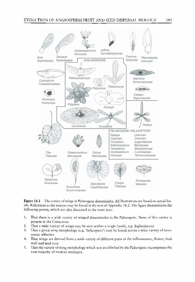
EVOLUTION OF ANGIOSPERM FRUIT AND SEED DISPERSAL BIOLOGY 345
Acer Deviacer Sapindaceae Sapindaceae
Cedrelospermum Joffrea Ulmaceae Cercidiphyllaceae
Fraxinus pteronepyles Oleaceae unknown
Tilia Tiliaceae
Palaeocarpinus Ostrya Betulaceae Betulaceae
PALAEOGENE 'HELICOPTERS'
Raskya Unknown Calycites Unknown Cruciptera Juglandaceae Asterocarpinus Betulaceae Tetrapterys Malplghlaceae Ceratopetalum Cunoniaceae Chaneya ?Simaroubaceae
Dipteronia Aceraceae
Eucommia Eucommiaceae
Diplodipelta Caprifoliaceae
Craigia Tiliaceae
Florissantia Malvales
Figure 18.1 The variety of wings in Palaeogene disseminules. All illustrations are based on actual fossils. References to the sources may be found in the text of Appendix 18.2. The figure demonstrates the following points, which are also discussed in the main text:
1. That there is a wide variety of winged disseminules in the Palaeogene. None of this variety is present in the Cretaceous
2. That a wide variety of wings may be seen within a single family, e.g. Juglandaceae 3. That a given wing morphology (e.g. 'helicopters') may be found across a wide variety of taxo-
nomic affinities 4. That wings are derived from a wide variety of different parts of the inflorescence, flower, fruit
wall and seed coat 5. That the variety of wing morphology which was established by the Palaeogene encompasses the
vast majority of modern strategies.
346 M. E. COLLINSON &c P. F. VAN BERGEN
exploit a wide variety of flight paths, trajectories and attitudes in flight (Burrows, 1986). Details of Palaeogene winged disseminules are summarized in Appendix 18.2. Although the summary is extensive it may not be totally comprehensive. However, it does serve to demonstrate the following key points:(l) that individual flowering plant families in the Palaeogene may show considerable variety of wing form (e.g. Juglandaceae, Figure 18.1, Appendix 18.2); (2) that comparable wing morphologies in the Palaeogene occur across systematic boundaries (e.g. helicopters. Figure 18.1, Appendix 18.2); (3) that comparable wing morphologies in the Palaeogene were developed from different anatomical entities (many examples. Figure 18.1, Appendix 18.2); and (4) that a very wide variety of wing morphology, on seeds and fruits, and derived from a variety of organs, had already evolved in the Palaeogene (many examples, Figure 18.1, Appendix 18.2).
In sharp contrast to the situation in the Palaeogene, Cretaceous relatives (where well-understood and based on actual fossils) tend to show unaided dispersal (Crane et al.^ 1995). Taphonomic bias may partly account for this observation as many studies of Cretaceous angiosperm fruits and seeds are based on sieved residues where wings would be less obvious than they are when preserved on bedding surfaces. Flowever, scars of former wing attachment should be obvious from SEM studies of specimens from sieved residues. Many Cretaceous angiosperms, especially those of the earlier phases of angiosperm evolution, were probably plants of small stature, which produced small fruits and seeds. Neither of these attributes would have favoured the earlier development of winged disseminules. Eriksson et al. (2000a) recorded up to 15% wind dispersal in late Cretaceous fruit and seed floras with lower levels (0-7%) in Palaeogene floras rising again in the Neogene. However, they inferred dispersal based on extrapolation from nearest living relatives, whereas in this chapter we document actual presence of wings in the fossils themselves. In addition, it is not clear if they included small dry fruits/seeds lacking wings in their wind-dispersed category. There are few examples of seeds with wings preserved in the literature on Cretaceous seeds, those that are documented (e.g. Crepet and Nixon, 1994) are minute wings on small seeds. The exceptionally preserved early Cretaceous angiosperms from the Crato Formation (bedding plane assemblages in fine grained limestone where wings would typically be preserved) do not include any winged disseminules (Mohr and Friis, 2000). Even if Cretaceous floras contained significant amounts of wind dispersed disseminules, any wings were small and certainly did not show the elaborate variety documented in Appendix 18.2 for the Palaeogene.
Abiotic ~ dust and microseeds
Cretaceous seeds, almost without exception, are small (volume less than 1 mm^) (Crane etaL^ 1995; Friis et al, 1999, 2000; Eriksson et al, 2000a,b). Only in the latest Cretaceous (Maastrichtian) is a small increase in size evident (Knobloch and Mai, 1986, 1991). Small seeds persist in the Palaeogene where they are joined by a wide variety of larger forms.
Cretaceous seed assemblages are only just beginning to be studied in detail so systematic affinities are mostly not yet clear enough to assess more detailed evolutionary patterns. Certainly, some families, with larger seeds today, are represented in the late Cretaceous (e.g. Campanian and Santonian) by extinct forms with small seeds. These include: members of the Fagales sensu APG, 1998 and Fagaceae (Herendeen et al, 1995; Sims et al, 1998; Schonenberger et al, 2001); the Juglandaceae (Friis, 1983; Knobloch and Mai, 1986, 1991; Herendeen et al, 1999) and the Mastixioid Cornaceae in the Maastrichtian (Knobloch and Mai, 1986, 1991). Less clear is the Cretaceous representation of taxa with small seeds

EVOLUTION OF ANGIOSPERM FRUIT AND SEED DISPERSAL BIOLOGY 347
today but it certainly includes members of the Ericales (Friis, 198Sb; Knobloch and Mai, 1986,1991; Schonenberger and Friis, 2001). Small seed size in the Cretaceous may in part be related to small stature (e.g. Wing and Boucher, 1998) of early angiosperms, a suggestion also made by Erikkson et al, (2000a), rather than reflecting the adoption of a particular dispersal strategy. Nevertheless, small seeds would be appropriate for the colonizing strategy implied by the occurrence of early angiosperm fossils in depositional environments representing disturbed habitats (e.g.Wing and Boucher, 1998).
Palaeogene floras also include small seeds and many of these belong to families w^hich are typified by small seeds today. Examples include:
1. Rhododendron (Ericaceae) with elongate seeds, with small terminal prolongations of the seed coat which are recorded from the Palaeocene/Eocene transition in England (Collinson and Crane, 1978; Collinson and Cleal, 2001c) and in the late Eocene of California (Wang and Tiffney, 2001)
2. Hydrangea (Hydrangeaceae) and Emmenopterys (Rubiaceae) with tiny winged seeds from the middle Eocene of the USA (Manchester, 1994b)
3. Hydrangeaceae, Ericaceae and Onagraceae from the middle Oligocene and Miocene of Europe (Mai, 1985b, 1998)
4. Restionaceae (Dettman and Clifford, 2000) from the Eocene/Oligocene of Australia 5. Juncaceae from the Eocene/Oligocene transition of England (Collinson, 1983a) 6. Lythraceae and Flacourtiaceae, with seeds less than 1mm in length, from the early
Eocene London Clay flora of England and middle Eocene Clarno flora of the USA (Reid and Chandler, 1933; Manchester, 1994b; Collinson and Cleal, 2001b).
Based on their size the smallest of these Cretaceous and Palaeogene seeds can only have had minimal stored reserves in the endosperm or embryo (no preservation known). Therefore, they may have relied on alternative nutritional associations like some of their living relatives.
Abiotic - flotation
The fibrous fruit wall of fossil Nypa fruits is indicative of the potential for dispersal by flotation similar to modern fruits of the genus. This is consistent with the fossil record of the fruits, including their sedimentary facies and strand-line-like accumulations (e.g. Collinson, 1993, 1996) and the overall comparability of the reproductive biology between modern and fossil examples. Some fossil seeds exhibit thin-walled tissues in the seed coat often referred to as 'spongy', which may represent light tissue aiding flotation. Examples include Decodon (Cevallos-Ferriz and Stockey, 1988a and references therein) and to a lesser extent Keratosperma (Cevallos-Ferriz and Stockey, 1988b). Water tight seed-coat layers, such as columnar palisades, may aid dispersal in water (see discussion on dormancy).
Biotic - dry fruits and seeds
Potential dispersal by animals is likely (based on inference via living relatives) for large dry nuts with an extensive food reserve, e.g. Juglandaceae, Betulaceae, Fagaceae which became abundant in the late Eocene, coincident with a radiation of rodents (Collinson, 1999; Collinson and Hooker, 2000; Hooker and Collinson, 2001). In the late Cretaceous, Juglandaceae and Fagaceae are represented only by small dry nuts, while Betulaceae have not yet been reported (see Appendix 18.2 and Chen et aL, 1999).

348 M. E. COLLINSON &c P. F. VAN BERGEN
Previous work has shown that the earliest examples of larder hoarding are Miocene (CoUinson, 1999; Hooker and Collinson, 2001; Gee et al., in press). Except in cases of hoarding, direct fossil evidence of animal interaction with nuts and dry seeds represents seed predation. These include rodent-gnawed seeds dating from the latest Eocene and insect borings in Eocene Rutaceae seeds (Collinson, 1999; Collinson and Hooker, 2000; Hooker and Collinson, 2001).
Biotic -- spines
Spines or hook-like protrusions are known on Eocene Fagus cupules (Meyer and Manchester, 1997), on Eocene Castanea cupules (Crepet and Dahglian, 1980), on Oligocene Sloanea capsules (Kvacek et aL, 2001) and on Palaeocene Aesculus capsules (Manchester, 2000b, 2001). They are also recorded on small (2 mm maximum dimension) early Cretaceous fruits named Appomattoxia (Friis et ai, 1995). Larger fruits of Trapa, Ceratostratiotes Gregor and Ceratophyllum (from the Palaeocene and Eocene) also bear short to long spines, sometimes hooked (Mai, 1985a, 1995; Herendeen etaL, 1990; Wehr, 1995; Manchester, 2000a; Meller and van Bergen, 2003), some of which are known attached to complete plants confirming their hydrophytic biology (Herendeen et al.^ 1990). Spines of aquatic disseminules can function as anchors and this has also been argued (Collinson, 1999) as their probable role in Appomattoxia. In other cases, especially when unhooked (e.g. Sloanea)., spines probably had a protective function. A dispersal function seems less likely in cases where fruits shed seeds on splitting.
The only known proof of fossil animal epizoochory of an angiosperm of which we are aware is that where mammalian hairs are entwined in the hooks of a fruit identified as the bamboo Pharus from late Eocene Dominican amber (Poinar and Columbus, 1992). Modifications favouring epizoochory seem to be exceptionally rare in Cretaceous and Palaeogene fruits and seeds in spite of the logical origin of mammalian fur in the late Triassic (Collinson, 1999; Hooker and Collinson, 2001).
Biotic - fleshy tissues
Fossil evidence for endozoochory has recently been documented elsewhere (Collinson, 1999; Hooker and Collinson, 2001). This evidence is derived from exceptionally preserved fossils including mammalian and bird gut contents and evidence for the former presence of soft tissues on fruits (e.g. from the middle Eocene of Messel, Germany, Richter, 1987; Schaal and Ziegler, 1992) as well as from dental morphology and microwear of mammaUan fossils. This evidence shows that, by the early Palaeogene, large fleshy fruit existed (e.g. Lauraceae, Vitaceae, Menispermaceae). Fleshy fruits were consumed by mammals and, at least in one case, the seeds survived uncrushed in the stomach and so could have been voided in faeces in a viable condition. Seed predation is also evident in the form of crushed seeds in the gut content of one mammal. However, these examples are rare and, although they prove seed predation and potential endozoochorous seed dispersal in the Palaeogene, most interpretation of animal dispersal is still based on inference from the presence (or inferred presence) of fleshy tissues. Mack (2000) argued that fleshy fruit pulp might have evolved as a defence against seed predation and secondarily become structures to promote seed dispersal.
By contrast to the Palaeogene, Cretaceous fruits and seeds were small and none were enclosed in large fleshy fruits. Some fruits possessed thin layers of fleshy tissue which may originally have been rather leathery (Knobloch and Mai, 1986, 1991; Crane et al., 1995; Friis et aL, 1999, 2000; Eriksson et al, 2000a). Eriksson et al. (2000a) documented that
EVOLUTION OF ANGIOSPERM FRUIT AND SEED DISPERSAL BIOLOGY 349
proportions of animal dispersal increased steadily from about 80 million years ago and reached a peak in the Palaeogene. However, their study relied mainly on living relatives to assign a dispersal system for the fossils (see also Abiotic - wings above). In one very well-studied early Cretaceous fruit and seed flora (Eriksson et ai, 2000b) it was determined that about one quarter of the taxa could be interpreted as drupes or berries with specialised tissues for animal dispersal. This interpretation was based on the presence of thin outer fruit walls. The average fruit volume was 2.22 mm^ (range 0.31-7.58 mm^). Thus, the Cretaceous fleshy fruit resource is of an utterly different category, in terms of size and tissue volume, compared with that of the Palaeogene. The significance of Cretaceous birds or reptiles for fruit and seed dispersal is uncertain but small Mesozoic mammals, such as multituberculates, haramyids and docodonts all had dentitions indicative of potential fruit or seed eating (Eriksson et al.^ 2000a,b; Hooker and Collinson, 2001). However, direct evidence of a dispersal (as opposed to predation) role for Cretaceous fruit and seed feeders is lacking, though it could be argued that such small seeds would avoid tooth crushing and survive to be defecated much as tomato seeds do in humans today.
Germination and establishment
Embryo and endosperm
Radicle emergence Emergence of a radicle (hence proof not only of presence of an embryo but also of mode of germination) is recorded in fossil seeds but very rarely. We are aware of only three examples. One is a late Cretaceous charred seed (Friis, 1985a), the second the seeds named Microphallus Manchester (affinity unknown) from the middle Eocene Clarno flora (Manchester, 1994b) and the third three specimens oi Joffrea (Cercidiphyllaceae) from a compression flora with numerous seedlings (see seedlings below). Microphallus is quite common (25 specimens in a flora where a number of taxa are represented by fewer than ten specimens but others by 200 +). The number of specimens showing radicle emergence probably indicates more or less simultaneous germination. (See also under seedlings below.)
Dispersal strategies (above) suggest utilization of appropriate orientations, positionings, or placements, at least in the Palaeogene. However, there is little or no evidence of the elaboration of this such as is associated with many dry land herbs today (e.g. no twisting awns etc).
Embryo and endosperm development, dormancy and establishment Sources of evidence. Charred fossils from the Cretaceous do preserve anatomical details of delicate tissues such as endosperm. However, the emphasis in the study of the Cretaceous charred angiosperms has so far been largely on the flowers which yield ovule rather than seed detail (e.g. Drinnan et al.^ 1990). Friis (personal communication 2001) considers that the Cretaceous dispersed charred seeds (e.g. Eriksson et al., 2000b) do hold potential for investigation of embryo and endosperm characteristics.
The degree of development of the embryo and endosperm (see following section) can be assessed from several permineralized floras from the Cretaceous/Tertiary transition and Palaeogene. These are summarized below. Where embryo and endosperm preservation is lacking this can sometimes be shown to be due to the activities of fungi prior to permineral-ization (Kalgutkar et al, 1993; Stockey et aL, 1998).
The floras from the Deccan Intertrappean series have been variously considered as Tertiary or Cretaceous in the past, but recent dating suggests that the Intertrappean Series

350 M, E. COLLINSON &c P. F. VAN BERGEN
spans the Cretaceous/Tertiary (Maastrichtian/Palaeocene) transition including the boundary itself (Courtillot et al, 1988; Jaeger et al, 1989; Courtillot, 1999; Guleria and Srivastava, 2001). Reviews of the flora and floral hsts may be found in Prakash (1960, 1978), Bande et al. (1988) and Band and Chandra (1990). The fossils are preserved as silica perminer-alizations or petrifactions and hence preserve some anatomical details not found in compression floras. There are relatively few fruits or seeds described from this flora. A large number of these have suggested affinity with the palms (e.g. hsts in Prakash, 1960, 1978). Some of these do not preserve internal seed details (e.g. Mehrotra, 1987) while others do (e.g. Bonde, 1990), the latter having a small, apical undifferentiated embryo within extensive non-ruminate endosperm. Seeds of Viracarpon (PAraceae, Prakash, 1978) are unfortunately not sufficiently well-preserved to reveal embryo or endosperm detail (Chitaley, 1958). Fruits of Tricoccites Rode are known in organic attachment to plants with Cyclanthodendron Sahni and Surange stems (Bonde, 1985; Biradar and Bonde, 1990) with affinity with Musaceae/ Strelitziaceae. Their anatomical preservation is discussed below. In addition, fruits of Enigmocarpon (PMyrtales) are also discussed below.
The middle Eocene flora from the Princeton Chert is reviewed by Cevallos-Ferriz et al. (1991) and Pigg and Stockey (1996) and is currently under active study (e.g. Stockey etal,, 1998). This flora is permineralized by silica and provides excellent anatomical detail (e.g. Cevallos-Ferriz et al, 1991; LePage et al, 1997; Stockey et al, 1998). The early Eocene London Clay flora was reviewed by Collinson (1983b) and CoUinson and Cleal (2001b). The fossils are preserved by pyrite permineralization and many yield good anatomical detail. The middle Eocene flora of the Clarno Chert was monographed by Manchester (1994b). The specimens are mostly preserved as silica petrifactions with some permineralization yielding anatomical details of less resistant tissues. The late Palaeocene Almont flora was reviewed by Crane et al (1990) and also contains some anatomical details as the result of siliceous preservation. Fossil evidence of Palaeogene endosperm and embryos. The following list summarizes the direct fossil evidence for Palaeogene endosperm and embryos taking all the above floras into account. Additional references are cited where necessary.
Small undifferentiated embryos with large amounts of endosperm occur in Musaceae from the middle Eocene of Clarno (Manchester and Kress, 1993; Manchester, 1994b) and some seeds assigned to palms from the Deccan flora (Bonde, 1990).
Direct evidence of the presence of an extensive ruminate endosperm filling the seeds, and hence indirect evidence of a small embryo, is known from numerous seeds. In particular these are numerous forms of Anonaceae seeds (Anonaspermum) in the Eocene Clarno and London Clay floras and also from the Palaeocene of Pakistan with earliest records in the Maastrichtian of Nigeria (review in Tiffney and McClamer, 1988; see also As-Saruri et al, 1999). Vitaceae from the London Clay and Clarno are another example.
Perisperm (a product of the nucellus) accompanied by a small embryo has also been inferred on the basis of cellular preservation with a suitably placed embryo cavity in seeds of Allenbya (Nymphaeaceae) from the middle Eocene Princeton Chert (Cevallos-Ferriz and Stockey, 1989) and in seeds of the dicotyledon Princetonia Stockey from the same flora (Stockey and Pigg, 1991). In the absence of anatomical preservation it is not possible to establish degree of differentiation of embryo in these examples.
Small differentiated embryos are represented in Tricoccites from the Deccan flora (see Zingiberales below).
Larger differentiated embryos with little endosperm include Decodon and Enigmocarpon (Lythraceae and extinct relative) (see Myrtales below) and Keratosperma (Araceae) from
EVOLUTION OF ANGIOSPERM FRUIT AND SEED DISPERSAL BIOLOGY 351
the middle Eocene Princeton Chert (Cevallos-Ferriz and Stockey, 1988a,b; Cevallos-Ferriz et aL^ 1991) and Trema (Ulmaceae) from the middle Eocene of Clarno which has a large coiled embryo with a prominent curved radicle. A weakly differentiated (possibly immature) embryo not associated with endosperm is preserved in Palaeorosa Basinger (Rosaceae) from the Princeton Chert (Cevallos-Ferriz et al, 1993).
Embryos filling the seed and fully differentiated are found in Sapindaceae and Juglans from the Eocene of the London Clay and Clarno. The Sapindaceae are diverse and represented by several clearly distinct forms (distinct at generic level and with several morphologies within each form genus). Each shows a fully differentiated radicle and paired folded cotyledons comparable with modern members of the family. In the case of Juglans the nature of the embryo with two large relatively smooth cotyledons is inferred from chalcedony casts of the embryo cavity (Manchester, 1994b) which conform to those of living relatives.
Although relatively limited, these data show examples of a range of strategy in the early Palaeogene. At one extreme this variety includes potentially dormant embryos and embryos with slow germination as the food reserve is mobilized from a large endosperm store to the embryo. However, the examples are ruminate, thus increasing the surface area for this mobilization. In contrast, there are larger differentiated embryos indicating faster germination and establishment. The bent or folded embryos, where the curved structures stretch on germination, increasing the chances of reaching the soil surface and placing cotyledons above adjacent vegetation or litter etc., are indicative of strategies enhancing the chance of rapid establishment (Bewley and Black, 1994).
Embryo and endosperm in the order Myrtales In the case of the Decodon seeds, the relationships to the Lythraceae and to Decodon are confirmed by the reconstruction of two partial to complete whole plants from the middle Miocene (Kvacek and Sakala, 1999) and middle Eocene, (Little and Stockey, 2000). These also confirm the habitat of the fossils based on facies and anatomical evidence as being from aquatic to swampy conditions similar to the extant relative D. verticillatus (the former) and in submerged conditions with fluctuating water levels at the edge of a shallow water system (the latter). Seeds of Decodon and extinct relatives have an extensive Palaeogene fossil record (Tiffney, 1981; Cevallos-Ferriz and Stockey, 1988a; Manchester, 1994b).
The Cretaceous/Tertiary transitional Deccan fruit Enigmocarpon and its allied flowers are a very abundant element in the Deccan intertrappean flora. These have been compared to modern members of the Myrtales especially Sonneratiaceae and Lythraceae (Sahni, 1943; Shukla, 1944; Chitaley, 1977; see also discussion in Friis and Crepet, 1987:161). In combination these two examples offer an opportunity to consider the evolution of embryo and endosperm in the order Myrtales.
Both Enigmocarpon (Sahni, 1943) and Decodon allenbeyensis (Cevallos-Ferriz and Stockey, 1988a) have relatively large developed, differentiated embryos with little endosperm or no endosperm. This is comparable to seeds of extant Decodon (Baskin and Baskin, 1998) and typical of Myrtales (Cronquist, 1981) showing that the modern strategy had developed by the Palaeogene. Unfortunately, other permineralized Myrtales do not exhibit embryo or endosperm preservation (e.g. berries of Paleomyrtinaea from the Eocene Princeton Chert and Palaeocene Almont floras, Pigg et aL, 1993). Further details on the fossil record of the order, including another partially reconstructed plant from a compression flora, are given by Manchester et al. (1998).

352 M. E. COLLINSON &c P. F. VAN BERGEN
Embryo, endosperm and seed internal organization in the order Zingiberales Rodriguez-de la Rosa and Cevallos-Ferriz (1994) described two types of late Cretaceous (Campanian) permineralized zingiberalean fruits from Mexico. The Formation in which these fossils were found contains dinosaurs and is biostratigraphically dated as latest Campanian (Kirkland et al., 2000). One fruit, Striatornata, Rodriguez-de la Rosa and Cevallos-Ferriz, was judged to be closely related to the extinct Spirematospermum (represented by compression fossils of seeds and fruits from the late Cretaceous to Neogene, see summary in Manchester and Kress, 1993). Both of these genera are assigned to the Musaceae based on the presence of a chalazal chamber and hilar cavity. In Striatornata the embryo sac itself is represented by an internal mould but cells are preserved in the chalazal chamber. Middle Eocene permineralized seeds of Ensete oregonense Manchester and Kress (1993), (see also Manchester, 1994b, 1995) of the Musaceae, also contain a chalazal chamber and hilar cavity. In addition, a few specimens reveal preservation of an endosperm chamber containing a small, straight bulbous embryo suspended from the hilar end. A small cylindrical to bulb-shaped embryo in this position is characteristic for the Zingiberales (Manchester and Kress, 1993) while the enlarged chalazal chamber appeared to be diagnostic for the family Musaceae (Manchester and Kress, 1993) where it may function to assist germination. In modern Musaceae, the embryo is usually shorter than in other families of the order, as it is in the fossil Ensete (Manchester and Kress, 1993).
The other mexican Cretaceous fruit, Tricostatocarpon Rodriguez-de la Rosa and Cevallos-Ferriz, was of uncertain affinity within the order, lacking the musaceous chalazal chamber. Other fossil seeds of uncertain affinity within the order, are permineralized specimens formerly assigned to Musa but which lack the chalazal chamber, originally described by Jain from the Cretaceous/Tertiary transitional strata of the Deccan Inter-trappean Beds of India (Manchester and Kress, 1993). Fruits of Callistemonites Bande et ai, also from the Deccan, were assigned to Musaceae and are certainly similar to that illustrated by Manchester (1995) but, although seed morphology is well-preserved, no internal anatomical detail could be discerned (Bande et al.^ 1993). The genus Cyclanthodendron, a reconstructed whole plant said to combine features of the Musaceae and the Strelitziaceae, bore the fruits described as Tricoccites (Bonde, 1985; Biradar and Bonde, 1990) and comes from the Cretaceous/Tertiary transition of the Deccan Intertrappean Beds. Seeds contained within these fruits reveal a large cellular endosperm containing a small elongate, cylindrical (ribbon-like in section), cellular embryo in which some possible differentiation was observed (Bonde, 1985). However, the fruit structure is considered as controversial and poorly known by Biradar and Bonde (1990) suggesting a need for re-investigation of the details.
Overall these fossils suggest that the internal organization characteristic of modern seeds of Zingiberales was in place by the late Cretaceous. Specific internal structures (e.g. large chalazal chamber), combined with embryo and endosperm preservation, proves organization comparable with modern Musaceae at least by the middle Eocene.
Seedlings
Vivipary in Rhizophoraceae Direct evidence of establishment strategy comes from fossil seedlings.Viviparous embryo hypocotyls of Ceriops and Palaeobruguiera (Rhizophoraceae), first recognized by Chandler, were fully described by Wilkinson (1981, 1983). That work shows beyond doubt that vivipary was exhibited by early Eocene Rhizophoraceae and specimens are preserved at two localities (Heme Bay and Sheppey) of slightly different ages (CoUinson, 1983b). The London

EVOLUTION OF ANGIOSPERM FRUIT AND SEED DISPERSAL BIOLOGY 353
Clay flora is perhaps the only fossil flora likely to demonstrate this as rapid permineral-ization with anatomical preservation is required in a flora from a brackish to marine facies. Another possibility is the Deccan Traps where the mangrove palm Nypa is found (see below) but Nypa is very rare. In sites such as Bracklesham (Collinson, 1996), rich in Nypa, but with preservation in the form of carbonaceous compression, mangrove hypocotyls probably would not have been preserved, or if preserved might not be recognizable as such.
Tomlinson and Cox (2000) showed, by experimentation, that modern viviparous seedlings were able rapidly to erect after rooting if dispersed by floating on the seawater surface and then, as likely, stranded horizontally. This strategy raises the plumule above tidal influence. It leaves anatomical evidence in the form of reaction (tension) wood fibres and morphological evidence in a resultant hook-like base. Wilkinson (1981) emphasized that the fossils were fragments of the hypocotyl, although she indicated that 'the fragments most frequently found suggest that they come from the distal tip'. The fossil Ceriops from Sheppey is sufficiently abundant (one per visit, personal observation) to examine for evidence of self-erection, but studies would have to be accompanied by detailed anatomical work to demonstrate that the true base of the seedling was preserved. Collinson has not encountered a hooked Ceriops during years of collecting in the London Clay of Sheppey. Although specimens figured by Chandler (1961, 1978) and Collinson (1983a) are curved, this is likely to be merely normal curvature of the hypocotyl axis. Only one is strongly curved (Chandler 1978: plate 7 Figure 1) and Chandler (1978) stated specifically that this is broken and incomplete at the radicle end. Therefore, the erection strategy to raise the plumule, is not demonstrated in the Palaeogene embryos.
Vivipary in Nypa The mangrove palm Nypa also exhibits vivipary today (Tomlinson, 1986; Farnsworth, 2000). Fossil Nypa fruits have been shown to be exactly comparable to modern fruits in many ways (Collinson, 1993). The only obvious difference being that the fossil fruits reached larger maximum sizes than the modern fruits. However, in all Nypa fossils, even permineralizations, the seed-containing cavity has a mineral (amorphous silica, micro or macrocyrstalline pyrite) or, rarely, sediment infill. This is the case for the London Clay flora based on many different workers who have studied hundreds of specimens (Bowerbank, 1840; Reid and Chandler, 1933; Chandler, 1961; Collinson, 1993, personal observation); and for the Belgian Brussels Sands specimens where again, many have been studied (Stockmans, 1936; Collinson, 1993). It is also the case for the Deccan Intertrappean flora, although only four specimens have been studied anatomically and Nypa is apparently very rare in this flora (Sahni and Rode, 1937; Chitaley, 1960a,b; Nambudiri, 1966; Chitaley and Nambudiri, 1969). Three-dimensionally preserved specimens with carbonaceous preservation, e.g. from the Eocene of Bracklesham, West Susssex, England (Collinson, 1996) have sediment infill in the seed cavity.
These Nypa fossils have a basal aperture (Reid and Chandler, 1933; Sahni and Rode, 1937) which is comparable with the germinal aperture in modern fruits. Mature modern fruits have a small undifferentiated embryo which then differentiates while still attached to the fruiting head. The detachment of the fruits from the fruiting head is assisted by the growth of the plumule, which breaks through the fruit at the base (Tomlinson, 1986). The fact that many fossils are found dispersed is not in itself proof of shedding through viviparous seedling growth because many aborted fruits, lacking seeds or former seed-containing cavities, are also found fossil (Collinson, 1993, 1996; Pole and MacPhail, 1996).
There are several explanations why embryo preservation has not been documented in any permineralized Nypa fruit specimens. First, once the plumule has penetrated the fibrous

354 M. E. COLLINSON &c P. F. VAN BERGEN
mesocarp at the base of the fruit, the germinal aperture is formed. At this stage, faiUng estabUshment, the soft tissues would be readily vulnerable to decomposition. Secondly, once established and the food resource used up, the fruit could be released from the seedling yielding an empty husk with high preservation potential. Thirdly, it should be noted that relatively few of the mineralized specimens have actually been cut and polished or thin-sectioned so that future work might reveal embryos in Nypa fossils. Such work on some silicified specimens from Curacao is in progress (CoUinson). In conclusion, although vivip-ary cannot be proven in the case of Nypa fossils, the lack of seed and embryo preservation in many fossils is consistent with a viviparous habit comparable with the modern Nypa.
Other seedlings Numerous (over 8000) fossil seedlings have been found in a thin layer, traced laterally for 50-60 m in the Palaeocene of Joffre Bridge in Canada; 99% of these seedlings are of Joffrea, a Cercidiphyllaceae (Stockey and Crane, 1983), reconstructed as a partial whole plant (Crane and Stockey, 1985). Other seedlings (about 100 specimens) are platanaceous (Stockey and Crane, 1983; Crane and Stockey, 1985; Pigg and Stockey, 1991). Almost all seedlings are preserved upright in growth position. They are preserved at many different stages of development but none have more than three pairs of leaves suggesting that they all germinated at approximately the same time. Germination was epigeal producing an emerging radicle, thin hypocotyl and two cotyledons 2-4 mm long (Joffrea) and epigeal yielding two cotyledons with other parts obscure (Platanaceae). These two plants have small seeds, produced in large numbers (especially Joffrea and relatives with small winged seeds) and their remains are frequently found in association in fluvial sediments. Together with the seedling biology these features suggest that the plants represent early successional communities, with rapid seedling establishment that occurred in disturbed habitats along stream margins (Stockey and Crane, 1983; Pigg and Stockey, 1991). Hoffman and Stockey (1999) place the seedling bed at Joffre Bridge in detailed sedimentological context which supports this interpretation. Joffrea seedlings have also been recorded, along with seedlings of a taxo-diaceous Metasequoia-like plant, in the Palaeocene of two other Palaeogene localities in Canada in comparable situations (Falder et aL, 1999).
Dormancy versus germination
Categories of dormancy can be generally summarized as follows (Bewley and Black, 1994: Chapter 5; Baskin and Baskin, 1998: 28)
Endogeneous (sensu Baskin and Baskin); broadly equal to embryo controlled Physiological - physiological inhibition Morphological - underdeveloped embryo; released by suitable conditions for growth Morphophysiological - the above combined.
Exogenous = coat imposed/enhanced Physical - coat impermeable to water; released by specialized opening mechanism Chemical - germination inhibitors; released by leaching Mechanical - restriction (physical constraint) on growth by woody tissues; released by stratification.
Germination may be defined as beginning with water uptake and ending with axis (usually radicle) emergence from the seed (Bewley and Black, 1994). Direct fossil evidence of radicle emergence during germination does exist but is very limited (see above). Fossil
EVOLUTION OF ANGIQSPERM FRUIT AND SEED DISPERSAL BIOLOGY 355
evidence of vivipary and of other seedlings (see above) also prove germination mechanisms in fossils. Preserved embryo evidence (see above) clearly show^s differentiated embryos v^hich are unlikely to have had morphological dormancy as well as probable dormant seeds with poorly developed embryos.
Exogenous, coat-imposed/enhanced, dormancy can act by interference with water uptake, mechanical restraint, influencing gas exchange, preventing exit of inhibitors (from/to) embryo and supplying inhibitors. In exogenous dormancy the fruit or seed coat is extremely important. To quote Boesewinkel and Bouman (1995:9): 'The seed coat is not just a protective covering but a multifunctional organ that transports, transforms and secretes metabolites and oxygen to the embryo sac'. The fossil record provides a huge amount of evidence on the fruit and seed coat.
Hard seed coats have long been considered to be a widespread cause of dormancy. Many fossils have hard seed (or endocarp) coats so it seems highly likely that many (or at least some) were probably dormant. Direct evidence of the specialist opening mechanisms that are associated with physical dormancy, as defined above, is readily available and widespread in the Palaeogene fossil record. These include plugs or valves in endocarps of Anacar-diacaeae, Mastixioid and other Cornaceae, Nyssaceae, Potamogetonaceae and in seeds of Nymphaeaceae (Collinson, 1983b; Mai, 1993, 1998; Manchester, 1994b; van Bergen et aL, 1996; Tiffney and Haggard, 1996; Stockey et aL, 1998; Manchester et al, 1999). Maastrichtian (latest Cretaceous) mastixioid Cornaceae are also recorded (Knobloch and Mai, 1986, 1991).
Bewley and Black (1994) cite experiments that indicate the importance of the osteosclereid layer beneath the palisade layer where, in dormant seeds, puncture of the osteosclerieds is necessary for germination to occur. However, most of this experimental evidence is derived from agriculturally important plants, few of which have a Palaeogene record. Therefore, this experimental evidence cannot easily be extrapolated to the past.
Columnar palisades and thick tough seed/endocarp coats with potential for imposing or enhancing coat dormancy, are very common in Cretaceous and Palaeogene seeds. However, these are very variable and the relative potential for dormancy conferred by each of the following, for example, is uncertain:
(i) Extremely close-packed columnar palisades (e.g. modern legumes) are represented in endocarp walls such as Neogene Cinnamomum (Pingen et al.^ 1994) and many Lauraceae endocarps are known in the Palaeogene. There is an extensive record of fossil legumes (Herendeen and Dilcher, 1992). However, data on seed coat anatomy seem to be lacking, seeds are generally poorly preserved and much of the work has relied on fruits. In their study of Nelumbo disseminule preservation, van Bergen et al. (1997) showed that a closely packed columnar structure need not imply high chemical resistance and argued that a distinctive tannin polysaccharide composition might account for the fossil record lacking Nelumbo disseminules while Nelumbo-likQ leaves occur from the Cretaceous onwards. It is possible that legume seed-coat chemistry may have affected the preservation potential of legume seeds.
(ii) Closely packed columnar sclerotesta, e.g. Nymphaeaceae, widespread in the Palaeogene which, if water tight, would aid aquatic dispersal and could also confer dormancy (Collinson, 1980; Cevallos-Ferriz and Stockey, 1989; Cevallos-Ferriz et al., 1991; van Bergen et al., 1996).
(iii) Rather porous or open columnar sclerotesta, where palisade cells have larger lumina than in i, or ii, as, for example, in some Nymphaeaeceae (Collinson, 1980; van Bergen
356 M. E. COLLINSON &c P. F. VAN BERGEN
etal., 1996) and Moraceae (CoUinson, 1989) and also commonly present in Cretaceous seeds (Friis et al, 1999, 2000).
(iv) More equiaxial as opposed to columnar sclereids (e.g. Torricellia endocarp, Collinson 1988 (as .^Lythraceae); Meller and Collinson unpublished data).
(v) Varying thickness of these layers, which are obviously relatively thinner, (though not necessarily proportionately thinner in terms of overall seed dimension) in smaller Cretaceous seeds.
Overall, the common occurrence of specialized plugs and valves as v^ell as distinctive dehiscence patterns, combined w ith thick, tough, compact seed (or endocarp) coat organization, combined with the presence of fossils of both germinated and non-germinated but of mature size seeds (Collinson, 1999), all argue strongly for the presence of coat-induced dormancy of a wide diversity in Palaeogene seeds. Until more detailed studies are made on large assemblages of Cretaceous seeds, with this question in mind, the evidence prior to the Palaeogene is less clear but suggestive of the existence of fewer specializations at that time.
Of course we cannot easily address seed longevity in these fossil assemblages as we do not know if the germinated fossils germinated immediately on shedding or how long the last to germinate existed prior to germination. The more recent fossil/archaeological record does have the potential to address longevity in the natural environment (see review in Bewley and Black, 1994). There are claims for Arctic lupin to 10 000 years and Nelumbo to 1700 years (but both have no direct dating). Reliable evidence would depend on accurate dating of associated materials or of a molecule from the seed coat that has not been metabolically active thus avoiding recycled carbon. Shen-Miller et al. (1995) reported a Nelumbo seed which was germinated and then destroyed for dating giving an age of 1288 ± 271 years old. Another specimen, the plant of which was still growing in 1994, was dated from a fragment of pericarp as 332 ± 135 years old. As van Bergen et al. (1997) have shown the Nelumbo disseminule wall is not only physically very tough but also has a distinctive tannin/ polysaccharide composition which may play a role in seed longevity. Legume seeds from herbarium sheets of known age, were shown to germinate after some 150 years when affected by fire extinguishing water at the British Museum of Natural History. The best-authenticated large scale example seems to be the Beal experiment where, of 21 species buried, only one retained viability after 100 years (1979), although several extended to 35-50 years. (Bewley and Black, 1994). However, this experiment was undertaken mainly on small-seeded, economically important arable plants or weeds and so gives Uttle indication as to the link between seed coat anatomy and longevity in the vast majority of angiosperms.
A broadly-based study of the relationship between the anatomy of the seed (or endocarp) coat, the possession of specialist physical opening structures and other physical attributes of the seed coat and the longevity of seeds or their dormancy mechanisms would be of considerable value. In this regard the fossil record is able to provide direct evidence of the location, morphology and structure of the physically and chemically resistant layers (which survive as fossils). These include cuticles and thick-walled sclerotic tissues (Figures 18.2 and 18.3) both of which are common constituents of fossil seeds and endocarps (Collinson, 1980, 1983b; Collinson and Gregor, 1988; van Bergen et al., 1996, 1999; and see the following section).
Chemical composition of resistant layers
Fossil fruits and seeds highlight those layers which are sufficiently resistant (e.g. to decomposition) to have survived in the fossil record. These layers can, therefore, be argued to have had a function in life related to their survival ability, such as in dormancy (physical
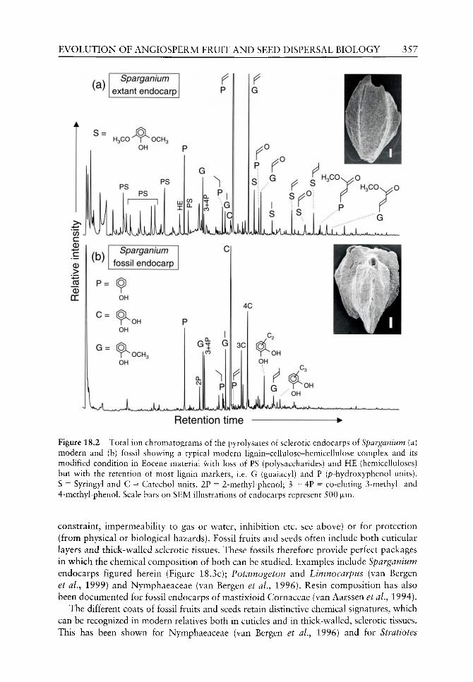
EVOLUTION OF ANGIOSPERM FRUIT AND SEED DISPERSAL BIOLOGY 357
Sparganium extant endocarp
Retention time •
Figure 18.2 Total ion chromatograms of the pyrolysates of sclerotic endocarps of Sparganium (a) modern and (b) fossil showing a typical modern lignin-cellulose-hemicellulose complex and its modified condition in Eocene material with loss of PS (polysaccharides) and HE (hemicelluloses) but with the retention of most lignin markers, i.e. G (guaiacyl) and P (p-hydroxyphenol units). S = Syringyl and C = Catechol units. 2P = 2-methyl-phenol; 3 + 4P = co-eluting 3-methyl- and 4-methyl-phenol. Scale bars on SEM illustrations of endocarps represent 500 |JLm.
constraint, impermeability to gas or water, inhibition etc. see above) or for protection (from physical or biological hazards). Fossil fruits and seeds often include both cuticular layers and thick-walled sclerotic tissues. These fossils therefore provide perfect packages in which the chemical composition of both can be studied. Examples include Sparganium endocarps figured herein (Figure 18.3c); Potamogeton and Limnocarpus (van Bergen et al.^ 1999) and Nymphaeaceae (van Bergen et al., 1996). Resin composition has also been documented for fossil endocarps of mastixioid Cornaceae (van Aarssen et al., 1994).
The different coats of fossil fruits and seeds retain distinctive chemical signatures, which can be recognized in modern relatives both in cuticles and in thick-walled, sclerotic tissues. This has been shown for Nymphaeaceae (van Bergen et al.^ 1996) and for Stratiotes
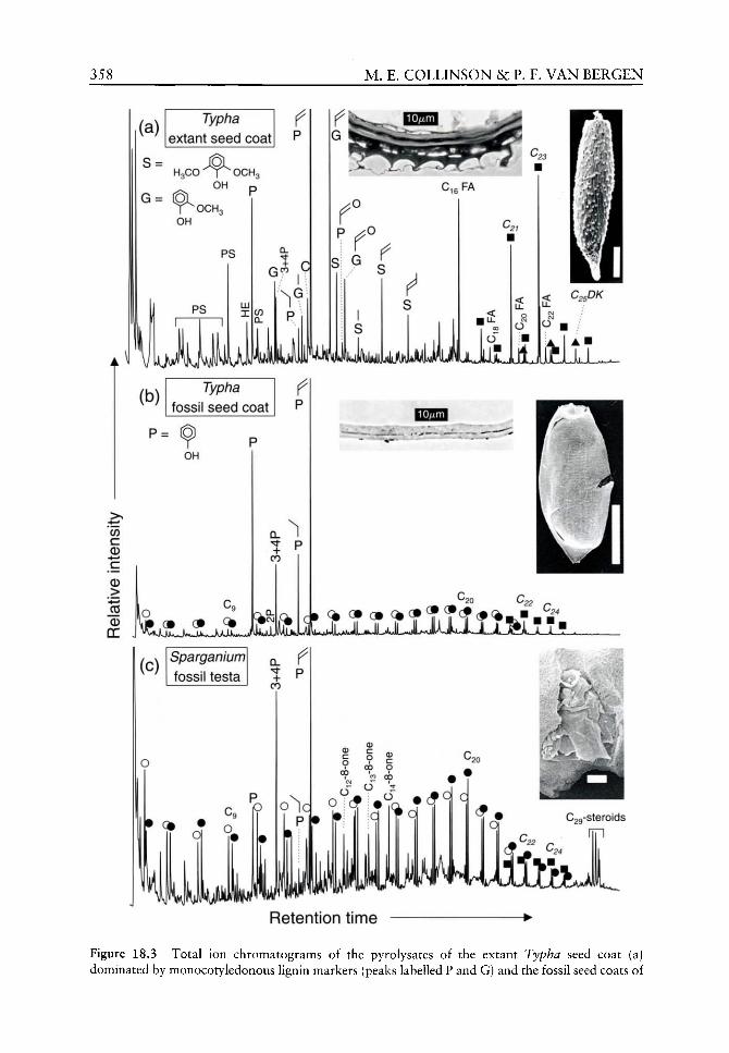
358 M. E. COLLINSON & P. F. VAN BERGEN
P ^ o
C,eFA
f ^
L41JUJAJ^^
|< C^jD/C
(b) Typha
fossil seed coat P
P= ^
OH
C CD
CD
>
T p CO
•«i-«»I,;.,^^M«« 4*^
I C I 22 /-»
(c) Sparganium
fossil testa
o
P ^ ol o
iw^^ Retention time
Figure 18.3 Total ion chromatograms of the pyrolysates of the extant Typha seed coat (a) dominated by monocotyledonous Hgnin markers (peaks labelled P and G) and the fossil seed coats of

EVOLUTION OF ANGIOSPERM FRUIT AND SEED DISPERSAL BIOLOGY 359
(van Bergen et aL, 1999). These results demonstrate that the particular biosynthetic pathways producing resistant fruit and seed coats, which exist today, also existed in the past (at least as far back as mid-Palaeogene). The results also add a new dimension of chemosystemat-ics to the study of fossil seeds. For example, Potamogeton and the extinct Limnocarpus show clear chemosystematic relationship, which supports that previously based only on morphological comparisons (van Bergen et al., 1999).
Here we report new results from Sparganium and Typha (families Sparganiaceae and Typhaceae, together order Typhales sensu Dahlgren et al., 1985; and family Sparganiaceae, order Poales Small sensu APG, 1998). The fossil samples are derived from the Bembridge Limestone Formation, Gurnard Ledge, Thorness Bay, Isle of Wight, from the top 20 cm of Daley and Edwards (1990) bed 6C (see CoUinson and Cleal, 2001a for details of the flora and references) and thus are from the Eocene/Oligocene transitional interval, probably latest Eocene in age.
The chemical composition of the modern and fossil resistant disseminule walls was examined by pyrolysis, which is a destructive method using thermal decomposition to yield characteristic building blocks (van Bergen, 1999). Details of methods are as reported in van Bergen et aL (1996, 1999). The fossil Sparganium sclerotic endocarp shows, as expected, modification of the lignin-hemicellulose complex (compared with the modern endocarp) with the loss of PS (polysaccharides) and HE (hemicellulose) but with the retention of most lignin markers, i.e. G (Guaiacyl) and P (p-hydroxy-phenol) units (See Figure 18.2). The chro-matogram of the pyrolysate (Figure 18.2b) is quite distinct from that of the sclerotesta of fossil Stratiotes (order AUsmatales Dumort. sensu APG, 1998) reported by van Bergen et aL, (1999) from the same fossil assemblage.
The fossil Typha and Sparganium seed coats are thin cuticles and their total ion chro-matograms of the pyrolysates are dominated by alkene-alkane doublets (see Figure 18.3), which are long straight chain aliphatic hydrocarbon units that are typical of fossil cuticles (see Chapter 8). In addition, there are phenolic pyrolysis products, most probably derived from p-coumaric acid moieties. The extant Typha seed coat, in contrast, is multilayered, including cuticular layers and cellular layers. The chromatogram is dominated by mono-cotyledonous lignin markers (e.g. peaks labelled P and G on Figure 18.3a) which swamp out a small signal from the cuticles which can be detected in appropriate mass chromatograms. Detailed mass chromatograms (Figure 18.4) are essential to provide chemical evidence for similarities. These show that the fossil Typha and Sparganium seed coats are strikingly similar in distribution patterns of specific constituents such as the alkanes (sharp drop off after C20 and C22) and the 2-alkanones (sharp drop off after C22 and C24). These latter compounds are also characteristic long-chain aliphatic units in cuticular membranes and in combination with alkanes/alkenes provide crucial evidence of chemical similarities. In addition, there is a distinct group of 8-alkanones which are dominated by the Ci4-8-alkanone
Typha (b) and Sparganium (c), dominated by alkene-alkane doublets with phenolic pyrolysis products most probably derived from p-coumaric acid moieties. The difference between fossil and modern is largely explained by the fact that the modern Typha has a multilayered seed-coat as shown in the transverse section, while the resistant material which survives in both the fossils is translucent cuticles only (as shown in the fossil Typha transverse section). Entire seed cuticles are illustrated by scanning electron micrographs; that of Sparganium is in place within a fractured endocarp. Transverse sections are transmitted light micrographs. Open circles: w-alk-1-enes, closed circles: w-alkanes, closed squares: 2-alkanones (= methyl ketones), Closed triangles: diketones, other abbreviations as in Figure 18.2. Scale bars on SEM illustrations of specimens represent 250 |xm.

360 M. E. COLLINSON & P. F. VAN BERGEN
(a) Typha fossil seed coat
^ •c„
m/z 55+57 • •
• q ^^ S! i
(b)
ILJILJUI
Q
o
q •
UJu^^ Sparganium fossil testa
Q.
U f'VjJ
••^(c) CO ^ ^
c B m/z 58
> "•4—•
_c5
DC
A77/Z 55+57
O
Typha fossil seed coat
LJLJJ LJ LJll
I CO O
°P c3" cx)
T ^ ^ •
uLlJjMitJlJ JJAAMIII
q q
^ | . . ^ A l ~ y ^ ^ - ^ « J ^ . _ ] ^ . <iJI, ..<jt, „,
(d) Sparganium fossil testa m/z 58
•
o 0) 00
0 C CO
CD ? CJ C 00
00 O
O
O
J M H H •
LJLjJLiij^^ IMS. Retention time

EVOLUTION OF ANGIOSPERM FRUIT AND SEED DISPERSAL BIOLOGY 361
in both taxa. Furthermore, these mass chromatograms are very different from those documented in cuticular seed coats of Potamogeton and Limnocarpus (order Aiismatales Dumort. sensu APG, 1998) from the same fossil assemblage where alkanes show drop off at C26 and 2-alkanones (=methyl ketones) at C28 (van Bergen et aL, 1999).
Fossils readily reveal resistant fruit wall and seed coat layers, the morphology and anatomy of which can easily be studied. Our results, from a variety of flowering plants (Nymphaeaceae, Potamogetonaceae, Stratiotes, Typha and Sparganium - see above), demonstrate that markers of chemosystematic and biosynthetic significance are surviving in Palaeogene fossils. These results indicate the potential to investigate any links (in fossils and their living relatives) between chemical composition, physical nature and the anatomy of fruit and seed coats and their functional role. This new combined information can be applied, in future, not only to understand the evolution of exogenous dormancy via the fossil record but also further to understand controls on dormancy and longevity at the present day.
Conclusions
The aim of this chapter was to document the grade of evolution of dispersal biology and ecophysiological strategies that had been attained by the Palaeogene and to compare this, where possible, with that in the preceding Cretaceous. The following are the main conclusions from this study.
Plumes, which may enable 'floatation' in air or may assist in orientation on landing, were lacking in Cretaceous floras and were of extremely low diversity in the Palaeogene. Only one form, ApocynospermumAikt with a long tufted plume, is reasonably widespread, although rare, in Palaeogene floras. Taphonomic bias is unlikely to explain this low diversity which may be related to higher humidities which would not favour plumed disseminules or to phylogenetic constraints (many plumed disseminules occur in clades with first fossil occurrences in the Neogene).
In contrast to plumes, wings, which may confer aerodynamic properties favouring varied dispersal trajectories and behaviour, were very widespread, very abundant and highly diverse in Palaeogene floras (See Figure 18.1). A wide variety of wings may be seen within a single family, e.g. Juglandaceae and a given wing morphology (e.g. 'helicopters') may be found across a wide variety of taxonomic affinities. Wings were derived from a wide variety of different parts of the inflorescence, flower, fruit wall and seed coat. The variety of wing morphology, which was established by the Palaeogene, encompasses many, if not most, of the modern strategies. By comparison wings were insignificant in preceding Cretaceous floras.
Small seeds, including dust and microseeds, were dominant throughout the Cretaceous. This may be as much related to parent plant stature as to a specific dispersal strategy. There are clear examples where clades with larger seeds in the Palaeogene and Recent had small seeds in the Cretaceous, thus excluding a simple phylogenetic constraint. Dust seeds are
Figure 18.4 Mass chromatograms of the pyrolysates of the fossil Typha (a, c) and Sparganium (b, d) seed coats which demonstrate a striking similarity in distribution patterns of specific constituents such as the alkanes (sharp drop off after C20 and C22) and the 2-alkanones (sharp drop off after C22 and C24). In addition there is a distinct group of 8-alkanones, which are dominated by the Ci4-8-alkanone in both taxa. Open circles: w-alk-1-enes, closed circles: ^i-alkanes, closed squares: 2-alkanones (= methyl ketones), open squares: monounsaturated 2-alkanones, other abbreviations as in Figure 18.2.

362 M. E. COLLINSON &: P. F. VAN BERGEN
also found in Palaeogene floras, representing families and often genera that have similar seeds today. Potential dispersal by floatation in water is indicated by the fibrous fruit wall of Palaeogene Nypa fruits.
Like plumes, spines on disseminules are very rare in Palaeogene (and in Cretaceous) floras. Only one example can be convincingly argued to be adapted for animal dispersal (a small fruit entwined in mammal hair within late Eocene amber) in spite of the logical origin for mammalian fur in the late Triassic. Spines may also function in anchoring to other substrates such as wet mud.
Large dry nuts and large fleshy fruits exhibit an Eocene radiation, the former coincident with a radiation of rodents. Seed predation (gnawing by rodents; crushing by mammal teeth and boring by insects) and survival of seeds from fleshy fruit in mammalian gut contents both indicate interactions, including potential dispersal, by mammals. However, these examples are still rare in the Palaeogene. As Cretaceous fruits and seeds were all so small, large dry nuts were lacking. Furthermore, fleshy tissue attractants or food resources and indeed food resources from seed contents were at an utterly different, very small scale compared with those of the Palaeogene.
Rare Cretaceous and Palaeogene fossils prove radicle emergence and seedling strategy including rapid simultaneous germination from small seeds in Palaeogene examples. Preserved (permineralized) embryo and endosperm in Palaeogene fossils indicates a wide range of strategies, comparable to those of the present day. These range from underdeveloped potentially dormant embryos including those with extensive ruminate endosperm tissue, to large embryos, including those which are fully differentiated, filling the seed and having folded cotyledons adapted for rapid establishment. In the very few examples where several members of one order can be assessed (e.g. Myrtales, Zingiberales), the indications are that modern strategies already existed in Palaeogene or even in Late Cretaceous examples. Vivipary is also documented in the Palaeogene.
Fossil fruit walls and seed coats provide a wealth of information pertinent to the further understanding of the evolution of dormancy. Physical dormancy, released by specialized opening mechanisms, is widely indicated in Palaeogene floras by the presence of distinct plugs and valves and germinated and non-germinated but mature seeds. This strategy was present in at least a few late Cretaceous examples but is much more common in the Palaeogene. Resistant layers, which are evident by their preservation as organic compression fossils, have the potential to confer not only physical but also mechanical and chemical dormancy. These layers include both sclerotic and cuticular materials in fruit walls and seed coats. Palaeogene sclerotic tissues vary considerably in their compactness and sclereid form (columnar to equiaxial) as well as in thickness, whereas earlier Cretaceous examples tend to be thin, single-layered and with sclereids with relatively open lumina. This may indicate differences in dormancy potential. Surprisingly, in spite of the excellent Palaeogene record of legume fruits, containing evidence of the former presence of seeds, no well-preserved legume seed coat (the classic columnar palisade of text-books) is known to us. This might indicate a distinctive underlying chemistry as we found in columnar sclereids of modern Nelumbo disseminules.
Resistant cuticular layers, which may function in dormancy as physical constraints or impermeable barriers, are particularly easily revealed in fossils. An assemblage of fossils with examples comprising both sclerotic and cuticular layers provides ideal packages in which to study the chemical composition of fossils because variations due to conditions of fossilization are eliminated. Our studies of Palaeogene fossils, including new data on co-occurring Sparganium and Typha reported here, show that different taxa and different tissue types carry distinctive chemical signatures indicative of underlying distinctive

EVOLUTION OF ANGIOSPERM FRUIT A N D SEED DISPERSAL BIOLOGY 363
biosynthetic pathways. These distinctive chemistries may all have played a role in mechanical and chemical (and possibly endogenous) dormancy in the Palaeogene. In combination, morphology, anatomy and chemistry of the resistant layers of disseminules have future potential as a powerful tool to investigate aspects of fruit and seed physiology in the past and in the present. This combined approach would not only improve our understanding of the evolution of fruit and seed ecophysiology but also help to understand controls on dormancy and on seed longevity at the present day.
Acknowledgements
We would like to thank the editors and two anonymous referees for their helpful comments.
One referee also provided valuable additional references. The chemical results reported and
discussed in this chapter were obtained during the tenure of a NERC Research grant from the
Biomolecular Palaeontology special topic which is gratefully acknowledged. M F C would also
like to thank the Royal Society for their support during her research career on fossil fruits and
seeds in the form of a Royal Society University Research Fellowship held from 1983 to 1993.
References
van Aarssen BGK, de Leeuw JW, Collinson ME, et al. 1994. Occurrence of polycadinene in fossil and recent resins. Geochimica et Cosmochimica Acta 58: 223-229.
APG, 1998. An ordinal classification for the families of flowering plants. Annals of the Missouri Botanical Garden 85: 531-553.
As-Saruri ML, Whybrow PJ, Collinson ME. 1999. Geology, fruits, seeds and vertebrates (.^Sirenia) from the Kaninah Formation (middle Eocene), Republic of Yemen. In: Whybrow PJ, Hill A, eds. Fo5S/7 Vertebrates of Arabia. New Haven, London: Yale University Press, 443-453.
Bande MB, Chandra S. 1990. Early Tertiary vegetational reconstructions around Nagpur-Chhindwara and Mandla, central India. Palaeobotanist 38: 196-208.
Bande MB, Chandra A, Venkatachala BS, Mehrotra RC. 1988. Deccan Intertrapean floristics and its stratigraphic implications. In: Maheshwari HK, ed. Paleocene of India. Lucknow: Indian Association of Palynostratigraphers, 83-123.
Bande MB, Mehrotra RC, Awasthi N, 1993. Revision of Callistemonites indicus Bande Mehrotra and Prakash from the Deccan Intertrappean Beds of Mandla District, Madhya Pradesh. Palaeobotanist 42: 66-69.
Barnes RW, Hill RS. 1999. Ceratopetalum fruits from Australian Cainozoic sediments and their significance for petal evolution in the genus. Australian Systematic Botany 12: 635-645.
Baskin CC, Baskin JM.1998. Seeds Ecology, Biogeography, and Evolution of Dormancy and Germination. San Diego: Academic Press.
Bellon H, Buzek C, Gaudant J, et al. 1998. The Ceske Stredohori magmatic complex in Northern Bohemia ' ^K-' ^Ar ages for volcanism and biostratigraphy of the Cenozoic freshwater formations. Newsletters in Stratigraphy 36: 77-103.
van Bergen PF. 1999. Pyrolysis and chemolysis of fossil plant remains: applications to palaeobotany. In: Jones TP, Rowe NP, eds. Fossil Plants and Spores: Modern Techniques. London: The Geological Society, 143-148.
van Bergen PF, Collinson ME, de Leeuw JW. 1996. Characterization of the insoluble constituents of propagule walls of fossil and extant water lilies: implications for the fossil record. Ancient Biomolecules 1: 55-81.
van Bergen PF, Collinson ME, Stankiewicz BA. 1999. The importance of molecular palaeobotany. Acta Palaeobotanica Supplement 2: 653-657.
364 M. E. COLLINSON &c P. F. VAN BERGEN
van Bergen PF, Hatcher PG, Boon JJ, et al. 1997. Macromolecular composition of the propagule wall oi Nelumbo nucifera. Fhy to chemistry 45: 601-610.
Bewley JD, Black M. 1994. Seeds: Physiology of Dormancy and Germination, 2nd edn. London: Plenum.
Biradar NV, Bonde SD. 1990. The genus Cyclanthodendron and its affinities. In: Douglas JG, Christophel DC, eds. Proceedings of the Third International Organisation of Palaeobotany Conference, Melbourne. lOP/ Douglas & Christophel, 51-57.
Boesewinkel FD, Bouman F. 1995. The seed: structure and function. In: Kigel J, Galili G, eds. Seed Development and Germination. New York: Marcel Decker, 1-24.
Bonde SD. 1985. Further contribution to the knowledge of Tricoccites trigonum Rode and its affinities. Biovigyanam 11: 65-71.
Bonde SD. 1990. Arecoidocarpon kulkarnii gen, et sp. nov., an arecoid palm fruit from Mohgaon Kalan, Madhya Pradesh. Palaeobotanist 38: 212-216.
Bowerbank JS. 1840. A History of the Fossil Fruits and Seeds of the London Clay. London: John Van Voorst.
Brown RW. 1934. The recognizable species of the Green River flora. United States Geological Survey Professional Paper 185C: 45-77.
Budantsev L. (ed.) 1994. fo5s/7 Flowering Plants of Russia and Adjacent States. Volume 3 Leitneriaceae-Juglandaceae. St Petersburg: Komarov Botanical Institute.
Burrows FM, 1986. The aerial motion of seeds, fruits, spores and pollen. In: Murray DR, ed. Seed Dispersal. Sydney: Academic Press, 1-47.
Buzek C, Holy F, Kvacek Z. 1976. Tertiary flora from the Volcanogenic Series at Markvartice and Veselicko near Ceska Kamenice (Ceske stredohori Mts.). Sbornik geologickych Ved paleontolo-gie 18: 69-125.
Buzek C, Kvacek Z, Manchester SR. 1989. Sapindaceous affinities of the Pteleaecarpum fruits from the Tertiary of North America. Botanical Gazette 150: 477-489.
Call VB, Dilcher DL. 1992. Investigations of angiosperms from the Eocene of southeastern North America: samaras of Fraxinus wilcoxiana Berry. Review of Palaeobotany and Palynology 74: 249-266.
Call VB, Dilcher DL. 1995. Fossil Ptelea samaras (Rutaceae) in North America. American Journal of Botany 82: 1069-1073.
Call VB, Dilcher DL. 1997. Fossil record of Eucommia (Eucommiaceae) in North America. American Journal of Botany 84: 798-814.
Cevallos-Ferriz SRS, Stockey RA. 1988a. Permineralized fruits and seeds from the Princeton Chert (Middle Eocene) of British Columbia: Lythraceae. Canadian Journal of Botany 66: 303-312.
Cevallos-Ferriz SRS, Stockey RA. 1988b. Permineralized fruits and seeds from the Princeton Chert (Middle Eocene) of British Columbia: Araceae. American Journal of Botany 75: 1099-1113.
Cevallos-Ferriz SRS, Stockey RA. 1989. PermiJi^ralized fruits and seeds from the Princeton Chert (Middle Eocene) of British Columbia: Nymphaeaceae. Botanical Gazette 150: 207-217.
Cevallos-Ferriz SRS, Stockey RA, Pigg KA. 1991. The Princeton Chert: evidence for in situ aquatic plants. Review of Palaeobotany and Palynology 70: 173-185.
Cevallos-Ferriz SRS, Erwin DM, Stockey RA. 1993. Further observations on Paleorosa similka-meenensis (Rosaceae) from the Middle Eocene Princeton Chert of British Columbia, Canada. Review of Palaeobotany and Palynology 78: 277-291.
Chandler MEJ. 1961. The Lower Tertiary Floras of southern England. 1. Palaeocene floras London Clay Flora (supplement). Text and Atlas. London: British Museum (Natural History).
Chandler MEJ 1978. Supplement to the Lower Tertiary floras of southern England. Part 5. Tertiary Research Special Paper 4: 1-47.
Chen Zhi-Duan, Manchester SR, Sun Hai-Ying. 1999. Phylogeny and evolution of the Betulaceae as inferred from DNA sequences, morphology and paleobotany. American J ournal of Botany 86: 1168-1181.
Chitaley SD. 1958. Seeds of Viracarpon hexaspermum Sahni from the Intertrappean beds of Mohgaon Kalan, India. Journal of the Indian Botanical Society 37: 408-411.

EVOLUTION OF ANGIOSPERM FRUIT AND SEED DISPERSAL BIOLOGY 365
Chitaley SD. 1960a. A new specimen of Nipa fruit from Mohgaonkalan Cherts. Nature 186: 495. Chitaley SD, 1960b. Nipa fruit from the Deccan intertrappeans of India. Bulletin of the Botanical
Society, College of Science, Nagpur 1: 31-35. Chitaley SD. 1977. Enigmocarpon parijai and its aUies. Frontiers of plant sciences (Prof. P. Parija
feHcitation Volume): 421-429. Chitaley SD, Nambudiri EMV. 1969. Anatomical studies of Nypa fruits from Deccan Intertrappean
beds of India - 1 . In: Chowdhury KA, ed. Recent advances in the anatomy of tropical seed plants; proceedings of the first national symposium held at Muslim University Aligarh, December 1966. International Monographs on Advanced Biology and Biophysics. Delhi: Hindustan Publishing Corporation India, 235-248.
Collinson ME. 1980. Recent and Tertiary seeds of the Nymphaeaceae sensu lato w ith a revision of Brasenia ovula (Brong.) Reid and Chandler. Annals of Botany 46: 603-632.
Collinson ME. 1983a. Palaeofloristic assemblages and palaeoecology of the Lov^er Oligocene Bembridge Marls, Hamstead Ledge, Isle of Wight. Botanical Journal of the Linnean Society 86: 177-225.
Collinson ME. 1983b. Fossil Plants of the London Clay. Palaeontological Association Field Guides to Fossils Number 1. London: Palaeontological Association.
Collinson ME. 1988. The special significance of the Middle Eocene fruit and seed flora from Messel, West Germany. Courier Forschungs-Institut Senckenberg 107: 187-197.
Collinson ME. 1989. The fossil history of the Moraceae, Urticaceae (including Cecropiaceae), and Cannabaceae. In: Crane PR. Blackmore S., eds. Evolution, Systematics, and Fossil History of the Hamamelidae. Volume 2 'Higher' Hamamelidae. Systematics Association Special Volume 40B. Oxford: Clarendon Press, 319-339.
Collinson ME. 1990. Plant evolution and ecology during the early Cainozoic diversification. Advances in Botanical Research 17: 1-98.
Collinson ME. 1993. Taphonomy and fruiting biology of recent and fossil Nypa. Special Papers in Palaeontology 49: 165-180.
Collinson ME. 1996. Plant macrofossils from the Bracklesham Group (Early and Middle Eocene), Bracklesham Bay, West Sussex, England; review^ and significance in the context of coeval British Tertiary Floras. Tertiary Research 16: 175-202.
Collinson ME. 1999. Evolution of angiosperm fruit and seed morphology and associated functional biology: status in the Late Cretaceous and Palaeogene. In: Kurmann MH, Hemsley AR, eds. The Evolution of Plant Architecture. Kew: Royal Botanic Gardens, 331-357.
Collinson ME, Boulter MC, Holmes PL. 1993. Magnohophyta ('Angiospermae'). In: Benton MJ, ed. The Fossil Record 2. London: Chapman and Hall, 809-841.
Collinson ME, Cleal CJ. 2001a. Late middle Eocene-early Oligocene (Bartonian-Rupehan) and Miocene palaeobotany of Great Britain. In: Cleal CJ, Thomas BA, Batten DJ, Collinson ME. Mesozoic and Tertiary Palaeobotany of Great Britain. Geological Conservation Reviev^ Series No. 22, Peterborough: Joint Nature Conservation Committee, 227-274.
Collinson ME, Cleal CJ. 2001b. Early and early-middle Eocene (Ypresian-Lutetian) palaeobotany of Great Britain. In: Cleal CJ, Thomas BA, Batten DJ, Collinson ME. Mesozoic and Tertiary Palaeobotany of Great Britain. Geological Conservation Review^ Series No. 22, Peterborough: Joint Nature Conservation Committee, 185-226.
Collinson ME, Cleal CJ. 2001c. The palaeobotany of the Palaeocene and Palaeocene-Eocene transitional strata in Great Britain. In: Cleal CJ, Thomas BA, Batten DJ, Collinson ME. Mesozoic and Tertiary Palaeobotany of Great Britain. Geological Conservation Review^ Series No. 22, Peterborough: Joint Nature Conservation Committee, 155-184.
Collinson ME, Crane PR. 1978. Rhododendron seeds from the Palaeocene of southern England. Botanical Journal of the Linnean Society 76: 195-205.
Collinson ME, Gregor HJ. 1988. Rutaceae from the Eocene of Messel, West Germany. Tertiary Research 9: 67-80.
Colhnson ME, Hooker JJ. 2000. Gnav^ marks on Eocene seeds: evidence for early rodent behaviour. Palaeogeography, Palaeoclimatology, Palaeoecology 157: 127-149.
Courtillot V. 1999. Evolutionary Catastrophes. Cambridge: Cambridge University Press. Courtillot V, Feraud G, Maluski H, et al. 1988. Deccan floor basalts and the Cretaceous/Tertiary
boundary. Nature 333: 843-846.

366 M . E. COLLINSON &c P. F. VAN BERGEN
Crane PR. 1981. Betulaceous leaves and fruits from the British Upper Palaeocene. Botanical Journal of the Linnean Society 83: 103-136.
Crane PR. 1984. A re-evaluation of Cercidiphyllum-like plant fossils from the British early Tertiary. Botanical Journal of the Linnean Society 89: 199-230.
Crane PR. 1988. Abelia-likt fruits from the Palaeogene of Scotland and North America. Tertiary Research 9: 21-30.
Crane PR, Herendeen PS. 1996. Cretaceous floras containing angiosperm flov^ers and fruits from eastern North America. Review of Palaeobotany and Palynology 90: 319-337.
Crane PR. Stockey RA. 1985. Growth and reproductive biology oijoffrea speirsii gen. et sp. nov., a Cercidiphyllum-like plant from the Late Paleocene of Alberta, Canada. Canadian Journal of Botany 63: 340-364.
Crane PR, Friis EM, Pedersen KR. 1995. The origin and early diversification of angiosperms. Nature 374: 27-33.
Crane PR, Manchester SR, Dilcher DL. 1990. A preliminary survey of fossil leaves and vs^ell-preserved reproductive structures from the Sentinel Butte Formation (Paleocene) near Almont, North Dakota. Fieldiana Geology New Series 20: 1-63.
Crepet WL, Daghlian CP. 1980. Castaneoid inflorescences from the middle Eocene of Tennessee and the diagnostic value of pollen (at the subfamily level) in the Fagaceae. American Journal of Botany 67: 739-757.
Crepet WL, Nixon KC. 1994. Flowers of Turonian Magnoliidae and their implications. Riant Systematics and Evolution Supplement 8: 73-91.
Cronquist A. 1981. An Integrated System of Classification of Flowering Plants. New York: Columbia University Press.
Dahlgren RMT, Clifford HT, Yeo PF. 1985. The Families of Monocotyledons. Berlin: Springer-Verlag. Dettmann ME, Clifford HT. 2000. Monocotyledon fruits and seeds, and an associated palynoflora
from Eocene-Oligocene sediments of coastal central Queensland, Australia. Review of Palaeobotany and Palynology 110: 141-173.
Drinnan AN, Crane PR, Friis EM, Pedersen KR. 1990. Lauraceous flowers from the Potomac Group (Mid Cretaceous) of eastern North America. Botanical Gazette 151: 370-384.
Eriksson O, Friis EM, Lofgren P. 2000a. Seed size, fruit size and dispersal systems in angiosperms from the early Cretaceous to the Late Tertiary. The American Naturalist 156: 47-58.
Eriksson O, Friis EM, Pedersen KR, Crane PR. 2000b. Seed size and dispersal systems of early Cretaceous angiosperms from Famalicao, Portugal. International Journal of Plant Sciences 161: 319-329.
Falder AB, Stockey RA, Roth well GW. 1999. In situ fossil seedlings of a Metasequoia-like taxodia-ceous conifer from Paleocene river floodplain deposits of central Alberta, Canada. American Journal of Botany 86: 900-902.
Farnsworth E. 2000. The ecology and physiology of viviparous and recalcitrant seeds. Annual Review of Ecology and Systematics 31: 107-138.
Feng Guang-Ping, Li Cheng-sen, Zhilin SG, et al. 2000. Nyssidium jiayinense sp. nov. (Cercidiphyl-laceae) of the early Tertiary from north-east China. Botanical Journal of the Linnean Society 134: 471-484.
Friis EM, 1983. Upper Cretaceous (Senonian) floral structures of juglandacean affinity containing Normapolles pollen. Review of Palaeobotany and Palynology 39: 161-188.
Friis EM. 1985a. Structure and function in Late Cretaceous angiosperm flowers. Biologiske Skrifter 25: 1-37.
Friis EM. 1985b. Actinocalyx gen. nov., sympetalous angiosperm flowers from the Upper Cretaceous of southern Sweden. Review of Palaeobotany and Palynology 45: 171-183.
Friis EM, Crepet WL. 1987. Time of appearance of floral features. In: Friis EM, Chaloner WG, Crane PR, eds. The Origins of Angiosperms and their Biological Consequences. Cambridge: Cambridge University Press, 145-179.
Friis EM, Pedersen KR, Crane PR. 1995. Appomattoxia ancistrophora gen. et sp. nov., a new early Cretaceous plant with similarities to Circaeaster and extant Magnoliidae. American Journal of Botany 82: 933-943.

EVOLUTION OF ANGIOSPERM FRUIT AND SEED DISPERSAL BIOLOGY 367
Friis EM, Pedersen KR, Crane PR. 1999. Early angiosperm diversification: the diversity of pollen associated with angiosperm reproductive structures in early Cretaceous floras from Portugal. Annals of the Missouri Botanical Garden 86: 259-296.
Friis EM, Pedersen KR, Crane PR. 2000. Reproductive structure and organization of basal angiosperms from the early Cretaceous (Barremian or Aptian) of w^estern Portugal. International Journal of Plant Sciences 161: S169-S182.
Gee CT, Sander PM, Petzelberger BEM. in press. A Miocene rodent nut cache in coastal dunes of the Low^er Rhine Embayment, Germany. Palaeontology.
Grande L. 1984. Paleontology of the Green River Formation, with a review of the fish fauna. 2nd edn. Bulletin of the Geological Survey of Wyoming 63: 1-333.
Grote PJ, Dilcher DL. 1992. Fruits and seed of tribe Gordonieae (Theaceae) from the Eocene of North America. American Journal of Botany 79: 744-753.
Guleria JS, Srivatsava R. 2001. Fossil dicotyledonous woods from the Deccan Intertrappean beds of Kachchh, Gujarat, Western India. Palaeontographica B 257: 17-33.
Hably L. 2001. Fruits and leaves of Ailanthus Desf. from the Tertiary of Hungary. Acta Palaeohotanica 41: 207-219.
Hably L, Kvacek Z, Manchester SR. 2000. Shared taxa of land plants in the Oligocene of Europe and North America in context of Holarctic phytogeography. Acta Universitatis Carolinae -Geologica 44: 59-74.
Hably L, Thiebaut W. 2002. Revision of Cedrelospermum (Ulmaceae) fruits and leaves from the Tertiary of Hungary and France. Palaeontographica B, 262: 71-90.
Herendeen PS, Dilcher DL. 1991. Caesalpinia subgenus Mezoneuron (Leguminosae, Caesalpinioideae) from the Tertiary of North America. American Journal of Botany 78: 1-12.
Herendeen PS, Dilcher DL. (eds) 1992. Advances in Legume Systematics Part 4 The Fossil Record. Kew: Royal Botanic Gardens.
Herendeen PS, Crane PR, Drinnan AN. 1995. Fagaceous flowers, fruits and cupules from the Campanian (Late Cretaceous) of Central Georgia, USA. International Journal of Plant Sciences 156: 93-116.
Herendeen PS, Les DH, Dilcher DL. 1990. Fossil Ceratophyllum (Ceratophyllaceae) from the Tertiary of North America. American Journal of Botany 77: 7-16.
Herendeen PS, Magallon-Puebla S, Lupia R, et al. 1999. A preliminary conspectus of the Allon flora from the late Cretaceous (Late Santonian) of Central Georgia, USA. Annals of the Missouri Botanical Garden 86: 407-471.
Hoffman GL, Stockey RA. 1999. Geological setting and palaeobotany of the Joffre Bridge Roadcut fossil locality (late Paleocene), Red Deer Valley, Alberta. Canadian Journal of Earth Sciences 36: 2073-2084.
Hooker JJ, Collinson ME. 2001. Plant-animal interactions: dispersal. In: Briggs DEG, Crowther PR, eds. Paleobiology II. Oxford: Blackwell Science, 429-431.
Jaeger J-J, Courtillot V, Tapponnier P. 1989. Palaeontological view of the ages of the Deccan Traps, the Cretaceous/Tertiary boundary, and the India-Asia collision. Geology 17: 316-319.
Kalgutkar RM, Nambudiri EMV, Tidwell WD. 1993. Diplodites sweetii sp. no v. from the Late Cretaceous (Maastrichtian) Deccan Intertrappean Beds of India. Review of Palaeobotany and Palynology77: 107-118.
Kirkland JI, Rivera RH, Martinez MCA, et al. 2000. The Late Cretaceous Difunta Group of the Parras Basin, Coahuila, Mexico, and its vertebrate fauna. In: Rivera RH, Kirkland JI, Nunez RG, coordinators. Guide Book for the field trip of Cretaceous dinosaurs from the State of Coahuila, Mexico. In: Guide Book for the Field Trips, Society of Vertebrate Paleontology, 60th annual meeting, Mexico, 2000. Avances en Investigacion, Special Publication. Universidad Autonoma del Estado de Hidalgo Ciudad Universitaria, Pachuca, Hgo. Mexico: Hecho, 131-172.
Knobloch E, Mai DH. 1986. Monographic der Friichte und Samen in der Kreide von Mitteleuropa. Prague: Geological Survey.
Knobloch E, Mai DH. 1991. Evolution of Middle and Upper Cretaceous floras in Central and Western Europe. Jahrbuch der Geologischen Bundesanstalt 134: 257-270.
368 M . E. COLLINSON &c P. F. VAN BERGEN
Kvacek Z, Konzalova M. 1996. Emended characteristics of Cercidiphyllum crenatum (Unger) R.W. Brown based on reproductive structures and pollen in situ. Palaeontographica B 239: 147-155.
Kvacek Z, Sakala J. 1999. Twig with attached leaves, fruits and seeds of Decodon (Lythraceae) from the Lower Miocene of northern Bohemia, and implications for the identification of detached leaves and seeds. Review of Palaeobotany and Palynology 107: 201-222.
Kvacek Z, Walther H. 1995. The Oligocene flora of Suletice-Berand near Usti Nad Labem, North Bohemia - a review. Acta Musei Nationalis Pragae, Series B, Historia Naturalis 50: 25-54.
Kvacek Z, Walther H. 1998. The Oligocene volcanic flora of Kundratice near Litomerice, Ceske Stredohori volcanic complex (Czech Republic) - A review. Acta Musei Nationalis Pragae, Series By Historia Naturalis 54: 1-42.
Kvacek Z, Buzek C, Manchester SR. 1991. Fossil fruits of Pteleaecarpum Weyland - Tiliaceous, not Sapindaceous. Botanical Gazette 152: 522-523.
Kvacek Z, Hably L, Manchester SR. 2001. Sloanea (Elaeocarpaceae) fruits and foUage from the Early Oligocene of Hungary and Slovenia. Palaeontographica B 259: 113-124.
Kvacek Z, Manchester SR, Zetter R, Pingen M. 2002. Fruits and seeds of Craigia bronnii (Malvaceae - Tilioideae) and associated flower buds from the late Miocene Inden Formation, Lower Rhine Basin, Germany. Review of Palaeobotany and Palynology^ 119: 311-324.
LePage BA, Currah RS, Stockey RA, Rothwell GW. 1997. Fossil ectomycorrhizae from the Middle Eocene. American Journal of Botany 84: 410-412.
Little SA, Stockey, RA. 2000. Reconstructing aquatic angiosperms from the Middle Eocene Princeton Chert: Decodon allenbyensis. American journal of Botany 87: 70-71.
MacGinitie HD. 1969. The Eocene Green River flora of northwestern Colorado and northeastern Utah. University of California Publications in Geological Sciences 83: 1-140.
Mack AL. 2000. Did fleshy fruit pulp evolve as a defence against seed loss rather than as a dispersal mechanism? journal of Bioscience IS: 93-97.
Magallon-Puebla S, Cevallos-Ferriz SRS. 1994a. Eucommia constans n.sp. fruits from upper Cenozoic strata of Puebla, Mexico: morphological and anatomical comparisons with Eucommia ulmoides Oliver. International Journal of Plant Sciences 155: 80-95.
Magallon-Puebla S, Cevallos-Ferriz SRS. 1994b. Latest occurrence of the extinct genus Cedrelospermum (Ulmaceae) in North America: Cedrelopsermum manchesteri from Mexico. Review of Palaeobotany and Palynology 81: 115-128.
Mai DH. 1985a. Entwicklung der Wasser- und Sumpfpflanzen-Gesellschaften Europas von der Kreide bis ins Quartar. Flora, 176: 449-511.
Mai DH. 1985b. Beitrage zur Geschichte einiger holziger Saxifragales-Gattungen. Gleditschia 13: 75-88.
Mai DH. 1993. On the extinct Mastixiaceae (Cornales) in Europe. Geophytology 23: 53-63. Mai DH. 1995. Tertidre Vegetationsgeschichte Europas. Jena: Fischer Verlag. Mai DH. 1997. Floras from the Upper Oligocene at the northern margin of Lausitz, Saxony. [In
German]. Palaeontographica B 244: 1-124. Mai DH. 1998. Contribution to the flora of the middle Oligocene Calau Beds in Brandenburg,
Germany. Review of Palaeobotany and Palynology 101: 43-70. Mai DH. 1999. The Lower Miocene floras of the Spremberger sequence and the second brown coal
horizon in the LU.S.A.tica region. Part I: waterferns, conifers and monocotyledons. Palaeontographica B 250: 1-76.
Manchester SR 1986. Vegetative and reproductive morphology of an extinct plane tree (Platanaceae) from the Eocene of western North America. Botanical Gazette 147:100-l lG.
Manchester SR.1987. The fossil history of the Juglandaceae. Monographs in Systematic Botany from the Missouri Botanical Garden 21: 1-137.
Manchester SR. 1989a. Early history of the Juglandaceae. Plant Systematics and Evolution 162: 231-250.
Manchester SR. 1989b. Systematics and fossil history of the Ulmaceae. In: Crane PR, Blackmore S, eds. Evolution, Systematics, and Fossil History of the Hamamelidae. Volume 2 'Higher' Hamamelidae. Systematics Association Special Volume 40B. Oxford: Clarendon Press, 221-251.

EVOLUTION OF ANGIOSPERM FRUIT AND SEED DISPERSAL BIOLOGY 369
Manchester SR. 1989c. Attached reproductive and vegetative remains of the extinct American-European genus Cedrelospermum (Ulmaceae) from the early Tertiary of Utah and Colorado. American Journal of Botany 76: 256-276.
Manchester SR. 199L Cruciptera, a nev^ juglandaceous w inged fruit from the Eocene and Oligocene of w^estern North America. Systematic Botany 16: 715-725.
Manchester SR. 1992. Flov^ers, fruits and pollen of Florissantia, an extinct malvalean genus from the Eocene and Oligocene of w^estern North America. American Journal of Botany 79: 996-1008.
Manchester SR. 1994a. Inflorescence bracts of fossil and extant Tilia in North America, Europe, and Asia: patterns of morphologic divergence and biogeographic history. American Journal of Botany SI: 1176-1185.
Manchester SR. 199Ah. Fruits and seeds of the Middle Eocene Nut Beds Flora, Clarno Formation, Oregon. Palaeontographica Americana, 58: 1-205.
Manchester SR. 1995. Yes, v e had bananas. Oregon Geology, 57: 41-43. Manchester SR. 1999. Biogeographical relationships of North American Tertiary floras. Annals of
the Missouri Botanical Garden 86: 472-522. Manchester SR. 2000a. Late Eocene fossil plants of the John Day Formation, Wheeler County,
Oregon. Oregon Geology 62: 51-63. Manchester SR. 2000b. Leaves and fruits of Aesculus antiquorum (Nev^berry) Iljinskaya (Sapindales)
from the Paleocene of North America. American Journal of Botany 87: 71. Manchester SR, 2001. Leaves and fruits oi Aesculus (Sapindales) from the Paleocene of North America.
International Journal of Plant Sciences 162: 985-998. Manchester SR, Chen Zhi-Duan. 1998. A new genus of Coryloideae (Betulaceae) from the Paleocene
of North America. International Journal of Plant Sciences 159: 522-532. Manchester SR, Crane PR. 1987. A nev^ genus of Betulaceae from the Oligocene of Western North
America. Botanical Gazette 148: 263-273. Manchester SR, Dilcher DL. 1997. Reproductive and vegetative morphology of Polyptera
(Juglandaceae) from the Paleocene of Wyoming and Montana. American Journal of Botany 84: 649-663.
Manchester SR, Donoghue MJ. 1995. Winged fruits of Linnaeae (Caprifoliaceae) in the Tertiary of v^estern North America. International Journal of Plant Sciences 156: 709-722.
Manchester SR, Hably L. 1997. Revision of 'Abelia' fruits from the Palaeogene of Hungary, Czech Republic and England. Review of Paleobotany and Palynology 96: 231-240.
Manchester SR, Hermsen EJ. 2000. Flow^ers, fruits, seeds and pollen of Landeenia gen. nov., an extinct sapindalean genus from the Eocene of Wyoming. American Journal of Botany 87: 1909-1914.
Manchester SR, Kress WJ. 1993. Fossil bananas (Musaceae): Ensete oregonense sp. nov. from the Eocene of w^estern North America and its phytogeographic significance. American Journal of Botany 80: 1264-1272.
Manchester SR, Collinson ME, Goth K. 1994. Fruits of the Juglandaceae from the Eocene of Messel, Germany, and implications for early Tertiary phytogeographic exchange between Europe and North America. International Journal of Plant Sciences 155: 388-394.
Manchester SR, Crane PR, Golovneva LB. 1999. An extinct genus with affinities to extant Davidia and Camptotheca (Cornales) from the Paleocene of North America and eastern Asia. International Journal of Plant Sciences 160: 188-207.
Manchester SR, Dilcher DL, Wing SL. 1998. Attached leaves and fruits of myrtaceous affinity from the Middle Eocene of Colorado. Review of Palaeobotany and Palynology 102: 153-163.
McClain AM, Manchester SR. 2001. Dipteronia (Sapindaceae) from the Tertiary of North America and implications for the phytogeographic history of the Aceroideae. American Journal of Botany 88: 1316-1325.
Mehrotra RC. 1987. Some new palm fruits from the Deccan Intertrappean Beds of Mandla district, Madhya Pradesh. Geophytology 17: 204-208.
Meller B, van Bergen PF. 2003. The problematic systematic position of Ceratostratiotes Gregor (Hydrocharitaceae?) - morphological, anatomical and biochemical comparison with Stratiotes L. Plant Systematics and Evolution 236: 125-150.
370 M . E. COLLINSON &c P. F. V A N BERGEN
Meyer HW, Manchester SR. 1997. The OUgocene Bridge Creek Flora of the John Day Formation, Oregon. University of California Publications in Geological Sciences 141: 1-195.
Mohr BAR, Friis EM. 2000. Early angiosperms from the lower Cretaceous Crato Formation (Brazil), a preliminary report. International Journal of Plant Sciences 161: S155-S167.
Mosbrugger V. 1996. Die Pflanzenwelt des Ober-Oligozans von Rott. In: von Koenigsw^ald W, ed. Fossillagerstdtte Rott. 2 erweiterte Auflage. Siegburg: Rheinlandia-Verlag, 27-40.
Nambudiri EMV. 1966. More Nypa fruits from the Deccan Intertrappean beds of Mohgaonkalan. Current Science 35: 421-422.
Pigg KB, Stockey RA. 1991. Platanaceous plants from the Paleocene of Alberta, Canada. Review of Palaeobotany and Palynology 70: 125-146.
Pigg KB, Stockey RA. 1996. The significance of the Princeton Chert permineralized flora to the middle Eocene upland biota of the Okanogan Highlands. Washington Geology 24: 32-36.
Pigg KB, Stockey RA, Maxw^ell SL. 1993. Paleomyrtinaea, a new^ genus of permineralized myrtaceous fruits and seeds from the Eocene of British Columbia and Paleocene of North Dakota. Canadian Journal of Botany 71: 1-9.
Pingen M, Ferguson DK, Collinson ME. 1994. Homolanthus costatus Mai: a nev^ Miocene fruit of Cinnamomum Schaeffer (Lauraceae). Palaeontographica B, 232: 155-174.
Pingen M, Kvacek Z, Manchester SR. 2001. Friichte und Samen van Craigia bronnii aus dem Obermiozan von Hambach (Niederrheinische Bucht - Deutschland). Documenta naturae 138: 1-7.
Poinar GO Jr., Columbus JT. 1992. Adhesive grass spikelet With mammalian hair in Dominican amber : first fossil evidence for epizoochory. Experientia 48: 906-908.
Pole MS, Macphail MK. 1996. Eocene Nypa from Regatta Point, Tasmania. Review of Palaeobotany and Palynology 92: 55-67.
Prakash U. 1960. A survey of the Deccan intertrappean flora of India. Journal of Paleontology 34: 1027-1040.
Prakash U. 1978. Palaeoenvironmental analysis of Indian Tertiary floras. Geophytology 2: 178-205.
Reid EM, Chandler MEJ. 1926. Catalogue of Cainozoic plants in the department of Geology Volume 1 The Bembridge Flora. London: British Museum (Natural History).
Reid EM, Chandler MEJ. 1933. The London Clay Flora. London: British Museum (Natural History). Richter G. 1987. Untersuchungen zur Ernahrung Eozaner Sanger aus der Fossil-Fundstatte Messel
bei Darmstadt. Courier Forschungs - Institut Senckenberg 91: 1-33. Ridley HN. 1930. The Dispersal of Plants throughout the World. Ashford, Kent: L. Reeve 6c Co., Ltd. Rodriguez-de la Rosa RA, Cevallos-Ferriz SRS. 1994. Upper Cretaceous Zingiberalean fruits v^ith
in situ seeds from southeastern Coahuila, Mexico. International Journal of Plant Sciences 155: 786-805.
Sahni B. 1943. Indian Silicified Plants 2 Enigmocarpon Parijai, a silicified fruit from the Deccan, with a review of the fossil history of the Lythraceae. Proceedings of the Indian Academy of Sciences 17: 59-96.
Sahni B, Rode KP. 1937. Fossil plants from the intertrappean beds of Mohgaon Kalan, in the Deccan, with a sketch of the Geology of Chhindwara district. Proceedings of the National Academy of Sciences India 7: 165-170.
Sakala J. 2000. Flora and vegetation of the roof of the main lignite seam in the Bihna Mine (Most Basin, Lower Miocene). Acta Musei Nationalis Pragae, Series B, Historia Naturalis 56: 49-84.
Schaal S, Ziegler W. 1992. Messel. An Insight into the History of Life and of the Earth. Oxford: Clarendon Press.
Schonenberger J, Friis EM. 2001. Fossil flowers of Ericalean affinity from the Late Cretaceous of southern Sweden. American Journal of Botany 88: 467-480.
Schonenberger J, Pedersen KR, Friis EM. 2001. Normapolles flowers of fagalean affinity from the Late Cretaceous of Portugal. Plant Systematics and Evolution 226: 205-230.
Shen-Miller J, Mudgett MB, Schopf JW, et al. 1995. Exceptional seed longevity and robust growth: ancient sacred lotus from China. American Journal of Botany 82: 1367-1380.
EVOLUTION OF ANGIOSPERM FRUIT AND SEED DISPERSAL BIOLOGY 371
Shukla VB. 1944. On Sahnianthus, a new genus of petrified flowers from the Intertrappean beds at Mohgaon Kalan in the Deccan and its relation with the fruit Enigmocarpon Parijai Sahni from the same locahty. Proceedings of the National Academy of Sciences India 14: 1-39.
Sims HJ, Herendeen PS, Crane PR. 1998. New genus of fossil Fagaceae from the Santonian (Late Cretaceous) of Central Georgia, USA. International Journal of Plant Sciences 159: 391-404.
Stockey RA, Crane PR. 1983. 7^ situ Cercidiphyllum-like seedlings from the Paleocene of Alberta, Canada. American Journal of Botany 70: 1564-1568.
Stockey RA, Pigg KB 1991. Flowers and fruits of Princetonia allenbyensis (Magnoliopsida; family indet.) from the Middle Eocene Princeton Chert of British Columbia. Review of Palaeobotany and Palynology 70: 163-172.
Stockey RA, LePage BA, Pigg KB. 1998. Permineralised fruits of Diplopanax (Cornaceae, Mastix-ioideae) from the Middle Eocene Princeton Chert of British Columbia. Review of Palaeobotany and Palynology 103: 223-234.
Stockmans F. 1936. Vegetaux Eocene des environs de Bruxelles. Memoires du Musee Royal d'Histoire Naturelle de Belgique 76: 1-56.
Takhtajan A. (ed.) 1974. Fossil Flowering Plants of the USSR Volume 1 Magnoliaceae-Eucommiaceae. Leningrad: Nauka. [In Russian]
Takhtajan A. (ed.) 1982. Fossil Flowering Plants of the USSR Volume 2 Ulmaceae-Betulaceae. Leningrad: Nauka. [In Russian]
Thiebaut M. 1999. A new locality of Raskya vetusta (Ettingshausen) Manchester &: Hably from France. Revue de Paleobiology Geneve 18: 509-515.
Tiffney BH. 1981. Fruits and seeds of the Brandon Lignite VI Microdiptera (Lythraceae). Journal of the Arnold Arboretum 62: 487-516.
Tiffney BH, Haggard KK. 1996. Fossil mastixioideae (Cornaceae) from the Palaeogene of western North America. Review of Palaeobotany and Palynology 92: 29-54.
Tiffney BH, McClammer, JU. 1988. A seed of the Anonaceae from the Palaeocene of Pakistan. Tertiary Research 9: 13-20.
Tomlinson PB. 1986. The Botany of Mangroves. Cambridge: Cambridge University Press. Tomlinson PB, Cox PA. 2000. Systematic and functional anatomy of seedlings in mangrove
Rhizophoraceae: vivipary explained? Botanical Journal of the Linnean Society 134: 215-231.
Vassiliev I, Ablaev A, Striegler U. 1995. On systematic belonging of Carpites koreana fruit from the Miocene of northern Korea and Primorye (Far East). Documenta Naturae 93: 16-18.
Wang N, Tiffney BH. 2001. Seeds of Rhododendron (Ericaceae) from the Late Eocene of California. Palaeontographica B 259: 245-254.
Wang Yu-Fei, Manchester SR. 2000. Chaneya, a new genus of winged fruit from the Tertiary of North America and Eastern Asia. International Journal of Plant Sciences 161: 167-178.
Wang Yu-Fei, Li Cheng-Sen, Collinson ME, et al. 2003. Eucommia (Eucommiaceae), a potential biothermometer for the reconstruction of palaeoenvironments. American Journal of Botany 90: 1-7.
Wehr WC. 1995. Early Tertiary flowers, fruits, and seeds of Washington State and adjacent areas. Washington Geology 23: 3-16.
Wilde V, Frankenhauser H. 1998. The Middle Eocene plant taphocoenosis from Eckfeld (Eifel, Germany). Review of Palaeobotany and Palynology 101: 7-28.
Wilkinson HP. 1981. The anatomy of hypocotyls of Ceriops Arnott (Rhizophoraceae), Recent and fossil. Botanical Journal of the Linnean Society 82: 139-164.
Wilkinson HP. 1983. Starch grain casts and moulds in Eocene (Tertiary) fossil mangrove hypoctyls. Annals of Botany 51: 39-45.
Wing SL, Boucher LD. 1998. Ecological aspects of the Cretaceous flowering plant radiation. Annual Review of Earth and Planetary Sciences 26: 379-421.
Wolfe JA, Tanai T. 1987. Systematics, phylogeny, and distribution oi Acer (maples) in the Cenozoic of western North America. Journal of the Faculty of Science of Hokkaido University 22: 1-246.
372 M. E. COLLINSON &c P. F. VAN BERGEN
Appendix 18.1 Palaeogene Plumed Disseminules
Apocynospermum-like plumes
Fossil seeds named Apocynospermum Reid and Chandler, Cypselites Heer and Echitonium Unger represent seeds similar to those in modern Apocynaceae and Asclepiadaceae (Reid and Chandler, 1926; Hably et al., 2000: 65). These occured in the Eocene Green River flora of North America (e.g. Brown, 1934: pi. 10 fig 3; MacGinitie, 1969: pi. 18 fig 4); the Eocene of Clarno and Florissant, North America (Manchester, 1999); the Eocene/Oligocene transition flora of England (Reid and Chandler, 1926; CoUinson and Cleal, 2001a); the middle Eocene of Eckfeld (Wilde and Frankenhauser, 1998: pi. 18 fig 12); the middle Eocene of Messel, Germany (Manchester, 1999: 476) and the late Eocene to early Oligocene floras of Hungary and the Czech Republic and the middle Miocene of Switzerland and Czech Republic (Kvacek and Walther, 1998; Manchester, 1999; Hably et aL, 2000; Sakala, 2000). Rather similar seeds were assigned to Phyllanthera (Asclepiadaceae) by Reid and Chandler (1926). Together these demonstrate one plumed seed morphology (possibly including several species, genera or even more than one family) in the Palaeogene, very uncommon but widespread, sometimes represented by just a single or few specimens though Reid and Chandler (1926) had 15 Apocynospermum specimens (13 with coma of hairs preserved) and also 6 (2 with coma) which they assigned to Phyllanthera.
In the terminology of Hoerner, applied by Burrows (1986), this terminal coma of long simple uniseriate hairs (Reid and Chandler, 1926) may act as a guide parachute rather than a drag parachute. A major functional role may have been in orientation on landing rather than influencing the aerodynamic properties of the seed.
Other plumes
Typha fossils are widespread and very common seeds (e.g. CoUinson, 1983a; Mai, 1997 and references cited therein; Dettman and Clifford, 2000). Intact fruiting heads are known in the Eocene Green River flora (Grande, 1984:.265) and early Miocene infructescence fragments are figured by Mai (1999: Taf. 29 figs 5 and 6). These figures do not indicate any pappus hairs. Whereas isolated seeds are to be expected (CoUinson, 1999), in view of the dehiscence of fruits, the fruiting heads should show pappus hairs on the fruits had they existed. However, I know of no detailed study of the Green River Typha fossils and Mai (1999, Taf. 29) refers to the infructescences in the plate explanation as 'Teile von fruchtstanden. Achse mit ansitzenden Samen'.
Hairs are not preserved on the awns of the fruits named Clematis by Reid and Chandler (1926). The middle Eocene Clarno plane and platanaceous plants from the Canadian Palaeocene at Joffre Bridge (Macginicarpa Manchester) lack pappus hairs on the achenes as do earlier Cretaceous relatives (Pigg and Stockey, 1991; Manchester, 1994b). However, middle Eocene achenes from the Clarno flora and nearby sites possesses pappus hairs and have been identified as the earliest true Platanus (Manchester, 1986, 1994b). We are not aware of any umbrella-like drag parachutes in Palaeogene floras.
Appendix 18.2 Palaeogene Winged Disseminules
Selected examples are illustrated in Figure 18.1 to demonstrate the variety of Palaeogene winged disseminules.
EVOLUTION OF ANGIOSPERM FRUIT AND SEED DISPERSAL BIOLOGY 373
Betulaceae (see review in Chen et aL^ 1999 and also Takhtajan, 1982)
Ostrya nutlets, surrounded by bladder-like involucres, occur in the early Oligocene of the USA and Europe and Miocene of USA, China and Japan (Meyer and Manchester, 1997; Manchester, 1999; Hably et al., 2000). The extinct Cranea also produced a nutlet surrounded by an involucre and is known from the Palaeocene of the USA (Manchester and Chen, 1998).
The extinct Asterocarpinus (Manchester and Crane, 1987) produced four to seven-winged helicopters with Carpinus-like nutlets and occurred in the late Eocene and Oligocene of the USA (Manchester and Crane, 1987; Meyer and Manchester, 1997). The extinct Palaoecarpinus Crane (1981) produced small Carpinus-like nutlets with paired deeply dissected to entire margined bracts similar to those of modern Corylus. Palaeocarpinus was cir-cumboreal in the Palaeocene, persisting into the Eocene of western North America and eastern Asia (Manchester, 1999).
The asymmetrical trilobed bracts of Carpinus, with basal nutlets, occurred in the middle Eocene and later in Europe (Wilde and Frankenhauser, 1998; Manchester, 1999) and doubtfully in the USA (Manchester, 1999). Betula^ with bilaterally winged fruits, is rare in the Oligocene of U.S.A, Europe and Asia (Hably et al, 2000), though the earHest records of trilobed bracts (characteristic of Betula) are from the middle Eocene of the USA and Palaeocene of Asia (Manchester, 1999).
Juglandaceae (see Manchester, 1987, 1989a; Budantsev, 1994)
The extinct Cruciptera Manchester (1991) produced four-winged propeller-like samaras with central nutlets and occurred in the middle Eocene of Messel, Germany (Manchester et al.^ 1994) and middle Eocene to Oligocene of western USA (Manchester et aL, 1994; Meyer and Manchester, 1997; Manchester, 1999, 2000a). The extinct Falaoecarya Saporta produced trilobed bracts with triveined lobes with a basal nutlet and occurred in the middle Eocene of Europe and North America and the Oligocene to Miocene of Europe and the USA (Hably et aL, 2000; Manchester 1999, 2000a). Casholdia, from Palaeocene/Eocene transitional strata in England and France, has an unlobed wing but is otherwise similar to engalhardioid fruits (Manchester, 1989a). Pterocarya has bilaterally winged nutlets and occurred in the early Oligocene and Miocene of the USA and Oligocene to Pliocene of Europe and Asia (Meyer and Manchester, 1997; Manchester, 1999). The extinct Hooleya produced bilaterally winged nutlets with large wings and occurred in the middle Eocene of North America and at the Eocene and Oligocene transition of Germany, England and Hungary (Manchester, 1987; Wilde and Frankenhauser, 1998). Bilaterally winged nutlets of Platycarya occurred in the early Eocene of England and North America (Manchester, 1999) while those of Palaeoplatycarya Manchester occurred in the early Eocene of the USA (Manchester, 1987).
Cyclocarya nutlets with large, entire disk-like encircling wings, occurred in the Palaeocene and Eocene of North America and in the Oligocene to Pliocene of Eurasia (Manchester, 1999; Hably et al, 2000). The extinct Polyptera Manchester and Dilcher produced nutlets with large, multilobed, disc-like encircling wings and occurred in the Palaeocene of western North America (Manchester and Dilcher, 1997). Polyptera is the oldest unequivocal Juglandaceae and is known from a reconstructed partial plant. In spite of this wealth of wing variety in late Palaeocene and Eocene Juglandaceae, Cretaceous relatives of this family show no similar modifications for dispersal and have only small nuts (Friis, 1983; Crane and Herendeen, 1996; Manchester and Dilcher, 1997: 649)
374 M. E. COLLINSON &c P. F. VAN BERGEN
Oleaceae
Fruits of FraxinuSy with flattened single-seeded samaras with lateral and apically extended wings, occurred in the middle Eocene and Oligocene of North America and the Oligocene of Europe and Kazakhstan. They became much more common in the Miocene of Europe and North America and Miocene to Pliocene of Asia (Call and Dilcher, 1992; Meyer and Manchester, 1997; Manchester, 1999; Hably et al, 2000).
Tiiiaceae and Tilioideae
Tilia bracts, subtending fruiting peduncles, are documented by Manchester (1994a) with the earliest record in the late Eocene of North America (Manchester, 1994a, 1999). Distinctive extinct round-bracted forms, with peduncle attached at the base (type A), occur in the early Oligocene of Oregon. Round bracted forms and oval bracted forms occur together in the Bridge Creek flora, early Oligocene USA (Meyer and Manchester, 1997). Type B bracts, ovate with peduncle fused at the base, occur in the late Eocene to Miocene of North America and Miocene to Pliocene of Europe. Type C bracts, ovate with peduncle fused midway like most extant species, occur in the Asian Oligocene onwards. Broad bracted forms also occur in the European Oligocene (Hably et al., 2000).
Craigia (Malvaceae : Tilioideae) produced capsular fruit with five two-winged valves, often represented by single valves, formerly named Koelreuteria, Ptelea and Pteleaecarpum WeylanJ (Buzek et al., 1989; Kvacek et al, 1991, 2002; Pingen et aL, 2001). The fruits are recorded in the Eocene and Oligocene of the USA, the Eocene to Miocene of Asia and the Oligocene to Pliocene of Europe (Manchester 1994a, 1999, 2000a; Meyer and Manchester, 1997; Hably etaL, 2000; and references above). Kvacek etal., (1991) described fossil Craigia as three-winged and this led Vassiliev et al.^ (1995) to question the inclusion of some Asian Miocene fossils in Craigia, arguing for an affinity with Tripterygium (Celastraceae) on the basis of the presence of three wings, while modern Craigia has five wings. However, continuing investigations and studies of new material have enabled Pingen et ah, (2001) and Kvacek et aL, (2002) to demonstrate conclusively the five-winged nature of fossil Craigia fruits.
Some authors assign Florissantia Manchester to the Tilioideae or Malvaceae (see section on other winged disseminules).
Ulmaceae
Cedrelospermum (an extinct Ulmaceae known from a partial reconstruction of the plant) produced a small samara. It occurred in the Eocene and Oligocene of western North America, the Neogene of South Mexico and Eocene to Miocene of Europe (Manchester, 1989b,c, 1999; Magallon-Puebla and Cevallos-Ferriz, 1994b; Meyer and Manchester, 1997; Hably et al, 2000; Hably and Thiebaut, 2002). Winged fruits of Ulmus are known from the late Eocene to Oligocene of the USA and the Oligocene to Miocene of Europe (Manchester, 1989b,c, 2000b; Meyer and Manchester, 1997; Hably et aL, 2000).
Aceraceae/Sapindaceae
Acer, large schizocarpic samaras with one-sided wings, are represented by at least six mor-photypes in the early Oligocene Bridge Creek flora, USA (Meyer and Manchester, 1997). The earliest record is from the late Palaeocene of the USA (Crane et al,, 1990). There is an extensive Palaeogene record in North America and Eurasia but links between fruits and leaves to establish species diversity more accurately are still needed (Wolfe and Tanai, 1987;

EVOLUTION OF ANGIOSPERM FRUIT AND SEED DISPERSAL BIOLOGY 375
Manchester, 1999; Hably et al., 2000). Deviacer Manchester (1994b) produced schizocarpic samaras with backward orientation of fruits compared to Acer (Manchester, 1994b: text-fig 17) and occurred in the middle Eocene of the USA (Manchester, 199Ah, 1999).
Dipteronia (sometimes previously named Bohlenia) produced schizocarpic samaras with encircling wing, paired or grouped in threes. Though the genus is now restricted to Asia in the Palaeogene it occurred only in the USA in the late Palaeocene, Eocene and Oligocene (Wolfe and Tanai, 1987; Meyer and Manchester, 1997; Manchester, 1999, 2000a; McClain and Manchester, 2001). Isolated valves of the inflated winged capsules of Koelreuteria occurred in the Eocene of the USA, Oligocene to Miocene of Europe and Miocene of China (Manchester, 1999).
Caprifoliaceae
Dipelta, large fruits with large bracts forming lateral wings and a smaller median wing, occurred in the Eocene/Oligocene transition of England (Reid and Chandler, 1926; Manchester and Donoghue, 1995; Collinson and Cleal, 2001a). The extinct Diplodipelta Manchester and Donoghue produced a samara-like, two-fruited infructescence with two larger bract wings and smaller median wings. Diplodipelta occurred in the late Eocene to Miocene of western USA (Manchester and Donoghue, 1995; Meyer and Manchester, 1997; Manchester, 1999).
Other winged disseminules
One-sided lateral wings Seeds oi Saportaspermum Meyer and Manchester (1997), sometimes named 'Embothrium\ (affinity unknown) are recorded from the Oligocene of the USA and late Eocene and Oligocene of Europe (Flably et aL, 2000).
Large winged seeds of Gordonia (Theaceae) are recorded from the middle Eocene and Oligocene of the USA and late Eocene of Europe (Grote and Dilcher, 1992). The related extinct genus Gordonopsis Grote and Dilcher lacked wings.
Small seeds oi Joffrea/Nyssidium (Cercidiphyllaceae) are up to 5 mm long with an elliptic seed body and a curved terminal wing. These were widespread in the late Cretaceous to Eocene of North America; late Cretaceous to Palaeocene of Asia and Palaeocene to Eocene of Europe, Greenland and Spitsbergen (Crane, 1984; Crane and Stockey, 1985; Manchester, 1999; Feng et al., 2000). Cercidiphyllum itself, with similar winged seeds, is known from the Oligocene onwards (Kvacek and Konzalova, 1996; Meyer and Manchester, 1997).
Winged Liquidambar-like seeds (Hamamelidaceae) are recorded from the Eocene of Washington State, USA (Wehr, 1995).
Fruits of Pteronepyles Manchester (1994b) (affinity unknown) are samaras with a laterally flattened endocarp and an elongate almost elliptical wing. They are recorded from the middle Eocene of Clarno and Republic, USA (Manchester, 1994b; Wehr, 1995).
Fruits of Liriodendron (Magnoliaceae) are two-seeded winged samaras. They are recorded from the Eocene/Oligocene transition onwards in Europe (Buzek et al., 1976; Kvacek and Walther, 1995; Bellon et al., 1998).
Encircling wings Potanispira Meyer and Manchester (1997) (affinity unknown) were small, 7 mm diameter, winged seeds. They are recorded from the early Oligocene of western North America.

376 M. E. COLLINSON &c P. F. VAN BERGEN
Small (up to 10 mm in maximum dimension) seeds of Bekerosperma Meyer and Manchester (1997) and Manchester (2000a) (affinity unknown) are recorded from the late Eocene and Oligocene of western North America.
The small seeds (6 mm) of Pinckneya (Rubiaceae) were surrounded by a reticulate membranous wing. They are recorded from the early Oligocene of the USA (Meyer and Manchester, 1997).
The small winged seeds of Landeenia (Sapindales) are recorded from the middle Eocene of Wyoming, USA (Manchester and Hermsen, 2000).
The small winged seeds of Hydrangea are recorded from the middle Eocene of Oregon, USA (Manchester, 1994b, 1999).
Fruits of Eucommia (Eucommiaceae) are large elongate samaras, with an asymmetrical wing surrounding a single-seeded fruit body. They are recorded from the Eocene of the USA and Asia, Neogene of South Mexico and Oligocene of Asia (Takhtajan 1974; Magallon-Puebla and Cevallos-Ferriz, 1994a; Call and Dilcher, 1997; Manchester, 1999,2000a; Wang ^^^/., 2003).
Fruits of Florissantia Manchester (1992) (assigned to extinct Sterculiaceae by Meyer and Manchester, 1997; to Tilioideae by Hably et aL, 2000 and to the broadly circumscribed Malvaceae by Manchester, 2000b) have a round encircling wing formed from the persistent calyx. They occurred in the middle Eocene to early Oligocene of the USA (Meyer and Manchester, 1997; Manchester, 2000b) with one record in the Miocene of eastern Asia (Manchester, 1999).
Fruits of Micropodium are winged legume pods that occurred in the early Oligocene of the USA (Meyer and Manchester, 1997). Rather bladder-like fruits of the legume Caesalpinia subgenus Mezoneuron are recorded from the Eocene and Miocene of the USA and Eocene of England (Herendeen and Dilcher, 1991, 1992).
Fruits of Faliurus (Rhamnaceae) are large nuts with a round encircling wing, convergent with Cyclocarya (see Manchester, 1999, for distinguishing characteristics). Paliurus occurred in the early Eocene to Miocene of the USA, middle Eocene to late Miocene of Asia and Upper Oligocene and Miocene of Europe (Manchester, 1999).
Fruits of Buzekia Manchester (1999) of unknown affinity (formerly named Pterocarpus), had a large encircling wing. They are recorded from the Oligocene to early Miocene of Europe and Miocene of North America.
Fruits of Ptelea (Rutaceae) are two- (rarely three-) loculed samaras with an ovoid fruit body and two (rarely three) broad surrounding papery wings. They are recorded from the Mid-Miocene of Oregon-Idaho, USA (Call and Dilcher, 1995).
Bilateral wings The small (6 mm long) seeds of Catalpa (Bignoniaceae) have transversely elongate striate wings (Pcomposed of stiff coarse hairs), the striae radiating straight out from the sides of the seed body. They are recorded from the late Eocene and Oligocene of the USA (Meyer and Manchester, 1997; Manchester, 2000b); Eocene/Oligocene transition of England (Reid and Chandler, 1926) and Oligocene to Miocene of continental Europe (Hably et al., 2000). Seeds assigned to Radermachera (Bignoniaceae) with long striate wings (15 mm broad) are recorded from the Eocene/Oligocene transition in England (Reid and Chandler, 1926; Collinson Cleal, 2001a). Bilaterally winged seeds assigned to Bignoniaceae are also recorded from the Eocene of Washington State, USA (Wehr, 1995).
EVOLUTION OF ANGIOSPERM FRUIT AND SEED DISPERSAL BIOLOGY 377
Fruits of Terminalia (Combretaceae) have two wide longitudinal wings, lacking marginal veins and radiating from a fusiform body. They are recorded from the Eocene and early Oligocene of western USA (Meyer and Manchester, 1997).
Large fruits oiAilanthus have large twisted lateral wings. They are recorded from the middle Eocene to middle Miocene of Europe, the Oligocene to Miocene of Asia and Eocene and Miocene of the USA (Mosbrugger, 1996; Manchester 1999, 2000a; Hably, 2001).
Multiple wings Chelicopters/propellers') Tetrapterys (Malpighiaceae) has a four winged fruit with a central nutlet. It is recorded from the Oligocene of Slovenia and Hungary (Flably et al., 2000).
The extinct Calycites Crane (affinity unknown) was a fruit with six wings and an elongate fruit body. It is recorded from the Palaeocene of Scotland and Wyoming and middle Eocene of western North America (Crane, 1988; Manchester; 1999).
Raskya Manchester and Hably was a four-winged, hypogynous, propeller-like fruit, with a fusiform body. It is recorded from the latest Eocene to early Oligocene of England, Czech Republic, France and Hungary (Manchester and Hably, 1997; Thiebaut, 1999).
'Abelia'-like fruits (affinity unknown) are three to five winged propeller-like fruits originally described as Abelia but now all rejected as not belonging to this genus (CoUinson and Cleal, 2001a: table 9.3). They are from the Eocene/Oligocene transition, England.
Chaneya Wang and Manchester (2000) (affinity uncertain, similar to some Simaroubaceae) was a five-winged accrescent calyx with one or more globose fruit bodies. It is recorded from the middle to late Eocene of the USA, the Eocene of China and the Miocene of eastern Asia. It is possibly congeneric with forms named Porana and Monotes in Europe (Wang and Manchester, 2000).
Ceratopetalum (Cunoniaceae), is a fruit with four to six woody sepals radiating from a central disc. It is recorded from the middle Eocene of South AustraUa (Barnes and Hill, 1999).
Multiple wings (longitudinally arranged) Halesia (Styracaceae) has fruits with two to four longitudinal wings (with intramarginal veins) radiating from a spindle-shaped body. It is recorded from the late Tertiary of Europe (Manchester, 1999).

19 The rise and fall of the Podocarpaceae in Australia - a physiological explanation
Tim Brodribb and Robert S Hill
CONTENTS.
Introduction 381 A physiological approach to the interpretation
of the fossil record 382
The fossil record of the Podocarpaceae 382 Podocarpaceae diversity through time 383
Adaptation to low light 386 Cretaceous forest structure at high southern
latitudes 389
Constraints on extant Podocarpaceae distributions 389
Synthesis 394
Acknowledgements 396
References 396
Introduction
Two large families dominate conifer diversity today. Remarkably, the distributions of these two families are almost perfectly separated by the equator, with the southern hemisphere dominated by Podocarpaceae and the northern hemisphere by Pinaceae. These families are ecologically and physiologically distinct and have apparently remained separate for many tens of millions of years. Reasons for this extraordinary pattern of hemispheric preference are likely to be associated with climatic factors, particularly the current predominance of highly seasonal continental type climate in the northern hemisphere compared with the oceanic climate in the southern landmasses where seasonal extremes are greatly attenuated.
In this chapter we will examine the history of the Podocarpaceae in Australia and discuss how the distribution and diversity of this family has changed since the rise to dominance of the angiosperms. Using evidence from the physiology of extant members of the Podocarpaceae, we will postulate how this family was able to compete successfully with angiosperms until later in the Cenozoic, when drying of the Australian continent led to large scale range contractions and extinction.
The Evolution of Plant Physiology Copyright © 2004 Elsevier Ltd ISBN 0-12-33955-26 All rights of reproduction in any form reserved

382 T. BRODRIBB &c R. S. HILL
A physiological approach to the interpretation of the fossil record
The fundamental assumption in arguments we present here is that the physiology of living Podocarpaceae species can be taken as representative of closely related extinct fossil taxa. Clearly this assumption would be entirely unjustified when dealing with many angiosperm families, which show aggressive speciation and span large morphological ranges. An example of such a group is the Proteaceae, an extremely common family in Australia today and one that is typically associated with Podocarpaceae fossils. Unlike Podocarpaceae, clades within the Proteaceae today occupy a great range of environments, and exhibit enormous variation in their tolerance to environmental stresses (Beadle, 1981). Because of this, it is often difficult to use the physiology of living Proteaceae species to interpret conditions (including climate) that were prevailing when related fossil species were growing tens of millions of years ago (e.g. Carpenter et al.^ 1994).
In contrast to families like the Proteaceae, Podocarpaceae species provide an ideal opportunity for nearest living relative research. The reasons for this are that the morphology of many extant genera have remained highly conserved over evolutionary time, so much so in some cases that fossil species from as far back as the Eocene or Early Oligocene are almost identical in morphology to extant species (e.g. the fossil Microstrobos micro-folius P. Wells and R.S. Hill cf, the extant M. niphophilus J. Garden and L.A.S. Johnson (Wells and Hill, 1989) and the fossil Phyllocladus aberensis R.S. Hill cf. the extant P. aspleniifolius (Labill.) Hook. f. (Hill, 1989)). Secondly, clades within the Podocarpaceae are generally small and well resolved (Conran et al,, 2000), meaning that macrofossils that can be identified to genus are likely to exhibit a high degree of similarity to extant members of that genus. Finally, the community associations of podocarps have remained largely conserved over time and these associations typically involve other morphologically conservative taxa such as the conifer families Araucariaceae and Cupressaceae and the angiosperm families Nothofagaceae, Winteraceae and Cunoniaceae. Even at the extremes of their distributions, in regions such as Central America, podocarps are found associated with their palaeo-counterparts Drimys Forster and Forster f. (Winteraceae) and Weinmannia L. (Cunoniaceae). This further strengthens confidence that the physiology and even ecology of the extant species of Podocarpaceae are very similar to their fossil relatives.
There are currently 19 extant genera in the Podocarpaceae, although not everyone accepts this (Hill and Brodribb, 1999). Most extant species belong to Podocarpus L'Her. ex Pers., but there is evidence to suggest that Podocarpus may not be monophyletic (Kelch, 1997). The morphological diversity among extant Podocarpaceae is large, although its ecological and environmental range is relatively small, with almost all species restricted to rainforest or wet montane environments. Within these forests regeneration can take the form of either a continuous recruitment of shade tolerant seedlings (Midgley et ai, 1990; Lusk and Smith, 1998) or in less shade tolerant species it is reliant on small scale disturbances or topographic features such as rivers or ridgelines to open the canopy. A few Podocarpaceae possess leaves that are imbricate and microphyllous and they grow as low shrubs in sub-alpine vegetation across the full range of southern latitudes available for woody vegetation.
The fossil record of the Podocarpaceae
The Podocarpaceae are one of the oldest of the extant conifer families, with several Early Triassic species reported (e.g. Townrow, 1967; Axsmith et al.^ 1998). More research is
THE RISE AND FALL OF THE PODOCARPACEAE IN AUSTRALIA 383
needed to clarify the relationships of the earliest probable podocarp fossils, but the inference is that the Podocarpaceae or near relatives may have been w^idespread and important early in the Mesozoic. Podocarps w ere still prominent in the southern hemisphere in the Cretaceous and they are usually assigned to extinct genera. There are other Mesozoic and Cenozoic macrofossil records of the Podocarpaceae from the northern hemisphere that require further research (e.g. Dilcher, 1969; Krassilov, 1974; Reymanov^na, 1987; Kimura et al, 1988), but the fossil record of the family in the Cenozoic is overv^helmingly southern hemispheric.
The Cretaceous and Cenozoic Podocarpaceae macrofossil record is mainly based on vegetative material, but many diagnostic vegetative characters of the extant genera fossilize w ell, thus enabling reliable identification of most fossils. Among the extant genera, only Afrocarpus (J. Buchholz and N.E. Gray) C.N. Page, Halocarpus Quinn, Nageia Gaertn., Farasitaxus de Laub. and Saxegothaea Lindl. have no macrofossil presence in the Cenozoic.
Podocarpaceae diversity through time
The macrofossil record for south-eastern Australia is nov^ v^ell enough understood to be able to attempt a preliminary assessment of the diversity of Podocarpaceae through time. This can only be done for the Cenozoic, since relevant Cretaceous fossils are sporadic. Every Cenozoic site that has been reasonably vv ell documented has been included (Table 19.1), but the data from each locality are not alv^ays strictly comparable for a variety of reasons, including:
1. Research is incomplete on most sites, so some species numbers underestimate reality 2. Some localities represent limited catchments, v^hereas others (notably the Latrobe
Valley coal) represent very large catchment areas and several different vegetation types 3. Some sites probably represent relatively small time intervals, w^hereas others represent
long periods of continual fossil accumulation and therefore a greater potential for mixing of different community types.
The overall picture is of a high diversity of Podocarpaceae at cooler locations throughout the region from the Late Paleocene through to the present day, although there has been an obvious decline in diversity since the peaks of the Late Eocene-Early Oligocene. Hov^ever, with the possible exception of Anglesea, the warmer Middle Eocene sites on mainland south-eastern Australia stand as an anomaly, with very low Podocarpaceae diversity. Podocarpaceae today enjoy only limited success in such lowland broad-leaf forest and this may be due to mechanical limitations on the maximum size of leaves and shoots. Although it has not been tested, it is probable that the cost of increasing leaf or shoot size in Podocarpaceae is higher than in angiosperms given the common limitations of having only a single vein per leaf and relatively low specific leaf area. This would result in podocarps being unable to reach the maximum growth potential of angiosperms and thus being excluded from highly productive forest. Nageia (the only podocarp with multiveined leaves) come closest to competing with angiosperms in these environments. The single tropical species of Fhyllocladus Rich, ex Mirb., with its multiveined phylloclades, also competes with angiosperms. Other species in the Podocarpaceae have alternative strategies to allow them to compete and these are outlined later. The overall difference between diversity and range of Podocarpaceae in the Palaeogene and today is clear, with species numbers uniformly much lower today (see Table 19.1), and this is matched by range decreases.
Podocarpaceae shoots are rare in the Australian Cretaceous, but those that have been recovered usually have narrow, imbricate foliage. The first appearance of podocarps with
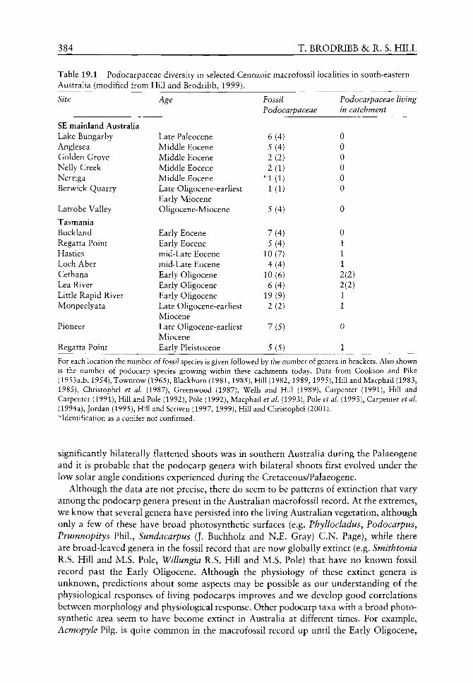
384 T. BRODRIBB & R. S. HILL
Table 19.1 Podocarpaceae diversity in selected Cenozoic macrofossil localities in south-eastern Australia (modified from Hill and Brodribb, 1999).
Site
SE mainland Australia Lake Bungarby Anglesea Golden Grove Nelly Creek Nerriga Berwick Quarry
Latrobe Valley
Tasmania Buckland Regatta Point Hasties Loch Aber Cethana Lea River Little Rapid River Monpeelyata
Pioneer
Regatta Point
Age
Late Paleocene Middle Eocene Middle Eocene Middle Eocene Middle Eocene Late Oligocene-earliest Early Miocene Oligocene-Miocene
Early Eocene Early Eocene mid-Late Eocene mid-Late Eocene Early Oligocene Early Oligocene Early Oligocene Late Oligocene-earliest Miocene Late Oligocene-earliest Miocene Early Pleistocene
Fossil Podocarpaceae
6(4) 5(4) 2(2) 2(1)
^•1(1) 1(1)
5(4)
7(4) 5(4)
10(7) 4(4)
10(6) 6(4)
19(9) 2(2)
7 {5)
5(5)
Podocarpaceae living in catchment
0 0 0 0 0 0
0
0 1 1 1 2(2) 2(2) 1 1
0
1
For each location the number of fossil species is given followed by the number of genera in brackets. Also shown is the number of podocarp species growing within these cachments today. Data from Cookson and Pike (1953a,b, 1954), Townrow (1965), Blackburn (1981,1985), Hill (1982,1989,1995), Hill and Macphail (1983, 1985), Christophel et al. (1987), Greenwood (1987), Wells and Hill (1989), Carpenter (1991), Hill and Carpenter (1991), Hill and Pole (1992), Pole (1992), Macphail et al. (1993), Pole et al (1993), Carpenter et al (1994a), Jordan (1995), Hill and Scriven (1997, 1999), Hill and Christophel (2001). * Identification as a conifer not confirmed.
significantly bilaterally flattened shoots was in southern Australia during the Palaeogene and it is probable that the podocarp genera with bilateral shoots first evolved under the low solar angle conditions experienced during the Cretaceous/Palaeogene.
Although the data are not precise, there do seem to be patterns of extinction that vary among the podocarp genera present in the Australian macrofossil record. At the extremes, we know that several genera have persisted into the living Australian vegetation, although only a few of these have broad photosynthetic surfaces (e.g. Phyllocladus, Podocarpus^ Prumnopitys Phil., Sundacarpus (J. Buchholz and N.E. Gray) C.N. Page), while there are broad-leaved genera in the fossil record that are now globally extinct (e.g. Smithtonia R.S. Hill and M.S. Pole, Willungia R.S. Hill and M.S. Pole) that have no known fossil record past the Early Oligocene. Although the physiology of these extinct genera is unknown, predictions about some aspects may be possible as our understanding of the physiological responses of living podocarps improves and we develop good correlations between morphology and physiological response. Other podocarp taxa with a broad photo-synthetic area seem to have become extinct in Australia at different times. For example, Acmopyle Pilg. is quite common in the macrofossil record up until the Early Oligocene,
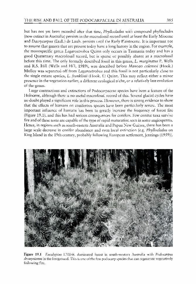
THE RISE AND FALL OF THE PODOCARPACEAE IN AUSTRALIA 385
but has not yet been recorded after that time, Phyllocladus with compound phylloclades (now extinct in AustraUa) persists in the macrofossil record until at least the Early Miocene and Dacrycarpus (Endl.) de Laub. persists until the Early Pleistocene. It is important not to assume that genera that are present today have a long history in the region. For example, the monospecific genus Lagarostrobos Quinn only occurs in Tasmania today and has a good Quaternary macrofossil record, but is sparse or possibly absent as a macrofossil before this time. The only formally described fossil in this genus, L. marginatus P. Wells and R.S. Hill (Wells and Hill, 1989), was described before Manoao colensoi (Hook.) MoUoy was separated off from Lagarostrobos and this fossil is not particularly close to the single extant species, L. franklinii (Hook, f.) Quinn. This may reflect either a minor presence in the vegetation earlier, a different ecological niche, or a relatively late evolution of the genus.
Large contractions and extinctions of Podocarpaceae species have been a feature of the Holocene, although there is no useful macrofossil record of this. Several glacial cycles have no doubt played a significant role in this process. However, there is strong evidence to show that the effects of humans on coniferous species have been particularly severe. The most important influence of humans has been to greatly increase the frequency of forest fire (Figure 19.1), and this has had serious consequences for conifers. Few conifer taxa survive fire and of these none are capable of the type of rapid maturation seen in some angiosperms. Hence, in regions such as south-eastern Australia and Papua New Guinea, there has been a large scale decrease in conifer abundance and even local extinction (e.g. Phyllocladus on King Island in the 19th century, probably following European settlement, Jennings (1959)).
1-/Al^m-'' > " • •? '." m.i'. ...'.: -v. -T^''^
r
*. %- , l^.r " ^ *" .«.»-%^
- . 1 - - %
- . ^. ";/•"-
• . ^k Ik
. '
, tr-.
'.."' 4-
Figure 19.1 Eucalyptus L'Herit. dominated forest in south-western Australia with Podocarpus drouynianus in the foreground. This is one of the few podocarp species that can regenerate vegetatively following fire.
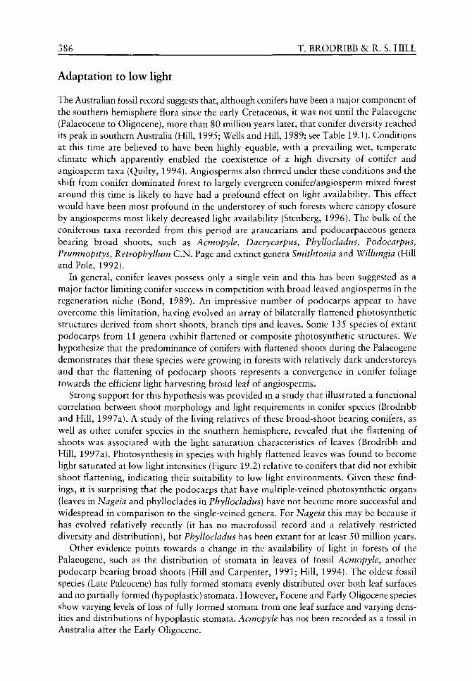
386 T. BRODRIBB &c R. S. HILL
Adaptation to low light
The Australian fossil record suggests that, although conifers have been a major component of the southern hemisphere flora since the early Cretaceous, it was not until the Palaeogene (Palaeocene to Oligocene), more than 80 million years later, that conifer diversity reached its peak in southern AustraHa (Hill, 1995; Wells and Hill, 1989; see Table 19.1). Conditions at this time are believed to have been highly equable, with a prevailing wet, temperate climate which apparently enabled the coexistence of a high diversity of conifer and angiosperm taxa (Quilty, 1994). Angiosperms also thrived under these conditions and the shift from conifer dominated forest to largely evergreen conifer/angiosperm mixed forest around this time is likely to have had a profound effect on light availability. This effect would have been most profound in the understorey of such forests where canopy closure by angiosperms most likely decreased light availability (Stenberg, 1996). The bulk of the coniferous taxa recorded from this period are araucarians and podocarpaceous genera bearing broad shoots, such as Acmopyle, Dacrycarpus, Fhyllocladus, Fodocarpus, Frumnopitys, Retrophyllum C.N. Page and extinct genera Smithtonia and Willungia (Hill and Pole, 1992).
In general, conifer leaves possess only a single vein and this has been suggested as a major factor limiting conifer success in competition with broad leaved angiosperms in the regeneration niche (Bond, 1989). An impressive number of podocarps appear to have overcome this limitation, having evolved an array of bilaterally flattened photosynthetic structures derived from short shoots, branch tips and leaves. Some 135 species of extant podocarps from 11 genera exhibit flattened or composite photosynthetic structures. We hypothesize that the predominance of conifers with flattened shoots during the Palaeogene demonstrates that these species were growing in forests with relatively dark understoreys and that the flattening of podocarp shoots represents a convergence in conifer foliage towards the efficient light harvesting broad leaf of angiosperms.
Strong support for this hypothesis was provided in a study that illustrated a functional correlation between shoot morphology and light requirements in conifer species (Brodribb and Hill, 1997a). A study of the living relatives of these broad-shoot bearing conifers, as well as other conifer species in the southern hemisphere, revealed that the flattening of shoots was associated with the light saturation characteristics of leaves (Brodribb and Hill, 1997a). Photosynthesis in species with highly flattened leaves was found to become light saturated at low light intensities (Figure 19.2) relative to conifers that did not exhibit shoot flattening, indicating their suitability to low light environments. Given these findings, it is surprising that the podocarps that have multiple-veined photosynthetic organs (leaves in Nageia and phylloclades in Fhyllocladus) have not become more successful and widespread in comparison to the single-veined genera. For Nageia this may be because it has evolved relatively recently (it has no macrofossil record and a relatively restricted diversity and distribution), but Fhyllocladus has been extant for at least 50 million years.
Other evidence points towards a change in the availability of light in forests of the Palaeogene, such as the distribution of stomata in leaves of fossil Acmopyle, another podocarp bearing broad shoots (Hill and Carpenter, 1991; Hill, 1994). The oldest fossil species (Late Paleocene) has fully formed stomata evenly distributed over both leaf surfaces and no partially formed (hypoplastic) stomata. However, Eocene and Early Oligocene species show varying levels of loss of fully formed stomata from one leaf surface and varying densities and distributions of hypoplastic stomata. Acmopyle has not been recorded as a fossil in Australia after the Early Oligocene.

THE RISE AND FALL OF THE PODOCARPACEAE IN AUSTRALIA 387
CO c 0 ^
.IT
c CO
'si CO-Q
E E =5 d .
E ^
1DUU-
1250-
1000-
750-
500-
D^^
4 \ ^
h
#7
' ' . 1
5 ^ \ ^ ^6 \ • 9
^
S ^ I O
1
10 Shoot width (mm)
100
Figure 19.2 Mean light saturation requirements for shoots from 10 species of Podocarpaceae; 1 Acmopyle pancheri (Brongn. and Gris) Pilg., 2 Dacrycarpus dacrydioides (A. Rich.) de Laub., 3 Lagarostrobos franklinii, 4 Microstrobos niphophilus, 5 Fhyllocladus aspleniifolius, 6 Podocarpus dispermus C.T. White, 7 Podocarpus drouynianus F. MuelL, 8 Podocarpus lawrencei Hook, f., 9 Prumnopitys ferruginea (G. Benn. ex D. Don) de Laub., and 10 Retrophyllum comptonii (J. Buchholz) C.N. Page, and four species of Cupressaceae; 11 Athrotaxis cupressiodes D. Don, 12 Athrotaxis selaginoides D. Don, 13 Callitris rhomboidea R.Br, ex Rich, and A. Rich., 14 Diselma archeri Hook. f. All plants were grown under identical glasshouse conditions.
Hypoplastic stomata may be evidence for stages in either the loss or acquisition of stomata from one leaf surface, but given the relative ages of the species, the former is more probable. The living Acmopyle species both have fully formed stomata in two row s along the entire length of the underside of the leaf, but on the upper surface stomata are usually restricted to small areas near the leaf base and apex with hypoplastic stomata in between. However, glasshouse grown specimens are very plastic in their stomatal distribution. In low light, shoots are almost hypostomatic, whereas in high light shoots tend towards amphis-tomatic (Brodribb, unpubhshed data), demonstrating that stomatal distribution on the leaf undersurface is not genetically fixed and it probably was not during the Cenozoic. Therefore, the trends seen in stomatal distribution among the fossils probably represent a plastic response to the environment in which they occurred.
It has been demonstrated that leaf diffusive conductance is well correlated with maximum assimilation (Wong et al.^ 1985; Korner et al., 1991) and that a linear relationship exists between stomatal density and maximum conductance in extant podocarps (Brodribb and Hill, 1997b). Thus the decrease in stomatal density in Acmopyle is almost certainly a response to decreased maximum assimilation rate in leaves and this most likely came about either through a decrease in temperature, or a decrease in light availability. Evidence supporting a decrease in light intensity comes from the fact that stomatal numbers were found to become differentially reduced on the upper surface of the shoot when compared to the lower surface, thus causing a shift from amphistomy to hypostomy in shoots. Hypostomy in extant Acmopyle species is most common in shade plants, while amphistomy
388 T. BRODRIBB &c R. S. HILL
is generally associated with leaves from open habitats (Peat and Fitter, 1994). Thus a shift from amphistomy to hypostomy in Acmopyle fossils tends to suggest a reduction of the available light in the Acmopyle habitat. A closure of the canopy by invading angiosperms as Australia moved into lower latitudes may have been the cause for such a change in the distribution and availability of light from forest crown to floor. There is also evidence of a decrease in temperature by the time of the last occurrences of Acmopyle in Australia (Early Oligocene). Although this temperature change apparently did not affect the shoot size oi Acmopyle, it is possible that it may have contributed to stomatal loss in the later history of the genus in Australia.
Another fossil Podocarpaceae genus that exhibits a trend in foliar morphology during the Cenozoic is Dacrycarpus. Two groups of fossil Dacrycarpus species cover a substantial time range and probably represent separate phylogenetic lines. The first is D. mucronatus P. Wells and R.S. Hill (Early Eocene to the Early Oligocene in Tasmania), where the oldest specimens have both bilaterally and bifacially flattened foliage and stomata are distributed equally over all leaf surfaces. Bifacially flattened foliage is the normal form of leaf flattening, with a distinct abaxial and adaxial surface, whereas bilaterally flattened foliage in the Podocarpaceae leads to each functional leaf surface containing part of the original adaxial and abaxial surface, with the original leaf margins being approximately in the middle of each flattened surface. By the Early Oligocene D. mucronatus is known only as bifacially flattened foliage. The phylogenetically more remote D. latrobensis R.S. Hill and R. Carpenter, from Victoria, occurs substantially later in time and still has both foliage types (Cookson and Pike, 1953a). However, stomata are more common on one leaf surface of the bilaterally flattened foliage than the other (Hill and Carpenter, 1991). Therefore, in these species there is evidence for loss of stomata on bilaterally flattened foliage and, in Tasmania at least, loss of the bilaterally flattened foliage type. Recently discovered fossils from Miocene sediments in northern New South Wales further demonstrate that Dacrycarpus from lower latitudes did not lose the bilaterally flattened foliage as quickly as those species in Tasmania (Hill and Whang, 2000).
The second phylogenetic line is Dacrycarpus linifolius P. Wells and R.S. Hill which occurs in Early Eocene and Early Oligocene sediments in Tasmania and has only been recovered as bifacially flattened foliage, which in gross leaf morphology is identical across the time range. However, while the Early Eocene leaves have stomata along the entire length of both leaf surfaces, the Early Oligocene leaves have stomata restricted to less than the basal third of the abaxial surface, providing further evidence for reduction in stomatal distribution, but this time on bifacially flattened leaves.
While the phylogenetic relationships of the remaining Dacrycarpus fossils are not clear, they do exhibit general leaf morphology trends. Six Dacrycarpus species have been described from lowland Oligocene-Early Miocene Tasmanian sites, all with imbricate, bifacially flattened foliage. Five are amphistomatic, but the sixth, D. linearis P. Wells and R.S. Hill, probably has stomata restricted to the adaxial surface. All bifacially flattened foliage of extant Dacrycarpus species is amphistomatic, although in D. compactus (Wasscher) de Laub., which occurs at high altitudes in New Guinea and is the only extant species to lack bilaterally flattened foliage, there are extremely few stomata on the abaxial surface. Significantly, the extant podocarpaceous alpine Tasmanian species Microstrobos niphophilus and Microcachrys tetragona (Hook.) Hook. f. are closely imbricate and epis-tomatic. A similar Microstrobos J. Garden and L.A.S. Johnson species co-occurs with Dacrycarpus in two Tasmanian fossil localities.
The last Australian Dacrycarpus macrofossil record is from Early Pleistocene Tasmanian sediments (Jordan, 1995). Only bifacially flattened foliage of this fossil has been recorded.

THE RISE AND FALL OF THE PODOCARPACEAE IN AUSTRALIA 3 ^
with stomata restricted to the adaxial leaf surface. Dacrycarpus must have become extinct in Australia soon after this. The change in leaf morphology in Dacry carpus is more complex than in Acmopyle, but some aspects of it are similar. The ability to change foliage form completely (from bilaterally to bifacially flattened leaves) gave Dacrycarpus more flexibility and may explain its longer known time range in Australia.
Cretaceous forest structure at high southern latitudes
There has been some speculation as to the structure and function of the high latitude conifer/angiosperm dominated forests that apparently extended across much of Australia and Antarctica during the mid-late Cretaceous (e.g. Specht et al.^ 1992) and this has considerable bearing on the evolution of today's conifer flora. During the mid-Cretaceous, Australia and Antarctica were joined and most of Australia was south of 60°S. Occurrence of these forests at such high latitudes has led to the hypothesis that they were dominated by stands of pyramidal crown conifers, as this crown shape most efficiently harvests light at low solar angles (Terborgh, 1985; Specht et al, 1992), forming a forest structure similar to that seen at high latitudes in the northern hemisphere boreal zone today. However, apart from the Araucariaceae, few of the southern hemisphere conifers today have the classical conical shape of many northern hemisphere conifers and it is unlikely that this conical shape was common in the southern hemisphere in the past. Many southern conifers have foliage cascading down the sides of the tree in a more random fashion and this is a more likely model for high southern latitude forests in the past. Evidence suggests that many of the angiosperms (and gymnosperms) present in these Cretaceous forests were deciduous (Pole, 1992; McLoughlin et aL, 1995; Hill and Scriven, 1995), also conforming to the observed pattern in the boreal forests, where a mixture of deciduous angiosperms and evergreen conifers predominates. Obviously there are difficulties in ascertaining the relative importance of the deciduous element in these fossil floras, as the taxonomic affinities of the species present are often poorly understood and the identification of deciduousness is sometimes based on fairly meagre evidence. However, it is widely accepted that a mixture of deciduous and evergreen angiosperms was present. Extant evergreen broad-leaved taxa with affinities to fossil species have been shown to survive long dark periods such as those that would have prevailed during winter at high latitudes (Read and Francis, 1992). The presence of a significant high latitude evergreen angiosperm flora contrasts with the situation today at high latitudes in the northern hemisphere. Persistence of the evergreen element was probably allowed by the fact that mild winter temperatures (Dorman, 1966) did not cause significant damage to overwintering broad-leaves while not allowing excessive dark respiration.
Constraints on extant Podocarpaceae distributions
It appears that podocarps, by virtue of their relatively large foliar morphological range, are well adapted to survive under most light conditions. Changing light conditions therefore do not appear to account for the restriction and extinction of conifers during the Cenozoic in the southern hemisphere. To address better the question of why podocarps, and indeed all conifer families, suffered during the later Cenozoic it is instructive to examine what constrains the distribution of extant southern conifer species.
The fundamental requirement for genetic survival in conifers, and indeed all plants, is the production of enough photosynthate to allow growth and reproduction. Thus, limitations

390 T. BRODRIBB &: R. S. HILL
imposed by the physical environment on the distribution of plant species will usually manifest themselves through their effects on leaf photosynthesis. The environmental parameters most commonly used to explain presence or absence of species are temperature and rainfall regimes and their inferred effects are almost invariably on the photosynthetic capacity of the species in question. Because of their characteristic longevity (Loehle, 1989), sporadic reproduction, slow rates of dispersal and short-term viability of soil-borne seed, (Gibson et al., 1994; Veblen et ai, 1995), conifers are particularly closely linked to long-term environmental characteristics. Unlike the fast growing, highly fecund angiosperms, most conifers do not have the capacity to move in and out of marginal environments in response to short-term physical fluctuations and are thus confined at the physiological extremes of their distributions by the long-term survival of adult plants.
Despite the anecdotal and quantitative evidence of environmental constraints on species distributions, few studies have demonstrated the actual mechanisms by which physical limitations affect the survival and reproduction of plant species. Over the last 50 years, research into the characteristics and control of leaf gas exchange has been prolific and the use of leaf photosynthetic characters to explain simple successional changes in the vegetation has become common in the literature, typically using water-use efficiency (e.g. Meinzer et aL, 1984; Dias-Filho and Dawson, 1995), drought response (e.g. Kubiske and Abrams, 1993) and light response (e.g. Field, 1988; Knapp and Smith, 1991; Ashton and Berlyn, 1994) to account for small-scale competitive outcomes. This type of ecophysiological research generally takes the form of comparisons between small numbers of species and, as such, does not attempt to make a quantitative link between photosynthetic function and environmental parameters. Indeed, with a few exceptions (e.g. Teeri and Stowe, 1976), large-scale relationships between physiology, environment and species distributions have been overlooked by ecophysiologists.
In order to understand how the distribution of species is likely to have been affected by historical climate change and might be affected by future climatic perturbations, it is important to be able to describe current, known distributions in terms of the most likely physiological constraint. This was undertaken for a group of 13 conifer species, including eight podocarps, in a study by Brodribb and Hill (1998). We took species from a diverse geographical range spanning Australia (Figures 19.3 and 19.4), New Zealand, New Guinea, South Africa and New Caledonia (Figure 19.5) and a broad spectrum of habitats and compared the water-use efficiency and drought tolerance of photosynthesis with environmental water availability. Water availability was considered the most likely constraint on distribution given that podocarp forest in the southern hemisphere is almost exclusively restricted to areas of high rainfall, with the most extensive and diverse conifer forests occurring on the west coasts of New Zealand and Tasmania (Figure 19.4), in New Caledonia (Figure 19.5) and highland New Guinea, where rainfall is commonly over 5000 mm per annum (van Royen, 1979).
We measured two physiological parameters related to the drought tolerance of leaves and the xylem vascular system. The first of these was maximum water-use efficiency during drought (Brodribb and Hill, 1996) and the second was the vulnerability of xylem to cavitation by water stress (Brodribb and Hill, 1999). These parameters were compared with minimum rainfall data over the ranges of the selected species. A good correlation between maximum water-use efficiency (see Brodribb, 1996 for details) and rainfall parameters (Figure 19.6) suggests that the climatic ranges of these conifers are closely controlled by their photosynthetic and gas-exchange characteristics during drought. This is an important result as it provides a direct link between instantaneous gas-exchange characteristics

THE RISE AND FALL OF THE PODOCARPACEAE IN AUSTRALIA 391
Figure 19.3 Cool temperate rainforest in western Tasmania containing mature trees of Phyllocladus aspleniifolius. These trees have been recently exposed on a road cutting.
and species distribution. The cost to the plant of maximizing water-use efficiency, as measured by these two parameters, may come as an increase in the saturation light requirement (discussed below) and respiration rate of leaves. Increases in leaf nitrogen associated with higher carboxylation efficiency and photosynthetic capacity in species have commonly been linked to increases in the dark respiration rate and this has been explained by suggestions of higher tissue maintenance costs, or costs associated with phloem loading and carbohydrate export from the leaf (Connor et al, 1993; Bouma et al., 1995; Baxter et al, 1995). Thus, there must be a significant selective pressure on these species to maintain their physiological drought tolerance to a level dictated by the long-term water availability in the environment and an insufficient drought response must become limiting by causing local extinction of a species, either by death during periods of extreme conditions, or by competitive exclusion by more efficient species. Either way, the importance of rainfall is emphasized in constraining the distribution of these species.
Strong correlation also exists between xylem vulnerability to cavitation and average rainfall during the dry season in the Podocarpaceae and southern hemisphere Cupressaceae

392 T. BRODRIBB & R. S. HILL
Figure 19.4 High altitude vegetation on Mt Read in western Tasmania. Six conifer species coexist in this area, with four belonging to the Podocarpaceae. The very damaging impact of fire can be seen in the foreground.
•^i^M
Figure 19.5 Conifer dominated forest in New Caledonia, with Dacrycarpus and Araucariaceae among the prominent trees.

THE RISE AND FALL OF THE PODOCARPACEAE IN AUSTRALIA 393
CO
U.4 —
0 .35-
0 . 3 -
0 .25-
0 . 2 -
0 .15 -
n 1 ^
i X % X
1 1
Vo
\ 1 1
Vs
1 1 100 200 300 400 500
Driest 4 months rainfall (mm)
600 700
Figure 19.6 Shows highly significant correlation (r = 0.97) between mean rainfall during the driest four months within the distributions of 8 Podocarpaceae (O) and 5 Cupressaceae (•), and leaf drought tolerance. The drought tolerance parameter is the lowest ratio of mesophyll to ambient CO2 concentration measured in leaves during imposed drought. This ratio is inversely proportional to the maximum water-use efficiency attainable by leaves.
(Brodribb and Hill, 1999). Species from wet environments were highly vulnerable to cavitation while species from the semi-arid zone produced stem xylem that was extremely resistant to pressure-induced cavitation. Clearly this indicates an important role for xylem vulnerability in determining the distributional limits of these plants in terms of minimum water availability. This also provides evidence for linkage between leaf drought tolerance and stem cavitation characteristics, as shown by a highly significant regression relating maximum water-use efficiency with the water potential C jo) that caused 50% cavitation in stem xylem (Figure 19.7). One possible inference from this is that a loss of hydraulic conductance in the xylem during water shortage is the causal factor dictating a loss of leaf function during drought in these species. Such a hypothesis is supported by data suggesting that plants may operate close to the point of 'runaway cavitation', where a positive feed-back following xylem embolism has the potential to cause the vascular system rapidly to lose hydraulic conductivity unless transpiration is reduced (Sperry et al., 1993; Alder et al.^ 1996). However, several pieces of evidence point away from xylem dysfunction as the primary cause of leaf failure during drought, particularly for species from drier habitats. This evidence comes from research illustrating that complete stomatal closure and a loss of optimal quantum yield (indicating damage to photosystem II) in these species both occurred at leaf water potentials above the value corresponding to '^50 (Brodribb and Hill, 1999). Given this large 'safety margin' between stem water potential during active photosynthesis and that which would induce significant (or possibly runaway) cavitation, it seems unlikely that the xylem water potential would approach " 50 unless plants were subject to severe water shortage. Considering that none of the species investigated are likely to suffer significant emboUsm by freeze-thaw cycles (Sperry et ^/.,1994), large-scale stem xylem cavitation probably only occurs when plants experience soil moisture conditions approaching '^59.
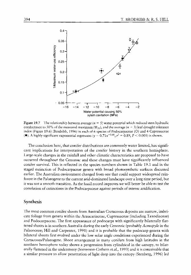
394 T. BRODRIBB & R. S. HILL
0.35
0.25
CO 0.15H
0.05
Water potential causing 50% xylem cavitation (MPa)
Figure 19.7 The relationship between average (n = 5) v^ater potential w hich reduced stem hydraulic conductance to 50% of the measured maximum C jo), and the average (n = 3) leaf drought tolerance index (Figure 19.6) (Brodribb, 1996) in each of 6 species of Podocarpaceae (O) and 4 Cupressaceae (•). A highly significant exponential regression (y = 0.71JC~^-^^; r = 0.89, P < 0.001) is shown.
The conclusion here, that conifer distributions are commonly water limited, has significant implications for interpretation of the conifer history in the southern hemisphere. Large-scale changes in the rainfall and other climatic characteristics are proposed to have occurred throughout the Cenozoic and these changes must have significantly influenced conifer survival. This is reflected in the species numbers show^n in Table 19.1 and in the staged extinction of Podocarpaceae genera with broad photosynthetic surfaces discussed earlier. The Australian environment changed from one that could support widespread rainforest in the Palaeogene to the current arid-dominated landscape over a long time period, but it was not a smooth transition. As the fossil record improves we will better be able to test the correlation of extinctions in the Podocarpaceae against periods of intense aridification.
Synthesis
The most common conifer shoots from Australian Cretaceous deposits are narrow, imbricate foliage from genera within the Araucariaceae, Cupressaceae (including Taxodiaceae) and Podocarpaceae. The first appearance of podocarps with significantly bilaterally flattened shoots is in southern Australia during the early Cenozoic (probably Acmopyle in the Palaeocene; Hill and Carpenter, 1991) and it is probable that the podocarp genera with bilateral shoots first evolved under the low solar angle conditions experienced during the Cretaceous/Palaeogene. Shoot arrangement in many conifers from high latitudes in the northern hemisphere today shows a progression from cylindrical in the canopy, to bilaterally flattened in the understorey (Sorrenson-Cothern et al.^ 1993) and it is conceivable that a similar pressure to allow penetration of light deep into the canopy (Stenberg, 1996) led
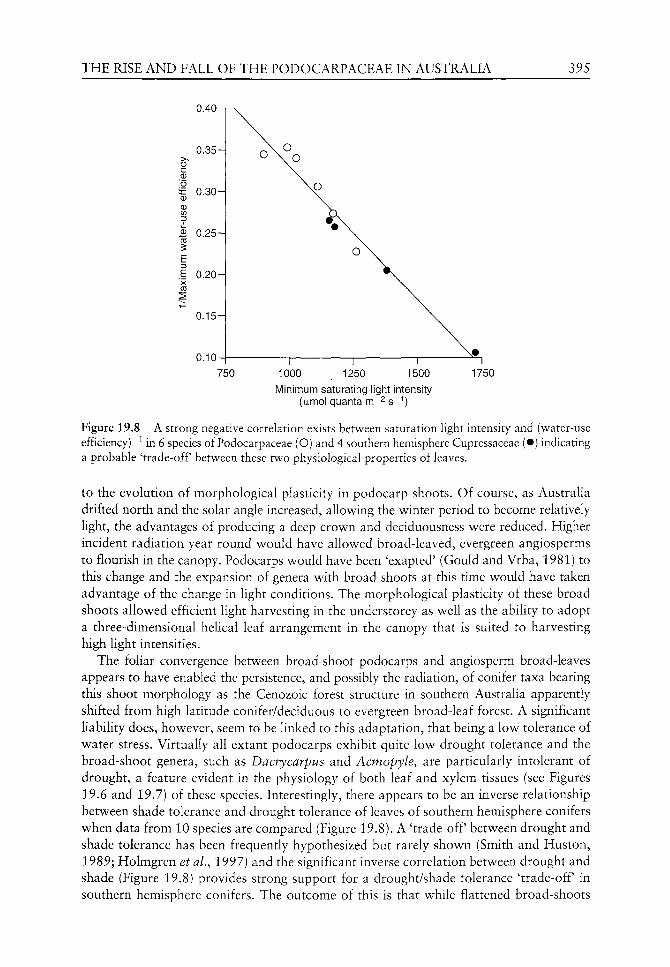
THE RISE AND FALL OF THE PODOCARPACEAE IN AUSTRALIA 395
0.40n
0.35 H
iE 0.30-0 0
B 0.25-
i E .1 0.20-
0.15H
0.10-
750 1000 1250 1500 1750
Minimum saturating iigiit intensity (ijimo! quanta m^^ g-i)
Figure 19.8 A strong negative correlation exists between saturation light intensity and (water-use efficiency)"^ in 6 species of Podocarpaceae (O) and 4 southern hemisphere Cupressaceae (•) indicating a probable 'trade-off between these two physiological properties of leaves.
to the evolution of morphological plasticity in podocarp shoots. Of course, as Australia drifted north and the solar angle increased, allowing the winter period to become relatively light, the advantages of producing a deep crown and deciduousness were reduced. Higher incident radiation year round would have allowed broad-leaved, evergreen angiosperms to flourish in the canopy. Podocarps would have been 'exapted' (Gould and Vrba, 1981) to this change and the expansion of genera with broad shoots at this time would have taken advantage of the change in light conditions. The morphological plasticity of these broad shoots allowed efficient light harvesting in the understorey as well as the ability to adopt a three-dimensional helical leaf arrangement in the canopy that is suited to harvesting high light intensities.
The foliar convergence between broad-shoot podocarps and angiosperm broad-leaves appears to have enabled the persistence, and possibly the radiation, of conifer taxa bearing this shoot morphology as the Cenozoic forest structure in southern Australia apparently shifted from high latitude conifer/deciduous to evergreen broad-leaf forest. A significant liability does, however, seem to be linked to this adaptation, that being a low tolerance of water stress. Virtually all extant podocarps exhibit quite low drought tolerance and the broad-shoot genera, such as Dacrycarpus and Acmopyle, are particularly intolerant of drought, a feature evident in the physiology of both leaf and xylem tissues (see Figures 19.6 and 19.7) of these species. Interestingly, there appears to be an inverse relationship between shade tolerance and drought tolerance of leaves of southern hemisphere conifers when data from 10 species are compared (Figure 19.8). A 'trade-off between drought and shade tolerance has been frequently hypothesized but rarely shown (Smith and Huston, 1989; Holmgren et al., 1997) and the significant inverse correlation between drought and shade (Figure 19.8) provides strong support for a drought/shade tolerance 'trade-off in southern hemisphere conifers. The outcome of this is that while flattened broad-shoots

396 T. BRODRIBB &c R. S. HILL
may have allowed podocarps to retain codominance in high rainfall, mixed Cenozoic forests,
these taxa were left extremely vulnerable to the progressive decrease in water availability that
followed.
Acknowledgements
This research was supported by grants from the Australian Research Council. RSH is in
receipt of an ARC Senior Research Fellowship.
References
Alder MN, Sperry JS, Pockman WT. 1996. Root and stem xylem embolism, stomatal conductance and leaf turgor in Acer grandidentatum populations along a soil moisture gradient. Oecologia 105:293-30L
Ashton PMS, Berlyn, GP. 1994. A comparison of leaf physiology and anatomy of Quercus species in different light environments. American Journal of Botany 81: 589-597.
Axsmith BJ, Taylor TN, Taylor EL. 1998. Anatomically preserved leaves of the conifer Notophytum krauselii (Podocarpaceae) from the Triassic of Antarctica. American Journal of Botany 85: 704-713.
Baxter R, Bell SA, Sparks TH, et al. 1995. Effects of elevated CO2 concentrations on three montane grass species. III. Source leaf metabolism and whole plant carbon partitioning. Journal of Experimental Botany 46: 917-929.
Beadle NCW. 1981. The Vegetation of Australia. Stuttgart, New York: Gustav Fischer Verlag. Blackburn DT. 1981. Tertiary megafossil flora of Maslin Bay, South Australia: Numerical taxo-
nomic study of selected leaves. Alcheringa 5: 9-28. Blackburn DT. 1985. Palaeobotany of the Yallourn and Morwell Coal Seams. Palaeobotanical
Project - report 3: State Electricity Commission of Victoria. Bond WJ. 1989. The tortoise and the hare: ecology of angiosperm dominance and gymnosperm per
sistence. Biological Journal of the Linnean Society 36: 227-249. Bouma TJ, DeVisser R, van Leeuwen PH, et al. 1995. The respiratory energy requirements involved
in nocturnal carbohydrate export from starch-storage mature source leaves and their contribution to leaf dark respiration. Journal of Experimental Botany 46: 1185-1194.
Brodribb T. 1996. Dynamics of changing intercellular CO2 concentration (cj) during drought and determination of minimum functional c . Plant Physiology 111: 179-185.
Brodribb T, Hill RS. 1996. The drought physiology of a diverse group of Southern Hemisphere conifer species is correlated with minimum seasonal rainfall. Functional Ecology 12: 465-471.
Brodribb T, Hill RS. 1997a. Light response characteristics of a morphologically diverse group of Southern Hemisphere conifers as measured by chlorophyll fluorescence. Oecologia 110: 10-17.
Brodribb T, Hill RS. 1997b. Imbricacy and stomatal wax plugs reduce maximum leaf conductance in Southern Hemisphere conifers. Australian Journal of Botany 45: 657-66S.
Brodribb T, Hill RS. 1998. The photosynthetic drought physiology of a diverse group of southern hemisphere conifer species is correlated with minimum seasonal rainfall. Functional Ecology 12: 465-471.
Brodribb T, Hill RS. 1999. The importance of xylem constraints in the distribution of conifer species. New Phytologist 143: 365-372.
Carpenter RJ. 1991. Palaeovegetation and Environment at Cethana, Tasmania. Unpublished Ph.D. Thesis, University of Tasmania.
Carpenter RJ, Hill RS, Jordan GJ. 1994a. Cenozoic vegetation in Tasmania: Macrofossil evidence. In: Hill RS, ed. History of the Australian Vegetation. Cambridge: Cambridge University Press, 276-298.
Carpenter RJ, Jordan GJ, Hill RS. 1994b. Banksieaephyllum taylorii (Proteaceae) from the Late Paleocene of New South Wales and its relevance to the origin of Australia's scleromorphic flora. Australian Systematic Botany 7: 385-392.
THE RISE A N D FALL OF T H E PODOCARPACEAE IN AUSTRALIA 397
Christophel DC, Harris WK, Syber AK. 1987. The Eocene flora of the Anglesea locaHty, Victoria. Alcheringa 11: 303-323.
Connor DJ, Hall AJ, Sadras VO. 1993. Effect of nitrogen content on the photosynthetic characters of sunflower leaves. Australian Journal of Plant Physiology 20: 251-263.
Conran JG, Wood GM, Martin PG, et al. 2000. Generic relationships within and between the gym-nosperm famiHes Podocarpaceae and Phyllocladaceae based on an analysis of the chloroplast gene rbcL. Australian Journal of Botany 48: 715-724.
Cookson IC, Pike KM. 1953a. The Tertiary occurrence and distribution of Podocarpus (section Dacrycarpus) in Australia and Tasmania. Australian Journal of Botany 1: 71-82.
Cookson IC, Pike KM. 1953b. A contribution to the Tertiary occurrence of the genus Dacrydium in the Austrahan region. Australian Journal of Botany 1: 474-484.
Cookson IC, Pike KM. 1954. The fossil occurrence of Phyllocladus and two other podocarpaceous types in Australia. Australian Journal of Botany 2: 60-67.
Dias-Filho MB, Dawson TE. 1995. Physiological responses to soil moisture stress in two Amazonian gap-invader species. Functional Ecology 9: 213-221.
Dilcher DL. 1969. Podocarpus from the Eocene of North America. Science 164: 299-301. Dorman FH. 1966. Austrahan Tertiary palaeotemperatures. Journal of Geology 74: 49-61. Field CB. 1988. On the role of photosynthetic responses in constraining the habitat distribution of
rainforest plants. Australian Journal of Plant Physiology 15: 343-358. Gibson N, Barker PCJ, Cullen PJ, Shapcott A. 1994. Conifers of southern Australia. In: Enright NJ,
Hill RS, eds. The Ecology of the Southern Conifers. Melbourne: Melbourne University Press, 223-251.
Gould SJ, Vrba E. 1981. Exaptation: a missing term in the science of form. Paleobiology 8: 4-15. Greenwood DR. 1987. Early Tertiary Podocarpaceae: Megafossils from the Eocene Anglesea local
ity, Victoria, Austraha. Australian Journal of Botany 35: 111-133. Hill RS. 1982. The Eocene megafossil flora of Nerriga, New South Wales, AustraHa. Palaeontographica
Abt. B 181: 44-77. Hill RS. 1989. New species oi Phyllocladus (Podocarpaceae) macrofossils from south eastern Australia.
Alcheringa 13: 193-208. Hill RS. 1994. The history of selected Australian taxa. In: Hill RS, ed. History of the Australian
Vegetation. Cambridge: Cambridge University Press, 390-419. Hill RS. 1995. Conifer origin, evolution and diversification in the Southern Hemisphere. In: Enright NJ,
Hill RS, eds. The Ecology of the Southern Conifers. Melbourne: Melbourne University Press, 10-29. Hill RS, Brodribb TJ. 1999. Southern conifers in time and space. Australian Journal of Botany 47:
639-696. Hill RS, Carpenter RJ. 1991. Evolution oi Acmopyle and Dacrycarpus (Podocarpaceae) fohage as
inferred from macrofossils in south-eastern Australia. Australian Systematic Botany 4: 449-479. Hill RS, Christophel DC. 2001. Two new species of Dacrydium (Podocarpaceae) based on vegeta
tive fossils from Middle Eocene sediments at Nelly Creek, South Australia. Australian Systematic Botany 14: 193-205.
Hill RS, Macphail MK. 1983. Reconstruction of the Oligocene vegetation at Pioneer, northeast Tasmania. Alcheringa 7: 281-299.
Hill RS, Macphail MK. 1985. A fossil flora from rafted Plio-Pleistocene mudstones at Regatta Point, Tasmania. Australian Journal of Botany 33: 497-517.
Hill RS, Merrifield HE. 1993. An Early Tertiary macroflora from West Dale, southwestern Austraha. Alcheringa 17: 285-326.
Hill RS, Pole MS. 1992. Leaf and shoot morphology of extant Afrocarpus, Nageia and Retrophyllum (Podocarpaceae) species, and species with similar leaf arrangement from Tertiary sediments in Australia. Australian Systematic Botany 5: 337-358.
Hill RS, Scriven LJ. 1995. The angiosperm-dominated woody vegetation of Antarctica: a review. Review of Palaeobotany and Palynology 86: 175-198.
Hill RS, Scriven LJ. 1997. Palaeoclimate across an altitudinal gradient in the Oligo-Miocene of northern Tasmania: an investigation of nearest living relative analysis. Australian Journal of Botany 45: 493-505.
398 T. BRODRIBB &c R. S. HILL
Hill RS, Scriven LJ. 1999. Falcatifolium (Podocarpaceae) macrofossils from Paleogene sediments in south-eastern Australia: a reassessment. Australian Systematic Botany 11: 711-720.
Hill RS, Whang SS. 2000. Dacrycarpus (Podocarpaceae) macrofossils from Miocene sediments at Elands, eastern Australia and their relationship to living and fossil species. Australian Systematic Botany 13: 395-408.
Holmgren M, Scheffer M, Huston MA. 1997. The interplay of facultation and competition in plant communities. Ecology 78: 1966-1975.
Jennings JN. 1959. The coastal geomorphology of King Island, Bass Strait, in relation to changes in the relative level of land and sea. Records of the Queen Victoria Museum Launceston 11: 1-35.
Jordan G.J. 1995. Extinct conifers and conifer diversity in the Early Pleistocene of v^estern Tasmania. Review of Paleobotany and Palynology 84: 375-387.
Kelch DG. 1997. The phylogeny of the Podocarpaceae based on morphological evidence. Systematic Botany 22: 113-131.
Kimura T, Ghana T, Mimoto K. 1988. Discovery of a podocarpaceous plant from the Low^er Cretaceous of Kochi Prefecture, in the Outer Zone of southwest Japan. Proceedings of the Japan Academy 64: 213-216.
Knapp AK, Smith WK. 1991. Gas exchange responses to variable sunlight in tv^o Sonoran desert herbs: comparison with subalpine species. Botanical Gazette 152: 269-274.
Korner C, Farquhar GD, Wong SC. 1991. Carbon isotope discrimination by plants follows latitudinal and altitudinal trends. Oecologia 88: 30-40.
Krassilov VA. 1974. Podocarpus from the Upper Cretaceous of eastern Asia and its bearing on the theory of conifer evolution. Palaeontology 17: 365-370.
Kubiske ME, Abrams MD. 1993. Stomatal and nonstomatal limitations of photosynthesis in 19 temperate tree species on contrasting sites during wet and dry years. Plant, Cell and Environment 16: 1123-1129.
Loehle C. 1989. Tree life history strategies: the role of defences, Canadian Journal of Forest Research 18: 209-222.
Lusk CH, Smith B. 1998. Life history differences and tree species co-existence in an old-growth New Zealand rainforest. Ecology 79: 795-806.
Macphail MK, Jordan GJ, Hill RS. 1993. Key periods in the evolution of the flora and vegetation in western Tasmania I. the Early-Middle Pleistocene. Australian Journal of Botany 41: 673-707.
McLoughlin S, Drinnan AN, Rozefelds AC. 1995. A Cenomanian flora from the Winton Formation, Eromanga Basin, Queensland, Australia. Memoirs of the Queensland Museum 38: 273-313.
Meinzer F, Goldstein G, Marisol J. 1984. The effect of atmospheric humidity on stomatal control of gas exchange in two tropical coniferous species. Canadian Journal of Botany 62: 591-595.
Midgley JJ, Seydack A, Reynell D, McKelly D. 1990. Fine-grain pattern in Southern Cape Plateau forests. Journal of Vegetation Science 1: 539-546.
Peat HJ, Fitter AH. 1994. A comparative study of the distribution and density of stomata in the British flora. Biological Journal of the Linnean Society 52: 377-393.
Pole MS. 1992. Eocene vegetation from Hasties, north-eastern Tasmania. Australian Systematic Botany 5: 431-475.
Pole MS, Hill RS, Green N, Macphail MK. 1993. The Late Oligocene Berwick Quarry flora -rainforest in a drying environment. Australian Systematic Botany 6: 399-428.
Quilty PG. 1994. The background: 144 million years of Australian palaeoclimate and palaeogeog-raphy. In: Hill RS, ed. History of the Australian Vegetation. Cambridge: Cambridge University Press, 14-43.
Read J, Francis J. 1992. Responses of some Southern Hemisphere tree species to a prolonged dark period and their implications for high-latitude Cretaceous and Tertiary floras. Palaeogeography, Palaeoclimatology, Palaeoecology 99: 271-290.
Reymanowna M. 1987. A Jurassic podocarp from Poland. Review of Paleobotany and Palynology 51: 133-143.
Royen P. 1979. The Alpine Flora of New Guinea. Vol.12. Amsterdam: J.Cramer, 20-22. Smith TM, Huston MA. 1989. A theory of the spatial and temporal dynamics of plant communities.
Vegetatio 83: 49-69.

T H E RISE A N D FALL OF THE PODOCARPACEAE IN AUSTRALIA 399
Sorrensen-Cothern KA, Ford ED, Sprugel DG. 1993. A model of competition incorporating plasticity through modular foliage and crown development. Ecological Monographs 63: 277-304.
Specht RL, Dettmann ME, Jarzen DM. 1992. Community associations and structure in the Late Cretaceous vegetation of southeast Australasia and Antarctica. Palaeogeography, Palaeoclimat-ology, Palaeoecology 94: 283-309.
Sperry JS, Alder NN, Eastlack SE. 1993. The effect of reduced hydraulic conductance on stomatal conductance and xylem cavitation. Journal of Experimental Botany 44: 1075-1082.
Sperry JS, Nichols KL, Sullivan JEM, Eastlack SE. 1994. Xylem embolism in ring-porous, diffuse-porous, and coniferous trees of northern Utah and interior Alaska. Ecology 75: 1736-1752.
Stenberg, P. 1996. Simulations of the effects of shoot structure and orientation on vertical gradients in intercepted light by conifer canopies. Tree Physiology 16: 99-108.
Teeri JA, Stow e EG. 1976. Climatic patterns and the distribution of C4 grasses in North America. Oecologia 23: 1-12.
Terborgh J. 1985. The vertical component of plant species diversity in temperate and tropical forests. American Naturalist 126: 760-766.
Tov^nrov^ JA. 1965. Notes on Tasmanian pines I. Some Lower Tertiary podocarps. Papers and Proceedings of the Royal Society of Tasmania 99: 87-107.
Townrow JA. 1967. On Rissikia and Mataia podocarpaceous conifers from the Lower Mesozoic of southern lands. Papers and Proceedings of the Royal Society of Tasmania 101: 103-136.
Veblen TT, Burns BR, Kitzberger T, et al. 1995. The ecology of the conifers of southern South America. In: Enright NJ, Hill RS, eds. The Ecology of the Southern Conifers. Melbourne: Melbourne University Press, 120-155.
Wells PM, Hill RS. 1989. Fossil imbricate-leaved Podocarpaceae from Tertiary sediments in Tasmania. Australian Systematic Botany 2: 387-423.
Wong S-C, Cowan IR, Farquhar GD. 1985. Leaf conductance in relation to rate of CO2 assimilation. Ill Influences of water stress and photoinhibition. Plant Physiology 78: 830-834.
20 The adaptive physiology of Metasequoia
to Eocene high-latitude environments
Richard Jagels and Michael E Day
CONTENTS.
Introduction 401
Background 402 The Eocene lowland forests of Axel Heiberg Island 402 Metasequoia: fossil and living 406
Physiological challenges of a high-latitude, continuous-light environment 407
Adaptations to Arctic light regimes 407 Carbon balance physiology 408 Site dominance 409 Photoprotection 410 Water balance 410
Comparative ecophysiology of Metasequoia glyptostroboides 410
Foliar morphology and croM n architecture 411 Photosynthetic light-response 415 Water-use efficiency: direct measurements and stable isotope data 417 Photosynthetic and accessory pigments 419
Summary: a conceptual model for the success of
Metasequoia in Eocene high-Arctic forests 420
Acknow^ledgements 421
References 421
Introduction
Both conceptual (Wolfe, 1979, 1985) and predictive (Iverson and Prasad, 1998) models
that relate the distribution of plant species to palaeoclimates have focused primarily on
temperature and hydrological parameters, with the assumption that these are the princi
pal forces driving latitudinal changes in distribution. While the predominance of these
two factors in determining the ranges of species under current global climate regimes is
The Evolution of Plant Physiology Copyright © 2004 Elsevier Ltd ISBN 0-12-33955-26 All rights of reproduction in any form reserved
402 R. JAGELS &c M. E. DAY
well established (Woodward, 1987), in the climatic regime of the palaeo-Arctic, the light environment may have exerted a major control over plant distribution. Such conditions, which have no contemporary analogue, would occur during global temperature maxima, when plant growth at high latitudes would not be restricted by low temperatures. Plant populations migrating toward polar regions, would encounter light environments characterized not only by reduced maximum irradiance but unique temporal patterns on both diel and seasonal scales. Optimizing growth in warm, high-latitude irradiance regimes would require specialized approaches to carbon balance physiology that would be both qualitatively and quantitatively distinct from those optimal at low and middle latitudes.
Because assimilation of CO2 necessitates water loss through stomata, success in a particular environment requires an appropriate balance between carbon gain and hydraulic stresses. Many modern plants that are adapted to both high-temperature and high-light environments employ crassuleacean acid metabolism (CAM) or C4 photosynthetic pathways to maximize water-use efficiency. However, CAM requires a diurnal dark period to operate effectively and, to our knowledge, the C4 pathway has not been demonstrated in any conifer, therefore these adaptations would have not been available to conifers in high-latitude Eocene environments. The greater distances between roots and photosynthetic organs in trees, increases the potential for hydraulic limitations to photosynthesis in that lifeform. In addition, success in the continuous light of the high-Arctic summer requires effective mechanisms for protection from detrimental effects of excess light energy. When solar energy is captured by photosynthetic pigment complexes it must be dissipated by photosynthetic or non-photochemical systems to prevent damage to the photosynthetic apparatus. In a continuous light environment, these systems must function without restriction (i.e. inhibition by accumulation of a product at any stage) and without a non-photosynthetic period for maintenance of structural and enzymatic components offered by diurnal day-night cycles. Although various photoprotective mechanisms have been studied in herbaceous species, their function in tree canopies is poorly understood.
The high-Arctic, swamp palaeoforests of the Eocene were often dominated by the gym-nosperm Metasequoia Miki, while the extant natural range of this genus, consisting of a single species M. glyptostroboides Hu et Cheng (Figure 20.1), is restricted to deep valleys in the Hupeh province of China. This contrast poses some interesting and important questions. What physiological and morphological attributes did Metasequoia possess that enabled it to dominate the unique environment offered by the Eocene high-latitude sites, in the face of competitors such as Larix P. Mill, a genus that now dominates many northern boreal forest ecosystems? Can the morphology and physiology of extant M. glyptostroboides provide clues to ecophysiological attributes that confer competitive advantages to temperate, continuous-light habitats? In this chapter we explore the physiological basis for Metasequoia's success in the Eocene high-Arctic and, in so doing, hope to shed light on ecophysiological attributes which imparted adaptive value to trees inhabiting that unique environment. In our attempt at answering these questions, we have adopted a comparative ecophysiological approach contrasting attributes of Metasequoia with Larix and other conifers that now occupy boreal forests.
Background
The Eocene lowland forests of Axel Heiberg Island
At least 28 layers of buried fossil forests from the middle Eocene, about 45 million years ago (Mclntyre, 1991; Ricketts and Mclntyre, 1986), have been found on the Canadian

THE ADAPTIVE PHYSIOLOGY OF METASEQUOIA 403
Figure 20.1 Metasequoia glyptostrohoides growing at approximately 44.8°N latitude in Hancock County, Maine, USA.
Arctic Island of Axel Heiberg (79°55'N, 88°58'W), where wood, cones, fruits and leaves have been mummified and possess extraordinary preservation (Basinger, 1986; LePage and Basinger, 1991; Young, 1991) (Figure 20.2). Current evidence indicates that these were wet-site forests, dominated by the gymnosperm Metasequoia (Estes and Hutchison, 1980; Basinger, 1991; Francis, 1991; Irving and Wynne, 1991; Greenwood and Basinger, 1994). Other fossil Metasequoia sites have been found throughout the high Arctic (Christie and McMillan, 1991; Monohara, 1994).
At an estimated palaeolatitude of 78 ± 5°N (Irving and Wynne, 1991), the Axel Heiberg palaeoforests grew under light/temperature combinations not currently found on the planet (Figure 20.3). During the months of May, June, July and parts of April and August, the forests received continuous, albeit low to moderate intensity, low-angle sunlight combined
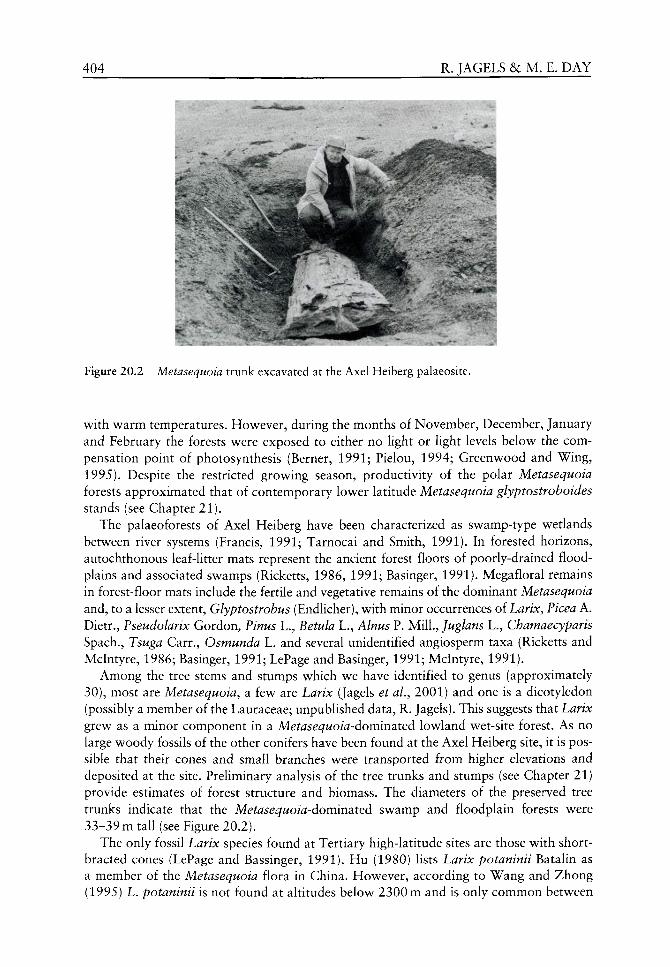
404 R. JAGELS &c M, E. DAY
Figure 20.2 Metasequoia trunk excavated at the Axel Heiberg palaeosite.
with warm temperatures. However, during the months of November, December, January and February the forests were exposed to either no Ught or Ught levels below the compensation point of photosynthesis (Berner, 1991; Pielou, 1994; Greenwood and Wing, 1995). Despite the restricted growing season, productivity of the polar Metasequoia forests approximated that of contemporary lower latitude Metasequoia glyptostroboides stands (see Chapter 21).
The palaeoforests of Axel Heiberg have been characterized as swamp-type wetlands between river systems (Francis, 1991; Tarnocai and Smith, 1991). In forested horizons, autochthonous leaf-litter mats represent the ancient forest floors of poorly-drained flood-plains and associated swamps (Ricketts, 1986, 1991; Basinger, 1991). Megafloral remains in forest-floor mats include the fertile and vegetative remains of the dominant Metasequoia and, to a lesser extent, Glyptostrobus (Endlicher), with minor occurrences of Larix, Picea A. Dietr., Pseudolarix Gordon, Pinus L., Betula L., Alnus P. Mill, Juglans L., Chamaecyparis Spach., Tsuga Carr., Osmunda L. and several unidentified angiosperm taxa (Ricketts and Mclntyre, 1986; Basinger, 1991; LePage and Basinger, 1991; Mclntyre, 1991).
Among the tree stems and stumps which we have identified to genus (approximately 30), most are Metasequoia, a few are Larix (Jagels et ai, 2001) and one is a dicotyledon (possibly a member of the Lauraceae; unpubUshed data, R. Jagels). This suggests that Larix grew as a minor component in a Metasequoia-dominated lowland wet-site forest. As no large woody fossils of the other conifers have been found at the Axel Heiberg site, it is possible that their cones and small branches were transported from higher elevations and deposited at the site. Preliminary analysis of the tree trunks and stumps (see Chapter 21) provide estimates of forest structure and biomass. The diameters of the preserved tree trunks indicate that the Metasequoia-dominated swamp and floodplain forests were 33-39 m tall (see Figure 20.2).
The only fossil Larix species found at Tertiary high-latitude sites are those with short-bracted cones (LePage and Bassinger, 1991). Hu (1980) lists Larix potaninii Batalin as a member of the Metasequoia flora in China. However, according to Wang and Zhong (1995) L. potaninii is not found at altitudes below 2300 m and is only common between
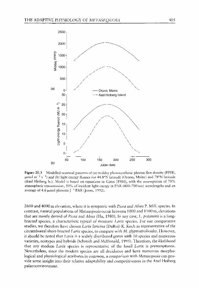
THE ADAPTIVE PHYSIOLOGY OF METASEQUOIA 405
2500
2000
^ 1500 CL
P 1000
500
(a) oJ 30 n
7 25
e 20 0 O c 3 15
g 10 0
(b) 50
/ X
X \ \
/ \
\ - Orono, Maine
- Axel Heiberg Island
100 150 200
Julian date
250 300
Figure 20.3 Modelled seasonal patterns of (a) midday photosynthetic photon flux density (PPFD, fxmol m~^ s~^) and (b) light energy fluence for 44.8°N latitude (Orono, Maine) and 78°N latitude (Axel Heiberg Is.). Model is based on equations in Gates (1980), with the assumptions of 70% atmospheric transmission, 50% of incident light energy in PAR (400-700 nm) wavelengths and an average of 4.6 fxmol photons J"^ PAR (Jones, 1992).
2600 and 4000 m elevation, where it is sympatric with Picea and Abies P. Mill, species. In contrast, natural populations oi Metasequoia occur between 1000 and 1100 m, elevations that are mostly devoid of Ficea and Abies (Hu, 1980). In any case, L. potaninii is a long-bracted species, a characteristic typical of montane Larix species. For our comparative studies, we therefore have chosen Larix laricina (DuRoi) K. Koch as representative of the circumboreal short-bracted Larix species, to compare with M. glyptostroboides. However, it should be noted that Larix is a widely distributed genus with 10 species and numerous varieties, ecotypes and hybrids (Schmidt and McDonald, 1995). Therefore, the likelihood that any modern Larix species is representative of the fossil Larix is presumptuous. Nevertheless, since the modern species are all deciduous and have numerous morphological and physiological attributes in common, a comparison with Metasequoia can provide some insight into their relative adaptability and competitiveness in the Axel Heiberg paiaeoenvironment.

406 R. JAGELS & M. E, DAY
Characteristics of the temperature regime at the 45 miUion year old palaeosite are very speculative at present. Rates of primary production in the palaeoforest suggest a temperate climate (see Chapter 21). Bassinger (1991) suggested that winter temperatures seldom dropped below freezing, based on identification of vegetative remains and the lack of significant latewood production in Metasequoia. However, Larix from the same site shows significant latewood production (Jagels et al., 2001). Mclntyre (1991) concluded that the climate was warm-temperate, based on the pollen assemblage.
Metasequoia: fossil and living
Extant natural stands of Metasequoia (M. glyptostroboides) are restricted to a small, remote area of Hupeh province near the border of Szechuan, China at an approximate latitude of 30°10'N (Chu and Cooper, 1950). Although in the fossil high-latitude forests Metasequoia was the upper-canopy dominant species and exhibited growth rates more typical of shade-intolerant conifers, characteristics of both the natural forests in China (Chu and Cooper, 1950; Florin, 1952; Li, 1957) and planted stands of extant Metasequoia (personal observations) suggest that the species maximizes growth in partially shaded rather than full sun conditions. This paradox will be explored further in later sections of this chapter.
Several authors have proposed that the deciduous habit of Metasequoia provided a competitive advantage for the Arctic winter (Francis, 1991; Basinger, 1991; Mclntyre, 1991; see Chapter 21). Chaney (1948) postulated that the deciduous forest type originated in the north during the Cretaceous and migrated south during the later Tertiary and Vann et al. (see Chapter 21) suggest the possibility that Metasequoia evolved at high-latitudes. Hu (1980) disputed Chaney's hypothesis on the basis that a deciduous habit can also be adaptive to wet/dry periodicity in relatively frost-free areas, such as the Hupeh Forest. Fossil evidence demonstrates that Metasequoia was well established at lower latitudes during the Cretaceous (Figure 20.4), predating the Eocene Arctic swamp forests. This is consistent with Hu's argument for the evolution of the deciduous habit under a wet-dry climate regime, particularly since this is a rare attribute in conifers. Therefore, in high-latitude Eocene forests deciduousness may have been adaptive not only by reducing metabolic carbon demand during winter dark periods, but by reducing water stress during the dry phase of soil moisture cycles. In a contemporary analogy, Taxodium distichum (L.)
/ /VVl r ^^ p r^r
^_0j^—
» ^ = ^ " w £ ^
Vv60°
140°
\ or
p\r ^^ \ Figure 20.4 Distribution of Metasequoia fossil palaeosites. Key to sites: • = Late Cretaceous, T =Palaeocene, I = Eocene, 0 = Oligocene, O = Miocene. Map adapted from Monohara (1994).
THE ADAPTIVE PHYSIOLOGY OF METASEQUOIA 407
Richard is deciduous and is restricted to floodplains in the southern USA where wet-dry periods dominate the soil moisture regime.
Metasequoia was well distributed above the Arctic circle from the Palaeocene through the Eocene. As the earth cooled and became drier during the Oligocene and Miocene (Wolfe, 1985) its distribution became more limited to temperate zones of North America and Asia (see Figure 20.4) and eventually became restricted to moist, narrow river valleys in China (Chu and Cooper, 1950; Li, 1957; Momohara, 1994). In contrast, short-bracted Larix, an equally poor competitor, but one having a greater low-temperature tolerance (either a preadaptation or one evolving over time) was able to successfully occupy north temperate and boreal hydric sites where competition is minimal.
Vann et al. (see Chapter 21) develop a compelling case that Metasequoia glypto-stroboides retains a substantial array of morphological and, by implication, physiological traits present in the Eocene Metasequoia^ and is, therefore, a useful nearest living relative (NLR) for comparative studies. As discussed in subsequent sections, extant M. glypto-stroboides appears to have 'relict' traits; and unusual combinations of morphological and physiological characteristics would have particular adaptive value in the unique environment suggested for the Eocene high-Arctic forests. In modern temperate forests these same traits would have little adaptive value and may even be maladaptive. This collection of morphological and physiological traits is consistent with the suggestion of Konoe (1960) that establishment of this species may depend on successful responses to multiple environmental factors. While also consistent with a high latitude evolution for Metasequoia, it does not rule out the possibility that these adaptations evolved at lower latitudes and provided Metasequoia with a preadaptive advantage in Eocene Arctic swamp-forests.
Physiological challenges of a high-latitude, continuous-Hght environment
Our research has focused on physiological and morphological traits which would be adaptive to a tree species growing in a temperate climate with a season of continuous light (CL) alternating with a season of continuous darkness (CD). Light intensities during the summer were assumed to be substantially less than at lower latitudes (see Figure 20.3). Categories of attributes considered adaptive to the Eocene palaeoarctic forest environment were: (1) the ability to photosynthesize efficiently under the light intensities and temporal patterns of high latitudes; (2) possession of an integrated carbon balance strategy (photosynthesis, respiration and allocation) that would be adaptive to growth and reproduction in that unique environment; (3) an expeditious system for translocation of photosyn-thetic products to prevent direct or indirect feedback inhibition of photosynthesis; (4) processes for dissipation of excess light energy through non-photochemical pathways or photorespiration in order to protect photosynthetic systems; and (5) strategies for minimizing limitations to photosynthesis from water stress imposed by continuous transpiration in CL.
Adaptations to Arctic light regimes
During the CD period of winter, the tree must be able to effectively minimize metabolic activity, which would provide strong selective pressure for a deciduous habit. Because the polar early spring provides only minimal light intensity, the tree should be able to produce

408 R. JAGELS &c M, E. DAY
new foliage and initiate chlorophyll synthesis at very low light levels. Foliar characteristics along the sun-foliage to shade-foliage continuum are determined by the light environment under which the leaves develop and are adaptive in that carbon and nutrient resources are allocated in proportion to potential carbon gain (Givnish, 1988; Klippers, 1994). However, at high latitudes spring light intensity is far lower than the summer intensity (see Figure 20.3), decreasing the efficiency of this mechanism. This great variation in light intensities over the growing season would impart adaptive advantage to photosynthetic physiology that provided for efficient carbon fixation (relative to the allocation costs) over a wide range of light intensities. While photosynthetic traits associated with shade-adapted foliage would be optimal during the Arctic spring and autumn, shade-adaptation has been shown to be negatively correlated with attributes necessary to address the physiological stresses of higher irradiance environments (Givnish, 1988; Johnson et ai, 1993; Tyree et al., 1998).
Under selective pressure to optimize carbon gain in the Arctic light regime, this incompatibility between shade- and sun-adaptation could be addressed through one or more of the following mechanisms: (1) by modifying the photosynthetic physiology of existing foliage; (2) by production of new foliage as the light environment changes; or (3) by producing foliage that exhibits relatively efficient photosynthesis over a wide range of light intensities (i.e. an 'optimal compromise' of sun- and shade-adaptations). Adaptive adjustment of photosynthetic attributes of existing foliage to increasing light intensities has been demonstrated in conifers (Brooks et al.^ 1996), however, this process has not been examined in Metasequoia or closely related species. Trees with an indeterminate growth habit, such as Metasequoia, continuously produce foliage through the growing season (Table 20.1). This provides a mechanism for adjusting the light-adaptation status of foliage to changing light environments (Pothier and Margolis, 1991). Furthermore, as discussed in the next section, the photosynthetic light-response curve of Metasequoia suggests that both shade-adapted and sun-adapted foliage of that species is photosynthetically efficient over a wide range of low to moderate light intensities, roughly coincident with those found in the high Arctic.
Carbon balance physiology
Efficient carbon balance physiology requires synchronization of photosynthetic, carbohydrate sink and storage activity with diel and seasonal cycles of resource availability (Luxmoore, 1991). A deciduous habit is obviously adaptive to extended periods when photosynthesis is resource (light or moisture) limited. In annual cycles at middle latitudes aspects of carbon balance are controlled by phenological processes (Cannell, 1990), which are induced by environmental cues such as day length and temperature. Adaptation to a temperate high-latitude environment would require modification of these processes in a manner that has no extant analogue.
During the midsummer period of maximum light intensity, down-regulation of photosynthesis by end-product buildup may become a challenge to plants growing in a CL environment. End-product down-regulation may decrease carboxylation rates through several potential pathways that may act directly on biochemical mechanisms or indirectly by genetic induction (Stitt, 1991; Krapp and Stitt, 1995) and have a two-fold deleterious influence by concurrently limiting potential carbon gain and restricting the carboxylation pathway for dissipation of intercepted light energy. Pathways leading to end-product inhibition of photosynthesis may be specific to metabolites accumulated (Schaffer et aL, 1986; Stitt, 1991; Huber and Ruber, 1992; Krapp and Stitt, 1995; Jang et al., 1997). Therefore, physiological strategies to limit feedback inhibition of photosynthesis under CL conditions must
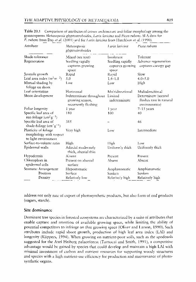
THE ADAPTIVE PHYSIOLOGY OF METASEQUOIA 409
Table 20.1 Comparison of attributes of crown architecture and foliar morphology among the gymnosperms Metasequoia glyptostroboides, Larix laricina and Picea rubens. SLA data for P. rubens from Day et ai (2001) and for Larix laricina from Hutchison et ai (1990).
Attribute
Shade-tolerance Regeneration
Juvenile growth Leaf area index (m^m"^) Mutual shading by
foHage on shoot Leaf orientation Shoot development
Foliar longevity Specific leaf area of
sun-foliage (cm^g^^) Specific leaf area of
shade-foliage (cm^g~^) Plasticity of foliage
morphology with respect to light environment
Surface-to-volume ratio Epidermal walls
Hypodermis Chloroplasts in
epidermal cells Stomata: Arrangement
Position Density
Metasequoia glyptostroboides
Mixed (see text) Seedling rapidly
captures growing space
Rapid 5.0 Low
Horizontal Indeterminate throughout
growing season, recurrently flushing
1 year 110
315
Very high
High Adaxial moderately
thick, abaxial thin Absent Present on abaxial
surface Hypostomatic Surface Relatively low
Larix laricina
Intolerant Seedling rapidly
captures growing space
Rapid 1.4-1.8 Low
Multidirectional Limited
indeterminate
1 year 100
-
Low
High Uniformly thick
Present Absent
Amphistomatic Sunken Relatively high
Ficea rubens
Tolerant Advance regeneration
; captures canopy gap
Slow 4.0-5.0 High
Multidirectional Determinate (second
flushes rare in natural environments)
7-15 years 40
46
Intermediate
Low Uniformly thick
Present Absent
Amphistomatic Sunken Relatively high
address not only rate of export of photosynthetic products, but also form of end products (sugars, starch).
Site dominance
Dominant tree species in forested ecosystems are characterized by a suite of attributes that enable capture and retention of available growing space, w^hile limiting the ability of potential competitors to infringe on that growing space (Oliver and Larson, 1990). Such attributes include rapid shoot growth, production of high leaf area index (LAI) and longevity (Kiippers, 1994). When growing on nutrient-poor soils, such as the spodosols suggested for the Axel Heiberg palaeoforest (Tarnocai and Smith, 1991), a competitive advantage would be gained by species that could develop and maintain a high LAI with minimal investment of carbon and nutrient resources for supporting woody structures and species with a high nutrient-use efficiency for production and maintenance of photo-synthetic organs.
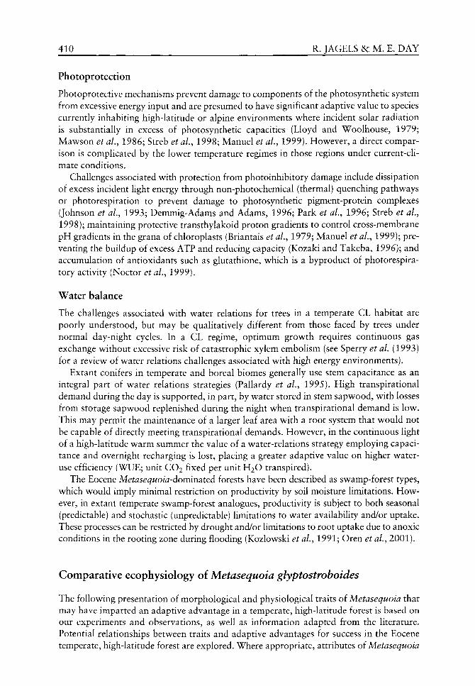
410 R. JAGELS &c M. E. DAY
Photoprotection
Photoprotective mechanisms prevent damage to components of the photosynthetic system from excessive energy input and are presumed to have significant adaptive value to species currently inhabiting high-latitude or alpine environments v^here incident solar radiation is substantially in excess of photosynthetic capacities (Lloyd and Woolhouse, 1979; Mawson et aL, 1986; Streb et aL, 1998; Manuel et al, 1999). How^ever, a direct comparison is complicated by the lov^er temperature regimes in those regions under current-climate conditions.
Challenges associated v^ith protection from photoinhibitory damage include dissipation of excess incident light energy through non-photochemical (thermal) quenching pathw^ays or photorespiration to prevent damage to photosynthetic pigment-protein complexes (Johnson et aL, 1993; Demmig-Adams and Adams, 1996; Park et aL, 1996; Streb et al, 1998); maintaining protective transthylakoid proton gradients to control cross-membrane pH gradients in the grana of chloroplasts (Briantais et ai, 1979; Manuel et ai, 1999); preventing the buildup of excess ATP and reducing capacity (Kozaki and Takeba, 1996); and accumulation of antioxidants such as glutathione, w^hich is a byproduct of photorespira-tory activity (Noctor et ai, 1999).
Water balance
The challenges associated w ith w^ater relations for trees in a temperate CL habitat are poorly understood, but may be qualitatively different from those faced by trees under normal day-night cycles. In a CL regime, optimum grov^th requires continuous gas exchange w^ithout excessive risk of catastrophic xylem embolism (see Sperry et al. (1993) for a review^ of v^ater relations challenges associated w ith high energy environments).
Extant conifers in temperate and boreal biomes generally use stem capacitance as an integral part of w^ater relations strategies (Pallardy et al., 1995). High transpirational demand during the day is supported, in part, by water stored in stem sapv^ood, w ith losses from storage sapv^ood replenished during the night when transpirational demand is low. This may permit the maintenance of a larger leaf area with a root system that would not be capable of directly meeting transpirational demands. However, in the continuous light of a high-latitude warm summer the value of a water-relations strategy employing capacitance and overnight recharging is lost, placing a greater adaptive value on higher water-use efficiency (WUE; unit CO2 fixed per unit H2O transpired).
The Eocene Metasequoia-dominatQd forests have been described as swamp-forest types, which would imply minimal restriction on productivity by soil moisture limitations. However, in extant temperate swamp-forest analogues, productivity is subject to both seasonal (predictable) and stochastic (unpredictable) limitations to water availability and/or uptake. These processes can be restricted by drought and/or limitations to root uptake due to anoxic conditions in the rooting zone during flooding (Kozlowski et aL, 1991; Oren et ai, 2001).
Comparative ecophysiology of Metasequoia glyptostroboides
The following presentation of morphological and physiological traits of Metasequoia that may have imparted an adaptive advantage in a temperate, high-latitude forest is based on our experiments and observations, as well as information adapted from the literature. Potential relationships between traits and adaptive advantages for success in the Eocene temperate, high-latitude forest are explored. Where appropriate, attributes of Metasequoia

THE ADAPTIVE PHYSIOLOGY OF METASEQUOIA 411
are contrasted with those of Larix, which was a minor component of Eocene high-latitude palaeoforests (but commonly dominates extant forests in sub-Arctic biomes) and with other conifers that are currently important components of boreal forests.
Foliar morphology and crown architecture
Metasequoia has both long (persistent) and short (deciduous) shoots. Foliage is decussately arranged (Figure 20.5). Leaves are uninerved with bifacially flattened, linear lamina and decurrent bases. The hypostomatic leaves have two bands of stomata on the abaxil surface and guard cells are surrounded by four to eight subsidiary cells. Non-stomatal epidermal cells have 'undulating' walls (Sterling, 1949; Florin, 1952; Srinivasan and Friis, 1989). Examinination with light microscopy of freehand sections of living leaves (Figure 20.6) and
Figure 20.5 Shoot and foliage structure of Metasequoia glyptostroboides. Arrow points to a late season shoot arising from a leaf base and the scale bar is 2 cm long.
100 um
(a) (b)
Figure 20.6 Microphotographs of cross-sections from Metasequoia glyptostroboides leaves, (a) Presents a comparison of abaxial and adaxial epidermal cell morphology. Note the thin exterior walls on the abaxial cells and thicker adaxial walls (arrows), (b) The protruding nature of adaxial epidermal cells and the presence of chloroplasts are apparent. The arrow points to a protruding abaxial epidermal cell containing chloroplasts.
412 R. JAGELS &c M. E. DAY
Figure 20.7 Scanning electron micrographs of the adaxial surface of a Metasequoia glyptostroboides leaf, (a) The convoluted surface arising from protruding epidermal cells and (b) stomatal guard cells (arrow) surrounded by raised subsidiary cells. Scale bars: (a) = 100 |xm; (b) = 10 |xm.
scanning electron micrographs of leaf surfaces (Figure 20.7), revealed that Metasequoia leaves have thickened outer walls on adaxial epidermal cells, v^hile those on the abaxial surface have very thin walls. Abaxial epidermal cells contain chloroplasts, which seem to be lacking in their adaxial counterparts. In addition, the abaxial epidermis is characterized by protruding cells interspersed with deep grooves (Figures 20.6 and 20.7a). Stomata are hypostomatic and not sunken, while subsidiary cells are raised (Figure 20.7b).
In Table 20.1 we compare crown morphological features and foliar attributes for Metasequoia glyptostroboides^ Larix laricina and Picea rubens Sarg. An interspecific comparison of photosynthetic response to light is shown in Figure 20.8. For these comparisons, P. rubens was included as a representative cold-temperate, shade-tolerant species and Finus banksiana Lamb, as a cold-temperate, shade-intolerant species adapted to water-limited habitats.
Metasequoia has some characteristics which are typical of extant shade-intolerant species, such as crown architecture, regeneration strategy, indeterminate shoot growth, rapid shoot elongation, high photosynthetic efficiency (initial slope of light response curve) and low diversity in photosynthetic light-response and foliar display between sun- and shade-adapted foliage (Givnish, 1988; Walters and Reich, 1999). In contrast, traits such as large differences in specific leaf area between sun- and shade-adapted foliage, horizontal leaf orientation, planar foliage display and high foliar density and LAI are generally characteristic of shade-tolerant species (Givnish, 1988). Other attributes, such as unusual presence of chloroplasts in the epidermal cells of abaxil leaf surfaces and their thin outer walls, are more characteristic of shade-loving ferns and other pteridophytes.
Several anatomical characteristics suggest that Metasequoia is best adapted to an environment with high atmospheric humidity. These include a high surface-to-volume ratio, enhanced by a convoluted lower epidermis (see Figure 20.6 and 20.7a), non-sunken stomata with raised subsidiary cells (see Figure 20.7b), thin epidermal walls on the abaxial surface and lack of a hypodermis. This contrasts with the more xerophytic leaf anatomy of Larix^ Picea and many other conifers. Whether the humid palaeoenvironment included fog is not known, but we know that Sequoia sempervirens (D.Don) Endl. satisfies a large portion of its water needs through fog capture (Dawson, 1998). Dew capture might be a valuable asset during periods of reduced soil moisture (Munne-Bosch and Alegre, 1999). In Metasequoia^ condensation of dew from humid air would be enhanced by the spreading

THE ADAPTIVE PHYSIOLOGY OF METASEQUOIA 413
20-
E
I
I
18-
16-
14-
12-
10-
8 -
6 -
4 -
2 -
0 -
/• ' A/
/ "
'
1 / f ••'
Metasequoia glyptostroboides • • • • Picea rubens
Pinus banksiana Larix lahcina
/ /
/ /
/ . ^ ' / .^
1 X
/ //
/ /
/ . . • • • • •
" 5 -
o ^ 3 -
CO CO P -CD
CO 1 o o
1 °" -z.
_ ^ — — '
^ ... ^ "
Shade-adapted foliage
' Picea rubens Metasequoia
0 250 500 750 10 00
250 500 750 1000 1250 1500 1750 2000
PPFD (jinriol m-2 s "
Figure 20.8 Photosynthetic light-response curves for sun-adapted foliage of Metasequoia glyptostroboides, red spruce (Picea rubens), jack pine {Pinus banksiana) and eastern larch (Larix laricina) at photosynthetic photon flux densities (PPFD) <1000 |xmolm~^s~^ M. glyptostroboides shows photosynthetic efficiencies similar to shade-intolerant species (?. banksiana and L. laricina) at low PPFD levels and a rapid approach to the light-saturated state, giving it much greater carbon gain per unit leaf area than the shade-tolerant P. rubens. Insert compares shade-adapted foliage of M. glyptostroboides and P. rubens, showing the similarities between response curves of sun- and shade-adapted M. glyptostroboides and their great divergence in P. rubens. Curves were modelled from light-response data for 6 saplings of each species, with data collected under standardized conditions.
414 R. JAGELS &: M. E. DAY
foliar display which would increase the rate of radiational heat loss and more effectively lower foliar temperatures. In addition, the increased surface-to-volume ratio resulting from the convoluted abaxial surface may increase convective heat transfer and enhance the efficiency of dew collection (Muselli et al.y 2001).
Most monopodial conifers have determinate growth (Kozlowski et aL, 1991). However, Metasequoia shows both a monopodial growth habit and indeterminate shoot growth, a pattern more common in angiosperms. With adequate moisture, new leaves are produced throughout the growing season. This growth habit provides a continuous sink (new shoot growth) for carbohydrates produced in neighbouring leaves (Luxmore, 1991), which would reduce sink limitations to photosynthesis and associated challenges. Although sink-limitation of photosynthesis has been relatively unexplored in conifers, indirect evidence has been reported for Psuedotsuga menziesii (Mirb.) Franco (Leverenz, 1981) and Ficea rubens (Day, 2000) and experimentally demonstrated for Finns taeda L. (Myers et al, 1999).
In Metasequoia^ new foliage can arise on epicormic shoots on small stems or branches, as side shoots on short shoots, as new flushes on long shoots and occasionally from the bases of leaflets on short shoots. These subsequent shoots may often be oriented in a vertical direction to maximize light capture in the crown (see Figures 20.1 and 20.5). In general, carbon allocation to foliage is positively associated with high relative growth rates and competitive advantage in woody species (Poorter, 1989, 1999; Cornelissen et al., 1996). The indeterminate growth habit of Metasequoia may be a critical component of its ability to dominate the high-Arctic forests by: (1) enhancing growth potential and capture of growing space in the long growing season; (2) permitting new foliage to be produced and displayed to adjust effectively to changing angular light distribution; (3) producing new foliage with morphological and physiological traits optimized to the particular light intensity at the time of development; and (4) providing a constant sink for photosynthate, thereby reducing problems associated with end-product down-regulation of photosynthesis. This growth habit confers the ability to develop foliage that is morphologically and physiologically optimized for the light environment in which it develops. As we presented in our list of challenges faced by species in the unique high-Arctic light regime, this is theoretically an important adaptation to low-intensity, low-angle incident light during the Arctic spring and fall and moderate intensities during the summer. In addition, this habit permits exploitation of changing distributions of light resources due to the effects of competition and small-scale disturbances (Canham and Marks, 1985; Canham, 1989).
Based on our greenhouse and field experiments and observations of planted trees in various environments, Metasequoia trees rapidly develop extensive crowns with high foliar density (FD) under favourable conditions of moisture and light, which contrasts with the much lower FD and LAI of Larix (see Table 20.1). In three 20+ year-old planted Metasequoia stands in Japan, Vann et al, (see Chapter 21) reported only 1-3% of incident PPFD penetrated to the forest floor. This degree of light interception is typical of climax forests (Landsburg and Gower, 1997). In Metasequoia plantings receiving full sunlight for only part of the day, we have observed the rapid development of a closed canopy. However, if trees are growing under full sunlight for more than 6 hours per day, shoot elongation is greatly reduced and the leaves are smaller and appear chlorotic. Under these conditions both crown size and foliar density are reduced. Under dense shade, shoot elongation is favoured over leaf production, a characteristic typical of shade-intolerant species (Kozlowski et aL, 1991). Typical shade-tolerant conifers such as P. rubens can survive for decades under dense shade as suppressed saplings (Davis, 1991). This advance regeneration is then available to respond to small-scale disturbances to attain upper canopy status.
THE ADAPTIVE PHYSIOLOGY OF METASEQUOIA 415
In contrast, Metasquoia appears to perform poorly under dense shade, which is, in part, a consequence of producing foUage with a one-year Ufespan. In temperate gymnosperms, leaf longevity is correlated with shade-tolerance (P. rubens can retain needles for up to 16 years). Among the genera of deciduous gymnosperms native to North America, Larix is classified as very shade-intolerant and Taxodium is described as requiring full sunlight for maximum growth (Burns and Honkala, 1990). Ida (1981) found that Metasequoia increased its leaf weight much more effectively at low light intensities than did T. distichum.
Among conifers, specific gravity (sp. gr.) of wood varies considerably. Those adapted to windy sites generally produce denser wood. Metasequoia has an unusually low sp. gr. among conifers. Depending on source, sp. gr. ranges from 0.29 to 0.31, while that of Larix species ranges from 0.57 to 0.59 (Alden, 1997; Polman et aL, 1999). Thus, considerably less photosynthetic resources are needed to create the same height of supporting trunk for a Metasequoia tree compared to Larix. Metasequoia partially compensates for low wood density by producing a fluted trunk near its base to provide additional lateral stability. Metasequoia may also be competitively favoured over Larix in crown attributes such as FD and LAI (see Table 20.1). These traits in Metasequoia more closely resemble those of the more shade-tolerant conifers (e.g. Picea and Abies spp.) that often dominate better quality sites in boreal and cool-temperate forests to the exclusion of Larix. Extant Larix is often relegated to bogs or steep mountain slopes due to competition on better sites from conifers producing substantial shade (Schmidt and McDonald, 1995).
Photosynthetic light-response
As with its morphology and growth habits, M. glyptostroboides shows characteristics in its photosynthetic light-response curve (see Figure 20.8) that are typical of both sun- and shade-adapted species. Photosynthetic quantum efficiency (QE: mol CO2 fixed per mol photons absorbed, i.e. the initial linear slope of the response curve) is similar to the shade-intolerant L. laricina and P. banksiana at low light levels, but its maximum photosynthetic rates are more comparable with the shade-tolerant P. rubens. However, in contrast to P. rubens^ maximum photosynthetic rates are reached at a far lower light intensity, resulting in a response curve with much higher convexity (lower loss in apparent quantum efficiency between its initial linear portion and light-saturation). Additionally, the photosynthetic light-responses of sun- and shade-adapted foliage show remarkably less divergence in characteristics than those of typical shade-tolerant conifers (compare with P. rubens^ insert in Figure 20.8). These physiological traits contrast with the significant sun-shade divergence with respect to specific leaf area (see Table 20.1). This inconsistency between high morphological and low physiological variation along the sun-shade adaptation continuum can be interpreted as adaptations to two opposing challenges in the model of the high-latitude light environment. The stochastic probability that foliage produced under low light intensity might become 'obsolete' as light resources are limited by shading within the canopy warrants minimal investment of resources. Opposing this is the predictable seasonal cycle of foliar development under low light intensities followed by exposure to much greater intensities as the growing season progresses (see Figure 20.3). In this interpretation, Metasequoia evolved a compromise response by minimizing carbon investment in foliage developed under low illumination, while investing greater resources in the photosynthetic apparatus than prevailing light conditions would warrant. Carbon investment in foliage is permanent and is lost if a leaf senesces due to low light availability. However, most nutrients (e.g. N, P, K, Mg) are mobile and are commonly retranslocated from senescing foliage before abscission. Therefore, investment of nutrients in
416 R. JAGELS &c M. E. DAY
the photosynthetic system has a lower risk of permanent loss to the tree and provides the potential for delivering higher photosynthetic output if, or w^hen, higher light resources become available. Interestingly, this strategy, while adaptive to the unique light regime of the high-Arctic, would have much reduced value under forest light regimes in current temperate zones, where the light environment under which foliage develops would be highly correlated with the light environment throughout the growing season. In species of similar leaf longevity, investment in nutrient resources closely parallels investment in carbon and nutrient resources (Evans, 1989; Hikosaka and Terishima, 1995; HoUinger, 1996) and may be an important physiological trait for shade-tolerance (Niinemets et aL, 1998). The unusual composite of sun-shade characteristics in M. glyptostroboides may be a conserved relict trait from cloudier, wetter paleoclimates in the lower latitudes.
The saturating light intensity for M. glyptostroboides (see Figure 20.8) is close to the predicted maximum intensity for 78°N latitude (see Figure 20.3). This suggests that investment of resources in maximum photosynthetic capacity may be optimal for the high-latitude light environment. The high convexity of the light-response curve for individual leaves compared with those of the other conifers (see Figure 20.8) shows a minimal loss of quantum efficiency from self-shading at the leaf level. A Blackman-response curve, where response increases linearly with input of resource (in this case light) until a saturation point is reached, describes an optimal utilization of resource. Any departure from that response, indicated by a bending of the response curve (non-linearity in the presaturation portion), suggests less than optimal resource utilization. In the case of photosynthetic light-response, this departure from optimality is associated with mutual shading of light harvesting elements (Hikosaka and Terashima, 1995). At the leaf-level, mutual shading is due to the architecture of mesophyll cells and the chloroplasts they contain. Under high intensity illumination, mutual shading may permit capture of 'excess' light by photosynthetic elements lying deeper in the mesophyll as surface elements become light-saturated. However, in an environment characterized by low-angle direct solar radiation at low to moderate intensities, architecture that minimizes mutual shading (e.g. Metasequoia) would provide for more efficient resource utilization compared to conifers with more robust needle morphology (e.g. Larix or Picea). Although we have not developed whole-tree light-response curves, this structural minimization of mutual shading is carried to the shoot-level, where flat-bladed foUage would be more efficient in this respect than the multidirectional, clustered foliar architecture common to many extant conifers, including Larix and Picea. A flattened foliar display pattern is also associated with more efficient light harvesting in environments of low to moderate light intensity (Canham and Marks, 1985; Givnish, 1988). The presence of chloroplasts in the protruding abaxial epidermal cells of Metasequoia (see Figure 20.6b), could additionally enhance capture of low-intensity diffuse light.
Efficiency in CO2 fixation might be enhanced by transport and/or sink physiology that reduces potential inhibition from the direct and/or indirect effects of photosynthetic end-product accumulation, such as a reduction of starch production in foliage. From an experiment with 72 one-year-old M. glyptostroboides trees planted in a forest opening in Orono, Maine, in which half the trees received natural diurnal light (NL) and half were under CL (supplied by high intensity metal halide lights at night), we found that by mid-July leaves from the NL side had abundant starch, but leaves from trees on the CL side produced little or no starch (as determined microscopically with IKI staining). Omitting starch synthesis in leaves would improve energy efficiency and might reduce investments in catalytic systems. The avoidance of starch production and subsequent catabolism reduces energy costs to the plant but, under CL conditions, requires a continuous and rapid transport of
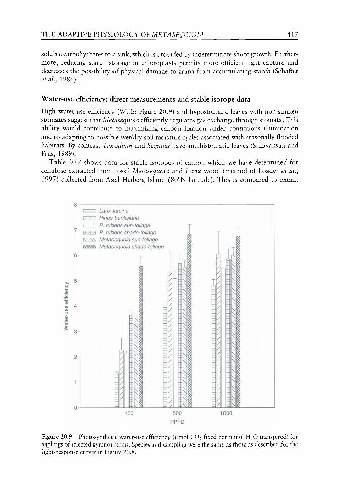
THE ADAPTIVE PHYSIOLOGY OF METASEQUOIA 417
soluble carbohydrates to a sink, which is provided by indeterminate shoot growth. Furthermore, reducing starch storage in chloroplasts permits more efficient light capture and decreases the possibility of physical damage to grana from accumulating starch (Schaffer etal., 1986).
Water-use efficiency: direct measurements and stable isotope data
High water-use efficiency (WUE: Figure 20.9) and hypostomatic leaves with non-sunken stomates suggest that Metasequoia efficiently regulates gas exchange through stomata. This ability would contribute to maximizing carbon fixation under continuous illumination and to adapting to possible wet/dry soil moisture cycles associated with seasonally flooded habitats. By contrast Taxodium and Sequoia have amphistomatic leaves (Srinivansan and Friis, 1989).
Table 20.2 shows data for stable isotopes of carbon which we have determined for cellulose extracted from fossil Metasequoia and Larix wood (method of Loader et al.^ 1997) collected from Axel Heiberg Island (80°N latitude). This is compared to extant
6H
5H
CD
0
Larix larcina \/ / / Pinus banksiana I I P. rubens sun-foliage II1111 i III P. rubens shade-foliage SSSS Metasequoia sun-foliage ^ ^ Metasequoia shade-foliage
v\
M 100 500
PPFD
1000
Figure 20.9 Photosynthetic water-use efficiency (ixmol CO2 fixed per mmol H2O transpired) for saplings of selected gymnosperms. Species and sampling were the same as those as described for the light-response curves in Figure 20.8.

418 R. JAGELS & M. E. DAY
Table 20.2 Mean 8^^C values (%o) extracted a-cellullose from both fossil and extant specimens. Means calculated from 6 samples. Standard deviations are given in parentheses.
Fossil species
Metasequoia
a-cellulose 8^^C (%o) -20.53 Standard deviation (0.066)
Larix
-21.70 (0.137)
Extant species
M. glyptostroboides L. laricina
-22.42 -24.80 (0.187) (0.133)
Metasequoia glyptostroboides and Larix laricina wood. Of note is the fact that both fossil woods have more positive values than the modern counterparts and Metasequoia is more positive than Larix (both fossil and modern).
The 8^^C values for extant Metasequoia and Larix species fall mostly within the expected range for extant C3 species, including conifers ( -23 to -34%o), although the Metasequoia value (—22.42) is slightly more positive. Values for fossil Metasequoia and Larix are more positive than those usually associated with C3 plants (O'Leary, 1988). Several environmental factors can promote more positive 8^^C values by influencing stomatal conductance. These include high irradiance and factors elevating plant water stress such as moisture limitations (Guy and Reid, 1986; Farquhar et at., 1989; Ehleringer, 1993; Grocke, 1998). Yet the fossil trees are believed to have grown under low level irradiance and were wet site species. Growth rates of the fossil trees (our ring width data and Vann et al. (see Chapter 21)) are comparable to that observed in extant Metasequoia growing on good sites (Jagels et al., 2003).
Grocke et aL (1999) have recently proposed that 8^^C values of fossil plant remains are primarily controlled by concentration and isotopic composition of CO2 in the ocean-atmosphere system (as registered in marine carbonates) and not by palaeoenvironmental and palaeoecological factors. For the 40 million years ago time period, several studies have suggested that atmospheric CO2 concentrations were higher than at present and more negative values in marine carbonate sources have been reported (Berner, 1991; Cerling, 1992; Freeman and Hayes, 1992). If true, one would expect that 8^^C values from the fossil specimens would be more negative than that of their NLRs, the opposite of what we have observed. In contrast, the reconstruction of Eocene atmospheric CO2 concentrations by Royer et aL (2001) indicates values comparable with present-day levels. In addition, Pearson et al. (2001) have recently proposed that surface water temperatures of tropical seas during the Eocene were 10°C higher than previous estimates. Therefore, if Eocene CO2 concentrations were near those of the present and global temperatures were driven by factors other than CO2 concentrations, 8^^C values of fossil cellulose are likely to be driven by the same ecophysiological factors influencing stable carbon isotope values at present. These would include, stomatal resistance to gas exchange, external and internal water relations, form and rate of photosynthetic end-product production and photorespiration rates. All of these factors would be potentially influenced by light regime periodicity (CL versus diurnal cycle).
WUE and 8^^C values have been shown to be highly correlated by numerous studies (e.g. Silim et al.., 2001). Therefore, 8^^C values have been widely used as indicators of integrated water-use efficiency. The more negative 8^^C values of Larix compared with Metasequoia, for both living and fossil specimens (see Table 20.2), suggest that Metasequoia had greater WUE than the second most abundant tree species in the Axel Heiberg Eocene lowland forests. This agrees with the extensive survey by Kloeppel et al. (1998) who found that WUE in most Larix species, determined from 8^^C values, were lower than for co-occurring conifers.
THE ADAPTIVE PHYSIOLOGY OF METASEQUOIA 419
Two principal factors control WUE: (1) stomatal control that minimizes transpiration and limitats to photosynthesis by decreasing internal CO2 concentration (Q); and (2) the photosynthetic efficiency and rates of carbon fixation at a given Q. To evaluate the first factor, v e tested photosynthetic WUE (juimol CO2 fixed per mmol H2O transpired) for sapling-sized M. glyptostroboides, L. laricina, P. rubens and P. banksiana (see Figure 20.9) over light intensities of 100 to 1000 ixmol m^^ s^^ PPFD. Since none of these trees were drought-stressed and measurements were made under ambient CO2 levels, the differences are presumed to be characteristic of the genetic potential of each species (Donovan and Ehleringer, 1994; Osorio and Pereira, 1994). Shade-adapted M. glyptostroboides demonstrated the highest WUE values throughout the range of light intensities. Metasequoia sun-foliage had WUE values as high as or higher than all other species by foUar type combinations. In contrast, Larix displayed the lowest WUE values. In addition, the relative consistency in WUE of Metasequoia^ especially its shade-adapted foUage, across the range of light intensities indicates that stomatal conductance is more sensitive to photosynthetic rates. At low light intensity, stomatal conductance decreases to conform with reduced photosynthetic rates in Metasequoia^ while stomatal conductance remains high in the other species, decreasing their WUE at low light levels. Thus, M. glyptostroboides appears to be better able to finely tune its gas exchange to minimize water loss at low to medium light intensities, intensities comparable with those through most of the high-latitude growing season. At high light intensity, Metasequoia shade-foliage still maintained the greatest WUE, followed by Finus banksiana^ a species adapted to dry, well drained sandy soils (Burns and Honkala, 1990).
While a conceptual model based on stomatal optimization of gas exchange rates is consistent with the interspecific variations in WUE observed at lower light intensities (i.e. Metasequoia showing greater optimization efficiency than the other species), its continued maintenance of high WUE at greater light intensities is somewhat surprising, given the much higher light-saturated photosynthetic rates of L. laricina and ?. banksiana (see Figure 20.8). A possible contributor to the high WUE and 8^^C values in Metasequoia is an enhanced level of photorespiratory activity which has been shown to influence net carbon isotope discrimination (Gillon and Griffiths, 1997). This possibility suggests that photorespiration may be a critical process for optimization of carbon utilization, phosphorus cycling and photoprotective functions under CL conditions. We are currently conducting research that will provide a better understanding of these potential mechanisms.
Photosynthetic and accessory pigments
The low intensity light environment of the Arctic spring would make chlorophyll production at low light levels advantageous in temperate high-latitude forests. Wieckowski and Goodwin (1967) and Mitrakos (1963) have reported that chlorophyll synthesis can occur in complete darkness in gymnosperms, including Metasequoia (Nikolaeva et aL, 1979). Ida (1981) has shown that in the Taxodiaceae chlorophyll production actually increases as light intensity decreases, down to a minimum of about 7% of full daylight. Metasequoia produced more chlorophyll at all light intensities than three other members of the Taxodiaceae and, in particular, produced significantly more at low light intensities when compared with the other deciduous member, Taxodium distichum, Ida (1981) also found chlorophyll a:b ratios to be negatively correlated with light intensity. Decreases in the chlorophyll a:b ratio have been associated with improved photosynthetic efficiency at low light levels (Kirk and Tilney-Bassett, 1978; Fedtke, 1979).
420 R. JAGELS &c M. E. DAY
We have noted differences in leaf colour between trees growing in NL and CL regimes in our experiments. However, we have not determined whether these differences are related to absolute or relative concentrations of chlorophylls and/or accessory pigments. In a preliminary trial where Metasequoia seedlings were grown under natural and continuous light regimes under the ambient light intensities of 45°N latitude, the continuous light cycle led to significant production of a red accessory pigment. This pigment in Metasequoia has been identified as rhodoxanthin (Czeczuga, 1987), the production of which has been shown to be a photoprotective response (Jagels, 1970; Czeczuga, 1987).
Summary: a conceptual model for the success of Metasequoia in Eocene high-Arctic forests
Over millions of years plants have evolved adaptations to environments with widely differing light, temperature and water regimes. These environments can be characterized by both levels (intensities) and temporal distribution of inputs. The environment that has been advanced for Eocene high-latitude forests provided a unique combination of light, temperature and water regimes not currently found on the planet and, therefore, offered an unparalleled set of adaptive challenges to plant species. The temperate continuous light (CL) regime offered both substantial physiological challenges and the potential benefit of high growth rates for species able to overcome those physiological hurdles. Our research to date has allowed us to develop a compelling, although admittedly incomplete, understanding of how the morphological and physiological traits of Metasequoia permitted this now relict species to attain such complete dominance of many Eocene high-latitude lowland swamp forests.
Among the unusual aspects of Metasequoia is a collection of characteristics that do not fit contemporary models of shade-adaptation or sun-adaptation. Typically, species successful in these opposing light-niches generally do not possess a complete set of shade-adapted or sun-adapted characteristics (Givnish, 1988; Hattenschwiler, 2001). However, Metasequoia possesses an aggregation of characteristics not generally associated with either adaptive strategy, but competitively adaptive at high latitudes. While its photosyn-thetic rates under light intensities typical of the growing season at middle latitudes are clearly much lower than those of sun-adapted conifers (see Figure 20.8), its resource allocation to photosynthetic systems (leaf-level) is close to optimum for the moderate light intensities of the Arctic lowland forests, or cloudier lower palaeolatitudes.
Our research indicates that fixed carbon is rapidly translocated as sugars under CL conditions, a process driven by strong growth sinks and an indeterminate growth habit, minimizing end-product inhibition of photosynthesis. These mechanisms, combined with flexibility in production of photoprotective pigments and perhaps enhanced photorespira-tory systems, would be highly adaptive by minimizing damage from excess fluence of light in a CL environment. The indeterminate growth habit would also provide plasticity in leaf display and physiology to permit continual adjustment to changing light resources during the growing season light conditions in an environment characterized by high seasonal variability in light intensity. Finally, the deciduous habit would eliminate carbon losses by leaf respiration during the winter dark period.
In the Eocene lowland forests, Metasequoia could rapidly produce canopies with extensive leaf area, not only efficiently capturing incident light for photosynthesis, but minimizing the transmitted light that would be available for competitors. Production of
T H E ADAPTIVE PHYSIOLOGY OF METASEQUOIA 421
low density stem wood permitted Metasequoia rapidly to overtop potential competitors
that were establishing concurrently (Jagels et al, 2003) .
It is possible that photorespiration is of adaptive value to growth in a CL environment
through several pathways, including photoprotection, enhancing internal cycling and pro
duction of antioxidants that lessen the effects of free radicals. Enhanced levels of photo-
respiration would be consistent with the unusually positive 8^^C values for extracted
a-cellulose. We are currently exploring the importance of photorespiration and alternate
pathways for photoenergetics in Metasequoia and other species growing under CL regimes.
Acknowledgements
This research was supported by funds from the Andrew W. Mellon Foundation and the
Maine Agricultural and Forest Experiment Station, MAFES report no. 2650.
References
Alden HA. 1997. Softwoods of North America. Washington, DC: USD A Forest Service FPL-GTR-102.
Basinger JF. 1986. Our 'tropical' arctic. Canadian Geographic 106: 28-37. Basinger JF. 1991. The fossil forests of the Buchanan Lake formation (early Tertiary) Axel Heiberg
Island, Canadian Archipelago: preliminary floristics and palaeoclimate. Geological Survey of Canada Bulletin 403: 39-65.
Berner RA. 1991. A model for atmospheric CO2 over Phanerozoic time. American Journal of Science 291: 339-376.
Briantais JM, Vernotte C, Picaud M, Krause GH. 1979. A quantitative study of the slow decline of chlorophyll a fluorescence in isolated chloroplasts. Biochimica et Biophysica Acta 548: 128-138.
Brooks JR, Sprugel DG, Hinckley TM. 1996. The effects of light accumulation during and after foliar expansion on photosynthesis of Abies amabilis foliage within the canopy. Oecologia 107: 21-32.
Burns RM, Honkala BM. 1990. Silvics of North America, volume 1 conifers. Agricultural Handbook 654. Washington, DC: USDA Forest Service.
Canham CD. 1989. Different response to gaps among shade-tolerant tree species. Ecology 70: 549-550.
Canham CD, Marks PL. 1985. The responses of woody plants to disturbance: patterns of establishment and growth. In: Pickett STA, White PS, eds. The Ecology of Natural Disturbance and Patch Dynamics. Orlando: Academic Press, 197-217.
Cannell MGR. 1990. Modeling the phenology of trees. Silva Carelica 15: 11-27. Cerling TE. 1992. Use of carbon isotopes in paleosols as an indicator of the p(C02) of the paleo-
atmosphere. Global Biogeochemical Cycles 6: 307-314. Chaney RW. 1948. The bearing of the living Metasequoia on problems of Tertiary paleobotany.
Proceedings of the National Academy of Sciences 34: 503-515. Christie RL, McMillan NJ, (eds.) 1991. Tertiary Fossil Forests of the Geodetic Hills Axel Heiberg
Island Arctic Archipelago. Ottawa: Geological Society of Canada, Bulletin 403. Chu K, Cooper WS. 1950. An ecological reconnaissance in the native home of Metasequoia
glyptostroboides. Ecology 31: 260-278. Cornelissen JHC, Castro-Diaz P, Hunt R. 1996. Seedling growth, allocation, and leaf attributes in
a wide variety of woody plant species and types. Journal of Ecology 84: 755-765. Czeczuga B. 1987. Different rhodoxanthin contents of the leaves of gymnosperms grown under vari
ous light intensities. Biochemical Systematics and Ecology 155: 531-533.
422 R. JAGELS &c M. E. DAY
Davis WC. 1991. The role of advance regeneration of red spruce and balsam fir in east central Maine. In: Simpson CM, ed. Proceedings of the Conference on Natural Regeneration Management. Fredericton, New Brunswick: Forestry Canada Maritimes Region, 157-168.
Dawson TE. 1998. Fog in the Cahfornia redwood forest: ecosystem inputs and use by plants. Oecologia 117: 476-485.
Day ME. 2000. Influence of temperature and leaf-to-air vapor pressure deficit on net photosynthesis and stomatal conductance in red spruce (Picea rubens). Tree Physiology 20: 47-53.
Day ME, Greenwood MS, White AS. 2001. Age-related changes in foliar morphology and physiology in red spruce and their influence on declining photosynthetic rates and productivity with tree age. Tree Physiology 21: 195-204.
Demmig-Adams B, Adams WW III. 1996. The role of xanthophyll cycle carotenoids in the protection of photosynthesis. Trends in Plant Science 1: 21-26.
Donovan LA, Ehleringer JR. 1994. Potential for selection of plants for water use efficiency as estimated by carbon isotope discrimination. American Journal of Botany 81: 927-935.
Ehleringer JR. 1993. Variation in leaf carbon isotope discrimination in Encelia farinosa: Implications for growth, competition, and drought survival. Oecologia 95: 340-346.
Estes R, Hutchison HJ. 1980. Eocene lower vertebrates from Ellesmere Island, Canadian Arctic Archipelago. Paleogeography, Paleoclimatology, Paleoecology 30: 324-347.
Evans JR. 1989. Photosynthesis and nitrogen relationships in leaves of C3 plants. Oecologia 78: 1-19. Farquhar GD, Ehleringer JR, Hubrick KT. 1989. Carbon isotope discrimination and photosynthesis.
Annual Review of Plant Physiology and Plant Molecular Biology 40: 503-537. Fedtke C. 1979. Plant physiological adaptations induced by low rates of photosynthesis. Z. Naturforsch
34c: 932-935. Florin R. 1952. On Metasequoia, living and fossil. Botaniska Notiser 1: 1-29. Francis JE. 1991. The dynamics of polar fossil forests; Tertiary fossil forests of Axel Heiberg Island,
Canadian Arctic Archipelago. Geological Survey of Canada Bulletin 403: 29-38. Freeman KH, Hayes JM. 1992. Fractionation of carbon isotopes by phytoplankton and estimates of
ancient CO2 levels. Global Biogeochemical Cycles 6: 185-198. Gates DM. 1980. Biophysical Ecology. New York: Springer-Verlag. Gillon JS, Griffiths H. 1997. The influence of (photo)respiration on carbon isotope discrimination
in plants. Plant, Cell and Environment 20: 1217-1230. Givnish TJ. 1988. Adaptation to sun and shade: a whole plant perspective. Australian Journal of
Plant Physiology 15: 63-92. Greenwood DR, Basinger JF. 1994. The paleoecology of high-latitude Eocene swamp forests from
Axel Heiberg Island, Canadian High Arctic. Review of Paleobotany and Palynology 81: 83-97. Greenwood DR, Wing SL. 1995. Eocene continental climates and latitudinal temperature gradients.
Geology 23: 1044-1048. Grocke DR. 1998. Carbon-isotope analysis of fossil plants as a chemostratigraphic and paleo-
environmental tool. Lethaia 31: 13. Grocke DR, Hesselbo SP, Jenkyns HC. 1999. Carbon-isotope composition of lower Cretaceous fossil
wood: Ocean atmosphere chemistry and relation to sea-level change. Geology 27: 155-158. Guy RD, Reid DM. 1986. Photosynthesis and the influence of CO2 enrichment on 8^^C values in
a C3 halophyte. Plant, Cell and Environment 9: 65-72. Hattenschwiler S. 2001. Tree seedling growth in natural deep shade: functional traits related to
interspecific variation in response to elevated CO2. Oecologia 129: 31-42. Hikosaka K, Terashima I. 1995. A model of the acclimation of photosynthesis in the leaves of
C3 plants to sun and shade with respect to nitrogen use. Functional Ecology 10: 335-343. Hollinger DY. 1996. Optimality and nitrogen allocation in a tree canopy. Tree Physiology 16:
627-634. Hu SY. 1980. The Metasequoia flora and its phytogeographic significance. Journal of the Arnold
Arboretum 61: 41-94. Huber SC, Huber JLA. 1992. Role of sucrose phosphate synthase in sucrose metabolism in leaves.
Plant Physiology 99: 1275-1278. Hutchison KW, Sherman CD, Weber J, et al. 1990. Maturation in larch. II. Effects of age on photo
synthesis and gene expression in developing foliage. Plant Physiology 94: 1308-1315.
THE ADAPTIVE PHYSIOLOGY OF METASEQUOIA 423
Ida K. 1981. Ecophysiological studies on the response of Taxodiaceous conifers to shading with special reference to the behavior of leaf pigments. II. Chlorophyll and carotenoid contents in green leaves grow^n under different grades of shading. Botanical Magazine Tokyo 94: 181-196.
Irving E, Wynne PJ. 1991. The paleolatitude of the Eocene fossil forests of arctic Canada. Geological Survey of Canada Bulletin 403: 209-211.
Iverson LR, Prasad AM. 1998. Predicting abundance for 80 tree species follov^ing climate change in the eastern United States. Ecological Monographs 68: 465-485.
Jagels R. 1970. Photosynthetic apparatus in Selaginella. II. Changes in plastid ultrastructure and pigment content under different light and temperature regimes. Canadian Journal of Botany 48: 1853-1860.
Jagels R, LePage BA, Jiang M. 2001. Definitive identification of Larix (Pinaceae) wood based on anatomy from the middle Eocene, Axel Heiberg Island, Canadian High Arctic. lAWA Journal 22: 73-83.
Jagels R, Visscher GE, Lucas J, Goodell B. 2003. Palaeo-adaptive properties of the xylem of Metasequoia: mechanical/hydraulic compromises. Annals of Botany 92: 79-88.
Jang J-C, Leon P, Zhou L, Sheen J. 1997. Hexokinase as a sugar sensor in higher plants. Plant Cell 9: 5-19.
Johnson GN, Young AJ, Scholes JD, Horton P. 1993. The dissipation of excess excitation energy in British plant species. Plant, Cell and Environment 16: 673-679.
Jones HG. 1992. Plants and Microclimate, 2nd edn. Cambridge: Cambridge University Press. Kirk JTO, Tilney-Bassett RAE. 1978. The Plastids. Amsterdam: Elsevier. Kloeppel BD, Gower ST, Treichel IW, Kharuk S. 1998. Foliar carbon isotope discrimination in
Larix species and sympatric evergreen conifers: a global comparison. Oecologia 114: 153-159. Konoe R. 1960. Preliminary study on the optimum temperature for the growth of Metasequoia and
related genera. Journal of Institute Polytechnic Osaka D-11: 101-107. Kozaki A, Takeba G. 1996. Photorespiration protects C3 plants from photooxidation. Nature 384:
557-560. Kozlowski TT, Kramer PJ, Pallardy SG 1991. The Physiological Ecology of Woody Plants. New York:
Academic Press. Krapp A, Stitt M. 1995. An evaluation of direct and indirect mechanisms for the 'sink-regulation' of
photosynthesis in spinach: changes in gas exchange, carbohydrates, metabolites, enzyme activities and steady-state transcript levels after cold-girdling source leaves. Planta 195: 313-323.
Kiippers M. 1994. Canopy gaps: competitive light interception and economic space filling - a matter of whole-plant allocation. In: Caldwell MM, Pearcy RW, eds. Exploitation of Environmental Heterogeneity by Plants. San Diego: Academic Press, 111-144.
Landsburg JJ, Gower ST. 1997. Applications of Physiological Ecology to Forest Management. San Diego: Academic Press.
LePage BA, Basinger JF. 1991. Early Tertiary Larix from the Buchanan Lake formation, Canadian Arctic Archipelago, and a consideration of the phytogeography of the genus. Geological Survey of Canada Bulletin 403: 67-68.
Leverenz JW. 1981. Photosynthesis and transpiration in large forest grown Douglas-fir: interactions with apical control. Canadian Journal of Botany 59: 2568-2576.
Li H. 1957. The discovery and cultivation of Metasequoia. Morris Arboreatum Bulletin 8: 49-53. Lloyd NDH, Woolhouse HW. 1979. Photosynthesis, photorespiration and transpiration in four
species of the Cyperaceae from the relict flora of Teesdale, northwestern England. The New Phytologist 83: 1-8.
Loader NJ, Robertson I, Barker AC, et al. 1997. An improved technique for batch processing of small wholewood samples to a-cellulose. Chemical Geology 136: 313-317.
Luxmoore RJ. 1991. A source-sink framework for coupling water, carbon, and nutrient dynamics of vegetation. Tree Physiology 9: 267-280.
Manuel N, Cornic G, Aubert S, et al. 1999. Protection against photoinhibition in the alpine plant Geum montanum. Oecologia 119: 149-158.
Mawson BT, Svoboda J, Cummings RW. 1986. Thermal acclimation of photosynthesis by the arctic plant Saxifraga cernua. Canadian Journal of Botany 64: 71-76.

424 R. JAGELS &c M, E, DAY
Mclntyre DJ. 1991. Pollen and spore flora of an Eocene forest, eastern Axel Heiberg Island, NWT. Geological Survey of Canada Bulletin 403: 83-97.
Mitrakos K. 1963. Relationship to chlorophyll metabolism and its photoperiodism, endogenous daily rhythm and far-red reaction system. Photochemistry and Photobiology 2: 223-231.
Momohara A. 1994. Paleoecology and paleobiogeography oi Metasequoia. Fossils 57: 24-30. Munne-Bosch S, Alegre L. 1999. Role of dew on the recovery of water-stressed Melissa officinales
L. plants. Journal of Plant Physiology 154: 759-766. Muselli M, Beysens D, Marcillat J, et al. 2001. A radiation-cooled dew condenser. Second Inter
national Fog Conference Proceedings, St Johns, Newfoundland, 317-320. Myers DA, Thomas RB, DeLucia EH. 1999. Photosynthetic responses of loblolly pine (Pinus taeda)
needles to experimental reduction in sink demand. Tree Physiology 19: 235-242. Nikolaeva LF, Florova NB, Porshneva EB. 1979. Spectral forms of chlorophyll synthesized in the
absence of light by seedlings of relict coniferous plants. Zhurnal Obshchei Biologii 40: 128-137. Niinemets U, Kull O, Tenhunen JD. 1998. An analysis of light effects on foliar morphology, physi
ology, and light interception in temperate deciduous woody species of contrasting shade tolerance. Tree Physiology 18: 681-696.
Noctor G, Arisi A-CM, Jouanin L, Foyer CH. 1999. Photorespiratory glycine enhances glutathione accumulation in both the chloroplastic and cytosolic compartments. Journal of Experimental Botany 50: 1157-1167.
O'Leary MH. 1988. Carbon isotopes in photosynthesis. Bioscience 38: 328-336. Oliver CD, Larson BC. 1990. Forest Stand Dynamics. New York: McGraw-Hill. Oren R, Sperry JS, Ewers BE, et al. 2001. Sensitivity of mean canopy stomatal conductance to vapor
pressure deficit in a flooded Taxodium distichum L. forest: hydraulic and non-hydraulic effects. Oecologia 126: 21-29.
Osorio J, Pereira JS. 1994. Genotypic differences in water use efficiency and ^^C discrimination in Eucalyptus globulus. Tree Physiology 14: 871-882.
Pallardy SG, Cermak J, Ewers FW, et al. 1995. Water transport dynamics in trees and stands. In: Smith WK, Hinckley TM, eds. Resource Physiology of Conifers Aquisition, Allocation, and Utilization. New York: Academic Press, 301-389.
Park Y-I, Chow WS, Osmond CB, Anderson JA. 1996. Electron transport to oxygen mitigates against the photochemical inactivation of photosystem II in vivo. Photosynthesis Research 50: 23-32.
Pearson PN, Ditchfield PW, Singano J, et al. 2001. Warm tropical sea surface temperatures in the late Cretaceous and Eocene epochs. Nature 413: 481-487.
Pielou EC. 1994. A Naturalists Guide to the Arctic. Chicago: University of Chicago Press. Polman JE, Michon SGL, Militz H, Helmink AThF. 1999. The wood of Metasequoia
glyptostroboides (Hu et Cheng) of Dutch origin. Holz als Roh-und Werkstoff 57: 215-221. Poorter L. 1989. Interspecific variation in relative growth rate: on physiological causes and ecological
consequences. In: Lambers H, Cambridge ML, Konings H, Pons TL, eds. Causes and Consequences of Variation in Growth Rate and Productivity in Higher Plants. The Hague: SPB Academic Press, 45-68.
Poorter L. 1999. Growth response of 15 rain-forest tree species to a light gradient: the relative importance of morphological and physiological traits. Functional Ecology 13: 396-410.
Pothier D, Margolis HA. 1991. Analysis of growth and light interception of balsam fir and white birch saphngs following precommercial thinning. Annals of Scientific Forestry 48: 123-132.
Ricketts BD. 1986. New formations in the Eureka Sound group, Canadian Arctic Islands. Geological Survey of Canada Papers 86-lB: 363-374.
Ricketts BD. 1991. The influence of sedimentation and Eurekan tectonism on the fossil forest succession, eastern Axel Heiberg Island. Geological Survey of Canada Bulletin 403: 1-27.
Ricketts BD, Mclntyre DW. 1986. The Eureka Sound group of eastern Axel Heiberg Island: New data on the Eureka Orogeny, Current Research, Part B. Geological Survey of Canada Papers 86-lB: 405-410.
Royer DL, Wing SL, BeerHng DJ, et al. 2001. Paleobotanical evidence for near present-day levels of atmospheric CO2 during part of the Tertiary. Science 292: 2310-2313.

THE ADAPTIVE PHYSIOLOGY OF METASEQUOIA 425
Schmidt WC, McDonald KD. 1995. Ecology and Management of Larix Forests: a Look Ahead. Washington: USDA Forest Service GTR-INT-319.
Schaffer AA, Kang-Chien L, Goldschmidt EE, et al. 1986. Citrus leaf chlorosis induced by sink removal: starch, nitrogen, and chloroplast ultrastructure. Journal of Plant Physiology 124: 111-121.
Silim SN, Guy RD, Patterson TB, Livingston NJ. 2001. Plasticity in water-use efficiency of Picea sitchensis, P. glauca and their natural hybrids. Oecologia 128: 317-325.
Sperry JS, Alder NN, Eastlack SE. 1993. The effect of reduced hydraulic conductance on stomatal conductance and xylem cavitation, journal of Experimental Botany 44: 1075-1082.
Srinvasan V, Friis EM. 1989. Taxodiaceous conifers from the Upper Cretaceous of Sweden. Biolgiske Skrifter, Det Kongelige Danske Yidenskabernes Selskab 35: 1-57.
Stitt M. 1991. Rising CO2 levels and their potential significance for carbon flow in photosynthetic cells. Plant, Cell and Environment 14: 741-762.
Streb P, Shang W, Feierabend J, Bligny R. 1998. Divergent strategies of photoprotection in high-mountain plants. Planta 207: 313-324.
Sterling C. 1949. Some features of the morphology of Metasequoia. American Journal of Botany 36:461-471 .
Tarnocai C, Smith CAS. 1991. Paleosols of the fossil forest area, Axel Heiberg Island. Geological Survey of Canada Bulletin 403: 171-187.
Tyree MT, Velez V, Dalling JW. 1998. Growth dynamics of root and shoot hydraulic conductance in seedlings of five neotropical tree species: scaling to show possible adaptation to different light regimes. Oeocolgia 114: 293-298.
Walters MB, Reich PB. 1999. Research review - low-light carbon balance and shade tolerance in the seedlings of woody plants: do winter deciduous and broad-leaved evergreen species differ? New Phytologist 143: 143-154.
Wang S, Zhong S. 1995. Ecological and Geographical Distribution o/ Larix and cultivation of its major species in southwestern China. Washington, DC: USDA Forest Service GTR-INT-319.
Wieckowski S, Goodwin TW. 1967. Studies on the metabolism of assimilatory pigments in cotyledons of four species of pine seedlings grown in darkness and light. In: Goodwin TW, ed. Biochemistry of Chloroplasts II. London: Academic Press, 445-451.
Wolfe JA. 1979. Temperature parameters of humid to mesic forests of eastern Asia and relation of forests of other regions of the Northern Hemisphere and Australasia. US Geological Survey Professional Papers 1106: 1-37.
Wolfe JA. 1985. Distribution of major vegetational types during the Tertiary. Geophysical Monographs 32: 357-375.
Woodward FI. 1987. Climate and Plant Distribution. Cambridge: Cambridge University Press. Young GS. 1991. Microscopic characterizations of fossil wood from Geodetic Hills, Axel Heiberg
Island. Geological Survey of Canada Bulletin 403: 159-170.

21 Experimental evaluation of photosystem
parameters and their role in the evolution of stand structure and deciduousness
in response to palaeoclimate seasonality in Metasequoia glyptostroboides
(Hu et Cheng)
David R Vann, Christopher J WiUiams and Ben A LePage
CONTENTS.
Introduction 427
Materials and methods 431 Gas-exchange measurements 431 Stand structure 432 Fossil evidence 433 Light measurements 434 SeedHng germination study 434
Resuhs 435 Light environment 435 Gas exchange 435 Stand structure 438 Seedhng germination 439
Discussion 439
Acknowledgements 443
References 443
Introduction
Reconstruction of palaeoenvironments is frequently accomplished using floristic analogy based on nearest-living-relatives (NLRs), comparing the species assemblages from the fossil record and inferring the palaeoclimate from that of the closest extant ecosystem.
The Evolution of Plant Physiology Copyright © 2004 Elsevier Ltd ISBN 0-12-33955-26 All rights of reproduction in any form reserved
428 D. R . V A N N £ T A L .
This method has been appUed widely to interpret Tertiary environments (e.g. MacGinitie, 1941; Hickey, 1977; Mosbrugger and Utescher, 1997). In this approach, it is assumed that the physiological requirements and climatic tolerances of the fossil representatives have not changed appreciably through geological time. Reliability of NLR use increases when: (1) there is a close morphological relationship between a fossil species and its NLRs; (2) there are a large number of NLRs represented in a fossil flora that have similar climatic affinities today; (3) the living representatives belong to widespread and diverse groups; and (4) the plant groups used possess anatomical and morphological features related to their climatic tolerances (Wing and Greenwood, 1993). Wolfe (1978) has observed that this approach can be flawed, as many modern NLRs have relictual distributions that fail to reflect the full potential range of the species. More recently, Jordan (1997) has returned to this issue, presenting comparisons of modern and reconstructed climates using species ranges, concluding that temperature estimates for many Tertiary sites are probably overestimated.
The presence of a well-preserved Eocene (45-50 million years ago (Ma)) flora within the 'Fossil Forest' at the Napartulik site in the Geodetic Hills site of Axel Heiberg Island, Nunavut, Canada (Francis and McMillan, 1987; Basinger et al., 1994) has presented an unparalleled opportunity to explore some of the difficulties and test some of the assumptions inherent in florisitic analogy and the NLR approach to palaeoenvironmental reconstruction. The dominant species at the Fossil Forest site is Metasequoia occidentalis (Newberry) Chaney (Mclver and Basinger, 1999, and references therein), which is morphologically indistinguishable from the modern NLR, Metasequoia glyptostroboides Hu et Cheng. The site consists of coaly layers alternating with sandy layers; the coaly layers contain, in situ^ rooted stumps and forest floor litter layer with palaeosols and ex situ, detached logs in sandy layers as well as in the coaly layers (see chapter 20; Mclver and Basinger, 1999 and other references herein for additional details).
Application of the floristic analogy approach to the Axel Heiberg site has led some authors to conclude that the palaeoenvironment at this site was warm-temperate (Francis, 1988; Basinger et ai, 1994; Mclver and Basinger, 1999); it has been described in the popular press as a 'tropical' or 'Carolinian' flora (Basinger, 1986; Struzik, 1999). However, the absence of certain important genera which define the modern 'Carolinian' flora of the southeastern USA (e.g. Magnolia L., Sabal Adans., Serenoa Hook, f.) underscores the limitations of floristic analogy, as without these clearly warm-temperate genera, the Arctic flora is no different from a 'Pennsylvanian' flora (Table 21.1).
An alternate approach to the use of floristic analogy is the use of the autecological characters of the NLR to infer the fossil species' ecological environments (Wing and DiMichele, 1992). These authors outline three assumptions underlying this approach: (1) there must be a close morphological/anatomical relationship between the fossil and extant taxa; (2) the extant species can be found in a full range of suitable environmental conditions; and (3) there has been little evolution in the organism's ecological tolerance. The Napartulik site provides an opportunity to apply these rules in as close a fashion as is likely possible with the fossil record. The living and fossil Metasequoia Miki used in our analyses are morphologically indistinguishable and based on this similarity, the Eocene Metasequoia could be assigned to the extant species. The specimens from this site are 'mummified' material, consisting of compressed and desiccated, but not decayed, original plant matter (Basinger et al., 1994; Mclver and Basinger, 1999). Consequently, we have high confidence that the two species are identical from a morphological standpoint, satisfying the first condition.

PHOTOSYNTHESIS AND DECIDUOUSNESS IN METASEQUOIA 429
Table 21.1 List of genera
Genus
Acer Alnus Betula Carya Castanea Cercidiphyllum Corylus Dier villa Engelhardtia Fagus Fraxinus Ilex Juglans Magnolia (E/D)
Flora
Arctic
+ + + + + + + + + + + + + - / -
found in
PA
+ + + + + -+ + -+ + + + - / +
three floras
GA
4-
-+ + + ----+ + + -+ / -
Genus
Metasequoia Myrica Nyssa Fachysandra Picea Pinus Pterocarya Quercus (E/D) Sabal Serenoa Salix Tilia Tsuga Ulmus
Flora
Arctic
+ + + + + + + - / + --+ + + +
PA
-+ + -+ + -- / + --+ + + +
GA
-4-
+ + -+ -+/+ + + + + -+
Arctic flora is the fossil assemblage found at the Napartuhk site, Axel Heiberg Island; PA is Pennsylvania, USA, a cool-temperate region (Mean annual temperature (MAT) 10-12°C, with frost); GA is coastal lowland Georgia, USA, a warm-temperate region ('Carolinian'; MAT 12-15°C, frost-free). + = present, - = absent, E = evergreeen, D = deciduous. Data from Sargent, 1965; Mclntyre, 1991).
Subsequent to the discovery of a large relict population of M. glyptostroboides in China in the late 1940s (Chaney, 1948), seeds were widely distributed to botanical gardens and academic institutions throughout the world. Horticultural interest has resulted in its propagation at many additional sites since that time. As a result, we have access to mature individuals growing under a very wide range of conditions that appear to exceed the range over which Metasequoia has been found in the fossil record, thus fulfilling the second condition.
The third condition, little evolutionary change in the ecological preference of the species, is more difficult to assess. There are no morphological/anatomical differences; consequently, any such features tied to environmental conditions are present in both the extant and fossil forms. From this aspect, their ecological constraints are the same. Changes in physiological responses to the environment could well arise from more subtle responses, such as changes in the cell biochemistry or enzyme efficiencies. Such biochemical differences are more difficult to assess, as organic compounds tend to deteriorate with time. At this point, we have extracted some residual biochemicals and expect to analyse this question in some detail. Preliminary analyses of amber resin suggest that, apart from the expected oxidation products, there is little difference between the chemical composition of fossil and the extant amber (Anderson and LePage, 1995). We do not expect to detect many differences in the biochemical constituents of the two species.
Metasequoia first appears in the fossil record near the Arctic circle (60-63°N) during the Cenomanian (about 95 Ma) and became widely distributed during the Palaeogene (Yang and Jin, 2000); the modern form was found growing around 30°N latitude in a small portion of central China. It is reasonable to ask whether any physiological changes occurred during the expansion and contraction of this species' distribution, as global climate cooled and the species went from a largely aseasonal to a strongly seasonal habitat with a markedly different light regime. To address this question, we have begun exploring the
430 D.R.VANN ETAL.
physiological ecology of M. glyptostroboides. If we find evidence of changes in the species' physiological response in the modern environment, this forms a model system from w^hich to infer physiological evolution in response to changing climates. Conversely, there may be no evidence for physiological differences between the modern M. glyptostroboides and the fossil genus. In this case, as Metasequoia is a major component of the northern hemisphere flora throughout the Tertiary (Tidwell, 1998), understanding the ecology of M. glyptostroboides should provide important insights into the nature of many Tertiary palaeoen-vironments.
In particular, v e v^ould like to consider three questions that this system seems wd\ suited to address, as follows: (1) are the environmental tolerances of the modern species consistent with its distribution above the Arctic circle? (2) how do physiology and environment interact to influence stand structure? and (3) could deciduousness have evolved in response to light/dark seasonality?
The high latitude light regime is substantially different from that of the lower latitudes and may represent a selection pressure inducing physiological responses particular to this environment. We conducted a survey of gas exchange parameters to evaluate response to various environmental conditions. The responses were then used to delineate the optimal environment for the species and to identify conditions under which the tree would likely be unsuccessful. From this range of conditions it is possible to infer fossil palaeoenviron-ments. Conversely, if the fossils are found under conditions for which supporting evidence indicates a climate outside of the modern species' tolerances, one can infer some sort of physiological evolution. Second, we use the species' light-response data, along with observations on modern plantation-grown trees and a seed germination study, to examine how physiology affects stand structure and dynamics. Light levels, branching patterns and tree density provide insights into stand structure. Finally, we examine the possibility that the Arctic light and climatic regime determined leaf turnover rates in this species.
Wing and DiMichele (1992) observe that the floristic analogy approach is more reHable if there are physical characters that are associated with specific climatic tolerances. M. glyptostroboides is an annually deciduous member of the Pinopsida. Conifers are almost exclusively evergreen (defined as holding a cohort of leaves for more than a year) and have been successful in exploiting habitats from the sub-Arctic to the tropics. Seasonal deciduousness has evolved in only four other genera in the Pinopsida; Taxodium Rich., Glyptostrobus Endl., Larix Mill, and Pseudolarix Gordon. The closest relatives of each of these taxa are evergreen. The first two taxa are closely related to Metasequoia; all three were once separated into a distinct family (Taxodiaceae); recently, this family has been consoUdated into the Cupressaceae (Hart and Price, 1990). Taxodium is seasonally deciduous in a temperate climate; this genus appears to be very closely related to Glyptostrobus (Kusumi et ai, 2000), which in turn seems to be largely drought-deciduous, although the ecology of this species is poorly known at this time. Similarly, from known locations, M. glyptostroboides is seasonally deciduous. The evolution of deciduousness in an otherwise evergreen and widely-dispersed clade implies the existence of particular selection pressures. It has been inferred that Metasequoia evolved near the Arctic circle (Yang and Jin, 2000). In the high latitudes, it has been suggested that deciduousness may have evolved in response to the seasonal pattern of light rather than temperature (Axelrod, 1984; Read and Francis, 1992). The rationale behind this is that, during a mild winter, the respiratory activity of the leaves would exceed their carbon reserves fixed during the short growing season. At some point, the cost of replacing a leaf would be less than respiratory loss of carbon if the leaf were retained. Using the gas exchange data and biomass estimates from

PHOTOSYNTHESIS AND DECIDUOUSNESS IN METASEQUOIA 431
reconstructed and plantation stands, we provide a simple evaluation of the leaf carbon balance under various winter scenarios.
Materials and methods
Gas-exchange measurements
All gas-exchange measurements were conducted using a portable photosynthesis and transpiration measurement system (LCA-4; Analytical Development Company, Ltd., Hertfordshire, UK) outfitted with PLC4B cuvette. Leaf area was determined using a digital scanner (ScanJet Ilex, Hewlett-Packard, Inc., Palo Alto, CA, USA) and image analysis software (SigmaScan Pro 5, SPSS, Inc., Chicago, IL, USA). The LCA-4 was caUbrated using standard gases of known CO2 concentration prior to each use. Light-response curves were obtained using the LCA-4's built-in light and neutral density filters or using 15-cm diameter Tiffen brand neutral density filters placed over the cuvette when in full sunlight in the field. Filter densities ranged from NDO.l to NDLO and were stacked to obtain intervening values. Light intensities were determined using the PLC4B's internal light sensor. CO2 and humidity were regulated using the LCA-4's built-in controls. Temperature response was determined using the built-in temperature controls for portions of the range. The LCA-4's internal system was unable to regulate the chamber temperature beyond about 10°C above or below the ambient temperature. The response to temperatures below about 15°C was assessed in the laboratory, with the cuvette's Peltier cooler embedded in ice or with the entire cuvette in a refrigerator. Laboratory measurements were performed on branches cut in the field and re-cut immediately in water. Such measurements were made on samples collected from trees at the University of Pennsylvania campus or from the Morris Arboretum, USA.
Field measurements of light and temperature (15-35°C) response were conducted at arboreta or educational institutions spanning a range of latitudes (Table 21.2) using mature trees from the 1948 plant distribution (Wyman, 1970) or their vegetatively propagated
Table 21.2 List of locations used in gas-exchange experiments and whole-tree metrics. All locations in the continental USA or on Honshu Island, Japan
Location MAT MAP Elev. Lat. N °C cm m °N
Morris Arboretum, Philadelphia, PA University of Pennsylvania, Philadelphia, PA National Arboretum, Washington, DC Sarah P. Duke Gardens, Duke University, Durham, NC J. C. Raulston Arboretum, Raleigh, NC North Carolina State University, Raleigh, NC Tulane University, New Orleans, LA Loyola University, New Orleans, LA Tanashi Experimental Station, Tanashi, Japan Aono Experimental Forest, Minamiizu, Japan Kamigamo Experimental Forest, Kyoto, Japan MAT = approximate mean annual temperature. MAP = mean annual precipitation, Lat = approximate latitude. N = number of individual trees at each site (gas-exchange measurements only). In the USA, all trees are from the 1948 seed collection except at Raulston (3) and Morris (3) Arboreta, which were derived from cuttings. Slope is 0% for all sites except Izu (15%) and Kyoto (20%), both of which had NW aspect.
12 12 13 15 15 15 20 20 13.7 15 15.3
1040 1040 990 1150 1150 1150 1665 1665 1400 2300 1700
40 18 12 120 137 133 3 3 60 180 150
40 40 39 36 36 36 30 30 35 34 35
8 2 3 1 3 1 1 1 na na na

432 D, R .VANNETAL.
offspring. There was typically a single tree at each site. Three to six branches on each tree were used for measurements of photosynthesis and transpiration. More intensive sampling and measurements of CO2 and humidity response, as well as low temperature response (5-15°C) were performed on trees at the Morris Arboretum (seven trees) or on the campus of the University of Pennsylvania (two trees). Measurements were performed on days with full sun or slight overcast; photosynthetically-active photon flux density (PPFD) was greater than the light saturation value for this species as determined in preliminary experiments (and shown below). Dark respiration was determined by completely covering the cuvette with foil. Tests were performed both in the field and in the laboratory at 5, 15, 25, 35 and 40+°C. The lowest and highest temperatures were only tested in the laboratory.
Stand structure
Stand structure for the Napartulik site was inferred by analogy with modern plantations growing in Japan. We compared stem density and diameter with branching patterns from open- and closed-grown plantation trees. We analysed four stands at three experimental forests on Honshu Island, Japan, located in the warm-temperate climate zone. At the Tokyo University Forests, Tanashi Experimental Station, two small stands of differing age and initial planting density were analysed. One stand (hereafter referred to as 'Tanashi A') was established in 1953 from two parent trees. Trees were established on a 2m square grid pattern and the original plot size was about 600 m^. A second stand (hereafter referred to as 'Tanashi B') was established in 1980 and was planted on a 1 m square grid; the stand is about 420 m^ in area. The third stand was located on the Izu Peninsula at the Aono Forest of the Arboricultural Research Institute, Minamiizu. The fourth stand analysed was located at the Kyoto University, Kamigamo Experimental Forest. All stands were derived from rooted cuttings.
In June 2000, 14 plantation trees across a range of stem diameters (minimum 8.9 cm, maximum 40.1 cm, measured 1.4 m above the soil; dbh) were selected and sampled using the methods of Vann et al. (1998) and Whittaker et aL (1974). Briefly, trees were felled and a measuring tape was laid along the stem axis from tip to base. Diameter of the stem was measured at intervals of 1-2 m. The distances of all living and dead branches were tallied from the tip downward along with branch diameter at the point at which it joined the stem. Measurements were made using standard dbh tapes for stem and large diameter branches (>10cm). Smaller diameters were measured using digital calipers. Five to six live branches were selected from the canopy of each tree and the length, fresh foliage weight and fresh branch wood weight were measured. Field measurements of stem, branch and leaf components were made with a combination of spring scales (10, 25, 50, 125 kg capacity) and a portable electronic scale (5 kg capacity). Dry weight determination of these fractions was based on the fresh/dry weight ratios determined on oven-dried (65°C) subsamples. Stems were cut into logs (1-2 m long). Fresh weights and dimensions of the logs were recorded and sample discs about 20 cm in diameter were cut from the logs near the base, middle and top of each tree for dry weight determination. Dry weights of logs were calculated from the log fresh weights and disc fresh/dry ratios.
Three plantation trees and all open-grown trees from arboretea or academic insititutions were sampled using a non-destructive technique (see Vann et aL, 1998). This involved climbing each tree to a point as close to the apex of the tree as possible (typically within 3 m of the apex). This distance was determined based on the canopy density and safety of the climber. The area of the canopy not directly accessible to the climber was assessed
PHOTOSYNTHESIS AND DECIDUOUSNESS IN METASEQUOIA 433
visually and branches were counted and diameters estimated by eye. Total tree height of open grown trees was determined using a distance tape in combination with a height pole raised by the climber. The diameter of each limb below the top was measured and recorded. The climber then moved down the length of the stem measuring stem diameter at 1-2 m intervals and the basal diameters of all branches.
The data for branch diameter, branch length and branch foliar biomass were natural log transformed prior to analysis. Branch wood and foliage dry weights (n = 80) were regressed against branch basal diameter using ordinary least-squares regression to predict branch and foliar biomass. These regressions were then applied to the basal diameters of all non-weighed branches and the results summed to estimate the dry weight of branch wood and foliage in the total canopy of each sample tree. Measured weights were related to diameters using ordinary least-squares regressions with the standard allometric formula Y = ax^, where a = e^^^ and y == the regression intercept and s = an error term equal to Vi the mean squared error from the regression analysis. The error term compensates for a bias introduced during back-transformation of regression estimates (Bell et al.^ 1984). Statistical analyses were completed using the statistical program JMP 4.03 (SAS Institute, Raleigh, NC).
Fossil evidence
Fossils examined in this study were recovered from sediments of the Buchanan Lake Formation, Eureka Sound Group on Axel Heiberg Island (Ricketts, 1991). The Buchanan Lake formation crops out sporadically throughout the Sverdrup Basin and is well exposed at Napartulik. Thirty-seven fossil stems were excavated from unconsolidated mud and silt-stone layers. We located fossil stems that were protruding from the sedimentary layers and excavated the stem with shovels and pick axes until a complete sample was obtained or permafrost impeded further excavation. We preferentially sampled stems that were buried at shallow depths above the permafrost layer or were oriented in a direction parallel to the slope, which generally permitted greater lengths of stems to be exposed.
The length of each exposed log was measured with standard measuring tapes. Logs were typically fractured into several segments and any gap between each segment was measured to adjust the total length of each log. Diameter measurements were taken at 50 cm intervals along the stem using large metal calipers. We also mapped the location of exposed branch stubs and branch scars on the stem surface using the distance from the basal end of the log as a reference point, as well as an approximate location of the branch stub on the stem at that distance (e.g. top centre or bottom right). For each branch stub, two diameters were measured to the nearest millimetre (major and minor) using digital calipers. Diameters were estimated for missing branch stubs. We also assessed the external morphology of each branch stub to determine whether a callus of wood had formed around the stub. We used the presence of a callus as an indicator of whether the branch was alive or dead at the time of burial (e.g. Mattheck, 1991:34-36). In the course of excavating fossil logs we recovered the remains of seven stems that were characteristic of the upper portions of tree stems with numerous exposed small diameter branch stubs projecting from the surface of the fossil. We applied allometric equations developed from plantation grown M. glyptostroboides (see above) to estimate foliar and branch wood dry weights for the fossil tree tops.
Forest stand density for the Napartulik site has been described previously; the forest (designated 'N-layer') was originally surveyed by Francis (1991) and later resurveyed by T. Sweda of Ehime University, Japan. A preliminary analysis of Sweda's mapping data

434 D. R .VANNETAL.
was published by Basinger et al. (1994). Sweda's raw dataset includes measurements of stump diameter (base and top), location and type of soil in which the stump was rooted (i.e. mineral soil or organic soil). We re-analysed Sweda's original dataset of field measurements of in situ stumps to determine the stand density and basal area of the N-layer forest. Because forest biomass studies typically report results based on bole diameter at a standard stem height (dbh), we reconstructed fossil stem dbh (cm) using a regression equation relating stump basal diameter (D^, cm) to dbh developed from plantation grown M. glyptostroboides trees (dbh = 0.775 D^^^^; R^ = 0.97, n = 444). We reconstructed fossil stem height using a combination of techniques. Kuser et al. (1997) summarized the measurements of 52 M. glyptostroboides growing within its native range in China. Using their original dataset we developed the following allometric regression equation expressing the relationship between tree height (m) and dbh (m): Tree ht = 42.26 dbh^-^^^ (R^ = 0.63). We also used sequential measurements made along the length of the log sections to determine a rate of stem taper, from which we derived an estimate of tree height using standard parabolic volume curves and the basal diameters of the fossil stumps.
Light measurements
Measurements of PPFD were determined using a quantum sensor (Li-Cor Corp., Lawrence, KS, USA) attached to a datalogger (LI-1000, Li-Cor). Multiple values (100-200) were recorded for each of the three plantations in Japan; these were obtained via a random walk through the plantation at approximately 2 m intervals or some 4-10 m outside of the plantation in the open. Values for the Arctic site were continuously recorded as hourly averages and total integrated sums during July 1999 and 2000.
Seedling germination study
In the spring of 2001 we established a series of greenhouse experiments to examine the influence of seedbed soil type and the influence of leaf litter on seed germination success of M. glyptostroboides seedlings under well-watered conditions. We germinated commercially available seeds (Sheffield Seed Company, Locke, NY) on three types of substrate; Pro Mix Bx (Premier Horticulture, Dorval Quebec, Canada), a Sphagnum L. peat based potting soil; pure washed beach sand; and a mineral soil (Earthgro Topsoil, Scotts Company, OH, USA). A second experiment evaluated the effect of two levels of litter layer thickness on germination success. We collected senescent M. glyptostroboides litter from a 45 year-old stand in Princeton, NJ. Litter was air dried and manually sorted to remove any seeds. Three treatments included a no litter control, a thin litter treatment (15 g dried litter, equivalent to 2 mm thickness) and a thick litter treatment (35 g dry litter equivalent to 5 mm thickness). Dried litter was spread to a uniform thickness across the surface of each tray.
Both experimental treatments were carried out using an unblocked design (four replicates of three soil types in the case of the substrate treatment; and eight replicates of three litter treatments in the litter thickness experiment). Germination trays were moved to a new position at random every 7 days to avoid any variation in environmental conditions within the experimental area. We germinated 2000 seeds in the soil treatment and 1000 seeds in the litter addition treatment. We then tracked percentage germination in each treatment over a period of 6 weeks. All germination percentages were arcsine transformed prior to statistical analysis. We used ANOVA to analyse the final percentage germination values for each treatment to test the hypothesis that substrate type or presence of litter had no effect on germination success.
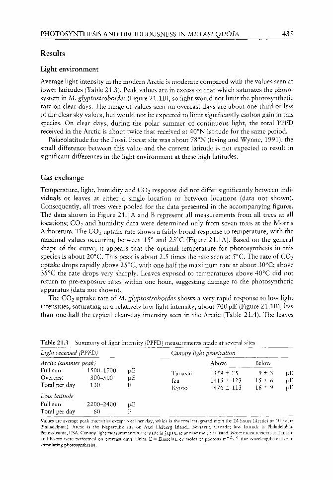
PHOTOSYNTHESIS AND DECIDUOUSNESS IN METASEQUOIA 435
Results
Light environment
Average light intensity in the modern Arctic is moderate compared with the values seen at lower latitudes (Table 21.3). Peak values are in excess of that which saturates the photo-system in M. glyptostroboides (Figure 21.IB), so light would not limit the photosynthetic rate on clear days. The range of values seen on overcast days are about one-third or less of the clear sky values, but would not be expected to limit significantly carbon gain in this species. On clear days, during the polar summer of continuous light, the total PPED received in the Arctic is about twice that received at 40°N latitude for the same period.
Palaeolatitude for the Fossil Forest site was about 78°N (Irving and Wynne, 1991); the small difference between this value and the current latitude is not expected to result in significant differences in the light environment at these high latitudes.
Gas exchange
Temperature, light, humidity and CO2 response did not differ significantly between individuals or leaves at either a single location or between locations (data not shown). Consequently, all trees were pooled for the data presented in the accompanying figures. The data shown in Figure 21.1 A and B represent all measurements from all trees at all locations; CO2 and humidity data were determined only from seven trees at the Morris Arboretum. The CO2 uptake rate shows a fairly broad response to temperature, with the maximal values occurring between 15° and 25°C (Figure 21.lA). Based on the general shape of the curve, it appears that the optimal temperature for photosynthesis in this species is about 20°C. This peak is about 2.5 times the rate seen at 5°C. The rate of CO2 uptake drops rapidly above 25°C, with one half the maximum rate at about 30°C; above 35°C the rate drops very sharply. Leaves exposed to temperatures above 40°C did not return to pre-exposure rates within one hour, suggesting damage to the photosynthetic apparatus (data not shown).
The CO2 uptake rate of M. glyptostroboides shows a very rapid response to low light intensities, saturating at a relatively low light intensity, about 700 |ULE (Figure 21.IB), less than one-half the typical clear-day intensity seen in the Arctic (Table 21.4). The leaves
Table 21.3 Summary of light intensity (PPFD) measurements made at several sites
Light received (PPFD)
Arctic (summer Full sun Overcast Total per day
Low latitude Full sun Total per day
• peak) 1500-1700
300-500 130
2200-2400 60
|JLE
|JLE
E
|JLE
E
Canopy light
Tanashi Izu Kyoto
penetration
Above
458 ± 75 1415 ± 123 476 ± 113
Below
9 ± 3 15 ± 6 16 ± 9
|JLE
|JLE
|JLE
Values are average peak intensities except total per day, which is the total integrated input for 24 hours (Arctic) or 10 hours (Philadelphia). Arctic is the Napartulik site on Axel Heiberg Island., Nunavut, Canada; low latitude is Philadelphia, Pennsylvania, USA. Canopy light measurements were made in Japan, at or near the cities listed. Note: measurements at Tanashi and Kyoto were performed on overcast days. Units: E = Einsteins, or moles of photons m"^s^^ (for wavelengths active in stimulating photosynthesis).
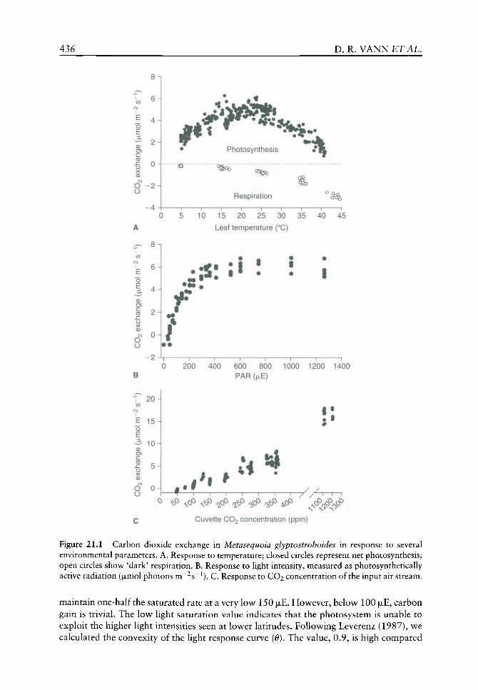
436 D. R .VANNETAL.
o E 0 O) c CO
O O
8n
6
4
2H
0
-2H
• Photosynthesis / R
©
Respiration
0 5 10 15 20 25 30 35 40 45
Leaf temperature (°C)
o £
<D U) C CO
O O
• • ! ^
f f
20
E 15 o £
0
c CO
o
1 1 1 1 1 1 1
0 200 400 600 800 1000 1200 1400 PAR ( JLE)
#
O O
10
5-1
OH . 1 *t f U ^
^ <D^ ^o^ ^<b^ c^^ c^ opQ c^ ^cP CNO CNO CNO
Cuvette CO2 concentration (ppm)
s^^^ 'b^"
Figure 21.1 Carbon dioxide exchange in Metasequoia glyptostroboides in response to several environmental parameters. A. Response to temperature; closed circles represent net photosynthesis; open circles show 'dark' respiration. B. Response to light intensity, measured as photosynthetically active radiation (fxmol photons m~^s~^). C. Response to CO2 concentration of the input air stream.
maintain one-half the saturated rate at a very low 150 |ULE. How^ever, below^ 100 |xE, carbon gain is trivial. The low^ light saturation value indicates that the photosystem is unable to exploit the higher light intensities seen at low^er latitudes. Follow^ing Leverenz (1987), wt calculated the convexity of the light response curve [0). The value, 0.9, is high compared
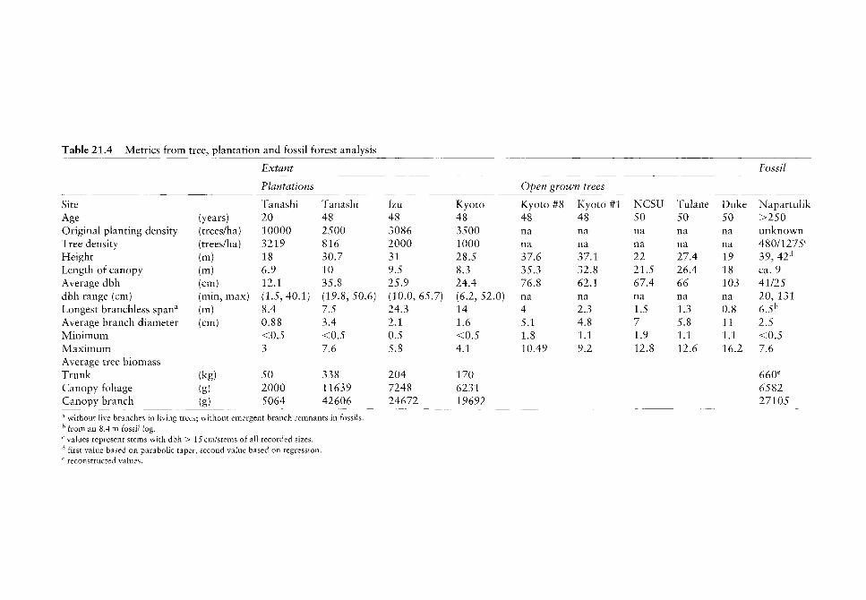
Table 21.4 Metrics from tree, plantation and fossil forest analysis
Site Age Original planting density Tree density Height Length of canopy Average dbh dbh range (cm) Longest branchless span'' Average branch diameter Minimum Maximum Average tree biomass Trunk Canopy fohage Canopy branch
(years) (trees/ha) (trees/ha) (m) (m) (cm) (min, max) (m) (cm)
(kg) (g) (g)
Extant
Plantations
Tanashi 20 10000 3219 18 6.9 12.1 (1.5,40.1) 8.4 0.88 <0.5 3
50 2000 5064
Tanashi 48 2500 816 30.7 10 35.8 (19.8,50.6) 7.5 3.4 <0.5 7.6
338 11639 42606
Izu 48 3086 2000 31 9.5 25.9 (10.0, 65.7) 24.3 2.1 0.5 5.8
204 7248 24672
Kyoto 48 3500 1000 28.5 8.3 24.4 (6.2, 52.0) 14 1.6 <0.5 4.1
170 6231 19692
Open
Kyoto 48 na na 37.6 35.3 76.8 na 4 5.1 1.8 10.49
grown trees
#8 Kyoto #1 48 na na 37.1 32.8 62.1 na 2.3 4.8 1.1 9.2
NCSU 50 na na 22 21.5 67.4 na 1.5 7 1.9 12.8
Tulane 50 na na 27.4 26.4 66 na 1.3 5.8 1.1 12.6
Duke 50 na na 19 18 103 na 0.8 11 1.1 16.2
Fossil
Napartulik >250 unknown 480/1275'^ 39, 42'' ca. 9 41/25 20, 131 6.5'' 2.5 <0.5 7.6
660' 6582 27105
^ without live branches in living trees; without emergent branch remnants in fossils. ^ from an 8.4m fossil log. ^ values represent stems with dbh > 15 cm/stems of all recorded sizes. ^ first value based on parabolic taper, second value based on regression. ' reconstructed values.
438 D, R .VANNETAL.
with many needle-leaved conifers (0.44-0.58; Leverenz, 1996), but is consistent with the planar nature of the leaves and the observed shade intolerance.
Short-term exposure to elevated CO2 resulted in an equivalent increase in net photosynthesis (Figure 21.IC). Carbon gain decreased rapidly with declining CO2 concentrations, reaching the compensation point between 50 and 100 ppm, consistent with the general pattern seen in C3 plants. There was little response to changes in humidity over a range of 20-80% RH. Measurements at higher RH values were confounded by temperature effects. Below 20% RH, carbon exchange became erratic and unstable. Based on transpiration data (not shown), it appeared that the stomata were opening and closing irregularly, possibly in an attempt to conserve water.
Dark respiration measurements were performed at five different temperatures; values at the highest temperature ranged from 1.6 to 3.2(jLmol C02m~^s~^ (mean 2.4, n = 8). At this setting (>40°C), the gas analyser had difficulty in maintaining a steady temperature. At 35°C, the CO2 efflux rate varied from 1.2 to 1.8 juimol m~^s"^ (mean 1.6, n = 12); at 25°C the rate ranged from 0.6 to 0.93 (mean 0.76, n = 12); at 15°C the mean value was 0.35 (range 0.18-0.51, n = 12); and at the lowest temperature, 5°C, the range was 0.09-0.22 (mean 0.15, n = 8). This is summarized in Figure 21.lA.
Stand structure
Plantation grown M. glyptostroboides exhibited rapid height growth, attaining relatively uniform canopy heights within each plantation. Average height of canopy dominant trees in the 20-year-old stand was 18 m. Canopy dominant trees in all of the 48-year-old plantations attained similar maximum tree heights (28.5-31 m). The height corresponded to vertical growth rates of approximately 0.9 m year"^ in the youngest plantation and between 0.6 to 0.65 m year~^ in the older plantations.
Average stem diameter increased with decreasing original planting density (see Table 21.4). Despite relatively uniform canopy heights, we found that stem diameter varied considerably within a given stand (see Table 21.4). This is reflected by an overall poor relationship (e.g. Tanashi B, R^ = 0.19) between bole diameter and total tree height of the canopy dominants in a given plantation.
On average, the youngest trees possessed the smallest branches and expressed the least variability in branch diameter (mean 0.88 cm, range <0.5-3.0, n = 372). In contrast, trees in the older, low density plantation (Tanashi B) had the largest average branch diameter (mean 3.4 cm) and the widest range (<0.5-7.6, n - 390). As expected, we found that the stands of trees with the largest diameter branches also had the largest foliar and branch biomass. Length of the live canopy differed among plantations (see Table 21.4). However, when expressed as a function of total tree height the proportion of live canopy to total tree height was similar among the older plantations (29 to 31%) but slightly higher in the youngest plantation where 38% of the total tree height was covered by live canopy.
Open grown solitary trees differed greatly from plantation grown trees in their overall structure. Total tree height varied considerably among solitary trees (range 19-37.6 m) and solitary trees had significantly larger stem diameters than plantation grown trees of similar age. The canopy structure of solitary M. glyptostroboides differed considerably from plantation grown trees. Maximum branch diameters were observed on the Duke Gardens tree (mean 11.0 cm, range 1.1-16.2, n = 104); whereas Kyoto #1 was observed to have the smallest branches and least variability of the open grown trees (mean 4.8 cm, range 1.1-9.2, n = 129). We found no sign of self-pruning occurring on open grown trees. The canopy of open grown M. glyptostroboides was more extensive on solitary as
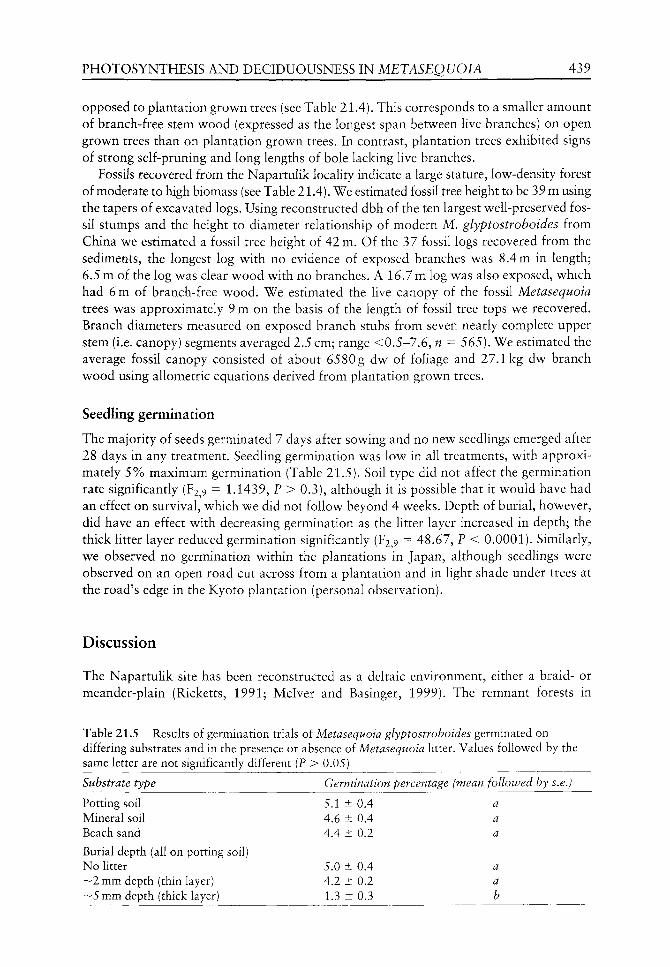
PHOTOSYNTHESIS AND DECIDUOUSNESS IN METASEQUOIA 439
opposed to plantation grown trees (see Table 21.4). This corresponds to a smaller amount of branch-free stem wood (expressed as the longest span between live branches) on open grown trees than on plantation grown trees. In contrast, plantation trees exhibited signs of strong self-pruning and long lengths of bole lacking live branches.
Fossils recovered from the Napartulik locality indicate a large stature, low-density forest of moderate to high biomass (see Table 21.4). We estimated fossil tree height to be 39 m using the tapers of excavated logs. Using reconstructed dbh of the ten largest well-preserved fossil stumps and the height to diameter relationship of modern M. glyptostroboides from China we estimated a fossil tree height of 42 m. Of the 37 fossil logs recovered from the sediments, the longest log with no evidence of exposed branches was 8.4 m in length; 6.5 m of the log was clear wood with no branches. A 16.7 m log was also exposed, which had 6 m of branch-free wood. We estimated the live canopy of the fossil Metasequoia trees was approximately 9 m on the basis of the length of fossil tree tops we recovered. Branch diameters measured on exposed branch stubs from seven nearly complete upper stem (i.e. canopy) segments averaged 2.5 cm; range <0.5-7.6, n = 565). We estimated the average fossil canopy consisted of about 6580g dw of foUage and 27.1kg dw branch wood using allometric equations derived from plantation grown trees.
Seedling germination
The majority of seeds germinated 7 days after sowing and no new seedlings emerged after 28 days in any treatment. Seedling germination was low in all treatments, with approximately 5% maximum germination (Table 21.5). Soil type did not affect the germination rate significantly (F2,9 = 1.1439, P > 0.3), although it is possible that it would have had an effect on survival, which we did not follow beyond 4 weeks. Depth of burial, however, did have an effect with decreasing germination as the litter layer increased in depth; the thick litter layer reduced germination significantly (¥2,9 "^ 48.67, P < 0.0001). Similarly, we observed no germination within the plantations in Japan, although seedlings were observed on an open road cut across from a plantation and in light shade under trees at the road's edge in the Kyoto plantation (personal observation).
Discussion
The Napartulik site has been reconstructed as a deltaic environment, either a braid- or meander-plain (Ricketts, 1991; Mclver and Basinger, 1999). The remnant forests in
Table 21.5 Results of germination trials of Metasequoia glyptostroboides germinated on differing substrates and in the presence or absence of Metasequoia litter. Values followed by the same letter are not significantly different (P > 0.05)
Substrate type Germination percentage (mean followed by s.e.)
a a a
a a b
Potting soil Mineral soil Beach sand
Burial depth (all on potting soil) No litter ~ 2 mm depth (thin layer) ~ 5 mm depth (thick layer)
5.1 ± 0.4 4.6 ± 0.4 4.4 ± 0.2
5.0 ± 0.4 4.2 ± 0.2 1.3 ± 0.3

440 D. R , V A N N £ T A L .
China exist in a dissimilar habitat (Yang and Jin, 2000); it is montane and, ahhough the species tends to be concentrated near riparian channels, it is lacking a permanent water source. Consequently, a modern analogue of the Napartulik site does not exist. However, several lines of evidence suggest that the ancient forests followed a riparian model, becoming established on open flood sediments as even-aged cohorts (sensu Oliver and Larson, 1996:145-167). First, the seeds are small and winged, similar to Sequoia sempervirens [D. Don (Endl.)], whose seeds do not fall far from the tree (Olson et al, 1990); however, they do float. Second, the seedling germination study indicates that the best germination rate occurs on open soil (see Table 21.5), as opposed to beneath forest floor litter. This is consistent with results from Sequoiadendron giganteum l(Lindl.) Buchholz], whose seedlings rarely become established on undisturbed forest floor, apparently resulting from rapid dessication of the thick litter (Olson et ai, 1990). Moreover, Falder et al. (1999) describe in situ fossil Metasequoia seedlings from a Palaeocene floodplain environment and interpret Metasequoia as a colonizer of marshy, mineral soil habitats. Finally, the species attains its best growth in the absence of water stress (Kuser, 1982). Considering the low light levels within the forest stands (see Table 21.3), it seems reasonably evident that the establishment of individuals within the stand is unlikely, consequently, multi-cohort stands would not result. Rather, the simultaneous germination of individuals on open flood soils would produce single-cohort stands. Although the existence of multiple stump diameters has been taken as evidence of a multi-aged stand (e.g. Nobori et al.^ 1997), the data from plantations in Japan demonstrate that single-cohort stands of similar canopy height can have a wide range of trunk diameters (see Table 21.4). We therefore contend that seedling physiology as reflected in germination and light tolerance is consistent with single-cohort stands subject to infrequent major disruptions or 'patch'-scale disturbances. However, seasonal flooding would produce new areas which could be rapidly colonized. This is suggestive of an evolutionary trend toward an invasive habit, exploiting riparian habitats where competition may have been lower in the past.
The thermotolerance of CO2 uptake in M. glyptostroboides is fairly broad (see Figure 21.lA), and is consistent with the patterns of growth seen in planted specimens throughout the world (Kuser, 1982). This range of temperature is also consistent with the geographical extent of the fossil Metasequoia and the presumed range of palaeoclimate. This suggests that, at least for temperature, M. glyptostroboides is an adequate NLR. However, it also indicates that M. glyptostroboides is of limited use in predicting palaeoclimate. Certainly, it would be expected to perform poorly in subtropical and tropical environments. Water availability appears to be the primary determinant of habitat for this species (Kuser, 1982). In warm-temperate regions with adequate water supply, the tree performs remarkably well; for example, the tree on the NCSU campus (MAT = 15.4°C) is increasing in diameter by about 1.2 cm year~^ and the average rate at the Tanashi plantation (MAT = 13.7°C) is 5.8 mm year~^ (C.J. Williams, unpublished data). Presumably, carbon gain during the cooler spring and fall months offsets the losses associated with the peak summer temperatures in these locations. In contrast, the Napartulik trees grew only about 1.38 ± 0.52 mm year~^ (C.J. WiUiams, unpublished, data). Since water was unlikely to be limiting in the riparian environment at Napartulik, it seems reasonable to assume that the slow growth rates were a consequence of low temperatures.
The photosynthetic apparatus of M. glyptostroboides saturates at a relatively low level of light, approximately 1700 |xE (see Figure 21.IB). Indeed, chlorophyll content declines with increasing light intensity (Ida, 1981b) and the pigment rhodoxanthin increases (Ida, 1981a; Czeczuga, 1987a), leading to a reddish cast in the upper leaves of the trees (personal
PHOTOSYNTHESIS AND DECIDUOUSNESS IN METASEQUOIA 441
observation). This pigment is normally an autumnal coloration (Ida, 1981a; Czeczuga, 1987b); it probably serves in a protective capacity during the summer, shading the chloro-plast pigments. The low light saturation level is consistent with the evolution of the photosynthetic system under the reduced light levels above the Arctic circle; full-sun measurements in the Arctic are relatively low (see Table 21.3), but exceed the saturation value. A light saturation value greater than that actually experienced by the plant would represent an investment in unused photosynthetic materials and would Ukely be selected against as an unnecessary energetic cost. The performance under low light conditions is intriguing; it is conceivable that Metasequoia evolved in a foggy riparian environment. However, the high shoot convexity value {6) is seemingly inconsistent with this scenario, as lower convexity values are thought to be related to more efficient absorbance of diffuse light (Leverenz, 1987) within the conifer shoot. The planar nature of the Metasequoia leaf, compared with the needle display of conifers with low convexity values, undoubtedly explains the 6 value. Interestingly, the fact that the light saturation level is low, in spite of the modern species' distribution at low latitudes, suggests that the species has not experienced a strong selective pressure to adapt physiologically to alternate light regimes and may not have evolved much in other aspects either. It is worth noting that, in high light environments and at the top of trees, the leaf morphology changes, with the leaflets becoming substantially narrower (D.R. Vann, personal observation; see also Chapter 20). This response appears to be quite plastic in the modern species. Although there is an extensive, well-preserved litter layer at the Napartulik site, the fossil leaves seldom show such a narrow leaflet morphology (B. LePage, personal observation). This implies low light levels in the Eocene Arctic and that the modern species' primary response to light intensities at low latitudes is morphological, rather than physiological. Specific leaf area varies such that mass-based photosynthetic rates are similar in sun and shade leaves (see Chapter 20).
Alternately, the roughly three-fold increase in CO2 uptake accompanying a three-fold increase in the CO2 concentration of the cuvette input air stream (see Figure 21.1C) suggests that the plateau in the light response curve is not primarily a limitation in the car-boxylation capacity of the cell. Instead, it implies a diffusional limitation. If this is true, it suggests that stomatal density may limit carboxylation efficiency (on a quantum basis). This may have arisen as a compromise between water loss and carbon uptake. Xie and co-workers reported that M. glyptostroboides has low drought tolerance and poor xylem hydraulic conductivity (Xie et al.^ 1999a,b). Further experimentation is required to resolve this question. Presumably, with an adequate CO2 supply, the light saturation value would increase, indicating that the photosystem's capacity is not saturated at current ambient CO2 levels.
Plants that shed their leaves simultaneously on an annual basis are considered 'deciduous'; generally deciduous species are responding to a seasonal stress such as cold or drought. As the late Tertiary and much of the Quaternary are considered to have had relatively mild climates, without the large temperature fluctuations seen today at the higher latitudes, cold seems an unlikely candidate as the driving force behind deciduousness in Metasequoia^ particularly as its evergreen relatives all tolerate frost (Sakai and Okada, 1971; Sakai, 1983). At higher altitudes and in cold lowland regions, evergreen taxa survive the winter in a state of suspended physiology. It has been argued that warmer temperatures could lead to substantial respiratory losses in the winter (Axelrod, 1984). If these losses cannot be replaced, as would occur during the polar night, the plants might not survive. This has not been explored extensively, although the results of Read and Francis (1992) were consistent with this idea.

442 D. R .VANNETAL.
Metasequoia apparently evolved near the Arctic circle during a period v^hen low^land winter temperatures probably seldom w ent below freezing (Yang and Jin, 2000). It is therefore reasonable to ask whether the polar light seasonality could have been a driving force for the evolution of deciduousness in this species. To test this, we used the foliar bio-mass estimates derived for the Napartulik site (see Table 21.4) and the gas-exchange data to derive a coarse carbon budget for the canopy under various temperature scenarios. An 'average' tree at the Napartulik site was calculated to hold approximately 6580 g of foHage (see Table 21.4). Using an average foHage density of 60 g m ' ^ dw (D.R. Vann, unpublished data; see also Chapter 20 who provide a range from 30 to 90gmm~^dw), there is some 108 m^ in the canopy foliage. Based on the photosynthetic rates determined above and using 3780 total hours of daylight (at 65°N latitude, for days with daylength longer than 5h), a canopy of this size can fix a maximum of perhaps 8.8kmol CO2 year"^ This figure assumes that the entire canopy is sunlit and fully functional. The actual value is likely considerably lower, as we do not consider a number of factors, such as the loss of photosynthetic capacity during senescence or the effect of rain or overcast weather. Carbon content of foliage is about 49%, so the amount of carbon in the foliage is 3.2 kg. Construction cost is some 30-50% (e.g. Baruch and Goldstein, 1999), therefore, the cost of rebuilding the foliage annually is around 0.4 kmol CO2.
Based on the values given in the Results section, maintenance respiration rates for the leaf during mean growing season temperatures of 10, 15, 20 and 25°C would be 0.42, 0.51, 0.84 and 1.1 kmol CO2 respectively. Dark season respiration at temperatures of 2, 5 and 10°C would amount to 0.11, 0.29 and 0.55 kmol CO2 respectively. Based on these estimates, and using 0.4 kmol CO2 as the canopy replacement cost, the 'break-even' temperature is somewhere around 7°C. This figure is a coarse estimate. It is reasonable, however, to expect that temperatures between 5 and 10°C would favour a loss of fohage during the dark months. This also assumes that the reserves are present in the plant. The total amount of carbohydrate required to support this respiration rate amounts to some 12% of the dry weight of the leaf, an unrealistic figure. Storage in other parts of the plant, such as the twigs, is possible, but translocation in the dark, without transpiration to assist, would require an additional energy expenditure. In any case, this analysis is also sensitive to latitude; during the portion of the year with 5 or more hours of daylight, total dark hours decrease with increasing latitudes; these hours accrue prior to and after the polar summer of continuous light. At the latitude where the sum of spring and autumn dark and light hours are equal, the total respiration requirements decline to the point where the foUage could presumably survive a warmer winter (about 8°C). This is an important result for evaluating the evolution of deciduousness as a physiological trait, as it constrains the environment under which this trait emerged. Winter temperatures below 5°C would appear to allow the retention of an evergreen habit, whereas warmer temperatures accompanied by prolonged dark periods represent a selection pressure towards deciduousness. This analysis does not examine whole-tree carbon balance; it is likely that competition for photosynthate from cambial respiration, below-ground growth and reserves for spring budbreak would reduce the balance to favour lower temperatures.
The logic of this result can be generalized to other species. It is consistent with the hypothesis that deciduous plants could have greater success at surviving and/or dominating in warm, dark winters. We cannot establish that dark winters initiated the deciduous trait; it could have existed prior to the migration of these species above the Arctic circle, where it provided a competitive edge over evergreen types. Applying the details of the rather naive analysis above to other species would not be appropriate. There is a large variation in the relationship
PHOTOSYNTHESIS AND DECIDUOUSNESS IN METASEQUOIA 443
between specific leaf area and photosynthesis, resulting in a spread of 'break-even' temperatures. Nonetheless, the general approach could work for other species. However, in all cases, the metabolic rates are so low below about 2°C, that it appears most species could survive, so cold, dark winters would not appear to be an important selective force. This is interesting in light of the fact that the majority of modern clades, in which deciduousness is present, evolved prior to the global cooling trends producing the modern temperate world.
As intimated above, it is possible that Metasequoia had already evolved the deciduous trait in response to drought (see Chapter 20). In this case, drought deciduousness could have been important in aiding the colonizing of Arctic habitats by Metasequoia. Different cues trigger drought-related leaf-fall compared with dark-deciduousness; based on the phenology of Metasequoia^ it is responding to a seasonal light cue, probably short days. When it experiences drought, the plant generally responds with a loss of green shoots, rather than the normal senescent process. Considering the species' association with hydric environments and its large water storage capacity within the trunk, it seems unlikely that drought drove the development of deciduousness. Conceivably, Metasequoia's> ancestor might have become deciduous in response to a seasonally predictable dry period, triggered by a seasonal light cue. The true origin is unresolved at this time, but it has bearing on the other deciduous members of this clade, Taxodium and Glyptostrobus, as it is likely the trait evolved in the common ancestor. Each of these is strongly associated with wetland habitats and drought sensitivity.
In conclusion, the variables measured to date support the idea that M. glyptostroboides is very likely an example of evolutionary stasis and, as such, it is a good NLR. However, its rather broad climatic tolerance means it is not useful in reconstructing past environments, with the probable exception of indicating a moist habitat. In terms of the evolution of physiological parameters, there seems to be little evidence that this has occurred, as the plasticity of the modern plant would allow its success in all known fossil locations. This does not preclude ecotypic variations (sensu Fryer and Ledig, 1972) in the history of the genus, however, the modern form is most likely little changed from the dominant fossil form (M. occidentalis). The similarity of fossil and modern stand structure as affected by physiological constraints suggests that the past forests functioned in a similar manner and that sunlight duration does not affect this process. Finally, we provide physiological evidence supporting the hypothesis that evolution of deciduousness could have been driven by warm, dark winters at high (although not polar) latitudes.
Acknowledgements
We thank A.H. Johnson, P. Semonche and D. Liptzin for help in unearthing trees at Napartulik and A. Ikeda, T. Sweda, T. Kusakabe and H. Tsuzuki for assistance in the analysis of trees at the Japanese plantations. We gratefully thank the Andrew Mellon Foundation for financial support and the Canadian Polar Shelf Project for access and logistical support at Napartulik, as well as Tokyo University and Kyoto University for access to their plantations. This is PCSP contribution #00702.
References
Anderson K, LePage BA. 1995. Analysis of fossil resins from Axel Heiberg Island, Canadian Arctic. American Chemical Society Symposium Series 617: 170-192.

444 D. R . V A N N E T A L .
Axelrod DI. 1984. An interpretation of Cretaceous and Tertiary biota in polar regions. Palaeogeography, Palaeoclimatology, Palaeoecology 45: 105-147.
Baruch Z, Goldstein G. 1999. Leaf construction cost, nutrient concentration, and net CO2 assimilation of native and invasive species in Hawaii. Oecologia 111: 183-192.
Basinger JF. 1986. Our 'tropical' arctic. Canadian Geographic 106: 28-37. Basinger JF, Greenwood DR, Sweda T. 1994. Early Tertiary vegetation of Arctic Canada and its rele
vance to paleoclimatic interpretation. In: Boulter MC, Fisher HC, eds. Cenozoic Plants and Climates of the Arctic. New York: Springer-Verlag. \75-196.
Bell JF, Ek AR, Hitchcock III HC, lies K, et al. 1984. Timber measurement. In: Wenger FW, ed. Forestry Handbook, 2nd edn. Toronto: Wiley-Interscience. 253-360.
Chaney RW. 1948. Redwoods in China. Natural History Magazine 47: 440-444. Czeczuga B. 1987a. Different rhodoxanthin contents in the leaves of gymnosperms grown under
various light intensities. Biochemical Systematics and Ecology 15: 531-533. Czeczuga B. 1987b. Ketocarotenoids - autumn carotenoids in Metasequoia glyptostrohoides.
Biochemical Systematics and Ecology 15: 303-306. Falder AB, Stockey RA, Rothwell GW. 1999. In situ fossil seedlings of a Metasequoia-like taxodia-
ceous conifer from Paleocene river floodplain deposits of central Alberta, Canada. American Journal of Botany 86: 900-902.
Francis JE. 1988. A 50-million-year old fossil forest from Strathcona Fiord, Ellesmere Island, Arctic Canada: evidence for a warm polar climate. Arctic 41: 314-318.
Francis JE. 1991. The dynamics of polar fossil forests of Axel Heiberg Island, Canadian Arctic Archipelago. In: Christie RL, McMillan NJ, eds. Tertiary Fossil Forests of the Geodetic Hills, Axel Heiberg Island, Arctic Archipelago. Ottawa, Canada: Geological Survey of Canada. 29-38.
Francis JE, McMillan NJ. 1987. Fossil forests in the far north. Geos 16: 6-9. Fryer JH, Ledig FT. 1972. Microevolution of the photosynthetic temperature optimum in relation
to the elevational complex gradient. Canadian Journal of Botany 50: 1231-1235. Hart JA, Price RA. 1990. The genera of the Cupressaceae (including Taxodiaceae). Journal of the
Arnold Arboretum 71: 275-322. Hickey LJ. 1977. Stratigraphy and paleobotany of the Golden Valley Formation (early Tertiary) of
western North Dakota. Geological Society of America Memoir 150: 1-181. Ida K. 1981a. Eco-physiological studies on the response of taxodiaceous conifers to shading with
special reference to the behaviour of leaf pigments I. Distribution of carotenoids in green and autumnal reddish brown leaves of gymnosperms. Botanical Magazine of Tokyo 94: 41-54.
Ida K. 1981b. Eco-physiological studies on the response of taxodiaceous conifers to shading with special reference to the behaviour of leaf pigments II. Chlorophyll and carotenoid contents in green leaves grown under different grades of shading. Botanical Magazine of Tokyo 94: 181-196.
Irving E, Wynne PJ. 1991. The paleolatitude of the Eocene fossil forests of Arctic Canada. Geological Survey of Canada Bulletin 403: 209-211.
Jordan GJ. 1997. Contrasts between the climatic ranges of fossil and extant taxa: causes and consequences for paleoclimatic estimates. Australian Journal of Botany 45: 465-474.
Kuser J. 1982. Metasequoia keeps on growing. Arnoldia 42: 130-138. Kuser JE, Sheely DL, Hendricks DR. 1997. Genetic variation in two ex situ collections of the rare
Metasequoia glyptostrohoides (Cupressaceae). Silvae Genetica 46: 258-264. Kusumi J, Tsumura Y, Yoshimaru H, Tachida H. 2000. Phylogenetic relationships in Taxodiaceae
and Cupressaceae sensu stricto based on MATK gene, CHLL gene, TRNL-TRNF IGS region, and TRNL intron sequences. American Journal of Botany 87: 1480-1488.
Leverenz JW. 1987. Chlorophyll content and the the light response curve of shade-adapted conifer needles. Physiologia Plantarum 71: 20-29.
Leverenz JW. 1996. Shade-shoot structure, photosynthetic performance in the field, and photosynthetic capacity of evergreen conifers. Tree Physiology 16: 109-114.
MacGinitie HD. 1941. A middle Eocene flora from the central Sierra Nevada. Carnegie Institute of Washington Publications 534: 1-94.
Mattheck C. 1991. Trees: the Mechanical Design. Berlin: Springer-Verlag.

PHOTOSYNTHESIS A N D DECIDUOUSNESS IN METASEQUOIA 445
Mclntyre DJ. 1991. Pollen and spore flora of an Eocene forest, Eastern Axel Heiberg Island, N.W.T. In: Christie RL, McMillan NJ, eds. Tertiary Fossil Forests of the Geodetic Hills, Axel Heiberg Island, Arctic Archipelago. Ottawa, Canada: Geological Survey of Canada, 83-97.
Mclver EE, Basinger JF. 1999. Early Tertiary floral evolution in the Canadian high arctic. Annals of the Missouri Botanical Garden 86: 523-545.
Mosbrugger V, Utescher T. 1997. The coexistence approach - a method for quantitative reconstructions of Tertiary terrestrial palaeoclimate data using plant fossils. Palaeogeography, Palaeocli-matology, Paleaeoecology 134: 61-86.
Nobori Y, Hayashi K, Kumagai H, et al. 1997. Reconstruction of a Tertiary fossil forest from the Canadian High Arctic using three-dimensional computer graphics, journal of Forest Planning 3: 49-54.
Oliver CD, Larson BC. 1996. Forest Stand Dynamics. Nev^ York: John Wiley & Sons, Inc. Olson DF Jr, Roy DF, Walters GA. 1990. Sequoia sempervirens (D. Don) Endl. Redv^ood. In:
Burns RM, Honkala BH, eds. Silvics of North America. Washington, DC: United States Department of Agriculture Forest Service, 541-551.
Read J, Francis J. 1992. Responses of some Southern Hemisphere tree species to a prolonged dark period and their implications for high-latitude Cretaceous and Tertiary floras. Palaeogeography, Palaeoclimatology, Palaeoecology 99: 271-290.
Ricketts BD. 1991. Sedimentation, Eurekan tectonism and the fossil forest succession on eastern Axel Heiberg Island, Canadian Arctic Archipelago. In: Christie RL, McMillan NJ, eds. Tertiary Fossil Forests of the Geodetic Hills, Axel Heiberg Island, Arctic Archipelago. Ottav^a, Canada: Geological Survey of Canada. 1-27.
Sakai A. 1983. Comparative study of freezing resistance of conifers with special reference to cold adaptation and its evolutive aspects. Canadian Journal of Botany 61: 2323-2332.
Sakai A, Okada S. 1971. Freezing resistance of conifers. Silvae Genetica 20: 91-97. Sargent CS. 1965. Manual of the Trees of North America. New York: Dover Publications. Struzik E. 1999. Requiem for a fossil forest. Canadian Geographic: 43-48. Tidwell WD. 1998. Common Fossil Plants of Western North America. Washington, DC: Smithonsian
Institution Press. Vann DR, Palmiotto PA, Strimbeck GR. 1998. Allometric equations for two South American
conifers: test of a non-destructive method. Forest Ecology and Management 106: 55-71. Whittaker RH, Bormann FH, Likers GE, Siccama, TG. 1974. The Hubbard Brook ecosystem study:
forest biomass and production. Ecological Monographs 44: 233-254. Wing SL, DiMichele WA. 1992. Ecological characterization of fossil plants. In: Behrensmeyer AK,
Damuth JD, DiMichele WA, et al. eds. Terrestrial Ecosystems through Time: Evolutionary Paleoecology of Terrestrial Plants and Animals. Chicago: University of Chicago Press, 139-180.
Wing SL, Greenwood DG. 1993. Fossils and fossil climate: the case for equable continental interiors in the Eocene. Philosophical Transactions of the Royal Society of London, Series B 341: 243-252.
Wolfe JA. 1978. A paleobotanical interpretation of Tertiary climates in the northern hemisphere. American Scientist 66: 694-703.
Wyman D. 1970. The complete Metasequoia story. American Nurseryman 131: 12, 13, 28-30, 36. Xie Y, Shen H, Luo A. 1999a. A study on the water relations parameters of seedlings of four conifer
species in the south of China under water stress. [Chinese], /owr^^/ of Nanjing Forestry University 23: 41-44.
Xie Y, Shen H, Luo A, Zhu X. 1999b. A study on the physiological indexes of drought-resistance in seedlings of seven afforestation tree species in the south of China. [Chinese]. Journal of Nanjing Forestry University 23: 13-16.
Yang H, Jin J. 2000. Phytogeographic history and evolutionary stasis of Metasequoia: geologic and genetic information contrasted. Acta Paleontologica Sinica 39: 288-307.
22 Adaptive ancientness of vascular plants to exploitation of low^-nutrient substrates -
a neobotanical overview^
Christopher N Page
CONTENTS.
Introduction
The modern array of low-nutrient habitats
The modern diversity of ALVP colonists of low-nutrient habitats
Taxonomic distribution Habitat distribution Responses to cultivation
Discussion Advantages of low-nutrient toleration for
survival of ALVPs Ancientness of low-nutrient toleration Theoretical perspectives
Conclusions
Acknowledgements
References
447
448
450 450 450 454
456
457 458 460
461
461
461
Introduction
Several living families of ferns have taxonomic fossil lineages which date back at least into the Lower Mesozoic (e.g. Osmundaceae, Dicksoniaceae, Matoniaceae, Dipteridaceae) or even into the Palaeozoic (Marattiaceae, Schizaeceae) (Cleal, 1993; Collinson, 1996; Rothwell, 1996a,b; van Konijnenburg-van Cittert, 1999). Various conifer groups (Rothwell, 1982; Page, 1990) as well as clubmosses of the living families Lycopodiaceae and Selaginellaceae (Thomas, 1992) also date back to the Palaeozoic, as do the Equisetaceae, whose sole living genus Equisetum has been suggested to have even changed little since that time (Page, 1972a). Indeed, some whole extant fern floras are ancient: that
The Evolution of Plant Physiology Copyright © 2004 Elsevier Ltd ISBN 0-12-33955-26 All rights of reproduction in any form reserved

448 C. N. PAGE
of the Canary Islands is an intact fern flora of Miocene age (Page, 1973), that of southern Siberia, though modified by later glacial influences, contains elements which appear to originate from the Cretaceous (Gureyeva, 2001). Among living pteridophytes, ancient elements as well as modern elements often coexist (e.g. Stewart and Rothwell, 1993). In the 1970s, I had the honour (so far as I can work out) of first appropriating the word 'diversity' into biology (Page, 1979a). Similarly, for convenience of reference, a new single phrase is needed for all of these ancient living vascular plants (from pteridophytes to conifers) - as there is no comprehensive taxonomic term. These are here collectively christened Ancient Living Vascular Plants or ALVPs for short (the 'OAPs' of the plant world).
The operation and dynamics of ecological strategies by which these ALVPs have succeeded in achieving their long-term survival, as well as widespread adaptive diversity, have been identified using evidence gained from the living members of these ancient groups, combining observations from field, whole-plant experimental cultural and laboratory approaches (Page, 1979a,b, 1990, 1997b, 2002a and b). This chapter outlines fundamental but little-researched aspects of the edaphic adaptations of ALVP clubmosses, horsetails, ferns and conifers, concentrating on the potential of many, on a global basis, for colonization of substrates characterized by usually direct mineral surfaces of permanently low-nutrient availability and often also of acute nutrient disequilibrium.
These adaptations enable many ALVPs today to continue to exist in sites in which they show exceptional abilities at 'doing without', reflecting extremely efficient standards of nutrient management. In consequence, these abilities enable these ALVPs to occupy diverse edaphically marginal habitats for which today most other modern plant competition is necessarily low. They effectively avoid encountering the many more vigorous competitors that would be more dangerously confronted within almost all more nutrient-rich, more mesic habitats, and the presence to this day of so many ALVPs in such low-nutrient sites remains a stringent test of the efficacy of this strategy for long-term survival.
The modern array of low-nutrient habitats
The term 'low-nutrient habitats' here encompasses all habitats which are, by their very nature, far from being nutrient-rich. Many, but not all, are the direct results of the dynamics of Earth Science processes and involve exposures of largely mineral terrains, with little previous amelioration by climatic or biological agents. Others are added to by biological activity, such as nutrient-poor peat surfaces and bark-surface epiphytic habitats. Among the total of nutrient-poor habitats arising, two broad regimes of tolerance types exist widely among ALVPs. These are tolerance of:
1. regimes of low-nutrient levels per se^ in which the great majority of mineral elements are either lacking or in very short supply (e.g. the majority of exposed mineral surfaces, peats and plant bark surfaces)
2. regimes of low-nutrient levels of essential mineral elements, but in which non-essential mineral elements may additionally occur in excess, some of which may be regarded as generally toxic (e.g. mineral surfaces containing heavy metal loadings).
Within each of these regimes, field evidence suggests that there are many variations of mineral availability, combination and quantity, which overall determine the habitats to which suites of ALVP species are differentially able to tolerate, while availability of moisture

ADAPTIVE ANCIENTNESS OF VASCULAR PLANTS TO LOW NUTRITION 449
regimes and habitat accessibility to propagules (e.g. epiphytic sites) further prescribes the groups of plants differentially exploiting them.
The suites of habitats of low-nutrient levels per se are especially widespread. Beside the epiphytic habitats (noted above), they include such sites as lowland sand-plains and innundated sand, clay and gravel streamsides and basins, intrinsically nutrient-poor sandy heathlands and savannahs, and leached soils such as laterites. Some of these habitats are ones also associated with wildfire regimes. In upland areas, widely occurring sites include a great variety of direct erosion-surface mineral exposures of most rocks of all types in virtually all climates, forming cliffs, screes, slump, slide and landslip surfaces, newly formed volcanic outpourings, ash and ejecta surfaces and, additionally, in the wet tropics, especially high mountain rocky summits, ridges and saddles, where gleyed clay ridges result. Ecologically, many of these seem remarkably little studied. For example, mountain summit ridges and saddles form habitats which contain (typically dominantly. Page, 1979a) special suites of ALVPs suggesting these to be among the most extreme in mineral deficiency anywhere.
The suites of habitats in which low-nutrient levels of essential mineral elements combine with toxic non-essential mineral element availability (mineral overload habitats) require further physiological abilities for toleration of such excesses. Such habitats occur where potentially toxic rock types of high mineral-yielding ability occur (and especially where metalliferous elements are yielded). In wild sites at all altitudes, these can include cliffs, major landslip surfaces, landslide debris fans, scree slopes and appropriate post-volcanic disturbance sites. Particularly toxic are the minerals of naturally surface-exposed ultramafic peridotite (volcanic) and serpentine (metamorphic) rock types, in which manganese, cobalt, nickel and chromium typically occur and whose mineral excesses of these elements often combine with simultaneous deficiencies of calcium. Other less toxic habitats, but in which mineral excesses play a particularly species-selective role, are those where calcium or magnesium are particularly high, such as in carboniferous or dolomitic limestones respectively. Man has locally added greatly to the surface occurrence of some of these in the form of discarded minespoil tailings, particularly where workable metal-containing ores occur (e.g. zinc, lead, copper, tin, cadmium and strontium), with discarded mine-tailing exposures still with toxic amounts of metals present also often containing arsenic and sulphurous compounds. In all, the exact mineral excesses involved clearly depend on the rock type itself, its chemical constitution, levels of mineral overload and the rate at which such excesses of minerals are yielded under the occurring climatic conditions.
Whether the sites are merely especially low-nutrient per se, or are ones of potentially toxic high mineral overload levels, and whether the habitats are wild or man-made, all these sites present surfaces exposed to potential colonization by arriving ALVP propagules, including wind-borne seeds of conifers and, especially, the highly mobile airborne spores of pteridophytes. Such immigrant seeds and spores have the potential to arrive from the earliest stages of new rock-surface exposures following disturbance events (such as land-sHdes, but also smaller disturbance-event mosaics), when exposure surfaces are particularly fresh. These immigrant propagules include chance arrival of 'unadapted' species arrays from potentially long-distance sources (notably so in Pteridophyta), or from other existing 'adapted' species already occurring on other nearby rock exposures. In each, as mineral susbstrates vary, local selection for particularly closely-adapted populations undoubtedly applies critical selection pressures, and close experimental investigation of this across an array of ALVPs and their habitats is much needed.
450 C. N, PAGE
The modern diversity of ALVP colonists of low-nutrient habitats
Taxonomic distribution
Wide arrays of ALVP species from diverse families are today specialist colonists of many low-nutrient sites under both temperate and tropical conditions. There can be considerable specializations of life-form between different ALVPs exploiting these relatively demanding situations and often different taxonomic groups target specific suites of habitats.
On the basis of personal verification in the field (and, for most genera, experience too of experimental cultivation of the same material), the taxonomic spectrum of extant low-nutrient-tolerant ALVPs by genera is presented in Table 22.1.
From Table 22.1 it can be seen that at least 85 genera of pteridophytes (19% of 414 total pteridophyte genera recognized by Crabbe et aL^ 1975) and at least 19 genera of conifers (27% of 68 total conifer genera recognized by Page, 1990) are identified here as possessing species which are tolerant of growth on low-nutrient substrates. This is a total of 104 of 482 genera of extant pteridophytes and conifers together (= 21%). In t]\t list, Cycadaceae (s.l.), Gnetaceae and Ephedraceae (although they are ALVPs) are excluded on the basis of lesser direct personal experience. Were these to be included also, my estimate is that it is likely that all cycad genera with the exceptions of Bowenia (tropical Australia) and Stangeria (tropical South Africa) would additionally qualify to be included, as would virtually all Ephedraceae. This would enhance still further the trend already identified among pteridophyte and conifer ALVPs for which I have more comprehensive field evidence.
From these wild habitat indicators, if I were to look for the greatest extremist tolerators of low-mineral nutrition availability in this list, I would single out the following: Lycopodium (Lycopodiaceae), Lycopodiella (Lycopodiaceae), Huperzia (Lycopodiaceae), Equisetum (Equisetaceae), Schizaea (Schizaeceae), Gleichenia (Gleicheniaceae), Sticherus (Gleicheni-aceae), Dicranopteris (Gleicheniaceae), Platyzoma (Platyzomataceae), Stromatopteris (Stromatopteridaceae), Lindsaea (Lindsaeceae), Sphenomeris (Lindsaeceae), Schizoloma (Lindsaeceae), Syngramma (Lindsaeceae), Matonia (Matoniaceae), Dipteris (Polypodiaceae), Widdringtonia (Cupressaceae), Cupressus (Cupressaceae), Actinostrobus (Cupressaceae), Callitris (Cupressaceae), Neocallitropsis (Cupressaceae), Austrocedrus (Cupressaceae) and Pinus (Pinaceae).
Habitat distribution
Not all ALVPs occur today in low-nutrient habitats, for many have adopted life-forms which enable them to avoid excesses of vegetative competition in other ways. Flowever, the above significant numbers of ALVPs do, and both the taxonomic and habitat diversity of these reflected in the above section, is considerable. In terms of overall habitat trends among ALVPs which do have members which occur in low-nutrient habitats, pteridophytes usually predominate today where such low-nutrient habitats occur in moister climates at all latitudes, but with special diversity in the wet tropics, often reaching their zenith at mid-mountain altitudes (Page, 1979a,b). There are, however, some ALVP fern genera (notably the monotypic Platyzoma in Australia and Stromatopteris in New Caledonia) which occupy particularly sub-desert terrains. More widely, however, conifers and then cycads occur in progressively drier low-nutrient sites, with conifers in both temperate and tropical conditions (and especially in tropical montane habitats, but also in dry Mediterranean ones) and cycads in mainly dry, high-light tropical ones.
Epiphytic sites are chiefly dominated, among ALVPs, by many ferns and a few club-mosses, which gain rapid access through the high mobility of their airborne spores. All (and
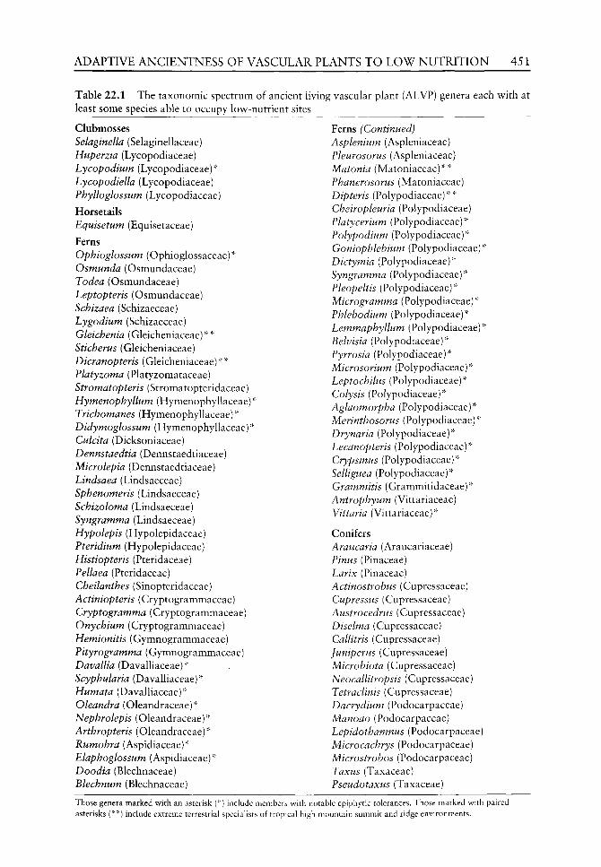
ADAPTIVE ANCIENTNESS OF VASCULAR PLANTS T O L O W N U T R I T I O N 451
Table 22.1 The taxonomic spectrum of ancient living vascular plant (ALVP) genera each w ith at least some species able to occupy lov^-nutrient sites
Clubmosses Selaginella (Selaginellaceae) Huperzia (Lycopodiaceae) Lycopodium (Lycopodiaceae)''" Lycopodiella (Lycopodiaceae) Phylloglossum (Lycopodiaceae)
Horsetails Equisetum (Equisetaceae)
Ferns Ophioglossum (Ophioglossaceae)''" Osmunda (Osmundaceae) Todea (Osmundaceae) Leptopteris (Osmundaceae) Schizaea (Schizaeceae) Lygodium (Schizaeceae) Gleichenia (Gleicheniaceae)''"''" Stickerus (Gleicheniaceae) Dicranopteris (Gleicheniaceae)''"''" Flatyzoma (Platyzomataceae) Stromatopteris (Stromatopteridaceae) Hymenophyllum (Hymenophyllaceae)'' Trichomanes (Hymenophyllaceae)''" Didymoglossum (Hymenophyllaceae)''" Culcita (Dicksoniaceae) Dennstaedtia (Dennstaedtiaceae) Microlepia (Dennstaedtiaceae) Lindsaea (Lindsaeceae) Sphenomeris (Lindsaeceae) Schizoloma (Lindsaeceae) Syngramma (Lindsaeceae) Hypolepis (Hypolepidaceae) Pteridium (Hypolepidaceae) Histiopteris (Pteridaceae) Pellaea (Pteridaceae) Cheilanthes (Sinopteridaceae) Actiniopteris (Cryptogrammaceae) Cryptogramma (Cryptogrammaceae) Onychium (Cryptogrammaceae) Hemionitis (Gymnogrammaceae) Pityrogramma (Gymnogrammaceae) Davallia (DavaUiaceae)''" Scyphularia (Davalliaceae)''" Humata (Davalliaceae)''" Oleandra (Oleandraceae)''" Nephrolepis (Oleandraceae)''" Arthropteris (Oleandraceae)''" Rumohra (Aspidiaceae)''" Elaphoglossum (Aspidiaceae)''" Doodia (Blechnaceae) Blechnum (Blechnaceae)
Ferns (Continued) Asplenium (Aspleniaceae) Pleurosorus (Aspleniaceae) Matonia (Matoniaceae)""''" Phanerosorus (Matoniaceae) Dipteris (Polypodiaceae)''"''" Cheiropleuria (Polypodiaceae) Platycerium (Polypodiaceae)''" Polypodium (Polypodiaceae)''" Goniophlebium (Polypodiaceae) "* Dictymia (Polypodiaceae)"" Syngramma (Polypodiaceae)"" Pleopeltis (Polypodiaceae)"" Microgramma (Polypodiaceae)"" Phlebodium (Polypodiaceae)"" Lemmaphyllum (Polypodiaceae)"' Belvisia (Polypodiaceae)"" Pyrrosia (Polypodiaceae)"" Microsorium (Polypodiaceae)"" Leptochilus (Polypodiaceae)"" Colysis (Polypodiaceae)"" Aglaomorpha (Polypodiaceae)"" Merinthosorus (Polypodiaceae)"" Drynaria (Polypodiaceae)"" Lecanopteris (Polypodiaceae)"" Crypsinus (Polypodiaceae)"" Selliguea (Polypodiaceae)"" Grammitis (Grammitidaceae)"" Antrophyum (Vittariaceae) Vittaria (Vittariaceae)""
Conifers Araucaria (Araucariaceae) Pinus (Pinaceae) Larix (Pinaceae) Actinostrohus (Cupressaceae) Cupressus (Cupressaceae) Austrocedrus (Cupressaceae) Diselma (Cupressaceae) Callitris (Cupressaceae) Juniperus (Cupressaceae) Microbiota (Cupressaceae) Neocallitropsis (Cupressaceae) Tetraclinis (Cupressaceae) Dacrydium (Podocarpaceae) Manoao (Podocarpaceae) Lepidothamnus (Podocarpaceae) Microcachrys (Podocarpaceae) Microstrobos (Podocarpaceae) Taxus (Taxaceae) Pseudotaxus (Taxaceae)
Those genera marked with an asterisk (' ) include members with asterisks (* *) include extreme terrestrial speciahsts of tropical hii
notable epiphytic tolerances. Those marked with paired gh mountain summit and ridge environments.

452 C. N. PAGE
especially ferns) predominate under conditions of frequent rainfall and diminish in species numbers in progressively drier epiphytic sites. The genera containing epiphytic species of clubmosses or ferns are those most likely to include the widest arrays of individual taxa most tolerant of overall low^ mineral levels, but not necessarily tolerant of high availability of other mineral elements. In the v^ild, subtle differences among tolerances of epiphytic taxa, especially in the tropics, are reflected in the tendency for different pteridophyte epiphytes either to be generalist or more narrow^ly specialist species as far as preferred host-tree habitat selection is concerned and complex arboreal species-habitat niche mosaics exist (Gardette, 1996; Page, 2002b). Some epiphytic genera additionally show^ unusual, sometimes bizarre, structural adaptations to modest natural enhancement of arboreal mineral scavenging, such as production of morphological adaptations to intercept and subsequently compost falling leaves (e.g. Platycerium, some Asplenium), or to form associations w ith animals and receive detritus (notably from ants, for w^hich specialist myrmecophilous fern genera such as Lecanopteris (extant Old World tropics) and Solanopteris (extant Nevsr World tropics) represent extreme adaptations).
In exposed rocky habitats, many ALVPs exist. These include especially ferns and conifers, each with suites of unrelated genera adapted to tolerance not only of the rock types outcropping, but also of the physical constraints of these habitats which include exposure to frequent and irregular droughts. Within both ferns and conifers, further taxonomic specialists occur with adaptations to rock types in which other great mineral excesses additionally occur. These are usually adaptee species from within existing rock-inhabiting specialist genera and related to ones tolerant of other mineral-poor habitats but with less toxic mineral overloads. Unusual rock types, including metal-loaded ones, such as ultramafic rock sites, provide habitats for specialist species of many ALVP genera either of pteridophytes or conifers (e.g. Wherry, 1920; Ishizuka, 1961; Krukeberg, 1964; Yoshioka, 1974; Jaffre, 1980, 1995; Sleep, 1985; Jaffre et al, 1987; Jaffre and Veillon, 1990; Page and Hollands, 1987; Page, 1999 and in press a,b). These extraordinary nutrient-poor mineral disequihb-rium sites clearly require unusual degrees of tolerance which can be highly taxon-specific.
In free-draining almost purely sandy soils, sometimes with acidic peat horizons, as well as in essentially rocky habitats, conifers often predominate. Such sandy soils today mostly carry members of the families Pinaceae and Cupressaceae in the northern hemisphere and Cupressaceae and Podocarpaceae in the southern hemisphere, sometimes with specialist Schizaeceae and Lycopodiaceae. Additionally, dry rocky habitats globally are occupied extensively by members of virtually all conifer families including Araucariaceae, and there may be also a number of specialist ferns. One of the most extensive of such areas of such habitats is across the south-eastern and eastern lowlands of the USA, where substantial pine-dominated savannahs occur. Here, soils and rocky substrates, which are acid and low in nutrients (Little, 1979), are especially widespread. Extensive areas are dominated by Loblolly Pine (Pinus taeda L.) and Shortleaf Pine (P. echinata Mill.), while some of the most apparently nutrient-poor extremes I have seen anywhere are the habitats of Sand Pine (Pinus clausa Vasey) in coastal white sands of the Florida panhandle and of Hard Pine (P. rigida Mill.) dominating the Tine Barrens' of the New Jersey coastal plain.
Fireburn habitats, often themselves occurring in already low-nutrient terrains which are also dry, such as sand plains and rocky surfaces (above), are sites to which many conifers and various pteridophytes characteristically contain locally-adapted species (often evolved from other non fire-adapted local species groups). Among conifers, these especially include Pinus (with other genera occurring more locally) and Araucaria and Callitris (in the southern hemisphere). Among pteridophytes there are a number of genera recorded in fire-climax
ADAPTIVE ANCIENTNESS OF VASCULAR PLANTS TO LOW NUTRITION 453
vegetation, including, in sub-Saharan Africa especially, species of Actiniopteris, Adiantum^ Anemia^ Aspidotis, Cheilanthes, Mohria^ Pellaea and Lycopodium (Kornas, 1978) and in Queensland especially species oi Adiantum, Blechnum, Doodia, Cheilanthes^ Cyclosorus^ Dicranopteris, Gleichenia, Lastreopsis and Schizaea (Page, personal observations). Globally Pteridium occurs especially widely in such fireburn habitats and, unusually, has the ability to pioneer ash-burn surfaces of high potash levels and highly alkaline pH, metamorphosing to become an acidophilous plant once its rhizomes penetrate beyond the initial ash surface layers (Page, 1986).
Lowr-nutrient poor clay and gravel sites, especially in temperate zones, often typical of areas of glacial retreat, can form habitats for ALVP clubmosses (especially where eventually overlain by acidic peats) or for horsetails (usually on direct mineral surface exposures). Today the former group often extends in such sites abundantly to nutrient-poor mountain summit habitats, both in high temperate latitudes and on mountain ranges in the tropics, such as those of the Andes (Olgaard, personal communication), while ALVP horsetails are most abundant in such sites in lowland areas, where they can be especially rapid colonizers of such newly-exposed mineral surfaces (Page, 1967). For horsetails, the mineral significance of such nutrient-poor sites is further complicated by the over-riding requirements of all members of this plant group for sites which also contain both adequate available bases (usually calcium) as well as abundant available silica - an element integral in the structuring of all aerial parts (Page, 1972b). No horsetails succeed (nor probably ever have) as true epiphytes (though they might theoretically have existed as scandent climbers), since the necessity for this unusual mineral combination clearly requires maintenance of permanent root contact with the ground.
On directly nutrient poor edaphic sites characterized by erosion-regimes and constant surface moisture downwash such as today on high-mountain saddles of tropical mountains, pteridophytes, but not usually conifers, gain the necessary up-slope access and so these nutrient-poor habitats are typically dominated by ALVP ferns. Of these, unrelated plants of surprisingly large size and often surface-creeping rhizomes with notably sprawling and often rampant frond habits are especially characteristic, with tough growth structure forming ridge-top fern thickets typically yielding much outwash debris. Characteristic genera are the ALVP ferns Gleichenia, Dicranopteris^ Dipteris, Matonia and Christensenia. Species of Lycopodium, especially L. cernuum, are occasionally present.
Surfaces of volcanic rocks have their own, usually peculiar, suites of colonizing ALVPs, especially ferns and sometimes horsetails. To what degree volcanic lava, ash and other ejecta surfaces represent low-nutrient ones clearly depends on many factors, since such volcanic outpourings have the repute of often eventually giving rise to highly fertile soils. Yet it is, however, at the earliest stages of post-cessation activity by the volcanic source itself that diverse ALVP components today, most especially, achieve pioneering vegetational roles in the arising low-competition habitats. At these stages, the substrates have weathered relatively little, and most nutrients would appear to be highly unavailable. Examples of such pioneer ALVPs thus occur in many geographically-scattered volcanogenic habitats. These can include both conifers and pteridophytes, the proportions dictated by colonization intervals and, especially, by geographic location of continentality versus oceanicity. For example, I have recorded the conifers Thujopsis dolabrata Sieb. & Zucc. and Picea polita Carr. colonizing such sites in Honshu, Japan, where these trees eventually re-form substantial forests. But where volcanogenic disturbances occur far from such seed sources, and especially so on oceanic islands, then promoted by the high access-mobility of airborne spores, contingents of unrelated pteridophyte genera are usually the colonists. Fern genera I have seen so-behaving
454 C. N. PAGE
in the field (Canary Islands, Mauritius, Hokkaido, Yakushima, Philippines, New Zealand, Western Samoa, Hawaii, Cascades, Chile) are: Arthropteris, Asplenium, Blechnum, Cheilanthes^ Cibotium, Davallia, Dryopteris, Elaphoglossum^ Humata, Hypolepis, Lygodium^ Microgramma, Microsorium^ Nephrolepis, Notholaena, Odontoloma^ Ony-chiunty Pellaea, Phymatodes, Phymatosorus^ Pityrogramma, Platycerium, Polypodium^ Psilotum^ Pteridium^ Pteris, Pyrrosia^ Sadleria, Sphenomeris and the fern allies Selaginella, Lycopodium and Equisetum (Page, 1979a et seq,^ but only those with multiple or extensive occurrences are included within the main list here). Further, Dicranopteris^ Gleichenia and Sticherus usually occur on more degraded lavas and some of these and other genera (e.g. Christella, Cyclosorus, Doodia^ Histiopteris^ Hypolepis) colonize and may persist in the inclement (poor and often sulphur-laden) terrain around hot-spring sites (e.g. Parris, 1976). Similar observations recording ferns to be early colonizers of lava flows and other vol-canogenic surfaces have been made by a number of authors in other localities (e.g. Gates, 1914 (Philippines); McCaughety, 1917 (Hawaii); Hartt and Neal, 1940 (Hawaii); Beard, 1945 (W. Indies); Crookes, 1960 (Rangitoto); Fosberg, 1967 (Hawaii and Galapagos); Eggler, 1971 (Hawaii); Uhe, 1974a-c (Kermadec Is., Savai'i and Tonga); Yoshioka, 1974 (Miyakejima and Sakurajima Is.); Benl, 1976 (Cameroons); Spicer etal.^ 1985 (Chiapas, Mexico). Some of these colonists (e.g. Nephrolepis biserrata (Sw.) Schott in Western Samoa) I found freely colonizing lava surfaces of as little as 30 years of age, and Equisetum arvense L. (Spicer, personal communication) established on bare ash-fall deposits within the Mt St Helens blast zone in a considerably shorter period than this. On the island of Krakatau where the post-eruptive succession has been particularly well documented (Treub, 1888; Ernst, 1908; Turrill, 1935; Docters van Leeuwen, 1936), eleven species of ferns were firmly estabUshed on explosion breccia within as little as three years of its August 1883 cataclysmic eruption.
Within each of these general habitat types, further factors then govern the actual species of ALVPs which are present. A very few species of ALVPs, almost exclusively ferns, seem able occasionally to exchange between some (albeit usually a limited array) of these habitats and these usually occur between certain epiphytic and rock-exposure faces of relatively low mineral-yielding rocks. Mostly, however, suites of species are characteristic of one type of site and not found in another. The frequency of occurrence and taxonomic diversity of ALVPs in all of these low-nutrient habitat-roles today, with subtle differences between sometimes related taxa and their adaptations and niches within suites, and major taxonomic, habit and consequent differences in taphronomic potential between suites, almost certainly indicates that there have been similar occupations with similar differences in pteridophytes in many palaeo-settings throughout land-plant evolutionary time.
It is significant that several terrestrial groups of clubmoss and ancient fern species present in the palaeo-record survive today especially as residents of extreme tropical mountain ridge habitats, of which on personal experience, Gleichenia^ Dicranopteris, Matonia, Dipteris fall closely within this most edaphically exacting modern habitat category.
Responses to cultivation
Experiences of exposure of a great array of globally wild-sourced low-nutrient tolerant ALVPs to experimental cultivation not only endorse that observed ecological abilities have a physiological basis, but that species, though sometimes flexible in response, are more often especially closely attuned to both tolerance and often optimization of growth under regimes of the low levels of mineral availabilities actually occurring in their wild habitats. If artificially exposed to high-nutrient availability, most fail to show significant growth-enhancement
ADAPTIVE ANCIENTNESS OF VASCULAR PLANTS TO LOW NUTRITION 455
response, failing completely to accommodate to enhanced nutrient availability, even at levels which would be regarded as mesic by general flowering plant standards. Under such conditions, rapid death usually ensues, even when, under experimental conditions, they are protected from the effects of competition for these habitats from other plants more demanding of these conditions. One can only conclude that, in the great majority of cases, their physiology is specifically attuned to high-nutrient deficiency and exposure to anything better presumably causes what I can only describe as 'nutrient shock'. (It would be rather analogous to putting high-octane aviation fuel into an old Landrover in the hope of going faster crosscountry, and finding the engine merely explodes as a result.)
In fact in ALVPs, the responses tend to vary subtly (but with apparently substantial ecological consequences) between suites of species (whether closely interrelated or not), but often showing close similarities of response when from similar originating habitats. For example, the genera containing epiphytic species of clubmosses or ferns are those most likely to include individual taxa most tolerant of overall low mineral levels, but not necessarily tolerance of any more than extremely modest levels of further nutritional application. Such plants respond successfully in cultivation to culture usually only on low nutrient substrates and, once established, can be horticulturally nutrient-fed (e.g. N-P-K) only at extremely modest levels. Greater feeding concentrations kill them rapidly. In the case of epiphytes, such modest tolerated nutrient levels in experimental cultivation presumably approximate to equivalents of available levels of such substances in the wild, through leachates, through leaf-composting strategies of large 'tank' epiphytes (Page, 1979a,b), and incoming animal debris and droppings, such as those of colonizing ants (e.g. Azteca spp., Janzen, 1974).
At an even greater extreme, ground-rooted genera from tropical high mountain-ridge habitats (e.g. the fern genera Gleichenia^ Matonia^ Christensenia, Dipteris, which can be large and deceptively luxuriant plants in the wild), are especially sensitive to virtually any imposed feeding regimes in cultivation, to the extent that it is difficult to devise edaphic conditions which are sufficiently poor on which to grow them (most feed-free composts as well as bracken-peat have been tried as substrates with varyingly low degrees of success and perannation, and even expanded polystyrene has been tried!).
These observations suggest the existence of underlying physiological mechanisms in many ALVPs (and especially pteridophytes) of appropriate habitats which are:
1. exceptionally efficient in nutrient management by general flowering plant standards, and which
2. become effectively 'saturated' at quite low, sometimes extremely low, levels of nutrient availability.
Further, the additional habitat category of low-nutrient tolerant plants from toxic mineral rock sites clearly must have other physiological mechanisms for tolerance of the excesses of toxic mineral overload. Cultural experience of the relatively few ALVPs from these sources which have been tried, has so-far produced varied results with different genera, suggesting that here wide differences in potentials and responses may exist. For example, some temperate conifer species exclusively from wild ultramafic soils prove difficult if not impossible to grow in cultivation. These include Pinus sabiniana Dougl. which I seed-sampled from ultramafic rocks in California (when samples of nearby non-ultramafic Finns coulteri D. Don. from the same climate grew successfully) and Ficea glehnii Voss. which I seed-sampled from ultramafic rocks in Hokkaido (when nearby non-ultramafic Ficea jezoensis Carr. seed-sampled from non-ultramafic rocks in the same climate grew vigorously). Further,

456 C, N. PAGE
even within-species there appear to be populational contrasts: preliminary experiments showed that Asplenium adiantum-nigrum L. ferns spore-sampled from ultramafic (serpentine) sources on the Lizard, Cornwall, grew well in cultivation on their native rocks but less successfully on non-ultramafic rocks, while cultures from parental populations on nearby non-ultramafic (shale) rock sources grew well on their own rock types but not at all on the ultramafic ones. These data help to explain the exclusive confinement of these ALVPs to their respective rock types in the wild, and the evolution of obligate physiological adaptations seem indicated in these cases.
However, by contrast, several ALVP genera of tropical conifers (e.g. Neocallitropsis araucarioides (R.H. Compton) Florin, Dacrydium araucarioides Brongn. & Gris, Dacrydium guillauminii Buchholz, Retrophyllum minor (Carr.) C.N. Page, Agathis ovata Warburg) which I seed-sampled from wild trees growing on peridotite rock exposures in New Caledonia (Page, 1999, in press b), proved to grow successfully in cultivation on regular horticultural compost substrates, where they also tended to show far more rapid rates of growth than in the wild. Here, at least for these plants, the evolution of physiological adaptations to these relatively exacting terrains would seem to be facultative. However, by contrast, the ALVP fern Stromatopteris^ growing in association with Dacrydium araucarioides^ failed to respond at all to attempts to grow it in cultivation. This seems to suggest that different evolutionary pathways of evolution of low-mineral nutrition levels and tolerance of other elements in excess have been involved, even between different ALVP members co-associated today in the same habitat!
Discussion
The success and diversity which pteridophytes and conifers have evolutionarily achieved in all habitats through time has been especially indicated from a palaeobotanic perspective by Seward (1933), Harris (1976), Miller (1976,1977,1982), Edwards (1980), Taylor (1981), Galtier and Scott (1985), Scott and Galtier (1985), Thomas (1985, 1986), Thomas and Spicer (1987), Rothwell (1982, 1987, 1996a,b), Stockey (1989), Collinson (1990, 1996, 2002), Stewart and Rothwell (1993), Taylor and Taylor (1993), Bateman (1994), Galtier and Phillips (1996) and DiMichele and Philips (2002). The surviving diversity of extant pteridophytes, including many aspects of their ecology, has been presented especially by Bower (1923), Verdoorn (1938), Holttum (1938, 1968), Copeland (1907, 1947), Jermy et al. (1973), Lovis (1977), Dyer (1979), Page (1979a,b, 1987), Tryon and Tryon (1982), Dyer and Page (1985), Camus et al. (1996), and that of conifers especially by Florin (1940, 1963), Page (1990), Enright and Hill (1995), Farjon (1998) and Richardson (1998). Attempts to analyse the ecological strategies by which this long-term survival has been achieved have been made for pteridophytes and conifers from a neobotanical perspective (Page, 2002b and a respectively). From the latter evidence it is clear that the living plants can provide information about the strengths of the abilities of these plants, many unique to them and to their life cycles, life styles and natural history, which would not have been easy to deduce or necessarily even possible to perceive, from examination of fossil material alone. On the other hand, the fossil material provides a factual basis of morphology in time and space and (often) some indications of habitat, against which to compare ideas formulated from extant material. Collinson (1996: 350), appropriately described the role of fossils as 'providing a time dimension for almost all aspects of interest in modern material'. This clearly includes setting a time-frame to the

ADAPTIVE ANCIENTNESS OF VASCULAR PLANTS TO LOW NUTRITION 457
inferences which can be made from the Uving material in relation to when and where adaptations demonstrated would have been of ecological benefit in the geological past, and thus, by further implication, when the necessary supporting physiologies to achieve such adaptations would likely have come into play.
Other theoretical aspects also help to establish a logical background for such neob-otanical interpretations. For example, the evolution of several aspects of plant architecture relevant to ALVPs has been presented by Kurmann and Hemsley (1999). As a habitat background to ALVP success, vegetational consequences of angiosperm radiation have been presented by Crane (1987) and summarized with respect to fossil ferns by Collinson (1996). Contrasts between gymnosperm and angiosperm neo-ecology have been drawn by Bond (1989), and the importance of considering long time-frames in understanding slowly operating biotic processes in palaeoecology has been stressed by Schoonmaker and Foster (1991).
Against this background, observations on the ecology of the whole of the diversity of living members particularly allows collective inferences to be drawn about the advantages of the whole diversity of the abilities of ALVPs in relation to long-term survival of these ancient plants, the ancientness of the traits indicated and the role which adaptation to these habitats has played in vascular plant evolution. Further, the importance of consideration of habitat in relation to strategies has been stressed by Southwood (1977), albeit for animals, but I adopt strong similarities of approach in the marrying of ALVP neo- and palaeo-ecological botanical data. For example, in probably the great majority of habitats in which ALVPs occur, there are likely to be a constantly reiterating processes of ecological dynamics of colonization and recolonization, producing an overall 'pattern and process' (sensu Watt, 1947) to the ALVP community as a whole, with strategic advantages of pioneering among plants able to tolerate the substrates exposed recurrently regained. Linking closely with this, the importance of such disturbance regimes in activating colonization opportunity for pteridophytes (and probably actively important for regeneration of very many ALVPs in the field) has been stressed in relation to the achievement of modern conservation goals for these groups (Page, 2002a).
Advantages of low-nutrient toleration for survival of ALVPs
Tolerance of low-nutrient regimes opens colonization opportunities which are not necessarily available to competing species. Advantages gained are specifically:
1. A time-frame advantage - opportunity for access to habitats in which newly-exposed mineral-poor surfaces become suddenly available
2. A spatial advantage - opportunity for access to habitats which are widely available 3. A strategic biotic advantage - avoidance of vigorously evolving competition for more
nutrient-rich habitats.
Clearly, the more generalized the low-nutrient terrain, the more colonists will be able to pioneer it. Experiences in experimental cultivation indicate that for the edaphic low-nutrient generalists, a wide array of opened habitats are accessible for those species which are able to tolerate the ruling nutrient disequilibria. For those of most extreme mineral-loaded habitats, high and exactingly-limited tolerances and limitations are involved. Further, such experimental evidence suggests that although, especially on the more extreme terrains, ALVPs with such adaptations are also relatively slow-growing in the wild by most flowering-plant standards, such rates of growth are not necessarily disadvantageous in such
458 C. N. PAGE
marginal wild low-nutrient habitats in the absence of more vigorous competition (most ALVPs are thus superb exploiters of the 'tortoise and hare' syndrome).
Direct colonization of each of these sites is most likely to follow in the immediate wake of disturbance regimes which have opened temporarily competition-free sites (Page, 2002a). Access factors then often determine to which colonists the habitats are available. A short-lived rocky site, if not moisture-limited, can be rapidly pioneered by pterido-phytes because of the high mobility of their airborne spores. It often takes only a little longer for conifers to arrive, depending on the source and type of seed. A poor sandy habitat can be easily available to pteridophyte spores and conifer seeds, given modest time for the latter, though moisture availability may limit the pteridophyte component. Epiphytic sites are an important low-nutrient target for pteridophyte spores but are seldom successfully accessed (except on fallen logs) by conifer seeds, even though they may be well-able to tolerate the habitats. Consequently, pteridophyte epiphytic diversity is abundant (and I suspect has been through the fossil record, wherever potential habitats have been present) but, despite their potentials for low-nutrition tolerance, recurrent poor access is almost certainly the main reason for the absence through evolution of epiphytic herbaceous conifers.
Of further biological significance is that newly-opened habitats of all types, because of their freedom from initial competition, are also sites in which enhanced opportunities exist for simultaneous colonization by randomly-derived individuals growing closely together. This is particularly significant for pteridophytes for which enhanced opportunities for occasional outcrossing across species boundaries can be a consequence. Such a role of newly-opened habitats is undoubtedly significant in hybrid formation in many pteridophytes today (Page, 1967, 1987, 1997a,b, 2000) and has been especially widely recorded in Equisetum (e.g. Page and Barker, 1985; Page, 1987). Such hybrids, in modern Pteridophyta, form the building blocks of evolution through allopolyploidy and such habitats have likely been additionally significant in this respect throughout land-plant evolution (Page, 2002b).
Ancientness of low-nutrient toleration
Today, specialist ALVPs able to exploit low-nutrient habitats come from a very wide range of taxonomic groups, the palaeo-elements of which have appeared (where known) at many different times in the fossil record. The adaptations and ecologies which they show today demonstrate the taxonomic diversity and effectiveness with which ALVPs are able to respond to the challenge of occupancy of such sites, and it has been independently suggested in relation to conifers (Lusk and Matus, 2000) that traits other than growth are important on low fertility sites. Mycotrophy is probably an important further element of this whole nutrition issue, which is beyond the immediate scope of this chapter, but which I am aware has itself almost certainly assisted with many of the uptake processes of these groups throughout geological time, and also probably has a history back to the first land colonizers (e.g. Taylor and Taylor, 1993, 1997; Taylor et al, 1995). We need to find out much more about its role in relation to ALVPs and especially in relation to those of the habitats discussed here.
Low-nutrient habitats of the types we see today must have occurred and recurred at least as widely on Earth throughout the geological record, varying mainly only in location, emphasis and extent between different Epochs and Periods, responding to both minor and more major geo-tectonic, orogenic and occasional catastrophic events. (Witness, for example.

ADAPTIVE ANCIENTNESS OF VASCULAR PLANTS TO LOW NUTRITION 459
the 'fern-spike' immediately following the dynamic events at least of the K/T boundary interpreted as representing the first Tertiary vegetation - e.g. Spicer, 1989: 295; Nichols et aL, 1992; Lerbekmo et al.^ 1999.) Indeed, disturbance of the environment associated with many episodes of extinction in the fossil record (e.g. Boulter, 1997) may have regularly provided extensive exposures of those nutrient, mineral-dominated substrates, in which ferns have subsequently expanded widely. Is it the case that only at these times (and possibly other boundaries) had ALVP (and especially fern) colonists evolved that could widely utilize the arising terrains? Alternatively, although it cannot be ruled out that there have also been other members of the same families and even genera that succeeded in more mesic terrains in the past, evidence from the living ALVPs suggests that low-nutrient toleration is likely to be a strategy which very probably similarly occurred and recurred through appreciable geological timescales, with cohorts of such species similarly existing wherever and whenever opportunity offered with at least as much efficiency and diversity as do their survivors today.
Other palaeo-evidence of colonization of low-nutrient sites is mainly by analogy. Conifers with many members able to colonize a variety of low-nutrient sites today are, as genera, often far more geographically widespread as fossils than they are today (Florin, 1940, 1963). Fireburn sites are known since at least the Early Carboniferous (Scott and Jones, 1994; Falcon-Lang, 1998, 1999), and volcanic ash habitats from at least similar times (e.g. Scott and Galtier, 1985; Brousmiche et aL, 1992; Falcon-Lang and Cantrill, 2002). Indeed, that the ancientness of this strategy probably provided opportunism for colonization even among early filicaleans is indicated by Galtier and Phillips (1996). Epiphytism among fossil ferns, although yet very fragmentarily known (e.g. Sanhi, 1931; Rothwell, 1991; Poole and Page, 2000) probably too has a very long fossil history. Some ferns of extremely nutrient-poor white-sand habitats with a long evolutionary history (e.g. Platyzoma, Tryon and Vida, 1976) grow in a curious colonial 'fairy-ring' formation today (Page and Chfford, 1981) providing one potential fossil 'signal' of such habitats. Also notable here is that so many terrestrial ferns as far back as the Early Carboniferous have been described, in habit, as 'sprawling' or 'rampant' (e.g. DiMichele and Phillips, 2002) and one cannot help but link this habit today with that so characteristic of several surviving pteridophytes (e.g. Gleichenia, Dicranopteris, Dennstaedtia) of the poorest edaphic terrains, themselves also so often mineral dominated sites associated with disturbance events and, from time to time, fireburn. The 'fern thickets' of Cretaceous Antarctica (Cantrill, 1996) suggest somewhat similar habitats much later in the fossil record and habitats which include such genera as Blechnum today, while the 'fern prairies' in the mid-Cretaceous probably occurring over sites of volcanogenic disturbance events (Collinson, 1996, 2002) may also indicate extensive low nutrient habitats at the time.
I have not included within this account the additional array of ALVPs which are largely swamp or open-water species (both pteridophytes and conifers, see Collinson, 2001, 2002 for recent reviews of the occurrence of fossil Cainozoic members of these) since I am not convinced that such swamp habitats are necessarily as completely nutrient-poor to the degree that are the habitats discussed here. Further, the response in cultivation of many of their members to enhanced nutritional levels tends more often to be a positive one, suggesting that different physiological mechanisms characterize these suites of genera and that there are other reasons for the survival of these ALVPs in the habitats which they occupy. In comparison with plants from the habitats discussed, swamp species are relatively well-represented in the fossil record and a separate study of these relating them close to the ecological experience of the modern plants would be desirable.

460 C. N. PAGE
Theoretical perspectives
Throughout the geological record of pteridophytes and conifers, I have the impression of an overall sparsity of evidence about the ecology and precise habitat types of so many of the plants w^hose morphology is often discussed at length. This may v^ell be partly due to lack of knowledge of such papers on my part, although I find that Collinson and van Konijnenburg-van Cittert (2002) have also drav^n attention to seeking more direct evidence of ancient fern ecology as an avenue for future research. How^ever, recent valuable contributions in relation to palaeo-ecology of ferns have been made, for example, by Watson and Alvin (1996), Bateman etal (2000), Collinson (2002), Deng (2002), DiMichele and Phillips (2002) and Van Konijnenburg-van Cittert (2002). Palaeo co-associations between specific conifers types, ferns types and lycopod types may w ell be overlooked elements in supporting conclusions about nutrient richness or otherwise of habitats here.
Of evidence further back in time, of that known to me, there is some direct evidence of the mineraliferous nature of habitats of at least some early plants such as Rhynia (Trewin, 1994; Rice 1995; Powell et aL, 2000; Rice et aL, 2002; DiMichele and Phillips, 2002). Although it may be merely by chance that in such hot-spring habitats this material became most extensively preserved, this evidence at least indicates the close association of these plants and probably mineral-dominated terrains dating back at least to the early Devonian.
I thus contend that the types of habitat adaptations discussed here among ALVPs are likely to be ancient and probably actually primitive within land plants, and have a history which is likely to be a continuous one back to the ecology of the earliest land colonizers. For at these times the earliest land colonizers must have once faced habitats virtually everywhere which were extensively nurient-poor but exceptionally mineral-rich terrains. It was the ability to manage efficiently the exploitation of these hostile terrains wherever water was not limiting, that so enabled these colonizers to be so successful in achieving the breakthrough of utilization and pioneering of these early land surfaces.
As all of the above conditions are geologically enduring, they have thus almost certainly operated to the advantage of the survival of ancient forebears of our modern ALVPs through time in exactly the ways in which they did originally, and still do today. Indeed, it is likely that seldom, if ever, on a global scale, have ALVPs needed to face such overall biotic competition pressures as are presented by modern biota, and especially by the flowering plants. The persistence of our ALVPs still today is thus high testament and stringent test of the efficacy and success for survival of ALVPs enduring strategy of low-nutrient tolerance.
Plants of these habitats, perhaps partly because they are likely to be always under-represented throughout the fossil record, tend to be little-appreciated components of land plant evolutionary success. The extant ALVPs of these habitats may well, however, often represent most closely the ecological cores from which others, and especially many of those of mesic habitats, have tended to evolve. There has probably always been a slow, sometimes very slow, rate of evolutionary turnover among extremist species at the periphery of nutrient availability, while, by contrast, higher, and often much higher, evolutionary turnover rates have probably always tended to dominate the mesic ones.
The displays of slow growth and considerable tolerances of exacting and quite extreme edaphic conditions which many ALVPs continue to show today nevertheless echo the existence of significant evolutionary niches through time, their basic parameters of which must have changed remarkably little through long time-spans. I suggest it is rarely, in the course of evolution, that diversification 'throws-up' new plants better able to exploit these more marginal conditions than do the ancient ones already holding them.

ADAPTIVE ANCIENTNESS OF VASCULAR PLANTS TO LOW NUTRITION 461
Conclusions
The wide taxonomic range of ALVPs exploiting today's nutrient-poor terrains reflects a wide distribution of the underlying physiological abilities, suggesting that they are unlikely to be new, but are ones which these plant groups have had the potentials to call upon through their past history. In the past, a wide variety of low-nutrient habitats of direct mineral exposure will have also repeatedly existed and continuously recurred on land throughout the geological record, as an immediate result of the dynamism of Earth Science processes. At various times, some of these will have been more extensive than at others. Adaptations similar to those seen today among ALVPs, must have occurred through much of land-plant evolutionary time and that occupation of such marginal low-competition habitats has often led to long-term survival among the taxa able to do so. The enabling physiologies are probably themselves ancient and may well be more primitive within land plants than are necessarily those of the occupants of more mesic habitats today, and reflect more closely those of the earliest land colonists.
Such habitats and their surviving ALVP occupants, may thus well actually represent, in their exacting adaptations, our nearest living equivalents to the adaptations of even the earliest vascular land colonizers. These, I suspect, were not only equally efficient in nutrient management of their Spartan resources, but as a result, like many extant ALVPs, I strongly suspect, may have grown large, were individually often long-lived and usually did everything slowly!
Acknowledgements
I am grateful for the support of several generations of horticultural staff at the Royal Botanic Garden, Edinburgh, UK for the day-to-day maintenance of my many pteridophyte and conifer experimental cultures for more than 30 years, and those at Oxford, UK, Brisbane, Australia, and Newcastle, UK, Universities for extended periods before this. I am also grateful to the Royal Botanic Gardens, Edinburgh and Kew and the Natural History Museum, London and to numerous overseas herbaria that I have visited over the course of nearly half a century for access to materials in their charge, and to the numerous botanists and foresters who have accompanied me in the field in their local areas over this same period. I am also grateful more recently to the Linnean Society, University of Exeter, and Camborne School of Mines, for support and access to library facilities in their charge. I am specifically grateful to many botanists and fossil colleagues for stimulating discussions on many palaeo-aspects over time, initially to Dr Trevor Walker and the late Professor Tom Harris, the encouragement of whom permanently set the directions of my future studies, and more recently especially to Professor R.M. Bateman, Professor W.G. Chaloner, Dr Margaret Collinson, Dr Imogen Poole and Professor Irina Gureyeva, although I alone am responsible for the sometimes less constrained interpretations which I have derived.
References
Bateman R. 1994. Evolutionary-developmental change in the growth architecture of fossil rhizomor-phic lycopsids: scenarios constructed on cladistic foundations. Biological Reviews of Cambridge Philosophical Society 69: 527-597.
Bateman RM, Martin D, Dower BL. 2000. Early Middle Jurassic plant communities in northwest Scotland: palaeoecological and palaeoclimatic significance. Geores. Forum 6: 501-512.

462 C. N . PAGE
Benl G. 1976. Studying ferns in the Cameroons I. The lava ferns and their occurrence on Cameroon Mountain. Fern Gazette 11: 207-215.
Beard JS. 1945. The progress of plant succession on the Soufriere of St. Vincent. Journal of Ecology 33: 1-9.
Bond WJ. 1989. The tortoise and the hare: ecology of angiosperm dominance and gymnosperm persistence. Biological Journal of the Linnean Society 36: 227-249.
Boulter ML. 1997. Plant evolution through the Planerozoic. Geology Today 1997: 102-106. Bovver FO. 1923. The Ferns (3 vols). Cambridge: Cambridge University Press. Brousmiche C, Coquel R, Wagner RH. 1992. Late Stephanian Scoleopteris from the Puertollano
Basin, Spain. Geobios 25: 323-339. Camus JM, Gibby M, Johns RJ (eds). 1996. Pteridology in Perspective. London: Royal Botanic
Gardens, Kew. Cantrill DJ. 1996. Fern thickets from the Cretaceous of Alexander Island, Antarctica, contain
ing Alamatus bifurcatus Douglas and Aculea acicularis sp. nov. Cretaceous Research 17: 169-182.
Cleal CJ. 1993. Pteridophyta. In: Benton MJ, ed. The Fossil Record. London: Chapman & Hall, 774-794.
Collinson ME. 1990. Plant evolution and ecology during the early Cainozoic diversification. Advances in Botanical Research 17: 1-98.
CoUinson ME. 1996. 'What use are fossil ferns?' - 20 years on: v^ith a review of the fossil history of extant pteridophyte families and genera. In: Camus JM, Gibby M, Johns RJ, eds. Pteridology in Perspective. London: Royal Botanic Gardens, Kev^, 349-394.
CoUinson ME. 2001. Cainozoic ferns and their distribution. Brittonia 53: 173-235. Collinson ME. 2002. The ecology of Cainozoic ferns. Review of Paleobotany and Palynology 119:
51-68. Collinson ME, van Konijnenburg-van Cittert JHA. 2002. Ecology of ferns through time (Editorial).
Review of Palaeobotany and Palynology 119: vi-vii. Copeland EB. 1907. Comparative ecology of the San Ramon Polypodiaceae. Philippine Journal of
Science 2c: 1-76. Copeland EB. 1947. Genera Filicum - the Genera of Ferns. Nevs York: The Ronald Press Company. Crabbe JA, Jermy AC, Mickel JT. 1975. A new generic sequence for the pteridophyte herbarium.
Fern Gazette W: 141-162. Crane PR. 1987. Vegetational consequences of angiosperm diversification. In: Friis EM, Chaloner WG,
Crane PR, eds. The Origins of Angiosperms and their Biological Consequences. Cambridge: Cambridge University Press, 107-144.
Crookes M. 1960. On the lava fields of Rangitoto. American Fern Journal 50: 257-263. Deng S. 2002. Ecology of the Early Cretaceous ferns of Northeast China. Review of Palaeobotany
and Palynology 119: 93-112. DiMichele WA, Phillips TL. 2002. The ecology of Paleozoic ferns. Review of Palaeobotany and
Palynology 119: 143-159. Docters van Leeuwen WM. 1936. Krakatau 1883-1933. A, Botany. Annals Jar din Botanique de
Buitenzorg 46: 1-502. Dyer AF (ed.). 1979. The Experimental Biology of Ferns. London: Academic Press. Dyer AF, Page CN (eds). 1985. The Biology of Pteridophytes. Edinburgh: Royal Society of Edinburgh. Edwards D. 1980. Early land floras. Systematics Association Special Volume 15: 55-85. Eggler WA. 1971. Quantitative studies of vegetation of sixteen young lava flows on the island of
Hawaii. Tropical Ecology 12: 66-100. Enright N, Hill RS. 1995. Ecology of the Southern Conifers. Melbourne University Press. Ernst A. 1908. The New Flora of the Volcanic Island of Krakatau. Cambridge: Cambridge University
Press. Falcon-Lang HJ. 1998. The impact of wildfire on an Early Carboniferous coastal system. North
Mayo, Ireland. Palaeogeography, Palaeoclimatology, Palaeoecology 139: 121-138. Falcon-Lang HJ. 1999. Fire ecology of a Late Carboniferous floodplain, Joggins, Nova Scotia.
Journal of the Geological Society 156: 137-148.
ADAPTIVE ANCIENTNESS OF VASCULAR PLANTS T O L O W N U T R I T I O N 463
Falcon-Lang HJ, Cantrill SJ. 2002. Terrestrial palaeoecology of the Cretaceous (early Aptian) Cerro Negro formation, South Shetland Islands, Antarctica: a record of polar vegetation in a volcanic environment. Palaios 17: 491-506.
Farjon A. 1998. World Checklist and Bibliography of Conifers. London: Royal Botanic Gardens, Kew. Florin R. 1940. The Tertiary fossil conifers of Southern Chile and their phytogeographical significance.
K. Svensk. Vet. Akad. Handl. 19: 1-107. Florin R. 1963. The distribution of conifer and taxad genera in time and space. Acta Hort. Bergiani
20: 121-312. Fosberg FR. 1967. Observations on vegetation patterns and dynamics on Hawaiian and Galapageian
volcanos. Micronesica 3: 129-134. Galtier J, Phillips TM. 1996. Structure and evolutionary significance of early ferns. In: Camus JM,
Gibby M, Johns RJ, eds. Pteridology in Perspective. London: Royal Botanic Gardens, Kew, 417-433.
Galtier J, Scott AC. 1985. Diversification of early ferns. Proceedings of the Royal Society of Edinburgh 86B: 289-301.
Gardette E. 1996. Microhabitats of epiphytic fern communities in large lowland rainforest plots in Sumatra. In: Camus JM, Gibby M, Johns RJ, eds. Pteridology in Perspective. London: Royal Botanic Gardens, Kew, 655-65^.
Gates FC. 1914. The pioneer vegetation of Taal Volcano. Philippine Journal of Science 9c: 391. Gureyeva II. 2001. Homosporous Ferns of Siberia. Taxonomy, Origin, Biomorphology, Population
Biology. Tomsk: Tomsk State University. Harris TM. 1976. The Mesozoic gymnosperms. Review ofPalaeobotany and Palynology 21:119-134. Hartt CE, Neal MC. 1940. Plant ecology of Mauna Kea, Hawaii. Ecology 21: 237-266. Holttum RE. 1938. The ecology of tropical pteridophytes. In: Verdoorn F, ed. Manual of
Pteridology. The Hague: Nijhoff, 420-450. Holttum RE. 1968. Flora of Malaya. Ferns. Singapore: Government Printer. Ishizuka K. 1961. A relict study of Picea glekhnii stands on Mt Hayacine, Iwate Prefecture.
Ecological Reviews 15: 155-162. Jaffre T. 1980. Vegetation des roches ultrabasiques en Nouvelle Caledonie. ORSTOM, Paris,
Travaux et Documents 124: 273. Jaffre T. 1995. Distribution and ecology of the conifers of New Caledonia. In: Enright NJ, Hill RS,
eds. Ecology of the Southern Conifers. Melbourne: Melbourne University Press, 171-196. Jaffre T, Morat PH, Veillon J-M, Mackee HS. 1987. Changements dans la vegetation de la Nouvelle
Caledonie au cours du Tertaire: la vegetation et las flore des roches ultrabasiques. Adansonia 4: 365-391.
Jaffre T, Veillon J-M. 1990. Etudes floristique et structural de deux forets denses humides sur roches ultrabasiques en Nouvelle-Caledonie. Adansonia 3: 243-272.
Janzen DA. 1974. Epiphytic myrmecophytes in Sarawak: mutualism through feeding of plants by ants. Biotropica 6: 237-259.
Jermy AC, Crabbe JA, Thomas BA (eds). 1973. The Phylogeny and Classification of the Ferns. London: Academic Press.
Van Konijnenburg-van Cittert JHA. 1999. On the evolution of fern spore architecture. In: Kurmann MH, Hemsley AR, eds. The Evolution of Plant Architecture. London: Royal Botanic Gardens, Kew, 279-287.
Van Konijnenburg-van Cittert JHA. 2002. Ecology of some Late Triassic to Early Cretaceous ferns in Eurasia. Review of Palaeobiology and Palynology 119: 113-124.
Kornas J. 1978. Fire-reistance in the Pteridophyta of Zambia. Fern Gazette 11: 373-384. Krukeberg AR. 1964. Ferns associated with ultramafic rocks in the Pacific Northwest. American
Fern journal S4: 113-126. Kurmann MH, Hemsley AR (eds). 1999. The Evolution of Plant Architecture. London: Royal
Botanic Gardens, Kew. Lerbekmo JF, Sweet AR, Davidson RA. 1999. Geochemistry of the Cretaceous-Tertiary (K-T) bound
ary interval: South-central Saskatchewan and Montana. Canadian Journal of Earth Sciences 36: 717-724.

464 C. N . PAGE
Little S. 1979. The Pine Barrens of New Jersey. In: Specht RL, ed. Ecosystems of the World 9A: Heathlands and Related Shrublands. Amsterdam: Elsevier, 451-464.
Lovis JD. 1977. Evolutionary patterns and processes in ferns. Advances in Botanical Research 4: 229-415.
Lusk CH, Matus F. 2000. Juvenile tree growth rates and species sorting on fine-scale soil fertility gradients in Chilean temperate rain forest. Journal of Biogeography 27: 1011-1020.
Malaisse F, Brooks RR, Baker AJM. 1994. Diversity of vegetation communities in relation to soil heavy metal content at the Shinkolobwe copper/cobalt/uranium mineralization, Upper Shaba, Zaire. Belgian Journal of Botany 127: 3-6.
McCaughety V. 1917. The vegetation of the Hawaiian lava flows. Botanical Gazette 64: 386-420. Miller CN. 1976. Early evolution in the Pinaceae. Review of Palaeobotany and Palynology 21:101-117. Miller CN. 1977. Mesozoic conifers. Botanical Review 43: 217-280. Miller CN. 1982. Current status of Paleozoic and Mesozoic conifers. Review of Palaeobotany and
Palynology 37: 99-114. Nichols DJ, Brown JL, Attrep M Jr, Orth CJ. 1992. A new Cretaceous-Tertiary boundary locality
in the western Powder River basin, Wyoming: biological and geological implications. Cretaceous Research 13: 3-30.
Page CN. 1967. Sporelings of Equisetum arvense in the wild. British Pern Gazette 9: 335-338. Page CN. 1972a. An interpretation of the morphology and evolution of the cone and shoot of
Equisetum. Journal of Linnean Society Botany 65: 359-397. Page CN. 1972b. An assessment of inter-specific relationships in Equisetum subgenus Equisetum.
New Phytologist 71: 355-369. Page CN. 1973. Ferns, polyploids, and their bearing on the evolution of the Canary Islands' flora.
Monographiae Biologicae Canariense 4: 83-88. Page CN. 1979a. The diversity of ferns. An ecological perspective. In: Dyer AF, ed. The Experimental
Biology of Ferns. London: Academic Press, 9-56. Page CN. 1979b. Experimental aspects of fern ecology. In: Dyer AF, ed. The Experimental Biology
of Perns. London: Academic Press, 551-589. Page CN. 1986. The strategies of bracken as a permanent ecological opportunist. In: Smith RT,
Taylor JA, eds. Bracken. Ecology, Land-use and Control Technology. Carnforth: Parthenon Publishing, 173-193.
Page CN. 1987. Perns: Their Habitats in the British and Irish Landscape. Glasgow: Collins New Naturalist.
Page CN. 1990. Coniferae. In: Kubitzki K, ed. The Families and Genera of Vascular Plants. I. Pteridophytes and Gymnosperms. Berlin: Springer-Verlag.
Page CN. 1997a. Ferns as field indicators of natural biodiversity restoration in the Scottish flora. Botanical Journal of Scotland 49: 405-414.
Page CN. 1997b. The Perns of Britain and Ireland^ 2nd edn. Cambridge: Cambridge University Press. Page CN. 1999. The ultramafic rock conifers of New Caledonia. International Dendrology Society
Yearbook 1999: 4^-55. Page CN. 2000. Ferns and allied plants. In: Hawksworth DL. ed. The Changing Wildlife of Great
Britain and Ireland. Systematics Association Special Volume. London and New York: Taylor and Francis, 50-77.
Page CN. (2002b). Ecological strategies in fern evolution - a neopteridological overview. Review of Palaeobotany and Palynology 119: 1-33.
Page CN. (2002a). The role of Natural disturbance regimes in pteridophyte conservation management. Pern Gazette (Proceedings of the International Pteridophyte Conservation Conference July 2001), 16:284-289.
Page CN. (in press a). Conifer strategies for long-term evolutionary survival and modern ecological success. In: Proceedings of the Fourth International Conifer Conference (in press).
Page CN. (in press b) The conifer flora of New Caledonia - stasis, evolution and survival in an ancient group. In: Proceedings of the Fourth International Conifer Conference (in press).
Page CN, Barker MA. 1985. Ecology and geography of hybridisation in British and Irish horsetails. Proceedings of the Royal Society of Edinburgh 86B: 265-272.

ADAPTIVE ANCIENTNESS OF VASCULAR PLANTS T O L O W N U T R I T I O N 465
Page CN, Clifford HT. 198L Ecological biogeography of Australian conifers and ferns. In: Keast A, ed. Ecological Biogeography of Australia. The Hague: W. Junk, 471-498.
Page CN, Hollands RC. 1987. The taxonomic and biogeographic position of sitka spruce. Proceedings of the Royal Society of Edinburgh 93B: 13-24.
Parris BS. 1974. The fern habitats of Mt. Wilhelm, New Guinea. Fern Gazette 11: 15-23. Parris BS. 1976. Ecology and biogeography of New Zealand pteridophytes. Fern Gazette 11: 15-23. Poole I, Page CN. 2000. A fossil fern indicator of epiphytism in a Tertiary flora. New Phytologist
148: 117-125. Powell CL, Trewin NH, Edwards D. 2000. Palaeoecology and plant succession in a borehole
through the Rhynie cherts, Lowes Old Red Sandstone, Scotland. Geological Society Special Publication 180: 439-457.
Rice CM. 1995. A Devonian auriferous hot spring system, Rhynie, Scotland. Journal of the Geological Society, London 52: 229-250.
Rice CM, Trewin NH, Anderson LI. 2002. Geological setting of the early Devonian Rhynie cherts, Aberdeenshire, Scotland: an early terrestrial hot spring system. Journal of the Geological Society, London 59: 203-214.
Richardson DM (ed.). 1998. Ecology and Biogeography ofPinus. Cambridge: Cambridge University Press.
Richardson DM, Williams PA, Hobbs RJ. 1994. Pine invasions in the Southern Hemisphere: determinants of spread and invadability. Journal of Biogeography 21: 511-527.
Rothwell GW. 1982. New interpretation of the earliest conifers. Review of Palaeobotany and Palynology 37: 7-28.
Rothwell GW. 1987. Complex Palaeozoic Filicales in the evolutionary radiation of ferns. American Journal of Botany 74: 782-788.
Rothwell GW. 1991. Botryopteris forensis (Botryopteridaceae), a trunk epiphyte of the tree-fern Psaronius. American Journal of Botany 78: 782-788.
Rothwell GW. 1996a. Pteridophytic evolution: an often underappreciated phytological success story. Review of Palaeobotany and Palynology 90: 209-222.
Rothwell GW. 1996b. Phylogenetic relationships of ferns: a palaeobotanical perspective. In: Camus JM, Gibby M, Johns RJ, eds. Pteridology in Perspective. London: Royal Botanic Gardens, Kew, 395-404.
Sahni B. 1931. On certain fossil epiphytic ferns found on stems of the Paleozoic tree-fern Psaronius. Proceedings 18th Indian Science Conference, Nagpur, p. 270.
Schoonmaker PK, Foster DR. 1991. Some implications of paleoecology for contemporary ecology. Botanical Review 57: 204-245.
Scott AC, Galtier J. 1985. Distribution and ecology of early ferns. Proceedings of the Royal Society of Edinburgh 86B: 141-149.
Scott AC, Jones TP. 1994. The nature and influence of fire in Carboniferous ecosystems. Palaeogeography, Palaeoclimatology, Palaeoecology 106: 91-112.
Seward AC. 1933. Plant Life Through the Ages, 2nd edn. Cambridge: Cambridge University Press. Sleep A. 1985. Speciation in relation to edaphic factors in the Asplenium adiantum-nigrum group.
Proceedings of the Royal Society of Edinburgh 86B: 325-334. Stockey RA. 1982. The Araucariaceae: an evolutionary perspective. Review of Palaeobotany and
Palynology 37: 133-154. Southwood TRE. 1977. Habitat, the template for ecological strategies? Journal of Animal Ecology
46: 337-365. Spicer RA. 1989. Plants at the Cretaceous-Tertiary boundary. Philosophical Transactions of the
Royal Society of London B 325: 291-305. Spicer RA, Burnham RJ, Grant PR, Glicken H. 1985. Pityrogramma calomelanos, the primary post
eruption colonizer of Volcan Chichonal, Chiapas, Mexico. American Fern Journal 53: 1-5. Stewart WN, Rothwell GW. 1993. Paleobotany and the Evolution of Plants, 2nd edn. Cambridge:
Cambridge University Press. Stockey RA. 1989. Antarctic and Gondwanan conifers. In: Taylor TN, Taylor EL, eds. Antarctic
Paleobiology, its Role in the Reconstruction of Gondwana. New York: Springer Verlag, 179-191.
466 C. N . PAGE
Taylor TN. 1981. Palaeobotany. An Introduction to Fossil Plant Biology. New York: McGraw-Hill. Taylor TN, Taylor EL. 1993. The Biology and Evolution of Fossil Plants. New Jersey: Prentice
Hall. Taylor TN, Taylor EL. 1997. The distribution and interactions of some palaeozoic fungi. Review of
Palaeobotany and Palynology 95: 83-94. Taylor TN, Remy W, Haas H and Kerp H. 1995. Fossil arbuncular mycorrhizae from the early
Devonian. Mycologia 87: 560-573. Thomas BA. 1985. Pteridophyte success and past biota - a palaeobotanists approach. Proceedings
of the Royal Society of Edinburgh 86B: 423-430. Thomas BA. 1992. Paleozoic herbaceous lycopsids and the beginnings of extant Lycopodium sens.
lat. and Selaginella sens. lat. Annals of the Missouri Botanic Garden 79: 623-631. Thomas BA, Spicer RA. 1987. The Evolution and Palaeobiology of Land Plants. London: Croom
Helm. Treub M. 1888. Notice sur la nouvelle flora de Krakatau. Annals Jar din Botanique de Buitenzorg
7: 213-223. Trewin NH. 1994. Depositional environment and preservation of biota in the Lower Devonia hot-
springs of Rhynie, Aberdeenshire, Scotland. Proceedings of the Royal Society of Edinburgh (Earth Sciences) 84: 433-442.
Try on RM, Tryon AF. 1982. Ferns and Fern Allies with Special Reference to Tropical America. New York: Springer-Verlag.
Tryon AF, Vida G. 1976. Platyzoma - 2i new look at an old link in ferns. Science 156: 1109-1110. Turrill WB. 1935. Krakatau and its problems. New Phytologist 34: 442-444. Uhe G. 1974a. The composition of the plant communities inhabiting the Green Lake Crater, Raoul,
Kermadec Islands. Tropical Ecology 15: 119-125. Uhe G. 197^h. The composition of the plant communities inhabiting the recent volcanic ejection of
Niuafo'ou, Tonga. Tropical Ecology 15: 126-139. Uhe G. 1974c. The composition of the plant communities inhabiting the recent lava flows of Savai'i,
Western Samoa. Tropical Ecology 15: 140-151. Verdoorn F (ed.). 1938. Manual of Pteridology. The Hague: Martinus Nijhoff. Wang Y. 2002. Fern ecological implications from the Lower Jurassic in Western Hubei, China.
Review of Palaeobotany and Palynology 119: 125-141. Watson J, Alvin KL. 1996. An English Wealden floral list, with comments on possible environmen
tal indicators. Cretaceous Research 17: 5-26. Watt AS. 1947. Pattern and process in the plant community. New Phytologist 35: 1-22. Wherry FT. 1920. The soil reactions of certain rock ferns. American Fern Journal 10: 15-22, 45-52. Yoshioka K. 1974. Volcanic vegetation. In: Numata M, ed. The Flora and Vegetation of Japan.
Amsterdam: Elsevier.
23 The evolution of aluminium accumulation in angiosperms
Steven Jansen, Toshihiro Watanabe, Steven Dessein, Elmar Robbrecht and Erik Smets
CONTENTS.
Aluminium in the environment and its toxicity for plants 467
What are Al accumulators? 468
The distribution of Al accumulators in flowering plants 469
Al accumulation: a useful character in plant systematics 470
The ecology of Al accumulators 472
The evolution of heavy metal accumulation 474
The wider relevance of studies on Al accumulation 475
General conclusion 475
Acknowledgements 476
References 476
Aluminium in the environment and its toxicity for plants
Aluminium (Al) is ubiquitous in our environment, as it is the most abundant metal and the third most common element in the earth's crust. Rocks and soils consist of primary and secondary alumino-silicate minerals. Although it occurs as a constituent element in the silicon tetrahedra and aluminium octahedra in ail soils and as oxides and hydroxides in highly weathered tropical soils, its solubility in soil solutions is very limited and varies according to soil pH. The naturally occurring Al forms are usually non-toxic and stable, but soluble Al ions (Al^+, Al(OH)^^, and Al(OH)^) become available in soil solutions when pH is below 5.5 and may be detrimental to many plant species, especially some widely cultivated crop plants such as wheat [Triticum aestivum L.), corn [Zea mays L.), or soybean {Glycine max (L.) Merr.) (Roy etal, 1988; Marschner, 1995; Kochian, 1995). For this reason, Al toxicity is a well-studied topic in agricultural research all over the world.
The Evolution of Plant Physiology Copyright © 2004 Elsevier Ltd ISBN 0-12-33955-26 All rights of reproduction in any form reserved

468 S.JANSEN £TAL.
The principal symptom of Al toxicity in plants is a rapid inhibition of the root growth, which results in a reduced and damaged root system and can lead to mineral deficiencies (e.g. Ca, Mg) and water stress (Foy et al, 1978; Roy et al, 1988). The task to unravel the toxic properties of Al in plants is complex and a number of different mechanisms have been proposed including Al apoplastic lesions, interactions with the cell wall, the plasma membrane, or the root symplasm (Taylor, 1988a; Liittge and Clarkson, 1992; Kochian, 1995).
Most of the native plant species on acid soils do not suffer from Al toxicity since they exhibit a wide range of resistance mechanisms. There are two main strategies to deal with Al toxicity, namely exclusion from the root apex, which is the major site of Al toxicity, and development of one or more mechanisms to tolerate Al once it has been taken up by the plant. Most physiologists working in the field of metal stress also distinguish between internal and external mechanisms depending on the site of Al response. Internal mechanisms occur in the symplasm, while external mechanisms take place in the apoplasm. The hypotheses proposed for external resistance include: (1) binding or fixation of Al in the cell wall; (2) exudation of Al chelator ligands, especially organic acids, or possibly phosphate; (3) selective permeability of the plasma membrane; (4) plant-induced pH barrier in the rhizosphere; and (5) Al efflux (Taylor, 1991; Kochian, 1995; Ma, 2000; Taylor etal., 2000; Ma etal., 2001). Our understanding of symplastic detoxification of Al is even more fragmentary and focuses on the following hypotheses: (1) formation of Al chelates by organic acids, proteins, or other organic ligands; (2) compartmentalization in the vacuole; (3) synthesis of Al tolerant proteins; and (4) elevated enzyme activity (Taylor, 1991; Kochian, 1995; Ma, 2000; Ma et ai, 2001). Because of the complex interactions between Al and the plant, it is very likely that plants are able to use a number of different mechanisms to confer Al resistance. This is supported by studies focusing on genetic mechanisms, which have shown that Al resistance is a trait that can be controlled by one or more major genes and several minor genes (Aniol, 1990; Larsen et al, 1998; Ma et al, 2000).
What are Al accumulators?
Only a small fraction of Al is taken up in plants before it is returned to its mineral forms. Since the majority exhibits a variety of mechanisms that exclude high Al levels from the shoot, the mean content of Al in plant tissues is found to be 200 mg kg~^ or 0.02% of the dry matter (Hutchinson, 1943, 1945). Moreover, Al levels in the aerial plant tissues are generally below 300mgkg~^ Differential uptake and transport between root and shoot in these Al excluding plants leads to rather constant low shoot levels over a wide range of Al concentrations in the soil.
Contrary to Al excluders, a limited number of plant species can be termed Al accumulators, or Al plants, because they take up Al in above ground tissues in quantities far above the average level (Hutchinson and Wollack, 1943; Hutchinson, 1945; Robinson and Edgington, 1945; Chenery, 1948a). A frequency histogram of Al levels in related taxa or acid, tropical rainforest floras usually shows a clear bimodality of normal Al levels and very high values representing Al accumulators. The establishment of a lOOOmgkg"^ threshold for dried leaf tissue has been suggested by several authors and is followed in this chapter as it generally serves to differentiate Al accumulators from non-accumulators very well. Masunaga et al. (1998), however, proposed a criterion of 3000 mgkg"^ for the definition of Al plants based on the relationships between Al concentrations and five other elements (Ca, Mg, P, S, Si).
THE EVOLUTION OF ALUMINIUM ACCUMULATION IN ANGIOSPERMS 469
Al concentrations above lOOOmgkg"^ are not only limited to leaves, but may also be found in tissues of the v^ood, bark, fruit and seed (Haridasan, 1987; Silva, 1990; Masunaga et al.^ 1998). Although the Al content of leaves is generally higher than in other aerial tissues, there appear to be quite a lot of plant species in which the Al bark content is above 1000 mgkg~^, but lower in the leaves. It is therefore suggested that Al accumulators should be defined as plants in which an Al concentration of at least 1000 mg kg" has been recorded in the dry matter of leaves in at least one specimen growing in its natural habitat.
Although exact analytical techniques are preferable to determine whether or not plants show Al accumulation, most Al accumulators have been reported using semiquantitative tests. The 'Aluminon' (ammonium aurine tricarboxylate) test was first used by Chenery (1946, 1948b). This simple but adequate test can be applied to both living and dried material. The pinkish to orange reagent acquires a distinctive dark red to crimson colour when the Al content in the tissue tested exceeds lOOOmgkg"^ Kukachka and Miller (1980) devised a similar chemical spot-test using a chrome azurol-S solution to detect Al accumulation in dried wood samples.
The distribution of Al accumulators in flowering plants
Our knowledge on the distribution of Al accumulators mainly builds on the substantial studies of Yoshii and Jimbo (1932), Chenery (1946, 1948a,b, 1949), Chenery and Sporne (1976), Webb (1954) and Kukachka and Miller (1980). A list of families with Al accumulating taxa is summarised in Table 23.1 based on these earlier studies. At present, the feature is found in approximately SS angiosperm families, which largely belong to the eudicots. About 93% of all Al accumulators that have been recorded belong to the aster-ids and rosids. Within these groups, Al accumulators are mainly restricted to the orders Myrtales and Gentianales, which account for 42.5% and 35% respectively of the total number of Al accumulators known. It should be emphasized, however, that Table 23.1 can only be regarded as a preliminary compilation since it is likely that several, as yet unidentified Al accumulators growing on natural and man-made acid soils remain to be discovered by plant scientists.
Examples of families that include many Al accumulators are Hydrangeaceae, Melastomataceae, Rubiaceae, Theaceae, Symplocaceae and Vochysiaceae. The tea bush [Camellia sinensis (L.) Kuntze) is probably one of the most important economic Al accumulators (Chenery, 1955). Another frequently cited Al accumulator is Hydrangea macro-phylla DC. The blue or pink flower colour of this ornamental species is found to be dependent on the Al concentration in the shoot (Allen, 1943; Chenery, 1946; Takeda et al., 1985). The occurrence of Al accumulation in these families may illustrate the close connection of this feature with taxonomy.
Al accumulators are mainly woody, perennial taxa from the tropics. Noteworthy is the lack of Al accumulators in the monocotyledons. According to Chenery (1949), this lack is closely connected with the rarity of species with high cell sap acidities and with the herbaceous habit. Remarkable Al accumulators in the monocots, however, include at least six genera of Rapateaceae, the genus Aletris (Liliaceae) and a few grass genera (Poaceae) (Chenery, 1949). Furthermore, there are also data available on Al accumulation outside angiosperms, in particular ferns, clubmosses, liverworts and algae, but the feature appears to be entirely lacking in gymnosperms (Yoshii and Jimbo, 1932; Hutchinson, 1945; Chenery, 1949; Webb, 1954).
470 S.JANSEN £ T A L .
Table 23.1 List of Al accumulators in angiosperms summarized from Jansen et al. (2002); classification follows APG (1998); taxa in bold include strong and/or numerous Al accumulators; ''Cardiopteridaceae sensu Karehead (2001)
Order Family
Basal angiosperms
Monocots
Commelinoids
Basal eudicots
Basal core eudicots
Eurosids I
Eurosids II
Basal asterids
Euasterids I
Euasterids II
Uncertain position
Laurales Asparagales Liliales
Poales Proteales
Ranunculales Santalales Saxifragales Cucurbitales Fabales Fagales Malpighiales
Oxalidales Myrtales
Cornales Ericales
Gentianales
Lamiales
Aquifoliales
Amborellaceae, Illiciaceae, Trimeniaceae, Winteraceae Lauraceae, Monimiaceae, Siparunaceae Orchidaceae Liliaceae Rapateaceae Poaceae Proteaceae (Grevilleoideae, Flacospermum C.T.White and W.D.Francis) Lardizabalaceae Olacaceae Daphniphyllaceae, Grossulariaceae, Saxifragaceae Anisophylleaceae Polygalaceae (Moutabeae, Xanthophylleae) Fagaceae, Juglandaceae Euphorbiaceae (Aporuseae, Phyllantheae subtribe Uapaceae), Flacourtiaceae.^ (Soyauxia Oliv.), Goupiaceae, Lacistemataceae, Violaceae Cunoniaceae Combretaceae, Crypteroniaceae, Melastomataceae, Memecylaceae, Myrtaceae, Rhynchocalycaceae, Vochysiaceae Cornaceae, Hydrangeaceae Diapensiaceae, Ebenaceae, Lecythidaceae, Myrsinaceae, Symplocaceae, Ternstroemiaceae, Theaceae Apocynaceae, Gentianaceae, Strychnaceae, Rubiaceae (Rubioideae) Lentibulariaceae Icacinaceae s.str. ? (Platea Blume), Polyosmaceae Phyllonomaceae Cardiopteridaceae'^' (including Gonocaryum Miq., Leptaulus Benth.), Geissolomataceae, Pentaphylacaceae, Peridiscaceae
Al accumulation: a useful character in plant systematics
Al accumulation has attracted little taxonomic attention in earlier classification systems. Only a few authors believed that a high Al concentration might not only characterize certain species, genera and families, but to a certain extent also rather loosely defined groups of allied families (Yoshii and J imbo, 1932; Hutchinson, 1943; Chenery, 1948b, 1949; Chenery and Sporne, 1976; Chenery and Metcalfe, 1983). Cronquist (1980) was not convinced that the feature proves systematic significance due to its scattered taxonomic distribution and the pervasive parallelisms among the angiosperms. It can be suggested that the following problems have hindered the use of this phytochemical character in systematics: (1) conflicts with morphological taxonomies; (2) difficulties in access to data on Al concentrations; (3) the poor phylogenetic knowledge of the angiosperms; and (4) more attention to ecological and physiological aspects of Al accumulators than its phylogenetic significance.

THE EVOLUTION OF ALUMINIUM ACCUMULATION IN ANGIOSPERMS 471
id§112 g o : ^JD c <
> ^ >>3 o H
off. '
03 iH
CO
0)
ii3 w
O CO CO 0±:
^ r: .9- a>< ocLQcr
CO fS
COCLh-
CD CO 0) Q
I srl §^(0'"~-^C0 " D - ^ C O S ^ COLLI m
o > < o J> C 0 J C _ J X 3 CO
Z3 C 0 < 0) 3
caococQO
CO
r
, ^ co< < O y j - i : CO
y a : 3 cts« ® ccoo"a.9"to U J O < < Q <
m UJ
< ^ Z C/5
, COLU"o OOCO
CO
o CO " ^ ^
CD'" iS < co"^
L y < i > « j o
< < a)>: 2
co!ijco iiJ<Ly ^
05
g a > Q 0 Q 2 ^ ; i^ « ^ - ^ -~" —»• 4 I ; ^ «—i
CO CO ^ 1 3 i m S " <
2 co<<X o ^ ffi:sco:^ooNu.
CO
2- ' *'
c/5o:"j
ipjJ I [|]£ tjM^
Figure 23.1 Angiosperm phylogeny inferred from 18S rDNA, rbcL, and ^ pB sequences (Soltis et aL, 2000); taxa including Al accumulators are printed in capitals; the number of Al accumulating plants known is indicated between brackets.
Recent phylogenetic analyses based on molecular sequence data allow us to evaluate the evolutionary trends of characters and character states in a more accurate w ay than in the past. With respect to Al accumulation, this character has independently been developed in several plant groups that are not necessarily closely related, but its occurrence is far from being randomly distributed. The primitive status of Al accumulation in angiosperms has previously been suggested on the basis of statistical correlations (Chenery and Sporne, 1976). Its primitive (plesiomorphic) or derived (apomorphic) nature, how^ever, largely depends on the taxonomic level and should be evaluated for each taxonomic group separately (Jansen et al., 2002). For instance, at the family level of Rubiaceae the presence of Al accumulation represents a synapomorphy that characterizes one of the three subfami-Ues, namely Rubioideae. Within the Rubioideae, hov^ever, the feature should be considered as a symplesiomorphy and the secondary loss of the feature as evolutionarily advanced or specialized in the herbaceous members (Jansen et aL, 2000a,b). In general, Al accumulation is most widespread at the base of major clades that belong to the higher groups of the eudi-cots, namely rosids (Malpighiales, Oxalidales, Myrtales) and asterids (Cornales, Ericales, Gentianales, Aquifoliales). On the other hand, Al accumulators are not common in the basal angiosperms, with the exception of Amborella Baill. (Amborellaceae) and some members within the Laurales (Figure. 23.1).
The evolutionary trend of Al accumulation, moreover, is complicated by the occurrence of numerous reversals (or losses) and parallel origins. Alternatively, the low incidence of Al accumulators in derived groups might also be correlated with their frequent herbaceous habit (see further). The two examples given below illustrate the potential significance of Al accumulation in plant systematics.
Al accumulators in the family Polygalaceae appear to be restricted to the following genera: Barnhartia Gleason, Diclidanthera Mart., Eriandra P.Royen & Steenis, Moutabea

472 S.JANSEN £TAL,
Aubl. and Xanthophyllum Roxb. (Chenery, 1948b; Eriksen, 1993). The taxonomic position of these genera within the family supports the deUmitation of the Xanthophylleae and Moutabeae, while the feature is lacking in all members of the Carpolobieae and Polygaleae (Eriksen, 1993). Since Al accumulators take the most basal position within this family (Persson, 2001), it can be hypothesized that the feature has been lost in the more derived clades.
In contrast with the distribution of Al accumulation in Polygalaceae, a possible relationship between the Al accumulating families Anisophylleaceae and Cunoniaceae is in conflict with results from molecular data. Al accumulation is strongly present in all members of the Anisophylleaceae and clearly distinguishes this family from the Rhizophoraceae (Dahlgren, 1988). Several Al accumulating genera have been reported in the Cunoniaceae, namely in the genera Acrophyllum^ Anodopetalum, Aphanopetalum, Ceratopetalum, Gillbeea, Platylophus, Schizomeria and Spiraeanthemum (Chenery, 1948b; Webb, 1954). A recent investigation of floral structures suggests that Anisophylleaceae and Cunoniaceae may be closely related, which is also supported by paleobotanical evidence (Matthews et al.^ 2001; Schonenberger et al.^ 2001). Nevertheless, Cunoniaceae appeared to have a position within Oxalidales according to molecular sequence data, while Anisophylleaceae were placed in the Cucurbitales (APG, 1998; Savolainen et al, 2000; Soltis et al, 2000). The presence of Al accumulation in both families may provide additional support for their potential close relationship. Moreover, this feature is not known in other families of the Cucurbitales or Oxalidales. It is clear, however, that more extensive phylogenetic studies based on molecular evidence are required to study critically the position of both families. Furthermore, the distribution of Al accumulators within Cunoniaceae needs further attention, since several non-accumulators have been reported as well. Preliminary data from literature indicate that Al accumulation characterizes the tribe Schizomerieae and the clade comprising Acro-phyllum and Gillbeea, which take a relatively basal position within the family (Bradford and Barnes, 2001).
The ecology of Al accumulators
It has often been stated that Al levels in leaves are subject to ecological influences. According to Chenery and Metcalfe (1983) Al accumulation is considered to be 'a process that depends partly on the influence of heredity and partly on ecological influence'. Among ecological factors, the one which comes to mind most readily is soil acidity. Indeed, the soil pH comprises the most important factor in Al uptake because the solubility and bioavailability of Al increases with decreasing pH. Most Al accumulators grow in leached, acid soils and they occur particularly in tropical, humid rainforests and savannahs. Al accumulators are much less common in temperate regions or dry areas (Webb, 1954; Larcher, 1980; Liittge, 1997). Hence, it is essential to incorporate ecological factors such as moisture, acidity and Al solubility in the soil when comparing Al levels of plants from different localities.
Another topic that merits attention is the relationship between Al accumulation and growth forms of angiosperms, especially the woody versus herbaceous habit. The number of soft stemmed herbs that show Al accumulation is very low. A remarkable example is for instance Coccocypselum (Rubiaceae), which is a small neotropical genus of creeping herbs with metallic blue berries. All specimens of Coccocypselum growing in its natural habitat are found to be strong Al accumulators (Jansen et al, 2000a). Other herbaceous Al accumulators occur in Lentibulariaceae (Genlisea, Utricularia) and in a few monocots. One may speculate that the higher amount of secondary cell wall material in woody

THE EVOLUTION OF ALUMINIUM ACCUMULATION IN ANGIOSPERMS 473
plants is favourable for the binding or fixation of Al in the cell wall of 'dead' tissues. This idea may indicate that Al levels are usually higher in w^ood than in leaves, but the reverse has generally been found. Another aspect that may partly affect Al concentrations in plant tissues includes the difference in life span of the organs or the growth rate between woody and herbaceous plants. It has been demonstrated that the Al concentration in the xylem sap of buckwheat is not much lower than that of woody plants (Ma and Hiradate, 2000). This may suggest that mechanisms of Al uptake and transport from roots to shoots are not responsible for the absence of high Al levels in the shoot of herbaceous plants.
A study of different specimens of Al accumulators from various localities may allow us to distinguish between obligate and facultative Al accumulators. Obligate Al accumulators can be defined as species in which the Al content of the shoot is constantly above 1000 mgkg~^, irrespective of the Al solubility in the soil, while the internal Al concentration in facultative accumulators (also termed 'indicator' species) more or less reflects the external soil level. Noteworthy is that Al concentrations in obligate accumulators do not vary during seasonal changes. Al levels in above ground organs of facultative accumulators, however, may vary depending on seasonal variations (de Medeiros and Haridasan, 1985;Mazorra^^^/., 1987).
Al accumulators usually do not show symptoms of Al toxicity in acidic soils. There is even evidence that Al stimulates the growth of several Al accumulating and Al tolerant plants. Examples of Al accumulators that show a beneficial effect of Al are Camellia sinensis (L.) Kuntze, Miconia albicans (Sw.) Triana and Melastoma malabathricum L. (Figure. 23.2; Matsumoto etal., 1976; Konishi etal., 1985; Haridasan, 1988; Osaki etal., 1997; Watanabe etal, 1997,1998; Watanabe and Osaki, 2001). A similar effect has been reported in Eucalyptus gummifera Hochr. (Mullette, 1975). This species grows on acidic soils, but is not an Al accumulator. These results raise interesting physiological questions about the possible role of Al in the metabolism of plants with Al accumulation and
Figure 23.2 Al-induced growth enhancement in Melastoma malabathricum L. (Melastomataceae); seedlings were grown in a nutrient solution with Al (left) and without Al (right) for 3 weeks.

474 S.JANSEN £TAL.
tolerance to Al toxicity. It can be suggested that Al is essential rather than beneficial for Al accumulators since healthy plants in acid, Al-rich soils may turn into sick plants with necrosis and leaf yellowing when growing on a non-acid, Al-poor soil. It is unknown whether this holds true for all Al accumulators.
We still know very little about the biological and evolutionary significance of metal accumulation in general. Hypotheses that have been addressed include the following: drought resistance, inadvertent uptake, tolerance or disposal of metal from plants and defence against herbivores or pathogens (Raskin et aL, 1994). With respect to Al, it is difficult to estimate the physiological cost that detoxification of Al in above ground plant tissues may imply in comparison with detoxification by exclusion mechanisms such as exudation of Al chelator ligands or impermeability of the endodermic cells to Al. It is likely that plants use different mechanisms effectively to guarantee safety and protection of vital tissues and organs. It can also be suggested that the tolerance limits to survive in a wide range of soil pH are higher for Al accumulators than for non-accumulators or Al excluders, which may indicate that Al accumulation is a more advantageous strategy for plants that grow in acidic soils.
After studying two forest communities in the cerrado region of central Brazil, Haridasan and Araujo (1988) concluded that Al accumulators growing on a strongly acid dysotrophic latosol account for 17.3% of the total importance value of the community. On the other hand, Al accumulators account for only 11.7% of the total importance value in a community on a mesotrophic but slightly acidic soil. This difference could be explained on the basis of a possible successful, but not essential, adaptive strategy of Al accumulators for survival and competition in strongly acidic and dysotrophic latosols. Al accumulators, however, are not absolutely restricted to this soil type. Hence, it is difficult to speculate any worthwhile hypothesis regarding the adaptive strategy of Al accumulators. Al accumulation is most likely to be just one of several options which allow plants to survive in an extremely acid, nutrient deficient soil environment (Haridasan, 1987; Haridasan and Araujo, 1988).
The evolution of heavy metal accumulation
Plant adaptation to high-metal soils and ecotypic variation with regard to resistance and accumulation of heavy metals (e.g. Ni, Zn, Pb, Cd, Co, Cu, Mn, Cr, Se) is a common phenomenon (Baker, 1987; Macnair, 1993). Often there is a single species exhibiting accumulation of a heavy metal within a genus, suggesting that differences in heavy metal content can be attributed to rather recent evolutionary processes (Reeves and Baker, 2000). For instance Ni accumulators in Rubiaceae are limited to a few genera and its taxonomic implications appear to be significant at the specific or generic level. In this way, the distribution of Ni accumulating species within the huge genus Psychotria^ which includes both Ni accumulators and non-accumulators, may be interesting for classification purposes among species. The most unifying feature of heavy metal accumulators is probably their restricted distribution and high degree of endemism (Baker and Brooks, 1989). By contrast, Broadley et al. (2001) found significant variation in heavy metal accumulation at the taxonomic level of orders (especially Malpighiales, Brassicales and Asterales) and above. This may indicate that rather ancient evolutionary traits influence heavy metal accumulation in angiosperms.
In our opinion, Al accumulation in leaves shows stronger phylogenetic signals at a higher taxonomic level than heavy metal accumulation. This difference may be explained by the availability of the elements and their role in the metabolic processes of plants. Acid soils with
THE EVOLUTION OF ALUMINIUM ACCUMULATION IN ANGIOSPERMS 475
high levels of soluble Al comprise up to 30% of the world's ice-free land area (von Uexkiill and Mutert, 1995), while metalliferous soils usually have a rather isolated and local distribution. Metalliferous soils show abnormally high concentrations of some elements that are normally present only at minor or trace levels. Unlike Al accumulation, the effect of metalliferous soils on plants may result in the development of a characteristic, local flora of metal-tolerant species (Reeves and Baker, 2000). Moreover, at least some heavy metals, such as Cu, Mn and Zn, have been shown to be essential for plant growth. Thus it is hypothesized that plant adaptation to metalliferous soils may have developed at the level of species or varieties, while the strategy of Al accumulation or exclusion appears to have affected larger taxonomic groups such as plant families or orders. It is also likely that Al accumulation is controlled by different physiological processes than heavy metal accumulation.
The wider relevance of studies on Al accumulation
Al accumulators have been used in traditional dying technologies for centuries. Although most of these plants contain no colouring substances, the Al in their bark and leaves operates as a mordant instead of alum and serves to set the colours furnished by other plants (Robinson and Edgington, 1945). The traditional use of a plant as a mordant has revealed many cases of Al accumulating plants, especially in South East Asia. For instance as early as 1743, Rumphius described a tree as Arbor aluminosa or 'Aluyn-Boom', which is a species of Symplocos (Symplocaceae) (Hutchinson, 1943).
Plant species show considerable genetic variation in their Al sensitivity. In general, species with low resistance to Al stress are only able to grow in soils with low Al levels, while the plants with moderate or high resistance can survive higher Al concentrations. Over recent years, numerous intensive research efforts have investigated resistance to Al stress, especially with respect to crop plants such as Zea mays L., Triticum aestivum L., Secale cereale L., Glycine max (L.) Merr. and Hordeum vulgare L. (e.g. Taylor, 1988b; Carver and Ownby, 1995; Kochian, 1995). These studies illustrate the agronomic importance of Al toxicity. Hence, further understanding of the distribution of Al accumulators and their physiological processes may help to develop more resistant crops or plants that can be used as forage for animals.
A better understanding of the nutritional strategies of plants adapted to different soils may also be essential for employing certain species in forest management, soil improvement and recuperation of degraded lands in tropical rainforests or savannahs. This may also provide useful applications for forestry in temperate areas of Europe and North America, where acid rain is thought to be one of the causes of forest decline (Godbold et aL^19^%), Ideally, studies on Al accumulating plants should be integrated in the whole understanding of Al in living organisms. They should also be considered in any assessment of the total dietary intake of Al in human beings, as Al is suggested to play a role in human neurodegenerative disorders such as Alzheimer's disease (Exley, 2001).
General conclusion
Al accumulation in plants is not only a very important and ecophysiologically highly interesting phenomenon, but it also provides useful information for systematic purposes at relatively high taxonomic levels. Its primitive status in the derived groups of the

476 S.JANSEN £ T A L .
angiosperms is generally confirmed following recent molecular phylogenies. In particular,
the feature appears to be largely restricted to woody representatives of the basal branches
of eurosids and asterids.
Further fruitful research on Al plants should incorporate a multidisciplinary approach
ranging from ecology, soil biology, evolutionary biology, taxonomy, physiology and phyto-
chemistry. It may not be easy to achieve a synthesis between these diverging fields, but
data on the systematic distribution, localization of Al in different plant tissues and eco
logical or habital categories may offer an excellent start for collaborative efforts to under
stand the biology of Al accumulators.
Acknowledgements
We thank Dr Mart in Broadley (HRI, Wellesbourne) and Dr M . Haridasan (Universidade
de Brasilia) for valuable discussions. Research at the Laboratory of Plant Systematics is
supported by grants from the Research Council of the K.U. Leuven (OT/01/25) and the
Fund for Scientific Research, Flanders (Belgium) (G.104.01). Steven Jansen is a postdoc
toral fellow and Steven Dessein a research assistant of the Fund for Scientific Research,
Flanders (Belgium) (F.W.O.-Vlaanderen).
References
Allen RC. 1943. Influence of aluminum on the flower color of Hydrangea macrophylla DC. Contributions from Boyce Thompson Institute for Plant Research 13: 221-242.
Aniol A. 1990. Genetics of tolerance to aluminum in wheat (Triticum aestivum L.). Plant and Soil 123: 223-227.
APG. 1998. An ordinal classification for the families of flowering plants. Annals of the Missouri Botanical Garden 85: 531-553.
Baker AJM. 1987. Metal tolerance. New Phytologist 106 (SuppL): 93-111. Baker AJM, Brooks RR. 1989. Terrestrial higher plants which hyperaccumulate metallic elements -
a review of their distribution, ecology and phytochemistry. Biorecovery 1: 81-126. Bradford JC, Barnes RW. 2001. Phylogenetics and classification of Cunoniaceae (Oxalidales) using
chloroplast DNA sequences and morphology. Systematic Botany 26: 354-385. Broadley MR, Willey NJ, Wilkins J, et al. 2001. Phylogenetic variation in heavy metal accumula
tion in angiosperms. New Phytologist 152: 9-27. Carver BF, Ownby JD. 1995. Acid soil tolerance in wheat. Advances in Agronomy 54: 117-173. Chenery EM. 1946. Are Hydrangea flowers unique? Nature 158:1A0-1A\. Chenery EM. 1948a. Aluminium in plants and its relation to plant pigments. Annals of Botany 12:
121-136. Chenery EM. 1948b. Aluminium in the plant world. Part I, General survey in dicotyledons. Kew
Bulletin!: 173-183. Chenery EM. 1949. Aluminium in the plant world. Part II, Monocotyledons and gymnosperms.
Part III, Cryptogams. Kew Bulletin 4: 463-473. Chenery EM. 1955. A preliminary study of aluminium and the tea bush. Plant and Soil 6: 174-200. Chenery EM, Metcalfe R. 1983. Aluminium accumulation as a taxonomic character. In: Metcalfe
CR, Chalk L, eds. Anatomy of the Dicotyledons, Vol. 11, Wood Structure and Conclusion of the General Introduction. Oxford: Clarendon Press, 165-167.
Chenery EM, Sporne KR. 1976. A note on the evolutionary status of aluminium-accumulators among dicotyledons. New Phytologist 76: 551-554.
Cronquist A. 1980. Chemistry in plant taxonomy: an assessment of where we stand. In: Bisby FA, Vaughan JG, Wright CA, eds. Chemosystematics: Principles and Practice. Special Volume 16. New York: The Systematics Association, 1-27.

THE EVOLUTION OF ALUMINIUM ACCUMULATION IN ANGIOSPERMS 477
Dahlgren R. 1988. Rhizophoraceae and Anisophylleaceae: summary statement, relationships. Annals of the Missouri Botanical Garden 75: 1259-1277.
de Medeiros RA, Haridasan M. 1985. Seasonal variations in the foliar concentrations of nutrients in some aluminium accumulating and non-accumulating species of the cerrado region of central Brazil. Flant and Soil 88: 433-436.
Eriksen B. 1993. Phylogeny of the Polygalaceae and its taxonomic implications. Plant Systematics and Evolution 186: 33-55.
Exley C. 2001. Aluminum and Alzheimer's Disease. The science that describes the link. New York: Elsevier Science.
Foy CD, Chaney RL, White MC. 1978. The physiology of metal toxicity in plants. Annual Review of Plant Physiology 29: 511-566.
Godbold DL, Fritz E, Hiitterman A. 1988. Aluminum toxicity and forest decline. Proceedings of the National Academy of Sciences of the United States of America 85: 3888-3892.
Haridasan M. 1987. Distribution and mineral nutrition of aluminium-accumulating species in different plant communities of the cerrado region of central Brazil. In: San Jose JJ, Montes R, eds. La capacidad bioproductiva de Sabanas, Caracas: I.V.I.C. 309-348.
Haridasan M. 1988. Performance of Miconia albicans (Sv .) Triana, an aluminum accumulating species in acidic and calcareous soils. Communications in Soil Science and Plant Analysis 19: 1091-1103.
Haridasan M, de Araujo CM. 1988. Aluminium-accumulating species in tw o forest communities in the cerrado region of central Brazil. Forest Ecology and Management 24: 15-26.
Hutchinson GE. 1943. The biogeochemistry of aluminium and of certain related elements. The Quarterly Review of Biology 18: 1-29.
Hutchinson GE. 1945. Aluminum in soils, plants, and animals. Soil Science 60: 29-40. Hutchinson GE, WoUack A. 1943. Biological accumulators of aluminum. Transactions of the
Connecticut Academy of Arts and Sciences 35: 73-128. Jansen S, Broadley M, Robbrecht E, Smets E. 2002. Aluminium hyperaccumulation in angiosperms:
a revievs of its phylogenetic significance. Botanical Review 68: 235-269. Jansen S, Dessein S, Piesschaert F, et al. 2000a. Aluminium accumulation in leaves of Rubiaceae:
systematic and phylogenetic implications. Annals of Botany 85: 91-101. Jansen S, Robbrecht E, Beeckman H, Smets E. 2000b. Aluminium accumulation in Rubiaceae: an
additional character for the delimitation of the subfamily Rubioideae? International Association of Wood Anatomists Journal 21: 197-212.
Karehead J. 2001. Multiple origin of the tropical forest tree family Icacinaceae. American Journal of Botany 88: 2259-2274.
Kochian LV. 1995. Cellular mechanisms of aluminum toxicity and resistance in plants. Annual Review of Plant Physiology and Plant Molecular Biology 46: 237-260.
Konishi S, Miyamoto S, Taki T. 1985. Stimulatory effects of aluminum on tea plants grov^n under low and high phosphorus supply. Soil Science and Plant Nutrition 31: 361-368.
Kukachka BF, Miller RB. 1980. A chemical spot-test for aluminium and its value in wood identification. International Association of Wood Anatomists Bulletin, New Series 1: 104-109.
Larcher W. 1980. Physiological Plant Ecology, 2nd edn. Berhn: Springer-Verlag. Larsen PB, Tai C-Y, Stenzler L, et al. 1998. Aluminum-resistant Arabidopsis mutants that exhibit
altered patterns of aluminum accumulation and organic acid release from roots. Plant Physiology \\7:9-n.
Liittge U. 1997. Physiological Ecology of Tropical Plants. Berlin: Springer-Verlag. Liittge U, Clarkson DT. 1992. Mineral nutrition: aluminium. Progress in Botany 53: 63-77. Ma JF. 2000. Role of organic acids in detoxification of aluminum in higher plants. Plant Cell
Physiology 41: ?>^3-?>90. Ma JF, Hiradate S. 2000. Form of aluminium for uptake and translocation in buckwheat
(Fagopyrum esculentum Moench). Planta 111: 355-360. Ma JF, Ryan PR, Delhaize E. 2001. Aluminium tolerance in plants and the complexing role of
organic acids. Trends in Plant Science 6: 273-278. Ma JF, Taketa S, Yang ZM. 2000. Aluminum tolerance genes on the short arm of chromosome 3R
are linked to organic acid release in Triticale. Plant Physiology 122: 687-694.
478 S.JANSEN ETAL,
Macnair MR. 1993. The genetics of metal tolerance in vascular plants. New Phytologist 124: 541-559.
Marschner H. 1995. Mineral Nutrition of Higher Plants, 2nd edn. London: Academic Press. Masunaga T, Kubota D, Hotta M, Wakatsuki T. 1998. Mineral composition of leaves and bark in
aluminum accumulators in a tropical rain forest in Indonesia. Soil Science and Plant Nutrition 44: 347-358.
Matsumoto H, Hirasawa E, Morimura R, Takahashi E. 1976. Localization of aluminium in tea leaves. Plant Cell Physiology 17: 627-631.
Matthev^s ML, Endress PK, Schonenberger J, Friis EM. 2001. A comparison of floral structures of Anisophylleaceae and Cunoniaceae and the problem of their systematic position. Annals of Botany 88: 439-455.
Mazorra MA, San Jose JJ, Montes R, et al. 1987. Aluminium concentration in the biomass of native species of the Morichals (swamp palm community) at the Orinoco Llanos, Venezuela. Plant and Soil 102: 275-277.
Mullette KJ. 1975. Stimulation of grov^th in Eucalyptus due to aluminium. Plant and Soil 42: 495-499.
Osaki M, Watanabe T, Tadano T. 1997. Beneficial effect of aluminum on grow^th of plants adapted to lov^ pH soils. Soil Science and Plant Nutrition 43: 551-563.
Persson C. 2001. Phylogenetic relationships in Polygalaceae based on plastid DNA sequences from the trnL-F region. Taxon 50: 763-779.
Raskin I, Kumar PBAN, Dushenkov S, Salt DE. 1994. Bioconcentration of heavy metals by plants. Current Opinion in Biotechnology 5: 285-290.
Reeves RD, Baker AJM. 2000. Metal-accumulating plants. In: Raskin I, Ensley BD, eds. Phytoremediation of Toxic Metals: Using Plants to Clean up the Environment. Nev^ York: John Wiley and Sons Inc, 193-229.
Robinson WO, Edgington G. 1945. Minor elements in plants, and some accumulator plants. Soil Science 60: 15-28.
Roy AK, Sharma A, Talukder G. 1988. Some aspects of aluminum toxicity in plants. Botanical Review 54: 145-178.
Rumphius GE. 1743. Herbarium amhoinense (Het Amboisch Kruid-boek), III. Amsterdam, Hage and Utrect: Burmannus.
Savolainen V, Fay MP, Albach DC, et al. 2000. Phylogeny of the eudicots: a nearly complete familial analysis based on rbcL gene sequences. Kew Bulletin 55: 257-309.
Schonenberger J, Friis EM, Matthews ML, Endress PK. 2001. Cunoniaceae in the Cretaceous of Europe: evidence from fossil flowers. Annals of Botany 88: 423-437.
Silva FC. 1990. Compartilhamento de biomassa aerea e nutrientes em especies arboreas de um cerrado, sensu stricto. Unpublished Master's Thesis, Universidade de Brasilia.
Soltis DE, Soltis PS, Chase MW, et al. 2000. Angiosperm phylogeny inferred from a combined data set of 18S rDNA, rbcL, and atpB sequences. Botanical Journal of the Linnean Society 133: 381-461.
Takeda K, Kariuda M, Itoi H. 1985. Blueing of sepal colour of Hydrangea macrophylla. Phytochemistry 24: 2251-2254.
Taylor GJ. 1988a. The physiology of aluminum phytotoxicity. In: Sigel H, ed. Metal Ions in Biological Systems. Volume 24. Aluminum and its Role in Biology. New York: Marcel Dekker, 123-163.
Taylor GJ. 1988b. Mechanisms of aluminum tolerance in Triticum aestivum L. (wheat). V. Nitrogen nutrition, plant-induced pH, and tolerance to aluminum; correlation without causality? Canadian Journal of Botany 66: 694-699.
Taylor GJ. 1991. Current views of the aluminum stress response: the physiological basis of tolerance. In: Randall DD, Blevins DG, Miles CD, eds. Ultraviolet-B Radiation Stress, Aluminum Stress, Toxicity and Tolerance, Boron Requirements, Stress and Toxicity., Volume 10, Current Topics in Plant Biochemistry and Physiology 1991. Interdisciplinary plant biochemistry and physiology program. University of Missouri-Columbia, 57-93.
Taylor GJ, McDonald-Stephens JL, Hunter DB, et al. 2000. Direct measurement of aluminum uptake and distribution in single cells of Char a corallina. Plant Physiology 123: 987-996.

T H E EVOLUTION OF ALUMINIUM ACCUMULATION IN ANGIOSPERMS 479
von Uexkiill HR, Mutert E. 1995. Global extent, development and economic impact of acid soils. Plant and Soil 171: 1-15.
Watanabe T, Osaki M. 2001. Influence of aluminum and phosphorus on growth and xylem sap composition in Melastoma malabathricum L. Plant and Soil 237: 63-70.
Watanabe T, Osaki M, Tadano T. 1997. Aluminum-induced growth stimulation in relation to calcium, magnesium, and silicate nutrition in Melastoma malabathricum L. Soil Science and Plant Nutrition 43: 827-837.
Watanabe T, Osaki M, Yoshihara T, Tadano T. 1998. Distribution and chemical speciation of aluminum in the Al accumulator plant, Melastoma malabathricum L. Plant and Soil 201: 165-213.
Webb LJ. 1954. Aluminium accumulation in the Australian-New Guinea flora. Australian Journal of Botany 1: 176-197.
Yoshii Y, Jimbo T. 1932. Mikrochemischer Nachweis von Aluminium und sein Vorkommen im Pflanzenreiche. The Science Reports of the Tohoku Imperial University 7: 65-77.

Index
Abelia, 377 Abies, 405 Abiotic dispersal:
dust and microseeds, 346-7 flotation, 347 plumes, 344, 372 wings, see Wings
Abscissic acid, 71, 78, 79 Acacia gerrardii, 111 Acacia heterophylla, 258 Acacia Senegal, 335 ACC, 123 ACC oxidase, 123-5
as 2-oxoacid-dependent dioxygenase, 125-6 ancestral enzyme, 128 molecular changes, 126-8 origins of, 125-8 protein sequences, 127
Acer, 374, 429 Aceraceae, 374-5 Acetolysis-resistant biomass, 157-8, 161 Acetylcholine, 78 Achillea millefolium, 211 Achlya, 1^ Acid rain, 182 Acinosporites macrospinosus, 54, 55, 56, 51 Acmopyle, 384, 388, 395
low light adaptation, 386 Acmopyle pancheri, 387 Acrophyllum, 412 Actinostrobus, 450 Actinoxylon banksii, 205, 206 Adansonia, 290 Adenostoma fasciculatum, 1%1 Adenostoma sparsifolium, 1%1 5-Adenosylmethionine, 122 Adiantum, 453 Adiantum capillus-veneris, 116, 117, 157 Adoketophyton subverticillatum, 204 Afrocarpus, 383 Agathis, 261 Agathis ovata, 456 Agathis robusta, 261, 262
carboxylation coefficient, 267 hydraulic conductivity, 263 photosynthesis, 266 saturation value, 267
stomatal conductance, 265 stomatal limitation to photosynthesis, 266 transpiration rate and leaf water potential, 264
Agava, 143 Aglaophyton, 304 Aglaophyton major, 32, 33, 36
turgor in, 301, 302 Ailanthus, 311 Aletris, 469 Algae:
cell walls, 136-8 poikilohydry, 22-7
Algaenan, 134, 136, 137, 142 Alnus, 404, 429 Aluminium:
accumulation, 470-2, 475 environmental, 467-8 toxicity, 467-8
Aluminium accumulators, 468-9 distribution among flowering plants, 469-70 ecology of, 472-4
Amborella, 115, 332, 475 1-Aminocyclopropane-l-carboxylic acid, see ACC Ammophila arenaria, 249 Ancyrospora, 53 Ancyrospora ancyrea, 54, 55, 56, 51 Andreaea, 49 Andreaea rupestris, 157, 159, 162 Anemia, 453 Angiosperms, 55, 259-11
aluminium accumulation, 467-79 ethylene biosynthesis pathway, 121-31 hydraulic properties, 260 MIPs, 117 self-incompatibility, 332-3 taxonomy, 330 transpiration rate, 263-5 vessels, 275
Animals: body plan, 86-7 MIPs in, 113-15
Anodopetalum, 476 Anonaspermum, 350 Antheridiol, 78 Anthoceros crispulus, 10 Aphanopetalum, 416 Apocynospermum, 344, 361, 372
482 INDEX
Aponogeton madagascariensis, 95 Appomattoxia, 348 Appressorium, 180 Aquaglyceroporins, 113, 115 Aquaporins, 109-20
discovery of, 111-12 see also Major intrinsic proteins
Arabidopsis, 337 Arabidopsis thaliana, 55, 58, 59, 96
ACC oxidase, 128 MIPS, 112, 114 MPs, 115, 116 stomatal response to CO2, 227
Araucaria, 452 Arbor aluminosa, 470 Archaeobacteria, 113 Archaeocalamites radiatus, 207, 208 Archaeoglobus fulgidus, 113 Archaeopteris, 304, 312 Archaeopteris fissilis, 205, 207 Archaeopteris halliana, 207 Archaeopteris hibernica, 205, 207 Archegonium, 89 Arctic light regimes, 407-8 Arctophyton gracile, 206 Arthropteris, 454 Ascarina, 332 Asclepias exaltata, 336 Aspidotis, 453 Asplenium, 452, 454 Asplenium adiantum-nigrum, 456 Asplenium scolopendrium, 248, 255 Asterocarpinus, 373 Asteroxylon, 303, 304 Athrotaxis cupressiodes, 387 Athrotaxis selaginoides, 387 Athyrium filix-femina, 255 Atmospheric CO2, 197-215
and Devonian megaphyll evolution, 199-202 early evolution of megaphyll leaf, 202-7
quantitative trends, 208-10 Austrocedrus, 450 Auxins, 78, 79, 80, 85-107
changes in regulation, 99-101 regulation of embryogenesis, 96-9
Axel Heiberg Island, 402-6, 432 Azolla, 140
Bacteria, MIPs in, 113-15 Balanites aegyptiaca, 277 Baragwanathia longifolia, 203 Barnhartia, 471 Bauhinia, 315 Bauhinia guianense, 316 Bekerosperma, 376 Betula, 404, 429 Betula pendula, 245 Betulaceae, 373 BIB gene, 113 Bifacial vascular cambium, 309-10
Biochemical evolution, 68-9, 72 Biomacromolecules, 133-54
algal cell w^alls, 136-8 characterization of, 134-6 leaf and stem cuticles, 143-6 pollen and spore walls, 138-42 water-conducting and strengthening tissues,
146-50 Biomolecular activity, 69-70
pathways controlling, 77-8 Biosystematics, 327-9 Biotic dispersal:
dry fruits and seeds, 347-8 fleshy tissues, 348-9 spines, 348
Blechnum, 459 Body plans:
bilateral animals, 86-7 developmental mechanisms, 96-9 land plants, 90-6
Botryococcus, 136, 137 Boundary layer, 220 Boundary layer resistance, 221 Brasenia schreberi, 148 Brassica campestris, 334 Brassica napus, 55, 58 Brassica oleracea, 179, 181, 334 Brassinosteroids, 81 Bryophytes:
body plan, 90-1 CCM,5 desiccation tolerance, 28 photosynthesis, 9-11 phylogeny, 4-5 Rubisco, 5-7 spore wall, 48-52 see also Hornworts; Liverworts
Buzekia, 376
Cacti, 244-5 Caenorhabiditis elegans, 113 Caesalpinia, 376 Calamophyton bicephalum, 206 Calatheca beckii, 204 Callistemonites, 352 Callitris, 450, 452 Callitris rhomboidea, 391 Callixylon, 312 Calvin cycle, 235 Calycites, 377 CAM, see Crassulacean acid metabolism Cambrian radiation of bilateral animals, 86-8
altered expression of embryonic genes, 87-8 body plan of each phylum, 86 early establishment of body plan, 87 rapid diversification of animal phyla, 86
Camellia sinensis, 469 Canalization, 71 Caprifoliaceae, 375 Capsella bursa-pastoris, 94-5

INDEX 483
Carbon balance, 408-9 Carbon dioxide, see CO2 Carbon isotope ratio, 5-7 Carboxylation coefficient, 267 Carotenoids, 71 Carpinus, 373 Carya, 429 Casholdia, 373 Castanea, 348, 429 Catalpa, 279, 376 CCM, see CO2 concentrating mechanisms Ceanothus megacarpus, 287 Ceanothus spinosus, 287 Cedrelospermum, 374 Cell wall, 310-12 Cellulose, 149 Celtis, 278 Ceratopetalum, 377, 472 Ceratophyllum, 348 Ceratopteris richardii, 96 Ceratostratiotes, 348 Cercidiphyllum, 375, 429 Ceriops, 352, 353 Chamaecyparis, 404 Chaneya, 377 C/7<3r vulgaris, 101 Charales, 88 Charbeckia macrophylla, 207, 208, 210 Charophyceae, 8, 168
spore wall, 45 transmigrant, 22
Charophytes, 88 phylogenetic distribution of auxins, 100
Cheilanthes, 453, 454 CHIP28, 111 Chlorella, 47 Chlorella marina, 137 Chlorella vulgaris, 157 Chlorophyll, 230
fluorescence, 235 Christella, 454 Cibotium, 454 Cibotium schiedei, 246 Cicadella viridis, 113 Cinnamic acid, 140, 141, 147, 163 Cinnamomum, 355 Cladophora glomerata, 156-7 Cladoxylon scoparium, 205 Classopollis, 332 Clavatipollenites, 332 Climbing habit:
lianoid habit, 314-15 types of climbing strategy, 314 vessel involvement, 315-18
Clivia, 143 Closterium ehrenbergi, 157, 159 Clusia fluminensis, 186 CO2:
assimilation, 222 atmospheric, s^e Atmospheric CO2
stomatal response to, 227-8, 245-6 CO2 balance, land plants, 19-22 CO2 concentrating mechanisms, 5
development of, 8 role of pyrenoids, 9
CO2 exchange, 436 Coccocypselum, 472 Cohesion-tension theory, 289 Coleochaetales, 88 Coleochaete, 18, 21 Coleochaete orbicularis, 157 Columellia oblonga, 173 Commelina communis, 117 Conductive efficiency, 274
and freeze-thaw, 285 and resistance to embolism, 287 versus mechanical strength, 287-8 versus vulnerability to embolism, 283-7
Conifers, 430 Coniferyl alcohol, 146 Conocephalum spp., 9 Conocephalum conicum, 10, 76 Continuous-light environment, 407-10
Arctic light regimes, 407-8 carbon balance, 408-9 photoprotection, 410 site dominance, 409 water balance, 410
Cooksonia, 19, 89, 202 Cooling by transpiration, 201-2 Cordaites, 147 Cornus, 278 Corylus, 419 Corylus avellana, 334 Cosmochlaina maculata, 32, 33 p-Coumaryl alcohol, 146 Craigia, 374 Cranea, 373 Crassulacean acid metabohsm, 4, 8, 328, 402 Cretaceous, 276, 281
forest structure, 389 Cruciptera, 373 Cupressus, 450 Cutan, 134, 136
structure, 144 Cuticle, 168, 171-94
biomechanical properties, 185-6 interactions with environment, 180-1 self-cleaning, 182-5 transport properties, 175-8
ions, 178 organic non-electrolytes, 175-6 water, 176-8
water repellency, 181-2 wax movement, 178-80
diffusion, 179-80 lipid transfer proteins, 179 pores/microchannels, 178-9
Cuticular resistance, 220 Cutin, 143-6, 163
484 INDEX
structure, 143 see also Cuticle
Cutin plugs, 248 Cyanobacteria, 6, 8, 122 Cyanthea, 93 Cyclanthodendron, 350, 352 Cyclocarya, 373, 376 Cyclosorus, 453, 454 Cypselites, 372 Cytokinins, 78, 79, 80
Dacrycarpus, 385, 388, 395 low light adaptation, 386
D aery carpus dacrydioides, 387 Dacrycarpus latrobensis^ 388 Dacrycarpus linifolius, 388 Dacry carpus mucronatus, 388 Dacrydium araucarioides, 456 Dacrydium guillauminii, 456 Daucus carota, 97 Davallia, 454 Dawsonia superba Grev., 30, 31 Decodon, 347, 351, 351 Decodon allenbeyensis, 351 Decodon verticillatus, 351 Dendoceros javanicus Colenso, 76 Desert plants:
photosynthesis, 244-6 transpiration, 244-6
Desiccation intolerance, 22 embryophytes, 27-8
Desiccation tolerance, 22, 168 bryophytes, 28 embryophytes, 27-8
Desmidium grevillii, 157, 158, 159, 160, 164 Desmidium majus, 157 Deviacer, 375 Devonian, 5, 53 DEXl gene, 58, 59 Dichogamy, 332 Dicksonia, 93 Diclidanthera, 471 Dicliny, 332
Dicotyledonous wood, 281 Dicranopteris, 450, 453, 454 Diervilla, 429 Diffusion, 179-80,221-2 Dioecy, 332 Dipelta, 375 Diplodipelta, 375 Diplopteridium holdenii, 207, 208 Diplopteridium teilianum, 207, 208 Dipteris, 450 Dipteronia, 375 Diselma archeri, 387 Dispersal, 344-49
abiotic: dust and microseeds, 346-7 flotation, 347 plumes, 344
wings, 344-6 biotic:
dry fruits and seeds, 347-8 fleshy tissues, 348-9 spines, 348
Dissolved inorganic carbon, 5 Doodia, 453, 454 Dormancy, 354-6 Drimys, 386 Drimys winterly 248 Dripping tip of leaves, 247 Drosophila, 113 Dry fruits, 347-8 Dryopteris, 454
Early Carboniferous photosynthetic structures, 206-7, 208
Early Devonian: photosynthetic structures, 203, 204 role of auxins in, 85-107
Early genes, 58 Early land plants:
spore wall formation, 52-9 see also Embryophytes
Echitonium, 372 Ediacaran fauna, 86 Elaphoglossum, 454 Embohsm, 283-7, 289-90
causes of, 284 drought-induced, 286
Embryo and endosperm: development, dormancy and establishment,
349-51 in Myrtales, 351 radicle emergence, 349 in Zingiberales, 352
Embryophytes, 17-41 ancestors of, 18-19 desiccation tolerance/intolerance, 27-8 endohydric water movement, 34-5 homoiohydry, 28-31 poikilohydry, 22-7 spore wall, 43-63 stomata, 35-7 transpiration rate and endodermal
function, 33-4 water, carbon dioxide and energetics, 19-22
Emmenopterys, 347 Enations, 203 Endodermis, 34 Endohydric water conduction, 29, 34-5 Endospore, 51 Endoxylon, 312 Energetics in land plants, 19-22 Engelhardtia, 429 Enigmocarpon, 350, 351 Enigmophyton superbum, 205 Ensete oregonense, 352 Enteromorpha Link in Nees, 22 Environmental interactions, 180-1

INDEX 485
Eocene, 281 lowland forests, 402-6
Eophyllophyton, 304 Eophyllophyton bellum, 199, 203-4, 204, 210 Ephedra, 314 Epicuticular waxes, 179, 183 Equisetum, 450, 454 Equisetum arvense, 51, 454 Equisetum hyemale, 311 Eremosphaera viridis, 157, 159 Eriandra, All Erica tetralix, 253 Ethylene, 78, 79, 80 Ethylene biosynthesis pathway, 121-31
evolution, 122-5 palaeoclimatic considerations, 128-9 and plant stress, 122
Euastrum insigne, 157, 158, 159, 160 Euastrum pinnatum, 157, 159 Eucalyptus, 245, 246, 385
dripping tip, 247 Eucommia, 376 Euphorbs, 244-5 Evaporation, 249-5
effect of leaf morphology, 253-4 rate of drying of wet surface, 249-3
Evolutionary innovation, 299 Exospore, 50, 51
Fagus, 348 Fagus sylvatica, 184 Ferns:
body plan, 91-3 spore wall, 48-52
Fick's law of diffusion, 249 Ficus elastica, 248 Fireburn habitats, 452-3 Flavonoids, 169 Fleshy tissues, 348-9 Florissantia, 374, 376 Flotation, 347 Fluorescence microscopy, 158 Fossil land plants, 20 Fraxinus, 278, 374, 429 Fraxinus excelsior, 247 Freezing stress, 284, 285 Fruits:
dry, 347-8 fleshy, 348-9
Funaria hygrometrica, 28 Functioning, 300 Fungal leaf pathogens, 249
Gametogenesis, 57, 58 Gas exchange measurement, 431-2, 435-8 Genlisea, All Genselia compacta, 208 Geological record, 280-3 Germination, 349-61
chemical composition of resistant layers, 356-1
dormancy versus germination, 354-6 embryo and endosperm:
development, dormancy and establishment, 349-51
Myrtales, 351 radicle emergency, 349 Zingiberales, 352
seedlings: Nypa, 353-4 Rhizophoraceae, 352-3
Gibberellins, 71, 78, 79, 80 synthesis, 80-1
Gillbeea, All Ginkgo, 259, 260 Ginkgo biloba, 143, 184, 261
carboxylation coefficient, 267 hydraulic conductivity, 263 photosynthesis, 266 saturation value, 262, 267 stomatal conductance, 265 stomatal limitation to photosynthesis, 266 transpiration rate and leaf water
potential, 264 Ginkgophytopsis gilkinetii, 205 GLABROUS2 genes, 96 Gleichenia, 316, 450, 453, 454 Globules, 47 Gloeocapsomorpha prisca Zalessky, 138 GlpF, 111, 113 Glycerol, 113, 116 Glycine max, A61, A15 Glyptostrobus, 404, 430 Gnetum, 31 A, 315 Gnetum africanum, 316 Gondwana, 282 Gordonia, 375 Gordonopsis, 375 Gosslingia, 304 Gosslingia breconensis, 301 Guaiacyl, 147 Guard cells, 230 Gymnosperms, 159-60
ACC oxidase, 124 embryo, 95 hydraulic properties, 260 MIPS, 117 transpiration rate, 263-5
Hagen-Poiseuille equation, 29 Hair layer, 248-9 Halo carpus, 383 HD-ZIP genes, 96 Heat shock proteins, 169 Heavy metal accumulation, 474-5
see also Aluminium; Aluminium accumulators Hedera, 314 Hedera helix, 95, U6,15A Helianthus annuus, 114 Heliconia latispatha, 253 Herkogamy, 332
486 INDEX
Heterostyly, 337 High altitude vegetation, 389, 392 High latitude vegetation, 401-25 Hilate cryptospores, 53 Histiopteris, 454 Hollandophyton cullicula, 32, 33 Homeophyton, 304 Homoiohydry, 28-31, 109-10, 300 Hordeum vulgare, 179, 475 Horneophyton lignieri, 203, 204, 301 Hornv^orts:
body plan, 90-1 CCM in, 8-9 CO2 assimilation, 10 gas exchange, 10 homoiohydry, 28-9 isotope discrimination, 10 light response, 11 photosynthesis, 11 phylogenetic distribution of auxins, 100 spore wall, 48-52
Hox genes, 87 Humata, 454 Humidity, stomatal response to, 228-9 Huperzia, 303, 304, 450 Huvenia, 304 Hyalotheca dissiliens, 157, 158, 159, 160, 164 Hydrangea, 347, 376 Hydrangea macrophylla, 469 Hydraulic conductivity, 261-2, 263-4 Hyenia 'complexa', 203 Hyenia elegans, 206 Hypodermal sterome, 301-4 Hypolepsis, 454
Ibkya, 304 Ibyka amphikoma, 205, 206 Ilex, 278, 429 Implosion resistance, 289-90 Indolyl-3-acetic acid, 78, 79, 96 Innovation, 300 Insects:
MIPs in, 113-15 plant interactions, 74-6
Integration, 300 Interactions of plants:
v^ith humans, 73-4 with microbes and insects, 74-6
Ion leaching, 249 Ion transport, 178 Ipomoea trifida, 334 Isoetes, 50, 51
body plan, 93 Isoetes japonica, 50 Isoprene, 169
Jasmonates, 78 Joffrea, 349, 354, 375 Juglandaceae, 373 Juglans, 351, 404, 429
Keratosperma, 347, 350 Key innovation, 299, 309 Klebsormidium barlowii, 157 Klebsormidium Silva Mattox et Blackwell, 18 KNOX genes, 96 Koelreuteria, 374 Kurzipites, 141
Laevolancis divellomedia, 56, 57 Lagarostrobos franklinii, 385
low light adaptation, 387 Lagarostrobos marginatus, 385 Laminate leaves, 199, 205-6, 211 Land plants:
body plans, 90-6 Silurian-Devonian radiation, 88-101 stress adaptation, 155-69 see also Embryophytes
Landeenia, 376 Larix, 404 Larix laricina, 405
crown architecture and foliar morphology, 409 light response, 413 water-use efficiency, 417
Larix potaninii, 404 Larrea divaricata, 247 Lastreopsis, 453 Late Devonian, 197-215, 222
photosynthetic structures, 207 Late genes, 58 Late Silurian, auxins, 85-107 Laurasia, 281, 282 Leaf area index, 252 Leaf biophysics, 200 Leaf cuticles, 143-6 Leaf gas exchange:
boundary layer, 219 edge effect, 219 role of stomata in, 222-4 stomatal control, 218-22
Leaf morphology: adaptive changes, 254-5 and leaf drying, 253-4
Leaf wettability, 248 Leaf wetting, prevention of, 248-9
dripping tip and position of leaves, 247 fungal leaf pathogens and ion leaching, 249 separation of water film and stomata, 248-9 wettability of leaves, 248
Lecanopteris, 452 Leclercquia, 305 Lecythis chartacea, 173 Ledum glandulosum, 173 Lianas, 318 Lianoid habit, 314-5 Liebig's Law of the Minimum, 33 Light absorption, 70 Light intensity, 434, 435 Light, stomatal response to, 230-2 Lignification, 310-12

INDEX 487
Lignin, 35, 134, 136, 146-50, 163, 169 Lilium, 336 Limnocarpus, 144, 357, 359, 361 Linanthus, 333, 337 Lingulodinium, 137 Linium, 337 Lipid transfer proteins, 179 Lipophilicity, 70 Liquidambar, 375 Liquidambar styraciflua, 276 Liriodendron, 375 Liriodendron tulipifera, 276 Liverworts:
body plan, 90-1 CO2 assimilation, 10 gas exchange, 10 homoiohydry, 28-9 isotope discrimination, 10 light response, 11 photosynthesis, 11 phylogenetic distribution of auxins, 100 spore wall, 48-52
Lochkovian, 32, 33, 36, 37 Lonicera korolkovii, 173 Lotus effect, 182-5 Lotus japonicus, 116 Low light adaptation, 386-9 Low-nutrient habitat, 447-65
habitat distribution, 450, 452-5 responses to cultivation, 454-6 taxonomic distribution, 450, 451 types of, 448-9
Low-nutrient tolerance: ancientness of, 458-59 survival value, 457-8
Lower Devonian, 35, 53 Lycium cestroides, 336 Lycopersicon esculentum, 114, 143 Lycophytes, body plan, 93-4 Lycopodiella, 450 Lycopodium, 450, 453, 454
body plan, 93 Lycopodium clavatum, 50, 139 Lycopodium obscurum, 157, 159 Lycopsids, spore wall, 48-52 Lygodium, 454
Macginicarpa, 372 Macrofossils, 89 MADS-box genes, 96 Magnolia, 429 Major intrinsic proteins:
bacteria, fungi and animals, 113-15 discovery of, 111-12 phylogenetic analysis, 117 plants, 115-17 structure and function, 112-13
Malus, 335 Manoao colensoi, 385 Marchantia spp., 10, 11
Marchantia polymorph a, 10, 99 body plan, 90, 91
Matonia, 450, 453 Mechanical strength, 287-8, 289-90 Mediterranean ecosystems, 246-47
dry season, 246-47 wet season, 247
Medullosa, 315 Megaphyll leaf evolution, 198, 202-7
and falling atmospheric CO2, 199-202 Megaspores, 48, 51 Melastoma malabathricum, 473 Meristem, 168 Mesembryanthemum crystallinum, 114 Mesofossils, 89 Metabolic evolution, 68-9
and selection for molecular properties, 71-2 Metasequoia, 354, 401-25, 429
evolution of, 442 first appearance, 429-30 fossil and living, 406-7
Metasequoia glyptostroboides, 402, 403, 406 carbon dioxide exchange, 436 comparative ecophysiology, 410-20 crown architecture and foliar morphology, 409,
411-15 light response, 434, 435 photosynthetic and accessory pigments, 419-20 photosynthetic light-response, 413, 415-17 response to palaeoclimate seasonality, 427-45 seedling germination, 434, 439 stand structure, 432-3, 438-9 water-use efficiency, 417-19
Metasequoia occidentalism 428 Methanobacterium thermoautotrophicum, 113 Methanococcus janaschii, 113 Miconia albicans, 473 Micrasterias truncata, 157, 159, 160 Microbes, plant interactions, 74-6 Microcachrys tetragona, 388 MicroChannel, 178-9 Microfossils, 88 Microgramma, 454 Microorganisms, 180 Microphallus, 349 Microphylls, 203 Micr op odium, 376 Microseeds, 346-7 Microsorium, 454 Microspores, development, 57 Microstrobos microfolius, 382 Microstrobos niphophilus, 382, 388
low light adaptation, 387 Middle Devonian, 53, 206 Mimosa pudica, 110 Miocene, 281 MIPs, see Major intrinsic proteins Modularity, 300 Mohria, 453 Molecular evolution, 68-9

488 INDEX
Molecular properties, selection for, 71-2 Momordica, 335 Monolignols, 147 Monotropa uniflora, 95 Morus, 279 Morus rubra, 279 Mosses:
body plan, 90-1 phylogenetic distribution of auxins, 100 spore wall, 48-52 see also Bryophytes
Moutabea, 471 Mycobiont, 5 Myrica, 429 Myrtales, 351
Nageia, 383 Narcissus, 336 Narcissus triandrus, 337 Nelumbo, 355, 356 Nelumbo nucifera, 149, 182 Nematothallus, 20, 32, 33 Neocallitropsis, 450 Neocallitropsis araucarioides, 456 Nephrolepsis, 454 Nephrolepsis biserrata, 454 Neutral molecules, cuticle transport, 175-6 Nicotiana, 335 Nicotiana tabacum, 114 NIPs, see Nodulin 26-like MIPs Nitella, 99 Nodulin 26, 111 Nodulin 26-like MIPs, 115 Nothia, 304 Notholaena, 458 Novelty, 300 Nypa, 347
seedlings, 353-4 Nyssa, 429 Nyssidium, 375
Odontoloma, 454 Oedogonium, 157 Oleaceae, 374 Oligocene, 281 Oligosaccharines, 78 Onychium, 454 Oogoniol, 78 Ophioglossum, 93 Orbicules, 47 Ordovician, 4, 21, 52, 53 Orychophragmus violaceus, 334 Osmunda, 404 Os^ry^, 373 2-Oxoacid dependent dioxygenase, 126
phylogeny, 129 Ozone, 182
Pachysandra, 429 Pachytheca, 149 Palaeobruguiera, 352
Palaeocene, 281 Palaoecarpinus, 373 Palaoecarya, 373 Palaeoclimate, response to, 427-45 Palaeoclosterium, 164 Palaeogene winged disseminules, 372-7 Palaeomyrtinaea, 351 Paliurus, 376 Pallaviciniities, 89 Paraexospores, 47, 50 Paraphyllanthoxylon, 275-6, 281 Paraxitaxus, 383 Parenchyma, 290 Pediastrum, 138 Pelargonium cucculatum, 254 Pelargonium scabrum, 254 Pelargonium triste, 254 ?^//^e^, 453, 454 F^///fl spp., 11 Pe///i7 endivifolia, 10 Fe//w epiphylla, 10 ?^/?^, 137 Penman-Monteith equation, 33, 221, 249, 251 Pennsylvanioxylon tianii, 312, 313 Perforation plates, 275-6 Perispore, 51, 53 Pertica quadrifaria, 203, 204 Pesticides, 182 Petunia, 335 Phaeoceros spp., 11 Phaeoceros laevis, 10, 30, 31 ?/7^rMS, 348
Phaseolus vulgaris, 114 CO2 assimilation, 221 spatial variation in gas exchange, 224 stomatal conductance, 219, 222 stomatal environmental interactions, 231 stomatal response to CO2, 227 stomatal response to humidity, 228 stomatal response to light, 231
Phenolics, 155-69 Phenylpropanoids, 71, 163 Phlox, 333 Phospholipid transfer proteins, 58 Photobionts, 6 Photoprotection, 410 Photosynthesis, 262-3, 266
bryophytes, 9-11 desert plants, 244-5 evolution of, 197-215 saturation value, 262, 267 stomata role in, 35-7 stomatal limitation to, 266 trade-off with transpiration, 244-6, 254-5 water cost of, 35-7
Phyllanthera, 372 Phyllocladus, 383, 384, 385
low light adaptation, 386 Phyllocladus aberensis, 382 Phyllocladus aspleniifolius, 382, 387, 391

INDEX
Phylogeny, 19, 327-9 bryophytes, 4-5
Physalts, 335 Physcomitrella patens, 96, 99, 117 Physicochemical properties, 70-1 Physiology, 72 Phytochemicals, 73, 74 Picea, 404, 405, 429 Picea glauca, 249 Picea jezoensis, 455 Picea mariana, 249 Picea polita, 453 Picea rubens, 249
crown architecture and foHar morphology, 409 light response, 414 water-use efficiency, 417
Pinckneya, 376 Pinus, 404, 429, 450 Pinus banksiana. 111, All
light response, 413 water-use efficiency, 417
Pinus coulteri, 455 PIPs, see Plasma membrane intrinsic proteins Pisum sativum, 114 Pitting, 275-6 Pitus, 312 Pityrogramma triangularis, 91,91 Plant growth substances, 78, 79 Plant hormones, 72
degradation, 81 as secondary metabolites, 78-9
Plasma membrane intrinsic proteins, 115, 117 Platanus, 344, 376 Platanus occidentalis, 261 Platanus orientalis, 261 Platanus x acerifolia, 261
carboxylation coefficient, 267 hydraulic conductivity, 263 photosynthesis, 266 saturation value, 267 stomatal conductance, 265 stomatal limitation to photosynthesis, 266 transpiration rate and leaf water potential, 264
Platycerium, 452 Platylophus, All Platyphyllum, 205 Platyzoma, 450 Phocene, 281 Plumes, 344, 372
Apocynospermum-likc, 372 Podocarpaceae, 381-99
adaptation to low light, 386-9 constraints on distribution, 389-94 Cretaceous forest structure at high southern
latitudes, 389 diversity through time, 383-5 fossil record, 382-3 synthesis, 394-6
Podocarpus, 261, 382, 384 low light adaptation, 386
Podocarpus dispermus, 387 Podocarpus drouynianus, 385, 387 Podocarpus latifolius, 261, 262
carboxylation coefficient, 267 hydraulic conductivity, 263 photosynthesis, 266 saturation value, 267 stomatal conductance, 265 stomatal limitation to photosynthesis, 266 transpiration rate and leaf water potential, 264
Podocarpus lawrencei, 387 Poikilohydry:
algae, 22-7 and gas-phase conductance, 25
Poiseuille's law, 284, 286 Pollen coat proteins, 333 Pollen maturation, 57 Pollen wall, 138-42 Pollination, 333 Polyamines, 7^ Polyp odium aureum, 157, 159 Polysporangiophytes, 29 Polytrichum, 304 Polytrichum commune, 30, 31, 157 Polytrichum formosum Hedw., 23 Polytrichum ohioense, 99, 157, 161 Pores, 178-9 Porphyra C.A. Agardh, 22 Potamogeton, 144, 357, 359 Potamogeton pectinatus, 128 Potanispira, 375 Primary metabolism, 70, 71 Primula obconica, 337 Primula vulgaris, 337 Princetonia, 350 Protandry, 332 Proto-leaves, 203-4, 205 Protocephalopteris praecox, 106 Protogyny, 332 Prototaxites, 149 Prumnopitys, 384
low light adaptation, 386 Prumnopitys ferruginea, 3 8 7 Prunus, 335, 404 Prunus avium, 185 Pseudo-self-compatibility, 337 Pseudoeiaters, 51 Pseudolarix, 404 Pseudosprochnus nodosus, 205, 206 Pseudotsuga m,enziesii, 248, 414 Psilophyton, 31, 149, 203, 304, 305, 308 Psilophyton charientos, 204 Psilophyton coniculum, 203 Psilophyton dapsile, 204 Psilophyton dawsonii, 204, 301, 302 Psilophyton forbesii, 204 Psilophyton microspinosum, 204 Psilotum, 454 Psilotum nudum, 93, 157 Psychotria, 474
490 INDEX
Ptelea, 374, 376 Pteleaecarpum, 374 Pteridium, 453
self-incompatibility, 332 Pteridium aquilinum, 253 Pteridophytes, spore wall, 48-52 Pteris, 454 Pterocarya, 373, 429 Pteronepyles, 375 Pyrenoids, 22
and CCM, 9 Pyrrosia, 454 Pyrus, 335
Quercus, 429 Quercus palustris, 261
carboxylation coefficient, 267 hydraulic conductivity, 263 photosynthesis, 266 saturation value, 267 stomatal conductance, 265 stomatal limitation to photosynthesis, 266 transpiration rate and leaf water potential, 264
Quercus petraea, 245 Quercus robur, 184, 261
carboxylation coefficient, 267 hydraulic conductivity, 263 photosynthesis, 266 saturation value, 267 stomatal conductance, 265 stomatal limitation to photosynthesis, 266 transpiration rate and leaf water potential, 264
Racocarpus purpurascens, 164 Radermachera, 376 Radicle emergency, 349 Raskya, 377 Reaction wood, 312-13 Reproductive physiology, 329-32 Retrophyllum, low light adaptation, 386 Retrophyllum comptonii, 387 Rhabdosporites, 53 Rhabdosporites langii, 54, 55, 56, 57 Rhacophyton, 304 Rhacophyton ceratangium, 207 Rhacopteris circularis, 207, 208 Rhizomorphs, 93 Rhizophoraceae, 352-3 Rhodnius prolixus, 113 Rhododendron, 349 Rhynia, 304 Rhynia gwynne-vaughanii, 36, 301, 302 Rhynie Chert, 31, 32, 33, 36, 37 Riccia fluitans, 157 Roots, 291
climbing, 317 Rubisco, 5-7, 19 Rutidosis leptorrhynchoides, 333
S glycoprotein, 333 S receptor protein kinase, 333
Sabal, 429 Saccharomyces cerevisiae, 113, 116 Sadleria, 454 Salix, 429 Salvinia, 93 Samarisporites, 53 Sambucus nigra, 254 Sap, ascent, 289 Sapindaceae, 374-5 Sapindus, 278 Saportaspermum, 375 Sassafras, 282 saturation value, 262, 267 Saururus, 332 Sawdonia, 304 Saxegothaea, 383 Schizaea, 450, 453 Schizomeria, 476 Sclerophylly, 246-7 Screening Hypothesis, 75-6 Secale cereale, 475 Secondary growth, 304—10 Secondary metabolism, 69-70
evolution of regulatory systems, 76-8 and hormonal control, 78-81
Seedlings: germination, 434, 439 Nypa, 353-4 Rhizophoraceae, 352-3
Seeds, 347-8 Selaginella, 23, 50, 51, 93, 454
vessel evolution, 275 Selaginella kraussiana, 157, 159 Self-cleaning, 182-5 Self-compatibility, 337 Self-incompatibility, 329, 332-3
acting at stigma, 333-4 acting in style, 334-5 alternative types, 336 in euasterids, 335-6 heteromorphic, 336-7
Senecio squalidus, 337 Sennicaulis hippocrepiformis, 36, 37 Sequesterable carbon, 158-9 Sequoia sempervirens, 412 Sequoiadendron giganteum, 440 Serenoa, 429 Sexual incompatibility systems, 332-7
self-compatibility, 337 self-incompatibility, 332-3
acting at stigma, 333-4 acting at style, 334-5 alternative types, 336 in euasterids, 335 heteromorphic, 336-7
Shade flecks, 233 SHOOTMERISTEMLESS gene, 96 Short basic intrinsic proteins, 115 Signalling molecules, 78 Silurian, 52, 53

INDEX 491
Silurian-Devonian radiation, 88-101 Sinapyl alcohol, 146 SIPs, see Short basic intrinsic proteins Site dominance, 409 Sloanea, 348 Smithtonia, 384
low light adaptation, 386 Solanopteris, 452 Solanum esculentum, 335 Solarium peruvianum, 335 Solanum tuberosum, 114 South African Fynbos, 246, 247, 253 Sparganium, 144, 357, 359, 360 Sphaerocarpos, 53 Sphaerocarpos Boehmer, 49 Sphagnum, 157 Sphagnum centrals, 157 Sphagnum compactum, 157, 161 Sphagnum L. Sphagnum, 49 Sphagnum nemoreum, 157, 161 Sphenomeris, 454 Sphenophyllum, 315 Sphenophyllum tenerrimum, 207, 208 Sphenopsids, spore wall, 48-52 Sphenopteridium rigidum, 207 Spines, 348 Spiraeanthemum, All Spirematospermum, 352 Spirogyra, 137, 157 Sporangiophores, 89 Sporangium, 52 Sporathylacium salopense, 32, 33 Spore wall, 43-63, 138-42
formation: in early land plants, 52-9 in extant plants, 48-52 mechanisms of, 46-8 molecular genetics, 55-9
function, 45-6 origin of, 44-5 substructural organization, 48
Sporogenesis, 57, 58 Sporogonites, 89 Sporophylls, 89 SporopoUenin, 134, 136, 139, 140, 163, 168
structure, 142 see also Spore wall
Stand structure, 432-3, 438-9 Stapeliads, 244-5 Staurastrum, 157, 158, 159, 160 Stefan-Boltzman law, 21 Stem cuticles, 143-6 Stems, 290 Sticherus, 450, 454 Stichococcus Nageli, 18 Stomata, 217-42
aperture, 218 ecophysiological research, 235 environmental interactions, 233-5 evolution, 236-7
heterogeneity, 224-5 leaf gas exchange, 218-24 responses to environment, 226-33
CO2, 226-8, 247-8 humidity, 228-9 light, 230-3
role in photosynthesis, 35-7 Stomatal conductance, 265 Stomatal wax plugs, 248-9 Stratiotes, 357, 359, 361 Strelitzia nicolia, 253 Streptophytes, 168 Stress adaptation, 155-69 Striatornata, 352 Stromatopteris, 450 Stylidium, 110 Suberin, 163 Succulents, 244-5 Sundacarpus, 384 Superoxide dismutase, 168 Svalhardia banksii, 207 Svalbardia polymorpha, 205 Symplocos, 278 Synechocystis, 111 Syngramma, 450 Syringyl, 147
Tapetum, 51, 52 Tasmanites, 137, 139 Taxodium, 430 Taxodium distichum, 406, 419 Taxonomy, 327-8 Tectona grandis, 17H Tensides, 182 Terminalia, 377 Tetracentron, 115 Tetraedron, 137 Tetraedron minimum, 135 Tetrapterys, 377 Tetraxylopteris, 304, 312 Tetraxylopteris arnoldii, 309 Tetraxylopteris schmidtii, 205, 305, 306, 308, 309 Theobroma cacao, 336 Thioacidolysis, 156-7 Thujopsis dolabrata, 453 Tilia, 374, 429 Tiliaceae, 374 Tilioideae, 374 TIPs, see Tonoplast intrinsic proteins Tmesipteris tannensis Bernh., 93 Tmesipteris vieillardii, 157 Tonoplast intrinsic proteins, 115, 117 Tortilicaulis, 89 Tortula ruralis, 11 Toxicodendron diversilobum, 1%1 Tradescantia, 236 Trait mapping, 156 Transmembrane water transport, 110-11 Transmigrant charophyceans, 22 Transmission electron microscopy, 158

492 INDEX
Transpiration, 110 cooling by, 201-2 trade-off with photosynthesis, 244-6, 254-5
Transpiration rate, 33-4 angiosperms, 263-5 gymnosperms, 263-5 and leaf water potential, 264
Trapa, 348 Trema, 351 Tricoccites, 350, 352 Tricostatocarpon, 352 Trifolium repens, 179 Trilete spores, 53 Triloboxylon, 312 Triloboxylon arnoldii, 305, 307 Tripterygium, 374 Triticum aestivum, 98, 467, 475 Tsuga, 408, 429 Turgor, 298, 300-1 Typha, 144, 145, 358, 359, 360, 361
plumes, 372
Ubisch bodies, 47 Ulmaceae, 374 Ulmus, 278, 374, 429 Ulmus americana, 285 Ulmus procera, 261
carboxylation coefficient, 267 hydraulic conductivity, 263 photosynthesis, 266 saturation value, 267 stomatal conductance, 265 stomatal limitation to photosynthesis, 266 transpiration rate and leaf water potential, 264
Ulva lactuca, 22 Upper Palaeozoic, 35 Upper Silurian, 35 Utricularia, All
Vapour pressure difference, 228-30, 234 Vascular plants, phylogenetic distribution of auxins,
100 Vessel diameter, 277
and growing season length, 286 and hydraulic efficiency, 288
Vessels: and climbing habit, 315-17 density, 277 element perforations, 275-7 length, 277 see also Xylem
Vines, body plan, 95 Viracarpon, 350 Vitis vinifera, 95
Water balance: continuous light environments, 410 land plants, 19-22
Water loss, 22 Water permeability of cuticle, 176-8 Water repellency, 181-2
biotic/non-biotic factors, 181-2 Water stress, 284, 298 Water-use efficiency, 417-9 Wax plugs, 248-9 Waxes, 172, see also Cuticle Weinmannia, 382 White-line-centred-lamellae, 46-7 Widdringtonia, 450 Williamodendron quadriocellatum, 173 Willungia, 384
low light adaptation, 386 Wilting, 298 Wings, 344-6, 372-5
bilateral, 376-7 encircling, 375-6 multiple, 377 one sided lateral, 375
Wood types, 277-80 and conditions, 280 and ecology, 279
Woodwardia radicans, 255
Xanthophyllum, All Xanthophylls, 71 Xerophyta scabrida, 23 Xylem, 31, 110, 259-71, 273-95
cohesion-tension theory and sap ascent, 289 conduction efficiency, 274
versus mechanical strength, 287-8 versus vulnerability to embolism, 284-87
early evolution, 274-5 geological record, 280-6 hydraulic structure and function, 290-1 implosion resistance, 289-90 mechanical strength, 289-90 parenchyma, 290 resistance to embolism, 283 roots versus stems, 290 trade-off triangle, 274 wood anatomical features, 275-80
diameter, density and length of vessels, 277 ecological preferences in modern woods,
277-80 vessel element perforations, 115-1
Yucca aloifolia, 186
Zea mays, 59, 95, 114, 471, 479 CO2 assimilation, 223, 224
Zingiberales, 352 Zosterophyllum, 301, 304 Zygnematales, 88, 137, 168
Copyright © 2022 FDOKUMEN