The Era of Molecular and Other Non-Culture-Based Methods ...
17
CLINICAL MICROBIOLOGY REVIEWS, Jan. 2010, p. 235–251 Vol. 23, No. 1 0893-8512/10/$12.00 doi:10.1128/CMR.00043-09 Copyright © 2010, American Society for Microbiology. All Rights Reserved. The Era of Molecular and Other Non-Culture-Based Methods in Diagnosis of Sepsis Nicasio Mancini,* Silvia Carletti, Nadia Ghidoli, Paola Cichero, Roberto Burioni, and Massimo Clementi Laboratorio di Microbiologia e Virologia, Universita ` “Vita-Salute” San Raffaele, Diagnostica e Ricerca San Raffaele, Milano, Italia INTRODUCTION .......................................................................................................................................................235 Definitions ................................................................................................................................................................236 The Clinical Microbiology Laboratory in the Management of Sepsis ............................................................236 CLINICAL ROLE OF BLOOD CULTURES..........................................................................................................236 The Limits of a Gold Standard: Factors Influencing Blood Culture Sensitivity ...........................................237 Blood volume .......................................................................................................................................................237 Time from sampling to incubation ...................................................................................................................237 Fastidious pathogens and antimicrobial therapy ...........................................................................................237 Turnaround time to definitive identification ..................................................................................................237 NUCLEIC ACID-BASED DIAGNOSTIC TECHNOLOGIES APPLIED TO SEPSIS ......................................237 Different Strategies and Different Results ..........................................................................................................237 Purification of Nucleic Acids from Blood Cultures or from Clinical Samples ..............................................238 Nucleic acid extraction from blood culture bottles ........................................................................................238 Nucleic acid extraction from whole blood, serum, or plasma ......................................................................238 Purification of fungal nucleic acids from clinical samples ...........................................................................238 Assays for Identification of Pathogens from Positive Blood Cultures ............................................................239 PCR-based methods............................................................................................................................................239 (i) Pathogen-specific assays...........................................................................................................................239 (ii) Broad-range assays ..................................................................................................................................239 (iii) Multiplex assays ......................................................................................................................................239 Nonamplified-NAT-based methods ...................................................................................................................239 Non-NAT-based methods ...................................................................................................................................239 NAT-Based Assays for Detection and Identification of Pathogens Directly from Blood, Plasma, and Serum Samples....................................................................................................................................................240 Pathogen-specific assays ....................................................................................................................................240 Genus-specific assays .........................................................................................................................................240 Broad-range assays .............................................................................................................................................240 Multiplex PCR assays and microarrays ......................................................................................................................241 NAT-Based Commercial Assays ............................................................................................................................241 Assays for identification of pathogens in positive blood cultures................................................................241 Assays for detection and identification of pathogens directly on blood samples ......................................242 Principal Shortcomings of NAT-Based Assays for Microbiological Diagnosis of Sepsis .............................243 NONMOLECULAR BIOMARKERS OF SEPSIS: ROLE AND LIMITS...........................................................243 Detection of Fungal Antigens ................................................................................................................................243 GM ........................................................................................................................................................................243 (133)--D-Glucan...............................................................................................................................................244 Nonmicrobiological, Nonspecific Sentinel Markers in Sepsis Patients ..........................................................244 IL-6 .......................................................................................................................................................................244 CRP.......................................................................................................................................................................244 PCT .......................................................................................................................................................................244 TREM-1 ................................................................................................................................................................244 CONCLUDING REMARKS ......................................................................................................................................245 ACKNOWLEDGMENT..............................................................................................................................................245 REFERENCES ............................................................................................................................................................245 INTRODUCTION Throughout the world, bloodstream infections (BSI) are as- sociated with high rates of morbidity and mortality, with a mortality rate ranging from 20% to 70% (3, 7, 38–40, 54, 120, 124). Overall, each year, about 750,000 patients develop bac- terial or fungal BSI in the United States, resulting in 215,000 deaths (3, 120). Recently, it was estimated that the most dan- gerous clinical manifestations of BSI, sepsis and septic shock (Table 1), are the 10th leading cause of death in the United States, accounting for 6% of all deaths (50.37 deaths per 100,000 individuals in the overall population) (96). The eco- * Corresponding author. Mailing address: Laboratorio di Microbio- logia e Virologia, Ospedale San Raffaele, DIBIT2-2° Piano, Via Olget- tina 58, 20132 Milano, Italia. Phone: 39 02 2643 4195. Fax: 39 02 2643 4288. E-mail: [email protected]. 235 on April 8, 2016 by PENN STATE UNIV http://cmr.asm.org/ Downloaded from
-
Upload
khangminh22 -
Category
Documents
-
view
0 -
download
0
Transcript of The Era of Molecular and Other Non-Culture-Based Methods ...

CLINICAL MICROBIOLOGY REVIEWS, Jan. 2010, p. 235–251 Vol. 23, No. 10893-8512/10/$12.00 doi:10.1128/CMR.00043-09Copyright © 2010, American Society for Microbiology. All Rights Reserved.
The Era of Molecular and Other Non-Culture-Based Methods inDiagnosis of Sepsis
Nicasio Mancini,* Silvia Carletti, Nadia Ghidoli, Paola Cichero, Roberto Burioni, and Massimo ClementiLaboratorio di Microbiologia e Virologia, Universita “Vita-Salute” San Raffaele, Diagnostica e Ricerca San Raffaele, Milano, Italia
INTRODUCTION .......................................................................................................................................................235Definitions................................................................................................................................................................236The Clinical Microbiology Laboratory in the Management of Sepsis ............................................................236
CLINICAL ROLE OF BLOOD CULTURES..........................................................................................................236The Limits of a Gold Standard: Factors Influencing Blood Culture Sensitivity...........................................237
Blood volume .......................................................................................................................................................237Time from sampling to incubation...................................................................................................................237Fastidious pathogens and antimicrobial therapy...........................................................................................237Turnaround time to definitive identification ..................................................................................................237
NUCLEIC ACID-BASED DIAGNOSTIC TECHNOLOGIES APPLIED TO SEPSIS ......................................237Different Strategies and Different Results ..........................................................................................................237Purification of Nucleic Acids from Blood Cultures or from Clinical Samples..............................................238
Nucleic acid extraction from blood culture bottles........................................................................................238Nucleic acid extraction from whole blood, serum, or plasma ......................................................................238Purification of fungal nucleic acids from clinical samples...........................................................................238
Assays for Identification of Pathogens from Positive Blood Cultures ............................................................239PCR-based methods............................................................................................................................................239
(i) Pathogen-specific assays...........................................................................................................................239(ii) Broad-range assays ..................................................................................................................................239(iii) Multiplex assays......................................................................................................................................239
Nonamplified-NAT-based methods...................................................................................................................239Non-NAT-based methods ...................................................................................................................................239
NAT-Based Assays for Detection and Identification of Pathogens Directly from Blood, Plasma, andSerum Samples....................................................................................................................................................240Pathogen-specific assays ....................................................................................................................................240Genus-specific assays .........................................................................................................................................240Broad-range assays.............................................................................................................................................240Multiplex PCR assays and microarrays ......................................................................................................................241
NAT-Based Commercial Assays............................................................................................................................241Assays for identification of pathogens in positive blood cultures................................................................241Assays for detection and identification of pathogens directly on blood samples ......................................242
Principal Shortcomings of NAT-Based Assays for Microbiological Diagnosis of Sepsis .............................243NONMOLECULAR BIOMARKERS OF SEPSIS: ROLE AND LIMITS...........................................................243
Detection of Fungal Antigens................................................................................................................................243GM ........................................................................................................................................................................243(133)-�-D-Glucan...............................................................................................................................................244
Nonmicrobiological, Nonspecific Sentinel Markers in Sepsis Patients ..........................................................244IL-6 .......................................................................................................................................................................244CRP.......................................................................................................................................................................244PCT .......................................................................................................................................................................244TREM-1................................................................................................................................................................244
CONCLUDING REMARKS......................................................................................................................................245ACKNOWLEDGMENT..............................................................................................................................................245REFERENCES ............................................................................................................................................................245
INTRODUCTION
Throughout the world, bloodstream infections (BSI) are as-sociated with high rates of morbidity and mortality, with a
mortality rate ranging from 20% to 70% (3, 7, 38–40, 54, 120,124). Overall, each year, about 750,000 patients develop bac-terial or fungal BSI in the United States, resulting in 215,000deaths (3, 120). Recently, it was estimated that the most dan-gerous clinical manifestations of BSI, sepsis and septic shock(Table 1), are the 10th leading cause of death in the UnitedStates, accounting for 6% of all deaths (50.37 deaths per100,000 individuals in the overall population) (96). The eco-
* Corresponding author. Mailing address: Laboratorio di Microbio-logia e Virologia, Ospedale San Raffaele, DIBIT2-2° Piano, Via Olget-tina 58, 20132 Milano, Italia. Phone: 39 02 2643 4195. Fax: 39 02 26434288. E-mail: [email protected].
235
on April 8, 2016 by P
EN
N S
TA
TE
UN
IVhttp://cm
r.asm.org/
Dow
nloaded from
nomic burden of sepsis-related complications is also very high,with an annual cost of nearly 17 billion dollars in the UnitedStates (3). In Europe, an estimated 135,000 patients die eachyear of sepsis-associated complications, with an overall inci-dence of sepsis of 3 cases per 1,000 individuals (103). A na-tional prospective multicenter study performed in Germanydemonstrated that sepsis is the third most common cause ofdeath in that country, with an overall prevalence of 23.4%,almost equally distributed between severe sepsis (12.4%) andseptic shock (11%) (53).
Definitions
The different forms of sepsis are always associated withbacteremia (or fungemia); on the other hand, bacteremia andfungemia do not always cause the syndrome of sepsis. Usually,bacteremia is categorized as transient, intermittent, or contin-uous (169, 181). The transient form, generally lasting for a fewminutes or a few hours, is associated with procedures involvinganatomic sites colonized by normal microbial flora (i.e., aftercolonoscopy, percutaneous catheterization, or dental extrac-tions) or with a manipulation of localized infected sites (i.e.,furuncles). Intermittent bacteremia is typically associated withclosed-space infections, such as abscesses, or with focal infec-tions, such as pneumonia and osteomyelitis. It is defined asrecurrent episodes of bacteremia due to the same microorgan-ism intermittently detected in blood because of cyclical clear-ance and recurrence of the pathogen at the primary site ofinfection. Finally, persistent low-grade bacteremia is commonlyassociated with an intravascular focus of infection such as in-fective endocarditis (IE) or vascular-graft infections. In allcases the microbial load may be as low as 1 CFU/ml, makingthe microbiological diagnosis difficult (107, 169, 175, 215).
If bacteremia (or fungemia) is not properly controlled, all ofthe above-described conditions may be associated with thedevelopment of sepsis, a clinical syndrome related to an infec-tious process with important alterations in the inflammatoryresponse and coagulation. The syndrome of sepsis is a contin-uum ranging from systemic inflammatory response syndrome(SIRS) to multiple-organ-dysfunction syndrome (MODS). Themidpoints are sepsis, severe sepsis, and septic shock (Table 1).
As indicated in Table 1, sepsis is clinically defined when apatient shows at least two of the criteria for SIRS and if thereis evidence of systemic infection (15, 104). Sepsis is consideredsevere when it determines systemic hypotension (systolic bloodpressure [SBP] of �90 mm Hg or �2 standard deviationsbelow the normal for age, a mean arterial pressure of �70 mm
Hg, or an SBP decrease of �40 mm Hg) or tissue hypoperfu-sion and organ dysfunction (15, 104). Septic shock, a compli-cation of severe sepsis, is defined as sepsis-induced hypoten-sion that is not responsive to adequate fluid resuscitation (15,104).
The Clinical Microbiology Laboratory in theManagement of Sepsis
Recently published guidelines for the management of severesepsis and septic shock include the following key recommen-dations: (i) prompt administration of broad-spectrum empiri-cal antibiotic therapy within 1 h of diagnosis; (ii) sampling ofblood for culture and, if possible, cultures of relevant body sitesbefore the initiation of antibiotic therapy; (iii) prompt resus-citation of the patient within the first 6 h; and (iv) thoroughclinical and radiological evaluation of the patient to look forthe potential primary source of infection (36). In the acutephase, the role of the clinical microbiology laboratory is usuallymarginal, as clinicians are aware that at least 24 to 72 h arenecessary for the confirmation of an infectious etiology, iden-tification of the pathogen, and evaluation of its antimicrobialsusceptibility (11, 21, 156).
The role of blood cultures is crucial for the correct fine-tuning of antibiotic therapy (36). However, several factors suchas empirical antibiotic therapy initiated before blood samplingor the presence of fastidious pathogens may have a negativeimpact on the diagnostic yield of blood cultures even when abloodstream infection is strongly suspected (48, 51, 65, 78, 79,98, 131, 136, 153, 204). These technical limitations will not beeliminated by significant improvements in this diagnostic tech-nique. Other approaches are therefore needed, as an adjunctto blood cultures, to improve the overall diagnostic yield.
This review therefore addresses non-culture-based techniquesfor the diagnosis of sepsis, including molecular methods. Differ-ent diagnostic strategies are evaluated, and the potential roleof the rapid detection of bacterial and fungal DNA in thedevelopment of new diagnostic algorithms is discussed.
CLINICAL ROLE OF BLOOD CULTURES
Blood cultures are the current “gold standard” of BSI diag-nosis and are based on the detection of viable microorganismspresent in blood. The accurate identification of blood isolatesand the identification of their portal of entry are central to theoptimal management of BSI. Blood cultures, which are used todetect viable pathogens, have the advantage of allowing the
TABLE 1. Definitions for sepsis and organ failure
Syndrome Definitiona
Systemic inflammatory response syndrome ............�2 of the following signs and symptoms: WBC count of �12,000 cells /mm3 or �4,000 cells/mm3 or �10% immature forms; body temp of �38°C (100.4°F) or �36°C (96.8°F); pulseof �90 beats per min; respiratory rate of �20 breaths per min; or PaCO2 of �32 mm Hg
Sepsis ...........................................................................SIRS due to suspected or confirmed infectionSevere sepsis ...............................................................Sepsis complicated by organ dysfunction, hypoperfusion, or hypotensionSeptic shock ................................................................Sepsis with hypotension despite adequate fluid resuscitation along with presence of perfusion
abnormalitiesMultiple-organ-dysfunction syndrome (MODS) ....Signs and symptoms of severe multiple-organ dysfunction
a Definitions reported in this table were derived from data reported in reference 104. WBC, white blood cells.
236 MANCINI ET AL. CLIN. MICROBIOL. REV.
on April 8, 2016 by P
EN
N S
TA
TE
UN
IVhttp://cm
r.asm.org/
Dow
nloaded from
evaluation of their antimicrobial susceptibility; this character-istic has still not been paralleled by any other technique avail-able to date. This aspect is important, as several studies haveshown that inadequate antimicrobial therapy is an independentrisk factor for mortality or microbiological failure for severelyill patients with life-threatening infections (81, 100, 102, 105,206, 217). A recent study of a cohort of patients with septicshock showed that after the onset of hypotension, each hour ofdelay in the administration of an effective antimicrobial ther-apy is associated with an average 8% decrease in survival (95).
The Limits of a Gold Standard: Factors Influencing BloodCulture Sensitivity
Current guidelines recommend the collection of two to threeblood culture sets per each suspected BSI episode, collecting20 to 30 ml of blood per each set evenly distributed between anaerobic bottle and an anaerobic bottle (29). Blood cultures arecurrently performed with continuous-monitoring blood culturesystems (CMBCS) using fully automated instruments that in-cubate the blood samples. These instruments also detect mi-crobial growth by the analysis of CO2 release using fluorescentsensors (Bactec 9240; Becton Dickinson) or colorimetric sen-sors (BacT/Alert; bioMerieux, France) or, alternatively, bymeasuring pressure changes in the bottle headspace due to theconsumption and production of gases (VersaTREK; TREKDiagnostic Systems). Several technical developments, includ-ing the refinement of culture media and the automated detec-tion of growth, have greatly improved the diagnostic perfor-mances of this diagnostic technique (29, 30, 130, 205).However, several factors may still reduce the overall sensitivityof blood cultures.
Blood volume. One of the most important factors influencingblood culture diagnostic yield is blood volume (71, 191). Sev-eral studies of adults (4, 17, 30, 84, 126) and pediatric patients(82, 86) confirmed that the rate of isolation from blood cul-tures increases with the quantity of blood submitted. This isparticularly important for pediatric patients, for whom it is notalways possible to draw a sufficient volume of blood. A recentstudy investigating the routine clinical practice at a tertiarychildren’s hospital reported that over half of blood culturescontained an inadequate volume of blood and that these inad-equate samples were less likely to yield positive results (2.1%versus 5.2%) (31).
Time from sampling to incubation. Another important vari-able influencing diagnostic results is the time taken from bloodwithdrawal to the loading of blood culture bottles into theinstrument (177, 180, 199). Ideally, blood cultures should beloaded immediately into the continuous-monitoring instru-ment in order to minimize the time to detection and to reducethe number of false-negative samples due to delays in loading.An evident decrease in recovery has been observed when bot-tles are held at room temperature for more than 12 h and evenmore so when they are preincubated at 37°C before beingloaded into the automatic instrument (177, 180).
Fastidious pathogens and antimicrobial therapy. An intrin-sic limitation of blood cultures is their low sensitivity to slow-growing and fastidious organisms such as Bartonella spp., Fran-cisella tularensis, Mycoplasma spp., several molds, and Nocardiaspp. (29, 57). Other uniformly uncultivable pathogens (by the
usual bacterial culture systems) such as Rickettsia spp., Coxiellaburnetii, Chlamydophila pneumoniae, and Tropheryma whippleiare better diagnosed by immunodiagnostic or molecular tech-niques (29, 57). Many of these organisms may be responsiblefor infective endocarditis, the diagnosis of which is based uponthe detection of vegetations on the cardiac valves and positiveblood cultures (44). It is not surprising that they are ofteninvolved in cases of blood-culture-negative endocarditis (158),confirming the extreme difficulty in microbiologically support-ing the clinical diagnosis of IE. Indeed, blood cultures arenegative for 2.5% to 31% of suspected IE cases despite theapparent appropriateness of the laboratory procedures (78, 98,204). The presence of multiple interfering factors such as pre-vious antimicrobial therapy, suboptimal sample collection, orincorrect preanalytical processing may cause false-negative re-sults even in IE cases due to easy-to-culture pathogens such asstaphylococci and streptococci (18).
Turnaround time to definitive identification. After a positivesignal is given by the automated instrument (usually within 24to 48 h of incubation [11]), a Gram stain is then performed(together with a preliminary evaluation of the antimicrobialsusceptibility by a Kirby-Bauer antibiogram) directly from theblood culture bottle. The pathogen is then identified by bio-chemical tests. Rapid phenotypic tests may allow the identifi-cation of a large percentage of pathogens commonly recoveredfrom blood cultures (usually within 18 to 24 h) (28); however,more time is often needed for the final identification and forantimicrobial susceptibility evaluation of a given isolate, espe-cially when slow-growing pathogens such as yeasts or anaer-obes are present (11).
NUCLEIC ACID-BASED DIAGNOSTIC TECHNOLOGIESAPPLIED TO SEPSIS
At present, the potential of nucleic acid-based technologies(NAT) has been only partially exploited in the routine micro-biology laboratory, especially compared to the molecular biol-ogy revolution that has dramatically changed the routine virol-ogy laboratory in the last two decades. Indeed, the use ofNAT-based diagnostic assays in microbiology has been ham-pered by the availability of less expensive, less labor-intensive,and often more sensitive culture-based methods. Although thisis also true for the diagnosis of sepsis, several NAT-baseddiagnostic approaches have been described in the literature,and some of them are commercially available. In this section,we review several studies evaluating different extraction meth-odologies and how they affect the final sensitivity of molecularassays. We then review different NAT-based assays, includingthose commercially available, while giving a rapid overview ofthe non-NAT methods applied to positive blood cultures.
Different Strategies and Different Results
NAT-based assays applied to sepsis can be divided into twomain categories: NAT assays for the detection and identifica-tion of pathogens from blood culture bottles and NAT assaysfor the detection and identification of microorganisms directlyfrom blood, serum, or plasma samples.
Three types of strategies have been described: pathogen-specific assays targeting species- or genus-specific genes;
VOL. 23, 2010 NON-CULTURE-BASED DIAGNOSIS OF SEPSIS 237
on April 8, 2016 by P
EN
N S
TA
TE
UN
IVhttp://cm
r.asm.org/
Dow
nloaded from
broad-range assays targeting conserved sequences in the bac-terial or fungal genome, such as the panbacterial 16S, 5S, and23S rRNA genes or the panfungal 18S, 5.8S, and 28S ribosomalDNAs (rDNAs); and, finally, multiplex assays allowing theparallel detection of species- or genus-specific targets of dif-ferent pathogens potentially involved in a certain infectiontype.
Regardless of the strategy used, classical PCR techniques(including nested PCR methods) should be avoided in a high-throughput clinical laboratory, as they are prone to carryovercontamination with amplified products (89). PCR techniquesbased on the automated fluorescence detection of amplicons(real-time PCR) are usually more robust, less labor-intensive,and less prone to contamination than conventional PCR tech-niques (93, 159). Real-time PCR techniques also allow theabsolute or relative quantitation of the target sequence, whichcan assist in discriminating significant bacterial loads of poten-tially contaminating species such as viridans group streptococcior coagulase-negative staphylococci (70, 101). However, real-time PCR techniques are certainly more expensive than con-ventional PCR techniques, and some of the commerciallyavailable platforms are still too small, with insufficient through-put. Moreover, the increasing need for full automation will re-quire the combination of real-time PCR instruments with au-tomated robots for the extraction of nucleic acids, leading tohigher-throughput workflows but also to a further increase incosts. Not all laboratories will be able to undertake thesechanges easily, and this will probably contribute to the creationof newer and larger centralized laboratories with highly spe-cialized staff (165). The potential benefits of such a revolutionare still open to debate, as reviewed by Raoult et al. (165).
Purification of Nucleic Acids from Blood Cultures or fromClinical Samples
Independent of the type of assay used, one of the criticalsteps in the molecular diagnosis of sepsis, profoundly influenc-ing its sensitivity, is the purification of microbial nucleic acidsfrom blood (184). The accurate purification of DNA from thesample is a prerequisite to any further downstream NAT-basedassay. An ideal extraction method should be sensitive, repro-ducible, cost-effective, fully automated, and universal in itsability to extract bacterial and fungal DNA. Four main draw-backs hinder the diagnostic yield of any molecular assay for thediagnosis of sepsis, and the extraction methodology needs toovercome them: the presence of PCR inhibitors in the bloodsample (164), the possible presence of contaminating bacterialor fungal DNA in the reagents (127, 163), the risk of carryovercontaminations among samples in the same extraction round(163, 178), and, finally, the interference of high levels of humanDNA with the extraction and amplification of less abundantbacterial or fungal DNA (73).
Recent studies compared classical DNA purification meth-ods, including the phenol-chloroform and the alkali wash/heatlysis extraction methods, to commercial kits (114, 127, 128,163). Many commercial kits are based on spin column tech-nology that can be easily used in a routine laboratory withstandard equipment. By using these kits, the nucleic acids arereleased from cells using chaotropic salts that denature pro-teins; in some cases, the extraction process is augmented by
digestion with proteinases. The separation of nucleic acids isthen obtained by temporary aspecific adsorption onto a glassfiber surface or a silica gel membrane within a disposable plasticcolumn. Subsequent washes with high-salt-concentration buff-ers then remove denatured proteins and low-molecular-weightcompounds; the bound DNA is then purified by the elution ofthe column with a low-salt-concentration buffer (167).
Nucleic acid extraction from blood culture bottles. A com-mon problem when extracting nucleic acids from blood culturebottles is the presence of PCR inhibitors such as the anticoag-ulant and anticomplementary agent sodium polyanetholesul-fonate (SPS) (62). Millar et al. investigated several commercialand in-house extraction methods to isolate bacterial and fungalDNA from BacT/Alert blood culture bottles (128). Most of themethods gave a good yield in terms of recovered DNA, with ahigher quantity obtained with the commercial kits. However,all methods but one (the alkali wash/heat lysis method [94])failed to remove PCR inhibitors present in the bottle broth.
Nucleic acid extraction from whole blood, serum, or plasma.An alternative to DNA isolation from blood cultures is extrac-tion from whole-blood samples or, alternatively, from plasmaand serum samples. Extraction from EDTA-treated whole-blood samples provides a higher number of possible target bac-teria than methods using serum and plasma, thus potentiallyimproving the overall sensitivity (142). However, the presenceof PCR inhibitors in whole blood may make this approach lesssensitive (162). As an example, Zerva et al. reported a greatersensitivity of serum samples for the diagnosis of human bru-cellosis by PCR despite the fact that members of the genusBrucella are facultative intracellular pathogens (218). Morerecently, a similar study was undertaken to compare sevencommercial DNA purification kits for the recovery of Brucellamelitensis DNA directly from serum samples, demonstratingthat kits containing a proteinase K digestion step yielded thebest sensitivity (163). Various rates of cross-contamination be-tween samples have also been described for the different kits,showing that this aspect should also be taken into accountwhen choosing an extraction protocol for a high-throughputroutine procedure (163).
Several automated systems for the direct extraction of bac-terial and fungal DNA from whole-blood samples have alsobeen developed (91, 108, 109), but the higher costs still limittheir use in clinical microbiology laboratories. They are cur-rently used for the high-throughput detection of bacteria orfungi in blood components (187).
Purification of fungal nucleic acids from clinical samples.The purification of DNA from pathogens such as yeasts andmolds is certainly more troublesome (197). A significant num-ber of PCR methods have been developed for detecting fungalDNA in either whole-blood or serum samples (91, 179, 211).The low sensitivity of many NAT-based approaches for thedetection of fungal DNA appears to be due to the difficulty inbreaking the fungal cell wall (110, 114) as well as to the verylow loads of fungal cells that may be present in blood duringfungemia (in some cases, even less than 1 cell per ml) (110,212). Indeed, different protocols for the extraction of fungalDNA from clinical specimens have been shown to have adramatic impact on the overall assay sensitivity (19, 212); as for
238 MANCINI ET AL. CLIN. MICROBIOL. REV.
on April 8, 2016 by P
EN
N S
TA
TE
UN
IVhttp://cm
r.asm.org/
Dow
nloaded from
bacteria (128), this is related not only to the overall yield ofrecovered DNA but also to the removal of factors interferingwith molecular assays (127).
For a more comprehensive overview of several commerciallyavailable extraction kits containing useful information on theirsalient features, such as detection limit, purity of extractedDNA, processing time, risk of intersample contamination, andcost, we suggest two recent comparative papers by Queipo-Ortuno et al. for intracellular bacteria (163) and by Metwallyet al. for fungi (127).
Assays for Identification of Pathogens from PositiveBlood Cultures
Several strategies for the detection of bacterial or fungalDNA from blood cultures have been described. These ap-proaches have been applied mainly to positive blood cultures(93, 159) to obtain a more rapid identification of the grownpathogens. Molecular strategies for the detection of fastidiouspathogens in negative blood cultures have also been described(178). However, the real diagnostic potential of these ap-proaches is limited, since they do not overcome any of thetechnical and sensitivity issues sometimes observed with bloodcultures; moreover, their clinical usefulness is further limitedby the fact that molecular identification may precede the re-sults obtained by culture methods by only a few hours. The roleof this faster identification approach may be more relevantwhen subculturing takes time, as in the case of fungi.
PCR-based methods. Several pathogen-specific, broad-range, and multiplex PCR-based amplification strategies havebeen used for positive blood cultures.
(i) Pathogen-specific assays. The role played by pathogen-specific assays (192) is limited due to the high variety of patho-gens potentially responsible for BSI. A partial utility may berecognized for those pathogen-specific assays that are capableof detecting genes encoding resistance to major antimicrobials,such as mecA in staphylococci (50, 192) or van genes in en-terococci (49, 50), thus allowing a 12- to 16-h-shorter turn-around time than the presumptive phenotypic detection of resis-tance. Overall, the actual clinical impact and the cost-effectivenessof this kind of early notification remain to be proven.
(ii) Broad-range assays. The clinical usefulness of broad-range PCR approaches for positive blood cultures is also lim-ited. The main disadvantage of broad-range approaches is thatafter the PCR amplification of a target sequence, further iden-tification procedures are necessary. Several identification strat-egies have been associated with the broad-range approach,such as sequencing (85, 161), polymorphism analysis (194), orsubsequent genus- or species-specific real-time PCR (64). Thisapproach could be more useful for persistently negative bloodcultures in the presence of a strong clinical suspicion of bac-teremia and fungemia, as in the case of infective endocarditis(18, 178). The risk of false-positive results due to environmen-tal bacterial or fungal DNA contaminating the extraction kit orthe blood culture bottles should always be considered whenthese panbacterial or panfungal approaches are used (132).
(iii) Multiplex assays. A compromise could be achieved bymultiplex approaches targeting different genes of the patho-gens most frequently isolated from BSI. Several technical so-lutions have been described, including multiplex PCR with
subsequent analysis of the electrophoretic pattern (111), hy-bridization on an enzyme-linked immunosorbent assay (ELISA)plate (208), or multiplex real-time PCR (209).
Nonamplified-NAT-based methods. Other molecular meth-ods not based on the amplification of the target have been usedfor positive blood cultures. An example is fluorescence in situhybridization (FISH) with oligonucleotide probes targetingbacterial or fungal genes (typically rRNA genes) (87, 207).
An evolution of classical oligonucleotide probes are peptidenucleic acid (PNA) probes, which are synthetic oligomers mim-icking the DNA or the RNA structure. In PNA probes, thenegatively charged (deoxy)ribose-phosphate nucleic acid back-bone is replaced by an uncharged N-(2-aminoethyl)-glycinescaffold to which the nucleotide bases are attached via a meth-ylene carbonyl linker (151). Due to their neutral charge, PNAprobes have more robust hybridization characteristics thanthose of DNA probes. As conventional FISH probes, they areusually designed to target naturally abundant rRNA genes,thereby allowing the detection of microorganisms without theneed for an amplification step (58, 143, 144, 170). Finally, andadding to their clinical applicability, PNA-FISH probes are lesssusceptible to inhibition by impurities in different clinical sam-ples than amplified NAT-based methods (20, 59, 90).
Non-NAT-based methods. Non-NAT-based methods usedfor the direct identification of microorganisms growing inblood culture include conventional immunological and bio-chemical identification assays performed directly on positiveblood culture bottles (26, 35, 63, 166). However, both antigenicand biochemical variations, as well as the presence of morethan one microbial species such as in polymicrobial infections,may give rise to a misinterpretation of data, thus requiring theisolation of the pathogen(s) from the blood culture broth.
Among phenotypic assays, new bacteriophage-based patho-gen detection assays are also being developed, aiming to im-prove the efficiency, sensitivity, and rapidity of classical phageplaque assays. These new assays take many forms, includingdirect visualization of differently labeled phages, reporter phagesthat genetically deposit trackable signals within their bacterialhosts, and the detection of progeny phages or other uniquelyidentifiable elements released from infected host cells (6, 171,172, 176). As far as sepsis is concerned, a phage-based assay(MicroPhage MRSA/MSSA blood culture test; Longmont) forthe identification of Staphylococcus aureus and the detection ofmethicillin resistance directly from positive blood cultures iscurrently undergoing clinical trials in the United States. Theassay detects the amplification of S. aureus-specific phages inthe presence of methicillin with a turnaround time of 5 h.Papers investigating the technical features and the generalusefulness of this assay have not yet been published.
Other non-NAT-based techniques (not comprehensively re-viewed here) are being investigated for their use in the iden-tification of microbial pathogens directly from blood cultures.The most promising techniques are proteomic technologies,including matrix-assisted laser desorption–ionization time-of-flight mass spectrometry (MALDI-TOF MS) (121). This tech-nique is able to identify bacteria or fungi by determining theirproteomic profiles (195). It has also been used to identifybacterial virulence factors (13) or antibiotic resistance markers(47). This method has the main advantage of allowing a defin-itive identification, or typing, of isolated microorganisms in
VOL. 23, 2010 NON-CULTURE-BASED DIAGNOSIS OF SEPSIS 239
on April 8, 2016 by P
EN
N S
TA
TE
UN
IVhttp://cm
r.asm.org/
Dow
nloaded from
only a few minutes. The main drawback, though, of proteomictechniques is that, until now, they could not be applied directlyto biological samples, needing a prior cultivation of bacteria toincrease the number of microbial cells for analysis. Their ap-plication in the diagnosis of BSI directly from blood samples isnot therefore foreseeable in the near future due to the very lowmicrobial loads observed in most cases. Moreover, the highcost of equipment nowadays precludes their routine use in theclinical laboratory, but in the near future, they could representa valid alternative to the biochemical and NAT-based identi-fication of cultured bacteria or fungi.
Mass spectrometry techniques are also becoming a signifi-cant method in DNA analysis and may be used as an adjunct toamplified NAT-based methods for the faster sequencing ofamplicons. An example of these hybrid techniques is PCR-mass spectrometry (139, 202). This strategy relies on the PCRamplification and subsequent base composition analysis of theamplified product using MALDI-MS or high-performanceelectrospray ionization mass spectrometry (ESI-MS). This ap-proach combines the sensitivity of PCR, especially in thebroad-range format, with the rapidity of MS techniques, po-tentially allowing a high-throughput diagnosis directly usingclinical samples, including blood (46, 122). Although verypromising, more studies of the capabilities of PCR-mass spec-trometry techniques to detect microorganisms from differentsamples are still required to validate their wider use in clinicalmicrobiology.
NAT-Based Assays for Detection and Identification ofPathogens Directly from Blood, Plasma,
and Serum Samples
Of more value could be the development and clinical vali-dation of amplified NAT-based assays for the detection andidentification of bacterial and fungal pathogens directly fromblood, plasma, or serum samples. A well-designed and clini-cally validated assay would allow a significantly shorter turn-around time (2 to 4 days less) than that for classical culture-based methods. As for molecular techniques applied to bloodcultures, several detection strategies have been described, in-cluding pathogen-specific, broad-range, and multiplex assays.
Pathogen-specific assays. Several molecular targets havebeen used for the specific detection of different pathogens (68,75, 135, 154), but their clinical usefulness in the diagnosis ofBSI is limited by the large number of pathogens potentiallyinvolved. However, these assays may be taken into consider-ation when the presence of fastidious or slow-growing micro-organisms is suspected, as, for example, in cases of bloodculture-negative endocarditis or in cases of possible rickettsi-osis, Q fever, bartonellosis, brucellosis, or Whipple’s disease(57). In these cases, pathogen-specific assays may be per-formed on EDTA blood samples, serum, cardiac valves, andvascular biopsy specimens (57). However, the possibility ofusing other more conventional and less expensive techniquesshould always be considered. As an example, a Coxiella bur-netii-specific PCR performed on serum samples has beenshown to be useful in the early weeks of infection (before apossible seroconversion), whereas it should not be used laterthan 4 weeks following onset, when serology has good sensi-tivity (61).
Genus-specific assays. Genus-specific assays allow the de-tection of a given group of pathogens without further identi-fication to the species level (56, 92). A condition that couldgreatly benefit from a genus-specific approach is the detectionof invasive aspergillosis (IA). An increasing body of evidence isdemonstrating the usefulness of such a genus-specific approachfor neutropenic patients with hematological and nonhemato-logical malignancies (92). A recent study documented signifi-cantly higher mortality rates (80% versus 35.6%; P � 0.003) forpatients with probable invasive pulmonary aspergillosis (IPA)according to traditional classification than for patients withIPA who had, in addition, a PCR-based diagnosis by use ofbronchoalveolar lavage (BAL) fluid specimens (74). This ad-vantage is probably related to earlier diagnosis with the NAT-based approach (33). A recent meta-analysis evaluating studiesusing different PCR techniques with blood, serum, or plasmasamples and comparing the molecular results with diagnosesmade according to European Organization for Research andTreatment of Cancer (EORTC) criteria for IA (5) concludedthat a single PCR-negative result is sufficient to exclude pos-sible or probable IA, whereas two positive tests are necessaryto confirm the diagnosis (125).
A major problem with molecular assays for the detection ofAspergillus spp. is the risk of false-positive results due to con-tamination by environmental Aspergillus spores (92). Indeed,the risk of contamination of reaction buffers or biological sam-ples is high and has to be weighed against the potential diag-nostic benefit of PCR testing as a routine procedure (106).Each phase from extraction to amplification has to be strictlycontrolled. Before extraction, all reagents should be irradiatedwith UV light, and the extraction should be performed by usingsemiautomated closed systems, such as the MagNA Pure LCapparatus (Roche Diagnostics, Indianapolis, IN) (32, 108, 109).Alternatively, dedicated hoods should be used for manual ex-traction. The extraction of a mock sample should also be per-formed in each session to check for contaminating DNA in theextraction phase. Several extraction instruments also featurean integrated PCR setup apparatus, which automatically dis-penses PCR reagents and extracted DNA (32). Alternatively,the use of dedicated PCR setup hoods, preferably equippedwith UV light, could help.
Broad-range assays. As far as broad-range approaches areconcerned, the combination of universal PCR targeting con-served regions with sequencing (129, 174, 178) or hybridization(196) has also been applied for the direct detection of bacterialand fungal pathogens from blood samples. As discussed above,this approach potentially allows the direct detection of anycultivable or noncultivable bacterial or fungal pathogen. Itsapplication to clinical samples is limited by the same technicalfactors described above, such as the risk of false-positive sam-ples due to contaminating microbial DNA present in extrac-tion or PCR reagents (132). However, under some conditions,this approach may be useful. An example is the application ofbroad-range assays for neonatal sepsis, where the performanceof blood cultures is often disappointing, with a high number offalse-negative samples (168, 178). Some authors argued thatfor neonatal sepsis, a test with a rapid turnaround time andwith a sensitivity superior to that of blood cultures may bedesirable even if there are some problems of specificity, such aswith broad-range PCR (131). In several culture-negative sam-
240 MANCINI ET AL. CLIN. MICROBIOL. REV.
on April 8, 2016 by P
EN
N S
TA
TE
UN
IVhttp://cm
r.asm.org/
Dow
nloaded from
ples, Schabereiter-Gurtner et al. detected DNA belonging topathogens such as Mycoplasma timone, Mycoplasma orale,Ureaplasma parvum, Ureaplasma urealyticum, and Treponemapallidum, which were not detectable with classical approaches(178). On the other hand, other authors reported limited clin-ical improvement by associating a similar molecular method toclassical blood cultures from newborns with suspected sepsis(168). However, the results of that study may be biased by thevery low sensitivity of the broad-range 16S PCR used, whichdetected only 103 to 104 CFU/ml (the bacterial load in neona-tal sepsis is frequently reported to be even less than 10 CFU/ml) (168).
Other broad-range approaches have been used for the mi-crobiological diagnosis of infective endocarditis (18, 118) andfor the diagnosis of invasive fungal infections (52, 55).
Multiplex PCR assays and microarrays. Promising approachesfor routine use in clinical laboratories are multiplex real-timePCR assays allowing the rapid identification of pathogens directlyfrom a biological sample. In this context, among the mostexploited molecular targets are internal transcribed spacer 1(ITS1) and ITS2 ribosomal DNA sequences. The ITS regionsare noncoding sequences interspaced among highly conservedbacterial rRNA genes and fungal ribosomal DNA, showing ahigh level of heterogeneity among different bacterial and fun-gal genera and species (37, 117, 138, 160). These characteristicsallow the use of a limited pool of slightly degenerated primersfor the PCR and species-specific detection with probes target-ing the ITS regions (37, 101).
The diagnostic features of both broad-range and multiplexapproaches could be exponentially improved, in terms of spec-ificity and the number of identifiable pathogens, with the de-velopment of DNA microarrays, that is, microchips coatedwith a great number of bacterial targets. Some prototype mi-croarrays have already been described and applied to the di-agnosis of sepsis (27, 146, 182). One of them also included sixantibiotic resistance genes, but it was limited to the detectionof Staphylococcus aureus, Pseudomonas aeruginosa, and Esch-erichia coli in positive blood cultures (27). More interestingly,another study compared blood cultures to a 16S rRNA genePCR microarray assay designed to distinguish 14 genera orspecies frequently causing sepsis in the pediatric population(182). Among 172 cases of suspected neonatal sepsis, thoseauthors found 8 positive samples by both PCR and bloodcultures, whereas 9 more samples were positive only by PCR.Unfortunately, those authors did not report the clinical signif-icance of the samples that tested positive only by the use of themolecular approach, nor did they discuss the clinical usefulnessof the earlier diagnosis (182). Overall, the clinical impact ofmicroarrays will be dramatic when a sufficiently broad panel ofantibiotic resistance genetic markers (to be adapted accordingto the geographic distribution of resistance genes) and of vir-ulence markers are available and are applied directly to clinicalsamples.
NAT-Based Commercial Assays
Molecular techniques to detect fungal and bacterial patho-gens in blood can allow a more rapid diagnosis of BSI underseveral conditions, but their cost-effectiveness in different clin-ical settings should be evaluated. Moreover, all techniques
have to be carefully controlled from a technical and clinicalpoint of view. Indeed, only validated commercial assays couldfulfill the standardization procedures needed, providing con-sistency among results obtained in different laboratories. Evenif wide standardization has not yet been reached, several com-mercial first-generation molecular assays for the diagnosis ofsepsis are already available (Table 2). Most of the reportedassays have been approved for diagnostic use in Europe butnot in the United States.
Assays for identification of pathogens in positive blood cul-tures. Among the commercial assays applied for positive bloodcultures, the most studied is PNA-FISH (AdvanDx; Woburn,MA) (58, 59, 76, 77, 113, 150, 183, 213). AdvanDx PNA-FISHkits are fluorescence in situ hybridization-based assays, whichuse fluorescent-labeled peptide nucleic acid probes targetingthe rRNA genes of a limited number of bacterial (S. aureusor coagulase-negative staphylococci, Enterococcus faecalis orother selected enterococci, E. coli, or P. aeruginosa) and tar-geting the rDNA of Candida species (Candida albicans/C.parapsilosis, C. tropicalis, or C. glabrata/C. krusei). The differentkits are quite easy to use, requiring a hands-on time of approx-imately 15 min. However, because of the drying phases and theincubation periods, the time to a final test result is approxi-mately 3 h. The reported sensitivities and specificities for thedifferent kits are high, with the mean sensitivity and specificitybeing very close to 99% and 100%, respectively (58, 76, 150,183, 213). Pharmacoeconomic studies evaluating the impact ofthe S. aureus PNA-FISH and C. albicans PNA-FISH kits arealso available. A study of the candida kit demonstrated that arapid identification of Candida albicans in blood cultures al-lowed substantial cost savings, due principally to caspofungin,which was used empirically in the institution where the studywas undertaken (58). On the other hand, the cost-effectivenessof the S. aureus kit was challenged by similar results obtainedby other much-cheaper tests, such as the classical tube coagu-lase test performed directly on staphylococcus-positive bloodcultures and read after only 4 h of incubation (77).
Another commercial kit used for positive blood cultures isHyplex BloodScreen (BAG, Lich, Germany), a multiplex PCRassay with the subsequent identification of several bacterialspecies (methicillin-sensitive S. aureus, methicillin-resistant S.aureus, Staphylococcus epidermidis, Streptococcus pyogenes,Streptococcus pneumoniae, E. faecalis, and Enterococcus fae-cium in the gram-positive panel and E. coli, Enterobacter aero-genes, P. aeruginosa, and Klebsiella spp. in the gram-negativepanel) by hybridization in an ELISA-like format. The overallturnaround time is approximately 3 to 4 h. A study of 482positive blood cultures demonstrated a sensitivity for the dif-ferent species ranging from 96.6 to 100% and a specificityranging from 92.5% to 100% (208). The assay is also availablein formats to allow the detection of other resistance markers,such as van genes and several �-lactamase genes.
An interesting new entrant into the market is Prove-it Sepsis(Mobidiag, Helsinki, Finland), the first microarray-based assaydesigned specifically for the microbiological diagnosis of sepsis.The microarray format allows the detection of a wider panel ofbacterial species and of the mecA gene. Gram-positive speciesnot detected by the above-described assays but detectable bythe Prove-it Sepsis assay are Listeria monocytogenes, Strepto-coccus agalactiae, and Clostridium perfringens, whereas adjunc-
VOL. 23, 2010 NON-CULTURE-BASED DIAGNOSIS OF SEPSIS 241
on April 8, 2016 by P
EN
N S
TA
TE
UN
IVhttp://cm
r.asm.org/
Dow
nloaded from
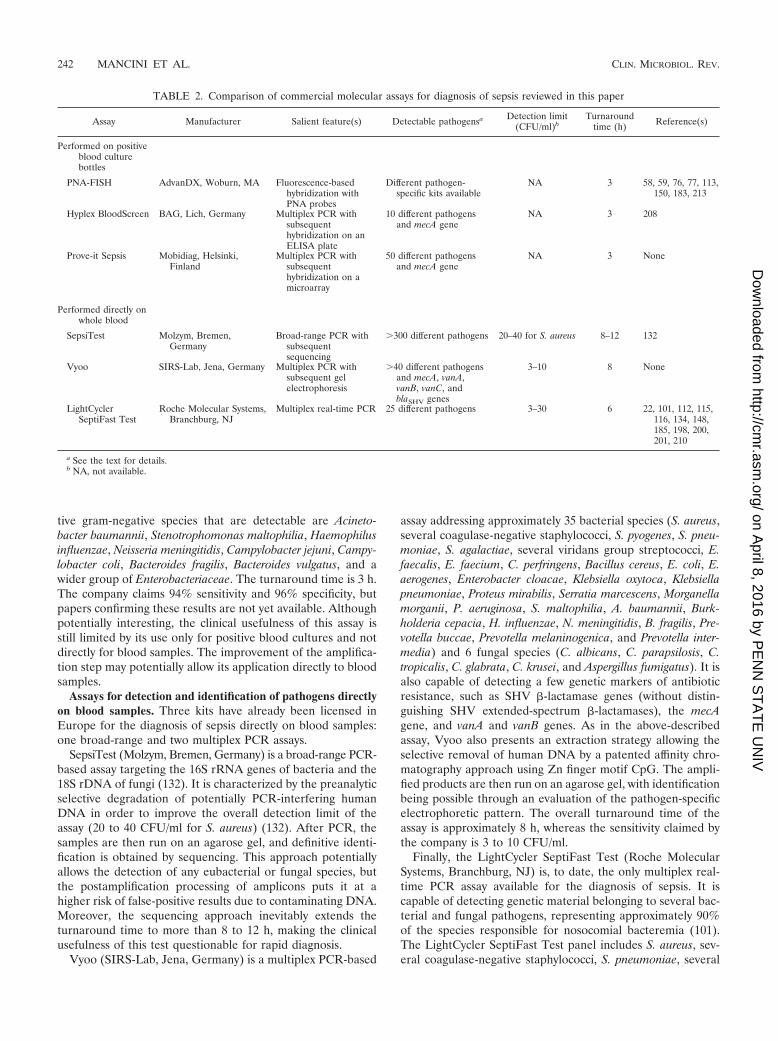
tive gram-negative species that are detectable are Acineto-bacter baumannii, Stenotrophomonas maltophilia, Haemophilusinfluenzae, Neisseria meningitidis, Campylobacter jejuni, Campy-lobacter coli, Bacteroides fragilis, Bacteroides vulgatus, and awider group of Enterobacteriaceae. The turnaround time is 3 h.The company claims 94% sensitivity and 96% specificity, butpapers confirming these results are not yet available. Althoughpotentially interesting, the clinical usefulness of this assay isstill limited by its use only for positive blood cultures and notdirectly for blood samples. The improvement of the amplifica-tion step may potentially allow its application directly to bloodsamples.
Assays for detection and identification of pathogens directlyon blood samples. Three kits have already been licensed inEurope for the diagnosis of sepsis directly on blood samples:one broad-range and two multiplex PCR assays.
SepsiTest (Molzym, Bremen, Germany) is a broad-range PCR-based assay targeting the 16S rRNA genes of bacteria and the18S rDNA of fungi (132). It is characterized by the preanalyticselective degradation of potentially PCR-interfering humanDNA in order to improve the overall detection limit of theassay (20 to 40 CFU/ml for S. aureus) (132). After PCR, thesamples are then run on an agarose gel, and definitive identi-fication is obtained by sequencing. This approach potentiallyallows the detection of any eubacterial or fungal species, butthe postamplification processing of amplicons puts it at ahigher risk of false-positive results due to contaminating DNA.Moreover, the sequencing approach inevitably extends theturnaround time to more than 8 to 12 h, making the clinicalusefulness of this test questionable for rapid diagnosis.
Vyoo (SIRS-Lab, Jena, Germany) is a multiplex PCR-based
assay addressing approximately 35 bacterial species (S. aureus,several coagulase-negative staphylococci, S. pyogenes, S. pneu-moniae, S. agalactiae, several viridans group streptococci, E.faecalis, E. faecium, C. perfringens, Bacillus cereus, E. coli, E.aerogenes, Enterobacter cloacae, Klebsiella oxytoca, Klebsiellapneumoniae, Proteus mirabilis, Serratia marcescens, Morganellamorganii, P. aeruginosa, S. maltophilia, A. baumannii, Burk-holderia cepacia, H. influenzae, N. meningitidis, B. fragilis, Pre-votella buccae, Prevotella melaninogenica, and Prevotella inter-media) and 6 fungal species (C. albicans, C. parapsilosis, C.tropicalis, C. glabrata, C. krusei, and Aspergillus fumigatus). It isalso capable of detecting a few genetic markers of antibioticresistance, such as SHV �-lactamase genes (without distin-guishing SHV extended-spectrum �-lactamases), the mecAgene, and vanA and vanB genes. As in the above-describedassay, Vyoo also presents an extraction strategy allowing theselective removal of human DNA by a patented affinity chro-matography approach using Zn finger motif CpG. The ampli-fied products are then run on an agarose gel, with identificationbeing possible through an evaluation of the pathogen-specificelectrophoretic pattern. The overall turnaround time of theassay is approximately 8 h, whereas the sensitivity claimed bythe company is 3 to 10 CFU/ml.
Finally, the LightCycler SeptiFast Test (Roche MolecularSystems, Branchburg, NJ) is, to date, the only multiplex real-time PCR assay available for the diagnosis of sepsis. It iscapable of detecting genetic material belonging to several bac-terial and fungal pathogens, representing approximately 90%of the species responsible for nosocomial bacteremia (101).The LightCycler SeptiFast Test panel includes S. aureus, sev-eral coagulase-negative staphylococci, S. pneumoniae, several
TABLE 2. Comparison of commercial molecular assays for diagnosis of sepsis reviewed in this paper
Assay Manufacturer Salient feature(s) Detectable pathogensa Detection limit(CFU/ml)b
Turnaroundtime (h) Reference(s)
Performed on positiveblood culturebottles
PNA-FISH AdvanDX, Woburn, MA Fluorescence-basedhybridization withPNA probes
Different pathogen-specific kits available
NA 3 58, 59, 76, 77, 113,150, 183, 213
Hyplex BloodScreen BAG, Lich, Germany Multiplex PCR withsubsequenthybridization on anELISA plate
10 different pathogensand mecA gene
NA 3 208
Prove-it Sepsis Mobidiag, Helsinki,Finland
Multiplex PCR withsubsequenthybridization on amicroarray
50 different pathogensand mecA gene
NA 3 None
Performed directly onwhole blood
SepsiTest Molzym, Bremen,Germany
Broad-range PCR withsubsequentsequencing
�300 different pathogens 20–40 for S. aureus 8–12 132
Vyoo SIRS-Lab, Jena, Germany Multiplex PCR withsubsequent gelelectrophoresis
�40 different pathogensand mecA, vanA,vanB, vanC, andblaSHV genes
3–10 8 None
LightCyclerSeptiFast Test
Roche Molecular Systems,Branchburg, NJ
Multiplex real-time PCR 25 different pathogens 3–30 6 22, 101, 112, 115,116, 134, 148,185, 198, 200,201, 210
a See the text for details.b NA, not available.
242 MANCINI ET AL. CLIN. MICROBIOL. REV.
on April 8, 2016 by P
EN
N S
TA
TE
UN
IVhttp://cm
r.asm.org/
Dow
nloaded from
other streptococcal species, E. faecalis, E. faecium, E. coli, E.aerogenes, E. cloacae, K. oxytoca, K. pneumoniae, P. mirabilis, S.marcescens, P. aeruginosa, S. maltophilia, A. baumannii, C. al-bicans, C. parapsilosis, C. tropicalis, C. glabrata, C. krusei, andA. fumigatus. The assay uses dual fluorescent resonance energytransfer (FRET) probes targeting the species-specific internaltranscribed spacer (ITS) regions. DNA is extracted by mechan-ical lysis with ceramic beads in a Magnalyzer instrument(Roche Molecular Systems), and after purification, it is pro-cessed in three parallel multiplex real-time PCRs (gram-posi-tive bacteria, gram-negative bacteria, and fungi). The meltingprofile of amplified products is then calculated by specific ded-icated software, thus allowing the detection of the pathogenand its identification at the genus or species level (101). Thedetection limit ranges from 3 to 30 CFU/ml, depending onsingle pathogens, whereas the turnaround time is approxi-mately 6 h (101). The assay has shown promising performancesas an adjunct to blood culture for neutropenic (115, 116, 185,198, 201), pediatric (134, 148, 200), intensive care, and generalmedicine (112, 210) patients. No real advantage was observedfor patients with suspected infective endocarditis (22), butseveral IE-related bacterial species are not included in theSeptiFast panel, and the sensitivity of the assay may not besufficient to detect the low-grade bacteremia associated withthis condition. In conclusion, the main technical advantageof this assay is its real-time format that markedly reducesthe risk of contamination. Current limitations of the assayinclude its very high cost (€150 to €200 [$215 to $290] pertest) and the lack of any information on antimicrobial sus-ceptibility.
Principal Shortcomings of NAT-Based Assays forMicrobiological Diagnosis of Sepsis
Despite the remarkable technical advances of NAT-basedapproaches, their widespread use for the microbiological diag-nosis of sepsis is still limited by shortcomings influencing theiroverall cost-effectiveness.
The first point to bear in mind when evaluating molecularreports is that the detection of circulating microbial DNA(DNAemia) does not necessarily indicate the presence of aviable microorganism responsible for a given infection. Thehigh sensitivity needed for the diagnosis of sepsis may increasethe risk of false-positive results due to carryover contaminationor due to the detection of environmental DNA contaminatingthe blood sample. Moreover, DNAemia may be the footprintfor transient bacteremia not related to any infection (173, 193),or it may be related to the persistence of circulating DNA stilldetectable several days after successful anti-infectious therapyhas been completed (116). More studies are needed to evalu-ate, for different categories of patients, the clinical usefulnessof this new laboratory parameter as an adjunct to blood cul-ture.
Another major drawback of the available molecular assaysfor the diagnosis of sepsis is that they do not provide informa-tion on the antimicrobial susceptibility of the detected patho-gen. The rapid detection of a pathogen may allow better fine-tuning of empirical therapy with possible economic savings, butthe lack of a specific susceptibility spectrum, especially withmultidrug-resistant pathogens on the rise, may limit the clinical
usefulness of these assays. In cases where the presence of asingle gene is always associated with phenotypic resistance(i.e., the mecA gene for oxacillin resistance and van genes forvancomycin resistance), it is relatively simple to design molec-ular strategies that allow their detection. More troublesomeare cases where the phenotypic resistance is influenced byseveral concurrent factors, such as the regulatory role of dis-tinct genes that modulate the levels of expression of thegene(s) determining resistance.
NONMOLECULAR BIOMARKERS OF SEPSIS: ROLEAND LIMITS
Detection of Fungal Antigens
Fungi continue to be a major cause of infection-relatedmortality principally for intensive care unit (ICU) patients andpatients with hematological malignancies. Frequently, systemicantifungals are necessary for universal prophylaxis and/or onan empirical basis for these high-risk populations, with issuesregarding the consequent cost, toxicity, and possible emergence ofdrug resistance. In addition, epidemiological data suggest thatthere have been changes in the epidemiology of fungal infec-tions of oncohematological patients (8, 216). Although invasiveaspergillosis and candidiasis continue to be the leading fungaldiseases, it would appear that less common mold pathogensare emerging, with important therapeutic consequences. Fur-thermore, it has been highlighted that the introduction of anactive diagnostic surveillance strategy for invasive fungal infec-tions of high-risk patients with hematological malignanciesmay result in improvements in patient survival (9, 25). How-ever, early diagnosis remains a challenge due mainly to the lowspecificity of clinical symptoms and the low sensitivity of fungalcultures.
GM. The development of an enzyme-linked immunosorbentassay for the detection of galactomannan (GM), an Aspergillussp. cell wall component, has been an important advance for thediagnosis of invasive aspergillosis (155, 188). A positive testconfirmed with two sequential samples is considered to be avalid index for invasive aspergillosis diagnosis using EORTCdiagnostic criteria (5). Although the detection of GM in serumis easy to perform, a major disadvantage of the serodiagnosisof invasive aspergillosis is the occurrence of false-positive re-sults. Several possible causes of false-positive reactions havebeen reported, including the intake of certain foods, especiallyif the intestinal barrier is altered; gastrointestinal chronic graft-versus-host disease; lipoglycans related to Bifidobacterium spp.;solutions containing sodium gluconate; and, in particular, theuse of semisynthetic �-lactam treatments, especially piperacil-lin-tazobactam (60). Changes in the manufacturing process forthis antibiotic, possibly resulting in the reduction of cross-reacting materials, have been suggested (152). Although theGM assay has been designed to be specific for Aspergillus spp.,cross-reactivity with other fungal species such as Blastomycesdermatitidis, Penicillium spp., and Fusarium spp. has been re-ported (34). The increasing clinical impact of non-Aspergillusinvasive infections warrants caution when interpreting positiveGM results, especially if an infection with a potentially cross-reacting species cannot be clinically excluded. Additionally,different studies reported highly variable sensitivities (51, 155).
VOL. 23, 2010 NON-CULTURE-BASED DIAGNOSIS OF SEPSIS 243
on April 8, 2016 by P
EN
N S
TA
TE
UN
IVhttp://cm
r.asm.org/
Dow
nloaded from
In a meta-analysis of 27 studies, the overall sensitivity wasreported to be 71%, and the specificity was reported to be 89%(155). Even lower sensitivities were reported for high-risk pa-tients, such as recipients of solid-organ transplants (80, 97).
(133)-�-D-Glucan. �-Glucan (BG) is another component ofthe fungal cell wall that present in a wider variety of fungalspecies, including Candida spp. (141). As for GM, differentstudies reported variable sensitivities (141, 145), with a sensi-tivity of 69.9% being observed for a large multicenter studyusing just one sample per patient (145). For most patients withconfirmed invasive fungal infections, BG levels were elevatedseveral days before clinical diagnosis (2). It was suggested thatBG detection may be a useful adjunctive tool for promptlyguiding preemptive antifungal therapy for neutropenic patients(51), but further studies investigating this aspect are certainlyneeded.
The combined use of GM and BG detection has also beenproposed. In a retrospective study of 40 neutropenic patients,parallel detection improved the specificity and positive predic-tive value of each individual test without affecting the sensitiv-ity and negative predictive values (149). In other words, thecombination seems to be useful to identify false-positive reac-tions for each test. Several studies of animal models seem toconfirm this observation (1, 88), but further clinical studies areneeded to evaluate the cost-effectiveness of this approach.Finally, it is important that neither the BG nor GM assaydetects zygomycetes, a rare but emerging cause of invasivefungal mycosis (24).
Nonmicrobiological, Nonspecific Sentinel Markersin Sepsis Patients
The limitations of blood cultures have also fostered interestin the development of sensitive and rapid laboratory testsaimed at detecting nonspecific biomarkers of sepsis. For thisprincipal reason, assays for C-reactive protein (CRP), procal-citonin (PCT), interleukin-6 (IL-6), and IL-8 and, more re-cently, proteomic assays have become subjects of interest, anda number of studies have evaluated their clinical usefulness.
IL-6. The release of inflammatory cytokines such as tumornecrosis factor alpha (TNF-�), IL-1b, IL-8, and IL-6 in re-sponse to infectious pathogens and host injury may lead toSIRS. In particular, IL-6 is induced by TNF-� and can bemeasured reliably in blood, having a long half-life (147). IL-6,an important mediator in septic shock, has been acknowledgedto predict severity and clinical outcome under this condition(69, 119). However, it is relatively nonspecific as a marker ofinfection, and it was observed that IL-6 levels are elevatedunder a variety of conditions, such as in the acute-phase re-sponse to injury (14, 42, 43), in acute pancreatitis (186), and inrenal transplant patients with an increased risk of acute rejec-tion or graft failure (12). From a clinical point of view, IL-6may be regarded principally as an early predictor of down-stream effects, such as organ dysfunction, due to its initial rolein the cytokine response.
CRP. CRP is an acute-phase protein synthesized predomi-nantly in hepatocytes and alveolar macrophages (41) in re-sponse to a variety of cytokines, including IL-6. CRP also playsa role in immunomodulation. It was observed that CRP mod-ulates the complement cascade and regulates bacterial opso-
nization and phagocytosis (66, 189). Increases in CRP levelshave been documented under a variety of noninfectious con-ditions, including postmyocardial infarction settings (137) andrheumatologic diseases (45). The kinetics of serum CRP arecharacterized by a rapid response to inflammation and a shorthalf-life (approximately 19 h) (123). Finally, the assay is inex-pensive and widely available. Although several studies demon-strated elevated CRP levels in sepsis (23, 157), its nonspecificdynamics cannot support a major diagnostic role for this bio-marker in sepsis.
PCT. Procalcitonin (PCT) is a propeptide of calcitonin thatis ubiquitously expressed as part of the host’s inflammatoryresponse to a variety of insults (10). Although calcitonin is ahormone classically produced in the parathyroids and involvedin calcium homeostasis, PCT (one of the calcitonin precursors)has effects on a variety of inflammatory conditions, includingcardiogenic shock, trauma, necrotizing pancreatitis, burns, sur-gery, and infection (10, 203). In animal models of sepsis, animmunological blockade of calcitonin precursors improves or-gan dysfunction and outcome (140). A growing body of evi-dence suggests that PCT is a marker of severe bacterial infec-tion (83) and can distinguish patients who have sepsis frompatients who have SIRS (133). In particular, PCT levels inplasma have been correlated with sepsis-related organ failurescores and may be useful in risk assessment (123). High andpersistent elevations in PCT levels have been associated withpoor outcomes for ICU patients (214). Moreover, the value ofPCT in risk assessment for pediatric patients was demon-strated (72). Although several studies suggested that PCT isamong the most promising biomarkers for sepsis, considerablecontroversy surrounding its clinical usefulness still remains. Arecent meta-analysis (190) indicated that PCT cannot reliablydifferentiate sepsis from other noninfectious causes of systemicinflammatory response syndrome in critically ill adult patients.However, the U.S. Food and Drug Administration (FDA) hasapproved the use of PCT in conjunction with other laboratoryfindings to aid the risk assessment of critically ill patients.
TREM-1. Neutrophils and monocytes/macrophages are theprimary mediators of the innate immune response to bacterialinfection, promoting the release of proinflammatory cytokinessuch as TNF-� and IL-1b, which, when produced in excess,contribute to end-organ dysfunction and overwhelming sepsis.The so-called triggering receptor expressed on myeloid cells 1(TREM-1) is part of the immunoglobulin superfamily and isupregulated in response to bacteria or fungi. When bound toligand, TREM-1 stimulates the release of cytokines (99). Incontrast to infections, TREM-1 is not upregulated in nonin-fectious inflammatory disorders such as inflammatory boweldisease and SIRS (16). A soluble form of TREM-1 (sTREM-1)is shed from the membranes of activated phagocytic cells andcan be quantified in human body fluids. Several studies haveinvestigated the use of TREM-1 as a diagnostic biomarker andhave shown it to be more sensitive and specific than CRP andPCT (67). Although TREM-1 may be a promising diagnosticmarker for sepsis, less is presently known about its use in riskassessment and prognosis for patients with known sepsis.
Overall, the present data suggest that IL-6 and CRP aresentinel markers of inflammation and infection but are toononspecific for further clinical use. PCT will likely enhanceclinicians’ risk assessments for critically ill patients with sepsis.
244 MANCINI ET AL. CLIN. MICROBIOL. REV.
on April 8, 2016 by P
EN
N S
TA
TE
UN
IVhttp://cm
r.asm.org/
Dow
nloaded from

Furthermore, TREM-1 is an additional promising candidate.Given the high complexity and variability of the disease, bio-marker panels or composite markers may prove most useful inexamining a particular immunological pathway, predicting or-gan-specific responses, and, ideally, identifying at-risk individ-uals.
CONCLUDING REMARKS
The number of people dying from sepsis has almost doubledin the past 20 years, and an increasing number of patientswith differing medical conditions are at a high risk of devel-oping sepsis. In this context, a rapid microbiological diag-nosis is particularly important for driving the correct thera-peutic intervention. The role of surrogate nonmolecularmarkers of sepsis is still controversial. Although less expensiveand more high throughput than molecular methods, their clin-ical usefulness seems, in the best cases, to be limited in the riskassessment of critically ill patients. Moreover, an increasingbody of evidence advocates that the prompt identification ofthe agent(s) causing sepsis is needed for a more rapid fine-tuning of empirical therapy.
Several molecular strategies have been applied in this field,and a number of technically reliable methodologies have beenproposed. These methods may allow the fast and sensitivedetection of a wide panel of bacterial and fungal pathogens,also directly from blood specimens. However, these techniquescannot be regarded as substitutes for classical culture methods,since they have been only partially validated for routine clinicaluse. From this point of view, molecular techniques are onlyadditional assays in the current diagnostic activity for BSI.
An additional point in the evaluation of molecular tech-niques is that the detection of circulating microbial DNA(DNAemia) represents a new diagnostic parameter that doesnot necessarily correspond to the presence of viable microor-ganisms in blood. Although promising, the clinical usefulnessof this new parameter needs further validation.
Furthermore, most of the available molecular assays for thediagnosis of sepsis do not provide information on the antimi-crobial susceptibility of the pathogen detected. Although therapid detection of a pathogen normally allows a better use ofempirical therapy, the absence of a specific susceptibility pro-file with multidrug-resistant pathogens may limit the clinicalrole of molecular methods in sepsis. This point should also beaddressed with future studies, especially in cases where thepresence of a single gene is not always associated with pheno-typic resistance.
Moreover, techniques whose clinical role is mainly to pro-vide sensitivity and rapidity should be available 24 h per day inthe diagnostic microbiology laboratory. As a consequence,their availability not only will increase costs and require ded-icated personnel but also will modify greatly the organizationof laboratories. The cost-effectiveness of such a revolution isstill under debate.
In conclusion, the development of a number of first-gener-ation molecular assays for the microbiological diagnosis ofsepsis, in support of the use of blood cultures, already opens upa new era in the microbiological laboratory. However, althoughthey offer the hope of potentially better diagnostic assays, theyare still not ready for “primetime” use. The refinement of these
assays in the near future (as well as those for other clinicalconditions, including central nervous system infections, sexu-ally transmitted diseases, and respiratory infections) will changedramatically the working algorithm and the general organizationof clinical microbiology laboratories.
ACKNOWLEDGMENT
We are grateful to Dan McAuley for revising the English manu-script.
REFERENCES
1. Ahmad, S., Z. U. Khan, and A. M. Theyyathel. 2007. Diagnostic value ofDNA, (1-3)-beta-d-glucan, and galactomannan detection in serum andbronchoalveolar lavage of mice experimentally infected with Aspergillusterreus. Diagn. Microbiol. Infect. Dis. 59:165–171.
2. Almyroudis, N. G., and B. H. Segal. 2009. Prevention and treatment ofinvasive fungal diseases in neutropenic patients. Curr. Opin. Infect. Dis.22:385–393.
3. Angus, D. C., W. T. Linde-Zwirble, J. Lidicker, G. Clermont, J. Carcillo,and M. R. Pinsky. 2001. Epidemiology of severe sepsis in the United States:analysis of incidence, outcome, and associated costs of care. Crit. CareMed. 29:1303–1310.
4. Arpi, M., M. W. Bentzon, J. Jensen, and W. Frederiksen. 1989. Importanceof blood volume cultured in the detection of bacteremia. Eur. J. Clin.Microbiol. Infect. Dis. 8:838–842.
5. Ascioglu, S., J. H. Rex, B. de Pauw, J. E. Bennett, J. Bille, F. Crokaert, D. W.Denning, J. P. Donnelly, J. E. Edwards, Z. Erjavec, D. Fiere, O. Lortholary,J. Maertens, J. F. Meis, T. F. Patterson, J. Ritter, D. Selleslag, P. M. Shah,D. A. Stevens, and T. J. Walsh. 2002. Defining opportunistic invasive fungalinfections in immunocompromised patients with cancer and hematopoieticstem cell transplants: an international consensus. Clin. Infect. Dis. 34:7–14.
6. Awais, R., H. Fukudomi, K. Miyanaga, H. Unno, and Y. Tanji. 2006. Arecombinant bacteriophage-based assay for the discriminative detection ofculturable and viable but nonculturable Escherichia coli O157:H7. Biotech-nol. Prog. 22:853–859.
7. Barnato, A. E., S. L. Alexander, W. T. Linde-Zwirble, and D. C. Angus.2008. Racial variation in the incidence, care, and outcomes of severe sepsis:analysis of population, patient, and hospital characteristics. Am. J. Respir.Crit. Care Med. 177:279–284.
8. Barnes, P. D., and K. A. Marr. 2007. Risks, diagnosis and outcomes ofinvasive fungal infections in haematopoietic stem cell transplant recipients.Br. J. Haematol. 139:519–531.
9. Barnes, R. A., P. L. White, C. Bygrave, N. Evans, B. Healy, and J. Kell.2009. Clinical impact of enhanced diagnosis of invasive fungal disease inhigh-risk haematology and stem cell transplant patients. J. Clin. Pathol.62:64–69.
10. Becker, K. L., E. S. Nylen, J. C. White, B. Muller, and R. H. Snider, Jr.2004. Procalcitonin and the calcitonin gene family of peptides in inflam-mation, infection, and sepsis: a journey from calcitonin back to its precur-sors. J. Clin. Endocrinol. Metab. 89:1512–1525.
11. Beekmann, S. E., D. J. Diekema, K. C. Chapin, and G. V. Doern. 2003.Effects of rapid detection of bloodstream infections on length of hospital-ization and hospital charges. J. Clin. Microbiol. 41:3119–3125.
12. Berber, I., B. Yigit, G. Isitmangil, G. Tellioglu, T. Ozgezer, S. Gulle, F.Turkmen, and I. Titiz. 2008. Evaluation of pretransplant serum cytokinelevels in renal transplant recipients. Transplant. Proc. 40:92–93.
13. Bernardo, K., S. Fleer, N. Pakulat, O. Krut, F. Hunger, and M. Kronke.2002. Identification of Staphylococcus aureus exotoxins by combined so-dium dodecyl sulfate gel electrophoresis and matrix-assisted laser desorp-tion/ionization-time of flight mass spectrometry. Proteomics 2:740–746.
14. Biffl, W. L., E. E. Moore, F. A. Moore, and V. M. Peterson. 1996. Interleu-kin-6 in the injured patient. Marker of injury or mediator of inflammation?Ann. Surg. 224:647–664.
15. Bone, R. C., R. A. Balk, F. B. Cerra, R. P. Dellinger, A. M. Fein, W. A.Knaus, R. M. Schein, and W. J. Sibbald. 1992. Definitions for sepsis andorgan failure and guidelines for the use of innovative therapies in sepsis.The ACCP/SCCM Consensus Conference Committee. American Collegeof Chest Physicians/Society of Critical Care Medicine. Chest 101:1644–1655.
16. Bouchon, A., F. Facchetti, M. A. Weigand, and M. Colonna. 2001. TREM-1amplifies inflammation and is a crucial mediator of septic shock. Nature410:1103–1107.
17. Bouza, E., D. Sousa, M. Rodriguez-Creixems, J. G. Lechuz, and P. Munoz.2007. Is the volume of blood cultured still a significant factor in the diag-nosis of bloodstream infections? J. Clin. Microbiol. 45:2765–2769.
18. Breitkopf, C., D. Hammel, H. H. Scheld, G. Peters, and K. Becker. 2005.Impact of a molecular approach to improve the microbiological diagnosis ofinfective heart valve endocarditis. Circulation 111:1415–1421.
VOL. 23, 2010 NON-CULTURE-BASED DIAGNOSIS OF SEPSIS 245
on April 8, 2016 by P
EN
N S
TA
TE
UN
IVhttp://cm
r.asm.org/
Dow
nloaded from
19. Bretagne, S., and J. M. Costa. 2005. Towards a molecular diagnosis ofinvasive aspergillosis and disseminated candidosis. FEMS Immunol. Med.Microbiol. 45:361–368.
20. Brown, A. R., and J. R. Govan. 2007. Assessment of fluorescent in situhybridization and PCR-based methods for rapid identification of Burkhold-eria cepacia complex organisms directly from sputum samples. J. Clin.Microbiol. 45:1920–1926.
21. Campbell, S. G., T. J. Marrie, R. Anstey, G. Dickinson, and S. Ackroyd-Stolarz. 2003. The contribution of blood cultures to the clinical manage-ment of adult patients admitted to the hospital with community-acquiredpneumonia: a prospective observational study. Chest 123:1142–1150.
22. Casalta, J. P., F. Gouriet, V. Roux, F. Thuny, G. Habib, and D. Raoult.2009. Evaluation of the LightCycler SeptiFast test in the rapid etiologicdiagnostic of infectious endocarditis. Eur. J. Clin. Microbiol. Infect. Dis.28:569–573.
23. Castelli, G. P., C. Pognani, M. Meisner, A. Stuani, D. Bellomi, and L.Sgarbi. 2004. Procalcitonin and C-reactive protein during systemic inflam-matory response syndrome, sepsis and organ dysfunction. Crit. Care8:R234–R242.
24. Chamilos, G., M. Luna, R. E. Lewis, G. P. Bodey, R. Chemaly, J. J.Tarrand, A. Safdar, I. I. Raad, and D. P. Kontoyiannis. 2006. Invasivefungal infections in patients with hematologic malignancies in a tertiarycare cancer center: an autopsy study over a 15-year period (1989–2003).Haematologica 91:986–989.
25. Chandrasekar, P. 2009. Invasive mold infections: recent advances in man-agement approaches. Leuk. Lymphoma 50:703–715.
26. Chapin, K., and M. Musgnug. 2003. Evaluation of three rapid methods forthe direct identification of Staphylococcus aureus from positive blood cul-tures. J. Clin. Microbiol. 41:4324–4327.
27. Cleven, B. E., M. Palka-Santini, J. Gielen, S. Meembor, M. Kronke, and O.Krut. 2006. Identification and characterization of bacterial pathogens caus-ing bloodstream infections by DNA microarray. J. Clin. Microbiol. 44:2389–2397.
28. CLSI. 2008. Abbreviated identification of bacteria and yeast. Approvedguideline, 2nd ed. (M35-A2), vol. 28, no.29. Clinical and Laboratory Stan-dards Institute, Wayne, PA.
29. CLSI. 2007. Principles and procedures for blood cultures. Approved guide-line (M47-A), vol. 27, no.17. Clinical and Laboratory Standards Institute,Wayne, PA.
30. Cockerill, F. R., III, J. W. Wilson, E. A. Vetter, K. M. Goodman, C. A.Torgerson, W. S. Harmsen, C. D. Schleck, D. M. Ilstrup, J. A. WashingtonII, and W. R. Wilson. 2004. Optimal testing parameters for blood cultures.Clin. Infect. Dis. 38:1724–1730.
31. Connell, T. G., M. Rele, D. Cowley, J. P. Buttery, and N. Curtis. 2007. Howreliable is a negative blood culture result? Volume of blood submitted forculture in routine practice in a children’s hospital. Pediatrics 119:891–896.
32. Costa, C., J. M. Costa, C. Desterke, F. Botterel, C. Cordonnier, and S.Bretagne. 2002. Real-time PCR coupled with automated DNA extractionand detection of galactomannan antigen in serum by enzyme-linked immu-nosorbent assay for diagnosis of invasive aspergillosis. J. Clin. Microbiol.40:2224–2227.
33. Cuenca-Estrella, M., Y. Meije, C. Diaz-Pedroche, A. Gomez-Lopez, M. J.Buitrago, L. Bernal-Martinez, C. Grande, R. S. Juan, M. Lizasoain, J. L.Rodriguez-Tudela, and J. M. Aguado. 2009. Value of serial quantification offungal DNA by a real-time PCR-based technique for early diagnosis ofinvasive aspergillosis in patients with febrile neutropenia. J. Clin. Microbiol.47:379–384.
34. Cummings, J. R., G. R. Jamison, J. W. Boudreaux, M. J. Howles, T. J.Walsh, and R. T. Hayden. 2007. Cross-reactivity of non-Aspergillus fungalspecies in the Aspergillus galactomannan enzyme immunoassay. Diagn.Microbiol. Infect. Dis. 59:113–115.
35. Davis, T. E., D. D. Fuller, and E. C. Aeschleman. 1992. Rapid, directidentification of Staphylococcus aureus and Streptococcus pneumoniaefrom blood cultures using commercial immunologic kits and modified con-ventional tests. Diagn. Microbiol. Infect. Dis. 15:295–300.
36. Dellinger, R. P., M. M. Levy, J. M. Carlet, J. Bion, M. M. Parker, R.Jaeschke, K. Reinhart, D. C. Angus, C. Brun-Buisson, R. Beale, T. Ca-landra, J. F. Dhainaut, H. Gerlach, M. Harvey, J. J. Marini, J. Marshall,M. Ranieri, G. Ramsay, J. Sevransky, B. T. Thompson, S. Townsend, J. S.Vender, J. L. Zimmerman, and J. L. Vincent. 2008. Surviving Sepsis Cam-paign: international guidelines for management of severe sepsis and septicshock: 2008. Crit. Care Med. 36:296–327.
37. De Marco, D., M. Perotti, C. M. Ossi, R. Burioni, M. Clementi, and N.Mancini. 2007. Development and validation of a molecular method for thediagnosis of medically important fungal infections. New Microbiol. 30:308–312.
38. Dombrovskiy, V. Y., A. A. Martin, J. Sunderram, and H. L. Paz. 2005.Facing the challenge: decreasing case fatality rates in severe sepsis despiteincreasing hospitalizations. Crit. Care Med. 33:2555–2562.
39. Dombrovskiy, V. Y., A. A. Martin, J. Sunderram, and H. L. Paz. 2007.Occurrence and outcomes of sepsis: influence of race. Crit. Care Med.35:763–768.
40. Dombrovskiy, V. Y., A. A. Martin, J. Sunderram, and H. L. Paz. 2007.Rapid increase in hospitalization and mortality rates for severe sepsis in theUnited States: a trend analysis from 1993 to 2003. Crit. Care Med. 35:1244–1250.
41. Dong, Q., and J. R. Wright. 1996. Expression of C-reactive protein byalveolar macrophages. J. Immunol. 156:4815–4820.
42. Drost, A. C., D. G. Burleson, W. G. Cioffi, Jr., B. S. Jordan, A. D. Mason,Jr., and B. A. Pruitt, Jr. 1993. Plasma cytokines following thermal injuryand their relationship with patient mortality, burn size, and time postburn.J. Trauma 35:335–339.
43. Drost, A. C., D. G. Burleson, W. G. Cioffi, Jr., A. D. Mason, Jr., and B. A.Pruitt, Jr. 1993. Plasma cytokines after thermal injury and their relationshipto infection. Ann. Surg. 218:74–78.
44. Durack, D. T., A. S. Lukes, and D. K. Bright. 1994. New criteria fordiagnosis of infective endocarditis: utilization of specific echocardiographicfindings. Duke Endocarditis Service. Am. J. Med. 96:200–209.
45. Eberhard, O. K., M. Haubitz, F. M. Brunkhorst, V. Kliem, K. M. Koch, andR. Brunkhorst. 1997. Usefulness of procalcitonin for differentiation be-tween activity of systemic autoimmune disease (systemic lupus erythema-tosus/systemic antineutrophil cytoplasmic antibody-associated vasculitis)and invasive bacterial infection. Arthritis Rheum. 40:1250–1256.
46. Ecker, D. J., R. Sampath, L. B. Blyn, M. W. Eshoo, C. Ivy, J. A. Ecker, B.Libby, V. Samant, K. A. Sannes-Lowery, R. E. Melton, K. Russell, N. Freed,C. Barrozo, J. Wu, K. Rudnick, A. Desai, E. Moradi, D. J. Knize, D. W.Robbins, J. C. Hannis, P. M. Harrell, C. Massire, T. A. Hall, Y. Jiang, R.Ranken, J. J. Drader, N. White, J. A. McNeil, S. T. Crooke, and S. A.Hofstadler. 2005. Rapid identification and strain-typing of respiratorypathogens for epidemic surveillance. Proc. Natl. Acad. Sci. U. S. A. 102:8012–8017.
47. Edwards-Jones, V., M. A. Claydon, D. J. Evason, J. Walker, A. J. Fox, andD. B. Gordon. 2000. Rapid discrimination between methicillin-sensitive andmethicillin-resistant Staphylococcus aureus by intact cell mass spectrome-try. J. Med. Microbiol. 49:295–300.
48. Eggimann, P. 2007. Diagnosis of intravascular catheter infection. Curr.Opin. Infect. Dis. 20:353–359.
49. Eigner, U., A. Fahr, M. Weizenegger, and W. Witte. 2005. Evaluation of anew molecular system for simultaneous identification of four Enterococcusspecies and their glycopeptide resistance genotypes. J. Clin. Microbiol.43:2920–2922.
50. Eigner, U., M. Weizenegger, A. M. Fahr, and W. Witte. 2005. Evaluation ofa rapid direct assay for identification of bacteria and the mecA and vangenes from positive-testing blood cultures. J. Clin. Microbiol. 43:5256–5262.
51. Ellis, M. 2008. Febrile neutropenia. Ann. N. Y. Acad. Sci. 1138:329–350.52. El-Mahallawy, H. A., H. H. Shaker, H. A. Helmy, T. Mostafa, and A. R.
Abo-Sedah. 2006. Evaluation of pan-fungal PCR assay and Aspergillusantigen detection in the diagnosis of invasive fungal infections in high riskpaediatric cancer patients. Med. Mycol. 44:733–739.
53. Engel, C., F. M. Brunkhorst, H. G. Bone, R. Brunkhorst, H. Gerlach, S.Grond, M. Gruendling, G. Huhle, U. Jaschinski, S. John, K. Mayer, M.Oppert, D. Olthoff, M. Quintel, M. Ragaller, R. Rossaint, F. Stuber, N.Weiler, T. Welte, H. Bogatsch, C. Hartog, M. Loeffler, and K. Reinhart.2007. Epidemiology of sepsis in Germany: results from a national prospec-tive multicenter study. Intensive Care Med. 33:606–618.
54. Esper, A. M., M. Moss, C. A. Lewis, R. Nisbet, D. M. Mannino, and G. S.Martin. 2006. The role of infection and comorbidity: factors that influencedisparities in sepsis. Crit. Care Med. 34:2576–2582.
55. Evertsson, U., H. J. Monstein, and A. G. Johansson. 2000. Detection andidentification of fungi in blood using broad-range 28S rDNA PCR ampli-fication and species-specific hybridisation. APMIS 108:385–392.
56. Farrell, J. J., L. J. Doyle, R. M. Addison, L. B. Reller, G. S. Hall, and G. W.Procop. 2005. Broad-range (pan) Salmonella and Salmonella serotypeTyphi-specific real-time PCR assays: potential tools for the clinical micro-biologist. Am. J. Clin. Pathol. 123:339–345.
57. Fenollar, F., and D. Raoult. 2007. Molecular diagnosis of bloodstreaminfections caused by non-cultivable bacteria. Int. J. Antimicrob. Agents30(Suppl. 1):S7–S15.
58. Forrest, G. N., K. Mankes, M. A. Jabra-Rizk, E. Weekes, J. K. Johnson,D. P. Lincalis, and R. A. Venezia. 2006. Peptide nucleic acid fluorescence insitu hybridization-based identification of Candida albicans and its impact onmortality and antifungal therapy costs. J. Clin. Microbiol. 44:3381–3383.
59. Forrest, G. N., M. C. Roghmann, L. S. Toombs, J. K. Johnson, E. Weekes,D. P. Lincalis, and R. A. Venezia. 2008. Peptide nucleic acid fluorescent insitu hybridization for hospital-acquired enterococcal bacteremia: deliveringearlier effective antimicrobial therapy. Antimicrob. Agents Chemother. 52:3558–3563.
60. Fortun, J., P. Martin-Davila, M. E. Alvarez, F. Norman, A. Sanchez-Sousa,L. Gajate, R. Barcena, S. J. Nuno, and S. Moreno. 2009. False-positiveresults of Aspergillus galactomannan antigenemia in liver transplant recip-ients. Transplantation 87:256–260.
61. Fournier, P. E., and D. Raoult. 2003. Comparison of PCR and serologyassays for early diagnosis of acute Q fever. J. Clin. Microbiol. 41:5094–5098.
62. Fredricks, D. N., and D. A. Relman. 1998. Improved amplification of mi-
246 MANCINI ET AL. CLIN. MICROBIOL. REV.
on April 8, 2016 by P
EN
N S
TA
TE
UN
IVhttp://cm
r.asm.org/
Dow
nloaded from
crobial DNA from blood cultures by removal of the PCR inhibitor sodiumpolyanetholesulfonate. J. Clin. Microbiol. 36:2810–2816.
63. Funke, G., and P. Funke-Kissling. 2004. Use of the BD PHOENIX Auto-mated Microbiology System for direct identification and susceptibility test-ing of gram-negative rods from positive blood cultures in a three-phasetrial. J. Clin. Microbiol. 42:1466–1470.
64. Gebert, S., D. Siegel, and N. Wellinghausen. 2008. Rapid detection ofpathogens in blood culture bottles by real-time PCR in conjunction with thepre-analytic tool MolYsis. J. Infect. 57:307–316.
65. Gerdes, J. S. 2004. Diagnosis and management of bacterial infections in theneonate. Pediatr. Clin. N. Am. 51:939–959.
66. Gershov, D., S. Kim, N. Brot, and K. B. Elkon. 2000. C-reactive proteinbinds to apoptotic cells, protects the cells from assembly of the terminalcomplement components, and sustains an antiinflammatory innate immuneresponse: implications for systemic autoimmunity. J. Exp. Med. 192:1353–1364.
67. Gibot, S., M. N. Kolopp-Sarda, M. C. Bene, A. Cravoisy, B. Levy, G. C.Faure, and P. E. Bollaert. 2004. Plasma level of a triggering receptorexpressed on myeloid cells-1: its diagnostic accuracy in patients with sus-pected sepsis. Ann. Intern. Med. 141:9–15.
68. Gray, S. J., M. A. Sobanski, E. B. Kaczmarski, M. Guiver, W. J. Marsh, R.Borrow, R. A. Barnes, and W. T. Coakley. 1999. Ultrasound-enhanced lateximmunoagglutination and PCR as complementary methods for non-cul-ture-based confirmation of meningococcal disease. J. Clin. Microbiol. 37:1797–1801.
69. Hack, C. E., E. R. De Groot, R. J. Felt-Bersma, J. H. Nuijens, R. J. S. VanSchijndel, A. J. Eerenberg-Belmer, L. G. Thijs, and L. A. Aarden. 1989.Increased plasma levels of interleukin-6 in sepsis. Blood 74:1704–1710.
70. Hall, K. K., and J. A. Lyman. 2006. Updated review of blood culturecontamination. Clin. Microbiol. Rev. 19:788–802.
71. Hall, M. M., D. M. Ilstrup, and J. A. Washington II. 1976. Effect of volumeof blood cultured on detection of bacteremia. J. Clin. Microbiol. 3:643–645.
72. Han, Y. Y., L. A. Doughty, D. Kofos, H. Sasser, and J. A. Carcillo. 2003.Procalcitonin is persistently increased among children with poor outcomefrom bacterial sepsis. Pediatr. Crit. Care Med. 4:21–25.
73. Handschur, M., H. Karlic, C. Hertel, M. Pfeilstocker, and A. G. Haslberger.2009. Preanalytic removal of human DNA eliminates false signals in general16S rDNA PCR monitoring of bacterial pathogens in blood. Comp. Immu-nol. Microbiol. Infect. Dis. 32:207–219.
74. Hardak, E., M. Yigla, I. Avivi, O. Fruchter, H. Sprecher, and I. Oren. 23March 2009, posting date. Impact of PCR-based diagnosis of invasive pul-monary aspergillosis on clinical outcome. Bone Marrow Transplant. doi:10.1038/bmt.2009.65.
75. Heininger, A., M. Binder, S. Schmidt, K. Unertl, K. Botzenhart, and G.Doring. 1999. PCR and blood culture for detection of Escherichia colibacteremia in rats. J. Clin. Microbiol. 37:2479–2482.
76. Hensley, D. M., R. Tapia, and Y. Encina. 2009. An evaluation of theAdvandx Staphylococcus aureus/CNS PNA FISH assay. Clin. Lab. Sci.22:30–33.
77. Hermsen, E. D., S. S. Shull, D. G. Klepser, P. C. Iwen, A. Armbrust, J.Garrett, A. G. Freifeld, and M. E. Rupp. 2008. Pharmacoeconomic analysisof microbiologic techniques for differentiating staphylococci directly fromblood culture bottles. J. Clin. Microbiol. 46:2924–2929.
78. Hoen, B., C. Selton-Suty, F. Lacassin, J. Etienne, S. Briancon, C. Leport,and P. Canton. 1995. Infective endocarditis in patients with negative bloodcultures: analysis of 88 cases from a one-year nationwide survey in France.Clin. Infect. Dis. 20:501–506.
79. Hugonnet, S., H. Sax, P. Eggimann, J. C. Chevrolet, and D. Pittet. 2004.Nosocomial bloodstream infection and clinical sepsis. Emerg. Infect. Dis.10:76–81.
80. Husain, S., E. J. Kwak, A. Obman, M. M. Wagener, S. Kusne, J. E. Stout,K. R. McCurry, and N. Singh. 2004. Prospective assessment of PlateliaAspergillus galactomannan antigen for the diagnosis of invasive aspergillo-sis in lung transplant recipients. Am. J. Transplant. 4:796–802.
81. Iregui, M., S. Ward, G. Sherman, V. J. Fraser, and M. H. Kollef. 2002.Clinical importance of delays in the initiation of appropriate antibiotictreatment for ventilator-associated pneumonia. Chest 122:262–268.
82. Isaacman, D. J., R. B. Karasic, E. A. Reynolds, and S. I. Kost. 1996. Effectof number of blood cultures and volume of blood on detection of bactere-mia in children. J. Pediatr. 128:190–195.
83. Jensen, J. U., L. Heslet, T. H. Jensen, K. Espersen, P. Steffensen, and M.Tvede. 2006. Procalcitonin increase in early identification of critically illpatients at high risk of mortality. Crit. Care Med. 34:2596–2602.
84. Jonsson, B., A. Nyberg, and C. Henning. 1993. Theoretical aspects ofdetection of bacteraemia as a function of the volume of blood cultured.APMIS 101:595–601.
85. Jordan, J. A., J. Jones-Laughner, and M. B. Durso. 2009. Utility of pyro-sequencing in identifying bacteria directly from positive blood culture bot-tles. J. Clin. Microbiol. 47:368–372.
86. Kaditis, A. G., A. S. O’Marcaigh, K. H. Rhodes, A. L. Weaver, and N. K.Henry. 1996. Yield of positive blood cultures in pediatric oncology patients
by a new method of blood culture collection. Pediatr. Infect. Dis. J. 15:615–620.
87. Kempf, V. A., K. Trebesius, and I. B. Autenrieth. 2000. Fluorescent in situhybridization allows rapid identification of microorganisms in blood cul-tures. J. Clin. Microbiol. 38:830–838.
88. Khan, Z. U., S. Ahmad, and A. M. Theyyathel. 2008. Detection of Aspergil-lus fumigatus-specific DNA, (1-3)-beta-D-glucan and galactomannan inserum and bronchoalveolar lavage specimens of experimentally infectedrats. Mycoses 51:129–135.
89. Khlif, M., C. Mary, H. Sellami, A. Sellami, H. Dumon, A. Ayadi, and S.Ranque. 11 May 2009, posting date. Evaluation of nested and real-timePCR assays in the diagnosis of candidaemia. Clin. Microbiol. Infect. doi:10.1111/j.1469-0691.2009.02762.
90. Kirketerp-Moller, K., P. O. Jensen, M. Fazli, K. G. Madsen, J. Pedersen, C.Moser, T. Tolker-Nielsen, N. Hoiby, M. Givskov, and T. Bjarnsholt. 2008.Distribution, organization, and ecology of bacteria in chronic wounds.J. Clin. Microbiol. 46:2717–2722.
91. Klingspor, L., and S. Jalal. 2006. Molecular detection and identification ofCandida and Aspergillus spp. from clinical samples using real-time PCR.Clin. Microbiol. Infect. 12:745–753.
92. Klingspor, L., and J. Loeffler. 2009. Aspergillus PCR formidable challengesand progress. Med. Mycol. 47(Suppl. 1):S241–S247.
93. Klouche, M., and U. Schroder. 2008. Rapid methods for diagnosis of blood-stream infections. Clin. Chem. Lab. Med. 46:888–908.
94. Kulski, J. K., and T. Pryce. 1996. Preparation of mycobacterial DNA fromblood culture fluids by simple alkali wash and heat lysis method for PCRdetection. J. Clin. Microbiol. 34:1985–1991.
95. Kumar, A., D. Roberts, K. E. Wood, B. Light, J. E. Parrillo, S. Sharma, R.Suppes, D. Feinstein, S. Zanotti, L. Taiberg, D. Gurka, A. Kumar, and M.Cheang. 2006. Duration of hypotension before initiation of effective anti-microbial therapy is the critical determinant of survival in human septicshock. Crit. Care Med. 34:1589–1596.
96. Kung, H., D. L. Hoyert, J. Xu, and S. Murphy. 2008. Deaths: final data for2005. Natl. Vital Stat. Rep. 56:1–120.
97. Kwak, E. J., S. Husain, A. Obman, L. Meinke, J. Stout, S. Kusne, M. M.Wagener, and N. Singh. 2004. Efficacy of galactomannan antigen in thePlatelia Aspergillus enzyme immunoassay for diagnosis of invasive aspergil-losis in liver transplant recipients. J. Clin. Microbiol. 42:435–438.
98. Lamas, C. C., and S. J. Eykyn. 2003. Blood culture negative endocarditis:analysis of 63 cases presenting over 25 years. Heart 89:258–262.
99. Lanier, L. L., B. C. Corliss, J. Wu, C. Leong, and J. H. Phillips. 1998.Immunoreceptor DAP12 bearing a tyrosine-based activation motif is in-volved in activating NK cells. Nature 391:703–707.
100. Larche, J., E. Azoulay, F. Fieux, L. Mesnard, D. Moreau, G. Thiery, M.Darmon, J. R. Le Gall, and B. Schlemmer. 2003. Improved survival ofcritically ill cancer patients with septic shock. Intensive Care Med. 29:1688–1695.
101. Lehmann, L. E., K. P. Hunfeld, T. Emrich, G. Haberhausen, H. Wissing, A.Hoeft, and F. Stuber. 2008. A multiplex real-time PCR assay for rapiddetection and differentiation of 25 bacterial and fungal pathogens fromwhole blood samples. Med. Microbiol. Immunol. 197:313–324.
102. Leibovici, L., I. Shraga, M. Drucker, H. Konigsberger, Z. Samra, and S. D.Pitlik. 1998. The benefit of appropriate empirical antibiotic treatment inpatients with bloodstream infection. J. Intern. Med. 244:379–386.
103. Lever, A., and I. Mackenzie. 2007. Sepsis: definition, epidemiology, anddiagnosis. BMJ 335:879–883.
104. Levy, M. M., M. P. Fink, J. C. Marshall, E. Abraham, D. Angus, D. Cook,J. Cohen, S. M. Opal, J. L. Vincent, and G. Ramsay. 2003. 2001 SCCM/ESICM/ACCP/ATS/SIS International Sepsis Definitions Conference. Crit.Care Med. 31:1250–1256.
105. Lodise, T. P., P. S. McKinnon, L. Swiderski, and M. J. Rybak. 2003.Outcomes analysis of delayed antibiotic treatment for hospital-acquiredStaphylococcus aureus bacteremia. Clin. Infect. Dis. 36:1418–1423.
106. Loeffler, J., H. Hebart, R. Bialek, L. Hagmeyer, D. Schmidt, F. P. Serey, M.Hartmann, J. Eucker, and H. Einsele. 1999. Contaminations occurring infungal PCR assays. J. Clin. Microbiol. 37:1200–1202.
107. Loeffler, J., N. Henke, H. Hebart, D. Schmidt, L. Hagmeyer, U. Schuma-cher, and H. Einsele. 2000. Quantification of fungal DNA by using fluo-rescence resonance energy transfer and the Light Cycler system. J. Clin.Microbiol. 38:586–590.
108. Loeffler, J., K. Schmidt, H. Hebart, U. Schumacher, and H. Einsele. 2002.Automated extraction of genomic DNA from medically important yeastspecies and filamentous fungi by using the MagNA Pure LC system. J. Clin.Microbiol. 40:2240–2243.
109. Loens, K., K. Bergs, D. Ursi, H. Goossens, and M. Ieven. 2007. Evaluationof NucliSens easyMAG for automated nucleic acid extraction from variousclinical specimens. J. Clin. Microbiol. 45:421–425.
110. Loffler, J., H. Hebart, U. Schumacher, H. Reitze, and H. Einsele. 1997.Comparison of different methods for extraction of DNA of fungal patho-gens from cultures and blood. J. Clin. Microbiol. 35:3311–3312.
111. Louie, L., J. Goodfellow, P. Mathieu, A. Glatt, M. Louie, and A. E. Simor.2002. Rapid detection of methicillin-resistant staphylococci from blood
VOL. 23, 2010 NON-CULTURE-BASED DIAGNOSIS OF SEPSIS 247
on April 8, 2016 by P
EN
N S
TA
TE
UN
IVhttp://cm
r.asm.org/
Dow
nloaded from
culture bottles by using a multiplex PCR assay. J. Clin. Microbiol. 40:2786–2790.
112. Louie, R. F., Z. Tang, T. E. Albertson, S. Cohen, N. K. Tran, and G. J. Kost.2008. Multiplex polymerase chain reaction detection enhancement of bac-teremia and fungemia. Crit. Care Med. 36:1487–1492.
113. Ly, T., J. Gulia, V. Pyrgos, M. Waga, and S. Shoham. 2008. Impact uponclinical outcomes of translation of PNA FISH-generated laboratory datafrom the clinical microbiology bench to bedside in real time. Ther. Clin.Risk Manag. 4:637–640.
114. Maaroufi, Y., N. Ahariz, M. Husson, and F. Crokaert. 2004. Comparison ofdifferent methods of isolation of DNA of commonly encountered Candidaspecies and its quantitation by using a real-time PCR-based assay. J. Clin.Microbiol. 42:3159–3163.
115. Mancini, N., S. Carletti, N. Ghidoli, P. Cichero, C. M. Ossi, R. Ieri, E. Poli,R. Burioni, and M. Clementi. 2009. Molecular diagnosis of polymicrobialsepsis. J. Clin. Microbiol. 47:1274–1275.
116. Mancini, N., D. Clerici, R. Diotti, M. Perotti, N. Ghidoli, D. De Marco, B.Pizzorno, T. Emrich, R. Burioni, F. Ciceri, and M. Clementi. 2008. Molec-ular diagnosis of sepsis in neutropenic patients with haematological malig-nancies. J. Med. Microbiol. 57:601–604.
117. Mancini, N., M. Perotti, C. M. Ossi, A. Cavallero, S. Matuska, G. Paganoni,R. Burioni, P. Rama, and M. Clementi. 2006. Rapid molecular identifica-tion of fungal pathogens in corneal samples from suspected keratomycosiscases. J. Med. Microbiol. 55:1505–1509.
118. Marin, M., P. Munoz, M. Sanchez, M. del Rosal, L. Alcala, M. Rodriguez-Creixems, and E. Bouza. 2007. Molecular diagnosis of infective endocardi-tis by real-time broad-range polymerase chain reaction (PCR) and sequenc-ing directly from heart valve tissue. Medicine (Baltimore) 86:195–202.
119. Martin, C., C. Boisson, M. Haccoun, L. Thomachot, and J. L. Mege. 1997.Patterns of cytokine evolution (tumor necrosis factor-alpha and interleu-kin-6) after septic shock, hemorrhagic shock, and severe trauma. Crit. CareMed. 25:1813–1819.
120. Martin, G. S., D. M. Mannino, S. Eaton, and M. Moss. 2003. The epide-miology of sepsis in the United States from 1979 through 2000. N. Engl.J. Med. 348:1546–1554.
121. Marvin, L. F., M. A. Roberts, and L. B. Fay. 2003. Matrix-assisted laserdesorption/ionization time-of-flight mass spectrometry in clinical chemistry.Clin. Chim. Acta 337:11–21.
122. Mayr, B. M., U. Kobold, M. Moczko, A. Nyeki, T. Koch, and C. G. Huber.2005. Identification of bacteria by polymerase chain reaction followed byliquid chromatography-mass spectrometry. Anal. Chem. 77:4563–4570.
123. Meisner, M., K. Tschaikowsky, T. Palmaers, and J. Schmidt. 1999. Com-parison of procalcitonin (PCT) and C-reactive protein (CRP) plasma con-centrations at different SOFA scores during the course of sepsis andMODS. Crit. Care 3:45–50.
124. Melamed, A., and F. J. Sorvillo. 2009. The burden of sepsis-associatedmortality in the United States from 1999 to 2005: an analysis of multiple-cause-of-death data. Crit. Care 13:R28.
125. Mengoli, C., M. Cruciani, R. A. Barnes, J. Loeffler, and J. P. Donnelly.2009. Use of PCR for diagnosis of invasive aspergillosis: systematic reviewand meta-analysis. Lancet Infect. Dis. 9:89–96.
126. Mermel, L. A., and D. G. Maki. 1993. Detection of bacteremia in adults:consequences of culturing an inadequate volume of blood. Ann. Intern.Med. 119:270–272.
127. Metwally, L., D. J. Fairley, P. V. Coyle, R. J. Hay, S. Hedderwick, B.McCloskey, H. J. O’Neill, C. H. Webb, W. Elbaz, and R. McMullan. 2008.Improving molecular detection of Candida DNA in whole blood: compar-ison of seven fungal DNA extraction protocols using real-time PCR.J. Med. Microbiol. 57:296–303.
128. Millar, B. C., X. Jiru, J. E. Moore, and J. A. Earle. 2000. A simple andsensitive method to extract bacterial, yeast and fungal DNA from bloodculture material. J. Microbiol. Methods 42:139–147.
129. Millar, M. R., G. Johnson, M. Wilks, R. Skinner, S. Stoneham, B. Pizer, S.Hemsworth, A. Fogarty, C. Steward, R. Gilbert, and E. M. Hennessy. 2008.Molecular diagnosis of vascular access device-associated infection in chil-dren being treated for cancer or leukaemia. Clin. Microbiol. Infect. 14:213–220.
130. Mirrett, S., R. J. Everts, and L. B. Reller. 2001. Controlled comparison oforiginal vented aerobic fan medium with new nonvented BacT/ALERT FAmedium for culturing blood. J. Clin. Microbiol. 39:2098–2101.
131. Mishra, U. K., S. E. Jacobs, L. W. Doyle, and S. M. Garland. 2006. Newerapproaches to the diagnosis of early onset neonatal sepsis. Arch. Dis. Child.Fetal Neonatal Ed. 91:F208–F212.
132. Muhl, H., A. J. Kochem, C. Disque, and S. G. Sakka. 20 August 2008,posting date. Activity and DNA contamination of commercial polymerasechain reaction reagents for the universal 16S rDNA real-time polymerasechain reaction detection of bacterial pathogens in blood. Diagn. Microbiol.Infect. Dis. doi:10.1016/j.diagmicrobio.2008.07.011.
133. Muller, B., K. L. Becker, H. Schachinger, P. R. Rickenbacher, P. R. Huber,W. Zimmerli, and R. Ritz. 2000. Calcitonin precursors are reliable markersof sepsis in a medical intensive care unit. Crit. Care Med. 28:977–983.
134. Mussap, M., M. P. Molinari, E. Senno, P. Gritti, B. Soro, S. Mannelli, and
C. Fabris. 2007. New diagnostic tools for neonatal sepsis: the role of areal-time polymerase chain reaction for the early detection and identifica-tion of bacterial and fungal species in blood samples. J. Chemother.19(Suppl. 2):31–34.
135. Newcombe, J., K. Cartwright, W. H. Palmer, and J. McFadden. 1996. PCRof peripheral blood for diagnosis of meningococcal disease. J. Clin. Micro-biol. 34:1637–1640.
136. Ng, P. C., and H. S. Lam. 2006. Diagnostic markers for neonatal sepsis.Curr. Opin. Pediatr. 18:125–131.
137. Nikfardjam, M., M. Mullner, W. Schreiber, E. Oschatz, M. Exner, H.Domanovits, A. N. Laggner, and K. Huber. 2000. The association betweenC-reactive protein on admission and mortality in patients with acute myo-cardial infarction. J. Intern. Med. 247:341–345.
138. Nolte, F. S., and A. M. Caliendo. 2007. Molecular detection and identifi-cation of microorganisms, p. 218–244. In P. R. Murray, E. J. Baron, J. H.Jorgensen, M. L. Landry, and M. A. Pfaller (ed.), Manual of clinical mi-crobiology, 9th ed., vol. 1. ASM Press, Washington, DC.
139. Null, A. P., J. C. Hannis, and D. C. Muddiman. 2001. Genotyping of simpleand compound short tandem repeat loci using electrospray ionization Fou-rier transform ion cyclotron resonance mass spectrometry. Anal. Chem.73:4514–4521.
140. Nylen, E. S., K. T. Whang, R. H. Snider, Jr., P. M. Steinwald, J. C. White,and K. L. Becker. 1998. Mortality is increased by procalcitonin and de-creased by an antiserum reactive to procalcitonin in experimental sepsis.Crit. Care Med. 26:1001–1006.
141. Odabasi, Z., G. Mattiuzzi, E. Estey, H. Kantarjian, F. Saeki, R. J. Ridge,P. A. Ketchum, M. A. Finkelman, J. H. Rex, and L. Ostrosky-Zeichner.2004. Beta-D-glucan as a diagnostic adjunct for invasive fungal infections:validation, cutoff development, and performance in patients with acutemyelogenous leukemia and myelodysplastic syndrome. Clin. Infect. Dis.39:199–205.
142. Ohlin, A., A. Backman, M. Bjorkqvist, P. Molling, M. Jurstrand, and J.Schollin. 2008. Real-time PCR of the 16S-rRNA gene in the diagnosis ofneonatal bacteraemia. Acta Paediatr. 97:1376–1380.
143. Oliveira, K., S. M. Brecher, A. Durbin, D. S. Shapiro, D. R. Schwartz, P. C.De Girolami, J. Dakos, G. W. Procop, D. Wilson, C. S. Hanna, G. Haase, H.Peltroche-Llacsahuanga, K. C. Chapin, M. C. Musgnug, M. H. Levi, C.Shoemaker, and H. Stender. 2003. Direct identification of Staphylococcusaureus from positive blood culture bottles. J. Clin. Microbiol. 41:889–891.
144. Oliveira, K., G. W. Procop, D. Wilson, J. Coull, and H. Stender. 2002.Rapid identification of Staphylococcus aureus directly from blood culturesby fluorescence in situ hybridization with peptide nucleic acid probes.J. Clin. Microbiol. 40:247–251.
145. Ostrosky-Zeichner, L., B. D. Alexander, D. H. Kett, J. Vazquez, P. G.Pappas, F. Saeki, P. A. Ketchum, J. Wingard, R. Schiff, H. Tamura, M. A.Finkelman, and J. H. Rex. 2005. Multicenter clinical evaluation of the(133) beta-D-glucan assay as an aid to diagnosis of fungal infections inhumans. Clin. Infect. Dis. 41:654–659.
146. Palka-Santini, M., S. Putzfeld, B. E. Cleven, M. Kronke, and O. Krut. 2007.Rapid identification, virulence analysis and resistance profiling of Staphy-lococcus aureus by gene segment-based DNA microarrays: application toblood culture post-processing. J. Microbiol. Methods 68:468–477.
147. Panacek, E., and M. Kaul. 1999. IL-6 as a marker of excessive TNF-alphaactivity in sepsis. Sepsis 3:65–73.
148. Paolucci, M., M. G. Capretti, P. Dal Monte, L. Corvaglia, M. P. Landini, S.Varani, A. Pession, G. Faldella, and V. Sambri. 2009. Laboratory diagnosisof late-onset sepsis in newborns by multiplex real-time PCR. J. Med. Mi-crobiol. 58:533–534.
149. Pazos, C., J. Ponton, and A. Del Palacio. 2005. Contribution of (133)-beta-D-glucan chromogenic assay to diagnosis and therapeutic monitoring ofinvasive aspergillosis in neutropenic adult patients: a comparison with serialscreening for circulating galactomannan. J. Clin. Microbiol. 43:299–305.
150. Peleg, A. Y., Y. Tilahun, M. J. Fiandaca, E. M. D’Agata, L. Venkataraman,R. C. Moellering, Jr., and G. M. Eliopoulos. 2009. Utility of peptide nucleicacid fluorescence in situ hybridization for rapid detection of Acinetobacterspp. and Pseudomonas aeruginosa. J. Clin. Microbiol. 47:830–832.
151. Pellestor, F., P. Paulasova, and S. Hamamah. 2008. Peptide nucleic acids(PNAs) as diagnostic devices for genetic and cytogenetic analysis. Curr.Pharm. Des. 14:2439–2444.
152. Penack, O., P. Rempf, B. Graf, E. Thiel, and I. W. Blau. 2008. False-positiveAspergillus antigen testing due to application of piperacillin/tazobac-tam—is it still an issue? Diagn. Microbiol. Infect. Dis. 60:117–120.
153. Peralta, G., E. Padron, M. P. Roiz, I. De Benito, J. C. Garrido, F. Talledo,M. J. Rodriguez-Lera, L. Ansorena, and M. B. Sanchez. 2006. Risk factorsfor bacteremia in patients with limb cellulitis. Eur. J. Clin. Microbiol. Infect.Dis. 25:619–626.
154. Peters, R. P., M. A. van Agtmael, S. Gierveld, S. A. Danner, A. B. Groen-eveld, C. M. Vandenbroucke-Grauls, and P. H. Savelkoul. 2007. Quantita-tive detection of Staphylococcus aureus and Enterococcus faecalis DNA inblood to diagnose bacteremia in patients in the intensive care unit. J. Clin.Microbiol. 45:3641–3646.
155. Pfeiffer, C. D., J. P. Fine, and N. Safdar. 2006. Diagnosis of invasive
248 MANCINI ET AL. CLIN. MICROBIOL. REV.
on April 8, 2016 by P
EN
N S
TA
TE
UN
IVhttp://cm
r.asm.org/
Dow
nloaded from
aspergillosis using a galactomannan assay: a meta-analysis. Clin. Infect. Dis.42:1417–1427.
156. Pitetti, R. D., and S. Choi. 2002. Utility of blood cultures in febrile childrenwith UTI. Am. J. Emerg. Med. 20:271–274.
157. Povoa, P., L. Coelho, E. Almeida, A. Fernandes, R. Mealha, P. Moreira, andH. Sabino. 2005. C-reactive protein as a marker of infection in critically illpatients. Clin. Microbiol. Infect. 11:101–108.
158. Prendergast, B. D. 2006. The changing face of infective endocarditis. Heart92:879–885.
159. Procop, G. W. 2007. Molecular diagnostics for the detection and charac-terization of microbial pathogens. Clin. Infect. Dis. 45(Suppl. 2):S99–S111.
160. Pryce, T. M., S. Palladino, I. D. Kay, and G. W. Coombs. 2003. Rapididentification of fungi by sequencing the ITS1 and ITS2 regions using anautomated capillary electrophoresis system. Med. Mycol. 41:369–381.
161. Qian, Q., Y. W. Tang, C. P. Kolbert, C. A. Torgerson, J. G. Hughes, E. A.Vetter, W. S. Harmsen, S. O. Montgomery, F. R. Cockerill III, and D. H.Persing. 2001. Direct identification of bacteria from positive blood culturesby amplification and sequencing of the 16S rRNA gene: evaluation ofBACTEC 9240 instrument true-positive and false-positive results. J. Clin.Microbiol. 39:3578–3582.
162. Queipo-Ortuno, M. I., M. A. Garcia-Ordonez, J. D. Colmenero, and P.Morata. 1999. Hydrogen peroxide improves the efficiency of a peripheralblood PCR assay for diagnosis of human brucellosis. Biotechniques 27:248–250, 252.
163. Queipo-Ortuno, M. I., F. Tena, J. D. Colmenero, and P. Morata. 2008.Comparison of seven commercial DNA extraction kits for the recovery ofBrucella DNA from spiked human serum samples using real-time PCR.Eur. J. Clin. Microbiol. Infect. Dis. 27:109–114.
164. Radstrom, P., R. Knutsson, P. Wolffs, M. Lovenklev, and C. Lofstrom.2004. Pre-PCR processing: strategies to generate PCR-compatible samples.Mol. Biotechnol. 26:133–146.
165. Raoult, D., P. E. Fournier, and M. Drancourt. 2004. What does the futurehold for clinical microbiology? Nat. Rev. Microbiol. 2:151–159.
166. Rappaport, T., K. P. Sawyer, and I. Nachamkin. 1988. Evaluation of severalcommercial biochemical and immunologic methods for rapid identificationof gram-positive cocci directly from blood cultures. J. Clin. Microbiol.26:1335–1338.
167. Read, S. J. 2001. Recovery efficiences on nucleic acid extraction kits asmeasured by quantitative LightCycler PCR. Mol. Pathol. 54:86–90.
168. Reier-Nilsen, T., T. Farstad, B. Nakstad, V. Lauvrak, and M. Steinbakk.2009. Comparison of broad range 16S rDNA PCR and conventional bloodculture for diagnosis of sepsis in the newborn: a case control study. BMCPediatr. 9:5.
169. Reimer, L. G., M. L. Wilson, and M. P. Weinstein. 1997. Update ondetection of bacteremia and fungemia. Clin. Microbiol. Rev. 10:444–465.
170. Rigby, S., G. W. Procop, G. Haase, D. Wilson, G. Hall, C. Kurtzman, K.Oliveira, S. Von Oy, J. J. Hyldig-Nielsen, J. Coull, and H. Stender. 2002.Fluorescence in situ hybridization with peptide nucleic acid probes for rapididentification of Candida albicans directly from blood culture bottles.J. Clin. Microbiol. 40:2182–2186.
171. Ripp, S. 21 May 2009, posting date. Bacteriophage-based pathogen detec-tion. Adv. Biochem. Eng. Biotechnol. doi:10.1007/10.2009.7.
172. Ripp, S., P. Jegier, C. M. Johnson, J. R. Brigati, and G. S. Sayler. 10January 2008, posting date. Bacteriophage-amplified bioluminescent sens-ing of Escherichia coli O157:H7. Anal. Bioanal. Chem. doi:10.1007/s00216-007-1812-z.
173. Rodero, L., M. Cuenca-Estrella, S. Cordoba, P. Cahn, G. Davel, S. Kauf-man, L. Guelfand, and J. L. Rodriguez-Tudela. 2002. Transient fungemiacaused by an amphotericin B-resistant isolate of Candida haemulonii.J. Clin. Microbiol. 40:2266–2269.
174. Rothman, R. E., M. D. Majmudar, G. D. Kelen, G. Madico, C. A. Gaydos,T. Walker, and T. C. Quinn. 2002. Detection of bacteremia in emergencydepartment patients at risk for infective endocarditis using universal 16SrRNA primers in a decontaminated polymerase chain reaction assay. J. In-fect. Dis. 186:1677–1681.
175. Safdar, A., G. H. Rodriguez, M. Balakrishnan, J. J. Tarrand, and K. V.Rolston. 2006. Changing trends in etiology of bacteremia in patients withcancer. Eur. J. Clin. Microbiol. Infect. Dis. 25:522–526.
176. Sarkis, G. J., W. R. Jacobs, Jr., and G. F. Hatfull. 1995. L5 luciferasereporter mycobacteriophages: a sensitive tool for the detection and assay oflive mycobacteria. Mol. Microbiol. 15:1055–1067.
177. Sautter, R. L., A. R. Bills, D. L. Lang, G. Ruschell, B. J. Heiter, and P. P.Bourbeau. 2006. Effects of delayed-entry conditions on the recovery anddetection of microorganisms from BacT/ALERT and BACTEC blood cul-ture bottles. J. Clin. Microbiol. 44:1245–1249.
178. Schabereiter-Gurtner, C., M. Nehr, P. Apfalter, A. Makristathis, M. L.Rotter, and A. M. Hirschl. 2008. Evaluation of a protocol for molecularbroad-range diagnosis of culture-negative bacterial infections in clinicalroutine diagnosis. J. Appl. Microbiol. 104:1228–1237.
179. Schabereiter-Gurtner, C., B. Selitsch, M. L. Rotter, A. M. Hirschl, and B.Willinger. 2007. Development of novel real-time PCR assays for detection
and differentiation of eleven medically important Aspergillus and Candidaspecies in clinical specimens. J. Clin. Microbiol. 45:906–914.
180. Schwetz, I., G. Hinrichs, E. C. Reisinger, G. J. Krejs, H. Olschewski, and R.Krause. 2007. Delayed processing of blood samples influences time topositivity of blood cultures and results of Gram stain-acridine orange leu-kocyte Cytospin test. J. Clin. Microbiol. 45:2691–2694.
181. Seifert, H. 2009. The clinical importance of microbiological findings in thediagnosis and management of bloodstream infections. Clin. Infect. Dis.48(Suppl. 4):S238–S245.
182. Shang, S., G. Chen, Y. Wu, L. Du, and Z. Zhao. 2005. Rapid diagnosis ofbacterial sepsis with PCR amplification and microarray hybridization in 16SrRNA gene. Pediatr. Res. 58:143–148.
183. Shepard, J. R., R. M. Addison, B. D. Alexander, P. Della-Latta, M. Gherna,G. Haase, G. Hall, J. K. Johnson, W. G. Merz, H. Peltroche-Llacsahuanga,H. Stender, R. A. Venezia, D. Wilson, G. W. Procop, F. Wu, and M. J.Fiandaca. 2008. Multicenter evaluation of the Candida albicans/Candidaglabrata peptide nucleic acid fluorescent in situ hybridization method forsimultaneous dual-color identification of C. albicans and C. glabrata di-rectly from blood culture bottles. J. Clin. Microbiol. 46:50–55.
184. Smith, K., M. A. Diggle, and S. C. Clarke. 2003. Comparison of commercialDNA extraction kits for extraction of bacterial genomic DNA from whole-blood samples. J. Clin. Microbiol. 41:2440–2443.
185. Steinmann, J., J. Buer, P. M. Rath, A. Paul, and F. Saner. 2009. Invasiveaspergillosis in two liver transplant recipients: diagnosis by SeptiFast.Transpl. Infect. Dis. 11:175–178.
186. Stimac, D., E. Fisic, S. Milic, L. Bilic-Zulle, and R. Peric. 2006. Prognosticvalues of IL-6, IL-8, and IL-10 in acute pancreatitis. J. Clin. Gastroenterol.40:209–212.
187. Stormer, M., K. Kleesiek, and J. Dreier. 2007. High-volume extraction ofnucleic acids by magnetic bead technology for ultrasensitive detection ofbacteria in blood components. Clin. Chem. 53:104–110.
188. Stynen, D., A. Goris, J. Sarfati, and J. P. Latge. 1995. A new sensitivesandwich enzyme-linked immunosorbent assay to detect galactofuran inpatients with invasive aspergillosis. J. Clin. Microbiol. 33:497–500.
189. Szalai, A. J., D. E. Briles, and J. E. Volanakis. 1996. Role of complementin C-reactive-protein-mediated protection of mice from Streptococcuspneumoniae. Infect. Immun. 64:4850–4853.
190. Tang, B. M., G. D. Eslick, J. C. Craig, and A. S. McLean. 2007. Accuracyof procalcitonin for sepsis diagnosis in critically ill patients: systematicreview and meta-analysis. Lancet Infect. Dis. 7:210–217.
191. Tenney, J. H., L. B. Reller, S. Mirrett, W. L. Wang, and M. P. Weinstein.1982. Controlled evaluation of the volume of blood cultured in detection ofbacteremia and fungemia. J. Clin. Microbiol. 15:558–561.
192. Thomas, L. C., H. F. Gidding, A. N. Ginn, T. Olma, and J. Iredell. 2007.Development of a real-time Staphylococcus aureus and MRSA (SAM-)PCR for routine blood culture. J. Microbiol. Methods 68:296–302.
193. Tomas, I., M. Alvarez, J. Limeres, C. Potel, J. Medina, and P. Diz. 2007.Prevalence, duration and aetiology of bacteraemia following dental extrac-tions. Oral Dis. 13:56–62.
194. Turenne, C. Y., E. Witwicki, D. J. Hoban, J. A. Karlowsky, and A. M.Kabani. 2000. Rapid identification of bacteria from positive blood culturesby fluorescence-based PCR-single-strand conformation polymorphismanalysis of the 16S rRNA gene. J. Clin. Microbiol. 38:513–520.
195. van Baar, B. L. 2000. Characterisation of bacteria by matrix-assisted laserdesorption/ionisation and electrospray mass spectrometry. FEMS Micro-biol. Rev. 24:193–219.
196. van Burik, J. A., D. Myerson, R. W. Schreckhise, and R. A. Bowden. 1998.Panfungal PCR assay for detection of fungal infection in human bloodspecimens. J. Clin. Microbiol. 36:1169–1175.
197. van Burik, J. A., R. W. Schreckhise, T. C. White, R. A. Bowden, and D.Myerson. 1998. Comparison of six extraction techniques for isolation ofDNA from filamentous fungi. Med. Mycol. 36:299–303.
198. Varani, S., M. Stanzani, M. Paolucci, F. Melchionda, G. Castellani, L.Nardi, M. P. Landini, M. Baccarani, A. Pession, and V. Sambri. 2009.Diagnosis of bloodstream infections in immunocompromised patients byreal-time PCR. J. Infect. 58:346–351.
199. Vigano, E. F., E. Vasconi, C. Agrappi, and P. Clerici. 2002. Use of simulatedblood cultures for time to detection comparison between BacT/ALERTand BACTEC 9240 blood culture systems. Diagn. Microbiol. Infect. Dis.44:235–240.
200. Vince, A., S. Z. Lepej, B. Barsic, D. Dusek, Z. Mitrovic, R. Serventi-Seiwerth, and B. Labar. 2008. LightCycler SeptiFast assay as a tool for therapid diagnosis of sepsis in patients during antimicrobial therapy. J. Med.Microbiol. 57:1306–1307.
201. von Lilienfeld-Toal, M., L. E. Lehmann, A. D. Raadts, C. Hahn-Ast, K. S.Orlopp, G. Marklein, I. Purr, G. Cook, A. Hoeft, A. Glasmacher, and F.Stuber. 2009. Utility of a commercially available multiplex real-time PCRassay to detect bacterial and fungal pathogens in febrile neutropenia.J. Clin. Microbiol. 47:2405–2410.
202. von Wintzingerode, F., S. Bocker, C. Schlotelburg, N. H. Chiu, N. Storm, C.Jurinke, C. R. Cantor, U. B. Gobel, and D. van den Boom. 2002. Base-specific fragmentation of amplified 16S rRNA genes analyzed by mass
VOL. 23, 2010 NON-CULTURE-BASED DIAGNOSIS OF SEPSIS 249
on April 8, 2016 by P
EN
N S
TA
TE
UN
IVhttp://cm
r.asm.org/
Dow
nloaded from

spectrometry: a tool for rapid bacterial identification. Proc. Natl. Acad. Sci.U. S. A. 99:7039–7044.
203. Wanner, G. A., M. Keel, U. Steckholzer, W. Beier, R. Stocker, and W. Ertel.2000. Relationship between procalcitonin plasma levels and severity ofinjury, sepsis, organ failure, and mortality in injured patients. Crit. CareMed. 28:950–957.
204. Watkin, R. W., S. Lang, P. A. Lambert, W. A. Littler, and T. S. Elliott. 2003.The microbial diagnosis of infective endocarditis. J. Infect. 47:1–11.
205. Weinstein, M. P. 1996. Current blood culture methods and systems:clinical concepts, technology, and interpretation of results. Clin. Infect.Dis. 23:40–46.
206. Weinstein, M. P., M. L. Towns, S. M. Quartey, S. Mirrett, L. G. Reimer, G.Parmigiani, and L. B. Reller. 1997. The clinical significance of positiveblood cultures in the 1990s: a prospective comprehensive evaluation of themicrobiology, epidemiology, and outcome of bacteremia and fungemia inadults. Clin. Infect. Dis. 24:584–602.
207. Wellinghausen, N., K. Nockler, A. Sigge, M. Bartel, A. Essig, and S. Pop-pert. 2006. Rapid detection of Brucella spp. in blood cultures by fluores-cence in situ hybridization. J. Clin. Microbiol. 44:1828–1830.
208. Wellinghausen, N., B. Wirths, A. Essig, and L. Wassill. 2004. Evaluation ofthe Hyplex BloodScreen multiplex PCR-enzyme-linked immunosorbent as-say system for direct identification of gram-positive cocci and gram-negativebacilli from positive blood cultures. J. Clin. Microbiol. 42:3147–3152.
209. Wellinghausen, N., B. Wirths, A. R. Franz, L. Karolyi, R. Marre, and U.Reischl. 2004. Algorithm for the identification of bacterial pathogens inpositive blood cultures by real-time LightCycler polymerase chain reaction(PCR) with sequence-specific probes. Diagn. Microbiol. Infect. Dis. 48:229–241.
210. Westh, H., G. Lisby, F. Breysse, B. Boddinghaus, M. Chomarat, V. Gant, A.Goglio, A. Raglio, H. Schuster, F. Stuber, H. Wissing, and A. Hoeft. 21April 2009, posting date. Multiplex real-time PCR and blood culture for
identification of bloodstream pathogens in patients with suspected sepsis.Clin. Microbiol. Infect. doi:10.1111/j.1469-0691.2009.02736.
211. White, P. L., R. Barton, M. Guiver, C. J. Linton, S. Wilson, M. Smith, B. L.Gomez, M. J. Carr, P. T. Kimmitt, S. Seaton, K. Rajakumar, T. Holyoake,C. C. Kibbler, E. Johnson, R. P. Hobson, B. Jones, and R. A. Barnes. 2006.A consensus on fungal polymerase chain reaction diagnosis? A UnitedKingdom-Ireland evaluation of polymerase chain reaction methods fordetection of systemic fungal infections. J. Mol. Diagn. 8:376–384.
212. White, P. L., A. Shetty, and R. A. Barnes. 2003. Detection of seven Candidaspecies using the Light-Cycler system. J. Med. Microbiol. 52:229–238.
213. Wilson, D. A., M. J. Joyce, L. S. Hall, L. B. Reller, G. D. Roberts, G. S. Hall,B. D. Alexander, and G. W. Procop. 2005. Multicenter evaluation of aCandida albicans peptide nucleic acid fluorescent in situ hybridizationprobe for characterization of yeast isolates from blood cultures. J. Clin.Microbiol. 43:2909–2912.
214. Wunder, C., O. Eichelbronner, and N. Roewer. 2004. Are IL-6, IL-10 andPCT plasma concentrations reliable for outcome prediction in severe sep-sis? A comparison with APACHE III and SAPS II. Inflamm. Res. 53:158–163.
215. Yagupsky, P., and F. S. Nolte. 1990. Quantitative aspects of septicemia.Clin. Microbiol. Rev. 3:269–279.
216. Zaoutis, T. E., K. Heydon, J. H. Chu, T. J. Walsh, and W. J. Steinbach.2006. Epidemiology, outcomes, and costs of invasive aspergillosis in immu-nocompromised children in the United States, 2000. Pediatrics 117:e711–e716.
217. Zaragoza, R., A. Artero, J. J. Camarena, S. Sancho, R. Gonzalez, and J. M.Nogueira. 2003. The influence of inadequate empirical antimicrobial treat-ment on patients with bloodstream infections in an intensive care unit. Clin.Microbiol. Infect. 9:412–418.
218. Zerva, L., K. Bourantas, S. Mitka, A. Kansouzidou, and N. J. Legakis. 2001.Serum is the preferred clinical specimen for diagnosis of human brucellosisby PCR. J. Clin. Microbiol. 39:1661–1664.
Nicasio Mancini graduated as a medicaldoctor in 2000 from the Universita Cattolicadel Sacro Cuore, Rome, Italy. In 2004, hewas Board Certified in Medical Microbiol-ogy and Virology by the Universita Politec-nica delle Marche, Ancona, Italy. Since2004 he has held the position of ConsultantMicrobiologist at the Diagnostica e RicercaSan Raffaele, Milan, Italy, and also the po-sition of Contract Professor of Microbiologyat the Universita Vita-Salute San Raffaele,Milan. His research interests include the development and clinicalvalidation of new diagnostic methods for bacterial and mycotic infec-tions and, in particular, their application to the microbiological diag-nosis of sepsis.
Silvia Carletti graduated in Biological Sci-ences in 2002 from the Universita Politec-nica delle Marche, Ancona, Italy. Her stud-ies focused on molecular mechanisms in thehuman immune response to hepatitis C vi-rus (HCV) by using phage antibody librar-ies. In 2003 and 2004 she was awarded twogrants to undertake research into humanrecombinant antibodies against HCV at theUniversita Vita-Salute San Raffaele, Milan,Italy. From 2004 to 2006 she worked on thedesign and validation of new molecular tests for the diagnosis of fungaland bacterial infections. Since 2006 she has been practicing as a bac-teriologist in the Microbiology and Virology Department of the Diag-nostica e Ricerca San Raffaele, where she is employed on a full-timebasis. In 2008, she was Board Certified in Medical Microbiology andVirology by the Universita Vita Salute San Raffaele.
Nadia Ghidoli received her Biological Sci-ences degree and a Master of Science de-gree in Biotechnology from the Universityof Milan, Milan, Italy. Her expertise relatesprimarily to molecular and cellular biology.During 2000 to 2001 she was a guest inves-tigator in Dr. Auwerx’s laboratory atIGBMC in Strasbourg, France. Her re-search focused on type 1 diabetes. Since2007 she has worked as Molecular Microbi-ologist in the Microbiology and VirologyLaboratory of the Diagnostica e Ricerca San Raffaele, Milan, Italy.
Paola Cichero is a medical doctor who grad-uated in 1981 from the University of Pavia,Pavia, Italy, where in 1986 she was boardcertified in Orthopedics. In 1991, she alsospecialized in Biochemistry and ClinicalChemistry at the University of Milan, Milan,Italy. Since 1987 she has been working inthe Microbiology Laboratory of the Diag-nostica e Ricerca San Raffaele, Milan, andsince 2007 she has been Assistant MedicalDirector. During this period, she has coor-dinated the issuing of new protocols applied to automated methodol-ogies for blood culture examinations. She has evaluated different tech-niques, comparing current automated continuous-monitoring bloodculture systems in the rapid diagnostics of bacteremia, fungemia, andmycobacteremia. In addition, she has been involved in the develop-ment of molecular biology techniques for the microbiological diagnosisof sepsis.
250 MANCINI ET AL. CLIN. MICROBIOL. REV.
on April 8, 2016 by P
EN
N S
TA
TE
UN
IVhttp://cm
r.asm.org/
Dow
nloaded from
Roberto Burioni received his M.D. degreefrom the Universita Cattolica del SacroCuore, Rome, Italy, in 1987 and his Ph.D. inMicrobiological Sciences from the Univer-sity of Genoa, Genoa, Italy, in 1992. He hasbeen visiting investigator at the Wistar In-stitute, Philadelphia, PA; at the Universityof California, San Diego; and at the ScrippsResearch Institute, La Jolla, CA. He wasappointed Assistant Professor of Microbiol-ogy at the Universita Cattolica del SacroCuore, Rome (1994 to 1999), and at the Universita Politecnica delleMarche, Ancona, Italy (1999 to 2004). He is currently Associate Pro-fessor of Microbiology at the Universita Vita Salute San Raffaele,Milan, Italy.
Massimo Clementi (M.D.) is Professor ofMicrobiology and Virology at the UniversitaVita-Salute San Raffaele and chief of theLaboratory of Microbiology and Virology,Diagnostica e Ricerca San Raffaele, Milan,Italy. He has research interests in the virus-host interplay in the course of hepatitis Cvirus (HCV) and human immunodeficiencyvirus (HIV) infections, in the microbiologyof emerging infectious diseases, and in in-fections of immunocompromised patients.
VOL. 23, 2010 NON-CULTURE-BASED DIAGNOSIS OF SEPSIS 251
on April 8, 2016 by P
EN
N S
TA
TE
UN
IVhttp://cm
r.asm.org/
Dow
nloaded from