The Epidemiology, Clinical Features, and Long‐Term Prognosis of Japanese Encephalitis in Central...
11
458 • CID 2008:47 (15 August) • Ooi et al. MAJOR ARTICLE The Epidemiology, Clinical Features, and Long-Term Prognosis of Japanese Encephalitis in Central Sarawak, Malaysia, 1997–2005 Mong How Ooi, 1,2,3,4 Penny Lewthwaite, 4 Boon Foo Lai, 1 Anand Mohan, 1 Daniela Clear, 3 Lina Lim, 1 Shekhar Krishnan, 1 Teresa Preston, 3 Chae Hee Chieng, 1 Phaik Hooi Tio, 2 See Chang Wong, 1 Jane Cardosa, 2 and Tom Solomon 3,4 1 Department of Paediatrics, Sibu Hospital, Sibu, and 2 Institute of Health and Community Medicine, Universiti Malaysia Sarawak, Sarawak, Malaysia; and 3 Division of Neurological Science, Walton Centre for Neurology and Neurosurgery, and 4 Division of Medical Microbiology and Genitourinary Medicine, University of Liverpool, Liverpool, United Kingdom Background. Japanese encephalitis is a major public health problem in Asia. However, there is little data on the long-term outcome of Japanese encephalitis survivors. Methods. We prospectively evaluated children with serologically confirmed Japanese encephalitis over an 8.3- year period. The patients were assessed and their outcomes were graded with a functional outcome score at hospital discharge and at follow-up appointments. We examined how patient outcome at hospital discharge compared with that at long-term follow-up visits, when changes in outcome occurred, and the prognostic indicators of the eventual outcome. Results. One hundred and eighteen patients were recruited into the study, and 10 (8%) died during the acute phase of illness. At hospital discharge, 44 (41%) of the 108 patients who survived had apparent full recovery; 3 (3%) had mild, 28 (26%) had moderate, and 33 (31%) had severe neurological sequelae. Eighty six of the 108 patients were followed up for a median duration of 52.9 months (range, 0.9–114.9 months). During follow-up, 31 patients experienced improvement, but 15 patients experienced deterioration in their outcome grade. In most cases, assessment during the first 3–6 months after hospital discharge was predictive of the long-term outcome. More than one-half of the patients continued to experience neuropsychological sequelae and behavioral disorders. A combination of poor perfusion, Glasgow coma score 8, and 2 witnessed seizures predicted a poor long- term outcome with 65% sensitivity and 92% specificity. Conclusions. Neurological assessment of Japanese encephalitis survivors at hospital discharge does not predict long-term outcome. Seizures and shock are treatable risk factors for a poor outcome at hospital discharge and at long-term follow-up visits. Japanese encephalitis (JE), a mosquito-borne viral in- fection of the CNS, is a major public health problem in Asia, where it accounts for up to 50,000 cases and 15,000 deaths annually [1, 2]. The causative agent, JE virus (JEV; genus Flavivirus, family Flaviviridae) is transmitted among birds, pigs, and other vertebrate hosts by Culex species mosquitoes that breed in rice fields and stagnant water. Humans, as incidental dead- Received 13 December 2007; accepted 7 April 2008; electronically published 10 July 2008. Reprints or correspondence: Dr. Mong How Ooi, Institute of Health and Community Medicine, Universiti Malaysia Sarawak, 94300 Kota Samarahan, Sarawak, Malaysia ([email protected]). Clinical Infectious Diseases 2008; 47:458–68 2008 by the Infectious Diseases Society of America. All rights reserved. 1058-4838/2008/4704-0004$15.00 DOI: 10.1086/590008 end hosts, become infected when they encroach on the enzoonotic cycle. In parts of Asia where JE is endemic, it is principally a disease of children living in rural areas [1, 2]. The clinical syndromes caused by the virus in- clude a mild nonspecific febrile illness, febrile seizures, aseptic meningitis, encephalitis, and a poliomyelitis-like illness. The acute case fatality rate is ∼30%, and up to 50% of the survivors develop neurological sequelae [1– 3]. Several studies have examined the neurological out- come of JE survivors soon after hospital discharge, but there are few data on long-term outcome. Therefore, we studied a cohort of patients with JE in Sarawak, Malaysia, paying particular attention to how their out- come at hospital discharge compared with that at long- term follow-up visits, when the changes in outcome occurred, and the prognostic indicators of the eventual outcome. by guest on May 10, 2011 cid.oxfordjournals.org Downloaded from
Transcript of The Epidemiology, Clinical Features, and Long‐Term Prognosis of Japanese Encephalitis in Central...

458 • CID 2008:47 (15 August) • Ooi et al.
M A J O R A R T I C L E
The Epidemiology, Clinical Features, and Long-TermPrognosis of Japanese Encephalitis in CentralSarawak, Malaysia, 1997–2005
Mong How Ooi,1,2,3,4 Penny Lewthwaite,4 Boon Foo Lai,1 Anand Mohan,1 Daniela Clear,3 Lina Lim,1 Shekhar Krishnan,1
Teresa Preston,3 Chae Hee Chieng,1 Phaik Hooi Tio,2 See Chang Wong,1 Jane Cardosa,2 and Tom Solomon3,4
1Department of Paediatrics, Sibu Hospital, Sibu, and 2Institute of Health and Community Medicine, Universiti Malaysia Sarawak, Sarawak,Malaysia; and 3Division of Neurological Science, Walton Centre for Neurology and Neurosurgery, and 4Division of Medical Microbiology andGenitourinary Medicine, University of Liverpool, Liverpool, United Kingdom
Background. Japanese encephalitis is a major public health problem in Asia. However, there is little data onthe long-term outcome of Japanese encephalitis survivors.
Methods. We prospectively evaluated children with serologically confirmed Japanese encephalitis over an 8.3-year period. The patients were assessed and their outcomes were graded with a functional outcome score at hospitaldischarge and at follow-up appointments. We examined how patient outcome at hospital discharge compared withthat at long-term follow-up visits, when changes in outcome occurred, and the prognostic indicators of the eventualoutcome.
Results. One hundred and eighteen patients were recruited into the study, and 10 (8%) died during the acutephase of illness. At hospital discharge, 44 (41%) of the 108 patients who survived had apparent full recovery; 3(3%) had mild, 28 (26%) had moderate, and 33 (31%) had severe neurological sequelae. Eighty six of the 108patients were followed up for a median duration of 52.9 months (range, 0.9–114.9 months). During follow-up,31 patients experienced improvement, but 15 patients experienced deterioration in their outcome grade. In mostcases, assessment during the first 3–6 months after hospital discharge was predictive of the long-term outcome.More than one-half of the patients continued to experience neuropsychological sequelae and behavioral disorders.A combination of poor perfusion, Glasgow coma score �8, and �2 witnessed seizures predicted a poor long-term outcome with 65% sensitivity and 92% specificity.
Conclusions. Neurological assessment of Japanese encephalitis survivors at hospital discharge does not predictlong-term outcome. Seizures and shock are treatable risk factors for a poor outcome at hospital discharge and atlong-term follow-up visits.
Japanese encephalitis (JE), a mosquito-borne viral in-
fection of the CNS, is a major public health problem
in Asia, where it accounts for up to 50,000 cases and
15,000 deaths annually [1, 2]. The causative agent, JE
virus (JEV; genus Flavivirus, family Flaviviridae) is
transmitted among birds, pigs, and other vertebrate
hosts by Culex species mosquitoes that breed in rice
fields and stagnant water. Humans, as incidental dead-
Received 13 December 2007; accepted 7 April 2008; electronically published10 July 2008.
Reprints or correspondence: Dr. Mong How Ooi, Institute of Health andCommunity Medicine, Universiti Malaysia Sarawak, 94300 Kota Samarahan,Sarawak, Malaysia ([email protected]).
Clinical Infectious Diseases 2008; 47:458–68� 2008 by the Infectious Diseases Society of America. All rights reserved.1058-4838/2008/4704-0004$15.00DOI: 10.1086/590008
end hosts, become infected when they encroach on the
enzoonotic cycle. In parts of Asia where JE is endemic,
it is principally a disease of children living in rural areas
[1, 2]. The clinical syndromes caused by the virus in-
clude a mild nonspecific febrile illness, febrile seizures,
aseptic meningitis, encephalitis, and a poliomyelitis-like
illness. The acute case fatality rate is ∼30%, and up to
50% of the survivors develop neurological sequelae [1–
3]. Several studies have examined the neurological out-
come of JE survivors soon after hospital discharge, but
there are few data on long-term outcome. Therefore,
we studied a cohort of patients with JE in Sarawak,
Malaysia, paying particular attention to how their out-
come at hospital discharge compared with that at long-
term follow-up visits, when the changes in outcome
occurred, and the prognostic indicators of the eventual
outcome.
by guest on May 10, 2011
cid.oxfordjournals.orgD
ownloaded from
Japanese Encephalitis in Malaysia • CID 2008:47 (15 August) • 459
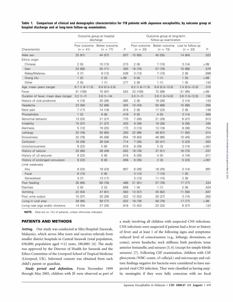
Table 1. Comparison of clinical and demographic characteristics for 118 patients with Japanese encephalitis, by outcome group athospital discharge and at long-term follow-up examination.
Characteristic
Outcome group at hospitaldischarge
P
Outcome group at long-termfollow-up examination
PPoor outcome
(n p 41)Better outcome
(n p 77)Poor outcome
(n p 23)Better outcome
(n p 73)Lost to follow-up
(n p 22)
Male sex 25 (61) 44 (57) .837 15 (65) 40 (55) 14 (64) .523Ethnic origin
Chinese 2 (5) 10 (13) .213 2 (9) 7 (10) 3 (14) 1.99Iban 33 (80) 55 (71) .393 16 (70) 57 (78) 15 (68) .579Malay/Melanau 3 (7) 9 (12) .539 3 (13) 7 (10) 2 (9) .699Orang Ulu 1 (2) 2 (3) 1.99 0 (0) 1 (1) 1 (5) 1.99Other 2 (5) 1 (1) .277 2 (9) 1 (1) 1 (5) .142
Age, mean years (range) 6.7 (1.8–11.5) 6.9 (0.3–2.0) 6.2 (1.8–11.5) 6.9 (0.3–12.0) 7.4 (0.5–12.0) .310Fever 41 (100) 75 (97) .543 23 (100) 72 (99) 21 (95) 1.99Duration of fever, mean days (range) 3.2 (1–7) 3.6 (1–14) 3.0 (1–7) 3.6 (1.0–14.0) 3.6 (1.0–12.0) .158History of viral prodrome 4 (10) 20 (26) .065 2 (9) 19 (26) 3 (14) .143Headache 23 (56) 52 (68) .304 10 (43) 50 (68) 15 (68) .056Neck pain 7 (17) 14 (18) .918 2 (9) 17 (23) 2 (9) .148Photophobia 1 (2) 6 (8) .419 0 (0) 4 (5) 3 (14) .569Abnormal behavior 13 (32) 21 (27) .770 7 (30) 21 (29) 6 (27) .913Irritability 15 (37) 21 (27) .403 9 (39) 19 (26) 8 (36) .346Alertness 5 (12) 19 (25) .173 3 (13) 13 (18) 8 (36) .754Lethargy 32 (78) 50 (65) .283 22 (96) 49 (67) 11 (50) .014Drowsiness 32 (78) 45 (58) .054 19 (83) 48 (66) 10 (45) .202Confusion 16 (39) 26 (34) .714 7 (30) 30 (41) 5 (23) .503Unconsciousness 9 (22) 5 (6) .018 9 (39) 2 (3) 3 (14) !.001History of seizure 33 (80) 38 (49) .002 18 (78) 37 (51) 16 (73) .037History of 13 seizures 9 (22) 5 (6) .018 6 (26) 4 (5) 4 (18) .011History of prolonged convulsion 9 (22) 6 (8) .056 8 (35) 2 (3) 5 (23) !.001Limb weakness
Any 9 (22) 18 (23) .957 6 (26) 18 (25) 3 (14) .891Focal 6 (15) 5 (6) 3 (13) 7 (10) 1 (5)Generalized 3 (7) 13 (17) 3 (13) 11 (15) 2 (9)
Poor feeding 35 (85) 60 (78) .468 21 (91) 57 (78) 17 (77) .224Diarrhea 2 (5) 2 (3) .609 1 (4) 1 (1) 2 (9) .424Vomiting 22 (54) 47 (61) .563 13 (57) 45 (62) 11 (50) .847Poor urine output 15 (37) 20 (26) .322 12 (52) 20 (27) 3 (14) .052Living in rural area 39 (95) 59 (77) .022 18 (78) 58 (79) 17 (77) 1.99Living near pigs and/or chickens 14 (34) 27 (35) .918 12 (52) 23 (32) 6 (27) .122
NOTE. Data are no. (%) of patients, unless otherwise indicated.
PATIENTS AND METHODS
Setting. Our study was conducted at Sibu Hospital (Sarawak,
Malaysia), which serves Sibu town and receives referrals from
smaller district hospitals in Central Sarawak (total population,
650,000; population aged �12 years, 180,000) [4]. The study
was approved by the Director of Health for Sarawak and the
Ethics Committee of the Liverpool School of Tropical Medicine
(Liverpool, UK). Informed consent was obtained from each
child’s parent or guardian.
Study period and definition. From November 1999
through May 2005, children with JE were observed as part of
a study involving all children with suspected CNS infections.
CNS infections were suspected if patients had a fever or history
of fever and at least 1 of the following signs and symptoms:
reduced level of consciousness (e.g., lethargy, drowsiness, or
coma); severe headache; neck stiffness; limb paralysis; tense
anterior fontanelle; and seizures [5, 6] (except for simple febrile
seizures) [7]. Following CSF examination, children with CSF
pleocytosis (WBC count, 15 cells/mL) and microscopy and cul-
ture findings negative for bacteria were considered to have sus-
pected viral CNS infection. They were classified as having asep-
tic meningitis if they were fully conscious with no focal
by guest on May 10, 2011
cid.oxfordjournals.orgD
ownloaded from

460
Tabl
e2.
Com
pari
son
offin
ding
sat
initi
alex
amin
atio
nan
dla
bora
tory
resu
ltsfo
r11
8pa
tient
sw
ithJa
pane
seen
ceph
aliti
s,by
outc
ome
grou
pat
hosp
ital
disc
harg
ean
dat
long
-te
rmfo
llow
-up
exam
inat
ion.
Varia
ble
Out
com
egr
oup
atho
spita
ldis
char
ge
P
Out
com
egr
oup
atlo
ng-te
rmfo
llow
-up
exam
inat
ion
PP
oor
outc
ome
(np
41)
Bet
ter
outc
ome
(np
77)
Poo
rou
tcom
e(n
p23
)B
ette
rou
tcom
e(n
p73
)Lo
stto
follo
w-u
p(n
p22
)
Exa
min
atio
nfin
ding
Illap
pear
ance
38(9
3)57
(74)
.028
22(9
6)58
(79)
15(6
8).1
07D
ehyd
ratio
n21
(51)
31(4
0).3
4414
(61)
29(4
0)9
(41)
.124
Intu
batio
nat
refe
rrin
gho
spita
l6
(15)
0(0
).0
015
(22)
1(1
)0
(0)
.003
Res
pira
tory
abno
rmal
ity5
(12)
4(5
).2
733
(13)
4(5
)2
(9)
.353
Res
pira
tory
rate
,m
ean
brea
ths
per
min
(ran
ge)
32(2
0–52
)31
(18–
48)
.373
33.9
(20–
52)
30.5
(18–
48)
30.5
(20–
44)
.052
Poo
rpe
rfus
ion
20(4
9)22
(29)
.048
15(6
5)22
(30)
5(2
3).0
06H
eart
rate
,m
ean
beat
spe
rm
in(r
ange
)12
1(7
2–16
8)11
0(7
6–17
0).0
0512
4.4
(92–
170)
110.
4(7
2–14
6)10
8.3
(76–
146)
.003
Nec
kst
iffne
ss29
(71)
48(6
2).4
7815
(65)
51(7
0)11
(50)
.872
Ker
nig’
ssi
gn4
(10)
9(1
2)1.9
92
(9)
9(1
2)2
(9)
1.9
9A
ltere
dse
nsor
ium
38(9
3)51
(66)
.003
22(9
6)52
(71)
15(6
8).0
32M
odifi
edG
lasg
owco
ma
scor
e,m
edia
nva
lue
(ran
ge)
9(3
–15)
13(3
–15)
!.0
016
(3–1
5)12
(6–1
5)12
.5(7
–15)
!.0
01M
odifi
edG
lasg
owco
ma
scor
e�
817
(41)
12(1
6).0
0414
(61)
11(1
5)4
(18)
!.0
01S
eizu
res
witn
esse
dat
adm
issi
on19
(46)
8(1
0)!.0
0116
(70)
9(1
2)2
(9)
!.0
01A
bnor
mal
mus
cle
tone
Ove
rall
31(7
6)30
(39)
!.0
0122
(96)
31(4
2)8
(36)
!.0
01H
yper
toni
a23
(56)
20(2
6)15
(65)
20(2
7)8
(36)
Hyp
oton
ia8
(20)
10(1
3)7
(30)
11(1
5)0
(0)
by guest on May 10, 2011
cid.oxfordjournals.orgD
ownloaded from

461
Abn
orm
allim
bre
flexe
sO
vera
ll33
(80)
32(4
2)!.0
0122
(96)
32(4
4)11
(50)
!.0
01H
yper
refle
xia
25(6
1)28
(36)
16(7
0)20
(27)
7(3
2)H
ypofl
exia
orar
eflex
ia8
(20)
14(1
8)6
(26)
12(1
6)4
(18)
Abn
orm
alpo
stur
eO
vera
ll11
(27)
7(9
).0
229
(39)
5(7
)4
(18)
!.0
01D
ecor
ticat
ion
8(2
0)4
(5)
3(1
3)1
(1)
1(5
)D
ecer
ebra
tion
3(7
)2
(3)
6(2
6)3
(4)
3(1
4)O
pist
hoto
nus
0(0
)1
(1)
0(0
)1
(1)
0(0
)Li
mb
wea
knes
sO
vera
ll23
(56)
11(1
4).8
9416
(70)
13(1
8)5
(23)
!.0
01Fo
cal
5(1
2)4
(5)
4(1
7)5
(7)
0(0
)G
ener
aliz
ed18
(44)
7(9
)12
(52)
8(1
1)5
(23)
Clo
nus
2(5
)1
(1)
1.9
92
(9)
0(0
)1
(5)
.056
Pre
senc
eof
pyra
mid
alsi
gns
33(8
0)36
(47)
!.0
0122
(96)
36(4
9)11
(50)
!.0
01H
epat
omeg
aly
5(1
2)8
(10)
.765
3(1
3)9
(12)
1(5
)1.9
9La
bora
tory
findi
ngs
Hem
oglo
bin
leve
l,m
ean
g/dL
(ran
ge)
11.2
(8.1
–13.
9)11
.4(7
.9–1
5.4)
.545
10.9
(8.1
–13.
9)11
.2(7
.9–1
3.9)
11.7
(9.5
–15.
4).3
09W
BC
coun
t,m
ean
cells
/mL
(rang
e)14
362
(390
0–38
,900
)16
,241
(230
0–36
,900
).1
9615
,520
(700
0–25
,900
)16
,650
(230
0–38
,900
)14
,109
(390
0–30
,400
).5
61P
late
let
coun
t,m
ean
plat
e-le
ts/m
L27
9950
(113
,000
–465
,000
)29
5,36
1(4
20,0
00–8
63,0
00)
.453
290,
000
(148
000–
4650
00)
297,
652
(420
00–8
6,30
00)
292,
818
(108
000–
5440
00)
.781
Sod
ium
leve
lat
hosp
itala
d-m
issi
on,
mea
nm
mol
/L(r
ange
)13
4(1
24–1
44)
135
(124
–163
).3
0813
4(1
24–1
40)
135
(124
–163
)13
5(1
26–1
45)
.223
Nad
irso
dium
leve
ldur
ing
hosp
italiz
atio
n,m
ean
mm
ol/L
(ran
ge)
130
(117
–144
)13
3(1
09–1
63)
.004
129
(117
–137
)13
3(1
09–1
63)
133
(125
–142
).0
09C
SF
cell
coun
t,m
edia
nce
lls/m
L(r
ange
)18
2(2
–133
3)18
9(0
–151
5).9
0522
(3–1
333)
80(0
–151
5)38
(1–7
10)
.185
CS
Fpr
otei
nle
vel,
mea
ng/
dL(r
ange
)0.
92(0
.12–
3.95
)0.
73(0
.1–6
.1)
.201
0.73
(0.1
2–2.
55)
0.81
(0.1
–6.1
)0.
78(0
.12–
3.95
).6
45C
SF/
seru
mgl
ucos
era
tio,
mea
nva
lue
(ran
ge)
0.66
(0.2
7–1.
09)
0.62
(0.1
2–1.
08)
.360
0.65
(0.3
1–1.
09)
0.65
(0.2
–1.0
4)0.
64(0
.31–
1.08
).9
50
NO
TE
.D
ata
are
no.
(%)
ofpa
tient
s,un
less
othe
rwis
ein
dica
ted.
by guest on May 10, 2011
cid.oxfordjournals.orgD
ownloaded from
462 • CID 2008:47 (15 August) • Ooi et al.
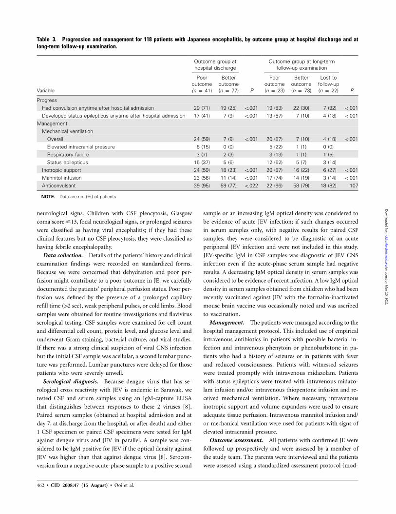
Table 3. Progression and management for 118 patients with Japanese encephalitis, by outcome group at hospital discharge and atlong-term follow-up examination.
Variable
Outcome group athospital discharge
P
Outcome group at long-termfollow-up examination
P
Pooroutcome(n p 41)
Betteroutcome(n p 77)
Pooroutcome(n p 23)
Betteroutcome(n p 73)
Lost tofollow-up(n p 22)
ProgressHad convulsion anytime after hospital admission 29 (71) 19 (25) !.001 19 (83) 22 (30) 7 (32) !.001Developed status epilepticus anytime after hospital admission 17 (41) 7 (9) !.001 13 (57) 7 (10) 4 (18) !.001
ManagementMechanical ventilation
Overall 24 (59) 7 (9) !.001 20 (87) 7 (10) 4 (18) !.001Elevated intracranial pressure 6 (15) 0 (0) 5 (22) 1 (1) 0 (0)Respiratory failure 3 (7) 2 (3) 3 (13) 1 (1) 1 (5)Status epilepticus 15 (37) 5 (6) 12 (52) 5 (7) 3 (14)
Inotropic support 24 (59) 18 (23) !.001 20 (87) 16 (22) 6 (27) !.001Mannitol infusion 23 (56) 11 (14) !.001 17 (74) 14 (19) 3 (14) !.001Anticonvulsant 39 (95) 59 (77) !.022 22 (96) 58 (79) 18 (82) .107
NOTE. Data are no. (%) of patients.
neurological signs. Children with CSF pleocytosis, Glasgow
coma score �13, focal neurological signs, or prolonged seizures
were classified as having viral encephalitis; if they had these
clinical features but no CSF pleocytosis, they were classified as
having febrile encephalopathy.
Data collection. Details of the patients’ history and clinical
examination findings were recorded on standardized forms.
Because we were concerned that dehydration and poor per-
fusion might contribute to a poor outcome in JE, we carefully
documented the patients’ peripheral perfusion status. Poor per-
fusion was defined by the presence of a prolonged capillary
refill time (12 sec), weak peripheral pulses, or cold limbs. Blood
samples were obtained for routine investigations and flavivirus
serological testing. CSF samples were examined for cell count
and differential cell count, protein level, and glucose level and
underwent Gram staining, bacterial culture, and viral studies.
If there was a strong clinical suspicion of viral CNS infection
but the initial CSF sample was acellular, a second lumbar punc-
ture was performed. Lumbar punctures were delayed for those
patients who were severely unwell.
Serological diagnosis. Because dengue virus that has se-
rological cross reactivity with JEV is endemic in Sarawak, we
tested CSF and serum samples using an IgM-capture ELISA
that distinguishes between responses to these 2 viruses [8].
Paired serum samples (obtained at hospital admission and at
day 7, at discharge from the hospital, or after death) and either
1 CSF specimen or paired CSF specimens were tested for IgM
against dengue virus and JEV in parallel. A sample was con-
sidered to be IgM positive for JEV if the optical density against
JEV was higher than that against dengue virus [8]. Serocon-
version from a negative acute-phase sample to a positive second
sample or an increasing IgM optical density was considered to
be evidence of acute JEV infection; if such changes occurred
in serum samples only, with negative results for paired CSF
samples, they were considered to be diagnostic of an acute
peripheral JEV infection and were not included in this study.
JEV-specific IgM in CSF samples was diagnostic of JEV CNS
infection even if the acute-phase serum sample had negative
results. A decreasing IgM optical density in serum samples was
considered to be evidence of recent infection. A low IgM optical
density in serum samples obtained from children who had been
recently vaccinated against JEV with the formalin-inactivated
mouse brain vaccine was occasionally noted and was ascribed
to vaccination.
Management. The patients were managed according to the
hospital management protocol. This included use of empirical
intravenous antibiotics in patients with possible bacterial in-
fection and intravenous phenytoin or phenobarbitone in pa-
tients who had a history of seizures or in patients with fever
and reduced consciousness. Patients with witnessed seizures
were treated promptly with intravenous midazolam. Patients
with status epilepticus were treated with intravenous midazo-
lam infusion and/or intravenous thiopentone infusion and re-
ceived mechanical ventilation. Where necessary, intravenous
inotropic support and volume expanders were used to ensure
adequate tissue perfusion. Intravenous mannitol infusion and/
or mechanical ventilation were used for patients with signs of
elevated intracranial pressure.
Outcome assessment. All patients with confirmed JE were
followed up prospectively and were assessed by a member of
the study team. The parents were interviewed and the patients
were assessed using a standardized assessment protocol (mod-
by guest on May 10, 2011
cid.oxfordjournals.orgD
ownloaded from

Japanese Encephalitis in Malaysia • CID 2008:47 (15 August) • 463
Figure 1. The outcome grading of 118 children with Japanese encephalitis at hospital discharge and at long-term follow-up evaluation. A total of22 patients were lost to follow-up.
ified from one developed earlier) before hospital discharge, 3–
6 months after hospital discharge, and every 3–12 months
thereafter, depending on the patients’ conditions and their ac-
cessibility to the hospital. The outcome of JEV infection was
graded with a functional outcome score, as follows: I, death;
II, severe sequelae greatly impairing function and incompatible
with independent living; III, moderate sequelae mildly affecting
function (including seizures) but compatible with independent
living; IV, minor sequelae including altered personality or clin-
ical signs not affecting functions; and V, full recovery and nor-
mal neurological examination findings [5].
In addition to the prospective study of patients with acute
JE, which began in November 1999, we also studied, at follow-
up, patients with JE who had been admitted to our hospital
previously (from February 1997, when diagnostic testing was
first instituted, through October 1999). Patients who had been
lost to follow-up were contacted again. Medical records were
reviewed and clinical data were extracted using a standardized
format. The clinical and laboratory diagnostic methods for pa-
tients admitted to the hospital from February 1997 through
October 1999 were similar to those for patients admitted to
the hospital in November 1999 and after. In October 2006, we
again contacted all patients who had missed follow-up ap-
pointments for a final assessment.
Statistical analysis. For the purpose of the analysis, the
grading of the final outcome was defined as that at the most
recent follow-up assessment. Patients with grade I (death) or
grade II (severe sequelae) were considered to have a poor out-
come, whereas patients with grades III, IV, and V (moderate,
mild, or no sequelae) were considered to have a better outcome
[5, 9]. Normally distributed data were compared using Stu-
dent’s t test; data that were not normally distributed were com-
pared by the Mann-Whitney U test (Staview 4.02; Abacus Con-
cepts). Differences between proportions were tested using the
x2 test with Yates’ correction or Fisher’s exact test (Epi-Info
2002; Centers for Disease Control and Prevention). Because we
were performing multiple comparisons to look for possible
parameters associated with a poor outcome, we considered
to indicate a trend and to be statistically sig-P ! .05 P ! .01
nificant in univariate analysis. Variables that were associated
with a poor outcome in univariate analyses were examined in
a stepwise logistic regression (SPSS, version 13; SPSS).
RESULTS
Epidemiology. Approximately 900 patients with suspected
CNS infection were admitted to the hospital over an 8.3-year
period. Of these patients, 118 (69 [58%] of whom were male)
had confirmed JEV neurological infection with JEV IgM found
in CSF samples; 102 of 111 patients had JEV IgM found in
serum samples. The annual incidence of JE in central Sarawak
was estimated to be 7.9 cases per 100,000 children aged �12
years. Of the 118 patients, 75 (64%) were observed prospec-
tively from their initial hospital admission (1999–2005 cohort);
for 43 patients, clinical features present at the initial hospital
admission were assessed retrospectively from hospital notes
(1997–1999 cohort). There was no significant difference be-
tween the 2 cohorts with respect to clinical features at hospital
admission or discharge or with respect to length of follow-up
(data not shown). All patients participated in the study.
The majority (73) of the patients were 5–10 years of age
(tables 1–3). Two patients were !12 months of age; both of
these patients presented with febrile seizures (at 3.4 months
and 6 months of age). Acute encephalitis was the most common
by guest on May 10, 2011
cid.oxfordjournals.orgD
ownloaded from

464 • CID 2008:47 (15 August) • Ooi et al.
Table 4. Outcome at hospital discharge for 118 patients with Japanese encephalitis.
Outcome grade No. (%) of patients
Death 10 (8)Severe sequelaea
Overall 33 (28)Severe cognitive impairment with spastic quadriparesis 14Severe cognitive impairment with no gross motor impairment 2Mutism with quadriparesisb 9Mutism 4Mutism and hemiplegia 1Mentally normal but bed-bound because of quadriparesis 2Isolated swallowing difficulty requiring nasogastric tube feeding 1
Moderate sequelaeOverall 28 (24)Paraplegia/diplegia (spastic) 1Quadriparesis with mild cognitive impairment 1Quadriparesis, walks with help 2Quadriparesis, walks alone 1Hemiparesis, walks with help 1Hemiparesis, walks alone 1Monoplegia (splastic, upper limb) 1Reduced speech, walks with help 2Reduced speech, walks alone 4Reduced speech, labile emotion 1Reduced speech and ataxic gait 2Ataxia 4Generalized weakness and requiring help to walk 6Seventh nerve upper neuron palsy 1
Mild sequelaeOverall 3 (3)Blunted affect 1Upper neuron signs ( hyperreflexia) 1Parkinsonian features (cogwheel rigidity) 1
Full recovery 44 (37)
a A total of 20 of 33 patients with severe sequelae required nasogastric tube feeding.b One patient had parkinsonian sequelae, characterized by cogwheel rigidity, mask-like facies, and
hand tremor.
presentation (97 patients; 82%), followed by febrile encepha-
lopathy (10 patients; 8%), aseptic meningitis (8 patients; 7%),
and febrile seizures (3 patients; 3%). The acute case–fatality
rate was 8% (10 of 118 patients). Long-term outcome data
were available for 96 (81%) of the 118 patients (59 of whom
were from the 1999–2005 cohort); this included 10 patients
who died. The median number of follow-up visits per patient
throughout the study was 2 (range, 1–6 visits). The median
duration of follow-up was 52.9 months (range, 0.9–114.9
months). The final outcome of the remaining 22 patients (16
of whom were from the 1999–2005 cohort) was uncertain,
because they were lost to follow-up. However, those patients
who were lost to follow-up were similar to those patients who
were followed-up; 61 (64%) of 96 patients who were followed
up had a good outcome at hospital discharge, compared with
16 (73%) of 22 patients who were not followed up ( ).P p .57
Comparison of outcome at hospital discharge and long-term
follow-up. Figure 1 shows the distribution of the outcome
grading of all patients at hospital discharge and at long-term
follow-up visits. At hospital discharge, 44 (37%) of the 118
patients had apparently made a full recovery, and 3 (3%) had
mild, 28 (24%) had moderate, and 33 (28%) had severe neu-
rological sequelae (table 4). Of the 86 patients who had follow-
up assessment, 36 (42%) had full recovery, and 48 (56%) had
neurological sequelae (17 [20%] had mild, 20 [23%] had mod-
erate, and 11 [13%] had severe neurological sequelae at their
last assessment) (table 5).
Figure 2 shows the changes in outcome grading at follow-
by guest on May 10, 2011
cid.oxfordjournals.orgD
ownloaded from

Japanese Encephalitis in Malaysia • CID 2008:47 (15 August) • 465
Table 5. Long-term outcome for 86 patients with Japanese encephalitis.
Outcome grade No. (%) of patients
Death 2 (2)Severe sequelae
Overall 11 (13)Severe cognitive impairment with spastic quadriparesis 9Severe cognitive impairment, microcephaly, and marked hyperactivity 1Mentally normal but bed-bound because of severe diplegia 1
Moderate sequelaeOverall 20 (23)Mild cognitive impairment with altered personality 4Mild cognitive impairment with hand tremor 2Mild cognitive impairment, altered personality with psychobehavioral problem 2Mild cognitive impairment, dysarthria, and right hand tremor 1Mild cognitive impairment, altered personality, and hemiparesis 1Altered personality with lower limb wasting 1Emiparesis, isolated 3Emiparesis, with dystonia 1Monoplegia, upper limb 1Monoplegia, lower limb 1Monoplegia, upper limb and dystonia 1Facial twitch and dysphonia 1Seizure, frontal release signs, and brisk reflexes 1
Mild sequelaeOverall 17 (20)Upper motor neuron signsa 5Mild cerebellar signsb 2Frontal release signs 1Upper motor neuron signs and cerebellar signsa 1Upper motor neuron signs, cerebellar signs, and frontal release signsa 1Altered personality 4Blunted affect 1Hand tremor 1Mild hyperactivity 1
Full recovery 36 (42)
a Upper motor neuron signs include extensor plantar, clonus, and hyperreflexia.b Mild cerebellar signs include nystagmus, intentional tremor, and dysdiadochokinesis.
up for different patient groups. A total of 27 (31%) of the 86
patients who returned for follow-up examination had severe
sequelae at hospital discharge; of these 27 patients, 6 (22%)
had full recovery, 1 (4%) had mild sequelae, 8 (30%) had
moderate sequelae, 10 (37%) had severe sequelae, and 2 (7%)
died 12 and 72 months after discharge. A 3-year-old boy who
had presented with encephalitis and status epilepticus appeared
to have made a full recovery before hospital discharge; however,
73 months later, he had microcephaly and hyperactive behavior
that required constant adult supervision, and therefore, he was
classified as having severe sequelae. Four patients had moderate
sequelae on follow-up examination; of these, 2 (who had pre-
sented with encephalitis) had mild cognitive impairment and
altered personality, 1 (who had presented with febrile seizures)
had isolated mild cognitive impairment, and 1 (who had pre-
sented with encephalitis) had epilepsy, brisk reflexes, and frontal
release signs. Eight patients (4 of whom had initially presented
with encephalitis, 3 with aseptic meningitis, and 1 with febrile
encephalopathy) had mild sequelae on follow-up with subtle
neurological deficits such as upper neuron signs, frontal release
signs, or hand tremor. The 8 patients with moderate sequelae
at hospital discharge (6 of whom had presented with enceph-
alitis and 2 of whom had presented with febrile encephalop-
athy) recovered fully. Three had been ataxic at hospital dis-
charge, 2 had been quiet and withdrawn, and 3 had required
assistance for walking because of lower limb weakness, quad-
riparesis, or generalized body weakness (1 patient each). The
6 patients with full recovery from severe sequelae at hospital
by guest on May 10, 2011
cid.oxfordjournals.orgD
ownloaded from

466 • CID 2008:47 (15 August) • Ooi et al.
Figure 2. A comparison of patient outcome grading at hospital dis-charge with outcome at long-term follow-up evaluations for 86 childrenwith Japanese encephalitis.
Table 6. Multiple logistic regression analysis of the factors associated with poor outcome in Jap-anese encephalitis.
VariableAcute phase, adjusted
OR (95% CI) P
Long-term follow-upexamination, adjusted
OR (95% CI) P
Poor perfusion … 8.53 (1.88– 38.79) .006Glasgow coma score �8 … 5.17 (1.10–24.24) .037�2 Witnessed convulsions 7.6 (3.1–18.5) !.001 15.64 (3.50–69.81) !.001Nadir sodium level �135 mmol/L 4.0 (1.3–12.3) .016 …
NOTE. Parameters entered into the model included history of unconsciousness, poor perfusion, Glasgow coma score�8 and presence of pyramidal signs on initial examination, �2 witnessed seizures, and nadir serum sodium level �135mmol/L. Terms were entered into the model only if they were statistically associated with poor outcome ( ). BothP ! .05forward selection and backward elimination methods were used. Forward selection and backward elimination proceduresgenerated the same model, indicating its robustness. The Hosmer-Lemeshow statistics indicated a nonsignificance oflack of fit ( ; ). Repeating the analysis by including the duration of follow-up in the model did not alter2x p 2.840 P p .725the independent risk factors associated with poor final outcome. A similar approach was taken to look for parameterspredictive of poor outcome at hospital discharge and found that �2 witnessed seizures and nadir serum sodium level�135 mmol/L were independent factors found to be predictive of a poor outcome at hospital discharge.
discharge included 5 who had been bed-bound with quadri-
paresis and 1 patient with impaired cognitive function who
required nasogastric feeding. Among these patients, 4 were also
aphasic, and 1 had Parkinsonian sequelae.
Fifteen (65%) of 23 patients with moderate sequelae at hos-
pital discharge who were followed up had improved signifi-
cantly; 8 had full recovery, and 7 had mild sequelae. In contrast,
13 (38%) of 34 patients who had apparent full recovery at
hospital discharge were found to have varying degrees of neu-
rological sequelae at long-term follow-up visits.
Although most patients improved after hospital discharge,
the degree of improvement was not necessarily sufficient to
effect a revision in the outcome grading. For example, 10 (30%)
of 33 patients with severe sequelae at hospital discharge had
better cognitive function, improved limb function, or the ability
to feed normally by the time of follow-up, but their grading
remained unchanged, because they remained dependent as the
result of a severe spastic quadriplegia.
Of 37 patients who later had mild and moderate sequelae,
14 (38%) had altered personality after JE. Their parents re-
ported poor temperament, impulsiveness, aggressive behavior,
loss of enthusiasm and motivation, and becoming quieter and
less communicative. In addition, 11 (30%) of the parents re-
ported that their children experienced impaired memory, par-
ticularly short-term memory.
Of the 86 patients who had follow-up examination, 48 pa-
tients (56%) were school children when infected with JEV. Six
(13%) of these 48 children had never returned to school because
of severe physical disabilities; 16 (38%) of the remaining 42
had marked deterioration in school performance, resulting in
6 of them having to stop schooling; and only 20 (48%) of the
42 children performed satisfactorily. Details about schooling
were available for 30 of the 38 patients who had JE at a pre-
school age (i.e., !7 years of age). Five children had never at-
tended school because of severe disability; 5 did poorly in
school, resulting in 2 of them having to stop schooling. The
remaining 20 children performed satisfactory. Epilepsy was
found in 7 (8%) of 86 patients at follow-up examinations.
Comparison of outcome grading at early follow-up and late
follow-up examinations. Of the 86 patients who had follow-
up, 24 (28%) had both an early follow-up assessment (within
the first 3–6 months after hospital discharge) and a late follow-
up assessment (16 months after hospital discharge). Eighteen
(75%) of these 24 patients had identical grading in their early
and late follow-up assessments. For the remaining 6 patients
(25%), there was a change in the grading for the late follow-
up assessments (improvement in 4 patients and deterioration
in 2 patients).
Predictors of poor outcome at hospital discharge and at
long-term follow-up examinations. In tables 1–3, the clinical
features of the patients who were classified as having poor
outcome at hospital discharge are compared with the clinical
by guest on May 10, 2011
cid.oxfordjournals.orgD
ownloaded from

Japanese Encephalitis in Malaysia • CID 2008:47 (15 August) • 467
features of patients with better outcome. Patients with a poor
outcome at hospital discharge were more likely than others to
have seizures before hospital admission, to have seizures wit-
nessed at admission, to have a faster heart rate, to have required
intubation at referring hospitals, to have reduced Glasgow coma
score, to have lower median Glasgow coma score, and to have
abnormal motor signs (abnormal muscle tone, abnormal limb
reflexes, and limb weakness).
Tables 1–3 also show how the presenting features and pro-
gress were related to the patients’ outcome at the final follow-
up examination. Many of the indicators for a poor prognosis
at hospital discharge were also indicators for a poor outcome
at long-term follow-up examination. These included deep coma
score, witnessed seizures at admission, continued seizures after
hospitalization, status epilepticus, abnormal muscle tone and
reflexes, hyponatremia, and the need for mechanical ventilation
and inotropic drugs. Abnormal body posture and limb weak-
ness at presentation that were not associated with poor outcome
at hospital discharge became important indicators for poor
long-term outcome. There was no difference in the median
length of follow-up between patients with a poor outcome and
those with a better outcome at long-term follow-up exami-
nation (median duration, 51.7 months [range, 6.1–84.3
months] vs. 54.0 months [range, 0.9–114.9 months]; P p
)..682
To find the best prediction of poor outcome at long-term
follow-up examination, parameters significant in univariate
analysis were entered into a multiple logistic regression model.
A combination of poor perfusion, Glasgow coma score �8,
and �2 episodes of witnessed seizures gave the best prediction
of poor outcome, with 65% sensitivity and 92% specificity
(table 6). The presence of these 3 characteristics had a positive
predictive value of 71% and a negative predictive value of 89%
for poor outcome.
DISCUSSION
Most studies of JE report the survivors’ outcome for periods
of 6 weeks to 6 months after hospital discharge [9–15]. To our
knowledge, only 3 studies have followed up survivors for 11
year, including a study involving 39 patients who were followed
up for up to 421 days [16], a study in which 55 patients were
followed up for up to 2 years [17], and a study in which 78
patients were followed up for 6–27 years [18]. The paucity of
long-term outcome data is partly related to the difficulty of
following up JE survivors, because most patients live in rural
areas with limited access to health care facilities [16]. Most
studies that examined prognostic indices related them to out-
come at hospital discharge, rather than to long-term outcome,
although the latter is more important.
Our study provides further insight into the long-term prog-
nosis of JE survivors. Most patients experienced improvement
after hospital discharge; some experienced remarkable recovery,
and others did not recover sufficiently to become independent.
More than one-half of the survivors continue to experience a
spectrum of neuropsychological and behavioral disorders. Our
findings are similar to those of Baruah et al. [16], who reported
improvement among survivors up to 330 days after hospital
discharge; however, many of our cohort who were apparently
healthy at hospital discharge later experienced other difficulties
(in particular, impairment of memory and changes in behavior
and personality). Similar late changes were also seen in patients
infected with West Nile virus, a closely related flavivirus [19,
20]. Marked deterioration in school performance and loss of
enthusiasm and motivation were also common among JE sur-
vivors. Our results show that neurological assessment of JE
survivors at hospital discharge does not entirely predict their
long-term outcome, but assessment at 3–6 months after hos-
pital discharge is predictive of long-term outcome in three-
quarters of patients. Neurological deterioration can occur sev-
eral years after the initial infection, so survivors should have
regular long-term follow-up evaluations to identify and manage
any disability in a timely fashion.
A range of clinical features has been associated with a poor
outcome of JE at hospital discharge, including seizures, elevated
intracranial pressure, deep coma, abnormal muscle tone and
posture, and hyponatremia [5, 10, 21, 22]. Our study has con-
firmed most of these and has shown that seizures and reduced
consciousness are also good predictors of long-term outcome.
We also showed that features of shock (tachycardia and poor
perfusion) were associated with poor outcome, which has not,
to our knowledge, been reported previously in JE. Adequate
cerebral perfusion pressure is vital in preventing secondary ce-
rebral ischemia after a primary brain injury, and it is of greater
importance than elevated intracranial pressure in determining
the eventual outcome of children with nontraumatic coma [23].
Shock may develop because of marked dehydration before a
child is hospitalized; but it can be compounded by some cli-
nicians’ fear that administering intravenous rehydration may
exacerbate elevated intracranial pressure. Fluid management in
patients with shock and acute brain injury is difficult, but our
data and clinical experience suggest that adequate rehydration
is vital.
Most of our patients were Ibans, who cultivate rice and com-
monly keep pigs close to their rural long houses. However,
115% of our patients originated from urban areas, possibly
because, in Sarawak, rice fields and pigs are commonly found
at the edges of towns. Although most patients in this study
presented with encephalitis or febrile encephalopathy (as has
been reported by many others previously), 8 (7%) of our pa-
tients had aseptic meningitis, and 3 (3%) had febrile seizures.
Interestingly, we did not have patients who presented with a
purely paralytic syndrome, although some encephalopathic pa-
by guest on May 10, 2011
cid.oxfordjournals.orgD
ownloaded from

468 • CID 2008:47 (15 August) • Ooi et al.
tients had paralysis. Our mortality rate was low (8%, compared
with the usual mortality rate of 20%–30%), possibly because
of our modern intensive care unit.
In conclusion, we have shown that, although the clinical
condition of many JE survivors’ improves after hospital dis-
charge, less than one-half make a full recovery, and approxi-
mately one-fifth experienced subsequent deterioration. Most
changes in outcome occur within the first 3–6 months after
hospital discharge, but some changes occur even after 2 years.
We have confirmed the importance of seizures as a sign of poor
prognosis, both for outcome at hospital discharge and long-
term outcome. Our data suggest that clinical signs of shock are
also associated with a poor outcome. Both of these factors are
potentially amenable to treatment.
Acknowledgments
We thank the Former State Health Director of Sarawak, Tan Sri Datu,Dr. Taha Mohamad Arif, for his approval and support for this work duringhis tenure as Director of Sarawak Health Department. We are grateful toDr. K. Krishnan; Dr. Hj Faizul Hj Mansoor; Matron Margeret Wong andher team at Sibu Divisional Health Department; Sibu Hospital DirectorDr. Abdul Rahim Abdullah; the doctors and nurses of pediatric wards andclinics; Peng Chin Pek, Guloi Selingau, and the medical records officersof Sibu Hospital for administrative, clinical, and laboratory assistance; andDavid Chadwick and C. Anthony Hart, for their support.
Financial support. The Ministry of Science, Technology and Innova-tion (NBD06-05-01-T001), operational funds of Sarawak Health Depart-ment, the Walton Centre for Neurology and Neurosurgery Research Fund,Program for Appropriate Technology in Health (PATH; Seattle, WA), Well-come Trust Clinical Training Fellowship (to M.H.O.), and a United King-dom Medical Research Council Senior Clinical Fellowship (to T.S.). Alldiagnostic reagents were provided free of charge by Venture Technology.
Potential conflicts of interest. M.J.C. is involved in the developmentof a Japanese encephalitis vaccine with Bavarian Nordic. All other authors:no conflicts.
References
1. Tsai T, Chang G, Yu Y. Japanese encephalitis vaccines. In: Plotkin S,Orenstein W, eds. Vaccine. 3rd ed. Philadephia: W.B. Saunders Com-pany, 1999:672–710.
2. Solomon T, Dung NM, Kneen R, et al. Japanese encephalitis. J NeurolNeurosurg Psychiatr 2000; 68:405.
3. Vaughn DW, Hoke CH Jr. The epidemiology of Japanese encephalitis:prospects for prevention. Epidemiol Rev 1992; 14:197–221.
4. Department of Statistics, The Government of Malaysia. Population andvital statistics. Yearbook of statistics, Sarawak, 2006.
5. Solomon T, Dung NM, Kneen R, et al. Seizures and raised intracranialpressure in Vietnamese patients with Japanese encephalitis. Brain2002; 125:1084–93.
6. Solomon T, Kneen R, Dung N, et al. Poliomyelitis-like illness due toJapanese encephalitis virus. Lancet 1998; 351:1094–7.
7. Verity CM, Butler NR, Golding J. Febrile convulsions in a nationalcohort followed up from birth. I—prevalence and recurrence in thefirst five years of life. BMJ 1985; 290:1307–10.
8. Cardosa MJ, Wang SM, Sum MSH, Tio PH. Antibodies against prMprotein distinguish between previous infection with dengue and Jap-anese encephalitis viruses. BMC Microbiol 2002; 2:9.
9. Solomon T, Dung NM, Wills B, et al. Interferon alfa-2a in Japaneseencephalitis: a randomised double-blind placebo-controlled trial. Lan-cet 2003; 361:821–6.
10. Rayamajhi A, Singh R, Prasad R, Khanal B, Singhi S. Clinico-laboratoryprofile and outcome of Japanese encephalitis in Nepali children. AnnTrop Paediatr 2006; 26:293–301.
11. Kalita J, Misra UK, Pandey S, Dhole TN. A comparison of clinical andradiological findings in adults and children with Japanese encephalitis.Arch Neurol 2003; 60:1760–4.
12. Kalita J, Misra UK. Neurophysiological changes in Japanese enceph-alitis. Neurology India 2002; 50:262–6.
13. Misra UK, Kalita J, Srivastava M. Prognosis of Japanese encephalitis:a multivariate analysis. J Neurol Sci 1998; 161:143–7.
14. Luo D, Song J, Ying H, Yao R, Wang Z. Prognostic factors of earlysequelae and fatal outcome of Japanese encephalitis. Southeast AsianJ Trop Med Public Health 1995; 26:694–8.
15. Huy B, Tu H, Luan T, Lindqvist R. Early mental and neurologicalsequelae after Japanese B encephalitis. Southeast Asian J Trop MedPublic Health 1994; 25:549–53.
16. Baruah HC, Biswas D, Patgiri D, Mahanta J. Clinical outcome andneurological sequelae in serologically confirmed cases of Japanese en-cephalitis patients in Assam, India. Indian Pediatrics 2002; 39:1143–8.
17. Kumar R, Mathur A, Kumar A, Sharma S, Chakraborty S, ChaturvediU. Clinical features and prognostic indicators of Japanese encephalitisin children in Lucknow (India). Indian J Med Res 1990; 91:321–7.
18. Ding D, Hong Z, Zhao S-J, et al. Long-term disability from acutechildhood Japanese encephalitis in Shanghai, China. Am J Trop MedHyg 2007; 77:528–33.
19. Sejvar J. Emerging infections: the long-term outcomes of human WestNile virus infection. Clin Infect Dis 2007; 44:1617–24.
20. Solomon T, Ooi MH, Beasley DWC, Mallewa M. West Nile encephalitis.BMJ 2003; 326:865–9.
21. Tiroumourougane SV, Raghava P, Srinivasan S, Badrinath A. Manage-ment parameters affecting the outcome of Japanese encephalitis. J TropPediatr 2003; 49:153–6.
22. Libraty D, Nisalak A, Endy T, Suntayakorn S, Vaughn D, Innis B.Clinical and immunological risk factors for severe disease in Japaneseencephalitis. Trans R Soc Trop Med Hyg 2002; 96:173–8.
23. Kirkham FJ. Non-traumatic coma in children. Arch Dis Child 2001;85:303–12.
by guest on May 10, 2011
cid.oxfordjournals.orgD
ownloaded from