The effects of burial on drug detection in skeletal tissues
11
346 Research Article Drug Testing and Analysis Received: 28 April 2010 Revised: 31 May 2010 Accepted: 2 June 2010 Published online in Wiley Interscience: 4 August 2010 (www.drugtestinganalysis.com) DOI 10.1002/dta.144 The effects of burial on drug detection in skeletal tissues 1 Nathalie A. Desrosiers and James H. Watterson ∗ Skeletal tissues have recently been investigated for use in post-mortem toxicology. Variables affecting drug concentration in these tissues, however, are still poorly characterized. In this work, the relative effects of burial on the response of enzyme-linked immunosorbent assay (ELISA) and gas chromatography-mass spectrometry (GC-MS) assays were examined. Rats were acutely exposed to ketamine or diazepam, euthanized and buried outdoors. After one month, the remains were exhumed and skeletal tissue drug levels were compared those of non-buried rats. A climate-controlled burial was also undertaken using defleshed bones to approximate an extended decomposition. Long bones (femora, tibiae) were isolated and separated into tissue type (diaphyseal bone, epiphyseal bone, and marrow), and according to treatment (i.e. buried or non-buried). Following methanolic extraction (bone) or simple homogenization (marrow), samples were analyzed with ELISA. Samples were then pooled according to treatment, extracted by solid phase extraction (SPE) and confirmed with GC-MS. Under the conditions examined, the effects of burial appear to be drug and tissue dependent. Ketamine-exposed tissues demonstrated the greatest differences, especially in bone marrow. In diazepam-exposed tissues, burial did not seem to greatly affect drug response and some gave greater assay response compared to the non-buried set. Overall, the data suggest that fresh tissue samples may not be representative of decomposed samples in terms of skeletal tissue drug levels. Copyright c 2010 John Wiley & Sons, Ltd. Keywords: forensic science; toxicology; bone; marrow; burial; ketamine; diazepam; immunoassay; gas chromatography-mass spectrometry Introduction Typically, blood is the tissue of choice in post-mortem drug analysis because of the potential for correlation of drug concentrations with intoxication and fatal toxicity. Nevertheless, alternative matrices such as urine, bile, liver, hair, and muscle have been used to indicate exposure to drugs of toxicological relevance. Bone and bone marrow, on the other hand, are seldom used for toxicological analysis. However, they may be the only tissues left after a significant period of decomposition. Thus, it is important to examine the utility of these tissues to provide valuable information in post-mortem toxicology. Although a growing number of reports are being published on the detection of drugs in skeletal tissues, [1–6] the effects of various post-mortem environments on drug concentration in skeletal tissues are still poorly understood. A handful of case studies have been presented showing the presence of drugs in skeletal tissues, most of which involve skeletonized remains and have been used to confirm the victim’s identity or to cor- roborate a suspect’s story. Amitriptyline, [7] acetaminophen and dextropropoxyphene, [8] triazolam, [9] bromisovalum, [10] metham- phetamine and amphetamine [11] have been detected in bone mar- row whereas aminopyrine and cyclobarbital, [12] nortriptyline, [13] and citalopram [14] have been detected in bone. These studies, however, either made use of non-decomposed tissues, or report only a particular subset of post-mortem conditions. Thus, fur- ther characterization of the implications and limitations of drug detection in skeletal tissues was warranted. Only a small number of studies have examined the variability in assay response to bone tissue extracts after exposure to different environmental conditions. Gorczynski and Melbye examined midazolam levels in murine bone after burial in sterile and non-sterile soil and found that drug concentrations were greatest in non-buried samples, followed by samples buried in sterilized or non-sterile soil. [15] While midazolam was detected in the bone of the samples buried in the non-sterile soil, no midazolam was detected in the bone marrow of these same samples. Methamphetamine and amphetamine were measured in the marrow of rabbit bones submerged in tap water as well as from air-dried bone fragments. [16] Although amphetamine could not be measured because of interfering peaks on the gas chromatogram, methamphetamine concentrations remained constant in air-dried samples but varied in submerged samples, with no distinctive pattern over the 24-month period. Cengiz et al. [17] showed that morphine concentration in rabbit bone marrow decreased over time for buried samples compared to those collected immediately after death. Guillot et al. also examined bone marrow morphine and 6-acetylmorphine (6-AM) concentrations for acute and chronic injection of heroin. [18] After storing the bones in a jar on soil from a nearby forest for a period of two months, they saw that only 6-AM remained in the bone marrow. After the two-month storage ∗ Correspondence to: James H. Watterson, Forensic Toxicology Research Labo- ratory, Laurentian University, 935 Ramsey Lake Rd, Sudbury ON, Canada P3E 2C6. E-mail: [email protected] 1 Financial support was provided by the Natural Sciences and Engineering Research Council (NSERC). The authors presented preliminary aspects of this work at the 61 st Annual Meeting of the American Academy of Forensic Sciences, Denver, CO, February 16–20, 2009, as a poster presentation for the Young Forensic Scientists’ Forum. Forensic Toxicology Research Laboratory, Laurentian University, 935 Ramsey Lake Rd, Sudbury ON, Canada P3E 2C6 Drug Test. Analysis 2010, 2, 346–356 Copyright c 2010 John Wiley & Sons, Ltd.
-
Upload
laurentian -
Category
Documents
-
view
2 -
download
0
Transcript of The effects of burial on drug detection in skeletal tissues

34
6
Research ArticleDrug Testingand Analysis
Received: 28 April 2010 Revised: 31 May 2010 Accepted: 2 June 2010 Published online in Wiley Interscience: 4 August 2010
(www.drugtestinganalysis.com) DOI 10.1002/dta.144
The effects of burial on drug detectionin skeletal tissues1
Nathalie A. Desrosiers and James H. Watterson∗
Skeletal tissues have recently been investigated for use in post-mortem toxicology. Variables affecting drug concentration inthese tissues, however, are still poorly characterized. In this work, the relative effects of burial on the response of enzyme-linkedimmunosorbent assay (ELISA) and gas chromatography-mass spectrometry (GC-MS) assays were examined. Rats were acutelyexposed to ketamine or diazepam, euthanized and buried outdoors. After one month, the remains were exhumed and skeletaltissue drug levels were compared those of non-buried rats. A climate-controlled burial was also undertaken using defleshedbones to approximate an extended decomposition. Long bones (femora, tibiae) were isolated and separated into tissue type(diaphyseal bone, epiphyseal bone, and marrow), and according to treatment (i.e. buried or non-buried). Following methanolicextraction (bone) or simple homogenization (marrow), samples were analyzed with ELISA. Samples were then pooled accordingto treatment, extracted by solid phase extraction (SPE) and confirmed with GC-MS.
Under the conditions examined, the effects of burial appear to be drug and tissue dependent. Ketamine-exposed tissuesdemonstrated the greatest differences, especially in bone marrow. In diazepam-exposed tissues, burial did not seem to greatlyaffect drug response and some gave greater assay response compared to the non-buried set. Overall, the data suggest thatfresh tissue samples may not be representative of decomposed samples in terms of skeletal tissue drug levels. Copyright c©2010 John Wiley & Sons, Ltd.
Keywords: forensic science; toxicology; bone; marrow; burial; ketamine; diazepam; immunoassay; gas chromatography-massspectrometry
Introduction
Typically, blood is the tissue of choice in post-mortem drug analysisbecause of the potential for correlation of drug concentrations withintoxication and fatal toxicity. Nevertheless, alternative matricessuch as urine, bile, liver, hair, and muscle have been used toindicate exposure to drugs of toxicological relevance. Bone andbone marrow, on the other hand, are seldom used for toxicologicalanalysis. However, they may be the only tissues left after asignificant period of decomposition. Thus, it is important toexamine the utility of these tissues to provide valuable informationin post-mortem toxicology.
Although a growing number of reports are being publishedon the detection of drugs in skeletal tissues,[1 – 6] the effects ofvarious post-mortem environments on drug concentration inskeletal tissues are still poorly understood. A handful of casestudies have been presented showing the presence of drugsin skeletal tissues, most of which involve skeletonized remainsand have been used to confirm the victim’s identity or to cor-roborate a suspect’s story. Amitriptyline,[7] acetaminophen anddextropropoxyphene,[8] triazolam,[9] bromisovalum,[10] metham-phetamine and amphetamine[11] have been detected in bone mar-row whereas aminopyrine and cyclobarbital,[12] nortriptyline,[13]
and citalopram[14] have been detected in bone. These studies,however, either made use of non-decomposed tissues, or reportonly a particular subset of post-mortem conditions. Thus, fur-ther characterization of the implications and limitations of drugdetection in skeletal tissues was warranted.
Only a small number of studies have examined the variability inassay response to bone tissue extracts after exposure to differentenvironmental conditions. Gorczynski and Melbye examinedmidazolam levels in murine bone after burial in sterile and
non-sterile soil and found that drug concentrations were greatestin non-buried samples, followed by samples buried in sterilized ornon-sterile soil.[15] While midazolam was detected in the boneof the samples buried in the non-sterile soil, no midazolamwas detected in the bone marrow of these same samples.Methamphetamine and amphetamine were measured in themarrow of rabbit bones submerged in tap water as well as fromair-dried bone fragments.[16] Although amphetamine could not bemeasured because of interfering peaks on the gas chromatogram,methamphetamine concentrations remained constant in air-driedsamples but varied in submerged samples, with no distinctivepattern over the 24-month period. Cengiz et al.[17] showed thatmorphine concentration in rabbit bone marrow decreased overtime for buried samples compared to those collected immediatelyafter death. Guillot et al. also examined bone marrow morphineand 6-acetylmorphine (6-AM) concentrations for acute and chronicinjection of heroin.[18] After storing the bones in a jar on soil froma nearby forest for a period of two months, they saw that only6-AM remained in the bone marrow. After the two-month storage
∗ Correspondence to: James H. Watterson, Forensic Toxicology Research Labo-ratory, Laurentian University, 935 Ramsey Lake Rd, Sudbury ON, Canada P3E2C6. E-mail: [email protected]
1 Financial support was provided by the Natural Sciences and EngineeringResearch Council (NSERC). The authors presented preliminary aspects of thiswork at the 61st Annual Meeting of the American Academy of Forensic Sciences,Denver, CO, February 16–20, 2009, as a poster presentation for the YoungForensic Scientists’ Forum.
Forensic Toxicology Research Laboratory, Laurentian University, 935 RamseyLake Rd, Sudbury ON, Canada P3E 2C6
Drug Test. Analysis 2010, 2, 346–356 Copyright c© 2010 John Wiley & Sons, Ltd.
34
7
The effects of burial on drug detection in skeletal tissues
Drug Testingand Analysis
period in forest soil, no drug was detectable in the bone. Finally,one author examined opiate concentrations in bone marrow ofa known heroin addict and found a morphine concentration of195 ng/g for bone marrow collected right after death.[19] Bonesampled at the same time had a morphine concentration of340 ng/g whereas a section of this bone intentionally buried fora year had a morphine concentration of 155 ng/g, representing a54.4% loss. Interestingly, the bone weighed 17% less after burial.
These studies suggest that post-mortem environment is a factorin observed bone drug levels, which may substantially complicateinterpretation of measurements. In order to understand howthis may occur, the processes associated with bone diagenesis,or changes occurring to bone in its post-mortem environment,should be considered. A variety of processes occur in bone duringburial, including uptake of cations and of circulating organics, ionexchange, breakdown and leaching of collagen, microbiologicalattack, alteration and possible leaching of mineral matrix andinfilling with mineral deposits.[20] Collagen content and porosity(which is related water uptake potential) could be postulatedto influence bone drug concentration due to their effect onthe exposed surface area. Collagen loss, which is mainly dueto microbiological attack, contributes to weight loss (20%) andporosity increase (50%).[20]
In this work, a pilot study was undertaken in order to examinewhether burial influences the response of forensic drug screeningimmunoassays and gas chromatography-mass spectrometry (GC-MS) confirmation assays of skeletal tissue extracts. Two drugsof forensic interest (ketamine and diazepam), selected for theirvarying chemical properties, were administered to rats. Drug-exposed and control animals then underwent burial, or were keptfrozen until analysis. An outdoor burial and a climate-controlledindoor burial were established in order to examine differentdegrees of exposure to the environment and to approximatedifferent degrees of decomposition. The goal of the workwas to better understand the changes that occur in skeletaldrug screening results following burial, which may assist ininterpretation of the significance of drug detection in skeletaltissues.
Materials and Methods
Chemicals
Methanol used for drug extraction was high performanceliquid chromatography (HPLC) grade and purchased from EMDChemicals (Gibbstown, NJ, USA). Drug standards (ketamine,chlorpheniramine, diazepam, D5-diazepam, nordiazepam, and D5-nordiazepam), were obtained from Cerilliant Corp. (Round Rock,TX, USA) as 1 mg/ml methanolic solutions and diluted as required.All other chemicals were reagent grade and were obtained fromEMD Chemicals (Gibbstown, NJ, USA).
ELISA Procedure and Validation
Enzyme-linked immunosorbent assay (ELISA) analysis of bonetissue extracts was carried out as previously described[21] and usedketamine and benzodiazepine kits obtained from ImmunalysisCorp. (Pomona, CA, USA). The procedure was carried out on aChemwell 2910 Automated EIA Analyzer (Awareness Technologies,Palm City, FL, USA).
The intra-day and inter-day variation of the ELISA assay wasdetermined for each drug. Standard solutions of ketamine and
diazepam were prepared daily in PB6 (0.1 M phosphate buffer,pH=6) for use in generating standard curves to illustrate theconcentration dependence for each assay. Drug concentrationsin standard solutions were 5, 10, 25, 50 100 or 200 ng/mL forketamine and from 0.5, 1, 2.5, 5, 10 and 25 ng/mL for diazepam.All standard curves included a blank, consisting of only PB6. Intra-day precision was examined by analyzing the concentration ofa standard curve four times in one day. Inter-day precision wasexamined by making a standard curve daily and analyzing theconcentration of solutions prepared, on four different days.
Matrix effects were examined by spiking drug-free skeletal tissueextracts with standard solutions of drugs at the concentrationsused in generation of standard curves. Briefly, bone marrowwas collected and homogenized in 0.5 M NaOH: 0.5M NaClsolution (hereafter named 50 : 50 solution) and then neutralizedwith acetate buffer, pH=4. Bones were cleaned in the 50 : 50solution, rinsed in water, and dried. Bones were then groundin an all-purpose domestic grinder and incubated in methanolat 50 ◦C for 72 h. Methanol was then evaporated and sampleswere reconstituted in PBS (phosphate buffered saline). Standardsolutions of drug were made in the same concentration rangementioned above in the marrow homogenate (approx. 0.1 gmarrow per mL of homogenate) and in bone extracts (approx.0.5 g bone per mL of extract) and analyzed with ELISA to examinethe potential effect that bone marrow and bone matrix could havethe ELISA response.
Animal Drug Administration
All procedures for this work were approved by the LaurentianUniversity Animal Care Committee. Adult male Wistar rats(Charles River Laboratories, Saint-Constant, Quebec, Canada)were generously donated from other researchers at LaurentianUniversity. The rats were housed alone, in groups of two, or ingroups of three, with Harlan Teklad 1/4-inch bedding (Indianapolis,IN, USA). Rats were on a 12-hour light/dark cycle, at roomtemperature (approximately 20 ◦C). They were supplied ad libitumwith water and Harlan Teklad Laboratory Diet 8640.
Adult male Wistar rats were given 0 (n=8), 75 mg/kg (n=11)ketamine or 20 mg/kg (n=10) diazepam. All drugs were admin-istered i.p. (intraperitoneally). Rats were sacrificed within 20 minof dosing with carbon dioxide to allow proper drug distribution,as demonstrated by previous research.[2 – 4] All sacrificed rats werethen frozen until further treatment.
Outdoor Burial
Drug-free (n=2), ketamine-dosed (n=3) and diazepam-dosed(n=3) rats were taken to a rural Northern Ontario site for burial.Holes, approximately two-feet deep, were dug roughly eight to tenfeet apart. Rats were buried in secure wire cages and completelycovered by soil. Rats were buried for a period of four weeks:ketamine-dosed rats were buried between May 28 and June 25,2008, whereas diazepam-dosed rats were buried between August15 and September 12, 2008. After burial, rats were exhumedand the hind legs were excised. Soft tissue was removed witha scalpel, to the greatest extent possible. All bones were frozenuntil further treatment. Rats that had been dosed with ketamine(n=3) or diazepam (n=2) but not buried were used to serve as acomparison (fresh samples). Drug-free rats (n=2) that were alsonot buried were used for each sample set to serve as negativecontrols.
Drug Test. Analysis 2010, 2, 346–356 Copyright c© 2010 John Wiley & Sons, Ltd. www.drugtestinganalysis.com

34
8
Drug Testingand Analysis N. A. Desrosiers and J. H. Watterson
Climate-controlled Burial
The hind legs of sacrificed rats were removed according to theprocedure described earlier. Freshly removed leg bones wereburied in Schultz Moisture SoilPlus Potting Mix (Brantford, Ontario,Canada) at room temperature (approximately 22 ◦C). An 11.4-Lreceptacle was filled with two inches of soil and tibiae and femora(n=3 for each drug) were placed on top of the soil and coveredby another three inches of soil. Drug-free bones were also buriedto serve as a control. Bones of drug-exposed animals were frozen(n=2) to serve as a comparison. Drug-free bones (n=4) were alsoburied or frozen to serve as controls. After a one-month period,bones were exhumed or thawed from their vial.
Sample Preparation - Marrow Treatment
Leg bones were separated according to bone type (femora ortibiae). Bones were cracked open with pliers to expose themedullary cavity. Bone marrow was collected with a syringe.Left and right bone marrow samples were weighed and pooledin a test tube, according to the type of bone. For animalsexposed to ketamine, bone marrow was homogenized in 3 mL of50 : 50 solution. For animals exposed to diazepam, bone marrowwas homogenized in 3 mL of PB6, to minimize benzodiazepinedegradation. All samples were vortexed for 10 s to ensure that allmarrow was in solution. Bone marrow samples from bones buriedin the climate-controlled burial were further homogenized using arotary homogenization tool. All samples were then homogenizedby ultrasonication for 30 min. Ketamine bone marrow sampleswere then neutralized with 3 mL of acetate buffer, pH=4.5.Samples were then diluted, if necessary, to fit into the pseudo-linear concentration range and analyzed by ELISA, according tothe method described earlier.
Bone Treatment
Bone fragments were further cracked with pliers to expose asmuch as the bone surface as possible. Fragments were thenpooled according to their section (epiphyseal or diaphysealsection) and their type (femora or tibiae). This yielded foursamples per animal. Bone fragments were submerged in the 50 : 50solution for ketamine-exposed animals or PB8.5 (0.5 M phosphatebuffer, pH=8.5) for diazepam-exposed animals. Samples werethen sonicated until remaining soft tissue and bone marrow werecompletely removed. In the case of diazepam bones, any remainingsoft tissue was cut off with a scalpel. All bone fragments werethen rinsed twice with distilled water and once with methanol.Bones were then dried under a gentle stream of argon at 50 ◦C.Dried bones were then ground in an all-purpose domestic grinder,weighed, and placed in a screw-cap test tube. Methanol (3 mL) wasaccurately added to each test tube, and samples were incubatedfor 72 h at 50 ◦C. The extraction solvent was removed and bonefragments washed twice with 1 mL methanol. Washes and extractsupernatatants were combined for each sample and evaporatedto dryness under a gentle stream of argon at 50 ◦C. Samples werethen reconstituted in 1 mL of PB6, and assayed by ELISA. Followingthe initial screen, samples were diluted to fit into the pseudo-linearconcentration range of the standard curve and re-analyzed withELISA as described earlier.
Data Analysis
Sample response was expressed as percent decrease in ab-sorbance, measured relative to the proper matrix-matched blank,
according to the following formula:
% Decrease in absorbance (%DA) = 100% × (Actrl − A)
Actrl(1)
where %DA represents the percent decrease in absorbance, Actrl
represents the absorbance of a given matrix-matched drug-freesample, and A represents the absorbance of a given sample.Values of %DA were then corrected for the dilution factor inorder to evaluate ELISA response. Furthermore, samples werenormalized for their mass by dividing the corrected %DA by themass of the bone marrow or bone sample. Mean mass values werecompared using Student’s t-test, with significance attributed atp<0.05 (StatPlus, v.5.7.6.2). The mean %DA/mass values were alsocompared with the t-test, whereas group variability was comparedusing the F-test, with significance attributed at p<0.05 (MicrosoftExcel 2003).
Solid Phase Extraction
Internal standard (chlorpheniramine for ketamine sample set anddiazepam-D5 and nordiazepam-D5 for diazepam sample set) wasadded to each sample. Samples were acidified with 100 µL of 7.3MH3PO4 and 3 mL acetonitrile : methanol (1:1 v/v) and left in thefreezer overnight. Samples were then centrifuged and supernatantwas removed. Samples were evaporated under a gentle stream ofargon at 70 ◦C down to 1 mL. Acetone (2 mL) was added to eachsample and left in the freezer for an hour for further precipitation.All samples were then centrifuged and supernatant was removed.Samples were evaporated down to 1 mL under a gentle stream ofargon at 70 ◦C. Sample volume was adjusted to 5 mL with PB6 andunderwent solid phase extraction (SPE).
All samples underwent extraction using Strata-XC mixed-mode columns (3cc, 60 mg, Phenomenex, Torrance, CA, USA).For ketamine extraction, columns were conditioned with 3 mLmethanol, 3 mL H2O and 2 mL PB6. Samples (4.5 mL) were loadedonto the columns (0.5 mL/min), which were then washed with3 mL 0.1M acetic acid (HOAc), 3 mL MeOH : H2O : HOAc (25 : 73:2)and 3 mL MeOH : H2O (3 : 1). Samples were then eluted with3 mL isopropanol : NH4OH (98 : 2, 0.5 mL/min) and evaporated.For diazepam extraction, columns were conditioned with 3 mLmethanol, 3 mL H20 and 1 mL PB6. Samples (4.5 mL) werethen loaded onto the columns (0.5 mL/min) which were thenwashed with 1 mL PB6, 3 mL 5% MeOH in water and 2 mLMeOH : H2O : HOAc (25 : 73:2). Samples were then eluted with 3 mLisopropanol : NH4OH (98 : 2, 0.5 mL/min) and evaporated.
Gas Chromatography-Mass Spectrometry
Samples were analyzed with GC-MS in order to confirm the resultsobtained by ELISA. Samples of a given tissue and of a giventreatment (i.e. buried tibial diaphyses) were pooled in orderto increase the chances of detection for samples of very lowconcentration.
The dried residues were reconstituted in ethyl acetate (100 µL)and analyzed by GC-MS. The GC-MS used was a PerkinElmer Clarus600 (PerkinElmer LAS, Shelton, CT, USA), equipped with a ZebronZB-Drug-1 column (15 m × 0.25 mm × 0.25 µm, Phenomenex,Torrance, CA, USA) and operated in the constant flow mode, usingelectron impact ionization for ketamine and negative chemicalionization for diazepam, using ammonia as the collision gas.Helium was used as the carrier gas at a flow rate of 1 mL/min.
www.drugtestinganalysis.com Copyright c© 2010 John Wiley & Sons, Ltd. Drug Test. Analysis 2010, 2, 346–356
34
9
The effects of burial on drug detection in skeletal tissues
Drug Testingand Analysis
Injection was done in the large-volume injection (LVI) mode, and5 µL of sample was injected. The injection port temperature wasprogrammed, with an initial temperature of 60 ◦C, which was heldfor 3 min. The injection port temperature was then increased to270 ◦C, with the split vent open and a split flow rate of 50 mL/min.The initial column temperature was 60 ◦C, which was held for2 min, increased directly to 160 ◦C, and then increased linearly at arate of 10 ◦C/min to a final temperature of 300 ◦C, where it was heldfor 3 min. Each drug was examined using selected ion monitoring(ketamine: tR 8.32 min; m/z 152, 167, 180; chlorpheniramine: tR
9.21; m/z 167, 180, 203; diazepam: tR 12.41 min; m/z 226, 283,285; diazepam-D5: tR 12.38 min; m/z 231, 288, 290; nordiazepam:tR 12.99 min; m/z 254, 269, 270; nordiazepam-D5: tR 12.99; m/z259, 274; ions for area comparison in bold). The response ratio wascalculated as the ratio of peak area of the appropriate ions fromthe analyte and internal standard, where the peaks examined wererequired to have tR values within 1% of the expected values. Theresponse ratio was then normalized for mass of the pooled sample.The detection limits for this method were approximately 10 ng/mLfor ketamine, diazepam, and nordiazepam. Over the concentrationrange of 10–200 ng/mL, the precision (%CV) of replicate responseratios was less than 20% (n=3) for all standard samples assayedand the response to all analytes was linear (R2 = 0.988, 0.993,0.997, respectively).
Results
Concentration Dependence of the ELISA Assay
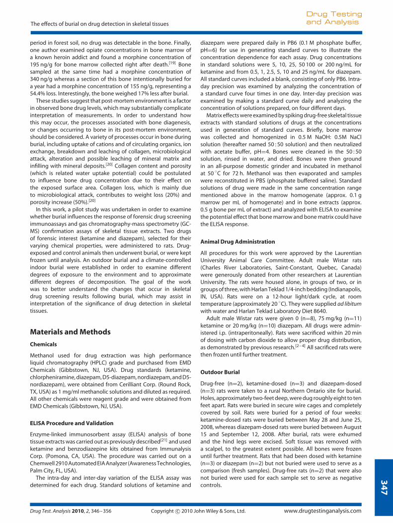
Standard solutions were made for each drug, in drug-free extractsof bone marrow or bone samples. Concentrations ranged from0 to 200 ng/mL for ketamine and from 0 to 25 ng/mL fordiazepam. Figure 1 illustrates the concentration dependence ofthe immunoassay. Coefficients of variation ranged from 0.17 to18% and 0.90 to 6.2% for ketamine bone marrow and bonestandards, respectively, and from 0 to 17% and 1.9 to 11% fordiazepam bone marrow and bone standards, respectively.
Physical Changes in Bones Buried in Outdoor Burial
Rats exhumed after one month of burial had undergoneslight decomposition. The skin was discolored and odor ofdecomposition was potent. During excision of the bones, thesoft tissue was removed quite easily due to the extent ofdecomposition. Upon examination of the bone exterior, theonly observable change was an apparent reduction in soft tissueremaining on the bone itself. Upon opening of the bones, however,it was noted that the marrow had changed from a viscous dark-redsubstance to a less viscous, peach-brown liquid. This permitted amore complete collection of bone marrow as it did not remain onthe sides of the bone in the medullary cavity.
Effect of Outdoor Burial on Assays of Skeletal Tissue Extractsof Drug-exposed Rats
The pH of the soil was also measured, and was found tobe approximately 7 for the topsoil layer, while the pH ofthe clay was 6.5. The mean mass of a given tissue type wascompared between given treatments in order determine whetherburial caused a significant reduction in tissue mass. Table 1illustrates the differences in mass for the ketamine- and diazepam-exposed bones in the outdoor burial. Furthermore, the ELISA
0
10
20
30
40
50
60
70
80
90
100
0 50 100 150 200 250
Concentration (ng/ml)
% D
ecre
ase
in A
bso
rban
ce
%DA -Marrow %DA -Bone
0
10
20
30
40
50
60
70
80
90
100
0 5 10 15 20 25 30
Concentration (ng/ml)
% D
ecre
ase
in A
bso
rban
ce
%DA - Marrow %DA - Bone
A
B
Figure 1. Concentration dependence ELISA response matrix matchedstandard solutions of A) ketamine and B) diazepam.
response (%DA) of extracts of each tissue type was examinedfor each treatment, after correcting for the dilution factor andthe mass of tissue sampled (i.e. %DA/mass). In addition, themean values and the variability between these treatments wereexamined using Student’s t-Test and the F-test, respectively.Table 2 demonstrates these results for ketamine- and diazepam-exposed samples. Samples were then pooled according to theirorigin and treatment for further extraction and confirmationby GC-MS.
Burial Conditions and Physical Changes in Bones FollowingClimate-controlled Burial
The pH of the soil used in the climate-controlled burial wasmeasured to be approximately 7.5. While the marrow of thefrozen samples was viscous and deep red in color, hardlyany soft tissue remained on the epiphyses in the case of theburied bones, and soil was observed within the crevices ofthe epiphyseal sections. Rinsing with water was necessary toremove the soil. It was also noticed that some of the bonefragments were spotted with red coloration, which was notremoved by washing. In the case of these buried bones, the bonemarrow was dry and deep brown (nearly black), and scraping ofthe medullary cavity was necessary to remove remaining bonemarrow. Additionally, a few samples also had fungus within themedullary cavity.
Drug Test. Analysis 2010, 2, 346–356 Copyright c© 2010 John Wiley & Sons, Ltd. www.drugtestinganalysis.com
35
0
Drug Testingand Analysis N. A. Desrosiers and J. H. Watterson
Table 1. Comparison of tissue mass for animals exposed to (A) ketamine or (B) diazepam in outdoor burial, including p values for t-test comparisonof mean. Statistically significant values are bolded
A
Tissue Mean mass (g, Fresh) Standard Deviation Mean mass (g, Buried) Standard Deviation p (t-test)
Tibial marrow 0.0206 0.013 0.0227 0.018 0.9
Femoral marrow 0.0398 0.019 0.0365 0.015 0.8
Tibial diaphyses 0.3337 0.047 0.1901 0.019 0.02Tibial epiphyses 0.4192 0.054 0.1773 0.080 0.02Femoral diaphyses 0.4816 0.132 0.1979 0.006 0.06
Femoral epiphyses 0.6643 0.037 0.1704 0.018 0.002
B
Tissue Mean mass (g, Fresh) Standard Deviation Mean mass (g, Buried) Standard Deviation p (t-test)
Tibial marrow 0.0927 0.036 0.0294 0.030 0.2
Femoral marrow 0.1473 0.039 0.0666 0.031 0.1
Tibial diaphyses 0.7144 0.138 0.5849 0.093 0.4
Tibial epiphyses 0.7732 0.350 0.4679 0.039 0.4
Femoral diaphyses 0.8434 0.080 0.4863 0.132 0.03Femoral epiphyses 0.8623 0.252 0.7244 0.057 0.6
Table 2. Comparison of tissue mass for animals exposed to (A) ketamine or (B) diazepam in interior burial, including p values for t-test comparisonof mean. Statistically significant values are bolded
A
Tissue Mean mass (g, Fresh) Standard Deviation Mean mass (g, Buried) Standard Deviation p (t-test)
Tibial marrow 0.0847 0.0005 0.0083 0.0062 0.002Femoral marrow 0.1219 0.0072 0.0043 0.0009 0.03Tibial diaphyses 0.7259 0.0110 0.6727 0.0713 0.3
Tibial epiphyses 0.4431 0.1138 0.4118 0.0346 0.8
Femoral diaphyses 0.8201 0.1097 0.6975 0.1819 0.4
Femoral epiphyses 0.7943 0.0997 0.9050 0.0982 0.3
B
Tissue Mean mass (g, Fresh) Standard Deviation Mean mass (g, Buried) Standard Deviation p (t-test)
Tibial marrow 0.0556 0.0156 0.0096 0.0065 0.1
Femoral marrow 0.1158 0.0053 0.0120 0.0097 0.001Tibial diaphyses 0.6800 0.0212 0.5859 0.0228 0.03Tibial epiphyses 0.5430 0.1088 0.6773 0.1001 0.3
Femoral diaphyses 0.7406 0.0211 0.8002 0.1205 0.5
Femoral epiphyses 0.9155 0.1175 0.8943 0.0522 0.8
Effect of Climate-controlled Burial on Assays of Skeletal TissueExtracts of Drug-exposed Rats
The ELISA response to extracts from bone and marrow followingexposure to ketamine or diazepam was examined in frozentissues and those that underwent the climate-controlled burial.Table 2 illustrates the differences in mass for the bones inthe climate-controlled burial, relative to tissues that werenot buried. The ELISA response for extracts of each tissuetype was examined for each treatment, after correcting forthe dilution factor and the mass. Figures 2–5 illustrate thesecorrected results for the ketamine- and diazepam-exposedtissues.
GC-MS confirmation
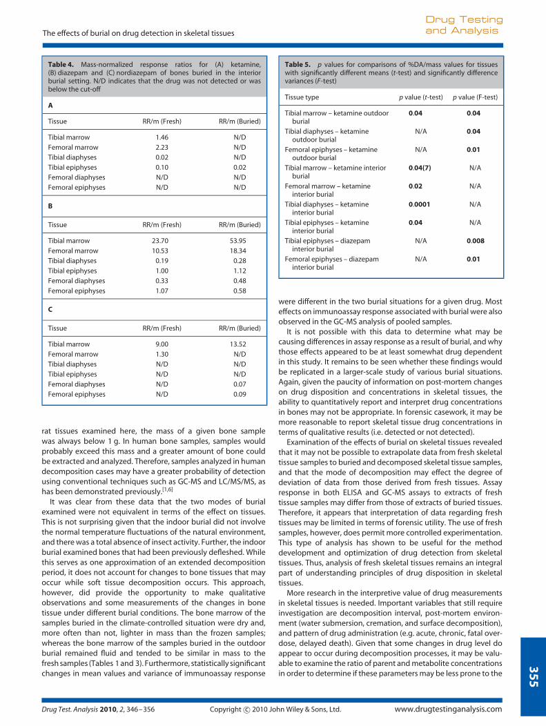
Samples of a given tissue and a given treatment were pooledfor confirmation with GC-MS. Table 3 demonstrates the mass-normalized response ratio (RR/m) for the outdoor burials andTable 4 illustrates the RR/m values for the climate-controlledburials, where the response ratio represents the ratio of peakarea for the analyte, relative to that of the internal standard.Total Ion Chromatograms (TICs) of the fresh (A) and buried(B) ketamine tibial bone marrow samples are shown in Figure 6,while Figure 7 illustrates a TIC for fresh (A) and buried (B) diazepamtibial bone marrow samples. Nordiazepam was also detected inthe marrow samples of the frozen samples and in the tibial
www.drugtestinganalysis.com Copyright c© 2010 John Wiley & Sons, Ltd. Drug Test. Analysis 2010, 2, 346–356

35
1
The effects of burial on drug detection in skeletal tissues
Drug Testingand Analysis
Table 3. Mass-normalized response ratios for (A) ketamine,(B) diazepam, and (C) nordiazepam in rats buried in the outdoor burial.N/D indicates that the drug was not detected or was below the cut-off
A
Tissue RR/m (Fresh) RR/m (Buried)
Tibial marrow 9.351 2.849
Femoral marrow 5.900 5.391
Tibial diaphyses 0.017 0.016
Tibial epiphyses 0.093 0.016
Femoral diaphyses 0.005 0.012
Femoral epiphyses 0.047 0.031
B
Tissue RR/m (Fresh) RR/m (Buried)
Tibial Marrow 5.46 14.23
Femoral Marrow 3.74 12.60
Tibial Diaphyses 0.13 0.23
Tibial Epiphyses 0.98 1.96
Femoral Diaphyses 0.17 0.13
Femoral Epiphyses 1.94 2.39
C
Tissue RR/m (Fresh) RR/m (Buried)
Tibial marrow 5.41 N/D
Femoral marrow 0.409 N/D
Tibial diaphyses N/D 0.201
Tibial epiphyses 0.123 N/D
Femoral diaphyses N/D N/D
Femoral epiphyses N/D N/D
marrow, femoral diaphyses and femoral epiphyses of the buriedsamples.
Discussion
Use of ELISA as an Analytical Technique for Bone and BoneMarrow Analysis
The use of small experimental animals under controlled conditionswarranted the use of ELISA methods to effect the necessarysensitivity, as we have discussed previously.[3,4] Furthermore, theuse of the relative decrease in absorbance (%DA) accounted,to a certain extent, for ELISA response caused by cross-reactingendogenous compounds. Any signal due to metabolites is stillindicative of the presence of drugs, due in part to the fact thatdrug history was known. Furthermore, the presence of drug orcross-reacting metabolite was confirmed with GC-MS, althoughthis was of limited value as samples of a given tissue type werepooled to ensure maximum probability of detection.
Effect of Burial in an Outdoor Burial Site
Whole rats, exposed to ketamine or diazepam, were buried forone month in an outdoor setting during the summer months. TheELISA response to ketamine- and diazepam-exposed tissues wasexamined in bone marrow and bone samples of all buried animals,
Figure 2. Comparison of %DA/mass of bone marrow (A) and bones (B) forketamine-exposed rats in the outdoor burial.
Figure 3. Comparison of %DA/mass of bone marrow (A) and bones (B) fordiazepam-exposed rats in the outdoor burial.
Drug Test. Analysis 2010, 2, 346–356 Copyright c© 2010 John Wiley & Sons, Ltd. www.drugtestinganalysis.com

35
2
Drug Testingand Analysis N. A. Desrosiers and J. H. Watterson
Figure 4. Comparison of %DA/mass of bone marrow (A) and bones (B) forketamine-exposed rats in the interior burial.
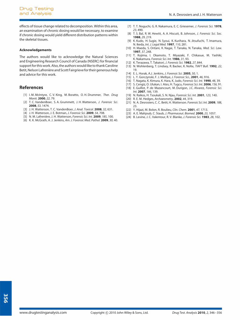
as well as fresh samples that served as a comparison. Sampleswere diluted so their concentration fell within the pseudo-linearregion of the standard ELISA curve to allow for comparison of %DAbetween samples from the different treatments. Furthermore,%DA was normalized for mass in order to account for any variationin tissue mass analyzed between samples and between treatmentsenabling a more accurate comparison of assay response betweentissue types. Overall, there was no significant difference in massbetween fresh/frozen groups and buried groups, except in the caseof the tibial diaphyses, femoral diaphyses, and femoral epiphysesof the ketamine-dosed animal of the outdoor burial set.
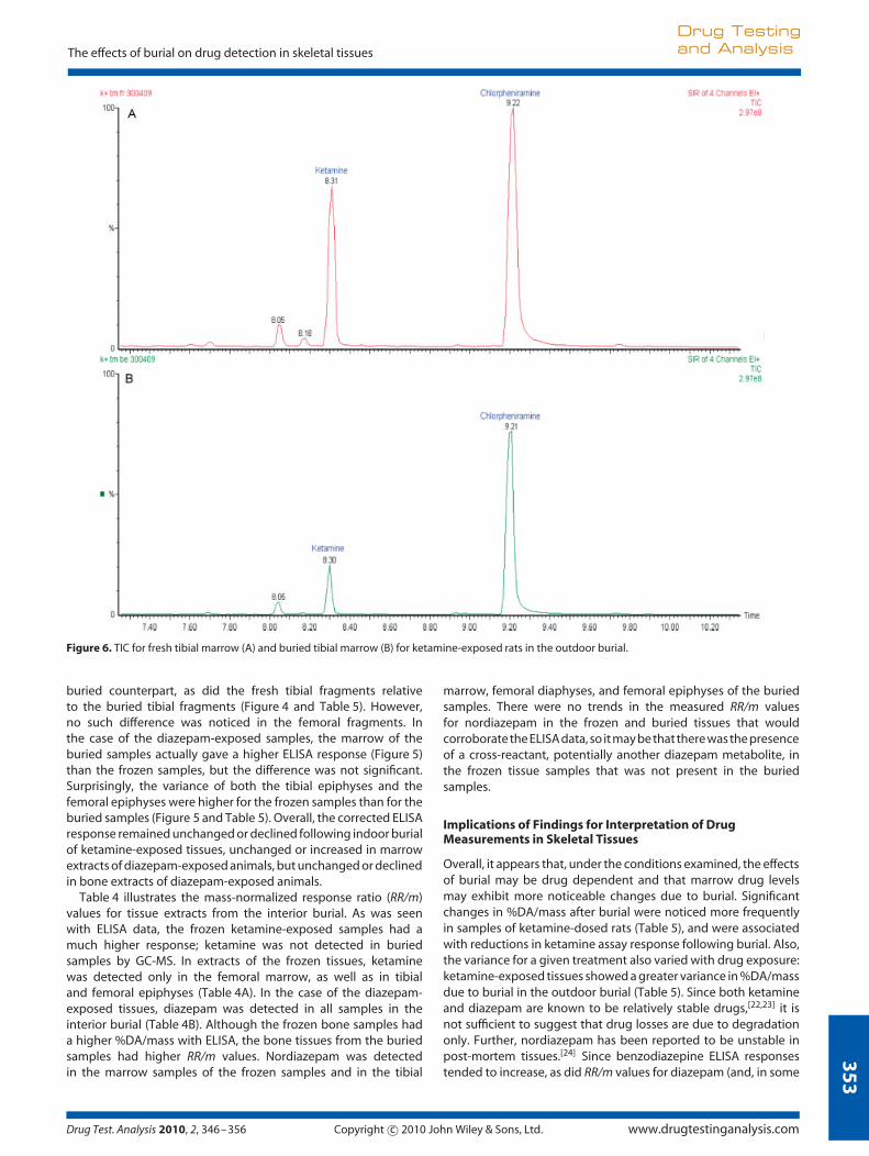
Figure 2 illustrates the normalized %DA values for ketamine-exposed rats buried in an outdoor setting for a one-month period.Marrow samples clearly gave a greater response than bone tissues.However, statistical analysis of the %DA/mass demonstratedthat only the tibial marrow of the buried animals was lowerthan its fresh equivalent. Furthermore, the variance appeared todiffer significantly between treatments (frozen vs burial) for thetibial marrow, the tibial diaphyses and the femoral epiphyses.As for diazepam, the ELISA response was also greater in bonemarrow than in bone (Figure 3). However, there was neither astatistical difference between any mean %DA/mass for any of thetreatments, nor was there any statistically significant difference invariance between treatment groups. Overall, the ELISA data didnot generally indicate a significant effect of burial in the outdoorsetting. Due to the potential for cross-reactivity of metabolites andendogenous compounds, the pooled tissue samples were alsoanalyzed by GC-MS to provide a better indication of the effect ofburial on the individual drugs.
Table 3 summarizes the mass-normalized response ratio (RR/m)for various tissues following the outdoor burials. It should be
Figure 5. Comparison of %DA/mass of bones marrow (A) and bones (B) fordiazepam-exposed rats in the interior burial.
noted again that, because the samples were pooled accordingto tissue type and treatment (e.g. tibial marrow/outdoor burial)following ELISA, the measured RR/m values represent the groupaverage and could not undergo any statistical comparison. Forketamine-exposed tissues, the fresh samples generally gave agreater RR/m value, especially in the case of the tibial marrow andtibial epiphyses (Table 3A). In the case of the diazepam-exposedsamples, nordiazepam was also detected in some samples.Although the %DA/mass was higher for the majority of freshsamples, these differences were not significant and the RR/mvalues determined by GC-MS results indicated a higher diazepamresponse in the buried samples. Nordiazepam was only detectedin the fresh marrow samples, the fresh tibial epiphyses and theburied tibial diaphyses. Overall, the GC-MS data suggested ageneral trend of a maintenance or reduction in ketamine level anda maintenance or increase in diazepam concentration as a resultof burial.
Effect of Burial in a Climate-controlled Situation
A climate-controlled setting was utilized in an attempt to controlenvironmental conditions and to assess the effects of ‘controlledburial’. It also is a first approximation for extended decompositionin burial since overlying soft tissue was removed. Significantphysical changes were noticed in the buried bones, where marrowin the medullary cavity was completely dry. Marrow removal wasaccomplished by scratching the sides of the bone with the syringeneedle. Furthermore, bone marrow of dried samples needed tobe homogenized further by manual homogenization with a rotarytool.
As was seen in the outdoor burial, the marrow of the freshketamine-exposed rat bones gave higher %DA/mass than the
www.drugtestinganalysis.com Copyright c© 2010 John Wiley & Sons, Ltd. Drug Test. Analysis 2010, 2, 346–356
35
3
The effects of burial on drug detection in skeletal tissues
Drug Testingand Analysis
Figure 6. TIC for fresh tibial marrow (A) and buried tibial marrow (B) for ketamine-exposed rats in the outdoor burial.
buried counterpart, as did the fresh tibial fragments relativeto the buried tibial fragments (Figure 4 and Table 5). However,no such difference was noticed in the femoral fragments. Inthe case of the diazepam-exposed samples, the marrow of theburied samples actually gave a higher ELISA response (Figure 5)than the frozen samples, but the difference was not significant.Surprisingly, the variance of both the tibial epiphyses and thefemoral epiphyses were higher for the frozen samples than for theburied samples (Figure 5 and Table 5). Overall, the corrected ELISAresponse remained unchanged or declined following indoor burialof ketamine-exposed tissues, unchanged or increased in marrowextracts of diazepam-exposed animals, but unchanged or declinedin bone extracts of diazepam-exposed animals.
Table 4 illustrates the mass-normalized response ratio (RR/m)values for tissue extracts from the interior burial. As was seenwith ELISA data, the frozen ketamine-exposed samples had amuch higher response; ketamine was not detected in buriedsamples by GC-MS. In extracts of the frozen tissues, ketaminewas detected only in the femoral marrow, as well as in tibialand femoral epiphyses (Table 4A). In the case of the diazepam-exposed tissues, diazepam was detected in all samples in theinterior burial (Table 4B). Although the frozen bone samples hada higher %DA/mass with ELISA, the bone tissues from the buriedsamples had higher RR/m values. Nordiazepam was detectedin the marrow samples of the frozen samples and in the tibial
marrow, femoral diaphyses, and femoral epiphyses of the buriedsamples. There were no trends in the measured RR/m valuesfor nordiazepam in the frozen and buried tissues that wouldcorroborate the ELISA data, so it may be that there was the presenceof a cross-reactant, potentially another diazepam metabolite, inthe frozen tissue samples that was not present in the buriedsamples.
Implications of Findings for Interpretation of DrugMeasurements in Skeletal Tissues
Overall, it appears that, under the conditions examined, the effectsof burial may be drug dependent and that marrow drug levelsmay exhibit more noticeable changes due to burial. Significantchanges in %DA/mass after burial were noticed more frequentlyin samples of ketamine-dosed rats (Table 5), and were associatedwith reductions in ketamine assay response following burial. Also,the variance for a given treatment also varied with drug exposure:ketamine-exposed tissues showed a greater variance in %DA/massdue to burial in the outdoor burial (Table 5). Since both ketamineand diazepam are known to be relatively stable drugs,[22,23] it isnot sufficient to suggest that drug losses are due to degradationonly. Further, nordiazepam has been reported to be unstable inpost-mortem tissues.[24] Since benzodiazepine ELISA responsestended to increase, as did RR/m values for diazepam (and, in some
Drug Test. Analysis 2010, 2, 346–356 Copyright c© 2010 John Wiley & Sons, Ltd. www.drugtestinganalysis.com

35
4
Drug Testingand Analysis N. A. Desrosiers and J. H. Watterson
Figure 7. TIC for fresh tibial marrow (A) and buried tibial marrow (B) for diazepam-exposed rats in the outdoor burial.
cases, for nordiazepam) following burial, one potential factor inthe observed differences is changes in drug concentration as theviscous marrow decomposed into a more fluid product. Differencesin water solubility may result in either concentration of less water-soluble drugs or losses of more water-soluble drugs as the fluidmatrix flows out of the bone sample.
These results may have important ramifications on the possibil-ity to quantitatively interpret toxicological data from decomposedbone and bone marrow samples, in that they suggest that it isinherently difficult to find an appropriate reference matrix condi-tion. In other words, the use of a database of bone drug levelsobtained from fresh bone only may not be representative of levelsobtained from decomposed bones, even if the initial drug expo-sure conditions were similar. The body of literature that exists withrespect to drug levels in human bones is somewhat disjointed,with most larger-scale studies[1,6] making reference largely to onlyrelatively fresh tissues, with individual case reports more oftenreporting levels in decomposed tissues.[7 – 11] Thus, at the presenttime, it seems more prudent to present toxicological findings ina qualitative manner (i.e. detected or not detected) in forensiccasework.
Implications and Limitations of Analytical Methodology
While the t-test was used in order to compare the mean values fortissue mass and normalized ELISA response, the trends observedshould be re-examined in a larger study, including a wider range
of decomposition conditions and drug exposures, and with alarger sample size. Furthermore, although the Shapiro-Wilk Wnormality test suggested that all sample sets possessed a normaldistribution, with the exception of the frozen diaphyses and theburied marrow for the ketamine-exposed samples of the climate-controlled burial and the buried marrow of the diazepam-exposedsamples of the climate-controlled burial, the F-test is susceptibleto effects of deviations from normality. The significance of thevariance comparisons for these samples should be taken withsome reservation, but the notion of significant changes in samplevariance with changes in decompositional state merits furtherconsideration in future work.
Following ELISA, samples of a given tissue type from animals ofa given treatment group (i.e. buried vs non-buried) were pooledprior to analysis by GC-MS in order to increase the probabilityof detection. The GC-MS results indicated that detection ofketamine and diazepam was still possible after burial for a periodof one month and was not simply an endogenous compoundthat cross-reacts with the ELISA assay. Although some of theGC-MS results differed from those found with ELISA, it may bedue in part with the way the samples were analyzed (pooledsamples for GC-MS vs individual analysis for ELISA), as well as thepotential for cross-reactivity to metabolites and other compoundsin ELISA measurements. Despite the potential for cross-reactivity,ELISA remains a very sensitive technique for measurements inexperimental studies of drug disposition in bone tissue. In the
www.drugtestinganalysis.com Copyright c© 2010 John Wiley & Sons, Ltd. Drug Test. Analysis 2010, 2, 346–356
35
5
The effects of burial on drug detection in skeletal tissues
Drug Testingand Analysis
Table 4. Mass-normalized response ratios for (A) ketamine,(B) diazepam and (C) nordiazepam of bones buried in the interiorburial setting. N/D indicates that the drug was not detected or wasbelow the cut-off
A
Tissue RR/m (Fresh) RR/m (Buried)
Tibial marrow 1.46 N/D
Femoral marrow 2.23 N/D
Tibial diaphyses 0.02 N/D
Tibial epiphyses 0.10 0.02
Femoral diaphyses N/D N/D
Femoral epiphyses N/D N/D
B
Tissue RR/m (Fresh) RR/m (Buried)
Tibial marrow 23.70 53.95
Femoral marrow 10.53 18.34
Tibial diaphyses 0.19 0.28
Tibial epiphyses 1.00 1.12
Femoral diaphyses 0.33 0.48
Femoral epiphyses 1.07 0.58
C
Tissue RR/m (Fresh) RR/m (Buried)
Tibial marrow 9.00 13.52
Femoral marrow 1.30 N/D
Tibial diaphyses N/D N/D
Tibial epiphyses N/D N/D
Femoral diaphyses N/D 0.07
Femoral epiphyses N/D 0.09
rat tissues examined here, the mass of a given bone samplewas always below 1 g. In human bone samples, samples wouldprobably exceed this mass and a greater amount of bone couldbe extracted and analyzed. Therefore, samples analyzed in humandecomposition cases may have a greater probability of detectionusing conventional techniques such as GC-MS and LC/MS/MS, ashas been demonstrated previously.[1,6]
It was clear from these data that the two modes of burialexamined were not equivalent in terms of the effect on tissues.This is not surprising given that the indoor burial did not involvethe normal temperature fluctuations of the natural environment,and there was a total absence of insect activity. Further, the indoorburial examined bones that had been previously defleshed. Whilethis serves as one approximation of an extended decompositionperiod, it does not account for changes to bone tissues that mayoccur while soft tissue decomposition occurs. This approach,however, did provide the opportunity to make qualitativeobservations and some measurements of the changes in bonetissue under different burial conditions. The bone marrow of thesamples buried in the climate-controlled situation were dry and,more often than not, lighter in mass than the frozen samples;whereas the bone marrow of the samples buried in the outdoorburial remained fluid and tended to be similar in mass to thefresh samples (Tables 1 and 3). Furthermore, statistically significantchanges in mean values and variance of immunoassay response
Table 5. p values for comparisons of %DA/mass values for tissueswith significantly different means (t-test) and significantly differencevariances (F-test)
Tissue type p value (t-test) p value (F-test)
Tibial marrow – ketamine outdoorburial
0.04 0.04
Tibial diaphyses – ketamineoutdoor burial
N/A 0.04
Femoral epiphyses – ketamineoutdoor burial
N/A 0.01
Tibial marrow – ketamine interiorburial
0.04(7) N/A
Femoral marrow – ketamineinterior burial
0.02 N/A
Tibial diaphyses – ketamineinterior burial
0.0001 N/A
Tibial epiphyses – ketamineinterior burial
0.04 N/A
Tibial epiphyses – diazepaminterior burial
N/A 0.008
Femoral epiphyses – diazepaminterior burial
N/A 0.01
were different in the two burial situations for a given drug. Mosteffects on immunoassay response associated with burial were alsoobserved in the GC-MS analysis of pooled samples.
It is not possible with this data to determine what may becausing differences in assay response as a result of burial, and whythose effects appeared to be at least somewhat drug dependentin this study. It remains to be seen whether these findings wouldbe replicated in a larger-scale study of various burial situations.Again, given the paucity of information on post-mortem changeson drug disposition and concentrations in skeletal tissues, theability to quantitatively report and interpret drug concentrationsin bones may not be appropriate. In forensic casework, it may bemore reasonable to report skeletal tissue drug concentrations interms of qualitative results (i.e. detected or not detected).
Examination of the effects of burial on skeletal tissues revealedthat it may not be possible to extrapolate data from fresh skeletaltissue samples to buried and decomposed skeletal tissue samples,and that the mode of decomposition may effect the degree ofdeviation of data from those derived from fresh tissues. Assayresponse in both ELISA and GC-MS assays to extracts of freshtissue samples may differ from those of extracts of buried tissues.Therefore, it appears that interpretation of data regarding freshtissues may be limited in terms of forensic utility. The use of freshsamples, however, does permit more controlled experimentation.This type of analysis has shown to be useful for the methoddevelopment and optimization of drug detection from skeletaltissues. Thus, analysis of fresh skeletal tissues remains an integralpart of understanding principles of drug disposition in skeletaltissues.
More research in the interpretive value of drug measurementsin skeletal tissues is needed. Important variables that still requireinvestigation are decomposition interval, post-mortem environ-ment (water submersion, cremation, and surface decomposition),and pattern of drug administration (e.g. acute, chronic, fatal over-dose, delayed death). Given that some changes in drug level doappear to occur during decomposition processes, it may be valu-able to examine the ratio of parent and metabolite concentrationsin order to determine if these parameters may be less prone to the
Drug Test. Analysis 2010, 2, 346–356 Copyright c© 2010 John Wiley & Sons, Ltd. www.drugtestinganalysis.com
35
6
Drug Testingand Analysis N. A. Desrosiers and J. H. Watterson
effects of tissue change related to decomposition. Within this area,an examination of chronic dosing would be necessary, to examineif chronic dosing would yield different distribution patterns withinthe skeletal tissues.
Acknowledgements
The authors would like to acknowledge the Natural Sciencesand Engineering Research Council of Canada (NSERC) for financialsupport for this work. Also, the authors would like to thank CarolineBetit, Nelson Lafreniere and Scott Fairgrieve for their generous helpand advice for this work.
References
[1] I. M. McIntyre, C. V. King, M. Boratto, O. H. Drummer, Ther. DrugMonit. 2000, 22, 79.
[2] T. C. VandenBoer, S. A. Grummett, J. H. Watterson, J. Forensic Sci.2008, 53, 1474.
[3] J. H. Watterson, T. C. VandenBoer, J. Anal. Toxicol. 2008, 32, 631.[4] J. H. Watterson, J. E. Botman, J. Forensic Sci. 2009, 54, 708.[5] N. M. Lafreniere, J. H. Watterson, Forensic Sci. Int. 2009, 185, 100.[6] K. K. McGrath, A. J. Jenkins, Am. J. Forensic Med. Pathol. 2009, 30, 40.
[7] T. T. Noguchi, G. R. Nakamura, E. C. Griesemer, J. Forensic Sci. 1978,23, 490.
[8] T. S. Bal, R. W. Hewitt, A. A. Hiscutt, B. Johnson, J. Forensic Sci. Soc.1988, 29, 219.
[9] K. Kudo, H. Sugie, N. Syoui, K. Kurihara, N. Jitsufuchi, T. Imamura,N. Ikeda, Int. J. Legal Med. 1997, 110, 281.
[10] H. Maeda, S. Oritani, K. Nagai, T. Tanaka, N. Tanaka, Med. Sci. Law.1997, 37, 248.
[11] T. Kojima, I. Okamoto, T. Miyazaki, F. Chikasue, M. Yashiki,K. Nakamura, Forensic Sci. Int. 1986, 31, 93.
[12] K. Terazawa, T. Takatori, J. Forensic Sci. 1982, 27, 844.[13] N. Wohlenberg, T. Lindsey, R. Backer, K. Nolte, TIAFT Bull. 1992, 22,
19.[14] E. L. Horak, A.J. Jenkins, J. Forensic Sci. 2005, 50, 1.[15] L. Y. Gorczynski, F. J. Melbye, J. Forensic Sci., 2001, 46, 916.[16] T. Nagata, K. Kimura, K. Hara, K. Judo, Forensic Sci. Int. 1990, 48, 39.[17] S. Cengiz, O. Ulukan, I. Ates, H. Tugcu, Forensic Sci. Int. 2006, 156, 91.[18] E. Guillot, P. de Mazancourt, M. Durigon, J.C. Alvarez, Forensic Sci.
Int. 2007, 166, 139.[19] N. Raikos, H. Tsoukali, S. N. Njau, Forensic Sci. Int. 2001, 123, 140.[20] R. E. M. Hedges, Archaeometry, 2002, 44, 319.[21] N. A. Desrosiers, C. C. Betit, H. Watterson. Forensic Sci. Int. 2009, 188,
23.[22] Y. Hijazi, M. Bolon, R. Boulieu, Clin. Chem. 2001, 47, 1713.[23] A. E. Mahjoub, C. Staub, J. Pharmaceut. Biomed. 2000, 23, 1057.[24] B. Levine, J. C. Valentour, R. V. Blanke, J. Forensic Sci. 1983, 28, 102.
www.drugtestinganalysis.com Copyright c© 2010 John Wiley & Sons, Ltd. Drug Test. Analysis 2010, 2, 346–356