the effect of kdel and ppi genes co - UM Students' Repository
185
THE EFFECT OF KDEL AND PPI GENES CO- EXPRESSION ON THE ANTI-TOXOPLASMA scFV ACTIVITY IN TRANSGENIC Nicotiana tabacum CULTIVAR SR1 FATIN IFFAH RASYIQAH BINTI MOHAMAD ZOOLKEFLI FACULTY OF SCIENCE UNIVERSITY OF MALAYA KUALA LUMPUR 2018 University of Malaya
-
Upload
khangminh22 -
Category
Documents
-
view
2 -
download
0
Transcript of the effect of kdel and ppi genes co - UM Students' Repository

THE EFFECT OF KDEL AND PPI GENES CO-EXPRESSION ON THE ANTI-TOXOPLASMA scFV
ACTIVITY IN TRANSGENIC Nicotiana tabacum CULTIVAR SR1
FATIN IFFAH RASYIQAH BINTI MOHAMAD ZOOLKEFLI
FACULTY OF SCIENCE
UNIVERSITY OF MALAYA KUALA LUMPUR
2018
Univers
ity of
Mala
ya

THE EFFECT OF KDEL AND PPI GENES CO-EXPRESSION ON THE ANTI-TOXOPLASMA scFV
ACTIVITY IN TRANSGENIC Nicotiana tabacum CULTIVAR SR1
FATIN IFFAH RASYIQAH BINTI MOHAMAD ZOOLKEFLI
THESIS SUBMITTED IN FULFILMENT OF THE
REQUIREMENTS FOR THE DEGREE OF MASTER OF SCIENCE
FACULTY OF SCIENCE
UNIVERSITY OF MALAYA KUALA LUMPUR
2018
Univers
ity of
Mala
ya

ii
UNIVERSITY OF MALAYA
ORIGINAL LITERARY WORK DECLARATION
Name of Candidate: FATIN IFFAH RASYIQAH BINTI MOHAMAD ZOOLKEFLI
Matric No: SGR140015
Name of Degree: MASTER OF SCIENCE
Title of Project Paper/Research Report/Dissertation/Thesis (“this Work”): THE EFFECT OF KDEL AND PPI GENES CO-EXPRESSION ON THE
ANTI-TOXOPLASMA scFV ACTIVITY IN TRANSGENIC Nicotiana tabacum CULTIVAR
SR1
Field of Study: BIOTECHNOLOGY
I do solemnly and sincerely declare that:
(1) I am the sole author/writer of this Work; (2) This Work is original; (3) Any use of any work in which copyright exists was done by way of fair
dealing and for permitted purposes and any excerpt or extract from, or reference to or reproduction of any copyright work has been disclosed expressly and sufficiently and the title of the Work and its authorship have been acknowledged in this Work;
(4) I do not have any actual knowledge nor do I ought reasonably to know that the making of this work constitutes an infringement of any copyright work;
(5) I hereby assign all and every rights in the copyright to this Work to the University of Malaya (“UM”), who henceforth shall be owner of the copyright in this Work and that any reproduction or use in any form or by any means whatsoever is prohibited without the written consent of UM having been first had and obtained;
(6) I am fully aware that if in the course of making this Work I have infringed any copyright whether intentionally or otherwise, I may be subject to legal action or any other action as may be determined by UM.
Candidate’s Signature Date: 10th July 2018
Subscribed and solemnly declared before,
Date: Witness’s Signature
Name:
Designation:
Univers
ity of
Mala
ya

iii
THE EFFECT OF KDEL AND PPI GENES CO-EXPRESSION ON THE
ANTI-TOXOPLASMA scFV ACTIVITY IN TRANSGENIC Nicotiana
tabacum CULTIVAR SR1
ABSTRACT
Toxoplasmosis is a disease caused by infection of a parasitic protozoan known
as Toxoplasma gondii, which has economic implications due to neonatal loss and
abortion in livestock animals. In human adults, it is usually asymptomatic but can
cause complications in immunocompromised persons and pregnant women which
leads to congenital effects on newborn babies. As a therapeutic approach to combat
the disease, biopharmaceutical products, such as recombinant antibody targeting to
T. gondii tachyzoite, has been introduced as an alternative to drugs and vaccines.
Innovative approaches for the expression and production of these novel
biopharmaceutical products which increased complexity towards disease targets
have been the subject of recent investigations. Plants have now emerged as a
preferred host to express and produce these recombinant proteins as it provides
advantages in term of efficiency, cost, scalability, safety and compatibility as
compared to prokaryotes and mammalian cell lines. Thus, in this study, the target
antibody, an anti-Toxoplasma scFv encoded by the TP60 gene was transformed into
leaf explants of Nicotiana tabacum cv. SR1 using Agrobacterium tumefaciens
strain LBA4404. The constructs were transformed in a series of experiments in
tandem with different enhancer elements comprising proteinase inhibitors or a
KDEL retention sequence or combinations of both elements in order to assess their
ability to stabilize and enhance the scFv production. Two different proteinase
inhibitors that have been used in this study were Oryzacystatin inhibitor (OCPI) (a
cysteine inhibitor) and Bowman-Birk inhibitor (BBI) (a trypsin and chymotrypsin
Univers
ity of
Mala
ya

iv
inhibitors). β-glucuronidase (GUS) and green fluorescence protein (GFP) assays as
well as PCR results confirmed the presence of transgenes in the tobacco genome
that was stably inherited into T1 generation. For Western blot analysis, the selected
lines of transgenic tobacco plants transformed with pTP60 construct in combination
with BBI-KDEL (pTP60BBIKDEL) elements was found to produce the highest
anti-Toxoplasma scFv recombinant antibody accumulation at the expected size of ~
54 kDa (homodimer formation) which was at first observed at ~ 27 kDa (monomer)
in T0 tobacco plants. No expression was detected in non-transformed plants. The
results were also confirmed at the mRNA level using real-time PCR analysis with
the BBI-KDEL tandem constructs expressing TP60 gene approximately 19-fold
higher as compared to plants transformed with the pTP60 construct in the absence
of any proteinase inhibitor and/or KDEL. Interestingly, the introduction of all of
these elements did not or only negligibly affected the growth and development of
the transgenic tobacco plants as compared to the wild-type. Our results confirmed
the ability of plants to function as bio-factories for recombinant protein production
and showed that the use of proteinase inhibitor and KDEL elements may be useful
in improving the yield of the targeted antibody proteins.
Keywords: Agrobacterium-mediated transformation, plant molecular farming,
recombinant protein, single-chain fragment antibody (scFv), toxoplasmosis
Univers
ity of
Mala
ya

v
KESAN EKSPRESI BERSAMA GEN KDEL DAN PPI TERHADAP
AKTIVITI ANTI-TOXOPLASMA scFV PADA Nicotiana tabacum
KULTIVAR SR1 TRANSGENIK
ABSTRAK
Toxoplasmosis merupakan penyakit yang disebabkan oleh jangkitan protozoan
parasit yang dikenali sebagai Toxoplasma gondii, yang mempunyai implikasi
kepada ekonomi akibat kehilangan neonatal dan keguguran haiwan ternakan. Di
kalangan manusia dewasa, penyakit ini biasanya tidak menunjukkan sebarang
gejala jangkitan tetapi boleh mengakibatkan komplikasi pada mereka yang
mempunyai immunisasi yang lemah serta wanita hamil yang boleh membawa
kepada kesan kongenital terhadap bayi yang baru dilahirkan. Sebagai pendekatan
terapeutik bagi memerangi penyakit ini, produk biofarmaseutikal seperti antibodi
rekombinan yang disasarkan kepada T. gondii tachyzoite telah diperkenalkan
sebagai alternatif kepada ubat-ubatan dan vaksin. Pendekatan inovatif terhadap
ekspresi dan penghasilan produk biofarmaseutikal yang baharu ini telah
mempunyai peningkatan kompleksiti terhadap penyakit yang disasarkan dan
menjadi subjek penyelidikan terkini. Kini, tumbuhan telah muncul sebagai hos
pilihan dalam mengekspresi dan menghasilkan protein rekombinan kerana
kelebihannya dari segi kecekapan, kos, skala, keselamatan dan keserasian
berbanding titisan sel prokariot dan mamalia. Oleh itu, dalam kajian ini, antibodi
sasaran iaitu scFv anti-Toxoplasma, yang dikodkan oleh gen TP60 telah
ditransformasi ke dalam eksplan daun Nicotiana tabacum kultivar SR1 dengan
menggunakan Agrobacterium tumefaciens strain LBA4404. Konstruk-konstruk
telah ditransformasi dalam satu siri eksperimen bersama dengan unsur penambah
berbeza yang terdiri daripada perencat proteinase atau jujukan pengekalan KDEL
Univers
ity of
Mala
ya

vi
atau gabungan kedua-dua unsur tersebut bagi menilai keupayaan mereka dalam
menstabil dan meningkatkan penghasilan scFv. Dua perencat proteinase yang telah
digunakan dalam kajian ini ialah perencat Oryzacystatin (OCPI) (perencat cystein)
dan perencat Bowman-Birk (BBI) (perencat trypsin dan chymotrypsin). Ujian β-
glucuronidase (GUS) dan protein pendarfluor hijau (GFP) serta keputusan PCR
mengesahkan kehadiran transgen dalam genom tembakau yang telah diwariskan
kepada generasi T1. Bagi analisis pemendapan Western, barisan tumbuhan
tembakau transgenik yang telah ditransformasi dengan konstruk pTP60 bersama
dengan unsur BBI-KDEL (pTP60BBIKDEL) didapati dapat menghasilkan
pengumpulan antibodi scFv anti-Toxoplasma tertinggi pada saiz jangkaan ~ 54 kDa
(pembentukan homodimer) yang pada pertama kalinya dilihat pada saiz jangkaan ~
27 kDa (monomer) di dalam tumbuhan tembakau T0. Tiada ekspresi yang dikesan
pada tumbuhan yang tidak ditransformasi. Keputusan ini juga disahkan pada tahap
mRNA dengan menggunakan analisis real-time PCR di mana konstruk tandem
BBI-KDEL menunjukkan ekspresi gen TP60 kira-kira 19 kali ganda lebih tinggi
berbanding dengan tumbuhan yang ditransformasi dengan konstruk pTP60 sahaja
tanpa sebarang unsur penambah. Menariknya, pengenalan semua unsur penambah
ini tidak atau hanya memberi kesan yang boleh diabaikan terhadap tumbesaran dan
perkembangan tumbuhan tembakau transgenik apabila dibandingkan dengan
tumbuhan bukan transgenik. Hasil kajian kami mengesahkan keupayaan tumbuhan
untuk berfungsi sebagai kilang bio bagi pengeluaran protein rekombinan dan juga
menunjukkan bahawa penggunaan unsur penambah mungkin berguna dalam
meningkatkan hasil protein antibodi yang disasarkan.
Kata kunci: Transformasi Agrobacterium-pengantara, tumbuhan pertanian
molekul, protein rekombinan, antibodi serpihan rantaian tunggal (scFv),
toxoplasmosis
Univers
ity of
Mala
ya

vii
ACKNOWLEDGEMENTS
In the name of ALLAH S.W.T the most Gracious and the most Merciful, all praises
to Him who had given me blessings, strengths, and knowledge in completing this
thesis entitled “The Effect of KDEL and PPI Genes Co-Expression on the Anti-
Toxoplasma scFV Activity in Transgenic Nicotiana tabacum Cultivar SR1” .
First and foremost, I would like to express my sincere gratitude to my supervisors,
Prof. Dr. Rofina Yasmin Othman and Prof. Dr. Norzulaani Khalid for giving me an
opportunity and trust to complete my Master of Science degree study in Plant
Biotechnology Research Laboratory, Institute of Biological Sciences. Their
understanding, encouraging, and personal guidance have encouraged me to do well
in my project as well as provided me a good basis, constructive criticism and
excellent advice for the present thesis.
I wish to express my warm and sincere thanks to Dr. Tan Boon Chin and Dr. Nur
Ardiyana Rejab for their wide knowledge and their logical way of thinking based
on their previous experiences have been of great value for me as well as helping me
a lot during my difficulties moments. Not forget to all lab-mates in Plant
Biotechnology Research Laboratory and HIR Interactome Laboratory who are
always rendered their help during period of my project work.
Thank you to University of Malaya for providing me the fellowship and PPP grant
(PG204-2016A) for my study. My gratitude to High Impact Research fund
(UM.C/625/1/HIR/MOHE/SCI/18) for funding this project.
Last but not least, I wish to avail myself of this opportunity, express a sense of
gratitude to my lovely husband Mohamad Badrisham, beloved parents and in-laws
whose always giving me moral and loving support. Without their encouragement it
would be impossible for me to complete this project.
Univers
ity of
Mala
ya

viii
TABLE OF CONTENTS
Abstract ............................................................................................................................ iii
Abstrak .............................................................................................................................. v
Acknowledgements ......................................................................................................... vii
Table of Contents ........................................................................................................... viii
List of Figures ................................................................................................................ xiii
List of Tables ................................................................................................................ xvii
List of Symbols and Abbreviations ................................................................................ xix
List of Appendices ....................................................................................................... xxiii
CHAPTER 1: INTRODUCTION .................................................................................. 1
1.1 Background study .................................................................................................... 1
1.2 Objectives ................................................................................................................ 3
1.2.1 General objective ........................................................................................ 3
1.2.2 Specific objectives ...................................................................................... 3
1.3 Hypothesis ............................................................................................................... 3
CHAPTER 2: LITERATURE REVIEW ...................................................................... 4
2.1 Toxoplasmosis ......................................................................................................... 4
2.1.1 Toxoplasma gondii strains .......................................................................... 5
2.1.2 Symptoms of infection ............................................................................... 6
2.1.3 Control of toxoplasmosis............................................................................ 7
2.2 Expression system for recombinant protein production .......................................... 7
2.2.1 Types of different expression systems ....................................................... 8
2.3 Plant molecular farming ........................................................................................ 10
2.4 Therapeutic proteins .............................................................................................. 12
Univers
ity of
Mala
ya

ix
2.4.1 Antibodies ................................................................................................ 13
2.4.1.1 Single-chain variable fragment antibodies ................................ 13
2.5 Challenges and strategies of plant as protein expression system .......................... 15
2.5.1 Co-expression with proteinase inhibitors ................................................. 17
2.5.2 Targeting to specific ER-organelle in plant tissue (KDEL) ..................... 18
2.6 Agrobacterium-mediated transformation .............................................................. 21
2.6.1 Agrobacterium strains used for recombinant protein expression ............. 21
2.6.2 Importance of Agrobacterium in recombinant protein production .......... 22
2.7 Expression vectors used for Agrobacterium-mediated transformation ................. 25
2.8 Tobacco as a plant host .......................................................................................... 27
CHAPTER 3: MATERIALS AND METHODS ........................................................ 30
3.1 Maintenance of in vitro Nicotiana tabacum cv. SR1 ............................................ 30
3.1.1 Preparation of culture medium ................................................................. 30
3.1.2 Germination of tobacco seeds .................................................................. 30
3.1.3 Maintenance of tobacco plantlets ............................................................. 31
3.2 Endogenous test of tobacco leaf disc towards hygromycin ................................... 32
3.3 Agrobacterium tumefaciens strain LBA4404 ........................................................ 33
3.3.1 Preparation of YEB and YEA media ....................................................... 33
3.3.2 Preparation of A. tumefaciens presence with different plasmid constructs .................................................................................................. 33
3.3.2.1 Gene sequencing and BLAST analysis ..................................... 35
3.3.2.2 Plasmid extraction ..................................................................... 35
3.3.2.3 PCR verification of Agrobacterium colonies ............................ 36
3.3.2.4 Agarose gel electrophoresis ...................................................... 39
3.3.2.5 Bacterial stock storage (glycerol stock) .................................... 40
3.4 Agrobacterium-mediated transformation into tobacco leaf disc ........................... 41
Univers
ity of
Mala
ya

x
3.4.1 Selection of transformants ........................................................................ 41
3.5 Transient assays of putative transformed shoots ................................................... 42
3.5.1 GUS assessments ...................................................................................... 42
3.5.1.1 GUS histochemical staining ...................................................... 42
3.5.1.2 GUS fluorometry assay ............................................................. 43
3.5.2 GFP visualization ..................................................................................... 43
3.6 Acclimatization and phenotypic assessment of transgenic tobacco plants ............ 44
3.7 Assessment of putative tobacco transformants by PCR analysis .......................... 44
3.7.1 Total genomic DNA extraction ................................................................ 44
3.7.2 Polymerase chain reaction (PCR)............................................................. 45
3.8 Polyacrylamide gel electrophoresis (SDS-PAGE) and Western blot analysis ...... 48
3.8.1 Gel cassette assembly ............................................................................... 48
3.8.2 Preparation of the gel ............................................................................... 48
3.8.3 Preparation of protein samples ................................................................. 50
3.8.3.1 Bradford’s quantification .......................................................... 50
3.8.4 Electrophoresis of SDS-PAGE ................................................................. 50
3.8.5 Staining and de-staining protein gel with Coomassie blue ...................... 51
3.8.6 Western blot analysis................................................................................ 51
3.8.7 Protein purification using Dynabeads® ................................................... 52
3.9 Verification of transgenic plant via real-time PCR analysis ................................. 53
3.9.1 RNA extraction and DNase treatment ...................................................... 53
3.9.2 Reverse-transcription PCR (RT-PCR) ..................................................... 55
3.9.3 Real-time PCR .......................................................................................... 57
3.10 Semi-quantitative RT-PCR .................................................................................... 59
3.11 Statistical analysis.................................................................................................. 59
Univers
ity of
Mala
ya

xi
CHAPTER 4: RESULTS.............................................................................................. 60
4.1 Endogenous test of N. tabacum cv. SR1 leaf discs towards antibiotics ................ 60
4.2 Confirmation of A. tumefaciens harboring binary vector and gene of interest ...... 65
4.2.1 Sequencing and BLAST analysis ............................................................. 73
4.3 Transformation of N. tabacum cv. SR1 leaf discs mediated by A. tumefaciens .... 81
4.3.1 Effects of different plasmid constructs towards regeneration of putative transformed shoots...................................................................... 81
4.3.2 Confirmation of putative transformed shoots using transient assays ....... 86
4.3.2.1 GUS histochemical staining ...................................................... 86
4.3.2.2 GUS fluorometry assay ............................................................. 89
4.3.2.3 GFP qualitative assay ................................................................ 93
4.3.3 Verification of putative transformed shoots (T0) through PCR
analysis ..................................................................................................... 95
4.4 Assessment of gene stability T1 generation of transgenic tobacco ...................... 100
4.4.1 Effect of different plasmid constructs on seed germination ................... 100
4.4.2 Verification of transgenic seedlings (T1) through PCR analysis ............ 103
4.5 Acclimatization of transgenic tobacco plants ...................................................... 105
4.6 Phenotypic assessment on T1 tobacco progenies ................................................. 107
4.7 Real-time PCR analysis ....................................................................................... 108
4.7.1 RNA extraction and DNase treatment of T1 tobacco progenies ............. 108
4.7.2 Relative expression of TP60 gene using real-time PCR ........................ 116
4.8 Recombinant anti-Toxoplasma protein expression in transgenic tobacco
plants ................................................................................................................. 124
4.8.1 Recombinant protein purification ........................................................... 124
CHAPTER 5: DISCUSSION ..................................................................................... 130
5.1 Introduction ......................................................................................................... 130
Univers
ity of
Mala
ya

xii
5.2 Transformation of tobacco with recombinant plasmid constructs ....................... 131
5.3 Confirmation of putative transformed shoots ...................................................... 132
5.4 Effects of different plasmid constructs on the phenotype of transgenic tobacco plants ................................................................................................................. 134
5.4.1 T1 seed germination ................................................................................ 134
5.4.2 Stem height and flowering period .......................................................... 135
5.5 Co-expression of proteinase inhibitor and KDEL increased the relative
expression of pTP60 gene and protein accumulation .......................................... 136
5.6 Homodimer formation of recombinant anti-Toxoplasma protein in T1 tobacco plants ................................................................................................................. 138
CHAPTER 6: CONCLUSION ................................................................................... 140
REFERENCES.............................................................................................................. 143
LIST OF PUBLICATIONS AND PAPERS PRESENTED ......................................... 161
APPENDIX ................................................................................................................... 162
Univers
ity of
Mala
ya

xiii
LIST OF FIGURES
Figure 2.1: Sexual and asexual life cycles of T. gondii life cycles. ............................. 5
Figure 2.2: Different host systems available for the production of recombinant proteins ...................................................................................................... 9
Figure 2.3: Domain architecture of antibodies and antibody fragments .................... 15
Figure 2.4: Binary vector, pCAMBIA 1304. ............................................................. 26
Figure 3.1: Internode parts of tobacco plants used for sub-culturing ........................ 32
Figure 3.2: Schematic diagram of constructed plasmids used in this study. ............. 34
Figure 4.1: Percentage of survival explants on media containing hygromycin at different concentrations after one month of culture ................................ 61
Figure 4.2: 10 kb of isolated plasmid containing gene of interest ............................. 66
Figure 4.3: PCR verification of empty plasmid (without insert) using a pair of GFP primer .............................................................................................. 67
Figure 4.4: PCR verification of extracted plasmid using a pair of 1304sk primer .... 67
Figure 4.5: PCR verification of extracted plasmid using a pair of 1304sk primers ... 68
Figure 4.6: PCR verification of extracted plasmid using a pair of F-1304sk and R-GFP primers ........................................................................................ 69
Figure 4.7: PCR verification of extracted plasmid using a pair of F-1304sk and R-GFP primers ........................................................................................ 70
Figure 4.8: PCR verification of extracted plasmid using a pair of F-35S and R1-1304sk primers ........................................................................................ 71
Figure 4.9: PCR verification of extracted plasmid using a pair of F-35S and R1-1304sk primers ........................................................................................ 72
Figure 4.10: BlastP search result of anti-Toxoplasma (scFv) recombinant protein and GFP. .................................................................................................. 74
Figure 4.11: BlastP search result of anti-Toxoplasma (scFv) recombinant protein and KDEL. ............................................................................................... 76
Figure 4.12: BlastP search result of anti-Toxoplasma (scFv) recombinant protein and BBI proteinase inhibitor. .................................................................. 78
Figure 4.13: BlastP search result of anti-Toxoplasma (scFv) recombinant protein and OCPI proteinase inhibitor. ................................................................ 80
Univers
ity of
Mala
ya

xiv
Figure 4.14: Number of regenerated shoots versus different constructs. .................... 85
Figure 4.15: GUS histochemical staining of putative transformed tobacco shoots (~10 week-old) on TSM-selection media. ............................................... 88
Figure 4.16: Quantitative assessment of GUS activity with fluorogenic substrates. 4-MUG which would hydrolyzed to form 4-MU protein product in the presence of gusA gene encoded for β-glucuronidase enzyme. .......... 90
Figure 4.17: Standard curve of fluorescence intensity versus known 4-MU concentration ........................................................................................... 91
Figure 4.18: Standard curve of known concentration BSA using Bradford’s assay quantification ........................................................................................... 92
Figure 4.19: GUS activity, (pmole per min) in 100 mg of putative transformed shoots transformed with different plasmid constructs. ............................ 92
Figure 4.20: Visualization of expanded putative transformed shoots with pTP60 construct under a confocal laser scanning microscope (40X-oil immersion objective lens).. ..................................................................... 94
Figure 4.21: Genomic DNA of selected putative transformed tobacco shoots harboring different plasmid constructs after 10 weeks of selection ........ 96
Figure 4.22: Genomic DNA of selected putative transformed tobacco shoots harboring different plasmid constructs after 10 weeks of selection ........ 97
Figure 4.23: PCR confirmation of putative transformed shoots using F-RGFP primers with the expected size of 759 bp ................................................ 98
Figure 4.24: PCR confirmation of putative transformed shoots using F-R11304sk primers with the expected size of ~ 900 bp ............................................. 99
Figure 4.25: PCR confirmation of putative transformed shoots using F-R11304sk primers with the expected size of ~ 1.5 kb ............................................ 100
Figure 4.26: The percentage of T1 seeds germination on 7th days of germination. ... 102
Figure 4.27: Germination of wild-type (1) and pTP60 T1 seedlings (2) on selection media after 4 weeks of germination. ...................................... 102
Figure 4.28: PCR confirmation of T1 seedlings using F-RHPT1 primers with an expected size of 559 bp ......................................................................... 104
Figure 4.29: PCR confirmation of T1 seedlings using F-RHPT1 primers with an expected size of 559 bp ......................................................................... 105
Figure 4.30: The growth stages of transgenic Nicotiana tabacum cv. SR1 within 3 months of acclimatization. .................................................................... 106
Univers
ity of
Mala
ya

xv
Figure 4.31: 1 µg of non-treated total RNA isolated from 40-day old acclimatized T1 tobacco progenies .............................................................................. 109
Figure 4.32: 100 ng of DNase-treated RNA isolated from 40-day old acclimatized T1 tobacco progenies .............................................................................. 110
Figure 4.33: 1 µg of non-treated and 100 ng of DNase-treated RNA isolated from 40-day old acclimatized T1 tobacco progenies ...................................... 110
Figure 4.34: 1 µg of non-treated and 100 ng of DNase-treated RNA isolated from 40-day old acclimatized tobacco wild-type ........................................... 111
Figure 4.35: Dissociation curve of different primer sets, L25 and FV60 for NtL25 internal reference gene and TP60 recombinant gene respectively at 60 ℃ annealing temperature (derivative reporter versus temperature) . 116
Figure 4.36: The expression levels of TP60 gene in T1 tobacco progenies compared between lines 3, 12, and 16 harboring pTP60 construct. ...... 118
Figure 4.37: The expression levels of TP60 gene in T1 tobacco progenies compared between lines 2, 1, and 14 harboring pTP60BBIKDEL construct. ............................................................................................... 119
Figure 4.38: The expression levels of TP60 gene in T1 tobacco progenies compared between lines 7, 8, and 6 harboring pTP60KDEL construct. ............................................................................................... 120
Figure 4.39: The expression levels of TP60 gene in T1 tobacco progenies compared between lines 1, 13, and 15 harboring pTP60OCPIKDEL construct. ............................................................................................... 121
Figure 4.40: The expression levels of TP60 gene in T1 tobacco progenies compared between lines 5, 6, and 7 harboring pTP60BBI construct. ... 122
Figure 4.41: The expression levels of TP60 gene in T1 tobacco progenies, comparing between representative lines of different plasmid constructs with the highest expression of anti-Toxoplasma gene at mRNA level.. ......................................................................................... 123
Figure 4.42: Standard curve of known concentration BSA using Bradford’s assay quantification ......................................................................................... 125
Figure 4.43: 20 µg of unpurified total soluble protein extracted from wild-type and a transgenic N. tabacum cv. SR1 representative from each construct (T0) after 40-days of acclimatization. .................................... 126
Figure 4.44: Detection of recombinant anti-Toxoplasma scFv antibody, TP60 from unpurified total soluble protein on the selected T0 tobacco plants and wild-type after 40-days of acclimatization using mouse monoclonal anti–E epitope tag and IgG anti-mouse with HRP conjugated by Western blot analysis. ................................................... 127
Univers
ity of
Mala
ya

xvi
Figure 4.45: SDS-PAGE of purified recombinant anti-Toxoplasma scFv antibody protein using DyNabeads™ from N. tabacum cv. SR1 representative lines harboring different plasmid constructs (T1) after 40-days of acclimatization ....................................................................................... 128
Figure 4.46: Stable expression of purified recombinant anti-Toxoplasma scFv antibody protein from N. tabacum cv. SR1 representative lines harboring different plasmid constructs (T1) after 40-days of acclimatization. ...................................................................................... 129
Univers
ity of
Mala
ya

xvii
LIST OF TABLES
Table 2.1: Plant based vaccines and antibodies in clinical development or on market ........................................................................................................ 12
Table 2.2: Strategies used for enhancing the expression of transgenes in plants ....... 16
Table 2.3: Proteinase inhibitors used for in vitro assays. ........................................... 18
Table 2.4: Impact of subcellular targeting on recombinant protein yield in transgenic plant systems. ........................................................................... 20
Table 2.5: Studies addressing post-inoculation environmental effects on Agrobacterium-mediated transient expression .......................................... 25
Table 3.1: Components involved in PCR mixtures used for screening (iNtRON Biotechnology, South Korea) .................................................................... 36
Table 3.2: Thermal cycling conditions using i-TaqTM DNA polymerase (iNtRON Biotechnology, South Korea) .................................................................... 37
Table 3.3: List of primers that were used for PCR screening of Agrobacterium transformants ............................................................................................. 38
Table 3.4: Concentrations of agarose gel used for different sizes and types of nucleic acid ................................................................................................ 40
Table 3.5: List of primers that were used for T1 seedlings screening and real-time PCR ............................................................................................................ 42
Table 3.6: 12 % (v/v) of resolving gel (SDS-PAGE) ................................................. 49
Table 3.7: 4 % (v/v) of stacking gel (SDS-PAGE) ..................................................... 49
Table 3.8: RapidOut DNA removal reaction mixture ................................................. 55
Table 3.9: Reverse-transcriptase PCR reaction mixture ............................................. 56
Table 3.10: DNase treated-RNA preparation ................................................................ 56
Table 3.11: Thermal cycling condition of reverse-transcriptase PCR .......................... 57
Table 3.12: 5X Real-time PCR reaction mixtures ........................................................ 58
Table 3.13: Thermal cycle condition for real-time PCR ............................................... 58
Table 4.1: Assessment on minimal concentration of hygromycin in N. tabacum cv. SR1 leaf discs ....................................................................................... 63
Univers
ity of
Mala
ya

xviii
Table 4.2: The list of primer combinations used for the confirmation of insert ......... 66
Table 4.3: The effect of different plasmid constructs towards the regeneration of putative transformed shoots on selection media (TSM-containing 20 mg/L hygromycin) after one month of culture .......................................... 82
Table 4.4: Assessment on stem height of T1 independent lines (20 days of post acclimatization) and flowering period on transgenic and wild-type tobacco plants .......................................................................................... 108
Table 4.5: ODA260/280 and ODA260/230 readings of non-transformed and T1 tobacco progenies before DNase-treatment .......................................................... 112
Table 4.6: ODA260/280 and ODA260/230 readings of non-transformed and T1 tobacco progenies DNase-treated RNA. ............................................................... 114
Univers
ity of
Mala
ya

xix
LIST OF SYMBOLS AND ABBREVIATIONS
% : Percent
µg : Microgram
HRP : Horseradish peroxidase
BAP : 6-Benzylaminopurine
BBI : Bowman-Birk proteinase inhibitor
bp : Base pair
BSA : Bovine serum albumin
µL : Microlitre
cDNA : Complementary deoxyribonucleic acid
Ct : Threshold
cv : Cultivar
DNA : Deoxyribonucleic acid
DNase : Deoxyribonuclease
dNTPs : Deoxyribonucleoside triphosphates
EDTA : Ethylenediaminetetraacetic
EtBr : Ethidium bromide
g : Gram
dH2O : Distilled water
K3[Fe(CN)6] : Potassium ferricyanide
K4[Fe(CN)6] : Potassium ferrocyanide
kb : Kilo base pair
L : Litre
LB : Luria-Bertani
M : Molar
Univers
ity of
Mala
ya

xx
mg : Milligram
RNA : Ribonucleic acid
MSO : Murashige and Skoog (without hormones added)
MW : Molecular weight
Na2HPO4 : Sodium hydrogen phosphate
NaH2PO4 : Sodium dihydrogen phosphate
NAA : l-Naphthaleneacetic acid
NaOH : Sodium hydroxide
NaPO4 : Sodium phosphate
NCBI : National Centre for Biotechnology Information
℃ : Degree Celsius
PCR : Polymerase Chain Reaction
RNase : Ribonuclease
rpm : Revolutions per minute
qRT-PCR : Quantitative real-time PCR
scFv : Single-chain variable fragment
SDS : Sodium dodecyl sulphate
CaMV : Cucumber mosaic virus
TBE : Tris-Borate-EDTA
V : Volt
v/v : Volume per volume
w/v : Weight per volume
OCPI : Oryzacystatin proteinase inhibitor
KDEL : ER-targeting gene
ER : Endoplasmic reticulum
mAb : Monoclonal antibody
Univers
ity of
Mala
ya

xxi
pAb : Polyclonal antibody
PAGE : Polyacrylamide gel electrophoresis
HCl : Hydrochloric acid
mM : Millimolar
ng : Nanogram
mL : Millilitre
µM : Micromolar
MgSO4 : Magnesium sulphate
LD : Lethal dosage
nm : Nanometer
GEB : GUS extraction buffer
GAB : GUS assay buffer
CSB : Carbonate stop buffer
MUG : 4-Methylumbelliferyl b-D-glucuronide
Na2CO3.H2O : Sodium carbonate monohydrate
4-MU : 7-Hydroxy-4-methylcoumarin
dH2O : Distilled water
h : Hour
min : Minute
RT-PCR : Reverse-transcription PCR
TEMED : Tetramethyethylenediamine
APS : Ammonium persulfate
[ ] : Concentration
PPI : Plant proteinase inhibitor(s)
T1/ F1 : Self-pollinated of T0 progeny lines
GUS : β-galactosidase
Univers
ity of
Mala
ya

xxii
GFP : Green fluorescent protein
X-gluc : 5-bromo-4-chloro-3-indolyl-β-D-glucuronide
TRM : Tobacco root induction media
TSM : Tobacco shoot induction media
m : Metre
s : Second
µmol : Micromole
cm : Centimetre
µmol : Micromole
pH : Potential hydrogen, representing alkalinity or acidity
YEB : Yeast extract broth medium
RT : Room temperature
OD600 : Optical density at 600 nm
Fc : Fragment, crystallizable receptor on antibody structure
TBST : Tris-buffered saline (Tween 20 added)
IgG : Immunoglobulin G
dT : Deoxythymine
UV : Ultraviolet
RQ : Relative quantification
pmole : Picomole
rRNA : Ribosomal RNA
A260/280 : Absorbance at 260 nm and 280 nm ratio
A260/230 : Absorbance at 260 nm and 230 nm ratio
kDa : Kilodalton
∞ : Infinity
Univers
ity of
Mala
ya

xxiii
LIST OF APPENDICES
Appendix A : Stock solutions and culture media 162
Appendix B : Bacterial culture media preparation 166
Appendix C : Stock solution for molecular works 168
Appendix D : Buffer used for GUS qualitative and quantitative assays 169
Appendix E : Buffer solutions used for protein work 173
Appendix F : Putative shoots regeneration assessment 176
Appendix G : GUS fluorometry assay 179
Appendix H : Percentage of T1 seeds germination 181
Appendix I : Stem height assessment of T1 tobacco plants 184
Appendix J : Flowering period (day) of T1 tobacco progenies 187
Appendix K : Real-time PCR (qRT-PCR) analysis comparing between different construct of a representative line
190
Univers
ity of
Mala
ya

1
CHAPTER 1: INTRODUCTION
1.1 Background study
Toxoplasmosis is a severe disease caused by protozoan parasite, Toxoplasma gondii.
This disease is predominant in birds and mammals, including humans. In human, the
symptoms usually can be seen in people with weakened immunity. Toxoplasmosis can
cause complication to pregnant women that may lead to congenital effects on newborn
babies due to direct contact with saliva and faeces of the infected cats (Dubey et al.,
2012; Flegr et al., 2014). Consuming uncooked meat and unpasteurized milk from
infected livestock animals could also be the contributory factors. This infectious disease
has economic implications due to neonatal loss and abortion in livestock animals. Thus,
the production of the therapeutic proteins would be of great value to treat this disease
besides drugs. Other than vaccines, antibody, an immune system against non-self-
antigens in living organisms could serve as an effective therapeutic agent to treat
toxoplasmosis. Recombinant antibody fragments such as single-chain variable fragment
(scFv) have become popular therapeutic alternatives due to minimal size with the
retained paratope specificity which is important for the recognition and elimination of
the specific antigen (Ario de Marco, 2011; Ahmad et al., 2012).
As compared to other expression systems, plants have recently emerged as a
preferred host as the level of recombinant protein accumulation in selected plant tissues
has increased. This is due to advantages provided by plants with lower risk of pathogen
contamination, capability to perform post-translational modification and multimeric
assembly capability especially for antibody production (Tschofen et al., 2016).
However, there are some limitations in using plants as host of recombinant protein
production. Most importantly, the accumulations of foreign protein in higher plants are
Univers
ity of
Mala
ya

2
very limited due to the localization of transgene in plant system and intracellular
degradation caused by proteolytic enzymes, known as proteases. Recombinant proteins
that interfered by proteases may lead to lower insolubility, altered integrity, and higher
heterogeneity of the end-protein product (Doran, 2006; Benchabane et al., 2008).
In order to overcome these limitations and subsequently improved the level of
desired foreign protein in plant system, this study was undertaken to investigate the
expression level of recombinant anti-Toxoplasma scFv antibody expressed with or
without the companion of different elements; proteinase inhibitors (BBI and OCPI) as
well as ER-targeting gene (KDEL). In this study, an anti-Toxoplasma single-chain
variable fragment (scFv) antibody encoded by TP60 gene with or without the presence
of different proteinase inhibitor and/or KDEL elements were transformed into Nicotiana
tabacum cv. SR1 mediated by Agrobacterium tumefaciens strain LBA4404 harboring
the binary vector pCAMBIA 1304 containing a mgfp5:gusA fusion reporter genes and a
selectable marker for hygromycin B (hptII) driven by the CaMV 35S promoter. The
presence of reporter genes in regenerated plants was determined based on the
expression of GUS and GFP transient assays. Further confirmation of the presence of
recombinant gene was verified using PCR and Western blot in both T0 and T1
generations. The relative expression of anti-Toxoplasma scFv gene also has been
examined using real-time PCR in the selected lines of T1 tobacco plants in order to
assess the effect of TP60 in the companion of plant proteinase inhibitor (PPI) and/ or
KDEL. Besides, the phenotypic assessments of T1 tobacco plants, such as percentage of
germination, stem height, and flowering period have also been carried out. Findings of
this study may help to improve the yield and quality of the targeted protein, which may
serve as a foundation for future research in therapeutic production in plants.
Univers
ity of
Mala
ya

3
1.2 Objectives
1.2.1 General objective
To investigate the effects of KDEL and PPI genes in improving the production of an
anti-Toxoplasma scFv antibody encoded by TP60 gene in N. tabacum cv. SR1.
1.2.2 Specific objectives
i. To introduce constructed gene cassettes containing TP60 gene with and
without the presence of KDEL and PPI genes into N. tabacum cv. SR1 by
Agrobacterium-mediated transformation method.
ii. To analyze the expression level and stability of TP60 protein expressed in
the presence or absence of KDEL and PPI genes in N. tabacum cv. SR1 at
both mRNA and protein levels.
iii. To investigate the effects of expressing TP60 with the presence of KDEL
and/ or PPI genes towards the growth and development of transgenic
tobacco plants.
1.3 Hypothesis
Co-expression of KDEL and PPI genes could potentially increase the accumulation
and production of an anti-Toxoplasma recombinant scFv antibody in N. tabacum cv.
SR1 without interfering the endogenous gene regulations which responsible for
growth and development in transgenic tobacco plants.
Univers
ity of
Mala
ya

4
CHAPTER 2: LITERATURE REVIEW
2.1 Toxoplasmosis
Toxoplasma gondii, a protozoan parasite which belongs to the family of
Sarcocystidae is a causative agent to a disease called toxoplasmosis. It infects a large
proportion of the world’s population (Petersen, 2007) but is an uncommonly clinically
significant disease (Montoya & Liesenfeld, 2004). Tachyzoites and bradyzoites are T.
gondii oocysts responsible to cause infection in human.
According to Centre for Disease Control and Prevention (CDC), over 60 million
people in United States are infected with this parasite (Friger, 2014). In Brazil, a very
high rate of T. gondii infection was reported in humans especially congenitally infection
with the estimation of 1 infected child per 1000 birth (Dubey et al., 2012). While in
Malaysia, about 30-40 % of immunocompetent people showed positive screening of T.
gondii in their blood serum without any symptoms of infection (Saidi, 2015).
Toxoplasmosis may not cause any harm to perfectly healthy people. However,
people with compromised immunity, pregnant women and certain individuals are at
high risk for severe or life-threatening complications. The infection mostly acquired due
to direct contact with infected cat’s saliva or their faeces (Mitchell et al., 1990; Luft &
Remington, 1992; Rorman et al., 2006). This disease may also be transmitted through
food (uncooked meat and unpasteurized milk) as well as untreated water. For pregnant
women, the transmission of tachyzoites to the foetus occurs via the placenta following
primary maternal infection. In rare cases, toxoplasmosis may be transmitted through a
blood transfusion or a transplanted organ (Kotton et al., 2009; Singh & Sehgal 2010). In
several risk factor or case-control studies, an increased risk of primary infection by T.
Univers
ity of
Mala
ya

5
gondii was also associated by eating unwashed fruits and vegetables besides raw meat
(Kapperud et al., 1996; Berger et al., 2009; Liu et al., 2009).
Domestic cats which belong to the family of Felidae are the definitive host while
warm-blooded animals including humans are the intermediate host for the propagation
of T. gondii parasites. T. gondii involves both sexual (cats) and asexual (warm-blooded
animals and human) life cycles (Figure 2.1).
Figure 2.1: Sexual and asexual life cycles of T. gondii life cycles. Source from Duque et al. (2013).
2.1.1 Toxoplasma gondii strains
T. gondii was discovered by Nicolle and Manceaux in 1908 from blood, spleen, and
liver of a North African rodent, Ctenodactylus gondii and named a year later in 1909.
There are three major genotypes of T. gondii (type I, type II, and type III) which
differ in pathogenicity effect and prevalence in human. The pathogenicity is classified
Univers
ity of
Mala
ya

6
into two different categories; virulent (type I) and non-virulent (type II and III). As
summarized by Maubon et al. (2008), type I was rarely isolated but highly virulent for a
longer time compared to type II and III (Saeij et al., 2005). As reported by Souza and
Morampudi (2011), type I T. gondii was also significantly high in multiplication rate in
an immortalized small intestinal human epithelial cell line but low in interconversion
from tachyzoite to bradyzoite. However, in Europe and United States, type II non-
virulent strain is responsible for most cases of congenital toxoplasmosis (Lindsay &
Dubey, 2011).
Among these three major strains, RH strain (type I) is the first human strain which is
only found in severe cases of human toxoplasmosis and caused 100% lethality when
tested in laboratory mice (Ajzenberg, 2010). The first isolated RH strain was from a
patient who died of toxoplasmosis encephalitis in 1939 (Sabin, 1941).
2.1.2 Symptoms of infection
Symptoms for immunocompromised people are brain inflammation, headache,
seizure, lung infection, cough and fever, shortness of breath, eye infection, blurry vision
and eye pain. However, very mild or no symptoms of infection can be seen with
immunocompetent people (Halonen & Weiss, 2013). The symptoms may include fever,
swollen lymph nodes, headache, muscle aches and pains as well as sore throat which
can last at least a month before fully recovered.
Approximately, 10-20 % of pregnant women infected with T. gondii become
symptomatic (Montoya & Remington, 1996). Severe symptoms in pregnant women can
be seen in newborn baby if the mother was infected with this parasite during first
trimester of pregnancy (gestation). T. gondii parasite may transmit to fetus in placenta
Univers
ity of
Mala
ya

7
resulting in congenital toxoplasmosis in children showing symptoms of brain and
nervous system problems (Petersen, 2007).
2.1.3 Control of toxoplasmosis
Currently, there are drugs available to treat toxoplasmosis, such as pyrimethamine,
spiramycin and sulfadiazine (Elsheikha, 2008), which may be associated with side
effects.
Over the years, there is increasing awareness in many countries in producing
therapeutic proteins such as vaccines and antibodies. At present, only one commercial
vaccine “Toxovax” based on live attenuated S48 strain has been licensed for use to
avoid congenital infection in ewes (Buxton & Innes, 1995). However, this vaccine is
expensive and has a short shelf-life. Furthermore, it may also revert to a pathogenic
strain and therefore it is not safe for human use (Kur et al., 2009). There is currently no
licensed vaccine available for humans. Due to chances of allergicity, specific
therapeutic antibodies production is a potential approach for treating the post-infection
diseases.
2.2 Expression system for recombinant protein production
With the advent of recombinant DNA technology, cloning and expression of
numerous mammalian genes in different systems have been explored to produce many
biopharmaceutical products, such as vaccines and antibodies, for human and animals in
the form of recombinant proteins. The selection of expression system becomes crucial
due to productivity, bioactivity, and physiochemical characteristics of the target protein.
Univers
ity of
Mala
ya

8
2.2.1 Types of different expression systems
The production of recombinant proteins has been widely carried out in bacteria
expression system. This is due to several advantages such as rapid propagation and low
cost required as well as the availability of established methods for genetic manipulation
(Snustad & Simmons, 2010). However, there are several disadvantages in producing
recombinant proteins in bacteria, such as lack of post-translational modifications. In
addition, bacteria are unable to secrete the protein product into the extracellular medium
due to protein misfolding, aggregation, and intracellular accumulation that led to the
formation of inclusion body of the target protein (Rosano & Ceccarelli, 2014).
However, recently, it also has been reported that this inclusion body is reversible and
the aggregates protein can be recovered by using mild solubilization process (Singh et
al., 2015) or IB-tag fused target protein (Jong et al., 2017) in order to examine the
expression of the insoluble protein which involved the laborious downstream processes.
In contrast, Khurana et al. (2010) showed that bacteria are capable to express the
properly folded functional globular HA1 domain of H1N1 vaccines, which has
protected ferret against H1N1 pandemic influenza virus. This suggested that bacteria
host is capable to express the recombinant protein. However, toxic components from
bacteria may contaminate the final protein product and this becomes a major issue
particularly in the production of recombinant protein intended for therapeutic use.
Eukaryotic organism was then established in order to overcome the problem of the
prokaryotic system since it shares many molecular, genetic and biochemical features.
Yeast is a lower eukaryote organism capable of secreting glycosylate protein product as
compared to bacteria. It has rapid propagation ability, is inexpensive to grow, and
capable to perform post-translational modification. This organism has been widely used
to express different heterologous protein for almost 25 years (Hitzeman et al., 1981).
Univers
ity of
Mala
ya

9
This system is well characterized to express a number of proteins including
pharmaceutical products and diagnostics purposes (Glick et al., 2010). This system has
successfully produced hepatitis B vaccines (DiMiceli et al., 2006) and Hantavirus
vaccines (Antoniukas et al., 2006). However, this organism carried out different protein
folding and post-translational modification due to hyper-mannosylation which is not
usually appropriate for the production of human therapeutic and diagnostic purposes.
Another eukaryotic organism as a host for protein production is mammalian cells.
Mammalian cells are capable to perform proper protein folding, post-translational
modification and assembly of the protein products which is important for complete
biological activity (Khan, 2013). Besides, this system also promotes signal synthesis,
process, and secretion of the glycosylate protein products. However, cost of production
of the products using these cell systems is high because of the slow growth and
expensive nutrient requirement. The choice of expression system invariability
influences the character, quantity and cost of a final product (Figure 2.2).
Figure 2.2: Different host systems available for the production of recombinant proteins. Adopted from Gomes et al. (2016).
Univers
ity of
Mala
ya

10
2.3 Plant molecular farming
In the last few years, plants have become an increasingly importance platform for
recombinant protein production due to the unique characteristics provided by plants
compared to microbial host and animal cell culture. There is a resurgence in interest to
use plants as host recently as the level of foreign protein accumulation is high in
selected plant tissue. This may lead to the potential of low-cost of biomass production,
free of pathogen contamination, ease of combining gene by crossing and rapid scale-up
(Egelkrout et al., 2011).
Besides, ethical problems associated with transgenic animals can be avoided when
using plant as host for recombinant protein production. As reported by United States
Food and Drug Administration (FDA), maize is generally recognized as safe for
ingestion and thus can be used as a dormant carrier, appropriate for drug delivery
(Ubalua, 2009).
As compared to other conventional expression systems, such as mammalian cell
cultures and bacteria, plant promotes several advantages in terms of cost efficiency,
product safety, and scalability of recombinant protein products. Several
biopharmaceutical products have been successfully produced in plants, such as
antibodies, vaccines, protein allergens, enzymes and enzyme inhibitors, coagulation
factors, cytokines and hormones (Hiatt et al., 1989; Mason et al., 1992; Ruggiero et al.,
2000; Kirk & Webb, 2005; Twyman et al., 2005; Floss et al., 2007; Lienard et al., 2007;
Obeme et al., 2011; Ma et al., 2015).
Furthermore, plants have the ability to perform post-translational modifications that
are required for producing functional mammalian proteins (Stoger et al., 2002; Breyer et
al., 2009). In the industry of antibody productions, as initially reported by Hiatt et al.
(1989), transgenic tobacco was capable to perform dimeric assembly of functional
Univers
ity of
Mala
ya

11
antibodies and positive expression of heavy and light-chains with biological activity in
the plant system itself. The multimeric assembly was shown to perform through cross
pollination between two separate plants carrying heavy and light-chains producing F1
with full-length IgG (Hiatt et al., 1989; Hein et al., 1991; Ma et al., 1994) or through co-
transformation on a single expression cassette (During et al., 1990) and two different
genes (Chen & Wu, 2005).
Production of numerous complex functional mammalian proteins, such as human
serum proteins, growth regulators, antibodies, and vaccines in plants have been reported
(Obeme et al., 2011). For example, several therapeutic recombinant proteins against
ebola virus (Maxmen, 2012; Merlin et al., 2014; Sack et al., 2015), high-
immunodeficiency virus (HIV) (Niemer et al., 2014), West-Nile virus (Lai et al., 2014;
Chen, 2015), H1N1 virus and its derivatives (Shoji et al., 2013; Cummings et al., 2014;
Takeyama et al., 2015) and dengue virus (Kim et al., 2015; Amaro et al., 2015; Dent et
al., 2016) have been published.
The field of plant-based biologics has made significant progress by addressing
technical and regulatory issues that have positioned plants as a commercially attractive
approach for developing and manufacturing vaccines and antibodies as shown in Table
2.1.
In addition, the cost of downstream processes of recombinant protein produced by
edible part of plants can be eliminated in oral vaccines production through direct
consumption (Thanavala et al., 2005). Examples of plants used to produce are tomato
and banana (Gaur et al., 2012). Moreover, plant oral vaccines can also induce mucosal
and humoral immune response in intestine (Holmgren et al., 2005).
Univers
ity of
Mala
ya

12
Table 2.1: Plant based vaccines and antibodies in clinical development or on market.
Adopted from Yusibov et al. (2011).
2.4 Therapeutic proteins
There are two major categories of biotechnology-derived drugs, which are antibodies
and vaccines. Each biopharmaceutical drug is functional at different times and targets.
These recombinant proteins have already been synthesized in living organisms, such as
bacteria, yeast, mammalian cell cultures and plants. However, the production of
therapeutic proteins through plant host systems offers a lower production cost and lower
risk of contamination compared to mammalian cells (Yao et al., 2015).
Univers
ity of
Mala
ya

13
2.4.1 Antibodies
Antibodies comprise of the principal effectors of the adaptive immune system. The
ability of antibodies to bind an antigen with high degree of affinity and specificity has
led to the ubiquitous use in a variety of scientific and medical disciplines. There are
three forms of antibodies such as single-chain variable fragment (scFv) and monovalent
antigen binding fragments (Fab) and single-domain antibodies VHH with several
different classes, such as IgG, IgA, IgE, IgM, and IgY/ IgD depending on species.
Monoclonal antibody were first studied by Cesar Milstein and Georges Khőhler in
1975 from B cell hybridomas of mice and this discovery subsequently won the Nobel
Prize in Medicine in 1984 (Milstein, 1985; Khőhler, 1985). The advantages brought by
monoclonal antibody (mAb), such as high in monospecificity, homogeneity, and
consistency, have been used as the antibody-based therapy agent for many diseases
(Lipman et al., 2005). The monospecificity provided by mAb is useful in evaluating
changes in molecular conformation, protein-protein interactions, and phosphorylation as
well as in identifying single members of protein families. However, the production of
mAb is time consuming, costly and laborious as compared to polyclonal antibodies
(pAb).
2.4.1.1 Single-chain variable fragment antibodies
Generally, immunoglobulin (IgG) molecules consist of two identical heavy and light
chains that are joined together by di-sulphide and non-covalent bonds. For single-chain
variable fragment antibodies, it is made of a non-covalent heterodimer comprised of one
heavy and light chain joined together with peptide linker. Therefore, the size of scFv is
smaller compared to immunoglobulin even though the specificity is retained (Ahmad et
al., 2012) (Figure 2.3).
Univers
ity of
Mala
ya

14
As reported by Baneyx in 1999, scFv is already successfully been expressed in
Escherichia coli with good folding properties due to its small size, cheap and fast
technique which can be used in multi-plexed cloning, expression, and purification. In
addition, tumor-specific single-chain variable fragment antibody (T84.66) has been
transiently expressed in tobacco leaves Nicotiana tabacum cv. Petite Harvana SR1
(Vaquero et al., 1999) which already undergo clinical trials. In 2010, a group of
researchers from State University of New York used this antibody to detect the
colorectal cancer xenograft in mouse model (Urva & Balthasar, 2010).
The small size of the scFv molecule is an attractive candidate due to ease in
penetrating the tissue or tumor, reduced immunogenicity, and also ease of cloning it in
bacteria for genetic engineering (Hagemeyer et al., 2009; Ahmad et al., 2012).
For instance, scFv with intact paratope has been successfully expressed in both
prokaryotes and eukaryotes, such as bacteria (Yim et al., 2014), mammalian cells (Jäger
et al., 2012), yeasts (Ferrara et al., 2012), and insects (Kurasawa et al., 2012). Recently,
a recombinant 62-71-3 monoclonal antibody has been developed in plant against rabies
prophylaxis in human (Both et al., 2013).
Univers
ity of
Mala
ya

15
Figure 2.3: Domain architecture of antibodies and antibody fragments. A) Conventional antibody class IgG; B) Surface representation of a Fab with heavy chain (blue) and light chain (green); and C) Antibody fragments: Fab, single-chain variable fragment, and variable heavy chain domain. Source from Ganesan et al. (2010).
2.5 Challenges and strategies of plant as protein expression system
Although plant expression systems have many advantages over the other systems,
they also have significant challenges in terms of quality and yield of recombinant
protein products. Naturally, endogenous protein is high in heterogeneity, often giving a
complex mixture of variants in recombinant protein products (Faye et al., 2005). In
relation to that, optimizations of transgene transcription and translation in plants have
been carried out, including elucidation and modulation characteristics of the plant cell
machinery (Gomord & Faye, 2004; Faye et al., 2005). Therefore, understanding various
post-translational steps of the whole protein synthesis and assembly of nascent protein
backbone is essential to ensure the functionality of the recombinant proteins.
Univers
ity of
Mala
ya

16
Another challenge in plant system is the presence of proteolytic mechanisms or
defense systems for their vital metabolic functions against foreign particles. The
interference of this defense systems contribute to the elimination of misfolded proteins
and the selective recycling of amino acids from short-lived proteins. As a consequence,
this may lead to lower production of biologically active proteins (Doran, 2006; Goulet
& Michaud, 2006).
There are many strategies have been done by researchers in order to increase the
yield and quality of the recombinant protein produced from plants (Table 2.2). Common
approach to overcome the challenges of unwanted proteolysis in planta involves the
introduction of element(s); such as, targeting the transgene to the specific sub-cellular
compartments such as endoplasmic reticulum (ER), chloroplast, or apoplast and co-
expression of recombinant proteins with proteinase inhibitors.
Table 2.2: Strategies used for enhancing the expression of transgenes in plants.
Adopted from Desai et al. (2010).
Univers
ity of
Mala
ya

17
2.5.1 Co-expression with proteinase inhibitors
As an alternative approach to modulate the unwanted proteolytic activities on the
targeted recombinant protein, the use of recombinant proteinase inhibitors could prove
their functionality by the reactivity against specific endogenous proteases. As
previously reported, proteinase inhibitors might protect the recombinant protein levels
in leaves with negligibly affected the plant growth and development (Faye et al., 2005).
Some examples of proteinase inhibitors and substrates that have been used are shown in
Table 2.3.
Michaud et al. (2005) reported that tomato cathepsin D inhibitor (CDI) showed an
increase in total soluble protein (TSP) level (20-35 %) in leaves of transgenic potato
lines accumulating this inhibitor in cytosolic compartments. Besides, Bowman-Birk
trypsin inhibitor from soybean also has successfully stabilized recombinant antibodies
secreted by roots of transgenic tobacco plants by co-secreting the inhibitor in the
medium (Komarnytsky et al., 2006). As reported by Van der Vyver et al. (2003), the co-
expression of Oryzacystatin I (cysteine proteinase inhibitor) produced higher total
soluble protein in tobacco leaf tissue than expected.
Univers
ity of
Mala
ya

18
Table 2.3: Proteinase inhibitors used for in vitro assays.
Source from Rivard et al. (2006).
2.5.2 Targeting to specific ER-organelle in plant tissue (KDEL)
Recombinant protein targeting to specific cell compartments has been recognized as
one of the key factors in determining the quality, yield, and stability of protein products
(Wandelt et al., 1992; Schouten et al., 1996; Gomord et al., 1997). Likewise, organelles
carry out their specific function in regulating specific metabolic machinery and
processes in cells (Table 2.4). For example, endoplasmic reticulum (ER) in cytosol
plays a large role in the synthesis of large, complex proteins and amino acids. Rough
ER with ribosomes on the surface functions to assemble amino acids to form specific
proteins which are essential to carry out cellular activities.
Univers
ity of
Mala
ya

19
Targeting recombinant protein to ER has been proposed to improve the stability and
yield of several proteins (Ma et al., 2003; Vitale & Pedrazzini, 2005). At the
biochemical level, the low abundance of proteolytic enzymes and the presence of
molecular chaperons in the ER, together with an oxidizing status favoring disulfide
bond formation, make this organelle a suitable destination for several proteins
susceptible to rapid turnover or showing a complex folding pathway (Nuttall et al.,
2002; Faye et al., 2005). Several proteins of medical and industrial interests have been
tested showing similar tendencies of stable protein production, such as human
interleukin-4 (Ma et al., 2005), SARS coronavirus S protein antigen (Pogrebnyak et al.,
2005), the synthetic silk-like protein DR1B (Yang et al., 2005), and recombinant
phytase from Aspergillus niger (Peng et al., 2006).
Univers
ity of
Mala
ya

20
Table 2.4: Impact of subcellular targeting on recombinant protein yield in transgenic plant systems.
Adopted from Benchabane et al. (2008).
In addition, ER performs late post-translational modification in downstream
processes such as formation of complex glycans, addition of a lipid moiety or the
proteolytic removal of a propeptide sequence (Gomord & Faye, 2005; Faye et al.,
2005).
Univers
ity of
Mala
ya

21
2.6 Agrobacterium-mediated transformation
2.6.1 Agrobacterium strains used for recombinant protein expression
A. tumefaciens is a soil-borne pathogen, gram-negative, rod shaped, aerobic and
motile bacterium found in the rhizosphere (a region around the roots of the plants)
where it normally survives on nutrients released from plant roots (Slater et al., 2008).
A. tumefaciens is a causative agent of crown-gall disease; an economically important
disease of many plants dependent on the ability of to transfer bacterial genes into the
plant genome (Slater et al., 2008). The concept of crown-gall formation through
“tumor-inducing principle” had been proposed referring to Agrobacterium-mediated
transformation in which stably transferred to and propagated in the plant genome
(Braun, 1947).
There are many strains of Agrobacterium that have been used for transformation
purposes, such as LBA4404, C58C1, GV3101, and EHA105. It has been reported that
Agrobacterium strain GV1301 produced higher recombinant protein of influenza virus
hemagglutinin production compared to strain C58C1 and LBA4404 through Agro-
infiltration method (Shamloul et al., 2014). On the other hands, Yadav et al. (2014)
reported that Agrobacterium strain LBA4404 showed highest GUS protein expression
in Bacopa monnieri (L.) Pennell with 6.01 µmol 4-MU/min/mg compared two other
strains tested, EHA105 and GV3101, through stable transformation method. Recently, a
new Agrobacterium strain has been discovered by a group of researchers from The Ohio
State University and has been nominated as JTND strain (Benzle et al., 2014). This
strain has been isolated from the soil of soybean field that possessed enhanced
transformation attributes for soybean. Based on GFP expression analysis, they found
Univers
ity of
Mala
ya

22
out that this strain capable to produce 10-100 fold improvement in soybean
transformation over EHA105 strain. In 2017, this project has been patented.
It may be concluded that the selection of Agrobacterium strains could be crucial in
dictating the expression and production either transient or stable for plant
transformation.
2.6.2 Importance of Agrobacterium in recombinant protein production
A. tumefaciens belongs to the family of Rhizobiaceae, has been used as a vector to
develop transgenic plants of agronomic and horticulture importance (Stanton et al.,
2003). Gene transfer mediated by Agrobacterium has introduced traits into crop plants,
dependent on the combined action between bacteria and plant genotypes (Sheeba et al.,
2010). Several factors need to be considered in the design and implementation of any
plant transformation mediated by Agrobacterium sp. which are types of plant tissue,
vector used to deliver the transgene into plant genome, and strain of Agrobacterium
used (Slater et al., 2008).
Agrobacterium-mediated transformation provides an invaluable system in studying
host-pathogen interaction due to its unique ability to transfer bacterial DNA into plant
genome (Kumar & Rajam, 2007). A. tumefaciens provides advantages as a transfer
system (vector) in plant transformation due to its simplicity, precision, integration of
DNA sequence with defined ends. It also linked transfer of genes of interest along with
transformation marker. Besides, Agrobacterium is commonly used to perform stable
transformation due to high amount of single copy insertions (multiple cloning sites) and
the ability to transfer long stretches of T-DNA (Veluthambi et al., 2003). A. tumefaciens
becomes widely used tool in plant biotechnology and it is worth looking at the biology
of crown-gall disease. The bacteria can infect the plant at the wounded site via
Univers
ity of
Mala
ya

23
chemotaxis, in response to chemical released from damaged plant cells (Slater et al.,
2008). Agrobacterium are natural engineers that are able to transform or modify mainly
dicotyledonous and monocotyledonous plants. Interestingly, this indirect transformation
method may also achieve an economically feasible for the upstream production and
downstream processing of the recombinant proteins (Fujiuchi et al., 2016).
In terms of recombinant protein production in plants, Agrobacterium-mediated
transformation provides a promising approach to produce vaccine antigens and
therapeutic proteins in a large-scale production. Besides stable transformation, transient
protein production has become an alternative approach and well-chosen technique by
researchers. This is due to the capability of this system to produce high level of
recombinant protein expression and accumulation within a short period of time (Plesha
et al., 2009) by introducing bacterial binary vectors or recombinant plant viral vectors
into plant tissues (Yusibov et al., 2008).
Agro-infiltration is the common technique used for transient recombinant protein
production. This is due to the fact of intracellular space within one-third of plant leaf,
possibly replaced the air in this cavities with Agrobacterium-carrying transgenes
suspension allowing the effective access or transfer of T-DNA into plant tissues
(Grimsley et al., 1986; Vaghchhipawala et al., 2011; Gleba et al., 2014). A successful
expression also was achieved by co-expressing both recombinant protein gene harbored
in plant expression vector with p19 or p23 viral RNA silencing suppressor in N.
benthamiana resulted in earlier accumulation and increases in production for about 15-
25% of targeted influenza virus hemagglutinin recombinant protein (Shamloul et al.,
2014). In addition, most advanced transient expression system is based on the unique
vector in which combining both plant viruses and binary plasmid and delivered by agro-
infiltration (Gleba et al., 2005; Musiychuk et al., 2007). There are several successful
applied of this unique vector based on Tobacco mosaic virus (TMV) against pathogens
Univers
ity of
Mala
ya

24
such as Bacillus anthracis (Chichester et al., 2007), human papilloma virus (Massa et
al., 2007), smallpox virus (Golovkin et al, 2007) and Ebola virus (Qiu et al., 2014).
Vacuum infiltration is one of the agro-infiltration methods that may result in similar
yield and temporal expression pattern for several recombinant proteins compared to that
of syringe infiltration. This suggested that these two agro-infiltration methods are
mutually transferrable. Due to the large-scale production capability provided by vacuum
infiltration over syringe infiltration, a fully automated vacuum system has been
developed with the capability to agro-infiltrate up to 1.2 tons of plant biomass per day,
allowing the production up to 75 g of mAb-based therapeutics per greenhouse lot
(Rivera et al., 2012; Gleba et al., 2014; Chen et al., 2014). Besides, agro-infiltration also
has been used in order to evaluate the stable transgenic plants harbored the recombinant
proteins by infiltrating a complementing construct and inducible vector followed by
crossing. Progeny from this cross were later screened by ethanol induction (Werner et
al., 2011).
Agrobacterium-mediated transformation provides a series of complex biological-
interaction system starting from the transfer of T-DNA from A. tumefaciens to protein
biosynthesis and accumulation in leaf tissue. In addition, the dynamic environmental
control during both pre and post-inoculation processes at an appropriate time also
provide an important factor in order to obtain a high yield the recombinant production
from plants (Fujiuchi et al., 2016). Several studies have examined the effect of light
intensity, temperature, and humidity during post-inoculation process on recombinant
protein content (Table 2.5).
Univers
ity of
Mala
ya

25
Table 2.5: Studies addressing post-inoculation environmental effects on Agrobacterium-mediated transient expression.
Source from Fujiuchi et al. (2016).
2.7 Expression vectors used for Agrobacterium-mediated transformation
Plant expression vector plays the vital role in expressing the transgene, transformed
into plant host. Through indirect transformation system, vector is required which can be
replicated in Agrobacterium and promotes the initiation of T-DNA transfer with the
help of virulent genes on chromosomes and/or other plasmid (Vaghchhipawala et al.,
2011). The earliest transcriptional vector that has been used for Agrobacterium-
mediated transformation was pCAMBIA or pBIN19. Interestingly, this vector has
demonstrated the superiority of transient as well as stable expression in terms of speed
and recombinant protein yield over the traditional protein expression in transgenic
plants (Cañizares et al., 2005; Lico et al., 2008; Chen & Lai, 2013).
Univers
ity of
Mala
ya
26
Therefore, in this study, a binary vector, pCAMBIA 1304 was used as the backbone
of the constructs. A. tumefaciens harboring pCAMBIA 1304 containing mgfp5:gusA
fusion reporter gene and hptII (hygromycin-resistance gene) as selectable marker driven
by cauliflower mosaic virus 35S promoter (CaMV 35S) was used for transformation
(Figure 2.4).
Figure 2.4: Binary vector, pCAMBIA 1304. Source from www.cambia.org
There are some features of Ti plasmid that allow transformation of T-DNA segment
which contained one or more T-DNA regions, a vir region, an origin of replication, a
region that enabling conjugative transfer, and genes for the catabolism of opines (Slater
et al., 2008).
Besides the mentioned vector, there were also several designed vectors for transient
and stable transformations have been constructed by researchers. As been reported by
Shah et al. (2013), tobacco mosaic virus (TMV) vectors (pPZPTRBO and pJLTRBO)
and pEAQ-HT result a strong GFP expression with or without the presence of viral
vector, p19. In addition, it also has been reported that N. benthamiana infiltrated with
GV3101 harboring pBID4-GFP transiently expressed high levels of GFP (Shamloul et
Univers
ity of
Mala
ya

27
al., 2014) which is similar to the GFP yield reported for N. benthamiana infiltrated with
Agrobacteria carrying pICH-GFPSYS viral vector (up to 80 % of total soluble protein)
(Marillonnet et al., 2005).
Reporter genes (mgfp5-gusA) were used to enable transformed cells to be transiently
assessed (Slater et al., 2008). β-glucuronidase enzyme encoded by gusA gene was
firstly developed in plant expression system by Jefferson (1987). This gene can be
assessed through quantitative (fluorometry) and qualitative (histochemical staining)
assays using 4-methylumbelliferyl-β-D-glucuronide (MUG) and 5-bromo-4-chloro-3-
indolyl glucuronidase (X-gluc) substrates respectively. Besides, green fluorescence
protein (GFP) encoded by mgfp5 was firstly found in jellyfish, Aequorea victoria by
Osamu Shimomura in 1960s (Shimomura et al., 1962; Shimomura & Johnson, 1969).
GFP also provides several advantages over GUS such as non-destructive in which no
substrate required; the expression can be assessed in vivo through direct visualization
without cell lysis and biochemical analysis; rapid discrimination of non-transformants,
as well as capable to be exported to specific cell compartments, such as cytoplasm
(Tanenbaum et al., 2014); ER (Samali et al., 2010); and mitochondria (Xu et al., 2011)
through fused protein with the target peptides. As been reported by Joensuu et al.
(2010), the ER-targeted GFP-hydrophobin fusion significantly enhanced the
accumulation of GFP, with the concentration of the fusion protein reaching 51 % of
total soluble protein, besides induced the formation of large novel protein bodies in
Nicotiana benthamiana.
2.8 Tobacco as a plant host
Although many different crop species have been used to produce a wide range of
vaccines, antibodies, biopharmaceuticals and industrial enzymes, tobacco is the
established platform for green bioreactors and offers several unique advantages over
Univers
ity of
Mala
ya

28
other plant species. Tobacco is known as one of the conventional models since it
amicable to genetic modification and has become the primary vehicle for proof-of-
concept work in recombinant protein production for the last 20 years (Llop-Tous et al.,
2011). Tobacco is a leafy plant, having high biomass yield (more than 100,000 kg per
hectare) (Sheen, 1983) with high soluble protein levels compared to other model and
crop species. Besides, tobacco also offers short life cycle (~3 months for flowering) and
various ways of expressing protein of interests, such as transient (Leuzinger et al., 2013)
and stable nuclear (Lee & Ko, 2017) or chloroplastic (Wang et al., 2015) genome-based
expression.
Out of 52 Nicotiana varieties, Nicotiana tabacum cv. I 64 produced highest
expression of recombinant erythropoietin and interleukin-10, in addition to producing a
large amount of biomass and a relatively low quantity of alkaloids (Conley et al., 2011).
Nicotiana tabacum and Nicotiana benthamiana are the most common varieties that
have been used especially for recombinant protein production. Besides that, Shamloul et
al. (2014) reported that N. excelsiana (N. benthamiana x N. excelsior) was a promising
host due to ease of infiltration, high level of reporter protein production, and about two-
fold higher biomass production under controlled environmental conditions.
In addition, N. tabacum cv. BY-2 cells suspension also provide an alternative bio-
production platform for pharmaceutical proteins (Xu et al., 2007; Fu et al., 2009; Xu et
al., 2010; Holland et al., 2010) due to several advantages over whole plant-system such
as; rapid growth, less prone to variations due to control condition in bioreactor system,
few regulatory and environmental compliance obstacles, and lacks of exogenous protein
which may interfere the end-protein products (Xu et al., 2011).
Due to biosafety issue, tobacco is the most promising host as for recombinant protein
productions since the expression platform is based on leaves which reducing the
Univers
ity of
Mala
ya

29
potential for gene leakage into the environment through pollen or seed dispersal from
flowers. Besides that, tobacco is a non-food and non-feed crop which has minimized the
regulatory barriers by eliminating the risk of plant-made protein entering the food chain
(Rymerson et al., 2002; Twyman et al., 2003). Inedible characteristic of this tobacco
plants due to the presence of nicotine also restrict from the formation of oral vaccines
(Ubalua, 2009). However, cultivar 81V9 is one of the tobacco varieties with low-
nicotine and alkaloid which capable to overcome the edible issue after maize and
tomato (Menassa et al., 2001).
Univers
ity of
Mala
ya

30
CHAPTER 3: MATERIALS AND METHODS
3.1 Maintenance of in vitro Nicotiana tabacum cv. SR1
3.1.1 Preparation of culture medium
Murashige and Skoog (MS) basal medium (MSO) (Murashige & Skoog, 1962) has
been used to maintain in vitro tobacco (Nicotiana tabacum cv. SR1) and seed
germination, whereas MS basal media containing different plant growth regulators were
used for shoot and root regeneration (TSM and TRM respectively) (Appendix A).
To prepare the MSO medium, MS basal salt and an appropriate volume of
macronutrient, micronutrient, vitamin, and iron solutions from a prepared stock solution
(Appendix A) were mixed well. For TSM and TRM media, 1 mg/L 6-
benzylaminopurine (BAP) and/ or 0.1 mg/L 1-naphthaleneacetic acid (NAA) (Duchefa,
Germany) were added to MSO (Appendix A).
All media contained 30 g/L of sucrose and 0.1 g/L myo-inositol (Sigma-Aldrich,
USA). pH was adjusted to 5.7 ± 0.5 by adding 1 M of HCl and/or 1 M NaOH. Gelrite®
(2.5 g/L; Duchefa, Germany) was added to the medium prior to autoclaving at 121 ℃
for 20 min. Approximately 30 mL and 25 mL of autoclaved media were poured into a
culture jar and a 90 mm diameter Petri dish, respectively.
3.1.2 Germination of tobacco seeds
Seeds of N. tabacum cv. SR1 were previously donated by Academia Sinica Beijing
(Lin, 2011). Seeds of tobacco were placed in a sterile universal container (~ 100 seeds
per replicate) and sterilized with 70 % (v/v) technical grade ethanol. The seeds were
Univers
ity of
Mala
ya

31
soaked for few seconds by swirling evenly and rinsed with sterile dH2O. The seeds were
then soaked in 25 % (v/v) Clorox® for about 10-20 min on an incubator shaker, ~ 150
rpm followed by at least six times rinsing with sterile dH2O to remove excessive ethanol
and Clorox®. The imbibition was carried out by one time soaking the seeds in sterile
dH2O for at least 10 min. The imbibed seeds were then transferred onto MSO media by
using sterilized forceps.
The culture plates were sealed with parafilm and incubated at 25 ± 3 ℃ under 16 h
light and 8 h dark with a light intensity of 20.3 µmol m-2 s-1 for about 2 weeks before
being transferred to new fresh media. Culture plates were assessed for contamination at
2 day interval.
T1 seeds were germinated on MSO media-containing 20 mg/L hygromycin to select
the transformants for subsequent experiments and/or transferred to TRM selection
media for in vitro rooting. The percentage of T1 seeds germination was assessed after 7
days of germination.
3.1.3 Maintenance of tobacco plantlets
Meristematic tissue of approximately 12-13 week-old of germinated tobacco
plantlets (at least two internodes per plantlet) were cut and transferred to new fresh
MSO-media (Figure 3.1). The tobacco plantlets were maintained on MSO media under
16 h light and 8 h dark for about 4 weeks.
Univers
ity of
Mala
ya

32
Figure 3.1: Internode parts of tobacco plants used for sub-culturing.
3.2 Endogenous test of tobacco leaf disc towards hygromycin
Endogenous test of tobacco leaf disc towards hygromycin was investigated. About 1
month-old tobacco leaves were cut into small pieces of ~ 1 cm2 (9 explants per
replicate) and cultured on TSM media containing different concentrations of
hygromycin (0, 10, 20, 30, 40, and 50 mg/L). All explants were incubated at room
temperature under 16 h light and 8 h dark.
The percentage of regenerated shoots from each explant was assessed after 4 weeks
of culture. This experiment was conducted in triplicate.
Internode 1
Internode 2
Univers
ity of
Mala
ya

33
3.3 Agrobacterium tumefaciens strain LBA4404
3.3.1 Preparation of YEB and YEA media
YEB (Yeast Extract Broth) medium (1 L) was prepared by adding nutrient broth,
yeast extract, sucrose and MgSO4 with appropriate amounts (Appendix B) and topped
up with dH2O. pH was adjusted to 7.0 ± 0.5 by adding 1 M NaOH and/or 1 M HCl and
autoclaved at 121 ℃ for 20 min. Autoclaved media was stored at 4 ℃ until used. In
order to prepare YEA (Yeast Extract Agar), 1.5 % (w/v) of OxoidTM agar technical
(Thermo Fisher Scientific, USA) was added prior to autoclaving. The prepared YEB
and YEA media were then aliquoted in needed volume and added with 100 mg/L
kanamycin and 50 mg/L rifampicin (Duchefa, Germany) (Appendix B).
3.3.2 Preparation of A. tumefaciens presence with different plasmid constructs
A. tumefaciens harboring different plasmid constructs (Figure 3.2) was obtained from
a previous researcher who previously conducted an experiment using different clone of
anti-Toxoplasma recombinant protein, pToxo130 (Go, 2013). The bacterial stock was
streaked on YEA medium containing 100 mg/L kanamycin and 50 mg/L rifampicin.
The culture plates were incubated at 28 ± 1 ℃ for 2 days in darkness. After two days of
incubation, the Agrobacterium colonies were isolated and transferred to grid-plate with
new fresh media (library plate) following incubation at 28 ± 1 ℃ for overnight in
darkness. Colonies from each construct were picked and inoculated into 10 mL of YEB
medium containing same antibiotics at same concentration as in solid medium and
incubated at 28 ± 1 ℃ for overnight and shaken at 120 rpm in darkness. This overnight
culture was then verified for its insert as described in Sections 3.3.2.1 and 3.3.2.2.
Univers
ity of
Mala
ya

34
CaMV35S CaMV 35S hptII TP60 PI KDEL GFP GUS
CaMV35S CaMV 35S hptII TP60 KDEL GFP GUS
CaMV35S CaMV 35S hptII TP60 GFP GUS
CaMV35S CaMV 35S hptII TP60 PI GFP GUS
Figure 3.2: Schematic diagram of constructed plasmids used in this study. A) pTP60BBIKDEL or pTP60OCPIKDEL; B) pTP60KDEL; C) pTP60BBI; and D) pTP60. PI: proteinase inhibitor (BBI); KDEL: gene targeting to ER; TP60: anti-Toxoplasma gene fused with E-epitope tag (GAPVPYPDPLEPR); GFP: Green fluorescent protein; GUS: β-glucuronidase; hptII: hygromycin phosphotransferase. These constructs were driven by CaMV 35S promoter.
A
B
C
D
Univers
ity of
Mala
ya

35
3.3.2.1 Gene sequencing and BLAST analysis
To ensure the correct sequence and reading frame between all inserted fragments
which consists of anti-Toxoplasma recombinant protein gene (TP60), proteinase
inhibitor gene and/or ER-retention sequence signal, the plasmids obtained from
previous researcher were extracted, amplified and sequenced.
The chromatograms of the sequencing results were analyzed using FinchTV
Software (Geospiza, Inc., UK) (Mishra et al., 2010). The sequences were later analyzed
using ClustalW2 (Multiple Sequence Alignment) software. The analyzed sequences
were then translated into amino acid sequences using http://web.expasy.org/translate/
online software (Gasteiger et al., 2003) followed by protein BLAST (BlastP) using
National Center for Biotechnology Information database (Johnson et al., 2008)
https://www.ncbi.nlm.nih.gov/. Anti-Toxoplasma (scFv) recombinant antibody encoded
by TP60 gene was tagged with E-epitope tag.
3.3.2.2 Plasmid extraction
Plasmid extraction was carried out using QIAPrep Spin Miniprep Kit (Qiagen, USA)
according to manufacturer’s manual. Five mL of overnight culture bacteria (from
Section 3.3.2) was centrifuged and the pellet was re-suspended in 250 µL buffer P1.
Another 250 µL of buffer P2 was then added and mixed by inversion of the tubes 4-6
times. About 350 µL of buffer N3 was added to the mixture, mixed and centrifuged at
13,000 rpm for 10 min at RT. The supernatant was carefully pipetted and transferred
into QIAprep Spin Column and centrifuged for 1 min. The flow-through was discarded.
Buffer PB (500 µL) was added and centrifuged at 13,000 rpm for 1 min to remove trace
amount of nuclease activity from the Agrobacterium. The QIAprep Spin Column was
then added with 750 µL buffer PE followed by centrifugation at 13,000 rpm for 1 min.
Univers
ity of
Mala
ya

36
To remove residual wash buffer or ethanol, an additional centrifugation was performed
for 1 min at 13,000 rpm. Finally, the plasmid was eluted with 30 µL buffer EB and
incubated for at least 20 min following centrifugation at 13,000 rpm for 1 min. The
eluate was stored at –20 ℃ for subsequent use.
3.3.2.3 PCR verification of Agrobacterium colonies
At the beginning, the PCR reaction mixtures were prepared as described in Table 3.1.
The thermal cycling condition is stated in Table 3.2. PCR amplification was conducted
using PCR Thermocycler Biometra Tpersonal (Biometra, Germany).
Table 3.1: Components involved in PCR mixtures used for screening (iNtRON Biotechnology, South Korea).
No. Components 1X
1. 10X Reaction Buffer with MgCl2 1.0 µL
2. 10 mM dNTP mix 1.0 µL
3. 10 µM Forward Primer 0.5 µL
4. 10 µM Reverse Primer 0.5 µL
5. i-Taq DNA Polymerase 0.1 µL
6. DNA template (20-100 ng) 1.0 µL
7. Sterile dH2O 5.9 µL
Total volume per reaction 10.0 µL
Univers
ity of
Mala
ya

37
Table 3.2: Thermal cycling conditions using i-TaqTM DNA polymerase (iNtRON Biotechnology, South Korea).
Condition Temperature, ℃ Time Number of cycle
Initial denaturation 94 2 min 1
Final denaturation 94 10-20 s
30 Annealing Depend on primer sets 10-20 s
Initial extension 72 1 min
Final extension 72 5 min 1
Final hold 4 ∞ 1
The oligonucleotide primers used in PCR amplification for transformed bacterial
colonies were lists in Table 3.3. The primers were designed by using Primer3 software.
Univers
ity of
Mala
ya

38
Table 3.3: List of primers that were used for PCR screening of Agrobacterium transformants.
Primer set Sequence Length, bp Annealing
temperature, ℃
PCR product size
F-35S
R-35S
5’-TCCTACAAATGCCATCATTG -3’
5’-GAAGGATAGTGGGATTGTGC-3’
20
20 52 400 bp
F-1304sk
R1-1304sk
5’-GAGAGAACACGGGGGACTC-3’
5’-GTGCCCATTAACATCACCATC-3’
19
21
58
pTP60 (914 bp)
pTP60KDEL (935 bp)
pTP60BBI (1520 bp)
pTP60BBIKDEL (1541 bp)
pTP60OCPIKDEL (1578 bp)
F- GFP
R-GFP
5’- TCATTTGGAGAGAACACGGGGGAC-3’
5’-TGGGGTTTCTACAGGACGTAAACT-3’
24
24 50.8 759 bp
Univers
ity of
Mala
ya

39
PCR screening was carried out to confirm the presence of insert harbored within T-
DNA region of binary vector, pCAMBIA 1304. The success and integrity of PCR
amplification were analyzed by agarose gel electrophoresis described in Section 3.3.2.3.
For sequencing purposes, the PCR product that obtained after amplification was
purified using QIAquick Gel Extraction Kit (Qiagen, USA) according to manufacturer’s
protocol. 3 volumes of buffer QG were added to 1 volume of excised gel. The mixture
was incubated at 50 ℃ for 10 min to ensure the complete dissolved of the gel. Vortex
was performed for every 2-3 min interval during incubation. Ratio 1: 1 isopropanol to
mixture of gel was added following transfer of mixture into QIAquick spin column and
centrifugation at 13,000 rpm for 1 min to ensure the binding of DNA on the membrane
of column. The flow-though was discarded. Due to the purpose of sequencing, another
500 µL of buffer QG was added into the spin column and centrifuged at 13,000 rpm for
1 min. The flow-though was discarded and 750 µL of the buffer PE were added to the
spin column. Centrifugation for 1 min at high speed was carried out. The flow-though
was discarded followed by additional centrifugation for 1 min at highest speed to
remove residual wash buffer. After that, the spin column was transferred into a new 1.5
mL microcentrifuge tube. A total of 30 µL of buffer EB was added and incubated 20
min following centrifugation at highest speed for 1 min. The eluate was stored at -20
℃.
3.3.2.4 Agarose gel electrophoresis
Different concentrations of agarose gel were prepared for different types and sizes of
nucleic acid samples as mentioned in Table 3.4.
Univers
ity of
Mala
ya

40
Table 3.4: Concentrations of agarose gel used for different sizes and types of nucleic acid.
Types of nucleic acid Product size, bp Concentration of agarose gel, %
(w/v)
DNA <500 2.0
DNA 500-700 1.5
DNA/ RNA 700-1500 1.0
Genomic DNA/ Plasmid >10,000 0.8
The desired amount of agarose powder was weighted and dissolved in 1X TBE
buffer (Appendix C). The mixture was heated for about 2 min using microwave. The
prepared solution need to be cooled before adding with 1 µL of ethidium bromide
(EtBr). EtBr was added in agarose gel solution in order to ensure the intercalation of
DNA with EtBr while running though the gel for viewing purposes. PCR product (5 µL)
or nucleic acid samples were pipetted and mixed with Fermentas 6X loading dye (Axon
Scientific, Malaysia) in the ratio of 1:5. The samples were then loaded into wells and
electrophoresed at 120 V for 25 min. It was then viewed and analyzed using Gel-Pro®
Imager and Analyzer (MicroLAMBDA, USA).
3.3.2.5 Bacterial stock storage (glycerol stock)
The confirmed Agrobacterium was kept stock by mixing 600 µL overnight cultures
with 400 µL sterilized glycerol under sterile conditions into 1.5 mL microcentrifuge
tube and stored at –80 ℃ until use.
Univers
ity of
Mala
ya

41
3.4 Agrobacterium-mediated transformation into tobacco leaf disc
Approximately 3 to 4 week-old tobacco plantlets were used for transformation.
Tobacco leaves were cut into ~ 1 cm2 (10 explants per plate) and pre-cultured on TSM
media under 16 h light and 8 h dark at RT for 2 days. An Agrobacterium with different
plasmid constructs (pTP60, pTP60KDEL, pTP60BBI, pTP60BBIKDEL,
pTP60OCPIKDEL) (as stated in Figure 3.2) and pCAMBIA 1304 (backbone construct)
were inoculated separately in 10 mL YEB medium containing 100 mg/L kanamycin and
50 mg/L rifampicin. The inoculant was cultured for overnight at 28 ± 1 ℃ and shaken
at 120 rpm, in darkness. Prior to transformation, the optical density (OD600) of
Agrobacterium overnight cultures was measured using Eppendorf BioPhotometer
(Eppendorf, USA). The bacterial culture reached OD600 0.8-1.0 was centrifuged at 5,000
rpm for 10 min, 4 ℃. The pellet was re-suspended in 10 mL of co-culture media
(Appendix A). The pre-cultured explants were infected with Agrobacterium cultures
and incubated for 10 min with gentle shaking in darkness. After that, the inoculated
explants were blotted on sterilized C-fold tissue towel (Scott, USA) and air dried before
transferred to TSM media for co-cultivation for 3 days in darkness at RT. Each
construct was replicated 3 times with 3 different biological and technical replicates.
3.4.1 Selection of transformants
After co-cultivation, the explants were rinsed with sterile dH2O containing 250 mg/L
cefotaxime for 5 times, each time 5 min, to ensure the cleanliness of explants from
Agrobacterium residues. The explants were blotted on sterilized C-fold tissue towel and
air dried in a laminar air flow. The dried explants were then transferred to TSM
selection media containing 20 mg/L hygromycin and 250 mg/L cefotaxime and kept in
16 h light and 8 h dark photoperiod. The TSM selection media was changed at one
Univers
ity of
Mala
ya

42
week-interval until the formation of shoots was observed. The shoots regeneration rate
towards different constructed plasmids was assessed after 7th weeks of transformation.
The well-developed shoots were excised separately from the explants and transferred to
MSO selection media. GUS histochemical staining and GFP visualization were
performed on 10th week old putative transformed shoots. Hygromycin-resistant plantlets
were transferred to TRM selection media for in vitro rooting purposes.
3.5 Transient assays of putative transformed shoots
3.5.1 GUS assessments
3.5.1.1 GUS histochemical staining
The putative transformed shoots (~10 week-old) survived on TSM selection media
were isolated and stained with histochemical reagents (Appendix D) which contained
0.1 M sodium phosphate, 10.0 mM EDTA, 0.5 mM potassium ferricyanide, 0.5 mM
potassium ferrocyanide, 1.0 mM X-glucuronide (Duchefa, Germany) and 0.1 % (v/v)
Triton X-100 (Sigma-Aldrich, USA). Samples were incubated in darkness at 37 ℃ for 3
days. After 3 days of culture, the stained putative transformants were washed with 70 %
(v/v) ethanol and fixed in formalin-acetic acid-alcohol (FAA) fixative tissue solution
(Appendix D). All samples were then viewed under a Stereomicroscope (Olympus,
Japan) and adapted digital camera (Nikon, Japan) to examine the bluish-colour of GUS
spots.
Univers
ity of
Mala
ya

43
GUS activity (pmole min-1 mg-1) = pmole of cleavage product, 4MU, released
(Unit of time) x (Unit of protein)
3.5.1.2 GUS fluorometry assay
Approximately 100 mg of putative transformed shoots were ground into fine powder
in the presence of liquid nitrogen and transferred into 2 mL microcentrifuge tubes. Two
hundred µL of GUS extraction buffer (GEB) was added and kept on ice (Appendix D).
The mixture was centrifuged at 12,000 rpm for 5 min, 4 ℃ to separate debris and
supernatant. Ten µL of the supernatant was pipetted out for Bradford’s quantification
(Section 3.8.3.1). The remaining 200 µL was transferred to a new 1.5 mL
microcentrifuge tube (100 µL per tube) used as replicates. Another 700 µL of GEB was
added into each of the tubes. The mixture was incubated at 37 ℃ for 5 min. Then, 200
µL of GUS assay buffer (GAB) was added followed by incubation at 37 ℃ for 90 min
(optimized time point) in darkness. After the incubation, 100 µL of the mixture was
transferred to 900 µL of 0.2 M carbonate stop buffer (CSB) to stop the enzyme-
substrate reaction. One mL of the sample was transferred into a clean glass cuvette and
assayed for fluorescent signal using DyNA Quant 200 Fluorometer (Hoefer, Inc., San
Francisco, CA). The quantification of GUS activity was taken according to 4-
methyumbelliferone (4-MU) standard curve. The reading was measured at excitation
and emission wavelength of 365/455 nm and calculated using the following formula:
3.5.2 GFP visualization
The expanded leaves of approximately 10th week-old putative transformed shoots
were used for GFP visualization. The GFP expression was observed using a confocal
laser scanning microscopy, CLSM (LEICA TCS SP5 II) with 40X oil-immersion
Univers
ity of
Mala
ya

44
objectives. GFP expression was excited at wavelength 488 nm and detected though a
filter for fluorescence wavelength of 550 nm. Confocal images from the selected section
within upper and lower epidermis were merged using software (LAS AF version 2.3.1
build 5194 SP5). Non-transformed leaves were used as negative control to adjust the
parameter of auto-fluorescence exclusion.
3.6 Acclimatization and phenotypic assessment of transgenic tobacco plants
Positive transformed plantlets (~ 12 week-old) with well-developed roots were
washed thoroughly under running tap water and immersed in 1 % (w/v) IMAS-
THIRAM 80 fungicide (Endona Corp, Malaysia) for at least 10 s before transferred to
soil in a polythene bag. The transformed plantlets were grown at the Plant
Biotechnology Facility (University of Malaya, Malaysia) under 16 h photoperiod at 25
± 3 oC. T1 seeds were collected for gene-stability expression study. In addition, the
phenotypes of at least three individuals of T1 positive seedlings of each line were
examined and recorded.
3.7 Assessment of putative tobacco transformants by PCR analysis
3.7.1 Total genomic DNA extraction
Ten and four weeks old of putative transformed tobacco plantlets, T0 and T1
respectively were used for DNA extraction. The total genomic DNA from tobacco
leaves was isolated using DNeasy Plant Mini Kit (Qiagen, USA) according to
manufacturer’s manual. About 100 mg of ground tissue were transferred to a 1.5 mL
microcentrifuge tube which contained 400 µL of AP1 buffer with 4 µL of 10 mg/mL
RNase A. The mixture was vortexed vigorously and incubated at 65 ℃ for an hour.
Univers
ity of
Mala
ya

45
After that, 150 µL of AP2 buffer were added and immediately mixed by inverting the
tubes following the incubation on ice for 5 min to precipitate the detergents, proteins
and polysaccharides. The lysate was centrifuged at 14,000 rpm for 5 min. The clear
lysate was transferred to QIAshreddar mini spin column and centrifuged at 14,000 rpm
for 2 min. The flow-through fraction was then transferred to a new 1.5 mL
microcentrifuge tube and 1.5 volume of AP3/E buffer was added and mixed well. The
mixture was directly transferred to DNeasy mini spin column and centrifuged at 8,000
rpm for 1 min. After centrifugation, the flow-though was discarded and the DNeasy
mini spin column was transferred to a new 2 mL collection tube. Five hundred µL of
AW buffer was added and centrifuged at 8,000 rpm for 1 min. The flow-though was
discarded followed by another addition of AW buffer and centrifuged at 14,000 rpm for
2 min. Finally, 30 µL of AE buffer were added directly onto the middle of the
membrane in the spin column and incubated at RT for 20 min. Then, centrifugation was
performed at 8,000 rpm for 1 min. The isolated genomic DNA was electrophoresed on
agarose gel. The isolated DNA was stored at -20℃ until use.
3.7.2 Polymerase chain reaction (PCR)
PCR analysis was performed to confirm the presence of transgene in the putative
transformants (T0 and T1). The isolated genomic DNA was used as template. The
presence of transgene for all plasmid constructs in putative transformants were
amplified using 2 different sets of primers for T0, F-1304sk & R1-1304sk; F-GFP & R-
GFP (Table 3.3) and T1, F-HPT1 & R-HPT1 (Table 3.5). The expected size of the PCR
product for each plasmid amplified using F-1304sk & R1-1304sk primers was listed in
Table 3.3. However, regardless of different plasmid constructs amplified using other
sets of primer mentioned in both Table 3.3 and 3.5 is the same. Thermal cycle
Univers
ity of
Mala
ya

46
conditions were set as described in Table 3.2 using PCR recipe as described in Table
3.1. The PCR products were electrophoresed on agarose gel mentioned in Table 3.4.
Univers
ity of
Mala
ya

47
Table 3.5: List of primers that were used for T1 seedlings screening and real-time PCR.
Primer set Sequence Length, bp Annealing
temperature, ℃
PCR product size
F-HPT1
R-HPT1
5’- AGATGTTGGCGACCTCGTATTG-3’
5’-GTTTATCGGCACTTTGCATCGGC-3’
22
23
56
559 bp
F-FV60
R-FV60
5’-TGGAGTCCCTGATCGCTTCACTGGC-3’
5’-GCTCCAGCTTGGTCCCAGCACCG-3’
25
23
60
149 bp
F-L25
R-L25
5’-GGTTGCCAAGGCTGTCAAGTCAGG-3’
5’-CCTTCCAGGTGCACTAATACGAGGG-3’
24
25
60
139 bp
*FV60 and L25 primer sets were used for real time PCR while HPT1 primer set was used for T1 seedlings PCR screening. The expected size of
PCR products for each primer is same for all plasmids.
Univers
ity of
Mala
ya

48
3.8 Polyacrylamide gel electrophoresis (SDS-PAGE) and Western blot analysis
3.8.1 Gel cassette assembly
Spacer plate (1.0 mM) and short plate were cleaned using Kimtech Delicates
KimWipes® (Kimberly-Clark, USA) and 70 % (v/v) ethanol before use. The plates
were assembled according to manufacturer’s manual (BioRad, CA, US). A short plate
was placed on top of spacer plate and inserted into the green casting frame on a flat
surface and was clamped by the casting frame. The plates were checked in order to be
level on the bottom then put into the casting stand.
3.8.2 Preparation of the gel
All reagents except tetramethyethylenediamine (TEMED) and ammonium persulfate
(APS) (BioRad, US) were mixed together for 12 % (v/v) resolving gel layer (Table 3.6).
Once the resolving gel was ready, the TEMED and APS were then added and mixed by
gently swirling following the transfer in between the glass plates in the casting chamber
until about 2 cm inch below the short plate. Isopropanol was added on top of the gel.
After the gel polymerized, the isopropanol was gently removed by absorption to
Kimtech Delicates KimWipes® (Kimberly-Clark, USA) with prior to pouring the upper
layer of stacking gel.
Univers
ity of
Mala
ya

49
Table 3.6: 12 % (v/v) of resolving gel (SDS-PAGE).
Components Solution per gel
dH2O 1.65 mL
1.5 M Tris-HCl 1.25 mL
10 % (w/v) SDS 25.00 µL
30 % (v/v) Acrylamide/ Bis solution, 37:5:1 2.00 mL
10 % (w/v) ammonium persulfate, APS 75.00 µL
TEMED 6.00 µL
For the preparation of 4 % (v/v) stacking gel, all reagents except TEMED and APS
were mixed together (Table 3.7). Once the resolving gel was ready, the TEMED and
APS were quickly added and mixed by gently swirling following the transfer in
between the glass plates over the resolving gel in the casting chamber. The comb was
inserted into the opening between the glass plates for well formation. Once the stacking
gel was polymerized, the comb was gently removed.
Table 3.7: 4 % (v/v) of stacking gel (SDS-PAGE).
Components Solution per gel
dH2O 1.70 mL
1.0 M Tris-HCl 315.00 µL
10 % (w/v) SDS 25.00 µL
30 % (v/v) Acrylamide/ Bis solution, 37:5:1 415.00 µL
10 % (w/v) ammonium persulfate, APS 12.50 µL
TEMED 6.00 µL
Univers
ity of
Mala
ya

50
3.8.3 Preparation of protein samples
Forty-day old of transgenic tobacco plantlets were used for protein expression study
(3rd to 7th leaf positions from main stem) (Robert et al., 2013). One hundred µL of 1X
PBS buffer (Appendix E) was added to 100 mg of ground leaf tissue (1:1 ratio), mixed
and centrifuged at 13,000 rpm, 4 ℃ for 5 min. The clear supernatant was collected and
transferred to a new 1.5 mL microcentrifuge tube. About 10 µL of the protein extract
was used for Bradford’s quantification (Section 3.8.3.1). The quantified protein samples
were mixed with 5X samples buffer (Appendix E) followed by heating at 100 ℃ for 5
min before loading into the well of casted polyacrylamide gel. The quantification of
total protein was performed according to standard curve bovine serum albumin (BSA),
1 mg/mL.
3.8.3.1 Bradford’s quantification
Quantification of total soluble proteins was assayed according to Bradford (1976)
using Bio-Rad Bradford Dye (Hercules, CA, USA) and 1 mg/mL BSA as a protein
standard.
3.8.4 Electrophoresis of SDS-PAGE
The gel cassette was removed from the casting frame stand and placed in Mini-
PROTEAN Tetra Cell electrophoresis tank (BioRad, CA, US) with the short plates
faced inside. Electrophoresis running buffer (1X) (Appendix E) was filled into the tank
in the compartment and outside of the frame. The tank was then covered with the lid
aligning the Mini-PROTEAN Tetra cell (BioRad, CA, US) appropriately. At first, the
samples were run at 80 V to properly stack the protein samples. Once the blue indicator
Univers
ity of
Mala
ya

51
entering the separating gel, the protein samples were run at 100 V until the blue
indicator of 5X sample buffer reached the bottom part of the gel.
3.8.5 Staining and de-staining protein gel with Coomassie blue
The gel was pre-fixed with fixing solution (Appendix E) for 30 min followed by
staining with 0.25 % (w/v) Coomassie blue staining for 2-4 h (Appendix E) until a
uniform blue color was observed on the gel. The gel was de-stained for 2-4 h in a de-
staining solution (Appendix E). The reaction was stopped by rinsing and soaking the gel
in ultrapure H2O.
3.8.6 Western blot analysis
Total protein samples were prepared as described in Section 3.8.3 separated by SDS-
PAGE (Section 3.8.4) before Western blot analysis. The run gel was transferred on
nitrocellulose membrane, Amersham ECL Sensitive Nitrocellulose membrane (GE
Healthcare, USA) using trans-blot Mini PROTEAN (BioRad, CA, USA). The transfer
was performed using FlashBlot Transfer Buffer (Advansta, USA) for 18 min at 55 V
according to manufacturer’s manual. The membrane was washed with 1X TBST
(Appendix E) for 5 min at 60 rpm. The blotted membrane was then blocked using
blocking buffer (Appendix E) at RT for an hour. Next, the membrane was washed again
with 1X TBST for 5 min followed by probing with 1: 1000 mouse monoclonal anti E-
tag antibody (Abcam, USA) in blocking buffer. The membrane was incubated for
overnight at 4 ℃ with gentle agitation. On the next day, the membrane was then washed
with 1X TBST for 3 times, each time 5 min, at higher speed and probed with 1: 2000
rabbit anti-mouse IgG, Fc target antibody conjugated with HRP (Abcam, USA) in
blocking buffer at RT for 90 min with gentle agitation. After 3 times washed with 1X
Univers
ity of
Mala
ya

52
TBST, WesternBright ECL chemiluminescent HRP substrate (Advansta, USA) was
added evenly at least 0.125 mL per cm2 on membrane (according to manufacturer’s
manual) and incubated for 1 min with gentle shaking at RT according to manufacturer’s
manual. The membrane was blotted dry using KimWipes® and exposed for at least 1
min under white light using ChemiDoc Imager (BioRad, CA, USA).
3.8.7 Protein purification using Dynabeads®
Protein purification was performed for T1 tobacco plant samples using Dynabeads®
M-280 Sheep anti-Mouse IgG (Thermo Fisher Scientific, USA) according to
manufacturer’s manual. The beads in vial were vortexed for 1 min and the desired
volume of beads was aliquoted into a new 1.5 ml microcentrifuge tube (2 µg Ig/ 50 µL
beads) per sample. One mL of washing buffer [Ca2+ and Mg2+ free 1X PBS
supplemented with 0.1 % BSA and 2 mM EDTA] was added. The mixture was re-
suspended by inverting the tube for few times. The tube was placed on the magnet rack,
DynaMag (Thermo Fisher Scientific, USA) for 1 min and the supernatant was
discarded. This step was repeated twice. To allow the binding of target Ig (primary
antibody) to the magnetic beads, 2 µg of target Ig (anti E-tag; Abcam, USA)
(optimized) was added to 50 µL of pre-washed beads and dissolved in 1 mL of washing
buffer. The mixture was tilted gently and rotated using HulaMixer (Thermo Fisher
Scientific, USA) for overnight at 4 ℃ to allow the perfect coupled of beads-IgG. The
tube containing coupled beads-IgG was then placed on a magnetic rack for 2 min to
separate coupled beads-IgG from the washing buffer. The supernatant was discarded.
The tube was removed from the magnet followed by the addition of 1 mL washing
buffer. The steps were repeated twice. About 1.25 µg of total soluble protein per 50 µL
of pre-washed beads were added to the final volume of 250 µL, diluted in 1X PBS
containing Ig-coupled beads. The mixture was mixed by tilting gently at 4 ℃ for
Univers
ity of
Mala
ya
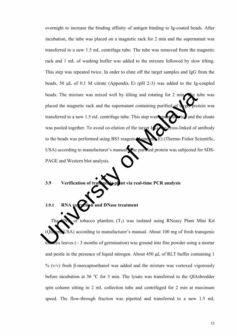
53
overnight to increase the binding affinity of antigen binding to Ig-coated beads. After
incubation, the tube was placed on a magnetic rack for 2 min and the supernatant was
transferred to a new 1.5 mL centrifuge tube. The tube was removed from the magnetic
rack and 1 mL of washing buffer was added to the mixture followed by slow tilting.
This step was repeated twice. In order to elute off the target samples and IgG from the
beads, 50 µL of 0.1 M citrate (Appendix E) (pH 2-3) was added to the Ig-coupled
beads. The mixture was mixed well by tilting and rotating for 2 min. The tube was
placed the magnetic rack and the supernatant containing purified of target protein was
transferred to a new 1.5 mL centrifuge tube. This step was repeated twice and the eluate
was pooled together. To avoid co-elution of the target IgG, the cross-linked of antibody
to the beads was performed using BS3 reagent (Appendix E) (Thermo Fisher Scientific,
USA) according to manufacturer’s manual. The purified protein was subjected for SDS-
PAGE and Western blot analysis.
3.9 Verification of transgenic plant via real-time PCR analysis
3.9.1 RNA extraction and DNase treatment
The RNA of tobacco plantlets (T1) was isolated using RNeasy Plant Mini Kit
(Qiagen, USA) according to manufacturer’s manual. About 100 mg of fresh transgenic
tobacco leaves (~ 3 months of germination) was ground into fine powder using a mortar
and pestle in the presence of liquid nitrogen. About 450 µL of RLT buffer containing 1
% (v/v) fresh β-mercaptoethanol was added and the mixture was vortexed vigorously
before incubation at 56 ℃ for 3 min. The lysate was transferred to the QIAshredder
spin column sitting in 2 mL collection tube and centrifuged for 2 min at maximum
speed. The flow-through fraction was pipetted and transferred to a new 1.5 mL
Univers
ity of
Mala
ya

54
microcentrifuge tube without disturbing the cell debris. Then, 0.5 volumes of chilled
absolute ethanol were added to the cleared lysate.
The mixture was mixed and transferred to RNeasy mini spin column (~ 675 µL)
followed by centrifugation at 10,000 rpm for 15 s. The flow-though was discarded.
About 700 µL of buffer RW1 was added onto the RNeasy column and centrifuged at
10,000 rpm for 15 s. The flow-though and the collection tube were discarded. The
RNeasy column was transferred to new 2 mL collection tube followed by pipetting 500
µL of buffer RPE onto the RNeasy column. Centrifugation was performed at 10,000
rpm for 15 s. The flow-though was discarded. This step was repeated twice following
centrifugation at maximum speed for 2 min. Full speed centrifugation for 1 min was
performed to remove any residual ethanol. The RNeasy column was then transferred to
a new 1.5 mL microcentrifuge tube and added with 30 µL of RNase-free water onto the
RNeasy membrane. The mixture was incubated for 20 min at RT before centrifuged at
10,000 rpm for 1 min. Then, the eluate was analyzed by agarose gel electrophoresis
described in Section 3.3.2.3 and stored at –80 ℃ until use. The quantification of RNA
was quantified using BioPhotometer (Eppendorf, USA). The quantification was
measured using 50X dilution (1 µL samples with 49 µL RNase-free water. After
measuring the RNA concentration, DNase treatment was carried out using RapidOut
DNA Removal Kit (Thermo Fisher Scientific, USA) to remove the remaining DNA.
The reaction mixture was prepared as showed in Table 3.8.
Univers
ity of
Mala
ya

55
Table 3.8: RapidOut DNA removal reaction mixture.
Components Working concentration Volume, µL
RNA samples 2 µg Concentration dependent
10X DNase buffer with MgCl2
1X 1
DNase I, RNase-free 1U 0.5
Water, nuclease-free - Adjusted to the final volume of 10 µL
The tube containing reaction mixture was mixed and incubated at 37 ℃ for 30 min.
Then, DNase-treated RNA was quantified using BioPhotometer (Eppendorf, USA) and
assessed by agarose gel electrophoresis described in Section 3.3.2.3.
3.9.2 Reverse-transcription PCR (RT-PCR)
The reverse-transcription PCR was performed prior to quantitative real-time analysis.
This cDNA conversion was carried out using RevertAid H minus First Strand cDNA
synthesis kit (Thermo Fisher Scientific, USA). The reaction mixture was prepared
according to manufacturer’s protocol. The reaction mixture was prepared as described
in Table 3.9 and aliquoted into prepared tube as prepare describe in Table 3.10.
Univers
ity of
Mala
ya

56
Table 3.9: Reverse-transcriptase PCR reaction mixture.
Components Volume (1X reaction)
5X reaction buffer 4 µL
RiboLock RNase Inhibitor (2U/ µL) 1 µL
10 mM dNTP mix 2 µL
RevertAid H Minus M-MuLV Reverse Transcriptase (200U/ µL)
1 µL
TOTAL VOLUME 20 µL
Table 3.10: DNase treated-RNA preparation.
Components Volume (1X reaction)
DNase-treated RNA Concentration dependent, 1 µg
Oligo (dT)18 primer 1 µL
Nuclease-free H2O Topped up to 12 µL
TOTAL VOLUME 12 µL
The mixture was gently mixed and briefly centrifuged before loaded into PCR
Thermocycler Biometra Tpersonal (Biometra, Germany). The thermal cycling condition
of reverse-transcription PCR as in Table 3.11. The cDNA was stored at – 20 ℃ for
short storage and – 80 ℃ for longer storage.
Univers
ity of
Mala
ya

57
Table 3.11: Thermal cycling condition of reverse-transcriptase PCR.
Temperature Time, (min)
42 ℃ 60
70 ℃ 5
4℃ ∞
The cDNA was stored at -20℃ for short storage and -80 ℃ for longer storage till
use.
3.9.3 Real-time PCR
The quantitative PCR was carried out using Applied BiosystemsTM QuantStudioTM
12K Flex Real-Time PCR machine. Firstly, the primer sets, F-FV60 & R-FV60 (target
gene) and F-L25 & R-L25 (internal reference gene) described in Table 3.5 was
examined though dissociation programs in order to check the binding affinity and
specificity of the primers. FV60 was used to amplify the recombinant protein gene
(TP60), mainly to compare the expression level due to the effect by different proteinase
inhibitors and/or KDEL. Nicotiana tabacum ribosomal large subunit 25, NtL25 was
used as the internal reference gene control (Schmidt & Delaney, 2010). The reaction
mixture was prepared as described in Table 3.12. Three replicates of each primer set
and non-template control were prepared and assayed with the following dissociation
thermal condition (Table 3.13). This dissociation curve was performed together with
samples in order to ensure the free-contamination or non-specific binding of primer
which will give false-positive results.
Univers
ity of
Mala
ya

58
Table 3.12: 5X Real-time PCR reaction mixtures.
Components Working concentration Volume, µL
2X Power SYBR® Green PCR Master mix
1X 5.0
F primer, 10 µM 0.5 µM 0.5
R primer, 10 µM 0.5 µM 0.5
cDNA template, 100 ng/µL 100 ng 1.0
Nucleases-free water - 3.0
The transgenic samples were analyzed using comparative expression study
(relative quantification, RQ) to determine the effect of proteinase inhibitors and/or
KDEL (BBI, KDEL, BBIKDEL, or OCPIKDEL) towards expression of target gene,
TP60. Each sample was prepared in triplicate including technical and biological. The
PCR reaction mixture was prepared as described in Table 3.12 and thermal cycle
condition was described in Table 3.13.
Table 3.13: Thermal cycle condition for real-time PCR.
Stage Temperature, ℃/ Time Cycle
1 95/ 10 min 1
2 95/ 1 min 40
60/ 15 sec
Then, the Ct value obtained from Applied BiosystemsTM QuantStudioTM 12K Flex
Real-Time PCR software was collected and recorded. The data were then analyzed
Univers
ity of
Mala
ya

59
manually using Microsoft Excel 2010 with calculation of expression 2-ΔΔCt (Livak &
Schmittgen, 2001).
The non-template control was used in triplicate for every run samples to ensure free-
contamination or unspecific binding of primers which lead to false-positive expression.
All data of comparative expression were used to compare the gene expression level in
between lines and constructed plasmids.
3.10 Semi-quantitative RT-PCR
For semi-quantitative RT-PCR, 100 ng cDNA was used as template following
normal PCR amplification (1 U/µL; iNtRON Biotechnology, Gyeonggi-do, South
Korea) at the annealing temperature of 60 ℃ for both target and internal reference gene
primers. The amplicon was visualized on an EtBr agarose gel electrophoresis under UV
Gel Pro Imager (Media Cybernetics, USA).
3.11 Statistical analysis
The statistical analysis was performed using SPSS 16.0 (SPSS Inc., USA) at 5 %
confident level. The analysis was conducted in triplicate and statistically analyzed using
one-way analysis of variance (ANOVA). The mean values of different plasmid
constructs were subjected to Duncan’s multiple comparison.
However, for number of shoot regeneration and GUS fluorometry analysis, Kruskal-
Wallis (non-parametric) test has been carried out due to not normally distributed across
samples tested (p < 0.05).
Univers
ity of
Mala
ya

60
CHAPTER 4: RESULTS
4.1 Endogenous test of N. tabacum cv. SR1 leaf discs towards antibiotics
In this study, endogenous test is the minimal shoot regeneration response of leaf disc
explants of N. tabacum cv. SR1 on regeneration media containing hygromycin
(selection agent) after one month of culture. The common method used in most
publications is based on the surviving explants over the total number of explants
cultured on selection media. However, this does not take into account the number of
shoots regenerated from the surviving explants. Hence, in this study, shoot regenerants
were scored on explants (producing the least number of shoots) when cultured on media
containing hygromycin. This is important in order to achieve the highest number of
shoots regenerated rather than percentage of surviving explants on selection media.
At 10 mg/L and 20 mg/L of hygromycin, the percentage of explants that survived
were 98 % (LD10) and 80 % (LD20), respectively, after one month of culture. At 30
mg/L, the percentage of surviving explants decreased to 50 % (LD50) followed by 3 %
(LD90) at 40 mg/L and 0 % (LD100) at 50 mg/L hygromycin. This showed that explants
were able to overcome the effect of antibiotics at certain concentration allowing chances
of survival after four weeks on culture plate. The percentage of surviving explants was
inversely proportional to the hygromycin concentrations (Figure 4.1). This assessment
method is commonly used in many publications, such as in Citrullus vulgaris (Suratman
et al., 2010) and Pisum sativum (Svabova et al., 2005).
Univers
ity of
Mala
ya

61
Figure 4.1: Percentage of survival explants on media containing hygromycin at different concentrations after one month of culture
At 0 mg/L, 100 % of the shoots regenerated at the cut edges and the surface of each
leaf explant cultured on media. However, the shoots regeneration and necrotic area out
of the total surface of explants decreased to about half (50 %) (LD50) due to the
selective pressure on media containing 10 mg/L hygromycin. At 20 mg/L hygromycin,
the shoot regeneration area was lesser compared to the necrotic area and decreased to
about 20 % (LD80) (less than half of 50 %). In addition, no shoots regenerated on
media-containing hygromycin at concentrations 30, 40 and 50 mg/L (LD100). The
whitish coloration formed at the edges of the leaf discs which were directly exposed to
the antibiotics in the media showed that the explants were naturally intolerant to high
concentration of hygromycin. Results are shown in Table 4.1.
Thus, 20 mg/L hygromycin (LD80) was chosen for the selection of transformants
based on the minimal number of shoots regenerated. Higher concentrations were not
chosen because the transformants may not be able to survive due to stringent selection
pressure.
0
20
40
60
80
100
120
0 10 20 30 40 50 60
Percentage of surviving explants (%)
Concentration of hygromycin, mg/L
Univers
ity of
Mala
ya

62
This result was in contrast to the earlier report by Pathi et al., (2013), where
transgenic tobacco plants were selected at 30 mg/L hygromycin where 100 % non-
transformants were eliminated. On the other hands, the obtained results were similar as
reported by Suratman et al. (2010) where 5 mg/L (LD80) of hygromycin was chosen for
the selection of Citrullus vulgaris transformants.
Univers
ity of
Mala
ya

63
Table 4.1: Assessment on minimal concentration of hygromycin in N. tabacum cv. SR1 leaf discs
Concentration of
hygromycin
Replicate 1
Replicate 2
Replicate 3
0 mg/L
10 mg/L
20 mg/L
++++ ++++
+++
++++
++ ++ ++
+ + +
Univers
ity of
Mala
ya

64
Table 4.1, continued.
Key: ++++ Highest responsive explants forming shoot (100 %)
++ Equal responsive explants forming shoots to necrotic areas (50 %)
+ Less responsive explants forming shoots to necrotic areas (20 %)
- All of the explants undergo necrosis. None of the explants forming shoots
30 mg/L
40 mg/L
50 mg/L
- - -
- - -
- - - Univers
ity of
Mala
ya

65
4.2 Confirmation of A. tumefaciens harboring binary vector and gene of interest
In this study, modified pCAMBIA 1304 with additional restriction sites, AvrII and
AfeI was used as a backbone binary vector harboring five different constructs (Figure
3.2) (Go, 2013).
Plasmid constructs were successfully isolated from overnight culture of
Agrobacterium and shown in Figure 4.2 with the expected size of ≥ 10 kb. Figures 4.3,
4.4, and 4.5 verified the presence of target and mgfp5 gene accordingly within T-DNA
region of the pCAMBIA 1304. The mgfp5 gene presence in Agrobacterium colonies
with empty vector was amplified using GFP primers at the expected size of 759 bp
(Figure 4.3). Agrobacterium colonies contained gene of interest (Figure 3.2 (A-D) were
amplified using 1304sk primers with the expected size of target gene ~ 900 bp (TP60
encoded for anti-Toxoplasma scFv antibody and/or with KDEL) (Figure 4.4) and ~ 1.5
kb (TP60 together with proteinase inhibitors with or without KDEL; BBI, BBIKDEL
and OCPIKDEL) (Figure 4.5). The exact size of each plasmid using 1304sk primers
mentioned in Table 3.3.
The presence of insert within T-DNA region of pCAMBIA 1304 was double
confirmed by using another pair of primers flanking between insert and mgfp5 gene (F-
1304sk and R-GFP) (Figures 4.6 and 4.7 at the expected size of ~1.6 and 2.3 kb,
respectively) and between CaMV 35S promoter and target (F-35S and R1-1304sk)
(Figures 4.8 and 4.9 at the expected size of ~ 1.3 kb and ~ 1.9 kb, respectively). The
expected size of each plasmid constructs using different combination of primers
mentioned showed in Table 4.2.
Univers
ity of
Mala
ya

66
Table 4.2: The list of primer combinations used for the confirmation of insert.
Primer set Plasmid constructs PCR product size
F-1304sk &
R-GFP
pTP60
pTP60KDEL
pTP60BBI
pTP60BBIKDEL
pTP60OCPIKDEL
1673 bp
1694 bp
2279 bp
2300 bp
2337 bp
F-35S &
R1-1304sk
pTP60
pTP60KDEL
pTP60BBI
pTP60BBIKDEL
pTP60OCPIKDEL
1314 bp
1335 bp
1920 bp
1941 bp
1978 bp
Lane M : VC 1 kb DNA ladder from Vivantis, Malaysia
Lanes 1-6 : Extracted plasmid of pCAMBIA 1304 containing gene of interest
M 1 2 3 4 5 6
250 bp
10 kb
Figure 4.2: 10 kb of isolated plasmid containing gene of interest.
Univers
ity of
Mala
ya

67
Lane M : PerfectTM 100 bp DNA ladder from Eurx, Poland
Lane –VE : Negative control, without plasmid template
Lane 1-2 : Isolated plasmid with empty vector, pCAMBIA 1304
Lane M : PerfectTM 100 bp DNA ladder from Eurx, Poland
Lane –VE : Negative control, without plasmid template
Lane 1 : Extracted plasmid of pCAMBIA 1304 containing anti-Toxoplasma (scFv) recombinant antibody gene, pTP60 construct (914 bp)
Lane 2 : Extracted plasmid of pCAMBIA 1304 containing anti-Toxoplasma (scFv) recombinant antibody and ER-targeting gene, pTP60KDEL construct (935 bp)
M -VE 1 2
1 kb
500bp
759 bp
M -VE 1 2
1 kb
500 bp
~ 900 bp
Figure 4.3: PCR verification of empty plasmid (without insert) using a pair of GFP primer.
Figure 4.4: PCR verification of extracted plasmid using a pair of 1304sk primer. Univers
ity of
Mala
ya

68
Lane M : PerfectTM 100 bp DNA ladder from Eurx, Poland
Lane –VE : Negative control, without plasmid template
Lane 1 : Extracted plasmid of pCAMBIA 1304 containing anti-Toxoplasma (scFv) recombinant antibody gene with PI, pTP60BBI construct (1520 bp)
Lane 2 : Extracted plasmid of pCAMBIA 1304 containing anti-Toxoplasma (scFv) recombinant antibody gene with PI-KDEL, pTP60BBIKDEL construct (1541 bp)
Lane 3 : Extracted plasmid of pCAMBIA 1304 containing anti-Toxoplasma (scFv) recombinant antibody gene with PI-KDEL, pTP60OCPIKDEL construct (1578 bp)
M -VE 1 2 3
1 kb
500 bp
~ 1.5 kb
Figure 4.5: PCR verification of extracted plasmid using a pair of 1304sk primers.
Univers
ity of
Mala
ya

69
:
Lane M : PerfectTM 100 bp DNA ladder from Eurx, Poland
Lane –VE : Negative control, without plasmid template
Lane 1 : Extracted plasmid of pCAMBIA 1304 containing anti-Toxoplasma (scFv) recombinant antibody gene, pTP60 construct (1673 bp)
Lane 2 : Extracted plasmid of pCAMBIA 1304 containing anti-Toxoplasma (scFv) recombinant antibody with ER-targeting gene, pTP60KDEL construct (1694 bp)
1 kb
500 bp
~ 1.6 kb
M -VE 1 2
Figure 4.6: PCR verification of extracted plasmid using a pair of F-1304sk and R-GFP primers.
Univers
ity of
Mala
ya

70
Lane M : PerfectTM 100 bp DNA ladder from Eurx, Poland
Lane –VE : Negative control, without plasmid template
Lane 1 : Extracted plasmid of pCAMBIA 1304 containing anti-Toxoplasma (scFv) recombinant antibody gene with PI, pTP60BBI construct (2279 bp)
Lane 2 : Extracted plasmid of pCAMBIA 1304 containing anti-Toxoplasma (scFv) recombinant antibody gene with PI-KDEL, pTP60BBIKDEL construct (2300 bp)
Lane 3 : Extracted plasmid of pCAMBIA 1304 containing anti-Toxoplasma (scFv) recombinant antibody gene with PI-KDEL, pTP60OCPIKDEL construct (2337 bp)
M -VE 1 2 3
500 bp
1 kb
2 kb ~ 2.2 - 2.3 kb
Figure 4.7: PCR verification of extracted plasmid using a pair of F-1304sk and R1-GFP primers.
Univers
ity of
Mala
ya

71
Lane M : PerfectTM 100 bp DNA ladder from Eurx, Poland
Lane –VE : Negative control, without plasmid template
Lane 1 : Extracted plasmid of pCAMBIA 1304 containing anti-Toxoplasma (scFv) recombinant antibody gene, pTP60 construct (1314 bp)
Lane 2 : Extracted plasmid of pCAMBIA 1304 containing anti-Toxoplasma (scFv) recombinant antibody with ER-targeting gene, pTP60KDEL construct (1335 bp)
M -VE 1 2
~ 1.3 kb 1.5 kb 1 kb
500 bp Unspecific band
Figure 4.8: PCR verification of extracted plasmid using a pair of F-35S and R1-1304sk primers.
Univers
ity of
Mala
ya

72
Lane M : PerfectTM 100 bp DNA ladder from Eurx, Poland
Lane –VE : Negative control, without plasmid template
Lane 1 : Extracted plasmid of pCAMBIA 1304 containing anti-Toxoplasma (scFv) recombinant antibody gene with PI, pTP60BBI construct (1920 bp)
Lane 2 : Extracted plasmid of pCAMBIA 1304 containing anti-Toxoplasma (scFv) recombinant antibody gene with PI-KDEL, pTP60BBIKDEL construct (1941 bp)
Lane 3 : Extracted plasmid of pCAMBIA 1304 containing anti-Toxoplasma (scFv) recombinant antibody gene with PI-KDEL, pTP60OCPIKDEL construct (1978 bp)
~ 1.9 kb
Unspecific band
1.5 kb
1 kb
500 bp
M -VE 1 2 3
Figure 4.9: PCR verification of extracted plasmid using a pair of F-35S and R1-1304sk primers.
Univers
ity of
Mala
ya

73
4.2.1 Sequencing and BLAST analysis
Results as shown in Figures 4.10, 4.11, 4.12, and 4.13 confirmed the correct
sequences for all constructs pTP60, pTP60KDEL, pTP60BBI/pTP60BBIKDEL, and
pTP60OCPIKDEL respectively, harbored within T-DNA region of pCAMBIA 1304
using a primer pair of F-1304sk and R-GFP.
The BLAST protein result for pTP60-mgfp5 is shown in Figure 4.10. Based on the
result obtained, target gene sequence of anti-Toxoplasma (scFv) recombinant protein
conserved domain showed highest similarity (97 % maximum identity) with anti-T.
gondii tachyzoite single-chain variable fragment antibody [synthetic construct]
(accession no.: AEX30825.1) with query coverage of 45 % (Figure 4.10) fused to E-
epitope tag (GAPVPYPDPLER). Besides that, this result also showed that GFP protein
is an unfused to target protein with the highest domain similarity for about 96 % of
maximum identity with mGFP* hexaHis tagged in pCAMBIA vector. This confirmed
that the target gene harbored within T-DNA region of pCAMBIA 1304.
Univers
ity of
Mala
ya

74
Figure 4.10: BlastP search result of anti-Toxoplasma (scFv) recombinant protein and GFP. Anti-Toxoplasma (scFv) recombinant protein and conserved domain showed highest similarity (97 % maximum identity) with anti-Toxoplasma gondii tachyzoite single-chain variable fragment antibody [synthetic construct] (accession no.: AEX30825.1) with query coverage of 45 % and unfused to GFP protein domain showed highest similarity (96 % maximum identity) with mGFP* hexaHis tagged [Binary vector pCAMBIA-1302] (accession no.: AAF65344.1) with query coverage 47 % of whole sequence.
Univers
ity of
Mala
ya

75
The BLAST protein result for pTP60KDEL shown in Figure 4.11. Based on the
result obtained, target gene sequence of anti-Toxoplasma (scFv) recombinant protein
conserved domain showed highest similarity (97 % maximum identity) with anti-T.
gondii tachyzoite single-chain variable fragment antibody and scFv antibody TP60
[synthetic construct] (accession no.: AEX30825.1 and AHA85991.1 respectively) with
query coverage of 75 % and 72 % respectively of whole sequence fused to E-epitope
tag (GAPVPYPDPLER). Besides, this result also showed SEKDEL (plant microsomal
retention peptide sequence) which represents ER retention sequence unfused to the
target gene.
Univers
ity of
Mala
ya

76
Figure 4.11: BlastP search result of anti-Toxoplasma (scFv) recombinant protein and KDEL. Anti-Toxoplasma (scFv) recombinant protein conserved domain showed highest similarity (97 % maximum identity) with anti-Toxoplasma gondii tachyzoite single-chain variable fragment antibody and scFv antibody TP60 [synthetic construct] (accession no.: AEX30825.1 and AHA85991.1 respectively) with query coverage of 75 % and 72 % respectively of whole sequence. SEKDEL sequence represents ER retention sequence.
The BLAST protein result for pTP60BBI/ pTP60BBIKDEL shown in Figure 4.12.
Based on the result obtained, target gene sequence of anti-Toxoplasma (scFv)
recombinant protein conserved domain showed highest similarity (95 % and 96 %
maximum identity) with anti-Toxoplasma gondii tachyzoite single-chain variable
fragment antibody and scFv antibody TP60 [synthetic construct] (accession no.:
AEX30825.1 and AHA85991.1 respectively) with query coverage of 56 % and 54 %,
respectively, of whole sequence fused to E-epitope tag (GAPVPYPDPLER). Besides
Univers
ity of
Mala
ya

77
that, this result also showed the unfused BBI protein domain showed highest similarity
(93 % maximum identity) with Bowman-Birk type proteinase inhibitor DE-4 like [Musa
acuminata subsp. malaccensis (accession no.: XP_009413787.1) with query coverage
28 % of whole sequence. For pTP60BBIKDEL construct, the SEKDEL sequence could
not be amplified due to poly-A at 3’ region which led to DNA polymerase slippage.
Univers
ity of
Mala
ya

78
Figure 4.12: BlastP search result of anti-Toxoplasma (scFv) recombinant protein and BBI proteinase inhibitor. Anti-Toxoplasma (scFv) recombinant protein conserved domain showed highest similarity (95 % and 96 % maximum identity) with anti-Toxoplasma gondii tachyzoite single-chain variable fragment antibody and scFv antibody TP60 [synthetic construct] (accession no.: AEX30825.1 and AHA85991.1 respectively) respectively with query coverage of 56 % and 54 % respectively of whole sequence. Unfused BBI protein domain showed highest similarity (93 % maximum identity) with Bowman-Birk type proteinase inhibitor DE-4 like [Musa acuminata subsp. malaccensis (accession no.: XP_009413787.1) with query coverage 28 % of whole sequence.
Univers
ity of
Mala
ya

79
The BLAST protein result for pTP60OCPIKDEL is shown in Figure 4.13. Based on
the result obtained, target gene sequence of anti-Toxoplasma (scFv) recombinant
protein conserved domain showed highest similarity (96 % and 97 % maximum
identity) with anti-Toxoplasma gondii tachyzoite single-chain variable fragment
antibody (accession no.: AEX30825.1) and scFv antibody TP60 [synthetic construct]
(accession no.: AHA85991.1) with query coverage of 59 % and 57 %, respectively, of
whole sequence. This result also showed the unfused OCPI (CY) superfamily protein
domain showed highest similarity (94 % maximum identity) with cysteine proteinase
inhibitor 1 [Oryza sativa japonica group] (accession no.: XP_015621143.1) with query
coverage 32 % of whole sequence fused with SEKDEL sequence which represents ER
retention sequence.
Univers
ity of
Mala
ya

80
Figure 4.13: BlastP search result of anti-Toxoplasma (scFv) recombinant protein and OCPI proteinase inhibitor. Anti-Toxoplasma (scFv) recombinant protein conserved domain showed highest similarity (96 % and 97 % maximum identity) with anti-Toxoplasma gondii tachyzoite single-chain variable fragment antibody (accession no.: AEX30825.1) and scFv antibody TP60 [synthetic construct] (accession no.: AHA85991.1) with query coverage of 59 % and 57 % respectively of whole sequence. Unfused OCPI (CY) superfamily protein domain showed highest similarity (94 % maximum identity) with cysteine proteinase inhibitor 1 [Oryza sativa japonica group] (accession no.: XP_015621143.1) with query coverage 32 % of whole sequence. SEKDEL sequence represents ER retention sequence.
Univers
ity of
Mala
ya

81
4.3 Transformation of N. tabacum cv. SR1 leaf discs mediated by A. tumefaciens
4.3.1 Effects of different plasmid constructs towards regeneration of putative transformed shoots
All non-transformed tobacco explants were able to regenerate showing the highest
score (+++++) in shoot regeneration on TSM media in the absence of antibiotics.
Certain edge of the explants also formed calluses due to the presence of auxin and
cytokinin in the media. In addition, least number of shoots regenerated on media
containing antibiotics especially on the mid-vein of the explants which might be due to
the explants’ least exposure of the to the antibiotic which led to the low selection
pressure (20 % false positives) (Table 4.1 and Figure 4.1). LD80 (20 mg/L) has been
selected instead of LD100 (at the minimum of 30 mg/L) to ensure the survival of all
possible putative transformed shoots. This is to avoid the stringent selection of
antibiotics that would inhibit the growth of transformants.
Based on the scoring results in Table 4.2, explants that have been transformed with
vector, pCAMBIA 1304 without any inserts showed higher number of regenerated
shoots as compared to those transformed with genes of interest (recombinant protein in
the presence of proteinase inhibitor and/ or KDEL (KDEL, BBI, BBIKDEL and
OCPIKDEL). This shows that the vector did not interfere with shoot regeneration.
Univers
ity of
Mala
ya
82
Table 4.3: The effect of different plasmid constructs towards the regeneration of putative transformed shoots on selection media (TSM-containing 20 mg/L hygromycin) after one month of culture.
Constructs Replicate 1 Replicate 2 Replicate 3
Non-transformed
(MS media)
Non-transformed
(Selection media)
pCAMBIA 1304
+++++
+++++
+++++
- +
+
+++++ +++++ ++++
Univers
ity of
Mala
ya

83
Table 4.3, continued.
pTP60
pTP60BBI
pTP60KDEL
++++ ++++ +++
++++ ++++ +++
+++ ++ ++
Univers
ity of
Mala
ya

84
Table 4.3, continued.
Key: +++++ Highest score of responsive explants
++++ Higher score of responsive explants
+++ Lower score of responsive explants
++ Low score of responsive explants
+ Lowest of responsive explants
- None responsive explants
pTP60
BBIKDEL
pTP60
OCPIKDEL
+++++ ++++ ++++
+++++ ++++ +++++
Univers
ity of
Mala
ya

85
Figure 4.14: Number of regenerated shoots versus different constructs. Significant difference (p < 0.05) between putative transformed shoots according non-parametric test, Kruskal-Wallis and Duncan test. Bar indicates the standard error (SE).
After 10 weeks of selection, the number of shoots regenerated (~1 cm length) from
explants (5 explants per replicate, triplicate) were recorded and analyzed statistically
using one-way ANOVA and Duncan Multiple range test at significance level of p <
0.05 (Appendix F). Based on Figure 4.14, results showed that explants transformed with
empty vector, pCAMBIA 1304 without gene of interest produced (35.67 ± 12.86)
shoots followed by descending shoots number of pTP60BBIKDEL (34 ± 4.16),
pTP60OCPIKDEL (23.33 ± 3.48), pTP60 (20.33 ± 0.33), pTP60BBI (12.33 ± 4.18),
and pTP60KDEL (5.33 ± 3.53) on selection media.
In addition, the co-expression of TP60 gene with only BBI or KDEL element
showing a significant different in the regeneration of shoots compared to transformation
with pCAMBIA 1304. The co-expression of TP60 gene in the combination of both BBI
and KDEL showing higher in shoot regeneration compared to the element on its own
0
10
20
30
40
50
60
Number of regenerated shoots
Constructs
a a
a
a,b a,b
b b
Univers
ity of
Mala
ya

86
and even as good as wild-type. This result also similar for the explants transformed with
only TP60 gene or the co-expression of TP60 gene with OCPI and KDEL elements in
which no significant different was observed compared to wild-type.
This could be concluded that recombinant gene of anti-Toxoplasma with the
companion of proteinase inhibitor and ER- retention signal (OCPIKDEL and
BBIKDEL) do not affect the plant growth especially in shoots regeneration.
4.3.2 Confirmation of putative transformed shoots using transient assays
4.3.2.1 GUS histochemical staining
GUS histochemical staining is a qualitative assay for transient gene expression and
also allows tissue-and cell specific localization. This assay was conducted in situ with
the chromogenic substrate (X-gluc) resulting in deposition of an insoluble blue-
precipitate. Regardless of T-DNA truncation during the transformation, the formation of
insoluble blue-precipitate indicates the localization of the transgene due to the
successful of transformation. GUS assay was conducted on the expanded leaves of ~ 10
week-old putative transformed shoots.
Based on Figure 4.15, the regenerated shoots from explants transformed with empty
vector, pCAMBIA 1304 (positive control) produced the most intense blue-GUS spots
per expanded leaf area (Figures 4.15a-b). The blue intensity of regenerated shoots from
explants transformed with pTP60 (Figure 4.15c) and pTP60KDEL (Figure 4.15d) were
similar as pCAMBIA 1304 putative transformants, even though the number of blue-
GUS spot observed per expanded leaf of these constructs are lesser compared to the
positive control.
Univers
ity of
Mala
ya

87
However, putative transformed shoots of pTP60BBI, pTP60OCPIKDEL, and
pTP60BBIKDEL produced least expression of blue-GUS coloration with less intensity
compared to the mentioned construct (Figures 4.15e-g). No GUS expression was
observed on the expanded non-inoculated regenerants confirmed the successful
transformation due to the absence of endogenous GUS gene in N. tabacum cv. SR1. The
different GUS expression between constructs might be due to the distance of gusA gene
from promoter after the inserts. The larger the insert size, the lower the GUS
expression. Although the GUS expression of control and putative transformants with
different constructs was different, quantification of GUS expression is still needed to
further verify its expression.
Univers
ity of
Mala
ya

88
10 x 1.6
a
10 x 1.6
b
10 x 1.6
c
10 x 1.6
d
10 x 1.6
e
10 x 1.6
f
Figure 4.15: GUS histochemical staining of putative transformed tobacco shoots (~ 10 week-old) on TSM-selection media.
Univers
ity of
Mala
ya

89
Figure 4.15, continued.
a and b : Inoculated with A. tumefaciens harbored with empty vector, pCAMBIA 1304
c : Inoculated with A. tumefaciens harbored with pTP60 construct
d : Inoculated with A. tumefaciens harbored with pTP60KDEL construct
e : Inoculated with A. tumefaciens harbored with pTP60BBI construct
f : Inoculated with A. tumefaciens harbored with pTP60OBBIKDEL construct
g : Inoculated with A. tumefaciens harbored with pTP60OCPIKDEL construct
h : Non-inoculated regenerant; as negative control
4.3.2.2 GUS fluorometry assay
GUS fluorometry assay is a quantitative assessment of GUS activity with fluorogenic
substrates, 4-MUG which would be hydrolyzed to form 4-MU protein product in the
presence of β-glucuronidase enzyme encoded by gusA gene (Figure 4.16). The reading
of GUS activity was calculated over the total protein concentration in 100 mg samples.
The total and 4-MU protein concentrations were calculated based on standard curve
obtained from known concentrations of BSA and 4-MU (Figures 4.17 and 4.18
respectively).
10 x 1.6
h
10 x 1.6
g
Univers
ity of
Mala
ya

90
Figure 4.16: Quantitative assessment of GUS activity with fluorogenic substrates. 4-MUG which would hydrolyzed to form 4-MU protein product in the presence of gusA gene encoded for β-glucuronidase enzyme. Adopted from http://biochemistry.wur.nl/pbc/gus-jenway/info3.html
Based on Figure 4.19, 4-MU protein products expression was significantly higher in
pCAMBIA 1304 putative transformants with 179.07 ± 0.010 pmole 4-MU min-1 mg-1
followed by pTP60 (26.76 ± 0.015 pmole 4-MU min-1 mg-1), pTP60KDEL (19.58 ±
0.006 pmole 4-MU min-1 mg-1), pTP60BBI (7.41 ± 0.002 pmole 4-MU min-1 mg-1), and
pTP60BBIKDEL (6.00 ± 0.001 pmole 4-MU min-1 mg-1). The least 4-MU protein
product was expressed by pTP60OCPIKDEL putative transformants with (3.62 ± 0.001
pmole 4-MU min-1 mg-1). All samples were corresponding to the wild-type as a baseline
reading. The reading was taken in three biological replicates (Appendix G). The
expression observed in wild-type might be due to the fluorescent of chloroplast under
same excitation and emission wavelength as GUS gene.
Univers
ity of
Mala
ya

91
y = 4.9011x + 10.714 R² = 0.9998
0
500
1000
1500
2000
2500
3000
0 100 200 300 400 500 600
Fluorescene intensity
4-MU concentration, pmole
The reading obtained from the quantitative data of GUS expression correlated to the
GUS qualitative assay. The expression obtained in wild-type samples might be due to
the interference of auto-fluorescence signal from subcellular organelles or other
pigments within plant cells which excited and emitted at the same wavelength as GUS
protein.
Figure 4.17: Standard curve of fluorescence intensity versus known 4-MU concentration.
Univers
ity of
Mala
ya

92
y = 0.6649x + 0.0265 R² = 0.9972
0
0.2
0.4
0.6
0.8
1
1.2
0 0.2 0.4 0.6 0.8 1 1.2 1.4 1.6
A595
Protein concentration, µg/µL
Figure 4.18: Standard curve of known concentration BSA using Bradford’s assay quantification.
Figure 4.19: GUS activity, (pmole per min) in 100 mg of putative transformed shoots transformed with different plasmid constructs. All sample readings were taken in response to the wild-type as negative control. Significant difference (p < 0.05) between putative transformed shoots according to non-parametric test, Kruskal-Wallis and Duncan test. Bars indicate the standard error (SE).
Univers
ity of
Mala
ya

93
4.3.2.3 GFP qualitative assay
Green fluorescent protein encoded by mgfp5 gene is a reporter gene that has widely
been used due to several advantages provided by this gene compared to GUS reporter
gene. GFP reporter gene is easier to be screened and non-destructive compared to GUS.
The expectation of green fluorescence signal observed on the expanded putative
transformed leaves was correlated with the presence of mgfp5 gene. Positive green
fluorescence signal of GFP on putative transformed N. tabacum cv. SR1 shoots within
merged images of upper and lower epidermis using software (LAS AF version 2.3.1
build 5194 SP5) are shown in Figure 4.20. The green fluorescence signal formation was
observed on the expanded leaf of putative transformed shoots under excitation and
emission wavelength of 488 nm and 550 nm, respectively, using confocal laser scanning
microscope. The non-transformed region of the same expanded putative transformed
shoots showed red coloration (Figure 4.20a) due to auto-fluorescence under UV light in
the presence of chlorophyll background signal (Goldman et al., 2003; Zhou et al.,
2005).
Univers
ity of
Mala
ya

94
Figure 4.20:
a : Expanded leaf under CLSM at UV wavelength
b : Expanded leaf under CLSM at bright field state
c: : Expanded leaf under CLSM at GFP ex/em wavelength of 488/550 nm
d : Overlaid images of a and c
a
d c
b
Figure 4.20: Visualization of expanded putative transformed shoots with pTP60 construct under a confocal laser scanning microscope (40X-oil immersion objective lens). Scale bar represents 100 µm.
Univers
ity of
Mala
ya

95
4.3.3 Verification of putative transformed shoots (T0) through PCR analysis
Approximately 10-week-old putative transformed tobacco shoots (T0) harboring
different plasmid constructs were selected and screened using PCR analysis. Total
genomic DNA that has been isolated from representative lines shown in Figure 4.21 and
Figure 4.22.
Isolated genomic DNA of representative lines from each constructs were screened
using PCR analysis with specific primers (Table 3.3). The putatively transformed
tobacco shoots with an empty vector was screened using F-RGFP primer. The detection
of about 759 bp amplified fragments confirmed the presence GFP within T-DNA region
(Figure 4.23). Meanwhile, F-R11304sk primers were used to screen putatively
transformed shoots present with different plasmid constructs (pTP60, pTP60KDEL,
pTP60BBI, pTP60BBIKDEL, and pTP60OCPIKDEL). Detection of about ~ 900 bp
and ~ 1.5 kb (Figures 4.24 and 4.25, respectively) confirmed the amplified fragment of
anti-Toxoplasma gene with the companion of different proteinase inhibitor and/or
KDEL.
Genomic DNA from wild-type SR1 tobacco plantlets was used as negative control.
Plasmid DNA from A. tumefaciens was isolated and used as positive technical control.
No amplification was detected in wild-type tobacco plantlets using both of the
mentioned primers indicated the successful of the transformation.
Univers
ity of
Mala
ya

96
Lane M : Perfect PlusTM 1 kb DNA ladder (EURx, Poland)
Lanes 1-2 : Non-transformed SR1 tobacco plantlets
Lanes 3-4 : Putative transformed shoots harboring pCAMBIA 1304
Lanes 5-6 : Putative transformed shoots harboring pTP60 construct
Lanes 7-9 : Putative transformed shoots harboring pTP60KDEL construct
M 1 2 3 4 5 6 7 8 9 M
10 kb
Figure 4.21: Genomic DNA of selected putative transformed tobacco shoots harboring different plasmid constructs after 10 weeks of selection.
Univers
ity of
Mala
ya

97
Lane M : Perfect PlusTM 1 kb DNA ladder (EURx, Poland)
Lane 1 : Non-transformed SR1 tobacco plantlets
Lanes 2-3 : Putative transformed shoots harboring pCAMBIA 1304
Lanes 4-5 : Putative transformed shoots harboring pTP60BBI construct
Lanes 6-7 : Putative transformed shoots harboring pTP60BBIKDEL construct
Lanes 8-10 : Putative transformed shoots harboring pTP60OCPIKDEL construct
M 1 2 3 4 5 6 7 8 9 10 M
10 kb
Figure 4.22: Genomic DNA of selected putative transformed tobacco shoots harboring different plasmid constructs after 10 weeks of selection.
Univers
ity of
Mala
ya

98
Lane M : PerfectTM 100 bp DNA ladder (EURx, Poland)
Lane -VE : Negative technical control
Lane WT : Negative biological control (wild-type)
Lane +VE : Positive control
Lanes 1-7 : Putative transformed shoots harboring pCAMBIA 1304
Figure 4.23: PCR confirmation of putative transformed shoots using F-RGFP primers with the expected size of 759 bp.
759 bp
M -VE WT +VE 1 2 3 4 5 6 7
1.5 kb
500 bp
Univers
ity of
Mala
ya

99
1.5 kb
Lane M : PerfectTM 100 bp DNA ladder (EURx)
Lane -VE : Negative technical control
Lane WT : Negative biological control (wild-type)
Lane +VE : Positive control
Lanes 1-5 : Putative transformed shoots harboring pTP60 construct
Lanes 6-11 : Putative transformed shoots harboring pTP60KDEL construct
M -VE WT +VE 1 2 3 4 5 6 7 8 9 10 11
~ 900 bp
Figure 4.24: PCR confirmation of putative transformed shoots using F-R11304sk primers with the expected size of ~ 900 bp.
500 bp
Univers
ity of
Mala
ya

100
Lane M : PerfectTM 100 bp DNA ladder (EURx)
Lane -VE : Negative technical control
Lane +VE : Positive control
Lanes 1-2 : Putative transformed shoots harboring pTP60BBI construct
Lanes 3-4 : Putative transformed shoots harboring pTP60BBIKDEL construct
Lanes 5-6 : Putative transformed shoots harboring pTP60OCPIKDEL construct
4.4 Assessment of gene stability T1 generation of transgenic tobacco
4.4.1 Effect of different plasmid constructs on seed germination
T1 seedlings that have been transformed with different constructs were cultured on
selection media for one week before recording their percentage of germination (Figure
4.26) (Appendix H). The percentage of germination was significantly lowest (65 % ±
2.03) in transgenic seeds transformed with the empty vector pCAMBIA 1304 compared
to wild-type and other constructs. There was a possibility that there was a delay in
germination (after 7 days) since the observation was only made within 7 days of
Figure 4.25: PCR confirmation of putative transformed shoots using F-R11304sk primers with the expected size of ~ 1.5 kb.
M -VE +VE 1 2 3 4 5 6
~ 1.5 kb
500 bp
1 kb
Univers
ity of
Mala
ya

101
culturing period on selection media. Low germination could be due to selective pressure
caused by hygromycin.
Constructs with proteinase inhibitor and KDEL elements, pTP60BBIKDEL and
pTP60OCPIKDEL recorded the highest percentage of germination (90 % ± 1.45)
whereas T1 seeds with pTP60BBI alone and pTP60KDEL were 85 % ± 1.76 and 75 % ±
1.20 respectively. The presence of proteinase inhibitors together with KDEL were
shown to improve the germination (Yan et al., 2009) possibly by preventing the
degradation of endogenous protein involved in plant growth and development against
foreign proteins. Besides that, this mechanism also may be due to naturally isolated
proteinase inhibitors from plants (regardless of the plant species) which may not be
affecting the plant growth. Interestingly, the percentage of germination for T1 seeds
with pTP60 alone was similar with the wild-type germinated on non-selection media 83
% ± 0.88 and 80 % ± 1.67 respectively. This indicated that the anti-Toxoplasma protein
did not interfere with the growth of the tobacco plant itself which is in accordance to
previous study (Ahmad et al., 2012).
T1 seedlings were assessed after one month of germination in selection media. In the
presence of hygromycin in the media, the wild-type seedlings grew with abnormal
phenotype (pale green color cotyledon without root formation) (Figure 4.27a) while the
phenotype of the transgenic seedlings became normal plants (Figure 4.27b) which might
be due to the selective pressure caused by antibiotic. Univers
ity of
Mala
ya

102
Figure 4.26: The percentage of T1 seeds germination on 7th days of germination Same letter indicate not significantly different based on one-way ANOVA followed by Duncan’s multiple range test at 95 % confident interval level. Experiment was conducted in triplicate for each constructs. Bar indicates the standard error (S.E).
0102030405060708090
100
Perc
enta
ge o
f T1 s
eeds
ger
min
atio
n, %
Plasmid constructs
c,d e
b,c d b
a a
a b
Figure 4.27: Germination of wild-type (a) and pTP60 T1 seedlings (b) on selection media after 4 weeks of germination. Bar = 1 cm.
Univers
ity of
Mala
ya

103
4.4.2 Verification of transgenic seedlings (T1) through PCR analysis
Approximately, 4 to 6 weeks old of germinated hygromycin-resistant T1 seedlings
were screened using PCR analysis. The stable integration and heritable hygromycin-
resistant gene, hptII in T1 plant genome was confirmed using specific primers, HPT1.
Detection of a 559 bp amplified fragments in randomly selected seedlings with different
constructs confirmed the presence of transgene in T1 generation (Figures 4.28 and 4.29).
Similar result was observed in the positive control; neither non-template nor wild-
type tobacco showed the similar band at the expected size. At least 20 individuals from
three different lines of the different constructs were screened.
No band was observed for germinated T1 seedlings harboring pTP60BBI construct
(Figure 4.29, Lane 1). This is might be due to false-positive because the hygromycin
concentration use for selection was at LD80, with 20 % survival rate of non-transformed
seedlings. All false-positive seedlings were discarded.
Antibiotic-resistance selection marker gene was designed to locate on the left border
(LB) of Ti plasmid to avoid that truncation event in the T1 generation. The left border of
T-DNA may not be inherited in subsequent generation after T0 (Kemski et al. 2013). T1
seeds have been produced and obtained according to self-pollination of T0 tobacco
plants, which the confirmation of the insert, TP60 with the companion of different
proteinase inhibitors and/ or KDEL was previously carried out.
Univers
ity of
Mala
ya

104
Lanes M : PerfectTM 100 bp DNA ladder (Eurx, Poland)
Lane –VE : Negative technical control
Lane WT : Wild-type
Lane +VE : Positive control
Lane 1-4 : Germinated T1 seedlings harboring pCAMBIA 1304 construct
Lane 5-8 : Germinated T1 seedlings harboring pTP60 construct
Lane 9-12 : Germinated T1 seedlings harboring pTP60KDEL construct
559 bp
M -VE WT +VE 1 2 3 4 5 6 7 8 9 10 11 12
1 kb
500 bp
Figure 4.28: PCR confirmation of T1 seedlings using F-RHPT1 primers with an expected size of 559 bp.
Univers
ity of
Mala
ya

105
559 bp
Lane M : PerfectTM 100 bp DNA ladder (Eurx, Poland)
Lane WT : Wild-type
Lane +VE : Positive control
Lanes 1-3 : Germinated T1 seedlings harboring pTP60BBI construct
Lanes 4-5 : Germinated T1 seedlings harboring pTP60BBIKDEL construct
Lanes 6-9 : Germinated T1 seedlings harboring pTP60OCPIKDEL construct
4.5 Acclimatization of transgenic tobacco plants
At least three individuals (from three different lines of each construct) of PCR
positive T0 tobacco plants and five T1 transgenic tobacco progenies of each line were
acclimatized in transgenic greenhouse (Plant Biotechnology Facility, University of
Malaya, Malaysia). The development of transgenic N. tabacum cv. SR1 from
vegetative to reproductive stages are shown in Figure 4.30. Seeds from transgenic
plants were ready for collection within 3 months of acclimatization.
Figure 4.29: PCR confirmation of T1 seedlings using F-RHPT1 primers with an expected size of 559 bp.
+VE M WT 1 2 3 4 5 6 7 8 9
1 kb
500 bp
Univers
ity of
Mala
ya

106
(A)
(E)
(C)
(D)
(B)
Figure 4.30: The growth stages of transgenic Nicotiana tabacum cv. SR1 within 3 months of acclimatization. A) 5 weeks-old acclimatized transgenic tobacco plants; B) Acclimatized transgenic tobacco lines producing flowers; C) Formation of seed pods (black arrows), 4-6 days post-pollination; D) Dried seed pods (black arrows) ready for collection after sepals and stems turned brown; E) 2 weeks-old of germinated transgenic seeds on selection media. Red arrow represents the putative transformed seedlings; while white arrow represents the false-positives showing phenotype abnormality. Bar = 1 cm.
Univers
ity of
Mala
ya

107
4.6 Phenotypic assessment on T1 tobacco progenies
The effect on phenotypic changes of T1 tobacco progenies transformed with different
plasmid constructs were assessed and tabulated in Table 4.3.
Transgenic tobacco plant harboring pTP60 construct showed the highest stem height
(11.00 ± 1.22) compared to the others with different plasmid constructs. However, there
was no significant different between the wild-type and the former. These non-
phenotypic difference in stem height indicated that this anti-Toxoplasma protein was
not involved in the development of the tobacco plant itself which may be a useful
characteristics in biopharming (Appendix I).
The flowering day of wild-type and transformed tobacco were analyzed. The results
showed that the flowering day of wild-type tobacco plants was 65 days ± 5.49 (Table
4.4). On the other hand, the earliest flowering day was observed in T1 tobacco progenies
harboring pTP60BBI plasmid construct (45 days ± 2.67). It was also observed that T1
tobacco progenies presence of pTP60KDEL, pTP60BBIKDEL, and pTP60OCPIKDEL
plasmid constructs gave a similar period for the plant to flower in a range of 51-52 days,
which non-significantly difference to the one in the presence of pCAMBIA 1304
plasmid construct (49 days ± 3.28) (Appendix J). Interestingly, anti-Toxoplasma
recombinant protein alone showed non-significant different to wild-type (62 ± 2.08)
which is an important factor for biopharming purposes. It may be concluded that the
introduction of anti-Toxoplasma recombinant protein doesn’t affect the flowering
period of tobacco plants as compared to wild-type.
Univers
ity of
Mala
ya

108
Table 4.4: Assessment on stem height of T1 independent lines (20 days of post acclimatization) and flowering period on transgenic and wild-type tobacco plants.
Constructs
Stem height (cm)
Flowering period (day)
pTP60 11.00 ± 1.22a 62 ± 2.08a,b
pTP60KDEL 3.75 ± 0.92c 52 ± 2.33b,c
pTP60BBI 4.27 ± 0.52c 45 ± 2.67c
pTP60BBIKDEL 5.31 ± 1.27b,c 51 ± 2.96c
pTP60OCPIKDEL 3.27 ± 0.58c 52 ± 2.33b,c
pCAMBIA 1304 4.84 ± 1.59c 49 ± 3.28c
Wild-type 6.60 ± 1.43a,b 65 ± 5.49a
The results represent the mean ± standard error (SE) of three replicated experiments.
Different letters indicate significant difference between samples at p < 0.05.
4.7 Real-time PCR analysis
4.7.1 RNA extraction and DNase treatment of T1 tobacco progenies
Leaves of approximately 40-day acclimatized T1 tobacco progenies were harvested
for the downstream expression analysis of anti-Toxoplasma gene at mRNA and protein
levels. Total RNA of positive transformants was isolated using RNeasy Plant Mini Kit
(Qiagen, USA) prior to real-time PCR. The isolated RNA was quantified and listed in
Table 4.5. The RNA isolated products (before and after DNase treatment) were
electrophoresed on 1 % EtBr stained agarose gel, as shown in Figures 4.31-4.34.
Univers
ity of
Mala
ya

109
Detection of bands (28S and 18S rRNA) confirmed successful RNA isolation in regards
to both the purity and integrity of RNA. DNase treatment was carried out in order to
remove the remaining genomic DNA which may interfere in the expression.
The quantification of RNA before and after DNase treatment was also carried out
prior to cDNA conversion. After DNase treatment, the RNA concentration showed a
reduction which corresponds to the quantified RNA yield (about 10-fold change) before
DNase treatment Table 4.6.
Lanes 1-3 : T1 tobacco progenies harboring pTP60 construct
Lanes 4-6 : T1 tobacco progenies harboring pTP60KDEL construct
Lanes 7-9 : T1 tobacco progenies harboring pTP60BBI construct
Lane 10 : T1 tobacco progenies harboring pTP60BBIKDEL construct
28S rRNA 18S rRNA
1 2 3 4 5 6 7 8 9 10
Genomic DNA
Figure 4.31: 1 µg of non-treated total RNA isolated from 40-day old acclimatized T1 tobacco progenies.
Univers
ity of
Mala
ya

110
Lanes 1-3 : T1 tobacco progenies harboring pTP60 construct
Lanes 4-6 : T1 tobacco progenies harboring pTP60KDEL construct
Lanes 7-9 : T1 tobacco progenies harboring pTP60BBI construct
Lane 10 : T1 tobacco progenies harboring pTP60BBIKDEL construct
Lane 1-2 : Non-treated RNA, T1 tobacco progenies harboring pTP60BBIKDEL
construct Lane 3-5 : Non-treated RNA, T1 tobacco progenies harboring pTP60OCPIKDEL
construct Lane A-B : Treated RNA, T1 tobacco progenies harboring pTP60BBIKDEL construct Lane C-E : Treated RNA, T1 tobacco progenies harboring pTP60OCPIKDEL
construct
1 2 3 4 5 6 7 8 9 10
28S rRNA
18S rRNA
1 A 2 B 3 C 4 D 5 E
28S rRNA 18S rRNA
Figure 4.32: 100 ng of DNase-treated RNA isolated from 40-day old acclimatized T1 tobacco progenies.
Figure 4.33: 1 µg of non-treated and 100 ng of DNase-treated RNA isolated from 40-day old acclimatized T1 tobacco progenies. Univ
ersity
of M
alaya

111
Lane 1-3 : Non-treated RNA, Wild-type tobacco plants
Lane A-C : Treated RNA, Wild-type tobacco plants
1 A 2 B 3 C
28S rRNA 18S rRNA
Figure 4.34: 1 µg of non-treated and 100 ng of DNase-treated RNA isolated from 40-day old acclimatized tobacco wild-type.
Univers
ity of
Mala
ya

112
Table 4.5: ODA260/280 and ODA260/230 readings of non-transformed and T1 tobacco progenies before DNase-treatment.
Samples Concentration,
ng/µL
Purity
Samples
Concentration,
ng/µL
Purity
OD
A260/280
OD
A260/230
OD
A260/280
OD
A260/230
Non-transformed (1) 1369 1.98 1.74 pTP60BBI (5) 1422.1 1.90 2.26
Non-transformed (2) 1019.0 1.90 2.34 pTP60BBI (6) 1171.5 1.97 2.25
Non-transformed (9) 1231.6 2.00 2.01 pTP60BBI (7) 1125.8 2.07 2.44
pTP60 (3) 1824.9 1.95 2.10 pTP60BBIKDEL (1) 1281.3 1.96 2.25
pTP60 (12) 970.2 1.92 2.15 pTP60BBIKDEL (2) 1384.1 1.98 2.35
pTP60 (17) 1157.1 1.92 2.47 pTP60BBIKDEL (14) 2296.6 2.02 1.93
Univers
ity of
Mala
ya

113
Table 4.5, continued.
pTP60KDEL (6) 1791.2 1.96 2.54 pTP60OCPIKDEL (1) 2647.9 2.01 2.37
pTP60KDEL (7) 1263.9 2.06 2.22 pTP60OCPIKDEL (13) 1810.7 2.00 2.05
pTP60KDEL (8) 2018 2.01 2.27 pTP60OCPIKDEL (15) 1524.8 2.00 2.45
Univers
ity of
Mala
ya

114
Table 4.6: ODA260/280 and ODA260/230 readings of non-transformed and T1 tobacco progenies DNase-treated RNA.
Samples Concentration,
ng/µL
Purity
Samples
Concentration,
ng/µL
Purity
OD
A260/280
OD
A260/230
OD
A260/280
OD
A260/230
Non-transformed
(1) 143.5 2.16 1.65 pTP60BBI (5) 161.7 2.06 2.22
Non-transformed
(2) 181.9 2.16 2.21 pTP60BBI (6) 134.8 2.07 2.44
Non-transformed
(9) 130.27 2.02 1.47 pTP60BBI (7) 174.5 2.01 2.37
pTP60 (3) 206.2 2.17 1.89 pTP60BBIKDEL (1) 123.9 2.02 2.48
pTP60 (12) 215.2 1.89 2.01 pTP60BBIKDEL (2) 160.4 2.11 1.45
pTP60 (17) 209.2 2.10 1.78 pTP60BBIKDEL (14) 142.13 2.24 1.68 Univers
ity of
Mala
ya

115
Table 4.6, continued.
pTP60KDEL (6) 159.03 1.96 1.50 pTP60OCPIKDEL (1) 231.33 2.02 1.93
pTP60KDEL (7) 42.37 2.00 1.87 pTP60OCPIKDEL (13) 149.6 2.04 2.19
pTP60KDEL (8) 125.6 2.34 1.76 pTP60OCPIKDEL (15) 117.7 2.00 2.01
Univers
ity of
Mala
ya

116
4.7.2 Relative expression of TP60 gene using real-time PCR
Prior to qRT-PCR analysis, a dissociation curve for both reference (N. tabacum
ribosomal large subunit 25, NtL25) and target gene (anti-Toxoplasma, TP60) primers,
L25 and FV60 respectively was performed. The dissociation curves for both primers in
the screened samples showed a single peak, indicating the specificity of the primer
designed (Table 3.4) and dissociated at a specific temperature confirming the single
amplicon being produced (Figure 4.35).
FV60
L25
Figure 4.35: Dissociation curve of different primer sets, L25 and FV60 for NtL25 internal reference gene and TP60 recombinant gene respectively at 60 ℃ annealing temperature (derivative reporter versus temperature).
Univers
ity of
Mala
ya

117
Three lines for each construct (pTP60, pTP60KDEL, pTP60BBI, pTP60BBIKDEL,
and pTP60OCPIKDEL) were used in this study. A representative from each line was
selected based on the highest expression of anti-Toxoplasma gene at the mRNA level
based on relative quantification by using qRT-PCR (tested in 3 different individuals).
The list of T1 tobacco progeny lines used for each construct was tabulated in Table 4.5
and Table 4.6.
The generated data from relative quantification was analyzed using Microsoft Excel.
The results are summarized in Figure 4.36 to Figure 4.40. The transgenic line from each
construct showing the highest expression of TP60 gene was selected. The best
transgenic line from each construct was re-analyzed by qRT-PCR in order to compare
the expression level of TP60 gene between different constructs. The expression data
was recalculated and the data was presented in Figure 4.41. Based on Figure 4.41, the
expression level of anti-Toxoplasma gene, TP60 with the companion of proteinase
inhibitor and KDEL (pTP60BBIKDEL) showed the highest expression with a 19-fold
change, followed by pTP60KDEL (10-fold), pTP60OCPIKDEL (8-fold), and
pTP60BBI (2-fold) compared to anti-Toxoplasma gene in the presence of neither
proteinase inhibitor nor KDEL (Appendix K).
Semi-quantitative data for each construct showed correlation in intensity with the
quantified expression from each line for each construct (Figure 4.36 to Figure 4.41).
Univers
ity of
Mala
ya

118
0123456789
10
pTP60 (3) pTP60 (12) pTP60 (16)
RQ value
Transgenic line
1 2 3
FV60
L25
Figure 4.36: The expression levels of TP60 gene in T1 tobacco progenies compared between lines 3, 12, and 16 harboring pTP60 construct. RQ value = Relative expression value on Y axis represents transcript level relative to NtL25. Semi-quantitative expression; 1) pTP60 (3), 2) pTP60 (16), and 3) pTP60 (12). Experiment was conducted in 3 technical replicates.
Univers
ity of
Mala
ya
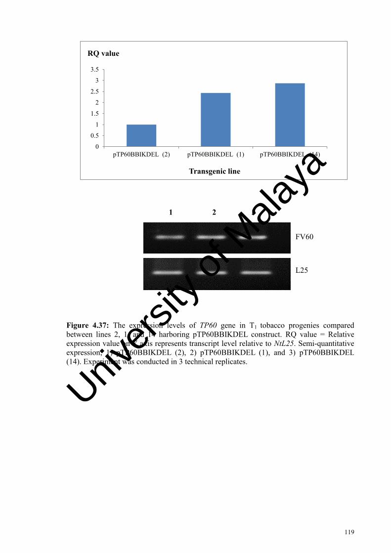
119
0
0.5
1
1.5
2
2.5
3
3.5
pTP60BBIKDEL (2) pTP60BBIKDEL (1) pTP60BBIKDEL (14)
RQ value
Transgenic line
1 2 3
FV60
L25
Figure 4.37: The expression levels of TP60 gene in T1 tobacco progenies compared between lines 2, 1, and 14 harboring pTP60BBIKDEL construct. RQ value = Relative expression value on Y axis represents transcript level relative to NtL25. Semi-quantitative expression; 1) pTP60BBIKDEL (2), 2) pTP60BBIKDEL (1), and 3) pTP60BBIKDEL (14). Experiment was conducted in 3 technical replicates.
Univers
ity of
Mala
ya

120
00.5
11.5
22.5
33.5
44.5
5
pTP60KDEL (7) pTP60KDEL (8) pTP60KDEL (6)
RQ value
Transgenic line
FV60
L25
1 2 3
Figure 4.38: The expression levels of TP60 gene in T1 tobacco progenies compared between lines 7, 8, and 6 harboring pTP60KDEL construct. RQ value = Relative expression value on Y axis represents transcript level relative to NtL25. Semi-quantitative expression; 1) pTP60KDEL (7), 2) pTP60KDEL (8), and 3) pTP60KDEL (6). Experiment was conducted in 3 technical replicates.
Univers
ity of
Mala
ya
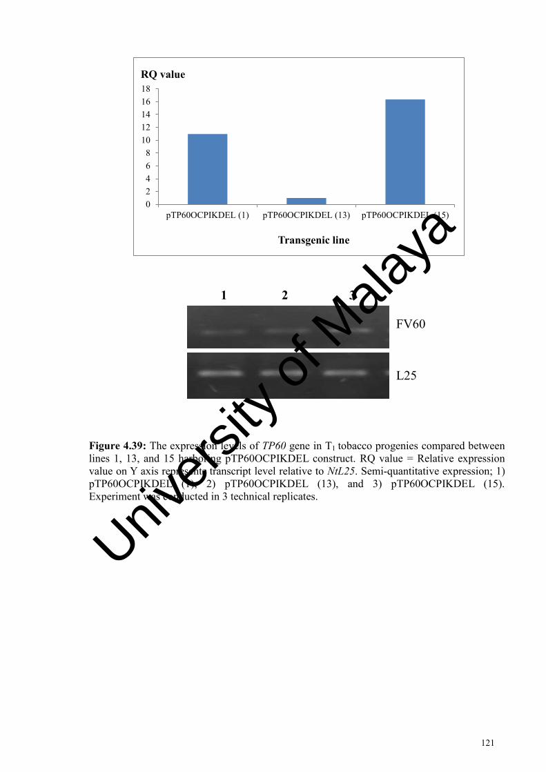
121
02468
1012141618
pTP60OCPIKDEL (1) pTP60OCPIKDEL (13) pTP60OCPIKDEL (15)
RQ value
Transgenic line
FV60
L25
1 2 3
Figure 4.39: The expression levels of TP60 gene in T1 tobacco progenies compared between lines 1, 13, and 15 harboring pTP60OCPIKDEL construct. RQ value = Relative expression value on Y axis represents transcript level relative to NtL25. Semi-quantitative expression; 1) pTP60OCPIKDEL (1), 2) pTP60OCPIKDEL (13), and 3) pTP60OCPIKDEL (15). Experiment was conducted in 3 technical replicates.
Univers
ity of
Mala
ya

122
05
1015202530354045
pTP60BBI (5) pTP60BBI (6) pTP60BBI (7)
RQ value
Transgenic line
FV60
L25
1 2 3
Figure 4.40: The expression levels of TP60 gene in T1 tobacco progenies compared between lines 5, 6, and 7 harboring pTP60BBI construct. RQ value = Relative expression value on Y axis represents transcript level relative to NtL25. Semi-quantitative expression; 1) pTP60BBI (5), 2) pTP60BBI (6), and 3) pTP60BBI (7). Experiment was conducted in 3 technical replicates.
Univers
ity of
Mala
ya

123
0
5
10
15
20
25
RQ value
Constructs
FV60
L25
1 2 3 4 5
Figure 4.41: The expression levels of TP60 gene in T1 tobacco progenies, comparing between representative lines of different plasmid constructs with the highest expression of anti-Toxoplasma gene at mRNA level. RQ value = Relative expression value on Y axis of independent lines transcript level relative to NtL25. Semi-quantitative expression; 1) pTP60 (12), 2) pTP60BBIKDEL (14), 3) pTP60KDEL (7), 4) pTP60BBI (5), and 5) pTP60OCPIKDEL (15). Experiment was conducted in triplicate. Univ
ersity
of M
alaya

124
4.8 Recombinant anti-Toxoplasma protein expression in transgenic tobacco plants
4.8.1 Recombinant protein purification
Initially, the total protein was extracted from transgenic tobacco plants (T0) and (T1)
followed by protein purification, SDS-PAGE and Western blot analysis. The total
protein extracts were quantified according to BSA standard curve (Figure 4.42) and the
integrity of the protein was determined by SDS-PAGE analysis (Figure 4.43). Based on
the results obtained as shown in Figure 4.44, T0 tobacco plants confirm the expression
of the recombinant protein at the expected size of ~ 27 kDa.
In this study, the targeted recombinant protein was purified using Dynabeads® M-
280 Sheep anti-Mouse IgG (ThermoFisher Scientific, USA) (Section 3.8.7) (Figure
4.45). A clear band at the size approximately of 60 kDa is suggested to be non-specific
protein eluate from the purified protein product was detected in all the selected samples.
This suggested the stability of the anti-Toxoplasma recombinant protein. No band was
observed in wild-type lane. However, there were apparent dimers formed of the detected
protein (~ 54 kDa instead of ~27 kDa) (Figure 4.46).
Univers
ity of
Mala
ya

125
Figure 4.42: Standard curve of known concentration BSA using Bradford’s assay quantification.
y = 0.5773x R² = 0.9908
0
0.1
0.2
0.3
0.4
0.5
0.6
0.7
0.8
0 0.2 0.4 0.6 0.8 1 1.2 1.4
A595
Protein concentration, µg/ µL
Univers
ity of
Mala
ya

126
Lane M : Page RulerTM Pre-stained Protein Ladder, 10-180 kDa (Thermo Scientific, USA)
Lane 1 : Non-transformed tobacco plants (wild-type)
Lane 2 : Transgenic tobacco harboring pTP60 construct
Lane 3 : Transgenic tobacco harboring pTP60KDEL construct
Lane 4 : Transgenic tobacco harboring pTP60BBI construct
Lane 5 : Transgenic tobacco harboring pTP60BBIKDEL construct
Lane 6 : Transgenic tobacco harboring pTP60OCPIKDEL construct
55 kDa
43 kDa
34 kDa
26 kDa
M 1 2 3 4 5 6
~ 27 kDa
Figure 4.43: 20 µg of unpurified total soluble protein extracted from wild-type and a transgenic N. tabacum cv. SR1 representative from each construct (T0) after 40-days of acclimatization. The expected size of recombinant anti-Toxoplasma scFv antibody protein is ~ 27 kDa.
Univers
ity of
Mala
ya

127
Lane M : PageRulerTM Plus Pre-stained Protein Ladder, 10-250 kDa (Thermo Scientific, USA)
Lane 1 : Non-transformed tobacco plants (wild-type)
Lane 2 : Transgenic tobacco harboring pTP60BBIKDEL construct
Lane 3 : Transgenic tobacco harboring pTP60OCPIKDEL construct
Lane 4 : Transgenic tobacco harboring pTP60BBI construct
Lane 5 : Transgenic tobacco harboring pTP60KDEL construct
Lane 6 : Transgenic tobacco harboring pTP60 construct
M 1 2 3 4 5 6
~ 27 kDa 25 kDa
Figure 4.44: Detection of recombinant anti-Toxoplasma scFv antibody, TP60 from unpurified total soluble protein on the selected T0 tobacco plants and wild-type after 40-days of acclimatization using mouse monoclonal anti–E epitope tag and IgG anti-mouse with HRP conjugated by Western blot analysis. The expected band size is ~ 27 kDa.
Univers
ity of
Mala
ya

128
Figure 4.45: SDS-PAGE of purified recombinant anti-Toxoplasma scFv antibody protein using DyNabeads™ from N. tabacum cv. SR1 representative lines harboring different plasmid constructs (T1) after 40-days of acclimatization.
Lane M : PageRulerTM Pre-stained Protein Ladder, 10-180 kDa (Thermo Scientific, USA)
Lane 1 : T1 tobacco progeny harboring pTP60 construct (line 12)
Lane 2 : T1 tobacco progeny harboring pTP60KDEL construct (line 7)
Lane 3 : T1 tobacco progeny harboring pTP60BBI construct (line 5)
Lane 4 : T1 tobacco progeny harboring pTP60BBIKDEL construct (line 14)
Lane 5 : T1 tobacco progeny harboring pTP60OCPIKDEL construct (line 15)
Univers
ity of
Mala
ya

129
Lane M: PageRulerTM Plus Pre-stained Protein Ladder, 10-250 kDa (Thermo Scientific, USA). Ladder used for loading control is Page RulerTM Pre-stained Protein Ladder, 10-180 kDa (Thermo Scientific, USA)
Lane 1 : T1 tobacco progeny harboring pTP60BBI construct (line 5)
Lane 2 : T1 tobacco progeny harboring pTP60KDEL construct (line 7)
Lane 3 : T1 tobacco progeny harboring pTP60BBIKDEL construct (line 14)
Lane 4 : T1 tobacco progeny harboring pTP60OCPIKDEL construct (line 15)
Lane 5 : T1 tobacco progeny harboring pTP60 construct (line 12)
Lane 6 : Wild-type tobacco plant
Figure 4.46: Stable expression of purified recombinant anti-Toxoplasma scFv antibody protein from N. tabacum cv. SR1 representative lines harboring different plasmid constructs (T1) after 40-days of acclimatization. The expected protein size is at ~ 54 kDa (dimer formation). Actin-11 protein used as loading control at the predicted size of ~ 45 kDa.
Univers
ity of
Mala
ya

130
CHAPTER 5: DISCUSSION
5.1 Introduction
The parasitic protozoan, T. gondii is a causal agent of toxoplasmosis which severely
affects mammals including humans and birds. In human, this disease may cause
complications in immunodeficient individuals and pregnant women which may lead to
congenital disease of the newborn babies. In addition, long term period of this untreated
disease can cause neonatal loss and abortion in livestock animals which may be
devastating to the husbandry sector.
This study was carried out to control toxoplasmosis through diagnosis and
therapeutic approaches by expressing the recombinant single-chain variable fragment
(scFv) antibody against toxoplasmosis. The recombinant antibody was derived from
mouse pre-cloned scFv gene (Lim, 2012) and subsequently transformed into tobacco
plants by using a plant expression vector as a backbone (Go, 2013). This scFv has the
same specificity as in parental antibody, IgG due to the presence of specific paratope
against specific target. The small and simple size functional antibody with both heavy
and light chains connected by a peptide linker (15-20 amino acids) to form a
polypeptide chain was a breakthrough (Tran et al., 2013; Lai et al., 2014). In addition,
scFv also provides other advantages such as increase the density of antigen-binding sites
and selectivity towards target antigen as well as rapid in antigens detection even in low
concentration of complex samples (Shen et al., 2005).
Besides the scalability and compatibility properties of plant-derived pharmaceutical
proteins due to similar machinery and glycosylation pattern as in human, ethical issues
and human-pathogenic contamination that are often associated with animal or bacterial
cell culture systems could be avoided.
Univers
ity of
Mala
ya

131
However, plant system may also exhibit some drawbacks, including higher
heterogeneity of the end recombinant protein product, low in solubility and expression
level due to the interference of endogenous proteases. Hence, in this study, different
proteinase inhibitor and/ or ER-targeting gene (KDEL) were co-expressed with anti-
Toxoplasma recombinant scFv antibody in order to enhance and stabilize the antibody
production in the model tobacco host plant, N. tabacum.
5.2 Transformation of tobacco with recombinant plasmid constructs
Hiatt et al., (1989) first reported on the production of functional IgG antibody from
plants obtained from crossing two transgenic tobacco plants, each expressing heavy and
light chains. Prior to crossing, A. tumefaciens-mediated transformation was performed
on separate events. Following that, Vaquero et al. (1999), has successfully developed
functional therapeutic scFv and IgG of a tumor-specific antibody (T84.66) in N.
tabacum cv. Petite Harvana SR1 using agroinfiltration technique. In years later,
ELPylated haemagglutinins as neutralizing antibodies against H5N1 viruses was
produced in mice (Phan et al., 2014), whereas production of stable HIV-neutralizing
human monoclonal antibody 2G12 was from N. tabacum (Ma et al., 2015).
Additionally, there are some scFv undergoing the clinical test, i.e. scFv targeted to P97
antigen against Melanoma disease and bispecific scFv for B-cell tumor which was
undergoing preclinical stage and phase I clinical trials respectively (Pucca et al., 2011).
In this study, standard Agrobacterium-mediated transformation protocol was used
(Go, 2013). On selection media, all hygromycin-resistant shoots regenerated from leaf
discs transformed with different plasmid constructs were assessed. Based on the results
obtained, explants that have been transformed with pTP60BBIKDEL showed 7-fold
higher shoot regeneration compared to the explants that have been transformed with
Univers
ity of
Mala
ya

132
only pTP60KDEL. It was also observed that no significant differences between putative
transformants transformed with pTP60, pTP60BBIKDEL, pTP60OCPIKDEL as well
pCAMBIA 1304 with wild-type. It could be concluded that recombinant anti-
Toxoplasma gene and the proteinase inhibitor with or without ER-targeting gene did not
affect the plant growth especially in shoot regeneration even though pTP60BBI and
pTP60KDEL alone gave a significant difference in the analysis.
It was also found that more shoots regenerated from the wounded edge compared to
the middle of the explants and would increase the chances of obtaining putative
transformed shoots. Furthermore, since the regenerated shoots were in direct contact
with hygromycin, the probability of attaining false-positives would be reduced. It was
also observed that the middle part of the explants produced callus and led to low
chances of forming shoots. This result was in accordance with Khan et al. (2015).
5.3 Confirmation of putative transformed shoots
In this study, putative transformed shoots were verified using transient and molecular
assays. Transient assays utilizing GUS (qualitative and quantitative) and GFP
visualization have been carried out on the randomly selected putatively transformed
shoots harboring different plasmid constructs.
GUS (gusA) gene has been extensively used in plant transformation and considered
as a good marker for monitoring transformation efficiency (Jefferson et al., 1987). This
is due to the capability of GUS gene catalytically hydrolyzed a broad range of substrates
through different applications due to the distinct chromogenic, spectrophotometric and
fluorogenic properties of their aglycone forms (Kim et al., 2006). The observed GUS-
blue spots and GUS quantification through fluorometry assay showed initially
Univers
ity of
Mala
ya

133
promising results as an indicator of successful transfer of transgene. In this study, the
lowest expression of GUS gene was shown in putative transformed shoots in the
presence of pTP60BBI, pTP60OCPIKDEL, and pTP60BBIKDEL (~ 1.5 kb insert in
size) compared to pTP60 and pTP60KDEL (~ 900 bp insert in size). On the other hand,
putative transformed shoots with the empty vector showed the highest GUS expression
(Figures 4.16 and 4.19). This might be due to the position of the GUS gene away from
the backbone promoter CaMV 35S leading to reduce gene regulation. In addition, GUS
gene expression also is a good indicator to explain the expression of unfused
recombinant gene within the T-DNA region even by using only a constitutive promoter
belongs to the backbone vector.
Besides GUS, GFP is also a reliable reporter gene that is suitable to determine the
successful transfer of T-DNA. It is a simple, convenient, and non-destructive
visualization assay. The putative transformed shoots were then examined for green
fluorescence to determine the expression of the mgfp5 gene (Figure 4.20). The non-
transformed region (Figure 4.20a) within the same expanded putative transformed
shoots showed red coloration due to auto-fluorescence under UV light in the presence of
chlorophyll background signal (Goldman et al., 2003; Zhou et al., 2005). The region in
the presence of GFP gene fluoresced under specific excitation/emission wavelength
488/550nm (Figure 4.20b). However, the GFP expression diminished over a period of
time indicating transient gene expression. Decreasing GFP expression is also influence
by the age of the leaves. According to previous publications, following the recovery of a
new putative transgenic plant, GFP fluorescence was usually visible in new emerging
shoots, young tissues or organs, whereas weak signal was observed in the older ones
(Zhou et al., 2004), such as in Medicago truncatula var. A17 (Kamaté et al., 2000) and
Verbena officialis L. (Tamura et al., 2003).
Univers
ity of
Mala
ya
134
PCR analysis was carried out in order to verify the presence of heritable hptII gene
and the integration of T-DNA into T1 tobacco progenies. For the latter, the screening of
T1 transformants was carried out by detecting hptII gene in the left border (LB) part of
the pCAMBIA 1304. hptII gene located on the left border (LB) of Ti plasmid could be
nicked during the transfer of T-DNA from Agrobacterium to plant cells causing
truncation to occur which has been documented by Kemski et al. (2013).
5.4 Effects of different plasmid constructs on the phenotype of transgenic tobacco plants
The percentage of T1 seed germination, stem height, and flowering period were
assessed in order to determine the effect of plasmid constructs towards plant growth and
development (Figure 4.26 and Table 4.4).
5.4.1 T1 seed germination
The percentage of seed germination was significantly lowest (65 % ± 2.03) in
transgenic seeds transformed with the empty vector compared. Contrastingly, construct
with elements of BBI-KDEL and OCPI-KDEL showed the highest germination
percentage (90 % ± 1.45) but germination of T1 seeds present with BBI and KDEL
elements alone were significantly low (p < 0.05). The presence of proteinase inhibitors
was shown to improve the germination and it has been reported that protease inhibitor is
involved in regulating the endogenous proteinases during germination of lettuce seeds
and for the formation of a sulphur reservoir during dormancy (Yan et al., 2009). Sulphur
reservoir results from the accumulation of amino acids rich in sulphur such as cysteine
which helps in overcoming dormancy during germination. Furthermore, the levels of T1
seed germination percentages in the presence of pTP60 construct and wild-type were
Univers
ity of
Mala
ya

135
significantly similar indicating that the anti-Toxoplasma protein did not interfere with
the growth of the tobacco plant itself.
However, the transgenic seeds showed delayed germination than wild-type after 14
days of culture on media containing with and without hygromycin respectively (no data
were recorded). The delayed in germination might be due to selective pressure on
transgenic seeds to adapt to the antibiotic-containing media. As previously reported by
Eluk et al. (2016), the introduction of antibiotics, such as enrofloxacin, oxytetracycline,
penicillin, and tylosin, on culture media delayed germination in seeds of Glycine max,
Helianthus annus, Sorghum bicolor, Triticum aestivum, and Zea mays, showing a
phytotoxic action on crop growth.
As been reported by Rivard et al. (2006), in order to prevent the proteolytic
degradation towards the recombinant proteins by endogenous proteases, tomato
cathepsin D inhibitor (CDI) has been co-expressed with the recombinant proteins.
Surprisingly, there were non-significant difference in the morphology of transgenic
Solaneum tuberosum in terms of height, germination time, number of leaves, stem
diameter and internode length. This suggested that proteinase inhibitors were not
interfered in plant growth and development regardless the types of proteinase inhibitor.
5.4.2 Stem height and flowering period
Acclimatized T1 plantlets were assessed in terms of stem height and flowering period
(Table 4.4). The data obtained for stem height was comparable between both transgenic
and wild-type (control) plants in T1 generation. The significant difference was observed
for T1 tobacco progeny presence of at least one element (proteinase inhibitor with or
without KDEL). On the other hand, T1 tobacco progeny transformed with anti-
Toxoplasma construct only (pTP60) did not impose a detectable penalty on growth and
Univers
ity of
Mala
ya

136
development of the tobacco plant itself as compared to wild-type which is pertinent in
using tobacco as a factory for biopharming (Commandeur et al., 2003; Ahmad et al.,
2012). As been reported by Hartl et al. (2010), serine proteinase like inhibitor (BBI in
this case), was commonly found in many species which ruled out the possibility of
affecting primary growth in Solanum americanum. In addition, serine proteinase-like
inhibitor could defend Solanum nigrum against general herbivores without influencing
the plant growth and development.
The flowering duration was observed to be faster in T1 tobacco progeny compared to
wild-type (65 days ± 5.49). T1 tobacco plant harboring pTP60BBI showed the earliest
flowering onset (45 days ± 2.67). In plants, flowering is commonly linked regulated by
environmental factors, such as photoperiod, temperature, and humidity. Most factors
responsible for non-photoperiodic flowering are due to stress (Takeno, 2012). In this
study, the introduction of foreign gene into the tobacco plant genome caused stress to
the transgenic plants. Thus, leading to faster flowering period compared to normal
tobacco plants.
In previous studies, it has been reported that the introduction of proteinase inhibitor
genes did not affect the developmental phenotype in other plant species. In the previous
study, it was also found that there was no significant difference in flower diameter (Sin
et al., 2006), number of reproductive units (Zavala et al., 2004b), plant growth (Zavala
et al., 2004a; Xie et al., 2007), or trichome morphology and density (Liu et al., 2006;
Luo et al., 2009) might be due to effector-specificity.
5.5 Co-expression of proteinase inhibitor and KDEL increased the relative expression of pTP60 gene and protein accumulation
The co-expression of proteinase inhibitors and KDEL-encoding transgenes in plants
is an alternative way to enhance protein expression. This approach may provide in situ
Univers
ity of
Mala
ya

137
stabilization of the recombinant protein against enzymatic reaction by endogenous
proteases and migration to specific sub-cellular compartments such as ER.
Despite the occurrence of different proteases with distinct modes of action and
specificities towards peptide bonds reported to be active in leaf extracts (van der Valk &
van Loon, 1988; Gonҫalves et al., 2016), single broad-spectrum inhibitors, such as BBI
and OCPI, may effectively protect co-expressed recombinant proteins in leaf tissue. As
previously reported in 2008, the introduction of serine-like BBI had increased the
accumulation of a transgenic recombinant human granulocyte-macrophage colony
stimulating factor by decreasing the protease activity in transformed rice callus (Kim et
al., 2008). Besides, the pharmaceutically important cytokine, human interleukin 2 was
also found to be protected by the co-expression of both BBI and silk protease inhibitors
(Redkiewicz et al., 2012). In 2012, transgenic tobacco plants that constitutively express
the OCPI had significantly increased glutathione reductase activity and the amount
expressed in transgenic tobacco plants by inhibiting cysteine protease activity which
was shown to be lowered in abundance (Pillay et al., 2012).
Transgenes that are targeted for expression within the ER also showed improvement
in the yield of recombinant proteins (Benchabane et al., 2008). The KDEL or SEKDEL
retention sequence (Ser-Glu-Lys-Asp-Glu-Leu) was first described by Munro and
Pelham (1987) has been used in this study. De Wilde et al. (2013) showed that VHH-Fc
and scFv-Fc accumulation levels of 1 % or more in Arabidopsis thaliana seeds triggered
an unfolding protein response and enhanced expression of genes involved in protein
folding, glycosylation, protein translocation, degradation, and vesicle trafficking was
observed. Hence, the combination of both of these strategies was investigated in this
study to potentially facilitate better expression of the targeted antibody.
Univers
ity of
Mala
ya

138
Based on the Western blot analysis (Figure 4.44 and Figure 4.46), the results showed
that the highest protein production and accumulation of anti-Toxoplasma scFv antibody
was achieved in the presence of co-expressed combination BBI-KDEL elements. The
result obtained was correlated to the expression at the mRNA level (Figure 4.41).
The data suggest that the combination of PI and KDEL had likely mitigated the
effect of proteinase and had thus enhanced the production of antibody. This finding
validates the use of these construct combinations for expression of the TP60
recombinant antibody and may be useful for future application in designing plant based
systems for large scale production of recombinant proteins.
5.6 Homodimer formation of recombinant anti-Toxoplasma protein in T1 tobacco plants
Detection of anti-Toxoplasma recombinant scFv, TP60 using an anti E-epitope tag
fused protein (Figure 4.45) confirmed the expression of the protein at different
accumulation levels as a ~ 54 kDa dimer protein in T1 tobacco plants instead of the
expected ~ 27 kDa of scFv protein size which was at first observed in T0 tobacco
plants. The dimeric formation of TP60 might be due to a serine residue in the linker
peptide (GGSSRSS) that could be involved in dimer formation of the disulphide band
under reducing conditions (Schouten et al., 2002). Dimerization of recombinant proteins
produced from Nicotiana tabacum cv. Xanthi due to a cysteine residue present in the
linker peptide of the recombinant protein has been reported (Dobhal et al., 2013). This
dimeric formation of TP60 might increase the effectiveness and inhibition toward the T.
gondii tachyzoite antigen which causes the Toxoplasma disease. As in a previous study,
dimerized anti-NbTNF-VHHCK proteins produced from Nicotiana benthamiana blocked
the TNFα-activity more effectively than monomeric anti-EcTNF-VHHCK protein
produced in Escherichia coli (Geirsberg et al., 2010). No band was observed in the lane
Univers
ity of
Mala
ya

139
for non-transformed tobacco plant. Recombinant scFv antibody of T1 tobacco plants in
combination with the BBIKDEL elements showed highest protein accumulation
compared to the other elements and were correlated with the gene transcript
quantification at mRNA level.
Univers
ity of
Mala
ya

140
CHAPTER 6: CONCLUSION
This study was conducted in order to assess the transgene expression of anti-
Toxoplasma recombinant scFv antibody encoded by TP60 at mRNA and protein levels.
The expression of TP60 was also been assessed at mRNA level in T1 generation. In
order to improve the amount of desired recombinant protein in transgenic tobacco
plants, this study was undertaken by co-expressing TP60 gene in combination with
different elements, namely proteinase inhibitors (BBI or OCPI) and ER-retention
sequence (KDEL). The gene was tandemly expressed with either one of this elements or
combination of a proteinase inhibitor with KDEL retention sequence.
Different plasmid constructs, namely pTP60, pTP60KDEL, pTP60BBI,
pTP60BBIKDEL and pTP60OCPIKDEL, were transformed into N. tabacum cv. SR1
mediated by A. tumefaciens strain LBA4404 harboring the binary vector
pCAMBIA1304 containing a gusA-mgfp5 fusion reporter genes encoded for GUS and
GFP, respectively, and a selectable marker for hygromycin B (hptII) genes driven by the
CaMV 35S promoter. Quantitative GUS assay showed that the non-significant
difference of pTP60BBI, pTP60BBIKDEL, and pTP60OCPIKDEL constructs
generated at least 20-fold low expression while, pTP60 and pTP60KDEL constructs
generated at least 7-fold lower expression compared to the empty vector which
generated significantly highest in GUS expression. This observation was correlated to
the least expression of GUS blue spot(s) on the expanded putative transformed shoots
due to the presence of gene insert. No GUS-blue spots and green fluorescence signal
were observed on non-transformed region of the putative transformed shoots.
Based on the results obtained from the Western blot analysis, the accumulation of the
recombinant TP60 protein was highest in the presence of BBIKDEL elements followed
Univers
ity of
Mala
ya

141
by OCPIKDEL, BBI, and KDEL in both T0 and T1 generations. This result can be
concluded due to the lowest expression of TP60 protein with the absence of either one
of these elements. The recombinant protein was first detected in the T0 at ~ 27 kDa but
then observed as homodimer at ~ 54 kDa in the T1 generation.
In order to support the observation of recombinant protein accumulation derived
from different constructs, the expression of TP60 transcript was analyzed in the T1
generation. In this analysis, three lines for each construct were evaluated and it was
found that the highest relative expression of TP60 was with the companion of BBI-
KDEL that showed 19-fold higher expression compared to the other constructs. This
result correlated with the expression of TP60BBIKDEL at the protein level.
According to the statistical data analysis on phenotypic assessments, data obtained
were comparable between both transgenic and control plants in T1 generation in terms
of number of putative shoots regeneration, stem height, and flowering initiation time.
The significant difference was observed for T1 tobacco progeny presence of BBIKDEL
or OCPIKDEL element. Transformation with anti-Toxoplasma constructs without any
additional elements (pTP60) and stably inherited in T1 tobacco progeny did not interfere
in growth and development of the tobacco. The phenotypic characteristic observed was
similar and no significant difference to wild-type; which is pertinent in using tobacco as
a factory for biopharming However, in the presence of proteinase inhibitor (BBI or
OCPI) with or without the companion of KDEL elements, the percentage of T1 seeds
germination was significantly higher compared to without proteinase inhibitors.
This study demonstrated that the production of recombinant anti-Toxoplasma protein
in plants is feasible and can be enhanced using specific elements. This complements the
advantages of the plant system which includes expression of eukaryotic proteins without
the need of downstream modifications, such as chemical refolding associated with
Univers
ity of
Mala
ya

142
prokaryotic systems or purification against pathogen. This technology can thus be
exploited for the production of pharmaceutically important proteins and diagnostic
antibodies which will have a wide impact on the medical biotechnology sector.
Univers
ity of
Mala
ya

143
REFERENCES
Ahmad, Z. A., Yeap, S. K., Ali, A. M., Ho, W. Y., Alitheen, N. B. M., & Hamid, M. (2012). scFv antibody: Principles and clinical application. Clinical and Development Immunology, 2012.
Ajzenberg, D. (2010). Type I strain in human toxoplasmosis: Myth or reality?. Future Microbiology, 5(6), 841-843.
Allen, G. C., Hall, G. Jr., Michalowski, S., Newman, W., Spiker, S., Weissinger, A. K., & Thompson, W. F. (1996). High-level transgene expression in plant cells: Effects of a strong scaffold attachment region from tobacco. Plant Cell, 8(5), 899-913.
Amaro, M. O., Xisto, M. F., Dias, A. S., Versiani, A. F., Cardoso, S. A., Otoni, W. C., … De Paula, S. O. (2015). Antigen production using heterologous expression of dengue virus-2 non-structural protein 1 (NS1) in Nicotiana tabacum (Havana) for immunodiagnostic purposes. Plant Cell Reports, 34(6), 919-928.
Antoniukas, L., Grammel, H., & Reichl, U. (2006). Production of hantavirus Puumala nucleocapsid protein in Saccharomyces cerevisiae for vaccines and diagnostics. Journal of Biotechnology, 124(2), 347-362.
Artsaenko, O., Kettig, B., Fiedler, U., Conrad, U., & Düring, K. (1998). Potato tubers as a biofactory for recombinant antibodies. Molecular Breeding, 4(4), 313-319.
Baneyx, F. (1999). Recombinant protein expression in Escherichia coli. Current Opinion in Biotechnology, 10(5), 411-421.
Benchabane, M., Goulet, C., Rivard, D., Faye, L., Gomord, V., & Michaud, D. (2008). Preventing unintended proteolysis in plant protein biofactories. Plant Biotechnology Journal, 6(7), 633-648.
Benzle, K., Finer, K. R., Marty, D., McHale, L. K., Goodner, B. W., Taylor, C., & Finer, J. J. (2014). Isolation and characterization of novel Agrobacterium strains for soybean and sunflower transformation. Plant Cell Tissue and Organ Culture, 121(1), 71-81.
Berger, F., Goulet, V., Le Strat, Y., & Desenclos, J. C. (2009). Toxoplasmosis among pregnant women in France: Risk factors and change of prevalence between 1995 and 2003. Journal of Epidemiology and Public Health, 57(4), 241-248.
Both, L., Dolleweerd van, C., Wright, E., Banyard, A. C., Bulmer-Thomas, B., Selden, D., … Ma, J. K. –C. (2013). Production, characterization, and antigen specificity of recombinant 62-71-3, a candidate monoclonal antibody for rabies prophylaxis in humans. The FASEB Journal, 27(5), 2055-2065.
Boulikas, T. (1994). Myb protein talking to their DNA (Review). International Journal of Oncology, 5(1), 101-109.
Univers
ity of
Mala
ya

144
Bradford, M. M. (1976). A rapid and sensitive method for the quantification of microgram quantities of protein utilizing the principle of protein-dye binding. Analytical Biochemistry, 7(72), 248-254.
Braun, A. C. (1947). Thermal studies on the factors responsible for tumour initiation in crown gall. American Journal of Botany, 34(4), 234-240.
Breyer, D., Goossens, M., Herman, P., & Sneyers, M. (2009). Biosafety considerations associated with molecular farming in genetically modified plants. Journal of Medicinal Plant Research, 3(1), 825-838.
Buxton, D., & Innes, E. A. (1995). A commercial vaccine for ovine toxoplasmosis. Parasitology, 110, S11-S16.
Cañizares, M. C., Nicholson, L., & Lomonossoff, G. P. (2005). Use of viral vectors for vaccine production in plants. Immunology and Cell Biology, 83(3), 263–270.
Chen, Q. (2015). Plant-made vaccines against West Nile virus are potent, safe, and economically feasible. Biotechnology Journal, 10(5), 671-680.
Chen, Q., & Lai, H. (2014). Plant-derived monoclonal antibodies as human biologics for infectious disease and cancer. In K.L Hefferon (Ed.), Plant-derived pharmaceuticals: Principles and applications for developing countries (pp. 42-75). Cryodon, UK: CABI.
Chen, Q., & Lai, H. (2013). Plant-derived virus-like particles as vaccines. Human Vaccines and Immunotherapeutics, 9(1), 26-49.
Chichester, J. A., Musiychuk, K., dela Rosa, P., Horsey, A., Stevenson, N., Ugulava, N., … Yusibov, V. (2007). Immunogenicity of a subunit vaccine against Bacillus antracis. Vaccine, 25(16), 3111-3114.
Comai, L., Moran, P., & Maslyar, D. (1990). Novel and useful properties of a chimeric plant promoter combining CaMV 35S and MAS elements. Plant Molecular Biology, 15(3), 373-381.
Commandeur, U., Twyman, R. M., & Fischer, R. (2003). The biosafety of molecular farming in plants. AgBiotechNet, 5(110), 1-9.
Conley, A. J., Zhu, H., Le, L. C., Jevnikar, A. M., Lee, B. H., Brandle, J. E., & Menassa, R. (2011). Recombinant protein production in a variety of Nicotiana hosts: A comparative analysis. Plant Biotechnology Journal, 9(4), 434-444.
Cummings, J. F., Guerrero, M. L., Moon, J. E., Waterman, P., Nielsen, R. K., Jefferson, S., … Yusibov, V. (2014). Safety and immunogenicity of a plant-produced recombinant monomer hemagglutinin-based influenza vaccine described from influenza A (H1N1)pdm09 virus: A phase 1 dose-escalation study in healthy adults. Vaccines, 32(19), 2251-2259.
Univers
ity of
Mala
ya

145
De Jaeger, G., Buys, E., Eeckhout, D., De Wilde, C., Jacobs, A., Kapila, J., … Depicker, A. (1999). High level accumulation of single‐chain variable fragments in the cytosol of transgenic Petunia hybrida. The FEBS Journal, 259(1‐2), 426-434.
De Marco, A. (2011). Biotechnological applications of recombinant single-domain antibody fragment. Microbial Cell Factories, 10, 44.
De Wilde, C., De Buck, S., Vanneste, K., & Depicker, A. (2013). Recombinant antibody production in Arabidopsis seeds triggers an unfolded protein response. Plant Physiology, 161(2), 1021-1033.
Dent, M., Hurtado, J., Paul, A. M., Sun, H., Lai, H., Yang, M., … Chen, Q. (2016). Plant-produced anti-dengue virus monoclonal antibodies exhibit reduced antibody-dependent enhancement of infection activity. Journal of General Virology, 97(12), 3280-3290.
Desai, P. N., Neeta, S., & Padh, H. (2010). Production of heterologous proteins in plants: Strategies for optimal expression. Biotechnology Advances, 28(4), 427-435.
DiMiceli, L., Pool, V., Kelso, J. M., Shadomy, S. V., Iskander, J., & V.A.E.R.S Team. (2006). Vaccination of yeast sensitive individuals: Review of safety data in the US vaccine adverse event reporting system (VAERS). Vaccine, 24(6), 703-707.
Dobhal, S., Chaudhary, V. K., Singh, A., Pandey, D., Kumar, A., & Agrawal, S. (2013). Expression of recombinant antibody (single-chain antibody fragment) in transgenic plant Nicotiana tabacum cv. Xanthi. Molecular Biology Report, 40(12), 7027-7037.
Doran, P. M. (2006). Foreign protein degradation and instability in plants and plant tissue cultures. Trends in Biotechnology, 24(9), 426-432.
Dubey, J. P., Lago, E. G., Gennari, S. M., & Su, C. (2012). Toxoplasmosis in humans and animals in Brazil: High prevalence, high burden of disease, and epidemiology. Parasitology, 139(11), 1375-1424.
Duque, L. A., Souto, X. M., de Andrade-Neto, V. V., Ennes-Vidal, V., & Menna-Barreto, F. S. (2013). Autophagy-A double-edged sword-cell survival or death?. Retrieved from https://cdn.intechopen.com/pdfs-wm/44112.pdf
During, K., Hippe, S., Kreutzaler, F., & Schell, J. (1990). Synthesis and self-assembly of a functional monoclonal antibody in transgenic Nicotiana tabacum. Plant Molecular Biology, 15(2), 281-293.
Egelkrout, E., Rajan, V., & Howard, J. A. (2011). Overproduction of recombinant proteins in plants. Plant Science, 184(2012), 83-101.
Elsheikha, H. M. (2008). Congenital toxoplasmosis: Priorities for further health promotion action. Public Health, 124(4), 335-353.
Univers
ity of
Mala
ya

146
Eluk, D., Nagel, O. G., Zimmerman, J., Molina, M. P., & Althaus, R. L. (2016). Effect of antibiotics on the germination and root elongation of Argentine intensive crops. International Journal of Environmental Research, 10(4), 471-480.
Faye, L. Boulaflous, A., Benchabane, M., Gomord, V., & Michaud, D. (2005). Protein modifications in the plant secretory pathway: Current status and practical implications in molecular farming. Vaccine, 23(15), 1770-1778.
Ferrara, F., Naranjo, L. A., Kumar, S., Gaiotto, T., Mukundan, H., Swanson, B., & Bradbury, A. R. M. (2012). Using phage and yeast display to select hundreds of monoclonal antibodies: Application to antigen 85, a tuberculosis biomarker. PLoS ONE, 7(11), e49535.
Fiedler, U., Phillips, J., Artsaenko, O., & Conrad, U. (1997). Optimization of scFv antibody production in transgenic plants. Immunotechnology, 3(3), 205-216.
Fischer, R., Schumann, D., Zimmermann, S., Drossard, J., Sack, M., & Schillberg, S. (1999). Expression and characterization of bispecific single-chain Fv fragments produced in transgenic plants. European Journal of Biochemistry, 262(3), 810–816.
Flegr, J., Klapilova, K., & Kaňková, S. (2014). Toxoplasmosis can be sexually transmitted infection with serious clinical consequences. Not all routes of infection are created equal. Medical Hypotheses, 83(3), 286-289.
Floss, D. M., Falkenburg, D., & Conrad, D. (2007). Production of vaccines and therapeutic antibodies for veterinary applications in transgenic plants: An overview. Transgenic Research, 16(3), 315-332.
Friger, J. (2014, May 8). CDC warns of common parasites plaguing millions in U.S. Retrieved from https://www.cbsnews.com/news/parasites-causing-infections-in-the-us-cdc-says/
Fu, L. H., Miao, Y. S., Lo, S. W., Seto, T. C., Sun, S. S. M., Xu, Z. F., … Jiang, L. (2009). Production and characterization of soluble human lysosomal enzyme α-iduronidase with high activity from culture media of transgenic tobacco BY-2 cells. Plant Science, 177(6), 668–675.
Fujiuchi, N., Matoba, N., & Matsuda, R. (2016). Environment control to improve recombinant protein yields in plants based on Agrobacterium-mediated transient gene expression. Frontiers in Bioengineering and Biotechnology, 4, 23.
Gallie, D. R. (1993). Posttranscriptional regulation of gene expression in plants. Annual Review of Plant Biology, 44(1), 77-105.
Ganesan, R., Eiigenbrot, C., & Kirchhofer, D. (2010). Structural and mechanistic insight into how antibodies inhibit serine proteases. Biochemical Journal, 430(2), 179-189.
Gasteiger, E., Gattiker, A., Hoogland, C., Ivanyi, I., Appel, R. D., & Bairoch, A. (2003). ExPASy: The proteomics server for in-depth protein knowledge and analysis. Nucleic Acid Research, 31(3), 3784-3788.
Univers
ity of
Mala
ya

147
Gaur, S., Maheshwari, S. K., & Gauba, P. (2012). Transgenic plants: Factories for the production of biomedicines. Journal of Pharmacy Research, 5(9), 4856-4859.
Giersberg, M., Floss, D. M., Kipriyanov, S., Conrad, U., & Scheller, J. (2010). Covalent dimerization of camelidae anti-human TNF-alpha single domain antibodies by the constant kappa light chain domain improves neutralizing activity. Biotechnology and Bioengineering, 106(1), 161-166.
Gils, M., Kandzia, R., Marillonnet, S., Klimyuk, V., & Gleba, Y. (2005). High-yield production of authentic human growth hormone using a plant virus-based expression system. Plant Biotechnology Journal, 3(6), 613–620.
Gleba, Y., Klimyuk, V., & Marillonnet, S. (2005). Magnifection-a new platform for expressing recombinant vaccines in plants. Vaccine, 23(17-18), 2042-2048.
Gleba, Y. Y., Tusé, D., & Giritch, A. (2014). Plant viral vectors for delivery by Agrobacterium. Current Topics in Microbiology and Immunology, 375, 155-192.
Go, P. S. (2013). Expression of anti-toxoplasma scFv antibodies in plants (Master’s thesis, University of Malaya). Retrieved from http://studentsrepo.um.edu.my/id/eprint/4174
Goldman, J. J., Hanna, W. W., Fleming, G., & Ozias-Akins, P. (2003). Fertile transgenic pearl millet [Pennisetum glaucum (L.) R. Br.] plants recovered through microprojectile bombardment and phosphinothricin Selection of apical meristem-, inflorescence-, and immature embryo-derived embryogenic tissues. Plant Cell Reports, 21(10), 482-486.
Golovkin, M., Spitsin, S., Andrianov, V., Smirnov, Y., Xiao, Y., Pogrebnyak, N., … Koprowski, H. (2007). Smallpox subunit vaccine produced in Planta confers protection in mice. Proceedings of the National Academy of Sciences 104(16), 6864-6869.
Gomes, A. R., Byregowda, S. M., Veeregowda, B. M., & Balamurugan, V. (2016). An overview of heterologous expression host systems for the production of recombinant proteins. Advances in Animal and Veterinary Sciences, 4(4), 346-356.
Gomord, V., & Faye, L. (2004). Post-translational modification of therapeutic proteins in plants. Current Opinion in Plant Biology, 7(2), 171-181.
Gomord, V., Denmat, L. A., Fitchette-Lainé A. C, Satiat-Jeunemaitre, B., Hawes, C., & Faye, L. (1997). The C-terminal HDEL sequence is sufficient for retention of secretory proteins in the endoplasmic reticulum (ER) but promotes vacuolar targeting of proteins that escape the ER. Plant Journal, 11(2), 313-325.
Gonҫalves, R. N., Gozzini B. S. D., & da Silva-López, R. E. (2016). Corrigendum to “Proteases from Canavalia ensiformis: Active and thermostable enzymes with potential of application in biotechnology”. Biotechnology Research International, 2016, 9872540
Univers
ity of
Mala
ya

148
Goulet, C., Michaud, D., & Teixeira da Silva, J. A. (2006). Degradation and stabilization of recombinant proteins in plants. In J. A. Taxeira da Silva (Ed.), Floriculture, Ornamental, and Plant Biotechnology (pp. 35-40). Japan, JP.
Grimsley, N., Hohn, B., Hohn, T., & Walden, R. (1986). “Agroinfection”, an alternative route for viral infection of plants by using the Ti plasmid. Proceedings of the National Academy of Sciences, 83(10), 3282-3286.
Hagemeyer, C. E., von zur Muhlen, C., von Elverfeldt, D., & Peter, K. (2009). Single-chain antibodies as diagnostic tools and therapeutic agents. Journal of Thrombosis and Hemostasis, 101(6), 1012-1019.
Halonen, S. K., & Weiss, L. M. (2013). Toxoplasmosis. Handbook Clinical Neurology, 114, 125-145.
Hartl, M., Giri, A. P., Kaur, H., & Baldwin, I. T. (2010). Serine protease inhibitors specifically defend Solanum nigrum against generalist herbivores but do not influence plant growth and development. Plant Cell, 22(12), 4158-4175.
Hein, M. B., Tang, Y., Mcleod, D. A., Janda, K. D., & Hitta, A. (1991). Evaluation of immunoglobulins from plant cells. Biotechnology Progress, 7(5), 455-461.
Hiatt, A., Cafferkey, R., & Bowdish, K. (1989). Production of antibodies in transgenic plants. Nature, 342, 76-78.
Hill, D., & Dubey, J. P. (2002). Toxoplasma gondii: Transmission, diagnosis and prevention. Clinical Microbiology and Infection, 8(10), 634-640.
Hitzeman, R. A., Hagie, F. E., Levine, H. L., Goeddel, D.V., Ammerer, G., & Hall, B. D. (1981). Expression of a human gene for interferon in yeast. Nature, 293(5835), 717-722.
Holland, T., Sack, M., Rademacher, T., Schmale, K., Altmann, F., Stadlmann, J., … Hellwig, S. (2010). Optimal nitrogen supply as a key to increased and sustained production of a monoclonal full-size antibody in BY-2 suspension culture. Biotechnology and Bioengineering, 107(2), 278–289.
Holmgren, J., & Czerkinsky, C. (2005). Mucosal immunity and vaccines. Nature Vaccines, 11(4 Suppl), S54-S53.
Horn, M. E., Woodard, S. L., & Howard, J. A. (2004). Plant molecular farming: Systems and products. Plant Cell Reports, 22(10), 711-720.
Ho-Yen, D. O. (2005). Toxoplasmosis. Medicine, 33(5), 120-121.
Jäger, V., Büssow, K., Wagner, A., Weber, S., Hust, M., Frenzel, A., & Schirrmann, T. (2012). High level transient production of recombinant antibodies and antibody fusion proteins in HEK293. BMC Biotechnology, 13(52), 1-20.
Jefferson, R. A., Kavanagh, T. A., & Bevan, M. W. (1987). GUS fusions: β-glucuronidase as a sensitive and versatile gene marker in higher plants. EMBO Journal, 6(13), 3901-3907.
Univers
ity of
Mala
ya

149
Joensuu, J. J., Conley, A. J., Lienemann, M., Brandle, J. E., Linder, M. B., & Menassa, R. (2010). Hydrophobin fusions for high-level transient protein expression and purification in Nicotiana benthamiana, Plant Physiology, 152(2), 622-633.
Johnson, M., Zaretskaya, I., Raytselis, Y., Merezhuk, Y., McGinnis, S., & Madden, T. L. (2008). NCBI BLAST: A Better web interface. Nucleic Acids Research, 36(suppl_2), W5-W9.
Jong, W. S. P., Vikström, D., Houben, D., Bart van den Berg van Saparoea, H., Williem de Gier, J-., & Luirink, J. (2017). Application of an E. coli signal sequence as a versatile inclusion body tag. Microbial Cell Factories, 16(1), 50.
Jung, E., & William, K. L. (1997). The production of recombinant glycoproteins with special reference to simple eukaryotes including Dictyostelium discoideum. Biotechnology Applied Biochemistry, 25, 3-8.
Kapperud, G., Jenum, P.A., Stray-Pedersen, B., Melby, K. K., Eskild, A., & Eng, J. (1996). Risk factors for Toxoplasma gondii infection in pregnancy. Results of a prospective case-control study in Norway. American Journal of Epidemiology, 144(4), 405-412.
Kemski, M. M., Stevens, B., & Rappleye, C. A. (2013). Spectrum of T-DNA integrations for insertional mutagenesis of Histoplasma capsulatum. Fungal Biology, 117(1), 41-51.
Khan, K. H. (2013). Gene expression in mammalian cells and its applications. Advance of Pharmaceutical Bulletins, 3(2), 257-263.
Khőhler, G. (1985). Derivation and diversification of monoclonal antibodies. The EMBO Journal, 4(6), 1359-1365.
Khurana, S., Verma, S., Verma, N., Crevar, C. J., Carter, D. M., Manischewitz, J., … Golding, H. (2010). Properly folded bacterially expressed H1N1 hemagglutinin globular head and ectodomain vaccines protect ferrets against H1N1 pandemic influenza virus. PLoS One, 5(7), e11548.
Kim, K. W., Franceschi, V. R., Davin, L. B., & Lewis, N. G. (2006). β-glucuronidase as reporter gene: Advantages and limitations. Methods in Molecular Biology, 323, 263-273.
Kim, M. Y., Reljic, R., Kilbourne, J., Ceballos-Olvera, I., Yang, M. S., Reyes-del Valle, J., & Mason, H. S. (2015). Novel vaccination approach for dengue infection based on recombinant immune complex universal platform. Vaccine, 33(15), 1830-1838.
Kim, T. G., Lee, H. J., Jang Y. S., Shin, Y. J., Kwon, T. H., & Yang, M. S. (2008). Co-expression of proteinase inhibitor enhances recombinant human granulocyte-macrophage colony stimulating factor production in transgenic rice cell suspension culture. Protein Expression and Purification, 61(2), 117-121.
Kirk, D. D., & Webb, S. R. (2005). The next 15 years: Taking plant-made vaccines beyond proof of concept. Immunology & Cell Biology, 83(3), 248-256.
Univers
ity of
Mala
ya

150
Komarnytsky, S., Borisjuk, N., Yakoby, N., Garvey, A., & Raskin, L. (2006). Co-secretion of protease inhibitor stabilizes antibodies produced by plant roots. Plant Physiology, 141(4), 1185-1193.
Kotton, C. N., Lattes, R., & the AST Infectious Diseases Community of Practice. (2009). Parasitic infections in solid organ transplant recipients. American Journal of Transplantation, 13(s4), 280-303.
Kumar, S. V., & Rajam, M. V. (2007). Induction of Agrobacterium tumefaciens vir gene by green algae, Chlamydomonas reinhardtii. Plant Sciences, 4(12), 1727- 1729.
Kur, J., Holec-Gasior, L., & Hiszczyńska-Sawicka, E. (2009). Current status of toxoplasmosis vaccine development. Expert Review of Vaccines, 8(6), 791-808.
Kurasawa, J. H., Shestopal, S. A., Jha, N. K., Ovanesov, M. V., Lee, T. K., & Sarafanov, A. G. (2012). Insect cell-based expression and characterization of a single-chain variable antibody fragment directed against blood coagulation factor VIII. Protein Expression and Purification, 88(2), 201-206.
Lai, H., He, J., Hurtado, J., Stahnke, J., Fuchs, A., Mehlhop, E., … Chen, Q. (2014). Structural and functional characterization of an anti-West Nile virus monoclonal antibody and its single-chain variant produced in glycoengineered plants. Plant Biotechnology Journal, 12(8), 1098-1107.
Lee, J. H., & Ko, K. (2017). Production of recombinant anti-cancer vaccines in plants. Biomolecules and Therapeutics, 25(4), 345-353.
Leuzinger, K., Dent, M., Hurtado, J., Stahnke, J., Lai, H., Zhou, Z., & Chen, Q. (2013). Efficient agroinfiltration of plants for high-level transient expression of recombinant proteins. Journal of Visualized Experiments, (77), 50521.
Lico, C., Chen, Q., & Santi, L. (2008). Viral vectors for production of recombinant proteins in plants. Journal of Cellular Physiology, 216(2), 366-377.
Lienard, D., Tran Dinh, O., van Oort, E., Van Overtveit, L., Bonneau, C., Wambre, E., … Gomord, V. (2007). Suspension-culture BY-2 tobacco cells produce and mature immunologically active house dust mite allergens. Plant Biotechnology Journal, 5(1), 93-108.
Lim, S. S. Y. (2012). The development of single-chain variable fragment (scFv) antibodies against Toxoplasma gondii by phage-display (Doctoral dissertation, University of Malaya). Retrieved from http://studentsrepo.um.edu.my/id/eprint/4340
Lin, S. (2011). Expression of dengue 3 antigens in Escherichia coli and Nicotiana tabacum (Master’s thesis, University of Malaya). Retrieved from http://pendeta.um.edu.my/
Lindsay, D. S., & Dubey, J.P. (2011). Toxoplasma gondii: The changing paradigm of congenital toxoplasmosis. Parasitology, 138(4), 1829-1831.
Univers
ity of
Mala
ya

151
Lipman, N. S., Jackson, L.R, Trudel, L. J., & Weis-Garcia, F. (2005). Monoclonal versus polyclonal antibodies: Distinguishing, characteristics, applications, and information resources. Institute for Laboratory Animal Research (ILAR) Journal, 46(3), 258-268.
Liu, J., Xia, K. F., Zhu, J. C., Deng, Y. G., Huang, X. L., Hu, B. L., … Xu, Z. F. (2006). The nightshade proteinase inhibitor llb gene is constitutively expressed in glandular trichomes. Plant and Cell Physiology, 47(9), 1274-1284.
Liu, Q., Wei, F., Gao, S., Jiang, L., Lian, H., Yuan, B., … Zhu, X. Q. (2009). Toxoplasma gondii infection in pregnant women in China. Transaction of the Royal Society of Tropical Medicine and Hygiene, 103(2), 162-166.
Livak, K. J., & Schmittgen, T. D. (2001). Analysis of relative gene expression data using real-time quantitative PCR and the 2-ΔΔCt method. Methods, 24(4), 402-408.
Llop-Tous, I., Ortiz, M., Torren, M., & Ludevid, M. D. (2011). The expression of a xylanase targeted to ER-protein bodies provides a simple strategy to produce active insoluble enzyme polymerase in tobacco plants. PLoS One, 6(4), e19474.
Luft, B. J., & Remington, J. S. (1992). Toxoplasmic encephalitis in AIDS. Clinical Infectious Disease, 15(2), 211-222.
Luo, M., Wang, Z., Li, H., Xia, K. F., Cai, Y., & Xu, Z. F. (2009). Overexpression of a weed (Solanum americanum) proteinase inhibitor in transgenic tobacco results in increased glandular trichome density and enhanced resistance to Helicoverpa armigera and Spodoptera litura. International Journal of Molecular Sciences, 10(4), 1896-1910.
Ma, J. K. C., Drake, P. M. W., & Christou, P. (2003). The production of recombinant pharmaceutical proteins in plants. Nature Review Genetics, 4(10), 794-805.
Ma, J. K. C., Lehner, T., Stabila, P., Fux, C. I., & Hiatt, A. (1994). Assembly of monoclonal antibodies with IgG1 and IgA heavy chain domains in transgenic tobacco plants. European Journal of Immunology, 24(1), 131-138.
Ma, J. K-C., Drossard, J., Lewis, D., Altmann, F., Boyle, J., Christou, P., … Fischer, R. (2015). Regulatory approval and a first-in-human phase I clinical trial of a monoclonal antibody produced in transgenic tobacco plants. Plant Biotechnology Journal, 13(8), 1106-1120.
Marillonnet, S., Thoeringer, C., Kandzia, R., Klimyuk, V., & Gleba, Y. (2005). Systemic Agrobacterium tumefaciens-mediated transfection of viral replicons for efficient transient expression in plants. Nature Biotechnology, 23(6), 718-723.
Mascarenhas, D., Mettler, I. J., Pierce, D. A., & Lowe, H. W. (1990). Intron-mediated enhancement of heterologous gene expression in maize. Plant Molecular Biology, 15(6), 913-920.
Mason, H. S., Lam, D. M. K., & Arntzen, C. J. (1992). Expression of hepatitis B surface antigen in transgenic plants. Proceedings of the Natural Academy of Sciences 89(24), 11745-11749.
Univers
ity of
Mala
ya

152
Massa, S., Franconi, R., Brandi, R., Muller, A., Mett, V., Yusibov, V., & Venuti, A. (2007). Anti-cancer activity of plant-produced HPV16 E7 vaccine. Vaccine, 25(16), 3018-3021.
Maubon, D., Ajzenberg, D., Brenier-Pinchart, M. P., Dardé, M. L., & Pelloux, H. (2008). What are the respective host and parasites contributions to toxoplasmosis?. Trends in Parasitology, 24(7), 299-303.
Maxmen, A. (2012). Drug making plant blooms: Approval of a ‘biologic’ manufactured in plant cells may pave the way for similar products. Nature Biotechnology, 485(7397)
Menassa, R., Nguyen, V., Jevnikar, A., & Brandle, J. (2001). A self-contained system for the field production of plant recombinant interleukin-10. Molecular Breeding, 8(2), 177-185.
Merlin, M., Gecchele, E., Capaldi, S., Pezzotti, M., & Avesani, L. (2014). Comparative evaluation of recombinant protein production in different biofactories: The green perspective. BioMed Research International, 2014, 136419
Michaud, D., & Asselin, A. (1995). Application to plant proteins of gel electrophoretic methods. Journals of Chromatography, 698(1-2), 263-279.
Michaud, D., Anguenot, R., & Brunelle, F. (2003). U.S. Patent Application No.
10/739,923.
Milstein, C. (1985). From the structure of antibodies to the diversification of the immune response. The EMBO Journal, 4(5), 1083-1092.
Mishra, K. N., Aaggarwal, A., Abdelhadi, E., & Srivastava, D. P. C. (2010). An efficient horizontal and vertical method for online DNA sequence compression. International Journal of Computer Applications, 3(1), 39-46.
Mitchell, C. D., Erlich, S. S., Mastrucci, M. T., Hutto, S. C., Parks, W. P., & Scott, G. B. (1990). Congenital toxoplasmosis occurring in infants perinatally infected with human immunodeficiency virus 1. Pediatric Infectious Disease Journal, 9(7), 512-518.
Montoya, J. G., & Liesenfeld, O. (2004). Toxoplasmosis. Lancet, 363(9425), 1965-1976.
Montoya, J. G., & Remington, J. S. (1996). Toxoplasmic chorioretinitis in the setting of acute acquired toxoplasmosis. Clinical Infectious Disease, 23(2), 277-282.
Munro, S., & Pelham, H. R. (1987). A C-terminal signal prevents secretion of luminal ER Proteins. Cell, 48(5), 899-907.
Murashige, T., & Skoog, F. (1962). A revised medium for rapid growth and bio assays with tobacco tissue cultures. Physiologia Plantarum, 15(3), 473-497.
Univers
ity of
Mala
ya

153
Musiychuk, K., Stevenson, N., Bi, H., Farrance, C. E., Orozovic, G., Brodelius, P., … Yusibov, V. (2007). A launch vector for the production of vaccine antigens in plants. Influenza and Other Respiratory Viruses, 1(1), 19-25.
Nadra, K., Maqsood, A., Ishfaq, H., Nadeem, A., Shaghef, E., & Muhammad, A. (2015). Optimizing the concentrations of plant growth regulators for in vitro shoot cultures, callus induction and shoot regeneration from calluses of grapes. Journal International des Sciences de la Vigne et du Vin, 49(1), 37-45.
National Center for Biotechnology Information (NCBI) [Internet]. Bethesda (MD): National Library of Medicine (US), National Center for Biotechnology Information; [1988] – [cited 2017 March 06]. Retrieved from: https://www.ncbi.nlm.nih.gov/
Nicolle, C., & Manceaux, L. (1908). On a leishman body infection. International Journal for Parasitology, 39(8), 859-860.
Niemer, M., Mehofer, U., Acosta, J. A. T., Verdianz, M., Henkel, T., Loos, A., … Mach, L. (2014). The human anti-HIV antibodies 2F5, 2G12, and PG9 differ in their susceptibility to proteolytic degradation: Down-regulation of endogenous serine and cysteine proteinase activities could improve antibody production in plant-based expression platforms. Biotechnology Journal, 9(4), 493-500.
Nuttall, J., Vine, N., Hadlington, J. L., Drake, P., Frigerio, L., & Ma, J. K. (2002). ER-resident chaperone interactions with recombinant antibodies in transgenic plants. European Journal of Biochemistry, 269(24), 6042-6051.
Obeme, O. O., Popoola, J. O., Leelavathi, S., & Reddy, S. V. (2011). Advances in plant molecular farming. Biotechnology Advances, 29(2), 210-222.
Outchkourov, N. S., Rogelj, B., Strukelj, B., & Jongsma, M. A. (2003). Expression of sea anemone equistatin in potato. Effects of plant proteases on heterologous protein production. Plant Physiology, 133(1), 379–390.
Pathi, K. M., Tula, S., & Tuteja, N. (2013). High frequency regeneration via direct somatic embryogenesis and efficient Agrobacterium-mediated genetic transformation of tobacco. Journal of Plant Signaling & Behavior, 8(6), e24354.
Peeters, K., De Wilde, C., & Depicker, A. (2001). Highly efficient targeting and accumulation of a Fab fragment within the secretory pathway and apoplast of Arabidopsis thaliana. The FEBS Journal, 268(15), 4251-4260.
Peng, R. H., Yao, Q. H., Xiong, A. S., Cheng, Z. M., & Li, Y. (2006). Codon-modifications and an endoplasmic reticulum-targeting sequence additively enhance expression of an Aspergillus hytase gene in transgenic canola. Plant Cell Reports, 25(2), 124-132.
Perlak, F. J., Fuchs, R. L., Dean, D. A., McPherson, S. L., & Fischhoff, D. A. (1991). Modification of the coding sequence enhances plant expression of insect control protein genes. Proceedings of the National Academy of Sciences 88(8), 3324-3328.
Univers
ity of
Mala
ya

154
Petersen, E. (2007). Toxoplasmosis. Seminars in Fetal & Neonatal Medicine, 12(3), 214-223.
Petruccelli, S., Otegui, M. S., Lareu, F., Dinh, O. T., Fitchette, A. C., Circosta, … Beachy, R. N. (2006). A KDEL-tagged monoclonal antibody is efficiently retained in the endoplasmic reticulum in leaves, but is both partially secreted and sorted to protein storage vacuoles in seeds. Plant Biotechnology Journal, 4(5), 511–527.
Phan, H. T., Hause, B., Hause, G., Arcalis, E., Stoger, E., Maresch, … Conrad, U. (2014). Influence of elastin-like polypeptide and hydrophobin on recombinant hemagglutinin accumulations in transgenic tobacco plants. PLoS ONE, 9(6), e99347.
Pillay, P., Kibido, T., du Plessis, M., van der Vyver, C., Beyene, G., Vorster, … Schlüter, U. (2012). Use of transgenic oryzacystatin-I-expressing plants enhances recombinant protein production. Applied Biochemistry and Biotechnology, 168(6), 1608-1620.
Plesha, M. A., Huang, T. K., Dandekar, A. M., Falk, B. W., and McDonald, K. A. (2009). Optimization of the bioprocessing conditions for scale-up of transient production of a heterologous protein in plants using a chemically inducible viral amplicon expression system. Biotechnology Progress, 25(3), 722-734.
Pogrebnyak, N., Golokovin, M., Andrianov, V., Spitsin, S., Smirnov, Y., Egolf, R., & Koprowski, H. (2005). Severe acute respiratory syndrome (SARS) S protein production in plants: Development of recombinant vaccine. Proceedings of the National Academy of Sciences 102(25), 9062-9067.
Pucca, M. B., Bertolini, T. B., Barbosa, J. E., Vasconcelos, S., Galina, R., & Porto, G. S. (2011). Therapeutic monoclonal antibodies: scFv patents as a marker of a new class of potential biopharmaceuticals. Brazilian Journal of Pharmaceutical Sciences, 47(1), 31-39.
Qiu, X., Wong, G., Andet, J., Bello, A., Fernando, L., Alimonti, J. B., … Kobinger, G. P. (2014). Reversion of advanced Ebola virus disease in nonhuman primates with ZMapp. Nature, 514(7520), 47-53.
Redkiewicz, P., Wiesyk, A., Góra-Sochacka, A., & Sirko, A. (2012). Transgenic tobacco plants as production platform for biologically active human interleukin 2 and its fusion with proteinase inhibitors. Plant Biotechnology Journal, 10(7), 806-814.
Rivard, D., Anguenot, R., Brunelle, F., Le, V. Q., Vézina, L-P., Trépanier, S., & Michaud, D. (2006). An in-built proteinase inhibitor system for the protection of recombinant proteins recovered from transgenic plants. Plant Biotechnology Journal, 4(3), 359-368.
Rivera, A. L., Gómez-Lim, M., Fernández, F., & Loske, A. M. (2012). Physical methods for genetic plant transformation. Physics of Life Reviews, 9(3), 308-345.
Univers
ity of
Mala
ya

155
Robert, S., Khalf, M., Goulet, M. C., D’Aoust, M. A., Sainsbury, F., & Michaud, D. (2013). Protection of recombinant mammalian antibodies from development-dependent proteolysis in leaves of Nicotiana benthamiana. PLoS One, 8(7), e70203.
Rorman, E., Zamir, C. S., Rilkis, I., & Ben-David, H. (2006). Congenital toxoplasmosis-prenatal aspects of Toxoplasma gondii infection. Reproductive Toxicology, 21(4), 458-472.
Rosano, G. L., & Ceccarelli, E. A. (2014). Recombinant protein expression in Escherichia coli: Advances and challenges. Frontiers in Microbiology, 5, 172.
Rozen, S., & Skaletsky, H. (2000). Primer3 on the WWW for general users and for biologist programmers. Bioinformatics Methods and Protocols, 132, 365-386.
Ruggiero, F., Exposito, J. Y., Bournat, P., Gruber, V., Perret, S., Comte, J., … Theisen, M. (2000). Triple helix assembly and processing of human collagen produced in transgenic tobacco plants. FEBS Letters, 469(1), 132-136.
Rymerson, R. T., Menassa, R., & Brandle, J. E. (2002). Tobacco, a platform for the production of recombinant proteins. In L. Erickson, W. –J. Yu, J. Brandle, & R. Rymerson (Eds.), Molecular farming of plants and animals for human and veterinary medicine (pp. 1-31). Netherlands: Springer
Sabin, A. B. (1941). Toxoplasmic encephalitis in children. Journal of the American Medical Association, 116(9), 801-807.
Sack, M., Hofbauer, A., Fischer, R., & Stoger, E. (2015). The increasing value of plant-made proteins. Current Opinion in Biotechnology, 32, 163-170.
Saeij, J. P. J., Boyle, J. P., & Boothroyd, J. C. (2005). Differences among the three major strains of Toxoplasma gondii and their specific interactions with the infected host. Trends in Parasitology, 21(10), 476-481.
Saidi, N. (2015, June 30). Penyakit bawaan kucing. Retrieved from http://www.utusan.com.my/gaya-hidup/kesihatan/penyakit-bawaan-kucing-1.109010
Samali, A., FitzGerald, U., Deegan, S., & Gupta, S. (2010). Methods for monitoring endoplasmic reticulum stress and the unfolded protein response. International Journal of Cell Biology, 2010, 1-11.
Schmidt, G. W & Delaney, S. K. (2010). Stable internal reference genes for normalization of real-time RT-PCR in tobacco (Nicotiana tabacum) during development and abiotic stress. Molecular Genetics and Genomics, 283(3), 233-241.
Schouten, A., Roosien, J., Bakker, J., & Schots, A. (2002). Formation of disulphide bridges by a single-chain Fv antibody in the reducing ectopic environment of the plant cytosol. The Journal of Biological Chemistry, 277(31), 19339-19345.
Univers
ity of
Mala
ya

156
Schouten, A., Roosien, J., van Engelen, F. A., de Jong, G. A., Borst-Vrenssen, A. W., Zilverentant, J. F., … Bakker, J. (1996). The C-terminal KDEL sequence increases the expression level of a single-chain antibody designed to be targeted to both the cytosol and the secretory pathway in transgenic tobacco. Plant Molecular Biology, 30(4), 781-793.
Shamloul, M., Trusa, J., Mett, V., & Yusibov, V. (2014). Optimization and utilization of Agrobacterium-mediated transient protein production in Nicotiana. Journal of Visualized Experiments, 86, 51204.
Sheeba, E., Parvathy, S., & Palanivel, S. (2010). Agrobacterium tumefaciens-mediated transformation and GUS gene expression in Physalis minima. Global Journal of Biotechnology & Biochemistry, 5(1), 41-43.
Sheen, S. (1983). Biomass and chemical composition of tobacco plants under high density growth. Contributions to Tobacco Research, 12(1), 35-42.
Shen, Z., Stryker, G. A., Mernaugh, R. L., Yu, L., Yan, H., & Zeng, X. (2005). Single-chain variable fragment antibody piezoimmunosensors. Analytical Chemistry, 77(3), 797-805.
Shimomura, O., & Johnson, F. H. (1969). Properties of the bioluminescent protein aequorin. Biochemistry, 8(10), 3991-3997.
Shimomura, O., Johnson, F. H., & Saiga, Y. (1962). Extraction, purification, and properties of aequorin, a bioluminescent protein from the luminous hydromedusan, Aequorea. Journal of Cellular Physiology, 59(3), 223-239.
Shoji, Y., Jones, R. M., Mett, V., Chichester, J. A., Musiychuk, K., Sun, X., … Yusibov, V. (2013). A plant-produced H1N1 trimeric hemagglutinin protects mice from a lethal influenza virus challenge. Human, Vaccines, and Immunotherapeutics, 9(3), 553-560.
Sin, S. F., Yeung, E. C., & Chye, M. L. (2006). Downregulation of Solanum americanum genes encoding proteinase inhibitor II causes defective seed development. Plant Journal, 45(1), 58-70.
Singh, A., Upadhyay, V., Upadhyay, A. K., Singh, S. M., & Panda, A. K. (2015). Protein recovery from inclusion bodies of Escherichia coli using mild solubilization process. Microbial Cell Factories, 14(1), 41.
Singh, G., & Sehgal, R. (2010). Transfusion-transmitted parasitic infections. Asian Journal of Transfusion Science, 4(2), 73-77.
Slater, A., Scoot, N., & Fowler, M. (2008). Plant Biotechnology: The Genetic Manipulation of Plants. (2nd ed.). New York, USA: Oxford University Press.
Snustad, D. P., & Simmons, M. J. (2015). Principles of genetics (5th ed.). United Kingdom, UK: John Wiley & Sons.
Univers
ity of
Mala
ya

157
Sojikul, P., Buehner, N., & Mason, H. S. (2003). A plant signal peptide-hepatitis B surface antigen fusion protein with enhanced stability and immunogenicity expressed in plant cells. Proceedings of the National Academy of Sciences 100(5), 2209–2214.
Souza, S. D. & Morampudi, V. (2011, June). Toxoplasma gondii virulence is predictable in cultured human cells. Retrieved from http://www.forschung3r.ch/en/publications/bu46.html
Stanton, B. G. (2003). Agrobacterium-mediated plant transformation: The biology behind the “gene-jockeying” tool. Microbiology and Molecular Biology Reviews, 67(1), 16-37.
Stoger, E., Sack, M., Fischer, R., & Christou, P. (2002). Plantibodies: Applications, advantages and bottlenecks. Current Opinion in Biotechnology, 13(2), 161-166.
Stoger, E., Sack, M., Perrin, Y., Vaquero, C., Torres, E., Twyman, R. M., … Fischer, R. (2002). Practical considerations for pharmaceutical antibody production in different crop systems. Molecular Breeding, 9(3), 149–158.
Streatfield, S. J., Lane, J. R., Brooks, C. A., Barker, D. K., Poage, M. L., Mayor, J. M., … Howard, J.A. (2003). Corn as a production system for human and animal vaccines. Vaccine, 21(7-8), 812–815.
Suratman, F., Huyop, F., Wagiran, A., Rahmat, Z., Ghazali, H., & Parveez, G. K. A. (2010). Cotyledon with hypocotyl segment as an explant for the production of transgenic Citrullus vulgaris Schrad (watermelon) mediated by Agrobacterium tumefaciens. Biotechnology, 9(2), 106-118.
Svabova, L., Smykal, P., & Griga, M. (2005). Agrobacterium-mediated transformation of Pisum sativum in vitro and in vivo. Biologia Planatarum, 49(3), 361-370.
Takagi, H., Hiroi, T., Yang, L., Tada, Y., Yuki, Y., Takamura, K., … Takaiwa, F. (2005). A rice-based edible vaccine expressing multiple T cell epitopes induces oral tolerance for inhibition of Th2-mediated IgE responses. Proceedings of the National Academy of Sciences 102(48), 17525–17530.
Takeno, K. (2012). Stress-induced flowering. In H. Hirt and K. Shinozaki (Eds.), Abiotic stress responses in plants (pp. 331-345). New York, NY: Springer.
Takeyama, N., Kiyono, H., & Yuki, Y. (2015). Plant-based vaccines for animals and humans: recent advances in technology and clinical trials. Therapeutic Advances in Vaccine, 3(5-6), 139-154.
Tanenbaum, M. E., Gilbert, L. A., Qi, L. S., Weissman, J. S., & Vale, R. D. (2014). A protein-tagging system for signal amplification in gene expression and fluorescence imaging. Cell, 159(3), 635-646.
Thanavala, Y., Mahoney, M., Pal, S., Scott, A., Ritcher, L., Natarajan, N., … Mason, H. S. (2005). Immunogenicity in humans of an edible vaccine for hepatitis B. Proceedings of the National Academy of Sciences 102(9), 3378-3382.
Univers
ity of
Mala
ya

158
Tran, M., Henry, R. E., Siefker, D., Van, C., Newkirk, G., Kim, J., … Mayfield, S. P. (2013). Production of anti-cancer immunotoxins in algae: Ribosome inactivating proteins as fusion partners. Biotechnology and Bioengineering, 110(11), 2826-2835.
Tripathi, L. & Tripathi, J. N. (2003). Role of biotechnology in medicinal plants. Tropical Journal of Pharmaceutical Research, 2(2), 243-253.
Tschofen, M., Knopp, D., Hood, E., & Stoger, E. (2016). Plant molecular farming: Much more than medicine. Annual Review of Analytical Chemistry, 9, 271-294.
Twyman, R. M., Schillberg, S., & Fischer, R. (2005). Transgenic plants in the biopharmaceutical market. Expert Opinion on Emerging Drugs, 10(1), 185-578.
Twyman, R. M., Stoger, E., Schillberg, S., Christou, P., & Fischer, R. (2003). Molecular farming in plants: Host systems and expression technology. Trends in Biotechnology, 21(12), 570–578.
Ubalua, A. O. (2009). Transgenic plants: Successes and controversies. Biotechnology and Molecular Biology Reviews, 4(6), 118-127.
Urva, S. R & Balthasar, J. P. (2010). Target mediated disposition of T84.66, a monoclonal anti-CEA antibody: Application in the detection of colorectal cancer xenografts. mAbs, 2(1), 67-72.
Vaghchhipawala, Z., Rojas, C. M., Kumar, M. S., & Mysore, K. S. (2011). Agroinoculation and agroinfiltration: Simple tools for complex gene function analyses. In A. Pereira (Ed.), Plant reverse genetics: Methods and protocols, methods in molecular biology (pp. 65-76). New York, NY: Springer.
Van der Valk, H. C., & van Loon, L. C. (1988). Subcellular localization of proteases in developing leaves oats (Avena sativa L.). Plant Physiology, 87(2), 536-541.
Van der Vyver, C., Schneidereit, J., Driscoll, S., Turner, J., Kunert, K., & Foyer, C. H. (2003). Oryzacystatin I expression in transformed tobacco produces a conditional growth phenotype and enhances chilling tolerance. Plant Biotechnology, 1(2), 101-112.
Vaquero, C., Sack, M., Chandler, J., Drossard, J., Schuster, F., Monecke, M., … Fischer, R. (1999). Transient expression of a tumor-specific single-chain fragment and a chimeric antibody in tobacco leaves. Applied Biological Sciences, 96(20), 11128-11133.
Vaquero, C., Sack, M., Schuster, F., Finnern, R., Drossard, J., Schumann, D., … Fischer, R. (2002). A carcinoembryonic antigen-specific diabody produced in tobacco. The FASEB Journal, 16(3), 408–410.
Veluthambi, K., Gupta, A. K., & Sharma, A. (2003). The current status of plant transformation technologies. Current Science, 84(3), 368- 380.
Vitale, A. & Pedrazzini, E. (2005). Recombinant pharmaceuticals from plants: the plant endomembrane system as bioreactor. Molecular Interventions, 5(4), 216-225.
Univers
ity of
Mala
ya

159
Wandelt, C. I., Khan, M. R., Craig, S., Schroeder, H. E., Spencer, D., & Higgins, T. J. (1992). Vicilin with carboxy-terminal KDEL is retained in the endoplasmic reticulum and accumulated to high levels in the leaves of transgenic plants. Plant Journal, 2(2), 181-192.
Wang, Y. P., Wei, Z. Y., Zhong, X. F., Lin, C. J., Cai, Y. H., Ma, J., … Xing, S. C. (2015). Stable expression of basic fibroblast growth factor in chloroplasts of tobacco. International Journal of Molecular Sciences, 17(1), 19.
Werner, S., Breus, O., Symonenko, Y., Marillonnet, S., & Gleba, Y. (2011). High-level recombinant protein expression in transgenic plants by using a double-inducible viral vector. Proceedings of the National Academy of Sciences 108(34), 14061-14066.
Wilmink, A., & Dons, J. J. M. (1993). Selective agents and marker genes for use in transformation of monocotyledonous plant. Plant Molecular Biology Reports, 11(2), 165-185.
Wirth, S., Calamante, G., Mentaberry, A., Bussmann, L., Lattanzi, M., Barañao, L., & Bravo-Almonacid, F. (2004). Expression of active human epidermal growth factor (hEGF) in tobacco plants by integrative and non-integrative systems. Molecular Breeding, 13(1), 23-35.
Xie, J., Ouyang, X. Z., Xia, K. F., Huang, Y. F., Pan, W. B., Cai, Y. P., … Xu, Z. F. (2007). Chloroplast-like organelles were found in enucleate sieve elements of transgenic plants overexpressing a proteinase inhibitor. Bioscience, Biotechnology, and Biochemistry, 71(11), 2759-2765.
Xu, J. F., Okada, S., Tan, L., Goodrum, K. J., Kopchick, J. J., & Kieliszewski, M. J. (2010). Human growth hormone expressed in tobacco cells as an arabinogalactan-protein fusion glycoprotein has a prolonged serum life. Transgenic Research, 19(5), 849–867.
Xu, J. F., Tan, L., Goodrum, K. J., & Kieliszewski, M. J. (2007). High-yields and extended serum half-life of human interferon α2b expressed in tobacco cells as arabinogalactan-protein fusions. Biotechnology and Bioengineering, 97(5), 997-1008.
Xu, J., Ge, X., & Dolan, M. C. (2011). Towards high-yield production of pharmaceutical proteins with plant cell suspension cultures. Biotechnology Advances, 29(3), 278-299.
Yadav, S., Sharma, P., Srivastava, A., Desai, P., & Shrivastava, N. (2014). Strain specific Agrobacterium-mediated genetic transformation on Bacopa monnieri. Journal of Genetic Engineering and Biotechnology, 12(2), 89-94.
Yan, K. M., Chang, T. N., Soon, S. A., & Huang, F. Y. (2009). Purification and characterization of Bowman-birk protease inhibitor from rice coleoptiles. Journal of the Chinese Chemical Society, 56(5), 949-960.
Univers
ity of
Mala
ya

160
Yang, J., Barr, L. A., Fahnestock, S. R., & Liu, Z. B. (2005). High yield recombinant silk-like protein production in transgenic plants through protein targeting. Transgenic Research, 14(3), 313-324.
Yao, J., Weng, Y., Dickey, A., & Wang, K. Y. (2015). Plants as factories for human pharmaceuticals: applications and challenges. International Journal of Molecular Sciences, 16(12), 28549-28565.
Yim, S. S., An, S. J., Choi, J. W., Ryu, A. J., & Jeong, K. J. (2014). High-level secretory production of recombinant single-chain variable fragment (scFv) in Corynebacterium glutamicum. Applied Microbiology and Biotechnology, 98(1), 273-284.
Yoshida, K., Matsui, T., & Shinmyo, A. (2004). The plant vesicular transport engineering for production of useful recombinant proteins. Journal of Molecular Catalysis B: Enzymatic, 28(4-6), 167-171.
Yusibov, V., & Rabindran, S. (2008). Recent progress in the development of plant derived vaccines. Expect Review of Vaccines, 7(8), 1173-1183.
Yusibov, V., Streatfield, S. J., & Kushnir, N. (2011). Clinical development of plant-produced recombinant pharmaceuticals: Vaccines, antibodies and beyond. Human Vaccines, 7(3), 313-321.
Zambryski, P., Joos, P. H., Genetello, C., Leemans, J., Montagu, V. M., & Schell, J. (1983). Ti plasmid vector for the introduction of DNA into plant cells without alteration of their normal regeneration capacity. The EMBO Journal, 2(12), 2143- 2150.
Zavala, J. A., Patankar, A. G., Gase, K., & Baldwin, I. T. (2004a). Constitutive and inducible trypsin proteinase inhibitor production incurs large fitness costs in Nicotiana attenuata. Proceedings of the National Academy of Sciences 101(6), 1607-1612.
Zavala, J. A., Patankar, A. G., Gase, K., Hui, D., & Baldwin, I. T. (2004b). Manipulation of endogenous trypsin proteinase inhibitor production in Nicotiana attenuata demonstrate their function as antiherbivore defenses. Plant Physiology, 134(3), 1181-1190.
Zhou, X., Carranco, R., Vitha, S., & Hall, T. C. (2005). The dark side of green fluorescent protein. New Phytologist 168(2), 313-322. Univ
ersity
of M
alaya

161
LIST OF PUBLICATIONS AND PAPERS PRESENTED
Fatin Iffah Rasyiqah. (2015, Sep). Characterization of heterologous protein expression
using different enhancer elements in Nicotiana tabacum cv. SR1. Paper
presented at the 22nd Malaysian Society of Molecular Biology and
Biotechnology (MSMBB) Annual Scientific Meeting. University of Malaya,
Kuala Lumpur, Malaysia.
Fatin Iffah Rasyiqah. (2016, Dec). Enhancement of recombinant protein production
through different enhancer elements approach in Nicotiana tabacum. Paper
presented at the 21st Biological Sciences Graduate Congress 2016. University of
Malaya, Kuala Lumpur, Malaysia.
Univers
ity of
Mala
ya