steric and anchimeric effects on the hydrolysis of oligoesters
Upload
independentCategory
view
3download
0
Marine Chemistry 119 (2010) 108–120
Contents lists available at ScienceDirect
Marine Chemistry
j ourna l homepage: www.e lsev ie r.com/ locate /marchem
The effect of chemical structure on the hydrolysis of tetrapeptides along ariver-to-ocean transect: AVFA and SWGA
Zhanfei Liu a,⁎, Megan E. Kobiela a, Georgina A. McKee a, Tiantian Tang b, Cindy Lee b,Margaret R. Mulholland c, Patrick G. Hatcher a
a Department of Chemistry and Biochemistry, Old Dominion University, Norfolk, VA 23529-0126, United Statesb School of Marine and Atmospheric Sciences, Stony Brook University, Stony Brook, NY 11794-5000, United Statesc Department of Ocean, Earth and Atmospheric Sciences, Old Dominion University, VA 23529-0126, United States
⁎ Corresponding author. Current address: Marine STexas at Austin, Port Aransas, TX 78373-5015, United Sfax: +1 361 749 6777.
E-mail address: [email protected] (Z. Liu)
0304-4203/$ – see front matter. Published by Elsevierdoi:10.1016/j.marchem.2010.01.005
a b s t r a c t
a r t i c l e i n f oArticle history:Received 19 August 2009Received in revised form 21 December 2009Accepted 20 January 2010Available online 28 January 2010
Keywords:Peptide hydrolysisTetrapeptidePeptide amideGlycosylationLag timeChesapeake BayJames RiverDON cycling
Peptides and proteins are key compounds involved in carbon and nitrogen cycling in biological systems, butlittle is known about how chemical structure affects hydrolysis rates of these labile compounds in aquaticenvironments. To investigate effects of chemical structure, custom designed peptides were incubated inwaters collected along a salinity transect from the James River, VA, to the coastal Atlantic Ocean. Wesynthesized tetrapeptide alanine–valine–phenylalanine–alanine (AVFA), a fragment of the common proteinribulose-1,5-bisphosphate carboxylase/oxygenase (RuBisCO), and its related tri- and dipeptide subunits. Wealso synthesized a self-designed tetrapeptide serine–tryptophan–glycine–alanine (SWGA). Along the JamesRiver–ocean transect, concentrations of added tetrapeptides generally had a 1–2 d lag time before rapidlydecreasing, suggesting that microbes needed time to adapt to the pulse of peptide added, being limited byeither specific extracellular enzymes or membrane transporters. The hydrolysis rate of SWGA variedsignificantly along the transect, whereas that of AVFA did not, showing that microbes in different aquaticenvironments, or their extracellular enzymes, are selective for the peptides they use. Modification of the C-terminal carboxylic acid of AVFA to the α-carboxyamide inhibited its hydrolysis, particularly in relativelyoligotrophic marine waters. This suggests that carboxypeptidases are less effective in hydrolyzing peptideamides even though it is apparent that they readily hydrolyze the corresponding peptide acids. This is likelyan important process for preservation of peptides existing as amides. Comparison of hydrolysis rates ofpositionally rearranged tetrapeptides, AVFA, AAVF and AFVA, showed that the sequence of amino acids in ashort peptide has little effect on tetrapeptide lability. Modification of SWGA by galactosylation demonstratedthat the glycosylation does not confer resistance to short peptides from microbial degradation. Overall, thisstudy shows how structural modifications can influence hydrolysis of short peptides, thus pointing to theneed for further research on controls on peptide hydrolysis in the environment.
cience Institute, University oftates. Tel.: +1 361 749 6772;
.
B.V.
Published by Elsevier B.V.
1. Introduction
In many marine environments, dissolved inorganic nitrogen limitsprimary production and thus becomes depleted, especially in surfacewaters. As a result, dissolved organic nitrogen (DON) is often thedominant N pool in the surface ocean and an important nutrient sourceto plankton (Bronk, 2002; Mulholland and Lomas, 2008). About 5–10%of theDONpool canbe characterized asdissolved combined amino acids(DCAA), and part of this DCAA exists as peptides and proteins (Bronk,2002). For example, porins and fragments of known proteins have beenfound in thewater column (Tanoue, 1995; Tanoue et al., 1996; Powell etal., 2005; Saijo and Tanoue, 2005). Even though proteins and peptides
are a major component in biomass, they are not abundant nor do theyaccumulate in the environment, suggesting that they either are rapidlyconsumed by microbes or undergo other rapid transformations.
Once proteins and peptides enter the dissolved organic matter(DOM) pool, they can be easily hydrolyzed into smaller peptides byextracellular enzymes either attached to bacterial cell surfaces (orperiplasm)or found free in thewater. If less than600 Da in size, peptidescan be taken up directly by heterotrophic bacteria (Weiss et al., 1991). Ithas been shown that phytoplankton directly take up organic nitrogencompounds such as small peptides (Mulholland and Lee, 2009).Peptides and proteins may also interact with natural organic matterphysically through sorption or encapsulation to limit access ofextracellular enzymes, thus preserving the protein molecules (e.g.,Nagata and Kirchman, 1996; Knicker and Hatcher, 1997; Hedges et al.,2001; Liu et al., 2008). Proteins also have the potential to undergochemical modification such as Michael-adduct or Schiff base formation,or glycosylation, which can slow their degradation (Keil and Kirchman,
109Z. Liu et al. / Marine Chemistry 119 (2010) 108–120
1993; Yamada and Tanoue, 2003; Hsu and Hatcher, 2005). For example,Keil and Kirchman (1993) showed that ribulose-1,5-bisphosphatecarboxylase (RuBisCO) turned over about 100 times more slowly afterit was glycosylated. Glycosylated proteinswere found to be prevalent inthe ocean (Yamada and Tanoue, 2003). Glycosylated proteins areexpected to be hydrolyzed eventually into small peptides, some ofwhich may be glycosylated. It is unknown how recalcitrant theseglycosylated peptides are compared to the unglycosylated ones.A peptide's terminus (whether acid or amide) may also affect its stabil-ity. The C-terminus of many mammalian and insect peptide hormonesexists as C-terminal amides like gonadotropin-releasing hormone,oxytocin and vasopressin (Lemke and Williams, 2007). However, theC-terminus of many other peptides is an acid. One important questionnot previously addressed is how the form of the C-terminus affectshydrolysis of peptides.
Earlier studies estimating rates of peptidehydrolysismainly dependedon fluorogenic substrates such as leucine methyl coumarinylamide (Leu-MCA) and leucyl-β-naphthylamide, which release fluorescent moleculesafter the internal amide bonds are cleaved (e.g., Hoppe, 1983b; Somvilleand Billen, 1983; Obayashi and Suzuki, 2005). Another approach is to use14C labeled protein and follow the appearance of low-molecular-weighthydrolysis products and release of 14CO2 from bacterial respiration(Hollibaugh and Azam, 1983). Pantoja et al. (1997) developed an alter-nativemethod employing a peptidewith an attachedfluorophore, LuciferYellow Anhydride (LYA), on its N-terminus, allowing the measurementof both the parent compound and the formation of hydrolysis products.Pantoja and Lee (1999) further showed that peptides containing morethan two amino acids tend to have much higher hydrolysis rates thandipeptides or Leu-MCA, and they suggested that 1) dipeptidases are notcommon in nature, 2) other peptidases have low affinity for dipeptides,or 3) the fluorescent tag affects the lability of the peptide bond due tosteric hindrance. Steric hindrance by the fluorescent tag is likely. Forexample, in soil Stevenson (1994) showed that the closer a peptide isattached to an aromatic ring, the less reactive it is. Apparently the useof fluorescence tags has complicated the issue of peptide hydrolysis ratesin the marine environment.
In this study, we evaluated the effect of structural modifications onthe hydrolysis of two tetrapeptides, alanine–valine–phenylalanine–alanine (AVFA) and serine–tryptophan–glycine–alanine (SWGA) inseawater, on a transect from an estuarine to oceanic environment.Here hydrolysis of peptide includes both extracellular hydrolysis anduptake by cells followed by intracellular hydrolysis. AVFA is a fragmentof ribulose-1, 5-bisphosphate carboxylase/oxygenase (RuBisCO), aubiquitous protein involved in plant photosynthesis. SWGA is de-signed to test the effect of glycosylation on its hydrolysis since agalactose molecule can be conveniently attached to the hydroxylgroup on the serine (S); W is used to enhance the UV absorbance ofthe tetrapeptide, and glycine (G) and alanine (A) are two abundantdissolved amino acids (Aluwihare and Meador, 2008). Both AVFA andSWGA contain aromatic groups that can be directly monitored by UVabsorbance, thus avoiding the potential steric effects from fluorescentderivatives of these compounds.
Specifically, we synthesized AVFA and its dipeptide and tripeptidesubunits, SWGA, and modified structures including AVFA amide, AAVF,AFVA and galactose-SWGA (Fig. A1), using 9-fluorenylmethoxycarbonyl(Fmoc) solid phase peptide synthesis (Chan and White, 2000).We measured hydrolysis rates of 1) AVFA and SWGA along an estuarinesalinity gradient from tidal freshwater tomarinewaters; 2) AVFA acid andAVFA amide to determine effects of the C-terminus formonhydrolysis; 3)SWGA and glycosylated SWGA to determine effects of glycosylation onhydrolysis; 4) rearranged tetrapeptides AVFA, AAVF and AFVA to de-termine the effect of amino acid sequence on hydrolysis, and 5) AVFA,VFA and FA to determine the hydrolysis rates of tetrapeptide subunits. Itshould be noted that concentrations of these amended peptides weremuch higher than that of the natural environment, so hydrolysis ratesmeasured here should be considered as potential rates.
2. Materials and methods
2.1. Preparation of peptide standards
Using Fmoc solid phase synthesis (Chan and White, 2000), peptideswere synthesized using an automated solid phase peptide synthesizer(PS3, Protein Technologies, AZ). The Fmoc amino acids and reactionsolvents [0.4MN-methylmorpholine inN,N,-dimethylformamide (DMF)for activation, 20% (v/v) piperidine in DMF for deprotection] were ob-tained from Protein Technologies, and Wang-alanine resin (for peptideacids) and Rink amide resin (for peptide amides) from Novabiochem.All of the peptide syntheses used 0.1 mmol resin (with active reactionsites) and 0.4 mmol Fmoc amino acids. Briefly, the automated procedureincluded washing and swelling the resin, deprotecting (to remove theFmoc from the amino group) and activating the first amino acid, andcoupling between the resin and the first amino acid in a reaction vessel.This procedure was repeated for each subsequent amino acid addition.
After synthesis, the resin was removed from the reaction vessel,washed with DMF, ethanol, and methylene chloride, and dried in adessicator under vacuum. The peptidewas cleaved from the resin usinga cleavage cocktail in a small vial with stirring at room temperature for2 h. For AVFA, AVFA amide, AFVA, AAVF, VFA and FA, we used a cocktailof 0.5 mL anisole, 0.5 mL MilliQ water and 9 mL trifluoroacetic acid(TFA); for SWGA and galactose-SWGA the cocktail was 8.15 mL TFA,0.5 mL thioanisole, 0.25 mL 1,2-ethanedithiol, 0.5 g phenol, 0.1 mLtriisopropylsilane, and 0.5 mL MilliQ water, according to the Novabio-chem online protocols (www.novabiochem.com). After cleavage, theslurrywasfiltered through aBuchner funnelwith fritted glass to removethe resin. TFA in thefiltratewas removed by rotary evaporation at 40 °C.The residue in theflaskwas dissolved in0.5 mLacetic acid, 4.5 mLMilliQwater, and 10 mL chloroform, transferred to a separatory funnel, andthe aqueous layer with the peptide in acetic acid solutionwas collected.To purify the peptide, the peptide solution was passed through a pre-parative C18 column (Grace, Apollo 5 µ, 150 mm×10mm) in a highperformance liquid chromatography (HPLC) systemwith a photo-diodearray (PDA) detector and a fraction collector (Shimadzu Prominence).The mobile phases consisted of HPLC grade water with 0.1% TFA, andHPLC grade acetonitrile (ACN) (Fisher)with 0.1% TFA. The flow ratewas1 mL/min, andACNwas ramped from0% to100% in25 min; the gradientwas optimized depending on the peptide and on column conditions. Bymonitoring wavelengths of 254 nm for AVFA and 273 nm for SWGA,peptide peaks were collected using the fraction collector. An aliquotof the collected peptides in ACN and water was used for massspectrometric analysis (see below) to confirm the product. For theremaining solution, ACN was removed by rotary evaporation at 40 °C,followed by lyophilization. Yields basedon thefinal products purifiedbyHPLC were 30–90%, with higher yields for AVFA and its subunits, andlower yield for SWGA.
To synthesize galactose-SWGA, we used galactosylated Fmoc serine(G-S003, V-LABS) instead of Fmoc serine, and followed the sameprocedure as for SWGA synthesis. The galactose-SWGA was furthertreatedwith 7 Mmethanolic ammonium for 7 hon anorbital shaker tableat room temperature to remove the o-acetyl used to protect the hydroxylgroups on galactose (Vuljanic et al., 1996). After removal of the o-acetylgroups, methanolic ammoniumwas removed by rotary evaporation, andthe residuewasdissolved indistilledwater andpurifiedbyHPLCas above.
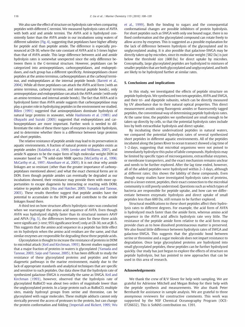
The structures of the synthesized peptides were evaluated byelectrospray ionization Fourier transform ion cyclotron resonance massspectrometry (ESI-FTICR-MS) and 1H Nuclear Magnetic Resonance(NMR) spectroscopy. For the MS analysis, formic acid (1%) was addedto the peptide solution (approx. 1:1 v/v ACN:H2O), which was thencontinuously infused into the ESI-FTICR-MS (Apollo II Bruker Daltonics12 Tesla Apex Qe) operated in positive ion mode using a modifiedprocedure described previously (Sleighter et al., 2008). For 1H NMRanalysis, 1.5 mg peptide was dissolved in 500 µL D2O, and analyzed by a400 MHzBrukerAvance spectrometer (BrukerBiospin, Inc.) usingwater
110 Z. Liu et al. / Marine Chemistry 119 (2010) 108–120
suppression (Bruker PRESAT). The obtained spectra agreed well withthose simulated by a 1H NMR predictor (ACD LABS 9.0). Based on theHPLC, MS and NMR spectra, the synthesized peptides were determinedto be of high purity (>95%) (Figs. A2–4). However, for AFVA and AAVF,the only difference between these twopeptides andAVFAwas the orderof amino acid vials in the peptide synthesizer, so we assume that AFVAand AAVF are of the same quality as AVFA. The MS analysis cannotdifferentiate between these three peptides because they have the samemolecular weight, but the HPLC chromatograms showed that they havedifferent retention times, indicating their successful synthesis.
2.2. Sampling location and description
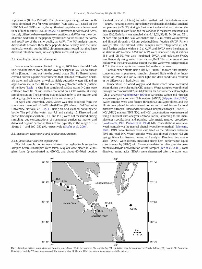
Water samples were collected in August, 2008, from the tidal freshto euryhaline James River (JR), the lower Chesapeake Bay (CB, southeastof the JR mouth), and out into the coastal ocean (Fig. 1). These stationscovered diverse aquatic environments that included freshwater, brack-ish water and salt water, as well as highly eutrophic waters (JR and analgal bloom site in the CB) and relatively oligotrophic waters (outsideof the Bay) (Table 1). One-liter samples of surface water (∼2 m) werecollected from 8 L Niskin bottles mounted on a CTD rosette at everysampling station. The sampling station labels refer to the location andsalinity, e.g., JR-5 indicates James River and salinity 5.
In April and December, 2008, water was also collected from theshore near themouth of the ElizabethRiver (ER) close toOldDominionUniversity, Norfolk, VA (Fig. 1), using an acid-cleaned polyethylenebottle. The pH of the water was 7.8 and salinity 17. Dissolved andparticulate organic carbon (DOC and POC) were not measured duringsampling, but concentrations of suspended particulate matter anddissolved organic carbon at this site are typically in the range of 10–30 mg L−1 and 200–250 µM, respectively (Shafer et al., 2004).
2.3. Incubation experiments and peptide measurement
2.3.1. James River transect experimentsThe 1-L sample bottles were shaken thoroughly to homogenize
samples before subsamples were taken. Aliquots were placed in 50 mLglass flasks (precombusted at 450 °C), and about 40–70 µL peptide
Fig. 1. Sampling stations along a transect from the James River (JR) to the southern ChesapeaUniversity, Norfolk, VA, was also sampled. The number after JR, CB, and ER in the station na
standard (in stock solution) was added so that final concentrations were∼10 µM. The sampleswere immediately incubated in the dark at ambienttemperature (∼24 °C). A single flask was incubated at each station (InJuly,weusedduplicateflasks and the variation inmeasured rateswas lessthan 10%). Each flaskwas sampled after 0, 12, 24, 36, 48, 54, 60, and 72 h.At each time point, the flaskwas shaken and 1–2 mLwater was removedand filtered through a 0.2 µm polyvinylidene fluoride (PVDF, Fisher)syringe filter. The filtered water samples were refrigerated at 4 °Cuntil further analysis within 1–2 d. AVFA and SWGA were incubated atall stations. AVFA amide, AAVF and AFVAwere incubated only at stationsJR-5 and CB-30. We also incubated SWGA and galactose-SWGAsimultaneously using water from station JR-23. The experimental pro-cedure was the same as above except that the water was refrigerated at4 °C in the laboratory for two weeks before the experiment.
Control experiments using HgCl2 (180 µM) showed that peptideconcentration in preserved samples changed little with time. Incu-bation of SWGA and AVFA under light and dark conditions resultedin no difference in hydrolysis rate.
Temperature, dissolved oxygen and fluorescence were measuredin situ during the cruise using CTD sensors. Water samples were filteredthrough precombusted 0.7 µm GF/F filters for fluorometric chlorophyll a(Chl a) analysis (Welschmeyer, 1994) or particulate carbon and nitrogenanalysis using an automatedCHNanalyzer (ANCA) (Filippino et al., 2009).Water samples were also filtered through 0.2 µm Supor filters, and thefiltrate was placed in acid-cleaned bottles and stored frozen for totaldissolvednitrogen (TDN) and for dissolved inorganic nitrogen (DIN:NO3
−,NO2
−, NH4+) analyses. TDN, NO3
−, andNO2− concentrationsweremeasured
using a nutrient auto-analyzer (Astoria Pacific) according to the man-ufacturer specifications and standard colorimetric method procedures(Valderrama, 1981; Parsons et al., 1984). NH4
+ concentrations were ana-lyzed manually via the manual phenol hypochlorite method (Solorzano,1969). DON concentrations were calculated as the difference betweenTDN and total DIN. Water samples were also filtered through 0.2-μmsyringe filters for dissolved amino acid analysis. Dissolved free aminoacids (DFAA) were directly measured using high performance liquidchromatography (HPLC) with fluorescence detection after pre-column o-phthaldialdehyde derivatization of the samples (Lee et al., 2000). Totaldissolved amino acids (TDAA) were determined after the water was
ke Bay (CB). A station near the mouth of the Elizabeth River (ER) close to Old Dominionme represents the salinity.
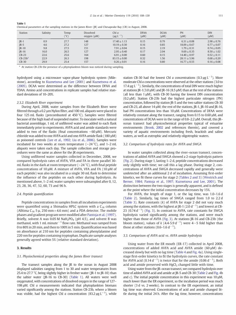
Table 1Chemical parameters at the sampling stations in the James River (JR) and Chesapeake Bay (CB) in August, 2008.
Station Salinity Temp(°C)
Dissolvedoxygen (µM)
Chl a(µg L−1)
DFAA(µM)
DCAA(µM)
PN(µM)
DIN(µM)
JR-1 0.9 27.7 148 17.40±1.13 0.15 0.79 6.31±0.08 2.98±0.76JR-5 4.6 27.2 127 10.19±0.30 0.16 0.85 18.09±0.67 0.77±0.07JR-10 9.8 27.5 151 7.91±0.04 0.15 2.19 1.79±0.31 0.74±0.05JR-16 16.0 26.5 127 2.95±0.40 0.17 2.04 9.69±0.35 8.50±1.02CB-23 22.6 25.6 164 6.91±0.00 0.60 0.15 14.40±0.97 0.52±0.11CB-23ba 22.9 26.3 198 83.15±0.00 0.32 1.56 28.11±5.96 0.68±0.20CB-30 29.9 25.4 157 0.26±0.01 0.16 0.80 16.77±0.55 0.16±0.08
a At station CB-23b the presence of a phytoplankton bloom was noticed during sampling.
111Z. Liu et al. / Marine Chemistry 119 (2010) 108–120
hydrolyzed using a microwave vapor-phase hydrolysis system (Mile-stone), according to Kuznetsova and Lee (2001) and Kuznetsova et al.(2005). DCAA were determined as the difference between DFAA andTDAA. Amino acid concentrations in replicate samples had relative stan-dard deviations of 10–20%.
2.3.2. Elizabeth River experimentDuring April, 2008, water samples from the Elizabeth River were
filtered through a0.2 µmNylonfilter, and100 mLaliquotswereplaced infour 125 mL flasks (precombusted at 450 °C). Samples were filteredbecause of the high load of suspendedmatter. To inoculatewith a naturalbacterial assemblage, 1 mL of unfiltered water was added to each flaskimmediately prior to experiments. AVFA acid and amide standards wereadded to two of the flasks (final concentrations ∼60 µM). Mercuricchloridewas added tooneAVFAacid andoneAVFAamideflask (180 µM)as poisoned controls (Lee et al., 1992; Liu et al., 2006). Samples wereincubated for two weeks at room temperature (∼24 °C), and 1–2 mLaliquots were taken each day. The sample collection and storage pro-cedures were the same as above for the JR experiments.
Using unfiltered water samples collected in December, 2008, wecompared hydrolysis rates of AVFA, VFA and FA in three parallel 30-mL flasks in the dark at room temperature (∼24 °C), with final peptideconcentrations of 10 µM. A mixture of AVFA, VFA and FA (10 µM ofeach peptide) was also incubated in a single 30 mL flask to determinethe influence of the peptides on each other during hydrolysis. Asmentioned above, 1–2 mLwater samples were subsampled after 0, 12,23, 28, 36, 47, 52, 60, 73 and 96 h.
2.4. Peptide quantification
Peptide concentrations in samples from all incubation experimentswere quantified using a Shimadzu HPLC system with a C18 column(Alltima C18 5 µ, 250 mm×4.6 mm) and a PDA detector. The mobilephases and gradient programweremodified after Pantoja et al. (1997).Briefly, solvent A was 0.05 M NaH2PO4 (pH 4.5), and solvent B wasmethanol, with 1 mL minute−1
flow rate. Methanol was ramped from0 to 80% in 20 min, and then to 100% in 5 min.Quantificationwas basedon absorbance at 210 nm for peptides containing phenylalanine and220 nm for peptides containing tryptophan. Duplicate sample analysesgenerally agreed within 5% (relative standard deviation).
3. Results
3.1. Physiochemical properties along the James River transect
The transect samples along the JR to the ocean in August 2008displayed salinities ranging from 1 to 30 and water temperatures from25.4 to 27.7 °C, being slightly higher in fresher water (JR-1 to JR-10) thanthe saltier water (JR-16 to CB-30) (Table 1). All waters were welloxygenated,with concentrations of dissolved oxygen in the range of 127–198 µM. Chl a measurements indicated that phytoplankton biomassvaried significantly among the stations. Station CB-23b, where a bloomwas visible, had the highest Chl a concentration (83.2 µg L−1), while
station CB-30 had the lowest Chl a concentrations (0.3 µg L−1). Moremoderate Chl a concentrationswere observed at the other stations (3.0 to17.4 µg L−1). Similarly, the concentrations of total DINweremuch higherat stations JR-1(3.0 µM) and JR-16 (8.5 µM) than at the rest of the stations(all less than 1 µM), with CB-30 having the lowest DIN concentration(0.2 µM). Station CB-23b had the highest particulate nitrogen (PN)concentration, followed by station JR-5 and the two saltier stations CB-30and CB-23, all above 14 µM; the rest of the stations, JR-1, JR-10 and JR-16,had PN concentrations less than 10 µM. Concentrations of DFAA wererelatively constant along the transect, ranging from 0.15 to 0.60 µM, andconcentrations of DCAAwere in the range of 0.8–2.2 µM. Overall, this JR–ocean transect had physicochemical properties typical of estuarineenvironments (Bronk, 2002 and references therein), and covered avariety of aquatic environments including fresh, brackish and salinewaters, as well as eutrophic and relatively oligotrophic waters.
3.2. Comparison of hydrolysis rates for AVFA and SWGA
In water samples collected along the river–ocean transect, concen-trations of added AVFA and SWGA showed a 2-stage hydrolysis pattern(Fig. 2). During stage 1, lasting 1–2 d, peptide concentrations decreasedonly slightly with time; we call this a lag phase. During stage 2, con-centrations of AVFA and SWGA decreased rapidly and generally wereundetected after an additional 2 d of incubation. Assuming first-orderkinetics, we fit these curves for stage 2 (Tables 2 and 3) (Westrich andBerner, 1984; Pantoja et al., 1997; Kuznetsova and Lee, 2001). Thedistinction between the two stages is generally apparent, and is definedas the point where the initial concentration decreases by 15%.
For AVFA, the length of stage 1, or the lag time, was 1.0–1.6 d(Table 2). Similarly, lag times of SWGA ranged from 1.0 to 2.2 d(Table 3). Rate constants (k) of AVFA for stage 2 did not vary muchamong the stations, with the highest at JR-1 (2.0 d−1) and lowest at CB-23 (0.74 d−1) (Fig. 3). In contrast to AVFA, rate constants for SWGAhydrolysis varied significantly among the stations, and were muchhigher than those of AVFA (Fig. 3). At stations JR-16 and CB-23b (thebloom station), values of k (4.4-5.9 d−1) were 4−5 fold higher thanthose at other stations (0.6–1.6 d−1).
3.3. Comparison of AVFA acid vs. AVFA amide hydrolysis
Using water from the ER mouth (ER-17) collected in April 2008,concentrations of added AVFA acid and AVFA amide (60 µM) de-creased slowly but with no lag time (Table 4 and Fig. 4a). Using single-stage first-order kinetics to fit the hydrolysis curves, the rate constantfor AVFA acid (0.14 d−1) is twice that for the amide (0.06 d−1). Bothacid and amide preserved with HgCl2 changed little with time.
Usingwater from the JR–ocean transect,we compared hydrolysis overtime of addedAVFA acid and amide at JR-5 and CB-30 (Table 2 and Fig. 4band c). The initial peptide concentration in this experiment was 10 µM,much lower than the ER experiment, so the incubation period was muchshorter (3 d vs. 2 weeks). In contrast to the ER experiment, an initiallag time was observed. Concentrations of acid and amide changed lit-tle during the initial 24 h. After the lag time, however, concentrations
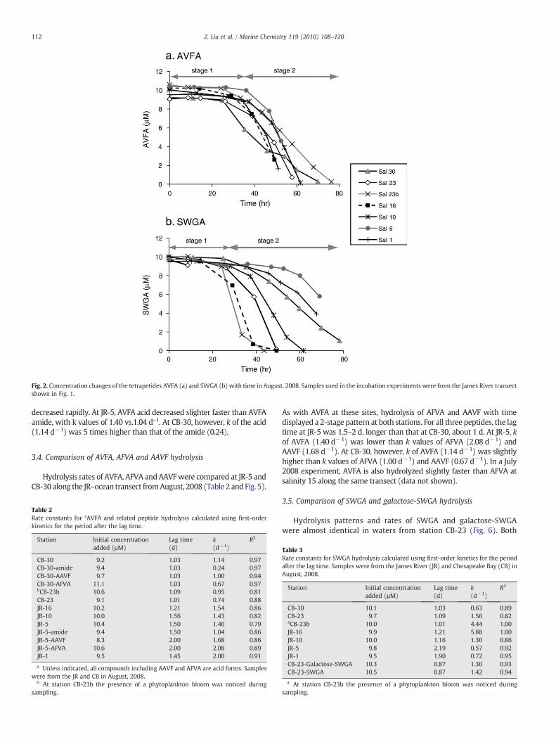
Fig. 2. Concentration changes of the tetrapetides AVFA (a) and SWGA (b) with time in August, 2008. Samples used in the incubation experiments were from the James River transectshown in Fig. 1.
112 Z. Liu et al. / Marine Chemistry 119 (2010) 108–120
decreased rapidly. At JR-5, AVFA acid decreased slighter faster than AVFAamide, with k values of 1.40 vs.1.04 d–1. At CB-30, however, k of the acid(1.14 d−1) was 5 times higher than that of the amide (0.24).
3.4. Comparison of AVFA, AFVA and AAVF hydrolysis
Hydrolysis rates of AVFA, AFVA andAAVFwere compared at JR-5 andCB-30 along the JR–ocean transect fromAugust, 2008 (Table 2 and Fig. 5).
Table 2Rate constants for aAVFA and related peptide hydrolysis calculated using first-orderkinetics for the period after the lag time.
Station Initial concentrationadded (µM)
Lag time(d)
k(d−1)
R2
CB-30 9.2 1.03 1.14 0.97CB-30-amide 9.4 1.03 0.24 0.97CB-30-AAVF 9.7 1.03 1.00 0.94CB-30-AFVA 11.1 1.03 0.67 0.97bCB-23b 10.6 1.09 0.95 0.81CB-23 9.1 1.01 0.74 0.88JR-16 10.2 1.21 1.54 0.86JR-10 10.0 1.56 1.43 0.82JR-5 10.4 1.50 1.40 0.79JR-5-amide 9.4 1.50 1.04 0.86JR-5-AAVF 8.3 2.00 1.68 0.86JR-5-AFVA 10.6 2.00 2.08 0.89JR-1 9.5 1.45 2.00 0.91
a Unless indicated, all compounds including AAVF and AFVA are acid forms. Sampleswere from the JR and CB in August, 2008.
b At station CB-23b the presence of a phytoplankton bloom was noticed duringsampling.
As with AVFA at these sites, hydrolysis of AFVA and AAVF with timedisplayed a 2-stage pattern at both stations. For all three peptides, the lagtime at JR-5 was 1.5–2 d, longer than that at CB-30, about 1 d. At JR-5, kof AVFA (1.40 d−1) was lower than k values of AFVA (2.08 d−1) andAAVF (1.68 d−1). At CB-30, however, k of AVFA (1.14 d−1) was slightlyhigher than k values of AFVA (1.00 d−1) and AAVF (0.67 d−1). In a July2008 experiment, AVFA is also hydrolyzed slightly faster than AFVA atsalinity 15 along the same transect (data not shown).
3.5. Comparison of SWGA and galactose-SWGA hydrolysis
Hydrolysis patterns and rates of SWGA and galactose-SWGAwere almost identical in waters from station CB-23 (Fig. 6). Both
Table 3Rate constants for SWGA hydrolysis calculated using first-order kinetics for the periodafter the lag time. Samples were from the James River (JR) and Chesapeake Bay (CB) inAugust, 2008.
Station Initial concentrationadded (µM)
Lag time(d)
k(d−1)
R2
CB-30 10.1 1.03 0.63 0.89CB-23 9.7 1.09 1.56 0.82aCB-23b 10.0 1.01 4.44 1.00JR-16 9.9 1.21 5.88 1.00JR-10 10.0 1.16 1.30 0.86JR-5 9.8 2.19 0.57 0.92JR-1 9.5 1.90 0.72 0.95CB-23-Galactose-SWGA 10.3 0.87 1.30 0.93CB-23-SWGA 10.5 0.87 1.42 0.94
a At station CB-23b the presence of a phytoplankton bloom was noticed duringsampling.
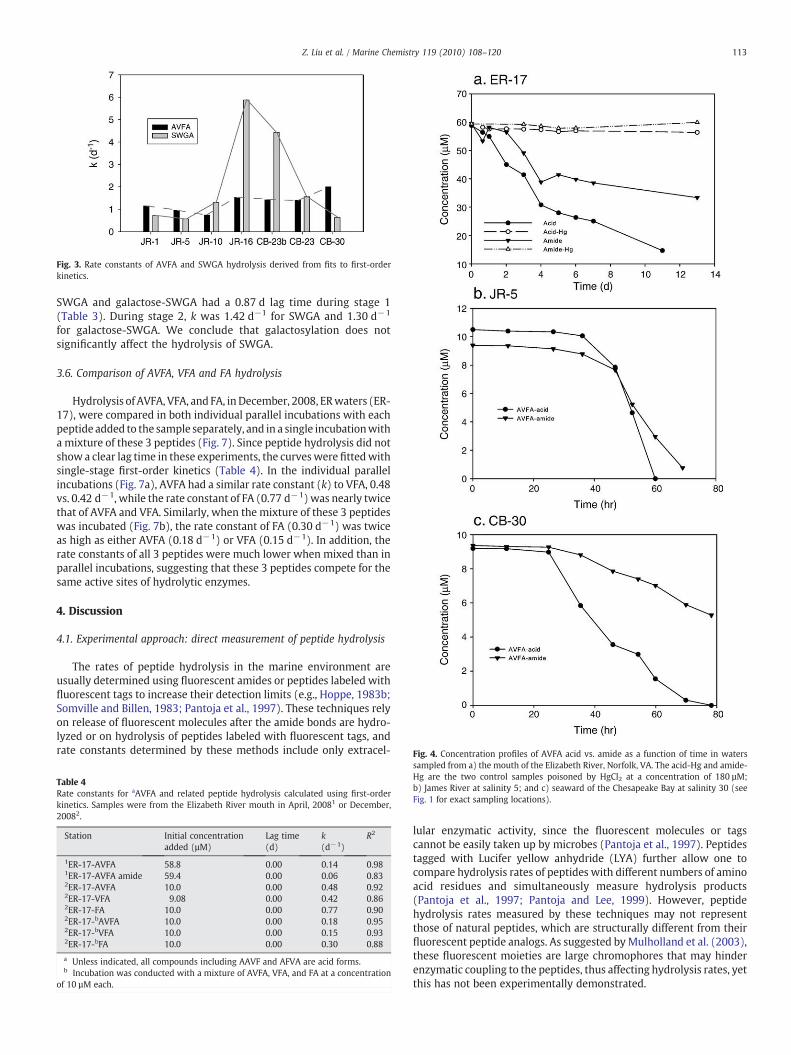
Fig. 3. Rate constants of AVFA and SWGA hydrolysis derived from fits to first-orderkinetics.
Fig. 4. Concentration profiles of AVFA acid vs. amide as a function of time in waters
113Z. Liu et al. / Marine Chemistry 119 (2010) 108–120
SWGA and galactose-SWGA had a 0.87 d lag time during stage 1(Table 3). During stage 2, k was 1.42 d−1 for SWGA and 1.30 d−1
for galactose-SWGA. We conclude that galactosylation does notsignificantly affect the hydrolysis of SWGA.
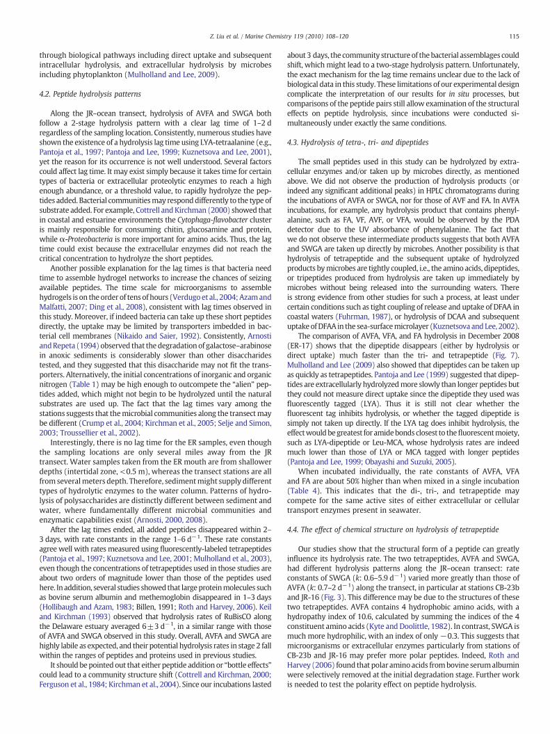
3.6. Comparison of AVFA, VFA and FA hydrolysis
Hydrolysis of AVFA, VFA, and FA, in December, 2008, ERwaters (ER-17), were compared in both individual parallel incubations with eachpeptide added to the sample separately, and in a single incubationwitha mixture of these 3 peptides (Fig. 7). Since peptide hydrolysis did notshow a clear lag time in these experiments, the curveswere fittedwithsingle-stage first-order kinetics (Table 4). In the individual parallelincubations (Fig. 7a), AVFA had a similar rate constant (k) to VFA, 0.48vs. 0.42 d−1, while the rate constant of FA (0.77 d−1) was nearly twicethat of AVFA and VFA. Similarly, when the mixture of these 3 peptideswas incubated (Fig. 7b), the rate constant of FA (0.30 d−1) was twiceas high as either AVFA (0.18 d−1) or VFA (0.15 d−1). In addition, therate constants of all 3 peptides were much lower when mixed than inparallel incubations, suggesting that these 3 peptides compete for thesame active sites of hydrolytic enzymes.
4. Discussion
4.1. Experimental approach: direct measurement of peptide hydrolysis
The rates of peptide hydrolysis in the marine environment areusually determined using fluorescent amides or peptides labeled withfluorescent tags to increase their detection limits (e.g., Hoppe, 1983b;Somville and Billen, 1983; Pantoja et al., 1997). These techniques relyon release of fluorescent molecules after the amide bonds are hydro-lyzed or on hydrolysis of peptides labeled with fluorescent tags, andrate constants determined by these methods include only extracel-
Table 4Rate constants for aAVFA and related peptide hydrolysis calculated using first-orderkinetics. Samples were from the Elizabeth River mouth in April, 20081 or December,20082.
Station Initial concentrationadded (µM)
Lag time(d)
k(d−1)
R2
1ER-17-AVFA 58.8 0.00 0.14 0.981ER-17-AVFA amide 59.4 0.00 0.06 0.832ER-17-AVFA 10.0 0.00 0.48 0.922ER-17-VFA 9.08 0.00 0.42 0.862ER-17-FA 10.0 0.00 0.77 0.902ER-17-bAVFA 10.0 0.00 0.18 0.952ER-17-bVFA 10.0 0.00 0.15 0.932ER-17-bFA 10.0 0.00 0.30 0.88
a Unless indicated, all compounds including AAVF and AFVA are acid forms.b Incubation was conducted with a mixture of AVFA, VFA, and FA at a concentration
of 10 µM each.
sampled from a) the mouth of the Elizabeth River, Norfolk, VA. The acid-Hg and amide-Hg are the two control samples poisoned by HgCl2 at a concentration of 180 µM;b) James River at salinity 5; and c) seaward of the Chesapeake Bay at salinity 30 (seeFig. 1 for exact sampling locations).
lular enzymatic activity, since the fluorescent molecules or tagscannot be easily taken up by microbes (Pantoja et al., 1997). Peptidestagged with Lucifer yellow anhydride (LYA) further allow one tocompare hydrolysis rates of peptides with different numbers of aminoacid residues and simultaneously measure hydrolysis products(Pantoja et al., 1997; Pantoja and Lee, 1999). However, peptidehydrolysis rates measured by these techniques may not representthose of natural peptides, which are structurally different from theirfluorescent peptide analogs. As suggested byMulholland et al. (2003),these fluorescent moieties are large chromophores that may hinderenzymatic coupling to the peptides, thus affecting hydrolysis rates, yetthis has not been experimentally demonstrated.
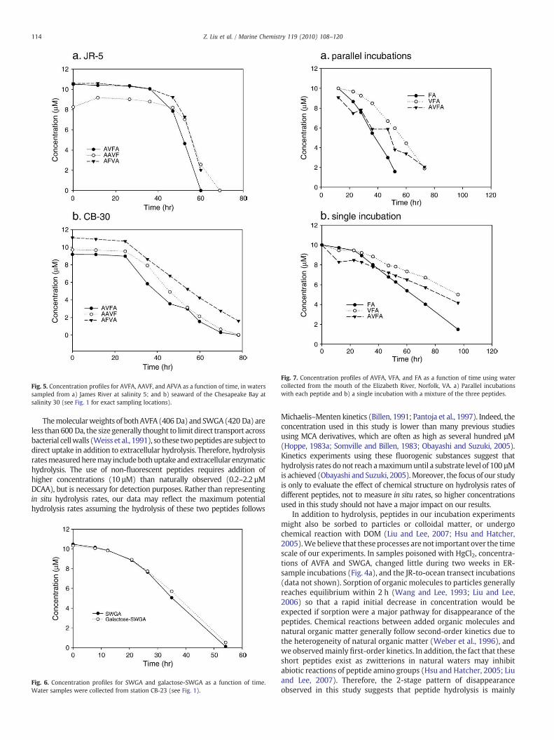
Fig. 5. Concentration profiles for AVFA, AAVF, and AFVA as a function of time, in waterssampled from a) James River at salinity 5; and b) seaward of the Chesapeake Bay atsalinity 30 (see Fig. 1 for exact sampling locations).
Fig. 7. Concentration profiles of AVFA, VFA, and FA as a function of time using watercollected from the mouth of the Elizabeth River, Norfolk, VA. a) Parallel incubationswith each peptide and b) a single incubation with a mixture of the three peptides.
114 Z. Liu et al. / Marine Chemistry 119 (2010) 108–120
Themolecularweights of bothAVFA(406 Da) andSWGA(420 Da) areless than600 Da, the size generally thought to limit direct transport acrossbacterial cellwalls (Weiss et al., 1991), so these twopeptidesare subject todirect uptake in addition to extracellular hydrolysis. Therefore, hydrolysisratesmeasuredheremay includebothuptake andextracellular enzymatichydrolysis. The use of non-fluorescent peptides requires addition ofhigher concentrations (10 µM) than naturally observed (0.2–2.2 µMDCAA), but is necessary for detection purposes. Rather than representingin situ hydrolysis rates, our data may reflect the maximum potentialhydrolysis rates assuming the hydrolysis of these two peptides follows
Fig. 6. Concentration profiles for SWGA and galactose-SWGA as a function of time.Water samples were collected from station CB-23 (see Fig. 1).
Michaelis–Menten kinetics (Billen, 1991; Pantoja et al., 1997). Indeed, theconcentration used in this study is lower than many previous studiesusing MCA derivatives, which are often as high as several hundred µM(Hoppe, 1983a; Somville and Billen, 1983; Obayashi and Suzuki, 2005).Kinetics experiments using these fluorogenic substances suggest thathydrolysis rates donot reach amaximumuntil a substrate level of 100 µMis achieved (Obayashi and Suzuki, 2005).Moreover, the focus of our studyis only to evaluate the effect of chemical structure on hydrolysis rates ofdifferent peptides, not to measure in situ rates, so higher concentrationsused in this study should not have a major impact on our results.
In addition to hydrolysis, peptides in our incubation experimentsmight also be sorbed to particles or colloidal matter, or undergochemical reaction with DOM (Liu and Lee, 2007; Hsu and Hatcher,2005).Webelieve that these processes are not important over the timescale of our experiments. In samples poisoned with HgCl2, concentra-tions of AVFA and SWGA, changed little during two weeks in ER-sample incubations (Fig. 4a), and the JR-to-ocean transect incubations(data not shown). Sorption of organic molecules to particles generallyreaches equilibrium within 2 h (Wang and Lee, 1993; Liu and Lee,2006) so that a rapid initial decrease in concentration would beexpected if sorption were a major pathway for disappearance of thepeptides. Chemical reactions between added organic molecules andnatural organic matter generally follow second-order kinetics due tothe heterogeneity of natural organic matter (Weber et al., 1996), andwe observedmainly first-order kinetics. In addition, the fact that theseshort peptides exist as zwitterions in natural waters may inhibitabiotic reactions of peptide amino groups (Hsu and Hatcher, 2005; Liuand Lee, 2007). Therefore, the 2-stage pattern of disappearanceobserved in this study suggests that peptide hydrolysis is mainly
115Z. Liu et al. / Marine Chemistry 119 (2010) 108–120
through biological pathways including direct uptake and subsequentintracellular hydrolysis, and extracellular hydrolysis by microbesincluding phytoplankton (Mulholland and Lee, 2009).
4.2. Peptide hydrolysis patterns
Along the JR–ocean transect, hydrolysis of AVFA and SWGA bothfollow a 2-stage hydrolysis pattern with a clear lag time of 1–2 dregardless of the sampling location. Consistently, numerous studies haveshown the existence of a hydrolysis lag time using LYA-tetraalanine (e.g.,Pantoja et al., 1997; Pantoja and Lee, 1999; Kuznetsova and Lee, 2001),yet the reason for its occurrence is not well understood. Several factorscould affect lag time. It may exist simply because it takes time for certaintypes of bacteria or extracellular proteolytic enzymes to reach a highenough abundance, or a threshold value, to rapidly hydrolyze the pep-tides added. Bacterial communitiesmay responddifferently to the typeofsubstrate added. For example, Cottrell andKirchman (2000) showed thatin coastal and estuarine environments the Cytophaga-flavobacter clusteris mainly responsible for consuming chitin, glucosamine and protein,while α-Proteobacteria is more important for amino acids. Thus, the lagtime could exist because the extracellular enzymes did not reach thecritical concentration to hydrolyze the short peptides.
Another possible explanation for the lag times is that bacteria needtime to assemble hydrogel networks to increase the chances of seizingavailable peptides. The time scale for microorganisms to assemblehydrogels is on theorder of tens of hours (Verdugoet al., 2004;AzamandMalfatti, 2007; Ding et al., 2008), consistent with lag times observed inthis study. Moreover, if indeed bacteria can take up these short peptidesdirectly, the uptake may be limited by transporters imbedded in bac-terial cell membranes (Nikaido and Saier, 1992). Consistently, ArnostiandRepeta (1994) observed that thedegradationof galactose–arabinosein anoxic sediments is considerably slower than other disaccharidestested, and they suggested that this disaccharide may not fit the trans-porters. Alternatively, the initial concentrations of inorganic and organicnitrogen (Table 1) may be high enough to outcompete the “alien” pep-tides added, which might not begin to be hydrolyzed until the naturalsubstrates are used up. The fact that the lag times vary among thestations suggests that themicrobial communities along the transectmaybe different (Crump et al., 2004; Kirchman et al., 2005; Selje and Simon,2003; Troussellier et al., 2002).
Interestingly, there is no lag time for the ER samples, even thoughthe sampling locations are only several miles away from the JRtransect. Water samples taken from the ER mouth are from shallowerdepths (intertidal zone, <0.5 m), whereas the transect stations are allfrom severalmeters depth. Therefore, sedimentmight supply differenttypes of hydrolytic enzymes to the water column. Patterns of hydro-lysis of polysaccharides are distinctly different between sediment andwater, where fundamentally different microbial communities andenzymatic capabilities exist (Arnosti, 2000, 2008).
After the lag times ended, all added peptides disappeared within 2–3 days, with rate constants in the range 1–6 d−1. These rate constantsagree well with rates measured using fluorescently-labeled tetrapeptides(Pantoja et al., 1997; Kuznetsova and Lee, 2001; Mulholland et al., 2003),even though the concentrations of tetrapeptides used in those studies areabout two orders of magnitude lower than those of the peptides usedhere. Inaddition, several studies showed that largeproteinmolecules suchas bovine serum albumin and methemoglobin disappeared in 1–3 days(Hollibaugh and Azam, 1983; Billen, 1991; Roth and Harvey, 2006). Keiland Kirchman (1993) observed that hydrolysis rates of RuBisCO alongthe Delaware estuary averaged 6±3 d−1, in a similar range with thoseof AVFA and SWGA observed in this study. Overall, AVFA and SWGA arehighly labile as expected, and their potential hydrolysis rates in stage 2 fallwithin the ranges of peptides and proteins used in previous studies.
It shouldbepointed out that either peptide addition or “bottle effects”could lead to a community structure shift (Cottrell and Kirchman, 2000;Ferguson et al., 1984; Kirchman et al., 2004). Since our incubations lasted
about3 days, the community structureof thebacterial assemblages couldshift, which might lead to a two-stage hydrolysis pattern. Unfortunately,the exact mechanism for the lag time remains unclear due to the lack ofbiological data in this study. These limitations of our experimental designcomplicate the interpretation of our results for in situ processes, butcomparisons of the peptide pairs still allow examination of the structuraleffects on peptide hydrolysis, since incubations were conducted si-multaneously under exactly the same conditions.
4.3. Hydrolysis of tetra-, tri- and dipeptides
The small peptides used in this study can be hydrolyzed by extra-cellular enzymes and/or taken up by microbes directly, as mentionedabove. We did not observe the production of hydrolysis products (orindeed any significant additional peaks) in HPLC chromatograms duringthe incubations of AVFA or SWGA, nor for those of AVF and FA. In AVFAincubations, for example, any hydrolysis product that contains phenyl-alanine, such as FA, VF, AVF, or VFA, would be observed by the PDAdetector due to the UV absorbance of phenylalanine. The fact thatwe do not observe these intermediate products suggests that both AVFAand SWGA are taken up directly by microbes. Another possibility is thathydrolysis of tetrapeptide and the subsequent uptake of hydrolyzedproducts bymicrobes are tightly coupled, i.e., the amino acids, dipeptides,or tripeptides produced from hydrolysis are taken up immediately bymicrobes without being released into the surrounding waters. Thereis strong evidence from other studies for such a process, at least undercertain conditions such as tight coupling of release and uptake of DFAA incoastal waters (Fuhrman, 1987), or hydrolysis of DCAA and subsequentuptakeofDFAA in the sea-surfacemicrolayer (Kuznetsova and Lee, 2002).
The comparison of AVFA, VFA, and FA hydrolysis in December 2008(ER-17) shows that the dipeptide disappears (either by hydrolysis ordirect uptake) much faster than the tri- and tetrapeptide (Fig. 7).Mulholland and Lee (2009) also showed that dipeptides can be taken upas quickly as tetrapeptides. Pantoja and Lee (1999) suggested that dipep-tides are extracellularly hydrolyzedmore slowly than longer peptides butthey could not measure direct uptake since the dipeptide they used wasfluorescently tagged (LYA). Thus it is still not clear whether thefluorescent tag inhibits hydrolysis, or whether the tagged dipeptide issimply not taken up directly. If the LYA tag does inhibit hydrolysis, theeffectwouldbegreatest for amidebonds closest to thefluorescentmoiety,such as LYA-dipeptide or Leu-MCA, whose hydrolysis rates are indeedmuch lower than those of LYA or MCA tagged with longer peptides(Pantoja and Lee, 1999; Obayashi and Suzuki, 2005).
When incubated individually, the rate constants of AVFA, VFAand FA are about 50% higher than when mixed in a single incubation(Table 4). This indicates that the di-, tri-, and tetrapeptide maycompete for the same active sites of either extracellular or cellulartransport enzymes present in seawater.
4.4. The effect of chemical structure on hydrolysis of tetrapeptide
Our studies show that the structural form of a peptide can greatlyinfluence its hydrolysis rate. The two tetrapeptides, AVFA and SWGA,had different hydrolysis patterns along the JR–ocean transect: rateconstants of SWGA (k: 0.6–5.9 d−1) varied more greatly than those ofAVFA (k: 0.7–2 d−1) along the transect, in particular at stations CB-23band JR-16 (Fig. 3). This difference may be due to the structures of thesetwo tetrapeptides. AVFA contains 4 hydrophobic amino acids, with ahydropathy index of 10.6, calculated by summing the indices of the 4constituent amino acids (Kyte and Doolittle, 1982). In contrast, SWGA ismuch more hydrophilic, with an index of only −0.3. This suggests thatmicroorganisms or extracellular enzymes particularly from stations ofCB-23b and JR-16 may prefer more polar peptides. Indeed, Roth andHarvey (2006) found that polar aminoacids frombovine serumalbuminwere selectively removed at the initial degradation stage. Further workis needed to test the polarity effect on peptide hydrolysis.
116 Z. Liu et al. / Marine Chemistry 119 (2010) 108–120
Wealso saw the effect of structure onhydrolysis ratewhen comparingpeptides with different C-termini. Wemeasured hydrolysis rates of AVFAwith both acid and amide termini. The AVFA acid is hydrolyzed con-sistently faster than the AVFA amide in our incubations using waters ofdifferent salinities (Fig. 4), suggesting that peptidases have higher affinityfor peptide acid than peptide amide. The difference is especially pro-nounced at CB-30, where the rate constant of AVFA acid is 5 times higherthan that of AVFA amide. This large difference between acid and amidehydrolysis rates is somewhat unexpected since the only difference be-tween them is the C-terminal structure. However, peptidases can becategorized into aminopeptidases, carboxypeptidases, and endopepti-dases, and each group has a different specificity. Aminopeptidases cleavepeptides at the amino terminus, carboxypeptidases at the carboxyl termi-nus, and endopeptidases at the internal peptide bonds (Barrett et al.,2004). While all three peptidases can attack the AVFA acid form (with anamino terminus, carboxyl terminus, and internal peptide bonds), onlyaminopeptidase andendopeptidase canattack theAVFAamide (withonlyan amino terminus and internal amide bonds). The fact that AVFA acid ishydrolyzed faster than AVFA amide suggests that carboxypeptidase mayplay a greater role in hydrolyzingpeptides in the environmentwe studied.Billen (1991) suggested that aminopeptidases dominate hydrolysis ofnatural large proteins in seawater, while Hashimoto et al. (1985) andObayashi and Suzuki (2005) suggested that endopeptidases and car-boxypeptidases are more important. Further work is needed to dif-ferentiate the roles of these three types of enzymes in peptide hydrolysis,and to determine whether there is a difference between large proteinsand short peptides.
The relative resistance of peptide amidemay lead to its preservation inaquatic environments. A fraction of natural protein or peptides exists aspeptide amides (Kulathila et al., 1999; Lemke and Williams, 2007), andamide N appears to be the major form of high molecular weight DON inseawater based on 15N solid-state NMR spectra (McCarthy et al., 1996;McCarthy et al., 1997; Aluwihare et al., 2005). It is not clear why amidelinkages are so resistant (other than the greater importance of carboxy-peptidases mentioned above) and what the exact chemical forms are inDON. Even though peptide amides can eventually be degraded as ourincubations showed, their resistance may provide them with more op-portunities to escape diagenesis by interacting or reacting with DOM,relative to peptide acids (Hsu and Hatcher, 2005; Yamada and Tanoue,2003). These results therefore suggest that peptide amides have thepotential to accumulate in the DON pool and contribute to the amidelinkages found there.
A third test on how structure affects hydrolysis rates was conductedwhen we rearranged the amino acid sequence of AVFA. Even thoughAVFA was hydrolyzed slightly faster than its structural isomers AAVFand AFVA (Fig. 5), the differences between rates for these three acidswere significant (t-test; 95% significance level) only at CB-30, not at JR-5.This suggests that the amino acid sequence in a peptide has little effecton its hydrolysis when the amino acid residues are the same, and thatsimilar enzymes are responsible for degrading these three peptide acids.
Glycosylation is thought to increase the resistanceof proteins inDOMtomicrobial attack (Keil and Kirchman, 1993). Recent studies suggestedthat a major fraction of proteins in waters is glycosylated (Yamada andTanoue, 2003; Saijo and Tanoue, 2005). It has been difficult to study theresistance of these glycosylated proteins and peptides and theirdiagenetic pathways in the marine environment, mainly due to thelack of appropriate standards and analytical techniques that are specificand sensitive to such peptides. Our data show that the hydrolysis rate ofsynthesized galactose-SWGA is essentially the same as SWGA. Keil andKirchman (1993), however, observed that the hydrolysis rate ofglycosylated RuBisCO was about two orders of magnitude lower thanthe unglycosylated protein. In a large protein such as RuBisCO, multiplesites, mainly hydroxyl groups from serine and threonine, can beglycosylated with sugar molecules. These multiple adducts cannot onlysterically prevent the access of proteases to the protein, but can changethe protein conformation and folding (Imperiali and Rickert, 1995; Wu
et al., 1999). Both the binding to sugars and the consequentialconformational changes are possible inhibitors of protein hydrolysis.For short peptides such as SWGAwith only one bound sugar, there is nofixed conformation and the glycosylated compound can rotate freely toallow access by enzymes. This is suggested as a possible explanation forthe lack of difference between hydrolysis of the glycosylated and itsunglycosylated analog. It is also possible that galactose-SWGA may bedirectly taken up bymicrobes, since itsmolecularweight (582 Da) is justbelow the threshold size (600 Da) for direct uptake by microbes.Conceptually, large glycosylated peptides are hydrolyzed to mixtures ofsmaller peptides that arebothglycosylatedandunglycosylated, andbothare likely to be hydrolyzed further at similar rates.
5. Conclusions and implications
In this study, we investigated the effects of peptide structure onpeptidehydrolysis.Wesynthesized two tetrapeptides, AVFA and SWGA,and their tri- and dipeptide subunits, which can be directly measuredby UV absorbance due to their natural optical properties. This directmeasurement avoids using fluorogenic amides or fluorescently taggedpeptides, the conventional way of determining peptide hydrolysis rates.At the same time, the peptides we synthesized are small enough to betaken up directly by cells, so that the potential hydrolysis rates includeloss by both extracellular hydrolysis and direct uptake.
By incubating these underivatized peptides in natural waters,we compared the potential hydrolysis rates of several synthesizedshort peptides in different aquatic environments. All of the peptidesincubated along the James River to ocean transect showed a lag time of1–2 days, suggesting that microbial organisms were not poised toimmediately hydrolyze this pulse of labile compounds. Hydrolysismaybe limited by specific types of microorganisms, extracellular enzymes,or membrane transporters, and the exact mechanism remains unclearand needs to be further explored. After the lag time ended, however,all of the added peptides were hydrolyzed within 2–3 days, althoughat different rates; this shows the lability of these compounds. Eventhough many studies have investigated hydrolysis rates of proteins,and to a lesser extent, peptides, their exact connection to themicrobialcommunity is still poorly understood. Questions such aswhich types ofbacteria are responsible for peptide uptake, and how can we differ-entiate between enzymatic hydrolysis and direct uptake of smallpeptides less than 600 Da, still remain to be further explored.
Structural modifications to these short peptides affect their hydro-lysis rates to different degrees. For example, the acid form of AVFAis hydrolyzed much faster than the amide form, whereas amino acidsequence in the AVFA acid affects hydrolysis rate very little. Theresistance of the peptide amide form relative to the acid form mayprovide clues as to how dissolved proteinaceous matter is preserved.We also found little difference between hydrolysis rates of SWGA andgalactose-SWGA. This suggests that the glycosidic bond betweenserine or threonine and a sugarmolecule does not impart resistance todegradation. Once large glycosylated proteins are hydrolyzed intosmall glycosylated peptides, these peptides can be further hydrolyzedquickly. Our study has just begun to explore the effects of structure onpeptide hydrolysis, but has pointed to new approaches that can beused in this area of research.
Acknowledgements
We thank the crew of R/V Slover for help with sampling. We aregrateful for Adrienne Mitchell and Megan Bishop for their help withthe peptide synthesis and measurements. We also thank PeterBernhardt for assistance in sample analysis. We are grateful to threeanonymous reviewers for constructive comments. This work wassupported by the NSF Chemical Oceanography Program (OCE-0726632). This is SoMAS contribution no. 1391.

117Z. Liu et al. / Marine Chemistry 119 (2010) 108–120
Appendix A
Fig. A1. Chemical structures of synthesized peptides. a. FA (C12H16N2O3, MW: 236.26758); b. VFA (C17H25N3O4, MW: 335.39888); c. AVFA (C20H30N4O5, MW: 406.47694), AVFAamide (C20H31N5O4, MW: 405.49222), AAVF (C20H30N4O5, MW: 406.47694), AFVA (C20H30N4O5, MW: 406.47694), SWGA (C19H25N5O6, MW: 419.43265) and galactose-SWGA(C25H35N5O11, MW:581.57349). The molecular weights are isotopically averaged neutral masses.
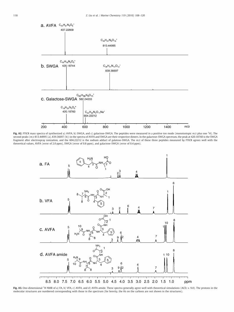
Fig. A2. FTICR mass spectra of synthesized a) AVFA, b) SWGA, and c) galactose-SWGA. The peptides were measured in a positive ion mode (monoisotopic m/z plus one 1H). Thesecond peaks (m/z 813.44995 (a), 839.36697 (b)) in the spectra of AVFA and SWGA are their respective dimers. In the galactose-SWGA spectrum, the peak at 420.18760 is the SWGAfragment after electrospray ionization, and the 604.22212 is the sodium adduct of galatose-SWGA. The m/z of these three peptides measured by FTICR agrees well with thetheoretical values, AVFA (error of 2.0 ppm), SWGA (error of 0.8 ppm), and galactose-SWGA (error of 0.4 ppm).
Fig. A3. One-dimensional 1H NMR of a) FA, b) VFA, c) AVFA, and d) AVFA amide. These spectra generally agree well with theoretical simulations (ACD, v. 9.0). The protons in themolecular structures are numbered corresponding with those in the spectrum (for brevity, the Hs on the carbons are not shown in the structures).
118 Z. Liu et al. / Marine Chemistry 119 (2010) 108–120
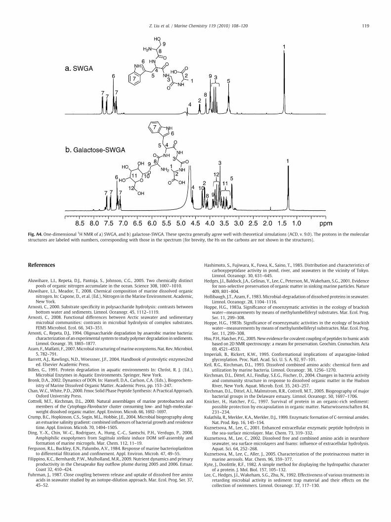
Fig. A4. One-dimensional 1H NMR of a) SWGA, and b) galactose-SWGA. These spectra generally agree well with theoretical simulations (ACD, v. 9.0). The protons in the molecularstructures are labeled with numbers, corresponding with those in the spectrum (for brevity, the Hs on the carbons are not shown in the structures).
119Z. Liu et al. / Marine Chemistry 119 (2010) 108–120
References
Aluwihare, L.I., Repeta, D.J., Pantoja, S., Johnson, C.G., 2005. Two chemically distinctpools of organic nitrogen accumulate in the ocean. Science 308, 1007–1010.
Aluwihare, L.I., Meador, T., 2008. Chemical composition of marine dissolved organicnitrogen. In: Capone, D., et al. (Ed.), Nitrogen in theMarine Environment. Academic,New York.
Arnosti, C., 2000. Substrate specificity in polysaccharide hydrolysis: contrasts betweenbottom water and sediments. Limnol. Oceanogr. 45, 1112–1119.
Arnosti, C., 2008. Functional differences between Arctic seawater and sedimentarymicrobial communities: contrasts in microbial hydrolysis of complex substrates.FEMS Microbiol. Ecol. 66, 343–351.
Arnosti, C., Repeta, D.J., 1994. Oligosaccharide degradation by anaerobic marine bacteria:characterization of an experimental system to studypolymer degradation in sediments.Limnol. Oceangr. 39, 1865–1877.
Azam, F.,Malfatti, F., 2007.Microbial structuringofmarine ecosystems.Nat. Rev.Microbiol.5, 782–791.
Barrett, A.J., Rawlings, N.D., Woessner, J.F., 2004. Handbook of proteolytic enzymes2nded. Elsevier Academic Press.
Billen, G., 1991. Protein degradation in aquatic environments In: Chróst, R. J. (Ed.),Microbial Enzymes in Aquatic Environments. Springer, New York.
Bronk, D.A., 2002. Dynamics of DON. In: Hansell, D.A., Carlson, C.A. (Eds.), Biogeochem-istry of Marine Dissolved Organic Matter. Academic Press, pp. 153–247.
Chan,W.C.,White, P.D., 2000. Fmoc Solid Phase Peptide Synthesis: A Practical Approach.Oxford University Press.
Cottrell, M.T., Kirchman, D.L., 2000. Natural assemblages of marine proteobacteria andmembers of the Cytophaga-Flavobacter cluster consuming low- and high-molecular-weight dissolved organic matter. Appl. Environ. Microb. 66, 1692–1697.
Crump, B.C., Hopkinson, C.S., Sogin, M.L., Hobbie, J.E., 2004. Microbial biogeography alongan estuarine salinity gradient: combined influences of bacterial growth and residencetime. Appl. Environ. Microb. 70, 1494–1505.
Ding, Y.-X., Chin, W.-C., Rodriguez, A., Hung, C.-C., Santschi, P.H., Verdugo, P., 2008.Amphiphilic exopolymers from Sagittula stellata induce DOM self-assembly andformation of marine microgels. Mar. Chem. 112, 11–19.
Ferguson, R.L., Buckley, E.N., Palumbo, A.V., 1984. Response of marine bacterioplanktonto differential filtration and confinement. Appl. Environ. Microb. 47, 49–55.
Filippino, K.C., Bernhardt, P.W., Mulholland, M.R., 2009. Nutrient dynamics and primaryproductivity in the Chesapeake Bay outflow plume during 2005 and 2006. Estuar.Coast 32, 410–424.
Fuhrman, J., 1987. Close coupling between release and uptake of dissolved free aminoacids in seawater studied by an isotope-dilution approach. Mar. Ecol. Prog. Ser. 37,45–52.
Hashimoto, S., Fujiwara, K., Fuwa, K., Saino, T., 1985. Distribution and characteristics ofcarboxypeptidase activity in pond, river, and seawaters in the vicinity of Tokyo.Limnol. Oceanogr. 30, 631–645.
Hedges, J.I., Baldock, J.A., Gelinas, Y., Lee, C., Peterson,M.,Wakeham, S.G., 2001. Evidencefor non-selective preservation of organic matter in sinking marine particles. Nature409, 801–804.
Hollibaugh, J.T., Azam, F., 1983.Microbial-degradation of dissolved proteins in seawater.Limnol. Oceanogr. 28, 1104–1116.
Hoppe, H.G., 1983a. Significance of exoenzymatic activities in the ecology of brackishwater—measurements by means of methylumbelliferyl substrates. Mar. Ecol. Prog.Ser. 11, 299–308.
Hoppe, H.G., 1983b. Significance of exoenzymatic activities in the ecology of brackishwater—measurements bymeans of methylumbelliferyl substractes. Mar. Ecol. Prog.Ser. 11, 299–308.
Hsu, P.H.,Hatcher, P.G., 2005. Newevidence for covalent coupling of peptides to humic acidsbased on 2D NMR spectroscopy: a means for preservation. Geochim. Cosmochim. Acta69, 4521–4533.
Imperiali, B., Rickert, K.W., 1995. Conformational implications of asparagine-linkedglycosylation. Proc. Natl. Acad. Sci. U. S. A. 92, 97–101.
Keil, R.G., Kirchman, D.L., 1993. Dissolved combined amino acids: chemical form andutilization by marine bacteria. Limnol. Oceanogr. 38, 1256–1270.
Kirchman, D.L., Dittel, A.I., Findlay, S.E.G., Fischer, D., 2004. Changes in bacteria activityand community structure in response to dissolved organic matter in the HudsonRiver, New York. Aquat. Microb. Ecol. 35, 243–257.
Kirchman, D.L., Dittel, A.I., Malmstrom, R.R., Cottrell, M.T., 2005. Biogeography of majorbacterial groups in the Delaware estuary. Limnol. Oceanogr. 50, 1697–1706.
Knicker, H., Hatcher, P.G., 1997. Survival of protein in an organic-rich sediment:possible protection by encapsulation in organic matter. Naturwissenschaften 84,231–234.
Kulathila, R., Merkler, K.A., Merkler, D.J., 1999. Enzymatic formation of C-terminal amides.Nat. Prod. Rep. 16, 145–154.
Kuznetsova, M., Lee, C., 2001. Enhanced extracellular enzymatic peptide hydrolysis inthe sea-surface microlayer. Mar. Chem. 73, 319–332.
Kuznetsova, M., Lee, C., 2002. Dissolved free and combined amino acids in nearshoreseawater, sea surface microlayers and foams: influence of extracellular hydrolysis.Aquat. Sci. 64, 252–268.
Kuznetsova, M., Lee, C., Aller, J., 2005. Characterization of the proteinaceous matter inmarine aerosols. Mar. Chem. 96, 359–377.
Kyte, J., Doolittle, R.F., 1982. A simple method for displaying the hydropathic characterof a protein. J. Mol. Biol. 157, 105–132.
Lee, C., Hedges, J.I., Wakeham, S.G., Zhu, N., 1992. Effectiveness of various treatments inretarding microbial activity in sediment trap material and their effects on thecollection of swimmers. Limnol. Oceanogr. 37, 117–130.
120 Z. Liu et al. / Marine Chemistry 119 (2010) 108–120
Lee, C., Wakeham, S.G., Hedges, J.I., 2000. Composition and flux of particulate aminoacids and chloropigments in equatorial Pacific seawater and sediments. Deep-SeaRes. 47, 1535–1568.
Lemke, T.L.,Williams, D.A., 2007. Foye's Principle ofMedicinal Chemistry6 ed. LippincottWilliams and Wilkins.
Liu, Z.F., Lee, C., 2006. Drying effects on sorption capacity of coastal sediment: Theimportance of architecture and polarity of organic matter. Geochim. Cosmochim.Acta 70, 3313–3324.
Liu, Z.F., Lee, C., 2007. The role of organic matter in the sorption capacity of marinesediments. Mar. Chem. 105, 240–257.
Liu, Z.F., Lee, C., Wakeham, S.G., 2006. Effects of mercuric chloride and proteaseinhibitors on degradation of particulate organic matter from the diatom Thalas-siosira pseudonana. Org. Geochem. 37, 1003–1018.
Liu, Z.F., Lee, C., Aller, R.C., 2008. Drying effects on decomposition of salt marshsediment and on lysine sorption. J. Mar. Res. 66, 665–689.
McCarthy, M., Hedges, J.I., Benner, R., 1996. Major biochemical composition of dissolvedhigh molecular weight organic matter in seawater. Mar. Chem. 55, 281–297.
McCarthy, M., Pratum, T., Hedges, J.I., Benner, R., 1997. Chemical composition ofdissolved organic nitrogen in the ocean. Nature 390, 150–154.
Mulholland, M.R., Lomas, M.W., 2008. Nitrogen uptake and assimilation. In: Capone, D.,et al. (Ed.), Nitrogen in the Marine Environment. Academic, New York.
Mulholland, M.R., Lee, C., 2009. Peptide hydrolysis and dipeptide uptake in cultures andnatural communities dominated by phytoplankton mixotrophs. Limnol. Oceanogr.54, 856–868.
Mulholland, M.R., Lee, C., Glibert, P.M., 2003. Extracellular enzyme activity and uptakeof carbon and nitrogen along an estuarine salinity and nutrient gradient. Mar. Ecol.Prog. Ser. 258, 3–17.
Nagata, T., Kirchman, D.L., 1996. Bacterial degradation of protein adsorbed to modelsubmicron particles in seawater. Mar. Ecol. Prog. Ser. 132, 241–248.
Nikaido, H., Saier, M.H., 1992. Transport proteins in bacteria—common themes in theirdesign. Science 258, 936–942.
Obayashi, Y., Suzuki, S., 2005. Proteolytic enzymes in coastal surface seawater: significantactivity of endopeptidases and exopeptidases. Limnol. Oceanogr. 50, 722–726.
Pantoja, S., Lee, C., 1999. Peptide decomposition by extracellular hydrolysis in coastalseawater and salt marsh sediment. Mar. Chem. 63, 273–291.
Pantoja, S., Lee, C., Marecek, J.F., 1997. Hydrolysis of peptides in seawater and sediment.Mar. Chem. 57, 25–40.
Parsons, T.R., Maita, Y., Lalli, C.M., 1984. A Manual of Chemical and Biological Methodsfor Seawater Analysis1st ed. Pergamon Press.
Powell, M.J., Sutton, J.N., del Castillo, C.E., Timperman, A.I., 2005. Marine proteomics:generation of sequence tags for dissolved proteins in seawater using tandem massspectrometry. Mar. Chem. 95, 183–198.
Roth, L.C., Harvey, H.R., 2006. Intact protein modification and degradation in estuarineenvironments. Mar. Chem. 102, 33–45.
Saijo, S., Tanoue, E., 2005. Chemical forms and dynamics of amino acid-containingparticulate organic matter in Pacific surface waters. Deep-Sea Res. 52, 1865–1884.
Selje, N., Simon, M., 2003. Composition and dynamics of particle-associated and free-living bacterial communities in the Weser estuary, Germany. Aquat. Microb. Ecol.30, 221–237.
Shafer, M.M., Hoffmann, S.R., Overdier, J.T., Armstrong, D.E., 2004. Physical and kineticspeciation of copper and zinc in three geochemically contrasting marine estuaries.Environ. Sci. Technol. 38, 3810–3819.
Sleighter, R.L., Mckee, G.A., Liu, Z.F., Hatcher, P.G., 2008. Naturally present fatty acids asinternal calibrants for Fourier transform mass spectra of dissolved organic matter.Limnol. Oceanogr. Meth. 6, 246–253.
Stevenson, F.J., 1994. Humus chemistry: genesis, composition, reactions. Wiley, New York.Solorzano, L., 1969. Determination of ammonia in natural waters by the phenolhypo-
chlorite method. Limnol. Oceanogr. 14, 799–801.Somville, M., Billen, G., 1983. A method for determining exoproteolytic activity in natural
waters. Limnol. Oceanogr. 28, 190–193.Tanoue, E., 1995. Detection of dissolved proteinmolecules in oceanicwaters.Mar. Chem.
51, 239–252.Tanoue, E., Ishii, M., Midorikawa, T., 1996. Discrete dissolved and particulate proteins in
oceanic waters. Limnol. Oceanogr. 41, 1334–1343.Troussellier, M., Schafer, H., Batailler, N., Bernard, L., Courties, C., Lebaron, P., Muyzer, G.,
Servais, P., Vives-Rego, J., 2002. Bacterial activity and genetic richness along anestuarine gradient (Rhone River plume, France). Aquat. Microb. Ecol. 28, 13–24.
Valderrama, J.C., 1981. The simultaneous analysis of total nitrogen and total phosphorusin natural waters. Mar. Chem. 10, 109–122.
Verdugo, P., Alldredge, A.L., Azam, F., Kirchman, D.L., Passow, U., Santschi, P.H., 2004.The oceanic gel phase: a bridge in the DOM-POM continuum.Mar. Chem. 92, 67–85.
Vuljanic, T., Bergquist, K.E., Clausen, H., Roy, S., Kihlberg, J., 1996. Piperidine is preferredto morpholine for Fmoc cleavage in solid phase glycopeptide synthesis as exemplifiedby preparation of glycopeptides related to HIV gp120 and mucins. Tetrahedron 52,7983–8000.
Wang, X.C., Lee, C., 1993. Adsorption and desorption of aliphatic amines, amino acidsand acetate by clay minerals and marine sediments. Mar. Chem. 44, 1–23.
Weber, E.J., Spidle, D.L., Thorn, K.A., 1996. Covalent binding of aniline to humic substances.1.Kinetic studies. Environ. Sci. Technol. 30, 2755–2763.
Weiss, M.S., Abele, U., Weckesser, J., Welte, W., Schiltz, E., Schulz, G.E., 1991. Moleculararchitecture and electrostatic properties of a bacterial porin. Science 254, 1627–1630.
Welschmeyer, N.A., 1994. Fluorometric analysis of chlorophyll a in the presence ofchlorophyll b and pheopigments. Limnol. Oceanogr. 39, 1985–1992.
Westrich, J.T., Berner, R.A., 1984. The role of sedimentary organic matter in bacterialsulfate reduction: The G model tested. Limnol. Oceanogr. 29, 236–249.
Wu, W.G., Pasternack, L., Huang, D.H., Koeller, K.M., Lin, C.C., Seitz, O., Wong, C.H., 1999.Structural study on O-glycopeptides: glycosylation-induced conformationalchanges of O-GlcNAc, O-LacNAc, O-sialyl-LacNAc, and O-sialyl-lewis-X peptidesof the mucin domain of MAdCAM-1. J. Am. Chem. Soc. 121, 2409–2417.
Yamada, N., Tanoue, E., 2003. Detection and partial characterization of dissolvedglycoproteins in oceanic waters. Limnol. Oceanogr. 48, 1037–1048.
Copyright © 2022 FDOKUMEN