
The effect of appraisal level on processing of emotional prosody in meaningless speech
9
The effect of appraisal level on processing of emotional prosody in meaningless speech Dominik R. Bach a,b, ⁎, Didier Grandjean c,d , David Sander c,d , Marcus Herdener a , Werner K. Strik a , Erich Seifritz a,e a University Hospital of Psychiatry, University of Bern, Switzerland b Wellcome Trust Centre for Neuroimaging, University College London, UK c Swiss Center for Affective Sciences, University of Geneva, Switzerland d Department of Psychology, University of Geneva, Switzerland e Psychiatric Private Clinic Sanatorium Kilchberg, Kilchberg, Switzerland abstract article info Article history: Received 8 January 2008 Revised 14 May 2008 Accepted 19 May 2008 Available online 30 June 2008 Keywords: Anger Fear Affective prosody Emotional prosody Emotional cues BOLD fMRI Explicit Implicit Appraisal level In visual perception of emotional stimuli, low- and high-level appraisal processes have been found to engage different neural structures. Beyond emotional facial expression, emotional prosody is an important auditory cue for social interaction. Neuroimaging studies have proposed a network for emotional prosody processing that involves a right temporal input region and explicit evaluation in bilateral prefrontal areas. However, the comparison of different appraisal levels has so far relied upon using linguistic instructions during low-level processing, which might confound effects of processing level and linguistic task. In order to circumvent this problem, we examined processing of emotional prosody in meaningless speech during gender labelling (implicit, low-level appraisal) and emotion labelling (explicit, high-level appraisal). While bilateral amygdala, left superior temporal sulcus and right parietal areas showed stronger blood oxygen level-dependent (BOLD) responses during implicit processing, areas with stronger BOLD responses during explicit processing included the left inferior frontal gyrus, bilateral parietal, anterior cingulate and supplemental motor cortex. Emotional versus neutral prosody evoked BOLD responses in right superior temporal gyrus, bilateral anterior cingulate, left inferior frontal gyrus, insula and bilateral putamen. Basal ganglia and right anterior cingulate responses to emotional versus neutral prosody were particularly pronounced during explicit processing. These results are in line with an amygdala-prefrontal-cingulate network controlling different appraisal levels, and suggest a specific role of the left inferior frontal gyrus in explicit evaluation of emotional prosody. In addition to brain areas commonly related to prosody processing, our results suggest specific functions of anterior cingulate and basal ganglia in detecting emotional prosody, particularly when explicit identification is necessary. © 2008 Elsevier Inc. All rights reserved. Introduction In social interaction, another person's feelings are usually evaluated in an incidental manner. Sometimes, for example when quickly scanning a crowd of people, this might even be accomplished without conscious awareness of the individual stimuli that are being appraised. The results of this appraisal often come to one's mind only when they bear special relevance (Sander et al., 2003). On the other hand, attention can be directed in a way that allows explicit appraisal of another's feelings right away. Appraisal theories have there- fore distinguished different levels of appraisal processing: low-level appraisal, which is thought to be automatic and pre- attentive, schematic level and high-level appraisal, which is controlled and requires more cognitive resources (Leventhal and Scherer, 1987). A number of neuroimaging studies have investigated neural networks involved in visual processing of facial affective cues. It was consistently shown that the relevant neural structures depend strongly on task instruction, and thus on the required level of appraisal processing. While facial emotion cues activate the amygdala, even when they are not consciously perceived (Killgore and Yurgelun-Todd, 2004; Whalen et al., 1998, 2004), this activity is less pronounced the more explicit the experi- mental instructions become. It should be noted here that explicit and implicit are terms in the context of a particular experimental design and can refer to different task instructions across NeuroImage 42 (2008) 919–927 ⁎ Corresponding author. Wellcome Trust Centre for Neuroimaging,12 Queen Square, London WC1N 3BG, UK. E-mail address: d.bach@fil.ion.ucl.ac.uk (D.R. Bach). 1053-8119/$ – see front matter © 2008 Elsevier Inc. All rights reserved. doi:10.1016/j.neuroimage.2008.05.034 Contents lists available at ScienceDirect NeuroImage journal homepage: www.elsevier.com/locate/ynimg
Transcript of The effect of appraisal level on processing of emotional prosody in meaningless speech
NeuroImage 42 (2008) 919–927
Contents lists available at ScienceDirect
NeuroImage
j ourna l homepage: www.e lsev ie r.com/ locate /yn img
The effect of appraisal level on processing of emotional prosodyin meaningless speech
Dominik R. Bach a,b,⁎, Didier Grandjean c,d, David Sander c,d,Marcus Herdener a, Werner K. Strik a, Erich Seifritz a,e
a University Hospital of Psychiatry, University of Bern, Switzerlandb Wellcome Trust Centre for Neuroimaging, University College London, UKc Swiss Center for Affective Sciences, University of Geneva, Switzerlandd Department of Psychology, University of Geneva, Switzerlande Psychiatric Private Clinic Sanatorium Kilchberg, Kilchberg, Switzerland
⁎ Corresponding author. Wellcome Trust Centre for NLondon WC1N 3BG, UK.
E-mail address: [email protected] (D.R. Bach).
1053-8119/$ – see front matter © 2008 Elsevier Inc. Alldoi:10.1016/j.neuroimage.2008.05.034
a b s t r a c t
a r t i c l e i n f oArticle history:
In visual perception of emot Received 8 January 2008Revised 14 May 2008Accepted 19 May 2008Available online 30 June 2008Keywords:AngerFearAffective prosodyEmotional prosodyEmotional cuesBOLDfMRIExplicitImplicitAppraisal level
ional stimuli, low- and high-level appraisal processes have been found to engagedifferent neural structures. Beyond emotional facial expression, emotional prosody is an important auditorycue for social interaction. Neuroimaging studies have proposed a network for emotional prosody processingthat involves a right temporal input region and explicit evaluation in bilateral prefrontal areas. However, thecomparison of different appraisal levels has so far relied upon using linguistic instructions during low-levelprocessing, which might confound effects of processing level and linguistic task. In order to circumvent thisproblem, we examined processing of emotional prosody in meaningless speech during gender labelling(implicit, low-level appraisal) and emotion labelling (explicit, high-level appraisal). While bilateral amygdala,left superior temporal sulcus and right parietal areas showed stronger blood oxygen level-dependent (BOLD)responses during implicit processing, areas with stronger BOLD responses during explicit processing includedthe left inferior frontal gyrus, bilateral parietal, anterior cingulate and supplemental motor cortex. Emotionalversus neutral prosody evoked BOLD responses in right superior temporal gyrus, bilateral anterior cingulate,left inferior frontal gyrus, insula and bilateral putamen. Basal ganglia and right anterior cingulate responses toemotional versus neutral prosodywere particularly pronounced during explicit processing. These results are inline with an amygdala-prefrontal-cingulate network controlling different appraisal levels, and suggest aspecific role of the left inferior frontal gyrus in explicit evaluation of emotional prosody. In addition to brainareas commonly related to prosody processing, our results suggest specific functions of anterior cingulate andbasal ganglia in detecting emotional prosody, particularly when explicit identification is necessary.
© 2008 Elsevier Inc. All rights reserved.
Introduction
In social interaction, another person's feelings are usuallyevaluated in an incidental manner. Sometimes, for examplewhen quickly scanning a crowd of people, this might even beaccomplished without conscious awareness of the individualstimuli that are being appraised. The results of this appraisaloften come to one's mind only when they bear specialrelevance (Sander et al., 2003). On the other hand, attentioncan be directed in a way that allows explicit appraisal ofanother's feelings right away. Appraisal theories have there-
euroimaging, 12 Queen Square,
rights reserved.
fore distinguished different levels of appraisal processing:low-level appraisal, which is thought to be automatic and pre-attentive, schematic level and high-level appraisal, which iscontrolled and requires more cognitive resources (Leventhaland Scherer, 1987).
A number of neuroimaging studies have investigated neuralnetworks involved in visual processing of facial affective cues. Itwas consistently shown that the relevant neural structuresdepend strongly on task instruction, and thus on the requiredlevel of appraisal processing. While facial emotion cues activatethe amygdala, even when they are not consciously perceived(Killgore and Yurgelun-Todd, 2004; Whalen et al., 1998, 2004),this activity is less pronounced the more explicit the experi-mental instructions become. It should benotedhere that explicitand implicit are terms in the context of a particular experimentaldesign and can refer to different task instructions across

920 D.R. Bach et al. / NeuroImage 42 (2008) 919–927
different studies. Thus, on different levels of cognitive demand,it was shown that amygdala activity is stronger for perceptualmatching (implicit) than facial expression labelling (explicit)(Hariri et al., 2000), for gender labelling (implicit) than for facialexpression labelling (explicit) (Critchley et al., 2000), and usingnon-facial stimuli, stronger for perceptual matching (implicit)than for labelling of picture type (naturalistic vs. artificial, ex-plicit) (Hariri et al., 2003). On the other hand, anterior cingulateand prefrontal responses in these studieswere stronger inmoreexplicit compared to the implicit tasks, which has led to theformulation of a prefrontal-amygdala network where theprefrontal cortex exerts a top-down regulatory function overthe amygdala (Hariri et al., 2003).
Beyond facial affect in the visual domain, affective prosody inthe auditory domain is an important cue for inferring whatanother person feels (Grandjean et al., 2006). While differentappraisal levels have been directly compared for processingfacial affect, our knowledge regarding affective prosody is morelimited. Neuroimaging studies of affective prosody investigatedappraisal levels independently by examining (a) labelling ofemotion-irrelevant cues, e.g. gender, in emotional versusneutral prosody (Grandjean et al., 2005; Sander et al., 2005),and (b) explicit labelling of emotional versus neutral prosody(Kotz et al., 2003), or explicit labelling of emotional prosodyversus explicit labelling of emotional semantic content (Ethoferet al., 2006a,b). While different results between these types ofstudiesmight in part be related to different task instruction andappraisal level, they also used different stimulus sets, rangingfrom filtered speech without semantic content (Kotz et al.,2003), nonsense syllables (Grandjean et al., 2005; Sander et al.,2005), to verbal stimuli with semantic content (Ethofer et al.,2006a,b; Kotz et al., 2003).
By varying task instruction, a number of studies alsoimplied different appraisal levels (Wambacq et al., 2004) and(c) directly compared explicit labelling of emotional prosodywith semantic or phonetic labelling of the same stimulus(Buchanan et al., 2000; George et al., 1996; Wildgruber et al.,2004, 2005). While those studies found converging evidencefor brain areas involved in explicit processing of emotionalprosody versus linguistic tasks, this task design might beproblematic insofar as speech-related networks are likely to beactivated differently by linguistic and emotional prosody ins-tructions. Areaswith a stronger blood oxygen level-dependent(BOLD) signal during linguistic evaluation might thereforereflect implicit prosodyprocessing aswell as explicit semantic/phonetic processing (and, on the other hand, stronger signalduring explicit prosody processingmight just aswell be causedby implicit linguistic processing). Hence, this study designmight limit the ability to distinguish the effect of emotionappraisal level and semantic/phonetic instruction. While thiscritique might also apply to other possible task designs (in theabove cited studies on visual perception, a main effect of im-plicit emotional cue processing cannot be disentangled from,e.g. effects of gender decision), converging evidence fromstudies using different task instructions might eventually re-veal commonnetworks related to appraisal level rather than tospecific task instructions.
Other studies on affective prosody investigated highercognitive functions in prosody perception and included (d)congruent versus incongruent semantic and prosodic infor-mation (Mitchell et al., 2003), dichotic presentation andmanipulation of endogenous attention (Grandjean et al.,2005; Sander et al., 2005), and audiovisual detection of
emotional cues (Dolan et al., 2001; Ethofer et al., 2006c;Johnstone et al., 2006; Pourtois et al., 2005). Given the designsused, these studies can hardly contribute in assessing theeffect of different appraisal levels (for a review, see Schirmerand Kotz, 2006).
Therefore, in the present study we investigated neuralcorrelates of explicit and implicit processing of emotional cuesin meaningless speech, using a non-linguistic implicit task. Inaddition, we aimed at assessing task×emotion interactions byincluding neutral stimuli into the design. Furthermore, wewere interested in comparing appraisal level effects across twodifferent emotional categories, that is, anger and fear.
Although previous studies on processing of affectiveprosody revealed partially inconsistent results, activity inthe inferior frontal gyrus and temporal regions emerged as acommon denominator. A hierarchical model has been pro-posed to explain these findings, where emotionally significantacoustic parameters are processed in the superior temporalgyrus (STG) and sulcus (STS), and evaluative judgements takepart in the right inferior frontal and orbitofrontal gyri, whilesemantic processing is carried out in the left inferior frontalgyrus (Schirmer and Kotz, 2006). A recent dynamic causalmodelling study has found evidence for a model where righttemporal areas serve as input region for affective prosody,while further processing is accomplished in the bilateralinferior frontal gyrus (Ethofer et al., 2006a).
Thus, we aimed at testing a part of this model by directlycomparing explicit and implicit processing of emotional pro-sody. We hypothesized that basic auditory features of thestimuliwould be processed under both task instructions,whileevaluative judgements in the explicit condition would bereflected by a BOLD response in the inferior frontal gyrus. Inaddition, taking into account evidence from visual emotionalprocessing (Critchley et al., 2000; Hariri et al., 2000, 2003), wehypothesized that the amygdala would show a stronger BOLDresponse in the implicit, and prefrontal and cingulate areas astronger response in the explicit condition.
Methods
Design
The study followed a two-way ANOVA design with a factorappraisal level/task instruction (implicit, explicit) and a factoremotion (neutral, anger, fear).
Participants
Sixteen right-handed, healthy volunteers (eight females andeight males; mean age±standard deviation, 26.0±3.9 years)participated in the study. All participants confirmed that theyhadnoknownauditory impairments. Handednesswas assessedusing the Edinburgh Handedness Inventory (Oldfield, 1971;mean laterality quotient±standard deviation, 88.9±10.1). Writ-ten informed consent was obtained from all participants. Thestudy had been approved by the ethics committee of the cantonof Bern.
Stimulus material and tasks
Stimuliwere constructed as described previously (Grandjeanet al., 2005; Sander et al., 2005). In brief, from the set of Banseand Scherer (1996), eight speakers (four female, fourmale)were
921D.R. Bach et al. / NeuroImage 42 (2008) 919–927
selected, and from each speaker and each emotion, three ex-cerptsof750ms1were cut that contained thepseudo-words “feegott laish”, “gosterr”, and “nyou venzy”. This procedure resulted in24 stimuli for every emotion category anger, fear, and neutral,summing up to 72 stimuli for thewhole experiment. All stimuliwere normalized with respect to mean sound pressure level.
In order to ensure comparable recognition of anger and fearprosody, a pilot behavioural experiment was conducted on anindependent sample of 20 participants who performed anemotion discrimination task on these stimuli. All individualstimuli were recognized above chance level. Correct identifi-cation for individual stimuli ranged from 45% to 100%. Meanperformance (±standard deviation) was 74% (±13%) for anger,74% (±12%) for fear and 86% (±8%) for neutral prosody. In arepeated measures ANOVA on emotion categories, there was asignificant main effect of emotion (F2, 38=8.3; pb .001). Bothemotional categories were not as well recognized as neutralprosody (anger: F1, 19=7.5; pb .05; fear: F1, 19=11.0; pb .01), butthere was no difference in recognition between fear and anger(F1, 19b1; n.s.). Misattribution patterns differed, with anger andfear misattributed as neutral at a rate of 17%, or 13%, respec-tively (|t19|=2.3; pb .05), and confused at a rate of 9% (angerlabelled as fear), or 13% (fear labelled as anger) (|t19|=2.6,pb .05). Neutral stimuli were misattributed as angry or fearfulat a rate of 6%, or 8%, respectively (|t19|b1; n.s.). To account forthe bias towards responding neutral, an ANOVA was per-formed on unbiased hit rates (Wagner, 1993) and confirmedthe results of the ANOVA on raw hit rates.
In the two functional magnetic resonance imaging (fMRI)tasks, the 72 stimuli were presented in pseudo-random order,including 24 interspersed null events without auditorystimulation. A maximum of three similar events were pre-sented in a row. For both experiments, the same stimuli wereused with differing order. During twelve discarded fMRIvolumes at the start of each run (36 s), three extra stimuliwere presented in order to habituate participants to the task,followed by 18 s of silence. The experiment was programmedin e-prime (Version 1.1.4.4, Psychology Software Tools, Pitts-burgh, PA, USA) and run on a personal computer. Responseswere collectedwith a customized two-keydevice for index andmiddle finger of the right hand using fiber optics.
In the first task (implicit appraisal), subjects were asked todiscriminate the speaker's gender and give a key responsewith speed instruction (male, right index finger, female, rightmiddle finger). In the second task (explicit appraisal), subjectswere asked to discriminate the emotion of the speaker (anger,right index finger, fear, right middle finger, neutral, doubleclick with right index finger). Instructions were given beforeeach task via headphones and were additionally visible duringthe task using a mirror/screen system. In both tasks, ins-truction was given to answer as quickly as possible. The orderof the two tasks was kept constant, since we assumed thatdirectly after explicit emotion discrimination, implicit proces-
1 A reviewer of this manuscript doubted that it was possible to create speech stimuliof exactly equal length. This is of importance since stimulus duration might influenceBOLD responses in the right superior temporal gyrus (Wiethoff et al., 2008). Theprocedure described here might indeed include silent gaps between syllables or wordsand therefore cause variability in the effective duration of the stimuli. By analysingintensity contours with three arbitrary intensity thresholds (10, 20, and 30 dB belowmean intensity across all sounds), we found a duration of (mean±standard deviation)703±4.5 ms, 734±2.9 ms, and 744±2.4 ms. Note that the variability in duration isabout 100 times smaller than that reported in Wiethoff et al. (2008). There were nosignificant differences in duration between the three emotion categories.
sing without explicit reference to the emotion category wouldnot be possible.
Imaging
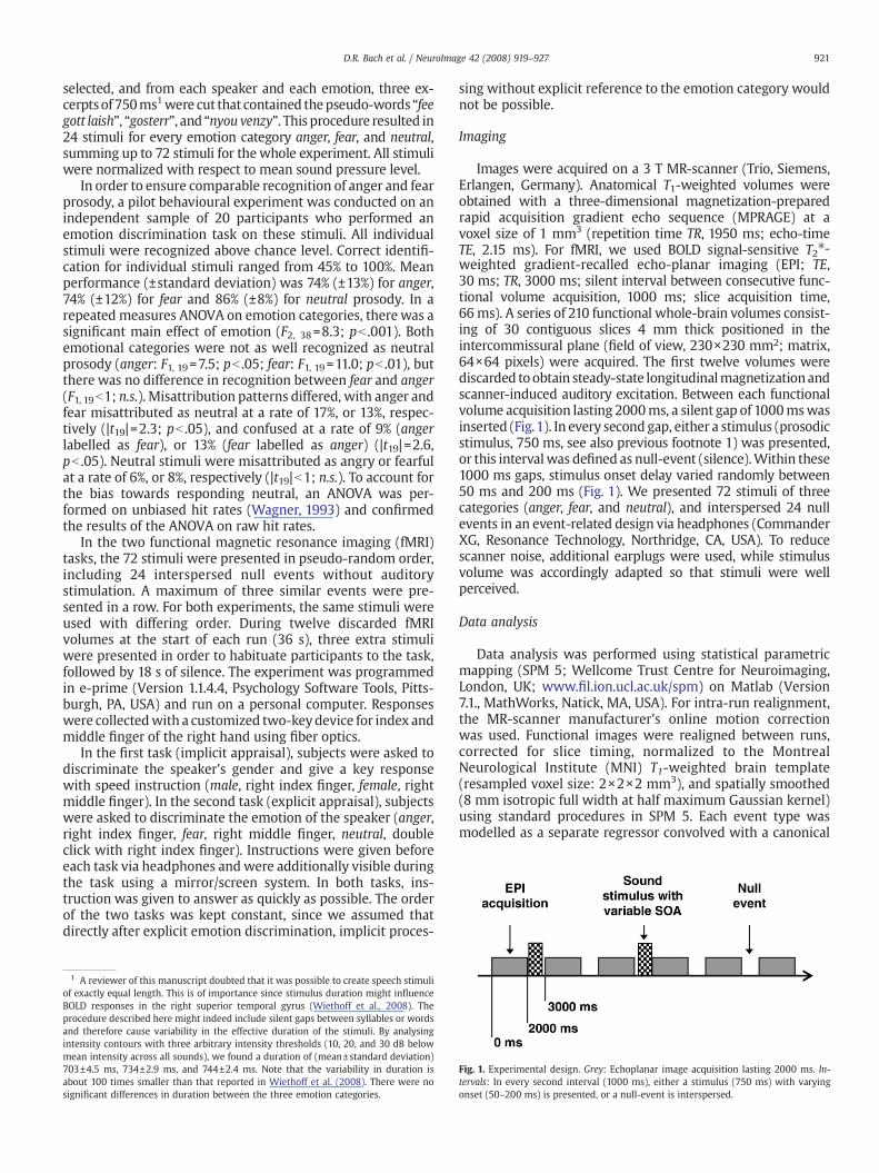
Images were acquired on a 3 T MR-scanner (Trio, Siemens,Erlangen, Germany). Anatomical T1-weighted volumes wereobtained with a three-dimensional magnetization-preparedrapid acquisition gradient echo sequence (MPRAGE) at avoxel size of 1 mm3 (repetition time TR, 1950 ms; echo-timeTE, 2.15 ms). For fMRI, we used BOLD signal-sensitive T2⁎-weighted gradient-recalled echo-planar imaging (EPI; TE,30 ms; TR, 3000 ms; silent interval between consecutive func-tional volume acquisition, 1000 ms; slice acquisition time,66 ms). A series of 210 functional whole-brain volumes consist-ing of 30 contiguous slices 4 mm thick positioned in theintercommissural plane (field of view, 230×230 mm2; matrix,64×64 pixels) were acquired. The first twelve volumes werediscarded to obtain steady-state longitudinalmagnetizationandscanner-induced auditory excitation. Between each functionalvolume acquisition lasting 2000ms, a silent gap of 1000mswasinserted (Fig.1). In every second gap, either a stimulus (prosodicstimulus, 750 ms, see also previous footnote 1) was presented,or this intervalwas defined as null-event (silence).Within these1000 ms gaps, stimulus onset delay varied randomly between50 ms and 200 ms (Fig. 1). We presented 72 stimuli of threecategories (anger, fear, and neutral), and interspersed 24 nullevents in an event-related design via headphones (CommanderXG, Resonance Technology, Northridge, CA, USA). To reducescanner noise, additional earplugs were used, while stimulusvolume was accordingly adapted so that stimuli were wellperceived.
Data analysis
Data analysis was performed using statistical parametricmapping (SPM 5; Wellcome Trust Centre for Neuroimaging,London, UK; www.fil.ion.ucl.ac.uk/spm) on Matlab (Version7.1., MathWorks, Natick, MA, USA). For intra-run realignment,the MR-scanner manufacturer's online motion correctionwas used. Functional images were realigned between runs,corrected for slice timing, normalized to the MontrealNeurological Institute (MNI) T1-weighted brain template(resampled voxel size: 2×2×2 mm3), and spatially smoothed(8 mm isotropic full width at half maximum Gaussian kernel)using standard procedures in SPM 5. Each event type wasmodelled as a separate regressor convolved with a canonical
Fig. 1. Experimental design. Grey: Echoplanar image acquisition lasting 2000 ms. In-tervals: In every second interval (1000 ms), either a stimulus (750 ms) with varyingonset (50–200 ms) is presented, or a null-event is interspersed.
922 D.R. Bach et al. / NeuroImage 42 (2008) 919–927
hemodynamic response function. In order to account fordifferences in task difficulty, individual reaction times for eachevent –which are amoremeaningful descriptor of difficulty ona trial-by-trial basis than the actual responses –weremodelledas one additional regressor for all event types. In order toaccount for variance caused by basic stimulus characteristics,the mean fundamental frequency F0 of each stimulus wascalculatedusing Praat (Version 4.5,www.praat.org), controlledfor octave jumps by visual inspection, and also introduced intothe model as one regressor for all event types. These para-metric modulators were orthogonalised using a serial Gram–Schmidt algorithm as implemented in SPM 5. In effect, the F0regressorwas orthogonalisedwith respect to the reaction timeregressor, while themeanwas removed from the reaction timeregressor which was otherwise left unchanged. Both para-metric regressors were then convolved with a canonicalhemodynamic response function. Due to the correlation oftask instruction and task difficulty, the reaction time regressormight obscure task effects. All analysis were thereforerepeated without reaction time regressor, confirming allBOLD response clusters with regard to localization and signifi-cance levels, and approximating cluster size. Statistical para-metric maps were generated from linear contrasts of interest(main effect task instruction, main effect emotionNneutral,main effect angerN fear, interaction task instruction×emotion)in each participant. A second level randomeffect analysis (RFX)was then performed using one sample t-tests on contrastimages obtained in each participant for each comparison ofinterest (df=15). We report signal clusters with a voxel-levelthreshold of pb0.001 (uncorrected), whole-brain corrected forfamily-wise error (FWE) at cluster level (pb .05), using therandom field theory approach implemented in SPM 5. Sincethe amygdala is of special interest in the context of implicit taskinstruction, we defined a region of interest in the amygdala asdescribed by cytoarchitectonic probability maps (Eickhoffet al., 2007). For clusters extending into this region, small-volume correction for FWE was applied at voxel level (pb .05)using a sphere of 30 mm diameter around cluster peakcoordinates. In order to evaluate common responses to angerand fear prosody in a more conservative approach, a conjunc-tion analysis (angerNneutral) and (fearNneutral) was con-ducted, using a second level two sample t-test and testingagainst conjunction null (Nichols et al., 2005; see also Fristonet al., 2005). Unique responses to these emotion categorieswere assessed by contrasting anger with fear prosody. For theinteraction of task instruction×emotion, regions of interestwere defined as brain areas where activity was found in either
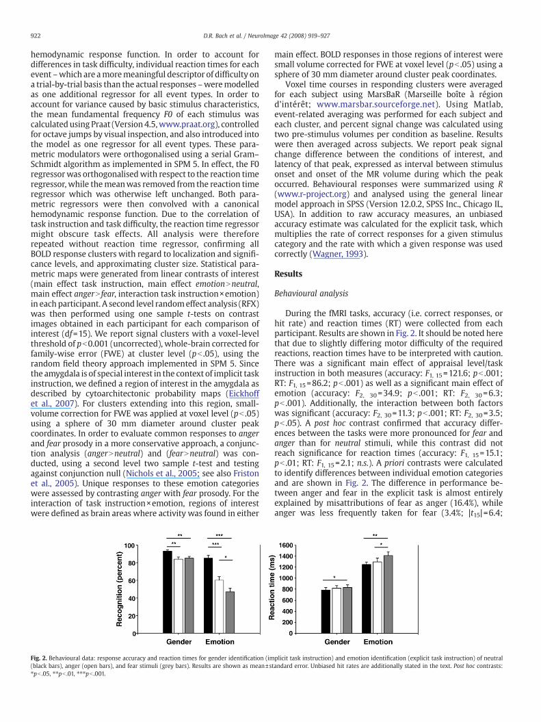
Fig. 2. Behavioural data: response accuracy and reaction times for gender identification (im(black bars), anger (open bars), and fear stimuli (grey bars). Results are shown as mean±st⁎pb .05, ⁎⁎pb .01, ⁎⁎⁎pb .001.
main effect. BOLD responses in those regions of interest weresmall volume corrected for FWE at voxel level (pb .05) using asphere of 30 mm diameter around cluster peak coordinates.
Voxel time courses in responding clusters were averagedfor each subject using MarsBaR (Marseille boîte à régiond'intérêt; www.marsbar.sourceforge.net). Using Matlab,event-related averaging was performed for each subject andeach cluster, and percent signal change was calculated usingtwo pre-stimulus volumes per condition as baseline. Resultswere then averaged across subjects. We report peak signalchange difference between the conditions of interest, andlatency of that peak, expressed as interval between stimulusonset and onset of the MR volume during which the peakoccurred. Behavioural responses were summarized using R(www.r-project.org) and analysed using the general linearmodel approach in SPSS (Version 12.0.2, SPSS Inc., Chicago IL,USA). In addition to raw accuracy measures, an unbiasedaccuracy estimate was calculated for the explicit task, whichmultiplies the rate of correct responses for a given stimuluscategory and the rate with which a given response was usedcorrectly (Wagner, 1993).
Results
Behavioural analysis
During the fMRI tasks, accuracy (i.e. correct responses, orhit rate) and reaction times (RT) were collected from eachparticipant. Results are shown in Fig. 2. It should be noted herethat due to slightly differing motor difficulty of the requiredreactions, reaction times have to be interpreted with caution.There was a significant main effect of appraisal level/taskinstruction in both measures (accuracy: F1, 15=121.6; pb .001;RT: F1, 15=86.2; pb .001) as well as a significant main effect ofemotion (accuracy: F2, 30=34.9; pb .001; RT: F2, 30=6.3;pb .001). Additionally, the interaction between both factorswas significant (accuracy: F2, 30=11.3; pb .001; RT: F2, 30=3.5;pb .05). A post hoc contrast confirmed that accuracy differ-ences between the tasks were more pronounced for fear andanger than for neutral stimuli, while this contrast did notreach significance for reaction times (accuracy: F1, 15=15.1;pb .01; RT: F1, 15=2.1; n.s.). A priori contrasts were calculatedto identify differences between individual emotion categoriesand are shown in Fig. 2. The difference in performance be-tween anger and fear in the explicit task is almost entirelyexplained by misattributions of fear as anger (16.4%), whileanger was less frequently taken for fear (3.4%; |t15|=6.4;
plicit task instruction) and emotion identification (explicit task instruction) of neutralandard error. Unbiased hit rates are additionally stated in the text. Post hoc contrasts:
923D.R. Bach et al. / NeuroImage 42 (2008) 919–927
pb .001). Anger and fear were misattributed as neutral at asimilar frequency (34.4% and 33.9%, respectively, |t15|b1). Inorder to account for such response biases, an unbiased hit rate(Wagner, 1993) was calculated for the explicit task andrevealed corrected accuracy of .49, .44, and .40, respectively,for neutral, anger, and fear prosody. In a one-way ANOVA,there was a tendentially significant effect of emotion category(F2, 30=3.2; p= .06) with fear accuracy differing significantlyfrom neutral accuracy (F2, 30=7.8; pb .05).
Main effect of appraisal level/task instruction
Brain regions that showed stronger BOLD responses to oneof the two tasks are summarized in Table 1 and Fig. 3. Duringimplicit as compared to explicit processing, we observedstronger BOLD responses in the left superior temporal gyrus(STG) and sulcus (STS). Also, a cluster in the right superior andinferior parietal lobule, extending into the postcentral gyrusshowed enhanced BOLD response. Two small clusters extend-ing into the amygdala in both hemispheres survived small-volume correction.
During explicit as compared to implicit processing, sev-eral clusters showed stronger BOLD signal in the leftdorsolateral prefrontal cortex (inferior and middle frontalgyrus [IFG and MFG]), bilateral medial frontal cortex(anterior cingulate [ACC], and supplemental premotorcortex), and bilateral superior parietal lobule and right pre-cuneus. The location of the right parietal areas was poster-iomedial of the right hemisphere cluster observed in theopposite contrast, while IFG responses to explicit versus
Table 1Main effects of appraisal level/task instruction and of emotional versus neutral prosody on
Brain regions Brodmann area oflocal maxima
Hemisphere Voxelnumber
Voxscor
Implicit N explicitInferior and superior parietal lobule,postcentral gyrus
40 and 7 Right 334 4.64
Superior temporal gyrus and sulcus 22 and 21 Left 176 3.94Amygdala (small-volume corrected) Right 8 3.79
Left 12 4.39
Explicit N implicitSuperior parietal lobuleand precuneus
7 and 19 Bilateral 1593 4.83
Precuneus 7 and 31 Right 129 3.71Inferior and middle frontal gyrus 45 and 46 Left 201 4.07Superior and middle frontal gyrus,anterior cingulate
6 Bilateral 183 3.93
Emotion N neutralInferior frontal gyrus,insula, putamen
47 Left 160 4.66
Anterior cingulate 32 Bilateral 1486 4.57Superior temporal gyrusand putamen
38 Right 129 3.81
(Anger N neutral) and (fear N neutral)Anterior cingulate 24 and 6 Bilateral 1182 4.45Middle frontal gyrus 46 and 9 Left 216 3.93
Anger N fearCaudate and putamen Bilateral 228 4.54
Emotion N neutral, explicit N implicit (small volume corrected in regions of interest)Putamen Right 79 4.03Anterior cingulate andmiddle frontal gyrus
6 Right 28 3.88
All reported clusters survived cluster-level correction at pb .05 unless stated otherwise.
implicit prosody were more superior than responses toemotional versus neutral prosody.
Emotion-specific effects of appraisal level/task instruction
The experimental design using both neutral and emotionalprosodic stimuli allowed us to assess task effects specific toemotional as compared to neutral prosody (that is, emo-tion×task interactions). Such effects were observed for theexplicit as compared to the implicit condition. A cluster in theright basal ganglia and another cluster in the right ACC andmiddle frontal gyrus survived small-volume correction.
Main effect of emotional compared to neutral prosody
Overall effects of emotional as compared to neutralprosody were analysed across tasks and revealed a strongerBOLD signal in the right STG extending into the putamen, in acluster in the left inferior frontal gyrus and insula, extendinginto the putamen, and in an extended cluster in the bilateralACC. In a second step, both unique and common responses toanger and to fear prosody were analysed in a more conser-vative approach. In order to identify unique responses, bothstimulus categories were compared. BOLD responses to angerwere more pronounced than to fear in the bilateral basalganglia, comprising parts of the putamen and caudate headand body. Common responses, that is, brain areas that respondconsistently and significantly to both single categories,were identified using a conjunction analysis, testing againstconjunction null (Nichols et al., 2005, see also Friston et al.,
blood oxygenation level-dependent (BOLD) responses
el Ze
Montreal Neurological Institute brain templatecoordinates of local maxima
BOLDpeak (%)
BOLD peaklatency (s)
50, −40, 56; 36, −52, 62; 42, −32, 52 0.05 3.875
−64, −16, 4; −64, −32, 0 0.05 3.87530, 2, −14 0.05 3.875−32, −10, −20 0.02 6.875
−30, −66, 44; −4, −70, 46; −32, −72, 38 0.05 3.875
16, −64, 20; 20, −68, 28; 24,−60, 26 0.03 3.875−46, 22, 18; −46, 38, 16; −44, 30, 18 0.09 6.8750, 4, 56; −10, −4, 58 0.07 3.875
−26, 18, −8; −24, 10, −8; −16, 8, −4 0.03 3.875
8, 14, 42; 6, 28, 34; −4, 14, 44 0.08 3.87530, 16, −10; 52, 16, −8; 24, 12, −6 0.04 3.875
6, 10, 36; −2, 12, 44; −4, 12, 36 0.08 3.875−42, 20, 30; −46, 12, 36 0.09 3.875
14, 20, 4; 6, 16, 6; 30, 34, −2 0.07 3.875
28, 16, 8; 36, 8, 8 0.02 3.87514, 0, 56 0.02 6.875
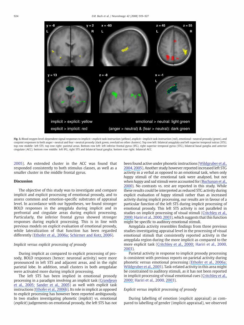
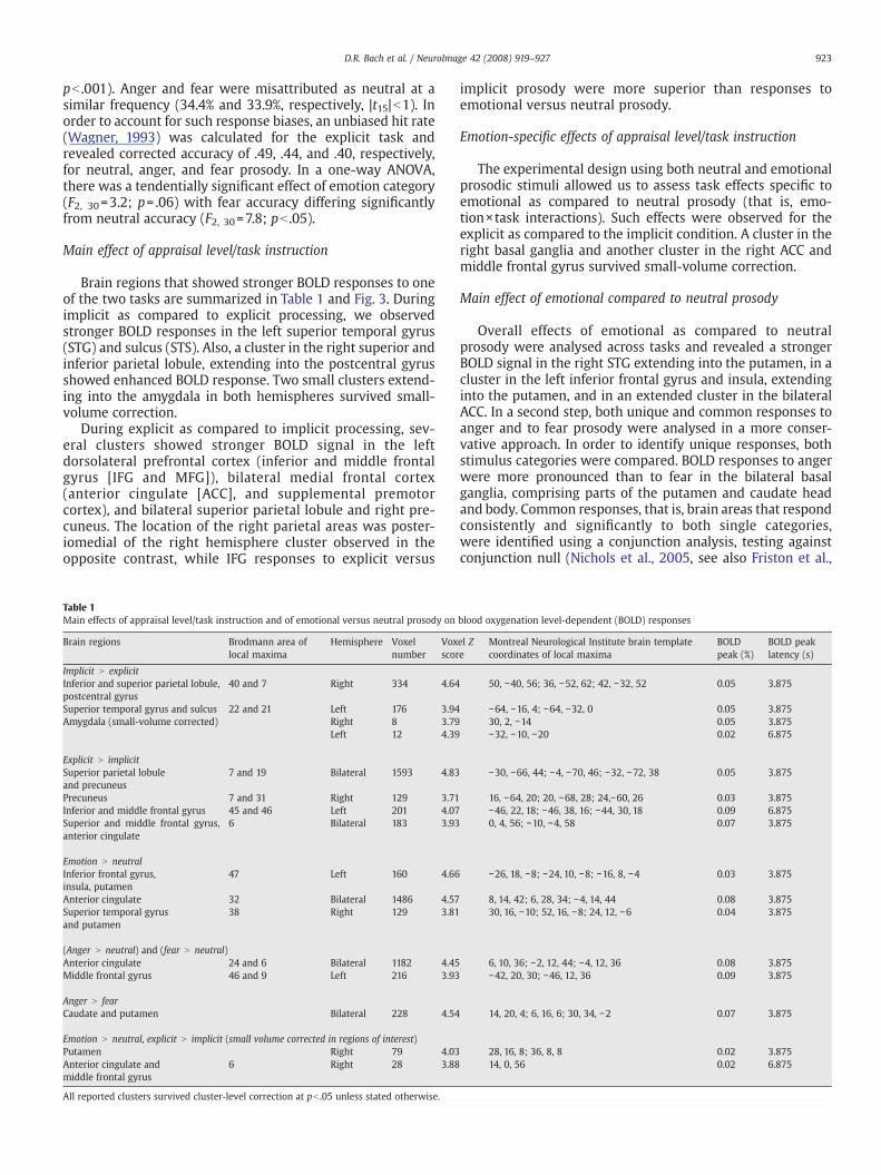
Fig. 3. Blood oxygen level-dependent signal responses to implicitNexplicit task instruction (yellow), explicitN implicit task instruction (red), emotionalNneutral prosody (green), andconjoint responses to both angerNneutral and fearNneutral prosody (dark green, overlaid on other clusters). Top row left: bilateral amygdala and left superior temporal sulcus (STS);top row middle: left STS; top row right: parietal areas. Bottom row left: left inferior frontal gyrus (IFG), right superior temporal gyrus (STG), bilateral basal ganglia and anteriorcingulate (ACC); bottom row middle: left IFG, right STS and bilateral basal ganglia; bottom row right: bilateral ACC.
924 D.R. Bach et al. / NeuroImage 42 (2008) 919–927
2005). An extended cluster in the ACC was found thatresponded consistently to both stimulus classes, as well as asmaller cluster in the middle frontal gyrus.
Discussion
The objective of this study was to investigate and compareimplicit and explicit processing of emotional prosody, and toassess common and emotion-specific substrates of appraisallevel. In accordance with our hypotheses, we found strongerBOLD responses in the amygdala during implicit and inprefrontal and cingulate areas during explicit processing.Particularly, the inferior frontal gyrus showed strongerresponses during explicit processing. This is in line withprevious models on explicit evaluation of emotional prosody,while lateralization of that function has been regardeddifferently (Ethofer et al., 2006a; Schirmer and Kotz, 2006).
Implicit versus explicit processing of prosody
During implicit as compared to explicit processing of pro-sody, BOLD responses (hence: neuronal activity) were morepronounced in left STS and adjacent areas, and in the rightparietal lobe. In addition, small clusters in both amygdalaewere activated more during implicit processing.
The left STS has been implied in emotional prosodyprocessing in a paradigm involving an implicit task (Grandjeanet al., 2005; Sander et al., 2005) as well with explicit taskinstructions (Ethofer et al., 2006b). Its role in implicit as opposedto explicit processing has however been reported ambiguously.In two studies investigating phonetic (implicit) vs. emotional(explicit) judgements on emotional prosody, the left STS has not
been foundactive underphonetic instructions (Wildgruber et al.,2004, 2005). Another study however reported increased left STGactivity in a verbal as opposed to an emotional task, when onlyhappy stimuli of the emotional task were analysed, but notwhenhappy and sad stimuliwere accounted for (Buchanan et al.,2000). No contrasts vs. rest are reported in this study. Whilethese results could be interpreted as reduced STG activity duringexplicit evaluation of happy stimuli rather than as increasedactivity during implicit processing, our results are in favour of aparticular function of the left STS during implicit processing ofemotional prosody. This left STS activity is not paralleled instudies on implicit processing of visual stimuli (Critchley et al.,2000;Hariri et al., 2000, 2003),which suggests that this functionmight be specific to auditory emotional stimuli.
Amygdala activity resembles findings from three previousstudies investigating appraisal level in the processing of visualemotional stimuli that consistently reported activity in theamygdala region during the more implicit as compared to themore explicit task (Critchley et al., 2000; Hariri et al., 2000,2003).
Parietal activity in response to implicit prosody processingis consistent with previous reports on parietal activity duringphonetic versus emotional processing (Ethofer et al., 2006a;Wildgruber et al., 2005). Task-related activity in this areamightbe constrained to auditory stimuli, as it has not been reportedin implicit processing of visual emotional cues (Critchley et al.,2000; Hariri et al., 2000, 2003).
Explicit versus implicit processing of prosody
During labelling of emotion (explicit appraisal) as com-pared to labelling of gender (implicit appraisal), we observed
925D.R. Bach et al. / NeuroImage 42 (2008) 919–927
activity in bilateral parietal cortex, including superior parietallobule and precuneus, in the left dorsolateral prefrontalcortex (DLPFC), including inferior frontal gyrus (IFG), and in acluster extending into the ACC and the supplemental motorarea.
Several studies comparing emotion labelling versusphonetic/semantic labelling of emotional prosody havereported right rather than left IFG activity (Buchanan et al.,2000; George et al., 1996; Wildgruber et al., 2005), and ithas been proposed that the right inferior frontal andorbitofrontal cortex is responsible for evaluative judgementsof emotional prosody, while the left inferior frontal gyrusserves semantic processing (Schirmer and Kotz, 2006).There is however evidence from a dynamic causal modellingstudy that both IFGs process prosody in parallel (Ethoferet al., 2006a), and it has been shown that circumscribedleft orbitofrontal lesions might impair recognition of emo-tional prosody (Hornak et al., 2003). Our results might sug-gest that the left IFG is particularly involved in explicit asopposed to implicit evaluative judgement of affectiveprosody. However, other possible explanations cannot beruled out. Attention to the speaker's emotional communica-tion (as required by the explicit task instructions in thisstudy) might enhance efforts to retrieve lexical informationfor the non-words presented here as compared to atten-tion towards the speakers identity (as required by implicittask instructions). Since the left IFG has been implied insemantic analysis (Schirmer and Kotz, 2006), this mightalso explain enhanced left IFG responses during explicitprocessing.
ACC activity has been reported in a study investigatingbrain responses to explicit processing of visual emotionalstimuli (Hariri et al., 2003), although two earlier studies havenot reported this activation (Critchley et al., 2000; Hariri et al.,2000). In a study investigating the effect of cognitive load onprocessing of anxiety, it was found that ACC activity isattenuated by the cognitive load of an unrelated task, thussuggesting that it might reflect higher cognitive appraisalprocesses that can be diminished by cognitive load (Kalischet al., 2006). Hence, ACC activity in the explicit conditionmight be due to task instructions requiring high-levelappraisal. On the other hand, the ACC might also serve aspecific role in affective prosody processing, particularly inexplicit evaluative judgements of affective prosody, since ithas been shown that ACC lesions impair recognition of affec-tive prosody (Hornak et al., 2003). The lack of ACC activation inprevious studies comparing emotional labelling and phoneticevaluation of emotional prosody can be explained by taskinstructions involving speech-related instructions in theimplicit condition: during phonetic instructions compared tobaseline, the ACC was strongly activated (Wildgruber et al.,2004, 2005).
Parietal activity during explicit emotional as compared tophonetic/semantic instructions has been reported in the studyof Buchanan et al. (2000), but not in the studies of Wildgruberet al. (2004, 2005). The parietal cortex contains polymodalareas (Bremmer et al., 2001) and might subserve higheranalyses of the auditory signal.
Task-related activity in the supplemental motor area mightbe related to the greater motor difficulty of the explicit task,with three instead of two reaction possibilities, one of whichalso necessitated a slightly more complicated motor program,that is, a double click.
Effects of emotional versus neutral prosody
During listening to emotional as compared to neutralprosody, we found activity in the right STG, in the left IFG, thebilateral ACC, as well as in the basal ganglia. Emotional versusneutral prosody has been studied previously with both implicit(Grandjean et al., 2005; Sander et al., 2005) and explicit (Kotzet al., 2003) task instructions. The study by Grandjean et al.(2005) is of special interest since it used angry stimuli from thesame set as ourmaterial. In aparadigmsimilar to the oneused inthe present study, activity was found in bilateral STS/STG, ACC,postcentral gyrus, and left putamen. With the exception of theleft STG and postcentral gyrus, this pattern is closely resembledby our findings, while in the present study, the inferior frontalgyrus was additionally activated. Those differences are mainlyattributable to the additional use of fear and differingsignificance thresholds, since when analysing only anger vs.neutral prosody, we found additional left STG activity at acluster-level corrected threshold, and at an uncorrected voxel-level threshold comparable to thatofGrandjean et al., additionalpericentral activity was observed. While our results are there-fore in line with a previous study using a similar stimulus set,the contrast emotional vs. neutral prosody cannot be expectedto show all areas involved in prosody processing. In the contextof emotional prosody stimuli, it might be assumed that alsoneutral stimuli are evaluated by some brain areas servingemotional prosody processing in general. Greater activity inresponse to emotional than to neutral prosody might thereforereflect mainly the detection aspect rather than the processingaspect of prosody evaluation. Of special importance here is theACC that was conjointly and significantly activated both byanger and fear prosody using the conservative conjunctionapproach. The relevance of the ACC during emotional prosodyprocessing is not yetwell understood, althoughACC activity hasbeen reported in response to anger vs. neutral prosody (Grand-jean et al., 2005), and lesions can impair recognition ofemotional prosody (Hornak et al., 2003) which suggests aspecific role of this region for detecting emotional prosody. Asimilar ACC region was activated in the contrast of explicitversus implicit task instructions. Thismight atfirst sight suggestthat ACC activation during emotional as compared to neutralprosody was in fact due to recruitment of explicit appraisalprocesses that might be more pronounced when a stimulus isdetected as emotional, and thereby potentially relevant. Thisexplanation is however unlikely, given that ACC responses toemotional versus neutral prosody were dramatically morepronounced than during explicit versus implicit processing,againpointing towards a specificACC functionduringemotionalprosody detection.
It is interesting to note that also in the bilateral basalganglia, activity was observed during emotional prosodyversus neutral prosody. This response was more pronouncedfor anger than for fear, and it was particularly pronouncedduring explicit task instructions. Previous studies comparingemotional and neutral prosody under implicit task instruc-tions reported activity in the caudate head (Kotz et al., 2003)and putamen (Grandjean et al., 2005). Also, there isevidence that basal ganglia dysfunction, for example inParkinson disease, impairs emotional prosody recognition(Pell and Leonard, 2003). Taken together, this points towardsa specific function of this brain structure in emotionalprosody detection, especially when explicit evaluation isrequired.
926 D.R. Bach et al. / NeuroImage 42 (2008) 919–927
Emotion-specific effects of task instruction
One starting point for the present study was the assump-tion that appraisal effects might differ for emotional and non-emotional stimuli. Such differences would present as interac-tion of task instruction×emotion. We observed such effects inthe right ACC and basal ganglia, again suggesting a specificfunction of these areas in the explicit detection of emotionalprosody. While previous studies on appraisal levels withcomparable paradigms have reported main effects of taskinstruction and no interaction (Critchley et al., 2000; Haririet al., 2000, 2003), they were not specifically designed to findsuch interactions. It should be noted that also in our study, thesmall number of stimuli per cell of the design does not providethe statistical power that an experiment might achieve thatfocuses on such interactions. Also, in the present study inter-actions were specifically analysed in areas where a main effectwas present, thus providing greater power for detectingordinal (but not disordinal) interactions but also increasingthe rate of type I errors. The question of how emotion×taskinteractions are represented in the brain therefore requiresfurther investigation.
Amygdala in emotional prosody processing
Amygdala activity under implicit as opposed to explicittask instructions resembles results from previous studiesusing visual stimuli (Critchley et al., 2000; Hariri et al., 2000,2003). An amygdala-cortical network has been proposedwhere higher-level appraisal processes are represented inprefrontal and cingulate areas and might attenuate amygdalaactivity related to lower-level processes (Hariri et al., 2000,2003). It is striking, however, that while emotional facesactivate the amygdala more than neutral faces (Whalen et al.,2001), no such effect could be found in our study foremotional versus neutral prosody. Specifically, no effect offear prosody could be observed in the amygdala, even whenonly fear vs. neutral prosody was contrasted at a threshold ofpb .001 uncorrected. Accounts of amygdala function inemotional prosody processing are however ambiguous inthe previous literature. Lesion studies suggest that the humanamygdala is not as critical for the recognition of affectiveprosody as it is for recognition of facial affect (Adolphs andTranel, 1999). This is contrasted by the effects of pure (non-verbal) emotional vocalization (Fecteau et al., 2007; Morriset al., 1999; Phillips et al., 1998; Sander and Scheich, 2001;Seifritz et al., 2003) and simple auditory warning stimuli(Bach et al., 2008), which in most paradigms have shown toelicit strong amygdala responses. This suggests that stimuliwith linguistic content might be less susceptible to activatethe amygdala than non-linguistic stimuli. An explanation forthese findings relies on task difficulty. Although the abovecited studies did not require or report stimulus identificationperformance, it can be speculated that pure emotional vocal-ization is much easier to recognize than linguistic prosodiccues. Also, visual stimuli used in the above cited studies werevery easily recognizable. Therefore, these stimuli are prob-ably more salient and might therefore activate the amygdalamore readily, while the more difficult stimuli used in ourexperiment might require a higher appraisal level per se.Note however that interindividual differences in amygdalaactivation during listening to emotional prosody have beenreported, and that these differences might be correlated with
individually differing relevance of prosodic stimuli (Schirmeret al., 2008).
Stimulus or response?
All analyses in this article were performed with regard tothe presented rather than the identified stimulus category.This is in line with previous studies on appraisal effects onprocessing of emotional cues. However, when stimulusdifficulty is high, this approach might obscure brain responsesto the recognition of individual emotion categories, as it doesnot distinguish between correct and incorrect responses. Inaddition, error variance might be increased as trials duringwhich participants did not pay attention to stimuli areincluded in the analysis. One strategy to circumvent theseproblems would thus be to perform analyses only on correctlyidentified stimuli. However, the meaning of such an analysiswould be rather different between implicit and explicit taskinstructions. During the implicit task, correct response mightbe regarded as an indicator of attention, particularly since thetask is comparably easy. On the other hand, under explicitinstructions and with a comparably difficult task, incorrectresponses do not necessarily imply lack of attention. Here,response accuracy is rather an indicator for the perceivedemotional meaning. An analysis of correct responses underexplicit instructions might therefore reveal brain areasinvolved in the recognition of an individual emotion category.However, even an analysis limited to explicit task instructionsnot without problems as stimuli are not necessarily recog-nized in a dichotomous manner. Uncertainty in recognitionmight introduce a probabilistic element into responses. There-fore, from a correct response on a given trial it is not possibleto infer correct identification of the presented stimulus. For allthese reasons, we believe that the presented analysis strategyis best suited to shed light on the effect of appraisal processes.In order to further clarify the recognition of individual emo-tion categories, analyses according to stimulus identificationmight however be useful. To overcome the problem of non-dichotomous recognition, one might ask participants to judgethe probabilities that a given stimulus belongs to any emotioncategory.
Limitations
The fixed task order used in this study might suggest thatdifferences between the tasks are in part due to stimulushabituation effects. This possibility cannot be ruled out for thepresent study. Also, the task used did not control for effects ofattention towards stimuli. Another limitation is the differencein difficulty between the tasks. Although main effects ofdifficulty were controlled by using individual reaction times asa covariate for all analyses, difficulty×task interactions wouldnot be ruled out by this approach. Matched difficulty betweentasks would be desirable. Previous studies on appraisal ofemotional cues have attempted to provide such paradigms,with mixed results however. While some did not reportstatistical comparisons of task difficulty (Critchley et al., 2000;Ethofer et al., 2006a,b), others have provided comparabletasks (Buchanan et al., 2000; Hariri et al., 2000; Wildgruberet al., 2004), and two studies report significant differences indifficulty across appraisal levels (Hariri et al., 2003; Wild-gruber et al., 2005). Yet another limitation refers to appraisalprocesses not captured by task instructions. In other words,
927D.R. Bach et al. / NeuroImage 42 (2008) 919–927
although the implicit task was always presented first, it cannotbe ruled out that explicit reference to emotion categoryalready took place during the implicit task. This possibilityshould, however, reduce differences between implicit andexplicit tasks and therefore render testing more conservative.
Conclusion
Our study aimed at comparing explicit and implicit proces-sing of affective prosody by avoiding speech-related taskinstructions for the implicit condition. Our results are in partcomparable to those obtained using speech-related taskinstruction. However, they differ in some important respects.Of note here is amygdala activity during implicit and ACCactivity during explicit processing, which has not been ob-served before during prosody processing but resemblesfindings from studies using visual cues. Also, inferior frontalgyrus activity during explicit appraisal was observed in the leftrather than in the right hemisphere, which might suggest aspecific function of this area in explicit evaluative judgementsof emotional prosody. The contrast emotional vs. neutralprosody revealed activity in brain areas that have been widelyrelated to emotional prosody processing, that is, the right STSand left inferior frontal gyrus. In addition, we observed activityin the ACC and caudate head. Our study also aimed at inves-tigating emotion-specific task effects. Emotion effects thatweremore pronounced under explicit task instructions could beshown in the basal ganglia and the ACC. Both areas have beenshown to be critical for prosody recognition in lesion studies,but have only rarely been reported in neuroimaging studiescomparing emotional and neutral prosodies.
Acknowledgments
Theauthorswish to thank theDepartmentofNeuroradiology,Inselspital Bern, and in particular the radiographers team fortheir continuing support, as well as three anonymous reviewersfor their comments on earlier versions of this manuscript.
References
Adolphs, R., Tranel, D., 1999. Intact recognition of emotional prosody followingamygdala damage. Neuropsychologia 37, 1285–1292.
Bach, D.R., Schachinger, H., Neuhoff, J.G., Esposito, F., Salle, F.D., Lehmann, C., Herdener,M., Scheffler, K., Seifritz, E., 2008. Rising sound intensity: an intrinsic warning cueactivating the amygdala. Cereb. Cortex 18, 145–150.
Banse, R., Scherer, K.R., 1996. Acoustic profiles in vocal emotion expression. J. Pers. Soc.Psychol. 70, 614–636.
Bremmer, F., Schlack, A., Shah, N.J., Zafiris, O., Kubischik, M., Hoffmann, K., Zilles, K., Fink,G.R., 2001. Polymodal motion processing in posterior parietal and premotor cortex:a human fMRI study strongly implies equivalencies between humans andmonkeys.Neuron 29, 287–296.
Buchanan, T.W., Lutz, K., Mirzazade, S., Specht, K., Shah, N.J., Zilles, K., Jancke, L., 2000.Recognition of emotional prosody and verbal components of spoken language:an fMRI study. Brain Res. Cogn. Brain Res. 9, 227–238.
Critchley, H., Daly, E., Phillips, M., Brammer, M., Bullmore, E., Williams, S., VanAmelsvoort, T., Robertson, D., David, A., Murphy, D., 2000. Explicit and implicitneural mechanisms for processing of social information from facial expressions:a functional magnetic resonance imaging study. Hum. Brain Mapp. 9, 93–105.
Dolan, R.J., Morris, J.S., de Gelder, B., 2001. Crossmodal binding of fear in voice and face.Proc. Natl. Acad. Sci. U. S. A. 98, 10006–10010.
Eickhoff, S.B., Paus, T., Caspers, S., Grosbras, M.H., Evans, A.C., Zilles, K., Amunts, K., 2007.Assignment of functional activations to probabilistic cytoarchitectonic areasrevisited. Neuroimage 36, 511–521.
Ethofer, T., Anders, S., Erb, M., Herbert, C., Wiethoff, S., Kissler, J., Grodd, W., Wildgruber,D., 2006a. Cerebral pathways in processing of affective prosody: a dynamic causalmodeling study. Neuroimage 30, 580–587.
Ethofer, T., Anders, S., Wiethoff, S., Erb, M., Herbert, C., Saur, R., Grodd, W., Wildgruber,D., 2006b. Effects of prosodic emotional intensity on activation of associativeauditory cortex. Neuroreport 17, 249–253.
Ethofer, T., Pourtois, G., Wildgruber, D., 2006c. Investigating audiovisual integration ofemotional signals in the human brain. Prog. Brain Res. 156, 345–361.
Fecteau, S., Belin, P., Joanette, Y., Armony, J.L., 2007. Amygdala responses to nonlinguisticemotional vocalizations. Neuroimage 36, 480–487.
Friston, K.J., Penny, W.D., Glaser, D.E., 2005. Conjunction revisited. Neuroimage 25,661–667.
George, M.S., Parekh, P.I., Rosinsky, N., Ketter, T.A., Kimbrell, T.A., Heilman, K.M.,Herscovitch, P., Post, R.M., 1996. Understanding emotional prosody activates righthemisphere regions. Arch. Neurol. 53, 665–670.
Grandjean, D., Sander, D., Pourtois, G., Schwartz, S., Seghier, M.L., Scherer, K.R.,Vuilleumier, P., 2005. The voices of wrath: brain responses to angry prosody inmeaningless speech. Nat. Neurosci. 8, 145–146.
Grandjean, D., Banziger, T., Scherer, K.R., 2006. Intonation as an interface betweenlanguage and affect. Prog. Brain Res. 156, 235–247.
Hariri, A.R., Bookheimer, S.Y., Mazziotta, J.C., 2000. Modulating emotional responses:effects of a neocortical network on the limbic system. Neuroreport 11, 43–48.
Hariri, A.R., Mattay, V.S., Tessitore, A., Fera, F., Weinberger, D.R., 2003. Neocorticalodulation of the amygdala response to fearful stimuli. Biol. Psychiatry 53, 494–501.
Hornak, J., Bramham, J., Rolls, E.T., Morris, R.G., O'Doherty, J., Bullock, P.R., Polkey, C.E.,2003. Changes in emotion after circumscribed surgical lesions of the orbitofrontaland cingulate cortices. Brain 126, 1691–1712.
Johnstone, T., van Reekum, C.M., Oakes, T.R., Davidson, R.J., 2006. The voice of emotion:an FMRI study of neural responses to angry and happy vocal expressions. Soc. Cogn.Affect. Neurosci. 1, 242–249.
Kalisch, R.,Wiech,K., Critchley,H.D.,Dolan,R.J., 2006. Levelsof appraisal: amedial prefrontalrole in high-level appraisal of emotional material. Neuroimage 30, 1458–1466.
Killgore, W.D., Yurgelun-Todd, D.A., 2004. Activation of the amygdala and anteriorcingulate during nonconscious processing of sad versus happy faces. Neuroimage21, 1215–1223.
Kotz, S.A., Meyer, M., Alter, K., Besson, M., von Cramon, D.Y., Friederici, A.D., 2003. On thelateralization of emotional prosody: an event-related functional MR investigation.Brain Lang. 86, 366–376.
Leventhal, H., Scherer, K.R., 1987. The relationship of emotion to cognition: a functionalapproach to a semantic controversy. Cogn. Emot. 1, 3–28.
Mitchell, R.L., Elliott, R., Barry, M., Cruttenden, A., Woodruff, P.W., 2003. The neuralresponse to emotional prosody, as revealed by functional magnetic resonanceimaging. Neuropsychologia 41, 1410–1421.
Morris, J.S., Scott, S.K., Dolan, R.J., 1999. Saying it with feeling: neural responses toemotional vocalizations. Neuropsychologia 37, 1155–1163.
Nichols, T., Brett, M., Andersson, J., Wager, T., Poline, J.B., 2005. Valid conjunctioninference with the minimum statistic. Neuroimage 25, 653–660.
Oldfield, R.C., 1971. The assessment and analysis of handedness: the Edinburghinventory. Neuropsychologia 9, 97–113.
Pell, M.D., Leonard, C.L., 2003. Processing emotional tone from speech in Parkinson'sdisease: a role for the basal ganglia. Cogn. Affect. Behav. Neurosci. 3, 275–288.
Phillips, M.L., Young, A.W., Scott, S.K., Calder, A.J., Andrew, C., Giampietro, V., Williams,C.R., Bullmore, E.T., Brammer, M., Gray, J.A., 1998. Neural responses to facial andvocal expressions of fear and disgust. Proc. R. Soc. B Biol. Sci. 265, 1809–1817.
Pourtois, G., deGelder, B., Bol, A., Crommelinck,M., 2005. Perception of facial expressionsand voices and of their combination in the human brain. Cortex 41, 49–59.
Sander, K., Scheich, H., 2001. Auditory perception of laughing and crying activates humanamygdala regardless of attentional state. Brain Res.Cogn Brain Res. 12, 181–198.
Sander, D., Grafman, J., Zalla, T., 2003. The human amygdala: an evolved system forrelevance detection. Rev. Neurosci. 14, 303–316.
Sander, D., Grandjean, D., Pourtois, G., Schwartz, S., Seghier, M.L., Scherer, K.R.,Vuilleumier, P., 2005. Emotion and attention interactions in social cognition: brainregions involved in processing anger prosody. Neuroimage 28, 848–858.
Schirmer, A., Kotz, S.A., 2006. Beyond the right hemisphere: brain mechanismsmediating vocal emotional processing. Trends Cogn. Sci. 10, 24–30.
Schirmer, A., Escoffier, N., Zysset, S., Koester, D., Striano, T., Friederici, A.D., 2008. Whenvocal processing gets emotional: on the role of social orientation in relevancedetection by the human amygdala. Neuroimage 40, 1402–1410.
Seifritz, E., Esposito, F., Neuhoff, J.G., Luthi, A., Mustovic, H., Dammann, G., vonBardeleben, U., Radue, E.W., Cirillo, S., Tedeschi, G., Di Salle, F., 2003. Differentialsex-independent amygdala response to infant crying and laughing in parentsversus nonparents. Biol. Psychiatry 54, 1367–1375.
Wagner, H.L., 1993. On measuring performance in category judgment studies ofnonverbal behavior. J. Nonverbal Behav. 17, 3–28.
Wambacq, I.J., Shea-Miller, K.J., Abubakr, A., 2004.Non-voluntary and voluntary processingof emotional prosody: an event-related potentials study. Neuroreport 15, 555–559.
Whalen, P.J., Rauch, S.L., Etcoff, N.L., McInerney, S.C., Lee, M.B., Jenike, M.A., 1998.Masked presentation of emotional facial expressions modulate amygdala activationwithout explicit knowledge. J. Neurosci. 18, 411–418.
Whalen, P.J., Shin, L.M., McInerney, S.C., Fischer, H., Wright, C.I., Rauch, S.L., 2001.A functional MRI study of human amygdala responses to facial expressions of fearversus anger. Emotion 1, 70–83.
Whalen, P.J., Kagan, J., Cook, R.G., Davis, F.C., Kim, H., Polis, S., McLaren, D.G., Somerville,L.H., McLean, A.A., Maxwell, J.S., Johnstone, T., 2004. Human amygdala responsivityto masked fearful eye whites. Science 306, 2061.
Wiethoff, S., Wildgruber, D., Kreifelts, B., Becker, H., Herbert, C., Grodd, W., Ethofer, T.,2008. Cerebral processing of emotional prosody—influence of acoustic parametersand arousal. Neuroimage 39, 885–893.
Wildgruber, D., Hertrich, I., Riecker, A., Erb, M., Anders, S., Grodd, W., Ackermann, H.,2004. Distinct frontal regions subserve evaluation of linguistic and emotionalaspects of speech intonation. Cereb. Cortex 14, 1384–1389.
Wildgruber, D., Riecker, A., Hertrich, I., Erb, M., Grodd, W., Ethofer, T., Ackermann, H.,2005. Identification of emotional intonation evaluated by fMRI. Neuroimage 24,1233–1241.