The Drosophila homolog of MCPH1, a human microcephaly gene, is required for genomic stability in the...
13
3565 Research Article Introduction Drosophila melanogaster is an ideal model organism for study of the cell cycle during development (reviewed by Foe et al., 1993; Lee and Orr-Weaver, 2003). Drosophila achieves rapid embryogenesis by using a streamlined cell cycle that is not dependent on transcription or growth. The first 13 embryonic cell cycles are nearly synchronous nuclear divisions without cytokinesis occurring in the shared cytoplasm of the syncytial blastoderm. These cycles differ from canonical G1-S-G2-M cycles in that they have no intervening gaps; instead DNA replication and mitosis rapidly oscillate. Maternal RNA and protein stockpiles drive these abbreviated ‘S-M’ cycles (~10 minutes each). In mammalian embryos, rapid peri-gastrulation divisions that occur later in development share many features and have been proposed to be related by evolutionary descent to early embryonic divisions of flies and frogs (O’Farrell et al., 2004). Thus, advances gained from studies of these streamlined cycles in ‘simple’ model organisms likely have relevance for understanding mammalian cell cycles. In a genetic screen for regulators of embryonic S-M cycles, we identified the Drosophila homolog of a human disease gene, MCPH1 (microcephalin). Mutation of human MCPH1 causes autosomal recessive primary microcephaly, a developmental disorder characterized by severe reduction of cerebral cortex size (Jackson et al., 2002). Mcph1 is highly expressed in the developing forebrain of fetal mice, consistent with its proposed role in regulating the number neuronal precursor cell divisions and, ultimately, brain size (Jackson et al., 2002). Human MCPH1 protein is predicted to contain three BR CA1 C -t erminal (BRCT) domains (reviewed by Glover et al., 2004; Huyton et al., 2000), which mediate phosphorylation-dependent protein-protein interactions in cell- cycle checkpoint and DNA repair functions. Several studies have implicated human MCPH1 in the cellular response to DNA damage. The DNA checkpoint is engaged at critical cell-cycle transitions in response to DNA damage or incomplete replication and serves as a mechanism to preserve genomic integrity (reviewed by Nyberg et al., 2002). Triggering of this checkpoint causes cell-cycle delay, presumably to allow time for correction of DNA defects. When a cell senses DNA damage or incomplete replication, a kinase cascade is activated. Activated ATM and ATR kinases phosphorylate their targets, including the checkpoint kinase Chk1, which is activated to phosphorylate its targets. The first clue that MCPH1 plays a role in the DNA damage response came from siRNA-mediated knockdown studies in cultured mammalian cells demonstrating a requirement for MCPH1 in the intra-S phase and G2-M checkpoints in response to ionizing Mutation of human microcephalin (MCPH1) causes autosomal recessive primary microcephaly, a developmental disorder characterized by reduced brain size. We identified mcph1, the Drosophila homolog of MCPH1, in a genetic screen for regulators of S-M cycles in the early embryo. Embryos of null mcph1 female flies undergo mitotic arrest with barrel-shaped spindles lacking centrosomes. Mutation of Chk2 suppresses these defects, indicating that they occur secondary to a previously described Chk2-mediated response to mitotic entry with unreplicated or damaged DNA. mcph1 embryos exhibit genomic instability as evidenced by frequent chromatin bridging in anaphase. In contrast to studies of human MCPH1, the ATR/Chk1-mediated DNA checkpoint is intact in Drosophila mcph1 mutants. Components of this checkpoint, however, appear to cooperate with MCPH1 to regulate embryonic cell cycles in a manner independent of Cdk1 phosphorylation. We propose a model in which MCPH1 coordinates the S-M transition in fly embryos: in the absence of mcph1, premature chromosome condensation results in mitotic entry with unreplicated DNA, genomic instability, and Chk2-mediated mitotic arrest. Finally, brains of mcph1 adult male flies have defects in mushroom body structure, suggesting an evolutionarily conserved role for MCPH1 in brain development. Supplementary material available online at http://jcs.biologists.org/cgi/content/full/120/20/3565/DC1 Key words: Drosophila, Embryogenesis, Microcephaly, Cell cycle, Mitosis, DNA checkpoint, BRCT domain Summary The Drosophila homolog of MCPH1, a human microcephaly gene, is required for genomic stability in the early embryo Jamie L. Rickmyre 1 , Shamik DasGupta 2 , Danny Liang-Yee Ooi 3 , Jessica Keel 1 , Ethan Lee 1 , Marc W. Kirschner 3 , Scott Waddell 2 and Laura A. Lee 1, * 1 Department of Cell and Developmental Biology, Vanderbilt University Medical Center, U-4200 MRBIII, 465 21st Avenue South, Nashville, TN 37232-8240, USA 2 Department of Neurobiology, University of Massachusetts Medical School, 364 Plantation Street, Worcester, MA 01605, USA 3 Department of Systems Biology, Harvard Medical School, 200 Longwood Avenue, Boston, MA 02115, USA *Author for correspondence (e-mail: [email protected]) Accepted 20 August 2007 Journal of Cell Science 120, 3565-3577 Published by The Company of Biologists 2007 doi:10.1242/jcs.016626 Journal of Cell Science
Transcript of The Drosophila homolog of MCPH1, a human microcephaly gene, is required for genomic stability in the...

3565Research Article
IntroductionDrosophila melanogaster is an ideal model organism for studyof the cell cycle during development (reviewed by Foe et al.,1993; Lee and Orr-Weaver, 2003). Drosophila achieves rapidembryogenesis by using a streamlined cell cycle that is notdependent on transcription or growth. The first 13 embryoniccell cycles are nearly synchronous nuclear divisions withoutcytokinesis occurring in the shared cytoplasm of the syncytialblastoderm. These cycles differ from canonical G1-S-G2-Mcycles in that they have no intervening gaps; instead DNAreplication and mitosis rapidly oscillate. Maternal RNA andprotein stockpiles drive these abbreviated ‘S-M’ cycles (~10minutes each). In mammalian embryos, rapid peri-gastrulationdivisions that occur later in development share many featuresand have been proposed to be related by evolutionary descentto early embryonic divisions of flies and frogs (O’Farrell et al.,2004). Thus, advances gained from studies of these streamlinedcycles in ‘simple’ model organisms likely have relevance forunderstanding mammalian cell cycles.
In a genetic screen for regulators of embryonic S-M cycles,we identified the Drosophila homolog of a human diseasegene, MCPH1 (microcephalin). Mutation of human MCPH1causes autosomal recessive primary microcephaly, adevelopmental disorder characterized by severe reduction of
cerebral cortex size (Jackson et al., 2002). Mcph1 is highlyexpressed in the developing forebrain of fetal mice, consistentwith its proposed role in regulating the number neuronalprecursor cell divisions and, ultimately, brain size (Jackson etal., 2002). Human MCPH1 protein is predicted to contain threeBRCA1 C-terminal (BRCT) domains (reviewed by Gloveret al., 2004; Huyton et al., 2000), which mediatephosphorylation-dependent protein-protein interactions in cell-cycle checkpoint and DNA repair functions.
Several studies have implicated human MCPH1 in thecellular response to DNA damage. The DNA checkpoint isengaged at critical cell-cycle transitions in response to DNAdamage or incomplete replication and serves as a mechanismto preserve genomic integrity (reviewed by Nyberg et al.,2002). Triggering of this checkpoint causes cell-cycle delay,presumably to allow time for correction of DNA defects. Whena cell senses DNA damage or incomplete replication, a kinasecascade is activated. Activated ATM and ATR kinasesphosphorylate their targets, including the checkpoint kinaseChk1, which is activated to phosphorylate its targets. The firstclue that MCPH1 plays a role in the DNA damage responsecame from siRNA-mediated knockdown studies in culturedmammalian cells demonstrating a requirement for MCPH1 inthe intra-S phase and G2-M checkpoints in response to ionizing
Mutation of human microcephalin (MCPH1) causesautosomal recessive primary microcephaly, adevelopmental disorder characterized by reduced brainsize. We identified mcph1, the Drosophila homolog ofMCPH1, in a genetic screen for regulators of S-M cycles inthe early embryo. Embryos of null mcph1 female fliesundergo mitotic arrest with barrel-shaped spindles lackingcentrosomes. Mutation of Chk2 suppresses these defects,indicating that they occur secondary to a previouslydescribed Chk2-mediated response to mitotic entry withunreplicated or damaged DNA. mcph1 embryos exhibitgenomic instability as evidenced by frequent chromatinbridging in anaphase. In contrast to studies of humanMCPH1, the ATR/Chk1-mediated DNA checkpoint isintact in Drosophila mcph1 mutants. Components of thischeckpoint, however, appear to cooperate with MCPH1 to
regulate embryonic cell cycles in a manner independent ofCdk1 phosphorylation. We propose a model in whichMCPH1 coordinates the S-M transition in fly embryos:in the absence of mcph1, premature chromosomecondensation results in mitotic entry with unreplicatedDNA, genomic instability, and Chk2-mediated mitoticarrest. Finally, brains of mcph1 adult male flies have defectsin mushroom body structure, suggesting an evolutionarilyconserved role for MCPH1 in brain development.
Supplementary material available online athttp://jcs.biologists.org/cgi/content/full/120/20/3565/DC1
Key words: Drosophila, Embryogenesis, Microcephaly, Cell cycle,Mitosis, DNA checkpoint, BRCT domain
Summary
The Drosophila homolog of MCPH1, a humanmicrocephaly gene, is required for genomic stability inthe early embryoJamie L. Rickmyre1, Shamik DasGupta2, Danny Liang-Yee Ooi3, Jessica Keel1, Ethan Lee1,Marc W. Kirschner3, Scott Waddell2 and Laura A. Lee1,*1Department of Cell and Developmental Biology, Vanderbilt University Medical Center, U-4200 MRBIII, 465 21st Avenue South, Nashville, TN37232-8240, USA2Department of Neurobiology, University of Massachusetts Medical School, 364 Plantation Street, Worcester, MA 01605, USA3Department of Systems Biology, Harvard Medical School, 200 Longwood Avenue, Boston, MA 02115, USA*Author for correspondence (e-mail: [email protected])
Accepted 20 August 2007Journal of Cell Science 120, 3565-3577 Published by The Company of Biologists 2007doi:10.1242/jcs.016626
Jour
nal o
f Cel
l Sci
ence
3566
radiation (Lin et al., 2005; Xu et al., 2004). Two recent reportshave further implicated MCPH1 in the DNA checkpoint,although puzzling discrepancies remain to be resolved(reviewed by Bartek, 2006). One report indicates that MCPH1functions far downstream in the pathway, at a level betweenChk1 and one of its targets, Cdc25 (Alderton et al., 2006).Another report (Rai et al., 2006) suggests that MCPH1 is aproximal component of the DNA damage response required forradiation-induced foci formation (i.e. recruitment ofcheckpoint and repair proteins to damaged chromatin).
Additional functions have been reported for MCPH1.MCPH1– lymphocytes of microcephalic patients exhibitpremature chromosome condensation (PCC) characterized byan abnormally high percentage of cells in a prophase-like state,suggesting that MCPH1 regulates chromosome condensationand/or cell-cycle timing (Trimborn et al., 2004). A possibleexplanation for the PCC phenotype is that MCPH1-deficientcells have high Cdk1-cyclin B activity, which drives mitoticentry; decreased inhibitory phosphorylation of Cdk1 was foundto be responsible for elevated Cdk1 activity in MCPH1-deficient cells (Alderton et al., 2006). It is not clear whetherMCPH1’s role in regulating mitotic entry in unperturbed cellsis related to its checkpoint function; intriguingly, Chk1 hassimilarly been reported to regulate timing of mitosis duringnormal division (Kramer et al., 2004). MCPH1 (also calledBrit1) was independently identified in a screen for negativeregulators of telomerase, suggesting that it may function as atumor suppressor (Lin and Elledge, 2003). Further evidence forsuch a role comes from a study showing that gene copy numberand expression of MCPH1 is reduced in human breast cancercell lines and epithelial tumors (Rai et al., 2006).
We report here the identification and phenotypiccharacterization of Drosophila mutants null for mcph1. Weshow that syncytial embryos from mcph1 females exhibitgenomic instability and undergo mitotic arrest due to activationof a DNA checkpoint kinase, Chk2. We find that, in contrastto reports of MCPH1 function in human cells, the ATR/Chk1-mediated DNA checkpoint is intact in Drosophila mcph1mutants. We propose that Drosophila MCPH1, like its humancounterpart, is required for proper coordination of cell-cycleevents; in early embryos lacking mcph1, chromosomecondensation prior to completion of DNA replication causesgenomic instability and Chk2-mediated mitotic arrest.
ResultsScreen for Drosophila cell-cycle mutants identifiesabsent without leave (awol)In an effort to identify genes required for S-M cycles of theearly embryo, we previously screened (Lee et al., 2003) amaternal-effect lethal subset of a collection ofethylmethanesulfonate (EMS)-mutagenized lines from CharlesZuker’s lab (Koundakjian et al., 2004). We screened ~2400lines by examining DAPI-stained embryos of homozygousfemales. Because early embryonic development is entirelyregulated by maternally deposited mRNA and protein, only thematernal genotype is relevant in this screen. We identified 33lines (12 chromosome II and 21 chromosome III mutants)representing 26 complementation groups in which the majorityof embryos from mutant females arrest at the syncytialblastoderm stage. We previously identified two alleles of giantnuclei, which prevents excessive DNA replication in S-M
cycles (Freeman et al., 1986; Renault et al., 2003), from thiscollection (Lee et al., 2003). We have now identified alleles offour well-known regulators of the cell cycle from the samescreen (supplementary material Table S1). All four genesencode protein kinases with conserved roles in cell-cycleregulation. wee1, grapes, telomere fusion and aurora encodeDrosophila orthologs of Wee1 (a Cdk1 inhibitory kinase),DNA checkpoint kinases Chk1 and ATM (ataxia telangiectasiamutated), and the mitotic kinase Aurora A, respectively(Fogarty et al., 1997; Glover et al., 1995; Oikemus et al., 2004;
Journal of Cell Science 120 (20)
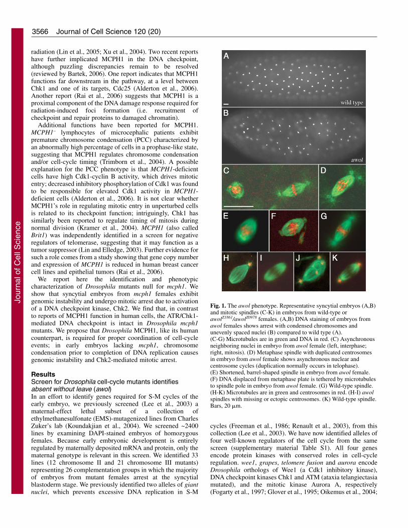
Fig. 1. The awol phenotype. Representative syncytial embryos (A,B)and mitotic spindles (C-K) in embryos from wild-type orawolZ1861/awolZ0978 females. (A,B) DNA staining of embryos fromawol females shows arrest with condensed chromosomes andunevenly spaced nuclei (B) compared to wild type (A).(C-G) Microtubules are in green and DNA in red. (C) Asynchronousneighboring nuclei in embryo from awol female (left, interphase;right, mitosis). (D) Metaphase spindle with duplicated centrosomesin embryo from awol female shows asynchronous nuclear andcentrosome cycles (duplication normally occurs in telophase).(E) Shortened, barrel-shaped spindle in embryo from awol female.(F) DNA displaced from metaphase plate is tethered by microtubulesto spindle pole in embryo from awol female. (G) Wild-type spindle.(H-K) Microtubules are in green and centrosomes in red. (H-I) awolspindles with missing or ectopic centrosomes. (K) Wild-type spindle.Bars, 20 �m.
Jour
nal o
f Cel
l Sci
ence
3567MCPH1 regulates Drosophila embryogenesis
Price et al., 2000). Identification of these alleles of bona fidecell-cycle regulators validates our screen.
We chose for further study the largest complementationgroup on chromosome II (comprising ZII-0978, ZII-1861 andZII-4050) identified in our screen. Females homozygous ortransheterozygous for any of these mutations are completelysterile, producing embryos that arrest in a metaphase-like state(~90% of embryos) in cycles 1-8 (the majority in cycles 6-8).Unevenly spaced, asynchronously dividing nuclei andcentrosome duplication prior to chromosome segregation areoften seen (Fig. 1B-D; Table 1); all of these are consistent withfailure of nuclear divisions. Tubulin foci are frequently missingfrom one or both poles of mitotic spindles, which are typicallyshorter and more barrel-shaped than those of wild type (Fig.1E; Table 1). Chromosomes are poorly aligned andoccasionally displaced from the metaphase plate (Fig. 1F).Staining for Centrosomin, a core centrosomal component (Liand Kaufman, 1996), revealed that lack of tubulin foci at oneor both poles in mutant-derived embryos is due to an absence
of centrosomes (Fig. 1H,I; Table 1); we occasionally seeectopic centrosomes embedded in spindles (Fig. 1J; Table 1).On the basis of the phenotype of acentrosomal mitotic spindles,we have given the name ‘absent without leave’ (‘awol’) tomutants of this complementation group.
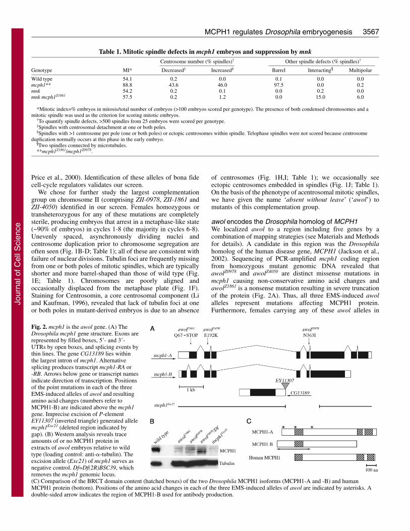
awol encodes the Drosophila homolog of MCPH1We localized awol to a region including five genes by acombination of mapping strategies (see Materials and Methodsfor details). A candidate in this region was the Drosophilahomolog of the human disease gene, MCPH1 (Jackson et al.,2002). Sequencing of PCR-amplified mcph1 coding regionfrom homozygous mutant genomic DNA revealed thatawolZ0978 and awolZ4050 are distinct missense mutations inmcph1 causing non-conservative amino acid changes andawolZ1861 is a nonsense mutation resulting in severe truncationof the protein (Fig. 2A). Thus, all three EMS-induced awolalleles represent mutations affecting MCPH1 protein.Furthermore, females carrying any of these awol alleles in
Fig. 2. mcph1 is the awol gene. (A) TheDrosophila mcph1 gene structure. Exons arerepresented by filled boxes, 5�- and 3�-UTRs by open boxes, and splicing events bythin lines. The gene CG13189 lies withinthe largest intron of mcph1. Alternativesplicing produces transcript mcph1-RA or-RB. Arrows below gene or transcript namesindicate direction of transcription. Positionsof the point mutations in each of the threeEMS-induced alleles of awol and resultingamino acid changes (numbers refer toMCPH1-B) are indicated above the mcph1gene. Imprecise excision of P-elementEY11307 (inverted triangle) generated allelemcph1Exc21 (deleted region indicated bygap). (B) Western analysis reveals traceamounts of or no MCPH1 protein inextracts of awol embryos relative to wildtype (loading control: anti-�-tubulin). Theexcision allele (Exc21) of mcph1 serves asnegative control. Df=Df(2R)BSC39, whichremoves the mcph1 genomic locus.(C) Comparison of the BRCT domain content (hatched boxes) of the two Drosophila MCPH1 isoforms (MCPH1-A and -B) and humanMCPH1 protein (bottom). Positions of the amino acid changes in each of the three EMS-induced alleles of awol are indicated by asterisks. Adouble-sided arrow indicates the region of MCPH1-B used for antibody production.
Table 1. Mitotic spindle defects in mcph1 embryos and suppression by mnkCentrosome number (% spindles)† Other spindle defects (% spindles)†
Genotype MI* Decreased‡ Increased§ Barrel Interacting¶ Multipolar
Wild type 54.1 0.2 0.0 0.1 0.0 0.0mcph1** 88.8 43.6 46.0 97.5 0.0 0.2mnk 54.2 0.2 0.1 0.0 0.2 0.0mnk mcph1Z1861 57.5 0.2 1.2 0.0 15.0 6.0
*Mitotic index=% embryos in mitosis/total number of embryos (>100 embryos scored per genotype). The presence of both condensed chromosomes and amitotic spindle was used as the criterion for scoring mitotic embryos.
†To quantify spindle defects, >500 spindles from 25 embryos were scored per genotype. ‡Spindles with centrosomal detachment at one or both poles.§Spindles with >1 centrosome per pole (one or both poles) or ectopic centrosomes within spindle. Telophase spindles were not scored because centrosome
duplication normally occurs at this phase in the early embryo.¶Two spindles connected by microtubules.**mcph1Z1861/mcph1Z0978.
Jour
nal o
f Cel
l Sci
ence
3568
trans to a deletion of the mcph1 genomic locus produceembryos with phenotypes indistinguishable from that ofhomozygous mutant females (data not shown), suggesting thatall three Zuker awol alleles behave genetically as nulls.
To confirm that mutation of mcph1 is responsible for theawol phenotype, we generated a null allele (mcph1Exc21) byimprecise P-element excision (Fig. 2A). mcph1Exc21
homozygous females produce embryos with the awolphenotype; similar results were obtained for females carryingthis excision in trans to any of the EMS-induced awol allelesor a deletion of the mcph1 genomic locus (data not shown),further confirming that mutation of mcph1 causes the awolphenotype. Importantly, expression of transgenic mcph1 usingthe UAS-Gal4 system (Brand and Perrimon, 1993; Rorth,1998) restored fertility to awolZ0978/awolZ4050 females,resulting in a hatch rate of ~40% of their embryos(supplementary material Table S2). Thus, mcph1 is the awolgene. We used the MCPH1 isoform that is most abundant inthe early embryo for transgenic rescue; it is possible that fullrescue of the maternal-effect lethality of awol mutants mightadditionally require expression of the less abundant isoform(see below for description of MCPH1 isoforms; Fig. 2A andsupplementary material Fig. S1B).
To further characterize our mcph1 alleles, we generatedpolyclonal antibodies against an MBP-MCPH1 fusion. Anti-MCPH1 antibodies recognize a major band of ~90 kDa,consistent with the predicted size of MCPH1-B, when used toprobe immunoblots of wild-type embryo extracts (Fig. 2B). Incontrast, for all mcph1 alleles identified here, we detect greatlyreduced or no MCPH1 protein in mutant-derived embryos.Thus, all of these alleles are null (or nearly null) for MCPH1protein.
MCPH1 isoforms differ in expression pattern and BRCTdomain contentOur genetic data revealed that mcph1 null alleles arehomozygous viable and that mcph1 is required maternally forearly embryonic development. To measure MCPH1 levelsthroughout Drosophila development, we probed immunoblotsof extracts from various developmental stages with anti-MCPH1 antibodies (supplementary material Fig. S1A). Asexpected, MCPH1 is abundant in ovaries and early embryos,whereas older embryos under zygotic control have relativelylow amounts. MCPH1 is present in larval brains and imaginaldiscs but undetectable in adult brain extracts. Although highlevels of MCPH1 are present in adult testes, it is not requiredfor male fertility (data not shown).
Two major isoforms of MCPH1 were detected byimmunoblotting: ~90 kDa (predominant in ovaries andembryos) and ~110 kDa (predominant in testes). Bothisoforms were detected in larval tissues. The most recentmcph1 gene model annotated by FlyBase predicts two splicevariants (A and B) differing at their 5�-ends that encodeproteins with distinct amino termini (Grumbling and Strelets,2006). We compared sizes of recombinant MCPH1-A and -Bproteins (produced by in vitro transcription-translationreactions) to that of endogenous MCPH1 isoforms byimmunoblotting. We found that the gel mobilities of MCPH1-A and -B closely match that of MCPH1 in testes and ovaries,respectively; thus, MCPH1-A is the ~110 kDa isoform that isabundant in testes, and MCPH1-B is the ~90 kDa isoform that
is abundant in ovaries and early embryos (supplementarymaterial Fig. S1B).
We observed a discrepancy between relative sizes ofMCPH1-A and -B on our immunoblots (A larger than B;supplementary material Fig. S1B) and as predicted by FlyBase[779 versus 826 amino acids, respectively (Grumbling andStrelets, 2006)]. We were unable to find 3�-end sequence datafor mcph1-A on public databases, so we fully sequenced arepresentative clone (LP15451) and found it to encode aprotein of 981 amino acids, which closely matches ourestimated size of 110 kDa for endogenous MCPH1-A.Furthermore, our sequencing revealed that mcph1-A containscoding sequence from both mcph1 and CG30038, a genepredicted to overlap the 3�-end of mcph1 (Fig. 2A). Thus,mcph1-A and -B are alternatively spliced at both ends,producing proteins that differ in their N- and C-terminalregions (Fig. 2C), and predicted gene CG30038 comprisesalternatively spliced exons of mcph1-A.
MCPH1-A and -B proteins both contain BRCT domains(three or one, respectively). The arrangement of BRCTdomains within MCPH1-A (one N-terminal and two paired C-terminal) resembles that of human MCPH1 (Fig. 2C).Drosophila and human MCPH1 have highest sequence identityin their BRCT domains (37.6%, 52.5% and 26.8% between theN-terminal, first C-terminal, and second C-terminal domains,respectively). The presence of extended amino termini in bothDrosophila isoforms relative to human MCPH1 raises thepossibility that the reported human sequence (Jackson et al.,2002) may not be full-length.
MCPH1 is a nuclear proteinBecause Drosophila MCPH1 contains BRCT domains, wehypothesized that it has a nuclear function. In syncytialembryos, MCPH1 signal localizes to interphase nuclei anddisappears in mitosis (supplementary material Fig. S2). Ascontrol for antibody specificity, no MCPH1 signal was detectedin interphase nuclei of embryos derived from mcph1 nullfemales. Because MCPH1 protein is readily detectablethroughout the cell cycle (by immunoblotting of extracts fromstaged embryos; data not shown), the disappearance ofMCPH1 signal in mitosis, as observed by immunostaining, isprobably due to its dispersal into the cytoplasm upon nuclearenvelope breakdown. Human MCPH1 has been reported tolocalize to the nucleus (Lin et al., 2005) as well as tocentrosomes (Jeffers et al., 2007; Zhong et al., 2006); weobserve no centrosomal localization for MCPH1 in syncytialembryos of Drosophila.
Mitotic arrest in mcph1 syncytial embryos is aconsequence of Chk2 activationThe defective mitotic spindles of embryos derived from mcph1females (hereafter referred to as ‘mcph1 embryos’) exhibit keyfeatures reminiscent of Chk2-mediated centrosomalinactivation. In particular, these spindles are short, barrel-shaped, anastral, and associated with poorly alignedchromosomes (Fig. 1). Late syncytial embryos of Drosophilause a two-stage response to DNA damage or replication defects(Sibon et al., 2000). The DNA checkpoint mediated by Meiotic41 (MEI-41) and Grapes (GRP), the Drosophila orthologs ofATR (ATM-Rad3-related) and Chk1 kinases, respectively,delays mitotic entry via inhibitory phosphorylation of Cdk1 to
Journal of Cell Science 120 (20)
Jour
nal o
f Cel
l Sci
ence
3569MCPH1 regulates Drosophila embryogenesis
allow repair of DNA damage or completion of replication(Sibon et al., 1999; Sibon et al., 1997). When this checkpointfails, a secondary damage-control system operating in mitosisis activated; resulting changes in spindle structure blockchromosome segregation, presumably to stop propagation ofdefective DNA (Sibon et al., 2000; Takada et al., 2003). Thisdamage-control system, known as centrosomal inactivation, ismediated by the checkpoint kinase Chk2 (Takada et al., 2003).
Loss of �-tubulin from centrosomes of mitotic spindles isanother characteristic feature of Chk2-mediated centrosomalinactivation. We detected decreased �-tubulin staining ofcentrosomes during mitosis in mcph1 embryos compared towild type (supplementary material Fig. S3). We typicallyobserve complete detachment of centrosomes from spindles inmcph1 embryos. High levels of DNA damage induced byintense laser illumination can similarly cause completecentrosomal detachment from spindle poles of wild-typeembryos (Takada et al., 2003), suggesting that the spindlechanges we observe in mcph1 embryos represent an extremeform of centrosomal inactivation.
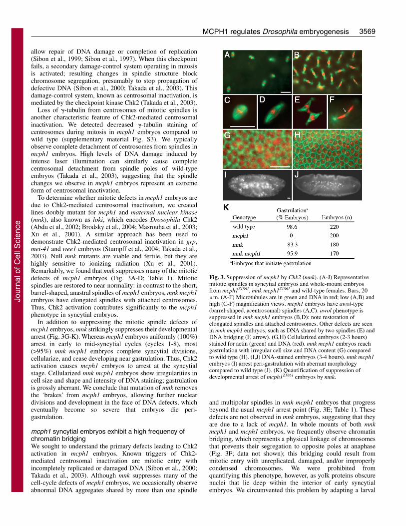
To determine whether mitotic defects in mcph1 embryos aredue to Chk2-mediated centrosomal inactivation, we createdlines doubly mutant for mcph1 and maternal nuclear kinase(mnk), also known as loki, which encodes Drosophila Chk2(Abdu et al., 2002; Brodsky et al., 2004; Masrouha et al., 2003;Xu et al., 2001). A similar approach has been used todemonstrate Chk2-mediated centrosomal inactivation in grp,mei-41 and wee1 embryos (Stumpff et al., 2004; Takada et al.,2003). Null mnk mutants are viable and fertile, but they arehighly sensitive to ionizing radiation (Xu et al., 2001).Remarkably, we found that mnk suppresses many of the mitoticdefects of mcph1 embryos (Fig. 3A-D; Table 1). Mitoticspindles are restored to near-normality: in contrast to the short,barrel-shaped, anastral spindles of mcph1 embryos, mnk mcph1embryos have elongated spindles with attached centrosomes.Thus, Chk2 activation contributes significantly to the mcph1phenotype in syncytial embryos.
In addition to suppressing the mitotic spindle defects ofmcph1 embryos, mnk strikingly suppresses their developmentalarrest (Fig. 3G-K). Whereas mcph1 embryos uniformly (100%)arrest in early to mid-syncytial cycles (cycles 1-8), most(>95%) mnk mcph1 embryos complete syncytial divisions,cellularize, and cease developing near gastrulation. Thus, Chk2activation causes mcph1 embryos to arrest at the syncytialstage. Cellularized mnk mcph1 embryos show irregularities incell size and shape and intensity of DNA staining; gastrulationis grossly aberrant. We conclude that mutation of mnk removesthe ‘brakes’ from mcph1 embryos, allowing further nucleardivisions and development in the face of DNA defects, whicheventually become so severe that embryos die peri-gastrulation.
mcph1 syncytial embryos exhibit a high frequency ofchromatin bridgingWe sought to understand the primary defects leading to Chk2activation in mcph1 embryos. Known triggers of Chk2-mediated centrosomal inactivation are mitotic entry withincompletely replicated or damaged DNA (Sibon et al., 2000;Takada et al., 2003). Although mnk suppresses many of thecell-cycle defects of mcph1 embryos, we occasionally observeabnormal DNA aggregates shared by more than one spindle
and multipolar spindles in mnk mcph1 embryos that progressbeyond the usual mcph1 arrest point (Fig. 3E; Table 1). Thesedefects are not observed in mnk embryos, suggesting that theyare due to a lack of mcph1. In whole mounts of both mnkmcph1 and mcph1 embryos, we frequently observe chromatinbridging, which represents a physical linkage of chromosomesthat prevents their segregation to opposite poles at anaphase(Fig. 3F; data not shown); this bridging could result frommitotic entry with unreplicated, damaged, and/or improperlycondensed chromosomes. We were prohibited fromquantifying this phenotype, however, as yolk proteins obscurenuclei that lie deep within the interior of early syncytialembryos. We circumvented this problem by adapting a larval
Fig. 3. Suppression of mcph1 by Chk2 (mnk). (A-J) Representativemitotic spindles in syncytial embryos and whole-mount embryosfrom mcph1Z1861, mnk mcph1Z1861 and wild-type females. Bars, 20�m. (A-F) Microtubules are in green and DNA in red; low (A,B) andhigh (C-F) magnification views. mcph1 embryos have awol-type(barrel-shaped, acentrosomal) spindles (A,C). awol phenotype issuppressed in mnk mcph1 embryos (B,D): note restoration ofelongated spindles and attached centrosomes. Other defects are seenin mnk mcph1 embryos, such as DNA shared by two spindles (E) andDNA bridging (F, arrow). (G,H) Cellularized embryos (2-3 hours)stained for actin (green) and DNA (red). mnk mcph1 embryos reachgastrulation with irregular cell size and DNA content (G) comparedto wild type (H). (I,J) DNA-stained embryos (3-4 hours). mnk mcph1embryos (I) arrest peri-gastrulation with aberrant morphologycompared to wild type (J). (K) Quantification of suppression ofdevelopmental arrest of mcph1Z1861 embryos by mnk.
Jour
nal o
f Cel
l Sci
ence
3570
brain squash protocol for this developmental stage that allowedus to more clearly observe chromosomes of early embryos.
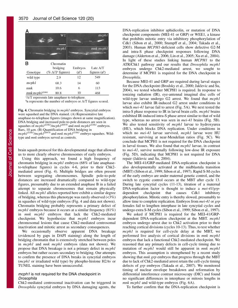
Using this approach, we found a high frequency ofchromatin bridging in mcph1 embryos (68% of late anaphase-to-telophase figures) in cycles 4-6, prior to their Chk2-mediated arrest (Fig. 4). Multiple bridges are often presentbetween segregating chromosomes. Spindle pole-to-poledistances are increased dramatically compared to wild-typefigures, presumably due to an extended anaphase B in a failedattempt to separate chromosomes that remain physicallylinked. All mcph1 alleles reported here exhibit a similar degreeof bridging, whereas this phenotype was rarely observed (<3%)in squashes of wild-type embryos (Fig. 4 and data not shown).Chromatin bridging probably represents a primary defect ofmcph1 embryos because it occurs at a similar frequency (81%)in mnk mcph1 embryos that lack the Chk2-mediatedcheckpoint. We hypothesize that mcph1 embryos incurchromosomal lesions that cause Chk2-mediated centrosomalinactivation and mitotic arrest as secondary consequences.
We occasionally observe apparent DNA breakage(evidenced by gaps in DAPI staining) along the length ofbridging chromatin that is extensively stretched between polesin mcph1 and mnk mcph1 embryos (data not shown). Wepropose that DNA breakage is not a primary defect in mcph1embryos but rather occurs secondary to bridging. Our attemptsto confirm the presence of DNA breaks in syncytial embryos(mcph1 or irradiated wild type) by phospho-histone H2Av orTUNEL staining have been unsuccessful.
mcph1 is not required for the DNA checkpoint inDrosophilaChk2-mediated centrosomal inactivation can be triggered inDrosophila syncytial embryos by DNA damaging agents, the
DNA-replication inhibitor aphidicolin, or mutation of DNAcheckpoint components (MEI-41 or GRP) or WEE1, a kinasethat prohibits mitotic entry via inhibitory phosphorylation ofCdk1 (Sibon et al., 2000; Stumpff et al., 2004; Takada et al.,2003). Human MCPH1-deficient cells show defective G2-Mand intra-S phase checkpoint responses following DNAdamage (Alderton et al., 2006; Lin et al., 2005; Xu et al., 2004).In light of these studies linking human MCPH1 to theATR/Chk1 pathway and our results that Drosophila mcph1embryos undergo Chk2-mediated arrest, we sought todetermine if MCPH1 is required for the DNA checkpoint inDrosophila.
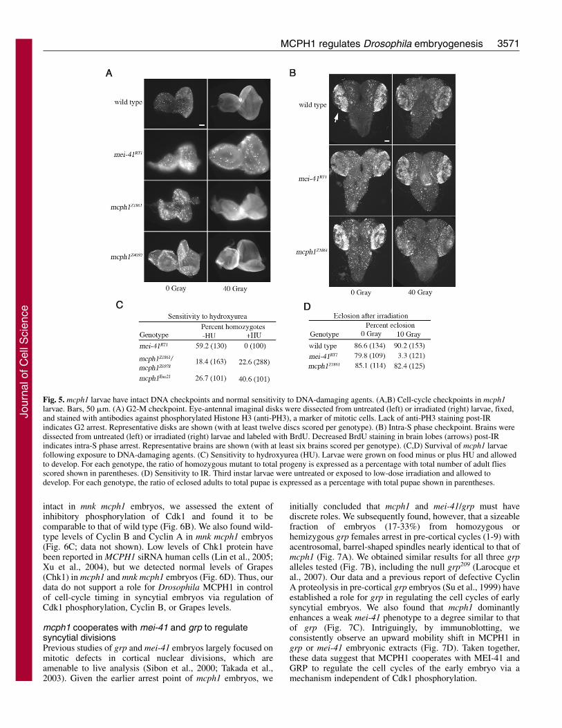
Because MEI-41 and GRP are required during larval stagesfor the DNA checkpoint (Brodsky et al., 2000; Jaklevic and Su,2004), we tested whether MCPH1 is required. In response toionizing radiation (IR), eye-antennal imaginal disc cells ofwild-type larvae undergo G2 arrest. We found that mcph1larvae also exhibit IR-induced G2 arrest under conditions inwhich mei-41 larvae fail to arrest (Fig. 5A). We next tested theintra-S phase response to IR in larval brain cells. mcph1 brainsexhibited IR-induced intra-S phase arrest similar to that of wildtype, whereas no arrest was seen in mei-41 brains (Fig. 5B).We also tested sensitivity of mcph1 larvae to hydroxyurea(HU), which blocks DNA replication. Under conditions inwhich no mei-41 larvae survived, mcph1 larvae were HUresistant, surviving at near-Mendelian ratios (Fig. 5C). Weconclude that MCPH1 is not required for the DNA checkpointin larval tissues. We also found that mcph1 larvae, in contrastto mei-41, survive normally following low-dose IR exposure(Fig. 5D), indicating that MCPH1 is not required for DNArepair (Jaklevic and Su, 2004).
The MEI-41/GRP-mediated DNA-replication checkpoint isalso developmentally activated at the midblastula transition(MBT) (Sibon et al., 1999; Sibon et al., 1997). Rapid S-M cyclesof the early embryo are under maternal genetic control, and theswitch to zygotic control occurs at the MBT after cycle 13.During late syncytial cycles (11-13), titration of a maternalDNA-replication factor is thought to induce a mei-41/grp-dependent checkpoint that causes Cdk1 inhibitoryphosphorylation. Mitotic entry is thereby slowed, presumably toallow time to complete replication. Embryos from mei-41 or grpfemales fail to lengthen interphase in late syncytial cycles andundergo extra S-M cycles (Sibon et al., 1999; Sibon et al., 1997).
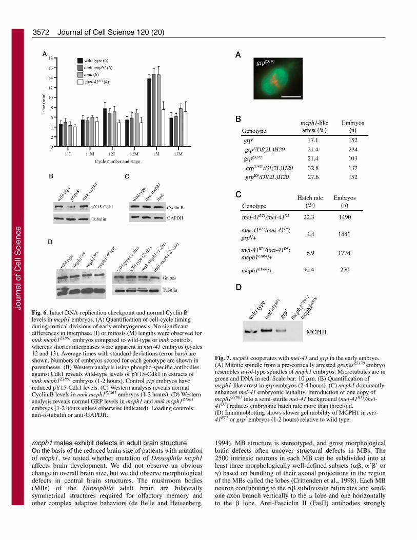
We asked if MCPH1 is required for the MEI-41/GRP-dependent DNA-replication checkpoint at the MBT. mcph1embryos undergo arrest due to Chk2 activation prior to theirreaching cortical divisions (cycles 10-13). Thus, to test whethermcph1 is required for cell-cycle delay at the MBT, weperformed live analysis of cortical divisions in mnk mcph1embryos that lack a functional Chk2-mediated checkpoint. Wereasoned that any primary defects in cell-cycle timing due tomutation of mcph1 would still be apparent in mnk mcph1embryos. This assumption is strengthened by a recent studyshowing that mnk grp embryos that progress through the MBTdue to lack of Chk2-mediated arrest retain the cell-cycle timingdefects of grp embryos (Takada et al., 2007). We monitoredtiming of nuclear envelope breakdown and reformation bydifferential interference contrast microscopy (DIC) and foundno significant differences in interphase or mitosis lengths inmnk mcph1 and wild-type embryos (Fig. 6A).
To further confirm that the DNA-replication checkpoint is
Journal of Cell Science 120 (20)
Fig. 4. Chromatin bridging in mcph1 embryos. Syncytial embryoswere squashed and the DNA stained. (A) Representative lateanaphase-to-telophase figures (images shown at same magnification).DNA bridging and increased pole-to-pole distances are seen insquashes of mcph1Z1861/mcph1Z0978 and mnk mcph1Z1861 embryos.Bars, 10 �m. (B) Quantification of DNA bridging inmcph1Z1861/mcph1Z0978 and mnk mcph1Z1861 embryo squashes. Wild-type and mnk embryos served as controls.
Jour
nal o
f Cel
l Sci
ence
3571MCPH1 regulates Drosophila embryogenesis
intact in mnk mcph1 embryos, we assessed the extent ofinhibitory phosphorylation of Cdk1 and found it to becomparable to that of wild type (Fig. 6B). We also found wild-type levels of Cyclin B and Cyclin A in mnk mcph1 embryos(Fig. 6C; data not shown). Low levels of Chk1 protein havebeen reported in MCPH1 siRNA human cells (Lin et al., 2005;Xu et al., 2004), but we detected normal levels of Grapes(Chk1) in mcph1 and mnk mcph1 embryos (Fig. 6D). Thus, ourdata do not support a role for Drosophila MCPH1 in controlof cell-cycle timing in syncytial embryos via regulation ofCdk1 phosphorylation, Cyclin B, or Grapes levels.
mcph1 cooperates with mei-41 and grp to regulatesyncytial divisionsPrevious studies of grp and mei-41 embryos largely focused onmitotic defects in cortical nuclear divisions, which areamenable to live analysis (Sibon et al., 2000; Takada et al.,2003). Given the earlier arrest point of mcph1 embryos, we
initially concluded that mcph1 and mei-41/grp must havediscrete roles. We subsequently found, however, that a sizeablefraction of embryos (17-33%) from homozygous orhemizygous grp females arrest in pre-cortical cycles (1-9) withacentrosomal, barrel-shaped spindles nearly identical to that ofmcph1 (Fig. 7A). We obtained similar results for all three grpalleles tested (Fig. 7B), including the null grp209 (Larocque etal., 2007). Our data and a previous report of defective CyclinA proteolysis in pre-cortical grp embryos (Su et al., 1999) haveestablished a role for grp in regulating the cell cycles of earlysyncytial embryos. We also found that mcph1 dominantlyenhances a weak mei-41 phenotype to a degree similar to thatof grp (Fig. 7C). Intriguingly, by immunoblotting, weconsistently observe an upward mobility shift in MCPH1 ingrp or mei-41 embryonic extracts (Fig. 7D). Taken together,these data suggest that MCPH1 cooperates with MEI-41 andGRP to regulate the cell cycles of the early embryo via amechanism independent of Cdk1 phosphorylation.
Fig. 5. mcph1 larvae have intact DNA checkpoints and normal sensitivity to DNA-damaging agents. (A,B) Cell-cycle checkpoints in mcph1larvae. Bars, 50 �m. (A) G2-M checkpoint. Eye-antennal imaginal disks were dissected from untreated (left) or irradiated (right) larvae, fixed,and stained with antibodies against phosphorylated Histone H3 (anti-PH3), a marker of mitotic cells. Lack of anti-PH3 staining post-IRindicates G2 arrest. Representative disks are shown (with at least twelve discs scored per genotype). (B) Intra-S phase checkpoint. Brains weredissected from untreated (left) or irradiated (right) larvae and labeled with BrdU. Decreased BrdU staining in brain lobes (arrows) post-IRindicates intra-S phase arrest. Representative brains are shown (with at least six brains scored per genotype). (C,D) Survival of mcph1 larvaefollowing exposure to DNA-damaging agents. (C) Sensitivity to hydroxyurea (HU). Larvae were grown on food minus or plus HU and allowedto develop. For each genotype, the ratio of homozygous mutant to total progeny is expressed as a percentage with total number of adult fliesscored shown in parentheses. (D) Sensitivity to IR. Third instar larvae were untreated or exposed to low-dose irradiation and allowed todevelop. For each genotype, the ratio of eclosed adults to total pupae is expressed as a percentage with total pupae shown in parentheses.
Jour
nal o
f Cel
l Sci
ence
3572
mcph1 males exhibit defects in adult brain structureOn the basis of the reduced brain size of patients with mutationof mcph1, we tested whether mutation of Drosophila mcph1affects brain development. We did not observe an obviouschange in overall brain size, but we did observe morphologicaldefects in central brain structures. The mushroom bodies(MBs) of the Drosophila adult brain are bilaterallysymmetrical structures required for olfactory memory andother complex adaptive behaviors (de Belle and Heisenberg,
1994). MB structure is stereotyped, and gross morphologicalbrain defects often uncover structural defects in MBs. The2500 intrinsic neurons in each MB can be subdivided into atleast three morphologically well-defined subsets (��, ���� or�) based on bundling of their axonal projections in the regionof the MBs called the lobes (Crittenden et al., 1998). Each MBneuron contributing to the �� subdivision bifurcates and sendsone axon branch vertically to the � lobe and one horizontallyto the � lobe. Anti-Fasciclin II (FasII) antibodies strongly
Journal of Cell Science 120 (20)
Fig. 6. Intact DNA-replication checkpoint and normal Cyclin Blevels in mcph1 embryos. (A) Quantification of cell-cycle timingduring cortical divisions of early embryogenesis. No significantdifferences in interphase (I) or mitosis (M) lengths were observed formnk mcph1Z1861 embryos compared to wild-type or mnk controls,whereas shorter interphases were apparent in mei-41 embryos (cycles12 and 13). Average times with standard deviations (error bars) areshown. Numbers of embryos scored for each genotype are shown inparentheses. (B) Western analysis using phospho-specific antibodiesagainst Cdk1 reveals wild-type levels of pY15-Cdk1 in extracts ofmnk mcph1Z1861 embryos (1-2 hours). Control grp embryos havereduced pY15-Cdk1 levels. (C) Western analysis reveals normalCyclin B levels in mnk mcph1Z1861 embryos (1-2 hours). (D) Westernanalysis reveals normal GRP levels in mcph1 and mnk mcph1Z1861
embryos (1-2 hours unless otherwise indicated). Loading controls:anti-�-tubulin or anti-GAPDH.
Fig. 7. mcph1 cooperates with mei-41 and grp in the early embryo.(A) Mitotic spindle from a pre-cortically arrested grapesZ5170 embryoresembles awol-type spindles of mcph1 embryos. Microtubules are ingreen and DNA in red. Scale bar: 10 �m. (B) Quantification ofmcph1-like arrest in grp embryos (2-4 hours). (C) mcph1 dominantlyenhances mei-41 embryonic lethality. Introduction of one copy ofmcph1Z1861 into a semi-sterile mei-41 background (mei-41RT1/mei-41D5) reduces embryonic hatch rate more than threefold.(D) Immunoblotting shows slower gel mobility of MCPH1 in mei-41RT1 or grp1 embryos (1-2 hours) relative to wild type.
Jour
nal o
f Cel
l Sci
ence
3573MCPH1 regulates Drosophila embryogenesis
label MB neurons that lie in the �� lobes (Grenningloh et al.,1991), thereby allowing straightforward visualization ofdevelopmental defects.
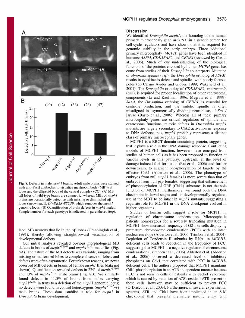
Our initial analysis revealed obvious morphological MBdefects in brains of mcph1Z1861 and mcph1Exc21 male flies (Fig.8A). The nature of the MB defects was variable, ranging frommissing or malformed lobes to complete absence of lobes, anddefects were often asymmetric. For unknown reasons, we neverobserved MB defects in brains of female mcph1 flies (data notshown). Quantification revealed defects in 22% of mcph1Z1861
and 13% of mcph1Exc21 male brains (Fig. 8B). We similarlyfound defects in 11.5% of brains from males carryingmcph1Z1861 in trans to a deletion of the mcph1 genomic locus;no defects were found in control heterozygous (mcph1Z1861/+)male brains. These data establish a role for mcph1 inDrosophila brain development.
DiscussionWe identified Drosophila mcph1, the homolog of the humanprimary microcephaly gene MCPH1, in a genetic screen forcell-cycle regulators and have shown that it is required forgenomic stability in the early embryo. Three additionalprimary microcephaly (MCPH) genes have been identified inhumans: ASPM, CDK5RAP2, and CENPJ (reviewed by Cox etal., 2006). Much of our understanding of the biologicalfunctions of the proteins encoded by human MCPH genes hascome from studies of their Drosophila counterparts. Mutationof abnormal spindle (asp), the Drosophila ortholog of ASPM,results in cytokinesis defects and spindles with poorly focusedpoles (do Carmo Avides and Glover, 1999; Wakefield et al.,2001). The Drosophila ortholog of CDK5RAP2, centrosomin(cnn), is required for proper localization of other centrosomalcomponents (Li and Kaufman, 1996; Megraw et al., 1999).Sas-4, the Drosophila ortholog of CENPJ, is essential forcentriole production, and the mitotic spindle is oftenmisaligned in asymmetrically dividing neuroblasts of Sas-4larvae (Basto et al., 2006). Whereas all of these primarymicrocephaly genes are critical regulators of spindle andcentrosome functions, mitotic defects in Drosophila mcph1mutants are largely secondary to Chk2 activation in responseto DNA defects; thus, mcph1 probably represents a distinctclass of primary microcephaly genes.
MCPH1 is a BRCT domain-containing protein, suggestingthat it plays a role in the DNA damage response. Conflictingmodels of MCPH1 function, however, have emerged fromstudies of human cells as it has been proposed to function atvarious levels in this pathway: upstream, at the level ofdamage-induced foci formation (Rai et al., 2006) and furtherdownstream, to augment phosphorylation of targets by theeffector Chk1 (Alderton et al., 2006). The phenotype ofembryos from null mcph1 females is more severe than that ofembryos from null grp females, suggesting that enhancementof phosphorylation of GRP (Chk1) substrates is not the solefunction of MCPH1. Furthermore, we found both the DNAcheckpoint in larval stages and its developmentally regulateduse at the MBT to be intact in mcph1 mutants, suggesting arequisite role for MCPH1 in the DNA checkpoint evolved inhigher organisms.
Studies of human cells suggest a role for MCPH1 inregulation of chromosome condensation. Microcephalicpatients homozygous for a severely truncating mutation inMCPH1 show increased frequency of G2-like cells displayingpremature chromosome condensation (PCC) with an intactnuclear envelope (Alderton et al., 2006; Trimborn et al., 2004).Depletion of Condensin II subunits by RNAi in MCPH1-deficient cells leads to reduction in the frequency of PCC,suggesting that MCPH1 is a negative regulator of chromosomecondensation (Trimborn et al., 2006). Alderton et al. (Aldertonet al., 2006) observed a decreased level of inhibitoryphosphates on Cdk1 that correlated with PCC in MCPH1-deficient cells. The authors proposed that MCPH1 maintainsCdk1 phosphorylation in an ATR-independent manner becausePCC is not seen in cells of patients with Seckel syndrome,which is caused by mutation of ATR; residual ATR present inthese cells, however, may be sufficient to prevent PCC(O’Driscoll et al., 2003). Furthermore, in several experimentalsystems, ATR and Chk1 have been implicated in an S-Mcheckpoint that prevents premature mitotic entry with
Fig. 8. Defects in male mcph1 brains. Adult male brains were stainedwith anti-FasII antibodies to visualize mushroom body (MB) ��lobes and the ellipsoid body of the central complex (CC). (A) MB�� lobes of wild-type brains are symmetric, whereas MBs of mcph1brains are occasionally defective with missing or diminished ��lobes (arrowheads). Df=Df(2R)BSC39, which removes the mcph1genomic locus. (B) Quantification of brain defects in mcph1 males.Sample number for each genotype is indicated in parentheses (top).
Jour
nal o
f Cel
l Sci
ence
3574
unreplicated DNA (reviewed by Petermann and Caldecott,2006).
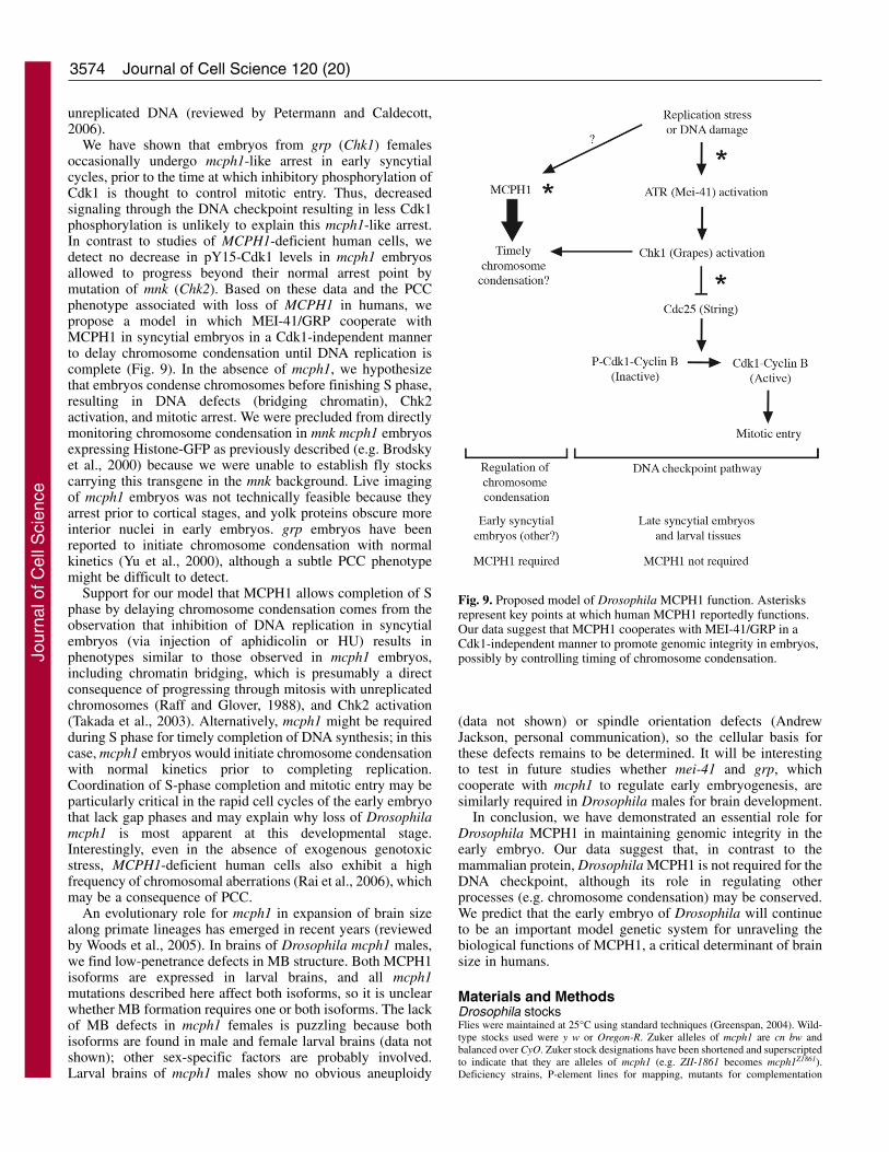
We have shown that embryos from grp (Chk1) femalesoccasionally undergo mcph1-like arrest in early syncytialcycles, prior to the time at which inhibitory phosphorylation ofCdk1 is thought to control mitotic entry. Thus, decreasedsignaling through the DNA checkpoint resulting in less Cdk1phosphorylation is unlikely to explain this mcph1-like arrest.In contrast to studies of MCPH1-deficient human cells, wedetect no decrease in pY15-Cdk1 levels in mcph1 embryosallowed to progress beyond their normal arrest point bymutation of mnk (Chk2). Based on these data and the PCCphenotype associated with loss of MCPH1 in humans, wepropose a model in which MEI-41/GRP cooperate withMCPH1 in syncytial embryos in a Cdk1-independent mannerto delay chromosome condensation until DNA replication iscomplete (Fig. 9). In the absence of mcph1, we hypothesizethat embryos condense chromosomes before finishing S phase,resulting in DNA defects (bridging chromatin), Chk2activation, and mitotic arrest. We were precluded from directlymonitoring chromosome condensation in mnk mcph1 embryosexpressing Histone-GFP as previously described (e.g. Brodskyet al., 2000) because we were unable to establish fly stockscarrying this transgene in the mnk background. Live imagingof mcph1 embryos was not technically feasible because theyarrest prior to cortical stages, and yolk proteins obscure moreinterior nuclei in early embryos. grp embryos have beenreported to initiate chromosome condensation with normalkinetics (Yu et al., 2000), although a subtle PCC phenotypemight be difficult to detect.
Support for our model that MCPH1 allows completion of Sphase by delaying chromosome condensation comes from theobservation that inhibition of DNA replication in syncytialembryos (via injection of aphidicolin or HU) results inphenotypes similar to those observed in mcph1 embryos,including chromatin bridging, which is presumably a directconsequence of progressing through mitosis with unreplicatedchromosomes (Raff and Glover, 1988), and Chk2 activation(Takada et al., 2003). Alternatively, mcph1 might be requiredduring S phase for timely completion of DNA synthesis; in thiscase, mcph1 embryos would initiate chromosome condensationwith normal kinetics prior to completing replication.Coordination of S-phase completion and mitotic entry may beparticularly critical in the rapid cell cycles of the early embryothat lack gap phases and may explain why loss of Drosophilamcph1 is most apparent at this developmental stage.Interestingly, even in the absence of exogenous genotoxicstress, MCPH1-deficient human cells also exhibit a highfrequency of chromosomal aberrations (Rai et al., 2006), whichmay be a consequence of PCC.
An evolutionary role for mcph1 in expansion of brain sizealong primate lineages has emerged in recent years (reviewedby Woods et al., 2005). In brains of Drosophila mcph1 males,we find low-penetrance defects in MB structure. Both MCPH1isoforms are expressed in larval brains, and all mcph1mutations described here affect both isoforms, so it is unclearwhether MB formation requires one or both isoforms. The lackof MB defects in mcph1 females is puzzling because bothisoforms are found in male and female larval brains (data notshown); other sex-specific factors are probably involved.Larval brains of mcph1 males show no obvious aneuploidy
(data not shown) or spindle orientation defects (AndrewJackson, personal communication), so the cellular basis forthese defects remains to be determined. It will be interestingto test in future studies whether mei-41 and grp, whichcooperate with mcph1 to regulate early embryogenesis, aresimilarly required in Drosophila males for brain development.
In conclusion, we have demonstrated an essential role forDrosophila MCPH1 in maintaining genomic integrity in theearly embryo. Our data suggest that, in contrast to themammalian protein, Drosophila MCPH1 is not required for theDNA checkpoint, although its role in regulating otherprocesses (e.g. chromosome condensation) may be conserved.We predict that the early embryo of Drosophila will continueto be an important model genetic system for unraveling thebiological functions of MCPH1, a critical determinant of brainsize in humans.
Materials and MethodsDrosophila stocksFlies were maintained at 25°C using standard techniques (Greenspan, 2004). Wild-type stocks used were y w or Oregon-R. Zuker alleles of mcph1 are cn bw andbalanced over CyO. Zuker stock designations have been shortened and superscriptedto indicate that they are alleles of mcph1 (e.g. ZII-1861 becomes mcph1Z1861).Deficiency strains, P-element lines for mapping, mutants for complementation
Journal of Cell Science 120 (20)
Fig. 9. Proposed model of Drosophila MCPH1 function. Asterisksrepresent key points at which human MCPH1 reportedly functions.Our data suggest that MCPH1 cooperates with MEI-41/GRP in aCdk1-independent manner to promote genomic integrity in embryos,possibly by controlling timing of chromosome condensation.Jo
urna
l of C
ell S
cien
ce
3575MCPH1 regulates Drosophila embryogenesis
testing (grp1, aurora1, wee1ES1), nanos-Gal4:VP16 stock, and mei-41 mutants werefrom Bloomington Stock Center. mcph1 P-element insertions were fromBloomington Stock Center (EY11307), Kyoto Stock Center (NP6229-5-1), or a giftfrom Steven Hou (l(2)SH0220). tefu356, mnk6006 and grp209 stocks were gifts fromMike Brodsky, Bill Theurkauf and Tin Tin Su, respectively.
Identification of new alleles of cell-cycle regulatorsA combination of female meiotic recombination, deficiency mapping and directcomplementation testing of candidates was used to identify mutants from our screen.Complementation testing with known cell-cycle regulators was performed byassessing fertility of females carrying a Zuker chromosome in trans to a knownmutation. We used the following alleles: wee1ES1 (Price et al., 2000), grp1 (Fogartyet al., 1997), tefu�356 (Oikemus et al., 2004) and aur1 (Glover et al., 1995).
Quantification of embryonic hatch ratesFor hatch rate assays, embryos (0-4 hours) were collected on grape plates, countedand aged ~40 hours at 25°C. The number of hatched embryos was determined bysubtracting the number of unhatched (intact) embryos from the total numbercollected. Hatch rate is the ratio of hatched to total embryos expressed as apercentage.
Genetic and molecular mapping of awolThe awol gene was localized by a combination of mapping strategies. We firstscreened a collection of deficiencies on the second chromosome for non-complementation of the female sterility of awolZ1861. We found that females carryingawolZ1861 in trans to Df(2R)BSC39 produced embryos with the awol phenotype;similar results were obtained for awolZ0978 and awolZ4050. Thus, awol lies betweenthe breakpoints of Df(2R)BSC39 in the polytene interval 48C5-E1, a region thatcontains ~35 genes. We mapped awol by P-element-induced male recombination(Chen et al., 1998) relative to the following insertion lines: Mtork03905, ERp60BG01854,KG04952, otkEP2017 and CG8378EP2501. We thereby narrowed awol to a region offive genes (including mcph1) that lie distal to ERp60BG01854 and proximal toKG04952. The awol stock used (cn ZII-1861 bw/CyO) has visible flanking markerscn and bw. The source of transposase was Delta2-3 Sb. Multiple independentrecombinant chromosomes were recovered for each P-element line tested. GenomicDNA was extracted from whole flies homozygous for awol mutations essentially aspreviously described (Ballinger and Benzer, 1989). mcph1 coding regions werePCR-amplified from genomic DNA and sequenced.
Generation of mcph1 excision lineP-element insertions have been identified in the 5�-UTR of mcph1 (NP6229-5-1)and within its largest intron (l(2)k06612, l(2)SH0220 and EY11307) (Grumbling andStrelets, 2006). l(2)k06612 is no longer available from stock centers. We mappedthe lethality of line l(2)SH0220 (Oh et al., 2003) outside of the mcph1 genomicregion (data not shown). We found that EY11307 homozygous andEY11307/mcph1Z1861 transheterozygous females are viable, fertile and produceembryos with nearly wild-type levels of MCPH1 protein, indicating that this P-insertion has little effect on mcph1 transcription; similar results were obtained forNP6229-5-1 (data not shown). EY11307 is inserted in the 5�-UTR of CG13189,which encodes a putative metal ion transporter, and the largest intron of mcph1 (Fig.2A). All EMS-induced mcph1 mutations described here lie outside of CG13189(including two beyond its 3� end), thereby making it unlikely that decreasedCG13189 activity causes the awol phenotype. We performed imprecise P-elementexcision of EY11307 to generate mcph1Exc21, which lacks two internal exons andpart of the 3�-most exon of mcph1; this excision left the 5�-UTR, coding region and3�-UTR of CG13189 intact, but probably removed some of its promoter (Fig. 2A).
Embryo fixation, staining and microscopyEmbryos (1-2 hours unless otherwise indicated) were collected for staining usingstandard techniques (Rothwell and Sullivan, 2000). For mouse anti-�-tubulin(DM1�, 1:500, Sigma) or rabbit anti-Centrosomin (1:10,000, a gift from W.Theurkauf) staining, embryos were dechorionated in 50% bleach, fixed, anddevitellinized by shaking in a mixture of methanol and heptane (1:1). For stainingwith guinea pig anti-MCPH1 (1:200) or mouse anti-actin (1:400, MP Biomedicals)or co-staining with anti-�-tubulin (YL1/2, Serotec, 1:250) and anti-�-tubulin (GTU-88, 1:250, Sigma), embryos were fixed fore 20 minutes in a mixture of 3.7%formaldehyde in PBS and heptane (1:1). The aqueous layer containingformaldehyde was removed and embryos devitellinized as described above.Embryos were incubated in primary antibodies at 4°C overnight except for anti-MCPH1 (4°C for three days). Secondary antibodies were conjugated to Cy2(Jackson ImmunoResearch). Embryos were stained with propidium iodide (Sigma)and cleared as previously described (Fenger et al., 2000). A Nikon Eclipse 80imicroscope equipped with a CoolSNAP ES camera (Photometrics) and Plan-Apo(20�, 100�) or Plan-Fluor 40� objectives was used; for confocal images, we useda Zeiss LSM510 microscope equipped with a Plan-Neofluar 100� objective.
Embryo squashes and quantification of DNA bridgingMethanol-fixed embryos (40-80 minutes) were placed in 2-�l drops of 45% acetic
acid on coverslips for 1-2 minutes. Slides were lowered onto coverslips, invertedand embryos squashed by hand between blotting paper. Samples were snap-frozenin liquid nitrogen, coverslips removed, and slides immersed in ethanol at –20°C for10 minutes and air-dried. Vectashield mounting medium with DAPI (Vector Labs)and new coverslips were added to slides. Fluorescence microscopy (100� objective)was used to visualize DNA. Late anaphase and telophase figures (cycle-5 to -7embryos) were examined. The presence of one or more linkages between DNAmasses segregating to opposite poles was scored as a bridging defect.
Live embryo imagingFor analysis of cell-cycle timing, embryos (0-1.5 hours) were dechorionated in 50%bleach, glued (octane extract of tape) to glass-bottomed culture dishes (MatTekCorp.), and covered with halocarbon oil 27 (Sigma). DIC images of dividingembryos at 21.5-22.5°C were captured (20-second intervals) using a Nikon EclipseTE2000-E inverted microscope with a CoolSNAP HQ CCD camera (Photometrics),Plan-Apo 20� objective, and IPLab image acquisition software (BD Biosciences).Interphase length was determined by counting frame numbers from nuclearenvelope formation to breakdown. Mitosis length was determined by counting framenumbers from nuclear envelope breakdown to reformation. Cycle number wasdetermined by nuclear size and density.
mcph1 cDNA clones and transgenescDNA clones encoding MCPH1-B (LD43341) or MCPH1-A (LP15451) were fromthe Drosophila Gene Collection or Drosophila Genomics Resource Center,respectively. MCPH1-B coding region was PCR-amplified from LD43341,subcloned into UASp (Rorth, 1998), and transformed into y w flies (Spradling,1986). To generate IVT constructs, MCPH1-B coding region was subcloned intopCS2. The BRCT domains of MCPH1 were identified using ScanProsite.Descriptions of FlyBase’s annotation of mcph1 were based on version FB2006_01(Grumbling and Strelets, 2006). GenBank accession number for LP15451 encodingMCPH1-A is EF587234.
Polyclonal antibodies against MCPH1Maltose-binding protein (MBP) fused to MCPH1-B protein (residues 1-352) wasused to produce antibodies. N-terminal MCPH1-B sequence was PCR-amplifiedfrom LD43341 and subcloned into pMAL (New England Biolabs). MBP-N-MCPH1-B was made in bacterial cells, purified using amylose beads, and injectedinto guinea pigs for antibody production (Covance). Anti-MCPH1 antibodies wereaffinity purified using standard techniques.
Protein extracts and immunoblotsProtein extracts were made by homogenizing either embryos (1-2 hours old unlessotherwise indicated) or dissected tissues in urea sample buffer as describedpreviously (Tang et al., 1998). Proteins were transferred to nitrocellulose forimmunoblotting using standard techniques. MCPH1-A and -B (unlabeled proteins)were made by coupled transcription-translation of LP15451 and LD43341,respectively, according to the manufacturer’s protocol (Promega). Antibodies wereused as follows: guinea pig anti-MCPH1 (1:200-500), mouse anti-Cyclin B (F2F4,1:200, Developmental Studies Hybridoma Bank), rabbit anti-pY15-Cdk1 (1:1000,Upstate), rabbit anti-Grapes (1:500, a gift from T. T. Su) (Purdy et al., 2005), mouseanti-�-tubulin (DM1�, 1:5000, Sigma), mouse anti-GAPDH (1:1000, Abcam).HRP-conjugated secondary antibodies and chemiluminescence were used to detectprimary antibodies.
DNA damage response assaysWe used a Mark I cesium-137 irradiator as a source of irradiation (IR). To test theG2-M checkpoint post-IR, we used the method of Brodsky et al. (Brodsky et al.,2000) except that fluorescently coupled secondary antibodies were used. To test theintra-S phase checkpoint post-IR, we used the method of Jaklevic and Su (Jaklevicand Su, 2004) except that larvae were exposed to 40 Gray (4000 Rad). To testsensitivity to irradiation, third instar larvae were untreated or exposed to 10 Gray(1000 Rad), transferred to food, and allowed to pupate and eclose as adults. Mutantchromosomes were balanced over CyO, arm-GFP (Sullivan et al., 2000) andhomozygotes identified by lack of GFP signal. Numbers of pupae formed and emptypupal cases (due to eclosion) were scored up to 10 days post-IR. Percentage eclosion(measure of survival) is the number of empty pupal cases expressed as a percentageof total pupae. All irradiated larvae formed pupae in these experiments. To testhydroxyurea (HU) sensitivity, heterozygous adults (ten males and ten virginfemales) were added to vials. After embryo collection (48 hours), adults wereremoved and 500 ml of 20 �M HU in water was added to food 24 hours later. Adultprogeny were scored after 2 weeks. HU sensitivity is indicated by preferential lossof a specific genotypic class.
Adult brain immunostainingAdult brains were fixed, immunostained and examined by confocal microscopy aspreviously described (Krashes et al., 2007) using mouse anti-Fasciclin II antibodies(1D4, 1:4, Developmental Studies Hybridoma Bank).
Jour
nal o
f Cel
l Sci
ence
3576
The maternal-effect mutant collection was kindly provided byCharles Zuker. We thank Edmund Koundakjian, David Cowan andRobert Hardy for establishing the collection and the labs of BarbaraWakimoto, Dan Lindsley and Mike McKeown for identifying thefemale-sterile subset. We gratefully acknowledge Terry Orr-Weaver,in whose lab the screen was performed (with support from NIH grantGM39341 and NSF grant MCB0132237 to T.O.-W.) and her labmembers for participation in the screen. We thank Irina Kaverina andSaeko Takada for expert advice on live analysis of embryos by DICmicroscopy. Erin Loggins, Audrey Frist and Joshua Tarkoff providedtechnical assistance. Curtis Thorne helped map awol. Bill Theurkauf,Tin Tin Su and Mike Brodsky provided antibodies and fly stocks. Wethank Bill Theurkauf and Tin Tin Su for helpful discussions andAndrew Jackson for sharing unpublished data. Daniela Drummond-Barbosa, Andrea Page-McCaw, Terry Orr-Weaver and members of theLee lab provided critical comments on the manuscript. This work wassupported by a Basil O’Connor Starter Scholar Research Award(Grant 5-FY05-29) and Grant 1-FY07-456 from the March of DimesFoundation and NIH grant GM074044 to L.A.L.
ReferencesAbdu, U., Brodsky, M. and Schupbach, T. (2002). Activation of a meiotic checkpoint
during Drosophila oogenesis regulates the translation of Gurken through Chk2/Mnk.Curr. Biol. 12, 1645-1651.
Alderton, G. K., Galbiati, L., Griffith, E., Surinya, K. H., Neitzel, H., Jackson, A. P.,Jeggo, P. A. and O’Driscoll, M. (2006). Regulation of mitotic entry by microcephalinand its overlap with ATR signalling. Nat. Cell Biol. 8, 725-733.
Ballinger, D. G. and Benzer, S. (1989). Targeted gene mutations in Drosophila. Proc.Natl. Acad. Sci. USA 86, 9402-9406.
Bartek, J. (2006). Microcephalin guards against small brains, genetic instability, andcancer. Cancer Cell 10, 91-93.
Basto, R., Lau, J., Vinogradova, T., Gardiol, A., Woods, C. G., Khodjakov, A. andRaff, J. W. (2006). Flies without centrioles. Cell 125, 1375-1386.
Brand, A. H. and Perrimon, N. (1993). Targeted gene expression as a means of alteringcell fates and generating dominant phenotypes. Development 118, 401-415.
Brodsky, M. H., Sekelsky, J. J., Tsang, G., Hawley, R. S. and Rubin, G. M. (2000).mus304 encodes a novel DNA damage checkpoint protein required during Drosophiladevelopment. Genes Dev. 14, 666-678.
Brodsky, M. H., Weinert, B. T., Tsang, G., Rong, Y. S., McGinnis, N. M., Golic, K.G., Rio, D. C. and Rubin, G. M. (2004). Drosophila melanogaster MNK/Chk2 andp53 regulate multiple DNA repair and apoptotic pathways following DNA damage.Mol. Cell. Biol. 24, 1219-1231.
Chen, B., Chu, T., Harms, E., Gergen, J. P. and Strickland, S. (1998). Mapping ofDrosophila mutations using site-specific male recombination. Genetics 149, 157-163.
Cox, J., Jackson, A. P., Bond, J. and Woods, C. G. (2006). What primary microcephalycan tell us about brain growth. Trends Mol. Med. 12, 358-366.
Crittenden, J. R., Skoulakis, E. M., Han, K. A., Kalderon, D. and Davis, R. L. (1998).Tripartite mushroom body architecture revealed by antigenic markers. Learn. Mem. 5,38-51.
de Belle, J. S. and Heisenberg, M. (1994). Associative odor learning in Drosophilaabolished by chemical ablation of mushroom bodies. Science 263, 692-695.
do Carmo Avides, M. and Glover, D. M. (1999). Abnormal spindle protein, Asp, andthe integrity of mitotic centrosomal microtubule organizing centers. Science 283, 1733-1735.
Fenger, D. D., Carminati, J. L., Burney-Sigman, D. L., Kashevsky, H., Dines, J. L.,Elfring, L. K. and Orr-Weaver, T. L. (2000). PAN GU: a protein kinase that inhibitsS phase and promotes mitosis in early Drosophila development. Development 127,4763-4774.
Foe, V. E., Odell, G. M. and Edgar, B. A. (1993). Mitosis and morphogenesis in theDrosophila embryo: point and counterpoint. In The Development of Drosophilamelanogaster (ed. M. Bate and A. Martinez Arias), pp. 149-300. Cold Spring Harbor,NY: Cold Spring Harbor Laboratory Press.
Fogarty, P., Campbell, S. D., Abu-Shumays, R., Phalle, B. S., Yu, K. R., Uy, G. L.,Goldberg, M. L. and Sullivan, W. (1997). The Drosophila grapes gene is related tocheckpoint gene chk1/rad27 and is required for late syncytial division fidelity. Curr.Biol. 7, 418-426.
Freeman, M., Nusslein-Volhard, C. and Glover, D. M. (1986). The dissociation ofnuclear and centrosomal division in gnu, a mutation causing giant nuclei in Drosophila.Cell 46, 457-468.
Glover, D. M., Leibowitz, M. H., McLean, D. A. and Parry, H. (1995). Mutations inaurora prevent centrosome separation leading to the formation of monopolar spindles.Cell 81, 95-105.
Glover, J. N., Williams, R. S. and Lee, M. S. (2004). Interactions between BRCT repeatsand phosphoproteins: tangled up in two. Trends Biochem. Sci. 29, 579-585.
Greenspan, R. J. (2004). Fly Pushing: The Theory and Practice of Drosophila Genetics.Cold Spring Harbor, NY: Cold Spring Harbor Laboratory Press.
Grenningloh, G., Rehm, E. J. and Goodman, C. S. (1991). Genetic analysis of growth
cone guidance in Drosophila: fasciclin II functions as a neuronal recognition molecule.Cell 67, 45-57.
Grumbling, G. and Strelets, V. (2006). FlyBase: anatomical data, images and queries.Nucleic Acids Res. 34, D484-D488.
Huyton, T., Bates, P. A., Zhang, X., Sternberg, M. J. and Freemont, P. S. (2000). TheBRCA1 C-terminal domain: structure and function. Mutat. Res. 460, 319-332.
Jackson, A. P., Eastwood, H., Bell, S. M., Adu, J., Toomes, C., Carr, I. M., Roberts,E., Hampshire, D. J., Crow, Y. J., Mighell, A. J. et al. (2002). Identification ofmicrocephalin, a protein implicated in determining the size of the human brain. Am. J.Hum. Genet. 71, 136-142.
Jaklevic, B. R. and Su, T. T. (2004). Relative contribution of DNA repair, cell cyclecheckpoints, and cell death to survival after DNA damage in Drosophila larvae. Curr.Biol. 14, 23-32.
Jeffers, L. J., Coull, B. J., Stack, S. J. and Morrison, C. G. (2007). Distinct BRCTdomains in Mcph1/Brit1 mediate ionizing radiation-induced focus formation andcentrosomal localization. Oncogene doi:10.1038/sj.onc.1210595.
Koundakjian, E. J., Cowan, D. M., Hardy, R. W. and Becker, A. H. (2004). The Zukercollection: a resource for the analysis of autosomal gene function in Drosophilamelanogaster. Genetics 167, 203-206.
Kramer, A., Mailand, N., Lukas, C., Syljuasen, R. G., Wilkinson, C. J., Nigg, E. A.,Bartek, J. and Lukas, J. (2004). Centrosome-associated Chk1 prevents prematureactivation of cyclin-B-Cdk1 kinase. Nat. Cell Biol. 6, 884-891.
Krashes, M. J., Keene, A. C., Leung, B., Armstrong, J. D. and Waddell, S. (2007).Sequential use of mushroom body neuron subsets during drosophila odor memoryprocessing. Neuron 53, 103-115.
Larocque, J. R., Jaklevic, B. R., Su, T. T. and Sekelsky, J. (2007). Drosophila ATR indouble-strand break repair. Genetics 175, 1023-1033.
Lee, L. A. and Orr-Weaver, T. L. (2003). Regulation of cell cycles in Drosophiladevelopment: intrinsic and extrinsic cues. Annu. Rev. Genet. 37, 545-578.
Lee, L. A., Van Hoewyk, D. and Orr-Weaver, T. L. (2003). The Drosophila cell cyclekinase PAN GU forms an active complex with PLUTONIUM and GNU to regulateembryonic divisions. Genes Dev. 17, 2979-2991.
Li, K. and Kaufman, T. C. (1996). The homeotic target gene centrosomin encodes anessential centrosomal component. Cell 85, 585-596.
Lin, S. Y. and Elledge, S. J. (2003). Multiple tumor suppressor pathways negativelyregulate telomerase. Cell 113, 881-889.
Lin, S. Y., Rai, R., Li, K., Xu, Z. X. and Elledge, S. J. (2005). BRIT1/MCPH1 is aDNA damage responsive protein that regulates the Brca1-Chk1 pathway, implicatingcheckpoint dysfunction in microcephaly. Proc. Natl. Acad. Sci. USA 102, 15105-15109.
Masrouha, N., Yang, L., Hijal, S., Larochelle, S. and Suter, B. (2003). The Drosophilachk2 gene loki is essential for embryonic DNA double-strand-break checkpointsinduced in S phase or G2. Genetics 163, 973-982.
Megraw, T. L., Li, K., Kao, L. R. and Kaufman, T. C. (1999). The centrosomin proteinis required for centrosome assembly and function during cleavage in Drosophila.Development 126, 2829-2839.
Nyberg, K. A., Michelson, R. J., Putnam, C. W. and Weinert, T. A. (2002). Towardmaintaining the genome: DNA damage and replication checkpoints. Annu. Rev. Genet.36, 617-656.
O’Driscoll, M., Ruiz-Perez, V. L., Woods, C. G., Jeggo, P. A. and Goodship, J. A.(2003). A splicing mutation affecting expression of ataxia-telangiectasia and Rad3-related protein (ATR) results in Seckel syndrome. Nat. Genet. 33, 497-501.
O’Farrell, P. H., Stumpff, J. and Su, T. T. (2004). Embryonic cleavage cycles: how isa mouse like a fly? Curr. Biol. 14, R35-R45.
Oh, S. W., Kingsley, T., Shin, H. H., Zheng, Z., Chen, H. W., Chen, X., Wang, H.,Ruan, P., Moody, M. and Hou, S. X. (2003). A P-element insertion screen identifiedmutations in 455 novel essential genes in Drosophila. Genetics 163, 195-201.
Oikemus, S. R., McGinnis, N., Queiroz-Machado, J., Tukachinsky, H., Takada, S.,Sunkel, C. E. and Brodsky, M. H. (2004). Drosophila atm/telomere fusion is requiredfor telomeric localization of HP1 and telomere position effect. Genes Dev. 18, 1850-1861.
Petermann, E. and Caldecott, K. W. (2006). Evidence that the ATR/Chk1 pathwaymaintains normal replication fork progression during unperturbed S phase. Cell Cycle5, 2203-2209.
Price, D., Rabinovitch, S., O’Farrell, P. H. and Campbell, S. D. (2000). Drosophilawee1 has an essential role in the nuclear divisions of early embryogenesis. Genetics155, 159-166.
Purdy, A., Uyetake, L., Cordeiro, M. G. and Su, T. T. (2005). Regulation of mitosis inresponse to damaged or incompletely replicated DNA require different levels of Grapes(Drosophila Chk1). J. Cell Sci. 118, 3305-3315.
Raff, J. W. and Glover, D. M. (1988). Nuclear and cytoplasmic mitotic cycles continuein Drosophila embryos in which DNA synthesis is inhibited with aphidicolin. J. CellBiol. 107, 2009-2019.
Rai, R., Dai, H., Multani, A. S., Li, K., Chin, K., Gray, J., Lahad, J. P., Liang, J.,Mills, G. B., Meric-Bernstam, F. et al. (2006). BRIT1 regulates early DNA damageresponse, chromosomal integrity, and cancer. Cancer Cell 10, 145-157.
Renault, A. D., Zhang, X. H., Alphey, L. S., Frenz, L. M., Glover, D. M., Saunders,R. D. and Axton, J. M. (2003). giant nuclei is essential in the cell cycle transitionfrom meiosis to mitosis. Development 130, 2997-3005.
Rorth, P. (1998). Gal4 in the Drosophila female germline. Mech. Dev. 78, 113-118.Rothwell, W. F. and Sullivan, W. (2000). Fluorescent analysis of Drosophila embryos.
In Drosophila Protocols (ed. W. Sullivan, M. Ashburner and R. S. Hawley), pp. 141-157. Cold Spring Harbor, NY: Cold Spring Harbor Laboratory Press.
Sibon, O. C., Stevenson, V. A. and Theurkauf, W. E. (1997). DNA-replicationcheckpoint control at the Drosophila midblastula transition. Nature 388, 93-97.
Journal of Cell Science 120 (20)
Jour
nal o
f Cel
l Sci
ence
3577MCPH1 regulates Drosophila embryogenesis
Sibon, O. C., Laurencon, A., Hawley, R. and Theurkauf, W. E. (1999). The DrosophilaATM homologue Mei-41 has an essential checkpoint function at the midblastulatransition. Curr. Biol. 9, 302-312.
Sibon, O. C., Kelkar, A., Lemstra, W. and Theurkauf, W. E. (2000). DNA-replication/DNA-damage-dependent centrosome inactivation in Drosophila embryos.Nat. Cell Biol. 2, 90-95.
Spradling, A. C. (1986). P-element-mediated transformation. In Drosophila: A PracticalApproach (ed. D. B. Roberts), pp. 175-197. Oxford: IRL Press.
Stumpff, J., Duncan, T., Homola, E., Campbell, S. D. and Su, T. T. (2004). DrosophilaWee1 kinase regulates Cdk1 and mitotic entry during embryogenesis. Curr. Biol. 14,2143-2148.
Su, T. T., Campbell, S. D. and O’Farrell, P. H. (1999). Drosophila grapes/CHK1mutants are defective in cyclin proteolysis and coordination of mitotic events. Curr.Biol. 9, 919-922.
Sullivan, K. M., Scott, K., Zuker, C. S. and Rubin, G. M. (2000). The ryanodinereceptor is essential for larval development in Drosophila melanogaster. Proc. Natl.Acad. Sci. USA 97, 5942-5947.
Takada, S., Kelkar, A. and Theurkauf, W. E. (2003). Drosophila checkpoint kinase 2couples centrosome function and spindle assembly to genomic integrity. Cell 113, 87-99.
Takada, S., Kwak, S., Koppetsch, B. S. and Theurkauf, W. E. (2007). grp (chk1)replication-checkpoint mutations and DNA damage trigger a Chk2-dependent block atthe Drosophila midblastula transition. Development 134, 1737-1744.
Tang, T. T., Bickel, S. E., Young, L. M. and Orr-Weaver, T. L. (1998). Maintenance
of sister-chromatid cohesion at the centromere by the Drosophila MEI-S332 protein.Genes Dev. 12, 3843-3856.
Trimborn, M., Bell, S. M., Felix, C., Rashid, Y., Jafri, H., Griffiths, P. D., Neumann,L. M., Krebs, A., Reis, A., Sperling, K. et al. (2004). Mutations in microcephalincause aberrant regulation of chromosome condensation. Am. J. Hum. Genet. 75, 261-266.
Trimborn, M., Schindler, D., Neitzel, H. and Hirano, T. (2006). Misregulatedchromosome condensation in MCPH1 primary microcephaly is mediated by condensinII. Cell Cycle 5, 322-326.
Wakefield, J. G., Bonaccorsi, S. and Gatti, M. (2001). The drosophila protein asp isinvolved in microtubule organization during spindle formation and cytokinesis. J. CellBiol. 153, 637-648.
Woods, C. G., Bond, J. and Enard, W. (2005). Autosomal recessive primarymicrocephaly (MCPH): a review of clinical, molecular, and evolutionary findings. Am.J. Hum. Genet. 76, 717-728.
Xu, J., Xin, S. and Du, W. (2001). Drosophila Chk2 is required for DNA damage-mediated cell cycle arrest and apoptosis. FEBS Lett. 508, 394-398.
Xu, X., Lee, J. and Stern, D. F. (2004). Microcephalin is a DNA damage response proteininvolved in regulation of CHK1 and BRCA1. J. Biol. Chem. 279, 34091-34094.
Yu, K. R., Saint, R. B. and Sullivan, W. (2000). The Grapes checkpoint coordinatesnuclear envelope breakdown and chromosome condensation. Nat. Cell Biol. 2, 609-615.
Zhong, X., Pfeifer, G. P. and Xu, X. (2006). Microcephalin encodes a centrosomalprotein. Cell Cycle 5, 457-458.
Jour
nal o
f Cel
l Sci
ence