tRNA Ligase Is Required for Regulated mRNA Splicing in the Unfolded Protein Response
Upload
independentCategory
view
1download
0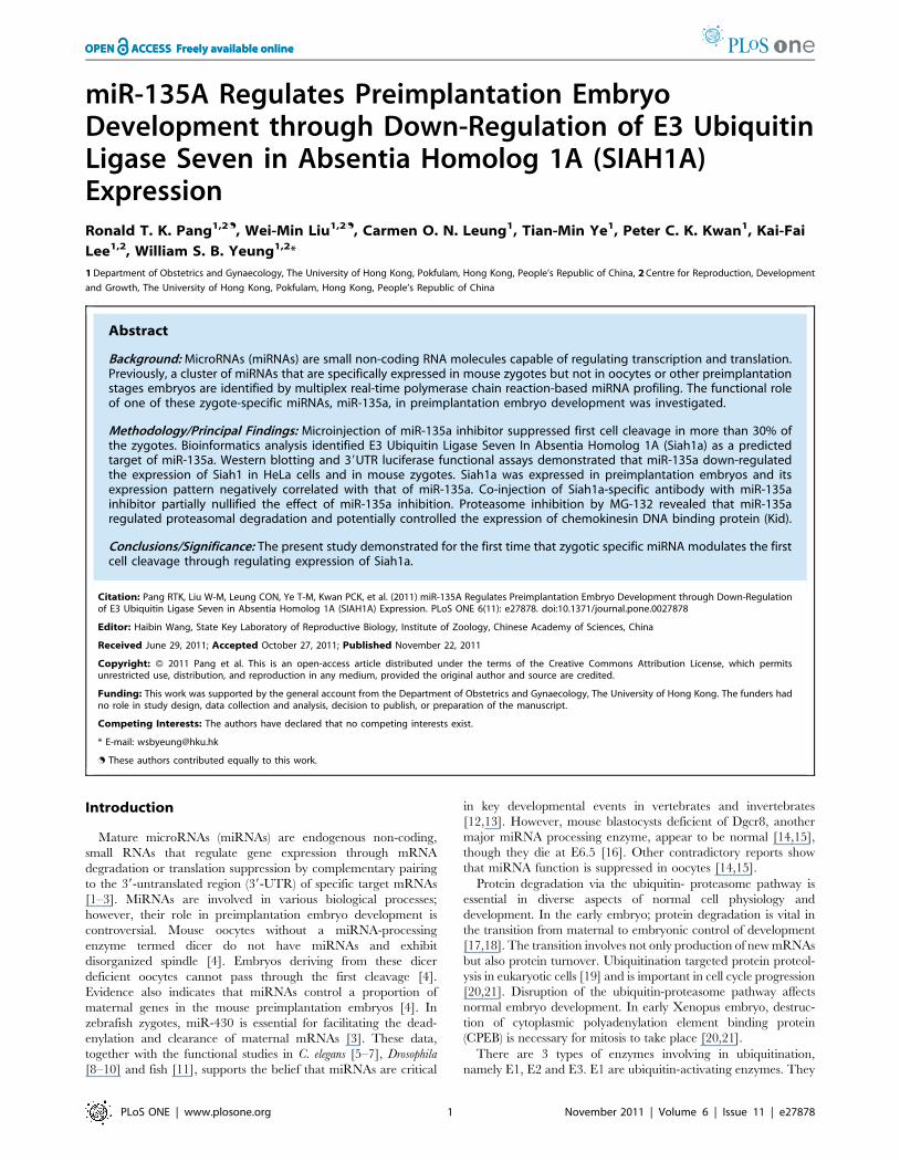
miR-135A Regulates Preimplantation EmbryoDevelopment through Down-Regulation of E3 UbiquitinLigase Seven in Absentia Homolog 1A (SIAH1A)ExpressionRonald T. K. Pang1,2., Wei-Min Liu1,2., Carmen O. N. Leung1, Tian-Min Ye1, Peter C. K. Kwan1, Kai-Fai
Lee1,2, William S. B. Yeung1,2*
1 Department of Obstetrics and Gynaecology, The University of Hong Kong, Pokfulam, Hong Kong, People’s Republic of China, 2 Centre for Reproduction, Development
and Growth, The University of Hong Kong, Pokfulam, Hong Kong, People’s Republic of China
Abstract
Background: MicroRNAs (miRNAs) are small non-coding RNA molecules capable of regulating transcription and translation.Previously, a cluster of miRNAs that are specifically expressed in mouse zygotes but not in oocytes or other preimplantationstages embryos are identified by multiplex real-time polymerase chain reaction-based miRNA profiling. The functional roleof one of these zygote-specific miRNAs, miR-135a, in preimplantation embryo development was investigated.
Methodology/Principal Findings: Microinjection of miR-135a inhibitor suppressed first cell cleavage in more than 30% ofthe zygotes. Bioinformatics analysis identified E3 Ubiquitin Ligase Seven In Absentia Homolog 1A (Siah1a) as a predictedtarget of miR-135a. Western blotting and 39UTR luciferase functional assays demonstrated that miR-135a down-regulatedthe expression of Siah1 in HeLa cells and in mouse zygotes. Siah1a was expressed in preimplantation embryos and itsexpression pattern negatively correlated with that of miR-135a. Co-injection of Siah1a-specific antibody with miR-135ainhibitor partially nullified the effect of miR-135a inhibition. Proteasome inhibition by MG-132 revealed that miR-135aregulated proteasomal degradation and potentially controlled the expression of chemokinesin DNA binding protein (Kid).
Conclusions/Significance: The present study demonstrated for the first time that zygotic specific miRNA modulates the firstcell cleavage through regulating expression of Siah1a.
Citation: Pang RTK, Liu W-M, Leung CON, Ye T-M, Kwan PCK, et al. (2011) miR-135A Regulates Preimplantation Embryo Development through Down-Regulationof E3 Ubiquitin Ligase Seven in Absentia Homolog 1A (SIAH1A) Expression. PLoS ONE 6(11): e27878. doi:10.1371/journal.pone.0027878
Editor: Haibin Wang, State Key Laboratory of Reproductive Biology, Institute of Zoology, Chinese Academy of Sciences, China
Received June 29, 2011; Accepted October 27, 2011; Published November 22, 2011
Copyright: � 2011 Pang et al. This is an open-access article distributed under the terms of the Creative Commons Attribution License, which permitsunrestricted use, distribution, and reproduction in any medium, provided the original author and source are credited.
Funding: This work was supported by the general account from the Department of Obstetrics and Gynaecology, The University of Hong Kong. The funders hadno role in study design, data collection and analysis, decision to publish, or preparation of the manuscript.
Competing Interests: The authors have declared that no competing interests exist.
* E-mail: [email protected]
. These authors contributed equally to this work.
Introduction
Mature microRNAs (miRNAs) are endogenous non-coding,
small RNAs that regulate gene expression through mRNA
degradation or translation suppression by complementary pairing
to the 39-untranslated region (39-UTR) of specific target mRNAs
[1–3]. MiRNAs are involved in various biological processes;
however, their role in preimplantation embryo development is
controversial. Mouse oocytes without a miRNA-processing
enzyme termed dicer do not have miRNAs and exhibit
disorganized spindle [4]. Embryos deriving from these dicer
deficient oocytes cannot pass through the first cleavage [4].
Evidence also indicates that miRNAs control a proportion of
maternal genes in the mouse preimplantation embryos [4]. In
zebrafish zygotes, miR-430 is essential for facilitating the dead-
enylation and clearance of maternal mRNAs [3]. These data,
together with the functional studies in C. elegans [5–7], Drosophila
[8–10] and fish [11], supports the belief that miRNAs are critical
in key developmental events in vertebrates and invertebrates
[12,13]. However, mouse blastocysts deficient of Dgcr8, another
major miRNA processing enzyme, appear to be normal [14,15],
though they die at E6.5 [16]. Other contradictory reports show
that miRNA function is suppressed in oocytes [14,15].
Protein degradation via the ubiquitin- proteasome pathway is
essential in diverse aspects of normal cell physiology and
development. In the early embryo; protein degradation is vital in
the transition from maternal to embryonic control of development
[17,18]. The transition involves not only production of new mRNAs
but also protein turnover. Ubiquitination targeted protein proteol-
ysis in eukaryotic cells [19] and is important in cell cycle progression
[20,21]. Disruption of the ubiquitin-proteasome pathway affects
normal embryo development. In early Xenopus embryo, destruc-
tion of cytoplasmic polyadenylation element binding protein
(CPEB) is necessary for mitosis to take place [20,21].
There are 3 types of enzymes involving in ubiquitination,
namely E1, E2 and E3. E1 are ubiquitin-activating enzymes. They
PLoS ONE | www.plosone.org 1 November 2011 | Volume 6 | Issue 11 | e27878
form a thiol-ester linkage with ubiquitin, which is then transferred
to the E2 ubiquitin-conjugating enzyme. The E3 enzymes are
protein ligases that transfer ubiquitin from the E2 enzyme to the
lysine residues of specific proteins, thus targeting them for
degradation by the proteasome. The E3 ubiquitin ligases control
the specificity of ubiquitination by interacting only with specific
target proteins. The E3 ubiquitin ligases contain a signature RING
(Really Interesting Novel Gene) finger consensus sequence.
Majority of the RING finger-containing proteins function as
ubiquitin protein ligase [22,23].
Seven in absentia homolog 1 (Siah1) is a member of the RING
finger proteins with E3 ligase activity. It is a highly related
mammalian homolog of the Drosophila SINA, that has been
implicated in the ubiquitination and proteasome-dependent
degradation of various molecules [24,25]. There are two Siah
genes in humans, Siah1 and Siah2 [25] while there are two Siah1
genes (Siah1a and Siah1b) and a single Siah2 gene in mice [24].
Siah1 expression is induced by p53 in mammals and over-
expression of Siah1 inhibits cell proliferation and promotes
apoptosis [26–28]. Siah1 is also important for early embryo
development; the birth frequency of Siah1 hemizygous (null) mice
is very low and many of their embryos exhibit developmental
abnormalities [29].
Our previous miRNA profiling of mouse spermatozoa, oocytes
and preimplantation embryos indicated that miR-135a was highly
expressed in zygotes, but decreased gradually with development ().
This study aimed to confirm this observation and to study the
function of miR-135a in the zygotes. Here, we provide evidence
that miR-135a regulates the first cell division mediated by
suppressing the expression of Siah1a, which in turns affect
destabilization of chemokinesin DNA binding protein (Kid), which
mediates chromosome compaction and is degraded by the
proteasome pathway during mitosis [30,31].
Results
Expression of miR-135a in preimplantation embryosOur previous miRNA profiling showed that miR-135a was
highly expressed in the zygotes and decreased thereafter
(Figure 1A). The observation was confirmed by independent
real-time qPCR. We found that miR-135a was expressed in all
preimplantation embryos but the level was significantly higher at
the zygote stage than at other stages of development (Figure 1B).
This stage-specific expression of miR-135a prompted us to
investigate its functional roles in zygotic development.
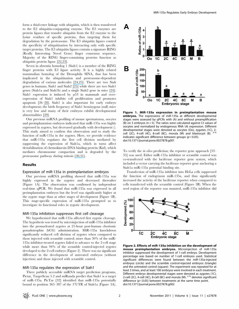
MiR-135a inhibition suppresses first cell cleavageWe hypothesized that miR-135a affected first zygotic cleavage.
The hypothesis was tested by microinjection of miR-135a inhibitor
into the pronucleated zygotes at 21-hour post-human chorionic
gonadotrophin (hCG) administration. MiR-135a knockdown
significantly reduced cell division of zygotes when compared to
those injected with scramble control; more than 30% of the miR-
135a inhibitor-treated zygotes failed to advance to the 2-cell stage
while more than 94% of the scramble control-injected zygotes
developed to the 2-cell embryo (Figure 2). There was no significant
difference in the development of untreated embryos (without
injection) and those injected with scramble control.
MiR-135a regulates the expression of Siah1Three publicly accessible miRNA target prediction programs,
Pictar, TargetScan 5.2 and miRanda predict that Siah1 is a target
of miR-135a. PicTar [32] identified that miR-135a potentially
bound to position 365–387 of the 39UTR of Siah1a (Figure 3A).
To verify the in-silico prediction; the reporter gene approach [33–
35] was used. Either miR-135a inhibitor or scramble control was
co-transfected with the luciferase reporter gene system, which
included a vector carrying the luciferase reporter gene anchoring a
Siah1a miR-135a potential binding site.
Transfection of miR-135a inhibitor into HeLa cells suppressed
the function of endogenous miR-135a, and thus significantly
increased the activity of the luciferase reporter when compared to
cells transfected with the scramble control (Figure 3B). When the
seed region of the reporter was mutated, miR-135a inhibitor did
Figure 1. MiR-135a expression in preimplantation mouseembryos. The expressions of miR-135a at different developmentalstages were assessed by qPCRs with (A) and without preamplification(B) on 5 embryos (n = 5). The ratios were calculated against Ct values ofoocytes and normalized by endogenous RNA U6 expression. Differentdevelopmental stages were denoted as oocytes (Oo), zygotes (1C), 2-cell (2C), 4-cell (4C), 8-cell (8C), morula (M) and blastocyst (B). a–b
indicates significant difference between groups (p,0.05).doi:10.1371/journal.pone.0027878.g001
Figure 2. Effects of miR-135a inhibition on the development ofmouse preimplantation embryos. Microinjection of miR-135ainhibitor suppressed the development of 1-cell embryo. Developmentpercentage was based on number of 1-cell embryos used. Statisticalsignificant differences were found between the miR-135a-injectedembryos (circle) and the scramble control-injected embryos (triangle)and the untreated control (square). The experiment was repeated for atleast 3 times, and at least 100 embryos were involved in each treatment.Different embryo developmental stages were denoted as zygotes (1C),2-cell (2C), 4-cell (4C), 8-cell (8C) and morula (M). a–b denotes significantdifference (p,0.05) between treatments at the same time point.doi:10.1371/journal.pone.0027878.g002
MiR-135a Regulates Early Embryo Development
PLoS ONE | www.plosone.org 2 November 2011 | Volume 6 | Issue 11 | e27878

not affect the luciferase activity (Figure 3B). The expression of
Siah1a in miR-135a inhibitor-treated cells and zygotes was also
examined. It was found that the Siah1a protein expression was
increased in both groups (Figure 3C and D), confirming that miR-
135a regulated the expression of Siah1a. The action of miR-135a
on Siah1a expression was not by inducing mRNA target
degradation as miR-135a inhibitor had no effect on Siah1a
mRNA expression in the 2 groups (Figure 3D).
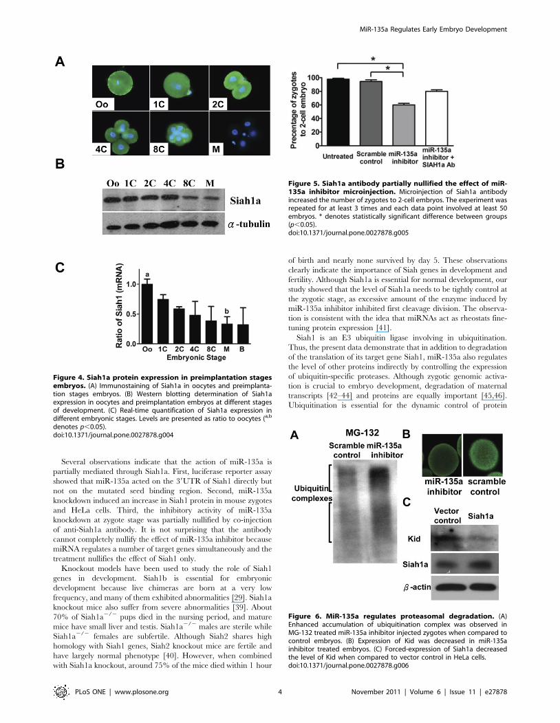
Localization and expression of Siah1a in oocytes andpreimplantation embryos
The protein expression of Siah1a during preimplantation
embryogenesis was studied by western blotting and immunohis-
tochemical staining. Siah1a immunoreactivities were observed in
the cytoplasm of oocytes, zygotes, blastomeres of 2-, 4- and 8-cell
embryos (Fig. 4A). Western blot showed that the protein
expression of Siah1a decreased continuously with development
and noticeable decrease in the levels of Siah1a protein was
observed after the 4-cell stage (Figure 4B). Siah1a mRNA also
showed a similar trend; the mRNA level was highest in oocytes
and decreased upon cleavage (Figure 4C). It is interesting to note
that the increased expression of miR-135a in the zygotes correlates
with a gradual decrease of Siah1a from this stage (Figure 1 and 4).
Siah1a mediates the effect of miR-135a on embryodevelopment in mice
We next tested whether Siah1a mediated the inhibitory effect of
miR-135a on embryo development. Siah1a specific antibody or
water control was co-injected with miR-135a inhibitor or scramble
control into pronucleated zygotes. Only about 60% of zygotes
injected with miR-135a inhibitor developed to the 2-cell stage.
Coinjection with Siah1a antibody partially nullified the effect of
miR-135a inhibitor and about 80% of these embryos developed to
the 2-cell stage (Figure 5).
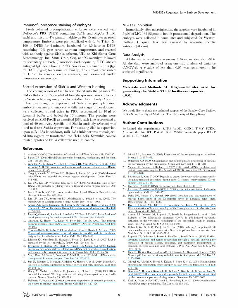
MiR-135a affects proteasomal degradation andexpression of Kid
Since Siah1a is involved in proteasomal degradation, we
examined the action of miR-135a in ubiquitation by using
proteasomal inhibitor MG-132. It was found that MG-132
increased the accumulation of ubiquitination complex (Figure 6A).
One of the potential Siah1 degradation target is Kid [31].
Therefore, we examined the effect of miR-135a inhibitor on Kid
expression. Immunostaining showed that the Kid level was
decreased in miR-135a inhibitor treated embryos (Figure 6B). To
demonstrate the relationship between Siah1a and Kid expression,
we constructed an expression vector of Siah1a. Forced-expression of
Siah1a in HeLa cells results in reduced Kid expression (Figure 6C).
Discussion
Embryonic development is a tightly regulated process. MiRNAs
are important regulators fine tuning the expression of many genes
simultaneously. They are important in early embryo development
of C. elegans [5–7], Drosophila [8–10], fish [11] and mammals [36].
Mouse preimplantation embryos expressed developmental stage-
specific miRNAs [4,37,38] and miR-135a is specifically expressed
in the zygotic stage. Here, we demonstrated that miR-135a is
important for the first cell cleavage; microinjection of miR-135a
inhibitor reduced the percentage of zygotes undergone first cell
division by about 30%.
Figure 3. MiR-135a regulates the expression of Siah1. (A) Potential miR-135a binding site on 39UTR of Siah1a, seed binding region werelabeled in bold (position 380–386). (B) Luciferase activity was increased upon transfection of miR-135a inhibitor with Siah1a 39UTR when compared tothe scramble control group. MiR-135a inhibitor did not affect the reporter activity when the Siah1a 39UTR carried a mutated seed binding region. (C)MiR-135a inhibitor treatment increased the expression of Siah1 in HeLa cells and zygotes. (D) Graphical presentation of means6SD of Siah1 mRNAand protein expressions derived from at least 3 independent experiments. Levels are presented as ratio to the scramble controls. * denotesstatistically significant difference between groups (p,0.05).doi:10.1371/journal.pone.0027878.g003
MiR-135a Regulates Early Embryo Development
PLoS ONE | www.plosone.org 3 November 2011 | Volume 6 | Issue 11 | e27878
Several observations indicate that the action of miR-135a is
partially mediated through Siah1a. First, luciferase reporter assay
showed that miR-135a acted on the 39UTR of Siah1 directly but
not on the mutated seed binding region. Second, miR-135a
knockdown induced an increase in Siah1 protein in mouse zygotes
and HeLa cells. Third, the inhibitory activity of miR-135a
knockdown at zygote stage was partially nullified by co-injection
of anti-Siah1a antibody. It is not surprising that the antibody
cannot completely nullify the effect of miR-135a inhibitor because
miRNA regulates a number of target genes simultaneously and the
treatment nullifies the effect of Siah1 only.
Knockout models have been used to study the role of Siah1
genes in development. Siah1b is essential for embryonic
development because live chimeras are born at a very low
frequency, and many of them exhibited abnormalities [29]. Siah1a
knockout mice also suffer from severe abnormalities [39]. About
70% of Siah1a2/2 pups died in the nursing period, and mature
mice have small liver and testis. Siah1a2/2 males are sterile while
Siah1a2/2 females are subfertile. Although Siah2 shares high
homology with Siah1 genes, Siah2 knockout mice are fertile and
have largely normal phenotype [40]. However, when combined
with Siah1a knockout, around 75% of the mice died within 1 hour
of birth and nearly none survived by day 5. These observations
clearly indicate the importance of Siah genes in development and
fertility. Although Siah1a is essential for normal development, our
study showed that the level of Siah1a needs to be tightly control at
the zygotic stage, as excessive amount of the enzyme induced by
miR-135a inhibitor inhibited first cleavage division. The observa-
tion is consistent with the idea that miRNAs act as rheostats fine-
tuning protein expression [41].
Siah1 is an E3 ubiquitin ligase involving in ubiquitination.
Thus, the present data demonstrate that in addition to degradation
of the translation of its target gene Siah1, miR-135a also regulates
the level of other proteins indirectly by controlling the expression
of ubiquitin-specific proteases. Although zygotic genomic activa-
tion is crucial to embryo development, degradation of maternal
transcripts [42–44] and proteins are equally important [45,46].
Ubiquitination is essential for the dynamic control of protein
Figure 4. Siah1a protein expression in preimplantation stagesembryos. (A) Immunostaining of Siah1a in oocytes and preimplanta-tion stages embryos. (B) Western blotting determination of Siah1aexpression in oocytes and preimplantation embryos at different stagesof development. (C) Real-time quantification of Siah1a expression indifferent embryonic stages. Levels are presented as ratio to oocytes (a,b
denotes p,0.05).doi:10.1371/journal.pone.0027878.g004
Figure 5. Siah1a antibody partially nullified the effect of miR-135a inhibitor microinjection. Microinjection of Siah1a antibodyincreased the number of zygotes to 2-cell embryos. The experiment wasrepeated for at least 3 times and each data point involved at least 50embryos. * denotes statistically significant difference between groups(p,0.05).doi:10.1371/journal.pone.0027878.g005
Figure 6. MiR-135a regulates proteasomal degradation. (A)Enhanced accumulation of ubiquitination complex was observed inMG-132 treated miR-135a inhibitor injected zygotes when compared tocontrol embryos. (B) Expression of Kid was decreased in miR-135ainhibitor treated embryos. (C) Forced-expression of Siah1a decreasedthe level of Kid when compared to vector control in HeLa cells.doi:10.1371/journal.pone.0027878.g006
MiR-135a Regulates Early Embryo Development
PLoS ONE | www.plosone.org 4 November 2011 | Volume 6 | Issue 11 | e27878

turnover in embryo development. It has been shown that the E3
ubiquitin ligase, Ret Finger-Like 4 gene (RFLP4) is involved in the
ubiquitin proteasome degradation pathway and interacts with
target proteins such as cyclin B1 for degradation during oocyte-
embryo transition [45]. Another E3 ubiquitin ligase known to be
important for embryonic development is G2E3, which acts to
prevent apoptosis of early embryos, and its deficiency leads to
death of embryos prior to implantation [47]. The importance of
ubiquitination is also suggested by the differential expression of
ubiquitin proteasome genes in embryos obtained after different
hormonal stimulation protocols [48].
In this study, the expression of Siah1a was high in oocytes, and
decreased in the zygotic stage when the level of miR-135a was
high. The expression pattern is similar to that of RFLP4, which is
highly expressed in the mouse oocytes but is much reduced in the
2-cell embryos [45]. The similarity in the expression and function
of these E3 ubiquitin ligases tempt us to speculate that down-
regulation of these enzymes is important in the first cleavage
division. The validity of this hypothesis remains to be tested. The
expression levels of Siah1a did not increase after the 4-cell stage
when the miR-135a level was low. Thus, other unknown miRNAs
or mechanism(s) are inhibiting the transcription and/or translation
of the Siah1a gene.
The inhibitory action of miR-135a knockdown on the first cell
cleavage could result from perturbation of the proteolysis of
Siah1a substrates. Indeed, immunoprecipitation and yeast two-
hybrid screening have demonstrated that Siah1 interacts with a
number of proteins that are involved in cell cycle arrest or
apoptosis, including POSH [49], HIPK2 [50], Numb [51], Pw1/
Peg3 [27], transforming growth factor b [52] and BAG-1 [53].
Whether these proteins are interacting with Siah1 in early oocytes-
embryo transition needs further investigation.
Kid is one of the Siah1 effector [31]. It is a microtubule-
associated motor protein essential for chromosome compaction to
ensure proper nuclear envelope formation by reducing anaphase
chromosome mass [30]. Mouse deficient of Kid have fragmented
pronuclei in zygote and multi-nuclei in blastomere of early
embryos; and half of them die prior to E9.5 [30]. Here, we
observed that the Kid level was decreased upon miR-135a
inhibition; consistent with the reduction in Siah1a expression
mediated by zygotic increase of miR-135a enhances the level of
Kid in zygotes for normal first cleavage division.
Besides protein ubiquitination, evidence also suggests that Siah1
inhibits cell proliferation and promotes apoptosis [26–28].
Ovulated oocytes that are not fertilized dies by apoptosis to
prevent the development of abnormal conceptus [54]. The
expression of caspase 3 is higher in aged mouse oocytes than in
young oocytes [55]. On the other hand, p53 induces Siah1
mediate cell growth arrest though inhibiting DNA synthesis
without inducing apoptosis in epithelial and fibroblast cell models
[53]. The importance of miR-135a in preventing death of oocyte/
zygote remains to be determined.
There are reports showing that the miRNA function is
suppressed in mouse oocytes [14,15]. Our unpublished data show
that miRNAs regained their activity after fertilization. Mouse
embryos deficient of Dgcr8 produced by heterozygous mating die
by E6.5 [16]. Although Dgcr8 null females produced offsprings
with wild-type males, their brood size was reduced by about 40%,
leading to the conclusion that maternal Dgcr8 affects female
fecundity though it is not required for fertility [15]. These reports
did not study the role of sperm miRNAs in embryo development.
Our unpublished data show that the miRNAs of spermatozoal
origin are delivered to the fertilized zygotes after fertilization and
affect first cleavage division. Mature miR-135a can be detected in
mouse epididymal spermatozoa at a level that is 2.2-fold higher
than that of oocytes.
In summary, we have demonstrated that miR-135a is involved
in the first cell cleavage in zygotes. The action of miR-135a is, at
least partially through down-regulation of Siah1a.
Materials and Methods
Mouse embryo collectionThe protocol of this study was approved by the Committee on
Use of Live Animals in Teaching and Research (approval number:
1531-07), the University of Hong Kong. ICR female mice aged 6–
8 weeks were superovulated by consecutive injections of 5 IU of
pregnant mare serum gonadotropin (Sigma, St. Louis, MO, USA)
and 5 IU of human chorionic gonadotropin (hCG, Sigma) 47–
48 hours apart. Unfertilized eggs were harvested from females at
21-hour post-hCG injection without mating. Fertilized zygotes (1-
cell embryos) were harvested from the mated mice at 21-hour
post-hCG. Embryos at the 2-cell, 4-cell, 8-cell, and morulae stages
were flushed out from the oviducts at 40–42, 54–56, 62–64, and
76–78 hour behind hCG injection, respectively. Blastocysts were
retrieved from the uterine horns at 86–88 hour post-hCG
injection.
RNA extraction and TaqMan real-time PCRFor miRNA expression analysis, 5 embryos at the same
developmental stage were pooled for RNA extraction. Reverse
transcription was performed by TaqMan H MicroRNA Reverse
Transcription Kit (Applied Biosystems, Carlsbad, CA). MicroRNA
expression was determined with TaqManH MicroRNA assay
(Applied Biosystems) according to the manufacturer’s protocol by
using the Applied Biosystems 7500 Detection system (Applied
Biosystems).
Microinjection and in vitro embryo cultureAbout 10 pL of 25 mM locked nucleic acid modified miR-135a
inhibitor (miRCURY LNATM microRNA inhibitors, Exiqon,
Vedbaek, Denmark) was microinjected into the cytoplasm of the
zygotes. Scramble control (miRCURY LNATM microRNA
antisense controls, LNA probe of similar length without homology
to any known miRNA or mRNA sequence in human, mouse or
rat) injected embryos were used as control for assessing injection
damage. After microinjection, groups of 20–30 embryos were
cultured in 40 ml of KSOM medium supplemented with amino
acids (Chemicon, Billerica, MA) and overlaid with mineral oil at
37uC in an atmosphere of 5% CO2 for 4 days. Embryo
development was observed under an inverted microscope.
MicroRNA-135a inhibition and luciferase reporter assayOligonucleotides were synthesized according to the sequence of
the potential binding regions identified by PicTar [32] (http://
pictar.bio.nyu.edu/). Digestion sites for NotI and XhoI were
added to the 59 and 39 end of each site in the 39UTR of the
predicted target. The sequences of the original and mutated
oligonucleotides were listed in Materials and Methods S1. The
double digested fragments were cloned downstream of the
luciferase gene between the XhoI/NotI site of psiCHECKTM-2
vector (Promega, Madison, WI). MiR-135a inhibitor (human miR-
135a is identical to mouse miR-135a and the same inhibitor can
be used), or scramble control were transfected together with
reporter constructs into HeLa cells (American Type Culture
Collection, Manassas, VA). Transfection and assay procedures
were described as in [56].
MiR-135a Regulates Early Embryo Development
PLoS ONE | www.plosone.org 5 November 2011 | Volume 6 | Issue 11 | e27878
Immunofluorescence staining of embryosFresh collected pre-implantation embryos were washed with
Dulbecco’s PBS (DPBS) containing CaCl2 and MgCl2 (1 mM
each) and fixed in 4% paraformaldehyde for 15 minutes at room
temperature. Embryos were permeabilized with 0.1% Triton X-
100 in DPBS for 4 minutes, incubated for 1.5 hour in DPBS
containing 10% goat serum at room temperature, and reacted
with antibody against Siah1a (Abcam, UK) or Kid (Santa Cruz
Biotechnology, Inc, Santa Cruz, CA), at 4uC overnight followed
by secondary antibody (fluorescein isothiocyanate, FITC-labeled
anti-goat IgG) for 1 hour at 37uC. Nuclei were stained with 5 mg/
ml DAPI (Sigma) for 5 minutes. Finally, the embryos were rinsed
in DPBS to remove excess reagents, and examined under
fluorescence microscope.
Forced-expression of Siah1a and Western blottingThe coding region of Siah1a was cloned into the pTracerTM-
CMV/Bsd vector. Successful of forced-expression was confirmed
by Western blotting using specific anti-Siah1a antibody (Abcam).
For examining the expression of Siah1a in preimplantation
embryos, oocytes and embryos at different stages of development
were collected, rinsed twice in PBS, resuspended in 10 ml of
Laemmli buffer and boiled for 10 minutes. The proteins were
resolved on SDS-PAGE as described [56], each lane represented a
pool of 40 embryos. Specific anti-Siah1a antibody (Abcam) was
used to detect Siah1a expression. For assessing Siah1a expression
upon miR-135a knockdown, miR-135a inhibitor was microinject-
ed into zygotes or transfected into HeLa cells. Scramble control
treated zygotes or HeLa cells were used as control.
MG-132 inhibitionImmediately after microinjection, the zygotes were incubated in
5 mM of MG-132 (Sigma) to inhibit proteasomal degradation. The
embryos were collected 6 hours later and subjected for Western
blotting. Ubiquitin level was assessed by ubiquitin specific
antibody (Abcam).
Data AnalysisAll the results are shown as means 6 Standard deviation (SD).
All the data were analyzed using one-way analysis of variance
(ANOVA). A p-value of less than 0.05 was considered to be
statistical significance.
Supporting Information
Materials and Methods S1 Oligonucleotides used forgenerating the Siah1a 39UTR luciferase reporter.
(DOC)
Acknowledgments
We would like to thank the technical support of the Faculty Core Facility,
Li Ka Shing Faculty of Medicine, The University of Hong Kong.
Author Contributions
Performed the experiments: RTKP W-ML CONL T-MY PCKK.
Analyzed the data: RTKP W-ML K-FL WSBY. Wrote the paper: RTKP
W-ML K-FL WSBY.
References
1. Ambros V (2004) The functions of animal microRNAs. Nature 431: 350–355.
2. Bartel DP (2004) MicroRNAs: genomics, biogenesis, mechanism, and function.
Cell 116: 281–297.
3. Giraldez AJ, Mishima Y, Rihel J, Grocock RJ, Van Dongen S, et al. (2006)Zebrafish MiR-430 promotes deadenylation and clearance of maternal mRNAs.
Science 312: 75–79.
4. Tang F, Kaneda M, O’Carroll D, Hajkova P, Barton SC, et al. (2007) MaternalmicroRNAs are essential for mouse zygotic development. Genes Dev 21:
644–648.
5. Lau NC, Lim LP, Weinstein EG, Bartel DP (2001) An abundant class of tiny
RNAs with probable regulatory roles in Caenorhabditis elegans. Science 294:858–862.
6. Lee RC, Ambros V (2001) An extensive class of small RNAs in Caenorhabditis
elegans. Science 294: 862–864.
7. Lim LP, Lau NC, Weinstein EG, Abdelhakim A, Yekta S, et al. (2003) ThemicroRNAs of Caenorhabditis elegans. Genes Dev 17: 991–1008.
8. Aravin AA, Lagos-Quintana M, Yalcin A, Zavolan M, Marks D, et al. (2003)
The small RNA profile during Drosophila melanogaster development. Dev Cell5: 337–350.
9. Lagos-Quintana M, Rauhut R, Lendeckel W, Tuschl T (2001) Identification of
novel genes coding for small expressed RNAs. Science 294: 853–858.
10. Okamura K, Hagen JW, Duan H, Tyler DM, Lai EC (2007) The mirtronpathway generates microRNA-class regulatory RNAs in Drosophila. Cell 130:
89–100.
11. Tessmar-Raible K, Raible F, Christodoulou F, Guy K, Rembold M, et al. (2007)Conserved sensory-neurosecretory cell types in annelid and fish forebrain:
insights into hypothalamus evolution. Cell 129: 1389–1400.
12. Johnson SM, Grosshans H, Shingara J, Byrom M, Jarvis R, et al. (2005) RAS isregulated by the let-7 microRNA family. Cell 120: 635–647.
13. Brennecke J, Hipfner DR, Stark A, Russell RB, Cohen SM (2003) bantam
encodes a developmentally regulated microRNA that controls cell proliferationand regulates the proapoptotic gene hid in Drosophila. Cell 113: 25–36.
14. Ma J, Flemr M, Stein P, Berninger P, Malik R, et al. (2010) MicroRNA activity
is suppressed in mouse oocytes. Curr Biol 20: 265–270.
15. Suh N, Baehner L, Moltzahn F, Melton C, Shenoy A, et al. (2010) MicroRNAfunction is globally suppressed in mouse oocytes and early embryos. Curr Biol
20: 271–277.
16. Wang Y, Medvid R, Melton C, Jaenisch R, Blelloch R (2007) DGCR8 isessential for microRNA biogenesis and silencing of embryonic stem cell self-
renewal. Nature Genetics 39: 380–385.
17. DeRenzo C, Seydoux G (2004) A clean start: degradation of maternal proteins atthe oocyte-to-embryo transition. Trends Cell Biol 14: 420–426.
18. Stitzel ML, Seydoux G (2007) Regulation of the oocyte-to-zygote transition.Science 316: 407–408.
19. Wilkinson KD (2000) Ubiquitination and deubiquitination: targeting of proteins
for degradation by the proteasome. Semin Cell Dev Biol 11: 141–148.
20. Mendez R, Barnard D, Richter JD (2002) Differential mRNA translation and
meiotic progression require Cdc2-mediated CPEB destruction. EMBO Journal21: 1833–1844.
21. Bowerman B, Kurz T (2006) Degrade to create: developmental requirements for
ubiquitin-mediated proteolysis during early C. elegans embryogenesis. Devel-opment 133: 773–784.
22. Freemont PS (2000) RING for destruction? Curr Biol 10: R84–87.
23. Joazeiro CA, Weissman AM (2000) RING finger proteins: mediators of ubiquitin
ligase activity. Cell 102: 549–552.
24. Della NG, Senior PV, Bowtell DD (1993) Isolation and characterisation ofmurine homologues of the Drosophila seven in absentia gene (sina).
Development 117: 1333–1343.
25. Hu G, Chung YL, Glover T, Valentine V, Look AT, et al. (1997)
Characterization of human homologs of the Drosophila seven in absentia (sina)
gene. Genomics 46: 103–111.
26. Amson RB, Nemani M, Roperch JP, Israeli D, Bougueleret L, et al. (1996)
Isolation of 10 differentially expressed cDNAs in p53-induced apoptosis:activation of the vertebrate homologue of the drosophila seven in absentia
gene. Proc Natl Acad Sci U S A 93: 3953–3957.
27. Relaix F, Wei X, Li W, Pan J, Lin Y, et al. (2000) Pw1/Peg3 is a potential celldeath mediator and cooperates with Siah1a in p53-mediated apoptosis. Proc
Natl Acad Sci U S A 97: 2105–2110.
28. Roperch JP, Lethrone F, Prieur S, Piouffre L, Israeli D, et al. (1999) SIAH-1
promotes apoptosis and tumor suppression through a network involving the
regulation of protein folding, unfolding, and trafficking: identification ofcommon effectors with p53 and p21(Waf1). Proc Natl Acad Sci U S A 96:
8070–8073.
29. Frew IJ, Dickins RA, Cuddihy AR, Del Rosario M, Reinhard C, et al. (2002)
Normal p53 function in primary cells deficient for Siah genes. Mol Cell Biol 22:
8155–8164.
30. Ohsugi M, Adachi K, Horai R, Kakuta S, Sudo K, et al. (2008) Kid-mediated
chromosome compaction ensures proper nuclear envelope formation. Cell 132:771–782.
31. Germani A, Bruzzoni-Giovanelli H, Fellous A, Gisselbrecht S, Varin-Blank N,
et al. (2000) SIAH-1 interacts with alpha-tubulin and degrades the kinesin Kidby the proteasome pathway during mitosis. Oncogene 19: 5997–6006.
32. Krek A, Grun D, Poy MN, Wolf R, Rosenberg L, et al. (2005) CombinatorialmicroRNA target predictions. Nat Genet 37: 495–500.
MiR-135a Regulates Early Embryo Development
PLoS ONE | www.plosone.org 6 November 2011 | Volume 6 | Issue 11 | e27878
33. Kiriakidou M, Nelson PT, Kouranov A, Fitziev P, Bouyioukos C, et al. (2004) A
combined computational-experimental approach predicts human microRNAtargets. Genes Dev 18: 1165–1178.
34. Koscianska E, Baev V, Skreka K, Oikonomaki K, Rusinov V, et al. (2007)
Prediction and preliminary validation of oncogene regulation by miRNAs. BMCMol Biol 8: 79.
35. Lewis BP, Shih IH, Jones-Rhoades MW, Bartel DP, Burge CB (2003) Predictionof mammalian microRNA targets. Cell 115: 787–798.
36. Landgraf P, Rusu M, Sheridan R, Sewer A, Iovino N, et al. (2007) A
mammalian microRNA expression atlas based on small RNA librarysequencing. Cell 129: 1401–1414.
37. Yang Y, Bai W, Zhang L, Yin G, Wang X, et al. (2008) Determination ofmicroRNAs in mouse preimplantation embryos by microarray. Developmental
Dynamics 237: 2315–2327.38. Viswanathan SR, Mermel CH, Lu J, Lu CW, Golub TR, et al. (2009)
microRNA expression during trophectoderm specification. PLoS ONE [Elec-
tronic Resource] 4: e6143.39. Dickins RA, Frew IJ, House CM, O’Bryan MK, Holloway AJ, et al. (2002) The
ubiquitin ligase component Siah1a is required for completion of meiosis I inmale mice. Mol Cell Biol 22: 2294–2303.
40. Frew IJ, Hammond VE, Dickins RA, Quinn JM, Walkley CR, et al. (2003)
Generation and analysis of Siah2 mutant mice. Mol Cell Biol 23: 9150–9161.41. Baek D, Villen J, Shin C, Camargo FD, Gygi SP, et al. (2008) The impact of
microRNAs on protein output. Nature 455: 64–71.42. Evsikov AV, Graber JH, Brockman JM, Hampl A, Holbrook AE, et al. (2006)
Cracking the egg: molecular dynamics and evolutionary aspects of the transitionfrom the fully grown oocyte to embryo. Genes & Development 20: 2713–2727.
43. Tadros W, Lipshitz HD (2005) Setting the stage for development: mRNA
translation and stability during oocyte maturation and egg activation inDrosophila. Developmental Dynamics 232: 593–608.
44. Hamatani T, Carter MG, Sharov AA, Ko MS (2004) Dynamics of global geneexpression changes during mouse preimplantation development. Developmental
Cell 6: 117–131.
45. Suzumori N, Burns KH, Yan W, Matzuk MM (2003) RFPL4 interacts withoocyte proteins of the ubiquitin-proteasome degradation pathway. Proc Natl
Acad Sci U S A 100: 550–555.
46. Wu X, Viveiros MM, Eppig JJ, Bai Y, Fitzpatrick SL, et al. (2003) Zygote arrest
1 (Zar1) is a novel maternal-effect gene critical for the oocyte-to-embryo
transition. Nature Genetics 33: 187–191.
47. Brooks WS, Helton ES, Banerjee S, Venable M, Johnson L, et al. (2008) G2E3 is
a dual function ubiquitin ligase required for early embryonic development.
Journal of Biological Chemistry 283: 22304–22315.
48. Mtango NR, Latham KE (2007) Ubiquitin proteasome pathway gene expression
varies in rhesus monkey oocytes and embryos of different developmental
potential. Physiol Genomics 31: 1–14.
49. Xu Z, Sproul A, Wang W, Kukekov N, Greene LA (2006) Siah1 interacts with
the scaffold protein POSH to promote JNK activation and apoptosis. Journal of
Biological Chemistry 281: 303–312.
50. Winter M, Sombroek D, Dauth I, Moehlenbrink J, Scheuermann K, et al. (2008)
Control of HIPK2 stability by ubiquitin ligase Siah-1 and checkpoint kinases
ATM and ATR. Nat Cell Biol 10: 812–824.
51. Susini L, Passer BJ, Amzallag-Elbaz N, Juven-Gershon T, Prieur S, et al. (2001)
Siah-1 binds and regulates the function of Numb. Proc Natl Acad Sci U S A 98:
15067–15072.
52. Johnsen SA, Subramaniam M, Monroe DG, Janknecht R, Spelsberg TC (2002)
Modulation of transforming growth factor beta (TGFbeta)/Smad transcriptional
responses through targeted degradation of TGFbeta-inducible early gene-1 by
human seven in absentia homologue. Journal of Biological Chemistry 277:
30754–30759.
53. Matsuzawa S, Takayama S, Froesch BA, Zapata JM, Reed JC (1998) p53-
inducible human homologue of Drosophila seven in absentia (Siah) inhibits cell
growth: suppression by BAG-1. EMBO Journal 17: 2736–2747.
54. Hardy K (1997) Cell death in the mammalian blastocyst. Mol Hum Reprod 3:
919–925.
55. Papandile A, Tyas D, O’Malley DM, Warner CM (2004) Analysis of caspase-3,
caspase-8 and caspase-9 enzymatic activities in mouse oocytes and zygotes.
Zygote 12: 57–64.
56. Pang RT, Leung CO, Ye TM, Liu W, Chiu PC, et al. (2010) MicroRNA-34a
suppresses invasion through downregulation of Notch1 and Jagged1 in cervical
carcinoma and choriocarcinoma cells. Carcinogenesis 31: 1037–1044.
MiR-135a Regulates Early Embryo Development
PLoS ONE | www.plosone.org 7 November 2011 | Volume 6 | Issue 11 | e27878
Copyright © 2022 FDOKUMEN