Effects of bivalent Co ion on the co-deposition of nickel and nano-diamond particles
Upload
independentCategory
view
1download
0
doi:10.1016/j.jmb.2009.12.049 J. Mol. Biol. (2010) 396, 1117–1127
Available online at www.sciencedirect.com
The Dimeric Structure and the Bivalent Recognition ofH3K4me3 by the Tumor Suppressor ING4 Suggests aMechanism for Enhanced Targeting of the HBO1Complex to Chromatin
Alicia Palacios1, Alberto Moreno2, Bruno L. Oliveira3, Teresa Rivera3,Jesús Prieto3, Pascal García3, M. Rosario Fernández-Fernández4,Pau Bernadó5, Ignacio Palmero2 and Francisco J. Blanco1,6⁎
1CIC bioGUNE, StructuralBiology Unit, ParqueTecnológico de Bizkaia, 48160Derio, Spain2Instituto de InvestigacionesBiomédicas “Alberto Sols”,CSIC-UAM, Arturo Duperier4, 28029 Madrid, Spain3Structural Biology andBiocomputing Programme,Centro Nacional deInvestigaciones Oncológicas(CNIO), Melchor FernándezAlmagro 3, 28029 Madrid,Spain4Center for Protein Engineering,MRC, Cambridge, UK5Institute for Research inBiomedicine, Baldiri Reixac10-12, 08028 Barcelona, Spain6IKERBASQUE, BasqueFoundation for Science, 48011Bilbao, SpainReceived 1 October 2009;received in revised form23 December 2009;accepted 23 December 2009Available online4 January 2010
*Corresponding author. E-mail addrPresent addresses: B. L. Oliveira, I
Garcia, Oncura, Orense 85, 28020 MOchoa, CSIC-UAM, Nicolás CabreraAbbreviations used: ING, inhibito
ray scattering; NLS, nuclear localiza
0022-2836/$ - see front matter © 2009 E
The INhibitor of Growth (ING) family of tumor suppressors regulates thetranscriptional state of chromatin by recruiting remodeling complexes tosites with histone H3 trimethylated at position K4 (H3K4me3). Thismodification is recognized by the plant homeodomain (PHD) present atthe C-terminus in the five members of the ING family. ING4 facilitateshistone H3 acetylation by the HBO1 complex. Here, we show that ING4forms homodimers through its N-terminal domain, which folds indepen-dently into an elongated coiled-coil structure. The central region of ING4,which contains the nuclear localization sequence, is disordered and flexibleand does not directly interact with p53, or does it with very low affinity, incontrast to previous findings. The NMR analysis of the full-length proteinreveals that the two PHD fingers of the dimer are chemically equivalent andindependent of the rest of the molecule. The detailed NMR analysis of thefull-length dimeric protein binding to histone H3K4me3 shows essentiallythe same binding site and affinity as the isolated PHD finger. Therefore, theING4 dimer has two identical and independent binding sites for H3K4me3tails, which, in the context of the chromatin, could belong to the same or todifferent nucleosomes. These results show that ING4 is a bivalent reader ofthe chromatin H3K4me3 modification and suggest a mechanism forenhanced targeting of the HBO1 complex to specific chromatin sites. Thismechanism could be common to other ING-containing remodelingcomplexes.
© 2009 Elsevier Ltd. All rights reserved.
Edited by P. Wright
Keywords: ING4 structure; histone tail; chromatin; NMR; SAXSess: [email protected] Tecnológico e Nuclear, Estrada Nacional 10, 2686-953 Sacavém, Portugal; P.adrid, Spain; M. R. Fernandez-Fernandez, Centro de Biología Molecular Severo1, 28049 Cantoblanco, Spain.r of growth; HSQC, heteronuclear single quantum coherence; SAXS, small-angle X-tion signal; PHD, plant homeodomain; GTA, glutaraldehyde ; OD, optical density.
lsevier Ltd. All rights reserved.

1118 ING4 as a Bivalent Reader of H3K4me3 Modification
Introduction
The nuclear DNA of eukaryotic cells is packedwith proteins into chromatin, the nucleosome beingits fundamental unit. Two superhelical turns ofDNA wind around an octamer of the four corehistones. The flexible N-terminal tails of histonesprovide internucleosomal linkages and, togetherwith other proteins, allow for higher levels ofcompaction.1 The structure of chromatin is highlydynamic and has a strong impact in DNAreplication, repair, and transcription.2,3 Its regula-tion is largely based on posttranslational covalentmodifications of certain residues at the N-terminalhistone tails, which directly affect the level ofchromatin condensation and/or recruit specificchromatin remodeling complexes to their location.The combinatorial nature of these histone modifica-tions, as well as their correlation with manychromatin-template processes, has led to the pro-posal of the existence of a “histone code” as thebasis for a fundamental regulatory mechanism thatextends the information potential of the geneticcode.4 Histone tails are modified by specificenzymatic complexes of proteins, some of themwith domains that recognize one or more of thepossible modifications.5
The INhibitor of Growth (ING) family of tumorsuppressors consists of five homologous proteins6
that regulate the transcriptional state of chromatinby recruiting remodeling complexes to sites withhistone H3 trimethylated at position K4 (H3K4me3).This histone post translation modification is recog-nized by their C-terminal plant homeodomains(PHDs).7 ING1 and ING2 form part of histonedeacetylase complexes, while ING3, ING4, andING5 do so with histone acetyl transferasecomplexes.8 The amino acid sequences of the INGproteins indicate that they have a conserved N-terminal region, a central non-conserved regioncontaining the nuclear localization signal (NLS),and a conserved C-terminal PHD.9 The structures ofthe PHD fingers of ING1,10 ING2,11 ING4,12 andING513 bound to H3K4me3 fragments have beendetermined, displaying very similar structures, withsome differences in the conformation of the boundhistone peptide.12 However, the structure of the full-length ING proteins is still unknown.We set out to experimentally study the structural
and functional organization of ING4, which facil-itates H3 acetylation by the histone acetyl transfer-ase–HBO1 complex.14 With this aim, we haveproduced the full-length ING4 protein and four
Fig. 1. Domain organization of ING4 and the bound-aries of the designed constructs. The predicted helices andcoiled-coil structures in the N-terminal domain areindicated with curvy black lines and dark green boxes,respectively.
fragments spanning the N-terminal domain, thecentral NLS region, the PHD finger, or combinationsof them (Fig. 1). Their structures have then beenanalyzed by different techniques. We show thatING4 dimerizes through its N-terminal domain witha coiled-coil structure and results in a molecule withtwo PHD fingers that independently bind toH3K4me3 histone marks.
Results and Discussion
Strategy and definition of the domainboundaries
To explore the domain organization of ING4, wedefined several constructs, considering the preser-vation of both the conserved sequence stretches andthe predicted secondary-structure elements, as wellas the particular residues found at the borders of theputative domains that may facilitate solubility andanalysis of the expressed proteins (Fig. 1). Second-ary-structure predictions suggest the presence of twolong helices (residues 4–52 and 57–107) in the N-terminal conserved domain, no secondary structurein the non-conserved central region, and three shortβ-strands in the PHD finger. The predicted helicespartially overlap with two segments (21–55 and 90–117) predicted to form coiled-coil structures. The firstpredicted helix overlaps with the postulated leucinezipper-like motif (1–65) and the second one overlapswith the potential chromatin regulatory domain.9
The boundaries of the N-terminal domain (Nt) werethus defined as residues 1–118, which are followedby a long stretch of predominantly polar amino acidswith high potential to be disordered. The C-terminalconstruct (residues 188–249, PHD) has been de-scribed previously15 and consists of the canonicalPHD, with its characteristic C4HC3 zinc finger motif,plus short chain extensions at the N- and C-termini,which are not strictly part of the PHD finger andhave been found to be flexible and disordered.12 Thecentral region, rich in positively charged aminoacids, contains the nuclear localization sequence9
and is named hereafter NLS (residues 119–188).Although no such isolated NLS construct wasstudied, its conformational behavior was exploredas part of the other constructs containing it.
ING4 is a dimer in solution and inside living cells
The mass gradients measured in sedimentationequilibrium experiments of ING4 correspond to adimeric molecule (Fig. 2). Sedimentation velocitymeasurements show that those molecules contain-ing the Nt domain are dimers, while those lacking itare monomers (Fig. 2 and Table 1). Cross-linkingexperiments with glutaraldehyde (GTA) confirmthat the Nt region contains the dimerization site ofING4. In experiments in mammalian cells withtransient expression of ING4 with N-terminal tagsAU5 or HA, we found that AU5-ING4 can be
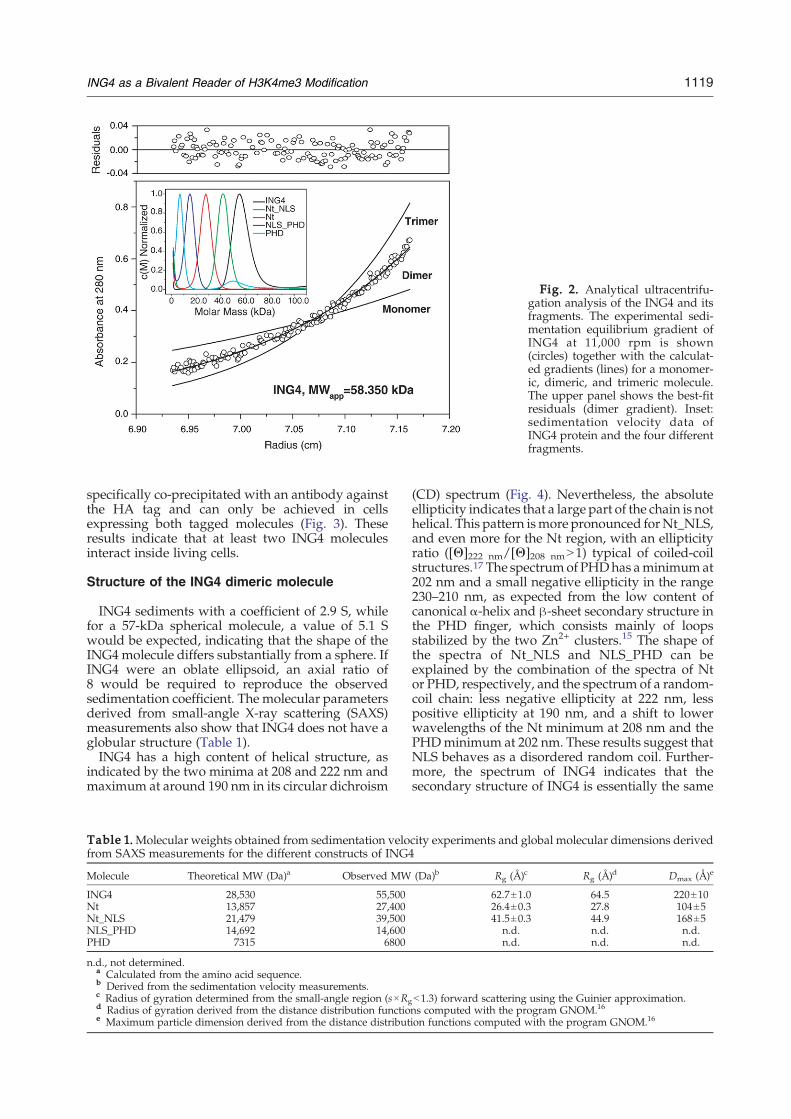
Fig. 2. Analytical ultracentrifu-gation analysis of the ING4 and itsfragments. The experimental sedi-mentation equilibrium gradient ofING4 at 11,000 rpm is shown(circles) together with the calculat-ed gradients (lines) for a monomer-ic, dimeric, and trimeric molecule.The upper panel shows the best-fitresiduals (dimer gradient). Inset:sedimentation velocity data ofING4 protein and the four differentfragments.
1119ING4 as a Bivalent Reader of H3K4me3 Modification
specifically co-precipitated with an antibody againstthe HA tag and can only be achieved in cellsexpressing both tagged molecules (Fig. 3). Theseresults indicate that at least two ING4 moleculesinteract inside living cells.
Structure of the ING4 dimeric molecule
ING4 sediments with a coefficient of 2.9 S, whilefor a 57-kDa spherical molecule, a value of 5.1 Swould be expected, indicating that the shape of theING4molecule differs substantially from a sphere. IfING4 were an oblate ellipsoid, an axial ratio of8 would be required to reproduce the observedsedimentation coefficient. The molecular parametersderived from small-angle X-ray scattering (SAXS)measurements also show that ING4 does not have aglobular structure (Table 1).ING4 has a high content of helical structure, as
indicated by the two minima at 208 and 222 nm andmaximum at around 190 nm in its circular dichroism
Table 1.Molecular weights obtained from sedimentation velofrom SAXS measurements for the different constructs of ING4
Molecule Theoretical MW (Da)a Observed MW
ING4 28,530 55,500Nt 13,857 27,400Nt_NLS 21,479 39,500NLS_PHD 14,692 14,600PHD 7315 6800
n.d., not determined.a Calculated from the amino acid sequence.b Derived from the sedimentation velocity measurements.c Radius of gyration determined from the small-angle region (s×Rgd Radius of gyration derived from the distance distribution functioe Maximum particle dimension derived from the distance distribut
(CD) spectrum (Fig. 4). Nevertheless, the absoluteellipticity indicates that a large part of the chain is nothelical. This pattern ismore pronounced forNt_NLS,and even more for the Nt region, with an ellipticityratio ([Θ]222 nm/[Θ]208 nmN1) typical of coiled-coilstructures.17 The spectrumof PHDhas aminimumat202 nm and a small negative ellipticity in the range230–210 nm, as expected from the low content ofcanonical α-helix and β-sheet secondary structure inthe PHD finger, which consists mainly of loopsstabilized by the two Zn2+ clusters.15 The shape ofthe spectra of Nt_NLS and NLS_PHD can beexplained by the combination of the spectra of Ntor PHD, respectively, and the spectrum of a random-coil chain: less negative ellipticity at 222 nm, lesspositive ellipticity at 190 nm, and a shift to lowerwavelengths of the Nt minimum at 208 nm and thePHDminimum at 202 nm. These results suggest thatNLS behaves as a disordered random coil. Further-more, the spectrum of ING4 indicates that thesecondary structure of ING4 is essentially the same
city experiments and global molecular dimensions derived
(Da)b Rg (Å)c Rg (Å)d Dmax (Å)e
62.7±1.0 64.5 220±1026.4±0.3 27.8 104±541.5±0.3 44.9 168±5
n.d. n.d. n.d.n.d. n.d. n.d.
b1.3) forward scattering using the Guinier approximation.ns computed with the program GNOM.16
ion functions computed with the program GNOM.16

Fig. 3. Analysis of ING4 associ-tion inside living cells by immuno-recipitation. Lysates from cellsransiently transfected with vectorsxpressing HA-ING4 (right-handanels), AU5-ING4 (middle panels),r both (left-hand panels) were im-unoprecipitated (IP) with (αHA) orithout (−αHA) an antibody against
he HA tag. The presence of eachrotein in the immunocomplexesas analyzed by Western blotting
WB) with antibodies against AU5αAU5, upper panels) or against HAαHA, lower panels) tag. TL is theotal lysate.
1120 ING4 as a Bivalent Reader of H3K4me3 Modification
as the sum of the secondary structures of its threeregions, suggesting that each domain is structurallyindependent from the rest of the chain. The thermaldenaturation curves show cooperative unfoldingtransitions for the full-length ING4 protein and Ntand Nt_NLS constructs (with an apparent meltingtemperature of 56 °C), as well as for NLS_PHD andPHD (with a midpoint denaturation temperaturearound 86 °C). Helical structures have the strongest
Fig. 4. Structure and stability of ING4 proteinsanalyzed by CD. (a) Far-UV spectra of the five constructsrecorded at 25 °C in 20 mM sodium phosphate buffer,pH 6.5, 200 mM NaCl, and 1 mM DTT. (b) Thermaldenaturation curves of the same molecules and in thesame buffer as before followed by the change in theirmolar residue ellipticity at 222 nm.
aptepomwtpw(((t
signal in protein CD spectra; therefore, the signalfrom Nt dominates the transition in those constructscontaining this domain, while the random-coil NLSregion contributes a nearly invariantmolar ellipticitythat flattens the curves of the corresponding con-structs, especially that of NLS_PHD (still, whenexpanded in the vertical scale, a transition with thesamemidpoint temperature as the PHD is observed).Altogether, the CD data indicate that Nt and PHDare domains with well-defined tertiary structures,while the NLS is a random coil, and that the threeregions are independent with weak interactionsamong them, if any (Fig. 4).The 1H–15N-heteronuclear single quantum coher-
ence (HSQC) NMR spectrum of the 57-kDa ING4dimer (Fig. 5a) displays distinct sets of peaks thatcan be recognized in the spectra of the individualfragments (Fig. 5b and c). A detailed analysis showsthat for 83% of the backbone amide signals of thePHD, the 1H and 15N resonance frequencies closelymatch those of a corresponding signal in thespectrum of full-length ING4 (chemical shiftperturbationsb0.065 ppm). For the other residues,signal overlap or a larger unknown shift precludes areliable assessment of the actual displacement. Thisresult demonstrates that the Nt and PHD domainsare independent non-interacting domains linked bya flexible NLS segment. In the spectrum of the Ntdomain, the number of observed peaks is smallerthan expected from its amino acid sequence, andmost of them are narrow and non-dispersed. Thisresult is consistent with a 28-kDa elongated coiled-coil molecule as this molecular size, shape, andstructure (with the 1H–15N bonds aligned with thelongitudinal helical axis) result in unfavorable NMRrelaxation properties and thus in poor sensitivity.Most likely, the observed signals correspond toresidues in the most flexible parts (mostly in theloops and chain ends). At higher temperatures, theincreased overall molecular tumbling slows therelaxation of the NMR signals favoring theirmeasurement, and a larger number of dispersedpeaks is observed in the spectra. Although there isstill substantial signal overlap, the number of signalsin the 1H–15N-HSQCNMR spectra of the Nt domainsuggests that it forms symmetric dimers.
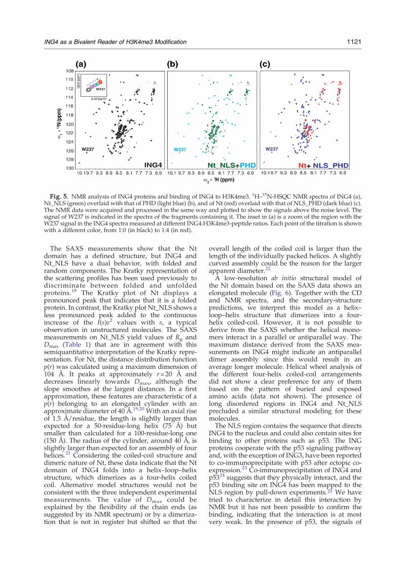
Fig. 5. NMR analysis of ING4 proteins and binding of ING4 to H3K4me3. 1H–15N-HSQC NMR spectra of ING4 (a),Nt_NLS (green) overlaid with that of PHD (light blue) (b), and of Nt (red) overlaid with that of NLS_PHD (dark blue) (c).The NMR data were acquired and processed in the same way and plotted to show the signals above the noise level. Thesignal of W237 is indicated in the spectra of the fragments containing it. The inset in (a) is a zoom of the region with theW237 signal in the ING4 spectra measured at different ING4:H3K4me3-peptide ratios. Each point of the titration is shownwith a different color, from 1:0 (in black) to 1:4 (in red).
1121ING4 as a Bivalent Reader of H3K4me3 Modification
The SAXS measurements show that the Ntdomain has a defined structure, but ING4 andNt_NLS have a dual behavior, with folded andrandom components. The Kratky representation ofthe scattering profiles has been used previously todiscriminate between folded and unfoldedproteins.18 The Kratky plot of Nt displays apronounced peak that indicates that it is a foldedprotein. In contrast, the Kratky plot Nt_NLS shows aless pronounced peak added to the continuousincrease of the I(s)s2 values with s, a typicalobservation in unstructured molecules. The SAXSmeasurements on Nt_NLS yield values of Rg andDmax (Table 1) that are in agreement with thissemiquantitative interpretation of the Kratky repre-sentation. For Nt, the distance distribution functionp(r) was calculated using a maximum dimension of104 Å. It peaks at approximately r=20 Å anddecreases linearly towards Dmax, although theslope smoothes at the largest distances. In a firstapproximation, these features are characteristic of ap(r) belonging to an elongated cylinder with anapproximate diameter of 40 Å.19,20 With an axial riseof 1.5 Å/residue, the length is slightly larger thanexpected for a 50-residue-long helix (75 Å) butsmaller than calculated for a 100-residue-long one(150 Å). The radius of the cylinder, around 40 Å, isslightly larger than expected for an assembly of fourhelices.21 Considering the coiled-coil structure anddimeric nature of Nt, these data indicate that the Ntdomain of ING4 folds into a helix–loop–helixstructure, which dimerizes as a four-helix coiledcoil. Alternative model structures would not beconsistent with the three independent experimentalmeasurements. The value of Dmax could beexplained by the flexibility of the chain ends (assuggested by its NMR spectrum) or by a dimeriza-tion that is not in register but shifted so that the
overall length of the coiled coil is larger than thelength of the individually packed helices. A slightlycurved assembly could be the reason for the largerapparent diameter.22
A low-resolution ab initio structural model ofthe Nt domain based on the SAXS data shows anelongated molecule (Fig. 6). Together with the CDand NMR spectra, and the secondary-structurepredictions, we interpret this model as a helix–loop–helix structure that dimerizes into a four-helix coiled-coil. However, it is not possible toderive from the SAXS whether the helical mono-mers interact in a parallel or antiparallel way. Themaximum distance derived from the SAXS mea-surements on ING4 might indicate an antiparalleldimer assembly since this would result in anaverage longer molecule. Helical wheel analysis ofthe different four-helix coiled-coil arrangementsdid not show a clear preference for any of thembased on the pattern of buried and exposedamino acids (data not shown). The presence oflong disordered regions in ING4 and Nt_NLSprecluded a similar structural modeling for thesemolecules.The NLS region contains the sequence that directs
ING4 to the nucleus and could also contain sites forbinding to other proteins such as p53. The INGproteins cooperate with the p53 signaling pathwayand, with the exception of ING3, have been reportedto co-immunoprecipitate with p53 after ectopic co-expression.23 Co-immunoprecipitation of ING4 andp5324 suggests that they physically interact, and thep53 binding site on ING4 has been mapped to theNLS region by pull-down experiments.25 We havetried to characterize in detail this interaction byNMR but it has not been possible to confirm thebinding, indicating that the interaction is at mostvery weak. In the presence of p53, the signals of

Fig. 6. Model of ING4 engaging nucleosomes with H3K4me3 marks and facilitating histone H3 acetylation by theHBO1 complex. The dimeric Nt domain is shown as the SAXS-derivedmodel, the NLS as lines, and the PHD fingers as thesurfaces of its NMR structure (left one) and of its crystal structure bound to the H3K4me3 peptide (right-hand one). Therepresentations of the Nt dimer and the PHD fingers are done approximately at the same scale, but those of the HBO1complex and the nucleosomes are not.
1122 ING4 as a Bivalent Reader of H3K4me3 Modification
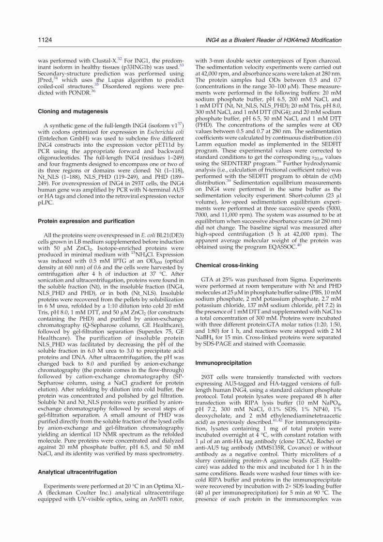
NLS_PHD do not appreciably change in frequency,line width, or intensity (Fig. 7), and the same occurswhen the signals of p53 (uniformly deuterated in thenon-labile protons) are observed in the presence orabsence of NLS_PHD. The NLS_PHD fragment ofING4 used here (residues 119–249) is almostidentical with one of the glutathione S-transferase-fused fragments of ING4 (the one spanning residues120–248) used in pull-down experiments that werepositive for p53 binding.25 As no significant chem-
ical shift perturbations (above the experimentalerror) were observed, it is not possible to confirmthe interaction, but a lower limit for the dissociationconstant (Kd) of the putative complex can beestimated. Assuming an equimolar complex, andthat we should be able to detect significantperturbations in the chemical shifts when at least20% of the 15N-labeled protein is bound to theunlabeled one,26,27 the dissociation constant wouldbe larger than 320 μM (estimated from the absence
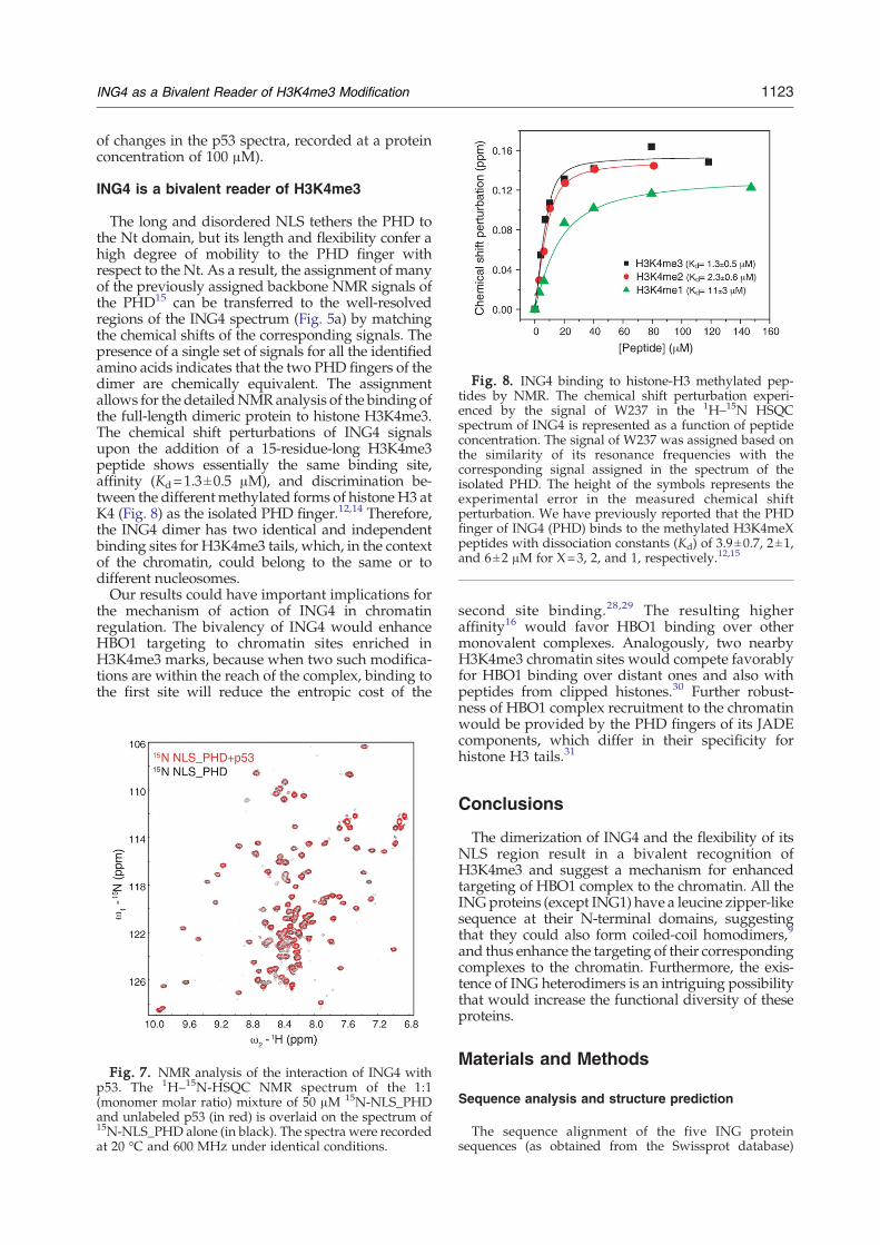
Fig. 8. ING4 binding to histone-H3 methylated pep-tides by NMR. The chemical shift perturbation experi-enced by the signal of W237 in the 1H–15N HSQCspectrum of ING4 is represented as a function of peptideconcentration. The signal of W237 was assigned based onthe similarity of its resonance frequencies with thecorresponding signal assigned in the spectrum of theisolated PHD. The height of the symbols represents theexperimental error in the measured chemical shiftperturbation. We have previously reported that the PHDfinger of ING4 (PHD) binds to the methylated H3K4meXpeptides with dissociation constants (Kd) of 3.9±0.7, 2±1,and 6±2 μM for X=3, 2, and 1, respectively.12,15
1123ING4 as a Bivalent Reader of H3K4me3 Modification
of changes in the p53 spectra, recorded at a proteinconcentration of 100 μM).
ING4 is a bivalent reader of H3K4me3
The long and disordered NLS tethers the PHD tothe Nt domain, but its length and flexibility confer ahigh degree of mobility to the PHD finger withrespect to the Nt. As a result, the assignment of manyof the previously assigned backbone NMR signals ofthe PHD15 can be transferred to the well-resolvedregions of the ING4 spectrum (Fig. 5a) by matchingthe chemical shifts of the corresponding signals. Thepresence of a single set of signals for all the identifiedamino acids indicates that the two PHD fingers of thedimer are chemically equivalent. The assignmentallows for the detailedNMRanalysis of the binding ofthe full-length dimeric protein to histone H3K4me3.The chemical shift perturbations of ING4 signalsupon the addition of a 15-residue-long H3K4me3peptide shows essentially the same binding site,affinity (Kd=1.3±0.5 μM), and discrimination be-tween the differentmethylated forms of histone H3 atK4 (Fig. 8) as the isolated PHD finger.12,14 Therefore,the ING4 dimer has two identical and independentbinding sites for H3K4me3 tails, which, in the contextof the chromatin, could belong to the same or todifferent nucleosomes.Our results could have important implications for
the mechanism of action of ING4 in chromatinregulation. The bivalency of ING4 would enhanceHBO1 targeting to chromatin sites enriched inH3K4me3 marks, because when two such modifica-tions are within the reach of the complex, binding tothe first site will reduce the entropic cost of the
Fig. 7. NMR analysis of the interaction of ING4 withp53. The 1H–15N-HSQC NMR spectrum of the 1:1(monomer molar ratio) mixture of 50 μM 15N-NLS_PHDand unlabeled p53 (in red) is overlaid on the spectrum of15N-NLS_PHD alone (in black). The spectra were recordedat 20 °C and 600 MHz under identical conditions.
second site binding.28,29 The resulting higheraffinity16 would favor HBO1 binding over othermonovalent complexes. Analogously, two nearbyH3K4me3 chromatin sites would compete favorablyfor HBO1 binding over distant ones and also withpeptides from clipped histones.30 Further robust-ness of HBO1 complex recruitment to the chromatinwould be provided by the PHD fingers of its JADEcomponents, which differ in their specificity forhistone H3 tails.31
Conclusions
The dimerization of ING4 and the flexibility of itsNLS region result in a bivalent recognition ofH3K4me3 and suggest a mechanism for enhancedtargeting of HBO1 complex to the chromatin. All theINGproteins (except ING1) have a leucine zipper-likesequence at their N-terminal domains, suggestingthat they could also form coiled-coil homodimers,9
and thus enhance the targeting of their correspondingcomplexes to the chromatin. Furthermore, the exis-tence of ING heterodimers is an intriguing possibilitythat would increase the functional diversity of theseproteins.
Materials and Methods
Sequence analysis and structure prediction
The sequence alignment of the five ING proteinsequences (as obtained from the Swissprot database)
1124 ING4 as a Bivalent Reader of H3K4me3 Modification
was performed with Clustal-X.32 For ING1, the predom-inant isoform in healthy tissues (p33ING1b) was used.33
Secondary-structure prediction was performed usingJPred,34 which uses the Lupas algorithm to predictcoiled-coil structures.35 Disordered regions were pre-dicted with PONDR.36
Cloning and mutagenesis
A synthetic gene of the full-length ING4 (isoform v137)with codons optimized for expression in Escherichia coli(Entelechon GmbH) was used to subclone five differentING4 constructs into the expression vector pET11d byPCR using the appropriate forward and backwardoligonucleotides. The full-length ING4 (residues 1–249)and four fragments designed to encompass one or two ofits three regions or domains were cloned: Nt (1–118),Nt_NLS (1–188), NLS_PHD (119–249), and PHD (189–249). For overexpression of ING4 in 293T cells, the ING4human gene was amplified by PCR with N-terminal AU5or HA tags and cloned into the retroviral expression vectorpLPC.
Protein expression and purification
All the proteins were overexpressed in E. coli BL21(DE3)cells grown in LB medium supplemented before inductionwith 50 μM ZnCl2. Isotope-enriched proteins wereproduced in minimal medium with 15NH4Cl. Expressionwas induced with 0.5 mM IPTG at an OD600 (opticaldensity at 600 nm) of 0.6 and the cells were harvested bycentrifugation after 4 h of induction at 37 °C. Aftersonication and ultracentrifugation, proteins were found inthe soluble fraction (Nt), in the insoluble fraction (ING4,NLS_PHD and PHD), or in both (Nt_NLS). Insolubleproteins were recovered from the pellets by solubilizationin 6 M urea, refolded by a 1:10 dilution into cold 20 mMTris, pH 8.0, 1 mM DTT, and 50 μM ZnCl2 (for constructscontaining the PHD) and purified by anion-exchangechromatography (Q-Sepharose column, GE Healthcare),followed by gel-filtration separation (Superdex 75, GEHealthcare). The purification of insoluble proteinNLS_PHD was facilitated by decreasing the pH of thesoluble fraction in 6.0 M urea to 3.0 to precipitate acidproteins and DNA. After ultracentrifugation, the pH waschanged back to 8.0 and purified by anion-exchangechromatography (the protein comes in the flow-through)followed by cation-exchange chromatography (SP-Sepharose column, using a NaCl gradient for proteinelution). After refolding by dilution into cold buffer, theprotein was concentrated and polished by gel filtration.Soluble Nt and Nt_NLS proteins were purified by anion-exchange chromatography followed by several steps ofgel-filtration separation. A small amount of PHD waspurified directly from the soluble fraction of the lysed cellsby anion-exchange and gel-filtration chromatographyyielding an identical 1D NMR spectrum as the refoldedmolecule. Pure proteins were concentrated and dialyzedagainst 20 mM phosphate buffer, pH 6.5, and 50 mMNaCl, and its identity was verified by mass spectrometry.
Analytical ultracentrifugation
Experiments were performed at 20 °C in an Optima XL-A (Beckman Coulter Inc.) analytical ultracentrifugeequipped with UV-visible optics, using an An50Ti rotor,
with 3-mm double sector centerpieces of Epon charcoal.The sedimentation velocity experiments were carried outat 42,000 rpm, and absorbance scans were taken at 280 nm.The protein samples had ODs between 0.5 and 0.7(concentrations in the range 30–100 μM). These measure-ments were performed in the following buffers: 20 mMsodium phosphate buffer, pH 6.5, 200 mM NaCl, and1 mM DTT (Nt, Nt_NLS, NLS_PHD); 20 mM Tris, pH 8.0,300 mMNaCl, and 1mMDTT (ING4); and 20mM sodiumphosphate buffer, pH 6.5, 50 mM NaCl, and 1 mM DTT(PHD). The concentrations of the samples were at ODvalues between 0.5 and 0.7 at 280 nm. The sedimentationcoefficients were calculated by continuous distribution c(s)Lamm equation model as implemented in the SEDFITprogram. These experimental values were corrected tostandard conditions to get the corresponding s20,w valuesusing the SEDNTERP program.38 Further hydrodynamicanalysis (i.e., calculation of frictional coefficient ratio) wasperformed with the SEDFIT program to obtain de c(M)distribution.39 Sedimentation equilibrium measurementson ING4 were performed in the same buffer as thesedimentation velocity experiment. Short-column (23 μlvolume), low-speed sedimentation equilibrium experi-ments were performed at three successive speeds (5000,7000, and 11,000 rpm). The system was assumed to be atequilibriumwhen successive absorbance scans (at 280 nm)did not change. The baseline signal was measured afterhigh-speed centrifugation (5 h at 42,000 rpm). Theapparent average molecular weight of the protein wasobtained using the program EQASSOC.40
Chemical cross-linking
GTA at 25% was purchased from Sigma. Experimentswere performed at room temperature with Nt and PHDmolecules at 25 μM in phosphate buffer saline (PBS, 10mMsodium phosphate, 2 mM potassium phosphate, 2.7 mMpotassium chloride, 137 mM sodium chloride, pH 7.2) inthe presence of 1mMDTT and supplementedwithNaCl toa total concentration of 300 mM. Proteins were incubatedwith three different protein:GTA molar ratios (1:20, 1:50,and 1:80) for 1 h, and reactions were stopped with 2 MNaBH4 for 15 min. Cross-linked proteins were separatedby SDS-PAGE and stained with Coomassie.
Immunoprecipitation
293T cells were transiently transfected with vectorsexpressing AU5-tagged and HA-tagged versions of full-length human ING4, using a standard calcium phosphateprotocol. Total protein lysates were prepared 48 h aftertransfection with RIPA lysis buffer (10 mM NaPO4,pH 7.2, 300 mM NaCl, 0.1% SDS, 1% NP40, 1%deoxycholate, and 2 mM ethylenediaminetetraaceticacid) as previously described.41,42 For immunoprecipita-tion, lysates containing 1 mg of total protein wereincubated overnight at 4 °C, with constant rotation with1 μl of an anti-HA tag antibody (clone 12CA2, Roche) oranti-AU5 tag antibody (MMS135R, Covance) or withoutantibody as a negative control. Thirty microliters of aslurry containing protein-A agarose beads (GE Health-care) was added to the mix and incubated for 1 h in thesame conditions. Beads were washed four times with ice-cold RIPA buffer and proteins in the immunoprecipitatewere recovered by incubation with 2× SDS loading buffer(40 μl per immunoprecipitation) for 5 min at 90 °C. Thepresence of each protein in the immunocomplex was

1125ING4 as a Bivalent Reader of H3K4me3 Modification
analyzed by Western blotting with antibodies againstAU5 tag (1/500) and HA tag (1/500). The immunopre-cipitation was repeated several times to confirm itsreproducibility.
Circular dichroism
The spectra were performed in a Jasco J-810 spectro-polarimeter at 25 °C. The spectra were the average of 15scans, recorded using a 0.1-mm path-length quartz cuvetteon 50-μM protein samples in 20 mM sodium phosphate,pH 6.5, 200 mMNaCl, and 1 mMDTT. The average helicalcontent calculated from the molar residue ellipticity at222 nm assuming 50-residue-long helices (as predicted byJPred) is 30%, 41%, and 63% for ING4, Nt_NLS, and Nt,respectively.43 Thermal denaturation curves were mea-sured in 2-mm path-length cuvettes closed with a Tefloncap on 25-μM protein solutions. Protein unfolding wasinduced by increasing temperature at a rate of 1 °C/minfrom 5 to 100 °C, and the ellipticity at 222 nm wasrecorded at intervals of 1 °C.
Small-angle X-ray scattering
Data were collected at X33 beamline at the EuropeanMolecular Biology Laboratory in the storage ring DORISIII of the Deutsches Elektronen Synchrotron, Hamburg.44
Scattering curves of ING4, Nt, and Nt_NLS constructswere measured at 10 °C at protein concentrations of327 μM (ING4), 500 and 260 μM (Nt), and 251 and 186 μM(Nt_NLS). An exposure time of 2 min was used for themeasurements. The scattering profiles covered a range ofmomentum transfer of 0.02b sb0.5 Å−1. Radiation damagewas minimal or absent since no significant changes wereobserved after repetitive exposures of the protein solu-tions. Buffer scattering profiles were measured before andafter the sample measurements, averaged, and subtractedfrom the scattering profiles of the protein solutions.Resulting profiles were merged to avoid inter-particleinteractions. All data manipulations were done usingstandard procedures with the software PRIMUS.45 Theforward scattering, I(0) and the radius of gyration, Rg,were evaluated using the Guinier approximation, assum-ing that at very small angles (sb1.3/Rg), the intensity canbe well represented by I(s) = I(0)exp(− (sRg)
2/3). Thedistance distribution function, p(r), was computed fromthe entire curve with the program GNOM46 by optimizingthe maximum particle dimension,Dmax. Low-resolution abinitio model structures of the Nt domain were built fromthe SAXS measurements using the program GASBOR.47
The protein was represented by an assembly of 236dummy residues inside a search volume. Starting from arandom distribution, 10 assemblies that fit the experimen-tal scattering profile were generated using a simulatedannealing protocol. These 10 independent GASBORmodels were averaged with the program packageDAMAVER48 to yield the most probable shape of themolecule.
NMR spectroscopy
NMR spectra of ING4 proteins were recorded at 25 °Con samples with a protein concentration of approximately130 μM in 20 mM sodium phosphate, pH 6.5, 200 mMNaCl, 1 mM perdeuterated DTT, 5% 2H2O, and 0.01%NaN3. Chemical shifts were measured relative to internal2,2-dimethyl-2-silapentane-5-sulfonate sodium salt for 1H
and calculated for 15N.49 Spectra were processed withXWINMR, TOPSpin (Bruker), and/or NMRPipe.50 Thetitration of ING4 with the peptides and the NMR bindinganalysis were performed as described previously.15 Theinteraction of NLS_PHD with p53 was examined by NMRat 20 °C on samples prepared at 1:1 monomer molar ratios.
Peptide binding analysis by NMR
The synthetic peptides H3K4meX correspond to resi-dues 1–15 of histone H3 plus an extra tyrosine residue atthe C-terminus (ARTKQTARKSTGGKAY) to facilitate thequantification of peptide concentration by UV absorbance.Three peptides named with X=1, 2, or 3 (mono-, di-, andtrimethylated, respectively) differ in the number ofmethyls bound to the amino group of Lys4. Thesepeptides have free N- and C-termini and were purchasedfromNeoMPS (Strasbourg). The titration of ING4 with thepeptides and the NMR binding analysis were performedas previously described.15 To measure the interaction ofNLS_PHDwith p53 by NMR, we preparedmixed samplesat 1:1 monomer molar ratio, and NMR experiments wereregistered at 20 °C. For p53 observation, U-15N-2H-p53(with protonated amides) was used, and transverserelaxation optimized spectroscopy 1H–15N-correlationspectra were recorded at 900 MHz. For NLS_PHDobservation, U-15N-NLS_PHD was used, and 1H–15NHSQC spectra were recorded at 600 MHz. The controlsamples (the isotope-labeled proteins alone) and the testsamples (labeled-p53/NLS_PHD and labeled-NLS_PHD/unlabeled-p53) were simultaneously dialyzed against thesame buffer (25 mM sodium phosphate, pH 7.2, 150 mMNaCl, and 5 mM perdeuterated DTT). The proteinconcentration was 100 μM in the labeled-p53-containingsamples and 50 μM in the labeled-NLS_PHD-containingsamples (monomer protein concentration).
Acknowledgements
We thank Prof. A. R. Fersht for support and helpfuldiscussions; C. González, L. Cheng, and C. M. Blairfor help with protein purification; D. Padró andS. M. V. Freund for help with NMR; and G.Montoyafor critical manuscript reading. This work wassupported by grants from Ministerio de Ciencia eInnovación (CTQ2008-03115/BQU), EuropeanUnion (3D-REPERTOIRE, contract no. LSHG-CT-2005-512028), Fundación Mutua Madrileña to F.J.B.,and ETORTEK-2008 grant to CIC bioGUNE.We alsoacknowledge the support to European MolecularBiology Laboratory Hamburg by European Com-mission–Research Infrastructure Action contract no.RII/2004/5060008. P.B. holds a Ramón y Cajalcontract, and M.R.F.-F. was a Career DevelopmentFellow of the MRC-UK.
Supplementary Data
Supplementary data associated with this articlecan be found, in the online version, at doi:10.1016/j.jmb.2009.12.049
1126 ING4 as a Bivalent Reader of H3K4me3 Modification
References
1. Kan, P. Y., Caterino, T. L. & Hayes, J. J. (2009). The H4tail domain participates in intra- and internucleosomeinteractions with protein and DNA during foldingand oligomerization of nucleosome arrays. Mol. Cell.Biol. 29, 538–546.
2. Groth, A., Rocha, W., Verreault, A. & Almouzni, G.(2007). Chromatin challenges during DNA replicationand repair. Cell, 128, 721–733.
3. Li, B., Carey, M. & Workman, J. L. (2007). The role ofchromatin during transcription. Cell, 128, 707–719.
4. Jenuwein, T. & Allis, C. D. (2001). Translating thehistone code. Science, 293, 1074–1080.
5. Taverna, S. D., Li, H., Ruthenburg, A. J., Allis, C. D. &Patel, D. J. (2007). How chromatin-binding modulesinterpret histone modifications: lessons from profes-sional pocket pickers. Nat. Struct. Mol. Biol. 14,1025–1040.
6. Russell, M., Berardi, P., Gong, W. & Riabowol, K.(2006). Grow-ING, Age-ING and Die-ING: INGproteins link cancer, senescence and apoptosis. Exp.Cell Res. 312, 951–961.
7. Shi, X., Hong, T., Walter, K. L., Ewalt, M., Michishita,E., Hung, T. et al. (2006). ING2 PHD domain linkshistone H3 lysine 4 methylation to active generepression. Nature, 442, 96–99.
8. Doyon, Y., Cayrou, C., Ullah, M., Landry, A. J., Cote,V., Selleck, W. et al. (2006). ING tumor suppressorproteins are critical regulators of chromatin acetyla-tion required for genome expression and perpetua-tion. Mol. Cell, 21, 51–64.
9. He, G. H., Helbing, C. C., Wagner, M. J., Sensen, C. W.& Riabowol, K. (2005). Phylogenetic analysis of theING family of PHD finger proteins. Mol. Biol. Evol. 22,104–116.
10. Pena, P. V., Hom, R. A., Hung, T., Lin, H., Kuo, A. J.,Wong, R. P. et al. (2008). Histone H3K4me3 binding isrequired for the DNA repair and apoptotic activitiesof ING1 tumor suppressor. J. Mol. Biol. 380, 303–312.
11. Pena, P. V., Davrazou, F., Shi, X., Walter, K. L.,Verkhusha, V. V., Gozani, O. et al. (2006). Molecularmechanism of histone H3K4me3 recognition by planthomeodomain of ING2. Nature, 442, 100–103.
12. Palacios, A.,Munoz, I. G., Pantoja-Uceda, D.,Marcaida,M. J., Torres, D., Martin-Garcia, J. M. et al. (2008).Molecular basis of histone H3K4me3 recognition byING4. J. Biol. Chem. 283, 15956–15964.
13. Champagne, K. S., Saksouk, N., Pena, P. V., Johnson,K., Ullah, M., Yang, X. J. et al. (2008). The crystalstructure of the ING5 PHD finger in complex with anH3K4me3 histone peptide. Proteins, 72, 1371–1376.
14. Hung, T., Binda, O., Champagne, K. S., Kuo, A. J.,Johnson, K., Chang, H. Y. et al. (2009). ING4 mediatescrosstalk between histone H3 K4 trimethylation andH3 acetylation to attenuate cellular transformation.Mol. Cell, 33, 248–256.
15. Palacios,A., Garcia, P., Padro,D., Lopez-Hernandez, E.,Martin, I. & Blanco, F. J. (2006). Solution structure andNMR characterization of the binding to methylatedhistone tails of the plant homeodomain finger of thetumour suppressor ING4. FEBS Lett. 580, 6903–6908.
16. Ruthenburg, A. J., Li, H., Patel, D. J. & Allis, C. D.(2007). Multivalent engagement of chromatin mod-ifications by linked binding modules. Nat. Rev., Mol.Cell Biol. 8, 983–994.
17. Dutta, K., Alexandrov, A., Huang, H. & Pascal, S. M.(2001). pH-induced folding of an apoptotic coiled coil.Protein Sci. 10, 2531–2540.
18. Doniach, S. (2001). Changes in biomolecular confor-mation seen by small angle X-ray scattering. Chem.Rev. 101, 1763–1778.
19. Glatter, O. K. & Kratky, O. (1982). Small Angle X-rayScattering. Academic Press, New York, NY.
20. Svergun, D. I. & Koch, M. H. J. (2003). Small-anglescattering studies of biological macromolecules insolution. Rep. Prog. Phys. 66, 1735–1782.
21. Tarbouriech, N., Curran, J., Ruigrok, R. W. &Burmeister, W. P. (2000). Tetrameric coiled coildomain of Sendai virus phosphoprotein. Nat. Struct.Biol. 7, 777–781.
22. Yousef, M. S., Kamikubo, H., Kataoka, M., Kato, R. &Wakatsuki, S. (2008). Miranda cargo-binding domainforms an elongated coiled-coil homodimer in solution:implications for asymmetric cell division inDrosophila.Protein Sci. 17, 908–917.
23. Coles, A. H. & Jones, S. N. (2009). The ING genefamily in the regulation of cell growth and tumori-genesis. J. Cell. Physiol. 218, 45–57.
24. Shiseki, M., Nagashima, M., Pedeux, R. M., Kitahama-Shiseki, M., Miura, K., Okamura, S. et al. (2003).p29ING4 and p28ING5 bind to p53 and p300, andenhance p53 activity. Cancer Res. 63, 2373–2378.
25. Zhang, X., Wang, K. S., Wang, Z. Q., Xu, L. S., Wang,Q.W., Chen, F. et al. (2005). Nuclear localization signalof ING4 plays a key role in its binding to p53. Biochem.Biophys. Res. Commun. 331, 1032–1038.
26. Hajduk, P. J., Gerfin, T., Boehlen, J. M., Haberli, M.,Marek, D. & Fesik, S. W. (1999). High-throughputnuclear magnetic resonance-based screening. J. Med.Chem. 42, 2315–2317.
27. Meyer, B. & Peters, T. (2003). NMR spectroscopytechniques for screening and identifying ligandbinding to protein receptors. Angew. Chem., Int. Ed.Engl. 42, 864–890.
28. Jencks, W. P. (1981). On the attribution and additivityof binding energies. Proc. Natl Acad. Sci. USA, 78,4046–4050.
29. Ruthenburg, A. J., Wang,W., Graybosch, D.M., Li, H.,Allis, C. D., Patel, D. J. & Verdine, G. L. (2006). HistoneH3 recognition and presentation by the WDR5module of the MLL1 complex. Nat. Struct. Mol. Biol.13, 704–712.
30. Santos-Rosa, H., Kirmizis, A., Nelson, C., Bartke, T.,Saksouk, N., Cote, J. & Kouzarides, T. (2009). HistoneH3 tail clipping regulates gene expression. Nat. Struct.Mol. Biol. 16, 17–22.
31. Saksouk, N., Avvakumov, N., Champagne, K. S.,Hung, T., Doyon, Y., Cayrou, C. et al. (2009). HBO1HAT complexes target chromatin throughout genecoding regions via multiple PHD finger interactionswith histone H3 tail. Mol. Cell, 33, 257–265.
32. Thompson, J. D., Gibson, T. J., Plewniak, F., Jeanmougin,F. & Higgins, D. G. (1997). The CLUSTAL_X windowsinterface: flexible strategies for multiple sequencealignment aided by quality analysis tools. NucleicAcids Res. 25, 4876–4882.
33. Feng, X., Hara, Y. & Riabowol, K. (2002). DifferentHATS of the ING1 gene family. Trends Cell Biol. 12,532–538.
34. Cuff, J. A., Clamp, M. E., Siddiqui, A. S., Finlay, M. &Barton, G. J. (1998). JPred: a consensus secondarystructure prediction server. Bioinformatics, 14, 892–893.
35. Lupas, A., Van Dyke, M. & Stock, J. (1991). Predictingcoiled coils from protein sequences. Science, 252,1162–1164.
36. Li, X., Romero, P., Rani, M., Dunker, A. K. &Obradovic, Z. (1999). Predicting protein disorder for
1127ING4 as a Bivalent Reader of H3K4me3 Modification
N-, C-, and internal regions. Genome Inform. Ser.Workshop Genome Inform. 10, 30–40.
37. Unoki, M., Shen, J. C., Zheng, Z. M. & Harris, C. C.(2006). Novel splice variants of ING4 and theirpossible roles in the regulation of cell growth andmotility. J. Biol. Chem. 281, 34677–34686.
38. Laue, T. M. S., Shah, B. D., Ridgeway, T. M. &Pelletier, S. L. (1992). Computer-Aided Interpretation ofAnalytical Sedimentation Data for Proteins. Royal Societyof Chemistry, Cambridge, UK.
39. Schuck, P. & Rossmanith, P. (2000). Determination ofthe sedimentation coefficient distribution by least-squares boundary modeling. Biopolymers, 54, 328–341.
40. Minton, A. P. (1994). Modern Analytical Ultracentrifu-gation. Birkhauser Boston, Inc., Cambridge, MA.
41. Palmero, I., Murga, M., Zubiaga, A. & Serrano, M.(2002). Activation of ARF by oncogenic stress inmouse fibroblasts is independent of E2F1 and E2F2.Oncogene, 21, 2939–2947.
42. Gonzalez, L., Freije, J. M., Cal, S., Lopez-Otin, C.,Serrano, M. & Palmero, I. (2006). A functional linkbetween the tumour suppressors ARF and p33ING1.Oncogene, 25, 5173–5179.
43. Chen, Y. H., Yang, J. T. & Chau, K. H. (1974).Determination of the helix and beta form ofproteins in aqueous solution by circular dichroism.Biochemistry, 13, 3350–3359.
44. Roessle, M. W., Klaering, R., Ristau, U., Robrahn, B.,Jahn, D., Gehrmann, T. et al. (2007). Upgrade of thesmall-angle X-ray scattering beamline X33 at theEuropean Molecular Biology Laboratory, Hamburg.J. Appl. Crystallogr. 40, 190–194.
45. Konarev, P. V., Volkov, V. V., Sokolova, A. V., Koch,M. H. J. & Svergun, D. I. (2003). PRIMUS: a WindowsPC-based system for small-angle scattering dataanalysis. J. Appl. Crystallogr. 36, 1277–1282.
46. Svergun, D. I. (1992). Determination of the regular-ization parameter in indirect-transform methodsusing perceptual criteria. J. Appl. Crystallogr. 25,495–503.
47. Svergun, D. I., Petoukhov, M. V. & Koch, M. H. (2001).Determination of domain structure of proteins fromX-ray solution scattering. Biophys. J. 80, 2946–2953.
48. Volkov, V. V. & Svergun, D. I. (2003). Uniqueness ofab initio shape determination in small-angle scatter-ing. Appl. Crystallogr. 36, 860–864.
49. Wishart, D. S., Bigam, C. G., Yao, J., Abildgaard, F.,Dyson, H. J., Oldfield, E. et al. (1995). 1H, 13C and 15Nchemical shift referencing in biomolecular NMR. J.Biomol. NMR, 6, 135–140.
50. Delaglio, F., Grzesiek, S., Vuister, G. W., Zhu, G.,Pfeifer, J. & Bax, A. (1995). NMRPipe: a multidimen-sional spectral processing system based on UNIXpipes. J. Biomol. NMR, 6, 277–293.
Copyright © 2022 FDOKUMEN