The Differential diagnosis of coma
74
University of Nebraska Medical Center University of Nebraska Medical Center DigitalCommons@UNMC DigitalCommons@UNMC MD Theses Special Collections 5-1-1939 The Differential diagnosis of coma The Differential diagnosis of coma Neil M. Burr University of Nebraska Medical Center This manuscript is historical in nature and may not reflect current medical research and practice. Search PubMed for current research. Follow this and additional works at: https://digitalcommons.unmc.edu/mdtheses Part of the Medical Education Commons Recommended Citation Recommended Citation Burr, Neil M., "The Differential diagnosis of coma" (1939). MD Theses. 729. https://digitalcommons.unmc.edu/mdtheses/729 This Thesis is brought to you for free and open access by the Special Collections at DigitalCommons@UNMC. It has been accepted for inclusion in MD Theses by an authorized administrator of DigitalCommons@UNMC. For more information, please contact [email protected].
-
Upload
khangminh22 -
Category
Documents
-
view
3 -
download
0
Transcript of The Differential diagnosis of coma

University of Nebraska Medical Center University of Nebraska Medical Center
DigitalCommons@UNMC DigitalCommons@UNMC
MD Theses Special Collections
5-1-1939
The Differential diagnosis of coma The Differential diagnosis of coma
Neil M. Burr University of Nebraska Medical Center
This manuscript is historical in nature and may not reflect current medical research and
practice. Search PubMed for current research.
Follow this and additional works at: https://digitalcommons.unmc.edu/mdtheses
Part of the Medical Education Commons
Recommended Citation Recommended Citation Burr, Neil M., "The Differential diagnosis of coma" (1939). MD Theses. 729. https://digitalcommons.unmc.edu/mdtheses/729
This Thesis is brought to you for free and open access by the Special Collections at DigitalCommons@UNMC. It has been accepted for inclusion in MD Theses by an authorized administrator of DigitalCommons@UNMC. For more information, please contact [email protected].

/"'\
THE DIFFERENTIAL DIAGl!OSIS OF COMA
Neil M. Burr
Senior ~esis Presented
to the
University of Nebraska College of Medicine
Omaha, Nebraska
APril, 1939

CONTENTS
page Introduction - - - - - - - - - - - - - - - 1.
Definition, Scope, & Importance - - - - - 3.
Classification and Enumeration of Ca.uses of Coma - - - - - - - - - - 6.
Incidence of various Types of Coma - - - 13.
General Diagnostic .Approach - - - - - - - 23.
Differential Diagnosis - - - - - - - - - 35.
Diagnostic Boutine for Coma's - - - - - - 64.
Bibliography
481020

INTRODUCTION
The subject of this paper as a senior the
sis was inspired by a situation in which a fellow
student found himself while taking a practical ex
amination for an internship. The patient that he
was supposed to examine had been admitted to the
hospital with very little history and had not been
diagnosed at the time he saw him in a comatose
condition.
-1- .
The above situation brought to my mind that in
less than a year as an intern, I might be confronted
with many such problems, and that although during
the four years spent in medical school, one learns
several facts concerning the subject, there has
never been a complete correlation of these facts
in my own mind, and I believe this situation is
similar to that of fellow students. Text book
articles have a tendency to discuss the subject
in a very general and abstract way, without spe
cial regard to the practical clinical aspects of
the problem at hand •
.P:J1 effort will be made to deal with the subject
as a condition that presents itself as a presenting
sign, rather than to consider in detail every pos
sible cause of comatose conditions, insomuch as i~

impossible to think of any disease entity that
might end fatally, where coma would not preceed
death.
Treatment will not be dealt with except in
those cases where diagnostic proceedures tend to
be likewise therapeutic.
-2-
The aim of the paper in general is to define
and classify the condition, to present the approxi
mate incidence of the various types, to discuss in a
general the diagnostic approach to such· conditions,
and to briefly discuss the differentiating features
of the more enmmon:types of comatose conditions.
Finally, the summaries of three articles will be
combined to suggest a diagnostic routine for
Coma., as a presenting sign.
After reviewing the literature on the subject,
it was found that most of the work in this country
has been done by Philip Solomon, :M.D., C.D. Aring,
M.D., H. H. :Merritt, M.D., W.W. _Bissell, M.D.,
E. R. Lecount, M.D., F. Fremonst Smith, M.D., who
with other authors the writer. of this paper is
greatly indebted for material used.
•

DE!FINITION, SCOP:/£ .AND IMPORTANCE OF SUBJECT
If we consult one of the more commonly used
medical dictionaries in search of a definition
of the term, "Coma", we till find the following,
-3-
" Coma -- A state of complete loss of consciousness
from which the patient cannot be aroused even by
the most powerful stimulation". Dorland (1936)
As was stated in the introduction an attempt
will be made to discuss coma as a condition which
presents itself for diagnosis and treatment by
the interne or the physician. It should be said
that a very small percentage of deaths will occur
without the patient going through a period of coma
before death. However it was found upon surveying
the literature that a surprising number of hospital
admissions during a year were patients in comatose
states, the origin of which was not definitely known
at the time of entrance to the hospital. In the
year 1933, 1,167 such patients or 3% of the total
admissions in the Boston City Hospital,were in such
a condition. Of these patients 68% entered the
hospital without an immediately available history. Solomon & Aring, (Boston, 1934)
The importance of immediate diagnosis is of
course evident since the condition without treatment

('"'\
-4-
.will always offer a grave prognosis for life.
Naturally, proper treatment cannot be administered
until the proper diagnosis has been made. One
can easily see where the incorrect treatment be~
cause of a mistaken or careless diagnosis of the
condition might give fatal results. .An example of
such an error would be administering insulin to a
known diabetic, who rather than suffering from
diabetic coma was in a state of hypoglycemia due
to an overdose of insulin.
The importance of immediate and correct
diagnosis will be further emphasized in the the
portion of this paper dealing with the incidence
of coma. Various authors have tabulated the num
ber of comatose patients entering hospitals, the
number of cases ending fatally, and the nuntber
of deaths that could have been prevented by a
correct diagnosis. The figures are self explanatory
in that chapter and will not be further discussed
here.
One might further state that it is the re
sponsibility of the medical profession of the
present day more so than in former years, to pro
perly diagnose such conditions since there is so
much more to be done for the patient than a few

fl""'.
-5-
decades ago. According to George s. Young, (1934)
the discovery of insulin and other endocrine pro
ducts, which may be used to relieve comatose
states, as well as rapid strides in the develop
ment of brain surgery have introduced new hope in
caring for all such cases.
From a historical point of view it is inter
esting to note the following quotations from The
Lmnleian Lectures (1850). ".Alnong the most for
midable indications of disturbance of the great
central organ.of the nervous system, the brain,
are those states which are known as coma or deler-
ium. It is during these times that the physician
is forced to appeal to all of his former experience
to guide him in his prognosis and to direct his
practice, then too he is compelled to examine the
grounds of his principles to assure himself as to
their soundness and as to the safety of following
the cause which they indicate". Cert~inly, after
nearly one hundred years this is still very true.

f'"'.
CLASSIFICATION .AND ENUM:!mATION OF CAUSES OF COMA ~6-
In order to make a logical diagnostic approach
to the vari·ous comatose conditions it will first
be well to arrive at a systematic classification
of the causes of coma. Various authors have made
different classifications, which have as their
basis for the most part, the etiology.
The first attempt at classification in the
series of articles reviewed is that of J. T. Es-
keridge, (1898} who offers the following classifi-
cation.
I Transient (Syncope or fainting}
II Coma from lethal doses of medicine. Chloral Lead Alcohol Belladona Opium Hyoscyamus
III Coma from other poisons Asphyxia· Uremia Ptomaines Diabetes
IV Convulsive States Preemptive stage of ;':!!Xanthemata Reflex convulsions Epilepsy Hysteria Epileptoid and appoplectoid attacks due to paralytic dementia.
V Voluntary Coma or malingering
VI Coma from profound disturbances of cerebral cir~ulation but without organic lesions.
Shock Congestion Concussion .Anemia
VII Coma from organic disease.

-7-
Coma from organic disease cont~d
Simple apoplexy Traumatic apoplexy Syphilis
Brain abscess Brain tum.or Cerebral hemorrhage Cerebral embolism Cerebral meningitis
Cerebral thrombosis
A later classification is that of Forsyth,
(1912) a summary of which follows.
He says that in the foreground of our minds
would should always keep; 1. · Vascular derangements of the brain. 2. Injuries to the head. 3. Epilepsy. 4. Diabetes 5. Poisons 6. Stokes Adams disease
He further classifies them into t,hree large
groups as follows.
I Derangements of Gerebral circulation Hemorrhage
II
Thrombosis :Embolism
Mechanical injury Fractures Compression Epilepsy Uremia Diabetes Poisons Stokes Adams Disease
Meningitis Encephalitis Cerebral abscess cerebral tum.or .revers Eolam.psia Gholaemia Epidemic Enteritis General Paralysis Disseminated sclerosis

III Pernicious malaria Muscular exhaustion Heat stroke
-8-
J!'riedman, ( 1933) offers the following clas-
sification.
I General 0auses Alcoholism Uremia Diabetes
II Epilepsy
Opium Gas Hypoglycemia
III Intracranial lesions Apoplexy Meningitis
with or without focal signs Tumor of brain Encephalitis Spontaneous subarachnoid hemorrhage.
Brain abscess
IV Trauma
Concussion of the brain Gross hemorrhage with or without fracture.
Probably the most complete classification of
coma that has been offered up to the present time
is that in .l!'rench, { 1936).
Group A includes:
1. Certain severe fevers in which coma may occur as a terminal phenomenon.
Typhus fever '1'yphoid fever Cholera Dysentery Measles Scarlet fever
Rheumatic fever Yell.ow fever Blackwater fever Malignant malaria Infective Endocarditis Diptheria
Influenza Spirochaetosis Lobar Pneumonia Small pox

-9-
2. Acute inflarmnatory lesions of the brain or the Cerebral meninges:
Acute encephalitis Suppurative meningitis Tuberculous meningitis Basal meningitis
Epidemic meningi"'t±s Encephalitis Lethargica Sleeping sickness
3. Certain less acute lesions of the central nervous system:
Cerebral tumor Cerebral abscess Disseminated sclerosis
Post epileptic state Paresis Syphilis of the brain
4. Diseases ·in which the general metabolism is at fault:
Uremia Diabetes mellitus Cholaemia
Addison's disease Raynaud's disease My:x:oedema
5. Late stages of' certain other maladies that exhibit prominent s:ym.ptoms other than coma before coma supervenes:
Acute yellow a~rophy of the liver T.N.T. poisoning Phosphorous poisoning
Pernicious anemia Leukemia Cirrhosis of the liver
Kala-azar Aeroplane-dope poisoning Botulism
Group B includes: Cases in which coma comes on early and may be the most prominent feature of the ease
l. 'The results of head injury:
~Compression by meningeal hemorrhage Concussion Fracture of the base of the skull
2. Vascular lesions of the brain:
Embolism Subaraehnoid hemorrhage Intracerebral hemorrhage Traumatic hemorrhage
Thrombotic occlusion of venous sinus of the head.

3. The acute effect of drugs, particularly:
Alcohol Opium Morphine Omnopon Heroin Carbolic Acid Oxalic acid Carbon monoxide Coal gas Absinthe Chloral Hydrate Verona! Sulphonal Chlorodyne Chloral amide Phenazone Phenacetin Pyramid on Petrol fumes Fire damps Sewer gas Carbon dioxide Triano! Tetranol
Bromides Chlorof or111 Myrtol Eucalyptus oil Camphor Luminal Barbi tone Medinal Dial Q,uadronox Hyperinsulinism
4. The chronic effects of chemical, especially plumbism, (saturnine encephalopathy). .
-10-
5. The effects of extremes of temperature: Heat stroke and excessive cold.
6. The effects of rapid and great alteration in the surrounding atmospheric pressure:
Divers brought too rapidly to the surface Caisson workers decompressed too fast Balloonists and aviators rising rapidly to great hights.
7., . Exoessi ve .. loss of blood from:
Ruptured tubal gestation Postpartum hemorrhage Hemoptysis Haematemesis
8. Stokes Adam's disease
Duodenal bleeding Intestinal bleeding Ruptured aneurysm Severed artery

-11-
9. Sudden nervous shock
10. Hysterical trance
French says that all coma's may be classified
according to the treatment they should receive when
they enter the hospital as follows:
1. Cases in which immediate trephinings are re
quired for example for traumatic meningeal hemorrhage.
2. Cases in which active treatment by lavage of
the stomach or by administration of antidotes is
required, as in opium or other cases of poisoning.
3. Cases in which active medicinal or physical
treatment is required: for instance diabetic
coma requiring the administration of alkalis and
insulin, or uremia requiring venesection, or coal
gas poisoning requiring the administration of con
centrated oxygen.
4. Cases in which absolute rest is indicated, es
pecially in cerebral hemorrhage, or in fracture of
the base of the skull.
Another and more recent writer, W.H. Lewis Jr.,
(1938} has formulated another, yet not as complete
classification of coma as to whether the cause is
external or internal.
External Causes: Trauma Injury with hemorrhage & shock
Cont'd on next page

~12-
Electric shock or other physical injury. Intoxication with alcohol, carbon monoxide, barbital, morphine, etc.
Internal Causes
Cerebro-vascular conditions Epilepsy Cardio-vaseular diseases Pneumonia Meningitis Diabetes mellitus Uremia
Delerium tremens Brain abscess Brain tumor Eclampsia Cirrhosis of the liver Encephalitis Lead encephalopathy
\
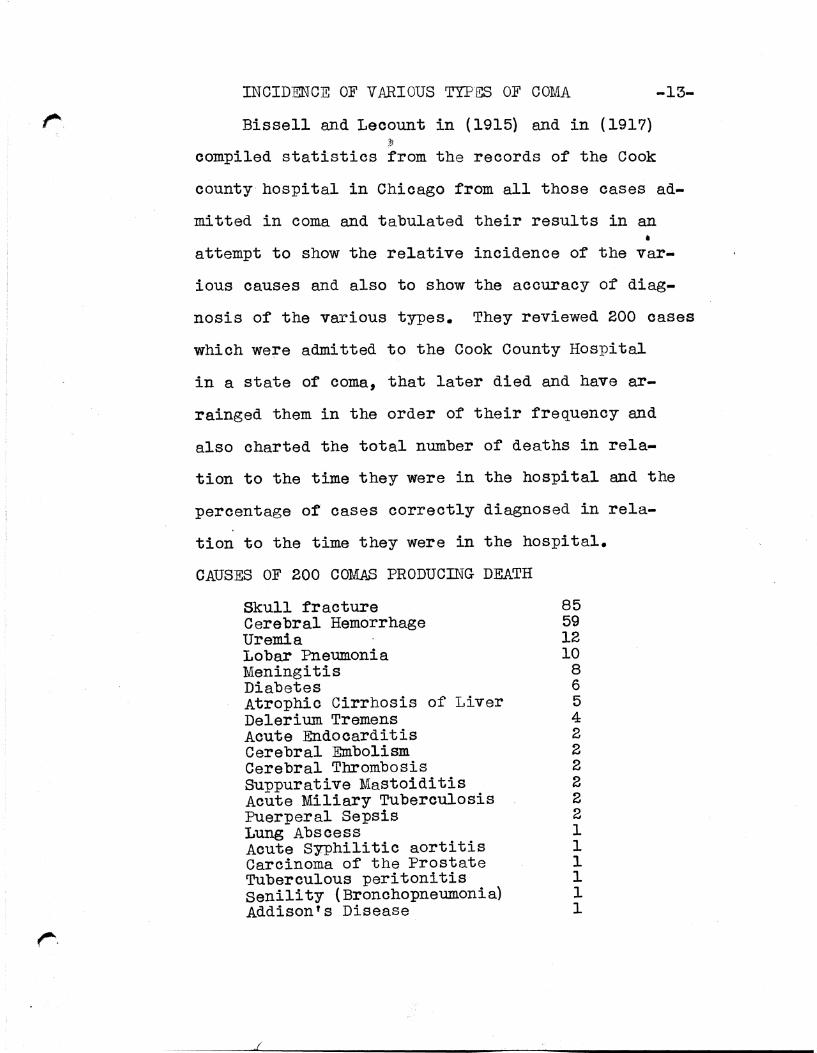
INCIDEl':WE OF VARIOUS TYP~S OF COMA -13-
Bissell and Lecount in ( 1915) and in ( 1917) ,l!
compiled statistics from the records of the Cook
county hospital in Chicago from all those cases ad
mitted in coma and tabulated their results in an •
attempt to show the relative incidence of the var-
ious causes and also to show the accuracy of diag
nosis of the various types. They reviewed 200 cases
which were admitted to the Cook County Hospital
in a state of coma, that later died and have ar
rainged them in the order of their frequency and
also charted the total number of deaths in rela
tion to the time they were in the hospital and the
percentage of cases correctly diagnosed in rela-
tion to the time they were in the hospital.
CAUSES OF 200 COM.AS PRODUCING DEATH
Skull fracture 85 Cerebral Hemorrhage 59 Uremia 12 Lobar Pneumonia 10 Meningitis 8 Diabetes 6 Atrophic Cirrhosis of Liver 5 Delerium Tremens 4 Acute Endocarditis 2 Cerebral Embolism 2 Cerebral Thrombosis 2 Suppurative Mastoiditis 2 Acute Miliary Tuberculosis 2 Puerperal Sepsis 2 Lung Abscess l Acute Syphilitic aortitis 1 Carcinoma of the Prostate 1 Tuberculous peritonitis 1 Senility (Bronchopneumonia) 1 Addison's Disease 1

DEATHS
50
45
40
35
30
25
20
15
10
5
f-.- -· -- -
r
/ '
/ l"---
I I \ I \ \ I \
\ I \ \
\ /
\ \ J \ J --...._ \.V 'I ~
I II
--
I !/ \
\ /\ ,_,..., "- v \
I/ -" 'J~ ,. ~' l
III
/ -....., ...--/
:v
-14-
I\ I ./
\
~'
F OH pital days.
In the above chart, the black line represents the number of deaths and the day on which they occurred and the blue line represents the number of the oases that were correctly diagnosed before death. The diagnoses in all cases were confirmed by post mortem examination.

-15-
0f the 200 deaths which occured over a period
of two and one half years 47 occured within four
hours after admission to the hospital and 42.5%
of these were correctly diagnosed. Eighteen deaths
occuring from four to eight hours contained 10 or
55.5% correct diagnoses. Of 116 deaths within 24
hours after admission there were 59 or 50.7% that
were correctly diagnosed. On the second day.there
were 35 deaths, 37% of which were correctly diagnosed,
and on the fourth day 9 deaths of which 44.4% were
correctly diagnosed. Of the remaining 26 deaths
ocouring from the fifth to the seventeenth day,
inclusive, eighteen or 70% were correctly diagnosed.
The conclusions that may be drawn from these fig
ures might be stated as follows:
1. over half of the patients admitted in coma
died within 24 hours.
2. Few live longer than five days.
3. Of those who died within four days the accuracy
of diagnosis was little affected by the time elemen"t
But of the deaths ooouring from the fifth to the
seventeenth day, inclusive, the accuracy of diagnosis
seems to increase with the increase of time in which
the condition may be studied.
Two years later the same two men added to their

-16-
above study an additional 200 cases and have tabu
lated their results in a similar manner.
CAUSES OF 4()Q COM.AS PRODUCING DEATH
CAUSE Number of cases Percentage Skull Fracture 144 36 Cerebral Hemorrhage 95 23.75 Miscellaneous 26 6.5 Meningitis 21 5.25 Lobar Pneumonia 19 4.75 Uremia 14 3.5 cardiovascular disease 14 3.5 cerebral Circulatory
conditions 12 3. Delerium Tram.ens 11 2.75 Atrophic Cirrhosis 7 1.75 Diabetes mellitus 6 1.5 Brain Abscess 4 1. Traumatic Hemorrhage 4 1. Fracture ribs 3 .75 Fractured vertebrae 3 .75 Senility (Bronchopneumonia)2 .5 Suppurative mastoiditis 2 .5 Acute Miliary
Tuberculosis ]. .25 Puerperal Sepsis l. .25 Lung Abscess 1 .25 Ttlberculous peritonitis l. .25 Carcinoma of Prostate 1 .25 Addison's disease 1 .25 Bronohiectasis 1 .25 Eclam.psia 1 .25 Erysipelas 1 .25 Epilepsy 1 .25 Poisoning 1 .25 Pulmonary Tuberculosis Fractured Mandible with phlegmanous condition of
.25 face 1
Total 400 cases
In the chart that follows on the next page, the black line represents the number of deaths for . each
tour hour period and the lower blue line represents

-17-
the number of each of these groups receiving cor-
rect clinical diagnoses.
DEATHS ---.,_ __ ---==----95
90 ---
80 t:: ..... :n:t=l .....
75
65 ::: = ..... = ..... -60f - -- -55 ==t: = ........ 50 -~ --
45
--__ ..._
------
40 -tll~:::t:
35~ -1±:1::±
3o!ml 25
20
15
1~0 ~~~~~~~~~~~~~1i~.~·~~~~!ilil:fll~ I II III IV v VI VII HOSPITAL
DAYS Thus it: can be seen that 93 died within the i'irst

:_
-is ... four hours after entrance and of these 4?.3% were
correctly diagnosed clinically. Similarly 230
·died within 24 hours after admission and of these
127 or 55.2% were correctly diagnosed. The abrupt
descent of both curves from the first four hours
to the end of the third day is a most graphic ex
pression of the high and rapid mortality of persons
entering the Cook County hospital in coma. From
the· fourth to the 31st day 84 died and of these
49 or 58.33% were diagnosed correctly.
In the above chart 21 deaths in coma are not
plotted: They occured as follows; 6 on the 8th
day, 3 on the 9th, 4 on the 10th, 1 on the 11th, 1
on the 21st, 1 on the 13th, 1 on the 14th, one on
22nd, 1 on the 28th, 1 on the 31st. The clinical
diagnoses were correct in 11 of these cases.
It is also noted by Bissell & Lecount, (1917}
that in comparing the two sets of figures as to skull
fracture that of the 200 cases in 1915, 61 or 72%
of 85 deaths from skull fracture were diagnosed cor
rectly and in the second series, 46 or 78% were
diagnosed correctly. Of those deaths occuring from
Cerebral hemorrhage 27 or 51% of 53 deaths were
diagnosed correctly in the first series while 31 or
74% of 42 deaths in the second series were diagnosed
correctly.

-19-
Four years later we find a similar study by
Blair Holcomb, (1921) in which he studies the re
sults of 346 comatose conditions as they entered
the hospital in a manner similar to that of
Lecount & Bissell.
The cases chosen for this study are those of
patients entering the hospital in coma of obscure
origin who died without regaining consciousness.
They cover the period between the years 1916-1920,
inclusive. Death in coma from such causes as il-
luminating gas and heat stroke have been omitted,
as well as acute fulminating influenza during the
two epidemics of this disease. The details as to
the other patients in coma are not included because
the clinical observation extended for some time and
the disease was well understood, for some recovery
occured and for others no post mortem examination
was made.
CAUSES OF COMA
Skull Fracture Cerebral Hemorrhage Uremia Meningitis Cerebral Thrombosis Pneumonia Alcoholism Cerebral Embolism Diabetic coma Tuberculous Meningitis Syphilis Ruptured Aneurysm
Cont'd
NUMBER 92 88 37 23 20 18 1.6 12
8 6 4 4

Pulmonary Tuberculosis Bran abscess Contusions of Brain Lethargic Encepha1itis Pancreatitis Perforated Duodenal Ulcer Acute Endocarditis Carcinoma of Stomach Em.pyema "Endocar«itt~ (Chronic) Typhoid FeYe-r Otitis Media
TOTAL
3 2 2 2 2 2 1 1 1 1 1 1
346 cases.
-20-
Holcomb has dealt with 346 deaths, 27 of which
ocoured between the 6th and 5lst days inclusive. The
following is a tabulation of the number of deaths
occuring on the first 6 days and the percentage of
correct clinical diagnoses that were made.
HOSPITAL DAY DEATHS
I 186
II 40
III 33
IV 24
V & VI 56
CORRECT DIAGNOSFS
111
28
17
8
18
PERCENTAGE
59%
70
51
33
50
He has compared his results with those of
Bissell & Lecount (1915 & 1917) in an effort to de
termine if' over a period of 6 years there has been
any improvement in making correct diagnoses on such
cases. His findings were that on the first hospital

-21-
day that there were approximately 8% less correct
diagnoses in his se~s of cases than those of Bis-
sell and Lecount, that on the second and third days
there was an improvement of 33% and 8.2% respectivelt, v
and that on the fourth day the two earlier series
showed 11.4% more correct diagnoses and for the re
mainder of the time the two earlier reports showed
2% more correct diagnoses than the summary by Hol
comb. He concludes finally that the accuracy of
clinical diagnosis does not depend upon the length
of time for observation. It would also seem that the
total number of correct clinical diagnoses in the
two different studies do not vary enough to be of
significance.
The next attempt to arrive at the frequency of
the different causes of coma was made by Solomon and
.A%ing, (1934). Their series of cases is taken from
the entries of the Boston City Hospital, and in
cludes cases entering the hospital in a state of
coma, the etiology of which has not been determined.
The total number of eases was 1 1 167, or 3% of the
total hospital entries for the time studied and it
is noted that 68% of the total number entered the
hospital without any history.

r".
-22-
DISEASE NUMBER FATALITIES PERC.ENTAGE Alcoholism 690 14 2 Trauma 152 48 31.5 cerebro-vascular lesions 118 91 77.1 Poisoning 33 3 9 Epilepsy 28 0 0 Diabetes 20 11 55 Meningitis 20 20 100 Pneumonia 20 18 90 Cardiac Deoompensation 17 12 70.6 EXsanguination 10 10 100 Ceq1;ral Nervous 0
system syphilis 7 0 0 Uremia 7 7 100 Eclampsia 'l 3 42 Miscellaneous 38 26 68.4
It will be noted immediately that the same
causes do not occupy the same relative positions
in this table and those of Bissell and Lecount and
of Holcomb, however their studies were based on
deaths from coma while the above set of figures is
based on the number of entries in comatose states.
vit was also noted that when the total number
of patients who entered the hospital in coma was
considered from the standpoint of correct diagnoses
that 94% of the total number were correctly diagnosed .•
Solomon and Aring, (1936}
In addition to the afore-mentioned studies,
w. H. Lewis Jr., (1938) in the same series of oases
has recorded the percentage of fatalities of some
of the more common causes as follows: Hemorrhage and
shock, 100%; Meningitis, 100%; Eolampsia, 43%; Cer
ebrovascular lesions, 70%; Fracture of skull, 32%;
Alcoholism, 2%; and Epilepsy, o.

GDERAL APPROACH -23-
Before attamptintt to give the diff erentiatinc
points of any individual typea of eoma it seema
wise to discuss in a general way the approaeh to
eomatose conditions, some of the various methods
that may be emplOJ'•d and their relative merit.
First of all it might be said that the differ
ential. diagnosis of a patient in coma. ia no differ
ent than any diagnostic proaeedure in that it is
neeessary to base one'• conclusions or diagnosis on
the history, physical examination, laboratory fin
dings, and radiological findings. However, as one
author has stated, (Solomon & Arina;, 1936) •Rem
ember that this is essentially vetinary medieine, and
that your patient cannot help you by telling you
a thit1c•.
In the majority of eases it will be possible
to obtain some sort of a history either from an
associate of the patient or in case of aeeidents,
from someone who saw the accident or from the ambu-
lance driver who brings the patient to the hospital.
In obtaining a history the examiner should keep
in mind of course the more JQ&jor eausea of coma
which will include, .Alcoholism, Traumatie or acquired
brain lesions of all kinds, Poisoning, Uremia, Dia
betes mellitus, Cardiac conditions, and in Tiew
of the recent far reaching advances in endocrinology

f"''.
-24-
one should certainly remember to think of the var-
ious crise• oceuring with a pronounced increase
or decrease in function of any one or group ot
endocrine glanda~
J'riedllan, (1933) has said of eoma cases "first
examine, diagnosticate, then treat•, and gives
some eood suggestions to be followed in taking a
history.
l. Always inquire as to the presence of any
previous diseases, such as aaeites, sinus infections,
diabetes, nephritis, hypertension, syphilis, hyper
thyroidism, or hypothyroidism etc.
2. Always ask as to the oceurence of any pre
vious injury. -...
3. Kode of onset: Was it sUcfi!n, aradualJ were
there convulsions, etc.?
4. Associated symptoms such as convulsions,
headache, diziness, vomiting, ete.
The history of a. patient's habits will have
some bearing on the diagnosis, especially in cases
of alcoholism. It has been said by str•use and
Binawanger, (1936) that the history of p~tient
being a chronic a.lcoholie or a total abstainer adds
mueh to the finding of an alcoholic breath and
makes it mean more than merely its presence may im-
-25-
ply, since alcoholic breath is present in so many
instances where it is not actually responsible for
the coma.
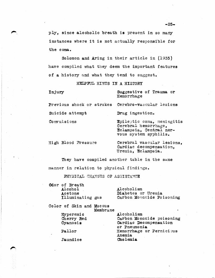
Solomon and Aring in their article in (1935)
have compiled what tbey deem the important features
of a history and what they tend to suggest.
HELPFUL HINTS IN A HISTORY
Injury Suggestive of Trauma or Hemorrhage
Previous shock: or strokes Cerebra-vascular lesions
suicide attempt Drug ingestion.
Convulsions Epileptic coma, meningitis Cerebral hemorrhage, Ecla.mpsia, Central nervous system syphilis.
High Blood Pressure Cerebral vascular lesions, Cardiac decompensation, Uremia, Eclampsia.
They have compiled another table in the same
manner in relation to physical findings.
PHYSICAL CHAMGES OF ASSISTA')\TCE
Odor of Br ea th Alcohol Acetone Illuminating gas
Color of Skin and Mucous :Membrane
Hyperemic Cherry Red Cya.nosia
Pallor
.Jaundice
Alcoholism Diabetes or Uremia Carbon Monoxide Poisoning
Alcoholism Carbon Monoxide poisoning Cardiac Decompensation or Pneumonia Hemorrhage or PerniciJus Anemia Cholemia

-26-
Local Signs of Injury Trauma Burn Hemorrhage Epilepsy Erysipelas
Temperature Increased
Decreased
Pulse :Rapid
Irregular Slow
Respiration mi small
.Increased
Hemiplegia
Observation of Convulsions
Vomiting
Stiff ·Neck
Kernig's leg sign
Ohest signa Consolidation Fluid
Pulmonary Congestion
Distention and spastici ty of abdomen
Pneumonia, Meningitis, Encephalitis Carbon Monixide poisoning Diabetes.
Diabetes, Pneumonia Meningitis, Eclampsia
Cardiac Decompensation Stokes-Adams Disease
Diabetes Pneumonia
Cerebra-vascular lesions
Epilepsy, Cerebrovascular lesions, Central Nervous system syphilis, and Alcoholism
Cerebral hemorrhage and Poisoning
Meningitis, Cerebro-vascular lesions
Meningitis and Cerebrovascular lesions.
Pneumonia Ruptured aortic aneurysm, and Empyema
Ascites, enlarged liver, distended neck veins; Cardiac decompensation.
Ruptured Esop~age?l Yarices, Carcinomatous erosion of G.I. Tract.

f"'- .
lluscular twitching
Abdominal Tumor
Bul~ing Fontaneles
Soft Eyeballs
Wounds or Scars on Tongue
Vaginal Abnormality
Blood Pressure Increased
-27-
Ruptured eotopic gestation, Miliary Tuberculosis.
Uremia
Ecla.mpsia
:Meningitis
Diabetes :Mellitus
Epilepsy
Pelvic malignancy or ruptured ectopic.
Cardiovascular lesion, Eclampsia, Uremia, Trauma
No physical examination of a comatose patient
would be complete without a thorough neurological
examination. A summary of an article found in
"Practice of Jledicine• by Tice on this phase of
examination is theref cre presented.
Naturally a different technic must be employed
for patients in coma, stupor, aphasia or of those
who are uncooperative because of their mental st.~jte.
Careful inspection for scalp bruises and lacer
ations or skull inj"1ry should be first conducted.
Occassionally depressions due to fractures may be
found. A facial palsy may be detected by droolinc
from one corner of the mouth or blowing out more of
one cheek during expiration. All the pupillary signs
should be looked for as it is important to detennine

-28-
if a forced con.jugate deviation of the eyes is pre-
sent.
The usual methods are u:..0 ed to determine mu,~' cle
tone. A clue to the presence of Ll paralysis can be
obtained by raising tha limbs and permitting them
to fall by their own eight to the bed-:;lotbes. If
the two limbs on th"' same side fall in that manner
and those of the opposite side do not, a heroiplegia
is suspected. N0rmally, if the stupor is not too
deep, they do not fall limply. Postures which resemble
the decerebrate should be looked for. Painful stim-
uli :rm.y cause movements of the limbs. Su~?ra-orbi-
tal pressure may cause movement of the face.
Reflexes may be elicited in the usual manner,
emphasizing particularly the plantar reflex, sphin--
cter control and tonic neck reflexes. The reaction
to pain sensation may be indicated by the '.vithdrawal
of a limb. If pain is felt on a ~aralyzed side, the
opposite limb will reflexly attempt to brush the pain
source aside. Bringing the finger into the lateral
visual field very close to the eye should cuuse a
defense closure of the lids, otherwise a hemianopsia
may be suspected. Opthalmoscopic examination is very
important, especially before a spinal _puncture is
attempted.
. ..

-29-
Si nce in the case of many poisons, the over
dosage of which will produce a comatose condition,
produce definite pupillary re~ctions it is important
that we note the pupils in examining a com3tose pa
tient. VI. C. Menninger (1927) has c:nductc:d a study
of the "91-llJillary conditions found in some various
farms of coma, a brief report of wh i ".?h follows.
His study is ba::::ed on Coma resulting from
Alcoholism, Diabetes, Uremia, Cerebral hemorrhage,
Pontine hemorrhage, Carbon monoxide poisoning, and
:Fracture of the skull.
In 58 cases of alcoholic com.a, the finding that
was most persistent was contraction in 43.1% of the
cases •. This is contrc.idictory, according to Menninger
from most writings in that most of them maintain that
the pupils are dilated. Many authors report that
anisocoria, or inequality of t'1e pupils is a common
finding in alcoholism but Menninger found it in only
13.8" of cases. It is commonly accepted that chronic
alcoholism tends to give a fixed pupil where in Men
ninger's series it was found in less than half or in
36.2% of the cases.
In his study of 10 cat1es of Diabetic coma, the
most common findings were mid-dilation and a prompt
reaction to light.
In his study of 8 c;.~ses of Uremic poisoning, the

-30-
o nly finding that stood out by any c0nstancy at all
v:ras a sluggish re:1ction to light.
In a study of 10 cases of Cerebral hemorrhage an
inequality of the pupils was found in 60% of the
cases and fixed pupils were found in 90°'° of the cases,
and 50% of the pupils were dilated, rather than being
mid-dilated or contracted.
In a study of 25 proven cases of fr~cture of
the skull, 10 showed mid-dilation, 9 contraction, 10
were fixed, 8 sluggish in their reaction to light
while ? were prompt, and 8 showed inequality. In
30 probably cases of skull fracture 13 showed ine
quality of the pupils, ? dilation, 4 mid-dilation,
5 co·ntraction, while 14 ·were not noted. In their
reaction to light, 10 were prom-pt, 5 sluggish, 13
fixed and 2 were not noted.
In a review of 2 cases of Pontine hemorrhage,
they both showed contraction of the pupil but both
cases were at variance on tbe other fe~:.tures. Of
three cases of sus:_)ected pontine hemorrhage that
were not posted 3 showed contraction, 2 fixation, and
2 inequality.
There were '43 cases of Carbon monoxide poisoning
and the only findings that Y1ere present with any
regularity were mid-dilation in 50. 6% of cases and
a prompt reaction to light in 48.3% of cases.
He concludes as follows: •Pupils may aid in
diagnosis of trauma to the brain but they are of

-31-
little or no help in Alcoholism, DiQbetes mellitus,
Uremia or Carbon rnonJxide :poisoning".
Following ci careful history and a thorough
physical examination the proper laboratory procee-
dures or those indicated should be carried out. In
Solomon and Aring's article (1935) they outline the
helpful diagnostic laboratory proceedures and the
things that different findings point to.
LABORATORY OBSERVATIONS
Lumbar Puncture Pressure
Increased
Decreased
Bloody Fluid
Purulent Jluid
Organisms by Smear or Culture
Sugar High Low
Protein High
Spinal Fluid Positive
Cont'd
Cerebra-vascular lesions Trauma, Syphilis of the Central Nervous system.
Diabetes
Cerebra-vascular lesions Trauma
Jleningitis
Meningitis
Diabetes Meningitis
Meningitis or Central Nervous System Syphilis
Central Nervous System Syphilis

Blood Eltamination SUgar High
Low lT.P. :tr. High wasserman positive Low Red count with abnormal smear
Culture positive
Urinalysis SU.gar Gross Albuminaria
Gastric Lavage
Roentgenograms Skull
Lungs
Heaart
Electrocardiograph
Diabetes Insulin Shock Uremia Syphilis
Pernicious anemia or Leukemia
Pneumonia, Meningitis, Septicemia.
Diabetes Eclampsia uremia Cardiac Decompensation
:Ex:amine contents for poisons.
Fracture across middle meningeal artery in extra-dural hematoma.
Pneumonia, Empyema and Kiliary Tuberculosis
Cardiac Decompensation
Heart block, Cardiac Decompensation.
It is felt that examination of the cerebrospinal
fluid plays a part of great importance in the diag
nosis of coma, expecially in differentiating between
the various conditions associated with the Central
Nervous system. The following indications and con-
traindications for spinal puncture are taken from
Merrit and atiith's recent book on the subject. (1938)

' '· I
i
-33-
INDICATIONS: Lumbar puncture should be performed in
any patient with symptoms or signs of meningeal ir
ritation, and in all patients in whom the cerebro-
spinal f.luid findings would be of aid in the diagnosis
and treatment. It is es pee ially valuable in:
1. The diagnosis and treatment of a ~ut e or chronic
inf lamrna ti on.
2. The diagnosis and treatment of injuries to the
head or spine.
3. The diagnosis of diseases of the central nervous
system in which the clinical signs and symptoms
are not diagnostic.
4. The intelligent treatment of syphilis.
CONTRAINDICATIONS: Inmbar puncture should not be
performed if it is necessary to puncture through in-
fected skin or subcutaneous tissues. A puncture 18
often necessary to establish the diagnosis of spinal
epidural abscess, and it can be safely performed if
the proper precautions are followed.
Puncture of the subarachnoid space is contra-
indicated whenever the diagnosis is established and
no additional information in regard to treatment
would be obtained. This is especially true in pa-
tients having high grade choked discs. Of ten, how-
ever, a patient has signs and symptoms suggestive of
an expanding intracranial lesion, but lumbar punctures
have obviated many needless craniotomies. Further
r. .34. -
references will be made to spinal fluid findings in
the chapter that deals with the different types of
coma.
A very important proceedure, both as to diagnosis
and treatment of comatose conditions where ac·idental
or purposeful poisoning is suspected is lavage of
the stomach. In fact, according to Strause and
Binswanger, (1936) it is the first thing to do in
all such cases.
According to H. L. Ma.matt, {1933) it should not
be forgotten that lavage in comatose patients is not
to be accomplished without a certain amount of danger.
Often the cough reflex is absent, and this may prove
very dangerous to the patient •. It is highly advisable
according to Mamott to do the proceedure in the op~
erating room if possible, where he should be put in
the Trendellenberg position. Additional advantage of
being in the operating room are access to electrical
suction, apparatus for oxygen and carbon dioxide ad
ministration and good light.
---------- ------------- -~---------

~ ' .
DIFFERENTIAL DIAGNOSIS -35-
In an effort to include as many as possible types,
in as systematic a manner as possible the different
types of coma will be discussed in the order in which
they appear in French's classification. From time to
time the outline will be deserted to make use of dis-
cussions or differential points found in the literature.
Likewise, some of French's causes will be omitted since
they do not fall within the phase of the subject being
dealt with in this paper.
In the first group of causes or that group of
infectious fevers in which coma may occur as a ter-
minal phenomenon, there are few of these causes that
will bring the patient into the hospital in coma with-
out the diagnosis being made before coma intervenes.
However, Solomon and Arine (1935) warn the physician
that pneumonia is a commoner cause of such a condi-
tion than is commonly suspected and should never be
overlooked in a differential diagnosis. In very
young or old patients it should always be suspected,
and signs in the chest are always present, at least
in the group of cases which they studied. In Solo
mon~ a article (1938) he says that in cases of pneu
monia, the fever, rapid respirations, cyanosis and
chest signs are practically diagnostic.

-36-
Next in French's classification are those acute
inflammatory lesions of the brain or meninges, the
first of which is acute encephalitis, or as named by
Oecil (1937) "Postinfection Encephalitis: The dis
ease is characterized by producing either a myelitic
or encephalitic syndrome, and is rarely insidious in
onset, and is ushered in by pyrexia, headache, vomi
ting, and drowsiness. A preceeding infection will be
very helpful in making a diagnosis. According to
Houston and Smith (1938) the Cerebrospinal fluid
picture includes a normal or increased pressure, a
moderate pleocytosis, an increased protein content
and a normal or midzone gold curve. According
to Cecil, (1937) the main entities that will be
confused with it in a. differential way a.re the other
encephalitides, but however the previous presence
will usually help rule these out. The actual diag
nosis according to Rivers, (Cecil 1937) cannot be
made until autopsy.
The diagnosis of a case of Encephalitis Letha.r
gica. will usually be made easier by the presence of
an epidemic, and should always be suspected in puz
zling cases of fever and delerium in the presence of
an epidemic. According to Cecil, it may dis9lay many
and numerous symptoms, at the onset, the most common

-37-
of which are somnolence, meningeal irritation, external
or internal opthalmoplegia, neuralgias or paralyses
in the cerebral nerve domains. According to Merrit
and Smith,· (1938) the cerebrospinal fluid in epidem-
ic encephalitis shows no changes or only slight de
viations from normal. There may be a slight pleocy
tosis and a mild change in the colloidal gold curve
are the only significant changes that occur. The
presence of any pronounced abnormal finding should
cast: a good deal of doubt on the diagnosis.
The JCeningitidea
The meninges of the brain may become inflammed
by practically any organism that will attack human
tissues. '.lhe infection may be the termi.nal stage of
an epidemic form, otherwise called Epidemia Cerebro
spinal meningitis or a lymphatic or blood born ex
tension from some other source of inf ectiun. Accor
ding to Weal et al, (1934) the most common causative
agents of meningitis are the meningococcus, pneumo
coccus, streptococcus, -staphylococcus, and the in
fluenzae bacillus. However, it may be caused by any
organism, twe of the conunoner ones being the Tuber
cle bacillus and the spirochaete of syphilis.
In considering the physical signs, helpful in
diagnosing a case of purulent meningitis of the acute
-38-
variety, one might say that the most common findings
are retraction of the head, stiffness of the neck,
positive Eernia's and Brudzinski's signs, a relatively
slow pulse and often other vagus irregularities, (Cecil,
1937 ).
Important in ma.king a diagn~sis will be the re
sults of lumbar puncture. Merritt and Smith, (1938)
say that the changes in the spinal fluid in a case of
acute purulent meningitis, will be practically the
same regardless of the organism, excluding syphilis
and tuberculosis. The changes consist chiefly of an
increase in pressure, a pleocytosis, an increase in
protein, and a decrease in sugar and chloride contents.
The fluid in the early stages of the disease are only
slightly turbid and later become cloudy or frankly
purulent. In the majority of cases the cell count is
between 1000 and 10,000 cells.
In diagnosing a case of !uberculous meningitis,
history of contac, or radiological evidence ·of an acid
fast infection, less severe signs of meningeal irrita
tion than in the usual case of meningitis a.re factors
that point toward an acid fast meningitis. The phy-
sical signs present are very similar to those found
in the more acute types of meningitis except for the
fact that they are usually not so marked. The spinal

-39-
fluid findings of diagnostic significance are an in-
crease in pressure, a pleocytosis, an increase in
protein and a decrease in the sugar and chloride con
tents. The type and grade of the pleocytosis, the
amount of sugar and chloride together with the bac
teriological findings, distinguish tuberculous from
the other forms of meni'ngitis.
The pressure on the whole is less than in the
acute type of meningitis, varying in most cases from
150 mm. to 200 mm. in contrast to the more acute var-
iety where it is usually between 200 and 500 mm. (Ker
rit & Smith, 1935). The fluids in most instances are
clear, especially so when compared to the pUrulent
fluids of the acute types. There is commonly a faint
yellow color and a tendency for the fluid to form
pellieles when allowed to drop. The cell count varies
but in a great majority of cases is between 50 and
500 cells.· The protein is between 100 and 500 mg.%
in more than half of the eases, and the sugar aver
ages 28 mg. per 100 ec. The chlorides are moderately
reduced averaging about 608 mg •. per 100 cc. in 60
eases, of ](errf~t and &nith, (1935)
The value of Cerebrospinal fluid examination in
cases of tuberculous meningitis is further emphasized

-40-
by L. L. Krafchik, and Slobody, (1938) when they re
ported ~ case of coma in a child that strongly re
sembled and was diagnosed and treated as Diabetes
mellitus and after failure of the patient to respond to
insulin therapy, repeated spinal taps proved the case
to be due to an acid fast infection of the menin-
ges.
Another similar case to the above is reported by
A. E. Rouselle, (1926) in which he reports a cases
of pneumococcie meningitis that was treated as a
diabetic coma until proper laboratory studies mis
proved the diagnosis.
Since as medical students, we are often told
that Syphilis is the great imitator, no attempt will
be made to completely cover all $f its different mani
festations in the central nervous system but a brief
discussion of Syphilitic meningitis as an entity
follows.
According to Cecil, (1937) the meninges are
involved in a great percentage of cases but the num-
ber of cases that show definite clinical evidence and
especially comatose conditions are in the minority
of cases. These cases occur early in the course of
the disease, commonly with the skin symptoms. The
disease at this stage seems to have a predilection. for

-41-
the basal portion of the brain and of ten involves
the crainial nerves. Cecil, (1937) warns that onset
of epileptic convulsions in anyone thirty or more
should strongly suggest syphilis of the meninges.
The Cerebrospinal fluid ina cute syphilitic
meningitis, according to Merrit and Smith, (1938)
ahows an increased pressure, a pleocytosis of a
varying degree, an increased protein content, an ab-
normal colloidal gold reaction and a positive Was
serman reaction. Strong,(Cecil, 1937) warns that
the Wasserman reaction may be negative. The pressure
is usually above 200 mm., the fluid usually co1orless1 or
slightly yellowish. The cell count averaged 450
cells per cubic millimeter, the protein averaged
110 mg. ~ the sugar 49 mg.%, the chlorides 696 mg.
per 100 cc. The colloidal gold curve was abnormal
in 95 % of cases and the Wasserman reaction positive
in 85% of cases.
Although Cerebral Trypanosomiasis is practically
limited to tropical countries, it should be mentioned
as a cause of coma. The stage of coma has usually
been preceeded by a prodromal stage in which the pa
tient has a fever, polya'denitis, and asthenia, fol
lowed by sleep and coma. ~iagnosis, although sug
gested by certain clinical symptoms cannot be made

-42-
exc ept by demonstration of the parasite which is
accomplished by demonstration in the blood, but more
often and easier from infected glands. (Cecil, 1937)
Less Acute Lesions of the Central Nervous System
While, these conditions are not as frequent in
the etiology of comatose states; in-pati;e:Ats entering
the hospital without a diagnosis, they are still
worthy of consideration as conditions that must be
ruled out in many eases. From Solomon and Aring's
report (1934) it will be noted that Central Nervous
System Syphilis and Epilepsy are the only ones of
this group that appeared in 1167 cases, however this
does not rule them out as possible causes of the
phase :of the subject with which this paper deals.
It is not common for brain tumors to cause
coma without first causing symptoms severe enough to
bring the patient to his physician. These symptoms
ac:ording to Dr. Keegan, (.runior notes in Surgery,
1937), may be classified as general and local signs.
The general signs will be those of increased intra
cranial pressure, vomiting, ~eadache, and papilloedema.
The local signs will be abnormal neurological findings
depending upon the location of the tumor.
According to Merritt and Smith, (1938) Brain

-43-
tumo rs must be differentiated from Brain abscess,
Subdural hematoma, Cerebral hemorrhage, Cerebral
thrombosis, Syphilis Of the nervous system, vremia,
Encephalitis lethargica and multiple sclerosis.
Brain Abscesses always show a pleocytosis with
a large percentage of polymorphonuclear leukocytes,
and a history of a septic focus can usually be ob
tained. Subdural hematoma cannot usually be dif
ferentiated from brain tumor except by a history of
trauma.
Cerebra-vascular lesions. can usually be dif
ferentiated by the presence of a frankly bloody fluid
in cases of the cerebra-vascular conditions.
Uremia can be excluded by the normal nonprotein
nitrogen in case of brain twnor and Encephalitis
lethargica and multiple sclerosis are excluded by an
increased cerebrospinal fluid pressure. Syphilis of
the nervous system may off er more trouble, but Was
sernan test is the best method and should always be
repeated to exclude the possibility of a false posi
tive.
The presence of a brain abscess is usu~lly pre
ceeded, as mentioned above by a focus of infection,
such as a mastoiditis, sinusitis, furunculosis etc.
According to Cecil, (1937) Brain abscesses do not

-44-
caus e complications such as are being dealt with in
this paper until they erode into the ventricles or
the subarachnoid space. The symptoms caused by such
an occurence are; chills, hyperexia, delerium, con
vulsions, coma and eventually death. The spinal
fluid findings according to Merritt and Smith, (1938)
are an increased pressure, a mild or moderate pleo
cytosis, an increased protein content and a normal
sugar content.
Post epileptic coma in Solomon and Aring's in
vestigations, (1935) was usually diagnosed by history
of previous attacks and abrupt onset.
According to Lennox and Merritt, (1936) the
Cerebrospinal fluid findings in epilepsy are prac
tically always normal, except for a few cases of in
ereased pressure, and they say in conclusion that
in the event of abnormal findings in the cerebro
spinal fluid that a diagnosis of essential epilepsy
should certainly be questioned.
Disseminated or :Multiple sclerosis as a cause
of coma without the previous history of symptoms of the
disease is so rare that it is practically never eri
couritered. 'l'he history of remissions of symptoms,
the previous presence of Cbarcot's triad, Nystagmus,
intention tremor, and scanning speech are diagnostic.
Tne spinal fluid findings are usually negative.

-45-
Syphi lit i c involvement of the central nervous
system has been previously mentioned, but the type
mentioned with this group of conditions is mo re com
monly known as Paresis or Syphilis of the brain.
It is usually preeeeded by signs of mental deteriora
tion. The diagnosis, if the patient is seen for the
first time in coma will depend upon history of pre
vious infection, positive spinal fluid Wasserman,
and a paretic type of gold curve.
Metabolic Disorders:
The diagnosis of these conditions i.n prompt or
der is much more important now than it was ten years
ago since the rapid advances in endocrinology have
made it possible to better treat them.
Uremia is a very common cause of coma and is
usually diagnosed by history of kidney disea0e,
muscular twitehings, acetone breath, abnormal eye
grounds, enlargement of the heart, grossly abnormal
urine and an increase in the blood non protein ni
trogen. Solomon and Aring, (1935) •
According to Lecount~ and Guy, (1925) Uremia may
easily be confused with Spontaneous intracrania.l
hemorrhage and should be seriously considered in the
differential diagnosis. They reviewed 30 autopsied
cases in which intracranial hemorrhage was the cause

-46-
of death. Nine of these had been clinically diag
nosed as uremia, and in only three cases had the
other been mentioned as a possibility. The main
differential points according to the above men are,
blood in the cerebrospinal fluid in the case of
hemorrhage and elevated non protein nitrogen in the
case of uremia.
The acidosis resulting from the derrangements of
metabolism in Diabetes mellitus is a common etiolog
ical factor in patients being brought to the hospital
in coma. The coma is usually preceeded by an in
crease in the cardinal symptoms of Diabetes, poly
phagia, polydypsia, and polyuria, along with nervous
irritability, later nausea, vomiting and the patient
may even show the picture of an acute abdomen. ~'then
coma finally intervenes, the patient exhibits a deep
regular sighing type of breathing, described by
Kusmall. The main findings are dry skin, soft eye
balls, dry beefy red tongue, Kussmal breathing, ace
tone odor to breath, rapid heart, low blood pressure
and occasionally anuria. Laboratory proceedures
will reveal acetone and diacetic acid and sugar in
the urine and carbon dioxide combining '.Jower of the
blood will be decreased. The above and the following
table are taken from an article by J. T. Beardwood,
Jr., (1938)

-47-,.,..._ {insulin Shock) Symptom Diabetic HYpoglycemia
Acidosis Regular Protamin•
Onset Gradual Rapid Gradual 12-48 hrs.
Premont tory Nausea, Hunger Headache Symptoms Anorexia Irritability Memory loss
Headache
Convulsions J'la.re Late Occasionally
lluscular Twitching Absent Frequent Frequent
Respirations Kus small Normal Normal
Breath Acetone Normal Normal odor
Skin Dry )(oist Normal or lloist
Eye~ Soft Normal Normal eyeballs
Pupils Dilated Normal or Normal or Contracted Contracted
Vomiting ::rrequent Absent Infrequent
.A.bdomi nal Common Absent Bare pain
Blood Pressure SUbnor:ma.1 Normal or Elevated elevated
Temperature Subnormal Subnormal SUbnormal llapid rise
Urine SUgar, Ace-sugar free No sugar tone & Di- No Ket ones acetic.
Blood High sugar Low Sugar Same Low Co2 Normal Co2
W.B. C. Increased Normal Normal
Response to None Prompt Prompt ,..., Glucose response response Ka.y Relapse

-48 ...
In addition to the above information, E. J'.
K-epler, (1938) adds that the oral secretions are te
nacious and viscid, the tendon reflexes absent, and
that there is no babinski reflex.
Hypoglycemia is a condition that will produce
coma. It may result from inward abnormalities of
metabolism or from over dosage of insulin. Accor
ding to Kepler, the symptoms depend upon rapidity
of change in the level of the blood sugar. The con
dition is preceeded by parasthesias of the limbs,
general convulsions, maniacal behavior, weakness,
tremor, sweating, hunger, ataxia, and distorted
speech. There is usually a rapid heart, dilated
pupils and a positive babinski. ~uantitive blood
sugar studies differentiate it from confusing oon
di tions.
The crisis of Addison's disease, although a
rare condition, (Kepler, 1938) should be mentioned.
It may be identified by the prodromal signs of buccal
pigmentation, hypotension, asthenia, fatigue, apathy,
constantly decreasing blood pressure, blood volume and
1ubnormal metabolic rate, and temperature. The signs
of help after the patient is comatose are; hiccups,
nausea, mania, mus;ular twitchings, meningeal irri
tation, increased blood urea, polyuria, and a poor
tolerance for potassium.
-49-
The crisis associated with E:scopthalmic goiter is
more common according to Kepler than one would ex
pect and is encountered in the early fulminating
group, old cases discontinuing iodine and following
thyroideetomy among patients who have had exopthal
mic goiter and who have not been given sufficient
iodine preoperatively. The findings in addition
to the cardinal symptoms of hyperthyroidism are ano
rexia, diarrhea, crying spells, and fibrillation.
]);)rland and Kepler, {1938) say that the diagnosis
is never difficult unless the hy-perthyroid manifes
tations are minimal.
A. R. Barnes, (1927) describes a case that was
preceeded by influenza in which the only symqtom of
hyperthyroidism at the time of admission was auri
cular fibrillation coupled with a moderately enlarged
gland. The case promptly responded to iodine treat
ment.
Spandler and Bilbon, (1937) report two cases of
thyroid crisis in which they were confused with diabe
tic coma., and summarize their article by saying, "VI.hen
stupor is accompanied by high fever, restlessness
or delerium and rapid ectopic cardiac rythm, then the
clinical picture is strongly suggestive of thyroid
storm and it is important to recognize it since treat-
ment ea.n be very effective".

-50·
Acute Parathyroid insufficiency most commonly
occurs following thyroidectomy, when they are re
moved in error. The symptoms are the results of
hypocaleemia. It is diagnosed by blood calcium de-
terminations following the occurence of convulsions,
which tend to show bilateral tendencies. (Cecil,L937)
Vyxoedema may be easily diagnosed by history
since stages far enough advanced to produce c~ma have
a definite history back of them. According to Merrit
and Smith, (1938) l(yxoedema. is commonly confused with
brain tumor in that they both have a similar spinal
fluid picture. They both show an increased pressure
and an increase in protein. In such cases a basal
metabolic rate would provide a diagnosis.
Conditions in Which Jaundiae is Present
Th.is will involve conditions in which liver dam-
age or damage to the red cells occurs and will involve
various poisons which will produce acute yello atrophy
of the liver, cirrhosis of the liver etc. The poisons
are T.N. T., and Phosphorous in the main and may be
differentiated in the general manner for differential
diagnosis in these cases. (See Poisons and General
Approach)
According to R. Bauer, (1934) these conditions
are usually very vague and difficult to diagnose, but
according to reeent work it has been found that liver
extract is very helpful in treating such conditions.

-51-
It is theorized that the severe symptoms of coma are
due to a secondary breakdown of protein and that the
liver extract helps the liver to prevent this from
happening. It is also found to be good in post
eclamptic coma.
Conditions Complicating Pregnancy
Besides rupture of an ectopic pregnancy which is
discussed under another heading, these C'.)nditions
are Eclampsia, Puerperal Sepsis, and a type of milk
fever described by Kinnimoth, (1931).
Eclampsia, according to Solomon and Aring, (1935)
in 7 out of 1167 cases, furnished the following
diagnostic features: Pregnancy, convulsions, vomiting,
abdominal tumor, rapid pulse, peripheral edema and
an increased blood pressure.
Puerperal sepsis, offers very few diagnostic
problems from the standpoint of coma. A history
of delivery followed by chills, and an abnormal
temperature and still later, coma w.ould be diag
nostic.
Kinnimoth, (1931) describes a case that occured
in England a few years ago, the summary of which
follows.
After a normal labor, and f ourty eight hours
of normal post partum progress, the patient became

-52-
acutely ill, with severe nausea and vomiting and ran
a rather low grade temperature, not at all compatible
with post partum sepsis. On the third day coma inter
vened, without the patient showing any signs of a
typical puerperal sepsis. She was jaundiced. Acting
on a "hunch", the physician in charge inflated her
breasts with air and within two hours she had come
out of the coma, and in slightly more than 24 hours,
she w;_:is com;Jletely normal, neurologically speaking.
It was diagnosed as milk fever, a condition anala
gous to one found in cattle but infrequently occur
ing in people.
Under the next classification, or late states of
certain other maladies that exhibit prominent symptoms
other than coma before coma supervenes, these topics
will be discussed under other headings, since most of
them nay be more conveniently discussed with other
groups.
The Results of Trauma to the Head
The diagnosis of these cases is usually not dif
ficult since the history of trauma. is usually present.
In Solomon and Aring's (1933) cases they consti
tuted 13% of the total number of cases and they found
a mortality of 31.51.

-53-
They summarized their results later, (1935) and
noted that a bistor·y was usually available, alcoholic
odor was often present on the breath, vomiting was
a common finding and usually meant increased intra-.
cranial pressure.
In the physical examination evidence of injury
was always present, and that a compound fracture with
bleeding and drainage along with signs of surgical
shock were serious prognostic signs. Conditions
which may be confused with it are Epilepsy, Diabetes,
Uremia, Alcoholism, etc., but the history, checked
by x-ray findings will usually determine the trau
matic origin if it be such.
Kerrit and Smith, (1938) say of tbe spinal
fluid findings that it is usually bloody and under
an increased pressure. According to Munro. (1934)
the use of repeated lumbar punctures in conjunction
with the intravenous injections of hypertonic sol
utions is invaluable in the control of the increased
pressure in such cases.
Vaseular Lesions of the Brain
One of the earliest references to such con
ditions is quoted from Hipocrates; (Adams, 1884).
•ti.en the common origin of nerves is affected and

;
i I·
-54-
from it all other parts of the body have lost their
motion and sensibility, the affection is called
,AJ>oplexy, by which the healing energies are impaired.
But if obstruction is in either side, it is called
Hemiplegia and paralysis•.
In considering this group as a whole, Solomon
and Aring (1935) say that of their 118 cases over 90~
were over 40 years of. age. In many the onset was
abrupt, many showed convulsions, as well as pre
vious history of high blood pressure or heart disease.
Common signs present on entry were complete or
partial bemiplegia, stiffness of neck, convulsions,
high fever, elevated blood pressure and unilateral
abnormality of the pupils. Unless contraindicated a
lumbar puncture should always be do'ne. (See con ...
traindieations for lumbar puncture).
According to R. H. ll'.cDonald, (193.8) the most
common accident accompanying cerebro arteriosclerosis
is cerebral hemorrhage. It is common in the aged
male, has a sudden onset, and optbalmoscopio reveals
arteriosclerosis and perhaps edema. Neurological
findings, as well as albuminaria are usually present.
It is to be differentiated from the following enti- ·
ties.

Cerebral Thrombosis
Kore gradual in onset Coma less pronounced Late in middle life and old age.
Cerebral Embolism
Very sudden J'C>llows sur-. gery or trauma
-55-
SUbaraehnoid Hemorrhage llay occur in younger people.· Blood in apinal fluid & Ken• i ngeal · irritation.
Arring and Kerrit (Archives of Internal Med
ieine 9-35) have studied cerebral hemorrhage in re
lation to Cerebral thrombosis and draw the follo-
wing conclusions.
l. The average ace:. O:f· a~. patient ~w1 th- (lere}>~L.hemorrhage is
lower than in cerebral thrombosis.
2.. Blood pressure is higher in cerebral hemorrhage.
3. Arteriosclerosis is more common with cerebral
thrombosis.
4. Bye findings are most often found with hemorrhage.
5. Stiffness of the neck indicates cerebral hemorrhage.
6. Leukocyte count will be more likely to be up in
hemorrhage.
7. Spinal fluid pressure is usually higher in hem-
orrhage.
Houston H. Kerri t, (1938) says that the salient
features of primary suba.rachnoid hemorrhage are cloudy
mental state, stiff neck, positive tlrnic and that
laboratory findings show 10,000 to 20,000 white cells.
There will be blood in the spinal fluid and a spinal

-56-
fluid pressure ot from 300 to 500 mm. of water. The
differential between this and hemorrhage is difficult
but, convulsions preceeding coma favor hemorrhage.
He says of Cerebral embolus that it may occur
at any age that the onset is sudden and that focal
signs are usually present. Other signs favoring
em.bolus are a septic temperature, abnormal heart . . . findings, normal or only slightly elevated spinal
' fluid pressure,negative serological reactions for
Wasserman reaction and a positive blood cul·ture.
Another condition which oeeurs, similar to the
above, is a thrombotic occlusion of any of the venoua
sinuses of the head. This may usually be diagnosed
by the presence of previous infection in the region
drained by the sinuses involved. It is rarely pri-
mary, except in Yarasmus and extreme cardiac weak
ness. (Cecil, 1937). The prominent symptoms of
sinus thrombosis are usually those of meningeal
irr~tation, coupled with venous stasis, which gives
rise to edema and cyanosis of the eyelids, and sur
rounding tissues. The spinal fluid is not greatly
altered unless meningitis has developed. Prognosis
is poor bu. t has been improTed in recent years by
sucaessful surgical attacks •.

-57-
Vlhile it is not a condition associated with
the brain, since embolic phenomena have been dis
cussed under the above heading, a word should be
mentioned as to Pulmonary Embolism. The symptoms
of tbis condition are usually so classical that
differential diagnosis is seldom difficult. They
usually occur in a hospital since they usually fol
low surgical proceedures or a previous illness.
The onset is sudden with pain in the chest or a
sense of tightness in the chest, the patient be
comes an ashen color, anxious, cyanotic and dys
pneic. Recover is rare, death occuring in from
a few minutes to aeveral hours.
Coma. From Overingestion of Poisons and Drugs
Naturally the more common ones will be dis
cussed since the overingestion of any drug or
chemical will cause death if enough is taken.
By far the most common of the group in re
lation to coma is Alcohol. Solomon and Aring, (1935)
report that a history of alcoholism can usually be
obtained. Other features are hyperemia of the face,
injection of the throat and conjunctivae, diminished
or absent reflexes, alsobolic odor to the breath,
vomiting, enlarged heart, pulmonary rales~

~ ... ,•-,--'" .. .
-58-
It may be confused with trauma, since so many
traumatic cases will exhibit alcoholic breath. The
cerebrospinal fluid findings are normal except for •
a slight increase in pressure in about one fourth
of the cases.
Poisoning from all the different derivatives
and alkaloids of the opiates will be considered under
one heading. The prodroma.l sym,1;>toms, after. ingestion
of the drug, a.re euphoria., pleasant bodily sensations,
giddiness, lassitude, dreams, incoordina.tion and
finally coma. History of inge·stion followed by
gradual onset of coma, along with pin point contracted
pupils will help to differentiate it from other con
ditions. Naturally lavage is helpful in both a diag-
nostic and therapeutic way.
Purves, et al, {1934) in a discussion of barbital
poisoning describe the condition as showing a depre
ssant effect on heart, lungs, and kidneys. When
coma supervenes the pupils are moderately contracted
but continue to react to light. The limbs are flac
cid, the tendon reflexes are lacking. The heart is
rapid and the patient cyanotic, the blood pressure falls
and basal pneumonia. may be a complication. They sug
gest gastric lavage both as a diagnostic and thera
peutic proceedure. They also suggest withdrawal of

spinal fluid in an effort to diminish the toxic
effect of the drug on the brain.
-59-
Carbon Monoxide poisong is described by Aring
and Solomon, (1935) as being a condition where the
coma is usually slight, the odor of gas is on the
breath, the skin and mucous membrane are of a cher
ry red color, the temperature is subnormal, and the
pulse high with an increased leukocyte count. Lum
bar puncture is usually normal, the patient responds
to inhalation of carbon dioxide and oxygen and fatal
results occur rarely.
Bromide poisoning is another common cause men
tioned by Solomon and Aring,.and tbey conclude their
discussions on _poisons with the statement that ex
cept for the cherry red color that is pathognoaon1c.~of
carbon monoxide poisoning, that the history and pre
sence of the poison in the gastric contents or the
spinal fluid are the diagnostic features of such con
ditions.
Lead encephalopathy is the result of a more chro
nic form of intoxication of which coma is often the
terminal phase. It should be diagnosed before coma
supervenes but a few of the findings in the comatose
condition will be mentioned. Weller and Christensen,
-60-
(1926) in a summary of the literature on the subject
say that besides the usual findings of lead poisoning
and history of the disease, that the spinal fluid pres
aure:., .:whdich may~::ber::elevatedet,o,:f150i--l600 mm.' is due.
to an extreme degree of cerebral edema. The fluid
may be tinged with yellow but not necessarily. If
pleocytosis is present it is present in a mild de
gree. Lead :may be found in the fluid.
The Effects of Physical Ex:tremes
In this group is included comatose states or
shock resulting from extremes in cold, heat or contact
with high voltage electrical cur:ents. As far as
the diagnostic problem is concerned, the history, when
available will usually make a diagnosis. Earl c. Elkins, (1938) differentiates between heat exhaustion
and heat storke by saying that the first is due to
exhaustion, the skin is pale and clammy, the pulse
rapid, and the temperature is subnormal. Heat stroke
exhibits similar symptoms except that it is usually
more sudden in onset and exhibits a definite rise in
temperature. The treatment in the first case is that
of shock, while that in the second is an attempt to
get the body temperature to normal.
Diagnosis of the other two will be made by his
tory and treatment will be that of shock.

-61-
Effects of Rapid and Great Alteration in the Surrounding Atmospheric Pressure.
This group of conditions, Caison's disease,
coma resulting from bringing divers to~ rapidly to
the surface, and conditions arising wben baloonists
and aviators rise too ra=)idly to great heights will
necessarily depend on history and it is inconceivable
for them to present themselves as causes of c :::ma where
a history would not be available. The cardinal sym
ptom of Caisorls disease is pain or "The bends" in
one or more extremities, fJllo 1Ned later by dyspnea,
choking and finally coma. According to Cecil (1937)
once collapse has occured, recovery is very remote.
Excessive Loss of Blood
According to George Johnson, (1869) the cause of
any type of coma is deoxidation of the brain tissues,
and that this is brought about lack of sufficient
blood, lack of thorough aeration, lack of proper
circulation, or violent contraction of the arteries
which he believed was res9onsible for e~ilepsy.
Of the conditions falling under this heading in
French's classification the diagnosis is usually ob-
vioua except perhaps in the case of a rupture of a
tubal pregnancy.

-62-
According to DeLee, (1934) when a woman in the
reproductive period misses a menstrual period, then
later complains of cramp like :pains in the lower
abdomen, one should always think of an ectopic
gestation. When rupture occurs, and bleeding oc
curs to such an extent that coma intervenes, there
will usually be no or little bl eding externally,
the symptoms will far exceed the ap:par'~nt blood loss,
.a mass may be found alongside of the uterus.
Stokes Adam's Disease and other Heart Conditions
Edgar A. Hines, (July 1938), says, "The Stokes
Adam's syndrome, or 0onvulsive syncope is one of the
most dramatic phenomena of heart dis'2:J.0e, the treat
ment vecy discoura:;ing, and palliative at best".
According to Best and Taylor, (1937) the car
dinal features of the Stokes Adam's syndrome are
as follows: Extrasystoles, delay in A-V conduction,
complete heart block.
Of Solomon and Aring' s cases (1935) l.4"b' were
cases of cardiac decompensation. The diagnostic .
points were: history of heart disease, and high blood
pressure, abnormal heurt findings, cyanosis, pulmonary
congestion, peripheral edema, dyspnea, and quite com
monly an enlarged liver. These findings of course
should be eonfirmed by E.K.G.

Sudden Nervous Shock Hysterical Trance Narcolepsy
-63-
These conditions are rare as causes of coma but
should be mentioned. Malingering should be mentioned
with them. It may be diagnosed by absence of any
positive findings as to history or Jhysical or labor-
atory proceedures.
SUdden nervous shock when an etiological agent
will be diagnosed by history and the surrounding
circumstances, and usualy produces no more than
a transient or temporary com.a.
Hysterical Trance and malingering will be
difficult to differentiate betv;een and will both be
suggested by lnck of positive findings. Hmvever,
every effort should be m~de to find something organ
ically wrong before making such a diagnosis.
Luman E. Daniels, (1934) in a survey of the
literature concerning Narcolepsy, says that the diag•
nostic points are: history of previous attacks, de-
sire to sleep which is irresistible, presence of cat-
aplexy, and disturbance of nocturnal sleep. R. L.
Gordu, {1938) says that the narcoleptic will have in
history the idea that he falls asleep at times whether
he wants to or not yet vrill fetil himself to be in
perfect health.

-64-
Routine Handling of Comatose Conditions
Several authors in the last several years have
written on the advisabi'lity and the need for a defi
nite routine to be used in hospitals for the handling
of comatose patients vrhen they enter the hospital
due to the fact that the the resident stnff is con-
stantly changing~ It is their opinion that if this
is carried out that the incoming internes will re-
ceive the benefit of diagnostic helps picked up by
preceeding members of the resident staff. A sum
mary of these articles of Bishop and Appelb:rnm,
(1929), Solomon and Aring, (1936), and P. Solomon,
(1938) follows.
The medical student ordinarily receives little
instruction from school and text book in the practi-
cal matter of handling coma. The hospital interne
soon acquires skill in this by experience but it
seems advisable that he have a definite routine to
follow.
I The history is of paramount importance and if no
friends are along, talk to the ambulance man who brought
the patient to the hospital. Send out a policeman .
or social worker to get a history if it is unobtain-
able by other means. Inquire as to the type of on-
set, whether or not there was injury. Inquire about

-65-
the use of alcohol, inge~.tion of poisons, previous in-
fections. Were their convulsions? Did the patient
have a _previous illness, headache, diabetes, kidney
disease, heart disease or high blood pressure1
II In the matter of physical examination be rapid and
thorough. Remember that this is essentially veti
na.ry medicine. The patient cannot help you. Use
your eyes, note color, posture, movements, look for
wounds, especially in scalp injuries. 3Xamine the
pupils, eyegrounds, eardrmns, and throat. Use your
nose to determine odor of breath. Is it alcoholic,
acetone, illuminating gas or v:hat? Use your hands and
feel for stiff neck, fractures, muscle and vase-motor
tone, for enlarged glands. Palpate the abdomen and
test reflexes. Listen to the heart and lungs and take
the temperature, pulse, respiration and the blood
pressure.
III Roentgenograms should be ta.ken on the way to the
ward, unless the patient is in shock. Skull plates
should be taken routinely in the accidents and in
any other case where it is indicated.
IV ()1stric Lavage should be done in all cases of sus
pected poisoning and the contents sho-uld be saved for

-66-
examination. A catheterized urine specimen should
be taken,cxamined for presence ot reducing sub
stances and acetone, albumin, and blood. Routine
blood and blood cultures should be done when at
all indicated. Take blood routinely tor 'Wasserman
and non protein nitrogen determinations on all non
traumatic cases. other things that may be of assis•
tance are~ Spectroscopy, Icteric index, Vandenbergh,
E. K.G., Blood Carbon dioxide, etc.
V Lumbar puncture: Routine in all injuries except
if patient is in shock. It is especially important
in suspected cases of cerebro-vascular accidents,
meningitis, and any other brain or cord conditions •.
Jrote the pressure, color of fluid, Qµeckenstedt,
cell count, protein, Wasserman reaction, gold rea
ction and any other routine that is indicated.

BIBLIOGRAPHY
Ada.ms, F.L.i The Seven Books of Paulus Aegineta., Translated from Greek, London; printed for Sydenham Society, 1884. V. I.
Aring, C.D., & Merritt, H.H. 1935. Differential Diagnosis between Cerebral Hemorrhage and Cerebral Thrombosis. Arch. Int. Med. V. 56, p. 435.
Barnes, A.R. 1927. Goiter Crisis with Coma. Proc. Staff Meet. Ya.yo Clin. v. 2 p. 162.
Bauer, E.R. 1934. New Treatment of Various Kinds of Coma with Liver Eictract. Ann. Int. 'Med. v. 8, p. 595
Best & Taylor: Physiological ~sis of Medical Practice, Baltimore, lil.ryland, William Wood & Co. 1937.
Bissell, W.W. & Lecount, E.R. 1915. Re1ative Frequency of the various Forms of Coma with spe-cial reference to Uremia. J.A.M.A. v. 64, p.1041.
Bissell, W.W. & Lecount, E.R. 1917. Relative Frequency of Various Causes of Coma. J.A.M.A. v. 68, p. 500.
Bishop, L.F. Jr., & Appelbaum, E. tine. New York State J. Med.
1929. A Coma. Rouv. 29, p. 1382.
Beardwood, J. T., jr., 1938. Coma a.nd Acidosis in Diabetes Mellitus. Internat. Clin. v. 3, p. 30.
Cecil, R.L. A Text Book of Medicine. Philadelphia. & London, W.B. saunders Co., 1937.

Daniels, Luman E. 1934. Narcolepsy. Medicine. v. 13, p. 1.
DeLee, Joseph B. Principles and Practice of Obstet-. rics, Philadelphia and London, w. B. Saunders Co.
1934.
Dorland, W.A. Newman. A Medical Dictionary, Philadelphh and London, W.B. sa.unders & Co. 1936.
Dorland, E.W., & Kepler, E.J'. 1938. 'Exopthalmie Crisis. Proc. Staff Keet. Ma.yo Clin. v. 13, p. 817.
Dorland R.H. 1938. Differential Diagnosis between Cerebral Hemorrhage, Cerebral Thrombosis, Cerebral Embolism & Subarachnoid Hemorrhage. ){. el~n •. North America. v. 22, P• 479.
Elkins, Earl C. 1938. Discussion of Emergencies due· to contact with physical agents. M. Clin. North A.m.erica. v. 22, p. 1009.
Eskeridge, J'.T. 1898. Difficulties in Determining the Ca.uses of Coma. New York State J. )(ed. v. 67, p. 137.
Forsythe, D. 1912. Coma and its Differential Diagnosis. Brit. K.J'. v. 1, p. 1063.
French, Herbert. An :Index of Differential Diagnosis ot Ya.in Symptoms. Baltimores William Wood & Co. 1936.
Friedman, E.D. 1933. Case of· Coma from Unknown Cause. New York State J. Med. v. 33, p. 132.

Gordu, R.C. lepsy.
1938. A Report of 62 Cases of NarcoOlin. )[ed. & SUrg. v. 45, p. 318.
Hines, Edgar -A. 1938. - Discussion of Cardiac <mmergencies. J(. Clin. North .Axnerica. v. 22, p. 101.
Holcomb, B. 1921. Causes· __ & Diagnosis of Various Forms of Coma--further considerations.
J.A.:M • .A.. v. 77, p. 2112.
J'ohnson, George. 1869. Lectures on the Physiology · ot Coma and Anaesthesia. ][ed. Times & Gazette, v. 1, p. 351.
Keegan, J'.J'. 1937. Lectures in J'unior surgery course.
Kepler, E. 1. 1938. -Diagnosis & Treatment of :Medical Emergencies. L Clin. North A.merica. v. 22, p. 1008.
Kinimoth, - J'. G. 1933. Puerperal Coma -- Rapid Recovery Jollowing Inflation of Breasts. Brit. lC.J. v. l., p. 395.
Krafchik, L.L. &: Sloboey, L.B. 1938. gitis resembling Diabetic Coma. v. 55, p. 292.
1'b e ~ )(eni n- · Arch. Pedia t.
Lecount, E.R. & Guy, c.c. 1925. Spontaneous Inter• cerebral Hemorrhage & Uremia. J.A.X.A. v. 85, p. 2005.
Lewis, W.H. Jr., 1938. Medical Aspects of Coma with· Particular Reference to Respiratory Disturbances. J. J(. Soc. New J'ersey. v. 35, p. 23.
Luml.eian Lectures, 1850• Of De?-eri um & ·Coma. v. 451_, p. 703.
The Pathology & Treatment London Ked. Gazette.

Mamott, H.L. 1933. On ~shing out the Stomach in Comatose States of Poisoning. Lancet, v. l, p. 965.
Menninger, W.C. 1927. Pupils as an Aid in the Diag. nos is·· of Coma. J. Nerv. & ]font. Dis. v. 65' p. 553.
llerritt, H.H. & Lennox, W.G. 1936. The CerebrosPinal Fluid in •Essential Epilepsy. J. :N'eurol. & Psychopath. v. 17, p. 97. Cited by Merritt & Smith(l938)
Merritt, R.H. & Smith, 1935. Oerebro Spinal Fluid in Tuberculous Meningitis. Arch. Neur & Psychia.t. v. 33, P• 516.
Merritt, R.H. 1938. Diagnosis & Treatment of Vascular·Lesions of Brain. M. Clin. North America. v. 22, p. 577.
Merritt, R.H. & Smith, F.F• Tfie Cerebra Spinal Fluid. Philadelphia & London, W.B. Saunders & Co. 1938.
Jl\lnro, D. 1934. The Diagnosis, Treatment, and Immediate Prognosis of Cerebral Trauma. Introductory study of 1494 cases. New England J. Med. v. 210, p. 287. Cited by Merritt, & Smith, (1938).
Neal, J.B., Jackson, H. W. & A.:ppelbaum, E. lleni ngitis due to Influenza bacillus of Pfeiff er~ A study of 111 ··cases vd th four recoveries. J.A.K.A. v. 102, p. 513. Cited by Merritt & Smith, (1938).
Ohler, W.R. & Hurwitz. noid Hemorrhage.
1932. Spontaneous SubaraohJ. A.M. A. v. 98, p. 1856.
Purves, J., Slavore, & Wilcox. 1934. Cisternal Drainage in Coma. from Ba:rbi,tone poisoning together with Observations on Toxic Effects of Continuous Barbital Medication. lancet, v. 1, p. 500.

(".: .....
Jt.ouselle, A..E., 1926. Pneumococcic Meningitis Simulating Diabetic Coma. Atlantic K. Journal. Te 30, P• 159.
Solomon, Philip, 1938. 'Handling of Coma. v. 22' p. 61 7.
Causes, Diagnosis & Proper K. Clin. North America.
SOlomon, P. & Arririg, C.D. 1934. Causes of Coma in Patients Entering General Hospital • .A.m. J. K. Sc. v. 191, p. 357.
Spandler, ·B.P. &: Bilbon, c. s. ·· 1937. Association of Diabetes Kelli tus with· Thyroid storm. New York State J. )(ed. v •. 37, p. 2023.
Strause, s. &" Binawanger, H.F. 1936. · ·Treatment o·t · Coma. K. Olin. North America. v. 19, p. 1265.
Tice, Frederick.· P:Dac-i-iee of Medicine. Hageratown, Jraryland; V.F. Prior Company. v. IX, p. 263.
Weller, o.v., &: Cb.ristenseri, A.D;. 1925~ bro •Pi?lal Fluid in Lead Poisoning. ol. & Payahiat. v. 14, p. 327.
The Cere-· Arch. :N'eur-
Youne, G.S. 1934. Differential Diagnosis of Coma.. Canada Ked. Ass. Journal. v. 31, p. 381.