Application of Blind Deconvolution Denoising in Failure Prognosis
Upload
independentCategory
view
5download
0
The association of microRNA expression with prognosis andprogression in early stage, non small cell lung adenocarcinoma:a retrospective analysis of three cohorts
Motonobu Saito1,2,*, Aaron J. Schetter1,*, Steen Mollerup3, Takashi Kohno4, Vidar Skaug3,Elise D. Bowman1, Ewy A. Mathé1, Seiichi Takenoshita2, Jun Yokota4, Aage Haugen3, andCurtis C. Harris1
1Laboratory of Human Carcinogenesis, Center for Cancer Research, National Cancer Institute,National Institutes of Health, Bethesda, Maryland 20892, USA2Department of Organ Regulatory Surgery, Fukushima Medical University School of Medicine,Fukushima 960-1295, Japan3Section of Toxicology, Department of Chemical and Biological Working Environment, NationalInstitute of Occupational Health, Oslo, Norway4Biology Division, National Cancer Center Research Institute, Tokyo 104-0045, Japan
AbstractPurpose—There is increasing evidence that altered microRNA expression is associated withtumor progression and survival in cancer patients. We tested if the expression of specificmicroRNAs was associated with prognosis and disease progression in early stage lungadenocarcinoma.
Experimental Design—The expression of miR-21, miR-17 and miR-155 was measured byquantitative RT-PCR in tissues from 317 non small cell lung cancer (NSCLC) patients thatoriginated from Maryland, Norway and Japan. Kaplan Meier and Cox regression analysisevaluated associations of microRNA expression with cancer-specific mortality and disease freesurvival.
Results—Elevated miR-21 (hazard ratio [HR] 2.06, 1.13–3.75), miR-17 (HR 2.00, 1.10–3.61),miR-155 (HR 2.37, 1.27–4.42) was associated with worse cancer-specific mortality in theMaryland cohort. These were evaluated in two additional cohorts and only miR-21 was associatedwith worse cancer-specific mortality in the Norwegian cohort (HR 2.78, 1.22–6.31) and worserelapse free survival in the Japanese cohort (HR 2.82, 1.57–5.07). More advanced stage tumorsexpressed significantly higher levels of miR-21 compared to TNM stage I tumors. TNM stage Ipatients were evaluated separately and high levels of miR-21 was associated with worse cancer-specific mortality (HR 2.16, 1.11–4.21) and relapse-free survival (3.40, 1.57–7.36) independent ofother clinical factors.
Conclusions and Summary—This is the first study to report that increased miR-21expression is associated with disease progression and survival in stage I lung cancer. This suggeststhat expression of miR-21 may contribute to lung carcinogenesis and serve as a therapeutic targetor early stage prognostic biomarker for lung adenocarcinoma.
Correspondence should be addressed to C.C.H. ([email protected]).*Authors contributed equally to this work
NIH Public AccessAuthor ManuscriptClin Cancer Res. Author manuscript; available in PMC 2012 October 22.
Published in final edited form as:Clin Cancer Res. 2011 April 1; 17(7): 1875–1882. doi:10.1158/1078-0432.CCR-10-2961.
NIH
-PA Author Manuscript
NIH
-PA Author Manuscript
NIH
-PA Author Manuscript
IntroductionLung cancer is globally responsible for 1.4 million deaths annually and is the leading causeof cancer mortality (1). Once diagnosed, survival rates are low. Although the 5-year survivalrate has slightly improved over the last 3 decades, that of early-stage patients is still worsethan colon, gastric and breast cancer patients. Even when diagnosed early, stage I lungcancer patients have only a 60–70% five-year survival rate (2–4). Successful surgicalresection remains the only curative treatment for lung cancer, highlighting the need fornovel diagnostic and therapeutic strategies. Identifying factors that are associated withaggressive disease may lead to the development of novel biomarkers and identification oftherapeutic targets that can help reduce the burden of this disease. MicroRNAs (miRNAs)have potential as both biomarkers and therapeutic targets for lung cancer (5–7).
MiRNAs are small noncoding RNAs that posttranscriptionally regulate the translation oftarget genes. They influence a variety of cellular functions, including proliferation,differentiation, and apoptosis (8). Increasing data suggests that they have a causal role intumorigenesis (9, 10). Specific miRNAs can behave as either tumor suppressor genes oroncogenes depending on the cellular environment. MiRNA expression is systematicallyaltered in every malignancy that has been examined (11, 12). Alteration of individualmiRNAs can affect tumor growth in vivo demonstrating the potential for miRNAs to have astrong role in tumor promotion. Polymorphisms in miRNAs or miRNA binding sites maymodify ones risk of developing cancer (13). The expression of miRNAs are associated withpoor survival and therapeutic outcome in different cancers (5, 14–18), including lung cancer(19–24), demonstrating the potential for miRNA expression patterns to be used asprognostic biomarkers that may aid in therapeutic decisions for cancer. These associationsshould be validated in additional populations to assess their potential as biomarkers.
We have previously analyzed microRNA microarray data of lung cancer specimens fromnon small cell lung cancer (NSCLC) adenocarcinoma patients. Based on the microarrayresults of that study, we identified 5 microRNAs (miR-155, miR-17, miR-21, miR-145 andlet-7a) whose expression were significantly altered in lung cancer and were associated withcancer-specific mortality (14). Three of these microRNAs (miR-17, miR-21 and miR-155)were potential oncogenic microRNAs showing increased expression in tumors with highexpression levels being associated with poor prognosis. Based on these results, we selectedmiR-17, miR-21 and miR-155 to validate in three additional cohorts of NSCLCadenocarcinoma patients. Here we report the findings of the associations of the expressionof miR-17, miR-21 and miR-155 with cancer-specific mortality and disease-free survival inthree independent cohorts of NSCLC adenocarcinoma patients.
MethodsPatients and Tissue samples
Primary lung tumors and adjacent noncancerous tissues were procured from patientsundergoing surgical resections. All tissues were snap-frozen immediately after surgery andstored at −80°C until use. Detailed background information for each tissue donor, includingage, gender, clinical staging, and smoking status was collected. Tumor histopathology wasclassified according to the World Health Organization Classification of Tumor system. Forthis study, only patients with the diagnosis of non-small lung cancer and withadenocarcinoma histology were used. All others were excluded prior to beginning the study.
The Maryland cohort consisted of 89 patients recruited from hospitals in the MetropolitanBaltimore area between 1987 and 2009. In this cohort, 13 cases were overlapping with ourprevious study (14). For the Norwegian cohort, 37 patients were recruited from Haukeland
Saito et al. Page 2
Clin Cancer Res. Author manuscript; available in PMC 2012 October 22.
NIH
-PA Author Manuscript
NIH
-PA Author Manuscript
NIH
-PA Author Manuscript

University Hospital, Bergen, Norway from 1988 to 2003. These patients were recruitedconsecutively at the time of surgery, whenever practically feasible. Patients from theJapanese cohort (n=191) were selected from a larger lung cancer population of 393 stage I–II patients recruited from National Cancer Center Hospital, Tokyo, Japan from 1998 to2008. The selection criteria for the Japanese cohort required all cases to have undergonepotentially curative radical surgery without any evidence of tumor cells present in theresection margins or mediastinal lymph node involvement. These patients did not receiveneoadjuvant therapy and they could not have been diagnosed with cancer in the 5 yearsbefore lung cancer diagnosis. To maximize the statistical efficiency to identify factorsassociated with cancer relapse, all stage I patients with cancer relapse (n=30) and stage II(n=52) patients for which RNA was available were selected. Incidence density sampling(25) selected the 109 non-relapsed stage I patients from the larger cohort. There was noselection based on cancer-specific mortality for the Maryland or Norway cohorts and allcases with lung adenocarcinoma were included. The differences in selection criteria of theJapanese cohort, at least in part, contributes to the improved prognosis of this cohortcompared to the American and Norwegian cohorts.
Survival time for the Maryland cohort was determined with a combination of searching theNational Death Index (www.cdc.gov/nchs/ndi.htm) through 12/31/2006 and searching theSocial Security death Index through 8/17/2010 for those still alive after according to theNational Death Index. The Norwegian cohort was followed through 12/31/2007 and theJapanese cohort was followed through 6/1/2010. Written informed consent was obtainedfrom all participants. This study was approved by the Institutional Review Board (IRB) forthe National Institutes of Health, Regional Committees for Medical and Health ResearchEthics in Norway and for National Cancer Center at Japan.
RNA isolation and miRNA measurementTotal RNA was extracted from grossly dissected, snap-frozen tissue samples using TRIZOL(Invitrogen, Carlsbad, CA) according to the manufacturer’s instructions. abundance andintegrity of 18S and 28S ribosomal bands were assessed via the Bioanalyser 2100 system(Agilent Technologies, Santa Clara, CA) and this was used as quality control for RNAsamples. RNA was isolated in the country of origin and was shipped to the Laboratory ofHuman Carcinogenesis (USA) for data collection while blinded to clinical outcomes.Quantitative reverse transcriptase-PCR (qRTPCR) of mature microRNA was performedusing Taqman MicroRNA assays (Applied Biosystems, Foster City, CA) according tomanufacturer’s instructions with the 7900 HT Fast Real-Time PCR System (AppliedBiosystems). All assays were performed in triplicate. All Taqman probes were purchasedfrom Applied Biosystems; hsa-miR-17 (Assay ID 002308), hsa-miR-21 (ID 000397), hsa-miR-155 (ID 002623) with RNU66 (ID 001002) as a normalization control.
Statistical analysisRelative expression quantitation of microRNA was calculated with RQ manager 1.2(Applied Biosystems). Patients were dichotomized into high or low expression groups basedon the median expression value for each microRNA. Univariate and multivariate Coxregression was used to evaluate the associations between clinical covariates and cancer-specific mortality (Maryland and Norwegian cohorts) and relapse-free survival (Japanesecohort) in Stata 9.2 (StataCorp LP, College station, TX). For all analyses, age was treated asa categorical variable as greater than or less than 65. Smoking status was evaluated with twovariables. First, smoking was dichotomized as never smokers (0 pack years) and eversmokers (> 0 pack years). Second, smoking was dichotomized as > 20 pack years and < 20pack years. TNM staging was categorized as stage I versus stage II/III. For finalmultivariable Cox regression models, all variables were included that were moderately
Saito et al. Page 3
Clin Cancer Res. Author manuscript; available in PMC 2012 October 22.
NIH
-PA Author Manuscript
NIH
-PA Author Manuscript
NIH
-PA Author Manuscript

associated (p < 0.10) with cancer-specific mortality or relapse-free survival in at least onecohort in any of the analyses. Expression graphs were used to analyze differences inmicroRNA expression between cancer tissues and adjacent noncancerous tissues for allqRT-PCR data using Graphpad Prism v5.0 (Graphpad Software Inc, San Diego, CA.)Kaplan-Meier analysis was performed using Graphpad Prism v5.0.
ResultsWe examined the expression of miR-21, miR-17 and miR-155 in three independent cohortsof NSCLC adenocarcinoma patients. These cohorts consist of 89 cases recruited fromMaryland, 37 cases recruited from Norway, and 191 cases recruited from Japan (Table 1).The 5-year survival rate was 45% for the Maryland cohort, 37% for the Norway cohort, and85% for the Japan cohort. These variations were largely due to differences in TNM stagingand selection criteria. In addition to the obvious racial, geographic, and cultural differencesamong these cohorts, smoking history also differed.
Cox regression analysis evaluated the association between the expression of miR-21,miR-17 and miR-155 and prognosis. Cancer-specific mortality was used as an endpoint forthe Maryland and Norway cohorts while relapse-free was used for the Japan cohort based ondata availability. High tumor expression (based on median) of miR-21 (hazard ratio [HR] =2.06, 95% confidence interval [CI] = 1.13–3.75, p = 0.018), miR-17 (HR = 2.00, 95% CI =1.10–3.61, p = 0.022) and miR-155 (HR = 2.37, 95% CI = 1.27–4.42, p = 0.006) weresignificantly associated with cancer-specific mortality in the Maryland cohort (Table 2).MiR-21, miR-17 and miR-155 were then evaluated in the Norway and Japan cohorts whileblinded to all clinical characteristics. High miR-21 was associated with cancer specific-mortality (HR = 2.78, 95% CI = 1.22–6.31, p = 0.014) in the Norway cohort and relapse-freesurvival (HR = 2.82, 95% CI = 1.57–5.07, p < 0.0005) in the Japan cohort (Table 2). Theseassociations were significant in each cohort providing strong evidence that miR-21expression may be a useful prognostic biomarker for lung adenocarcinoma. The expressionof miR-17 and miR-155 were not significantly associated with prognosis in either theNorway or Japan cohort.
Kaplan-Meier analysis further demonstrated that patients with elevated miR-21 expressionhad significantly worse cancer-specific mortality in the Maryland (p = 0.016) and Norwaycohort (p = 0.009) and worse relapse-free survival in the Japan cohort (p = 0.0003) (Figure1A). We also analyzed the expression of miR-21 comparing tumors with adjacentnoncancerous tissue. MiR-21 expression was consistently elevated in tumors from theMaryland cohort (2.11-fold higher, p < 0.001, Wilcoxon matched pairs test), Norway cohort(2.78-fold higher, p < 0.001) and Japan cohort (3.11-fold, p < 0.001) consistent with anoncogenic role for this miRNA.
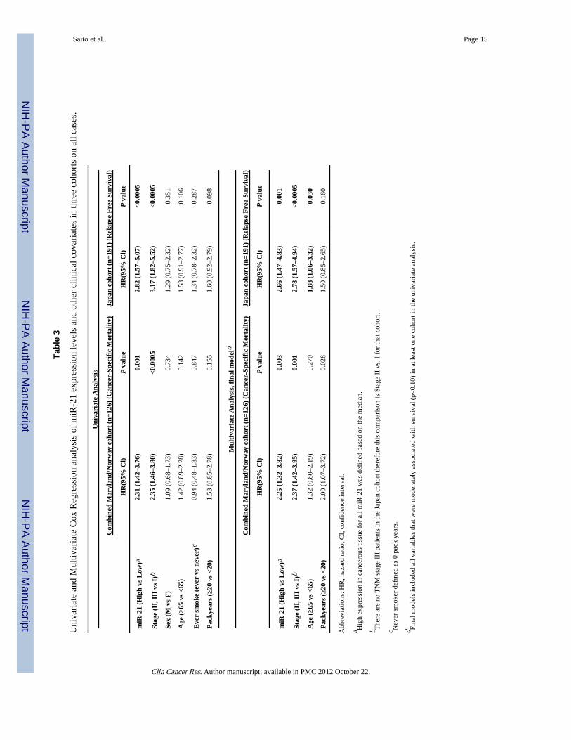
The Maryland and Norway cohorts were similar in 5-year survival rates, TNM staging,gender and age at diagnosis. To increase the statistical power for all further analyses, theMaryland and Norway cohorts were combined. MiR-21 expression was significantlyelevated in more advanced-stage tumors consistent with a role for miR-21 in the progressionof cancer (Figure 1B). Multivariate Cox regression demonstrated that miR-21 expressionwas associated with poor prognosis in the Maryland/Norway and Japan cohorts independentof staging (Table 3) supporting that miR-21 may be a useful prognostic biomarker for lungcancer.
Metastases are not clinically detectable in TNM stage I lung cancer patients, yet manypatients have undetectable micro-metastases that will progress. Prognostic biomarkers forTNM stage I patients may help identify those at high risk of disease progression and provide
Saito et al. Page 4
Clin Cancer Res. Author manuscript; available in PMC 2012 October 22.
NIH
-PA Author Manuscript
NIH
-PA Author Manuscript
NIH
-PA Author Manuscript

an opportunity to intervene at an earlier time. Validated prognostic biomarkers may alsoinfluence future revisions of tumor staging system.
We evaluated the potential of miR-21 expression as an early stage prognostic biomarkerusing a stratified analysis on TNM stage I tumors. Based on Kaplan Meier analysis, highmiR-21 expression was associated with poor cancer-specific mortality for stage I cases inthe Maryland/Norway cohort (P = 0.025, Kaplan-Meier log-rank test) and with worserelapse-free survival in the Japan cohort (P = 0.001) (Figure 1C). These associations weresignificant and independent of all clinical covariates in multivariate models (Table 4)demonstrating the potential for miR-21 as an early stage prognostic biomarker for lungcancer. High expression of miR-21 was also associated with cancer-specific mortality in the48 TNM stage II/III patients from the Maryland/Norway cohort (HR = 3.35, 95% CI = 1.43–7.82, p = 0.005) but not disease-free survival in the 49 stage II patients from the Japancohort.
DiscussionAltered expression of miRNAs has been found in every malignancy examined and appearsto play an important role in tumorigenesis. There is increasing evidence that miRNAs haveutility as both biomarkers and therapeutic targets for cancer. Here, we report that elevatedmiR-21 expression is associated with worse prognosis in NSCLC patients.
We evaluated the expression of miRNAs from snap-frozen lung tissue from 317 NSCLCpatients originating from three independent cohorts. To our knowledge, this is the largeststudy to date using flash-frozen tissue in NSCLC and the only study to use threeindependent cohorts. The association of increased miR-21 expression with worse lungcancer prognosis was significant in each of the three cohorts from different geographicalregions of the world. This suggests that our findings are representative of the majority oflung adenocarcinoma patients. A limitation of the study is that we were only studyingNSCLC with adenocarcinoma histology; therefore our findings may not be relevant to smallcell lung cancer or NSCLC with other histologies.
We assayed the expression of miR-21 from snap-frozen tissue while blinded to clinicaloutcomes and found that miR-21 was associated with NSCLC prognosis. This association isconsistent with our original microRNA array findings using frozen tissue (14), but theseresults are in apparent contrast to another study that we recently published evaluatingmiRNA expression in formalin-fixed paraffin-embedded (FFPE) tumor specimens (26). Inthat study we evaluated miR-21 expression, in addition to other miRNAs, in FFPE lungcancer tissue from 639 patients enrolled in the International Adjuvant Lung Cancer Trial(IALT), of which 218 patients were diagnosed with NSCLC with adenocarcinoma histology.We did not find that increased miR-21 was significantly associated with worse prognosis inthose tissues. This is likely due to the fact that the quality of RNA extracted from FFPEtissue is not as high of a quality as RNA extracted from frozen tissue. Therefore, qRTPCRmeasurements on RNA extracted from FFPE tissue may be less reliable than qRTPCRmeasurements on RNA extracted from snap-frozen tissue. Three additional reports supportthis assumption. When using snap frozen tissue, Markou and colleagues found thatoverexpression of miR-21 was associated with poor survival in 48 NSCLC patients (19).Gao and colleagues also used snap-frozen tissue as a source for RNA extraction and foundan association of increased miR-21 expression and poor survival in 47 NSCLC patients (27).But when FFPE tissue was used, Duncavage and colleagues did not find that miR-21 wasassociated with worse patient outcomes in 46 NSCLC patients (28). The fact that weobserved similar associations with high miR-21 expression and poor NSCLC prognosis in
Saito et al. Page 5
Clin Cancer Res. Author manuscript; available in PMC 2012 October 22.
NIH
-PA Author Manuscript
NIH
-PA Author Manuscript
NIH
-PA Author Manuscript
three independent cohorts when using snap-frozen tissues provides confidence that ourobservations in this study are accurate and representative.
Biomarkers that are capable of identifying patients at high risk of dying of stage I lungcancer may provide physicians with tools to help choose appropriate therapeutic strategies.The current clinical guidelines recommend surgical resection for TNM stage I lung cancerand additional therapy is not recommended unless the disease progresses (29). Here we findevidence that miR-21 has the potential to identify high risk early stage patients in threecohorts. To our knowledge, this is the first report that demonstrates an association of miR-21with prognosis in TNM stage I patients. Stage I patients with elevated miR-21 expressionare at high risk of disease progression, therefore these patients may be suitable for earlier ormore aggressive intervention than current recommendations indicate. Further studies arewarranted and required to address this important area of research.
MiR-21 is an oncogenic microRNA with a suggested role in multiple cancer types.Overexpression of miR-21 has been found in at least 18 malignancies indicating that alteredexpression of miR-21 may be a common mechanism in carcinogenesis (11, 30). The causeof miR-21 overexpression in cancer is still being elucidated. The altered activity of at leastfour cancer related regulatory factors is thought to contribute to the altered expression ofmiR-21, including signal transducer and activator of transcription 3 (STAT3), activatorprotein 1 (AP-1), transforming growth factor β (TGFβ), and epidermal growth factorreceptor (EGFR). STAT3 is a transcription factor that is downstream of IL-6 (interleukin 6)and can influence cell transformation. The miR-21 promoter has STAT3 binding sites andIL-6 can induce the expression of miR-21 in a STAT3 dependent manner indicating thataltered inflammatory states may be affecting miR-21 expression in the context of cancer(31). AP-1 can also increase the expression of miR-21. AP-1 can regulate cell proliferation,apoptosis and cell migration and has been implicated in a number of cancers, therefore itmay be contributing to the altered expression of miR-21. TGFβ has been shown to increasemiR-21 expression through the Smad signaling pathways and may be involved in theincreased expression of miR-21 in cancer. EGFR is an important cell surface receptor whoseactivity has important implications in cancer. The function of EGFR has also been linked tomiR-21 expression, raising the possibility that altered function of EGFR may contribute tocancer in part by deregulating miR-21 expression (7, 32).
The finding that increased expression of miR-21 is associated with worse prognosis issimilar to findings in other cancers. Increased levels of miR-21 are associated with worseprognosis and/or therapeutic outcome in multiple cancer types including colon cancer (15),breast cancer (33), chronic lymphocytic leukemia (34), pancreatic cancer (35), tonguecancer (36), astrocytomas (37) and head and neck cancers (38). We previously reported thatincreased miR-21 expression was associated with poor cancer-specific mortality in early,stage II colon cancer (15), similar to the finding that miR-21 is associated with prognosis inearly, stage I NSCLC. Taken together, miR-21 expression has the potential to be aprognostic biomarker in multiple cancer types, including early stage disease where it mayhelp guide therapeutic decisions.
Mechanistic studies have demonstrated the oncogenic potential of miR-21 (39) suggestingthat targeting miR-21 may provide some therapeutic benefit. Overexpression of miR-21actsas an anti-apoptotic factor and promotes cell proliferation. Inhibition of miR-21 inhibits cellgrowth in vitro and inhibits tumor growth in xenograft mouse models. MiR-21 expressioncan affect cell migration, cell invasion and tumor metastasis. MiR-21 can target severaltumor suppressor genes including programmed cell death 4 (PDCD4) (40, 41), phosphataseand tensin homolog (PTEN)(42), reversion-inducing-cysteine-rich protein with kazal motifs(RECK) (43), TIMP3 (43), maspin (44), nuclear factor 1 B-type (NFIB) (45), tropomyosin 1
Saito et al. Page 6
Clin Cancer Res. Author manuscript; available in PMC 2012 October 22.
NIH
-PA Author Manuscript
NIH
-PA Author Manuscript
NIH
-PA Author Manuscript

(TPM1) (46), sprouty 2 (SPRY2) (47) and Ras homolog gene family, member B (RHOB)(48). Overexpression of miR-21 can cause a malignant transformation in vivo and loss ofmiR-21 expression results in tumor regression in these models (49). MiR-21 expression isalso required for K-Ras-dependent lung tumor formation in mice, demonstrating necessaryrole of this microRNA in lung tumorigeneses (50). Inhibiting miR-21 can also sensitizecancer cells to chemotherapy (7, 50), further demonstrating the potential for targetingmiR-21, alone or in combination with other therapies, as a cancer treatment. Therefore,miR-21 may be both a useful biomarker and a therapeutic target for multiple cancer types.
In conclusion, we found strong evidence that high miR-21 expression is associated withpoor lung cancer survival outcomes, including early stage patients. This was found in threeseparate cohorts and is consistent with our previous report (14) and two additional reports(19, 27). These data suggest that increased miR-21 may in part be responsible for theaggressive nature and poor survival outcomes of NSCLC adenocarcinoma patients. Theseresults are likely representative of the majority of NSCLC adenocarcinomas. Therefore,miR-21 expression has potential as a diagnostic and prognostic biomarker for lungadenocarcinoma, including stage I lung cancer.
AcknowledgmentsThis research was supported by the Intramural Research Program of the National Cancer Institute, NationalInstitutes of Health, Department of Defense Congressionally Directed Medical Research Program Grant PR093793,the Norwegian Cancer Society, and a Grant-in-Aid from the Ministry of Health, Labor and Welfare for the 3rd-termComprehensive 10-year Strategy for Cancer Control, Japan.
References1. Ferlay J, Shin HR, Bray F, Forman D, Mathers C, Parkin DM. Estimates of worldwide burden of
cancer in 2008: GLOBOCAN 2008. Int J Cancer. 2010
2. Chansky K, Sculier JP, Crowley JJ, Giroux D, Van Meerbeeck J, Goldstraw P. The InternationalAssociation for the Study of Lung Cancer Staging Project: prognostic factors and pathologic TNMstage in surgically managed non-small cell lung cancer. J Thorac Oncol. 2009; 4:792–801.[PubMed: 19458556]
3. van Rens MT, de la Riviere AB, Elbers HR, van Den Bosch JM. Prognostic assessment of 2,361patients who underwent pulmonary resection for non-small cell lung cancer, stage I, II, and IIIA.Chest. 2000; 117:374–9. [PubMed: 10669677]
4. Nesbitt JC, Putnam JB Jr, Walsh GL, Roth JA, Mountain CF. Survival in early-stage non-small celllung cancer. Ann Thorac Surg. 1995; 60:466–72. [PubMed: 7646126]
5. Garzon R, Calin GA, Croce CM. MicroRNAs in Cancer. Annu Rev Med. 2009; 60:167–79.[PubMed: 19630570]
6. Trang P, Weidhaas JB, Slack FJ. MicroRNAs as potential cancer therapeutics. Oncogene. 2008; 27(Suppl 2):S52–7. [PubMed: 19956180]
7. Seike M, Goto A, Okano T, Bowman ED, Schetter AJ, Horikawa I, et al. MiR-21 is an EGFR-regulated anti-apoptotic factor in lung cancer in never-smokers. Proc Natl Acad Sci U S A. 2009;106:12085–90. [PubMed: 19597153]
8. Calin GA, Croce CM. MicroRNA signatures in human cancers. Nat Rev Cancer. 2006; 6:857–66.[PubMed: 17060945]
9. Esquela-Kerscher A, Slack FJ. Oncomirs - microRNAs with a role in cancer. Nat Rev Cancer. 2006;6:259–69. [PubMed: 16557279]
10. Calin GA, Ferracin M, Cimmino A, Di Leva G, Shimizu M, Wojcik SE, et al. A MicroRNAsignature associated with prognosis and progression in chronic lymphocytic leukemia. N Engl JMed. 2005; 353:1793–801. [PubMed: 16251535]
Saito et al. Page 7
Clin Cancer Res. Author manuscript; available in PMC 2012 October 22.
NIH
-PA Author Manuscript
NIH
-PA Author Manuscript
NIH
-PA Author Manuscript

11. Volinia S, Calin GA, Liu CG, Ambs S, Cimmino A, Petrocca F, et al. A microRNA expressionsignature of human solid tumors defines cancer gene targets. Proc Natl Acad Sci U S A. 2006;103:2257–61. [PubMed: 16461460]
12. Lu J, Getz G, Miska EA, Alvarez-Saavedra E, Lamb J, Peck D, et al. MicroRNA expressionprofiles classify human cancers. Nature. 2005; 435:834–8. [PubMed: 15944708]
13. Ryan BM, Robles AI, Harris CC. Genetic variation in microRNA networks: the implications forcancer research. Nat Rev Cancer. 2010; 10:389–402. [PubMed: 20495573]
14. Yanaihara N, Caplen N, Bowman E, Seike M, Kumamoto K, Yi M, et al. Unique microRNAmolecular profiles in lung cancer diagnosis and prognosis. Cancer Cell. 2006; 9:189–98. [PubMed:16530703]
15. Schetter AJ, Leung SY, Sohn JJ, Zanetti KA, Bowman ED, Yanaihara N, et al. MicroRNAexpression profiles associated with prognosis and therapeutic outcome in colon adenocarcinoma.JAMA. 2008; 299:425–36. [PubMed: 18230780]
16. Budhu A, Jia HL, Forgues M, Liu CG, Goldstein D, Lam A, et al. Identification of metastasis-related microRNAs in hepatocellular carcinoma. Hepatology. 2008; 47:897–907. [PubMed:18176954]
17. Mathe EA, Nguyen GH, Bowman ED, Zhao Y, Budhu A, Schetter AJ, et al. MicroRNA expressionin squamous cell carcinoma and adenocarcinoma of the esophagus: associations with survival.Clin Cancer Res. 2009; 15:6192–200. [PubMed: 19789312]
18. Ueda T, Volinia S, Okumura H, Shimizu M, Taccioli C, Rossi S, et al. Relation betweenmicroRNA expression and progression and prognosis of gastric cancer: a microRNA expressionanalysis. Lancet Oncol. 2010; 11:136–46. [PubMed: 20022810]
19. Markou A, Tsaroucha EG, Kaklamanis L, Fotinou M, Georgoulias V, Lianidou ES. Prognosticvalue of mature microRNA-21 and microRNA-205 overexpression in non-small cell lung cancerby quantitative real-time RT-PCR. Clin Chem. 2008; 54:1696–704. [PubMed: 18719201]
20. Raponi M, Dossey L, Jatkoe T, Wu X, Chen G, Fan H, et al. MicroRNA classifiers for predictingprognosis of squamous cell lung cancer. Cancer Res. 2009; 69:5776–83. [PubMed: 19584273]
21. Gallardo E, Navarro A, Vinolas N, Marrades RM, Diaz T, Gel B, et al. miR-34a as a prognosticmarker of relapse in surgically resected non-small-cell lung cancer. Carcinogenesis. 2009;30:1903–9. [PubMed: 19736307]
22. Yu SL, Chen HY, Chang GC, Chen CY, Chen HW, Singh S, et al. MicroRNA signature predictssurvival and relapse in lung cancer. Cancer Cell. 2008; 13:48–57. [PubMed: 18167339]
23. Landi MT, Zhao Y, Rotunno M, Koshiol J, Liu H, Bergen AW, et al. MicroRNA expressiondifferentiates histology and predicts survival of lung cancer. Clin Cancer Res. 16:430–41.[PubMed: 20068076]
24. Takamizawa J, Konishi H, Yanagisawa K, Tomida S, Osada H, Endoh H, et al. Reducedexpression of the let-7 microRNAs in human lung cancers in association with shortenedpostoperative survival. Cancer Res. 2004; 64:3753–6. [PubMed: 15172979]
25. Lubin JH, Gail MH. Biased selection of controls for case-control analyses of cohort studies.Biometrics. 1984; 40:63–75. [PubMed: 6375751]
26. Voortman J, Goto A, Mendiboure J, Sohn JJ, Schetter AJ, Saito M, et al. MicroRNA Expressionand Clinical Outcomes in Patients Treated with Adjuvant Chemotherapy after Complete Resectionof Non-Small Cell Lung Carcinoma. Cancer Res. 2010
27. Gao W, Yu Y, Cao H, Shen H, Li X, Pan S, et al. Deregulated expression of miR-21, miR-143 andmiR-181a in non small cell lung cancer is related to clinicopathologic characteristics or patientprognosis. Biomed Pharmacother. 2010; 64:399–408. [PubMed: 20363096]
28. Duncavage E, Goodgame B, Sezhiyan A, Govindan R, Pfeifer J. Use of MicroRNA ExpressionLevels to Predict Outcomes in Resected Stage I Non-small Cell Lung Cancer. J Thorac Oncol.2010; 5:1755–63. [PubMed: 20975375]
29. Pignon JP, Tribodet H, Scagliotti GV, Douillard JY, Shepherd FA, Stephens RJ, et al. Lungadjuvant cisplatin evaluation: a pooled analysis by the LACE Collaborative Group. J Clin Oncol.2008; 26:3552–9. [PubMed: 18506026]
30. Selcuklu SD, Donoghue MT, Spillane C. miR-21 as a key regulator of oncogenic processes.Biochem Soc Trans. 2009; 37:918–25. [PubMed: 19614619]
Saito et al. Page 8
Clin Cancer Res. Author manuscript; available in PMC 2012 October 22.
NIH
-PA Author Manuscript
NIH
-PA Author Manuscript
NIH
-PA Author Manuscript

31. Loffler D, Brocke-Heidrich K, Pfeifer G, Stocsits C, Hackermuller J, Kretzschmar AK, et al.Interleukin-6 dependent survival of multiple myeloma cells involves the Stat3-mediated inductionof microRNA-21 through a highly conserved enhancer. Blood. 2007; 110:1330–3. [PubMed:17496199]
32. Zhou X, Ren Y, Moore L, Mei M, You Y, Xu P, et al. Downregulation of miR-21 inhibits EGFRpathway and suppresses the growth of human glioblastoma cells independent of PTEN status. LabInvest. 2010; 90:144–55. [PubMed: 20048743]
33. Yan LX, Huang XF, Shao Q, Huang MY, Deng L, Wu QL, et al. MicroRNA miR-21overexpression in human breast cancer is associated with advanced clinical stage, lymph nodemetastasis and patient poor prognosis. RNA. 2008; 14:2348–60. [PubMed: 18812439]
34. Rossi S, Shimizu M, Barbarotto E, Nicoloso MS, Dimitri F, Sampath D, et al. microRNAfingerprinting of CLL patients with chromosome 17p deletion identify a miR-21 score thatstratifies early survival. Blood. 2010; 116:945–52. [PubMed: 20393129]
35. Dillhoff M, Liu J, Frankel W, Croce C, Bloomston M. MicroRNA-21 is overexpressed inpancreatic cancer and a potential predictor of survival. J Gastrointest Surg. 2008; 12:2171–6.[PubMed: 18642050]
36. Li J, Huang H, Sun L, Yang M, Pan C, Chen W, et al. MiR-21 indicates poor prognosis in tonguesquamous cell carcinomas as an apoptosis inhibitor. Clin Cancer Res. 2009; 15:3998–4008.[PubMed: 19509158]
37. Zhi F, Chen X, Wang S, Xia X, Shi Y, Guan W, et al. The use of hsa-miR-21, hsa-miR-181b andhsa-miR-106a as prognostic indicators of astrocytoma. Eur J Cancer. 2010; 46:1640–9. [PubMed:20219352]
38. Avissar M, McClean MD, Kelsey KT, Marsit CJ. MicroRNA expression in head and neck cancerassociates with alcohol consumption and survival. Carcinogenesis. 2009; 30:2059–63. [PubMed:19901002]
39. Krichevsky AM, Gabriely G. miR-21: a small multi-faceted RNA. J Cell Mol Med. 2009; 13:39–53. [PubMed: 19175699]
40. Asangani IA, Rasheed SA, Nikolova DA, Leupold JH, Colburn NH, Post S, et al. MicroRNA-21(miR-21) post-transcriptionally downregulates tumor suppressor Pdcd4 and stimulates invasion,intravasation and metastasis in colorectal cancer. Oncogene. 2008; 27:2128–36. [PubMed:17968323]
41. Lu Z, Liu M, Stribinskis V, Klinge CM, Ramos KS, Colburn NH, et al. MicroRNA-21 promotescell transformation by targeting the programmed cell death 4 gene. Oncogene. 2008; 27:4373–9.[PubMed: 18372920]
42. Meng F, Henson R, Wehbe-Janek H, Ghoshal K, Jacob ST, Patel T. MicroRNA-21 regulatesexpression of the PTEN tumor suppressor gene in human hepatocellular cancer. Gastroenterology.2007; 133:647–58. [PubMed: 17681183]
43. Gabriely G, Wurdinger T, Kesari S, Esau CC, Burchard J, Linsley PS, et al. MicroRNA 21promotes glioma invasion by targeting matrix metalloproteinase regulators. Mol Cell Biol. 2008;28:5369–80. [PubMed: 18591254]
44. Zhu S, Wu H, Wu F, Nie D, Sheng S, Mo YY. MicroRNA-21 targets tumor suppressor genes ininvasion and metastasis. Cell Res. 2008; 18:350–9. [PubMed: 18270520]
45. Fujita S, Ito T, Mizutani T, Minoguchi S, Yamamichi N, Sakurai K, et al. miR-21 Gene expressiontriggered by AP-1 is sustained through a double-negative feedback mechanism. J Mol Biol. 2008;378:492–504. [PubMed: 18384814]
46. Zhu S, Si ML, Wu H, Mo YY. MicroRNA-21 Targets the Tumor Suppressor Gene Tropomyosin 1(TPM1). J Biol Chem. 2007; 282:14328–36. [PubMed: 17363372]
47. Sayed D, Rane S, Lypowy J, He M, Chen IY, Vashistha H, et al. MicroRNA-21 targets Sprouty2and promotes cellular outgrowths. Mol Biol Cell. 2008; 19:3272–82. [PubMed: 18508928]
48. Connolly EC, Van Doorslaer K, Rogler LE, Rogler CE. Overexpression of miR-21 promotes an invitro metastatic phenotype by targeting the tumor suppressor RHOB. Mol Cancer Res. 2010;8:691–700. [PubMed: 20460403]
49. Medina PP, Nolde M, Slack FJ. OncomiR addiction in an in vivo model of microRNA-21-inducedpre-B-cell lymphoma. Nature. 2010; 467:86–90. [PubMed: 20693987]
Saito et al. Page 9
Clin Cancer Res. Author manuscript; available in PMC 2012 October 22.
NIH
-PA Author Manuscript
NIH
-PA Author Manuscript
NIH
-PA Author Manuscript

50. Hatley ME, Patrick DM, Garcia MR, Richardson JA, Bassel-Duby R, van Rooij E, et al.Modulation of K-Ras-dependent lung tumorigenesis by MicroRNA-21. Cancer Cell. 2010;18:282–93. [PubMed: 20832755]
Saito et al. Page 10
Clin Cancer Res. Author manuscript; available in PMC 2012 October 22.
NIH
-PA Author Manuscript
NIH
-PA Author Manuscript
NIH
-PA Author Manuscript

Translational Relevance
We report that elevated miR-21 expression is independently associated with worseprognosis in three independent NSCLC adenocarcinoma cohorts from different regions ofthe world. This suggests that our findings are representative of the majority of NSCLCadenocarcinoma patients. These associations were significant when evaluating TNMstage I tumors alone. Biomarkers that are capable of identifying patients at high risk ofdying of stage I lung cancer may provide physicians with key tools to help diagnose lungcancer and choose appropriate therapeutic strategies. Therefore miR-21 expression haspotential utility as an early stage prognostic biomarker for NSCLC adenocarcinomapatients. We also find increased levels of miR-21 expression in more advanced tumors,consistent with a possible role for miR-21 in the progression of NSCLC. These findingssuggest that miR-21 may also be a useful therapeutic target for NSCLC patients.
Saito et al. Page 11
Clin Cancer Res. Author manuscript; available in PMC 2012 October 22.
NIH
-PA Author Manuscript
NIH
-PA Author Manuscript
NIH
-PA Author Manuscript

Figure 1.The expression of miR-21 is associated with survival in the Maryland, Norway and Japancohorts. (A) Kaplan-Meier survival analysis of all stage cases in the Maryland, Norway andJapan cohort stratified by median miR-21 tumor expression. (B) Expression differences ofmiR-21 between tumor and nontumor in the Maryland and Norway combined cohort andJapan cohort. Dot plots represent miR-21 relative threshold cycle values from qRT-PCR andexpression levels in tumor were normalized to nontumor. Relative threshold cycle valuesgreater than 0 in tumor indicate higher expression than that of mean in nontumor log2 scale.Horizontal bars indicate mean expression value. *P < 0.0001, **P = 0.001, ***P = 0.009.Wilcoxon matched-pairs test. (C) Kaplan-Meier survival analysis of TNM stage I cases inthe Maryland and Norway combined cohort and Japan cohort stratified by median miR-21tumor expression.
Saito et al. Page 12
Clin Cancer Res. Author manuscript; available in PMC 2012 October 22.
NIH
-PA Author Manuscript
NIH
-PA Author Manuscript
NIH
-PA Author Manuscript

NIH
-PA Author Manuscript
NIH
-PA Author Manuscript
NIH
-PA Author Manuscript
Saito et al. Page 13
Table 1
Characteristics of study populations of patients with non small cell lung adenocarcinoma
Maryland Cohort (n=89) Norway Cohort (n=37) Japan Cohort (n=191) p-value1
Age-years p<0.0005
Mean (SD) 63.6 (10.5) 64.4 (11.7) 59.6 (7.7)
Range 32–90 37–82 30–76
Gender-no. (%) p=0.742
Male 46 (52) 20 (54) 92 (48)
Female 43 (48) 17 (46) 99 (52)
Race-no. (%) p<0.0005
Caucasian 58 (65) 37 (100) 0
African-American 31 (35) 0 0
Asian 0 0 191 (100)
TNM Stage-no. (%) p<0.0005
I 57 (64) 21 (57) 142 (74)
II 22 (25) 5 (14) 49 (26)
III 10 (11) 11 (29) 0 (0)
Smoking (pack year)-no. (%) p<0.0005
never 7 (8) 3 (8) 95 (50)
<20 pack years 12 (13) 12 (32) 30 (16)
≥20 pack years 66 (74) 19 (52) 66 (34)
unknown 4 (5) 3 (8) 0 (0)
1p-value from one-way ANOVA or Fisher’s exact test, where appropriate
Clin Cancer Res. Author manuscript; available in PMC 2012 October 22.

NIH
-PA Author Manuscript
NIH
-PA Author Manuscript
NIH
-PA Author Manuscript
Saito et al. Page 14
Tabl
e 2
Uni
vari
ate
Cox
Reg
ress
ion
anal
ysis
of
miR
NA
s ex
pres
sion
leve
ls a
nd s
urvi
val i
n ca
ncer
ous
tissu
e.
mic
roR
NA
(hi
gh v
s lo
w)a
Mar
ylan
d co
hort
(n=
89)
(Can
cer-
Spec
ific
Mor
talit
y)N
orw
ay c
ohor
t (n
=37)
(C
ance
r-Sp
ecif
ic M
orta
lity)
Japa
n co
hort
(n=
191)
(R
elap
se F
ree
Surv
ival
)
HR
(95
% C
l)P
val
ueH
R (
95%
Cl)
P v
alue
HR
(95
% C
l)P
val
ue
miR
-17
2.00
(1.
10–3
.61)
0.02
21.
23 (
0.56
–2.7
0)0.
603
1.37
(0.
80–2
.37)
0.24
5
miR
-21
2.06
(1.
13–3
.75)
0.01
82.
78 (
1.22
–6.3
1)0.
014
2.82
(1.
57–5
.07)
<0.0
005
miR
-155
2.37
(1.
27–4
.42)
0.00
61.
60 (
0.73
–3.5
2)0.
245
1.33
(0.
77–2
.29)
0.30
9
Abb
revi
atio
ns: H
R, h
azar
d ra
tio; C
I, c
onfi
denc
e in
terv
al
a Hig
h ex
pres
sion
in c
ance
rous
tiss
ue f
or a
ll m
iRN
As
was
def
ined
bas
ed o
n th
e m
edia
n. M
icro
RN
A e
xpre
ssio
n w
as m
easu
red
with
qR
T-P
CR
.
Clin Cancer Res. Author manuscript; available in PMC 2012 October 22.
NIH
-PA Author Manuscript
NIH
-PA Author Manuscript
NIH
-PA Author Manuscript
Saito et al. Page 15
Tabl
e 3
Uni
vari
ate
and
Mul
tivar
iate
Cox
Reg
ress
ion
anal
ysis
of
miR
-21
expr
essi
on le
vels
and
oth
er c
linic
al c
ovar
iate
s in
thre
e co
hort
s on
all
case
s.
Uni
vari
ate
Ana
lysi
s
Com
bine
d M
aryl
and/
Nor
way
coh
ort
(n=1
26)
(Can
cer-
Spec
ific
Mor
talit
y)Ja
pan
coho
rt (
n=19
1) (
Rel
apse
Fre
e Su
rviv
al)
HR
(95%
Cl)
P v
alue
HR
(95%
Cl)
P v
alue
miR
-21
(Hig
h vs
Low
)a2.
31 (
1.42
–3.7
6)0.
001
2.82
(1.
57–5
.07)
<0.0
005
Stag
e (I
I, I
II v
s I)
b2.
35 (
1.46
–3.8
0)<0
.000
53.
17 (
1.82
–5.5
2)<0
.000
5
Sex
(M v
s F
)1.
09 (
0.68
–1.7
3)0.
734
1.29
(0.
75–2
.32)
0.35
1
Age
(≥6
5 vs
<65
)1.
42 (
0.89
–2.2
8)0.
142
1.58
(0.
91–2
.77)
0.10
6
Eve
r sm
oke
(eve
r vs
nev
er)c
0.94
(0.
48–1
.83)
0.84
71.
34 (
0.78
–2.3
2)0.
287
Pac
kyea
rs (
≥20
vs <
20)
1.53
(0.
85–2
.78)
0.15
51.
60 (
0.92
–2.7
9)0.
098
Mul
tiva
riat
e A
naly
sis,
fin
al m
odel
d
Com
bine
d M
aryl
and/
Nor
way
coh
ort
(n=1
26)
(Can
cer-
Spec
ific
Mor
talit
y)Ja
pan
coho
rt (
n=19
1) (
Rel
apse
Fre
e Su
rviv
al)
HR
(95%
Cl)
P v
alue
HR
(95%
Cl)
P v
alue
miR
-21
(Hig
h vs
Low
)a2.
25 (
1.32
–3.8
2)0.
003
2.66
(1.
47–4
.83)
0.00
1
Stag
e (I
I, I
II v
s I)
b2.
37 (
1.42
–3.9
5)0.
001
2.78
(1.
57–4
.94)
<0.0
005
Age
(≥6
5 vs
<65
)1.
32 (
0.80
–2.1
9)0.
270
1.88
(1.
06–3
.32)
0.03
0
Pac
kyea
rs (
≥20
vs <
20)
2.00
(1.
07–3
.72)
0.02
81.
50 (
0.85
–2.6
5)0.
160
Abb
revi
atio
ns: H
R, h
azar
d ra
tio; C
I, c
onfi
denc
e in
terv
al.
a Hig
h ex
pres
sion
in c
ance
rous
tiss
ue f
or a
ll m
iR-2
1 w
as d
efin
ed b
ased
on
the
med
ian.
b The
re a
re n
o T
NM
sta
ge I
II p
atie
nts
in th
e Ja
pan
coho
rt th
eref
ore
this
com
pari
son
is S
tage
II
vs. I
for
that
coh
ort.
c Nev
er s
mok
er d
efin
ed a
s 0
pack
yea
rs.
d Fina
l mod
els
incl
uded
all
vari
able
s th
at w
ere
mod
erat
ely
asso
ciat
ed w
ith s
urvi
val (
p<0.
10)
in a
t lea
st o
ne c
ohor
t in
the
univ
aria
te a
naly
sis.
Clin Cancer Res. Author manuscript; available in PMC 2012 October 22.
NIH
-PA Author Manuscript
NIH
-PA Author Manuscript
NIH
-PA Author Manuscript
Saito et al. Page 16
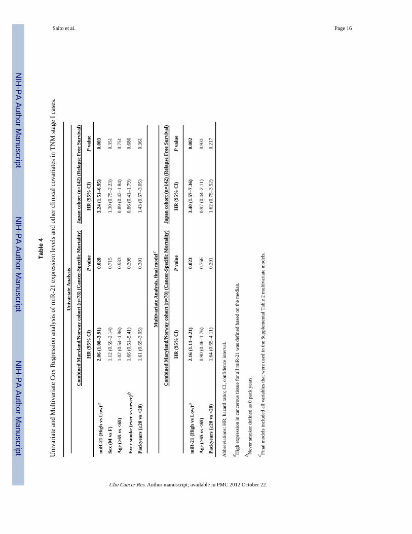
Tabl
e 4
Uni
vari
ate
and
Mul
tivar
iate
Cox
Reg
ress
ion
anal
ysis
of
miR
-21
expr
essi
on le
vels
and
oth
er c
linic
al c
ovar
iate
s in
TN
M s
tage
I c
ases
.
Uni
vari
ate
Ana
lysi
s
Com
bine
d M
aryl
and/
Nor
way
coh
ort
(n=7
8) (
Can
cer-
Spec
ific
Mor
talit
y)Ja
pan
coho
rt (
n=14
2) (
Rel
apse
Fre
e Su
rviv
al)
HR
(95
% C
l)P
val
ueH
R (
95%
Cl)
P v
alue
miR
-21
(Hig
h vs
Low
)a2.
06 (
1.08
–3.9
1)0.
028
3.24
(1.
51–6
.95)
0.00
3
Sex
(M v
s F
)1.
12 (
0.59
–2.1
4)0.
715
1.30
(0.
75–2
.23)
0.35
1
Age
(≥6
5 vs
<65
)1.
02 (
0.54
–1.9
6)0.
933
0.89
(0.
42–1
.84)
0.75
1
Eve
r sm
oke
(eve
r vs
nev
er)b
1.66
(0.
51–5
.41)
0.39
80.
86 (
0.41
–1.7
9)0.
686
Pac
kyea
rs (
≥20
vs <
20)
1.61
(0.
65–3
.95)
0.30
11.
43 (
0.67
–3.0
5)0.
361
Mul
tiva
riat
e A
naly
sis,
fin
al m
odel
c
Com
bine
d M
aryl
and/
Nor
way
coh
ort
(n=7
8) (
Can
cer-
Spec
ific
Mor
talit
y)Ja
pan
coho
rt (
n=14
2) (
Rel
apse
Fre
e Su
rviv
al)
HR
(95
% C
l)P
val
ueH
R (
95%
Cl)
P v
alue
miR
-21
(Hig
h vs
Low
)a2.
16 (
1.11
–4.2
1)0.
023
3.40
(1.
57–7
.36)
0.00
2
Age
(≥6
5 vs
<65
)0.
90 (
0.46
–1.7
6)0.
766
0.97
(0.
44–2
.11)
0.93
1
Pac
kyea
rs (
≥20
vs <
20)
1.64
(0.
65–4
.11)
0.29
11.
62 (
0.75
–3.5
2)0.
217
Abb
revi
atio
ns: H
R, h
azar
d ra
tio; C
I, c
onfi
denc
e in
terv
al.
a Hig
h ex
pres
sion
in c
ance
rous
tiss
ue f
or a
ll m
iR-2
1 w
as d
efin
ed b
ased
on
the
med
ian.
b Nev
er s
mok
er d
efin
ed a
s 0
pack
yea
rs.
c Fina
l mod
els
incl
uded
all
vari
able
s th
at w
ere
used
in th
e Su
pple
men
tal T
able
2 m
ultiv
aria
te m
odel
s.
Clin Cancer Res. Author manuscript; available in PMC 2012 October 22.
Copyright © 2022 FDOKUMEN