The Arabidopsis ACR4 gene plays a role in cell layer organisation during ovule integument and sepal...
10
INTRODUCTION Plant meristems are composed of organised layers (files or plates) of cells arranged parallel to the ‘outside’ of the meristem. Each layer undergoes cell divisions in a defined plane or planes pushing cells to the periphery of the meristem where they are either incorporated into new meristems or become differentiated. In Arabidopsis the shoot apical meristem (SAM) has two outer tunica layers, the L1 and the L2, which undergo regulated divisions in the anticlinal plane. The inner cell layer or corpus is designated L3 and undergoes both anticlinal and periclinal divisions. As organ primordia arise on meristem flanks, changes in the regulation of cell division patterns occur. In dicotyledon leaf primordia, the epidermal cell layer is exclusively L1-derived and L1-derived cells continue to divide largely anticlinally until late in development. In contrast the L2 layer undergoes both anticlinal and periclinal divisions to contribute the leaf mesophyll, while the L3 contributes to both leaf mesophyll and the vasculature (Stewart and Burk, 1970). The contributions of meristematic cell layers to organ primordia vary. Whilst the Arabidopsis leaf is usually formed from L1-, L2- and L3-derived cells, petal primordia have been shown to contain cells of only L1 and L2 origin and ovule integuments are entirely L1 derived (Jenik and Irish, 2000). Integument cells undergo carefully regulated divisions, mainly in the anticlinal plane, so that the completed organ is a tubular plate of cells only 2-3 cells thick and effectively entirely epidermal (Schneitz et al., 1995; Robinson- Beers et al., 1992). Experiments and observations in many plant species have shown that the developmental behaviour of cells in meristems and developing organs is largely dictated by their position rather than by lineage. Thus if the progeny of cells from one layer invade another layer during development, the displaced cells differentiate according to their new position (Stewart and Derman, 1975; van den Berg et al., 1995; Kidner et al., 2000). For this developmental plasticity to be achieved, cells must constantly receive and interpret information from their neighbours. Our understanding of how plant cell layers communicate is currently limited to a few specific examples. In Arabidopsis roots, an inside to outside movement of transcription factors (notably the SCARECROW (SCR) protein) is required for normal differentiation of ground cell layers (Nakajima and Benfey, 2002). In contrast, inter layer communication in shoot meristems appears to require the interaction of a diffusible ligand with a cell-autonomous receptor kinase complex (Fletcher et al., 1999). A similar interaction is invoked in the development of maize leaves and endosperm, where the receptor kinase-encoding CRINKLY4 (CR4) and the calpain-encoding DEFECTIVE KERNEL 1 (DEK1) genes are required for specification and maintenance of ‘outer’ cell layer identity during endosperm and leaf development (Becraft et al., 1996; Becraft et al., 2002; Lid et al., 2002). The maize EX TRA C ELL L AYERS 1 (XCL1) gene seems to be involved in pathways regulating division behaviour in L1 cells during organ formation. The Xcl1 mutant provides intriguing evidence that cell identity can be uncoupled from positional cues at least late in development. (Kessler et al., 2002). In a search to identify genes involved in inter-cell layer communication in Arabidopsis, a study of ACR4, an 4249 Development 130, 4249-4258 © 2003 The Company of Biologists Ltd doi:10.1242/dev.00634 The mechanisms regulating cell layer organisation in developing plant organs are fundamental to plant growth, but remain largely uninvestigated. We have studied the receptor kinase-encoding ARABIDOPSIS CRINKLY4 gene and shown that its expression is restricted to the L1 cell layer of most meristems and organ primordia, including those of the ovule integuments. Insertion mutations show that ARABIDOPSIS CRINKLY4 is required for regulation of cellular organisation during the development of sepal margins and ovule integument outgrowth. We show that ARABIDOPSIS CRINKLY4 encodes a functional kinase that, in ovules and possibly other tissues, is abundant in anticlinal and the inner periclinal plasma membrane of ‘outside’ cells. We propose that ARABIDOPSIS CRINKLY4 may be involved in maintaining L1 cell layer integrity by receiving and transmitting signals from neighbouring L1 cells and/or from underlying cell layers. Key words: L1, Cell layer, Integuments, Signalling, Receptor kinase, Ovule, Arabidopsis thaliana SUMMARY The Arabidopsis ACR4 gene plays a role in cell layer organisation during ovule integument and sepal margin development Miriam L. Gifford, Samuel Dean and Gwyneth C. Ingram* Institute of Cell and Molecular Biology, Kings Buildings, University of Edinburgh, Edinburgh EH9 3JR, UK *Author for correspondence (e-mail: [email protected]) Accepted 28 May 2003
Transcript of The Arabidopsis ACR4 gene plays a role in cell layer organisation during ovule integument and sepal...

INTRODUCTION
Plant meristems are composed of organised layers (files orplates) of cells arranged parallel to the ‘outside’ of themeristem. Each layer undergoes cell divisions in a definedplane or planes pushing cells to the periphery of the meristemwhere they are either incorporated into new meristems orbecome differentiated. In Arabidopsis the shoot apicalmeristem (SAM) has two outer tunica layers, the L1 and theL2, which undergo regulated divisions in the anticlinal plane.The inner cell layer or corpus is designated L3 and undergoesboth anticlinal and periclinal divisions. As organ primordiaarise on meristem flanks, changes in the regulation of celldivision patterns occur. In dicotyledon leaf primordia, theepidermal cell layer is exclusively L1-derived and L1-derivedcells continue to divide largely anticlinally until late indevelopment. In contrast the L2 layer undergoes both anticlinaland periclinal divisions to contribute the leaf mesophyll, whilethe L3 contributes to both leaf mesophyll and the vasculature(Stewart and Burk, 1970). The contributions of meristematiccell layers to organ primordia vary. Whilst the Arabidopsis leafis usually formed from L1-, L2- and L3-derived cells, petalprimordia have been shown to contain cells of only L1 and L2origin and ovule integuments are entirely L1 derived (Jenik andIrish, 2000). Integument cells undergo carefully regulateddivisions, mainly in the anticlinal plane, so that the completedorgan is a tubular plate of cells only 2-3 cells thick andeffectively entirely epidermal (Schneitz et al., 1995; Robinson-Beers et al., 1992).
Experiments and observations in many plant species have
shown that the developmental behaviour of cells in meristemsand developing organs is largely dictated by their position ratherthan by lineage. Thus if the progeny of cells from one layerinvade another layer during development, the displaced cellsdifferentiate according to their new position (Stewart andDerman, 1975; van den Berg et al., 1995; Kidner et al., 2000).For this developmental plasticity to be achieved, cells mustconstantly receive and interpret information from theirneighbours. Our understanding of how plant cell layerscommunicate is currently limited to a few specific examples.In Arabidopsis roots, an inside to outside movement oftranscription factors (notably the SCARECROW (SCR) protein)is required for normal differentiation of ground cell layers(Nakajima and Benfey, 2002). In contrast, inter layercommunication in shoot meristems appears to require theinteraction of a diffusible ligand with a cell-autonomous receptorkinase complex (Fletcher et al., 1999). A similar interaction isinvoked in the development of maize leaves and endosperm,where the receptor kinase-encoding CRINKLY4(CR4) and thecalpain-encoding DEFECTIVE KERNEL 1(DEK1) genes arerequired for specification and maintenance of ‘outer’ cell layeridentity during endosperm and leaf development (Becraft et al.,1996; Becraft et al., 2002; Lid et al., 2002). The maize EXTRACELL LAYERS 1(XCL1)gene seems to be involved in pathwaysregulating division behaviour in L1 cells during organ formation.The Xcl1 mutant provides intriguing evidence that cell identitycan be uncoupled from positional cues at least late indevelopment. (Kessler et al., 2002).
In a search to identify genes involved in inter-cell layercommunication in Arabidopsis, a study of ACR4, an
4249Development 130, 4249-4258 © 2003 The Company of Biologists Ltddoi:10.1242/dev.00634
The mechanisms regulating cell layer organisation indeveloping plant organs are fundamental to plant growth,but remain largely uninvestigated. We have studied thereceptor kinase-encoding ARABIDOPSIS CRINKLY4 geneand shown that its expression is restricted to the L1 celllayer of most meristems and organ primordia, includingthose of the ovule integuments. Insertion mutations showthat ARABIDOPSIS CRINKLY4 is required for regulationof cellular organisation during the development of sepalmargins and ovule integument outgrowth. We show that
ARABIDOPSIS CRINKLY4 encodes a functional kinasethat, in ovules and possibly other tissues, is abundant inanticlinal and the inner periclinal plasma membraneof ‘outside’ cells. We propose that ARABIDOPSISCRINKLY4 may be involved in maintaining L1 cell layerintegrity by receiving and transmitting signals fromneighbouring L1 cells and/or from underlying cell layers.
Key words: L1, Cell layer, Integuments, Signalling, Receptor kinase,Ovule, Arabidopsis thaliana
SUMMARY
The Arabidopsis ACR4 gene plays a role in cell layer organisation during
ovule integument and sepal margin development
Miriam L. Gifford, Samuel Dean and Gwyneth C. Ingram*
Institute of Cell and Molecular Biology, Kings Buildings, University of Edinburgh, Edinburgh EH9 3JR, UK*Author for correspondence (e-mail: [email protected])
Accepted 28 May 2003

4250
Arabidopsis CR4 homologue, was carried out. ACR4 was foundto be required for normal cell organisation during ovuleintegument development and the formation of sepal margins.Both these tissues are formed exclusively from plates of L1cells arranged back to back. By isolating the functional ACR4promoter, ACR4was shown to be expressed in L1 cells in allapical meristems and young organ primordia, including thoseof the developing ovule integuments. In addition, ACR4 isexpressed in an intriguing pattern in root meristems. The kinaseactivity of ACR4was demonstrated and, using fusion proteinsexpressed under the ACR4promoter, ACR4 protein localisationwas visualised in vivo in the plasma membranes of L1-derivedcells. The wide expression pattern of ACR4 compared to itsassociated mutant phenotype may be a result of functionalredundancy with other related proteins or functionally relatedpathways. Taken together, the data presented indicate a role forACR4in the cellular signalling pathways required for correctcell organisation in ovule integuments and sepal boundaries,and may provide important clues as to the types of signallinginvolved in cell layer maintenance and specification in thewider context of plant development.
MATERIALS AND METHODS
Expression and complementation analysis of ACR4The ACR4 open reading frame (ORF) was PCR amplified fromArabidopsis thaliana genomic DNA ecotype Columbia (Col0) withCR5 (5′-TGGTACCTTTGAAAAGAATGAGAATGTTCG) and 5′-GAGCTCAGAAATTATGATGCAAGAACAAGC. The ACR4promoterwas amplified with 5′-TGTCGACATAGTCAAGAAATGGCCTTTCCand 5′-TTCTAGACAAAGTCAACACACACGCTT. Products werecloned into pGEMT-easy (Promega) (pL92 and pL93 respectively).Probes (antisense and sense) for in situ hybridisation were madeby linearising pL92 with NcoI or SalI, respectively, and transcribingwith Sp6 or T7 RNA polymerase, respectively. In situ hybridisationswere carried out using a standard protocol (Jackson, 1991). Forpromoter expression analysis, the GAL4::VP16-encoding sequenceand terminator were isolated from an enhancer trap vector(http://www.plantsci.cam.ac.uk/Haseloff/Home.html) (Haseloff, 1999)and transferred to the binary vector pSPTV20 (Becker et al., 1992). TheACR4 promoter was inserted upstream of the GAL4::VP16 codingsequence (pL143). The ACR4 promoter was placed upstream ofH2B::YFP, by cloning the H2B::YFP-coding sequence from pBI121(Boisnard-Lorig et al., 2001) into the binary vector pBIBHyg (pMD4)(Becker, 1990). The ACR4 promoter was inserted upstream ofH2B::YFP (pMD6). For deletion –1026 an XhoI-XbaI fragmentfrom pL93 was cloned into pMD4 (pL227). For deletions –857 and–405, L93 was fully digested with XbaI and partially digested withHindIII. Appropriate fragments were cloned into MD4 (pL226 andpL225 respectively). To place the ACR4promoter upstream of mGFP6,an mGFP6-encoding fragment was cloned from pBSmGFP6 topBIBHyg and the ACR4promoter was placed upstream (pL228). ThemGFP6 variant is identical to mGFP5 (Haseloff, 1999) with two aminoacid changes; F64-L and S65-T (J. Haseloff, personal communication).For protein localisation studies the full-length ACR4ORF was amplifiedwith CR5 and 5′-GAGCTCGAGAAATTATGATGCAAGAACAAG,and mGFP6 was amplified from pBSmGFP6 with 5′-CTCGAGAAT-GAGTAAAGGAGAAGAAC and 5′-TCTAGTGTTTGTATAGTTC-ATCCATG so as to remove the ER retention signal. Green fluorescentprotein (GFP) was cloned downstream of ACR4and the fusion protein-encoding fragment was then cloned into pBIBHyg. The ACR4promoterwas then added (pMD11). For complementation studies the ACR4ORFwas cloned into pBIBHyg. The ACR4promoter was added (pMD5).
Plant transformations were carried out using AgrobacteriumGV3101(Koncz and Schell, 1986) and a floral dipping technique (Clough andBent, 1998). Fluorescence studies were carried out using an OlympusFluoview confocal microscope.
Expression of recombinant proteins in bacteria and kinaseassaysTo express recombinant GST fusion proteins in bacteria, the ACR4kinase domain was amplified using 5′-AGGATCCGTCCGGA-TCTTGATGAG and 5′-GAGCTCGAGTTTCCCATTAGCTGTGC,and cloned as an in-frame fusion with GST coding sequences in pGEX-3x (Amersham Pharmacia Biotech). Protein expression and purificationusing GST-sepharose (Amersham Pharmacia Biotech) was carried outaccording to the manufacturer’s guidelines. Site directed mutagenesiswas carried out using the QuikChange site-directed mutagenesis kit(Stratagene) with primer 5′- GGAACCACTGTTGCAGTGATGA-GAGCGATAATGTC and its reverse complement. GST fusion proteinswere assayed for kinase activity by incubation in 30 µl (final volume)with 20 mM Tris (pH 7.5), 100 mM NaCl, 12 mM MgCl2 with 10 µCiof [γ-32P]ATP for 1 hour at room temperature. Samples were boiled inloading buffer and analysed by SDS-PAGE. Coomassie Blue-stainedgels were dried and exposed to film.
Isolation and phenotypic characterisation of mutantallelesTo isolate acr4-1the Wisconsin collection was screened with oligos 5′-TGCCATCTCAGTACTTCATGACTCTCTCT and 5′- CTCTCTGC-CTCTTTGTTACTTTCCTGCCT as described previously (Krysan etal., 1999). The mutants acr4-2, acr4-3, acr4-4were identified on theSyngenta website (Sessions et al., 2002). To estimate insertion number,probes against the GUS marker gene or BAR selection gene were madeby amplifying the GUS ORF with primers 5′-GTGGGAAAGCGCGT-TACAAGAAAGC and 5′-CACCATTGGCCACCACCTGCCAGTCor the BAR ORF with 5′-CGTACCGAGCCGCAGGAAC and 5′-ATCTCGGTGACGGGCAGGAC. For histological analysis, tissuewas submerged overnight in 84 mM Pipes (pH 6.8) solution containing4% acrolein, 1.5% glutaraldehyde 1% paraformaldehyde and 0.5%Tween 20. Tissue was rinsed several times in 100 mM Pipes anddehydrated using an ethanol series. JB4 resin was infiltrated into thetissue over a period of 2 weeks before embedding. 4.5 µm sectionswere stained in Toluidine Blue and visualised using a Leica standardlight microscope. For creation of the ATML1marker line, the ATML1ORF was amplified by reverse transcription PCR and cloned intopGEMT-easy using oligos ATML1A and ATML1B (Abe et al., 2001).GFP was amplified using 5′-AGCTAGCATGAGTAAAGGAGAA-GAAC and 5′-AGCTAGCGTGTTTGTATAGTTCATC, and clonedpGEM-9z (Promega). The ATML1 ORF was fused downstream of GFPand the fused construction was cloned downstream of the pAS99HindIII insert [containing the full ATML1promoter (Sessions et al.,1999)] in pBIBHyg (pL178).
Brefeldin A experimentsRoots were incubated for 2 hours in 100 µM brefeldin A (BFA) (B7651,Sigma-Aldrich). The working BFA solution was made by diluting a 10mM DMSO stock 1:100 in water. Control roots were incubated for thesame period of time in a 1:100 dilution of DMSO in water.
RESULTS
ACR4 RNA is distributed in an outer cell layerspecific patternSimilarity searches were carried out using the maize CR4(Becraft et al., 1996) protein against the annotated Arabidopsisgenome. Five genes encoding predicted products showingsequence and structural similarity to CR4 were identified. One
M. L. Gifford S. Dean and G. C. Ingram

4251ACR4 in cell layer organisation
predicted protein, encoded by ACR4(Tanaka et al., 2002), wasconsiderably more similar to CR4 than the other sequencesidentified, both within the extracellular domain and the kinasedomain. RNA in situ hybridisations were carried out todetermine the distribution of ACR4transcripts in developingArabidopsistissues (Fig. 1). Embryonic ACR4expression wasfirst observed at the eight-cell stage, throughout the eight cellsof the embryo proper (Fig. 1A-D) and then became restrictedto the outer cell layer (protoderm) of the developing embryosoon after the dermatogen stage. Expression was maintainedat high levels in all protoderm cells until the early torpedostage, when it diminished in non-meristematic cells. Cells ofthe embryonic root and shoot meristems continued to expressACR4at high levels until embryo maturity. No ACR4mRNAcould be detected in the developing endosperm at any stage.Post-germination, ACR4 transcripts were detected in the L1cell layers of seedling apical meristems, inflorescencemeristems (Fig. 1F), floral meristems and young leaf and floralorgan primordia but decreased rapidly in older organs beforecell expansion had initiated. Expression was also detected inovule primordia, where it was initially limited to external celllayers as in other organs, and then detected in integumentprimordia. At maturity, expression in the ovule was moststrongly maintained in the internal layer of the innerintegument, the endothelium, although it was detectablethroughout the integuments. In main and lateral rootprimordia, results were unclear although expression wasobserved in the outer (epidermal) cell layer of young roots insome transverse sections, and diminished as roots expanded.Transcript distribution at the root tip appeared strong in root-cap cells near the quiescent centre. In summary, ACR4transcripts were detected in all meristematic tissues tested andwere, with the exception of roots, specifically localised toouter cell layers.
The ACR4 promoter drives marker gene expressionin patterns similar to RNA distributionBecause ACR4 RNA expression levels were low, a two-component transactivation approach was used for promoteranalysis. A 1.9 kb genomic fragment finishing at thepresumptive ATG of theACR4gene was placed upstream of asequence encoding the chimaeric GAL4::VP16 transcriptionalactivator (Haseloff, 1999). Homozygous single-insertiontransformants were crossed to plants containing a HISTONE2B::YFP protein fusion encoding gene under control of a 35Sminimal promoter and the GAL4-UAS (Boisnard-Lorig et al.,2001). In the immediate products of these crosses, nuclear-localised YFP was detected in embryos as early as 48 hoursafter pollination. Embryonic pACR4-driven marker geneexpression was protoderm localised, mirroring exactly ACR4mRNA distribution (Fig. 1E). The observation that ACR4is notexpressed in the developing endosperm was confirmed. Post-germination expression patterns correlated with in situhybridisation results in root, vegetative, inflorescence (Fig. 1G)and floral meristems as well as in leaf and floral organprimordia (Fig. 1H). In ovules all integument cells showedmarker expression although expression was stronger in theovule epidermis, the ‘outer’ layer of the inner integument, andthe endothelium (Fig. 1I). H2B::YFP placed directly undercontrol of the 1.9 kb ACR4promoter gave expression that wasidentical to, but weaker than trans-activated marker expression,
confirming that the transactivation system amplified promoteractivity without distorting expression patterns.
In the roots of plants transactivating H2B::YFP, marker
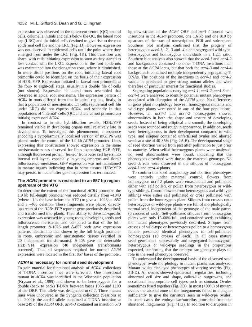
Fig. 1.Expression of ACR4during development. (A,C) Fluorescenceimages of two- to four-cell (A) and eight-cell (C) embryos (arrows).(B,D) In situ hybridisations of the same sections with ACR4antisense probe. Expression is detected as light brown coloration inthe eight-cell embryos but not in two- to four-cell embryos.(E-H) ACR4expression in the L1 (outer) cell layer of developingembryo, inflorescence (im) and floral meristems (fm). In situhybridisation (F) and confocal images (E,G,H) of H2B::YFPexpression (green) in pACR4transactivation lines. (I) In matureovules expression is detected in the outer cell layer of the funiculus(f), the outer integument (oi) inner integument (ii) and endothelium(en). (J) In the root tip expression occurs in at least four columella (c)cells layers, the lateral root cap (LRC) and the quiescent centre (QC)but not in the epithelial cell file (e). (K) Expression in the rootepithelial cell file (e) initiates as epithelial cells emerge from theLRC. c, cotyledon primordia; rp, embryonic root pole; sam,embryonic shoot apical meristem; fg, female gametophyte; m,micropyle. Scale bars: 25 µm except for K (10 µm).
4252
expression was observed in the quiescent centre (QC) centralcells, columella initials and cells below the QC, the lateral rootcap (LRC) and the initial cells destined to give rise to the rootepidermal cell file and the LRC (Fig. 1J). However, expressionwas not observed in epidermal cells until the point where theyemerged from under the LRC (Fig. 1K). This transition wassharp, with cells initiating expression as soon as they started tolose contact with the LRC. Expression in the root epidermiswas maintained into the elongation zone, where it diminished.In more distal positions on the root, initiating lateral rootprimordia could be identified on the basis of their expressionof H2B::YFP. Expression initiated in lateral root primordia atthe four- to eight-cell stage, usually in a double file of cells(not shown). Expression in lateral roots resembled thatobserved in apical root meristems. The expression pattern ofACR4 in roots differed from that in apical regions, firstly, inthat a population of meristematic L1 cells (epidermal cell fileunder LRC) did not express ACR4, and secondly in thatpopulations of ‘internal’ cells (QC, and lateral root primordiuminitials) expressed ACR4.
In contrast to in situ hybridisation results, H2B::YFPremained visible in developing organs until relatively late indevelopment. To investigate this phenomenon, a sequenceencoding a cytoplasmically localised version of mGFP6 wasplaced under the control of the 1.9 kb ACR4promoter. Linesexpressing this construction showed expression in the samemeristematic zones observed for lines expressing H2B::YFP,although fluorescent protein ‘leaked’ from outer cell layers intointernal cell layers, especially in young embryos and floral/inflorescence meristems. GFP expression was not maintainedin mature organs indicating that in some tissues H2B::YFPmay persist in nuclei after gene expression has terminated.
The ACR4 promoter is restricted to an 857 bp regionupstream of the ATGTo determine the extent of the functional ACR4promoter, the1.9 kb full-length promoter was reduced distally from –1849(where –1 is the base before the ATG) to give a –1026, a –857and a –405 deletion. These fragments were placed directlyupstream of the H2B::YFPreporter gene previously described,and transformed into plants. Their ability to drive L1-specificexpression was assessed in young roots, developing seeds andinflorescence meristems, and compared to that of the full-length promoter. ∆-1026 and ∆-857 both gave expressionpatterns identical to that shown by the full-length promoterin roots, embryonic and meristematic tissues (verified in20 independent transformants). ∆-405 gave no detectableH2B::YFP expression (40 independent transformantsscreened). Thus all sequences required for normal ACR4expression were located in the first 857 bases of the promoter.
ACR4 is necessary for normal seed developmentTo gain material for functional analysis of ACR4, collectionsof T-DNA insertion lines were screened. One insertionalmutant in ACR4was identified in the Wisconsin population(Krysan et al., 1999) and shown to be heterozygous for adouble (back to back) T-DNA between bases 1066 and 1100of the ORF. This allele was designated acr4-1. Three mutantlines were uncovered in the Syngenta collection (Sessions etal., 2002): the acr4-2 allele contained a T-DNA insertion atbase 249 of the ACR4ORF, acr4-3contained an insertion 570
bp downstream of the ACR4ORF and acr4-4housed twoinsertions in the ACR4 promoter, one 1.6 kb and one 810 bpupstream of the start of transcription. PCR and subsequentSouthern blot analysis confirmed that the progeny ofheterozygous acr4-1, -2, -3 and-4 plants segregated wild-type,heterozygous and homozygous individuals in a 1:2:1 ratio.Southern blot analysis also showed that theacr4-1and acr4-2and backgrounds contained no other T-DNA insertions thanthose at the ACR4locus, but that both the acr4-3 and acr4-4backgrounds contained multiple independently segregating T-DNAs. The positions of the insertions in acr4-1 and acr4-2would be predicted to give strong mutant alleles and weretherefore of particular interest for functional studies.
Segregating populations carryingacr4-1, acr4-2, acr4-3andacr4-4 were analysed to identify potential mutant phenotypesassociated with disruption of the ACR4gene. No differencesin gross plant morphology between homozygous mutants andwild-type plants were noted in any of the four populations.However, all acr4-1 and acr4-2 homozygotes showedabnormalities in both the shape and texture of developingseeds. Instead of being elliptical and smooth, the developingseeds were rounded and rough in appearance. In addition, seedswere heterogeneous in their development compared to wildtype, and siliques contained unfertilised ovules and abortedseeds at a rate of 40-85% (Fig. 2A,B). The developmental stageof seed abortion varied from just after pollination to just priorto maturity. When selfed heterozygous plants were analysed,no seed abnormalities were found, indicating that thephenotypes described were due to the maternal genotype. Noseed defects were observed in the siliques of homozygousacr4-3and acr4-4plants.
To confirm that seed morphology and abortion phenotypeswere entirely under maternal control, flowers fromhomozygous acr4-2 plants were emasculated and pollinatedeither with self pollen, or pollen from heterozygous or wild-type siblings. Control flowers from heterozygous and wild-typesiblings were either self pollinated or cross pollinated withpollen from the homozygous plant. Siliques from crosses ontoheterozygous or wild-type plants were full of morphologicallynormal seed, independent of the genotype of the male parent(5 crosses of each). Self-pollinated siliques from homozygousplants were only 15-60% full, and contained seeds exhibitingthe mutant phenotypes previously described. Siliques fromcrosses of wild-type or heterozygous pollen to a homozygousfemale presented identical phenotypes to self-pollinatedhomozygotes (10 crosses of each). In all cases matureseed germinated successfully and segregated homozygous,heterozygous or wild-type seedlings in the proportionsexpected, confirming that the embryo sac genotype plays norole in the seed phenotype observed.
To understand the developmental basis of the observed seedphenotype, ovule morphology in mutant plants was analysed.Mutant ovules displayed phenotypes of varying severity (Fig.3B-D). All ovules showed epidermal irregularities, includingabnormal cell size and shape, callus-like outgrowths, andoccasional inappropriate cell types such as stomata. Ovulessometimes fused together (Fig. 3D). In most (>90%) of mutantovules the abaxial zone of the integuments failed to elongatesufficiently to give the curvature seen in wild-type ovules.In some cases the embryo sac/nucellus protruded from theshortened integuments (Fig. 4H,J). In addition to disruption in
M. L. Gifford S. Dean and G. C. Ingram

4253ACR4 in cell layer organisation
ovule epidermal organisation, lack of organisation ofintegument cell layers was observed, with some ovulesshowing loss of cell layers, and others showing sporadic over-proliferation of integument cells. A varying proportion (20-50%) of ovules lacked a recognisable embryo sac (Fig. 3C,D).In extreme cases the endothelium was absent or reduced to afew disorganised cells. In other cases the endothelium cellsenclosed differentiated/divided cells, or an empty space. In 30-
50% of mutant ovules the egg apparatus (synergids, egg celland polar nucleus) could be distinguished (Fig. 3B).
In order to ascertain at what stage ovule developmentaldefects first occurred, scanning electron microscopy (SEM) ofdeveloping ovules was undertaken. Wild-type developmentwas as previously described (Schnietz et al., 1995; Robinson-Beers et al., 1992). Ovule primordia arose as bulges along theplacenta, and developed into finger-like protrusions (Fig. 4A).Subsequently the inner and outer integuments initiated as tworing-shaped growths encircling the megasporocyte-containingovule tip (nucellus), with the inner integument initiating justbefore the outer integument (Fig. 4C). Both integuments thenelongated as sleeves of cells engulfing the nucellus, with theouter-integument growing faster than, and eventuallyovergrowing the inner integument (Fig. 4E,G). In acr4 mutantovules, development was normal until the point of integumentinitiation (Fig. 4B). However, instead of initiating as smoothring-like bulges, the integuments of mutant ovules initiatedunevenly, with some cell files bulging out, and othersremaining flat. In many cases more than two sets of bulgingcells could be seen in the proximodistal axis, and integumentsdid not initiate as coherent rings, suggesting that the points ofintegument initiation were not well defined (Fig. 4D). Afterinitiation, mutant integuments appeared thicker than wild-type,and their more rounded cells gave developing ovules a roughtexture (Fig. 4F). Integuments grew more slowly in mutant thanin wild-type plants, and the leading edge of the integument,instead of being smooth, appeared disorganised. At maturity,even in the most ‘normal’ mutant ovules, integuments failed tofully enclose the nucellus (compare Fig. 4G with 4H). In somecases integument elongation either of one (Fig. 4I) or bothinteguments was severely compromised (Fig. 4J). Abnormalprotruding cells were often observed on the surface of mutantovules (Fig. 4H)
Defects observed in ovules were maintained in developingseeds when fertilisation had been possible. In particular, thetexture of the seed coat was abnormal, with outgrowthsobserved, particularly in retarded seeds. A lack ofproximodistal elongation of the mutant embryo sac afterfertilisation caused the mutant endosperm to develop in a
reduced volume giving seeds a round rather thanelliptical shape (Fig. 2D). Although defects in embryoorganisation were not observed, seeds with moresevere defects in integument organisation were alsoretarded in embryo and endosperm development.
Fig. 2.acr4mutant seed phenotype. (A,B) Opened siliques showingdifferences in seed size and texture between a wild-type (A) andhomozygous (B) plant at comparable stages (12 days afterpollination). Aborted ovules (arrowhead), retarded seeds (star) andseeds with epidermal outgrowths (arrow) are frequent in mutantsiliques. (C,D) Comparison between mature wild-type (C) andmutant (D) seeds observed by SEM. Scale bars: 100 µm.
Fig. 3.The internal structure of acr4mutant ovules. Wild-type (A) compared with mutant (B,C,D) ovules. Micropylesare indicated by arrowheads. Female gametophytes (fg) anddensely staining endothelial cells (e) are labelled wherepresent. (A) In wild type neatly organised cell layers arevisible, with the female gametophyte (fg) surrounded by anorderly endothelium. (B) Mutant ovule with weakphenotype. Fg is visible but outer integument is disrupted(to left of star). Endothelial layer is visible. (C) Mutantovule with intermediate phenotype showing disorganisedcell layers and replacement of fg with divided cells.(D) Two fused (star) mutant ovules with extremephenotypes. Both show cell layer disorganisation but onehas distinguishable (probably abnormal) fg. Scale bar:25µm.

4254
Histological analysis supported the hypothesis that these seedswere those observed to abort.
To study the epidermal abnormalities observed in developingseeds, SEM analysis of mature mutant seeds was carried out.Although seed coat abnormalities were observed, particularlyat the funiculus abscission scar and at the micropylar region,the majority of seed coat cells had a similar structure to thoseobserved in wild-type seeds (Fig. 2C,D). Because homozygousseeds still differentiated appropriate epidermal cell types, andeven in ovules, mis-specification of cell types (for example thepresence of stomata) involved epidermal-specific identities, theexpression of an L1 marker in mutant ovules was investigated.Homozygous acr4-2and acr4-1plants were crossed to markerlines expressing an N-terminal GFP::ATML1 fusion protein(unpublished results) under the ATML1 promoter (Sessions etal., 1999). These lines expressed nuclear localised fusionprotein in the L1-specific pattern previously reported forATML1expression in embryos and meristems (Lu et al., 1996;Sessions et al., 1999). ATML1 fusion protein expression wasobserved in the outer cell layer and endothelium of matureovules in wild-type plants, with weak expression occasionallyobserved in the inner cell layer of the inner integument. In acr4mutant ovules ATML1expression was similar to or morewidespread than in wild type. In excrescences on the ovulesurface, both protruding callus-like cells and underlying cellsshowed expression. Strong expression was sporadically seen incells situated between the ovule epidermis and theendothelium. In several cases, the egg sac space was filled withexpressing cells. This analysis suggests that although mutantovule integument cells showed abnormalities in organisation,they did not loose their L1 identity.
Because acr4 mutants showed abnormalities in ovuleinteguments, sepal margins, which have a similar structure(appressed layers of L1 cells) were examined in more detail.Although no major defects in sepal morphology wereobserved in acr4 mutants, it was noted that the cells at sepalboundaries appeared less well organised than in wild-typeplants, giving a somewhat ragged appearance (Fig. 4K,L). Ingeneral the border region was thicker (contained more cells)in the abaxial/adaxial dimension than in wild type, suggestingthat outgrowth of sepal margins could be affected. Mutantmargin cells were irregularly shaped and showed abnormal‘lumpy’ areas and regions devoid of the cuticular decorationseen in wild-type cells. No defects at the margins of leaves orpetals could be discerned.
Although two independent mutant alleles in two differentbackgrounds both gave identical phenotypes, a furtherconfirmation that the observed phenotype was due to loss ofACR4 function was obtained by genetic complementation ofacr4-2. Homozygous mutants were crossed to hygromycin-resistant transformants carrying a full-length ACR4promoterdriving the ACR4ORF. Four F2 families corresponding to fourindependent transformants were selected on hygromycin andPCR-genotyped for homo- or heterozygosity of acr4-2. Thephenotypes of homozygous plants were compared with thoseof heterozygous and wild-type plants in each case. For twofamilies homozygosity of acr4-2plants was verified bySouthern blot. For all four families full phenotypiccomplementation was apparent in immature and mature seedsof homozygous mutant plants, confirming that the observedmutant phenotypes were due to loss of ACR4function.
M. L. Gifford S. Dean and G. C. Ingram
Fig. 4. Phenotypic analysis of wild-type and acr4mutant plants.(A,B) Ovule primordia immediately prior to integument initiation inwild-type (A) and mutant (B) plants. (C,D) The initiation of the inner(arrowheads) and outer (arrows) integuments in wild-type and mutantovules respectively. In the mutant, the irregular initiation ofintegument outgrowth is visible, with at least two outgrowthsobserved in one region of outer integument initiation (asterisks),whereas other regions have no outgrowths. (E,F) Ongoing integumentoutgrowth. In wild-type ovules (E) the leading edges of theinteguments are smooth, whereas in mutants (F) they are ragged andoften retarded. (The nucellus has snapped off in the right hand ovuleof F.) (G) A mature wild-type ovule at anthesis. The outer integumenthas overgrown the inner integument and nucellus to give a narrowmicropyle (m) facing the funiculus (f). (H-J) A weak, a medium and asevere mature mutant ovule phenotype, respectively. In H, theretardation of outer integument (arrow) growth has left an openmicropyle within which the inner integument (arrowhead) andnucellus are visible. In I the inner integument (arrowhead) looksrelatively normal whereas the outer integument (arrow) has failed toelongate correctly. (J) The inner integument (arrowhead) has failed togrow out leaving the nucellus almost completely exposed. (K) Wild-type sepal margin (arrowhead) showing well organised border cellscovered in cuticular decoration. (L) Mutant sepal margin (arrowhead)showing typical irregularities in cell organisation, ‘lumpy’ appearanceand regions devoid of cuticular decoration. Scale bars: 20 µm.

4255ACR4 in cell layer organisation
ACR4 encodes an active kinase domainTo establish whether ACR4 protein encodes a functional kinase,as predicted from its sequence, a GST fusion protein constructwas engineered to express the ACR4kinase domain in bacteria.A 61 kDa protein encoding the GST-kinase was expressed andpurified (Fig. 5). To act as a control in kinase assays, Lys 540(a crucial amino acid in the kinase activation loop) was mutatedto methionine. GST-kinase and GST-kinase-null proteins weresubjected to in vitro kinase assays. The kinase domain showedphosphorylation that was absent in the kinase-null variant (Fig.5). Incubation of the kinase domain with GST protein alone didnot result in phosphorylation of GST, indicating that the kinasedomain could autophosphorylate inter or intramolecularly invitro (results not shown).
ACR4 fusion proteins localise to the plasmamembrane and to intracellular bodiesStructural predictions indicated a plasma membrane localisationfor ACR4. To test this prediction the entire ACR4ORF was fusedat the C terminus in frame with GFP, placed under control of thecomplete ACR4promoter and introduced into plants. In order totest whether the fusion protein was being correctly localised,plants from two different expressing lines were crossed tohomozygous acr4-2 mutants, and F2 plants were genotypedfor the acr4-2 allele. Full complementation of the acr4-2mutant phenotype was observed for one line, and partialcomplementation for the other line tested. Partiallycomplementing plants showed reduced seed death, and a morenormal seed shape, although seed texture was still abnormal.Expression of fusion proteins was detected in regions whereH2B::YFP reporter expression had previously been observed(Fig. 6), and was identical in wild-type and in complementedhomozygous mutant plants. Cellular localisation of fusionproteins varied from tissue to tissue. In some cells, for examplethose on the surface of ovules, most fluorescence appeared to beassociated with plasma membranes (Fig. 6A). In the L1 cells ofembryos, inflorescence and floral meristems and roots, plasmamembrane localisation was observed, but fluorescence alsolocalised to multiple small intensely staining bodies within cells(Fig. 6B,C,D,G). These bodies did not co-localise with red-fluorescing chloroplasts, but were the same size or smaller.To confirm that fluorescent protein was localised to plasmamembranes rather than cell walls, roots were treated with 0.8 Mmannitol to induce plasmolysis. Under these conditions
fluorescence was pulled away from cell-cell boundaries,indicating that fluorescent proteins were indeed associated withthe plasma membrane, rather than the cell wall (Fig. 6F). In orderto further address ACR4::GFP localisation, the effect ofbrefeldin A (BFA) on protein localisation in roots was examined.BFA targets and inhibits the action of proteins involved in vesicleformation, thereby inhibiting vesicle trafficking within cellularmembrane compartments and to and from the plasma membrane(Nebenführ et al., 2002). After treatment with BFA, ACR4::GFPlocalisation was compromised (Fig. 6G,H). The relative intensityof plasma membrane-associated fluorescence decreased, andinstead of multiple small cytoplasmic bodies, one or two largefluorescent bodies were observed in each cell. An identicalphenomenon has been observed using immunolocalisation of theauxin efflux carrier PIN1 in BFA-treated roots (Geldner et al.,2001; Geldner et al., 2003). The described result of BFAtreatment on ACR4::GFP localisation supports the hypothesisthat ACR4 is usually exported to the plasma membrane via theER and Golgi, and that this export, or possibly some form ofrecycling, is inhibited by BFA. It seems likely that thecytoplasmic bodies observed in cells not treated with BFAcorrespond to elements of the endomembrane system, such asexcretory vesicles or endosomes.
In all tissues studied, fusion protein was present in plasmamembranes adjacent to both anticlinal and periclinal cell walls,although the degree of localisation adjacent to periclinal cellwalls was variable. In root meristems (QC and root cap initials)localisation was observed uniformly in both anticlinal andpericlinal plasma membranes (Fig. 6E). In cells situated on thesurface of the plant, the amount of protein visible in plasmamembranes adjacent to the outer periclinal cell wall appearedlower than that on anticlinal and inner periclinal cell plasmamembranes (Fig. 6D,I). This phenomenon was particularlynoticeable in the outer cells of ovule outer integuments whereall cells expressed fusion protein, although this could in partbe due to the additive signal from two appressed internalmembranes (Fig. 6I).
DISCUSSION
ACR4 regulates the organisation of L1-derived ovuleinteguments and sepal marginsDespite the wide ranging expression pattern observed forACR4, probable null mutants only show defects in two tissues;ovule integuments and sepal boundaries. Characterisation ofmutants in several genes affecting integument developmentincluding INNER NO OUTER, SHORT INTEGUMENTS 1,SHORT INTEGUMENTS 2, BELL, AINTEGUMENTA,ABERRANT TESTA SHAPE andNOZZLE, has shown thatinteguments play an important role in female gametophytedevelopment and maturation (Reiser and Fischer, 1993;Villanueva et al., 1999; Robinson-Beers et al., 1992;Broadhvest et al., 1999; Baker et al., 1997; Schneitz et al.,1998; Balasubramanian et al., 2002). In particular the presenceof an intact endothelial cell layer is crucial, possibly becausenutritionally and developmentally important substances arechannelled to the gametophyte through this specialised celllayer (Kapil and Tiwari, 1978). We observed no defectsin ovule development until integument initiation, whenmegaspores usually initiate meiosis, suggesting that the
Fig. 5.ACR4 kinase activity. Coomassie Blue-stained gel (top)showing products of kinase assay on the wild-type ACR4 kinasedomain (K+) and kinase null variant (negative control, K–).Autoradiography of this gel (below) shows that the native kinasedomain has kinase activity whereas the kinase null variant does not.

4256
observed lack of a female gametophyte in some mature acr4ovules may be due to degradation or de-differentiation ratherthan to lack of initiation of gametophyte development.
Many acr4 mutant ovules are never fertilised because ofsevere morphological abnormalities, but of the ones that are,those with more severe organisational defects abort asdeveloping seeds. Abortion is independent of zygotic genotypeand is, moreover not due to developmental defects in embryoand endosperm development, although both tissues areretarded at the time of seed death. Retardation and abortionprobably occur because defective seeds provide insufficientmaternal support, in terms of nutrients, for embryo sacdevelopment. Similar retardation and death of embryo/endosperm was observed when reduced expression of thegenes FBP7 and FBP11 led to developmental abnormalitiesand degeneration in the endothelium and seed coat of Petunia(Colombo et al., 1997). The total lack of zygotically derivedembryo development defects and the observation of seed coatabnormalities in our study contradicts results obtained usingantisense experiments to reduce ACR4expression (Tanaka etal., 2002).
ACR4, as a membrane-localised receptor-like kinase,probably acts by perceiving extracellular ligands. Severalgenes encoding possible ligands, or ligand processingmolecules for CR4 and related proteins have been proposed.These include the subtilase encoded by the ABNORMALLEAF SHAPE 1(ALE1) gene (Tanaka et al., 2001). Duringembryo and endosperm development, signals fromsurrounding tissues (as could be provided by the action ofgenes such as ALE1) might be important in signalling required
for ‘outside’ cell layer specification. However, it seems morelikely that in organ primordia, as has been shown in root celllayer differentiation, an ‘inside to outside’ signalling processis involved in regulating cell layer behaviour, combined witha role for signals from neighbouring cells in the same celllayer (Nakajima and Benfey, 2002). Our observation thatACR4 protein is localised on ‘internal’ plasma membranes of‘outside’ cells supports the hypothesis that ACR4 mayperceive signals from underlying cells and/or same-layerneighbours. If this is the case, the restriction of the acr4phenotype to ovule integuments and sepal margins could beattributable to the fact that these tissues are unique in theArabidopsisplant, in being composed of two appressed layersof L1 cells. If normal L1 behaviour (i.e. anticlinal divisionsgiving rise to a monolayer of L1 cells) were dependent onperception of positional information both from underlyingcells, and from same-layer neighbours, then a loss insignalling between same-layer neighbours could becompensated for by signals from underlying cells in mosttissues. However, in the case of ovule integuments and sepalmargins, positional information would be effectively limitedto that exchanged between same-layer neighbours. The cellsin these organs would thus be particularly sensitive todisruption of this signalling pathway, which would beexpected to lead to a loss of cellular organisation and thusabnormalities in organ outgrowth, similar to the phenotypeobserved in acr4mutants.
Other pieces of the puzzleThe restricted mutant phenotype of ACR4compared to maize
M. L. Gifford S. Dean and G. C. Ingram
Fig. 6.Protein localisation in lines expressing anACR4::GFP protein fusion. (A) localisation offusion proteins (gren) in the plasma membranes ofthe ovule epidermis. (B,C) Expression patterns offusion proteins (green) in embryos (B) andmeristems (floral meristem, fm) (C). Plasmamembrane localisation can be observed (arrow inB). (D-F) Protein localisation in root meristems.(D) Lateral root tip, columella and LRCexpression are visible. No protein is seen in theepidermis (e) before emergence from the LRC.(E) Main root tip with columella cell layersindicated by stars. (F) surface view when mountedin 0.8 M mannitol. Cell wall area is clear offluorescence. (G,H) Protein localisation incomparable untreated and 2-hour BFA-treated rootsamples, respectively, showing relative decrease inplasma membrane localisation and the appearanceof bright perinuclear bodies. (I) mature ovule withfluorescence seen in outer integument (oi) (wherelittle protein is detected in the outermost cellplasma membrane; arrowhead), inner integument(ii) and outer cell layer of funiculus (f). rp,embryonic root pole; c, cotyledon primordia; s,sepal primordia; m, micropyle; fg, femalegametophyte.Scale bars: 25 µm.

4257ACR4 in cell layer organisation
CR4 mutants is surprising since ACR4appears to be unique inArabidopsisin its degree of similarity to maize CR4. Unlikestudies of cr4in maize, we find no evidence for a loss ofepidermal identity in acr4mutants, but rather solely a loss ofcell organisation. The cell disorganisation observed in acr4ovule integuments and sepal margins is, however, reminiscentof aspects of the epidermal defects observed in the leaves ofmaize cr4 mutants. Notably, both phenotypes involvederegulation of the planes of division, and organisation ofpopulations of L1 cells. Striking differences in expression alsoexist between ACR4andCR4. In maize, CR4 is expressed inthe aleurone cell layer and one of the major phenotypesassociated with cr4mutants is a defect in aleuronedifferentiation (Becraft et al., 1996). ACR4shows noendosperm expression, although it is arguable whetherArabidopsis can be considered to differentiate a structureanalogous to the cereal aleurone layer (Berger, 1999). Inaddition, unlike ACR4, CR4 appears to be expressedthroughout apical meristems, without restriction to the L1 layeruntil late in leaf development (Becraft et al., 1996), and no CR4expression has been reported in maize root tissue.
Functional redundancy between ACR4and four otherArabidopsisgenes showing weaker similarity to CR4 cannotbe ruled out as an explanation for some of the differencesin phenotypic severity between cr4and acr4mutants. Thetwo most closely related genes encode proteins lackinga conserved kinase catalytic domain required for kinaseactivity (domain 8) (Hanks et al., 1998). ACR4encodes afunctional kinase, and kinase activity is probably required forat least some of its functions. However, kinase-inactivereceptors can retain partial function, possibly by interactionwith other unrelated kinases. The kinase-null clv1-6allele,which causes part of the kinase domain of the CLAVATA1protein to be deleted, causes only a weak mutant phenotype.The mutant protein thus retains functions that are independentof its ability to auto/transphosphorylate itself and otherproteins (Torii and Clark, 2000). Of the two less similargenes, one encodes a protein closely related to tobaccoCRK1, which has recently be implicated in cytokininresponses (Schafer et al., 2002). The other shares many moreresidues with CRK1 than with ACR4 and CR4, especiallyin the extracellular domain adjacent to the trans-plasmamembrane domain, where ACR4and CR4encode putativeTNFR-like repeats.
An alternative explanation for the weak acr4phenotypecould be that although several independent mechanismsregulate L1 behaviour in both Arabidopsis and maize,mechanistic differences in organ primordium development inmonocotyledonous and dicotyledonous species have led to lessfunctional overlap in maize than in Arabidopsis. Consideringthe relatively large numbers of genes expressed in L1 celllayers from early in development in both species, thispossibility seems realistic, and will be investigated usingongoing mutagenesis and double mutant analysis approachesin the near future.
We would like to thank Dr Jim Haseloff (University of Cambridge,UK), Dr Corinne Boisnard-Lorig and Dr Frederic Berger (EcoleNormale Superieure, Lyon, France) for providing pBI121,pBSmGFP6 and the GAL4::VP16 coding sequence. We are mostgrateful to Dr Allen Sessions (Syngenta, Torrey Mesa Research
Institute, San Diego) for providing pAS99, to Dr Chris Jeffree for helpwith SEM, Kathryn Degnan for technical assistance, Dr JaneLangdale, Dr Andrew Hudson, Dr Justin Goodrich and three refereesfor critical comments on the manuscript, Dr John Golz for help within situ hybridisations and resin embedding and Dr Thomas Guebitzfor help with phylogenetic analysis. We acknowledge the work of theNASC, ABRC, Syngenta and the University of Wisconsin for creatingand providing seed stocks. G.I. is supported by a Royal SocietyUniversity Research Fellowship, and M.G. by a BBSRC studentship.The project also benefited from a JREI grant (BBSRC/Olympusoptical).
REFERENCES
Abe, M., Takahashi, T. and Komeda, Y.(2001). Identification of a cis-regulatory element for L1 layer-specific gene expression, which is targetedby an L1-specific homeodomain protein. Plant J.26, 487-494.
Baker, S. C., Robinson-Beers, K., Villanueva, J. M., Gaiser, J. C. andGasser, C. S. (1997). Interactions among genes regulating ovuledevelopment in Arabidopsis thaliana. Genetics145, 1109-1124.
Balasubramanian, S. and Schneitz, K.(2002). NOZZLE links proximal-distal and adaxial-abaxial pattern formation during ovule development inArabidopsis.Development129, 4291-4300.
Becker, D. (1990). Binary vectors which allow the exchange of plant selectablemarkers and reporter genes. Nucleic Acids Res.18, 203.
Becker, D., Kemper, E., Schell, J. and Masterson, R.(1992). New plantbinary vectors with selectable markers located proximal to the left T-DNAborder. Plant Mol. Biol. 20, 1195-1197.
Becraft, P. W., Li, K., Dey, N. and Asuncion-Crabb, Y.(2002). The maizedek1 gene functions in embryonic pattern formation and cell fatespecification. Development129, 5217-5225.
Becraft, P. W., Stinard, P. S. and McCarty, D. R.(1996). CRINKLY4: ATNFR-like receptor kinase involved in maize epidermal differentiation.Science273, 1406-1409.
Berger, F. (1999). Endosperm development. Curr. Opin. Plant Biol. 2, 28-32. Boisnard-Lorig, C., Colon-Carmona, A., Bauch, M., Hodge, S., Doerner,
P., Bancharel, E., Dumas, C., Haseloff, J. and Berger, F.(2001). Dynamicanalyses of the expression of the HISTONE::YFP fusion protein inArabidopsisshow that syncytial endosperm is divided in mitotic domains.Plant Cell 13, 495-509.
Broadhvest, J., Baker, S. C. and Gasser, C. S.(1999). SHORTINTEGUMENTS 2promotes growth during Arabidopsis reproductivedevelopment. Genetics 155, 899-907.
Clough, S. J. and Bent, A. F.(1998). Floral dip: a simplified method forAgrobacterium-mediated transformation of Arabidopsis thaliana. Plant J.16, 735-743.
Colombo, L., Franken, J., van der Krol, A. R., Wittich, P. E., Dons, H. J.M. and Angenent, G. C.(1997). Downregulation of ovule-specific MADSbox genes from Petuniaresults in maternally controlled defects in seeddevelopment. Plant Cell 9, 703-715.
Fletcher, J. C., Brand, U., Running, M. P., Simon, R. and Meyerowitz, E.M. (1999). Signalling of cell fate decisions by CLAVATA3 in Arabidopsisshoot meristems. Science283, 1911-1914.
Geldner, N., Anders, N., Wolters, H., Keicher, J., Kornberger, W., Muller,P., Delbarre, A., Ueda, T., Nakana, A. and Jürgens, G. (2003). TheArabidopsis GNOM ARF-GEF mediates endosomal recycling, auxintransport and auxin-dependent plant growth. Cell 112, 219-230.
Geldner, N., Friml, J., Stierhof, Y.-D., Jürgens, G. and Palme, K.(2001).Auxin transport inhibitors block PIN1 cycling and vesicle trafficking.Nature 413, 425-428.
Hanks, S. K., Quinn, A. M. and Hunter, T.(1998). The protein kinase family:conserved features and deduced phylogeny of the catalytic domains. Science241, 42-52.
Haseloff, J. (1999). GFP variants for multispectral imaging of living cells.Methods Cell Biol.58, 139-151.
Jackson, D. (1991). In situ hybridisation in plants. In Molecular PlantPathology: A Practical Approach (ed. D. J. Bowles, S. J. Gurr and M.McPherson). Oxford: Oxford University Press.
Jenik, P. D. and Irish, V. F.(2000). Regulation of cell proliferation patternsby homeotic genes during Arabidopsisfloral development. Development127, 1267-1276.

4258
Kapil, R. N. and Tiwari, S. C. (1978). The integumentary tapetum. Bot. Rev.44, 457-490.
Kessler, S., Seiki, S. and Sinha, N.(2002). Xcl1causes delayed obliquepericlinal cell divisions in developing maize leaves, leading to cellulardifferentiation by lineage instead of position. Development 129, 1859-1869.
Kidner, C., Sundaresan, V., Roberts, K. and Dolan, L. (2000). Clonalanalysis of the Arabidopsisroot confirms that position, not lineage,determines cell fate. Planta221, 191-199.
Koncz, C. and Schell, J.(1986). The promoter of TL-DNA gene 5 controlsthe tissue-specific expression of chimaeric genes carried by a novel type ofAgrobacteriumbinary vector. Mol. Gen. Genet.204, 383-396.
Krysan, P. J., Young, J. K. and Sussman, M. R.(1999). T-DNA as aninsertional mutagen in Arabidopsis. Plant Cell11, 2283-2290.
Lid, S. E., Gruis, D., Jung, R., Lorentzen, J. A., Ananiev, E., Chamberlin,M., Niu, X., Meeley, R., Nichols, S. and Olsen, O. A.(2002). The defectivekernel 1 (dek1) gene required for aleurone cell development in theendosperm of maize grains encodes a plasma membrane protein of thecalpain gene superfamily. Proc. Natl. Acad. Sci. USA 99, 5460-5465.
Lu, P., Porat, R., Nadeau, J. A. and O’Neill, S. D.(1996). Identification ofa meristem L1 layer-specific gene in Arabidopsisthat is expressed duringembryonic pattern formation and defines a class of homeobox genes. PlantCell 8, 2155-2168.
Nakajima, K. and Benfey, P. N.(2002). Signalling in and out: control of celldivision and differentiation in the shoot and root. Plant Cell14, S265-S276.
Nebenführ, A., Ritzenthaler, C. and Robinson, D. G.(2002). Brefeldin A:Deciphering an enigmatic inhibitor of secretion.Plant Physiol. 130, 1102-1108.
Notredame, C., Higgins, D. G. and Heringa, J.(2000). T-Coffee: A novelmethod for fast and accurate multiple sequence alignment. J. Mol. Biol. 302,205-217.
Reiser, L. and Fischer, R. L. (1993). The ovule and embryo sac. Plant Cell5, 1291-1301.
Robinson-Beers, K., Pruitt, R. E. and Gasser, C. S.(1992). Ovuledevelopment in wild-type Arabidopsisand two female-sterile mutants. PlantCell 4, 1237-1249.
Schafer, S. and Schmulling, T.(2002). The CRK1 receptor-like kinase oftobacco is negatively regulated by cytokinin.Plant Mol. Biol.50, 155-166.
Schneitz, K., Baker, S. C., Gasser, C. S. and Redweik, A. (1998). Patternformation and growth during floral organogenesis: HUELLENLOS andAINTEGUMENTA are required for the formation of the proximal region ofthe ovule primordium in Arabidopsis thaliana. Development125, 2555-2563.
Schneitz, K., Hulskamp, M. and Pruitt, R. E. (1995). Wild-type ovuledevelopment in Arabidopsis thaliana: a light microscope study of clearedwhole-mount tissue. Plant J.7, 731-749.
Sessions, A., Burke, E., Presting, G., Aux, G., McElver, J., Patton, D.,Dietrich, B., Ho, P., Bacwaden, J., Ko, C. et al. (2002). A high-throughputArabidopsisreverse genetics system. Plant Cell14, 2985-2994.
Sessions, A., Weigel, D. and Yanofsky, M. F. (1999). The ArabidopsisthalianaMERISTEM LAYER 1 promoter specifies epidermal expression inmeristems and young primordia. Plant J.20, 259-263.
Stewart, R. N. and Burk, L. G.(1970). Independence of tissues derived fromapical layers in ontegeny of the tobacco leaf and ovary. Am. J. Bot.57, 1010-1016.
Stewart, R. N. and Dermen, H.(1975). Flexibility in ontogeny as shown bythe contribution of the shoot apical layers to the leaves of periclinalchimaeras. Am. J. Bot.62, 935-947.
Tanaka, H., Onouchi, H., Kondo, M., Hara-Nishimura, I., Nishimura, M.,Machida, C. and Machida, Y.(2001). A subtilisin-like serine protease isrequired for epidermal surface formation in Arabidopsisembryos andjuvenile plants. Development 128, 4681-4689.
Tanaka, H., Watanabe, M., Watanabe, D., Tanaka, T., Machida, C.and Machida, Y. (2002). ACR4, a putative receptor kinase gene ofArabidopsis thaliana, that is expressed in the outer cell layers of embryosand plants, is involved in proper embryogenesis.Plant Cell Physiol.43,419-428.
Torii, K. and Clark, S. (2000). Receptor-like kinases in plant development.Adv. Bot. Res.32, 225-267.
van den Berg, C., Willemsen, V., Hage, W., Weisbeek, P. and Scheres, B.(1995). Cell fate in the Arabidopsisroot meristem determined by directionalsignalling. Nature378, 62-65.
Villanueva, J. M., Broadhvest, J., Hauser, B. A., Meister, R. J., Schneitz,K. and Gasser, C. S.(1999). INNER NO OUTER regulates abaxial-adaxialpatterning in Arabidopsisovules. Genes Dev. 13, 3160-3169.
M. L. Gifford S. Dean and G. C. Ingram