The Ancient mariner Sails Again: Transposition of the Human Hsmar1 Element by a Reconstructed...
13
See discussions, stats, and author profiles for this publication at: https://www.researchgate.net/publication/51385438 The Ancient mariner Sails Again: Transposition of the Human Hsmar1 Element by a Reconstructed Transposase and... Article in Molecular and Cellular Biology · June 2007 DOI: 10.1128/MCB.02027-06 · Source: PubMed CITATIONS 79 READS 43 7 authors, including: Some of the authors of this publication are also working on these related projects: Mobile DNA View project Csaba Miskey Paul-Ehrlich-Institut 25 PUBLICATIONS 986 CITATIONS SEE PROFILE Lajos Mátés Hungarian Academy of Sciences 71 PUBLICATIONS 1,271 CITATIONS SEE PROFILE Zsuzsanna Izsvák Max-Delbrück-Centrum für Molekulare Medi… 175 PUBLICATIONS 6,707 CITATIONS SEE PROFILE Zoltán Ivics Paul-Ehrlich-Institut 226 PUBLICATIONS 6,722 CITATIONS SEE PROFILE All content following this page was uploaded by Lajos Mátés on 02 December 2016. The user has requested enhancement of the downloaded file. All in-text references underlined in blue are linked to publications on ResearchGate, letting you access and read them immediately.
Transcript of The Ancient mariner Sails Again: Transposition of the Human Hsmar1 Element by a Reconstructed...

Seediscussions,stats,andauthorprofilesforthispublicationat:https://www.researchgate.net/publication/51385438
TheAncientmarinerSailsAgain:TranspositionoftheHumanHsmar1ElementbyaReconstructedTransposaseand...
ArticleinMolecularandCellularBiology·June2007
DOI:10.1128/MCB.02027-06·Source:PubMed
CITATIONS
79
READS
43
7authors,including:
Someoftheauthorsofthispublicationarealsoworkingontheserelatedprojects:
MobileDNAViewproject
CsabaMiskey
Paul-Ehrlich-Institut
25PUBLICATIONS986CITATIONS
SEEPROFILE
LajosMátés
HungarianAcademyofSciences
71PUBLICATIONS1,271CITATIONS
SEEPROFILE
ZsuzsannaIzsvák
Max-Delbrück-CentrumfürMolekulareMedi…
175PUBLICATIONS6,707CITATIONS
SEEPROFILE
ZoltánIvics
Paul-Ehrlich-Institut
226PUBLICATIONS6,722CITATIONS
SEEPROFILE
AllcontentfollowingthispagewasuploadedbyLajosMátéson02December2016.
Theuserhasrequestedenhancementofthedownloadedfile.Allin-textreferencesunderlinedinblue
arelinkedtopublicationsonResearchGate,lettingyouaccessandreadthemimmediately.

MOLECULAR AND CELLULAR BIOLOGY, June 2007, p. 4589–4600 Vol. 27, No. 120270-7306/07/$08.00�0 doi:10.1128/MCB.02027-06Copyright © 2007, American Society for Microbiology. All Rights Reserved.
The Ancient mariner Sails Again: Transposition of the Human Hsmar1Element by a Reconstructed Transposase and Activities of the
SETMAR Protein on Transposon Ends�†Csaba Miskey,1 Balazs Papp,2 Lajos Mates,1 Ludivine Sinzelle,1 Heiko Keller,1
Zsuzsanna Izsvak,1,3 and Zoltan Ivics1*Max Delbruck Center for Molecular Medicine, 13092 Berlin, Germany1; Faculty of Life Sciences, The University of Manchester,
Manchester M13 9PT, United Kingdom2; and Institute of Biochemistry, Biological Research Center of theHungarian Academy of Sciences, 6726 Szeged, Hungary3
Received 30 October 2006/Returned for modification 15 November 2006/Accepted 26 March 2007
Hsmar1, one of the two subfamilies of mariner transposons in humans, is an ancient element that entered theprimate genome lineage �50 million years ago. Although Hsmar1 elements are inactive due to mutationaldamage, one particular copy of the transposase gene has apparently been under selection. This transposasecoding region is part of the SETMAR gene, in which a histone methylatransferase SET domain is fused to anHsmar1 transposase domain. A phylogenetic approach was taken to reconstruct the ancestral Hsmar1 trans-posase gene, which we named Hsmar1-Ra. The Hsmar1-Ra transposase efficiently mobilizes Hsmar1 trans-posons by a cut-and-paste mechanism in human cells and zebra fish embryos. Hsmar1-Ra can also mobilizeshort inverted-repeat transposable elements (MITEs) related to Hsmar1 (MiHsmar1), thereby establishing afunctional relationship between an Hsmar1 transposase source and these MITEs. MiHsmar1 excision is 2orders of magnitude more efficient than that of long elements, thus providing an explanation for their high copynumbers. We show that the SETMAR protein binds and introduces single-strand nicks into Hsmar1 inverted-repeat sequences in vitro. Pathway choices for DNA break repair were found to be characteristically differentin response to transposon cleavage mediated by Hsmar1-Ra and SETMAR in vivo. Whereas nonhomologousend joining plays a dominant role in repairing excision sites generated by the Hsmar1-Ra transposase, DNArepair following cleavage by SETMAR predominantly follows a homology-dependent pathway. The noveltransposon system can be a useful tool for genome manipulations in vertebrates and for investigations into thetranspositional dynamics and the contributions of these elements to primate genome evolution.
mariner elements make up a diverse family of eukaryoticDNA transposons present in a wide variety of genomes, in-cluding that of humans (references 6, 41, and 43 and referencestherein). These transposons contain a single gene encoding thetransposase flanked by short, �30-bp terminal inverted-repeat(IR) sequences. mariner elements mobilize through a cut-and-paste mechanism catalyzed by the transposase, which belongsto a large family of recombinase proteins, including retroviral/retrotransposon integrases and transposases, characterized bythe DDE/D signature in the catalytic domain of the proteins(40). Transposition results in the accumulation of hundreds orthousands of transposon copies over evolutionary time. How-ever, most mariner copies appear to be dead remnants of once-active transposons inactivated by mutations (14). To date, onlythree mariner elements out of the hundreds of sequences thathave been described have proven to be active. Two of these,Mos1 and Famar1, are natural elements isolated from thegenomes of Drosophila mauritiana (20) and the earwig For-ficula auricularia (3), respectively. The active Himar1 elementis a majority-rule consensus of cloned genomic copies obtained
from the horn fly Haematobia irritans (24). Mos1 and Himar1have been used as molecular tools for genome manipulationsin diverse species (reviewed in reference 40). However, theutility of these invertebrate mariner transposons in mammaliangenetics is hindered by their limited activity in mammalian cells(12).
mariner elements are represented by two subfamilies inthe human genome: Hsmar1 (44) and Hsmar2 (43). The firstHsmar1 element entered the primate genome lineage approx-imately 50 million years ago, and transposition was ongoinguntil at least 37 million years ago, producing about 200 Hsmar1copies (44) (Fig. 1A). However, none of the present copiesencodes a functional transposase protein, due to mutationalinactivation. The Hsmar1 transposon copies are accompaniedby about 4,500 copies of solo IRs (containing a single IR) andabout 2,500 copies of an Hsmar1-related paired-IR element,MiHsmar1 (15, 44) (Fig. 1A). Such miniature IR transposableelements (MITEs) are thought to have been generated byinternal deletions of longer transposons; they make up thepredominant fraction of DNA elements in flowering plants andare often found in animal genomes (9). Even though the clas-sical MITEs are longer than the MiHsmar1 elements, the com-mon characteristics that link all of these elements are thefollowing: significantly smaller size than the corresponding pre-cursor element, significantly higher copy number than the cor-responding precursor element, and lack of transposase-codingcapacity. It is widely believed that MITEs can be mobilized
* Corresponding author. Mailing address: Max Delbruck Center forMolecular Medicine, Robert Rossle Str. 10, D-13092 Berlin, Germany.Phone: (49) 30 9406-2546. Fax: (49) 30 9406-2547. E-mail: [email protected].
† Supplemental material for this article may be found at http://mcb.asm.org/.
� Published ahead of print on 2 April 2007.
4589
only by transposases supplied in trans (9), but only one suchinstance has been documented at the molecular level (7). Al-though mechanisms of preferential transposition due to smallsize (25, 44) and transposition linked to the cellular process ofDNA replication (10, 17) have been suggested, the mecha-nisms of MITE mobilization and amplification are incom-pletely understood.
Despite their parasitic nature, there is increasing evidencethat transposable elements are a powerful force in gene evo-lution. Indeed, about 50 human genes are derived from trans-posable elements (15), among them genes that are responsiblefor immunoglobulin gene recombination in all vertebrates(23). One of these “domesticated” transposase-derived genesis SETMAR, a fusion gene containing an N-terminal SETdomain fused in frame to an Hsmar1 transposase (44). TheSETMAR gene has apparently been under selection; the trans-posase open reading frame is conserved and shows only 2.4%divergence from a consensus Hsmar1 transposase gene se-quence (versus 8% average divergence between Hsmar1 trans-posase genes) (44). SETMAR is broadly expressed in humans(30), suggesting a housekeeping function. The SET domaincan be found in histone methyltransferases that regulate geneexpression by chromatin modifications (21). The SETMARprotein has been shown to have histone H3 methyltransferaseactivity in vitro and has been proposed to play a role in DNAdouble-strand break (DSB) repair (30). Both the transposasedomain of SETMAR and the full-length SETMAR proteinwere shown to bind to Hsmar1 IR sequences (5, 32), and it wasrecently demonstrated that SETMAR can perform transposi-tion reactions using precleaved transposon substrates in vitro(32).
We used a computational approach to reconstruct the an-cient sequence of an originally active Hsmar1 transposasegene, which was named Hsmar1-Ra (for Hsmar1-reconstructedancestral). The reconstructed transposase and the Hsmar1 IRsconstitute the first active vertebrate mariner transposon system.
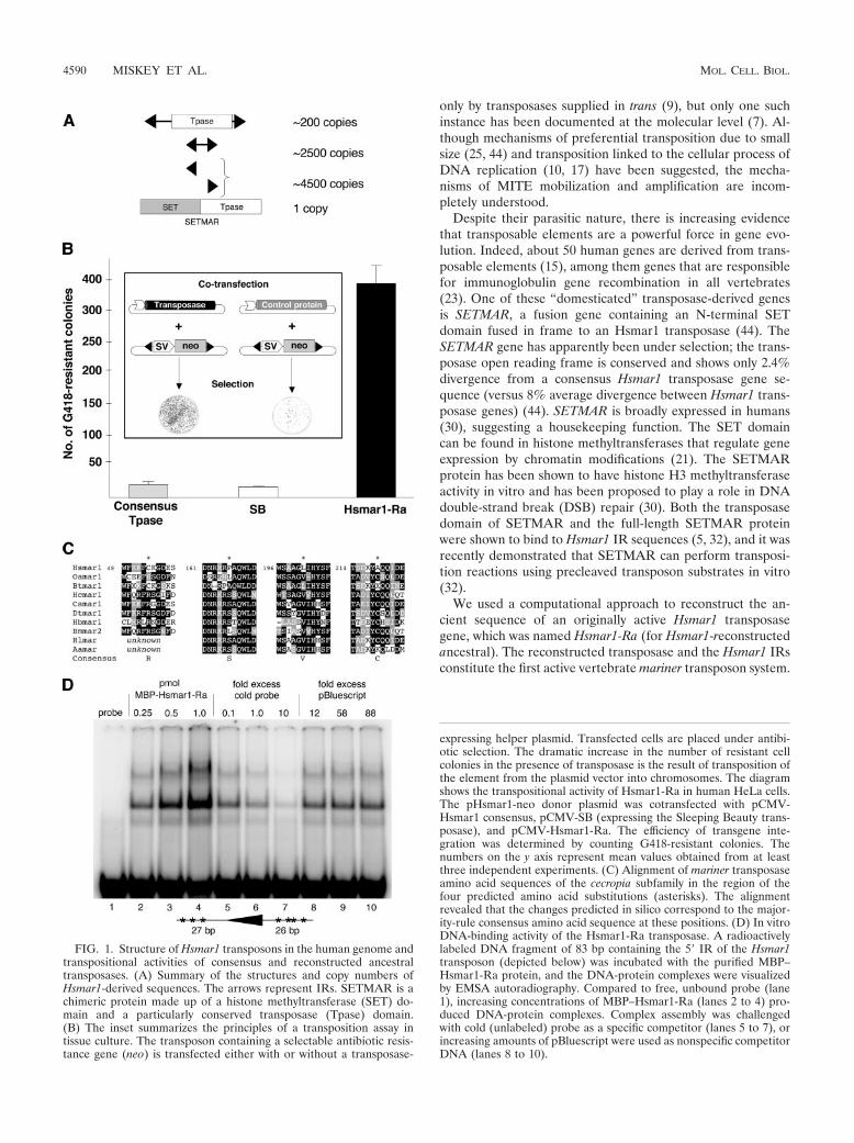
FIG. 1. Structure of Hsmar1 transposons in the human genome andtranspositional activities of consensus and reconstructed ancestraltransposases. (A) Summary of the structures and copy numbers ofHsmar1-derived sequences. The arrows represent IRs. SETMAR is achimeric protein made up of a histone methyltransferase (SET) do-main and a particularly conserved transposase (Tpase) domain.(B) The inset summarizes the principles of a transposition assay intissue culture. The transposon containing a selectable antibiotic resis-tance gene (neo) is transfected either with or without a transposase-
expressing helper plasmid. Transfected cells are placed under antibi-otic selection. The dramatic increase in the number of resistant cellcolonies in the presence of transposase is the result of transposition ofthe element from the plasmid vector into chromosomes. The diagramshows the transpositional activity of Hsmar1-Ra in human HeLa cells.The pHsmar1-neo donor plasmid was cotransfected with pCMV-Hsmar1 consensus, pCMV-SB (expressing the Sleeping Beauty trans-posase), and pCMV-Hsmar1-Ra. The efficiency of transgene inte-gration was determined by counting G418-resistant colonies. Thenumbers on the y axis represent mean values obtained from at leastthree independent experiments. (C) Alignment of mariner transposaseamino acid sequences of the cecropia subfamily in the region of thefour predicted amino acid substitutions (asterisks). The alignmentrevealed that the changes predicted in silico correspond to the major-ity-rule consensus amino acid sequence at these positions. (D) In vitroDNA-binding activity of the Hsmar1-Ra transposase. A radioactivelylabeled DNA fragment of 83 bp containing the 5� IR of the Hsmar1transposon (depicted below) was incubated with the purified MBP–Hsmar1-Ra protein, and the DNA-protein complexes were visualizedby EMSA autoradiography. Compared to free, unbound probe (lane1), increasing concentrations of MBP–Hsmar1-Ra (lanes 2 to 4) pro-duced DNA-protein complexes. Complex assembly was challengedwith cold (unlabeled) probe as a specific competitor (lanes 5 to 7), orincreasing amounts of pBluescript were used as nonspecific competitorDNA (lanes 8 to 10).
4590 MISKEY ET AL. MOL. CELL. BIOL.

We found that the Hsmar1-Ra transposase efficiently mobi-lizes Hsmar1 transposons in mammalian cultured cells andin the zebra fish embryo with a cut-and-paste mechanism.Hsmar1-Ra can also excise MiHsmar1 elements, providing evi-dence for a functional relationship between these elements andan Hsmar1 transposase source. We also report that SETMARis capable of introducing nicks at the 5� ends of Hsmar1 trans-poson DNA in vitro. In line with these observations, we showthat SETMAR triggers homology-dependent DSB repairevents at the Hsmar1 transposon ends in human and rodentcells in vivo.
MATERIALS AND METHODS
Phylogenetic analysis and ancestral-sequence reconstruction. We collectedHsmar1-related elements from the human genome by performing a TBLASTN(2) search for potential homologues of the Hsmar1 consensus sequence (44) inthe translated chromosomal sequences of Homo sapiens at an E-value cutoff of10�5. Only hits with the following criteria were retained for further analysis: (i)alignment covering at least 90% of the query sequence, (ii) less than 10% gapsin the alignment, and (iii) sequence identity of more than 30%. The resulting 51sequences, along with those of the cecropia transposase subfamily (Bombyx morimar2 [Bmmar2], D88671; Heterorhabditis bacteriophora mar1 [Hbmar1], U68392;Dugesia tigrina mar1 [Dtmar1], X71979; Hyalophora cecropia mar1 [Hcmar1] [31];and Cecropius sp. mar1 [26]) were aligned by T-Coffee (37). The phylogeny ofthese sequences was reconstructed using the maximum likelihood method im-plemented in PhyML 2.4 with a JTT amino acid substitution model. We allowedfor site-specific substitution rate heterogeneity by using a discrete gamma modelwith eight rate categories and the gamma shape parameter estimated from thedata. The resulting phylogenetic tree was rooted using the invertebrate marinerelements as outgroups and was visualized by TreeView (38) (see Fig. S1 in thesupplemental material). Protein sequences at ancestral nodes were inferred byPAML 3.14b (51) using maximum likelihood marginal reconstruction. The eval-uations of the reconstructed ancestral amino acid states at the node connectingthe cecropia mariner transposons with the branch leading to the human orchimpanzee sequences (see Fig. S1 in the supplemental material) predicted thefollowing three amino acid substitutions in the consensus transposase proteinsequence with posterior probabilities of �0.9: C53R, L201V, and A219C. Theprobabilities for the P167S substitution predicted from the human and thechimpanzee trees were 0.9 and 0.7, respectively. Other Hsmar1-like sequencesfor alignments were Hydra littoralis mar1 (AAB61385), acrobat ant mar 29.3 (42),and Bos taurus mar1 (Btmar1) (6). The Ornithorhynchus anatinus mar1 (Oamar1)sequence found in public databases is available upon request.
Plasmids. The Klenow-filled EcoRI/BamHI fragment of pRc/CMV (Invitro-gen), containing the simian virus 40 promoter-neo-poly(A) sequences, wascloned into the SmaI site of pUC19 to gain pNeo. Hsmar1 transposon sequenceswere assembled from 40- to 60-nucleotide-long oligonucleotides. Briefly, for theassembly of the left IR and the 5� untranslated sequences of the consensusHsmar1 transposon, 6 pmol (each) of the primers L1 to L8 was used in a PCR ina total volume of 50 �l with Pwo polymerase (Roche) with the following PCRprogram: 94°C for 1 min; 30 cycles of 94°C for 30 s, 52°C for 30 s, and 72°C for30 s; and 72°C for 2 min. The 3� untranslated sequences and the right IR of theHsmar1 transposon were assembled from the oligonucleotides R1 and R2 usingPCR conditions similar to those described above. The PCR for the left end of thetransposon was diluted 200-fold, and 1 �l of the dilution was used in a 100-�lPCR mixture with 15 pmol (each) of the primers L1 and L8. The PCR programwas 94°C for 1 min; 25 cycles of 94°C for 30 s, 50°C for 30 s, and 72°C for 30 s;and 72°C for 2 min. The fragments were then cloned into the EcoRI/KpnI (5� IRand untranslated region [UTR]) and XbaI/HindIII (3� UTR and IR) sites ofpNeo, resulting in pHsmar1-neo. pHsmar1-zeo was obtained by cloning theKlenow-filled XbaI-BspHI fragment of pUT-SVZeo (Invitrogen) into theblunted KpnI/XbaI site of pHsmar1-neo.
The Hsmar1-like transposase coding sequence was obtained from chimpanzeegenomic DNA with an open reading frame-trapping strategy as described pre-viously (36) and cloned into the NotI/SpeI sites of pBS (Stratagene) to serve asthe template for a series of PCR mutagenesis reactions performed with theQuikChange Multi Site-Directed Mutagenesis Kit (Stratagene). The final trans-posase gene was cloned into the NheI/BamHI site of pEGFP-C2 (Clontech) toform pCMV-Hsmar1-Ra. The same fragment cloned into the KpnI/XbaI sites ofpHsmar1-neo resulted in pHsmar1-Full.
The primers Amp-MITEFw and Amp-MITERev were used to create a cloning
site within the beta lactamase (bla) gene in a zeo-containing pUC19-derivedplasmid. The obtained fragment served as the donor backbone for all trans-posons tested in excision experiments with the bla gene. The consensusMiHsmar1 sequence (44) was assembled by annealing 0.5 nmol of each of theHsmar1 MITEFw and Hsmar1 MITERev oligonucleotides in 100 �l 1� NewEngland Biolabs buffer 2, followed by a fill-in reaction with Klenow using 20 �lof the annealed primers. The MITE and the PCR-amplified transposons frompHsmar1-neo and pHsmar1-Full (primer, Hsmar1IR) were inserted in the blagene, deriving pAmpMITE, pAmp-neo, and pAmpFull, respectively. To obtainthe longer IRs found in MiHsmar1 elements, pAmp-neo and pAmpFull weresubjected to PCR mutagenesis with the primers HsmarLonger1 and -2, using aQuikChange Multi Site-Directed Mutagenesis Kit (Stratagene), yielding pAmp-neoL and pAmpFullL, respectively. The SETMAR gene was PCR amplified fromthe IMAGp956A05160Q24 cDNA clone (RZPD) and ligated into the BamHI/XbaI sites of pcDNA3.1/Zeo (Invitrogen), resulting in pCMV-SETMAR. Allprimer sequences are available in Table S1 in the supplemental material.
Cell culture transfection and plasmid rescue. Cells were cultured, transfected,and selected as described previously (36) with some modifications: 2 �l JET-PEI/-RGD (Qbiogene) was used to transfect 100 ng pCMV-Hsmar1-Ra orpCMV-SB (16) with 500 ng of the transposon donor plasmids. For plasmidrescue, 10 �g of genomic DNA prepared from pooled zeocin (Zeo)-resistantHeLa colonies was digested with NheI, SpeI, and AvrII; precipitated and ligatedunder dilute conditions; and electroporated into DH10B cells, which were thenselected with 50 �g/ml Zeo.
Luciferase reporter assays. Reporter assays were performed as describedpreviously (50); 150 ng of all reporter constructs (1) and p5�-UTR/Luc werecotransfected into HeLa cells with 100 ng of pCMV-�gal (Clontech) as aninternal control for transfection efficiency. Two days posttransfection, luciferaseactivity was measured in a Lumat LB 9507 luminometer with a 10-second inte-gration period. Luciferase units were normalized with �-galactosidase readoutsfor transfection efficiency and with the Bradford assay for total protein.
Transposon excision assays. Hsmar1-Ra mRNA was transcribed from pSK/BG3�u-2a (a gift from Perry Hackett) with the mMessage mMachine kit (Am-bion). One-cell-stage zebra fish embryos were injected with a mixture of 50 ng/�lmRNA and 20 ng/�l pHsmar1-neo. After 9 h, pools of 30 embryos were digestedwith 200 �g/ml proteinase K in 0.5 ml extraction buffer (10 mM Tris-HCl, pH 8.2,10 mM EDTA, 0.2 M NaCl, 0.5% sodium dodecyl sulfate), and plasmid DNAwas purified with a QIAprep Spin Kit (QIAgen). Plasmid purification fromcultured cells and the conditions for the nested PCRs have been describedpreviously (18).
For excision from the bla gene, HeLa cells were transfected with 100 ng ofpCMV-Hsmar1-Ra and 500 ng of the various transposon donor plasmids. Plas-mid DNA was purified from cells 3 days posttransfection by a standard miniprepprotocol. After precipitation, the DNA pellet was dissolved in 10 �l water andtransformed into Escherichia coli. Ten microliters of the 1-ml bacterial suspen-sion in SOC medium (Super Optimal broth with catabolite repression) wasplated on Zeo plates, with the rest on ampicillin (Amp)/Zeo plates. The numbers ofthe double-resistant colonies were normalized with the total amount of plasmidDNA recovered as follows: (number of Ampr/Zeor foci � 100)/number of Zeor foci.
Maltose-binding protein (MBP)–Hsmar1-Ra and MBP-SETMAR purifica-tion. The SETMAR coding region from the IMAGp956A05160Q24 cDNA clone(RZPD) was cloned into the BamHI/XbaI site of pMAL-c2x (New EnglandBiolabs); in the case of pHsmar1-Ra, the XbaI/HindIII sites were used forcloning. The purification of the proteins was carried out as described previously(32) with minor modifications. The plasmids were transfected into E. coli BL21-CodonPlus-RIL competent cells (Stratagene). The next day, the culture wasdiluted 100 times to 80 ml and grown to an optical density at 600 nm of 0.5, andprotein expression was induced with 0.5 mM IPTG (isopropyl-�-D-thiogalacto-pyranoside) at 30°C for 3 h. After being harvested by centrifugation, the pelletwas resuspended in 3 ml of HSG buffer (32) and sonicated with a BandelinSonopuls sonicator for 3 min (cycle, 50%; 10% power). After centrifugation, 400�l of the soluble crude extract was mixed with 100 �l of amilose resin (NewEngland Biolabs) and rotated for 30 min at 4°C. The resin was washed seventimes with 0.5 ml of HSG buffer containing 0.5 M NaCl and once with 0.5 mlHSG buffer. The proteins were eluted with 80 �l of HSG buffer containing 10mM maltose.
Electrophoretic mobility shift assay (EMSA). The 83-bp-long probe compris-ing the 5� IR of Hsmar1 was obtained by annealing the primers Hsmar1/Apo1and Hsmar1/AflII, followed by a Klenow fill-in reaction using [-P32]dATP(Perkin Elmer); 0.24 pmol of MBP–Hsmar1-Ra or 2.5 pmol of MBP-SETMARwas incubated with 0.3 pmol of probe in a 20-�l binding reaction mixture asdescribed previously (32) for 3 h at room temperature. The binding reactionproducts were separated on 6% Tris-acetic acid-polyacrylamide gels containing
VOL. 27, 2007 RECONSTRUCTED HUMAN TRANSPOSON 4591

1% glycerol, dried, exposed to a phosphoscreen for several hours, and visualizedby a STORM phosphorimager (Amersham).
Cleavage site determination by linker-mediated PCR. A 281-bp-long DNAfragment containing the left IR and the complete 5� untranslated sequences ofthe Hsmar1 transposon was PCR amplified with the pUC6 and L8 primers usingpHsmar1-Full as a template; 0.5 pmol of this DNA fragment was incubated with1 pmol MBP–Hsmar1-Ra or 25 pmol MBP-SETMAR in a 30-�l reaction mixturecontaining 20 mM HEPES (pH 7.5), 100 mM NaCl, 250 �g/ml bovine serumalbumin, 2 mM dithiothreitol, 10% glycerol, 5% dimethyl sulfoxide, and 6 mMMgCl2 overnight at 37°C, and 0.5 pmol of the probe was digested with EcoRI asa control. The proteins were heat inactivated at 70°C for 10 min and digestedwith 100 �g of proteinase K in a 100-�l reaction volume for 1 h. After heatinactivation at 95°C for 30 min, the DNA was precipitated with isopropanol inthe presence of glycogene and dissolved in 10 �l water. The DNA solution wassubsequently mixed with 10 pmol single-stranded linker oligonucleotide (Link1/2), heated to 95°C for 3 min, and chilled on ice before it was completed withbuffer, T4 RNA ligase (New England Biolabs), and water to a 15-�l final volume.After ligation overnight at room temperature, the reaction mixture was heatinactivated at 65°C for 15 min and purified with a QIAquick PCR purification Kit(QIAgen). The eluate was split and used for templates for two rounds of PCR;1 �l of eluate was used for the PCR when the ligation was performed on theEcoRI-digested probe. The primers used in the first round of PCR to detectcleavage of the lower strand were pUC6 and Link3; L8 and Link3 were used todetect upper-strand cleavage sites. The PCR program was 94°C for 30 s and 30cycles of 94°C for 30s, 55°C for 30 s, and 72°C for 15 s. One microliter (each) of100-fold-diluted PCR mixtures was used in the nested PCRs, using Link4 andpUC2 primers for the lower strand and Link4 and HsmarRev3 for determiningthe upper-strand cleavage sites. The PCR program for the nested PCR was 94°Cfor 30 s and 30 cycles of 94°C for 30 s, 62°C for 30 s, and 72°C for 15 s. Thedetected PCR products were gel isolated, cloned using the pGEM-T VectorSystem (Promega), and sequenced.
Nucleotide sequence accession number. The sequence of the reconstructedancestral Hsmar1-Ra element can be found under GenBank accession numberEF517118.
RESULTS
The consensus Hsmar1 transposase is not active. We pre-viously reconstructed two functional transposable elementsfrom vertebrate genomes: Sleeping Beauty (SB) from fish (16)and Frog Prince (FP) from amphibians (36). Both gene recon-structions were based on the hypothesis that a consensus se-quence established from several cloned inactive copies repre-sents an active gene. Encouraged by our former success, we setout to reconstruct an active Hsmar1 transposable element fromthe human genome.
We engineered the consensus sequence of the transposasegene (44) by site-directed mutagenesis of 21 codons of anHsmar1 ortholog obtained from the chimpanzee genome.Transposition activity was assessed in human HeLa cells, usinga two-component transposition system similar to those estab-lished for SB and FP (16, 36). The assay (Fig. 1B) is based oncotransfection of a donor plasmid carrying a nonautonomoustransposon marked with a neomycin resistance gene (neo),with or without the helper plasmid expressing the transposase.Both random plasmid integrations and transposition eventsinto chromosomes give rise to G418-resistant colonies. Follow-ing antibiotic selection, transposition efficiency is calculatedfrom the numbers of G418-resistant cell colonies in the pres-ence versus absence of the transposase. There was no indica-tion of Hsmar1 transposition in these experiments (Fig. 1B),suggesting that the consensus of the Hsmar1 transposase generepresents an inactive sequence.
Molecular reconstruction of an ancestral Hsmar1 trans-posase gene. The approach to gene sequence prediction basedon consensus does not incorporate phylogenetic information.
For example, an inactivating mutation in a transposable ele-ment may become overrepresented if that particular mutantwas preferentially amplified over the active sequence. With theaim of reconstructing the ancient, active Hsmar1 transposasegene that colonized the genome lineage of primates, we ap-plied a statistically rigorous approach based on maximum like-lihood that has been successfully used to reconstruct ancestralgene sequences (48).
First, human Hsmar1 transposase-like amino acid sequenceswere obtained from the human genome by TBLASTN similar-ity searches. To infer the sequence of the last common ances-tor of primate and invertebrate mariner transposases, a likelycandidate for an active transposase protein capable of coloniz-ing new hosts, invertebrate mariner transposase sequences ofthe cecropia subfamily were also included in the phylogeneticanalysis (see Materials and Methods). The evaluations of thereconstructed ancestral amino acid states at the node connect-ing the invertebrate mariner elements with the branch leadingto the human sequences (see Fig. S1 in the supplementalmaterial) revealed the following four amino acid substitutionsin the known consensus transposase protein sequence withposterior probabilities greater than or equal to 0.9: C53R,P167S, L201V, and A219C. Independent inferences from hu-man nucleotide sequences (as identified by BLASTN) or fromchimpanzee amino acid sequences also identified these aminoacids as the most likely ancestral states at these sites. Further-more, inspection of these positions in an alignment of cecropia-type transposase sequences revealed that the predicted substi-tutions represent conserved amino acids within the subfamily(Fig. 1C), suggesting that these residues may be important fortransposase activity.
Next, the putative ancestral Hsmar1 transposase gene wasengineered by incorporating the four predicted amino acidsubstitutions into the framework of the consensus Hsmar1transposase, and the resulting protein was tested for DNAbinding and transposition activities. A fusion protein consistingof the MBP and the reconstructed ancestral Hsmar1 trans-posase was expressed and purified from E. coli, and its abilityto bind Hsmar1 IR sequences was tested in an EMSA. Theexperiment showed binding of the transposase to a transposonIR probe in a sequence-specific manner (Fig. 1D). The colony-forming assay was used to address the transpositional activityof the reconstructed ancestral transposase in tissue culturecells. Upon cotransfection of the neo-marked Hsmar1 transpo-son with a vector expressing the modified transposase, a 23-fold increase in the number of antibiotic-resistant colonies wasobserved (Fig. 1B), suggesting that the resurrected proteinefficiently catalyzes transgene integration from the donor plas-mids into human chromosomes. Thus, it is likely that the in-ferred sequence, which was named Hsmar1-Ra, represents or isvery similar to the sequence of the ancient mariner elementthat colonized the genome lineage of primates.
We next sought to establish molecular evidence of Hsmar1transposition. In the first step of the cut-and-paste transposi-tion of Tc1/mariner elements, the transposase mediates exci-sion of the transposon from its donor site with staggered cuts.The resulting DSB is sensed by host DNA repair mechanismsthat seal the broken DNA, predominantly by nonhomologousend joining (NHEJ) (18, 52). NHEJ repair of the transposonexcision sites generates a characteristic transposon footprint (a
4592 MISKEY ET AL. MOL. CELL. BIOL.

small, transposon-specific, 2- to 4-bp sequence signature),which can be identified with PCR (Fig. 2A), using primersflanking the transposon (18). PCR readily generated a productconsistent with transposon excision and subsequent footprintformation in transfections with the Hsmar1-Ra transposase(Fig. 2B, lane 1), but not with the SB transposase (Fig. 2B, lane2), consistent with specific Hsmar1-Ra-dependent excisionevents followed by DNA repair. Sequencing of the PCR prod-ucts revealed that Hsmar1-Ra generates TTA or TAA tripletsat the excision site, corresponding to the 5�- and 3�-terminalnucleotides of Hsmar1 transposons (Fig. 2C). This is consistentwith footprint formation by the Himar1 (24, 27) and Mos1 (4)mariner transposons, which predominantly generate 3-bp foot-prints. Similar to footprints generated by SB in mammaliansomatic cells (18), a minor fraction of Hsmar1 excision sitescontained noncanonical footprints with small 1- to 2-bp dele-tions (data not shown).
Tc1/mariner elements are able to transpose in organismsother than their natural hosts (40). This phenomenon makesthese elements excellent tools for genome manipulation inmodel organisms. We tested Hsmar1 transposition in zebra fishembryos. Hsmar1-Ra transposase mRNA synthesized in vitrowas coinjected with the donor plasmid into fertilized zebra fishoocytes, and the excision PCR assay was performed on DNAisolated from the embryos. The majority of PCR productsobtained in the presence of the Hsmar1-Ra transposasemRNA (Fig. 2B, lane 3) contained the canonical TTA/TAAfootprints (Fig. 2C), indicative of precise Hsmar1 excision fol-lowed by NHEJ repair.
Tc1/mariner elements insert into TA target dinucleotides,which become duplicated and flank the integrated transposons(40). In order to obtain formal molecular proof for cut-and-paste Hsmar1 transposition, 47 integration events were iso-lated from HeLa cells. All of the transposon insertions oc-curred at TA dinucleotides (Fig. 2D) scattered on 16 humanchromosomes. We found that the chromosomal distributionsof the endogenous genomic copies and the de novo integrantsoverlap and have a bias toward larger chromosomes (Fig. 2E).Forty-four percent of the hits were identified in introns ofgenes (Fig. 2E), indicating a fairly random genomic distribu-tion similar to that found with SB in human cells (49).
In sum, the results described above indicate that we success-fully reactivated the first vertebrate mariner transposon fromthe human genome. The new transposon system shows high-efficiency transposition into various loci in the human genomeand retains its activity in zebra fish embryos.
Hsmar1 can function as an autonomous transposon. Virtu-ally all Tc1/mariner elements contain UTRs upstream of theinitiation codon of the transposase gene, suggesting that theymight have functions associated with transposition activity. In-deed, some of these 5� UTRs harbor eukaryotic promotermotifs (28). However, previous studies did not reveal an inter-nal promoter in the Tc1 element but showed that the elementsare transcribed by read-through transcription from Caenorhab-ditis elegans genes (46).
To test whether the Hsmar1 element has promoter activity,the 180-bp-long 5� UTR of the transposon was fused to aluciferase reporter gene (Fig. 3A). This plasmid and controlreporter constructs were transfected into HeLa cells, and theexpression of the reporter gene was followed by enzymatic
FIG. 2. Excision and integration of Hsmar1. (A) Transposon exci-sion assay in cultured cells. The two functional components of thetransposon system (a transposon and a transposase source) are intro-duced into cells. The transposase catalyzes transposon excision; Tc1/mariner elements leave behind a 2- to 4-nucleotide-long 3� overhang atthe DSBs. DNA repair rejoins the ends of the broken DNA, and theresulting sequences can be recovered by PCR using primers flankingthe excision site. (B) Agarose gels showing PCR products of excisionassays obtained from HeLa cells and zebra fish embryos. The appear-ance of the 260-bp bands in the presence of pCMV-Hsmar1-Ra (lane1) or Hsmar1-Ra mRNA (lane 3) indicates transposon excision fromthe pHsmar1-neo donor plasmids, followed by excision site repair.Transfection of pCMV-SB (lane 2) and injection of SB mRNA (lane 4)served as controls. M, 250-bp size marker. (C) Hsmar1 transposonfootprints identified from cloned excision PCR products from HeLacells and zebra fish embryos. The sequence of the donor plasmid isschematically shown above. Flanking TA dinucleotides are in boldface,and footprints are in uppercase letters. (D) Three human chromo-somal sequences that served as target sites for Hsmar1 transposons.Duplicated TA target dinucleotides are in boldface. (E) Distribution ofendogenous genomic Hsmar1 copies (gray columns) and 47 de novointegrants (black columns) in human chromosomes. Mapping of trans-poson insertions was done using BLAST on the human genome se-quence database at NCBI. The graph shows the genomic distributionof transposon insertions with respect to human genes, nongene re-gions, and repetitive sequences.
VOL. 27, 2007 RECONSTRUCTED HUMAN TRANSPOSON 4593

luciferase assays. The 5�-UTR sequences of Hsmar1 showedpronounced promoter activity (Fig. 3A), indicating that theexpression of the transposase gene (and thus transposition)might not depend on external promoters.
To test such a possibility, the Hsmar1-Ra transposase genewas cloned between the transposon IRs, resulting in a trans-poson whose structure mimics that of the hypothetical ancestorof the full-length element. This element was transfected intoHeLa cells, and excision of the complete transposon was testedby the excision PCR assay. The autonomous element producedPCR products comigrating with those produced by the two-component system (Fig. 3B), indicating excision of the elementfrom its donor plasmid. These results suggest that the full-length Hsmar1 element can function as an autonomous trans-poson.
Excision of MiHsmar1. The human genome contains about2,500 copies of an Hsmar1-related MITE (Fig. 1A). TheMiHsmar1 elements have a consensus sequence of 80 bp con-taining 37-bp IRs, the first 30 bp of which are identical to theIRs of Hsmar1 (44). The copy number of MiHsmar1 is at leastan order of magnitude higher than that of the full-sizedHsmar1 elements in the human genome (44). One possibleexplanation that might account for their abundance is their
small size, which could predispose them for efficient transpo-sition (25, 44).
To test this hypothesis, transposon donor plasmids wereconstructed containing a Zeo resistance gene and either theconsensus sequence of MiHsmar1 or the long versions of thetransposon (Hsmar1-neo or the autonomous element) insertedinto the same position in the coding region of the �-lactamase(bla) gene. In addition, long transposons with IRs identical tothose of MiHsmar1 were created. The insertions disrupt the blareading frame, which can be restored only if the transposonsare excised by the transposase and NHEJ repairs the plasmidproducing the canonical footprints (Fig. 4).
The donor plasmids harboring transposons of differentlengths were transfected into HeLa cells, together with vectorsexpressing either the Hsmar1-Ra or the SB (control) trans-posase; the reporter containing the autonomous Hsmar1 ele-ment was transfected alone. Low-molecular-weight DNA pu-rified from the cells was transformed into E. coli. Excisionevents were scored by counting Ampr/Zeor colonies; selectionwith Zeo alone served to control overall plasmid recovery.MiHsmar1 elements were excised from the donor plasmids 2orders of magnitude more efficiently than any of the longerversions of the transposon (Fig. 4, experiments 1, 2, and 4). IR
FIG. 3. Hsmar1 can function as an autonomous transposon. A schematic representation of the luciferase reporter construct in p5� UTR/Luccompared to the structure of an Hsmar1 transposon is depicted. (A) Reporter gene expression in HeLa cells from the plasmids indicated below.The error bars indicate standard deviations. (B) Transposon excision assay as shown in Fig. 2. HeLa cells were transfected with the pHsmar1-neodonor plasmid, together with the plasmids indicated. The presence of the characteristic PCR band indicates that the complete Hsmar1 element(Hsmar1-Full) can autonomously catalyze its own excision from the donor plasmid. M, 250-bp size marker.
4594 MISKEY ET AL. MOL. CELL. BIOL.

length did not influence the activities of the long versions ofthe transposons in the excision assays (Fig. 4, experiments 3and 5) and in transposon integration assays (data not shown).Therefore, our data indicate that reduced size is responsiblefor the elevated excision frequency of MiHsmar1.
The human SETMAR protein binds Hsmar1 IRs and retains5� cleavage activity of its transposase domain. The transposasedomain of the SETMAR protein was recently shown to spe-cifically bind to Hsmar1 IRs (5) and to catalyze transpositionreactions in vitro (32). Since the protein used in those exper-iments lacked the SET domain, we addressed the question ofwhether the full-length physiological form of SETMAR canalso exhibit transposase-related activities.
To test such a possibility, SETMAR was expressed in E. coliand purified as an N-terminal fusion with the MBP (MBP-SETMAR). EMSAs showed binding of MBP-SETMAR to the5� IR of Hsmar1 (Fig. 5, compare unbound probe in lane 1 tocomplexes formed at increasing transposase concentrations inlanes 2 to 4) in a sequence-specific manner, since binding was
competed with cold specific DNA (Fig. 5, lanes 5 to 7) but notwith excess nonspecific DNA (Fig. 5, lanes 8 to 10).
Despite the high sequence similarity of SETMAR to theHsmar1-Ra transposase, SETMAR contains several aminoacid substitutions that potentially compromise its catalyticfunctions. For example, the third D (Asp) of the catalyticallyessential DDD triad in the transposase domain of SETMAR isreplaced by an N (Asn). Even the conservative D-to-E (Glu)mutation in this position abolished the catalytic activity of theMos1 transposase (33). In order to test possible catalytic ac-tivity of SETMAR, an in vitro DNA cleavage assay was firstapplied. The assay is based on incubation of recombinant,purified protein with a double-stranded DNA substrate con-taining IR sequences of Hsmar1, followed by ligation of the 3�end of a single-stranded oligonucleotide linker to phosphory-lated 5� ends of DNA exposed as a result of endonucleaseactivity and PCR using substrate- and linker-specific primers(Fig. 6A).
Purified MBP-SETMAR or MBP–Hsmar1-Ra protein was
FIG. 4. Excision of Hsmar1 and MiHsmar1 transposons. (A) Outline of the excision assay. A transposon (Tpson) insertion disrupts andinactivates the Amp resistance gene (Amp). Transposon excision, followed by DNA repair, can restore Amp resistance, which can be selected forin E. coli. (B) Coding triplets of the original, the modified, and the repaired excision sites are listed. Transposon footprints and amino acidsequences are uppercase. Various transposon donor plasmids (indicated on the x axis) were transfected into HeLa cells either with pCMV-Hsmar1-Ra (experiments 1 to 3, numbered below the graph), alone (experiments 4 and 5), or with pCMV-SB (experiment 6) as a control. Indicatorplasmids contain the following transposons in the Amp resistance gene: pAmpMITE, the consensus MiHsmar1 sequence; pAmpneo, theneo-tagged Hsmar1 element; pAmpFull, the autonomous Hsmar1 transposon; pAmpneoL, the neo-tagged Hsmar1 element with IR sequences thatmatch those of MiHsmar1; and pAmpFullL, the autonomous Hsmar1 transposon with IR sequences that match those of MiHsmar1. The Ampr/Zeor
colonies represent excision events followed by canonical footprint formation at the excision site. The normalized numbers of the double-resistantcolonies (shown above the columns) were obtained by dividing the numbers of Ampr/Zeor colonies with the corresponding Zeor colony numbers.
VOL. 27, 2007 RECONSTRUCTED HUMAN TRANSPOSON 4595

incubated with a double-stranded DNA fragment containingthe 5� IR of Hsmar1. The substrate also contained an EcoRIrecognition site, directly adjacent to the 5� end of the Hsmar1IR (Fig. 6A). Digestion of the probe with EcoRI served as acontrol for the assay. MBP–Hsmar1-Ra cleavage productswere identified for both strands of the Hsmar1 transposon ends(Fig. 6B). The most prominent cleavage site on the upperstrand was 3 nucleotides inside the transposon DNA, whereasthe lower strand was predominantly cut at the end of thetransposon (Fig. 6C). Therefore, the cleavage pattern ofHsmar1-Ra fully corresponds to the 3-bp transposon footprintsthat are generated after transposon excision. Cleavage prod-ucts by MBP-SETMAR could only be identified for the upperstrand (Fig. 6B), suggesting that the SETMAR protein has veryweak 3� nicking activity, if any. These findings are in goodagreement with the results of Liu et al. who found inefficient 3�cleavage activity with the transposase domain of SETMAR(32). Multiple SETMAR nicking sites were identified, none ofthem corresponding to the major 5� cleavage site by Hsmar1-Ra(Fig. 6C). Altogether, the data indicate that SETMAR is a de-fective transposase in vitro and that it retains only a fraction of thebiochemical activities of a transposase, namely, 5� cleavage attransposon IR sequences.
In vivo catalytic activity of SETMAR was addressed by code-livery of an Hsmar1 transposon plasmid and a SETMAR ex-pression plasmid into mammalian cultured cells, recovery of
extrachromosomal plasmids from the transfected cells, andPCR amplification using primers flanking the transposable el-ement in the donor plasmid (Fig. 2A). In contrast to the strong,dominant footprint products generated by the Hsmar1-Ratransposase (Fig. 7A, lane 1), SETMAR generated only weak,smeary products (Fig. 7A, lane 2), which were cloned andsequenced. As shown in Fig. 7B, 10 out of the 12 recoveredproducts contained sequences from either one or both IRs.The majority (9/12) of the excision products contained dele-tions of pUC19 sequences flanking the transposon in the donorplasmid (Fig. 7B). The DNA ends were almost exclusivelyrejoined at 2- to 9-nucleotide-long microhomologies, sharedeither between the left and right transposon sequences orbetween transposon and vector backbone sequences flankingthe element (Fig. 7B). No canonical footprints were identifiedat the excision sites.
These results are consistent with SETMAR-mediated nick-ing of Hsmar1 elements in vivo. However, in contrast to theDNA lesions generated by the Hsmar1-Ra transposase, whichare predominantly repaired by NHEJ, the characteristics oftransposon footprints by SETMAR could be best explainedby the involvement of a homology-dependent repair (HDR)pathway.
DISCUSSION
Here, we report the resurrection of an ancient, extinct trans-poson in the human genome. The apparent inactivity of theconfident majority-rule consensus Hsmar1 transposase se-quence (Fig. 1B) implies that inactive Hsmar1 copies wereefficiently mobilized in the past by a transposase source intrans; thus, nonautonomous elements contributed more effec-tively to the spread of Hsmar1 copies. One plausible mecha-nism that could explain such a phenomenon is that trans-posase-producing Hsmar1 copies suffered mutations withintheir IRs that compromised their ability to move. We under-took an in silico phylogenetic approach to infer the sequence ofan archaic mariner transposase that colonized an ancestralprimate genome �50 million years ago (44). The reconstructedtransposase gene and its specific IRs make up the first func-tional vertebrate mariner transposon system.
Molecular characterization of transposition events demon-strated that Hsmar1 follows precise cut-and-paste transpositionin its natural (human) host and in the zebra fish embryo (Fig.2). Transpositional activity of Hsmar1 in HeLa cells is an orderof magnitude higher than that of the invertebrate marinerelements Himar1 and Mos1 and about as efficient as FP andSB, two Tc1-like elements of vertebrate origin (data notshown). These observations suggest that the new Hsmar1transposition system can be developed as a useful genetic tool.
We provide experimental evidence that the Hsmar1-Ratransposase can excise MiHsmar1 elements (Fig. 4). Almost allMITEs previously identified from different genomes are inac-tive, and thus, their mechanisms of transposition and accumu-lation in eukaryotic genomes have been poorly understood.Although there are strong indications that MITEs are mobi-lized in trans by a corresponding transposase (e.g., mPing/Pongand Stowaway/Osmar mobilization in the rice genome [11, 22],Tourist/PIF interaction in maize [53], or in vitro interactionbetween the Arabidopsis elements Emigrant and Lemi1 [35]),
FIG. 5. In vitro DNA binding activity of the SETMAR protein. Aradioactively labeled DNA fragment of 83 bp containing the 5� IR ofHsmar1 (depicted below) was incubated with purified MBP-SETMARprotein, and the DNA-protein complexes were visualized by EMSA.Compared to free, unbound probe (lane 1), increasing concentrationsof MBP-SETMAR (lanes 2 to 4) produced DNA-protein complexes.Substantial amounts of the SETMAR protein formed aggregates at allconcentrations used and were unable to enter the gel during electro-phoresis. Complex assembly was challenged with cold (unlabeled)probe as a specific competitor (lanes 5 to 7), or increasing amounts ofpBluescript were used as nonspecific competitor DNA (lanes 8 to 10).
4596 MISKEY ET AL. MOL. CELL. BIOL.

FIG. 6. In vitro cleavage site analysis with ligation-mediated PCR. (A) Schematic overview of the assay. A 281-bp-long DNA fragmentcontaining the 5� IR of Hsmar1 was used as the substrate for the reaction. The 5� end of the IR is shown on a black background, the TA nucleotidesbordering the element are in boldface, and the neighboring EcoRI site is underlined. Cleavage of one or both strands of the substrate DNA bythe purified proteins results in phosphoryl-terminated DNA ends, which can later be ligated to 3�-hydroxyl-terminated single-stranded oligonu-cleotide linkers (gray lines) with T4 RNA ligase. The linkers tag the cleavage sites and enable amplification of the cleavage products by nested PCR.(B) Agarose gels with PCR products obtained with linker-specific primers (gray arrows in panel A) and primers specific for the upper or bottomstrand of the substrate (black or white arrows in panel A, respectively). PCRs performed on EcoRI-digested DNA served as positive controls.(C) Distribution of cleavage positions identified after cloning and sequencing of the PCR products. The black triangles indicate cleavage sitesintroduced by MBP-Hsmar1-Ra, and the white triangles mark positions where MBP-SETMAR nicked the upper strand of the substrate DNA.
4597

this has been experimentally demonstrated for MITE mobili-zation only by the impala transposase in Fusarium (7). The newHsmar1-Ra transposon system provides a unique opportunityto investigate the origin and transpositional dynamics of theseelements and their contribution to primate genome evolution.
MITEs can accumulate to copy numbers far exceeding thoseof transposase-encoding DNA transposons in different ge-nomes (9). The suggestion that MITEs might be preferentiallymobilized due to their small size has been speculative. Here,we show that MiHsmar1 elements can be excised 2 orders ofmagnitude more efficiently than their longer transposon ver-sions (Fig. 4). This phenomenon could have contributed to theprevalence of Hsmar1-related MITEs in primate genomes.
The Human Genome Project identified about 50 humangenes derived from transposable elements (15). However, todate, there is no evidence for current transpositional activity ofany of these “domesticated” genes in humans. The only excep-tions are the Rag genes, whose physiological function is togenerate the immunoglobulin repertoire by a transposition-like process called V(D)J recombination (23).
We provided experimental evidence that SETMAR, theproduct of a domesticated gene in the human genome derivedfrom an Hsmar1 transposase, retains its capacity to cleaveHsmar1 transposon DNA in vitro (Fig. 6). However, whereasthe Hsmar1-Ra transposase efficiently cleaved both strandsof DNA at transposon ends, thereby generating DSBs, the
SETMAR protein exhibited only 5� cleavage activity, generat-ing single-strand nicks (Fig. 6). Inefficient 3� cleavage by thetransposase domain of SETMAR has been recently described(32). We showed that DNA damage inflicted by Hsmar1-Raand SETMAR is processed differently by cells. Whereas trans-poson excision sites generated by the Hsmar1-Ra transposasepredominantly contained the canonical 3-bp footprints (Fig.2C), SETMAR activity in vivo is associated with extendedstretches of transposon sequences, deletions of flanking DNA,and microhomologies at the junctions at the excision sites (Fig.7B). The structure of these noncanonical footprints can be bestexplained by an interrupted synthesis-dependent strand-an-nealing (SDSA) pathway of HDR, completed by an end-join-ing process generating microhomologies (18). SDSA has beenshown to play a role in the repair of transposon excision sitesand to be responsible for generating internally deleted versionsof diverse transposable elements in animals and plants (8, 13,39, 45), including the Mos1 mariner element (34) and SB (18).Pathway choice in DSB repair can be influenced by the struc-ture of the gap, the availability of repair factors, and the cellcycle phase (13, 18, 47). Our observations for the lack ofdetectable 3� cleavage activity of SETMAR suggests that thedifferential utilization of repair pathways by Hsmar1-Ra andSETMAR can be explained by the different structures of thecleavage sites: DSBs for Hsmar1-Ra and single-strand nicks forSETMAR (Fig. 6). Single-strand nicks have been shown to be
FIG. 7. In vivo cleavage activity of SETMAR. (A) The agarose gel shows PCR products of excision assays on plasmid DNA from HeLa cellstransfected with pHsmar1-neo and the helper plasmids indicated. The faint, smeary products around the expected size in lane 2 indicate loss ofHsmar1 transposons from the donor plasmids catalyzed by the SETMAR protein. (B) The overall structures of footprints from HeLa and CHO-K1cells. The schematic view of the donor site is depicted above. represents deletions, and the sequences in the white boxes are microhomologiesat the junctions.
4598 MISKEY ET AL. MOL. CELL. BIOL.

potent triggers of HDR in mammalian cells (29). For example,repair of DSBs generated by the RAG recombinase in V(D)Jrecombination is tightly linked to NHEJ (19). However, nick-only RAG mutants have been shown to stimulate robust ho-mologous recombination, and RAG-mediated nicking hasbeen proposed to contribute to gene duplication events andchromosomal rearrangements (29). Interestingly, some of therepair products obtained after SETMAR cleavage (products 1,2, 4, 6, and 8 to 11 in Fig. 7B) resemble the structure of theHsmar1-related solo IRs and MITEs present in the humangenome. Thus, interrupted SDSA repair events followingHsmar1 transposon excision catalyzed by an Hsmar1 orSETMAR transposase source could have played a role in theemergence and proliferation of the MITEs and solo IRs.
The emergence of the SETMAR gene and the invasion of theancient primate genome by the Hsmar1 transposons took placewithin an overlapping evolutionary time window, between40 and 58 million years ago (5). Thus, it may be that theSETMAR protein played a role in regulating Hsmar1 transpo-sition. The 5� UTR of the Hsmar1 transposon has significantpromoter activity, sufficient to drive transposase expression(Fig. 3). Through its ability to bind to Hsmar1 transposon IRsequences (Fig. 5), and to catalyze specific histone modifica-tions (30), SETMAR could induce local chromatin changes atthe Hsmar1 transposase gene promoter, thereby regulatingtransposase expression.
SETMAR has likely been under selection in human cells fora function other than its residual nicking activity, but thisfunction remains enigmatic. Cordaux et al. (5) have found thatselection has been preserving the IR-binding activity of theSETMAR transposase (5). Thus, the function of the SETMARprotein is likely associated with its ability to specifically recog-nize numerous genomic binding sites represented by theHsmar1 IRs. It is tempting to speculate that some of thesebinding sites are conserved because targeted chromatin mod-ifications by SETMAR at these genomic locations are requiredfor normal cellular functions. Ongoing work will have to clarifythe past and present functions of SETMAR, making use of theactive Hsmar1 transposon as an experimental system.
ACKNOWLEDGMENTS
The pSK/BG3�u-2a plasmid was a kind gift of P. Hackett, and lu-ciferase reporter constructs were received from J. Altschmied.
This work was supported by EU grant QLG2-CT-2000-00821 andgrant IV 21/3-2 from the Deutsche Forschungsgemeinschaft. B.P. is aLong-Term Fellow of the Human Frontier Science Program Organi-zation.
REFERENCES
1. Altschmied, J., and J. Duschl. 1997. Set of optimized luciferase reportergene plasmids compatible with widely used CAT vectors. BioTechniques23:436–438.
2. Altschul, S. F., T. L. Madden, A. A. Schaffer, J. Zhang, Z. Zhang, W. Miller,and D. J. Lipman. 1997. Gapped BLAST and PSI-BLAST: a new generationof protein database search programs. Nucleic Acids Res. 25:3389–3402.
3. Barry, E. G., D. J. Witherspoon, and D. J. Lampe. 2004. A bacterial geneticscreen identifies functional coding sequences of the insect mariner transpos-able element Famar1 amplified from the genome of the earwig, Forficulaauricularia. Genetics 166:823–833.
4. Bryan, G., D. Garza, and D. Hartl. 1990. Insertion and excision of thetransposable element mariner in Drosophila. Genetics 125:103–114.
5. Cordaux, R., S. Udit, M. A. Batzer, and C. Feschotte. 2006. Birth of achimeric primate gene by capture of the transposase gene from a mobileelement. Proc. Natl. Acad. Sci. USA 103:8101–8106.
6. Demattei, M. V., C. Auge-Gouillou, N. Pollet, M. H. Hamelin, M. Meunier-
Rotival, and Y. Bigot. 2000. Features of the mammal mar1 transposons in thehuman, sheep, cow, and mouse genomes and implications for their evolution.Mamm. Genome 11:1111–1116.
7. Dufresne, M., A. Hua-Van, H. A. El Wahab, S. Ben M’Barek, C. Vasnier, L.Teysset, G. H. Kema, and M. J. Daboussi. 2007. Transposition of a fungalminiature inverted-repeat transposable element through the action of aTc1-like transposase. Genetics 175:441–452.
8. Engels, W. R., D. M. Johnson-Schlitz, W. B. Eggleston, and J. Sved. 1990.High-frequency P element loss in Drosophila is homolog dependent. Cell62:515–525.
9. Feschotte, C., N. Jiang, and S. R. Wessler. 2002. Plant transposable ele-ments: where genetics meets genomics. Nat. Rev. Genet. 3:329–341.
10. Feschotte, C., and C. Mouches. 2000. Evidence that a family of miniatureinverted-repeat transposable elements (MITEs) from the Arabidopsisthaliana genome has arisen from a pogo-like DNA transposon. Mol. Biol.Evol. 17:730–737.
11. Feschotte, C., M. T. Osterlund, R. Peeler, and S. R. Wessler. 2005. DNA-binding specificity of rice mariner-like transposases and interactions withStowaway MITEs. Nucleic Acids Res. 33:2153–2165.
12. Fischer, S. E., E. Wienholds, and R. H. Plasterk. 2001. Regulated transpo-sition of a fish transposon in the mouse germ line. Proc. Natl. Acad. Sci. USA98:6759–6764.
13. Gloor, G. B., J. Moretti, J. Mouyal, and K. J. Keeler. 2000. Distinct P-element excision products in somatic and germline cells of Drosophila mela-nogaster. Genetics 155:1821–1830.
14. Hartl, D. L., A. R. Lohe, and E. R. Lozovskaya. 1997. Modern thoughts on anancyent marinere: function, evolution, regulation. Annu. Rev. Genet. 31:337–358.
15. International Human Genome Sequencing Consortium. 2001. Initial se-quencing and analysis of the human genome. Nature 409:860–921.
16. Ivics, Z., P. B. Hackett, R. H. Plasterk, and Z. Izsvak. 1997. Molecularreconstruction of Sleeping Beauty, a Tc1-like transposon from fish, and itstransposition in human cells. Cell 91:501–510.
17. Izsvak, Z., Z. Ivics, N. Shimoda, D. Mohn, H. Okamoto, and P. B. Hackett.1999. Short inverted-repeat transposable elements in teleost fish and impli-cations for a mechanism of their amplification. J. Mol. Evol. 48:13–21.
18. Izsvak, Z., E. E. Stuwe, D. Fiedler, A. Katzer, P. A. Jeggo, and Z. Ivics. 2004.Healing the wounds inflicted by Sleeping Beauty transposition by double-strand break repair in mammalian somatic cells. Mol. Cell 13:279–290.
19. Jackson, S. P., and P. A. Jeggo. 1995. DNA double-strand break repair andV(D)J recombination: involvement of DNA-PK. Trends Biochem. Sci. 20:412–415.
20. Jacobson, J. W., M. M. Medhora, and D. L. Hartl. 1986. Molecular structureof a somatically unstable transposable element in Drosophila. Proc. Natl.Acad. Sci. USA 83:8684–8688.
21. Jenuwein, T., G. Laible, R. Dorn, and G. Reuter. 1998. SET domain proteinsmodulate chromatin domains in eu- and heterochromatin. Cell Mol. Life Sci.54:80–93.
22. Jiang, N., Z. Bao, X. Zhang, H. Hirochika, S. R. Eddy, S. R. McCouch, andS. R. Wessler. 2003. An active DNA transposon family in rice. Nature421:163–167.
23. Jones, J. M., and M. Gellert. 2004. The taming of a transposon: V(D)Jrecombination and the immune system. Immunol. Rev. 200:233–248.
24. Lampe, D. J., M. E. Churchill, and H. M. Robertson. 1996. A purifiedmariner transposase is sufficient to mediate transposition in vitro. EMBO J.15:5470–5479.
25. Lampe, D. J., T. E. Grant, and H. M. Robertson. 1998. Factors affectingtransposition of the Himar1 mariner transposon in vitro. Genetics 149:179–187.
26. Lampe, D. J., K. K. Walden, and H. M. Robertson. 2001. Loss of trans-posase-DNA interaction may underlie the divergence of mariner familytransposable elements and the ability of more than one mariner to occupythe same genome. Mol. Biol. Evol. 18:954–961.
27. Lampe, D. J., K. K. Walden, J. M. Sherwood, and H. M. Roberstson. 2000.Genetic engineering of insects with mariner transposons, p. 237–248. InA. M. Handler and A. A. James (ed.), Insect transgenesis: methods andapplications. CRC Press, Boca Raton, FL.
28. Leaver, M. J. 2001. A family of Tc1-like transposons from the genomes offishes and frogs: evidence for horizontal transmission. Gene 271:203–214.
29. Lee, G. S., M. B. Neiditch, S. S. Salus, and D. B. Roth. 2004. RAG proteinsshepherd double-strand breaks to a specific pathway, suppressing error-prone repair, but RAG nicking initiates homologous recombination. Cell117:171–184.
30. Lee, S. H., M. Oshige, S. T. Durant, K. K. Rasila, E. A. Williamson, H.Ramsey, L. Kwan, J. A. Nickoloff, and R. Hromas. 2005. The SET domainprotein Metnase mediates foreign DNA integration and links integration tononhomologous end-joining repair. Proc. Natl. Acad. Sci. USA 102:18075–18080.
31. Lidholm, D. A., G. H. Gudmundsson, and H. G. Boman. 1991. A highlyrepetitive, mariner-like element in the genome of Hyalophora cecropia.J. Biol. Chem. 266:11518–11521.
32. Liu, D., J. Bischerour, A. Siddique, N. Buisine, Y. Bigot, and R. Chalmers.
VOL. 27, 2007 RECONSTRUCTED HUMAN TRANSPOSON 4599

2007. The human SETMAR protein preserves most of the activities of theancestral Hsmar1 transposase. Mol. Cell. Biol. 27:1125–1132.
33. Lohe, A. R., D. De Aguiar, and D. L. Hartl. 1997. Mutations in the marinertransposase: the D,D(35)E consensus sequence is nonfunctional. Proc. Natl.Acad. Sci. USA 94:1293–1297.
34. Lohe, A. R., C. Timmons, I. Beerman, E. R. Lozovskaya, and D. L. Hartl.2000. Self-inflicted wounds, template-directed gap repair and a recombina-tion hotspot. Effects of the mariner transposase. Genetics 154:647–656.
35. Loot, C., N. Santiago, A. Sanz, and J. M. Casacuberta. 2006. The proteinsencoded by the pogo-like Lemi1 element bind the TIRs and subterminalrepeated motifs of the Arabidopsis Emigrant MITE: consequences for thetransposition mechanism of MITEs. Nucleic Acids Res. 34:5238–5246.
36. Miskey, C., Z. Izsvak, R. H. Plasterk, and Z. Ivics. 2003. The Frog Prince: areconstructed transposon from Rana pipiens with high transpositional activ-ity in vertebrate cells. Nucleic Acids Res. 31:6873–6881.
37. Notredame, C., D. G. Higgins, and J. Heringa. 2000. T-Coffee: A novelmethod for fast and accurate multiple sequence alignment. J. Mol. Biol.302:205–217.
38. Page, R. D. 1996. TreeView: an application to display phylogenetic trees onpersonal computers. Comput. Appl. Biosci. 12:357–358.
39. Plasterk, R. H. 1991. The origin of footprints of the Tc1 transposon ofCaenorhabditis elegans. EMBO J. 10:1919–1925.
40. Plasterk, R. H., Z. Izsvak, and Z. Ivics. 1999. Resident aliens: the Tc1/mariner superfamily of transposable elements. Trends Genet. 15:326–332.
41. Robertson, H. M. 1995. The Tc1-mariner superfamily of transposons inanimals. J. Insect Physiol. 41:99–105.
42. Robertson, H. M., and E. G. MacLeod. 1993. Five major subfamilies ofmariner transposable elements in insects, including the Mediterranean fruitfly, and related arthropods. Insect Mol. Biol. 2:125–139.
43. Robertson, H. M., and R. Martos. 1997. Molecular evolution of the secondancient human mariner transposon, Hsmar2, illustrates patterns of neutralevolution in the human genome lineage. Gene 205:219–228.
44. Robertson, H. M., and K. L. Zumpano. 1997. Molecular evolution of anancient mariner transposon, Hsmar1, in the human genome. Gene 205:203–217.
45. Rubin, E., and A. A. Levy. 1997. Abortive gap repair: underlying mechanismfor Ds element formation. Mol. Cell. Biol. 17:6294–6302.
46. Sijen, T., and R. H. Plasterk. 2003. Transposon silencing in the Caenorhab-ditis elegans germ line by natural RNAi. Nature 426:310–314.
47. Takata, M., M. S. Sasaki, E. Sonoda, C. Morrison, M. Hashimoto, H.Utsumi, Y. Yamaguchi-Iwai, A. Shinohara, and S. Takeda. 1998. Homolo-gous recombination and non-homologous end-joining pathways of DNAdouble-strand break repair have overlapping roles in the maintenance ofchromosomal integrity in vertebrate cells. EMBO J. 17:5497–5508.
48. Thornton, J. W. 2004. Resurrecting ancient genes: experimental analysis ofextinct molecules. Nat. Rev. Genet. 5:366–375.
49. Vigdal, T. J., C. D. Kaufman, Z. Izsvak, D. F. Voytas, and Z. Ivics. 2002.Common physical properties of DNA affecting target site selection of Sleep-ing Beauty and other Tc1/mariner transposable elements. J. Mol. Biol. 323:441–452.
50. Walisko, O., Z. Izsvak, K. Szabo, C. D. Kaufman, S. Herold, and Z. Ivics.2006. Sleeping Beauty transposase modulates cell-cycle progression throughinteraction with Miz-1. Proc. Natl. Acad. Sci. USA 103:4062–4067.
51. Yang, Z. 1997. PAML: a program package for phylogenetic analysis bymaximum likelihood. Comput. Appl. Biosci. 13:555–556.
52. Yant, S. R., and M. A. Kay. 2003. Nonhomologous-end-joining factors reg-ulate DNA repair fidelity during Sleeping Beauty element transposition inmammalian cells. Mol. Cell. Biol. 23:8505–8518.
53. Zhang, X., C. Feschotte, Q. Zhang, N. Jiang, W. B. Eggleston, and S. R.Wessler. 2001. P instability factor: an active maize transposon system asso-ciated with the amplification of Tourist-like MITEs and a new superfamily oftransposases. Proc. Natl. Acad. Sci. USA 98:12572–12577.
4600 MISKEY ET AL. MOL. CELL. BIOL.