Discrete Circles: an arithmetical approach with non-constant ...
Upload
independentCategory
view
1download
0
IS911 transposon circles give rise to linear formsthat can undergo integration in vitro
B. Ton-Hoang, P. Polard, L. Haren, C. Turlan and M.Chandler *
Laboratoire de Microbiologie et Genetique Moleculaire duCNRS, 118 Route de Narbonne, 31062 Toulouse,France.
Summary
High levels of expression of the transposase OrfAB ofbacterial insertion sequence IS 911 leads to the forma-tion of excised transposon circles, in which the twoabutted ends are separated by 3 bp. Initially, OrfABcatalyses only single-strand cleavage at one 3 8 trans-poson end and strand transfer of that end to the other.It is believed that this molecule, in which both transpo-son ends are held together in a single-strand bridge, isthen converted to the circular form by the action ofhost factors. The transposon circles can be integratedefficiently into an appropriate target in vivo and in vitroin the presence of OrfAB and a second IS 911 proteinOrfA. In the results reported here, we have identifiedlinear transposon forms in vivo from a transposon pre-sent in a plasmid, raising the possibility that IS 911 canalso transpose using a cut-and-paste mechanism.However, the linear species appeared not to be deriveddirectly from the plasmid-based copy by direct double-strand cleavages at both ends, but from preformedexcised transposon circles. This was confirmed furtherby the observation that OrfAB can cleave a clonedcircle junction both in vivo and in vitro by two single-strand cleavages at the 3 8 transposon ends to generatea linear transposon form with a 3 8-OH and a three-nucleotide 5 8 overhang at the ends. Moreover, whilesignificantly less efficient than the transposon circle,a precleaved linear transposon underwent detect-able levels of integration in vitro . The possible roleof such molecules in the IS 911 transposition pathwayis discussed.
Introduction
When supplied with high levels of its transposase OrfAB invivo, the bacterial insertion sequence IS911 gives rise to a
circular transposon form (Polard et al., 1992), in which thetwo terminal inverted repeats, IRL and IRR, are abuttedand separated by a 3 bp spacer originally flanking one orother of the ends. These circles, which can undergo effi-cient integration both in vivo (Ton-Hoang et al., 1997)and in vitro (Ton-Hoang et al., 1998), are thought to be inter-mediates in a transposition pathway of this element (Polardet al., 1992; Polard and Chandler, 1995a). Similar circularmolecules have been observed for other members of thelarge IS3 family to which IS911 belongs. These includeIS150 (Welz, 1993), IS3 itself (Sekine et al., 1994) andIS2 (Lewis and Grindley, 1997). This suggests that trans-position involving transposon circles as intermediates mightbe a general mechanism adopted by this family. Recentresults obtained with the related IS3 element have, how-ever, raised the possibility that transposition of membersof this family might also occur using a cut-and-paste mech-anism in which the element is excised from its donor mole-cule by two double-strand cleavages at each end (Sekineet al., 1996).
The IS911 transposase, OrfAB, like that of a numberof other members of the IS3 family, is assembled by aprogrammed ¹1 translational frameshift between two con-secutive and partially overlapping reading frames (seeChandler and Fayet, 1993). The upstream frame, orfA,encodes a helix–turn–helix motif, which may confer theability to recognize and bind to the ends of the element,whereas the second, orfB, encodes a DD(35)E motif,which is part of the catalytic centre of many enzymes ofthis type (see Grindley and Leschziner, 1995; Polard andChandler, 1995b; Andrake and Skalka, 1996; Rice et al.,1996; Mahillon and Chandler, 1998). Analysis of circle pro-duction in vivo suggested that the only IS911 proteinrequired for generating transposon circles is OrfAB. Simul-taneous production of OrfA, the product of the upstreamframe, eliminated or greatly reduced the level of trans-poson circles, either by preventing their formation or bystimulating their disappearance (Polard et al., 1992). In con-trast, efficient OrfAB-dependent integration of transposoncircles both in vivo (Ton-Hoang et al., 1997) and in vitro(Ton-Hoang et al., 1998) was shown recently to be greatlystimulated by the simultaneous production of OrfA.
OrfAB is a sequence-specific, single-strand endonuc-lease, which catalyses strand cleavage at the 38 transposonends and strand transfer (Polard et al., 1996). A majormolecular species observed with IS911-carrying plasmidsis a figure-eight molecule, in which the two IS911 ends are
Molecular Microbiology (1999) 32(3), 617–627
Q 1999 Blackwell Science Ltd
Received 30 December, 1998; revised 8 February, 1999; accepted10 February, 1999. *For correspondence. E-mail [email protected]; Tel. (þ33) 5 61 33 58 58; Fax (þ33) 5 61 33 58 86.

retained by a single-strand bridge resulting from single-strand cleavage at one end and site-specific strand trans-fer into the other end. It is thought that this figure-eightmolecule is a precursor to the transposon circle (Polardand Chandler, 1995a; Polard et al., 1996). To date, OrfABhas failed to demonstrate any capacity to introduce double-strand breaks at the transposon ends in vitro. This type ofcleavage would serve to separate the transposon fromthe donor DNA backbone physically. In the absence ofsuch double-strand cleavages, the formation of transposoncircles represents an alternative way of separating transpo-son and vector DNA before insertion into a target. OrfAB-mediated single-strand cleavage at each abutted end atthe circle junction is effectively equivalent to double-strandcleavage offset on each strand by the intervening 3 bpspacer. Such cleavages would liberate both 38 transposonends for strand transfer. Moreover, as the transposon endsare brought into close proximity in the circle junction, thisarrangement might be expected to facilitate concomitanttransfer of both ends into a given target site.
Results obtained from analysis of an in vitro integra-tion system using the circle junction as a substrate havedemonstrated that simple insertions, requiring co-ordinatedtransfer of both ends, are major products of the reaction.However, two other intermolecular products involving theinsertion of a single end were also observed. One formresulted from cleavage and insertion of only a single strand(and single end) of the circular transposon into the circularplasmid target. The other resembled a lariat structure andcould have been generated either by cleavage of thesecond transposon strand from the former, or directly bya single-end insertion of a linearized transposon (Ton-Hoang et al., 1998; see also Fig. 5 below).
To explore the way in which IS911 ends are processedduring intermolecular transposition, we have examinedwhether OrfAB can generate linear transposon formseither from a copy of the transposon embedded in a donorDNA molecule or from the equivalent of a transposon circle:a plasmid carrying a cloned circle junction. When the entireIS, present in a plasmid donor, was supplied with highlevels of OrfAB in vivo, a linear form of IS911 was readilyobserved. However, this form seems to derive from atransposon circle rather than directly by double-strandcleavage at both ends of the IS. The results also showthat a plasmid carrying a cloned IRL–IRR circle junctionyielded detectable levels of linear plasmid forms in vivoin the presence of high levels of transposase. These formsresulted from two single-strand cleavages at the 38 ends ofIRL and IRR and generated a three-nucleotide 58 over-hang. In both cases, the appearance of the linear formdepended on the presence of two wild-type IS911 endsand resulted from two single-strand cleavages at abuttedends of the circle junction, exposing the 38-OH recessedby three bases. The ability of an artificially produced linear
transposon to undergo integration was also investigated invitro. While significantly less efficient than the transposoncircle, the linear form was found to undergo detectablelevels of integration. The role of such molecules in thetransposition pathway is discussed.
Results
In the case of transposons such as Tn10, Tn5, Tn7, Tc1and mariner, which transpose by a cut-and-paste mechan-ism, a linear transposon molecule is generated by trans-posase-mediated double-strand cleavage of the elementfrom the donor DNA molecule. These forms are generallyobserved early in the reaction and are clearly intermedi-ates in insertion (Bainton et al., 1991; 1993; Haniford etal., 1991; Vos et al., 1996; Goryshin et al., 1998). Initialstudies with IS911 failed to identify such early reactionproducts in vivo and were unsuccessful in demonstratingdouble-strand cleavages that might lead to excision in vitro(Polard et al., 1992; 1996; Polard and Chandler, 1995a).In previous in vivo studies of the transposon circles (Polardet al., 1992), however, not only was an alkaline lysisprocedure used (which eliminates or reduces the yield ofnon-covalently closed circular forms), but the level ofIS911 proteins was limited by the use of the weak residentIS911 ribosome binding site (RBS). We therefore re-examined the capacity of OrfAB to introduce double-strand breaks by using the non-denaturing cleared lysateprocedure and by increasing protein levels by exchangeof the resident RBS for the strong RBS of the phage T7f10 gene.
High levels of OrfAB generate excised linear formsof IS911 in vivo
A two-plasmid system was used here (Polard and Chandler,1995a). The transposase donor was the p15A-based plas-mid, pAPT158, which carries the placUV5 promoter drivingthe expression of OrfAB. The transposon substrates withwild-type ends (pAPT166) or with a mutation of the termi-nal 58-CA-38 of IRL (pAPT104), of IRR (pAPT105) or ofboth (pAPT108) were carried by a ColE1-based replicon(Fig. 1, top) (Polard et al., 1992). The transposon wascomposed of 56 bp of IRL and 51 bp of IRR flanking anorfA–lacZ translational fusion. After the induction of trans-posase expression with IPTG for 2 h, plasmid DNA wasextracted using the cleared lysate procedure, which permitsthe isolation of plasmid forms carrying single- or double-strand breaks, and analysed by agarose gel electrophor-esis. The results, presented in Fig. 1, show that plasmidpAPT166 (lane 1) generated four additional species com-pared with either parental plasmid alone. These were iden-tified by suitable restriction digests (Polard and Chandler,1995a) as the closed circular transposon circle; a figure-eight molecule, a probable precursor of the transposon circle
Q 1999 Blackwell Science Ltd, Molecular Microbiology, 32, 617–627
618 B. Ton-Hoang et al.

in which a single transposon strand has been circularizedretaining both terminal inverted repeats (IRs) by a single-strand bridge; and open circular and linear forms of thetransposon circle. Mutation of both terminal 58-CA-38 dinuc-leotides (IRL: CA to TC; IRR: CA to AG) resulted in the lossof all these species (lane 4). However, mutation of the left(lane 3) or right (lane 2) terminal dinucleotide resulted inspecific loss of the linear species.
As transposon circles, but not linear forms, are observedwith the single-end mutants, the linear form cannot be aprecursor of the transposon circles. The results do not,however, allow a distinction between a model in which thetransposon is simply liberated by a double-strand cleavageat each transposon end or one in which the linear forms aregenerated directly from transposon circles by two single-strand cleavages at a circle junction.
The linear IS911 forms derive from transposoncircles
In order to distinguish between these possibilities, we haveexploited the fact that the 3 bp linker sequence separat-ing the abutted terminal IRs in the transposon circle isderived from one or other of the flanking sequences inthe donor molecule (Polard et al., 1992). In the donor plas-mid pAPT166, the bases flanking IRL (ACG) are differentfrom those flanking IRR (CAT). Thus, if linear transposonmolecules are derived from a transposon circle (in whichthe 3 base linker sequences at the IRL–IRR junction arecomplementary), they will carry complementary sequencesderived from either flanking IRL or IRR sequences at eachend. On the other hand, if they are derived directly bydouble-strand cleavage at each end without passagethrough a circular intermediate, they should simply carryboth the original non-complementary flanking sequences.
The linear transposon DNA from pAPT166 was isolated,treated with Klenow fragment in the presence of all fourdNTPs, and the resulting flush end fragments were clonedinto a p15A-based plasmid, pAPT110. After transformationinto a suitable host, the nucleotide sequence of the trans-poson–vector junctions of 10 clones was determined. Allcarried the same 3 bp flanking sequences derived fromeither IRL or IRR in the donor plasmid (one clone carriedACG and nine clones carried CAT sequences at bothends); none was found to carry both flanking sequences.This result indicates that the linear transposon observedin the presence of OrfAB is derived from the transposoncircle and not from double-strand cleavage at the IS911ends in the donor molecule.
A cloned circle junction is cleaved efficiently in vivo
To examine the action of transposase on the transposoncircle in more detail, the effect of high levels of OrfAB onplasmids carrying a wild-type or mutant circle junctionwas determined. In an initial in vivo study, a two-plasmidsystem similar to that described above was used. The sub-strate plasmids were ColE1-based replicons, pMiT130,pBST2 (Ton-Hoang et al., 1997) or pBST3 (Ton-Hoanget al., 1998), which carry a cloned wild-type circle junctionor a junction in which the terminal 58-CA-38 dinucleotidewas mutated in both or only one end respectively. OrfAB
Q 1999 Blackwell Science Ltd, Molecular Microbiology, 32, 617–627
Fig. 1. Formation of an excised linear form from an intacttransposon in vivo.Top: the two-plasmid system. Structure of the p15A-derivedplasmid, pAPT158, showing the orfAB reading frame under thecontrol of the placUV5 promoter. The plasmid origin of replicationis shown as an ellipse, and the position and direction oftranscription of the Km resistance gene is indicated by an arrow.The structure of the compatible transposon donor plasmid is alsoshown. The origin of replication ori pBR is indicated as an ellipse,and the position and direction of transcription of the bla gene isrepresented by an arrow. The synthetic transposon carried bythese plasmids is depicted by open boxes, representing IRL andIRR, flanking an orfA (stippled box) translational fusion with lacZ(cross-hatched box). The direction of transcription is shown by anarrow.Bottom: separation of plasmid DNA from cleared lysates. Thecartoons at the top of the gel indicate the presence or absence ofmutant ends. Lane 1, pAPT166 (wild-type); lane 2, pAPT105(mutated IRR); lane 3, pAPT104 (mutated IRL); lane 4, pAPT108(mutated IRL and IRR); lane 5 shows the transposase donorplasmid, pAPT158, alone. The positions of closed circular (cc),open circular (oc) and linear (lin) species are indicated on thelefthand side of the figure. The righthand lane shows a standard1 kb ladder.
Transposon integration 619
was supplied in trans from pAPT158 (Ton-Hoang et al.,1998).
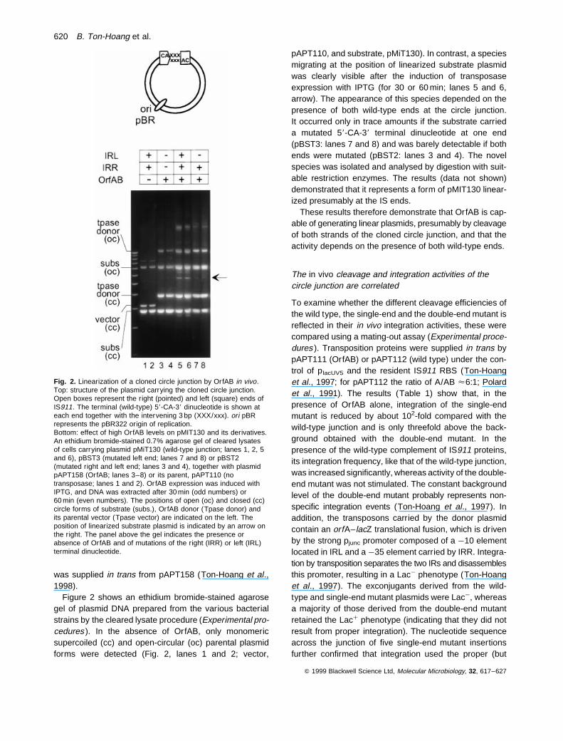
Figure 2 shows an ethidium bromide-stained agarosegel of plasmid DNA prepared from the various bacterialstrains by the cleared lysate procedure (Experimental pro-cedures ). In the absence of OrfAB, only monomericsupercoiled (cc) and open-circular (oc) parental plasmidforms were detected (Fig. 2, lanes 1 and 2; vector,
pAPT110, and substrate, pMiT130). In contrast, a speciesmigrating at the position of linearized substrate plasmidwas clearly visible after the induction of transposaseexpression with IPTG (for 30 or 60 min; lanes 5 and 6,arrow). The appearance of this species depended on thepresence of both wild-type ends at the circle junction.It occurred only in trace amounts if the substrate carrieda mutated 58-CA-38 terminal dinucleotide at one end(pBST3: lanes 7 and 8) and was barely detectable if bothends were mutated (pBST2: lanes 3 and 4). The novelspecies was isolated and analysed by digestion with suit-able restriction enzymes. The results (data not shown)demonstrated that it represents a form of pMIT130 linear-ized presumably at the IS ends.
These results therefore demonstrate that OrfAB is cap-able of generating linear plasmids, presumably by cleavageof both strands of the cloned circle junction, and that theactivity depends on the presence of both wild-type ends.
The in vivo cleavage and integration activities of thecircle junction are correlated
To examine whether the different cleavage efficiencies ofthe wild type, the single-end and the double-end mutant isreflected in their in vivo integration activities, these werecompared using a mating-out assay (Experimental proce-dures ). Transposition proteins were supplied in trans bypAPT111 (OrfAB) or pAPT112 (wild type) under the con-trol of placUV5 and the resident IS911 RBS (Ton-Hoanget al., 1997; for pAPT112 the ratio of A/AB <6:1; Polardet al., 1991). The results (Table 1) show that, in thepresence of OrfAB alone, integration of the single-endmutant is reduced by about 102-fold compared with thewild-type junction and is only threefold above the back-ground obtained with the double-end mutant. In thepresence of the wild-type complement of IS911 proteins,its integration frequency, like that of the wild-type junction,was increased significantly, whereas activity of the double-end mutant was not stimulated. The constant backgroundlevel of the double-end mutant probably represents non-specific integration events (Ton-Hoang et al., 1997). Inaddition, the transposons carried by the donor plasmidcontain an orfA–lacZ translational fusion, which is drivenby the strong pjunc promoter composed of a ¹10 elementlocated in IRL and a ¹35 element carried by IRR. Integra-tion by transposition separates the two IRs and disassemblesthis promoter, resulting in a Lac¹ phenotype (Ton-Hoanget al., 1997). The exconjugants derived from the wild-type and single-end mutant plasmids were Lac¹, whereasa majority of those derived from the double-end mutantretained the Lacþ phenotype (indicating that they did notresult from proper integration). The nucleotide sequenceacross the junction of five single-end mutant insertionsfurther confirmed that integration used the proper (but
Q 1999 Blackwell Science Ltd, Molecular Microbiology, 32, 617–627
Fig. 2. Linearization of a cloned circle junction by OrfAB in vivo.Top: structure of the plasmid carrying the cloned circle junction.Open boxes represent the right (pointed) and left (square) ends ofIS911. The terminal (wild-type) 58-CA-38 dinucleotide is shown ateach end together with the intervening 3 bp (XXX/xxx). ori pBRrepresents the pBR322 origin of replication.Bottom: effect of high OrfAB levels on pMIT130 and its derivatives.An ethidium bromide-stained 0.7% agarose gel of cleared lysatesof cells carrying plasmid pMiT130 (wild-type junction; lanes 1, 2, 5and 6), pBST3 (mutated left end; lanes 7 and 8) or pBST2(mutated right and left end; lanes 3 and 4), together with plasmidpAPT158 (OrfAB; lanes 3–8) or its parent, pAPT110 (notransposase; lanes 1 and 2). OrfAB expression was induced withIPTG, and DNA was extracted after 30 min (odd numbers) or60 min (even numbers). The positions of open (oc) and closed (cc)circle forms of substrate (subs.), OrfAB donor (Tpase donor) andits parental vector (Tpase vector) are indicated on the left. Theposition of linearized substrate plasmid is indicated by an arrow onthe right. The panel above the gel indicates the presence orabsence of OrfAB and of mutations of the right (IRR) or left (IRL)terminal dinucleotide.
620 B. Ton-Hoang et al.

mutant) end (data not shown). These results thus demon-strate a correlation between the capacity of the circle sub-strate to undergo double-strand cleavage and its capacityto integrate.
Mapping the cleavage site at the cloned junction
To date, OrfAB has been observed to accomplish only
single-strand cleavage at the terminal inverted repeatsof IS911 (Polard et al., 1996). Its activity in linearizingpMiT130 presumably results from a single-strand cleavageat each of the abutting IRs. As the two ends at the junctionare separated by 3 bp, two single-strand cleavages at thelevel of the 58-CA-38 terminal dinucleotide should producea linear molecule with 3 bp protruding 58 ends (Fig. 3, top).To verify this, a preparation of gel-purified linear molecules
Q 1999 Blackwell Science Ltd, Molecular Microbiology, 32, 617–627
Table1 1. Results of mating-out assays.
Mating out assays are described in Experimental procedures. Left and right transposon endsare indicated by open pointed and square boxes respectively. Mutation of the terminal 58-CA-38dinucleotide is indicated by the superimposed open circle. These transposon donor plasmidsall carry an orfA (stippled) translational fusion with lacZ (cross-hatched). The source of trans-position enzymes from a compatible plasmid are shown at the top of the two righthand col-umns. The values included represent the frequency of transfer of the transposon donorplasmid divided by that of the conjugative target plasmid pOX38Km. They are the averageof five individual experiments. Standard errors in the data are included within brackets.
Fig. 3. Mapping of cleavage site at circlejunction.Top: schematic representation of single-strandcleavages at the cloned circle junction. Right(IRR) and left (IRL) transposon ends are shownflanked by EcoRI and BamHI sites, respectively,terminating in the wild-type case with 58-CA-38dinucleotides. The position of strand cleavage isindicated by vertical arrowheads.Bottom: analysis of cleavage products. The linearand open circle species shown in Fig. 2 weredigested with EcoRI and BamHI. Products wereseparated on a 6% denaturing polyacrylamidegel, transferred to a membrane and hybridizedwith probes specific for IRL (O16) (A) or IRR(O18) (B). The products of digestion by EcoRIand BamHI of the purified linear form of the wild-type junction substrate, pMiT130, before andafter Klenow treatment are shown in lanes 1 and2 respectively. The open circle form of substrateplasmids pMiT130, pBST3 and pBST2 arepresented in lanes 3–5 respectively. The positionand size of respective species are indicated inbasepairs. Lanes on the right of the figure show asequencing ladder as a length standard.
Transposon integration 621

was divided in two, and one part was treated with the Kle-now fragment in the presence of dNTPs. Both treated anduntreated samples were then digested with EcoRI andBamHI, separated on a denaturing polyacrylamide geland hybridized with oligonucleotides specific for IRL(O16: complementary to the lower strand) and IRR(O18: complementary to the upper strand). The resultsare shown in Fig. 3 (bottom). The untreated sample gaverise to fragments of 109 (with O16) and 97 bases (withO18), which correspond precisely to that expected from38 cleavage at the terminal A of IRL and IRR respectively(lanes A1 and B1). The molecules, which were treated withKlenow fragment before digestion, generated fragments of112 and 100 bases (lanes A2 and B), showing that clea-vage at the ends resulted in a 38-OH recessed by 3 bases.
As shown in Fig. 2, pBST3, which carries the single-endmutant junction, exhibited an increased level of open circu-lar DNA. To determine whether this results from specificcleavage of a single end of the circle junction, the open cir-cular species was gel purified, digested with EcoRI andBamHI and analysed as described for linearized DNA.When probed with the IRL-specific oligonucleotide, DNAfrom pMiT130 (wild-type junction) generated fragmentsof 206 bases, corresponding to a full-length strand, and109 bases, corresponding to a strand cleaved at the termi-nal A of IRL (Fig. 3, lane A3). When probed with the IRR-specific oligonucleotide, this DNA generated two bands of206 and 97 bases, corresponding to the full-length strandand to a strand cleaved at the terminal A of IRR (lane B3).This demonstrates that the open circular pMiT130 specieswas a mixed population, in which part carried a single-strand cleavage at IRL and part at IRR. Note that theEcoRI–BamHI fragment of plasmids with a mutant circlejunction is slightly smaller than that carried by pMiT130.For pBST3 (lane A4), a single band of 179 bases (full length)was revealed by the IRL probe, indicating that the mutatedend was very inefficiently cleaved. On the other hand,bands of 179 and 72 bases were observed with the IRRprobe (lane B4). The 72 base fragment corresponds tocleavage of IRR, whereas the full-length 179 base bandpresumably arose from open circular DNA carrying non-specific single-strand nicks. For pBST2, both oligonucleo-tides generated only a single band of 179 bases, corre-sponding to the uncleaved fragment (Fig. 3, lanes A5and B5), indicating that the open circular DNA was prob-ably generated by random single-strand nicking.
These results therefore confirm that OrfAB can generatelinear molecules from plasmids carrying the circle junction(Ton-Hoang et al., 1998) and demonstrate that it doesso by introducing two single-strand cleavages at the 38
end of the CA dinucleotide. Moreover, they also show thatcleavage leaves a 38-OH group capable of being extendedby Klenow polymerase and that the integrity of the twoterminal nucleotides is necessary for strand cleavage.
Linearization in vitro
To determine whether ‘double-strand’ cleavage of the IRL–IRR junction requires only OrfAB or whether other IS911-or host-encoded proteins are involved, linearization of thejunction-carrying plasmids was investigated in vitro usinga highly enriched OrfAB preparation. The reaction bufferwas similar to that used in the previously described invitro integration assay (Ton-Hoang et al., 1998) andincluded Mg2þ as the divalent cation and DMSO. The reac-tion products were analysed by agarose gel electro-phoresis, and the results are shown in Fig. 4. In thepresence of OrfAB, the plasmid substrate with the wild-type circle junction, pMiT130, was efficiently convertedto a linear form (lane 3), while only a trace amount or no signalwas observed with the single- (pBST3) or double-end(pBST2) mutant (lanes 2 and 1 respectively). The nature ofthis form was confirmed by digestion with suitable restrictionenzymes (data not shown). Note that the high-molecular-weight species present in lanes with wild-type or singlemutant substrates probably represent autointegration
Q 1999 Blackwell Science Ltd, Molecular Microbiology, 32, 617–627
Fig. 4. Linearization of transposon circle in vitro. Ethidiumbromide-stained agarose gel (0.7%) of undigested products oflinearization reaction of transposon circle after treatment withpurified OrfAB. The lefthand lane shows molecular mass standards(1 kb ladder). The type of junction carried by the plasmid is shownabove the gel. The superimposed circles indicate the presence of amutation of the terminal 58-CA-38 dinucleotide. Lanes 1 and 4,double mutant, pBST2; lanes 2 and 5, single-end mutation in IRL,pBST3; lanes 3 and 6, wild-type junction, pMIT130. Whereindicated, 10% DMSO was included in the reaction. The arrow onthe right indicates the position of linearized plasmid.
622 B. Ton-Hoang et al.

products (see Ton-Hoang et al., 1998). DMSO has beenshown previously to stimulate the integration of transposoncircles greatly. When the reaction was undertaken in theabsence of DMSO, the linear form was still observed,but was significantly less abundant (lane 6). DMSO pre-sumably stimulates in vitro integration by enhancing the invitro cleavage activity of OrfAB at the circle junction.
These in vitro results mirror those obtained in vivo andsupport the notion that OrfAB alone is capable of cleavingthe IRL–IRR junction.
Integration of precleaved IS911 derivatives in vitro
One important question in understanding the mechanismof circle integration is the sequence of cleavages and strandtransfer events leading to integration. Several different andnon-exclusive pathways could operate. The simplest wouldbe passage by two concerted or co-ordinated single-strandnicks followed by strand transfer of both cleaved ends (Fig.5, right). In other words, by passage through a ‘linear’ mole-cule derived from the circle. Alternatively, it could occur ina stepwise process in which first one and then the secondend in the circle junction was cleaved and transferred (Fig.5, left). This possibility is raised by the identification of aspecies that appears to result from cleavage and insertionof only a single strand (and single end) of the circular trans-poson into the target plasmid (Ton-Hoang et al., 1998). Inthis type of model, the proximity of the second end in thecircle junction would be a key factor in its integration atthe same target site. Although determination of the kineticsof formation of the different intermolecular species will berequired in order to clarify the exact pathway used, an indi-cation of the integration mechanism would be the demon-stration that a linear transposon substrate can undergoefficient integration. For this purpose, a precleaved trans-poson substrate was prepared first by polymerase chainreaction (PCR) amplification of a transposon with appropri-ate primers containing an NdeI site at the position of the58-CA-38 dinucleotide. The fragment was then digestedwith NdeI to generate a full-length transposon with liber-ated 58-CA-38-OH ends recessed by two nucleotides (com-pared with the three nucleotides observed in vivo ).Cleavage was confirmed using an end-labelled substrate.The standard reaction (Ton-Hoang et al., 1998; seeExperimental procedures ) contained plasmid pDAG92as target and a protein preparation containing both OrfAand OrfAB produced from IS911 reading frames in thewild-type configuration. The products were analysed bygel electrophoresis and Southern blotting using an oligo-nucleotide probe specific for the transposon. The resultsare presented in Fig. 6 (lane 1). A parallel experimentusing the transposon circle substrate was also included(lane 2). Only the three major intermolecular integrationproducts are indicated. These were shown previously to
be single-ended integration of the linear transposon intothe circular plasmid target generating a lariat form (I),single-end integration of the circular transposon forming afigure-eight molecule (II) and concerted integration (III)respectively (Fig. 5; Ton-Hoang et al., 1998). In the caseof the transposon circle substrate (Fig. 6, lane 2), the con-certed integration product was significantly more abundantthan species I and II. As expected, with the precleaved sub-strate, no band corresponding to species II was observed,although both species I and species III were detected (lane1). In contrast to the results obtained with the circulartransposon, species I was the major product of the reac-tion in this case (compare lanes 1 and 2). Although theends of the precleaved transposon substrate can be
Q 1999 Blackwell Science Ltd, Molecular Microbiology, 32, 617–627
Fig. 5. Possible pathways of transposon circle integration. Thetransposon DNA is represented by heavy double lines and targetDNA by dashed double lines. The 38 ends of the transposon areshown as small circles. The lefthand section of the figure shows apathway in which strand cleavage and strand transfer occurs first atone end to generate a molecule that has the structure of species II(see Fig. 6 and Ton-Hoang et al., 1998), and then at the secondend to generate first species I and finally species III. The righthandpathway represents the concerted cleavage and transfer of bothtransposon ends.
Transposon integration 623

integrated in a concerted manner into a target, giving riseto simple transposon insertion, the reaction is clearly lessefficient than with a circular transposon substrate. The for-mation of species III was confirmed further using a biologi-cal assay, in which the reaction products were used totransform a suitable host strain with selection for chloram-phenicol resistance (CmR), a marker carried by the trans-poson (data not shown). These results indicate thatspecies III is not necessarily derived from species II bysequential cleavage and strand transfer, as the figure-eight molecule seems unlikely to be generated from a pre-cleaved linear substrate. It demonstrates that linearizedtransposon can be integrated, giving rise to species III,but it does not rule out the possibility that species II is cap-able of evolving into species III.
Discussion
Previous studies have established that IS911 can assumea circular form in which the terminal IRs are abutted andseparated by a linker sequence of 3 bp. This is thoughtto occur by a two-step process in which one transposonstrand is first circularized by cleavage at its 38 end andtransferred to the opposite end. This generates a figure-eight molecule in which a single transposon strand hasbeen circularized to retain both terminal IRs by a single-strand bridge (Polard and Chandler, 1995a). It has beenproposed that the second strand is then resolved by hostfactors to generate the transposon circle (Polard et al.,1996). It was further demonstrated that the transposoncircles undergo efficient insertion both in vivo (Ton-Hoanget al., 1997) and in vitro (Ton-Hoang et al., 1998) andcan thus be formally viewed as transposition intermediates.Moreover, using a set of circle junctions in which the spacing
between the terminal IRs varied, it was shown that themaximal integration efficiency in vitro occurred with thesubstrate having the natural 3 bp spacing (Ton-Hoang etal., 1998).
In the experiments described here, we have begun toaddress the sequence of cleavages and strand transferevents leading to integration of the transposon circles. Inparticular, a linear transposon species has been detectedand shown to be derived directly from the circular form bysingle-strand cleavage at each 38 end of abutted terminalIRs in the circle junction. The linear form undergoes inte-gration into a suitable plasmid target in vitro, albeit with alower efficiency than the circles. Cleavage at the circle junc-tion leaving a three-base 58 overhang at each end wasobserved both in vivo and in vitro.
A similar linear transposon species was also detected invivo in the presence of high levels of the OrfAB proteinfrom a donor plasmid carrying an inserted copy of the trans-poson, i.e. in the absence of a preformed circle junction.Analysis of the effects of mutations in the terminal IRsand determination of the flanking nucleotide sequencesat the ends of this species demonstrated that it too wasderived from a transposon circle generated from the origi-nal insertion. The linear forms therefore generally appearto be associated with the presence of wild-type transposoncircles. It should be noted, however, that, in most of theseexperiments, high levels of the OrfAB protein were sup-plied without OrfA and that intermolecular transpositionof IS911 using an IRL–IRR junction requires OrfA in addi-tion to OrfAB both in vivo and in vitro (Ton-Hoang et al.,1997; 1998). It could, therefore, be argued that double-strand cleavage at IRL and IRR normally occurs whenIS911 is carried in the donor molecule, but that it requiresthe presence of OrfA in addition to OrfAB. However, thisseems unlikely, as the addition of purified OrfA to an invitro OrfAB-mediated reaction that faithfully reproducesthe formation of figure-eight molecules (the probable pre-cursor of the transposon circle) does not generate detectablelinearized transposon species (data not shown). Thus,despite detailed analysis under a variety of experimentalconditions, we have no evidence that IS911 ends canundergo double-strand cleavage. In this respect, IS911may behave differently to IS3, which appears to undergodouble-strand cleavage at each end (Sekine et al., 1996).
Integration of transposon circles could occur by severaldifferent and non-exclusive pathways. Identification of aspecies that appears to result from cleavage and insertionof only a single strand (and single end) of the circular trans-poson (Ton-Hoang et al., 1998) raises the possibility thatit could occur in a stepwise process, in which first one andthen the second end in the circle junction is cleaved andtransferred (Fig. 5, left). Another possibility would be aprocess involving two concerted or co-ordinated singlecleavages followed by concerted strand transfer at both
Q 1999 Blackwell Science Ltd, Molecular Microbiology, 32, 617–627
Fig. 6. Integration of precleaved transposon in vitro. DNA of alinear transposon derivative prepared by PCR and digested withNdeI to generate terminal-free 58-CA-38-OH was reacted understandard conditions with plasmid pDAG92 DNA. The products wereanalysed by gel electrophoresis, Southern blotting and hybridizationwith an oligonucleotide specific for the transposon. The type oftransposon substrate is indicated above the autoradiograph. Lane1, wild-type ends; lane 2, transposon circles. The lefthand sideindicates the type of product as identified previously (Ton-Hoanget al., 1998): type III, co-ordinated integration of both transposonends; type II, single-end cleavage and transfer from the transposoncircle; type I, single-end insertion.
624 B. Ton-Hoang et al.

cleaved ends (Fig. 5, right). The latter model implies theformation of a linear transposon form. Such forms havebeen observed for several transposons as intermediatesin transposition (see Craig, 1996; Kleckner et al., 1996;Plasterk, 1996) and, in the case of Tn10 (IS10 ), havebeen isolated as a complex in which the ends are heldtogether with the cognate transposase (Haniford et al.,1991; Sakai et al., 1995). For IS911, the efficiency of co-ordinated or concerted integration of the transposonends appears to be significantly higher from the transposoncircle than from the precleaved linear substrate. This mayreflect a necessity to assemble the two separate ends ofthe transposon to create this active architecture. This kineticbarrier may be circumvented by the proximity of the endsat the circle junction.
At present, we are unable to attribute each of the obser-ved forms to a temporal position in the overall IS911 trans-position pathway. It is clearly possible that simple insertionof IS911 can be achieved by several pathways. Althoughpreliminary kinetic studies of transposon circle integrationin vitro have shown that the appearance of the linear formprecedes that of the concerted integration products (B.Ton-Hoang, unpublished), the presence of other forms,including species I and II, complicates the interpretationof these results. To resolve some of these questions, itwill be useful to isolate these species and to determinewhether they can also give rise to simple insertions in vitro.
What is the relation between the transposition mechan-ism of IS911 and that of other transposons? Historically,transposition mechanisms have been separated into twoprincipal modes: ‘cut-and-paste’ and replicative. This isbased on whether or not the element is copied duringits displacement. In replicative transposition, duplicationof the element is intimately associated with the transfer ofthe cleaved transposon ends into the target. In cut-and-paste transposition, the preintegration intermediate is gen-erally separated from the donor backbone and, as aconsequence, the integration event itself is conservative.However, retroviral integrases, IN, and many related trans-posases are single-strand endonucleases and unable tointroduce double-strand cleavages at the transposaseends. It is now clear that different transposable elementshave adopted different strategies to circumvent this con-straint. In some, such as Tn7, the transposase is a hetero-dimeric complex of two proteins, each dedicated to cleavageof one of the two strands (Sarnovsky et al., 1996), whileothers use a single enzyme. In the case of retroviruses,a free double-strand DNA copy with 2 bp extensions,designated for integration, issynthesized fromthe integratedcopy by transcription followed by reverse transcription. INpromotes single-strand cleavage to remove the two 38
bases and subsequent strand transfer (see Andrake andSkalka, 1996). In the case of IS10, a single catalytic siteat each transposon end is used repeatedly for consecutive
hydrolysis and strand transfer reactions. In a first step, theenzyme catalyses the cleavage of the 38 end of the ele-ment (hydrolysis). The liberated 38-OH is then used as anucleophile to attack the complementary strand (intramole-cular strand transfer) in order to generate a linear transpo-son in which the two strands are joined by a hairpin bridgeat each end. A second set of hydrolysis reactions breaksthe hairpins and regenerates the 38-OH groups. Finally,the regenerated 38-OH is used in intermolecular strandtransfer (Kennedy et al., 1998). Like IS10, IS911 transpo-sition also occurs by repeated consecutive hydrolysis andtransfer reactions. Here, however, the first single-strandhydrolysis occurs only at one end, whereas the intramole-cular strand transfer occurs on the same DNA strand togenerate a single circularized strand (Polard and Chandler,1995a). This structure evolves into a transposon circle, thesubstrate for a second set of two single-strand hydrolysisreactions. Hydrolysis results in separation of the abuttedends and generation of 38-OH groups, which are then trans-ferred to the target. Thus, while Tn7, retroviruses, IS10 (andother members of the IS4 family) and IS911 (and othermembers of the IS3 family) have apparently assumed differ-ent transposition pathways, they all transpose by a conser-vative mechanism, and the major differences reside in theway in which separation of the preintegration intermediateoccurs.
Experimental procedures
Bacterial strains and media
The bacterial strain used in the preparation of cell extractswas Mi898, a derivative of MC1061 (araD139, D[ara leu],gal U, gal K, hsd M, hsdS, rpsL D[lacIOPZYA]X74) carryinga deletion of the endonuclease I gene endA (Polard et al.,1996).
Supercoiled transposon circles were generated in JS219(srl C::Tn10, recA1, mal Pp ::lacIQ, araD139, D[ara leu], gal Ugal K, hsd M, hsdS, rplsL D[lacIOPZYA]X74) as host (seePolard et al., 1992).
Cultures were grown in TB (Sambrook et al., 1989) supple-mented with ampicillin (Ap; 100 mg ml¹1) and kanamycin (Km;25 mg ml¹1). Selection of chloramphenicol-resistant transfor-mants was carried out on L plates (Cm; 30 mg ml¹1).
Plasmids
Plasmids pBST2, pBST3 and pMiT130 have been describedpreviously (Ton-Hoang et al., 1997; 1998). The plasmidpAPT166 containing the transposon in non-circular form hasbeen described previously (Polard and Chandler, 1995a).
pAPT110, pAPT111 and pAPT112 have been describedpreviously (Polard and Chandler, 1995a). Briefly, pAPT110is a p15A-based plasmid carrying the plac promoter, lacIq
and an Sp Sm resistance cassette. Plasmids pAPT111 andpAPT112 are derived from pAPT110. They carry orfAB andthe wild-type open reading frame configuration, respectively,
Q 1999 Blackwell Science Ltd, Molecular Microbiology, 32, 617–627
Transposon integration 625

together with indigenous translation initiation signals underthe control of placUV5 (Polard and Chandler, 1995a).
The plasmid pAPT158 (8208 bp) expressing OrfAB or wild-type configuration pAPT155 (8207 bp) contains the ribosomebinding site (RBS) of bacteriophage T7 f10 located betweenthe placUV5 promoter and the OrfAB initiation codon (Ton-Hoang et al., 1998).
Mating-out assays
The frequency of IS911 junction-mediated integration wasdetermined by a standard mating-out assay (Galas and Chand-ler, 1982) using the conjugal plasmid pOX38Km (Chandlerand Galas, 1983) as a target replicon, as described previously(Ton-Hoang et al., 1997).
Preparation and purification of transposon circles andlinear integration substrates
Transposon circles were prepared as described previously(Ton-Hoang et al., 1998). The wild-type or single mutant endprecleaved linear transposon was prepared by PCR amplifi-cation of plasmids pAPT99 or pAPT92 (Polard et al., 1992)using the following primers: B29 (58-GATCCCCAATCATAT-GAAGTGGTCAACAAAAA-38); B30 (58-CGGAATGTTAATT-CCATATGAAGTGGCACA-38); B31 (58-CGGAATGTTAATT-CATCGATAAGTGGCACA-38).
The amplified fragment was purified and digested with NdeIfor wild type and with NdeI–ClaI for single mutant end pre-cleaved substrate respectively.
Purification of OrfAB and wild-type proteins
The transposase OrfAB and the wild-type mixture of proteinswere prepared as described previously (Ton-Hoang et al.,1998).
Cell-free systems
The linearization reaction was performed in a final volume of30 ml containing 600 ng of DNA circle junction substrate(pMiT130, pBST2 and pBST3), 1 mg of the transposase proteinpreparations (in dilution buffer A, 0.4 M KCl) in 25 mM HEPES(pH 7,5), 0.3 M KCl, 10 mM MgCl2, 2 mM dithiothreitol (DTT),20 ng ml¹1 BSA and 14% glycerol. Where indicated, 10%DMSO was added. The reactions were carried out at 308Cfor 45 min. Reactions were stopped and deproteinized by theaddition of 30 ml of 25 mM EDTA, 0.6 mg ml¹1 proteinase K,2% SDS, incubated for 1 h at 378C and treated with a mixtureof phenol–chloroform (1:1), followed by extraction with chloro-form and ethanol precipitation.
The integration reaction with linear precleaved substrateswas performed under standard conditions as described pre-viously (Ton-Hoang et al., 1998) with the wild-type proteinpreparation and pDAG92 as target.
DNA procedures
Standard techniques were used for DNA manipulation and
cloning (Sambrook et al., 1989). Restriction and DNA-modify-ing enzymes were purchased from Biolabs. DNA fragmentswere isolated using Geneclean (Bio101). Preparation of clearedlysates was as described previously (Polard and Chandler,1995b).
Southern blots were probed with oligonucleotides B21(which hybridizes within the transposon between 186 and211 bp from IRL and IRR): 58-GGATGACCTTTTGAATGA-CCTTTTAA-38.
Radiolabelling was with 8 pmol of oligonucleotides, 16 pmolof [g32-P]-ATP (5000 Ci mmol¹1; Amersham) and 1 unit of T4kinase (New England Biolabs) in T4 kinase buffer (70 mMTris-HCl, pH 7.6, 10 mM MgCl2, 5 mM DTT) for 1 h at 378C.Labelled oligonucleotides were purified by filtration throughSephadex G25.
Gel electrophoresis and DNA quantification byhybridization
Reaction products were separated on 0.7% agarose gels inTAE buffer at 2.4 V cm¹1 for 18 h (16 h for linear substrates)at room temperature.
DNA hybridization was accomplished directly in the gel.The gel was dried for 70 min at 608C. Then, the DNA wasdenatured (0.5 M NaOH, 0.15 M NaCl, 20 min), and the gelwas neutralized in 0.5 M Tris (pH 8), 0.15 M NaCl. After a pre-hybridization step in 6 × SSC, 0.5% SDS, 100 mg ml¹1 calfthymus DNA and 0.1% non-fat milk for 6–8 h, hybridizationwas carried out with a large excess of labelled oligonucleotidefor 12–15 h in the same buffer. Both steps were performed at88C below the Tm of the oligonucleotide probe. The gel wasthen washed three times in 6 × SSC, 0.1% SDS at room tem-perature, dried and exposed either for autoradiography usingKodak XR5 film or for quantification in a Fuji X BAS1000 phos-phorimaging system coupled to the PCBAS software package.
For the mapping of cleavage sites at the circle junction,DNA samples were digested with EcoRI and BamHI, and pro-ducts were separated on a 6% polyacrylamide gel with 8 Murea in TBE running buffer and treated as described pre-viously (Polard et al., 1996).
Acknowledgements
We would like to thank all members of the Mobile Genetic Ele-ments group (R. Alazard, G. Duval-Valentin and C. Normand)for discussions and advice during the course of this work, andA. J. Carpousis for critical reading of the manuscript. We wouldalso like to acknowledge D. Villa for image processing. Thiswork benefited from grants from the CNRS (UPR9007), the‘Association pour la Recherche contre le Cancer’ (6406), the‘Association Nationale de Recherche Contre le SIDA’ andthe ‘Region Midi-Pyrenees’.
References
Andrake, M.D., and Skalka, A.M. (1996) Retroviral integrase,putting the pieces together. J Biol Chem 271: 19633–19636.
Bainton, R.J., Kubo, K.M., Feng, J.N., and Craig, N.L. (1993)Tn7 transposition: target DNA recognition is mediated by
Q 1999 Blackwell Science Ltd, Molecular Microbiology, 32, 617–627
626 B. Ton-Hoang et al.

multiple Tn7-encoded proteins in a purified in vitro system.Cell 72: 931–943.
Chandler, M., and Fayet, O. (1993) Translational frameshift-ing in the control of transposition in bacteria. Mol Microbiol7: 497–503.
Chandler, M., and Galas, D.J. (1983) Cointegrate formationmediated by Tn9. II. Activity of IS1 is modulated by exter-nal DNA sequences. J Mol Biol 170: 61–91.
Craig, N.L. (1996) Transposon Tn7. Curr Topics MicrobiolImmunol 204: 27–48.
Galas, D.J., and Chandler, M. (1982) Structure and stabilityof Tn9-mediated cointegrates. Evidence for two pathwaysof transposition. J Mol Biol 154: 245–272.
Goryshin, I.Y., and Reznikoff, W.S. (1998) Tn5 in vitro trans-position. J Biol Chem 273: 7367–7374.
Grindley, N.D., and Leschziner, A.E. (1995) DNA transposi-tion: from a black box to a color monitor. Cell 83: 1063–1066.
Haniford, D.B., Benjamin, H.W., and Kleckner, N. (1991)Kinetic and structural analysis of a cleaved donor inter-mediate and a strand transfer intermediate in Tn10 trans-position. Cell 64: 171–179.
Kennedy, A.K., Guhathakurta, A., Kleckner, N., and Hani-ford, D.B. (1998) Tn10 transposition via a DNA hairpinintermediate. Cell 95: 125–134.
Kleckner, N., Chalmers, R.M., Kwon, D., Sakai, J., and Bol-land, S. (1996) Tn10 and IS10 transposition and chromo-some rearrangements: mechanisms and regulation in vivoand in vitro. In Transposable Elements. Saedler, H., andGierl, A. (eds). Heidelberg: Springer, pp. 49–82.
Lewis, L.A., and Grindley, N.D. (1997) Two abundant intra-molecular transposition products, resulting from reactionsinitiated at a single end, suggest that IS2 transposes byan unconventional pathway. Mol Microbiol 25: 517–529.
Mahillon, J., and Chandler, M. (1998) Insertion sequences.Microbiol Mol Biol Rev 62: 725–774.
Plasterk, R.H. (1996) The Tc1/mariner transposon family.Curr Topics Microbiol Immunol 204: 125–143.
Polard, P., and Chandler, M. (1995a) An in vivo transposase-catalyzed single-stranded DNA circularization reaction.Genes Dev 9: 2846–2858.
Polard, P., and Chandler, M. (1995b) Bacterial transposasesand retroviral integrases. Mol Microbiol 15: 13–23.
Polard, P., Prere, M.F., Chandler, M., and Fayet, O. (1991)Programmed translational frameshifting and initiation atan AUU codon in gene expression of bacterial insertionsequence, IS911. J Mol Biol 222: 465–477.
Polard, P., Prere, M.F., Fayet, O., and Chandler, M. (1992)Transposase-induced excision and circularization of thebacterial insertion sequence IS911. EMBO J 11: 5079–5090.
Polard, P., Ton-Hoang, B., Haren, L., Betermier, M., Walczak,R., and Chandler, M. (1996) IS911-mediated transpositionalrecombination in vitro. J Mol Biol 264: 68–81.
Rice, P., Craigie, R., and Davies, D.R. (1996) Retroviral inte-grases and their cousins. Curr Opin Struct Biol 6: 76–83.
Sakai, J., Chalmers, R.M., and Kleckner, N. (1995) Identifica-tion and characterization of a pre-cleavage synaptic com-plex that is an early intermediate in Tn10 transposition.EMBO J 14: 4374–4383.
Sambrook, J., Fritsch, E.F., and Maniatis, T. (1989) MolecularCloning: a Laboratory Manual, 2nd edn. Cold Spring Har-bor, NY: Cold Spring Harbor Laboratory Press.
Sarnovsky, R.J., May, E.W., and Craig, N.L. (1996) The Tn7transposase is a heteromeric complex in which DNA break-age and joining activities are distributed between differentgene products. EMBO J 15: 6348–6361.
Sekine, Y., Eisaki, N., and Ohtsubo, E. (1994) Translationalcontrol in production of transposase and in transpositionof insertion sequence IS3. J Mol Biol 235: 1406–1420.
Sekine, Y., Eisaki, N., and Ohtsubo, E. (1996) Identificationand characterization of the linear IS3 molecules generatedby staggered breaks. J Biol Chem 271: 197–202.
Ton-Hoang, B., Betermier, M., Polard, P., and Chandler, M.(1997) Assembly of a strong promoter following IS911 circu-larization and the role of circles in transposition. EMBO J16: 3357–3371.
Ton-Hoang, B., Polard, P., and Chandler, M. (1998) Efficienttransposition of IS911 circles in vitro. EMBO J 17: 1169–1181.
Vos, J.C., De Baere, I., and Plasterk, R.H. (1996) Transpo-sase is the only nematode protein required for in vitro trans-position of Tc1. Genes Dev 10: 755–761.
Welz, C. (1993) Functionelle analyse des Bakteriellen Inser-tionelements IS150. PhD Thesis, Fakultat fur Biologie derAlbert-Ludwigs-Universitat, Freiburg.
Q 1999 Blackwell Science Ltd, Molecular Microbiology, 32, 617–627
Transposon integration 627
Copyright © 2022 FDOKUMEN