Passport, a native Tc1 transposon from flatfish, is functionally active in vertebrate cells
Upload
independentCategory
view
4download
0
University of Rhode IslandDigitalCommons@URI
Cell and Molecular Biology Faculty Publications Cell and Molecular Biology
2014
Identification of the Maize Gravitropism Gene lazyplant1 by a Transposon-Tagging GenomeResequencing StrategyThomas P. Howard III
Andrew P. Hayward
See next page for additional authors
Creative Commons License
/">This work is licensed under a Creative Commons Public Domain Dedication 1.0 License.
Follow this and additional works at: http://digitalcommons.uri.edu/cmb_facpubs
Terms of UseAll rights reserved under copyright.
This Article is brought to you for free and open access by the Cell and Molecular Biology at DigitalCommons@URI. It has been accepted for inclusionin Cell and Molecular Biology Faculty Publications by an authorized administrator of DigitalCommons@URI. For more information, please [email protected].
Citation/Publisher AttributionHoward TP, Hayward AP, Tordillos A, Fragoso C, Moreno MA, Tohme J, Kausch AP, Mottinger JP, Dellaporta SL. (2014)."Identification of the maize gravitropism gene lazy plant1 by a transposon-tagging genome resequencing strategy." PLoS One. 9(1):e87053.Available at: http://dx.doi.org/10.1371/journal.pone.0087053

AuthorsThomas P. Howard III, Andrew P. Hayward, Anthony Tordillos, Christopher Fragoso, Maria A. Moreno, JoeTohme, Albert P. Kausch, John P. Mottinger, and Stephen L. Dellaporta
This article is available at DigitalCommons@URI: http://digitalcommons.uri.edu/cmb_facpubs/12

Identification of the Maize Gravitropism Gene lazy plant1by a Transposon-Tagging Genome ResequencingStrategyThomas P. Howard III1¤, Andrew P. Hayward1, Anthony Tordillos1, Christopher Fragoso1,
Maria A. Moreno1, Joe Tohme2, Albert P. Kausch3, John P. Mottinger3, Stephen L. Dellaporta1*
1 Department of Molecular, Cellular and Developmental Biology, Yale University, New Haven, Connecticut, United States of America, 2 International Center for Tropical
Agriculture, Cali, Colombia, 3 Department of Cell and Molecular Biology, University of Rhode Island, Kingston, Rhode Island, United States of America
Abstract
Since their initial discovery, transposons have been widely used as mutagens for forward and reverse genetic screens in arange of organisms. The problems of high copy number and sequence divergence among related transposons have oftenlimited the efficiency at which tagged genes can be identified. A method was developed to identity the locations of Mutator(Mu) transposons in the Zea mays genome using a simple enrichment method combined with genome resequencing toidentify transposon junction fragments. The sequencing library was prepared from genomic DNA by digesting with arestriction enzyme that cuts within a perfectly conserved motif of the Mu terminal inverted repeats (TIR). Paired-end readscontaining Mu TIR sequences were computationally identified and chromosomal sequences flanking the transposon weremapped to the maize reference genome. This method has been used to identify Mu insertions in a number of alleles and toisolate the previously unidentified lazy plant1 (la1) gene. The la1 gene is required for the negatively gravitropic response ofshoots and mutant plants lack the ability to sense gravity. Using bioinformatic and fluorescence microscopy approaches, weshow that the la1 gene encodes a cell membrane and nuclear localized protein. Our Mu-Taq method is readily adaptable toidentify the genomic locations of any insertion of a known sequence in any organism using any sequencing platform.
Citation: Howard TP III, Hayward AP, Tordillos A, Fragoso C, Moreno MA, et al. (2014) Identification of the Maize Gravitropism Gene lazy plant1 by a Transposon-Tagging Genome Resequencing Strategy. PLoS ONE 9(1): e87053. doi:10.1371/journal.pone.0087053
Editor: Lewis Lukens, University of Guelph, Canada
Received September 25, 2013; Accepted December 17, 2013; Published January 31, 2014
This is an open-access article, free of all copyright, and may be freely reproduced, distributed, transmitted, modified, built upon, or otherwise used by anyone forany lawful purpose. The work is made available under the Creative Commons CC0 public domain dedication.
Funding: This work was funded by a grant to SLD, MAM and JT from the National Science Foundation (0965420). The funders had no role in study design, datacollection and analysis, decision to publish, or preparation of the manuscript.
Competing Interests: The authors have declared that no competing interests exist.
* E-mail: [email protected]
¤ Current address: Harvard Medical School, Boston, Massachusetts, United States of America
Introduction
The maize Mutator (Mu) is one of the most aggressively mobile
transposon families yet characterized in any organism. Mutator
lines were first described as a genetic system that increased the
mutation rate by 30-fold [1]. The ‘‘Mutator’’ trait did not
segregate according to a simple one-gene model, with nearly 100%
of progeny between crosses of Mutator lines and non-Mutator lines
exhibiting high mutation rates [1]. The first mutant allele to be
cloned and characterized from a Mutator line contained a 1.4 kb
insertion (later named Mu1) in the alcohol dehydrogenase (adh1)
gene of maize [2]. The Mu1 element had ,215 bp highly
homologous terminal inverted repeats (TIRs), and was flanked by
a 9 bp target sequence duplication at its site of insertion [3]. When
Robertson’s Mu lines were examined by Southern hybridization,
plants were shown to possess between 10–70 copies of Mu-related
sequences [4]. These early studies suggested that active transpos-
able elements were the genetic basis for the high mutation rates
found in Robertson’s Mutator lines.
Continued characterization of Mu lines has elucidated a family
of transposons that is both diverse and complex. More than a
dozen different Mu elements are currently known (reviewed in [5]),
including the autonomous MuDR element that encodes for an
active transposase, MURA [6,7]. All active (mobile) Mu elements
share highly similar TIRs but their internal regions can vary
considerably in both size and sequence [reviewed in [5,8–11]].
MURA binds to a highly conserved region within the Mu TIR,
promoting transposition both in cis and in trans for other Mu
elements found in the plant genome [7]. In this way, the presence
and activity of MuDR determines whether all non-autonomous Mu
elements are mobile or immobile in the plant’s genome. Other
epigenetic factors, such as the cytosine methylation state of Mu
elements, affect mobility of individual elements (reviewed in [5]).
A number of factors account for the exceptionally high mutation
rates in Mu lines. First, active Mu lines can contain over 100 Mu
elements per genome (reviewed in [9]), a number that can be
maintained from generation to generation [12]. On average, each
transposon is responsible for one new insertion event every
generation, either non-conservatively through transposition or
conservatively through duplication [13]. Studies show that Mu
elements prefer to insert into low copy number (non-repetitive)
DNA [14], and genome-wide analysis suggests that Mu elements
preferentially insert into regions of the genome that are transcribed
[15]. For instance, analysis of the RescueMu element (described
below) shows that 69% of its insertion sites are located in putatively
expressed genomic sequences [16] even though the maize genome
PLOS ONE | www.plosone.org 1 January 2014 | Volume 9 | Issue 1 | e87053

appears to be made up of more than 80% repetitive sequences
[17]. Finally, Mu elements exhibit little target site sequence
specificity, allowing for essentially uniform genome-wide muta-
genesis (reviewed in [9]). These studies show that Mu elements are
abundant, highly active, and targeted towards genes throughout
the entire genome, features that drive a forward mutation rate of
up to 1023 per locus per generation.
Given their robust mutagenic properties, Mu transposons have
been employed in a number of gene cloning and functional
genomics experiments. Originally, mutant alleles were recovered
from Mu lines, and the corresponding gene cloned by their
association with Mu sequences by standard transposon tagging
methods [18–20]. The limitation with this approach was the high
copy number and diversity of Mu elements, making the linkage
association of any particular element with a mutant allele a
difficult and time-consuming endeavor. In fact, using this
approach, only a handful of genes had been cloned even decades
after the molecular identification of Mu elements.
More efficient reverse genetic screens became feasible with the
advent of PCR methods that incorporated a degenerate Mu
primer that anneals to a TIR consensus sequence together with a
gene-specific primer to clone Mu-induced mutant alleles [20,21].
The Maize-Targeted-Mutagenesis (MTM) project increased the
scope of reverse genetic screens by producing a large population
(.40,000) of Mu plants whose genomic DNA was multiplexed for
screening for Mu insertions in specific gene sequences [22]. Since
these methods required prior knowledge of a gene sequence for
primer design, they did not necessarily provide a robust approach
for forward genetic studies of Mu-induced mutant phenotypes.
Other approaches were developed to clone and sequence Mu
flanking sequences (MFS) en masse without prior knowledge of the
gene surrounding the insertion [23]. These ‘‘anonymous’’ methods
would ligate adapters to restriction-digested genomic DNA, and
then amplify DNA with a degenerate Mu TIR primer together
with a primer specific to the adapter sequence. A variation of this
method, called MuTAIL-PCR, was developed to achieve the same
goal with fewer sample manipulations, incorporating nested
degenerate Mu TIR primers together with random primers and
programmed thermal alternations to gradually enrich for MFS
[24]. With these technologies, numerous groups undertook large-
scale MFS cloning and sequencing projects [15]. Chief among
these was the ‘‘UniformMu’’ project, which generated extensive
maize Mu populations and incorporated the ability to stabilize
transpositions by epigenetically silencing Mu elements after a cycle
of mutagenesis [25]. UniformMu studies identified almost 2,000
independent, stable insertions, creating, at the time, one of the
most significant knockout resources in maize [26]. While these
resources provide efficient reverse genetics methods of identifying
insertions in known genes, they have not been widely employed as
a method of conducting forward genetic screens.
The Rescue Mutator (RescueMu) project attempted to develop a
facile transposon tagging strategy by introducing a recombinant
Mu element containing a bacterial origin of replication (ori) and an
antibiotic resistance gene (ampicillin) into maize harboring MuDR
as a transposase source [16,27,28]. After rounds of transposition,
genomic DNA was digested around RescueMu, the linear fragments
were circularized by ligation, rescued in E. coli, and sequenced to
identify the MFS. The main limitation of this method is that all Mu
elements, including endogenous ones, are active in a MuDR line
and any one could have caused a mutation of interest. Because of
this, only a small subset of mutants derived from RescueMu lines
was caused by the insertion of RescueMu and many mutations went
uncharacterized at a molecular level. Nevertheless, this project,
like MTM and UniformMu, contributed to the genetic resources
in maize by providing an additional source of Mu-derived alleles.
The advent of next-generation sequencing (NGS) technologies
has increased the throughput and efficiency of Mutator functional
genomics. The UniformMu project now incorporates Illumina
sequencing strategies with over 12,000 of the nearly 40,000 maize
genes identified as Mu-tagged with 20,000 new insertions expected
each year [29]. A recent method called Digestion-Ligation-
Amplification (DLA) combines an adapter-ligation step together
with NGS to efficiently identify Mu flanking genomic sequences
[30]. More specifically, DLA employs a degenerate set of Mu TIR
primers together with a single-stranded, blocked adapter to
selectively amplify MFS fragments. A secondary PCR step is
required to create a suitable fragment library for sequencing. The
DLA method generates a large number of sequencing reads with
94% corresponding to MFS with exceptional depth of coverage.
The method was used to identify 12/14 Mu-induced alleles of the
glossy4 gene [30]. An alternate NGS-based method, employing a
biotinylated oligonucleotide corresponding to the Mu TIR,
enriches for MFS reads by hybridization and selection from
whole genome Illumina libraries [31]. This method has been
shown to have an 80% success rate in identifying Mu-tagged
alleles.
Both of these NGS-based methods are effective at character-
izing Mu insertion sites, but require specific primers and multiple
PCR steps. Here, we report on a method that is both efficient and
cost-effective in identifying MFS using a standard sequencing
library protocol with minor modifications. Our ‘‘Mu-Taq’’
method incorporates restriction-digested genomic DNA with
standard Illumina library protocols for paired-end sequencing to
enrich for MFS-containing reads. MFS reads were identified in
silico with custom scripts and mapped to the maize reference
genome to locate Mu insertion sites.
Using the Mu-Taq method, we identified several known Mu-
induced mutations and isolated the previously uncharacterized
gravitropism lazy plant1 (la1) gene of maize. To confirm our
identification of la1, we characterized a second, independent la1
mutation arising from insertion of a TIR family CACTA
transposon in a plant displaying the lazy phenotype. We further
characterized the wildtype La1 gene using bioinformatic and
fluorescence microscopy approaches. The maize LA1 protein was
found to localize to the cell membrane and nucleus, with nuclear
localization dependent upon a predicted nuclear localization
sequence (NLS).
Materials and Methods
Genetic StocksThe sources of mutant alleles used in this study are listed in
Table S1. Mutant germplasm was obtained from the Maize
Genetics Cooperation Stock Center (http://maizecoop.cropsci.
uiuc.edu/). Many of these alleles were first identified during D.S.
Robertson’s original Mu experiments and, in some cases, the
molecular basis of the mutation was unknown. The ‘‘lazy plant’’
mutation arose as a segregating recessive mutation in an F2 family
derived from Mutator lines grown at the University of Rhode Island
Experiment Station (this study). This mutation was phenotypically
similar to the lazy plant1 mutation first described in 1931 [32] and
later designated la1-mu1.
Library ConstructionGenomic DNA was isolated as previously described [33].
Approximately 5 mg of DNA was digested by 30 units TaqaI
restriction endonuclease (New England BioLabs, Inc.) according to
Transposon-Tagging Genome Resequencing
PLOS ONE | www.plosone.org 2 January 2014 | Volume 9 | Issue 1 | e87053

the manufacturer’s instructions. The enzyme was inactivated by
phenol:chloroform (1:1) extraction, precipitated with two volumes
of ethanol, and resuspended in 20 ml 10 mM Tris pH 8.0. TaqaI
digestion leaves a 59 overhang on the ends of fragments. One mg of
TaqaI digested DNA was made blunt using the NEBNext End
Repair Module (NEB) according to the manufacturer’s instruc-
tions. Reactions were incubated at 20u for 30 min followed by 65ufor 20 min. The reaction buffer was exchanged with 10 mM Tris
pH 8.0 in a volume (,30 ml).
The concentrated end-repaired fragments were 39-adenylated in
a 50 ml reaction using the NEBNext dA-Tailing Module (NEB)
according to the manufacturer’s instructions. Reactions were
incubated at 37u for 30 min followed by 65u for 20 min. The
reaction buffer was exchanged with 10 mM Tris pH 8.0 and the
adenylated fragments were ligated to Illumina paired-end adapters
(Table S2) using the following conditions: 19.6 ml of adenylated
fragments in a 30 ml reaction containing adapters (final concen-
tration 0.33 mM), 3,000 units T4 DNA Ligase (NEB), and 1X T4
DNA Ligase Reaction Buffer (NEB) supplemented with 2 mM
ATP (NEB). Reactions were incubated at 20u for 60 min followed
by 65u for 15 min. Unligated adapters were removed using the
QIAquick PCR Purification Kit (Qiagen) according to the
manufacturer’s protocol. Samples were eluted in 30 ml elution
buffer prewarmed to 65u.The adapted fragments were amplified by PCR using modified
Illumina PCR primers (Table S3). Triplicate 50 ml reactions were
performed for each allele. Reactions contained 1X Phusion High-
Fidelity PCR Master Mix with GC Buffer (NEB) or 1X KAPA
HiFi HotStart ReadyMix (Kapa Biosystems) (preferred), 60 nM of
each primer, and ,50 ng adapted DNA. Cycling instructions
were as follows: for Phusion: 98u (2 min); 12 cycles of 98u (10 sec),
65u (30 sec), 72u (30 sec); 72u (5 min); for KAPA: 95u (4 min); 12
cycles of 98u (20 sec), 65u (15 sec), 72u (15 sec); 72u (5 min).
Triplicate samples were pooled and concentrated using Microcon
YM-30 Centrifugal Filter Devices (Millipore) as described. Pooled
samples were subjected to gel electrophoresis and 200–800 bp
fragments were excised and purified using Freeze ‘N Squeeze
DNA Gel Extraction Spin Columns (Bio-Rad) according to the
manufacturer’s protocol. The samples’ buffer was exchanged to
1X TE and concentrated to a minimal volume.
Barcoded Adapter Design and PreparationIn these experiments, custom bar-coded Illumina adapters were
employed for library preparation. Custom adapters incorporated a
unique four-base ‘‘barcode’’ sequence (see Table S2 for adapter
sequences). The barcode sequence was sequenced in the first four
nucleotides in each of the paired-end reads followed by the
thymine added in the adenylation reaction. Note, however, that
our method incorporates ligating adapters to adenylated library
fragments. Therefore, standard Illumina paired-end adapters, with
or without indexing, can be substituted in lieu of custom barcoded
primers without any modifications to the protocol.
Multiplexing and qPCR Library NormalizationThe gel-extracted libraries were subjected to qPCR analysis in
order to normalize the amount of DNA from each library in the
multiplexed sample. The qPCR reaction employed primers (Table
S3) that anneal to the extension of the adapters that was added to
fragments during the PCR amplification step. The qPCR step
estimates the relative concentration of only those fragments with
suitable adapters at both ends for sequencing. Triplicate 20 ml
reactions were performed containing 1X KAPA SYBR FAST
Universal qPCR Master Mix (Kapa Biosystems), 200 nM of each
primer, and 1 ml (,1 ng) DNA. Cycling conditions were as
follows: 95u (3 min); 40 cycles of 95u (3 sec), 60u (30 sec); ramp to
95u over 5 min; in an Eppendorf Mastercycler ep realplex real-
time PCR system, with Ct values calculated using the CalQPlex
algorithm. Relative concentrations of each sample were calculated
from the mean of triplicate Ct values using a DCt method (2Ct1 2
Ct2). Samples were multiplexed according to their calculated
relative concentrations. The multiplexed pool was then concen-
trated to a minimal volume using the Microcon columns as
described. The pool was paired-end sequenced (2675 nt) using an
Illumina HiSeq 2000 at the W.M. Keck Foundation’s Yale Center
for Genome Analysis (YCGA).
Data AnalysisRaw image files were processed by the YCGA using the
Illumina CASAVA computational pipeline for base calling and
quality score determination. The paired-end fastq reads were
demultiplexed and parsed into separate fastq files according to the
four-bp terminal barcode using a custom Java script. This
algorithm also removed the first five bases of the read, which
represents the barcode along with the thymine introduced in the
adenylation and adapter ligation steps. In some cases, the parsed
fastq files were mapped against the reference maize genome using
Burrows-Wheeler algorithm [34]. The parsed fastq files were then
analyzed using a second custom script, which identified and parsed
reads that contain the Mu TIR tag on one or the other end (read 1
or read 2). The script also extracted the sequences of each MFS
(31 bases adjacent to the Mu TIR tag) into a separate fasta
sequence file. The MFS were mapped using the BLASTn
algorithm [35] against the Zea mays B73 reference genome v2
(maizesequence.org) [17]. BLAST output files were imported into
Microsoft Excel for all subsequent analyses. All primary sequence
files, extracted MFS, and scripts are provided in File S1.
Localization StudiesFusion constructs. Plasmid pYU2973 (La1:Citrine) was
derived from plant expression vector pPZP200b and contained
the full-length La1 CDS with the stop codon removed in frame
with the citrine CDS under control of the single CaMV 35S
promoter with TEV leader and the 35S terminator (construction
details available upon request). Plasmid pYU2972 (La1DNLS:Ci-
trine) contained La1 CDS with an in-frame deletion of 381 bases
encompassing amino acids 263–389 of the LA1 protein, and was
otherwise identical to pYU2973. The La1DNLS:Citrine deletion
arose from spontaneous PCR amplification error. The single
CaMV 35S promoter was amplified from plasmid pDPG165. The
TEV leader sequence and 35S terminator were amplified from
plasmid pRTL2.
Transient protein expression and confocal
microscopy. Agrobacterium-mediated transient expression was
performed in N. benthamiana leaves as previously described [36].
Briefly, Agrobacterium GV2260 containing pYU2972 or pYU2973
was grown overnight, pelleted, and resuspended in infiltration
medium containing 10 mM MgCl2, 10 mM 2-Morpholinoetha-
nesulfonic acid and 200 mM acetosyringone. Strains were induced
at room temperature for 4 hours followed by vacuum infiltration
into 4–5 week old N. benthamiana leaves at OD600 1.2. Live tissue
imaging was performed on a Zeiss LSM510 META confocal
microscope (Carl Zeiss) using a 406 C-Apochromat water
immersion objective lens. Tissue samples were cut from N.
benthamiana leaves at approximately 42 hours post infiltration.
The 514 nm laser line of an argon laser with appropriate emission
filters was used to image citrine and chloroplasts.
Transposon-Tagging Genome Resequencing
PLOS ONE | www.plosone.org 3 January 2014 | Volume 9 | Issue 1 | e87053

Results
Identification of a Conserved Motif in Mu ElementsWe sought to devise a simple and efficient NGS-based method
for identifying transposon-induced mutations by virtue of their
association with a conserved terminal inverted repeat (TIR)
sequence. Current enrichment methods for transposon sequences
suffer from limitations, such as complex and costly library
preparation, the use of degenerate primers, or assumptions about
sequence conservation in Mu TIRs. Often, these methods are
designed for a single transposon family, or a single sequencing
platform, or both, and are not flexible and adaptable.
To address these limitations, we tested a simple transposon
enrichment method based on digestion of genomic DNA with a
restriction enzyme that recognizes a conserved sequence motif in
its TIR element. The enrichment strategy was to digest genomic
DNA with an appropriate restriction enzyme as to define one end
of the genomic fragments in the TIR element. Given the high read
coverage of current instrumentation –200 million or more paired-
end reads – these TIR-containing fragments could be readily
identified by computational methods and mapped to a reference
genome.
In this study, the transposon of interest was Robertson’s Mutator,
a diverse family of transposable elements in maize that share
conserved TIRs [reviewed in [5]]. Illumina libraries were
constructed using restriction-digested genomic DNA. The choice
of the restriction enzyme was crucial since it must recognize a
highly conserved or invariant motif in all TIRs found in active Mu
elements to enrich for these fragments and to maximize the
chances that all, or nearly all, Mu Flanking Sequences (MFS) were
represented in the library. Furthermore, the restriction site needed
to be an optimal distance from the end of the TIR to enable
identification of TIRs and to map flanking sequences. In the
Illumina sequencing protocol, for instance, only 75–100 nt of
sequence are generated from one or both ends of library fragments
[37]. Therefore, if one end of the library fragment is fixed within
the TIR, then the chosen site must be located so as to generate
sufficient TIR sequence (Mu tag) for identification, as well as
sufficient flanking genomic sequence (MFS) for mapping to a
reference genomic location. Paired-end read technology was
chosen so that either read of a pair could be examined for a
TIR tag. Moreover, if necessary, the paired, non-TIR end could
be later used for resolution or confirmation of the mapping
location of any individual MFS.
An additional requirement is that the restriction site be frequent
enough to generate fragment sizes appropriate for NGS methods.
For instance, Illumina library fragments are typically between 200
and 800 bp [38]. An enzyme with a recognition site of N bp will
cut once every 4N bp [excluding degenerate recognition and
assuming 50% G-C genome composition]. In our protocol, the
library preparation steps add an additional 130 bp of adapter
sequences to every fragment. For example, an enzyme with a 4 bp
non-degenerate recognition site would generate a library consist-
ing of fragments normally distributed around 386 bp, an ideal size
for Illumina-based sequencing methods. Lastly, it would be
preferable that the enzyme not be inhibited by CG or CNG
methylation since plant genomes contain high levels of cytosine
methylation at these sites [39,40]. Hence, a methylation-sensitive
enzyme will be blocked from digesting a significant fraction of its
recognition sites that are methylated, increasing the average
fragment size in the library. Taken together, we sought to identify
a cytosine methylation-insensitive restriction enzyme that recog-
nizes a conserved 4 bp motif located approximately 20–50 bp
from the end of Mu TIRs. Depending on its exact location,
Illumina paired-end sequencing (2675 nt) would yield a suitable
20–50 nt TIR tag for fragment identification and a suitable 20–
50 nt of MFS for reference mapping.
To identify a suitable restriction enzyme, a ClustalW [41]
alignment was performed on 70 bp of TIR sequences from all
known active and potentially active (mobile) Mu elements
(Figure 1). Of the enzyme options available in the Mu TIR (Table
S4), TaqaI and MnlI fit the criteria we sought. TaqaI was chosen
because it recognizes a perfectly conserved 4 bp motif (TCGA),
located 37–40 bp from the TIR end, and this enzyme was
cytosine-methylation insensitive (NEB product information). Using
this enzyme with barcoded adapters, we would generate enough
data to identify a TIR tag of 39 bp and a MFS tag of 31 bp, ample
enough sequence data to unambiguously identify Mu TIRs and to
map MFS to the maize genome (Figure 2). [Note: using standard
Illumina adapters, TaqaI digests will generate a TIR sequence of
39 bp and a MFS sequence of 36 bp.].
Identification of Mu Flanking SequencesThe basic strategy was to digest genomic DNA with TaqaI,
construct Illumina libraries, and pair-end sequence to identify
MFS for mapping to the B73 reference genome (Figure 2).
Genomic DNA samples were isolated from plants homozygous for
several mutant alleles (Table S1) and digested using TaqaI. The
DNA fragments were prepared for Illumina sequencing by end-
repair, adenylation, adapter ligation, and library amplification (see
Materials and Methods). Each genomic library was ligated to a
specific barcoded adapter for later identification. After qPCR
normalization, the individual libraries were multiplexed and
sequenced using the Illumina HiSeq 2000. The resultant
sequencing data were demultiplexed and parsed by barcodes.
Each parsed file was analyzed to identify reads containing a Mu
TIR tag on one end, and its adjacent MFS extracted and parsed as
fasta files. MFS were mapped to the B73 reference genome
(version 2) using the BWA [34] and Bowtie 2 [42,43].
The results of this analysis are summarized in Table 1. The
average number of reads per sample was ,14,500,000 reads, with
a range from 8,955,215 to 25,196,240 reads. This range indicated
that the qPCR step was fairly effective at normalizing the
individual libraries in the multiplexed pool. Given that 150 bp
of sequence are generated for each read, the average number of
bases generated per sample was ,2.15 Gb, which, if the reads
were distributed evenly, is approximately 1X coverage of the
entire maize genome [17]. Since we are only sampling a subset of
the genome that is directly adjacent to a TaqaI recognition site,
however, the depth of coverage in these regions should be much
higher.
To assess coverage, all paired-end reads from one library, la1-
mu1 were mapped to the maize B73 reference genome using the
BWA algorithm [34]. These results showed a uniform distribution
across the genome (Figure 3A). A closer look at the read coverage
in a 1 MB subinterval (Figure 3B) showed a relatively even
distribution of reads. Given that some TaqaI fragments created
through digestion of the genome will be either too small or too
large to be sequenced and that some areas will not have a
recognition site at all, it is not surprising that certain areas in the
interval were not covered.
Next, the number of TIRs and associated MFS detected in each
sample was analyzed. On average, 281, with a range of 144 to 506,
were found in each genomic library (Table 1). Some of these MFS
were likely independent reads from the same insertion, or the
results of PCR replication. Because the paired end reads were
fixed at restriction sites, it was not possible to distinguish between
these two possibilities. Approximately 50% of the total MFS
Transposon-Tagging Genome Resequencing
PLOS ONE | www.plosone.org 4 January 2014 | Volume 9 | Issue 1 | e87053

represent unique reads. Given that there were between 50 and
100 Mu elements per genome [reviewed in [10]] this number of
unique TIRs (twice the number of expected insertions) was in the
range expected for a genome containing active Mu elements. As
long as there is not a strong bias towards amplifying Mu TIR
fragments, PCR replicates should alter read depth evenly
throughout the genome. If we were to assume the number of
Mu TIRs was high (e.g. 200), the 39 bp Mu TIR tag that we search
for would make up 0.000312% of the genome. Nonetheless, in our
data, an average of 0.00204% of sequencing reads contained a Mu
TIR and MFS, representing 6.5-fold enrichment for Mu TIR
fragments.
Analysis of Mutant AllelesIn total, we analyzed MFS from 13 different plant genomes.
Eleven of the 13 genomes harbored a mutation in a known gene,
while the remaining two genomes harbored mutations in
previously unidentified genes, lazy plant1 and silkless1. Each of
these 13 mutations was derived from Mutator lines of maize (see
Table S1 for alleles and sources) and four of these were previously
known to be caused by an insertion of a Mu element. The
molecular basis of the remaining nine alleles had not been
previously characterized at the molecular level. Seven of these nine
alleles showed mutability in the presence of Mutator activity, a
likely indicator of Mu insertion in the gene.
The locations of MFS found in each of these genomes were
examined for those mapping to the genomic location of the
affected gene. In the case of the four known Mu-induced
mutations, our analysis identified Mu insertions in three of these
four genes. In the previously uncharacterized alleles, we identified
a Mu insertion in four out of nine alleles. These included the two
previously uncharacterized genes, lazy plant1 (discussed below) and
silkless1 (manuscript in preparation) both derived from active
Mutator stocks.
The lazy plant1 (la1) maize mutation results in a prostrate growth
pattern (Figure 4A). Interestingly, mutant plants do not have any
type of structural abnormality but instead lack the drive to grow
upwards, leading to their appropriately named ‘‘lazy’’ phenotype
[44–46]. Charles Darwin was one of the first scientists to
document that plant shoots show negative gravitropism, i.e. grow
in the opposite direction as gravity [47]. The lazy plant1 mutants
have been shown to continue growing in whichever direction they
are pointed in, regardless of the direction of gravity [44]. Without
the perception of gravity, the maturing plant grows prostrate
(Figure 4A). These phenotypes suggest that the wild type gene is
required for the process of negative gravitropism in the shoot.
Figure 1. Alignment of Mu Terminal Inverted Repeats (TIRs). ClustalW was used to align all known active and potentially active Mu elements.Strings of four or more consecutive bases that are entirely conserved among all elements are shaded in red. The conserved TaqaI site is shaded inblue. Mu4, 5, and 6 are inactive and are not included [reviewed in [5]]. Mu9 is MuDR. Only the first 39 bp of the Mu10, 11, and 12 TIRs have beensequenced [62]. However, the primer used to amplify the TIR ended in a 39 GTC, allowing for the assumption that the sequence continues as GAC(shown as small case) and that the TaqaI site remains intact. Of the most recently discovered Mu elements (13–19), only Mu13 has been confirmed toactively move and create new mutations [58]. TIR sequences obtained from [3,58,62–68].doi:10.1371/journal.pone.0087053.g001
Transposon-Tagging Genome Resequencing
PLOS ONE | www.plosone.org 5 January 2014 | Volume 9 | Issue 1 | e87053

The la1 gene of maize had not been cloned at the time of this
study but its chromosomal position had been genetically mapped
to chromosome 4 (Maize IBM2 2008 Neighbors map, www.
maizegdb.org). In 2009, a recessive ‘‘lazy’’ mutation was found
segregating in an F2 population derived from an active
Robertson’s Mutator line. This mutation represented an ideal
Figure 2. Mu-TaqaI Library Construction. Digesting genomic DNA with TaqaI creates a library of fragments. Fragments containing a Mu-MFSjunction will all contain a degenerate 39 nt Mu TIR tag along with 31–35 nt of flanking genomic sequence at one end of the 2675 nt paired-end read.These fragments are computationally identified and the Mu flanking sequence (MFS) is mapped to the maize reference genome.doi:10.1371/journal.pone.0087053.g002
Table 1. Summary of Sequenced Alleles.
Allele Mu insertion1Total Number ofPaired-End Reads
Number of MFSReads (total/unique)
Gene Identified(this study)
a1-mum1 Unknown1 21,746,017 506/163 No
a1-mum2 Yes 12,486,199 328/132 No
a2-mum2 Unknown2 11,122,765 230/120 No
a2-mum4 Unknown2 12,301,174 331/146 Yes
bz1-mum4::Mu1 Unknown2 9,824,747 263/167 No
bz1-mum9 Yes 9,100,676 228/101 Yes
bz2-mVW4::MuDR Yes 8,955,215 224/120 Yes
c2-mum1 Unknown2 14,066,613 374/172 Yes
wx1-mum1 Unknown2 11,681,516 370/171 No
wx1-mum2 Unknown3 16,439,709 275/130 No
wx1-mum5::Mu8 Yes 18,280,957 217/100 Yes
la1-mu1 (replicate 1) Unknown 25,196,240 280/161 Yes
la1-mu1 (replicate 2) Unknown 10,754,601 144/105 Yes
sk1-mu1 Unknown 14,327,049 168/141 Yes
1Allele shown to contain a Mu element by molecular analysis.2Uncharacterized at the molecular level but likely caused by Mu insertion based on mutable phenotype dependent on Mutator activity.3Atypical mutability pattern. May not contain Mu element.doi:10.1371/journal.pone.0087053.t001
Transposon-Tagging Genome Resequencing
PLOS ONE | www.plosone.org 6 January 2014 | Volume 9 | Issue 1 | e87053

situation for testing our method since any MFS in a gene mapping
to the genetic location of la1 in these plants would potentially
represent a candidate La1 gene. The Mu-Taq strategy was applied
to two biological replicates of la1 plants. The libraries returned
25,196,240 reads with 280 MFS and 10,754,601 reads with
144 MFS, respectively (shown diagrammatically in Figure 3A). In
both libraries, a single MFS mapped within a few MB of the
predicted la1 map location (Figure 3B). This MFS, identical in
both replicates, mapped to a predicted exon within the
GRMZM2G135019 gene (la1-mu1; Figure 4C) located on
chromosome 4 at position 17,977,557 to 17,984,094, an estimated
4 Mb from the genetic map position of la1. BLASTn analysis of
the predicted cDNA sequence returned no putative conserved
domains but did identify an Oryza sativa (rice) gene
(Os11g0490600) with significant homology (E value = 8e268). In
rice, mutant alleles of Os11g0490600 display a gravitropic
phenotype similar to the maize ‘‘lazy’’ phenotype [48]. Moreover,
the maize and rice genes are located in a region of extensive
synteny [49] suggesting that they are orthologous. BLASTp [50]
alignment of the maize and rice proteins indicates that they share
60% identity and 67% similarity (Figure 4D). These results
indicate that GRMZM2G135019, identified by virtue of its
Figure 3. Manhattan plot of la1-mu1 TaqaI sequencing. (A) Manhattan plot showing the distribution of reads from la1-mu1 genomic DNAmapped throughout the B73 genome. Alternating colors represent each of the ten maize chromosomes. Each x-axis pixel represents a bin of 1 Mband the logarithmic y-axis denotes the number of reads mapping to each bin. The red line represents the known genetic map position for the la1reference mutation. Each triangle below the plot represents the approximate location of mapped MFS. (B) Expanded Manhattan plot of a 1 Mbinterval corresponding to the approximate map position of la1. Same as top with each x-axis pixel representing a bin of 1 kb. Filtered genes in the1 Mb interval are shown as black rectangles. MFS mapping to this interval are shown as inverted red triangles.doi:10.1371/journal.pone.0087053.g003
Transposon-Tagging Genome Resequencing
PLOS ONE | www.plosone.org 7 January 2014 | Volume 9 | Issue 1 | e87053
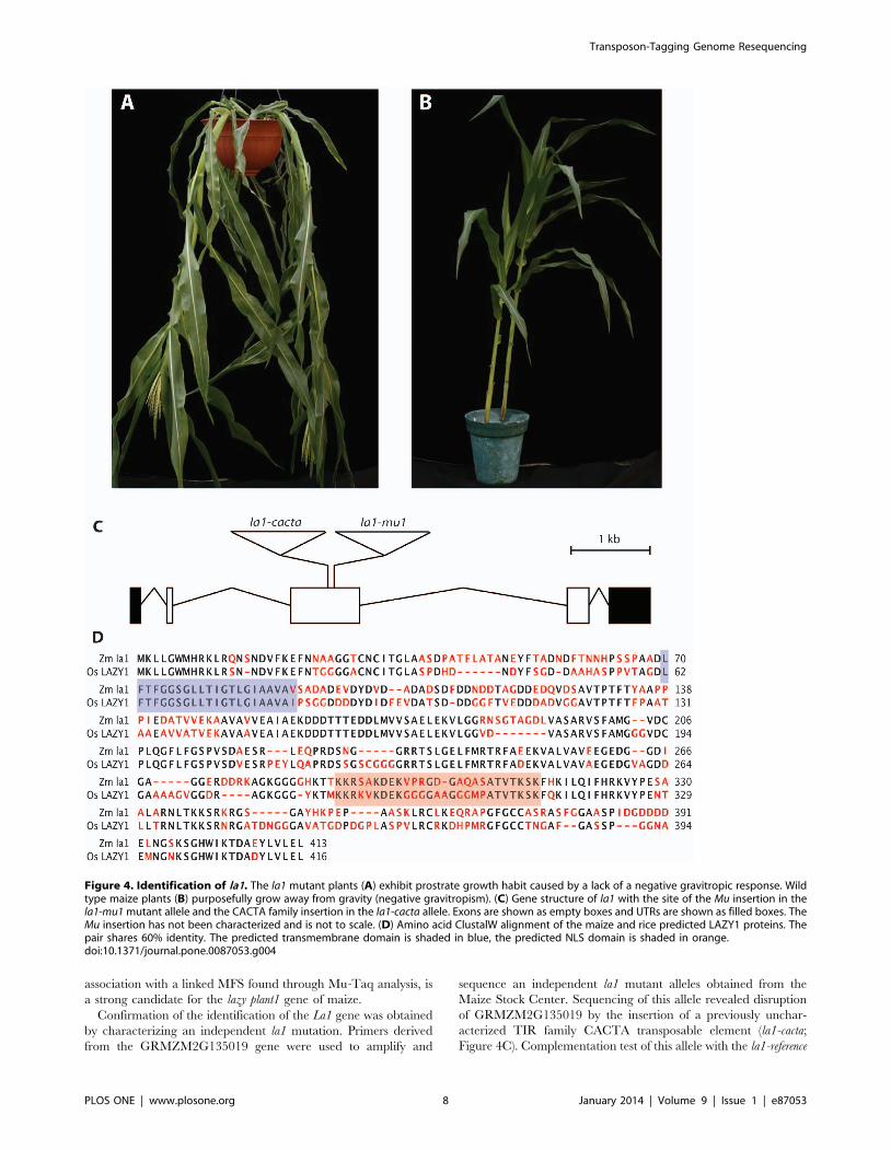
association with a linked MFS found through Mu-Taq analysis, is
a strong candidate for the lazy plant1 gene of maize.
Confirmation of the identification of the La1 gene was obtained
by characterizing an independent la1 mutation. Primers derived
from the GRMZM2G135019 gene were used to amplify and
sequence an independent la1 mutant alleles obtained from the
Maize Stock Center. Sequencing of this allele revealed disruption
of GRMZM2G135019 by the insertion of a previously unchar-
acterized TIR family CACTA transposable element (la1-cacta;
Figure 4C). Complementation test of this allele with the la1-reference
Figure 4. Identification of la1. The la1 mutant plants (A) exhibit prostrate growth habit caused by a lack of a negative gravitropic response. Wildtype maize plants (B) purposefully grow away from gravity (negative gravitropism). (C) Gene structure of la1 with the site of the Mu insertion in thela1-mu1 mutant allele and the CACTA family insertion in the la1-cacta allele. Exons are shown as empty boxes and UTRs are shown as filled boxes. TheMu insertion has not been characterized and is not to scale. (D) Amino acid ClustalW alignment of the maize and rice predicted LAZY1 proteins. Thepair shares 60% identity. The predicted transmembrane domain is shaded in blue, the predicted NLS domain is shaded in orange.doi:10.1371/journal.pone.0087053.g004
Transposon-Tagging Genome Resequencing
PLOS ONE | www.plosone.org 8 January 2014 | Volume 9 | Issue 1 | e87053

allele (Maize Genetics Cooperative Stock Center) provide further
confirmation that GRMZM2G135019 is indeed the la1 gene.
Characterization of the Maize La1 GeneThe maize La1 was predicted to encode a protein of 413 amino
acids of unknown function with a molecular weight of 43.6 kD
(Figure 4D). Prediction of transmembrane helices using both
HMMTOP [51,52] and TMpred [53] indicated a strong
likelihood of the presence of a transmembrane helix at position
70–91 (blue box in Figure 4D). Strong homology was also seen at
an NLS region previously described in rice LAZY1 at position 288–
313 (orange box in Figure 4D). The subcellular localization of
maize LA1 was determined with a fluorescent-tagged LA1 protein
(La1:Citrine). LA1 localized to the cell membrane and nucleus
during transient expression in N. benthamiana epidermal and
mesophyll cells (Figure 5A, 5B). The LA1-Citrine fusion protein
was predicted to be larger than the threshold for cytoplasm-to-
nucleus diffusion through the nuclear pores (, ,50 KDa), and
LA1-Citrine was not detected in the cytoplasm, suggesting that
nonspecific nuclear localization was unlikely. Western blotting also
revealed no evidence of smaller cleavage products containing
citrine that could cause artifactual nuclear signal (Figure 5D). To
confirm that the putative maize LA1 NLS domain was required
for nuclear localization, a deletion encompassing the NLS (amino-
acid residues 263–389) was generated (La1DNLS:Citrine). Dele-
tion of the NLS in LA1DNLS-Citrine abrogated nuclear
localization (Figure 5C). These results suggest that full-length
LA1 protein is present at both the cell membrane and nucleus,
though its molecular function remains unclear.
Discussion
The Mu-Taq method identified three out of four alleles known
to be caused by Mu insertions and four out of nine alleles
(including la1 and sk1) that were potentially caused by insertion of
a Mu element. This efficiency of known alleles (75%) is comparable
to that of previously published methods such as DLA (86%) and
the biotinylated oligonucleotide method (80%). The overall
efficiency of the method, however, was lower for previously
uncharacterized alleles. For example, it is likely that many of these
unknown alleles actually contain Mu insertions since their mutable
phenotype is dependent on Mutator activity. This would therefore
place a lower limit on the efficiency at 54%. From analysis of bulk
read data, we detect over 100 unique MFS in a plant known to
harbor Mu elements (Table 1). This is above the number detected
per genome by other methods and in the range predicted from the
average number of Mu elements in a genome. Given that the a1-
mum2 allele is confirmed to have a Mu insertion [54] and that other
alleles showing mutability are likely to be Mu-induced, certainly a
fraction of MFS have gone undetected. Based on the number of
unique MFS identified in each genome the depth of sequencing is
probably below saturation and additional sequencing may lead to
the identification of some of these alleles. Therefore, the efficiency
of our method at detecting a Mu insertion is between the estimates
of 54–75% [54].
Figure 5. Localization of LA1. LA1-Citrine localizes to the nucleus (arrows) and cell membrane in (A) N. benthamiana epidermal cells and (B) N.benthamiana mesophyll cells. Inset shows detail of the mesophyll cell membrane. (C) Deletion of the putative NLS in LA1DNLS-Citrine abrogatesnuclear localization. Scale bars are 20 uM. (D) Western blotting confirms expression of full length LA1-Citrine (lane 1) and LA1DNLS-Citrine (lane 2).doi:10.1371/journal.pone.0087053.g005
Transposon-Tagging Genome Resequencing
PLOS ONE | www.plosone.org 9 January 2014 | Volume 9 | Issue 1 | e87053

Besides depth of sequencing, there are many possible reasons for
not detecting a MFS when it is present in the genome. Some of
these reasons are technical in nature. One obvious reason would
be the absence of the conserved TaqaI site in the TIR. Based on
the conservation of TaqaI in all known mobile and potentially
mobile elements, we believe that this possibility is unlikely.
Another reason is that any individual TaqaI fragment generated
during restriction digestion may be too large for efficient cluster
formation, estimated to be greater than 800 bp. While the average
size of a TaqaI fragment, based on a random distribution of bases
and an AT-GC content of 50%, would be 256 bp, the actual size
will vary according to local and global base composition and a
quasi-random distribution of sites. Also, given that 130 bp of
adapter sequence are added during library construction, it is
possible that some TaqaI fragments will exceed the size necessary
to generate efficient clusters during the sequencing process.
Nevertheless, this is predicted to be a relatively minor fraction of
TaqaI fragments. A third possible reason for missing an individual
MFS is the problem of TaqaI digesting too close to the end of the
TIR sequence to identify a MFS. Since we are mapping MFS
using the adjacent 31 nt, the frequency of smaller size fragments is
not expected to be significant. In the specific case of Mu elements
and TaqaI, the terminal two bases of the TIR are part of the TaqaI
recognition site. Therefore, on average, one out of sixteen TIR
ends would reconstitute the full TaqaI site and be cut directly after
the TIR. These fragments would have no detectable MFS.
It is also possible that the map position of an individual MFS is
not accurate due to mapping errors or reference genome
limitations. Our current analysis includes only the single, most
significant mapped hit. Given that variation exists between the
maize lines used in this study and the inbred B73 reference
genome as well as the relatively high Illumina sequencing error
rate, some MFS may not have been properly mapped. In the short
term, reducing the stringency of possible matches and mapping
both ends of the paired-end reads may resolve this issue. A longer-
term solution would be to continue to characterize and catalog the
variation found in maize lines. Nevertheless, for these issues to be
relevant, both ends of the transposon would have to suffer from
one or more of these problems to cause the element to be
completely absent from a particular MFS dataset.
Another issue with the efficiency of MFS detection is depth of
read coverage and absence of coverage for some areas of the
genome. As previously mentioned in cases where the gene went
undetected in the MFS reads, it is possible that read depth was
insufficient to detect all MFS sequences. In our experiments we
achieve an overall depth of coverage of approximately 1X.
However, since only fragments adjacent to a TaqaI sites were
sequenced, our effective genome size is considerably smaller and
our effective depth of coverage much higher for these regions. Our
estimate of 6.5X enrichment for Mu TIR fragments should
enhance the effectiveness of this method to detect individual MFS.
Evidence that depth of coverage may not be a significant issue is
that the allele with the lowest number of total paired-end reads
(bz2-mVW4) was detected, but the allele with the second highest
number of total paired-end reads (a1-mum1) was not. Therefore,
increasing read depth may not be the only solution sought to
resolve the issue of MFS detection.
One biological reason for failing to identify an MFS-associated
gene is that the mutant allele may not contain a Mu element. Since
Mu is mobile, it may no longer be associated with a particular
allele. Mu elements frequently insert and excise imperfectly,
causing loss-of-function alleles even though the Mu element is no
longer present. In our study, nine alleles derived from Mutator lines
were not previously characterized at the molecular level to confirm
Mu insertions in the gene. Nevertheless, the mutable phenotype
associated with many of these alleles makes it likely that a Mu
insertion is the causative agent. Yet, only four of these nine alleles
were found to be associated with a particular MFS. It has been
well documented that many mutations recovered from Mu lines do
not contain heritable Mu insertions. For instance, at the liguleless1
locus of maize, two alleles derived from Mutator stocks did not
contain Mu insertions [55], while at the tasselseed1 locus, only two
out of four alleles derived from Mutator lines contained Mu
insertions [56]. If the alleles used in this study did not contain a Mu
element, this would explain the failure to detect at least some of
these genes in the dataset. At this point, the molecular basis of the
mutations in the alleles that were not identified need to be further
investigated to determine whether or not they actually contain Mu
elements with conserved TIR sequences. This additional data
would allow us to accurately assess the effectiveness of our
protocol.
Comparison to Other MethodsThree other methods have been reported to massively identify
MFS using next-generation sequencing. Though successful, these
methods rely on imperfectly conserved sequences in the Mu TIR
(Figure 1). The DLA method [30] uses a set of degenerate primers
to amplify out of the Mu TIR to create a library of MFS. Certain
Mu elements are likely to be selectively amplified because of
differential annealing efficiency of the various primers. The
biotinylated oligonucleotide method [31] relies on annealing over
a larger range of the TIR, expanding the possibility that divergent
sequences of certain Mu elements limit their capture and
sequencing. A recent method called Mu-seq incorporates a single
23 base, 12-fold-degenerate Mu TIR primer to target fragment
amplification [57]. In comparison, our method did not rely on
degenerate Mu primers but rather on anchoring reads to a highly
conserved of a 4 bp TaqaI site, which is located in the MURA
transposase binding site. This site has been perfectly conserved in
all active Mu elements characterized to date. In addition, our
method would identify MFS from any unknown Mu elements as
they likely contain the conserved TIR TaqaI site. Seven new Mu
elements were recently identified [58] and it seems likely that other
Mu elements have yet to be identified. In our method, the
identification of TIR tags was incorporated into an algorithm that
could be easily adapted to include additional TIR elements and to
modulate the stringency and flexibility of TIR identification.
One of the main advantages of the Mu-Taq strategy is its
simplicity. Standard Illumina reagents and adapters can be used
for libraries and sequencing with little protocol modification.
Other published methods are cumbersome and require numerous
steps to prepare sequencing libraries or complicated post-library
enrichment methods. For instance, the DLA method requires a
dideoxynucleotide blocking step as well as an extra PCR
amplification, introducing biases and read redundancy into the
libraries. The biotinylated oligonucleotide method requires two
rounds of hybridization as well as an extra amplification, also
introducing amplification bias and read redundancy. Mu-seq
employs three rounds of PCR amplification and special adapters.
In contrast, our Mu-Taq method requires only the typical steps –
end-repair, adenylation, and ligation – used during standard
Illumina library construction. Next-generation library protocols
also require a size fractionation step. In this method, instead of the
standard random DNA fragmentation by sonication or nebuliza-
tion, a simple restriction digestion step was employed as both a size
fractionation and enrichment step. Once end-repaired and
adenylated, these fragments can be ligated to standard Illumina
adapters and amplified in a single low cycle PCR reaction.
Transposon-Tagging Genome Resequencing
PLOS ONE | www.plosone.org 10 January 2014 | Volume 9 | Issue 1 | e87053

Subsequently, the Mu-Taq method incorporates a custom
bioinformatics pipeline to identify Mu TIR sequences to extract
MFS and standard algorithms to map their corresponding MFS.
While other NGS methods generate a much higher percentage
of reads that correspond to MFS, their exceptional coverage depth
is unnecessary for some applications such as single gene
identification. Even though in our method only 0.00204% of
reads correspond to MFS, mutations in eight independent plant
genomes were identified from reads derived from a single Illumina
HiSeq lane. With the ever-improving efficiency of NGS platforms,
an increase in read depth will become even more superfluous for
regular gene cloning applications. While larger MFS screening
projects may wish to identify more alleles in a single run using an
MFS amplification protocol, our method is ideal for identifying
single or multiple Mu-tagged genes of interest.
Adaptability of Mu-Taq to Other Transposon FamiliesThe Mu-Taq method is readily adaptable to other transposon
systems, and indeed any type of known DNA insertion, in
organisms besides plants. The only requirement is the presence of
a suitable and highly conserved restriction site close to the end of
the insertion. The ‘‘Dynamic TIR Finder’’ algorithm has been
written to accept a Fasta file of any set of TIR tags and to parse out
all corresponding transposon flanking sequence reads. Therefore,
in principle, this method is readily adaptable to any species, and to
any family of transposable elements with conserved terminal
elements. In addition, the method should be readily adaptable to
other sequencing platforms. In this way, the Mu-Taq method
described here represents a general and flexible strategy for
identifying transposon-tagged genes in the ever-progressing NGS
field.
Application of Mu-Taq Cloning to Identify andCharacterize Maize la1
The application of the Mu-Taq method effectively identified the
maize la1 gravitropism gene in two biological replicates of an allele
derived from a Mutator line. We were able to further identify a
second mutant la1 allele that failed to complement the la1-reference
that contained a previously uncharacterized CACTA transposon
insertion. We show that maize La1 shares significant homology to
its recently identified rice ortholog [48]. Concurrent with this
study, Dong et al. (2013) have also reported on the cloning of the
maize La1 gene using a Mutator-derived allele [59]. Interestingly,
the mapping and cloning strategy employed required a time- and
labor-intensive effort involving several generations and nearly
1300 plants. In our study, from mutant to candidate gene
identification required only a few weeks at a substantial savings in
time and expense.
Both our study and Dong et al. [59] show that
GRMZM2G135019 corresponds to the maize La1 gene and
LA1 protein localizes to the nucleus and cell membrane, a
localization profile previously reported for both the rice and
Arabidopsis La1 orthologs [48,60]. Using sequence homology
between rice and maize La1, we confirm a putative NLS required
for nuclear localization of maize LA1. The rice LAZY1 gene has
been shown to affect gravitropism through asymmetric auxin
distribution in the developing plant [48,61]. Similar perturbations
of auxin transport have been reported in maize la1 plants [59],
and the la1 phenotype was shown to be auxin dependent in the
original investigations into the nature of the mutant in the 1930s
[45]. Even though the effector (auxin) is known, there still remain
significant questions regarding how la1 actually senses gravity.
Although trafficking of membrane-bound LA1 to the nucleus
could hypothetically mediate auxin signaling, we are unable to
detect shuttling of LA1 by confocal microscopy. Furthermore, a
recent study showed that deletion of the AtLAZY1 NLS did not
prevent rescue of the atlazy1 mutant phenotype in Arabidopsis,
leaving the functional relevance of nuclear LA1 unclear [60].
More complete understanding of LA1 activity will certainly be a
productive area of future research on gravitropism.
Supporting Information
Table S1 Mutant alleles used in this study.
(DOCX)
Table S2 Barcoded adapters used in this study.
(DOCX)
Table S3 Primers used in this study.
(DOCX)
Table S4 Restriction enzyme choices in the first 70 bpof Mu TIRs.
(DOCX)
File S1 MuTaq Supplementals.
(ZIP)
Acknowledgments
The authors wish to thank Yingchun Tong, Jorge Duitama, Christopher
Bolick, and Christopher Heffelfinger for technical assistance and
suggestions to the manuscript.
Author Contributions
Conceived and designed the experiments: TPH APH AT JPM SLD TPH
APK AT JPM SLD. Performed the experiments: TPH APH AT CF MAM
JPM TPH APK AT CF MAM JPM. Analyzed the data: TPH APH AT CF
MAM JPM SLD TPH APK AT CF MAM JPM SLD. Contributed
reagents/materials/analysis tools: TPH APH AT TPH APK AT JT.
Wrote the paper: TPH APH SLD TPH APK SLD.
References
1. Robertson DS (1978) Characterization of a mutator system in maize. Mutat Res
51: 21–28.
2. Bennetzen JL, Swanson J, Taylor WC, Freeling M (1984) DNA insertion in the
first intron of maize Adh1 affects message levels: cloning of progenitor and
mutant Adh1 alleles. Proc Natl Acad Sci U S A 81: 4125–4128.
3. Barker RF, Thompson DV, Talbot DR, Swanson J, Bennetzen JL (1984)
Nucleotide sequence of the maize transposable element Mul. Nucleic Acids Res
12: 5955–5967.
4. Bennetzen JL (1984) Transposable element Mu1 is found in multiple copies only
in Robertson’s Mutator maize lines. J Mol Appl Genet 2: 519–524.
5. Walbot V, Rudenko GN (2002) MuDR/Mu Transposable Elements of Maize. In:
Craig NL, Craigie R, Gellert M, Lambowitz AM, editors. Mobile DNA II.
Washington, D.C.: ASM Press. 533–564.
6. Chomet P, Lisch D, Hardeman KJ, Chandler VL, Freeling M (1991)
Identification of a regulatory transposon that controls the Mutator transposable
element system in maize. Genetics 129: 261–270.
7. Benito MI, Walbot V (1997) Characterization of the maize Mutator transposable
element MURA transposase as a DNA-binding protein. Mol Cell Biol 17: 5165–
5175.
8. Chandler VL, Hardeman KJ (1992) The Mu elements of Zea mays. Adv Genet
30: 77–122.
9. Bennetzen JL, Springer PS, Cresse AD, Hendrickx M (1993) Specificity and
regulation of the mutator transposable element system in maize. Critical Reviews
in Plant Sciences 12: 57–95.
10. Bennetzen JL (1996) The Mutator transposable element system of maize. Curr
Top Microbiol Immunol 204: 195–229.
11. Lisch D (2002) Mutator transposons. Trends Plant Sci 7: 498–504.
Transposon-Tagging Genome Resequencing
PLOS ONE | www.plosone.org 11 January 2014 | Volume 9 | Issue 1 | e87053
12. Walbot V, Warren C (1988) Regulation of Mu element copy number in maize
lines with an active or inactive Mutator transposable element system. Mol GenGenet 211: 27–34.
13. Alleman M, Freeling M (1986) The Mu transposable elements of maize:
evidence for transposition and copy number regulation during development.Genetics 112: 107–119.
14. Cresse AD, Hulbert SH, Brown WE, Lucas JR, Bennetzen JL (1995) Mu1-related transposable elements of maize preferentially insert into low copy
number DNA. Genetics 140: 315–324.
15. Hanley S, Edwards D, Stevenson D, Haines S, Hegarty M, et al. (2000)Identification of transposon-tagged genes by the random sequencing of Mutator-
tagged DNA fragments from Zea mays. Plant J 23: 557–566.
16. Raizada MN, Nan GL, Walbot V (2001) Somatic and germinal mobility of the
RescueMu transposon in transgenic maize. Plant Cell 13: 1587–1608.
17. Schnable PS, Ware D, Fulton RS, Stein JC, Wei F, et al. (2009) The B73 maizegenome: complexity, diversity, and dynamics. Science 326: 1112–1115.
18. O’Reilly C, Shepherd NS, Pereira A, Schwarz-Sommer Z, Bertram I, et al.(1985) Molecular cloning of the a1 locus of Zea mays using the transposable
elements En and Mu1. EMBO J 4: 877–882.
19. McLaughlin M, Walbot V (1987) Cloning of a mutable bz2 allele of maize bytransposon tagging and differential hybridization. Genetics 117: 771–776.
20. Bensen RJ, Johal GS, Crane VC, Tossberg JT, Schnable PS, et al. (1995)Cloning and characterization of the maize An1 gene. Plant Cell 7: 75–84.
21. Das L, Martienssen R (1995) Site-selected transposon mutagenesis at the hcf106locus in maize. Plant Cell 7: 287–294.
22. May BP, Liu H, Vollbrecht E, Senior L, Rabinowicz PD, et al. (2003) Maize-
targeted mutagenesis: A knockout resource for maize. Proc Natl Acad Sci U S A100: 11541–11546.
23. Frey M, Stettner C, Gierl A (1998) A general method for gene isolation intagging approaches: amplification of insertion mutagenised sites (AIMS). The
Plant Journal 12: 717–721.
24. Settles AM, Latshaw S, McCarty DR (2004) Molecular analysis of high-copyinsertion sites in maize. Nucleic Acids Res 32: e54.
25. McCarty DR, Settles AM, Suzuki M, Tan BC, Latshaw S, et al. (2005) Steady-state transposon mutagenesis in inbred maize. Plant J 44: 52–61.
26. Settles AM, Holding DR, Tan BC, Latshaw SP, Liu J, et al. (2007) Sequence-
indexed mutations in maize using the UniformMu transposon-taggingpopulation. BMC Genomics 8: 116.
27. Raizada MN (2003) RescueMu protocols for maize functional genomics.Methods Mol Biol 236: 37–58.
28. Fernandes J, Dong Q, Schneider B, Morrow DJ, Nan GL, et al. (2004) Genome-
wide mutagenesis of Zea mays L. using RescueMu transposons. Genome Biol 5:R82.
29. Project TUM (2011) Project Documentation & Protocols: UniformMuTransposon Resource.
30. Liu S, Dietrich CR, Schnable PS (2009) DLA-based strategies for cloning
insertion mutants: cloning the gl4 locus of maize using Mu transposon taggedalleles. Genetics 183: 1215–1225.
31. Williams-Carrier R, Stiffler N, Belcher S, Kroeger T, Stern DB, et al. (2010) Useof Illumina sequencing to identify transposon insertions underlying mutant
phenotypes in high-copy Mutator lines of maize. Plant J 63: 167–177.
32. Jenkins M, Gerhardt F (1931) A gene influencing the composition of the culm in
maize. Iowa Ag Exp Sta Research Bull 138: 121–151.
33. Chen J, Dellaporta S (1993) Urea-based plant DNA miniprep. In: Freeling M,Walbot V, editors. The Maize Handbook. New York: Springer-Verlag. 526–
527.
34. Li H, Durbin R (2009) Fast and accurate short read alignment with Burrows-
Wheeler transform. Bioinformatics 25: 1754–1760.
35. Altschul SF, Gish W, Miller W, Myer EW, Lipman DJ (1990) Basic localalignment search tool. J Mol Biol 215: 403–410.
36. Hayward A, Padmanabhan M, Dinesh-Kumar SP (2011) Virus-induced genesilencing in nicotiana benthamiana and other plant species. Methods Mol Biol
678: 55–63.
37. Bentley DR, Balasubramanian S, Swerdlow HP, Smith GP, Milton J, et al.(2008) Accurate whole human genome sequencing using reversible terminator
chemistry. Nature 456: 53–59.
38. Illumina (2010) Genomic Sequencing.
39. Palmer LE, Rabinowicz PD, O’Shaughnessy AL, Balija VS, Nascimento LU, et
al. (2003) Maize genome sequencing by methylation filtration. Science 302:2115–2117.
40. Zhao X, Chai Y, Liu B (2007) Epigenetic inheritance and variation of DNAmethylation level and pattern in maize intra-specific hybrids. Plant Science 172:
930–938.
41. Thompson JD, Higgins DG, Gibson TJ (1994) CLUSTAL W: improving the
sensitivity of progressive multiple sequence alignment through sequenceweighting, position-specific gap penalties and weight matrix choice. Nucleic
Acids Res 22: 4673–4680.
42. Langmead B, Trapnell C, Pop M, Salzberg SL (2009) Ultrafast and memory-efficient alignment of short DNA sequences to the human genome. Genome Biol
10: R25.43. Langmead B, Salzberg SL (2012) Fast gapped-read alignment with Bowtie 2.
Nat Methods 9: 357–359.
44. van Overbeek J (1936) ‘‘Lazy,’’ an a-geotropic form of maize. The Journal ofHeredity 29: 339–341.
45. van Overbeek J (1938) ‘‘Laziness’’ in maize due to abnormal distribution ofgrowth hormone. The Journal of Heredity 29: 339–341.
46. Shafer J (1939) Physiology of Lazy Corn. Botanical Gazette 101: 66–80.47. Darwin C, Darwin F (1880) The Power of Movement in Plants. London: John
Murray.
48. Li P, Wang Y, Qian Q, Fu Z, Wang M, et al. (2007) LAZY1 controls rice shootgravitropism through regulating polar auxin transport. Cell Res 17: 402–410.
49. Soderlund C, Bomhoff M, Nelson WM (2011) SyMAP v3.4: a turnkey syntenysystem with application to plant genomes. Nucleic Acids Res 39: e68.
50. Altschul SF, Madden TL, Schaffer AA, Zhang J, Zhang Z, et al. (1997) Gapped
BLAST and PSI-BLAST: a new generation of protein database searchprograms. Nucleic Acids Res 25: 3389–3402.
51. Tusnady GE, Simon I (1998) Principles governing amino acid composition ofintegral membrane proteins: application to topology prediction. J Mol Biol 283:
489–506.52. Tusnady GE, Simon I (2001) The HMMTOP transmembrane topology
prediction server. Bioinformatics 17: 849–850.
53. Hofmann K, Stoffel W (1993) TMbase- A database of membrane spanningproteins segments. The Journal of Biological Chemistry 374: 166.
54. Pooma W, Gersos C, Grotewold E (2002) Transposon insertions in the promoterof the Zea mays a1 gene differentially affect transcription by the Myb factors P
and C1. Genetics 161: 793–801.
55. Moreno MA, Harper LC, Krueger RW, Dellaporta SL, Freeling M (1997)liguleless1 encodes a nuclear-localized protein required for induction of ligules
and auricles during maize leaf organogenesis. Genes Dev 11: 616–628.56. Acosta IF, Laparra H, Romero SP, Schmelz E, Hamberg M, et al. (2009)
tasselseed1 is a lipoxygenase affecting jasmonic acid signaling in sexdetermination of maize. Science 323: 262–265.
57. McCarty DR, Latshaw S, Wu S, Suzuki M, Hunter CT, et al. (2013) Mu-seq:
Sequence-Based Mapping and Identification of Transposon Induced Mutations.PLoS One 8: e77172.
58. Tan BC, Chen Z, Shen Y, Zhang Y, Lai J, et al. (2011) Identification of an activenew mutator transposable element in maize. G3 (Bethesda) 1: 293–302.
59. Dong Z, Jiang C, Chen X, Zhang T, Ding L, et al. (2013) Maize LAZY1
Mediates Shoot Gravitropism and Inflorescence Development through Regu-lating Auxin Transport, Auxin Signaling, and Light Response. Plant Physiol
163: 1306–1322.60. Yoshihara T, Spalding EP, Iino M (2013) AtLAZY1 is a signaling component
required for gravitropism of the Arabidopsis thaliana inflorescence. Plant J 74:267–279.
61. Yoshihara T, Iino M (2007) Identification of the gravitropism-related rice gene
LAZY1 and elucidation of LAZY1-dependent and -independent gravitysignaling pathways. Plant Cell Physiol 48: 678–688.
62. Dietrich CR, Cui F, Packila ML, Li J, Ashlock DA, et al. (2002) Maize Mutransposons are targeted to the 59 untranslated region of the gl8 gene and
sequences flanking Mu target-site duplications exhibit nonrandom nucleotide
composition throughout the genome. Genetics 160: 697–716.63. Taylor LP, Walbot V (1987) Isolation and characterization of a 1.7-kb
transposable element from a mutator line of maize. Genetics 117: 297–307.64. Chen CH, Oishi KK, Kloeckener-Gruissem B, Freeling M (1987) Organ-specific
expression of maize Adh1 is altered after a Mu transposon insertion. Genetics
116: 469–477.65. Talbert LE, Patterson GI, Chandler VL (1989) Mu transposable elements are
structurally diverse and distributed throughout the genus Zea. J Mol Evol 29:28–39.
66. Schnable PS, Peterson PA, Saedler H (1989) The bz-rcy allele of the Cytransposable element system of Zea mays contains a Mu-like element insertion.
Mol Gen Genet 217: 459–463.
67. Fleenor D, Spell M, Robertson D, Wessler S (1990) Nucleotide sequence of themaize Mutator element, Mu8. Nucleic Acids Res 18: 6725.
68. James MG, Scanlon MJ, Qin M, Robertson DS, Myers AM (1993) DNAsequence and transcript analysis of transposon MuA2, a regulator of Mutator
transposable element activity in maize. Plant Mol Biol 21: 1181–1185.
Transposon-Tagging Genome Resequencing
PLOS ONE | www.plosone.org 12 January 2014 | Volume 9 | Issue 1 | e87053
Copyright © 2022 FDOKUMEN