Potential Roles of Peroxisomes in Alzheimer's Disease and in Dementia of the Alzheimer's Type
The activity of the glyoxylate cycle in peroxisomes ofCandida albicans depends on a functionalb-oxidation pathway: evidence for reduced metabolitetransport across the peroxisomal membrane
Katarzyna Piekarska,13 Guy Hardy,1 Els Mol,1 Janny van den Burg,1
Karin Strijbis,1 Carlo van Roermund,2 Marlene van den Berg1
and Ben Distel1
Correspondence
Ben Distel
1Department of Medical Biochemistry, Academic Medical Center, Meibergdreef 15, 1105 AZAmsterdam, The Netherlands
2Department of Genetic Metabolic Diseases, Academic Medical Center, Meibergdreef 15, 1105 AZAmsterdam, The Netherlands
Received 6 May 2008
Revised 25 June 2008
Accepted 25 June 2008
The glyoxylate cycle, a metabolic pathway required for generating C4 units from C2 compounds, is
an important factor in virulence, in both animal and plant pathogens. Here, we report the
localization of the key enzymes of this cycle, isocitrate lyase (Icl1; EC 4.1.3.1) and malate
synthase (Mls1; EC 2.3.3.9), in the human fungal pathogen Candida albicans.
Immunocytochemistry in combination with subcellular fractionation showed that both Icl1 and
Mls1 are localized to peroxisomes, independent of the carbon source used. Although Icl1 and
Mls1 lack a consensus type I peroxisomal targeting signal (PTS1), their import into peroxisomes
was dependent on the PTS1 receptor Pex5p, suggesting the presence of non-canonical targeting
signals in both proteins. Peroxisomal compartmentalization of the glyoxylate cycle is not
essential for proper functioning of this metabolic pathway because a pex5D/D strain, in which Icl1
and Mls1 were localized to the cytosol, grew equally as well as the wild-type strain on acetate and
ethanol. Previously, we reported that a fox2D/D strain that is completely deficient in fatty acid b-
oxidation, but has no peroxisomal protein import defect, displayed strongly reduced growth on
non-fermentable carbon sources such as acetate and ethanol. Here, we show that growth of the
fox2D/D strain on these carbon compounds can be restored when Icl1 and Mls1 are relocated to
the cytosol by deleting the PEX5 gene. We hypothesize that the fox2D/D strain is disturbed in
the transport of glyoxylate cycle products and/or acetyl-CoA across the peroxisomal membrane
and discuss the possible relationship between such a transport defect and the presence of giant
peroxisomes in the fox2D/D mutant.
INTRODUCTION
The glyoxylate cycle, present in bacteria, yeasts and plants,allows utilization of non-fermentable carbon sources suchas acetate, ethanol and fatty acids by converting theproduced acetyl-CoA into C4 compounds that can be usedfor the biosynthesis of macromolecules (Kornberg & Krebs,1957). This function has been confirmed through theanalysis of bacterial and fungal mutants lacking eitherisocitrate lyase (Icl1; EC 4.1.3.1) or malate synthase (Mls1;
EC 2.3.3.9), the key enzymes of the glyoxylate cycle. Loss ofIcl1 or Mls1 results in the inability of these mutants togrow on acetate, ethanol or fatty acids (Hartig et al., 1992;Lorenz & Fink, 2001; McKinney et al., 2000; Munoz-Elias& McKinney, 2005; Pellicer et al., 1999). There is a renewedinterest in the glyoxylate cycle that is based on recentobservations suggesting a key role for this metabolicpathway in survival of bacterial and fungal pathogenswithin macrophages, phagocytic cells of the innate immunesystem that form the first line of defence against microbialinfections. Whole-genome DNA microarrays have shownan upregulation of glyoxylate cycle genes and concomitantinduction of genes associated with gluconeogenesis whenCandida albicans, a human fungal pathogen, is internalizedby macrophages (Lorenz et al., 2004). Deletion of the ICL1
Abbreviations: Icl1, isocitrate lyase; Mls1, malate synthase; PEX,peroxisome biogenesis gene/protein; PTS1, peroxisomal targetingsignal 1; TCA, tricarboxylic acid.
3Present address: University of Manchester, The Michael Smith BuildingA1030, Oxford Road, Manchester M13 9PT, UK.
Microbiology (2008), 154, 3061–3072 DOI 10.1099/mic.0.2008/020289-0
2008/020289 G 2008 SGM Printed in Great Britain 3061
gene resulted in a mutant C. albicans strain that showedreduced virulence in a mouse model, providing strongsupport for the requirement of the glyoxylate cycle forpathogenesis of this fungus (Lorenz & Fink, 2001).Similarly, the persistence of Mycobacterium tuberculosis inmacrophages is dependent on a functional glyoxylate cycle(McKinney et al., 2000; Munoz-Elias & McKinney, 2005).For both C. albicans and M. tuberculosis it has beensuggested that the organism relies heavily on lipidcatabolism for survival within the human host (Boshoff& Barry, 2005; Lorenz & Fink, 2002). However, we haverecently shown that fatty acid metabolism is not essentialfor virulence of C. albicans, indicating that in vivo theacetyl-CoA that feeds the glyoxylate cycle may be derivedfrom other, simple, carbon sources (Piekarska et al., 2006).In support of this, Ramırez & Lorenz (2007) recentlyreported that the ability to utilize different non-ferment-able carbon sources contributes to the virulence of C.albicans. Since the metabolism of these carbon compoundsrequires a functional glyoxylate cycle and this metabolicpathway is absent from mammals, Icl1 and Mls1 arepotential targets for the development of drugs to combatbacterial and fungal infections.
The glyoxylate cycle consists of five enzymic activities, twoof which are unique to the cycle, namely Icl1 and Mls1,while the other three activities, citrate synthase (EC2.3.3.1), aconitase (EC 4.2.1.3) and malate dehydrogenase(EC 1.1.1.37), are shared with the tricarboxylic acid (TCA)cycle and are often carried out by isoenzymes. In the C.albicans genome, single genes exist for Icl1 (orf19.6844),Mls1 (orf19.4833) and citrate synthase (orf19.4393),whereas there are three encoding malate dehydrogenase(orf19.7481, orf19.4602 and orf19.5223) and two foraconitase (orf19.6385 and orf19.6632). Studies in fungihave shown that the key enzymes of the glyoxylate cycle,Icl1 and Mls1, are often compartmentalized in peroxi-somes, the other enzymic steps being extra-peroxisomal(either cytosolic or mitochondrial) (Hikida et al., 1991;Maeting et al., 1999; Tanaka & Ueda, 1993; Titorenko et al.,1998; Valenciano et al., 1996). Saccharomyces cerevisiaeseems to be an exception to this rule: Icl1 is a cytosolicenzyme (McCammon et al., 1990) and Mls1 is localizedeither to the peroxisome or to the cytosol depending on thegrowth conditions (Kunze et al., 2002). These data inconjunction with the observation that S. cerevisiae pexmutants lacking functional peroxisomes can grow onethanol and acetate as sole carbon source imply that theglyoxylate cycle can function in the cytosol, at least in S.cerevisiae.
The import of proteins into peroxisomes is mediated bytwo cycling receptors: Pex5p and Pex7p (reviewed byPurdue & Lazarow, 2001). In the cytosol, Pex5p binds toproteins carrying a type I peroxisomal targeting signal(PTS1), which is a carboxyl-terminal tripeptide with theconsensus sequence S/A/C-K/R/H-L/M (Gould et al., 1989;Swinkels et al., 1992). However, depending on the speciesand protein under study the carboxyl-terminal tripeptide
may deviate from the consensus, and residues justupstream of it also can contribute to Pex5p binding andperoxisomal targeting (Lametschwandtner et al., 1998;Purdue & Lazarow, 1996). Proteins delivered to theperoxisome via the Pex7p receptor possess a PTS2, abipartite amino acid motif with the consensus sequence R/K-L/V/I-x5-H/Q-L/A (Gietl et al., 1994; Glover et al.,1994b) present in the amino terminus of these proteins. Anumber of proteins can be imported into peroxisomesindependent of a recognizable PTS1 or PTS2 (Elgersma etal., 1995; Klein et al., 2002; Ozimek et al., 2006; Small et al.,1988). Subsequent studies have shown that, although thetargeting signal is as yet not characterized, Pex5p mediatesthe peroxisomal sorting of these proteins (Elgersma et al.,1995; Klein et al., 2002; Ozimek et al., 2006). Finally, someproteins can reach the organelle by association withanother subunit or protein that possesses a functionaltargeting signal, a process referred to as ‘piggy backing’(Glover et al., 1994a; McNew & Goodman, 1994; Yang etal., 2001). The PTS1 and PTS2 pathways converge at theperoxisomal membrane, where the cargo-loaded receptorsdock onto a complex consisting of Pex13p and Pex14p(and Pex17p in S. cerevisiae). Subsequently, the cargo isreleased and translocated across the peroxisomal mem-brane, after which the receptors recycle back to the cytosolfor a new round of import (Purdue & Lazarow, 2001).Although the early steps in peroxisomal protein import arerather well defined, very little is known about the eventsfollowing docking.
Here we have studied the localization of the two keyenzymes of the glyoxylate cycle, Icl1 and Mls1, in thehuman fungal pathogen C. albicans and show that bothenzymes are localized to peroxisomes in a Pex5p-dependent way. We also provide evidence that theglyoxylate cycle no longer functions in giant peroxisomesof the fatty acid b-oxidation mutant fox2D/D. The activityof the glyoxylate cycle in the fox2D/D mutant is restoredwhen Icl1 and Mls1 are relocated to the cytosol throughdeletion of the PEX5 gene in this strain. Our data show thatthe glyoxylate cycle can function in the cytosol also in C.albicans and suggest that the giant peroxisomes of thefox2D/D strain are impaired in metabolite transport acrossthe membrane. Possible mechanisms that may explain thereduced peroxisomal metabolite transport are discussed.
METHODS
Strains, media and culture conditions. C. albicans and S. cerevisiae
strains used in this study are listed in Table 1. C. albicans deletion
strains are derivatives of BWP17 (Wilson et al., 1999). Gene deletions
in S. cerevisiae were constructed in BJ1991 (Jones, 1977). The solid
minimal medium used for selection and growth of both C. albicans
and S. cerevisiae transformants contained 0.67 % (w/v) yeast nitrogen
base (YNB) w/o amino acids (Difco), 2 % (w/v) glucose, 2 % (w/v)
agar, amino acids (20–30 mg ml21) and either 80 mg uridine ml21 (C.
albicans) or 20 mg uracil ml21 (S. cerevisiae) as needed. For
transformations using the dominant selection marker SAT1, a 24 h
incubation under non-selective conditions was applied prior to
K. Piekarska and others
3062 Microbiology 154
replica plating on YPD+Uri [2 % (w/v) bactopeptone, 1 % (w/v)
yeast extract, 2 % (w/v) glucose and 80 mg uridine ml21] with 200 mg
nourseothricin ml21 (clonNAT, Werner Bioagents). Plates used for
spot assays had the same composition and contained 2 % (w/v)
glucose, 2 % (v/v) ethanol, 2 % (v/v) glycerol, 2 % (w/v) sodium
citrate or 2 % (w/v) sodium acetate (pH 5.0) as a carbon source. The
liquid medium used for culturing of the cells for total protein
isolation, subcellular fractionation and immunoelectron microscopy
contained 0.5 % (w/v) potassium phosphate buffer, pH 6.0, 0.3 % (w/v)
yeast extract, 0.5 % (w/v) peptone and, as a carbon source, either 0.1 %
(v/v) oleate and 0.2 % (v/v) Tween 40, or 2 % (v/v) ethanol. All yeast
strains were grown at 28 uC unless otherwise stated.
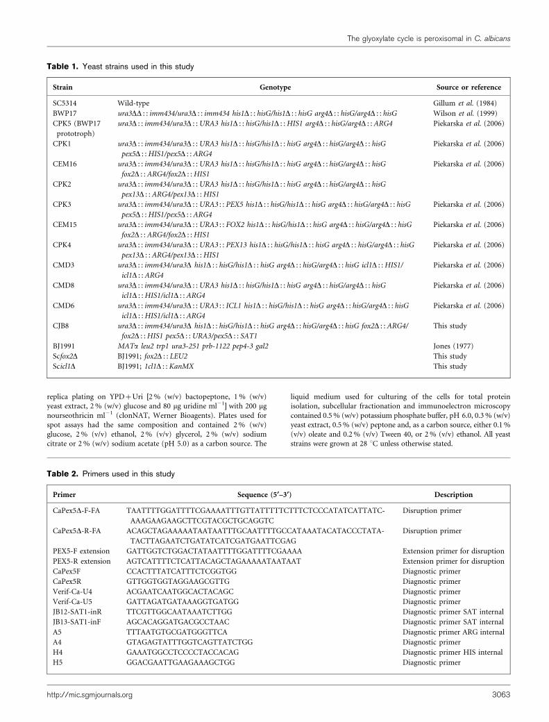
Table 1. Yeast strains used in this study
Strain Genotype Source or reference
SC5314 Wild-type Gillum et al. (1984)
BWP17 ura3DD : : imm434/ura3D : : imm434 his1D : : hisG/his1D : : hisG arg4D : : hisG/arg4D : : hisG Wilson et al. (1999)
CPK5 (BWP17
prototroph)
ura3D : : imm434/ura3D : : URA3 his1D : : hisG/his1D : : HIS1 arg4D : : hisG/arg4D : : ARG4 Piekarska et al. (2006)
CPK1 ura3D : : imm434/ura3D : : URA3 his1D : : hisG/his1D : : hisG arg4D : : hisG/arg4D : : hisG
pex5D : : HIS1/pex5D : : ARG4
Piekarska et al. (2006)
CEM16 ura3D : : imm434/ura3D : : URA3 his1D : : hisG/his1D : : hisG arg4D : : hisG/arg4D : : hisG
fox2D : : ARG4/fox2D : : HIS1
Piekarska et al. (2006)
CPK2 ura3D : : imm434/ura3D : : URA3 his1D : : hisG/his1D : : hisG arg4D : : hisG/arg4D : : hisG
pex13D : : ARG4/pex13D : : HIS1
CPK3 ura3D : : imm434/ura3D : : URA3 : : PEX5 his1D : : hisG/his1D : : hisG arg4D : : hisG/arg4D : : hisG
pex5D : : HIS1/pex5D : : ARG4
Piekarska et al. (2006)
CEM15 ura3D : : imm434/ura3D : : URA3 : : FOX2 his1D : : hisG/his1D : : hisG arg4D : : hisG/arg4D : : hisG
fox2D : : ARG4/fox2D : : HIS1
Piekarska et al. (2006)
CPK4 ura3D : : imm434/ura3D : : URA3 : : PEX13 his1D : : hisG/his1D : : hisG arg4D : : hisG/arg4D : : hisG
pex13D : : ARG4/pex13D : : HIS1
Piekarska et al. (2006)
CMD3 ura3D : : imm434/ura3D his1D : : hisG/his1D : : hisG arg4D : : hisG/arg4D : : hisG icl1D : : HIS1/
icl1D : : ARG4
Piekarska et al. (2006)
CMD8 ura3D : : imm434/ura3D : : URA3 his1D : : hisG/his1D : : hisG arg4D : : hisG/arg4D : : hisG
icl1D : : HIS1/icl1D : : ARG4
Piekarska et al. (2006)
CMD6 ura3D : : imm434/ura3D : : URA3 : : ICL1 his1D : : hisG/his1D : : hisG arg4D : : hisG/arg4D : : hisG
icl1D : : HIS1/icl1D : : ARG4
Piekarska et al. (2006)
CJB8 ura3D : : imm434/ura3D his1D : : hisG/his1D : : hisG arg4D : : hisG/arg4D : : hisG fox2D : : ARG4/
fox2D : : HIS1 pex5D : : URA3/pex5D : : SAT1
This study
BJ1991 MATa leu2 trp1 ura3-251 prb-1122 pep4-3 gal2 Jones (1977)
Scfox2D BJ1991; fox2D : : LEU2 This study
Scicl1D BJ1991; 1cl1D : : KanMX This study
Table 2. Primers used in this study
Primer Sequence (5§–3§) Description
CaPex5D-F-FA TAATTTTGGATTTTCGAAAATTTGTTATTTTTCTTTCTCCCATATCATTATC-
AAAGAAGAAGCTTCGTACGCTGCAGGTC
Disruption primer
CaPex5D-R-FA ACAGCTAGAAAAATAATAATTTGCAATTTTGCCATAAATACATACCCTATA-
TACTTAGAATCTGATATCATCGATGAATTCGAG
Disruption primer
PEX5-F extension GATTGGTCTGGACTATAATTTTGGATTTTCGAAAA Extension primer for disruption
PEX5-R extension AGTCATTTTCTCATTACAGCTAGAAAAATAATAAT Extension primer for disruption
CaPex5F CCACTTTATCATTTCTCGGTGG Diagnostic primer
CaPex5R GTTGGTGGTAGGAAGCGTTG Diagnostic primer
Verif-Ca-U4 ACGAATCAATGGCACTACAGC Diagnostic primer
Verif-Ca-U5 GATTAGATGATAAAGGTGATGG Diagnostic primer
JB12-SAT1-inR TTCGTTGGCAATAAATCTTGG Diagnostic primer SAT internal
JB13-SAT1-inF AGCACAGGATGACGCCTAAC Diagnostic primer SAT internal
A5 TTTAATGTGCGATGGGTTCA Diagnostic primer ARG internal
A4 GTAGAGTATTTGGTCAGTTATCTGG Diagnostic primer
H4 GAAATGGCCTCCCCTACCACAG Diagnostic primer HIS internal
H5 GGACGAATTGAAGAAAGCTGG Diagnostic primer
The glyoxylate cycle is peroxisomal in C. albicans
http://mic.sgmjournals.org 3063
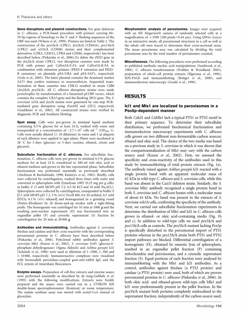
Gene disruptions and plasmid constructions. For gene deletionsin C. albicans, a PCR-based procedure with primers carrying 60–70 bp regions of homology to the 59 and 39 flanking sequences of theORF was used (Wilson et al., 1999). Primers are listed in Table 2. Theconstruction of the pex5D/D (CPK1), fox2D/D (CEM16), pex13D/D(CPK2) and icl1D/D (CDM8) strains and their complementedderivatives (CPK3, CEM15, CPK4 and CDM6, respectively) has beendescribed before (Piekarska et al., 2006).To delete the PEX5 gene inthe fox2D/D strain CPK12, two disruption cassettes were made byPCR with primer pair CaPex5D-F-FA and CaPex5D-R-FA incombination with extension primers (PEX5-F extension and PEX5-R extension) on plasmids pFA-URA and pFA-SAT1, respectively(Gola et al., 2003). The latter plasmid contains the dominant markerSAT1 that confers resistance to nourseothricin. Sequential trans-formation of these cassettes into CPK12 resulted in strain CJB8(fox2D/D pex5D/D). All C. albicans disruption strains were madeprototrophic by transformation of a linearized pLUBP vector, whichcontains the complete URA3 gene and the flanking IRO1 gene. The S.cerevisiae icl1D and fox2D strains were generated by one-step PCR-mediated gene disruption using KanMX and LEU2, respectively(Gueldener et al., 2002). All constructed strains were verified bydiagnostic PCR and Southern blotting.
Spot assay. Cells were pre-grown in minimal liquid mediumcontaining 0.3 % glucose for at least 24 h, washed with water andresuspended at a concentration of ~2.76107 cells ml21 (OD600 1).Cells were serially diluted (1 : 10 dilutions) in water and 5 ml aliquotsof each dilution were applied to agar plates. Plates were incubated at28 uC for 3 days (glucose) or 5 days (acetate, ethanol, citrate andglycerol).
Subcellular fractionation of C. albicans. For subcellular frac-tionation, C. albicans cells were pre-grown in minimal 0.3 % glucosemedium for at least 24 h, transferred to 200 ml rich oleic acid orethanol medium and grown to the late exponential phase. Subcellularfractionation was performed essentially as previously described(Aitchison & Rachubinski, 1990; Kamiryo et al., 1982). Briefly, cellswere collected by centrifugation, washed three times with water andconverted to spheroplasts with Zymolyase 100T (0.25 mg per g cells)in buffer Z (5 mM MOPS pH 7.2, 0.5 M KCl and 10 mM Na2SO3).Spheroplasts were collected by centrifugation, resuspended in buffer F[5.5 mM MOPS pH 7.2, 5 % (w/v) Ficoll 400, 0.6 M sorbitol, 0.5 mMEDTA, 0.1 % (v/v) ethanol] and homogenized in a grinding vessel(Potter–Elvejhem) by 20 down-and-up strokes with a tight-fittingpestle. The homogenate was centrifuged for 10 min at 1000 g and theresulting post-nuclear supernatant (H) was fractionated into anorganellar pellet (P) and cytosolic supernatant (S) fraction bycentrifugation for 20 min at 20 000 g.
Antibodies and immunoblotting. Antibodies against S. cerevisiaethiolase and catalase and their cross-reactivity with the correspondingperoxisomal proteins in C. albicans have been described before(Piekarska et al., 2006). Polyclonal rabbit antibodies against S.cerevisiae Mls1 (Kunze et al., 2002), S. cerevisiae Zwf1 (glucose-6-phosphate dehydrogenase) (Sigma Aldrich) and Ashbya gossypii Icl1(Schmidt et al., 1996) were used at dilutions of 1 : 1000, 1 : 500 and1 : 10 000, respectively. Immunoreactive complexes were visualizedwith horseradish peroxidase-coupled goat-anti-rabbit IgG and theECL system of Amersham Biosciences.
Enzyme assays. Preparation of cell-free extracts and enzyme assayswere performed essentially as described by de Jong-Gubbels et al.(1995) with the following modifications. Extracts were freshlyprepared and the assays were carried out in a UVIKON 820double-beam spectrophotometer (Kontron) at room temperature.The malate synthase assay was started with acetyl-CoA instead ofglyoxylate.
Morphometric analysis of peroxisomes. Images were acquired
with an SIS MegaviewII camera of randomly selected cells at a
magnification of 65500 (100 pixels50.44 mm). Using QWin (Leica)
in an interactive mode, all peroxisomal structures in a cell as well as
the whole cell were traced to determine their cross-sectional areas.
The mean peroxisome area was calculated by dividing the total
peroxisome area by the total number of peroxisomes counted.
Miscellaneous. The following procedures were performed according
to published methods: nucleic acid manipulations (Sambrook et al.,
1989), C. albicans transformation (Walther & Wendland, 2003),
preparation of whole-cell protein extracts (Elgersma et al., 1996),
SDS-PAGE and immunoblotting (Bottger et al., 2000), and
immunoelectron miscroscopy (Gould et al., 1990).
RESULTS
Icl1 and Mls1 are localized to peroxisomes in aPex5p-dependent manner
Both CaIcl1 and CaMls1 lack a typical PTS1 or PTS2 motif intheir primary sequence. To determine their subcellulardistribution, we performed biochemical fractionation andimmunoelectron miscroscopy experiments with C. albicanscells grown on two different non-fermentable-carbon sources:ethanol and oleic acid. The choice of the two media was basedon a previous study in S. cerevisiae in which it was shown thatthe compartmentalization of Mls1 may vary with the carbonsource used (Kunze et al., 2002). First, we assessed thespecificity and cross-reactivity of the antibodies used in thisstudy by immunoblotting of total protein extracts (Fig. 1a).The antibody raised against Ashbya gossypii Icl1 reacted with asingle protein band with an apparent molecular mass of62 kDa in wild-type C. albicans and S. cerevisiae cells, while thisband was absent in the Caicl1 deletion strain. Similarly, the S.cerevisiae Mls1 antibody recognized a single protein band inboth S. cerevisiae and C. albicans lysates with a molecular massof about 61 kDa. No band was present in the extracts of S.cerevisiae mls1D cells, confirming the specificity of the antibody.Next, we carried out subcellular fractionation experiments todetermine the distribution of Mls1 and Icl1 in C. albicans cellsgrown in ethanol- or oleic acid-containing media (Fig. 1band c). In addition to wild-type cells we used pex5D/D andpex13D/D cells as controls. The pex5D/D mutant lacking Pex5pis specifically disturbed in the peroxisomal import of PTS1proteins whereas in the pex13D/D strain both PTS1 and PTS2import pathways are blocked. Differential centrifugation of ahomogenate (H), obtained by osmotic lysis of spheroplasts,resulted in an organellar pellet fraction (P) containingmitochondria and peroxisomes, and a cytosolic supernatantfraction (S). Equal portions of each fraction were analysed byimmunoblotting with the Mls1 and Icl1 antibodies. As acontrol, antibodies against thiolase (a PTS2 protein) andcatalase (a PTS1 protein) were used, both of which are provenperoxisomal proteins in C. albicans (Piekarska et al., 2006). Inboth oleic acid- and ethanol-grown wild-type cells Mls1 andIcl1 were predominantly present in the pellet fraction. In thepex5D/D mutant both proteins completely mislocalized to thesupernatant fraction, independently of the carbon source used.
K. Piekarska and others
3064 Microbiology 154
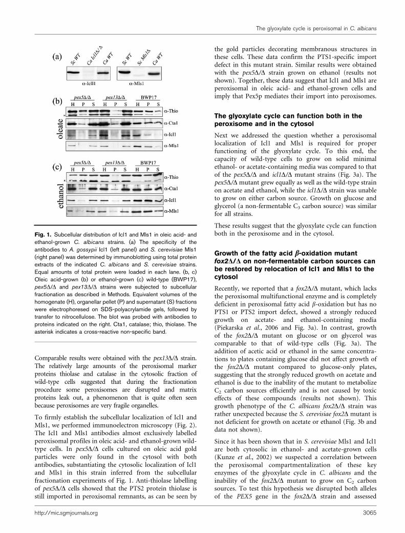
Comparable results were obtained with the pex13D/D strain.The relatively large amounts of the peroxisomal markerproteins thiolase and catalase in the cytosolic fraction ofwild-type cells suggested that during the fractionationprocedure some peroxisomes are disrupted and matrixproteins leak out, a phenomenon that is quite often seenbecause peroxisomes are very fragile organelles.
To firmly establish the subcellular localization of Icl1 andMls1, we performed immunoelectron microscopy (Fig. 2).The Icl1 and Mls1 antibodies almost exclusively labelledperoxisomal profiles in oleic acid- and ethanol-grown wild-type cells. In pex5D/D cells cultured on oleic acid goldparticles were only found in the cytosol with bothantibodies, substantiating the cytosolic localization of Icl1and Mls1 in this strain inferred from the subcellularfractionation experiments of Fig. 1. Anti-thiolase labellingof pex5D/D cells showed that the PTS2 protein thiolase isstill imported in peroxisomal remnants, as can be seen by
the gold particles decorating membranous structures inthese cells. These data confirm the PTS1-specific importdefect in this mutant strain. Similar results were obtainedwith the pex5D/D strain grown on ethanol (results notshown). Together, these data suggest that Icl1 and Mls1 areperoxisomal in oleic acid- and ethanol-grown cells andimply that Pex5p mediates their import into peroxisomes.
The glyoxylate cycle can function both in theperoxisome and in the cytosol
Next we addressed the question whether a peroxisomallocalization of Icl1 and Mls1 is required for properfunctioning of the glyoxylate cycle. To this end, thecapacity of wild-type cells to grow on solid minimalethanol- or acetate-containing media was compared to thatof the pex5D/D and icl1D/D mutant strains (Fig. 3a). Thepex5D/D mutant grew equally as well as the wild-type strainon acetate and ethanol, while the icl1D/D strain was unableto grow on either carbon source. Growth on glucose andglycerol (a non-fermentable C3 carbon source) was similarfor all strains.
These results suggest that the glyoxylate cycle can functionboth in the peroxisome and in the cytosol.
Growth of the fatty acid b-oxidation mutantfox2D/D on non-fermentable carbon sources canbe restored by relocation of Icl1 and Mls1 to thecytosol
Recently, we reported that a fox2D/D mutant, which lacksthe peroxisomal multifunctional enzyme and is completelydeficient in peroxisomal fatty acid b-oxidation but has noPTS1 or PTS2 import defect, showed a strongly reducedgrowth on acetate- and ethanol-containing media(Piekarska et al., 2006 and Fig. 3a). In contrast, growthof the fox2D/D mutant on glucose or on glycerol wascomparable to that of wild-type cells (Fig. 3a). Theaddition of acetic acid or ethanol in the same concentra-tions to plates containing glucose did not affect growth ofthe fox2D/D mutant compared to glucose-only plates,suggesting that the strongly reduced growth on acetate andethanol is due to the inability of the mutant to metabolizeC2 carbon sources efficiently and is not caused by toxiceffects of these compounds (results not shown). Thisgrowth phenotype of the C. albicans fox2D/D strain wasrather unexpected because the S. cerevisiae fox2D mutant isnot deficient for growth on acetate or ethanol (Fig. 3b anddata not shown).
Since it has been shown that in S. cerevisiae Mls1 and Icl1are both cytosolic in ethanol- and acetate-grown cells(Kunze et al., 2002) we suspected a correlation betweenthe peroxisomal compartmentalization of these keyenzymes of the glyoxylate cycle in C. albicans and theinability of the fox2D/D mutant to grow on C2 carbonsources. To test this hypothesis we disrupted both allelesof the PEX5 gene in the fox2D/D strain and assessed
Fig. 1. Subcellular distribution of Icl1 and Mls1 in oleic acid- andethanol-grown C. albicans strains. (a) The specificity of theantibodies to A. gossypii Icl1 (left panel) and S. cerevisiae Mls1(right panel) was determined by immunoblotting using total proteinextracts of the indicated C. albicans and S. cerevisiae strains.Equal amounts of total protein were loaded in each lane. (b, c)Oleic acid-grown (b) or ethanol-grown (c) wild-type (BWP17),pex5D/D and pex13D/D strains were subjected to subcellularfractionation as described in Methods. Equivalent volumes of thehomogenate (H), organellar pellet (P) and supernatant (S) fractionswere electrophoresed on SDS-polyacrylamide gels, followed bytransfer to nitrocellulose. The blot was probed with antibodies toproteins indicated on the right. Cta1, catalase; thio, thiolase. Theasterisk indicates a cross-reactive non-specific band.
The glyoxylate cycle is peroxisomal in C. albicans
http://mic.sgmjournals.org 3065
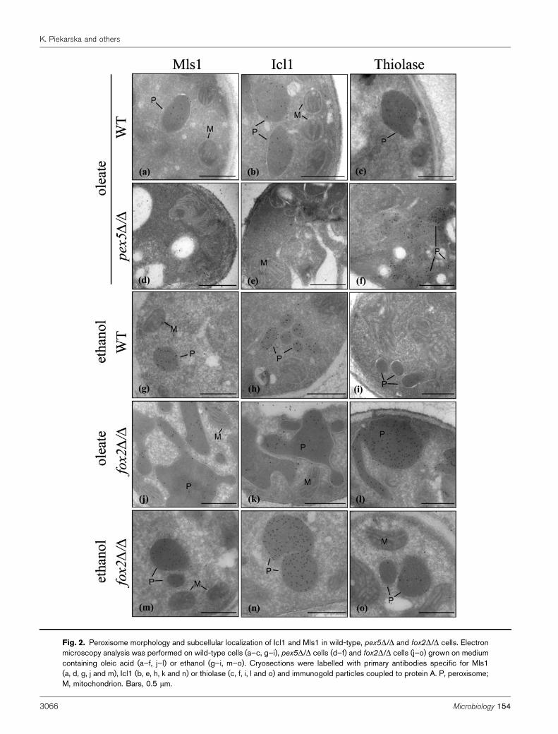
Fig. 2. Peroxisome morphology and subcellular localization of Icl1 and Mls1 in wild-type, pex5D/D and fox2D/D cells. Electronmicroscopy analysis was performed on wild-type cells (a–c, g–i), pex5D/D cells (d–f) and fox2D/D cells (j–o) grown on mediumcontaining oleic acid (a–f, j–l) or ethanol (g–i, m–o). Cryosections were labelled with primary antibodies specific for Mls1(a, d, g, j and m), Icl1 (b, e, h, k and n) or thiolase (c, f, i, l and o) and immunogold particles coupled to protein A. P, peroxisome;M, mitochondrion. Bars, 0.5 mm.
K. Piekarska and others
3066 Microbiology 154
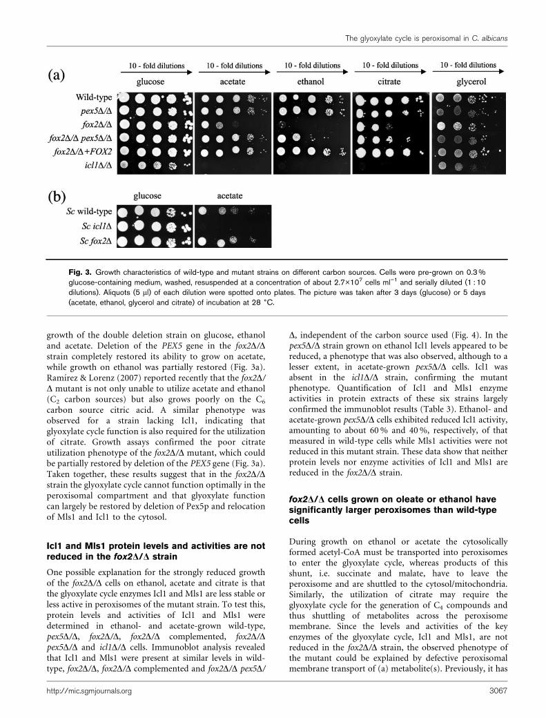
growth of the double deletion strain on glucose, ethanoland acetate. Deletion of the PEX5 gene in the fox2D/Dstrain completely restored its ability to grow on acetate,while growth on ethanol was partially restored (Fig. 3a).Ramırez & Lorenz (2007) reported recently that the fox2D/D mutant is not only unable to utilize acetate and ethanol(C2 carbon sources) but also grows poorly on the C6
carbon source citric acid. A similar phenotype wasobserved for a strain lacking Icl1, indicating thatglyoxylate cycle function is also required for the utilizationof citrate. Growth assays confirmed the poor citrateutilization phenotype of the fox2D/D mutant, which couldbe partially restored by deletion of the PEX5 gene (Fig. 3a).Taken together, these results suggest that in the fox2D/Dstrain the glyoxylate cycle cannot function optimally in theperoxisomal compartment and that glyoxylate functioncan largely be restored by deletion of Pex5p and relocationof Mls1 and Icl1 to the cytosol.
Icl1 and Mls1 protein levels and activities are notreduced in the fox2D/D strain
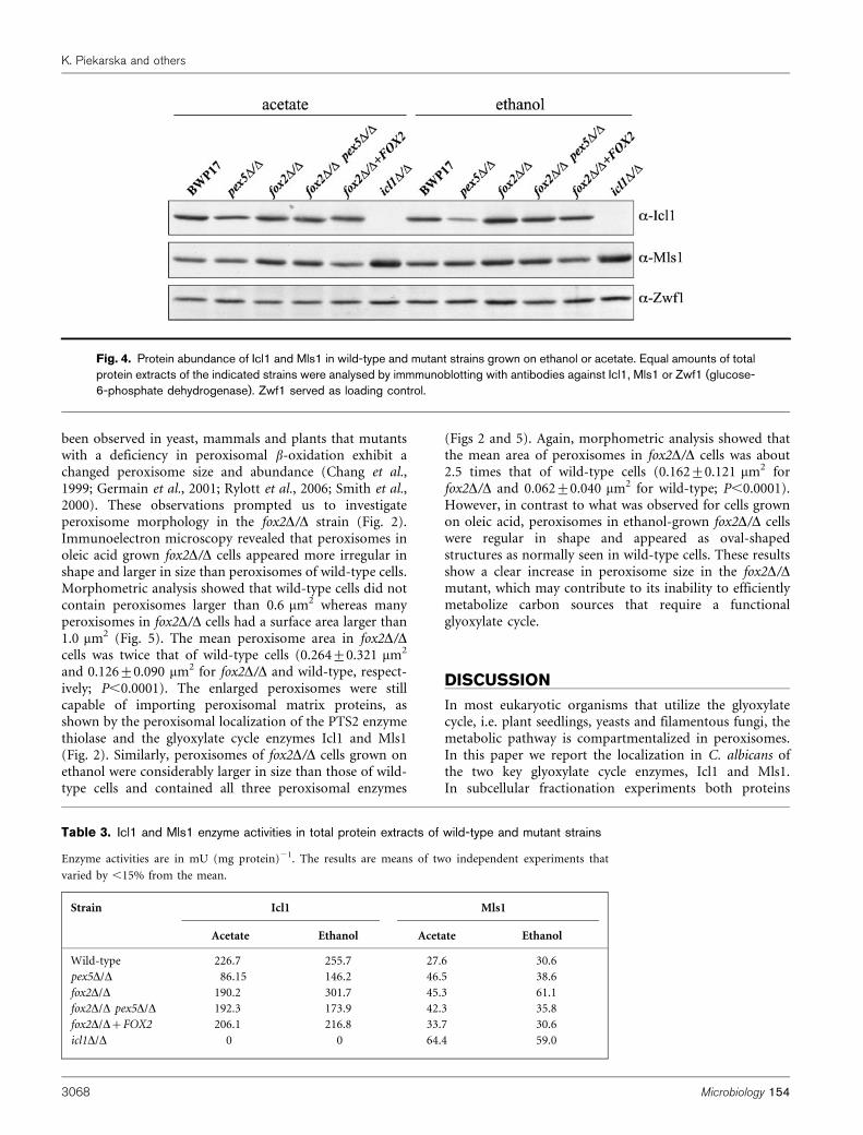
One possible explanation for the strongly reduced growthof the fox2D/D cells on ethanol, acetate and citrate is thatthe glyoxylate cycle enzymes Icl1 and Mls1 are less stable orless active in peroxisomes of the mutant strain. To test this,protein levels and activities of Icl1 and Mls1 weredetermined in ethanol- and acetate-grown wild-type,pex5D/D, fox2D/D, fox2D/D complemented, fox2D/Dpex5D/D and icl1D/D cells. Immunoblot analysis revealedthat Icl1 and Mls1 were present at similar levels in wild-type, fox2D/D, fox2D/D complemented and fox2D/D pex5D/
D, independent of the carbon source used (Fig. 4). In thepex5D/D strain grown on ethanol Icl1 levels appeared to bereduced, a phenotype that was also observed, although to alesser extent, in acetate-grown pex5D/D cells. Icl1 wasabsent in the icl1D/D strain, confirming the mutantphenotype. Quantification of Icl1 and Mls1 enzymeactivities in protein extracts of these six strains largelyconfirmed the immunoblot results (Table 3). Ethanol- andacetate-grown pex5D/D cells exhibited reduced Icl1 activity,amounting to about 60 % and 40 %, respectively, of thatmeasured in wild-type cells while Mls1 activities were notreduced in this mutant strain. These data show that neitherprotein levels nor enzyme activities of Icl1 and Mls1 arereduced in the fox2D/D strain.
fox2D/D cells grown on oleate or ethanol havesignificantly larger peroxisomes than wild-typecells
During growth on ethanol or acetate the cytosolicallyformed acetyl-CoA must be transported into peroxisomesto enter the glyoxylate cycle, whereas products of thisshunt, i.e. succinate and malate, have to leave theperoxisome and are shuttled to the cytosol/mitochondria.Similarly, the utilization of citrate may require theglyoxylate cycle for the generation of C4 compounds andthus shuttling of metabolites across the peroxisomemembrane. Since the levels and activities of the keyenzymes of the glyoxylate cycle, Icl1 and Mls1, are notreduced in the fox2D/D strain, the observed phenotype ofthe mutant could be explained by defective peroxisomalmembrane transport of (a) metabolite(s). Previously, it has
Fig. 3. Growth characteristics of wild-type and mutant strains on different carbon sources. Cells were pre-grown on 0.3 %glucose-containing medium, washed, resuspended at a concentration of about 2.7�107 cells ml”1 and serially diluted (1 : 10dilutions). Aliquots (5 ml) of each dilution were spotted onto plates. The picture was taken after 3 days (glucose) or 5 days(acetate, ethanol, glycerol and citrate) of incubation at 28 6C.
The glyoxylate cycle is peroxisomal in C. albicans
http://mic.sgmjournals.org 3067
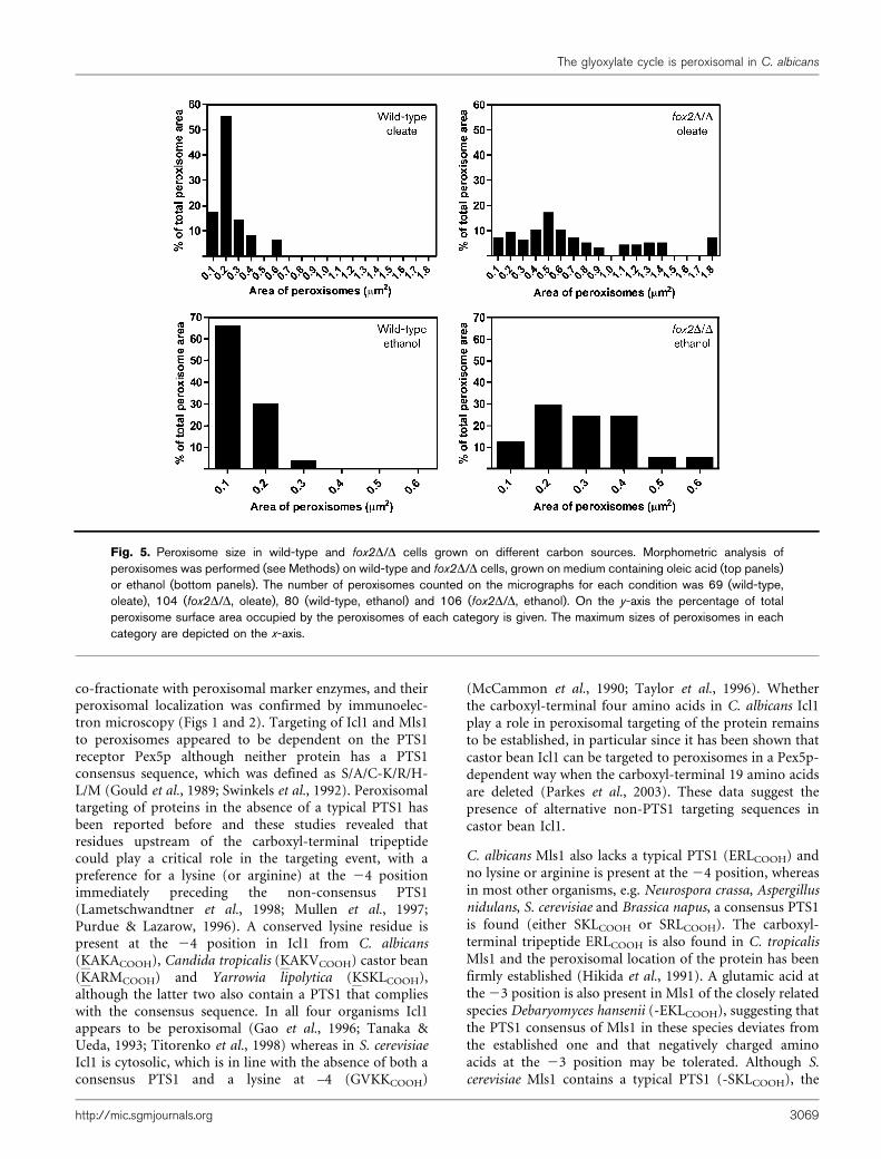
been observed in yeast, mammals and plants that mutantswith a deficiency in peroxisomal b-oxidation exhibit achanged peroxisome size and abundance (Chang et al.,1999; Germain et al., 2001; Rylott et al., 2006; Smith et al.,2000). These observations prompted us to investigateperoxisome morphology in the fox2D/D strain (Fig. 2).Immunoelectron microscopy revealed that peroxisomes inoleic acid grown fox2D/D cells appeared more irregular inshape and larger in size than peroxisomes of wild-type cells.Morphometric analysis showed that wild-type cells did notcontain peroxisomes larger than 0.6 mm2 whereas manyperoxisomes in fox2D/D cells had a surface area larger than1.0 mm2 (Fig. 5). The mean peroxisome area in fox2D/Dcells was twice that of wild-type cells (0.264±0.321 mm2
and 0.126±0.090 mm2 for fox2D/D and wild-type, respect-ively; P,0.0001). The enlarged peroxisomes were stillcapable of importing peroxisomal matrix proteins, asshown by the peroxisomal localization of the PTS2 enzymethiolase and the glyoxylate cycle enzymes Icl1 and Mls1(Fig. 2). Similarly, peroxisomes of fox2D/D cells grown onethanol were considerably larger in size than those of wild-type cells and contained all three peroxisomal enzymes
(Figs 2 and 5). Again, morphometric analysis showed thatthe mean area of peroxisomes in fox2D/D cells was about2.5 times that of wild-type cells (0.162±0.121 mm2 forfox2D/D and 0.062±0.040 mm2 for wild-type; P,0.0001).However, in contrast to what was observed for cells grownon oleic acid, peroxisomes in ethanol-grown fox2D/D cellswere regular in shape and appeared as oval-shapedstructures as normally seen in wild-type cells. These resultsshow a clear increase in peroxisome size in the fox2D/Dmutant, which may contribute to its inability to efficientlymetabolize carbon sources that require a functionalglyoxylate cycle.
DISCUSSION
In most eukaryotic organisms that utilize the glyoxylatecycle, i.e. plant seedlings, yeasts and filamentous fungi, themetabolic pathway is compartmentalized in peroxisomes.In this paper we report the localization in C. albicans ofthe two key glyoxylate cycle enzymes, Icl1 and Mls1.In subcellular fractionation experiments both proteins
Table 3. Icl1 and Mls1 enzyme activities in total protein extracts of wild-type and mutant strains
Enzyme activities are in mU (mg protein)21. The results are means of two independent experiments that
varied by ,15% from the mean.
Strain Icl1 Mls1
Acetate Ethanol Acetate Ethanol
Wild-type 226.7 255.7 27.6 30.6
pex5D/D 86.15 146.2 46.5 38.6
fox2D/D 190.2 301.7 45.3 61.1
fox2D/D pex5D/D 192.3 173.9 42.3 35.8
fox2D/D+FOX2 206.1 216.8 33.7 30.6
icl1D/D 0 0 64.4 59.0
Fig. 4. Protein abundance of Icl1 and Mls1 in wild-type and mutant strains grown on ethanol or acetate. Equal amounts of totalprotein extracts of the indicated strains were analysed by immmunoblotting with antibodies against Icl1, Mls1 or Zwf1 (glucose-6-phosphate dehydrogenase). Zwf1 served as loading control.
K. Piekarska and others
3068 Microbiology 154
co-fractionate with peroxisomal marker enzymes, and theirperoxisomal localization was confirmed by immunoelec-tron microscopy (Figs 1 and 2). Targeting of Icl1 and Mls1to peroxisomes appeared to be dependent on the PTS1receptor Pex5p although neither protein has a PTS1consensus sequence, which was defined as S/A/C-K/R/H-L/M (Gould et al., 1989; Swinkels et al., 1992). Peroxisomaltargeting of proteins in the absence of a typical PTS1 hasbeen reported before and these studies revealed thatresidues upstream of the carboxyl-terminal tripeptidecould play a critical role in the targeting event, with apreference for a lysine (or arginine) at the 24 positionimmediately preceding the non-consensus PTS1(Lametschwandtner et al., 1998; Mullen et al., 1997;Purdue & Lazarow, 1996). A conserved lysine residue ispresent at the 24 position in Icl1 from C. albicans(KAKACOOH), Candida tropicalis (KAKVCOOH) castor bean(KARMCOOH) and Yarrowia lipolytica (KSKLCOOH),although the latter two also contain a PTS1 that complieswith the consensus sequence. In all four organisms Icl1appears to be peroxisomal (Gao et al., 1996; Tanaka &Ueda, 1993; Titorenko et al., 1998) whereas in S. cerevisiaeIcl1 is cytosolic, which is in line with the absence of both aconsensus PTS1 and a lysine at –4 (GVKKCOOH)
(McCammon et al., 1990; Taylor et al., 1996). Whetherthe carboxyl-terminal four amino acids in C. albicans Icl1play a role in peroxisomal targeting of the protein remainsto be established, in particular since it has been shown thatcastor bean Icl1 can be targeted to peroxisomes in a Pex5p-dependent way when the carboxyl-terminal 19 amino acidsare deleted (Parkes et al., 2003). These data suggest thepresence of alternative non-PTS1 targeting sequences incastor bean Icl1.
C. albicans Mls1 also lacks a typical PTS1 (ERLCOOH) andno lysine or arginine is present at the 24 position, whereasin most other organisms, e.g. Neurospora crassa, Aspergillusnidulans, S. cerevisiae and Brassica napus, a consensus PTS1is found (either SKLCOOH or SRLCOOH). The carboxyl-terminal tripeptide ERLCOOH is also found in C. tropicalisMls1 and the peroxisomal location of the protein has beenfirmly established (Hikida et al., 1991). A glutamic acid atthe 23 position is also present in Mls1 of the closely relatedspecies Debaryomyces hansenii (-EKLCOOH), suggesting thatthe PTS1 consensus of Mls1 in these species deviates fromthe established one and that negatively charged aminoacids at the 23 position may be tolerated. Although S.cerevisiae Mls1 contains a typical PTS1 (-SKLCOOH), the
Fig. 5. Peroxisome size in wild-type and fox2D/D cells grown on different carbon sources. Morphometric analysis ofperoxisomes was performed (see Methods) on wild-type and fox2D/D cells, grown on medium containing oleic acid (top panels)or ethanol (bottom panels). The number of peroxisomes counted on the micrographs for each condition was 69 (wild-type,oleate), 104 (fox2D/D, oleate), 80 (wild-type, ethanol) and 106 (fox2D/D, ethanol). On the y-axis the percentage of totalperoxisome surface area occupied by the peroxisomes of each category is given. The maximum sizes of peroxisomes in eachcategory are depicted on the x-axis.
The glyoxylate cycle is peroxisomal in C. albicans
http://mic.sgmjournals.org 3069
localization of this enzyme is not always peroxisomal butdepends on the carbon source used: in oleic acid-growncells Mls1 is peroxisomal whereas in ethanol-grown cells itis cytosolic (Kunze et al., 2002). It has been suggested thatthe carbon source-dependent localization of Mls1 may beadvantageous to the cell, the enzyme being localized in thecompartment where the acetyl-CoA, one of its substrates, isformed, i.e. peroxisomal on oleic acid and cytosolic onethanol or acetate. Such a carbon source-dependentlocalization was not seen for Mls1 or Icl1 in C. albicans:both enzymes were found in peroxisomes under allconditions tested (Figs 1 and 2). Nevertheless, a perox-isomal compartmentalization of Mls1 and Icl1 is notabsolutely required for proper functioning of the glyoxylatecycle since a pex5D/D strain, which mislocalizes bothenzymes to the cytosol, showed wild-type growth rates onethanol and acetate (Fig. 3) despite the reduced activity ofIcl1 on these carbon sources (Table 3).
Next, we asked whether there is a correlation between theperoxisomal compartmentalization of the key enzymes ofthe glyoxylate cycle in C. albicans and the inability of the b-oxidation mutant fox2D/D to grow efficiently on C2 carbonsources (Fig. 4 and Piekarska et al., 2006; Ramırez &Lorenz, 2007). We showed that the growth inhibition is notcaused by toxicity of acetate or ethanol, and mitochondrialfunctions appeared to be normal in the fox2D/D mutant,strongly suggesting that it cannot efficiently metabolize C2
compounds. In support of this we found acetate metabol-ism in the fox2D/D mutant to be reduced to 60 % of that ofthe wild-type strain using 14C-labelled acetate as substrate(data not shown). In addition to the growth deficiency onC2 compounds (and oleate), the fox2D/D mutant usescitrate (C6) poorly (Ramırez & Lorenz, 2007). The fact thatthe icl1D/D strain shows a similar growth defect on citrateimplies that the glyoxylate cycle function is also requiredfor the utilization of this C6 compound. We suggest that(iso)citrate must enter the glyoxylate cycle to allow the cellto generate C4 units (succinate/malate), which can be usedto replenish the TCA cycle and/or function as precursorsfor gluconeogenesis. By deleting the PEX5 gene in thefox2D/D mutant, growth on ethanol and citrate could berestored partially and growth on acetate completely,consistent with our hypothesis that the observed growthphenotype of the fox2D/D strain on these carboncompounds may be caused by peroxisomal compartmen-talization of the glyoxylate cycle. Ramırez & Lorenz (2007)have recently suggested that the different growth pheno-types of the C. albicans and S. cerevisiae fox2 null strainsmay be explained by differences in the regulatory networksgoverning carbon metabolism in both fungi. However, ourdata now strongly suggest that the different compartmen-talization of glyoxylate cycle enzymes in both organismsmay provide an explanation for these growth differences.
It is currently unclear why the glyoxylate cycle cannotfunction optimally in peroxisomes of the C. albicans fox2D/D strain. We show here that the impaired glyoxylate cyclefunction in the fox2D/D mutant is not caused by reduced
levels or activity of the key enzymes Icl1 and Mls1 (Fig. 4,Table 3). Therefore, we favour the hypothesis that thetransport of metabolites (glyoxylate cycle substrates/products and/or acetyl-CoA) across the peroxisomalmembrane is affected in the fox2D/D strain, therebyreducing the overall rate of non-fermentable carbonmetabolism. We consider two possible mechanisms: directinhibition of peroxisomal transport proteins by activatedfatty acids (acyl-CoAs) or an indirect mechanism caused bythe increase in size and/or changed morphology ofperoxisomes. A consequence of blocking fatty acid b-oxidation by disruption of the FOX2 gene is theaccumulation of acyl-CoAs inside the cell. Even in theabsence of exogenously added fatty acids such anaccumulation of acyl-CoAs is likely to occur because fattyacid b-oxidation is also required for the turnover ofendogenous membrane lipids. Since the peroxisome is thesole site of fatty acid b-oxidation in fungi it is conceivablethat the acyl-CoA levels inside peroxisomes increase. Theinhibitory effect of (long-chain) acyl-CoAs on mitochon-drial carrier proteins is well documented (Morel et al.,1974) and we speculate that peroxisomal metabolitetransporters may be inhibited in a similar way in thefox2D/D mutant. It is also possible that, due to theiramphipathic nature, the acyl-CoAs insert into the perox-isomal membrane, thereby changing its physical propertiesand transport capabilities. An alternative mechanism isbased on simple physics: when the size of the organelleincreases, the surface (5membrane) area-to-volume ratiodecreases, and thus large peroxisomes have relatively lessmembrane surface available for transport processes thansmall peroxisomes. Similar suggestions were put forwardby Kiel et al. (2005) to explain the increase of penicillinproduction in Penicillium chrysogenum upon overexpres-sion of Pex11p. Penicillin biosynthesis in P. chrysogenumoccurs partially in peroxisomes and partially in the cytosol.Overexpression of Pex11p in this organism resulted inmassive proliferation of small tubular-shaped peroxisomesand a 2.5-fold higher penicillin production while the levelof the penicillin biosynthesis enzymes remainedunchanged, suggesting that an increase in membrane-to-volume ratio may increase the transport capacity of theorganelles. Further experiments are required to distinguishbetween these possibilities.
In conclusion, our results provide an explanation for theunexpected growth phenotypes of the C. albicans fox2D/Dstrain on non-fermentable carbon sources and stronglysupport the existence of peroxisomal metabolite transpor-ters. Identification of such transporters may now befeasible using C. albicans as a model system.
ACKNOWLEDGEMENTS
We thank Aaron Mitchell (Columbia University, New York, USA) forproviding strains and plasmids, William Fonzi (GeorgetownUniversity, Washington, USA) for plasmid pLUBP, Jurgen Wendland(Friedrich-Schiller-University, Jena, Germany) for the pFA modules,and Andreas Hartig (Vienna Biocenter, Vienna, Austria) and Sonja
K. Piekarska and others
3070 Microbiology 154
Meyer zu Berstenhorst (Insitut fur Biotechnology, Julich, Germany) for
providing antibodies. We are grateful to Jan van Marle for his help with
the morphometric analysis and to Rob Benne and Fred Meijer for
valuable comments and suggestions. This work was supported by
grants from the Academic Medical Center and the European
Community (QLG2-CT-2001-01663).
REFERENCES
Aitchison, J. D. & Rachubinski, R. A. (1990). In vivo import of
Candida tropicalis hydratase-dehydrogenase-epimerase into peroxi-
somes of Candida albicans. Curr Genet 17, 481–486.
Boshoff, H. I. & Barry, C. E. (2005). A low-carb diet for a high-octane
pathogen. Nat Med 11, 599–600.
Bottger, G., Barnett, P., Klein, A. T., Kragt, A., Tabak, H. F. & Distel, B.(2000). Saccharomyces cerevisiae PTS1 receptor Pex5p interacts with the
SH3 domain of the peroxisomal membrane protein Pex13p in an
unconventional, non-PXXP-related manner. Mol Biol Cell 11, 3963–3976.
Chang, C. C., South, S., Warren, D., Jones, J., Moser, A. B., Moser,H. W. & Gould, S. J. (1999). Metabolic control of peroxisome
abundance. J Cell Sci 112, 1579–1590.
de Jong-Gubbels, P., Vanrolleghem, P., Heijnen, S., van Dijken, J. P.& Pronk, J. T. (1995). Regulation of carbon metabolism in chemostat
cultures of Saccharomyces cerevisiae grown on mixtures of glucose and
ethanol. Yeast 11, 407–418.
Elgersma, Y., van Roermund, C. W., Wanders, R. J. & Tabak, H. F.(1995). Peroxisomal and mitochondrial carnitine acetyltransferases of
Saccharomyces cerevisiae are encoded by a single gene. EMBO J 14,
3472–3479.
Elgersma, Y., Kwast, L., Klein, A., Voorn-Brouwer, T., van den Berg, M.,Metzig, B., America, T., Tabak, H. F. & Distel, B. (1996). The SH3
domain of the Saccharomyces cerevisiae peroxisomal membrane protein
Pex13p functions as a docking site for Pex5p, a mobile receptor for the
import PTS1-containing proteins. J Cell Biol 135, 97–109.
Gao, X., Marrison, J. L., Pool, M. R., Leech, R. M. & Baker, A. (1996).
Castor bean isocitrate lyase lacking the putative peroxisomal targeting
signal 1 ARM is imported into plant peroxisomes both in vitro and in
vivo. Plant Physiol 112, 1457–1464.
Germain, V., Rylott, E. L., Larson, T. R., Sherson, S. M., Bechtold, N.,
Carde, J. P., Bryce, J. H., Graham, I. A. & Smith, S. M. (2001).Requirement for 3-ketoacyl-CoA thiolase-2 in peroxisome devel-
opment, fatty acid beta-oxidation and breakdown of triacylglycerol in
lipid bodies of Arabidopsis seedlings. Plant J 28, 1–12.
Gietl, C., Faber, K. N., van der Klei, I. J. & Veenhuis, M. (1994).Mutational analysis of the N-terminal topogenic signal of watermelon
glyoxysomal malate dehydrogenase using the heterologous host
Hansenula polymorpha. Proc Natl Acad Sci U S A 91, 3151–3155.
Gillum, A. M., Tsay, E. Y. H. & Kirsch, D. R. (1984). Isolation of the
Candida albicans genes for orotidine-59-phosphate decarboxylase by
complementation of S. cerevisiae ura3 and E. coli pyrF mutations. Mol
Gen Genet 198, 179–182.
Glover, J. R., Andrews, D. W. & Rachubinski, R. A. (1994a).Saccharomyces cerevisiae peroxisomal thiolase is imported as a dimer.
Proc Natl Acad Sci U S A 91, 10541–10545.
Glover, J. R., Andrews, D. W., Subramani, S. & Rachubinski, R. A.(1994b). Mutagenesis of the amino targeting signal of Saccharomyces
cerevisiae 3-ketoacyl-CoA thiolase reveals conserved amino acids required
for import into peroxisomes in vivo. J Biol Chem 269, 7558–7563.
Gola, S., Martin, R., Walther, A., Dunkler, A. & Wendland, J. (2003).New modules for PCR-based gene targeting in Candida albicans:
rapid and efficient gene targeting using 100 bp of flanking homologyregion. Yeast 20, 1339–1347.
Gould, S. J., Keller, G. A., Hosken, N., Wilkinson, J. & Subramani, S.(1989). A conserved tripeptide sorts proteins to peroxisomes. J CellBiol 108, 1657–1664.
Gould, S. J., Keller, G. A., Schneider, M., Howell, S. H., Garrard, L. J.,Goodman, J. M., Distel, B., Tabak, H. & Subramani, S. (1990).Peroxisomal protein import is conserved between yeast, plants,insects and mammals. EMBO J 9, 85–90.
Gueldener, U., Heinisch, J., Koehler, G. J., Voss, D. & Hegemann,J. H. (2002). A second set of loxP marker cassettes for Cre-mediatedmultiple gene knockouts in budding yeast. Nucleic Acids Res 30, e23.
Hartig, A., Simon, M. M., Schuster, T., Daugherty, J. R., Yoo, H. S. &Cooper, T. G. (1992). Differentially regulated malate synthase genesparticipate in carbon and nitrogen metabolism of S. cerevisiae. NucleicAcids Res 20, 5677–5686.
Hikida, M., Atomi, H., Fukuda, Y., Aoki, A., Hishida, T., Teranishi, Y.,Ueda, M. & Tanaka, A. (1991). Presence of two transcribed malatesynthase genes in an n-alkane-utilizing yeast, Candida tropicalis.J Biochem 110, 909–914.
Jones, E. W. (1977). Proteinase mutants of Saccharomyces cerevisiae.Genetics 85, 23–33.
Kamiryo, T., Abe, M., Okazaki, K., Kato, S. & Shimamoto, N. (1982).Absence of DNA in peroxisomes of Candida tropicalis. J Bacteriol 152,269–274.
Kiel, J. A., van der Klei, I. J., van den Berg, M. A., Bovenberg, R. A. &Veenhuis, M. (2005). Overproduction of a single protein, Pc-Pex11p,results in 2-fold enhanced penicillin production by Penicilliumchrysogenum. Fungal Genet Biol 42, 154–164.
Klein, A. T., van den Berg, M., Bottger, G., Tabak, H. F. & Distel, B.(2002). Saccharomyces cerevisiae acyl-CoA oxidase follows a novel,non-PTS1, import pathway into peroxisomes that is dependent onPex5p. J Biol Chem 277, 25011–25019.
Kornberg, H. L. & Krebs, H. A. (1957). Synthesis of cell constituents fromC2-units by a modified tricarboxylic acid cycle. Nature 179, 988–991.
Kunze, M., Kragler, F., Binder, M., Hartig, A. & Gurvitz, A. (2002). Targetingof malate synthase 1 to the peroxisomes of Saccharomyces cerevisiae cellsdepends on growth on oleic acid medium. Eur J Biochem 269, 915–922.
Lametschwandtner, G., Brocard, C., Fransen, M., Van Veldhoven, P.,Berger, J. & Hartig, A. (1998). The difference in recognition of terminaltripeptides as peroxisomal targeting signal 1 between yeast and humanis due to different affinities of their receptor Pex5p to the cognate signaland to residues adjacent to it. J Biol Chem 273, 33635–33643.
Lorenz, M. C. & Fink, G. R. (2001). The glyoxylate cycle is required forfungal virulence. Nature 412, 83–86.
Lorenz, M. C. & Fink, G. R. (2002). Life and death in a macrophage:role of the glyoxylate cycle in virulence. Eukaryot Cell 1, 657–662.
Lorenz, M. C., Bender, J. A. & Fink, G. R. (2004). Transcriptionalresponse of Candida albicans upon internalization by macrophages.Eukaryot Cell 3, 1076–1087.
Maeting, I., Schmidt, G., Sahm, H., Revuelta, J. L., Stierhof, Y. D. &Stahmann, K. P. (1999). Isocitrate lyase of Ashbya gossypii – transcriptionalregulation and peroxisomal localization. FEBS Lett 444, 15–21.
McCammon, M. T., Veenhuis, M., Trapp, S. B. & Goodman, J. M.(1990). Association of glyoxylate and beta-oxidation enzymes withperoxisomes of Saccharomyces cerevisiae. J Bacteriol 172, 5816–5827.
McKinney, J. D., Honer zu Bentrup, K., Munoz-Elias, E. J., Miczak, A.,Chen, B., Chan, W. T., Swenson, D., Sacchettini, J. C., Jacobs, W. R.,Jr & Russell, D. G. (2000). Persistence of Mycobacterium tuberculosisin macrophages and mice requires the glyoxylate shunt enzymeisocitrate lyase. Nature 406, 735–738.
The glyoxylate cycle is peroxisomal in C. albicans
http://mic.sgmjournals.org 3071
McNew, J. A. & Goodman, J. M. (1994). An oligomeric protein isimported into peroxisomes in vivo. J Cell Biol 127, 1245–1257.
Morel, F., Lauquin, G., Lunardi, J., Duszynski, J. & Vignais, P. V. (1974).An appraisal of the functional significance of the inhibitory effect of longchain acyl-CoAs on mitochondrial transports. FEBS Lett 39, 133–138.
Mullen, R. T., Lee, M. S., Flynn, C. R. & Trelease, R. N. (1997). Diverseamino acid residues function within the type 1 peroxisomal targetingsignal. Implications for the role of accessory residues upstream of thetype 1 peroxisomal targeting signal. Plant Physiol 115, 881–889.
Munoz-Elias, E. J. & McKinney, J. D. (2005). Mycobacteriumtuberculosis isocitrate lyases 1 and 2 are jointly required for in vivogrowth and virulence. Nat Med 11, 638–644.
Ozimek, P., Kotter, P., Veenhuis, M. & van der Klei, I. J. (2006).Hansenula polymorpha and Saccharomyces cerevisiae Pex5p’s recognizedifferent, independent peroxisomal targeting signals in alcoholoxidase. FEBS Lett 580, 46–50.
Parkes, J. A., Langer, S., Hartig, A. & Baker, A. (2003). PTS1-independent targeting of isocitrate lyase to peroxisomes requires thePTS1 receptor Pex5p. Mol Membr Biol 20, 61–69.
Pellicer, M. T., Fernandez, C., Badia, J., Aguilar, J., Lin, E. C. &Baldom, L. (1999). Cross-induction of glc and ace operons ofEscherichia coli attributable to pathway intersection. Characterizationof the glc promoter. J Biol Chem 274, 1745–1752.
Piekarska, K., Mol, E., van den Berg, M., Hardy, G., van den Burg, J.,van Roermund, C., Maccallum, D., Odds, F. & Distel, B. (2006).Peroxisomal fatty acid b-oxidation is not essential for virulence ofCandida albicans. Eukaryot Cell 5, 1847–1856.
Purdue, P. E. & Lazarow, P. B. (1996). Targeting of human catalase toperoxisomes is dependent upon a novel COOH-terminal peroxisomaltargeting sequence. J Cell Biol 134, 849–862.
Purdue, P. E. & Lazarow, P. B. (2001). Peroxisome biogenesis. AnnuRev Cell Dev Biol 17, 701–752.
Ramırez, M. A. & Lorenz, M. C. (2007). Mutations in alternativecarbon utilization pathways in Candida albicans attenuate virulenceand confer pleiotropic phenotypes. Eukaryot Cell 6, 280–290.
Rylott, E. L., Eastmond, P. J., Gilday, A. D., Slocombe, S. P., Larson,T. R., Baker, A. & Graham, I. A. (2006). The Arabidopsis thalianamultifunctional protein gene (MFP2) of peroxisomal beta-oxidationis essential for seedling establishment. Plant J 45, 930–941.
Sambrook, J., Fritsch, E. F. & Maniatis, T. (1989). Molecular Cloning:A Laboratory Manual, 2nd edn. Cold Spring Harbor, NY: Cold SpringHarbor Laboratory.
Schmidt, G., Stahmann, K. P., Kaesler, B. & Sahm, H. (1996).Correlation of isocitrate lyase activity and riboflavin formationin the riboflavin overproducer Ashbya gossypii. Microbiology 142,419–426.
Small, G. M., Szabo, L. J. & Lazarow, P. B. (1988). Acyl-CoA oxidasecontains two targeting sequences each of which can mediate proteinimport into peroxisomes. EMBO J 7, 1167–1173.
Smith, J. J., Brown, T. W., Eitzen, G. A. & Rachubinski, R. A. (2000).Regulation of peroxisome size and number by fatty acid beta-oxidationin the yeast Yarrowia lipolytica. J Biol Chem 275, 20168–20178.
Swinkels, B. W., Gould, S. J. & Subramani, S. (1992). Targetingefficiencies of various permutations of the consensus C-terminaltripeptide peroxisomal targeting signal. FEBS Lett 305, 133–136.
Tanaka, A. & Ueda, M. (1993). Assimilation of alkanes by yeasts –function and biogenesis of peroxisomes. Mycol Res 97, 1025–1044.
Taylor, K. M., Kaplan, C. P., Gao, X. & Baker, A. (1996). Localizationand targeting of isocitrate lyases in Saccharomyces cerevisiae. Biochem J319, 255–262.
Titorenko, V. I., Smith, J. J., Szilard, R. K. & Rachubinski, R. A. (1998).Pex20p of the yeast Yarrowia lipolytica is required for theoligomerization of thiolase in the cytosol and for its targeting tothe peroxisome. J Cell Biol 142, 403–420.
Valenciano, S., Lucas, J. R., Pedregosa, A., Monistrol, I. F. & Laborda, F.(1996). Induction of beta-oxidation enzymes and microbody prolifera-tion in Aspergillus nidulans. Arch Microbiol 166, 336–341.
Walther, A. & Wendland, J. (2003). An improved transformationprotocol for the human fungal pathogen Candida albicans. Curr Genet42, 339–343.
Wilson, R. B., Davis, D. & Mitchell, A. P. (1999). Rapid hypothesistesting with Candida albicans through gene disruption with shorthomology regions. J Bacteriol 181, 1868–1874.
Yang, X., Purdue, P. E. & Lazarow, P. B. (2001). Eci1p uses a PTS1 toenter peroxisomes: either its own or that of a partner, Dci1p. Eur JCell Biol 80, 126–138.
Edited by: J. F. Ernst
K. Piekarska and others
3072 Microbiology 154
Copyright © 2022 FDOKUMEN