Copper Redox Cycling in the Prion Protein Depends Critically on Binding Mode
21
Copper Redox Cycling in the Prion Protein Depends Critically on Binding Mode Lin Liu †,# , Dianlu Jiang †,# , Alex McDonald ‡ , Yuanqiang Hao † , Glenn L. Millhauser ‡,* , and Feimeng Zhou †,* † Department of Chemistry and Biochemistry, California State University, Los Angeles, Los Angeles, California 90032 ‡ Department of Chemistry and Biochemistry, University of California, Santa Cruz, Santa Cruz, California 95064 Abstract The prion protein (PrP) takes up four to six equivalents of copper in its extended N-terminal domain, composed of the octarepeat (OR) segment (human sequence residues 60–91) and two mononuclear binding sites (at His96 and His111; also referred to as the non-OR region). The OR segment responds to specific copper concentrations by transitioning from a multi-His mode at low copper levels to a single-His, amide nitrogen mode at high levels (Chattopadhyay et al. J. Am. Chem. Soc., 127, 12647–12656, 2005). The specific function of PrP in healthy tissue is unclear, but numerous reports link copper uptake to a neuroprotective role that regulates cellular stress (Stevens et al. PLoS Pathogens, 5(4): e1000390, 2009). A current working hypothesis is that the high occupancy binding mode quenches copper’s inherent redox cycling, thus protecting against the production of reactive oxygen species from unregulated Fenton type reactions. Here, we directly test this hypothesis by performing detailed pH-dependent electrochemical measurements on both low and high occupancy copper binding modes. In contrast to the current belief, we find that the low occupancy mode completely quenches redox cycling, but high occupancy leads to the gentle production of hydrogen peroxide through a catalytic reduction of oxygen facilitated by the complex. These electrochemical findings are supported by independent kinetic measurements that probe for ascorbate usage and also peroxide production. Hydrogen peroxide production is also observed from a segment corresponding to the non-OR region. Collectively, these results overturn the current working hypothesis and suggest, instead, that the redox cycling of copper bound to PrP in the high occupancy mode is not quenched, but is regulated. The observed production of hydrogen peroxide suggests a mechanism that could explain PrP’s putative role in cellular signaling. 1. INTRODUCTION The misfolding and aggregation of the prion protein (PrP), a membrane-bound glycoprotein present in the central nervous system (CNS) of mammalian and avian species, leads to the development of the prion diseases. 1,2 The human forms of prion diseases, 3 including Creutzfeldt-Jakob disease and kuru, share similar neuropathologies to other more prevalent * Corresponding authors: Phone: 831-459-2176 and [email protected] (GLM) and Phone: 323-343-2390, Fax: 323-343-6490, and [email protected] (FZ). # These authors contributed equally to this work. Supporting Information Available Additional experimental details about the electrochemical studies are in the Supporting Information. This material is available free of charge via the internet at http://pubs.acs.org. NIH Public Access Author Manuscript J Am Chem Soc. Author manuscript; available in PMC 2012 August 10. Published in final edited form as: J Am Chem Soc. 2011 August 10; 133(31): 12229–12237. doi:10.1021/ja2045259. NIH-PA Author Manuscript NIH-PA Author Manuscript NIH-PA Author Manuscript
-
Upload
independent -
Category
Documents
-
view
3 -
download
0
Transcript of Copper Redox Cycling in the Prion Protein Depends Critically on Binding Mode

Copper Redox Cycling in the Prion Protein Depends Critically onBinding Mode
Lin Liu†,#, Dianlu Jiang†,#, Alex McDonald‡, Yuanqiang Hao†, Glenn L. Millhauser‡,*, andFeimeng Zhou†,*
†Department of Chemistry and Biochemistry, California State University, Los Angeles, LosAngeles, California 90032‡Department of Chemistry and Biochemistry, University of California, Santa Cruz, Santa Cruz,California 95064
AbstractThe prion protein (PrP) takes up four to six equivalents of copper in its extended N-terminaldomain, composed of the octarepeat (OR) segment (human sequence residues 60–91) and twomononuclear binding sites (at His96 and His111; also referred to as the non-OR region). The ORsegment responds to specific copper concentrations by transitioning from a multi-His mode at lowcopper levels to a single-His, amide nitrogen mode at high levels (Chattopadhyay et al. J. Am.Chem. Soc., 127, 12647–12656, 2005). The specific function of PrP in healthy tissue is unclear,but numerous reports link copper uptake to a neuroprotective role that regulates cellular stress(Stevens et al. PLoS Pathogens, 5(4): e1000390, 2009). A current working hypothesis is that thehigh occupancy binding mode quenches copper’s inherent redox cycling, thus protecting againstthe production of reactive oxygen species from unregulated Fenton type reactions. Here, wedirectly test this hypothesis by performing detailed pH-dependent electrochemical measurementson both low and high occupancy copper binding modes. In contrast to the current belief, we findthat the low occupancy mode completely quenches redox cycling, but high occupancy leads to thegentle production of hydrogen peroxide through a catalytic reduction of oxygen facilitated by thecomplex. These electrochemical findings are supported by independent kinetic measurements thatprobe for ascorbate usage and also peroxide production. Hydrogen peroxide production is alsoobserved from a segment corresponding to the non-OR region. Collectively, these results overturnthe current working hypothesis and suggest, instead, that the redox cycling of copper bound to PrPin the high occupancy mode is not quenched, but is regulated. The observed production ofhydrogen peroxide suggests a mechanism that could explain PrP’s putative role in cellularsignaling.
1. INTRODUCTIONThe misfolding and aggregation of the prion protein (PrP), a membrane-bound glycoproteinpresent in the central nervous system (CNS) of mammalian and avian species, leads to thedevelopment of the prion diseases.1,2 The human forms of prion diseases,3 includingCreutzfeldt-Jakob disease and kuru, share similar neuropathologies to other more prevalent
*Corresponding authors: Phone: 831-459-2176 and [email protected] (GLM) and Phone: 323-343-2390, Fax: 323-343-6490, [email protected] (FZ).#These authors contributed equally to this work.Supporting Information AvailableAdditional experimental details about the electrochemical studies are in the Supporting Information. This material is available free ofcharge via the internet at http://pubs.acs.org.
NIH Public AccessAuthor ManuscriptJ Am Chem Soc. Author manuscript; available in PMC 2012 August 10.
Published in final edited form as:J Am Chem Soc. 2011 August 10; 133(31): 12229–12237. doi:10.1021/ja2045259.
NIH
-PA Author Manuscript
NIH
-PA Author Manuscript
NIH
-PA Author Manuscript

neurodegenerative disorders such as Alzheimer’s disease (AD) and Parkinson’s disease(PD).4,5 PrP has been shown to interact with a number of divalent metal ions,6,7 and withmoderate to high affinity to Cu2+ and Zn2+.7,8 The linkage between PrP and bioavailableCu2+ is particularly well established by in vitro and in vivo9 studies and described in anumber of recent reviews.10–14 Although copper is an essential metal and exists in a numberof proteins, free Cu2+ exhibits high cytotoxicity, stemming from its ability to initiate redoxcycling, in turn generating reactive oxygen species (ROS) via the Harber-Weissreaction:15–18
(1)
(2)
(3)
The highly toxic and reactive hydroxyl radical (OH•) can readily damage DNA and proteinsand cause lipid peroxidation. Reaction (3) is also referred to as a Fenton-like reactionbecause of its similarity to the Fenton reaction with Fe2+ and H2O2 as the reactants.16
In biological milieu, free or bound Cu2+ could initiate another redox cycle, which isdependent on the thermodynamic properties of the reactants and the structure of the Cu2+
complexes. The cycle starts with the reduction of free or Cu2+ bound to a ligand (L) by abiological reductant. The resultant Cu+ can be subsequently oxidized by O2, generatingH2O2 as a product. Using ascorbic acid (AA, which exists as ascorbate at neutral pH andoxidizes to dehydroascorbate or DA) as a representative reductant, this cycle can bedescribed as:19–22
(4)
(5)
Notice H2O2 produced in reaction (5) can in turn react with any remaining free or boundCu2+ through reactions (1)–(3) to produce OH•.
Given the abundance of oxygen23,24 and biological reductants (e.g. AA and glutathione orGSH) in the CNS (e.g., cerebrospinal fluid (CSF) in particular), if a redox active metal werenot properly regulated, the large quantity of ROS produced by the Cu2+-initiated redoxcycling would likely lead to significant oxidative damage. On the basis that PrP is a Cu2+-binding protein,9 a major biological function of PrP has been hypothesized to complex Cu2+
to attenuate or buffer its redox activity so that ROS is prevented from forming, or convertedinto less harmful species.11,12 A unique feature of PrP is that distinct Cu2+ binding modesare populated in response to different Cu2+ concentrations. PrP contains a highly conservedrepeat of an octapeptide (OP) sequence within its N-terminal domain that constitutes itsprimary sites for Cu2+ uptake.11,12 Human PrP has four tandem copies of the specificsequence PHGGGWGQ (referred to as the octarepeat11 or OR within residues 60–91). In
Liu et al. Page 2
J Am Chem Soc. Author manuscript; available in PMC 2012 August 10.
NIH
-PA Author Manuscript
NIH
-PA Author Manuscript
NIH
-PA Author Manuscript

vitro studies have shown that when the concentration ratio between PrP and Cu2+ is largerthan 1:1, four histidines (His) in the OR domain coordinate a single Cu2+ center forming thelow Cu2+ occupancy binding mode OR–Cu2+ (Figure 1).25 When the Cu2+ concentrationrises to four or more equivalents per protein, each of the four OPs binds one Cu2+, givingrise to the so-called high occupancy or OR–Cu2+
4 (Figure 1).25 In OR–Cu2+4, each Cu2+ is
ligated by one His, the amide nitrogens on the two adjacent and deprotonated glycines, and aglycine carbonyl.25 The OR–Cu2+ and OR–Cu2+
4 binding modes take up Cu2+ with highlydisparate dissociation constants (0.1 nM for the former and ~10 μM for the latter) andconsequent negative cooperativity.11,26 Immediately outside of the OR domain, the segmentencompassing two His residues (in human PrP they are His96 and His111 and in mouse PrPHis96 and His110) can also bind Cu2+. This segment is referred to as the non-OR Cu2+-binding site.27–29 It is recently shown that when solution pH becomes sufficiently low (~6.0or lower), the wild-type (WT) PrP binds a single Cu2+ in the OR–Cu2+ mode.30 This isconceivable since His imidazoles have a lower pKa than glycine amide nitrogens (evenwhen they are bound to metal ions), and consequently the Gly amide nitrogens, onceprotonated, are the first to lose their metal-binding ability.11
The multiple binding modes between Cu2+ and PrP and their disparate affinities have longbeen linked to the various functions of PrP11,12 in modulating or inhibiting Cu2+ redoxcycling and oxidative stress under different conditions. For example, a recent analysis ofinsertional mutations in the OR domain linked early onset prion disease to hindered copperuptake and decreased population of the high occupancy mode.31 Delineation of therespective roles and/or functions of the distinct binding copper binding modes requires aclear understanding of their redox behaviors, which has been confounded by severalcontradictory studies. For example, Miura et al.32 and Ruiz et al.33 reported that thetryptophan (Trp) residues in the OR domain appears to readily reduce Cu2+ to Cu+ at bothphysiological and acidic pH. It was proposed that the OR–Cu2+ mode may allow one Trpresidue to be in close vicinity to the Cu2+ center for a facile Cu2+ reduction.32 However,many EPR studies have found that the Cu2+ centers bound by OR in both the low and highoccupancies (even the transitory modes or stoichiometries such as OR–Cu2+
2 and OR–Cu2+
3) are stable.25,26,34,35 Bonomo et al. determined the redox potential of the OP–Cu2+
complex, which contains only one OP but has an equivalent Cu2+ coordination to that inOR–Cu2+
4.36 The surprisingly negative potential (~ −0.3 V vs. NHE) would silence theredox cycling of bound Cu2+ completely as it is lower (more negative) than those of manybiological relevant reductants (e.g., AA and GSH). In other words, reaction (4) would befully quenched. This contradicts the finding that OR–Cu2+
4 catalyzes the production of OH•from H2O2.37 In addition, Nishikimi and coworkers37 and Requena and coworkers38
demonstrated that the Cu2+ centers in the OR domain are responsible for the oxidation ofAA to DA. Owing to the above conflicting results, it has been difficult to rationalize cell-based and in vivo findings regarding the functions of PrP and its copper complexes.
A current working hypothesis, based on some of these published reports,32,33,39 suggeststhat the high occupancy binding mode (OR–Cu2+
4) quenches the redox cycling of free Cu2+,thus protecting against the production of ROS.11,39 In addition, this hypothesis does notconsider the detailed cellular milieu in which the catalytic cycle represented by reactions (4)and (5) might be more relevant. Such a cycle can proceed only if (I) the redox potential ofL–Cu2+ is higher than that of the biological reductant and (II) the redox potential of L–Cu+
is lower than that of the O2/H2O2 couple. In this work, we determined redox potentials ofOR–Cu2+, OR–Cu2+
4 and non-OR–Cu2+ under an anaerobic environment and comparedtheir voltammetric characteristics to those observed in the presence of oxygen. Wedemonstrate that the redox cycling observed for these complexes can be reliably predictedfrom redox potentials and verified by measurements of the oxidation of AA (as a commonreductant) and H2O2 generated from the catalytic reduction of O2. Our finding clearly shows
Liu et al. Page 3
J Am Chem Soc. Author manuscript; available in PMC 2012 August 10.
NIH
-PA Author Manuscript
NIH
-PA Author Manuscript
NIH
-PA Author Manuscript

that redox cycling of Cu2+ is highly dependent on the binding mode of the PrP–Cu2+
complexes. Moreover, contrary to the current hypothesis, we find that OR–Cu2+ is thebinding mode that completely quenches any copper redox cycling. OR-Cu2+
4 leads to thegentle and controlled production of H2O2, which may serve as a signaling molecule toinitiate various cellular events. We also examined the redox reactions of these complexes atdifferent pH to assess how redox activity may be modulated by PrP trafficking through earlyand late endosomes.
2. EXPERIMENTAL SECTION2.1 Materials
The octapeptide PHGGGWGQ (OP), an OR-containing peptide PrP(23–28, 57–91), and thePrP(91–110) peptide representing the non-OR site in mouse PrP, were all prepared usingsolid-phase fluorenylmethoxycarbonyl (Fmoc) methods.34, 40 These peptides wereacetylated at their N-termini and amidated at C-terminus. The crude products were purifiedby reverse-phase high-performance liquid chromatography (HPLC) and characterized bymass spectrometry. The α-synuclein (α-Syn) peptide, α-Syn(1–19), was also synthesized andpurified similarly. Amyloid beta (Aβ) peptide Aβ(1–16) was purchased from AmericanPeptide Co. Inc. (Sunnyvale, CA). Table 1 lists the five peptides used in this work.
2.2 Electrochemical MeasurementsVoltammetric measurements of OR–Cu2+, OR–Cu2+
4, and non-OR–Cu2+ were performedon a CHI 411 electrochemical workstation (CHI Instruments, Austin, TX) using ahomemade plastic electrochemical cell. A glassy carbon disk (3 mm in diameter), a platinumwire, and a Ag/AgCl electrode served as the working, auxiliary, and reference electrodes,respectively. The electrolyte solution was a 10 mM phosphate buffer (pH 7.4) comprising 50mM Na2SO4. Prior to each experiment, the glassy carbon electrode was successivelypolished with diamond pastes of 1 and 0.3 μm in diameter (Buehler, Lake Bluff, IL) andsonicated in deionized water. Voltammetric experiments under the oxygen-free conditionwere carried out in a glove box (Plas Lab, Lasing, MI) that had been thoroughly purged withand kept under N2. The oxygen concentration in solution housed in this glove box wasmeasured to be less than 0.05 ppm by a portable conductivity meter (Orion 3-Star Plus;Thermo Electron Corp., MA). Voltammetry was also performed in solutions purged with O2for a few minutes and the concentration of O2 was maintained at ca. 8.5 ppm. In order tominimize free Cu2+ in solution and avoid its interference on the voltammograms ofcomplexes formed between Cu2+ and the PrP peptides, [Cu2+]/[OR] was maintained at 0.9/1for OR–Cu2+ and 3.6/1 for OR–Cu2+
4
2.3 Kinetic measurementThe AA oxidation rate was determined by monitoring the change in AA absorbance at 265nm using a UV-vis spectrophotometer (Cary 100 Bio, Varian Inc., Palo Alto, CA). Theabsorbance values plotted have excluded those contributed by the individual peptides at 265nm (measured in separate peptide solutions of same concentrations). The kineticexperiments were conducted at 25 °C in phosphate buffer, as described by our previousstudies.22 To form exclusively OR–Cu2+, it is critical to maintain the [OR]/[Cu2+] ratiomuch higher than 1:1 so that the amount of free Cu2+ remaining in solution is negligible.
2.4 Detection of Hydrogen PeroxideThe H2O2-detection kit was purchased from Bioanalytical System Inc. (West Lafayette,IN)41 and the detection was conducted in a thin-layer electrochemical flow cell(Bioanalytical System Inc.) using a hydrogel-peoxidase-modified glassy carbon electrode byfollowing our previously published protocol.40 Briefly, 100 μM of a given peptide was
Liu et al. Page 4
J Am Chem Soc. Author manuscript; available in PMC 2012 August 10.
NIH
-PA Author Manuscript
NIH
-PA Author Manuscript
NIH
-PA Author Manuscript

mixed with Cu2+ (5 μM) for 10 min. The resultant mixture was then incubated with AA (200μM) for 2 h. The sample mixture was diluted five-fold with phosphate buffer, prior to beinginjected into the electrochemical flow cell.
3. RESULTS3.1 Redox behavior of Cu2+ complexes with PrP peptides in aerobic and anaerobicsolutions
The cyclic voltammogram (CV) of OR in an oxygen-free solution reveals that it is redoxinactive between −0.3 and 0.6 V vs. Ag/AgCl (dotted line curve in Figure 2A). In contrast,voltammograms of OR–Cu2+ exhibit a redox wave with the midpoint potential (E1/2)42 at0.126 V (black curve in Figure 2A). The potential value is about 0.05 V higher than that of acomplex formed between a cyclic peptide (Gly-His)4 and copper43 for mimicking theputative role of superoxide dismutase-like activity of PrP.44, 45 It is also 0.06 V lower thanthe theoretically calculated redox potential of OR–Cu2+.46 Detailed CV studies (Figure S1and Table S1 in the Supporting Information) indicate that the cycling between the Cu2+ andCu+ centers is quite reversible. This CV wave remains essentially unchanged when thesolution is saturated with O2 (red curve). Notice that the higher background of the CV atpotential beyond −0.2 V is attributable to electrochemical reduction of dissolved O2.Interestingly, the voltammetric behavior of OR–Cu2+
4 (Figure 2B) is different from that ofOR–Cu2+ in the following aspects. First, in the absence of O2, E1/2 (black curve) of OR–Cu2+
4 was determined to be −0.025 V, a shift by 0.151 V in the negative direction withrespect to that of OR–Cu2+. A small shoulder wave is discernable at ca. 0.075 V, which is0.051 V more negative than the wave of OR–Cu2+. The two waves are more distinguishablein their differential pulse voltammorams (Figure S2). We assign the wave at −0.025 V toOR–Cu2+
4 and that at 0.051 V to the transitory binding modes in which the stoichiometrybetween OR and Cu2+ ranges between 1:2 and 1:3.25 No Cu0 stripping peak (vide infra) wasobserved, suggesting that free Cu2+ in the mixture is minimal. Previously an EPR studyconducted by one of us25 has shown that OR–Cu2+
4 coexists with these less abundanttransitory species. The second aspect is that, when O2 is present in solution, the OR–Cu2+
4CV becomes sigmoidal (i.e., the reduction peak is enhanced at the expense of the oxidationwave). Such a behavior was also observed for non-OR–Cu2+ (red curve in Figure 2C), albeitthe latter exhibits a plateau at an even more negative potential. These sigmoidal CV waveshave the same characteristics as that of free Cu2+ (red curve in Figure 2D), which isindicative of a catalytic process42 in which the electrogenerated Cu+ is rapidly reoxidized byO2 in solution. Therefore, the Cu+ centers in both OR–Cu2+
4 and non-OR–Cu2+ areoxidizable by O2, a direct consequence of the negative (cathodic) shifts of their redoxpotentials with respect to that of OR–Cu2+. In deaerated solution (Figure 2D), there is nooxygen to regenerate Cu+ reduced from free Cu2+ and consequently Cu+ is further reducedto Cu0 (since Cu+ is easier to reduce than Cu2+).42 Cu0 deposited onto the glassy carbonelectrode can then be oxidized back to Cu2+ during the scan reversal. The sharp anodic peakis resulted from the stripping of the electrodeposited Cu0.42
Another point worth noting is that the shift of the plateau of the red curve in Figure 2Crelative to that in Figure 2B is originated from the more negative redox potential of non-OR–Cu2+. In the absence of O2 (black curve in Figure 2C), the non-OR–Cu2+ CV is lessreversible, as evidenced by a smaller anodic peak compared to its cathodic counterpart and awider separation in potential between the two peaks than that in the CV of OR–Cu2+
4 (cf.more details in Table S2).
Liu et al. Page 5
J Am Chem Soc. Author manuscript; available in PMC 2012 August 10.
NIH
-PA Author Manuscript
NIH
-PA Author Manuscript
NIH
-PA Author Manuscript

3.2 pH-dependent redox behavior of PrP–Cu2+ complexesAn interesting property of PrP and its peptidic variants is that their Cu2+-binding is sensitiveto solution pH.11,12,30,32 At values lower than physiological pH (e.g., ~6.5 or lower),30 theamide nitrogens become protonated, disfavoring the OR–Cu2+
4 binding mode. However, forOR–Cu2+, an even lower pH is required to release the only Cu2+ ion, owing to the fact thatCu2+ is strongly coordinated by the four His residues in the OR domain (cf. Figure 1). Wetherefore examined the voltammetric responses of OR–Cu2+, OR–Cu2+
4, and non-OR–Cu2+
at different pH values and also studied the effects of O2. Panels (A) and (B) in Figure 3 areoverlaid voltammograms of OR–Cu2+ and OR–Cu2+
4 in deaerated solutions atrepresentative pH values, respectively. As can be seen, CVs of OR–Cu2+ are essentiallycongruent between pH 7.4 and 6.5 and only till pH 6.0 does the CV begin to show a drasticchange. At pH 6.0 and lower, the CV is analogous to that of free Cu2+ under anaerobiccondition (cf. the black curve in Figure 2D) and peaks associated with OR–Cu2+ are nolonger observable. For OR–Cu2+
4, the Cu2+ release occurs at a higher pH (6.5), consistentwith the abovementioned fact that protonation of the Gly amide nitrogens destabilizes theOR–Cu2+
4 complex. A close examination of the CVs collected at pH 7.4 (black curve) and7.0 (red curve) reveals that even with such a small pH variation, some Cu2+ release appearsto have taken place. This is reflected by the small increase of the wave at around 0.051 V(red curve), which is given rise by the transitory species such as OR–Cu2+
2 and/or OR–Cu2+
3. As mentioned above, these species are better resolved in their differential pulsevoltammograms (Figure S2). Thus, as soon as some Cu2+ ions are released from OR–Cu2+
4,OR might have rearranged to bind the remaining Cu2+ ions with the available His residue(s).As for non-OR–Cu2+, a 0.4-unit pH decrease causes the redox wave to shift by 0.01 V in theanodic direction (cf. the red and black curves in Figure 3C).
To verify that in aerated solution Cu2+ released from the complexes behaves the same asfree Cu2+ in ligand-free solutions, we also collected CVs of the three complexes at lower pHvalues. Figure 3D compares the CVs of OR–Cu2+ in deaerated (black) and aerated (red)solutions at pH 5.5. The trend is essentially the same as that of free Cu2+ (cf. Figure 2D).OR–Cu2+
4 or non-OR–Cu2+ also displayed very similar behaviors at higher pH (data notshow). Thus Cu2+ ions, once released from these complexes, are capable of initiating redoxcycles. The relevance of our data to the hypothesis that PrP acts as a Cu2+ transporter viaendocytosis is described in the Discussion section.
3.3 Relationship between the Cu2+ binding mode and the H2O2 productionThrough voltammetric studies of Cu2+ complexes with amyloid β (Aβ) peptides,47 which arelinked to the neuropathology of AD, and α-synuclein (α-syn),40 which is believed to lead tothe development of PD, we have demonstrated that H2O2 is indeed a product generatedthrough reactions (4) and (5).40,47 Therefore we are interested in probing whether H2O2 canbe generated by one or more of the PrP–Cu2+ complexes. In Table 2, we contrast the redoxpotentials of OR–Cu2+, OR–Cu2+
4, and non-OR–Cu2+ to those of the O2/H2O2 and AA/DAcouples. It is evident that, from the thermodynamic viewpoint O2 is incapable of oxidizingOR–Cu+. In other words, the single Cu+ center is so “stabilized” by OR that O2 cannot turnit over to Cu2+ (i.e., reaction (5) would not occur). In contrast, both the OR–Cu2+
4 (highoccupancy) and non-OR–Cu2+ modes have lower redox potentials than the O2/H2O2 coupleand therefore are expected to behave similarly to the Cu2+ complexes of Aβ and α-syn tofacilitate the catalytic O2 reduction.
The above thermodynamic prediction was verified by two experiments. First, we determinedthe kinetics of AA oxidation by O2 in the presence of OR–Cu2+, OR–Cu2+
4, or non-OR–Cu2+ by monitoring the change of AA absorbance at 265 nm. As shown by the green curvein Figure 4A, the rate of AA autooxidation was found to be exceedingly slow (0.051 ± 0.004
Liu et al. Page 6
J Am Chem Soc. Author manuscript; available in PMC 2012 August 10.
NIH
-PA Author Manuscript
NIH
-PA Author Manuscript
NIH
-PA Author Manuscript

nM s−1). The rate did not change with the addition of 5 μM OR–Cu2+ (0.048 ± 0.004 nMs−1), but AA becomes rapidly consumed when the same amount of OR–Cu2+
4 (42.2 ± 4.7nM s−1), non-OR–Cu2+ (34.8 ± 3.7 nM s−1) or free Cu2+ (191 ± 17.2 nM s−1) is present.However, AA can be instantaneously and significantly oxidized by OR–Cu2+ at aconcentration stoichiometrically more comparable to that of AA. Notice that the absorbanceof a 200 μM AA solution decreased by ~28% immediately upon being mixed with 100 μMOR–Cu2+ (magenta curve). The oxidation of AA is quantitative, since such a decrease isclose to the theoretical value (~25%). This observation is also confirmed by a separatedifferential pulse voltammetric experiment (Figure S3). Thus, the inability of O2 to oxidizeOR–Cu+ results in a halt of the Cu2+ redox cycling, preventing additional AA from beingcontinuously oxidized. In the second experiment, we demonstrated that (1) H2O2 is indeed aproduct of the redox cycle and (2) the amount of H2O2 produced can be correlated to theredox potential of the Cu2+ complexes with select peptides. Specifically, using a commercialelectrochemical H2O2 detection kit,40 we found that after 2 h of reaction the amount ofH2O2 generated in the presence of OR–Cu2+ is essentially negligible, whereas H2O2 wasdetected in the presence of other species. The amount of H2O2 increases in the order ofAβ(1–16)–Cu2+ < α-syn(1–19)–Cu2+ < non-OR–Cu2+ < OR–Cu2+
4 (Figure 4B), which islargely consistent with the order of the redox potentials of the respective complexes (Table2). It is worth noting that we chose the Cu2+-binding peptidic segments of the full-length Aβpeptide and the α-syn protein, because the resultant complexes are more comparable to OR–Cu2+ or OR–Cu2+
4 in terms of their sizes and O2 accessibility to the copper center.47
3.4 Time and pH dependences of the H2O2 productionAt different solution pH, we also monitored the production of H2O2 as a function of time.Again, OR–Cu2+ generates little H2O2 (even over a period of 2 h as shown by Figure 5A).In contrast, H2O2 generated by OR–Cu2+
4 or non-OR–Cu2+ increases gradually with time.However, the amounts of H2O2 produced by these complexes are substantially lower thanthat by free Cu2+, especially at the beginning of the redox cycle (magenta curve in Figure5A). We also measured the amount of H2O2 generated by the Cu2+-glutamine complex, onthe basis that glutamine is the most abundant amino acid in CSF (more than 10 times higherthan other amino acids and also albumin, a major Cu2+-transport protein24). Using 1012.5 forthe formation constant of the Cu(Gln)2 complex51 and adjusted to pH 7.0, the Cu(Gln)2complex has an affinity constant of 0.32 μM, which is between those of OR–Cu2+ (0.1 nM)and OR–Cu2+
4 (7.0–12.0 μM)26. That much more H2O2 was generated by the Cu(Gln)2complex (see the green curve in Figure 5A) suggests that, in addition to the binding affinity,other structural factors (e.g., accessibility to the Cu2+ center(s) by cellular reductants andO2) are also important. Thus, OR–Cu2+ completely quenches the Cu2+ redox cycling, whileOR–Cu2+
4 and non-OR–Cu2+ are capable of reducing the amount of H2O2 production.
The initial sharp rises of the blue and green curves in Figure 5A can be attributed to therapid production of H2O2 via reactions (4) and (5) by free Cu2+ or the glutamine-Cu2+
complex. The more gradual decay is a convoluted result of the continuous H2O2 productionvia this redox cycle and the decomposition of H2O2 by free Cu2+ or the glutamine-Cu2+
(Glu–Cu2+) complex via reactions (1)–(3). Thus, as H2O2 builds up in the solution, reactions(1)–(3) will accelerate to decrease the amount of H2O2. However, in this case the morereactive and pernicious hydroxyl radicals are formed. The biological implication of theH2O2 generation as well as the complete inhibition of the H2O2 and hydroxyl radicalproductions by these Cu2+ complexes will be presented in the Discussion section.
Finally, we determined the time dependence of H2O2 production by the Cu2+ complexes atdifferent solution pH. Figure 5B displays representative results measured and plotted forOR–Cu2+ at pH 7.4, 6.5, 6.0 and 5.5. When Cu2+ is bound, no (for OR–Cu2+) or a smallamount of H2O2 (for OR–Cu2+ and non-OR–Cu2+; data not shown) was produced.
Liu et al. Page 7
J Am Chem Soc. Author manuscript; available in PMC 2012 August 10.
NIH
-PA Author Manuscript
NIH
-PA Author Manuscript
NIH
-PA Author Manuscript

However, as the solution pH is lowered, all complexes exhibit the same trend as depicted bythe green and blue curves in Figure 5B, suggesting that Cu2+ released from these complexesbecomes predominant in producing H2O2 and possibly hydroxyl radicals. Thus, atphysiological pH, all the binding domains possess the ability of preventing a large amount ofH2O2 from being produced and completely annihilating the possibility of hydroxyl radicalgeneration. This aspect will be reiterated below in connection with the discussion of thepossible functions of PrP.
4. DISCUSSIONUsing carefully controlled anaerobic and aerated solutions, we conducted a systematicinvestigation on the electrochemical behaviors of OR–Cu2+, OR–Cu2+
4, and non-OR–Cu2+.Comparison of the respective redox potentials of these complexes to those of the O2/H2O2and AA/DA couples (cf. Table 2) clearly indicates that, thermodynamically, the Cu2+
centers in these complexes can all be reduced by common cellular reductants (e.g., AA andGSH). However, the occurrence of the Cu2+ redox cycle described by reactions (4)–(5) togenerate H2O2 also requires the Cu+ centers be oxidizable by O2. Based on the shifts in thereduction potentials of the complexes with respect to that of free Cu2+, we determined thatin the OR–copper complex (low occupancy), binding of Cu+ by OR is about three orders ofmagnitude stronger than that of Cu2+ (Supporting Information). Thus, by significantlyshifting the redox potential of the bound Cu2+ in the anodic direction, the low occupancybinding mode completely inhibits the H2O2 generation. In contrast, binding affinities ofCu2+ and Cu+ in the high occupancy mode are comparable. We found that non-OR bindsCu+ about 4.7 times less strongly than Cu2+ (Supporting Information). As a consequence,non-OR–Cu2+ can also facilitate the H2O2 generation. The reported redox potentials ofcomplexes of shorter non-OR peptides with copper are 0.24 V for the GGGTH–Cu2+
complex29 and 0.32 V for the GGGTHSQW–Cu2+ complex52, both of which are muchhigher than 0.110 V we measured for non-OR–Cu2+. From the thermodynamic viewpoint,GGGTHSQW–Cu2+ would not be capable of facilitating the redox cycling of Cu2+ if itsredox potential were 0.32 V. The results from both the UV-vis spectrophotometricmeasurements of AA oxidation and electrochemical quantification of H2O2 (Figure 4) arefully consistent with our thermodynamic reasoning. This firmly establishes a correlationbetween a specific Cu2+ binding mode in PrP and the protein’s ability to ameliorateoxidative stress. It is worth mentioning that the properties of the complexes not only dependon what and how many amino acid residues are involved in the binding, but also aregoverned by the environment and steric hindrance under which the residues are bound to theCu2+ center. We noticed that the complex formed between (Gly-His)4 and Cu2+ has apotential that is 0.05 V more negative than OR–Cu2+.43 Such a difference is expected tomake this complex behave similarly to the Aβ–Cu2+ complex (cf. Table 2), leading to theCu2+ redox cycling to produce H2O2. It is also interesting to note that the redox potential ofOR–Cu2+ (0.323 V vs. NHE; cf. Table 2) is very close to that of superoxide dismutase orSOD (0.32 V).43 While this may be used to argue for the SOD-like behavior of the PrP–Cu2+ complex,44 we should point out that the reported “SOD-like activity” of PrP–Cu2+ is atleast two orders of magnitude smaller than that of native SOD.45 In addition, there are otherlarge differences in structures and properties between the PrP–Cu2+ complex and nativeSOD that make the correlation of the PrP–Cu2+ complex to SOD questionable.11,12
The significance of our findings is three fold. First, with a better understanding of the redoxbehavior of the PrP–Cu2+ complexes, several major discrepancies in the literature may nowbe clarified. As mentioned in the introduction, the current paradigm suggesting that OR–Cu2+
4, instead of OR–Cu2+, is the more effective binding mode for quenching Cu2+ redoxcycling now becomes questionable.11,12 Our new paradigm provides theoretical supports forthe work of Shiraishi et al.37 and Requena and coworkers38 in that AA can be directly
Liu et al. Page 8
J Am Chem Soc. Author manuscript; available in PMC 2012 August 10.
NIH
-PA Author Manuscript
NIH
-PA Author Manuscript
NIH
-PA Author Manuscript

oxidized by both OR–Cu2+4 and OR–Cu2+, but only the latter of which completely quenches
the copper redox cycling. Regarding the reduction of Cu2+ by Trp inherent in the ORdomain of PrP, we believe that the use of bathocuproine (BC) to assay the redox activity ofPrP-copper complexes can be problematic. Despite the cautions advocated by Sayre53 andIvanov et al.54, BC and its derivatives are still widely used for studying reduction of Cu2+
bound by biomolecules to Cu+. With a strong binding affinity, BC can seize Cu2+ fromcertain biomolecules and bind it in the form of (BC)2–Cu2+, which has a much higheroxidation potential than free Cu2+ and many other Cu2+ complexes.53 Consequently, whenoxidizable ligands are present in the system (e.g., His, Tyr, Trp, and Cys residues on proteinmolecules), Cu+ is produced and bound by BC, with the simultaneous oxidation of theseredox-active moieties. Another important issue is that the Cu2+ redox cycling (or lack of)shown by reactions (4) and (5) is not only dependent on the conversion of Cu2+ to Cu+ by anendogenous reductant, but also governed by the requirement that Cu+ remaining bound toPrP must then be oxidized back by O2 to Cu2+. Thus, to simply ascribe the absence orpresence of a redox cycle to the “stabilization” of Cu2+ or Cu+ might not be the mostaccurate interpretation on the role of PrP in inhibiting Cu2+-induced ROS production.Finally, OR–Cu2+ is also more effective in inhibiting ROS production than the Aβ–Cu2+ andα-syn–Cu2+ complexes. High levels of Cu2+ have been found to be complexed by Aβpeptides in senile plaques55 and by α-syn in CSFs of patients56. Such observations havebeen used to suggest the pro- or anti-oxidation functions of these amyloidogenicspecies.17,55–59
The second outcome of our study is that the voltammetric responses of the three PrP–Cu2+
complexes each exhibit a different dependence on solution pH. The pH sensitivity of PrP–Cu2+ and the demonstration that high copper levels (e.g., 100 μM60) stimulate PrPendocytosis have been used to advance the hypothesis that PrP may function as a coppertransporter. Once becoming internalized and incorporated by endosomes, Cu2+ could bereleased from the complex due to the acidic environment of the endosome.11,32 Althoughthis copper-transport model is still under debate11,12 and the fate of internalized PrP or thebound and released Cu2+ is not known, our results suggest that PrP with Cu2+ bound indifferent modes may undergo different endocytotic processes. It is well established that pHis only slightly acidic (~6.3–6.8) in the tubular extension of the early endosome, whereas itdecreases from 6.2 to ~5.5 in the lumen of multi-vesicular bodies.61 The tubular extension ismainly responsible for the recycling of proteins, while the multi-vesicular bodies, a featurecharacteristic of the late endosome, are involved in sorting its cargo to the degradationpathway.62 PrP fully loaded with Cu2+, once encountering the slightly acidic environment ofthe early endosome, can release most of its Cu2+ complement. In contrast, Cu2+ is stronglywithheld by OR in the OR–Cu2+ mode, even at the much lower pH inside the multi-vesicular bodies. Indeed, Brown and colleagues recently showed that at pH 5.5 or evenlower, a single Cu2+ remains bound to the WT PrP in the OR domain. At a higher pH (~6.0),non-OR becomes capable of binding Cu2+ and the incorporation of additional Cu2+ ions intothe OR domain occurs only after the solution pH has approached to the physiologicalvalue.30 Based on our pH-dependence study (Figure 3), we hypothesize that Cu2+ releasedfrom the fully loaded PrP, through either subsequent complexation with other copper-binding proteins or generation of H2O2, may trigger the accumulation of PrP in the tubularextension of the early endosome, which ultimately leads to recycling of PrP back to plasmamembrane. As for PrP containing only one Cu2+, it could remain bound to PrP even in thelate endosome, which is eventually fused with the lysosome for the final protein and metaldegradation.62
Finally, we note that the distinct electrochemical features associated with each binding modemay provide insight into PrP function. PrP has been suggested to serve as a neuroprotectantby scavenging weakly complexed Cu2+ in vivo and thereby alleviating oxidative stress that
Liu et al. Page 9
J Am Chem Soc. Author manuscript; available in PMC 2012 August 10.
NIH
-PA Author Manuscript
NIH
-PA Author Manuscript
NIH
-PA Author Manuscript

might be induced by free Cu2+.31,63 The gradual generation of H2O2 by either OR–Cu2+4 or
non-OR–Cu2+ does not contradict the proposed neuroprotecting function of PrP. As can beseen from Figure 5, the amount of H2O2 generated by OR–Cu2+
4 or non-OR–Cu2+ is muchlower than those generated by free Cu2+ and the Cu2+-glutamine complex (glutamine is thedominant amino acid in CSF), especially at the beginning of the redox cycle. AlthoughH2O2 as a ROS could also inflict oxidative stress/damage, we note that in vivo H2O2 isreadily decomposed by enzymes such as catalase and GSH peroxidase or reacted away withantioxidants such as AA and GSH. Therefore a harmful buildup of H2O2 given rise by thePrP–Cu2+ complexes is unlikely. It was estimated that up to 250 μM H2O2 could beproduced within brain neuropil every minute.64 The H2O2 concentration depicted in Figure5A (sub-μM levels produced by AA at a physiologically relevant concentration at thebeginning) is only a small fraction of the known endogenous H2O2 concentration in theCNS. Therefore, the capacity of brain homeostasis of endogenous H2O2 appears to be morethan sufficient to reduce any adverse effects that might be caused by H2O2 generated by thePrP–Cu2+ complexes. More importantly, our observation of the in vitro production of H2O2is supported by the cell culture work conducted by Nishimura et al.65 In their work, the PrP-deficient neurons were found to be more susceptible to Cu2+ toxicity than normal neurons.In addition, in the presence of Cu2+, more intracellular H2O2 was detected in the media ofnormal neurons and the viability of these neurons is not greatly impacted. This is consistentwith the common belief that H2O2 is much less potent than the oxygen-containing radicals(e.g., OH•) and does not readily cause oxidative damage.66–68 In fact, Rice and coworkersdid not detect lipid peroxidation in cell membranes when dopaminergic neurons wereexposed to high dosage of H2O2.64 Taken together, as summarized in Figure 6, we proposethat H2O2 generated at the PrP-residing membrane could act as an important signalingmolecule. There is a growing body of evidence demonstrating that H2O2 is a signalingmessenger capable of modulating synaptic activities and triggering a variety of cellularevents.23,67–70 Indeed, Herms et al. have reported that 0.001% exogenous H2O2 enhancesinhibitory synaptic activity in wild-type mouse Purkinje cells.71 More relevantly, aprominent mechanism for H2O2-triggered cell signaling involves diffusion of H2O2 fromextracellular space to cytosol to enhance protein tyrosine phosphorylation via simultaneousinactivation of tyrosine phosphotases and activation of tyrosine kinases.69 In support of thismechanism, we note that Mouillet-Richard and coworkers suggested a role for PrP in signaltransduction that might be facilitated by coupling of the membrane-anchored PrP to theintracellular tyrosine kinase Fyn through an unidentified signaling molecule.72
In conclusion, we demonstrate that the occurrence/inhibition of the redox cycling of Cu2+ byPrP is dependent on binding mode. The voltammetric data, kinetic studies, and H2O2detection and quantification unequivocally indicate that OR–Cu2+ is the most effectivemode in inhibiting the Cu2+ redox cycling and in sequestering uncomplexed Cu2+, thusminimizing possible oxidative damage. Specifically, at nanomolar or lower Cu2+
concentrations, which favor the formation of OR–Cu2+, the complete abolishment of redoxcycling of Cu2+ by PrP is realized by shifting the redox potential of the resultant complexout of the range wherein the complexed Cu+ is oxidizable by O2 (Figure 6). As free orweakly complexed Cu2+ concentration increases to micromolar levels, the binding modetransitions to high Cu2+ occupancy, which facilitates the redox cycling of Cu2+ andconversion of O2 to H2O2. Such results challenge the currently held hypothesis that the highoccupancy mode quenches Cu2+ redox cycling. We show that both OR–Cu2+
4 and non-OR–Cu2+ produce H2O2 in a controlled and gradual manner. Instead of producing a large amountof H2O2 that might inflict cellular damage, H2O2 generated at a lower level probably servesas a cellular signal to initiate important cellular processes. We therefore suggest that OR–Cu2+, OR–Cu2+
4 and non-OR–Cu2+ all contribute to the maintenance of neuronal integrityby inhibiting the formation of the radical forms of ROS and regulating the endocytoticprocesses, perhaps through an H2O2 dependent signaling mechanism.
Liu et al. Page 10
J Am Chem Soc. Author manuscript; available in PMC 2012 August 10.
NIH
-PA Author Manuscript
NIH
-PA Author Manuscript
NIH
-PA Author Manuscript

Supplementary MaterialRefer to Web version on PubMed Central for supplementary material.
AcknowledgmentsPartial support of this work by the NIH (SC1MS070155-01 to FZ and GM065790 to GM) is gratefullyacknowledged. F. Z. also acknowledges support from the NIH-RIMI Program at California State University, LosAngeles (P20-MD001824-01).
References1. Prusiner SB. Science. 1997; 278:245–251. [PubMed: 9323196]2. Cobb NJ, Surewicz WK. Biochemistry. 2009; 48:2574–2585. [PubMed: 19239250]3. Brandner S. Br Med Bull. 2003; 66:131–139. [PubMed: 14522855]4. Dawson, TM., editor. Parkinson’s Disease. Informa Healthcare USA, Inc; New York: 2007.5. Sisodia, SS.; Tanzi, RE., editors. Alzheimer’s Disease. Springer; New York: 2007.6. Gaggelli E, Bernardi F, Molteni E, Pogni R, Valensin D, Valensin G, Remelli M, Luczkowski M,
Kozlowski H. J Am Chem Soc. 2005; 127:996–1006. [PubMed: 15656638]7. Pandey KK, Snyder JP, Liotta DC, Musaev DG. J Phys Chem B. 2010; 114:1127–1135. [PubMed:
20020721]8. Walter ED, Stevens DJ, Visconte MP, Millhauser GL. J Am Chem Soc. 2007; 129:15440–15441.
[PubMed: 18034490]9. Brown DR, Qin K, Herms JW, Madlung A, Manson J, Strome R, Fraser PE, Kruck T, von Bohlen
A, Schulz-Schaeff W, Giese A, Westaway D, Kretzschmar H. Nature. 1997; 390:684–687.[PubMed: 9414160]
10. Gaggelli E, Kozlowski H, Valensin D, Valensin G. Chem Rev. 2006; 106:1995–2044. [PubMed:16771441]
11. Millhauser GL. Acc Chem Res. 2004; 37:79–85. [PubMed: 14967054]12. Millhauser GL. Annu Rev Phys Chem. 2007; 58:299–320. [PubMed: 17076634]13. Singh N, Singh A, Das D, Mohan ML. Antioxid Redox Signal. 2009; 12:1271–1294. [PubMed:
19803746]14. Vassallo N, Herms J. J Neurochem. 2003; 86:538–544. [PubMed: 12859667]15. Koppenol WH. Redox Rep. 2001; 6:229–234. [PubMed: 11642713]16. Barb WG, Baxendale JH, George P, Hargrave KR. Trans Faraday Soc. 1951; 47:462–500.17. Baruch-Suchodolsky R, Fischer B. Biochemistry. 2009; 48:4354–4370. [PubMed: 19320465]18. Halliwell B, Gutteridge JM. Methods Enzymol. 1990; 186:1–85. [PubMed: 2172697]19. Silverblatt E, Robinson AL, King CG. J Am Chem Soc. 1943; 65:137–141.20. Srogl J, Voltrova S. Org Lett. 2009; 11:843–845. [PubMed: 19146453]21. Strizhak PE, Basylchuk AB, Demjanchyk I, Fecher F, Schneider FW, Münster AF. Phys Chem
Chem Phys. 2000; 2:4721–4727.22. Jiang D, Li X, Liu L, Yagnik GB, Zhou F. J Phys Chem B. 2010; 114:4896–4903. [PubMed:
20302320]23. Erecińska M, Silver IA. Respiration Physiology. 2001; 128:263–276. [PubMed: 11718758]24. Siegel, GJ.; Albers, RW.; Brady, SDP. Basic Neurochemistry: Molecular, Cellular, and Medical
Aspects. 7. Academic Press; London: 2005.25. Chattopadhyay M, Walter ED, Newell DJ, Jackson PJ, Aronoff-Spencer E, Peisach J, Gerfen GJ,
Bennett B, Antholine WE, Millhauser GL. J Am Chem Soc. 2005; 127:12647–12656. [PubMed:16144413]
26. Walter ED, Chattopadhyay M, Millhauser GL. Biochemistry. 2006; 45:13083–13092. [PubMed:17059225]
27. Burns CS, Aronoff-Spencer E, Legname G, Prusiner SB, Antholine WE, Gerfen GJ, Peisach J,Millhauser GL. Biochemistry. 2003; 42:6794–6803. [PubMed: 12779334]
Liu et al. Page 11
J Am Chem Soc. Author manuscript; available in PMC 2012 August 10.
NIH
-PA Author Manuscript
NIH
-PA Author Manuscript
NIH
-PA Author Manuscript

28. Õsz K, Nagy Z, Pappalardo G, Di Natale G, Sanna D, Micera G, Rizzarelli E, Sóvágó I. Chem EurJ. 2007; 13:7129–7143.
29. Hureau C, Charlet L, Dorlet P, Gonnet F, Spadini L, Anxolabéhère-Mallart E, Girerd J-J. J BiolInorg Chem. 2006; 11:735–744. [PubMed: 16758168]
30. Davies P, Marken F, Salter S, Brown DR. Biochemistry. 2009; 48:2610–2619. [PubMed:19196019]
31. Stevens DJ, Walter ED, Rodriguez A, Draper D, Davies P, Brown DR, Millhauser GL. PLoSPathog. 2009; 5:11.
32. Miura T, Sasaki S, Toyama A, Takeuchi H. Biochemistry. 2005; 44:8712–8720. [PubMed:15952778]
33. Ruiz FH, Silva E, Inestrosa NC. Biochem Biophys Res Commun. 2000; 269:491–495. [PubMed:10708581]
34. Aronoff-Spencer E, Burns CS, Avdievich NI, Gerfen GJ, Peisach J, Antholine WE, Ball HL,Cohen FE, Prusiner SB, Millhauser GL. Biochemistry. 2000; 39:13760–13771. [PubMed:11076515]
35. Burns CS, Aronoff-Spencer E, Dunham CM, Lario P, Avdievich NI, Antholine WE, OlmsteadMM, Vrielink A, Gerfen GJ, Peisach J, Scott WG, Millhauser GL. Biochemistry. 2002; 41:3991–4001. [PubMed: 11900542]
36. Bonomo RP, Impellizzeri G, Pappalardo G, Rizzarelli E, Tabbì G. Chem Eur J. 2000; 6:4195–4201.
37. Shiraishi N, Ohta Y, Nishikimi M. Biochem Biophy Res Commun. 2000; 267:398–402.38. Requena JR, Groth D, Legname G, Stadtman ER, Prusiner SB, Levine RL. Proc Natl Acad Sci U S
A. 2001; 98:7170–7175. [PubMed: 11404462]39. Shearer J, Rosenkoetter KE, Callan PE, Pham C. Inorg Chem. 2011; 50:1173–1175. [PubMed:
21250682]40. Wang C, Liu L, Zhang L, Peng Y, Zhou F. Biochemistry. 2010; 49:8134–8142. [PubMed:
20701279]41. [accessed July 5, 2010] http://www.basinc.com/mans/PE-man.pdf42. Bard, AJ.; Faulkner, LR. Electrochemical Methods: Fundamentals and Applications. John Wiley &
Sons, Inc; New York: 2001.43. Bonomo RP, Impellizzeri G, Pappalardo G, Purrello R, Rizzarelli E, Tabbi G. J Chem Soc-Dalton
Trans. 1998:3851–3857.44. Brown DR, Wong BS, Hafiz F, Clive C, Haswell SJ, Jones IM. Biochem J. 1999; 344:1–5.
[PubMed: 10548526]45. Stańczak P, Kozlowski H. Biochem Biophys Res Commun. 2007; 352:198–202. [PubMed:
17112476]46. Yamamoto N, Kuwata K. J Biol Inorg Chem. 2009; 14:1209–1218. [PubMed: 19585160]47. Jiang D, Men L, Wang J, Zhang Y, Chickenyen S, Wang Y, Zhou F. Biochemistry. 2007;
46:9270–9282. [PubMed: 17636872]48. Nelson, DL.; Cox, MM.; Freeman, WH. Lehninger Principles of Biochemistry. W. H. Freeman;
New York: 2004.49. Conway, BE. Electrochemical Data. Greenwood Press; New York: 1969.50. Dryhurst, G.; Kadish, KM.; Scheller, F.; Renneberg, R. Biological Electrochemistry. Academic
Press; New York and London: 1982.51. Deschamps P, Zerrouk N, Nicolis I, Martens T, Curis E, Charlot MF, Girerd JJ, Prange T,
Benazeth S, Chaumeil JC, Tomas A. Inorg Chim Acta. 2003; 353:22–34.52. Hureau C, Mathé C, Faller P, Mattioli TA, Dorlet P. J Biol Inorg Chem. 2008; 13:1055–1064.
[PubMed: 18500541]53. Sayre LM. Science. 1996; 274:1933–1934. [PubMed: 8984650]54. Ivanov AI, Parkinson JA, Cossins E, Woodrow J, Sadler PJ. J Biol Inorg Chem. 2000; 5:102–109.
[PubMed: 10766442]
Liu et al. Page 12
J Am Chem Soc. Author manuscript; available in PMC 2012 August 10.
NIH
-PA Author Manuscript
NIH
-PA Author Manuscript
NIH
-PA Author Manuscript

55. Strozyk, D.; Bush, AI. Metal Ions in Life Sciences. Sigel, A.; Sigel, H.; Sigel, RKO., editors. JohnWiley & Sons; West Sussex: 2006.
56. Pall HS, Blake DR, Gutteridge JM, Williams AC, Lunec J, Hall M, Taylor A. Lancet. 1987; 2:238–241. [PubMed: 2886715]
57. Zhu M, Qin ZJ, Hu DM, Munishkina LA, Fink AL. Biochemistry. 2006; 45:8135–8142. [PubMed:16800638]
58. Varadarajan S, Yatin S, Aksenova M, Butterfield DA. J Struct Biol. 2000; 130:184–208. [PubMed:10940225]
59. Smith MA, Joseph JA, Perry G. Ann N Y Acad Sci. 2000; 924:35–38. [PubMed: 11193799]60. Pauly PC, Harris DA. J Biol Chem. 1998; 273:33107–33110. [PubMed: 9837873]61. Jovic M, Sharma M, Rahajeng J, Caplan S. Histol Histopath. 2010; 25:99–112.62. Luzio JP, Rous BA, Bright NA, Pryor PR, Mullock BM, Piper RC. J Cell Sci. 2000; 113:1515–
1524. [PubMed: 10751143]63. Brown DR, Schmidt B, Kretzschmar HA. J Neurochem. 1998; 70:1686–1693. [PubMed: 9523587]64. Chen BT, Avshalumov MV, Rice ME. J Neurophysiol. 2001; 85:2468–2476. [PubMed: 11387393]65. Nishimura T, Sakudo A, Nakamura I, Lee DC, Taniuchi Y, Saeki K, Matsumoto Y, Ogawa M,
Sakaguchi S, Itohara S, Onodera T. Biochem Biophys Res Commun. 2004; 323:218–222.[PubMed: 15351724]
66. Cohen G. Ann NY Acad Sci. 1994; 738:8–15. [PubMed: 7832459]67. Forman HJ, Maiorino MFU. Biochemistry. 2010; 49:935–842.68. Veal EA, Day AM, Morgan BA. Mol Cell. 2007; 26:1–14. [PubMed: 17434122]69. Rhee SG. Science. 2006; 312:1882–1883. [PubMed: 16809515]70. Stone JR, Yang S. Antioxi Redox Signaling. 2006; 8:243–270.71. Herms J, Tings T, Gall S, Madlung A, Giese A, Siebert H, Schurmann P, Windl O, Brose N,
Kretzschmar H. J Neurosci. 1999; 19:8866–8875. [PubMed: 10516306]72. Mouillet-Richard S, Ermonval M, Chebassier C, Laplanche JL, Lehmann S, Launay JM,
Kellermann O. Science. 2000; 289:1925–1928. [PubMed: 10988071]
Liu et al. Page 13
J Am Chem Soc. Author manuscript; available in PMC 2012 August 10.
NIH
-PA Author Manuscript
NIH
-PA Author Manuscript
NIH
-PA Author Manuscript

Figure 1.Structures of OR–Cu2+ (low Cu2+ occupancy) and OR–Cu2+
4 (high Cu2+ occupancy).
Liu et al. Page 14
J Am Chem Soc. Author manuscript; available in PMC 2012 August 10.
NIH
-PA Author Manuscript
NIH
-PA Author Manuscript
NIH
-PA Author Manuscript

Figure 2.Cyclic voltammograms of (A) OR–Cu2+, (B) OR–Cu2+
4, (C) non-OR–Cu2+ and (D) freeCu2+ in N2-saturated (black curve) and O2-purged solutions (red curve), respectively. TheCu2+ concentration in all cases was 90 μM, while the OR and non-OR concentrations were100, 25, and 100 μM in panels (A), (B) and (C), respectively. The dotted line curve in (A)corresponds to the CV of OR. The scan rate was 5 mV/s.
Liu et al. Page 15
J Am Chem Soc. Author manuscript; available in PMC 2012 August 10.
NIH
-PA Author Manuscript
NIH
-PA Author Manuscript
NIH
-PA Author Manuscript

Figure 3.Cyclic voltammograms of (A) OR–Cu2+, (B) OR–Cu2+
4, and (C) non-OR–Cu2+ at differentpH values. In panel (A) the curves correspond to pH 7.4 (black), 6.5 (red) and 6.0 (blue) andin panels (B) and (C) the curves correspond to 7.4 (black), 7.0 (red) and 6.0 (blue). Theconcentrations of Cu2+ and peptides used are the same as those in Figure 2. Panel (D)contrasts the voltammograms of OR–Cu2+ at pH 5.5 in the presence (red curve) and absence(black curve) of O2.
Liu et al. Page 16
J Am Chem Soc. Author manuscript; available in PMC 2012 August 10.
NIH
-PA Author Manuscript
NIH
-PA Author Manuscript
NIH
-PA Author Manuscript

Figure 4.(A) Change of AA (200 μM) absorbance as a function of reaction time in the absence (greencurve) and presence of different Cu2+-containing species: OR–Cu2+ (red curve), non-OR–Cu2+ (cyan), OR–Cu2+
4 (blue), and free Cu2+ (black). The magenta curve corresponds to theAA absorbance variation recorded after the addition of 100 μM OR–Cu2+ to 200 μM AAsolution (B) Amounts of H2O2 generated by OR–Cu2+, Aβ(1–16)–Cu2+, α-syn(1–19)–Cu2+,OR–Cu2+
4, and non-OR–Cu2+. [Cu2+] was 5 μM, and concentrations of the peptidemolecules were all 100 μM except for the solution of OR–Cu2+
4 wherein [OR] = 1.25 μM.
Liu et al. Page 17
J Am Chem Soc. Author manuscript; available in PMC 2012 August 10.
NIH
-PA Author Manuscript
NIH
-PA Author Manuscript
NIH
-PA Author Manuscript
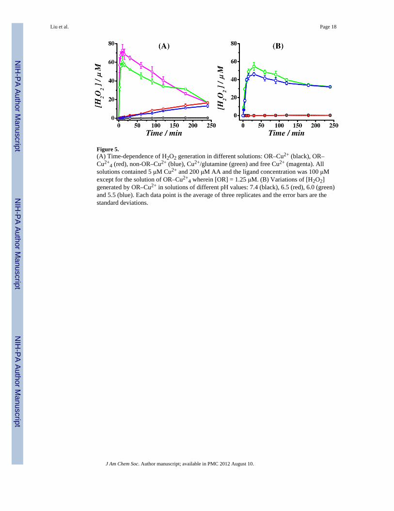
Figure 5.(A) Time-dependence of H2O2 generation in different solutions: OR–Cu2+ (black), OR–Cu2+
4 (red), non-OR–Cu2+ (blue), Cu2+/glutamine (green) and free Cu2+ (magenta). Allsolutions contained 5 μM Cu2+ and 200 μM AA and the ligand concentration was 100 μMexcept for the solution of OR–Cu2+
4 wherein [OR] = 1.25 μM. (B) Variations of [H2O2]generated by OR–Cu2+ in solutions of different pH values: 7.4 (black), 6.5 (red), 6.0 (green)and 5.5 (blue). Each data point is the average of three replicates and the error bars are thestandard deviations.
Liu et al. Page 18
J Am Chem Soc. Author manuscript; available in PMC 2012 August 10.
NIH
-PA Author Manuscript
NIH
-PA Author Manuscript
NIH
-PA Author Manuscript

Figure 6.Schematic representation of the possible roles of PrP–Cu2+ complexes in quenching theCu2+ redox cycling or gradual production of H2O2 for signal transduction. PrP is tethered tocell membrane via the glycosylphosphatidylinositol (GPI) anchor (green) with its α-helicesin the C terminus shown in orange, N-linked carbohydrates in purple, and the OR and non-OR domains near the N-terminus depicted in white. When [Cu2+] is at a low level (nM orlower) and at pH ranging between 5.5 and 7.4, Cu2+ (blue sphere) remains bound in the OR–Cu2+ mode (left), quenching the Cu2+ redox cycling. At higher [Cu2+] (μM) and a pH closerto the physiological value, the binding mode transitions to OR–Cu2+
4 (right), leading to agradual and controlled production of H2O2. Coordinates for the PrP C-terminal domain,along with carbohydrates, GPI anchor and membrane were kindly provided by ProfessorValerie Daggett (U. Washington).
Liu et al. Page 19
J Am Chem Soc. Author manuscript; available in PMC 2012 August 10.
NIH
-PA Author Manuscript
NIH
-PA Author Manuscript
NIH
-PA Author Manuscript

NIH
-PA Author Manuscript
NIH
-PA Author Manuscript
NIH
-PA Author Manuscript
Liu et al. Page 20
Table 1
Sequences of peptides used in this work
KKRPKPWGQ(PHGGGWGQ)4 PrP(23–28, 57–91) or OR
PHGGGWGQ OP
GGGTHNQWNKLSKPKTNLKH PrP(91–110) or non-OR
DAEFRHDSGYEVHHQK Aβ(1–16)
MDVFMKGLSKAKEGVVAAA α-Syn(1–19)
J Am Chem Soc. Author manuscript; available in PMC 2012 August 10.

NIH
-PA Author Manuscript
NIH
-PA Author Manuscript
NIH
-PA Author Manuscript
Liu et al. Page 21
Table 2
Redox potentials of Cu2+ complexes with select amyloidogenic molecules and biological redox couples.
Species E1/2 or E0 vs. Ag/AgCl vs. NHE References
OR–Cu2+/OR–Cu+ 0.126 V 0.323 V This work
O2/H2O2 0.099 0.296 48
Aβ–Cu2+/Aβ–Cu+ 0.080 0.277 47
PrP–Cu52+/PrP–Cu5
+ 0.070 0.267 30
α-Syn–Cu2+/α-Syn–Cu+ 0.018 0.215 40
OR–Cu2+ 4/OR–Cu+4 −0.025 0.172 This work
Free Cu2+/Cu+ −0.040 0.157 42
non-OR–Cu2+/non-OR–Cu+ −0.087* 0.110* This work
AA/DA −0.145 0.052 49
GSH/GSSG −0.425 −0.228 50
*Accuracy is affected by the less reversible redox wave of non-OR–Cu2+.42
J Am Chem Soc. Author manuscript; available in PMC 2012 August 10.