Tesis Doctoral Nakita Camara.pdf - accedaCRIS
219
-
Upload
khangminh22 -
Category
Documents
-
view
0 -
download
0
Transcript of Tesis Doctoral Nakita Camara.pdf - accedaCRIS


ADMINISTRATIVE FRAMEWORK


D. ANTONIO FERNÁNDEZ RODRIGUEZ, COORDINADOR DEL
PROGRAMA DE DOCTORADO DE SANIDAD ANIMAL Y SEGURIDAD
ALIMENTARIA DE LA UNIVERSIDAD DE LAS PALMAS DE GRAN CANARIA,
INFORMA QUE:
La Comisión Académica del Programa de Doctorado, en su sesión de
fecha / / tomó el acuerdo de dar el consentimiento para su
tramitación, a la tesis doctoral titulada “Stress Cardiomyopathy In Cetaceans:
Histological, Histochemical, Immunohistochemical and Biochemical
Studies” presentada por la doctoranda Dª Nakita Câmara y dirigida por el
Doctor Pedro Herráez Thomas y por la Doctora Eva Sierra Pulpillo.
Y para que así conste, y a efectos de lo previsto en el Artº 11 del
Reglamento de Estudios de Doctorado (BOULPGC 7/10/2016) de la Universidad
de Las Palmas de Gran Canaria, firmo la presente en Las Palmas de Gran
Canaria, a de de .


EPIGRAPH
__________________________________________________________________________________________________________________________________________________________________________
7
EPIGRAPH
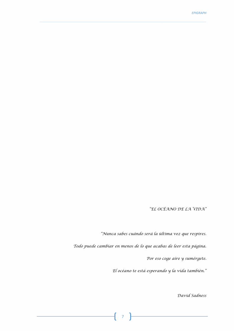
“EL OCÉANO DE LA VIDA”
“Nunca sabes cuándo será la última vez que respires.
Todo puede cambiar en menos de lo que acabas de leer esta página.
Por eso coge aire y sumérgete.
El océano te está esperando y la vida también.”
David Sadness


DEDICATION
__________________________________________________________________________________________________________________________________________________________________________
9
DEDICATION
This PhD thesis is dedicated to my lost loved ones, specially my aunt Sandra Miranda,
who watches for me in the other side. You will always be loved, never forgotten and forever
missed.
I also dedicate this work to my loving parents, Lena and Zé, and courageous sister Angie.
As Calum Scott says in one of his song:
“She/He said I love you no matter what
I just want you to be happy and always be who you are
She/He wrapped his arms around me
Said, "Don't try to be what you're not
'Cause I love you no matter what"
She/He loves me no matter what
They love me no matter what”
And finally, to Jesus, the one I love, who showed me that when two people really care
about each other, they always find a way to make it work. No matter how hard it is.


ACKNOWLEDGEMENTS
__________________________________________________________________________________________________________________________________________________________________________
11
ACKNOWLEDGEMENTS
The best kind of people are the ones that come into your life and make you see the sun
where you once saw clouds. The people that believe in you so much, you start to believe in
yourself too. The people that love you, simply for being you. The once in a lifetime kind of
people. Therefore, I would like to transmit my gratitude to some extraordinary people, who
made this work possible, not only as the result of my own efforts but also of theirs, specially to:
Pedro and Eva, my thesis directors: I am so grateful you took me under your wing. I
would not be where I am today without you. Your leadership and example in the pursue
of perfection in yours, mine and, finally, our work, has helped me grow into my
potential; and it has taught me that the best teachers teach from the heart and not
from a book.
Toño: for allowing me to do my PhD in the IUSA. I am truly privileged by the trust you
have placed in me and your precious advice, because as Ever Garrison said “A teacher
is a compass that activates the magnets of curiosity, knowledge and wisdom in the
pupils”.
Manolo: for the help solving many irregularities and setbacks encountered during the
development of this thesis; as well as for the teaching moments and feedback involving
the diagnosis of the different case studies.
Ani, my “Pegatina”: for making so many ordinary moments, extraordinary. For creating
a healthy work environment, for her tireless understanding, laboratory teachings and
help, and for the constant companionship.
Mercedes: for her administrative efficiency and her daily good disposition.
Javi: for his muscular strength, which was often needed in our work, and for the many
friendly conversations.
Antonio Espinosa, Marisa Andrada, Óscar, Miguel, Yara, Idaira, Simone, Silvia, Alicia,
and Pablo: for your fellowship and the time we spent together.
STRESS CARDIOMYOPATHY IN CETACEANS: HISTOLOGICAL, HISTOCHEMICAL, IMMUNOHISTOCHEMICAL AND BIOCHEMICAL STUDIES __________________________________________________________________________________________________________________________________________________________________________
12
In addition to all the people already mentioned, I want to thank all those who have indirectly
participated in the elaboration of this work, particularly to:
Josué and Carolina, who contributed to the study of the pathologies and causes of death
of cetaceans, thus allowing me to have a better idea of the animals included in my
thesis.
The staff of Palmitos Park, specially to Amelia and Óscar, who provided us serum
samples from their captive cetaceans for the establishment of normal troponin values.
The staff of Animal Lab, specially to Rubén, who performed the clinical analyses.
Angelo Santana, from the department of mathematics of the ULPGC, who helped with
the statistics.
All the staff and volunteers of the Cetacean Stranding Network, and associated
nongovernmental organizations (SECAC and Canarias Conservación), who collaborated
in the necropsies.
The Canary Islands Government and the Regional Government of Andalusia, who
funded and provided support to the stranding research network.
The Ministry of Science, Innovation and Universities, subsided by the Government of
Gran Canaria, who partially funded this research.
And, finally, the Ministry of Economy and Competitiveness (MINECO), for my
predoctoral grant and for the funding given to this investigation.
I also cannot fail to register my special thanks to:
Siani and the staff from East London Aquarium, and to Greg and the staff from Bayworld:
I am very grateful for the internship opportunity you gave me. I appreciate the
experience of working with you and learning from you.

ACKNOWLEDGEMENTS
__________________________________________________________________________________________________________________________________________________________________________
13
Ana, my first mentor: I will never be done thanking you for starting me on the cetacean
necropsies and for your constant friendship. I still remember I felt extremely blessed
when you accepted to be my master's thesis advisor. I will never forget the words you
said to me at that time: "Be careful, I am very demanding!" to which I replied, "That's
good, that's what I need!", because as Socrates said: "Under the direction of a strong
general, there will never be weak soldiers." I hope I haven't disappointed you so far
because the influence of a teacher can never be erased! And as Colleen Wilcox said “it
takes a big heart to help shape little minds”, and you definitely helped shaping mine so
far.
Furthermore, I would like to recognize the important part of a friend in our lives. A friend is
someone we turn to when our spirits need a lift. A friend is someone we treasure, for friendship
is a gift. A friend is someone who fills our lives with beauty, joy and grace. A friend turns the
world we live in a better and happier place. So, thank you Anna Maria, Cristian, Francesco, José
Angel, Maria Carolina, Marina, Marisa, Natalia, Raquel, Tania, Yania, Maria and Fede, Cristina
and family, and Maria for being my friends and basically my “Canarian family”, and also to my
(actual and future) borrowed nieces (Africa and Carmen) and nephews (Iván, Mateo, Lucas and
Thiago), who always puts a smile on my face.
As Thomas Aquinas said, “there is nothing on this earth more to be prized than true
friendship” and as Henry David Thoreau “nothing makes the earth seem so spacious as to have
friends at a distance; they make the latitudes and longitudes”. Therefore, I would also like to
express my gratitude to my friends around the world. To Nicole, from the White Shark Projects,
and to Che Rie, from Bayworld in South Africa. To Andrea, Bella, Isabella, Mar (and her little
Samuel) Marta, Oihane and Raquel del Solar, or as we call her “Mujer”, friends that I met while
working at IUSA. To Gabby, Lili, Luisa, Sofia and João, or as we call him “Rato”, friends from my
university in Portugal. And finally, to André (my Valentim) and to my oldest friend Vasco.
And as the last are said to be the first ...
I am eternally grateful to all the members of my family, my constant source of love and
inexhaustible understanding, who always encourage me with driving words of confidence,
showing me their admiration, emotional support and unwavering enthusiasm. As Brad Henry
said: “families are the compass that guide us. They are the inspiration to reach great heights,
and our comfort when we occasionally falter”.
STRESS CARDIOMYOPATHY IN CETACEANS: HISTOLOGICAL, HISTOCHEMICAL, IMMUNOHISTOCHEMICAL AND BIOCHEMICAL STUDIES __________________________________________________________________________________________________________________________________________________________________________
14
First, to Fátima, Marlene and Joana, mother and sisters not by blood but by heart, who have
always been present in both my life and my family’s life, and for often checking in on mom and
dad since me and my sister were not there.
Second, to Micas, my “mini me”, for helping me in the lab when you came to visit me and
for assisting mommy and daddy with the technological issues for the drawings inside this thesis.
Third, to Tojo (my brother-in-law and “Mr. Engineer” as I call him) for goofing around when
we spoke on the telephone and for being patient with my sister when she was helping me.
Unconditionally, to Angie (my sister and my hero), Lena (my mommy) and Zé (my daddy and
my “Cuddly cuddle” as I call him), I could never thank you enough for listening to me, for
protecting me from the things I shouldn’t do, for guiding me in the right direction, for putting
up with my mood swings and arrogance, for being there for me without a doubt. I know I never
tell you enough how much I love you. No matter what, I always will love you, no matter how
much we argue, or how much I upset you, I’ll love you till the day I die. I hope that the final result
of this work will be worthy of the help (with the English corrections and all the drawings of the
cover and each separator sheet), effort and trust you have all placed in me. Without you the
realization of it would not have been possible.
And finally, to Jesus, I may not be able to tell you every day but I just want you to know that
you mean the world to me. The day you stepped into my life, you changed it into something
really beautiful and meaningful. You hear my pain when everyone else ignores it. You always try
to make me smile when I think I can’t. You listen to my secrets and make them yours. You give
me a hug when I can’t find my voice. You wipe away the tears that the world makes me weep.
You mean more to me than you’ll ever know. You’re my best friend. You’re my safe harbour. I
can truly say that without the inspiration, drive, and support that you have given me in these
past few years, this work, probably, would not be finished and I would not be the person and
fighter I am today.

TABLE OF CONTENTS

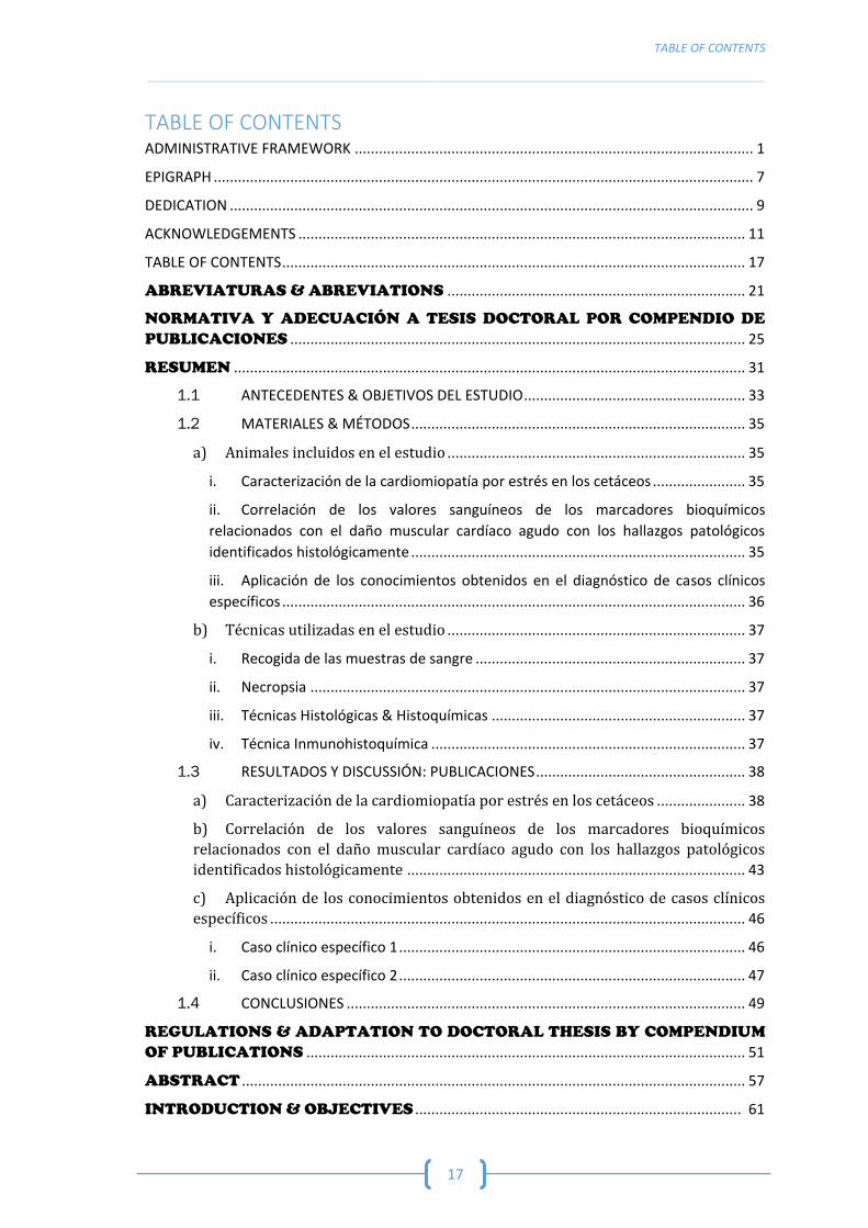
TABLE OF CONTENTS
__________________________________________________________________________________________________________________________________________________________________________
17
TABLE OF CONTENTS ADMINISTRATIVE FRAMEWORK ................................................................................................... 1
EPIGRAPH ...................................................................................................................................... 7
DEDICATION .................................................................................................................................. 9
ACKNOWLEDGEMENTS ............................................................................................................... 11
TABLE OF CONTENTS ................................................................................................................... 17
ABREVIATURAS & ABREVIATIONS .......................................................................... 21
NORMATIVA Y ADECUACIÓN A TESIS DOCTORAL POR COMPENDIO DE
PUBLICACIONES ................................................................................................................. 25
RESUMEN ............................................................................................................................... 31
1.1 ANTECEDENTES & OBJETIVOS DEL ESTUDIO ....................................................... 33
1.2 MATERIALES & MÉTODOS ................................................................................... 35
a) Animales incluidos en el estudio .......................................................................... 35
i. Caracterización de la cardiomiopatía por estrés en los cetáceos ....................... 35
ii. Correlación de los valores sanguíneos de los marcadores bioquímicos
relacionados con el daño muscular cardíaco agudo con los hallazgos patológicos
identificados histológicamente ................................................................................... 35
iii. Aplicación de los conocimientos obtenidos en el diagnóstico de casos clínicos
específicos ................................................................................................................... 36
b) Técnicas utilizadas en el estudio .......................................................................... 37
i. Recogida de las muestras de sangre ................................................................... 37
ii. Necropsia ............................................................................................................ 37
iii. Técnicas Histológicas & Histoquímicas ............................................................... 37
iv. Técnica Inmunohistoquímica .............................................................................. 37
1.3 RESULTADOS Y DISCUSSIÓN: PUBLICACIONES .................................................... 38
a) Caracterización de la cardiomiopatía por estrés en los cetáceos ...................... 38
b) Correlación de los valores sanguíneos de los marcadores bioquímicos
relacionados con el daño muscular cardíaco agudo con los hallazgos patológicos
identificados histológicamente .................................................................................... 43
c) Aplicación de los conocimientos obtenidos en el diagnóstico de casos clínicos
específicos ...................................................................................................................... 46
i. Caso clínico específico 1 ...................................................................................... 46
ii. Caso clínico específico 2 ...................................................................................... 47
1.4 CONCLUSIONES ................................................................................................... 49
REGULATIONS & ADAPTATION TO DOCTORAL THESIS BY COMPENDIUM
OF PUBLICATIONS ............................................................................................................. 51
ABSTRACT ............................................................................................................................. 57
INTRODUCTION & OBJECTIVES ................................................................................. 61
STRESS CARDIOMYOPATHY IN CETACEANS: HISTOLOGICAL, HISTOCHEMICAL, IMMUNOHISTOCHEMICAL AND BIOCHEMICAL STUDIES __________________________________________________________________________________________________________________________________________________________________________
18
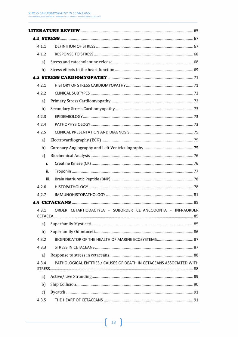
LITERATURE REVIEW ...................................................................................................... 65
4.1 STRESS......................................................................................................................... 67
4.1.1 DEFINITION OF STRESS ........................................................................................ 67
4.1.2 RESPONSE TO STRESS .......................................................................................... 68
a) Stress and catecholamine release ......................................................................... 68
b) Stress effects in the heart function ....................................................................... 69
4.2 STRESS CARDIOMYOPATHY ............................................................................. 71
4.2.1 HISTORY OF STRESS CARDIOMYOPATHY ............................................................. 71
4.2.2 CLINICAL SUBTYPES ............................................................................................. 72
a) Primary Stress Cardiomyopathy .......................................................................... 72
b) Secondary Stress Cardiomyopathy....................................................................... 73
4.2.3 EPIDEMIOLOGY .................................................................................................... 73
4.2.4 PATHOPHYSIOLOGY ............................................................................................. 73
4.2.5 CLINICAL PRESENTATION AND DIAGNOSIS ......................................................... 75
a) Electrocardiography (ECG) ................................................................................... 75
b) Coronary Angiography and Left Ventriculography ............................................. 75
c) Biochemical Analysis ............................................................................................. 76
i. Creatine Kinase (CK) ............................................................................................ 76
ii. Troponin .............................................................................................................. 77
iii. Brain Natriuretic Peptide (BNP) ........................................................................... 78
4.2.6 HISTOPATHOLOGY ............................................................................................... 78
4.2.7 IMMUNOHISTOPATHOLOGY ............................................................................... 81
4.3 CETACEANS .............................................................................................................. 85
4.3.1 ORDER CETARTIODACTYLA - SUBORDER CETANCODONTA - INFRAORDER
CETACEA………………………………………………………………………………………………………………………… 85
a) Superfamily Mysticeti ............................................................................................ 85
b) Superfamily Odontoceti ......................................................................................... 86
4.3.2 BIOINDICATOR OF THE HEALTH OF MARINE ECOSYSTEMS................................. 87
4.3.3 STRESS IN CETACEANS ......................................................................................... 87
a) Response to stress in cetaceans ............................................................................ 88
4.3.4 PATHOLOGICAL ENTITIES / CAUSES OF DEATH IN CETACEANS ASSOCIATED WITH
STRESS…………………………………………………………………………………………………………………………… 88
a) Active/Live Stranding ............................................................................................ 89
b) Ship Collision .......................................................................................................... 90
c) Bycatch ................................................................................................................... 91
4.3.5 THE HEART OF CETACEANS ................................................................................. 91
TABLE OF CONTENTS
__________________________________________________________________________________________________________________________________________________________________________
19
4.4 GEOGRAPHIC STUDY AREA .............................................................................. 95
4.4.1 CANARY ISLANDS ................................................................................................. 96
a) Features of the archipelago .................................................................................. 96
b) Cetaceans species observed in the archipelago .................................................. 96
4.4.2 ANDALUSIA .......................................................................................................... 98
a) Features of the area ............................................................................................... 99
b) Cetaceans species observed in the area ............................................................... 99
MATERIALS & METHODS .............................................................................................. 101
5.1 DETERMINATION OF THE STUDY ANIMALS ............................................ 103
5.1.1 ACTIVE/LIVE STRANDING .................................................................................. 103
5.1.2 SHIP COLLISIONS ............................................................................................... 103
5.1.3 BYCATCH ............................................................................................................ 104
5.1.4 ANIMALS INCLUDED IN THE STUDY .................................................................. 106
a) Characterization of Stress Cardiomyopathy ...................................................... 106
b) Correlation of the blood values detected for the biochemical markers of acute
cardiac muscle damage with the pathological findings identified histologically ... 107
c) Application of the knowledge obtained previously in the diagnosis of specific
clinical cases ................................................................................................................. 108
5.2 TECHNIQUES USED IN THE STUDY .............................................................. 111
5.1.1 BLOOD COLLECTION .......................................................................................... 111
5.1.2 NECROPSY TECHNIQUE ..................................................................................... 111
5.1.3 SAMPLE PREPARATION ..................................................................................... 113
a) Fixation and dehydration .................................................................................... 113
b) Paraffin Inclusion ................................................................................................ 114
5.1.4 HISTOLOGICAL AND HISTOCHEMICAL TECHNIQUES ......................................... 114
a) Haematoxylin and Eosin (HE) Staining .............................................................. 115
b) Periodic Acid Schiff (PAS) Staining .................................................................... 116
c) Phosphotungstic Acid-Haematoxylin (PTAH) Staining .................................... 116
d) Masson’s trichrome Staining .............................................................................. 117
5.1.5 IMMUNOHISTOCHEMICAL TECHNIQUE ............................................................ 118
RESULTS & DISCUSSION: PUBLICATIONS ............................................................ 123
6.1 CHARACTERIZATION OF STRESS CARDIOMYOPATHY (STRESS
CARDIOMYOPATHY IN STRANDED CETACEANS: AN HISTOLOGICAL, HISTOCHEMICAL AND
IMMUNOHISTOCHEMICAL STUDY) ....................................................................................... 125
STRESS CARDIOMYOPATHY IN CETACEANS: HISTOLOGICAL, HISTOCHEMICAL, IMMUNOHISTOCHEMICAL AND BIOCHEMICAL STUDIES __________________________________________________________________________________________________________________________________________________________________________
20
6.2 CORRELATION OF THE BLOOD VALUES DETECTED FOR THE
BIOCHEMICAL MARKERS OF ACUTE CARDIAC MUSCLE DAMAGE WITH
THE PATHOLOGICAL FINDINGS IDENTIFIED HISTOLOGICALLY
(INCREASED PLASMA CARDIAC TROPONIN I IN LIVE-STRANDED CETACEANS: CORRELATION
WITH PATHOLOGICAL FINDINGS OF ACUTE CARDIAC INJURY) ............................................. 149
6.3 APPLICATION OF THE KNOWLEDGE OBTAINED IN THE DIAGNOSIS
OF SPECIFIC CLINICAL CASES (SKELETAL AND CARDIAC RHABDOMYOPLYSIS IN A
LIVE-STRANDED NEONATAL BRYDE’S WHALE WITH FETAL DISTRESS) ................................. 171
6.4 APPLICATION OF THE KNOWLEDGE OBTAINED IN THE DIAGNOSIS
OF SPECIFIC CLINICAL CASES (CAPTURE MYOPATHY AND STRESS
CARDIOMYOPATHY IN A LIVE-STRANDED RISSO’S DOLPHIN [GRAMPUS GRISEUS] IN
REHABILITATION) .................................................................................................................. 185
CONCLUSIONS .................................................................................................................... 203
REFERENCES ...................................................................................................................... 207
ABREVIATURAS
&
ABREVIATIONS


ABREVIATURAS & ABREVIATIONS
__________________________________________________________________________________________________________________________________________________________________________
23
AMI – Infarto Agudo de Miocardio / Acute Myocardial Infarction
BUN – Nitrógeno Ureico en Sangre/ Blood Urea Nitrogen
CK – Creatina Quinasa / Creatine Kinase
CM – Miopatía de Captura / Capture Myopathy
cTnI – Troponina I Cardíaca / Cardiac Troponin I
cTnC – Troponina C Cardíaca / Cardiac Troponin C
ECG – Electrocardiografia / Electrocardiography
HE – Tinción Hematoxilina y Eosina / Haematoxylin & Eosin Stain
MO – Microscópio óptico / Optical Microscope
PAS – Tinción Ácido Periódico de Schiff / Periodic Acid Shiff Stain
PTAH – Tinción Hematoxilina Ácida Fosfotúngstica / Phosphotungstic Acid Haematoxylin Stain
SCMP – Cardiomiopatía por Estrés / Stress Cardiomyopathy


NORMATIVA Y
ADECUACIÓN A
TESIS DOCTORAL
POR COMPENDIO DE
PUBLICACIONES

NORMATIVA & ADECUACIÓN A TESIS DOCTORAL POR COMPENDIO DE PUBLICACIONES
__________________________________________________________________________________________________________________________________________________________________________
27
El Reglamento de estudios de doctorado de la Universidad de Las Palmas de Gran
Canaria, aprobado en Consejo de Gobierno de 26 de febrero de 2019 (Boletín Oficial de la ULPGC
de 04/03/2019), establece en el artículo 12 los requisitos generales que debe cumplir una tesis
doctoral por compendio de publicaciones:
1. Para la presentación de tesis por compendio de publicaciones será necesario:
a) Un mínimo de tres publicaciones, con unidad temática, indexadas en el Journal
Citations Reports, Arts and Humanities Citation Index o equivalentes, de las que el
doctorando sea el primer autor o autor principal. Al menos una de ellas deberá
haber sido publicada en una revista cuyo índice de impacto la sitúe dentro de la
primera mitad en orden decreciente de índice de impacto entre las revistas del área.
b) Para acreditar la condición de autor principal, esta deberá ser reconocida por el
resto de los autores de las publicaciones presentadas como núcleo de la tesis
doctoral, al mismo tiempo que estos deberán renunciar a utilizar estas
publicaciones como núcleo principal de otras tesis doctorales, sin perjuicio de que
dichas publicaciones puedan ser presentadas como méritos complementarios en las
tesis doctorales que pudieran presentar los otros autores de dichas publicaciones.
c) En áreas de especial incidencia tecnológica dos de estas publicaciones podrán ser
sustituidas por patentes en explotación o publicaciones en congresos reconocidos
por la ANEP en sus baremos para la obtención de sexenios.
d) Que en las publicaciones o patentes conste la ULPGC a través de la filiación del
director o del doctorando.
2. Las tesis doctorales presentadas como compendio de publicaciones deberán ajustarse
al formato establecido en los apartados del 1 al 3, del artículo 11 del presente
Reglamento y contener los apartados siguientes:
a) Una introducción en la que se presenten los objetivos de la tesis, los trabajos
publicados y la justificación de la unidad temática de la tesis.
b) Una copia de los trabajos publicados.
c) Las conclusiones finales.
d) En el caso de que lo dispuesto en los apartados a y c se haya redactado en una
lengua diferente del español, deberá incluirse un resumen en español según el
artículo 10 del presente reglamento, de una extensión de entre 5 y 20 páginas, en
el que se incluyan los objetivos y las conclusiones.

STRESS CARDIOMYOPATHY IN CETACEANS: HISTOLOGICAL, HISTOCHEMICAL, IMMUNOHISTOCHEMICAL AND BIOCHEMICAL STUDIES _________________________________________________________________________________________________________________________________________________________________________
28
Esta tesis doctoral cumple con lo establecido en el artículo mencionado anteriormente,
ya que se presentan cuatro publicaciones, teniendo todas estas a la doctoranda como primera
autora y están publicadas en revistas científicas del ámbito de conocimiento del programa de
doctorado. Las cuatro publicaciones cuentan con los indicios de calidad que exige el programa
de doctorado, esto es: los cuatro artículos están indexados en Journal Citations Reports.
A continuación, se detallan los indicios de calidad de las publicaciones:
1. Nakita Câmara, Eva Sierra, Carolina Fernández-Maldonado, Antonio Espinosa de los
Monteros, Manuel Arbelo, Antonio Fernández, Pedro Herráez. “Stress cardiomyopathy
in stranded cetaceans: a histological, histochemical and immunohistochemical study”.
doi: 10.1136/vr.105562. Veterinary Record. Impact Factor 2018: 2.101. Journal Rank in
Veterinary Sciences category: 16/141 (Q1).
2. Nakita Câmara, Eva Sierra, Antonio Fernández, Manuel Arbelo, Marisa Andrada,
Antonio Espinosa de los Monteros, Pedro Herráez. “Increased plasma cardiac troponin I
in live-stranded cetaceans: correlation with pathological findings of acute cardiac
injury”. doi: 10.1038/s41598-020-58497-3. Scientific Reports. Impact Factor: 4.011.
Journal Rank in Multidisciplinary Sciences category: 15/69 (Q1).
3. Nakita Câmara, Eva Sierra, Antonio Fernández, Cristian Manuel Suárez-Santana, Raquel
Puig-Lozano, Manuel Arbelo, Pedro Herráez. “Skeletal and cardiac rhabdomyolysis in a
live-stranded neonatal Bryde's whale with fetal distress”. doi:
10.3389/fvets.2019.00476. Frontiers in Veterinary Science. Impact Factor: 2.029.
Journal Rank in Veterinary Sciences category: 18/141 (Q1).
4. Nakita Câmara, Eva Sierra, Antonio Fernández, Manuel Arbelo, Yara Bernaldo de Quirós,
Marina Arregui, Francesco Consoli, Pedro Herráez. “Capture Myopathy and Stress
Cardiomyopathy in a Live-Stranded Risso’s Dolphin (Grampus griseus) in Rehabilitation”.
doi: 10.3390/ani10020220. Animals. Impact Factor: 1.832. Journal Rank in Veterinary
Sciences category: 29/141 (Q1).
En conformidad con lo establecido en el artículo 12 del Reglamento de estudios de
doctorado para la presentación de tesis por compendio de publicaciones, se muestra la unidad
temática de las publicaciones aportadas. La unidad temática de las publicaciones que integran
NORMATIVA & ADECUACIÓN A TESIS DOCTORAL POR COMPENDIO DE PUBLICACIONES
__________________________________________________________________________________________________________________________________________________________________________
29
esta tesis por compendio es manifiesta, pues todas versan sobre la Cardiomiopatía por Estrés
en cetáceos, habiendo abordado esta patología a través del estudio bioquímico, histológico,
histoquímico e inmunohistoquímico.
Finalmente, la estructura de esta tesis doctoral cumple lo establecido en el artículo 12.2
del Reglamento de estudios de doctorado, como puede comprobarse en las páginas siguientes,
pues ofrece:
a) una introducción en la que se presenta los objetivos de la tesis, la presentación de los
trabajos publicados y la justificación de la unidad temática de la tesis (revisión
bibliográfica);
b) una copia de los trabajos publicados (publicaciones);
c) las conclusiones finales.


RESUMEN


RESUMEN
__________________________________________________________________________________________________________________________________________________________________________
33
1.1 ANTECEDENTES & OBJETIVOS DEL ESTUDIO
Estudios previos han puesto de manifesto la susceptibilidad de los cetáceos al estrés, a
través de análisis de sangre, que demuestran la activación del eje Hipotalámico-Hipofisario-
adrenal con la consiguiente producción y liberación de glucocorticoides, mineralocorticoides y
catecolaminas (adrenalina y noradrenalina) (Prosser 1986; Spraker 1993).
Aunque los efectos beneficiosos de la activación del sistema de estrés son bien
conocidos, los animales pueden verse afectados negativamente por los efectos del estrés físico
y psicogénico en situaciones extremas o prolongadas (Cowan and Curry 2002).
Por lo tanto, los mamíferos marinos pueden desarrollar respuestas extremas frente al
estrés que causan un deterioro notable en la salud e incluso la muerte. Las lesiones cardíacas
parecen jugar un papel central en estas respuestas adversas al estrés en los cetáceos, y
diferentes autores sugieren que estos animales podrían estar particularmente predispuestos a
desarrollar cardiomiopatía por estrés (SCMP) probablemente debido a sus adaptaciones
cardiovasculares al metabolismo del buceo (Cowan and Curry 2002; Cowan, Harter, and Kandel
2000; Cozzi, Huggenberger, and Oelschläger 2017; Herráez et al. 2007, 2013).
En las últimas décadas, se han realizado esfuerzos para reducir el impacto que algunas
actividades humanas tienen en los cetáceos de vida libre, pero desafortunadamente las ballenas
y los delfines continúan amenazados, entre otros, por el varamiento activo (vivo) y posterior
interacción con los humanos, las colisiones con embarcaciones y las capturas accidentales
(bycatch). Estas entidades patológicas tienen el estrés agudo como eje común de su
etiopatogenia, pudiendo causar la muerte del animal o agravar una situación de enfermedad
previa, al tiempo que influye en su rehabilitación posterior, haciendo que la terapia y la
recuperación de los animales involucrados no tengan éxito en la mayoría de las ocasiones
(Arbelo et al. 2013; Bonsembiante et al. 2017; Cowan and Curry 2008; Díaz-Delgado et al. 2018;
Duignan and Jones 2005; Gulland, Dierauf, and Whitman 2018; Herráez et al. 2007, 2013; Sierra
et al. 2014, 2017; Soulsbury, Iossa, and Harris 2008).
El presente trabajo ha sido diseñado con el objetivo de continuar la investigación en
cetáceos con una doble intención: por un lado, caracterizar la cardiomiopatía por estrés,
tratando de proporcionar un mayor conocimiento sobre las entidades patológicas causantes y,
por otro lado, aplicar este conocimiento a soluciones concretas, utilizando la salud como un
instrumento necesario para la conservación de estos animales en nuestros mares.

STRESS CARDIOMYOPATHY IN CETACEANS: HISTOLOGICAL, HISTOCHEMICAL, IMMUNOHISTOCHEMICAL AND BIOCHEMICAL STUDIES __________________________________________________________________________________________________________________________________________________________________________
34
Estudios anteriores muestran que, en las Islas Canarias desde el año 1999 hasta el año
2005, el 27% (37 animales de un total de 138) de los animales vararon activamente y, en el
período comprendido entre el año 2006 al 2012, el 13% (30 animales de un total de 236).
Además, en Andalucía entre el año 2011 y el 2014, el 27% (28 animales de un total de 104) de
los animales estaban vivos cuando vararon (Arbelo et al. 2013; Díaz-Delgado et al. 2018;
Maldonado 2015). Por lo tanto, es extremadamente importante estudiar los parámetros
bioquímicos relacionados con la SCMP en los cetáceos, ya que es de nuestro interés poder
reintroducir al medio marino estos animales con el mínimo daño posible. Del mismo modo, la
medición de estos parámetros en el momento del varamiento y el uso de medicamentos que
controlan los efectos negativos de la liberación de altas concentraciones de catecolaminas al
torrente sanguíneo, responsables entre otros del daño cardíaco.
Así, y con estos antecedentes, el objetivo general de este estudio ha sido caracterizar la
SCMP bioquímica y morfológicamente, como lesiones resultantes de respuestas extremas al
estrés en cetáceos varados activamente y sometidos a manipulación e interacción con humanos,
cetáceos muertos debido a colisiones con barcos e interacción con actividades pesqueras
(bycatch).
Esto se logrará a través de los siguientes objetivos específicos:
1. Caracterizar la cardiomiopatía por estrés (SCMP), como lesión resultante de las
respuestas al estrés extremo en los cetáceos que han varados activamente y han
sido sometidos a manipulación e interacción con humanos, que han muerto tras
la colisión con una embarcación o como consecuencia de interacciones
pesqueras (captura accidental o bycatch).
2. Correlacionar los valores sanguíneos de los marcadores bioquímicos
relacionados con el daño muscular cardíaco agudo con los hallazgos patológicos
identificados histológicamente, en cetáceos varados activamente y sometidos a
manipulación e interacción con humanos.
3. Aplicar los conocimientos obtenidos en el diagnóstico de casos clínicos
específicos.
RESUMEN
__________________________________________________________________________________________________________________________________________________________________________
35
1.2 MATERIALES & MÉTODOS
a) Animales incluidos en el estudio
Para realizar este estudio, utilizamos muestras de corazón de cetáceos (ballenas y delfines)
varados en diferentes localizaciones geográficas del archipiélago canario y Andalucía. Estos
tejidos se almacenan en el banco de tejidos del Centro Atlántico de Cetáceos, División de
Histología y Patología Animal, del Instituto Universitario de Sanidad Animal y Seguridad
Alimentaria, perteneciente a la Universidad de Las Palmas de Gran Canaria (ULPGC).
Para cada objetivo específico de este trabajo se realizó una selección de muestras
concretas, la cual se explica a continuación:
i. Caracterización de la cardiomiopatía por estrés en los cetáceos
La primera fase de este objetivo específico consistió en la selección de los animales del
estudio (n=148), incluyendo los que vararon activamente y posteriormente murieron antes o
durante el rescate y la rehabilitación (n=83), los que murieron debido a la colisión con una
embarcación (n= 32), y los que murieron atrapados en redes de pesca (n=33). Todos estos
animales aparecieron varados en las costas de las Islas Canarias entre los años 2000 y 2016 y en
las costas de Andalucía entre 2011 y 2014.
Posteriormente, del total de animales se seleccionaron 67 (48 varados activamente, 7
muertos por colisión con embarcaciones y 12 por captura accidental) en función de su estado
de conservación, descartándose aquellos que se encontraban en un estado de autolisis avanzada
(grado 4) o muy avanzada (grado 5). Igualmente se descartaron aquellos animales que habían
sido congelados previamente a la realización de la necropsia ya que generan falsos negativos al
usar técnicas histoquímicas e inmunohistoquímicas (ASCOBANS/ACCOBAMS 2019; Godinho
2010).
ii. Correlación de los valores sanguíneos de los marcadores bioquímicos
relacionados con el daño muscular cardíaco agudo con los hallazgos
patológicos identificados histológicamente
La primera fase de este objetivo específico consistió en la determinación de valores
normales de troponina I cardíaca (cTnI) en cetáceos, específicamente en delfines mulares (n=5)
pertenecientes a un zoológico local (Palmitos Park). Para lograr esto, se recolectaron un total de

STRESS CARDIOMYOPATHY IN CETACEANS: HISTOLOGICAL, HISTOCHEMICAL, IMMUNOHISTOCHEMICAL AND BIOCHEMICAL STUDIES __________________________________________________________________________________________________________________________________________________________________________
36
20 muestras de sangre en los meses de junio, septiembre y diciembre de 2018; así como en
marzo de 2019 (4 muestras de cada individuo, 1 muestra por mes). Para calcular el intervalo de
referencia, que permite establecer un rango de valores mínimo y máximo dentro de los cuales
se encuentra el 95% de los valores observables de la variable en una población sana de
referencia, se realizó una estimación de la variabilidad intersujetos mediante el método de
estimación de componentes de la varianza descrito en Rasch and Mašata (2006), y se asumió
distribución normal en las variables para construir los intervalos de referencia tal como se
describe en Friedrichs et al. 2012.
En la segunda fase, se incluyeron en este estudio un total de 9 animales que vararon
activamente. Se recogieron muestras de sangre de estos animales, siendo una de las muestras
tomadas ante-mortem y el resto post-mortem debido a la muerte de los animales antes o
durante la manipulación, transporte y/o rescate/rehabilitación (n=8). Todos estos animales
vararon en las costas de las Islas Canarias desde principios de 2016 hasta junio de 2019.
Durante la tercera fase, se seleccionaron un total de 7 animales, de los animales
incluidos (n=9) en la fase anterior, para el estudio histológico e histoquímico, ya que no pudimos
realizar la necropsia en 2 animales, porque uno fue liberado de vuelta al mar y otro fue utilizado
con fines de investigación anatómica.
En la cuarta fase, 2 animales (de la fase anterior) fueron eliminados del estudio debido
a su congelación previa. Por lo tanto, al final de esta fase, se estudiaron un total de 5 individuos
con el uso de la técnica inmunohistoquímica para la detección de diferentes marcadores.
iii. Aplicación de los conocimientos obtenidos en el diagnóstico de casos
clínicos específicos
Para lograr este objetivo específico, se seleccionaron cuidadosamente 2 animales en
función de características específicas.
El primer animal fue seleccionado en base al hecho de que era un ejemplar, que varó
activamente, perteneciente al infraorden Mysticeti, ya que la descripción de la SCMP en estas
especies es aún escasa.
El segundo caso de estudio se centra en un animal varado activamente que estuvo en
rehabilitación durante 6 días, lo que nos permitió obtener muestras de sangre consecutivas in
vivo.

RESUMEN
__________________________________________________________________________________________________________________________________________________________________________
37
b) Técnicas utilizadas en el estudio
i. Recogida de las muestras de sangre
Se obtuvieron muestras de sangre de la aleta caudal de cada animal que fueron
depositadas en tubos sin anticoagulante. Posteriormente, las muestras fueron centrifugadas a
3500 rpm durante 5 minutos, dos veces para obtener el suero (aproximadamente 1 ml).
ii. Necropsia
Se realizó el examen postmortem completo siguiendo el protocolo estándar publicado
por la European Cetacean Society, con algunos procedimientos adicionales detallados en el
manual Marine Mammals Ashore: A Field Guide for Strandings (Geraci and Lounsbury 2005;
Kuiken and Hartmann 1991). El estado de conservación de los animales también se determinó
siguiendo los parámetros y clasificaciones establecidas por estos dos protocolos.
Muestras de tejido representativas de cada órgano se fijaron en formalina al 10%
durante aproximadamente 48h y se procesaron utilizando el protocolo estándar. Fueron
colectadas y posteriormente analizadas muestras de músculo cardíaco (tanto aurículas como
ventrículos), válvulas auriculoventriculares (bicúspides o mitrales y tricúspides), válvulas
semilunares (sigmoide aórtico y sigmoide pulmonar con las correspondientes arterias). En los
casos clínicos específicos, debido a la posible presencia de CM, los músculos esqueléticos
(longissimus dorsi y rectus abdominis) y los riñones fueron también analizados.
iii. Técnicas Histológicas & Histoquímicas
Se realizaron secciones de tejido (4 µm de grosor) para la tinción de hematoxilina y
eosina (HE) y tinción de ácido periódico de Schiff (PAS), mientras que se utilizaron cortes de 5
µm de grosor para las técnicas de hematoxilina ácida fosfotúngstica (PTAH) y tricrómico de
Masson.
iv. Técnica Inmunohistoquímica
Las secciones de tejido (3 µm de grosor) se inmunomarcaron con los anticuerpos anti-
mioglobina (1:200), anti-fibrinógeno (1:50), anti-troponina I cardíaca (1:25) y anti-troponina C
cardíaca (1:250). Los anticuerpos primarios fueron visualizados usando el VECTASTAIN Kit Elite
ABC-Peroxidase (PK-6100) de Vector Laboratories (Peterborough, Reino Unido).

STRESS CARDIOMYOPATHY IN CETACEANS: HISTOLOGICAL, HISTOCHEMICAL, IMMUNOHISTOCHEMICAL AND BIOCHEMICAL STUDIES __________________________________________________________________________________________________________________________________________________________________________
38
El control negativo consistió en secciones en serie de corazón sin el anticuerpo primario.
Por el contrario, el control positivo de mioglobina y fibrinógeno provenía de una muestra de
corazón de delfín listado (Stenella coeruleoalba). El delfín había varado activamente y desarrolló
miopatía de captura (CM) debido a la manipulación en sí y a la interacción humana durante el
proceso de rehabilitación (Herráez et al. 2007, 2013). Finalmente, se usaron muestras de
corazón de un cerdo y un cetáceo, sin lesiones macroscópicas y/o histológicas, como controles
positivos para la troponina I y troponina C (cTnC).
1.3 RESULTADOS Y DISCUSIÓN: PUBLICACIONES
a) Caracterización de la cardiomiopatía por estrés en los cetáceos
El primer objetivo específico de este trabajo, que consistió en caracterizar la SCMP en
los cetáceos como lesiones resultantes de respuestas de estrés extremo, se logró mediante
análisis histológicos, histoquímicos e inmunohistoquímicos en un total de 67 animales que
vararon activamente (n=48) o murieron debido a colisiones con embarcaciones (n=7) e
interacciones con pesca (bycatch) (n=12).
Histológicamente, las principales lesiones cardíacas asociadas con el estrés fueron
cambios vasculares, que se presentaron como congestión, hemorragias y edema intersticial;
lesiones degenerativo-necróticas cardíacas agudas o subagudas, que se atribuyeron a la
cardiotoxicidad por las catecolaminas y consistieron en necrosis en banda de contracción, fibras
en acordeón, hipereosinofilia citoplasmática y vacuolización perinuclear; la presencia de
glóbulos de mioglobina intersticiales y la infiltración de células inflamatorias. Estos hallazgos
coinciden con los descritos en análisis histológicos de biopsias y/o de tejido miocárdico post-
mortem de humanos con SCMP (Akashi et al. 2010; Fineschi et al. 2010; Jaspreet, Wangde, and
A. 2019; Jiang and Downing 1990; Kawai 2012; Maréchaux et al. 2008; Mitchell and Marquis
2017; Miura et al. 2017; Pascual, Abó, and Piqué 2015; Prasad, Lerman, and Rihal 2008).
La congestión subendocárdica, el edema intersticial y las hemorragias se detectan
mediante abordajes histológicos y forman parte de la SCMP (Jiang and Downing 1990).
Se observó congestión vascular en 45 de los 67 animales (67,2%); hallazgo vascular que
está frecuentemente presente en situaciones estresantes (Cowan and Curry 2008; Díaz-Delgado
et al. 2018; Fishbein 2001; Herráez et al. 2007, 2013).

RESUMEN
__________________________________________________________________________________________________________________________________________________________________________
39
Las muestras de tejido de 26 animales (38.8%) contenían fibras separadas por espacios
claros, que representan edema intersticial (Jaspreet et al. 2019). Después de la administración
de catecolaminas, el edema intersticial generalmente se asocia con hemorragias
subendocárdicas y subepicárdicas. El edema intersticial está característicamente presente en las
áreas dañadas del miocardio, incluso después de 72 horas de actuar la causa desencadenante
(Fishbein 2001). En nuestro estudio, detectamos edema y hemorragias en 8 de los 67 animales
analizados (11,9%).
Las hemorragias subendocárdicas focales y difusas son visibles en el examen
macroscópico. Estas lesiones también se detectan en ocasiones en el miocardio y el epicardio,
poco después de la administración y/o liberación de grandes dosis de catecolaminas en
humanos con SCMP (Jiang and Downing 1990; Mitchell and Marquis 2017). Quince animales
(22,4%) presentaron hemorragias de localización epicárdica y/o subendocárdica detectables
tanto macro como microscópicamente. Estos hallazgos coinciden con estudios previos
centrados en muertes agudas asociadas con el estrés, particularmente en animales que
murieron después de un varamiento activo y subsecuente
manipulación/rehabilitación/recuperación (Díaz-Delgado et al. 2018; Herráez et al. 2007, 2013).
La necrosis en banda de contracción se desarrolla entre los 5 a 10 minutos después de
un episodio de isquemia transitoria y reperfusión. Es una lesión músculo-esquelética y
miocárdica característica asociada con la administración de catecolaminas, o con altas
concentraciones de catecolaminas endógenas (Fineschi et al. 2010; Reichenbach and Benditt
1970; Turnbull and Cowan 1998). Es, por tanto, una lesión que se describe ampliamente como
un indicador de SCMP. La necrosis en banda de contracción ha sido descrita en humanos como
consecuencia de eventos estresantes (Akashi et al. 2010; Fineschi et al. 2010; J. et al. 2008; Jiang
and Downing 1990; Kawai 2012; Maréchaux et al. 2008; Mitchell and Marquis 2017; Miura et al.
2017; Pascual et al. 2015; Prasad et al. 2008), así como en otros animales, como focas (Seguel et
al. 2014) y cetáceos, debido a muertes agudas como las producidas por colisiones con barcos,
captura accidental, y/o varamientos activos con subsecuente estrés por el manejo (Arbelo et al.
2013; Cowan and Curry 2002, 2008; Díaz-Delgado et al. 2018; Herráez et al. 2007, 2013; Sierra
et al. 2014). En nuestro estudio, 33 animales presentaron necrosis en banda de contracción
(49.3% de todos los animales y 100% de los animales que murieron por colisión con
embarcación, 52.1% de los animales varados activamente y 8.3% de los animales que murieron
por la captura accidental). Esta condición fue más marcada a nivel subendocárdico y

STRESS CARDIOMYOPATHY IN CETACEANS: HISTOLOGICAL, HISTOCHEMICAL, IMMUNOHISTOCHEMICAL AND BIOCHEMICAL STUDIES __________________________________________________________________________________________________________________________________________________________________________
40
subepicárdico. El daño fue más severo en los animales que murieron por colisiones con barcos,
lo que coincide con estudios previos (Adegboyega, Haque, and Boor 1996; Sierra et al. 2014).
El primer hallazgo histológico asociado a la isquemia es la presencia de fibras onduladas
largas y adelgazadas, denominadas fibras en acordeón (Fineschi et al. 2010; Fishbein 2001). Este
hallazgo se detectó en 29 animales presentes en nuestro estudio (43.3%). Esta lesión ha sido
previamente descrita en animales que murieron súbitamente tras un episódio estresante
(Cowan and Curry 2002, 2008; Herráez et al. 2007, 2013). Es probable que las fibras onduladas
resulten de las fuerzas sistólicas de las fibras viables inmediatamente adyacentes a las fibras
muertas no contráctiles (Fishbein 2001; Kumar et al. 2015). Aunque las fibras en acordeón no se
consideren fibras en degeneración y/o necrosis si pueden ser usadas como un indicador
temprano de lesión miocárdica isquémica. Este hecho ha sido reproducido experimentalmente
tras la oclusión de las arterias coronarias (Fishbein 2001).
La hipereosinofilia es el primer cambio confirmado específico de la necrosis miocárdica
utilizando técnicas histológicas (HE) e histoquímicas (PTAH y el tricrómico de Masson). Consiste
en un aumento de la tinción eosinofílica (HE), azul (PTAH) o roja (tricrómico), respectivamente,
de los cardiomiocitos necróticos (Fineschi et al. 2010; Fishbein 2001). En nuestro estudio, todos
los animales (100%) exhibieron hipereosinofilia citoplasmática. En estudios experimentales, este
cambio de color y edema intersticial sutil es evidente 2 a 3 horas después de la oclusión
coronaria, y es más pronunciado y detectable a las 3-6 horas (Fishbein 2001). Los animales que
mueren debido a una situación estresante presentan este cambio citoplasmático (Cowan and
Curry 2002, 2008; Herráez et al. 2007, 2013; Sierra et al. 2014). La hipereosinofilia fue más
pronunciada en la zona subendocárdica y subepicárdica, lo que respalda estudios anteriores que
mostraron cómo progresa la lesión irreversible en un movimiento de frente de onda desde el
subendocardio isquémico. El déficit de perfusión es más grave en el subendocardio ya que el
subepicardio recibe un flujo sanguíneo colateral (Buja 2005; Cowan and Curry 2002, 2008).
La degeneración vacuolar se caracteriza, morfológicamente, por la acumulación
intracelular de líquido y la lisis de miofibrillas. Este hallazgo se detectó en 65 animales (96,9%)
siendo más notable a nivel subendocárdico y subepicárdico. Estudios previos lo detectaron
comúnmente en la periferia de los infartos de miocardio y en las regiones subepicárdicas,
subendocárdicas y perivasculares que sufren de isquemia grave, mantenida y letal (Adegboyega
et al. 1996; Cowan and Curry 2002, 2008). Si bien esta condición es bastante común, las
características morfológicas y la importancia funcional de la degeneración vacuolar son poco
conocidas. Aunque no se conoce la patogénesis, se hipotetiza que es el resultado del aumento

RESUMEN
__________________________________________________________________________________________________________________________________________________________________________
41
de la permeabilidad de la membrana celular del miocardio, con el consiguiente flujo de fluidos,
inducida por estados hipóxicos. Este fenómeno se observa normalmente en muertes agudas
asociadas con situaciones estresantes (Adegboyega et al. 1996; Cowan and Curry 2002, 2008).
Estudios anteriores han demostrado la necesidad de corroborar los hallazgos
histológicos con marcadores específicos que podrían exponer mejor el daño al miocardio
(Bonsembiante et al. 2017; Herráez et al. 2007, 2013; Seguel et al. 2014). Por lo tanto, en este
estudio, comparamos los marcadores que se han utilizado previamente en estudios similares,
como la mioglobina y el fibrinógeno (Herráez et al. 2007, 2013; Ortmann, Pfeiffer, and
Brinkmann 2000; Sierra et al. 2014; Xiaohong et al. 2002), con los marcadores utilizados en
estudios de muestras de corazón humano con SCMP, como la cTnI y la cTnC (Fishbein et al. 2003;
Hansen and Rossen 1999; Martínez-Díaz et al. 2005; Ortmann et al. 2000). La depleción de
proteínas musculares (esqueléticas y cardíacas) comienza inmediatamente después de un daño
celular. Como resultado, se produce una ruptura temprana de la membrana celular del
miocardio, lo que provoca una disminución rápida del contenido de mioglobina, cTnI y cTnC,
junto con el depósito de proteínas plasmáticas, como el fibrinógeno, en los cardiomiocitos. La
tinción homogénea de la mioglobina se observa en el músculo cardíaco normal, mientras que
los cardiomiocitos lesionados muestran tanto la depleción de la mioglobina dentro de las fibras
musculares como la acumulación intercelular e intersticial de mioglobina (Herráez et al. 2007,
2013; Ortmann et al. 2000; Sierra et al. 2014; Xiaohong et al. 2002). El estudio actual confirmó
la pérdida de mioglobina y la acumulación de fibrinógeno, de los cardiomiocitos degenerados
en la zona adyacente, así como dentro de la necrosis en banda de contracción.
Este estudio es el primero en utilizar marcadores específicos, como cTnI y cTnC, para
detectar daños en el corazón de los cetáceos.
La troponina es un complejo regulador de tres subunidades de proteínas liberadas de
los cardiomiocitos, cuando se produce un daño irreversible del miocardio. Las tres subunidades
son la Troponina C (componente de unión a calcio), la Troponina T (componente de unión a
tropomiosina) y la Troponina I (componente inhibidor). La subunidad cTnI es, en la actualidad,
el biomarcador más aceptado, ya que es altamente específico para el tejido cardíaco y
proporciona una alta precisión diagnóstica del infarto de miocardio (Lewandrowski, Chen, and
Januzzi 2002). Aunque cTnI y cTnC se deplecionan en los cardiomiocitos dañados, como se
demostró en nuestro estudio, estos dos marcadores se expresaron intensamente en la necrosis
en banda de contracción y en algunas células aisladas que mostraban degeneración. Estudios
previos indican que las células que sufren apoptosis tienen una mayor concentración de
STRESS CARDIOMYOPATHY IN CETACEANS: HISTOLOGICAL, HISTOCHEMICAL, IMMUNOHISTOCHEMICAL AND BIOCHEMICAL STUDIES __________________________________________________________________________________________________________________________________________________________________________
42
troponina debido a la condensación, mientras que los antígenos tisulares se agotan
severamente en zonas con necrosis isquémica evidente (zonas de infarto) (Fishbein et al. 2003;
Hansen and Rossen 1999; Martínez-Díaz et al. 2005; Ortmann et al. 2000).
Estudios recientes apoyan el concepto de que la patogénesis de la SCMP es causada por
isquemia miocárdica aguda. En nuestro estudio, identificamos y describimos los cardiomiocitos
dañados (es decir, células individuales o grupos de células dañadas). Estas células comúnmente
presentan hipereosinofilia y vacuolización citoplasmática en una distribución multifocal,
particularmente en las filas de cardiomiocitos localizados en la periferia de los vasos sanguíneos.
Este patrón lesional perivascular, principalmente observado en animales varados activamente,
sugiere que la patogénesis de estas lesiones se asocia con fenómenos de isquemia y subsecuente
reperfusión. La lesión por isquemia-reperfusión es un fenómeno que se describe como una
lesión acelerada en el corazón, debido a que el aporte sanguíneo se reabastece en el área
isquémica del corazón que anteriormente estaba privada de su suministro de sangre (isquemia).
Como resultado, estas áreas son objeto de hipoxia, pérdida sustancial de la regulación del
volumen celular y posterior reentrada de calcio debido al funcionamiento inadecuado de las
bombas de iones. El daño a la membrana también ocurre cuando se restablece el suministro de
sangre en áreas con células potencialmente viables (Buja 2005; Buja and Butany 2015).
Otro hallazgo anatomopatológico descrito en el examen post mortem y las biopsias
endomiocárdicas de pacientes humanos y animales experimentales con SCMP es la infiltración
intersticial de linfocitos, neutrofilos y macrófagos (Jaspreet et al. 2019; Mitchell and Marquis
2017; Pascual et al. 2015). En este estudio, se identificaron diferentes tipos de células
inflamatorias en 17 animales (25,4%). Estas células se infiltraron en zonas con hemorragias,
ruptura fibrilar de fibras, degeneración y necrosis de células cardíacas.
Estos resultados dieron lugar a la publicación de un artículo titulado Stress
cardiomyopathy in stranded cetaceans: a histological, histochemical and
immunohistochemical study, en Veterinary Record, Volume 185, Issue 22 (doi:
10.1136/vr.105562), teniendo esta revista 2.101 de índice de impacto en 2018 y siendo, por eso,
una revista Q1 en la categoría de ciencias veterinarias.
RESUMEN
__________________________________________________________________________________________________________________________________________________________________________
43
b) Correlación de los valores sanguíneos de los marcadores bioquímicos
relacionados con el daño muscular cardíaco agudo con los hallazgos
patológicos identificados histológicamente
El segundo objetivo específico de este trabajo consistió en correlacionar los valores
sanguíneos detectados para los marcadores bioquímicos de daño muscular cardíaco agudo
[creatina quinasa (CK) y troponina I cardíaca (cTnI)] con los hallazgos patológicos identificados a
nivel microscópico.
En humanos, los criterios de diagnóstico de la SCMP comprenden alteraciones en los
exámenes médicos (como electrocardiografía, ecocardiografía, cateterismos cardíacos) y
análisis bioquímicos. Las anormalidades más comunes a nivel laboratorial consisten en el
aumento discreto y temprano de cTnI y/o CK (Fineschi et al. 2010; J. et al. 2008; Lyon et al. 2016).
La enzima sérica más utilizada en la determinación del daño neuromuscular en animales
es la creatina quinasa (CK); También se utiliza en la detección de lesiones miocárdicas en
humanos (Lewandrowski et al. 2002; Valberg 2008). La CK aumenta a las 3–12 horas después de
la lesión muscular, alcanzan sus máximos valores 12–24 horas y vuelve a la línea basal después
de 48–72 horas, a menos que se haya producido una nueva lesión o daño permanente
(Lewandrowski et al. 2002). Todos los animales analizados en nuestro estudio, y en comparación
con la literatura publicada, presentaron valores de CK elevados (animales 1, 3, 5, 6, 7 y 9).
Sin embargo, aunque la CK se considera un marcador sensible de daño miocárdico,
también está presente en los músculos esqueléticos en altas concentraciones, así como en el
intestino, el diafragma, el útero y la próstata en cantidades menores; por lo tanto, tiene poca
especificidad cuando se usa para detectar daño cardíaco agudo (Khan 2015). En consecuencia,
las troponinas se han aceptado como el principal marcador en patología humana, ya que cTnI es
detectable en cantidades muy bajas (por ejemplo, 0,01 µg/L) en la sangre de individuos sanos
sin evidencia de enfermedad cardíaca (Hassan et al. 2009; Lewandrowski et al. 2002; Morrow et
al. 2007; Venge et al. 2009). Por lo tanto, se cree que las elevaciones significativas (≥0.1 µg/L) de
este marcador probablemente reflejan necrosis miocárdica, y algunos autores lo han descrito
como cardioespecífico debido no sólo a su especificidad para el tejido miocárdico, sino también
por su alta sensibilidad (Hassan et al. 2009; Lewandrowski et al. 2002; Morrow et al. 2007). Por
esta razón, la cTnI se utiliza para detectar varias patologías cardíacas, como el infarto agudo de
miocardio (AMI) y la SCMP (Khan 2015; Lewandrowski et al. 2002; Lyon et al. 2016).

STRESS CARDIOMYOPATHY IN CETACEANS: HISTOLOGICAL, HISTOCHEMICAL, IMMUNOHISTOCHEMICAL AND BIOCHEMICAL STUDIES __________________________________________________________________________________________________________________________________________________________________________
44
En la primera fase de este trabajo, estimamos un rango de referencia para cTnI en
cetáceos, ya que no existen datos en la literatura actual. Para lograr esto, se recolectaron un
total de 20 muestras de sangre, en junio, septiembre y diciembre de 2018 y marzo de 2019 (4
muestras de cada individuo, 1 muestra por mes) de 5 delfines mulares mantenidos bajo el
cuidado humano.
Así, el rango basal obtenido en este estudio y aplicable para cetáceos va desde 0 µg/L
(como valor mínimo) hasta 0.025 µg/L (como valor máximo).
Al comparar este rango en particular (0–0.0256 µg/L) con los valores normales para
humanos (≤0.1 µg/L) y perros (≤0.03–0.07 µg/L), podemos concluir que el intervalo de referencia
para cTnI en delfines mulares es más bajo que en las otras especies (Padilla n.d.; Sleeper, Clifford,
and Laster 2001; Wray 2017).
La segunda fase consistió en comparar los valores de 9 cetáceos varados activamente
con el rango normal de cetáceos (determinado en la fase previa).
Nuestros resultados, de los cetáceos varados activamente, mostraron que 8 animales
[animal 1 (0.06 µg/L), animal 2 (40.00 µg/L), animal 4 (0.235 µg/L), animal 5 (0.249 µg/L), animal
6 (0,748 µg/L), animal 7 (0.033 µg/L), animal 8 (0.06 µg/L) y animal 9 (0.049 µg/L)] presentaron
un aumento en comparación con el valor normal/basal para cTnI (0–0.0256 µg/L) que obtuvimos
de delfines mulares bajo cuidado humano y que uno de los animales [animal 3 (0.025 µg/L)]
presentó un valor de cTnI dentro del rango normal.
En general, los cardiomiocitos lesionados liberan troponina 3–9 horas después del daño
isquémico, alcanzan su nivel sérico máximo después de 12–48 horas y permaneciendo elevados
durante 4–7 días (Hansen and Rossen 1999; Khan 2015; Lewandrowski et al. 2002). El
reconocimiento temprano de la necrosis miocárdica (1–3 horas) no es posible mediante el
monitoreo de la troponina sérica, y estos marcadores son ineficaces hasta 6 o más horas después
del inicio del AMI y/o SCMP (Hansen and Rossen 1999; Khan 2015; Lewandrowski et al. 2002).
La determinación precisa del momento del evento de estrés y/o aparición de síntomas a
menudo es excepcionalmente difícil porque se centra en el informe clínico del paciente. Por lo
tanto, en humanos, una condición previa para obtener una capacidad satisfactoria para
distinguir estas patologías es que la sangre se debe recolectar entre 6 y 9 horas después del
inicio de la causa desencadenante (Khan 2015). En el caso de un evento de varamiento activo, a
menudo también es clínicamente y patológicamente desafiante conocer el momento exacto del
episodio estresante, ya que estamos trabajando con animales salvajes, redes de varamientos y
RESUMEN
__________________________________________________________________________________________________________________________________________________________________________
45
el público en general. Cuando se notifica el varamiento, es importante reconocer que el animal
podría haber varado recientemente o ser detectado mucho tiempo después de haber varado.
Además, estudios previos han propuesto que medir el nivel de troponina en el suero
puede ser un método auxiliar importante para examinar la muerte súbita, ya que su
concentración máxima puede estar relacionada con el grado de lesión tisular (Khan 2015). Por
esta razón, este hallazgo se confirmó con los análisis histológicos, histoquímicos e
inmunohistoquímicos (tercera y cuarta fase del estudio).
Una lesión isquémica aguda, como la que ocurre en el AMI o SCMP, se determina por
alteraciones morfológicas, que consisten en cambios vasculares y lesiones degenerativas agudas
basadas en el análisis histológico del tejido miocárdico (Akashi et al. 2010; Buja 2005; Buja and
Butany 2015; Câmara et al. 2019; Cebelin and Hirsch 1980; Fineschi et al. 2010; Jaspreet et al.
2019; Jiang and Downing 1990; Kawai 2012; Lyon et al. 2008; Maréchaux et al. 2008; Mitchell
and Marquis 2017; Miura et al. 2017; Pascual et al. 2015; Prasad et al. 2008).
En toda la lesión miocárdica isquémica aguda hay una secuencia cronológica de cambios.
Desde los primeros 5 minutos se observa la presencia de fibras adelgazadas y onduladas
separadas por espacios que representan edema intersticial y congestión microvascular,
localizadas en los bordes del miocardio isquémico. En los siguientes 15 minutos, la muerte
celular puede comenzar a ocurrir. Los cambios tempranos de la degeneración y necrosis de
coagulación de cardiomiocitos caracterizada por picnosis nuclear, cambio de color, más
específicamente "cambio de color rojo ladrillo" o hipereosinofilia citoplasmática, necrosis en
banda de contracción focal y edema intersticial sutil, son evidentes dentro de 2-3 horas. La
hipereosinofilia y el edema se vuelven más pronunciados y más fácilmente reconocibles después
de un período de 3-6 horas. Posteriormente, 6-12 horas más tarde, se observa un mayor número
de neutrófilos alineados en los capilares, así como cambios acelerados y una necrosis en banda
de contracción más extensa. En el próximo período de 12 horas, ocurre la extravasación en el
espacio intersticial de los neutrófilos. También se reconocen congestión vascular, edema
intersticial y áreas focales de hemorragia. A partir de entonces, comienza el período subagudo
(Buja and Butany 2015).
Teniendo en cuenta todo lo anterior, concluimos que todos los resultados obtenidos de
los animales de este segundo estudio estaban de acuerdo tanto con la cinética bioquímica como
con la secuencia cronológica de los cambios histopatológicos en una lesión miocárdica
isquémica aguda.
STRESS CARDIOMYOPATHY IN CETACEANS: HISTOLOGICAL, HISTOCHEMICAL, IMMUNOHISTOCHEMICAL AND BIOCHEMICAL STUDIES __________________________________________________________________________________________________________________________________________________________________________
46
Los cardiomiocitos dañados, durante la lesión cardíaca, liberan cTnI y cTnC, lo que
resulta en un aumento de los niveles séricos y una disminución de la inmunoreacción de las
troponinas en los cardiomiocitos (Khan 2015; Mikaelian et al. 2008). En el presente estudio, la
severidad del daño presente en las células se determinó mediante el inmunomarcaje. Todos los
animales analizados presentaron depleción tisular de las troponinas I y C cardíacas, así como de
mioglobina, junto con la deposición intrafibrilar de fibrinógeno. En consecuencia, con estos
cambios inmunohistoquímicos, confirmamos que las lesiones presentes en estos animales eran
ante mortem.
Estos resultados fueron publicados en el artículo titulado Increased plasma cardiac
troponin I in live-stranded cetaceans: correlation with pathological findings of acute cardiac
injury, en la revista Scientific Reports, Volume 10, Issue 1 (doi: 10.1038/s41598-020-58497-3),
teniendo esta revista 4.011 de índice de impacto en 2018 y siendo, por eso, una revista Q1 en la
categoría de ciencias multidisciplinares.
c) Aplicación de los conocimientos obtenidos en el diagnóstico de casos
clínicos específicos
El tercer objetivo específico de este estudio consistió en la aplicación de los
conocimientos obtenidos en el diagnóstico de dos casos clínicos concretos.
i. Caso clínico específico 1
El objetivo principal del análisis forense de los animales de vida silvestre es reconocer
los cambios patológicos y la causa de la muerte. Aunque no siempre es posible determinar la
enfermedad específica y/o la etiología, la descripción y la posterior interpretación de las lesiones
proporcionan unas contribuciones estimables al conocimiento de la patología de los cetáceos.
Si bien, los estudios patológicos han sido reportados previamente en varias especies de
cetáceos, tales descripciones en el infraorden Mysticeti siguen siendo escasas. Por lo tanto, en
el primer caso clínico específico, analizamos un neonato de ballena de Bryde (Balaenoptera
edeni), que murió poco después del varamiento activo. Esta evaluación se llevó a cabo mediante
un examen físico, análisis de sangre, necropsia, histopatología e inmunohistoquímica.
El animal presentó niveles séricos elevados de CK, cTnI, BUN y creatinina.
Microscópicamente, observamos espículas de queratina (células epiteliales escamosas) y áreas
de atelectasia en los pulmones indicativo de sufrimiento fetal. La degeneración aguda en los
RESUMEN
__________________________________________________________________________________________________________________________________________________________________________
47
miocitos y cardiomiocitos fue comparable a los hallazgos descritos previamente en casos de
miopatía de captura en cetáceos vivos. Se analizaron, además, marcadores de
inmunohistoquímica como mioglobina, fibrinógeno y troponina.
Aunque se han documentado daños esqueléticos y miocárdicos en varias especies de
cetáceos, sin embargo, este es el primer caso descrito en la literatura de rabdomiólisis
esquelética y cardíaca asociada al varamiento activo en una ballena de Bryde recién nacida que
sufrió sufrimiento fetal.
Los resultados se publicaron en el artículo titulado Skeletal and cardiac rhabdomyolysis
in a live-stranded neonatal Bryde’s whale with fetal distress, en la revista Frontiers in
Veterinary Sciences, Volume 20, Issue 6 (doi: 10.3389/fvets.2019.00476), teniendo esta revista
2.029 de índice de impacto en 2018 y siendo, por eso, una revista Q1 en la categoría de ciencias
veterinarias.
ii. Caso clínico específico 2
Las muertes agudas por causas estresantes de los cetáceos varados activamente pueden
atribuirse al "síndrome de respuesta al estrés" o "reacción de alarma", que se cree que son
comparables a los descritos en la miopatía de captura (CM). La CM consiste en un síndrome
metabólico siendo la forma más devastadora de estrés agudo descrita en animales salvajes que
puede ocurrir durante y después de la manipulación y transporte de los mismos. Aunque la CM
se ha caracterizado en muchas especies de cetáceos, las descripciones del daño cardíaco – un
componente importante de este síndrome, y, según autores anteriores, comparables a la
patología humana existente, llamada SCMP – aún son escasas (Cowan and Curry 2008; Herráez
et al. 2007, 2013). Por lo tanto, en el segundo caso clínico específico, hemos descrito, por
primera vez, el análisis bioquímico de muestras de sangre consecutivas in vivo, seguidas de las
características macroscópicas generales, histopatológicas, histoquímicas e
inmunohistoquímicas de la CM, y más específicamente de la SCMP involucrado en este
síndrome, causadas por el varamiento activo y el consiguiente intento de rehabilitación, durante
un cierto período de tiempo (6 días), en un macho juvenil de calderón gris (Grampus griseus).
El animal presentó valores elevados de CK, cTnI y BUN, con algunas variaciones durante
el período de rehabilitación.
STRESS CARDIOMYOPATHY IN CETACEANS: HISTOLOGICAL, HISTOCHEMICAL, IMMUNOHISTOCHEMICAL AND BIOCHEMICAL STUDIES __________________________________________________________________________________________________________________________________________________________________________
48
La cinética de la CK, descrita en la literatura, después de una lesión en el músculo (tanto
esquelético como cardíaco) consiste en un aumento en 4-9 horas, alcanzando su punto máximo
a las 24 horas, y volviendo a la línea de base 48-72 horas después, a menos que ocurra una nueva
lesión o daño permanente. Durante el período de rehabilitación del animal objeto de estudio,
hemos conseguido comprobar esta cinética, ya que el animal ha presentado diferentes valores
en cada día (día 0 - 837.8 U/L; día 1 - 885.1 U/L; día 2 - 334.1 U/L; día 3 – 959.0 U/L; día 4 - 455.7
U/L; día 5 - 715.3 U/L; día 5 post-eutanasia - 843.6 U/L) (Bonsembiante et al. 2017; Gulland et
al. 2018; Lewandrowski et al. 2002; Nachtigall et al. 1990; Seguel et al. 2014). (Lewandrowski et
al. 2002). Además, el aumento del valor de este marcador en los días 3 y 5 indica la aparición de
una nueva lesión.
La mayoría de los cardiomiocitos lesionados liberan troponina 3-9 horas después del
daño isquémico, alcanzando su punto máximo entre 12-48 horas y permanece elevada hasta 4-
7 días, a menos que ocurra una nueva lesión o daño permanente (Khan 2015; Lewandrowski et
al. 2002). Todos los resultados (día 0 - 0.035 µg/L; día 1 - 0.151 µg/L; día 2 - 0.133 µg/L; día 3 -
0.120 µg/L; día 4 - 0.164 µg/L; día 5 - 0.162 µg/L; día 5 post-eutanasia - 0.168 µg/L) de este
animal coinciden con la cinética, excepto los días 4 y 5 (1:30 horas después de la eutanasia), en
donde se detecta un nuevo pico atribuible a un nuevo daño.
Histológicamente, detectamos lesiones degenerativas agudas (es decir, necrosis en
banda de contracción, mionecrosis que consiste en degeneración fibrilar segmentaria con
sarcoplasma eosinofílico hialinizado e hipercontracción, fibras en acordeón, hipereosinofilia y
vacuolización citoplasmática) análogas a las observadas en animales con CM (a nivel del músculo
esquelético) y en humanos con SCMP (a nivel del músculo cardíaco). Además, también se
observó infiltración por células mononucleares a nivel cardíaco. Los riñones mostraron una
congestión y se encontró una sustancia amorfa y acidófila dentro de la cápsula de Bowman y en
los túbulos renales medulares.
Consideramos que este estudio es una contribución importante a la bioquímica clínica
en los cetáceos. El análisis bioquímico de muestras de sangre y, finalmente, la evaluación de la
necropsia avanza nuestra comprensión sobre la patología en los cetáceos, ya que podría ayudar
en la toma de decisiones y los procedimientos de tratamiento durante los varamientos activos
y mejorar los esfuerzos de conservación al reducir la mortalidad de estos animales.
Todos estos resultados se publicaron en el artículo titulado Capture Myopathy and
Stress Cardiomyopathy in a live-stranded Risso’s dolphin (Grampus griseus) in rehabilitation,

RESUMEN
__________________________________________________________________________________________________________________________________________________________________________
49
en la revista Animals, Volume 10, Issue 2 (doi: 10.3390/ani10020220), teniendo esta revista
1.832 de índice de impacto en 2018 y siendo, por eso, una revista Q1 en la categoría de ciencias
veterinarias.
1.4 CONCLUSIONES
PRIMERA. Los cetáceos que mueren durante y/o después de eventos estresantes como los
varamientos activos, colisiones con embarcaciones e interacciones accidentales con pesca,
desarrollan cambios vasculares y lesiones degenerativo-necróticas agudas cardíacas
comparables a las lesiones observadas en humanos con cardiomiopatía por estrés, demostrando
así que los cetáceos son susceptibles de sufrir esta patología.
SEGUNDA. El rango de referencia normal para la troponina I cardíaca sérica en delfines mulares
adultos nacidos y mantenidos bajo el cuidado humano es de 0 a 0.0256 µg/L. Esta es la primera
referencia en la literatura científica de valores sanguíneos para la troponina I cardíaca en
cetáceos.
TERCERA. Los cetáceos varados vivos y sometidos a interacción con humanos desarrollan una
cardiomiopatía aguda por estrés caracterizada por el incremento sérico de la troponina I
cardíaca, que se correlaciona con la depleción tisular de las troponinas cardíacas I y C, en los
cardiomiocitos con cambios degenerativos agudos, detectada histológicamente.
CUARTA. Se valida la utilización de la determinación clínica de la troponina I cardíaca sérica y el
análisis inmunohistoquímico de las troponinas cardíacas I y C para el diagnóstico y toma de
decisiones en la cardiomiopatía de estrés en cetáceos varados vivos.

REGULATIONS &
ADAPTATION TO
DOCTORAL THESIS
BY COMPENDIUM OF
PUBLICATIONS


REGULATIONS & ADAPTATION TO DOCTORAL THESIS BY COMPENDIUM OF PUBLICATIONS
__________________________________________________________________________________________________________________________________________________________________________
53
The Regulations for doctoral studies at the University of Las Palmas de Gran Canaria,
approved by the Governing Council on 26th February 2019 (Official Bulletin of the ULPGC of
04/03/2019), establishes in article 12 the general requirements that a doctoral thesis must meet
for a publication compendium:
1. For the presentation of thesis by compendium of publications it will be necessary:
e) A minimum of three publications, with a thematic unit, indexed in the Journal
Citations Reports, Arts and Humanities Citation Index or equivalent, of which the
doctoral student is the first or main author. At least one of them must have been
published in a journal whose impact index places it within the first half in decreasing
order of impact index among the journals in the area.
f) In order to accredit the condition of principal author, this should be recognized by
the rest of the authors of the publications presented as the nucleus of the doctoral
thesis, at the same time that they should renounce using these publications as the
principal nucleus of other doctoral theses, without prejudice that these publications
can be presented as complementary merits in the doctoral theses that could be
presented by the other authors of these publications.
g) In areas of special technological incidence, two of these publications may be
replaced by patents in use or publications in congresses recognized by the ANEP in
its scales for obtaining six-year periods.
h) That the publications or patents include the ULPGC through the affiliation of the
director or the doctoral student.
2. Doctoral theses presented as a compendium of publications must comply with the
format established in paragraphs 1 to 3 of Article 11 of this Regulation and contain the
following paragraphs:
e) An introduction presenting the objectives of the thesis, the published works and the
justification of the thematic unit of the thesis.
f) A copy of the published work.
g) The final conclusions.
h) In the event that the provisions of paragraphs a and c have been written in a
language other than Spanish, a summary in Spanish must be included, in accordance
with Article 10 of these regulations, of between 5 and 20 pages, including the
objectives and conclusions.
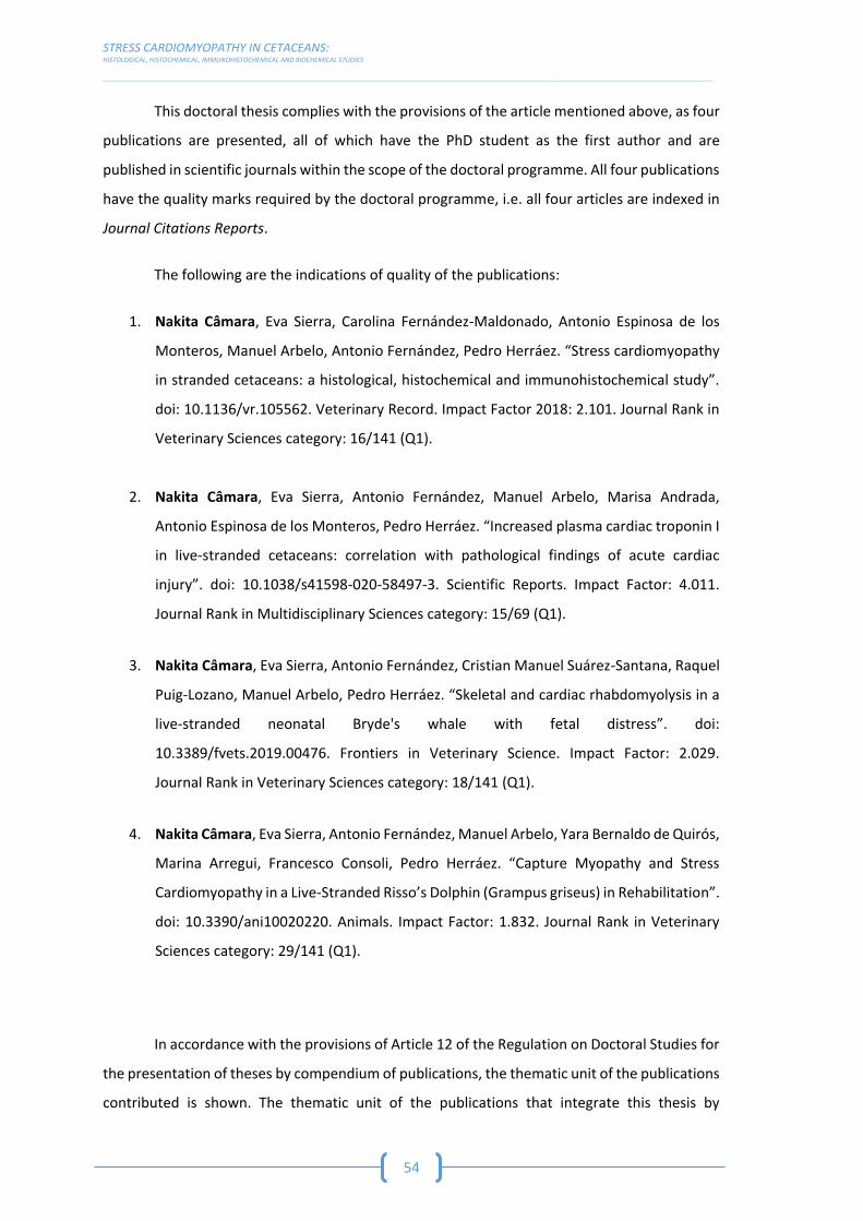
STRESS CARDIOMYOPATHY IN CETACEANS: HISTOLOGICAL, HISTOCHEMICAL, IMMUNOHISTOCHEMICAL AND BIOCHEMICAL STUDIES _________________________________________________________________________________________________________________________________________________________________________
54
This doctoral thesis complies with the provisions of the article mentioned above, as four
publications are presented, all of which have the PhD student as the first author and are
published in scientific journals within the scope of the doctoral programme. All four publications
have the quality marks required by the doctoral programme, i.e. all four articles are indexed in
Journal Citations Reports.
The following are the indications of quality of the publications:
1. Nakita Câmara, Eva Sierra, Carolina Fernández-Maldonado, Antonio Espinosa de los
Monteros, Manuel Arbelo, Antonio Fernández, Pedro Herráez. “Stress cardiomyopathy
in stranded cetaceans: a histological, histochemical and immunohistochemical study”.
doi: 10.1136/vr.105562. Veterinary Record. Impact Factor 2018: 2.101. Journal Rank in
Veterinary Sciences category: 16/141 (Q1).
2. Nakita Câmara, Eva Sierra, Antonio Fernández, Manuel Arbelo, Marisa Andrada,
Antonio Espinosa de los Monteros, Pedro Herráez. “Increased plasma cardiac troponin I
in live-stranded cetaceans: correlation with pathological findings of acute cardiac
injury”. doi: 10.1038/s41598-020-58497-3. Scientific Reports. Impact Factor: 4.011.
Journal Rank in Multidisciplinary Sciences category: 15/69 (Q1).
3. Nakita Câmara, Eva Sierra, Antonio Fernández, Cristian Manuel Suárez-Santana, Raquel
Puig-Lozano, Manuel Arbelo, Pedro Herráez. “Skeletal and cardiac rhabdomyolysis in a
live-stranded neonatal Bryde's whale with fetal distress”. doi:
10.3389/fvets.2019.00476. Frontiers in Veterinary Science. Impact Factor: 2.029.
Journal Rank in Veterinary Sciences category: 18/141 (Q1).
4. Nakita Câmara, Eva Sierra, Antonio Fernández, Manuel Arbelo, Yara Bernaldo de Quirós,
Marina Arregui, Francesco Consoli, Pedro Herráez. “Capture Myopathy and Stress
Cardiomyopathy in a Live-Stranded Risso’s Dolphin (Grampus griseus) in Rehabilitation”.
doi: 10.3390/ani10020220. Animals. Impact Factor: 1.832. Journal Rank in Veterinary
Sciences category: 29/141 (Q1).
In accordance with the provisions of Article 12 of the Regulation on Doctoral Studies for
the presentation of theses by compendium of publications, the thematic unit of the publications
contributed is shown. The thematic unit of the publications that integrate this thesis by

REGULATIONS & ADAPTATION TO DOCTORAL THESIS BY COMPENDIUM OF PUBLICATIONS
__________________________________________________________________________________________________________________________________________________________________________
55
compendium is evident, since all of them deal with Stress Cardiomyopathy in cetaceans, having
approached this pathology through biochemical, histological, histochemical and
immunohistochemical studies.
Finally, the structure of this doctoral thesis complies with the provisions of Article 12.2
of the Regulations on Doctoral Studies, as can be seen in the following pages, as it offers:
d) an introduction presenting the objectives of the thesis, the presentation of the
published works and the justification of the thematic unit of the thesis (bibliographic
review);
e) a copy of the published works (publications);
f) the final conclusions.


ABSTRACT

ABSTRACT
__________________________________________________________________________________________________________________________________________________________________________
59
Previous studies have shown the susceptibility of cetaceans to stress through blood tests
that demonstrate the activation of the hypothalamic-pituitary-adrenal axis with the consequent
production and release of glucocorticoids, mineralocorticoids and catecholamines (adrenaline
and norepinephrine) (Prosser 1986; Spraker 1993).
Although the beneficial effects of stress system activation are well known, in extreme or
prolonged situations, animals can be adversely affected by the effects of physical and
psychological stress (Cowan and Curry 2002).
Therefore, marine mammals can develop extreme responses to stress that cause a
significant deterioration in animal health and even death. Cardiac lesions seem to play a central
role in these adverse responses to stress in cetaceans. Different authors suggest that these
animals are particularly predisposed to develop stress cardiomyopathy (SCMP) probably due to
the characteristics of their cardiovascular adaptations, conforming to the requirements of diving
metabolism (Cowan and Curry 2002; Cowan et al. 2000; Cozzi et al. 2017; Herráez et al. 2007,
2013).
In recent decades, efforts have been made to reduce the impact that some human
activities have on free-living cetaceans, but unfortunately whales and dolphins continue to be
threatened by: active (live) stranding and interactions with humans, collision with ships and
accidental capture (bycatch). These different pathological entities have acute stress, as the
centre of its etiopathogenesis, which can cause the death of the animal or seriously aggravate a
previous disease situation, while influencing its subsequent rehabilitation, making the therapy
and recovery of the animals involved not successful (Arbelo et al. 2013; Bonsembiante et al.
2017; Cowan and Curry 2008; Díaz-Delgado et al. 2018; Duignan and Jones 2005; Gulland et al.
2018; Herráez et al. 2007, 2013; Sierra et al. 2014, 2017; Soulsbury et al. 2008).
The investigation here presented was designed with the aim of continuing the research
in cetaceans, with a double intention:
On the one hand, characterize cardiomyopathy due to stress in cetaceans, trying to
provide greater knowledge about their pathologies derived from a situation of acute
stress.
On the other hand, this knowledge would help us find applicable solutions, using health
as a necessary instrument for the conservation of cetaceans in our seas.

STRESS CARDIOMYOPATHY IN CETACEANS: HISTOLOGICAL, HISTOCHEMICAL, IMMUNOHISTOCHEMICAL AND BIOCHEMICAL STUDIES __________________________________________________________________________________________________________________________________________________________________________
60
Thus, based on the above, this will be achieved through specific objectives such as:
1. Characterize SCMP, as injuries resulting from extreme stress responses in live-stranded
cetaceans subjected to capture and interaction with humans, as well as in dead
cetaceans due to ship strikes and interactions with fisheries (bycatch).
2. Correlate the blood values detected for the biochemical markers of acute cardiac muscle
damage with the pathological findings identified histologically, in actively stranded
cetaceans and subjected to capture and interaction with humans.
3. Application of the knowledge obtained previously in the diagnosis of specific clinical
cases.
This thesis is presented as the compendium of four articles, published in peer-review
journals with high scientific impact, describing the results and interpretation of the biochemical,
histological, histochemical and immunohistochemical analysis conducted.
This study represents the first biochemical and morphological characterization of SCMP,
as an injury resulting from extreme stress response in live-stranded cetaceans and subjected to
capture and interaction with humans, dead cetaceans due to collisions with ships and with
fishing activities interaction (bycatch).

INTRODUCTION
&
OBJECTIVES


INTRODUCTION & OBJECTIVES
__________________________________________________________________________________________________________________________________________________________________________
63
The ocean urges us to contemplate it, overwhelmed by its immensity. However,
although we observe it, it is still a liquid blade that conceals another world. Beneath its blue
horizon is a saltwater space, a three-dimensional domain full of life. In a medium as uniform as
the ocean, there are few boundaries that prevent the passage of species. The currents are
authentic freeways through which life circulates freely (Fundación Mapfre Guanarteme 2011).
The stability of the marine environment is a critical condition for the sustainable
development of the planet. The protection of marine ecosystems is a global priority. Throughout
history, we have accumulated biological, oceanographic, geophysical, etc. data with those who
have been able to define risks and propose solutions to maintain a lasting balance between
human activities and the conservation of the seas. Among these environmental sciences the
study of marine mammals receives a special interest, since, like men, they are homeothermal
mammals, with a long period of survival and they are at the top of the trophic chain, constituting
excellent bioindicators of marine environment, providing valuable information on the degree of
degradation or conservation of this habitat through the biosanitary study of these species.
Consequently, with the advancement of science, the strandings of marine mammals
have moved from mere curiosity/fascination throughout history to increasingly detailed studies.
It is now important to continue this scientific evolution, which goes through areas as diverse as
oceanography, pathology, toxicology, genetics, etc. and to draw as much information as possible
on these events in order to know and eventually predict future changes in populations and
species. Despite limitations on the stranding study, the record compilation of stranded animals,
not only on Canary Islands or Andalusia, but also around the world, provide a rich collection that
can be explored alongside the development of other sciences.
Thus, through the study of stranded animals, it is intended to add another drop to the
vast sea of scientific knowledge about cetaceans.
Previous studies have shown the susceptibility of cetaceans to stress through blood tests
that demonstrate the activation of the Hypothalamic-Pituitary-Adrenal (HHA) axis with the
consequent production and release of glucocorticoids, mineralocorticoids and catecholamines
(adrenaline and norepinephrine) (Prosser 1986; Spraker 1993).
Although the beneficial effects of the activation of the stress system are well known, in
extreme or prolonged situations, animals can be adversely affected by the effects of both
physical and psychological stress (Cowan and Curry 2002).

STRESS CARDIOMYOPATHY IN CETACEANS: HISTOLOGICAL, HISTOCHEMICAL, IMMUNOHISTOCHEMICAL AND BIOCHEMICAL STUDIES __________________________________________________________________________________________________________________________________________________________________________
64
Therefore, marine mammals can develop extreme responses to stress that cause a
remarkable deterioration of animals and even their death. In these adverse responses to stress
in cetaceans, cardiac lesions seem to play a central role, thus different authors suggest that
cetaceans are particularly predisposed to develop Stress Cardiomyopathy (SCMP) probably due
to the characteristics of their cardiovascular adaptations conforming to the requirements of
diving metabolism (Cowan and Curry 2002; Cowan et al. 2000; Cozzi et al. 2017; Herráez et al.
2007, 2013).
Efforts have been made, in recent decades, to reduce the impact that some human
activities have on cetaceans, but unfortunately whales and dolphins continue to be threatened
by: active (live) stranding, ship collision and/or fishery interactions such as bycatch. These
different pathological entities have the acute stress as the central etiopathogenesis, which can
cause the death of the animal or seriously aggravate a previous disease situation, whilst
influencing their subsequent rehabilitation, making therapy and recovery of the involved
animals unsuccessful (Arbelo et al. 2013; Bonsembiante et al. 2017; Cowan and Curry 2008; Díaz-
Delgado et al. 2018; Duignan and Jones 2005; Gulland et al. 2018; Herráez et al. 2007, 2013;
Sierra et al. 2014, 2017; Soulsbury et al. 2008).
The research presented, was designed with the general aim of continuing the research
in cetaceans with a double intention:
On the one hand, confirming the hypothesis presented by previous authors, suggesting
that cetaceans can develop and suffer from SCMP.
On the other hand, this knowledge is helping us to find applied solutions, using health
as a necessary instrument for the conservation of cetaceans in our seas.
Subsequently, and based on the above, this study was developed with 3 specific
objectives, which are:
1. To characterize the SCMP, such as injuries resulting from extreme stress responses in
actively stranded cetaceans and subjected to capture and interaction with humans,
dead cetaceans due to ship strikes and interactions with fisheries (bycatch).
2. To correlate the blood values detected for the biochemical markers of acute cardiac
muscle damage with the pathological findings identified histologically, in actively
stranded cetaceans and subjected to capture and interaction with humans.
3. To apply the knowledge obtained previously in the diagnosis of specific clinical cases.

LITERATURE REVIEW


LITERATURE REVIEW STRESS
__________________________________________________________________________________________________________________________________________________________________________
67
4.1 STRESS
4.1.1 DEFINITION OF STRESS
Nowadays stress is a very commonly used word. It is a part of life and is not intrinsically
bad. All forms of life have evolved mechanisms to deal with stressful situations in their lives. In
fact, we often look for stress/ “thrill”/ stimulus, and we love its biological stimulant effects, as
even being psychologically rewarding. This is why we ski, ride roller coasters and climb
mountains, etc. However, nobody denies that stress can have a detrimental effect on the
individual, and therefore is seen as a pervasive modern-day killer. We are only very aware of the
human diseases associated with a life full of stress, and we are concerned with the effect that
stress carries in us. Little by little we have come to accept that animals also bear a great stress
load, and that when they suffer from stress they develop very similar pathologies. Like humans,
while experiencing severe stress, animals can succumb to the disease, not reproduce or develop
properly (Moberg 2000; Yousef 1988). It is the recognition of these negative effects of stress
that has sensitized us to the importance of stress for the welfare of an animal. Our challenge is
to differentiate between the small non-threatening situations of life and those that negatively
affect the welfare of an animal.
To date, animal stress has not been given a universally accepted precise definition. The
term stress is very general, and is used quite loosely by various scientists. The divergent
definitions are unfortunate, leading to confusion in gathering and integrating available data
from literature (Yousef 1988). Stress can be defined as an internal (physiologic or psychogenic)
or environmental stimulus that initiates an adaptive change or stress response in an animal
(Breazile 1988). Despite stating that, stress is commonly defined as the biological response
caused when a person or animal perceives a threat to their homeostasis, being the threat the
stressor, it is also not easy to formulate an acceptable definition due to the following (Moberg
2000):
- There are no good biological tests to measure stress;
- Biological responses to stress (behavioural, automatic and neuroendocrine) have a
marked degree of interanimal variability;
- Failure to correlate measures of stress and meaningful changes in the well-being of
animals.

STRESS CARDIOMYOPATHY IN CETACEANS: HISTOLOGICAL, HISTOCHEMICAL, IMMUNOHISTOCHEMICAL AND BIOCHEMICAL STUDIES __________________________________________________________________________________________________________________________________________________________________________
68
Stress can be psychological (fear, retention, management or exposure to a new
environment) or physical (thirst or hunger, fatigue, trauma or extreme temperatures) (Cowan
and Curry 2008).
4.1.2 RESPONSE TO STRESS
a) Stress and catecholamine release
Although the adaptive responses to a stressor, may be different not only depending on
the species and/or individual but also because of a complex interplay of genetic factors and
previous experiences, it always involves a number of changes including alterations in behaviour,
endocrine and autonomic system that induce significant physiological effects on the body
through activation of the Hypothalamic-Pituitary-Adrenal axis with the consequent production
and release of catecholamines (adrenaline and norepinephrine), glucocorticoids and
mineralocorticoids (Gray and Young 1956; Moberg 2000; Prosser 1986; Spraker 1993).
Serum elevation of catecholamines, exogenous or endogenous, is a proven cause of
striated and smooth muscle necrosis in humans and animals (Cebelin and Hirsch 1980; Gray and
Young 1956; Reichenbach and Benditt 1970).
During the resting state, the sympathetic and parasympathetic nervous system and the
adrenal medulla are constantly activated (sympathetic-parasympathetic tone). However, in
situations of extreme stress, or when the stressful situation is prolonged in time, a "storm" is
triggered. The response induces the activation of the Hypothalamus, which alters the activity of
the autonomic nervous system, inhibiting the parasympathetic system and stimulating the
sympathetic nerves, resulting in a stimulation of the adrenal medulla and the release of
catecholamines and encephalin (Spraker 1993).
The sympathetic nervous system is mainly composed of two molecular chemical signals
(catecholamines): epinephrine (adrenaline) and norepinephrine (norepinephrine). Epinephrine
is produced and released from the adrenal glands, while norepinephrine is produced and
released from nerve axons after stimulation of acetylcholine (Spraker 1993).
Catecholamines produce physiological changes at a general level that prepare the body
for physical activity (“fight or flight” response). Some of these effects include an increase in heart

LITERATURE REVIEW STRESS
__________________________________________________________________________________________________________________________________________________________________________
69
rate, blood pressure, blood glucose levels and a general reaction of the sympathetic nervous
system (Spraker 1993).
b) Stress effects in the heart function
Endogenous catecholamines, epinephrine and norepinephrine, are essential
neurotransmitters, which in physiological plasma concentrations, exert a positive inotropic
action on the heart. However, when administered in large doses, induce myocardial injury, both
in animals and in humans (Cebelin and Hirsch 1980; Reichenbach and Benditt 1970; Turnbull and
Cowan 1998). Under stress conditions, large amounts of catecholamines are released from the
sympathetic nerve endings and the adrenal medulla, and exposure to these high concentrations
produces coronary spasm, arrhythmias, contractile dysfunction, myocardial cell damage and
extensive necrosis (Dhalla et al. 1992).


LITERATURE REVIEW STRESS CARDIOMYOPATHY
__________________________________________________________________________________________________________________________________________________________________________
71
STRESS CARDIOMYOPATHY
4.2.1 HISTORY OF STRESS CARDIOMYOPATHY
Recently, there has been an increasing awareness of an unique cardiac syndrome that
has been described as the “Apical Ballooning Syndrome”, “Takotsubo Disease”, and Ampulla or
Stress Cardiomyopathy (SCMP), being also referred to as the “Broken Heart Syndrome” in the
popular press (Prasad et al. 2008).
The syndrome is characterized by acute but rapidly reversible cardiomyopathy, which
consists in a left ventricular systolic dysfunction in the absence of occlusive (atherosclerotic)
coronary artery disease (Buja and Butany 2015; Merchant et al. 2008; Prasad et al. 2008).
This distinct cardiac syndrome was originally described in the Japanese population in
1990 and was called “Takotsubo Cardiomyopathy”, named after the octopus trapping pot (Tako,
octopus; Tsubo, pot). This distinctive round bottom and a narrow neck, resemble the
echocardiographic appearance of the left ventricle during systole in these patients (Figure 1).
The echocardiogram during this condition displays transient hypokinesis, akinesis, or dyskinesis
in the left ventricular middle segments without apical involvement (Buja and Butany 2015;
Prasad et al. 2008).
Figure 1. X-ray of the left ventricle (A) shows apical ballooning, a reversible abnormality characteristic
of takotsubo cardiomyopathy. During systole (cardiac contraction) the midsection and tip (apex) of the
left ventricle balloon out, while the area above, called the base, contracts normally. The shape is similar
to that of a tako-tsubo (B), a round-bottomed, narrow-necked vessel used to catch octopuses. Figure

STRESS CARDIOMYOPATHY IN CETACEANS: HISTOLOGICAL, HISTOCHEMICAL, IMMUNOHISTOCHEMICAL AND BIOCHEMICAL STUDIES __________________________________________________________________________________________________________________________________________________________________________
72
obtained from the website: https://www.health.harvard.edu/heart-health/takotsubo-cardiomyopathy-
broken-heart-syndrome.
Nowadays, several cases have been reported from around the world, including different
countries from Europe, such as Germany, Belgium, Italy, France, Spain, Portugal or United
Kingdom, North America, and Australia (Abdulla et al. 2006; Bybee et al. 2004; Desmet,
Adriaenssens, and Dens 2003; Haghi et al. 2007; Huang et al. 1996; Mitchell et al. 2007; Samardhi
et al. 2012; Seth et al. 2003; Sharkey et al. 2005; Singh et al. 2014).
4.2.2 CLINICAL SUBTYPES
Stress Cardiomyopathy appears to be related to an excess of plasma catecholamines,
which most often occurs after an emotional or physical stress event, and has a clinical
presentation that is indistinguishable from a myocardial infarction. Therefore, it is an important
differential diagnosis of an acute myocardial infarction, thus it is under recognized and often
misdiagnosed (Abdulla et al. 2006; Buja and Butany 2015; Bybee et al. 2004; Desmet et al. 2003;
Haghi et al. 2007; Huang et al. 1996; Mitchell and Marquis 2017; Prasad et al. 2008; Samardhi et
al. 2012; Seth et al. 2003; Sharkey et al. 2005; Singh et al. 2014).
This syndrome comes to the attention of medical personnel in a variety of clinical
scenarios and contexts (Lyon et al. 2016). The different cases can be classified as:
a) Primary Stress Cardiomyopathy
The acute cardiac symptoms are the primary reason for seeking medical care. These
patients may or may not have a clear identification of the severe, unexpected physical or
emotional stressful triggers (being more often emotional). It can also occur spontaneously.
Therefore, the potential co-existing medical conditions may be the predisposing risk factors but
not the primary cause of the catecholamine rise. The clinical management on these cases
depends on the specific complications.

LITERATURE REVIEW STRESS CARDIOMYOPATHY
__________________________________________________________________________________________________________________________________________________________________________
73
b) Secondary Stress Cardiomyopathy
A substantial proportion of cases occur in patients already hospitalized for another
medical condition such as endocrine (e.g. pheochromocytoma), neurological and neurosurgical,
respiratory, obstetric, psychiatric, gastrointestinal, infection, babesiosis, cardiological,
haematological, surgical, anaesthetic, medication or illicit drugs. In such patients, sudden
activation of the sympathetic nervous system or a rise in catecholamines precipitates an acute
SCMP as a complication of the primary condition or its treatment. The management in these
patients should focus not only on the SCMP and its cardiac complications but also on the
condition that triggered the syndrome.
4.2.3 EPIDEMIOLOGY
Stress Cardiomyopathy occurs predominantly in post-menopausal women, being the
demographic and clinical characteristics similar in men and women. However, in previous
studies demonstrated that emotional stress or anxiety (including in some cases a formal
diagnosis of anxiety or panic disorders) or even the absence of identifiable triggers was more
common in women. Conversely, a physical stressful triggering event, shock, or resuscitation on
presentation was more common in men, who also had higher levels of cardiac biomarkers
(troponin). Furthermore, elderly patients are considered to be at higher risk for this syndrome
and related major complications, and less of 10% of patients are below 50 years of age (Gianni
et al. 2006; Kurisu et al. 2004; Lyon et al. 2016; Ogura et al. 2003; Pilgrim and Wyss 2008; Sharkey
2008).
4.2.4 PATHOPHYSIOLOGY
The pathophysiology is complex and reflects the integrated and systemic physiological
responses to acute, severe stress and the cardiovascular responses to sudden surges of
endogenous or exogenously administrated plasma catecholamines (Buja and Butany 2015; Lyon
et al. 2016).
Catecholamines appear to have a central role in the pathophysiology of this syndrome,
as the trigger is often a sudden, unexpected stress. It is necessary to take in consideration two
initial elements of the physiology: the first being how much epinephrine and norepinephrine is
STRESS CARDIOMYOPATHY IN CETACEANS: HISTOLOGICAL, HISTOCHEMICAL, IMMUNOHISTOCHEMICAL AND BIOCHEMICAL STUDIES __________________________________________________________________________________________________________________________________________________________________________
74
released from the hypothalamic-pituitary-adrenal axis in response to a given stress; and the
second is the response of the cardiovascular system (including the myocardium, coronary
arteries and peripheral vasculature) and the sympathetic nervous system to the sudden
sympathetic activation and surge in circulating catecholamines (Helen et al. 2012; Lyon et al.
2016; Pelliccia et al. 2018; Suzuki et al. 2014).
Several hypotheses have been proposed to explain the unique cardiac appearance in
this syndrome and the cardiac response to severe stress. These hypotheses can be broadly
divided into vascular and/or myocardial causes and may not be mutually exclusive, as the entire
cardiovascular system is exposed to the same catecholamine storm. The vascular abnormalities
consist in acute multivessel coronary spasm, aborted myocardial infarction with spontaneous
recanalization, and acute increased ventricular afterload. Acute left ventricular outflow tract
obstruction and direct catecholamine-mediated myocardial stunning are the myocardial
irregularities, and finally, the vascular and myocardial motive is the integrated cardiovascular
physiology (a cardio-circulatory syndrome) (Angelini 2008; Camici and Filippo 2015; Crea,
Camici, and Bairey Merz 2013; Lyon et al. 2016; Naegele et al. 2016; Vitale, Mendelsohn, and
Rosano 2009).
Therefore, new information has supported the concept that this pathology is caused by
acute myocardial ischemia (Jaffe et al. 2018; Pelliccia et al. 2018).
The ischemia-reperfusion injury is a phenomenon described as an accelerated injury to
the heart due to the resupplied blood to the ischemic area of the heart which was previously
deprived of its blood supply (ischemia) and suffered from hypoxia, substantial loss of cell volume
regulation and influx of calcium because of inadequate run of the ion pumps. Added membrane
damage is a result when blood supply is re-established to areas with potentially viable cells (Buja
2005; Buja and Butany 2015).
The crucial role of myocardial ischemia in Takotsubo syndrome, has emerged thanks to
the evidence that most of these cases occur in patients with risk factor for endothelial
dysfunction which is characterized by an imbalance between vasoconstricting and vasodilating
factors that could explain the propensity to epicardial and/or microvascular coronary artery
spasm of the disease. This pathologic state of the endothelium may represent an important link
between stress and myocardial dysfunction in SCMP. There is now an agreement that the
increased concentration of catecholamine occurring in the acute phase of SCMP contributes to
an acute situation of “supply-demand mismatch” followed by post-ischemic stunning, since it
LITERATURE REVIEW STRESS CARDIOMYOPATHY
__________________________________________________________________________________________________________________________________________________________________________
75
induces direct myocardial dysfunction in sarcoplasmic reticulum calcium channels, leading to
leakage of calcium into the sarcoplasm, contraction impairment, myocardial necrosis and
coronary vasoconstriction, mostly at the microvascular level, together with an increased cardiac
workload (Pelliccia et al. 2018; Seguel et al. 2014).
4.2.5 CLINICAL PRESENTATION AND DIAGNOSIS
Patients with SCMP typically present with acute chest pain of cardiac origin (angina),
breathlessness, and palpitations due to sinus tachycardia or arrhythmia. In more severe cases,
pre-syncope or syncope due to ventricular tachyarrhythmia’s, severe left ventricular outflow
tract obstruction, or cardiogenic shock may be present. They may describe a wave of pressure
from the chest to the neck and into the head, consistent with the acute catecholamine and
hypertensive surge and frequently associated with diaphoresis and heightened anxiety.
The diagnostic criteria for SCMP established by the Heart Failure Association consists in
alterations in medical exams, such as electrocardiography (ECG), coronary angiography and left
ventriculography, echocardiography, cardiac magnetic resonance, coronary computed
tomography angiography, and biochemical analysis (Lyon et al. 2016).
a) Electrocardiography (ECG)
More than 95% of the SCMP patients present irregularities on ECG during the acute
phase (under 12 h). These new and reversible abnormalities consist in ST-segment elevation, ST
depression, new left bundle brunch block (which may be permanent after the syndrome, but
should also alert clinicians to exclude other cardiomyopathies), and sometimes Q-waves, with
deep and widespread T-wave inversion, and/or QT prolongation developing 24-48 h after the
onset of symptoms or the precipitating stressful trigger when present (which can take many
weeks to months to normalize after recovery of left ventricular function) (Haghi et al. 2007;
Kurisu et al. 2004; Lyon et al. 2016; Mitchell et al. 2007; Ogura et al. 2003; Sharkey 2008).
b) Coronary Angiography and Left Ventriculography
The imaging exams, mainly coronary angiography and left ventriculography, reveal that
patients with SCMP present transient regional wall motion abnormalities of the left ventricle or
STRESS CARDIOMYOPATHY IN CETACEANS: HISTOLOGICAL, HISTOCHEMICAL, IMMUNOHISTOCHEMICAL AND BIOCHEMICAL STUDIES __________________________________________________________________________________________________________________________________________________________________________
76
right ventricle myocardium which are frequently, but not always, preceded by a stressful trigger
(emotional or physical). These irregularities usually extend beyond a single epicardial vascular
distribution, and often result in circumferential dysfunction of the ventricular segments
involved. It is also necessary to observe the absence of culprit atherosclerotic coronary artery
disease including acute plaque rupture, thrombus formation, and coronary dissection or other
pathological conditions to explain the pattern of temporary left ventricle dysfunction detected
(e.g. hypertrophic cardiomyopathy, viral myocarditis) (Abdulla et al. 2006; Desmet et al. 2003;
Haghi et al. 2007; Lyon et al. 2016; Mitchell et al. 2007; Prasad et al. 2008; Sharkey et al. 2005).
c) Biochemical Analysis
Reference values, both haematological and biochemical, are vital to provide baseline,
screening, and diagnosis of diseases. Healthy subjects are able to maintain the biochemical
concentration of certain molecules in each cell or biological fluid within the limits of
concentrations considered normal or usual values. For that reason, variations of these biological
parameters, both qualitative and quantitative, may indicate a pathological state. Consequently,
the assess of a pathological condition through the measurement of the degree of a modification
of a biochemical parameter is one of the very aims of clinical biochemistry (Kamdje, Nkem, and
Mimfoumou 2017). Considering this, the biomarkers, cardiac troponin I (cTnI) and/or creatine
kinase (CK), have a small, rapid increased to above normal levels, the brain natriuretic peptide
(BNP) may be elevated, and plasma catecholamines may be also elevated (Fineschi et al. 2010;
J. et al. 2008; Lyon et al. 2016).
i. Creatine Kinase (CK)
The most commonly used serum enzyme in the determination of neuromuscular
diseases of domestic animals is creatine kinase (CK), previously designated by creatine
phosphokinase (Valberg 2008).
There are three main forms of CK isoenzymes. Creatine kinase has a dimeric structure
consisting of M (muscular) subunits and B (cerebral) subunits, which combine to form the three
heterogeneous isoenzymes: heterogeneous MM (or CK3), MB (or CK2) and BB (or CK1) (Valberg
2008). In humans, CK-MB is present in a relatively high concentration in the myocardium
(approximately 20% of the total myocardial CK), while the concentration of CK-MM is the highest
LITERATURE REVIEW STRESS CARDIOMYOPATHY
__________________________________________________________________________________________________________________________________________________________________________
77
in skeletal muscle (98% of the total muscle CK), with only a small amount of CK-MB (usually
about 2%) (Lewandrowski et al. 2002).
Creatine kinase rises in less than 3 to 12 h after the injury, with peaks within 12 to 24 h
and returns to the baseline after 48 to 72 h, unless a new injury or permanent damage occurs
(Lewandrowski et al. 2002).
Although CK is considered a sensitive marker of myocardial damage, it is also present in
skeletal muscle in high concentrations as well as in the intestine, diaphragm, uterus, and
prostate in minor amounts, thusly it has poor specificity to detect heart damage (Khan 2015).
ii. Troponin
Consequently, troponins have been adopted, over other cardiac markers, such as CK, as
the new gold standard for necrosis, since cTnI is detectable in very low amounts (for example
0.01 µg/L) in blood from healthy individuals with no evidence of cardiac disease (Hassan et al.
2009; Lewandrowski et al. 2002; Morrow et al. 2007; Venge et al. 2009). Therefore, it is thought
that significant elevations (≥0.1 µg/L) of this marker is most likely reflecting myocardial necrosis.
When irreversible myocardial damage occurs, troponin is released from cardiomyocytes. This is
a regulatory complex protein comprising three subunits, which are Troponin C (calcium binding
component), Troponin T (binding component tropomyosin) and Troponin I (inhibiting
component). All of these are involved in the contractile process of both skeletal and cardiac
muscle. The Troponin C expressed in the muscle is identical to the cardiac troponin C (cTnC).
However, cardiac troponin T (cTnT) and cardiac troponin I (cTnI) are specific to the heart
(Coudrey 1998). Due to this specificity, cTnT and cTnI are currently recommended by various
international societies as a diagnostic indicator for acute myocardial infarction since they have
the potential to be specific markers of cardiac damage (Morrow et al. 2007). Thus, as mentioned
previously cardiac troponin is released from cardiomyocytes into the blood in proportion to the
degree of damage, as a result from an injury, consistent with ischemia or various other causes
(Hassan et al. 2009).
Injured cardiac myocytes release troponin, for the most part, 3 to 9 h after ischemic
damage, reaches its peak 12 to 48 h and its elevation stay rose for up for 4 to 7 days in case of
cTnI (Hansen and Rossen 1999; Khan 2015; Lewandrowski et al. 2002). Early recognition of
myocardial necrosis (1 to 3 h) is not allowed by the view of this kinetics, and these markers do
not help maximal affectability until 6 or more hours after the onset of the acute myocardial
STRESS CARDIOMYOPATHY IN CETACEANS: HISTOLOGICAL, HISTOCHEMICAL, IMMUNOHISTOCHEMICAL AND BIOCHEMICAL STUDIES __________________________________________________________________________________________________________________________________________________________________________
78
infarction and/or SCMP. Since precise determination of the timing of stress event and/or
symptom onset is frequently clinically exceptionally difficult because it is focused around patient
report. Therefore, a precondition to get a satisfactory clinical affectability to distinguish these
pathologies in humans is that the blood for testing should be collected at 6 to 9 h after onset of
manifestation (Khan 2015). Moreover, previous studies propose that the hence measuring
troponin in serum can be an important auxiliary method in examining sudden death seeing that
its peak concentration can be related with the degree of injury (Khan 2015).
More than 90% of patients with SCMP revealed raised cTnI when measured by
conventional assays (Lyon et al. 2016). However, SCMP is under-recognized because of the
unnecessary delay in the diagnosis process in postmortem examination cases of acute
myocardial infarction caused by the less sensitive conventional markers. Therefore, it is
necessary to establish diagnostic utility of different biochemical cardiac markers in biological
fluids for postmortem diagnosis of acute myocardial infarction and/or SCMP due to the
limitations of histopathological findings. Thus, the detection of even microinfarction and acute
myocardial infarction much earlier after the onset of ischemia by using a rapid one-step assay in
body fluids in autopsy cases is allowed by the sensitivity of cTnI (Khan 2015).
iii. Brain Natriuretic Peptide (BNP)
Type B natriuretic peptide (BNP) has shown promise as a new diagnostic marker for
congestive heart failure. The level of plasma BNP has been shown to correlate with long-term
cardiovascular mortality in human patients with acute myocardial infarction and is an
independent predictor of left ventricular ejection fraction and heart failure in these patients.
The level of BNP correlates with the amount of ventricular dysfunction present after an acute
myocardial infarction and may, in the future, become an useful screening test to identify
patients with a higher risk of morbidity and mortality (Lewandrowski et al. 2002). This marker
increases rapidly within 6 h, reaching its peak usually by 24-48 h and at 1 month levels are similar
to baseline (Agarwal et al. 2015).
4.2.6 HISTOPATHOLOGY
The histological findings, both in human myocardial samples and in experimental models
of SCMP, are consistent with catecholamine cardiotoxicity, due to the ischemia-reperfusion

LITERATURE REVIEW STRESS CARDIOMYOPATHY
__________________________________________________________________________________________________________________________________________________________________________
79
injury (Akashi et al. 2010; Buja 2005; Buja and Butany 2015; Fineschi et al. 2010; Jaspreet et al.
2019; Jiang and Downing 1990; Kawai 2012; Maréchaux et al. 2008; Mitchell and Marquis 2017;
Miura et al. 2017; Pascual et al. 2015; Prasad et al. 2008).
From several histological analysis of myocardial tissue obtained through, biopsied and
postmortem examined specimens, it is known that SCMP is associated with different vascular
changes essentially congestion, haemorrhages or interstitial edema. As well as severe
morphological alterations, consisting of acute degenerative changes, such as the presence of
contraction band necrosis, hypereosinophilia, and many vacuoles of different sizes contributing
to the cellular deterioration. Not to mention infiltration by mononuclear lymphocytes,
leukocytes and macrophages (Akashi et al. 2010; Fineschi et al. 2010; Jaspreet et al. 2019; Jiang
and Downing 1990; Kawai 2012; Maréchaux et al. 2008; Mitchell and Marquis 2017; Miura et al.
2017; Pascual et al. 2015; Prasad et al. 2008).
Subendocardial congestion, fibers separated by spaces representing interstitial edema,
and haemorrhages are early histological findings included in the pathology of a catecholamine
cardiomyopathy (Jiang and Downing 1990).
Vascular congestion is the engorgement of a vascular bed generally caused by a
decreased outflow with a normal or increased inflow of blood (Fishbein 2001; Jaspreet et al.
2019; Mosier 2017).
Following administration of catecholamines, interstitial edema (represented by spaces
in the fibers which are separated) is usually associated with subendocardial and subepicardial
haemorrhages and is characteristically present in damaged areas of the myocardium, even after
72 h (Fishbein 2001; Jaspreet et al. 2019).
Focal and diffuse subendocardial haemorrhages are visible on gross examination, being
also noted sometimes in the myocardium and epicardium, shortly after large doses of
catecholamines are given and/or released, in humans with SCMP (Jiang and Downing 1990;
Mitchell and Marquis 2017).
Contraction band necrosis is described as focal hypercontraction and lysis of contractile
filaments in small groups of myocardial cells. The resulting dense hypereosinophilic bands run
transversely through the myocyte. It develops after transient ischemia and reperfusion, is visible
within 5 to 10 minutes, and is the characteristic skeletal and myocardial injury associated with
the administration of catecholamines or with high concentrations of endogenous

STRESS CARDIOMYOPATHY IN CETACEANS: HISTOLOGICAL, HISTOCHEMICAL, IMMUNOHISTOCHEMICAL AND BIOCHEMICAL STUDIES __________________________________________________________________________________________________________________________________________________________________________
80
catecholamines (Fineschi et al. 2010; Reichenbach and Benditt 1970; Turnbull and Cowan 1998).
Therefore, is a lesion widely described as indicator of stress cardiomyopathy (Akashi et al. 2010;
Fineschi et al. 2010; J. et al. 2008; Jiang and Downing 1990; Kawai 2012; Maréchaux et al. 2008;
Mitchell and Marquis 2017; Miura et al. 2017; Pascual et al. 2015; Prasad et al. 2008),
The first histologic abnormality associated with ischemia is the presence of long and
thinned undulated fibers, denominated as wavy fibers (Fineschi et al. 2010; Fishbein 2001). The
wavy fibers are likely to result from stretched fibers that cannot contract, as does the adjacent
myocardium, during bulging of severely ischemic tissue that occurs during systole. This finding
serves as a further criterion for the morphological recognition of early myocardial injury,
however, wavy fibers are not specific for necrosis, since they have been observed experimentally
shortly after coronary occlusion in the myocardium (Fishbein 2001).
Hypereosinophilia is the first confirmed change specific for myocardial necrosis using
histological and histochemical techniques, and consists in an increased eosinophilic staining of
necrotic cardiomyocytes (Fineschi et al. 2010; Fishbein 2001). In experimental animals, this
colour change and subtle interstitial edema is evident 2 to 3 h after coronary occlusion and will
be more pronounced and more easily recognizable from 3 to 6 h (Fishbein 2001).
Hypereosinophilia is more pronounced in the subendocardial and subepicardial zone, being
supported by previous studies which shows that irreversible injury progresses in a wavefront
movement from the ischemic subendocardium, where the perfusion deficit is most severe into
the subepicardium, which receives some collateral blood flow (Buja 2005; Cowan and Curry
2002, 2008).
In the literature, vacuolar degeneration is characterized, morphologically, by
intracellular accumulation of fluid and lysis of myofibrils. The increase of liquid exerts pressure
on the different constituents of the cell, and because of this most of the fibers with vacuoles
have a pyknotic nucleus. It is commonly seen in the periphery of myocardial infarctions, and in
the subepicardial, subendocardial and perivascular regions which suffer with severe, chronic and
fatal ischemia (Adegboyega et al. 1996). Although its occurrence is quite common, the
morphological characteristics and the functional importance of the vacuolar degeneration has
not generally been appreciated and even though pathogenesis is not known, it has been
hypothesized that it may be the result of increased hypoxia induced in myocardial cell
membrane permeability with consequent fluid influx which is normally observed in acute deaths
associated with stressful situations (Adegboyega et al. 1996).

LITERATURE REVIEW STRESS CARDIOMYOPATHY
__________________________________________________________________________________________________________________________________________________________________________
81
Another anatomopathological finding, described in postmortem examination and
endomyocardial biopsies of human patients and experimental animals who suffer from SCMP,
is interstitial infiltration of lymphocytes, leukocytes and macrophages (Jaspreet et al. 2019;
Mitchell and Marquis 2017; Pascual et al. 2015).
A well-characterized sequence of events follows the onset of myocardial necrosis and
allows for understanding of the progression of changes following onset of acute ischemic injury
as well as identification of features useful in determining the age of the lesion. Therefore, in the
next table it is possible to observe the chronology of the light microscopic changes described
previously (Buja and Butany 2015).
Chronology of the changes
Starting time
Light microscopic changes
Acute
5 minutes Myocardium with long, thinned, wavy fibers separated by spaces representing edema, and microvascular congestion, at borders of ischemic myocardium.
15 to 20 minutes
Cell death may starts to develop.
90 minutes Fine changes in the sarcoplasm of some myocardial fibers.
2 to 3 h
Early changes of cardiomyocyte coagulation necrosis with nuclear pyknosis, colour change, more specifically “brick red change” or cytoplasm hypereosinophilia, focal contraction bands and subtle interstitial edema are evident.
3 to 6 h Hypereosinophilia and edema become more pronounced and more easily recognizable.
6 to 12 h Increased number of neutrophils line up in capillaries, so-called margination of neutrophils. Changes accelerated and more extensive contraction band necrosis with reperfusion
12 to 24 h
Extravasation into interstitial space of neutrophils. Vascular congestion, interstitial edema, and focal areas of haemorrhage are also recognized. Continuing coagulation necrosis with karyorrhexis and karyolysis of nuclei. Thinning of myocytes in central region and contraction band necrosis toward periphery.
Subacute
1 to 3 days
Heavy influx of neutrophils in periphery. Advanced coagulation necrosis with loss of nuclei and striations. Lymphocytes start to appear at the edge of the infarction. Pigmented mononuclear cells containing lipofuscin appear. Early degeneration of neutrophils.
3 to 7 days
Degeneration of neutrophils becomes clearly evident producing basophilic granular debris referred to as “nuclear dust”. Beginning of disintegration of necrotic myocytes with phagocytosis and removal of necrotic fibers. Lymphocytes presence is evident. Influx of macrophages; Onset of vascular and connective (granulation) tissue proliferation and early fibrosis has begun at edge of the injury.

STRESS CARDIOMYOPATHY IN CETACEANS: HISTOLOGICAL, HISTOCHEMICAL, IMMUNOHISTOCHEMICAL AND BIOCHEMICAL STUDIES __________________________________________________________________________________________________________________________________________________________________________
82
Chronology of the changes
Starting time
Light microscopic changes
Chronic
2 weeks
Chronic inflammation and removal of peripheral muscle fibers occur. Increased numbers of mononuclear cells (macrophages) are apparent at the margins of the injury where vascular and connective tissue proliferation has become more prominent. Eosinophils, lymphocytes and plasma cells are also present. Infrequent presence of neutrophils.
3 to 4 weeks
Continued removal of necrotic fibers and proliferation of granulation tissue extends throughout the injured area. Macrophages are still numerous, and plasma cells, eosinophils and lymphocytes are still present, but there is more substantial fibroblastic proliferation with collagen formation recognizable as pink fibrillary extracellular material.
5 to 6 weeks
Removal of residual necrotic fibers and continued gradual scar formation with deposition of collagen. Inflammatory cells and vascularity decreased, but a few macrophages, plasma cells, and lymphocytes persist and remain in the scar that eventually forms.
After 3 months
Fibrous scar is fully developed in most infarcts
4.2.7 IMMUNOHISTOPATHOLOGY
The histological findings are corroborated with specific markers which better expose a
myocardial damage. Therefore, the main markers that have been used in studies of human heart
samples with SCMP are for example cTnI, cTnC, myoglobin and fibrinogen (Fishbein et al. 2003;
Hansen and Rossen 1999; Martínez-Díaz et al. 2005; Ortmann et al. 2000; Xiaohong et al. 2002).
The leakage of muscle (skeletal and cardiac) proteins begins immediately after vital
trauma, which causes an acute ischemia, producing early myocardial cell membrane rupture,
causing a quickly decline of myoglobin, cTnI and cTnC content and deposition of plasma proteins
such as fibrinogen in cardiomyocytes. Homogenous labelling for myoglobin is demonstrated in
normal cardiac muscle while injured cardiomyocytes shows both depletion of myoglobin within
muscle fibres and intercellular and interstitial accumulation of myoglobin (Ortmann et al. 2000;
Xiaohong et al. 2002).
In cells undergoing apoptosis there is a greater concentration of troponin due to
condensation, while in zones of obvious ischemic necrosis (infarction zones) tissue antigens are

LITERATURE REVIEW STRESS CARDIOMYOPATHY
__________________________________________________________________________________________________________________________________________________________________________
83
severely depleted (Fishbein et al. 2003; Hansen and Rossen 1999; Martínez-Díaz et al. 2005;
Ortmann et al. 2000).


LITERATURE REVIEW CETACEANS
__________________________________________________________________________________________________________________________________________________________________________
85
4.3 CETACEANS
4.3.1 ORDER CETARTIODACTYLA
SUBORDER CETANCODONTA
INFRAORDER CETACEA
The previous Suborder and actual Infraorder Cetacea (from Latin: cetus meaning whale
and from Greek: ketos meaning sea monster) is represented by a diverse group of mammals
(whales, porpoises and dolphins) fully adapted to aquatic life, whose specialized anatomy and
behaviour mask their terrestrial origin (Motta 2006; Perrin 2020; Perrin, Würsig, and Thewissen
2009).
Therefore, these exclusively aquatic animals that live mostly in the seas, and can also be
found in rivers and estuaries, are recognized as mammals for having their own characteristics of
this group. Among them can be mentioned the presence of mammary glands, all are
endothermic and their breathing is aerial through the lungs.
Taxonomy is constantly changing and the taxonomy for cetaceans is no exception.
According to Perrin, Würsig, & Thewissen (2009), cetaceans were divided into two living
infraorders (Mysticeti and Odontoceti) and one extinct infraorder (Archaeoceti). Nowadays, and
as stated in the World Register of Marine Species by Perrin (2020), these two living infraorders
are defined in two superfamilies:
a) Superfamily Mysticeti
The superfamily Mysticeti is composed of mysticetes, represented by 14 species of
toothless whales, grouped into 4 families: Balaenidae, Neobalaenidae, Eschrichtiidae and
Balaenopteridae. The Balaenopteridae is most variable, with 8 species in two genera
(Balaenoptera with 7 species and Megaptera with only one). This superfamily has as its main
features a symmetrical skull, the presence of fins, two breathing holes and buccal beards, which
are structures similar to sieves located at the top of the mouth and made of keratin. Mysticetes
use beards to filter out plankton from water, small crustaceans and some small fish species
(REMANE 2005). Usually males are smaller than females; It has solitary behaviour, except in the

STRESS CARDIOMYOPATHY IN CETACEANS: HISTOLOGICAL, HISTOCHEMICAL, IMMUNOHISTOCHEMICAL AND BIOCHEMICAL STUDIES __________________________________________________________________________________________________________________________________________________________________________
86
breeding and feeding areas. Generally speaking, species of Balaenoptera vary in size, distribution
and behaviour, but species boundaries may be blurred. Species are distinguished mainly on
aspects of the feeding apparatus baleen size and spacing, size and shape of the upper jaw, and
skeletal differences are rather minor. Thus disparity appears low. The gray whale, Eschrichtius
robustus, is structurally quite different (disparate) from other mysticetes and is generally placed
in its own monotypic family, although some molecular studies place it within the
Balaenopteridae.
b) Superfamily Odontoceti
The superfamily Odontoceti encompasses the largest number of species, some of which
are fluvial. The navies may be coastal, oceanic or may appear along the continental shelf. The
odontocetes are composed of at least 74 species in 10 families of dolphins in general, orcas, all
porpoises, sperm whales (the largest representative of this infraorder) and “beak whales”
(REMANE 2005), which are characterized by the presence of a breathing hole and teeth (ranging
in number from 2 to 200). Usually the teeth are all the same and there is only one dentition,
meaning there is no replacement for lifelong missing teeth. A remarkable ability of this group is
to locate their prey (fish of various sizes and cephalopods, and may, in the case of orca, devour
animals such as seals and penguins) by echolocation. Unlike mysticetes, the skull of odontocetes
is asymmetrical. In some species the frontal region and the face are quite developed, in others
the mouth is extended forward forming a kind of long sharp beak. Males are generally larger
than females, live in groups and their length can range from 1.5 meters to 18 meters in length.
Delphinidae is the most diverse family of cetaceans, and disparity seems much higher than, e.g.,
within Balaenopteridae. Among beaked whales (Ziphiidae; 21 species), the genus Mesoplodon
has 14 rather similar species in which only adult males are separated easily. Disparity here
appears low and awaits explanation in terms of evolutionary ecology within the genus. Among
other odontocetes, 4 species of small long-beaked “river dolphins” each represent a single
family: Iniidae, Pontoporiidae, Lipotidae, and Platanistidae. Although these “river dolphins” are
superficially similar externally, they differ markedly in skull form.

LITERATURE REVIEW CETACEANS
__________________________________________________________________________________________________________________________________________________________________________
87
4.3.2 BIOINDICATOR OF THE HEALTH OF MARINE
ECOSYSTEMS
Cetaceans are the mammals best adapted to the aquatic environment. They evolved
from terrestrial mammals between 55 and 34 million years ago. Although their history is closely
related to man, as he has hunted them since time immemorial, the mystery still surrounds these
animals. The competition for space led them to dominate the pelagic environment, to specialize
in certain fishing resources and to reach depths of 2,000 meters in search of their prey
(Fundación Mapfre Guanarteme 2011).
Plankton is the largest settler in the pelagic space and lives freely in the ocean's water
column, often being swept away by ocean currents. For example, in order for a pilot whale to
reach one ton of weight, it must first eat five thousand tons of plankton. Thus, plankton is at the
base of the food chain of aquatic ecosystems as it feeds larger organisms such as cetaceans,
which are at the highest end of the food chain (Fundación Mapfre Guanarteme 2011).
And as such, cetaceans are an excellent bioindicator of the state of the marine
environment, currently constituting one of the marine mammal groups on which much of the
social and scientific interest is centred.
All the knowledge gained from these animals will allow us to work with scientific grounds
on their conservation objectives and to detect problems that can directly or indirectly affect
marine ecosystems and at the same time animal and sometimes human health.
Because they are wild animals, which encompass numerous species, with an important
variety of behaviours, migratory routes, feeding, reproductive cycles, etc. there are numerous
and deep gaps in the knowledge of these animals, from the biological point of view as health.
4.3.3 STRESS IN CETACEANS
Physiological stress in cetaceans occurs in the necessary adjustments to adapt to diving,
to escape from predators or inter and intraspecific aggression, and catch prey, or even occur
when a wild animal is restricted or locked (Cowan and Curry 2008).
STRESS CARDIOMYOPATHY IN CETACEANS: HISTOLOGICAL, HISTOCHEMICAL, IMMUNOHISTOCHEMICAL AND BIOCHEMICAL STUDIES __________________________________________________________________________________________________________________________________________________________________________
88
So it is clear that the question is not whether dolphins are "under stress" in a given
condition, since they are always under the demand for adaptation, in a constant condition of
adjustment of physiological systems to maintain homeostasis, but if the degree of stress
experienced is physiologically harmful (Cowan and Curry 2008).
a) Response to stress in cetaceans
Marine mammals, in this case cetaceans, present the same basic response to stress as
humans and other mammals (Curry 1999). Once the central nervous system perceives a threat,
a biological or defence response is developed (Cowan and Curry 2002). This response is well
documented in marine mammals with several studies, and includes an adrenocortical response
and effects on thyroid hormonal balance (St. Aubin and Geraci 1988; St Aubin and Geraci 1992).
Cortisol levels have been elevated in cetaceans exposed to stress situations, such as
capture, handling and retention, although this elevation seems to be modest if we compare it
with that experienced by other mammals in similar stress situations. In contrast, aldosterone is
significantly increased compared to adrenocortical stimulation in the case of marine mammals
(cetaceans and pinniped) (St. Aubin and Geraci 1986; Fair and Becker 2000; Thomson and Geraci
2011).
As previously mentioned, although physiological stress has some benefits, an extreme
or prolonged response with release of catecholamines, high blood cortisol and aldosterone
levels are potentially damaging to skeletal and heart muscle (Cowan and Curry 2002).
4.3.4 PATHOLOGICAL ENTITIES / CAUSES OF DEATH IN
CETACEANS ASSOCIATED WITH STRESS
Free-living cetaceans can be threatened, daily, by a wide variety of stressful situations,
of natural and anthropic origin, that affect their well-being.
Different pathological entities have the central axis of their pathogenesis to acute stress.
Among these are active stranding and interaction with humans (capture myopathy), but also
others such as collisions with boats or fishing activities (bycatch) (Arbelo et al. 2013; Díaz-
Delgado et al. 2018; Herráez et al. 2007, 2013; Maldonado 2015; Sierra et al. 2014).

LITERATURE REVIEW CETACEANS
__________________________________________________________________________________________________________________________________________________________________________
89
All these pathological entities share clinico-lesional findings, where, among others,
degenerative cardiac, acute or sub-acute necrotic lesions, may cause the death of the animals
themselves or seriously aggravate a previous disease situation at that time, and it can condition
its subsequent rehabilitation, making therapy unsuccessful as well as subsequent recovery
(Adegboyega et al. 1996; Arbelo et al. 2013; Cowan and Curry 2002, 2008; Díaz-Delgado et al.
2018; Duignan and Jones 2005; Herráez et al. 2007, 2013; Seguel et al. 2014; Sierra et al. 2017,
2014; Soulsbury et al. 2008).
a) Active/Live Stranding
Active stranding is an unnatural situation in all cetaceans, defined as the one in which
the cetacean is alive on the beach or in shallow water in distress due to being unable to free
itself and resume natural activity (Geraci and Lounsbury 2005; Gulland et al. 2018; Wilkinson
1991). Regardless of the prior health status of the animals, the stranding implies an anomalous
and extreme situation for an organism that is not anatomically and physiologically adapted to
environmental conditions different from those of the aquatic environment.
It is a pathological entity "per se" in which acute stress is the central axis of its
etiopathogeny presenting clinical-lesional findings that can cause the death of the animal or
seriously aggravate a previous disease situation over the period of capture or captivity, which
may influence the subsequent rehabilitation and recovery of affected animals, since live
stranded cetaceans are frequently debilitated when rescued. Therefore, although the stranding
response is oriented to improve the health and the welfare of these animals, some “rescue and
recovery” activities that seem logical to us, the interaction with humans, transport and
rehabilitation, may in fact be counterproductive (Arbelo et al. 2013; Bonsembiante et al. 2017;
Cowan and Curry 2008; Díaz-Delgado et al. 2018; Duignan and Jones 2005; Gulland et al. 2018;
Herráez et al. 2007, 2013; Sierra et al. 2014, 2017; Soulsbury et al. 2008).
In cetaceans, acute and stressful deaths, such the ones that occur in live-stranding, may
be attributed to the so called “stress response syndrome” or “alarm reaction”, which consists in
hyperthermia, catecholaminergic and neurogenic shock, and also assigned to the multiorgan
failure, e.g., hemodynamic shock and renal dysfunction. These mechanisms are thought to be
analogous to Capture Myopathy, in which stress cardiomyopathies seem to have an important
role, since it is the most devastating form of acute stress described in birds, terrestrial and
marine wild mammals, for instance sea otters, seals, and cetaceans, that may occur during and

STRESS CARDIOMYOPATHY IN CETACEANS: HISTOLOGICAL, HISTOCHEMICAL, IMMUNOHISTOCHEMICAL AND BIOCHEMICAL STUDIES __________________________________________________________________________________________________________________________________________________________________________
90
after the stress of capture, handling, restraint, and transport of the animals (Cowan and Curry
2008; Díaz-Delgado et al. 2018; Geraci and Lounsbury 2005; Herráez et al. 2007, 2013; Seguel et
al. 2014).
The etiopathogenesis in these species are not fully resolved despite the high incidence
of active stranding-associated lesions. The stress response of this syndrome resides in different
pathological findings that varies and include biochemical changes consistent with elevated
serum muscle enzyme activities, specifically CK, which represents variable degrees of myopathy,
and, in ataxic myoglobinuric syndrome, elevated serum urea associated with myoglobinuria. In
relation to the resulting damage on the marine mammals’ heart, more specifically in cetaceans,
until now there are no studies where specific cardiac markers have been analysed. Local to
generalized vasospasm and vasodilation (catecholamines, neurogenic shock, impeded venous
flow return by body compression), direct traumatic injury of muscle and viscera, and reperfusion
damage, are the major pathological mechanisms associated with the deaths due to the live
stranding (Akashi et al. 2010; Bonsembiante et al. 2017; Buja 2005; Buja and Butany 2015;
Cebelin and Hirsch 1980; Dhalla et al. 1992; Díaz-Delgado et al. 2018; Fineschi et al. 2010;
Herráez et al. 2007, 2013; Jaspreet et al. 2019; Jiang and Downing 1990; Kawai 2012; Maréchaux
et al. 2008; Mitchell and Marquis 2017; Miura et al. 2017; Pascual et al. 2015; Prasad et al. 2008;
Reichenbach and Benditt 1970; Seguel et al. 2014; Spraker 1993; Turnbull and Cowan 1998).
b) Ship Collision
The collision of vessels with cetaceans is a recognized and important global threat with
a rapid increase in its incidence due to the development of maritime traffic on a global scale,
fleet size, and the increase in speed (more than 35 knots). The most serious lethal injuries are
caused by large vessels and by those traveling at speeds exceeding 14 knots. This entity
contemplates those individuals who have suffered severe trauma after impact with a boat and
generally death occurs in an acute form, derived from haemorrhagic shock (hypovolemic),
involvement of vital organs directly (e.g., section of the spinal cord), or chronically, by
development of sepsis or generalized weakness (Arbelo et al. 2013; Díaz-Delgado et al. 2018;
Sierra et al. 2017).
More recently, acute stress has been proposed to have a central role in the over-acute
death of animals that suffer collisions with boats, where both skeletal degenerative-necrotic
LITERATURE REVIEW CETACEANS
__________________________________________________________________________________________________________________________________________________________________________
91
lesions are observed (in muscles other than traumatized), but also cardiac attributable to the
massive discharge of catecholamines (Sierra et al. 2014).
c) Bycatch
The fishing gears that pose the greatest danger to cetaceans include: gill nets, trawls,
trammel nets, purse seines and longlines (RR, McClellan, and B 2013). Because of their low cost
and widespread use, gillnets are responsible for a large proportion of the incidental catches of
cetaceans worldwide. When trapped in fishing nets, small whales, dolphins and porpoises die
often because they are not strong enough to break free or surface to breathe. Large whales can
usually be released, however, they can continue to be entangled for long periods, causing
debilitating injuries and even slow death (Moore et al. 2013).
There is a series of agreed criteria in the diagnosis of accidental capture in cetaceans
related to health status, contact with fishing gear, the release of the net and related to the lack
of oxygen (hypoxia). In captured cetaceans, typical hemodynamic changes of shock, due to the
submersion or drowning (asphyxia) are recognized, with macroscopic or histopathological
lesions evident at the cardiovascular and respiratory levels (Arbelo et al. 2013; Díaz-Delgado et
al. 2018; Duignan and Jones 2005; Soulsbury et al. 2008).
4.3.5 THE HEART OF CETACEANS
The heart of mammals is an organ, subdivided into four chambers, and comparable to a
rhythmically double pump, with separate routes for pulmonary and systemic circulation. The
right half of the heart pumps the blood that returns from the body to the lungs; here carbon
dioxide diffuses out and oxygen diffuses in the blood through the alveolar and capillary
membranes. Blood enters the left half of the heart, where it is pumped, through the aorta,
throughout the body (Cozzi et al. 2017; Fernández Rodríguez, Caballero Cansino, and Jaber
Mohamad 2005).
The heart of cetaceans has the general appearance of that of terrestrial mammals, with
some noteworthy differences. For example, the heart of dolphins is larger and flatter than that
of most terrestrial mammals (Figure 2), due to the shape of the thorax and the pressure it
withstands; the thorax of dolphins is wide and short, and the ribs are connected to the small
sternum by bony appendages, being this shape of thorax, in fact, more similar to the human

STRESS CARDIOMYOPATHY IN CETACEANS: HISTOLOGICAL, HISTOCHEMICAL, IMMUNOHISTOCHEMICAL AND BIOCHEMICAL STUDIES __________________________________________________________________________________________________________________________________________________________________________
92
anatomy (except for the virtual absence of rib cartilages in dolphins), than to other mammals.
Therefore, it has an oval almost circular outline, with wider (but shorter) ventricles, a slightly
rotated axis, and being the auricles flat with their ventral edges surpassing the coronary sulcus
separating atria from ventricles (Cozzi et al. 2017).
A A
B

LITERATURE REVIEW CETACEANS
__________________________________________________________________________________________________________________________________________________________________________
93
Figure 2. Views of the heart of a dolphin. (A) Ventral view of the heart of a young common bottlenose
dolphin (Tursiops truncatus). (B) Ventral view of the heart of an adult common bottlenose dolphin
(Tursiops truncatus). (C) Dorsal view of the heart of an adult common bottlenose dolphin (Tursiops
truncatus). Figures obtained from the book Anatomy of Dolphins.
Histologically, the cardiac muscle in cetaceans is no different from that of terrestrial
mammals. Cardiac muscle fibers (cardiomyocytes) are short, less thick than skeletal striated and
uninucleated, with the nucleus in the center of the cells. Three typical regions are distinguished:
epicardium, myocardium and endocardium. The outermost layer, which covers externally the
myocardium, is the epicardium (visceral serous pericardium). The myocardium is the middle
contractile muscular layer of the heart, and is by far the thickest layer of the organ. Here, besides
the cardiomyocytes (Figure 3A), it is also possible to observe the Purkinje fibers (Figure 3B),
likewise known as cardiac conduction fibers (modified heart cells that transmit nerve impulses).
And, finally, the inner layer of the heart is referred to as endocardium, which lines the ventricles
and atria completely and covers the cardiac valves and associated structures (Eurell and Frappier
2013; Fernández Rodríguez et al. 2005).
C

STRESS CARDIOMYOPATHY IN CETACEANS: HISTOLOGICAL, HISTOCHEMICAL, IMMUNOHISTOCHEMICAL AND BIOCHEMICAL STUDIES __________________________________________________________________________________________________________________________________________________________________________
94
Figure 3. Histology of cardiac muscle. (A) Detail of the cardiac muscle fibers (cardiomyocytes) that
constitute the myocardium. (B) Detail of the subendocardial Purkinje fibers (Pu). Figures obtained from
the book Atlas de Histología de Peces y Cetáceos.
LITERATURE REVIEW GEOGRAPHIC STUDY AREA
__________________________________________________________________________________________________________________________________________________________________________
95
4.4 GEOGRAPHIC STUDY AREA
According to Law 41/2010, of December 29, the Spanish marine environment is divided
in the following marine regions and sub-regions:
a) Northeast Atlantic Region with the Sub-region of the Bay of Biscay and the Iberian
coasts, and also with the Macaronesian Atlantic sub-region of the Canary Islands.
b) Mediterranean Sea Region.
To facilitate the application of this law, on the previous marine regions and sub-regions
the following subdivisions were established, called marine demarcations, which constitute the
spatial scope on which it will be developed each marine strategy:
a) Noratlantic marine demarcation: marine environment in which Spain exercises
sovereignty or jurisdiction between the limit of jurisdictional waters between Spain and
France in the Bay of Biscay and the northern boundary of jurisdictional waters between
Spain and Portugal.
b) South Atlantic marine demarcation: marine environment in which Spain exercises
sovereignty or jurisdiction between the limit of jurisdictional waters between Spain and
Portugal in the Gulf of Cádiz and the meridian that passes through the Cape of Espartel.
c) Marine demarcation of the Strait and Alborán: marine environment in which Spain it
exercises sovereignty or jurisdiction between the meridian that passes through the end
of Espartel and an imaginary line with 128 ° orientation with respect to the meridian
that passes through the Cape of Gata, and marine environment in which Spain exercises
sovereignty or jurisdiction in the area of Ceuta, Melilla, the Chafarinas Islands, the Perejil
islet, Peñones de Vélez de la Gomera and Alhucemas and the island of Alborán.
d) Levantino-Balearic marine demarcation: marine environment in which Spain exercises
sovereignty or jurisdiction between an imaginary line with 128 ° orientation with respect
to the meridian that passes through the end of Gata, and the limit of jurisdictional
waters between Spain and France in the Gulf of León.
e) Canary marine demarcation: marine environment in which Spain exercises sovereignty
or jurisdiction around the Canary Islands.
STRESS CARDIOMYOPATHY IN CETACEANS: HISTOLOGICAL, HISTOCHEMICAL, IMMUNOHISTOCHEMICAL AND BIOCHEMICAL STUDIES __________________________________________________________________________________________________________________________________________________________________________
96
4.4.1 CANARY ISLANDS
The Canary Islands are a Spanish archipelago that belongs to the outermost regions of
the European Union. The 7 main islands are located in the North Atlantic Ocean near Europe
and North Africa, within the biogeographical region of the Macaronesia, which also includes the
islands of Azores, Madeira, Savage Islands and Cape Verde and are, from east to west, the island
of Lanzarote, Fuerteventura, Gran Canaria, Tenerife, La Gomera, La Palma and El Hierro (Carrillo
2007).
Due to the biodiversity of this archipelago, the Canary Islands were designated as a
Particularly Sensitive Sea Area for the International Maritime Organization in 2005 and four of
the Canary Islands were declared a Unesco World Biosphere Reserve with several protected
areas. In addition, 12 Special Areas of Conservation are currently designated, under the
European Habitats Directive, for the conservation of the bottlenose dolphin (Tursiops truncatus)
and the loggerhead sea turtle (Caretta caretta) in the Canary Islands (Tobeña et al. 2014).
a) Features of the archipelago
The geographical position of the Canary Islands in the Atlantic East Node, its volcanic
origin, the influence of sea currents and trade winds provide favourable conditions for marine
life. The islands act as a screen against the Canary trade winds and the cold current of the
Canaries, which runs through the archipelago from north to south, creating food-producing
habitats that in turn support numerous species of oceanic habits (Fundación Mapfre
Guanarteme 2011).
The availability of food in both deep and surface waters is the determining factor that
makes the Canary Islands the region of the planet with the greatest diversity of cetaceans.
(Fundación Mapfre Guanarteme 2011).
b) Cetaceans species observed in the archipelago
Nutrient richness is primarily responsible for the great biodiversity of cetaceans that can
be observed in the Canary Islands, having registered, from sightings and/or strandings, 30
species (7 mysticetes and 23 odontocetes). Thus, the Canarian waters not only provide
LITERATURE REVIEW GEOGRAPHIC STUDY AREA
__________________________________________________________________________________________________________________________________________________________________________
97
permanent residence for some populations of these species, but they also serve as a migratory
route to countless others.
From the infraorder Mysticeti we have:
a) Family Balaenidae:
Eubalaena glacialis, North Atlantic right whale (only known from sightings)
b) Family Balaenopteridae:
Balaenoptera musculus, Blue whale (only known from sightings)
Balaenoptera physalus, Fin whale
Balaenoptera borealis, Sei whale
Balaenoptera edeni, Bryde’s whale
Balaenoptera acutorostrata, Common minke whale
Megaptera novaengliae, Humpback whale
From the infraorder Odonteci there is:
a) Family Ziphiidae
Ziphius cavirostris, Cuvier’s or goose-beaked whale
Mesoplodon densirostris, Blainville’s or dense-beaked whale
Mesoplodon europaeus, Gervais’ beaked whale
Mesoplodon bidens, Sowerby’s beaked whale (only known from strandings)
Mesoplodon mirus, True’s beaked whale (only known from strandings)
Hyperoodon ampullatus, North Atlantic bottlenose whale (only known from
strandings)
b) Family Kogiidae
Kogia breviceps, Pygmy sperm whale
Kogia sima, Dwarf sperm whale (only known from strandings)
c) Family Physeteridae
Physeter macrocephalus, Sperm whale
d) Family Delphinidae
Delphinus delphis, Short-beaked common dolphin
Tursiops truncatus, Common bottlenose dolphin
Stenella coeruleoalba, Striped dolphin
Stenella frontalis, Atlantic spotted dolphin
Stenella longirostris, Spinner dolphin (only known from strandings)
STRESS CARDIOMYOPATHY IN CETACEANS: HISTOLOGICAL, HISTOCHEMICAL, IMMUNOHISTOCHEMICAL AND BIOCHEMICAL STUDIES __________________________________________________________________________________________________________________________________________________________________________
98
Lagenodelphis hosei, Fraser’s dolphin (only known from strandings)
Grampus griseus, Risso’s dolphin or gray grampus
Steno bredanensis, Rough-toothed dolphin
Globicephala macrorhynchus, Short-finned pilot whale
Globicephala melas, Long-finned pilot whale (only known from strandings)
Orcinus orca, Killer whale or orca
Pseudorca crassidens, False killer whale
Feresa attenuate, Pygmy killer whale (only known from sightings)
e) Family Phocoenidae
Phocoena phocoena, Harbor or common porpoise (only known from strandings)
4.4.2 ANDALUSIA
Andalusia is a bridge between two continents, Africa and Europe, and a meeting point
between the Atlantic and the Mediterranean.
Located south of the Iberian Peninsula being the southernmost point of the European
Continent. The northern limit is marked by Sierra Morena that breaks the Castilian plateau to
the north and the Guadalquivir depression to the south. The western limit is determined by the
Guadiana river that separates the province of Huelva from Portugal.
To the south, the presence of the Atlantic Ocean, which bathes the coasts of Huelva and
Cádiz, and of the Mediterranean Sea, which does the same with the coasts of Cádiz, Málaga,
Granada and Almería mark the southern limit. The eastern limit is determined by the contact
between Almería with the Mediterranean Sea and the Spanish Levante.
The autonomous community of Andalusia includes 3 (one of them only partially) of the
5 marine demarcations described in Law 41/2010 mentioned above: South Atlantic marine
demarcation, marine demarcation of the Strait and Alborán and Levantino-Balearic marine
demarcation (in this case, a small portion of this last demarcation would be within the
geographical limits of Andalusia, from Cabo de Gata to the limit with the region of Murcia).

LITERATURE REVIEW GEOGRAPHIC STUDY AREA
__________________________________________________________________________________________________________________________________________________________________________
99
a) Features of the area
The diversity, extension and ecological wealth of the Andalusian territories brings
together the highest peaks of the Iberian Peninsula in the Sierra Nevada, extensive wetlands,
thick and shady forests, volcanic deserts and stretches of coastline with hardly any human trace.
The marine environment and the Andalusian coast stand out for their high biological and
landscape diversity and their ecological complexity. Its geographical location favours a great
wealth of habitats and species, a fact that gives its waters and seabed the greatest biodiversity
values of the European seas. The almost 1100 kilometres of coastline of the Autonomous
Community, one of the largest in the entire Spanish territory, offer multiple positive
externalities, which, apart from their exceptional environmental values, include economic,
social and cultural aspects of various kinds, sometimes with divergent interests and intervention
formulas with the conservation of the natural environment.
The region of the Strait of Gibraltar is considered an ecosystem of great biological
richness, due to the confluence of the surface Atlantic and deeper Mediterranean waters, which
together with the different hydrographic and topographic characteristics, make this marine
region extremely important at the level of its ecosystems. It is considered the dynamic engine
of the biodiversity of the western Mediterranean and probably the one with the greatest
biological diversity of the European coasts (Raga and Pantoja 2004).
On the other hand, the Alborán Sea has oceanographic characteristics that make it an
area of transition between the Atlantic Ocean and the Mediterranean Sea.
b) Cetaceans species observed in the area
The 3 marine demarcations included in Andalusia are characterized by a high number
(19 in total) of cetacean species present, more specifically 4 mysticetes and 15 odontocetes.
From the infraorder Mysticeti we have:
a) Family Balaenopteridae:
Balaenoptera physalus, Fin whale
Balaenoptera edeni, Bryde’s whale (only known from strandings)
Balaenoptera acutorostrata, Common minke whale

STRESS CARDIOMYOPATHY IN CETACEANS: HISTOLOGICAL, HISTOCHEMICAL, IMMUNOHISTOCHEMICAL AND BIOCHEMICAL STUDIES __________________________________________________________________________________________________________________________________________________________________________
100
Megaptera novaengliae, Humpback whale
From the infraorder Odonteci there is:
a) Family Ziphiidae
Ziphius cavirostris, Cuvier’s or goose-beaked whale
Mesoplodon densirostris, Blainville’s or dense-beaked whale (only known from
strandings)
Mesoplodon europaeus, Gervais’ beaked whale (only known from strandings)
Hyperoodon ampullatus, North Atlantic bottlenose whale (only known from
sightings)
b) Family Kogiidae
Kogia breviceps, Pygmy sperm whale
Kogia sima, Dwarf sperm whale (only known from strandings)
c) Family Physeteridae
Physeter macrocephalus, Sperm whale
d) Family Delphinidae
Delphinus delphis, Short-beaked common dolphin
Tursiops truncatus, Common bottlenose dolphin
Stenella coeruleoalba, Striped dolphin
Grampus griseus, Risso’s dolphin or gray grampus
Globicephala melas, Long-finned pilot whale
Orcinus orca, Killer whale or orca (only known from sightings)
Pseudorca crassidens, False killer whale (only known from sightings)
e) Family Phocoenidae
Phocoena phocoena, Harbor or common porpoise

MATERIALS
&
METHODS

MATERIALS & METHODS DETERMINATION OF THE STUDY ANIMALS
__________________________________________________________________________________________________________________________________________________________________________
103
5.1 DETERMINATION OF THE STUDY
ANIMALS
The average number of strandings in the Canary Islands to which this research unit has
access is around 45 cetaceans per year. In Andalusia there is, more or less, 150 cetaceans
strandings per year, but a great number of them we do not have access to. Nonetheless, this
research unit only has access to a phew number of samples, which are sent by stranding network
responsible in Andalusia.
Based on Arbelo et al. (2013), Díaz-Delgado et al. (2018) and Maldonado (2015),
integrating the growing scientific bibliography and experience gained in the last 20 years of
pathological studies of cetaceans stranded in the Canary Islands and in Andalusia, as well as the
integration of knowledge of comparative pathology of human and domestic animals, of free and
exotic life, we have considered that the pathological entities that will best allow us to
characterize catecholamine cardiomyopathies in stranded cetaceans will be the following:
5.1.1 ACTIVE/LIVE STRANDING
This pathological entity is quite important since, previous studies show that in the
Canary Islands from 1999 to 2005, 27% (37 animals out of a total of 138) of the animals stranded
alive and 13% (30 animals out of a total of 236) in 2006 to 2012, and that in Andalusia from 2011
to 2014, 27% (28 animals out of a total of 104) of the animals were live-stranded (Arbelo et al.
2013; Díaz-Delgado et al. 2018; Maldonado 2015). Thus, it is extremely important to study the
biochemical parameters related to SCMP in cetaceans, since it is in our interest to be able to
make a return to the marine environment of these animals with the minimum possible damage.
Likewise, the measurement of these parameters at the time of stranding and the use of drugs,
which control the negative effects of the release of high concentrations in the bloodstream of
biomarkers of heart damage, are possibly one of the key points in the release and return rate of
these animals.
5.1.2 SHIP COLLISIONS
As previously mentioned, the collision of vessels with cetaceans is a documented and
significant global threat, with a rapid increase in its incidence, due to the development of
STRESS CARDIOMYOPATHY IN CETACEANS: HISTOLOGICAL, HISTOCHEMICAL, IMMUNOHISTOCHEMICAL AND BIOCHEMICAL STUDIES __________________________________________________________________________________________________________________________________________________________________________
104
maritime traffic on an international scale, the increase in speed (more than 35 knots) and the
size of the fleet. Prior studies show that in the Canary Islands from 1999 to 2005, 6% (8 animals
out of a total of 138) of the animals died due to ship collision and 10% (24 animals out of a total
of 236) in 2006 to 2012, and that in Andalusia from 2011 to 2014, 2% (2 animals out of a total of
104) of the animals died because of a collision with a vessel (Arbelo et al. 2013; Díaz-Delgado et
al. 2018; Maldonado 2015). Therefore, it is extremely important to study this pathological entity
to advance the knowledge and the possible increase in technology in large ships which could
help to reduce collisions, since ship strikes are more common than what was thought of, being
in some cases, an important conservation problem, on certain threatened cetaceans’ species.
5.1.3 BYCATCH
As formerly described, fishing interactions, more specifically accidental catches
(bycatch), are one of the biggest threats to many populations, not just cetaceans. In 1994 it was
calculated that of the total catches, made by the Spanish drift fleet operating in the Strait of
Gibraltar, only 7% corresponded to the target species (swordfish), the remaining 93% accounted
for being accidental catches (cetaceans, turtles and elasmobranchs) (Maldonado 2015).
Preceding studies show that in the Canary Islands from 1999 to 2005 and during the period 2006
to 2012, 7 and 10 animals died due to bycatch, respectively. In Andalusia from 2011 to 2014, 4
animals died through bycatch (Arbelo et al. 2013; Díaz-Delgado et al. 2018; Maldonado 2015).
Although much progress has been made in the diagnosis of this type of interaction, which implies
capture and subsequent drowning of the animal in the nets, it remains a challenge to show that
the cause of death was the interaction with fishing gear. For this reason, it is extremely
important to study this pathological entity, in order to provide another piece of knowledge to
facilitate the diagnosis of this cause of death.
MATERIALS & METHODS DETERMINATION OF THE STUDY ANIMALS
__________________________________________________________________________________________________________________________________________________________________________
105
Diagnosed Animals in Canary Islands from 1999 to 2005
Total of
138
Animals
Deaths by Fishing interaction
19 Animals
14% of the total
animals
Deaths by Bycatch
7 Animals
36% of the fishing interaction animals
Diagnosed Animals in Canary Islands from 2006 to 2012
Total of
236
Animals
Deaths by Fishing interaction
10 Animals
4% of the total
animals
Deaths by Bycatch
2 Animals
20% of the fishing interaction animals
Diagnosed Animals in Andalusia from 2011 to 2014
Total of
104
Animals
Deaths by Fishing interaction
23 Animals
22% of the total
animals
Deaths by Bycatch
4 Animals
17% of the fishing interaction animals

STRESS CARDIOMYOPATHY IN CETACEANS: HISTOLOGICAL, HISTOCHEMICAL, IMMUNOHISTOCHEMICAL AND BIOCHEMICAL STUDIES __________________________________________________________________________________________________________________________________________________________________________
106
Figure 4. Diagram representing the number of animals which died due to bycatch (in comparison with the
animals which died by fishing interaction and with the total of animals diagnosed) in Canary Islands from
1999 to 2005; 2006 to 2012 and in Andalusia from 2011 to 2014.
5.1.4 ANIMALS INCLUDED IN THE STUDY
To perform this study, we used heart samples from stranded cetaceans (whales and
dolphins) in different geographical locations of the Canary archipelago and in Andalusia. These
tissues are stored in the tissue bank of the Cetacean Research Unit, Division of Animal Histology
and Pathology, University Institute of Animal Health and Food Safety, belonging to the University
of Las Palmas de Gran Canaria (ULPGC).
Each specific objective of this work had a particular material selection explained as
follow:
a) Characterization of Stress Cardiomyopathy
The first phase of this specific objective consisted of the selection of all the animals
(n=148) that stranded alive and subsequently died previous to or during the rescue and
rehabilitation (n=83), that died due to ship collision (n=32), and died from entanglement in
fishery lines (n=33). All of these animals were stranded on the coast of the Canary Islands from
2000 to 2016 and on the coast of Andalucía from 2011 to 2014.
At the end of the second phase, a total of 67 animals (48 stranded alive, 7 died from ship
collision and 12 from bycatch) were selected for their “very fresh”, “fresh” and “moderate
autolysis” conservation state without prior freezing. Previous studies have shown that animals
with advanced autolysis or previously frozen present a great number of false negatives when
using histochemical and immunohistochemical techniques.
The following scheme illustrates our sample selection and the total number of animals
evaluated in each phase (Figure 4).

MATERIALS & METHODS DETERMINATION OF THE STUDY ANIMALS
__________________________________________________________________________________________________________________________________________________________________________
107
Figure 5. Diagram representing the selection of animals in each phase of the first specific objective of the
present work.
b) Correlation of the blood values detected for the biochemical markers
of acute cardiac muscle damage with the pathological findings
identified histologically
The first phase of this specific objective consisted in determining a normal baseline
range for cardiac troponin I in cetaceans, specifically bottlenose dolphins, with a 95% probability
through measurements obtained from captive animals (n=5) at a local zoo (Palmitos Park). To
achieve this, a total of 20 blood samples were collected, in June, September and December of
2018 and March of 2019 (4 samples of each individual, 1 sample in each month).
In the second phase, a total of 9 animals that stranded alive were included in this study.
All of these animals were stranded on the coast of the Canary Islands from beginning of 2016 to
June of 2019. Blood samples were collected from these animals, being one pre-mortem and the
rest post-mortem since the animals subsequently died previous to or during the handling,
restraint, transport, and/or rescue/rehabilitation (n=8).
During the third phase, a total of 7 animals were selected, from the animals included
(n=9) in the previous phase, for the histological and histochemical study, since we were not able
to perform a necropsy in 2 animals, as one was released back to sea and the other was used for
anatomical research purposes.
At the fourth phase of the study, we decided to eliminate 2 animals (from the prior
phase) as they had been frozen and as mentioned before, animals previously frozen present a
great number of false negatives when using immunohistochemical techniques. Therefore, at the
end of this phase, a total of 5 animals were studied with the use of the immunohistochemical
technique for the detection of different markers.
2nd Phase
1st PhaseTotal number of animals
148
Live-stranding
83
Included
48
Excluded
35
Ship Collision
32
Included
7
Excluded
25
Bycatch
33
Included
12
Excluded
21
Total number of animals included in the study
67

STRESS CARDIOMYOPATHY IN CETACEANS: HISTOLOGICAL, HISTOCHEMICAL, IMMUNOHISTOCHEMICAL AND BIOCHEMICAL STUDIES __________________________________________________________________________________________________________________________________________________________________________
108
In the following scheme we present the different stages presented formerly (Figure 5).
Figure 6. Diagram representing the different phases of the second specific objective of the present study.
c) Application of the knowledge obtained previously in the diagnosis of
specific clinical cases
To achieve this specific objective, 2 animals were carefully chosen based on specific
characteristics.
The first animal was selected based on the fact that it was a live-stranded specimen
belonging to the Mysticeti infraorder, and unfortunately, the description of pathological entities
and/or causes of death in the infraorder Mysticeti are still scarce.
The second report is centred on a live-stranded animal which was in rehabilitation
during a certain period of time, allowing to obtain consecutive blood samples in vivo.
Both case studies were carried out following specific stages, represented in Figure 7.
1st Phase
• Total of samples - 20•Captive animals - 5 cetaceans
•Blood samples - 4 for each individual (1 in June, 1 in September, 1 in December of 2018 and 1 in March of 2019)
2nd Phase
9 Animals
•1 Pre-mortem sample
•Live stranded animal and released back to the wild
•8 Post-mortem sample
•Live stranded animals and subsequently died.
3rd Phase
Animals excluded
•1 released back to the wild
•1 used for anatomical research
7 Animals included
4th Phase
5 Animals includedAnimals excluded
•2 animals previously frozen

MATERIALS & METHODS DETERMINATION OF THE STUDY ANIMALS
__________________________________________________________________________________________________________________________________________________________________________
109
Figure 7. Diagram representing the different phases carried out in each case study of the third specific
objective of the present work.
1st Phase - Biochemical analysis
2nd Phase - Gross, histological and histochemical analysis
3rd Phase - Immunohistochemical analysis

MATERIALS & METHODS TECHNIQUES USED IN THE STUDY
__________________________________________________________________________________________________________________________________________________________________________
111
5.2 TECHNIQUES USED IN THE STUDY
5.1.1 BLOOD COLLECTION
Needles 18-21 gauge (2.5 to 9 cm) will be required, depending on the size of the animal.
In addition to the central veins of the tail and the central arteriovenous complex in the dorsal
fin, locations of the pectoral fins and caudal peduncle have been used, but there is an increased
risk of thrombovasculitis if these sites are sampled repeatedly (Barnett, Knight, and Steven n.d.).
Figure 8. The diagram below illustrates the commonly used sampling sites. Figure obtained from the
BDMLR - Marine Mammal Medic Handbook.
The blood sample is collected to a gel tube (without anticoagulant), it is allowed to clot
and then is centrifuge at about 3500-4000 rotations for 5 minutes, two times, to obtain the
serum. At least 0.5 to 1 ml of serum should be taken for measurements.
The serum can be stored for a maximum of one week in refrigeration (4º C) or up to one
month in freezing (-20º C) until it is sent and processed in a laboratory.
5.1.2 NECROPSY TECHNIQUE
Necropsies are performed in order to better understand the cause of death. In marine
animals these may indicate the cause of the stranding or other mortalities. Thus, they allow a

STRESS CARDIOMYOPATHY IN CETACEANS: HISTOLOGICAL, HISTOCHEMICAL, IMMUNOHISTOCHEMICAL AND BIOCHEMICAL STUDIES __________________________________________________________________________________________________________________________________________________________________________
112
series of crude observations, which provide differential diagnoses (Díaz-Delgado 2015; Godinho
2010; Hernández 2007; De La Fuente Marquez 2011).
Subsequent investigations, such as histopathology, also allow several potential
diagnostics to be ruled out until a definitive etiology is established (Díaz-Delgado 2015; Godinho
2010; Hernández 2007; De La Fuente Marquez 2011).
Regardless of whether it is a common chronic disease, interaction with fishing nets, or
an emerging zoonosis, the constant performance of necropsies allows monitoring viral
outbreaks/trends in the marine population (Díaz-Delgado 2015; Godinho 2010; Hernández
2007; De La Fuente Marquez 2011).
Due to logistical issues, the necropsy can be performed in a variety of locations, however
it is noteworthy that the chosen site will obviously condition the level of compliance with the
protocol stipulated for performing an appropriate diagnostic necropsy (Díaz-Delgado 2015;
Godinho 2010; Hernández 2007; De La Fuente Marquez 2011). Nonetheless, it is important to
remark that in the stranded animals, whose conservation conditions and logistical circumstances
allowed, a complete or partial necropsy was performed.
The basic protocol for performance of a cetacean necropsy was outlined and published
by the European Cetacean Society by Thijs Kuiken and Manuel García Hartmann (Proceedings of
the first ECS workshop on Cetacean Pathology: Dissection Techniques and Tissue Sampling.
Leiden, Netherlands, 13-14 September 1991. Newsletter # 17 special issue). This protocol has
been updated and innovated as it addresses procedures used for necropsy of domestic
terrestrial mammals from Professor King and colleagues' Necropsy of Domestic Animals book,
along with some modifications of the same procedures referenced in the manual published by
Geraci and Lounsbury in 2005 (Necropsy Book, Veterinary School, Cornell University, New York,
US 1989).
Therefore, depending on the experience gained and as new pathologies not previously
described are found, the relevant modifications or priorities are introduced in the protocols.
Tissue samples of approximately 2-3 cm3 are collected during the necropsy. All of these
samples are taken from both apparently healthy organs and those that may be injured and
stored in 4% buffered formaldehyde for further histopathology (Díaz-Delgado 2015; Godinho
2010; Hernández 2007; De La Fuente Marquez 2011).
In order to make an accurate diagnosis of the cause and / or death of the cetacean under
study, it is equally important to collect duplicates of specific organs, which are frozen at -80° C

MATERIALS & METHODS TECHNIQUES USED IN THE STUDY
__________________________________________________________________________________________________________________________________________________________________________
113
for chemical contaminants, virological and/or bacteriological studies, as well as mucosal
isotopes and fluid samples in the body cavities, urine, blood, vitreous humor or others needed
(Díaz-Delgado 2015; Godinho 2010; Hernández 2007; De La Fuente Marquez 2011).
5.1.3 SAMPLE PREPARATION
Histology is the study of body tissues and cells, namely their constituents. Cells cannot
be seen with the naked eye, so the primary tool used to study them is the microscope. It
produces enlarged images of cells and also increases the contrast for detail resolution (Ovalle
and Nahirney 2008).
Most cells absorb very little light, so staining is required to increase light absorption.
Cells and tissues first undergo sequential stages of histological processing. Fixation (by
aldehydes) and dehydration (by alcohols) are followed by inclusion in paraffin or plastic resin.
Specimen cuts are obtained on a microtome, followed by dye staining (Ovalle and Nahirney
2008).
a) Fixation and dehydration
The samples, after being fixed for at least 24 h in 10% buffered formaldehyde, are cut
(approximately 1.5cm x 0.3 cm) and placed in cassettes for automatic processing and later
inclusion in paraffin.
In short, this processing consists of the following:
Reactive Time Temperature
Formalin 3 h Ambience
Alcohol 70% 1 h Ambience
Alcohol 96% I 1 h Ambience
Alcohol 96% II 1 h Ambience
Alcohol 100% I 1 h Ambience
Alcohol 100% II 1 h and 30 minutes Ambience
Alcohol 100% III 1 h and 30 minutes Ambience
Xylene I 1 h Ambience
Xylene II 1 h Ambience
STRESS CARDIOMYOPATHY IN CETACEANS: HISTOLOGICAL, HISTOCHEMICAL, IMMUNOHISTOCHEMICAL AND BIOCHEMICAL STUDIES __________________________________________________________________________________________________________________________________________________________________________
114
Reactive Time Temperature
Xylene III 1 h and 30 minutes Ambience
Paraffin I 1 h and 30 minutes 59ºC
Paraffin II 1 h and 30 minutes 59ºC
b) Paraffin Inclusion
Once the aforementioned process is completed, the paraffin blocks with melting point
of 56° C were made and then cut with a microtome. The cuts are extended into distilled water
at 45ºC to be transferred to crystal slides. Haematoxylin and Eosin staining (HE) was performed
using untreated slides, while the slides to be subjected to the Periodic Acid Shiff (PAS) stain,
Phosphotungstic Acid-Haematoxylin (PTAH) stain and Masson’s trichrome stain technique were
previously treated with Poly-L-lysine. for a better fixation of the cuts on the crystal blades.
5.1.4 HISTOLOGICAL AND HISTOCHEMICAL TECHNIQUES
Histological and histochemical techniques provide different but complementary views
of cells and thus a useful morphological basis that may assist in understanding cellular function
in health and disease (Ovalle and Nahirney 2008).
For all the stains used in this study, the cuts were subjected to heat in the oven at a
temperature of 100ºC for 30 minutes and subsequently deparaffinised and hydrated by the
following process:
Reactive Time Temperature
Xylene I 2 Minutes Ambience
Xylene II 2 Minutes Ambience
Alcohol 100% I 2 Minutes Ambience
Alcohol 100% II 2 Minutes Ambience
Alcohol 70% 2 Minutes Ambience
Distilled water I 2 Minutes Ambience
Distilled water II 2 Minutes Ambience
Distilled water III 2 Minutes Ambience

MATERIALS & METHODS TECHNIQUES USED IN THE STUDY
__________________________________________________________________________________________________________________________________________________________________________
115
a) Haematoxylin and Eosin (HE) Staining
Paraffin sections, 5 µm thick, are routinely stained with HE and examined under an
optical microscope (MO). Cell nuclei (which are rich in nucleic acids such as DNA and RNA) have
an affinity for haematoxylin (a basic dye) staining a bluish kind, and later characterized as
basophils. In contrast, cell cytoplasm and extracellular matrix typically have an affinity for eosin
(an anionic dye), stain pink, and are described as eosinophils (or acidophilus) (Ovalle and
Nahirney 2008).
After dewaxing and hydration, mentioned a priori, the following automatic procedure is
performed:
Reactive Time Temperature
Harris Haematoxylin
15 minutes followed by a
brief wash in distilled water
for 1 minute
Ambience
Hydrochloric Alcohol 4 brief passes followed by a
quick 1 minute water wash Ambience
Ammoniacal Water 15 passes Ambience
Running Water 15 minutes Ambience
Eosin
4 minutes oozing well at
the end and absorbing
excess dye with paper
Ambience
Once the HE staining process is complete, the samples are dehydrated according to the
subsequent automated process:
Reactive Time Temperature
Alcohol 96% I 2 Minutes Ambience
Alcohol 96% II 2 Minutes Ambience
Alcohol 100% I 2 Minutes Ambience
Alcohol 100% II 2 Minutes Ambience
Xylene I 2 Minutes Ambience
Xylene II 2 Minutes Ambience
Xylene III 2 Minutes Ambience
STRESS CARDIOMYOPATHY IN CETACEANS: HISTOLOGICAL, HISTOCHEMICAL, IMMUNOHISTOCHEMICAL AND BIOCHEMICAL STUDIES __________________________________________________________________________________________________________________________________________________________________________
116
After the previous procedure, the coverslips are applied using DPX, which favours
permanent mounting and observation under the optical microscope.
b) Periodic Acid Schiff (PAS) Staining
In opposition to organelles - cytoplasmic structures that perform all cellular functions -
inclusions are relatively inert, dispensable and transient components, which vary in type and
distribution. Usually metabolic by-products or stored nutrients include glycogen, lipid droplets
and pigment granules (Ovalle and Nahirney 2008).
Glycogen is a D-glucose polymer, stored mainly in the cytoplasm of hepatocytes and
skeletal muscle cells, and in smaller amounts in cells of other tissues. Glycogen synthesis, storage
and degradation occur rapidly as needed. Glycogen is not normally visualized in routine sections
from which it is removed unless preserved and stained by special techniques such as PAS staining
(Ovalle and Nahirney 2008).
The 4 µm thick sections are submitted to heat and they are deparaffinated and hydrated
as mentioned above in the HE stain, followed by the PAS technique.
Reactive Time Temperature
Periodic Acid 0.5% 5 Minutes Ambience
Distilled Water 1 wash Ambience
Schiff Reactive 10 Minutes Ambience
Running Water 5 Minutes Ambience
Harris Haematoxylin 6 Minutes Ambience
Running Water 10 Minutes Ambience
Once the above procedure is completed, the samples are dehydrated and mounted as
explained in HE staining.
c) Phosphotungstic Acid-Haematoxylin (PTAH) Staining
PTAH is a mix of haematoxylin with phosphotungstic acid, used in histology for staining.
It stains some tissue in contrasting colours in a way similar to HE stain, as phosphotungstic acid
binds to tissue proteins. It is used to show gliosis in the central nervous system, tumours of
skeletal muscles, and fibrin deposits in lesions. Muscle is stained blue-black to dark brown,

MATERIALS & METHODS TECHNIQUES USED IN THE STUDY
__________________________________________________________________________________________________________________________________________________________________________
117
connective tissue is pale orange-pink to brownish red, fibrin and neuroglia stain deep blue,
coarse elastic fibers show as purple, and bone and cartilage obtain yellowish to brownish red
colour (Carson and Cappellano 2009).
This technique is ideal for demonstrating striated muscle fibers and mitochondria, often
without a counterstain. As such, it is used to identify contraction bands, as seen in contraction
band necrosis (Carson and Cappellano 2009; Vargas, Sampson, and Schoen 1999).
After dewaxing and hydration, mentioned a priori, of the 5 µm thick tissue sections, the
following procedure is performed:
Reactive Time Temperature
Zenker fixer 24 h 37ºC
Running Water
Abundantly followed by 2
brief passes in distilled
water
Ambience
Iodinated solution of
Langeron 10 minutes Ambience
Sodium Thiosulfate 5 minutes followed by 5
minutes in distilled water Ambience
PTAH 2 h 60ºC
Once the PTAH staining process is complete, the samples are dehydrated and mounted
according to the process explained in HE staining.
d) Masson’s trichrome Staining
Collagen fibers are the elements most frequently found in connective tissue. They have
a basic support function and are synthesized by multiple cellular elements of the organism. From
the point of view of conventional histology, all techniques for staining collagen fibers that are
classically grouped under the denominations of trichrome colorations, have type I collagen,
which forms the thick collagen fibers existing in extracellular spaces and organic stroma. Most
recipes produce red keratin and muscle fibers, blue or green collagen and bone, light red or pink
cytoplasm, and dark brown to black cell nuclei (Garcia del Moral 1993).
The 4 µm thick sections are deparaffinated and hydrated as mentioned above in the
Haematoxylin and Eosin (H&E) stain, followed by the Masson’s trichrome technique.
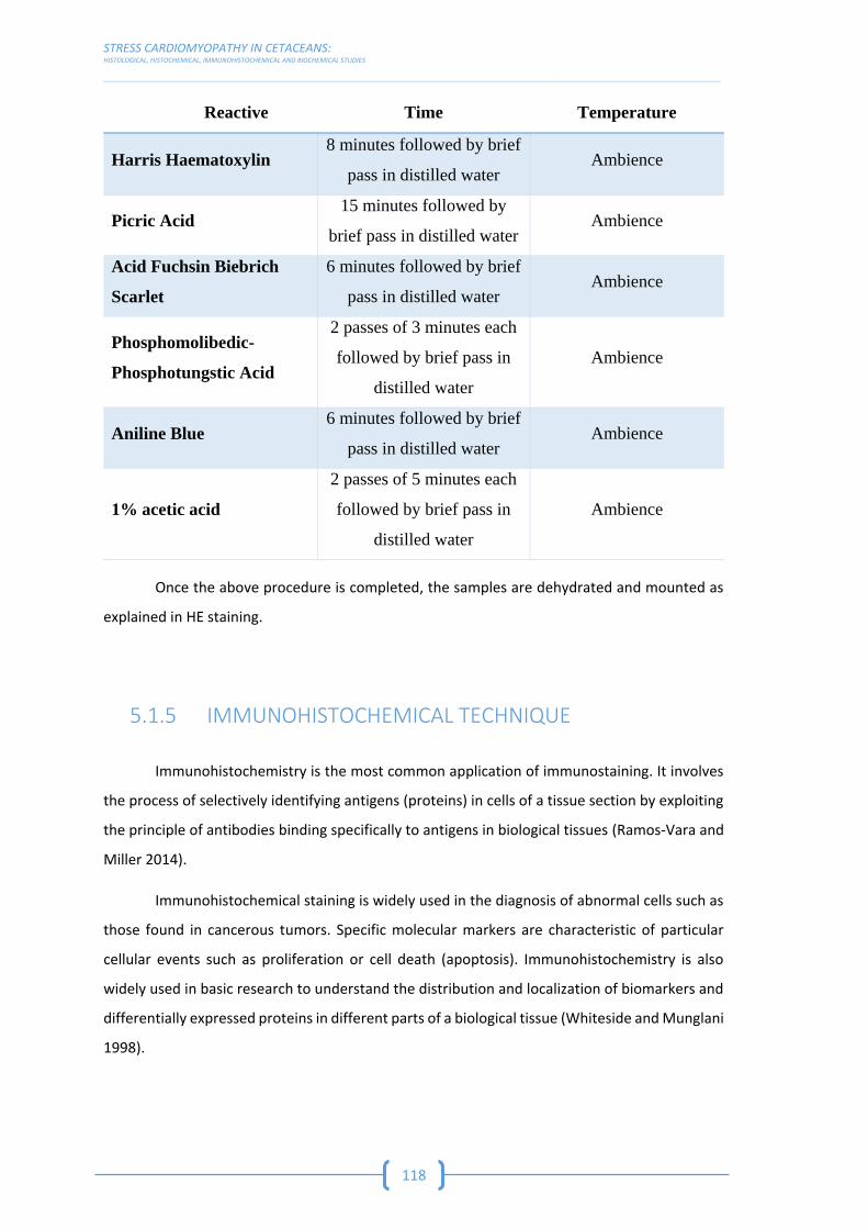
STRESS CARDIOMYOPATHY IN CETACEANS: HISTOLOGICAL, HISTOCHEMICAL, IMMUNOHISTOCHEMICAL AND BIOCHEMICAL STUDIES __________________________________________________________________________________________________________________________________________________________________________
118
Reactive Time Temperature
Harris Haematoxylin 8 minutes followed by brief
pass in distilled water Ambience
Picric Acid 15 minutes followed by
brief pass in distilled water Ambience
Acid Fuchsin Biebrich
Scarlet
6 minutes followed by brief
pass in distilled water Ambience
Phosphomolibedic-
Phosphotungstic Acid
2 passes of 3 minutes each
followed by brief pass in
distilled water
Ambience
Aniline Blue 6 minutes followed by brief
pass in distilled water Ambience
1% acetic acid
2 passes of 5 minutes each
followed by brief pass in
distilled water
Ambience
Once the above procedure is completed, the samples are dehydrated and mounted as
explained in HE staining.
5.1.5 IMMUNOHISTOCHEMICAL TECHNIQUE
Immunohistochemistry is the most common application of immunostaining. It involves
the process of selectively identifying antigens (proteins) in cells of a tissue section by exploiting
the principle of antibodies binding specifically to antigens in biological tissues (Ramos-Vara and
Miller 2014).
Immunohistochemical staining is widely used in the diagnosis of abnormal cells such as
those found in cancerous tumors. Specific molecular markers are characteristic of particular
cellular events such as proliferation or cell death (apoptosis). Immunohistochemistry is also
widely used in basic research to understand the distribution and localization of biomarkers and
differentially expressed proteins in different parts of a biological tissue (Whiteside and Munglani
1998).

MATERIALS & METHODS TECHNIQUES USED IN THE STUDY
__________________________________________________________________________________________________________________________________________________________________________
119
Tissue samples are typically sliced at a range of 3 µm. The slices are then mounted on
slides treated with VECTABOND® reagent for a better tissue section adhesion. Tissue sections
rest approximately 48h to dry well. After this, the samples require certain steps to make the
epitopes available for antibody binding, including deparaffinization and antigen retrieval, as
followed:
Reactive Time Temperature
Xylene I 10 minutes Ambience
Xylene II 5 minutes Ambience
Xylene III 5 minutes Ambience
Alcohol 100% 5 minutes Ambience
Block of Endogenous
Peroxidase 1
30 minutes while stirring in
a container where light
does not pass
Ambience
Alcohol 100% 5 minutes Ambience
Alcohol 96% 5 minutes Ambience
Alcohol 70% 5 minutes Ambience
Distilled Water I 5 minutes while stirring Ambience
Distilled Water II 5 minutes while stirring Ambience
PBS Solution 2 I 5 minutes while stirring Ambience
PBS Solution 2 II 5 minutes while stirring Ambience
(1) Hydrogen Peroxide 30% solution from Honeywell Fluka™ with reference 008-003-00-9 mixed with Methanol (EMSURE®) from
Merck with reference 1.06009.1000 (6 ml of Hydrogen Peroxide in 194 ml of Methanol). It is important to add the Hydrogen Peroxide
right before using the solution. (2) For 2 litters of distilled water we add: 2.96 g of Sodium Hydrogen Phosphate anhydrous from
PanReac AppliChem ITW Reagents with reference 131679.1211 with 0.86 g of Potassium di-Hydrogen Phosphate from PanReac
AppliChem ITW Reagents with reference 121509.1210 and finally 14.4 g of Sodium Chloride from PanReac AppliChem ITW Reagents
with reference 131679.1211.
For formalin-fixed paraffin-embedded tissues, antigen-retrieval is often necessary, and
involves pre-treating the sections with heat or protease. These steps may make the difference
between the target antigens staining or not staining. Depending on the tissue type and the
method of antigen detection, endogenous biotin or enzymes may need to be blocked or
quenched, respectively, prior to antibody staining. Although antibodies show preferential
avidity for specific epitopes, they may partially or weakly bind to sites on nonspecific proteins
(also called reactive sites) that are similar to the cognate binding sites on the target antigen. A
great amount of non-specific binding causes high background staining which will mask the
detection of the target antigen. To reduce background staining samples are incubated with a
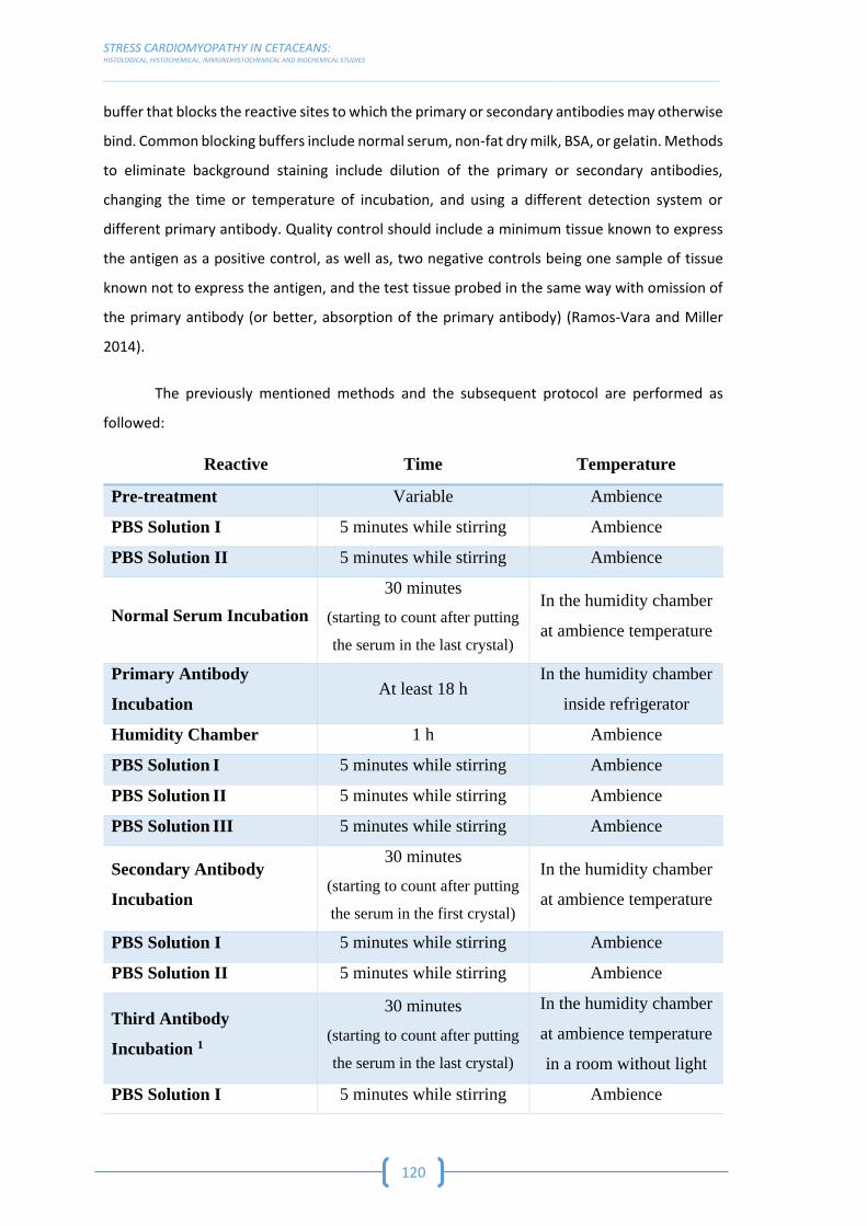
STRESS CARDIOMYOPATHY IN CETACEANS: HISTOLOGICAL, HISTOCHEMICAL, IMMUNOHISTOCHEMICAL AND BIOCHEMICAL STUDIES __________________________________________________________________________________________________________________________________________________________________________
120
buffer that blocks the reactive sites to which the primary or secondary antibodies may otherwise
bind. Common blocking buffers include normal serum, non-fat dry milk, BSA, or gelatin. Methods
to eliminate background staining include dilution of the primary or secondary antibodies,
changing the time or temperature of incubation, and using a different detection system or
different primary antibody. Quality control should include a minimum tissue known to express
the antigen as a positive control, as well as, two negative controls being one sample of tissue
known not to express the antigen, and the test tissue probed in the same way with omission of
the primary antibody (or better, absorption of the primary antibody) (Ramos-Vara and Miller
2014).
The previously mentioned methods and the subsequent protocol are performed as
followed:
Reactive Time Temperature
Pre-treatment Variable Ambience
PBS Solution I 5 minutes while stirring Ambience
PBS Solution II 5 minutes while stirring Ambience
Normal Serum Incubation
30 minutes
(starting to count after putting
the serum in the last crystal)
In the humidity chamber
at ambience temperature
Primary Antibody
Incubation At least 18 h
In the humidity chamber
inside refrigerator
Humidity Chamber 1 h Ambience
PBS Solution I 5 minutes while stirring Ambience
PBS Solution II 5 minutes while stirring Ambience
PBS Solution III 5 minutes while stirring Ambience
Secondary Antibody
Incubation
30 minutes
(starting to count after putting
the serum in the first crystal)
In the humidity chamber
at ambience temperature
PBS Solution I 5 minutes while stirring Ambience
PBS Solution II 5 minutes while stirring Ambience
Third Antibody
Incubation 1
30 minutes
(starting to count after putting
the serum in the last crystal)
In the humidity chamber
at ambience temperature
in a room without light
PBS Solution I 5 minutes while stirring Ambience

MATERIALS & METHODS TECHNIQUES USED IN THE STUDY
__________________________________________________________________________________________________________________________________________________________________________
121
Reactive Time Temperature
PBS Solution II 5 minutes while stirring Ambience
Acetate Solution 2 10 minutes while stirring Ambience
Reveal Solution 3 Variable 4 Ambience
Running Water 10 minutes Ambience
Mayer’s Haematoxylin 10 minutes Ambience
Running Water 10 minutes Ambience
Distilled Water 5 minutes Ambience
Mounting 5 - Ambience
(1) Tissue samples are visualized using the VECTASTAIN Elite ABC-Peroxidase Kit from Vector Laboratories with reference PK-6100.
In 5000 µl of PBS solution we put 2 drops (100 µl) of reagent A and 2 drops (100 µl) of reagent B. (2) For 1 litter of Acetate you need:
1.2 ml of Acetic Acid Glacial from PanReac AppliChem ITW Reagents with reference 131008.1211 mixed with 210 ml of distilled
water and 10.75 g Sodium Acetate Anhydrous from PanReac AppliChem ITW Reagents with reference 131633.1210 mixed with 790
ml of distilled water. (3) 0.5 g of 3-Amino-9-Ethylcarbazole from Sigma with reference A5754 mixed with 10 ml of N-N-
Dimethylformamide from Sigma with reference D4551. The room should have minimal light when preparing this solution and due
to the toxicity of these reagents, it should be prepared under the gas extraction fan and the personal must use a protective gas
mask. Mix this solution in a container where light does not pass (container that is not translucent). Add 140ml of Acetate solution.
Filter the solution. Add 150 µl of Hydrogen Peroxide right before using the solution. (4) 3 minutes in the case of Myoglobin and
Cardiac Troponin C and 5 minutes for Fibrinogen and Cardiac Troponin I. (5) Mount the crystals with the tissue samples with glass
cover and Faramount Aqueous Mounting Medium from Dako with reference S3025.
All of the information about the pre-treatment for the different antibodies used in this
study and all the primary and secondary antibodies are summarized in the following table.

ANTIGEN RETRIEVAL
SERUM SOURCE DILUTION PRIMARY ANTIBODY SOURCE HOST TYPE DILUTION SECONDARY ANTIBODY SOURCE DILUTION
Citrate buffer (1)
Swine serum (3)
Dako (4) 10% (5) Myoglobin (6) Abcam
(10) Rabbit Polyclonal
1 in 200 (11)
Polyclonal Swine Anti-Rabbit Immunoglobulins
(15) Dako (4)
1 in 200 (16)
Citrate buffer (1)
Swine serum (3)
Dako (4) 10% (5) Fibrinogen (7) Abcam
(10) Rabbit Polyclonal 1 in 50 (12)
Polyclonal Swine Anti-Rabbit Immunoglobulins
(15) Dako (4)
1 in 200 (16)
Citrate buffer (2)
Swine serum (3)
Dako (4) 10% (5) Troponin I (8) Abcam
(10) Rabbit Polyclonal 1 in 25 (13)
Polyclonal Swine Anti-Rabbit Immunoglobulins
(15) Dako (4)
1 in 200 (16)
Citrate buffer (2)
Swine serum (3)
Dako (4) 10% (5) Troponin C (9) Abcam
(10) Rabbit Monoclonal
1 in 250 (14)
Polyclonal Swine Anti-Rabbit Immunoglobulins
(15) Dako (4)
1 in 100 (17)
(1) Citrate buffer, pH 6.0, 7 minutes at 96ºC. (2) Citrate buffer, pH 6.0, 20 minutes at 100ºC. (3) Dako Swine serum (Normal) (X090110-8). (4) Dako (Glostrup, Denmark). (5) Dilution of 10 µl of serum in 90 µl of
PBS. (6) Anti-Myoglobin antibody (ab187506). (7) Anti-Fibrinogen antibody (ab34269). (8) Anti-Cardiac Troponin I antibody (ab47003). (9) Anti-Cardiac Troponin C antibody (ab137130). (10) Abcam (Cambridge,
United Kingdom). (11) Dilution of 1 µl of primary antibody in 199 µl of serum at 1% in PBS (dilution of 1 µl of serum in 99 µl of PBS). (12) Dilution of 1 µl of primary antibody in 49 µl of serum at 1% in PBS
(dilution of 1 µl of serum in 99 µl of PBS). (13) Dilution of 1 µl of primary antibody in 24 µl of serum at 1% in PBS (dilution of 1 µl of serum in 99 µl of PBS). (14) Dilution of 1 µl of primary antibody in 249 µl of
serum at 1% in PBS (dilution of 1 µl of serum in 99 µl of PBS). (15) Dako Polyclonal Swine Anti-Rabbit Immunoglobulins/Biotinylated (E035301-2). (16) Dilution of 1 µl of secondary antibody in 199 µl of serum
at 1% in PBS (dilution of 1 µl of serum in 99 µl of PBS). (17) Dilution of 1 µl of secondary antibody in 99 µl of serum at 1% in PBS (dilution of 1 µl of serum in 99 µl of PBS).
RESULTS
&
DISCUSSION
PUBLICATIONS


6.1 CHARACTERIZATION OF STRESS
CARDIOMYOPATHY


RESULTS & DISCUSSION: PUBLICATIONS CHARACTERIZATION OF STRESS CARDIOMYOPATHY
[STRESS CARDIOMYOPATHY IN CETACEANS: AN HISTOLOGICAL, HISTOCHEMICAL AND IMMUNOHISTOCHEMICAL STUDY] __________________________________________________________________________________________________________________________________________________________________________
127
The first specific objective of this work, which consisted in characterizing the SCMP in
cetaceans, such as injuries resulting from extreme stress responses, was achieved using
histological, histochemical and immunohistochemical approaches on a total of 67 animals which
live-stranded or died due to ship collisions and interactions with fisheries (bycatch).
This resulted in the publication of an article (as followed) titled Stress cardiomyopathy
in stranded cetaceans: a histological, histochemical and immunohistochemical study, on
September 2019 in the Veterinary Record, Volume 185, Issue 22 (doi: 10.1136/vr.105562),
having this journal an impact factor of 2.101 in 2018 and therefore being a Q1 in the veterinary
category.

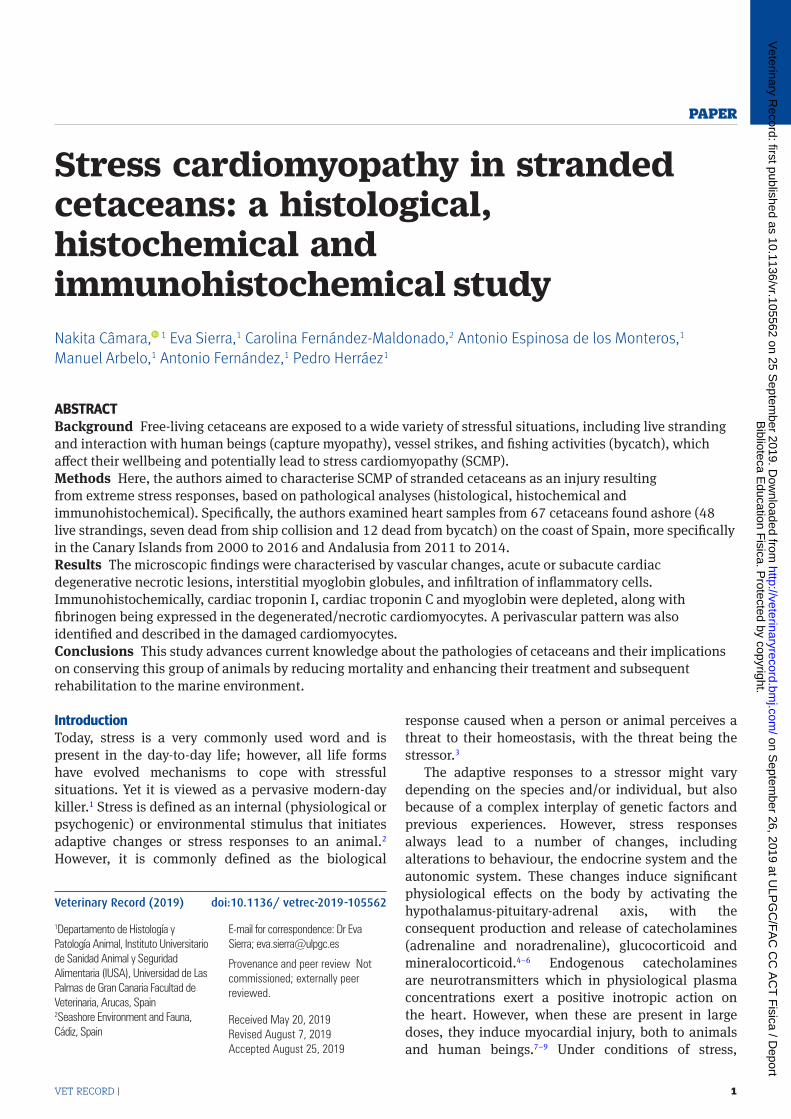
Vet RecoRD | 1
PaPer
Stress cardiomyopathy in stranded cetaceans: a histological, histochemical and immunohistochemical studyNakita Câmara, 1 Eva Sierra,1 Carolina Fernández-Maldonado,2 Antonio Espinosa de los Monteros,1 Manuel Arbelo,1 Antonio Fernández,1 Pedro Herráez1
ABSTRACTBackground Free-living cetaceans are exposed to a wide variety of stressful situations, including live stranding and interaction with human beings (capture myopathy), vessel strikes, and fishing activities (bycatch), which affect their wellbeing and potentially lead to stress cardiomyopathy (SCMP).Methods Here, the authors aimed to characterise SCMP of stranded cetaceans as an injury resulting from extreme stress responses, based on pathological analyses (histological, histochemical and immunohistochemical). Specifically, the authors examined heart samples from 67 cetaceans found ashore (48 live strandings, seven dead from ship collision and 12 dead from bycatch) on the coast of Spain, more specifically in the Canary Islands from 2000 to 2016 and Andalusia from 2011 to 2014.Results The microscopic findings were characterised by vascular changes, acute or subacute cardiac degenerative necrotic lesions, interstitial myoglobin globules, and infiltration of inflammatory cells. Immunohistochemically, cardiac troponin I, cardiac troponin C and myoglobin were depleted, along with fibrinogen being expressed in the degenerated/necrotic cardiomyocytes. A perivascular pattern was also identified and described in the damaged cardiomyocytes.Conclusions This study advances current knowledge about the pathologies of cetaceans and their implications on conserving this group of animals by reducing mortality and enhancing their treatment and subsequent rehabilitation to the marine environment.
IntroductionToday, stress is a very commonly used word and is present in the day-to-day life; however, all life forms have evolved mechanisms to cope with stressful situations. Yet it is viewed as a pervasive modern-day killer.1 Stress is defined as an internal (physiological or psychogenic) or environmental stimulus that initiates adaptive changes or stress responses to an animal.2 However, it is commonly defined as the biological
response caused when a person or animal perceives a threat to their homeostasis, with the threat being the stressor.3
The adaptive responses to a stressor might vary depending on the species and/or individual, but also because of a complex interplay of genetic factors and previous experiences. However, stress responses always lead to a number of changes, including alterations to behaviour, the endocrine system and the autonomic system. These changes induce significant physiological effects on the body by activating the hypothalamus-pituitary-adrenal axis, with the consequent production and release of catecholamines (adrenaline and noradrenaline), glucocorticoid and mineralocorticoid.4–6 Endogenous catecholamines are neurotransmitters which in physiological plasma concentrations exert a positive inotropic action on the heart. However, when these are present in large doses, they induce myocardial injury, both to animals and human beings.7–9 Under conditions of stress,
10.1136/vetrec-2019-105562
Veterinary Record (2019) doi:10.1136/ vetrec-2019-105562
1Departamento de Histología y Patología Animal, Instituto Universitario de Sanidad Animal y Seguridad Alimentaria (IUSA), Universidad de Las Palmas de Gran Canaria Facultad de Veterinaria, Arucas, Spain2Seashore Environment and Fauna, Cádiz, Spain
E-mail for correspondence: Dr Eva Sierra; eva. sierra@ ulpgc. es
Provenance and peer review Not commissioned; externally peer reviewed.
Received May 20, 2019Revised August 7, 2019Accepted August 25, 2019
Biblioteca E
ducation Fisica. P
rotected by copyright. on S
eptember 26, 2019 at U
LPG
C/F
AC
CC
AC
T F
isica / Deport
http://veterinaryrecord.bmj.com
/V
eterinary Record: first published as 10.1136/vr.105562 on 25 S
eptember 2019. D
ownloaded from
| Vet RecoRD2
large amounts of catecholamines are released from the sympathetic nerve endings and adrenal medulla, with exposure to these high concentrations producing coronary spasms, arrhythmias, contractile dysfunction, myocardial cell damage and extensive necrosis.10
There is increasing awareness of a unique cardiac syndrome in human pathology, which has been described as the ‘Apical Ballooning Syndrome’, ‘Takotsubo Disease’, and ampulla or stress cardiomyopathy (SCMP), otherwise known as the ‘Broken Heart Syndrome’ in the popular press.11 This syndrome is a reversible cardiomyopathy (CMP) that typically occurs after an emotional or physical stress event. Its clinical presentation cannot be distinguished from a myocardial infarction.11 12 Several cases have been reported globally.13–22 SCMP is an important differential diagnosis of an acute myocardial infarction; consequently, it is under-recognised and often misdiagnosed.11
The pathophysiology of this syndrome appears to be related to an excess of plasma catecholamines, which are triggered by stressful conditions. Histological findings are consistent with catecholamine cardiotoxicity, due to ischaemia-reperfusion injury, both in human myocardial samples and in experimental models of SCMP.11 12 23–32 The crucial role of myocardial ischaemia in Takotsubo syndrome has been identified, with most cases occurring in patients with a risk factor for endothelial dysfunction. This dysfunction is characterised by an imbalance between vasoconstricting and vasodilating factors, which might explain the propensity to epicardial and/or microvascular coronary artery spasm of the disease.33 34
Marine mammals, in this case cetaceans, present the same basic response to stress as human beings and other mammals. Once the central nervous system perceives a threat, a biological or defence response is developed.35 This response is well documented in marine mammals.36–40 Although physiological stress has some benefits, an extreme or prolonged response, with release of catecholamines and sustained high blood cortisol and aldosterone levels, could potentially damage skeletal and heart muscles.35 Consequently, cardiac lesions seem to play a central role in marine mammals, with cetaceans being particularly prone to develop SCMP, probably due to the characteristics of their cardiovascular adaptations, conforming to the requirements of diving metabolism.35 39 41–43
Free-living cetaceans are at daily risk to a wide variety of stressful situations, of both natural and anthropogenic origin, that influence their wellbeing. In recent decades, efforts have been made to reduce the impact of some human activities on cetaceans. Unfortunately, whales and dolphins continue to be threatened, for example, by active (live) stranding, ship collision and bycatch. These different pathological entities have acute stress as the central aetiopathogenesis, potentially leading to the
death of the animal or seriously aggravating a previous disease situation. These issues impact the subsequent rehabilitation of animals, reducing the effectiveness of therapy and limiting successful recovery.39 40 42 44–49
Acute and stressful deaths of cetaceans, such as occurs in live strandings, might be attributed to the so-called ‘stress response syndrome’ or ‘alarm reaction’. The involved mechanisms are thought to be analogous to capture myopathy in terrestrial mammals and are typically referred in the literature, particularly in prolonged rescues and rehabilitation efforts.39 42 45 50 The response consists of local to generalised vasospasm and vasodilation (catecholamines, neurogenic shock, impeded venous flow return by body compression), concurring with the SCMP in human beings, direct traumatic injury of the muscle and viscera, and reperfusion damage.39 42 45
The collision of ships with cetaceans is recognised as an important global threat, with a rapid increase in its incidence in recent years. For individuals that suffer severe trauma after impact with a boat, death generally occurs in an acute form, due to haemorrhagic shock (hypovolaemic), mirroring the SCMP process in human beings, and directly impacting vital organs (eg, section of the spinal cord). Alternatively, chronic trauma occurs, which can lead to the development of sepsis or generalised weakness.44 45 47
Fishing gears also present a major threat to cetaceans, worldwide, because of their widespread use.51 When trapped in fishing nets, small whales, dolphins and porpoises often die because they are not strong enough to break free or surface to breathe.52 Captured cetaceans typically exhibit haemodynamic changes to shock, as it happens in the SCMP in human beings, due to submersion or drowning (asphyxia). Often, macroscopic or histopathological lesions are evident in the cardiovascular and respiratory systems.44–46 48
Here, the authors aimed to characterise SCMP of stranded cetaceans as an injury resulting from extreme stress responses. Cetaceans from live strandings, ship collisions and bycatch were analysed via histological, histochemical and immunohistochemical approaches.
Materials and methodsOne hundred and forty-eight cetaceans of 20 different species, including small and large odontocetes and mysticetes, were initially included in this study. Of these, 83 were stranded alive (and subsequently died previous to or during the rescue and rehabilitation), 32 died due to ship collision and 33 died from entanglement in fishery lines. All of these animals were stranded on the coast of Spain, more specifically in the Canary Islands from 2000 to 2016 and Andalusia from 2011 to 2014. The pathological findings in the different organs and causes of death (stranded alive, ship collision and entanglement in fishery lines) of the 148 animals are described in annual reports.44 45 53
Biblioteca E
ducation Fisica. P
rotected by copyright. on S
eptember 26, 2019 at U
LPG
C/F
AC
CC
AC
T F
isica / Deport
http://veterinaryrecord.bmj.com
/V
eterinary Record: first published as 10.1136/vr.105562 on 25 S
eptember 2019. D
ownloaded from
Vet RecoRD | 3
Tabl
e 1
Sum
mar
y of t
he im
mun
ohis
toch
emic
al m
etho
dolo
gy u
sed
in th
is st
udy
Antig
en re
triev
alSe
rum
Sour
ceDi
lutio
nPr
imar
y ant
ibod
ySo
urce
Host
Type
Dilu
tion
Seco
ndar
y ant
ibod
ySo
urce
Dilu
tion
Citra
te bu
ffer (
1)Sw
ine s
erum
(3)
Dako
(4)
10%
(5)
Myo
glob
in (6
)Ab
cam
(10)
Rabb
itPo
lyclo
nal
1 in
200
(11)
Polyc
lona
l Swi
ne A
nti-R
abbi
t Imm
unog
lobu
lins (
15)
Dako
(4)
1 in
200
(16)
Citra
te bu
ffer (
1)Sw
ine s
erum
(3)
Dako
(4)
10%
(5)
Fibrin
ogen
(7)
Abca
m (1
0)Ra
bbit
Polyc
lona
l1
in 5
0 (1
2)Po
lyclo
nal S
wine
Ant
i-Rab
bit Im
mun
oglo
bulin
s (15
)Da
ko (4
)1
in 2
00 (1
6)Ci
trate
buffe
r (2)
Swin
e ser
um (3
)Da
ko (4
)10
% (5
)Tro
poni
n I (8
)Ab
cam
(10)
Rabb
itPo
lyclo
nal
1 in
25
(13)
Polyc
lona
l Swi
ne A
nti-R
abbi
t Imm
unog
lobu
lins (
15)
Dako
(4)
1 in
200
(16)
Citra
te bu
ffer (
2)Sw
ine s
erum
(3)
Dako
(4)
10%
(5)
Tropo
nin C
(9)
Abca
m (1
0)Ra
bbit
Mon
oclo
nal
1 in
250
(14)
Polyc
lona
l Swi
ne A
nti-R
abbi
t Imm
unog
lobu
lins (
15)
Dako
(4)
1 in
100
(17)
(1) C
itrat
e buf
fer, p
H 6.
0, se
ven m
inut
es at
96º
C. (2
) Citr
ate b
uffe
r, pH
6.0,
20
min
utes
at 1
00ºC
. (3) D
ako S
wine
seru
m (n
orm
al) (
X090
110-
8). (4
) Dak
o (Gl
ostru
p, D
enm
ark)
. (5) D
ilutio
n of 1
0 µl
of se
rum
in 9
0 µl
of PB
S. Th
e ser
um is
incu
bate
d in a
hum
idity
cham
ber f
or ha
lf an h
our. (
6) A
ntim
yogl
obin
an
tibod
y (ab
1875
06). (
7) A
ntifi
brin
ogen
antib
ody (
ab34
269)
. (8) A
nti-c
ardi
ac tr
opon
in I a
ntib
ody (
ab47
003)
. (9) A
nti-c
ardi
ac tr
opon
in C
antib
ody (
ab13
7130
). (10
) Abc
am (C
ambr
idge
, UK)
. (11)
Dilu
tion o
f 1 µl
of pr
imar
y ant
ibod
y in 1
99 µl
of se
rum
at 1
% in
PBS (
dilu
tion o
f 1 µl
of se
rum
in 9
9 µl
of
PBS)
. The
prim
ary a
ntib
ody i
s inc
ubat
ed in
a hu
mid
ity ch
ambe
r for
at le
ast 1
8 ho
urs,
insid
e the
refri
gera
tor. (
12) D
ilutio
n of 1
µl of
prim
ary a
ntib
ody i
n 49
µl of
seru
m at
1%
in PB
S (di
lutio
n of 1
µl of
seru
m in
99
µl of
PBS)
. The
prim
ary a
ntib
ody i
s inc
ubat
ed in
a hu
mid
ity ch
ambe
r for
at le
ast 1
8 ho
urs,
insid
e th
e ref
riger
ator
. (13)
Dilu
tion o
f 1 µl
of pr
imar
y ant
ibod
y in 2
4 µl
of se
rum
at 1
% in
PBS (
dilu
tion o
f 1 µl
of se
rum
in 9
9 µl
of PB
S). T
he pr
imar
y ant
ibod
y is i
ncub
ated
in a
hum
idity
cham
ber f
or at
leas
t 18
hour
s, in
side t
he re
frige
rato
r. (14
) Dilu
tion o
f 1 µl
of pr
imar
y ant
ibod
y in 2
49 µl
of se
rum
at 1
% in
PBS
(dilu
tion o
f 1 µl
of se
rum
in 9
9 µl
of PB
S). T
he pr
imar
y ant
ibod
y is i
ncub
ated
in a
hum
idity
cham
ber f
or at
leas
t 18
hour
s, in
side t
he re
frige
rato
r. (15
) Dak
o Pol
yclo
nal S
wine
Ant
i-Rab
bit Im
mun
oglo
bulin
s/Bi
otin
ylate
d (E0
3530
1-2)
. (16)
Dilu
tion o
f 1 µl
of se
cond
ary a
ntib
ody i
n 199
µl of
seru
m at
1%
in PB
S (d
ilutio
n of 1
µl of
seru
m in
99
µl of
PBS)
. The
seco
ndar
y ant
ibod
y is i
ncub
ated
in a
hum
idity
cham
ber f
or ha
lf an h
our. (
17) D
ilutio
n of 1
µl of
seco
ndar
y ant
ibod
y in 9
9 µl
of se
rum
at 1
% in
PBS (
dilu
tion o
f 1 µl
of se
rum
in 9
9 µl
of PB
S). T
he se
cond
ary a
ntib
ody i
s inc
ubat
ed in
a hu
mid
ity ch
ambe
r for
half a
n ho
ur.
A complete postmortem examination was performed following the standard protocol published by the European Cetacean Society,54 with some additional procedures detailed in the manual Marine Mammals Ashore: A Field Guide for Strandings.50 The conservation state of the bodies was also determined following the parameters and classifications established by these two protocols. Thus, out of the initial 148 animals, the authors identified 67 animals (48 stranded alive, seven dead from ship collision and 12 from bycatch) belonging to 19 species that had a very fresh, fresh and moderate autolysis conservation state. Previous studies have shown that animals with advanced autolysis or that were previously frozen present a great number of false negatives when using histochemical and immunohistochemical techniques.
The samples from the 67 animals were fixed and processed following standard histological procedures. Specifically, 4 µm of samples was used for haematoxylin and eosin (HE) and periodic acid-Schiff staining, and 5 µm was used for phosphotungstic acid haematoxylin (PTAH) and Masson’s trichrome techniques. The basic epidemiological data about the animals are presented in online supplementary file 1. Tissue sections of 3 µm were immunolabelled using antimyoglobin, antifibrinogen, anti-cardiac troponin I (cTnI) and anti-cardiac troponin C (cTnC) primary antibodies, and were visualised using the VECTASTAIN Elite ABC-Peroxidase Kit (PK-6100) from Vector Laboratories (Peterborough, UK). The immunohistochemical methodology is summarised in table 1. The negative control for the immunohistochemical techniques was performed without the primary antibody on a serial heart section. The positive control for myoglobin and fibrinogen was a cetacean heart sample of a striped dolphin (previously published to have been stranded alive and to have developed capture myopathy due to capture and human interaction during the rehabilitation process).39 42 For cTnI and cTnC, the authors used a heart sample from a pig, with no apparent acute macroscopic and/or histological lesions.
The sections submitted to the different techniques were examined in a blind manner by three veterinary pathologists (NC, ES and PH). The sections were evaluated for vascular changes (congestion, haemorrhages and interstitial oedema); acute degenerative changes (contraction band necrosis, wavy fibres, perinuclear vacuolisation, cytoplasmic hypereosinophilia and pyknotic nuclei); and presence of interstitial myoglobin globules and infiltration of inflammatory cells. The extent of cardiac lesions was judged subjectively as absent, mild, mild to moderate, moderate, moderate to severe, and severe.
The amount of damage present to cells was confirmed as antemortem when it was accompanied with immunohistochemically demonstrated depletion of myoglobin together with intrafibrillar fibrinogen deposition.
Biblioteca E
ducation Fisica. P
rotected by copyright. on S
eptember 26, 2019 at U
LPG
C/F
AC
CC
AC
T F
isica / Deport
http://veterinaryrecord.bmj.com
/V
eterinary Record: first published as 10.1136/vr.105562 on 25 S
eptember 2019. D
ownloaded from

| Vet RecoRD4
Table 2 Classification for the absence or presence, in different degrees, of vascular changes (congestion, interstitial oedema and haemorrhage) in the 67 selected animals, from the 148 initially examined animals, belonging to the distinct pathological entities and causes of death, in this case ship collision (seven animals), bycatch (12 animals) and live-stranded (48 animals)
Total number of cases Ship collision cases Bycatch cases Live stranding cases
Animals (n) Percentage Animals (n) Percentage Animals (n) Percentage Animals (n) Percentage
Congestion Absence 22 32.8 5 71.4 2 16.7 15 31.3 Mild 23 34.3 2 28.6 6 50.0 15 31.3 Mild to moderate 6 9.0 0 0 2 16.7 4 8.3 Moderate 13 19.4 0 0 1 8.3 12 25.0 Moderate to severe 1 1.5 0 0 1 8.3 0 0 Severe 2 3.0 0 0 0 0 2 4.2Interstitial oedema Absence 41 61.2 3 42.9 6 50.0 32 66.7 Mild 12 17.9 3 42.9 4 33.3 5 10.4 Mild to moderate 5 7.5 0 0 2 16.7 3 6.3 Moderate 5 7.5 0 0 0 0 5 10.4 Moderate to severe 3 4.5 1 14.3 0 0 2 4.2 Severe 1 1.5 0 0 0 0 1 2.1Haemorrhage Absence 53 79.1 6 85.7 11 91.7 36 75.0 Mild 8 11.9 1 14.3 1 8.3 7 14.6 Mild to moderate 1 1.5 0 0 0 0 1 2.1 Moderate 1 1.5 0 0 0 0 1 2.1 Moderate to severe 1 1.5 0 0 0 0 0 0 Severe 3 4.5 0 0 0 0 3 6.3
ResultsHistologically, the major cardiac lesions associated with stress were (1) vascular changes (table 2), which presented as congestion, haemorrhage and interstitial oedema; (2) acute degenerative changes, which were attributed to cardiotoxicity by catecholamines (table 3) and consisted of contraction band necrosis, wavy fibres, cytoplasmic hypereosinophilia and perinuclear vacuolisation; and (3) the presence of interstitial myoglobin globules (table 4) and infiltration of inflammatory cells (table 5).
Vascular congestion was identified by swelling and the light occlusion of capillaries with blood (figure 1). This phenomenon was observed in 45 out of 67 animals (67.2 per cent). This condition was mild to moderate in the individuals that died due to bycatch (10 out of 12 animals, 83.3 per cent) and live-stranded (33 out of 48 animals, 68.8 per cent). Individuals that died from ship collision (two out of seven animals, 28.6 per cent) displayed a mild degree of congestion.
Out of the 67 animals, 26 (38.9 per cent) had fibres separated by spaces, representing interstitial oedema. Overall, it was mild to moderate for all pathological categories. It was particularly noticeable in four of the seven animals (57.2 per cent) that died from ship collisions, six of the 12 animals (50.0 per cent) that died due to bycatch, and 16 of the 48 animals (33.4 per cent) that were stranded alive. Eight out of the 67 animals (11.9 per cent) with interstitial oedema also had haemorrhages.
Only haemorrhages could be visualised grossly and microscopically (figure 1). They were classified
as endocardial, myocardial and epicardial, and were primarily observed in the endocardium. One out of the 67 animals (1.5 per cent) exclusively exhibited gross haemorrhages, four (6.0 per cent) exhibited gross and microscopic haemorrhages, and 10 (14.9 per cent) only presented microscopic haemorrhages that were of a mild to moderate degree. Microscopically, one of the seven animals (14.3 per cent) that died from ship collisions exhibited a moderate to severe degree of haemorrhage, 12 out of the 48 animals (25.0 per cent) that were stranded alive exhibited a mild to moderate degree, and one out of 12 animals (8.3 per cent) that died due to bycatch exhibited a mild degree.
Contraction band necrosis is the focal hypercontraction and lysis of contractile filaments in small groups of myocardial cells. The resulting dense hypereosinophilic bands run transversely through the myocyte (figure 2). This acute degenerative change was observed, mainly with a mild to moderate degree, in 33 out of the 67 animals (49.3 per cent). Some cases had lesions that were more marked at the subendocardial and subepicardial level. Of note, all of the animals that died from ship collisions (seven individuals, 100 per cent) presented contraction band necrosis that was moderate to severe, followed by the animals that were stranded alive (25 out of 48 animals, 52.1 per cent) which displayed mild to moderate necrosis, and animals that died due to bycatch (one out of 12 animals, 8.3 per cent) with a mild degree of necrosis.
Twenty-nine out of the 67 animals (43.3 per cent) primarily exhibited a mild degree of long and thin undulated fibres, termed wavy fibres (figure 3). Wavy
Biblioteca E
ducation Fisica. P
rotected by copyright. on S
eptember 26, 2019 at U
LPG
C/F
AC
CC
AC
T F
isica / Deport
http://veterinaryrecord.bmj.com
/V
eterinary Record: first published as 10.1136/vr.105562 on 25 S
eptember 2019. D
ownloaded from

Vet RecoRD | 5
Table 3 Classification for the absence or presence, in different degrees, of acute degenerative changes (contraction band necrosis, wavy fibres, hypereosinophilia and cytoplasmic vacuolisation) in the 67 selected animals, from the 148 initially examined animals, belonging to the distinct pathological entities and causes of death, in this case ship collision (seven animals), bycatch (12 animals) and live-stranded (48 animals)
Total number of cases Ship collision cases Bycatch cases Live stranding cases
Animals (n) Percentage Animals (n) Percentage Animals (n) Percentage Animals (n) Percentage
Contraction band necrosis Absence 34 50.7 0 0 11 91.7 23 47.9 Mild 17 25.4 3 42.9 1 8.3 13 27.1 Mild to moderate 4 6.0 0 0 0 0 4 8.3 Moderate 2 3.0 0 0 0 0 2 4.2 Moderate to severe 2 3.0 1 14.3 0 0 1 2.1 Severe 8 11.9 3 42.9 0 0 5 10.4
Wavy fibres Absence 38 56.7 2 28.6 6 50.0 30 62.5 Mild 18 26.9 3 42.9 5 41.7 10 20.8
Mild to moderate 3 4.5 0 0 0 0 3 6.3 Moderate 7 10.4 2 28.6 1 8.3 4 8.3 Moderate to severe 1 1.5 0 0 0 0 1 2.1 Severe 0 0 0 0 0 0 0 0
Hypereosinophilia Absence 0 0 0 0 0 0 0 0
Mild 10 14.9 1 14.3 6 50.0 3 6.3 Mild to moderate 9 13.4 2 28.6 1 8.3 6 12.5
Moderate 26 38.8 2 28.6 5 41.7 19 39.6 Moderate to severe 12 17.9 0 0 0 0 12 25.0 Severe 10 14.9 2 28.6 0 0 8 16.7Vacuolar degeneration
Absence 2 3.0 0 0 1 8.3 1 2.1 Mild 15 22.4 4 57.1 7 58.3 4 8.3 Mild to moderate 7 10.4 1 14.3 1 8.3 5 10.4
Moderate 22 32.8 2 28.6 3 25.0 17 35.4 Moderate to severe 13 19.4 0 0 0 0 13 27.1 Severe 8 11.9 0 0 0 0 8 16.7
Table 4 Classification for the absence or presence of interstitial myoglobin globules in the 67 selected animals, from the 148 initially examined animals, belonging to the distinct pathological entities and causes of death, in this case ship collision (seven animals), bycatch (12 animals) and live-stranded (48 animals)
Total number of cases Ship collision cases Bycatch cases Live stranding cases
Animals (n) Percentage Animals (n) Percentage Animals (n) Percentage Animals (n) Percentage
Interstitial myoglobin globules Absence 38 56.7 5 71.4 3 25.0 30 62.5 Presence 29 43.3 2 28.6 9 75.0 18 37.5
fibres were observed, with a mild to moderate degree, in five out of seven animals (71.5 per cent) that died from ship collisions, six out of 12 animals (50.0 per cent) that died from bycatch, and 18 out of 48 animals (37.5 per cent) that were stranded alive.
Hypereosinophilia was easily identified by comparing the region suspected to be necrotic against fibres in the same section. The necrotic fibres presented a more eosinophilic, blue or red (using HE, PTAH and Masson’s trichrome technique, respectively) staining than the adjacent normal fibres (figure 4). This acute degenerative change was observed in all animals (67, 100 per cent) from a moderate to severe degree. In some cases, hypereosinophilia was more pronounced at the subendocardial and subepicardial level. All animals that were stranded alive (48 animals in total, 100 per cent) presented an overall moderate to severe degree
of hypereosinophilia, along with all animals that died from ship collisions (seven individuals, 100 per cent), which presented a moderate degree, and all animals that died from bycatch (12 animals, 100 per cent), which mostly presented a mild to moderate degree of hypereosinophilia.
Sixty-five out of the 67 animals (96.9 per cent) exhibited some degree of vacuolar degeneration. This acute degenerative change was morphologically characterised by the intracellular accumulation of fluid and lysis of myofibrils. The increase in liquid exerts pressure on the different parts of the cell, resulting in most of the fibres with vacuoles having a pyknotic nucleus (figure 5). In some cases, this lesion was more noticeable at the subendocardial and subepicardial level. Forty-seven out of 48 animals (97.9 per cent) that were stranded alive presented vacuoles in the fibres of a
Biblioteca E
ducation Fisica. P
rotected by copyright. on S
eptember 26, 2019 at U
LPG
C/F
AC
CC
AC
T F
isica / Deport
http://veterinaryrecord.bmj.com
/V
eterinary Record: first published as 10.1136/vr.105562 on 25 S
eptember 2019. D
ownloaded from

| Vet RecoRD6
Table 5 Classification for the absence or presence, in different degrees, of inflammatory infiltration in the 67 selected animals, from the 148 initially examined animals, belonging to the distinct pathological entities and causes of death, in this case ship collision (seven animals), bycatch (12 animals) and live-stranded (48 animals)
Total number of cases Ship collision cases Bycatch cases Live stranding cases
Animals (n) Percentage Animals (n) Percentage Animals (n) Percentage Animals (n) Percentage
Inflammatory infiltration Absence 50 74.6 5 71.4 8 66.7 37 77.1 Mild 14 20.9 2 28.6 2 16.7 10 20.8
Mild to moderate 1 1.5 0 0 1 8.3 0 0 Moderate 1 1.5 0 0 0 0 1 2.1 Moderate to severe 1 1.5 0 0 1 8.3 0 0 Severe 0 0 0 0 0 0 0 4
Figure 1 Live-stranded animal: Kogia breviceps (reference: i373_07). Endocardial haemorrhage (arrows) and congestion (arrowhead) visualised with haematoxylin and eosin, magnification x 10.
Figure 2 Live-stranded animal: Kogia breviceps (reference: i373_07). The contraction band necrosis runs (arrows) transversely through the cardiomyocytes, and it is identified through the higher intensity of colour with different histochemical techniques, in this case with the PTAH technique, magnification x 60. Notice also the presence of myoglobin globules in the interstitial space (arrowheads). Inset: Live-stranded animal: K. breviceps (reference: i452_00). Detail of the contraction band necrosis (arrows) with Masson’s trichrome, magnification x 60. PTAH, phosphotungstic acid haematoxylin.
moderate to severe degree, all animals (seven animals, 100 per cent) that died from ship collisions had a mild to moderate degree of vacuoles, and 11 of the 12 animals (91.6 per cent) that died from bycatch had a mild degree of vacuoles.
Immunohistochemically, the degenerated/necrotic cardiomyocytes showed homogeneous, intrafibrillar depletion of cTnI, cTnC and myoglobin (figures 6–9). Simultaneously, the authors verified that myoglobin accumulated as globules inside the blood vessels and in the interstitial spaces (figure 10). The myoglobin globules were observed in 29 out of 67 animals (43.3 per cent). Specifically, myoglobin globules were detected in nine out of 12 animals (75.0 per cent) that died from bycatch, 18 out of 48 animals (37.5 per cent) that were stranded alive, and two out of seven animals (28.6 per cent) that died from ship collisions. Damaged cardiomyocytes exhibited various intensities of immunolabelling for fibrinogen. In some cases, cTnI, cTnC and fibrinogen were strongly expressed in the zone next to and inside the contraction band necrosis (figures 7, 9 and 11). However, cTnI, cTnC and myoglobin were so slightly expressed that they disappeared at times (depletion), with fibrinogen expression emerging more intensively. Immunolabelling of uninjured cardiomyocytes produced homogeneous, intracytoplasmic immunolabelling for cTnI, cTnC and myoglobin, and did not stain for fibrinogen.
The major cardiac lesions associated with stress (such as the hypereosinophilia and cytoplasmic vacuolisation) were more commonly observed in a multifocal distribution in the rows of cardiomyocytes that were located closest to the blood vessels and that followed the paths. Such cells were damaged or formed groups of damaged cells. Thus, the acute degenerative changes present in the degenerated/necrotic cardiomyocytes had a perivascular pattern (figures 12 and 13). Live-stranded animals presented this pattern more intensively.
In seventeen out of 67 animals (25.37 per cent), different types of cells with a mild degree of inflammation were identified infiltrating zones where haemorrhages, fibrillary rupture of fibres, degeneration and necrosis of cardiac cells were present. Four out of 12 animals (33.3 per cent) that died from bycatch displayed a mild to moderate degree of inflammatory cell infiltration, as well as two out of seven (28.6 per cent) that died from ship collisions and 11 animals out of 48 animals (22.9 per cent) that were stranded alive. Six out of the 67 animals (9.0 per cent) presented a mild degree of infiltration of mononuclear cells. This condition was present to a mild degree in animals that died from ship
Biblioteca E
ducation Fisica. P
rotected by copyright. on S
eptember 26, 2019 at U
LPG
C/F
AC
CC
AC
T F
isica / Deport
http://veterinaryrecord.bmj.com
/V
eterinary Record: first published as 10.1136/vr.105562 on 25 S
eptember 2019. D
ownloaded from

Vet RecoRD | 7
Figure 3 Ship collision animal: Physeter macrocephalus (reference: i59_09). Wavy fibres (arrows) which consist of long and thinned undulated fibres. HE technique, magnification x 20. Inset: Live-stranded animal: Mesoplodon densirostris (reference: i92_04). Detail of wavy fibres (arrows) with PTAH, magnification x 20. HE, haematoxylin and eosin; PTAH, phosphotungstic acid haematoxylin.
Figure 4 Live-stranded animal: Pseudorca crassidens (reference: i102_08). Hypereosinophilia consists of an increased staining of necrotic cardiac cells, such as Purkinje cells (arrows), with different techniques, in this case with Masson’s trichrome technique, magnification x 20. Inset: Live-stranded animal: Mesoplodon densirostris (reference: i238_03). Hypereosinophilia (arrowheads) of necrotic cardiomyocytes stained with HE, magnification x 40. Furthermore, observe the presence of cytoplasmic vacuolisation in some injured cardiomyocytes (thin arrows). HE, haematoxylin and eosin.
Figure 5 Live-stranded animal: Kogia breviceps (reference: i373_07). Detail of the intracytoplasmic vacuoles (arrows) of the cardiomyocytes with HE technique. Moreover, take into consideration the presence of injured cells with hypereosinophilic staining (arrowhead), magnification x 60. Inset: Bycatch animal: Delphinus delphis (reference: i263_11). Vacuolar degeneration (arrows) in Purkinje cells, with PTAH technique. Magnification x 40. HE, haematoxylin and eosin; PTAH, phosphotungstic acid haematoxylin.
Figure 6 Live-stranded animal: Delphinus delphis (reference: i87_03). Degenerated/necrotic cardiomyocytes (arrows) show intrafibrillar depletion of myoglobin. Immunohistochemical technique: antimyoglobin, magnification x 60.
collisions (two out of seven animals, 28.6 per cent) and that were stranded alive (three out of 48 animals, 6.3 per cent). It was present at a moderate to severe degree in animals that died from bycatch (one out of 12 animals, 6.3 per cent). Three out of the 67 animals (4.5 per cent) displayed a mild degree of infiltration by polymorphonuclear cells. This condition was present at mild degree in animals that died from bycatch (two out of 12 animals, 16.7 per cent) and animals that were stranded alive (one out of 48 animals, 2.1 per cent). Twelve out of the 67 animals (17.9 per cent) had a mild degree of infiltration of macrophage cells. This condition was present at a mild to moderate degree in three out of the 12 animals that died from bycatch (24.9 per cent), and at a mild degree in nine of the 48 animals that were stranded alive (18.8 per cent).
DiscussionBased on the histological analysis of biopsied and postmortem myocardial tissue of examined specimens (mostly human), SCMP is associated with severe morphological alterations, consisting of vascular changes and acute degenerative lesions.11 23 24 26–32
Subendocardial congestion, interstitial oedema and haemorrhages are detected by histological approaches and form part of the pathology of a catecholamine CMP.29
Vascular congestion was observed in 45 of the 67 animals (67.2 per cent) in this study, and presented as swelling or capillary light occlusions, resulting from damage to capillary endothelial cells. This condition is normally present in stressful situations.39 42 45 49 55
The tissue samples of 26 animals (38.8 per cent) contained fibres separated by spaces, representing interstitial oedema, supporting published literature.28 Following administration of catecholamines, interstitial oedema is usually associated with subendocardial and
Biblioteca E
ducation Fisica. P
rotected by copyright. on S
eptember 26, 2019 at U
LPG
C/F
AC
CC
AC
T F
isica / Deport
http://veterinaryrecord.bmj.com
/V
eterinary Record: first published as 10.1136/vr.105562 on 25 S
eptember 2019. D
ownloaded from

| Vet RecoRD8
Figure 7 Live-stranded animal: Delphinus delphis (reference: i73_13). Intrafibrillar depletion (arrowhead) of cardiac troponin I with intense immunolabelling in the contraction band necrosis (arrows). Immunohistochemical technique: antitroponin I, magnification x 60.
Figure 8 Live-stranded animal: Delphinus delphis (reference: i87_03). Degenerated/necrotic cardiomyocytes (arrows) near the blood vessels (*) and interstice (arrow head) present depletion of cardiac troponin C. Immunohistochemical technique: antitroponin C, magnification x 60.
Figure 9 Live-stranded animal: Delphinus delphis (reference: i87_03). Intrafibrillar depletion (arrowheads) of cTnC, with intense immunolabelling in the contraction band necrosis (arrows). Immunohistochemical technique: antitroponin C, magnification x 20. Inset: Live-stranded animal: D. delphis (reference: i73_13). Detail of the high staining in the contraction band necrosis (arrows) for cTnC. Immunohistochemical technique: antitroponin C, magnification x 60. cTnC, cardiac troponin C.
Figure 10 Live-stranded animal: Steno bredanensis (reference: i145_04). Myoglobin globules observed inside the blood vessels (arrows) and in the interstitial space, with immunolabelling against myoglobin. Immunohistochemical technique: antimyoglobin, magnification x 60.
subepicardial haemorrhages. It is characteristically present in the damaged areas of the myocardium, even after 72 hours.55 In the present study, the authors detected both oedema and haemorrhages in eight out of the 67 animals (11.9 per cent).
Focal and diffuse subendocardial haemorrhages are visible on gross examination. These conditions are also sometimes detected in the myocardium and epicardium, shortly after large doses of catecholamines are given and/or released in human beings with SCMP.29 32 In the present study, the authors perceived haemorrhages in 15 animals (22.4 per cent), both macroscopically and microscopically. These findings support previous studies focused on acute deaths associated with stress, particularly in animals that died following live stranding and handling.39 42 45
Contraction band necrosis develops within five to 10 minutes of transient ischaemia and reperfusion. It is a characteristic skeletal and myocardial injury associated with the administration of catecholamines, or with high concentrations of endogenous catecholamines.7 9 27 It is
a lesion that is widely described as an indicator of SCMP. This is a condition that is reported in human beings as a consequence of stressful events,11 23 24 26 27 29–32 56 as well as other animals, such as seals34 and cetaceans, due to acute deaths following ship collisions, bycatch, live stranding and handling stress.35 39 40 42 44 45 49 In this study, 33 animals (49.3 per cent of all animals and 100 per cent of animals that died from ship collision, 52.1 per cent of animals that were stranded alive and 8.3 per cent of animals that died from bycatch) presented contraction band necrosis. This condition was more marked at the subendocardial and subepicardial level. Damage was more severe in animals that died from ship collisions, supporting previous studies.40 57
The first histological abnormality associated with ischaemia is the presence of long and thinned undulated fibres, termed wavy fibres.27 55 This abnormality was detected in 29 animals in the present study (43.3 per cent). This lesion is present in animals that had an
Biblioteca E
ducation Fisica. P
rotected by copyright. on S
eptember 26, 2019 at U
LPG
C/F
AC
CC
AC
T F
isica / Deport
http://veterinaryrecord.bmj.com
/V
eterinary Record: first published as 10.1136/vr.105562 on 25 S
eptember 2019. D
ownloaded from

Vet RecoRD | 9
Figure 11 Ship collision animal: Physeter macrocephalus (reference: i58_09). Expression of fibrinogen mainly in an area next to (arrows) the contraction band necrosis (arrowhead). Immunohistochemical technique: antifibrinogen, magnification x 40. Inset: Ship collision animal: P. macrocephalus (reference: i59_09). Detail of the immunolabelling of fibrinogen in the zone near (arrows) the contraction band necrosis (arrowhead). Immunohistochemical technique: antifibrinogen, magnification x 60.
Figure 12 Live-stranded animal: Delphinus delphis (reference: i71_07). Groups of damaged cardiomyocytes near the blood vessels (*) which presented higher staining (arrows), in this case due to the PTAH technique, comparing with the normal cardiomyocytes which demonstrate minor staining (arrowheads). Magnification x 10. Take also into consideration the presence of fibrosis (thin arrows). Inset: Live-stranded animal: Mesoplodon densirostris (reference: i238_03). Detail of the perivascular pattern through the presence of rows of individual degenerated cells surrounding the blood vessels (thin arrows), with a more intense staining (arrows) with Masson’s trichrome technique, magnification x 40. Notice as well the sporadic occurrence of cytoplasmic vacuolisation (arrowhead). PTAH, phosphotungstic acid haematoxylin.
Figure 13 Live-stranded animal: Delphinus delphis (reference: i87_03). Groups of damaged cardiomyocytes near the blood vessels (arrowheads) which presented cytoplasmic vacuolisation (arrows). Furthermore, observe the presence of higher staining (thin arrows), in this case due to the Masson’s trichrome technique, comparing with the normal cardiomyocytes which demonstrate minor staining. Magnification x 20. Inset: Live-stranded animal: D. delphis (reference: i87_03). Detail of the perivascular pattern through the presence of rows of individual degenerated cells surrounding the blood vessels (arrowhead), with vacuolar degeneration (arrows) and a more intense staining with PTAH technique, magnification x 60. PTAH, phosphotungstic acid haematoxylin.
acute death due to a stressful situation.35 39 42 49 The wavy fibres are likely to result from stretched fibres that cannot contract, which also occurs in the adjacent myocardium, during the bulging of severely ischaemic tissue that occurs during systole. Thus, this condition could be used as a morphological indicator of early myocardial injury. However, wavy fibres are not specific to necrosis, as they have been observed experimentally shortly after coronary occlusion in the myocardium.55
Hypereosinophilia is the first confirmed change specific to myocardial necrosis using histological (HE) and histochemical techniques (PTAH and Masson’s trichrome). It consists of an increased eosinophilic, blue or red staining of necrotic cardiomyocytes.27 55 In
the present study, all animals (100 per cent) exhibited hypereosinophilia. In experimental animals, this colour change and subtle interstitial oedema are evident two to three hours after coronary occlusion, and are more pronounced and more easily recognisable at three to six hours.55 Animals that die due to a stressful situation present this cytoplasmic change.35 39 40 42 49 In the present study, hypereosinophilia was more pronounced in the subendocardial and subepicardial zone, supporting previous studies that showed how irreversible injury progresses in a wavefront movement from the ischaemic subendocardium. The perfusion deficit is most severe in the subepicardium, which receives some collateral blood flow.25 35 49
Vacuolar degeneration is characterised, morphologically, by the intracellular accumulation of fluid and lysis of myofibrils. It was detected in 65 animals (96.9 per cent) in this study. Cytoplasmic vacuolisation was more noticeable at the subendocardial and subepicardial level in this study. Previous studies commonly detected it in the periphery of myocardial infarctions, and in the subepicardial, subendocardial and perivascular regions which suffer with severe, chronic and fatal ischaemia.35 49 57 While this condition is quite common, the morphological characteristics and the functional importance of vacuolar degeneration are poorly understood. Even though the pathogenesis is not known, it is hypothesised that it is the result of increased hypoxia induced by myocardial cell membrane permeability, with consequent fluid influx. This phenomenon is normally observed in acute deaths associated with stressful situations.35 49 57
Previous studies have demonstrated the necessity of corroborating histological findings with specific markers that might better expose myocardial damage.39
Biblioteca E
ducation Fisica. P
rotected by copyright. on S
eptember 26, 2019 at U
LPG
C/F
AC
CC
AC
T F
isica / Deport
http://veterinaryrecord.bmj.com
/V
eterinary Record: first published as 10.1136/vr.105562 on 25 S
eptember 2019. D
ownloaded from
| Vet RecoRD10
Therefore, in this study, the authors compared markers that have been previously used in similar studies, such as myoglobin and fibrinogen,39 40 42 58 59 with markers used in studies of human heart samples with SCMP, such as cTnI and cTnC.58 60–62 The leakage of muscle (skeletal and cardiac) proteins begins immediately after vital trauma, which causes an acute ischaemia. As a result, early myocardial cell membrane rupture occurs, causing a quick decline in myoglobin, cTnI and cTnC content, along with the deposition of plasma proteins, such as fibrinogen in cardiomyocytes. Homogeneous labelling for myoglobin was demonstrated in normal cardiac muscle, while injured cardiomyocytes exhibit both the depletion of myoglobin within muscle fibres and the intercellular and interstitial accumulation of myoglobin.39 40 42 58 59 The current study confirmed the depletion of myoglobin and the accumulation of fibrinogen, particularly in the zone adjacent to and within the contraction band necrosis in the injured cardiomyocytes. Thus, this study is the first to use specific markers, such as cTnI and cTnC, to detect damage to the heart of cetaceans.
Troponin is a regulatory complex of three subunits of proteins released from cardiomyocytes when irreversible myocardial damage occurs. The three subunits are troponin C (calcium binding component), troponin T (binding component tropomyosin) and troponin I (inhibiting component). The subunit cTnI is, at present, the blood test most ordered because it is highly specific for cardiac tissue and provides diagnostic accuracy of myocardial infarction that reflects ischaemia.63 Although cTnI and cTnC are depleted in damaged cardiomyocytes, as demonstrated in this study, these two markers were strongly expressed in the contraction band necrosis and in some isolated cells showing degeneration. Thus, cells undergoing apoptosis have a greater concentration of troponin due to condensation, while tissue antigens are severely depleted in zones with obvious ischaemic necrosis (infarction zones).58 60–62
Recent studies support the concept that pathogenesis is caused by acute myocardial ischaemia. In this study, the authors identified and described damaged cardiomyocytes (ie, individual cells or groups of damaged cells). These cells commonly presented hypereosinophilia and cytoplasmic vacuolisation in a multifocal distribution, particularly in the rows of cardiomyocytes localised in the periphery of blood vessels. These cells followed a perivascular pattern, mainly in live-stranded animals, indicating that the pathogenesis of these lesions was associated with ischaemia-reperfusion. Ischaemia-reperfusion injury is a phenomenon that is described as an accelerated injury to the heart due to blood being resupplied to the ischaemic area of the heart that was previously deprived of its blood supply (ischaemia). As a result, this part of the heart had been subject to hypoxia, substantial loss of cell volume regulation and influx of calcium due to
inadequate run of the ion pumps. Membrane damage also occurs when blood supply is re-established to areas with potentially viable cells.12 25
Another anatomopathological finding described in the postmortem examination and endomyocardial biopsies of human patients and experimental animals with SCMP is interstitial infiltration of the lymphocytes, leucocytes and macrophages.24 28 32 In this study, different types of inflammatory cells were identified in 17 animals (25.4 per cent). These cells infiltrated zones with haemorrhages, fibrillary rupture of fibres, degeneration and necrosis of cardiac cells.
ConclusionsTo conserve marine mammals appropriately, it is important to understand many aspects of their biology, including the causes and rates of mortality. Mortality in cetaceans arises through both natural events and human activities which may be intentional and unintentional. Examples include live stranding, entanglement in fishing equipment (bycatch) and collisions with boats (ship collision). While stress in cetaceans represents an extreme and multifactorial condition, the presence of vascular changes (congestion, interstitial oedema and haemorrhages), acute degenerative lesions (such as contraction band necrosis, wavy fibres, hypereosinophilia and vacuolar degeneration) and inflammatory infiltration in the heart, following live strandings, capture/rescue interactions, bycatch and ship collisions, demonstrates that cetaceans experience SCMP similar to that of human beings. This is further supported by immunolabelling for biomarkers of early, in vivo, heart damage, like cTnC and cTnI. Therefore, the pathophysiology of SCMP might contribute to mortality following these type of stress events. Consequently, it might compromise subsequent rehabilitation or the effectiveness of therapy and care of recovering animals. In conclusion, this study advances understanding of the pathology of cetaceans that could be used to help decision-making processes during stressful situations and improve conservation efforts by reducing the mortality of these animals.
Acknowledgements The authors want to thank all the people who indirectly participated in the elaboration of this work; therefore, a very special thanks particularly to La Junta de Andalucía, the authors’ laboratory staff, and to all the members and volunteers of the Cetacean Stranding Network, Marisa Tejedor and associated non-governmental organisations, SECAC and Canary Conservation, who collaborated in the postmortem examinations.
Contributors NC wrote the article, performed the postmortem examination of the animals, and contributed to the gross, histological, histochemical and immunohistological description and diagnosis of the cases. ES performed the postmortem examination of the animals, contributed to the gross, histological, histochemical and immunohistological description and diagnosis of the cases, and guided the first author during the drafting and publication process. CF-M, AEdlM, MA and AF performed the postmortem examination of the animals, and contributed to the gross and histological descriptions and diagnosis of the cases. PH contributed to the gross, histological, histochemical and immunohistological description and diagnosis of the cases and guided the first author during the drafting and publication process.
Funding This study is part of a PhD thesis at the Universidad de Las Palmas de Gran Canaria (ULPGC) supported by the Ministerio de Economía y Competitividad (MINECO) through a predoctoral grant for training of research personnel (Contrato
Biblioteca E
ducation Fisica. P
rotected by copyright. on S
eptember 26, 2019 at U
LPG
C/F
AC
CC
AC
T F
isica / Deport
http://veterinaryrecord.bmj.com
/V
eterinary Record: first published as 10.1136/vr.105562 on 25 S
eptember 2019. D
ownloaded from

Vet RecoRD | 11
Predoctoral para Formación de Personal Investigador, año 2016) with reference BES-2016-076907. Furthermore, part of this research work was supported through the national project titled Patología Embólica (Gaseosa/Grasa) en Cetáceos supported by the MINECO with reference CGL2015-71498-P, and through the project titled Cardiomiopatías de Estrés en Cetáceos supported by the Ministerio de Ciencia, Innovación y Universidades, subsidised by the Cabildo of Gran Canaria, with reference CABILDO2018: CABILDO2018-04. Moreover, the Canary Islands Government and the Regional Government of Andalusia has funded and provided support to the stranding network.
Competing interests None declared.
Data availability statement All data relevant to the study are included in the article.
© British Veterinary Association 2019. No commercial re-use. See rights and permissions. Published by BMJ.
► Additional material is published online only. To view please visit the journal online (http:// dx. doi. org/ 10. 1136/ vetrec- 2019- 105562).
References 1 Yousef MK. Animal stress and strain: definition and measurements. Appl Anim Behav
Sci 1988;20:119–26. 2 Breazile JE. The physiology of stress and its relationship to mechanisms of disease and
therapeutics. Vet Clin North Am Food Anim Pract 1988;4:441–80. 3 Moberg GP. Biological response to stress: Implications for animal welfare. In: The
biology of animal stress: basic principles and implications for animal welfare [Internet, 2000: 377. http:// books. google. com/ books? hl= en% 5C& lr=% 5C& id= LmKCN- 7kluYC% 5C& pgis=1
4 Gray I, Young JG. Biochemical response to trauma. III. epinephrine and norepinephrine levels in plasma of rats subjected to tumbling trauma. Am J Physiol 1956;186:67–70.
5 Prosser CL. Adaptational biology: molecules to organisms. In: Environmental science and technology: a Wiley-Interscience series of Texts and Monographs. Wiley, 1986. https:// books. google. es/ books? id= qe8UAQAAIAAJ
6 Spraker T. Stress and capture myopathy in artiodactylids. Zoo wild Anim Med Curr Ther 1993.
7 Reichenbach DD, Benditt EP. Catecholamines and cardiomyopathy: the pathogenesis and potential importance of myofibrillar degeneration. Hum Pathol 1970;1:125–50.
8 Cebelin MS, Hirsch CS. Human stress cardiomyopathy. myocardial lesions in victims of homicidal assaults without internal injuries. Hum Pathol 1980;11:123–32.
9 Turnbull BS, Cowan DF. Myocardial contraction band necrosis in stranded cetaceans. J Comp Pathol 1998;118:317–27.
10 Dhalla NS, Dixon IM, Suzuki S, et al. Changes in adrenergic receptors during the development of heart failure. Mol Cell Biochem 1992;114:91–5.
11 Prasad A, Lerman A, Rihal CS. Apical ballooning syndrome (Tako-Tsubo or stress cardiomyopathy): a mimic of acute myocardial infarction. Am Heart J 2008;155:408–17.
12 Buja LM, Butany J. Cardiovascular pathology. Elsevier Science, 2015. https:// books. google. es/ books? id= rdqcBAAAQBAJ
13 Desmet WJR, Adriaenssens BFM, Dens JAY. Apical ballooning of the left ventricle: first series in white patients. Heart 2003;89:1027–31.
14 Haghi D, Fluechter S, Suselbeck T, et al. Cardiovascular magnetic resonance findings in typical versus atypical forms of the acute apical ballooning syndrome (takotsubo cardiomyopathy). Int J Cardiol . 2007;120:205–11.
15 Singh K, Carson K, Usmani Z, et al. Systematic review and meta-analysis of incidence and correlates of recurrence of takotsubo cardiomyopathy. Int J Cardiol 2014;174:696–701.
16 Bybee KA, Prasad A, Barsness GW, et al. Clinical characteristics and thrombolysis in myocardial infarction frame counts in women with transient left ventricular apical ballooning syndrome. Am J Cardiol 2004;94:343–6.
17 Mitchell JH, Hadden TB, Wilson JM, et al. Clinical features and usefulness of cardiac magnetic resonance imaging in assessing myocardial viability and prognosis in takotsubo cardiomyopathy (transient left ventricular apical ballooning syndrome). Am J Cardiol 2007;100:296–301.
18 Seth PS, Aurigemma GP, Krasnow JM, et al. A syndrome of transient left ventricular apical wall motion abnormality in the absence of coronary disease: a perspective from the United States. Cardiology 2003;100:61–6.
19 Sharkey SW, Lesser JR, Zenovich AG, et al. Acute and reversible cardiomyopathy provoked by stress in women from the United States. Circulation 2005;111:472–9.
20 Abdulla I, Kay S, Mussap C, et al. Apical sparing in tako-tsubo cardiomyopathy. Intern Med J . 2006;36:414–8.
21 Huang MH, Friend DS, Sunday ME, et al. An intrinsic adrenergic system in mammalian heart. J Clin Invest 1996;98:1298–303.
22 Samardhi H, Raffel OC, Savage M, et al. Takotsubo cardiomyopathy: an Australian single centre experience with medium term follow up. Intern Med J 2012;42:35–42.
23 Miura M, Kawano H, Yoshida T, et al. The histological features of a myocardial biopsy specimen in a patient in the acute phase of reversible catecholamine-induced cardiomyopathy due to pheochromocytoma. Intern Med 2017;56:665–71.
24 Pascual I, Abó AI, Piqué M. Hallazgos histológicos en El síndrome de tako-tsubo. Revista Española de Cardiología 2015;68.
25 Buja LM. Myocardial ischemia and reperfusion injury. Cardiovasc Pathol 2005;14:170–5.
26 Akashi YJ, Nef HM, Möllmann H, et al. Stress cardiomyopathy. Annu Rev Med 2010;61:271–86.
27 Fineschi V, Michalodimitrakis M, D'Errico S, et al. Insight into stress-induced cardiomyopathy and sudden cardiac death due to stress. A forensic cardio-pathologist point of view. Forensic Sci Int 2010;194:1–8.
28 Jaspreet S, Wangde D A. Functional and Histological Assessment of an Experimental Model of Takotsubo’s Cardiomyopathy. J Am Heart Assoc 2019;3.
29 Jiang JP, Downing SE. Catecholamine cardiomyopathy: review and analysis of pathogenetic mechanisms. Yale J Biol Med 1990;63:581-91.
30 Kawai S. Pathology of takotsubo (ampulla) cardiomyopathy. Cardiomyopathies–from basic. Res to Clin Manag 2012:709–26.
31 Maréchaux S, Fornes P, Petit S, et al. Pathology of inverted takotsubo cardiomyopathy. Cardiovascular Pathology 2008;17:241–3.
32 Mitchell A, Marquis F. Can takotsubo cardiomyopathy be diagnosed by autopsy? Report of a presumed case presenting as cardiac rupture. BMC Clin Pathol 2017;17:4.
33 Pelliccia F, Sinagra G, Elliott P, et al. Takotsubo is not a cardiomyopathy. Int J Cardiol 2018;254:250–3.
34 Seguel M, Paredes E, Pavés H, et al. Capture-induced stress cardiomyopathy in South American fur seal pups (Arctophoca australis gracilis). Marine Mammal Science 2014;30:1149–57.
35 Cowan DF, Curry BE. Histopathological assessment of dolphins necropsies onboard vessels in the eastern tropical Pacific tuna fishery, 2002: 31.
36 St. Aubin DJ, Geraci JR. Capture and handling stress suppresses circulating levels of thyroxine (T4) and triiodothyronine (T3) in Beluga whales Delphinapterus leucas. Physiol Zool 1988;61:170–5.
37 St Aubin DJ, Geraci JR. Thyroid hormone balance in Beluga whales, Delphinapterus leucas: dynamics after capture and influence of thyrotropin. Can J Vet Res 1992;56:1–5.
38 Fair PA, Becker PR. Review of stress in marine mammals. J Aquat Ecosyst Stress Recover 2000;7:335–54.
39 Herráez P, Espinosa de los Monteros A, Fernández A, et al. Capture myopathy in live-stranded cetaceans. Vet J 2013;196:181–8.
40 Sierra E, Fernández A, Espinosa de los Monteros A, et al. Histopathological muscle findings may be essential for a definitive diagnosis of suspected sharp trauma associated with SHIP strikes in stranded cetaceans. PLoS One 2014;9:e88780.
41 Cowan WM, Harter DH, Kandel ER. The emergence of modern neuroscience: some implications for neurology and psychiatry. Annu Rev Neurosci 2000;23:343–91.
42 Herráez P, Sierra E, Arbelo M, et al. Rhabdomyolysis and myoglobinuric nephrosis (capture myopathy) in a striped dolphin. J Wildl Dis 2007;43:770–4.
43 Cozzi B, Huggenberger S, Oelschläger H. Anatomy of dolphins, 2017. 44 Arbelo M, Espinosa de los Monteros A, Herráez P, et al. Pathology and causes of
death of stranded cetaceans in the Canary Islands (1999-2005). Dis Aquat Organ 2013;103:87–99.
45 Díaz-Delgado J, Fernández A, Sierra E, et al. Pathologic findings and causes of death of stranded cetaceans in the Canary Islands (2006-2012). PLoS One 2018;13:1–33.
46 PáJ D, Jones GW. Autopsy of cetaceans including those incidentally caught in commercial fisheries, 2002/03. New Zealand: Department of Conservation Wellington, 2005.
47 Sierra E, Espinosa de Los Monteros A, Fernández A, et al. Muscle pathology in free-ranging stranded cetaceans. Vet Pathol 2017;54:298–311.
48 Soulsbury CD, Iossa G, Harris S. The animal welfare implications of cetacean deaths in fisheries, 2008.
49 Cowan DF, Curry BE. Histopathology of the alarm reaction in small odontocetes. J Comp Pathol 2008;139:24–33.
50 Geraci JR, Lounsbury VJ. Marine mammals ashore: a field guide for strandings. National Aquarium in Baltimore, 2005.
51 RR R, McClellan K, WT B. Marine mammal bycatch in gillnet and other entangling net fisheries, 1990 to 2011. Endanger Species Res 2013;20:71–97.
52 Moore MJ, der Hoop JVAN, Barco SG, et al. Criteria and case definitions for serious injury and death of pinnipeds and cetaceans caused by anthropogenic trauma. Dis Aquat Organ 2013;103:229–64.
53 Maldonado CF. Patología Y causas de la muerte de Los cetáceos varados en Andalucía (2011-2014), 2015. Available: http:// hdl. handle. net/ 10553/ 23015
54 Kuiken T, Hartmann MG. Proceedings of the first ECS workshop on cetacean pathology: dissection techniques and tissue sampling: Leiden, the Netherlands, 13-14 September 1991. European Cetacean Society, 1991.
55 Fishbein MC. Early evolution from ischemia to myocardial necrosis. Cardiovasc Toxicol . 2001;1:83–6.
56 Akashi YJ, Goldstein DS, Barbaro G, et al. Takotsubo cardiomyopathy. Circulation 2008;118:2754–62.
57 Adegboyega PA, Haque AK, Boor PJ. Extensive myocytolysis as a marker of sudden cardiac death. Cardiovasc Pathol 1996;5:315–21.
58 Ortmann C, Pfeiffer H, Brinkmann B. A comparative study on the immunohistochemical detection of early myocardial damage. Int J Legal Med 2000;113:215–20.
59 Xiaohong Z, Xiaorui C, Jun H, et al. The contrast of immunohistochemical studies of myocardial fibrinogen and myoglobin in early myocardial ischemia in rats. Leg Med 2002;4:47–51.
60 Martínez Díaz F, Rodríguez-Morlensín M, Pérez-Cárceles MD, et al. Biochemical analysis and immunohistochemical determination of cardiac troponin for the postmortem diagnosis of myocardial damage. Histol Histopathol 2005;20:475–81.
61 Hansen SH, Rossen K. Evaluation of cardiac troponin I immunoreaction in autopsy hearts: a possible marker of early myocardial infarction. Forensic Sci Int 1999;99:189–96.
62 Fishbein MC, Wang T, Matijasevic M, et al. Myocardial tissue troponins T and I. An immunohistochemical study in experimental models of myocardial ischemia. Cardiovasc Pathol 2003;12:65–71.
63 Lewandrowski K, Chen A, Januzzi J. Cardiac markers for myocardial infarction. A brief review. Am J Clin Pathol 2002;118:S93–9.
Biblioteca E
ducation Fisica. P
rotected by copyright. on S
eptember 26, 2019 at U
LPG
C/F
AC
CC
AC
T F
isica / Deport
http://veterinaryrecord.bmj.com
/V
eterinary Record: first published as 10.1136/vr.105562 on 25 S
eptember 2019. D
ownloaded from


Supplementary file. Epidemiologic data, such as, species, sex, age, state of decomposition, stranding location, type of stranding and distinct pathological entity and cause of
death, of the 67 selected animals, from the 148 initially examined animals.
NUMBER ANIMAL CODE SAMPLES
CODE SPECIES SEX AGE
STATE OF
DECOMPOSITION STRANDING LOCATION
TYPE OF
STRANDING
PATHOLOGICAL ENTITY /
CAUSE OF DEATH
1 CET 097 061/00 Stenella coeruleoalba Male Undetermined Very Fresh Fuerteventura - Canary Islands Live Pathology associated with significant loss
of nutritional status
2 CET 098 141/00 Stenella coeruleoalba Male Undetermined Very Fresh La Graciosa - Canary Islands Live Pathology associated with significant loss
of nutritional status
3 CET 110 339/00 Delphinus delphis Male Adult Very Fresh Gran Canaria - Canary Islands Live Pathology associated with good
nutritional status
4 CET 116 452/00 Kogia breviceps Female Adult Fresh Tenerife - Canary Islands Live Pathology associated with significant loss
of nutritional status
5 CET 132 i241/01 Stenella frontalis Male Adult Very Fresh La Gomera - Canary Islands Live Pathology associated with significant loss
of nutritional status
6 CET 134 i245/01 Mesoplodon europaeus Female Newborn Very Fresh Gran Canaria - Canary Islands Live Neonatal/Perinatal pathology
7 CET 145 i300/01 Tursiops truncatus Female Adult Very Fresh Gran Canaria - Canary Islands Live Massive stranding
8 CET 168 i083/02 Tursiops truncatus Male Subadult Fresh Tenerife - Canary Islands Dead Entanglement in fishery lines (Bycatch)
9 CET 170 i087/02 Stenella coeruleoalba Male Adult Very Fresh Fuerteventura - Canary Islands Live Pathology associated with good
nutritional status
10 CET 177 i108/02 Stenella frontalis Female Adult Very Fresh Gran Canaria - Canary Islands Live Pathology associated with significant loss
of nutritional status
11 CET 178 i109/02 Stenella coeruleoalba Male Juvenile Very Fresh Gran Canaria - Canary Islands Live Pathology associated with good
nutritional status
12 CET 206 i087/03 Delphinus delphis Male Calf Fresh Fuerteventura - Canary Islands Live Pathology associated with good
nutritional status
13 CET 213 i238/03 Mesoplodon densirostris Female Adult Fresh Gran Canaria - Canary Islands Live Pathology associated with significant loss
of nutritional status
14 CET 229 i015/04 Stenella frontalis Female Adult Fresh Fuerteventura - Canary Islands Live Pathology associated with good
nutritional status
15 CET 243 i092/04 Mesoplodon densirostris Male Adult Very Fresh Tenerife - Canary Islands Live Pathology associated with significant loss
of nutritional status
16 CET 260 i130/04 Stenella coeruleoalba Female Juvenile Fresh Fuerteventura - Canary Islands Live Pathology associated with good
nutritional status
17 CET 261 i132/04 Stenella frontalis Female Adult Fresh Tenerife - Canary Islands Live Pathology associated with good
nutritional status
18 CET 267 i144/04 Stenella coeruleoalba Male Adult Fresh Gran Canaria - Canary Islands Live Pathology associated with good
nutritional status
Supplementary material Vet Rec
doi: 10.1136/vr.105562–11.:10 2019;Vet Rec, et al. Câmara N
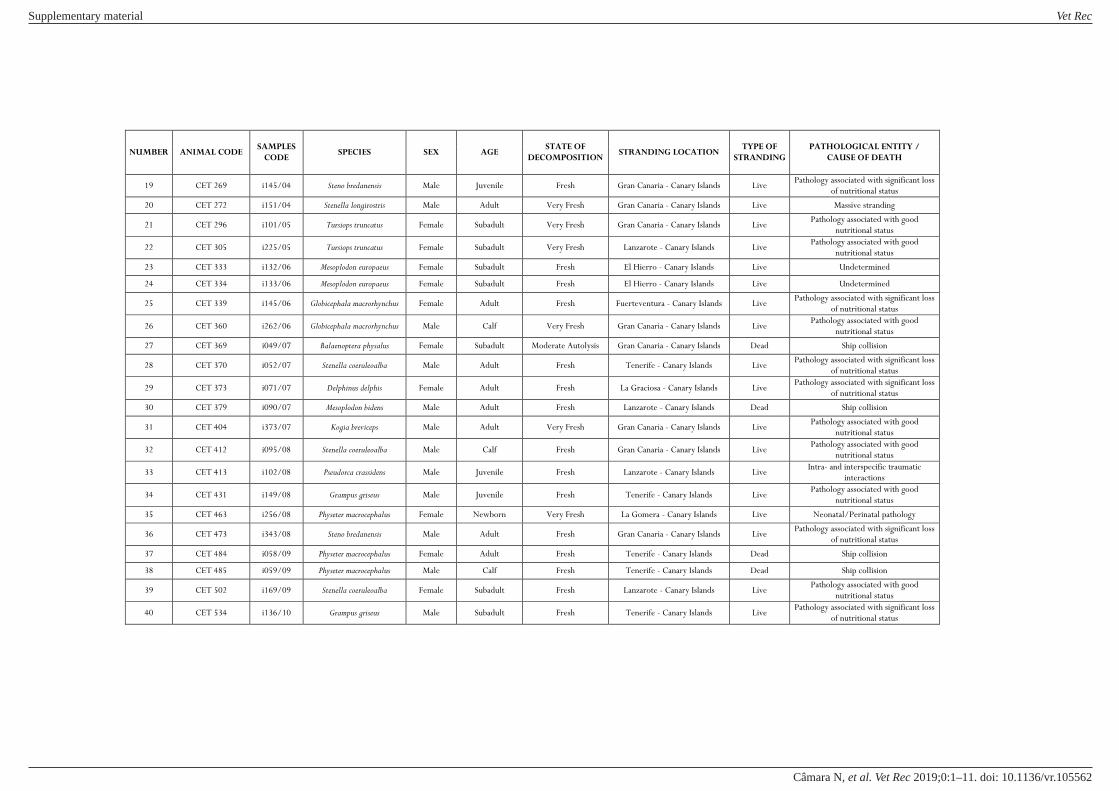
NUMBER ANIMAL CODE SAMPLES
CODE SPECIES SEX AGE
STATE OF
DECOMPOSITION STRANDING LOCATION
TYPE OF
STRANDING
PATHOLOGICAL ENTITY /
CAUSE OF DEATH
19 CET 269 i145/04 Steno bredanensis Male Juvenile Fresh Gran Canaria - Canary Islands Live Pathology associated with significant loss
of nutritional status
20 CET 272 i151/04 Stenella longirostris Male Adult Very Fresh Gran Canaria - Canary Islands Live Massive stranding
21 CET 296 i101/05 Tursiops truncatus Female Subadult Very Fresh Gran Canaria - Canary Islands Live Pathology associated with good
nutritional status
22 CET 305 i225/05 Tursiops truncatus Female Subadult Very Fresh Lanzarote - Canary Islands Live Pathology associated with good
nutritional status
23 CET 333 i132/06 Mesoplodon europaeus Female Subadult Fresh El Hierro - Canary Islands Live Undetermined
24 CET 334 i133/06 Mesoplodon europaeus Female Subadult Fresh El Hierro - Canary Islands Live Undetermined
25 CET 339 i145/06 Globicephala macrorhynchus Female Adult Fresh Fuerteventura - Canary Islands Live Pathology associated with significant loss
of nutritional status
26 CET 360 i262/06 Globicephala macrorhynchus Male Calf Very Fresh Gran Canaria - Canary Islands Live Pathology associated with good
nutritional status
27 CET 369 i049/07 Balaenoptera physalus Female Subadult Moderate Autolysis Gran Canaria - Canary Islands Dead Ship collision
28 CET 370 i052/07 Stenella coeruleoalba Male Adult Fresh Tenerife - Canary Islands Live Pathology associated with significant loss
of nutritional status
29 CET 373 i071/07 Delphinus delphis Female Adult Fresh La Graciosa - Canary Islands Live Pathology associated with significant loss
of nutritional status
30 CET 379 i090/07 Mesoplodon bidens Male Adult Fresh Lanzarote - Canary Islands Dead Ship collision
31 CET 404 i373/07 Kogia breviceps Male Adult Very Fresh Gran Canaria - Canary Islands Live Pathology associated with good
nutritional status
32 CET 412 i095/08 Stenella coeruleoalba Male Calf Fresh Gran Canaria - Canary Islands Live Pathology associated with good
nutritional status
33 CET 413 i102/08 Pseudorca crassidens Male Juvenile Fresh Lanzarote - Canary Islands Live Intra- and interspecific traumatic
interactions
34 CET 431 i149/08 Grampus griseus Male Juvenile Fresh Tenerife - Canary Islands Live Pathology associated with good
nutritional status
35 CET 463 i256/08 Physeter macrocephalus Female Newborn Very Fresh La Gomera - Canary Islands Live Neonatal/Perinatal pathology
36 CET 473 i343/08 Steno bredanensis Male Adult Fresh Gran Canaria - Canary Islands Live Pathology associated with significant loss
of nutritional status
37 CET 484 i058/09 Physeter macrocephalus Female Adult Fresh Tenerife - Canary Islands Dead Ship collision
38 CET 485 i059/09 Physeter macrocephalus Male Calf Fresh Tenerife - Canary Islands Dead Ship collision
39 CET 502 i169/09 Stenella coeruleoalba Female Subadult Fresh Lanzarote - Canary Islands Live Pathology associated with good
nutritional status
40 CET 534 i136/10 Grampus griseus Male Subadult Fresh Tenerife - Canary Islands Live Pathology associated with significant loss
of nutritional status
Supplementary material Vet Rec
doi: 10.1136/vr.105562–11.:10 2019;Vet Rec, et al. Câmara N
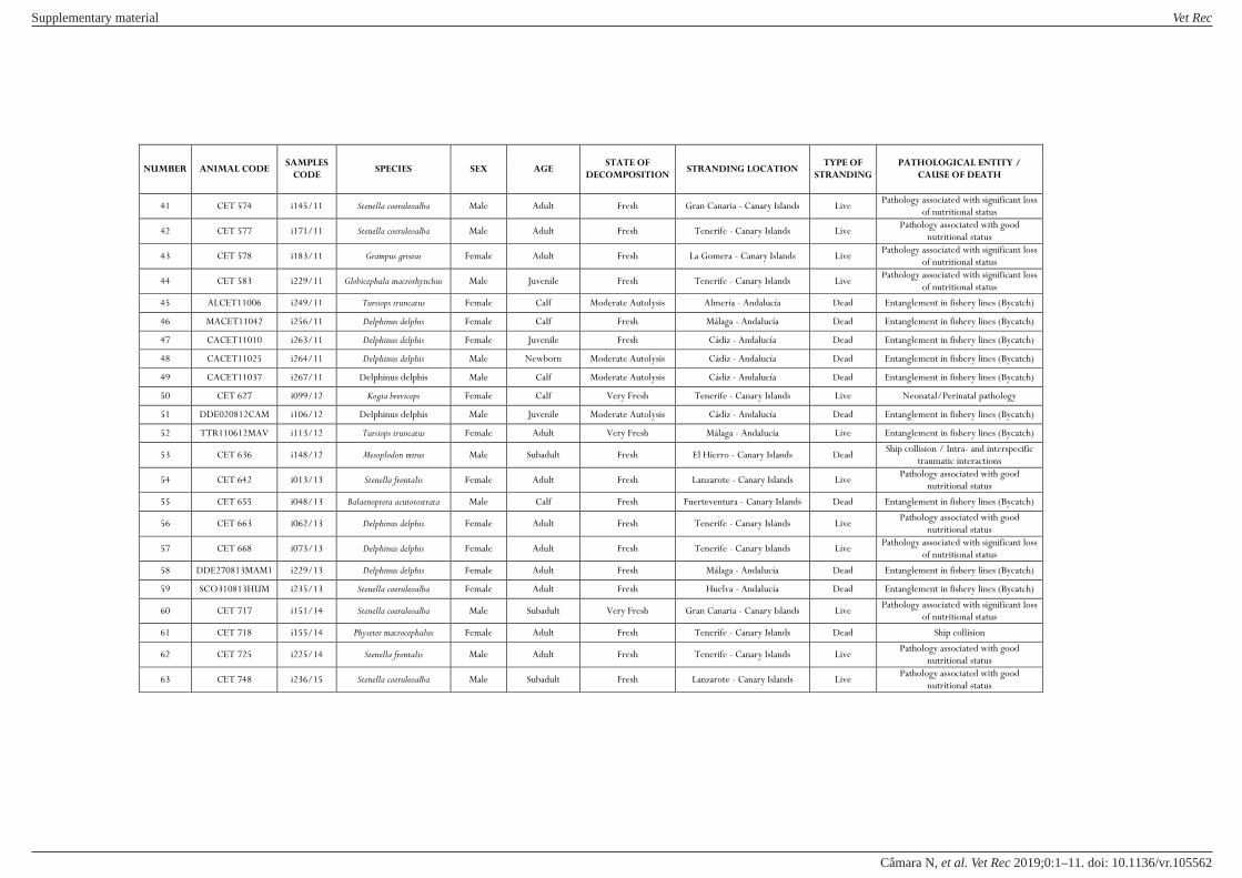
NUMBER ANIMAL CODE SAMPLES
CODE SPECIES SEX AGE
STATE OF
DECOMPOSITION STRANDING LOCATION
TYPE OF
STRANDING
PATHOLOGICAL ENTITY /
CAUSE OF DEATH
41 CET 574 i145/11 Stenella coeruleoalba Male Adult Fresh Gran Canaria - Canary Islands Live Pathology associated with significant loss
of nutritional status
42 CET 577 i171/11 Stenella coeruleoalba Male Adult Fresh Tenerife - Canary Islands Live Pathology associated with good
nutritional status
43 CET 578 i183/11 Grampus griseus Female Adult Fresh La Gomera - Canary Islands Live Pathology associated with significant loss
of nutritional status
44 CET 583 i229/11 Globicephala macrorhynchus Male Juvenile Fresh Tenerife - Canary Islands Live Pathology associated with significant loss
of nutritional status
45 ALCET11006 i249/11 Tursiops truncatus Female Calf Moderate Autolysis Almería - Andalucía Dead Entanglement in fishery lines (Bycatch)
46 MACET11042 i256/11 Delphinus delphis Female Calf Fresh Málaga - Andalucía Dead Entanglement in fishery lines (Bycatch)
47 CACET11010 i263/11 Delphinus delphis Female Juvenile Fresh Cádiz - Andalucía Dead Entanglement in fishery lines (Bycatch)
48 CACET11025 i264/11 Delphinus delphis Male Newborn Moderate Autolysis Cádiz - Andalucía Dead Entanglement in fishery lines (Bycatch)
49 CACET11037 i267/11 Delphinus delphis Male Calf Moderate Autolysis Cádiz - Andalucía Dead Entanglement in fishery lines (Bycatch)
50 CET 627 i099/12 Kogia breviceps Female Calf Very Fresh Tenerife - Canary Islands Live Neonatal/Perinatal pathology
51 DDE020812CAM i106/12 Delphinus delphis Male Juvenile Moderate Autolysis Cádiz - Andalucía Dead Entanglement in fishery lines (Bycatch)
52 TTR110612MAV i113/12 Tursiops truncatus Female Adult Very Fresh Málaga - Andalucía Live Entanglement in fishery lines (Bycatch)
53 CET 636 i148/12 Mesoplodon mirus Male Subadult Fresh El Hierro - Canary Islands Dead Ship collision / Intra- and interspecific
traumatic interactions
54 CET 642 i013/13 Stenella frontalis Female Adult Fresh Lanzarote - Canary Islands Live Pathology associated with good
nutritional status
55 CET 655 i048/13 Balaenoptera acutorostrata Male Calf Fresh Fuerteventura - Canary Islands Dead Entanglement in fishery lines (Bycatch)
56 CET 663 i062/13 Delphinus delphis Female Adult Fresh Tenerife - Canary Islands Live Pathology associated with good
nutritional status
57 CET 668 i073/13 Delphinus delphis Female Adult Fresh Tenerife - Canary Islands Live Pathology associated with significant loss
of nutritional status
58 DDE270813MAM1 i229/13 Delphinus delphis Female Adult Fresh Málaga - Andalucía Dead Entanglement in fishery lines (Bycatch)
59 SCO310813HUM i235/13 Stenella coeruleoalba Female Adult Fresh Huelva - Andalucía Dead Entanglement in fishery lines (Bycatch)
60 CET 717 i151/14 Stenella coeruleoalba Male Subadult Very Fresh Gran Canaria - Canary Islands Live Pathology associated with significant loss
of nutritional status
61 CET 718 i155/14 Physeter macrocephalus Female Adult Fresh Tenerife - Canary Islands Dead Ship collision
62 CET 725 i225/14 Stenella frontalis Male Adult Fresh Tenerife - Canary Islands Live Pathology associated with good
nutritional status
63 CET 748 i236/15 Stenella coeruleoalba Male Subadult Fresh Lanzarote - Canary Islands Live Pathology associated with good
nutritional status
Supplementary material Vet Rec
doi: 10.1136/vr.105562–11.:10 2019;Vet Rec, et al. Câmara N

NUMBER ANIMAL CODE SAMPLES
CODE SPECIES SEX AGE
STATE OF
DECOMPOSITION STRANDING LOCATION
TYPE OF
STRANDING
PATHOLOGICAL ENTITY /
CAUSE OF DEATH
64 SCO110314CAM i288/14 Stenella coeruleoalba Male Juvenile Moderate Autolysis Cádiz - Andalucía Dead Entanglement in fishery lines (Bycatch)
65 CET 771 i414/15 Ziphius cavirostris Female Adult Fresh Tenerife - Canary Islands Dead Ship collision
66 CET 776 i461/15 Tursiops truncatus Male Juvenile Fresh Tenerife - Canary Islands Live Pathology associated with good
nutritional status
67 CET 819 i950/16 Balaenoptera edeni Male Calf Very Fresh Fuerteventura - Canary Islands Live Neonatal/Perinatal pathology
Supplementary material Vet Rec
doi: 10.1136/vr.105562–11.:10 2019;Vet Rec, et al. Câmara N

6.2 CORRELATION OF THE BLOOD VALUES
DETECTED FOR THE BIOCHEMICAL MARKERS
OF ACUTE CARDIAC MUSCLE DAMAGE WITH
THE PATHOLOGICAL FINDINGS IDENTIFIED
HISTOLOGICALLY


RESULTS & DISCUSSION: PUBLICATIONS CORRELATION OF THE BLOOD VALUES DETECTED FOR THE BIOCHEMICAL MARKERS OF ACUTE CARDIAC MUSCLE DAMAGE WITH THE PATHOLOGICAL FINDINGS IDENTIFIED HISTOLOGICALY
[Increased plasma cardiac troponin I in live-stranded cetaceans: correlation with pathological findings of acute cardiac injury] _____________________________________________________________________________________________________________________________________________________________________________
151
The second specific objective of this work, which consisted in correlating the blood
values detected for the biochemical markers of acute cardiac muscle damage with the
pathological findings identified at a microscopic level, was achieved using different phases as
mentioned in the materials and methods chapter.
In the first phase, we estimated a baseline range for cTnI in cetaceans, since there is not
an existing one in the current literature, through the measurements obtained from captive
animals.
The second phase consisted in comparing the values from 9 live-stranded cetaceans with
the normal baseline range of cetaceans (determined previously by us) and also with other
mammals, such as humans and dogs.
In the third and fourth phase, we used histological, histochemical and
immunohistochemical approaches to evaluate the acute cardiac lesions at a microscopic level.
Finally, we correlated, using the available literature on the biochemical kinetics and the
chronological sequence of an acute ischemic injury in the heart in the histology, our blood results
with the histopathological lesions in our animals.
All of this resulted in the publication of an article, as you can see subsequently, titled
Plasma cardiac troponin I in live-stranded cetaceans: correlation with pathological findings of
acute cardiac injury, on January 2020 in the Scientific Reports journal, Volume 10, Issue 1 (doi:
10.1038/s41598-020-58497-3), having this journal an impact factor of 4.011 in 2018 and
therefore being a Q1 in the veterinary category.

1Scientific RepoRtS | (2020) 10:1555 | https://doi.org/10.1038/s41598-020-58497-3
www.nature.com/scientificreports
increased plasma cardiac troponin i in Live-Stranded cetaceans: correlation with pathological findings of Acute cardiac injurynakita câmara , eva Sierra*, Antonio fernández, Manuel Arbelo , Marisa Andrada, Antonio espinosa de los Monteros & pedro Herráez
capture myopathy (cM), is a syndrome that occurs as the result of the stress during and after capture, handling, restraint, and transport of wild animals. Although cM has been described for many species of cetaceans, characterization of the acute cardiac injury - an important component of this syndrome - are still scarce. In this study, we firstly estimated a normal range for cardiac troponin I (cTnI) on cetaceans. Here, through biochemical analysis (especially of ctni) and histopathological, histochemical, and immunohistochemical correlations with decreased troponin immunolabelling, we studied the cardiac injury in live-stranded cetaceans. nine cetaceans which stranded alive on the canary islands (January 2016 - June 2019) were included in this study. Sampled individuals presented elevated values of plasma ctni, which were correlated to histopathological lesions comprised of vascular changes and acute degenerative lesions. immunohistochemically, injured cardiomyocytes showed a decreased intrafibrillar troponin immunoreaction. This is the first attempt to establish a normal baseline range for cTnI in cetaceans, and the first study comparing plasma biomarkers values with histopathological and immunohistochemical findings. This approach allowed us to demonstrate the degree of cardiac damage as a result of injury, consistent with ischemia–reperfusion lesions. the knowledge gained here could improve decision-making procedures during stressful situations, mainly in live-strandings, handling, and rehabilitation, thereby reducing the mortality of cetaceans.
All life forms have evolved mechanisms to cope with stressful situations in their lives1. For cetaceans, live-stranding is a situation in which they are alive on the beach or in shallow water, and in distress due to being unable to free themselves and resume normal activity2–4. Similar to humans and other animals, once the central nervous system of a cetacean perceives an internal (physiologic or psychogenic) or environmental threat to their homeostasis, the threat being the stressor, a biological response is developed5–7.
Irrespective of the animal’s previous health, stranding creates an anomalous and extreme situation for an organism that it is not anatomically or physiologically adapted to handle. It is a pathological entity in which acute stress is the central axis of its etiopathogenesis, presenting clinical and lesional findings that can cause the death of the animal or seriously aggravate a previous disease over the period of capture or captivity. This reaction may in turn influence the subsequent rehabilitation and recovery of affected animals, since live-stranded cetaceans are frequently debilitated when rescued. Therefore, although the stranding response is intended to improving the health and welfare of these animals, some “rescue and recovery” activities may in fact be counterproductive4,8–13.
Deaths of live-stranded cetaceans may be attributed to the so-called “stress response syndrome” or “alarm reaction,” which are thought to have analogous mechanisms to capture myopathy (CM), in which cardiac dam-age due to the extreme stress seems to play an important role2,8,10–12,14,15. For this reason, previous authors have suggested that cetaceans would be especially predisposed to develop stress cardiomyopathy (SCMP), comparable to SCMP in humans which appears to be related to an excess of plasma catecholamines triggered by a stressful
Veterinary Histology and Pathology. Institute of Animal Health and Food Safety (IUSA). Veterinary School. University of Las Palmas de Gran Canaria, Las Palmas de Gran Canaria, Spain. *email: [email protected]
open

2Scientific RepoRtS | (2020) 10:1555 | https://doi.org/10.1038/s41598-020-58497-3
www.nature.com/scientificreportswww.nature.com/scientificreports/
event2,8,10–12,14–27. There are no previous studies regarding biochemical cardiac markers that indicate damage to the heart of marine mammals (more specifically in cetaceans).
When irreversible cardiac damage occurs, troponin is released from cardiomyocytes. Troponin is a regulatory protein complex comprised three subunits; troponin C, the calcium binding component; troponin T, the binding component tropomyosin; and troponin I, the inhibitory component. All components are involved in the contrac-tile process of both skeletal and cardiac muscle. The troponin C expressed in skeletal muscle is identical to cardiac troponin C (cTnC). However, cardiac troponin T (cTnT) and cardiac troponin I (cTnI) are specific to the heart28.
Very low amounts of cTnI are detectable in the blood of healthy human individuals with no evidence of cardiac disease, as opposed to the amount of cardiac troponin released from cardiomyocytes into the blood as a result of injury, which is consistent with ischemia and other causes29–31. Due to this specificity, cTnT and cTnI are recommended by various international societies as a diagnostic indicator for acute myocardial infarction (AMI) and other heart pathologies, such as SCMP, since they are potential markers of cardiac damage32.
The histology that defines CM consists of ischemia-reperfusion injuries consistent with local to generalised vasospasm and vasodilation (catecholamines, neurogenic shock, and impeded venous flow return from body compression, causing hypoxia in different organs), direct traumatic injury to the muscle resulting in acute to subacute degeneration (rhabdomyolysis), acute renal failure associated with myoglobinuric nephrosis, and areas of necrosis in viscera9–11,13–15,33–35.
The aim of this study was to estimate a normal range for cTnI in cetaceans and describe the acute cardiac injury in live-stranded cetaceans, correlating biochemical analysis with histological, histochemical, and immu-nohistochemical findings.
ResultsThe reference range of cTnI was of 0–0.0256 µg/L, determined with a 95% probability. All data necessary to calcu-late this range are shown in Supplemental Table 1.
The serum values of cTnI and creatine kinase (CK) obtained for each animal are shown in Table 1. Additionally, it summarises the evaluation of the vascular changes, acute degenerative changes, and the presence of inflammatory cells. It also shows the presence or absence of myoglobin globules.
Regarding animal 1, the biochemical analysis presented 0.06 µg/L for cTnI and 1875.0 U/L for CK. With refer-ence to the histopathological results, we observed a moderate epicarditis, neuritis, and multifocal lymphoplasma-cytic perineuritis, and a mild multifocal lymphoplasmacytic myocarditis. We also detected moderate multifocal myocardial fibrosis and mild multifocal acute degenerative changes in the cardiomyocytes consistent with wavy fibres, hypereosinophilia, and cytoplasmic vacuolisation (Fig. 1). We did not assess the depletion or expression of the immunohistochemical markers because the sample was frozen, and previous studies have shown that animals with advanced autolysis or that were previously frozen presented a greater number of false negatives when using histochemical and immunohistochemical techniques15.
Biochemically, due to serum insufficiency, the only metric we were able to obtain from animal 2, was a cTnI level of 40.00 µg/L. The histopathology revealed a mild, multifocal congestion and haemorrhages. Moreover, it also showed moderate, multifocal acute cardiomyodegenerative lesions indicated as hypereosinophilia and cyto-plasmic vacuolisation. Immunohistochemically, we verified homogenous, intrafibrillar depletion of cTnI, cTnC, and myoglobin in the degenerated/necrotic cardiomyocytes. Furthermore, damaged cells from the cardiac mus-cle, showed various intensities of immunolabeling for fibrinogen.
The biochemical analysis of animal 3, it presented 0.025 µg/L of cTnI and 1667.1 U/L of CK. We were incapable to obtain histopathological and immunohistochemical parameters, as we were unable to perform a necropsy on this animal, since it was used for anatomical research purposes.
Biochemically, due to serum insufficiency, the only metric we were able to obtain from animal 4, was a cTnI level of 0.235 µg/L. The histopathology revealed mild to moderate, multifocal congestion, and interstitial oedema. Additionally, we observed moderate to severe multifocal acute degenerative changes in the cardiomyocytes, such as hypereosinophilia and cytoplasmic vacuolisation. Immunohistochemically, we detected fibrinogen expres-sion and the disappearance of immunolabeling for cTnI, cTnC, and myoglobin in the degenerated/necrotic cardiomyocytes.
Regarding animal 5, the biochemical analysis showed a cTnI level of 0.249 µg/L and a CK level of 3521.2 U/L. The histopathology revealed mild to moderate acute cardiomyodegenerative lesions, consistent with contraction band necrosis (Fig. 2), wavy fibres, hypereosinophilia, and cytoplasmic vacuolisation. Simultaneously, we also verified myoglobin globules and mild, multifocal congestion, and interstitial oedema. The immunohistochem-ical analysis showed the homogenous intrafibrillar depletion of cTnI, cTnC, and myoglobin in the degenerated/necrotic cardiomyocytes; these also exhibited various intensities of immunolabeling for fibrinogen.
The biochemical results for animal 6 showed 0.748 µg/L for cTnI and 3299.5 U/L for CK. The histopathology showed moderate multifocal myocardial fibrosis and acute degenerative changes in the cardiomyocytes, reflected as wavy fibres, hypereosinophilia, and cytoplasmic vacuolisation. Furthermore, we detected the presence of myo-globin globules and mild multifocal congestion. The immunohistochemical parameters showed the expression of fibrinogen and the disappearance of immunolabeling for cTnI, cTnC, and myoglobin in the degenerated/necrotic cardiomyocytes.
Biochemically animal 7 showed 0.033 µg/L for cTnI and 383.6 U/L for CK. The histopathology showed mild acute cardiomyodegenerative lesions, such as hypereosinophilia and cytoplasmic vacuolisation. We did not ana-lyse depletion or expression of immunohistochemical marker because the sample was frozen.
For animal 8, serum insufficiency prevented us from obtaining all biochemical metrics. We were only able to obtain the cTnI level, which was 0.06 µg/L. We were unable perform histopathology and immunohistochemistry because we were unable to perform a necropsy on this animal as it was released back into the sea.

3Scientific RepoRtS | (2020) 10:1555 | https://doi.org/10.1038/s41598-020-58497-3
www.nature.com/scientificreportswww.nature.com/scientificreports/
Biochemically, animal 9 exhibited 0.049 µg/dL for cTnI and 739.2 U/L for CK. The histopathology showed a mild multifocal congestion and moderate, multifocal acute degenerative changes in the cardiomyocytes, such as hypereosinophilia and cytoplasmic vacuolisation. Regarding the immunohistochemistry, we observed homoge-nous intrafibrillar depletion of cTnI, cTnC, and myoglobin in the degenerated/necrotic cardiomyocytes; we also detected various intensities of immunolabeling for fibrinogen.
DiscussionRecent studies have characterized cardiac lesions associated with live-stranding in cetaceans. The etiopathogen-esis of this acute cardiac pathology has as its central axis the stress derived from stranding, handing, interaction with humans, transport and captivity (having been compared with the CM described in terrestrial mammals). Etiological and lesionally, the acute cardiomyopathy associated with live-stranding is comparable to SCMP in humans. SCMP lesions in live-stranded cetaceans consist of vascular changes (congestion, interstitial edema and hemorrhages) and acute necrotic degenerative lesions (contraction band necrosis, wavy fibers, hypereosinophilia and cytoplasm vacuolization) of perivascular distribution, indicative of ischemia-reperfusion damage10–12,15,35.
In humans, the diagnostic criteria for SCMP comprises alterations in medical exams (such as electrocardi-ography, echocardiography, cardiac catheterizations), and biochemical analysis. The more common laboratories abnormalities are consistent with a small, rapid rise to above the normal levels of cTnI and/or CK36–38.
Animal 1 (*) Animal 2 (†) Animal 3 (‡) Animal 4 (§) Animal 5 (‡) Animal 6 (§) Animal 7 (‡) Animal 8 (||) Animal 9 (*)
Delphinus delphis
Delphinus delphis
Stenella coeruleoalba
Stenella coeruleoalba
Globicephala macrorhynchus
Delphinus delphis
Stenella coeruleoalba
Stenella coeruleoalba
Stenella coeruleoalba
STRANDING CIRCUNSTANCES
TIME OF STRANDING NOTICE 10:16 AM 11:25 AM 5:12 PM 11:13 AM 11:57 AM 9:30 AM 8:37 AM 9:38 AM 12:35 PM
TIME OF DEATH 12:00 PM 12:00 PM 6:45 PM NO 12:00 PM NO 8:45 AM NO 2:38 PM
TIME OF SAMPLE COLLECTION 10:00 PM 4:00 PM 6:45 PM 11:47 AM 3:37 PM 10:00 AM next day 11:00 AM 2:00 PM 3:28 PM
BIOCHEMICAL ANALYSIS
TROPONIN I (cTnI) (µg/L)
Animal Value 0.06 40.00 0.025 0.235 0.249 0.748 0.033 0.06 0.049
Baseline Range Cetaceans 0–0.0256 (#)
Baseline Range Dogs ≤0.03–0.07 (**)
Baseline Range Humans ≤0.1 (**)
CREATININE KINASE (CK) (U/L)
Animal Value 1875.0 Not measured 1667.1 Not
measured 3521.2 3299.5 383.6 Not measured 739.2
Baseline Range 100–250 (††)
HISTOPATHOLOGICAL & HISTOCHEMICAL ANALYSIS
VASCULAR CHANGES
Congestion Absent Mild
No necropsy
Mild to moderate Mild Mild Absent
No necropsy
Mild
Interstitial Oedema Absent Absent Mild to moderate Mild Absent Absent Absent
Haemorrhages Absent Mild Absent Absent Absent Absent Absent
ACUTE DEGENERATIVE CHANGES
Contraction Band Necrosis Absent Absent Absent Mild to
moderate Absent Absent Absent
Wavy Fibers Mild Absent Absent Mild to moderate Moderate Absent Absent
Hypereosinophilia Mild Moderate Moderate to severe
Mild to moderate Moderate Mild Moderate
Cytoplasmic Vacuolization Mild Moderate Moderate to
severeMild to moderate Moderate Mild Moderate
INFLAMMATORY INFILTRATION Moderate Absent Absent Absent Absent Absent Absent
MYOGLOBIN GLOBULES Absent Absent Absent Present Present Absent Absent
IMMUNOHISTO-CHEMICAL ANALYSIS
MYOGLOBIN
Frozen animal
Depletion
No necropsy
Depletion Depletion Depletion
Frozen animal No necropsy
Depletion
CARDIAC TROPONIN I (cTnI) Depletion Depletion Depletion Depletion Depletion
CARDIAC TROPONIN C (cTnC) Depletion Depletion Depletion Depletion Depletion
FIBRINOGEN Deposition Deposition Deposition Deposition Deposition
Table 1. Summary of the stranding circumstances, biochemical results, histopathological findings and immunohistochemical changes for each animal of the study. (*) Live stranded animal that dies during transport. (†) Live stranding notification (09:00 PM) of a cetacean swimming very close to shore being reintroduced in to sea several times by general public until he swims away. New communication of stranded animal one day and a half after the first notification (11:25). The animal is euthanized. (‡) Live stranded animal that died before being attended. (§) Dead animal which presented injuries that indicates that the animal stranded alive. (||) Live stranded animal and released back into the sea. (#) Baseline range determined in this study. Comparing the interval of reference for cTnI in bottlenose dolphins with the normal values for humans and dogs we can conclude that is shorter than in other species. (**) Values from literature41–43. (††) Reference range for Tursiops truncatus in captivity4.
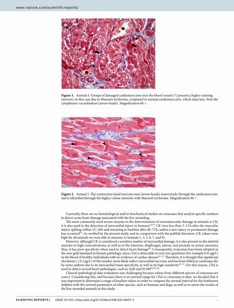
4Scientific RepoRtS | (2020) 10:1555 | https://doi.org/10.1038/s41598-020-58497-3
www.nature.com/scientificreportswww.nature.com/scientificreports/
Currently, there are no hematological and/or biochemical studies on cetaceans that analyze specific markers to detect acute heart damage associated with the live-stranding.
The most commonly used serum enzyme in the determination of neuromuscular damage in animals is CK; it is also used in the detection of myocardial injury in humans30,39. CK rises less than 3–12 h after the muscular injury, spiking within 12–24 h and returning to baseline after 48–72 h, unless a new injury or permanent damage has occurred30. As verified by the present study, and in comparison with the publish literature, CK values were high for all animals we were able to measure it (animals 1, 3, 5, 6, 7, and 9).
However, although CK is considered a sensitive marker of myocardial damage, it is also present in the skeletal muscles in high concentrations, as well as in the intestine, diaphragm, uterus, and prostate in minor amounts; thus, it has poor specificity when used to detect heart damage40. Consequently, troponins have been adopted as the new gold standard in human pathology, since cTnI is detectable in very low quantities (for example 0.01 µg/L) in the blood of healthy individuals with no evidence of cardiac disease29–32. Therefore, it is thought that significant elevations (≥0.1 µg/L) of this marker most likely reflect myocardial necrosis, and has been billed as cardiospecific by some authors due to its myocardial tissue specificity, as well as its high sensitivity30–32. For this reason, cTnI is used to detect several heart pathologies, such as AMI and SCMP30,36,40.
Clinical-pathological data evaluation was challenging because values from different species of cetaceans are scarce. Considering this, and because there is no normal range for cTnI in cetaceans to date, we decided that it was important to determine a range of baseline values in order to compare the normal interval for the bottlenose dolphin with the normal parameters of other species, such as humans and dogs, as well as to assess the results of the live-stranded animals in this study.
Figure 1. Animal 4. Groups of damaged cardiomyocytes near the blood vessels (*) present a higher staining (arrows), in this case due to Masson’s trichrome, compared to normal cardiomyocytes, which stain less. Note the cytoplasmic vacuolisation (arrow heads). Magnification 60 × .
Figure 2. Animal 5. The contraction band necrosis runs (arrow heads) transversely through the cardiomyocytes and is identified through the higher colour intensity with Masson’s trichrome. Magnification 60 × .

5Scientific RepoRtS | (2020) 10:1555 | https://doi.org/10.1038/s41598-020-58497-3
www.nature.com/scientificreportswww.nature.com/scientificreports/
Comparing the range in this particular study (0–0.0256 µg/L) with the normal values for humans ( ≤ 0.1 µg/L) and dogs ( ≤ 0.03–0.07 µg/L), we can conclude that the interval of reference for cTnI in bottlenose dolphins is shorter than in other species41–43. This could be justified by the small number of samples (20 blood samples in total). Nonetheless, we consider this result to be an important contribution to clinical biochemistry in cetaceans, as it may help in decision-making and treatment procedures during stressful situations, such as live-strandings, and improve conservation efforts by reducing the mortality of these animals.
Our results, from the live-stranded cetaceans, showed that one of the animals presented a cTnI value within normal range whilst the remaining eight animals demonstrated an increase in comparison to the normal/baseline value for cTnI (0–0.0256 µg/L) that we obtained from captive bottlenose dolphins. However, when comparing these with those for humans ( ≤ 0.1 µg/L) and dogs ( ≤ 0.03–0.07 µg/L), we observed that animal 1 (0.06 µg/L), animal 3 (0.025 µg/L), animal 7 (0.033 µg/L), animal 8 (0.06 µg/L), and animal 9 (0.049) are within range, while animal 2 (40.00 µg/L), animal 4 (0.235 µg/L), animal 5 (0.249 µg/L), and animal 6 (0.748 µg/L) exceed the upper value41–43.
More than 90% of patients with SCMP revealed raised cTnI when measured by conventional assays36. However, SCMP is a disease with a high rate of misdiagnosis because it is an important differential diagnosis of an AMI since highly similar clinical presentations occur between them25,26. Besides, it is under-recognized because of the unnecessary delay in the diagnosis process in post-mortem examination cases of AMI caused by the less sensitive conventional markers.
Previous authors have stated the necessity of establishing diagnostic biochemical cardiac markers for the post-mortem diagnosis of AMI and SCMP due to the limitations of histopathology. The sensitivity of cTnI makes early detection of microinfarction possible after the onset of ischemia using a rapid one-step assay in body fluids in autopsy cases40. Therefore, we selected cTnI as our marker of choice to detect myocardial damage in live-stranded cetaceans, through biochemical and immunohistochemical detection, on the grounds of its speci-ficity, as well as being highly sensitive30,40.
Generally, injured cardiac myocytes release troponin 3–9 h after ischemic damage, peaking after 12–48 h and levels remain elevated for 4–7 days for cTnI30,40,44. Early recognition of myocardial necrosis (1–3 h) is not possible by monitoring kinetics, and these markers are ineffective until 6 or more hours after the onset of the AMI and/or SCMP. The precise determination of the timing of the stress event and/or symptom onset is often exceptionally difficult because it is focused around the clinical patient report. Therefore, in humans, a precondition to obtaining a satisfactory ability to distinguish these pathologies is that blood should be collected 6–9 h after onset40. In the case of a live-stranding event, it is also often clinically and pathologically challenging to have the correct timing of the stressful episode, since we are working with wild animals, stranding networks, and the general public. When notified of stranding it is important to acknowledge that the animal could have been stranded recently or could have been stranded for hours before being detected.
Moreover, previous studies have proposed that measuring the level of troponin in the serum can be an impor-tant auxiliary method of examining sudden death, since its peak concentration can be related to the degree of injury40. For this reason, this finding was confirmed with the histological, histochemical, and immunohistochem-ical analyses.
An acute ischemic injury, such as the one that occurs in the AMI or SCMP, is determined by morphologi-cal alterations, consisting of vascular changes and acute degenerative lesions based on the histological analysis of myocardial tissue obtained from biopsied and post-mortem examined species, mostly humans, but also in cetaceans15,19,25,26,37,45–54.
In acute ischemic injuries there is a chronological sequence of changes, which can be observed as early as the first 5 minutes (presence of long, thinned, wavy fibers separated by spaces representing edema, and microvascular congestion, at borders of ischemic myocardium). In the following 15 minutes cell death may start to occur. Early changes of cardiomyocyte coagulation necrosis with nuclear pyknosis, color change, more specifically “brick red change” or cytoplasm hypereosinophilia, focal contraction bands and subtle interstitial edema are evident within 2–3 h. Hypereosinophilia and edema become more pronounced and more easily recognizable after a period of 3–6 h. Subsequently, 6–12 h later, an increased number of neutrophils line up in capillaries as well as acceler-ated changes and more extensive contraction band necrosis with reperfusion are noticeable. In the next 12-hour period, extravasation into interstitial space of neutrophils happens. Vascular congestion, interstitial edema, and focal areas of haemorrhage are also recognized. Thereafter, the subacute period starts15,26.
Considering all the above, we conclude that all the results obtained from the animals of this study were in accordance with the biochemical kinetics, as well as the chronological sequence of histopathological changes in an acute ischemic injury.
Animal 1 died 2 h after the stranding notice and histopathologically presented mild acute degenerative changes, which indicate that they would fit into the 0–3 h time frame, as expected following the histological sequence.
Elevated biomarker values were observed in animal 2. This can be explained by the fact that the notification, of a cetacean swimming very close to shore, was received and after several attempts by members of the public to reintroduce it into the ocean the animal swam away; despite all the effort 38 h later a new alert was received stating that the animal stranded again. Its histopathology analysis showed mild vascular changes and moderate acute cardiomyodegenerative lesions. All of these are detectable between 0–24 h approximately and in agreement with both biochemical kinetics and histopathological sequence previously mentioned.
The biochemical results for animal 3 were near the maximum of the normal range value for cTnI in cetaceans. The animal died 1 h after the stranding notice, nonetheless, we were unable to compare these results with the histopathological lesions since a necropsy could not be performed.
The external injuries in animal 4 indicated that it had stranded alive, but was already dead by the time it was detected at the beach. Although the time of death is unknown, cTnI value was elevated. Histologically, it presented

6Scientific RepoRtS | (2020) 10:1555 | https://doi.org/10.1038/s41598-020-58497-3
www.nature.com/scientificreportswww.nature.com/scientificreports/
mild to moderate vascular alterations, and moderate to severe acute degenerative changes. This fact was consist-ent, with the time frame of 0–24 h, as per the histological sequence discussed earlier.
Considering the biochemical kinetics in animal 5, which died 1 h after the notice, we hypothesise that the stressful event took place sometime before we received the communication of the stranding, since the cTnI results were high. The mild vascular changes, and mild to moderate acute cardiomyodegenerative lesions were compati-ble with the time frame of 0–24 h of the histological sequence.
In spite of the unknown time of death of animal 6, and similarly to animal 4, the cTnI results were high and revealed mild vascular alterations and moderate degree of acute degenerative changes, which were comparable to the ones illustrated in the 0–24 h time frame of the histological sequence described previously.
The biochemical values obtained for animal 7 are consistent with the kinetics, since the animal died minutes after the stranding notice. Histopathologically presented mild acute cardiomyodegenerative lesions complying with the time frame of 0–3 h expectations for an acute ischemic injury sequence.
Regarding animal 8, the values challenged the kinetics, because the blood sample was collected 5 hours after the stranding event. However, as discussed previously, the levels of cTnI increases 3–9 h after the stressful event. In view of the fact that we were able to release the animal back to the sea, no histopathological study could be carried out for this animal.
Finally, the results of animal 9 are in accordance with the biokinetics mentioned above, since the animal died 2 h after notification of the stranding. It also presented a mild degree of vascular alterations and moderate acute degenerative changes, common to the sequence of an acute ischemic injury histology, for 0–3 h time frame.
It is important to highlight that active stranding implies a bias in the cases analysed. This is because most ani-mals tend to strand themselves due to a previous pathological process, which is not always identifiable. Therefore,
Figure 3. Animal 5. Degenerated/necrotic cardiomyocytes (arrow heads) near the blood vessels (*) present the depletion of cTnI when compared with normal cardiomyocytes (arrows). Magnification 40 × .
Figure 4. Animal 6. Damaged cardiomyocytes (arrows) reveal a perivascular pattern (*) with decreased immunolabeling for cTnI, in comparison to normal cells (arrow heads). Magnification 40 × .

7Scientific RepoRtS | (2020) 10:1555 | https://doi.org/10.1038/s41598-020-58497-3
www.nature.com/scientificreportswww.nature.com/scientificreports/
the histological lesions described previously were, most likely, directly related to the stranding itself but not related to the cause of the stranding. Nevertheless, the live-stranding could negatively influence the pre-existing pathological process in the animal, aggravating it, and contributing to its death.
For example, animal 1 became stranded/died as a result of a Morbillivirus infection (unpublished data) and demonstrated characteristic lesions, such as lymphoplasmacytic infiltration, which can be observed in both Morbillivirus infection and in an acute ischemic injury such as the one resulting in AMI or SCMP8,9,19,45,46. Another example is the presence of fibrosis, both in animal 1 and animal 6, indicating a chronic phase of the car-diomyocytes damage, since connective tissue proliferation dominates the third week until healing is completed after 3 months, which indicates a previous heart damage had occurred prior to the latest stranding26.
Previous studies have demonstrated the necessity of corroborating biochemical and histological findings with specific markers that might better delineate myocardial damage10, so we compared the markers cTnI, cTnC, myo-globin and fibrinogen10,11,15,44,55–59. Immediately after acute ischemia caused by a vital trauma, muscle (skeletal and cardiac) proteins begin to leak as a result of early skeletal and myocardial cell membrane rupture, causing a rapid decline in myoglobin, cTnI, and cTnC contents, along with the deposition of plasma proteins, such as fibrinogen10,11,15,55–57. Therefore, damaged cardiomyocytes, in the absence of necrosis during myocardial injury, release cTnI and cTnC, resulting in an increase in serum levels and decreased cardiomyocyte troponin immu-noreaction15,40,60. In the present study, the amount of damage present in cells was tested using immunohisto-chemical labelling of the animals that we were able to perform a complete necropsy and had not been previously frozen (animals 2, 4, 5, 6, and 9). All presented tissue depletion, especially cTnI and cTnC (Figs. 3–6) as well as
Figure 6. Animal 5. Damaged cardiomyocytes (arrow heads) reveal a perivascular pattern (*) with decreased immunolabeling for cTnC, in comparison to normal cells (arrows). Magnification 40 × .
Figure 5. Animal 2. Degenerated/necrotic cardiomyocytes (arrow heads), especially in the epicardium, present the depletion of cTnC when compared to the normal cardiomyocytes (arrows). Note the presence of cytoplasmic vacuolisation (thin arrows). Magnification 40 × .

8Scientific RepoRtS | (2020) 10:1555 | https://doi.org/10.1038/s41598-020-58497-3
www.nature.com/scientificreportswww.nature.com/scientificreports/
myoglobin, together with intrafibrillar fibrinogen deposition. Consequently, with these immunohistochemical changes, we confirmed that the lesions present in these animals were ante mortem.
In conclusion, although the descriptions of CM has been reported in different species of cetaceans, charac-terisation of the cardiac damage in marine mammals – which is an important part of this syndrome – are still scarce. It is acknowledged and proven that live-stranding, itself, is an extreme and intensive stressor. Here, we describe the first clinical and pathological study of cardiac injury in live-stranded cetaceans – through biochem-ical analysis (especially of cTnI) and their histopathological, histochemical, and immunohistochemical correla-tions with decreased troponins immunolabeling – being the results highly comparable to the existing ones for SCMP in humans. We recommend further studies to advance our understanding and knowledge of the cardiac clinical-pathology of cetaceans.
Materials and MethodsFirstly, this report determines a baseline range for cardiac troponin I (cTnI), a specific cardiac biomarker for the detection of cardiac damage in cetaceans, more specifically bottlenose dolphins (Tursiops truncatus). Secondly, it describes the biochemical analysis and histological, histochemical, and immunohistochemical features of cardiac injuries associated with live-stranding in different species of cetaceans from the infraorder Odontoceti.
phases and cases of the present study. The first phase of this study consisted in the determination of normal values for cTnI in cetaceans, specifically bottlenose dolphins, with a 95% probability through meas-urements obtained from captive animals (n = 5) at a local zoo. To achieve this, a total of 20 blood samples were collected, in June, September and December of 2018 and March of 2019 (4 samples of each individual, 1 sample in each month).
In the second phase, a total of 9 animals of four different species, including small and large odontocetes, were included in this study. Blood samples from these animals were collected, being one pre-mortem and the rest post-mortem since the animals subsequently died previous to or during the handling, restraint, transport, and/or rescue/rehabilitation (n = 8). All of the specimens were stranded alive on the coast of the Canary Islands from the beginning of 2016 until June of 2019.
During the third phase, a total of 7 animals were selected, from the animals included (n = 9) in the previous phase, for the histological and histochemical study, since we were not able to perform a necropsy in 2 animals, because one was released back to sea and another was used for anatomical research purposes.
In the fourth and last phase, 2 animals (from the prior phase) were eliminated from the study due to their prior freezing. Therefore, at the end of this phase, a total of 5 animals were studied with the use of the immuno-histochemical technique for the detection of different markers.
In Fig. 7 we present the different stages formerly described. The stranding circumstance, basic and epidemio-logical data of each animal, included in this study, are detailed in Table 1 and Supplemental Table 2, respectively.
Biochemical analysis. Whole blood samples for each animal were collected in a gel tube (without anticoag-ulant), allowed to coagulate, and centrifuged at 3500 rpm for 5 min, two times to obtain the serum (approximately 1 ml).
Baseline range determination of ctni in cetaceans. As the sample size is less than 40 individuals, we have had to assume that each of the variables is normally distributed and that for each individual, and for the variable (troponin), the measured value follows a model of the form xij = μ + bi + 𝜖ij, where 𝑖 is the index of the
Figure 7. Diagram explaining the different stages of the present study.

9Scientific RepoRtS | (2020) 10:1555 | https://doi.org/10.1038/s41598-020-58497-3
www.nature.com/scientificreportswww.nature.com/scientificreports/
individual (i from 1–5) and 𝑗 is the order of measurement within the individual (j from 1–4)61,62. It has been assumed that the individual effect of the animal is bi ≈ (0, σb), and that the variability within each animal is 𝜖 ≈ N(0, σ𝜖), where the values of σb and σ𝜖 are specific to each variable. Under these assumptions, the values of each variable follow normal distributions of the form X ≈ (μ,√σ𝑏
2 + σ𝜖2). The IMER library was used to estimate the
parameter, and the anovaVCA library was used to estimate the variances, both using the statistical package R63. Once these quantities were estimated, the normality intervals were calculated according to the expression μ σ σ μ σ σ− √ + + √ +ε εZ Z[ , ]a b a b/2
2 2/2
2 2 , where Zα/2 is the α/2 percentile of the normal distribution. Under all of these assumptions, the normal values of each of the variables considered can be expected to fall within this range with a probability of 1 − α. Therefore, we have constructed normal intervals with a 95% probability.
Biochemical analysis of the live-stranded cetaceans included in this study. On eight animals, blood samples were retrieved 1–38 h after the live-stranding and with a minimum of 2 h to a maximum of 24 h after the death of the animal. Additionally, one sample was recovered pre-mortem and within 5 h of the stressful event. The main biochemical markers analysed were cTnI, which reveals a specific heart injury, and CK, which demonstrates acute muscle damage, both cardiac and skeletal. The results of the biochemical analyses were com-pared to published values of different mammals, such as humans and domestic animals (dog), in respect to cTnI and diverse cetaceans of the infraorder Odontoceti in the case of CK whenever possible.
Histological and histochemical analysis. Complete necropsies were performed following the standard protocol, with the animals (n = 7) presenting a very fresh or fresh state of decomposition2,64. For histopatholog-ical analysis, the heart muscle (both atria and ventricles, different atrioventricular valves [bicuspid or mitral and tricuspid], and semilunar valves [sigmoid aortic and pulmonary] with the corresponding arteries) were fixed and processed. Specifically, 4 µm of the samples mentioned above were used for haematoxylin and eosin staining and Masson’s trichrome techniques.
The sections subjected to the histochemical techniques were examined in a blind manner by three veterinar-ians (NC, ES, and PH). The sections were evaluated for vascular changes (congestion, haemorrhages, and inter-stitial oedema); acute degenerative changes (contraction band necrosis, wavy fibres, perinuclear vacuolisation, cytoplasmic hypereosinophilia, and pyknotic nuclei), as well as the presence of interstitial myoglobin globules, and infiltration of inflammatory cells. The extent of cardiac lesions was judged subjectively as follows: absent, mild, mild to moderate, moderate, moderate to severe, and severe.
immunohistological analysis. Tissue sections of 3 µm, from each of the animals (n = 5), were immu-nolabeled using specific markers of myocardial injury, such as anti-cTnI, anti-cTnC, anti-myoglobin, and anti-fibrinogen primary antibodies, and were visualised using the VECTASTAIN® Elite ABC-Peroxidase Kit with reference PK-6100 (Vector Laboratories, Peterborough, United Kingdom). The methodology is summarised in Supplemental Table 3. For the immunohistochemical techniques, the positive control for cTnI and cTnC was a heart sample from a pig and a cetacean, respectively, with no apparent acute macroscopic and/or histological lesions, nor a live-stranding history in the case of the cetacean. For myoglobin and fibrinogen, we used a heart sample from a previously published case of a striped dolphin that was stranded alive and developed CM due to capture and the rehabilitation process10,11. Finally, the negative control was performed without the primary antibody.
The sections submitted to the immunohistochemical techniques were examined “blind” by three veterinarians (NC, ES, and PH). The amount of cell damage was confirmed as ante-mortem when it was accompanied with the immunohistochemically demonstrated depletion of myoglobin together with intrafibrillar fibrinogen deposition.
evidence of ethical approval. Blood collection from the captive animals was carried out by specialised veterinarians authorised by “Palmitos Park” Zoo, within their routine prophylactic program, as well as their use for this scientific study, in compliance with the requirements established on the articles 3, 4 and 5 of the “Ley 31/2003, de 27 de octubre, de conservación de la fauna silvestre en los parques zoológicos” (BOE-A-2003-19800) and the Council Directive 1999/22/EC of 29 March 1999 relating to the keeping of wild animals in zoos (EUR-Lex -31999L0022). Likewise, the use of blood samples for this scientific study was expressly authorised by the Director of the “Palmitos Park” zoo.
Regarding the management of stranded cetaceans, required permission was issued by the environmental department of the Canary Islands’ Government and the Spanish Ministry of Environment.
Data availabilityAll data reported in this work are classified and stored in the tissue bank of the Institute of Animal Health and Food Safety (IUSA). Veterinary School. University of Las Palmas de Gran Canaria.
Received: 28 August 2019; Accepted: 13 January 2020;Published: xx xx xxxx
References 1. Yousef, M. K. Animal stress and strain: Definition and measurements. Appl. Anim. Behav. Sci. 20, 119–126 (1988). 2. Geraci, J. R. & Lounsbury, V. J. Marine mammals ashore: a field guide for strandings. (National Aquarium in Baltimore, 2005). 3. Wilkinson, D. M. Report to Assistant Administrator for Fisheries: Program review of the marine mammal stranding networks.
(1991). 4. Gulland, F. M. D., Dierauf, L. A. & Whitman, K. L. CRC handbook of marine mammal medicine. (CRC Press, 2018). 5. Cowan, D. F. & Curry, B. E. Histopathological assessment of dolphins necropsies onboard vesssels in the eastern tropical pacific tuna
fishery. 31 (2002).
1 0Scientific RepoRtS | (2020) 10:1555 | https://doi.org/10.1038/s41598-020-58497-3
www.nature.com/scientificreportswww.nature.com/scientificreports/
6. Moberg, G. P. Biological response to stress: Implications for animal welfare. In The biology of animal stress: basic principles and implications for animal welfare 377, https://doi.org/10.1079/9780851993591.0001 (2000).
7. Breazile, J. E. The Physiology of Stress and Its Relationship to Mechanisms of Disease and Therapeutics. Vet. Clin. North Am. Food Anim. Pract. 4, 441–480 (1988).
8. Díaz-Delgado, J. et al. Pathologic findings and causes of death of stranded cetaceans in the Canary Islands (2006-2012). PLoS One 13, 1–33 (2018).
9. Arbelo, M. et al. Pathology and causes of death of stranded cetaceans in the Canary Islands (1999-2005). Dis. Aquat. Organ. 103, 87–99 (2013).
10. Herráez, P. et al. Capture myopathy in live-stranded cetaceans. Vet. J., https://doi.org/10.1016/j.tvjl.2012.09.021 (2013). 11. Herráez, P. et al. Rhabdomyolysis and Myoglobinuric Nephrosis (Capture Myopathy) in a Striped Dolphin. J. Wildl. Dis. Wildl. Dis.
Assoc. 43, 770–774 (2007). 12. Cowan, D. F. & Curry, B. E. Histopathology of the Alarm Reaction in Small Odontocetes. J. Comp. Pathol. 139, 24–33 (2008). 13. Bonsembiante, F. et al. Clinico-pathological findings in a striped dolphin (Stenella coeruleoalba) affected by rhabdomyolysis and
myoglobinuric nephrosis (capture myopathy). J. Vet. Med. Sci. 79, 1013–1018 (2017). 14. Seguel, M., Paredes, E., Pavés, H. & Gottdenker, N. L. Capture-induced stress cardiomyopathy in South American fur seal pups
(Arctophoca australis gracilis). Mar. Mammal Sci. 30, 1149–1157 (2014). 15. Câmara, N. et al. Stress cardiomyopathy in stranded cetaceans: a histological, histochemical and immunohistochemical study. Vet.
Rec. vetrec-2019–105562, https://doi.org/10.1136/vr.105562 (2019). 16. Haghi, D. et al. Cardiovascular magnetic resonance findings in typical versus atypical forms of the acute apical ballooning syndrome
(Takotsubo cardiomyopathy). Int. J. Cardiol. 120, 205–211 (2007). 17. Singh, K. et al. Systematic review and meta-analysis of incidence and correlates of recurrence of takotsubo cardiomyopathy. Int. J.
Cardiol. 174, 696–701 (2014). 18. Bybee, K. A. et al. Clinical characteristics and Thrombolysis In Myocardial Infarction frame counts in women with transient left
ventricular apical ballooning syndrome. Am. J. Cardiol. 94, 343–346 (2004). 19. Mitchell, A. & Marquis, F. Can takotsubo cardiomyopathy be diagnosed by autopsy? Report of a presumed case presenting as cardiac
rupture. BMC Clin. Pathol. 17, 4 (2017). 20. Seth, P. S. et al. A Syndrome of Transient Left Ventricular Apical Wall Motion Abnormality in the Absence of Coronary Disease: A
Perspective from the United States. Cardiology 100, 61–66 (2003). 21. Sharkey, S. W. et al. Acute and reversible cardiomyopathy provoked by stress in women from the United States. Circulation, https://
doi.org/10.1161/01.CIR.0000153801.51470.EB (2005). 22. Abdulla, I. et al. Apical sparing in tako-tsubo cardiomyopathy. Intern. Med. J. 36, 414–418 (2006). 23. Huang, M. H. et al. An intrinsic adrenergic system in mammalian heart. J. Clin. Invest. https://doi.org/10.1172/JCI118916 (1996). 24. Samardhi, H. et al. Takotsubo cardiomyopathy: an Australian single centre experience with medium term follow up. Intern. Med. J.
42, 35–42 (2012). 25. Prasad, A., Lerman, A. & Rihal, C. S. Apical ballooning syndrome (Tako-Tsubo or stress cardiomyopathy): A mimic of acute
myocardial infarction. American Heart Journal, https://doi.org/10.1016/j.ahj.2007.11.008 (2008). 26. Buja, L. M. & Butany, J. Cardiovascular Pathology. (Elsevier Science, 2015). 27. Desmet, W. J. R., Adriaenssens, B. F. M. & Dens, J. A. Y. Apical ballooning of the left ventricle: first series in white patients. Heart 89,
1027 LP–1031 (2003). 28. Coudrey, L. The Troponins. JAMA Intern. Med. 158, 1173–1180 (1998). 29. Venge, P., James, S., Jansson, L. & Lindahl, B. Clinical Performance of Two Highly Sensitive Cardiac Troponin I Assays. Clin. Chem.
55, 109 LP–116 (2009). 30. Lewandrowski, K., Chen, A. & Januzzi, J. Cardiac Markers for Myocardial Infarction: A Brief Review. Am. J. Clin. Pathol. Pathol.
Patterns Rev. 118, 93–99 (2002). 31. Hassan, A. K. M. et al. Usefulness of Peak Troponin-T to Predict Infarct Size and Long-Term Outcome in Patients With First Acute
Myocardial Infarction After Primary Percutaneous Coronary Intervention. Am. J. Cardiol. 103, 779–784 (2009). 32. Morrow, D. A. et al. National Academy of Clinical Biochemistry Laboratory Medicine Practice Guidelines: Clinical Characteristics
and Utilization of Biochemical Markers in Acute Coronary Syndromes. Circulation 115, e356–e375 (2007). 33. Spraker, T. Stress and capture myopathy in artiodactylids. Zoo wild Anim. Med. Curr. Ther (1993). 34. Suárez-Santana, C. M. et al. Prostatic Lesions in Odontocete Cetaceans. Vet. Pathol, https://doi.org/10.1177/0300985818755252
(2018). 35. Turnbull, B. S. & Cowan, D. F. Myocardial contraction band necrosis in stranded cetaceans. J. Comp. Pathol. 118, 317–327 (1998). 36. Lyon, A. R. et al. Current state of knowledge on Takotsubo syndrome: a Position Statement from the Taskforce on Takotsubo
Syndrome of the Heart Failure Association of the European Society of Cardiology. Eur. J. Heart Fail. 18, 8–27 (2016). 37. Fineschi, V. et al. Insight into stress-induced cardiomyopathy and sudden cardiac death due to stress. A forensic cardio-pathologist
point of view. Forensic Sci. Int. 194, 1–8 (2010). 38. J., A. Y., S., G. D., Giuseppe, B. & Takashi, U. Takotsubo Cardiomyopathy. Circulation 118, 2754–2762 (2008). 39. Valberg, S. J. Chapter 15 - Skeletal Muscle Function. in (eds Kaneko, J. J., Harvey, J. W. & Bruss, M. L. B. T.-C. B. of D. A. (Sixth E.)
459–484, https://doi.org/10.1016/B978-0-12-370491-7.00015-5 (Academic Press 2008). 40. Khan, M. S. Diagnostic efficacy of cardiac troponin in post-mortem examination of acute myocardial infarction. Int J Eth Trauma
Vict. 1, 2 (2015). 41. Padilla, O. (Texas T. H. S. C. Normal Laboratory Values: Blood, Plasma, and Serum. Available at: https://www.msdmanuals.com/
professional/resources/normal-laboratory-values/blood-tests-normal-values#v8508814. 42. Wray, J. Canine Internal Medicine: What’s Your Diagnosis? (John Wiley & Sons, 2017). 43. Sleeper, M. M., Clifford, C. A. & Laster, L. L. Cardiac Troponin I in the Normal Dog and Cat. J. Vet. Intern. Med. 15, 501–503 (2001). 44. Hansen, S. H. & Rossen, K. Evaluation of cardiac troponin I immunoreaction in autopsy hearts: A possible marker of early
myocardial infarction. Forensic Sci. Int. 99, 189–196 (1999). 45. Pascual, I., Abó, A. I. & Piqué, M. Hallazgos histológicos en el síndrome de tako-tsubo. Rev. Española Cardiol. 68, 625 (2015). 46. Jaspreet, S., Wangde, D. & A., K. R. Functional and Histological Assessment of an Experimental Model of Takotsubo’s
Cardiomyopathy. J. Am. Heart Assoc. 3, e000921 (2019). 47. Maréchaux, S. et al. Pathology of inverted Takotsubo cardiomyopathy. Cardiovasc. Pathol. 17, 241–243 (2008). 48. Cebelin, M. S. & Hirsch, C. S. Human stress cardiomyopathy: Myocardial lesions in victims of homicidal assaults without internal
injuries. Hum. Pathol., https://doi.org/10.1016/S0046-8177(80)80129-8 (1980). 49. Buja, L. M. Myocardial ischemia and reperfusion injury. Cardiovasc. Pathol. 14, 170–175 (2005). 50. Kawai, S. Pathology of Takotsubo (Ampulla) Cardiomyopathy. Cardiomyopathies–from basic Res. to Clin. Manag. 709–726 (2012). 51. Akashi, Y. J., Nef, H. M., Möllmann, H. & Ueyama, T. Stress Cardiomyopathy. Annu. Rev. Med. 61, 271–286 (2010). 52. Miura, M. et al. The Histological Features of a Myocardial Biopsy Specimen in a Patient in the Acute Phase of Reversible
Catecholamine-induced Cardiomyopathy due to Pheochromocytoma. Intern. Med. 56, 665–671 (2017). 53. Lyon, A. R., Rees, P. S., Prasad, S., Poole-Wilson, P. A. & Harding, S. E. Stress (Takotsubo) cardiomyopathy—a novel
pathophysiological hypothesis to explain catecholamine-induced acute myocardial stunning. Nat. Clin. Pract. Cardiovasc. Med. 5, 22–29 (2008).

1 1Scientific RepoRtS | (2020) 10:1555 | https://doi.org/10.1038/s41598-020-58497-3
www.nature.com/scientificreportswww.nature.com/scientificreports/
54. Jiang, J. P. & Downing, S. E. Catecholamine cardiomyopathy: Review and analysis of pathogenetic mechanisms. Yale J. Biol. Med. 63, 581–591 (1990).
55. Sierra, E. et al. Histopathological Muscle Findings May Be Essential for a Definitive Diagnosis of Suspected Sharp Trauma Associated with Ship Strikes in Stranded Cetaceans. PLoS One 9, e88780 (2014).
56. Ortmann, C., Pfeiffer, H. & Brinkmann, B. A comparative study on the immunohistochemical detection of early myocardial damage. Int. J. Legal Med. 113, 215–220 (2000).
57. Xiaohong, Z., Xiaorui, C., Jun, H. & Qisheng, Q. The contrast of immunohistochemical studies of myocardial fibrinogen and myoglobin in early myocardial ischemia in rats. Leg. Med. 4, 47–51 (2002).
58. Martínez-Díaz, F. et al. Biochemical analysis and immunohistochemical determination of cardiac troponin for the postmortem diagnosis of myocardial damage. Histol. Histopathol. 20, 475–781 (2005).
59. Fishbein, M. C., Wang, T., Matijasevic, M., Hong, L. & Apple, F. S. Myocardial tissue troponins T and I: An immunohistochemical study in experimental models of myocardial ischemia. Cardiovasc. Pathol. 12, 65–71 (2003).
60. Mikaelian, I. et al. Temporal Gene Expression Profiling Indicates Early Up-regulation of Interleukin-6 in Isoproterenol-induced Myocardial Necrosis in Rat. Toxicol. Pathol. 36, 256–264 (2008).
61. Friedrichs, K. R. et al. ASVCP reference interval guidelines: determination of de novo reference intervals in veterinary species and other related topics. Vet. Clin. Pathol. 41, 441–453 (2012).
62. Stefan, W. et al. Determination of reference limits: statistical concepts and tools for sample size calculation. Clinical Chemistry and Laboratory Medicine (CCLM) 52, 1685 (2014).
63. Rasch, D. & Mašata, O. Methods of variance component estimation. Czech Journal of Animal Science 51 (2006). 64. Kuiken, T. & Hartmann, M. G. Proceedings of the First ECS Workshop on Cetacean Pathology: dissection techniques and tissue
sampling: Leiden, the Netherlands, 13–14 September 1991. (European Cetacean Society, 1991).
AcknowledgementsWe want to thank all of the people who participated in the elaboration of this work, particularly to the staff of Palmitos Park who provided serum samples from their captive cetaceans for the preparation of normal troponin values; the staff of Animal Lab, who performed the clinical analyses; Angelo Santana, who calculated the reference interval of cTnI; all personnel and volunteers of the Cetacean Stranding Network, Marisa Tejedor and associated nongovernmental organisations (SECAC and Canarias Conservación), who collaborated in the necropsies; and to our own laboratory technician, who conducted the processing of the samples included in this study. This study is part of a PhD thesis of the Universidad de Las Palmas de Gran Canaria (ULPGC) supported by the Ministry of Economy and Competitiveness (MINECO) through a predoctoral grant for training of research personnel (reference BES-2016-076907). Furthermore, part of this research was supported through the national project titled Embolic Pathology (gas/fat) in cetaceans supported by the Ministry of Economy and Competitiveness (MINECO) (reference CGL2015-71498-P) and through a project titled Stress Cardiomyopathies in Cetaceans supported by the Ministry of Science, Innovation and Universities, subsided by Government of Gran Canaria (reference CABILDO2018: CABILDO2018-04). Moreover, the Canary Islands Government funded and provided support to the stranding research network.
Author contributionsN.C. wrote the article, collected the blood samples, performed the necropsy of the animals and contributed towards the biochemical analysis and the gross, histological, histochemical and immunohistological description and diagnosis of the cases. E.S. and P.H. performed the necropsy of the animals and contributed towards the biochemical analysis and the gross, histological, histochemical, and immunohistological description and diagnosis of the cases and guided the N.C. during the drafting and publication process. A.F. performed the necropsy of the animals and contributed towards the gross and histological descriptions and diagnosis of the cases and guided N.C. during the publication process. M.A., M.A. and A.E.M. performed the necropsy of the animals and contributed towards the gross and histological descriptions and diagnosis of the cases. All authors read and approved the final manuscript.
competing interestsThe authors declare no competing interests.
Additional informationSupplementary information is available for this paper at https://doi.org/10.1038/s41598-020-58497-3.Correspondence and requests for materials should be addressed to E.S.Reprints and permissions information is available at www.nature.com/reprints.Publisher’s note Springer Nature remains neutral with regard to jurisdictional claims in published maps and institutional affiliations.
Open Access This article is licensed under a Creative Commons Attribution 4.0 International License, which permits use, sharing, adaptation, distribution and reproduction in any medium or
format, as long as you give appropriate credit to the original author(s) and the source, provide a link to the Cre-ative Commons license, and indicate if changes were made. The images or other third party material in this article are included in the article’s Creative Commons license, unless indicated otherwise in a credit line to the material. If material is not included in the article’s Creative Commons license and your intended use is not per-mitted by statutory regulation or exceeds the permitted use, you will need to obtain permission directly from the copyright holder. To view a copy of this license, visit http://creativecommons.org/licenses/by/4.0/. © The Author(s) 2020


Supplemental Tables of the manuscript:
Increased Plasma Cardiac Troponin I in Live-Stranded Cetaceans: Correlation
with Pathological Findings of Acute Cardiac Injury
Nakita Câmara1, Eva Sierra1*, Antonio Fernández1, Manuel Arbelo1, Marisa Andrada1,
Antonio Espinosa de los Monteros1, and Pedro Herráez1
1 Veterinary Histology and Pathology. Institute of Animal Health and Food Safety
(IUSA). Veterinary School. University of Las Palmas de Gran Canaria, Las Palmas de
Gran Canaria. Spain
* Corresponding author: Institute for Animal Health and Food Safety. Veterinary
School. University of Las Palmas de Gran Canaria. Transmontaña s/n. 35416, Arucas,
Las Palmas de Gran Canaria, Canary Islands, Spain. ([email protected]) (+34 928 45
97 08).


Supplemental table 1. Summary of the cTnI values of the different blood samples collected from the 5 Bottlenose dolphins living in the Zoological Park.
JUNE 2018 SEPTEMBER 2018 DECEMBER 2018 MARCH 2019
ANIMAL ZP 1 0.015 0.015 0,001 0.004
ANIMAL ZP 2 0.015 0.015 0.003 0.001
ANIMAL ZP 3 0.015 0.015 0.016 0.001
ANIMAL ZP 4 0.024 0.015 0.004 0.002
ANIMAL ZP 5 0.015 0.015 0.001 0.002

Supplemental table 2. Summary of the data of the cetaceans participating in the study.
INTERNAL ANIMAL
CODE
SAMPLES CODE (HISTOLOGICAL CODE)
SPECIES SEX AGE PRESERVATION
STATE STRANDING LOCATION
TYPE OF STRANDING
PATHOLOGICAL ENTITY / CAUSE OF DEATH
ANIMAL 1 (*)
CET 810 (i907/16) Delphinus delphis Female Juvenile Fresh Tenerife Live Pathology associated with significant loss of nutritional
status
ANIMAL 2 CET 860 (i219/17) Delphinus delphis Female Calf Very Fresh Lanzarote Live Neonatal/Perinatal pathology
ANIMAL 3 (†)
CET 893 (SA34/18) Stenella coeruleoalba Male Adult Fresh Gran Canaria Live Undetermined
ANIMAL 4 CET 907 (SA90/18) Stenella coeruleoalba Male Calf Fresh Gran Canaria Dead Pathology associated with significant loss of nutritional
status
ANIMAL 5 CET 920 (SA220/18) Globicephala
macrorhynchus Male Juvenile Fresh Gran Canaria Live
Pathology associated with significant loss of nutritional status
ANIMAL 6 CET 930 (SA256/18) Delphinus delphis Male Adult Fresh Fuerteventura Dead Interaction with fishing activities
ANIMAL 7 (*)
CET 933 (SA336/18) Stenella coeruleoalba Male Subadult Fresh Gran Canaria Live Pathology associated with significant loss of nutritional
status
ANIMAL 8 (‡)
CET 935 Stenella coeruleoalba Male Juvenile Alive Gran Canaria Live Live animal
ANIMAL 9 CET 999 (SA279/19) Stenella coeruleoalba Female Newborn Very fresh Gran Canaria Live Neonatal/Perinatal pathology
Detail of the data of the cetaceans participating in this study. (*) Frozen animals. (†) Animal used for anatomical research purposes. (‡) Animal released back into the sea.

Supplemental table 3. Summary of the immunohistochemical methodology used in this study.
ANTIGEN RETRIEVAL
SERUM SOURCE DILUTION PRIMARY
ANTIBODY SOURCE HOST TYPE DILUTION
SECONDARY ANTIBODY
SOURCE DILUTION
Citrate buffer (*) Swine serum (‡) Dako (§) 10% (||) Myoglobin (#) Abcam (§§) Rabbit Polyclonal 1 in 200 (||||) Polyclonal Swine Anti-
Rabbit Immunoglobulins (‡‡‡)
Dako (§) 1 in 200 (§§§)
Citrate buffer (*) Swine serum (‡) Dako (§) 10% (||) Fibrinogen (**) Abcam (§§) Rabbit Polyclonal 1 in 50 (##) Polyclonal Swine Anti-
Rabbit Immunoglobulins (‡‡‡)
Dako (§) 1 in 200 (§§§)
Citrate buffer (†) Swine serum (‡) Dako (§) 10% (||) Troponin I (††) Abcam (§§) Rabbit Polyclonal 1 in 25 (***) Polyclonal Swine Anti-
Rabbit Immunoglobulins (‡‡‡)
Dako (§) 1 in 200 (§§§)
Citrate buffer (†) Swine serum (‡) Dako (§) 10% (||) Troponin C (‡‡) Abcam (§§) Rabbit Monoclonal 1 in 250 (†††) Polyclonal Swine Anti-
Rabbit Immunoglobulins (‡‡‡)
Dako (§) 1 in 100
(||||||)
Detail of the immunohistochemical protocol used in this study. (*) Citrate buffer, pH 6.0, 7 minutes at 96ºC. (†) Citrate buffer, pH 6.0, 20 minutes at 100ºC. (‡) Dako Swine serum (Normal) (X090110-8). (§) Dako (Glostrup, Denmark). (||)
Dilution of 10 µl of serum in 90 µl of PBS and incubated in a humidity chamber for half an hour. (#) Anti-Myoglobin antibody (ab187506). (**) Anti-Fibrinogen antibody (ab34269). (††) Anti-Cardiac Troponin I antibody (ab47003). (‡‡) Anti-
Cardiac Troponin C antibody (ab137130). (§§) Abcam (Cambridge, United Kingdom). (||||) Dilution of 1 µl of antibody in 199 µl of serum at 1% in PBS and is incubated in a humidity chamber for at least 18 hours, inside the refrigerator.
(##) Dilution of 1 µl of antibody in 49 µl of serum at 1% in PBS and incubated in a humidity chamber for at least 18 hours, inside the refrigerator. (***) Dilution of 1 µl of antibody in 24 µl of serum at 1% in PBS and incubated in a humidity
chamber for at least 18 hours, inside the refrigerator. (†††) Dilution of 1 µl of antibody in 249 µl of serum at 1% in PBS and incubated in a humidity chamber for at least 18 hours, inside the refrigerator. (‡‡‡) Dako Polyclonal Swine Anti-Rabbit
Immunoglobulins/Biotinylated (E035301-2). (§§§) Dilution of 1 µl of antibody in 199 µl of serum at 1% in PBS and incubated in a humidity chamber for half an hour. (||||||) Dilution of 1 µl of antibody in 99 µl of serum at 1% in PBS and
incubated in a humidity chamber for half an hour.

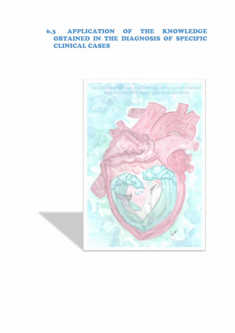
6.3 APPLICATION OF THE KNOWLEDGE
OBTAINED IN THE DIAGNOSIS OF SPECIFIC
CLINICAL CASES


RESULTS & DISCUSSION: PUBLICATIONS APPLICATION OF THE KNOWLEDGE OBTAINED IN THE DIAGNOSIS OF SPECIFIC CLINICAL CASES
[Skeletal and cardiac rhabdomyolysis in a live-stranded neonatal Bryde’s whale with fetal distress] __________________________________________________________________________________________________________________________________________________________________________
173
The third specific objective of this study consisted in the presentation of two specific
case studies of SCMP in cetaceans.
In this first case study we present a live-stranded specimen belonging to the Mysticeti
infraorder, due to the fact that description of pathological entities and/or causes of death in the
infraorder Mysticeti are still scarce. Therefore, we analysed a live-stranded neonatal Bryde’s
whale (Balaenoptera edeni) with fetal distress, which subsequently died soon after the
stranding. This assessment was carried out through a physical exam, blood examination, gross
necropsy evaluation, histopathology, and immunohistochemistry.
The results were published in the article (as followed) titled Skeletal and cardiac
rhabdomyolysis in a live-stranded neonatal Bryde’s whale with fetal distress, on December
2019 in the Frontiers in Veterinary Sciences journal, Volume 20, Issue 6 (doi:
10.3389/fvets.2019.00476), having this journal an impact factor of 2.029 in 2018 and therefore
being a Q1 in the veterinary category.

CASE REPORTpublished: 20 December 2019doi: 10.3389/fvets.2019.00476
Frontiers in Veterinary Science | www.frontiersin.org 1 December 2019 | Volume 6 | Article 476
Edited by:
Robert James Ossiboff,
University of Florida, United States
Reviewed by:
Michael Thomas Walsh,
University of Florida, United States
Molly E. Church,
University of Pennsylvania,
United States
*Correspondence:
Eva Sierra
Specialty section:
This article was submitted to
Veterinary Experimental and
Diagnostic Pathology,
a section of the journal
Frontiers in Veterinary Science
Received: 12 August 2019
Accepted: 05 December 2019
Published: 20 December 2019
Citation:
Câmara N, Sierra E, Fernández A,
Suárez-Santana CM, Puig-Lozano R,
Arbelo M and Herráez P (2019)
Skeletal and Cardiac Rhabdomyolysis
in a Live-Stranded Neonatal Bryde’s
Whale With Fetal Distress.
Front. Vet. Sci. 6:476.
doi: 10.3389/fvets.2019.00476
Skeletal and CardiacRhabdomyolysis in a Live-StrandedNeonatal Bryde’s Whale With FetalDistressNakita Câmara, Eva Sierra*, Antonio Fernández, Cristian Manuel Suárez-Santana,
Raquel Puig-Lozano, Manuel Arbelo and Pedro Herráez
Department of Veterinary Histology and Pathology, Veterinary School, Institute of Animal Health and Food Safety, University
of Las Palmas de Gran Canaria, Las Palmas de Gran Canaria, Spain
The main objective of wildlife forensic investigation is to recognize pathologic changes
and cause of death. Even though it may not always be possible to determine the
specific illness and/or etiology, the description and subsequent interpretation of the
injuries provide an invaluable understanding of pathology in cetacean post-mortem
investigations. Although pathological studies have been previously reported in various
cetacean species, such descriptions of the infraorder Mysticeti remain rare. A
live-stranded neonatal Bryde’s whale (Balaenoptera edeni) which subsequently died soon
after the stranding, was assessed by physical exam, blood examination, gross necropsy
evaluation, histopathology, and immunohistochemistry. It presented with elevated serum
levels of creatine kinase, cardiac troponin I, urea, and creatinine. Microscopically, we
observed keratin spicules (squamous epithelial cells) and areas of atelectasis in the
lungs. Acute degeneration in the myocytes and cardiomyocytes were comparable
to the findings previously described in cases of capture myopathy in live-stranded
cetaceans. Immunohistochemistry biomarkers such as myoglobin, fibrinogen, and
troponin were analyzed. Skeletal and myocardial damage has been documented in
several cetacean species. However, this is the first reported case of skeletal and cardiac
rhabdomyolysis associated with live-stranding in a newborn Bryde’s whale that suffered
from fetal distress.
Keywords: Bryde’s whale (Balaenoptera edeni), cetaceans, rhabdomyolysis, live-stranding, Mysticeti, neonate,
stress cardiomyopathy
BACKGROUND
Pathological study of wildlife fauna has the disadvantage of an unknown clinical history of theanimal. The complexity is enhanced in cetaceans because of the difficulty of performing clinicalexams and/or other analyses on live animals. The most feasible technique for health assessmentin dead cetaceans is by detection of injuries in these animals, through pathological study. Theimportance of these studies is recognized worldwide to promote conservation of these animals.Unfortunately, the description of pathological entities and/or causes of death in the infraorderMysticeti remains rare (1–8).

Câmara et al. Rhabdomyolysis in a Neonatal Bryde’s Whale
Live-stranding is a pathological state with severe acute stressand physical damage central to its etiopathogenesis. It presentsclinical and traumatic findings that can cause death of theanimal, or can seriously aggravate an existing condition over theperiod of stranding, capture, handling, restraint, transportationand/or captivity (1, 2, 9–15). The response mechanisms andresultant damage to multiple systems involved in live-strandedcetaceans are comparable to exertional rhabdomyolysis (capturemyopathy) in many animals, including birds and terrestrial ormarine wild mammals (1, 9–11, 14, 16). Although pathologicalfindings may vary among individuals, biochemical changesand histopathological lesions, consisting of ischemia-reperfusioninjuries, are often observed. These changes result in local-to-generalized vasospasms and vasodilation (catecholamine surge,neurogenic shock, and impeded venous flow return by bodycompression), which is analogous to the stress cardiomyopathyin humans and in direct traumatic injury to muscles, resultingin acute to subacute degeneration (rhabdomyolysis). Acute renalfailure associated with myoglobinuric nephrosis secondary tomuscle damage and areas of necrosis in viscera are also observed(1, 9, 10, 12, 16–27).
In both wild and captive cetaceans, neonatal mortality isa recognized concern (28–31). The main causes of strandingand/or death in newborns are related with problems inpregnancy (abortion, prematurity), childbirth (fetal distress,dystocia), nursing (missed transfer of passive immunity),behavior (maternal-filial separation/maternal neglect), or intraand interspecific interactions with a fatal outcome. All theseabove are enclosed in the category of neonatal and/or perinatalpathologies (1, 2). In the case of asphyxia, the fetus respondswith redistribution of the blood flow, which limits the deleteriouseffects of oxygen deprivation in vital organs. This enables thefetus to survive intact unless the asphyxia is profound orprolonged (32).
This report describes the biochemical analysis and gross,histopathological, histochemical, and immunohistochemicalfeatures in a live-stranded neonatal Bryde’s whale.
CASE PRESENTATION
Stranding CircumstancesA 393-cm-long newborn male Bryde’s whale was stranded on thecoast of Fuerteventura, Canary Island, Spain, in September 2016.Observations between the high and low tide revealed that theanimal appeared to be alive and few meters from the coast. Atlow tide, the animal was stranded on the beach and died beforespecialized assistance could be given.
Biochemical AnalysisA sample of whole blood was collected from the tail flukes,immediately post-mortem, for analysis of the serum. Biochemicalmarkers of acute skeletal and heart muscle damage, creatinekinase (CK 460.0 U/L), and cardiac troponin I (0.20 µg/L),were analyzed. Kidney function was also assessed via blood ureanitrogen (BUN 162 mg/dL) and creatinine (2.4 mg/dL).
FIGURE 1 | Macroscopic vascular changes observed in the heart.
Subepicardial (arrow head) and subendocardial hemorrhage (arrow) in the left
ventricle. Detail of the subendocardial hemorrhages present in the left ventricle
(arrows).
Gross Anatomic AnalysisA thorough necropsy was performed on the calf, followingthe standard protocol published by the European Society ofCetaceans and with the addition of some procedures detailedin the Marine Mammals Ashore manual, to determine the causeof death (14, 33). The animal was in poor body/nutritionalcondition with several linear erosions, distributed in a multifocalmanner on the ventral abdomen (attributed to direct activestranding damage). While several vestigial hairs were notedin the lateral part of the maxilla, the navel was not healedand contained an internal white exudate. During dissectionof the subcutaneous planes, moderate diffuse hemorrhageswere observed, especially in the ventral region. The muscleswere pale yellow-to-orange. The epiglottis was flaccid at therostral level, the trachea showed mild-to-moderate foam, andthe main and secondary bronchi and bronchioles presentedabundant foam, representing pulmonary edema. Both lungs hadmultifocal and local extensive areas of dark reddish color andwere firm (compatible with pulmonary atelectasis). Mild-to-moderate exudation of blood was noted at the incision. Someserous fluid was detected in the pericardial space. On sectioning,both ventricles presented moderate-to-severe subepicardial andsubendocardial hemorrhage (Figure 1). The ductus arteriosuswas also present. Moderate and diffuse mucosal congestion wasobserved in the stomach, liver, bladder, sclera, andmeningeal andsubarachnoid vessels. The cerebellum displayed moderate anddiffuse congestion, edema, and mild-to-moderate hemorrhage.
Histopathological AnalysisRepresentative tissue samples were fixed in 10% formalin for∼48 h and processed using standard protocol. The skeletal(longissimus dorsi and rectus abdominis) and heart muscles(both atria and ventricles), atrioventricular valves (bicuspid ormitral and tricuspid), semilunar valves (sigmoid, aortic, and
Frontiers in Veterinary Science | www.frontiersin.org 2 December 2019 | Volume 6 | Article 476
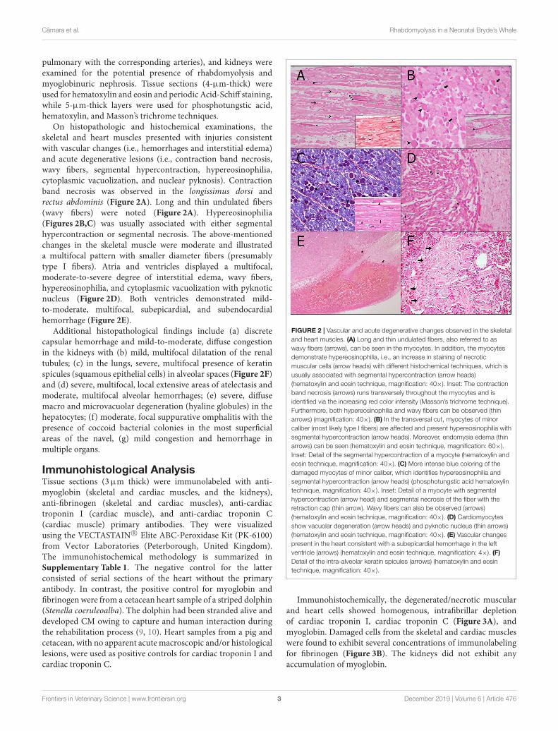
Câmara et al. Rhabdomyolysis in a Neonatal Bryde’s Whale
pulmonary with the corresponding arteries), and kidneys wereexamined for the potential presence of rhabdomyolysis andmyoglobinuric nephrosis. Tissue sections (4-µm-thick) wereused for hematoxylin and eosin and periodic Acid-Schiff staining,while 5-µm-thick layers were used for phosphotungstic acid,hematoxylin, and Masson’s trichrome techniques.
On histopathologic and histochemical examinations, theskeletal and heart muscles presented with injuries consistentwith vascular changes (i.e., hemorrhages and interstitial edema)and acute degenerative lesions (i.e., contraction band necrosis,wavy fibers, segmental hypercontraction, hypereosinophilia,cytoplasmic vacuolization, and nuclear pyknosis). Contractionband necrosis was observed in the longissimus dorsi andrectus abdominis (Figure 2A). Long and thin undulated fibers(wavy fibers) were noted (Figure 2A). Hypereosinophilia(Figures 2B,C) was usually associated with either segmentalhypercontraction or segmental necrosis. The above-mentionedchanges in the skeletal muscle were moderate and illustrateda multifocal pattern with smaller diameter fibers (presumablytype I fibers). Atria and ventricles displayed a multifocal,moderate-to-severe degree of interstitial edema, wavy fibers,hypereosinophilia, and cytoplasmic vacuolization with pyknoticnucleus (Figure 2D). Both ventricles demonstrated mild-to-moderate, multifocal, subepicardial, and subendocardialhemorrhage (Figure 2E).
Additional histopathological findings include (a) discretecapsular hemorrhage and mild-to-moderate, diffuse congestionin the kidneys with (b) mild, multifocal dilatation of the renaltubules; (c) in the lungs, severe, multifocal presence of keratinspicules (squamous epithelial cells) in alveolar spaces (Figure 2F)and (d) severe, multifocal, local extensive areas of atelectasis andmoderate, multifocal alveolar hemorrhages; (e) severe, diffusemacro and microvacuolar degeneration (hyaline globules) in thehepatocytes; (f) moderate, focal suppurative omphalitis with thepresence of coccoid bacterial colonies in the most superficialareas of the navel, (g) mild congestion and hemorrhage inmultiple organs.
Immunohistological AnalysisTissue sections (3µm thick) were immunolabeled with anti-myoglobin (skeletal and cardiac muscles, and the kidneys),anti-fibrinogen (skeletal and cardiac muscles), anti-cardiactroponin I (cardiac muscle), and anti-cardiac troponin C(cardiac muscle) primary antibodies. They were visualizedusing the VECTASTAIN R© Elite ABC-Peroxidase Kit (PK-6100)from Vector Laboratories (Peterborough, United Kingdom).The immunohistochemical methodology is summarized inSupplementary Table 1. The negative control for the latterconsisted of serial sections of the heart without the primaryantibody. In contrast, the positive control for myoglobin andfibrinogen were from a cetacean heart sample of a striped dolphin(Stenella coeruleoalba). The dolphin had been stranded alive anddeveloped CM owing to capture and human interaction duringthe rehabilitation process (9, 10). Heart samples from a pig andcetacean, with no apparent acute macroscopic and/or histologicallesions, were used as positive controls for cardiac troponin I andcardiac troponin C.
FIGURE 2 | Vascular and acute degenerative changes observed in the skeletal
and heart muscles. (A) Long and thin undulated fibers, also referred to as
wavy fibers (arrows), can be seen in the myocytes. In addition, the myocytes
demonstrate hypereosinophilia, i.e., an increase in staining of necrotic
muscular cells (arrow heads) with different histochemical techniques, which is
usually associated with segmental hypercontraction (arrow heads)
(hematoxylin and eosin technique, magnification: 40×). Inset: The contraction
band necrosis (arrows) runs transversely throughout the myocytes and is
identified via the increasing red color intensity (Masson’s trichrome technique).
Furthermore, both hypereosinophilia and wavy fibers can be observed (thin
arrows) (magnification: 40×). (B) In the transversal cut, myocytes of minor
caliber (most likely type I fibers) are affected and present hypereosinophilia with
segmental hypercontraction (arrow heads). Moreover, endomysia edema (thin
arrows) can be seen (hematoxylin and eosin technique, magnification: 60×).
Inset: Detail of the segmental hypercontraction of a myocyte (hematoxylin and
eosin technique, magnification: 40×). (C) More intense blue coloring of the
damaged myocytes of minor caliber, which identifies hypereosinophilia and
segmental hypercontraction (arrow heads) (phosphotungstic acid hematoxylin
technique, magnification: 40×). Inset: Detail of a myocyte with segmental
hypercontraction (arrow head) and segmental necrosis of the fiber with the
retraction cap (thin arrow). Wavy fibers can also be observed (arrows)
(hematoxylin and eosin technique, magnification: 40×). (D) Cardiomyocytes
show vacuolar degeneration (arrow heads) and pyknotic nucleus (thin arrows)
(hematoxylin and eosin technique, magnification: 40×). (E) Vascular changes
present in the heart consistent with a subepicardial hemorrhage in the left
ventricle (arrows) (hematoxylin and eosin technique, magnification: 4×). (F)
Detail of the intra-alveolar keratin spicules (arrows) (hematoxylin and eosin
technique, magnification: 40×).
Immunohistochemically, the degenerated/necrotic muscularand heart cells showed homogenous, intrafibrillar depletionof cardiac troponin I, cardiac troponin C (Figure 3A), andmyoglobin. Damaged cells from the skeletal and cardiac muscleswere found to exhibit several concentrations of immunolabelingfor fibrinogen (Figure 3B). The kidneys did not exhibit anyaccumulation of myoglobin.
Frontiers in Veterinary Science | www.frontiersin.org 3 December 2019 | Volume 6 | Article 476

Câmara et al. Rhabdomyolysis in a Neonatal Bryde’s Whale
FIGURE 3 | Immunohistochemical techniques in skeletal and heart muscles.
(A) Degenerated/necrotic cardiomyocytes (arrow heads), with pyknotic
nucleus (thin arrows), show intrafibrillar depletion of cardiac troponin C. In
contrast, normal cardiomyocytes (arrows) present an intense immunolabeling
(immunohistochemical technique: anti-troponin C, magnification: 60×). (B)
Expression of fibrinogen (arrows) in the myocytes presenting changes,
including wavy fibers, hypereosinophilia and segmental hypercontraction.
Immunolabeling of fibrinogen in the interior of the blood vessels can also be
seen (thin arrows) (immunohistochemical technique: anti-fibrinogen,
magnification: 40×). Inset: Necrotic myocytes (arrows) strongly expressed
alongside the contraction band necrosis (arrow head) and inside the blood
vessels (thin arrows) (immunohistochemical technique: anti-fibrinogen,
magnification: 40×).
DISCUSSION
Discussion of the Animal CharacteristicsCetacean newborns/neonates are defined as having a compatibletotal length, displaying “fetal folds” over the body, soft and foldeddorsal fin and tail flukes, vibrissal hairs or vibrissal crypts, anda healing (or closing) navel (34). Our animal was 393 cm long,slightly under the normal range (395–430 cm) (35). The animaldid not present any “fetal folds” but we identified some vibrissalhairs; the navel was not healed and contained inflammatoryexudate. This suggested an infection after birth; therefore, theanimal would most likely be a few days old (34).
Discussion of the Biochemical ResultsClinico-pathological evaluation was challenging in this case asbiochemical values, such as that for cardiac troponin I, are rarelyreported. They may not exist in the scientific database for variousspecies of cetaceans, particularly the infraorder Mysticeti. Ourbiochemical data was compared with published papers assessingdifferent mammals, including humans, dogs, and other speciesof cetaceans [e.g., bottlenose dolphins (Tursiops truncatus)belonging to the infraorder Odontoceti and a stranded baby graywhale (Eschrichtius robustus) of the infraorder Mysticeti]; this issummarized in Table 1 (13, 36–39).
In concordance with the results obtained from other species(i.e., 107–255 U/L in a neonatal gray whale), injury to the skeletalmuscle was supported by an increase in CK (460 U/L), which isone of the useful indicators of both skeletal and cardiac muscledamage (12, 13, 16, 39–41). After myocardial injury, CK beginsto rise in 4 to 9 h, peaks at 24 h, and returns to baseline 48 to 72 hafter the stress event (42). This increase in CKwas correlated withhistological and immunohistochemical changes, thus supportingthe presence of muscle damage.
Cardiac troponin I, measured by conventional assays, iselevated in >90% of patients with stress cardiomyopathy (43). T
ABLE1|Comparisonbetw
eenthebiochemicalresu
ltsofthestudiedanim
alandnorm
allaboratory
valuesofothermammals.
ClassMammalia
OrderPrimates
OrderCarnivora
OrderCetartiodactyla
SuborderHaplorhini
SuborderCetacea
InfraorderSim
iform
es
InfraorderOdontoceti
InfraorderMysticeti
FamilyHominidae
FamilyCanidae
FamilyDelphinidae
FamilyEschrichtiidae
FamilyBalaenopteridae
HUMANS(Homosapiens)(17)
DOG
(Canislupus
familiaris)( 19,20)
BOTTLENOSEDOLPHIN
(Tursiopstruncatus)(9)
NEONATALGRAY
WHALE(Eschrichtius
robustus)( 9)
NEONATALBRYDE’S
WHALE(Balaenoptera
edeni)
Captive(n
=38)
WildAtlantic
Juvenile(n
=96)
Stranded(n
=1)
Stranded(n=1)
Creatin
eKinase
(U/L)
30–1
70
0–1
90
100–2
50
47–4
55
107–2
55
460
TroponinI(
µg/L)
≤0.1
≤0.03–0
.07
ND
ND
ND
0.20
BloodUreaNitrogen(m
g/dL)
8–2
07–2
0.72
42–5
842–7
721–7
5162
Creatin
ine(m
g/dL)
0.7–1
.30.44–1
.595
1.0–2
.00.68–1
.49
1.0–2
.02.4
Frontiers in Veterinary Science | www.frontiersin.org 4 December 2019 | Volume 6 | Article 476

Câmara et al. Rhabdomyolysis in a Neonatal Bryde’s Whale
Our animal presented with 0.20 µg/L cardiac troponin I inthe serum; this is higher than the reference values in bothhumans (≤0.1 µg/L) and dogs (≤0.03–0.07 µg/L) (36–38).The release of troponin from injured cardiomyocytes usuallyoccurs 3 to 9 h after ischemic damage, peaks in 12 to 48 h,and remains elevated for 4 to 7 days (42, 44, 45). Hence,the above laboratory values cannot aid in early detection ofmyocardial necrosis (1–3 h). These markers do not assist inaccurate diagnosis until 6 or more hours after the onset ofthe event. In order to obtain a satisfactory clinical picture inhumans, blood should be drawn 6–9 h after the onset of thestress event and/or symptom onset (44). The increase in cardiactroponin I serum levels and the decrease in myocyte troponinimmunoreaction is caused by the early release of cardiac troponinI and troponin C by damaged cardiomyocytes. This was verifiedby immunohistochemistry (22, 46).
In this case, pre-renal azotemia is likely related to hypovolemicshock, and was reasonably supported by higher BUN values (162mg/dL) than the reference values from the other species (i.e., 21to 75 mg/dL in the stranded gray whale). Hypovolemic shock canbe originated by a relative decrease in the effective circulatingvolume without a loss of total body fluid (i.e., decreased in venousreturn) and/or a direct intravascular fluid loss (i.e., dehydrationor hemorrhage) (12, 40). In our case, we can associate this clinicalfinding with various causes, such as compartment syndrome,heart failure, and dehydration. Moreover, creatinine was slightlyabove (2.4 mg/dL) the normal values seen in other species (i.e.,1.0 to 2.0 mg/dL in a stranded neonatal gray whale). In orderto confirm the hypothesis that the increase in creatinine weobserved could be within the normal ranges for the Bryde’s whalespecies, more accurate age and species-specific normal valueswould be needed. Small elevation of creatinine and high levelsof urea can be associated with pre-renal azotemia caused bydehydration. This is a possible interpretation for our case. Sincedehydration is associated with prolonged fasting, it is importantto consider this aspect in stranded animals, especially youngones (47).
Discussion of the HistopathologicalResultsWith the anatomopathological findings, the morphologicaldiagnoses include (1) severe multifocal fetal atelectasis withpresence of severe multifocal keratin spicules and a moderatealveolar hemorrhage; (2) multifocal moderate-to-severe acutedegeneration of cardiomyocytes; (3) moderate multifocal acuteskeletal muscle degeneration; and (4) mild congestion andhemorrhage in various organs.
Atelectasis is a relatively frequent finding in fetal andneonatal (atelectasis neonatorum) deaths (perhaps associatedwith aspiration of amniotic fluid or meconium), it is foundincidentally or in non-specific forms in young or adultindividuals (1, 48–50). In contrast, pulmonary edema withintra-alveolar keratin spicules (pulmonary vernix caseosa), eitherisolated or in aggregates of stratified epithelium, keratinizedand with nuclear retention, can be observed in fetal distress.Considering non-specific findings, without knowledge of theprimary cause, and after discarding lesions compatible with other
etiological diagnoses, we considered the macro and microscopicfindings as a whole to be typical of fetal distress (1).
Acute degenerative changes, such as contraction bandnecrosis, were observed in both the longissimus dorsi and therectus abdominis. This represents a skeletal and myocardiallesion characteristic of transient ischemia and reperfusion,which is associated with high concentrations of endogenouscatecholamines (23, 51, 52). This condition has been reportedin humans after stressful events, as well as in other animalsafter acute death, including seals and cetaceans (1, 9–12, 16–23, 53–56). Wavy fibers were also detected in the skeletal andheart muscles. Considering that they are the first histologicabnormality associated with ischemia, this condition may beused as a morphological indicator of early myocardial injury (9–11, 22, 23, 53, 57). In addition, hypereosinophilia was observedin the skeletal and cardiac muscles, respectively. Animals thatdie following a stressful situation present with this cytoplasmicalteration (9–11, 22, 53, 57, 58). The animal also presented withvacuolization in the cardiomyocytes. Previous studies commonlyassociate this with areas that experience severe, chronic, andfatal ischemia as a result of acute death due to stressfulsituations (11, 22, 53, 59).
Vascular changes, including congestion, interstitial edema,and hemorrhage, are generally detected through histologicalapproaches and form part of the stress cardiomyopathypathology (17). In the current case, all the heart sectionsshowed separated fibers, with interstitial edema. Following theintroduction of catecholamines, interstitial edema is usuallyassociated with subendocardial and subepicardial hemorrhage,found in both ventricles of this neonate (57). These lesions areoccasionally detected in humans with stress cardiomyopathy andhave been previously demonstrated in animals that died afterlive-stranding and handling (1, 9, 10, 17, 20).
The sequence of changes in an acute ischemic injury beginswithin 5min. The myocardium reveals long, thinned, wavy fibersseparated by spaces, characterizing edema and microvascularcongestion at the borders of the ischemic myocardium. In 2 to3 h, early changes of cardiomyocyte coagulation necrosis withnuclear pyknosis, color change, more specifically “brick redchange” or cytoplasm hypereosinophilia, focal contraction bands,and subtle interstitial edema are evident. Hypereosinophilia andedema become more pronounced and more easily recognizable 3to 6 h after the event. Six to 12 h later changes accelerate andmoreextensive contraction band necrosis with reperfusion is noted(25). Based on the acute degenerative findings in both skeletal andcardiac muscle of our case, we propose that the ischemic injury,which caused these lesions, occurred between 6 and 12 h prior todeath, coinciding with the live-stranding.
Discussion of the ImmunohistochemicalResultsPrevious studies demonstrated the necessity of corroboratinghistopathological findings with specific markers to betterdetermine the amount of damage present in cells. Theimmunohistochemical confirmation ante-mortem showeddepletion of myoglobin, cardiac troponin I, and cardiactroponin C as well as intrafibrillar fibrinogen deposition(9, 10, 12, 22, 45, 58, 60–63). Depletion of myoglobin (a
Frontiers in Veterinary Science | www.frontiersin.org 5 December 2019 | Volume 6 | Article 476

Câmara et al. Rhabdomyolysis in a Neonatal Bryde’s Whale
marker used for skeletal and cardiac damage), troponin Iand C (specific markers to detect injury to the heart), aswell as accumulation of fibrinogen (used to identify skeletaland cardiac damage) in injured cells was confirmed in thepresent study.
Although this animal presented with clinical and pathologicalfindings resembling rhabdomyolysis, which can lead to asecondary myoglobinuric nephrosis, lesions associated withacute kidney injury (i.e., intrinsic kidney disease/ damageor acute tubular necrosis) were not detected throughthe histopathological and immunohistochemical studies(1, 9, 10, 12, 16–27, 64).
Discussion of the Cause of DeathConsidering the biochemical results and the macro andmicroscopic findings which concur with the etiological diagnosesof fetal distress and skeletal and cardiac rhabdomyolysis, wepropose that the most probable cause of death in this animalis active stranding pathology which aggravated a previousneonatal/perinatal pathology.
The active stranding pathology is defined by a set oflesions and biochemical findings in animals that were strandedalive and leads to both a catecholaminergic crisis (stresscardiomyopathy) and multi-organ ischemic-reperfusion damagewith rhabdomyolysis with myoglobinuric nephrosis secondary tomuscle damage. The severity of this syndrome usually causes thedeath of the animal, occasionally as a result of the intensificationof preexisting pathologies (1, 9, 10, 22).
Neonatal/perinatal pathology in cetaceans comprises a wideconstellation of etiologic factors, including fetal distress (1, 2).A severe disturbance in the oxygen supply to the fetus canhave effects on the newborn’s cardiac function. Elevated levelsof cardiac troponin I, cardiac troponin T, CK and its fractionMB can be observed in full-term infants after intrauterinehypoxia and respiratory distress (65). Limited studies have shownpremature infants and various breeds of stillborn cow calvesto present with acute degenerative changes, such as myocardialnecrosis, which may result from antepartum or intrapartumasphyxia (hypoxia) (65–67).
CONCLUSIONS
Although we cannot confirm that the elevated serum values(CK, cTnI, BUN, and creatinine) were due to post strandingor fetal distress, but based on the histological findings, we canconclude that these lesions are due to live stranding. Therefore,we suggest that the animal assessed here probably died because ofan exacerbation of preceding injuries (fetal distress) and the finalcomplications of stranding. Since description of pathologicalentities and/or causes of death in the Mysticeti infraorder is stillscarce, we consider this article to be an important contributionto improve conservation efforts by reducing the mortality ofthese animals.
DATA AVAILABILITY STATEMENT
All data reported in this work is classified and stored in thetissue bank of the Institute of Animal Health and Food Safety
(IUSA), Veterinary School, University of Las Palmas de GranCanaria (ULPGC).
ETHICS STATEMENT
Ethical review and approval was not required for the animalstudy because no experiments were performed on live animals.Required permission for the management of stranded cetaceanswas issued by the environmental department of the CanaryIslands’ Government and the Spanish Ministry of Environment.No experiments were performed on live animals.
AUTHOR CONTRIBUTIONS
NC wrote the article, collected the blood sample, performedthe necropsy of the animal, and contributed toward thebiochemical analysis, gross, histological, histochemical, andimmunohistological description and diagnosis of the case. ESand PH contributed toward the biochemical analysis, gross,histological, histochemical, and immunohistological descriptionand diagnosis of the case, and guided NC during the draftingand publication processes. AF, CS-S, RP-L, and MA performedthe necropsy of the animal and contributed toward the gross andhistological description and diagnosis of the case. All authorsread and approved the final manuscript.
FUNDING
This study was part of a Ph.D. thesis of the Universidadde Las Palmas de Gran Canaria (ULPGC) supported bythe Ministry of Economy and Competitiveness (MINECO)through a predoctoral grant for training of research personnel(BES-2016-076907). Furthermore, this research work was partof the National Project titled Embolic Pathology (gas/fat)in cetaceans supported by the Ministry of Economy andCompetitiveness (MINECO) (CGL2015-71498-P) as wellas part of the Project titled Stress Cardiomyopathies inCetaceans supported by the Ministry of Science, Innovation andUniversities, and subsided by the Government of Gran Canaria(CABILDO2018: CABILDO2018-04). Finally, the Canary IslandsGovernment funded and provided support to the strandingresearch network.
ACKNOWLEDGMENTS
We would like to thank all the people who indirectly participatedin the production of this work, including the staff of the AnimalLab, our laboratory technician, all the members and volunteers ofthe Cetacean Stranding Network, as well as Marisa Tejedor andthe associated non-governmental organization (SECAC) whocollaborated in the necropsy.
SUPPLEMENTARY MATERIAL
The Supplementary Material for this article can be foundonline at: https://www.frontiersin.org/articles/10.3389/fvets.2019.00476/full#supplementary-material
Frontiers in Veterinary Science | www.frontiersin.org 6 December 2019 | Volume 6 | Article 476
Câmara et al. Rhabdomyolysis in a Neonatal Bryde’s Whale
REFERENCES
1. Díaz-Delgado J, Fernández A, Sierra E, Sacchini S, Andrada M, VelaAI, et al. Pathologic findings and causes of death of stranded cetaceansin the Canary Islands (2006-2012). PLoS ONE. (2018) 13:e0204444.doi: 10.1371/journal.pone.0204444
2. Arbelo M, Los Monteros AE, Herráez P, Andrada M, Sierra E, RodríguezF, et al. Pathology and causes of death of stranded cetaceans inthe Canary Islands (1999-2005). Dis Aquat Organ. (2013) 103:87–99.doi: 10.3354/dao02558
3. Cornaglia E, Rebora L, Gili C, Di Guardo G. Histopathological andimmunohistochemical studies on cetaceans found stranded on the Coastof Italy between 1990 and 1997. J Vet Med Ser A. (2000) 47:129–42.doi: 10.1046/j.1439-0442.2000.00268.x
4. Kemper C, Flaherty A, Gibbs S, Hill M, Long M, Byard RW. Cetaceancaptures, strandings and mortalities in South Australia 1881-2000, withspecial reference to human interactions. Aust Mammal. (2005) 27:37–47.doi: 10.1071/AM05037
5. Martineau D, Lagacé A, Béland P, Higgins R, Armstrong D, Shugart LR.Pathology of stranded beluga whales (Delphinapterus leucas) from the St.Lawrence Estuary, Québec, Canada. J Comp Pathol. (1988) 98:287–310.doi: 10.1016/0021-9975(88)90038-2
6. Dhermain F, Soulier L, Bompar J-M. Natural mortality factors affectingcetaceans in theMediterranean Sea.CetaceansMediterr Black Seas State Knowl
Conserv Strateg A Rep to ACCOBAMS Secr Monaco (2002).7. McAloose D, Rago MV, Di Martino M, Chirife A, Olson SH, Beltramino L,
et al. Post-mortem findings in southern right whales Eubalaena Australis atPenínsula Valdés, Argentina, 2003-2012. Dis Aquat Organ. (2016) 119:17–36.doi: 10.3354/dao02986
8. Groch KR, Díaz-Delgado J, Marcondes MCC, Colosio AC, Santos-Neto EB,Carvalho VL, et al. Pathology and causes of death in stranded humpbackwhales (Megaptera novaeangliae) from Brazil. PLoS ONE. (2018) 13:e0194872.doi: 10.1371/journal.pone.0194872
9. Herráez P, Espinosa de los Monteros A, Fernández A, Edwards JF, SacchiniS, Sierra E. Capture myopathy in live-stranded cetaceans. Vet J. (2013)196:181–8. doi: 10.1016/j.tvjl.2012.09.021
10. Herráez P, Sierra E, Arbelo M, Jaber JR, Espinosa De Los Monteros A,et al. Rhabdomyolysis and myoglobinuric nephrosis (Capture Myopathy)in a striped dolphin. J Wildl Dis Wildl Dis Assoc. (2007) 43:770–4.doi: 10.7589/0090-3558-43.4.770
11. Cowan DF, Curry BE. Histopathology of the alarm reaction in smallodontocetes. J Comp Pathol. (2008) 139:24–33. doi: 10.1016/j.jcpa.2007.11.009
12. Bonsembiante F, Centelleghe C, Rossi G, Giglio S, Madeo E, GelainME, et al. Clinico-pathological findings in a striped dolphin (Stenellacoeruleoalba) affected by rhabdomyolysis and myoglobinuric nephrosis(capture myopathy). J Vet Med Sci. (2017) 79:1013–18. doi: 10.1292/jvms.17-0023
13. Gulland FMD, Dierauf LA, Whitman KL. CRC Handbook of Marine Mammal
Medicine. Florida, FL: CRC Press (2018).14. Geraci JR, Lounsbury VJ. Marine Mammals Ashore: A Field Guide for
Strandings. Baltimore, MD: National Aquarium (2005).15. Wilkinson DM. Report to Assistant Administrator for Fisheries: Program
Review of the Marine Mammal Stranding Networks (1991).16. Seguel M, Paredes E, Pavés H, Gottdenker NL. Capture-induced stress
cardiomyopathy in South American fur seal pups (Arctophoca australis
gracilis).Mar Mammal Sci. (2014) 30:1149–57. doi: 10.1111/mms.1207917. Jiang JP, Downing SE. Catecholamine cardiomyopathy: review and analysis of
pathogenetic mechanisms. Yale J Biol Med. (1990) 63:581–91.18. Kawai S. Pathology of takotsubo (ampulla) cardiomyopathy. In: Veselka J,
editor. Cardiomyopathies - From Basic Research to Clinical Management.London: IntechOpen (2012). p. 709–26. doi: 10.5772/29050
19. Maréchaux S, Fornes P, Petit S, Poisson C, Thevenin D, Le Tourneau T, et al.Pathology of inverted takotsubo cardiomyopathy. Cardiovasc Pathol. (2008)17:241–3. doi: 10.1016/j.carpath.2007.08.002
20. Mitchell A, Marquis F. Can takotsubo cardiomyopathy be diagnosed byautopsy? Report of a presumed case presenting as cardiac rupture. BMC Clin
Pathol. (2017) 17:4. doi: 10.1186/s12907-017-0045-0
21. Miura M, Kawano H, Yoshida T, Yamagata Y, Nakata T, Koga S,et al. The histological features of a myocardial biopsy specimen ina patient in the acute phase of reversible catecholamine-inducedcardiomyopathy due to pheochromocytoma. Intern Med. (2017) 56:665–71.doi: 10.2169/internalmedicine.56.7454
22. Câmara N, Sierra E, Fernández-Maldonado C, Espinosa de los Monteros A,Arbelo M, Fernández A, et al. Stress cardiomyopathy in stranded cetaceans: ahistological, histochemical and immunohistochemical study. Vet Rec. (2019)185:694. doi: 10.1136/vr.105562
23. Fineschi V,MichalodimitrakisM, D’Errico S, NeriM, Pomara C, Riezzo I, et al.Insight into stress-induced cardiomyopathy and sudden cardiac death dueto stress. a forensic cardio-pathologist point of view. Forensic Sci Int. (2010)194:1–8. doi: 10.1016/j.forsciint.2009.10.025
24. Spraker T. Stress and capture myopathy in artiodactylids. ZooWild AnimMed
Curr Ther. (1993).25. Buja LM, Butany J. Cardiovascular Pathology. Elsevier Science (2015).
Available at: https://books.google.es/books?id=rdqcBAAAQBAJ26. Buja LM. Myocardial ischemia and reperfusion injury. Cardiovasc Pathol.
(2005) 14:170–5. doi: 10.1016/j.carpath.2005.03.00627. Sachdeva J, Dai W, Kloner RA. Functional and histological assessment of an
experimental model of takotsubo’s cardiomyopathy. J Am Heart Assoc. (2019)3:e000921. doi: 10.1161/JAHA.114.000921
28. Mann J, Connor RC, Barre LM, Heithaus MR. Female reproductive successin bottlenose dolphins (Tursiops sp.): life history, habitat, provisioning, andgroup-size effects. Behav Ecol. (2000) 11:210–9. doi: 10.1093/beheco/11.2.210
29. Wells R, Scott M, Irvine AB. The social structure of free-rangingbottlenose dolphins. In: Genoways HH, editor. Current Mammalogy. NewYork, NY: Springer Science; Business Media, LLC (1987). p. 247–305.doi: 10.1007/978-1-4757-9909-5_7
30. Sweeney J, Stone R, Campbell M, McBain J, Leger J, Xitco M, et al.Comparative survivability of tursiops neonates from three U.S. institutionsfor the decades 1990-1999 and 2000-2009. Aquat Mamm. (2010) 36:248–61.doi: 10.1578/AM.36.3.2010.248
31. Flower J, Langan J, Nevitt B, Chinnadurai S, Stacey R, IvancicM, et al. Neonatal critical care and hand-rearing of a bottlenosedolphin (Tursiops truncatus) calf. Aquat Mamm. (2018) 43:482–90.doi: 10.1578/AM.44.5.2018.482
32. Parer JT, Livingston EG. What is fetal distress? Am J Obstet Gynecol. (1990)162:1421–7. doi: 10.1016/0002-9378(90)90901-I
33. Kuiken T, Hartmann MG. Proceedings of the First ECS Workshop on Cetacean
Pathology: Dissection Techniques and Tissue Sampling: Leiden, the Netherlands,
13-14 September 1991. European Cetacean Society (1991).34. Puig-Lozano R, Bernaldo de Quirós Y, Díaz-Delgado J, García-Álvarez N,
Sierra E, De la Fuente J, et al. Retrospective study of foreign body-associatedpathology in stranded cetaceans, Canary Islands (2000–2015). Environ Pollut.
(2018) 243:519–27. doi: 10.1016/j.envpol.2018.09.01235. Perrin WF, Würsig BG, Thewissen JGM. Encyclopedia of Marine Mammals.
Amsterdam; Boston, MA: Elsevier/Academic Press (2009).36. Padilla O Texas THSC. Normal Laboratory Values: Blood, Plasma, and Serum.
Available online at: https://www.msdmanuals.com/professional/resources/normal-laboratory-values/blood-tests-normal-values#v8508814
37. Sleeper MM, Clifford CA, Laster LL. Cardiac troponin I inthe normal dog and cat. J Vet Intern Med. (2001) 15:501–3.doi: 10.1111/j.1939-1676.2001.tb01582.x
38. Wray J.Canine InternalMedicine:What’s Your Diagnosis?.Oxford: JohnWiley& Sons (2017).
39. Reidarson T, Mcbain J, Yochem P. Medical and nutritional aspects of arehabilitating California gray whale calf. Aquat Mamm. (2001) 27:215–21.
40. Stockham SL, Scott MA. Fundamentals of Veterinary Clinical Pathology. Iowa:John Wiley & Sons (2013).
41. St Aubin DJ, Austin TP, Geraci JR. Effects of handling stress on plasmaenzymes in harp seals, Phoca groenlandica. J Wildl Dis. (1979) 15:569–72.doi: 10.7589/0090-3558-15.4.569
42. Lewandrowski K, Chen A, Januzzi J. Cardiac markers for myocardialinfarction: a brief review. Am J Clin Pathol Pathol Patterns Rev. (2002)118:93–9. doi: 10.1309/3EK7-YVV9-228C-E1XT
43. Lyon AR, Bossone E, Schneider B, Sechtem U, Citro R, Underwood SR, et al.Current state of knowledge on takotsubo syndrome: a position statement
Frontiers in Veterinary Science | www.frontiersin.org 7 December 2019 | Volume 6 | Article 476
Câmara et al. Rhabdomyolysis in a Neonatal Bryde’s Whale
from the taskforce on takotsubo syndrome of the heart failure associationof the European society of cardiology. Eur J Heart Fail. (2016) 18:8–27.doi: 10.1002/ejhf.424
44. Khan MS. Diagnostic efficacy of cardiac troponin in post-mortemexamination of acute myocardial infarction. Int J Eth Trauma Vict. (2015) 1:2.doi: 10.18099/ijetv.v1i1.27
45. Hansen SH, Rossen K. Evaluation of cardiac troponin I immunoreactionin autopsy hearts: a possible marker of early myocardial infarction.Forensic Sci Int. (1999) 99:189–96. doi: 10.1016/S0379-0738(98)00193-5
46. Mikaelian I, Coluccio D, Morgan KT, Johnson T, Ryan AL, RasmussenE, et al. Temporal Gene expression profiling indicates early up-regulation of interleukin-6 in isoproterenol-induced myocardial necrosisin rat. Toxicol Pathol. (2008) 36:256–64. doi: 10.1177/0192623307312696
47. Cubas ZS, Silva JCR, Catão-Dias JL. Tratado de Animais Selvagens: Medicina
Veterinária. São Paulo: Roca (2014).48. Moore MJ, Knowlton A, Kraus S, Mclellan WA, Bonde R. Morphometry,
gross morphology and available histopathology in North Atlantic right whale(eubalaena glacialis) mortalities. J Cetac Res Manage. (2004) 6:199–214.
49. Fernández A, Edwards JF, Rodríguez F, de los Monteros AE, Herráez P, CastroP, et al. “Gas and fat embolic syndrome” involving a mass stranding of beakedwhales (Family Ziphiidae) exposed to anthropogenic sonar signals.Vet Pathol.(2005) 42:446–57. doi: 10.1354/vp.42-4-446
50. Alstrup AKO, Hedayat A, Jensen TH, Hammer AS, Munk OL, Jensen HE.Necropsy report of a fin whale (Balaenoptera physalus) stranded in Denmarkin 2010. Aquat Mamm. (2013) 39:385. doi: 10.1578/AM.39.4.2013.385
51. Reichenbach DD, Benditt EP. Catecholamines and cardiomyopathy: thepathogenesis and potential importance of myofibrillar degeneration. HumPathol. (1970) 1:125–50. doi: 10.1016/S0046-8177(70)80007-7
52. Turnbull BS, Cowan DF. Myocardial contraction band necrosisin stranded cetaceans. J Comp Pathol. (1998) 118:317–27.doi: 10.1016/S0021-9975(07)80007-7
53. Cowan DF, Curry BE. Histopathological Assessment of Dolphins Necropsies
Onboard Vesssels in the Eastern Tropical Pacific Tuna Fishery. La Jolla, CA:Southwest Fisheries Science Center, NationalMarine Fisheries Service, NOAA(2002). p. 31.
54. Akashi YJ, Nef HM, Möllmann H, Ueyama T. Stress cardiomyopathy. AnnuRev Med. (2010) 61:271–86. doi: 10.1146/annurev.med.041908.191750
55. Pascual I, Abó AI, Piqué M. Hallazgos histológicos en el síndrome de tako-tsubo. Rev Española Cardiol. (2015) 68:625. doi: 10.1016/j.recesp.2014.08.013
56. Lyon AR, Rees PS, Prasad S, Poole-Wilson PA, Harding SE. Stress(Takotsubo) cardiomyopathy—A novel pathophysiological hypothesis toexplain catecholamine-induced acute myocardial stunning. Nat Clin Pract
Cardiovasc Med. (2008) 5:22–9. doi: 10.1038/ncpcardio106657. Fishbein MC. Early evolution from ischemia to myocardial necrosis.
Cardiovasc Toxicol. (2001) 1:83–6. doi: 10.1385/CT:1:2:08358. Sierra E, Fernández A, de los Monteros A, Arbelo M, Díaz J, Andrada
Borzollino M, et al. Histopathological muscle findings may be essential
for a definitive diagnosis of suspected sharp trauma associated withship strikes in stranded cetaceans. PLoS ONE. (2014) 9:e88780.doi: 10.1371/journal.pone.0088780
59. Adegboyega PA, Haque AK, Boor PJ. Extensive myocytolysis as amarker of sudden cardiac death. Cardiovasc Pathol. (1996) 5:315–21.doi: 10.1016/S1054-8807(96)00041-5
60. Ortmann C, Pfeiffer H, Brinkmann B. A comparative study on theimmunohistochemical detection of early myocardial damage. Int J Legal Med.
(2000) 113:215–20. doi: 10.1007/s00414990009461. Xiaohong Z, Xiaorui C, Jun H, Qisheng Q. The contrast of
immunohistochemical studies of myocardial fibrinogen and myoglobinin early myocardial ischemia in rats. Leg Med. (2002) 4:47–51.doi: 10.1016/S1344-6223(01)00054-2
62. Martínez-Díaz F, Rodríguez-Morlensín M, Pérez-Cárceles MD, NogueraJ, Luna A, Osuna E. Biochemical analysis and immunohistochemicaldetermination of cardiac troponin for the postmortem diagnosisof myocardial damage. Histol Histopathol. (2005) 20:475–81.doi: 10.14670/HH-20.475.
63. Fishbein MC, Wang T, Matijasevic M, Hong L, Apple FS. Myocardialtissue troponins T and I: an immunohistochemical study in experimentalmodels of myocardial ischemia. Cardiovasc Pathol. (2003) 12:65–71.doi: 10.1016/S1054-8807(02)00188-6
64. Malkina A. Acute Kidney Injury (AKI). San Francisco,CA: University of California. Available online at:ttps://www.msdmanuals.com/professional/genitourinary-disorders/acute-kidney-injury/acute-kidney-injury-aki?query=acute kidney injury
65. Correale M, Nunno L, Ieva R, Rinaldi M, Maffei G, Biase RM, et al. Troponinin newborns and pediatric patients. Cardiovasc Hematol Agents Med Chem.
(2009) 7:270–8. doi: 10.2174/18715250978954192766. Murray RD, Williams AJ, Sheldon IM. Field investigation of
perinatal mortality in friesian cattle associated with myocardialdegeneration and necrosis. Reprod Domest Anim. (2008) 43:339–45.doi: 10.1111/j.1439-0531.2007.00911.x
67. Waldner CL, Kennedy RI, Rosengren LB, Pollock CM, Clark ETG. Grosspostmortem and histologic examination findings from abortion losses andcalf mortalities in western Canadian beef herds. Can Vet J La Rev Vet Can.
(2010) 51:1227–38.
Conflict of Interest: The authors declare that the research was conducted in theabsence of any commercial or financial relationships that could be construed as apotential conflict of interest.
Copyright © 2019 Câmara, Sierra, Fernández, Suárez-Santana, Puig-Lozano, Arbelo
andHerráez. This is an open-access article distributed under the terms of the Creative
Commons Attribution License (CC BY). The use, distribution or reproduction in
other forums is permitted, provided the original author(s) and the copyright owner(s)
are credited and that the original publication in this journal is cited, in accordance
with accepted academic practice. No use, distribution or reproduction is permitted
which does not comply with these terms.
Frontiers in Veterinary Science | www.frontiersin.org 8 December 2019 | Volume 6 | Article 476
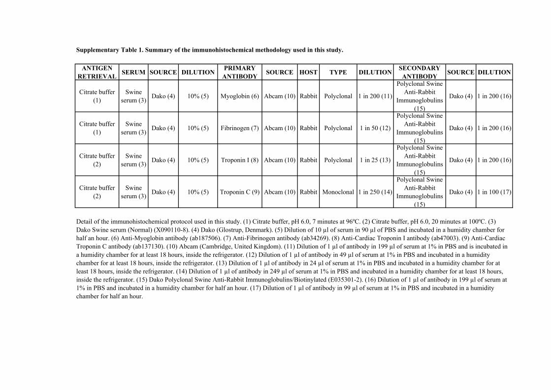
ANTIGEN RETRIEVAL SERUM SOURCE DILUTION PRIMARY
ANTIBODY SOURCE HOST TYPE DILUTION SECONDARY ANTIBODY SOURCE DILUTION
Citrate buffer (1)
Swine serum (3) Dako (4) 10% (5) Myoglobin (6) Abcam (10) Rabbit Polyclonal 1 in 200 (11)
Polyclonal Swine Anti-Rabbit
Immunoglobulins (15)
Dako (4) 1 in 200 (16)
Citrate buffer (1)
Swine serum (3) Dako (4) 10% (5) Fibrinogen (7) Abcam (10) Rabbit Polyclonal 1 in 50 (12)
Polyclonal Swine Anti-Rabbit
Immunoglobulins (15)
Dako (4) 1 in 200 (16)
Citrate buffer (2)
Swine serum (3) Dako (4) 10% (5) Troponin I (8) Abcam (10) Rabbit Polyclonal 1 in 25 (13)
Polyclonal Swine Anti-Rabbit
Immunoglobulins (15)
Dako (4) 1 in 200 (16)
Citrate buffer (2)
Swine serum (3) Dako (4) 10% (5) Troponin C (9) Abcam (10) Rabbit Monoclonal 1 in 250 (14)
Polyclonal Swine Anti-Rabbit
Immunoglobulins (15)
Dako (4) 1 in 100 (17)
Supplementary Table 1. Summary of the immunohistochemical methodology used in this study.
Detail of the immunohistochemical protocol used in this study. (1) Citrate buffer, pH 6.0, 7 minutes at 96ºC. (2) Citrate buffer, pH 6.0, 20 minutes at 100ºC. (3) Dako Swine serum (Normal) (X090110-8). (4) Dako (Glostrup, Denmark). (5) Dilution of 10 µl of serum in 90 µl of PBS and incubated in a humidity chamber for half an hour. (6) Anti-Myoglobin antibody (ab187506). (7) Anti-Fibrinogen antibody (ab34269). (8) Anti-Cardiac Troponin I antibody (ab47003). (9) Anti-Cardiac Troponin C antibody (ab137130). (10) Abcam (Cambridge, United Kingdom). (11) Dilution of 1 µl of antibody in 199 µl of serum at 1% in PBS and is incubated in a humidity chamber for at least 18 hours, inside the refrigerator. (12) Dilution of 1 µl of antibody in 49 µl of serum at 1% in PBS and incubated in a humidity chamber for at least 18 hours, inside the refrigerator. (13) Dilution of 1 µl of antibody in 24 µl of serum at 1% in PBS and incubated in a humidity chamber for at least 18 hours, inside the refrigerator. (14) Dilution of 1 µl of antibody in 249 µl of serum at 1% in PBS and incubated in a humidity chamber for at least 18 hours, inside the refrigerator. (15) Dako Polyclonal Swine Anti-Rabbit Immunoglobulins/Biotinylated (E035301-2). (16) Dilution of 1 µl of antibody in 199 µl of serum at 1% in PBS and incubated in a humidity chamber for half an hour. (17) Dilution of 1 µl of antibody in 99 µl of serum at 1% in PBS and incubated in a humidity chamber for half an hour.

6.4 APPLICATION OF THE KNOWLEDGE
OBTAINED IN THE DIAGNOSIS OF SPECIFIC
CLINICAL CASES


RESULTS & DISCUSSION: PUBLICATIONS APPLICATION OF THE KNOWLEDGE OBTAINED IN THE DIAGNOSIS OF SPECIFIC CLINICAL CASES
[Capture myopathy and stress cardiomyopathy in a live-stranded Risso’s dolphin (Grampus griseus) in rehabilitation] __________________________________________________________________________________________________________________________________________________________________________
187
As mentioned in the previous point the third specific objective of this study consisted in
the presentation of two specific case studies of SCMP in cetaceans.
In this second case study we studied the biochemical analysis of consecutive blood
samples in vivo, followed by the gross, histopathological, histochemical and
immunohistochemical features caused by the live-stranding and consequent rehabilitation
attempt, for a certain period of time, in a juvenile male Risso’s dolphin (Grampus griseus).
The results were published in the article (as followed) titled Capture myopathy and
stress cardiomyopathy in a live-stranded Risso’s dolphin (Grampus griseus) in rehabilitation,
on January 2020 in the Animals journal, Volume 10, Issue 2 (doi: 10.3390/ani10020220), having
this journal an impact factor of 1.832 in 2018 and therefore being a Q1 in the veterinary
category.

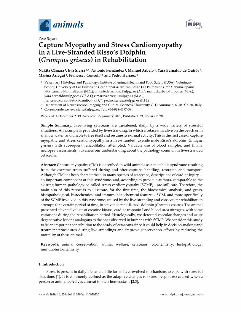
Animals 2020, 10, 220; doi:10.3390/ani10020220 www.mdpi.com/journal/animals
Case Report
Capture Myopathy and Stress Cardiomyopathy in a Live-Stranded Risso’s Dolphin (Grampus griseus) in Rehabilitation Nakita Câmara 1, Eva Sierra 1,*, Antonio Fernández 1, Manuel Arbelo 1, Yara Bernaldo de Quirós 1, Marina Arregui 1, Francesco Consoli 1,2 and Pedro Herráez 1
1 Veterinary Histology and Pathology, Institute of Animal Health and Food Safety (IUSA), Veterinary School, University of Las Palmas de Gran Canaria, Arucas, 35416 Las Palmas de Gran Canaria, Spain; [email protected] (N.C.); [email protected] (A.F.); [email protected] (M.A.); [email protected] (Y.B.d.Q.); [email protected] (M.A.); [email protected] (F.C.); [email protected] (P.H.)
2 Department of Neuroscience, Imaging and Clinical Sciences, University G. D’Annunzio, 66100 Chieti, Italy * Correspondence: [email protected]; Tel.: +34-928-4597-08
Received: 4 December 2019; Accepted: 27 January 2020; Published: 29 January 2020
Simple Summary: Free-living cetaceans are threatened, daily, by a wide variety of stressful situations. An example is provided by live-stranding, in which a cetacean is alive on the beach or in shallow water, and unable to free itself and resume its normal activity. This is the first case of capture myopathy and stress cardiomyopathy in a live-stranded juvenile male Risso’s dolphin (Grampus griseus) with subsequent rehabilitation attempted. Valuable use of blood samples, and finally necropsy assessments, advances our understanding about the pathology common in live-stranded cetaceans.
Abstract: Capture myopathy (CM) is described in wild animals as a metabolic syndrome resulting from the extreme stress suffered during and after capture, handling, restraint, and transport. Although CM has been characterized in many species of cetaceans, descriptions of cardiac injury—an important component of this syndrome, and, according to previous authors, comparable to the existing human pathology so-called stress cardiomyopathy (SCMP)—are still rare. Therefore, the main aim of this report is to illustrate, for the first time, the biochemical analysis, and gross, histopathological, histochemical and immunohistochemical features of CM, and more specifically of the SCMP involved in this syndrome, caused by the live-stranding and consequent rehabilitation attempt, for a certain period of time, in a juvenile male Risso’s dolphin (Grampus griseus). The animal presented elevated values of creatine kinase, cardiac troponin I and blood urea nitrogen, with some variations during the rehabilitation period. Histologically, we detected vascular changes and acute degenerative lesions analogous to the ones observed in humans with SCMP. We consider this study to be an important contribution to the study of cetaceans since it could help in decision-making and treatment procedures during live-strandings and improve conservation efforts by reducing the mortality of these animals.
Keywords: animal conservation; animal welfare; cetaceans; biochemistry; histopathology; immunohistochemistry
1. Introduction
Stress is present in daily life, and all life forms have evolved mechanisms to cope with stressful situations [1]. It is commonly defined as the adaptive changes (or stress responses) caused when a person or animal perceives a threat to their homeostasis [2,3].
Animals 2020, 10, 220 2 of 9
In human pathology, despite still under-recognized and often misdiagnosed, an unique reversible cardiac syndrome, triggered by a stressful event, has globally been reported and described as stress cardiomyopathy (SCMP) [4,5].
The clinical presentation is similar to the acute myocardial infarction. Diagnostic criteria for SCMP has been established by the Heart Failure Association (electrocardiography, echocardiography, cardiac catheterizations, and biochemical analysis). Regarding the biomarkers, cardiac troponin I (cTnI) and/or creatine kinase (CK) have small, rapid increases to above normal levels [4–7].
Histological findings are consistent with ischemia-reperfusion injury, such as vascular changes (i.e., congestion, hemorrhages, interstitial edema) and acute necrotic degenerative lesions (i.e., contraction band necrosis, wavy fibers, hypereosinophilia and cytoplasmic vacuolization) [4,5,8].
Cetaceans are exposed daily to a wide variety of stressful situations (i.e., live-stranding, ship collisions, bycatch) that influence their well-being. Efforts have been made to reduce the impact of these situations, but whales and dolphins continue to be threatened [9–15]. Live-stranding is an unnatural and distressful situation, where the cetacean is alive on the beach or in shallow water, being unable to resume normal activity [16–18]. Independently of the animal’s previous health, this implies an anomalous and extreme situation for an organism that is not adapted to terrestrial environmental conditions. Therefore, it is considered life-threatening as it may cause death or seriously aggravate a previous disease [9–12,14,16,18,19].
The acute and stressful deaths of live-stranded cetaceans might be attributed to the “stress response syndrome” or “alarm reaction” which are thought to be comparable to those described in capture myopathy (CM), in which the cardiac damage, due to the extreme stress, seems to have an important role. According to some authors, cetaceans are especially predisposed to develop SCMP, similarly to the SCMP in humans. It is the most devastating form of acute stress described in several animals that may occur during and after the capture, handling, restraint, and transport [9,10,14]. Although the pathological findings vary amongst individuals, the biochemical changes are consistent with elevated serum muscle enzyme activities, specifically CK, and elevated blood urea nitrogen (BUN). There are no biochemical studies on Odontoceti in which specific cardiac markers have been analyzed. Histopathological changes consisting of ischemia-reperfusion injuries may be observed. These changes result in local-to-generalized vasospasms and vasodilation (as seen in human SCMP), as well as in direct traumatic injury to muscles (rhabdomyolysis), with acute renal failure associated with myoglobinuric nephrosis secondary to muscle damage [9–12,15,20,21].
The aim of the present report is to illustrate, for the first time, the biochemical analysis, gross, histopathological, histochemical and immunohistochemical features of the CM, and more specifically of the SCMP involved in CM, caused by the live-stranding and consequent rehabilitation attempt in a juvenile male Risso’s dolphin (Grampus griseus).
2. Materials and Methods
This study is of a juvenile male Risso’s dolphin (Grampus griseus) which was stranded alive on the coast of Gran Canaria (Canary Islands, Spain), on April 26, 2019 (notification at 8:30 a.m.). After receiving specialized first aid (i.e., help maintaining correct body posture and wetness of skin, evaluating breathing and heart rate) at the location, the animal was transferred to the Wildlife Recovery Center (Gran Canaria), where it was monitored 24 h a day. From day 0 (26 April 2019) to day 5 (1 May 12019), several diagnostic tests (i.e., biochemical and hematological analysis, gastric probing and ultrasound evaluation) were performed to establish the most appropriate treatment. Around 10 pm on 29 April 2019 (day 3), the animal began to have muscle spasms. The next day, a lateral body curvature began to develop at the caudal peduncle, with increasing muscle tremors. Due to the worsening prognosis the animal was euthanized on 1 May 2019 (day 5). The therapeutic treatment and the euthanasia protocol applied in this animal are described in the Supplementary Materials Table S1 and Table S2, respectively.

Animals 2020, 10, 220 3 of 9
2.3. Evidence of Ethical Approval
Permission for the management of stranded cetaceans was issued by the environmental department of the Canary Islands’ Government and the Spanish Ministry of Environment.
2.2. Biochemical Analysis
Samples of whole blood were collected from the tail flukes, into a gel tube (without anticoagulant), were allowed to coagulate, and then centrifuged twice, each time at 3500 rpm for 5 min to obtain the serum for analysis. The first blood sample was collected 4 h after the warning (approximately midday), the remaining samples every 24 h during rehabilitation, and also post-mortem (1:30 h after euthanasia). The biomarkers for acute skeletal and heart muscle damage (CK and cTnI) and the kidney function (BUN and creatinine) were analyzed.
2.3. Gross, Histological, Histochemical and Immuznohistochemical Analysis
Prior to the necropsy, and after the euthanasia, an entire body scan was carried out, using helical computer tomography (CT) (Toshiba Astellion 16) setting the protocol on 120kV, 300 mA and a reconstruction slice thickness/slice interval of 1/0,80 mm. All the DICOM files obtained were processed with the Horos software. Thereafter, a standard protocol full necropsy was performed, where representative tissue samples were collected and fixed in 10% formalin for approximately 48 hours and processed using standard protocol [16,22]. Due to the potential presence of CM and SCMP, the skeletal (longissimus dorsi and rectus abdominis) and heart muscles (both atria and ventricles), atrioventricular valves (bicuspid or mitral and tricuspid), semilunar valves (sigmoid aortic and sigmoid pulmonary with the corresponding arteries), and kidneys were fully studied. Tissue sections (4- and 5-µm-thick) were used for hematoxylin and eosin (HE) and Masson’s trichrome technique, respectively, while 3-µm-thick layers were immunolabeled with anti-myoglobin, anti-fibrinogen, anti-cardiac troponin I, and anti-cardiac troponin C primary antibodies. The followed protocol was presented in a previous publication, and it can be accessed in Supplementary Materials Table S3 [15].
3. Results
3.1. Biochemical Results
The different measurements obtained during the rehabilitation time and after euthanasia, are presented in Figure 1 and in the Supplementary Materials Table S4.
(a)
(b)
0
200
400
600
800
1000
1200
DAY 0 DAY 1 DAY 2 DAY 3 DAY 4 DAY 5 DAY 5(1:30H Post-
mortem)
Creatine Kinase (U/L)
0
0,02
0,04
0,06
0,08
0,1
0,12
0,14
0,16
0,18
DAY 0 DAY 1 DAY 2 DAY 3 DAY 4 DAY 5 DAY 5(1:30H Post-
mortem)
Cardiac Troponin I (µg/L)

Animals 2020, 10, 220 4 of 9
(c)
(d)
Figure 1. Biochemical analysis during rehabilitation and after euthanasia: (a) Creatine kinase (CK); (b) Cardiac troponin I (cTnI); (c) Blood urea nitrogen (BUN); (d) Creatinine.
It was possible to observe that every single result obtained for CK was high. It is important to remark the new peak of the value on day 3. This animal presented one value within the normal range on day 0 for cTnI, being the rest of the measurements elevated. As noticed in the CK, the cTnI also presented a peak on day 4 and 5–1:30 h after euthanasia. The animal demonstrated higher BUN values and lower creatinine level than normal values.
3.2. Gross Results
The 105 kg juvenile male, measured 205.5 cm, had a very poor body condition, and displayed several lacerations, distributed in a multifocal manner, on the rostrum, dorsal and pectoral fins, tail flukes and ventral part of the body, attributed to live-stranding. External and internal signs of a spinal lateral curvature (S-shape) were identified at the level of the caudal peduncle (Figure 2).
(a)
(b)
0
50
100
150
200
250
DAY 0 DAY 1 DAY 2 DAY 3 DAY 4 DAY 5 DAY 5(1:30H Post-
mortem)
Blood Urea Nitrogen (mg/dL)
0
0,1
0,2
0,3
0,4
0,5
0,6
0,7
0,8
0,9
1
DAY 0 DAY 1 DAY 2 DAY 3 DAY 4 DAY 5 DAY 5(1:30H Post-
mortem)
Creatinine (mg/dL)

Animals 2020, 10, 220 5 of 9
(c) (d) Figure 2. Animal caudal peduncle scoliosis: (a) Caudal view on the necropsy table; (b) Dorsal plane of the entire body, with computer tomography (CT); (c) Dorsal view of the entire body, using CT, bones in white, muscle in red, and skin in blue; and (d) Dorsal view of the skeleton, with the CT.
3.3. Histopathological Results
On histopathologic and histochemical examinations, the skeletal and heart muscles presented injuries consistent with acute degenerative lesions. The longissimus dorsi and the rectus abdominis demonstrated a multifocal, severe contraction band necrosis and myonecrosis consisting of segmentary fibrilar degeneration with hyalinized eosinophilic sarcoplasm and hypercontraction (Figure 3a). The atria and ventricles displayed a multifocal, moderate degree of contraction band necrosis (Figure 3b), wavy fibers, hypereosinophilia, and cytoplasmic vacuolization (Figure 3b, inset) with pyknotic nucleus. All of the above changes were more pronounced in the subepicardial and subendocardial regions. The different heart sections also exhibited a multifocal, mild degree of infiltration of mononuclear cells in the areas with fibrillary ruptures.
The kidneys showed a multifocal, moderate congestion in the cortex and renal medulla. An amorphous and acidophilic substance was found inside the Bowman's capsule and in the medullary renal tubules.
(a) (b)
Figure 3. Histochemical techniques: (a) Contraction band necrosis (arrow heads), segmentary fibrilar degeneration (thick arrows) with hyalinized eosinophilic sarcoplasm and hypercontraction (thin arrows); 20x, HE. Inset: Detail of the myocytes with contraction band necrosis (arrow heads) and segmentary fibrilar degeneration (thick arrows) with hyalinized eosinophilic sarcoplasm and hypercontraction (thin arrows); 20×, Masson’s trichrome technique; (b) Contraction band necrosis (arrows) in the cardiomyocytes; 40×. Inset: Detail of the intracytoplasmic vacuoles (arrows) of the cardiomyocytes; 60×, HE.
3.4. Immunohistochemical Results
The degenerated/necrotic muscular and heart cells showed homogenous, intrafibrillar depletion of cTnI (Figure 4a), cardiac troponin C (cTnC) (Figure 4b), and myoglobin (Figure 4c), and exhibited

Animals 2020, 10, 220 6 of 9
immunolabelling for fibrinogen (Figure 4d). The kidneys did not exhibit any displacement/presence of myoglobin.
(a) (b)
(c) (d) Figure 4. Immunohistochemical techniques: (a) Degenerated cardiomyocytes (thick arrows) present depletion of cardiac troponin I (cTnI), compared to the normal cardiomyocytes (arrow heads). 10×. Inset: Detail of the intrafibrillar depletion (thick arrows) of cTnI in the injured cardiomyocytes, in comparison with normal cardiomyocytes (thin arrows); 60×, anti-troponin I; (b) Necrotic cardiomyocytes (thick arrows), close to the blood vessels (*) demonstrate depletion of cardiac troponin C (cTnC), compared to the normal cardiomyocytes (arrow heads); 20×. Inset: Detail of intrafibrillar depletion of cTnC with intense immunolabelling in the contraction band necrosis (thick arrows); 60×, anti-troponin C; (c) Degenerated cardiomyocytes (thick arrows), show intrafibrillar depletion of myoglobin, compared to the normal cardiomyocytes (thin arrows); 10×. Inset: Detail of the intrafibrillar depletion (thick arrows) of myoglobin in the injured cardiomyocytes, in comparison with normal cardiomyocytes (thin arrows); 20×, anti-myoglobin; (d) Expression of fibrinogen, in the necrotic myocytes, mainly in area next to (thin arrows) the contraction band necrosis (thick arrows). Presence of fibrinogen in the blood vessels (arrow head); 20×. Inset: Detail of the immunolabelling of fibrinogen, in the injured myocytes, in the zone near to (thin arrows) the contraction band necrosis (thick arrows). Fibrinogen inside the blood vessels (arrow heads); 60× anti-fibrinogen.
4. Discussion
4.1. Discussion of the Biochemical Results
Clinico-pathological evaluation was challenging, not only because normal and/or pathological biochemical values are rarely reported for cetaceans, and in this case, particularly regarding Risso’s dolphins but also due to the non-existing normal range for specific cardiac markers, such as cTnI, in the cetacean scientific database. Our biochemical analysis was compared with different mammals in published papers [23–26].

Animals 2020, 10, 220 7 of 9
The most relevant biochemical changes described in CM in birds (i.e., wild turkeys, mallards), terrestrial (i.e., hoofed mammals), and marine wild mammals (i.e., sea otters, seals), are the CK and BUN elevation [9–12,19,21,27]. For SCMP in humans, the more common laboratories abnormalities are consistent with a small, rapid rise to above the normal levels of cTnI and/or CK [6,7].
After an injury to the muscle (both skeletal and cardiac), the serum level of CK begins to rise in 4–9 h, peaks at 24 h, and returns to baseline 48–72 h, unless a new injury or permanent damage occurs [28]. Every single result obtained is higher than the normal range published for this species (48 to 154 U/L) which is a useful indicator of both skeletal and cardiac muscle damage suffered by this animal during and after stranding, and also during the rehabilitation period [18,19,21,26,28]. Additionally, the highest value of this marker on day 3 indicates the occurrence of a new injury.
Although CK is considered a sensitive marker of myocardial damage, it is present in the skeletal muscles in high concentrations. Due to its poor specificity to detect specific heart damage, troponins have been adopted as the new gold standard for necrosis, more specifically cTnI, since it is detectable in very low amounts (0.01 µg/L) in the blood of healthy individuals [28,29]. Thus, significant elevations (≥ 0.1 µg/L) most likely reflect myocardial necrosis [28]. It is considered as cardiospecific, therefore it is used to detect heart pathologies (i.e., SCMP) [6,28,29]. Most of the injured cardiac myocytes release troponin, 3–9 h after ischemic damage, reaching its peak between 12–48 h and remains raised for up to 4–7 days, unless a new injury or permanent damage occurs [28,29].
At present, there are no published studies on Odontoceti that analyze specific cardiac markers to detect the resulting damage to the heart in these animals. Compared to the recommended values in humans (≤0.1 µg/L) and in dogs (≤0.03–0.07 µg/L), this animal presented one value within the normal range on day 0 (0.035 µg/L), being the rest of the measurements elevated [23–25]. All the results concur with the kinetics except on day 4 and 5 - 1:30 hours after euthanasia, which indicates the occurrence of a new stress event, leading to a new damage. In this case, pre-renal azotemia was reasonably supported by higher BUN values than the existing reference values for this species (i.e., 36 to 69 mg/dL), associating this clinical finding with various possible causes, such as compartment syndrome, heart failure, and dehydration [19,26,30]. All measurements of creatinine were below the normal range for this species (1.4 to 2.8 mg/dL) probably related to the poor body condition [26,30].
4.2. Discussion of the Gross, Histopathological and Immunohistochemical Results
The chronology of events described in this case report is concurring with previous cases of live-stranded cetaceans, being the scoliosis a common problem in these animals, occurring during the rehabilitation process secondary to lack of swimming in a compromised animal [18,31].
The histopathological findings present in animals who suffered from CM consist of acute to subacute degeneration (rhabdomyolysis) and acute renal failure associated with myoglobinuric nephrosis secondary to muscle damage. The alterations related with the SCMP present in CM are associated with morphological alterations namely acute degenerative lesions (i.e., contraction band necrosis, wavy fibers, hypereosinophilia, and cytoplasm vacuolization), vascular changes (i.e., congestion, interstitial edema, and hemorrhages) and infiltration by inflammatory cells, based on the histological analysis of myocardial tissue [4,8–11,15,19,21,27]. Baring this in mind, the skeletal muscle and kidney samples of this animal presented lesions which illustrated rhabdomyolysis and acute renal failure. In relation to the heart lesions it is possible to conclude that the animal was entering the subacute stage (1 to 3 days), described in the sequence of changes in an acute ischemic injury in the heart, since it is when lymphocytes start to appear [5]. Immediately after a vital trauma, skeletal and cardiac proteins begin to leak as a result of early cell membrane rupture, causing a quick decline in myoglobin, cTnI, and cTnC contents, along with the deposition of plasma proteins (fibrinogen) [9,10,15,32,33]. These changes were observed in our animal confirming the ante mortem lesions.
5. Conclusions
We consider this article to be an important contribution to clinical biochemistry in cetaceans. Valuable use of blood samples and finally necropsy assessment advances our understanding about the pathology in live-stranded cetaceans.
Animals 2020, 10, 220 8 of 9
Supplementary Materials: The following are available online at www.mdpi.com/xxx/s1, Table S1: Summary of the therapeutic treatment. Table S2: Summary of the euthanasia protocol. Table S3: Summary of the immunohistochemical methodology. Table S4: Comparison between the biochemical analysis during rehabilitation (ante-mortem) and after euthanasia (post-mortem) with the normal baseline values.
Author Contributions: Methodology and formal analysis, N.C., E.S., A.F., M.Arbelo, Y.B.deQ., M.Arregui, F.C. and P.H.; writing—original draft preparation, N.C.; supervision and writing—review and editing, E.S. and P.H.; funding acquisition, A.F. and P.H. All authors have read and agreed to the published version of the manuscript.
Funding: This study is part of a PhD thesis supported by the Ministry of Economy and Competitiveness (MINECO) through a predoctoral grant BES-2016-076907. Furthermore, part of this research work was supported through the Research Projects (MINECO, CGL2015-71498-P and CABILDO2018: CABILDO2018-04). Moreover, the Canary Islands Government funded and provided support to the stranding research network.
Acknowledgments: We want to Animal Lab, Cetacean Stranding Network and Centro de Recuperación de Fauna Silvestre de Tafira (Gran Canaria), and Fundación Loro Parque.
Conflicts of Interest: The authors declare no conflicts of interest.
References
1. Yousef, M.K. Animal stress and strain: Definition and measurements. Appl. Anim. Behav. Sci. 1988, 20, 119–126.
2. Breazile, J.E. The physiology of stress and its relationship to mechanisms of disease and therapeutics. Vet. Clin. North Am. Food Anim. Pract. 1988, 4, 441–480.
3. Moberg, G.P. Biological response to stress: Implications for animal welfare. In The biology of animal stress: basic principles and implications for animal welfare, 1st Ed.; Moberg, G.P., Mench, J.A., Eds.; CABI: Wallingford, Oxon, UK, 2000; pp. 1–22.
4. Prasad, A.; Lerman, A.; Rihal, C.S. Apical ballooning syndrome (tako-tsubo or stress cardiomyopathy): A mimic of acute myocardial infarction. Am. Heart J. 2008, 3, 408–417.
5. Buja, L.M.; Butany, J. Cardiovascular Pathology, 4th Ed.; Buja, L.M.; Butany, J., Eds; Elsevier Science: London, UK, 2015; pp. 245–249, 251, 477–478.
6. Lyon, A.R.; Bossone, E.; Schneider, B.; Sechtem, U.; Citro, R.; Underwood, S.R.; Sheppard, M.N.; Figtree, G.A.; Parodi, G.; Akashi, Y.J.; et al. Current state of knowledge on takotsubo syndrome: A position statement from the taskforce on takotsubo syndrome of the heart failure association of the european society of cardiology. Eur. J. Heart Fail. 2016, 18, 8–27.
7. Fineschi, V.; Michalodimitrakis, M.; D’Errico, S.; Neri, M.; Pomara, C.; Riezzo, I.; Turillazzi, E. Insight into stress-induced cardiomyopathy and sudden cardiac death due to stress: a forensic cardio-pathologist point of view. Forensic Sci. Int. 2010, 194, 1–8.
8. Miura, M.; Kawano, H.; Yoshida, T.; Yamagata, Y.; Nakata, T.; Koga, S.; Ikeda, S.; Kageyama, K.; Abe, K.; Maemura, K. The histological features of a myocardial biopsy specimen in a patient in the acute phase of reversible catecholamine-induced cardiomyopathy due to pheochromocytoma. Intern. Med. 2017, 56, 665–671.
9. Herráez, P.; Sierra, E.; Arbelo, M.; Jaber, J.R.; Espinosa De Los Monteros, A.; Fernández, A. Rhabdomyolysis and myoglobinuric nephrosis (capture myopathy) in a striped dolphin. J. Wildl. Dis. Wildl. Dis. Assoc. 2007, 43, 770–774.
10. Herráez, P.; Espinosa de los Monteros, A.; Fernández, A.; Edwards, J.F.; Sacchini, S.; Sierra, E. Capture myopathy in live-stranded cetaceans. Vet. J. 2013, 196, 181–188.
11. Díaz-Delgado, J.; Fernández, A.; Sierra, E.; Sacchini, S.; Andrada, M.; Vela, A.I.; Quesada-Canales, Ó.; Paz, Y.; Zucca, D.; Groch, K.; et al. Pathologic findings and causes of death of stranded cetaceans in the Canary Islands (2006–2012). PLoS ONE 2018, 13, 1–33.
12. Arbelo, M.; de los Monteros, A.; Herráez, P.; Andrada Borzollino, M.; Sierra, E.; Rodríguez Guisado, F.; Jepson, P.; Fernández, A. Pathology and causes of death of stranded cetaceans in the canary islands (1999–2005). Dis. Aquat. Organ. 2013, 103, 87–99.
13. Cowan, D.F.; Curry, B.E. Histopathological assessment of dolphins necropsies onboard vesssels in the eastern tropical pacific tuna fishery. 2020. Available online: https://www.google.com.hk/url?sa=t&rct=j&q=&esrc=s&source=web&cd=2&ved=2ahUKEwj4v7DHoqXnAhUDBKYKHUWUDeoQFjABegQIARAB&url=https%3A%2F%2Fswfsc.noaa.gov%2FuploadedFiles%2FDivisions%2FPRD%2FPrograms%2FETP_Cetacean_Assessment%2FLJ_02_24C.pdf&usg=AOvVaw1ezGp

Animals 2020, 10, 220 9 of 9
_XRyF-XLVg4FGaZjG (accessed on 25 January 2020). 14. Cowan, D.F.; Curry, B.E. Histopathology of the Alarm Reaction in Small Odontocetes. J. Comp. Pathol. 2008,
139, 24–33. 15. Câmara, N.; Sierra, E.; Fernández-Maldonado, C.; Espinosa de los Monteros, A.; Arbelo, M.; Fernández, A.;
Herráez, P. Stress cardiomyopathy in stranded cetaceans: A histological, histochemical and immunohistochemical study. Vet. Rec. 2019, 185, 694–704.
16. Geraci, J.R.; Lounsbury, V.J. Marine mammals ashore: a field guide for strandings, 2nd Ed.; Yates, N.S., Ed.; National Aquarium: Baltimore, MD, USA, 2005; pp. 39–48, 75–128, 167–252.
17. Report to Assistant Administrator for Fisheries: Program review of the marine mammal stranding networks. Available online: https://repository.library.noaa.gov/view/noaa/14920 (accessed on 1 June 2019).
18. Gulland, F.M.D.; Dierauf, L.A.; Whitman, K.L. CRC handbook of marine mammal medicine, 2nd Ed.; Gulland, F.M.D.; Dierauf, L.A., Eds. CRC Press: Boca Raton, FL, USA, 2018; 45–68, 253–270, 383–436, 449–470, 689–740.
19. Bonsembiante, F.; Centelleghe, C.; Rossi, G.; Giglio, S.; Madeo, E.; Gelain, M.E.; Mazzariol, S. Clinico-pathological findings in a striped dolphin (stenella coeruleoalba) affected by rhabdomyolysis and myoglobinuric nephrosis (capture myopathy). J. Vet. Med. Sci. 2017, 79, 1013–1018.
20. Turnbull, B.S.; Cowan, D.F. Myocardial contraction band necrosis in stranded cetaceans. J. Comp. Pathol. 1998, 118, 317–327.
21. Seguel, M.; Paredes, E.; Pavés, H.; Gottdenker, N.L. Capture-induced stress cardiomyopathy in south american fur seal pups (arctophoca australis gracilis). Mar. Mammal Sci. 2014, 30, 1149–1157.
22. Kuiken, T.; Hartmann, M.G. Dissection techniques and tissue sampling. In Workshop on cetacean pathology, Proceedings of the first European Cetacean Society, Leiden, The Netherlands, 13-14 September 1991.
23. Normal laboratory values: Blood, plasma, and serum. Available online: https://www.msdmanuals.com/professional/resources/normal-laboratory-values/blood-tests-normal-values#v8508814 (accessed on 1 July 2019).
24. Wray, J. Canine Internal Medicine: What’s Your Diagnosis?, 1st Ed.; Wiley Blackwell: Chichester, West Sussex, UK, 2017; pp. 1–60.
25. Sleeper, M.M.; Clifford, C.A.; Laster, L.L. Cardiac troponin i in the normal dog and cat. J. Vet. Intern. Med. 2001, 15, 501–503.
26. Nachtigall, P.E.; Pawloski, J.; Schroeder, J.P.; Sinclair, S. Successful maintenance and research with a formerly stranded risso’s dolphin (grampus griseus). Aquat. Mamm. 1990, 16, 8–13.
27. Spraker, T. Stress and capture myopathy in artiodactylids. In Zoo and wild animal medicine: current therapy, 3rd Ed.; Fowler, M.E., Ed.; W. B. Saunders Company: Philadelphia, USA, 1993; pp. 481–489.
28. Lewandrowski, K.; Chen, A.; Januzzi, J. Cardiac markers for myocardial infarction: A brief review. Am. J. Clin. Pathol. Pathol. Patterns Rev. 2002, 118, 93–99.
29. Khan, M.S. Diagnostic efficacy of cardiac troponin in post-mortem examination of acute myocardial infarction. Int. J. Eth. Trauma Vict. 2015, 1, 21–28.
30. Gough, A.; Murphy, K. Differential diagnosis in small animal medicine; 2nd Ed.;Gough, A., Murphy, K., Eds.; Wiley Blackwell: Chichester, West Sussex, UK, 2015; pp. 324, 338.
31. Wells, R.S.; Fauquier, D.A.; Gulland, F.M.D.; Townsend, F.I.; DiGiovanni Jr., R.A. Evaluating postintervention survival of free-ranging odontocete cetaceans. Mar. Mammal Sci. 2013, 29, 463–483.
32. Fishbein, M.C.; Wang, T.; Matijasevic, M.; Hong, L.; Apple, F.S. Myocardial tissue troponins t and i: an immunohistochemical study in experimental models of myocardial ischemia. Cardiovasc. Pathol. 2003, 12, 65–71.
33. Ortmann, C.; Pfeiffer, H.; Brinkmann, B. A comparative study on the immunohistochemical detection of early myocardial damage. Int. J. Legal Med. 2000, 113, 215–220.
© 2020 by the authors. Licensee MDPI, Basel, Switzerland. This article is an open access article distributed under the terms and conditions of the Creative Commons Attribution (CC BY) license (http://creativecommons.org/licenses/by/4.0/).


Animals 2019, 9, x 1 of 4
Table S1. Summary of the therapeutic treatment.
Medicine Active Principle Dosage
[1] Number of Dosages Via Days
Dexametasona Kern Pharma Dexamethasone 0.11 mg/kg 1 Intramuscular Day 0 (26/04/2019)
Ganadexil Enrofloxacino Enrofloxacin 5 mg/kg 1
(each day)
Intramuscular
Day 0 (26/04/2019)
Day 1 (27/04/2019)
Day 2 (28/04/2019)
Oral
(diluted at 50%)
Day 3 (29/04/2019)
Day 4 (30/04/2019)
Zipyran Plus Praziquantel 10 mg/kg 1 Oral Day 1 (27/04/2019)
Ketoisdin Ketoconazole 5 mg/kg 1 Oral Day 2 (28/04/2019)
Fluid therapy Freshwater 2 liters
2
Oral
Day 2 (28/04/2019)
1
(each day)
Day 3 (29/04/2019)
Day 4 (30/04/2019)
1. Gulland, F.M.D.; Dierauf, L.A.; Whitman, K.L. CRC handbook of marine mammal medicine; CRC Press, 2018; ISBN 1351384163.

Animals 2019, 9, x 2 of 4
Table S2. Summary of the euthanasia protocol.
Medicine Active
Principle Dosage
Number
of
Dosages
Via Days Commentaries
Tiobarbital Sodium
Thiopental
5
grams 1 Intravenous
Day 5
(01/05/2019)
The decision to euthanize the animal was taken among several veterinarians
who are experts in dealing with these types of animals, one of them being the
veterinarian responsible for the Wildlife Recovery Center (Gran Canaria) and
veterinarians of Loro Parque. The main reason, which lead to this decision,
consisted in the development of the body curvature which could not be
reversible seeing that there were no adequate facilities to maintain the
animal, more specifically, a pool which allow the animal to have a proper
swimming pattern and not having the access to the therapeutic treatment
recommend to reverse scoliosis in cetaceans in rehabilitation.

Table S3. Summary of the immunohistochemical methodology used in this study.
Antigen
Retrieval Serum Source Dilution
Primary
Antibody Source Host Type Dilution Secondary Antibody Source Dilution
Citrate
buffer1
Swine
serum3 Dako4 10%5 Myoglobin6 Abcam10 Rabbit Polyclonal 1 in 20011
Polyclonal Swine
Anti-Rabbit
Immunoglobulins15
Dako4 1 in 20016
Citrate
buffer1
Swine
serum3 Dako4 10%5 Fibrinogen7 Abcam10 Rabbit Polyclonal 1 in 5012
Polyclonal Swine
Anti-Rabbit
Immunoglobulins15
Dako4 1 in 20016
Citrate
buffer2
Swine
serum3 Dako4 10%5 Troponin I8 Abcam10 Rabbit Polyclonal 1 in 2513
Polyclonal Swine
Anti-Rabbit
Immunoglobulins15
Dako4 1 in 20016
Citrate
buffer2
Swine
serum3 Dako4 10%5 Troponin C9 Abcam10 Rabbit Monoclonal 1 in 25014
Polyclonal Swine
Anti-Rabbit
Immunoglobulins15
Dako4 1 in 10017
1 Citrate buffer, pH 6.0, 7 minutes at 96ºC. 2 Citrate buffer, pH 6.0, 20 minutes at 96ºC. 3 Dako Swine serum (Normal) (X090110-8). 4 Dako (Glostrup, Denmark). 5 Dilution
of 10 µl of serum in 90 µl of PBS. The serum is incubated in a humidity chamber for half an hour. 6 Anti-Myoglobin antibody (ab187506). 7 Anti-Fibrinogen antibody
(ab34269). 8 Anti-Cardiac Troponin I antibody (ab47003). 9 Anti-Cardiac Troponin C antibody (ab137130). 10 Abcam (Cambridge, United Kingdom). 11 Dilution of 1 µl of
primary antibody in 199 µl of serum at 1% in PBS (dilution of 1 µl of serum in 99 µl of PBS). The primary antibody is incubated in a humidity chamber for at least 18 hours,
inside the refrigerator. 12 Dilution of 1 µl of primary antibody in 49 µl of serum at 1% in PBS (dilution of 1 µl of serum in 99 µl of PBS). The primary antibody is incubated
in a humidity chamber for at least 18 hours, inside the refrigerator. 13 Dilution of 1 µl of primary antibody in 24 µl of serum at 1% in PBS (dilution of 1 µl of serum in 99 µl
of PBS). The primary antibody is incubated in a humidity chamber for at least 18 hours, inside the refrigerator. 14 Dilution of 1 µl of primary antibody in 249 µl of serum at
1% in PBS (dilution of 1 µl of serum in 99 µl of PBS). The primary antibody is incubated in a humidity chamber for at least 18 hours, inside the refrigerator. 15 Dako
Polyclonal Swine Anti-Rabbit Immunoglobulins/Biotinylated (E035301-2). 16 Dilution of 1 µl of secondary antibody in 199 µl of serum at 1% in PBS (dilution of 1 µl of serum
in 99 µl of PBS). The secondary antibody is incubated in a humidity chamber for half an hour. 17 Dilution of 1 µl of secondary antibody in 99 µl of serum at 1% in PBS
(dilution of 1 µl of serum in 99 µl of PBS). The secondary antibody is incubated in a humidity chamber for half an hour.

Animals 2019, 9, x 4 of 4
Table S4. Comparison between the biochemical analysis during rehabilitation (ante-mortem) and after euthanasia (post-mortem) with the normal baseline values.
Item Time
CK
(U/L)
cTnI
(µg/L)
BUN
(mg/dL) Creatinine (mg/dL)
Study
animal
Normal
value for
this
species
Study
animal
Normal
value for
this
species
Study
animal
Normal
value for
this
species
Study
animal
Normal
value for
this
species
Ante-mortem
(During
rehabilitation
period)
Day 0 (26/04/2019) 837.8
48 to 154
[1]
0.035
Non
existing
128
36 to 69
[1]
0.7
1.4 to 2.8
[1]
Day 1 (27/04/2019) 885.1 0.151 230 0.9
Day 2 (28/04/2019) 334.1 0.133 196 0.8
Day 3 (29/04/2019) 959.0 0.120 195 0.6
Day 4 (30/04/2019) 455.7 0.164 173 0.4
Day 5 (01/05/2019) 715.3 0.162 170 0.4
Post-mortem
(After euthanasia)
Day 5 (01/05/2019)
(1:30 h after euthanasia) 843.6 0.168 171 0.4
1. Nachtigall, P.E.; Pawloski, J.; Schroeder, J.P.; Sinclair, S. Successful maintenance and research with a formerly stranded Risso’s dolphin (Grampus griseus). Aquat. Mamm.
1990, 16, 8–13.

CONCLUSIONS


CONCLUSIONS
__________________________________________________________________________________________________________________________________________________________________________
205
FIRST: Cetaceans that die during and/or after stressful events such as active strandings, ship
collisions and accidental interactions with fisheries, develop vascular changes and cardiac acute
degenerative-necrotic injuries comparable to lesions observed in humans with stress
cardiomyopathy, thus demonstrating that cetaceans are susceptible to this pathology.
SECOND: The normal reference range for serum cardiac troponin I in adult bottlenose dolphins
born and maintained under human care is 0 to 0.0256 µg/L. This is the first reference in the
scientific literature for blood values for cardiac troponin I in cetaceans.
THIRD: Stranded cetaceans alive and subjected to interaction with humans develop an acute
stress cardiomyopathy characterized by a serum increase of cardiac troponin I, which correlates
with tissue depletion of cardiac troponins I and C, in cardiomyocytes with acute degenerative
changes, detected histologically.
FOURTH: The use of the clinical determination of serum cardiac troponin I and
immunohistochemical analysis of cardiac troponins I and C for the diagnosis and decision making
in stress cardiomyopathy in live-stranded cetaceans is validated.


REFERENCES


REFERENCES
__________________________________________________________________________________________________________________________________________________________________________
209
Abdulla, I., S. Kay, C. Mussap, G. I. C. Nelson, H. H. Rasmussen, P. S. Hansen, and M. R. Ward. 2006. “Apical Sparing in Tako-Tsubo Cardiomyopathy.” Internal Medicine Journal 36(7):414–18.
Adegboyega, Patrick A., Abida K. Haque, and Paul J. Boor. 1996. “Extensive Myocytolysis as a Marker of Sudden Cardiac Death.” Cardiovascular Pathology 5(6):315–21.
Agarwal, Surendra K., Aditya Kapoor, Shantanu Pande, Gauranga Majumdar, Satyajit Singh, Archana Sinha, Sudeep Kumar, Satyendra Tewari, Naveen Garg, and Pravin K. Goel. 2015. “Comparison of Release Kinetics of Different Cardiac Biomarkers in Patients Undergoing off Pump Coronary Artery Bypass Surgery and Valve Replacement Surgery for Rheumatic Heart Disease.” Journal of Cardiothoracic Surgery 10(1):A208.
Akashi, Yoshihiro J., Holger M. Nef, Helge Möllmann, and Takashi Ueyama. 2010. “Stress Cardiomyopathy.” Annual Review of Medicine 61(1):271–86.
Angelini, Paolo. 2008. “Transient Left Ventricular Apical Ballooning: A Unifying Pathophysiologic Theory at the Edge of Prinzmetal Angina.” Catheterization and Cardiovascular Interventions 71(3):342–52.
Arbelo, Manuel, Antonio de los Monteros, Pedro Herráez, Marisa Andrada Borzollino, Eva Sierra, F. Rodríguez Guisado, Paul Jepson, and Antonio Fernández. 2013. “Pathology and Causes of Death of Stranded Cetaceans in the Canary Islands (1999-2005).” Diseases of Aquatic Organisms 103:87–99.
ASCOBANS/ACCOBAMS. 2019. “European Best Practice on Cetacean Post-Mortem Investigation and Tissue Sampling.” OSFPREPRINTS.
St. Aubin, D. J. and J. R. Geraci. 1986. “ADRENOCORTICAL FUNCTION IN PINNIPED HYPONATREMIA.” Marine Mammal Science 2(4):243–50.
St. Aubin, D. J. and J. R. Geraci. 1988. “Capture and Handling Stress Suppresses Circulating Levels of Thyroxine (T4) and Triiodothyronine (T3) in Beluga Whales Delphinapterus Leucas.” Physiological Zoology 61(2):170–75.
Barnett, James, Alan Knight, and Mark Steven. n.d. BDMLR - Marine Mammal Medic Handbook. 2nd ed.
Bonsembiante, Federico, Cinzia Centelleghe, Gabriele Rossi, Stefania Giglio, Elena Madeo, Maria Elena Gelain, and Sandro Mazzariol. 2017. “Clinico-Pathological Findings in a Striped Dolphin (Stenella Coeruleoalba) Affected by Rhabdomyolysis and Myoglobinuric Nephrosis (Capture Myopathy).” Journal of Veterinary Medical Science 79:1013–18.
Breazile, James E. 1988. “The Physiology of Stress and Its Relationship to Mechanisms of Disease and Therapeutics.” Veterinary Clinics of North America: Food Animal Practice 4(3):441–80.
Buja, L. Maximilian. 2005. “Myocardial Ischemia and Reperfusion Injury.” Cardiovascular Pathology 14(4):170–75.
Buja, L M and J. Butany. 2015. Cardiovascular Pathology. 4th ed. edited by L.M; Buja and J. Butany. London, UK: Elsevier Science.

STRESS CARDIOMYOPATHY IN CETACEANS: HISTOLOGICAL, HISTOCHEMICAL, IMMUNOHISTOCHEMICAL AND BIOCHEMICAL STUDIES __________________________________________________________________________________________________________________________________________________________________________
210
Bybee, Kevin A., Abhiram Prasad, Greg W. Barsness, Amir Lerman, Allan S. Jaffe, Joseph G. Murphy, R. Scot. Wright, and Charanjit S. Rihal. 2004. “Clinical Characteristics and Thrombolysis In Myocardial Infarction Frame Counts in Women with Transient Left Ventricular Apical Ballooning Syndrome.” The American Journal of Cardiology 94(3):343–46.
Câmara, Nakita, Eva Sierra, Carolina Fernández-Maldonado, Antonio Espinosa de los Monteros, Manuel Arbelo, Antonio Fernández, and Pedro Herráez. 2019. “Stress Cardiomyopathy in Stranded Cetaceans: A Histological, Histochemical and Immunohistochemical Study.” Veterinary Record 185(22):694–704.
Camici, Paolo G. and Crea Filippo. 2015. “Microvascular Angina.” Circulation: Cardiovascular Imaging 8(4):e003252.
Carrillo, Manuel. 2007. “Scientific Symposium of the Western African Talks on Cetaceans and Their Habitats (WATCH).” in Cetaceans in the Macaronesia region (Eastern Central Atlantic Ocean) and threats faced in the Canary Islands.
Carson, Freida L. and Christa Hladik. Cappellano. 2009. Histotechnology: A Self Instructional Text Citation. [Chicago]: ASCP Press.
Cebelin, Marilyn S. and Charles S. Hirsch. 1980. “Human Stress Cardiomyopathy: Myocardial Lesions in Victims of Homicidal Assaults without Internal Injuries.” Human Pathology.
Coudrey, Laura. 1998. “The Troponins.” JAMA Internal Medicine 158(11):1173–80.
Cowan, D. F. and B. E. Curry. 2008. “Histopathology of the Alarm Reaction in Small Odontocetes.” Journal of Comparative Pathology 139(1):24–33.
Cowan, Daniel F. and Barbara E. Curry. 2002. “Histopathological Assessment of Dolphins Necropsies Onboard Vesssels in the Eastern Tropical Pacific Tuna Fishery.” (June):31.
Cowan, W. Maxwell, Donald H. Harter, and Eric R. Kandel. 2000. “The Emergence of Modern Neuroscience: Some Implications for Neurology and Psychiatry.” Annual Review of Neuroscience.
Cozzi, Bruno, Stefan Huggenberger, and Helmut Oelschläger. 2017. Anatomy of Dolphins.
Crea, Filippo, Paolo G. Camici, and Cathleen Noel Bairey Merz. 2013. “Coronary Microvascular Dysfunction: An Update.” European Heart Journal 35(17):1101–11.
Curry, Barbara E. 1999. “Stress in Mammals: The Potential Influence of Fishery-Induced Stress on Dolphins in the Eastern Tropical Pacific Ocean.” NOAA Technical Memorandum NMFS.
Desmet, W. J. R., B. F. M. Adriaenssens, and J. A. Y. Dens. 2003. “Apical Ballooning of the Left Ventricle: First Series in White Patients.” Heart 89(9):1027 LP – 1031.
Dhalla, N. S., I. M. Dixon, S. Suzuki, M. Kaneko, A. Kobayashi, and R. E. Beamish. 1992. “Changes in Adrenergic Receptors during the Development of Heart Failure.” Molecular and Cellular Biochemistry.
Díaz-Delgado, Josué. 2015. “Patología y Causas de La Muerte de Los Cetáceos Varados En Las Islas Canarias (2006-2012).”

REFERENCES
__________________________________________________________________________________________________________________________________________________________________________
211
Díaz-Delgado, Josué, Antonio Fernández, Eva Sierra, Simona Sacchini, Marisa Andrada, Ana Isabel Vela, Óscar Quesada-Canales, Yania Paz, Daniele Zucca, Kátia Groch, and Manuel Arbelo. 2018. “Pathologic Findings and Causes of Death of Stranded Cetaceans in the Canary Islands (2006-2012).” PLOS ONE 13(10):1–33.
Duignan, PÁdraig J. and Gareth W. Jones. 2005. Autopsy of Cetaceans Including Those Incidentally Caught in Commercial Fisheries, 2002/03. Department of Conservation Wellington,, New Zealand.
Eurell, Jo Ann and Brian L. Frappier. 2013. Dellmann’s Textbook of Veterinary Histology. John Wiley & Sons.
Fair, Patricia A. and Paul R. Becker. 2000. “Review of Stress in Marine Mammals.” Journal of Aquatic Ecosystem Stress and Recovery.
Fernández Rodríguez, Antonio (Universidad de Las Palmas de Gran Canaria), Maria José (Universidad de Las Palmas de Gran Canaria) Caballero Cansino, and José Raduan (Universidad de Las Palmas de Gran Canaria) Jaber Mohamad. 2005. Atlas de Histología de Peces y Cetáceos. 1st ed.
Fineschi, Vittorio, Manolis Michalodimitrakis, Stefano D’Errico, Margherita Neri, Cristoforo Pomara, Irene Riezzo, and Emanuela Turillazzi. 2010. “Insight into Stress-Induced Cardiomyopathy and Sudden Cardiac Death Due to Stress. A Forensic Cardio-Pathologist Point of View.” Forensic Science International 194:1–8.
Fishbein, Michael C. 2001. “Early Evolution from Ischemia to Myocardial Necrosis.” Cardiovascular Toxicology 1(2):83–86.
Fishbein, Michael C., Tiffany Wang, Maria Matijasevic, Longsheng Hong, and Fred S. Apple. 2003. “Myocardial Tissue Troponins T and I: An Immunohistochemical Study in Experimental Models of Myocardial Ischemia.” Cardiovascular Pathology 12(2):65–71.
Friedrichs, Kristen R., Kendal E. Harr, Kathy P. Freeman, Balazs Szladovits, Raquel M. Walton, Kirstin F. Barnhart, and Julia Blanco-Chavez. 2012. “ASVCP Reference Interval Guidelines: Determination of de Novo Reference Intervals in Veterinary Species and Other Related Topics.” Veterinary Clinical Pathology 41(4):441–53.
Fundación Mapfre Guanarteme. 2011. Piélagos.
Garcia del Moral, Raimundo. 1993. Laboratorio de anatomía patológica. Madrid: Interamericana-McGraw-Hill.
Geraci, Joseph R. and Valerie J. Lounsbury. 2005. Marine Mammals Ashore: A Field Guide for Strandings. National Aquarium in Baltimore.
Gianni, Monica, Francesco Dentali, Anna Maria Grandi, Glen Sumner, Rajesh Hiralal, and Eva Lonn. 2006. “Apical Ballooning Syndrome or Takotsubo Cardiomyopathy: A Systematic Review.” European Heart Journal 27(13):1523–29.
Godinho, A. 2010. “Estudio Morfológico e Inmonohistoquímico de Glóbulos Hialinos En Hígado de Cetáceos Varados.” Universidad de Las Palmas de Gran Canaria.

STRESS CARDIOMYOPATHY IN CETACEANS: HISTOLOGICAL, HISTOCHEMICAL, IMMUNOHISTOCHEMICAL AND BIOCHEMICAL STUDIES __________________________________________________________________________________________________________________________________________________________________________
212
Gray, I. and J. G. Young. 1956. “Biochemical Response to Trauma. III. Epinephrine and Norepinephrine Levels in Plasma of Rats Subjected to Tumbling Trauma.” The American Journal of Physiology 186(1):67–70.
Gulland, Frances M. D., Leslie A. Dierauf, and Karyl L. Whitman. 2018. CRC Handbook of Marine Mammal Medicine. CRC Press.
Haghi, Dariusch, Stephan Fluechter, Tim Suselbeck, Jens J. Kaden, Martin Borggrefe, and Theano Papavassiliu. 2007. “Cardiovascular Magnetic Resonance Findings in Typical versus Atypical Forms of the Acute Apical Ballooning Syndrome (Takotsubo Cardiomyopathy).” International Journal of Cardiology 120(2):205–11.
Hansen, Steen Holger and Kristian Rossen. 1999. “Evaluation of Cardiac Troponin I Immunoreaction in Autopsy Hearts: A Possible Marker of Early Myocardial Infarction.” Forensic Science International 99(3):189–96.
Hassan, Ayman K. M., Sandrin C. Bergheanu, Hosam Hasan-Ali, Su San Liem, Arnoud van der Laarse, Ron Wolterbeek, Douwe E. Atsma, Martin J. Schalij, and J. Wouter Jukema. 2009. “Usefulness of Peak Troponin-T to Predict Infarct Size and Long-Term Outcome in Patients With First Acute Myocardial Infarction After Primary Percutaneous Coronary Intervention.” American Journal of Cardiology 103(6):779–84.
Helen, Paur, Wright Peter T., Sikkel Markus B., Tranter Matthew H., Mansfield Catherine, O’Gara Peter, Stuckey Daniel J., Nikolaev Viacheslav O., Diakonov Ivan, Pannell Laura, Gong Haibin, Sun Hong, Peters Nicholas S., Petrou Mario, Zheng Zhaolun, Gorelik Julia, Lyon Alexander R., and Harding Sian E. 2012. “High Levels of Circulating Epinephrine Trigger Apical Cardiodepression in a Β2-Adrenergic Receptor/Gi–Dependent Manner.” Circulation 126(6):697–706.
Hernández, Manuel Antonio Arbelo. 2007. “Patología y Causas de La Muerte de Los Cetáceos Varados En Las Islas Canarias (1999-2005).”
Herráez, P., A. Espinosa de los Monteros, A. Fernández, J. F. Edwards, S. Sacchini, and E. Sierra. 2013. “Capture Myopathy in Live-Stranded Cetaceans.” Veterinary Journal 196(2):181–88.
Herráez, P., E. Sierra, M. Arbelo, J. R. Jaber, A. Espinosa De Los Monteros, and A. Fernández. 2007. “Rhabdomyolysis and Myoglobinuric Nephrosis (Capture Myopathy) in a Striped Dolphin.” Journal of Wildlife Diseases Wildlife Disease Association 43:770–74.
Huang, Ming He, Daniel S. Friend, Mary E. Sunday, Krishna Singh, Kathleen Haley, K. Frank Austen, Ralph A. Kelly, and Thomas W. Smith. 1996. “An Intrinsic Adrenergic System in Mammalian Heart.” Journal of Clinical Investigation.
J., Akashi Yoshihiro, Goldstein David S., Barbaro Giuseppe, and Ueyama Takashi. 2008. “Takotsubo Cardiomyopathy.” Circulation 118(25):2754–62.
Jaffe, Allan S., Bernard R. Chaitman, David A. Morrow, Jeroen J. Bax, Harvey D. White, Joseph S. Alpert, Kristian Thygesen, and E. S. C. Scientific Document Group. 2018. “Fourth Universal Definition of Myocardial Infarction (2018).” European Heart Journal 40(3):237–69.
Jaspreet, Sachdeva, Dai Wangde, and Kloner Robert A. 2019. “Functional and Histological Assessment of an Experimental Model of Takotsubo’s Cardiomyopathy.” Journal of the American Heart Association 3(3):e000921.

REFERENCES
__________________________________________________________________________________________________________________________________________________________________________
213
Jiang, J. P. and S. E. Downing. 1990. “Catecholamine Cardiomyopathy: Review and Analysis of Pathogenetic Mechanisms.” Yale Journal of Biology and Medicine 63:581–91.
Kamdje, Armel Herve Nwabo, Gaetan Nkem, and Haverie Ghislaine Ateba Mimfoumou. 2017. “Estimation of Reference Values of Biochemical Parameters Exploring the Renal Function in Adults in Ngaoundere, Cameroon TT -.” Ibbjorg 3(1):17–20.
Kawai, Sachio. 2012. “Pathology of Takotsubo (Ampulla) Cardiomyopathy.” Cardiomyopathies–from Basic Research to Clinical Management 709–26.
Khan, Mohammed Sarosh. 2015. “Diagnostic Efficacy of Cardiac Troponin in Post-Mortem Examination of Acute Myocardial Infarction.” Int J Eth Trauma Victimology 1(1):21–28.
Kuiken, Thijs and Manuel García Hartmann. 1991. “Dissection Techniques and Tissue Sampling, Leiden, the Netherlands, 13-14 September 1991.” in Proceedings of the First ECS Workshop on Cetacean Pathology. European Cetacean Society.
Kumar, Vinay, Abul K. Abbas, Jon C. Aster, and James A. Perkins. 2015. Robbins and Cotran Pathologic Basis of Disease. 9th ed. edited by Robbins and Cotran. Philadelphia: Elsevier/Saunders.
Kurisu, Satoshi, Ichiro Inoue, Takuji Kawagoe, Masaharu Ishihara, Yuji Shimatani, Suji Nakamura, Masashi Yoshida, Naoya Mitsuba, Takaki Hata, and Hikaru Sato. 2004. “Time Course of Electrocardiographic Changes in Patients With Tako-Tsubo Syndrome.” Circulation Journal 68(1):77–81.
De La Fuente Marquez, Jesus. 2011. “Estudio de Las Patologías y Causas de Muerte de Cetáceos Varados En El Litoral de La Provincia de Cádiz (2001-2004).” Las Palmas de Gran Canaria.
Lewandrowski, Kent, Ahchean Chen, and James Januzzi. 2002. “Cardiac Markers for Myocardial Infarction: A Brief Review.” American Journal of Clinical Pathology. Pathology Patterns Reviews. 118:93–99.
Lyon, Alexander R., Eduardo Bossone, Birke Schneider, Udo Sechtem, Rodolfo Citro, S. Richard Underwood, Mary N. Sheppard, Gemma A. Figtree, Guido Parodi, Yoshihiro J. Akashi, Frank Ruschitzka, Gerasimos Filippatos, Alexandre Mebazaa, and Elmir Omerovic. 2016. “Current State of Knowledge on Takotsubo Syndrome: A Position Statement from the Taskforce on Takotsubo Syndrome of the Heart Failure Association of the European Society of Cardiology.” European Journal of Heart Failure 18(1):8–27.
Lyon, Alexander R., Paul SC Rees, Sanjay Prasad, Philip A. Poole-Wilson, and Sian E. Harding. 2008. “Stress (Takotsubo) Cardiomyopathy—a Novel Pathophysiological Hypothesis to Explain Catecholamine-Induced Acute Myocardial Stunning.” Nature Clinical Practice Cardiovascular Medicine 5:22–29.
Maldonado, Carolina Fernández. 2015. “Patología y Causas de La Muerte de Los Cetáceos Varados En Andalucía (2011-2014).”
Maréchaux, Sylvestre, Paul Fornes, Stéphanie Petit, Catherine Poisson, Didier Thevenin, Thierry Le Tourneau, Philippe Asseman, Patrick Bruneval, and Pierre-Vladimir Ennezat. 2008. “Pathology of Inverted Takotsubo Cardiomyopathy.” Cardiovascular Pathology 17(4):241–43.

STRESS CARDIOMYOPATHY IN CETACEANS: HISTOLOGICAL, HISTOCHEMICAL, IMMUNOHISTOCHEMICAL AND BIOCHEMICAL STUDIES __________________________________________________________________________________________________________________________________________________________________________
214
Martínez-Díaz, F., M. Rodríguez-Morlensín, M. D. Pérez-Cárceles, J. Noguera, A. Luna, and E. Osuna. 2005. “Biochemical Analysis and Immunohistochemical Determination of Cardiac Troponin for the Postmortem Diagnosis of Myocardial Damage.” Histology and Histopathology 20:475–781.
Merchant, Emily E., Sara W. Johnson, Phu Nguyen, Christopher Kang, and William K. Mallon. 2008. “Takotsubo Cardiomyopathy: A Case Series and Review of the Literature.” The Western Journal of Emergency Medicine 9(2):104—111.
Mikaelian, Igor, Denise Coluccio, Kevin T. Morgan, Teona Johnson, Amber L. Ryan, Erik Rasmussen, Rosemary Nicklaus, Charu Kanwal, Holly Hilton, Karl Frank, Luke Fritzky, and Eric B. Wheeldon. 2008. “Temporal Gene Expression Profiling Indicates Early Up-Regulation of Interleukin-6 in Isoproterenol-Induced Myocardial Necrosis in Rat.” Toxicologic Pathology 36(2):256–64.
Mitchell, Andrew and François Marquis. 2017. “Can Takotsubo Cardiomyopathy Be Diagnosed by Autopsy? Report of a Presumed Case Presenting as Cardiac Rupture.” BMC Clinical Pathology 17:4.
Mitchell, James H., Timothy B. Hadden, James M. Wilson, Arup Achari, Raja Muthupillai, and Scott D. Flamm. 2007. “Clinical Features and Usefulness of Cardiac Magnetic Resonance Imaging in Assessing Myocardial Viability and Prognosis in Takotsubo Cardiomyopathy (Transient Left Ventricular Apical Ballooning Syndrome).” The American Journal of Cardiology 100(2):296–301.
Miura, Miyuki, Hiroaki Kawano, Takeo Yoshida, Yuki Yamagata, Tomoo Nakata, Seiji Koga, Satoshi Ikeda, Kan Kageyama, Kuniko Abe, and Koji Maemura. 2017. “The Histological Features of a Myocardial Biopsy Specimen in a Patient in the Acute Phase of Reversible Catecholamine-Induced Cardiomyopathy Due to Pheochromocytoma.” Internal Medicine 56(6):665–71.
Moberg, G. P. 2000. “Biological Response to Stress: Implications for Animal Welfare.” Pp. 1–22 in The biology of animal stress: basic principles and implications for animal welfare, edited by G. P; Moberg and J. A. Mench. Wallingford, Oxon, UK: CABI.
Moore, Michael J., Julie Van Der Hoop, Susan G. Barco, Alex M. Costidis, Frances M. Gulland, Paul D. Jepson, Kathleen T. Moore, Stephen Raverty, and William A. McLellan. 2013. “Criteria and Case Definitions for Serious Injury and Death of Pinnipeds and Cetaceans Caused by Anthropogenic Trauma.” Diseases of Aquatic Organisms.
Morrow, David A., Christopher P. Cannon, Robert L. Jesse, Newby L. Kristin, Ravkilde Jan, Storrow Alan B., Wu Alan H.B., and Christenson Robert H. 2007. “National Academy of Clinical Biochemistry Laboratory Medicine Practice Guidelines: Clinical Characteristics and Utilization of Biochemical Markers in Acute Coronary Syndromes.” Circulation 115(13):e356–75.
Mosier, Derek A. 2017. “Vascular Disorders and Thrombosis.” Pathologic Basis of Veterinary Disease 44-72.e1.
Motta, Monica Regina Alves. 2006. “Avaliação Macroscópica e Histopatológica de Cetáceos Encalhados No Litoral Do Ceará.” Universidade Estatual do Ceará.
REFERENCES
__________________________________________________________________________________________________________________________________________________________________________
215
Nachtigall, P. E., JeffL Pawloski, J. P. Schroeder, and Sue Sinclair. 1990. “Successful Maintenance and Research with a Formerly Stranded Risso’s Dolphin (Grampus Griseus).” Aquatic Mammals 16(1):8–13.
Naegele, M., A. J. Flammer, F. Enseleit, S. Roas, M. Frank, A. Hirt, P. Kaiser, S. Cantatore, C. Templin, G. Fröhlich, M. Romanens, T. F. Lüscher, F. Ruschitzka, G. Noll, and I. Sudano. 2016. “Endothelial Function and Sympathetic Nervous System Activity in Patients with Takotsubo Syndrome.” International Journal of Cardiology 224:226–30.
Ogura, Riyo, Yoshikazu Hiasa, Takefumi Takahashi, Koji Yamaguchi, Kensuke Fujiwara, Yoshikazu Ohara, Teru Nada, Tatsuro Ogata, Kanji Kusunoki, Kenichiro Yuba, Shinobu Hosokawa, Koichi Kishi, and Ryuji Ohtani. 2003. “Specific Findings of the Standard 12-Lead ECG in Patients With `Takotsubo’ Cardiomyopathy.” Circulation Journal 67(8):687–90.
Ortmann, C., H. Pfeiffer, and B. Brinkmann. 2000. “A Comparative Study on the Immunohistochemical Detection of Early Myocardial Damage.” International Journal of Legal Medicine 113(4):215–20.
Ovalle, William K. and Patrick C. Nahirney. 2008. Netter Bases Da Histologia. Elsevier Brasil.
Padilla, Osvaldo (Texas Tech Health Science) Center. n.d. “Normal Laboratory Values: Blood, Plasma, and Serum.” Retrieved (https://www.msdmanuals.com/professional/resources/normal-laboratory-values/blood-tests-normal-values#v8508814).
Pascual, Isaac, Ana Isabel Abó, and Manel Piqué. 2015. “Hallazgos Histológicos En El Síndrome de Tako-Tsubo.” Revista Española de Cardiología 68(7):625.
Pelliccia, Francesco, Gianfranco Sinagra, Perry Elliott, Guido Parodi, Cristina Basso, and Paolo G. Camici. 2018. “Takotsubo Is Not a Cardiomyopathy.” International Journal of Cardiology 254:250–53.
Perrin, W. F. 2020. “WoRMS. Cetacea.” Retrieved (http://www.marinespecies.org/aphia.php?p=taxdetails&id=2688).
Perrin, William F., Bernd G. Würsig, and J. G. M. Thewissen. 2009. Encyclopedia of Marine Mammals. Amsterdam; Boston: Elsevier/Academic Press.
Pilgrim, Thomas M. and Thomas R. Wyss. 2008. “Takotsubo Cardiomyopathy or Transient Left Ventricular Apical Ballooning Syndrome: A Systematic Review.” International Journal of Cardiology 124(3):283–92.
Prasad, Abhiram, Amir Lerman, and Charanjit S. Rihal. 2008. “Apical Ballooning Syndrome (Tako-Tsubo or Stress Cardiomyopathy): A Mimic of Acute Myocardial Infarction.” American Heart Journal 155(3):408–17.
Prosser, C. L. 1986. Adaptational Biology: Molecules to Organisms. Wiley.
Raga, Juan Antonio. and Javier. Pantoja. 2004. Proyecto Mediterráneo : zonas de especial interés para la conservación de los cetáceos en el Mediterráneo español. [Madrid: Organismo Autónomo Parques Nacionales.
STRESS CARDIOMYOPATHY IN CETACEANS: HISTOLOGICAL, HISTOCHEMICAL, IMMUNOHISTOCHEMICAL AND BIOCHEMICAL STUDIES __________________________________________________________________________________________________________________________________________________________________________
216
Ramos-Vara, J. A. and M. A. Miller. 2014. “When Tissue Antigens and Antibodies Get along: Revisiting the Technical Aspects of Immunohistochemistry--the Red, Brown, and Blue Technique.” Veterinary Pathology 51(1):42–87.
Rasch, Dieter and O. Mašata. 2006. Methods of Variance Component Estimation. Vol. 51.
Reichenbach, Dennis D. and Earl P. Benditt. 1970. “Catecholamines and Cardiomyopathy: The Pathogenesis and Potential Importance of Myofibrillar Degeneration.” Human Pathology 1(1):125–50.
REMANE. 2005. Protocolo de Conduta Para Encalhes de Mamíferos Aquáticos.
RR, Reeves, K. McClellan, and Werner T. B. 2013. “Marine Mammal Bycatch in Gillnet and Other Entangling Net Fisheries, 1990 to 2011.” Endangered Species Research 20(1):71–97.
Samardhi, H., O. C. Raffel, M. Savage, T. Sirisena, N. Bett, M. Pincus, A. Small, and D. L. Walters. 2012. “Takotsubo Cardiomyopathy: An Australian Single Centre Experience with Medium Term Follow Up.” Internal Medicine Journal 42(1):35–42.
Seguel, Mauricio, Enrique Paredes, Hector Pavés, and Nicole L. Gottdenker. 2014. “Capture-Induced Stress Cardiomyopathy in South American Fur Seal Pups (Arctophoca Australis Gracilis).” Marine Mammal Science 30(3):1149–57.
Seth, P. S., G. P. Aurigemma, J. M. Krasnow, D. A. Tighe, W. J. Untereker, and T. E. Meyer. 2003. “A Syndrome of Transient Left Ventricular Apical Wall Motion Abnormality in the Absence of Coronary Disease: A Perspective from the United States.” Cardiology 100(2):61–66.
Sharkey, Scott W. 2008. “Electrocardiogram Mimics of Acute ST-Segment Elevation Myocardial Infarction: Insights from Cardiac Magnetic Resonance Imaging in Patients with Tako-Tsubo (Stress) Cardiomyopathy.” Journal of Electrocardiology 41(6):621–25.
Sharkey, Scott W., John R. Lesser, Andrey G. Zenovich, Martin S. Maron, Jana Lindberg, Terrence F. Longe, and Barry J. Maron. 2005. “Acute and Reversible Cardiomyopathy Provoked by Stress in Women from the United States.” Circulation.
Sierra, E., A. Espinosa de los Monteros, A. Fernández, J. Díaz-Delgado, C. Suárez-Santana, M. Arbelo, M. A. Sierra, and P. Herráez. 2017. “Muscle Pathology in Free-Ranging Stranded Cetaceans.” Veterinary Pathology.
Sierra, Eva, Antonio Fernández, Antonio de los Monteros, Manuel Arbelo, Josué Díaz, Marisa Andrada Borzollino, and Pedro Herráez. 2014. “Histopathological Muscle Findings May Be Essential for a Definitive Diagnosis of Suspected Sharp Trauma Associated with Ship Strikes in Stranded Cetaceans.” PloS One 9:e88780.
Singh, Kuljit, Kristin Carson, Zafar Usmani, Gagandeep Sawhney, Ranjit Shah, and John Horowitz. 2014. “Systematic Review and Meta-Analysis of Incidence and Correlates of Recurrence of Takotsubo Cardiomyopathy.” International Journal of Cardiology 174(3):696–701.
Sleeper, Margaret M., Craig A. Clifford, and Larry L. Laster. 2001. “Cardiac Troponin I in the Normal Dog and Cat.” Journal of Veterinary Internal Medicine 15(5):501–3.
REFERENCES
__________________________________________________________________________________________________________________________________________________________________________
217
Soulsbury, Carl D., Graziella Iossa, and Stephen Harris. 2008. “The Animal Welfare Implications of Cetacean Deaths in Fisheries.” A University of Bristol Report to the Whale and Dolphin Conservation Society (WDC).
Spraker, Tr. 1993. Stress and Capture Myopathy in Artiodactylids. 3rd ed. edited by M. . Fowler. Philadelphia: W.B. Saunders.
St Aubin, D. J. and J. R. Geraci. 1992. “Thyroid Hormone Balance in Beluga Whales, Delphinapterus Leucas: Dynamics after Capture and Influence of Thyrotropin.” Canadian Journal of Veterinary Research = Revue Canadienne de Recherche Veterinaire 56(1):1–5.
Suzuki, Hideaki, Yasuharu Matsumoto, Tomohiro Kaneta, Koichiro Sugimura, Jun Takahashi, Yoshihiro Fukumoto, Shoki Takahashi, and Hiroaki Shimokawa. 2014. “Evidence for Brain Activation in Patients With Takotsubo Cardiomyopathy.” Circulation Journal 78(1):256–58.
Thomson, C. A. and J. R. Geraci. 2011. “Cortisol, Aldosterone, and Leucocytes in the Stress Response of Bottlenose Dolphins, Tursiops Truncatus.” Canadian Journal of Fisheries and Aquatic Sciences 43:1010–16.
Tobeña, Marta, Alejandro Escánez, Yasmina Rodríguez, Cataixa López, Fabian Ritter, and Natacha Aguilar de Soto. 2014. “Inter-Island Movements of Common Bottlenose Dolphins Tursiops Truncatus among the Canary Islands: Online Catalogues and Implications for Conservation and Management.” African Journal of Marine Science 36:1–5.
Turnbull, B. S. and D. F. Cowan. 1998. “Myocardial Contraction Band Necrosis in Stranded Cetaceans.” Journal of Comparative Pathology 118(4):317–27.
Valberg, Stephanie J. 2008. “Chapter 15 - Skeletal Muscle Function.” Pp. 459–84 in, edited by J. J. Kaneko, J. W. Harvey, and M. L. B. T.-C. B. of D. A. (Sixth E. Bruss. San Diego: Academic Press.
Vargas, S. O., B. A. Sampson, and F. J. Schoen. 1999. “Pathologic Detection of Early Myocardial Infarction: A Critical Review of the Evolution and Usefulness of Modern Techniques.” Modern Pathology : An Official Journal of the United States and Canadian Academy of Pathology, Inc 12(6):635–45.
Venge, Per, Stefan James, Leif Jansson, and Bertil Lindahl. 2009. “Clinical Performance of Two Highly Sensitive Cardiac Troponin I Assays.” Clinical Chemistry 55(1):109 LP – 116.
Vitale, Cristiana, Michael E. Mendelsohn, and Giuseppe M. C. Rosano. 2009. “Gender Differences in the Cardiovascular Effect of Sex Hormones.” Nature Reviews Cardiology 6:532.
Whiteside, G. and R. Munglani. 1998. “TUNEL, Hoechst and Immunohistochemistry Triple-Labelling: An Improved Method for Detection of Apoptosis in Tissue Sections--an Update.” Brain Research. Brain Research Protocols 3(1):52–53.
Wilkinson, Dean M. 1991. “Report to Assistant Administrator for Fisheries: Program Review of the Marine Mammal Stranding Networks.”
Wray, Jon. 2017. Canine Internal Medicine: What’s Your Diagnosis? John Wiley & Sons.
STRESS CARDIOMYOPATHY IN CETACEANS: HISTOLOGICAL, HISTOCHEMICAL, IMMUNOHISTOCHEMICAL AND BIOCHEMICAL STUDIES __________________________________________________________________________________________________________________________________________________________________________
218
Xiaohong, Zhao, Chen Xiaorui, Hu Jun, and Qin Qisheng. 2002. “The Contrast of Immunohistochemical Studies of Myocardial Fibrinogen and Myoglobin in Early Myocardial Ischemia in Rats.” Legal Medicine 4(1):47–51.
Yousef, M. K. 1988. “Animal Stress and Strain: Definition and Measurements.” Applied Animal Behaviour Science 20(1–2):119–26.